KR20210125532A - Isolation and detection of exosome-associated microbiome for diagnostic and therapeutic purposes - Google Patents
Isolation and detection of exosome-associated microbiome for diagnostic and therapeutic purposes Download PDFInfo
- Publication number
- KR20210125532A KR20210125532A KR1020217028785A KR20217028785A KR20210125532A KR 20210125532 A KR20210125532 A KR 20210125532A KR 1020217028785 A KR1020217028785 A KR 1020217028785A KR 20217028785 A KR20217028785 A KR 20217028785A KR 20210125532 A KR20210125532 A KR 20210125532A
- Authority
- KR
- South Korea
- Prior art keywords
- cancer
- microbial
- patient
- exosomes
- microbiome
- Prior art date
Links
Images

Classifications
-
- G—PHYSICS
- G01—MEASURING; TESTING
- G01N—INVESTIGATING OR ANALYSING MATERIALS BY DETERMINING THEIR CHEMICAL OR PHYSICAL PROPERTIES
- G01N33/00—Investigating or analysing materials by specific methods not covered by groups G01N1/00 - G01N31/00
- G01N33/48—Biological material, e.g. blood, urine; Haemocytometers
- G01N33/50—Chemical analysis of biological material, e.g. blood, urine; Testing involving biospecific ligand binding methods; Immunological testing
- G01N33/53—Immunoassay; Biospecific binding assay; Materials therefor
- G01N33/569—Immunoassay; Biospecific binding assay; Materials therefor for microorganisms, e.g. protozoa, bacteria, viruses
- G01N33/56911—Bacteria
-
- G—PHYSICS
- G01—MEASURING; TESTING
- G01N—INVESTIGATING OR ANALYSING MATERIALS BY DETERMINING THEIR CHEMICAL OR PHYSICAL PROPERTIES
- G01N33/00—Investigating or analysing materials by specific methods not covered by groups G01N1/00 - G01N31/00
- G01N33/48—Biological material, e.g. blood, urine; Haemocytometers
- G01N33/50—Chemical analysis of biological material, e.g. blood, urine; Testing involving biospecific ligand binding methods; Immunological testing
- G01N33/53—Immunoassay; Biospecific binding assay; Materials therefor
- G01N33/574—Immunoassay; Biospecific binding assay; Materials therefor for cancer
- G01N33/57484—Immunoassay; Biospecific binding assay; Materials therefor for cancer involving compounds serving as markers for tumor, cancer, neoplasia, e.g. cellular determinants, receptors, heat shock/stress proteins, A-protein, oligosaccharides, metabolites
- G01N33/57488—Immunoassay; Biospecific binding assay; Materials therefor for cancer involving compounds serving as markers for tumor, cancer, neoplasia, e.g. cellular determinants, receptors, heat shock/stress proteins, A-protein, oligosaccharides, metabolites involving compounds identifable in body fluids
-
- C—CHEMISTRY; METALLURGY
- C12—BIOCHEMISTRY; BEER; SPIRITS; WINE; VINEGAR; MICROBIOLOGY; ENZYMOLOGY; MUTATION OR GENETIC ENGINEERING
- C12Q—MEASURING OR TESTING PROCESSES INVOLVING ENZYMES, NUCLEIC ACIDS OR MICROORGANISMS; COMPOSITIONS OR TEST PAPERS THEREFOR; PROCESSES OF PREPARING SUCH COMPOSITIONS; CONDITION-RESPONSIVE CONTROL IN MICROBIOLOGICAL OR ENZYMOLOGICAL PROCESSES
- C12Q1/00—Measuring or testing processes involving enzymes, nucleic acids or microorganisms; Compositions therefor; Processes of preparing such compositions
- C12Q1/68—Measuring or testing processes involving enzymes, nucleic acids or microorganisms; Compositions therefor; Processes of preparing such compositions involving nucleic acids
- C12Q1/6876—Nucleic acid products used in the analysis of nucleic acids, e.g. primers or probes
- C12Q1/6883—Nucleic acid products used in the analysis of nucleic acids, e.g. primers or probes for diseases caused by alterations of genetic material
- C12Q1/6886—Nucleic acid products used in the analysis of nucleic acids, e.g. primers or probes for diseases caused by alterations of genetic material for cancer
-
- C—CHEMISTRY; METALLURGY
- C12—BIOCHEMISTRY; BEER; SPIRITS; WINE; VINEGAR; MICROBIOLOGY; ENZYMOLOGY; MUTATION OR GENETIC ENGINEERING
- C12Q—MEASURING OR TESTING PROCESSES INVOLVING ENZYMES, NUCLEIC ACIDS OR MICROORGANISMS; COMPOSITIONS OR TEST PAPERS THEREFOR; PROCESSES OF PREPARING SUCH COMPOSITIONS; CONDITION-RESPONSIVE CONTROL IN MICROBIOLOGICAL OR ENZYMOLOGICAL PROCESSES
- C12Q1/00—Measuring or testing processes involving enzymes, nucleic acids or microorganisms; Compositions therefor; Processes of preparing such compositions
- C12Q1/68—Measuring or testing processes involving enzymes, nucleic acids or microorganisms; Compositions therefor; Processes of preparing such compositions involving nucleic acids
- C12Q1/6876—Nucleic acid products used in the analysis of nucleic acids, e.g. primers or probes
- C12Q1/6888—Nucleic acid products used in the analysis of nucleic acids, e.g. primers or probes for detection or identification of organisms
- C12Q1/689—Nucleic acid products used in the analysis of nucleic acids, e.g. primers or probes for detection or identification of organisms for bacteria
-
- G—PHYSICS
- G01—MEASURING; TESTING
- G01N—INVESTIGATING OR ANALYSING MATERIALS BY DETERMINING THEIR CHEMICAL OR PHYSICAL PROPERTIES
- G01N33/00—Investigating or analysing materials by specific methods not covered by groups G01N1/00 - G01N31/00
- G01N33/48—Biological material, e.g. blood, urine; Haemocytometers
- G01N33/50—Chemical analysis of biological material, e.g. blood, urine; Testing involving biospecific ligand binding methods; Immunological testing
- G01N33/53—Immunoassay; Biospecific binding assay; Materials therefor
- G01N33/569—Immunoassay; Biospecific binding assay; Materials therefor for microorganisms, e.g. protozoa, bacteria, viruses
-
- G—PHYSICS
- G01—MEASURING; TESTING
- G01N—INVESTIGATING OR ANALYSING MATERIALS BY DETERMINING THEIR CHEMICAL OR PHYSICAL PROPERTIES
- G01N33/00—Investigating or analysing materials by specific methods not covered by groups G01N1/00 - G01N31/00
- G01N33/48—Biological material, e.g. blood, urine; Haemocytometers
- G01N33/50—Chemical analysis of biological material, e.g. blood, urine; Testing involving biospecific ligand binding methods; Immunological testing
- G01N33/53—Immunoassay; Biospecific binding assay; Materials therefor
- G01N33/574—Immunoassay; Biospecific binding assay; Materials therefor for cancer
-
- G—PHYSICS
- G01—MEASURING; TESTING
- G01N—INVESTIGATING OR ANALYSING MATERIALS BY DETERMINING THEIR CHEMICAL OR PHYSICAL PROPERTIES
- G01N2800/00—Detection or diagnosis of diseases
- G01N2800/52—Predicting or monitoring the response to treatment, e.g. for selection of therapy based on assay results in personalised medicine; Prognosis
-
- G—PHYSICS
- G01—MEASURING; TESTING
- G01N—INVESTIGATING OR ANALYSING MATERIALS BY DETERMINING THEIR CHEMICAL OR PHYSICAL PROPERTIES
- G01N2800/00—Detection or diagnosis of diseases
- G01N2800/54—Determining the risk of relapse
Landscapes
- Life Sciences & Earth Sciences (AREA)
- Health & Medical Sciences (AREA)
- Chemical & Material Sciences (AREA)
- Engineering & Computer Science (AREA)
- Immunology (AREA)
- Analytical Chemistry (AREA)
- Organic Chemistry (AREA)
- Proteomics, Peptides & Aminoacids (AREA)
- Molecular Biology (AREA)
- Zoology (AREA)
- Wood Science & Technology (AREA)
- Biotechnology (AREA)
- Microbiology (AREA)
- General Health & Medical Sciences (AREA)
- Biochemistry (AREA)
- Physics & Mathematics (AREA)
- Biomedical Technology (AREA)
- Hematology (AREA)
- Urology & Nephrology (AREA)
- Genetics & Genomics (AREA)
- Pathology (AREA)
- Bioinformatics & Cheminformatics (AREA)
- Cell Biology (AREA)
- General Engineering & Computer Science (AREA)
- Biophysics (AREA)
- Medicinal Chemistry (AREA)
- Food Science & Technology (AREA)
- General Physics & Mathematics (AREA)
- Oncology (AREA)
- Hospice & Palliative Care (AREA)
- Tropical Medicine & Parasitology (AREA)
- Virology (AREA)
- Measuring Or Testing Involving Enzymes Or Micro-Organisms (AREA)
Abstract
본 발명은 단리된 엑소좀에 존재하는 마이크로바이옴 시그니처를 분석하여 환자에서 질환을 예측, 진단 및 예후하는 (prognosing) 방법을 제공한다.The present invention provides a method for predicting, diagnosing and prognosing a disease in a patient by analyzing the microbiome signature present in the isolated exosomes.
Description
관련 출원에 대한 참조REFERENCE TO RELATED APPLICATIONS
본 출원은 2019년 2월 8일자로 출원된 미국 가출원 번호 US 62/802,994의 우선권 이익을 주장하며, 이의 내용 전체는 본 출원에 참조로 포함된다.This application claims the benefit of priority from US Provisional Application No. US 62/802,994, filed on February 8, 2019, the entire contents of which are incorporated herein by reference.
기술분야technical field
본 발명은 일반적으로 의학 분야에 관한 것이다. 보다 구체적으로, 본 발명은 순환하는 엑소좀에서 마이크로바이옴 (microbiome)의 검출에 관한 것이다. 보다 더 구체적으로, 본 발명은 질환의 분석 및 치료에서 순환하는 엑소좀 중 마이크로바이옴의 검출에 관한 것이다.FIELD OF THE INVENTION The present invention relates generally to the field of medicine. More specifically, the present invention relates to the detection of the microbiome in circulating exosomes. Even more specifically, the present invention relates to the detection of the microbiome in circulating exosomes in the analysis and treatment of diseases.
관련 기술에 대한 설명Description of related technologies
최근 수년 동안, 사람의 결장 및 기타 조직에 존재하는 마이크로바이옴은 개체의 건강에 대한 중요한 결정 인자로서 확인되었다. 실제로, 종양 관련 마이크로바이옴 및 결장 관련 마이크로바이옴은 면역 요법을 비롯한 암 요법에 영향을 미치는 것으로 확인되었다.In recent years, the microbiome present in the human colon and other tissues has been identified as an important determinant of an individual's health. Indeed, tumor-associated microbiome and colon-associated microbiome have been identified to influence cancer therapy, including immunotherapy.
마이크로바이옴이 개체의 건강에 영향을 미칠 수 있는지 여부를 결정하고 질환에 대한 미래의 위험을 결정하는 방법이 필요하다.A method is needed to determine whether the microbiome can affect an individual's health and to determine future risk for disease.
발명의 개요Summary of invention
혈액 중의 엑소좀은 핵산과 같은 마이크로바이옴 관련 마커를 갖고 있다. 따라서, 본 발명은 사람 혈청 샘플로부터 단리된 엑소좀에서 발견되는 마이크로바이옴을 분석하고 검출하는 방법을 제공한다.Exosomes in blood carry microbiome-related markers such as nucleic acids. Accordingly, the present invention provides methods for analyzing and detecting the microbiome found in exosomes isolated from human serum samples.
하나의 실시 형태에서, 환자에서 마이크로바이옴을 검출하는 방법이 제공되며, 상기 방법은 (a) 환자로부터 체액 샘플을 수득하는 단계; (b) 상기 체액 샘플의 엑소좀 분획을 단리하는 단계; 및 (c) 상기 엑소좀 분획에 존재하는 미생물 거대 분자를 검출하는 단계를 포함한다. 일부 양태에서, 상기 체액 샘플은 혈액, 림프액, 타액, 객담, 소변, 뇌척수액, 골수 천자액, 눈 삼출물/눈물 또는 혈청이다.In one embodiment, a method of detecting a microbiome in a patient is provided, the method comprising: (a) obtaining a sample of a bodily fluid from the patient; (b) isolating the exosome fraction of the bodily fluid sample; and (c) detecting microbial macromolecules present in the exosome fraction. In some embodiments, the bodily fluid sample is blood, lymph, saliva, sputum, urine, cerebrospinal fluid, bone marrow puncture, ocular exudate/tears or serum.
일부 양태에서, 상기 마이크로바이옴은 마이크로바이옴 시그니처 (signature)이다. 일부 양태에서, 상기 마이크로바이옴은 2개 이상의 세균 종을 포함한다. 일부 양태에서, 미생물 거대 분자는 미생물 핵산 분자, 예를 들어, 미생물 DNA 분자, 미생물 16S rRNA 유전자 또는 미생물 RNA 분자이다. 일부 양태에서, 상기 미생물 거대 분자는 미생물 단백질이다.In some aspects, the microbiome is a microbiome signature. In some embodiments, the microbiome comprises two or more bacterial species. In some embodiments, the microbial macromolecule is a microbial nucleic acid molecule, eg, a microbial DNA molecule, a microbial 16S rRNA gene, or a microbial RNA molecule. In some embodiments, the microbial macromolecule is a microbial protein.
일부 양태에서, 상기 미생물 시그너처는 질환에 대한 위험 인자를 나타낸다. 일부 양태에서, 상기 미생물 시그니처는 질환과 관련되는 것으로 공지된 미생물 시그니처와 비교된다. 일부 양태에서, 상기 미생물 시그너처는 상기 환자에서 질환을 나타낸다. 일부 양태에서, 상기 질환은 암, 유전적 각인 장애, 신경계 장애, 자가 면역 질환 또는 대사 장애이다.In some embodiments, the microbial signature is indicative of a risk factor for disease. In some embodiments, the microbial signature is compared to a microbial signature known to be associated with a disease. In some embodiments, the microbial signature is indicative of a disease in the patient. In some embodiments, the disease is cancer, a genetic imprinting disorder, a neurological disorder, an autoimmune disease, or a metabolic disorder.
일부 양태에서, 상기 질환은 암이며, 상기 방법은 상기 엑소좀 분획으로부터 글리피칸 1 함유 엑소좀을 단리하는 단계를 추가로 포함한다. 일부 양태에서, 상기 암은 유방암, 폐암, 두경부암, 전립선암, 식도암, 기관암, 뇌암, 간암, 방광암, 위암, 췌장암, 난소암, 자궁암, 자궁경부암, 고환암, 결장암, 직장암 또는 피부암이다.In some embodiments, the disease is cancer, and the method further comprises isolating glypican 1-containing exosomes from the exosome fraction. In some embodiments, the cancer is breast cancer, lung cancer, head and neck cancer, prostate cancer, esophageal cancer, tracheal cancer, brain cancer, liver cancer, bladder cancer, stomach cancer, pancreatic cancer, ovarian cancer, uterine cancer, cervical cancer, testicular cancer, colon cancer, rectal cancer or skin cancer.
일부 양태에서, 상기 방법은 상기 환자에게 치료제를 투여하는 단계를 추가로 포함한다. 일부 양태에서, 상기 질환은 암이며, 상기 치료제는 항암 요법이다.In some embodiments, the method further comprises administering a therapeutic agent to the patient. In some embodiments, the disease is cancer and the therapeutic agent is an anti-cancer therapy.
일부 양태에서, 상기 방법은 상기 환자의 진단을 보고하는 단계를 추가로 포함한다. 일부 양태에서, 상기 보고 단계는 서면 또는 전자 보고서를 준비하는 단계를 포함한다. 일부 양태에서, 상기 방법은 상기 보고서를 환자, 의사, 병원 또는 보험 회사에 제공하는 단계를 추가로 포함한다.In some embodiments, the method further comprises reporting the patient's diagnosis. In some embodiments, the reporting step comprises preparing a written or electronic report. In some embodiments, the method further comprises providing the report to the patient, physician, hospital or insurance company.
일부 양태에서, 상기 환자는 건강한 환자이다. 일부 양태에서, 상기 환자는 관해 상태에 있으며, 상기 방법은 재발을 검출하는 방법이다. 일부 양태에서, 상기 환자는 사람이다. 일부 양태에서, 상기 방법은 상기 방법을 2회째 수행하는 단계를 추가로 포함한다. 일부 양태에서, 상기 2회째는 상기 방법의 최초 수행 후 적어도 1일, 1주 또는 1개월이다.In some embodiments, the patient is a healthy patient. In some embodiments, the patient is in remission, and the method is a method of detecting a recurrence. In some embodiments, the patient is a human. In some embodiments, the method further comprises performing the method a second time. In some embodiments, said second time is at least 1 day, 1 week, or 1 month after the first performance of said method.
특정 구성 요소와 관련하여, 본 출원에서 사용되는 "본질적으로 없는"은, 특정 구성 요소가 조성물로 의도적으로 제형화되지 않았고/않았거나 오염물로서만 또는 미량으로만 존재하는 것을 의미하는 것으로 본 출원에서 사용된다. 따라서, 조성물의 의도치 않은 임의의 오염으로 인해 생성된 특정 구성 요소의 총량은 0.05% 미만, 바람직하게는 0.01% 미만이다. 특정 구성 요소의 양을 표준 분석 방법으로 검출할 수 없는 조성물이 가장 바람직하다.With reference to a particular component, "essentially free" as used herein, as used herein, means that the particular component has not been intentionally formulated into a composition and/or is present only as a contaminant or only in trace amounts. used Accordingly, the total amount of a particular component produced due to any unintended contamination of the composition is less than 0.05%, preferably less than 0.01%. Compositions in which the amount of a particular component cannot be detected by standard analytical methods are most preferred.
본 출원의 명세서에서 사용되는 "한" 또는 "하나"는 하나 이상을 의미할 수 있다. 본 출원의 청구항(들)에서 사용되는 "한" 또는 "하나"라는 단어는, "~을 포함하는"이라는 단어와 함께 사용될 때, 하나 또는 하나 이상을 의미할 수 있다.As used in the specification of the present application, “a” or “an” may mean one or more. The word "a" or "an" as used in the claim(s) of this application, when used in conjunction with the word "comprising", may mean one or more than one.
청구항에서 "또는"이라는 용어의 사용은, 명백하게 대안만을 지칭하는 것으로 명시되지 않거나, 본 개시 내용이 대안만 및 "및/또는"을 지칭하는 정의를 뒷받침하고 있다고 하더라도 그 대안이 상호 배타적이지 않는다면, "및/또는"을 의미하는 것으로 사용된다. 본 출원에서 사용되는 "또 다른"은 적어도 제2 또는 그 이상을 의미할 수 있다.The use of the term "or" in a claim is not explicitly stated to refer to alternatives only, or even if the present disclosure supports definitions referring to alternatives only and "and/or", unless the alternatives are mutually exclusive; used to mean “and/or”. As used herein, “another” may mean at least a second or more.
본 출원 전반에 걸쳐, "약"이라는 용어는 특정 값이 장치, 해당 값을 측정하는데 사용되는 방법, 또는 해당 연구 대상체들 사이에 존재하는 변이에 대한 고유의 오차 변이, 또는 명시된 값의 10% 이내인 값을 포함한다는 것을 나타내는데 사용된다.Throughout this application, the term "about" refers to a particular value that is within 10% of the specified value, or the inherent error variation for the device, the method used to measure that value, or the variation that exists between the study subjects in question. It is used to indicate that it contains a value of .
본 발명의 다른 목적, 특징 및 이점들은 하기의 상세한 설명으로부터 명백해질 것이다. 그러나, 본 발명의 사상과 범위 내에서의 다양한 변경 및 변형이 상세한 설명으로부터 당해 분야의 통상의 기술자에게 자명할 것이기 때문에, 상세한 설명 및 구체적인 실시예는 본 발명의 바람직한 실시 형태를 나타내지만 단지 예시로서만 제공된다는 것을 이해해야 한다.Other objects, features and advantages of the present invention will become apparent from the following detailed description. The detailed description and specific examples represent preferred embodiments of the present invention, however, by way of illustration only, since various changes and modifications within the spirit and scope of the present invention will become apparent to those skilled in the art from the detailed description. It should be understood that only
하기 도면들은 본 명세서의 일부를 형성하며, 본 발명의 특정 양태들을 추가로 증명하기 위해 포함된다. 본 발명은 본 출원에서 제시된 구체적인 실시 형태들에 대한 상세한 설명과 함께 이들 도면들 중 하나 이상을 참조하여 보다 잘 이해될 수 있다.
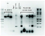
도 1a~1b. 건강한 혈청 유래 엑소좀에서 미생물 DNA의 식별. 혈청 유래 엑소좀 샘플은 임의의 자유롭게 순환하는 핵산을 제거하기 위해 DNA 추출 전에 DNAse로 처리되었다. DNA는 건강한 혈청 (1mL)으로부터 유래된 DNAse 처리된 엑소좀으로부터 단리되었다. 상기 단리된 DNA는 세균 16S 리보솜 RNA 유전자에 대한 범용 프라이머 (27F-B: AGRGTTYGATYMTGGCTCAG (서열 번호 1), 1492R: GGYTACCTTGTTACGACTT (서열 번호 2); 16S rRNA 유전자에 대해 약 1500 bp)로 PCR 증폭되었다. 이. 콜라이 (E. coli) 유래의 DNA는 양성 대조군으로서 사용되었다. 사람 세포주 Panc-1 및 섬유 아세포 BJ 유래의 DNA 뿐만 아니라 블랭크 (주형 DNA 없음)도 음성 대조군으로서 사용되었다. 상기 증폭된 DNA는 겔 전기 영동으로 분석되었다. 도 1a는 한 반복으로부터의 데이터를 도시한 것이며, 도 1b는 또 다른 반복에 대한 데이터를 도시한 것이다.BRIEF DESCRIPTION OF THE DRAWINGS The following drawings form part of this specification and are included to further demonstrate certain aspects of the invention. The present invention may be better understood by reference to one or more of these drawings in conjunction with the detailed description of specific embodiments presented herein.
1a-1b. Identification of microbial DNA from healthy serum-derived exosomes. Serum-derived exosome samples were treated with DNAse prior to DNA extraction to remove any freely circulating nucleic acids. DNA was isolated from DNAse treated exosomes derived from healthy serum (1 mL). The isolated DNA was PCR amplified with universal primers for the bacterial 16S ribosomal RNA gene (27F-B: AGRGTTYGATYMTGGCTCAG (SEQ ID NO: 1), 1492R: GGYTACCTTGTTACGACTT (SEQ ID NO: 2); about 1500 bp for the 16S rRNA gene). this. DNA from E. coli was used as a positive control. DNA from the human cell line Panc-1 and fibroblast BJ as well as blanks (no template DNA) were used as negative controls. The amplified DNA was analyzed by gel electrophoresis. Fig. 1A shows data from one iteration, and Fig. 1B shows data for another iteration.
건강한 개체의 혈액 중의 엑소좀은 세균 마이크로바이옴을 함유한다. 이러한 엑소좀은 체내 미생물, 또는 세균에 감염된 세포에 의해 생성될 수 있다. 이와 같이, 환자의 마이크로바이옴은 순환하는 엑소좀을 단리하고 내부에 존재하는 미생물 핵산과 같은 미생물 구성 요소를 검출함으로써 평가될 수 있다. 이것은 간단한 혈액 엑소좀 테스트를 사용하여 환자의 마이크로바이옴이 샘플링되도록 할 수 있다. 이러한 테스트의 결과는 요법 옵션 및 배설물 이식 결과 (fecal implant outcome)를 결정할 수 있다. 상기 마이크로바이옴은 엑소좀에서 보다 안정적이며, 이것은 면역계의 세포로부터 보호되고 또한 면역 제거를 회피하기 때문이다. 환자의 마이크로바이옴은 개체의 전반적인 건강 상태를 나타내고 많은 질환의 위험에 대한 잠재적 통찰력을 제공할 수 있다. 이와 같이, 환자는 당해 환자의 엑소-마이크로바이옴 (exo-microbiome)을 샘플링하여 암과 같은 다양한 질환 또는 요법에 대한 반응에 대해 스크리닝될 수 있다.Exosomes in the blood of healthy individuals contain the bacterial microbiome. These exosomes may be produced by microorganisms in the body, or cells infected with bacteria. As such, a patient's microbiome can be assessed by isolating circulating exosomes and detecting microbial components such as microbial nucleic acids present therein. This could allow the patient's microbiome to be sampled using a simple blood exosome test. The results of these tests can determine therapy options and fecal implant outcomes. The microbiome is more stable in exosomes, as it is protected from cells of the immune system and also avoids immune clearance. A patient's microbiome represents an individual's overall health and can provide potential insight into the risk of many diseases. As such, patients can be screened for response to various diseases or therapies, such as cancer, by sampling the patient's exo-microbiome.
I. 엑소좀I. Exosomes
본 출원에서 사용되는 "미세 소포체" 및 "엑소좀"이라는 용어는 약 10 nm 내지 약 5000 nm, 보다 전형적으로는 30 nm 내지 1000 nm, 가장 전형적으로는 약 50 nm 내지 750 nm의 직경 (또는 입자가 구형이 아닌 경우, 최대 직경)을 갖는 막성 입자 (membranous particle)를 지칭하며, 여기서, 상기 엑소좀의 막의 적어도 일부는 세포로부터 직접 수득된다. 가장 일반적으로, 엑소좀은 공여체 세포의 크기의 최대 5%인 크기 (평균 직경)을 가질 것이다. 따라서, 특별히 고려되는 엑소좀은 세포로부터 분비된 것들을 포함한다.As used herein, the terms “microvesicles” and “exosomes” refer to a diameter (or particle) of from about 10 nm to about 5000 nm, more typically from 30 nm to 1000 nm, most typically from about 50 nm to 750 nm. is not spherical, refers to a membranous particle having a maximum diameter), wherein at least a portion of the membrane of the exosome is obtained directly from a cell. Most commonly, exosomes will have a size (average diameter) that is up to 5% of the size of the donor cell. Thus, exosomes of special consideration include those secreted from cells.
엑소좀은, 예를 들어, 체액과 같은 임의의 적합한 샘플 유형 내에서 검출되거나 이로부터 단리될 수 있다. 본 출원에서 사용되는 "단리된"이라는 용어는 자연 환경으로부터 분리되는 것을 지칭하며, 적어도 부분 정제를 포함하는 것을 의미하고, 실질적인 정제를 포함할 수 있다. 본 출원에서 사용되는 "샘플"이라는 용어는 본 발명에 의해 제공된 방법에 적합한 임의의 샘플을 지칭한다. 상기 샘플은 검출 또는 단리에 적합한 엑소좀을 포함하는 임의의 샘플일 수 있다. 샘플의 공급원으로는 혈액, 골수, 흉수, 복수, 뇌척수액, 소변, 타액, 양수, 악성 복수, 기관지 폐포 세척액, 윤활액, 모유, 땀, 눈물, 관절액 및 기관지 세척액이 포함된다. 하나의 양태에서, 상기 샘플은, 예를 들어, 전혈 또는 이의 임의의 분획 또는 구성 요소를 포함하는 혈액 샘플이다. 본 발명에 사용하기에 적합한 혈액 샘플은 혈액 세포 또는 이의 구성 요소, 예를 들어, 정맥, 동맥, 말초, 조직, 척수 등을 포함하는 공지된 임의의 공급원으로부터 추출될 수 있다. 예를 들어, 샘플은 널리 공지된 일상적인 임상 방법 (예를 들어, 전혈을 채취하고 처리하기 위한 절차)을 사용하여 수득되고 처리될 수 있다. 하나의 양태에서, 예시적인 샘플은 암에 걸린 대상체로부터 채취된 말초 혈액일 수 있다.Exosomes can be detected in or isolated from any suitable sample type, such as, for example, bodily fluids. As used herein, the term "isolated" refers to being separated from the natural environment, is meant to include at least partial purification, and may include substantial purification. As used herein, the term “sample” refers to any sample suitable for the method provided by the present invention. The sample may be any sample comprising exosomes suitable for detection or isolation. Sources of samples include blood, bone marrow, pleural fluid, ascites, cerebrospinal fluid, urine, saliva, amniotic fluid, malignant ascites, bronchoalveolar lavage fluid, synovial fluid, breast milk, sweat, tears, joint fluid, and bronchial lavage fluid. In one embodiment, the sample is a blood sample comprising, for example, whole blood or any fraction or component thereof. Blood samples suitable for use in the present invention may be extracted from any known source, including blood cells or components thereof, such as veins, arteries, peripheral, tissue, spinal cord, and the like. For example, a sample can be obtained and processed using well known routine clinical methods (eg, procedures for collecting and processing whole blood). In one embodiment, an exemplary sample can be peripheral blood taken from a subject with cancer.
엑소좀은 또한 조직 샘플, 예를 들어, 외과적 샘플, 생검 샘플, 조직, 배설물 및 배양 세포로부터 단리될 수 있다. 엑소좀을 조직 공급원으로부터 단리하는 경우, 단일 세포 현탁액을 수득하기 위해 조직을 균질화한 후 엑소좀을 방출하는 세포의 용해를 수행하는 것이 필요할 수 있다. 엑소좀을 조직 샘플로부터 단리하는 경우, 엑소좀의 파괴를 초래하지 않는 균질화 및 용해 절차를 선택하는 것이 중요하다. 본 출원에서 고려되는 엑소좀은 바람직하게는 생리학적으로 허용되는 용액, 예를 들어, 완충 식염수, 성장 배지, 다양한 수성 배지 등에서 체액으로부터 단리된다.Exosomes can also be isolated from tissue samples, such as surgical samples, biopsy samples, tissues, feces, and cultured cells. When isolating exosomes from a tissue source, it may be necessary to homogenize the tissue to obtain a single cell suspension followed by lysis of the cells releasing the exosomes. When isolating exosomes from tissue samples, it is important to select homogenization and lysis procedures that do not result in destruction of the exosomes. Exosomes contemplated in the present application are preferably isolated from body fluids in physiologically acceptable solutions, for example, buffered saline, growth media, various aqueous media, and the like.
엑소좀은 수집된 샘플로부터 또는 냉동 또는 냉장 저장된 샘플로부터 단리될 수 있다. 일부 실시 형태에서, 엑소좀은 세포 배양 배지로부터 단리될 수 있다. 필수적이지는 않지만, 부피 배제 중합체 (volume-excluding polymer)에 의한 침전 전에 유체 샘플을 정화하여 샘플로부터 임의의 잔해물을 제거하는 경우, 보다 높은 순도의 엑소좀을 수득할 수 있다. 정화 방법으로는 원심 분리, 초고속 원심 분리, 여과 또는 한외 여과가 포함된다. 가장 전형적으로, 엑소좀은 당해 분야에 공지된 다수의 방법에 의해 단리될 수 있다. 하나의 바람직한 방법은 체액 또는 세포 배양 상청액으로부터의 분별 원심 분리이다. 엑소좀의 단리를 위한 예시적인 방법은 문헌 [Losche et al., 2004], [Mesri and Altieri, 1998], [Morel et al., 2004]에 기재되어 있다. 대안으로, 엑소좀은 또한 문헌 [Combes et al., 1997]에 기재된 바와 같은 유세포 계측법을 통해 단리될 수 있다.Exosomes can be isolated from collected samples or from frozen or refrigerated stored samples. In some embodiments, exosomes can be isolated from cell culture media. Although not essential, exosomes of higher purity can be obtained if the fluid sample is purged to remove any debris from the sample prior to precipitation with a volume-excluding polymer. Purification methods include centrifugation, ultra-high speed centrifugation, filtration or ultrafiltration. Most typically, exosomes can be isolated by a number of methods known in the art. One preferred method is fractional centrifugation from body fluids or cell culture supernatants. Exemplary methods for isolation of exosomes are described in Losche et al. , 2004], [Mesri and Altieri, 1998], [Morel et al. , 2004]. Alternatively, exosomes are also described in Combes et al. , 1997].
엑소좀의 단리를 위한 하나의 허용된 프로토콜은 종종 상대적으로 저밀도인 엑소좀을 부유시키기 위해 수크로오스 밀도 구배 또는 수크로오스 쿠션 (sucrose cushion)과 조합된 초고속 원심 분리를 포함한다. 순차적인 분별 원심 분리에 의한 엑소좀의 단리는 다른 미세 소포체 또는 거대 분자 복합체와의 크기 분포의 중첩 가능성에 의해 복잡하다. 또한, 원심 분리는 크기에 기초하여 소포체를 분리하는 불충분한 수단을 제공할 수 있다. 그러나, 순차적인 원심 분리는 수크로오스 구배의 초고속 원심 분리와 조합될 때 고농축 엑소좀을 제공할 수 있다.One accepted protocol for the isolation of exosomes often involves ultrafast centrifugation in combination with a sucrose density gradient or sucrose cushion to suspend relatively low density exosomes. Isolation of exosomes by sequential fractional centrifugation is complicated by the potential for overlap of size distribution with other microvesicles or macromolecular complexes. In addition, centrifugation may provide an insufficient means to separate vesicles based on size. However, sequential centrifugation can provide highly concentrated exosomes when combined with ultrafast centrifugation of a sucrose gradient.
초고속 원심 분리 경로에 대한 대안을 사용하는 크기에 기초한 엑조솜의 단리는 또 다른 옵션이다. 초고속 원심 분리 보다 시간이 덜 걸리고, 특별한 설비의 사용을 요구하지 않는 한외 여과 절차를 사용하는 엑소좀의 성공적인 정제가 보고되었다. 유사하게는, 하나의 마이크로필터 상에서 세포, 혈소판 및 세포 잔해물의 제거와, 유체를 구동하기 위한 양압을 사용하는 제2 마이크로필터 상에서 30 nm 초과의 소포체의 포획을 가능하도록 하는 시판 키트 (EXOMIR™, Bioo Scientific)가 이용 가능하다. 그러나, 이러한 과정 동안, 엑소좀은 회수되지 않으며, 이의 RNA 함량은 이후에 PCR 분석을 위해 사용될 수 있는 제2 마이크로필터 상에서 포획된 물질로부터 직접 추출된다. 잠재적으로, HPLC 기반 프로토콜은 이들 과정이 전용 설비를 요구하고 규모를 확대하기는 어렵지만 고도로 순수한 엑소좀을 수득하도록 할 수 있다. 중요한 문제는 혈액 및 세포 배양 배지 모두가 엑소좀과 동일한 크기 범위의 다수의 나노 입자 (일부 비소포성)를 포함한다는 것이다. 예를 들어, 일부 miRNA는 엑소좀 보다는 오히려 세포외 단백질 복합체 내에 함유될 수 있지만; 프로테아제 (예를 들어, 프로테이나아제 K)에 의한 처리는 "엑스트라 엑소좀 (extraexosomal)" 단백질에 의한 임의의 가능한 오염을 근절시키기 위해 수행될 수 있다.Isolation of exosomes based on size using an alternative to ultrafast centrifugation routes is another option. Successful purification of exosomes using an ultrafiltration procedure that takes less time than ultrafast centrifugation and does not require the use of special equipment has been reported. Similarly, a commercially available kit (EXOMIR™, Bioo Scientific) is available. However, during this process, exosomes are not recovered and their RNA content is extracted directly from the captured material on a second microfilter that can then be used for PCR analysis. Potentially, HPLC-based protocols could allow these processes to yield highly pure exosomes, although these procedures require dedicated equipment and are difficult to scale up. An important problem is that both blood and cell culture media contain a large number of nanoparticles (some non-vesicular) in the same size range as exosomes. For example, some miRNAs may be contained within extracellular protein complexes rather than exosomes; Treatment with a protease (eg, proteinase K) can be performed to eradicate any possible contamination by “extraexosomal” proteins.
또 다른 실시 형태에서, 암 세포 유래된 엑소좀은 면역 특이적 상호 작용 (예를 들어, 면역 자성 포획)을 포함하는 것과 같은 엑소좀을 위한 샘플을 농축하기 위해 통상적으로 사용되는 기술에 의해 포획될 수 있다. 또한, 면역 자성 세포 분리로서 공지된 면역 자성 포획은 전형적으로 작은 특정 세포 유형 상에서 발견되는 단백질에 대한 항체를 상자성 비드에 부착시키는 것을 포함한다. 항체 코팅된 비드들이 혈액과 같은 샘플과 혼합되는 경우, 이들은 특정 세포에 부착되어 특성 세포를 둘러싼다. 그 다음, 상기 샘플은 강한 자기장 내에 배치되는데, 이는 비드들이 한쪽에 펠렛화되도록 한다. 혈액을 제거한 후, 포획된 세포를 비드와 함께 유지한다. 이러한 일반적인 방법의 다양한 변형이 당해 분야에 널리 공지되어 있으며, 엑소좀을 단리하는데 사용하기에 적합하다. 하나의 예에서, 상기 엑소좀을 자기 비드 (예를 들어, 알데하이드/설페이트 비드)에 부착시킬 수 있으며, 이어서 상기 비드에 부착되는 엑소좀의 표면 상의 에피토프를 인식하도록 항체를 혼합물에 첨가한다. 암 세포 유래된 엑소좀 상에서 발견되는 것으로 공지된 예시적인 단백질로는, ATP 결합 카세트 하위 계열 A 구성원 6 (ATP-binding cassette sub-family A member 6: ABCA6), 테트라스파닌-4 (TSPAN4), SLIT 및 NTRK 유사 단백질 4 (SLITRK4), 추정적 프로토카드헤린 베타-18 (PCDHB18), 골수 세포 표면 항원 CD33 (CD33) 및 글리피칸-1 (GPC1)이 포함된다 (그 전문이 본 출원에 참조로 포함되는 미국 US 특허 9,921,223). 암 세포 유래된 엑소좀은, 예를 들어, 하나 이상의 이러한 단백질에 대한 항체 또는 압타머를 사용하여 단리될 수 있다.In another embodiment, cancer cell-derived exosomes may be captured by techniques commonly used to enrich samples for exosomes, such as those involving immune-specific interactions (eg, immune magnetic capture). can Immune magnetic capture, also known as immune magnetic cell isolation, typically involves attaching antibodies to paramagnetic beads against proteins found on small specific cell types. When antibody-coated beads are mixed with a sample such as blood, they attach to specific cells and surround characteristic cells. The sample is then placed in a strong magnetic field, which causes the beads to pellet on one side. After the blood is removed, the captured cells are kept with the beads. Various modifications of these general methods are well known in the art and are suitable for use in isolating exosomes. In one example, the exosomes can be attached to magnetic beads (eg, aldehyde/sulfate beads), and then an antibody is added to the mixture to recognize epitopes on the surface of the exosomes attached to the beads. Exemplary proteins known to be found on cancer cell-derived exosomes include ATP-binding cassette sub-family A member 6: ABCA6, tetraspanin-4 (TSPAN4), SLIT and NTRK-like protein 4 (SLITRK4), putative protocadherin beta-18 (PCDHB18), bone marrow cell surface antigens CD33 (CD33) and glypican-1 (GPC1). US Pat. No. 9,921,223, which is incorporated. Cancer cell derived exosomes can be isolated using, for example, antibodies or aptamers to one or more of these proteins.
본 출원에서 사용되는 분석은 엑소좀의 직접적 또는 간접적 가시화를 가능하도록 하는 임의의 방법을 포함하고, 생체 내에서 또는 생체 외에서 일 수 있다. 예를 들어, 분석은 고형 기질에 결합된 엑소좀의 생체외 현미경적 또는 세포 계측적 검출 및 가시화, 유세포 계측법, 형광 이미징 등을 포함할 수 있지만, 이들에 한정되는 것은 아니다. 예시적인 양태에서, 암 세포 유래된 엑소좀은 ATP 결합 카세트 하위 계열 A 구성원 6, 테트라스파닌-4 (TSPAN4), SLIT 및 NTRK 유사 단백질 4 (SLITRK4), 추정적 프로토카드헤린 베타-18 (PCDHB18), 골수 세포 표면 항원 CD33 (CD33), 글리피칸-1 (GPC1), 히스톤 H2A 2형-A (HIST1H2AA), 히스톤 H2A 1형-A (HIST1H1AA), 히스톤 H3.3 (H3F3A), 히스톤 H3.1 (HIST1H3A), 아연 핑거 단백질 37 동족체 (ZFP37), 라미닌 서브 유닛 베타-1 (LAMB1), 세뇨관 간질 신염 항원 유사 (TINAGL1), 페록시리데옥신-4 (PRDX4), 콜라겐 알파-2(IV) 쇄 (COL4A2), 추정적 단백질 C3P1 (C3P1), 헤미센틴-1 (HMCN1), 추정적 로필린-2 유사 단백질 (RHPN2P1), 안키린 반복 도메인 함유 단백질 62 (ANKRD62), 3분체 모티프 함유 단백질 42 (TRIM42), 접합 플라코글로빈 (Junction plakoglobin: JUP), 튜불린 베타-2B 쇄 (TUBB2B), 엔도리보뉴클레아제 다이서 (DICER1), E3 유비퀴틴 단백질 리가아제 TRIM71 (TRIM71), 카타닌 p60 ATPase 함유 서브 유닛 A 유사 2 (KATNAL2), 단백질 S100-A6 (S100A6), 5'-뉴클레오티다아제 도메인 함유 단백질 3 (NT5DC3), 발린-tRNA 리가아제 (VARS), 카즈린 (KAZN), ELAV 유사 단백질 4 (ELAVL4), RING 핑거 단백질 166 (RNF166), FERM 및 PDZ 도메인 함유 단백질 1 (FRMPD1), 78 kDa 글루코오스 조절 단백질 (HSPA5), 이동 단백질 입자 복합 서브 유닛 6A (TRAPPC6A), 스쿠알렌 모노옥시게나아제 (SQLE), 종양 감수성 유전자 101 단백질 (TSG101), 액포 단백질 분류 28 동족체 (VPS28), 프로스타글란딘 F2 수용체 음성 조절물질 (PTGFRN), 이소부티릴-CoA 데하이드로게나아제, 미토콘드리아 (ACAD8), 26S 프로테아제 조절 서브 유닛 6B (PSMC4), 신장 인자 1-감마 (EEF1G), 티틴 (TTN), 타이로신-단백질 포스파타아제 13형 (PTPN13), 트리오스포스페이트 아이소머라아제 (TPI1) 또는 카복시펩티다아제 E (CPE) 중 하나 이상에 대한 항체를 사용하여 검출된 후, 고형 기질에 결합되고/되거나 현미경적 또는 유세포 계측적 검출을 사용하여 가시화된다.Assays used in this application include any method that enables direct or indirect visualization of exosomes, and may be in vivo or ex vivo. For example, assays may include, but are not limited to, in vitro microscopic or cytometric detection and visualization of exosomes bound to a solid matrix, flow cytometry, fluorescence imaging, and the like. In an exemplary embodiment, cancer cell-derived exosomes contain ATP binding cassette subfamily A member 6, tetraspanin-4 (TSPAN4), SLIT and NTRK-like protein 4 (SLITRK4), putative protocadherin beta-18 (PCDHB18). ), bone marrow cell surface antigen CD33 (CD33), glypican-1 (GPC1), histone H2A type 2 -A (HIST1H2AA), histone H2A type 1 -A (HIST1H1AA), histone H3.3 (H3F3A), histone H3. 1 (HIST1H3A), zinc finger protein 37 homologue (ZFP37), laminin subunit beta-1 (LAMB1), tubular interstitial nephritis antigen-like (TINAGL1), peroxyrideoxin-4 (PRDX4), collagen alpha-2 (IV) chain (COL4A2), putative protein C3P1 (C3P1), hemisentin-1 (HMCN1), putative ropilin-2 like protein (RHPN2P1), ankyrin repeat domain containing protein 62 (ANKRD62), ternary motif containing protein 42 (TRIM42), Junction plakoglobin (JUP), tubulin beta-2B chain (TUBB2B), endoribonuclease Dicer (DICER1), E3 ubiquitin protein ligase TRIM71 (TRIM71), catanin p60 ATPase Containing subunit A-like 2 (KATNAL2), protein S100-A6 (S100A6), 5'-nucleotidase domain containing protein 3 (NT5DC3), valine-tRNA ligase (VARS), kazrin (KAZN), ELAV-like Protein 4 (ELAVL4), RING finger protein 166 (RNF166), FERM and PDZ domain containing protein 1 (FRMPD1), 78 kDa glucose regulatory protein (HSPA5), mobile protein particle complex subunit 6A (TRAPPC6A), squalene monooxygenase (SQLE), tumor susceptibility gene 101 protein (TSG101), vacuolar protein class 28 homologue (VPS28), prostaglandin F2 receptor negative Modulator (PTGFRN), isobutyryl-CoA dehydrogenase, mitochondria (ACAD8), 26S protease regulatory subunit 6B (PSMC4), elongation factor 1-gamma (EEF1G), titin (TTN), tyrosine-protein phosphata After detection using an antibody to one or more ofase type 13 (PTPN13), triose phosphate isomerase (TPI1) or carboxypeptidase E (CPE), bound to a solid substrate and/or detected by microscopic or flow cytometry is visualized using
세포에서 발현되는 모든 단백질이 당해 세포에 의해 분비된 엑소좀에서 발견되지 않는다는 것에 유의해야 한다. 예를 들어, 칼넥신, GM130 및 LAMP-2는 모두 MCF-7 세포에서 발현되는 단백질이지만, MCF-7 세포에 의해 분비되는 엑소좀에서 발견되지 않는다 (문헌 [Baietti et al., 2012]). 또 다른 예로서, 하나의 연구는 190명/190명의 췌장관 선암종 환자가 건강한 대조군 보다 더 높은 수준의 GPC1+ 엑소좀을 갖는다는 것을 밝혀냈다 (그 전문이 본 출원에 참조로 포함되는 문헌 [Melo et al., 2015]). 특히, 평균적으로 건강한 대조군 중 단지 2.3%만이 GPC1+ 엑소좀을 가졌다.It should be noted that not all proteins expressed in a cell are found in the exosomes secreted by the cell. For example, Calnexin, GM130 and LAMP-2 are all proteins expressed in MCF-7 cells, but not found in exosomes secreted by MCF-7 cells (Baietti et al. , 2012). As another example, one study found that 190/190 pancreatic ductal adenocarcinoma patients had higher levels of GPC1+ exosomes than healthy controls (Melo et al, incorporated herein by reference in its entirety). . , 2015]). Notably, only 2.3% of healthy controls on average had GPC1+ exosomes.
A. 세포 배양물로부터 엑소좀을 수집하기 위한 예시적인 프로토콜A. Exemplary Protocol for Collecting Exosomes from Cell Culture
1일차에, 10% FBS를 함유하는 배지 중에 T225 플라스크 내의 충분한 세포 (예를 들어, 약 5백만개 세포)를 씨딩하여 다음날에 상기 세포가 약 70%의 밀집도를 갖도록 한다. 2일차에, 상기 세포 상의 배지를 흡인하고, 상기 세포를 PBS로 2회 세척한 다음, 상기 세포에 25~30 mL의 기본 배지 (즉, PenStrep 또는 FBS 부재)를 첨가한다. 상기 세포를 24~48 시간 동안 인큐베이션한다. 48 시간 인큐베이션이 바람직하지만, 일부 세포주는 무혈청 배지에 보다 민감하므로, 인큐베이션 시간은 24시간으로 감소되어야 한다. FBS가 상당하게 NanoSight 결과를 심하게 왜곡하는 엑소좀을 함유한다는 것에 유의한다.On
3/4일차에, 배지를 수집하고, 800×g로 5분 동안 실온에서 원심 분리하여 사망 세포 및 거대 잔해물을 펠렛화한다. 상청액을 새로운 코니칼 튜브로 옮기고, 배지를 10분 동안 2000×g로 다시 원심 분리하여 다른 거대 잔해물 및 거대 소포체를 제거한다. 배지를 0.2 μm 필터에 통과시킨 다음, 튜브 당 35 mL를 사용하여 초고속 원심 분리 튜브 (예를 들어, 25×89 mm Beckman Ultra-Clear)에 분취량을 취한다. 튜브 당 배지의 부피가 35 mL 미만인 경우, 35 mL에 도달되도록 튜브의 나머지를 PBS로 충전한다. SW 32 Ti 로터 (k 인자 266.7, RCF 최대 133,907)를 사용하여 배지를 4℃에서 28,000 rpm으로 2~4 시간 동안 초고속 원심 분리한다. 대략 1인치의 액체가 남을 때까지 상청액을 주의하여 흡인한다. 튜브를 기울이고, 잔류 배지가 흡인기 피펫으로 서서히 유입되도록 한다. 원하는 경우, 엑소좀 펠렛을 PBS 중에 재현탁시킬 수 있으며, 28,000 rpm의 초고속 원심 분리를 1~2 시간 동안 반복하여 엑소좀의 집단을 추가로 정제한다.On day 3/4, collect the medium and centrifuge at 800×g for 5 min at room temperature to pellet dead cells and large debris. The supernatant is transferred to a new conical tube and the medium is centrifuged again at 2000×g for 10 min to remove other large debris and large vesicles. Pass the medium through a 0.2 μm filter, then aliquot into ultrafast centrifuge tubes (e.g., 25×89 mm Beckman Ultra-Clear) using 35 mL per tube. If the volume of medium per tube is less than 35 mL, fill the remainder of the tube with PBS to reach 35 mL. Using a SW 32 Ti rotor (k factor 266.7, RCF max. 133,907), the medium is ultra-fast centrifuged at 28,000 rpm at 4°C for 2-4 h. Carefully aspirate the supernatant until approximately 1 inch of liquid remains. Tilt the tube and allow residual medium to slowly flow into the aspirator pipette. If desired, the exosome pellet can be resuspended in PBS, and ultrafast centrifugation at 28,000 rpm is repeated for 1-2 hours to further purify the exosome population.
마지막으로, 210 μL의 PBS 중에 엑소좀 펠렛을 재현탁시킨다. 각각의 샘플에 대한 다중 초고속 원심 분리 튜브가 존재하는 경우, 동일한 210 μL의 PBS를 사용하여 각각의 엑소좀 펠렛을 연속적으로 재현탁시킨다. 각각의 샘플에 대해, 10 μL를 취하고, 나노 입자 추적 분석에 사용하기 위해 990 μL의 H2O에 첨가한다. 다운스트림 과정 동안 나머지 200 μL의 엑소좀 함유 현탁액을 사용하거나, 즉시 -80℃에 저장한다.Finally, resuspend the exosome pellet in 210 μL of PBS. If multiple ultrafast centrifuge tubes for each sample are present, serially resuspend each exosome pellet using the same 210 µL of PBS. For each sample, take 10 µL and add to 990 µL of H 2 O for use in nanoparticle tracking analysis. Use the remaining 200 µL of the exosome-containing suspension for downstream processing, or immediately store at -80 °C.
B. 혈청 샘플로부터 엑소좀을 추출하기 위한 예시적인 프로토콜B. Exemplary Protocol for Extracting Exosomes from Serum Samples
우선, 혈청 (또는 다른 체액) 샘플을 얼음 상에서 해동시킨다. 그 다음, 11 mL의 PBS 중에 250 μL의 무세포 혈청 샘플을 희석시키고; 0.2 μm 기공 필터를 통해 여과한다. 희석된 샘플을 4℃에서 밤새 150,000×g로 초고속 원심 분리한다. 다음날, 상청액을 주의하여 폐기하고, 엑소좀 펠렛을 11 mL의 PBS 중에서 세척한다. 2 시간 동안 4℃에서 150,000×g로 2회차의 초고속 원심 분리를 수행한다. 마지막으로, 상청액을 주의하여 폐기하고, 분석을 위해 엑소좀 펠렛을 100 μL의 PBS 중에 재현탁시킨다.First, a serum (or other bodily fluid) sample is thawed on ice. Then, dilute 250 μL of cell-free serum sample in 11 mL of PBS; Filter through a 0.2 μm pore filter. The diluted samples are ultrafast centrifuged at 150,000×g overnight at 4°C. The next day, the supernatant is carefully discarded and the exosome pellet is washed in 11 mL of PBS. Perform two rounds of ultrafast centrifugation at 150,000 × g at 4°C for 2 hours. Finally, carefully discard the supernatant and resuspend the exosome pellet in 100 μL of PBS for analysis.
II. 마이크로바이옴II. microbiome
사람 미생물총 (microbiota)은 사람 게놈에 존재하는 것보다 100배 이상 더 많은 유전자를 보유하고 있는 150~200개의 널리 퍼진 세균 종과 1000개의 덜 흔한 세균 종을 포함하는 수조 개의 미생물로 이루어진다. 상기 미생물총은 대부분 세균으로 구성되지만 고세균, 원생 동물 및 바이러스도 또한 포함한다. 상기 미생물총은 식품 가공, 복합 난소화성 다당류의 소화 및 비타민 합성을 비롯하여 건강 유지에 필수적인 중요한 기능을 수행하며, 병원체 억제, 독성 화합물 대사에서부터 숙주 대사 조절에 이르기까지 다양한 기능을 갖는 생체 활성 대사 산물을 분비한다.The human microbiota consists of trillions of microorganisms, including 150 to 200 prevalent bacterial species and 1000 less common bacterial species, carrying more than 100 times more genes than are present in the human genome. The microflora consists mostly of bacteria, but also includes archaea, protozoa and viruses. The microflora performs important functions essential for maintaining health, including food processing, digestion of complex indigestible polysaccharides, and vitamin synthesis, and produces bioactive metabolites with various functions from pathogen suppression and toxic compound metabolism to host metabolism regulation. secrete
교란된 미생물총은 유아의 괴사성 소장대장염에서부터 성인의 비만, 당뇨병, 대사 증후군, 과민성 대장 증후군 및 염증성 장 질환에 이르기까지 사람의 다양한 장애에 연루되어 있다. 사람 건강에서 마이크로바이옴 불균형에 대한 최근 연구는 암을 비롯한 다수의 질환 상태에서 마이크로바이옴의 특이적 변화를 시사한다. "마이크로바이옴"은 미생물총의 집단 게놈을 지칭한다. 또한, 연구는 특정 마이크로바이옴과 특이적 암의 연관을 시사하였다. 따라서, 특유의 마이크로바이옴은 질환의 원인 또는 발병에 기여할 수 있다. 반대로, 종양 미세 환경은 이러한 바이러스와 미생물이 존재할 수 있는 특수한 틈새 (niche)를 제공할 수 있다. 어느 경우에서도, 질환 유형 특이적 마이크로바이옴 시그니처는 조기 진단, 예후 및 치료 전략을 위한 바이오 마커를 제공할 수 있다.Disturbed microbiota is implicated in a variety of disorders in humans, from necrotizing enterocolitis in infants to obesity, diabetes, metabolic syndrome, irritable bowel syndrome and inflammatory bowel disease in adults. Recent studies of microbiome imbalances in human health suggest specific changes in the microbiome in a number of disease states, including cancer. "Microbiome" refers to the population genome of the microbiota. In addition, studies have suggested associations of specific microbiome with specific cancers. Thus, a unique microbiome may contribute to the etiology or pathogenesis of the disease. Conversely, the tumor microenvironment can provide a special niche for these viruses and microorganisms to exist. In either case, disease type-specific microbiome signatures can provide biomarkers for early diagnosis, prognosis and treatment strategies.
일부 실시 형태에서, 하나 이상의 유형의 미생물 또는 이의 구성 요소 또는 생성물의 수준 또는 수준 세트를 결정하는 것은 하나 이상의 DNA 서열의 수준 또는 수준 세트를 결정하는 것을 포함한다. 일부 실시 형태에서, 하나 이상의 DNA 서열은 상이한 미생물 유형을 구별하는데 사용될 수 있는 임의의 DNA 서열을 포함한다. 특정 실시 형태에서, 하나 이상의 DNA 서열은 16S rRNA 유전자 서열을 포함한다. 특정 실시 형태에서, 하나 이상의 DNA 서열은 18S rRNA 유전자 서열을 포함한다. 일부 실시 형태에서, 1, 2, 3, 4, 5, 10, 15, 20, 25, 50, 100, 1,000, 5,000개 또는 그 이상의 서열이 증폭된다.In some embodiments, determining the level or set of levels of one or more types of microorganisms or components or products thereof comprises determining the level or set of levels of one or more DNA sequences. In some embodiments, the one or more DNA sequences include any DNA sequence that can be used to distinguish different types of microorganisms. In certain embodiments, the one or more DNA sequences comprise a 16S rRNA gene sequence. In certain embodiments, the one or more DNA sequences comprises an 18S rRNA gene sequence. In some embodiments, 1, 2, 3, 4, 5, 10, 15, 20, 25, 50, 100, 1,000, 5,000 or more sequences are amplified.
16S 및 18S rRNA 유전자 서열은 각각 원핵 생물 및 진핵 생물 리보솜의 작은 서브유닛 구성 요소를 인코딩한다. rRNA 유전자는 미생물 유형을 구별하는데 특히 유용한데, 그 이유는 이러한 유전자의 서열이 미생물 종들 사이에 상이하지만, 해당 유전자가 프라이머 결합을 위한 고도로 보존된 영역을 갖고 있기 때문이다. 보존된 프라이머 결합 영역들 사이의 이러한 특이성은 다양한 유형의 미생물의 rRNA 유전자가 단일 세트의 프라이머로 증폭된 다음 증폭된 서열에 의해 구별되도록 한다.The 16S and 18S rRNA gene sequences encode small subunit components of prokaryotic and eukaryotic ribosomes, respectively. rRNA genes are particularly useful for differentiating types of microorganisms because, although the sequences of these genes differ between microbial species, they have highly conserved regions for primer binding. This specificity between conserved primer binding regions allows the rRNA genes of various types of microorganisms to be amplified with a single set of primers and then differentiated by the amplified sequences.
III. 질환의 진단, 예후 및 치료III. Diagnosis, prognosis and treatment of diseases
본 발명의 방법을 사용한 엑소-마이크로바이옴의 검출, 단리 및 특성화는 질환 위험 인자, 진단 및 예후를 평가하고 질환 재발을 초래할 수 있는 치료 실패의 조기 검출을 위한 치료 효능을 모니터링하는데 유용하다. 또한, 본 발명에 따른 엑소-마이크로바이옴 분석은 요법 과정을 완료한 무증상 (presymptomatic) 환자에서 조기 재발의 검출을 가능하게 한다. 이것은 엑소좀에 존재하는 마이크로바이옴의 존재가 질환 진행, 요법에 대한 불량한 반응, 질환의 재발 및/또는 일정 기간에 걸친 감소된 생존과 연관되고/되거나 상관 관계가 있을 수 있기 때문에 가능하다. 따라서, 엑소-마이크로바이옴의 계수 및 특성화는 요법에 대한 반응을 기반으로 초기 위험 및 후속 위험을 예측하는 기준선 특성에 대해 환자를 계층화하는 방법을 제공한다.The detection, isolation and characterization of the exo-microbiome using the methods of the present invention is useful for evaluating disease risk factors, diagnosis and prognosis, and for monitoring therapeutic efficacy for early detection of treatment failure that can lead to disease recurrence. In addition, the exo-microbiome analysis according to the present invention enables the detection of early recurrence in presymptomatic patients who have completed the course of therapy. This is possible because the presence of the microbiome present in exosomes may be associated with and/or correlated with disease progression, poor response to therapy, disease recurrence, and/or decreased survival over a period of time. Thus, enumeration and characterization of the exo-microbiome provides a way to stratify patients for baseline characteristics that predict initial risk and subsequent risk based on response to therapy.
예를 들어, 상기에서 개시된 방법에 따라 단리된 암 세포 유래된 엑소좀은 대상체에서 암을 진단 또는 예후하기 (prognose) 위해 분석될 수 있다. 이와 같이, 본 발명의 방법은, 예를 들어, 암 세포 유래된 엑소좀의 엑소-마이크로바이옴과 비-암성 세포로부터 유래되는 엑소좀을 비교함으로써 암 환자 및 암 위험 환자를 평가하는데 사용될 수 있다. 본 출원에서 기재된 임의의 진단 또는 예후 방법에서, 암의 하나 이상의 지표, 예를 들어, 암 특이적 엑소-마이크로바이옴 시그니처 또는 임의의 다른 장애의 존재 또는 부재가 진단 또는 예후를 발생시키기 위해 사용될 수 있다.For example, cancer cell-derived exosomes isolated according to the methods disclosed above can be analyzed to diagnose or prognose cancer in a subject. As such, the methods of the present invention can be used to assess cancer patients and cancer risk patients, for example, by comparing the exosomes derived from cancer cell-derived exosomes with the exosomes derived from non-cancerous cells with the exo-microbiome of cancer cell-derived exosomes. . In any diagnostic or prognostic method described herein, one or more indicators of cancer, eg, the presence or absence of a cancer-specific exo-microbiome signature or any other disorder, may be used to generate a diagnosis or prognosis. have.
하나의 양태에서, 체액 (예를 들어, 혈액, 소변, 타액 등) 샘플이 환자로부터 채취되고, 질환 세포 유래된 엑소좀이 본 출원에서 기재된 바와 같이 검출 및/또는 단리된다. 예를 들어, 상기 엑소좀은 ATP 결합 카세트 하위 계열 A 구성원 6 (ABCA6), 테트라스파닌-4 (TSPAN4), SLIT 및 NTRK 유사 단백질 4 (SLITRK4), 추정적 프로토카드헤린 베타-18 (PCDHB18), 골수 세포 표면 항원 CD33 (CD33) 및/또는 글리피칸-1 (GPC1)에 결합하는 하나 이상의 항체 또는 압타머로 라벨링될 수 있으며, 상기 항체는 공유 결합된 형광 표지를 가질 수 있다. 그 다음, 샘플 내 암 세포 유래된 엑소좀의 수 및 특성화를 결정하기 위한 분석이 수행될 수 있으며, 이러한 측정으로부터, 초기 혈액 샘플에 존재하는 암 세포 유래된 엑소좀의 수가 결정될 수 있다. 암 세포 유래된 엑소좀으로 식별된 엑소좀은 그 자체로서 암 세포 유래된 엑소좀에서 선택적으로 또는 특이적으로 발견되는 것으로 공지된 제2 (또는 그 이상) 마커, 예를 들어, 히스톤 H2A 2형-A (HIST1H2AA), 히스톤 H2A 1형-A (HIST1H1AA), 히스톤 H3.3 (H3F3A), 히스톤 H3.1 (HIST1H3A), 아연 핑거 단백질 37 동족체 (ZFP37), 라미닌 서브 유닛 베타-1 (LAMB1), 세뇨관 간질 신염 항원 유사 (TINAGL1), 페록시리데옥신-4 (PRDX4), 콜라겐 알파-2(IV) 쇄 (COL4A2), 추정적 단백질 C3P1 (C3P1), 헤미센틴-1 (HMCN1), 추정적 로필린-2 유사 단백질 (RHPN2P1), 안키린 반복 도메인 함유 단백질 62 (ANKRD62), 3분체 모티프 함유 단백질 42 (TRIM42), 접합 플라코글로빈 (Junction plakoglobin: JUP), 튜불린 베타-2B 쇄 (TUBB2B), 엔도리보뉴클레아제 다이서 (DICER1), E3 유비퀴틴 단백질 리가아제 TRIM71 (TRIM71), 카타닌 p60 ATPase 함유 서브 유닛 A 유사 2 (KATNAL2), 단백질 S100-A6 (S100A6), 5'-뉴클레오티다아제 도메인 함유 단백질 3 (NT5DC3), 발린-tRNA 리가아제 (VARS), 카즈린 (KAZN), ELAV 유사 단백질 4 (ELAVL4), RING 핑거 단백질 166 (RNF166), FERM 및 PDZ 도메인 함유 단백질 1 (FRMPD1), 78 kDa 글루코오스 조절 단백질 (HSPA5), 이동 단백질 입자 복합 서브 유닛 6A (TRAPPC6A), 스쿠알렌 모노옥시게나아제 (SQLE), 종양 감수성 유전자 101 단백질 (TSG101), 액포 단백질 분류 28 동족체 (VPS28), 프로스타글란딘 F2 수용체 음성 조절물질 (PTGFRN), 이소부티릴-CoA 데하이드로게나아제, 미토콘드리아 (ACAD8), 26S 프로테아제 조절 서브 유닛 6B (PSMC4), 신장 인자 1-감마 (EEF1G), 티틴 (TTN), 타이로신-단백질 포스파타아제 13형 (PTPN13), 트리오스포스페이트 아이소머라아제 (TPI1) 또는 카복시펩티다아제 E (CPE)의 검출을 통해 입증될 수 있다. 암 세포 유래된 엑소좀의 수는 엑소좀을 시각적으로 정량화하고 특성화하기 위해 세포 측정 또는 현미경 기법에 의해 결정될 수 있다. 암 세포 유래된 엑소좀은 당해 분야에 공지된 다른 방법 (예를 들어, ELISA)에 의해 검출 및 정량화될 수 있다.In one embodiment, a sample of bodily fluid (eg, blood, urine, saliva, etc.) is taken from the patient and diseased cell derived exosomes are detected and/or isolated as described herein. For example, the exosomes contain ATP binding cassette subfamily A member 6 (ABCA6), tetraspanin-4 (TSPAN4), SLIT and NTRK-like protein 4 (SLITRK4), putative protocadherin beta-18 (PCDHB18) , may be labeled with one or more antibodies or aptamers that bind to the bone marrow cell surface antigens CD33 (CD33) and/or glypican-1 (GPC1), wherein the antibody may have a covalently linked fluorescent label. Analyzes can then be performed to determine the number and characterization of cancer cell-derived exosomes in the sample, from which measurements the number of cancer cell-derived exosomes present in the initial blood sample can be determined. Exosomes identified as cancer cell-derived exosomes as such are a second (or more) marker known to be selectively or specifically found in cancer cell-derived exosomes, e.g., histone H2A type 2 -A (HIST1H2AA), histone H2A type 1-A (HIST1H1AA), histone H3.3 (H3F3A), histone H3.1 (HIST1H3A), zinc finger protein 37 homolog (ZFP37), laminin subunit beta-1 (LAMB1) , tubular interstitial nephritis antigen-like (TINAGL1), peroxyrideoxin-4 (PRDX4), collagen alpha-2(IV) chain (COL4A2), putative protein C3P1 (C3P1), hemisentin-1 (HMCN1), putative Lophilin-2 like protein (RHPN2P1), ankyrin repeat domain containing protein 62 (ANKRD62), ternary motif containing protein 42 (TRIM42), Junction plakoglobin (JUP), tubulin beta-2B chain (TUBB2B) ), endoribonuclease Dicer (DICER1), E3 ubiquitin protein ligase TRIM71 (TRIM71), catanin p60 ATPase containing subunit A-like 2 (KATNAL2), protein S100-A6 (S100A6), 5'-nucleo Tidase domain containing protein 3 (NT5DC3), valine-tRNA ligase (VARS), kazrin (KAZN), ELAV-like protein 4 (ELAVL4), RING finger protein 166 (RNF166), FERM and PDZ domain containing protein 1 (FRMPD1) ), 78 kDa glucose regulatory protein (HSPA5), migratory protein particle complex subunit 6A (TRAPPC6A), squalene monooxygenase (SQLE), tumor susceptibility gene 101 protein (TSG101), vacuolar protein class 28 homologue (VPS28), prostaglandins F2 receptor negative modulator (PTGFRN), isobutyryl-CoA dehydrogenase, mitochondria (ACAD8), 26S protease regulatory subunit 6 B (PSMC4), elongation factor 1-gamma (EEF1G), titin (TTN), tyrosine-protein phosphatase type 13 (PTPN13), triose phosphate isomerase (TPI1) or carboxypeptidase E (CPE) can be proven through The number of cancer cell-derived exosomes can be determined by cytometric or microscopic techniques to visually quantify and characterize exosomes. Cancer cell derived exosomes can be detected and quantified by other methods known in the art (eg, ELISA).
다양한 양태에서, 대상체의 엑소-마이크로바이옴 분석은 대상체의 진행 및 병리를 평가하기 위해 다양한 간격으로 특정 시간 경과에 걸쳐 이루어질 수 있다. 예를 들어, 분석은 1일, 2일, 3일, 1주, 2주, 1 개월, 2 개월, 3 개월, 6 개월 또는 1년과 같은 주기적인 간격으로 수행되어 시간의 함수로서 엑소-마이크로바이옴의 수준 및 특성화를 추적할 수 있다. 기존 암 환자의 경우, 이것은 질환의 진행에 대한 유용한 지표를 제공하며, 의사가 엑소-마이크로바이옴 변화의 증가, 감소 또는 결여를 기반으로 적절한 치료 선택을 하는데 도움이 된다.In various aspects, exo-microbiome analysis of a subject can be made over a specific time course at various intervals to assess the subject's progression and pathology. For example, assays may be performed at periodic intervals such as 1 day, 2 days, 3 days, 1 week, 2 weeks, 1 month, 2 months, 3 months, 6 months, or 1 year to perform exo-microorganism as a function of time. The level and characterization of the biome can be traced. For patients with pre-existing cancer, this provides a useful indicator of disease progression and helps physicians make appropriate treatment choices based on increased, decreased or absent exo-microbiome changes.
본 출원에서 제공된 임의의 방법에서, 엑소-마이크로바이옴을 특성화하여 추가의 임상 평가를 제공하기 위한 추가의 분석이 또한 수행될 수 있다. 예를 들어, 특정 마커에 대해 특이적인 프라이머와의 다중화 (multiplexing)와 같은 PCR 기술이 엑소-마이크로바이옴이 유래되는 미생물의 유형과 같은 정보를 수득하기 위해 사용될 수 있다. 추가로, DNA 또는 RNA 분석, 프로테옴 분석 또는 대사체 분석이 환자의 특성화에 관한 추가의 정보를 평가하는 수단으로서 수행될 수 있다.In any of the methods provided herein, additional analyzes to characterize the exo-microbiome to provide further clinical evaluation can also be performed. For example, PCR techniques such as multiplexing with primers specific for a particular marker can be used to obtain information such as the type of microorganism from which the exo-microbiome is derived. Additionally, DNA or RNA analysis, proteome analysis or metabolite analysis may be performed as a means of evaluating additional information regarding the characterization of the patient.
예를 들어, 엑소-마이크로바이옴 분석은 특정 치료 방법에 대한 대상체의 반응성을 결정하거나 암 치료에서 후보 작용제의 유효성을 결정하기에 충분한 데이터를 제공할 수 있다. 따라서, 본 발명은 본 출원에서 기재된 바와 같이 대상체의 엑소-마이크로바이옴을 검출함으로써 특정 치료 방법에 대한 대상체의 반응성을 결정하거나 암 치료에서 후보 작용제의 유효성을 결정하는 방법을 제공한다. 예를 들어, 일단 약물 치료가 환자에게 투여되면, 본 발명의 방법을 사용하여 약물 치료의 효능을 결정하는 것이 가능하다. 예를 들어, 약물 치료 전에 환자로부터 채취된 샘플 뿐만 아니라 약물 치료와 동시에 또는 약물 치료 후에 환자로부터 채취된 하나 이상의 샘플이 본 발명의 방법을 사용하여 처리될 수 있다. 각각의 처리된 샘플의 분석 결과를 비교함으로써, 약물 치료의 효능 또는 작용제에 대한 환자의 반응성을 결정할 수 있다. 이러한 방식으로, 실패한 화합물의 조기 식별이 이루어질 수 있거나, 유망한 화합물의 조기 검증이 이루어질 수 있다.For example, exo-microbiome analysis can provide sufficient data to determine a subject's responsiveness to a particular treatment method or to determine the effectiveness of a candidate agent in the treatment of cancer. Accordingly, the present invention provides a method of determining a subject's responsiveness to a particular treatment method or determining the effectiveness of a candidate agent in cancer treatment by detecting the subject's exo-microbiome as described herein. For example, once a drug treatment is administered to a patient, it is possible to determine the efficacy of the drug treatment using the methods of the present invention. For example, samples taken from a patient prior to drug treatment as well as one or more samples taken from a patient concurrently with or after drug treatment can be processed using the methods of the present invention. By comparing the results of the analysis of each treated sample, the efficacy of a drug treatment or a patient's responsiveness to an agent can be determined. In this way, early identification of failed compounds can be made, or early validation of promising compounds can be made.
본 발명의 특정 양태는 엑소-마이크로바이옴의 존재를 기준으로 질환 또는 장애를 예방 또는 치료하는데 사용될 수 있다. 본 발명의 특정 양태는 치료제 또는 진단제를 발현하거나 포함하는 엑소-마이크로바이옴으로 환자를 치료하는 것을 제공한다. 본 출원에서 사용되는 "치료제"는 암 또는 다른 병태의 치료에 유용한 원자, 분자 또는 화합물이다. 치료제의 예로는 약물, 화학 요법제, 치료학적 항체 및 항체 단편, 독소, 방사성 동위 원소, 효소, 뉴클레아제, 호르몬, 면역 조절제, 안티센스 올리고뉴클레오타이드, 킬레이트화제, 붕소 화합물, 광활성제 및 염료가 포함되지만, 이들에 한정되는 것은 아니다. 본 출원에서 사용되는 "진단제"는 질환을 진단, 검출 또는 시각화하는데 유용한 원자, 분자 또는 화합물이다. 본 출원에서 기재된 실시 형태에 따르면, 진단제로는 방사성 물질 (예를 들어, 방사성 동위 원소, 방사성 핵종, 방사성 표지 또는 방사성 추적자), 염료, 조영제, 형광 화합물 또는 분자, 생체 발광 화합물 또는 분자, 효소 및 증진제 (예를 들어, 상자성 이온)가 포함될 수 있지만, 이들에 한정되는 것은 아니다.Certain aspects of the invention may be used to prevent or treat a disease or disorder based on the presence of the exo-microbiome. Certain aspects of the invention provide for treating a patient with an exo-microbiome expressing or comprising a therapeutic or diagnostic agent. As used herein, a “therapeutic agent” is an atom, molecule or compound useful for the treatment of cancer or other conditions. Examples of therapeutic agents include drugs, chemotherapeutic agents, therapeutic antibodies and antibody fragments, toxins, radioisotopes, enzymes, nucleases, hormones, immunomodulators, antisense oligonucleotides, chelating agents, boron compounds, photoactive agents and dyes However, it is not limited to these. As used herein, a "diagnostic agent" is an atom, molecule or compound useful for diagnosing, detecting, or visualizing a disease. According to embodiments described in this application, the diagnostic agent includes a radioactive material (eg, a radioisotope, radionuclide, radiolabel or radiotracer), a dye, a contrast agent, a fluorescent compound or molecule, a bioluminescent compound or molecule, an enzyme and Enhancers (eg, paramagnetic ions) may be included, but are not limited thereto.
일부 양태에서, 치료학적 재조합 단백질은 환자의 세포에서 소실된 활성을 갖는 단백질, 목적하는 효소 활성을 갖는 단백질, 목적하는 억제 활성을 갖는 단백질 등일 수 있다. 예를 들어, 상기 단백질은 전사 인자, 효소, 단백질성 독소, 항체, 단클론 항체 등일 수 있다. 상기 단클론 항체는 세포내 항원에 특이적으로 또는 선택적으로 결합할 수 있다. 상기 단클론 항체는 세포내 항원의 기능을 억제하고/하거나 단백질-단백질 상호 작용을 방해할 수 있다. 본 발명의 다른 양태는 환자 샘플내 암 세포 유래된 엑소좀에서 발견되는 특정 엑소-마이크로바이옴의 존재에 기초하여 질환을 진단하는 것을 제공한다.In some embodiments, the therapeutic recombinant protein may be a protein having an activity lost in the patient's cells, a protein having a desired enzymatic activity, a protein having a desired inhibitory activity, and the like. For example, the protein may be a transcription factor, enzyme, proteinaceous toxin, antibody, monoclonal antibody, or the like. The monoclonal antibody may specifically or selectively bind to an intracellular antigen. The monoclonal antibody may inhibit the function of an intracellular antigen and/or interfere with protein-protein interaction. Another aspect of the invention provides for diagnosing a disease based on the presence of a specific exo-microbiome found in exosomes derived from cancer cells in a patient sample.
본 출원에서 사용되는 "대상체"라는 용어는 본 발명의 방법이 수행되는 임의의 개체 또는 환자를 지칭한다. 일반적으로, 상기 대상체는 당해 분야의 통상의 기술자들에 의해 인식될 수 있는 바와 같이 사람이지만, 상기 대상체는 동물일 수 있다. 따라서, 포유 동물, 예를 들어, 설치류 (마우스, 래트, 햄스터 및 기니피그 포함), 고양이, 개, 래빗, 농장 동물 (소, 말, 염소, 양, 돼지 등 포함) 및 영장류 (원숭이, 침팬지, 오랑우탄 및 고릴라 포함)를 비롯한 다른 동물이 대상체의 정의 내에 포함된다.As used herein, the term “subject” refers to any individual or patient on which the methods of the present invention are performed. Generally, the subject is a human as will be appreciated by those of ordinary skill in the art, but the subject may be an animal. Thus, mammals such as rodents (including mice, rats, hamsters and guinea pigs), cats, dogs, rabbits, farm animals (including cows, horses, goats, sheep, pigs, etc.) and primates (including monkeys, chimpanzees, orangutans) and other animals including gorillas) are included within the definition of a subject.
"치료" 및 "치료하는"은 대상체에 대한 치료제의 투여 또는 적용, 또는 질환 또는 건강 관련 병태의 치료학적 이득을 수득할 목적으로 하는 대상체에 대한 절차 또는 양상의 수행을 지칭한다. 예를 들어, 치료는 화학 요법, 면역 요법 또는 방사선 요법, 수술의 수행 또는 이들의 임의의 조합을 포함할 수 있다."Treatment" and "treating" refer to the administration or application of a therapeutic agent to the subject, or the performance of a procedure or aspect on a subject for the purpose of obtaining a therapeutic benefit of a disease or health-related condition. For example, treatment may include chemotherapy, immunotherapy or radiation therapy, performing surgery, or any combination thereof.
본 출원에서 사용되는 "치료학적 이득" 또는 "치료학적으로 유효한"이라는 용어는 이러한 병태의 의학적 치료와 관련하여 대상체의 복지를 촉진하거나 증진시키는 모든 것을 지칭한다. 이것은 질환의 징후 또는 증상의 빈도 또는 중증도의 감소를 포함하지만, 이들에 한정되는 것은 아니다. 예를 들어, 암 치료는, 예를 들어, 종양 침습의 감소, 암 성장 속도의 감소 또는 전이의 예방을 포함할 수 있다. 암 치료는 또한 암을 갖는 대상체의 생존 연장을 지칭할 수 있다.As used herein, the term “therapeutically benefit” or “therapeutically effective” refers to anything that promotes or enhances the well-being of a subject in connection with the medical treatment of such a condition. This includes, but is not limited to, a reduction in the frequency or severity of signs or symptoms of a disease. For example, treating cancer can include, for example, reducing tumor invasion, reducing the rate of cancer growth, or preventing metastasis. Treating cancer may also refer to prolonging the survival of a subject having cancer.
본 출원에서 사용되는 "암"이라는 용어는 고형 종양, 전이암 또는 비전이암을 기술하기 위해 사용될 수 있다. 특정 실시 형태에서, 상기 암은 방광, 혈액, 골, 골수, 뇌, 유방, 결장, 식도, 십이지장, 소장, 대장, 결장, 직장, 항문, 잇몸, 머리, 신장, 간, 폐, 비인두, 목, 난소, 췌장, 전립선, 피부, 위, 고환, 혀 또는 자궁에서 유래될 수 있다.As used herein, the term “cancer” may be used to describe a solid tumor, metastatic cancer, or non-metastatic cancer. In certain embodiments, the cancer is bladder, blood, bone, bone marrow, brain, breast, colon, esophagus, duodenum, small intestine, large intestine, colon, rectum, anus, gum, head, kidney, liver, lung, nasopharynx, throat , ovary, pancreas, prostate, skin, stomach, testis, tongue or uterus.
또한, 본 발명은 또한 비-암성 질환을 진단하는데, 특히, 엑소-마이크로바이옴의 변경과 관련된 것으로 공지된 임의의 질환을 진단하는데 사용될 수 있는 것으로 인식된다. 예를 들어, 본 발명은 자가 면역 질환 (예를 들어, 류마티스 관절염, 전신 홍반성 루푸스, 다발성 경화증), 대사 장애 (고혈당증, 고지혈증, 심혈관계 질환, 당뇨병), 신경계 질환 (예를 들어, 자폐 스펙트럼 장애, 레트 증후군 (Rett symdrome), 파킨슨병, 정신 분열증) 또는 심리 장애를 진단하는데 사용될 수 있다.It is also recognized that the present invention may also be used to diagnose non-cancerous diseases, in particular any disease known to be associated with alterations in the exo-microbiome. For example, the present invention relates to autoimmune diseases (eg, rheumatoid arthritis, systemic lupus erythematosus, multiple sclerosis), metabolic disorders (hyperglycemia, hyperlipidemia, cardiovascular disease, diabetes), neurological diseases (eg, autism spectrum). Disorders, Rett syndrome, Parkinson's disease, schizophrenia) or psychological disorders.
치료에 대한 환자의 효과적인 반응 또는 환자의 "반응성"은 질환 또는 장애의 위험이 있거나 이를 앓고 있는 환자에 대해 부여되는 임상적 또는 치료학적 이득을 지칭한다. 이러한 이득으로는 세포 또는 생물학적 반응, 완전 반응, 부분 반응, 안정한 질환 (진행 또는 재발 없음) 또는 추후 재발을 갖는 반응이 포함될 수 있다. 예를 들어, 효과적인 반응은 암 진단 환자의 감소된 종양 크기 또는 무진행 생존율일 수 있다.A patient's effective response or "responsiveness" of a patient to a treatment refers to a clinical or therapeutic benefit conferred on a patient at risk for or suffering from a disease or disorder. Such benefits may include cellular or biological responses, complete responses, partial responses, stable disease (no progression or recurrence) or responses with later recurrence. For example, an effective response may be reduced tumor size or progression-free survival in a patient diagnosed with cancer.
치료 결과를 예측하고 모니터링할 수 있고/있거나, 이러한 치료로부터 이득을 보는 환자를 본 출원에서 기재된 방법을 통해 확인하거나 선택할 수 있다.Treatment outcomes may be predicted and monitored and/or patients who would benefit from such treatment may be identified or selected via the methods described herein.
신생물 병태 치료와 관련하여, 신생물 병태 치료는 신생물 병태의 단계에 따라 다음 요법들 중 하나 또는 이들의 조합을 포함한다: 신생물 조직을 제거하기 위한 수술, 방사선 요법 및 화학 요법. 다른 치료 요법은 항암제, 예를 들어, 치료학적 조성물 및 화학 요법제의 투여와 조합될 수 있다. 예를 들어, 이러한 항암제로 치료되는 환자는 또한 방사선 요법을 받을 수 있고/있거나 수술을 받을 수 있다.With respect to the treatment of neoplastic conditions, treatment of neoplastic conditions includes one or a combination of the following therapies, depending on the stage of the neoplastic condition: surgery, radiation therapy, and chemotherapy to remove neoplastic tissue. Other treatment regimens may be combined with administration of anti-cancer agents, eg, therapeutic compositions and chemotherapeutic agents. For example, patients treated with such anticancer agents may also receive radiation therapy and/or may undergo surgery.
질환의 치료를 위해, 치료학적 조성물의 적절한 투약량은 상기에서 정의된 바와 같이 치료되는 질환의 유형, 질환의 중증도 및 경과, 환자의 임상 병력 및 작용제에 대한 반응 및 주치의의 재량에 좌우될 것이다. 상기 작용제는 한번에 또는 일련의 치료에 걸쳐 환자에게 적합하게 투여된다.For the treatment of a disease, the appropriate dosage of the therapeutic composition will depend on the type of disease being treated, the severity and course of the disease, the patient's clinical history and response to the agent, as defined above, and the discretion of the attending physician. The agent is suitably administered to the patient at one time or over a series of treatments.
치료학적 및 예방학적 방법 및 조성물은 목적하는 효과를 달성하는데 유효한 조합량으로 제공될 수 있다. 조직, 종양 또는 세포를 1종 이상의 작용제를 포함하는 1종 이상의 조성물 또는 약리학적 제형(들)과 접촉시키거나, 조직, 종양 및/또는 세포를 2종 이상의 별개 조성물 또는 제형과 접촉시킬 수 있다. 또한, 이러한 조합 요법은 화학 요법, 방사선 요법, 수술 요법 또는 면역 요법과 함께 사용될 수 있는 것으로 고려된다.Therapeutic and prophylactic methods and compositions may be provided in combination amounts effective to achieve the desired effect. A tissue, tumor or cell may be contacted with one or more composition or pharmacological formulation(s) comprising one or more agents, or a tissue, tumor and/or cell may be contacted with two or more separate compositions or formulations. It is also contemplated that such combination therapy may be used in conjunction with chemotherapy, radiation therapy, surgical therapy, or immunotherapy.
IV. 키트 및 진단IV. Kits and Diagnostics
본 발명의 다양한 양태에서, 체액 또는 조직 배양 배지로부터 엑소좀을 정제하기 위한 필수 구성 요소를 함유하는 키트가 고려된다. 또 다른 양태에서, 엑소좀을 단리하고 상기 단리된 엑소좀 내의 마이크로바이옴의 존재를 결정하는데 필요한 구성 요소를 함유하는 키트가 고려된다.In various aspects of the invention, kits containing the essential components for purifying exosomes from bodily fluids or tissue culture media are contemplated. In another aspect, a kit containing the components necessary to isolate an exosome and determine the presence of a microbiome within the isolated exosome is contemplated.
상기 키트는 이러한 구성 요소들 중 임의의 것을 함유하는 하나 이상의 밀봉 바이알을 포함할 수 있다. 일부 실시 형태에서, 상기 키트는 또한 에펜도르프 튜브, 분석 플레이트, 주사기, 병 또는 튜브와 같은 키트의 구성 요소와 반응하지 않는 용기인 적합한 용기 수단을 포함할 수 있다. 상기 용기는 플라스틱 또는 유리와 같은 멸균 가능한 물질로부터 제조될 수 있다. 상기 키트는 본 출원에서 제시된 방법의 절차적 단계를 생략하고 본 출원에서 기재된 바와 동일한 절차를 실질적으로 따르거나 당해 분야의 통상의 기술자에게 공지된 설명서 시트를 추가로 포함할 수 있다. 설명서 정보는, 컴퓨터를 사용하여 실행될 때 엑소좀을 샘플로부터 정제하고/하거나 엑소-마이크로바이옴을 식별하는 실제 또는 가상 절차의 디스플레이를 초래하는 기계 판독 가능한 설명서를 내부에 포함하는 컴퓨터 판독 가능한 매체 내에 있을 수 있다.The kit may include one or more sealed vials containing any of these components. In some embodiments, the kit may also include suitable container means, which are containers that do not react with components of the kit, such as eppendorf tubes, assay plates, syringes, bottles or tubes. The container may be made from a sterilizable material such as plastic or glass. The kit may omit the procedural steps of the method presented in this application and may follow substantially the same procedure as described in this application or may further comprise an instruction sheet known to those skilled in the art. The instructional information may be contained within a computer readable medium having machine readable instructions therein that, when executed using a computer, result in display of real or virtual procedures for purifying exosomes from a sample and/or identifying an exo-microbiome. there may be
V. 실시예V. Examples
하기 실시예들은 본 발명의 바람직한 실시 형태를 입증하기 위해 포함된다. 당해 분야의 통상의 기술자라면, 하기 실시예들에 개시되는 기술들이 본 발명의 실시에서 잘 기능하도록 본 발명자들에 의해 밝혀진 기술들을 나타내기 때문에, 발명의 실시를 위한 바람직한 방식을 구성하는 것으로 고려될 수 있다는 것을 인식해야 한다. 그러나, 당해 분야의 통상의 기술자라면, 본 개시 내용을 고려하여, 수많은 변경이 개시된 특정 실시 형태에서 이루어질 수 있으며 본 발명의 사상 및 범위를 벗어나지 않고도 여전히 비슷하거나 유사한 결과를 수득할 수 있다는 것을 인식해야 한다.The following examples are included to demonstrate preferred embodiments of the present invention. Those of ordinary skill in the art would consider that the techniques disclosed in the following examples constitute preferred modes for the practice of the invention, since they represent techniques discovered by the inventors to function well in the practice of the invention. You have to realize that you can. However, those skilled in the art should, in view of the present disclosure, recognize that numerous changes can be made in the specific embodiments disclosed and still obtain a like or similar result without departing from the spirit and scope of the invention. do.
실시예 1 - 건강한 혈청 유래 엑소좀에서 미생물 DNA의 식별Example 1 - Identification of microbial DNA from healthy serum-derived exosomes
5명의 건강한 사람 대상체로부터 수득된 혈청 샘플 (1 mL)로부터 엑소좀을 단리하였다. 상기 단리된 엑소좀을 DNA 추출 전에 DNAse로 처리하여 임의의 단리된 DNA에 대한 관내 국부화를 보장하기 위해 자유롭게 순환하는 임의의 핵산을 제거하였다. DNA 추출 후, 상기 DNA를 세균 16S 리보솜 RNA 유전자에 대한 범용 프라이머로 PCR 증폭하였다 (27F-B: AGRGTTYGATYMTGGCTCAG (서열 번호 1), 1492R: GGYTACCTTGTTACGACTT (서열 번호 2); 16S rRNA 유전자에 대해 약1500 bp). 이. 콜라이 DNA를 양성 대조군으로서 사용하였다. 사람 세포주 Panc-1 및 섬유 아세포 BJ로부터 단리된 DNA 뿐만 아니라 블랭크 (주형 DNA 없음)도 음성 대조군으로서 사용하였다. 상기 증폭된 DNA를 겔 전기 영동을 사용하여 시각화하였다. 본 프로토콜의 2회 반복으로부터 수득된 데이터는 도 1a~1b에 제시되어 있다.Exosomes were isolated from serum samples (1 mL) obtained from 5 healthy human subjects. The isolated exosomes were treated with DNAse prior to DNA extraction to remove any freely circulating nucleic acids to ensure in vitro localization to any isolated DNA. After DNA extraction, the DNA was PCR amplified with universal primers for bacterial 16S ribosomal RNA gene (27F-B: AGRGTTYGATYMTGGCTCAG (SEQ ID NO: 1), 1492R: GGYTACCTTGTTACGACTT (SEQ ID NO: 2); about 1500 bp for 16S rRNA gene) . this. E. coli DNA was used as a positive control. DNA isolated from the human cell line Panc-1 and fibroblast BJ as well as blanks (no template DNA) were used as negative controls. The amplified DNA was visualized using gel electrophoresis. Data obtained from two replicates of this protocol are presented in Figures 1A-1B.
* * ** * *
본 출원에서 개시되고 청구된 모든 방법들은 본 개시 내용을 고려하여 과도한 실험 없이도 만들어지고 수행될 수 있다. 본 발명의 조성물 및 방법은 바람직한 실시 형태의 관점에서 기재되었지만, 본 발명의 개념, 사상 및 범위를 벗어나지 않으면서 다양한 변화들을 본 출원에 기재된 방법들, 이들의 단계들 또는 이들의 단계들의 순서에 적용할 수 있다는 사실은 당해 분야의 통상의 기술자들에게 자명할 것이다. 보다 구체적으로, 동일하거나 유사한 결과가 달성되는 한, 화학적 및 생리학적으로 모두 관련되어 있는 특정 작용제가 본 출원에서 기재된 작용제를 대체할 수 있다는 것은 자명할 것이다. 당해 분야의 통상의 기술자들에게 자명한 이러한 모든 유사한 대체 및 변형은 첨부된 청구범위에 정의된 바와 같은 본 발명의 사상, 범위 및 개념에 속하는 것으로 간주된다.All methods disclosed and claimed herein can be made and performed without undue experimentation in light of this disclosure. Although the compositions and methods of the present invention have been described in terms of preferred embodiments, various changes may be made to the methods described herein, their steps or the sequence of their steps without departing from the spirit, spirit and scope of the invention. It will be apparent to those skilled in the art that this is possible. More specifically, it will be apparent that certain agents that are both chemically and physiologically related may be substituted for the agents described herein, so long as the same or similar results are achieved. All such similar substitutions and modifications apparent to those skilled in the art are deemed to fall within the spirit, scope and concept of the invention as defined in the appended claims.
참고 문헌references
하기 참고 문헌들은, 본 출원에서 제시된 것들을 보완하는 예시적인 절차 또는 기타 세부 사항을 제공하는 정도까지, 본 출원에 참조로 구체적으로 포함된다.The following references are specifically incorporated herein by reference to the extent that they provide exemplary procedures or other details that supplement those set forth herein.
미국 특허 US 9,921,223US Patent US 9,921,223
Baietti et al., Syndecan-syntenin-ALIX regulated the biogenesis of exosomes, Nat. Cell Biol., 14:677-685, 2012.Baietti et al., Syndecan-syntenin-ALIX regulated the biogenesis of exosomes, Nat. Cell Biol. , 14:677-685, 2012.
Combes et al., A new flow cytometry method of platelet-derived microvesicle quantitation in plasma, Thromb. Haemost., 77:220, 1997.Combes et al. , A new flow cytometry method of platelet-derived microvesicle quantitation in plasma, Thromb. Haemost. , 77:220, 1997.
Losche et al., Platelet-derived microvesicles transfer tissue factor to monocytes but not to neutrophils, Platelets, 15: 109-115, 2004.Losche et al. , Platelet-derived microvesicles transfer tissue factor to monocytes but not to neutrophils, Platelets , 15: 109-115, 2004.
Melo et al., Glypican-1 identifies cancer exosomes and detects early pancreatic cancer, Nature, 523:177-182, 2015.Melo et al., Glypican-1 identifies cancer exosomes and detects early pancreatic cancer, Nature , 523:177-182, 2015.
Mesri and Altieri, Endothelial cell activation by leukocyte microparticles, J. Immunol., 161:4382-4387, 1998.Mesri and Altieri, Endothelial cell activation by leukocyte microparticles, J. Immunol. , 161:4382-4387, 1998.
Morel et al., Cellular microparticles: a disseminated storage pool of bioactive vascular effectors, Curr. Opin. Hematol., 11:156-164, 2004.Morel et al. , Cellular microparticles: a disseminated storage pool of bioactive vascular effectors, Curr. Opin. Hematol. , 11:156-164, 2004.
<110> BOARD OF REGENTS, THE UNIVERSITY OF TEXAS SYSTEM <120> ISOLATION AND DETECTION OF EXOSOME-ASSOCIATED MICROBIOME FOR DIAGNOSTIC AND THERAPEUTIC PURPOSES <130> MDAC.P1234WO <140> PCT/US2020/017224 <141> 2020-02-07 <150> US 62/802,994 <151> 2019-02-08 <160> 2 <170> KoPatentIn 3.0 <210> 1 <211> 20 <212> DNA <213> Artificial sequence <220> <223> Synthetic primer <220> <221> misc_feature <222> (3)..(3) <223> r = a or g <220> <221> misc_feature <222> (7)..(7) <223> y = t or c <220> <221> misc_feature <222> (11)..(11) <223> y = t or c <220> <221> misc_feature <222> (12)..(12) <223> m = a or c <400> 1 agrgttygat ymtggctcag 20 <210> 2 <211> 19 <212> DNA <213> Artificial sequence <220> <223> Synthetic primer <220> <221> misc_feature <222> (3)..(3) <223> y = t or c <400> 2 ggytaccttg ttacgactt 19 <110> BOARD OF REGENTS, THE UNIVERSITY OF TEXAS SYSTEM <120> ISOLATION AND DETECTION OF EXOSOME-ASSOCIATED MICROBIOME FOR DIAGNOSTIC AND THERAPEUTIC PURPOSES <130> MDAC.P1234WO <140> PCT/US2020/017224 <141> 2020-02-07 <150> US 62/802,994 <151> 2019-02-08 <160> 2 <170> KoPatentIn 3.0 <210> 1 <211> 20 <212> DNA <213> Artificial sequence <220> <223> Synthetic primer <220> <221> misc_feature <222> (3)..(3) <223> r = a or g <220> <221> misc_feature <222> (7)..(7) <223> y = t or c <220> <221> misc_feature <222> (11)..(11) <223> y = t or c <220> <221> misc_feature <222> (12)..(12) <223> m = a or c <400> 1 agrgttygat ymtggctcag 20 <210> 2 <211> 19 <212> DNA <213> Artificial sequence <220> <223> Synthetic primer <220> <221> misc_feature <222> (3)..(3) <223> y = t or c <400> 2 ggytaccttg ttacgactt 19
Claims (25)
(a) 환자로부터 체액 샘플을 수득하는 단계;
(b) 상기 체액 샘플의 엑소좀 분획을 단리하는 단계; 및
(c) 상기 엑소좀 분획에 존재하는 미생물 거대 분자를 검출하는 단계.A method for detecting a microbiome in a patient, the method comprising the steps of:
(a) obtaining a bodily fluid sample from the patient;
(b) isolating the exosome fraction of the bodily fluid sample; and
(c) detecting microbial macromolecules present in the exosome fraction.
25. The method of claim 24, wherein the second time is at least 1 day, 1 week or 1 month after the first performance of the method.
Applications Claiming Priority (3)
| Application Number | Priority Date | Filing Date | Title |
|---|---|---|---|
| US201962802994P | 2019-02-08 | 2019-02-08 | |
| US62/802,994 | 2019-02-08 | ||
| PCT/US2020/017224 WO2020163724A1 (en) | 2019-02-08 | 2020-02-07 | Isolation and detection of exosome-associated microbiome for diagnostic and therapeutic purposes |
Publications (1)
| Publication Number | Publication Date |
|---|---|
| KR20210125532A true KR20210125532A (en) | 2021-10-18 |
Family
ID=71947984
Family Applications (1)
| Application Number | Title | Priority Date | Filing Date |
|---|---|---|---|
| KR1020217028785A KR20210125532A (en) | 2019-02-08 | 2020-02-07 | Isolation and detection of exosome-associated microbiome for diagnostic and therapeutic purposes |
Country Status (8)
| Country | Link |
|---|---|
| US (1) | US20220137056A1 (en) |
| EP (1) | EP3921325A4 (en) |
| JP (1) | JP2022519326A (en) |
| KR (1) | KR20210125532A (en) |
| CN (1) | CN113631725A (en) |
| AU (1) | AU2020218358A1 (en) |
| CA (1) | CA3129250A1 (en) |
| WO (1) | WO2020163724A1 (en) |
Families Citing this family (1)
| Publication number | Priority date | Publication date | Assignee | Title |
|---|---|---|---|---|
| CN114486829B (en) * | 2022-01-24 | 2023-06-27 | 复旦大学 | Method for trapping and counting trace exosomes |
Family Cites Families (6)
| Publication number | Priority date | Publication date | Assignee | Title |
|---|---|---|---|---|
| GB9807045D0 (en) * | 1998-04-01 | 1998-06-03 | Rudi Knut | Nucleic acid detection method |
| BRPI0921043A2 (en) * | 2008-11-12 | 2018-08-07 | Caris Life Sciences Luxembourg Holdings | methods and systems for using exosomes to determine phenotypes |
| US20130052172A1 (en) * | 2011-08-26 | 2013-02-28 | John Edward Baker | Methods for diagnosing and treating cardiac defects |
| EP2875158A4 (en) * | 2012-07-18 | 2016-03-23 | Exosome Diagnostics Inc | Use of microvesicles in diagnosis, prognosis, and treatment of medical diseases and conditions |
| US9921223B2 (en) * | 2013-12-04 | 2018-03-20 | Board Of Regents, The University Of Texas System | Analysis of genomic DNA, RNA, and proteins in exosomes for diagnosis and theranosis |
| EP3601564A4 (en) * | 2017-03-23 | 2021-03-31 | Quadrant Biosciences Inc. | Method for diagnosing or monitoring conditions characterized by abnormal temporal variations method of normalizing epigenetic data to compensate for temporal variations |
-
2020
- 2020-02-07 JP JP2021546281A patent/JP2022519326A/en active Pending
- 2020-02-07 KR KR1020217028785A patent/KR20210125532A/en unknown
- 2020-02-07 CN CN202080024007.0A patent/CN113631725A/en active Pending
- 2020-02-07 WO PCT/US2020/017224 patent/WO2020163724A1/en unknown
- 2020-02-07 AU AU2020218358A patent/AU2020218358A1/en active Pending
- 2020-02-07 EP EP20752533.8A patent/EP3921325A4/en active Pending
- 2020-02-07 CA CA3129250A patent/CA3129250A1/en active Pending
- 2020-02-07 US US17/429,281 patent/US20220137056A1/en active Pending
Also Published As
| Publication number | Publication date |
|---|---|
| WO2020163724A1 (en) | 2020-08-13 |
| JP2022519326A (en) | 2022-03-22 |
| EP3921325A4 (en) | 2022-11-23 |
| AU2020218358A1 (en) | 2021-08-26 |
| CA3129250A1 (en) | 2020-08-13 |
| CN113631725A (en) | 2021-11-09 |
| EP3921325A1 (en) | 2021-12-15 |
| US20220137056A1 (en) | 2022-05-05 |
Similar Documents
| Publication | Publication Date | Title |
|---|---|---|
| US20200283851A1 (en) | METHODS OF DETECTING miRNA | |
| Vishal et al. | Role of Runx2 in breast cancer-mediated bone metastasis | |
| JP2010536372A5 (en) | ||
| US9857375B2 (en) | Cancer marker and utilization thereof | |
| Matz et al. | Identification of T cell–mediated vascular rejection after kidney transplantation by the combined measurement of 5 specific microRNAs in blood | |
| Ye et al. | CircRNA_103765 acts as a proinflammatory factor via sponging miR-30 family in Crohn’s disease | |
| CN107430105A (en) | For gathering related application, the equipment of analysis and the sample of diagnosis, solution and method | |
| CN106164299A (en) | TERT and BRAF sudden change in human cancer | |
| Deng et al. | Overcoming the resistance of hepatocellular carcinoma to PD-1/PD-L1 inhibitor and the resultant immunosuppression by CD38 siRNA-loaded extracellular vesicles | |
| KR20210125532A (en) | Isolation and detection of exosome-associated microbiome for diagnostic and therapeutic purposes | |
| CN109402262A (en) | The PCR detection kit of auxiliary diagnosis neuroblastoma and the method for detecting miR-199a-3p expression | |
| CN106244675B (en) | Kit for adult AML risk stratification and clinical prognosis evaluation and application of CPNE3 | |
| WO2010050268A1 (en) | Molecular marker for cancer stem cell | |
| BR102017001563A2 (en) | RNA APTAMERS AND THEIR DIAGNOSTIC AND THERAPEUTIC APPLICATIONS IN PROSTATE CANCER | |
| CN111733226A (en) | Circular RNA marker for detecting Crohn's disease and application | |
| Yuan et al. | SALL1 promotes proliferation and metastasis and activates phosphorylation of p65 and JUN in colorectal cancer cells | |
| JP6041297B2 (en) | Diagnostic method and diagnostic kit for canine lymphoma | |
| CN102965447A (en) | Alzheimer's disease (AD) lesion early-stage APOE-4 gene mRNA (messenger ribonucleic acid) level in-situ hybridization screening kit, as well as screening method and application thereof | |
| Marconi et al. | Exosomes as carriers for notch molecules | |
| CN116200488B (en) | Application of LncRNA RPAR in glioma diagnosis and treatment | |
| CN116287253B (en) | Lung cancer molecular marker and application thereof | |
| WO2023154268A2 (en) | A method for rapid generation of 3d organotypic cell clusters | |
| Noh et al. | Plasma miRNA-214 is a predictive candidate biomarker of progression speed in patients with ALS | |
| Yue-Ying et al. | LINC00937 acts bi-function in oral squamous cell carcinoma cell growth and immune cell infiltration regulated FGR/IL10RA axis | |
| Mahboobi et al. | Evolution of miR-155 and its probable targets: MST1R, Adam10, and CD9 genes expression levels in adults and children gastritis patients with H. pylori infection |