KR20210102331A - Identification and targeting of tumor-promoting carcinoma-associated fibroblasts for diagnosis and treatment of cancer and other diseases - Google Patents
Identification and targeting of tumor-promoting carcinoma-associated fibroblasts for diagnosis and treatment of cancer and other diseases Download PDFInfo
- Publication number
- KR20210102331A KR20210102331A KR1020217021272A KR20217021272A KR20210102331A KR 20210102331 A KR20210102331 A KR 20210102331A KR 1020217021272 A KR1020217021272 A KR 1020217021272A KR 20217021272 A KR20217021272 A KR 20217021272A KR 20210102331 A KR20210102331 A KR 20210102331A
- Authority
- KR
- South Korea
- Prior art keywords
- antibody
- cell
- caf
- cells
- cancer
- Prior art date
Links
Images


Classifications
-
- C—CHEMISTRY; METALLURGY
- C07—ORGANIC CHEMISTRY
- C07K—PEPTIDES
- C07K16/00—Immunoglobulins [IGs], e.g. monoclonal or polyclonal antibodies
- C07K16/18—Immunoglobulins [IGs], e.g. monoclonal or polyclonal antibodies against material from animals or humans
- C07K16/24—Immunoglobulins [IGs], e.g. monoclonal or polyclonal antibodies against material from animals or humans against cytokines, lymphokines or interferons
- C07K16/244—Interleukins [IL]
- C07K16/248—IL-6
-
- A—HUMAN NECESSITIES
- A61—MEDICAL OR VETERINARY SCIENCE; HYGIENE
- A61K—PREPARATIONS FOR MEDICAL, DENTAL OR TOILETRY PURPOSES
- A61K31/00—Medicinal preparations containing organic active ingredients
- A61K31/70—Carbohydrates; Sugars; Derivatives thereof
- A61K31/7042—Compounds having saccharide radicals and heterocyclic rings
- A61K31/7052—Compounds having saccharide radicals and heterocyclic rings having nitrogen as a ring hetero atom, e.g. nucleosides, nucleotides
- A61K31/706—Compounds having saccharide radicals and heterocyclic rings having nitrogen as a ring hetero atom, e.g. nucleosides, nucleotides containing six-membered rings with nitrogen as a ring hetero atom
- A61K31/7064—Compounds having saccharide radicals and heterocyclic rings having nitrogen as a ring hetero atom, e.g. nucleosides, nucleotides containing six-membered rings with nitrogen as a ring hetero atom containing condensed or non-condensed pyrimidines
- A61K31/7068—Compounds having saccharide radicals and heterocyclic rings having nitrogen as a ring hetero atom, e.g. nucleosides, nucleotides containing six-membered rings with nitrogen as a ring hetero atom containing condensed or non-condensed pyrimidines having oxo groups directly attached to the pyrimidine ring, e.g. cytidine, cytidylic acid
-
- A—HUMAN NECESSITIES
- A61—MEDICAL OR VETERINARY SCIENCE; HYGIENE
- A61K—PREPARATIONS FOR MEDICAL, DENTAL OR TOILETRY PURPOSES
- A61K35/00—Medicinal preparations containing materials or reaction products thereof with undetermined constitution
- A61K35/12—Materials from mammals; Compositions comprising non-specified tissues or cells; Compositions comprising non-embryonic stem cells; Genetically modified cells
- A61K35/14—Blood; Artificial blood
- A61K35/17—Lymphocytes; B-cells; T-cells; Natural killer cells; Interferon-activated or cytokine-activated lymphocytes
-
- A—HUMAN NECESSITIES
- A61—MEDICAL OR VETERINARY SCIENCE; HYGIENE
- A61K—PREPARATIONS FOR MEDICAL, DENTAL OR TOILETRY PURPOSES
- A61K39/00—Medicinal preparations containing antigens or antibodies
- A61K39/395—Antibodies; Immunoglobulins; Immune serum, e.g. antilymphocytic serum
- A61K39/39533—Antibodies; Immunoglobulins; Immune serum, e.g. antilymphocytic serum against materials from animals
- A61K39/3955—Antibodies; Immunoglobulins; Immune serum, e.g. antilymphocytic serum against materials from animals against proteinaceous materials, e.g. enzymes, hormones, lymphokines
-
- A—HUMAN NECESSITIES
- A61—MEDICAL OR VETERINARY SCIENCE; HYGIENE
- A61K—PREPARATIONS FOR MEDICAL, DENTAL OR TOILETRY PURPOSES
- A61K45/00—Medicinal preparations containing active ingredients not provided for in groups A61K31/00 - A61K41/00
- A61K45/06—Mixtures of active ingredients without chemical characterisation, e.g. antiphlogistics and cardiaca
-
- A—HUMAN NECESSITIES
- A61—MEDICAL OR VETERINARY SCIENCE; HYGIENE
- A61P—SPECIFIC THERAPEUTIC ACTIVITY OF CHEMICAL COMPOUNDS OR MEDICINAL PREPARATIONS
- A61P35/00—Antineoplastic agents
-
- C—CHEMISTRY; METALLURGY
- C07—ORGANIC CHEMISTRY
- C07K—PEPTIDES
- C07K16/00—Immunoglobulins [IGs], e.g. monoclonal or polyclonal antibodies
- C07K16/18—Immunoglobulins [IGs], e.g. monoclonal or polyclonal antibodies against material from animals or humans
- C07K16/28—Immunoglobulins [IGs], e.g. monoclonal or polyclonal antibodies against material from animals or humans against receptors, cell surface antigens or cell surface determinants
- C07K16/2803—Immunoglobulins [IGs], e.g. monoclonal or polyclonal antibodies against material from animals or humans against receptors, cell surface antigens or cell surface determinants against the immunoglobulin superfamily
- C07K16/2809—Immunoglobulins [IGs], e.g. monoclonal or polyclonal antibodies against material from animals or humans against receptors, cell surface antigens or cell surface determinants against the immunoglobulin superfamily against the T-cell receptor (TcR)-CD3 complex
-
- C—CHEMISTRY; METALLURGY
- C07—ORGANIC CHEMISTRY
- C07K—PEPTIDES
- C07K16/00—Immunoglobulins [IGs], e.g. monoclonal or polyclonal antibodies
- C07K16/18—Immunoglobulins [IGs], e.g. monoclonal or polyclonal antibodies against material from animals or humans
- C07K16/28—Immunoglobulins [IGs], e.g. monoclonal or polyclonal antibodies against material from animals or humans against receptors, cell surface antigens or cell surface determinants
- C07K16/2803—Immunoglobulins [IGs], e.g. monoclonal or polyclonal antibodies against material from animals or humans against receptors, cell surface antigens or cell surface determinants against the immunoglobulin superfamily
- C07K16/2818—Immunoglobulins [IGs], e.g. monoclonal or polyclonal antibodies against material from animals or humans against receptors, cell surface antigens or cell surface determinants against the immunoglobulin superfamily against CD28 or CD152
-
- G—PHYSICS
- G01—MEASURING; TESTING
- G01N—INVESTIGATING OR ANALYSING MATERIALS BY DETERMINING THEIR CHEMICAL OR PHYSICAL PROPERTIES
- G01N33/00—Investigating or analysing materials by specific methods not covered by groups G01N1/00 - G01N31/00
- G01N33/48—Biological material, e.g. blood, urine; Haemocytometers
- G01N33/50—Chemical analysis of biological material, e.g. blood, urine; Testing involving biospecific ligand binding methods; Immunological testing
- G01N33/53—Immunoassay; Biospecific binding assay; Materials therefor
- G01N33/574—Immunoassay; Biospecific binding assay; Materials therefor for cancer
- G01N33/57407—Specifically defined cancers
- G01N33/57438—Specifically defined cancers of liver, pancreas or kidney
-
- G—PHYSICS
- G01—MEASURING; TESTING
- G01N—INVESTIGATING OR ANALYSING MATERIALS BY DETERMINING THEIR CHEMICAL OR PHYSICAL PROPERTIES
- G01N33/00—Investigating or analysing materials by specific methods not covered by groups G01N1/00 - G01N31/00
- G01N33/48—Biological material, e.g. blood, urine; Haemocytometers
- G01N33/50—Chemical analysis of biological material, e.g. blood, urine; Testing involving biospecific ligand binding methods; Immunological testing
- G01N33/53—Immunoassay; Biospecific binding assay; Materials therefor
- G01N33/574—Immunoassay; Biospecific binding assay; Materials therefor for cancer
- G01N33/57484—Immunoassay; Biospecific binding assay; Materials therefor for cancer involving compounds serving as markers for tumor, cancer, neoplasia, e.g. cellular determinants, receptors, heat shock/stress proteins, A-protein, oligosaccharides, metabolites
- G01N33/57492—Immunoassay; Biospecific binding assay; Materials therefor for cancer involving compounds serving as markers for tumor, cancer, neoplasia, e.g. cellular determinants, receptors, heat shock/stress proteins, A-protein, oligosaccharides, metabolites involving compounds localized on the membrane of tumor or cancer cells
-
- A—HUMAN NECESSITIES
- A61—MEDICAL OR VETERINARY SCIENCE; HYGIENE
- A61K—PREPARATIONS FOR MEDICAL, DENTAL OR TOILETRY PURPOSES
- A61K39/00—Medicinal preparations containing antigens or antibodies
- A61K2039/505—Medicinal preparations containing antigens or antibodies comprising antibodies
-
- A—HUMAN NECESSITIES
- A61—MEDICAL OR VETERINARY SCIENCE; HYGIENE
- A61K—PREPARATIONS FOR MEDICAL, DENTAL OR TOILETRY PURPOSES
- A61K39/00—Medicinal preparations containing antigens or antibodies
- A61K2039/505—Medicinal preparations containing antigens or antibodies comprising antibodies
- A61K2039/507—Comprising a combination of two or more separate antibodies
-
- A—HUMAN NECESSITIES
- A61—MEDICAL OR VETERINARY SCIENCE; HYGIENE
- A61K—PREPARATIONS FOR MEDICAL, DENTAL OR TOILETRY PURPOSES
- A61K39/00—Medicinal preparations containing antigens or antibodies
- A61K2039/545—Medicinal preparations containing antigens or antibodies characterised by the dose, timing or administration schedule
-
- A—HUMAN NECESSITIES
- A61—MEDICAL OR VETERINARY SCIENCE; HYGIENE
- A61K—PREPARATIONS FOR MEDICAL, DENTAL OR TOILETRY PURPOSES
- A61K2300/00—Mixtures or combinations of active ingredients, wherein at least one active ingredient is fully defined in groups A61K31/00 - A61K41/00
-
- C—CHEMISTRY; METALLURGY
- C07—ORGANIC CHEMISTRY
- C07K—PEPTIDES
- C07K2317/00—Immunoglobulins specific features
- C07K2317/70—Immunoglobulins specific features characterized by effect upon binding to a cell or to an antigen
- C07K2317/76—Antagonist effect on antigen, e.g. neutralization or inhibition of binding
Abstract
TP-CAF를 표적화하는 항체 또는 키메라 항원 수용체와 같은 제제가 본원에 제공된다. 유효량의 TP-CAF 중화제를 이를 필요로 하는 환자에게 투여하는 것을 포함하는, 암을 치료하는 방법이 제공된다. 본 방법은 상기 환자에게 유효량의 화학요법 또는 면역요법을 투여하는 것을 추가로 포함할 수 있다. 본 방법은 면역 체크포인트 차단 요법과 조합하여 IL-6 시그널링 억제제를 투여하는 것을 포함할 수 있다.Provided herein are agents such as antibodies or chimeric antigen receptors that target TP-CAF. A method of treating cancer is provided comprising administering to a patient in need thereof an effective amount of a TP-CAF neutralizing agent. The method may further comprise administering to said patient an effective amount of chemotherapy or immunotherapy. The method may comprise administering an IL-6 signaling inhibitor in combination with an immune checkpoint blockade therapy.
Description
관련 related 출원에 대한 참조REFERENCE TO APPLICATIONS
본 출원은 2018년 12월 8일에 출원된 미국 가출원 제62/777,101의 우선권 혜택을 주장하며, 그 전체 내용은 본원에 참조로 포함된다.This application claims priority to U.S. Provisional Application No. 62/777,101, filed December 8, 2018, the entire contents of which are incorporated herein by reference.
서열 order 목록에 대한 참조reference to the list
본 출원은 EFS-Web을 통해 ASCII 형식으로 제출된 서열 목록을 포함하며, 이의 전문이 본원에 참조로 포함되어 있다. 2019년 12월 6일에 생성된 상기 ASCII 사본의 이름은 UTFCP1429WO_ST25.txt이며, 크기는 2.0 킬로바이트이다.This application contains a sequence listing submitted in ASCII format via EFS-Web, which is incorporated herein by reference in its entirety. The ASCII copy created on December 6, 2019 is named UTFCP1429WO_ST25.txt and is 2.0 kilobytes in size.
분야Field
본 발명은 일반적으로 의학 분야에 관한 것이다. 보다 구체적으로, 이는 종양-촉진 암 관련 섬유아세포를 표적화하고/하거나 면역 체크포인트 차단 요법과 함께 IL-6 시그널링을 억제함으로써 암을 치료하는 방법에 관한 것이다.FIELD OF THE INVENTION The present invention relates generally to the field of medicine. More specifically, it relates to methods of treating cancer by targeting tumor-promoting cancer-associated fibroblasts and/or inhibiting IL-6 signaling in combination with immune checkpoint blockade therapy.
섬유아세포는 PDAC 진행을 조절하는 추정 능력으로 종양에 축적된다(LeBleu & Kalluri, 2018; Kalluri, 2016). 총칭하여 이는 암 관련 섬유아세포(CAF)로 지칭된다. CAF는 면역 세포 및 암세포와의 협력을 통해 암에 대한 숙주 반응을 조율하는 기능을 하고, PDAC 진행 및/또는 치료에 대한 반응에 영향을 미칠 수 있다. PDAC에서 CAF의 생물학은 종양 면역 미세환경을 형성하는 역할에 대한 인식이 증가하면서 진화하고 있다(Kalluri, 2016; Neesse et al., 2015; Ohlund et al., 2014). αSMA+ CAF는 PDAC의 유전자 조작 마우스 모델(GEMM)에서 종양을 억제하는 기능을 하며, 종양 침윤 T 세포를 분극화한다(Ozdemir et al., 2014).Fibroblasts accumulate in tumors with a putative ability to modulate PDAC progression (LeBleu & Kalluri, 2018; Kalluri, 2016). Collectively they are referred to as cancer-associated fibroblasts (CAFs). CAFs function to orchestrate host responses to cancer through cooperation with immune cells and cancer cells, and may affect PDAC progression and/or response to treatment. The biology of CAFs in PDAC is evolving with increasing recognition of its role in shaping the tumor immune microenvironment (Kalluri, 2016; Neesse et al., 2015; Ohlund et al., 2014). αSMA + CAF functions to suppress tumors and polarize tumor-infiltrating T cells in a genetically engineered mouse model (GEMM) of PDAC (Ozdemir et al., 2014).
최근 연구에서는 아마도 CXCL12(SDF1)(Feig et al., 2013) 뿐만 아니라 CCL2 시그널링(Yang et al., 2016)을 통해 PDAC 종양을 억제하는 면역 분극자(immune polarizer)로서 작용하는 FAP의 발현에 의해 정의되는 PDAC에서 CAF의 뚜렷한 아형을 강조했다. FAP 단백질 손실은 마우스에서 PDAC 질병 진행을 지연시켰지만(Lo et al., 2017); FAP+ CAF를 표적화하는 것은 악액질 표현형, 뼈 독성 및 빈혈도 초래할 수 있다(Roberts et al., 2013; Tran et al., 2013). 따라서 CAF를 표적화하여 암을 진단하고 치료하는 새로운 방법이 필요하다.Recent studies suggest that by expression of FAP acting as an immune polarizer to suppress PDAC tumors via CXCL12 (SDF1) (Feig et al., 2013) as well as CCL2 signaling (Yang et al., 2016). We highlighted distinct subtypes of CAF in the defined PDAC. FAP protein loss delayed PDAC disease progression in mice (Lo et al., 2017); Targeting FAP + CAF can also lead to a cachexia phenotype, bone toxicity and anemia (Roberts et al., 2013; Tran et al., 2013). Therefore, new methods for diagnosing and treating cancer by targeting CAF are needed.
PDAC CAF의 포괄적이고 기능적인 정의를 제공하기 위해, CAF 정체성 및 기능은 신규 GEMM, 다중 CAF 바이오마커의 다중 스펙트럼 이미징 분석, 및 단리된 CAF 집단 및 인간 및 마우스 PDAC 종양의 단일 세포 RNA 시퀀싱을 사용하여 확인되었다. CAF는 종양 미세환경 내에서 기능적으로 이질적이고 반대 기능을 갖는 것으로 밝혀졌다. 또한, αSMA+ CAF-유래 인터류킨-6(IL-6)은 화학요법 및 면역 체크포인트 차단 동안 T 세포 매개 항종양 반응의 음성 조절인자로 식별되었다.To provide a comprehensive and functional definition of PDAC CAFs, CAF identity and function was determined using novel GEMMs, multispectral imaging analysis of multiple CAF biomarkers, and single-cell RNA sequencing of isolated CAF populations and human and mouse PDAC tumors. Confirmed. CAFs have been shown to be functionally heterogeneous and have opposing functions within the tumor microenvironment. In addition, αSMA + CAF-derived interleukin-6 (IL-6) was identified as a negative regulator of T cell-mediated antitumor responses during chemotherapy and immune checkpoint blockade.
섬유아세포는 종양 억제 섬유아세포/간엽 세포 및 종양 촉진 섬유아세포/간엽 세포를 포함하는 이종 집단이다. 종양 촉진 섬유아세포와 특이적으로 관련된 여러 유전자/단백질은 종양 억제 섬유아세포/간엽 세포에 존재하지 않는다. 따라서 이러한 식별된 유전자/단백질을 식별하고 표적화할 수 있는 치료제는 화학요법, 방사선 요법 및 면역 체크포인트 차단과 시너지 효과를 낼 수 있다. 또한, 자가 T 세포 또는 자가 또는 동종이계 NK 세포에서 TP-CAF-특이적 CAR-T 작제물이 면역요법 접근법으로 사용될 수 있다. ShRNA, siRNA 및 CRISPR-CAS-9 표적화도 사용될 수 있다. 하나의 암(arm)을 통해 TP-CAF를 표적화하고, 다른 암을 통해 CD3을 표적화하는 이중특이 적 항체는 암 진행을 제어하기 위해 TP-CAF의 면역-표적화를 초래할 수 있다.Fibroblasts are a heterogeneous population comprising tumor suppressor fibroblasts/mesenchymal cells and tumor-promoting fibroblasts/mesenchymal cells. Several genes/proteins specifically associated with tumor-promoting fibroblasts are absent in tumor suppressor fibroblasts/mesenchymal cells. Thus, therapeutics that can identify and target these identified genes/proteins can synergize with chemotherapy, radiation therapy and immune checkpoint blockade. In addition, TP-CAF-specific CAR-T constructs in autologous T cells or autologous or allogeneic NK cells can be used as immunotherapy approaches. ShRNA, siRNA and CRISPR-CAS-9 targeting may also be used. Bispecific antibodies targeting TP-CAF through one arm and targeting CD3 through another arm could result in immune-targeting of TP-CAF to control cancer progression.
일 구현예에서, 본원에서는 TP-CAF에 의해 발현되고 TS-CAF에 의해 발현되지 않는 단백질에 결합하는 항체 또는 항체 단편 또는 키메라 항원 수용체를 포함하는 조성물이 제공된다. 상기 단백질은 Apoe; Fth1; Ftl1; Tmsb4x; Rpl41; Rps29; Actb; Rps27; Rps28; Lyz2; Rpl37a; mt-Atp6; mt-Co1; Rps19; Rpl13; Rplp0; Rpl32; Fau; Rpl18a; mt-Co3; Cd74; Rpl35; Rps18; Rpl39; Rpl13a; Rpl37; Tmsb10; Rps23; Rpl35a; Rplp1; Rps15a; Rpl36; Gm8730; Cxcl2; Rps5; Rps27a; Gm10260; Rps16; Rps24; Rpl38; Rps4x; Rplp2; Rps9; Rpl17; Rps11; Rpl10; Rps6; Cebpb; Rps14; Rpl26; Rps8; Rpl34; S100a8; Rpl6; Gm9843; Rps3a1; mt-Nd1; Rps27rt; Rpl14; Rpl27a; Rpl9; Rps7; Rpl23; Rpl19; Rps3; H2-Aa; Tyrobp; Cst3; Tpt1; mt-Co2; Eef1a1; Rpl11; Rpl8; Wfdc17; Pfn1; Rpl3; Rps13; S100a9; Rpl21; Rpl24; Rpl23a; Rps26; C1qa; Rps18-ps3; Fcer1g; Rpl36a; Rps15; Uba52; Gm11808; Gm2000; Rpl15; Rps20; Rpl27; Rpl10a; Rps10; H2-D1; B2m; H2-Ab1; Rpl31; Ccl6; Wdr89; Rpl28; Rps12; Rpl18; Rpl29; Rpsa; Oaz1; Btg1; Rps21; Rps17; C1qc; Ctsd; Psap; Rpl4; Rps25; Ctsb; Cox8a; C1qb; Atox1; H2afz; Ctss; Rpl12; Cfl1; Actg1; Cox4i1; Rpl10-ps3; Rpl7; Gpx1; Cyba; Gm10116; Gm9493; H3f3a; Rpl30; Atp5e; Rpl23a-ps3; Srgn; Cd14; Rpl9-ps6; Rpl13-ps3; Arpc1b; Rpl6l; Thbs1; Trf; mt-Nd4; Il1b; Rpl22; Sh3bgrl3; Sepp1; Shfm1; Lgals3; Npc2; Sat1; Pim1; Rps12-ps3; Sub1; Cd52; Gm10076; Gng5; Cstb; Rps26-ps1; Msrb1; Cox6b1; Rpl36al; Arpc2; Pf4; Lamp1; Naca; Ucp2; Prdx5; Snrpg; Gm10073; Id2; Bcl2a1b; H2-K1; Pabpc1; Fxyd5; Lgmn; Rpl27-ps3; Ndufa2; Ctsz; Cox6a1; Uqcr11; Npm1; Gnb2l1; Eif3f; Ccl8; Ube2d3; Fcgr3; Cotl1; Gm10263; AA467197; Uqcrq; Gnai2; Ubl5; D8Ertd738e; Ndufa3; Ccl3; Sdcbp; Serinc3; Fcgr2b; Tomm7; Atp6v1g1; Cox7a2l; Clta; Grn; Ccrl2; Atp5g2; G0s2; Ptpn18; Smdt1; Bri3; Jchain; Rgs10; Atp6v0e; Rap1b; Rbm39; Laptm5; Rpl5; Atp6v0b; Serp1; Akr1a1; Mcl1; Ccl7; Ccl9; Eef1b2; Marcksl1; Mrpl52; Cd53; Atp5k; Pnrc1; Myeov2; Sdc4; Clec4n; Tma7; Cdc42; Rnaset2a; Rac2; 2010107E04Rik; Arpc3; Alox5ap; Vamp8; Zfp36l2; Tspo; Ctsh; Atp6v1f; Coro1a; Ninj1; Ccl4; Rnf149; Tgfbi; Fosl2; mt-Nd2; Ms4a6c; Ndufa6; Pcbp2; Klf13; Eif3h; Ostf1; Hilpda; Fam49b; Trem2; Ctsc; Rgs1; Mafb; Prelid1; Fabp5; Ndufa1; Cox17; Eno1; Gm2a; Hnrnpf; Gdi2; Eif3e; Retnlg; Picalm; Ndufb7; Cox5a; Kdm6b; Acp5; Arhgdib; Plaur; Arg1; Srp9; Tomm20; Timm13; Crem; Lst1; Arpc5; Bola2; Hmgb2; Hexa; Gm10036; Cxcl16; Mtdh; Lcp1; Spi1; Gm6576; Sh3glb1; Ndufb8; Gdpd3; Hn1; Cirbp; Plek; Hspe1; Bcl2a1d; Capza2; Ctsa; Efhd2; Gm42418; Gm8186; Tpd52; Tra2b; Actr3; Sptssa; Brk1; Ppp1ca; Ndufv3; Erdr1; Arpc4; Cdk2ap2; Zfos1; Snx3; Ccl17; Ap2s1; Rac1; Cxcr4; Eif5; 및 Pitpna 중 어느 하나일 수 있다.In one embodiment, provided herein is a composition comprising an antibody or antibody fragment or chimeric antigen receptor that binds to a protein expressed by TP-CAF and not expressed by TS-CAF. The protein is Apoe; Fth1; Ftl1; Tmsb4x; Rpl41; Rps29; Actb; Rps27; Rps28; Lyz2; Rpl37a; mt-Atp6; mt-Co1; Rps19; Rpl13; Rplp0; Rpl32; Fau; Rpl18a; mt-Co3; Cd74; Rpl35; Rps18; Rpl39; Rpl13a; Rpl37; Tmsb10; Rps23; Rpl35a; Rplp1; Rps15a; Rpl36; Gm8730; Cxcl2; Rps5; Rps27a; GM10260; Rps16; Rps24; Rpl38; Rps4x; Rplp2; Rps9; Rpl17; Rps11; Rpl10; Rps6; Cebpb; Rps14; Rpl26; Rps8; Rpl34; S100a8; Rpl6; Gm9843; Rps3a1; mt-Nd1; Rps27rt; Rpl14; Rpl27a; Rpl9; Rps7; Rpl23; Rpl19; Rps3; H2-Aa; Tyrobp; Cst3; Tpt1; mt-Co2; Eef1a1; Rpl11; Rpl8; Wfdc17; Pfn1; Rpl3; Rps13; S100a9; Rpl21; Rpl24; Rpl23a; Rps26; C1qa; Rps18-ps3; Fcer1g; Rpl36a; Rps15; Uba52; GM11808; Gm2000; Rpl15; Rps20; Rpl27; Rpl10a; Rps10; H2-D1; B2m; H2-Ab1; Rpl31; Ccl6; Wdr89; Rpl28; Rps12; Rpl18; Rpl29; Rpsa; Oaz1; Btg1; Rps21; Rps17; C1qc; ctsd; Psap; Rpl4; Rps25; Ctsb; Cox8a; C1qb; Atox1; H2afz; Ctss; Rpl12; Cfl1; Actg1; Cox4i1; Rpl10-ps3; Rpl7; Gpx1; Cyba; GM10116; Gm9493; H3f3a; Rpl30; ATP5e; Rpl23a-ps3; Srgn; Cd14; Rpl9-ps6; Rpl13-ps3; Arpc1b; Rpl6l; Thbs1; Trf; mt-Nd4; Il1b; Rpl22; Sh3bgrl3; Sepp1; Shfm1; LGals3; NPC2; Sat1; Pim1; Rps12-ps3; Sub1; Cd52; GM10076; Gng5; Cstb; Rps26-ps1; Msrb1; Cox6b1; Rpl36al; Arpc2; Pf4; Lamp1; Naca; Ucp2; Prdx5; Snrpg; Gm10073; Id2; Bcl2a1b; H2-K1; Pabpc1; Fxyd5; LGmn; Rpl27-ps3; Ndufa2; Ctsz; Cox6a1; Uqcr11; Npm1; Gnb2l1; Eif3f; Ccl8; Ube2d3; Fcgr3; Cotl1; Gm10263; AA467197; Uqcrq; Gnai2; Ubl5; D8Ertd738e; Ndufa3; Ccl3; Sdcbp; Serinc3; Fcgr2b; Tomm7; ATP6v1g1; Cox7a2l; Clta; Grn; CCrl2; ATP5g2; G0s2; Ptpn18; Smdt1; Bri3; Jchain; Rgs10; ATP6v0e; Rap1b; Rbm39; Laptm5; Rpl5; ATP6v0b; Serp1; Akr1a1; Mcl1; Ccl7; Ccl9; Eef1b2; Marksl1; Mrpl52; Cd53; ATP5k; Pnrc1; Myeov2; Sdc4; Clec4n; Tma7; Cdc42; RNAset2a; Rac2; 2010107E04Rik; Arpc3; Alox5ap; Vamp8; Zfp36l2; Tspo; Ctsh; ATP6v1f; Coro1a; Ninj1; Ccl4; Rnf149; Tgfbi; Fosl2; mt-Nd2; Ms4a6c; Ndufa6; pcbp2; Klf13; Eif3h; Ostf1; Hilpda; Fam49b; Trem2; Ctsc; Rgs1; Mafb; Prelid1; Fabp5; Ndufa1; Cox17; Eno1; Gm2a; Hnrnpf; Gdi2; Eif3e; Retnlg; Picalm; Ndufb7; Cox5a; Kdm6b; Acp5; Arghdib; Plaur; Arg1; Srp9; Tomm20; Timm13; Crem; Lst1; Arpc5; Bola2; Hmgb2; Hexa; GM10036; Cxcl16; Mtdh; Lcp1; Spi1; Gm6576; Sh3glb1; Ndufb8; Gdpd3; Hn1; Cirbp; Plek; Hsp1; Bcl2a1d; Capza2; Ctsa; Efhd2; Gm42418; GM8186; Tpd52; Tra2b; Actr3; Sptssa; Brk1; Ppp1ca; Ndufv3; Erdr1; Arpc4; Cdk2ap2; Zfos1; Snx3; Ccl17; Ap2s1; Rac1; Cxcr4; Eif5; and Pitpna.
일부 측면에서, 항체 단편은 재조합 scFv(단일 사슬 단편 가변) 항체, Fab 단편, F(ab')2 단편, 또는 Fv 단편이다. 일부 측면에서, 항체는 키메라 항체이거나 이중특이적 항체이다. 일부 측면에서, 키메라 항체는 인간화 항체이다. 일부 측면에서, 이중특이적 항체는 (1) TP-CAF에 의해 발현되고 TS-CAF에 의해 발현되지 않는 단백질 및 (2) CD3 둘 다에 결합한다. 일부 측면에서, 항체 또는 항체 단편은 세포독성제에 접합된다. 일부 측면에서, 항체 또는 항체 단편은 진단제에 접합된다. 일 구현예에서, 본원에서는 본 구현예 중 어느 하나의 항체 또는 항체 단편을 인코딩하는 하이브리도마 또는 조작된 세포가 제공된다. 일 구현예에서, 본원에서는 본 구현예 중 어느 하나의 하나 이상의 항체 또는 항체 단편 또는 키메라 항원 수용체를 포함하는 약제학적 제형이 제공된다.In some aspects, the antibody fragment is a recombinant scFv (single chain fragment variable) antibody, Fab fragment, F(ab′) 2 fragment, or Fv fragment. In some aspects, the antibody is a chimeric antibody or a bispecific antibody. In some aspects, the chimeric antibody is a humanized antibody. In some aspects, the bispecific antibody binds to both (1) a protein expressed by TP-CAF and not expressed by TS-CAF and (2) CD3. In some aspects, the antibody or antibody fragment is conjugated to a cytotoxic agent. In some aspects, the antibody or antibody fragment is conjugated to a diagnostic agent. In one embodiment, provided herein is a hybridoma or engineered cell encoding an antibody or antibody fragment of any one of the present embodiments. In one embodiment, provided herein is a pharmaceutical formulation comprising one or more antibodies or antibody fragments or chimeric antigen receptors of any one of the present embodiments.
일 구현예에서, 본원에서는 TP-CAF에 의해 발현되고 TS-CAF에 의해 발현되지 않는 단백질에 결합하는 유효량의 항체 또는 항체 단편 또는 키메라 항원 수용체를 투여하는 것을 포함하는, 이를 필요로 하는 환자를 치료하는 방법이 제공된다. 상기 단백질은 Apoe; Fth1; Ftl1; Tmsb4x; Rpl41; Rps29; Actb; Rps27; Rps28; Lyz2; Rpl37a; mt-Atp6; mt-Co1; Rps19; Rpl13; Rplp0; Rpl32; Fau; Rpl18a; mt-Co3; Cd74; Rpl35; Rps18; Rpl39; Rpl13a; Rpl37; Tmsb10; Rps23; Rpl35a; Rplp1; Rps15a; Rpl36; Gm8730; Cxcl2; Rps5; Rps27a; Gm10260; Rps16; Rps24; Rpl38; Rps4x; Rplp2; Rps9; Rpl17; Rps11; Rpl10; Rps6; Cebpb; Rps14; Rpl26; Rps8; Rpl34; S100a8; Rpl6; Gm9843; Rps3a1; mt-Nd1; Rps27rt; Rpl14; Rpl27a; Rpl9; Rps7; Rpl23; Rpl19; Rps3; H2-Aa; Tyrobp; Cst3; Tpt1; mt-Co2; Eef1a1; Rpl11; Rpl8; Wfdc17; Pfn1; Rpl3; Rps13; S100a9; Rpl21; Rpl24; Rpl23a; Rps26; C1qa; Rps18-ps3; Fcer1g; Rpl36a; Rps15; Uba52; Gm11808; Gm2000; Rpl15; Rps20; Rpl27; Rpl10a; Rps10; H2-D1; B2m; H2-Ab1; Rpl31; Ccl6; Wdr89; Rpl28; Rps12; Rpl18; Rpl29; Rpsa; Oaz1; Btg1; Rps21; Rps17; C1qc; Ctsd; Psap; Rpl4; Rps25; Ctsb; Cox8a; C1qb; Atox1; H2afz; Ctss; Rpl12; Cfl1; Actg1; Cox4i1; Rpl10-ps3; Rpl7; Gpx1; Cyba; Gm10116; Gm9493; H3f3a; Rpl30; Atp5e; Rpl23a-ps3; Srgn; Cd14; Rpl9-ps6; Rpl13-ps3; Arpc1b; Rpl6l; Thbs1; Trf; mt-Nd4; Il1b; Rpl22; Sh3bgrl3; Sepp1; Shfm1; Lgals3; Npc2; Sat1; Pim1; Rps12-ps3; Sub1; Cd52; Gm10076; Gng5; Cstb; Rps26-ps1; Msrb1; Cox6b1; Rpl36al; Arpc2; Pf4; Lamp1; Naca; Ucp2; Prdx5; Snrpg; Gm10073; Id2; Bcl2a1b; H2-K1; Pabpc1; Fxyd5; Lgmn; Rpl27-ps3; Ndufa2; Ctsz; Cox6a1; Uqcr11; Npm1; Gnb2l1; Eif3f; Ccl8; Ube2d3; Fcgr3; Cotl1; Gm10263; AA467197; Uqcrq; Gnai2; Ubl5; D8Ertd738e; Ndufa3; Ccl3; Sdcbp; Serinc3; Fcgr2b; Tomm7; Atp6v1g1; Cox7a2l; Clta; Grn; Ccrl2; Atp5g2; G0s2; Ptpn18; Smdt1; Bri3; Jchain; Rgs10; Atp6v0e; Rap1b; Rbm39; Laptm5; Rpl5; Atp6v0b; Serp1; Akr1a1; Mcl1; Ccl7; Ccl9; Eef1b2; Marcksl1; Mrpl52; Cd53; Atp5k; Pnrc1; Myeov2; Sdc4; Clec4n; Tma7; Cdc42; Rnaset2a; Rac2; 2010107E04Rik; Arpc3; Alox5ap; Vamp8; Zfp36l2; Tspo; Ctsh; Atp6v1f; Coro1a; Ninj1; Ccl4; Rnf149; Tgfbi; Fosl2; mt-Nd2; Ms4a6c; Ndufa6; Pcbp2; Klf13; Eif3h; Ostf1; Hilpda; Fam49b; Trem2; Ctsc; Rgs1; Mafb; Prelid1; Fabp5; Ndufa1; Cox17; Eno1; Gm2a; Hnrnpf; Gdi2; Eif3e; Retnlg; Picalm; Ndufb7; Cox5a; Kdm6b; Acp5; Arhgdib; Plaur; Arg1; Srp9; Tomm20; Timm13; Crem; Lst1; Arpc5; Bola2; Hmgb2; Hexa; Gm10036; Cxcl16; Mtdh; Lcp1; Spi1; Gm6576; Sh3glb1; Ndufb8; Gdpd3; Hn1; Cirbp; Plek; Hspe1; Bcl2a1d; Capza2; Ctsa; Efhd2; Gm42418; Gm8186; Tpd52; Tra2b; Actr3; Sptssa; Brk1; Ppp1ca; Ndufv3; Erdr1; Arpc4; Cdk2ap2; Zfos1; Snx3; Ccl17; Ap2s1; Rac1; Cxcr4; Eif5; 및 Pitpna 중 하나일 수 있다.In one embodiment, treating a patient in need thereof comprising administering herein an effective amount of an antibody or antibody fragment or chimeric antigen receptor that binds to a protein expressed by TP-CAF and not expressed by TS-CAF method is provided. The protein is Apoe; Fth1; Ftl1; Tmsb4x; Rpl41; Rps29; Actb; Rps27; Rps28; Lyz2; Rpl37a; mt-Atp6; mt-Co1; Rps19; Rpl13; Rplp0; Rpl32; Fau; Rpl18a; mt-Co3; Cd74; Rpl35; Rps18; Rpl39; Rpl13a; Rpl37; Tmsb10; Rps23; Rpl35a; Rplp1; Rps15a; Rpl36; Gm8730; Cxcl2; Rps5; Rps27a; GM10260; Rps16; Rps24; Rpl38; Rps4x; Rplp2; Rps9; Rpl17; Rps11; Rpl10; Rps6; Cebpb; Rps14; Rpl26; Rps8; Rpl34; S100a8; Rpl6; Gm9843; Rps3a1; mt-Nd1; Rps27rt; Rpl14; Rpl27a; Rpl9; Rps7; Rpl23; Rpl19; Rps3; H2-Aa; Tyrobp; Cst3; Tpt1; mt-Co2; Eef1a1; Rpl11; Rpl8; Wfdc17; Pfn1; Rpl3; Rps13; S100a9; Rpl21; Rpl24; Rpl23a; Rps26; C1qa; Rps18-ps3; Fcer1g; Rpl36a; Rps15; Uba52; GM11808; Gm2000; Rpl15; Rps20; Rpl27; Rpl10a; Rps10; H2-D1; B2m; H2-Ab1; Rpl31; Ccl6; Wdr89; Rpl28; Rps12; Rpl18; Rpl29; Rpsa; Oaz1; Btg1; Rps21; Rps17; C1qc; ctsd; Psap; Rpl4; Rps25; Ctsb; Cox8a; C1qb; Atox1; H2afz; Ctss; Rpl12; Cfl1; Actg1; Cox4i1; Rpl10-ps3; Rpl7; Gpx1; Cyba; GM10116; Gm9493; H3f3a; Rpl30; ATP5e; Rpl23a-ps3; Srgn; Cd14; Rpl9-ps6; Rpl13-ps3; Arpc1b; Rpl6l; Thbs1; Trf; mt-Nd4; Il1b; Rpl22; Sh3bgrl3; Sepp1; Shfm1; LGals3; NPC2; Sat1; Pim1; Rps12-ps3; Sub1; Cd52; GM10076; Gng5; Cstb; Rps26-ps1; Msrb1; Cox6b1; Rpl36al; Arpc2; Pf4; Lamp1; Naca; Ucp2; Prdx5; Snrpg; Gm10073; Id2; Bcl2a1b; H2-K1; Pabpc1; Fxyd5; LGmn; Rpl27-ps3; Ndufa2; Ctsz; Cox6a1; Uqcr11; Npm1; Gnb2l1; Eif3f; Ccl8; Ube2d3; Fcgr3; Cotl1; Gm10263; AA467197; Uqcrq; Gnai2; Ubl5; D8Ertd738e; Ndufa3; Ccl3; Sdcbp; Serinc3; Fcgr2b; Tomm7; ATP6v1g1; Cox7a2l; Clta; Grn; CCrl2; ATP5g2; G0s2; Ptpn18; Smdt1; Bri3; Jchain; Rgs10; ATP6v0e; Rap1b; Rbm39; Laptm5; Rpl5; ATP6v0b; Serp1; Akr1a1; Mcl1; Ccl7; Ccl9; Eef1b2; Marksl1; Mrpl52; Cd53; ATP5k; Pnrc1; Myeov2; Sdc4; Clec4n; Tma7; Cdc42; RNAset2a; Rac2; 2010107E04Rik; Arpc3; Alox5ap; Vamp8; Zfp36l2; Tspo; Ctsh; ATP6v1f; Coro1a; Ninj1; Ccl4; Rnf149; Tgfbi; Fosl2; mt-Nd2; Ms4a6c; Ndufa6; pcbp2; Klf13; Eif3h; Ostf1; Hilpda; Fam49b; Trem2; Ctsc; Rgs1; Mafb; Prelid1; Fabp5; Ndufa1; Cox17; Eno1; Gm2a; Hnrnpf; Gdi2; Eif3e; Retnlg; Picalm; Ndufb7; Cox5a; Kdm6b; Acp5; Arghdib; Plaur; Arg1; Srp9; Tomm20; Timm13; Crem; Lst1; Arpc5; Bola2; Hmgb2; Hexa; GM10036; Cxcl16; Mtdh; Lcp1; Spi1; Gm6576; Sh3glb1; Ndufb8; Gdpd3; Hn1; Cirbp; Plek; Hsp1; Bcl2a1d; Capza2; Ctsa; Efhd2; Gm42418; GM8186; Tpd52; Tra2b; Actr3; Sptssa; Brk1; Ppp1ca; Ndufv3; Erdr1; Arpc4; Cdk2ap2; Zfos1; Snx3; Ccl17; Ap2s1; Rac1; Cxcr4; Eif5; and Pitpna.
일부 측면에서, TP-CAF에 의해 발현되고, TS-CAF에 의해 발현되지 않는 단백질에 결합하는 항체 또는 항체 단편 또는 키메라 항원 수용체는 본 구현예 중 어느 하나의 항체 또는 항체 단편 또는 키메라 항원 수용체이다. 일부 측면에서, 환자는 암이 있다. 일부 측면에서, 암은 FAP+ CAF를 포함하는 것으로 결정되었다. 일부 측면에서, 암은 췌장암이다. 일부 측면에서, 본 방법은 췌장암 전이를 억제하는 방법이다. 일부 측면에서, 본 방법은 췌장암 성장을 억제하는 방법이다. 일부 측면에서, 본 방법은 적어도 제2 항암 요법을 투여하는 것을 추가로 포함한다. 일부 측면에서, 제2 항암 요법은 화학요법, 면역요법, 방사선요법, 유전자 요법, 수술, 호르몬 요법, 항혈관신생 요법 또는 사이토카인 요법이다.In some aspects, the antibody or antibody fragment or chimeric antigen receptor that binds to a protein expressed by TP-CAF and not expressed by TS-CAF is the antibody or antibody fragment or chimeric antigen receptor of any one of the present embodiments. In some aspects, the patient has cancer. In some aspects, the cancer has been determined to comprise FAP + CAF. In some aspects, the cancer is pancreatic cancer. In some aspects, the method is a method of inhibiting pancreatic cancer metastasis. In some aspects, the method is a method of inhibiting pancreatic cancer growth. In some aspects, the method further comprises administering at least a second anti-cancer therapy. In some aspects, the second anti-cancer therapy is chemotherapy, immunotherapy, radiation therapy, gene therapy, surgery, hormone therapy, anti-angiogenic therapy, or cytokine therapy.
일 구현예에서, 본원에서는 N-말단에서 C-말단으로 항원 결합 도메인; 힌지 도메인; 막횡단 도메인 및 세포내 시그널링 도메인을 포함하는 키메라 항원 수용체(CAR) 폴리펩티드가 제공되며, 여기서 CAR 폴리펩티드는 TP-CAF에 의해 발현되고 TS-CAF에 의해 발현되지 않는 단백질에 결합한다. 상기 단백질은 Apoe; Fth1; Ftl1; Tmsb4x; Rpl41; Rps29; Actb; Rps27; Rps28; Lyz2; Rpl37a; mt-Atp6; mt-Co1; Rps19; Rpl13; Rplp0; Rpl32; Fau; Rpl18a; mt-Co3; Cd74; Rpl35; Rps18; Rpl39; Rpl13a; Rpl37; Tmsb10; Rps23; Rpl35a; Rplp1; Rps15a; Rpl36; Gm8730; Cxcl2; Rps5; Rps27a; Gm10260; Rps16; Rps24; Rpl38; Rps4x; Rplp2; Rps9; Rpl17; Rps11; Rpl10; Rps6; Cebpb; Rps14; Rpl26; Rps8; Rpl34; S100a8; Rpl6; Gm9843; Rps3a1; mt-Nd1; Rps27rt; Rpl14; Rpl27a; Rpl9; Rps7; Rpl23; Rpl19; Rps3; H2-Aa; Tyrobp; Cst3; Tpt1; mt-Co2; Eef1a1; Rpl11; Rpl8; Wfdc17; Pfn1; Rpl3; Rps13; S100a9; Rpl21; Rpl24; Rpl23a; Rps26; C1qa; Rps18-ps3; Fcer1g; Rpl36a; Rps15; Uba52; Gm11808; Gm2000; Rpl15; Rps20; Rpl27; Rpl10a; Rps10; H2-D1; B2m; H2-Ab1; Rpl31; Ccl6; Wdr89; Rpl28; Rps12; Rpl18; Rpl29; Rpsa; Oaz1; Btg1; Rps21; Rps17; C1qc; Ctsd; Psap; Rpl4; Rps25; Ctsb; Cox8a; C1qb; Atox1; H2afz; Ctss; Rpl12; Cfl1; Actg1; Cox4i1; Rpl10-ps3; Rpl7; Gpx1; Cyba; Gm10116; Gm9493; H3f3a; Rpl30; Atp5e; Rpl23a-ps3; Srgn; Cd14; Rpl9-ps6; Rpl13-ps3; Arpc1b; Rpl6l; Thbs1; Trf; mt-Nd4; Il1b; Rpl22; Sh3bgrl3; Sepp1; Shfm1; Lgals3; Npc2; Sat1; Pim1; Rps12-ps3; Sub1; Cd52; Gm10076; Gng5; Cstb; Rps26-ps1; Msrb1; Cox6b1; Rpl36al; Arpc2; Pf4; Lamp1; Naca; Ucp2; Prdx5; Snrpg; Gm10073; Id2; Bcl2a1b; H2-K1; Pabpc1; Fxyd5; Lgmn; Rpl27-ps3; Ndufa2; Ctsz; Cox6a1; Uqcr11; Npm1; Gnb2l1; Eif3f; Ccl8; Ube2d3; Fcgr3; Cotl1; Gm10263; AA467197; Uqcrq; Gnai2; Ubl5; D8Ertd738e; Ndufa3; Ccl3; Sdcbp; Serinc3; Fcgr2b; Tomm7; Atp6v1g1; Cox7a2l; Clta; Grn; Ccrl2; Atp5g2; G0s2; Ptpn18; Smdt1; Bri3; Jchain; Rgs10; Atp6v0e; Rap1b; Rbm39; Laptm5; Rpl5; Atp6v0b; Serp1; Akr1a1; Mcl1; Ccl7; Ccl9; Eef1b2; Marcksl1; Mrpl52; Cd53; Atp5k; Pnrc1; Myeov2; Sdc4; Clec4n; Tma7; Cdc42; Rnaset2a; Rac2; 2010107E04Rik; Arpc3; Alox5ap; Vamp8; Zfp36l2; Tspo; Ctsh; Atp6v1f; Coro1a; Ninj1; Ccl4; Rnf149; Tgfbi; Fosl2; mt-Nd2; Ms4a6c; Ndufa6; Pcbp2; Klf13; Eif3h; Ostf1; Hilpda; Fam49b; Trem2; Ctsc; Rgs1; Mafb; Prelid1; Fabp5; Ndufa1; Cox17; Eno1; Gm2a; Hnrnpf; Gdi2; Eif3e; Retnlg; Picalm; Ndufb7; Cox5a; Kdm6b; Acp5; Arhgdib; Plaur; Arg1; Srp9; Tomm20; Timm13; Crem; Lst1; Arpc5; Bola2; Hmgb2; Hexa; Gm10036; Cxcl16; Mtdh; Lcp1; Spi1; Gm6576; Sh3glb1; Ndufb8; Gdpd3; Hn1; Cirbp; Plek; Hspe1; Bcl2a1d; Capza2; Ctsa; Efhd2; Gm42418; Gm8186; Tpd52; Tra2b; Actr3; Sptssa; Brk1; Ppp1ca; Ndufv3; Erdr1; Arpc4; Cdk2ap2; Zfos1; Snx3; Ccl17; Ap2s1; Rac1; Cxcr4; Eif5; 및 Pitpna 중 어느 하나일 수 있다.In one embodiment, herein are antigen binding domains from N-terminus to C-terminus; hinge domain; A chimeric antigen receptor (CAR) polypeptide comprising a transmembrane domain and an intracellular signaling domain is provided, wherein the CAR polypeptide binds a protein expressed by TP-CAF and not expressed by TS-CAF. The protein is Apoe; Fth1; Ftl1; Tmsb4x; Rpl41; Rps29; Actb; Rps27; Rps28; Lyz2; Rpl37a; mt-Atp6; mt-Co1; Rps19; Rpl13; Rplp0; Rpl32; Fau; Rpl18a; mt-Co3; Cd74; Rpl35; Rps18; Rpl39; Rpl13a; Rpl37; Tmsb10; Rps23; Rpl35a; Rplp1; Rps15a; Rpl36; Gm8730; Cxcl2; Rps5; Rps27a; GM10260; Rps16; Rps24; Rpl38; Rps4x; Rplp2; Rps9; Rpl17; Rps11; Rpl10; Rps6; Cebpb; Rps14; Rpl26; Rps8; Rpl34; S100a8; Rpl6; Gm9843; Rps3a1; mt-Nd1; Rps27rt; Rpl14; Rpl27a; Rpl9; Rps7; Rpl23; Rpl19; Rps3; H2-Aa; Tyrobp; Cst3; Tpt1; mt-Co2; Eef1a1; Rpl11; Rpl8; Wfdc17; Pfn1; Rpl3; Rps13; S100a9; Rpl21; Rpl24; Rpl23a; Rps26; C1qa; Rps18-ps3; Fcer1g; Rpl36a; Rps15; Uba52; GM11808; Gm2000; Rpl15; Rps20; Rpl27; Rpl10a; Rps10; H2-D1; B2m; H2-Ab1; Rpl31; Ccl6; Wdr89; Rpl28; Rps12; Rpl18; Rpl29; Rpsa; Oaz1; Btg1; Rps21; Rps17; C1qc; ctsd; Psap; Rpl4; Rps25; Ctsb; Cox8a; C1qb; Atox1; H2afz; Ctss; Rpl12; Cfl1; Actg1; Cox4i1; Rpl10-ps3; Rpl7; Gpx1; Cyba; GM10116; Gm9493; H3f3a; Rpl30; ATP5e; Rpl23a-ps3; Srgn; Cd14; Rpl9-ps6; Rpl13-ps3; Arpc1b; Rpl6l; Thbs1; Trf; mt-Nd4; Il1b; Rpl22; Sh3bgrl3; Sepp1; Shfm1; LGals3; NPC2; Sat1; Pim1; Rps12-ps3; Sub1; Cd52; GM10076; Gng5; Cstb; Rps26-ps1; Msrb1; Cox6b1; Rpl36al; Arpc2; Pf4; Lamp1; Naca; Ucp2; Prdx5; Snrpg; Gm10073; Id2; Bcl2a1b; H2-K1; Pabpc1; Fxyd5; LGmn; Rpl27-ps3; Ndufa2; Ctsz; Cox6a1; Uqcr11; Npm1; Gnb2l1; Eif3f; Ccl8; Ube2d3; Fcgr3; Cotl1; Gm10263; AA467197; Uqcrq; Gnai2; Ubl5; D8Ertd738e; Ndufa3; Ccl3; Sdcbp; Serinc3; Fcgr2b; Tomm7; ATP6v1g1; Cox7a2l; Clta; Grn; CCrl2; ATP5g2; G0s2; Ptpn18; Smdt1; Bri3; Jchain; Rgs10; ATP6v0e; Rap1b; Rbm39; Laptm5; Rpl5; ATP6v0b; Serp1; Akr1a1; Mcl1; Ccl7; Ccl9; Eef1b2; Marksl1; Mrpl52; Cd53; ATP5k; Pnrc1; Myeov2; Sdc4; Clec4n; Tma7; Cdc42; RNAset2a; Rac2; 2010107E04Rik; Arpc3; Alox5ap; Vamp8; Zfp36l2; Tspo; Ctsh; ATP6v1f; Coro1a; Ninj1; Ccl4; Rnf149; Tgfbi; Fosl2; mt-Nd2; Ms4a6c; Ndufa6; pcbp2; Klf13; Eif3h; Ostf1; Hilpda; Fam49b; Trem2; Ctsc; Rgs1; Mafb; Prelid1; Fabp5; Ndufa1; Cox17; Eno1; Gm2a; Hnrnpf; Gdi2; Eif3e; Retnlg; Picalm; Ndufb7; Cox5a; Kdm6b; Acp5; Arghdib; Plaur; Arg1; Srp9; Tomm20; Timm13; Crem; Lst1; Arpc5; Bola2; Hmgb2; Hexa; GM10036; Cxcl16; Mtdh; Lcp1; Spi1; Gm6576; Sh3glb1; Ndufb8; Gdpd3; Hn1; Cirbp; Plek; Hsp1; Bcl2a1d; Capza2; Ctsa; Efhd2; Gm42418; GM8186; Tpd52; Tra2b; Actr3; Sptssa; Brk1; Ppp1ca; Ndufv3; Erdr1; Arpc4; Cdk2ap2; Zfos1; Snx3; Ccl17; Ap2s1; Rac1; Cxcr4; Eif5; and Pitpna.
일부 측면에서, 상기 항원 결합 도메인은 TP-CAF에 의해 발현되고 TS-CAF에 의해 발현되지 않는 단백질에 결합하는 제1 항체로부터의 HCDR 서열 및 TP-CAF에 의해 발현되고 TS-CAF에 의해 발현되지 않는 단백질에 결합하는 제2 항체로부터의 LCDR 서열을 포함한다. 일부 측면에서, 상기 항원 결합 도메인은 TP-CAF에 의해 발현되고 TS-CAF에 의해 발현되지 않는 단백질에 결합하는 항체로부터의 HCDR 서열 및 LCDR 서열을 포함한다. 일부 측면에서, 상기 힌지 도메인은 CD8a 힌지 도메인 또는 IgG4 힌지 도메인이다. 일부 측면에서, 상기 막횡단 도메인은 CD8a 막횡단 도메인 또는 CD28 막횡단 도메인이다. 일부 측면에서, 상기 세포내 시그널링 도메인은 CD3z 세포내 시그널링 도메인을 포함한다.In some aspects, the antigen binding domain is an HCDR sequence from a first antibody that binds a protein expressed by TP-CAF and not expressed by TS-CAF and expressed by TP-CAF and not expressed by TS-CAF an LCDR sequence from a second antibody that binds to a protein that does not In some aspects, the antigen binding domain comprises an HCDR sequence and an LCDR sequence from an antibody that binds a protein expressed by TP-CAF and not expressed by TS-CAF. In some aspects, the hinge domain is a CD8a hinge domain or an IgG4 hinge domain. In some aspects, the transmembrane domain is a CD8a transmembrane domain or a CD28 transmembrane domain. In some aspects, the intracellular signaling domain comprises a CD3z intracellular signaling domain.
일 구현예에서, 본원에서는 본 구현예 중 어느 하나의 CAR 폴리펩티드를 인코딩하는 핵산 분자가 제공된다. 일부 측면에서, CAR 폴리펩티드를 인코딩하는 서열은 발현 조절 서열에 작동 가능하게 연결된다. 일 구현예에서, 본원에서는 본 구현예 중 어느 하나에 따른 CAR 폴리펩티드 또는 본 구현예 중 어느 하나의 핵산을 포함하는 단리된 면역 이펙터 세포가 제공된다. 일부 측면에서, 핵산은 세포의 게놈 내로 통합된다. 일부 측면에서, 세포는 T 세포이다. 일부 측면에서, 세포는 NK 세포이다. 일부 측면에서, 세포는 인간 세포이다. 일 구현예에서, 본원에서는 약제학적으로 허용되는 담체 내에 본 구현예 중 어느 하나에 따른 세포 집단을 포함하는 약제학적 조성물이 제공한다.In one embodiment, provided herein is a nucleic acid molecule encoding a CAR polypeptide of any one of the present embodiments. In some aspects, the sequence encoding the CAR polypeptide is operably linked to an expression control sequence. In one embodiment, provided herein is an isolated immune effector cell comprising a CAR polypeptide according to any one of the embodiments or a nucleic acid according to any one of the embodiments. In some aspects, the nucleic acid is integrated into the genome of the cell. In some aspects, the cell is a T cell. In some aspects, the cell is an NK cell. In some aspects, the cell is a human cell. In one embodiment, provided herein is a pharmaceutical composition comprising a cell population according to any one of the present embodiments in a pharmaceutically acceptable carrier.
일 구현예에서, 본원에서는 본 구현예 중 어느 하나에 따른 CAR 폴리펩티드를 발현하는 항종양 유효량의 키메라 항원 수용체(CAR) T 세포를 투여하는 것을 포함하는 대상체를 치료하는 방법이 제공된다. 일부 측면에서, 상기 CAR T 세포는 동종이계 세포이다. 일부 측면에서, 상기 CAR T 세포는 자가 세포이다. 일부 측면에서, 상기 CAR T 세포는 대상체에 HLA 매칭된다. 일부 측면에서, 상기 대상체는 암이 있다. 일부 측면에서, 상기 암은 췌장암이다.In one embodiment, provided herein is a method of treating a subject comprising administering an antitumor effective amount of a chimeric antigen receptor (CAR) T cell expressing a CAR polypeptide according to any one of the embodiments. In some aspects, the CAR T cell is an allogeneic cell. In some aspects, the CAR T cell is an autologous cell. In some aspects, the CAR T cells are HLA matched to the subject. In some aspects, the subject has cancer. In some aspects, the cancer is pancreatic cancer.
일 구현예에서, 본원에서는 본 구현예 중 어느 하나에 따른 CAR 폴리펩티드를 발현하는 항종양 유효량의 키메라 항원 수용체(CAR) NK 세포를 투여하는 것을 포함하는 대상체를 치료하는 방법이 제공된다. 일부 측면에서, 상기 CAR NK 세포는 동종이계 세포이다. 일부 측면에서, 상기 CAR NK 세포는 자가 세포이다. 일부 측면에서, 상기 CAR NK 세포는 대상체에 HLA 매칭된다. 일부 측면에서, 상기 대상체는 암이 있다. 일부 측면에서, 상기 암은 췌장암이다.In one embodiment, provided herein is a method of treating a subject comprising administering an antitumor effective amount of a chimeric antigen receptor (CAR) NK cell expressing a CAR polypeptide according to any one of the embodiments. In some aspects, the CAR NK cells are allogeneic cells. In some aspects, the CAR NK cells are autologous cells. In some aspects, the CAR NK cells are HLA matched to the subject. In some aspects, the subject has cancer. In some aspects, the cancer is pancreatic cancer.
일 구현예에서, 본원에서는 환자가 질환이 있는 것으로 진단하는 방법이 제공되며, 상기 방법은 대상체에서 얻은 암 조직을 본 구현예 중 어느 하나의 항체 또는 항체 단편과 접촉시키고, 상기 항체 또는 항체 단편이 상기 조직에 결합하는 것을 검출하는 것을 포함하며, 여기서 상기 항체 또는 항체 단편이 조직에 결합하면, 환자는 암이 있는 것으로 진단된다.In one embodiment, provided herein is a method for diagnosing that a patient has a disease, the method comprising contacting cancer tissue obtained from a subject with the antibody or antibody fragment of any one of the embodiments, wherein the antibody or antibody fragment is detecting binding to said tissue, wherein if said antibody or antibody fragment binds to tissue, the patient is diagnosed as having cancer.
일 구현예에서, 본원에서는 질환이 있는 대상체를 치료하는 방법이 제공되며, 상기 방법은 IL-6 시그널링을 억제하는 제제, 젬시타빈 및 면역 체크포인트 차단 요법을 포함하는 항종양 유효량의 조성물을 투여하는 것을 포함한다. 일부 측면에서, 상기 질환은 암이다. 일부 측면에서, 상기 암은 이전에 면역 체크포인트 차단 요법에 반응하지 않았다. 일부 측면에서, 상기 방법은 TP-CAF에 의해 발현되고 TS-CAF에 의해 발현되지 않는 단백질에 결합하는 유효량의 항체 또는 항체 단편 또는 키메라 항원 수용체를 투여하는 것을 추가로 포함한다. 일부 측면에서, 상기 암은 췌장암이다. 일부 측면에서, 상기 방법은 췌장암 전이를 억제하는 방법이다. 일부 측면에서, 상기 방법은 췌장암 성장을 억제하는 방법이다. 일부 측면에서, 상기 방법은 적어도 제2 항암 요법을 투여하는 것을 추가로 포함한다. 일부 측면에서, 상기 제2 항암 요법은 화학요법, 면역요법, 방사선요법, 유전자 요법, 수술, 호르몬 요법, 항혈관신생 요법 또는 사이토카인 요법이다.In one embodiment, provided herein is a method of treating a subject having a disease, the method comprising administering an anti-tumor effective amount of a composition comprising an agent that inhibits IL-6 signaling, gemcitabine, and an immune checkpoint blockade therapy include that In some aspects, the disease is cancer. In some aspects, the cancer has not previously responded to immune checkpoint blockade therapy. In some aspects, the method further comprises administering an effective amount of an antibody or antibody fragment or chimeric antigen receptor that binds to a protein expressed by TP-CAF and not expressed by TS-CAF. In some aspects, the cancer is pancreatic cancer. In some aspects, the method is a method of inhibiting pancreatic cancer metastasis. In some aspects, the method is a method of inhibiting pancreatic cancer growth. In some aspects, the method further comprises administering at least a second anti-cancer therapy. In some aspects, the second anti-cancer therapy is chemotherapy, immunotherapy, radiation therapy, gene therapy, surgery, hormone therapy, anti-angiogenic therapy, or cytokine therapy.
본원에 사용된 바와 같이, 특정 성분과 관련하여 "본질적으로 없는"은 특정 성분 중 어느 것도 의도적으로 조성물로 제형화되지 않았고/않았거나 오염물 또는 미량으로만 존재함을 의미하기 위해 본원에서 사용된다. 따라서 조성물의 의도하지 않은 임의의 오염으로 인한 특정 성분의 총량은 0.05% 미만, 바람직하게는 0.01% 미만이다. 가장 바람직한 것은 특정 성분의 양이 표준 분석 방법으로 검출될 수 없는 조성물이다.As used herein, “essentially free” in reference to a particular ingredient is used herein to mean that none of the particular ingredient has been intentionally formulated into a composition and/or is present only in contaminants or traces. Accordingly, the total amount of a particular component due to any unintended contamination of the composition is less than 0.05%, preferably less than 0.01%. Most preferred are compositions in which the amount of a particular component cannot be detected by standard analytical methods.
본원에 사용된 바와 같이, 하나("a" 또는 "an")는 하나 이상을 의미할 수 있다. 청구항(들)에서 본원에 사용된 바와 같이, "포함하는"이라는 단어와 함께 사용될 때, 단어 하나("a" 또는 "an")은 하나 또는 하나 초과를 의미할 수 있다.As used herein, one (“a” or “an”) can mean one or more. As used herein in the claim(s), when used in conjunction with the word "comprising", a word ("a" or "an") may mean one or more than one.
청구범위에서 용어 "또는"의 사용은 대안만을 지칭하는 것으로 명시적으로 지시되지 않는 한 "및/또는"을 의미하는 것으로 사용되거나 대안은 상호 배타적이지만, 본 발명은 대안만 및 "및/또는"을 지칭하는 정의를 지지한다. 본원에 사용된 바와 같이 "또 다른"은 적어도 두 번째 이상을 의미할 수 있다.The use of the term "or" in the claims is used to mean "and/or" or alternatives are mutually exclusive, unless explicitly indicated to refer only to alternatives, but the present invention provides for alternatives only and "and/or" support a definition referring to As used herein, “another” may mean at least a second or more.
본 출원 전반에 걸쳐, 용어 "약"은 값이 장치에 대한 고유한 오차 변동, 값을 결정하는 데 사용되는 방법, 또는 연구 대상 사이에 존재하는 변동을 포함함을 나타내기 위해 사용된다.Throughout this application, the term “about” is used to indicate that a value includes variations in error inherent to the device, the method used to determine the value, or variations that exist between subjects of study.
본 발명의 다른 목적, 특징 및 이점은 다음의 상세한 설명으로부터 명백해질 것이다. 그러나, 상세한 설명 및 특정 실시예는, 본 발명의 바람직한 구현예를 나타내지만, 본 발명의 사상 및 범위 내에서 다양한 변경 및 수정이 이 상세한 설명으로부터 당업자에게 명백할 것이므로 단지 예시로서 제공된다는 것을 이해해야 한다.Other objects, features and advantages of the present invention will become apparent from the following detailed description. It should be understood, however, that the detailed description and specific examples, while indicating preferred embodiments of the present invention, are provided by way of illustration only, as various changes and modifications within the spirit and scope of the invention will become apparent to those skilled in the art from this detailed description. .
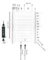
하기 도면은 본 명세서의 일부를 형성하며, 본 발명의 특정 측면을 추가로 입증하기 위해 포함된다. 본 발명은 본원에 제공된 구체적인 구현예의 상세한 설명과 함께 하나 이상의 이러한 도면을 참조하여 더 잘 이해될 수 있다.
도 1a 내지 1d. PDAC 종양에서 CAF 이질성. 도 1a. PKT 종양에서 대표적인 간질 세포 조성(n=11마리 마우스). 이러한 데이터는 도 4e에서도 보여준다. 도 1b. 인간 PDAC에서 αSMA 및 FAP의 면역형광 표지에 의한 공동 국재화의 정량화(n = 2명의 환자, 표준 편차는 가시 필드에 걸친 염색 분포의 변화를 나타냄). 도 1c 내지 1d. αSMA-RFP+ 세포, YFP+ 암세포, 및 PKT 종양에서 FAP-APC 면역표지된 세포의 특성화; LSL-YFP; αSMA-RFP GEM. n = 1마리 마우스, 분류된 세포 집단은 후속적으로 scRNA seq 분석에 사용되었다(도 2a 내지 2c). GEM: 유전자 조작 마우스, Neg: 음성, s+: 단일 양성, Vim: 비멘틴, scRNA seq: 단일 세포 RNA 시퀀싱. 마우스 GEM 명명법은 표 1을 참조한다.
도 2a 내지 2b. αSMA + 및 FAP + CAF는 면역-유사 전사체 프로파일을 사용하여 뚜렷한 섬유아세포 하위 집단을 규정한다. 도 2a. αSMA+ 농축 세포의 클러스터(1, 2, 3, 4 & 6, 왼쪽 패널) 및 FAP+ 농축 세포의 클러스터(3, 5, 7, 왼쪽 패널)와 함께 t-SNE 플롯으로 표현된 PKT 종양에서 유세포 분석(도 1d)에 의해 농축된 αSMA+ 및 FAP+ 세포의 scRNA seq 분석. 클러스터 3은 αSMA+ 및 FAP+ 농축 세포 간에 공유된 전사체 동일성을 갖는 세포 클러스터를 포함한다. 기능 클러스터(1-9, 오른쪽 패널)도 정의되고, 그룹 정의가 나열되었다. 도 2b. 특정 αSMA+ 및 FAP+ 클러스터를 비교 프로파일링하고, 각각의 클러스터에서 풍부한 전사물을 기반으로 유전자 네트워크를 식별하였다. scRNA seq: 단일 세포 RNA 시퀀싱, RBC: 적혈구, ECM: 세포외 기질.
도 3. 인간 PDAC 종양의 scRNA 시퀀싱에 의해 포착된 세포 이질성. t-SNE 플롯으로 표현된 비분별 인간 PDAC 종양의 scRNA seq 분석, 2명의 환자를 평가했다(PDAC 1 및 PDAC 2). PDAC 2의 경우 두 번의 별개의 유세포 분석 세포 분류를 수행하고(PDAC 2A, PDAC 2B; 기술적 복제), 이어서 후속 분석을 위해 PDAC 2에 통합했다. scRNA seq: 단일 세포 RNA 시퀀싱.
도 4a 내지 4e. αSMA + 대 FAP + CAF의 선택적 고갈에 의한 PDAC 진행에 대한 뚜렷한 결과. 도 4a. αSMA+ 또는 FAP+ 세포 고갈이 있거나 없는 PKT GEM의 췌장 종양 절편의 H&E 염색 후 표시된 그룹의 조직학적 특징을 나타내는 막대그래프. 고갈은 αSMA-TK 및 FAP-TK 이식유전자를 보유한 PKT 마우스에서 GCV의 투여에 의해 가능해졌다. 대조군은 이식유전자를 보유하고 PBS로 투여되거나 주사되지 않은 PKT 마우스, 뿐만 아니라 이식유전자가 없고 GCV가 투여된 PKT 마우스를 포함한다. FAP-TK, n= 5 및 대조군, n = 8; αSMA-TK, n = 11 및 대조군, n = 17. 평균 +/- 평균의 표준 오차가 표시된다. 통계적 유의성은 만-휘트니(Mann-Whitney) 검정을 사용하여 평가되었다. 도 4b. PKT 종양에서 αSMA 및 FAP에 대한 면역조직화학(IHC)의 정량화 및 지시된 그룹의 관련 정량화를 나타내는 막대그래프. FAP-TK, n = 6 및 대조군, n = 7; αSMA-TK, n = 5 및 대조군, n = 5 마우스. 통계적 유의성은 만-휘트니 검정을 사용하여 평가되었다. 도 4c 내지 4d. αSMA+ 대 FAP+ 세포 고갈이 있는 PKT 마우스의 종양에서 일반적으로 하향 또는 상향 조절되는 유전자(도 4c) 및 관련 경로(도 4d)의 중첩. FAP-TK, n = 3 및 대조군, n = 3; αSMA-TK, n = 3 및 대조군, n = 3 마우스. 도 4e. PKT 종양(도 1a에도 표시됨) 및 αSMA+ 또는 FAP+ 세포가 고갈된 PKT 종양에서 간엽 세포 조성. FAP-TK, n = 7; αSMA-TK, n = 5, 대조군, n = 4. 대조군(FAP-TK대조군, n = 7, αSMA-TK대조군, n = 4 둘 모두에 대한 조합 대조군), n = 11. 48.3%*: 독립 표본 양측 t 검정(unpaired two-tailed t test)에 의해 평가된 유의차. 막대그래프는 αSMA 간질 고갈의 정량화, 만-휘트니 검정을 나타낸다. 평균 +/- 표준 편차가 표시된다. GEM: 유전자 조작 마우스, GCV: 간시클로비르, IRS: 면역 반응 점수. 마우스 GEM 명명법은 표 1을 참조한다.
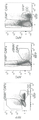
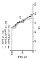
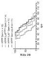
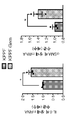
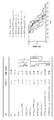
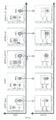
도 5a 내지 5h. αSMA + CAF의 IL-6은 젬시타빈에 대한 암세포 내성을 부여한다. 도 5a. t-SNE 플롯으로 표시되고, 세포 수(괄호 안) 및 IL-6을 발현하는 세포의 백분율을 나타내는 PKT 종양으로부터 αSMA+ 및 FAP+ 농축 세포의 scRNA seq 분석. 막대그래프는 scRNA seq에서 얻은 데이터를 요약하며, 이는 IL-6 전사물이 주로 αSMA+ 세포에 농축됨을 뒷받침한다. 도 5b. 지시된 세포에서 IL-6 전사물의 qPCR 정량화. 세포는 3마리의 별개의 마우스에서 얻었다. 통계적 유의성은 독립 표본 양측 t 검정을 사용하여 평가되었다. 도 5c. 지시된 GEM에서 췌장 종양의 H&E 절편의 종양 조직학적 표현형의 정량화. 각 컬럼 내에서 절편은 위에서 아래로 괴사, 불량, 양호, PanIN 및 정상을 나타낸다. 이원 ANOVA. 도 5d. 시간 경과에 따른 지시된 GEM의 생존율. 로그 순위 검정. 도 6c 참조. 도 5e. 종양에서 IL-6 전사물의 qPCR 정량화, 그룹당 n = 4마리 마우스. 왼쪽에서 오른쪽으로 막대는 KPPF, KPPF;IL-6smaKO, 및 KPPF;IL-6-/-을 나타낸다. 통계적 유의성은 독립 표본 양측 t 검정을 사용하여 평가되었다. 도 5f. 시간 경과에 따른 지시된 GEM의 생존율. y축의 50% 생존율 표시에서 왼쪽에서 오른쪽으로 선은 KPPF Gem, KPPF Gem IL-6, KPPF;IL-6smaKO, 및 KPPF;IL-6-/-을 나타낸다. 로그 순위 검정. 도 6c 참조. 도 5g. 종양에서 IL-6 및 Acta2(αSMA) 전사물의 qPCR 정량화, 그룹당 n = 3마리 마우스. 통계적 유의성은 독립 표본 양측 t 검정을 사용하여 평가되었다. 도 5h. 지시된 GEM에서 인산화된 Stat3(포스포-Stat3)에 대한 면역조직화학 후 가시 필드당 포스포-Stat3+ 세포 수의 정량적 분석. 막대는 왼쪽부터 오른쪽으로 KPPF, KPPF;IL-6smaKO, KPPF;IL-6-/-, KPPF Gem, 및 KPPF;IL-6smaKO Gem을 나타낸다. 그룹당 n = 5마리 마우스, 일원 ANOVA. 평균 +/- 평균의 표준 오차가 표시된다. * P < 0.05, ** P < 0.01, *** P < 0.005, **** P < 0.001, ns: 유의하지 않음. scRNAseq: 단일 세포 RNA 시퀀싱, GEM: 유전자 조작 마우스, Gem: 젬시타빈, qPCR: 정량적 PCR, nd: 미검출. 마우스 GEM 명명법은 표 1을 참조한다.
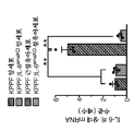
도 6a 내지 6d. 종양 내 T 세포의 간질 IL-6 분극화의 이점은 젬시타빈과 동시에 실현된다. 도 6a. 지시된 GEM 및 치료 그룹에서 종양 면역 침투 분획. 데이터는 독립 표본 단측 t 검정의 평균 +/- 평균의 표준 오차로 표시된다. 도 6b. 지시된 실험 그룹에서 마우스의 생존율. y축의 25% 생존율 표시에서 왼쪽에서 오른쪽으로 선은 KPPF Gem, KPPF Gem CP, KPPF;IL-6-/- Gem, 및 KPPF;IL-6-/- Gem CP를 나타낸다. 로그 순위 검정, 도 6c 참조. 도 6c. GEM 및 치료 그룹 개요 및 선택된 그룹에서 마우스의 생존율 분석. 로그 순위 검정. 도 6d. 고(IL-6 HI) 및 저(IL-6 LO) IL-6 전사물 수준을 갖는 종양에서 FOXP3, GADH, 및 ACTA2(αSMA) 전사물 수준을 평가하는 TCGA 데이터 세트 분석. 독립 표본 양측 t 검정. * P < 0.05, *** P < 0.005, ns: 유의하지 않음. Gem: 젬시타빈, aIL-6: 항-IL-6 항체, CP: 항-CTLA-4 및 항-PD1 항체. 마우스 GEM 명명법은 표 1을 참조한다.
도 7a 내지 7b. 게이팅 전략 및 도 1e 내지 1f에 도시된 게이트를 규정하는 데 사용되는 대조군. 종양 세포 게이팅 전략 및 FAP 이소형 대조군(도 7a), 내인성 형광(αSMA-RFP 및 YFP) 게이팅에 대한 음성 대조군으로 사용되는 염색되지 않은 비장 세포(도 7b).
도 8a 내지 8e. 도 8a. αSMA 세포 고갈이 있거나 없는 PKP 마우스의 생존율 곡선(PKP 대조군은 αSMA-TK 이식유전자가 없고 GCV가 투여된 PKP 마우스임). 화살표는 GCV 치료가 시작된 시기를 나타낸다. 약 75일에서 생존율이 0%로 떨어지는 선은 PKP αSMA가 고갈되었음을 나타낸다. 로그 순위 검정. 도 8b. αSMA+ 세포 고갈이 있거나 없는 PKP GEM의 췌장 종양 섹션의 H&E 염색, 연령 일치 또는 종점을 기준으로 지시된 그룹의 조직학적 특징을 나타내는 막대그래프. 대조군에는 GCV가 없는 이식유전자를 보유하는 PKP 마우스 또는/및 GCV가 투여되고 이식유전자가 없는 PKT 마우스가 포함된다. 연령 일치-대조군, n = 9; 빈사 상태(moribund) 대조군(실험 종점), n = 11, αSMA-TK, n = 12 마우스. 통계적 유의성은 이원 ANOVA를 사용하여 평가되었다. 도 8c. 체중에 대한 종양 중량의 백분율로 표시되는 종양 부담. 대조군, n = 8; FAP-TK, n = 9 마우스. 독립 표본 양측 t 검정. 도 8d. 지시된 마우스의 종양에서 FAP+ 세포의 유세포 분석 평가 및 그래픽 표현(그룹당 하나의 마우스). 도 8e. 종양 조직병리학적 점수. 대조군, n = 4; FAP-TK, n = 4 마우스. 평균 +/- 표준 편차, 만-휘트니 검정. 달리 명시되지 않는 한, 데이터는 평균 +/- 평균의 표준 오차로 표시된다. * P < 0.05, *** P < 0.005, ****, P < 0.001, ns: 유의하지 않음. GEM: 유전자 조작 마우스, GCV: 간시클로비르, 마우스 GEM 명명법은 표 1을 참조한다.
도 9a 내지 9e. 도 9a. GCV를 투여한 비종양 보유 마우스에서 시간 경과에 따른 체중 측정. WT(하단 선): 야생형 한배새끼 대조군, n = 3; FAP-TK(상단 선), n = 4 마우스. 도 9b. 종점에서 췌장, 비장, 대퇴사두근(QM) 및 비복근(GM) 중량. WT: 야생형 한배새끼 대조군, n = 3; FAP-TK, n = 4마리 마우스. 도 9c. PKT 마우스의 비장에서 FAP+ 세포에 대한 유세포 분석. 도 9d. 비-종양 보유 마우스의 골수에서 αSMA-RFP+(n = 2마리 마우스) 및 FAP 면역표지된 세포(n = 3마리 마우스)의 유세포 분석을 사용한 평가. 도 9e. 비-종양 보유 대조군 마우스(n = 6)에 비해 종양 보유 마우스(PKT, n = 3)의 골수에서 FAP+ 세포의 빈도 증가. 평균 +/- 평균의 표준 오차가 표시됨, 독립 표본 양측 t 검정, * P < 0.05, ns: 유의하지 않음. GCV: 간시클로비르, 마우스 명명법은 표 1을 참조한다.
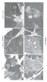
도 10a 내지 10c. 도 10a. 나열된 시점에 KPPF GEM의 대표적인 H&E 염색된, CK19 및 αSMA 면역표지된 췌장, 폐 및 간 절편. 스케일 바(Scale bar): 100 mm. 도 10b. 나열된 시점에 KPPC GEM의 대표적인 H&E 염색된 췌장 절편. 스케일 바: 100 mm. 도 10c. KPPF의 췌장 종양에서 GFP, RFP, YFP 및 CFP 및 핵의 대표적인 면역형광 포획; αSMA-Cre; R26Confetti GEM. 스케일 바: 20 mm. GEM: 유전자 조작 마우스, wks: 주, ADM: 선포에서 도관 이형성(Acinar to ductal metaplasia), 마우스 GEM 명명법은 표 1을 참조한다.
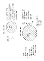
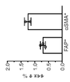
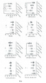
도 11a 내지 11f. 도 11a. KPPF에서 수거된 종양 유래 암세포 및 섬유아세포의 개략적 표현; αSMA-Cre; R26Dual GEM. 도 11b. qPCR에 의해 나열된 GEM의 종양에서 IL-1β의 발현. 왼쪽에서 오른쪽으로 막대는 KPPF, KPPF;IL-6smaKO, 및 KPPF;IL-6-/-을 나타낸다. n = 그룹당 마우스. 도 11c 내지 11e. 나열된 기관 및 GEM에서 정제된 DNA의 PCR 산물의 전기영동 이동. 산물 검출은 예상 레인에서 유전자 재조합에 의한 IL-6의 특정 결실을 확인한다. 도 11f. KPPF의 종양에서 FAP 및 αSMA에 대한 면역표지의 공동 국재화의 정량화; αSMA-Cre;R26Dual GEM. n = 4마리 마우스. 평균 +/- 평균의 표준 오차가 표시됨, ns: 유의하지 않음, GEM: 유전자 조작 마우스, qPCR: 정량적 PCR, 마우스 GEM 명명법은 표 1을 참조한다.
도 12a 내지 12c. 도 12a. 나열된 GEM의 종양 부담. 왼쪽에서 오른쪽으로 막대는 KPPF, KPPF;IL-6smaKO, 및 KPPF;IL-6-/-을 나타낸다. 도 12b. 지시된 GEM의 전이 발생률. 도 12c. 나열된 GEM에서 H&E 염색된 췌장 절편의 조직병리학적 특징의 정량화. 각 컬럼 내에서 절편은 위에서 아래로 PanIN 및 정상을 나타낸다. GEM: 유전자 조작 마우스, ns: 유의하지 않음, 마우스 GEM 명명법은 표 1을 참조한다.
도 13a 내지 13c. 도 13a. 질환 진행의 나열된 시점에 KPF 마우스의 대표적인 H&E 염색 및 CK19 면역표지된 췌장, 및 간 및 폐 전이의 H&E 염색된 절편(검정색 화살표). 스케일 바: 100 mm. 도 13b. 나열된 GEM의 췌장의 대표적인 H&E 염색된 절편. 스케일 바: 100 mm. 도 13c. 나열된 GEM의 생존율, 도 6c 참조. 400일 바로 아래에서 x축과 교차하는 선이 KPF를 나타낸다. 로그 순위 검정. GEM: 유전자 조작 마우스, ns: 유의하지 않음, 마우스 GEM 명명법은 표 1을 참조한다.
도 14a 내지 14d. 도 14a. 나열된 GEM의 생존율, 도 6c 참조. 로그 순위 검정. 도 14b. 빈사 상태 종점에서 나열된 GEM의 췌장의 H&E 염색된 절편의 조직병리학적 특징의 정량화. 각 컬럼 내에서 절편은 위에서 아래로 괴사, 불량, 양호, PanIN, 및 정상을 나타낸다. KPPF Gem, n = 6; KPPF; IL-6smaKO Gem, n = 5마리 마우스. 이원 ANOVA. 도 14c. 지시된 GEM의 종양 부담. KPPF Gem(왼쪽 컬럼), n = 8; KPPF; IL-6smaKO Gem(오른쪽 컬럼), n = 11마리 마우스. 독립 표본 양측 t 검정. 도 14d. 나열된 GEM의 췌장의 αSMA 면역표지된 절편에서 αSMA+ 면적 백분율의 정량화. 그룹당 n = 5마리 마우스, 일원 ANOVA. * P < 0.05, ** P < 0.01, ***, P < 0.005, ****, P < 0.001, ns: 유의하지 않음. 데이터는 평균 +/- 평균의 표준 오차로 표시된다. GEM: 유전자 조작 마우스, Gem: 젬시타빈, 마우스 GEM 명명법은 표 1을 참조한다.
도 15a 내지 15b. 도 15a. 포스포-ERK1/2 및 포스포-Akt에 대해 면역표지된, 나열된 GEM에서 췌장 절편의 염색 면적 백분율 정량화. 그룹당 n = 5마리 마우스, 일원 ANOVA. 도 15b. 나열된 GEM의 췌장 절편에 대한 염색 면적 백분율 정량화, CD31, Ki67에 대한 면역표지, 절단된 카스파아제 3, 및 MTS에 대한 염색. 그룹당 n = 5마리 마우스, 일원. ANOVA. * P < 0.05, ** P < 0.01, ***, P < 0.005, ****, P < 0.001, ns: 유의하지 않음. GEM: 유전자 조작 마우스, Gem: 젬시타빈, 마우스 GEM 명명법은 표 1을 참조한다.
도 16a 내지 16d. 면역형(immunotyping) 분석을 위한 종양(도 16a) 및 비장(도 16b)의 유세포 분석을 위한 게이팅 전략. T 세포 패널(도 16c) 및 골수 세포 패널(도 16d)에 대한 종양의 유세포 분석을 위한 게이팅 전략.
도 17a 내지 17c. 종양(도 17a), 비장(도 17b) 및 말초혈액(도 17c)을 평가하는 지시된 GEM에서 나열된 면역 세포에 대한 추가 면역형 분석 결과.BRIEF DESCRIPTION OF THE DRAWINGS The following drawings form part of this specification and are included to further demonstrate certain aspects of the invention. The invention may be better understood by reference to one or more of these drawings in conjunction with the detailed description of specific embodiments provided herein.
1a to 1d. CAF heterogeneity in PDAC tumors. 1a. Representative stromal cell composition in PKT tumors (n=11 mice). These data are also shown in Fig. 4e. Figure 1b. Quantification of co-localization by immunofluorescent labeling of αSMA and FAP in human PDAC (n = 2 patients, standard deviations represent changes in staining distribution across the visible field). 1c to 1d. characterization of FAP-APC immunolabeled cells in αSMA-RFP + cells, YFP + cancer cells, and PKT tumors; LSL-YFP; αSMA-RFP GEM. n = 1 mouse, sorted cell populations were subsequently used for scRNA seq analysis ( FIGS. 2A-2C ). GEM: genetically engineered mouse, Neg: negative, s+: single positive, Vim: vimentin, scRNA seq: single cell RNA sequencing. See Table 1 for mouse GEM nomenclature.
2a to 2b. αSMA + and FAP + CAF uses immune-like transcriptome profiles to define distinct fibroblast subpopulations. Figure 2a. Flow cytometry in PKT tumors expressed as a t-SNE plot with clusters of αSMA + enriched cells (1, 2, 3, 4 & 6, left panels) and FAP + enriched cells (3, 5, 7, left panels). scRNA seq analysis of αSMA + and FAP + cells enriched by assay ( FIG. 1D ).
Figure 3. Cellular heterogeneity captured by scRNA sequencing of human PDAC tumors. scRNA seq analysis of undifferentiated human PDAC tumors expressed as t-SNE plots, two patients were evaluated (
4a to 4e. αSMA + vs. FAP + PDAC by selective depletion of CAF Distinct results for progression. Figure 4a. Histograms showing the histological features of the indicated groups after H&E staining of pancreatic tumor sections of PKT GEM with or without αSMA + or FAP + cell depletion. Depletion was made possible by administration of GCV in PKT mice carrying the αSMA-TK and FAP-TK transgenes. Controls included PKT mice bearing the transgene and either administered or not injected with PBS, as well as PKT mice without the transgene and administered GCV. FAP-TK, n=5 and control, n=8; αSMA-TK, n = 11 and control, n = 17. Mean +/- standard error of mean is shown. Statistical significance was assessed using the Mann-Whitney test. Figure 4b. Histograms showing the quantification of immunohistochemistry (IHC) for αSMA and FAP in PKT tumors and the relative quantification of the indicated groups. FAP-TK, n = 6 and control, n = 7; αSMA-TK, n = 5 and control, n = 5 mice. Statistical significance was assessed using the Mann-Whitney test. 4c to 4d. Overlapping of genes (Fig. 4c) and related pathways (Fig. 4d) that are normally down- or up-regulated in tumors of αSMA + vs. FAP + PKT mice with cell depletion. FAP-TK, n = 3 and control, n = 3; αSMA-TK, n = 3 and control, n = 3 mice. Figure 4e. Mesenchymal cell composition in PKT tumors (also shown in FIG. 1A ) and in PKT tumors depleted of αSMA + or FAP + cells. FAP-TK, n = 7; αSMA-TK, n = 5, control, n = 4. control (FAP-TK control , n = 7, αSMA-TK control , n = 4 combination control for both), n = 11. 48.3%*: independent Significant difference assessed by an unpaired two-tailed t test. Histograms represent the quantification of αSMA epileptic depletion, the Mann-Whitney test. Mean +/- standard deviation is shown. GEM: genetically engineered mice, GCV: ganciclovir, IRS: immune response score. See Table 1 for mouse GEM nomenclature.
5a to 5h. αSMA + IL-6 in CAF confers cancer cell resistance to gemcitabine. Figure 5a. scRNA seq analysis of αSMA + and FAP + enriched cells from PKT tumors, presented as t-SNE plots, showing cell count (in parentheses) and percentage of cells expressing IL-6. The histogram summarizes the data obtained from scRNA seq, supporting that IL-6 transcripts are predominantly enriched in αSMA + cells. Figure 5b. qPCR quantification of IL-6 transcripts in indicated cells. Cells were obtained from 3 separate mice. Statistical significance was assessed using an independent sample two-tailed t-test. 5c. Quantification of the tumor histological phenotype of H&E sections of pancreatic tumors in the indicated GEM. Sections within each column represent necrotic, poor, good, PanIN, and normal from top to bottom. Two-way ANOVA. Figure 5d. Survival of the indicated GEM over time. log rank test. See Figure 6c. Figure 5e. qPCR quantification of IL-6 transcripts in tumors, n = 4 mice per group. Bars from left to right represent KPPF, KPPF;IL-6 smaKO , and KPPF;IL-6 −/- . Statistical significance was assessed using an independent sample two-tailed t-test. Figure 5f. Survival of the indicated GEM over time. The lines from left to right in the 50% survival rate display on the y-axis represent KPPF Gem, KPPF Gem IL-6, KPPF;IL-6 smaKO , and KPPF;IL-6 −/- . log rank test. See Figure 6c. Figure 5g. qPCR quantification of IL-6 and Acta2 (αSMA) transcripts in tumors, n = 3 mice per group. Statistical significance was assessed using an independent sample two-tailed t-test. Figure 5h. Quantitative analysis of the number of phospho-Stat3 + cells per visible field after immunohistochemistry for phosphorylated Stat3 (phospho-Stat3) in the indicated GEM. Bars represent KPPF, KPPF;IL-6 smaKO , KPPF;IL-6 −/- , KPPF Gem, and KPPF;IL-6 smaKO Gem from left to right. n = 5 mice per group, one-way ANOVA. The mean +/- standard error of the mean is shown. * P < 0.05, ** P < 0.01, *** P < 0.005, **** P < 0.001, ns : not significant. scRNAseq: single cell RNA sequencing, GEM: genetically engineered mouse, Gem: gemcitabine, qPCR: quantitative PCR, nd : undetected. See Table 1 for mouse GEM nomenclature.
6a to 6d. The benefits of stromal IL-6 polarization of intratumoral T cells are realized simultaneously with gemcitabine. Figure 6a. Tumor immune infiltration fractions in the indicated GEM and treatment groups. Data are expressed as the mean +/- standard error of the mean of an independent sample one-sided t-test. Figure 6b. Survival rates of mice in the indicated experimental groups. The lines from left to right in the 25% survival plot on the y-axis represent KPPF Gem, KPPF Gem CP, KPPF;IL-6 −/- Gem, and KPPF;IL-6 −/- Gem CP. log rank test, see FIG. 6C. 6c. GEM and treatment group overview and analysis of survival rates of mice in selected groups. log rank test. Figure 6d. TCGA data set analysis evaluating FOXP3, GADH, and ACTA2 (αSMA) transcript levels in tumors with high (IL-6 HI) and low (IL-6 LO) IL-6 transcript levels. Independent samples two-sided t-test. * P < 0.05, *** P < 0.005, ns : not significant. Gem: gemcitabine, aIL-6: anti-IL-6 antibody, CP: anti-CTLA-4 and anti-PD1 antibody. See Table 1 for mouse GEM nomenclature.
7a-7b . Controls used to define the gating strategy and gates shown in Figures 1E-1F. Tumor cell gating strategy and FAP isotype control (Figure 7a), unstained splenocytes (Figure 7b) used as negative controls for endogenous fluorescence (αSMA-RFP and YFP) gating.
8a to 8e. Figure 8a. Survival curves of PKP mice with and without αSMA cell depletion (PKP controls were PKP mice without αSMA-TK transgene and administered GCV). Arrows indicate when GCV treatment started. The line where the survival rate drops to 0% at about 75 days indicates that PKP αSMA is depleted. log rank test. Figure 8b. H&E staining of pancreatic tumor sections of PKP GEM with or without αSMA + cell depletion, histograms showing histological features of indicated groups based on age-matched or endpoint. Controls include PKP mice carrying the transgene without GCV or/and PKT mice receiving GCV and no transgene. age-matched-control, n = 9; moribund control (experimental endpoint), n = 11, αSMA-TK, n = 12 mice. Statistical significance was assessed using two-way ANOVA. Figure 8c. Tumor burden expressed as a percentage of tumor weight to body weight. control, n = 8; FAP-TK, n = 9 mice. Independent samples two-sided t-test. Figure 8d. Flow cytometric evaluation and graphical representation of FAP + cells in tumors of indicated mice (one mouse per group). Figure 8e. Tumor histopathological score. control, n = 4; FAP-TK, n = 4 mice. Mean +/- standard deviation, Mann-Whitney test. Unless otherwise specified, data are expressed as mean +/- standard error of the mean. * P < 0.05, *** P < 0.005, ****, P < 0.001, ns : not significant. GEM: genetically engineered mouse, GCV: ganciclovir, mouse GEM See Table 1 for nomenclature.
9a to 9e. Figure 9a. Determination of body weight over time in non-tumor-bearing mice administered GCV. WT (bottom line): wild-type littermate control, n = 3; FAP-TK (top line), n = 4 mice. Figure 9b. Pancreas, spleen, quadriceps (QM) and gastrocnemius (GM) weights at endpoint. WT: wild-type littermate control, n = 3; FAP-TK, n = 4 mice. Figure 9c. Flow cytometry analysis of FAP + cells in the spleen of PKT mice. Figure 9d. Assessment using flow cytometry of αSMA-RFP + (n = 2 mice) and FAP immunolabeled cells (n = 3 mice) in the bone marrow of non-tumor bearing mice. Figure 9e. Increased frequency of FAP + cells in the bone marrow of tumor bearing mice (PKT, n = 3) compared to non-tumor bearing control mice (n = 6). Mean +/- standard error of mean shown, independent sample two-tailed t-test, * P < 0.05, ns : not significant. GCV: ganciclovir, mouse See Table 1 for nomenclature.
10a to 10c. Figure 10a. Representative H&E stained, CK19 and αSMA immunolabeled pancreatic, lung and liver sections of KPPF GEM at the time points listed. Scale bar: 100 mm. Figure 10b. Representative H&E stained pancreatic sections from KPPC GEM at the time points listed. Scale bar: 100 mm. Figure 10c. Representative immunofluorescence capture of GFP, RFP, YFP and CFP and nuclei in pancreatic tumors of KPPF; αSMA-Cre; R26 Confetti GEM. Scale bar: 20 mm. GEM: genetically engineered mouse, wks: week, ADM: Acinar to ductal metaplasia, see Table 1 for mouse GEM nomenclature.
11a to 11f. Figure 11a. Schematic representation of tumor-derived cancer cells and fibroblasts harvested from KPPF; αSMA-Cre; R26 Dual GEM. 11b. Expression of IL-1β in tumors of the listed GEMs by qPCR. Bars from left to right represent KPPF, KPPF;IL-6 smaKO , and KPPF;IL-6 −/- . n = mice per group. 11c to 11e. Electrophoretic transfer of PCR products of purified DNA from the listed organs and GEM. Product detection identifies specific deletion of IL-6 by genetic recombination in the expected lane. 11f. Quantification of the co-localization of immunolabels for FAP and αSMA in tumors of KPPF; αSMA-Cre;R26 Dual GEM. n = 4 mice. Mean +/- standard error of mean indicated, ns : not significant, GEM: genetically engineered mouse, qPCR: quantitative PCR, mouse GEM See Table 1 for nomenclature.
12a to 12c. 12a. Tumor burden of the listed GEMs. Bars from left to right represent KPPF, KPPF;IL-6 smaKO , and KPPF;IL-6 −/- . 12b. The incidence of metastases in the indicated GEM. 12c. Quantification of histopathological features of H&E-stained pancreatic sections in the listed GEMs. Intercepts within each column represent PanIN and normal from top to bottom. GEM: genetically engineered mouse, ns : not significant, see Table 1 for mouse GEM nomenclature.
13a to 13c. Figure 13a. Representative H&E stained and CK19 immunolabeled pancreas and H&E stained sections of liver and lung metastases (black arrows) from KPF mice at the listed time points of disease progression. Scale bar: 100 mm. 13b. Representative H&E-stained sections of the pancreas from the listed GEMs. Scale bar: 100 mm. 13c. Viability of the listed GEMs, see Figure 6c. The line that intersects the x-axis just below 400 days represents the KPF. log rank test. GEM: genetically engineered mouse, ns : not significant, see Table 1 for mouse GEM nomenclature.
14a to 14d. 14a. Viability of the listed GEMs, see Figure 6c. log rank test. 14b. Quantification of histopathological features of H&E stained sections of pancreas from listed GEMs at moribund endpoints. Sections within each column represent necrosis, poor, good, PanIN, and normal from top to bottom. KPPF Gem, n = 6; KPPF; IL-6 smaKO Gem, n = 5 mice. Two-way ANOVA. 14c. Tumor burden of the indicated GEM. KPPF Gem (left column), n = 8; KPPF; IL-6 smaKO Gem (right column), n = 11 mice. Independent samples two-sided t-test. 14d. Quantification of αSMA + area percentages in αSMA immunolabeled sections of the pancreas from the listed GEMs. n = 5 mice per group, one-way ANOVA. * P < 0.05, ** P < 0.01, ***, P < 0.005, ****, P < 0.001, ns : not significant. Data are expressed as mean +/- standard error of the mean. GEM: genetically engineered mouse, Gem: gemcitabine, mouse GEM See Table 1 for nomenclature.
15a to 15b. 15a. Quantification of percent stained area of pancreatic sections in the listed GEMs, immunolabeled for phospho-ERK1/2 and phospho-Akt. n = 5 mice per group, one-way ANOVA. 15b. Quantification of the percentage staining area for pancreatic sections of the listed GEMs, immunolabels for CD31, Ki67, cleaved
16a to 16d. Gating strategy for flow cytometry analysis of tumor ( FIG. 16A ) and spleen ( FIG. 16B ) for immunotyping analysis. Gating strategy for flow cytometry analysis of tumors for a panel of T cells ( FIG. 16C ) and a panel of bone marrow cells ( FIG. 16D ).
17A-17C. Additional immunotyping assays for listed immune cells in the indicated GEMs evaluating tumor ( FIG. 17A ), spleen ( FIG. 17B ) and peripheral blood ( FIG. 17C ).
췌관 선암(pancreatic ductal adenocarcinoma; PDAC)에서 결합조직형성 반응은 면역 세포 및 섬유아세포의 상당한 축적을 수반한다. 섬유아세포는 조직 복구 및 재생에 기여하고 암에 대한 숙주 반응의 일부로 종양에 축적되는 간엽 세포이다. 이러한 암종 관련 섬유아세포(CAF)의 기원과 기능적 다양성은 거의 알려지지 않았다. αSMA+ 세포는 종양 촉진 활성(TP-CAF)을 나타내는 FAP+ CAF와 달리 종양 억제 특성(TS-CAF)을 가진 PDAC에서 지배적인 CAF 집단이다. TS-CAF는 주로 세포외 기질(ECM) 생산을 조절하고, 세포-ECM 부착을 촉진하고, 적응 면역을 조절하지만, TP-CAF는 전염증, 케모카인 분비 표현형으로 치우친 계통을 나타내고, TP-CAF를 억제하기 위한 진단 및 치료 표적 역할을 할 수 있다. 또한, CAF는 림프구 및 골수 계통의 특징적인 고유한 유전자 발현 프로파일을 공유한다. αSMA+ CAF-유래 인터류킨-6(IL-6)은 PDAC 진행에 영향을 미치지 않지만, 화학적 내성에 기여하고 면역 체크포인트 차단 요법의 잠재력을 약화시킨다. αSMA+ CAF로부터 IL-6의 특정 결실은 화학요법 및 체크포인트 차단 요법에 대한 감도를 향상시켰다. 총체적으로, 이러한 연구는 PDAC 진행 동안 기능적으로 이질적인 섬유아세포의 복잡한 네트워크를 식별하며, 이는 중요한 치료적 의미를 갖는다. 이와 같이, 본원에서는 종양 성장을 제어하기 위한 CAF 표적이 제공된다.In pancreatic ductal adenocarcinoma (PDAC), the connective tissue-forming response involves a significant accumulation of immune cells and fibroblasts. Fibroblasts are mesenchymal cells that contribute to tissue repair and regeneration and accumulate in tumors as part of the host response to cancer. The origin and functional diversity of these carcinoma-associated fibroblasts (CAFs) are largely unknown. αSMA+ cells are the dominant CAF population in PDACs with tumor suppressive properties (TS-CAF) in contrast to FAP+ CAFs, which exhibit tumor-promoting activity (TP-CAF). TS-CAF mainly regulates extracellular matrix (ECM) production, promotes cell-ECM adhesion, and modulates adaptive immunity, whereas TP-CAF represents a lineage biased towards pro-inflammatory, chemokine-secreting phenotypes, It can serve as a diagnostic and therapeutic target for inhibition. CAFs also share unique gene expression profiles characteristic of lymphocyte and myeloid lineages. αSMA + CAF-derived interleukin-6 (IL-6) does not affect PDAC progression, but contributes to chemical resistance and undermines the potential of immune checkpoint blockade therapy. Specific deletion of IL-6 from αSMA + CAF enhanced sensitivity to chemotherapy and checkpoint blockade therapy. Collectively, these studies identify a complex network of functionally heterogeneous fibroblasts during PDAC progression, which has important therapeutic implications. As such, provided herein are CAF targets for controlling tumor growth.
유전적 무(moue) 모델을 사용한 결과에 의하면 PDAC CAF가 PDAC에서 우세한 집단으로 나타나는 αSMA+ CAF를 갖는 이종 집단임이 확인된다. 이 연구는 인간 PDAC 생물학과 더 관련이 있는 연구를 만들기 위한 노력으로 이러한 결론에 도달하기 위해 세포 배양 시스템의 사용을 완전히 자제했다. αSMA+ CAF 및 FAP+ CAF는 PDAC 진행에서 서로 다른 기능을 나타낸다(Feig et al., 2013; Lo et al., 2017; Kraman et al., 2010). 흥미롭게도 FAP+ CAF는 αSMA+ CAF를 유발할 수 있는 골수-유래 전구체(progenitor)를 나타낸다. 놀랍게도, scRNA-Seq에 의해 정의된 αSMA+ CAF 및 FAP+ CAF의 정체성은 뚜렷한 면역 세포-유사 전사체를 반영한다. αSMA+ CAF는 콜라겐 생성 및 리모델링 세포의 서브셋, 뿐만 아니라 대식세포-유사, 및 T 세포-유사 전사체를 나타낸 세포를 포함한다. 대조적으로, FAP+ CAF는 호중구-유사 및 B 세포-유사 전사체를 갖는 세포의 서브셋으로 구성되었다. αSMA+ CAF 및 FAP+ CAF의 서브셋은 단핵구 및 수지상 세포-유사 전사체를 나타냈다. 이러한 발견은 간엽 및 조혈 세포 계통 사이의 중복 가능성을 강조하기 때문에 흥미롭다. PDAC 종양 미세환경의 풍부한 사이토카인 환경은 면역 세포 동원 및 표현형 성숙을 조절하며, 이 환경에 대한 CAF의 노출 결과 면역 세포 관련 전사물을 유도할 수 있다. 특정 CAF가 종양 내 면역 반응을 조작하기 위해 피드포워드 시그널링 루프를 조성할 수 있다. 특정 CAF 집단이 없는 경우, PDAC에서 특정 면역 세포의 분화/활성화가 저해되거나 제한될 수 있다. 대안적으로, CAF는 종양에서 면역 세포 기능을 수행할 수 있다.The results using the genetic moue model confirm that PDAC CAF is a heterogeneous population with αSMA + CAF that appears as a predominant population in PDAC. This study completely refrained from using cell culture systems to arrive at these conclusions in an effort to make studies more relevant to human PDAC biology. αSMA + CAF and FAP + CAF show different functions in PDAC progression (Feig et al., 2013; Lo et al., 2017; Kraman et al., 2010). Interestingly, FAP + CAF represents a bone marrow-derived progenitor that can induce αSMA + CAF. Remarkably, the identity of αSMA + CAF and FAP + CAF as defined by scRNA-Seq reflects distinct immune cell-like transcripts. αSMA + CAFs include a subset of collagen-producing and remodeling cells, as well as cells displaying macrophage-like, and T cell-like transcripts. In contrast, FAP + CAF consisted of a subset of cells with neutrophil-like and B cell-like transcripts. A subset of αSMA + CAF and FAP + CAF showed monocyte and dendritic cell-like transcripts. These findings are interesting because they highlight the potential overlap between mesenchymal and hematopoietic cell lineages. The rich cytokine milieu of the PDAC tumor microenvironment regulates immune cell recruitment and phenotypic maturation, and exposure of CAFs to this milieu can induce immune cell-associated transcripts. Certain CAFs can create feedforward signaling loops to manipulate immune responses in tumors. In the absence of a specific CAF population, differentiation/activation of specific immune cells in PDAC may be inhibited or limited. Alternatively, CAFs can perform immune cell functions in tumors.
αSMA+ CAF-유래 IL-6은 화학적 내성을 부여하고, 종양 미세환경에서 T 세포를 부정적으로 조절한다. 이와 관련하여, 오르가노이드-기반 연구에서, CAF의 서브셋이 IL-6 생산을 포함한 면역조절 표현형을 나타내는 것으로 강조되었다(Ohlund et al., 2017). 전체적으로 αSMA+ CAF는 PDAC 진행에서 종양 억제(종양 진압 또는 TS-CAF)로 나타났지만, 젬시타빈 치료 스트레스의 맥락에서, 암세포는 αSMA+ CAF에 의해 생산된 IL-6을 '활용'하여 생존을 촉진한다. αSMA+ CAF에 의해 생산된 IL-6은 비-치료 상태에서 자가 보존 목적을 위한 것이지만, 젬시타빈 치료에 대한 내성이라는 맥락에서 실현되는 생존 촉진(pro-survival) 시그널링 경로를 유도하기 위해 암세포에 의해 활용된다고 생각할 수 있다. 이전 연구에서는 IL-6 시그널링이 화학요법의 맥락에서 JAK/STAT 시그널링 경로를 통해 암세포에 생존 촉진 신호를 부여한다고 보고되었다(Wormann et al., 2016; Nagathihalli et al., 2016). PDAC 면역 미세환경의 상당한 분극화는 αSMA+ CAF 생산된 IL-6이 결실될 때 관찰되었지만; 이러한 Teff 및 Treg 빈도 변화는 PDAC 진행에 영향을 미치지 않았다. 면역 체크포인트 차단 요법은 젬시타빈과 조합되었을 때 효과가 없었지만; 이러한 결합 이점은 IL-6 시그널링의 억제와 조합될 때 실현되었으며, 이는 IL-6이 PDAC에서 면역 체크포인트 차단 요법의 중요한 억제인자임을 나타낸다. IL-6의 억제는 세포 사멸 및 아마도 젬시타빈에 의해 유도된 신생항원 생성과 조합될 때, 면역 체크포인트 차단의 효능을 증가시킬 수 있는 이펙터 T 세포의 출현을 선호했을 가능성이 있다.αSMA + CAF-derived IL-6 confers chemical resistance and negatively regulates T cells in the tumor microenvironment. In this regard, in organoid-based studies, it has been highlighted that a subset of CAFs exhibit immunomodulatory phenotypes including IL-6 production (Ohlund et al., 2017). Overall, αSMA + CAF was shown to be tumor suppressive (tumor suppressive or TS-CAF) in PDAC progression, but in the context of gemcitabine treatment stress, cancer cells 'utilize' IL-6 produced by αSMA + CAF to promote survival do. IL-6 produced by αSMA + CAF is intended for self-preservation purposes in non-therapeutic conditions, but by cancer cells to induce a pro-survival signaling pathway that is realized in the context of resistance to gemcitabine treatment. You can think of it as being used. Previous studies have reported that IL-6 signaling confers survival-promoting signals to cancer cells via the JAK/STAT signaling pathway in the context of chemotherapy (Wormann et al., 2016; Nagathihalli et al., 2016). Significant polarization of the PDAC immune microenvironment was observed when αSMA + CAF produced IL-6 was deleted; These Teff and Treg frequency changes did not affect PDAC progression. Immune checkpoint blockade therapy was ineffective when combined with gemcitabine; This binding advantage was realized when combined with inhibition of IL-6 signaling, indicating that IL-6 is an important inhibitor of immune checkpoint blockade therapy in PDACs. Inhibition of IL-6, when combined with apoptosis and possibly gemcitabine-induced neoantigen production, likely favored the emergence of effector T cells that could increase the efficacy of immune checkpoint blockade.
I. 항체 및 이의 생산I. Antibodies and their production
"단리된 항체"는 자연환경의 성분으로부터 분리 및/또는 회수된 항체이다. 자연환경의 오염 성분은 항체의 진단 또는 치료 용도를 방해하는 물질이며, 효소, 호르몬 및 기타 단백질성 또는 비-단백질성 용질을 포함할 수 있다. 특정 구현예에서, 항체는 다음과 같은 수준으로 정제된다: (1) 로리(Lowry) 방법으로 결정될 때 95중량% 초과, 가장 특히 99중량% 초과의 항체; (2) 스피닝 컵 시쿼네이터(spinning cup sequenator)를 사용하여 N-말단 또는 내부 아미노산 서열의 적어도 15개 잔기를 얻기에 충분한 정도; 또는 (3) 쿠마씨 블루(Coomassie blue) 또는 은 염색을 사용하여 환원 또는 비-환원 조건하에 SDS-PAGE에 의한 균질성. 단리된 항체는 항체의 자연환경의 적어도 하나의 성분이 존재하지 않을 것이므로 재조합 세포 내에 동소 항체를 포함한다. 그러나 일반적으로 단리된 항체는 적어도 하나의 정제 단계에 의해 준비될 것이다.An “isolated antibody” is an antibody that has been isolated and/or recovered from a component of its natural environment. Contaminant components of the natural environment are substances that interfere with diagnostic or therapeutic uses of the antibody, and may include enzymes, hormones, and other proteinaceous or non-proteinaceous solutes. In certain embodiments, the antibody is purified to the following levels: (1) greater than 95% by weight, most particularly greater than 99% by weight of antibody as determined by the Lowry method; (2) sufficient to obtain at least 15 residues of the N-terminal or internal amino acid sequence using a spinning cup sequenator; or (3) homogeneity by SDS-PAGE under reducing or non-reducing conditions using Coomassie blue or silver staining. An isolated antibody comprises the antibody in situ within recombinant cells since at least one component of the antibody's natural environment will not be present. In general, however, an isolated antibody will be prepared by at least one purification step.
기본 4-사슬 항체 단위는 2개의 동일한 경쇄(L) 및 2개의 동일한 중쇄(H)로 구성된 이종 사량체 당단백질이다. IgM 항체는 J 사슬이라고 하는 추가 폴리펩티드와 함께 5개의 기본 이종사량체 단위로 구성되어 있으므로 10개의 항원 결합 부위를 포함하는 반면, 분비된 IgA 항체는 중합되어 J 사슬과 함께 2-5개의 기본 4-사슬 단위를 포함하는 다가 군집(assemblage)을 형성할 수 있다. IgG의 경우에, 4-사슬 단위는 일반적으로 약 150,000 달톤이다. 각각의 L 사슬은 하나의 공유 이황화 결합에 의해 H 사슬에 연결되는 반면, 2개의 H 사슬은 H 사슬 이소형에 따라 하나 이상의 이황화 결합에 의해 서로 연결된다. 각각의 H 및 L 사슬은 또한 규칙적으로 간격을 둔 사슬 내 이황화 가교를 가지고 있다. 각각의 H 사슬은 N-말단에 가변 영역(VH)이 있고 그 뒤에 알파 및 감마 사슬 각각에 대한 3개의 불변 도메인(CH) 및 mu 및 이소형에 대한 4개의 CH 도메인이 있다. 각각의 L 사슬은 N-말단에 가변 영역(VL)이 있고 그 뒤에 다른 쪽 말단에 불변 도메인(CL)이 있다. VL은 VH와 정렬되고, CL은 중쇄(CH1)의 제1 불변 도메인과 정렬된다. 특정 아미노산 잔기는 경쇄 가변 영역과 중쇄 가변 영역 사이의 계면을 형성하는 것으로 여겨진다. VH와 VL의 쌍은 함께 단일 항원-결합 부위를 형성한다. 다양한 부류의 항체의 구조 및 특성은, 예를 들어, 문헌[참조: Basic and Clinical Immunology, 8th edition, Daniel P. Stites, Abba I. Terr and Tristram G. Parslow (eds.), Appleton & Lange, Norwalk, Conn., 1994, page 71, and Chapter 6]을 참조한다.The basic four-chain antibody unit is a heterotetrameric glycoprotein composed of two identical light (L) chains and two identical heavy (H) chains. IgM antibodies are composed of 5 basic heterotetrameric units with an additional polypeptide called the J chain and thus contain 10 antigen binding sites, whereas secreted IgA antibodies polymerize and contain 2-5 basic 4- A multivalent assemblage comprising chain units may be formed. In the case of IgG, a four-chain unit is generally about 150,000 daltons. Each L chain is linked to the H chain by one covalent disulfide bond, whereas the two H chains are linked to each other by one or more disulfide bonds, depending on the H chain isotype. Each H and L chain also has regularly spaced intrachain disulfide bridges. Each H chain has a variable region (V H ) at the N-terminus followed by three constant domains (CH ) for each of the alpha and gamma chains and four CH domains for mu and isoforms. Each L chain has a variable region (V L ) at the N-terminus followed by a constant domain ( CL ) at the other end. V L is aligned with the V H, C L is aligned with the first constant domain of the heavy chain (C H1). Certain amino acid residues are believed to form an interface between the light chain variable region and the heavy chain variable region. The pair of V H and V L together forms a single antigen-binding site. The structures and properties of the various classes of antibodies are described, for example, in Basic and Clinical Immunology, 8th edition, Daniel P. Stites, Abba I. Terr and Tristram G. Parslow (eds.), Appleton & Lange, Norwalk. , Conn., 1994, page 71, and Chapter 6].
임의의 척추동물 종으로부터의 L 사슬은 불변 도메인(CL)의 아미노산 서열에 기초하여 카파 및 람다라고 하는 두 가지 명확하게 구별되는 유형 중 하나로 할당될 수 있다. 중쇄(CH)의 불변 도메인의 아미노산 서열에 따라, 면역글로불린은 서로 다른 부류 또는 이소형으로 할당될 수 있다. 면역글로불린에는 각각 알파, 델타, 엡실론, 감마 및 뮤로 지정된 중쇄를 갖는 IgA, IgD, IgE, IgG 및 IgM의 5가지 부류가 있다. 감마 및 알파 부류는 CH 서열 및 기능에서 비교적 작은 차이를 기반으로 하위 부류로 더 나뉘며, 인간은 IgG1, IgG2, IgG3, IgG4, IgA1, 및 IgA2의 하위 부류를 나타낸다.L chains from any vertebrate species can be assigned to one of two clearly distinct types, called kappa and lambda, based on the amino acid sequence of the constant domain (CL ). Depending on the amino acid sequence of the constant domain of the heavy chain ( CH ), immunoglobulins can be assigned to different classes or isotypes. There are five classes of immunoglobulins: IgA, IgD, IgE, IgG and IgM with heavy chains designated alpha, delta, epsilon, gamma and mu, respectively. The gamma and alpha classes are further divided into subclasses based on relatively small differences in C H sequence and function, and humans represent the subclasses of IgG1, IgG2, IgG3, IgG4, IgA1, and IgA2.
용어 "가변"은 V 도메인의 특정 세그먼트가 항체 간에 서열이 광범위하게 다르다는 사실을 지칭한다. V 도메인은 항원 결합을 매개하고, 특정 항원에 대한 특정 항체의 특이성을 한정한다. 그러나 가변성은 가변 영역의 110개 아미노산 범위에 걸쳐 고르게 분포되지 않는다. 대신, V 영역은 각각 길이가 9-12개의 아미노산인 "초가변 영역"이라고 하는 극단적 가변성의 짧은 영역으로 분리된 15-30개 아미노산의 프레임워크 영역(FR)이라고 하는 비교적 변함없는 스트레치로 구성된다. 천연 중쇄 및 경쇄의 가변 영역은 각각 4개의 FR을 포함하며, 이는 베타-시트 구조를 연결하는 루프를 형성하는 3개의 초가변 영역으로 연결되는 베타-시트 배치를 주로 채택하고, 일부 경우 베타-시트 구조의 일부를 형성한다. 각각의 사슬의 초가변 영역은 FR에 의해 함께 매우 근접하게 유지되며, 다른 사슬의 초가변 영역과 함께 항체의 항원-결합 부위 형성에 기여한다(참조: Kabat et al., Sequences of Proteins of Immunological Interest, 5th Ed. Public Health Service, National Institutes of Health, Bethesda, Md. (1991)). 불변 도메인은 항체를 항원에 결합하는 데 직접 관여하지 않지만, 항체 의존적 세포 세포독성(ADCC), 항체-의존적 세포 식균 작용(ADCP), 항체-의존적 호중구 식균 작용(ADNP), 및 항체-의존적 보체 침착(antibody-dependent complement deposition; ADCD)과 같은 다양한 이펙터 기능을 나타낸다.The term “variable” refers to the fact that certain segments of the V domain differ widely in sequence between antibodies. The V domain mediates antigen binding and defines the specificity of a particular antibody for a particular antigen. However, the variability is not evenly distributed over the 110 amino acid range of the variable region. Instead, the V region consists of relatively invariant stretches called framework regions (FRs) of 15-30 amino acids separated into short regions of extreme variability, called “hypervariable regions”, each 9-12 amino acids in length. . The variable regions of native heavy and light chains each contain four FRs, which predominantly adopt a beta-sheet configuration, joined by three hypervariable regions forming loops connecting the beta-sheet structure, and in some cases beta-sheet structures. form part of the structure. The hypervariable regions of each chain are held in close proximity together by the FRs and, together with the hypervariable regions of the other chain, contribute to the formation of the antigen-binding site of antibodies (Kabat et al. , Sequences of Proteins of Immunological Interest). , 5th Ed. Public Health Service, National Institutes of Health, Bethesda, Md. (1991)). The constant domains are not directly involved in binding the antibody to antigen, but antibody-dependent cellular cytotoxicity (ADCC), antibody-dependent cellular phagocytosis (ADCP), antibody-dependent neutrophil phagocytosis (ADNP), and antibody-dependent complement deposition It exhibits various effector functions such as antibody-dependent complement deposition (ADCD).
본원에서 사용될 때 용어 "초가변 영역"은 항원 결합을 담당하는 항체의 아미노산 잔기를 나타낸다. 초가변 영역은 일반적으로 "상보성 결정 영역" 또는 "CDR"의 아미노산 잔기(예를 들어, 카밧(Kabat) 넘버링 시스템에 따라 넘버링될 때 VL에서 대략 잔기 24-34(L1), 50-56(L2) 및 89-97(L3), 및 VH에서 대략 31-35(H1), 50-65(H2) 및 95-102(H3); Kabat et al., Sequences of Proteins of Immunological Interest, 5th Ed. Public Health Service, National Institutes of Health, Bethesda, Md. (1991)); 및/또는 "초가변 루프"의 잔기(예를 들어, 초티아(Chothia) 넘버링 시스템에 따라 넘버링될 때 VL에서 잔기 24-34(L1), 50-56(L2) 및 89-97(L3), 및 VH에서 26-32(H1), 52-56(H2) 및 95-101(H3); Chothia and Lesk, J. Mol. Biol. 196:901-917 (1987)); 및/또는 "초가변 루프"/CDR의 잔기(예를 들어, IMGT 넘버링 시스템에 따라 넘버링될 때 VL에서 잔기 27-38(L1), 56-65(L2) 및 105-120(L3), 및 VH에서 27-38(H1), 56-65(H2) 및 105-120(H3); Lefranc, M. P. et al. Nucl. Acids Res. 27:209-212 (1999), Ruiz, M. et al. Nucl. Acids Res. 28:219-221 (2000))를 포함한다. 임의로 항체에는 문헌[참조: AHo; Honneger, A. and Plunkthun, A. J. Mol. Biol. 309:657-670 (2001)]에 따라 넘버링될 때 VL에서 28, 36(L1), 63, 74-75(L2) 및 123(L3), 및 VsubH에서 28, 36(H1), 63, 74-75(H2) 및 123(H3) 포인트 중 하나 이상에서 대칭적인 삽입이 있다.The term “hypervariable region” as used herein refers to the amino acid residues of an antibody that are responsible for antigen binding. Hypervariable regions are generally "complementarity determining region" or an amino acid residue of a "CDR" (e. G., Kabat (Kabat) about residues 24-34 (L1) in the V L when the numbering according to the numbering system, 50-56 ( L2) and 89-97 (L3), and approximately 31-35 (H1), 50-65 (H2) and 95-102 (H3) in V H ; Kabat et al. , Sequences of Proteins of Immunological Interest, 5th Ed Public Health Service, National Institutes of Health, Bethesda, Md. (1991)); and/or residues of a "hypervariable loop" (eg, residues 24-34 (L1), 50-56 (L2) and 89-97 (L3) in V L when numbered according to the Chothia numbering system. ), and 26-32 (H1), 52-56 (H2) and 95-101 (H3) in V H ; Chothia and Lesk, J. Mol. Biol. 196:901-917 (1987)); and/or residues of the "hypervariable loop"/CDR (eg, residues 27-38 (L1), 56-65 (L2) and 105-120 (L3) in V L when numbered according to the IMGT numbering system, and 27-38 (H1), 56-65 (H2) and 105-120 (H3) in V H ;Lefranc, MP et al. Nucl. Acids Res. 27:209-212 (1999), Ruiz, M. et al. al. Nucl. Acids Res. 28:219-221 (2000)). Optionally, antibodies include those described in AHo; Honneger, A. and Plunkthun, AJ Mol. Biol. 309: 657-670 at V L 28, 36, when numbered in accordance with the (2001)] (L1), 63, 74-75 (L2) and 123 (L3), and in the V sub H 28, 36 (H1 ), There is a symmetrical insertion at one or more of points 63, 74-75 (H2) and 123 (H3).
"생식계(germline) 핵산 잔기"는 불변 또는 가변 영역을 인코딩하는 생식계 유전자에서 자연적으로 발생하는 핵산 잔기를 의미한다. "생식계 유전자"는 생식 세포(즉, 난자 또는 정자가 될 예정인 세포)에서 발견되는 DNA이다. "생식계 돌연변이"는 단일-세포 단계에서 생식 세포 또는 접합체에서 발생한 특정 DNA의 유전 가능한 변화를 나타내며, 자손에게 전달될 때, 이러한 돌연변이는 신체의 모든 세포에 통합된다. 생식계 돌연변이는 단일 체세포에서 획득되는 체세포 돌연변이와 대조된다. 일부 경우에, 가변 영역을 인코딩하는 생식계 DNA 서열의 뉴클레오티드는 돌연변이되고(즉, 체세포 돌연변이), 상이한 뉴클레오티드로 대체된다."Germline nucleic acid residue" means a nucleic acid residue that occurs naturally in a germline gene encoding a constant or variable region. A “germline gene” is the DNA found in a germ cell (ie, a cell that is expected to become an egg or sperm). A "germline mutation" refers to a heritable change in a specific DNA that occurs in a germ cell or zygote at the single-cell stage, and when passed on to offspring, these mutations are incorporated into all cells of the body. Germline mutations are contrasted with somatic mutations that are acquired in single somatic cells. In some cases, nucleotides of the germline DNA sequence encoding the variable region are mutated (ie, somatic mutation) and replaced with different nucleotides.
본원에 사용된 용어 "단클론 항체"는 실질적으로 균질한 항체 집단으로부터 수득된 항체를 나타내며, 즉, 집단을 구성하는 개별 항체는 소량으로 존재할 수 있는 가능한 자연 발생 돌연변이를 제외하고 동일하다. 단클론 항체는 단일 항원 부위에 대해 매우 특이적이다. 또한, 상이한 결정인자(에피토프)에 대한 상이한 항체를 포함하는 다클론 항체 제제와 달리, 각각의 단클론 항체는 항원 상의 단일 결정인자에 대해 지시된다. 이의 특이성 외에도, 단클론 항체는 다른 항체에 의해 오염되지 않은 상태로 합성될 수 있다는 점에서 유리하다. 수식어 "단클론"은 임의의 특정 방법에 의한 항체 생산을 요구하는 것으로 해석되어서는 안된다. 예를 들어, 본 발명에 유용한 단클론 항체는 콜러 등(참조: Kohler et al., Nature, 256:495 (1975))에 의해 처음 설명된 하이브리도마 방법론에 의해 제조될 수 있거나, 항원 특이적 B 세포, 감염 또는 면역화에 반응하는 항원 특이적 형질아세포의 단일 세포 분류, 또는 대량 분류된 항원 특이적 집합체에서 단일 세포로부터 연결된 중쇄 및 경쇄 포획 후 박테리아, 진핵 동물 또는 식물 세포에서 재조합 DNA 방법을 사용하여 만들어질 수 있다(예를 들어, 미국 특허 제4,816,567호 참조). "단클론 항체"는 또한 예를 들어, 문헌[참조: Clackson et al., Nature, 352:624-628 (1991) and Marks et al., J. Mol. Biol., 222:581-597 (1991)]에 기재된 기술을 사용하여 파지 항체 라이브러리로부터 단리될 수 있다.As used herein, the term “monoclonal antibody” refers to an antibody obtained from a substantially homogeneous population of antibodies, ie, the individual antibodies making up the population are identical except for possible naturally occurring mutations that may be present in minor amounts. Monoclonal antibodies are highly specific for a single antigenic site. Also, unlike polyclonal antibody preparations, which include different antibodies directed against different determinants (epitopes), each monoclonal antibody is directed against a single determinant on the antigen. In addition to their specificity, monoclonal antibodies are advantageous in that they can be synthesized uncontaminated by other antibodies. The modifier "monoclonal" should not be construed as requiring production of the antibody by any particular method. For example, monoclonal antibodies useful in the present invention may be prepared by the hybridoma methodology first described by Kohler et al. (Kohler et al., Nature, 256:495 (1975)), or antigen-specific B Using recombinant DNA methods in bacterial, eukaryotic or plant cells after single cell sorting of cells, antigen-specific plasmablasts in response to infection or immunization, or capture of linked heavy and light chains from single cells in mass sorted antigen-specific aggregates. (See, eg, US Pat. No. 4,816,567). "Monoclonal antibodies" are also described, eg, in Clackson et al. , Nature, 352:624-628 (1991) and Marks et al. , J. Mol. Biol., 222:581-597 (1991)].
B. 일반적인 방법B. General method
TP-CAF에 의해 고도로 발현되는 단백질에 결합하는 단클론 항체는 여러 용도를 가질 것임을 이해할 것이다. 여기에는 암을 검출하고 진단하는 데 사용될 뿐만 아니라 암을 치료하기 위한 진단 키트의 생산이 포함된다. 이러한 맥락에서, 이러한 항체를 진단제 또는 치료제에 연결하거나, 경쟁 검정에서 포획제 또는 경쟁자로 사용하거나, 추가 제제를 부착하지 않고 개별적으로 사용할 수 있다. 항체는 아래에서 추가로 논의된 바와 같이 돌연변이되거나 변형될 수 있다. 항체를 제조하고 특성화하는 방법은 당해 분야에 잘 알려져 있다(참조: 예를 들어, Antibodies: A Laboratory Manual, Cold Spring Harbor Laboratory, 1988; 미국 특허 4,196,265).It will be appreciated that monoclonal antibodies that bind to proteins highly expressed by TP-CAF will have several uses. This includes the production of diagnostic kits used to detect and diagnose cancer as well as to treat cancer. In this context, such antibodies can be linked to a diagnostic or therapeutic agent, used as a capture agent or competitor in a competition assay, or used individually without the attachment of an additional agent. Antibodies may be mutated or modified as discussed further below. Methods for making and characterizing antibodies are well known in the art (see, eg, Antibodies: A Laboratory Manual, Cold Spring Harbor Laboratory, 1988; US Pat. No. 4,196,265).
단클론 항체(MAb)를 생성하는 방법은 일반적으로 다클론 항체를 제조하는 방법과 동일한 방식에 따라 시작된다. 이 두 가지 방법의 첫 번째 단계는 적절한 숙주의 면역화 또는 이전의 자연 감염 또는 허가된 백신 또는 실험 백신으로의 예방 접종으로 인해 면역이 생긴 대상체를 식별하는 것이다. 당해 분야에 잘 알려진 바와 같이, 주어진 면역화 조성물은 이의 면역원성이 다양할 수 있다. 따라서, 펩티드 또는 폴리펩티드 면역원을 캐리어에 결합함으로써 달성될 수 있는 바와 같이 숙주 면역계를 강화하는 것이 종종 필요하다. 예시적이고 바람직한 캐리어는 키홀 림펫 헤모시아닌(keyhole limpet hemocyanin; KLH) 및 소 혈청 알부민(BSA)이다. 오브알부민, 마우스 혈청 알부민 또는 토끼 혈청 알부민과 같은 다른 알부민도 캐리어로 사용될 수 있다. 폴리펩티드를 캐리어 단백질에 접합하는 수단은 당해 분야에 잘 알려져 있으며, 글루타르알데히드, m-말레이미도벤코일-N-하이드록시석신이미드 에스테르, 카보디이미드 및 비스-바이아조화 벤지딘을 포함한다. 또한, 당해 분야에 잘 알려진 바와 같이, 특정 면역원 조성물의 면역원성은 아쥬반트로도 알려진, 면역 반응의 비특이적 자극제를 사용하여 향상될 수 있다. 동물에서 예시적이고 바람직한 아쥬반트는 완전 프로인트 아쥬반트(사멸된 마이코박테리움 투버쿨로시 스(Mycobacterium tuberculosis)를 포함하는 면역 반응의 비특이적 자극제), 불완전 프로인트 아쥬반트 및 수산화알루미늄 아쥬반트를 포함하고, 인간에서는 명반, CpG, MFP59 및 면역자극 분자의 조합("아쥬반트 시스템", 예를 들어 AS01 또는 AS03)을 포함한다. 나노입자 백신 또는 물리적 전달 시스템(예를 들어 지질 나노입자 또는 금 바이올리스틱 비드(gold biolistic bead))에서 DNA 또는 RNA 유전자로 전달되고 바늘, 유전자 총, 경피 전기천공 장치로 전달되는 유전자-인코딩된 항원을 포함하여, 암-특이적 B 세포를 유도하기 위한 추가 실험 형태의 접종이 가능하다. 항원 유전자는 또한 아데노바이러스, 아데노-관련 바이러스, 폭스바이러스, 헤르페스바이러스, 또는 알파바이러스 레플리콘과 같은 복제 가능 또는 결함 바이러스 벡터, 또는 대안적으로 바이러스 유사 입자에 의해 인코딩된 대로 운반될 수 있다.A method for producing a monoclonal antibody (MAb) is generally initiated in the same manner as a method for producing a polyclonal antibody. The first step in both methods is to identify subjects who have developed immunity due to immunization of an appropriate host or previous natural infection or immunization with a licensed or experimental vaccine. As is well known in the art, a given immunizing composition may vary in its immunogenicity. Thus, it is often necessary to enhance the host immune system, as can be achieved by binding a peptide or polypeptide immunogen to a carrier. Exemplary and preferred carriers are keyhole limpet hemocyanin (KLH) and bovine serum albumin (BSA). Other albumins may also be used as carriers, such as ovalbumin, mouse serum albumin or rabbit serum albumin. Means for conjugating a polypeptide to a carrier protein are well known in the art and include glutaraldehyde, m-maleimidobencoyl-N-hydroxysuccinimide ester, carbodiimide, and bis-biazoated benzidine. Also, as is well known in the art, the immunogenicity of certain immunogenic compositions can be enhanced using non-specific stimulators of the immune response, also known as adjuvants. Exemplary and preferred adjuvant is a complete Freund adjuvant (scan (non-specific stimulator of the immune response containing Mycobacterium tuberculosis) when in the killing Mycobacterium-to M.) in an animal, the incomplete Freund's adjuvants and aluminum hydroxide adjuvant and in humans a combination of alum, CpG, MFP59 and an immunostimulatory molecule (“adjuvant system”, eg AS01 or AS03). Gene-encoded antigen delivered as a DNA or RNA gene in a nanoparticle vaccine or physical delivery system (e.g. lipid nanoparticles or gold biolistic beads) and delivered to a needle, gene gun, transdermal electroporation device Further experimental types of inoculation are possible to induce cancer-specific B cells, including The antigenic gene may also be carried as encoded by a replication competent or defective viral vector, such as an adenovirus, adeno-associated virus, poxvirus, herpesvirus, or alphavirus replicon, or alternatively a virus-like particle.
다클론 항체의 생산에 사용되는 면역원 조성물의 양은 면역원의 특성, 및 면역화에 사용되는 동물에 따라 달라진다. 면역원을 투여하기 위해 다양한 경로를 사용할 수 있다(피하, 근육내, 피내, 정맥내 및 복강내). 다클론 항체의 생산은 면역화 후 다양한 시점에 면역화된 동물의 혈액을 샘플링하여 모니터링될 수 있다. 두 번째, 부스터 주사도 제공될 수 있다. 부스팅 및 타이터링(titering) 과정은 적절한 역가에 도달할 때까지 반복된다. 원하는 수준의 면역원성이 얻어지면, 면역화된 동물을 채혈하고, 혈청을 분리하여 보관할 수 있고/있거나 동물을 사용하여 MAb를 생성할 수 있다.The amount of immunogen composition used in the production of polyclonal antibodies depends on the nature of the immunogen and the animal used for immunization. A variety of routes can be used to administer the immunogen (subcutaneous, intramuscular, intradermal, intravenous and intraperitoneal). Production of polyclonal antibodies can be monitored by sampling the blood of the immunized animal at various time points after immunization. Second, booster injections may also be given. The boosting and titering process is repeated until an appropriate titer is reached. Once the desired level of immunogenicity is achieved, the immunized animal may be bled, the serum may be isolated and stored, and/or the animal may be used to generate MAbs.
면역화 후, 항체를 생산할 가능성이 있는 체세포, 특히 B 림프구(B 세포)가 MAb 생성 프로토콜에 사용하기 위해 선택된다. 이러한 세포는 생검된 비장, 림프절, 편도선 또는 아데노이드, 골수 흡인물 또는 생검, 폐 또는 위장관과 같은 점막 기관의 조직 생검, 또는 순환 혈액에서 얻을 수 있다. 그 다음 면역화된 동물 또는 면역 인간으로부터의 항체-생성 B 림프구는 일반적으로 면역화된 동물 또는 인간 또는 인간/마우스 키메라 세포와 같은 종들 중 하나인, 불멸 골수종 세포의 세포와 융합된다. 하이브리도마-생산 융합 절차에 사용하기에 적합한 골수종 세포주는 바람직하게는 비-항체-생산되고, 융합 효율이 높으며, 원하는 융합 세포(하이브리도마)의 성장만을 지원하는 특정 선택적 배지에서 성장할 수 없게 만드는 효소 결핍이 있다. 당업자에게 공지된 바와 같이, 다수의 골수종 세포 중 임의의 하나가 사용될 수 있다. HMMA2.5 세포 또는 MFP-2 세포는 이러한 세포의 특히 유용한 예이다.After immunization, somatic cells likely to produce antibodies, particularly B lymphocytes (B cells), are selected for use in the MAb production protocol. Such cells can be obtained from a biopsied spleen, lymph node, tonsil or adenoid, bone marrow aspirate or biopsy, a tissue biopsy of a mucosal organ such as the lung or gastrointestinal tract, or circulating blood. The antibody-producing B lymphocytes from the immunized animal or immunized human are then fused with cells of the immunized animal or immortal myeloma cells, usually of one of the same species as human or human/mouse chimeric cells. A myeloma cell line suitable for use in a hybridoma-producing fusion procedure is preferably non-antibody-producing, has high fusion efficiency, and is incapable of growing in a specific selective medium that only supports the growth of the desired fusion cell (hybridoma). There is an enzyme deficiency that makes As is known to those skilled in the art, any one of a number of myeloma cells can be used. HMMA2.5 cells or MFP-2 cells are particularly useful examples of such cells.
항체-생산 비장 또는 림프절 세포와 골수종 세포의 하이브리드를 생성하는 방법은 일반적으로 세포막의 융합을 촉진하는 제제(들)(화학 또는 전기)의 존재하에 체세포와 골수종 세포를 2:1 비율로 혼합하는 것을 포함하지만, 비율은 각각 약 20:1에서 약 1:1까지 다양할 수 있다. 일부 경우에, 인간 B 세포를 초기 단계로서 엡슈타인 바 바이러스(Epstein Barr virus; EBV)로 형질전환하면 B 세포의 크기가 증가하여 비교적 큰 크기의 골수종 세포와의 융합이 강화된다. EBV에 의한 형질전환 효율은 형질전환 배지에서 CpG 및 Chk2 억제제 약물을 사용하여 향상된다. 대안적으로, 인간 B 세포는 IL-21, 및 TNF 수퍼패밀리의 유형 II 구성원인 인간 B 세포 활성화 인자(BAFF)와 같은 추가 가용성 인자를 포함하는 배지에서 CD40 리간드(CD154)를 발현하는 형질감염된 세포주와 공동 배양하여 활성화될 수 있다. 센다이(Sendai) 바이러스를 사용하는 융합 방법과 37%(v/v) PEG와 같은 폴리에틸렌 글리콜(PEG)을 사용하는 방법이 설명되었다. 전기적으로 유도된 융합 방법의 사용도 적절하며, 더 나은 효율을 위한 과정도 있다. 융합 절차는 일반적으로 약 1 x 10⁻⁶ 내지 1 x 10⁻⁸의 낮은 빈도로 생존 가능한 하이브리드를 생성하지만, 최적화된 절차를 사용하면 200분의 1에 가까운 융합 효율을 얻을 수 있다. 그러나, 비교적 낮은 융합 효율은 문제를 일으키지 않는데, 생존 가능한 융합된 하이브리드가 선택 배지에서 배양함으로써 모계의 주입된 세포(특히, 일반적으로 무한정 계속 분열하는 주입된 골수종 세포)와 구별되기 때문이다. 선택 배지는 일반적으로 조직 배양 배지에서 뉴클레오티드의 신생 합성을 차단하는 제제를 함유하는 배지이다. 예시적이고 바람직한 제제는 아미노프테린, 메토트렉세이트 및 아자세린이다. 아미노프테린과 메토트렉세이트는 퓨린과 피리미딘 둘 다의 신규 합성을 차단하는 반면, 아자세린은 퓨린 합성만을 차단한다. 아미노프테린 또는 메토트렉세이트가 사용되는 경우, 배지에는 뉴클레오티드 공급원(HAT 배지)으로 하이포크산틴 및 티미딘이 보충된다. 아자세린이 사용되는 경우, 배지에는 하이포크산틴이 보충된다. B 세포 공급원이 EBV-형질전환된 인간 B 세포주인 경우, 골수종에 융합되지 않은 EBV-형질전환된 계통을 제거하기 위해 우아바인(ouabain)이 첨가된다.Methods of generating hybrids of antibody-producing spleen or lymph node cells with myeloma cells generally involve mixing somatic and myeloma cells in a 2:1 ratio in the presence of an agent(s) (chemical or electrical) that promotes the fusion of cell membranes. inclusive, but the ratio may vary from about 20:1 to about 1:1, respectively. In some cases, transformation of human B cells with Epstein Barr virus (EBV) as an early stage increases the size of the B cells, enhancing fusion with relatively large sized myeloma cells. Transformation efficiency by EBV is improved using CpG and Chk2 inhibitor drugs in the transformation medium. Alternatively, human B cells are transfected cell lines expressing CD40 ligand (CD154) in a medium containing IL-21 and additional soluble factors such as human B cell activating factor (BAFF), a type II member of the TNF superfamily. It can be activated by co-culture with A fusion method using Sendai virus and a method using polyethylene glycol (PEG) such as 37% (v/v) PEG were described. The use of electrically induced fusion methods is also appropriate, and there are procedures for better efficiency. The fusion procedure generally produces viable hybrids with a low frequency of about 1 x 10 -6 to 1 x 10 -8 , however, fusion efficiencies close to 1/200 can be achieved with an optimized procedure. However, the relatively low fusion efficiency does not pose a problem, as viable fused hybrids are distinguished from maternally injected cells (particularly, injected myeloma cells, which generally continue to divide indefinitely) by culturing in selective medium. A selective medium is generally a medium containing an agent that blocks the de novo synthesis of nucleotides in a tissue culture medium. Exemplary and preferred agents are aminopterin, methotrexate and azaserine. Aminopterin and methotrexate block de novo synthesis of both purines and pyrimidines, whereas azaserine blocks only purine synthesis. When aminopterin or methotrexate is used, the medium is supplemented with hypoxanthine and thymidine as a nucleotide source (HAT medium). If azaserine is used, the medium is supplemented with hypoxanthine. When the B cell source is an EBV-transformed human B cell line, ouabain is added to eliminate EBV-transformed lines not fused to myeloma.
바람직한 선택 배지는 HAT 또는 우아바인이 있는 HAT이다. 뉴클레오티드 구제 경로(salvage pathway)를 작동할 수 있는 세포만이 HAT 배지에서 생존할 수 있다. 골수종 세포는 구제 경로의 주요 효소(예를 들어, 하이포크산틴 포스포리보실 트랜스페라아제(HPRT))에 결함이 있으며, 생존할 수 없다. B 세포는 이 경로를 작동할 수 있지만, 배양시 수명이 제한되어 있으며, 일반적으로 약 2주 이내에 죽는다. 따라서, 선택적 배지에서 생존할 수 있는 유일한 세포는 골수종과 B 세포에서 형성된 하이브리드이다. 융합에 사용되는 B 세포의 공급원이 EBV-형질전환된 B 세포 계통인 경우, 여기서와 같이, EBV-형질전환된 B 세포가 약물 사멸에 민감하기 때문에 우아바인을 또한 하이브리드의 약물 선택에 사용할 수 있지만, 사용된 골수종 파트너는 우아바인 내성인 것으로 선택된다.A preferred selective medium is HAT or HAT with ouabain. Only cells capable of activating the nucleotide salvage pathway can survive in HAT medium. Myeloma cells are defective in key enzymes of the rescue pathway (eg, hypoxanthine phosphoribosyl transferase (HPRT)) and cannot survive. B cells can operate this pathway, but have a limited lifespan in culture and usually die within about two weeks. Thus, the only cells that can survive in the selective medium are hybrids formed from myeloma and B cells. When the source of B cells used for fusion is an EBV-transformed B cell lineage, as here, ouabain can also be used for drug selection of hybrids, since EBV-transformed B cells are susceptible to drug killing, but , the myeloma partner used is selected to be ouabain resistant.
배양은 특정 하이브리도마가 선택되는 하이브리도마 집단을 제공한다. 통상적으로, 하이브리도마의 선택은 미세역가 플레이트에서 단일 클론 희석으로 세포를 배양한 다음 원하는 반응성에 대해 개별 클론 상청액을 시험함(약 2주 내지 3주 후)으로써 수행된다. 검정은 방사 면역 검정, 효소 면역 검정, 세포독성 검정, 플라크 검정 도트 면역결합 검정 등과 같이 민감하고 간단하며 신속해야 한다. 그 다음 선택된 하이브리도마는 연속적으로 희석되거나 단일-세포가 유세포 분석 분류에 의해 분류되고, 개별 항체-생산 세포주로 클로닝되며, 이 클론은 mAb를 제공하기 위해 무한정 증식될 수 있다. 세포주는 두 가지 기본 방식으로 MAb 생산을 위해 이용될 수 있다. 하이브리도마 샘플을 동물(예를 들어, 마우스)에 주사(종종 복막강 내)할 수 있다. 선택적으로, 동물은 주입 전에 탄화수소, 특히 프리스탄(테트라메틸펜타데칸)과 같은 오일로 프라이밍된다. 이러한 방식으로 인간 하이브리도마를 사용하는 경우, 종양 거부를 예방하기 위해 SCID 마우스와 같은 면역손상 마우스를 주입하는 것이 가장 좋다. 주입된 동물은 융합된 세포 하이브리드에 의해 생산된 특정 단클론 항체를 분비하는 종양을 발생시킨다. 그 다음 혈청 또는 복수액과 같은 동물의 체액을 태핑(tapping)하여 고농도의 MAb를 제공할 수 있다. 개별 세포주는 또한 시험관내에서 배양될 수 있으며, 여기서 MAb는 고농도로 쉽게 수득될 수 있는 배양 배지로 자연적으로 분비된다. 대안적으로, 인간 하이브리도마 세포주를 시험관내에서 사용하여 세포 상청액에서 면역글로불린을 생산할 수 있다. 세포주는 고순도의 인간 단클론 면역글로불린을 회수하는 능력을 최적화하기 위해 무혈청 배지에서 성장하도록 조정될 수 있다.Culturing provides a population of hybridomas from which specific hybridomas are selected. Typically, selection of hybridomas is performed by culturing the cells at monoclonal dilutions in microtiter plates and then testing individual clonal supernatants for the desired reactivity (after about 2-3 weeks). Assays should be sensitive, simple and rapid, such as radioimmunoassays, enzyme immunoassays, cytotoxicity assays, plaque assay dot immunobinding assays, and the like. Selected hybridomas are then serially diluted or single-cells sorted by flow cytometry sorting and cloned into individual antibody-producing cell lines, which clones can be propagated indefinitely to provide mAbs. Cell lines can be used for MAb production in two basic ways. A hybridoma sample can be injected (often intraperitoneally) into an animal (eg, a mouse). Optionally, the animal is primed with a hydrocarbon, particularly an oil such as pristane (tetramethylpentadecane), prior to injection. When using human hybridomas in this way, it is best to inject immunocompromised mice, such as SCID mice, to prevent tumor rejection. The injected animals develop tumors that secrete specific monoclonal antibodies produced by the fused cell hybrid. A high concentration of MAb can then be provided by tapping an animal's bodily fluid, such as serum or ascites fluid. Individual cell lines can also be cultured in vitro , where the MAbs are naturally secreted into a culture medium that can be readily obtained in high concentrations. Alternatively, human hybridoma cell lines can be used in vitro to produce immunoglobulins in cell supernatants. Cell lines can be adapted to grow in serum-free medium to optimize the ability to recover high-purity human monoclonal immunoglobulins.
어느 한 수단에 의해 생산된 MAb는, 원하는 경우, 여과, 원심분리 및 FPLC 또는 친화성 크로마토그래피와 같은 다양한 크로마토그래피 방법을 사용하여 추가로 정제될 수 있다. 본 발명의 단클론 항체의 단편은 펩신 또는 파파인과 같은 효소에 의한 분해 및/또는 화학적 환원에 의한 이황화 결합의 절단을 포함하는 방법에 의해 정제된 단클론 항체로부터 수득될 수 있다. 대안적으로, 본 개시내용에 포함된 단클론 항체 단편은 자동화된 펩티드 합성장치를 사용하여 합성될 수 있다.MAbs produced by either means can, if desired, be further purified using filtration, centrifugation and various chromatographic methods such as FPLC or affinity chromatography. A fragment of the monoclonal antibody of the present invention can be obtained from a purified monoclonal antibody by a method comprising cleavage of disulfide bonds by chemical reduction and/or digestion by enzymes such as pepsin or papain. Alternatively, monoclonal antibody fragments encompassed by the present disclosure can be synthesized using an automated peptide synthesizer.
또한, 분자 클로닝 접근법을 사용하여 단클론 항체를 생성할 수 있는 것으로 고려된다. 관심 항원으로 표지된 단일 B 세포는 상자성 비드 선택 또는 유세포 분석 분류를 사용하여 물리적으로 분류될 수 있으며, 그 다음 RT-PCR에 의해 증폭된 단일 세포 및 항체 유전자로부터 RNA를 단리할 수 있다. 대안적으로, 항원-특이적 벌크 분류된 세포 집단은 미세소포로 분리될 수 있으며, 중쇄 및 경쇄 앰플리콘의 물리적 연결 또는 소포로부터의 중쇄 및 경쇄 유전자의 일반적인 바코딩(barcoding)을 사용하여 일치하는 중쇄 및 경쇄 가변 유전자를 회수할 수 있다. 일치하는 중쇄 및 경쇄 유전자는 세포당 하나의 바코드로 전사물을 마킹하기 위한 바코드 및 RT-PCR 프라이머를 보유하는 세포-침투 나노입자로 세포를 처리하여 항원 특이적 B 세포 집단으로부터 또한 수득될 수 있는 단일 세포를 형성한다. 항체 가변 유전자는 또한 하이브리도마 계통의 RNA 추출 및 RT-PCR에 의해 수득된 항체 유전자를 단리하여 면역글로불린 발현 벡터 내로 클로닝할 수 있다. 대안적으로, 조합 면역글로불린 파지미드(phagemid) 라이브러리는 세포주로부터 단리된 RNA로부터 제조되고, 적절한 항체를 발현하는 파지미드는 바이러스 항원을 사용하여 패닝(panning)함으로써 선택된다. 통상적인 하이브리도마 기술에 비해 이 접근법의 이점은 대략 104배 많은 항체가 단일 라운드로 생산되고 스크리닝될 수 있으며, H 및 L 사슬 조합에 의해 새로운 특이성이 생성되어 적절한 항체를 찾을 가능성을 더욱 증가시킨다는 것이다.It is also contemplated that molecular cloning approaches can be used to generate monoclonal antibodies. Single B cells labeled with the antigen of interest can be physically sorted using paramagnetic bead selection or flow cytometric sorting, and then RNA can be isolated from the amplified single cells and antibody genes by RT-PCR. Alternatively, antigen-specific bulk sorted cell populations can be isolated into microvesicles and matched using physical linkage of heavy and light chain amplicons or general barcoding of heavy and light chain genes from vesicles. Heavy and light chain variable genes can be recovered. Matching heavy and light chain genes can also be obtained from antigen-specific B cell populations by treating cells with cell-penetrating nanoparticles bearing a barcode and RT-PCR primers for marking transcripts with one barcode per cell. form single cells. Antibody variable genes can also be cloned into immunoglobulin expression vectors by isolating antibody genes obtained by RNA extraction and RT-PCR of hybridoma lines. Alternatively, combinatorial immunoglobulin phagemid libraries are prepared from RNA isolated from cell lines, and phagemids expressing the appropriate antibody are selected by panning using viral antigens. The advantage of this approach over conventional hybridoma technology is that approximately 10 4 times as many antibodies can be produced and screened in a single round, and new specificities are generated by H and L chain combinations, further increasing the likelihood of finding suitable antibodies that makes it
본 발명에 유용한 항체의 생산을 교시하는, 각각 본원에 참고로 포함된 다른 미국 특허는 조합 접근법을 사용한 키메라 항체의 생산을 설명하는 미국 특허 5,565,332; 재조합 면역글로불린 제제를 설명하는 미국 특허 4,816,567; 및 항체-치료제 접합체를 설명하는 미국 특허 4,867,973을 포함한다.Other US patents, each of which are incorporated herein by reference, which teach the production of antibodies useful in the present invention, include US Pat. Nos. 5,565,332, which describe the production of chimeric antibodies using a combinatorial approach; US Pat. No. 4,816,567, which describes recombinant immunoglobulin preparations; and US Pat. No. 4,867,973, which describes antibody-therapeutic agent conjugates.
C. 본 발명의 항체C. Antibodies of the Invention
본 발명에 따른 항체는 먼저 결합 특이성에 의해 정의될 수 있다. 당업자는 당업자에게 잘 알려진 기술을 사용하여 주어진 항체의 결합 특이성/친화도를 평가함으로써 이러한 항체가 본 청구범위의 범주 내에 속하는지 여부를 결정할 수 있다. 예를 들어, 주어진 항체가 결합하는 에피토프는 항원 분자 내에 위치한 3개 이상(예를 들어, 3, 4, 5, 6, 7, 8, 9, 10, 11, 12, 13, 14, 15, 16, 17, 18, 19, 20개) 아미노산의 단일 연속 서열로 구성될 수 있다(예를 들어, 도메인 내의 선형 에피토프). 대안적으로, 에피토프는 항원 분자 내에 위치하는 복수의 비-연속 아미노산(또는 아미노산 서열)으로 구성될 수 있다(예를 들어, 형태(conformational) 에피토프).Antibodies according to the invention may first be defined by their binding specificity. One of ordinary skill in the art can determine whether such an antibody falls within the scope of the present claims by assessing the binding specificity/affinity of a given antibody using techniques well known to those skilled in the art. For example, the epitope to which a given antibody binds can be three or more (e.g., 3, 4, 5, 6, 7, 8, 9, 10, 11, 12, 13, 14, 15, 16) located within the antigenic molecule. , 17, 18, 19, 20) of a single contiguous sequence of amino acids (eg, a linear epitope within a domain). Alternatively, an epitope may consist of a plurality of non-contiguous amino acids (or amino acid sequences) located within an antigenic molecule (eg, a conformational epitope).
당업자에게 알려진 다양한 기술을 사용하여 항체가 폴리펩티드 또는 단백질 내에서 "하나 이상의 아미노산과 상호작용"하는지 여부를 결정할 수 있다. 예시적인 기술은, 예를 들어, 문헌[참조: Antibodies, Harlow and Lane (Cold Spring Harbor Press, Cold Spring Harbor, N.Y.)]에 설명된 것과 같은 일상적인 교차-차단 검정을 포함한다. 교차-차단은 ELISA, 생물층 간섭계(biolayer interferometry) 또는 표면 플라스몬 공명과 같은 다양한 결합 검정에서 측정될 수 있다. 다른 방법으로는 알라닌 스캐닝 돌연변이 분석, 펩티드 블롯 분석(Reineke (2004) Methods Mol. Biol. 248: 443-63), 펩티드 절단 분석, 단일 입자 재구성을 사용한 고해상도 전자 현미경 기술, cryoEM 또는 단층촬영, 결정학 연구 및 NMR 분석이 포함된다. 또한, 에피토프 절제, 에피토프 추출 및 항원의 화학적 변형과 같은 방법을 사용할 수 있다(Tomer (2000) Prot. Sci. 9: 487-496). 항체가 상호작용하는 폴리펩티드 내의 아미노산을 식별하는 데 사용될 수 있는 또 다른 방법은 질량 분석법에 의해 검출된 수소/중수소 교환이다. 일반적으로 수소/중수소 교환 방법은 관심 단백질을 중수소-표지한 다음 항체를 중수소-표지된 단백질에 결합하는 것을 포함한다. 다음으로, 단백질/항체 복합체는 물로 전달되고, 항체 복합체에 의해 보호되는 아미노산 내의 교환 가능한 양성자는 계면의 일부가 아닌 아미노산 내의 교환 가능한 양성자보다 느린 속도로 중수소-대-수소 역교환을 겪는다. 결과적으로 단백질/항체 계면의 일부를 형성하는 아미노산은 중수소를 보유할 수 있으므로 계면에 포함되지 않은 아미노산에 비해 비교적 더 높은 질량을 나타낸다. 항체의 해리 후, 표적 단백질은 프로테아제 절단 및 질량 분석법 분석을 거쳐 항체가 상호작용하는 특정 아미노산에 상응하는 중수소-표지된 잔기를 드러낸다. 예를 들어, 문헌[참조: Ehring (1999) Analytical Biochemistry 267: 252-259; Engen and Smith (2001) Anal. Chem. 73: 256A-265A]을 참조한다.A variety of techniques known to those of skill in the art can be used to determine whether an antibody "interacts with one or more amino acids" within a polypeptide or protein. Exemplary techniques include routine cross-blocking assays such as those described, for example, in Antibodies, Harlow and Lane (Cold Spring Harbor Press, Cold Spring Harbor, N.Y.). Cross-blocking can be measured in a variety of binding assays such as ELISA, biolayer interferometry or surface plasmon resonance. Other methods include alanine scanning mutation analysis, peptide blot analysis (Reineke (2004) Methods Mol. Biol. 248: 443-63), peptide cleavage analysis, high resolution electron microscopy techniques using single particle reconstruction, cryoEM or tomography, crystallographic studies. and NMR analysis. In addition, methods such as epitope excision, epitope extraction and chemical modification of antigens can be used (Tomer (2000) Prot. Sci. 9: 487-496). Another method that can be used to identify amino acids within a polypeptide with which the antibody interacts is hydrogen/deuterium exchange detected by mass spectrometry. In general, hydrogen/deuterium exchange methods involve deuterium-labeling the protein of interest and then binding the antibody to the deuterium-labeled protein. Next, the protein/antibody complex is transferred to water, and the exchangeable protons in the amino acids protected by the antibody complex undergo reverse deuterium-to-hydrogen exchange at a slower rate than the exchangeable protons in the amino acids that are not part of the interface. As a result, amino acids that form part of the protein/antibody interface may retain deuterium and thus exhibit a relatively higher mass compared to amino acids not included in the interface. Following dissociation of the antibody, the target protein is subjected to protease cleavage and mass spectrometry analysis to reveal deuterium-labeled residues corresponding to the specific amino acids with which the antibody interacts. See, eg, Ehring (1999) Analytical Biochemistry 267: 252-259; Engen and Smith (2001) Anal. Chem. 73: 256A-265A].
용어 "에피토프"는 B 및/또는 T 세포가 반응하는 항원 상의 부위를 지칭한다. B-세포 에피토프는 연속 아미노산 또는 단백질의 3차 폴딩에 의해 병치되는 비연속 아미노산 모두로부터 형성될 수 있다. 연속 아미노산으로부터 형성된 에피토프는 통상적으로 변성 용매에 노출될 때 유지되는 반면, 3차 폴딩에 의해 형성되는 에피토프는 통상적으로 변성 용매로 처리될 때 손실된다. 에피토프는 통상적으로 고유한 공간 형태로 적어도 3개, 보다 일반적으로 적어도 5개 또는 8-10개의 아미노산을 포함한다.The term “epitope” refers to a site on an antigen to which B and/or T cells respond. B-cell epitopes can be formed from both contiguous amino acids or noncontiguous amino acids juxtaposed by tertiary folding of proteins. Epitopes formed from contiguous amino acids are typically retained when exposed to denaturing solvents, whereas epitopes formed by tertiary folding are typically lost when treated with denaturing solvents. An epitope typically comprises at least 3, more usually at least 5 or 8-10 amino acids in a unique spatial conformation.
항원 구조-기반 항체 프로파일링(ASAP)으로도 알려진 변형-지원 프로파일링(MAP)은 동일한 항원에 대한 다수의 단클론 항체(mAb)를 화학적으로 또는 효소적으로 변형된 항원 표면에 대한 각각의 항체의 결합 프로파일의 유사성에 따라 분류하는 방법이다(US 2004/0101920 참조, 그 전문이 본원에 구체적으로 참조로 포함됨). 각각의 카테고리는 또 다른 카테고리로 표현되는 에피토프와 완전히 다르거나 부분적으로 중복되는 고유한 에피토프를 나타낼 수 있다. 이 기술은 유전적으로 동일한 항체의 신속한 필터링을 가능하게 하여 특성화가 유전적으로 구별되는 항체에 집중될 수 있도록 한다. 하이브리도마 스크리닝에 적용될 때, MAP는 원하는 특성을 갖는 mAb를 생산하는 희귀 하이브리도마 클론의 식별을 용이하게 할 수 있다. MAP는 본 발명의 항체를 상이한 에피토프에 결합하는 항체 그룹으로 분류하는 데 사용될 수 있다.Modification-assisted profiling (MAP), also known as antigen structure-based antibody profiling (ASAP), is a method of analyzing multiple monoclonal antibodies (mAbs) directed against the same antigen, each antibody directed against a chemically or enzymatically modified antigenic surface. It is a method of classifying according to similarity of binding profiles (see US 2004/0101920, specifically incorporated herein by reference in its entirety). Each category may represent a unique epitope that is completely different or partially overlaps with an epitope represented by another category. This technique enables rapid filtering of genetically identical antibodies, allowing characterization to be focused on genetically distinct antibodies. When applied to hybridoma screening, MAP can facilitate the identification of rare hybridoma clones that produce mAbs with desired properties. MAP can be used to classify the antibodies of the invention into groups of antibodies that bind to different epitopes.
본 발명은 동일한 에피토프 또는 에피토프의 일부에 결합할 수 있는 항체를 포함한다. 마찬가지로, 본 발명은 또한 본원에 기재된 임의의 특정 예시적인 항체와 표적 또는 이의 단편에 대한 결합에 대해 경쟁하는 항체를 포함한다. 항체가 당해 분야에 공지된 일상적인 방법을 사용하여 기준 항체와 동일한 에피토프에 결합하는지 또는 기준 항체와 결합에 대해 경쟁하는지 쉽게 결정할 수 있다. 예를 들어, 시험 항체가 기준과 동일한 에피토프에 결합하는지 결정하기 위해, 기준 항체를 포화 조건하에 표적에 결합시킨다. 다음으로, 표적 분자에 결합하는 시험 항체의 능력이 평가된다. 시험 항체가 기준 항체와의 포화 결합 후에 표적 분자에 결합할 수 있다면, 시험 항체는 기준 항체와 다른 에피토프에 결합한다고 결론을 내릴 수 있다. 반면에, 시험 항체가 기준 항체와의 포화 결합 후에 표적 분자에 결합할 수 없다면, 시험 항체는 기준 항체가 결합한 에피토프와 동일한 에피토프에 결합할 수 있다.The present invention includes antibodies capable of binding to the same epitope or portion of an epitope. Likewise, the invention also includes antibodies that compete for binding to a target or fragment thereof with any of the specific exemplary antibodies described herein. Whether an antibody binds to the same epitope as a reference antibody or competes for binding with a reference antibody can be readily determined using routine methods known in the art. For example, to determine whether the test antibody binds the same epitope as the reference, the reference antibody is bound to the target under saturating conditions. Next, the ability of the test antibody to bind to the target molecule is assessed. If the test antibody is able to bind the target molecule after saturating binding with the reference antibody, it can be concluded that the test antibody binds to a different epitope than the reference antibody. On the other hand, if the test antibody is unable to bind the target molecule after saturating binding with the reference antibody, the test antibody may bind to the same epitope as the reference antibody binds.
2개의 항체는 서로 경쟁적으로 항원에 대한 결합을 억제(차단)하는 경우 동일하거나 중첩되는 에피토프에 결합한다. 즉, 1배, 5배, 10배, 20배 또는 100배 과잉의 한 항체는 경쟁 결합 검정에서 측정될 때 다른 항체의 결합을 적어도 50%, 바람직하게는 75%, 90% 또는 심지어 99%까지 억제한다(참조: 예를 들어, Junghans et al., Cancer Res. 1990 50:1495-1502). 대안적으로, 한 항체의 결합을 감소 또는 제거하는 항원의 본질적으로 모든 아미노산 돌연변이가 다른 항체의 결합을 감소 또는 제거하는 경우 두 항체는 동일한 에피토프를 갖는다. 한 항체의 결합을 감소 또는 제거하는 일부 아미노산 돌연변이가 다른 항체의 결합을 감소 또는 제거하는 경우 두 항체는 중복 에피토프를 갖는다.Two antibodies bind to the same or overlapping epitope when competing with each other to inhibit (block) binding to antigen. That is, a 1-fold, 5-fold, 10-fold, 20-fold or 100-fold excess of one antibody reduces the binding of the other antibody by at least 50%, preferably 75%, 90% or even 99%, as measured in a competition binding assay. Inhibits (see, eg, Junghans et al. , Cancer Res. 1990 50:1495-1502). Alternatively, two antibodies have the same epitope if essentially all amino acid mutations in the antigen that reduce or eliminate binding of one antibody reduce or eliminate binding of the other antibody. Two antibodies have overlapping epitopes if some amino acid mutations that reduce or eliminate binding of one antibody reduce or eliminate binding of the other antibody.
그 다음 추가의 일상적인 실험(예를 들어, 펩티드 돌연변이 및 결합 분석)을 수행하여 관찰된 시험 항체의 결합 부족이 실제로 기준 항체와 동일한 에피토프에 대한 결합 때문인지 또는 입체 차단(또는 또 다른 현상)이 관찰된 결합 부족의 원인이 되는지 확인할 수 있다. 이러한 종류의 실험은 ELISA, RIA, 표면 플라스몬 공명, 유세포 분석 또는 당해 분야에서 이용 가능한 임의의 다른 정량적 또는 정성적 항체-결합 검정을 사용하여 수행될 수 있다. EM 또는 결정학을 사용한 구조 연구도 결합에 대해 경쟁하는 두 항체가 동일한 에피토프를 인식하는지 여부를 입증할 수 있다.Additional routine experiments (e.g., peptide mutation and binding assays) may then be performed to determine whether the observed lack of binding of the test antibody is actually due to binding to the same epitope as the reference antibody or steric blocking (or another phenomenon). It can be determined whether the observed lack of binding is the cause. Experiments of this kind can be performed using ELISA, RIA, surface plasmon resonance, flow cytometry or any other quantitative or qualitative antibody-binding assay available in the art. Structural studies using EM or crystallography can also demonstrate whether two antibodies competing for binding recognize the same epitope.
또 다른 측면에서, 항체는 추가 "프레임워크" 영역을 포함하는 가변 서열에 의해 정의될 수 있다. 또한, 항체 서열은 임의로 아래에 더 자세히 논의된 방법을 사용하여 이들 서열과 다를 수 있다. 예를 들어, 핵산 서열은 (a) 가변 영역이 경쇄 및 중쇄의 불변 도메인으로부터 분리될 수 있거나, (b) 핵산이 이에 인코딩된 잔기에는 영향을 주지 않으면서 상기 설명된 것과 다를 수 있거나, (c) 핵산이 주어진 백분율, 예를 들어, 70%, 75%, 80%, 85%, 90%, 91%, 92%, 93%, 94%, 95%, 96%, 97%, 98% 또는 99% 상동성으로 상기 설명된 것과 다를 수 있거나, (d) 핵산이 약 50℃ 내지 약 70℃의 온도에서 약 0.02M 내지 약 0.15M NaCl로 제공되는 것과 같은 낮은 염 및/또는 고온 조건으로 예시된 바와 같이, 높은 엄격한 조건(stringency condition)하에 하이브리드화하는 능력으로 인해 상기 설명된 것과 다를 수 있거나 (e) 아미노산이 주어진 백분율, 예를 들어, 80%, 85%, 90%, 91%, 92%, 93%, 94%, 95%, 96%, 97%, 98% 또는 99% 상동성으로 상기 설명된 것과 다를 수 있거나, (f) 아미노산이 보존적 치환을 허용함으로써 상기 설명된 것과 다를 수 있다(아래 논의됨)는 점에서 상기 설명된 것과 다를 수 있다.In another aspect, an antibody may be defined by variable sequences comprising additional “framework” regions. In addition, antibody sequences may optionally differ from these sequences using the methods discussed in more detail below. For example, the nucleic acid sequence may be (a) the variable regions may be separated from the constant domains of the light and heavy chains, (b) may differ from that described above without affecting the residues encoded therein, or (c) ) a given percentage of nucleic acids, e.g., 70%, 75%, 80%, 85%, 90%, 91%, 92%, 93%, 94%, 95%, 96%, 97%, 98% or 99 may differ from those described above by % homology, or (d) exemplified by low salt and/or high temperature conditions, such as wherein the nucleic acid is provided in from about 0.02 M to about 0.15 M NaCl at a temperature from about 50° C. to about 70° C. as described above due to their ability to hybridize under high stringency conditions, or (e) a given percentage of amino acids, e.g., 80%, 85%, 90%, 91%, 92% , 93%, 94%, 95%, 96%, 97%, 98% or 99% homology, or (f) the amino acid may differ from that described above by allowing conservative substitutions. (discussed below) may differ from those described above in that respect.
폴리뉴클레오티드와 폴리펩티드 서열을 비교할 때, 두 서열의 뉴클레오티드 또는 아미노산 서열이 아래에 설명된 바와 같이 최대 대응을 위해 정렬될 때 동일하다면 두 서열이 "동일하다"고 한다. 두 서열 간의 비교는 통상적으로 서열 유사성이 있는 국부적 영역을 식별하고 비교하기 위해 비교 창(comparison window)을 통해 서열을 비교함으로써 수행된다. 본원에 사용된 "비교 창"은 적어도 약 20개의 인접 위치, 일반적으로 30개 내지 약 75개, 40개 내지 약 50개의 인접 위치 세그먼트를 지칭하며, 여기서 서열은 두 서열을 최적으로 정렬한 후 동일한 수의 인접 위치의 기준 서열과 비교될 수 있다.When comparing polynucleotide and polypeptide sequences, two sequences are said to be "identical" if the nucleotide or amino acid sequences of the two sequences are identical when aligned for maximum correspondence as described below. Comparisons between two sequences are typically performed by comparing the sequences through a comparison window to identify and compare local regions of sequence similarity. As used herein, a “comparison window” refers to a segment of at least about 20 contiguous positions, typically 30 to about 75, 40 to about 50 contiguous positions, wherein the sequences are identical after optimal alignment of the two sequences. It can be compared to a reference sequence of a number of contiguous positions.
비교를 위한 최적의 서열 정렬은 디폴트 파라미터를 사용하여 생물정보학 소프트웨어(디엔에이스타, 인코포레이티드(DNASTAR, Inc.), 위스콘신주 매디슨 소재)의 레이저진(Lasergene) 제품군의 메갈라인(Megalign) 프로그램을 사용하여 수행될 수 있다. 이 프로그램은 다음 참조 문헌에 설명된 여러 정렬 체계를 구현한다: Dayhoff, M. O. (1978) A model of evolutionary change in proteins--Matrices for detecting distant relationships. In Dayhoff, M. O. (ed.) Atlas of Protein Sequence and Structure, National Biomedical Research Foundation, Washington D.C. Vol. 5, Suppl. 3, pp. 345-358; Hein J. (1990) Unified Approach to Alignment and Phylogeny pp. 626-645 Methods in Enzymology vol. 183, Academic Press, Inc., San Diego, Calif.; Higgins, D. G. and Sharp, P. M. (1989) CABIOS 5:151-153; Myers, E. W. and Muller W. (1988) CABIOS 4:11-17; Robinson, E. D. (1971) Comb. Theor 11:105; Santou, N. Nes, M. (1987) Mol. Biol. Evol. 4:406-425; Sneath, P. H. A. and Sokal, R. R. (1973) Numerical Taxonomy--the Principles and Practice of Numerical Taxonomy, Freeman Press, San Francisco, Calif.; Wilbur, W. J. and Lipman, D. J. (1983) Proc. Natl. Acad., Sci. USA 80:726-730.Optimal sequence alignment for comparison was performed using default parameters using the Megalign program of the Lasergene family of bioinformatics software (DNASTAR, Inc., Madison, Wis.). can be performed using This program implements several alignment schemes described in the following references: Dayhoff, M. O. (1978) A model of evolutionary change in proteins--Matrices for detecting distant relationships. In Dayhoff, M. O. (ed.) Atlas of Protein Sequence and Structure, National Biomedical Research Foundation, Washington D.C. Vol. 5, Suppl. 3, pp. 345-358; Hein J. (1990) Unified Approach to Alignment and Phylogeny pp. 626-645 Methods in Enzymology vol. 183, Academic Press, Inc., San Diego, Calif.; Higgins, D. G. and Sharp, P. M. (1989) CABIOS 5:151-153; Myers, E. W. and Muller W. (1988) CABIOS 4:11-17; Robinson, E. D. (1971) Comb. Theor 11:105; Santou, N. Nes, M. (1987) Mol. Biol. Evol. 4:406-425; Sneath, P. H. A. and Sokal, R. R. (1973) Numerical Taxonomy--the Principles and Practice of Numerical Taxonomy, Freeman Press, San Francisco, Calif.; Wilbur, W. J. and Lipman, D. J. (1983) Proc. Natl. Acad., Sci. USA 80:726-730.
대안적으로, 비교를 위한 최적의 서열 정렬은 문헌[참조: Smith and Waterman (1981) Add. APL. Math 2:482]의 국부적 동일성 알고리즘, 문헌[참조: Needleman and Wunsch (1970) J. Mol. Biol. 48:443]의 동일성 정렬 알고리즘, 문헌[참조: Pearson and Lipman (1988) Proc. Natl. Acad. Sci. USA 85: 2444]의 유사성 방법 검색, 이러한 알고리즘의 컴퓨터화된 구현(위스콘신 제네틱스 소프트웨어 패키지(Wisconsin Genetics Software Package), 제네틱스 컴퓨터 그룹(Genetics Computer Group; GCG), 위스콘신 매디슨 사이언스 드라이브 575 소재의 GAP, BESTFIT, BLAST, FASTA, 및 TFASTA), 또는 검사를 통해 수행될 수 있다.Alternatively, optimal sequence alignments for comparison are described in Smith and Waterman (1981) Add. APL. Math 2:482, the local identity algorithm, Needleman and Wunsch (1970) J. Mol. Biol. 48:443, the identity sorting algorithm, see Pearson and Lipman (1988) Proc. Natl. Acad. Sci. USA 85: 2444], a computerized implementation of this algorithm (Wisconsin Genetics Software Package, Genetics Computer Group (GCG), GAP at 575 Science Drive, Madison, Wisconsin, BESTFIT) , BLAST, FASTA, and TFASTA), or tests.
서열 동일성 및 서열 유사성 백분율을 결정하는 데 적합한 알고리즘의 하나의 특정 예는 BLAST 및 BLAST 2.0 알고리즘이며, 이는 각각 문헌[참조: Altschul et al. (1977) Nucl. Acids Res. 25:3389-3402 및 Altschul et al. (1990) J. Mol. Biol. 215:403-410]에 기재되어 있다. BLAST 및 BLAST 2.0은, 예를 들어 본원에 기재된 파라미터와 함께 사용되어 본 발명의 폴리뉴클레오티드 및 폴리펩티드에 대한 서열 동일성 백분율을 결정할 수 있다. BLAST 분석을 수행하기 위한 소프트웨어는 미국 국립생물정보센터(National Center for Biotechnology Information)를 통해 공개적으로 입수할 수 있다. 항체 서열의 재배열된 특성과 각각의 유전자의 가변 길이는 단일 항체 서열에 대한 여러 라운드의 BLAST 검색을 필요로 한다. 또한, 상이한 유전자의 수동 조립은 어렵고 오류가 발생하기 쉽다. 서열 분석 도구 IgBLAST(ncbi.nlm.nih.gov/igblast/의 월드-와이드-웹)로 생식계 V, D 및 J 유전자에 대한 매치, 재배열 접합부의 세부 사항, Ig V 도메인 프레임워크 영역의 묘사 및 상보성 결정 영역이 확인된다. IgBLAST는 뉴클레오티드 또는 단백질 서열을 분석할 수 있으며, 배치로 서열을 처리할 수 있으며, 생식계 유전자 데이터베이스 및 기타 서열 데이터베이스에 대한 검색을 동시에 허용하여 가장 일치하는 생식계 V 유전자가 누락될 가능성을 최소화한다.One specific example of an algorithm suitable for determining percent sequence identity and sequence similarity is the BLAST and BLAST 2.0 algorithms, which are each described in Altschul et al. (1977) Nucl. Acids Res. 25:3389-3402 and Altschul et al. (1990) J. Mol. Biol. 215:403-410]. BLAST and BLAST 2.0 can be used, for example, in conjunction with the parameters described herein to determine the percent sequence identity for polynucleotides and polypeptides of the invention. Software for performing BLAST analysis is publicly available through the National Center for Biotechnology Information. The rearranged nature of the antibody sequences and the variable length of each gene necessitate multiple rounds of BLAST searches for a single antibody sequence. In addition, the manual assembly of different genes is difficult and error-prone. Matches to germline V, D and J genes with the sequencing tool IgBLAST (world-wide-web at ncbi.nlm.nih.gov/igblast/), details of rearrangement junctions, delineation and Complementarity determining regions are identified. IgBLAST can analyze nucleotide or protein sequences, process sequences in batches, and allows simultaneous searches against germline gene databases and other sequence databases, minimizing the possibility of missing the best-matched germline V gene.
하나의 예시적인 예에서, 누적 점수는 뉴클레오티드 서열에 대해 파라미터 M(일치하는 잔기 쌍에 대한 보상 점수; 항상 >0) 및 N(불일치 잔기에 대한 패널티 점수; 항상 <0)을 사용하여 계산될 수 있다. 각 방향에서 워드 히트(word hit)의 확장은 다음과 같은 경우에 중단된다: 누적 정렬 점수가 최대 달성 값에서 양 X만큼 떨어질 때; 하나 이상의 음의 점수 잔기 정렬의 축적으로 인해 누적 점수가 0 이하가 될 때; 또는 어느 한 서열의 끝에 도달할 때. BLAST 알고리즘 파라미터 W, T 및 X는 정렬의 감도와 속도를 결정한다. BLASTN 프로그램(뉴클레오티드 서열용)은 디폴트로서 11의 워드길이(W), 및 10의 기대값(E), 및 50의 BLOSUM62 스코어링 매트릭스(Henikoff and Henikoff (1989) Proc. Natl. Acad. Sci. USA 89:10915 참조) 정렬, (B), 10의 기대값(E), M=5, N=-4 및 두 가닥의 비교를 사용한다.In one illustrative example, the cumulative score can be calculated using the parameters M (reward score for matching residue pairs; always >0) and N (penalty score for mismatched residues; always <0) for the nucleotide sequence. have. Expansion of word hits in each direction is stopped when: the cumulative alignment score drops by an amount X from the maximum achieved value; when the cumulative score is less than or equal to zero due to the accumulation of one or more negative scoring residue alignments; or when the end of either sequence is reached. BLAST algorithm parameters W, T and X determine the sensitivity and speed of the alignment. The BLASTN program (for nucleotide sequences) defaults to a wordlength (W) of 11, and an expected value (E) of 10, and a BLOSUM62 scoring matrix of 50 (Henikoff and Henikoff (1989) Proc. Natl. Acad. Sci. USA 89). :10915) alignment, (B), an expected value of 10 (E), M=5, N=-4, and a comparison of the two strands.
아미노산 서열의 경우, 스코어링 매트릭스를 사용하여 누적 점수를 계산할 수 있다. 각 방향에서 워드 히트의 확장은 다음과 같은 경우에 중단된다: 누적 정렬 점수가 최대 달성 값에서 양 X만큼 떨어질 때; 하나 이상의 음의 점수 잔기 정렬의 축적으로 인해 누적 점수가 0 이하가 될 때; 또는 어느 한 서열의 끝에 도달할 때. BLAST 알고리즘 파라미터 W, T 및 X는 정렬의 감도와 속도를 결정한다.For amino acid sequences, a scoring matrix can be used to calculate a cumulative score. Expansion of word hits in each direction is stopped when: the cumulative sort score drops by an amount X from the maximum achieved value; when the cumulative score is less than or equal to zero due to the accumulation of one or more negative scoring residue alignments; or when the end of either sequence is reached. BLAST algorithm parameters W, T and X determine the sensitivity and speed of the alignment.
한 접근법에서, "서열 동일성 백분율"은 적어도 20개 위치의 비교 창을 통해 2개의 최적으로 정렬된 서열을 비교함으로써 결정되며, 여기서 비교 창에서 폴리뉴클레오티드 또는 폴리펩티드 서열의 일부는 두 서열의 최적 정렬을 위해 기준 서열(첨가 또는 결실을 포함하지 않음)과 비교하여 20% 이하, 일반적으로 5% 내지 15% 또는 10% 내지 12%의 첨가 또는 결실(즉, 갭)을 포함할 수 있다. 백분율은 두 서열에서 동일한 핵산 염기 또는 아미노산 잔기가 발생하는 위치의 수를 결정하여 일치하는 위치의 수를 산출하고, 일치하는 위치의 수를 기준 서열의 총 위치 수(즉, 창 크기)로 나누고, 그 결과에 100을 곱하여 서열 동일성 백분율을 산출함으로써 계산된다.In one approach, "percent sequence identity" is determined by comparing two optimally aligned sequences through a comparison window of at least 20 positions, wherein a portion of the polynucleotide or polypeptide sequence in the comparison window determines the optimal alignment of the two sequences. 20% or less, typically 5% to 15% or 10% to 12% of additions or deletions (ie, gaps) compared to the risk reference sequence (not including additions or deletions). The percentage determines the number of positions where identical nucleic acid bases or amino acid residues occur in both sequences to yield the number of positions that are matched, dividing the number of positions by the total number of positions in the reference sequence (i.e., window size); It is calculated by multiplying the result by 100 to yield the percent sequence identity.
항체를 정의하는 또 다른 방법은 아래 기재된 항체 및 이의 항원 결합 단편 중 어느 것의 "유도체"로서이다. 용어 "유도체"는 항원에 면역특이적으로 결합하지만 "모계"(또는 야생형) 분자에 비해 1, 2, 3, 4, 5개 이상의 아미노산 치환, 첨가, 결실 또는 변형을 포함하는 항체 또는 이의 항원-결합 단편을 지칭한다. 이러한 아미노산 치환 또는 첨가는 자연 발생(즉, DNA-인코딩) 또는 비-자연 발생 아미노산 잔기를 도입할 수 있다. 용어 "유도체"는, 예를 들어, 강화되거나 손상된 이펙터 또는 결합 특성을 나타내는 변이체 Fc 영역을 갖는 항체 등을 형성하기 위한, 예를 들어, 변경된 CH1, 힌지, CH2, CH3 또는 CH4 영역을 갖는 변이체로 포함한다. 용어 "유도체"는 비-아미노산 변형, 예를 들어, 글리코실화되거나(예를 들어, 만노오스, 2-N-아세틸글루코사민, 갈락토오스, 푸코오스, 글루코오스, 시알산, 5-N-아세틸뉴라민산, 5-글리콜뉴라민산, 등의 함량이 변경됨), 아세틸화되거나, 페길화되거나, 포스포릴화되거나, 아미드화되거나, 공지된 보호기/차단기에 의한 유도체화되거나, 단백질 분해 절단되거나, 세포 리간드 또는 다른 단백질에 연결될 수 있는 아미노산 등을 추가로 포함한다. 일부 구현예에서, 변경된 탄수화물 변형은 항체의 가용화, 항체의 세포하 수송 및 분비 촉진, 항체 조립 촉진, 형태적 완전성 및 항체-매개된 이펙터 기능 중 하나 이상을 조절한다. 구체적인 구현예에서, 변경된 탄수화물 변형은 탄수화물 변형이 없는 항체에 비해 항체 매개된 이펙터 기능이 향상된다. 항체 매개된 이펙터 기능을 변경시키는 탄수화물 변형은 당해 분야에 잘 알려져 있다(예를 들어, R. L. et al. (2002) "Lack Of Fucose On Human IgG N-Linked Oligosaccharide Improves Binding To Human Fcgamma RIII And Antibody-Dependent Cellular Toxicity," J. Biol. Chem. 277(30): 26733-26740; Davies J. et al. (2001) "Expression Of GnTIII In A Recombinant Anti-CD20 CHO Production Cell Line: Expression Of Antibodies With Altered Glycoforms Leads To An Increase In ADCC Through Higher Affinity For FC Gamma RIII," Biotechnology & Bioengineering 74(4): 288-294). Methods of altering carbohydrate contents are known to those skilled in the art, see, e.g., Wallick, S. C. et al. (1988) "Glycosylation Of A VH Residue Of A Monoclonal Antibody Against Alpha (1----6) Dextran Increases Its Affinity For Antigen," J. Exp. Med. 168(3): 1099-1109; Tao, M. H. et al. (1989) "Studies Of Aglycosylated Chimeric Mouse-Human IgG. Role Of Carbohydrate In The Structure And Effector Functions Mediated By The Human IgG Constant Region," J. Immunol. 143(8): 2595-2601; Routledge, E. G. et al. (1995) "The Effect Of Aglycosylation On The Immunogenicity Of A Humanized Therapeutic CD3 Monoclonal Antibody," Transplantation 60(8):847-53; Elliott, S. et al. (2003) "Enhancement Of Therapeutic Protein In Vivo Activities Through Glycoengineering," Nature Biotechnol. 21:414-21; Shields, R. L. et al. (2002) "Lack Of Fucose On Human IgG N-Linked Oligosaccharide Improves Binding To Human Fcgamma RIII And Antibody-Dependent Cellular Toxicity," J. Biol. Chem. 277(30): 26733-26740 참조).Another way to define an antibody is as a "derivative" of any of the antibodies and antigen-binding fragments thereof described below. The term “derivative” refers to an antibody or antigen thereof that immunospecifically binds to an antigen but comprises 1, 2, 3, 4, 5 or more amino acid substitutions, additions, deletions or modifications compared to the “parental” (or wild-type) molecule. refers to a binding fragment. Such amino acid substitutions or additions may introduce naturally occurring (ie, DNA-encoding) or non-naturally occurring amino acid residues. The term "derivative" refers to, e.g., a variant having an altered CH1, hinge, CH2, CH3 or CH4 region, e.g., to form an antibody with a variant Fc region that exhibits enhanced or impaired effector or binding properties, etc. include The term "derivative" refers to non-amino acid modifications, e.g., glycosylated (e.g., mannose, 2-N-acetylglucosamine, galactose, fucose, glucose, sialic acid, 5-N-acetylneuraminic acid, 5-glycolneuraminic acid, etc.), acetylated, pegylated, phosphorylated, amidated, derivatized with known protecting/blocking groups, proteolytically cleaved, cellular ligand or It further includes amino acids and the like that can be linked to other proteins. In some embodiments, the altered carbohydrate modification modulates one or more of solubilization of the antibody, promoting subcellular transport and secretion of the antibody, promoting antibody assembly, conformational integrity, and antibody-mediated effector function. In a specific embodiment, the altered carbohydrate modification enhances antibody mediated effector function compared to an antibody without the carbohydrate modification. Carbohydrate modifications that alter antibody mediated effector function are well known in the art (see, e.g., RL et al. (2002) "Lack Of Fucose On Human IgG N-Linked Oligosaccharide Improves Binding To Human Fcgamma RIII And Antibody-Dependent") Cellular Toxicity," J. Biol. Chem. 277(30): 26733-26740; Davies J. et al. (2001) "Expression Of GnTIII In A Recombinant Anti-CD20 CHO Production Cell Line: Expression Of Antibodies With Altered Glycoforms Leads To An Increase In ADCC Through Higher Affinity For FC Gamma RIII," Biotechnology & Bioengineering 74(4): 288-294). Methods of altering carbohydrate contents are known to those skilled in the art, see, e.g., Wallick, S. C. et al. (1988) "Glycosylation Of A VH Residue Of A Monoclonal Antibody Against Alpha (1----6) Dextran Increases Its Affinity For Antigen," J. Exp. Med. 168(3): 1099-1109; Tao, M. H. et al. (1989) "Studies Of Aglycosylated Chimeric Mouse-Human IgG. Role Of Carbohydrate In The Structure And Effector Functions Mediated By The Human IgG Constant Region," J. Immunol. 143(8): 2595-2601; Routledge, E. G. et al. (1995) "The Effect Of Aglycosylation On The Immunogenicity Of A Humanized Therapeutic CD3 Monoclonal Antibody," Transplantation 60(8):847-53; Elliott, S. et al. (2003) "Enhancement Of Therapeutic Protein In Vivo Activities Through Glycoengineering," Nature Biotechnol. 21:414-21; Shields, R. L. et al. (2002) "Lack Of Fucose On Human IgG N-Linked Oligosaccharide Improves Binding To Human Fcgamma RIII And Antibody-Dependent Cellular Toxicity," J. Biol. Chem. 277(30): 26733-26740).
유도체 항체 또는 항체 단편은 조작된 서열 또는 글리코실화 상태로 생성되어 비드-기반 또는 세포-기반 검정 또는 동물 모델의 생체내 연구로 측정될 때 항체 의존적 세포 세포독성(ADCC), 항체-의존적 세포 식균 작용(ADCP), 항체-의존적 호중구 식균 세포(ADNP), 또는 항체-의존적 보체 침착(ADCD) 기능에서 바람직한 수준의 활성을 부여할 수 있다.Derivative antibodies or antibody fragments are produced with engineered sequences or glycosylated states and have antibody-dependent cellular cytotoxicity (ADCC), antibody-dependent cellular phagocytosis, as measured in bead-based or cell-based assays or in vivo studies in animal models. (ADCP), antibody-dependent neutrophil phagocytic cells (ADNP), or antibody-dependent complement deposition (ADCD) function.
유도체 항체 또는 항체 단편은 특정 화학적 절단, 아세틸화, 제형화, 튜니카마이신의 대사 합성 등을 포함하지만, 이에 제한되지 않는 당업자에게 공지된 기술을 사용하여 화학적 변형에 의해 변형될 수 있다. 일 구현예에서, 항체 유도체는 모계 항체와 유사하거나 동일한 기능을 가질 것이다. 또 다른 구현예에서, 항체 유도체는 모계 항체에 비해 변경된 활성을 나타낼 것이다. 예를 들어, 유도체 항체(또는 이의 단편)는 모계 항체보다 이의 에피토프에 더 단단히 결합하거나 단백질 분해에 더 저항성일 수 있다.Derivative antibodies or antibody fragments may be modified by chemical modifications using techniques known to those of skill in the art, including, but not limited to, specific chemical cleavage, acetylation, formulation, metabolic synthesis of tunicamycin, and the like. In one embodiment, the antibody derivative will have a function similar to or identical to that of the parental antibody. In another embodiment, the antibody derivative will exhibit altered activity compared to the parental antibody. For example, a derivative antibody (or fragment thereof) may bind more tightly to its epitope or be more resistant to proteolysis than the maternal antibody.
D. 항체 서열 조작D. Antibody Sequence Engineering
다양한 구현예에서, 개선된 발현, 개선된 교차-반응성 또는 감소된 표적외 결합과 같은 다양한 이유로 식별된 항체의 서열을 조작하도록 선택할 수 있다. 변형된 항체는 표준 분자 생물학적 기술을 통한 발현 또는 폴리펩티드의 화학적 합성을 포함하여, 당업자에게 공지된 임의의 기술에 의해 제조될 수 있다. 재조합 발현 방법은 이 문서의 다른 부분에서 다루어진다. 다음은 항체 조작과 관련된 목표 기술에 대한 일반적인 논의이다.In various embodiments, one may choose to engineer the sequence of an identified antibody for a variety of reasons, such as improved expression, improved cross-reactivity, or reduced off-target binding. Modified antibodies can be prepared by any technique known to those of skill in the art, including expression through standard molecular biology techniques or chemical synthesis of polypeptides. Recombinant expression methods are covered elsewhere in this document. The following is a general discussion of target technologies related to antibody engineering.
하이브리도마를 배양한 다음 세포를 용해시키고, 총 RNA를 추출할 수 있다. 무작위 6량체를 RT와 함께 사용하여 RNA의 cDNA 사본을 생성한 다음 모든 인간 가변 유전자 서열을 증폭시킬 것으로 예상되는 PCR 프라이머의 다중 혼합물을 사용하여 PCR을 수행할 수 있다. PCR 산물을 pGEM-T Easy 벡터로 클로닝한 다음 표준 벡터 프라이머를 사용하여 자동화된 DNA 시퀀싱으로 시퀀싱할 수 있다. 결합 및 중화 검정은 하이브리도마 상청액에서 수집한 항체를 사용하여 수행될 수 있으며, 단백질 G 컬럼을 사용하여 FPLC로 정제될 수 있다.After culturing the hybridomas, the cells can be lysed and total RNA can be extracted. Random hexamers can be used in conjunction with RT to generate cDNA copies of RNA, followed by PCR using a multiplex mixture of PCR primers expected to amplify all human variable gene sequences. PCR products can be cloned into the pGEM-T Easy vector and then sequenced by automated DNA sequencing using standard vector primers. Binding and neutralization assays can be performed using antibodies collected from hybridoma supernatants and purified by FPLC using a Protein G column.
클로닝 벡터의 중쇄 및 경쇄 Fv DNA를 IgG 플라스미드 벡터로 서브클로닝하여 재조합 전장 IgG 항체를 생성하고, 293(예를 들어, 프리스타일(Freestyle)) 세포 또는 CHO 세포로 형질감염시킬 수 있으며, 293 또는 CHO 세포 상청액에서 항체를 수집하고 정제할 수 있다. 다른 적절한 숙주 세포 시스템에는 박테리아, 예를 들어, 이. 콜라이(E. coli), 곤충 세포(S2, Sf9, Sf29, 하이 파이브(High Five)), 식물 세포(예를 들어, 담배, 인간-유사 글리칸에 대한 조작 유무에 관계없이), 조류, 또는 다양한 비인간 형질전환 상황에서는, 예를 들어, 마우스, 랫트, 염소 또는 소가 포함된다.Recombinant full-length IgG antibodies are generated by subcloning the heavy and light chain Fv DNAs of the cloning vector into an IgG plasmid vector, which can be transfected into 293 (eg Freestyle) cells or CHO cells, 293 or CHO cells. Antibodies can be collected and purified from the cell supernatant. Other suitable host cell systems include bacteria such as E. E. coli , insect cells (S2, Sf9, Sf29, High Five), plant cells (eg, tobacco, with or without manipulation for human-like glycans), algae, or Various non-human transgenic contexts include, for example, mice, rats, goats or cattle.
후속 항체 정제 및 숙주 치료 둘 다를 위한 항체를 인코딩하는 핵산의 발현이 또한 고려된다. 항체 코딩 서열은 천연 RNA 또는 변형된 RNA와 같은 RNA일 수 있다. 변형된 RNA는 mRNA에 증가된 안정성과 낮은 면역원성을 부여하여 치료적으로 중요한 단백질의 발현을 촉진하는 특정 화학적 변형을 고려한다. 예를 들어, N1-메틸-슈도우리딘(N1mΨ)은 번역 능력 측면에서 여러 다른 뉴클레오시드 변형 및 이들의 조합을 능가한다. 면역/eIF2α 인산화-의존적 번역 억제를 끄는 것 외에도, 통합된 N1mΨ 뉴클레오티드는 mRNA의 리보솜 일시 중지(ribosome pausing) 및 밀도를 증가시켜 번역 과정의 역학을 극적으로 변경한다. 변형된 mRNA의 리보솜 로딩 증가는 동일한 mRNA에서 리보솜 재활용 또는 신규 리보솜 동원을 선호함으로써 개시에 대해 더 관대하게 만든다. 이러한 변형은 RNA 접종 후 생체내 항체 발현을 향상시키는 데 사용될 수 있다. RNA는, 천연이든 변형되었든, 네이키드 RNA로서 또는 전달 비히클, 예를 들어 지질 나노입자로 전달될 수 있다.Expression of the nucleic acid encoding the antibody for both subsequent antibody purification and host treatment is also contemplated. The antibody coding sequence may be RNA, such as native RNA or modified RNA. Modified RNA allows for specific chemical modifications that confer increased stability and low immunogenicity to the mRNA to promote expression of therapeutically important proteins. For example, N1-methyl-pseudouridine (N1mΨ) outperforms several other nucleoside modifications and combinations thereof in terms of translation ability. In addition to turning off immune/eIF2α phosphorylation-dependent translational repression, integrated N1mΨ nucleotides dramatically alter the kinetics of the translation process by increasing ribosome pausing and density of mRNA. Increased ribosome loading of modified mRNAs makes them more tolerant of initiation by favoring ribosome recycling or new ribosome recruitment in the same mRNA. Such modifications can be used to enhance antibody expression in vivo after RNA inoculation. RNA, whether natural or modified, can be delivered as naked RNA or in a delivery vehicle, such as a lipid nanoparticle.
대안적으로, 항체를 인코딩하는 DNA는 동일한 목적으로 사용될 수 있다. DNA는 그것이 설계된 숙주 세포에서 활성인 프로모터를 포함하는 발현 카세트에 포함된다. 발현 카세트는 유리하게는 통상적인 플라스미드 또는 미니벡터와 같은 복제 가능한 벡터에 포함된다. 벡터는 폭스바이러스, 아데노바이러스, 헤르페스바이러스, 아데노-관련 바이러스와 같은 바이러스 벡터를 포함하며, 렌티바이러스가 고려된다. VEE 바이러스 또는 신드비스(Sindbis) 바이러스에 기초한 알파바이러스 레플리콘과 같은 항체 유전자를 인코딩하는 레플리콘도 고려된다. 이러한 벡터의 전달은 근육내, 피하 또는 피내 경로를 통해 바늘에 의해, 또는 생체내 발현이 요구될 때 경피 전기천공에 의해 수행될 수 있다.Alternatively, DNA encoding the antibody may be used for the same purpose. DNA is contained in an expression cassette comprising a promoter that is active in the host cell for which it is designed. The expression cassette is advantageously comprised in a replicable vector such as a conventional plasmid or minivector. Vectors include viral vectors such as poxviruses, adenoviruses, herpesviruses, adeno-associated viruses, lentiviruses are contemplated. Replicons encoding antibody genes are also contemplated, such as alphavirus replicons based on VEE virus or Sindbis virus. Delivery of such vectors can be accomplished by needle via intramuscular, subcutaneous or intradermal routes, or by transdermal electroporation when expression in vivo is desired.
최종 cGMP 제조 공정과 동일한 숙주 세포 및 세포 배양 공정으로 생산된 항체의 빠른 가용성은 공정 개발 프로그램의 기간을 단축할 수 있는 잠재력을 가지고 있다. 론자(Lonza)는 CHO 세포에서 소량(최대 50g)의 항체를 신속하게 생산하기 위해 CDACF 배지에서 성장한 풀링된 형질감염체를 사용하는 일반적인 방법을 개발했다. 실제 과도 시스템(transient system)보다 약간 느리지만, 장점은 더 높은 제품 농도와 생산 세포주와 동일한 숙주 및 공정을 사용한다는 것을 포함한다. 일회용 생물 반응기에서 모델 항체를 발현하는 GS-CHO 풀의 성장 및 생산성의 예: 유가식 모드로 작동되는 일회용 백 생물 반응기 배양(5L 작업 용적)에서, 형질감염 후 9주 이내에 수거 항체 농도는 2g/L였다.The rapid availability of antibodies produced with host cells and cell culture processes identical to the final cGMP manufacturing process has the potential to shorten the duration of process development programs. Lonza developed a general method using pooled transfectants grown in CDACF medium to rapidly produce small amounts (up to 50 g) of antibodies in CHO cells. Although slightly slower than actual transient systems, the advantages include higher product concentrations and use of the same host and process as the production cell line. Example of growth and productivity of a GS-CHO pool expressing model antibody in a disposable bioreactor: In a disposable bag bioreactor culture (5 L working volume) operated in fed-batch mode, the harvested antibody concentration within 9 weeks after transfection was 2 g/g/ It was L.
항체 분자는, 예를 들어, mAb의 단백질 분해 절단에 의해 생산되는 단편(예를 들어, F(ab'), F(ab')2), 또는 예를 들어, 재조합 수단을 통해 생산 가능한 단일-사슬 면역글로불린을 포함할 것이다. F(ab') 항체 유도체는 1가인 반면, F(ab')2 항체 유도체는 2가이다. 일 구현예에서, 이러한 단편은 서로 결합되거나, 다른 항체 단편 또는 수용체 리간드와 결합되어 "키메라" 결합 분자를 형성할 수 있다. 유의하게는, 이러한 키메라 분자는 동일한 분자의 다른 에피토프에 결합할 수 있는 치환체를 포함할 수 있다.Antibody molecules are fragments produced, for example, by proteolytic cleavage of a mAb (eg, F(ab'), F(ab') 2 ), or mono-producible, eg, via recombinant means. chain immunoglobulins. F(ab') antibody derivatives are monovalent, whereas F(ab') 2 antibody derivatives are bivalent. In one embodiment, such fragments can bind to each other or to other antibody fragments or receptor ligands to form a “chimeric” binding molecule. Significantly, such chimeric molecules may include substituents capable of binding different epitopes of the same molecule.
관련 구현예에서, 항체는 개시된 항체의 유도체, 예를 들어, 개시된 항체의 것과 동일한 CDR 서열을 포함하는 항체(예를 들어, 키메라, 또는 CDR-이식된 항체)이다. 대안적으로, 항체 분자에 보존적 변화를 도입하는 것과 같은 변형을 원할 수 있다. 이러한 변화를 만들 때 아미노산의 하이드로패틱 지수(hydropathic index)를 고려할 수 있다. 단백질에 대한 상호작용적 생물학적 기능을 부여하는 데 있어서 하이드로패틱 아미노산 지수의 중요성은 일반적으로 당해 분야에서 이해된다. 아미노산의 상대적인 하이드로패틱 특성은 결과적인 단백질의 2차 구조에 기여하며, 이는 결국 다른 분자, 예를 들어, 효소, 기질, 수용체, DNA, 항체, 항원 등과 단백질의 상호작용을 한정한다.In a related embodiment, the antibody is a derivative of a disclosed antibody, eg, an antibody (eg, a chimeric, or CDR-grafted antibody) comprising the same CDR sequences as that of the disclosed antibody. Alternatively, modifications such as introducing conservative changes to the antibody molecule may be desired. The hydropathic index of amino acids can be taken into account when making these changes. The importance of the hydropathic amino acid index in conferring an interactive biological function to a protein is generally understood in the art. The relative hydropathic properties of amino acids contribute to the secondary structure of the resulting protein, which in turn limits the protein's interaction with other molecules, such as enzymes, substrates, receptors, DNA, antibodies, antigens, and the like.
또한, 친수성에 기초하여 유사 아미노산의 치환이 효과적으로 이루어질 수 있음이 당해 분야에서 이해된다. 본원에 참조로 포함된 미국 특허 4,554,101에서는 인접 아미노산의 친수성에 의해 지배되는 단백질의 최대 국소 평균 친수성이 단백질의 생물학적 특성과 관련이 있다고 명시한다. 미국 특허 4,554,101에 자세히 설명된 바와 같이, 다음과 같은 친수성 값이 아미노산 잔기에 할당되었다: 염기성 아미노산: 아르기닌(+3.0), 라이신(+3.0), 및 히스티딘(-0.5); 산성 아미노산: 아스파르테이트(+3.0 ± 1), 글루타메이트(+3.0 ± 1), 아스파라진(+0.2), 및 글루타민(+0.2); 친수성, 비이온성 아미노산: 세린(+0.3), 아스파라진(+0.2), 글루타민(+0.2), 및 트레오닌(-0.4), 황 함유 아미노산: 시스테인(-1.0) 및 메티오닌(-1.3); 소수성, 비방향족 아미노산: 발린(-1.5), 류신(-1.8), 이소류신(-1.8), 프롤린(-0.5 ± 1), 알라닌(-0.5), 및 글리신(0); 소수성, 방향족 아미노산: 트립토판(-3.4), 페닐알라닌(-2.5), 및 티로신(-2.3).It is also understood in the art that substitutions of similar amino acids can be made effectively based on hydrophilicity. U.S. Patent 4,554,101, incorporated herein by reference, states that the maximum local average hydrophilicity of a protein, governed by the hydrophilicity of adjacent amino acids, is related to the biological properties of the protein. As detailed in US Pat. No. 4,554,101, the following hydrophilicity values have been assigned to amino acid residues: basic amino acids: arginine (+3.0), lysine (+3.0), and histidine (-0.5); Acidic amino acids: aspartate (+3.0 ± 1), glutamate (+3.0 ± 1), asparagine (+0.2), and glutamine (+0.2); hydrophilic, nonionic amino acids: serine (+0.3), asparagine (+0.2), glutamine (+0.2), and threonine (-0.4), sulfur containing amino acids: cysteine (-1.0) and methionine (-1.3); Hydrophobic, non-aromatic amino acids: valine (-1.5), leucine (-1.8), isoleucine (-1.8), proline (-0.5 ± 1), alanine (-0.5), and glycine (0); Hydrophobic, aromatic amino acids: tryptophan (-3.4), phenylalanine (-2.5), and tyrosine (-2.3).
아미노산은 유사한 친수성을 갖는 또 다른 아미노산으로 치환될 수 있고, 생물학적으로 또는 면역학적으로 변형된 단백질을 생성할 수 있음이 이해된다. 이러한 변화에서, 친수성 값이 ± 2 이내인 아미노산의 치환이 바람직하고, 친수성 값이 ± 1 이내인 아미노산의 치환이 특히 바람직하며, 친수성 값이 ± 0.5 이내인 아미노산의 치환이 더욱 더 특히 바람직하다.It is understood that an amino acid may be substituted with another amino acid having similar hydrophilicity, resulting in a biologically or immunologically modified protein. In such a change, substitution of amino acids with a hydrophilicity value within ±2 is preferred, substitution of amino acids with a hydrophilicity value within ±1 is particularly preferred, and substitution of amino acids with a hydrophilicity value within ±0.5 is even more particularly preferred.
상기 개괄된 바와 같이, 아미노산 치환은 일반적으로 아미노산 측쇄 치환체의 상대적 유사성, 예를 들어, 소수성, 친수성, 전하, 크기 등을 기초로 한다. 전술한 다양한 특성을 고려하는 예시적인 치환은 당업자에게 잘 알려져 있으며, 아르기닌 및 라이신; 글루타메이트 및 아스파르테이트; 세린 및 트레오닌; 글루타민 및 아스파라진; 및 발린, 류신 및 이소류신을 포함한다.As outlined above, amino acid substitutions are generally based on the relative similarity of amino acid side chain substituents, eg, hydrophobicity, hydrophilicity, charge, size, and the like. Exemplary substitutions that take into account the various properties described above are well known to those skilled in the art, and include arginine and lysine; glutamate and aspartate; serine and threonine; glutamine and asparagine; and valine, leucine and isoleucine.
본 발명은 또한 이소형 변형을 고려한다. 상이한 이소형을 갖도록 Fc 영역을 변형함으로써, 상이한 기능성을 달성할 수 있다. 예를 들어, IgG1로 변경하면 항체 의존적 세포 세포독성이 증가할 수 있고, 클래스 A로 전환하면 조직 분포가 개선될 수 있으며, 클래스 M으로 전환하면 원자가가 개선될 수 있다.The present invention also contemplates isoform modifications. By modifying the Fc region to have different isotypes, different functionality can be achieved. For example, switching to IgG 1 may increase antibody dependent cellular cytotoxicity, switching to class A may improve tissue distribution, and switching to class M may improve valency.
대안적으로 또는 추가로, 아미노산 변형을 IL-23p19 결합 분자의 Fc 영역의 C1q 결합 및/또는 보체 의존적 세포독성(CDC) 기능을 변경하는 하나 이상의 추가 아미노산 변형과 조합하는 것이 유용할 수 있다. 특히 관심 있는 결합 폴리펩티드는 C1q에 결합하고 보체 의존적 세포독성을 나타내는 것일 수 있다. 임의로 CDC를 매개하는 능력을 추가로 갖는 기존 C1q 결합 활성을 갖는 폴리펩티드는 이들 활성 중 하나 또는 둘 모두가 향상되도록 변형될 수 있다. C1q를 변경 및/또는 이의 보체 의존적 세포독성 기능을 변경하는 아미노산 변형은, 예를 들어, WO/0042072에 기재되어 있으며, 이는 본원에 참조로 포함된다.Alternatively or additionally, it may be useful to combine amino acid modifications with one or more additional amino acid modifications that alter Clq binding and/or complement dependent cytotoxicity (CDC) function of the Fc region of the IL-23p19 binding molecule. Binding polypeptides of particular interest may be those that bind Clq and exhibit complement dependent cytotoxicity. Polypeptides with pre-existing Clq binding activity, optionally further having the ability to mediate CDC, may be modified to enhance one or both of these activities. Amino acid modifications that alter Clq and/or alter its complement dependent cytotoxic function are described, for example, in WO/0042072, which is incorporated herein by reference.
예를 들어, C1q 결합 및/또는 FcγR 결합을 변형하고, 이에 의해 CDC 활성 및/또는 ADCC 활성을 변경함으로써 이펙터 기능이 변경되는 항체의 Fc 영역을 설계할 수 있다. "이펙터 기능"은 (예를 들어, 대상체에서) 생물학적 활성을 활성화하거나 감소시키는 역할을 한다. 이펙터 기능의 예는 C1q 결합; 보체 의존적 세포독성(CDC); Fc 수용체 결합; 항체-의존적 세포-매개된 세포독성(ADCC); 식균 작용; 세포 표면 수용체(예를 들어, B 세포 수용체; BCR)의 하향 조절, 등을 포함하지만, 이에 제한되지 않는다. 이러한 이펙터 기능은 Fc 영역이 결합 도메인(예를 들어, 항체 가변 도메인)과 결합될 것을 필요로 할 수 있으며, 다양한 검정(예를 들어, Fc 결합 검정, ADCC 검정, CDC 검정 등)을 사용하여 평가될 수 있다.For example, by modifying Clq binding and/or FcγR binding, thereby altering CDC activity and/or ADCC activity, it is possible to design an Fc region of an antibody in which effector functions are altered. An “effector function” serves to activate or decrease a biological activity (eg, in a subject). Examples of effector functions include C1q binding; complement dependent cytotoxicity (CDC); Fc receptor binding; antibody-dependent cell-mediated cytotoxicity (ADCC); phagocytosis; down regulation of cell surface receptors (eg, B cell receptors; BCRs), and the like. Such effector functions may require the Fc region to bind to a binding domain (eg, an antibody variable domain) and assessed using a variety of assays (eg, Fc binding assay, ADCC assay, CDC assay, etc.) can be
예를 들어, 개선된 C1q 결합 및 개선된 FcγRIII 결합을 갖는(예를 들어, 개선된 ADCC 활성 및 개선된 CDC 활성 모두를 갖는) 항체의 변이체 Fc 영역을 생성할 수 있다. 대안적으로, 이펙터 기능이 감소되거나 제거되는 것이 바람직하다면, 변이체 Fc 영역은 감소된 CDC 활성 및/또는 감소된 ADCC 활성으로 조작될 수 있다. 다른 구현예에서, 이들 활성 중 하나만이 증가될 수 있고, 임의로, 다른 활성도 감소될 수 있다(예를 들어, ADCC 활성이 개선되지만 CDC 활성이 감소된 Fc 영역 변이체를 생성하고 그 반대의 Fc 영역 변이체를 생성하기 위해).For example, one can generate a variant Fc region of an antibody that has improved Clq binding and improved FcγRIII binding (eg, has both improved ADCC activity and improved CDC activity). Alternatively, if it is desired to reduce or eliminate effector function, the variant Fc region can be engineered with reduced CDC activity and/or reduced ADCC activity. In other embodiments, only one of these activities may be increased, and optionally, the other activities may also be decreased (eg, generating Fc region variants with improved ADCC activity but reduced CDC activity and vice versa) to create ).
FcRn 결합. Fc 돌연변이는 또한 신생아 Fc 수용체(FcRn)와의 상호작용을 변경하고 약동학적 특성을 개선하기 위해 도입되고 조작될 수 있다. FcRn에 대한 결합이 개선된 인간 Fc 변이체의 집합이 설명되었다. FcγRI, FcγRII, FcγRIII, 및 FcRn에 대한 인간 IgG1 상의 결합 부위의 고해상도 맵핑 및 FcγR에 대한 결합이 개선된 IgG1 변이체의 디자인(J. Biol. Chem. 276:6591-6604). 알라닌 스캐닝 돌연변이 유발, 무작위 돌연변이 유발, 및 신생아 Fc 수용체(FcRn)에 대한 결합 및/또는 생체내 거동을 평가하기 위한 스크리닝을 포함하는 기술을 통해 생성될 수 있는 아미노산 변형을 포함하여, 증가된 반감기를 초래할 수 있는 많은 방법이 공지되어 있다. 돌연변이 유발에 이은 계산 전략(computational strategy)도 돌연변이될 아미노산 돌연변이 중 하나를 선택하는 데 사용될 수 있다. FcRn binding. Fc mutations can also be introduced and engineered to alter their interaction with the neonatal Fc receptor (FcRn) and to improve pharmacokinetic properties. A set of human Fc variants with improved binding to FcRn has been described. High-resolution mapping of binding sites on human IgGl to FcγRI, FcγRII, FcγRIII, and FcRn and design of IgGl variants with improved binding to FcγR (J. Biol. Chem. 276:6591-6604). increased half-life, including amino acid modifications that can be generated through techniques including alanine scanning mutagenesis, random mutagenesis, and screening to assess binding and/or in vivo behavior to the neonatal Fc receptor (FcRn). Many methods are known which can lead to this. A computational strategy following mutagenesis can also be used to select one of the amino acid mutations to be mutated.
따라서 본 발명은 FcRn에 대한 결합이 최적화된 항원 결합 단백질의 변이체를 제공한다. 특정 구현예에서, 항원 결합 단백질의 상기 변이체는 상기 항원 결합 단백질의 Fc 영역에 적어도 하나의 아미노산 변형을 포함하고, 여기서 상기 변형은 상기 모 폴리펩티드와 비교하여 Fc 영역의 226, 227, 228, 230, 231, 233, 234, 239, 241, 243, 246, 250, 252, 256, 259, 264, 265, 267, 269, 270, 276, 284, 285, 288, 289, 290, 291, 292, 294, 297, 298, 299, 301, 302, 303, 305, 307, 308, 309, 311, 315, 317, 320, 322, 325, 327, 330, 332, 334, 335, 338, 340, 342, 343, 345, 347, 350, 352, 354, 355, 356, 359, 360, 361, 362, 369, 370, 371, 375, 378, 380, 382, 384, 385, 386, 387, 389, 390, 392, 393, 394, 395, 396, 397, 398, 399, 400, 401 403, 404, 408, 411, 412, 414, 415, 416, 418, 419, 420, 421, 422, 424, 426, 428, 433, 434, 438, 439, 440, 443, 444, 445, 446 및 447로 이루어진 그룹으로부터 선택되고, 여기서 Fc 영역의 아미노산 넘버링은 카밧의 EU 인덱스 넘버링이다. 본 발명의 추가 측면에서, 변형은 M252Y/S254T/T256E이다.Accordingly, the present invention provides a variant of the antigen-binding protein optimized for binding to FcRn. In certain embodiments, said variant of an antigen binding protein comprises at least one amino acid modification in the Fc region of said antigen binding protein, wherein said modification comprises 226, 227, 228, 230, 226, 227, 228, 230, 231, 233, 234, 239, 241, 243, 246, 250, 252, 256, 259, 264, 265, 267, 269, 270, 276, 284, 285, 288, 289, 290, 291, 292, 294, 297, 298, 299, 301, 302, 303, 305, 307, 308, 309, 311, 315, 317, 320, 322, 325, 327, 330, 332, 334, 335, 338, 340, 342, 343, 345, 347, 350, 352, 354, 355, 356, 359, 360, 361, 362, 369, 370, 371, 375, 378, 380, 382, 384, 385, 386, 387, 389, 390, 392, 393, 394, 395, 396, 397, 398, 399, 400, 401 403, 404, 408, 411, 412, 414, 415, 416, 418, 419, 420, 421, 422, 424, 426, 428, 433 , 434, 438, 439, 440, 443, 444, 445, 446 and 447, wherein the amino acid numbering of the Fc region is the EU index numbering of Kabat. In a further aspect of the invention, the variant is M252Y/S254T/T256E.
또한, FcRn-결합 폴리펩티드를 분자에 도입하거나 분자를 FcRn-결합 친화도는 유지되지만 다른 Fc 수용체에 대한 친화도가 크게 감소된 항체와 융합하거나 항체의 FcRn 결합 도메인과 융합하여 반감기가 변형된 생리적 활성 분자를 수득하는 방법을 다양한 공보에 기재하고 있다.In addition, physiological activity in which the half-life is altered by introducing an FcRn-binding polypeptide into a molecule or fusion of the molecule with an antibody that maintains FcRn-binding affinity but has significantly reduced affinity for other Fc receptors or with the FcRn-binding domain of an antibody Methods for obtaining molecules are described in various publications.
유도체화된 항체는 포유동물, 특히 인간에서 모계 항체의 반감기(예를 들어, 혈청 반감기)를 변경하기 위해 사용될 수 있다. 이러한 변경은 15일 초과, 바람직하게는 20일 초과, 25일 초과, 30일 초과, 35일 초과, 40일 초과, 45일 초과, 2개월 초과, 3개월 초과, 4개월 초과, 또는 5개월 초과의 반감기를 초래할 수 있다. 포유동물, 바람직하게는 인간에서 본 발명의 항체 또는 이의 단편의 반감기 증가는 포유동물에서 상기 항체 또는 항체 단편의 혈청 역가를 높이고, 따라서 상기 항체 또는 항체 단편의 투여 빈도를 감소시키고/시키거나 투여될 상기 항체 또는 항체 단편의 농도를 감소시킨다. 생체내 반감기가 증가된 항체 또는 이의 단편은 당업자에게 공지된 기술에 의해 생성될 수 있다. 예를 들어, 생체내 반감기가 증가된 항체 또는 이의 단편은 Fc 도메인과 FcRn 수용체 사이의 상호 작용에 관여하는 것으로 식별된 아미노산 잔기를 변형(예를 들어, 치환, 결실 또는 첨가)함으로써 생성될 수 있다.Derivatized antibodies can be used to alter the half-life (eg, serum half-life) of a maternal antibody in a mammal, particularly a human. This change is more than 15 days, preferably more than 20 days, more than 25 days, more than 30 days, more than 35 days, more than 40 days, more than 45 days, more than 2 months, more than 3 months, more than 4 months, or more than 5 months. may lead to a half-life of Increasing the half-life of an antibody or fragment thereof of the invention in a mammal, preferably a human, increases the serum titer of the antibody or antibody fragment in the mammal, thus reducing the frequency of administration of the antibody or antibody fragment and/or may be administered. The concentration of the antibody or antibody fragment is reduced. Antibodies or fragments thereof with increased in vivo half-life can be produced by techniques known to those skilled in the art. For example, antibodies or fragments thereof with increased in vivo half-life can be generated by modifying (eg, substituting, deleting or adding) amino acid residues identified as involved in the interaction between the Fc domain and the FcRn receptor. .
벨트라멜로(Beltramello) 등(2010)은 이전에, CH2 도메인의 위치 1.3 및 1.2에 있는 류신 잔기(C-도메인에 대한 IMGT 고유 넘버링에 따름)가 알라닌 잔기로 치환된 것을 생성함으로써 뎅기 바이러스 감염을 강화하는 경향으로 인해 중화 mAb의 변형을 보고했다. "LALA" 돌연변이로도 공지된 이 변형은 FcγRI, FcγRII 및 FcγRIIIa에 대한 항체 결합을 폐지한다. 변이체 및 비변형 재조합 mAb를 4개의 뎅기 바이러스 혈청형에 의한 감염을 중화하고 강화하는 능력에 대해 비교했다. LALA 변이체는 비변형 mAb와 동일한 중화 활성을 유지했지만, 강화 활성이 전혀 없었다. 따라서 이러한 성질의 LALA 돌연변이가 현재 개시된 항체의 맥락에서 고려된다.Beltramello et al. (2010) previously reported dengue virus infection by generating substitutions of leucine residues at positions 1.3 and 1.2 of the CH2 domain (according to IMGT unique numbering for C-domain) with alanine residues. We have reported modifications of neutralizing mAbs due to their tendency to potentiate. This modification, also known as the "LALA" mutation, abrogates antibody binding to FcγRI, FcγRII and FcγRIIIa. Variant and unmodified recombinant mAbs were compared for their ability to neutralize and potentiate infection by the four dengue virus serotypes. The LALA variant retained the same neutralizing activity as the unmodified mAb, but no potentiating activity. Thus, LALA mutations of this nature are contemplated in the context of the presently disclosed antibodies.
변경된 글리코실화 . 본 발명의 특정 구현예는 시알산, 갈락토오스 또는 푸코오스 없이 실질적으로 균질한 글리칸을 함유하는, 단리된 단클론 항체 또는 이의 항원 결합 단편이다. 단클론 항체는 중쇄 가변 영역 및 경쇄 가변 영역을 포함하며, 이들 모두는 각각 중쇄 또는 경쇄 불변 영역에 부착될 수 있다. 상기 언급된 실질적으로 균질한 글리칸은 중쇄 불변 영역에 공유 결합될 수 있다. Altered Glycosylation . A particular embodiment of the invention is an isolated monoclonal antibody or antigen-binding fragment thereof, which contains glycans that are substantially homogeneous without sialic acid, galactose or fucose. Monoclonal antibodies comprise a heavy chain variable region and a light chain variable region, both of which may be attached to a heavy or light chain constant region, respectively. The substantially homogeneous glycans mentioned above may be covalently linked to the heavy chain constant region.
본 발명의 또 다른 구현예는 신규 Fc 글리코실화 패턴을 갖는 mAb를 포함한다. 단리된 단클론 항체 또는 이의 항원 결합 단편은 GNGN 또는 G1/G2 글리코폼(glycoform)으로 표시되는 실질적으로 균질한 조성물로 존재한다. Fc 글리코실화는 치료용 mAb의 항바이러스 및 항암 특성에 중요한 역할을 한다. 본 발명은 시험관 내에서 푸코오스가 없는 항-HIV mAb의 증가된 항-렌티바이러스 세포-매개된 바이러스 억제를 보여주는 최근 연구와 일치한다. 코어 푸코오스가 결여된 균질 글리칸을 사용한 본 발명의 이 구현예는 2배 이상의 인자만큼 특정 바이러스에 대한 증가된 보호를 나타냈다. 코어 푸코오스의 제거는 자연 살해(NK) 세포에 의해 매개되는 mAb의 ADCC 활성을 극적으로 향상시키지만 다형핵 세포(PMN)의 ADCC 활성에 반대 효과를 갖는 것으로 보인다.Another embodiment of the present invention comprises a mAb with a novel Fc glycosylation pattern. The isolated monoclonal antibody or antigen-binding fragment thereof is present in a substantially homogeneous composition represented by GNGN or G1/G2 glycoform. Fc glycosylation plays an important role in the antiviral and anticancer properties of therapeutic mAbs. The present invention increases the anti-anti -HIV mAb with no fucose in vitro - in agreement with a recent study showing mediated virus suppression - lentivirus cells. This embodiment of the invention using homogeneous glycans lacking core fucose showed increased protection against certain viruses by a factor of 2 or more. Removal of core fucose dramatically enhances ADCC activity of mAbs mediated by natural killer (NK) cells, but appears to have an opposite effect on ADCC activity of polymorphonuclear cells (PMN).
GNGN 또는 G1/G2 글리코폼으로 표시되는 실질적으로 균질한 조성물을 포함하는 단리된 단클론 항체 또는 이의 항원 결합 단편은 실질적으로 균질한 GNGN 글리폼이 없고, G0, G1F, G2F, GNF, GNGNF 또는 GNGNFX 함유 글리코폼이 있는 동일한 항체에 비해 Fc 감마 RI 및 Fc 감마 RIII에 대해 증가된 결합 친화도를 나타낸다. 본 발명의 일 구현예에서, 항체는 1 x 10⁻⁸ M 이하의 Kd로 Fc 감마 RI로부터, 1 x 10⁻⁷ M 이하의 Kd로 Fc 감마 RIII로부터 해리된다.An isolated monoclonal antibody or antigen-binding fragment thereof comprising a substantially homogeneous composition represented by GNGN or G1/G2 glycoform is substantially free of homogeneous GNGN glycoform and contains G0, G1F, G2F, GNF, GNGNF or GNGNFX It shows increased binding affinity for Fc gamma RI and Fc gamma RIII compared to the same antibody with glycoform. In one embodiment, the antibody dissociates from the from the Fc gamma RI as a Kd of less than 1 x 10 -8 M, Fc gamma RIII to 1 x 10 -7 M Kd less.
Fc 영역의 글리코실화는 통상적으로 N-연결 또는 O-연결이다. N-연결은 탄수화물 모이어티가 아스파라진 잔기의 측쇄에 부착되는 것을 나타낸다. O-연결 글리코실화는 당 N-아세틸갈락토사민, 갈락토오스 또는 자일로오스 중 하나가 하이드록시아미노산, 가장 일반적으로 세린 또는 트레오닌에 부착되는 것을 나타내지만, 5-하이드록시프롤린 또는 5-하이드록시라이신도 사용될 수 있다. 탄수화물 모이어티를 아스파라진 측쇄 펩티드 서열에 효소적으로 부착하기 위한 인식 서열은 아스파라진-X-세린 및 아스파라진-X-트레오닌이며, 여기서 X는 프롤린을 제외한 모든 아미노산이다. 따라서, 폴리펩티드에 이러한 펩티드 서열 중 어느 하나의 존재는 잠재적인 글리코실화 부위를 생성한다.Glycosylation of the Fc region is usually either N-linked or O-linked. An N-linkage indicates that the carbohydrate moiety is attached to the side chain of the asparagine residue. O-linked glycosylation refers to the attachment of one of the sugars N-acetylgalactosamine, galactose or xylose to a hydroxyamino acid, most commonly serine or threonine, but 5-hydroxyproline or 5-hydroxylysine can also be used. Recognition sequences for enzymatic attachment of the carbohydrate moiety to the asparagine side chain peptide sequence are asparagine-X-serine and asparagine-X-threonine, where X is any amino acid except proline. Thus, the presence of either of these peptide sequences in a polypeptide creates a potential glycosylation site.
글리코실화 패턴은, 예를 들어, 폴리펩티드에서 발견되는 하나 이상의 글리코실화 부위(들)를 결실시키고/시키거나 폴리펩티드에 존재하지 않는 하나 이상의 글리코실화 부위(들)를 첨가함으로써 변경될 수 있다. 항체의 Fc 영역에 글리코실화 부위를 첨가하는 것은 하나 이상의 상기-기재된 트리펩티드 서열을 함유하도록 아미노산 서열을 변경함으로써 편리하게 달성된다(N-연결된 글리코실화 부위의 경우). 예시적인 글리코실화 변이체는 중쇄의 잔기 Asn 297의 아미노산 치환을 갖는다. 변형은 또한 원래 폴리펩티드 서열에 하나 이상의 세린 또는 트레오닌 잔기를 첨가하거나 이로 치환함으로써 이루어질 수 있다(O-연결된 글리코실화 부위의 경우). 또한, Asn 297을 Ala로 변경하면 글리코실화 부위 중 하나가 제거될 수 있다.The glycosylation pattern can be altered, for example, by deleting one or more glycosylation site(s) found in the polypeptide and/or adding one or more glycosylation site(s) not present in the polypeptide. Addition of glycosylation sites to the Fc region of an antibody is conveniently accomplished by altering the amino acid sequence to contain one or more of the above-described tripeptide sequences (for N-linked glycosylation sites). An exemplary glycosylation variant has an amino acid substitution of residue Asn 297 of the heavy chain. Modifications may also be made by adding or substituting one or more serine or threonine residues to the original polypeptide sequence (for O-linked glycosylation sites). Also, changing Asn 297 to Ala may remove one of the glycosylation sites.
특정 구현예에서, 항체는 베타 (1,4)-N-아세틸글루코사미닐트랜스퍼라아제 III(GnT III)을 발현하는 세포에서 발현되어 GnT III이 IL-23p19 항체에 GlcNAc를 첨가한다. 이러한 방식으로 항체를 생산하는 방법은 WO/9954342, WO/03011878, 특허 공개 US 2003/0003097A1, 및 문헌[참조: Umana et al., Nature Biotechnology, 17:176-180, February 1999]에 제공된다. 크리스퍼(Clustered Regularly Interspaced Short Palindromic Repeats; CRISPR)와 같은 게놈 편집 기술을 사용하여 글리코실화와 같은 특정 번역 후 변형을 향상시키거나 감소하거나 제거하기 위해 세포주를 변경할 수 있다. 예를 들어, CRISPR 기술은 재조합 단클론 항체를 발현하기 위해 사용되는 293 또는 CHO 세포에서 글리코실화 효소를 인코딩하는 유전자를 제거하기 위해 사용될 수 있다.In certain embodiments, the antibody is expressed in cells expressing beta (1,4)-N-acetylglucosaminyltransferase III (GnT III) such that GnT III adds GlcNAc to the IL-23p19 antibody. Methods for producing antibodies in this way are described in WO/9954342, WO/03011878, patent publication US 2003/0003097A1, and Umana et al. , Nature Biotechnology, 17:176-180, February 1999]. Genome editing techniques such as Clustered Regularly Interspaced Short Palindromic Repeats (CRISPR) can be used to alter cell lines to enhance, reduce, or eliminate specific post-translational modifications such as glycosylation. For example, CRISPR technology can be used to remove a gene encoding a glycosylation enzyme in 293 or CHO cells used to express recombinant monoclonal antibodies.
단클론 항체 단백질 서열 책임(liability) 제거. 인간 B 세포에서 얻은 항체 가변 유전자 서열을 조작하여 제조 가능성과 안전성을 향상시킬 수 있다. 잠재적인 단백질 서열 책임은 다음을 포함하는 부위와 관련된 서열 모티프를 검색하여 식별될 수 있다: Elimination of monoclonal antibody protein sequence liability. Antibody variable gene sequences obtained from human B cells can be engineered to improve manufacturability and safety. Potential protein sequence responsibilities can be identified by searching for sequence motifs associated with sites including:
1) 쌍을 이루지 않은 Cys 잔기,1) unpaired Cys residues,
2) N-연결된 글리코실화,2) N-linked glycosylation,
3) Asn 탈아미드화,3) Asn deamidation,
4) Asp 이성질체화,4) Asp isomerization,
5) SYE 절단, 5) SYE cutting,
6) Met 산화, 6) Met oxidation,
7) Trp 산화, 7) Trp oxidation,
8) N-말단 글루타메이트,8) N-terminal glutamate,
9) 인테그린 결합, 9) integrin binding,
10) CD11c/CD18 결합, 또는10) CD11c/CD18 binding, or
11) 분절 11) segment
이러한 모티프는 재조합 항체를 인코딩하는 cDNA의 합성 유전자를 변경하여 제거될 수 있다.These motifs can be removed by altering the synthetic gene of the cDNA encoding the recombinant antibody.
치료용 항체 개발 분야에서의 단백질 조작 노력은 특정 서열 또는 잔기가 용해도 차이와 관련되어 있음을 분명히 보여준다(Fernandez-Escamilla et al., Nature Biotech., 22 (10), 1302-1306, 2004; Chennamsetty et al., PNAS, 106 (29), 11937-11942, 2009; Voynov et al., Biocon . Chem., 21 (2), 385-392, 2010). 문헌에서 용해도-변경 돌연변이의 증거는 아스파르트산, 글루탐산 및 세린과 같은 일부 친수성 잔기가 아스파라진, 글루타민, 트레오닌, 라이신 및 아르기닌과 같은 다른 친수성 잔기보다 단백질 용해도에 훨씬 더 유리하게 기여한다는 것을 나타낸다.Protein engineering efforts in the field of therapeutic antibody development have clearly shown that certain sequences or residues are associated with solubility differences (Fernandez-Escamilla et al. , Nature Biotech. , 22 (10), 1302-1306, 2004; Chennamsetty et al. al. , PNAS , 106 (29), 11937-11942, 2009; Voynov et al. , Biocon . Chem ., 21 (2), 385-392, 2010). Evidence of solubility-altering mutations in the literature indicates that some hydrophilic residues such as aspartic acid, glutamic acid and serine contribute much more favorably to protein solubility than other hydrophilic residues such as asparagine, glutamine, threonine, lysine and arginine.
안정성. 항체는 향상된 생물물리적 특성을 위해 조작될 수 있다. 평균 겉보기 용융 온도를 사용하여 상대적 안정성을 결정하기 위해 항체를 언폴딩(unfolding)하는 데 고온을 사용할 수 있다. 시차 주사 열량측정법(DSC)은 분자의 열용량, Cp(도(degree)당 온도를 높이는 데 필요한 열)를 온도의 함수로 측정한다. DSC를 사용하여 항체의 열 안정성을 연구할 수 있다. mAb에 대한 DSC 데이터는 mAb 구조 내에서 개별 도메인의 언폴딩을 해결하여 (Fab, CH2 및 CH3 도메인의 언폴딩으로부터) 서모그램에서 최대 3개의 피크를 생성하기 때문에 특히 흥미롭다. 통상적으로 Fab 도메인의 언폴딩은 가장 강한 피크를 생성한다. Fc 부분의 DSC 프로파일 및 상대적 안정성은 인간 IgG1, IgG2, IgG3, 및 IgG4 하위 클래스에 대한 특징적인 차이를 보여준다(Garber and Demarest, Biochem . Biophys . Res. Commun . 355, 751-757, 2007). CD 분광계로 수행되는 원편광 이색성(CD)을 사용하여 평균 겉보기 용융 온도를 결정할 수도 있다. 200 내지 260nm 범위에서 0.5nm 증분으로 항체에 대한 원자외선 CD 스펙트럼을 측정할 것이다. 최종 스펙트럼은 20회 누적 평균으로 결정될 수 있다. 잔류 타원도 값(Residue ellipticity value)은 배경을 공제한 후에 계산될 수 있다. 항체(0.1mg/mL)의 열적 언폴딩은 25 내지 95℃에서 1℃/분의 가열 속도로 235nm에서 모니터링될 수 있다. 동적 광 산란(DLS)을 사용하여 응집 경향을 평가할 수 있다. DLS는 단백질을 포함한 다양한 입자의 크기를 특성화하는 데 사용된다. 시스템의 크기가 분산되지 않은 경우, 입자의 평균 유효 직경을 결정할 수 있다. 이 측정은 입자 코어의 크기, 표면 구조의 크기, 및 입자 농도에 따라 달라진다. DLS는 본질적으로 입자로 인한 산란광 강도의 변동을 측정하기 때문에, 입자의 확산 계수를 결정할 수 있다. 상업용 DLA 기기의 DLS 소프트웨어는 다양한 직경의 입자 집단을 표시한다. 안정성 연구는 DLS를 사용하여 편리하게 수행될 수 있다. 샘플의 DLS 측정은 입자의 유체 역학적 반경이 증가하는지를 결정하여 입자가 시간이 지남에 따라 또는 온도가 변함에 따라 응집되는지를 보여줄 수 있다. 입자가 응집되면, 더 큰 반경을 가진 더 많은 입자 집단을 볼 수 있다. 온도에 따른 안정성은 동일계에서 온도를 제어함으로써 분석될 수 있다. 모세관 전기영동(CE) 기술에는 항체 안정성의 특징을 결정하기 위한 입증된 방법이 포함된다. iCE 접근법을 사용하여 탈아미드화, C-말단 라이신, 시알릴화, 산화, 글리코실화 및 단백질의 pI 변화를 초래할 수 있는 단백질의 기타 변화로 인한 항체 단백질 전하 변이체를 해결할 수 있다. 각각의 발현된 항체 단백질은 프로틴 심플 모리스(Protein Simple Maurice) 기기를 사용하여 모세관 컬럼(cIEF)에서 고처리량, 유리 용액 등전점 전기이동(IEF)으로 평가될 수 있다. 등전점(pI)에 초점을 맞추는 분자의 실시간 모니터링을 위해 30초마다 전체 컬럼 UV 흡수를 검출할 수 있다. 이 접근법은 이동 단계의 필요성을 제거하면서 기존 겔 IEF의 고해상도와 컬럼-기반 분리에서 발견되는 정량화 및 자동화의 장점을 결합한다. 이 기술은 발현된 항체에 대한 정체성, 순도 및 이질성 프로파일의 재현 가능한 정량 분석을 산출한다. 결과는 흡광도 및 고유 형광 검출 모드 둘 다를 사용하고, 0.7μg/mL에 이르는 검출 감도로 항체에 대한 전하 이질성 및 분자 크기를 식별한다. stability. Antibodies can be engineered for improved biophysical properties. High temperatures can be used to unfold the antibody to determine relative stability using the average apparent melting temperature. Differential scanning calorimetry (DSC) measures the heat capacity of a molecule, C p (the heat required to raise the temperature per degree) as a function of temperature. DSC can be used to study the thermal stability of an antibody. The DSC data for the mAb is of particular interest because it resolves the unfolding of individual domains within the mAb structure (from the unfolding of the Fab,
용해도. 항체 서열의 고유 용해도 점수를 결정할 수 있다. 고유 용해도 점수는 캠솔 인트링식(CamSol Intrinsic)을 사용하여 계산될 수 있다(Sormanni et al., J Mol Biol 427, 478-490, 2015). scFv와 같은 각각의 항체 단편의 HCDR3에서 잔기 95-102(카밧 넘버링)에 대한 아미노산 서열은 용해도 점수를 계산하기 위해 온라인 프로그램을 통해 평가될 수 있다. 또한, 실험실 기술을 사용하여 용해도를 결정할 수 있다. 용액이 포화되고 용해도 한계에 도달할 때까지 동결건조된 단백질을 용액에 첨가하거나 적절한 분자량 컷오프가 있는 미세농축기에서 한외여과에 의해 농축하는 것을 포함하는 다양한 기술이 존재한다. 가장 간단한 방법은 황산암모늄을 사용한 단백질 침전을 포함하는 방법을 사용하여 단백질 용해도를 측정하는 비정질 침전 유도이다(Trevino et al., J Mol Biol, 366: 449-460, 2007). 황산암모늄 침전은 상대적 용해도 값에 대한 빠르고 정확한 정보를 제공한다. 황산암모늄 침전은 잘 정의된 수성 및 고체 상으로 침전된 용액을 생성하며 비교적 소량의 단백질을 필요로 한다. 황산암모늄에 의한 비정질 침전 유도를 사용하여 수행된 용해도 측정은 다른 pH 값에서도 쉽게 수행될 수 있다. 단백질 용해도는 pH 의존성이 높으며, pH는 용해도에 영향을 미치는 가장 중요한 외인성 요인으로 간주된다. Solubility. The intrinsic solubility score of the antibody sequence can be determined. Intrinsic solubility scores can be calculated using the CamSol Intrinsic (Sormanni et al. , J Mol Biol). 427, 478-490, 2015). The amino acid sequence for residues 95-102 (Kabat numbering) in HCDR3 of each antibody fragment, such as an scFv, can be evaluated through an online program to calculate a solubility score. In addition, solubility can be determined using laboratory techniques. A variety of techniques exist, including adding the lyophilized protein to the solution until the solution is saturated and the solubility limit is reached or concentration by ultrafiltration in a microconcentrator with an appropriate molecular weight cutoff. The simplest method is the induction of amorphous precipitation, which measures protein solubility using a method involving protein precipitation with ammonium sulfate (Trevino et al. , J Mol). Biol , 366: 449-460, 2007). Ammonium sulfate precipitation provides fast and accurate information on relative solubility values. Ammonium sulfate precipitation produces a well-defined aqueous and solid phase precipitated solution and requires relatively small amounts of protein. Solubility measurements performed using induction of amorphous precipitation with ammonium sulfate can be easily performed at other pH values. Protein solubility is highly pH dependent, and pH is considered to be the most important extrinsic factor affecting solubility.
자가반응. 일반적으로, 음성 선택에 의해 개체 발생 동안 자가반응 클론이 제거되어야 한다고 생각되지만; 자가반응 특성을 지닌 많은 인간 천연 발생 항체가 성인 성숙한 레퍼토리에서 지속된다는 것이 분명해졌다. 초기 B 세포 발달 동안 항체의 HCDR3 루프는 종종 양전하가 풍부하고, 자가반응 패턴을 나타낸다는 것이 주목되었다(Wardemann et al., Science 301, 1374-1377, 2003). 현미경 관찰(부착성 HeLa 또는 HEp-2 상피 세포 사용) 및 유세포 분석 세포 표면 염색(현탁 저캇(Jurkat) T 세포 및 293S 인간 배아 신장 세포 사용)에서 인간 기원 세포에 대한 결합 수준을 평가하여 주어진 항체의 자가반응성을 시험할 수 있다. 조직 배열에서 조직에 대한 결합 평가를 사용하여 자가반응성을 조사할 수도 있다. self-reaction. It is generally believed that autoreactive clones should be eliminated during ontogeny by negative selection; It has become clear that many human naturally occurring antibodies with autoreactive properties persist in the adult mature repertoire. It has been noted that during early B cell development, the HCDR3 loop of antibodies is often positively charged and exhibits an autoreactive pattern (Wardemann et al. , Science 301, 1374-1377, 2003). The level of binding of a given antibody was assessed by microscopy (using adherent HeLa or HEp-2 epithelial cells) and flow cytometry cell surface staining (using suspension Jurkat T cells and 293S human embryonic kidney cells) to cells of human origin. Autoreactivity can be tested. Autoreactivity can also be investigated using binding assays to tissues in a tissue arrangement.
바람직한 잔기 ("인간 유사성(Human Likeness)"). 혈액 기증자로부터 인간 B 세포의 B 세포 레퍼토리 심층 시퀀싱은 많은 최근 연구에서 광범위하게 수행되고 있다. 인간 항체 레퍼토리의 상당 부분에 대한 서열 정보는 건강한 인간에게 공통적인 항체 서열 특징의 통계적 평가를 용이하게 한다. 인간 재조합 항체 가변 유전자 참조 데이터베이스의 항체 서열 특징에 대한 지식을 통해 항체 서열의 "인간 유사성"(HL)의 위치 특이적 정도를 추정할 수 있다. HL은 치료용 항체 또는 백신으로서의 항체와 같은 임상 사용에서 항체 개발에 유용한 것으로 나타났다. 목표는 항체 약물의 효능을 현저히 감소시키거나 건강에 심각한 영향을 미칠 수 있는 잠재적인 부작용과 항-항체 면역 반응을 줄이기 위해 항체의 인간 유사성을 높이는 것이다. 총 약 4억 개 서열의 3명의 건강한 인간 혈액 공여자의 조합된 항체 레퍼토리의 항체 특성을 평가하고 항체의 초가변 영역에 초점을 맞춘 새로운 "상대 인간 유사성"(rHL) 점수를 생성할 수 있다. rHL 점수는 인간(양성 점수)과 비인간 서열(음성 점수)을 쉽게 구별할 수 있게 한다. 항체는 인간 레퍼토리에서 일반적이지 않은 잔류기를 제거하도록 조작될 수 있다. Preferred residues (“Human Likeness”). Deep sequencing of the B cell repertoire of human B cells from blood donors has been extensively performed in many recent studies. Sequence information for a significant portion of the human antibody repertoire facilitates the statistical assessment of antibody sequence features common to healthy humans. Knowledge of the antibody sequence characteristics of a human recombinant antibody variable gene reference database allows an estimate of the site-specific degree of "human similarity" (HL) of antibody sequences. HL has been shown to be useful for antibody development in clinical use, such as therapeutic antibodies or antibodies as vaccines. The goal is to increase the human similarity of the antibody to reduce the anti-antibody immune response and potential side effects that can significantly reduce the efficacy of antibody drugs or seriously affect health. Antibody properties of a combined antibody repertoire of three healthy human blood donors of approximately 400 million sequences in total can be evaluated and new "Relative Human Similarity" (rHL) scores can be generated focusing on hypervariable regions of antibodies. The rHL score makes it easy to distinguish between human (positive scores) and non-human sequences (negative scores). Antibodies can be engineered to remove residues that are not common in the human repertoire.
B. 단일 사슬 항체B. Single Chain Antibodies
단일 사슬 가변 단편(scFv)은 짧은(보통 세린, 글리신) 링커와 함께 연결된 면역글로불린의 중쇄 및 경쇄 가변 영역의 융합물이다. 이 키메라 분자는 불변 영역의 제거와 링커 펩티드의 도입에도 불구하고, 원래의 면역글로불린의 특이성을 유지한다. 이 변형은 일반적으로 특이성을 변경하지 않는다. 이들 분자는 역사적으로 단일 펩티드로서 항원 결합 도메인을 발현하는 것이 매우 편리한 파지 디스플레이를 용이하게 하기 위해 생성되었다. 대안적으로, scFv는 하이브리도마 또는 B 세포로부터 유래된 서브클로닝된 중쇄 및 경쇄로부터 직접 생성될 수 있다. 단일 사슬 가변 단편에는 완전한 항체 분자에서 발견되는 불변 Fc 영역, 및 따라서 항체를 정제하는 데 사용되는 공통 결합 부위(예를 들어, 단백질 A/G)가 없다. 이러한 단편은 단백질 L이 카파 경쇄의 가변 영역과 상호작용하기 때문에 종종 단백질 L을 사용하여 정제/고정될 수 있다.Single chain variable fragments (scFvs) are fusions of the heavy and light chain variable regions of an immunoglobulin joined together with short (usually serine, glycine) linkers. This chimeric molecule retains the specificity of the original immunoglobulin despite removal of the constant region and introduction of a linker peptide. This modification usually does not alter specificity. These molecules have historically been created to facilitate phage display where it is very convenient to express the antigen binding domain as a single peptide. Alternatively, scFvs can be generated directly from subcloned heavy and light chains derived from hybridomas or B cells. Single chain variable fragments lack the constant Fc region found in intact antibody molecules, and thus a common binding site (eg, Protein A/G) used to purify the antibody. These fragments can often be purified/fixed using protein L because protein L interacts with the variable region of the kappa light chain.
가요성 링커는 일반적으로 알라닌, 세린 및 글리신과 같은 나선- 및 회전-촉진 아미노산 잔기로 구성된다. 그러나 다른 잔기도 기능할 수 있다. 탕(Tang) 등(1996)은 단백질 링커 라이브러리로부터 단일-사슬 항체(scFv)에 대한 맞춤형 링커를 빠르게 선택하는 수단으로 파지 디스플레이를 사용했다. 중쇄 및 경쇄 가변 도메인에 대한 유전자가 가변 조성의 18개 아미노산 폴리펩티드를 인코딩하는 세그먼트로 연결된 무작위 링커 라이브러리가 구축되었다. scFv 레퍼토리(대략 5 Х 106개의 다른 구성원)는 사상 파지에 표시되고, 합텐을 사용한 친화성 선택을 거쳤다. 선택된 변이체 집단은 결합 활성에서 상당한 증가를 보였지만 상당한 서열 다양성을 유지했다. 이어서 1054개의 개별 변이체를 스크리닝하여 가용성 형태로 효율적으로 생성된 촉매 활성 scFv가 생성되었다. 서열 분석 결과, VH C 말단에서 2개의 잔기 이후 링커에서 보존된 프롤린과 선택된 테더의 유일한 공통 특징으로서 다른 위치에서 풍부한 아르기닌 및 프롤린을 나타냈다.Flexible linkers are generally composed of helix- and rotation-promoting amino acid residues such as alanine, serine and glycine. However, other moieties may also function. Tang et al. (1996) used phage display as a means of rapidly selecting custom linkers for single-chain antibodies (scFv) from a library of protein linkers. A random linker library was constructed in which the genes for the heavy and light chain variable domains were linked into segments encoding 18 amino acid polypeptides of variable composition. The scFv repertoire (approximately 5 Х 10 6 different members) was displayed on filamentous phage and subjected to affinity selection using haptens. The selected population of variants showed a significant increase in binding activity but maintained significant sequence diversity. 1054 individual variants were then screened to generate catalytically active scFvs efficiently produced in soluble form. Sequence analysis revealed proline conserved in the linker after two residues at the V H C terminus and abundant arginine and proline at other positions as the only common feature of the selected tether.
본 발명의 재조합 항체는 또한 수용체의 이량체화 또는 다량체화를 허용하는 서열 또는 모이어티를 포함할 수 있다. 이러한 서열에는 IgA로부터 유래된 서열이 포함되며, 이는 J-사슬과 함께 다량체의 형성을 허용한다. 또 다른 다량체화 도메인은 Gal4 이량체화 도메인이다. 다른 구현예에서, 사슬은 두 항체의 조합을 허용하는 비오틴/아비딘과 같은 제제로 변형될 수 있다.Recombinant antibodies of the invention may also comprise sequences or moieties that permit dimerization or multimerization of the receptor. Such sequences include those derived from IgA, which together with the J-chain allow the formation of multimers. Another multimerization domain is the Gal4 dimerization domain. In other embodiments, the chain can be modified with an agent such as biotin/avidin that allows for the combination of the two antibodies.
별도의 구현예에서, 단일-사슬 항체는 비펩티드 링커 또는 화학적 단위를 사용하여 수용체 경쇄 및 중쇄를 연결함으로써 생성될 수 있다. 일반적으로 경쇄 및 중쇄는 별개의 세포에서 생산되고, 정제된 후 적절한 방식으로 함께 연결될 것이다(즉, 중쇄의 N-말단은 적절한 화학적 가교를 통해 경쇄의 C-말단에 부착됨).In a separate embodiment, single-chain antibodies can be generated by linking the receptor light and heavy chains using non-peptide linkers or chemical units. Generally the light and heavy chains will be produced in separate cells, purified and linked together in an appropriate manner (ie, the N-terminus of the heavy chain is attached to the C-terminus of the light chain via appropriate chemical crosslinking).
가교 시약은 2개의 상이한 분자, 예를 들어, 안정제 및 응고제의 작용기를 묶는 분자 가교를 형성하는 데 사용된다. 그러나, 동일한 유사체의 이량체 또는 다량체 또는 상이한 유사체로 구성된 헤테로머 복합체가 생성될 수 있음이 고려된다. 두 개의 서로 다른 화합물을 단계적으로 연결하기 위해, 원하지 않는 단독 중합체 형성을 제거하는 헤테로-이작용성 가교제를 사용할 수 있다.A crosslinking reagent is used to form a molecular crosslink that binds the functional groups of two different molecules, for example, a stabilizer and a coagulant. However, it is contemplated that heteromeric complexes composed of dimers or multimers of the same analog or of different analogs may be generated. To stepwise link two different compounds, a hetero-bifunctional crosslinking agent that eliminates unwanted homopolymer formation can be used.
예시적인 헤테로-이작용성 가교제는 2개의 반응성 그룹을 함유하는데, 하나는 1급 아민 그룹(예를 들어, N-하이드록시 석신이미드)와 반응하고, 다른 하나는 티올 그룹(예를 들어, 피리딜 디설파이드, 말레이미드, 할로겐 등)과 반응한다. 1급 아민 반응성 그룹을 통해 가교제는 하나의 단백질(예를 들어, 선택된 항체 또는 단편)의 라이신 잔기(들)와 반응할 수 있으며, 티올 반응성 그룹을 통해, 이미 첫 번째 단백질에 묶인 가교제는 다른 단백질(예를 들어, 선택 제제)의 시스테인 잔기(유리 설프히드릴 그룹)와 반응한다.Exemplary hetero-bifunctional crosslinkers contain two reactive groups, one reacting with a primary amine group (e.g., N-hydroxy succinimide) and a thiol group (e.g., pyri) disulfide, maleimide, halogen, etc.). Through a primary amine reactive group, a crosslinking agent can react with lysine residue(s) of one protein (e.g., a selected antibody or fragment), and through a thiol reactive group, a crosslinking agent already bound to the first protein can react with another protein. It reacts with cysteine residues (free sulfhydryl groups) of (eg, selective agents).
혈액에서 적당한 안정성을 갖는 가교제를 사용하는 것이 바람직하다. 표적화 제제 및 치료제/예방제를 접합하는데 성공적으로 사용될 수 있는 수많은 유형의 이황화-결합 함유 링커가 공지되어 있다. 입체적으로 방해되는 이황화 결합을 포함하는 링커는 생체내에서 더 큰 안정성을 제공하여 작용 부위에 도달하기 전에 표적화 펩티드의 방출을 방지하는 것으로 입증될 수 있다. 따라서 이러한 링커는 연결제의 일종이다.It is preferred to use a crosslinking agent having adequate stability in blood. Numerous types of disulfide-bond containing linkers are known that can be used successfully to conjugate targeting agents and therapeutic/prophylactic agents. Linkers comprising sterically hindered disulfide bonds can be demonstrated to provide greater stability in vivo , preventing release of the targeting peptide prior to reaching the site of action. Therefore, such a linker is a kind of linking agent.
또 다른 가교 시약은 인접한 벤젠 고리 및 메틸 그룹에 의해 "입체적으로 방해되는" 이황화 결합을 함유하는 이작용성 가교제인 SMPT이다. 이황화 결합의 입체 장애는 조직과 혈액에 존재할 수 있는 글루타티온과 같은 티올레이트 음이온에 의한 공격으로부터 결합을 보호하는 기능을 하며, 이에 따라 목표 부위에 부착된 제제를 전달하기 전에 접합체의 분리를 방지하는 데 도움이 된다고 여겨진다.Another crosslinking reagent is SMPT, a bifunctional crosslinking agent containing disulfide bonds that are “sterically hindered” by adjacent benzene rings and methyl groups. The steric hindrance of the disulfide bond serves to protect the bond from attack by thiolate anions, such as glutathione, which may be present in tissues and blood, thus preventing segregation of the conjugate prior to delivery of the agent attached to the target site. It is believed to be helpful.
다른 많은 공지된 가교 시약과 마찬가지로, SMPT 가교 시약은 시스테인의 SH 또는 1급 아민(예를 들어, 라이신의 엡실론 아미노 그룹)과 같은 작용 그룹을 가교하는 능력을 제공한다. 또 다른 가능한 유형의 가교제는 설포석신이미딜-2-(p-아지도 살리실아미도)에틸-1,3'-디티오프로피오네이트와 같은 절단 가능한 이황화 결합을 함유하는 헤테로-이작용성 광반응성 페닐아지드를 포함한다. N-하이드록시-석신이미딜 그룹은 1급 아미노 그룹과 반응하고, 페닐아지드(광분해시)는 아미노산 잔기와 비-선택적으로 반응한다.Like many other known crosslinking reagents, SMPT crosslinking reagents provide the ability to crosslink functional groups such as the SH of cysteine or a primary amine (eg, the epsilon amino group of lysine). Another possible type of crosslinking agent is a hetero-bifunctional containing a cleavable disulfide bond such as sulfosuccinimidyl-2-(p-azido salicylamido)ethyl-1,3′-dithiopropionate. photoreactive phenylazide. The N-hydroxy-succinimidyl group reacts with the primary amino group, and the phenylazide (on photolysis) reacts non-selectively with the amino acid residue.
방해된 가교제에 더하여, 방해가 없는 링커도 이에 따라 사용될 수 있다. 보호된 이황화물을 포함하거나 생성하는 것으로 간주되지 않는 기타 유용한 가교제로는 SATA, SPDP 및 2-이미노티올란이 포함된다. 이러한 가교제의 사용은 당해 분야에서 잘 이해된다. 또 다른 구현예는 가요성 링커의 사용을 포함한다.In addition to hindered crosslinking agents, uninterrupted linkers may also be used accordingly. Other useful crosslinking agents that are not considered to contain or generate protected disulfides include SATA, SPDP, and 2-iminothiolane. The use of such crosslinking agents is well understood in the art. Another embodiment involves the use of a flexible linker.
미국 특허 4,680,338에서는 아민-함유 중합체 및/또는 단백질과 리간드의 접합체를 생산하는 데, 특히 킬레이터, 약물, 효소, 검출 가능한 표지 등과 항체 접합체를 형성하는 데 유용한 이작용성 링커를 설명한다. 미국 특허 5,141,648 및 5,563,250에서는 다양한 온화한 조건하에 절단 가능한 불안정한 결합을 포함하는 절단 가능한 접합체를 개시한다. 이 링커는 관심 제제가 링커에 직접 결합될 수 있고, 절단으로 인해 활성제가 방출된다는 점에서 특히 유용하다. 특정 용도로는 항체 또는 약물과 같은 단백질에 유리 아미노 또는 유리 설프히드릴 그룹을 첨가하는 것이 포함된다.U.S. Pat. No. 4,680,338 describes bifunctional linkers useful for producing amine-containing polymers and/or conjugates of proteins with ligands, in particular for forming antibody conjugates with chelators, drugs, enzymes, detectable labels, and the like. U.S. Patents 5,141,648 and 5,563,250 disclose cleavable conjugates comprising labile bonds that are cleavable under a variety of mild conditions. This linker is particularly useful in that the agent of interest can be bound directly to the linker and cleavage releases the active agent. Specific uses include adding free amino or free sulfhydryl groups to proteins such as antibodies or drugs.
미국 특허 5,856,456에서는 융합 단백질, 예를 들어, 단일 사슬 항체를 만들기 위해 폴리펩티드 구성 성분을 연결하는 데 사용하기 위한 펩티드 링커를 제공한다. 링커는 약 50개 이하의 아미노산 길이이고, 하전된 아미노산(바람직하게는 아르기닌 또는 라이신)과 그 뒤에 프롤린이 적어도 한 번 포함되어 있으며, 더 큰 안정성과 감소된 응집을 특징으로 한다. 미국 특허 5,880,270에서는 다양한 면역진단 및 분리 기술에 유용한 아미노옥시-함유 링커를 개시한다.U.S. Pat. No. 5,856,456 provides peptide linkers for use in linking polypeptide components to make fusion proteins, such as single chain antibodies. The linker is no more than about 50 amino acids in length, contains a charged amino acid (preferably arginine or lysine) followed by at least one proline, and is characterized by greater stability and reduced aggregation. U.S. Patent 5,880,270 discloses aminooxy-containing linkers useful in a variety of immunodiagnostic and isolation techniques.
C. 다중특이적 항체C. Multispecific Antibodies
특정 구현예에서, 본 발명의 항체는 이중특이적 또는 다중특이적이다. 이중특이적 항체는 적어도 2개의 상이한 에피토프에 대한 결합 특이성을 갖는 항체이다. 예시적인 이중특이적 항체는 단일 항원의 2개의 상이한 에피토프에 결합할 수 있다. 다른 이러한 항체는 제1 항원 결합 부위를 제2 항원에 대한 결합 부위와 조합할 수 있다. 대안적으로, 항원-특이적 아암(arm)을 T-세포 수용체 분자(예를 들어, CD3) 또는 IgG에 대한 Fc 수용체(FcγR), 예를 들어 FcγRI(CD64), FcγRII(CD32) 및 Fc 감마 RIII(CD16)와 같은 백혈구 상의 촉발 분자에 결합하는 아암과 조합하여 세포 방어 메커니즘을 감염된 세포에 집중하고 국한시킬 수 있다. 이중특이적 항체를 사용하여 세포독성제를 감염된 세포에 국한시킬 수도 있다. 이러한 항체는 항원-결합 아암 및 세포독성제(예를 들어, 사포린, 항-인터페론-α, 빈카 알칼로이드, 리신 A 사슬, 메토트렉세이트 또는 방사성 동위원소 합텐)에 결합하는 아암을 보유한다. 이중특이적 항체는 전장 항체 또는 항체 단편(예를 들어, F(ab').sub.2 이중특이적 항체)으로 제조될 수 있다. WO 96/16673에서는 이중특이적 항-ErbB2/항-Fc 감마 RIII 항체를 설명하며, 미국 특허 제5,837,234호에서는 이중특이적 항-ErbB2/항-Fc 감마 RI 항체를 개시한다. 이중특이적 항-ErbB2/Fc 알파 항체는 WO98/02463에 제시되어 있다. 미국 특허 제5,821,337호에서는 이중특이적 항-ErbB2/항-CD3 항체를 교시한다.In certain embodiments, an antibody of the invention is bispecific or multispecific. Bispecific antibodies are antibodies that have binding specificities for at least two different epitopes. Exemplary bispecific antibodies may bind to two different epitopes of a single antigen. Other such antibodies may combine a first antigen binding site with a binding site for a second antigen. Alternatively, the antigen-specific arm may include T-cell receptor molecules (eg CD3) or Fc receptors for IgG (FcyR), such as FcγRI (CD64), FcγRII (CD32) and Fc gamma In combination with arms that bind trigger molecules on leukocytes such as RIII (CD16), it is possible to focus and localize cellular defense mechanisms to infected cells. Bispecific antibodies can also be used to localize cytotoxic agents to infected cells. Such antibodies have an antigen-binding arm and an arm that binds a cytotoxic agent (eg, saporin, anti-interferon-α, vinca alkaloid, ricin A chain, methotrexate or radioactive isotope hapten). Bispecific antibodies can be prepared as full length antibodies or antibody fragments (eg, F(ab').sub.2 bispecific antibodies). WO 96/16673 describes a bispecific anti-ErbB2/anti-Fc gamma RIII antibody, and US Pat. No. 5,837,234 discloses a bispecific anti-ErbB2/anti-Fc gamma RI antibody. A bispecific anti-ErbB2/Fc alpha antibody is shown in WO98/02463. U.S. Pat. No. 5,821,337 teaches bispecific anti-ErbB2/anti-CD3 antibodies.
이중특이적 항체를 제조하는 방법은 당해 분야에 공지되어 있다. 전장 이중특이적 항체의 전통적인 생산은 2개의 면역글로불린 중쇄-경쇄 쌍의 공발현에 기초하며, 여기서 2개의 사슬은 상이한 특이성을 갖는다(Millstein et al., Nature, 305:537-539 (1983)). 면역글로불린 중쇄 및 경쇄의 무작위 분류로 인해, 이러한 하이브리도마(쿼드로마(quadroma))는 10개의 상이한 항체 분자의 잠재적인 혼합물을 생성하며, 그 중 하나만 올바른 이중특이적 구조를 갖는다. 일반적으로 친화성 크로마토그래피 단계에 의해 수행되는 올바른 분자의 정제는 다소 번거롭고, 제품 수율이 낮다. 유사한 절차가 WO 93/08829, 및 문헌[참조: Traunecker et al., EMBO J., 10:3655-3659 (1991)]에 개시되어 있다.Methods for making bispecific antibodies are known in the art. Traditional production of full-length bispecific antibodies is based on the coexpression of two immunoglobulin heavy chain-light chain pairs, where the two chains have different specificities (Millstein et al. , Nature, 305:537-539 (1983)). . Due to the random assortment of immunoglobulin heavy and light chains, these hybridomas (quadromas) generate a potential mixture of 10 different antibody molecules, only one of which has the correct bispecific structure. Purification of the correct molecule, which is usually done by affinity chromatography steps, is rather cumbersome and the product yield is low. Similar procedures are described in WO 93/08829, and Traunecker et al. , EMBO J., 10:3655-3659 (1991).
다른 접근법에 따르면, 원하는 결합 특이성을 갖는 항체 가변 영역(항체-항원 결합 부위)이 면역글로불린 불변 도메인 서열에 융합된다. 바람직하게는, 융합물은 힌지, CH2 및 CH3 영역의 적어도 일부를 포함하는 Ig 중쇄 불변 도메인과의 것이다. 적어도 하나의 융합물에 존재하는 경쇄 결합에 필요한 부위를 포함하는 제1 중쇄 불변 영역(CH1)을 갖는 것이 바람직하다. 면역글로불린 중쇄 융합물 및, 원하는 경우, 면역글로불린 경쇄를 인코딩하는 DNA는 별도의 발현 벡터에 삽입되고, 적합한 숙주 세포로 공동 형질감염된다. 이는 구성에 사용된 3개의 폴리펩티드 사슬의 동일하지 않은 비율이 원하는 이중특이적 항체의 최적 수율을 제공하는 구현예에서 3개의 폴리펩티드 단편의 상호 비율을 조정하는 데 더 큰 유연성을 제공한다. 그러나 동일한 비율의 적어도 2개의 폴리펩티드 사슬의 발현이 높은 수율을 가져오거나 비율이 원하는 사슬 조합의 수율에 유의한 영향을 미치지 않는 경우 2개 또는 3개 모두의 폴리펩티드 사슬에 대한 코딩 서열을 단일 발현 벡터에 삽입하는 것이 가능하다.According to another approach, an antibody variable region (antibody-antigen binding site) with the desired binding specificity is fused to an immunoglobulin constant domain sequence. Preferably, the fusion is with an Ig heavy chain constant domain comprising at least a portion of the hinge, C H2 and C H3 regions. It is preferred to have a first heavy chain constant region (C H1 ) comprising a site necessary for light chain binding present in at least one fusion. DNA encoding the immunoglobulin heavy chain fusion and, if desired, the immunoglobulin light chain, is inserted into separate expression vectors and co-transfected into a suitable host cell. This provides greater flexibility in adjusting the reciprocal proportions of the three polypeptide fragments in embodiments where unequal proportions of the three polypeptide chains used in the construction provide optimal yields of the desired bispecific antibody. However, if the expression of at least two polypeptide chains in equal proportions results in a high yield or if the proportions do not significantly affect the yield of the desired chain combination, then the coding sequences for two or all three polypeptide chains can be placed in a single expression vector. It is possible to insert
이 접근법의 특정 구현예에서, 이중특이적 항체는 한 아암에서 제1 결합 특이성을 갖는 하이브리드 면역글로불린 중쇄, 및 다른 아암에서 하이브리드 면역글로불린 중쇄-경쇄 쌍(제2 결합 특이성을 제공함)으로 구성된다. 이 비대칭 구조는, 이중특이적 분자의 절반에만 면역글로불린 경쇄가 존재하면 용이한 분리 방법을 제공하기 때문에 원치 않는 면역글로불린 사슬 조합으로부터 원하는 이중특이적 화합물의 분리를 용이하게 한다는 것이 밝혀졌다. 이 접근법은 WO 94/04690에 개시되어 있다. 이중특이적 항체 생성에 대한 추가 자세한 내용은, 예를 들어, 문헌[참조: Suresh et al., Methods in Enzymology, 121:210 (1986)]을 참조한다.In certain embodiments of this approach, the bispecific antibody consists of a hybrid immunoglobulin heavy chain having a first binding specificity in one arm, and a hybrid immunoglobulin heavy chain-light chain pair (providing a second binding specificity) in the other arm. It has been found that this asymmetric structure facilitates the separation of the desired bispecific compound from unwanted immunoglobulin chain combinations as the presence of immunoglobulin light chains in only half of the bispecific molecule provides an easy separation method. This approach is disclosed in WO 94/04690. For further details on bispecific antibody generation, see, eg, Suresh et al. , Methods in Enzymology, 121:210 (1986)].
미국 특허 제5,731,168호에 기재된 또 다른 접근법에 따르면, 한 쌍의 항체 분자 사이의 계면은 재조합 세포 배양에서 회수되는 이종이량체의 비율을 최대화하도록 조작될 수 있다. 바람직한 계면은 CH3 도메인의 적어도 일부를 포함한다. 이 방법에서, 제1 항체 분자의 계면으로부터의 하나 이상의 작은 아미노산 측쇄는 더 큰 측쇄(예를 들어, 티로신 또는 트립토판)로 대체된다. 큰 아미노산 측쇄를 더 작은 측쇄(예를 들어, 알라닌 또는 트레오닌)로 대체함으로써 큰 측쇄(들)와 동일하거나 유사한 크기의 보상성 "공동"이 이차 항체 분자의 계면에 생성된다. 이것은 동종이량체와 같은 다른 원치 않은 최종-생성물에 비해 이종이량체의 수율을 증가시키는 메커니즘을 제공한다.According to another approach described in US Pat. No. 5,731,168, the interface between a pair of antibody molecules can be engineered to maximize the proportion of heterodimers recovered in recombinant cell culture. A preferred interface comprises at least a portion of the C H3 domain. In this method, one or more small amino acid side chains from the interface of the first antibody molecule are replaced with larger side chains (eg, tyrosine or tryptophan). Compensatory “cavities” of the same or similar size as the large side chain(s) are created at the interface of the secondary antibody molecule by replacing large amino acid side chains with smaller side chains (eg, alanine or threonine). This provides a mechanism to increase the yield of heterodimers compared to other unwanted end-products such as homodimers.
이중특이적 항체는 가교된 또는 "이종접합" 항체를 포함한다. 예를 들어, 이종접합체의 항체 중 하나는 아비딘에 결합되고, 다른 하나는 비오틴에 결합될 수 있다. 예를 들어, 이러한 항체는 면역계 세포를 원치 않는 세포로 표적화하고(미국 특허 제4,676,980호), HIV 감염 치료(WO 91/00360, WO 92/200373, 및 EP 03089)를 위해 제안되었다. 이종접합 항체는 임의의 편리한 가교 방법을 사용하여 제조될 수 있다. 적합한 가교제는 당해 분야에 잘 알려져 있으며, 여러 가교 기술과 함께 미국 특허 제4,676,980호에 개시되어 있다.Bispecific antibodies include cross-linked or “heteroconjugate” antibodies. For example, one of the antibodies of the heteroconjugate may bind to avidin and the other to biotin. For example, such antibodies have been proposed for targeting immune system cells to unwanted cells (US Pat. No. 4,676,980) and for the treatment of HIV infection (WO 91/00360, WO 92/200373, and EP 03089). Heteroconjugate antibodies can be prepared using any convenient crosslinking method. Suitable crosslinking agents are well known in the art and are disclosed in US Pat. No. 4,676,980, along with several crosslinking techniques.
항체 단편으로부터 이중특이적 항체를 생성하는 기술도 문헌에 기재되었다. 예를 들어, 화학적 연결을 사용하여 이중특이적 항체를 제조할 수 있다. 문헌[참조: Brennan et al., Science, 229: 81 (1985)]에서는 F(ab')2 단편을 생성하기 위해 온전한 항체가 단백질 분해로 절단되는 절차를 설명한다. 이러한 단편은 디티올 착화제인 아비산나트륨의 존재하에 환원되어 인접 디티올을 안정화시키고, 분자간 이황화물 형성을 방지한다. 생성된 Fab' 단편은 티오니트로벤조에이트(TNB) 유도체로 전환된다. 그 다음 Fab'-TNB 유도체 중 하나는 머캅토에틸아민을 사용한 환원에 의해 Fab'-티올로 재전환되고, 등몰량의 다른 Fab'-TNB 유도체와 혼합되어 이중특이적 항체를 형성한다. 생성된 이중특이적 항체는 효소의 선택적 고정화를 위한 제제로 사용될 수 있다.Techniques for generating bispecific antibodies from antibody fragments have also been described in the literature. For example, chemical linkages can be used to make bispecific antibodies. See Brennan et al. , Science, 229: 81 (1985) describe a procedure in which intact antibodies are proteolytically cleaved to generate F(ab') 2 fragments. These fragments are reduced in the presence of the dithiol complexing agent sodium arsenite to stabilize the adjacent dithiols and prevent intermolecular disulfide formation. The resulting Fab' fragment is converted to a thionitrobenzoate (TNB) derivative. One of the Fab'-TNB derivatives is then reconverted to Fab'-thiol by reduction with mercaptoethylamine and mixed with an equimolar amount of another Fab'-TNB derivative to form a bispecific antibody. The resulting bispecific antibody can be used as an agent for selective immobilization of enzymes.
이중특이적 항체를 형성하기 위해 화학적으로 결합될 수 있는 이. 콜라이로부터 Fab'-SH 단편의 직접적인 회수를 촉진하는 기술이 존재한다. 문헌[참조: Shalaby et al., J. Exp. Med., 175: 217-225 (1992)]에서는 인간화된 이중특이적 항체 F(ab')2 분자의 생산을 설명한다. 각각의 Fab' 단편은 이. 콜라이로부터 개별적으로 분비되었고, 시험관내에서 유도 화학 커플링을 거쳐 이중특이적 항체를 형성하였다. 이렇게 형성된 이중특이적 항체는 ErbB2 수용체를 과발현하는 세포 및 정상 인간 T 세포에 결합할 수 있을 뿐만 아니라 인간 유방 종양 표적에 대한 인간 세포독성 림프구의 용해 활성을 유발할 수 있었다.E. that can be chemically linked to form bispecific antibodies . Techniques exist to facilitate the direct recovery of Fab'-SH fragments from E. coli. See Shalaby et al. , J. Exp. Med., 175: 217-225 (1992) describe the production of humanized bispecific antibody F(ab′) 2 molecules. Each Fab 'fragment is. Separately secreted from E. coli and subjected to induced chemical coupling in vitro to form bispecific antibodies. The bispecific antibody thus formed could not only bind to cells overexpressing the ErbB2 receptor and normal human T cells, but also elicit lytic activity of human cytotoxic lymphocytes against human breast tumor targets.
재조합 세포 배양물로부터 직접 이중특이적 항체 단편을 제조하고 단리하기 위한 다양한 기술이 또한 설명되었다(Merchant et al., Nat. Biotechnol . 16, 677-681 (1998)). 예를 들어, 이중특이적 항체는 류신 지퍼를 사용하여 생산되었다(Kostelny et al., J. Immunol., 148(5):1547-1553, 1992). Fos 및 Jun 단백질의 류신 지퍼 펩티드는 유전자 융합에 의해 2개의 다른 항체의 Fab' 부분에 연결되었다. 항체 동종이량체는 힌지 영역에서 환원되어 단량체를 형성한 다음 다시 산화되어 항체 이종이량체를 형성하였다. 이 방법은 항체 동종이량체의 생산에도 사용될 수 있다. 문헌[참조: Hollinger et al., Proc. Natl. Acad. Sci. USA, 90:6444-6448 (1993)]에 기재된 "디아바디(diabody)" 기술은 이중특이적 항체 단편을 제조하는 대체 메커니즘을 제공하였다. 상기 단편은 동일한 사슬에서 두 도메인 사이의 쌍을 허용하기에는 너무 짧은 링커에 의해 VL에 연결된 VH를 포함한다. 따라서, 한 단편의 VH 및 VL 도메인은 또 다른 단편의 상보적 VL 및 VH 도메인과 쌍을 이루어 2개의 항원-결합 부위를 형성하게 된다. 단일-사슬 Fv(sFv) 이량체를 사용하여 이중특이적 항체 단편을 제조하는 또 다른 전략도 보고되었다. 문헌[참조: Gruber et al., J. Immunol., 152:5368 (1994)]을 참조한다.Various techniques for making and isolating bispecific antibody fragments directly from recombinant cell culture have also been described (Merchant et al. , Nat. Biotechnol . 16 , 677-681 (1998)). For example, bispecific antibodies have been produced using leucine zippers (Kostelny et al. , J. Immunol., 148(5):1547-1553, 1992). The leucine zipper peptides of the Fos and Jun proteins were linked to the Fab' portions of two different antibodies by gene fusion. The antibody homodimer was reduced in the hinge region to form a monomer and then oxidized again to form the antibody heterodimer. This method can also be used for the production of antibody homodimers. See Hollinger et al. , Proc. Natl. Acad. Sci. USA, 90:6444-6448 (1993) provided an alternative mechanism for making bispecific antibody fragments. The fragment comprises V H connected to V L by a linker that is too short to allow a pair between the two domains in the same chain. Thus, the V H and V L domains of one fragment pair with the complementary V L and V H domains of another fragment to form two antigen-binding sites. Another strategy for preparing bispecific antibody fragments using single-chain Fv (sFv) dimers has also been reported. See Gruber et al. , J. Immunol., 152:5368 (1994).
특정 구현예에서, 이중특이적 또는 다중특이적 항체는 DOCK-AND-LOCK™(DNL™) 복합체로 형성될 수 있다(예를 들어, 미국 특허 제7,521,056호; 제7,527,787호; 제7,534,866호; 제7,550,143호 및 제7,666,400호 참조, 이들 각각의 예 섹션은 본원에 참조로 포함됨). 일반적으로, 이 기술은 cAMP-의존적 단백질 키나아제(PKA)의 조절(R) 서브유닛의 이량체화 및 도킹 도메인(DDD) 서열과 다양한 AKAP 단백질 중 어느 것으로부터 유래된 앵커 도메인(AD) 서열 사이에서 발생하는 특이적 및 고친화성 결합 상호작용을 활용한다(Baillie et al., FEBS Letters. 2005; 579: 3264; Wong and Scott, Nat. Rev. Mol . Cell Biol . 2004; 5: 959). DDD 및 AD 펩티드는 임의의 단백질, 펩티드 또는 기타 분자에 부착될 수 있다. DDD 서열은 자발적으로 이량체화되고 AD 서열에 결합하기 때문에, 이 기술은 DDD 또는 AD 서열에 부착될 수 있는 임의의 선택된 분자 사이에 복합체를 형성할 수 있게 한다.In certain embodiments, bispecific or multispecific antibodies can be formed into DOCK-AND-LOCK™ (DNL™) complexes (see, e.g., U.S. Patent Nos. 7,521,056; 7,527,787; 7,534,866; 7,550,143 and 7,666,400, each of which section of the example is incorporated herein by reference). In general, this technique involves the dimerization and docking domain (DDD) sequences of the regulatory (R) subunit of cAMP-dependent protein kinase (PKA) and the anchor domain (AD) sequence derived from any of the various AKAP proteins. (Baillie et al. , FEBS Letters. 2005; 579: 3264; Wong and Scott, Nat. Rev. Mol . Cell Biol . 2004; 5: 959). The DDD and AD peptides may be attached to any protein, peptide or other molecule. Because the DDD sequence dimerizes spontaneously and binds to the AD sequence, this technique allows for the formation of complexes between any selected molecule capable of attaching to the DDD or AD sequence.
2가 이상의 항체가 고려된다. 예를 들어, 삼중특이적 항체를 제조할 수 있다(Tutt et al., J. Immunol. 147: 60, 1991; Xu et al., Science, 358(6359):85-90, 2017). 다가 항체는 항체가 결합하는 항원을 발현하는 세포에 의해 2가 항체보다 빠르게 내재화(및/또는 이화)될 수 있다. 본 발명의 항체는 항체의 폴리펩티드 사슬을 인코딩하는 핵산의 재조합 발현에 의해 용이하게 생산될 수 있는 3개 이상의 항원 결합 부위를 갖는 다가 항체(예를 들어, 4가 항체)일 수 있다. 다가 항체는 이량체화 도메인 및 3개 이상의 항원 결합 부위를 포함할 수 있다. 바람직한 이량체화 도메인은 Fc 영역 또는 힌지 영역을 포함한다(또는 이로 구성된다). 이 시나리오에서, 항체는 Fc 영역, 및 Fc 영역에 대한 아미노-말단에 있는 3개 이상의 항원 결합 부위를 포함할 것이다. 본원에서 바람직한 다가 항체는 3개 내지 약 8개, 그러나 바람직하게는 4개의 항원 결합 부위를 포함한다(또는 이로 구성된다). 다가 항체는 적어도 하나의 폴리펩티드 사슬(및 바람직하게는 2개의 폴리펩티드 사슬)을 포함하고, 여기서 폴리펩티드 사슬(들)은 2개 이상의 가변 영역을 포함한다. 예를 들어, 폴리펩티드 사슬(들)은 VD1-(X1).sub.n-VD2-(X2)n-Fc를 포함할 수 있으며, 여기서 VD1은 제1 가변 영역이고, VD2는 제2 가변 영역이고, Fc는 Fc 영역의 하나의 폴리펩티드 사슬이고, X1 및 X2는 아미노산 또는 폴리펩티드를 나타내고, n은 0 또는 1이다. 예를 들어, 폴리펩티드 사슬(들)은 VH-CH1-가요성 링커-VH-CH1-Fc 영역 사슬; 또는 VH-CH1-VH-CH1-Fc 영역 사슬을 포함할 수 있다. 본원의 다가 항체는 바람직하게는 적어도 2개(바람직하게는 4개)의 경쇄 가변 영역 폴리펩티드를 추가로 포함한다. 본원의 다가 항체는, 예를 들어, 약 2개 내지 약 8개의 경쇄 가변 영역 폴리펩티드를 포함할 수 있다. 본원에 고려되는 경쇄 가변 영역 폴리펩티드는 경쇄 가변 영역을 포함하고, 임의로 CL 도메인을 추가로 포함한다.Bivalent or higher antibodies are contemplated. For example, trispecific antibodies can be prepared (Tutt et al. , J. Immunol. 147: 60, 1991; Xu et al. , Science , 358(6359):85-90, 2017). Multivalent antibodies may be internalized (and/or catabolized) faster than bivalent antibodies by cells expressing the antigen to which the antibody binds. An antibody of the invention may be a multivalent antibody (eg, a tetravalent antibody) having three or more antigen binding sites that can be readily produced by recombinant expression of a nucleic acid encoding a polypeptide chain of the antibody. A multivalent antibody may comprise a dimerization domain and three or more antigen binding sites. A preferred dimerization domain comprises (or consists of) an Fc region or a hinge region. In this scenario, the antibody will comprise an Fc region and three or more antigen binding sites amino-terminally to the Fc region. Preferred multivalent antibodies herein comprise (or consist of) 3 to about 8, but preferably 4 antigen binding sites. A multivalent antibody comprises at least one polypeptide chain (and preferably two polypeptide chains), wherein the polypeptide chain(s) comprises two or more variable regions. For example, the polypeptide chain(s) may comprise VD1-(X1).sub.n-VD2-(X2) n -Fc, wherein VD1 is a first variable region, VD2 is a second variable region and , Fc is one polypeptide chain of the Fc region, X1 and X2 represent amino acids or polypeptides, and n is 0 or 1. For example, the polypeptide chain(s) may include a VH-CH1-flexible linker-VH-CH1-Fc region chain; or VH-CH1-VH-CH1-Fc region chains. The multivalent antibody herein preferably further comprises at least two (preferably four) light chain variable region polypeptides. A multivalent antibody herein may comprise, for example, from about 2 to about 8 light chain variable region polypeptides. A light chain variable region polypeptide contemplated herein comprises a light chain variable region and optionally further comprises a C L domain.
전하 변형은, 결합 아암 중 하나(또는 2개 초과의 항원-결합 Fab 분자를 포함하는 분자의 경우에는 하나 초과)에서 VH/VL 교환을 통해 Fab-기반 이중특이적/다중특이적 항원 결합 분자의 생산에서 발생할 수 있는, Fab 분자의 아미노산 치환이 경쇄와 매칭되지 않는 중쇄의 미스페어링(mispairing)(벤스-존스(Bence-Jones)-유형 부산물)을 감소시키는 다중특이적 항체의 맥락에서 특히 유용하다(또한 전문이 본원에 참조로 포함된, PCT 공개 번호 WO 2015/150447, 특히 그 안의 예 참조).Charge modification of the Fab-based bispecific/multispecific antigen binding molecule via VH/VL exchange in one of the binding arms (or more than one in the case of a molecule comprising more than two antigen-binding Fab molecules) It is particularly useful in the context of multispecific antibodies in which amino acid substitutions in the Fab molecule that may occur in production reduce mispairing of heavy chains that do not match light chains (Bence-Jones-type byproducts). (See also PCT Publication No. WO 2015/150447, particularly examples therein, incorporated herein by reference in its entirety).
G. G. 키메라chimera 항원 수용체 antigen receptor
키메라 항원 수용체 분자는 재조합 융합 단백질로, 세포질 꼬리에 존재하는 면역수용체 활성화 모티프(ITAM)를 통해 항원에 결합하고 활성화 신호를 전달하는 능력으로 구별된다. 항원-결합 모이어티(예를 들어, 단일 사슬 항체(scFv)로부터 생성된 것)를 사용하는 수용체 작제물은 HLA-독립적 방식으로 표적 세포 표면상의 천연 항원에 결합한다는 점에서 "보편적"이라는 추가 이점을 제공한다.Chimeric antigen receptor molecules are recombinant fusion proteins, distinguished by their ability to bind antigen and transmit activation signals via an immunoreceptor activation motif (ITAM) present in the cytoplasmic tail. Receptor constructs using antigen-binding moieties (eg, those generated from single chain antibodies (scFv)) have the added advantage of being "universal" in that they bind to native antigen on the surface of the target cell in an HLA-independent manner. provides
키메라 항원 수용체는 당해 분야에 공지된 임의의 수단에 의해 생산될 수 있지만, 바람직하게는 재조합 DNA 기술을 사용하여 생산된다. 키메라 항원 수용체의 여러 영역을 인코딩하는 핵산 서열은 표준 분자 클로닝 기술(게놈 라이브러리 스크리닝, PCR, 프라이머-보조 결찰, 효모 및 박테리아의 scFv 라이브러리, 부위-특이적 돌연변이 유발 등)에 의해 준비되고, 완전한 코딩 서열로 조립될 수 있다. 생성된 코딩 영역은 발현 벡터에 삽입되고, T 세포 또는 NK 세포와 같은 적합한 발현 숙주 동종이계 또는 자가 면역 이펙터 세포를 형질전환하는 데 사용될 수 있다.Chimeric antigen receptors can be produced by any means known in the art, but are preferably produced using recombinant DNA technology. Nucleic acid sequences encoding the different regions of the chimeric antigen receptor are prepared by standard molecular cloning techniques (genomic library screening, PCR, primer-assisted ligation, yeast and bacterial scFv libraries, site-specific mutagenesis, etc.) and complete coding can be assembled into sequences. The resulting coding region can be inserted into an expression vector and used to transform a suitable expression host allogeneic or autoimmune effector cell, such as a T cell or NK cell.
본원에 기재된 CAR의 구현예는 세포내 시그널링 도메인, 막횡단 도메인, 및 하나 이상의 시그널링 모티프를 포함하는 세포외 도메인을 포함하는, 항원-특이적 키메라 항원 수용체(CAR) 폴리펩티드를 인코딩하는 핵산을 포함한다. 특정 구현예에서, CAR은 하나 이상의 항원 사이의 공유 공간으로 구성된 에피토프를 인식할 수 있다. 일부 구현예에서, 키메라 항원 수용체는 a) 세포내 시그널링 도메인, b) 막횡단 도메인, 및 c) 항원 결합 도메인을 포함하는 세포외 도메인을 포함한다. 임의로, CAR은 막횡단 도메인과 항원 결합 도메인 사이에 위치하는 힌지 도메인을 포함할 수 있다. 특정 측면에서, 상기 구현예의 CAR은 CAR의 발현을 세포 표면으로 유도하는 신호 펩티드를 추가로 포함한다. 예를 들어, 일부 측면에서, CAR은 GM-CSF로부터의 신호 펩티드를 포함할 수 있다.Embodiments of a CAR described herein include a nucleic acid encoding an antigen-specific chimeric antigen receptor (CAR) polypeptide comprising an intracellular signaling domain, a transmembrane domain, and an extracellular domain comprising one or more signaling motifs. . In certain embodiments, the CAR is capable of recognizing an epitope composed of a shared space between one or more antigens. In some embodiments, the chimeric antigen receptor comprises an extracellular domain comprising a) an intracellular signaling domain, b) a transmembrane domain, and c) an antigen binding domain. Optionally, the CAR may comprise a hinge domain positioned between the transmembrane domain and the antigen binding domain. In certain aspects, the CAR of the above embodiments further comprises a signal peptide that directs expression of the CAR to the cell surface. For example, in some aspects, the CAR may comprise a signal peptide from GM-CSF.
특정 구현예에서, CAR은 또한 종양-관련 항원의 양이 적을 때 지속성을 개선하기 위해 막-결합 사이토카인과 공발현될 수 있다. 예를 들어, CAR은 막-결합 IL-15와 공발현될 수 있다.In certain embodiments, the CAR may also be co-expressed with a membrane-bound cytokine to improve persistence when the amount of tumor-associated antigen is low. For example, the CAR can be co-expressed with membrane-bound IL-15.
CAR의 도메인 배열 및 도메인에서 사용되는 특정 서열에 따라, CAR을 발현하는 면역 이펙터 세포는 표적 세포에 대해 상이한 수준의 활성을 가질 수 있다. 일부 측면에서, 상이한 CAR 서열을 면역 이펙터 세포에 도입하여 조작된 세포, SRC 상승을 위해 선택된 조작된 세포 및 최대 치료 효능을 갖는 것으로 예측되는 CAR 작제물을 식별하기 위해 활성에 대해 시험된 선택된 세포를 생성할 수 있다.Depending on the domain arrangement of the CAR and the specific sequence used in the domain, immune effector cells expressing the CAR may have different levels of activity against the target cell. In some aspects, different CAR sequences are introduced into immune effector cells to generate engineered cells, engineered cells selected for SRC elevation, and selected cells tested for activity to identify CAR constructs predicted to have maximal therapeutic efficacy. can create
1. 항원 결합 도메인1. Antigen Binding Domain
특정 구현예에서, 항원 결합 도메인은 단클론 항체의 상보적 결정 영역, 단클론 항체의 가변 영역 및/또는 이의 항원 결합 단편을 포함할 수 있다. 또 다른 구현예에서, 특이성은 수용체에 결합하는 펩티드(예를 들어, 사이토카인)에서 유래된다. "상보성 결정 영역(CDR)"은 항원을 보완하는 항원 수용체(예를 들어, 면역글로불린 및 T-세포 수용체) 단백질의 가변 도메인에서 발견되는 짧은 아미노산 서열이며, 따라서 수용체에 특정 항원에 대한 특이성을 제공한다. 항원 수용체의 각각의 폴리펩티드 사슬은 3개의 CDR(CDR1, CDR2 및 CDR3)을 포함한다. 항원 수용체는 통상적으로 2개의 폴리펩티드 사슬로 구성되기 때문에, 항원과 접촉할 수 있는 각각의 항원 수용체에 대해 6개의 CDR이 있으며, 각각의 중쇄 및 경쇄에는 3개의 CDR이 포함된다. 면역글로불린 및 T-세포 수용체와 관련된 대부분의 서열 변이가 CDR에서 발견되기 때문에, 이러한 영역을 때때로 초가변 도메인이라고 한다. 이들 중, CDR3은 VJ(중쇄 및 TCR αβ 사슬의 경우 VDJ) 영역의 재조합에 의해 인코딩되므로 최대 가변성을 보인다.In certain embodiments, an antigen binding domain may comprise a complementarity determining region of a monoclonal antibody, a variable region of a monoclonal antibody and/or an antigen binding fragment thereof. In another embodiment, the specificity is derived from a peptide (eg, a cytokine) that binds to a receptor. A "complementarity determining region (CDR)" is a short amino acid sequence found in the variable domain of an antigen receptor (eg, immunoglobulin and T-cell receptor) protein that complements an antigen, thus providing the receptor with specificity for a particular antigen. do. Each polypeptide chain of an antigen receptor comprises three CDRs (CDR1, CDR2 and CDR3). Since antigen receptors typically consist of two polypeptide chains, there are 6 CDRs for each antigen receptor that can contact the antigen, and each heavy and light chain contains 3 CDRs. Because most sequence variations associated with immunoglobulins and T-cell receptors are found in CDRs, these regions are sometimes referred to as hypervariable domains. Of these, CDR3 exhibits the greatest variability as it is encoded by recombination of the VJ (VDJ for heavy and TCR αβ chains) regions.
CAR 핵산, 특히 scFv 서열은 인간 환자에 대한 세포 면역요법을 향상시키기 위한 인간 유전자인 것으로 고려된다. 구체적인 구현예에서, 전장 CAR cDNA 또는 코딩 영역이 제공된다. 항원 결합 영역 또는 도메인은 특정 마우스, 또는 인간 또는 인간화된 단클론 항체로부터 유래된 단일-사슬 가변 단편(scFv)의 VH 및 VL 사슬의 단편을 포함할 수 있다. 상기 단편은 또한 항원-특이적 항체의 임의의 수의 상이한 항원 결합 도메인일 수 있다. 보다 구체적인 구현예에서, 상기 단편은 인간 세포에서의 발현을 위한 인간 코돈 사용에 최적화된 서열에 의해 인코딩된 항원-특이적 scFv이다. 특정 측면에서, VH 및 VL 도메인은 휘틀로우(Whitlow) 링거와 같은 링커 서열에 의해 분리된다. 상기 구현예에 따라 변형되거나 사용될 수 있는 CAR 작제물은 또한 본원에 참조로 포함된 국제(PCT) 특허 공개 번호 WO/2015/123642에 제공된다.CAR nucleic acids, particularly scFv sequences, are considered to be human genes for enhancing cellular immunotherapy for human patients. In a specific embodiment, a full-length CAR cDNA or coding region is provided. The antigen binding region or domain may comprise fragments of the VH and VL chains of a single-chain variable fragment (scFv) derived from a particular mouse, or human or humanized monoclonal antibody. The fragment may also be any number of different antigen binding domains of an antigen-specific antibody. In a more specific embodiment, said fragment is an antigen-specific scFv encoded by a sequence optimized for use of human codons for expression in human cells. In certain aspects, the VH and VL domains are separated by a linker sequence, such as a Whitlow Ringer. CAR constructs that may be modified or used according to the above embodiments are also provided in International (PCT) Patent Publication No. WO/2015/123642, which is incorporated herein by reference.
이전에 기재된 바와 같이, 원형 CAR은 막횡단 도메인 및 하나 이상의 세포질 시그널링 도메인(예를 들어, 공자극 도메인 및 시그널링 도메인)에 결합된 하나의 단클론 항체(mAb)로부터 유래된 VH 및 VL 도메인을 포함하는 scFv를 인코딩한다. 따라서, CAR은 종양 관련 항원과 같은 관심 항원에 결합하는 항체의 LCDR1-3 서열 및 HCDR1-3 서열을 포함할 수 있다. 그러나, 추가 측면에서, 관심 항원에 결합하는 2개 이상의 항체가 식별되고, (1) 항원에 결합하는 제1 항체의 HCDR1-3 서열; 및 (2) 항원에 결합하는 제2 항체의 LCDR1-3 서열을 포함하는 CAR이 구축된다. 2개의 상이한 항원 결합 항체로부터의 HCDR 및 LCDR 서열을 포함하는 이러한 CAR은 항원의 특정 입체형태(예를 들어, 정상 조직에 비해 암세포와 우선적으로 결합되는 입체형태)에 우선적으로 결합하는 이점을 가질 수 있다.As previously described, a circular CAR comprises VH and VL domains derived from one monoclonal antibody (mAb) bound to a transmembrane domain and one or more cytoplasmic signaling domains (e.g., a costimulatory domain and a signaling domain). Encode scFv. Thus, the CAR may comprise the LCDR1-3 sequence and the HCDR1-3 sequence of an antibody that binds an antigen of interest, such as a tumor associated antigen. However, in a further aspect, two or more antibodies that bind an antigen of interest are identified, comprising: (1) the HCDR1-3 sequence of the first antibody that binds the antigen; and (2) a CAR comprising the LCDR1-3 sequence of the second antibody that binds the antigen is constructed. Such CARs comprising HCDR and LCDR sequences from two different antigen binding antibodies may have the advantage of preferentially binding to a particular conformation of the antigen (eg, a conformation that preferentially binds cancer cells compared to normal tissue). have.
대안적으로, CAR은 CAR+ T 세포 패널을 생성하기 위해 상이한 mAb로부터 유래된 VH 및 VL 사슬을 사용하여 조작될 수 있는 것으로 나타났다. CAR의 항원 결합 도메인은 제1 항체의 LCDR1-3 서열 및 제2 항체의 HCDR1-3 서열의 임의의 조합을 포함할 수 있다.Alternatively, it has been shown that CARs can be engineered using VH and VL chains derived from different mAbs to generate a panel of CAR+ T cells. The antigen binding domain of the CAR may comprise any combination of the LCDR1-3 sequence of a first antibody and the HCDR1-3 sequence of a second antibody.
2. 2. 힌지hinge 도메인 domain
특정 측면에서, 상기 구현예의 CAR 폴리펩티드는 항원 결합 도메인과 막횡단 도메인 사이에 위치하는 힌지 도메인을 포함할 수 있다. 일부 경우에, 항원 결합 도메인과 세포 표면 사이에 적절한 거리를 제공하거나 CAR-유전자 변형된 T 세포의 항원 결합 또는 이펙터 기능에 악영향을 미칠 수 있는 가능한 입체 장애를 완화하기 위해 힌지 도메인이 CAR 폴리펩티드에 포함될 수 있다. 일부 측면에서, 힌지 도메인은 FcγR2a 또는 FcγR1a와 같이 Fc 수용체에 결합하는 서열을 포함한다. 예를 들어, 힌지 서열은 Fc 수용체에 결합하는 인간 면역글로불린(예를 들어, IgG1, IgG2, IgG3, IgG4, IgA1, IgA2, IgM, IgD 또는 IgE)의 Fc 도메인을 포함할 수 있다. 특정 측면에서, 힌지 도메인(및/또는 CAR)은 야생형 인간 IgG4 CH2 및 CH3 서열을 포함하지 않는다.In certain aspects, the CAR polypeptide of the above embodiments may comprise a hinge domain positioned between the antigen binding domain and the transmembrane domain. In some cases, a hinge domain will be included in the CAR polypeptide to provide an appropriate distance between the antigen binding domain and the cell surface or to mitigate possible steric hindrances that could adversely affect antigen binding or effector function of the CAR-genetically modified T cell. can In some aspects, the hinge domain comprises a sequence that binds an Fc receptor, such as FcγR2a or FcγR1a. For example, the hinge sequence may comprise the Fc domain of a human immunoglobulin (eg, IgG1, IgG2, IgG3, IgG4, IgA1, IgA2, IgM, IgD or IgE) that binds to an Fc receptor. In certain aspects, the hinge domain (and/or CAR) does not comprise wild-type human IgG4 CH2 and CH3 sequences.
일부 경우에 CAR 힌지 도메인은 인간 면역글로불린(Ig) 불변 영역 또는 Ig 힌지를 포함하는 이의 일부, 또는 인간 CD8 α 막횡단 도메인 및 CD8a-힌지 영역으로부터 유래될 수 있다. 일 측면에서, CAR 힌지 도메인은 항체 이소형 IgG4의 힌지-CH2-CH3 영역을 포함할 수 있다. 일부 측면에서, 점 돌연변이는 CAR-T 세포 또는 임의의 다른 CAR-변형된 세포의 글리코실화 및 비특이적 Fc 감마 수용체 결합을 감소시키기 위해 항체 중쇄 CH2 도메인에 도입될 수 있다.In some cases the CAR hinge domain may be derived from a human immunoglobulin (Ig) constant region or portion thereof, including an Ig hinge, or a human CD8 α transmembrane domain and a CD8a-hinge region. In one aspect, the CAR hinge domain may comprise a hinge-CH 2 -CH 3 region of an antibody isotype IgG 4 . In some aspects, point mutations may be introduced into the antibody heavy chain CH 2 domain to reduce glycosylation and nonspecific Fc gamma receptor binding of a CAR-T cell or any other CAR-modified cell.
특정 측면에서, 상기 구현예의 CAR 힌지 도메인은 Fc-수용체 결합을 감소시키는 야생형 Ig Fc 도메인에 비해 적어도 하나의 돌연변이를 포함하는 Ig Fc 도메인을 포함한다. 예를 들어, CAR 힌지 도메인은 Fc-수용체 결합을 감소시키는 야생형 IgG4-Fc 도메인에 비해 적어도 하나의 돌연변이를 포함하는 IgG4-Fc 도메인을 포함할 수 있다. 일부 측면에서, CAR 힌지 도메인은 야생형 IgG4-Fc 서열에 비해 L235 및/또는 N297에 상응하는 위치에서 돌연변이(예를 들어 아미노산 결실 또는 치환)를 갖는 IgG4-Fc 도메인을 포함한다. 예를 들어, CAR 힌지 도메인은 야생형 IgG4-Fc 서열에 비해 L235E 및/또는 N297Q 돌연변이를 갖는 IgG4-Fc 도메인을 포함할 수 있다. 추가 측면에서, CAR 힌지 도메인은 R, H, K, D, E, S, T, N 또는 Q와 같이 친수성이거나 D와 같이 "E"와 유사한 특성을 갖는 아미노산에 대해 위치 L235에서 아미노산 치환을 갖는 IgG4-Fc 도메인을 포함할 수 있다. 특정 측면에서, CAR 힌지 도메인은 S 또는 T와 같이 "Q"와 유사한 특성을 갖는 아미노산에 대해 위치 N297에서 아미노산 치환을 갖는 IgG4-Fc 도메인을 포함할 수 있다.In certain aspects, the CAR hinge domain of the above embodiments comprises an Ig Fc domain comprising at least one mutation relative to a wild-type Ig Fc domain that reduces Fc-receptor binding. For example, the CAR hinge domain may comprise an IgG4-Fc domain comprising at least one mutation relative to a wild-type IgG4-Fc domain that reduces Fc-receptor binding. In some aspects, the CAR hinge domain comprises an IgG4-Fc domain having a mutation (eg, amino acid deletion or substitution) at a position corresponding to L235 and/or N297 relative to a wild-type IgG4-Fc sequence. For example, the CAR hinge domain may comprise an IgG4-Fc domain with L235E and/or N297Q mutations relative to the wild-type IgG4-Fc sequence. In a further aspect, the CAR hinge domain has an amino acid substitution at position L235 for an amino acid that is hydrophilic as R, H, K, D, E, S, T, N or Q or has properties similar to "E" as D an IgG4-Fc domain. In certain aspects, the CAR hinge domain may comprise an IgG4-Fc domain having an amino acid substitution at position N297 for an amino acid having properties similar to "Q", such as S or T.
특정 구체적인 측면에서, 힌지 도메인은 IgG4 힌지 도메인, CD8a 힌지 도메인, CD28 힌지 도메인 또는 조작된 힌지 도메인과 약 85%, 90%, 91%, 92%, 93%, 94%, 95%, 96%, 97%, 98%, 99% 또는 100% 동일한 서열을 포함한다.In certain specific aspects, the hinge domain comprises an IgG4 hinge domain, a CD8a hinge domain, a CD28 hinge domain, or an engineered hinge domain and about 85%, 90%, 91%, 92%, 93%, 94%, 95%, 96%, 97%, 98%, 99% or 100% identical sequences.
3. 3. 막횡단transmembrane 도메인 domain
항원-특이적 세포외 도메인 및 세포내 시그널링-도메인은 막횡단 도메인에 의해 연결될 수 있다. 막횡단 도메인의 일부로 사용될 수 있는 폴리펩티드 서열은, 제한 없이, 인간 CD4 막횡단 도메인, 인간 CD28 막횡단 도메인, 막횡단 인간 CD3ζ 도메인, 또는 시스테인 돌연변이 인간 CD3ζ 도메인, 또는 CD16 및 CD8 및 에리트로포이에틴 수용체와 같은 다른 인간 막횡단 시그널링 단백질로부터의 다른 막횡단 도메인을 포함한다. 일부 측면에서, 예를 들어, 막횡단 도메인은, 둘 다 본원에 참조로 포함된, 미국 특허 공개 번호 2014/0274909(예를 들어, CD8 및/또는 CD28 막횡단 도메인) 또는 미국 특허 제8,906,682호(예를 들어, CD8α 막횡단 도메인)에 제공된 것들 중 하나와 적어도 85%, 90%, 91%, 92%, 93%, 94%, 95%, 96%, 97%, 98%, 99% 또는 100% 동일한 서열을 포함한다. 본 발명에서 특히 사용되는 막횡단 영역은 T-세포 수용체의 알파, 베타 또는 제타 사슬, CD3 엡실론, CD45, CD4, CD5, CD8, CD9, CD16, CD22, CD33, CD37, CD64, CD80, CD86, CD134, CD137, CD154로부터 유래될 수 있다(즉, 이의 막횡단 영역(들)을 적어도 포함함). 특정 구체적인 측면에서, 막횡단 도메인은 CD8a 막횡단 도메인 또는 CD28 막횡단 도메인과 85%, 90%, 91%, 92%, 93%, 94%, 95%, 96%, 97%, 98%, 99% 또는 100% 동일할 수 있다.The antigen-specific extracellular domain and the intracellular signaling-domain may be linked by a transmembrane domain. Polypeptide sequences that can be used as part of the transmembrane domain include, but are not limited to, human CD4 transmembrane domain, human CD28 transmembrane domain, transmembrane human CD3ζ domain, or cysteine mutant human CD3ζ domain, or CD16 and CD8 and erythropoietin receptors. It contains other transmembrane domains from the same other human transmembrane signaling proteins. In some aspects, for example, the transmembrane domain is described in US Patent Publication No. 2014/0274909 (eg, CD8 and/or CD28 transmembrane domains) or US Pat. No. 8,906,682, both of which are incorporated herein by reference. CD8α transmembrane domain) and at least 85%, 90%, 91%, 92%, 93%, 94%, 95%, 96%, 97%, 98%, 99% or 100 % contain identical sequences. The transmembrane region particularly used in the present invention is the alpha, beta or zeta chain of a T-cell receptor, CD3 epsilon, CD45, CD4, CD5, CD8, CD9, CD16, CD22, CD33, CD37, CD64, CD80, CD86, CD134. , CD137, CD154 (ie, comprising at least its transmembrane region(s)). In certain specific aspects, the transmembrane domain is a CD8a transmembrane domain or a CD28 transmembrane domain and 85%, 90%, 91%, 92%, 93%, 94%, 95%, 96%, 97%, 98%, 99 % or 100% identical.
4. 4. 세포내intracellular 시그널링signaling 도메인 domain
상기 구현예의 키메라 항원 수용체의 세포내 시그널링 도메인은 키메라 항원 수용체를 발현하도록 조작된 면역 세포의 정상 이펙터 기능 중 적어도 하나의 활성화를 담당한다. 용어 "이펙터 기능"은 분화된 세포의 특수 기능을 나타낸다. 예를 들어, T 세포의 이펙터 기능은 세포용해 활성 또는 사이토카인 분비를 포함하는 헬퍼 활성일 수 있다. 나이브(naive), 기억 또는 기억-유형 T 세포의 이펙터 기능에는 항원-의존성 증식이 포함된다. 따라서 용어 "세포내 시그널링 도메인"은 이펙터 기능 신호를 전달하고 세포가 특수 기능을 수행하도록 지시하는 단백질의 일부를 나타낸다. 일부 측면에서, 세포내 시그널링 도메인은 천연 수용체의 세포내 시그널링 도메인으로부터 유래된다. 이러한 천연 수용체의 예는 T-세포 수용체의 제타 사슬 또는 임의의 이의 상동체(예를 들어, 에타, 델타, 감마 또는 엡실론), MB1 사슬, B29, Fc RIII, Fc RI, 및 시그널링 분자의 조합, 예를 들어 CD3ζ 및 CD28, CD27, 4-1BB, DAP-10, OX40 및 이들의 조합, 뿐만 아니라 기타 유사한 분자 및 단편을 포함한다. 활성화 단백질 패밀리의 다른 구성원의 세포내 시그널링 부분이 사용될 수 있다. 일반적으로 전체 세포내 시그널링 도메인이 사용될 것이지만, 많은 경우에 전체 세포내 폴리펩티드를 사용할 필요는 없을 것이다. 세포내 시그널링 도메인의 절단된 부분이 사용될 수 있는 정도까지, 이러한 절단된 부분은 여전히 이펙터 기능 신호를 전달하는 한 온전한 사슬 대신에 사용될 수 있다. 따라서 용어 "세포내 시그널링 도메인"은 CAR이 표적에 결합할 때 이펙터 기능 신호를 전달하기에 충분한 세포내 시그널링 도메인의 절단된 부분을 포함하는 것을 의미한다. 바람직한 구현예에서, 인간 CD3ζ 세포내 도메인은 상기 구현예의 CAR에 대한 세포내 시그널링 도메인으로 사용된다.The intracellular signaling domain of the chimeric antigen receptor of this embodiment is responsible for activation of at least one of the normal effector functions of an immune cell engineered to express the chimeric antigen receptor. The term “effector function” refers to a specific function of a differentiated cell. For example, an effector function of a T cell may be a helper activity including cytolytic activity or cytokine secretion. Effector functions of naive, memory or memory-type T cells include antigen-dependent proliferation. Thus, the term “intracellular signaling domain” refers to the portion of a protein that transduces effector function signals and directs the cell to perform a specific function. In some aspects, the intracellular signaling domain is derived from the intracellular signaling domain of a native receptor. Examples of such natural receptors include a combination of a zeta chain of a T-cell receptor or any homologue thereof (eg, eta, delta, gamma or epsilon), MB1 chain, B29, Fc RIII, Fc RI, and a signaling molecule, Examples include CD3ζ and CD28, CD27, 4-1BB, DAP-10, OX40 and combinations thereof, as well as other similar molecules and fragments. Intracellular signaling moieties of other members of the activating protein family may be used. In general, the entire intracellular signaling domain will be used, but in many cases it will not be necessary to use the entire intracellular polypeptide. To the extent that truncated portions of the intracellular signaling domain can be used, such truncated portions can be used in place of the intact chain as long as they still transmit effector function signals. Thus, the term “intracellular signaling domain” is meant to include a truncated portion of the intracellular signaling domain sufficient to transmit an effector function signal when the CAR binds to its target. In a preferred embodiment, the human CD3ζ intracellular domain is used as the intracellular signaling domain for the CAR of this embodiment.
구체적인 구현예에서, CAR의 세포내 수용체 시그널링 도메인은 T 세포 항원 수용체 복합체의 것들, 예를 들어 또한 Fcγ RIII 공자극 시그널링 도메인인 CD3의 ζ 사슬, CD28, CD27, DAP10, CD137, OX40, CD2를 단독으로 또는 CD3ζ과 연속으로 포함한다. 구체적인 구현예에서, 세포내 도메인(세포질 도메인으로 지칭될 수 있음)은 TCRζ 사슬, CD28, CD27, OX40/CD134, 4-1BB/CD137, FcεRIγ, ICOS/CD278, IL-2Rβ/CD122, IL-2Rα/CD132, DAP10, DAP12, 및 CD40 중 하나 이상의 일부 또는 전부를 포함한다. 일부 구현예에서, 세포내 도메인에서 내인성 T 세포 수용체 복합체의 임의의 부분을 사용한다. 소위 3세대 CAR은 부가적 또는 상승적 효과를 위해 함께 융합된 적어도 2개 또는 3개의 시그널링 도메인을 갖기 때문에 하나 또는 다수의 세포질 도메인이 사용될 수 있으며, 예를 들어 CD28 및 4-1BB는 CAR 작제물에서 조합될 수 있다.In a specific embodiment, the intracellular receptor signaling domain of the CAR is those of the T cell antigen receptor complex, e.g., the ζ chain of CD3 which is also an Fcγ RIII costimulatory signaling domain, CD28, CD27, DAP10, CD137, OX40, CD2 alone as or contiguous with CD3ζ. In a specific embodiment, the intracellular domain (which may be referred to as the cytoplasmic domain) is a TCRζ chain, CD28, CD27, OX40/CD134, 4-1BB/CD137, FcεRIγ, ICOS/CD278, IL-2Rβ/CD122, IL-2Rα / contains some or all of one or more of CD132, DAP10, DAP12, and CD40. In some embodiments, any portion of the endogenous T cell receptor complex in the intracellular domain is used. One or multiple cytoplasmic domains can be used as so-called third generation CARs have at least two or three signaling domains fused together for additive or synergistic effect, e.g. CD28 and 4-1BB in the CAR construct. can be combined.
일부 구현예에서, CAR은 추가적인 다른 공자극 도메인을 포함한다. 다른 공자극 도메인은 CD28, CD27, OX-40 (CD134), DAP10, 및 4-1BB(CD137) 중 하나 이상을 포함할 수 있지만, 이에 제한되지 않는다. CD3ζ에 의해 개시된 1차 신호 외에도, 인간 CAR에 삽입된 인간 공자극 수용체에 의해 제공되는 추가 신호는 T 세포의 완전한 활성화에 중요하며, 생체내 지속성과 입양 면역요법의 치료 성공을 개선하는 데 도움이 될 수 있다.In some embodiments, the CAR comprises additional other costimulatory domains. Other costimulatory domains may include, but are not limited to, one or more of CD28, CD27, OX-40 (CD134), DAP10, and 4-1BB (CD137). In addition to the primary signals initiated by CD3ζ, additional signals provided by human costimulatory receptors inserted into human CARs are important for full activation of T cells, helping to improve in vivo persistence and therapeutic success of adoptive immunotherapy. can be
특정 구체적인 측면에서, 세포내 시그널링 도메인은 CD3ζ 세포내 도메인, CD28 세포내 도메인, CD137 세포내 도메인, 또는 4-1BB 세포내 도메인에 융합된 CD28 세포내 도메인을 포함하는 도메인과 85%, 90%, 91%, 92%, 93%, 94%, 95%, 96%, 97%, 98%, 99% 또는 100% 동일한 서열을 포함한다.In certain specific aspects, the intracellular signaling domain comprises 85%, 90%, a domain comprising a CD28 intracellular domain fused to a CD3ζ intracellular domain, a CD28 intracellular domain, a CD137 intracellular domain, or a 4-1BB intracellular domain, 91%, 92%, 93%, 94%, 95%, 96%, 97%, 98%, 99% or 100% identical sequences.
H. H. ADCADC
항체 약물 접합체 또는 ADC는 질환이 있는 사람들의 치료를 위한 표적 요법으로 설계된 새로운 부류의 매우 강력한 바이오의약품 약물(biopharmaceutical drug)이다. ADC는 불안정한 결합을 갖는 안정된 화학적 링커를 통해 생물학적 활성 세포독성/항바이러스 페이로드 또는 약물에 연결되는 항체(전체 mAb 또는 항체 단편 예를 들어 단일-사슬 가변 단편 또는 scFv)로 구성된 복합 분자이다. 항체 약물 접합체는 생체 접합체 및 면역 접합체의 예이다.Antibody drug conjugates or ADCs are a new class of highly potent biopharmaceutical drugs designed as targeted therapies for the treatment of people with disease. ADCs are complex molecules composed of antibodies (whole mAbs or antibody fragments such as single-chain variable fragments or scFvs) that are linked to biologically active cytotoxic/antiviral payloads or drugs via stable chemical linkers with labile bonds. Antibody drug conjugates are examples of bioconjugates and immunoconjugates.
단클론 항체의 고유한 표적화 능력과 세포독성 약물의 암-살해 능력을 결합함으로써, 항체-약물 접합체는 건강한 조직과 병든 조직 사이의 민감한 구별을 허용한다. 이는, 기존의 전신 접근법과 달리, 항체-약물 접합체가 병든 세포를 표적으로 하고 공격하여 건강한 세포가 덜 심각하게 영향을 받음을 의미한다.By combining the intrinsic targeting ability of monoclonal antibodies with the cancer-killing ability of cytotoxic drugs, antibody-drug conjugates allow for sensitive differentiation between healthy and diseased tissues. This means that, unlike traditional systemic approaches, antibody-drug conjugates target and attack diseased cells, leaving healthy cells less severely affected.
개발 ADC-기반 항종양 요법에서, 항암 약물(예를 들어, 세포 독소(cell toxin) 또는 세포독소(cytotoxin))은 특정 세포 마커(예를 들어, 이상적으로는 감염된 세포 내 또는 상에서만 발견되는 단백질)를 특이적으로 표적화하는 항체에 결합된다. 항체는 이러한 단백질을 체내에서 추적하여 암세포 표면에 부착한다. 항체와 표적 단백질(항원) 사이의 생화학적 반응은 종양 세포에서 신호를 촉발한 다음 세포독소와 함께 항체를 흡수하거나 내재화한다. ADC가 내재화되면, 세포독성 약물이 방출되어 세포를 죽이거나 세포 복제를 손상시킨다. 이러한 표적화로 인해, 이상적으로 약물은 부작용이 적고, 다른 약물보다 더 넓은 치료 범위(therapeutic window)를 제공한다.In developing ADC-based anti-tumor therapies, anti-cancer drugs (eg, cell toxins or cytotoxins) are directed to specific cellular markers (eg, proteins that are ideally only found in or on infected cells) ) to an antibody that specifically targets Antibodies track these proteins in the body and attach them to the surface of cancer cells. A biochemical reaction between an antibody and a target protein (antigen) triggers a signal in the tumor cell, which then uptakes or internalizes the antibody along with a cytotoxin. Once the ADC is internalized, a cytotoxic drug is released that either kills the cell or impairs cell replication. Because of this targeting, ideally the drug has fewer side effects and offers a wider therapeutic window than other drugs.
항체와 세포독성제 사이의 안정한 연결은 ADC의 중대한 측면이다. 링커는 디설파이드, 히드라존 또는 펩티드(절단 가능) 또는 티오에테르(절단 불가능)를 포함한 화학적 모티프를 기반으로 하며, 표적 세포로의 세포독성제의 분포 및 전달을 제어한다. 절단 가능 및 절단 불가능 유형의 링커는 전임상 및 임상 시험에서 안전한 것으로 입증되었다. 브렌툭시맙 베도틴에는 강력하고 독성이 강한 항미세관 제제(antimicrotubule agent)인 모노메틸 아우리스타틴(Monomethyl auristatin) E 또는 합성 항신생물제인 MMAE를 인간 특이적 CD30-양성 악성 세포에 전달하는 효소-민감성 절단 가능한 링커가 포함된다. 튜불린의 중합을 차단하여 세포 분열을 억제하는 MMAE는 독성이 높기 때문에, 단일-제제 화학요법 약물로 사용할 수 없다. 그러나, 항-CD30 단클론 항체(종양 괴사 인자 또는 TNF 수용체의 세포막 단백질인 cAC10)에 연결된 MMAE의 조합은 세포외액에서 안정하고, 카텝신에 의해 절단될 수 있으며, 치료에 안전한 것으로 입증되었다. 다른 승인된 ADC인 트라스투주맙 엠탄신은 메이탄신(Maytansine)의 유도체인 미세관-형성 억제제 머탄신(DM-1)과 안정하고, 절단 불가능한 링커에 의해 부착된 항체 트라스투주맙(허셉틴(Herceptin)®/제넨테크(Genentech)/로슈(Roche))의 조합이다.A stable linkage between the antibody and the cytotoxic agent is a critical aspect of ADCs. Linkers are based on chemical motifs including disulfides, hydrazones or peptides (cleavable) or thioethers (non-cleavable) and control the distribution and delivery of cytotoxic agents to target cells. Cleavable and non-cleavable types of linkers have been demonstrated to be safe in preclinical and clinical trials. Brentuximab vedotin contains an enzyme that delivers either Monomethyl auristatin E, a potent and highly toxic antimicrotubule agent, or MMAE, a synthetic anti-neoplastic agent, to human-specific CD30-positive malignant cells. -sensitive cleavable linkers are included. MMAE, which inhibits cell division by blocking the polymerization of tubulin, cannot be used as a single-agent chemotherapy drug because of its high toxicity. However, the combination of MMAE linked to an anti-CD30 monoclonal antibody (cAC10, a cell membrane protein of tumor necrosis factor or TNF receptor) has proven to be stable in extracellular fluid, capable of being cleaved by cathepsin, and safe for treatment. Another approved ADC, Trastuzumab emtansine, is a derivative of Maytansine, the microtubule-forming inhibitor Mertansine (DM-1), and the antibody Trastuzumab (Herceptin) attached by a stable, non-cleavable linker. )®/Genentech/Roche).
더 양호하고 더 안정한 링커의 이용 가능성은 화학 결합의 기능을 변화시켰다. 절단 가능 또는 절단 불가능한 링커 유형은 세포독성(항암) 약물에 독특한 특성을 부여한다. 예를 들어, 절단 불가능한 링커는 약물을 세포 내에 유지시킨다. 결과적으로, 전체 항체, 링커 및 세포독성제는 항체가 아미노산 수준으로 분해되는 표적 암세포로 들어간다. 결과적으로 생성된 복합체(아미노산, 링커 및 세포독성제)는 이제 활성 약물이 된다. 대조적으로, 절단 가능한 링커는 세포독성제를 방출하는 숙주 세포의 효소에 의해 촉매된다.The availability of better and more stable linkers changed the function of chemical bonds. Cleavable or non-cleavable linker types confer unique properties to cytotoxic (anti-cancer) drugs. For example, a non-cleavable linker retains the drug within the cell. As a result, the whole antibody, linker and cytotoxic agent enter the target cancer cell where the antibody is degraded to the amino acid level. The resulting complexes (amino acids, linkers and cytotoxic agents) are now active drugs. In contrast, a cleavable linker is catalyzed by an enzyme in the host cell that releases a cytotoxic agent.
현재 개발중인 또 다른 유형의 절단 가능한 링커는 세포독성 약물과 절단 부위 사이에 추가 분자를 첨가한다. 이 링커 기술을 통해 연구자들은 절단 역학(cleavage kinetics) 변화에 대해 걱정하지 않고 더 많은 유연성을 갖는 ADC를 만들 수 있다. 연구자들은 또한 펩티드에서 아미노산을 시퀀싱하는 방법인 에드먼(Edman) 분해를 기반으로 한 새로운 펩티드 절단 방법을 개발하고 있다. ADC 개발의 향후 방향에는 안정성 및 치료 지수를 더욱 향상시키기 위한 부위-특이적 접합(TDC) 및 α 방출 면역접합체 및 항체-접합된 나노입자의 개발도 포함된다.Another type of cleavable linker currently in development adds an additional molecule between the cytotoxic drug and the cleavage site. This linker technology allows researchers to create ADCs with more flexibility without worrying about changes in cleavage kinetics. Researchers are also developing novel peptide cleavage methods based on Edman digestion, a method for sequencing amino acids in peptides. Future directions for ADC development also include development of site-specific conjugation (TDC) and α-releasing immunoconjugates and antibody-conjugated nanoparticles to further improve stability and therapeutic indices.
I. I. BiTEBiTE
이중특이적 T-세포 참여자(engager)(BiTE)는 항암 약물로 사용하기 위해 조사되는 인공 이중특이적 단클론 항체의 한 부류이다. 이는 감염된 세포에 대해 숙주의 면역계, 보다 구체적으로는 T 세포의 세포독성 활성을 지시한다. BiTE는 마이크로메트 아게(Micromet AG)의 등록 상표이다.Bispecific T-cell engagers (BiTEs) are a class of artificial bispecific monoclonal antibodies that are being investigated for use as anticancer drugs. It directs the cytotoxic activity of the host's immune system, more specifically T cells, against the infected cells. BiTE is a registered trademark of Micromet AG.
BiTE는 약 55 킬로달톤의 단일 펩티드 사슬에서 상이한 항체의 2개의 단일-사슬 가변 단편(scFv), 또는 4개의 상이한 유전자의 아미노산 서열로 구성된 융합 단백질이다. scFv 중 하나는 CD3 수용체를 통해 T 세포에 결합하고, 다른 하나는 특정 분자를 통해 감염된 세포에 결합한다.BiTE is a fusion protein consisting of two single-chain variable fragments (scFv) of different antibodies in a single peptide chain of about 55 kilodaltons, or the amino acid sequences of four different genes. One of the scFvs binds to T cells through the CD3 receptor, and the other binds to infected cells through a specific molecule.
다른 이중특이적 항체와 마찬가지로, 일반적인 단클론 항체와 달리, BiTE는 T 세포와 표적 세포 사이에 연결을 형성한다. 이는 T 세포가 MHC I 또는 공자극 분자의 존재와 관계없이, 퍼포린 및 그랜자임과 같은 단백질을 생산함으로써 감염된 세포에 세포독성 활성을 발휘하게 한다. 이 단백질은 감염된 세포에 들어가 세포의 아폽토시스를 개시한다. 이 작용은 감염된 세포에 대한 T 세포 공격 동안 관찰된 생리적 과정을 모방한다.As with other bispecific antibodies, unlike common monoclonal antibodies, BiTE forms a link between the T cell and the target cell. This allows T cells to exert cytotoxic activity on infected cells by producing proteins such as perforin and granzyme, regardless of the presence of MHC I or costimulatory molecules. This protein enters the infected cell and initiates apoptosis of the cell. This action mimics the physiological processes observed during T cell attack on infected cells.
J. J. 인트라바디Intra body (( IntrabodyIntrabody ))
특정 구현예에서, 항체는 세포 내부의 작용에 적합한 재조합 항체로, 이러한 항체는 "인트라바디"로 알려져 있다. 이러한 항체는 세포내 단백질 수송을 변경하고, 효소 기능을 방해하며, 단백질-단백질 또는 단백질-DNA 상호작용을 차단하는 것과 같은 다양한 메커니즘에 의해 표적 기능을 방해할 수 있다. 여러 면에서 이의 구조는 상기 논의된 단일 사슬 및 단일 도메인 항체의 구조를 모방하거나 이와 유사하다. 실제로, 단일-전사물/단일-사슬은 표적 세포에서 세포내 발현을 허용하고, 세포막을 통한 단백질 전달을 보다 실현 가능하게 만드는 중요한 특징이다. 그러나 추가 특징이 필요하다.In certain embodiments, the antibody is a recombinant antibody suitable for action inside a cell, such antibody being known as an "intrabody". Such antibodies can interfere with target function by a variety of mechanisms, such as altering intracellular protein transport, interfering with enzymatic function, and blocking protein-protein or protein-DNA interactions. In many respects its structure mimics or resembles that of single chain and single domain antibodies discussed above. Indeed, single-transcript/single-chain is an important feature that allows intracellular expression in target cells and makes protein delivery across cell membranes more feasible. However, additional features are needed.
인트라바디 치료의 실행에 영향을 미치는 두 가지 주요 문제는 세포/조직 표적화를 포함한 전달 및 안정성이다. 전달과 관련하여, 조직-유도 전달, 세포-유형 특이적 프로모터의 사용, 바이러스-기반 전달 및 세포-투과성/막 전위 펩티드의 사용과 같은 다양한 접근법이 사용되었다. 하나의 전달 수단은, 전문이 본원에 참조로 포함된 미국 특허 출원 공개 2018/0177727에 교시된 바와 같이, 지질-기반 나노입자 또는 엑소좀의 사용을 포함한다. 안정성과 관련하여, 접근법은 일반적으로 파지 디스플레이를 포함하고, 서열 성숙 또는 컨센서스 서열의 개발을 포함할 수 있는 방법을 포함하는, 브루트 포스(brute force)에 의한 스크리닝, 또는 삽입 안정화 서열(예를 들어, Fc 영역, 샤페론 단백질 서열, 류신 지퍼) 및 이황화물 치환/변형과 같은 보다 특이적인 변형에 관한 것이다.Two major issues affecting the implementation of intrabody therapies are delivery and stability, including cell/tissue targeting. With respect to delivery, various approaches have been used, such as tissue-directed delivery, the use of cell-type specific promoters, virus-based delivery, and the use of cell-permeable/membrane translocation peptides. One means of delivery involves the use of lipid-based nanoparticles or exosomes, as taught in US Patent Application Publication 2018/0177727, which is incorporated herein by reference in its entirety. With regard to stability, approaches generally include phage display, screening by brute force, including methods that may include sequence maturation or development of consensus sequences, or insertion stabilization sequences (e.g. For example, Fc regions, chaperone protein sequences, leucine zippers) and more specific modifications such as disulfide substitutions/modifications.
인트라바디가 필요로 할 수 있는 추가 특징은 세포내 표적화를 위한 신호이다. 세포질, 핵, 미토콘드리아 및 ER과 같은 세포하 영역에 대해 인트라바디(또는 다른 단백질)를 표적화할 수 있는 벡터가 설계되었으며, 시판 중이다(인비트로겐 코포레이션(Invitrogen Corp.)).An additional feature that an intrabody may require is a signal for intracellular targeting. Vectors capable of targeting intrabodies (or other proteins) to subcellular regions such as cytoplasm, nucleus, mitochondria and ER have been designed and are commercially available (Invitrogen Corp.).
K. 정제K. Tablets
본 발명의 항체는 정제될 수 있다. 본원에 사용된 용어 "정제된"은 다른 성분으로부터 분리 가능한 조성물을 지칭하는 것으로 의도되며, 여기서 단백질은 자연적으로 수득 가능한 상태에 비해 임의의 정도로 정제된다. 따라서 정제된 단백질은 자연적으로 발생할 수 있는 환경이 없는 단백질을 의미하기도 한다. 용어 "실질적으로 정제된"이 사용되는 경우, 이 말은 단백질 또는 펩티드가 조성물의 주요 성분을 구성하는 조성물, 예를 들어, 조성물에서 단백질이 약 50%, 약 60%, 약 70%, 약 80%, 약 90%, 약 95% 또는 그 이상을 구성하는 조성물을 나타낸다.Antibodies of the invention may be purified. As used herein, the term “purified” is intended to refer to a composition that is separable from other components, wherein the protein is purified to any degree relative to its naturally obtainable state. Therefore, a purified protein also refers to a protein in which there is no environment in which it can occur naturally. When the term "substantially purified" is used, it means a composition in which the protein or peptide constitutes a major component of the composition, e.g., about 50%, about 60%, about 70%, about 80% of the protein in the composition. %, about 90%, about 95% or more.
단백질 정제 기술은 당업자에게 잘 알려져 있다. 이러한 기술은 한 수준에서 세포 환경을 폴리펩티드 및 비-폴리펩티드 분획으로 조질 분별(crude fractionation)하는 것을 포함한다. 폴리펩티드를 다른 단백질로부터 분리한 후, 관심 폴리펩티드는 크로마토그래피 및 전기영동 기술을 사용하여 추가로 정제하여 부분적 또는 완전한 정제(또는 동종으로 정제)를 달성할 수 있다. 순수한 펩티드의 제조에 특히 적합한 분석 방법은 이온-교환 크로마토그래피, 배제 크로마토그래피; 폴리아크릴아미드 겔 전기영동; 등전점 전기이동이다. 단백질 정제를 위한 다른 방법은 황산암모늄, PEG, 항체 등을 사용하거나 열 변성 후 원심 분리에 의한 침전; 겔 여과, 역상, 수산화 인회석 및 친화성 크로마토그래피; 및 이러한 기술과 기타 기술의 조합을 포함한다.Protein purification techniques are well known to those skilled in the art. This technique involves, at one level, the crude fractionation of the cellular environment into polypeptide and non-polypeptide fractions. After isolation of the polypeptide from other proteins, the polypeptide of interest can be further purified using chromatographic and electrophoretic techniques to achieve partial or complete purification (or homologous purification). Analytical methods particularly suitable for the preparation of pure peptides include ion-exchange chromatography, exclusion chromatography; polyacrylamide gel electrophoresis; isoelectric point electrophoresis. Other methods for protein purification include precipitation using ammonium sulfate, PEG, antibodies, etc. or by centrifugation after heat denaturation; gel filtration, reverse phase, hydroxyapatite and affinity chromatography; and combinations of these and other technologies.
본 발명의 항체를 정제할 때, 원핵 또는 진핵 발현 시스템에서 폴리펩티드를 발현하고 변성 조건을 사용하여 단백질을 추출하는 것이 바람직할 수 있다. 폴리펩티드는 폴리펩티드의 태그된 부분에 결합하는 친화성 컬럼을 사용하여 다른 세포 성분으로부터 정제될 수 있다. 당해 분야에 일반적으로 공지된 바와 같이, 다양한 정제 단계를 수행하는 순서가 변경될 수 있거나, 특정 단계가 생략될 수 있지만, 여전히 실질적으로 정제된 단백질 또는 펩티드의 제조를 위한 적합한 방법이 생성될 수 있다고 여겨진다.When purifying an antibody of the invention, it may be desirable to express the polypeptide in a prokaryotic or eukaryotic expression system and extract the protein using denaturing conditions. Polypeptides can be purified from other cellular components using affinity columns that bind the tagged portion of the polypeptide. As is generally known in the art, the order in which the various purification steps are performed may be altered, or certain steps may be omitted, but still result in suitable methods for the preparation of substantially purified proteins or peptides. It is considered
일반적으로, 완전한 항체는 항체의 Fc 부분에 결합하는 제제(즉, 단백질 A)를 사용하여 분별된다. 대안적으로 항원을 사용하여 적절한 항체를 동시에 정제하고 선택할 수 있다. 이러한 방법은 종종 컬럼, 필터 또는 비드와 같은 지지체에 결합된 선택 제제를 사용한다. 항체는 지지체에 결합되고, 오염물이 제거되고(예를 들어, 세척됨), 조건(염, 열 등)을 적용하여 항체를 방출시킨다.In general, intact antibodies are fractionated using an agent that binds to the Fc portion of the antibody (ie, protein A). Alternatively, the antigen can be used to simultaneously purify and select the appropriate antibody. These methods often use a selective agent bound to a support such as a column, filter or bead. The antibody is bound to a support, contaminants are removed (eg, washed), and conditions (salt, heat, etc.) are applied to release the antibody.
단백질 또는 펩티드의 정제 정도를 정량화하기 위한 다양한 방법이 본 발명에 비추어 당업자에게 공지될 것이다. 여기에는, 예를 들어, 활성 분획의 특정 활성을 결정하거나 SDS/PAGE 분석에 의해 분획 내 폴리펩티드의 양을 평가하는 것이 포함된다. 분획의 순도를 평가하는 또 다른 방법은 분획의 비활성을 계산하고, 이를 초기 추출물의 비활성과 비교하여 순도를 계산하는 것이다. 물론 활성의 양을 나타내는 데 사용되는 실제 단위는 정제가 뒤따르도록 선택된 특정 분석 기술, 및 발현된 단백질 또는 펩티드가 검출 가능한 활성을 나타내는지 여부에 따라 달라진다.Various methods for quantifying the degree of purification of a protein or peptide will be known to those skilled in the art in light of the present invention. This includes, for example, determining the specific activity of an active fraction or assessing the amount of polypeptide in a fraction by SDS/PAGE analysis. Another way to evaluate the purity of a fraction is to calculate the specific activity of the fraction and compare it to the specific activity of the initial extract to calculate the purity. Of course, the actual units used to express the amount of activity will depend on the particular analytical technique chosen to follow purification, and whether the expressed protein or peptide exhibits detectable activity.
폴리펩티드의 이동은 SDS/PAGE의 상이한 조건에 따라 때때로 상당히 다를 수 있다는 것이 알려져 있다. 따라서, 상이한 전기영동 조건하에, 정제되거나 부분적으로 정제된 발현 생성물의 겉보기 분자량이 달라질 수 있음을 이해할 것이다.It is known that the movement of polypeptides can sometimes be quite different under different conditions of SDS/PAGE. Accordingly, it will be appreciated that under different electrophoretic conditions, the apparent molecular weight of the purified or partially purified expression product may vary.
L. 항체 접합체L. Antibody Conjugates
본 발명의 항체는 적어도 하나의 제제에 연결되어 항체 접합체를 형성할 수 있다. 진단제 또는 치료제로서 항체 분자의 효능을 증가시키기 위해, 적어도 하나의 원하는 분자 또는 모이어티를 연결하거나 공유 결합하거나 복합체화시키는 것이 통상적이다. 이러한 분자 또는 모이어티는 적어도 하나의 이펙터 또는 리포터 분자일 수 있지만, 이에 제한되지 않는다. 이펙터 분자는 원하는 활성, 예를 들어, 세포독성 활성을 갖는 분자를 포함한다. 항체에 부착된 이펙터 분자의 비제한적인 예에는 독소, 항종양제, 치료 효소, 방사성핵종, 항바이러스제, 킬레이트제, 사이토카인, 성장 인자 및 올리고뉴클레오티드 또는 폴리뉴클레오티드가 포함된다. 그에 반해, 리포터 분자는 검정을 사용하여 검출될 수 있는 임의의 모이어티로 정의된다. 항체에 접합된 리포터 분자의 비제한적인 예에는 효소, 방사성표지, 합텐, 형광 표지, 인광성 분자, 화학발광 분자, 발색단, 광친화성 분자, 착색된 입자 또는 리간드, 예를 들어 비오틴이 포함된다.An antibody of the invention may be linked to at least one agent to form an antibody conjugate. To increase the efficacy of an antibody molecule as a diagnostic or therapeutic agent, it is customary to link, covalently bind or complex at least one desired molecule or moiety. Such molecule or moiety can be, but is not limited to, at least one effector or reporter molecule. Effector molecules include molecules that have a desired activity, eg, cytotoxic activity. Non-limiting examples of effector molecules attached to antibodies include toxins, antitumor agents, therapeutic enzymes, radionuclides, antiviral agents, chelating agents, cytokines, growth factors, and oligonucleotides or polynucleotides. In contrast, a reporter molecule is defined as any moiety that can be detected using an assay. Non-limiting examples of reporter molecules conjugated to antibodies include enzymes, radiolabels, haptens, fluorescent labels, phosphorescent molecules, chemiluminescent molecules, chromophores, photoaffinity molecules, colored particles or ligands such as biotin.
항체 접합체는 일반적으로 진단제로 사용하기에 바람직하다. 항체 진단제는 일반적으로 다양한 면역검정과 같은 시험관내 진단제로 사용되는 것과 일반적으로 "항체-유도 이미징"으로 공지된 생체내 진단 프로토콜에 사용되는 것의 두 가지 부류에 속한다. 항체에 대한 이의 부착 방법과 같이, 많은 적절한 이미징제가 당해 분야에 공지되어 있다(예를 들어, 미국 특허 5,021,236, 4,938,948 및 4,472,509 참조). 사용되는 이미징 모이어티는 상자성 이온, 방사성 동위원소, 형광색소, NMR-감지 물질 및 X-선 이미징제일 수 있다.Antibody conjugates are generally preferred for use as diagnostic agents. Antibody diagnostic agents generally fall into two classes: those used as in vitro diagnostic agents, such as various immunoassays, and those used in in vivo diagnostic protocols commonly known as "antibody-guided imaging". Many suitable imaging agents are known in the art, such as methods of their attachment to antibodies (see, eg, US Pat. Nos. 5,021,236, 4,938,948 and 4,472,509). The imaging moieties used may be paramagnetic ions, radioactive isotopes, fluorochromes, NMR-sensitive substances and X-ray imaging agents.
상자성 이온의 경우, 예를 들어 크롬(III), 망간(II), 철(III), 철(II), 코발트(II), 니켈(II), 구리(II), 네오디뮴(III), 사마륨(III), 이테르븀(III), 가돌리늄(III), 바나듐(II), 터븀(III), 디스프로슘(III), 홀뮴(III) 및/또는 에르븀(III)과 같은 이온이 언급될 수 있으며, 가돌리늄이 특히 바람직하다. X-선 이미징과 같은 다른 상황에서 유용한 이온은 란탄(III), 금(III), 납(II), 및 특히 비스무트(III)를 포함하지만 이에 제한되지 않는다.For paramagnetic ions, for example chromium (III), manganese (II), iron (III), iron (II), cobalt (II), nickel (II), copper (II), neodymium (III), samarium ( III), ions such as ytterbium(III), gadolinium(III), vanadium(II), terbium(III), dysprosium(III), holmium(III) and/or erbium(III) may be mentioned, and gadolinium may be Especially preferred. Ions useful in other contexts, such as X-ray imaging, include, but are not limited to, lanthanum (III), gold (III), lead (II), and particularly bismuth (III).
치료 및/또는 진단 적용을 위한 방사성 동위원소의 경우, 아스타틴211, 14탄소, 51크롬, 36염소, 57코발트, 58코발트, 구리67, 152Eu, 갈륨67, 3수소, 요오드123, 요오드125, 요오드131, 인듐111, 59철, 32인, 레늄186, 레늄188, 75셀레늄, 35황, 테크니슘(technicium)99m 및/또는 이트륨90이 언급될 수 있다. 125I는 종종 특정 구현예에서 사용하기에 바람직하며, 테크니슘99m 및/또는 인듐111은 낮은 에너지 및 장거리 검출에 대한 적합성으로 인해 종종 바람직하다. 본 발명의 방사성 표지된 단클론 항체는 당해 분야에 잘 알려진 방법에 따라 생산될 수 있다. 예를 들어, 단클론 항체는 요오드화나트륨 및/또는 요오드화칼륨 및 차아염소산나트륨과 같은 화학적 산화제, 또는 락토페록시다아제와 같은 효소 산화제와의 접촉에 의해 요오드화될 수 있다. 본 발명에 따른 단클론 항체는 리간드 교환 공정, 예를 들어, 퍼테크네이트를 주석 용액으로 환원시키고, 환원된 테크네튬을 세파덱스 컬럼에서 킬레이트화하고, 항체를 이 컬럼에 적용함으로써 테크네튬99m으로 표지될 수 있다. 대안적으로, 직접 표지 기술은, 예를 들어, 퍼테크네이트, SNCl2와 같은 환원제, 나트륨-칼륨 프탈레이트 용액과 같은 완충 용액, 및 항체를 항온처리함으로써 사용될 수 있다. 금속 이온으로 존재하는 방사성 동위원소를 항체에 결합시키는 데 종종 사용되는 중간 작용기는 디에틸렌트리아민펜타아세트산(DTPA) 또는 에틸렌 디아민테트라아세트산(EDTA)이다.In the case of radioactive isotopes for therapeutic and / or diagnostic application, astatine 211, 14 carbon, 51 chromium, 36 chlorine, 57 cobalt, 58 cobalt, copper 67, 152 Eu, gallium 67, 3 hydrogen, iodine 123, iodine 125, Mention may be made of iodine 131 , indium 111 , 59 iron, 32 phosphorus, rhenium 186 , rhenium 188 , 75 selenium, 35 sulfur, technicium 99m and/or yttrium 90 . 125 I is often preferred for use in certain embodiments, and technicium 99m and/or indium 111 are often preferred due to their suitability for low energy and long range detection. The radiolabeled monoclonal antibody of the present invention can be produced according to methods well known in the art. For example, monoclonal antibodies can be iodinated by contact with chemical oxidizing agents such as sodium and/or potassium iodide and sodium hypochlorite, or enzymatic oxidizing agents such as lactoperoxidase. The monoclonal antibody according to the present invention can be labeled with technetium 99m by a ligand exchange process, for example, by reducing pertechnate with a tin solution, chelating the reduced technetium on a Sephadex column, and applying the antibody to this column. have. Alternatively, direct labeling techniques can be used, for example, by incubating the antibody with pertechnate, a reducing agent such as SNCl 2 , a buffered solution such as sodium-potassium phthalate solution, and the antibody. An intermediate functional group often used to bind a radioactive isotope present as a metal ion to an antibody is diethylenetriaminepentaacetic acid (DTPA) or ethylene diaminetetraacetic acid (EDTA).
접합체로서 사용하기 위해 고려되는 형광 표지 중에는 알렉사(Alexa) 350, 알렉사 430, AMCA, 보디피(BODIPY) 630/650, 보디피 650/665, 보디피-FL, 보디피-R6G, 보디피-TMR, 보디피-TRX, 캐스케이드 블루(Cascade Blue), Cy3, Cy5,6-FAM, 플루오레세인 이소티오시아네이트, HEX, 6-JOE, 오레곤 그린(Oregon Green) 488, 오레곤 그린 500, 오레곤 그린 514, 퍼시픽 블루(Pacific Blue), REG, 로다민 그린(Rhodamine Green), 로다민 레드(Rhodamine Red), 레노그라핀(Renographin), ROX, TAMRA, TET, 테트라메틸로다민, 및/또는 텍사스 레드(Texas Red)가 있다.Among the fluorescent labels contemplated for use as conjugates are Alexa 350, Alexa 430, AMCA, BODIPY 630/650, BODIPY 650/665, BODIP-FL, BODIPY-R6G, BODIPY-TMR. , Body P-TRX, Cascade Blue, Cy3, Cy5,6-FAM, fluorescein isothiocyanate, HEX, 6-JOE, Oregon Green 488,
본 발명에서 고려되는 추가 유형의 항체는 주로 시험관내 사용을 목적으로 하는 항체이며, 여기서 항체는 2차 결합 리간드 및/또는 발색 기질과 접촉할 때 착색된 생성물을 생성할 효소(효소 태그)에 연결된다. 적합한 효소의 예에는 우레아제, 알칼리성 포스파타아제, (호스래디쉬) 하이드로겐 퍼옥시다아제 또는 글루코오스 옥시다아제가 포함된다. 바람직한 2차 결합 리간드는 비오틴 및 아비딘 및 스트렙타비딘 화합물이다. 이러한 표지의 사용은 당업자에게 잘 알려져 있으며, 예를 들어, 미국 특허 3,817,837, 3,850,752, 3,939,350, 3,996,345, 4,277,437, 4,275,149 및 4,366,241에 설명되어 있다.A further type of antibody contemplated by the present invention is an antibody intended primarily for in vitro use, wherein the antibody is linked to an enzyme (enzyme tag) that will produce a colored product when contacted with a secondary binding ligand and/or a chromogenic substrate. do. Examples of suitable enzymes include urease, alkaline phosphatase, (horseradish) hydrogen peroxidase or glucose oxidase. Preferred secondary binding ligands are biotin and avidin and streptavidin compounds. The use of such labels is well known to those skilled in the art and is described, for example, in US Pat. Nos. 3,817,837, 3,850,752, 3,939,350, 3,996,345, 4,277,437, 4,275,149 and 4,366,241.
항체에 대한 분자의 부위-특이적 부착의 또 다른 공지된 방법은 항체와 합텐-기반 친화성 표지의 반응을 포함한다. 본질적으로 합텐-기반 친화성 표지는 항원 결합 부위의 아미노산과 반응하여 이 부위를 파괴하고 특정 항원 반응을 차단한다. 그러나 이것은 항체 접합체에 의한 항원 결합의 손실을 초래하기 때문에 유리하지 않을 수 있다.Another known method of site-specific attachment of a molecule to an antibody involves reaction of the antibody with a hapten-based affinity label. In essence, hapten-based affinity labels react with amino acids in the antigen binding site to disrupt this site and block specific antigenic responses. However, this may not be advantageous as it results in loss of antigen binding by the antibody conjugate.
아지도 그룹을 함유하는 분자는 또한 저 강도 자외선에 의해 생성되는 반응성 니트렌 중간체를 통해 단백질에 대한 공유 결합을 형성하는 데 사용될 수 있다. 특히, 퓨린 뉴클레오티드의 2-아지도 및 8-아지도 유사체는 조 세포 추출물에서 뉴클레오티드 결합 단백질을 식별하기 위해 부위-특이적 포토프로브로 사용되었다. 2-아지도 및 8-아지도 뉴클레오티드는 또한 정제된 단백질의 뉴클레오티드 결합 도메인을 맵핑하는 데 사용되었으며, 항체 결합제로 사용될 수 있다.Molecules containing azido groups can also be used to form covalent bonds to proteins via reactive nitrene intermediates generated by low-intensity ultraviolet light. In particular, 2-azido and 8-azido analogs of purine nucleotides were used as site-specific photoprobes to identify nucleotide binding proteins in crude cell extracts. 2-azido and 8-azido nucleotides have also been used to map the nucleotide binding domains of purified proteins and can be used as antibody binding agents.
항체의 이의 접합체 모이어티에 대한 부착 또는 접합을 위한 여러 방법이 당해 분야에 공지되어 있다. 일부 부착 방법은, 예를 들어, 디에틸렌트리아민펜타아세트산 무수물(DTPA); 에틸렌트리아민테트라아세트산; N-클로로-p-톨루엔설폰아미드; 및/또는 항체에 부착된 테트라클로로-3a-6a-디페닐글리쿠릴-3과 같은 유기 킬레이트제를 이용한 금속 킬레이트 착물의 사용을 포함한다(미국 특허 4,472,509 및 4,938,948). 단클론 항체는 글루타르알데히드 또는 과요오드산염과 같은 커플링제의 존재하에 효소와 반응할 수도 있다. 플루오레세인 마커가 있는 접합체는 이러한 커플링제의 존재하에 또는 이소티오시아네이트와의 반응에 의해 제조된다. 미국 특허 4,938,948에서 유방 종양의 이미징은 단클론 항체를 사용하여 이루어지며, 검출 가능한 이미징 모이어티는 메틸-p-하이드록시벤즈이미데이트 또는 N-석신이미딜-3-(4-하이드록시페닐)프로피오네이트와 같은 링커를 사용하여 항체에 결합된다.Several methods are known in the art for attachment or conjugation of an antibody to its conjugate moiety. Some methods of attachment include, for example, diethylenetriaminepentaacetic anhydride (DTPA); ethylenetriaminetetraacetic acid; N-chloro-p-toluenesulfonamide; and/or the use of metal chelate complexes with organic chelating agents such as tetrachloro-3a-6a-diphenylglycuryl-3 attached to the antibody (US Pat. Nos. 4,472,509 and 4,938,948). Monoclonal antibodies may also be reacted with enzymes in the presence of coupling agents such as glutaraldehyde or periodate. Conjugates with fluorescein markers are prepared in the presence of such coupling agents or by reaction with isothiocyanates. In US Pat. No. 4,938,948, imaging of breast tumors is accomplished using monoclonal antibodies, the detectable imaging moieties being methyl-p-hydroxybenzimidate or N-succinimidyl-3-(4-hydroxyphenyl)propio It is bound to the antibody using a linker such as nate.
다른 구현예에서, 항체 결합 부위를 변경하지 않는 반응 조건을 사용하여 면역글로불린의 Fc 영역에 설프하이드릴 그룹을 선택적으로 도입함으로써 면역글로불린의 유도체화가 고려된다. 이 방법론에 따라 생산된 항체 접합체는 개선된 수명, 특이성 및 감도를 나타내는 것으로 개시되어 있다(미국 특허 5,196,066, 본원에 참조로 포함됨). 리포터 또는 이펙터 분자가 Fc 영역의 탄수화물 잔기에 접합되는 이펙터 또는 리포터 분자의 부위-특이적 부착도 문헌에 개시되었다. 이 접근법은 현재 임상 평가중인 진단적 및 치료적으로 유망한 항체를 생산하는 것으로 보고되었다.In another embodiment, derivatization of an immunoglobulin is contemplated by selectively introducing a sulfhydryl group into the Fc region of the immunoglobulin using reaction conditions that do not alter the antibody binding site. Antibody conjugates produced according to this methodology are disclosed to exhibit improved longevity, specificity and sensitivity (US Pat. No. 5,196,066, incorporated herein by reference). Site-specific attachment of an effector or reporter molecule in which the reporter or effector molecule is conjugated to a carbohydrate moiety of the Fc region has also been disclosed in the literature. This approach has been reported to produce diagnostically and therapeutically promising antibodies that are currently under clinical evaluation.
II. 치료 방법II. treatment method
본 구현예의 특정 측면은 췌관 선암과 같은 TP-CAF의 존재와 관련된 질환 또는 장애를 예방하거나 치료하기 위해 사용될 수 있다. TP-CAF의 기능은 임의의 적절한 약물에 의해 감소될 수 있다. 예를 들어, 이러한 물질은 항-TP-CAF 항체 또는 키메라 항원 수용체일 수 있다. 또한, 본 구현예는 IL-6 시그널링 억제제를 면역 체크포인트 차단 요법과 조합하여, 또한 임의로 표준 치료 화학요법과 조합하여 투여함으로써 면역 체크포인트 차단 요법에 대해 이전에 내성이었던 암을 치료하는 데 사용될 수 있다.Certain aspects of this embodiment can be used to prevent or treat a disease or disorder associated with the presence of TP-CAF, such as pancreatic ductal adenocarcinoma. The function of TP-CAF can be reduced by any suitable drug. For example, such a substance may be an anti-TP-CAF antibody or a chimeric antigen receptor. In addition, this embodiment can be used to treat cancer that was previously resistant to immune checkpoint blockade therapy by administering an IL-6 signaling inhibitor in combination with immune checkpoint blockade therapy, and optionally in combination with standard treatment chemotherapy. have.
"치료" 및 "치료하는"은 질환 또는 건강-관련 상태의 치료적 이점을 얻기 위한 목적으로 대상체에게 치료제의 투여 또는 적용 또는 대상체에 대한 절차 또는 양식의 수행을 나타낸다. 예를 들어, 치료는 TP-CAF를 단독으로 또는 화학요법, 면역요법 또는 방사선요법의 투여, 수술 수행, 또는 이들의 임의 조합과 조합하여 TP-CAF를 표적화하는 항체의 약제학적 유효량의 투여를 포함할 수 있다."Treatment" and "treating" refer to the administration or application of a therapeutic agent to a subject or the performance of a procedure or modality on a subject for the purpose of obtaining a therapeutic benefit for a disease or health-related condition. For example, treatment comprises administration of a pharmaceutically effective amount of an antibody targeting TP-CAF, either alone or in combination with administration of chemotherapy, immunotherapy or radiotherapy, performing surgery, or any combination thereof. can do.
본원에 사용된 용어 "대상체"는 대상 방법이 수행되는 임의의 개인 또는 환자를 나타낸다. 일반적으로, 대상체는 인간이지만, 당업자에 의해 이해되는 바와 같이, 대상체는 동물일 수 있다. 따라서, 설치류(마우스, 랫트, 햄스터 및 기니피그 포함), 고양이, 개, 토끼, 농장 동물(소, 말, 염소, 양, 돼지 등 포함) 및 영장류(원숭이, 침팬지, 오랑우탄, 및 고릴라 포함)와 같은 포유동물을 포함한 기타 동물도 대상체의 정의에 포함된다.As used herein, the term “subject” refers to any individual or patient on which a subject method is performed. Generally, the subject is a human, but as will be understood by one of ordinary skill in the art, the subject may be an animal. Thus, such as rodents (including mice, rats, hamsters and guinea pigs), cats, dogs, rabbits, farm animals (including cattle, horses, goats, sheep, pigs, etc.) and primates (including monkeys, chimpanzees, orangutans, and gorillas) Other animals, including mammals, are also included in the definition of a subject.
본 출원 전반에 걸쳐 사용된 용어 "치료적 이점" 또는 "치료적으로 효과적인"은 이 상태의 의학적 치료와 관련하여 대상체의 웰빙을 촉진하거나 향상시키는 모든 것을 나타낸다. 여기에는 암과 같은 질환의 징후 또는 증상의 빈도 또는 중증도의 감소가 포함되지만, 이에 제한되지 않는다. 예를 들어, 암 치료는, 예를 들어, 종양 크기 감소, 종양 침습성 감소, 암 성장 속도 감소 또는 전이 예방을 포함할 수 있다. 암 치료는 또한 암에 걸린 대상체의 생존 연장을 나타낼 수 있다.The term "therapeutic benefit" or "therapeutically effective" as used throughout this application refers to anything that promotes or improves the well-being of a subject in connection with the medical treatment of this condition. This includes, but is not limited to, reducing the frequency or severity of signs or symptoms of a disease such as cancer. For example, treating cancer may include, for example, reducing tumor size, reducing tumor invasiveness, reducing cancer growth rate, or preventing metastasis. Cancer treatment may also refer to prolonging the survival of a subject afflicted with cancer.
본원에 사용된 용어 "암"은 고형 종양, 전이성 암 또는 비-전이성 암을 설명하는 데 사용될 수 있다. 특정 구현예에서, 암은 방광, 혈액, 뼈, 골수, 뇌, 유방, 결장, 식도, 십이지장, 소장, 대장, 결장, 직장, 항문, 잇몸, 머리, 신장, 간, 폐, 비인두, 목, 난소, 췌장, 전립선, 피부, 위, 고환, 혀 또는 자궁에서 비롯될 수 있다.As used herein, the term “cancer” may be used to describe a solid tumor, metastatic cancer, or non-metastatic cancer. In certain embodiments, the cancer is bladder, blood, bone, bone marrow, brain, breast, colon, esophagus, duodenum, small intestine, large intestine, colon, rectum, anus, gum, head, kidney, liver, lung, nasopharynx, neck, It can originate from the ovaries, pancreas, prostate, skin, stomach, testes, tongue or uterus.
암은 구체적으로 다음과 같은 조직학적 유형일 수 있지만, 이에 제한되지 않는다: 악성 신생물; 암종; 미분화된 암종; 거대 및 방추세포 암종; 소세포 암종; 유두상 암종; 편평세포 암종; 림프상피 암종; 기저세포 암종; 모발기질 암종; 이행세포 암종; 유두상 이행세포 암종; 선암종; 악성 가스트린종; 담관암종; 간세포 암종; 복합 간세포 암종 및 담관암종; 섬유주 선암종; 샘낭 암종; 선종 폴립의 선암종; 선암종, 가족성 대장폴립증; 고형 암종; 악성 카르시노이드 종양; 기관세지-폐포성 선암종(branchiolo-alveolar adenocarcinoma); 유두상 선암종; 혐색소 암종; 호산성 암종; 호산세포 선암종; 호염기 암종; 투명세포 선암종; 과립세포 암종; 여포상 선암종; 유두상 및 여포상 선암종; 비피낭 경화성 암종(nonencapsulating sclerosing carcinoma); 부신피질 암종; 자궁내막양 암종(endometroid carcinoma); 피부부속기 암종(skin appendage carcinoma); 아포크린 선암종; 피지 선암종; 귀지 선암종(ceruminous adenocarcinoma); 점막표피모양 암종; 낭선암종; 유두 낭선암종; 유두상 장액성 낭선암종(papillary serous cystadenocarcinoma); 점액 낭선암종; 점액 선암종; 반지세포 암종; 침윤성 관암종; 수질 암종; 소엽성 암종; 염증성 암종; 유방의 파제트병(paget's disease, mammary); 샘꽈리세포 암종; 선편평상피 암종(adenosquamous carcinoma); 선암종 w/편평상피화생; 악성 흉선종; 악성 난소 간질 종양(ovarian stromal tumor, malignant); 악성 난포막종; 악성 과립막세포 종양; 악성 남성모세포종; 세르톨리세포 암종; 악성 라이디히세포 종양(leydig cell tumor); 악성 지질세포 종양; 악성 부신경절종; 악성 유방외 부신경절종(extra-mammary paraganglioma, malignant); 크롬친화세포종; 사구맥관육종(glomangiosarcoma); 악성 흑색종; 멜라닌결핍 흑색종; 표재 확산 흑색종; 거대색소 모반의 악성 흑색종(malignant melanoma in giant pigmented nevus); 유상피세포 흑색종; 악성 청색모반; 육종; 섬유육종; 악성 섬유성 조직구증; 점액육종; 지방육종; 평활근육종; 횡문근육종; 배아형횡문근육종; 포상횡문근육종; 버팀질 육종; 악성 혼합 종양; 뮐러리안 혼합 종양(mullerian mixed tumor); 콩팥모세포종; 간모세포종; 암육종; 악성 간엽종; 악성 브렌너 종양(brenner tumor, malignant); 악성 엽상 종양(phyllodes tumor, malignant); 활막 육종; 악성 중피종; 난소고환종; 배아 암종; 악성 기형종; 악성 난소갑상선종; 융모막암종; 악성 중신종; 혈관육종; 악성 혈관내피종; 카포시 육종(kaposi's sarcoma); 악성 혈관주위세포종; 림프관육종; 골육종; 피질주위 골육종; 연골육종; 악성 연골모세포종; 중간엽 연골육종; 뼈의 거대세포 종양; 유잉 육종(ewing's sarcoma); 악성 치성 종양; 사기질모세포성 치아육종(ameloblastic odontosarcoma); 악성 사기질모세포종; 사기질모세포 섬유육종; 악성 송과체부종양; 척삭종; 악성 신경교종; 상의세포종; 성상세포종; 원형질 성상세포종; 원섬유 성상세포종; 성아세포종; 교모세포종; 핍지교종; 핍지모세포종; 원시신경외배엽; 소뇌 육종; 신경절아세포종; 신경모세포종; 망막모세포종; 후각 신경성 종양(olfactory neurogenic tumor); 악성 뇌수막종; 신경섬유육종; 악성 신경집종; 악성 과립세포 종양; 악성 림프종; 호지킨병; 호지킨(hodgkin's); 파라육아종; 소형 림프구성 악성 림프종; 대세포, 미만성 악성 림프종; 여포상 악성 림프종; 균상식육종; 기타 명시된 비호지킨 림프종; 악성 조직구증; 다발골수종; 비만세포 육종; 면역증식성 소장 질환; 백혈병; 림프구성 백혈병; 형질구성 백혈병; 적백혈병; 림프육종 세포 백혈병; 골수성 백혈병; 호염기성 백혈병; 호산구성 백혈병; 단핵구 백혈병; 비만세포 백혈병; 거대모구성 백혈병; 골수성 육종; 및 모발세포 백혈병. 그럼에도 불구하고, 본 발명은 또한 비암성 질환(예를 들어, 진균 감염, 박테리아 감염, 바이러스 감염, 신경퇴행성 질환 및/또는 유전 장애)을 치료하는 데 사용될 수 있음이 인식된다.The cancer may specifically be, but is not limited to, the following histological types: malignant neoplasms; carcinoma; undifferentiated carcinoma; giant and spindle cell carcinoma; small cell carcinoma; papillary carcinoma; squamous cell carcinoma; lymphepithelial carcinoma; basal cell carcinoma; hair stromal carcinoma; transitional cell carcinoma; papillary transitional cell carcinoma; adenocarcinoma; malignant gastrinoma; cholangiocarcinoma; hepatocellular carcinoma; complex hepatocellular carcinoma and cholangiocarcinoma; trabecular adenocarcinoma; adenocystic carcinoma; adenocarcinoma of adenomatous polyps; adenocarcinoma, familial polyposis of the colon; solid carcinoma; malignant carcinoid tumors; bronchiolo-alveolar adenocarcinoma; papillary adenocarcinoma; hypochromic carcinoma; eosinophilic carcinoma; eosinophilic adenocarcinoma; basophilic carcinoma; clear cell adenocarcinoma; granule cell carcinoma; follicular adenocarcinoma; papillary and follicular adenocarcinomas; nonencapsulating sclerosing carcinoma; adrenocortical carcinoma; endometrioid carcinoma; skin appendage carcinoma; apocrine adenocarcinoma; sebaceous adenocarcinoma; ceruminous adenocarcinoma; mucoepidermoid carcinoma; cystadenocarcinoma; papillary cystadenocarcinoma; papillary serous cystadenocarcinoma; mucinous cystadenocarcinoma; mucinous adenocarcinoma; ring cell carcinoma; invasive ductal carcinoma; medullary carcinoma; lobular carcinoma; inflammatory carcinoma; Paget's disease (mammary) of the breast; acinar cell carcinoma; adenosquamous carcinoma; adenocarcinoma w/squamous metaplasia; malignant thymoma; malignant ovarian stromal tumor (malignant); malignant oocystoma; malignant granulosa cell tumor; malignant androblastoma; Sertoli cell carcinoma; malignant leydig cell tumor; malignant lipid cell tumor; malignant paraganglioma; malignant extra-mammary paraganglioma (malignant); pheochromocytoma; glomangiosarcoma; malignant melanoma; melanin-deficient melanoma; superficial diffuse melanoma; malignant melanoma in giant pigmented nevus; epithelial cell melanoma; malignant blue nevus; sarcoma; fibrosarcoma; malignant fibrous histiocytosis; myxosarcoma; liposarcoma; leiomyosarcoma; rhabdomyosarcoma; embryonic rhabdomyosarcoma; acinar rhabdomyosarcoma; brace sarcoma; malignant mixed tumor; Mullerian mixed tumor; nephroblastoma; hepatoblastoma; carcinosarcoma; malignant mesenchymal; malignant Brenner tumor (malignant); phyllodes tumor, malignant; synovial sarcoma; malignant mesothelioma; ovarian testis; embryonic carcinoma; malignant teratoma; malignant ovarian thyroidoma; choriocarcinoma; malignant melanoma; hemangiosarcoma; malignant hemangioendothelioma; kaposi's sarcoma; malignant hemangiopericytoma; lymphangiosarcoma; osteosarcoma; pericortical osteosarcoma; chondrosarcoma; malignant chondroblastoma; mesenchymal chondrosarcoma; giant cell tumor of bone; Ewing's sarcoma; malignant odontogenic tumors; ameloblastic odontosarcoma; malignant stromal blastoma; eosinophilic fibrosarcoma; malignant pineal tumor; Chordoma; malignant glioma; ependymomas; astrocytoma; protoplasmic astrocytoma; fibrillar astrocytoma; astroblastoma; glioblastoma; oligodendroglioma; oligodendroblastoma; primitive neuroectoderm; cerebellar sarcoma; ganglioblastoma; neuroblastoma; retinoblastoma; olfactory neurogenic tumor; malignant meningioma; neurofibrosarcoma; malignant schwannoma; malignant granule cell tumor; malignant lymphoma; Hodgkin's disease; Hodgkin's; paragranuloma; small lymphocytic malignant lymphoma; large cell, diffuse malignant lymphoma; follicular malignant lymphoma; mycosis fungoides; other specified non-Hodgkin's lymphoma; malignant histiocytosis; multiple myeloma; mast cell sarcoma; immunoproliferative small intestine disease; leukemia; lymphocytic leukemia; plasmacytic leukemia; erythroleukemia; lymphosarcoma cell leukemia; myeloid leukemia; basophilic leukemia; eosinophilic leukemia; monocytic leukemia; mast cell leukemia; megablastic leukemia; myeloid sarcoma; and hair cell leukemia. Nevertheless, it is recognized that the present invention may also be used to treat noncancerous diseases (eg, fungal infections, bacterial infections, viral infections, neurodegenerative diseases and/or genetic disorders).
A. 제형 및 투여A. Formulation and Administration
본 발명은 TP-CAF를 선택적으로 표적화하는 항체를 포함하는 약제학적 조성물을 제공한다. 이러한 조성물은 예방적 또는 치료적 유효량의 항체 또는 이의 단편 및 약제학적으로 허용되는 담체를 포함한다. 구체적인 구현예에서, 용어 "약제학적으로 허용되는"은 연방 또는 주 정부의 규제 기관에 의해 승인되거나 미국 약전 또는 동물, 보다 상세하게는 인간에서 사용하기 위해 일반적으로 인정되는 기타 약전에 등재된 것을 의미한다. 용어 "담체"는 치료제가 투여되는 희석제, 부형제 또는 비히클을 나타낸다. 이러한 약제학적 담체는 석유, 동물, 식물성 또는 합성 유래의 것을 포함하는 물 및 오일과 같은 멸균 액체, 예를 들어 낙화생유, 대두유, 광유, 참깨유 등일 수 있다. 약제학적 조성물이 정맥내로 투여될 때 물이 특정 담체이다. 식염수 및 수성 덱스트로오스 및 글리세롤 용액은 특히 주사 용액의 경우 액체 담체로도 사용될 수 있다. 다른 적합한 약제학적 부형제는 전분, 글루코오스, 락토오스, 수크로오스, 젤라틴, 맥아, 쌀, 밀가루, 백악, 실리카겔, 나트륨 스테아레이트, 글리세롤 모노스테아레이트, 활석, 염화나트륨, 건조 탈지유, 글리세롤, 프로필렌, 글리콜, 물, 에탄올 등을 포함한다.The present invention provides a pharmaceutical composition comprising an antibody that selectively targets TP-CAF. Such compositions comprise a prophylactically or therapeutically effective amount of an antibody or fragment thereof and a pharmaceutically acceptable carrier. In specific embodiments, the term "pharmaceutically acceptable" means that it is approved by a federal or state regulatory agency or listed in the United States Pharmacopoeia or other pharmacopoeia generally recognized for use in animals, and more particularly in humans. do. The term “carrier” refers to a diluent, excipient, or vehicle in which a therapeutic agent is administered. Such pharmaceutical carriers may be sterile liquids such as water and oils, including those of petroleum, animal, vegetable or synthetic origin, for example peanut oil, soybean oil, mineral oil, sesame oil and the like. Water is a particular carrier when the pharmaceutical composition is administered intravenously. Saline and aqueous dextrose and glycerol solutions can also be used as liquid carriers, especially for injectable solutions. Other suitable pharmaceutical excipients include starch, glucose, lactose, sucrose, gelatin, malt, rice, flour, chalk, silica gel, sodium stearate, glycerol monostearate, talc, sodium chloride, dry skim milk, glycerol, propylene, glycol, water, ethanol and the like.
원하는 경우, 조성물은 또한 소량의 습윤제 또는 유화제, 또는 pH 완충제를 함유할 수 있다. 이러한 조성물은 용액, 현탁액, 에멀젼, 정제, 알약, 캡슐, 분말, 지속-방출 제형 등의 형태를 취할 수 있다. 경구 제형은 제약 등급의 만니톨, 락토오스, 전분, 마그네슘 스테아레이트, 나트륨 사카린, 셀룰로오스, 탄산마그네슘 등과 같은 표준 담체를 포함할 수 있다. 적합한 약제의 예는 문헌[참조: "Remington's Pharmaceutical Sciences"]에 설명되어 있다. 이러한 조성물은 환자에게 적절한 투여를 위한 형태를 제공하기 위해 적합한 양의 담체와 함께, 바람직하게는 정제된 형태의 항체 또는 이의 단편의 예방적 또는 치료적 유효량을 함유할 것이다. 제형은 경구, 정맥내, 동맥내, 구강내, 비강내, 분무, 기관지 흡입, 직장내, 질, 국소 또는 기계적 환기에 의해 전달될 수 있는 투여 방식에 적합해야 한다.If desired, the composition may also contain minor amounts of wetting or emulsifying agents, or pH buffering agents. Such compositions may take the form of solutions, suspensions, emulsions, tablets, pills, capsules, powders, sustained-release formulations, and the like. Oral formulations may include standard carriers such as pharmaceutical grades of mannitol, lactose, starch, magnesium stearate, sodium saccharin, cellulose, magnesium carbonate, and the like. Examples of suitable agents are described in "Remington's Pharmaceutical Sciences". Such compositions will contain a prophylactically or therapeutically effective amount of the antibody or fragment thereof, preferably in purified form, together with a suitable amount of carrier to provide a form for proper administration to a patient. The dosage form should be suitable for a mode of administration that can be delivered orally, intravenously, intraarterially, intraoral, intranasally, by spray, bronchial inhalation, rectal, vaginal, topical or mechanical ventilation.
항체의 수동적 전달은 일반적으로 정맥내 또는 근육내 주사의 사용을 포함할 것이다. 항체의 형태는 단클론 항체(MAb)일 수 있다. 이러한 면역은 일반적으로 단기간 동안만 지속되며, 특히 비인간 기원의 감마 글로불린으로 인한 과민 반응 및 혈청병의 잠재적 위험이 있다. 항체는 주사에 적합한, 즉, 멸균되고 주사 가능한(syringeable) 담체에서 제형화될 것이다.Passive delivery of the antibody will generally involve the use of intravenous or intramuscular injection. The antibody may be in the form of a monoclonal antibody (MAb). Such immunity generally lasts only for a short period of time, and there is a potential risk of hypersensitivity reactions and serum sickness, particularly with gamma globulins of non-human origin. The antibody will be formulated in a suitable injectable, ie, sterile, syringeable carrier.
일반적으로, 본 발명의 조성물의 성분은 개별적으로 공급되거나, 단위 투여 형태로, 예를 들어, 활성제의 양을 표시하는 앰플 또는 사셰트와 같은 밀폐된 용기에서 건조 동결건조 분말 또는 물이 없는 농축물로서 함께 혼합된다. 상기 조성물이 주입에 의해 투여되는 경우, 멸균된 제약 등급의 물 또는 염수를 포함하는 주입 병과 함께 분배될 수 있다. 상기 조성물이 주사로 투여되는 경우, 주사용 멸균 수 또는 염수의 앰플이 제공되어 투여 전에 성분이 혼합될 수 있다.In general, the components of the compositions of the present invention are supplied individually or in unit dosage form, for example, in a closed container such as an ampoule or sachette indicating the amount of active agent, as a dry lyophilized powder or water-free concentrate. mixed together as When the composition is administered by infusion, it may be dispensed with an infusion bottle containing sterile pharmaceutical grade water or saline. When the composition is administered by injection, an ampoule of sterile water or saline for injection is provided so that the ingredients can be mixed prior to administration.
본 발명의 조성물은 중성 또는 염 형태로 제형화될 수 있다. 약제학적으로 허용되는 염에는 염산, 인산, 아세트산, 옥살산, 타르타르산 등에서 유도된 것과 같은 음이온으로 형성된 염, 및 나트륨, 칼륨, 암모늄, 칼슘, 수산화3가철, 이소프로필아민, 트리에틸아민, 2-에틸아미노 에탄올, 히스티딘, 프로카인 등에서 유도된 것과 같은 양이온으로 형성된 염을 포함한다.The compositions of the present invention may be formulated in neutral or salt form. Pharmaceutically acceptable salts include salts formed with anions such as those derived from hydrochloric acid, phosphoric acid, acetic acid, oxalic acid, tartaric acid, etc., and sodium, potassium, ammonium, calcium, trivalent hydroxide, isopropylamine, triethylamine, 2-ethyl salts formed with cations such as those derived from amino ethanol, histidine, procaine, and the like.
B. B. 키트kit 및 진단 and diagnosis
구현예의 다양한 측면에서, 치료제 및/또는 다른 치료제 및 전달제를 함유하는 키트가 구상된다. 본 구현예는 구현예의 요법을 준비 및/또는 투여하기 위한 키트를 고려한다. 키트는 본 구현예의 임의의 약제학적 조성물을 함유하는 하나 이상의 밀봉된 바이알을 포함할 수 있다. 키트는, 예를 들어, 적어도 하나의 항-TP-CAF 항체뿐만 아니라 구현예의 성분을 제조, 제형화 및/또는 투여하거나 본 발명의 방법의 하나 이상의 단계를 수행하기 위한 시약을 포함할 수 있다. 일부 구현예에서, 키트는 또한 에펜도르프 튜브, 검정 플레이트, 주사기, 병 또는 튜브와 같은 키트의 성분과 반응하지 않을 수 있는 용기인 적합한 용기를 포함할 수 있다. 용기는 플라스틱 또는 유리와 같은 살균 가능한 재료로 만들어질 수 있다.In various aspects of the embodiments, kits containing therapeutic agents and/or other therapeutic agents and delivery agents are envisioned. This embodiment contemplates kits for preparing and/or administering the therapies of the embodiments. The kit may include one or more sealed vials containing any of the pharmaceutical compositions of this embodiment. The kit may include, for example, at least one anti-TP-CAF antibody as well as reagents for preparing, formulating and/or administering components of an embodiment or for performing one or more steps of the methods of the invention. In some embodiments, the kit may also include a suitable container, which is a container that may not react with the components of the kit, such as an Eppendorf tube, assay plate, syringe, bottle or tube. The container may be made of a sterilizable material such as plastic or glass.
키트는 본원에 설명된 방법의 절차적 단계를 개략적으로 설명하는 지침 시트를 추가로 포함할 수 있으며, 본원에 설명된 것과 실질적으로 동일한 절차를 따를 수 있거나 당업자에게 공지되어 있다. 지침 정보는 컴퓨터를 사용하여 실행될 때, 약제학적 유효량의 치료제를 전달하는 실제 또는 가상 절차의 디스플레이를 야기하는 기계 판독 가능 명령을 포함하는 컴퓨터 판독 가능 매체에 있을 수 있다.The kit may further comprise an instruction sheet outlining the procedural steps of the methods described herein, and may follow substantially the same procedures as those described herein or are known to those skilled in the art. The instructional information may be on a computer-readable medium comprising machine-readable instructions that, when executed using a computer, result in display of an actual or virtual procedure for delivering a pharmaceutically effective amount of a therapeutic agent.
C. C. ADCCADCC
항체-의존적 세포-매개 세포독성(ADCC)은 면역 이펙터 세포에 의한 항체-코팅된 표적 세포의 용해를 유도하는 면역 메커니즘이다. 표적 세포는 Fc 영역을 포함하는 항체 또는 이의 단편이 일반적으로 Fc 영역에 대해 N-말단인 단백질 부분을 통해 특이적으로 결합하는 세포이다. "항체 의존적 세포-매개 세포독성(ADCC)이 증가/감소된 항체"는 당업자에게 공지된 임의의 적합한 방법에 의해 결정된 바와 같이 증가/감소된 ADCC를 갖는 항체를 의미한다.Antibody-dependent cell-mediated cytotoxicity (ADCC) is an immune mechanism that induces lysis of antibody-coated target cells by immune effector cells. A target cell is a cell to which an antibody comprising an Fc region or a fragment thereof specifically binds via a protein portion that is generally N-terminal to the Fc region. By "antibody with increased/decreased antibody dependent cell-mediated cytotoxicity (ADCC)" is meant an antibody with increased/decreased ADCC as determined by any suitable method known to those of skill in the art.
본원에 사용된 용어 "증가/감소된 ADCC"는 상기 정의된 ADCC의 메커니즘에 의해 표적 세포를 둘러싸는 배지에서 주어진 항체 농도에서 주어진 시간에 용해되는 표적 세포 수의 증가/감소, 및/또는 ADCC의 메커니즘에 의해 주어진 시간에 주어진 수의 표적 세포의 용해를 달성하는 데 필요한, 표적 세포를 둘러싼 배지 중의 항체 농도의 감소/증가로 정의된다. ADCC의 증가/감소는 동일한 표준 생산, 정제, 제형화 및 저장 방법(당업자에게 공지됨)을 사용하여 동일한 유형의 숙주 세포에 의해 생성되지만, 조작되지 않은 동일한 항체에 의해 매개되는 ADCC에 상대적이다. 예를 들어, 본원에 기재된 방법에 의해 변경된 글리코실화 패턴을 갖도록(예를 들어, 글리코실트랜스퍼라아제, GnTIII 또는 다른 글리코실트랜스퍼라아제를 발현하도록) 조작된 숙주 세포에 의해 생산된 항체에 의해 매개되는 ADCC의 증가는 동일한 유형의 조작되지 않은 숙주 세포에 의해 생산된 동일한 항체에 의해 매개되는 ADCC에 상대적이다.As used herein, the term “increased/decreased ADCC” refers to an increase/decrease in the number of target cells that are lysed at a given time at a given antibody concentration in the medium surrounding the target cells by the mechanism of ADCC as defined above, and/or of ADCC It is defined as the decrease/increase in the concentration of antibody in the medium surrounding the target cells required to achieve lysis of a given number of target cells at a given time by a mechanism. The increase/decrease in ADCC is relative to ADCC produced by the same type of host cell using the same standard production, purification, formulation and storage methods (known to those skilled in the art), but mediated by the same unengineered antibody. For example, by an antibody produced by a host cell engineered to have an altered glycosylation pattern (e.g., to express a glycosyltransferase, GnTIII or other glycosyltransferase) by the methods described herein. The increase in ADCC mediated is relative to ADCC mediated by the same antibody produced by an unengineered host cell of the same type.
D. D. CDCCDC
보체-의존적 세포독성(CDC)은 보체 시스템의 기능이다. 면역계의 항체 또는 세포의 개입 없이 막을 손상시켜 병원체를 죽이는 것이 면역계의 과정이다. 세 가지 주요 과정이 있다. 세 가지 모두 하나 이상의 막 공격 복합체(MAC)를 병원체에 삽입하여 치명적인 콜로이드-삼투성 팽창, 즉, CDC를 유발한다. 이는 항체 또는 항체 단편이 세포독성 효과를 갖는 메커니즘 중 하나이다.Complement-dependent cytotoxicity (CDC) is a function of the complement system. It is the process of the immune system that damages the membrane and kills pathogens without the intervention of antibodies or cells of the immune system. There are three main processes. All three insert one or more membrane attack complexes (MACs) into pathogens, causing lethal colloid-osmotic swelling, ie, CDC. This is one of the mechanisms by which an antibody or antibody fragment has a cytotoxic effect.
E. 병용 요법E. Combination Therapy
특정 구현예에서, 본 구현예의 조성물 및 방법은 화학요법 또는 면역요법과 같은 2차 또는 추가 요법과 조합하여 이의 활성을 억제하기 위해 TP-CAF에 대한 항체 또는 항체 단편을 포함한다. 이러한 요법은 TP-CAF와 관련된 모든 질환의 치료에 적용될 수 있다. 예를 들어, 상기 질환은 암일 수 있다.In certain embodiments, the compositions and methods of this embodiment comprise an antibody or antibody fragment to TP-CAF to inhibit its activity in combination with a second or additional therapy, such as chemotherapy or immunotherapy. This therapy can be applied to the treatment of all diseases associated with TP-CAF. For example, the disease may be cancer.
병용 요법을 포함하는 방법 및 조성물은 치료 또는 보호 효과를 향상시키고/시키거나 또 다른 항암 또는 항-과증식 요법의 치료 효과를 증가시킨다. 치료 및 예방 방법 및 조성물은 암세포의 사멸 및/또는 세포 과다 증식의 억제와 같은 원하는 효과를 달성하는 데 효과적인 조합 양으로 제공될 수 있다. 이 과정은 세포를 항체 또는 항체 단편 및 두 번째 요법과 접촉시키는 것을 포함할 수 있다. 조직, 종양 또는 세포는 하나 이상의 제제(즉, 항체 또는 항체 단편 또는 항암제)를 포함하는 하나 이상의 조성물 또는 약리학적 제형(들)과 접촉되거나 조직, 종양 및/또는 세포를 2개 이상의 별개의 조성물 또는 제형과 접촉시켜 접촉될 수 있으며, 여기서 하나의 조성물은 1) 항체 또는 항체 단편, 2) 항암제, 또는 3) 항체 또는 항체 단편과 항암제 둘 다를 제공한다. 또한, 이러한 병용 요법은 화학요법, 방사선요법, 외과적 요법 또는 면역요법과 함께 사용될 수 있다고 생각된다.Methods and compositions comprising combination therapy enhance the therapeutic or protective effect and/or increase the therapeutic effect of another anti-cancer or anti-hyperproliferative therapy. The therapeutic and prophylactic methods and compositions may be provided in a combined amount effective to achieve a desired effect, such as killing of cancer cells and/or inhibiting cell hyperproliferation. This procedure may include contacting the cells with an antibody or antibody fragment and a second therapy. The tissue, tumor or cell is contacted with one or more compositions or pharmacological formulation(s) comprising one or more agents (i.e., antibodies or antibody fragments or anticancer agents) or the tissue, tumor and/or cells are treated in two or more separate compositions or can be brought into contact with the formulation, wherein one composition provides 1) an antibody or antibody fragment, 2) an anti-cancer agent, or 3) both an antibody or antibody fragment and an anti-cancer agent. It is also contemplated that such combination therapy may be used in conjunction with chemotherapy, radiation therapy, surgical therapy or immunotherapy.
세포에 적용될 때 용어 "접촉된" 및 "노출된"은 치료 작제물 및 화학요법제 또는 방사선 치료제가 표적 세포로 전달되거나 또는 표적 세포와 직접 병치되는 과정을 설명하기 위해 본원에서 사용된다. 예를 들어, 세포 사멸을 달성하기 위해, 두 제제 모두 세포를 죽이거나 분열을 방지하는 데 효과적인 조합 양으로 세포에 전달된다.The terms "contacted" and "exposed" when applied to a cell are used herein to describe the process by which a therapeutic construct and a chemotherapeutic or radiotherapeutic agent are delivered to or directly juxtaposed with a target cell. For example, to achieve cell death, both agents are delivered to a cell in a combined amount effective to kill the cell or prevent division.
치료 항체는 항암 치료 전, 동안, 후에 또는 항암 치료와 관련된 다양한 조합으로 투여될 수 있다. 투여는 동시부터 몇 분에서 며칠에서 몇 주까지의 간격으로 이루어질 수 있다. 항체 또는 항체 단편이 항암제와 별도로 환자에게 제공되는 구현예에서, 일반적으로 두 화합물이 환자에게 유리하게 조합된 효과를 발휘할 수 있도록 각 전달 시간 사이에 상당한 기간이 만료되지 않도록 해야 할 것이다. 이러한 경우, 환자에게 항체 요법 및 항암 요법을 서로 약 12 내지 24 또는 72시간 내에, 보다 구체적으로는 서로 약 6 내지 12시간 내에 제공할 수 있는 것으로 고려된다. 일부 상황에서는 각각의 투여 사이에 며칠(2, 3, 4, 5, 6 또는 7일) 내지 몇 주(1, 2, 3, 4, 5, 6, 7 또는 8주)가 경과하는 경우 치료 기간을 상당히 연장하는 것이 바람직할 수 있다.Therapeutic antibodies may be administered before, during, after, or in various combinations associated with anticancer treatment. Administration may occur simultaneously and at intervals of several minutes to several days to several weeks. In embodiments in which the antibody or antibody fragment is provided to the patient separately from the anticancer agent, it will generally be necessary to ensure that no significant period of time expires between each delivery time in order for the two compounds to exert a beneficially combined effect on the patient. In such cases, it is contemplated that the patient may be provided with antibody therapy and anti-cancer therapy within about 12 to 24 or 72 hours of each other, and more specifically within about 6 to 12 hours of each other. Duration of treatment, in some circumstances, when several days (2, 3, 4, 5, 6, or 7 days) to several weeks (1, 2, 3, 4, 5, 6, 7 or 8 weeks) elapse between each administration It may be desirable to significantly extend the
특정 구현예에서, 치료 과정은 1 내지 90일 이상 지속될 것이다(이러한 범위에는 중간일 포함). 하나의 제제가 1일 내지 90일 중 임의의 날(이러한 범위에는 중간일 포함함) 또는 이들의 임의의 조합에 제공될 수 있고, 또 다른 제제가 1일 내지 90일 중 임의의 날(이러한 범위에는 중간일 포함함) 또는 이들의 임의의 조합에 제공되는 것으로 고려된다. 하루(24시간 기간) 내에 환자에게 제제(들)를 한 번 또는 여러 번 투여할 수 있다. 더욱이, 치료 과정 후에 항암 치료가 투여되지 않는 기간이 있는 것으로 생각된다. 이 기간은 예후, 강도, 건강 등과 같은 환자의 상태에 따라 1 내지 7일, 및/또는 1 내지 5주, 및/또는 1 내지 12개월 이상(이러한 범위에는 중간일 포함) 지속될 수 있다. 필요에 따라 치료 주기가 반복될 것으로 예상된다.In certain embodiments, the course of treatment will last from 1 to 90 days or longer (this range includes intermediate days). One agent may be provided on any day from 1 to 90 days (including intermediate days in this range) or any combination thereof, and another agent may be provided on any day from 1 to 90 days (this range includes intervening days). (including intermediate days) or any combination thereof. The agent(s) may be administered to the patient once or several times within a day (24 hour period). Moreover, it is believed that there is a period during which no anticancer treatment is administered after the course of treatment. This period may last 1 to 7 days, and/or 1 to 5 weeks, and/or 1 to 12 months or longer (including intermediate days) depending on the condition of the patient such as prognosis, strength, health, etc. It is expected that the treatment cycle will be repeated as needed.
다양한 조합이 사용될 수 있다. 하기 예에서 항체 요법은 "A"이고, 항암 요법은 "B"이다:Various combinations may be used. In the following examples, the antibody therapy is “A” and the anti-cancer therapy is “B”:
A/B/A B/A/B B/B/A A/A/B A/B/B B/A/A A/B/B/B B/A/B/BA/B/A B/A/B B/B/A A/A/B A/B/B B/A/A A/B/B/B B/A/B/B
B/B/B/A B/B/A/B A/A/B/B A/B/A/B A/B/B/A B/B/A/AB/B/B/A B/B/A/B A/A/B/B A/B/A/B A/B/B/A B/B/A/A
B/A/B/A B/A/A/B A/A/A/B B/A/A/A A/B/A/A A/A/B/AB/A/B/A B/A/A/B A/A/A/B B/A/A/A A/B/A/A A/A/B/A
환자에게 본 구현예의 임의의 화합물 또는 요법의 투여는, 존재한다면, 제제의 독성을 고려하여 이러한 화합물의 투여를 위한 일반적인 프로토콜을 따를 것이다. 따라서, 일부 구현예에서는 병용 요법에 기인하는 독성을 모니터링하는 단계가 있다.Administration of any compound or therapy of this embodiment to a patient will follow the general protocol for administration of such compounds, taking into account the toxicity of the agent, if any. Accordingly, in some embodiments there is a step of monitoring for toxicity attributable to the combination therapy.
1. 화학요법1. Chemotherapy
본 구현예에 따라 매우 다양한 화학요법제가 사용될 수 있다. 용어 "화학요법"은 암을 치료하기 위한 약물의 사용을 나타낸다. "화학요법제"는 암 치료에 투여되는 화합물 또는 조성물을 의미하기 위해 사용된다. 이러한 제제 또는 약물은 세포 내 활동 방식, 예를 들어, 그것이 세포 주기에 영향을 미치는지 여부 및 그것이 어떤 단계에서 세포 주기에 영향을 미치는지에 따라 분류된다. 대안적으로, 제제는 DNA를 직접 가교하거나, DNA로 삽입하거나, 핵산 합성에 영향을 줌으로써 염색체 및 유사분열 이상을 유도하는 능력에 따라 특성화될 수 있다.A wide variety of chemotherapeutic agents may be used in accordance with this embodiment. The term “chemotherapy” refers to the use of drugs to treat cancer. "Chemotherapeutic agent" is used to mean a compound or composition that is administered to treat cancer. Such agents or drugs are classified according to how they act within the cell, eg, whether it affects the cell cycle and at what stage it affects the cell cycle. Alternatively, agents can be characterized according to their ability to induce chromosomal and mitotic abnormalities by directly crosslinking DNA, inserting into DNA, or affecting nucleic acid synthesis.
화학요법제의 예에는 알킬화제, 예를 들어 티오테파 및 사이클로포스파미드; 알킬 설포네이트, 예를 들어 부설판, 임프로설판(improsulfan), 및 피포설판(piposulfan); 아지리딘, 예를 들어 벤조도파(benzodopa), 카보쿠온(carboquone), 메투레도파(meturedopa), 및 우레도파(uredopa); 알트레타민, 트리에틸렌멜라민, 트리에틸렌포스포르아미드, 트리에틸렌티오포스포르아미드, 및 트리메틸올로멜라민을 포함한 에틸렌이민 및 메틸라멜라민(methylamelamine); 아세토제닌(특히 불라타신(bullatacin) 및 불라타시논(bullatacinone)); 캠프토테신(합성 유사체 토포테칸 포함); 브리오스타틴; 칼리스타틴(callystatin); CC-1065(이의 아도젤레신(adozelesin), 카젤레신(carzelesin) 및 비젤레신(bizelesin) 합성 유사체 포함); 크립토피신(cryptophycin)(상세하게는 크립토피신 1 및 크립토피신 8); 돌라스타틴(dolastatin); 듀오카마이신(duocarmycin)(합성 유사체, KW-2189 및 CB1-TM1 포함); 엘레우테로빈(eleutherobin); 판크라티스타틴(pancratistatin); 사코딕티인(sarcodictyin); 사퐁기스타틴(spongistatin); 질소 머스타드, 예를 들어 클로람부실, 클로르나파진(chlornaphazine), 콜로포스파미드(cholophosphamide), 에스트라무스틴, 이포스파미드, 메클로르에타민, 메클로르에타민 옥사이드 하이드로클로라이드, 멜팔란, 노벰비킨(novembichin), 페네스테린(phenesterine), 프레드니무스틴(prednimustine), 트로포스파미드(trofosfamide), 및 우라실 머스타드; 니트로수레아(nitrosurea), 예를 들어 카무스틴, 클로로조토신, 포테무스틴, 로무스틴, 니무스틴, 및 라님누스틴(ranimnustine); 항생제, 예를 들어 엔디인(enediyne) 항생제(예를 들어, 칼리케아미신(calicheamicin), 특히 칼리케아미신 감마lI 및 칼리케아미신 오메가I1); 디네마이신(dynemicin) A를 포함한 디네마이신; 비스포스포네이트(bisphosphonate), 예를 들어 클로드로네이트; 에스페라마이신(esperamicin); 뿐만 아니라 네오카르지노스타틴 발색단 및 관련 색소단백질 엔디인 항생제 발색단, 아클라시노마이신(aclacinomysin), 악티노마이신, 오트라르니신(authrarnycin), 아자세린, 블레오마이신, 칵티노마이신(cactinomycin), 카라비신(carabicin), 카르미노마이신(carminomycin), 카르치노필린(carzinophilin), 크로모마이시니스(chromomycinis), 닥티노마이신, 다우노루비신, 데토루비신(detorubicin), 6-디아조-5-옥소-L-노르류신, 독소루비신(모르폴리노-독소루비신, 시아노모르폴리노-독소루비신, 2-피롤리노-독소루비신 및 데옥시독소루비신 포함), 에피루비신(epirubicin), 에소루비신(esorubicin), 이다루비신, 마셀로마이신(marcellomycin), 미토마이신, 예를 들어 미토마이신 C, 마이코페놀산(mycophenolic acid), 노갈라르니신(nogalarnycin), 올리보마이신(olivomycin), 페플로마이신(peplomycin), 포트피로마이신(potfiromycin), 퓨로마이신, 켈라마이신(quelamycin), 로도루비신(rodorubicin), 스트렙토니그린, 스트렙토조신, 튜베르시딘, 우베니멕스(ubenimex), 지노스타틴, 및 조루비신(zorubicin); 항-대사물, 예를 들어 메토트렉세이트 및 5-플루오로우라실(5-FU); 엽산 유사체, 예를 들어 데놉테린(denopterin), 테로프테린(pteropterin), 및 트리메트렉세이트; 퓨린 유사체, 예를 들어 플루다라빈, 6-머캅토퓨린, 티아미프린(thiamiprine), 및 티오구아닌; 피리미딘 유사체, 예를 들어 안시타빈(ancitabine), 아자시티딘, 6-아자우리딘(azauridine), 카모푸르(carmofur), 시타라빈, 디데옥시우리딘(dideoxyuridine), 독시플루리딘(doxifluridine), 에노시타빈, 및 플록스우리딘(floxuridine); 안드로겐, 예를 들어 칼루스테론(calusterone), 드로모스타놀론(dromostanolone) 프로피오네이트, 에피티오스타놀(epitiostanol), 메피티오스탄(mepitiostane), 및 테스토락톤; 항-아드레날(anti-adrenal), 예를 들어 미토탄 및 트리로스탄(trilostane); 엽산 보충제, 예를 들어 프롤린산(frolinic acid); 아세글라톤(aceglatone); 알도포스파미드 글리코시드; 아미노레불린산; 에닐우라실; 암사크린; 베스트라부실(bestrabucil); 비산트렌(bisantrene); 에다트랙세이트(edatraxate); 데포파민(defofamine); 데메콜신(demecolcine); 디아지쿠온(diaziquone); 엘포르미틴(elformithine); 엘렙티늄 아세테이트(elliptinium acetate); 에포틸론(epothilone); 에토글루시드(etoglucid); 질산갈륨; 하이드록시우레아; 렌티난; 로니다이닌(lonidainine); 메이탄시노이드(maytansinoid), 예를 들어 메이탄신(maytansine) 및 안사미토신(ansamitocin); 미토구아존(mitoguazone); 미토산트론; 모피단몰(mopidanmol); 니트라에린(nitraerine); 펜토스타틴; 페나메트(phenamet); 피라루비신; 로소산트론(losoxantrone); 포도필린산; 2-에틸하이드라지드; 프로카바진; PSK폴리사카라이드 복합체; 라족산(razoxane); 리족신(rhizoxin); 시조피란(sizofiran); 스피로게르마늄(spirogermanium); 테누아존산(tenuazonic acid); 트리아지쿠온(triaziquone); 2,2',2"-트리클로로트리에틸아민; 트리코테신(특히 T-2 독소, 베라쿠린(verracurin) A, 로리딘(roridin) A 및 앙구이딘(anguidine)); 우레탄; 빈데신; 다카바진; 만노무스틴(mannomustine); 미토브로니톨(mitobronitol); 미토락톨(mitolactol); 피포브로만(pipobroman); 가시토신(gacytosine); 아라비노사이드(arabinoside)("Ara-C"); 사이클로포스파미드; 탁소이드, 예를 들어, 파클리탁셀 및 도세탁셀 젬시타빈; 6-티오구아닌; 머캅토퓨린; 백금 배위 착물, 예를 들어 시스플라틴, 옥살리플라틴, 및 카보플라틴; 빈블라스틴; 백금; 에토포시드(VP-16); 이포스파미드; 미토산트론; 빈크리스틴; 비노렐빈; 노반트론(novantrone); 테니포시드; 에다트렉세이트(edatrexate); 다우노마이신; 아미노프테린; 젤로다(xeloda); 이반드로네이트(ibandronate); 이리노테칸(예를 들어, CPT-11); 토포이소머라아제 억제제 RFS 2000; 디플루오로메틸오르니틴(DMFO); 레티노이드, 예를 들어 레티노산; 카페시타빈; 카보플라틴, 프로카바진, 플리코마이신(plicomycin), 젬시타비엔(gemcitabien), 나벨빈(navelbine), 파네실-단백질 트랜스퍼라아제 억제제, 트랜스플라티넘(transplatinum), 및 상기 중 어느 것의 약제학적으로 허용되는 염, 산 또는 유도체가 포함된다.Examples of chemotherapeutic agents include alkylating agents such as thiotepa and cyclophosphamide; alkyl sulfonates such as busulfan, improsulfan, and piposulfan; aziridines such as benzodopa, carboquone, meturedopa, and uredopa; ethylenimine and methylamelamine, including altretamine, triethylenemelamine, triethylenephosphoramide, triethylenethiophosphoramide, and trimethylolomelamine; acetogenins (especially bullatacin and bullatacinone); camptothecin (including the synthetic analogue topotecan); bryostatin; Callistatin (callystatin); CC-1065 (including its synthetic analogs of adozelesin, carzelesin and bizelesin); cryptophycin (specifically cryptophycin 1 and cryptophycin 8); dolastatin; duocarmycin (including synthetic analogs, KW-2189 and CB1-TM1); eleutherobin; pancratistatin; sarcodictyin; spongistatin; Nitrogen mustards such as chlorambucil, chlornaphazine, cholophosphamide, estramustine, ifosfamide, mechlorethamine, mechlorethamine oxide hydrochloride, melphalan, novem novembichin, phenesterine, prednimustine, trofosfamide, and uracil mustard; nitrosurea such as carmustine, chlorozotocin, potemustine, lomustine, nimustine, and ranimnustine; antibiotics such as enediyne antibiotics (eg calicheamicin, in particular calicheamicin gammaI and calicheamicin omegaI1); dynemycin, including dynemycin A; bisphosphonates such as clodronate; esperamicin; In addition to neocarzinostatin chromophore and related pigment protein endyin antibiotic chromophore, alacinomysin, actinomycin, authrarnycin, azaserine, bleomycin, cactinomycin, carabicin (carabicin), carminomycin (carminomycin), carzinophilin, chromomycinis, dactinomycin, daunorubicin, detorubicin, 6-diazo-5-oxo -L-norleucine, doxorubicin (including morpholino-doxorubicin, cyanomorpholino-doxorubicin, 2-pyrrolino-doxorubicin and deoxydoxorubicin), epirubicin, esorubicin, idarubicin, marcellomycin, mitomycin such as mitomycin C, mycophenolic acid, nogalarnycin, olivomycin, peplomycin, potfiromycin, puromycin, quelamycin, rodorubicin, streptonigrin, streptozocin, tubersidin, ubenimex, ginostatin, and zorubicin ( zorubicin); anti-metabolites such as methotrexate and 5-fluorouracil (5-FU); folic acid analogs such as denopterin, pteropterin, and trimetrexate; purine analogs such as fludarabine, 6-mercaptopurine, thiamiprine, and thioguanine; Pyrimidine analogs such as ancitabine, azacitidine, 6-azauridine, carmofur, cytarabine, dideoxyuridine, doxifluridine , enocitabine, and floxuridine; androgens such as calusterone, dromostanolone propionate, epithiostanol, mepitiostane, and testolactone; anti-adrenal, such as mitotane and trilostane; folic acid supplements such as frolinic acid; aceglatone; aldophosphamide glycoside; aminolevulinic acid; enyluracil; amsacrine; bestrabucil; bisantrene; edatraxate; defofamine; demecolcine; diaziquone; elformithine; elliptinium acetate; epothilone; etoglucid; gallium nitrate; hydroxyurea; lentinan; lonidainine; maytansinoids such as maytansine and ansamitocin; mitoguazone; mitoxantrone; mopidanmol; nitraerine; pentostatin; phenamet; pyrarubicin; Losoxantrone; podophyllic acid; 2-ethylhydrazide; procarbazine; PSK polysaccharide complex; razoxane; rhizoxin; sizofiran; spirogermanium; tenuazonic acid; triaziquone; 2,2′,2″-trichlorotriethylamine; trichothecins (especially T-2 toxins, verracurin A, roridin A and anguidine); urethane; vindesine; daca Vazine; mannomustine; mitobronitol; mitolactol; pipobroman; gacytosine; arabinoside ("Ara-C"); cyclo phosphamide; taxoids such as paclitaxel and docetaxel gemcitabine; 6-thioguanine; mercaptopurine; platinum coordination complexes such as cisplatin, oxaliplatin, and carboplatin; vinblastine; platinum; etopo cyd (VP-16); ifosfamide; mitoxantrone; vincristine; vinorelbine; novantrone; teniposide; edatrexate; daunomycin; aminopterin; xeloda (xeloda) ); ibandronate; irinotecan (eg CPT-11); topoisomerase inhibitor RFS 2000; difluoromethylornithine (DMFO); retinoids such as retinoic acid; capecitabine; Carboplatin, procarbazine, plicomycin, gemcitabien, navelbine, farnesyl-protein transferase inhibitor, transplatinum, and an agent of any of the above Scientifically acceptable salts, acids or derivatives are included.
2. 방사선요법2. Radiation therapy
DNA 손상을 유발하고 광범위하게 사용되는 다른 요인에는 일반적으로 γ-선, X-선 및/또는 종양 세포로의 방사성 동위원소의 직접 전달이라고 알려진 것이 포함된다. 마이크로파, 양성자 빔 조사(미국 특허 5,760,395 및 4,870,287) 및 UV-조사와 같은 다른 형태의 DNA 손상 인자도 고려된다. 이러한 모든 요인은 DNA, DNA 전구체, DNA 복제 및 복구, 염색체 조립 및 유지에 대한 광범위한 손상에 영향을 미칠 가능성이 높다. X-선의 선량 범위는 장기간(3 내지 4주) 동안 50 내지 200 뢴트겐의 1일 선량부터 2000 내지 6000 뢴트겐의 단일 선량에 이르기까지 다양하다. 방사성 동위원소의 선량 범위는 매우 다양하며, 동위원소의 반감기, 방출되는 방사선의 강도와 유형, 및 신생물 세포의 흡수에 따라 달라진다.Other factors that cause DNA damage and are widely used include what is commonly known as γ-rays, X-rays, and/or direct delivery of radioactive isotopes to tumor cells. Other forms of DNA damaging agents are also contemplated, such as microwaves, proton beam irradiation (US Pat. Nos. 5,760,395 and 4,870,287), and UV-irradiation. All of these factors are likely to affect widespread damage to DNA, DNA precursors, DNA replication and repair, and chromosome assembly and maintenance. The dose range of X-rays varies from a daily dose of 50 to 200 roentgens for a long period of time (3 to 4 weeks) to a single dose of 2000 to 6000 roentgens. The dose range for radioactive isotopes varies widely and depends on the half-life of the isotope, the intensity and type of radiation emitted, and the uptake of neoplastic cells.
3. 면역요법3. Immunotherapy
당업자는 면역요법이 구현예의 방법과 조합하여 또는 함께 사용될 수 있음을 이해할 것이다. 암 치료의 맥락에서, 면역요법제는 일반적으로 암세포를 표적으로 하고 파괴하기 위해 면역 이펙터 세포와 분자의 사용에 의존한다. 리툭시맙(리툭산(RITUXAN)®)이 그러한 예이다. 면역 이펙터는, 예를 들어, 종양 세포 표면의 일부 마커에 특이적인 항체일 수 있다. 항체는 단독으로 치료 이펙터 역할을 할 수 있거나 실제로 세포 사멸에 영향을 미치는 다른 세포를 동원할 수 있다. 항체는 또한 약물 또는 독소(화학요법제, 방사성핵종, 리신 A 사슬, 콜레라 독소, 백일해 독소 등)에 접합될 수 있으며, 단지 표적 제제로만 작용할 수 있다. 대안적으로, 이펙터는 종양 세포 표적과 직접 또는 간접적으로 상호작용하는 표면 분자를 운반하는 림프구일 수 있다. 여러 이펙터 세포에는 세포독성 T 세포 및 NK 세포가 포함된다.One of ordinary skill in the art will understand that immunotherapy may be used in combination with or in conjunction with the methods of the embodiments. In the context of cancer treatment, immunotherapeutic agents generally rely on the use of immune effector cells and molecules to target and destroy cancer cells. Rituximab (RITUXAN®) is such an example. The immune effector may be, for example, an antibody specific for some marker on the surface of a tumor cell. Antibodies alone can serve as therapeutic effectors or recruit other cells that actually affect cell death. The antibody may also be conjugated to a drug or toxin (chemotherapeutic agent, radionuclide, ricin A chain, cholera toxin, pertussis toxin, etc.) and only act as a targeting agent. Alternatively, the effector may be a lymphocyte carrying a surface molecule that directly or indirectly interacts with a tumor cell target. Several effector cells include cytotoxic T cells and NK cells.
면역요법의 일 측면에서, 종양 세포는 표적화에 적합한, 즉, 대부분의 다른 세포에 존재하지 않는 일부 마커를 보유해야 한다. 많은 종양 마커가 존재하고, 이들 중 임의의 마커는 본 구현예의 맥락에서 표적화에 적합할 수 있다. 일반적인 종양 마커에는 CD20, 암배아 항원, 티로시나아제(p97), gp68, TAG-72, HMFG, 시알릴 루이스 항원(Sialyl Lewis Antigen), MucA, MucB, PLAP, 라미닌 수용체, erb B 및 p155가 포함된다. 면역요법의 대안적인 측면은 항암 효과와 면역 자극 효과를 조합하는 것이다. 다음을 포함하는 면역 자극 분자도 존재한다: 사이토카인, 예를 들어 IL-2, IL-4, IL-12, GM-CSF, 감마-IFN, 케모카인, 예를 들어 MIP-1, MCP-1, IL-8, 및 성장 인자, 예를 들어 FLT3 리간드.In one aspect of immunotherapy, the tumor cells must possess some markers suitable for targeting, ie that are not present in most other cells. Many tumor markers exist, any of which may be suitable for targeting in the context of this embodiment. Common tumor markers include CD20, carcinoembryonic antigen, tyrosinase (p97), gp68, TAG-72, HMFG, Sialyl Lewis Antigen, MucA, MucB, PLAP, laminin receptor, erb B and p155 do. An alternative aspect of immunotherapy is the combination of an anticancer effect and an immune stimulating effect. Immune stimulating molecules also exist, including: cytokines such as IL-2, IL-4, IL-12, GM-CSF, gamma-IFN, chemokines such as MIP-1, MCP-1, IL-8, and growth factors such as FLT3 ligand.
현재 연구중이거나 사용중인 면역요법의 예는 면역 보조제, 예를 들어, 마이코박테리움 보비스(Mycobacterium bovis), 플라스모디움 팔시파룸(Plasmodium falciparum), 디니트로클로로벤젠, 및 방향족 화합물(미국 특허 5,801,005 및 5,739,169); 사이토카인 요법, 예를 들어, 인터페론 α, β, 및 γ, IL-1, GM-CSF, 및 TNF; 유전자 요법, 예를 들어, TNF, IL-1, IL-2, 및 p53(미국 특허 5,830,880 및 5,846,945); 및 단클론 항체, 예를 들어, 항-CD20, 항-강글리오사이드 GM2, 및 항-p185(미국 특허 5,824,311)이다. 하나 이상의 항암 요법이 본원에 기재된 항체 요법과 함께 사용될 수 있는 것으로 고려된다.Examples of immunotherapies currently under study or being used adjuvant, for example, M. bovis (Mycobacterium bovis), plasminogen modium Arm Shifa room (Plasmodium falciparum), dinitro chlorobenzene, and aromatic compounds (U.S. Patent 5,801,005 and 5,739,169); cytokine therapy, eg, interferon α, β, and γ, IL-1, GM-CSF, and TNF; gene therapy, such as TNF, IL-1, IL-2, and p53 (US Pat. Nos. 5,830,880 and 5,846,945); and monoclonal antibodies such as anti-CD20, anti-ganglioside GM2, and anti-p185 (US Pat. No. 5,824,311). It is contemplated that one or more anti-cancer therapies may be used in conjunction with the antibody therapies described herein.
일부 구현예에서, 면역요법은 면역 체크포인트 억제제일 수 있다. 면역 체크포인트는 신호(예를 들어, 공자극 분자)를 높이거나 신호를 낮춘다. 면역 체크포인트 차단에 의해 표적화될 수 있는 억제 면역 체크포인트에는 아데노신 A2A 수용체(A2AR), B7-H3(CD276으로도 공지됨), B 및 T 림프구 감쇠물(BTLA), 세포독성 T-림프구-관련 단백질 4(CTLA-4, CD152로도 공지됨), 인돌아민 2,3-디옥시게나아제(IDO), 살해-세포 면역글로불린(KIR), 림프구 활성화 유전자-3(LAG3), 예정된 사멸 1(PD-1), T-세포 면역글로불린 도메인 및 뮤신 도메인 3(TIM-3) 및 T 세포 활성화의 V-도메인 Ig 억제인자(VISTA)가 포함된다. 특히, 면역 체크포인트 억제제는 PD-1 축 및/또는 CTLA-4를 표적화한다.In some embodiments, the immunotherapy may be an immune checkpoint inhibitor. Immune checkpoints raise or lower a signal (eg, a costimulatory molecule). Inhibitory immune checkpoints that can be targeted by immune checkpoint blockade include adenosine A2A receptor (A2AR), B7-H3 (also known as CD276), B and T lymphocyte attenuator (BTLA), cytotoxic T-lymphocyte-associated Protein 4 (CTLA-4, also known as CD152),
면역 체크포인트 억제제는 소분자, 리간드 또는 수용체의 재조합 형태와 같은 약물일 수 있거나, 특히, 인간 항체와 같은 항체일 수 있다(예를 들어, 본원에 참조로 포함된 국제 특허 공개 WO2015016718). 면역 체크포인트 단백질의 공지된 억제제 또는 이의 유사체가 사용될 수 있으며, 특히 키메라화, 인간화 또는 인간 형태의 항체가 사용될 수 있다. 숙련가가 알고 있는 바와 같이, 본 발명에 언급된 특정 항체에 대해 대안적인 및/또는 동등한 명칭이 사용될 수 있다. 이러한 대안적인 및/또는 동등한 명칭은 본 발명의 맥락에서 상호교환 가능하다. 예를 들어, 람브롤리주맙은 MK-3475 및 펨브롤리주맙의 대안적이고 동등한 명칭으로도 알려져 있다.The immune checkpoint inhibitor may be a drug, such as a small molecule, a recombinant form of a ligand or receptor, or may be, in particular, an antibody, such as a human antibody (eg, International Patent Publication No. WO2015016718, incorporated herein by reference). Known inhibitors of immune checkpoint proteins or analogs thereof may be used, and in particular antibodies in chimeric, humanized or human form may be used. As will be appreciated by the skilled artisan, alternative and/or equivalent designations may be used for certain antibodies referred to herein. These alternative and/or equivalent designations are interchangeable in the context of the present invention. For example, lambrolizumab is also known by alternative and equivalent names for MK-3475 and pembrolizumab.
일부 구현예에서, PD-1 결합 길항제는 PD-1의 이의 리간드 결합 파트너에 대한 결합을 억제하는 분자이다. 구체적인 측면에서, PD-1 리간드 결합 파트너는 PDL1 및/또는 PDL2이다. 또 다른 구현예에서, PDL1 결합 길항제는 PDL1의 이의 결합 파트너에 대한 결합을 억제하는 분자이다. 구체적인 측면에서, PDL1 결합 파트너는 PD-1 및/또는 B7-1이다. 또 다른 구현예에서, PDL2 결합 길항제는 PDL2의 이의 결합 파트너에 대한 결합을 억제하는 분자이다. 구체적인 측면에서, PDL2 결합 파트너는 PD-1이다. 길항제는 항체, 이의 항원 결합 단편, 면역접합체(immunoadhesin), 융합 단백질 또는 올리고펩티드일 수 있다. 예시적인 항체는 미국 특허 제8,735,553호, 제8,354,509호 및 제8,008,449호에 기재되어 있으며, 모두 본원에 참조로 포함된다. 본원에 제공된 방법에 사용하기 위한 다른 PD-1 축 길항제는 미국 특허 공개 제20140294898호, 제2014022021호 및 제20110008369호에 설명된 바와 같이 당해 분야에 공지되어 있으며, 모두 본원에 참조로 포함된다.In some embodiments, a PD-1 binding antagonist is a molecule that inhibits binding of PD-1 to its ligand binding partner. In a specific aspect, the PD-1 ligand binding partner is PDL1 and/or PDL2. In another embodiment, the PDL1 binding antagonist is a molecule that inhibits binding of PDL1 to its binding partner. In a specific aspect, the PDL1 binding partner is PD-1 and/or B7-1. In another embodiment, the PDL2 binding antagonist is a molecule that inhibits binding of PDL2 to its binding partner. In a specific aspect, the PDL2 binding partner is PD-1. The antagonist may be an antibody, antigen-binding fragment thereof, immunoadhesin, fusion protein or oligopeptide. Exemplary antibodies are described in US Pat. Nos. 8,735,553, 8,354,509, and 8,008,449, all of which are incorporated herein by reference. Other PD-1 axis antagonists for use in the methods provided herein are known in the art as described in US Patent Publication Nos. 20140294898, 2014022021 and 20110008369, all incorporated herein by reference.
일부 구현예에서, PD-1 결합 길항제는 항-PD-1 항체(예를 들어, 인간 항체, 인간화 항체, 또는 키메라 항체). 일부 구현예에서, 항-PD-1 항체는 니볼루맙, 펨브롤리주맙, 및 CT-011로 이루어진 그룹으로부터 선택된다. 일부 구현예에서, PD-1 결합 길항제는 면역접합체(예를 들어, 불변 영역(예를 들어, 면역글로불린 서열의 Fc 영역)에 융합된 PDL1 또는 PDL2의 세포외 또는 PD-1 결합 부분을 포함하는 면역접합체)이다. 일부 구현예에서, PD-1 결합 길항제는 AMP- 224이다. MDX-1106-04, MDX-1106, ONO-4538, BMS-936558, 및 옵디보(OPDIVO)®로도 공지된 니볼루맙은 WO2006/121168에 기재된 항-PD-1 항체이다. MK-3475, 머크(Merck) 3475, 람브롤리주맙(lambrolizumab), 키트루다(KEYTRUDA)®, 및 SCH-900475로도 공지된 펨브롤리주맙은 WO2009/114335에 기재된 항-PD-1 항체이다. hBAT 또는 hBAT-1로도 공지된 CT-011은 WO2009/101611에 기재된 항-PD-1 항체이다. B7-DCIg로도 공지된 AMP-224는 WO2010/027827 및 WO2011/066342에 기재된 PDL2-Fc 융합 가용성 수용체이다.In some embodiments, the PD-1 binding antagonist is an anti-PD-1 antibody (eg, a human antibody, a humanized antibody, or a chimeric antibody). In some embodiments, the anti-PD-1 antibody is selected from the group consisting of nivolumab, pembrolizumab, and CT-011. In some embodiments, the PD-1 binding antagonist comprises an extracellular or PD-1 binding portion of PDL1 or PDL2 fused to an immunoconjugate (eg, a constant region (eg, an Fc region of an immunoglobulin sequence)) immunoconjugate). In some embodiments, the PD-1 binding antagonist is AMP-224. MDX-1106-04, MDX-1106, ONO-4538, BMS-936558, and options diborane (OPDIVO) ® also known nibol rumap is wherein -PD-1 antibody described in WO2006 / 121168. MK-3475, Merck (Merck) 3475, Raleigh rambeu jumap (lambrolizumab), kit holder (KEYTRUDA) ®, and SCH-900475 also known pembeu Raleigh jumap is wherein -PD-1 antibody described in WO2009 / 114335. CT-011, also known as hBAT or hBAT-1, is an anti-PD-1 antibody described in WO2009/101611. AMP-224, also known as B7-DCIg, is a PDL2-Fc fusion soluble receptor described in WO2010/027827 and WO2011/066342.
본원에 제공된 방법으로 표적화될 수 있는 또 다른 면역 체크포인트는 CD152로도 공지된 세포독성 T-림프구-관련 단백질 4(CTLA-4)이다. 인간 CTLA-4의 완전한 cDNA 서열은 젠뱅크(Genbank) 수탁 번호가 L15006이다. CTLA-4는 T 세포 표면에서 발견되며, 항원-제시 세포 표면의 CD80 또는 CD86에 결합될 때 "오프(off)" 스위치 역할을 한다. CTLA4는 헬퍼 T 세포의 표면에서 발현되고 억제 신호를 T 세포에 전달하는 면역글로불린 수퍼패밀리의 일원이다. CTLA4는 T-세포 공자극 단백질인, CD28과 유사하며, 두 분자 모두 항원-제시 세포에서 각각 B7-1 및 B7-2라고도 하는 CD80 및 CD86에 결합한다. CTLA4는 T 세포에 억제 신호를 전송하는 반면, CD28은 자극 신호를 전송한다. 세포내 CTLA4는 조절 T 세포에서도 발견되며, 그 기능에 중요할 수 있다. T 세포 수용체와 CD28을 통한 T 세포 활성화는 B7 분자에 대한 억제 수용체인 CTLA-4의 발현을 증가시킨다.Another immune checkpoint that can be targeted with the methods provided herein is cytotoxic T-lymphocyte-associated protein 4 (CTLA-4), also known as CD152. The complete cDNA sequence of human CTLA-4 is Genbank accession number L15006. CTLA-4 is found on the surface of T cells and acts as an “off” switch when bound to CD80 or CD86 on the surface of antigen-presenting cells. CTLA4 is a member of the immunoglobulin superfamily that is expressed on the surface of helper T cells and transmits inhibitory signals to T cells. CTLA4 is similar to the T-cell co-stimulatory protein, CD28, and both molecules bind to CD80 and CD86, also referred to as B7-1 and B7-2, respectively, in antigen-presenting cells. CTLA4 transmits an inhibitory signal to T cells, whereas CD28 transmits a stimulatory signal. Intracellular CTLA4 is also found in regulatory T cells and may be important for its function. T cell activation through the T cell receptor and CD28 increases the expression of CTLA-4, an inhibitory receptor for the B7 molecule.
일부 구현예에서, 면역 체크포인트 억제제는 항-CTLA-4 항체(예를 들어, 인간 항체, 인간화 항체, 또는 키메라 항체), 이의 항원 결합 단편, 면역접합체, 융합 단백질, 또는 올리고펩티드이다.In some embodiments, the immune checkpoint inhibitor is an anti-CTLA-4 antibody (eg, a human antibody, humanized antibody, or chimeric antibody), antigen-binding fragment, immunoconjugate, fusion protein, or oligopeptide thereof.
본 발명에 사용하기에 적합한 항-인간-CTLA-4 항체(또는 이로부터 유래된 VH 및/또는 VL 도메인)는 당해 분야에 잘 알려진 방법을 사용하여 생성될 수 있다. 대안적으로, 당해 분야에서 인정된 항-CTLA-4 항체를 사용할 수 있다. 예를 들어, 미국 특허 제8,119,129호, WO 01/14424, WO 98/42752; WO 00/37504(트레멜리무맙으로도 공지된 CP675,206; 이전에는 티실리무맙(ticilimumab)), 미국 특허 제6,207,156호; 문헌[참조: Hurwitz et al. (1998) Proc Natl Acad Sci USA 95(17): 10067-10071; Camacho et al. (2004) J Clin Oncology 22(145): Abstract No. 2505 (antibody CP-675206); 및 Mokyr et al. (1998) Cancer Res 58:5301-5304]에 개시된 항-CTLA-4 항체가 본원에 개시된 방법에서 사용될 수 있다. 각각의 상기 언급된 간행물의 교시는 본원에 참조로 포함된다. CTLA-4에 대한 결합을 위해 이러한 당해 분야에 인정된 항체 중 어느 것과 경쟁하는 항체도 사용될 수 있다. 예를 들어, 인간화 CTLA-4 항체는 국제 특허 출원 제WO2001014424호, 제WO2000037504호 및 미국 특허 제8,017,114호에 기재되어 있으며; 모두 본원에 참조로 포함된다.Anti-human-CTLA-4 antibodies (or VH and/or VL domains derived therefrom) suitable for use in the present invention can be generated using methods well known in the art. Alternatively, art-recognized anti-CTLA-4 antibodies can be used. See, for example, US Pat. Nos. 8,119,129, WO 01/14424, WO 98/42752; WO 00/37504 (CP675,206 also known as tremelimumab; formerly ticilimumab), US Pat. No. 6,207,156; See Hurwitz et al. (1998) Proc Natl Acad Sci USA 95(17): 10067-10071; Camacho et al. (2004) J Clin Oncology 22(145): Abstract No. 2505 (antibody CP-675206); and Mokyr et al. (1998) Cancer Res 58:5301-5304 can be used in the methods disclosed herein. The teachings of each of the aforementioned publications are incorporated herein by reference. Antibodies that compete with any of these art-recognized antibodies for binding to CTLA-4 may also be used. For example, humanized CTLA-4 antibodies are described in International Patent Applications WO2001014424, WO2000037504, and US Patent No. 8,017,114; All are incorporated herein by reference.
예시적인 항-CTLA-4 항체는 이필리무맙(10D1, MDX- 010, MDX- 101 및 여보이(Yervoy)®로도 공지됨) 또는 이의 항원 결합 단편 및 변이체(예를 들어, WO 01/14424 참조)이다. 다른 구현예에서, 항체는 이필리무맙의 중쇄 및 경쇄 CDR 또는 VR을 포함한다. 따라서, 일 구현예에서, 항체는 이필리무맙의 VH 영역의 CDR1, CDR2, 및 CDR3 도메인, 및 이필리무맙의 VL 영역의 CDR1, CDR2 및 CDR3 도메인을 포함한다. 또 다른 구현예에서, 항체는 상기-언급된 항체와 CTLA-4 상의 동일한 에피토프에 결합하기 위해 경쟁하고/하거나 이에 결합한다. 또 다른 구현예에서, 항체는 상기 언급된 항체와 적어도 약 90% 가변 영역 아미노산 서열 동일성(예를 들어, 이필리무맙과 적어도 약 90%, 95% 또는 99% 가변 영역 동일성)을 갖는다.Exemplary anti-CTLA-4 antibodies include ipilimumab (also known as 10D1, MDX-010, MDX-101 and Yervoy®) or antigen-binding fragments and variants thereof (see, eg, WO 01/14424). am. In another embodiment, the antibody comprises the heavy and light chain CDRs or VRs of ipilimumab. Thus, in one embodiment, the antibody comprises the CDR1, CDR2, and CDR3 domains of the VH region of ipilimumab, and the CDR1, CDR2 and CDR3 domains of the VL region of ipilimumab. In another embodiment, the antibody competes for and/or binds to the same epitope on CTLA-4 as the above-mentioned antibody. In another embodiment, the antibody has at least about 90% variable region amino acid sequence identity to the aforementioned antibody (eg, at least about 90%, 95% or 99% variable region identity to ipilimumab).
CTLA-4를 조절하기 위한 다른 분자는 모두 본원에 참조로 포함된, 미국 특허 제5844905호, 제5885796호 및 국제 특허 출원 제WO1995001994호 및 제WO1998042752호에 기재된 것과 같은 CTLA-4 리간드 및 수용체, 및 본원에 참조로 포함된 미국 특허 제8329867호에 기재된 것과 같은 면역접합체를 포함한다.Other molecules for modulating CTLA-4 include CTLA-4 ligands and receptors, such as those described in US Pat. immunoconjugates such as those described in US Pat. No. 8329867, incorporated herein by reference.
일부 구현예에서, 면역 요법은 체외에서 생성된 자가 항원-특이적 T 세포의 전달을 포함하는, 입양 면역요법일 수 있다. 입양 면역요법에 사용되는 T 세포는 항원-특이적 T 세포의 확장 또는 유전 공학을 통한 T 세포의 재지정(redirection)에 의해 생성될 수 있다(Park, Rosenberg et al. 2011). 종양 특이적 T 세포의 단리 및 전달은 흑색종 치료에 성공적인 것으로 나타났다. T 세포의 새로운 특이성은 유전자삽입 T 세포 수용체 또는 키메라 항원 수용체(CAR)의 유전적 전달을 통해 성공적으로 생성되었다(Jena, Dotti et al. 2010). CAR은 단일 융합 분자에서 하나 이상의 시그널링 도메인과 결합된 표적화 모이어티로 구성된 합성 수용체이다. 일반적으로, CAR의 결합 모이어티는 가요성 링커에 의해 연결된 단클론 항체의 경쇄 및 가변 단편을 포함하는, 단일-사슬 항체(scFv)의 항원-결합 도메인으로 구성된다. 수용체 또는 리간드 도메인에 기반한 결합 모이어티도 성공적으로 사용되었다. 1세대 CAR에 대한 시그널링 도메인은 CD3제타 또는 Fc 수용체 감마 사슬의 세포질 영역으로부터 유래된다. CAR은 림프종 및 고형 종양을 포함한 다양한 악성 종양의 종양 세포 표면에서 발현되는 항원에 대해 T 세포가 성공적으로 재지정되도록 했다.In some embodiments, the immunotherapy may be adoptive immunotherapy, comprising the delivery of ex vivo generated autologous antigen-specific T cells. T cells used in adoptive immunotherapy can be generated by expansion of antigen-specific T cells or redirection of T cells through genetic engineering (Park, Rosenberg et al. 2011). Isolation and delivery of tumor-specific T cells has been shown to be successful in treating melanoma. Novel specificity of T cells has been successfully generated through genetic delivery of transgenic T cell receptors or chimeric antigen receptors (CARs) (Jena, Dotti et al. 2010). A CAR is a synthetic receptor composed of a targeting moiety associated with one or more signaling domains in a single fusion molecule. Generally, the binding moiety of a CAR consists of the antigen-binding domain of a single-chain antibody (scFv), comprising the light and variable fragments of a monoclonal antibody linked by a flexible linker. Binding moieties based on receptor or ligand domains have also been used successfully. The signaling domain for the first generation CAR is derived from the cytoplasmic region of the CD3zeta or Fc receptor gamma chain. CAR has successfully redirected T cells to antigens expressed on the surface of tumor cells in a variety of malignancies, including lymphoma and solid tumors.
일 구현예에서, 본 출원은 암 치료를 위한 병용 요법을 제공하며, 여기서 병용 요법은 입양 T 세포 요법 및 체크포인트 억제제를 포함한다. 일 측면에서, 입양 T 세포 요법은 자가 및/또는 동종이계 T-세포를 포함한다. 또 다른 측면에서, 자가 및/또는 동종이계 T-세포는 종양 항원에 대해 표적화된다.In one embodiment, the present application provides a combination therapy for the treatment of cancer, wherein the combination therapy comprises adoptive T cell therapy and a checkpoint inhibitor. In one aspect, adoptive T cell therapy involves autologous and/or allogeneic T-cells. In another aspect, the autologous and/or allogeneic T-cells are targeted to a tumor antigen.
4. 수술4. Surgery
암 환자의 대략 60%는 예방, 진단 또는 병기 결정, 치유 및 완화 수술을 포함하는 어떤 유형의 수술을 받게 될 것이다. 치료 수술은 암 조직의 전부 또는 일부를 물리적으로 제거, 절제 및/또는 파괴하는 절제술을 포함하며, 본 구현예의 치료, 화학요법, 방사선요법, 호르몬 요법, 유전자 요법, 면역요법 및/또는 대체 요법과 같은 다른 요법과 함께 사용될 수 있다. 종양 절제술은 종양의 적어도 일부를 물리적으로 제거하는 것을 나타낸다. 종양 절제술 외에도, 수술 치료에는 레이저 수술, 냉동수술(cryosurgery), 전기수술(electrosurgery), 및 현미경-제어 수술(모스 수술(Mohs' surgery))이 포함된다.Approximately 60% of cancer patients will undergo some type of surgery, including preventive, diagnostic or staging, curative and palliative surgery. Therapeutic surgery includes resection in which all or part of cancerous tissue is physically removed, excised, and/or destroyed, and includes treatment, chemotherapy, radiotherapy, hormone therapy, gene therapy, immunotherapy, and/or replacement therapy of the present embodiment. It can also be used in conjunction with other therapies. Tumor resection refers to the physical removal of at least a portion of a tumor. In addition to tumor resection, surgical treatments include laser surgery, cryosurgery, electrosurgery, and microscopically-controlled surgery (Mohs' surgery).
암성 세포, 조직 또는 종양의 일부 또는 전부를 절제하면, 체내에 공동이 형성될 수 있다. 치료는 관류, 직접 주사 또는 추가 항암 요법으로 부위의 국소 적용으로 수행될 수 있다. 이러한 치료는, 예를 들어, 1, 2, 3, 4, 5, 6, 또는 7일마다, 또는 1, 2, 3, 4, 및 5주마다 또는 1, 2, 3, 4, 5, 6, 7, 8, 9, 10, 11, 또는 12개월마다 반복될 수 있다. 이러한 치료는 투여량도 다양 할 수 있다.Resection of some or all of the cancerous cells, tissues, or tumors can lead to the formation of cavities in the body. Treatment may be by perfusion, direct injection, or topical application to the site with additional anti-cancer therapy. Such treatment may be, for example, every 1, 2, 3, 4, 5, 6, or 7 days, or every 1, 2, 3, 4, and 5 weeks or 1, 2, 3, 4, 5, 6 , every 7, 8, 9, 10, 11, or 12 months. Such treatment may also vary in dosage.
5. 다른 제제5. Other Agents
치료의 치료 효능을 개선하기 위해 다른 제제가 본 구현예의 특정 측면과 조합하여 사용될 수 있다는 것이 고려된다. 이러한 추가 제제에는 세포 표면 수용체 및 GAP 접합의 상향 조절에 영향을 미치는 제제, 세포 증식 억제제 및 분화제, 세포 부착 억제제, 아폽토시스 유발 인자에 대한 과증식 세포의 감도를 증가시키는 제제, 또는 기타 생물학적 제제가 포함된다. GAP 접합의 수를 증가시킴으로써 세포간 시그널링의 증가는 인접한 과증식 세포 집단에 대한 항과증식 효과를 증가시킬 것이다. 다른 구현예에서, 세포 증식 억제제 또는 분화제는 치료의 항과증식 효능을 개선하기 위해 본 구현예의 특정 측면과 조합하여 사용될 수 있다. 본 구현예의 효능을 개선하기 위해 세포 부착 억제제가 고려된다. 세포 부착 억제제의 예로는 초점 접착 키나아제(focal adhesion kinase; FAK) 억제제와 로바스타틴이 있다. 치료 효능을 개선하기 위해 항체 c225와 같은 아폽토시스에 대한 과증식 세포의 감도를 증가시키는 다른 제제가 본 구현예의 특정 측면과 조합하여 사용될 수 있다는 것이 추가로 고려된다.It is contemplated that other agents may be used in combination with certain aspects of this embodiment to improve the therapeutic efficacy of the treatment. Such additional agents include agents that affect the upregulation of cell surface receptors and GAP junctions, cell proliferation inhibitors and differentiation agents, cell adhesion inhibitors, agents that increase the sensitivity of hyperproliferative cells to factors that induce apoptosis, or other biological agents do. Increasing the intercellular signaling by increasing the number of GAP junctions will increase the antihyperproliferative effect on adjacent hyperproliferative cell populations. In another embodiment, a cell proliferation inhibitor or differentiation agent can be used in combination with certain aspects of this embodiment to improve the anti-hyperproliferative efficacy of a treatment. Cell adhesion inhibitors are contemplated to improve the efficacy of this embodiment. Examples of cell adhesion inhibitors include focal adhesion kinase (FAK) inhibitors and lovastatin. It is further contemplated that other agents that increase the sensitivity of hyperproliferative cells to apoptosis, such as antibody c225, may be used in combination with certain aspects of this embodiment to improve therapeutic efficacy.
III. III. 실시예Example
다음 실시예는 본 발명의 바람직한 구현예를 입증하기 위해 포함된다. 다음의 실시예에 개시된 기술은 본 발명의 실행에서 잘 기능하기 위해 발명자에 의해 발견된 기술을 나타내며, 따라서 이의 실행을 위한 바람직한 방식을 구성하는 것으로 간주될 수 있음을 당업자는 인식해야 한다. 그러나 당업자는 본 개시내용에 비추어 볼 때, 개시된 구체적인 구현예에서 많은 변경이 이루어질 수 있고, 본 발명의 사상 및 범위를 벗어나지 않고 여전히 같거나 유사한 결과를 얻을 수 있음을 인식해야 한다.The following examples are included to demonstrate preferred embodiments of the present invention. It should be appreciated by those skilled in the art that the techniques disclosed in the following examples represent techniques discovered by the inventors to function well in the practice of the present invention, and thus may be considered to constitute preferred modes for its practice. However, those skilled in the art should, in light of the present disclosure, recognize that many changes can be made in the specific embodiments disclosed and still obtain a like or similar result without departing from the spirit and scope of the invention.
재료 및 방법Materials and Methods
마우스. 특정 유전자 조작 마우스(GEM)를 지정하는 모든 약어는 표 1에 나열되어 있다. FSF - Kras G12D /+(Schonhuber et al., 2014), Pdx1 - Flp(Schonhuber et al., 2014), Trp53 frt /+(Lee et al., 2012), LSL - Kras G12D /+(Hingorani et al., 2005), Pdx1 -Cre(Hingorani et al., 2005), αSMA - Cre(LeBleu et al., 2013), αSMA -RFP(LeBleu et al., 2013), Rosa26 - loxP -Stop- loxP - YFP(Ozdemir et al., 2014), Tgfbr2 loxP/loxP(Ijichi et al., 2006), 및 IL- 6 loxP / loxP(Quintana et al., 2013) 마우스 균주는 이전에 문서로 기록되었다. Rosa26 - CAG - loxP - frt -Stop- frt - FireflyLuc - EGFP -loxP-RenillaLuc-tdTomato(R26Dual로 지칭됨), FSF - Kras G12D /+, Pdx1 - Flp , 및 Trp53 frt / + 균주는 D. 소어(D. Saur)에 의해 친절하게 제공되었고, R26R-Brainbow2.1/Confetti(R26Confetti로 지칭됨) 균주는 잭슨 래보러토리즈(Jackson Laboratory)에서 구입하였다(Stock 013731). IL-6 -/- 균주는 IL- 6 loxP / loxP 균주와 CMV-Cre 균주(잭슨 래보러토리즈; Stock 006054)를 교배하여 생성되었다. FAP-TK 형질전환 균주가 새롭게 생성되었다: FAP 프로모터 및 부분 엑손 1(Ex1)이 측면에 있는 5 kb 서열을 NotI 및 AgeI를 사용하여 pORF-HSV1-TK 벡터(인비보겐(Invivogen)) 내로 클로닝하였다. 서열-확인 FAP-TK 작제물은 정제하고 수정란에 주입하기 전에 NotI 및 SwaI를 사용하여 벡터로부터 방출되었다. 형질전환 마우스는 C57Bl/6 유전적 배경에서 생성되었다. 이러한 마우스는 PDAC GEM에서 사육되거나 이전에 기재된 바와 같이 689KPC 암세포로 동소 이식되었다(Kamerkar et al., 2017). 마우스는 혼합된 유전적 배경에서 유지되었고, 수컷과 암컷 마우스 모두 평가되었다. 마우스에게 젬시타빈(G-4177, LC 래보러토리즈(LC Laboratories))을 주당 2회 체중 1kg당 40mg으로 복강내(i.p.) 투여했다. 젬시타빈 치료는 생후 35일에 개시되었다. 간시클로비르(GCV; sud-gcv, 인비보겐)는 매일 체중 1kg당 50mg(마우스 25g당 대략 1.5mg)으로 복강내 투여했다. GCV는 28 내지 30일령의 PKT 마우스와 50 내지 51일령의 PKP 마우스에 투여되었다. PKT;αSMA-TK GEM에서 분석된 조직 중 일부는 이전에 발표된 마우스에서 유래했다(Ozdemir et al., 2014). 대조군에게는 GCV 대신 인산염 완충 염수가 제공되거나 주사되지 않았다. 오르톱틱(orthoptic) 종양 모델(689KPC)에서는 GCV가 종양 이식 후 15일에 투여되었고, 마우스를 종양 이식 후 40일에 안락사시켰다. 항-IL-6 항체(MP5-20F3; 바이오엑스셀(BioXCell))를 주 2회 마우스당 200μg의 용량으로 복강내 투여했다. 치료는 생후 35일에 개시되었다. 항-CTLA-4(BE0131, 클론 9H10, 바이오엑스셀) 및 항-PD1(BE0273, 29F.1A12, 바이오엑스셀) 항체를 3일 간격으로 총 3회 주사 동안(생후 35일에 개시됨) 각각의 마우스당 100μg의 용량으로 복강내 투여했다. 모든 마우스를 표준 하우징 조건에서 수용하였다. 연구자들은 그룹 할당에 맹검되지 않았지만 표현형 결과의 조직학적 평가를 위해 맹검되었다. 무작위화 방법을 사용하지 않았고, 분석에서 동물을 제외하지 않았다. 실험 종점은 동물이 사망하거나 안락사를 필요로 하는 심각한 질병 징후를 보일 때로 한정되었다. mouse . All abbreviations designating specific genetically engineered mice (GEMs) are listed in Table 1. FSF - Kras G12D / + (. Schonhuber et al, 2014), Pdx1 - Flp (. Schonhuber et al, 2014), Trp53 frt / + (Lee et al, 2012.), LSL - Kras G12D /+ (Hingorani et al., 2005), Pdx1- Cre (Hingorani et al., 2005), αSMA - Cre (LeBleu et al., 2013), αSMA- RFP (LeBleu et al., 2013), Rosa26 - loxP- Stop- loxP - YFP (Ozdemir et al. , 2014), Tgfbr2 loxP/loxP (Ijichi et al., 2006), and IL- 6 loxP / loxP (Quintana et al., 2013) mouse strains have been previously documented. Rosa26 - CAG - loxP - frt -Stop- frt - FireflyLuc - EGFP ( referred to as R26 Dual) -loxP-RenillaLuc-tdTomato , FSF - Kras G12D / +, Pdx1 - Flp, and Trp53 frt / + strains D. soeo (D. Saur), and the strain R26R-Brainbow2.1/Confetti ( referred to as R26 Confetti ) was purchased from Jackson Laboratory (Stock 013731). IL-6 - / - strain IL- 6 loxP / loxP strain and CMV-Cre strain; were generated by mating a (Jackson Laboratories, see grease soil Stock 006054). A FAP-TK transformant strain was newly generated: the 5 kb sequence flanked by the FAP promoter and partial exon 1 (Ex1) was cloned into the pORF-HSV1-TK vector (Invivogen) using NotI and AgeI. . Sequence-verified FAP-TK constructs were purified and released from the vector using NotI and SwaI prior to injection into fertilized eggs. Transgenic mice were generated on the C57Bl/6 genetic background. These mice were bred in PDAC GEM or transplanted orthotopic with 689KPC cancer cells as previously described (Kamerkar et al., 2017). Mice were maintained on a mixed genetic background, and both male and female mice were evaluated. Gemcitabine (G-4177, LC Laboratories) was intraperitoneally (ip) administered to mice at a dose of 40 mg/kg body weight twice a week. Gemcitabine treatment was initiated at 35 days of age. Ganciclovir (GCV; sud-gcv, invivogen) was administered intraperitoneally at a daily dose of 50 mg/kg body weight (approximately 1.5 mg/kg mouse). GCV was administered to PKT mice aged 28-30 days and PKP mice 50-51 days old. Some of the tissues analyzed in the PKT;αSMA-TK GEM were derived from a previously published mouse (Ozdemir et al., 2014). Controls were given or not injected with phosphate buffered saline instead of GCV. In an orthoptic tumor model (689KPC), GCV was administered 15 days after tumor implantation, and mice were euthanized 40 days after tumor implantation. Anti-IL-6 antibody (MP5-20F3; BioXCell) was intraperitoneally administered at a dose of 200 μg per mouse twice a week. Treatment was initiated at 35 days of age. Anti-CTLA-4 (BE0131, clone 9H10, BioX-Cell) and anti-PD1 (BE0273, 29F.1A12, Bio-X-Cell) antibodies were each administered for a total of 3 injections at 3-day intervals (initiated at 35 days of age) was administered intraperitoneally at a dose of 100 μg per mouse. All mice were housed in standard housing conditions. Investigators were not blinded to group assignment but were blinded for histological assessment of phenotypic outcome. No randomization method was used and no animals were excluded from the analysis. Experimental endpoints were limited to when animals died or showed signs of serious disease requiring euthanasia.
다중 염색된 조직 섹션의 다중 스펙트럼 이미징. 뉘앙스(Nuance) 및 인폼(inForm) 이미징 소프트웨어를 사용한 다중 염색 절차, 스펙트럼 탈혼합(spectral unmixing) 및 세포 분할은 이전에 게재되었다(Carstens et al., 2017). 다중 염색에 사용된 항체 농도는 표 1에서 찾을 수 있다. 벡트라(Vectra) 소프트웨어 버전 3.0.3(퍼킨 엘머(Perkin Elmer))을 사용하여 벡트라 다중 스펙트럼 이미징 시스템(Vectra Multispectral Imaging System)으로 다중 염색된 슬라이드를 이미지화하였다. 각각의 조직 섹션은 4x 대물렌즈를 사용하여 전체를 스캐닝하고, 페노차트(Phenochart) 소프트웨어(퍼킨 엘머)를 사용한 다중 스펙트럼 이미징을 위해 최대 80개의 영역(20x)을 선택했다. 각각의 다중 필드는 각각의 방출 필터 큐브의 범위에 걸쳐 방출광 스펙트럼 10nm마다 스캐닝되었다. 다중 스펙트럼 이미징에 사용된 필터 큐브는 DAPI(440-600nm), FITC(520nm-680nm), Cy3(570-690nm), 텍사스 레드(Texas Red)(580-700nm) 및 Cy5(680-720nm)였다. 뉘앙스 이미지 분석 소프트웨어(퍼킨 엘머)를 사용하여 스펙트럼 라이브러리를 생성하기 위해 상응하는 형광단으로 단일 마커 염색된 슬라이드의 다중 스펙트럼 이미지를 사용하였다. 라이브러리는 모든 형광단의 방출 스펙트럼 피크를 포함하였고, 인폼 2.2 이미지 분석 소프트웨어를 사용하여 각각의 다중 스펙트럼 이미지(스펙트럼 탈혼합)를 이의 개별 6개 구성 요소로 탈혼합하는 데 사용되었다. 스펙트럼적으로 혼합되지 않은 모든 이미지 큐브는 이후 핵 DAPI 대조염색을 기반으로 개별 세포로 분할되었다. 모든 마커의 개별 세포 발현 수준을 포함하는 세포 분할 정보는 R 알고리즘을 사용하여 처리되었고, 마커 양성은 이전에 인폼 소프트웨어를 사용하여 결정된 염색 양성 임계값을 기반으로 각 세포에 대해 식별되었다. 모든 대조군에서 양성 신호를 일관되게 포착하기 위해 상이한 마커에 대한 검출 임계값을 상이한 코호트에서 조정했다. 각각의 코호트의 모든 이미지는 동일한 염색 양성 임계값을 사용하여 처리되었다. PKT 조합 대조군 종양에서 간엽 세포 조성(도 1a 및 도 4e)은 FAP-TK대조군, n = 7, 및 αSMA-TK대조군, n = 4를 포함한다. 도 4e에 나열된 백분율 차이는 고갈된 대조군과 각 대조군 간의 비교이며, 이러한 결과는 조합 대조군을 사용하여 비교를 수행한 경우 유사했다. 항체 공급원 및 희석은 표 2에 자세히 설명되어 있다. Multispectral imaging of multi-stained tissue sections . Multiple staining procedures, spectral unmixing and cell division using Nuance and inForm imaging software have been previously published (Carstens et al., 2017). Antibody concentrations used for multiplex staining can be found in Table 1. Multistained slides were imaged with the Vectra Multispectral Imaging System using Vectra software version 3.0.3 (Perkin Elmer). Each tissue section was scanned in its entirety using a 4x objective, and up to 80 regions (20x) were selected for multispectral imaging using the Phenochart software (Perkin Elmer). Each multiple field was scanned every 10 nm of the emission light spectrum over the range of each emission filter cube. The filter cubes used for multispectral imaging were DAPI (440-600 nm), FITC (520 nm-680 nm), Cy3 (570-690 nm), Texas Red (580-700 nm) and Cy5 (680-720 nm). Multispectral images of single marker stained slides with the corresponding fluorophores were used to generate spectral libraries using Nuance Image Analysis software (Perkin Elmer). The library contained the emission spectral peaks of all fluorophores and was used to demix each multispectral image (spectral demix) into its individual 6 components using Inform 2.2 image analysis software. All image cubes that were not spectrally mixed were then split into individual cells based on nuclear DAPI counterstaining. Cell division information, including individual cell expression levels of all markers, was processed using the R algorithm, and marker positivity was identified for each cell based on a staining positive threshold previously determined using the Inform software. Detection thresholds for different markers were adjusted in different cohorts to consistently capture positive signals in all controls. All images from each cohort were processed using the same staining positive threshold. Mesenchymal cell composition in PKT combination control tumors ( FIGS. 1A and 4E ) includes FAP-TK control , n=7, and αSMA-TK control , n=4. The percentage differences listed in Figure 4e are comparisons between the depleted control group and each control group, and these results were similar when the comparison was performed using the combination control group. Antibody sources and dilutions are detailed in Table 2.
면역형광 표지 및 면역조직화학. 모든 항체, 공급원 및 희석은 표 2에 나열되어 있다. EGFP/tdTomato 시각화를 위해 KPPF;αSMA-Cre;R26Dual 및 KPPF;αSMA-Cre;R26Confetti 마우스의 조직을 4℃에서 밤새 4% 파라포름알데히드에 고정하고, 4℃에서 밤새 30% 수크로오스에서 평형화했다. 조직을 O.C.T. 컴파운드(티슈텍(TissueTek))에 포매하고, 5μm 두께의 냉동 절편으로 처리하였다. 절편을 4% 냉수 피시 젤라틴(오리온(Aurion))으로 1시간 동안 차단하고, 항-αSMA 항체(그 다음에 알렉사플루오르(AlexaFluor)647 이차 항체) 또는 FAP 항체(그 다음에 알렉사플루오르405 이차 항체)로 4℃에서 밤새 면역염색했다. 그 다음 슬라이드를 벡타실드 봉입제(Vectashield Mounting Medium)(벡터 래보러토리즈(Vector Laboratories))로 유리 커버슬립에 장착하고, LSM800 공초점 레이저 스캐닝 현미경 및 ZEN 소프트웨어(제이스(Zeiss))로 시각화하였다. 내인성 EYFP 및 RFP 형광의 시각화를 위해 PKTY;αSMA-RFP 마우스의 조직을 4℃에서 밤새 4% 파라포름알데히드에 고정하고, 4℃에서 밤새 30% 수크로오스에서 평형화했다. 그 다음 조직을 O.C.T. 컴파운드(티슈텍)에 포매하고, 5μm 두께의 절편으로 처리하였다. 절편을 차가운 아세톤에 고정하고, DAPI 봉입제로 장착하고, 405, 488 및 559nm 레이저를 사용하는 20x 0.85 NA UPLSAPO 대물 렌즈가 있는 레이저 스캐닝 공초점 현미경(올림푸스(Olympus) FV1000)으로 시각화하였다. 이미지는 플루오뷰(FLUOVIEW) FV100 소프트웨어 버전 4.0.3.4(올림푸스)로 획득되었다. Immunofluorescence labeling and immunohistochemistry . All antibodies, sources and dilutions are listed in Table 2. For EGFP/tdTomato visualization, tissues from KPPF;αSMA-Cre;R26 Dual and KPPF;αSMA-Cre;R26 Confetti mice were fixed in 4% paraformaldehyde overnight at 4°C and equilibrated in 30% sucrose overnight at 4°C. . Tissues were embedded in OCT compound (TissueTek) and treated with 5 μm-thick frozen sections. Sections were blocked with 4% cold water fish gelatin (Aurion) for 1 h, followed by anti-αSMA antibody (then AlexaFluor647 secondary antibody) or FAP antibody (then AlexaFluor405 secondary antibody) was immunostained overnight at 4°C. Slides were then mounted on glass coverslips with Vectashield Mounting Medium (Vector Laboratories) and visualized with an LSM800 confocal laser scanning microscope and ZEN software (Zeiss). . For visualization of endogenous EYFP and RFP fluorescence, tissues of PKTY;αSMA-RFP mice were fixed in 4% paraformaldehyde overnight at 4°C and equilibrated in 30% sucrose overnight at 4°C. Then, the tissue was embedded in an OCT compound (Tissue Tech), and treated as a 5 μm-thick section. Sections were fixed in cold acetone, mounted with DAPI encapsulant, and visualized with a laser scanning confocal microscope (Olympus FV1000) with a 20x 0.85 NA UPLSAPO objective using 405, 488 and 559 nm lasers. Images were acquired with FLUOVIEW FV100 software version 4.0.3.4 (Olympus).
시트레이트 기반 항원 검색(pH = 6) 후에 이전에 기재된 바와 같이(Zheng et al., 2015) 면역조직화학(IHC) 염색을 위해 포르말린-고정된, 파라핀-포매된(FFPE) 절편을 처리했다. 마우스 FFPE 절편에서 FAP 염색을 위해 시트레이트-기반 항원 검색을 pH 7.4에서 수행하였다. αSMA에 대한 염색은 제조업체의 지침에 따라 M.O.M. 키트(벡터 래보러토리즈)로 수행되었다. 다른 모든 염색의 경우, 절편을 비오티닐화된 염소 항-토끼 및 스트렙타비딘 HRP(바이오케어 메디칼(Biocare Medical))와 함께 각각 10분 동안 항온처리했다. 모든 염색에 대해 헤마톡실린으로 대조염색을 수행하고, DAB 양성을 200x 배율에서 10개의 가시 필드에서 검사했다. 절단된 카스파아제-3 염색은 400x 가시 필드당 양성 핵을 나타내는 세포의 수를 세어 정량화했다. NIH 이미지J 분석 소프트웨어를 사용하여 양성 영역에 대해 이미지를 정량화했다(αSMA, CD31, CK19, 절단된 카스파아제-3, Ki67, MTS, 포스포-Akt, 포스포-ERK1/2, 포스포-Stat3). 인간 FFPE 절편의 αSMA 및 FAP 이중 면역형광 검사를 위해, 이차 항-양 알렉사 플루오르 488 및 항-마우스 알렉사 플루오르 534 항체를 실온에서 30분 동안 항온처리했다. FAP-TK 및 대조군 종양 절편에 대한 αSMA 및 FAP IHC의 경우, 면역반응 점수(IRS)는 각 절편에 대한 분포 및 강도 점수의 합계에서 얻어졌으며, 1 내지 4의 척도로 설정되었다(Meyerholz & Beck, 2018).After citrate-based antigen retrieval (pH = 6), formalin-fixed, paraffin-embedded (FFPE) sections were processed for immunohistochemical (IHC) staining as previously described (Zheng et al., 2015). A citrate-based antigen retrieval was performed at pH 7.4 for FAP staining in mouse FFPE sections. Staining for αSMA was performed using M.O.M. kit (Vector Laboratories). For all other staining, sections were incubated with biotinylated goat anti-rabbit and streptavidin HRP (Biocare Medical) for 10 min each. Counterstaining with hematoxylin was performed for all stains, and DAB positivity was tested in 10 visible fields at 200x magnification. Cleaved caspase-3 staining was quantified by counting the number of cells exhibiting positive nuclei per 400x visible field. Images were quantified for positive regions using NIH ImageJ analysis software (αSMA, CD31, CK19, cleaved caspase-3, Ki67, MTS, Phospho-Akt, Phospho-ERK1/2, Phospho-Stat3 ). For αSMA and FAP dual immunofluorescence assays of human FFPE sections, secondary anti-sheep Alexa Fluor 488 and anti-mouse Alexa Fluor 534 antibodies were incubated for 30 min at room temperature. For αSMA and FAP IHC for FAP-TK and control tumor sections, the immune response score (IRS) was obtained from the sum of the distribution and intensity scores for each section, set on a scale of 1 to 4 (Meyerholz & Beck, 2018).
유세포 분석. PKTY;αSMA-RFP 마우스의 종양으로부터 YFP, αSMA-RFP, 및 FAP 면역표지된 세포의 분석을 위해, 종양을 잘게 다지고, 37℃에서 1시간 동안 DMEM 배지 중의 콜라게나아제 IV(4mg/mL) 및 디스파아제(4mg/mL)에서 분해시켰다. 그 다음 분해된 조직을 70μm 메시에 이어 40μm 메시를 통해 여과하고, 원심분리한 후 실온에서 3분 동안 ACK 용해 완충액에서 항온처리했다. FAP 및 이의 상응하는 이소형 항체는 제조업체의 지침에 따라 제논 알렉사 플루오르(Zenon Alexa Fluor) 647 토끼 IgG 표지 키트와 접합되었다. 샘플을 얼음 위에서 30분 동안 FACS 완충액에서 항체 및 고정 가능한 생존력 염료 e플루오르(eFluor) 780으로 염색한 다음 BD LSR 포테사(Fortessa) X20에서 분석하기 전에 세척했다. 분류 실험을 위해 샘플을 분석하고 BD FACS 아리아(Aria)에서 분류했다. 긴 뼈에서 플러싱된 골수와 야생형 비장, αSMA-RFP, 및 FAP-TK 마우스도 FAP에 대해 면역표지되었다. 염색 전에 비장을 잘게 다지고, 40μm 메쉬를 통해 여과하고, 비장과 골수를 모두 실온에서 3분 동안 ACK 용해 완충액에서 항온처리했다. 염색되지 않은 샘플과 단일 염색된 샘플을 보정 대조군으로 사용했다. 항체, 공급원 및 희석에 대한 자세한 내용은 표 2에 나열되어 있다. Flow cytometric analysis. For analysis of YFP, αSMA-RFP, and FAP immunolabeled cells from tumors of PKTY;αSMA-RFP mice, the tumors were minced, and collagenase IV (4 mg/mL) and digested in dispase (4 mg/mL). The digested tissue was then filtered through a 70 μm mesh followed by a 40 μm mesh, centrifuged and incubated in ACK lysis buffer for 3 minutes at room temperature. FAP and its corresponding isotype antibody were conjugated with a Zenon Alexa Fluor 647 Rabbit IgG Labeling Kit according to the manufacturer's instructions. Samples were stained with the antibody and the fixable viability dye eFluor 780 in FACS buffer for 30 min on ice and then washed prior to analysis on a BD LSR Fortessa X20. Samples were analyzed for sorting experiments and sorted in the BD FACS Aria. Bone marrow and wild-type spleen, αSMA-RFP, and FAP-TK mice flushed from long bones were also immunolabeled for FAP. Prior to staining, spleens were minced, filtered through a 40 μm mesh, and both spleens and bone marrow were incubated in ACK lysis buffer for 3 min at room temperature. Unstained and single stained samples were used as calibration controls. Details of antibodies, sources and dilutions are listed in Table 2.
면역 침윤의 특성화를 위해, 종양(2.5개월령 마우스의 종양, 염수 또는 젬시타빈으로 2주 동안 처리됨)을 칭량하고, 젠틀MACS 해리기(gentleMACS Dissociator)로 잘게 다지고, 37℃에서 30분 동안 DMEM 배지 중의 1mg/mL 리버라아제(Liberase) TL(로슈(Roche)) 및 0.2mg/mL DNase I을 포함하는 2mL 용액에서 분해시켰다. 비장을 칭량하고, 100μm 메쉬를 통해 여과했다. EDTA-튜브로 말초 혈액을 수거하고, ACK 용해 완충액으로 5분 동안 항온처리한 후 메쉬 여과를 진행했다. 면역염색 전에 조직 용해물을 100μm 메쉬를 통해 여과했다. 후속 단일 세포 현탁액을 고정 가능한 생존력 염료 e플루오르 780(이바이오사이언스(eBioscience)) 및 표 2에 명시된 항체로 염색했다. 양성 세포 백분율은 플루오조(FlowJo) 10.1로 분석되고, CD45 양성에 대해 게이팅되었다. 염색되지 않은, 생존력 염색 단독, 및 단일 염색된 비드(이바이오사이언스)를 보정 대조군으로 사용했다. 단일항은 전방 산란(FSC) 높이(FSC-H) 및 FSC 영역(FSC-A) 이벤트 특성을 사용하여 게이팅되었다. 게이팅 전략은 도 16a 내지 도 16d에 도시되어 있다.For characterization of immune invasion, tumors (tumors of 2.5 month old mice, treated with saline or gemcitabine for 2 weeks) were weighed, minced with a gentleMACS Dissociator, and in DMEM medium at 37°C for 30 min. Digested in 2 mL solution containing 1 mg/mL Liberase TL (Roche) and 0.2 mg/mL DNase I. The spleen was weighed and filtered through a 100 μm mesh. Peripheral blood was collected by EDTA-tubes, incubated with ACK lysis buffer for 5 min, followed by mesh filtration. Prior to immunostaining, tissue lysates were filtered through a 100 μm mesh. Subsequent single cell suspensions were stained with the fixable viability dye eFluor 780 (eBioscience) and the antibodies specified in Table 2. Percent positive cells were analyzed with FlowJo 10.1 and gated for CD45 positivity. Unstained, viability staining alone, and single stained beads (eBioscience) were used as calibration controls. Singlets were gated using forward scatter (FSC) height (FSC-H) and FSC area (FSC-A) event characteristics. The gating strategy is shown in Figures 16A-16D.
조직병리학적 스코어링. 마우스 조직을 10% 중성 완충 포르말린에 고정하고, 파라핀에 포매하고, 5μm 두께로 절개했다. 절편을 고모리의 트리콤 염색 키트(Gomori's Trichome Stain Kit)(38016SS2, 레이카 바이오시스템즈(Leica Biosystems))를 사용한 마슨의 트리콤 염색(Masson's trichrome staining; MTS) 및/또는 헤마톡실린 및 에오신(H&E) 염색으로 처리했다. 조직병리학적 평가는 나열된 조직병리학적 표현형의 상대 비율에 대해 H&E-염색된 절편을 스코어링하여 맹검 방식으로 수행되었다. 동소이식 종양에 대한 종양 점수는 전체 췌장의 H&E 절편에서 상대적인 종양 침범을 평가한 1(경미한 침범)에서 4(광범위 침범)까지의 척도로 귀속되었다. 간과 폐의 H&E-염색된 조직 절편에서 현미경 검사에 의한 전이를 검사했다. 양성(하나의 조직에서 하나 이상의 병변)은 CK19 염색으로 확인되었다. 이미지는 레이카 DM 1000 LED 현미경 및 Las V4.4 소프트웨어(레이카)가 있는 MC120 HD 현미경 카메라로 얻어졌다. histopathological scoring . Mouse tissues were fixed in 10% neutral buffered formalin, embedded in paraffin, and incised to a thickness of 5 μm. Sections were subjected to Masson's trichrome staining (MTS) and/or hematoxylin and eosin (H&E) using Komori's Trichome Stain Kit (38016SS2, Leica Biosystems). treated with dye. Histopathological assessments were performed in a blinded fashion by scoring H&E-stained sections for the relative proportions of the listed histopathological phenotypes. Tumor scores for orthotopic tumors were imputed on a scale of 1 (mild invasion) to 4 (extensive invasion), which evaluated relative tumor invasion in H&E sections of the whole pancreas. H&E-stained tissue sections of liver and lung were examined for metastases by microscopic examination. Positive (one or more lesions in one tissue) was confirmed by CK19 staining. Images were acquired with a Leica DM 1000 LED microscope and an MC120 HD microscope camera with Las V4.4 software (Leica).
scRNA 시퀀싱. PKTY;αSMA-RFP 마우스의 종양을 처리하여 단일 세포 현탁액을 얻었다(유세포 분석 방법 섹션 참조). 단일 세포 겔 비드-인-에멀젼(Gel Bead-In-Emulsion; GEM) 생성 및 바코딩, GEM-RT 후 클린업, 및 cDNA 증폭, 라이브러리 구성, 및 일루미나-레디(Illumina-ready) 시퀀싱 라이브러리 생성은 제조업체의 지침에 따라 준비되었다. 고감도 dsDNA Qubit 키트를 사용하여 cDNA 및 라이브러리 농도를 추정했다. HS DNA 바이오분석기(Bioanalyzer)를 cDNA의 정량화에 사용했다. DNA 1000 바이오분석기를 라이브러리의 정량화에 사용했다. 단일-세포 RNA-Seq 데이터는 MD 앤더슨 암센터(Anderson Cancer Center)에 있는 시퀀싱 및 마이크로어레이 시설(Sequencing and Microarray Facility)에서 처리되었다. "cloupe" 파일은 제조업체의 지침에 따라 셀 레인저(Cell Ranger) 소프트웨어 파이프라인을 사용하여 생성되었다. 10X 제노믹스 루페 셀 브라우저(Genomics' Loupe Cell Browser) 소프트웨어를 사용하여 추가 데이터 분석을 수행했다. MD 앤더슨 암센터에 있는 시퀀싱 및 마이크로어레이 시설에서 10X 제노믹스 크로뮴 조절기(Genomics' Chromium controller) 및 단일 세포 3' 시약 키트 v2를 사용하여 미분별 종양 유래 1118개 세포, αSMA-RFP 분류된 샘플 유래 961개 세포, 및 FAP-APC 분류된 샘플 유래 340개를 캡슐화했다. 포획 및 용해 후, cDNA를 합성하고 증폭시켜 일루미나 시퀀싱 라이브러리를 구축했다. 샘플당 약 1,000개 세포의 라이브러리는 MD 앤더슨 암센터에 있는 시퀀싱 및 마이크로어레이 시설에서 일루미나 Nextseq 500 방법으로 시퀀싱되었다. 실행 형식은 판독 1의 경우 26회 사이클, 인덱스 1의 경우 8회 사이클, 판독 2의 경우 124회 사이클이었다. 세포에서의 분획 판독(Fraction Reads in Cells) 점수는 83.6 내지 93.2%의 범위였다. 검출된 세포당 중앙값 유전자는 세포당 872개부터 최대 2870개 유전자까지 다양했다. 다른 QC 메트릭(샘플의 맵핑 %, 판독 수/세포, RNA 판독에서는 QC30) 점수는 모두 65% 초과였다. scRNA 시퀀싱 데이터는 MD 앤더슨 암센터에 있는 시퀀싱 및 마이크로어레이 시설에서 처리되었다. 10X 제노믹스 루페 셀 브라우저 소프트웨어를 사용하여 추가 데이터 분석을 수행했다. 루페 셀 브라우저 소프트웨어를 사용하여 αSMA+ 또는 FAP+ 클러스터에서 차별적으로 조절되는 상위 100개 유전자로 식별된 유전자를 잉게뉴티 경로 분석(ingenuity pathway analysis; IPA) 네트워크에 맵핑했다. 인간의 '정상' 췌장이 췌장 종양에 적어도 1.5cm까지 인접해 있고, PDAC1 또는 PDAC2(개별 환자 유래)와 일치하지 않았다. PDAC1 샘플에는 8회 사이클의 젬시타빈/아브락산이 제공되었다. PDAC2 샘플은 폴피리녹스(folfirinox)로 처리되었다. 인간 종양 및 정상 인접 샘플의 경우, MD 앤더슨 암센터에 있는 시퀀싱 및 마이크로어레이 시설에서 10X 제노믹스 크로뮴 조절기 및 단일 세포 3' 시약 키트 v2를 사용하여 정상 인간 췌장 유래 214개 세포, 인간 PDAC1 유래 562개 세포, 인간 PDAC 2A 유래 218개 세포, 및 PDAC 2B 유래 110개 세포를 캡슐화했다. 실행당 최대 5,000개의 세포가 있는 라이브러리를 MD 앤더슨 암센터에 있는 시퀀싱 및 마이크로어레이 시설에서 일루미나 Nextseq 500 방법으로 시퀀싱했다. 세포에서의 분획 판독 점수는 66% 내지 92.4%의 범위였다. 검출된 세포당 중앙값 유전자는 정상 인간 췌장의 경우 45개, 인간 PDAC1의 경우 2,547개, 인간 PDAC2A의 경우 506개, 인간 PDAC2B의 경우 542개였다. 전사체의 맵핑 백분율은 34.7%부터 58.5%까지 다양했다. RNA 판독 점수에서 QC30은 모두 65% 초과였다. 10X 제노믹스 루페 셀 브라우저 소프트웨어를 사용하여 추가 데이터 분석을 수행했다. scRNA sequencing . Tumors from PKTY;αSMA-RFP mice were treated to obtain single cell suspensions (see Flow Cytometry Methods section). Single-cell Gel Bead-In-Emulsion (GEM) generation and barcoding, GEM-RT post-cleanup, and cDNA amplification, library construction, and Illumina-ready sequencing library generation are provided by the manufacturer. prepared according to the instructions of The cDNA and library concentrations were estimated using the highly sensitive dsDNA Qubit kit. A HS DNA Bioanalyzer was used for quantification of cDNA. A DNA 1000 bioanalyzer was used for quantification of the library. Single-cell RNA-Seq data were processed at the Sequencing and Microarray Facility at the Anderson Cancer Center, MD. The "cloupe" file was generated using the Cell Ranger software pipeline according to the manufacturer's instructions. Additional data analysis was performed using 10X Genomics' Loupe Cell Browser software. 1118 cells from undifferentiated tumors, 961 cells from αSMA-RFP sorted samples using a 10X Genomics' Chromium controller and single cell 3' reagent kit v2 at the sequencing and microarray facility at Anderson Cancer Center, MD Cells, and 340 from FAP-APC sorted samples were encapsulated. After capture and lysis, cDNA was synthesized and amplified to construct an Illumina sequencing library. A library of approximately 1,000 cells per sample was sequenced using the
PDAC 조직으로부터 일차 췌장 선암종 세포 및 근섬유아세포의 단리. 일차 PDAC 세포주 및 근섬유아세포주의 확립은 이전에 약간 수정하여 서술된 바와 같이(Zheng et al., 2015) 수행되었다. KPPF;IL-6smaKO / WT;R26Dual 마우스에서 유래한 신선한 PDAC 조직을 멸균 란셋(lancet)으로 잘게 다지고, 37℃에서 1시간 동안 콜라게나아제 IV(17104019, 깁코(Gibco), 4mg/mL)/디스파아제 II(17105041, 깁코, 4mg/mL)/DMEM으로 분해시키고, 70μm 세포 스트레이너로 여과한 다음 I형 콜라겐-코팅된 디시(354401, 코닝(Corning))에 시딩했다. 세포를 20% FBS 및 1% 페니실린/스트렙토마이신/암포테리신 B(PSA) 항생제 혼합물을 포함하는 DMEM 배지에서 배양했다. Pdx1-발현 암세포 및 αSMA-발현 근섬유아세포를 각각 EGFP 및 tdTomato 신호를 기반으로 하는 FACS(BD FACSAria™ II 분류기)로 추가로 정제했다. 분류된 세포는 이후 시험관내에서 유지되었다. 모든 연구는 25 계대 미만으로 배양된 세포에 대해 수행되었다. 이러한 일차 세포주 유래 DNA는 DNA 미니 키트(DNA Mini Kit)(51304 퀴아젠(QIAGEN))를 사용하여 추출되었다. Isolation of primary pancreatic adenocarcinoma cells and myofibroblasts from PDAC tissue. The establishment of primary PDAC cell lines and myofibroblast lines was performed as previously described (Zheng et al., 2015) with slight modifications. Fresh PDAC tissue from KPPF;IL-6 smaKO / WT ;R26 Dual mice was minced with a sterile lancet and collagenase IV (17104019, Gibco, 4mg/mL) at 37°C for 1 hour. /Dispase II (17105041, Gibco, 4 mg/mL)/DMEM, filtered through a 70 μm cell strainer and seeded in type I collagen-coated dishes (354401, Corning). Cells were cultured in DMEM medium containing 20% FBS and 1% penicillin/streptomycin/amphotericin B (PSA) antibiotic mixture. Pdx1-expressing cancer cells and αSMA-expressing myofibroblasts were further purified by FACS (BD FACSAria™ II classifier) based on EGFP and tdTomato signals, respectively. Sorted cells were then maintained in vitro. All studies were performed on cells cultured less than 25 passages. DNA from this primary cell line was extracted using a DNA Mini Kit (51304 QIAGEN).
유전자 발현 분석 및 글로벌 유전자 발현 프로파일링. RNA는 KPPF, KPPF;IL-6smaKO, 및 KPPF;IL-6-/- 마우스의 PDAC 종양 조직에서 지시된 바와 같이(74104, 퀴아젠) RNeasy 미니 키트를 사용하여 추출되었다. cDNA는 고성능 cDNA 역전사 키트(4368814, 어플라이드 바이오시스템즈(Applied Biosystems))를 사용하여 합성되었다. 검출된 유전자의 발현 수준은 Gapdh 하우스키핑 유전자의 발현 수준으로 정규화되었다. 상대 발현 데이터는 폴드 값 1로 정규화된 대조군과 함께 폴드 변화(2ΔΔCt)로 표시된다. ΔCt에 대한 통계 분석을 수행했다. 검출된 유전자의 프라이머 서열은 아래에 나열되어 있다. 마우스 유전자와 프라이머는 다음과 같다: GAPDH F 5'-AGGTCGGTGTGAACGGATTTG-3'(서열 번호: 1); GAPDH R 5'-TGTAGACCATGTAGTTGAGGTCA-3'(서열 번호: 2); IL-6 F 5'-GCTTAATTACACATGTTCTCTGGGAAA-3'(서열 번호: 3); IL-6 R 5'-CAAGTGCATCATCGTTGTTCATAC-3'(서열 번호: 4); IL-1β F 5'-GGGCTGCTTCCAAACCTTTG-3'(서열 번호: 5); IL-1β R 5'-TGATACTGCCTGCCTGAAGCTC-3'(서열 번호: 6); αSMA (Acta2) F 5'-GTCCCAGACATCAGGGAGTAA-3'(서열 번호: 7); αSMA R 5'-TCGGATACTTCAGCGTCAGGA-3'(서열 번호: 8). Gene expression analysis and global gene expression profiling . RNA was extracted from PDAC tumor tissues of KPPF, KPPF;IL-6 smaKO , and KPPF;IL-6 −/- mice using the RNeasy mini kit as indicated (74104, Qiagen). cDNA was synthesized using a high performance cDNA reverse transcription kit (4368814, Applied Biosystems). The expression level of the detected gene was normalized to the expression level of the Gapdh housekeeping gene. Relative expression data are expressed as fold change (2 ΔΔCt ) with controls normalized to a fold value of 1. Statistical analysis was performed on Δ C t. The primer sequences of the detected genes are listed below. The mouse genes and primers were as follows: GAPDH F 5'-AGGTCGGTGTGAACGGATTTG-3' (SEQ ID NO: 1); GAPDH R 5'-TGTAGACCATGTAGTTGAGGTCA-3' (SEQ ID NO: 2); IL-6 F 5'-GCTTAATTACACATGTTCTCTGGGAAA-3' (SEQ ID NO: 3); IL-6 R 5'-CAAGTGCATCATCGTTGTTCATAC-3' (SEQ ID NO: 4); IL-
총 RNA는 또한 GCV 또는 PBS로 투여된 연령-매칭된 PKT;αSMA-TK, 및 PKT; FAP-TK 마우스(각 그룹당 n = 3마리의 마우스)의 종양으로부터 단리되었다. RNA 추출은 퀴아젠 RNeasy 미니 키트를 사용하여 수행되었고, MD 앤더슨 암센터에 있는 마이크로어레이 핵심 시설(Microarray Core Facility)에 기탁되었다. 아피메트릭스(Affymetrix) MTA 1.0 유전자칩(Genechip)을 사용하여 유전자 발현 분석을 수행했다. R 바이오컨덕터(R Bioconductor)의 림마(Limma) 패키지(Smyth, 2005)는 발현 어레이의 분위수 정규화(quantile normalization) 및 TK 그룹(PKT;αSMA-TK 및 PKT-FAP-TK 그룹)과 이의 각각의 대조(PKT-αSMA-TK대조군 및 PKT;FAP-TK대조군) 그룹 간의 차별적 유전자 발현을 분석하기 위해 사용되었다(p ≤ 0.05 및 배수 변화 ≥ 1.2). 유전자 세트 강화 분석(Gene Set Enrichment Analysis; GSEA)을 사용하여 TK와 대조 그룹 간에 차별적으로 발현된 경로의 분석을 수행했다(Subramanian et al., 2005). 유전자 발현 마이크로어레이 데이터는 ncbi.nlm.nih.gov/geo/query/acc.cgi?acc=GSE120577의 월드 와이드 웹에서 이용 가능한 GEO에 기탁되었다.Total RNA also included age-matched PKT; αSMA-TK, and PKT administered with GCV or PBS; It was isolated from tumors of FAP-TK mice (n=3 mice per group). RNA extraction was performed using a Qiagen RNeasy mini kit and deposited at the Microarray Core Facility, MD Anderson Cancer Center. Gene expression analysis was performed using an Affymetrix MTA 1.0 Genechip. R Bioconductor's Limma package (Smyth, 2005) performed quantile normalization of expression arrays and TK groups (PKT; αSMA-TK and PKT-FAP-TK groups) and their respective controls. (PKT-αSMA-TK control and PKT;FAP-TK control ) were used to analyze differential gene expression between groups (p ≤ 0.05 and fold change ≥ 1.2). Analysis of differentially expressed pathways between TK and control groups was performed using Gene Set Enrichment Analysis (GSEA) (Subramanian et al., 2005). Gene expression microarray data has been deposited with GEO, available on the World Wide Web at ncbi.nlm.nih.gov/geo/query/acc.cgi?acc=GSE120577.
TCGA 데이터 분석. 더 캔서 게놈 아틀라스(the Cancer Genome Atlas; TCGA) 데이터베이스로부터 179건의 췌장 선암종 사례의 mRNA 발현 프로파일은 c바이오포털 포 캔서 제노믹스(cBioPortal for Cancer Genomics)(cbioportal.org/의 월드 와이드 웹에서 이용 가능)로 관련 mRNA 발현 데이터(RNA Seq V2 RSEM)를 다운로드한 후 분석하였다(Cerami et al., 2012). 모든 사례는 상대적인 IL-6 mRNA 수준(ACTB 하우스키핑 유전자로 정규화됨)에 따라 두 그룹(IL-6-고 및 IL-6-저) 그룹으로 나뉜다. TCGA data analysis . The mRNA expression profiles of 179 cases of pancreatic adenocarcinoma from the Cancer Genome Atlas (TCGA) database were obtained from the cBioPortal for Cancer Genomics (available on the World Wide Web at cbioportal.org/). Relevant mRNA expression data (RNA Seq V2 RSEM) were downloaded and analyzed (Cerami et al., 2012). All cases were divided into two groups (IL-6-high and IL-6-low) according to their relative IL-6 mRNA levels ( normalized to the ACTB housekeeping gene).
통계 분석. 제시된 비교 분석에 사용된 통계 시험은 도면 범례에 나열되어 있다. 통계 분석은 그래프패드 프리즘(GraphPad Prism)(그래프패드 소프트웨어)을 사용하여 수행되었다. 카플란-마이어(Kaplan-Meier) 플롯을 생존 분석에 사용하였고, 로그 순위 맨틀-콕스(log rank Mantel-Cox) 시험은 그래프패드 프리즘과의 통계적 차이를 평가하는 데 사용하였다. 오차 막대는 도면 범례에서 지정되지 않은 경우 평균(sem)의 표준 오차를 나타낸다. 통계적 유의성은 P < 0.05로 정의되었다. statistical analysis . Statistical tests used in the comparative analysis presented are listed in the figure legend. Statistical analysis was performed using GraphPad Prism (GraphPad software). Kaplan-Meier plots were used for survival analysis, and log rank Mantel-Cox tests were used to evaluate statistical differences with GraphPad Prism. Error bars represent standard error of the mean (sem) if not specified in the figure legend. Statistical significance was defined as P < 0.05.
실시예Example 1 - One - PDACPDAC 섬유아세포 기질은 The fibroblast matrix is 이질적이다heterogeneous
PDAC 병변의 결합조직형성 반응(desmoplastic reaction)과 관련된 섬유아세포 서브셋을 정의하기 위해, Ptf1a cre /+;LSL - Kras G12D /+;Tgfbr2 loxP / loxP(PKT) 유전자 조작된 마우스(GEM, 표 1; n = 11)의 자발적으로 발생하는 췌장 종양은 원형 간엽 유전자 산물에 대해 면역표지되었다(LeBleu & Kalluri, 2018). 여기에는 알파-평활근 액틴(Acat2, αSMA), 혈소판 성장 인자 수용체 알파(Pdgfra, PDGFRα), 섬유아세포 특이적 단백질 1(S100A4, Fsp1), 및 비멘틴(Vim, 본원에서 Vim으로도 지칭됨)이 포함되었다. 섬유아세포 활성화 단백질(FAP) 식별은 다중 염색 절차에서 항-FAP 항체에 의한 표지가 불량하기 때문에 포함될 수 없다. 단일 염색된 대조군은 다중 절편에 사용된 동일한 스펙트럼 탈혼합 알고리즘을 사용하여 분석되었고, 상이한 형광단 사이에 중첩이 없음을 보여주었다. DAPI 핵 염색 및 사이토케라틴 8(CK8) 상피 면역표지법과 함께, 정량적 다중 면역형광 분석에서는 선택된 간엽 마커가 종양에 있는 모든 세포의 대략 36.27%를 포획하였고, 나머지 세포는 암세포(CK8+; 53.59%)이거나 미표지된 세포(10.14%)였음을 밝혀냈다. 미표지된 세포는 간엽 염색 패널에서 포획되지 않는 혈관 및 면역 세포를 포함할 가능성이 높다. 암세포가 섬유아세포 마커를 발현하는 것으로 나타났지만(상피 간엽 이행(epithelial to mesenchymal transition; EMT) 프로그램으로 세포를 표지할 수 있음), 후속 분석은 상피 마커(CK8)를 발현하는 세포를 제외함으로써 간엽 기질로 제한되었다. 별개의 간엽 세포 아형을 정의하기 위한 상대적인 간엽 마커 중첩의 분석에서는 이들 종양에서 우세한 간엽 세포 집단이 αSMA+ 세포(30.40%)이고, 그 다음이 PDGFRα+ 세포였음이 밝혀졌다(도 1a). 간엽 기질의 최대 69%는 αSMA, PDGFRα, 또는 둘 모두를 발현하고, 비멘틴 및 Fsp1이 없는 세포로 구성되었다(도 1a). PDGFRα의 발현은 다른 간엽 마커(αSMA, 비멘틴, 또는 Fsp1)와 중첩되었고, 비멘틴 발현은 평가된 다른 모든 간엽 마커와 공동-양성을 나타내는 가장 난잡한 마커였다(도 1a). 이 분석은 정의된 마커 세트로 특성화된 PDAC의 섬유아세포 정체성이 αSMA에 의해 우세한 간엽 종으로서 특이적으로 표지된 섬유아세포와 함께 고도로 이질적인 섬유아세포 조성으로 확인됐음을 보여주었다. 특히, PDAC 조직 절편의 면역표지법을 사용하여 뮤린(Ozdemir et al., 2014) 및 인간 PDAC 기질에서 αSMA 및 FAP의 최소 중첩이 관찰되었다(도 1b). 또한, PKT 마우스는 암세포(LSL-YFP) 및 αSMA+ CAF(αSMA-RFP 이식유전자)의 계통 추적으로 조작되어 암세포(YPF) 및 αSMA+ CAF(YFP-, RFP+)의 유세포 분석 포획을 가능하게 했다. FAP 면역표지법(APC 표지됨)과 함께, 내인성 YFP 및 RFP(αSMA) 신호에 대한 이러한 종양의 유세포 분석은 또한 간질 세포에서 αSMA와 FAP 발현 사이에 최소한의 중첩을 나타냈다(도 1c).To define the fibroblast subset involved in the desmoplastic reaction of PDAC lesions, Ptf1a cre /+ ; LSL - Kras G12D /+ ; Spontaneously occurring pancreatic tumors of Tgfbr2 loxP / loxP (PKT) genetically engineered mice (GEM, Table 1; n = 11) were immunolabeled for the protozoan mesenchymal gene product (LeBleu & Kalluri, 2018). These include alpha-smooth muscle actin ( Acat2 , αSMA), platelet growth factor receptor alpha ( Pdgfra , PDGFRα), fibroblast specific protein 1 ( S100A4 , Fsp1), and vimentin ( Vim , also referred to herein as Vim). included Fibroblast activating protein (FAP) identification cannot be included because of poor labeling by anti-FAP antibodies in the multiplex staining procedure. Single stained controls were analyzed using the same spectral demixing algorithm used for multiple sections and showed no overlap between different fluorophores. In combination with DAPI nuclear staining and cytokeratin 8 (CK8) epithelial immunolabeling, a quantitative multiplex immunofluorescence assay showed that selected mesenchymal markers captured approximately 36.27% of all cells in the tumor, with the remaining cells being cancerous (CK8 + ; 53.59%). or unlabeled cells (10.14%). Unlabeled cells likely include vascular and immune cells that are not captured in the mesenchymal staining panel. Although cancer cells have been shown to express fibroblast markers (which can be labeled with an epithelial to mesenchymal transition (EMT) program), subsequent analysis was performed by excluding cells expressing the epithelial marker (CK8) in the mesenchymal stroma. was limited to Analysis of the relative mesenchymal marker overlap to define distinct mesenchymal cell subtypes revealed that the predominant mesenchymal cell population in these tumors was αSMA + cells (30.40%), followed by PDGFRα + cells (Figure 1a). Up to 69% of the mesenchymal stroma was composed of cells expressing αSMA, PDGFRα, or both and lacking vimentin and Fsp1 ( FIG. 1A ). Expression of PDGFRα overlapped with other mesenchymal markers (αSMA, vimentin, or Fsp1), and vimentin expression was the most promiscuous marker co-positive with all other mesenchymal markers evaluated (Fig. 1a). This analysis showed that the fibroblast identity of PDACs characterized by a defined set of markers was identified as a highly heterogeneous fibroblast composition with fibroblasts specifically labeled as mesenchymal predominantly by αSMA. In particular, minimal overlap of αSMA and FAP was observed in murine (Ozdemir et al., 2014) and human PDAC substrates using immunolabeling of PDAC tissue sections (Fig. 1b). In addition, PKT mice were engineered with lineage tracing of cancer cells (LSL- YFP) and αSMA + CAF (αSMA-RFP transgene) to enable flow cytometric capture of cancer cells (YPF) and αSMA + CAF (YFP − , RFP + ). did. Flow cytometry analysis of these tumors for endogenous YFP and RFP (αSMA) signals along with FAP immunolabeling (APC labeled) also revealed minimal overlap between αSMA and FAP expression in stromal cells (Fig. 1c).
실시예Example 2 - 2 - scRNAscRNA -- SeqSeq 분석은 analysis αSMAαSMA ++ 및 and FAPFAP ++ 를 cast PDAC에서in PDAC CAF의CAF's 별개 separate 서브셋subset 으로 식별한다.identified as
PDAC에서 섬유아세포의 기능적 이질성을 추가로 해결하기 위해, PKT 종양에서 분별되지 않은 세포의 단일 세포 RNA 시퀀싱(scRNA-Seq)을 수행했다. t-SNE 플롯에서 특정 유전자 발현 프로파일의 차원 감소 및 표현은, 단일 세포 단리가 상피 세포(Krt19; Muc1; Krt18; Muc5ac)와 섬유아세포(Vim; Acta2; Fap; Thy1; Col3a1; S100a4; C3; Des; Cxcl12; Pecam1; Pdgfra; Pdgfrb)의 더 작은 클러스터(표현)와 더불어 면역 세포(골수성(Itgam; Itgb2; S100a8; Ccl3; Apoe; Itgax; Adgre1) 및 림프구성(Ptprc; Cd3d; Cd4; Gzmb; Cd8a; Cd19; CD69; CD79a; MS4a2; Cd3e; Cd79b) 계통)를 주로 생성함을 나타냈다. αSMA+ 및 FAP+ 섬유아세포를 농축하기 위해, scRNA-Seq 이전에 정제된 αSMA-RFP+ 및 FAP+(면역표지된) 섬유아세포를 사용하여 유세포 분석을 수행했다(도 1d, 도 7a 내지 7b). 각각 αSMA+ 및 FAP+ 농축 집단에서 뚜렷한 세포 클러스터가 나타났으며, 둘 사이에 하나의 공통 클러스터가 관찰되었다(클러스터 3)(도 2a, 왼쪽 패널). αSMA+ 농축 클러스터(클러스터 1, 2, 4, 및 6)에는 상피 간엽 이행(EMT, 클러스터 2)을 겪고 있는 암세포(CK19, CD18, Muc1) 및 기타 간질 세포(클러스터 1, 4, 및 6)가 포함되었다(도 2a). αSMA+ 농축 클러스터 중에서, T-림프구 유전자 시그니처가 있는 세포 클러스터(CD3, CD4, CD8; 클러스터 4), 대식세포 유전자 시그니처가 있는 세포 클러스터(CCL5, CCL22; 클러스터 6) 및 콜라겐/세포외 기질(ECM) 유전자 시그니처가 있는 세포 클러스터(Col1α1, MMP, Lox; 클러스터 1)가 관찰되었다(도 2a). 대조적으로, FAP+ 농축 클러스터 중에서 B-림프구 유전자 시그니처가 있는 세포 클러스터(CD79a/b, CD19; 클러스터 5) 및 호중구-유사 유전자 시그니처가 있는 세포 클러스터(NGP, Retnlg; 클러스터 7)가 관찰되었다(도 2a). αSMA+ 및 FAP+ 분류된 세포에 의해 공유되는 클러스터는 골수성 유전자 발현 시그니처(F4/80, Mac-1, CD11c; 도 2a)와 함께 제공되었다. 파손된 세포(mtRNA) 또는 적혈구 오염물(Hba/Hbb)을 나타내는 클러스터 8 및 9는 소수 클러스터였으며, 후속 분석에서 무시되었다(도 2a). SMA+ 및 FAP+ 분류된 CAF의 정의된 클러스터에서 각각 관찰된 T-림프구 및 B-림프구 유전자 시그니처는 CAF의 면역 세포와 관련된 유전자 발현을 나타냈다. 세포 클러스터 사이의 비-무작위 패턴은 αSMA+ 및 FAP+ 세포의 유세포 분석-기반 농축으로부터 잠재적인 면역 세포 오염물에 반대의 결론을 보인다. 또한, FAP+ 분류된 세포에서 포획된 EMT 프로그램 유전자 시그니처가 있는 암세포는 거의 없었다.To further address the functional heterogeneity of fibroblasts in PDAC, single-cell RNA sequencing (scRNA-Seq) of undifferentiated cells in PKT tumors was performed. The dimensionality reduction and expression of specific gene expression profiles in the t-SNE plots showed that single cell isolates were isolated from epithelial cells (Krt19; Muc1; Krt18; Muc5ac) and fibroblasts (Vim; Acta2; Fap; Thy1; Col3a1; S100a4; C3; Des). ; Cxcl12; Pecam1; Pdgfra; Pdgfrb) along with smaller clusters (expression) of immune cells (myeloid (Itgam; Itgb2; S100a8; Ccl3; Apoe; Itgax; Adgre1) and lymphocytic (Ptprc; Cd3d; Cd4; Gzmb; Cd8a) ; Cd19; CD69; CD79a; MS4a2; Cd3e; Cd79b) lines). To enrich αSMA + and FAP + fibroblasts, flow cytometry was performed using αSMA-RFP + and FAP + (immunolabeled) fibroblasts purified prior to scRNA-Seq (Fig. 1d, Fig. 7a-7b). . A distinct cell cluster appeared in the αSMA + and FAP + enriched populations, respectively, with one common cluster observed between the two (cluster 3) (Fig. 2a, left panel). αSMA + enriched clusters (
다음으로, αSMA+ 및 FAP+ CAF(αSMA+ 분류된 세포에서 포획된 암세포 제외)를 정의하고 이러한 하위 집단에서 풍부한 유전자를 확인했다(도 2b). αSMA+ CAF는 초점 부착(focal adhesion), ECM 수용체 상호작용, 및 PI3K-Akt 시그널링과 관련된 전 사물이 풍부한 반면, FAP+ 간엽 세포는 면역 및 케모카인 시그널링, 및 리소좀 및 파고좀(phagosome) 활성과 관련된 전사물이 풍부했다(도 2b). αSMA에서 상향 조절되고 FAP에서 하향 조절되는 예시적인 유전자는 Col3a1, Dcn, Serpinh1, Col1a1, Crlf1, Mgp, Acta2, Fstl1, Myl9, Ctgf, Igfbp5, Sparc, Bgn, Serpinf1, Igfbp7, Cpxm1, Tnc, Col1a2, Loxl1, Rbp1, Sparcl1, Postn, Col5a2, Col6a1, Mfap2, Lum, CCl11, Aebp1, Rarres2, Gm13889, Mylk, Ndufa412, Oaf, Gpx8, Mfap4, Ccdc80, Mmp2, Serping1, Cyr61, Mfap5, Col4a2, Fxyd6, Sfrp1, Rasl11a, Mdk, Cald1, Serpine2, Lox, Snhg18, Cygb, Tagln, Penk, Cdh11, Col8a1, Ppic, Rgs5, Tpm2, Rxyd1, Itm2a, 및 Prkcdbp를 포함한다. αSMA에서 하향 조절되고, FAP에서 상향 조절되는 예시적인 유전자는 S100a9, S100a8, Jchain, Ccl3, Ccl6, Wfdc17, Il1b, Rac2, Ctss, Cd52, Retnig, Spi1, Lcp1, C1qc, Ccl4, Tyrobp, Clec4n, Fcgr2b, C1qa, Bcl2a1b, Ms4a6c, Laptm5, C1qb, Plek, Bcl2a1d, Coro1a, Lyz2, H2-Aa, Pf4, Fcer1g, Ptpn18, Ccl8, Arg1, Rgs1, Ccl9, Ccl17, Ccl14, H2-Ab1, Cxcr4, Cd74, Srgn, Apoe, Csf1r, AA467197, Alox5ap, Fcgr3, Cd53, Ccrl2, Acp5, Cxcl2, Ucp2, Rgs10, Tpd52, Lgals3, Tgfbi, Fam49b, Trem2, Lst1, Mafb, 및 Msrb1을 포함한다. 이러한 데이터는 αSMA+ 및 FAP+ CAF와 관련된 전사체가 구별되고, 세포외 기질 리모델링에서 αSMA+ CAF 및 종양 면역 반응 조절에서 FAP+ CAF의 역할을 제안한다는 것을 더욱 강조한다.Next, αSMA + and FAP + CAFs ( excluding captured cancer cells in αSMA + sorted cells) were defined and genes abundant in these subpopulations were identified (Fig. 2b). αSMA + CAF is rich in transcripts involved in focal adhesion, ECM receptor interactions, and PI3K-Akt signaling, whereas FAP + mesenchymal cells are involved in immune and chemokine signaling, and lysosomal and phagosome activity. Transcripts were abundant (Fig. 2b). Exemplary genes that are upregulated in αSMA and downregulated in FAP are Col3a1, Dcn, Serpinh1, Col1a1, Crlf1, Mgp, Acta2, Fstl1, Myl9, Ctgf, Igfbp5, Sparc, Bgn, Serpinf1, Igfbp7, Cpxm1, Tnc, Col1a Loxl1, Rbp1, Sparcl1, Postn, Col5a2, Col6a1, Mfap2, Lum, CCl11, Aebp1, Rarres2, Gm13889, Mylk, Ndufa412, Oaf, Gpx8, Mfap4, Ccdc80, Mmp2, Serping1, Sfrp1, Mfap5, Sfrp1, Mfap5 Rasl11a, Mdk, Cald1, Serpine2, Lox, Snhg18, Cygb, Tagln, Penk, Cdh11, Col8a1, Ppic, Rgs5, Tpm2, Rxyd1, Itm2a, and Prkcdbp. Exemplary genes that are downregulated in αSMA and upregulated in FAP are S100a9, S100a8, Jchain, Ccl3, Ccl6, Wfdc17, Il1b, Rac2, Ctss, Cd52, Retnig, Spi1, Lcp1, C1qc, Ccl4, Tyrobp, Clec4n, Fcgr2b , C1qa, Bcl2a1b, Ms4a6c, Laptm5, C1qb, Plek, Bcl2a1d, Coro1a, Lyz2, H2-Aa, Pf4, Fcer1g, Ptpn18, Ccl8, Arg1, Rgs1, Ccl9, Ccl17, Cclr14, H2-Aa, Ccl9, Ccl17, Cclr14, H2-Ab , Apoe, Csf1r, AA467197, Alox5ap, Fcgr3, Cd53, Ccrl2, Acp5, Cxcl2, Ucp2, Rgs10, Tpd52, Lgals3, Tgfbi, Fam49b, Trem2, Lst1, Mafb, and Msrb1. These data further emphasize that transcripts associated with αSMA + and FAP + CAF are distinct, suggesting a role for αSMA + CAF in extracellular matrix remodeling and FAP + CAF in regulating tumor immune responses.
인간 PDAC 종양의 scRNA-Seq 분석은 또한 면역 및 상피 클러스터가 풍부함을 나타냈으며, 환자마다 이러한 클러스터를 포획할 때 이질성을 나타냈다(도 3). 2명의 다른 환자(PDAC 1 및 PDAC 2)에서 얻은 PDAC 종양의 단일 세포에 대한 전사체 분석에서는 기술 복제(PDAC 2A 및 PDAC 2B)에서 유사한 클러스터를 나타냈지만; PDAC 1 클러스터는 주로 면역 세포(PTPRC, CD69)로 구성되는 반면, PDAC 2 클러스터는 상피 세포(CK19, MUC1, CK18)가 풍부했다. 건강한 인간 췌장의 scRNA-Seq 분석에서 생존 가능한 세포가 포획되었을 때, αSMA+ 간엽 세포는 여전히 검출될 수 있었지만, 검출 빈도는 낮았다.scRNA-Seq analysis of human PDAC tumors also revealed an abundance of immune and epithelial clusters, indicating heterogeneity in capturing these clusters from patient to patient (Figure 3). Transcriptome analysis of single cells of PDAC tumors obtained from two different patients (
실시예Example 3 - 3 - αSMAαSMA ++ 및 and FAPFAP ++ CAF는CAF PDACPDAC 진행에서 반대 기능을 Opposite function in progress 나타낸다indicate
PDAC GEM에서 증식하는 αSMA+ 간엽 세포를 표적화하기 위한 유전적 접근법이 이전에 보고되었으며, 여기서 αSMA+ CAF의 고갈은 PDAC 진행을 가속화했다(Ozdemir et al., 2014). 이러한 발견들과 다른 발견들은 PDAC CAF의 잠재적인 항종양 기능을 강조했다(Ozdemir et al., 2014; Feig et al., 2013; Kraman et al., 2010; Rhim et al., 2014). αSMA+ 및 FAP+ CAF의 뚜렷한 전사체 프로파일에 의해 안내된 이러한 보고서에 비추어, αSMA+ CAF(αSMA-TK)를 특이적으로 고갈시키는 능력이 있는 PKT GEM이 생성되었고, PKT GEM은 FAP-TK+ CAF를 특이적으로 고갈시키는 능력이 있다. αSMA+ 세포와 유사하게, FAP-TK 이식유전자는 간시클로비르 투여시 증식하는 FAP-발현 세포의 특정 고갈을 가능하게 했다. αSMA+ CAF의 고갈은 보다 공격적인 PDAC 표현형을 초래했다(도 4a 내지 4b). αSMA+ CAF가 PKP GEM(Ptf1a cre /+;LSL - Kras G12D /+ ; Trp53 R172H /+ )에서 고갈될 때 감소된 생존과 함께 유사한 표현형이 또한 관찰된다(도 8a 내지 8b). 그에 반해서, FAP+ CAF의 고갈은 PDAC 표현형의 억제를 초래했다(도 4a 내지 4b, 도 8c 내지 8d). 대조군 마우스와 비교했을 때, 동소 이식된 PDAC 종양의 맥락에서 FAP+ CAF가 고갈되었을 때 더 낮은 종양 부담이 또한 관찰되었다(도 8e). A genetic approach to target proliferating αSMA + mesenchymal cells in PDAC GEM has been previously reported, where depletion of αSMA + CAF accelerated PDAC progression (Ozdemir et al., 2014). These and other findings highlighted the potential antitumor function of PDAC CAFs (Ozdemir et al., 2014; Feig et al., 2013; Kraman et al., 2010; Rhim et al., 2014). In light of these reports, guided by distinct transcriptome profiles of αSMA + and FAP + CAF, PKT GEMs with the ability to specifically deplete αSMA + CAF (αSMA-TK) were generated, and PKT GEMs were compared to FAP-TK + It has the ability to specifically deplete CAF. Similar to αSMA + cells, the FAP-TK transgene enabled a specific depletion of proliferating FAP-expressing cells upon ganciclovir administration. Depletion of αSMA + CAF resulted in a more aggressive PDAC phenotype ( FIGS. 4A-4B ). αSMA + CAF the PKP GEM (Ptf1a cre / +; LSL - Kras G12D / +; Trp53 R172H / +) A similar phenotype with reduced viability when in the exhaustion also is observed (Fig. 8a-8b). In contrast, depletion of FAP + CAF resulted in suppression of the PDAC phenotype ( FIGS. 4A-4B , 8C-8D ). A lower tumor burden was also observed when FAP + CAF was depleted in the context of orthotopic transplanted PDAC tumors when compared to control mice ( FIG. 8E ).
이전 연구에서는 마우스에서 악액질 표현형의 발달에 FAP 표적화를 연루시켰다(Roberts et al., 2013; Tran et al., 2013). 활발하게 증식하는 세포로 제한되는 유전자 표적화 전략은 시간이 지남에 따라 체중 감소나 근육 소모와 관련이 없었다(도 9a 내지 9b). 유전적 전략으로 건강한 마우스의 비장에서 FAP+ 세포의 손실이 주목되지 않았다(도 9c). 림프구 및 골수성 유전자 시그니처를 나타내는, 종양-유래 αSMA+ 및 FAP+ CAF에서 얻은 scRNA-Seq 결과(도 2a)에 비추어, 건강한 마우스의 골수에서 αSMA+ 및 FAP+ 세포의 빈도가 확인되었다. αSMA+ 및 FAP+ 세포는 분별되지 않은 대퇴골 및 경골 골수 플러시(flush)에서 소수의 세포 집단이었다(1.5% 미만)(도 9d). 흥미롭게도, 건강한 대조군 마우스와 비교할 때, PDAC 보유 마우스에서 골수의 FAP+ 세포 빈도가 상승하여 PDAC 종양에서 골수가 FAP+ CAF의 가능한 공급원으로 증가했다(도 9e).Previous studies have implicated FAP targeting in the development of the cachexia phenotype in mice (Roberts et al., 2013; Tran et al., 2013). Gene targeting strategies restricted to actively proliferating cells were not associated with weight loss or muscle wasting over time ( FIGS. 9A-9B ). No loss of FAP + cells was noted in the spleen of healthy mice with a genetic strategy (Fig. 9c). In light of the scRNA-Seq results obtained from tumor-derived αSMA + and FAP + CAF ( FIG. 2A ), representing lymphocyte and myeloid gene signatures, the frequency of αSMA + and FAP + cells in the bone marrow of healthy mice was confirmed. αSMA + and FAP + cells were the minority cell population (less than 1.5%) in the undifferentiated femoral and tibial bone marrow flush ( FIG. 9D ). Interestingly, when compared to healthy control mice, the frequency of FAP + cells in the bone marrow was elevated in PDAC-bearing mice, increasing bone marrow as a possible source of FAP + CAF in PDAC tumors (Fig. 9e).
실시예Example 4 - 4 - αSMAαSMA ++ 및 and FAPFAP ++ CAFCAF 고갈이 exhaustion PDACPDAC 전사체에on the whole body 미치는 뚜렷한 영향 distinct impact
PDAC 진행에서 αSMA+ 및 FAP+ CAF의 반대 기능에 대한 기계론적 토대를 해독하기 위해, 대조군, αSMA+ 세포-고갈 종양, 및 FAP+ 세포-고갈 종양에 대한 전반적인 전사체 분석을 수행했다. αSMA+ 또는 FAP+ CAF의 고갈 후 공통 및 비-중첩 유전자의 비교 분석은 하향 조절 및 상향 조절 유전자 둘 다에 대해 최소한의 중복을 나타냈다(도 4c). 이러한 뚜렷한 전사체 프로파일이 특정 생물학적 과정과 관련이 있는지 확인하기 위해, 정의된 유전자 세트에 대한 전사물 수준의 변화에 의해 정의된 경로의 중복을 평가했다. αSMA+ CAF-고갈된 종양에서, 유전자 발현 변화는 리보솜, 리소좀 및 식세포 활동, 케모카인 시그널링, VEGF 시그널링, 및 B 및 T 세포 수용체 시그널링과 관련된 경로의 상향 조절과 크게 연관되었다(717 상향 조절된 경로, 도 4d). 흥미롭게도, 이러한 경로는 FAP+ CAF-고갈된 종양에서 하향 조절되었으며, PDAC에서 αSMA+ 및 FAP+ CAF의 반대 기능을 강조했다(98 하향 조절된 경로, 도 4d). 이 평가는 종양의 특정 세포 유형과 관련된 전사체 변화를 구별할 수 없지만, FAP+ CAF가 고갈되면, 리소좀 및 식세포 활동 및 케모카인 시그널링 경로가 종양 전사체에서 하향 조절되었음이 주목된다(도 4d). 동일한 경로가 PDAC 종양에서 풍부한 FAP+ CAF의 scRNA-Seq 분석에서도 풍부한 것으로 밝혀졌다(도 2b). 이러한 결과는 FAP+ CAF-고갈된 종양에서 하향 조절된 경로가 scRNA-Seq 분석에 의해 관찰된 FAP+ 세포와 관련된 변화를 반영함을 시사한다. FAP+ CAF-고갈된 종양에서 상향 조절된 경로에는 세포질 트래피킹(ARF6), 세포 부착, 및 ECM 분해가 포함되었다(도 4d). 흥미롭게도, PDAC 종양에서 풍부한 αSMA+ 세포의 scRNA-Seq 분석은 이것이 ECM-세포 표면 수용체 상호작용 및 세포 부착에 관여할 가능성이 있음을 나타냈다(도 2b). 이러한 데이터는 FAP+ CAF의 고갈이 염증 약화(도 4a) 및 αSMA+ CAF와 관련된 항종양 활성의 상향 조절을 통해 PDAC 조직병리를 개선한다는 개념을 뒷받침한다(도 4a, 4c). αSMA+ CAF 또는 FAP+ CAF가 고갈된 종양에서 일반적으로 상향 조절된 경로에는 저산소증-유도 인자-1(HIF-1), P53, 및 아폽토시스 경로가 포함되며, 일반적으로 식별된 하향 조절 경로가 없었으며, 이는 αSMA+ CAF 또는 FAP+ CAF가 고갈된 종양의 전사체에 미치는 뚜렷한 영향을 뒷받침한다(도 4c).To decipher the mechanistic underpinnings of the opposing functions of αSMA + and FAP + CAF in PDAC progression, global transcriptome analysis was performed for controls, αSMA + cell-depleting tumors, and FAP + cell-depleting tumors. Comparative analysis of common and non-overlapping genes after depletion of αSMA + or FAP + CAF showed minimal overlap for both down-regulated and up-regulated genes (Fig. 4c). To determine whether these distinct transcript profiles were associated with specific biological processes, we evaluated the redundancy of defined pathways by changes in transcript levels for a defined set of genes. In αSMA + CAF-depleted tumors, gene expression changes were strongly associated with upregulation of pathways involved in ribosome, lysosomal and phagocytic activity, chemokine signaling, VEGF signaling, and B and T cell receptor signaling (717 upregulated pathways, Fig. 4d). Interestingly, these paths FAP + CAF- was down-regulated in the tumor depletion, in PDAC stressed the opposite of αSMA + and FAP + CAF (98 a downregulation path, Fig. 4d). It is noted that, although this assessment could not discriminate transcript changes associated with specific cell types of tumors, depletion of FAP + CAF resulted in down-regulation of lysosomal and phagocytic activity and chemokine signaling pathways in the tumor transcript (Fig. 4d). The same pathway was found to be enriched also in scRNA-Seq analysis of FAP + CAF enriched in PDAC tumors (Fig. 2b). These results suggest that the down-regulation path in a depleted tumor FAP + CAF- reflect changes related to the FAP + cells observed by scRNA-Seq analysis. Pathways up-regulated in FAP + CAF-depleted tumors included cytoplasmic trafficking (ARF6), cell adhesion, and ECM degradation ( FIG. 4D ). Interestingly, scRNA-Seq analysis of αSMA + cells abundant in PDAC tumors indicated that it was likely involved in ECM-cell surface receptor interactions and cell adhesion (Fig. 2b). These data support the notion that depletion of FAP + CAF ameliorates PDAC histopathology through attenuation of inflammation (Fig. 4a) and upregulation of antitumor activity associated with αSMA + CAF (Fig. 4a, 4c). Pathways commonly upregulated in αSMA + CAF or FAP + CAF-depleted tumors include hypoxia-inducing factor-1 (HIF-1), P53, and apoptotic pathways, with no commonly identified downregulated pathways. , supporting the distinct effect of αSMA + CAF or FAP + CAF on the transcriptome of depleted tumors (Fig. 4c).
실시예Example 5 - 5 - αSMAαSMA ++ 및 and FAPFAP ++ CAF의CAF's 고갈은 뚜렷한 방식으로 exhaustion in a distinct way PDACPDAC 종양 기질을 리모델링한다 remodels the tumor matrix
PDAC에서 αSMA+ 대 FAP+ CAF의 고갈 영향을 규정하기 위해, 각각의 CAF 서브셋의 빈도에서의 상대적 변화(도 1a에 정의됨)를 종양에서 측정했다. αSMA+ CAF가 고갈되었을 때 (이 검정에 의해 포획된) 총 CAF 집단은 상당히 감소(검사된 CAF에서 약 48% 감소)한 반면, FAP+ CAF의 고갈은 CAF 집단을 미미하게 감소시켰다(약 11.2% 감소, 도 4e). αSMA+ 또는 FAP+ CAF 고갈의 두 맥락에서, CAF 서브셋의 조성이 크게 변경되었다. 이러한 변화는 FAP+ CAF가 고갈되었을 때와 대조적으로 αSMA+ CAF가 고갈되었을 때 더욱 두드러졌으며(도 4e), 이는 PDAC 종양 미세환경 및 진행에 미치는 뚜렷한 영향을 강조했다. 모든 αSMA+ CAF 중에서, αSMA+ CAF 고갈은 주로 αSMA+ PDGFRα+ 서브셋을 감소시킨 반면, FAP+ CAF 고갈은 αSMA+ CAF를 상승시켰지만 αSMA를 공발현하는 대부분의 다른 CAF 서브셋의 빈도를 유지하였다(도 4e). 또한, αSMA+ CAF 고갈은 FSP1+ 세포(FSP1+ 세포 및 FSP1+ 및 αSMA+ 세포)의 빈도를 증가시킨 반면, FAP+ CAF 고갈은 Vim+ CAF의 증가와 관련이 있지만 PDGFRα+ CAF 빈도(PDGFRα+, PDGFRα+ Vim+, 및 PDGFRα+ αSMA+)가 전반적으로 감소하였다(도 4e). FAP+ CAF의 고갈은 αSMA+ CAF의 빈도를 유지시켜 종양 억제 특성을 보존할 수 있게 했다(도 4a). αSMA+ 및 FAP+ CAF의 scRNA-Seq는 FAP+ 클러스터와 비교하여, αSMA+ 클러스터에서 복잡한 간엽 유전자 발현 중복을 나타냈다. 이러한 결과는 αSMA+ 클러스터가 더 이질적이고, 복잡한 CAF 집단을 포획하므로 FAP+ CAF가 더 전구체와 유사한 간엽 세포 집단을 구성할 수 있음을 시사한다. 이러한 발견(도 4e)은 유전적 표적 연구 및 PDAC GEM의 유전자 발현 프로파일링(도 4a 내지 4d)과 함께 PDAC에서 αSMA+ 및 FAP+ CAF의 뚜렷한 기능을 뒷받침하며, αSMA+ CAF는 종양 억제(TS)-CAF 기능을 나타내고, FAP+ CAF는 종양 촉진(TP)-CAF 기능을 나타낸다. To define the effect of depletion of αSMA + versus FAP + CAF in PDAC, the relative change in the frequency of each CAF subset (defined in FIG. 1A ) was measured in tumors. When αSMA + CAF was depleted (captured by this assay) the total CAF population significantly decreased (approximately 48% reduction in tested CAF), whereas depletion of FAP + CAF slightly reduced the CAF population (approximately 11.2). % reduction, Fig. 4e). In both contexts of αSMA + or FAP + CAF depletion, the composition of the CAF subset was significantly altered. These changes were more pronounced when αSMA + CAF was depleted as opposed to when FAP + CAF was depleted (Fig. 4e), highlighting their distinct impact on PDAC tumor microenvironment and progression. Of all αSMA + CAFs, αSMA + CAF depletion mainly reduced αSMA + PDGFRα + subsets, whereas FAP + CAF depletion elevated αSMA + CAF but maintained the frequency of most other CAF subsets coexpressing αSMA (Fig. 4e). Moreover, αSMA + CAF depletion increased the frequency of FSP1 + cells (FSP1 + cells and FSP1 + and αSMA + cells), whereas FAP + CAF depletion was associated with an increase in Vim + CAF but PDGFRα + CAF frequency (PDGFRα + , PDGFRα + Vim + , and PDGFRα + αSMA + ) were decreased overall ( FIG. 4E ). Depletion of FAP + CAF maintained the frequency of αSMA + CAF, allowing preservation of tumor suppressive properties (Fig. 4a). scRNA-Seq of αSMA + and FAP + CAF revealed complex mesenchymal gene expression duplication in the αSMA + cluster, compared to the FAP + cluster. These results suggest that FAP + CAF may constitute a more progenitor-like mesenchymal cell population as the αSMA + clusters capture a more heterogeneous, complex CAF population. These findings (Fig. 4e), together with genetic targeting studies and gene expression profiling of PDAC GEMs (Figs. 4a-4d), support the distinct functions of αSMA + and FAP + CAF in PDAC, and that αSMA + CAF is tumor suppressor (TS). )-CAF function, and FAP + CAF indicates tumor promotion (TP)-CAF function.
실시예Example 6 - 6 - αSMAαSMA ++ CAFCAF -유래 IL-6은 -derived IL-6 젬시타빈에on gemcitabine 대한 내성을 resistance to 부여한다grant
PDAC 종양으로부터의 αSMA+ CAF 또는 FAP+ CAF의 scRNA-Seq 분석은 이의 이종 표현형을 강조하고, 골수성 및 림프구 계통을 포함하는 여러 면역 세포 유형을 연상시키는 뚜렷한 전사체 프로파일을 제공한다(도 2a). 이러한 데이터는 PDAC 종양에서 αSMA+ CAF 또는 FAP+ CAF 고갈과 관련된 표현형 및 전사체 변화와 함께, αSMA+ CAF 및 FAP+ CAF가 뚜렷한 방식으로 종양 면역을 조절한다는 개념을 뒷받침한다. 면역 세포의 분극화의 중요한 매개체로서 간질 IL-6을 암시하는 연구에 비추어(Lesina et al., 2014), scRNA-Seq 데이터를 쿼리(query)하여 IL-6 전사물이 풍부한 CAF 하위 집단을 규정했다. IL-6 전사물은 주로 αSMA+ CAF-풍부 클러스터와 연관되었다(46%의 αSMA+ 세포 대 3%의 FAP+ 세포는 높은 IL-6 전사물 수준에 대해 양성이었다, 도 5a). PDAC 진행에 대한 αSMA+ CAF-유래 IL-6의 기능적 기여도를 조사하기 위해, 2개의 별개의 유전자 재조합 시스템(플립파아제- 또는 Cre-매개된 리콤비나아제, 표 1)이 독립적으로 암 형성(Pdx1 - Flp;FSF - Kras G12D /+ ; TP53 frt / frt; KPPF) 및 αSMA+ CAF(αSMA-Cre; 관심 플록스드 ( floxed )-유전자)에서 조건부 유전자 재조합을 유도하는 GEM이 생성되었다(Chen et al., 2018). KPPF 마우스는 비교할 만한 Cre-유도 모델(Pdx1-Cre;LSL-Kras G12D/+ ;TP53 loxP/loxP; KPPC, 도 10a 내지 10b)과 유사한 질환 진행을 나타냈다. KPPF;αSMA-Cre;R26Confetti 리포터 마우스의 αSMA+ CAF에서의 재조합은 PDAC와 관련된 결합조직형성 반응에서 GFP, RFP, YFP, 및 CFP 형광 세포의 포획으로 시각화되었다(도 10c). KPPF;αSMA-Cre;R26Dual 리포터 마우스(표 1)를 사용하여, 정제된 tdTomato+ 섬유아세포의 IL-6 전사물 농축이 확인되었고, 암세포에 비해 αSMA+ CAF에서 더 높은 IL-6 발현이 주목되었다(도 5b). 또한, KPPF;αSMA-Cre;R26Dual 리포터 마우스를 조건부 IL-6 유전자 녹아웃 대립 유전자(KPPF; IL-6smaKO)로 사육하여 αSMA+ CAF에서 IL-6 전사를 효과적으로 제거했다(도 5b). KPPF 마우스는 또한 전신 IL-6 녹아웃 마우스(KPPF; IL-6-/-)와 함께 사육되었다. 질환 진행 및 PDAC 조직학은 KPPF 대조군과 비교했을 때 조건부 및 전신 IL6 녹아웃 마우스 모두에서 유사했다(도 5c 내지 5d, 도 12a 내지 12c)(Wormann et al., 2016). IL-6 수준은 KPPF 대조군 종양에 비해 KPPF;IL-6smaKO 마우스의 종양에서 유의하게 감소되었고, KPPF;IL-6-/- 종양에서는 부재하였다(도 5e). IL-1β 전사물 수준은 변하지 않았다(대조군, 도 11a). 종양 및 대조군 기관에서 PCR 반응에 의해 포착된 게놈 재조합 이벤트는 또한 αSMA+ 세포에서 IL-6의 조건부 결실을 보장하기 위해 사용된 유전적 전략의 특이성을 입증했다(도 11b 내지 11d). 흥미롭게도, αSMA-Cre 계통-추적(tdTomato+) CAF의 대략 65%가 αSMA 면역형광 염색으로 공동 국재화되었다(도 11e). scRNA-Seq analysis of αSMA + CAF or FAP + CAF from PDAC tumors highlights its heterologous phenotype and provides distinct transcriptome profiles reminiscent of several immune cell types, including myeloid and lymphoid lineages (Fig. 2a). These data support the notion that αSMA + CAF and FAP + CAF modulate tumor immunity in a distinct manner, along with phenotypic and transcriptome changes associated with αSMA + CAF or FAP + CAF depletion in PDAC tumors. In light of studies suggesting interstitial IL-6 as an important mediator of polarization of immune cells (Lesina et al., 2014), scRNA-Seq data were queried to define a CAF subpopulation rich in IL-6 transcripts. . IL-6 transcripts were mainly associated with αSMA + CAF-rich clusters (46% of αSMA + cells versus 3% of FAP + cells were positive for high IL-6 transcript levels, FIG. 5A ). To investigate the functional contribution of αSMA + CAF-derived IL-6 to PDAC progression, two separate genetic recombination systems (flippase- or Cre-mediated recombinase, Table 1) independently formed cancer. (Pdx1 - Flp; FSF - Kras G12D / +; TP53 frt / frt; KPPF) and αSMA + CAF (αSMA-Cre; interest phlox de (floxed) - gene) the GEM to induce a conditional gene recombinant was generated from (Chen et al., 2018). KPPF mice showed similar disease progression to comparable Cre-induced models ( Pdx1-Cre; LSL-Kras G12D/+ ; TP53 loxP/loxP ; KPPC, FIGS. 10A-10B ). Recombination in αSMA + CAF of KPPF;αSMA-Cre;R26 Confetti reporter mice was visualized by capture of GFP, RFP, YFP, and CFP fluorescent cells in PDAC-associated connective tissue formation (Fig. 10c). Using KPPF;αSMA-Cre;R26 Dual reporter mice (Table 1), the IL-6 transcript enrichment of purified tdTomato + fibroblasts was confirmed, and higher IL-6 expression was noted in αSMA + CAF compared to cancer cells. became (Fig. 5b). In addition, KPPF;αSMA-Cre;R26 Dual reporter mice were bred with a conditional IL-6 gene knockout allele (KPPF; IL-6 smaKO ) to effectively abolish IL-6 transcription in αSMA + CAF (Fig. 5b). KPPF mice were also bred with systemic IL-6 knockout mice (KPPF; IL-6 −/− ). Disease progression and PDAC histology were similar in both conditional and systemic IL6 knockout mice when compared to KPPF controls ( FIGS. 5C-5D , 12A-12C ) (Wormann et al., 2016). IL-6 levels were significantly reduced in tumors of KPPF;IL-6 smaKO mice compared to KPPF control tumors, and absent in KPPF;IL-6 −/− tumors ( FIG. 5E ). IL-1β transcript levels did not change (control, FIG. 11A ). Genomic recombination events captured by PCR reactions in tumor and control organs also demonstrated the specificity of the genetic strategy used to ensure conditional deletion of IL-6 in αSMA + cells ( FIGS. 11B-11D ). Interestingly, approximately 65% of the αSMA-Cre lineage-trace (tdTomato + ) CAFs co-localized with αSMA immunofluorescence staining ( FIG. 11E ).
IL-6의 전신적 손실 또는 IL-6의 αSMA+ 세포-특이적 손실은 KPF 마우스(p53 손실에 대한 이형접합체임, 도 13a 내지 13c)에서 질환 진행에 영향을 미치지 않았다. p53이 결여된 PDAC 종양(KPPF)이 있는 마우스의 젬시타빈 처리는 미처리 KPPF 마우스에 비해 이점은 없었지만, IL-6이 감소되거나(항-IL-6 중화 항체, αIL-6) 부재한(IL-6-/-) 마우스는 전체 생존율이 증가하면서 젬시타빈 처리에 반응하였다(도 5d, 5f, 도 14a). 비판적으로, IL-6의 αSMA+ CAF-특이적 손실(IL-6smaKO)은 전체 IL-6이 결여된(IL-6-/-) KPPF 마우스와 유사하게 젬시타빈 처리시 전체 생존율을 증가시키기에 충분했다(도 5f). 이러한 결과는 αSMA+ CAF 유래 IL-6이 젬시타빈에 종양 내성을 부여함을 시사한다. αSMA+ CAF로부터 IL-6의 손실은 젬시타빈 치료의 맥락에서 조직병리를 개선하고 종양 부담을 감소시켰다(도 14b 내지 14c). 특히, IL-6 전사물 수준 및 양성 세포, 뿐만 아니라 αSMA 전사물 수준 및 αSMA+ CAF는 젬시타빈 처리 후 상승하였다(도 5g, 도 14d). 젬시타빈으로 처리된 마우스 종양의 암세포는 인산화된 Stat3, ERK1/2, 및 Akt의 수준을 상승시켰으며, 이러한 변화는 αSMA+ CAF에서 IL-6의 손실로 인해 현저하게 약화되었다(도 5h, 도 15a). IL-6의 αSMA+ CAF-특이적 손실이 있는 마우스의 젬시타빈 처리는 대조군과 비교하여 종양 콜라겐 침착, 혈관 구조 또는 암세포 증식에 유의한 영향을 미치지 않았지만; 아폽토시스를 나타내는 절단된 카스파아제-3은 젬시타빈으로 처리된 마우스에서 상승하였고, 젬시타빈으로 처리된 마우스에서 IL-6의 αSMA+ CAF-특이적 손실과 동시에 더 증가하였다(도 15b).Neither systemic loss of IL-6 nor αSMA + cell-specific loss of IL-6 affected disease progression in KPF mice (heterozygous for p53 loss, FIGS. 13A-13C ). Gemcitabine treatment in mice with p53-deficient PDAC tumors (KPPF) had no advantage over untreated KPPF mice, but with reduced IL-6 (anti-IL-6 neutralizing antibody, αIL-6) or absent (IL- 6 −/− ) mice responded to gemcitabine treatment with an increase in overall survival ( FIGS. 5D , 5F , and 14A ). Critically, αSMA + CAF-specific loss of IL-6 (IL-6 smaKO ) was associated with increased overall survival upon gemcitabine treatment, similar to KPPF mice lacking total IL-6 (IL-6 −/− ). was sufficient (Fig. 5f). These results suggest that αSMA + CAF-derived IL-6 confer tumor resistance to gemcitabine. Loss of IL-6 from αSMA + CAF ameliorated histopathology and reduced tumor burden in the context of gemcitabine treatment ( FIGS. 14B-14C ). In particular, IL-6 transcript levels and positive cells, as well as αSMA transcript levels and αSMA + CAF were elevated after gemcitabine treatment ( FIG. 5G , FIG. 14D ). Cancer cells of mouse tumors treated with gemcitabine had elevated levels of phosphorylated Stat3, ERK1/2, and Akt, and these changes were significantly attenuated by loss of IL-6 in αSMA + CAF (Fig. 5h, Fig. 15a). Gemcitabine treatment in mice with αSMA + CAF-specific loss of IL-6 did not significantly affect tumor collagen deposition, vasculature, or cancer cell proliferation compared to controls; Cleavage caspase-3 indicative of apoptosis was elevated in gemcitabine-treated mice and further increased concurrently with αSMA + CAF-specific loss of IL-6 in gemcitabine-treated mice ( FIG. 15b ).
실시예Example 7 - IL-6 억제 및 7 - IL-6 inhibition and 젬시타빈Gemcitabine 치료와 treatment and 결합될to be combined 때 면역 체크포인트 차단에 대한 기회 반응 Opportunistic response to immune checkpoint blockade when
이 보고서에 수집된 결과는 종양이 젬시타빈과 같은 화학치료제로 치료될 때 자가-보존/생존 촉진 프로그램이 αSMA+ CAF(IL-6 증가)에 의해 시작됨을 시사한다. 이러한 시나리오에서 암세포는 αSMA+ CAF에 의해 생산된 IL-6을 사용하여 생존 촉진 신호를 유도하고, 또한 면역계를 조작하여 면역억제를 유도할 수 있다. 이 가설을 해결하기 위해, 젬시타빈 요법을 사용하거나 사용하지 않은 KPPF, KPPF;IL-6-/- 및 KPPF;IL-6smaKO 마우스의 종양, 비장 및 말초 혈액의 면역 조성을 평가했다(도 16a 내지 16c). 종양 내 면역 세포 빈도는 비장 및 말초 혈액 면역 빈도에 비해 IL-6의 손실에 의해 주로 영향을 받았다(도 6a, 도 17a 내지 17c). 젬시타빈 처리는 IL-6으로 종양 내 T 세포 빈도에 최소한의 영향을 미쳤다. 조절 T 세포(Treg) 및 이펙터 T 세포(Teff)의 수가 크게 변경되어 KPPF;IL-6-/- 및 KPPF;IL-6smaKO 마우스 모두에서 Teff/Treg 비율이 상승했다(도 6a). 또한, CD11b+PD-L1+ 세포의 빈도는 대조군 KPPF 마우스와 비교하여 KPPF;IL-6-/- 및 KPPF;IL-6smaKO 마우스에서 유의하게 감소했다(도 6a). 중요한 것은, αSMA+ CAF-유래 IL-6의 손실이 Teff/Treg 비율을 증가시키고, 면역억제 CD11b+PD-L1+ 세포의 빈도를 억제하기에 충분했지만, 이러한 종양 면역 프로파일은 마우스의 생존율 개선으로 해석되지 않았다(도 6b 내지 6c). 그럼에도 불구하고, 젬시타빈으로 처리된 대조군 KPPF 마우스와 비교하여 젬시타빈으로 처리된 KPPF;IL-6-/- 및 KPPF;IL-6smaKO 마우스에서 전반적인 생존율 증가가 관찰되었다. 이러한 이점은 부분적으로 IL-6의 손실로 인한 종양 면역 개선 때문일 수 있다. 다음으로, 면역 체크포인트 차단의 이중 억제(항-CTLA4 및 항-PD-1; αCP)의 치료적 이점이 IL-6 손실의 맥락에서 시험되었다. 결과는 IL-6 손실과 젬시타빈 및 항-CTLA4 및 항-PD-1 요법의 조합이 상당한 이점을 제공함을 나타냈다(도 6b 내지 6c). IL-6 억제와 병행하는 이 병용 요법은 PDAC를 갖는 생쥐의 전체 생존율을 증가시키는 데 상당한 영향을 미치는 가장 효과적인 조합으로 나타났다(도 6b 내지 6c).The results gathered in this report suggest that the self-preservation/survival promotion program is initiated by αSMA + CAF (increased IL-6) when tumors are treated with chemotherapeutic agents such as gemcitabine. In this scenario, cancer cells can use IL-6 produced by αSMA + CAF to induce survival-promoting signals, and also manipulate the immune system to induce immunosuppression. To address this hypothesis, we evaluated the immune composition of tumor, spleen and peripheral blood of KPPF, KPPF;IL-6 −/- and KPPF;IL-6 smaKO mice with and without gemcitabine therapy (Fig. 16a- 16c). Intratumoral immune cell frequency was mainly affected by loss of IL-6 compared to splenic and peripheral blood immune frequencies ( FIGS. 6A , 17A-17C ). Gemcitabine treatment had minimal effect on intratumoral T cell frequency with IL-6. The number of regulatory T cells (Tregs) and effector T cells (Teff) was significantly altered, resulting in elevated Teff/Treg ratios in both KPPF;IL-6 −/- and KPPF;IL-6 smaKO mice ( FIG. 6A ). In addition, the frequency of CD11b + PD-L1 + cells was significantly reduced in KPPF;IL-6 −/- and KPPF;IL-6 smaKO mice compared with control KPPF mice ( FIG. 6A ). Importantly, although the loss of αSMA + CAF-derived IL-6 was sufficient to increase the Teff/Treg ratio and suppress the frequency of immunosuppressive CD11b + PD-L1 + cells, this tumor immune profile was linked to improved survival in mice. not interpreted ( FIGS. 6B-6C ). Nevertheless, an increase in overall survival was observed in KPPF;IL-6 −/- and KPPF;IL-6 smaKO mice treated with gemcitabine compared to control KPPF mice treated with gemcitabine. This benefit may be due in part to improved tumor immunity due to loss of IL-6. Next, dual inhibition of the immune checkpoint block (anti - C TLA4 and wherein - P D-1; αCP) the therapeutic benefit of the IL-6 were tested in the context of the loss. The results indicated that the combination of IL-6 loss with gemcitabine and anti-CTLA4 and anti-PD-1 therapy provided significant benefit ( FIGS. 6B-6C ). This combination therapy in combination with IL-6 inhibition was shown to be the most effective combination with a significant effect in increasing overall survival of mice with PDAC ( FIGS. 6B-6C ).
종양 IL-6은 TCGA 데이터베이스에 보고된 췌장암 전사체 분석에서 평가되었다. 환자는 중간 IL-6 발현 수준을 기준으로 두 그룹, 즉 IL-6 고(n=90 사례) 및 IL-6 저(n=89 사례)로 분류되었다. 흥미롭게도, IL-6 고 그룹의 종양은 또한 높은 FOXP3 mRNA 수준을 나타낸 반면, GAPDH 및 ACTA2(αSMA) 전사물은 크게 변경되지 않았다(도 6d). 이러한 데이터는 인간 PDAC 종양에서 IL-6의 전사 상향 조절이 잠재적으로 증가된 Treg와 관련된다는 개념을 뒷받침한다(도 6a).Tumor IL-6 was assessed in pancreatic cancer transcriptome analysis reported in the TCGA database. Patients were divided into two groups based on median IL-6 expression level: IL-6 high (n=90 cases) and IL-6 low (n=89 cases). Interestingly, tumors in the IL-6 high group also displayed high FOXP3 mRNA levels, whereas GAPDH and ACTA2 (αSMA) transcripts were not significantly altered (Fig. 6d). These data support the notion that transcriptional upregulation of IL-6 in human PDAC tumors is potentially associated with increased Treg (Fig. 6a).


* * ** * *
본원에 개시되고 청구된 모든 방법은 본 발명에 비추어 과도한 실험 없이 만들어지고 실행될 수 있다. 본 발명의 조성물 및 방법이 바람직한 구현예의 관점에서 설명되었지만, 본 발명의 개념, 사상 및 범위를 벗어나지 않고 본원에 기재된 방법, 및 방법의 단계 또는 단계들의 순서에 변화가 일어날 수 있음이 당업자에게 명백할 것이다. 보다 구체적으로는, 화학적 및 생리학적으로 관련된 특정 제제가 본원에 기재된 제제로 대체될 수 있지만 동일하거나 유사한 결과가 달성될 수 있음이 명백할 것이다. 당업자에게 명백한 이러한 모든 유사한 치환 및 수정은 첨부된 청구범위에 의해 정의된 본 발명의 사상, 범위 및 개념 내에 있는 것으로 간주된다.All methods disclosed and claimed herein can be made and practiced without undue experimentation in light of the present invention. While the compositions and methods of the present invention have been described in terms of preferred embodiments, it will be apparent to those skilled in the art that changes may be made to the methods described herein, and to the steps or sequence of steps of the methods, without departing from the spirit, spirit and scope of the invention. will be. More specifically, it will be apparent that certain chemically and physiologically related agents may be substituted for the agents described herein, although the same or similar results may be achieved. All such similar substitutions and modifications apparent to those skilled in the art are deemed to be within the spirit, scope and concept of the invention as defined by the appended claims.
참조문헌References
하기 참조문헌은, 이들이 본원에 설명된 것들에 보충적인 예시적인 절차적 또는 기타 세부 사항을 제공하는 범위 내에서, 구체적으로 본원에 참조로 포함된다.The following references are specifically incorporated herein by reference to the extent that they provide exemplary procedural or other details supplementary to those set forth herein.
Carstens et al., "Spatial computation of intratumoral T cells correlates with survival of patients with pancreatic cancer," Nat. Commun., 8:15095 (2017).Carstens et al., "Spatial computation of intratumoral T cells correlates with survival of patients with pancreatic cancer," Nat. Commun. , 8:15095 (2017).
Cerami et al., "The cBio cancer genomics portal: an open platform for exploring multidimensional cancer genomics data," Cancer Discov ., 2:401-404 (2012).Cerami et al., "The cBio cancer genomics portal: an open platform for exploring multidimensional cancer genomics data," Cancer Discov . , 2:401-404 (2012).
Chen et al., "Dual reporter genetic mouse models of pancreatic cancer identify an epithelial-to-mesenchymal transition-independent metastasis program," EMBO Mol. Med., 10:e9085 (2018).Chen et al., "Dual reporter genetic mouse models of pancreatic cancer identify an epithelial-to-mesenchymal transition-independent metastasis program," EMBO Mol. Med. , 10:e9085 (2018).
Feig et al., "Targeting CXCL12 from FAP-expressing carcinoma-associated fibroblasts synergizes with anti-PD-L1 immunotherapy in pancreatic cancer," Proc. Natl . Acad . Sci . U.S.A ., 110:20212-20217 (2013).Feig et al., "Targeting CXCL12 from FAP-expressing carcinoma-associated fibroblasts synergizes with anti-PD-L1 immunotherapy in pancreatic cancer," Proc. Natl . Acad . Sci . USA . , 110:20212-20217 (2013).
Hingorani et al., "Trp53R172H and KrasG12D cooperate to promote chromosomal instability and widely metastatic pancreatic ductal adenocarcinoma in mice," Cancer Cell, 7:469-483 (2005).Hingorani et al., "Trp53R172H and KrasG12D cooperate to promote chromosomal instability and widely metastatic pancreatic ductal adenocarcinoma in mice," Cancer Cell , 7:469-483 (2005).
Ijichi et al., "Aggressive pancreatic ductal adenocarcinoma in mice caused by pancreas-specific blockade of transforming growth factor-beta signaling in cooperation with active Kras expression," Genes Dev ., 20:3147-3160 (2006).Ijichi et al., "Aggressive pancreatic ductal adenocarcinoma in mice caused by pancreas-specific blockade of transforming growth factor-beta signaling in cooperation with active Kras expression," Genes Dev . , 20:3147-3160 (2006).
Kalluri, "The biology and function of fibroblasts in cancer," Nat. Rev. Cancer, 16:582-598 (2016).Kalluri, "The biology and function of fibroblasts in cancer," Nat. Rev. Cancer , 16:582-598 (2016).
Kamerkar et al., "Exosomes facilitate therapeutic targeting of oncogenic KRAS in pancreatic cancer," Nature, 546:498-503 (2017).Kamerkar et al., "Exosomes facilitate therapeutic targeting of oncogenic KRAS in pancreatic cancer," Nature , 546:498-503 (2017).
Kraman et al., "Suppression of antitumor immunity by stromal cells expressing fibroblast activation protein-alpha," Science, 330:827-830 (2010).Kraman et al., "Suppression of antitumor immunity by stromal cells expressing fibroblast activation protein-alpha," Science , 330:827-830 (2010).
LeBleu et al., "Origin and function of myofibroblasts in kidney fibrosis," Nat. Med ., 19:1047-1053 (2013).LeBleu et al., "Origin and function of myofibroblasts in kidney fibrosis," Nat. Med . , 19:1047-1053 (2013).
LeBleu & Kalluri, "A peek into cancer-associated fibroblasts: origins, functions and translational impact," Dis . Model Mech ., 11:dmm029447 (2018).LeBleu & Kalluri, "A peek into cancer-associated fibroblasts: origins, functions and translational impact," Dis . Model Mech . , 11:dmm029447 (2018).
Lee et al., "Generation of primary tumors with Flp recombinase in FRT-flanked p53 mice," Dis . Model Mech ., 5:397-402 (2012).Lee et al., "Generation of primary tumors with Flp recombinase in FRT-flanked p53 mice," Dis . Model Mech . , 5:397-402 (2012).
Lesina et al., "Interleukin-6 in inflammatory and malignant diseases of the pancreas," Semin . Immunol ., 26:80-87 (2014).Lesina et al., "Interleukin-6 in inflammatory and malignant diseases of the pancreas," Semin . Immunol . , 26:80-87 (2014).
Lo et al., "Fibroblast activation protein augments progression and metastasis of pancreatic ductal adenocarcinoma," JCI Insight, 2:92232 (2017).Lo et al., “Fibroblast activation protein augments progression and metastasis of pancreatic ductal adenocarcinoma,” JCI Insight , 2:92232 (2017).
Meyerholz & Beck, "Principles and approaches for reproducible scoring of tissue stains in research," Lab. Invest., 98:844-855 (2018).Meyerholz & Beck, "Principles and approaches for reproducible scoring of tissue stains in research," Lab. Invest. , 98:844-855 (2018).
Nagathihalli et al., "Pancreatic stellate cell secreted IL-6 stimulates STAT3 dependent invasiveness of pancreatic intraepithelial neoplasia and cancer cells," Oncotarget, 7:65982-65992 (2016).Nagathihalli et al., "Pancreatic stellate cell secreted IL-6 stimulates STAT3 dependent invasiveness of pancreatic intraepithelial neoplasia and cancer cells," Oncotarget , 7:65982-65992 (2016).
Neesse et al., "Stromal biology and therapy in pancreatic cancer: a changing paradigm," Gut, 64:1476-1484 (2015).Neesse et al., “Stromal biology and therapy in pancreatic cancer: a changing paradigm,” Gut , 64:1476-1484 (2015).
Ohlund et al., "Fibroblast heterogeneity in the cancer wound," J. Exp . Med., 211:1503-1523 (2014).Ohlund et al., "Fibroblast heterogeneity in the cancer wound," J. Exp . Med. , 211:1503-1523 (2014).
Ohlund et al., "Distinct populations of inflammatory fibroblasts and myofibroblasts in pancreatic cancer," J. Exp . Med ., 214:579-596 (2017).Ohlund et al., "Distinct populations of inflammatory fibroblasts and myofibroblasts in pancreatic cancer," J. Exp . Med . , 214:579-596 (2017).
Ozdemir et al., "Depletion of carcinoma-associated fibroblasts and fibrosis induces immunosuppression and accelerates pancreas cancer with reduced survival," Cancer Cell, 25:719-734 (2014).Ozdemir et al., "Depletion of carcinoma-associated fibroblasts and fibrosis induces immunosuppression and accelerates pancreas cancer with reduced survival," Cancer Cell , 25:719-734 (2014).
Quintana et al., "Astrocyte-specific deficiency of interleukin-6 and its receptor reveal specific roles in survival, body weight and behavior," Brain, Behavior, and Immunity, 27:162-173 (2013).Quintana et al., "Astrocyte-specific deficiency of interleukin-6 and its receptor reveal specific roles in survival, body weight and behavior," Brain, Behavior, and Immunity , 27:162-173 (2013).
Rhim et al., "Stromal elements act to restrain, rather than support, pancreatic ductal adenocarcinoma," Cancer Cell, 25:735-747 (2014).Rhim et al., "Stromal elements act to restrain, rather than support, pancreatic ductal adenocarcinoma," Cancer Cell , 25:735-747 (2014).
Roberts et al., "Depletion of stromal cells expressing fibroblast activation protein-alpha from skeletal muscle and bone marrow results in cachexia and anemia," J. Exp . Med ., 210:1137-1151 (2013).Roberts et al., "Depletion of stromal cells expressing fibroblast activation protein-alpha from skeletal muscle and bone marrow results in cachexia and anemia," J. Exp . Med . , 210:1137-1151 (2013).
Schonhuber et al., "A next-generation dual-recombinase system for time- and host-specific targeting of pancreatic cancer," Nat. Med ., 20:1340-1347 (2014).Schonhuber et al., "A next-generation dual-recombinase system for time- and host-specific targeting of pancreatic cancer," Nat. Med . , 20:1340-1347 (2014).
Smyth, "limma: Linear Models for Microarray Data," In: Gentleman R., Carey V.J., Huber W., Irizarry R.A., Dudoit S. (eds), Bioinformatics and Computational Biology Solutions Using R and Bioconductor , Statistics for Biology and Health, Springer, New York, NY (2005).Smyth, "limma: Linear Models for Microarray Data," In: Gentleman R., Carey VJ, Huber W., Irizarry RA, Dudoit S. (eds), Bioinformatics and Computational Biology Solutions Using R and Bioconductor , Statistics for Biology and Health , Springer, New York, NY (2005).
Subramanian et al., "Gene set enrichment analysis: a knowledge-based approach for interpreting genome-wide expression profiles," Proc . Natl . Acad . Sci. U.S.A ., 102:15545-15550 (2005).Subramanian et al., "Gene set enrichment analysis: a knowledge-based approach for interpreting genome-wide expression profiles," Proc . Natl . Acad . Sci. USA . , 102:15545-15550 (2005).
Tran et al., "Immune targeting of fibroblast activation protein triggers recognition of multipotent bone marrow stromal cells and cachexia," J. Exp . Med., 210:1125-1135 (2013).Tran et al., "Immune targeting of fibroblast activation protein triggers recognition of multipotent bone marrow stromal cells and cachexia," J. Exp . Med. , 210:1125-1135 (2013).
Wormann et al., "Loss of P53 Function Activates JAK2-STAT3 Signaling to Promote Pancreatic Tumor Growth, Stroma Modification, and Gemcitabine Resistance in Mice and Is Associated With Patient Survival," Gastroenterology, 151:180-193.e12 (2016).Wormann et al., "Loss of P53 Function Activates JAK2-STAT3 Signaling to Promote Pancreatic Tumor Growth, Stroma Modification, and Gemcitabine Resistance in Mice and Is Associated With Patient Survival," Gastroenterology , 151:180-193.e12 (2016).
Yang et al., "FAP Promotes Immunosuppression by Cancer-Associated Fibroblasts in the Tumor Microenvironment via STAT3-CCL2 Signaling," Cancer Res., 76:4124-4135 (2016).Yang et al., "FAP Promotes Immunosuppression by Cancer-Associated Fibroblasts in the Tumor Microenvironment via STAT3-CCL2 Signaling," Cancer Res. , 76:4124-4135 (2016).
Zheng et al., "Epithelial-to-mesenchymal transition is dispensable for metastasis but induces chemoresistance in pancreatic cancer," Nature, 527:525-530 (2015).Zheng et al., “Epithelial-to-mesenchymal transition is dispensable for metastasis but induces chemoresistance in pancreatic cancer,” Nature , 527:525-530 (2015).
SEQUENCE LISTING <110> Board of Regents, The University of Texas System <120> IDENTIFICATION AND TARGETING OF TUMOR PROMOTING CARCINOMA ASSOCIATED FIBROBLASTS FOR DIAGNOSIS AND TREATMENT OF CANCER AND OTHER DISEASES <130> MDAC.P1233WO <150> <151> 2021-06-01 <150> PCT/US2019/065008 <151> 2019-12-06 <150> US 62/777,101 <151> 2018-12-08 <160> 8 <170> PatentIn version 3.5 <210> 1 <211> 21 <212> DNA <213> Artificial Sequence <220> <223> Synthetic polynucleotide <400> 1 aggtcggtgt gaacggattt g 21 <210> 2 <211> 23 <212> DNA <213> Artificial Sequence <220> <223> Synthetic polynucleotide <400> 2 tgtagaccat gtagttgagg tca 23 <210> 3 <211> 27 <212> DNA <213> Artificial Sequence <220> <223> Synthetic polynucleotide <400> 3 gcttaattac acatgttctc tgggaaa 27 <210> 4 <211> 24 <212> DNA <213> Artificial Sequence <220> <223> Synthetic polynucleotide <400> 4 caagtgcatc atcgttgttc atac 24 <210> 5 <211> 20 <212> DNA <213> Artificial Sequence <220> <223> Synthetic polynucleotide <400> 5 gggctgcttc caaacctttg 20 <210> 6 <211> 22 <212> DNA <213> Artificial Sequence <220> <223> Synthetic polynucleotide <400> 6 tgatactgcc tgcctgaagc tc 22 <210> 7 <211> 21 <212> DNA <213> Artificial Sequence <220> <223> Synthetic polynucleotide <400> 7 gtcccagaca tcagggagta a 21 <210> 8 <211> 21 <212> DNA <213> Artificial Sequence <220> <223> Synthetic polynucleotide <400> 8 tcggatactt cagcgtcagg a 21 SEQUENCE LISTING <110> Board of Regents, The University of Texas System <120> IDENTIFICATION AND TARGETING OF TUMOR PROMOTING CARCINOMA ASSOCIATED FIBROBLASTS FOR DIAGNOSIS AND TREATMENT OF CANCER AND OTHER DISEASES <130> MDAC.P1233WO <150> <151> 2021-06-01 <150> PCT/US2019/065008 <151> 2019-12-06 <150> US 62/777,101 <151> 2018-12-08 <160> 8 <170> PatentIn version 3.5 <210> 1 <211> 21 <212> DNA <213> Artificial Sequence <220> <223> Synthetic polynucleotide <400> 1 aggtcggtgt gaacggattt g 21 <210> 2 <211> 23 <212> DNA <213> Artificial Sequence <220> <223> Synthetic polynucleotide <400> 2 tgtagaccat gtagttgagg tca 23 <210> 3 <211> 27 <212> DNA <213> Artificial Sequence <220> <223> Synthetic polynucleotide <400> 3 gcttaattac acatgttctc tgggaaa 27 <210> 4 <211> 24 <212> DNA <213> Artificial Sequence <220> <223> Synthetic polynucleotide <400> 4 caagtgcatc atcgttgttc atac 24 <210> 5 <211> 20 <212> DNA <213> Artificial Sequence <220> <223> Synthetic polynucleotide <400> 5 gggctgcttc caaacctttg 20 <210> 6 <211> 22 <212> DNA <213> Artificial Sequence <220> <223> Synthetic polynucleotide <400> 6 tgatactgcc tgcctgaagc tc 22 <210> 7 <211> 21 <212> DNA <213> Artificial Sequence <220> <223> Synthetic polynucleotide <400> 7 gtcccagaca tcagggagta a 21 <210> 8 <211> 21 <212> DNA <213> Artificial Sequence <220> <223> Synthetic polynucleotide <400> 8 tcggatactt cagcgtcagg a 21
Claims (54)
A method for diagnosing that a patient has a disease, comprising contacting a cancer tissue obtained from a subject with the antibody or antibody fragment of any one of claims 10 to 16, and detecting binding of the antibody or antibody fragment to the tissue. wherein if the antibody or antibody fragment binds to the tissue, the patient is diagnosed as having cancer.
Applications Claiming Priority (3)
| Application Number | Priority Date | Filing Date | Title |
|---|---|---|---|
| US201862777101P | 2018-12-08 | 2018-12-08 | |
| US62/777,101 | 2018-12-08 | ||
| PCT/US2019/065008 WO2020118216A1 (en) | 2018-12-08 | 2019-12-06 | Identification and targeting of tumor promoting carcinoma associated fibroblasts for diagnosis and treatment of cancer and other diseases |
Publications (1)
| Publication Number | Publication Date |
|---|---|
| KR20210102331A true KR20210102331A (en) | 2021-08-19 |
Family
ID=70973537
Family Applications (1)
| Application Number | Title | Priority Date | Filing Date |
|---|---|---|---|
| KR1020217021272A KR20210102331A (en) | 2018-12-08 | 2019-12-06 | Identification and targeting of tumor-promoting carcinoma-associated fibroblasts for diagnosis and treatment of cancer and other diseases |
Country Status (8)
| Country | Link |
|---|---|
| US (1) | US20220144938A1 (en) |
| EP (1) | EP3890802A4 (en) |
| JP (1) | JP2022511096A (en) |
| KR (1) | KR20210102331A (en) |
| CN (1) | CN113412130A (en) |
| AU (1) | AU2019392915A1 (en) |
| CA (1) | CA3121933A1 (en) |
| WO (1) | WO2020118216A1 (en) |
Families Citing this family (2)
| Publication number | Priority date | Publication date | Assignee | Title |
|---|---|---|---|---|
| CA3233085A1 (en) * | 2021-09-24 | 2023-03-30 | Synergy Imt, Inc. | Composition of recombinant antigen binding molecules and method of making and using thereof |
| CN114533884A (en) * | 2022-04-20 | 2022-05-27 | 澳门大学 | Medicine for combined treatment of cancer and application of S100A9-CXCL12 signal inhibitor |
Family Cites Families (3)
| Publication number | Priority date | Publication date | Assignee | Title |
|---|---|---|---|---|
| WO2017040686A1 (en) * | 2015-09-01 | 2017-03-09 | University Of Miami | Identification of circulating cancer associated fibroblasts |
| TWI822521B (en) * | 2016-05-13 | 2023-11-11 | 美商再生元醫藥公司 | Methods of treating skin cancer by administering a pd-1 inhibitor |
| JP7219376B2 (en) * | 2016-07-15 | 2023-02-08 | ノバルティス アーゲー | Treatment and prevention of cytokine release syndrome using chimeric antigen receptors in combination with kinase inhibitors |
-
2019
- 2019-12-06 KR KR1020217021272A patent/KR20210102331A/en unknown
- 2019-12-06 JP JP2021532209A patent/JP2022511096A/en active Pending
- 2019-12-06 EP EP19891878.1A patent/EP3890802A4/en active Pending
- 2019-12-06 US US17/299,265 patent/US20220144938A1/en active Pending
- 2019-12-06 AU AU2019392915A patent/AU2019392915A1/en active Pending
- 2019-12-06 CN CN201980091486.5A patent/CN113412130A/en active Pending
- 2019-12-06 CA CA3121933A patent/CA3121933A1/en active Pending
- 2019-12-06 WO PCT/US2019/065008 patent/WO2020118216A1/en active Application Filing
Also Published As
| Publication number | Publication date |
|---|---|
| AU2019392915A1 (en) | 2021-07-15 |
| EP3890802A1 (en) | 2021-10-13 |
| WO2020118216A1 (en) | 2020-06-11 |
| CA3121933A1 (en) | 2020-06-11 |
| US20220144938A1 (en) | 2022-05-12 |
| CN113412130A (en) | 2021-09-17 |
| EP3890802A4 (en) | 2023-03-22 |
| JP2022511096A (en) | 2022-01-28 |
Similar Documents
| Publication | Publication Date | Title |
|---|---|---|
| EP3362074B1 (en) | Regulatory t cell pd-1 modulation for regulating t cell effector immune responses | |
| RU2623161C2 (en) | Polypeptides and polynucleotides and their use for treatment of immune disorders and cancer | |
| EP4344748A2 (en) | Anti-gitr antibodies and uses thereof | |
| JP6450381B2 (en) | Soluble MIC neutralizing monoclonal antibody for treating cancer | |
| US20220144938A1 (en) | Identification and targeting of tumor promoting carcinoma associated fibroblasts for diagnosis and treatment of cancer and other diseases | |
| US20230139944A1 (en) | Targeting alpha3beta1 integrin for treatment of cancer and other diseases | |
| US20220389106A1 (en) | Anti-epha10 antibodies and methods of use thereof | |
| KR20220042128A (en) | Anti-CD53 compositions and methods for modulating myeloid cell inflammatory phenotype, and uses thereof | |
| US20220144926A1 (en) | Identification and targeting of pathogenic extracellular matrix for diagnosis and treatment of cancer and other diseases | |
| US20230128075A1 (en) | Monoclonal antibodies targeting hsp70 and therapeutic uses thereof | |
| CA3218832A1 (en) | Combined therapy using anti-cd300c antibody | |
| EP4320153A1 (en) | Methods for the treatment of anaplastic large cell lymphoma | |
| CA3154771A1 (en) | Compositions and methods for treating cytotoxic t cell resistant tumors | |
| CN115698070A (en) | CD5L binding antibodies and uses thereof | |
| JP2022502517A (en) | Monoclonal antibody against human DICKKOPF3 and its usage |