KR20210100086A - Improvement of glymph delivery by manipulation of plasma osmolarity - Google Patents
Improvement of glymph delivery by manipulation of plasma osmolarity Download PDFInfo
- Publication number
- KR20210100086A KR20210100086A KR1020217013589A KR20217013589A KR20210100086A KR 20210100086 A KR20210100086 A KR 20210100086A KR 1020217013589 A KR1020217013589 A KR 1020217013589A KR 20217013589 A KR20217013589 A KR 20217013589A KR 20210100086 A KR20210100086 A KR 20210100086A
- Authority
- KR
- South Korea
- Prior art keywords
- brain
- antibody
- antagonist
- composition
- imaging
- Prior art date
Links
- 210000003169 central nervous system Anatomy 0.000 claims abstract description 60
- 210000004556 brain Anatomy 0.000 claims description 113
- 238000000034 method Methods 0.000 claims description 78
- 239000000203 mixture Substances 0.000 claims description 73
- 230000004941 influx Effects 0.000 claims description 66
- 238000003384 imaging method Methods 0.000 claims description 65
- FAPWRFPIFSIZLT-UHFFFAOYSA-M Sodium chloride Chemical compound [Na+].[Cl-] FAPWRFPIFSIZLT-UHFFFAOYSA-M 0.000 claims description 52
- FBPFZTCFMRRESA-KVTDHHQDSA-N D-Mannitol Chemical compound OC[C@@H](O)[C@@H](O)[C@H](O)[C@H](O)CO FBPFZTCFMRRESA-KVTDHHQDSA-N 0.000 claims description 47
- 230000008499 blood brain barrier function Effects 0.000 claims description 43
- 210000001218 blood-brain barrier Anatomy 0.000 claims description 43
- 208000037265 diseases, disorders, signs and symptoms Diseases 0.000 claims description 42
- 206010015037 epilepsy Diseases 0.000 claims description 42
- 229930195725 Mannitol Natural products 0.000 claims description 41
- 239000000594 mannitol Substances 0.000 claims description 41
- 235000010355 mannitol Nutrition 0.000 claims description 41
- 239000003795 chemical substances by application Substances 0.000 claims description 36
- 239000005557 antagonist Substances 0.000 claims description 35
- 150000007523 nucleic acids Chemical class 0.000 claims description 32
- 206010010904 Convulsion Diseases 0.000 claims description 29
- 239000011780 sodium chloride Substances 0.000 claims description 28
- 108020004707 nucleic acids Proteins 0.000 claims description 26
- 102000039446 nucleic acids Human genes 0.000 claims description 26
- 108090000765 processed proteins & peptides Proteins 0.000 claims description 26
- 230000001225 therapeutic effect Effects 0.000 claims description 26
- 239000000819 hypertonic solution Substances 0.000 claims description 25
- 229940021223 hypertonic solution Drugs 0.000 claims description 25
- 201000010099 disease Diseases 0.000 claims description 21
- 108020003175 receptors Proteins 0.000 claims description 21
- 208000035475 disorder Diseases 0.000 claims description 20
- 238000011282 treatment Methods 0.000 claims description 15
- KBZOIRJILGZLEJ-LGYYRGKSSA-N argipressin Chemical compound C([C@H]1C(=O)N[C@@H](CCC(N)=O)C(=O)N[C@@H](CC(N)=O)C(=O)N[C@@H](CSSC[C@@H](C(N[C@@H](CC=2C=CC(O)=CC=2)C(=O)N1)=O)N)C(=O)N1[C@@H](CCC1)C(=O)N[C@@H](CCCN=C(N)N)C(=O)NCC(N)=O)C1=CC=CC=C1 KBZOIRJILGZLEJ-LGYYRGKSSA-N 0.000 claims description 14
- 230000002708 enhancing effect Effects 0.000 claims description 14
- 239000012216 imaging agent Substances 0.000 claims description 14
- 206010028980 Neoplasm Diseases 0.000 claims description 13
- 101800001288 Atrial natriuretic factor Proteins 0.000 claims description 12
- 101800001890 Atrial natriuretic peptide Proteins 0.000 claims description 12
- NSQLIUXCMFBZME-MPVJKSABSA-N carperitide Chemical compound C([C@H]1C(=O)NCC(=O)NCC(=O)N[C@@H](CCCNC(N)=N)C(=O)N[C@@H](CCSC)C(=O)N[C@@H](CC(O)=O)C(=O)N[C@@H](CCCNC(N)=N)C(=O)N[C@H](C(NCC(=O)N[C@@H](C)C(=O)N[C@@H](CCC(N)=O)C(=O)N[C@@H](CO)C(=O)NCC(=O)N[C@@H](CC(C)C)C(=O)NCC(=O)N[C@@H](CSSC[C@@H](C(=O)N1)NC(=O)[C@H](CO)NC(=O)[C@H](CO)NC(=O)[C@H](CCCNC(N)=N)NC(=O)[C@H](CCCNC(N)=N)NC(=O)[C@H](CC(C)C)NC(=O)[C@@H](N)CO)C(=O)N[C@@H](CC(N)=O)C(=O)N[C@@H](CO)C(=O)N[C@@H](CC=1C=CC=CC=1)C(=O)N[C@@H](CCCNC(N)=N)C(=O)N[C@@H](CC=1C=CC(O)=CC=1)C(O)=O)=O)[C@@H](C)CC)C1=CC=CC=C1 NSQLIUXCMFBZME-MPVJKSABSA-N 0.000 claims description 12
- 239000012530 fluid Substances 0.000 claims description 12
- 239000012634 fragment Substances 0.000 claims description 12
- 239000003446 ligand Substances 0.000 claims description 12
- 102000007350 Bone Morphogenetic Proteins Human genes 0.000 claims description 11
- 108010007726 Bone Morphogenetic Proteins Proteins 0.000 claims description 11
- 208000012902 Nervous system disease Diseases 0.000 claims description 11
- 229940112869 bone morphogenetic protein Drugs 0.000 claims description 11
- 201000011510 cancer Diseases 0.000 claims description 10
- 230000003710 glymphatic flow Effects 0.000 claims description 10
- 150000003384 small molecules Chemical class 0.000 claims description 10
- 210000000278 spinal cord Anatomy 0.000 claims description 10
- 206010061218 Inflammation Diseases 0.000 claims description 9
- 230000004054 inflammatory process Effects 0.000 claims description 9
- 239000003112 inhibitor Substances 0.000 claims description 9
- 241000124008 Mammalia Species 0.000 claims description 8
- 241000700605 Viruses Species 0.000 claims description 8
- 206010002022 amyloidosis Diseases 0.000 claims description 8
- 230000000813 microbial effect Effects 0.000 claims description 8
- 230000011664 signaling Effects 0.000 claims description 8
- 230000003612 virological effect Effects 0.000 claims description 8
- GXBMIBRIOWHPDT-UHFFFAOYSA-N Vasopressin Natural products N1C(=O)C(CC=2C=C(O)C=CC=2)NC(=O)C(N)CSSCC(C(=O)N2C(CCC2)C(=O)NC(CCCN=C(N)N)C(=O)NCC(N)=O)NC(=O)C(CC(N)=O)NC(=O)C(CCC(N)=O)NC(=O)C1CC1=CC=CC=C1 GXBMIBRIOWHPDT-UHFFFAOYSA-N 0.000 claims description 7
- 108010004977 Vasopressins Proteins 0.000 claims description 7
- 102000002852 Vasopressins Human genes 0.000 claims description 7
- 230000005284 excitation Effects 0.000 claims description 7
- 208000015181 infectious disease Diseases 0.000 claims description 7
- 208000028867 ischemia Diseases 0.000 claims description 7
- 229920002521 macromolecule Polymers 0.000 claims description 7
- 230000004770 neurodegeneration Effects 0.000 claims description 7
- 208000015122 neurodegenerative disease Diseases 0.000 claims description 7
- 229960003726 vasopressin Drugs 0.000 claims description 7
- 101150059573 AGTR1 gene Proteins 0.000 claims description 6
- 239000000556 agonist Substances 0.000 claims description 6
- 230000003542 behavioural effect Effects 0.000 claims description 6
- 238000005086 pumping Methods 0.000 claims description 6
- 101800000733 Angiotensin-2 Proteins 0.000 claims description 5
- CZGUSIXMZVURDU-JZXHSEFVSA-N Ile(5)-angiotensin II Chemical compound C([C@@H](C(=O)N[C@@H]([C@@H](C)CC)C(=O)N[C@@H](CC=1NC=NC=1)C(=O)N1[C@@H](CCC1)C(=O)N[C@@H](CC=1C=CC=CC=1)C([O-])=O)NC(=O)[C@@H](NC(=O)[C@H](CCCNC(N)=[NH2+])NC(=O)[C@@H]([NH3+])CC([O-])=O)C(C)C)C1=CC=C(O)C=C1 CZGUSIXMZVURDU-JZXHSEFVSA-N 0.000 claims description 5
- 229950006323 angiotensin ii Drugs 0.000 claims description 5
- 201000001119 neuropathy Diseases 0.000 claims description 5
- 230000007823 neuropathy Effects 0.000 claims description 5
- 208000033808 peripheral neuropathy Diseases 0.000 claims description 5
- 238000009825 accumulation Methods 0.000 claims description 2
- 239000002333 angiotensin II receptor antagonist Substances 0.000 claims description 2
- 102000002723 Atrial Natriuretic Factor Human genes 0.000 claims 6
- 102000005862 Angiotensin II Human genes 0.000 claims 2
- 108010064733 Angiotensins Proteins 0.000 claims 1
- 102000015427 Angiotensins Human genes 0.000 claims 1
- 239000000400 angiotensin II type 1 receptor blocker Substances 0.000 claims 1
- 230000002132 lysosomal effect Effects 0.000 claims 1
- 241000699670 Mus sp. Species 0.000 description 90
- 210000001175 cerebrospinal fluid Anatomy 0.000 description 83
- 239000000700 radioactive tracer Substances 0.000 description 73
- 210000002381 plasma Anatomy 0.000 description 65
- -1 small molecule compounds Chemical class 0.000 description 46
- 239000007924 injection Substances 0.000 description 38
- 238000002347 injection Methods 0.000 description 38
- 238000001727 in vivo Methods 0.000 description 32
- 230000027455 binding Effects 0.000 description 30
- 239000003814 drug Substances 0.000 description 30
- 239000000243 solution Substances 0.000 description 30
- 238000002474 experimental method Methods 0.000 description 22
- 150000001875 compounds Chemical class 0.000 description 21
- 238000001802 infusion Methods 0.000 description 21
- 238000011002 quantification Methods 0.000 description 21
- 230000001965 increasing effect Effects 0.000 description 20
- 229920002307 Dextran Polymers 0.000 description 19
- 238000004458 analytical method Methods 0.000 description 19
- 102000004196 processed proteins & peptides Human genes 0.000 description 19
- 101150073415 Aqp4 gene Proteins 0.000 description 18
- 210000004369 blood Anatomy 0.000 description 18
- 239000008280 blood Substances 0.000 description 18
- 230000001054 cortical effect Effects 0.000 description 18
- 229940079593 drug Drugs 0.000 description 18
- 230000000694 effects Effects 0.000 description 18
- 230000006870 function Effects 0.000 description 18
- 210000001519 tissue Anatomy 0.000 description 18
- 108090000623 proteins and genes Proteins 0.000 description 17
- 102000012002 Aquaporin 4 Human genes 0.000 description 16
- 108010036280 Aquaporin 4 Proteins 0.000 description 16
- 239000001961 anticonvulsive agent Substances 0.000 description 16
- 229920001184 polypeptide Polymers 0.000 description 16
- 102000005962 receptors Human genes 0.000 description 16
- 229940125681 anticonvulsant agent Drugs 0.000 description 15
- 210000003657 middle cerebral artery Anatomy 0.000 description 15
- 208000024827 Alzheimer disease Diseases 0.000 description 14
- 238000010586 diagram Methods 0.000 description 14
- 230000009368 gene silencing by RNA Effects 0.000 description 14
- 210000003625 skull Anatomy 0.000 description 14
- XLYOFNOQVPJJNP-UHFFFAOYSA-N water Substances O XLYOFNOQVPJJNP-UHFFFAOYSA-N 0.000 description 14
- 238000010162 Tukey test Methods 0.000 description 13
- FGYNZFHVGOFCMD-KHVHPYDTSA-N 4-[(e)-2-[4-[(e)-2-(4-hydroxyphenyl)ethenyl]-3-methoxyphenyl]ethenyl]phenol Chemical compound C=1C=C(\C=C\C=2C=CC(O)=CC=2)C(OC)=CC=1\C=C\C1=CC=C(O)C=C1 FGYNZFHVGOFCMD-KHVHPYDTSA-N 0.000 description 12
- 206010020852 Hypertonia Diseases 0.000 description 12
- 108091030071 RNAI Proteins 0.000 description 12
- 238000013459 approach Methods 0.000 description 12
- 238000000338 in vitro Methods 0.000 description 12
- 238000010175 APPswe/PSEN1dE9 Methods 0.000 description 11
- 241001465754 Metazoa Species 0.000 description 11
- 230000000926 neurological effect Effects 0.000 description 11
- AOJJSUZBOXZQNB-TZSSRYMLSA-N Doxorubicin Chemical compound O([C@H]1C[C@@](O)(CC=2C(O)=C3C(=O)C=4C=CC=C(C=4C(=O)C3=C(O)C=21)OC)C(=O)CO)[C@H]1C[C@H](N)[C@H](O)[C@H](C)O1 AOJJSUZBOXZQNB-TZSSRYMLSA-N 0.000 description 10
- 238000009826 distribution Methods 0.000 description 10
- 230000005764 inhibitory process Effects 0.000 description 10
- 239000012528 membrane Substances 0.000 description 10
- 230000035515 penetration Effects 0.000 description 10
- 238000007492 two-way ANOVA Methods 0.000 description 10
- 210000001367 artery Anatomy 0.000 description 9
- 230000006378 damage Effects 0.000 description 9
- 238000011503 in vivo imaging Methods 0.000 description 9
- 239000000463 material Substances 0.000 description 9
- 230000003204 osmotic effect Effects 0.000 description 9
- 230000037361 pathway Effects 0.000 description 9
- 102000004169 proteins and genes Human genes 0.000 description 9
- 229930040373 Paraformaldehyde Natural products 0.000 description 8
- 206010002026 amyotrophic lateral sclerosis Diseases 0.000 description 8
- 239000002246 antineoplastic agent Substances 0.000 description 8
- 210000004027 cell Anatomy 0.000 description 8
- 238000007917 intracranial administration Methods 0.000 description 8
- 238000001543 one-way ANOVA Methods 0.000 description 8
- 229920002866 paraformaldehyde Polymers 0.000 description 8
- 229920000642 polymer Polymers 0.000 description 8
- 229940124597 therapeutic agent Drugs 0.000 description 8
- 238000002560 therapeutic procedure Methods 0.000 description 8
- 108020004414 DNA Proteins 0.000 description 7
- 229920001202 Inulin Polymers 0.000 description 7
- 108091027967 Small hairpin RNA Proteins 0.000 description 7
- 229940127089 cytotoxic agent Drugs 0.000 description 7
- 230000014509 gene expression Effects 0.000 description 7
- 238000007912 intraperitoneal administration Methods 0.000 description 7
- 229940029339 inulin Drugs 0.000 description 7
- 238000004519 manufacturing process Methods 0.000 description 7
- 238000005259 measurement Methods 0.000 description 7
- 239000002679 microRNA Substances 0.000 description 7
- 239000002245 particle Substances 0.000 description 7
- 230000035699 permeability Effects 0.000 description 7
- 239000004055 small Interfering RNA Substances 0.000 description 7
- 239000000126 substance Substances 0.000 description 7
- 239000013598 vector Substances 0.000 description 7
- 102400001282 Atrial natriuretic peptide Human genes 0.000 description 6
- 208000015439 Lysosomal storage disease Diseases 0.000 description 6
- 241000699666 Mus <mouse, genus> Species 0.000 description 6
- NWIBSHFKIJFRCO-WUDYKRTCSA-N Mytomycin Chemical compound C1N2C(C(C(C)=C(N)C3=O)=O)=C3[C@@H](COC(N)=O)[C@@]2(OC)[C@@H]2[C@H]1N2 NWIBSHFKIJFRCO-WUDYKRTCSA-N 0.000 description 6
- 108091028043 Nucleic acid sequence Proteins 0.000 description 6
- 239000000427 antigen Substances 0.000 description 6
- 108091007433 antigens Proteins 0.000 description 6
- 102000036639 antigens Human genes 0.000 description 6
- 230000037007 arousal Effects 0.000 description 6
- 230000002490 cerebral effect Effects 0.000 description 6
- 230000002401 inhibitory effect Effects 0.000 description 6
- 238000001990 intravenous administration Methods 0.000 description 6
- 239000002773 nucleotide Substances 0.000 description 6
- 125000003729 nucleotide group Chemical group 0.000 description 6
- 230000002829 reductive effect Effects 0.000 description 6
- 208000011580 syndromic disease Diseases 0.000 description 6
- 238000012360 testing method Methods 0.000 description 6
- 230000002792 vascular Effects 0.000 description 6
- 206010012289 Dementia Diseases 0.000 description 5
- 108010025020 Nerve Growth Factor Proteins 0.000 description 5
- 108010033576 Transferrin Receptors Proteins 0.000 description 5
- 108020005202 Viral DNA Proteins 0.000 description 5
- 239000002253 acid Substances 0.000 description 5
- 150000001413 amino acids Chemical class 0.000 description 5
- 230000000692 anti-sense effect Effects 0.000 description 5
- 230000004872 arterial blood pressure Effects 0.000 description 5
- 230000003727 cerebral blood flow Effects 0.000 description 5
- 230000001684 chronic effect Effects 0.000 description 5
- 230000007423 decrease Effects 0.000 description 5
- 230000007850 degeneration Effects 0.000 description 5
- 238000009472 formulation Methods 0.000 description 5
- 208000035474 group of disease Diseases 0.000 description 5
- 239000007928 intraperitoneal injection Substances 0.000 description 5
- 229940124560 neurologic drug Drugs 0.000 description 5
- 230000002981 neuropathic effect Effects 0.000 description 5
- 238000012634 optical imaging Methods 0.000 description 5
- 239000013641 positive control Substances 0.000 description 5
- 210000002330 subarachnoid space Anatomy 0.000 description 5
- FWBHETKCLVMNFS-UHFFFAOYSA-N 4',6-Diamino-2-phenylindol Chemical compound C1=CC(C(=N)N)=CC=C1C1=CC2=CC=C(C(N)=N)C=C2N1 FWBHETKCLVMNFS-UHFFFAOYSA-N 0.000 description 4
- STQGQHZAVUOBTE-UHFFFAOYSA-N 7-Cyan-hept-2t-en-4,6-diinsaeure Natural products C1=2C(O)=C3C(=O)C=4C(OC)=CC=CC=4C(=O)C3=C(O)C=2CC(O)(C(C)=O)CC1OC1CC(N)C(O)C(C)O1 STQGQHZAVUOBTE-UHFFFAOYSA-N 0.000 description 4
- 239000012114 Alexa Fluor 647 Substances 0.000 description 4
- 108091003079 Bovine Serum Albumin Proteins 0.000 description 4
- 201000011240 Frontotemporal dementia Diseases 0.000 description 4
- 102000008394 Immunoglobulin Fragments Human genes 0.000 description 4
- 108010021625 Immunoglobulin Fragments Proteins 0.000 description 4
- FBOZXECLQNJBKD-ZDUSSCGKSA-N L-methotrexate Chemical compound C=1N=C2N=C(N)N=C(N)C2=NC=1CN(C)C1=CC=C(C(=O)N[C@@H](CCC(O)=O)C(O)=O)C=C1 FBOZXECLQNJBKD-ZDUSSCGKSA-N 0.000 description 4
- 108090001090 Lectins Proteins 0.000 description 4
- 102000004856 Lectins Human genes 0.000 description 4
- 108700011259 MicroRNAs Proteins 0.000 description 4
- 208000002193 Pain Diseases 0.000 description 4
- 208000018737 Parkinson disease Diseases 0.000 description 4
- RJKFOVLPORLFTN-LEKSSAKUSA-N Progesterone Chemical compound C1CC2=CC(=O)CC[C@]2(C)[C@@H]2[C@@H]1[C@@H]1CC[C@H](C(=O)C)[C@@]1(C)CC2 RJKFOVLPORLFTN-LEKSSAKUSA-N 0.000 description 4
- 241000283984 Rodentia Species 0.000 description 4
- 102000004584 Somatomedin Receptors Human genes 0.000 description 4
- 108010017622 Somatomedin Receptors Proteins 0.000 description 4
- 102100026144 Transferrin receptor protein 1 Human genes 0.000 description 4
- 208000030886 Traumatic Brain injury Diseases 0.000 description 4
- RJURFGZVJUQBHK-UHFFFAOYSA-N actinomycin D Natural products CC1OC(=O)C(C(C)C)N(C)C(=O)CN(C)C(=O)C2CCCN2C(=O)C(C(C)C)NC(=O)C1NC(=O)C1=C(N)C(=O)C(C)=C2OC(C(C)=CC=C3C(=O)NC4C(=O)NC(C(N5CCCC5C(=O)N(C)CC(=O)N(C)C(C(C)C)C(=O)OC4C)=O)C(C)C)=C3N=C21 RJURFGZVJUQBHK-UHFFFAOYSA-N 0.000 description 4
- 230000001154 acute effect Effects 0.000 description 4
- 239000003242 anti bacterial agent Substances 0.000 description 4
- 229940088710 antibiotic agent Drugs 0.000 description 4
- 229940098773 bovine serum albumin Drugs 0.000 description 4
- 210000005013 brain tissue Anatomy 0.000 description 4
- 210000000170 cell membrane Anatomy 0.000 description 4
- 230000002596 correlated effect Effects 0.000 description 4
- STQGQHZAVUOBTE-VGBVRHCVSA-N daunorubicin Chemical compound O([C@H]1C[C@@](O)(CC=2C(O)=C3C(=O)C=4C=CC=C(C=4C(=O)C3=C(O)C=21)OC)C(C)=O)[C@H]1C[C@H](N)[C@H](O)[C@H](C)O1 STQGQHZAVUOBTE-VGBVRHCVSA-N 0.000 description 4
- 230000003247 decreasing effect Effects 0.000 description 4
- 210000001951 dura mater Anatomy 0.000 description 4
- 210000003743 erythrocyte Anatomy 0.000 description 4
- MHMNJMPURVTYEJ-UHFFFAOYSA-N fluorescein-5-isothiocyanate Chemical compound O1C(=O)C2=CC(N=C=S)=CC=C2C21C1=CC=C(O)C=C1OC1=CC(O)=CC=C21 MHMNJMPURVTYEJ-UHFFFAOYSA-N 0.000 description 4
- 238000001917 fluorescence detection Methods 0.000 description 4
- 238000000799 fluorescence microscopy Methods 0.000 description 4
- 230000036541 health Effects 0.000 description 4
- 238000009169 immunotherapy Methods 0.000 description 4
- 239000002523 lectin Substances 0.000 description 4
- 230000001404 mediated effect Effects 0.000 description 4
- 229960000485 methotrexate Drugs 0.000 description 4
- 238000000386 microscopy Methods 0.000 description 4
- 229960004857 mitomycin Drugs 0.000 description 4
- 201000006417 multiple sclerosis Diseases 0.000 description 4
- 229940021182 non-steroidal anti-inflammatory drug Drugs 0.000 description 4
- 230000000149 penetrating effect Effects 0.000 description 4
- 230000004044 response Effects 0.000 description 4
- 239000011734 sodium Substances 0.000 description 4
- RCINICONZNJXQF-MZXODVADSA-N taxol Chemical compound O([C@@H]1[C@@]2(C[C@@H](C(C)=C(C2(C)C)[C@H](C([C@]2(C)[C@@H](O)C[C@H]3OC[C@]3([C@H]21)OC(C)=O)=O)OC(=O)C)OC(=O)[C@H](O)[C@@H](NC(=O)C=1C=CC=CC=1)C=1C=CC=CC=1)O)C(=O)C1=CC=CC=C1 RCINICONZNJXQF-MZXODVADSA-N 0.000 description 4
- 230000002123 temporal effect Effects 0.000 description 4
- 239000012085 test solution Substances 0.000 description 4
- WYWHKKSPHMUBEB-UHFFFAOYSA-N tioguanine Chemical compound N1C(N)=NC(=S)C2=C1N=CN2 WYWHKKSPHMUBEB-UHFFFAOYSA-N 0.000 description 4
- 239000003053 toxin Substances 0.000 description 4
- 231100000765 toxin Toxicity 0.000 description 4
- 108700012359 toxins Proteins 0.000 description 4
- 238000011870 unpaired t-test Methods 0.000 description 4
- KWTSXDURSIMDCE-QMMMGPOBSA-N (S)-amphetamine Chemical compound C[C@H](N)CC1=CC=CC=C1 KWTSXDURSIMDCE-QMMMGPOBSA-N 0.000 description 3
- 108091032973 (ribonucleotides)n+m Proteins 0.000 description 3
- 102000040650 (ribonucleotides)n+m Human genes 0.000 description 3
- WEVYAHXRMPXWCK-UHFFFAOYSA-N Acetonitrile Chemical compound CC#N WEVYAHXRMPXWCK-UHFFFAOYSA-N 0.000 description 3
- 229920000936 Agarose Polymers 0.000 description 3
- 102000013455 Amyloid beta-Peptides Human genes 0.000 description 3
- 108010090849 Amyloid beta-Peptides Proteins 0.000 description 3
- 206010002091 Anaesthesia Diseases 0.000 description 3
- 102400000345 Angiotensin-2 Human genes 0.000 description 3
- 208000006096 Attention Deficit Disorder with Hyperactivity Diseases 0.000 description 3
- 108091006146 Channels Proteins 0.000 description 3
- 108020004635 Complementary DNA Proteins 0.000 description 3
- IAZDPXIOMUYVGZ-UHFFFAOYSA-N Dimethylsulphoxide Chemical compound CS(C)=O IAZDPXIOMUYVGZ-UHFFFAOYSA-N 0.000 description 3
- 102000004190 Enzymes Human genes 0.000 description 3
- 108090000790 Enzymes Proteins 0.000 description 3
- GHASVSINZRGABV-UHFFFAOYSA-N Fluorouracil Chemical compound FC1=CNC(=O)NC1=O GHASVSINZRGABV-UHFFFAOYSA-N 0.000 description 3
- 102000034615 Glial cell line-derived neurotrophic factor Human genes 0.000 description 3
- 108091010837 Glial cell line-derived neurotrophic factor Proteins 0.000 description 3
- YQEZLKZALYSWHR-UHFFFAOYSA-N Ketamine Chemical compound C=1C=CC=C(Cl)C=1C1(NC)CCCCC1=O YQEZLKZALYSWHR-UHFFFAOYSA-N 0.000 description 3
- 102000000853 LDL receptors Human genes 0.000 description 3
- 108010001831 LDL receptors Proteins 0.000 description 3
- 102000007072 Nerve Growth Factors Human genes 0.000 description 3
- 229930012538 Paclitaxel Natural products 0.000 description 3
- DNIAPMSPPWPWGF-UHFFFAOYSA-N Propylene glycol Chemical compound CC(O)CO DNIAPMSPPWPWGF-UHFFFAOYSA-N 0.000 description 3
- RWRDLPDLKQPQOW-UHFFFAOYSA-N Pyrrolidine Chemical compound C1CCNC1 RWRDLPDLKQPQOW-UHFFFAOYSA-N 0.000 description 3
- 208000006011 Stroke Diseases 0.000 description 3
- FOCVUCIESVLUNU-UHFFFAOYSA-N Thiotepa Chemical compound C1CN1P(N1CC1)(=S)N1CC1 FOCVUCIESVLUNU-UHFFFAOYSA-N 0.000 description 3
- 230000002159 abnormal effect Effects 0.000 description 3
- 230000037005 anaesthesia Effects 0.000 description 3
- 230000000840 anti-viral effect Effects 0.000 description 3
- 239000003443 antiviral agent Substances 0.000 description 3
- 229940049706 benzodiazepine Drugs 0.000 description 3
- 239000000872 buffer Substances 0.000 description 3
- 239000004067 bulking agent Substances 0.000 description 3
- 229930195731 calicheamicin Natural products 0.000 description 3
- HXCHCVDVKSCDHU-LULTVBGHSA-N calicheamicin Chemical compound C1[C@H](OC)[C@@H](NCC)CO[C@H]1O[C@H]1[C@H](O[C@@H]2C\3=C(NC(=O)OC)C(=O)C[C@](C/3=C/CSSSC)(O)C#C\C=C/C#C2)O[C@H](C)[C@@H](NO[C@@H]2O[C@H](C)[C@@H](SC(=O)C=3C(=C(OC)C(O[C@H]4[C@@H]([C@H](OC)[C@@H](O)[C@H](C)O4)O)=C(I)C=3C)OC)[C@@H](O)C2)[C@@H]1O HXCHCVDVKSCDHU-LULTVBGHSA-N 0.000 description 3
- 208000015114 central nervous system disease Diseases 0.000 description 3
- 230000008859 change Effects 0.000 description 3
- 229960004630 chlorambucil Drugs 0.000 description 3
- JCKYGMPEJWAADB-UHFFFAOYSA-N chlorambucil Chemical compound OC(=O)CCCC1=CC=C(N(CCCl)CCCl)C=C1 JCKYGMPEJWAADB-UHFFFAOYSA-N 0.000 description 3
- DGBIGWXXNGSACT-UHFFFAOYSA-N clonazepam Chemical compound C12=CC([N+](=O)[O-])=CC=C2NC(=O)CN=C1C1=CC=CC=C1Cl DGBIGWXXNGSACT-UHFFFAOYSA-N 0.000 description 3
- 229960003120 clonazepam Drugs 0.000 description 3
- 208000010877 cognitive disease Diseases 0.000 description 3
- 230000000295 complement effect Effects 0.000 description 3
- 239000002254 cytotoxic agent Substances 0.000 description 3
- 231100000599 cytotoxic agent Toxicity 0.000 description 3
- 229960000975 daunorubicin Drugs 0.000 description 3
- CYQFCXCEBYINGO-IAGOWNOFSA-N delta1-THC Chemical compound C1=C(C)CC[C@H]2C(C)(C)OC3=CC(CCCCC)=CC(O)=C3[C@@H]21 CYQFCXCEBYINGO-IAGOWNOFSA-N 0.000 description 3
- 229960003529 diazepam Drugs 0.000 description 3
- AAOVKJBEBIDNHE-UHFFFAOYSA-N diazepam Chemical compound N=1CC(=O)N(C)C2=CC=C(Cl)C=C2C=1C1=CC=CC=C1 AAOVKJBEBIDNHE-UHFFFAOYSA-N 0.000 description 3
- 238000009792 diffusion process Methods 0.000 description 3
- 229960000520 diphenhydramine Drugs 0.000 description 3
- ZZVUWRFHKOJYTH-UHFFFAOYSA-N diphenhydramine Chemical compound C=1C=CC=CC=1C(OCCN(C)C)C1=CC=CC=C1 ZZVUWRFHKOJYTH-UHFFFAOYSA-N 0.000 description 3
- 229960004679 doxorubicin Drugs 0.000 description 3
- 238000012377 drug delivery Methods 0.000 description 3
- 229940088598 enzyme Drugs 0.000 description 3
- 210000003722 extracellular fluid Anatomy 0.000 description 3
- 229960002949 fluorouracil Drugs 0.000 description 3
- OVBPIULPVIDEAO-LBPRGKRZSA-N folic acid Chemical compound C=1N=C2NC(N)=NC(=O)C2=NC=1CNC1=CC=C(C(=O)N[C@@H](CCC(O)=O)C(O)=O)C=C1 OVBPIULPVIDEAO-LBPRGKRZSA-N 0.000 description 3
- PCHJSUWPFVWCPO-UHFFFAOYSA-N gold Chemical compound [Au] PCHJSUWPFVWCPO-UHFFFAOYSA-N 0.000 description 3
- 239000010931 gold Substances 0.000 description 3
- 229910052737 gold Inorganic materials 0.000 description 3
- 229940088597 hormone Drugs 0.000 description 3
- 239000005556 hormone Substances 0.000 description 3
- 230000002727 hyperosmolar Effects 0.000 description 3
- 238000007918 intramuscular administration Methods 0.000 description 3
- 238000007913 intrathecal administration Methods 0.000 description 3
- 238000010253 intravenous injection Methods 0.000 description 3
- UWKQSNNFCGGAFS-XIFFEERXSA-N irinotecan Chemical compound C1=C2C(CC)=C3CN(C(C4=C([C@@](C(=O)OC4)(O)CC)C=4)=O)C=4C3=NC2=CC=C1OC(=O)N(CC1)CCC1N1CCCCC1 UWKQSNNFCGGAFS-XIFFEERXSA-N 0.000 description 3
- 229960003299 ketamine Drugs 0.000 description 3
- 238000002372 labelling Methods 0.000 description 3
- 230000000670 limiting effect Effects 0.000 description 3
- 238000012417 linear regression Methods 0.000 description 3
- GLVAUDGFNGKCSF-UHFFFAOYSA-N mercaptopurine Chemical compound S=C1NC=NC2=C1NC=N2 GLVAUDGFNGKCSF-UHFFFAOYSA-N 0.000 description 3
- 108091070501 miRNA Proteins 0.000 description 3
- 230000005012 migration Effects 0.000 description 3
- 238000013508 migration Methods 0.000 description 3
- 229960001156 mitoxantrone Drugs 0.000 description 3
- KKZJGLLVHKMTCM-UHFFFAOYSA-N mitoxantrone Chemical compound O=C1C2=C(O)C=CC(O)=C2C(=O)C2=C1C(NCCNCCO)=CC=C2NCCNCCO KKZJGLLVHKMTCM-UHFFFAOYSA-N 0.000 description 3
- 238000002610 neuroimaging Methods 0.000 description 3
- 230000003287 optical effect Effects 0.000 description 3
- 229960001592 paclitaxel Drugs 0.000 description 3
- 230000036407 pain Effects 0.000 description 3
- 239000008177 pharmaceutical agent Substances 0.000 description 3
- 230000002265 prevention Effects 0.000 description 3
- 150000003839 salts Chemical class 0.000 description 3
- MEZLKOACVSPNER-GFCCVEGCSA-N selegiline Chemical compound C#CCN(C)[C@H](C)CC1=CC=CC=C1 MEZLKOACVSPNER-GFCCVEGCSA-N 0.000 description 3
- 229960003946 selegiline Drugs 0.000 description 3
- 239000002904 solvent Substances 0.000 description 3
- 208000024891 symptom Diseases 0.000 description 3
- 230000001052 transient effect Effects 0.000 description 3
- JXLYSJRDGCGARV-CFWMRBGOSA-N vinblastine Chemical compound C([C@H](C[C@]1(C(=O)OC)C=2C(=CC3=C([C@]45[C@H]([C@@]([C@H](OC(C)=O)[C@]6(CC)C=CCN([C@H]56)CC4)(O)C(=O)OC)N3C)C=2)OC)C[C@@](C2)(O)CC)N2CCC2=C1NC1=CC=CC=C21 JXLYSJRDGCGARV-CFWMRBGOSA-N 0.000 description 3
- 229960004528 vincristine Drugs 0.000 description 3
- OGWKCGZFUXNPDA-UHFFFAOYSA-N vincristine Natural products C1C(CC)(O)CC(CC2(C(=O)OC)C=3C(=CC4=C(C56C(C(C(OC(C)=O)C7(CC)C=CCN(C67)CC5)(O)C(=O)OC)N4C=O)C=3)OC)CN1CCC1=C2NC2=CC=CC=C12 OGWKCGZFUXNPDA-UHFFFAOYSA-N 0.000 description 3
- OGWKCGZFUXNPDA-XQKSVPLYSA-N vincristine Chemical compound C([N@]1C[C@@H](C[C@]2(C(=O)OC)C=3C(=CC4=C([C@]56[C@H]([C@@]([C@H](OC(C)=O)[C@]7(CC)C=CCN([C@H]67)CC5)(O)C(=O)OC)N4C=O)C=3)OC)C[C@@](C1)(O)CC)CC1=C2NC2=CC=CC=C12 OGWKCGZFUXNPDA-XQKSVPLYSA-N 0.000 description 3
- 229960001600 xylazine Drugs 0.000 description 3
- BPICBUSOMSTKRF-UHFFFAOYSA-N xylazine Chemical compound CC1=CC=CC(C)=C1NC1=NCCCS1 BPICBUSOMSTKRF-UHFFFAOYSA-N 0.000 description 3
- MKJIEFSOBYUXJB-HOCLYGCPSA-N (3S,11bS)-9,10-dimethoxy-3-isobutyl-1,3,4,6,7,11b-hexahydro-2H-pyrido[2,1-a]isoquinolin-2-one Chemical compound C1CN2C[C@H](CC(C)C)C(=O)C[C@H]2C2=C1C=C(OC)C(OC)=C2 MKJIEFSOBYUXJB-HOCLYGCPSA-N 0.000 description 2
- DIWRORZWFLOCLC-HNNXBMFYSA-N (3s)-7-chloro-5-(2-chlorophenyl)-3-hydroxy-1,3-dihydro-1,4-benzodiazepin-2-one Chemical compound N([C@H](C(NC1=CC=C(Cl)C=C11)=O)O)=C1C1=CC=CC=C1Cl DIWRORZWFLOCLC-HNNXBMFYSA-N 0.000 description 2
- SGKRLCUYIXIAHR-AKNGSSGZSA-N (4s,4ar,5s,5ar,6r,12ar)-4-(dimethylamino)-1,5,10,11,12a-pentahydroxy-6-methyl-3,12-dioxo-4a,5,5a,6-tetrahydro-4h-tetracene-2-carboxamide Chemical compound C1=CC=C2[C@H](C)[C@@H]([C@H](O)[C@@H]3[C@](C(O)=C(C(N)=O)C(=O)[C@H]3N(C)C)(O)C3=O)C3=C(O)C2=C1O SGKRLCUYIXIAHR-AKNGSSGZSA-N 0.000 description 2
- FPVKHBSQESCIEP-UHFFFAOYSA-N (8S)-3-(2-deoxy-beta-D-erythro-pentofuranosyl)-3,6,7,8-tetrahydroimidazo[4,5-d][1,3]diazepin-8-ol Natural products C1C(O)C(CO)OC1N1C(NC=NCC2O)=C2N=C1 FPVKHBSQESCIEP-UHFFFAOYSA-N 0.000 description 2
- WHTVZRBIWZFKQO-AWEZNQCLSA-N (S)-chloroquine Chemical compound ClC1=CC=C2C(N[C@@H](C)CCCN(CC)CC)=CC=NC2=C1 WHTVZRBIWZFKQO-AWEZNQCLSA-N 0.000 description 2
- IAKHMKGGTNLKSZ-INIZCTEOSA-N (S)-colchicine Chemical compound C1([C@@H](NC(C)=O)CC2)=CC(=O)C(OC)=CC=C1C1=C2C=C(OC)C(OC)=C1OC IAKHMKGGTNLKSZ-INIZCTEOSA-N 0.000 description 2
- JVJUWEFOGFCHKR-UHFFFAOYSA-N 2-(diethylamino)ethyl 1-(3,4-dimethylphenyl)cyclopentane-1-carboxylate;hydrochloride Chemical compound Cl.C=1C=C(C)C(C)=CC=1C1(C(=O)OCCN(CC)CC)CCCC1 JVJUWEFOGFCHKR-UHFFFAOYSA-N 0.000 description 2
- SNKZJIOFVMKAOJ-UHFFFAOYSA-N 3-Aminopropanesulfonate Chemical compound NCCCS(O)(=O)=O SNKZJIOFVMKAOJ-UHFFFAOYSA-N 0.000 description 2
- AOJJSUZBOXZQNB-VTZDEGQISA-N 4'-epidoxorubicin Chemical compound O([C@H]1C[C@@](O)(CC=2C(O)=C3C(=O)C=4C=CC=C(C=4C(=O)C3=C(O)C=21)OC)C(=O)CO)[C@H]1C[C@H](N)[C@@H](O)[C@H](C)O1 AOJJSUZBOXZQNB-VTZDEGQISA-N 0.000 description 2
- FTOAOBMCPZCFFF-UHFFFAOYSA-N 5,5-diethylbarbituric acid Chemical compound CCC1(CC)C(=O)NC(=O)NC1=O FTOAOBMCPZCFFF-UHFFFAOYSA-N 0.000 description 2
- 239000012103 Alexa Fluor 488 Substances 0.000 description 2
- 208000011403 Alexander disease Diseases 0.000 description 2
- 208000031277 Amaurotic familial idiocy Diseases 0.000 description 2
- 206010072599 Amyloid related imaging abnormalities Diseases 0.000 description 2
- 108020005544 Antisense RNA Proteins 0.000 description 2
- 108091023037 Aptamer Proteins 0.000 description 2
- CJLHTKGWEUGORV-UHFFFAOYSA-N Artemin Chemical compound C1CC2(C)C(O)CCC(=C)C2(O)C2C1C(C)C(=O)O2 CJLHTKGWEUGORV-UHFFFAOYSA-N 0.000 description 2
- QTGIAADRBBLJGA-UHFFFAOYSA-N Articaine Chemical compound CCCNC(C)C(=O)NC=1C(C)=CSC=1C(=O)OC QTGIAADRBBLJGA-UHFFFAOYSA-N 0.000 description 2
- BSYNRYMUTXBXSQ-UHFFFAOYSA-N Aspirin Chemical compound CC(=O)OC1=CC=CC=C1C(O)=O BSYNRYMUTXBXSQ-UHFFFAOYSA-N 0.000 description 2
- 206010003591 Ataxia Diseases 0.000 description 2
- 108010078286 Ataxins Proteins 0.000 description 2
- 102000014461 Ataxins Human genes 0.000 description 2
- IJGRMHOSHXDMSA-UHFFFAOYSA-N Atomic nitrogen Chemical compound N#N IJGRMHOSHXDMSA-UHFFFAOYSA-N 0.000 description 2
- 208000036864 Attention deficit/hyperactivity disease Diseases 0.000 description 2
- 206010003805 Autism Diseases 0.000 description 2
- 208000020706 Autistic disease Diseases 0.000 description 2
- 102100026189 Beta-galactosidase Human genes 0.000 description 2
- 108010006654 Bleomycin Proteins 0.000 description 2
- 102000004219 Brain-derived neurotrophic factor Human genes 0.000 description 2
- 108090000715 Brain-derived neurotrophic factor Proteins 0.000 description 2
- 238000011740 C57BL/6 mouse Methods 0.000 description 2
- 241000282472 Canis lupus familiaris Species 0.000 description 2
- 108090000994 Catalytic RNA Proteins 0.000 description 2
- 102000053642 Catalytic RNA Human genes 0.000 description 2
- 229930186147 Cephalosporin Natural products 0.000 description 2
- 206010008025 Cerebellar ataxia Diseases 0.000 description 2
- 108010005939 Ciliary Neurotrophic Factor Proteins 0.000 description 2
- 102100031614 Ciliary neurotrophic factor Human genes 0.000 description 2
- 208000003407 Creutzfeldt-Jakob Syndrome Diseases 0.000 description 2
- UHDGCWIWMRVCDJ-CCXZUQQUSA-N Cytarabine Chemical compound O=C1N=C(N)C=CN1[C@H]1[C@@H](O)[C@H](O)[C@@H](CO)O1 UHDGCWIWMRVCDJ-CCXZUQQUSA-N 0.000 description 2
- 150000008574 D-amino acids Chemical class 0.000 description 2
- 108010092160 Dactinomycin Proteins 0.000 description 2
- HTIJFSOGRVMCQR-UHFFFAOYSA-N Epirubicin Natural products COc1cccc2C(=O)c3c(O)c4CC(O)(CC(OC5CC(N)C(=O)C(C)O5)c4c(O)c3C(=O)c12)C(=O)CO HTIJFSOGRVMCQR-UHFFFAOYSA-N 0.000 description 2
- 241000283074 Equus asinus Species 0.000 description 2
- 102000003951 Erythropoietin Human genes 0.000 description 2
- 108090000394 Erythropoietin Proteins 0.000 description 2
- 241000588724 Escherichia coli Species 0.000 description 2
- 102000018233 Fibroblast Growth Factor Human genes 0.000 description 2
- 108050007372 Fibroblast Growth Factor Proteins 0.000 description 2
- 239000004606 Fillers/Extenders Substances 0.000 description 2
- UGJMXCAKCUNAIE-UHFFFAOYSA-N Gabapentin Chemical compound OC(=O)CC1(CN)CCCCC1 UGJMXCAKCUNAIE-UHFFFAOYSA-N 0.000 description 2
- DHMQDGOQFOQNFH-UHFFFAOYSA-N Glycine Chemical compound NCC(O)=O DHMQDGOQFOQNFH-UHFFFAOYSA-N 0.000 description 2
- 102000003745 Hepatocyte Growth Factor Human genes 0.000 description 2
- 108090000100 Hepatocyte Growth Factor Proteins 0.000 description 2
- 241000282412 Homo Species 0.000 description 2
- 101001076407 Homo sapiens Interleukin-1 receptor antagonist protein Proteins 0.000 description 2
- 208000023105 Huntington disease Diseases 0.000 description 2
- HEFNNWSXXWATRW-UHFFFAOYSA-N Ibuprofen Chemical compound CC(C)CC1=CC=C(C(C)C(O)=O)C=C1 HEFNNWSXXWATRW-UHFFFAOYSA-N 0.000 description 2
- XDXDZDZNSLXDNA-TZNDIEGXSA-N Idarubicin Chemical compound C1[C@H](N)[C@H](O)[C@H](C)O[C@H]1O[C@@H]1C2=C(O)C(C(=O)C3=CC=CC=C3C3=O)=C3C(O)=C2C[C@@](O)(C(C)=O)C1 XDXDZDZNSLXDNA-TZNDIEGXSA-N 0.000 description 2
- XDXDZDZNSLXDNA-UHFFFAOYSA-N Idarubicin Natural products C1C(N)C(O)C(C)OC1OC1C2=C(O)C(C(=O)C3=CC=CC=C3C3=O)=C3C(O)=C2CC(O)(C(C)=O)C1 XDXDZDZNSLXDNA-UHFFFAOYSA-N 0.000 description 2
- 206010021750 Infantile Spasms Diseases 0.000 description 2
- 108010050904 Interferons Proteins 0.000 description 2
- 102000014150 Interferons Human genes 0.000 description 2
- 229940119178 Interleukin 1 receptor antagonist Drugs 0.000 description 2
- 102000051628 Interleukin-1 receptor antagonist Human genes 0.000 description 2
- PIWKPBJCKXDKJR-UHFFFAOYSA-N Isoflurane Chemical compound FC(F)OC(Cl)C(F)(F)F PIWKPBJCKXDKJR-UHFFFAOYSA-N 0.000 description 2
- PWWVAXIEGOYWEE-UHFFFAOYSA-N Isophenergan Chemical compound C1=CC=C2N(CC(C)N(C)C)C3=CC=CC=C3SC2=C1 PWWVAXIEGOYWEE-UHFFFAOYSA-N 0.000 description 2
- 150000008575 L-amino acids Chemical class 0.000 description 2
- 208000005870 Lafora disease Diseases 0.000 description 2
- 208000014161 Lafora myoclonic epilepsy Diseases 0.000 description 2
- 102100033448 Lysosomal alpha-glucosidase Human genes 0.000 description 2
- CSNNHWWHGAXBCP-UHFFFAOYSA-L Magnesium sulfate Chemical compound [Mg+2].[O-][S+2]([O-])([O-])[O-] CSNNHWWHGAXBCP-UHFFFAOYSA-L 0.000 description 2
- 229930192392 Mitomycin Natural products 0.000 description 2
- 208000019022 Mood disease Diseases 0.000 description 2
- 208000001089 Multiple system atrophy Diseases 0.000 description 2
- 241001529936 Murinae Species 0.000 description 2
- ZDZOTLJHXYCWBA-VCVYQWHSSA-N N-debenzoyl-N-(tert-butoxycarbonyl)-10-deacetyltaxol Chemical compound O([C@H]1[C@H]2[C@@](C([C@H](O)C3=C(C)[C@@H](OC(=O)[C@H](O)[C@@H](NC(=O)OC(C)(C)C)C=4C=CC=CC=4)C[C@]1(O)C3(C)C)=O)(C)[C@@H](O)C[C@H]1OC[C@]12OC(=O)C)C(=O)C1=CC=CC=C1 ZDZOTLJHXYCWBA-VCVYQWHSSA-N 0.000 description 2
- CMWTZPSULFXXJA-UHFFFAOYSA-N Naproxen Natural products C1=C(C(C)C(O)=O)C=CC2=CC(OC)=CC=C21 CMWTZPSULFXXJA-UHFFFAOYSA-N 0.000 description 2
- 102000015336 Nerve Growth Factor Human genes 0.000 description 2
- 102000010803 Netrins Human genes 0.000 description 2
- 108010063605 Netrins Proteins 0.000 description 2
- 208000025966 Neurological disease Diseases 0.000 description 2
- 108090000099 Neurotrophin-4 Proteins 0.000 description 2
- DFPAKSUCGFBDDF-UHFFFAOYSA-N Nicotinamide Chemical compound NC(=O)C1=CC=CN=C1 DFPAKSUCGFBDDF-UHFFFAOYSA-N 0.000 description 2
- 241000283973 Oryctolagus cuniculus Species 0.000 description 2
- UOZODPSAJZTQNH-UHFFFAOYSA-N Paromomycin II Natural products NC1C(O)C(O)C(CN)OC1OC1C(O)C(OC2C(C(N)CC(N)C2O)OC2C(C(O)C(O)C(CO)O2)N)OC1CO UOZODPSAJZTQNH-UHFFFAOYSA-N 0.000 description 2
- 208000037158 Partial Epilepsies Diseases 0.000 description 2
- 206010061334 Partial seizures Diseases 0.000 description 2
- 108091093037 Peptide nucleic acid Proteins 0.000 description 2
- 208000000609 Pick Disease of the Brain Diseases 0.000 description 2
- 108010038512 Platelet-Derived Growth Factor Proteins 0.000 description 2
- 102000010780 Platelet-Derived Growth Factor Human genes 0.000 description 2
- 239000004698 Polyethylene Substances 0.000 description 2
- 208000024777 Prion disease Diseases 0.000 description 2
- 206010058895 Psychogenic seizure Diseases 0.000 description 2
- 208000028017 Psychotic disease Diseases 0.000 description 2
- LOUPRKONTZGTKE-WZBLMQSHSA-N Quinine Chemical compound C([C@H]([C@H](C1)C=C)C2)C[N@@]1[C@@H]2[C@H](O)C1=CC=NC2=CC=C(OC)C=C21 LOUPRKONTZGTKE-WZBLMQSHSA-N 0.000 description 2
- 238000012228 RNA interference-mediated gene silencing Methods 0.000 description 2
- 208000006289 Rett Syndrome Diseases 0.000 description 2
- 208000013738 Sleep Initiation and Maintenance disease Diseases 0.000 description 2
- 208000009415 Spinocerebellar Ataxias Diseases 0.000 description 2
- 239000004830 Super Glue Substances 0.000 description 2
- CYQFCXCEBYINGO-UHFFFAOYSA-N THC Natural products C1=C(C)CCC2C(C)(C)OC3=CC(CCCCC)=CC(O)=C3C21 CYQFCXCEBYINGO-UHFFFAOYSA-N 0.000 description 2
- 239000004098 Tetracycline Substances 0.000 description 2
- KJADKKWYZYXHBB-XBWDGYHZSA-N Topiramic acid Chemical compound C1O[C@@]2(COS(N)(=O)=O)OC(C)(C)O[C@H]2[C@@H]2OC(C)(C)O[C@@H]21 KJADKKWYZYXHBB-XBWDGYHZSA-N 0.000 description 2
- 108010009583 Transforming Growth Factors Proteins 0.000 description 2
- 102000009618 Transforming Growth Factors Human genes 0.000 description 2
- HWHLPVGTWGOCJO-UHFFFAOYSA-N Trihexyphenidyl Chemical group C1CCCCC1C(C=1C=CC=CC=1)(O)CCN1CCCCC1 HWHLPVGTWGOCJO-UHFFFAOYSA-N 0.000 description 2
- 102100022205 Tumor necrosis factor receptor superfamily member 21 Human genes 0.000 description 2
- 101710187751 Tumor necrosis factor receptor superfamily member 21 Proteins 0.000 description 2
- XSQUKJJJFZCRTK-UHFFFAOYSA-N Urea Chemical compound NC(N)=O XSQUKJJJFZCRTK-UHFFFAOYSA-N 0.000 description 2
- 102000005789 Vascular Endothelial Growth Factors Human genes 0.000 description 2
- 108010019530 Vascular Endothelial Growth Factors Proteins 0.000 description 2
- JXLYSJRDGCGARV-WWYNWVTFSA-N Vinblastine Natural products O=C(O[C@H]1[C@](O)(C(=O)OC)[C@@H]2N(C)c3c(cc(c(OC)c3)[C@]3(C(=O)OC)c4[nH]c5c(c4CCN4C[C@](O)(CC)C[C@H](C3)C4)cccc5)[C@@]32[C@H]2[C@@]1(CC)C=CCN2CC3)C JXLYSJRDGCGARV-WWYNWVTFSA-N 0.000 description 2
- 206010047700 Vomiting Diseases 0.000 description 2
- 201000006791 West syndrome Diseases 0.000 description 2
- 108010046516 Wheat Germ Agglutinins Proteins 0.000 description 2
- 206010000269 abscess Diseases 0.000 description 2
- 238000010521 absorption reaction Methods 0.000 description 2
- 229960001138 acetylsalicylic acid Drugs 0.000 description 2
- RJURFGZVJUQBHK-IIXSONLDSA-N actinomycin D Chemical compound C[C@H]1OC(=O)[C@H](C(C)C)N(C)C(=O)CN(C)C(=O)[C@@H]2CCCN2C(=O)[C@@H](C(C)C)NC(=O)[C@H]1NC(=O)C1=C(N)C(=O)C(C)=C2OC(C(C)=CC=C3C(=O)N[C@@H]4C(=O)N[C@@H](C(N5CCC[C@H]5C(=O)N(C)CC(=O)N(C)[C@@H](C(C)C)C(=O)O[C@@H]4C)=O)C(C)C)=C3N=C21 RJURFGZVJUQBHK-IIXSONLDSA-N 0.000 description 2
- 239000004480 active ingredient Substances 0.000 description 2
- 239000013543 active substance Substances 0.000 description 2
- ORILYTVJVMAKLC-UHFFFAOYSA-N adamantane Chemical compound C1C(C2)CC3CC1CC2C3 ORILYTVJVMAKLC-UHFFFAOYSA-N 0.000 description 2
- 229940009456 adriamycin Drugs 0.000 description 2
- 230000032683 aging Effects 0.000 description 2
- 229960003805 amantadine Drugs 0.000 description 2
- DKNWSYNQZKUICI-UHFFFAOYSA-N amantadine Chemical compound C1C(C2)CC3CC2CC1(N)C3 DKNWSYNQZKUICI-UHFFFAOYSA-N 0.000 description 2
- 125000000539 amino acid group Chemical group 0.000 description 2
- VIROVYVQCGLCII-UHFFFAOYSA-N amobarbital Chemical compound CC(C)CCC1(CC)C(=O)NC(=O)NC1=O VIROVYVQCGLCII-UHFFFAOYSA-N 0.000 description 2
- 230000000259 anti-tumor effect Effects 0.000 description 2
- 239000000935 antidepressant agent Substances 0.000 description 2
- 229940005513 antidepressants Drugs 0.000 description 2
- 239000000939 antiparkinson agent Substances 0.000 description 2
- 239000000164 antipsychotic agent Substances 0.000 description 2
- 229940005529 antipsychotics Drugs 0.000 description 2
- 239000002249 anxiolytic agent Substances 0.000 description 2
- 230000000949 anxiolytic effect Effects 0.000 description 2
- 229960003831 articaine Drugs 0.000 description 2
- 238000003556 assay Methods 0.000 description 2
- 208000015802 attention deficit-hyperactivity disease Diseases 0.000 description 2
- 201000004562 autosomal dominant cerebellar ataxia Diseases 0.000 description 2
- 210000003050 axon Anatomy 0.000 description 2
- WZPBZJONDBGPKJ-VEHQQRBSSA-N aztreonam Chemical compound O=C1N(S([O-])(=O)=O)[C@@H](C)[C@@H]1NC(=O)C(=N/OC(C)(C)C(O)=O)\C1=CSC([NH3+])=N1 WZPBZJONDBGPKJ-VEHQQRBSSA-N 0.000 description 2
- 229960003644 aztreonam Drugs 0.000 description 2
- 239000008228 bacteriostatic water for injection Substances 0.000 description 2
- 230000008901 benefit Effects 0.000 description 2
- 201000008916 benign epilepsy with centrotemporal spikes Diseases 0.000 description 2
- BLFLLBZGZJTVJG-UHFFFAOYSA-N benzocaine Chemical compound CCOC(=O)C1=CC=C(N)C=C1 BLFLLBZGZJTVJG-UHFFFAOYSA-N 0.000 description 2
- 150000001557 benzodiazepines Chemical class 0.000 description 2
- 108010005774 beta-Galactosidase Proteins 0.000 description 2
- 229960001561 bleomycin Drugs 0.000 description 2
- OYVAGSVQBOHSSS-UAPAGMARSA-O bleomycin A2 Chemical compound N([C@H](C(=O)N[C@H](C)[C@@H](O)[C@H](C)C(=O)N[C@@H]([C@H](O)C)C(=O)NCCC=1SC=C(N=1)C=1SC=C(N=1)C(=O)NCCC[S+](C)C)[C@@H](O[C@H]1[C@H]([C@@H](O)[C@H](O)[C@H](CO)O1)O[C@@H]1[C@H]([C@@H](OC(N)=O)[C@H](O)[C@@H](CO)O1)O)C=1N=CNC=1)C(=O)C1=NC([C@H](CC(N)=O)NC[C@H](N)C(N)=O)=NC(N)=C1C OYVAGSVQBOHSSS-UAPAGMARSA-O 0.000 description 2
- 230000017531 blood circulation Effects 0.000 description 2
- 210000004204 blood vessel Anatomy 0.000 description 2
- 229940077737 brain-derived neurotrophic factor Drugs 0.000 description 2
- RYYVLZVUVIJVGH-UHFFFAOYSA-N caffeine Chemical compound CN1C(=O)N(C)C(=O)C2=C1N=CN2C RYYVLZVUVIJVGH-UHFFFAOYSA-N 0.000 description 2
- 229960000623 carbamazepine Drugs 0.000 description 2
- FFGPTBGBLSHEPO-UHFFFAOYSA-N carbamazepine Chemical compound C1=CC2=CC=CC=C2N(C(=O)N)C2=CC=CC=C21 FFGPTBGBLSHEPO-UHFFFAOYSA-N 0.000 description 2
- 230000003915 cell function Effects 0.000 description 2
- 230000001413 cellular effect Effects 0.000 description 2
- 229940124587 cephalosporin Drugs 0.000 description 2
- 150000001780 cephalosporins Chemical class 0.000 description 2
- 238000010382 chemical cross-linking Methods 0.000 description 2
- 208000033205 childhood epilepsy with centrotemporal spikes Diseases 0.000 description 2
- 229960003677 chloroquine Drugs 0.000 description 2
- WHTVZRBIWZFKQO-UHFFFAOYSA-N chloroquine Natural products ClC1=CC=C2C(NC(C)CCCN(CC)CC)=CC=NC2=C1 WHTVZRBIWZFKQO-UHFFFAOYSA-N 0.000 description 2
- 229960001076 chlorpromazine Drugs 0.000 description 2
- ZPEIMTDSQAKGNT-UHFFFAOYSA-N chlorpromazine Chemical compound C1=C(Cl)C=C2N(CCCN(C)C)C3=CC=CC=C3SC2=C1 ZPEIMTDSQAKGNT-UHFFFAOYSA-N 0.000 description 2
- MYSWGUAQZAJSOK-UHFFFAOYSA-N ciprofloxacin Chemical compound C12=CC(N3CCNCC3)=C(F)C=C2C(=O)C(C(=O)O)=CN1C1CC1 MYSWGUAQZAJSOK-UHFFFAOYSA-N 0.000 description 2
- 230000004087 circulation Effects 0.000 description 2
- ZPUCINDJVBIVPJ-LJISPDSOSA-N cocaine Chemical compound O([C@H]1C[C@@H]2CC[C@@H](N2C)[C@H]1C(=O)OC)C(=O)C1=CC=CC=C1 ZPUCINDJVBIVPJ-LJISPDSOSA-N 0.000 description 2
- OROGSEYTTFOCAN-DNJOTXNNSA-N codeine Chemical compound C([C@H]1[C@H](N(CC[C@@]112)C)C3)=C[C@H](O)[C@@H]1OC1=C2C3=CC=C1OC OROGSEYTTFOCAN-DNJOTXNNSA-N 0.000 description 2
- 239000003184 complementary RNA Substances 0.000 description 2
- 229920001940 conductive polymer Polymers 0.000 description 2
- 230000000875 corresponding effect Effects 0.000 description 2
- 239000003431 cross linking reagent Substances 0.000 description 2
- OPTASPLRGRRNAP-UHFFFAOYSA-N cytosine Chemical compound NC=1C=CNC(=O)N=1 OPTASPLRGRRNAP-UHFFFAOYSA-N 0.000 description 2
- 229960000640 dactinomycin Drugs 0.000 description 2
- 238000012217 deletion Methods 0.000 description 2
- 230000037430 deletion Effects 0.000 description 2
- 230000001419 dependent effect Effects 0.000 description 2
- 238000001514 detection method Methods 0.000 description 2
- 229960004890 diethylpropion Drugs 0.000 description 2
- XXEPPPIWZFICOJ-UHFFFAOYSA-N diethylpropion Chemical compound CCN(CC)C(C)C(=O)C1=CC=CC=C1 XXEPPPIWZFICOJ-UHFFFAOYSA-N 0.000 description 2
- 229960000616 diflunisal Drugs 0.000 description 2
- HUPFGZXOMWLGNK-UHFFFAOYSA-N diflunisal Chemical compound C1=C(O)C(C(=O)O)=CC(C=2C(=CC(F)=CC=2)F)=C1 HUPFGZXOMWLGNK-UHFFFAOYSA-N 0.000 description 2
- 239000003085 diluting agent Substances 0.000 description 2
- ADEBPBSSDYVVLD-UHFFFAOYSA-N donepezil Chemical compound O=C1C=2C=C(OC)C(OC)=CC=2CC1CC(CC1)CCN1CC1=CC=CC=C1 ADEBPBSSDYVVLD-UHFFFAOYSA-N 0.000 description 2
- 229960005426 doxepin Drugs 0.000 description 2
- ODQWQRRAPPTVAG-GZTJUZNOSA-N doxepin Chemical compound C1OC2=CC=CC=C2C(=C/CCN(C)C)/C2=CC=CC=C21 ODQWQRRAPPTVAG-GZTJUZNOSA-N 0.000 description 2
- 229960003722 doxycycline Drugs 0.000 description 2
- 229960002472 eletriptan Drugs 0.000 description 2
- OTLDLQZJRFYOJR-LJQANCHMSA-N eletriptan Chemical compound CN1CCC[C@@H]1CC1=CN=C2[C]1C=C(CCS(=O)(=O)C=1C=CC=CC=1)C=C2 OTLDLQZJRFYOJR-LJQANCHMSA-N 0.000 description 2
- 206010014599 encephalitis Diseases 0.000 description 2
- 229960001904 epirubicin Drugs 0.000 description 2
- 229940105423 erythropoietin Drugs 0.000 description 2
- FGBJXOREULPLGL-UHFFFAOYSA-N ethyl cyanoacrylate Chemical compound CCOC(=O)C(=C)C#N FGBJXOREULPLGL-UHFFFAOYSA-N 0.000 description 2
- VJJPUSNTGOMMGY-MRVIYFEKSA-N etoposide Chemical compound COC1=C(O)C(OC)=CC([C@@H]2C3=CC=4OCOC=4C=C3[C@@H](O[C@H]3[C@@H]([C@@H](O)[C@@H]4O[C@H](C)OC[C@H]4O3)O)[C@@H]3[C@@H]2C(OC3)=O)=C1 VJJPUSNTGOMMGY-MRVIYFEKSA-N 0.000 description 2
- 238000011156 evaluation Methods 0.000 description 2
- 210000001105 femoral artery Anatomy 0.000 description 2
- 229940126864 fibroblast growth factor Drugs 0.000 description 2
- 238000002073 fluorescence micrograph Methods 0.000 description 2
- 102000034287 fluorescent proteins Human genes 0.000 description 2
- 108091006047 fluorescent proteins Proteins 0.000 description 2
- 235000019152 folic acid Nutrition 0.000 description 2
- 239000011724 folic acid Substances 0.000 description 2
- ASUTZQLVASHGKV-JDFRZJQESA-N galanthamine Chemical compound O1C(=C23)C(OC)=CC=C2CN(C)CC[C@]23[C@@H]1C[C@@H](O)C=C2 ASUTZQLVASHGKV-JDFRZJQESA-N 0.000 description 2
- CHPZKNULDCNCBW-UHFFFAOYSA-N gallium nitrate Chemical compound [Ga+3].[O-][N+]([O-])=O.[O-][N+]([O-])=O.[O-][N+]([O-])=O CHPZKNULDCNCBW-UHFFFAOYSA-N 0.000 description 2
- 239000011521 glass Substances 0.000 description 2
- 230000002518 glial effect Effects 0.000 description 2
- 239000003102 growth factor Substances 0.000 description 2
- UYTPUPDQBNUYGX-UHFFFAOYSA-N guanine Chemical compound O=C1NC(N)=NC2=C1N=CN2 UYTPUPDQBNUYGX-UHFFFAOYSA-N 0.000 description 2
- LNEPOXFFQSENCJ-UHFFFAOYSA-N haloperidol Chemical compound C1CC(O)(C=2C=CC(Cl)=CC=2)CCN1CCCC(=O)C1=CC=C(F)C=C1 LNEPOXFFQSENCJ-UHFFFAOYSA-N 0.000 description 2
- OROGSEYTTFOCAN-UHFFFAOYSA-N hydrocodone Natural products C1C(N(CCC234)C)C2C=CC(O)C3OC2=C4C1=CC=C2OC OROGSEYTTFOCAN-UHFFFAOYSA-N 0.000 description 2
- 239000003326 hypnotic agent Substances 0.000 description 2
- 229960001680 ibuprofen Drugs 0.000 description 2
- 229960000908 idarubicin Drugs 0.000 description 2
- 229960001101 ifosfamide Drugs 0.000 description 2
- HOMGKSMUEGBAAB-UHFFFAOYSA-N ifosfamide Chemical compound ClCCNP1(=O)OCCCN1CCCl HOMGKSMUEGBAAB-UHFFFAOYSA-N 0.000 description 2
- 238000005286 illumination Methods 0.000 description 2
- 238000010191 image analysis Methods 0.000 description 2
- 238000003364 immunohistochemistry Methods 0.000 description 2
- 230000001771 impaired effect Effects 0.000 description 2
- 208000035231 inattentive type attention deficit hyperactivity disease Diseases 0.000 description 2
- 201000008319 inclusion body myositis Diseases 0.000 description 2
- CGIGDMFJXJATDK-UHFFFAOYSA-N indomethacin Chemical compound CC1=C(CC(O)=O)C2=CC(OC)=CC=C2N1C(=O)C1=CC=C(Cl)C=C1 CGIGDMFJXJATDK-UHFFFAOYSA-N 0.000 description 2
- 238000009616 inductively coupled plasma Methods 0.000 description 2
- 206010022437 insomnia Diseases 0.000 description 2
- 230000003993 interaction Effects 0.000 description 2
- 230000002452 interceptive effect Effects 0.000 description 2
- 239000003407 interleukin 1 receptor blocking agent Substances 0.000 description 2
- 230000007794 irritation Effects 0.000 description 2
- 229960002725 isoflurane Drugs 0.000 description 2
- 229960001848 lamotrigine Drugs 0.000 description 2
- PYZRQGJRPPTADH-UHFFFAOYSA-N lamotrigine Chemical compound NC1=NC(N)=NN=C1C1=CC=CC(Cl)=C1Cl PYZRQGJRPPTADH-UHFFFAOYSA-N 0.000 description 2
- 208000036546 leukodystrophy Diseases 0.000 description 2
- 239000002502 liposome Substances 0.000 description 2
- 239000007788 liquid Substances 0.000 description 2
- 238000004599 local-density approximation Methods 0.000 description 2
- 230000033001 locomotion Effects 0.000 description 2
- 229960004391 lorazepam Drugs 0.000 description 2
- 239000003550 marker Substances 0.000 description 2
- 238000002483 medication Methods 0.000 description 2
- 229960001924 melphalan Drugs 0.000 description 2
- SGDBTWWWUNNDEQ-LBPRGKRZSA-N melphalan Chemical compound OC(=O)[C@@H](N)CC1=CC=C(N(CCCl)CCCl)C=C1 SGDBTWWWUNNDEQ-LBPRGKRZSA-N 0.000 description 2
- 229960001428 mercaptopurine Drugs 0.000 description 2
- 208000027061 mild cognitive impairment Diseases 0.000 description 2
- 230000003278 mimic effect Effects 0.000 description 2
- 230000004048 modification Effects 0.000 description 2
- 238000012986 modification Methods 0.000 description 2
- BQJCRHHNABKAKU-KBQPJGBKSA-N morphine Chemical compound O([C@H]1[C@H](C=C[C@H]23)O)C4=C5[C@@]12CCN(C)[C@@H]3CC5=CC=C4O BQJCRHHNABKAKU-KBQPJGBKSA-N 0.000 description 2
- 238000011201 multiple comparisons test Methods 0.000 description 2
- 201000006938 muscular dystrophy Diseases 0.000 description 2
- 229960002009 naproxen Drugs 0.000 description 2
- CMWTZPSULFXXJA-VIFPVBQESA-N naproxen Chemical compound C1=C([C@H](C)C(O)=O)C=CC2=CC(OC)=CC=C21 CMWTZPSULFXXJA-VIFPVBQESA-N 0.000 description 2
- 229940053128 nerve growth factor Drugs 0.000 description 2
- 210000002569 neuron Anatomy 0.000 description 2
- 229960001756 oxaliplatin Drugs 0.000 description 2
- DWAFYCQODLXJNR-BNTLRKBRSA-L oxaliplatin Chemical compound O1C(=O)C(=O)O[Pt]11N[C@@H]2CCCC[C@H]2N1 DWAFYCQODLXJNR-BNTLRKBRSA-L 0.000 description 2
- 229960001914 paromomycin Drugs 0.000 description 2
- UOZODPSAJZTQNH-LSWIJEOBSA-N paromomycin Chemical compound N[C@@H]1[C@@H](O)[C@H](O)[C@H](CN)O[C@@H]1O[C@H]1[C@@H](O)[C@H](O[C@H]2[C@@H]([C@@H](N)C[C@@H](N)[C@@H]2O)O[C@@H]2[C@@H]([C@@H](O)[C@H](O)[C@@H](CO)O2)N)O[C@@H]1CO UOZODPSAJZTQNH-LSWIJEOBSA-N 0.000 description 2
- 230000001575 pathological effect Effects 0.000 description 2
- 229960002340 pentostatin Drugs 0.000 description 2
- FPVKHBSQESCIEP-JQCXWYLXSA-N pentostatin Chemical compound C1[C@H](O)[C@@H](CO)O[C@H]1N1C(N=CNC[C@H]2O)=C2N=C1 FPVKHBSQESCIEP-JQCXWYLXSA-N 0.000 description 2
- 230000010412 perfusion Effects 0.000 description 2
- 239000008194 pharmaceutical composition Substances 0.000 description 2
- 229960002695 phenobarbital Drugs 0.000 description 2
- DDBREPKUVSBGFI-UHFFFAOYSA-N phenobarbital Chemical compound C=1C=CC=CC=1C1(CC)C(=O)NC(=O)NC1=O DDBREPKUVSBGFI-UHFFFAOYSA-N 0.000 description 2
- DHHVAGZRUROJKS-UHFFFAOYSA-N phentermine Chemical compound CC(C)(N)CC1=CC=CC=C1 DHHVAGZRUROJKS-UHFFFAOYSA-N 0.000 description 2
- 230000036470 plasma concentration Effects 0.000 description 2
- 239000004033 plastic Substances 0.000 description 2
- 229920003023 plastic Polymers 0.000 description 2
- BASFCYQUMIYNBI-UHFFFAOYSA-N platinum Chemical compound [Pt] BASFCYQUMIYNBI-UHFFFAOYSA-N 0.000 description 2
- 229920000573 polyethylene Polymers 0.000 description 2
- OXCMYAYHXIHQOA-UHFFFAOYSA-N potassium;[2-butyl-5-chloro-3-[[4-[2-(1,2,4-triaza-3-azanidacyclopenta-1,4-dien-5-yl)phenyl]phenyl]methyl]imidazol-4-yl]methanol Chemical compound [K+].CCCCC1=NC(Cl)=C(CO)N1CC1=CC=C(C=2C(=CC=CC=2)C2=N[N-]N=N2)C=C1 OXCMYAYHXIHQOA-UHFFFAOYSA-N 0.000 description 2
- 229960005205 prednisolone Drugs 0.000 description 2
- OIGNJSKKLXVSLS-VWUMJDOOSA-N prednisolone Chemical compound O=C1C=C[C@]2(C)[C@H]3[C@@H](O)C[C@](C)([C@@](CC4)(O)C(=O)CO)[C@@H]4[C@@H]3CCC2=C1 OIGNJSKKLXVSLS-VWUMJDOOSA-N 0.000 description 2
- MFDFERRIHVXMIY-UHFFFAOYSA-N procaine Chemical compound CCN(CC)CCOC(=O)C1=CC=C(N)C=C1 MFDFERRIHVXMIY-UHFFFAOYSA-N 0.000 description 2
- 230000008569 process Effects 0.000 description 2
- 229960003111 prochlorperazine Drugs 0.000 description 2
- WIKYUJGCLQQFNW-UHFFFAOYSA-N prochlorperazine Chemical compound C1CN(C)CCN1CCCN1C2=CC(Cl)=CC=C2SC2=CC=CC=C21 WIKYUJGCLQQFNW-UHFFFAOYSA-N 0.000 description 2
- 229960003387 progesterone Drugs 0.000 description 2
- 239000000186 progesterone Substances 0.000 description 2
- 201000002212 progressive supranuclear palsy Diseases 0.000 description 2
- 229960003910 promethazine Drugs 0.000 description 2
- 208000020016 psychiatric disease Diseases 0.000 description 2
- 230000001179 pupillary effect Effects 0.000 description 2
- RXWNCPJZOCPEPQ-NVWDDTSBSA-N puromycin Chemical compound C1=CC(OC)=CC=C1C[C@H](N)C(=O)N[C@H]1[C@@H](O)[C@H](N2C3=NC=NC(=C3N=C2)N(C)C)O[C@@H]1CO RXWNCPJZOCPEPQ-NVWDDTSBSA-N 0.000 description 2
- 230000002285 radioactive effect Effects 0.000 description 2
- 230000011514 reflex Effects 0.000 description 2
- 230000001105 regulatory effect Effects 0.000 description 2
- 108091092562 ribozyme Proteins 0.000 description 2
- 239000000932 sedative agent Substances 0.000 description 2
- 210000002966 serum Anatomy 0.000 description 2
- 210000001324 spliceosome Anatomy 0.000 description 2
- 238000000528 statistical test Methods 0.000 description 2
- UCSJYZPVAKXKNQ-HZYVHMACSA-N streptomycin Chemical compound CN[C@H]1[C@H](O)[C@@H](O)[C@H](CO)O[C@H]1O[C@@H]1[C@](C=O)(O)[C@H](C)O[C@H]1O[C@@H]1[C@@H](NC(N)=N)[C@H](O)[C@@H](NC(N)=N)[C@H](O)[C@H]1O UCSJYZPVAKXKNQ-HZYVHMACSA-N 0.000 description 2
- PVYJZLYGTZKPJE-UHFFFAOYSA-N streptonigrin Chemical compound C=1C=C2C(=O)C(OC)=C(N)C(=O)C2=NC=1C(C=1N)=NC(C(O)=O)=C(C)C=1C1=CC=C(OC)C(OC)=C1O PVYJZLYGTZKPJE-UHFFFAOYSA-N 0.000 description 2
- KZNICNPSHKQLFF-UHFFFAOYSA-N succinimide Chemical compound O=C1CCC(=O)N1 KZNICNPSHKQLFF-UHFFFAOYSA-N 0.000 description 2
- 229940124530 sulfonamide Drugs 0.000 description 2
- 238000001356 surgical procedure Methods 0.000 description 2
- 230000008685 targeting Effects 0.000 description 2
- 229960005333 tetrabenazine Drugs 0.000 description 2
- 235000019364 tetracycline Nutrition 0.000 description 2
- 150000003522 tetracyclines Chemical class 0.000 description 2
- 229960001196 thiotepa Drugs 0.000 description 2
- RWQNBRDOKXIBIV-UHFFFAOYSA-N thymine Chemical compound CC1=CNC(=O)NC1=O RWQNBRDOKXIBIV-UHFFFAOYSA-N 0.000 description 2
- 229960003087 tioguanine Drugs 0.000 description 2
- 230000000699 topical effect Effects 0.000 description 2
- 229960004394 topiramate Drugs 0.000 description 2
- UCFGDBYHRUNTLO-QHCPKHFHSA-N topotecan Chemical compound C1=C(O)C(CN(C)C)=C2C=C(CN3C4=CC5=C(C3=O)COC(=O)[C@]5(O)CC)C4=NC2=C1 UCFGDBYHRUNTLO-QHCPKHFHSA-N 0.000 description 2
- 229960000575 trastuzumab Drugs 0.000 description 2
- 230000009529 traumatic brain injury Effects 0.000 description 2
- GPRLSGONYQIRFK-MNYXATJNSA-N triton Chemical compound [3H+] GPRLSGONYQIRFK-MNYXATJNSA-N 0.000 description 2
- MSRILKIQRXUYCT-UHFFFAOYSA-M valproate semisodium Chemical compound [Na+].CCCC(C(O)=O)CCC.CCCC(C([O-])=O)CCC MSRILKIQRXUYCT-UHFFFAOYSA-M 0.000 description 2
- 229960000604 valproic acid Drugs 0.000 description 2
- 210000005166 vasculature Anatomy 0.000 description 2
- 210000003462 vein Anatomy 0.000 description 2
- 229960003048 vinblastine Drugs 0.000 description 2
- 239000013603 viral vector Substances 0.000 description 2
- 230000008673 vomiting Effects 0.000 description 2
- 230000002618 waking effect Effects 0.000 description 2
- AHOUBRCZNHFOSL-YOEHRIQHSA-N (+)-Casbol Chemical compound C1=CC(F)=CC=C1[C@H]1[C@H](COC=2C=C3OCOC3=CC=2)CNCC1 AHOUBRCZNHFOSL-YOEHRIQHSA-N 0.000 description 1
- XEEQGYMUWCZPDN-DOMZBBRYSA-N (-)-(11S,2'R)-erythro-mefloquine Chemical compound C([C@@H]1[C@@H](O)C=2C3=CC=CC(=C3N=C(C=2)C(F)(F)F)C(F)(F)F)CCCN1 XEEQGYMUWCZPDN-DOMZBBRYSA-N 0.000 description 1
- IGLYMJRIWWIQQE-QUOODJBBSA-N (1S,2R)-2-phenylcyclopropan-1-amine (1R,2S)-2-phenylcyclopropan-1-amine Chemical compound N[C@H]1C[C@@H]1C1=CC=CC=C1.N[C@@H]1C[C@H]1C1=CC=CC=C1 IGLYMJRIWWIQQE-QUOODJBBSA-N 0.000 description 1
- WYDUSKDSKCASEF-LJQANCHMSA-N (1s)-1-cyclohexyl-1-phenyl-3-pyrrolidin-1-ylpropan-1-ol Chemical compound C([C@](O)(C1CCCCC1)C=1C=CC=CC=1)CN1CCCC1 WYDUSKDSKCASEF-LJQANCHMSA-N 0.000 description 1
- DQJCDTNMLBYVAY-ZXXIYAEKSA-N (2S,5R,10R,13R)-16-{[(2R,3S,4R,5R)-3-{[(2S,3R,4R,5S,6R)-3-acetamido-4,5-dihydroxy-6-(hydroxymethyl)oxan-2-yl]oxy}-5-(ethylamino)-6-hydroxy-2-(hydroxymethyl)oxan-4-yl]oxy}-5-(4-aminobutyl)-10-carbamoyl-2,13-dimethyl-4,7,12,15-tetraoxo-3,6,11,14-tetraazaheptadecan-1-oic acid Chemical compound NCCCC[C@H](C(=O)N[C@@H](C)C(O)=O)NC(=O)CC[C@H](C(N)=O)NC(=O)[C@@H](C)NC(=O)C(C)O[C@@H]1[C@@H](NCC)C(O)O[C@H](CO)[C@H]1O[C@H]1[C@H](NC(C)=O)[C@@H](O)[C@H](O)[C@@H](CO)O1 DQJCDTNMLBYVAY-ZXXIYAEKSA-N 0.000 description 1
- RDJGLLICXDHJDY-NSHDSACASA-N (2s)-2-(3-phenoxyphenyl)propanoic acid Chemical compound OC(=O)[C@@H](C)C1=CC=CC(OC=2C=CC=CC=2)=C1 RDJGLLICXDHJDY-NSHDSACASA-N 0.000 description 1
- YXTKHLHCVFUPPT-YYFJYKOTSA-N (2s)-2-[[4-[(2-amino-5-formyl-4-oxo-1,6,7,8-tetrahydropteridin-6-yl)methylamino]benzoyl]amino]pentanedioic acid;(1r,2r)-1,2-dimethanidylcyclohexane;5-fluoro-1h-pyrimidine-2,4-dione;oxalic acid;platinum(2+) Chemical compound [Pt+2].OC(=O)C(O)=O.[CH2-][C@@H]1CCCC[C@H]1[CH2-].FC1=CNC(=O)NC1=O.C1NC=2NC(N)=NC(=O)C=2N(C=O)C1CNC1=CC=C(C(=O)N[C@@H](CCC(O)=O)C(O)=O)C=C1 YXTKHLHCVFUPPT-YYFJYKOTSA-N 0.000 description 1
- FLWWDYNPWOSLEO-HQVZTVAUSA-N (2s)-2-[[4-[1-(2-amino-4-oxo-1h-pteridin-6-yl)ethyl-methylamino]benzoyl]amino]pentanedioic acid Chemical compound C=1N=C2NC(N)=NC(=O)C2=NC=1C(C)N(C)C1=CC=C(C(=O)N[C@@H](CCC(O)=O)C(O)=O)C=C1 FLWWDYNPWOSLEO-HQVZTVAUSA-N 0.000 description 1
- CGMTUJFWROPELF-YPAAEMCBSA-N (3E,5S)-5-[(2S)-butan-2-yl]-3-(1-hydroxyethylidene)pyrrolidine-2,4-dione Chemical compound CC[C@H](C)[C@@H]1NC(=O)\C(=C(/C)O)C1=O CGMTUJFWROPELF-YPAAEMCBSA-N 0.000 description 1
- ZGGHKIMDNBDHJB-NRFPMOEYSA-M (3R,5S)-fluvastatin sodium Chemical compound [Na+].C12=CC=CC=C2N(C(C)C)C(\C=C\[C@@H](O)C[C@@H](O)CC([O-])=O)=C1C1=CC=C(F)C=C1 ZGGHKIMDNBDHJB-NRFPMOEYSA-M 0.000 description 1
- TVIRNGFXQVMMGB-OFWIHYRESA-N (3s,6r,10r,13e,16s)-16-[(2r,3r,4s)-4-chloro-3-hydroxy-4-phenylbutan-2-yl]-10-[(3-chloro-4-methoxyphenyl)methyl]-6-methyl-3-(2-methylpropyl)-1,4-dioxa-8,11-diazacyclohexadec-13-ene-2,5,9,12-tetrone Chemical compound C1=C(Cl)C(OC)=CC=C1C[C@@H]1C(=O)NC[C@@H](C)C(=O)O[C@@H](CC(C)C)C(=O)O[C@H]([C@H](C)[C@@H](O)[C@@H](Cl)C=2C=CC=CC=2)C/C=C/C(=O)N1 TVIRNGFXQVMMGB-OFWIHYRESA-N 0.000 description 1
- FFTVPQUHLQBXQZ-KVUCHLLUSA-N (4s,4as,5ar,12ar)-4,7-bis(dimethylamino)-1,10,11,12a-tetrahydroxy-3,12-dioxo-4a,5,5a,6-tetrahydro-4h-tetracene-2-carboxamide Chemical compound C1C2=C(N(C)C)C=CC(O)=C2C(O)=C2[C@@H]1C[C@H]1[C@H](N(C)C)C(=O)C(C(N)=O)=C(O)[C@@]1(O)C2=O FFTVPQUHLQBXQZ-KVUCHLLUSA-N 0.000 description 1
- GUXHBMASAHGULD-SEYHBJAFSA-N (4s,4as,5as,6s,12ar)-7-chloro-4-(dimethylamino)-1,6,10,11,12a-pentahydroxy-3,12-dioxo-4a,5,5a,6-tetrahydro-4h-tetracene-2-carboxamide Chemical compound C1([C@H]2O)=C(Cl)C=CC(O)=C1C(O)=C1[C@@H]2C[C@H]2[C@H](N(C)C)C(=O)C(C(N)=O)=C(O)[C@@]2(O)C1=O GUXHBMASAHGULD-SEYHBJAFSA-N 0.000 description 1
- XRBSKUSTLXISAB-XVVDYKMHSA-N (5r,6r,7r,8r)-8-hydroxy-7-(hydroxymethyl)-5-(3,4,5-trimethoxyphenyl)-5,6,7,8-tetrahydrobenzo[f][1,3]benzodioxole-6-carboxylic acid Chemical compound COC1=C(OC)C(OC)=CC([C@@H]2C3=CC=4OCOC=4C=C3[C@H](O)[C@@H](CO)[C@@H]2C(O)=O)=C1 XRBSKUSTLXISAB-XVVDYKMHSA-N 0.000 description 1
- WKJGTOYAEQDNIA-IOOZKYRYSA-N (6r,7r)-7-[[(2r)-2-amino-2-phenylacetyl]amino]-3-chloro-8-oxo-5-thia-1-azabicyclo[4.2.0]oct-2-ene-2-carboxylic acid;hydrate Chemical compound O.C1([C@H](C(=O)N[C@@H]2C(N3C(=C(Cl)CS[C@@H]32)C(O)=O)=O)N)=CC=CC=C1 WKJGTOYAEQDNIA-IOOZKYRYSA-N 0.000 description 1
- WDLWHQDACQUCJR-ZAMMOSSLSA-N (6r,7r)-7-[[(2r)-2-azaniumyl-2-(4-hydroxyphenyl)acetyl]amino]-8-oxo-3-[(e)-prop-1-enyl]-5-thia-1-azabicyclo[4.2.0]oct-2-ene-2-carboxylate Chemical compound C1([C@@H](N)C(=O)N[C@H]2[C@@H]3N(C2=O)C(=C(CS3)/C=C/C)C(O)=O)=CC=C(O)C=C1 WDLWHQDACQUCJR-ZAMMOSSLSA-N 0.000 description 1
- AESVUZLWRXEGEX-DKCAWCKPSA-N (7S,9R)-7-[(2S,4R,5R,6R)-4-amino-5-hydroxy-6-methyloxan-2-yl]oxy-6,9,11-trihydroxy-9-(2-hydroxyacetyl)-4-methoxy-8,10-dihydro-7H-tetracene-5,12-dione iron(3+) Chemical compound [Fe+3].COc1cccc2C(=O)c3c(O)c4C[C@@](O)(C[C@H](O[C@@H]5C[C@@H](N)[C@@H](O)[C@@H](C)O5)c4c(O)c3C(=O)c12)C(=O)CO AESVUZLWRXEGEX-DKCAWCKPSA-N 0.000 description 1
- INAUWOVKEZHHDM-PEDBPRJASA-N (7s,9s)-6,9,11-trihydroxy-9-(2-hydroxyacetyl)-7-[(2r,4s,5s,6s)-5-hydroxy-6-methyl-4-morpholin-4-yloxan-2-yl]oxy-4-methoxy-8,10-dihydro-7h-tetracene-5,12-dione;hydrochloride Chemical compound Cl.N1([C@H]2C[C@@H](O[C@@H](C)[C@H]2O)O[C@H]2C[C@@](O)(CC=3C(O)=C4C(=O)C=5C=CC=C(C=5C(=O)C4=C(O)C=32)OC)C(=O)CO)CCOCC1 INAUWOVKEZHHDM-PEDBPRJASA-N 0.000 description 1
- RCFNNLSZHVHCEK-IMHLAKCZSA-N (7s,9s)-7-(4-amino-6-methyloxan-2-yl)oxy-6,9,11-trihydroxy-9-(2-hydroxyacetyl)-4-methoxy-8,10-dihydro-7h-tetracene-5,12-dione;hydrochloride Chemical compound [Cl-].O([C@H]1C[C@@](O)(CC=2C(O)=C3C(=O)C=4C=CC=C(C=4C(=O)C3=C(O)C=21)OC)C(=O)CO)C1CC([NH3+])CC(C)O1 RCFNNLSZHVHCEK-IMHLAKCZSA-N 0.000 description 1
- NOPNWHSMQOXAEI-PUCKCBAPSA-N (7s,9s)-7-[(2r,4s,5s,6s)-4-(2,3-dihydropyrrol-1-yl)-5-hydroxy-6-methyloxan-2-yl]oxy-6,9,11-trihydroxy-9-(2-hydroxyacetyl)-4-methoxy-8,10-dihydro-7h-tetracene-5,12-dione Chemical compound N1([C@H]2C[C@@H](O[C@@H](C)[C@H]2O)O[C@H]2C[C@@](O)(CC=3C(O)=C4C(=O)C=5C=CC=C(C=5C(=O)C4=C(O)C=32)OC)C(=O)CO)CCC=C1 NOPNWHSMQOXAEI-PUCKCBAPSA-N 0.000 description 1
- FDKXTQMXEQVLRF-ZHACJKMWSA-N (E)-dacarbazine Chemical compound CN(C)\N=N\c1[nH]cnc1C(N)=O FDKXTQMXEQVLRF-ZHACJKMWSA-N 0.000 description 1
- WSEQXVZVJXJVFP-HXUWFJFHSA-N (R)-citalopram Chemical compound C1([C@@]2(C3=CC=C(C=C3CO2)C#N)CCCN(C)C)=CC=C(F)C=C1 WSEQXVZVJXJVFP-HXUWFJFHSA-N 0.000 description 1
- XFDJYSQDBULQSI-QFIPXVFZSA-N (R)-doxapram Chemical compound C([C@H]1CN(C(C1(C=1C=CC=CC=1)C=1C=CC=CC=1)=O)CC)CN1CCOCC1 XFDJYSQDBULQSI-QFIPXVFZSA-N 0.000 description 1
- RTHCYVBBDHJXIQ-MRXNPFEDSA-N (R)-fluoxetine Chemical compound O([C@H](CCNC)C=1C=CC=CC=1)C1=CC=C(C(F)(F)F)C=C1 RTHCYVBBDHJXIQ-MRXNPFEDSA-N 0.000 description 1
- VSWBSWWIRNCQIJ-GJZGRUSLSA-N (R,R)-asenapine Chemical compound O1C2=CC=CC=C2[C@@H]2CN(C)C[C@H]2C2=CC(Cl)=CC=C21 VSWBSWWIRNCQIJ-GJZGRUSLSA-N 0.000 description 1
- TVYLLZQTGLZFBW-ZBFHGGJFSA-N (R,R)-tramadol Chemical compound COC1=CC=CC([C@]2(O)[C@H](CCCC2)CN(C)C)=C1 TVYLLZQTGLZFBW-ZBFHGGJFSA-N 0.000 description 1
- ZEUITGRIYCTCEM-KRWDZBQOSA-N (S)-duloxetine Chemical compound C1([C@@H](OC=2C3=CC=CC=C3C=CC=2)CCNC)=CC=CS1 ZEUITGRIYCTCEM-KRWDZBQOSA-N 0.000 description 1
- XUBOMFCQGDBHNK-JTQLQIEISA-N (S)-gatifloxacin Chemical compound FC1=CC(C(C(C(O)=O)=CN2C3CC3)=O)=C2C(OC)=C1N1CCN[C@@H](C)C1 XUBOMFCQGDBHNK-JTQLQIEISA-N 0.000 description 1
- AGNGYMCLFWQVGX-AGFFZDDWSA-N (e)-1-[(2s)-2-amino-2-carboxyethoxy]-2-diazonioethenolate Chemical compound OC(=O)[C@@H](N)CO\C([O-])=C\[N+]#N AGNGYMCLFWQVGX-AGFFZDDWSA-N 0.000 description 1
- JYEUMXHLPRZUAT-UHFFFAOYSA-N 1,2,3-triazine Chemical compound C1=CN=NN=C1 JYEUMXHLPRZUAT-UHFFFAOYSA-N 0.000 description 1
- FONKWHRXTPJODV-DNQXCXABSA-N 1,3-bis[2-[(8s)-8-(chloromethyl)-4-hydroxy-1-methyl-7,8-dihydro-3h-pyrrolo[3,2-e]indole-6-carbonyl]-1h-indol-5-yl]urea Chemical compound C1([C@H](CCl)CN2C(=O)C=3NC4=CC=C(C=C4C=3)NC(=O)NC=3C=C4C=C(NC4=CC=3)C(=O)N3C4=CC(O)=C5NC=C(C5=C4[C@H](CCl)C3)C)=C2C=C(O)C2=C1C(C)=CN2 FONKWHRXTPJODV-DNQXCXABSA-N 0.000 description 1
- UBCHPRBFMUDMNC-UHFFFAOYSA-N 1-(1-adamantyl)ethanamine Chemical compound C1C(C2)CC3CC2CC1(C(N)C)C3 UBCHPRBFMUDMNC-UHFFFAOYSA-N 0.000 description 1
- LEBVLXFERQHONN-UHFFFAOYSA-N 1-butyl-N-(2,6-dimethylphenyl)piperidine-2-carboxamide Chemical compound CCCCN1CCCCC1C(=O)NC1=C(C)C=CC=C1C LEBVLXFERQHONN-UHFFFAOYSA-N 0.000 description 1
- ZAUYNCUCMJDAHW-UHFFFAOYSA-N 1-ethenylpyrrolidin-2-one;hydrogen peroxide;molecular iodine Chemical compound OO.II.C=CN1CCCC1=O ZAUYNCUCMJDAHW-UHFFFAOYSA-N 0.000 description 1
- WJFKNYWRSNBZNX-UHFFFAOYSA-N 10H-phenothiazine Chemical compound C1=CC=C2NC3=CC=CC=C3SC2=C1 WJFKNYWRSNBZNX-UHFFFAOYSA-N 0.000 description 1
- FUFLCEKSBBHCMO-UHFFFAOYSA-N 11-dehydrocorticosterone Natural products O=C1CCC2(C)C3C(=O)CC(C)(C(CC4)C(=O)CO)C4C3CCC2=C1 FUFLCEKSBBHCMO-UHFFFAOYSA-N 0.000 description 1
- SVUOLADPCWQTTE-UHFFFAOYSA-N 1h-1,2-benzodiazepine Chemical compound N1N=CC=CC2=CC=CC=C12 SVUOLADPCWQTTE-UHFFFAOYSA-N 0.000 description 1
- BTOTXLJHDSNXMW-POYBYMJQSA-N 2,3-dideoxyuridine Chemical compound O1[C@H](CO)CC[C@@H]1N1C(=O)NC(=O)C=C1 BTOTXLJHDSNXMW-POYBYMJQSA-N 0.000 description 1
- BOMZMNZEXMAQQW-UHFFFAOYSA-N 2,5,11-trimethyl-6h-pyrido[4,3-b]carbazol-2-ium-9-ol;acetate Chemical compound CC([O-])=O.C[N+]1=CC=C2C(C)=C(NC=3C4=CC(O)=CC=3)C4=C(C)C2=C1 BOMZMNZEXMAQQW-UHFFFAOYSA-N 0.000 description 1
- YFGHCGITMMYXAQ-UHFFFAOYSA-N 2-[(diphenylmethyl)sulfinyl]acetamide Chemical compound C=1C=CC=CC=1C(S(=O)CC(=O)N)C1=CC=CC=C1 YFGHCGITMMYXAQ-UHFFFAOYSA-N 0.000 description 1
- QCXJFISCRQIYID-IAEPZHFASA-N 2-amino-1-n-[(3s,6s,7r,10s,16s)-3-[(2s)-butan-2-yl]-7,11,14-trimethyl-2,5,9,12,15-pentaoxo-10-propan-2-yl-8-oxa-1,4,11,14-tetrazabicyclo[14.3.0]nonadecan-6-yl]-4,6-dimethyl-3-oxo-9-n-[(3s,6s,7r,10s,16s)-7,11,14-trimethyl-2,5,9,12,15-pentaoxo-3,10-di(propa Chemical compound C[C@H]1OC(=O)[C@H](C(C)C)N(C)C(=O)CN(C)C(=O)[C@@H]2CCCN2C(=O)[C@H](C(C)C)NC(=O)[C@H]1NC(=O)C1=C(N=C2C(C(=O)N[C@@H]3C(=O)N[C@H](C(N4CCC[C@H]4C(=O)N(C)CC(=O)N(C)[C@@H](C(C)C)C(=O)O[C@@H]3C)=O)[C@@H](C)CC)=C(N)C(=O)C(C)=C2O2)C2=C(C)C=C1 QCXJFISCRQIYID-IAEPZHFASA-N 0.000 description 1
- FDAYLTPAFBGXAB-UHFFFAOYSA-N 2-chloro-n,n-bis(2-chloroethyl)ethanamine Chemical compound ClCCN(CCCl)CCCl FDAYLTPAFBGXAB-UHFFFAOYSA-N 0.000 description 1
- AZSNMRSAGSSBNP-UHFFFAOYSA-N 22,23-dihydroavermectin B1a Natural products C1CC(C)C(C(C)CC)OC21OC(CC=C(C)C(OC1OC(C)C(OC3OC(C)C(O)C(OC)C3)C(OC)C1)C(C)C=CC=C1C3(C(C(=O)O4)C=C(C)C(O)C3OC1)O)CC4C2 AZSNMRSAGSSBNP-UHFFFAOYSA-N 0.000 description 1
- YIMDLWDNDGKDTJ-QLKYHASDSA-N 3'-deamino-3'-(3-cyanomorpholin-4-yl)doxorubicin Chemical compound N1([C@H]2C[C@@H](O[C@@H](C)[C@H]2O)O[C@H]2C[C@@](O)(CC=3C(O)=C4C(=O)C=5C=CC=C(C=5C(=O)C4=C(O)C=32)OC)C(=O)CO)CCOCC1C#N YIMDLWDNDGKDTJ-QLKYHASDSA-N 0.000 description 1
- NDMPLJNOPCLANR-UHFFFAOYSA-N 3,4-dihydroxy-15-(4-hydroxy-18-methoxycarbonyl-5,18-seco-ibogamin-18-yl)-16-methoxy-1-methyl-6,7-didehydro-aspidospermidine-3-carboxylic acid methyl ester Natural products C1C(CC)(O)CC(CC2(C(=O)OC)C=3C(=CC4=C(C56C(C(C(O)C7(CC)C=CCN(C67)CC5)(O)C(=O)OC)N4C)C=3)OC)CN1CCC1=C2NC2=CC=CC=C12 NDMPLJNOPCLANR-UHFFFAOYSA-N 0.000 description 1
- PWMYMKOUNYTVQN-UHFFFAOYSA-N 3-(8,8-diethyl-2-aza-8-germaspiro[4.5]decan-2-yl)-n,n-dimethylpropan-1-amine Chemical compound C1C[Ge](CC)(CC)CCC11CN(CCCN(C)C)CC1 PWMYMKOUNYTVQN-UHFFFAOYSA-N 0.000 description 1
- QGJZLNKBHJESQX-UHFFFAOYSA-N 3-Epi-Betulin-Saeure Natural products C1CC(O)C(C)(C)C2CCC3(C)C4(C)CCC5(C(O)=O)CCC(C(=C)C)C5C4CCC3C21C QGJZLNKBHJESQX-UHFFFAOYSA-N 0.000 description 1
- PMXMIIMHBWHSKN-UHFFFAOYSA-N 3-{2-[4-(6-fluoro-1,2-benzoxazol-3-yl)piperidin-1-yl]ethyl}-9-hydroxy-2-methyl-6,7,8,9-tetrahydropyrido[1,2-a]pyrimidin-4-one Chemical compound FC1=CC=C2C(C3CCN(CC3)CCC=3C(=O)N4CCCC(O)C4=NC=3C)=NOC2=C1 PMXMIIMHBWHSKN-UHFFFAOYSA-N 0.000 description 1
- CLOUCVRNYSHRCF-UHFFFAOYSA-N 3beta-Hydroxy-20(29)-Lupen-3,27-oic acid Natural products C1CC(O)C(C)(C)C2CCC3(C)C4(C(O)=O)CCC5(C)CCC(C(=C)C)C5C4CCC3C21C CLOUCVRNYSHRCF-UHFFFAOYSA-N 0.000 description 1
- TVZGACDUOSZQKY-LBPRGKRZSA-N 4-aminofolic acid Chemical compound C1=NC2=NC(N)=NC(N)=C2N=C1CNC1=CC=C(C(=O)N[C@@H](CCC(O)=O)C(O)=O)C=C1 TVZGACDUOSZQKY-LBPRGKRZSA-N 0.000 description 1
- IDPUKCWIGUEADI-UHFFFAOYSA-N 5-[bis(2-chloroethyl)amino]uracil Chemical compound ClCCN(CCCl)C1=CNC(=O)NC1=O IDPUKCWIGUEADI-UHFFFAOYSA-N 0.000 description 1
- NMUSYJAQQFHJEW-KVTDHHQDSA-N 5-azacytidine Chemical compound O=C1N=C(N)N=CN1[C@H]1[C@H](O)[C@H](O)[C@@H](CO)O1 NMUSYJAQQFHJEW-KVTDHHQDSA-N 0.000 description 1
- 102000040125 5-hydroxytryptamine receptor family Human genes 0.000 description 1
- 108091032151 5-hydroxytryptamine receptor family Proteins 0.000 description 1
- LCGTWRLJTMHIQZ-UHFFFAOYSA-N 5H-dibenzo[b,f]azepine Chemical compound C1=CC2=CC=CC=C2NC2=CC=CC=C21 LCGTWRLJTMHIQZ-UHFFFAOYSA-N 0.000 description 1
- USSIQXCVUWKGNF-UHFFFAOYSA-N 6-(dimethylamino)-4,4-diphenylheptan-3-one Chemical compound C=1C=CC=CC=1C(CC(C)N(C)C)(C(=O)CC)C1=CC=CC=C1 USSIQXCVUWKGNF-UHFFFAOYSA-N 0.000 description 1
- WYXSYVWAUAUWLD-SHUUEZRQSA-N 6-azauridine Chemical compound O[C@@H]1[C@H](O)[C@@H](CO)O[C@H]1N1C(=O)NC(=O)C=N1 WYXSYVWAUAUWLD-SHUUEZRQSA-N 0.000 description 1
- 229960005538 6-diazo-5-oxo-L-norleucine Drugs 0.000 description 1
- YCWQAMGASJSUIP-YFKPBYRVSA-N 6-diazo-5-oxo-L-norleucine Chemical compound OC(=O)[C@@H](N)CCC(=O)C=[N+]=[N-] YCWQAMGASJSUIP-YFKPBYRVSA-N 0.000 description 1
- HGGAKXAHAYOLDJ-FHZUQPTBSA-N 6alpha-[(R)-1-hydroxyethyl]-2-[(R)-tetrahydrofuran-2-yl]pen-2-em-3-carboxylic acid Chemical compound S([C@@H]1[C@H](C(N1C=1C(O)=O)=O)[C@H](O)C)C=1[C@H]1CCCO1 HGGAKXAHAYOLDJ-FHZUQPTBSA-N 0.000 description 1
- ZGXJTSGNIOSYLO-UHFFFAOYSA-N 88755TAZ87 Chemical compound NCC(=O)CCC(O)=O ZGXJTSGNIOSYLO-UHFFFAOYSA-N 0.000 description 1
- SPBDXSGPUHCETR-JFUDTMANSA-N 8883yp2r6d Chemical compound O1[C@@H](C)[C@H](O)[C@@H](OC)C[C@@H]1O[C@@H]1[C@@H](OC)C[C@H](O[C@@H]2C(=C/C[C@@H]3C[C@@H](C[C@@]4(O[C@@H]([C@@H](C)CC4)C(C)C)O3)OC(=O)[C@@H]3C=C(C)[C@@H](O)[C@H]4OC\C([C@@]34O)=C/C=C/[C@@H]2C)/C)O[C@H]1C.C1C[C@H](C)[C@@H]([C@@H](C)CC)O[C@@]21O[C@H](C\C=C(C)\[C@@H](O[C@@H]1O[C@@H](C)[C@H](O[C@@H]3O[C@@H](C)[C@H](O)[C@@H](OC)C3)[C@@H](OC)C1)[C@@H](C)\C=C\C=C/1[C@]3([C@H](C(=O)O4)C=C(C)[C@@H](O)[C@H]3OC\1)O)C[C@H]4C2 SPBDXSGPUHCETR-JFUDTMANSA-N 0.000 description 1
- FUXVKZWTXQUGMW-FQEVSTJZSA-N 9-Aminocamptothecin Chemical compound C1=CC(N)=C2C=C(CN3C4=CC5=C(C3=O)COC(=O)[C@]5(O)CC)C4=NC2=C1 FUXVKZWTXQUGMW-FQEVSTJZSA-N 0.000 description 1
- GSDSWSVVBLHKDQ-UHFFFAOYSA-N 9-fluoro-3-methyl-10-(4-methylpiperazin-1-yl)-7-oxo-2,3-dihydro-7H-[1,4]oxazino[2,3,4-ij]quinoline-6-carboxylic acid Chemical compound FC1=CC(C(C(C(O)=O)=C2)=O)=C3N2C(C)COC3=C1N1CCN(C)CC1 GSDSWSVVBLHKDQ-UHFFFAOYSA-N 0.000 description 1
- PQJUJGAVDBINPI-UHFFFAOYSA-N 9H-thioxanthene Chemical compound C1=CC=C2CC3=CC=CC=C3SC2=C1 PQJUJGAVDBINPI-UHFFFAOYSA-N 0.000 description 1
- HDZZVAMISRMYHH-UHFFFAOYSA-N 9beta-Ribofuranosyl-7-deazaadenin Natural products C1=CC=2C(N)=NC=NC=2N1C1OC(CO)C(O)C1O HDZZVAMISRMYHH-UHFFFAOYSA-N 0.000 description 1
- 208000023769 AA amyloidosis Diseases 0.000 description 1
- 208000018282 ACys amyloidosis Diseases 0.000 description 1
- 101150037123 APOE gene Proteins 0.000 description 1
- 206010000117 Abnormal behaviour Diseases 0.000 description 1
- 108020005296 Acid Ceramidase Proteins 0.000 description 1
- 102000006772 Acid Ceramidase Human genes 0.000 description 1
- 206010052075 Acquired epileptic aphasia Diseases 0.000 description 1
- 229930024421 Adenine Natural products 0.000 description 1
- GFFGJBXGBJISGV-UHFFFAOYSA-N Adenine Chemical compound NC1=NC=NC2=C1N=CN2 GFFGJBXGBJISGV-UHFFFAOYSA-N 0.000 description 1
- 208000017194 Affective disease Diseases 0.000 description 1
- 229920001817 Agar Polymers 0.000 description 1
- 208000008811 Agoraphobia Diseases 0.000 description 1
- 108010012934 Albumin-Bound Paclitaxel Proteins 0.000 description 1
- FDQGNLOWMMVRQL-UHFFFAOYSA-N Allobarbital Chemical compound C=CCC1(CC=C)C(=O)NC(=O)NC1=O FDQGNLOWMMVRQL-UHFFFAOYSA-N 0.000 description 1
- WKEMJKQOLOHJLZ-UHFFFAOYSA-N Almogran Chemical compound C1=C2C(CCN(C)C)=CNC2=CC=C1CS(=O)(=O)N1CCCC1 WKEMJKQOLOHJLZ-UHFFFAOYSA-N 0.000 description 1
- GUBGYTABKSRVRQ-XLOQQCSPSA-N Alpha-Lactose Chemical compound O[C@@H]1[C@@H](O)[C@@H](O)[C@@H](CO)O[C@H]1O[C@@H]1[C@@H](CO)O[C@H](O)[C@H](O)[C@H]1O GUBGYTABKSRVRQ-XLOQQCSPSA-N 0.000 description 1
- 102100034561 Alpha-N-acetylglucosaminidase Human genes 0.000 description 1
- CEIZFXOZIQNICU-UHFFFAOYSA-N Alternaria alternata Crofton-weed toxin Natural products CCC(C)C1NC(=O)C(C(C)=O)=C1O CEIZFXOZIQNICU-UHFFFAOYSA-N 0.000 description 1
- OVCDSSHSILBFBN-UHFFFAOYSA-N Amodiaquine Chemical compound C1=C(O)C(CN(CC)CC)=CC(NC=2C3=CC=C(Cl)C=C3N=CC=2)=C1 OVCDSSHSILBFBN-UHFFFAOYSA-N 0.000 description 1
- APKFDSVGJQXUKY-KKGHZKTASA-N Amphotericin-B Natural products O[C@H]1[C@@H](N)[C@H](O)[C@@H](C)O[C@H]1O[C@H]1C=CC=CC=CC=CC=CC=CC=C[C@H](C)[C@@H](O)[C@@H](C)[C@H](C)OC(=O)C[C@H](O)C[C@H](O)CC[C@@H](O)[C@H](O)C[C@H](O)C[C@](O)(C[C@H](O)[C@H]2C(O)=O)O[C@H]2C1 APKFDSVGJQXUKY-KKGHZKTASA-N 0.000 description 1
- 208000037259 Amyloid Plaque Diseases 0.000 description 1
- 102000002659 Amyloid Precursor Protein Secretases Human genes 0.000 description 1
- 108010043324 Amyloid Precursor Protein Secretases Proteins 0.000 description 1
- 101710137189 Amyloid-beta A4 protein Proteins 0.000 description 1
- 102100022704 Amyloid-beta precursor protein Human genes 0.000 description 1
- 101710151993 Amyloid-beta precursor protein Proteins 0.000 description 1
- 206010002329 Aneurysm Diseases 0.000 description 1
- 208000009575 Angelman syndrome Diseases 0.000 description 1
- 229940123413 Angiotensin II antagonist Drugs 0.000 description 1
- WZPBZJONDBGPKJ-UHFFFAOYSA-N Antibiotic SQ 26917 Natural products O=C1N(S(O)(=O)=O)C(C)C1NC(=O)C(=NOC(C)(C)C(O)=O)C1=CSC(N)=N1 WZPBZJONDBGPKJ-UHFFFAOYSA-N 0.000 description 1
- 102100021253 Antileukoproteinase Human genes 0.000 description 1
- 208000019901 Anxiety disease Diseases 0.000 description 1
- 102100026376 Artemin Human genes 0.000 description 1
- 101710205806 Artemin Proteins 0.000 description 1
- 102100022146 Arylsulfatase A Human genes 0.000 description 1
- 102100031491 Arylsulfatase B Human genes 0.000 description 1
- 102100032948 Aspartoacylase Human genes 0.000 description 1
- 108700023155 Aspartoacylases Proteins 0.000 description 1
- 206010003571 Astrocytoma Diseases 0.000 description 1
- AXRYRYVKAWYZBR-UHFFFAOYSA-N Atazanavir Natural products C=1C=C(C=2N=CC=CC=2)C=CC=1CN(NC(=O)C(NC(=O)OC)C(C)(C)C)CC(O)C(NC(=O)C(NC(=O)OC)C(C)(C)C)CC1=CC=CC=C1 AXRYRYVKAWYZBR-UHFFFAOYSA-N 0.000 description 1
- 108010019625 Atazanavir Sulfate Proteins 0.000 description 1
- 206010003628 Atonic seizures Diseases 0.000 description 1
- XUKUURHRXDUEBC-KAYWLYCHSA-N Atorvastatin Chemical compound C=1C=CC=CC=1C1=C(C=2C=CC(F)=CC=2)N(CC[C@@H](O)C[C@@H](O)CC(O)=O)C(C(C)C)=C1C(=O)NC1=CC=CC=C1 XUKUURHRXDUEBC-KAYWLYCHSA-N 0.000 description 1
- XUKUURHRXDUEBC-UHFFFAOYSA-N Atorvastatin Natural products C=1C=CC=CC=1C1=C(C=2C=CC(F)=CC=2)N(CCC(O)CC(O)CC(O)=O)C(C(C)C)=C1C(=O)NC1=CC=CC=C1 XUKUURHRXDUEBC-UHFFFAOYSA-N 0.000 description 1
- 238000012935 Averaging Methods 0.000 description 1
- NOWKCMXCCJGMRR-UHFFFAOYSA-N Aziridine Chemical compound C1CN1 NOWKCMXCCJGMRR-UHFFFAOYSA-N 0.000 description 1
- 239000005552 B01AC04 - Clopidogrel Substances 0.000 description 1
- 239000005465 B01AC22 - Prasugrel Substances 0.000 description 1
- 108010001478 Bacitracin Proteins 0.000 description 1
- 241000894006 Bacteria Species 0.000 description 1
- 102100032412 Basigin Human genes 0.000 description 1
- 108010064528 Basigin Proteins 0.000 description 1
- 206010070530 Benign rolandic epilepsy Diseases 0.000 description 1
- VGGGPCQERPFHOB-MCIONIFRSA-N Bestatin Chemical compound CC(C)C[C@H](C(O)=O)NC(=O)[C@@H](O)[C@H](N)CC1=CC=CC=C1 VGGGPCQERPFHOB-MCIONIFRSA-N 0.000 description 1
- 102100022548 Beta-hexosaminidase subunit alpha Human genes 0.000 description 1
- DIZWSDNSTNAYHK-XGWVBXMLSA-N Betulinic acid Natural products CC(=C)[C@@H]1C[C@H]([C@H]2CC[C@]3(C)[C@H](CC[C@@H]4[C@@]5(C)CC[C@H](O)C(C)(C)[C@@H]5CC[C@@]34C)[C@@H]12)C(=O)O DIZWSDNSTNAYHK-XGWVBXMLSA-N 0.000 description 1
- 208000020925 Bipolar disease Diseases 0.000 description 1
- 241000283690 Bos taurus Species 0.000 description 1
- 201000006474 Brain Ischemia Diseases 0.000 description 1
- 208000003174 Brain Neoplasms Diseases 0.000 description 1
- 206010048962 Brain oedema Diseases 0.000 description 1
- 206010006542 Bulbar palsy Diseases 0.000 description 1
- 206010068597 Bulbospinal muscular atrophy congenital Diseases 0.000 description 1
- 208000032841 Bulimia Diseases 0.000 description 1
- 206010006550 Bulimia nervosa Diseases 0.000 description 1
- MBABCNBNDNGODA-LTGLSHGVSA-N Bullatacin Natural products O=C1C(C[C@H](O)CCCCCCCCCC[C@@H](O)[C@@H]2O[C@@H]([C@@H]3O[C@H]([C@@H](O)CCCCCCCCCC)CC3)CC2)=C[C@H](C)O1 MBABCNBNDNGODA-LTGLSHGVSA-N 0.000 description 1
- KGGVWMAPBXIMEM-JQFCFGFHSA-N Bullatacinone Natural products O=C(C[C@H]1C(=O)O[C@H](CCCCCCCCCC[C@H](O)[C@@H]2O[C@@H]([C@@H]3O[C@@H]([C@@H](O)CCCCCCCCCC)CC3)CC2)C1)C KGGVWMAPBXIMEM-JQFCFGFHSA-N 0.000 description 1
- KGGVWMAPBXIMEM-ZRTAFWODSA-N Bullatacinone Chemical compound O1[C@@H]([C@@H](O)CCCCCCCCCC)CC[C@@H]1[C@@H]1O[C@@H]([C@H](O)CCCCCCCCCC[C@H]2OC(=O)[C@H](CC(C)=O)C2)CC1 KGGVWMAPBXIMEM-ZRTAFWODSA-N 0.000 description 1
- COVZYZSDYWQREU-UHFFFAOYSA-N Busulfan Chemical compound CS(=O)(=O)OCCCCOS(C)(=O)=O COVZYZSDYWQREU-UHFFFAOYSA-N 0.000 description 1
- 239000002083 C09CA01 - Losartan Substances 0.000 description 1
- 239000004072 C09CA03 - Valsartan Substances 0.000 description 1
- 108091079001 CRISPR RNA Proteins 0.000 description 1
- 208000022526 Canavan disease Diseases 0.000 description 1
- 206010058019 Cancer Pain Diseases 0.000 description 1
- GAGWJHPBXLXJQN-UORFTKCHSA-N Capecitabine Chemical compound C1=C(F)C(NC(=O)OCCCCC)=NC(=O)N1[C@H]1[C@H](O)[C@H](O)[C@@H](C)O1 GAGWJHPBXLXJQN-UORFTKCHSA-N 0.000 description 1
- KXDHJXZQYSOELW-UHFFFAOYSA-M Carbamate Chemical compound NC([O-])=O KXDHJXZQYSOELW-UHFFFAOYSA-M 0.000 description 1
- SHHKQEUPHAENFK-UHFFFAOYSA-N Carboquone Chemical compound O=C1C(C)=C(N2CC2)C(=O)C(C(COC(N)=O)OC)=C1N1CC1 SHHKQEUPHAENFK-UHFFFAOYSA-N 0.000 description 1
- AOCCBINRVIKJHY-UHFFFAOYSA-N Carmofur Chemical compound CCCCCCNC(=O)N1C=C(F)C(=O)NC1=O AOCCBINRVIKJHY-UHFFFAOYSA-N 0.000 description 1
- DLGOEMSEDOSKAD-UHFFFAOYSA-N Carmustine Chemical compound ClCCNC(=O)N(N=O)CCCl DLGOEMSEDOSKAD-UHFFFAOYSA-N 0.000 description 1
- 208000002177 Cataract Diseases 0.000 description 1
- GNWUOVJNSFPWDD-XMZRARIVSA-M Cefoxitin sodium Chemical compound [Na+].N([C@]1(OC)C(N2C(=C(COC(N)=O)CS[C@@H]21)C([O-])=O)=O)C(=O)CC1=CC=CS1 GNWUOVJNSFPWDD-XMZRARIVSA-M 0.000 description 1
- 102100026772 Cell cycle control protein 50A Human genes 0.000 description 1
- 241000282693 Cercopithecidae Species 0.000 description 1
- 206010008027 Cerebellar atrophy Diseases 0.000 description 1
- 208000005145 Cerebral amyloid angiopathy Diseases 0.000 description 1
- 206010008111 Cerebral haemorrhage Diseases 0.000 description 1
- 206010008120 Cerebral ischaemia Diseases 0.000 description 1
- 108010036867 Cerebroside-Sulfatase Proteins 0.000 description 1
- 241000122205 Chamaeleonidae Species 0.000 description 1
- 229940122444 Chemokine receptor antagonist Drugs 0.000 description 1
- JWBOIMRXGHLCPP-UHFFFAOYSA-N Chloditan Chemical compound C=1C=CC=C(Cl)C=1C(C(Cl)Cl)C1=CC=C(Cl)C=C1 JWBOIMRXGHLCPP-UHFFFAOYSA-N 0.000 description 1
- XCDXSSFOJZZGQC-UHFFFAOYSA-N Chlornaphazine Chemical compound C1=CC=CC2=CC(N(CCCl)CCCl)=CC=C21 XCDXSSFOJZZGQC-UHFFFAOYSA-N 0.000 description 1
- MKQWTWSXVILIKJ-LXGUWJNJSA-N Chlorozotocin Chemical compound OC[C@@H](O)[C@@H](O)[C@H](O)[C@H](C=O)NC(=O)N(N=O)CCCl MKQWTWSXVILIKJ-LXGUWJNJSA-N 0.000 description 1
- UDKCHVLMFQVBAA-UHFFFAOYSA-M Choline salicylate Chemical compound C[N+](C)(C)CCO.OC1=CC=CC=C1C([O-])=O UDKCHVLMFQVBAA-UHFFFAOYSA-M 0.000 description 1
- 201000005262 Chondroma Diseases 0.000 description 1
- 206010008874 Chronic Fatigue Syndrome Diseases 0.000 description 1
- 208000000094 Chronic Pain Diseases 0.000 description 1
- VWFCHDSQECPREK-LURJTMIESA-N Cidofovir Chemical compound NC=1C=CN(C[C@@H](CO)OCP(O)(O)=O)C(=O)N=1 VWFCHDSQECPREK-LURJTMIESA-N 0.000 description 1
- 235000001258 Cinchona calisaya Nutrition 0.000 description 1
- 208000019888 Circadian rhythm sleep disease Diseases 0.000 description 1
- HZZVJAQRINQKSD-UHFFFAOYSA-N Clavulanic acid Natural products OC(=O)C1C(=CCO)OC2CC(=O)N21 HZZVJAQRINQKSD-UHFFFAOYSA-N 0.000 description 1
- GDLIGKIOYRNHDA-UHFFFAOYSA-N Clomipramine Chemical compound C1CC2=CC=C(Cl)C=C2N(CCCN(C)C)C2=CC=CC=C21 GDLIGKIOYRNHDA-UHFFFAOYSA-N 0.000 description 1
- 206010053398 Clonic convulsion Diseases 0.000 description 1
- 208000010200 Cockayne syndrome Diseases 0.000 description 1
- 108091033380 Coding strand Proteins 0.000 description 1
- 108010078777 Colistin Proteins 0.000 description 1
- 208000033001 Complex partial seizures Diseases 0.000 description 1
- 206010010254 Concussion Diseases 0.000 description 1
- MFYSYFVPBJMHGN-UHFFFAOYSA-N Cortisone Natural products O=C1CCC2(C)C3C(=O)CC(C)(C(CC4)(O)C(=O)CO)C4C3CCC2=C1 MFYSYFVPBJMHGN-UHFFFAOYSA-N 0.000 description 1
- MFYSYFVPBJMHGN-ZPOLXVRWSA-N Cortisone Chemical compound O=C1CC[C@]2(C)[C@H]3C(=O)C[C@](C)([C@@](CC4)(O)C(=O)CO)[C@@H]4[C@@H]3CCC2=C1 MFYSYFVPBJMHGN-ZPOLXVRWSA-N 0.000 description 1
- 208000020406 Creutzfeldt Jacob disease Diseases 0.000 description 1
- 208000010859 Creutzfeldt-Jakob disease Diseases 0.000 description 1
- 201000007336 Cryptococcosis Diseases 0.000 description 1
- 241001337994 Cryptococcus <scale insect> Species 0.000 description 1
- 241000221204 Cryptococcus neoformans Species 0.000 description 1
- 229930188224 Cryptophycin Natural products 0.000 description 1
- 229920001651 Cyanoacrylate Polymers 0.000 description 1
- 201000003883 Cystic fibrosis Diseases 0.000 description 1
- 101100216294 Danio rerio apoeb gene Proteins 0.000 description 1
- WEAHRLBPCANXCN-UHFFFAOYSA-N Daunomycin Natural products CCC1(O)CC(OC2CC(N)C(O)C(C)O2)c3cc4C(=O)c5c(OC)cccc5C(=O)c4c(O)c3C1 WEAHRLBPCANXCN-UHFFFAOYSA-N 0.000 description 1
- XXGMIHXASFDFSM-UHFFFAOYSA-N Delta9-tetrahydrocannabinol Natural products CCCCCc1cc2OC(C)(C)C3CCC(=CC3c2c(O)c1O)C XXGMIHXASFDFSM-UHFFFAOYSA-N 0.000 description 1
- 208000024254 Delusional disease Diseases 0.000 description 1
- FMTDIUIBLCQGJB-UHFFFAOYSA-N Demethylchlortetracyclin Natural products C1C2C(O)C3=C(Cl)C=CC(O)=C3C(=O)C2=C(O)C2(O)C1C(N(C)C)C(O)=C(C(N)=O)C2=O FMTDIUIBLCQGJB-UHFFFAOYSA-N 0.000 description 1
- 206010012397 Depression suicidal Diseases 0.000 description 1
- 108010002156 Depsipeptides Proteins 0.000 description 1
- HCYAFALTSJYZDH-UHFFFAOYSA-N Desimpramine Chemical compound C1CC2=CC=CC=C2N(CCCNC)C2=CC=CC=C21 HCYAFALTSJYZDH-UHFFFAOYSA-N 0.000 description 1
- AUGQEEXBDZWUJY-ZLJUKNTDSA-N Diacetoxyscirpenol Chemical compound C([C@]12[C@]3(C)[C@H](OC(C)=O)[C@@H](O)[C@H]1O[C@@H]1C=C(C)CC[C@@]13COC(=O)C)O2 AUGQEEXBDZWUJY-ZLJUKNTDSA-N 0.000 description 1
- BXZVVICBKDXVGW-NKWVEPMBSA-N Didanosine Chemical compound O1[C@H](CO)CC[C@@H]1N1C(NC=NC2=O)=C2N=C1 BXZVVICBKDXVGW-NKWVEPMBSA-N 0.000 description 1
- 208000004986 Diffuse Cerebral Sclerosis of Schilder Diseases 0.000 description 1
- 206010061818 Disease progression Diseases 0.000 description 1
- 201000010374 Down Syndrome Diseases 0.000 description 1
- 201000007547 Dravet syndrome Diseases 0.000 description 1
- CYQFCXCEBYINGO-DLBZAZTESA-N Dronabinol Natural products C1=C(C)CC[C@H]2C(C)(C)OC3=CC(CCCCC)=CC(O)=C3[C@H]21 CYQFCXCEBYINGO-DLBZAZTESA-N 0.000 description 1
- 206010013752 Drug withdrawal convulsions Diseases 0.000 description 1
- 208000030814 Eating disease Diseases 0.000 description 1
- XPOQHMRABVBWPR-UHFFFAOYSA-N Efavirenz Natural products O1C(=O)NC2=CC=C(Cl)C=C2C1(C(F)(F)F)C#CC1CC1 XPOQHMRABVBWPR-UHFFFAOYSA-N 0.000 description 1
- 241000196324 Embryophyta Species 0.000 description 1
- SAMRUMKYXPVKPA-VFKOLLTISA-N Enocitabine Chemical compound O=C1N=C(NC(=O)CCCCCCCCCCCCCCCCCCCCC)C=CN1[C@H]1[C@@H](O)[C@H](O)[C@@H](CO)O1 SAMRUMKYXPVKPA-VFKOLLTISA-N 0.000 description 1
- 241000991587 Enterovirus C Species 0.000 description 1
- OBMLHUPNRURLOK-XGRAFVIBSA-N Epitiostanol Chemical compound C1[C@@H]2S[C@@H]2C[C@]2(C)[C@H]3CC[C@](C)([C@H](CC4)O)[C@@H]4[C@@H]3CC[C@H]21 OBMLHUPNRURLOK-XGRAFVIBSA-N 0.000 description 1
- 241000283086 Equidae Species 0.000 description 1
- VTUSIVBDOCDNHS-UHFFFAOYSA-N Etidocaine Chemical compound CCCN(CC)C(CC)C(=O)NC1=C(C)C=CC=C1C VTUSIVBDOCDNHS-UHFFFAOYSA-N 0.000 description 1
- 108050001049 Extracellular proteins Proteins 0.000 description 1
- 101150021185 FGF gene Proteins 0.000 description 1
- 208000024720 Fabry Disease Diseases 0.000 description 1
- 208000007487 Familial Cerebral Amyloid Angiopathy Diseases 0.000 description 1
- 208000001948 Farber Lipogranulomatosis Diseases 0.000 description 1
- 208000002091 Febrile Seizures Diseases 0.000 description 1
- 208000019454 Feeding and Eating disease Diseases 0.000 description 1
- 241000282326 Felis catus Species 0.000 description 1
- PLDUPXSUYLZYBN-UHFFFAOYSA-N Fluphenazine Chemical compound C1CN(CCO)CCN1CCCN1C2=CC(C(F)(F)F)=CC=C2SC2=CC=CC=C21 PLDUPXSUYLZYBN-UHFFFAOYSA-N 0.000 description 1
- 208000024412 Friedreich ataxia Diseases 0.000 description 1
- 201000009010 Frontal lobe epilepsy Diseases 0.000 description 1
- 241000233866 Fungi Species 0.000 description 1
- 208000015872 Gaucher disease Diseases 0.000 description 1
- JRZJKWGQFNTSRN-UHFFFAOYSA-N Geldanamycin Natural products C1C(C)CC(OC)C(O)C(C)C=C(C)C(OC(N)=O)C(OC)CCC=C(C)C(=O)NC2=CC(=O)C(OC)=C1C2=O JRZJKWGQFNTSRN-UHFFFAOYSA-N 0.000 description 1
- 229930182566 Gentamicin Natural products 0.000 description 1
- CEAZRRDELHUEMR-URQXQFDESA-N Gentamicin Chemical compound O1[C@H](C(C)NC)CC[C@@H](N)[C@H]1O[C@H]1[C@H](O)[C@@H](O[C@@H]2[C@@H]([C@@H](NC)[C@@](C)(O)CO2)O)[C@H](N)C[C@@H]1N CEAZRRDELHUEMR-URQXQFDESA-N 0.000 description 1
- 208000003736 Gerstmann-Straussler-Scheinker Disease Diseases 0.000 description 1
- 206010072075 Gerstmann-Straussler-Scheinker syndrome Diseases 0.000 description 1
- 208000032612 Glial tumor Diseases 0.000 description 1
- 201000010915 Glioblastoma multiforme Diseases 0.000 description 1
- 206010018338 Glioma Diseases 0.000 description 1
- 208000010055 Globoid Cell Leukodystrophy Diseases 0.000 description 1
- WQZGKKKJIJFFOK-GASJEMHNSA-N Glucose Chemical compound OC[C@H]1OC(O)[C@H](O)[C@@H](O)[C@@H]1O WQZGKKKJIJFFOK-GASJEMHNSA-N 0.000 description 1
- 102000058063 Glucose Transporter Type 1 Human genes 0.000 description 1
- 102000004547 Glucosylceramidase Human genes 0.000 description 1
- 108010017544 Glucosylceramidase Proteins 0.000 description 1
- 108010060309 Glucuronidase Proteins 0.000 description 1
- 102000053187 Glucuronidase Human genes 0.000 description 1
- 239000004471 Glycine Substances 0.000 description 1
- 208000032007 Glycogen storage disease due to acid maltase deficiency Diseases 0.000 description 1
- 206010053185 Glycogen storage disease type II Diseases 0.000 description 1
- 108010015899 Glycopeptides Proteins 0.000 description 1
- 102000002068 Glycopeptides Human genes 0.000 description 1
- 244000060234 Gmelina philippensis Species 0.000 description 1
- 208000034308 Grand mal convulsion Diseases 0.000 description 1
- 108010017080 Granulocyte Colony-Stimulating Factor Proteins 0.000 description 1
- 102100039619 Granulocyte colony-stimulating factor Human genes 0.000 description 1
- AIJTTZAVMXIJGM-UHFFFAOYSA-N Grepafloxacin Chemical compound C1CNC(C)CN1C(C(=C1C)F)=CC2=C1C(=O)C(C(O)=O)=CN2C1CC1 AIJTTZAVMXIJGM-UHFFFAOYSA-N 0.000 description 1
- 229940121710 HMGCoA reductase inhibitor Drugs 0.000 description 1
- 206010018910 Haemolysis Diseases 0.000 description 1
- 241000606790 Haemophilus Species 0.000 description 1
- 241000606841 Haemophilus sp. Species 0.000 description 1
- 206010019233 Headaches Diseases 0.000 description 1
- 208000032843 Hemorrhage Diseases 0.000 description 1
- HTTJABKRGRZYRN-UHFFFAOYSA-N Heparin Chemical compound OC1C(NC(=O)C)C(O)OC(COS(O)(=O)=O)C1OC1C(OS(O)(=O)=O)C(O)C(OC2C(C(OS(O)(=O)=O)C(OC3C(C(O)C(O)C(O3)C(O)=O)OS(O)(=O)=O)C(CO)O2)NS(O)(=O)=O)C(C(O)=O)O1 HTTJABKRGRZYRN-UHFFFAOYSA-N 0.000 description 1
- 102400001369 Heparin-binding EGF-like growth factor Human genes 0.000 description 1
- 101800001649 Heparin-binding EGF-like growth factor Proteins 0.000 description 1
- 208000032849 Hereditary cerebral hemorrhage with amyloidosis Diseases 0.000 description 1
- 102000016871 Hexosaminidase A Human genes 0.000 description 1
- 108010053317 Hexosaminidase A Proteins 0.000 description 1
- 101000910814 Homo sapiens Cell cycle control protein 50A Proteins 0.000 description 1
- 101000619542 Homo sapiens E3 ubiquitin-protein ligase parkin Proteins 0.000 description 1
- 101000620451 Homo sapiens Leucine-rich glioma-inactivated protein 1 Proteins 0.000 description 1
- VSNHCAURESNICA-UHFFFAOYSA-N Hydroxyurea Chemical compound NC(=O)NO VSNHCAURESNICA-UHFFFAOYSA-N 0.000 description 1
- 206010020751 Hypersensitivity Diseases 0.000 description 1
- MPBVHIBUJCELCL-UHFFFAOYSA-N Ibandronate Chemical compound CCCCCN(C)CCC(O)(P(O)(O)=O)P(O)(O)=O MPBVHIBUJCELCL-UHFFFAOYSA-N 0.000 description 1
- 102100029199 Iduronate 2-sulfatase Human genes 0.000 description 1
- 101710096421 Iduronate 2-sulfatase Proteins 0.000 description 1
- 102000004627 Iduronidase Human genes 0.000 description 1
- 108010003381 Iduronidase Proteins 0.000 description 1
- 108060003951 Immunoglobulin Proteins 0.000 description 1
- 208000035899 Infantile spasms syndrome Diseases 0.000 description 1
- 102000003746 Insulin Receptor Human genes 0.000 description 1
- 108010001127 Insulin Receptor Proteins 0.000 description 1
- 102100040018 Interferon alpha-2 Human genes 0.000 description 1
- 108010079944 Interferon-alpha2b Proteins 0.000 description 1
- 102000015696 Interleukins Human genes 0.000 description 1
- 108010063738 Interleukins Proteins 0.000 description 1
- JUZNIMUFDBIJCM-ANEDZVCMSA-N Invanz Chemical compound O=C([C@H]1NC[C@H](C1)SC=1[C@H](C)[C@@H]2[C@H](C(N2C=1C(O)=O)=O)[C@H](O)C)NC1=CC=CC(C(O)=O)=C1 JUZNIMUFDBIJCM-ANEDZVCMSA-N 0.000 description 1
- LPHGQDQBBGAPDZ-UHFFFAOYSA-N Isocaffeine Natural products CN1C(=O)N(C)C(=O)C2=C1N(C)C=N2 LPHGQDQBBGAPDZ-UHFFFAOYSA-N 0.000 description 1
- UETNIIAIRMUTSM-UHFFFAOYSA-N Jacareubin Natural products CC1(C)OC2=CC3Oc4c(O)c(O)ccc4C(=O)C3C(=C2C=C1)O UETNIIAIRMUTSM-UHFFFAOYSA-N 0.000 description 1
- 206010071082 Juvenile myoclonic epilepsy Diseases 0.000 description 1
- 208000027747 Kennedy disease Diseases 0.000 description 1
- 208000028226 Krabbe disease Diseases 0.000 description 1
- WTDRDQBEARUVNC-LURJTMIESA-N L-DOPA Chemical compound OC(=O)[C@@H](N)CC1=CC=C(O)C(O)=C1 WTDRDQBEARUVNC-LURJTMIESA-N 0.000 description 1
- WTDRDQBEARUVNC-UHFFFAOYSA-N L-Dopa Natural products OC(=O)C(N)CC1=CC=C(O)C(O)=C1 WTDRDQBEARUVNC-UHFFFAOYSA-N 0.000 description 1
- KDXKERNSBIXSRK-YFKPBYRVSA-N L-lysine Chemical compound NCCCC[C@H](N)C(O)=O KDXKERNSBIXSRK-YFKPBYRVSA-N 0.000 description 1
- GUBGYTABKSRVRQ-QKKXKWKRSA-N Lactose Natural products OC[C@H]1O[C@@H](O[C@H]2[C@H](O)[C@@H](O)C(O)O[C@@H]2CO)[C@H](O)[C@@H](O)[C@H]1O GUBGYTABKSRVRQ-QKKXKWKRSA-N 0.000 description 1
- 201000005802 Landau-Kleffner Syndrome Diseases 0.000 description 1
- 201000006792 Lennox-Gastaut syndrome Diseases 0.000 description 1
- 229920001491 Lentinan Polymers 0.000 description 1
- 208000009625 Lesch-Nyhan syndrome Diseases 0.000 description 1
- 102100022275 Leucine-rich glioma-inactivated protein 1 Human genes 0.000 description 1
- 108010000817 Leuprolide Proteins 0.000 description 1
- GSDSWSVVBLHKDQ-JTQLQIEISA-N Levofloxacin Chemical compound C([C@@H](N1C2=C(C(C(C(O)=O)=C1)=O)C=C1F)C)OC2=C1N1CCN(C)CC1 GSDSWSVVBLHKDQ-JTQLQIEISA-N 0.000 description 1
- 208000009829 Lewy Body Disease Diseases 0.000 description 1
- 201000002832 Lewy body dementia Diseases 0.000 description 1
- NNJVILVZKWQKPM-UHFFFAOYSA-N Lidocaine Chemical compound CCN(CC)CC(=O)NC1=C(C)C=CC=C1C NNJVILVZKWQKPM-UHFFFAOYSA-N 0.000 description 1
- WHXSMMKQMYFTQS-UHFFFAOYSA-N Lithium Chemical compound [Li] WHXSMMKQMYFTQS-UHFFFAOYSA-N 0.000 description 1
- GQYIWUVLTXOXAJ-UHFFFAOYSA-N Lomustine Chemical compound ClCCN(N=O)C(=O)NC1CCCCC1 GQYIWUVLTXOXAJ-UHFFFAOYSA-N 0.000 description 1
- 108010015340 Low Density Lipoprotein Receptor-Related Protein-1 Proteins 0.000 description 1
- 108010015372 Low Density Lipoprotein Receptor-Related Protein-2 Proteins 0.000 description 1
- 102100021922 Low-density lipoprotein receptor-related protein 2 Human genes 0.000 description 1
- TYMRLRRVMHJFTF-UHFFFAOYSA-N Mafenide Chemical compound NCC1=CC=C(S(N)(=O)=O)C=C1 TYMRLRRVMHJFTF-UHFFFAOYSA-N 0.000 description 1
- MQHWFIOJQSCFNM-UHFFFAOYSA-L Magnesium salicylate Chemical compound [Mg+2].OC1=CC=CC=C1C([O-])=O.OC1=CC=CC=C1C([O-])=O MQHWFIOJQSCFNM-UHFFFAOYSA-L 0.000 description 1
- 229930126263 Maytansine Natural products 0.000 description 1
- ZPXSCAKFGYXMGA-UHFFFAOYSA-N Mazindol Chemical compound N12CCN=C2C2=CC=CC=C2C1(O)C1=CC=C(Cl)C=C1 ZPXSCAKFGYXMGA-UHFFFAOYSA-N 0.000 description 1
- OCJYIGYOJCODJL-UHFFFAOYSA-N Meclizine Chemical compound CC1=CC=CC(CN2CCN(CC2)C(C=2C=CC=CC=2)C=2C=CC(Cl)=CC=2)=C1 OCJYIGYOJCODJL-UHFFFAOYSA-N 0.000 description 1
- 208000000172 Medulloblastoma Diseases 0.000 description 1
- ZRVUJXDFFKFLMG-UHFFFAOYSA-N Meloxicam Chemical compound OC=1C2=CC=CC=C2S(=O)(=O)N(C)C=1C(=O)NC1=NC=C(C)S1 ZRVUJXDFFKFLMG-UHFFFAOYSA-N 0.000 description 1
- 201000009906 Meningitis Diseases 0.000 description 1
- 208000008948 Menkes Kinky Hair Syndrome Diseases 0.000 description 1
- 208000012583 Menkes disease Diseases 0.000 description 1
- 206010027294 Menkes' syndrome Diseases 0.000 description 1
- XADCESSVHJOZHK-UHFFFAOYSA-N Meperidine Chemical compound C=1C=CC=CC=1C1(C(=O)OCC)CCN(C)CC1 XADCESSVHJOZHK-UHFFFAOYSA-N 0.000 description 1
- NPPQSCRMBWNHMW-UHFFFAOYSA-N Meprobamate Chemical compound NC(=O)OCC(C)(CCC)COC(N)=O NPPQSCRMBWNHMW-UHFFFAOYSA-N 0.000 description 1
- 206010027476 Metastases Diseases 0.000 description 1
- 206010059282 Metastases to central nervous system Diseases 0.000 description 1
- FWJKNZONDWOGMI-UHFFFAOYSA-N Metharbital Chemical compound CCC1(CC)C(=O)NC(=O)N(C)C1=O FWJKNZONDWOGMI-UHFFFAOYSA-N 0.000 description 1
- AJXPJJZHWIXJCJ-UHFFFAOYSA-N Methsuximide Chemical compound O=C1N(C)C(=O)CC1(C)C1=CC=CC=C1 AJXPJJZHWIXJCJ-UHFFFAOYSA-N 0.000 description 1
- MWCLLHOVUTZFKS-UHFFFAOYSA-N Methyl cyanoacrylate Chemical compound COC(=O)C(=C)C#N MWCLLHOVUTZFKS-UHFFFAOYSA-N 0.000 description 1
- DUGOZIWVEXMGBE-UHFFFAOYSA-N Methylphenidate Chemical compound C=1C=CC=CC=1C(C(=O)OC)C1CCCCN1 DUGOZIWVEXMGBE-UHFFFAOYSA-N 0.000 description 1
- FQISKWAFAHGMGT-SGJOWKDISA-M Methylprednisolone sodium succinate Chemical compound [Na+].C([C@@]12C)=CC(=O)C=C1[C@@H](C)C[C@@H]1[C@@H]2[C@@H](O)C[C@]2(C)[C@@](O)(C(=O)COC(=O)CCC([O-])=O)CC[C@H]21 FQISKWAFAHGMGT-SGJOWKDISA-M 0.000 description 1
- 108010092801 Midkine Proteins 0.000 description 1
- 102000016776 Midkine Human genes 0.000 description 1
- 208000019695 Migraine disease Diseases 0.000 description 1
- VFKZTMPDYBFSTM-KVTDHHQDSA-N Mitobronitol Chemical compound BrC[C@@H](O)[C@@H](O)[C@H](O)[C@H](O)CBr VFKZTMPDYBFSTM-KVTDHHQDSA-N 0.000 description 1
- KLPWJLBORRMFGK-UHFFFAOYSA-N Molindone Chemical compound O=C1C=2C(CC)=C(C)NC=2CCC1CN1CCOCC1 KLPWJLBORRMFGK-UHFFFAOYSA-N 0.000 description 1
- PCZOHLXUXFIOCF-UHFFFAOYSA-N Monacolin X Natural products C12C(OC(=O)C(C)CC)CC(C)C=C2C=CC(C)C1CCC1CC(O)CC(=O)O1 PCZOHLXUXFIOCF-UHFFFAOYSA-N 0.000 description 1
- 229940123685 Monoamine oxidase inhibitor Drugs 0.000 description 1
- 208000002678 Mucopolysaccharidoses Diseases 0.000 description 1
- 241000699660 Mus musculus Species 0.000 description 1
- 241000187479 Mycobacterium tuberculosis Species 0.000 description 1
- 208000003926 Myelitis Diseases 0.000 description 1
- 208000036572 Myoclonic epilepsy Diseases 0.000 description 1
- 208000002033 Myoclonus Diseases 0.000 description 1
- 108010027520 N-Acetylgalactosamine-4-Sulfatase Proteins 0.000 description 1
- OVBPIULPVIDEAO-UHFFFAOYSA-N N-Pteroyl-L-glutaminsaeure Natural products C=1N=C2NC(N)=NC(=O)C2=NC=1CNC1=CC=C(C(=O)NC(CCC(O)=O)C(O)=O)C=C1 OVBPIULPVIDEAO-UHFFFAOYSA-N 0.000 description 1
- YLXDSYKOBKBWJQ-LBPRGKRZSA-N N-[2-[(8S)-2,6,7,8-tetrahydro-1H-cyclopenta[e]benzofuran-8-yl]ethyl]propanamide Chemical compound C1=C2OCCC2=C2[C@H](CCNC(=O)CC)CCC2=C1 YLXDSYKOBKBWJQ-LBPRGKRZSA-N 0.000 description 1
- 101710099863 N-acetylgalactosamine-6-sulfatase Proteins 0.000 description 1
- 102100031688 N-acetylgalactosamine-6-sulfatase Human genes 0.000 description 1
- 229940127523 NMDA Receptor Antagonists Drugs 0.000 description 1
- BLXXJMDCKKHMKV-UHFFFAOYSA-N Nabumetone Chemical compound C1=C(CCC(C)=O)C=CC2=CC(OC)=CC=C21 BLXXJMDCKKHMKV-UHFFFAOYSA-N 0.000 description 1
- 241000588653 Neisseria Species 0.000 description 1
- 241001440871 Neisseria sp. Species 0.000 description 1
- 208000005736 Nervous System Malformations Diseases 0.000 description 1
- 102000014413 Neuregulin Human genes 0.000 description 1
- 108050003475 Neuregulin Proteins 0.000 description 1
- 108090000556 Neuregulin-1 Proteins 0.000 description 1
- 102400000058 Neuregulin-1 Human genes 0.000 description 1
- 206010029260 Neuroblastoma Diseases 0.000 description 1
- 201000004404 Neurofibroma Diseases 0.000 description 1
- 208000002537 Neuronal Ceroid-Lipofuscinoses Diseases 0.000 description 1
- 108090000189 Neuropeptides Proteins 0.000 description 1
- 102000003683 Neurotrophin-4 Human genes 0.000 description 1
- 102100033857 Neurotrophin-4 Human genes 0.000 description 1
- 208000014060 Niemann-Pick disease Diseases 0.000 description 1
- 208000000224 Night Terrors Diseases 0.000 description 1
- SYNHCENRCUAUNM-UHFFFAOYSA-N Nitrogen mustard N-oxide hydrochloride Chemical compound Cl.ClCC[N+]([O-])(C)CCCl SYNHCENRCUAUNM-UHFFFAOYSA-N 0.000 description 1
- 208000001294 Nociceptive Pain Diseases 0.000 description 1
- 101710163270 Nuclease Proteins 0.000 description 1
- 229940122313 Nucleoside reverse transcriptase inhibitor Drugs 0.000 description 1
- KYYIDSXMWOZKMP-UHFFFAOYSA-N O-desmethylvenlafaxine Chemical compound C1CCCCC1(O)C(CN(C)C)C1=CC=C(O)C=C1 KYYIDSXMWOZKMP-UHFFFAOYSA-N 0.000 description 1
- 208000021384 Obsessive-Compulsive disease Diseases 0.000 description 1
- 201000010133 Oligodendroglioma Diseases 0.000 description 1
- 108010093625 Opioid Peptides Proteins 0.000 description 1
- 102000001490 Opioid Peptides Human genes 0.000 description 1
- 206010061323 Optic neuropathy Diseases 0.000 description 1
- 229940079172 Osmotic diuretic Drugs 0.000 description 1
- 208000010191 Osteitis Deformans Diseases 0.000 description 1
- BRUQQQPBMZOVGD-XFKAJCMBSA-N Oxycodone Chemical compound O=C([C@@H]1O2)CC[C@@]3(O)[C@H]4CC5=CC=C(OC)C2=C5[C@@]13CCN4C BRUQQQPBMZOVGD-XFKAJCMBSA-N 0.000 description 1
- UQCNKQCJZOAFTQ-ISWURRPUSA-N Oxymorphone Chemical compound O([C@H]1C(CC[C@]23O)=O)C4=C5[C@@]12CCN(C)[C@@H]3CC5=CC=C4O UQCNKQCJZOAFTQ-ISWURRPUSA-N 0.000 description 1
- 239000004100 Oxytetracycline Substances 0.000 description 1
- 229910019142 PO4 Inorganic materials 0.000 description 1
- 208000027868 Paget disease Diseases 0.000 description 1
- 102000005327 Palmitoyl protein thioesterase Human genes 0.000 description 1
- 108020002591 Palmitoyl protein thioesterase Proteins 0.000 description 1
- VREZDOWOLGNDPW-MYVCAWNPSA-N Pancratistatin Natural products O=C1N[C@H]2[C@H](O)[C@H](O)[C@H](O)[C@H](O)[C@@H]2c2c1c(O)c1OCOc1c2 VREZDOWOLGNDPW-MYVCAWNPSA-N 0.000 description 1
- VREZDOWOLGNDPW-ALTGWBOUSA-N Pancratistatin Chemical compound C1=C2[C@H]3[C@@H](O)[C@H](O)[C@@H](O)[C@@H](O)[C@@H]3NC(=O)C2=C(O)C2=C1OCO2 VREZDOWOLGNDPW-ALTGWBOUSA-N 0.000 description 1
- 206010033664 Panic attack Diseases 0.000 description 1
- VQASKUSHBVDKGU-UHFFFAOYSA-N Paramethadione Chemical compound CCC1(C)OC(=O)N(C)C1=O VQASKUSHBVDKGU-UHFFFAOYSA-N 0.000 description 1
- 208000006199 Parasomnias Diseases 0.000 description 1
- AHOUBRCZNHFOSL-UHFFFAOYSA-N Paroxetine hydrochloride Natural products C1=CC(F)=CC=C1C1C(COC=2C=C3OCOC3=CC=2)CNCC1 AHOUBRCZNHFOSL-UHFFFAOYSA-N 0.000 description 1
- 208000037273 Pathologic Processes Diseases 0.000 description 1
- 241001494479 Pecora Species 0.000 description 1
- 108010057150 Peplomycin Proteins 0.000 description 1
- RGCVKNLCSQQDEP-UHFFFAOYSA-N Perphenazine Chemical compound C1CN(CCO)CCN1CCCN1C2=CC(Cl)=CC=C2SC2=CC=CC=C21 RGCVKNLCSQQDEP-UHFFFAOYSA-N 0.000 description 1
- RMUCZJUITONUFY-UHFFFAOYSA-N Phenelzine Chemical compound NNCCC1=CC=CC=C1 RMUCZJUITONUFY-UHFFFAOYSA-N 0.000 description 1
- CXOFVDLJLONNDW-UHFFFAOYSA-N Phenytoin Chemical compound N1C(=O)NC(=O)C1(C=1C=CC=CC=1)C1=CC=CC=C1 CXOFVDLJLONNDW-UHFFFAOYSA-N 0.000 description 1
- 206010034972 Photosensitivity reaction Diseases 0.000 description 1
- 238000006124 Pilkington process Methods 0.000 description 1
- YQKAVWCGQQXBGW-UHFFFAOYSA-N Piperocaine Chemical compound CC1CCCCN1CCCOC(=O)C1=CC=CC=C1 YQKAVWCGQQXBGW-UHFFFAOYSA-N 0.000 description 1
- 108010093965 Polymyxin B Proteins 0.000 description 1
- 208000010366 Postpoliomyelitis syndrome Diseases 0.000 description 1
- TUZYXOIXSAXUGO-UHFFFAOYSA-N Pravastatin Natural products C1=CC(C)C(CCC(O)CC(O)CC(O)=O)C2C(OC(=O)C(C)CC)CC(O)C=C21 TUZYXOIXSAXUGO-UHFFFAOYSA-N 0.000 description 1
- HFVNWDWLWUCIHC-GUPDPFMOSA-N Prednimustine Chemical compound O=C([C@@]1(O)CC[C@H]2[C@H]3[C@@H]([C@]4(C=CC(=O)C=C4CC3)C)[C@@H](O)C[C@@]21C)COC(=O)CCCC1=CC=C(N(CCCl)CCCl)C=C1 HFVNWDWLWUCIHC-GUPDPFMOSA-N 0.000 description 1
- 102000015499 Presenilins Human genes 0.000 description 1
- 108010050254 Presenilins Proteins 0.000 description 1
- 208000032319 Primary lateral sclerosis Diseases 0.000 description 1
- 241000288906 Primates Species 0.000 description 1
- 108091000054 Prion Proteins 0.000 description 1
- 102000029797 Prion Human genes 0.000 description 1
- 208000033063 Progressive myoclonic epilepsy Diseases 0.000 description 1
- 102100021923 Prolow-density lipoprotein receptor-related protein 1 Human genes 0.000 description 1
- KCLANYCVBBTKTO-UHFFFAOYSA-N Proparacaine Chemical compound CCCOC1=CC=C(C(=O)OCCN(CC)CC)C=C1N KCLANYCVBBTKTO-UHFFFAOYSA-N 0.000 description 1
- CAJIGINSTLKQMM-UHFFFAOYSA-N Propoxycaine Chemical compound CCCOC1=CC(N)=CC=C1C(=O)OCCN(CC)CC CAJIGINSTLKQMM-UHFFFAOYSA-N 0.000 description 1
- 108010029485 Protein Isoforms Proteins 0.000 description 1
- 102000001708 Protein Isoforms Human genes 0.000 description 1
- 241000588769 Proteus <enterobacteria> Species 0.000 description 1
- 241000334216 Proteus sp. Species 0.000 description 1
- 241000589516 Pseudomonas Species 0.000 description 1
- 241000589774 Pseudomonas sp. Species 0.000 description 1
- 208000037012 Psychomotor seizures Diseases 0.000 description 1
- KDCGOANMDULRCW-UHFFFAOYSA-N Purine Natural products N1=CNC2=NC=NC2=C1 KDCGOANMDULRCW-UHFFFAOYSA-N 0.000 description 1
- AQXXZDYPVDOQEE-MXDQRGINSA-N Pyrantel pamoate Chemical compound CN1CCCN=C1\C=C\C1=CC=CS1.C1=CC=C2C(CC=3C4=CC=CC=C4C=C(C=3O)C(=O)O)=C(O)C(C(O)=O)=CC2=C1 AQXXZDYPVDOQEE-MXDQRGINSA-N 0.000 description 1
- CZPWVGJYEJSRLH-UHFFFAOYSA-N Pyrimidine Chemical compound C1=CN=CN=C1 CZPWVGJYEJSRLH-UHFFFAOYSA-N 0.000 description 1
- 201000004035 RIDDLE syndrome Diseases 0.000 description 1
- AHHFEZNOXOZZQA-ZEBDFXRSSA-N Ranimustine Chemical compound CO[C@H]1O[C@H](CNC(=O)N(CCCl)N=O)[C@@H](O)[C@H](O)[C@H]1O AHHFEZNOXOZZQA-ZEBDFXRSSA-N 0.000 description 1
- 241000700159 Rattus Species 0.000 description 1
- 208000005587 Refsum Disease Diseases 0.000 description 1
- IWUCXVSUMQZMFG-AFCXAGJDSA-N Ribavirin Chemical compound N1=C(C(=O)N)N=CN1[C@H]1[C@H](O)[C@H](O)[C@@H](CO)O1 IWUCXVSUMQZMFG-AFCXAGJDSA-N 0.000 description 1
- 108091028664 Ribonucleotide Proteins 0.000 description 1
- NCDNCNXCDXHOMX-UHFFFAOYSA-N Ritonavir Natural products C=1C=CC=CC=1CC(NC(=O)OCC=1SC=NC=1)C(O)CC(CC=1C=CC=CC=1)NC(=O)C(C(C)C)NC(=O)N(C)CC1=CSC(C(C)C)=N1 NCDNCNXCDXHOMX-UHFFFAOYSA-N 0.000 description 1
- XSVMFMHYUFZWBK-NSHDSACASA-N Rivastigmine Chemical compound CCN(C)C(=O)OC1=CC=CC([C@H](C)N(C)C)=C1 XSVMFMHYUFZWBK-NSHDSACASA-N 0.000 description 1
- 208000004974 Rolandic Epilepsy Diseases 0.000 description 1
- RYMZZMVNJRMUDD-UHFFFAOYSA-N SJ000286063 Natural products C12C(OC(=O)C(C)(C)CC)CC(C)C=C2C=CC(C)C1CCC1CC(O)CC(=O)O1 RYMZZMVNJRMUDD-UHFFFAOYSA-N 0.000 description 1
- 108091006296 SLC2A1 Proteins 0.000 description 1
- 240000004808 Saccharomyces cerevisiae Species 0.000 description 1
- 102000017852 Saposin Human genes 0.000 description 1
- 108050007079 Saposin Proteins 0.000 description 1
- 206010039811 Secondary amyloidosis Diseases 0.000 description 1
- 102000014105 Semaphorin Human genes 0.000 description 1
- 108050003978 Semaphorin Proteins 0.000 description 1
- 229940121991 Serotonin and norepinephrine reuptake inhibitor Drugs 0.000 description 1
- 206010073677 Severe myoclonic epilepsy of infancy Diseases 0.000 description 1
- 206010040703 Simple partial seizures Diseases 0.000 description 1
- 208000010340 Sleep Deprivation Diseases 0.000 description 1
- 206010041010 Sleep terror Diseases 0.000 description 1
- 108020004459 Small interfering RNA Proteins 0.000 description 1
- 108010061312 Sphingomyelin Phosphodiesterase Proteins 0.000 description 1
- XNKLLVCARDGLGL-JGVFFNPUSA-N Stavudine Chemical compound O=C1NC(=O)C(C)=CN1[C@H]1C=C[C@@H](CO)O1 XNKLLVCARDGLGL-JGVFFNPUSA-N 0.000 description 1
- 241000194017 Streptococcus Species 0.000 description 1
- 241000194022 Streptococcus sp. Species 0.000 description 1
- 238000000692 Student's t-test Methods 0.000 description 1
- 208000032851 Subarachnoid Hemorrhage Diseases 0.000 description 1
- 229930006000 Sucrose Natural products 0.000 description 1
- CZMRCDWAGMRECN-UGDNZRGBSA-N Sucrose Chemical compound O[C@H]1[C@H](O)[C@@H](CO)O[C@@]1(CO)O[C@@H]1[C@H](O)[C@@H](O)[C@H](O)[C@@H](CO)O1 CZMRCDWAGMRECN-UGDNZRGBSA-N 0.000 description 1
- 102000005262 Sulfatase Human genes 0.000 description 1
- NHUHCSRWZMLRLA-UHFFFAOYSA-N Sulfisoxazole Chemical compound CC1=NOC(NS(=O)(=O)C=2C=CC(N)=CC=2)=C1C NHUHCSRWZMLRLA-UHFFFAOYSA-N 0.000 description 1
- PJSFRIWCGOHTNF-UHFFFAOYSA-N Sulphormetoxin Chemical compound COC1=NC=NC(NS(=O)(=O)C=2C=CC(N)=CC=2)=C1OC PJSFRIWCGOHTNF-UHFFFAOYSA-N 0.000 description 1
- 206010048327 Supranuclear palsy Diseases 0.000 description 1
- 208000034799 Tauopathies Diseases 0.000 description 1
- 208000022292 Tay-Sachs disease Diseases 0.000 description 1
- 108010053950 Teicoplanin Proteins 0.000 description 1
- 108010039185 Tenecteplase Proteins 0.000 description 1
- CGMTUJFWROPELF-UHFFFAOYSA-N Tenuazonic acid Natural products CCC(C)C1NC(=O)C(=C(C)/O)C1=O CGMTUJFWROPELF-UHFFFAOYSA-N 0.000 description 1
- WKDDRNSBRWANNC-UHFFFAOYSA-N Thienamycin Natural products C1C(SCCN)=C(C(O)=O)N2C(=O)C(C(O)C)C21 WKDDRNSBRWANNC-UHFFFAOYSA-N 0.000 description 1
- KLBQZWRITKRQQV-UHFFFAOYSA-N Thioridazine Chemical compound C12=CC(SC)=CC=C2SC2=CC=CC=C2N1CCC1CCCCN1C KLBQZWRITKRQQV-UHFFFAOYSA-N 0.000 description 1
- GFBKORZTTCHDGY-UWVJOHFNSA-N Thiothixene Chemical compound C12=CC(S(=O)(=O)N(C)C)=CC=C2SC2=CC=CC=C2\C1=C\CCN1CCN(C)CC1 GFBKORZTTCHDGY-UWVJOHFNSA-N 0.000 description 1
- 208000007536 Thrombosis Diseases 0.000 description 1
- ATJFFYVFTNAWJD-UHFFFAOYSA-N Tin Chemical compound [Sn] ATJFFYVFTNAWJD-UHFFFAOYSA-N 0.000 description 1
- HJLSLZFTEKNLFI-UHFFFAOYSA-N Tinidazole Chemical compound CCS(=O)(=O)CCN1C(C)=NC=C1[N+]([O-])=O HJLSLZFTEKNLFI-UHFFFAOYSA-N 0.000 description 1
- SUJUHGSWHZTSEU-UHFFFAOYSA-N Tipranavir Natural products C1C(O)=C(C(CC)C=2C=C(NS(=O)(=O)C=3N=CC(=CC=3)C(F)(F)F)C=CC=2)C(=O)OC1(CCC)CCC1=CC=CC=C1 SUJUHGSWHZTSEU-UHFFFAOYSA-N 0.000 description 1
- 108090000373 Tissue Plasminogen Activator Proteins 0.000 description 1
- 102000003978 Tissue Plasminogen Activator Human genes 0.000 description 1
- 206010043994 Tonic convulsion Diseases 0.000 description 1
- 208000035317 Total hypoxanthine-guanine phosphoribosyl transferase deficiency Diseases 0.000 description 1
- 208000000323 Tourette Syndrome Diseases 0.000 description 1
- 208000016620 Tourette disease Diseases 0.000 description 1
- 241000223996 Toxoplasma Species 0.000 description 1
- 241000223997 Toxoplasma gondii Species 0.000 description 1
- 102000004338 Transferrin Human genes 0.000 description 1
- 102000004887 Transforming Growth Factor beta Human genes 0.000 description 1
- 108090001012 Transforming Growth Factor beta Proteins 0.000 description 1
- XSTXAVWGXDQKEL-UHFFFAOYSA-N Trichloroethylene Chemical group ClC=C(Cl)Cl XSTXAVWGXDQKEL-UHFFFAOYSA-N 0.000 description 1
- 229940123445 Tricyclic antidepressant Drugs 0.000 description 1
- UMILHIMHKXVDGH-UHFFFAOYSA-N Triethylene glycol diglycidyl ether Chemical compound C1OC1COCCOCCOCCOCC1CO1 UMILHIMHKXVDGH-UHFFFAOYSA-N 0.000 description 1
- 108010039203 Tripeptidyl-Peptidase 1 Proteins 0.000 description 1
- 102100034197 Tripeptidyl-peptidase 1 Human genes 0.000 description 1
- FYAMXEPQQLNQDM-UHFFFAOYSA-N Tris(1-aziridinyl)phosphine oxide Chemical compound C1CN1P(N1CC1)(=O)N1CC1 FYAMXEPQQLNQDM-UHFFFAOYSA-N 0.000 description 1
- 206010044688 Trisomy 21 Diseases 0.000 description 1
- 241000209140 Triticum Species 0.000 description 1
- 235000021307 Triticum Nutrition 0.000 description 1
- 208000006657 Unverricht-Lundborg syndrome Diseases 0.000 description 1
- 206010046298 Upper motor neurone lesion Diseases 0.000 description 1
- 102000003990 Urokinase-type plasminogen activator Human genes 0.000 description 1
- 108090000435 Urokinase-type plasminogen activator Proteins 0.000 description 1
- HDOVUKNUBWVHOX-QMMMGPOBSA-N Valacyclovir Chemical compound N1C(N)=NC(=O)C2=C1N(COCCOC(=O)[C@@H](N)C(C)C)C=N2 HDOVUKNUBWVHOX-QMMMGPOBSA-N 0.000 description 1
- WPVFJKSGQUFQAP-GKAPJAKFSA-N Valcyte Chemical compound N1C(N)=NC(=O)C2=C1N(COC(CO)COC(=O)[C@@H](N)C(C)C)C=N2 WPVFJKSGQUFQAP-GKAPJAKFSA-N 0.000 description 1
- 108010059993 Vancomycin Proteins 0.000 description 1
- 208000018756 Variant Creutzfeldt-Jakob disease Diseases 0.000 description 1
- 108010073929 Vascular Endothelial Growth Factor A Proteins 0.000 description 1
- 206010047115 Vasculitis Diseases 0.000 description 1
- 102000004136 Vasopressin Receptors Human genes 0.000 description 1
- 108090000643 Vasopressin Receptors Proteins 0.000 description 1
- 208000012886 Vertigo Diseases 0.000 description 1
- 229940122803 Vinca alkaloid Drugs 0.000 description 1
- 208000027418 Wounds and injury Diseases 0.000 description 1
- 208000006269 X-Linked Bulbo-Spinal Atrophy Diseases 0.000 description 1
- WREGKURFCTUGRC-POYBYMJQSA-N Zalcitabine Chemical compound O=C1N=C(N)C=CN1[C@@H]1O[C@H](CO)CC1 WREGKURFCTUGRC-POYBYMJQSA-N 0.000 description 1
- PVNFMCBFDPTNQI-UIBOPQHZSA-N [(1S,2R,5S,6S,16E,18E,20R,21S)-11-chloro-21-hydroxy-12,20-dimethoxy-2,5,9,16-tetramethyl-8,23-dioxo-4,24-dioxa-9,22-diazatetracyclo[19.3.1.110,14.03,5]hexacosa-10,12,14(26),16,18-pentaen-6-yl] acetate [(1S,2R,5S,6S,16E,18E,20R,21S)-11-chloro-21-hydroxy-12,20-dimethoxy-2,5,9,16-tetramethyl-8,23-dioxo-4,24-dioxa-9,22-diazatetracyclo[19.3.1.110,14.03,5]hexacosa-10,12,14(26),16,18-pentaen-6-yl] 3-methylbutanoate [(1S,2R,5S,6S,16E,18E,20R,21S)-11-chloro-21-hydroxy-12,20-dimethoxy-2,5,9,16-tetramethyl-8,23-dioxo-4,24-dioxa-9,22-diazatetracyclo[19.3.1.110,14.03,5]hexacosa-10,12,14(26),16,18-pentaen-6-yl] 2-methylpropanoate [(1S,2R,5S,6S,16E,18E,20R,21S)-11-chloro-21-hydroxy-12,20-dimethoxy-2,5,9,16-tetramethyl-8,23-dioxo-4,24-dioxa-9,22-diazatetracyclo[19.3.1.110,14.03,5]hexacosa-10,12,14(26),16,18-pentaen-6-yl] propanoate Chemical compound CO[C@@H]1\C=C\C=C(C)\Cc2cc(OC)c(Cl)c(c2)N(C)C(=O)C[C@H](OC(C)=O)[C@]2(C)OC2[C@H](C)[C@@H]2C[C@@]1(O)NC(=O)O2.CCC(=O)O[C@H]1CC(=O)N(C)c2cc(C\C(C)=C\C=C\[C@@H](OC)[C@@]3(O)C[C@H](OC(=O)N3)[C@@H](C)C3O[C@@]13C)cc(OC)c2Cl.CO[C@@H]1\C=C\C=C(C)\Cc2cc(OC)c(Cl)c(c2)N(C)C(=O)C[C@H](OC(=O)C(C)C)[C@]2(C)OC2[C@H](C)[C@@H]2C[C@@]1(O)NC(=O)O2.CO[C@@H]1\C=C\C=C(C)\Cc2cc(OC)c(Cl)c(c2)N(C)C(=O)C[C@H](OC(=O)CC(C)C)[C@]2(C)OC2[C@H](C)[C@@H]2C[C@@]1(O)NC(=O)O2 PVNFMCBFDPTNQI-UIBOPQHZSA-N 0.000 description 1
- XZSRRNFBEIOBDA-CFNBKWCHSA-N [2-[(2s,4s)-4-[(2r,4s,5s,6s)-4-amino-5-hydroxy-6-methyloxan-2-yl]oxy-2,5,12-trihydroxy-7-methoxy-6,11-dioxo-3,4-dihydro-1h-tetracen-2-yl]-2-oxoethyl] 2,2-diethoxyacetate Chemical compound O([C@H]1C[C@](CC2=C(O)C=3C(=O)C4=CC=CC(OC)=C4C(=O)C=3C(O)=C21)(O)C(=O)COC(=O)C(OCC)OCC)[C@H]1C[C@H](N)[C@H](O)[C@H](C)O1 XZSRRNFBEIOBDA-CFNBKWCHSA-N 0.000 description 1
- 229960004748 abacavir Drugs 0.000 description 1
- MCGSCOLBFJQGHM-SCZZXKLOSA-N abacavir Chemical compound C=12N=CN([C@H]3C=C[C@@H](CO)C3)C2=NC(N)=NC=1NC1CC1 MCGSCOLBFJQGHM-SCZZXKLOSA-N 0.000 description 1
- 238000002679 ablation Methods 0.000 description 1
- 230000005856 abnormality Effects 0.000 description 1
- 229940028652 abraxane Drugs 0.000 description 1
- 238000002835 absorbance Methods 0.000 description 1
- ZOZKYEHVNDEUCO-XUTVFYLZSA-N aceglatone Chemical compound O1C(=O)[C@H](OC(C)=O)[C@@H]2OC(=O)[C@@H](OC(=O)C)[C@@H]21 ZOZKYEHVNDEUCO-XUTVFYLZSA-N 0.000 description 1
- 229950002684 aceglatone Drugs 0.000 description 1
- 229960000571 acetazolamide Drugs 0.000 description 1
- BZKPWHYZMXOIDC-UHFFFAOYSA-N acetazolamide Chemical compound CC(=O)NC1=NN=C(S(N)(=O)=O)S1 BZKPWHYZMXOIDC-UHFFFAOYSA-N 0.000 description 1
- YQNQNVDNTFHQSW-UHFFFAOYSA-N acetic acid [2-[[(5-nitro-2-thiazolyl)amino]-oxomethyl]phenyl] ester Chemical compound CC(=O)OC1=CC=CC=C1C(=O)NC1=NC=C([N+]([O-])=O)S1 YQNQNVDNTFHQSW-UHFFFAOYSA-N 0.000 description 1
- 229960004150 aciclovir Drugs 0.000 description 1
- MKUXAQIIEYXACX-UHFFFAOYSA-N aciclovir Chemical compound N1C(N)=NC(=O)C2=C1N(COCCO)C=N2 MKUXAQIIEYXACX-UHFFFAOYSA-N 0.000 description 1
- 102000010126 acid sphingomyelin phosphodiesterase activity proteins Human genes 0.000 description 1
- 208000004064 acoustic neuroma Diseases 0.000 description 1
- 229960001997 adefovir Drugs 0.000 description 1
- WOZSCQDILHKSGG-UHFFFAOYSA-N adefovir depivoxil Chemical compound N1=CN=C2N(CCOCP(=O)(OCOC(=O)C(C)(C)C)OCOC(=O)C(C)(C)C)C=NC2=C1N WOZSCQDILHKSGG-UHFFFAOYSA-N 0.000 description 1
- 229960000643 adenine Drugs 0.000 description 1
- 229950004955 adozelesin Drugs 0.000 description 1
- BYRVKDUQDLJUBX-JJCDCTGGSA-N adozelesin Chemical compound C1=CC=C2OC(C(=O)NC=3C=C4C=C(NC4=CC=3)C(=O)N3C[C@H]4C[C@]44C5=C(C(C=C43)=O)NC=C5C)=CC2=C1 BYRVKDUQDLJUBX-JJCDCTGGSA-N 0.000 description 1
- 230000001919 adrenal effect Effects 0.000 description 1
- 239000000674 adrenergic antagonist Substances 0.000 description 1
- 229950008995 aducanumab Drugs 0.000 description 1
- 208000030597 adult Refsum disease Diseases 0.000 description 1
- 230000002411 adverse Effects 0.000 description 1
- 239000008272 agar Substances 0.000 description 1
- 229960002669 albendazole Drugs 0.000 description 1
- HXHWSAZORRCQMX-UHFFFAOYSA-N albendazole Chemical compound CCCSC1=CC=C2NC(NC(=O)OC)=NC2=C1 HXHWSAZORRCQMX-UHFFFAOYSA-N 0.000 description 1
- 229960000548 alemtuzumab Drugs 0.000 description 1
- 229940045714 alkyl sulfonate alkylating agent Drugs 0.000 description 1
- 150000008052 alkyl sulfonates Chemical class 0.000 description 1
- 229940100198 alkylating agent Drugs 0.000 description 1
- 239000002168 alkylating agent Substances 0.000 description 1
- SHGAZHPCJJPHSC-YCNIQYBTSA-N all-trans-retinoic acid Chemical compound OC(=O)\C=C(/C)\C=C\C=C(/C)\C=C\C1=C(C)CCCC1(C)C SHGAZHPCJJPHSC-YCNIQYBTSA-N 0.000 description 1
- 208000030961 allergic reaction Diseases 0.000 description 1
- 229960000880 allobarbital Drugs 0.000 description 1
- 229960002133 almotriptan Drugs 0.000 description 1
- VWYANPOOORUCFJ-UHFFFAOYSA-N alpha-Fernenol Chemical compound CC1(C)C(O)CCC2(C)C3=CCC4(C)C5CCC(C(C)C)C5(C)CCC4(C)C3CCC21 VWYANPOOORUCFJ-UHFFFAOYSA-N 0.000 description 1
- 102000005840 alpha-Galactosidase Human genes 0.000 description 1
- 108010030291 alpha-Galactosidase Proteins 0.000 description 1
- 108010028144 alpha-Glucosidases Proteins 0.000 description 1
- 108010009380 alpha-N-acetyl-D-glucosaminidase Proteins 0.000 description 1
- VREFGVBLTWBCJP-UHFFFAOYSA-N alprazolam Chemical compound C12=CC(Cl)=CC=C2N2C(C)=NN=C2CN=C1C1=CC=CC=C1 VREFGVBLTWBCJP-UHFFFAOYSA-N 0.000 description 1
- 229960004538 alprazolam Drugs 0.000 description 1
- 229960003318 alteplase Drugs 0.000 description 1
- 229960000473 altretamine Drugs 0.000 description 1
- 229960004821 amikacin Drugs 0.000 description 1
- LKCWBDHBTVXHDL-RMDFUYIESA-N amikacin Chemical compound O([C@@H]1[C@@H](N)C[C@H]([C@@H]([C@H]1O)O[C@@H]1[C@@H]([C@@H](N)[C@H](O)[C@@H](CO)O1)O)NC(=O)[C@@H](O)CCN)[C@H]1O[C@H](CN)[C@@H](O)[C@H](O)[C@H]1O LKCWBDHBTVXHDL-RMDFUYIESA-N 0.000 description 1
- 229960003437 aminoglutethimide Drugs 0.000 description 1
- ROBVIMPUHSLWNV-UHFFFAOYSA-N aminoglutethimide Chemical compound C=1C=C(N)C=CC=1C1(CC)CCC(=O)NC1=O ROBVIMPUHSLWNV-UHFFFAOYSA-N 0.000 description 1
- 229940126575 aminoglycoside Drugs 0.000 description 1
- 229960002749 aminolevulinic acid Drugs 0.000 description 1
- 229960003896 aminopterin Drugs 0.000 description 1
- 229960000836 amitriptyline Drugs 0.000 description 1
- KRMDCWKBEZIMAB-UHFFFAOYSA-N amitriptyline Chemical compound C1CC2=CC=CC=C2C(=CCCN(C)C)C2=CC=CC=C21 KRMDCWKBEZIMAB-UHFFFAOYSA-N 0.000 description 1
- 229960001301 amobarbital Drugs 0.000 description 1
- 229960001444 amodiaquine Drugs 0.000 description 1
- 229960002519 amoxapine Drugs 0.000 description 1
- QWGDMFLQWFTERH-UHFFFAOYSA-N amoxapine Chemical compound C12=CC(Cl)=CC=C2OC2=CC=CC=C2N=C1N1CCNCC1 QWGDMFLQWFTERH-UHFFFAOYSA-N 0.000 description 1
- 229960003022 amoxicillin Drugs 0.000 description 1
- LSQZJLSUYDQPKJ-NJBDSQKTSA-N amoxicillin Chemical compound C1([C@@H](N)C(=O)N[C@H]2[C@H]3SC([C@@H](N3C2=O)C(O)=O)(C)C)=CC=C(O)C=C1 LSQZJLSUYDQPKJ-NJBDSQKTSA-N 0.000 description 1
- 229940025084 amphetamine Drugs 0.000 description 1
- APKFDSVGJQXUKY-INPOYWNPSA-N amphotericin B Chemical compound O[C@H]1[C@@H](N)[C@H](O)[C@@H](C)O[C@H]1O[C@H]1/C=C/C=C/C=C/C=C/C=C/C=C/C=C/[C@H](C)[C@@H](O)[C@@H](C)[C@H](C)OC(=O)C[C@H](O)C[C@H](O)CC[C@@H](O)[C@H](O)C[C@H](O)C[C@](O)(C[C@H](O)[C@H]2C(O)=O)O[C@H]2C1 APKFDSVGJQXUKY-INPOYWNPSA-N 0.000 description 1
- 229960003942 amphotericin b Drugs 0.000 description 1
- 229960000723 ampicillin Drugs 0.000 description 1
- AVKUERGKIZMTKX-NJBDSQKTSA-N ampicillin Chemical compound C1([C@@H](N)C(=O)N[C@H]2[C@H]3SC([C@@H](N3C2=O)C(O)=O)(C)C)=CC=CC=C1 AVKUERGKIZMTKX-NJBDSQKTSA-N 0.000 description 1
- 230000003321 amplification Effects 0.000 description 1
- 229960001830 amprenavir Drugs 0.000 description 1
- YMARZQAQMVYCKC-OEMFJLHTSA-N amprenavir Chemical compound C([C@@H]([C@H](O)CN(CC(C)C)S(=O)(=O)C=1C=CC(N)=CC=1)NC(=O)O[C@@H]1COCC1)C1=CC=CC=C1 YMARZQAQMVYCKC-OEMFJLHTSA-N 0.000 description 1
- 229960001220 amsacrine Drugs 0.000 description 1
- XCPGHVQEEXUHNC-UHFFFAOYSA-N amsacrine Chemical compound COC1=CC(NS(C)(=O)=O)=CC=C1NC1=C(C=CC=C2)C2=NC2=CC=CC=C12 XCPGHVQEEXUHNC-UHFFFAOYSA-N 0.000 description 1
- 230000006933 amyloid-beta aggregation Effects 0.000 description 1
- DZHSAHHDTRWUTF-SIQRNXPUSA-N amyloid-beta polypeptide 42 Chemical compound C([C@@H](C(=O)N[C@@H](C)C(=O)N[C@@H](CCC(O)=O)C(=O)N[C@@H](CC(O)=O)C(=O)N[C@H](C(=O)NCC(=O)N[C@@H](CO)C(=O)N[C@@H](CC(N)=O)C(=O)N[C@@H](CCCCN)C(=O)NCC(=O)N[C@@H](C)C(=O)N[C@H](C(=O)N[C@@H]([C@@H](C)CC)C(=O)NCC(=O)N[C@@H](CC(C)C)C(=O)N[C@@H](CCSC)C(=O)N[C@@H](C(C)C)C(=O)NCC(=O)NCC(=O)N[C@@H](C(C)C)C(=O)N[C@@H](C(C)C)C(=O)N[C@@H]([C@@H](C)CC)C(=O)N[C@@H](C)C(O)=O)[C@@H](C)CC)C(C)C)NC(=O)[C@H](CC=1C=CC=CC=1)NC(=O)[C@@H](NC(=O)[C@H](CC(C)C)NC(=O)[C@H](CCCCN)NC(=O)[C@H](CCC(N)=O)NC(=O)[C@H](CC=1N=CNC=1)NC(=O)[C@H](CC=1N=CNC=1)NC(=O)[C@@H](NC(=O)[C@H](CCC(O)=O)NC(=O)[C@H](CC=1C=CC(O)=CC=1)NC(=O)CNC(=O)[C@H](CO)NC(=O)[C@H](CC(O)=O)NC(=O)[C@H](CC=1N=CNC=1)NC(=O)[C@H](CCCNC(N)=N)NC(=O)[C@H](CC=1C=CC=CC=1)NC(=O)[C@H](CCC(O)=O)NC(=O)[C@H](C)NC(=O)[C@@H](N)CC(O)=O)C(C)C)C(C)C)C1=CC=CC=C1 DZHSAHHDTRWUTF-SIQRNXPUSA-N 0.000 description 1
- 230000003444 anaesthetic effect Effects 0.000 description 1
- 239000002269 analeptic agent Substances 0.000 description 1
- 229940035676 analgesics Drugs 0.000 description 1
- 238000000540 analysis of variance Methods 0.000 description 1
- 108010037422 anantin Proteins 0.000 description 1
- BBDAGFIXKZCXAH-CCXZUQQUSA-N ancitabine Chemical compound N=C1C=CN2[C@@H]3O[C@H](CO)[C@@H](O)[C@@H]3OC2=N1 BBDAGFIXKZCXAH-CCXZUQQUSA-N 0.000 description 1
- 229950000242 ancitabine Drugs 0.000 description 1
- 239000003098 androgen Substances 0.000 description 1
- 229940030486 androgens Drugs 0.000 description 1
- 229940035674 anesthetics Drugs 0.000 description 1
- 208000022531 anorexia Diseases 0.000 description 1
- 239000000730 antalgic agent Substances 0.000 description 1
- 230000001466 anti-adreneric effect Effects 0.000 description 1
- 230000000844 anti-bacterial effect Effects 0.000 description 1
- 230000001093 anti-cancer Effects 0.000 description 1
- 230000001078 anti-cholinergic effect Effects 0.000 description 1
- 230000003556 anti-epileptic effect Effects 0.000 description 1
- 229940121363 anti-inflammatory agent Drugs 0.000 description 1
- 239000002260 anti-inflammatory agent Substances 0.000 description 1
- 230000000340 anti-metabolite Effects 0.000 description 1
- 230000002460 anti-migrenic effect Effects 0.000 description 1
- 239000002111 antiemetic agent Substances 0.000 description 1
- 229940125683 antiemetic agent Drugs 0.000 description 1
- 229960003965 antiepileptics Drugs 0.000 description 1
- 229940121375 antifungal agent Drugs 0.000 description 1
- 239000003429 antifungal agent Substances 0.000 description 1
- 239000013059 antihormonal agent Substances 0.000 description 1
- 229940100197 antimetabolite Drugs 0.000 description 1
- 239000002256 antimetabolite Substances 0.000 description 1
- 229940045687 antimetabolites folic acid analogs Drugs 0.000 description 1
- 229940045719 antineoplastic alkylating agent nitrosoureas Drugs 0.000 description 1
- 239000003972 antineoplastic antibiotic Substances 0.000 description 1
- 229940041181 antineoplastic drug Drugs 0.000 description 1
- 239000003096 antiparasitic agent Substances 0.000 description 1
- 229940125687 antiparasitic agent Drugs 0.000 description 1
- 229940125688 antiparkinson agent Drugs 0.000 description 1
- 229940127218 antiplatelet drug Drugs 0.000 description 1
- 239000003920 antivertigo agent Substances 0.000 description 1
- 230000036506 anxiety Effects 0.000 description 1
- 229940005530 anxiolytics Drugs 0.000 description 1
- 229960004046 apomorphine Drugs 0.000 description 1
- VMWNQDUVQKEIOC-CYBMUJFWSA-N apomorphine Chemical compound C([C@H]1N(C)CC2)C3=CC=C(O)C(O)=C3C3=C1C2=CC=C3 VMWNQDUVQKEIOC-CYBMUJFWSA-N 0.000 description 1
- 229960003153 aprobarbital Drugs 0.000 description 1
- UORJNBVJVRLXMQ-UHFFFAOYSA-N aprobarbital Chemical compound C=CCC1(C(C)C)C(=O)NC(=O)NC1=O UORJNBVJVRLXMQ-UHFFFAOYSA-N 0.000 description 1
- 150000008209 arabinosides Chemical class 0.000 description 1
- 229960004823 armodafinil Drugs 0.000 description 1
- YFGHCGITMMYXAQ-LJQANCHMSA-N armodafinil Chemical compound C=1C=CC=CC=1C([S@](=O)CC(=O)N)C1=CC=CC=C1 YFGHCGITMMYXAQ-LJQANCHMSA-N 0.000 description 1
- 229960004191 artemisinin Drugs 0.000 description 1
- 229930101531 artemisinin Natural products 0.000 description 1
- BLUAFEHZUWYNDE-NNWCWBAJSA-N artemisinin Chemical compound C([C@](OO1)(C)O2)C[C@H]3[C@H](C)CC[C@@H]4[C@@]31[C@@H]2OC(=O)[C@@H]4C BLUAFEHZUWYNDE-NNWCWBAJSA-N 0.000 description 1
- 229960005245 asenapine Drugs 0.000 description 1
- 229960003277 atazanavir Drugs 0.000 description 1
- AXRYRYVKAWYZBR-GASGPIRDSA-N atazanavir Chemical compound C([C@H](NC(=O)[C@@H](NC(=O)OC)C(C)(C)C)[C@@H](O)CN(CC=1C=CC(=CC=1)C=1N=CC=CC=1)NC(=O)[C@@H](NC(=O)OC)C(C)(C)C)C1=CC=CC=C1 AXRYRYVKAWYZBR-GASGPIRDSA-N 0.000 description 1
- 229960002430 atomoxetine Drugs 0.000 description 1
- VHGCDTVCOLNTBX-QGZVFWFLSA-N atomoxetine Chemical compound O([C@H](CCNC)C=1C=CC=CC=1)C1=CC=CC=C1C VHGCDTVCOLNTBX-QGZVFWFLSA-N 0.000 description 1
- 229960005370 atorvastatin Drugs 0.000 description 1
- 239000003693 atypical antipsychotic agent Substances 0.000 description 1
- 229940127236 atypical antipsychotics Drugs 0.000 description 1
- 230000002567 autonomic effect Effects 0.000 description 1
- 229960002756 azacitidine Drugs 0.000 description 1
- VSRXQHXAPYXROS-UHFFFAOYSA-N azanide;cyclobutane-1,1-dicarboxylic acid;platinum(2+) Chemical compound [NH2-].[NH2-].[Pt+2].OC(=O)C1(C(O)=O)CCC1 VSRXQHXAPYXROS-UHFFFAOYSA-N 0.000 description 1
- 229950011321 azaserine Drugs 0.000 description 1
- 150000001541 aziridines Chemical class 0.000 description 1
- 229960003623 azlocillin Drugs 0.000 description 1
- JTWOMNBEOCYFNV-NFFDBFGFSA-N azlocillin Chemical compound N([C@@H](C(=O)N[C@H]1[C@H]2SC([C@@H](N2C1=O)C(O)=O)(C)C)C=1C=CC=CC=1)C(=O)N1CCNC1=O JTWOMNBEOCYFNV-NFFDBFGFSA-N 0.000 description 1
- 229960003071 bacitracin Drugs 0.000 description 1
- 229930184125 bacitracin Natural products 0.000 description 1
- CLKOFPXJLQSYAH-ABRJDSQDSA-N bacitracin A Chemical compound C1SC([C@@H](N)[C@@H](C)CC)=N[C@@H]1C(=O)N[C@@H](CC(C)C)C(=O)N[C@H](CCC(O)=O)C(=O)N[C@@H]([C@@H](C)CC)C(=O)N[C@@H]1C(=O)N[C@H](CCCN)C(=O)N[C@@H]([C@@H](C)CC)C(=O)N[C@H](CC=2C=CC=CC=2)C(=O)N[C@@H](CC=2N=CNC=2)C(=O)N[C@H](CC(O)=O)C(=O)N[C@@H](CC(N)=O)C(=O)NCCCC1 CLKOFPXJLQSYAH-ABRJDSQDSA-N 0.000 description 1
- 230000001580 bacterial effect Effects 0.000 description 1
- 229960002319 barbital Drugs 0.000 description 1
- 229940125717 barbiturate Drugs 0.000 description 1
- GIJXKZJWITVLHI-PMOLBWCYSA-N benzatropine Chemical compound O([C@H]1C[C@H]2CC[C@@H](C1)N2C)C(C=1C=CC=CC=1)C1=CC=CC=C1 GIJXKZJWITVLHI-PMOLBWCYSA-N 0.000 description 1
- 229960001081 benzatropine Drugs 0.000 description 1
- 229960005274 benzocaine Drugs 0.000 description 1
- YXKTVDFXDRQTKV-HNNXBMFYSA-N benzphetamine Chemical compound C([C@H](C)N(C)CC=1C=CC=CC=1)C1=CC=CC=C1 YXKTVDFXDRQTKV-HNNXBMFYSA-N 0.000 description 1
- 229960002837 benzphetamine Drugs 0.000 description 1
- 239000003782 beta lactam antibiotic agent Substances 0.000 description 1
- 239000003781 beta lactamase inhibitor Substances 0.000 description 1
- IQFYYKKMVGJFEH-UHFFFAOYSA-N beta-L-thymidine Natural products O=C1NC(=O)C(C)=CN1C1OC(CO)C(O)C1 IQFYYKKMVGJFEH-UHFFFAOYSA-N 0.000 description 1
- 229940126813 beta-lactamase inhibitor Drugs 0.000 description 1
- QGJZLNKBHJESQX-FZFNOLFKSA-N betulinic acid Chemical compound C1C[C@H](O)C(C)(C)[C@@H]2CC[C@@]3(C)[C@]4(C)CC[C@@]5(C(O)=O)CC[C@@H](C(=C)C)[C@@H]5[C@H]4CC[C@@H]3[C@]21C QGJZLNKBHJESQX-FZFNOLFKSA-N 0.000 description 1
- 229960000397 bevacizumab Drugs 0.000 description 1
- 230000002146 bilateral effect Effects 0.000 description 1
- 239000011230 binding agent Substances 0.000 description 1
- 230000031018 biological processes and functions Effects 0.000 description 1
- 230000033228 biological regulation Effects 0.000 description 1
- 230000005540 biological transmission Effects 0.000 description 1
- YSXKPIUOCJLQIE-UHFFFAOYSA-N biperiden Chemical compound C1C(C=C2)CC2C1C(C=1C=CC=CC=1)(O)CCN1CCCCC1 YSXKPIUOCJLQIE-UHFFFAOYSA-N 0.000 description 1
- 229950008548 bisantrene Drugs 0.000 description 1
- 229950006844 bizelesin Drugs 0.000 description 1
- 230000000740 bleeding effect Effects 0.000 description 1
- 230000036772 blood pressure Effects 0.000 description 1
- 210000001124 body fluid Anatomy 0.000 description 1
- 239000010839 body fluid Substances 0.000 description 1
- 230000037396 body weight Effects 0.000 description 1
- 238000009835 boiling Methods 0.000 description 1
- 208000005881 bovine spongiform encephalopathy Diseases 0.000 description 1
- 210000004958 brain cell Anatomy 0.000 description 1
- 208000006752 brain edema Diseases 0.000 description 1
- 230000003925 brain function Effects 0.000 description 1
- DYODAJAEQDVYFX-UHFFFAOYSA-N brallobarbital Chemical compound BrC(=C)CC1(CC=C)C(=O)NC(=O)NC1=O DYODAJAEQDVYFX-UHFFFAOYSA-N 0.000 description 1
- 229960002802 bromocriptine Drugs 0.000 description 1
- OZVBMTJYIDMWIL-AYFBDAFISA-N bromocriptine Chemical compound C1=CC(C=2[C@H](N(C)C[C@@H](C=2)C(=O)N[C@]2(C(=O)N3[C@H](C(N4CCC[C@H]4[C@]3(O)O2)=O)CC(C)C)C(C)C)C2)=C3C2=C(Br)NC3=C1 OZVBMTJYIDMWIL-AYFBDAFISA-N 0.000 description 1
- 229960005520 bryostatin Drugs 0.000 description 1
- MJQUEDHRCUIRLF-TVIXENOKSA-N bryostatin 1 Chemical compound C([C@@H]1CC(/[C@@H]([C@@](C(C)(C)/C=C/2)(O)O1)OC(=O)/C=C/C=C/CCC)=C\C(=O)OC)[C@H]([C@@H](C)O)OC(=O)C[C@H](O)C[C@@H](O1)C[C@H](OC(C)=O)C(C)(C)[C@]1(O)C[C@@H]1C\C(=C\C(=O)OC)C[C@H]\2O1 MJQUEDHRCUIRLF-TVIXENOKSA-N 0.000 description 1
- MUIWQCKLQMOUAT-AKUNNTHJSA-N bryostatin 20 Natural products COC(=O)C=C1C[C@@]2(C)C[C@]3(O)O[C@](C)(C[C@@H](O)CC(=O)O[C@](C)(C[C@@]4(C)O[C@](O)(CC5=CC(=O)O[C@]45C)C(C)(C)C=C[C@@](C)(C1)O2)[C@@H](C)O)C[C@H](OC(=O)C(C)(C)C)C3(C)C MUIWQCKLQMOUAT-AKUNNTHJSA-N 0.000 description 1
- MBABCNBNDNGODA-LUVUIASKSA-N bullatacin Chemical compound O1[C@@H]([C@@H](O)CCCCCCCCCC)CC[C@@H]1[C@@H]1O[C@@H]([C@H](O)CCCCCCCCCC[C@@H](O)CC=2C(O[C@@H](C)C=2)=O)CC1 MBABCNBNDNGODA-LUVUIASKSA-N 0.000 description 1
- 229960003150 bupivacaine Drugs 0.000 description 1
- 229960001058 bupropion Drugs 0.000 description 1
- SNPPWIUOZRMYNY-UHFFFAOYSA-N bupropion Chemical compound CC(C)(C)NC(C)C(=O)C1=CC=CC(Cl)=C1 SNPPWIUOZRMYNY-UHFFFAOYSA-N 0.000 description 1
- QWCRAEMEVRGPNT-UHFFFAOYSA-N buspirone Chemical compound C1C(=O)N(CCCCN2CCN(CC2)C=2N=CC=CN=2)C(=O)CC21CCCC2 QWCRAEMEVRGPNT-UHFFFAOYSA-N 0.000 description 1
- 229960002495 buspirone Drugs 0.000 description 1
- 229960002092 busulfan Drugs 0.000 description 1
- 210000004899 c-terminal region Anatomy 0.000 description 1
- 108700002839 cactinomycin Proteins 0.000 description 1
- 229950009908 cactinomycin Drugs 0.000 description 1
- 229960001948 caffeine Drugs 0.000 description 1
- VJEONQKOZGKCAK-UHFFFAOYSA-N caffeine Natural products CN1C(=O)N(C)C(=O)C2=C1C=CN2C VJEONQKOZGKCAK-UHFFFAOYSA-N 0.000 description 1
- IVFYLRMMHVYGJH-PVPPCFLZSA-N calusterone Chemical compound C1C[C@]2(C)[C@](O)(C)CC[C@H]2[C@@H]2[C@@H](C)CC3=CC(=O)CC[C@]3(C)[C@H]21 IVFYLRMMHVYGJH-PVPPCFLZSA-N 0.000 description 1
- 229950009823 calusterone Drugs 0.000 description 1
- 229940088954 camptosar Drugs 0.000 description 1
- 230000005907 cancer growth Effects 0.000 description 1
- 239000004202 carbamide Substances 0.000 description 1
- 229960003669 carbenicillin Drugs 0.000 description 1
- FPPNZSSZRUTDAP-UWFZAAFLSA-N carbenicillin Chemical compound N([C@H]1[C@H]2SC([C@@H](N2C1=O)C(O)=O)(C)C)C(=O)C(C(O)=O)C1=CC=CC=C1 FPPNZSSZRUTDAP-UWFZAAFLSA-N 0.000 description 1
- 229960004205 carbidopa Drugs 0.000 description 1
- TZFNLOMSOLWIDK-JTQLQIEISA-N carbidopa (anhydrous) Chemical compound NN[C@@](C(O)=O)(C)CC1=CC=C(O)C(O)=C1 TZFNLOMSOLWIDK-JTQLQIEISA-N 0.000 description 1
- BVKZGUZCCUSVTD-UHFFFAOYSA-N carbonic acid Chemical compound OC(O)=O BVKZGUZCCUSVTD-UHFFFAOYSA-N 0.000 description 1
- 229960004562 carboplatin Drugs 0.000 description 1
- 229960002115 carboquone Drugs 0.000 description 1
- 229930188550 carminomycin Natural products 0.000 description 1
- XREUEWVEMYWFFA-CSKJXFQVSA-N carminomycin Chemical compound C1[C@H](N)[C@H](O)[C@H](C)O[C@H]1O[C@@H]1C2=C(O)C(C(=O)C3=C(O)C=CC=C3C3=O)=C3C(O)=C2C[C@@](O)(C(C)=O)C1 XREUEWVEMYWFFA-CSKJXFQVSA-N 0.000 description 1
- XREUEWVEMYWFFA-UHFFFAOYSA-N carminomycin I Natural products C1C(N)C(O)C(C)OC1OC1C2=C(O)C(C(=O)C3=C(O)C=CC=C3C3=O)=C3C(O)=C2CC(O)(C(C)=O)C1 XREUEWVEMYWFFA-UHFFFAOYSA-N 0.000 description 1
- 229960003261 carmofur Drugs 0.000 description 1
- 229960005243 carmustine Drugs 0.000 description 1
- 210000000269 carotid artery external Anatomy 0.000 description 1
- 229950001725 carubicin Drugs 0.000 description 1
- BBZDXMBRAFTCAA-AREMUKBSSA-N carzelesin Chemical compound C1=2NC=C(C)C=2C([C@H](CCl)CN2C(=O)C=3NC4=CC=C(C=C4C=3)NC(=O)C3=CC4=CC=C(C=C4O3)N(CC)CC)=C2C=C1OC(=O)NC1=CC=CC=C1 BBZDXMBRAFTCAA-AREMUKBSSA-N 0.000 description 1
- 229950007509 carzelesin Drugs 0.000 description 1
- 229960004841 cefadroxil Drugs 0.000 description 1
- NBFNMSULHIODTC-CYJZLJNKSA-N cefadroxil monohydrate Chemical compound O.C1([C@@H](N)C(=O)N[C@H]2[C@@H]3N(C2=O)C(=C(CS3)C)C(O)=O)=CC=C(O)C=C1 NBFNMSULHIODTC-CYJZLJNKSA-N 0.000 description 1
- CZTQZXZIADLWOZ-CRAIPNDOSA-N cefaloridine Chemical compound O=C([C@@H](NC(=O)CC=1SC=CC=1)[C@H]1SC2)N1C(C(=O)[O-])=C2C[N+]1=CC=CC=C1 CZTQZXZIADLWOZ-CRAIPNDOSA-N 0.000 description 1
- OLVCFLKTBJRLHI-AXAPSJFSSA-N cefamandole Chemical compound CN1N=NN=C1SCC1=C(C(O)=O)N2C(=O)[C@@H](NC(=O)[C@H](O)C=3C=CC=CC=3)[C@H]2SC1 OLVCFLKTBJRLHI-AXAPSJFSSA-N 0.000 description 1
- 229960003012 cefamandole Drugs 0.000 description 1
- 229960001139 cefazolin Drugs 0.000 description 1
- MLYYVTUWGNIJIB-BXKDBHETSA-N cefazolin Chemical compound S1C(C)=NN=C1SCC1=C(C(O)=O)N2C(=O)[C@@H](NC(=O)CN3N=NN=C3)[C@H]2SC1 MLYYVTUWGNIJIB-BXKDBHETSA-N 0.000 description 1
- 229960004261 cefotaxime Drugs 0.000 description 1
- AZZMGZXNTDTSME-JUZDKLSSSA-M cefotaxime sodium Chemical compound [Na+].N([C@@H]1C(N2C(=C(COC(C)=O)CS[C@@H]21)C([O-])=O)=O)C(=O)\C(=N/OC)C1=CSC(N)=N1 AZZMGZXNTDTSME-JUZDKLSSSA-M 0.000 description 1
- 229960002682 cefoxitin Drugs 0.000 description 1
- WYUSVOMTXWRGEK-HBWVYFAYSA-N cefpodoxime Chemical compound N([C@H]1[C@@H]2N(C1=O)C(=C(CS2)COC)C(O)=O)C(=O)C(=N/OC)\C1=CSC(N)=N1 WYUSVOMTXWRGEK-HBWVYFAYSA-N 0.000 description 1
- 229960005090 cefpodoxime Drugs 0.000 description 1
- 229960002580 cefprozil Drugs 0.000 description 1
- 229960004755 ceftriaxone Drugs 0.000 description 1
- VAAUVRVFOQPIGI-SPQHTLEESA-N ceftriaxone Chemical compound S([C@@H]1[C@@H](C(N1C=1C(O)=O)=O)NC(=O)\C(=N/OC)C=2N=C(N)SC=2)CC=1CSC1=NC(=O)C(=O)NN1C VAAUVRVFOQPIGI-SPQHTLEESA-N 0.000 description 1
- 229960001668 cefuroxime Drugs 0.000 description 1
- JFPVXVDWJQMJEE-IZRZKJBUSA-N cefuroxime Chemical compound N([C@@H]1C(N2C(=C(COC(N)=O)CS[C@@H]21)C(O)=O)=O)C(=O)\C(=N/OC)C1=CC=CO1 JFPVXVDWJQMJEE-IZRZKJBUSA-N 0.000 description 1
- 229960000590 celecoxib Drugs 0.000 description 1
- RZEKVGVHFLEQIL-UHFFFAOYSA-N celecoxib Chemical compound C1=CC(C)=CC=C1C1=CC(C(F)(F)F)=NN1C1=CC=C(S(N)(=O)=O)C=C1 RZEKVGVHFLEQIL-UHFFFAOYSA-N 0.000 description 1
- 230000030833 cell death Effects 0.000 description 1
- 201000007455 central nervous system cancer Diseases 0.000 description 1
- 208000025997 central nervous system neoplasm Diseases 0.000 description 1
- 229940106164 cephalexin Drugs 0.000 description 1
- ZAIPMKNFIOOWCQ-UEKVPHQBSA-N cephalexin Chemical compound C1([C@@H](N)C(=O)N[C@H]2[C@@H]3N(C2=O)C(=C(CS3)C)C(O)=O)=CC=CC=C1 ZAIPMKNFIOOWCQ-UEKVPHQBSA-N 0.000 description 1
- 210000001627 cerebral artery Anatomy 0.000 description 1
- 206010008118 cerebral infarction Diseases 0.000 description 1
- 208000026106 cerebrovascular disease Diseases 0.000 description 1
- 210000004720 cerebrum Anatomy 0.000 description 1
- 229960005110 cerivastatin Drugs 0.000 description 1
- SEERZIQQUAZTOL-ANMDKAQQSA-N cerivastatin Chemical compound COCC1=C(C(C)C)N=C(C(C)C)C(\C=C\[C@@H](O)C[C@@H](O)CC(O)=O)=C1C1=CC=C(F)C=C1 SEERZIQQUAZTOL-ANMDKAQQSA-N 0.000 description 1
- 229960005395 cetuximab Drugs 0.000 description 1
- DDTDNCYHLGRFBM-YZEKDTGTSA-N chembl2367892 Chemical compound CC(=O)N[C@H]1[C@@H](O)[C@H](O)[C@H](CO)O[C@H]1O[C@@H]([C@H]1C(N[C@@H](C2=CC(O)=CC(O[C@@H]3[C@H]([C@H](O)[C@H](O)[C@@H](CO)O3)O)=C2C=2C(O)=CC=C(C=2)[C@@H](NC(=O)[C@@H]2NC(=O)[C@@H]3C=4C=C(O)C=C(C=4)OC=4C(O)=CC=C(C=4)[C@@H](N)C(=O)N[C@H](CC=4C=C(Cl)C(O5)=CC=4)C(=O)N3)C(=O)N1)C(O)=O)=O)C(C=C1Cl)=CC=C1OC1=C(O[C@H]3[C@H]([C@@H](O)[C@H](O)[C@H](CO)O3)NC(C)=O)C5=CC2=C1 DDTDNCYHLGRFBM-YZEKDTGTSA-N 0.000 description 1
- 239000002559 chemokine receptor antagonist Substances 0.000 description 1
- 229940044683 chemotherapy drug Drugs 0.000 description 1
- RNFNDJAIBTYOQL-UHFFFAOYSA-N chloral hydrate Chemical compound OC(O)C(Cl)(Cl)Cl RNFNDJAIBTYOQL-UHFFFAOYSA-N 0.000 description 1
- 229950008249 chlornaphazine Drugs 0.000 description 1
- VDANGULDQQJODZ-UHFFFAOYSA-N chloroprocaine Chemical compound CCN(CC)CCOC(=O)C1=CC=C(N)C=C1Cl VDANGULDQQJODZ-UHFFFAOYSA-N 0.000 description 1
- 229960002023 chloroprocaine Drugs 0.000 description 1
- 229960001480 chlorozotocin Drugs 0.000 description 1
- 229960002688 choline salicylate Drugs 0.000 description 1
- 239000000544 cholinesterase inhibitor Substances 0.000 description 1
- ZYVSOIYQKUDENJ-WKSBCEQHSA-N chromomycin A3 Chemical compound O([C@@H]1C[C@@H](O[C@H](C)[C@@H]1OC(C)=O)OC=1C=C2C=C3C[C@H]([C@@H](C(=O)C3=C(O)C2=C(O)C=1C)O[C@@H]1O[C@H](C)[C@@H](O)[C@H](O[C@@H]2O[C@H](C)[C@@H](O)[C@H](O[C@@H]3O[C@@H](C)[C@H](OC(C)=O)[C@@](C)(O)C3)C2)C1)[C@H](OC)C(=O)[C@@H](O)[C@@H](C)O)[C@@H]1C[C@@H](O)[C@@H](OC)[C@@H](C)O1 ZYVSOIYQKUDENJ-WKSBCEQHSA-N 0.000 description 1
- 208000017580 chronic wasting disease Diseases 0.000 description 1
- 229960000724 cidofovir Drugs 0.000 description 1
- 229960004588 cilostazol Drugs 0.000 description 1
- RRGUKTPIGVIEKM-UHFFFAOYSA-N cilostazol Chemical compound C=1C=C2NC(=O)CCC2=CC=1OCCCCC1=NN=NN1C1CCCCC1 RRGUKTPIGVIEKM-UHFFFAOYSA-N 0.000 description 1
- LOUPRKONTZGTKE-UHFFFAOYSA-N cinchonine Natural products C1C(C(C2)C=C)CCN2C1C(O)C1=CC=NC2=CC=C(OC)C=C21 LOUPRKONTZGTKE-UHFFFAOYSA-N 0.000 description 1
- 229960003405 ciprofloxacin Drugs 0.000 description 1
- 229960004316 cisplatin Drugs 0.000 description 1
- DQLATGHUWYMOKM-UHFFFAOYSA-L cisplatin Chemical compound N[Pt](N)(Cl)Cl DQLATGHUWYMOKM-UHFFFAOYSA-L 0.000 description 1
- 210000003703 cisterna magna Anatomy 0.000 description 1
- 229960001653 citalopram Drugs 0.000 description 1
- HZZVJAQRINQKSD-PBFISZAISA-N clavulanic acid Chemical compound OC(=O)[C@H]1C(=C/CO)/O[C@@H]2CC(=O)N21 HZZVJAQRINQKSD-PBFISZAISA-N 0.000 description 1
- 229960003324 clavulanic acid Drugs 0.000 description 1
- 229960002227 clindamycin Drugs 0.000 description 1
- KDLRVYVGXIQJDK-AWPVFWJPSA-N clindamycin Chemical compound CN1C[C@H](CCC)C[C@H]1C(=O)N[C@H]([C@H](C)Cl)[C@@H]1[C@H](O)[C@H](O)[C@@H](O)[C@@H](SC)O1 KDLRVYVGXIQJDK-AWPVFWJPSA-N 0.000 description 1
- 229960004606 clomipramine Drugs 0.000 description 1
- 229960003009 clopidogrel Drugs 0.000 description 1
- GKTWGGQPFAXNFI-HNNXBMFYSA-N clopidogrel Chemical compound C1([C@H](N2CC=3C=CSC=3CC2)C(=O)OC)=CC=CC=C1Cl GKTWGGQPFAXNFI-HNNXBMFYSA-N 0.000 description 1
- 229960003326 cloxacillin Drugs 0.000 description 1
- LQOLIRLGBULYKD-JKIFEVAISA-N cloxacillin Chemical compound N([C@@H]1C(N2[C@H](C(C)(C)S[C@@H]21)C(O)=O)=O)C(=O)C1=C(C)ON=C1C1=CC=CC=C1Cl LQOLIRLGBULYKD-JKIFEVAISA-N 0.000 description 1
- 229960004170 clozapine Drugs 0.000 description 1
- QZUDBNBUXVUHMW-UHFFFAOYSA-N clozapine Chemical compound C1CN(C)CCN1C1=NC2=CC(Cl)=CC=C2NC2=CC=CC=C12 QZUDBNBUXVUHMW-UHFFFAOYSA-N 0.000 description 1
- 229960003920 cocaine Drugs 0.000 description 1
- 229960004126 codeine Drugs 0.000 description 1
- 230000003930 cognitive ability Effects 0.000 description 1
- 230000006999 cognitive decline Effects 0.000 description 1
- 230000001149 cognitive effect Effects 0.000 description 1
- 230000001427 coherent effect Effects 0.000 description 1
- 229960001338 colchicine Drugs 0.000 description 1
- 229960003346 colistin Drugs 0.000 description 1
- 230000000112 colonic effect Effects 0.000 description 1
- 238000002648 combination therapy Methods 0.000 description 1
- 230000009514 concussion Effects 0.000 description 1
- 238000001218 confocal laser scanning microscopy Methods 0.000 description 1
- 238000004624 confocal microscopy Methods 0.000 description 1
- 229960000562 conivaptan Drugs 0.000 description 1
- JGBBVDFNZSRLIF-UHFFFAOYSA-N conivaptan Chemical compound C12=CC=CC=C2C=2[N]C(C)=NC=2CCN1C(=O)C(C=C1)=CC=C1NC(=O)C1=CC=CC=C1C1=CC=CC=C1 JGBBVDFNZSRLIF-UHFFFAOYSA-N 0.000 description 1
- 230000008602 contraction Effects 0.000 description 1
- 210000000877 corpus callosum Anatomy 0.000 description 1
- 238000010219 correlation analysis Methods 0.000 description 1
- 239000003246 corticosteroid Substances 0.000 description 1
- 229960001334 corticosteroids Drugs 0.000 description 1
- 229960004544 cortisone Drugs 0.000 description 1
- 229940111134 coxibs Drugs 0.000 description 1
- COFJBSXICYYSKG-OAUVCNBTSA-N cph2u7dndy Chemical compound OS(O)(=O)=O.C([C@H](C[C@]1(C(=O)OC)C=2C(=CC3=C([C@]45[C@H]([C@@]([C@H](O)[C@]6(CC)C=CCN([C@H]56)CC4)(O)C(N)=O)N3C)C=2)OC)C[C@@](C2)(O)CC)N2CCC2=C1NC1=CC=CC=C21 COFJBSXICYYSKG-OAUVCNBTSA-N 0.000 description 1
- 229950001954 crenezumab Drugs 0.000 description 1
- 238000004132 cross linking Methods 0.000 description 1
- 108010089438 cryptophycin 1 Proteins 0.000 description 1
- PSNOPSMXOBPNNV-VVCTWANISA-N cryptophycin 1 Chemical compound C1=C(Cl)C(OC)=CC=C1C[C@@H]1C(=O)NC[C@@H](C)C(=O)O[C@@H](CC(C)C)C(=O)O[C@H]([C@H](C)[C@@H]2[C@H](O2)C=2C=CC=CC=2)C/C=C/C(=O)N1 PSNOPSMXOBPNNV-VVCTWANISA-N 0.000 description 1
- 108010090203 cryptophycin 8 Proteins 0.000 description 1
- PSNOPSMXOBPNNV-UHFFFAOYSA-N cryptophycin-327 Natural products C1=C(Cl)C(OC)=CC=C1CC1C(=O)NCC(C)C(=O)OC(CC(C)C)C(=O)OC(C(C)C2C(O2)C=2C=CC=CC=2)CC=CC(=O)N1 PSNOPSMXOBPNNV-UHFFFAOYSA-N 0.000 description 1
- 230000001186 cumulative effect Effects 0.000 description 1
- 239000003255 cyclooxygenase 2 inhibitor Substances 0.000 description 1
- 229960000684 cytarabine Drugs 0.000 description 1
- 229940104302 cytosine Drugs 0.000 description 1
- 229960003901 dacarbazine Drugs 0.000 description 1
- 229960005107 darunavir Drugs 0.000 description 1
- CJBJHOAVZSMMDJ-HEXNFIEUSA-N darunavir Chemical compound C([C@@H]([C@H](O)CN(CC(C)C)S(=O)(=O)C=1C=CC(N)=CC=1)NC(=O)O[C@@H]1[C@@H]2CCO[C@@H]2OC1)C1=CC=CC=C1 CJBJHOAVZSMMDJ-HEXNFIEUSA-N 0.000 description 1
- 230000034994 death Effects 0.000 description 1
- 230000006837 decompression Effects 0.000 description 1
- 206010061428 decreased appetite Diseases 0.000 description 1
- 230000007123 defense Effects 0.000 description 1
- 230000002950 deficient Effects 0.000 description 1
- 230000006735 deficit Effects 0.000 description 1
- 230000003412 degenerative effect Effects 0.000 description 1
- 229960002398 demeclocycline Drugs 0.000 description 1
- 239000003479 dental cement Substances 0.000 description 1
- 239000005547 deoxyribonucleotide Substances 0.000 description 1
- 125000002637 deoxyribonucleotide group Chemical group 0.000 description 1
- 230000008021 deposition Effects 0.000 description 1
- 229960003914 desipramine Drugs 0.000 description 1
- 229960001623 desvenlafaxine Drugs 0.000 description 1
- 229950003913 detorubicin Drugs 0.000 description 1
- 229960003957 dexamethasone Drugs 0.000 description 1
- UREBDLICKHMUKA-CXSFZGCWSA-N dexamethasone Chemical compound C1CC2=CC(=O)C=C[C@]2(C)[C@]2(F)[C@@H]1[C@@H]1C[C@@H](C)[C@@](C(=O)CO)(O)[C@@]1(C)C[C@@H]2O UREBDLICKHMUKA-CXSFZGCWSA-N 0.000 description 1
- 229960000632 dexamfetamine Drugs 0.000 description 1
- 229960001042 dexmethylphenidate Drugs 0.000 description 1
- DUGOZIWVEXMGBE-CHWSQXEVSA-N dexmethylphenidate Chemical compound C([C@@H]1[C@H](C(=O)OC)C=2C=CC=CC=2)CCCN1 DUGOZIWVEXMGBE-CHWSQXEVSA-N 0.000 description 1
- 229940119743 dextran 70 Drugs 0.000 description 1
- NIJJYAXOARWZEE-UHFFFAOYSA-N di-n-propyl-acetic acid Natural products CCCC(C(O)=O)CCC NIJJYAXOARWZEE-UHFFFAOYSA-N 0.000 description 1
- 229960001259 diclofenac Drugs 0.000 description 1
- DCOPUUMXTXDBNB-UHFFFAOYSA-N diclofenac Chemical compound OC(=O)CC1=CC=CC=C1NC1=C(Cl)C=CC=C1Cl DCOPUUMXTXDBNB-UHFFFAOYSA-N 0.000 description 1
- 229960002656 didanosine Drugs 0.000 description 1
- 235000014113 dietary fatty acids Nutrition 0.000 description 1
- 229960003974 diethylcarbamazine Drugs 0.000 description 1
- RCKMWOKWVGPNJF-UHFFFAOYSA-N diethylcarbamazine Chemical compound CCN(CC)C(=O)N1CCN(C)CC1 RCKMWOKWVGPNJF-UHFFFAOYSA-N 0.000 description 1
- PZXJOHSZQAEJFE-UHFFFAOYSA-N dihydrobetulinic acid Natural products C1CC(O)C(C)(C)C2CCC3(C)C4(C)CCC5(C(O)=O)CCC(C(C)C)C5C4CCC3C21C PZXJOHSZQAEJFE-UHFFFAOYSA-N 0.000 description 1
- XYYVYLMBEZUESM-UHFFFAOYSA-N dihydrocodeine Natural products C1C(N(CCC234)C)C2C=CC(=O)C3OC2=C4C1=CC=C2OC XYYVYLMBEZUESM-UHFFFAOYSA-N 0.000 description 1
- 229960004704 dihydroergotamine Drugs 0.000 description 1
- HESHRHUZIWVEAJ-JGRZULCMSA-N dihydroergotamine Chemical compound C([C@H]1C(=O)N2CCC[C@H]2[C@]2(O)O[C@@](C(N21)=O)(C)NC(=O)[C@H]1CN([C@H]2[C@@H](C3=CC=CC4=NC=C([C]34)C2)C1)C)C1=CC=CC=C1 HESHRHUZIWVEAJ-JGRZULCMSA-N 0.000 description 1
- 229960000691 diiodohydroxyquinoline Drugs 0.000 description 1
- 239000012895 dilution Substances 0.000 description 1
- 238000010790 dilution Methods 0.000 description 1
- 239000000539 dimer Substances 0.000 description 1
- 229950010160 dimethocaine Drugs 0.000 description 1
- OWQIUQKMMPDHQQ-UHFFFAOYSA-N dimethocaine Chemical compound CCN(CC)CC(C)(C)COC(=O)C1=CC=C(N)C=C1 OWQIUQKMMPDHQQ-UHFFFAOYSA-N 0.000 description 1
- NAGJZTKCGNOGPW-UHFFFAOYSA-K dioxido-sulfanylidene-sulfido-$l^{5}-phosphane Chemical compound [O-]P([O-])([S-])=S NAGJZTKCGNOGPW-UHFFFAOYSA-K 0.000 description 1
- LOKCTEFSRHRXRJ-UHFFFAOYSA-I dipotassium trisodium dihydrogen phosphate hydrogen phosphate dichloride Chemical compound P(=O)(O)(O)[O-].[K+].P(=O)(O)([O-])[O-].[Na+].[Na+].[Cl-].[K+].[Cl-].[Na+] LOKCTEFSRHRXRJ-UHFFFAOYSA-I 0.000 description 1
- 229960002768 dipyridamole Drugs 0.000 description 1
- IZEKFCXSFNUWAM-UHFFFAOYSA-N dipyridamole Chemical compound C=12N=C(N(CCO)CCO)N=C(N3CCCCC3)C2=NC(N(CCO)CCO)=NC=1N1CCCCC1 IZEKFCXSFNUWAM-UHFFFAOYSA-N 0.000 description 1
- 229940042406 direct acting antivirals neuraminidase inhibitors Drugs 0.000 description 1
- 229940042399 direct acting antivirals protease inhibitors Drugs 0.000 description 1
- 150000002016 disaccharides Chemical class 0.000 description 1
- 230000005750 disease progression Effects 0.000 description 1
- 208000037765 diseases and disorders Diseases 0.000 description 1
- 235000014632 disordered eating Nutrition 0.000 description 1
- 208000002173 dizziness Diseases 0.000 description 1
- UKNFHVAQMUDAFF-KPKJPENVSA-N dl-anantine Natural products CN1C=NC(C2C(/C(=O)NC2)=C\C=2C=CC=CC=2)=C1 UKNFHVAQMUDAFF-KPKJPENVSA-N 0.000 description 1
- 239000003534 dna topoisomerase inhibitor Substances 0.000 description 1
- 229960003668 docetaxel Drugs 0.000 description 1
- AMRJKAQTDDKMCE-UHFFFAOYSA-N dolastatin Chemical compound CC(C)C(N(C)C)C(=O)NC(C(C)C)C(=O)N(C)C(C(C)C)C(OC)CC(=O)N1CCCC1C(OC)C(C)C(=O)NC(C=1SC=CN=1)CC1=CC=CC=C1 AMRJKAQTDDKMCE-UHFFFAOYSA-N 0.000 description 1
- 229930188854 dolastatin Natural products 0.000 description 1
- 229960003530 donepezil Drugs 0.000 description 1
- 239000000221 dopamine uptake inhibitor Substances 0.000 description 1
- 230000003291 dopaminomimetic effect Effects 0.000 description 1
- AVAACINZEOAHHE-VFZPANTDSA-N doripenem Chemical compound C=1([C@H](C)[C@@H]2[C@H](C(N2C=1C(O)=O)=O)[C@H](O)C)S[C@@H]1CN[C@H](CNS(N)(=O)=O)C1 AVAACINZEOAHHE-VFZPANTDSA-N 0.000 description 1
- 229960000895 doripenem Drugs 0.000 description 1
- 239000002552 dosage form Substances 0.000 description 1
- 229960002955 doxapram Drugs 0.000 description 1
- NOTIQUSPUUHHEH-UXOVVSIBSA-N dromostanolone propionate Chemical compound C([C@@H]1CC2)C(=O)[C@H](C)C[C@]1(C)[C@@H]1[C@@H]2[C@@H]2CC[C@H](OC(=O)CC)[C@@]2(C)CC1 NOTIQUSPUUHHEH-UXOVVSIBSA-N 0.000 description 1
- 229960004242 dronabinol Drugs 0.000 description 1
- 229950004683 drostanolone propionate Drugs 0.000 description 1
- 239000003937 drug carrier Substances 0.000 description 1
- 230000009977 dual effect Effects 0.000 description 1
- 229960002866 duloxetine Drugs 0.000 description 1
- 229960005501 duocarmycin Drugs 0.000 description 1
- VQNATVDKACXKTF-XELLLNAOSA-N duocarmycin Chemical compound COC1=C(OC)C(OC)=C2NC(C(=O)N3C4=CC(=O)C5=C([C@@]64C[C@@H]6C3)C=C(N5)C(=O)OC)=CC2=C1 VQNATVDKACXKTF-XELLLNAOSA-N 0.000 description 1
- 229930184221 duocarmycin Natural products 0.000 description 1
- 239000000975 dye Substances 0.000 description 1
- FSIRXIHZBIXHKT-MHTVFEQDSA-N edatrexate Chemical compound C=1N=C2N=C(N)N=C(N)C2=NC=1CC(CC)C1=CC=C(C(=O)N[C@@H](CCC(O)=O)C(O)=O)C=C1 FSIRXIHZBIXHKT-MHTVFEQDSA-N 0.000 description 1
- 229950006700 edatrexate Drugs 0.000 description 1
- 229960003804 efavirenz Drugs 0.000 description 1
- XPOQHMRABVBWPR-ZDUSSCGKSA-N efavirenz Chemical compound C([C@]1(C2=CC(Cl)=CC=C2NC(=O)O1)C(F)(F)F)#CC1CC1 XPOQHMRABVBWPR-ZDUSSCGKSA-N 0.000 description 1
- VLCYCQAOQCDTCN-UHFFFAOYSA-N eflornithine Chemical compound NCCCC(N)(C(F)F)C(O)=O VLCYCQAOQCDTCN-UHFFFAOYSA-N 0.000 description 1
- 238000004520 electroporation Methods 0.000 description 1
- 229950000549 elliptinium acetate Drugs 0.000 description 1
- 229940120655 eloxatin Drugs 0.000 description 1
- 230000002996 emotional effect Effects 0.000 description 1
- 210000003038 endothelium Anatomy 0.000 description 1
- 229950011487 enocitabine Drugs 0.000 description 1
- 229960002549 enoxacin Drugs 0.000 description 1
- IDYZIJYBMGIQMJ-UHFFFAOYSA-N enoxacin Chemical compound N1=C2N(CC)C=C(C(O)=O)C(=O)C2=CC(F)=C1N1CCNCC1 IDYZIJYBMGIQMJ-UHFFFAOYSA-N 0.000 description 1
- 229960003337 entacapone Drugs 0.000 description 1
- JRURYQJSLYLRLN-BJMVGYQFSA-N entacapone Chemical compound CCN(CC)C(=O)C(\C#N)=C\C1=CC(O)=C(O)C([N+]([O-])=O)=C1 JRURYQJSLYLRLN-BJMVGYQFSA-N 0.000 description 1
- 229960000980 entecavir Drugs 0.000 description 1
- YXPVEXCTPGULBZ-WQYNNSOESA-N entecavir hydrate Chemical compound O.C1=NC=2C(=O)NC(N)=NC=2N1[C@H]1C[C@H](O)[C@@H](CO)C1=C YXPVEXCTPGULBZ-WQYNNSOESA-N 0.000 description 1
- YJGVMLPVUAXIQN-UHFFFAOYSA-N epipodophyllotoxin Natural products COC1=C(OC)C(OC)=CC(C2C3=CC=4OCOC=4C=C3C(O)C3C2C(OC3)=O)=C1 YJGVMLPVUAXIQN-UHFFFAOYSA-N 0.000 description 1
- 230000008472 epithelial growth Effects 0.000 description 1
- 229950002973 epitiostanol Drugs 0.000 description 1
- 229930013356 epothilone Natural products 0.000 description 1
- 150000003883 epothilone derivatives Chemical class 0.000 description 1
- FQHMMOGHDWAXDI-WUQHHHCFSA-N ergoloid mesylate Chemical compound CS(O)(=O)=O.C([C@H]1[C@]2(O)O3)CCN1C(=O)[C@H](C)N2C(=O)[C@]3(C(C)C)NC(=O)[C@H]1CN(C)[C@H](CC=2C3=C4C=CC=C3NC=2)[C@@H]4C1 FQHMMOGHDWAXDI-WUQHHHCFSA-N 0.000 description 1
- 229960002770 ertapenem Drugs 0.000 description 1
- 229950002017 esorubicin Drugs 0.000 description 1
- ITSGNOIFAJAQHJ-BMFNZSJVSA-N esorubicin Chemical compound O([C@H]1C[C@@](O)(CC=2C(O)=C3C(=O)C=4C=CC=C(C=4C(=O)C3=C(O)C=21)OC)C(=O)CO)[C@H]1C[C@H](N)C[C@H](C)O1 ITSGNOIFAJAQHJ-BMFNZSJVSA-N 0.000 description 1
- CDCHDCWJMGXXRH-UHFFFAOYSA-N estazolam Chemical compound C=1C(Cl)=CC=C(N2C=NN=C2CN=2)C=1C=2C1=CC=CC=C1 CDCHDCWJMGXXRH-UHFFFAOYSA-N 0.000 description 1
- 229960002336 estazolam Drugs 0.000 description 1
- 229960001842 estramustine Drugs 0.000 description 1
- FRPJXPJMRWBBIH-RBRWEJTLSA-N estramustine Chemical compound ClCCN(CCCl)C(=O)OC1=CC=C2[C@H]3CC[C@](C)([C@H](CC4)O)[C@@H]4[C@@H]3CCC2=C1 FRPJXPJMRWBBIH-RBRWEJTLSA-N 0.000 description 1
- 229940011871 estrogen Drugs 0.000 description 1
- 239000000262 estrogen Substances 0.000 description 1
- GBBSUAFBMRNDJC-INIZCTEOSA-N eszopiclone Chemical compound C1CN(C)CCN1C(=O)O[C@H]1C2=NC=CN=C2C(=O)N1C1=CC=C(Cl)C=N1 GBBSUAFBMRNDJC-INIZCTEOSA-N 0.000 description 1
- 229960001578 eszopiclone Drugs 0.000 description 1
- HAPOVYFOVVWLRS-UHFFFAOYSA-N ethosuximide Chemical compound CCC1(C)CC(=O)NC1=O HAPOVYFOVVWLRS-UHFFFAOYSA-N 0.000 description 1
- QSRLNKCNOLVZIR-KRWDZBQOSA-N ethyl (2s)-2-[[2-[4-[bis(2-chloroethyl)amino]phenyl]acetyl]amino]-4-methylsulfanylbutanoate Chemical compound CCOC(=O)[C@H](CCSC)NC(=O)CC1=CC=C(N(CCCl)CCCl)C=C1 QSRLNKCNOLVZIR-KRWDZBQOSA-N 0.000 description 1
- 229960003976 etidocaine Drugs 0.000 description 1
- 229960005293 etodolac Drugs 0.000 description 1
- XFBVBWWRPKNWHW-UHFFFAOYSA-N etodolac Chemical compound C1COC(CC)(CC(O)=O)C2=N[C]3C(CC)=CC=CC3=C21 XFBVBWWRPKNWHW-UHFFFAOYSA-N 0.000 description 1
- 229960005237 etoglucid Drugs 0.000 description 1
- 229960005420 etoposide Drugs 0.000 description 1
- 229960002049 etravirine Drugs 0.000 description 1
- PYGWGZALEOIKDF-UHFFFAOYSA-N etravirine Chemical compound CC1=CC(C#N)=CC(C)=C1OC1=NC(NC=2C=CC(=CC=2)C#N)=NC(N)=C1Br PYGWGZALEOIKDF-UHFFFAOYSA-N 0.000 description 1
- 210000001723 extracellular space Anatomy 0.000 description 1
- 229960004396 famciclovir Drugs 0.000 description 1
- GGXKWVWZWMLJEH-UHFFFAOYSA-N famcyclovir Chemical compound N1=C(N)N=C2N(CCC(COC(=O)C)COC(C)=O)C=NC2=C1 GGXKWVWZWMLJEH-UHFFFAOYSA-N 0.000 description 1
- 229960000379 faropenem Drugs 0.000 description 1
- 201000006061 fatal familial insomnia Diseases 0.000 description 1
- 229930195729 fatty acid Natural products 0.000 description 1
- 239000000194 fatty acid Substances 0.000 description 1
- 150000004665 fatty acids Chemical class 0.000 description 1
- 210000003191 femoral vein Anatomy 0.000 description 1
- 229960001419 fenoprofen Drugs 0.000 description 1
- 229960002428 fentanyl Drugs 0.000 description 1
- PJMPHNIQZUBGLI-UHFFFAOYSA-N fentanyl Chemical compound C=1C=CC=CC=1N(C(=O)CC)C(CC1)CCN1CCC1=CC=CC=C1 PJMPHNIQZUBGLI-UHFFFAOYSA-N 0.000 description 1
- 239000000835 fiber Substances 0.000 description 1
- 239000000945 filler Substances 0.000 description 1
- 229960000961 floxuridine Drugs 0.000 description 1
- ODKNJVUHOIMIIZ-RRKCRQDMSA-N floxuridine Chemical compound C1[C@H](O)[C@@H](CO)O[C@H]1N1C(=O)NC(=O)C(F)=C1 ODKNJVUHOIMIIZ-RRKCRQDMSA-N 0.000 description 1
- 229960000390 fludarabine Drugs 0.000 description 1
- GIUYCYHIANZCFB-FJFJXFQQSA-N fludarabine phosphate Chemical compound C1=NC=2C(N)=NC(F)=NC=2N1[C@@H]1O[C@H](COP(O)(O)=O)[C@@H](O)[C@@H]1O GIUYCYHIANZCFB-FJFJXFQQSA-N 0.000 description 1
- GNBHRKFJIUUOQI-UHFFFAOYSA-N fluorescein Chemical compound O1C(=O)C2=CC=CC=C2C21C1=CC=C(O)C=C1OC1=CC(O)=CC=C21 GNBHRKFJIUUOQI-UHFFFAOYSA-N 0.000 description 1
- 238000001506 fluorescence spectroscopy Methods 0.000 description 1
- 229960002464 fluoxetine Drugs 0.000 description 1
- 229960002690 fluphenazine Drugs 0.000 description 1
- 229960002390 flurbiprofen Drugs 0.000 description 1
- SYTBZMRGLBWNTM-UHFFFAOYSA-N flurbiprofen Chemical compound FC1=CC(C(C(O)=O)C)=CC=C1C1=CC=CC=C1 SYTBZMRGLBWNTM-UHFFFAOYSA-N 0.000 description 1
- 229960003765 fluvastatin Drugs 0.000 description 1
- 229940014144 folate Drugs 0.000 description 1
- 229960000304 folic acid Drugs 0.000 description 1
- 150000002224 folic acids Chemical class 0.000 description 1
- 229960001447 fomivirsen Drugs 0.000 description 1
- XCWFZHPEARLXJI-UHFFFAOYSA-N fomivirsen Chemical compound C1C(N2C3=C(C(NC(N)=N3)=O)N=C2)OC(CO)C1OP(O)(=S)OCC1OC(N(C)C(=O)\N=C(\N)C=C)CC1OP(O)(=S)OCC1OC(N2C3=C(C(NC(N)=N3)=O)N=C2)CC1OP(O)(=S)OCC1OC(N2C(NC(=O)C(C)=C2)=O)CC1OP(O)(=S)OCC1OC(N2C(NC(=O)C(C)=C2)=O)CC1OP(O)(=S)OCC1OC(N2C(NC(=O)C(C)=C2)=O)CC1OP(O)(=S)OCC1OC(N2C3=C(C(NC(N)=N3)=O)N=C2)CC1OP(O)(=S)OCC1OC(N2C(N=C(N)C=C2)=O)CC1OP(O)(=S)OCC(C(C1)OP(S)(=O)OCC2C(CC(O2)N2C(N=C(N)C=C2)=O)OP(O)(=S)OCC2C(CC(O2)N2C(NC(=O)C(C)=C2)=O)OP(O)(=S)OCC2C(CC(O2)N2C(NC(=O)C(C)=C2)=O)OP(O)(=S)OCC2C(CC(O2)N2C(N=C(N)C=C2)=O)OP(O)(=S)OCC2C(CC(O2)N2C(NC(=O)C(C)=C2)=O)OP(O)(=S)OCC2C(CC(O2)N2C(NC(=O)C(C)=C2)=O)OP(O)(=S)OCC2C(CC(O2)N2C(N=C(N)C=C2)=O)OP(O)(=S)OCC2C(CC(O2)N2C(NC(=O)C(C)=C2)=O)OP(O)(=S)OCC2C(CC(O2)N2C(NC(=O)C(C)=C2)=O)OP(O)(=S)OCC2C(CC(O2)N2C3=C(C(NC(N)=N3)=O)N=C2)OP(O)(=S)OCC2C(CC(O2)N2C(N=C(N)C=C2)=O)OP(O)(=S)OCC2C(CC(O2)N2C3=C(C(NC(N)=N3)=O)N=C2)O)OC1N1C=C(C)C(=O)NC1=O XCWFZHPEARLXJI-UHFFFAOYSA-N 0.000 description 1
- 229960003142 fosamprenavir Drugs 0.000 description 1
- MLBVMOWEQCZNCC-OEMFJLHTSA-N fosamprenavir Chemical compound C([C@@H]([C@H](OP(O)(O)=O)CN(CC(C)C)S(=O)(=O)C=1C=CC(N)=CC=1)NC(=O)O[C@@H]1COCC1)C1=CC=CC=C1 MLBVMOWEQCZNCC-OEMFJLHTSA-N 0.000 description 1
- 230000008014 freezing Effects 0.000 description 1
- 238000007710 freezing Methods 0.000 description 1
- 230000002538 fungal effect Effects 0.000 description 1
- 230000004927 fusion Effects 0.000 description 1
- 229960002870 gabapentin Drugs 0.000 description 1
- 229960003980 galantamine Drugs 0.000 description 1
- ASUTZQLVASHGKV-UHFFFAOYSA-N galanthamine hydrochloride Natural products O1C(=C23)C(OC)=CC=C2CN(C)CCC23C1CC(O)C=C2 ASUTZQLVASHGKV-UHFFFAOYSA-N 0.000 description 1
- 229940044658 gallium nitrate Drugs 0.000 description 1
- 229960002963 ganciclovir Drugs 0.000 description 1
- IRSCQMHQWWYFCW-UHFFFAOYSA-N ganciclovir Chemical compound O=C1NC(N)=NC2=C1N=CN2COC(CO)CO IRSCQMHQWWYFCW-UHFFFAOYSA-N 0.000 description 1
- 201000005649 gangliocytoma Diseases 0.000 description 1
- 201000008361 ganglioneuroma Diseases 0.000 description 1
- QPJBWNIQKHGLAU-IQZHVAEDSA-N ganglioside GM1 Chemical compound O[C@@H]1[C@@H](O)[C@H](OC[C@H](NC(=O)CCCCCCCCCCCCCCCCC)[C@H](O)\C=C\CCCCCCCCCCCCC)O[C@H](CO)[C@H]1O[C@H]1[C@H](O)[C@@H](O[C@]2(O[C@H]([C@H](NC(C)=O)[C@@H](O)C2)[C@H](O)[C@H](O)CO)C(O)=O)[C@@H](O[C@H]2[C@@H]([C@@H](O[C@H]3[C@@H]([C@@H](O)[C@@H](O)[C@@H](CO)O3)O)[C@@H](O)[C@@H](CO)O2)NC(C)=O)[C@@H](CO)O1 QPJBWNIQKHGLAU-IQZHVAEDSA-N 0.000 description 1
- 229950002508 gantenerumab Drugs 0.000 description 1
- 229960003923 gatifloxacin Drugs 0.000 description 1
- QTQAWLPCGQOSGP-GBTDJJJQSA-N geldanamycin Chemical compound N1C(=O)\C(C)=C/C=C\[C@@H](OC)[C@H](OC(N)=O)\C(C)=C/[C@@H](C)[C@@H](O)[C@H](OC)C[C@@H](C)CC2=C(OC)C(=O)C=C1C2=O QTQAWLPCGQOSGP-GBTDJJJQSA-N 0.000 description 1
- SDUQYLNIPVEERB-QPPQHZFASA-N gemcitabine Chemical compound O=C1N=C(N)C=CN1[C@H]1C(F)(F)[C@H](O)[C@@H](CO)O1 SDUQYLNIPVEERB-QPPQHZFASA-N 0.000 description 1
- 229960003297 gemtuzumab ozogamicin Drugs 0.000 description 1
- 230000030279 gene silencing Effects 0.000 description 1
- 238000012226 gene silencing method Methods 0.000 description 1
- 238000001415 gene therapy Methods 0.000 description 1
- 239000003193 general anesthetic agent Substances 0.000 description 1
- 230000002068 genetic effect Effects 0.000 description 1
- 210000004392 genitalia Anatomy 0.000 description 1
- 229960002518 gentamicin Drugs 0.000 description 1
- 208000005017 glioblastoma Diseases 0.000 description 1
- 239000003862 glucocorticoid Substances 0.000 description 1
- 150000004676 glycans Chemical class 0.000 description 1
- 208000007345 glycogen storage disease Diseases 0.000 description 1
- 201000004502 glycogen storage disease II Diseases 0.000 description 1
- 229930182470 glycoside Natural products 0.000 description 1
- 230000013595 glycosylation Effects 0.000 description 1
- 238000006206 glycosylation reaction Methods 0.000 description 1
- 229960000642 grepafloxacin Drugs 0.000 description 1
- 230000012010 growth Effects 0.000 description 1
- 239000000122 growth hormone Substances 0.000 description 1
- 230000010005 growth-factor like effect Effects 0.000 description 1
- 229960003878 haloperidol Drugs 0.000 description 1
- BCQZXOMGPXTTIC-UHFFFAOYSA-N halothane Chemical compound FC(F)(F)C(Cl)Br BCQZXOMGPXTTIC-UHFFFAOYSA-N 0.000 description 1
- 229960003132 halothane Drugs 0.000 description 1
- 210000003128 head Anatomy 0.000 description 1
- 231100000869 headache Toxicity 0.000 description 1
- 230000008588 hemolysis Effects 0.000 description 1
- 229960002897 heparin Drugs 0.000 description 1
- 229920000669 heparin Polymers 0.000 description 1
- 210000002767 hepatic artery Anatomy 0.000 description 1
- 230000002440 hepatic effect Effects 0.000 description 1
- MCAHMSDENAOJFZ-BVXDHVRPSA-N herbimycin Chemical compound N1C(=O)\C(C)=C\C=C/[C@H](OC)[C@@H](OC(N)=O)\C(C)=C\[C@H](C)[C@@H](OC)[C@@H](OC)C[C@H](C)[C@@H](OC)C2=CC(=O)C=C1C2=O MCAHMSDENAOJFZ-BVXDHVRPSA-N 0.000 description 1
- 229930193320 herbimycin Natural products 0.000 description 1
- UUVWYPNAQBNQJQ-UHFFFAOYSA-N hexamethylmelamine Chemical compound CN(C)C1=NC(N(C)C)=NC(N(C)C)=N1 UUVWYPNAQBNQJQ-UHFFFAOYSA-N 0.000 description 1
- 229940088013 hycamtin Drugs 0.000 description 1
- WJRBRSLFGCUECM-UHFFFAOYSA-N hydantoin Chemical compound O=C1CNC(=O)N1 WJRBRSLFGCUECM-UHFFFAOYSA-N 0.000 description 1
- 229940091173 hydantoin Drugs 0.000 description 1
- LLPOLZWFYMWNKH-CMKMFDCUSA-N hydrocodone Chemical compound C([C@H]1[C@H](N(CC[C@@]112)C)C3)CC(=O)[C@@H]1OC1=C2C3=CC=C1OC LLPOLZWFYMWNKH-CMKMFDCUSA-N 0.000 description 1
- 229960000240 hydrocodone Drugs 0.000 description 1
- 230000002706 hydrostatic effect Effects 0.000 description 1
- 229960001330 hydroxycarbamide Drugs 0.000 description 1
- 239000002471 hydroxymethylglutaryl coenzyme A reductase inhibitor Substances 0.000 description 1
- 229960000930 hydroxyzine Drugs 0.000 description 1
- ZQDWXGKKHFNSQK-UHFFFAOYSA-N hydroxyzine Chemical compound C1CN(CCOCCO)CCN1C(C=1C=CC(Cl)=CC=1)C1=CC=CC=C1 ZQDWXGKKHFNSQK-UHFFFAOYSA-N 0.000 description 1
- 230000000147 hypnotic effect Effects 0.000 description 1
- 229940015872 ibandronate Drugs 0.000 description 1
- 208000034287 idiopathic generalized susceptibility to 7 epilepsy Diseases 0.000 description 1
- 229960003162 iloperidone Drugs 0.000 description 1
- XMXHEBAFVSFQEX-UHFFFAOYSA-N iloperidone Chemical compound COC1=CC(C(C)=O)=CC=C1OCCCN1CCC(C=2C3=CC=C(F)C=C3ON=2)CC1 XMXHEBAFVSFQEX-UHFFFAOYSA-N 0.000 description 1
- 238000003709 image segmentation Methods 0.000 description 1
- 229960002182 imipenem Drugs 0.000 description 1
- ZSKVGTPCRGIANV-ZXFLCMHBSA-N imipenem Chemical compound C1C(SCC\N=C\N)=C(C(O)=O)N2C(=O)[C@H]([C@H](O)C)[C@H]21 ZSKVGTPCRGIANV-ZXFLCMHBSA-N 0.000 description 1
- 229960004801 imipramine Drugs 0.000 description 1
- BCGWQEUPMDMJNV-UHFFFAOYSA-N imipramine Chemical compound C1CC2=CC=CC=C2N(CCCN(C)C)C2=CC=CC=C21 BCGWQEUPMDMJNV-UHFFFAOYSA-N 0.000 description 1
- 238000007654 immersion Methods 0.000 description 1
- 102000018358 immunoglobulin Human genes 0.000 description 1
- 229940072221 immunoglobulins Drugs 0.000 description 1
- 239000002955 immunomodulating agent Substances 0.000 description 1
- DBIGHPPNXATHOF-UHFFFAOYSA-N improsulfan Chemical compound CS(=O)(=O)OCCCNCCCOS(C)(=O)=O DBIGHPPNXATHOF-UHFFFAOYSA-N 0.000 description 1
- 229950008097 improsulfan Drugs 0.000 description 1
- 229960001936 indinavir Drugs 0.000 description 1
- CBVCZFGXHXORBI-PXQQMZJSSA-N indinavir Chemical compound C([C@H](N(CC1)C[C@@H](O)C[C@@H](CC=2C=CC=CC=2)C(=O)N[C@H]2C3=CC=CC=C3C[C@H]2O)C(=O)NC(C)(C)C)N1CC1=CC=CN=C1 CBVCZFGXHXORBI-PXQQMZJSSA-N 0.000 description 1
- 229960000905 indomethacin Drugs 0.000 description 1
- 230000006698 induction Effects 0.000 description 1
- 230000002458 infectious effect Effects 0.000 description 1
- 206010022000 influenza Diseases 0.000 description 1
- 230000000977 initiatory effect Effects 0.000 description 1
- 208000014674 injury Diseases 0.000 description 1
- 229940124525 integrase strand transfer inhibitor Drugs 0.000 description 1
- 239000000138 intercalating agent Substances 0.000 description 1
- 229940079322 interferon Drugs 0.000 description 1
- 229940047124 interferons Drugs 0.000 description 1
- 238000001361 intraarterial administration Methods 0.000 description 1
- 208000020658 intracerebral hemorrhage Diseases 0.000 description 1
- 238000007914 intraventricular administration Methods 0.000 description 1
- JYJIGFIDKWBXDU-MNNPPOADSA-N inulin Chemical compound O[C@H]1[C@H](O)[C@@H](CO)O[C@@]1(CO)OC[C@]1(OC[C@]2(OC[C@]3(OC[C@]4(OC[C@]5(OC[C@]6(OC[C@]7(OC[C@]8(OC[C@]9(OC[C@]%10(OC[C@]%11(OC[C@]%12(OC[C@]%13(OC[C@]%14(OC[C@]%15(OC[C@]%16(OC[C@]%17(OC[C@]%18(OC[C@]%19(OC[C@]%20(OC[C@]%21(OC[C@]%22(OC[C@]%23(OC[C@]%24(OC[C@]%25(OC[C@]%26(OC[C@]%27(OC[C@]%28(OC[C@]%29(OC[C@]%30(OC[C@]%31(OC[C@]%32(OC[C@]%33(OC[C@]%34(OC[C@]%35(OC[C@]%36(O[C@@H]%37[C@@H]([C@@H](O)[C@H](O)[C@@H](CO)O%37)O)[C@H]([C@H](O)[C@@H](CO)O%36)O)[C@H]([C@H](O)[C@@H](CO)O%35)O)[C@H]([C@H](O)[C@@H](CO)O%34)O)[C@H]([C@H](O)[C@@H](CO)O%33)O)[C@H]([C@H](O)[C@@H](CO)O%32)O)[C@H]([C@H](O)[C@@H](CO)O%31)O)[C@H]([C@H](O)[C@@H](CO)O%30)O)[C@H]([C@H](O)[C@@H](CO)O%29)O)[C@H]([C@H](O)[C@@H](CO)O%28)O)[C@H]([C@H](O)[C@@H](CO)O%27)O)[C@H]([C@H](O)[C@@H](CO)O%26)O)[C@H]([C@H](O)[C@@H](CO)O%25)O)[C@H]([C@H](O)[C@@H](CO)O%24)O)[C@H]([C@H](O)[C@@H](CO)O%23)O)[C@H]([C@H](O)[C@@H](CO)O%22)O)[C@H]([C@H](O)[C@@H](CO)O%21)O)[C@H]([C@H](O)[C@@H](CO)O%20)O)[C@H]([C@H](O)[C@@H](CO)O%19)O)[C@H]([C@H](O)[C@@H](CO)O%18)O)[C@H]([C@H](O)[C@@H](CO)O%17)O)[C@H]([C@H](O)[C@@H](CO)O%16)O)[C@H]([C@H](O)[C@@H](CO)O%15)O)[C@H]([C@H](O)[C@@H](CO)O%14)O)[C@H]([C@H](O)[C@@H](CO)O%13)O)[C@H]([C@H](O)[C@@H](CO)O%12)O)[C@H]([C@H](O)[C@@H](CO)O%11)O)[C@H]([C@H](O)[C@@H](CO)O%10)O)[C@H]([C@H](O)[C@@H](CO)O9)O)[C@H]([C@H](O)[C@@H](CO)O8)O)[C@H]([C@H](O)[C@@H](CO)O7)O)[C@H]([C@H](O)[C@@H](CO)O6)O)[C@H]([C@H](O)[C@@H](CO)O5)O)[C@H]([C@H](O)[C@@H](CO)O4)O)[C@H]([C@H](O)[C@@H](CO)O3)O)[C@H]([C@H](O)[C@@H](CO)O2)O)[C@@H](O)[C@H](O)[C@@H](CO)O1 JYJIGFIDKWBXDU-MNNPPOADSA-N 0.000 description 1
- UXZFQZANDVDGMM-UHFFFAOYSA-N iodoquinol Chemical compound C1=CN=C2C(O)=C(I)C=C(I)C2=C1 UXZFQZANDVDGMM-UHFFFAOYSA-N 0.000 description 1
- 229960004768 irinotecan Drugs 0.000 description 1
- 239000000644 isotonic solution Substances 0.000 description 1
- 229960002418 ivermectin Drugs 0.000 description 1
- 230000000366 juvenile effect Effects 0.000 description 1
- 208000017476 juvenile neuronal ceroid lipofuscinosis Diseases 0.000 description 1
- 229960000318 kanamycin Drugs 0.000 description 1
- 229930027917 kanamycin Natural products 0.000 description 1
- SBUJHOSQTJFQJX-NOAMYHISSA-N kanamycin Chemical compound O[C@@H]1[C@@H](O)[C@H](O)[C@@H](CN)O[C@@H]1O[C@H]1[C@H](O)[C@@H](O[C@@H]2[C@@H]([C@@H](N)[C@H](O)[C@@H](CO)O2)O)[C@H](N)C[C@@H]1N SBUJHOSQTJFQJX-NOAMYHISSA-N 0.000 description 1
- 229930182823 kanamycin A Natural products 0.000 description 1
- 229960004752 ketorolac Drugs 0.000 description 1
- OZWKMVRBQXNZKK-UHFFFAOYSA-N ketorolac Chemical compound OC(=O)C1CCN2C1=CC=C2C(=O)C1=CC=CC=C1 OZWKMVRBQXNZKK-UHFFFAOYSA-N 0.000 description 1
- 230000002147 killing effect Effects 0.000 description 1
- 206010023497 kuru Diseases 0.000 description 1
- 229960002623 lacosamide Drugs 0.000 description 1
- VPPJLAIAVCUEMN-GFCCVEGCSA-N lacosamide Chemical compound COC[C@@H](NC(C)=O)C(=O)NCC1=CC=CC=C1 VPPJLAIAVCUEMN-GFCCVEGCSA-N 0.000 description 1
- 239000008101 lactose Substances 0.000 description 1
- 229960001627 lamivudine Drugs 0.000 description 1
- JTEGQNOMFQHVDC-NKWVEPMBSA-N lamivudine Chemical compound O=C1N=C(N)C=CN1[C@H]1O[C@@H](CO)SC1 JTEGQNOMFQHVDC-NKWVEPMBSA-N 0.000 description 1
- 201000010901 lateral sclerosis Diseases 0.000 description 1
- JNODQFNWMXFMEV-UHFFFAOYSA-N latrepirdine Chemical compound C1N(C)CCC2=C1C1=CC(C)=CC=C1N2CCC1=CC=C(C)N=C1 JNODQFNWMXFMEV-UHFFFAOYSA-N 0.000 description 1
- 229940115286 lentinan Drugs 0.000 description 1
- 208000032839 leukemia Diseases 0.000 description 1
- GFIJNRVAKGFPGQ-LIJARHBVSA-N leuprolide Chemical compound CCNC(=O)[C@@H]1CCCN1C(=O)[C@H](CCCNC(N)=N)NC(=O)[C@H](CC(C)C)NC(=O)[C@@H](CC(C)C)NC(=O)[C@@H](NC(=O)[C@H](CO)NC(=O)[C@H](CC=1C2=CC=CC=C2NC=1)NC(=O)[C@H](CC=1N=CNC=1)NC(=O)[C@H]1NC(=O)CC1)CC1=CC=C(O)C=C1 GFIJNRVAKGFPGQ-LIJARHBVSA-N 0.000 description 1
- 229960004338 leuprorelin Drugs 0.000 description 1
- 229960004002 levetiracetam Drugs 0.000 description 1
- HPHUVLMMVZITSG-ZCFIWIBFSA-N levetiracetam Chemical compound CC[C@H](C(N)=O)N1CCCC1=O HPHUVLMMVZITSG-ZCFIWIBFSA-N 0.000 description 1
- 229960004288 levobupivacaine Drugs 0.000 description 1
- LEBVLXFERQHONN-INIZCTEOSA-N levobupivacaine Chemical compound CCCCN1CCCC[C@H]1C(=O)NC1=C(C)C=CC=C1C LEBVLXFERQHONN-INIZCTEOSA-N 0.000 description 1
- 229960004502 levodopa Drugs 0.000 description 1
- 229960003376 levofloxacin Drugs 0.000 description 1
- 229960004194 lidocaine Drugs 0.000 description 1
- 230000002197 limbic effect Effects 0.000 description 1
- 229910052744 lithium Inorganic materials 0.000 description 1
- 229960001078 lithium Drugs 0.000 description 1
- 210000004185 liver Anatomy 0.000 description 1
- 244000144972 livestock Species 0.000 description 1
- PPHTXRNHTVLQED-UHFFFAOYSA-N lixivaptan Chemical compound CC1=CC=C(F)C=C1C(=O)NC(C=C1Cl)=CC=C1C(=O)N1C2=CC=CC=C2CN2C=CC=C2C1 PPHTXRNHTVLQED-UHFFFAOYSA-N 0.000 description 1
- 229960002422 lomefloxacin Drugs 0.000 description 1
- ZEKZLJVOYLTDKK-UHFFFAOYSA-N lomefloxacin Chemical compound FC1=C2N(CC)C=C(C(O)=O)C(=O)C2=CC(F)=C1N1CCNC(C)C1 ZEKZLJVOYLTDKK-UHFFFAOYSA-N 0.000 description 1
- 229960002247 lomustine Drugs 0.000 description 1
- 229960004773 losartan Drugs 0.000 description 1
- KJJZZJSZUJXYEA-UHFFFAOYSA-N losartan Chemical compound CCCCC1=NC(Cl)=C(CO)N1CC1=CC=C(C=2C(=CC=CC=2)C=2[N]N=NN=2)C=C1 KJJZZJSZUJXYEA-UHFFFAOYSA-N 0.000 description 1
- YROQEQPFUCPDCP-UHFFFAOYSA-N losoxantrone Chemical compound OCCNCCN1N=C2C3=CC=CC(O)=C3C(=O)C3=C2C1=CC=C3NCCNCCO YROQEQPFUCPDCP-UHFFFAOYSA-N 0.000 description 1
- 229950008745 losoxantrone Drugs 0.000 description 1
- 229960004844 lovastatin Drugs 0.000 description 1
- PCZOHLXUXFIOCF-BXMDZJJMSA-N lovastatin Chemical compound C([C@H]1[C@@H](C)C=CC2=C[C@H](C)C[C@@H]([C@H]12)OC(=O)[C@@H](C)CC)C[C@@H]1C[C@@H](O)CC(=O)O1 PCZOHLXUXFIOCF-BXMDZJJMSA-N 0.000 description 1
- QLJODMDSTUBWDW-UHFFFAOYSA-N lovastatin hydroxy acid Natural products C1=CC(C)C(CCC(O)CC(O)CC(O)=O)C2C(OC(=O)C(C)CC)CC(C)C=C21 QLJODMDSTUBWDW-UHFFFAOYSA-N 0.000 description 1
- 229960000423 loxapine Drugs 0.000 description 1
- XJGVXQDUIWGIRW-UHFFFAOYSA-N loxapine Chemical compound C1CN(C)CCN1C1=NC2=CC=CC=C2OC2=CC=C(Cl)C=C12 XJGVXQDUIWGIRW-UHFFFAOYSA-N 0.000 description 1
- 208000002780 macular degeneration Diseases 0.000 description 1
- 229960003640 mafenide Drugs 0.000 description 1
- 229940072082 magnesium salicylate Drugs 0.000 description 1
- 229910052943 magnesium sulfate Inorganic materials 0.000 description 1
- 235000019341 magnesium sulphate Nutrition 0.000 description 1
- 210000004962 mammalian cell Anatomy 0.000 description 1
- 208000027202 mammary Paget disease Diseases 0.000 description 1
- 238000007726 management method Methods 0.000 description 1
- MQXVYODZCMMZEM-ZYUZMQFOSA-N mannomustine Chemical compound ClCCNC[C@@H](O)[C@@H](O)[C@H](O)[C@H](O)CNCCCl MQXVYODZCMMZEM-ZYUZMQFOSA-N 0.000 description 1
- 229950008612 mannomustine Drugs 0.000 description 1
- GSNHKUDZZFZSJB-QYOOZWMWSA-N maraviroc Chemical compound CC(C)C1=NN=C(C)N1[C@@H]1C[C@H](N2CC[C@H](NC(=O)C3CCC(F)(F)CC3)C=3C=CC=CC=3)CC[C@H]2C1 GSNHKUDZZFZSJB-QYOOZWMWSA-N 0.000 description 1
- 229960004710 maraviroc Drugs 0.000 description 1
- 229940099262 marinol Drugs 0.000 description 1
- WKPWGQKGSOKKOO-RSFHAFMBSA-N maytansine Chemical compound CO[C@@H]([C@@]1(O)C[C@](OC(=O)N1)([C@H]([C@@H]1O[C@@]1(C)[C@@H](OC(=O)[C@H](C)N(C)C(C)=O)CC(=O)N1C)C)[H])\C=C\C=C(C)\CC2=CC(OC)=C(Cl)C1=C2 WKPWGQKGSOKKOO-RSFHAFMBSA-N 0.000 description 1
- 229960000299 mazindol Drugs 0.000 description 1
- 229960003439 mebendazole Drugs 0.000 description 1
- BAXLBXFAUKGCDY-UHFFFAOYSA-N mebendazole Chemical compound [CH]1C2=NC(NC(=O)OC)=NC2=CC=C1C(=O)C1=CC=CC=C1 BAXLBXFAUKGCDY-UHFFFAOYSA-N 0.000 description 1
- 230000007246 mechanism Effects 0.000 description 1
- 229960004961 mechlorethamine Drugs 0.000 description 1
- HAWPXGHAZFHHAD-UHFFFAOYSA-N mechlorethamine Chemical compound ClCCN(C)CCCl HAWPXGHAZFHHAD-UHFFFAOYSA-N 0.000 description 1
- 229960001474 meclozine Drugs 0.000 description 1
- 229960003464 mefenamic acid Drugs 0.000 description 1
- HYYBABOKPJLUIN-UHFFFAOYSA-N mefenamic acid Chemical compound CC1=CC=CC(NC=2C(=CC=CC=2)C(O)=O)=C1C HYYBABOKPJLUIN-UHFFFAOYSA-N 0.000 description 1
- 229960001962 mefloquine Drugs 0.000 description 1
- 210000002752 melanocyte Anatomy 0.000 description 1
- 229960001929 meloxicam Drugs 0.000 description 1
- BUGYDGFZZOZRHP-UHFFFAOYSA-N memantine Chemical compound C1C(C2)CC3(C)CC1(C)CC2(N)C3 BUGYDGFZZOZRHP-UHFFFAOYSA-N 0.000 description 1
- 229960004640 memantine Drugs 0.000 description 1
- 230000028161 membrane depolarization Effects 0.000 description 1
- 206010027191 meningioma Diseases 0.000 description 1
- 230000002175 menstrual effect Effects 0.000 description 1
- 229960000906 mephenytoin Drugs 0.000 description 1
- GMHKMTDVRCWUDX-UHFFFAOYSA-N mephenytoin Chemical compound C=1C=CC=CC=1C1(CC)NC(=O)N(C)C1=O GMHKMTDVRCWUDX-UHFFFAOYSA-N 0.000 description 1
- 229960002409 mepivacaine Drugs 0.000 description 1
- INWLQCZOYSRPNW-UHFFFAOYSA-N mepivacaine Chemical compound CN1CCCCC1C(=O)NC1=C(C)C=CC=C1C INWLQCZOYSRPNW-UHFFFAOYSA-N 0.000 description 1
- 229960004815 meprobamate Drugs 0.000 description 1
- 229960002260 meropenem Drugs 0.000 description 1
- DMJNNHOOLUXYBV-PQTSNVLCSA-N meropenem Chemical compound C=1([C@H](C)[C@@H]2[C@H](C(N2C=1C(O)=O)=O)[C@H](O)C)S[C@@H]1CN[C@H](C(=O)N(C)C)C1 DMJNNHOOLUXYBV-PQTSNVLCSA-N 0.000 description 1
- 208000030159 metabolic disease Diseases 0.000 description 1
- 229910052751 metal Inorganic materials 0.000 description 1
- 239000002184 metal Substances 0.000 description 1
- 229910021645 metal ion Inorganic materials 0.000 description 1
- 230000009401 metastasis Effects 0.000 description 1
- 229960001797 methadone Drugs 0.000 description 1
- 229960001252 methamphetamine Drugs 0.000 description 1
- MYWUZJCMWCOHBA-VIFPVBQESA-N methamphetamine Chemical compound CN[C@@H](C)CC1=CC=CC=C1 MYWUZJCMWCOHBA-VIFPVBQESA-N 0.000 description 1
- 229960001344 methylphenidate Drugs 0.000 description 1
- 229960004584 methylprednisolone Drugs 0.000 description 1
- 229960004503 metoclopramide Drugs 0.000 description 1
- TTWJBBZEZQICBI-UHFFFAOYSA-N metoclopramide Chemical compound CCN(CC)CCNC(=O)C1=CC(Cl)=C(N)C=C1OC TTWJBBZEZQICBI-UHFFFAOYSA-N 0.000 description 1
- BQDBKDMTIJBJLA-UHFFFAOYSA-N metopimazine Chemical compound C12=CC(S(=O)(=O)C)=CC=C2SC2=CC=CC=C2N1CCCN1CCC(C(N)=O)CC1 BQDBKDMTIJBJLA-UHFFFAOYSA-N 0.000 description 1
- 229960000282 metronidazole Drugs 0.000 description 1
- VAOCPAMSLUNLGC-UHFFFAOYSA-N metronidazole Chemical compound CC1=NC=C([N+]([O-])=O)N1CCO VAOCPAMSLUNLGC-UHFFFAOYSA-N 0.000 description 1
- 229960000198 mezlocillin Drugs 0.000 description 1
- YPBATNHYBCGSSN-VWPFQQQWSA-N mezlocillin Chemical compound N([C@@H](C(=O)N[C@H]1[C@H]2SC([C@@H](N2C1=O)C(O)=O)(C)C)C=1C=CC=CC=1)C(=O)N1CCN(S(C)(=O)=O)C1=O YPBATNHYBCGSSN-VWPFQQQWSA-N 0.000 description 1
- HPNSFSBZBAHARI-UHFFFAOYSA-N micophenolic acid Natural products OC1=C(CC=C(C)CCC(O)=O)C(OC)=C(C)C2=C1C(=O)OC2 HPNSFSBZBAHARI-UHFFFAOYSA-N 0.000 description 1
- 244000005700 microbiome Species 0.000 description 1
- 206010027599 migraine Diseases 0.000 description 1
- 229960004023 minocycline Drugs 0.000 description 1
- 229960001785 mirtazapine Drugs 0.000 description 1
- RONZAEMNMFQXRA-UHFFFAOYSA-N mirtazapine Chemical compound C1C2=CC=CN=C2N2CCN(C)CC2C2=CC=CC=C21 RONZAEMNMFQXRA-UHFFFAOYSA-N 0.000 description 1
- CFCUWKMKBJTWLW-BKHRDMLASA-N mithramycin Chemical compound O([C@@H]1C[C@@H](O[C@H](C)[C@H]1O)OC=1C=C2C=C3C[C@H]([C@@H](C(=O)C3=C(O)C2=C(O)C=1C)O[C@@H]1O[C@H](C)[C@@H](O)[C@H](O[C@@H]2O[C@H](C)[C@H](O)[C@H](O[C@@H]3O[C@H](C)[C@@H](O)[C@@](C)(O)C3)C2)C1)[C@H](OC)C(=O)[C@@H](O)[C@@H](C)O)[C@H]1C[C@@H](O)[C@H](O)[C@@H](C)O1 CFCUWKMKBJTWLW-BKHRDMLASA-N 0.000 description 1
- 229960005485 mitobronitol Drugs 0.000 description 1
- 208000012268 mitochondrial disease Diseases 0.000 description 1
- 229960003539 mitoguazone Drugs 0.000 description 1
- MXWHMTNPTTVWDM-NXOFHUPFSA-N mitoguazone Chemical compound NC(N)=N\N=C(/C)\C=N\N=C(N)N MXWHMTNPTTVWDM-NXOFHUPFSA-N 0.000 description 1
- VFKZTMPDYBFSTM-GUCUJZIJSA-N mitolactol Chemical compound BrC[C@H](O)[C@@H](O)[C@@H](O)[C@H](O)CBr VFKZTMPDYBFSTM-GUCUJZIJSA-N 0.000 description 1
- 229950010913 mitolactol Drugs 0.000 description 1
- 229960000350 mitotane Drugs 0.000 description 1
- ZAHQPTJLOCWVPG-UHFFFAOYSA-N mitoxantrone dihydrochloride Chemical compound Cl.Cl.O=C1C2=C(O)C=CC(O)=C2C(=O)C2=C1C(NCCNCCO)=CC=C2NCCNCCO ZAHQPTJLOCWVPG-UHFFFAOYSA-N 0.000 description 1
- 229960001165 modafinil Drugs 0.000 description 1
- 229960004938 molindone Drugs 0.000 description 1
- 239000002899 monoamine oxidase inhibitor Substances 0.000 description 1
- 239000000178 monomer Substances 0.000 description 1
- 229960005181 morphine Drugs 0.000 description 1
- 208000005264 motor neuron disease Diseases 0.000 description 1
- 238000010172 mouse model Methods 0.000 description 1
- 229960003702 moxifloxacin Drugs 0.000 description 1
- FABPRXSRWADJSP-MEDUHNTESA-N moxifloxacin Chemical compound COC1=C(N2C[C@H]3NCCC[C@H]3C2)C(F)=CC(C(C(C(O)=O)=C2)=O)=C1N2C1CC1 FABPRXSRWADJSP-MEDUHNTESA-N 0.000 description 1
- 206010028093 mucopolysaccharidosis Diseases 0.000 description 1
- 238000000340 multi-photon laser scanning microscopy Methods 0.000 description 1
- 208000029766 myalgic encephalomeyelitis/chronic fatigue syndrome Diseases 0.000 description 1
- 229960000951 mycophenolic acid Drugs 0.000 description 1
- HPNSFSBZBAHARI-RUDMXATFSA-N mycophenolic acid Chemical compound OC1=C(C\C=C(/C)CCC(O)=O)C(OC)=C(C)C2=C1C(=O)OC2 HPNSFSBZBAHARI-RUDMXATFSA-N 0.000 description 1
- JORAUNFTUVJTNG-BSTBCYLQSA-N n-[(2s)-4-amino-1-[[(2s,3r)-1-[[(2s)-4-amino-1-oxo-1-[[(3s,6s,9s,12s,15r,18s,21s)-6,9,18-tris(2-aminoethyl)-3-[(1r)-1-hydroxyethyl]-12,15-bis(2-methylpropyl)-2,5,8,11,14,17,20-heptaoxo-1,4,7,10,13,16,19-heptazacyclotricos-21-yl]amino]butan-2-yl]amino]-3-h Chemical compound CC(C)CCCCC(=O)N[C@@H](CCN)C(=O)N[C@H]([C@@H](C)O)CN[C@@H](CCN)C(=O)N[C@H]1CCNC(=O)[C@H]([C@@H](C)O)NC(=O)[C@H](CCN)NC(=O)[C@H](CCN)NC(=O)[C@H](CC(C)C)NC(=O)[C@@H](CC(C)C)NC(=O)[C@H](CCN)NC1=O.CCC(C)CCCCC(=O)N[C@@H](CCN)C(=O)N[C@H]([C@@H](C)O)CN[C@@H](CCN)C(=O)N[C@H]1CCNC(=O)[C@H]([C@@H](C)O)NC(=O)[C@H](CCN)NC(=O)[C@H](CCN)NC(=O)[C@H](CC(C)C)NC(=O)[C@@H](CC(C)C)NC(=O)[C@H](CCN)NC1=O JORAUNFTUVJTNG-BSTBCYLQSA-N 0.000 description 1
- NJSMWLQOCQIOPE-OCHFTUDZSA-N n-[(e)-[10-[(e)-(4,5-dihydro-1h-imidazol-2-ylhydrazinylidene)methyl]anthracen-9-yl]methylideneamino]-4,5-dihydro-1h-imidazol-2-amine Chemical compound N1CCN=C1N\N=C\C(C1=CC=CC=C11)=C(C=CC=C2)C2=C1\C=N\NC1=NCCN1 NJSMWLQOCQIOPE-OCHFTUDZSA-N 0.000 description 1
- 210000004897 n-terminal region Anatomy 0.000 description 1
- 229960004270 nabumetone Drugs 0.000 description 1
- 229960000515 nafcillin Drugs 0.000 description 1
- GPXLMGHLHQJAGZ-JTDSTZFVSA-N nafcillin Chemical compound C1=CC=CC2=C(C(=O)N[C@@H]3C(N4[C@H](C(C)(C)S[C@@H]43)C(O)=O)=O)C(OCC)=CC=C21 GPXLMGHLHQJAGZ-JTDSTZFVSA-N 0.000 description 1
- 239000002105 nanoparticle Substances 0.000 description 1
- 201000003631 narcolepsy Diseases 0.000 description 1
- 230000003533 narcotic effect Effects 0.000 description 1
- 229930014626 natural product Natural products 0.000 description 1
- 229940086322 navelbine Drugs 0.000 description 1
- 229960001800 nefazodone Drugs 0.000 description 1
- VRBKIVRKKCLPHA-UHFFFAOYSA-N nefazodone Chemical compound O=C1N(CCOC=2C=CC=CC=2)C(CC)=NN1CCCN(CC1)CCN1C1=CC=CC(Cl)=C1 VRBKIVRKKCLPHA-UHFFFAOYSA-N 0.000 description 1
- 210000001577 neostriatum Anatomy 0.000 description 1
- MQYXUWHLBZFQQO-UHFFFAOYSA-N nepehinol Natural products C1CC(O)C(C)(C)C2CCC3(C)C4(C)CCC5(C)CCC(C(=C)C)C5C4CCC3C21C MQYXUWHLBZFQQO-UHFFFAOYSA-N 0.000 description 1
- 210000005036 nerve Anatomy 0.000 description 1
- 210000000653 nervous system Anatomy 0.000 description 1
- 208000004296 neuralgia Diseases 0.000 description 1
- 208000007538 neurilemmoma Diseases 0.000 description 1
- 230000001272 neurogenic effect Effects 0.000 description 1
- 201000007607 neuronal ceroid lipofuscinosis 3 Diseases 0.000 description 1
- 208000021722 neuropathic pain Diseases 0.000 description 1
- 239000003900 neurotrophic factor Substances 0.000 description 1
- 229940097998 neurotrophin 4 Drugs 0.000 description 1
- NQDJXKOVJZTUJA-UHFFFAOYSA-N nevirapine Chemical compound C12=NC=CC=C2C(=O)NC=2C(C)=CC=NC=2N1C1CC1 NQDJXKOVJZTUJA-UHFFFAOYSA-N 0.000 description 1
- 229960003966 nicotinamide Drugs 0.000 description 1
- 235000005152 nicotinamide Nutrition 0.000 description 1
- 239000011570 nicotinamide Substances 0.000 description 1
- 229960001420 nimustine Drugs 0.000 description 1
- VFEDRRNHLBGPNN-UHFFFAOYSA-N nimustine Chemical compound CC1=NC=C(CNC(=O)N(CCCl)N=O)C(N)=N1 VFEDRRNHLBGPNN-UHFFFAOYSA-N 0.000 description 1
- 229960002480 nitazoxanide Drugs 0.000 description 1
- 229910052757 nitrogen Inorganic materials 0.000 description 1
- 230000000422 nocturnal effect Effects 0.000 description 1
- 229940042402 non-nucleoside reverse transcriptase inhibitor Drugs 0.000 description 1
- 239000000041 non-steroidal anti-inflammatory agent Substances 0.000 description 1
- 239000002726 nonnucleoside reverse transcriptase inhibitor Substances 0.000 description 1
- 229960001180 norfloxacin Drugs 0.000 description 1
- OGJPXUAPXNRGGI-UHFFFAOYSA-N norfloxacin Chemical compound C1=C2N(CC)C=C(C(O)=O)C(=O)C2=CC(F)=C1N1CCNCC1 OGJPXUAPXNRGGI-UHFFFAOYSA-N 0.000 description 1
- 201000003077 normal pressure hydrocephalus Diseases 0.000 description 1
- 229940053973 novocaine Drugs 0.000 description 1
- 238000003199 nucleic acid amplification method Methods 0.000 description 1
- 229960001699 ofloxacin Drugs 0.000 description 1
- 229960005017 olanzapine Drugs 0.000 description 1
- KVWDHTXUZHCGIO-UHFFFAOYSA-N olanzapine Chemical compound C1CN(C)CCN1C1=NC2=CC=CC=C2NC2=C1C=C(C)S2 KVWDHTXUZHCGIO-UHFFFAOYSA-N 0.000 description 1
- 239000010450 olivine Substances 0.000 description 1
- 229910052609 olivine Inorganic materials 0.000 description 1
- 239000003399 opiate peptide Substances 0.000 description 1
- 239000000014 opioid analgesic Substances 0.000 description 1
- 229940005483 opioid analgesics Drugs 0.000 description 1
- 208000020911 optic nerve disease Diseases 0.000 description 1
- 230000008520 organization Effects 0.000 description 1
- 229960003752 oseltamivir Drugs 0.000 description 1
- VSZGPKBBMSAYNT-RRFJBIMHSA-N oseltamivir Chemical compound CCOC(=O)C1=C[C@@H](OC(CC)CC)[C@H](NC(C)=O)[C@@H](N)C1 VSZGPKBBMSAYNT-RRFJBIMHSA-N 0.000 description 1
- 239000002337 osmotic diuretic agent Substances 0.000 description 1
- 229960001019 oxacillin Drugs 0.000 description 1
- UWYHMGVUTGAWSP-JKIFEVAISA-N oxacillin Chemical compound N([C@@H]1C(N2[C@H](C(C)(C)S[C@@H]21)C(O)=O)=O)C(=O)C1=C(C)ON=C1C1=CC=CC=C1 UWYHMGVUTGAWSP-JKIFEVAISA-N 0.000 description 1
- OFPXSFXSNFPTHF-UHFFFAOYSA-N oxaprozin Chemical compound O1C(CCC(=O)O)=NC(C=2C=CC=CC=2)=C1C1=CC=CC=C1 OFPXSFXSNFPTHF-UHFFFAOYSA-N 0.000 description 1
- COWNFYYYZFRNOY-UHFFFAOYSA-N oxazolidinedione Chemical compound O=C1COC(=O)N1 COWNFYYYZFRNOY-UHFFFAOYSA-N 0.000 description 1
- 229960001816 oxcarbazepine Drugs 0.000 description 1
- CTRLABGOLIVAIY-UHFFFAOYSA-N oxcarbazepine Chemical compound C1C(=O)C2=CC=CC=C2N(C(=O)N)C2=CC=CC=C21 CTRLABGOLIVAIY-UHFFFAOYSA-N 0.000 description 1
- 229960002085 oxycodone Drugs 0.000 description 1
- 229960005118 oxymorphone Drugs 0.000 description 1
- 229960000625 oxytetracycline Drugs 0.000 description 1
- IWVCMVBTMGNXQD-PXOLEDIWSA-N oxytetracycline Chemical compound C1=CC=C2[C@](O)(C)[C@H]3[C@H](O)[C@H]4[C@H](N(C)C)C(O)=C(C(N)=O)C(=O)[C@@]4(O)C(O)=C3C(=O)C2=C1O IWVCMVBTMGNXQD-PXOLEDIWSA-N 0.000 description 1
- 235000019366 oxytetracycline Nutrition 0.000 description 1
- LSQZJLSUYDQPKJ-UHFFFAOYSA-N p-Hydroxyampicillin Natural products O=C1N2C(C(O)=O)C(C)(C)SC2C1NC(=O)C(N)C1=CC=C(O)C=C1 LSQZJLSUYDQPKJ-UHFFFAOYSA-N 0.000 description 1
- 229960001057 paliperidone Drugs 0.000 description 1
- 229960000402 palivizumab Drugs 0.000 description 1
- VREZDOWOLGNDPW-UHFFFAOYSA-N pancratistatine Natural products C1=C2C3C(O)C(O)C(O)C(O)C3NC(=O)C2=C(O)C2=C1OCO2 VREZDOWOLGNDPW-UHFFFAOYSA-N 0.000 description 1
- 208000019906 panic disease Diseases 0.000 description 1
- 229960001972 panitumumab Drugs 0.000 description 1
- 229960003274 paramethadione Drugs 0.000 description 1
- 208000002851 paranoid schizophrenia Diseases 0.000 description 1
- 244000045947 parasite Species 0.000 description 1
- 210000003455 parietal bone Anatomy 0.000 description 1
- 102000045222 parkin Human genes 0.000 description 1
- 229960002296 paroxetine Drugs 0.000 description 1
- 230000036961 partial effect Effects 0.000 description 1
- 230000008506 pathogenesis Effects 0.000 description 1
- 230000009054 pathological process Effects 0.000 description 1
- 229960000761 pemoline Drugs 0.000 description 1
- NRNCYVBFPDDJNE-UHFFFAOYSA-N pemoline Chemical compound O1C(N)=NC(=O)C1C1=CC=CC=C1 NRNCYVBFPDDJNE-UHFFFAOYSA-N 0.000 description 1
- VOKSWYLNZZRQPF-GDIGMMSISA-N pentazocine Chemical compound C1C2=CC=C(O)C=C2[C@@]2(C)[C@@H](C)[C@@H]1N(CC=C(C)C)CC2 VOKSWYLNZZRQPF-GDIGMMSISA-N 0.000 description 1
- 229960005301 pentazocine Drugs 0.000 description 1
- QIMGFXOHTOXMQP-GFAGFCTOSA-N peplomycin Chemical compound N([C@H](C(=O)N[C@H](C)[C@@H](O)[C@H](C)C(=O)N[C@@H]([C@H](O)C)C(=O)NCCC=1SC=C(N=1)C=1SC=C(N=1)C(=O)NCCCN[C@@H](C)C=1C=CC=CC=1)[C@@H](O[C@H]1[C@H]([C@@H](O)[C@H](O)[C@H](CO)O1)O[C@@H]1[C@H]([C@@H](OC(N)=O)[C@H](O)[C@@H](CO)O1)O)C=1NC=NC=1)C(=O)C1=NC([C@H](CC(N)=O)NC[C@H](N)C(N)=O)=NC(N)=C1C QIMGFXOHTOXMQP-GFAGFCTOSA-N 0.000 description 1
- 229950003180 peplomycin Drugs 0.000 description 1
- 239000000137 peptide hydrolase inhibitor Substances 0.000 description 1
- 229960004851 pergolide Drugs 0.000 description 1
- YEHCICAEULNIGD-MZMPZRCHSA-N pergolide Chemical compound C1=CC([C@H]2C[C@@H](CSC)CN([C@@H]2C2)CCC)=C3C2=CNC3=C1 YEHCICAEULNIGD-MZMPZRCHSA-N 0.000 description 1
- 230000002093 peripheral effect Effects 0.000 description 1
- 229940021222 peritoneal dialysis isotonic solution Drugs 0.000 description 1
- 229960000762 perphenazine Drugs 0.000 description 1
- 208000022821 personality disease Diseases 0.000 description 1
- 229960002087 pertuzumab Drugs 0.000 description 1
- 229960000482 pethidine Drugs 0.000 description 1
- 239000003186 pharmaceutical solution Substances 0.000 description 1
- 229960000964 phenelzine Drugs 0.000 description 1
- 229950000688 phenothiazine Drugs 0.000 description 1
- 229960003562 phentermine Drugs 0.000 description 1
- YZTJYBJCZXZGCT-UHFFFAOYSA-N phenylpiperazine Chemical compound C1CNCCN1C1=CC=CC=C1 YZTJYBJCZXZGCT-UHFFFAOYSA-N 0.000 description 1
- 229960002036 phenytoin Drugs 0.000 description 1
- 208000019899 phobic disease Diseases 0.000 description 1
- NBIIXXVUZAFLBC-UHFFFAOYSA-K phosphate Chemical compound [O-]P([O-])([O-])=O NBIIXXVUZAFLBC-UHFFFAOYSA-K 0.000 description 1
- 239000010452 phosphate Substances 0.000 description 1
- 239000002953 phosphate buffered saline Substances 0.000 description 1
- 230000036211 photosensitivity Effects 0.000 description 1
- YVUQSNJEYSNKRX-UHFFFAOYSA-N pimozide Chemical compound C1=CC(F)=CC=C1C(C=1C=CC(F)=CC=1)CCCN1CCC(N2C(NC3=CC=CC=C32)=O)CC1 YVUQSNJEYSNKRX-UHFFFAOYSA-N 0.000 description 1
- 229960003634 pimozide Drugs 0.000 description 1
- 229960002292 piperacillin Drugs 0.000 description 1
- WCMIIGXFCMNQDS-IDYPWDAWSA-M piperacillin sodium Chemical compound [Na+].O=C1C(=O)N(CC)CCN1C(=O)N[C@H](C=1C=CC=CC=1)C(=O)N[C@@H]1C(=O)N2[C@@H](C([O-])=O)C(C)(C)S[C@@H]21 WCMIIGXFCMNQDS-IDYPWDAWSA-M 0.000 description 1
- 229960001045 piperocaine Drugs 0.000 description 1
- NUKCGLDCWQXYOQ-UHFFFAOYSA-N piposulfan Chemical compound CS(=O)(=O)OCCC(=O)N1CCN(C(=O)CCOS(C)(=O)=O)CC1 NUKCGLDCWQXYOQ-UHFFFAOYSA-N 0.000 description 1
- 229950001100 piposulfan Drugs 0.000 description 1
- 229960002702 piroxicam Drugs 0.000 description 1
- QYSPLQLAKJAUJT-UHFFFAOYSA-N piroxicam Chemical compound OC=1C2=CC=CC=C2S(=O)(=O)N(C)C=1C(=O)NC1=CC=CC=N1 QYSPLQLAKJAUJT-UHFFFAOYSA-N 0.000 description 1
- 229960002797 pitavastatin Drugs 0.000 description 1
- VGYFMXBACGZSIL-MCBHFWOFSA-N pitavastatin Chemical compound OC(=O)C[C@H](O)C[C@H](O)\C=C\C1=C(C2CC2)N=C2C=CC=CC2=C1C1=CC=C(F)C=C1 VGYFMXBACGZSIL-MCBHFWOFSA-N 0.000 description 1
- 239000013612 plasmid Substances 0.000 description 1
- 210000004180 plasmocyte Anatomy 0.000 description 1
- 239000000106 platelet aggregation inhibitor Substances 0.000 description 1
- 229910052697 platinum Inorganic materials 0.000 description 1
- 150000003057 platinum Chemical class 0.000 description 1
- 229960003171 plicamycin Drugs 0.000 description 1
- 229960001237 podophyllotoxin Drugs 0.000 description 1
- YJGVMLPVUAXIQN-XVVDYKMHSA-N podophyllotoxin Chemical compound COC1=C(OC)C(OC)=CC([C@@H]2C3=CC=4OCOC=4C=C3[C@H](O)[C@@H]3[C@@H]2C(OC3)=O)=C1 YJGVMLPVUAXIQN-XVVDYKMHSA-N 0.000 description 1
- YVCVYCSAAZQOJI-UHFFFAOYSA-N podophyllotoxin Natural products COC1=C(O)C(OC)=CC(C2C3=CC=4OCOC=4C=C3C(O)C3C2C(OC3)=O)=C1 YVCVYCSAAZQOJI-UHFFFAOYSA-N 0.000 description 1
- 230000010287 polarization Effects 0.000 description 1
- 229920000024 polymyxin B Polymers 0.000 description 1
- XDJYMJULXQKGMM-UHFFFAOYSA-N polymyxin E1 Natural products CCC(C)CCCCC(=O)NC(CCN)C(=O)NC(C(C)O)C(=O)NC(CCN)C(=O)NC1CCNC(=O)C(C(C)O)NC(=O)C(CCN)NC(=O)C(CCN)NC(=O)C(CC(C)C)NC(=O)C(CC(C)C)NC(=O)C(CCN)NC1=O XDJYMJULXQKGMM-UHFFFAOYSA-N 0.000 description 1
- KNIWPHSUTGNZST-UHFFFAOYSA-N polymyxin E2 Natural products CC(C)CCCCC(=O)NC(CCN)C(=O)NC(C(C)O)C(=O)NC(CCN)C(=O)NC1CCNC(=O)C(C(C)O)NC(=O)C(CCN)NC(=O)C(CCN)NC(=O)C(CC(C)C)NC(=O)C(CC(C)C)NC(=O)C(CCN)NC1=O KNIWPHSUTGNZST-UHFFFAOYSA-N 0.000 description 1
- 229960005266 polymyxin b Drugs 0.000 description 1
- 108091033319 polynucleotide Proteins 0.000 description 1
- 102000040430 polynucleotide Human genes 0.000 description 1
- 239000002157 polynucleotide Substances 0.000 description 1
- 229920001282 polysaccharide Polymers 0.000 description 1
- 239000005017 polysaccharide Substances 0.000 description 1
- 229950003486 ponezumab Drugs 0.000 description 1
- 208000028173 post-traumatic stress disease Diseases 0.000 description 1
- 229960003089 pramipexole Drugs 0.000 description 1
- FASDKYOPVNHBLU-ZETCQYMHSA-N pramipexole Chemical compound C1[C@@H](NCCC)CCC2=C1SC(N)=N2 FASDKYOPVNHBLU-ZETCQYMHSA-N 0.000 description 1
- 229960004197 prasugrel Drugs 0.000 description 1
- DTGLZDAWLRGWQN-UHFFFAOYSA-N prasugrel Chemical compound C1CC=2SC(OC(=O)C)=CC=2CN1C(C=1C(=CC=CC=1)F)C(=O)C1CC1 DTGLZDAWLRGWQN-UHFFFAOYSA-N 0.000 description 1
- 229960002965 pravastatin Drugs 0.000 description 1
- TUZYXOIXSAXUGO-PZAWKZKUSA-N pravastatin Chemical compound C1=C[C@H](C)[C@H](CC[C@@H](O)C[C@@H](O)CC(O)=O)[C@H]2[C@@H](OC(=O)[C@@H](C)CC)C[C@H](O)C=C21 TUZYXOIXSAXUGO-PZAWKZKUSA-N 0.000 description 1
- 239000002243 precursor Substances 0.000 description 1
- 229960004694 prednimustine Drugs 0.000 description 1
- 229960004618 prednisone Drugs 0.000 description 1
- XOFYZVNMUHMLCC-ZPOLXVRWSA-N prednisone Chemical compound O=C1C=C[C@]2(C)[C@H]3C(=O)C[C@](C)([C@@](CC4)(O)C(=O)CO)[C@@H]4[C@@H]3CCC2=C1 XOFYZVNMUHMLCC-ZPOLXVRWSA-N 0.000 description 1
- 229960001233 pregabalin Drugs 0.000 description 1
- AYXYPKUFHZROOJ-ZETCQYMHSA-N pregabalin Chemical compound CC(C)C[C@H](CN)CC(O)=O AYXYPKUFHZROOJ-ZETCQYMHSA-N 0.000 description 1
- 229960001807 prilocaine Drugs 0.000 description 1
- MVFGUOIZUNYYSO-UHFFFAOYSA-N prilocaine Chemical compound CCCNC(C)C(=O)NC1=CC=CC=C1C MVFGUOIZUNYYSO-UHFFFAOYSA-N 0.000 description 1
- 229960005179 primaquine Drugs 0.000 description 1
- INDBQLZJXZLFIT-UHFFFAOYSA-N primaquine Chemical compound N1=CC=CC2=CC(OC)=CC(NC(C)CCCN)=C21 INDBQLZJXZLFIT-UHFFFAOYSA-N 0.000 description 1
- 229960002393 primidone Drugs 0.000 description 1
- DQMZLTXERSFNPB-UHFFFAOYSA-N primidone Chemical compound C=1C=CC=CC=1C1(CC)C(=O)NCNC1=O DQMZLTXERSFNPB-UHFFFAOYSA-N 0.000 description 1
- 229960004919 procaine Drugs 0.000 description 1
- CPTBDICYNRMXFX-UHFFFAOYSA-N procarbazine Chemical compound CNNCC1=CC=C(C(=O)NC(C)C)C=C1 CPTBDICYNRMXFX-UHFFFAOYSA-N 0.000 description 1
- 229960000624 procarbazine Drugs 0.000 description 1
- 238000012545 processing Methods 0.000 description 1
- 229960005253 procyclidine Drugs 0.000 description 1
- 239000000047 product Substances 0.000 description 1
- 239000000583 progesterone congener Substances 0.000 description 1
- 238000004393 prognosis Methods 0.000 description 1
- 201000002241 progressive bulbar palsy Diseases 0.000 description 1
- 201000001204 progressive myoclonus epilepsy Diseases 0.000 description 1
- SSOLNOMRVKKSON-UHFFFAOYSA-N proguanil Chemical compound CC(C)\N=C(/N)N=C(N)NC1=CC=C(Cl)C=C1 SSOLNOMRVKKSON-UHFFFAOYSA-N 0.000 description 1
- 229960005385 proguanil Drugs 0.000 description 1
- 230000035755 proliferation Effects 0.000 description 1
- 229960003981 proparacaine Drugs 0.000 description 1
- 230000000069 prophylactic effect Effects 0.000 description 1
- 238000011321 prophylaxis Methods 0.000 description 1
- 229950003255 propoxycaine Drugs 0.000 description 1
- 210000004129 prosencephalon Anatomy 0.000 description 1
- 229960002601 protriptyline Drugs 0.000 description 1
- BWPIARFWQZKAIA-UHFFFAOYSA-N protriptyline Chemical compound C1=CC2=CC=CC=C2C(CCCNC)C2=CC=CC=C21 BWPIARFWQZKAIA-UHFFFAOYSA-N 0.000 description 1
- 230000001107 psychogenic effect Effects 0.000 description 1
- WOLQREOUPKZMEX-UHFFFAOYSA-N pteroyltriglutamic acid Chemical compound C=1N=C2NC(N)=NC(=O)C2=NC=1CNC1=CC=C(C(=O)NC(CCC(=O)NC(CCC(=O)NC(CCC(O)=O)C(O)=O)C(O)=O)C(O)=O)C=C1 WOLQREOUPKZMEX-UHFFFAOYSA-N 0.000 description 1
- 239000002212 purine nucleoside Substances 0.000 description 1
- 150000003212 purines Chemical class 0.000 description 1
- 125000000561 purinyl group Chemical group N1=C(N=C2N=CNC2=C1)* 0.000 description 1
- 229950010131 puromycin Drugs 0.000 description 1
- 229960000996 pyrantel pamoate Drugs 0.000 description 1
- WKSAUQYGYAYLPV-UHFFFAOYSA-N pyrimethamine Chemical compound CCC1=NC(N)=NC(N)=C1C1=CC=C(Cl)C=C1 WKSAUQYGYAYLPV-UHFFFAOYSA-N 0.000 description 1
- 229960000611 pyrimethamine Drugs 0.000 description 1
- 150000003230 pyrimidines Chemical class 0.000 description 1
- 229960004431 quetiapine Drugs 0.000 description 1
- URKOMYMAXPYINW-UHFFFAOYSA-N quetiapine Chemical compound C1CN(CCOCCO)CCN1C1=NC2=CC=CC=C2SC2=CC=CC=C12 URKOMYMAXPYINW-UHFFFAOYSA-N 0.000 description 1
- 229960000948 quinine Drugs 0.000 description 1
- 150000007660 quinolones Chemical class 0.000 description 1
- 229960004742 raltegravir Drugs 0.000 description 1
- CZFFBEXEKNGXKS-UHFFFAOYSA-N raltegravir Chemical compound O1C(C)=NN=C1C(=O)NC(C)(C)C1=NC(C(=O)NCC=2C=CC(F)=CC=2)=C(O)C(=O)N1C CZFFBEXEKNGXKS-UHFFFAOYSA-N 0.000 description 1
- 229960001150 ramelteon Drugs 0.000 description 1
- 229960002185 ranimustine Drugs 0.000 description 1
- 230000009467 reduction Effects 0.000 description 1
- 201000005070 reflex epilepsy Diseases 0.000 description 1
- 238000011160 research Methods 0.000 description 1
- 229960002917 reteplase Drugs 0.000 description 1
- 108010051412 reteplase Proteins 0.000 description 1
- 229930002330 retinoic acid Natural products 0.000 description 1
- 150000004492 retinoid derivatives Chemical class 0.000 description 1
- 230000033764 rhythmic process Effects 0.000 description 1
- 229960000329 ribavirin Drugs 0.000 description 1
- HZCAHMRRMINHDJ-DBRKOABJSA-N ribavirin Natural products O[C@@H]1[C@H](O)[C@@H](CO)O[C@H]1N1N=CN=C1 HZCAHMRRMINHDJ-DBRKOABJSA-N 0.000 description 1
- 239000002336 ribonucleotide Substances 0.000 description 1
- 125000002652 ribonucleotide group Chemical group 0.000 description 1
- 229960000885 rifabutin Drugs 0.000 description 1
- ATEBXHFBFRCZMA-VXTBVIBXSA-N rifabutin Chemical compound O([C@](C1=O)(C)O/C=C/[C@@H]([C@H]([C@@H](OC(C)=O)[C@H](C)[C@H](O)[C@H](C)[C@@H](O)[C@@H](C)\C=C\C=C(C)/C(=O)NC(=C2N3)C(=O)C=4C(O)=C5C)C)OC)C5=C1C=4C2=NC13CCN(CC(C)C)CC1 ATEBXHFBFRCZMA-VXTBVIBXSA-N 0.000 description 1
- 229960001225 rifampicin Drugs 0.000 description 1
- JQXXHWHPUNPDRT-WLSIYKJHSA-N rifampicin Chemical compound O([C@](C1=O)(C)O/C=C/[C@@H]([C@H]([C@@H](OC(C)=O)[C@H](C)[C@H](O)[C@H](C)[C@@H](O)[C@@H](C)\C=C\C=C(C)/C(=O)NC=2C(O)=C3C([O-])=C4C)C)OC)C4=C1C3=C(O)C=2\C=N\N1CC[NH+](C)CC1 JQXXHWHPUNPDRT-WLSIYKJHSA-N 0.000 description 1
- 229960000888 rimantadine Drugs 0.000 description 1
- 229960001534 risperidone Drugs 0.000 description 1
- RAPZEAPATHNIPO-UHFFFAOYSA-N risperidone Chemical compound FC1=CC=C2C(C3CCN(CC3)CCC=3C(=O)N4CCCCC4=NC=3C)=NOC2=C1 RAPZEAPATHNIPO-UHFFFAOYSA-N 0.000 description 1
- 229960000311 ritonavir Drugs 0.000 description 1
- NCDNCNXCDXHOMX-XGKFQTDJSA-N ritonavir Chemical compound N([C@@H](C(C)C)C(=O)N[C@H](C[C@H](O)[C@H](CC=1C=CC=CC=1)NC(=O)OCC=1SC=NC=1)CC=1C=CC=CC=1)C(=O)N(C)CC1=CSC(C(C)C)=N1 NCDNCNXCDXHOMX-XGKFQTDJSA-N 0.000 description 1
- 229960004641 rituximab Drugs 0.000 description 1
- 229960004136 rivastigmine Drugs 0.000 description 1
- 229960000425 rizatriptan Drugs 0.000 description 1
- TXHZXHICDBAVJW-UHFFFAOYSA-N rizatriptan Chemical compound C=1[C]2C(CCN(C)C)=CN=C2C=CC=1CN1C=NC=N1 TXHZXHICDBAVJW-UHFFFAOYSA-N 0.000 description 1
- 239000003419 rna directed dna polymerase inhibitor Substances 0.000 description 1
- 229920002477 rna polymer Polymers 0.000 description 1
- 229960000371 rofecoxib Drugs 0.000 description 1
- RZJQGNCSTQAWON-UHFFFAOYSA-N rofecoxib Chemical compound C1=CC(S(=O)(=O)C)=CC=C1C1=C(C=2C=CC=CC=2)C(=O)OC1 RZJQGNCSTQAWON-UHFFFAOYSA-N 0.000 description 1
- MBABCNBNDNGODA-WPZDJQSSSA-N rolliniastatin 1 Natural products O1[C@@H]([C@@H](O)CCCCCCCCCC)CC[C@H]1[C@H]1O[C@@H]([C@H](O)CCCCCCCCCC[C@@H](O)CC=2C(O[C@@H](C)C=2)=O)CC1 MBABCNBNDNGODA-WPZDJQSSSA-N 0.000 description 1
- 229960001879 ropinirole Drugs 0.000 description 1
- UHSKFQJFRQCDBE-UHFFFAOYSA-N ropinirole Chemical compound CCCN(CCC)CCC1=CC=CC2=C1CC(=O)N2 UHSKFQJFRQCDBE-UHFFFAOYSA-N 0.000 description 1
- 229960000672 rosuvastatin Drugs 0.000 description 1
- BPRHUIZQVSMCRT-VEUZHWNKSA-N rosuvastatin Chemical compound CC(C)C1=NC(N(C)S(C)(=O)=O)=NC(C=2C=CC(F)=CC=2)=C1\C=C\[C@@H](O)C[C@@H](O)CC(O)=O BPRHUIZQVSMCRT-VEUZHWNKSA-N 0.000 description 1
- 229960003179 rotigotine Drugs 0.000 description 1
- KFQYTPMOWPVWEJ-INIZCTEOSA-N rotigotine Chemical compound CCCN([C@@H]1CC2=CC=CC(O)=C2CC1)CCC1=CC=CS1 KFQYTPMOWPVWEJ-INIZCTEOSA-N 0.000 description 1
- 201000005404 rubella Diseases 0.000 description 1
- VHXNKPBCCMUMSW-FQEVSTJZSA-N rubitecan Chemical compound C1=CC([N+]([O-])=O)=C2C=C(CN3C4=CC5=C(C3=O)COC(=O)[C@]5(O)CC)C4=NC2=C1 VHXNKPBCCMUMSW-FQEVSTJZSA-N 0.000 description 1
- 150000003873 salicylate salts Chemical class 0.000 description 1
- 239000000523 sample Substances 0.000 description 1
- 229960001852 saquinavir Drugs 0.000 description 1
- QWAXKHKRTORLEM-UGJKXSETSA-N saquinavir Chemical compound C([C@@H]([C@H](O)CN1C[C@H]2CCCC[C@H]2C[C@H]1C(=O)NC(C)(C)C)NC(=O)[C@H](CC(N)=O)NC(=O)C=1N=C2C=CC=CC2=CC=1)C1=CC=CC=C1 QWAXKHKRTORLEM-UGJKXSETSA-N 0.000 description 1
- 229920006395 saturated elastomer Polymers 0.000 description 1
- 210000004761 scalp Anatomy 0.000 description 1
- 201000000980 schizophrenia Diseases 0.000 description 1
- 206010039667 schwannoma Diseases 0.000 description 1
- 208000008864 scrapie Diseases 0.000 description 1
- 229960002060 secobarbital Drugs 0.000 description 1
- KQPKPCNLIDLUMF-UHFFFAOYSA-N secobarbital Chemical compound CCCC(C)C1(CC=C)C(=O)NC(=O)NC1=O KQPKPCNLIDLUMF-UHFFFAOYSA-N 0.000 description 1
- 229940125723 sedative agent Drugs 0.000 description 1
- 230000001624 sedative effect Effects 0.000 description 1
- 229940124834 selective serotonin reuptake inhibitor Drugs 0.000 description 1
- 239000012896 selective serotonin reuptake inhibitor Substances 0.000 description 1
- 230000035945 sensitivity Effects 0.000 description 1
- 239000003775 serotonin noradrenalin reuptake inhibitor Substances 0.000 description 1
- 229960002073 sertraline Drugs 0.000 description 1
- VGKDLMBJGBXTGI-SJCJKPOMSA-N sertraline Chemical compound C1([C@@H]2CC[C@@H](C3=CC=CC=C32)NC)=CC=C(Cl)C(Cl)=C1 VGKDLMBJGBXTGI-SJCJKPOMSA-N 0.000 description 1
- DFEYYRMXOJXZRJ-UHFFFAOYSA-N sevoflurane Chemical compound FCOC(C(F)(F)F)C(F)(F)F DFEYYRMXOJXZRJ-UHFFFAOYSA-N 0.000 description 1
- 229960002078 sevoflurane Drugs 0.000 description 1
- 239000002911 sialidase inhibitor Substances 0.000 description 1
- 229960002855 simvastatin Drugs 0.000 description 1
- RYMZZMVNJRMUDD-HGQWONQESA-N simvastatin Chemical compound C([C@H]1[C@@H](C)C=CC2=C[C@H](C)C[C@@H]([C@H]12)OC(=O)C(C)(C)CC)C[C@@H]1C[C@@H](O)CC(=O)O1 RYMZZMVNJRMUDD-HGQWONQESA-N 0.000 description 1
- 210000003491 skin Anatomy 0.000 description 1
- 208000019116 sleep disease Diseases 0.000 description 1
- 239000000779 smoke Substances 0.000 description 1
- 229960003928 sodium oxybate Drugs 0.000 description 1
- 229950007874 solanezumab Drugs 0.000 description 1
- 230000007928 solubilization Effects 0.000 description 1
- 238000005063 solubilization Methods 0.000 description 1
- 238000004611 spectroscopical analysis Methods 0.000 description 1
- 208000020431 spinal cord injury Diseases 0.000 description 1
- 208000002320 spinal muscular atrophy Diseases 0.000 description 1
- 210000001032 spinal nerve Anatomy 0.000 description 1
- 229950006315 spirogermanium Drugs 0.000 description 1
- 238000007619 statistical method Methods 0.000 description 1
- 229960001203 stavudine Drugs 0.000 description 1
- 210000000130 stem cell Anatomy 0.000 description 1
- 238000003860 storage Methods 0.000 description 1
- 229960005322 streptomycin Drugs 0.000 description 1
- 229960001052 streptozocin Drugs 0.000 description 1
- ZSJLQEPLLKMAKR-GKHCUFPYSA-N streptozocin Chemical compound O=NN(C)C(=O)N[C@H]1[C@@H](O)O[C@H](CO)[C@@H](O)[C@@H]1O ZSJLQEPLLKMAKR-GKHCUFPYSA-N 0.000 description 1
- 238000007920 subcutaneous administration Methods 0.000 description 1
- 238000010254 subcutaneous injection Methods 0.000 description 1
- 239000007929 subcutaneous injection Substances 0.000 description 1
- 229960002317 succinimide Drugs 0.000 description 1
- 239000005720 sucrose Substances 0.000 description 1
- FKENQMMABCRJMK-RITPCOANSA-N sulbactam Chemical compound O=S1(=O)C(C)(C)[C@H](C(O)=O)N2C(=O)C[C@H]21 FKENQMMABCRJMK-RITPCOANSA-N 0.000 description 1
- 229960005256 sulbactam Drugs 0.000 description 1
- 229960002673 sulfacetamide Drugs 0.000 description 1
- SKIVFJLNDNKQPD-UHFFFAOYSA-N sulfacetamide Chemical compound CC(=O)NS(=O)(=O)C1=CC=C(N)C=C1 SKIVFJLNDNKQPD-UHFFFAOYSA-N 0.000 description 1
- 229960004306 sulfadiazine Drugs 0.000 description 1
- SEEPANYCNGTZFQ-UHFFFAOYSA-N sulfadiazine Chemical compound C1=CC(N)=CC=C1S(=O)(=O)NC1=NC=CC=N1 SEEPANYCNGTZFQ-UHFFFAOYSA-N 0.000 description 1
- 229960004673 sulfadoxine Drugs 0.000 description 1
- 229960000654 sulfafurazole Drugs 0.000 description 1
- 229960005158 sulfamethizole Drugs 0.000 description 1
- VACCAVUAMIDAGB-UHFFFAOYSA-N sulfamethizole Chemical compound S1C(C)=NN=C1NS(=O)(=O)C1=CC=C(N)C=C1 VACCAVUAMIDAGB-UHFFFAOYSA-N 0.000 description 1
- 229960005404 sulfamethoxazole Drugs 0.000 description 1
- FDDDEECHVMSUSB-UHFFFAOYSA-N sulfanilamide Chemical compound NC1=CC=C(S(N)(=O)=O)C=C1 FDDDEECHVMSUSB-UHFFFAOYSA-N 0.000 description 1
- 229960001940 sulfasalazine Drugs 0.000 description 1
- NCEXYHBECQHGNR-QZQOTICOSA-N sulfasalazine Chemical compound C1=C(O)C(C(=O)O)=CC(\N=N\C=2C=CC(=CC=2)S(=O)(=O)NC=2N=CC=CC=2)=C1 NCEXYHBECQHGNR-QZQOTICOSA-N 0.000 description 1
- NCEXYHBECQHGNR-UHFFFAOYSA-N sulfasalazine Natural products C1=C(O)C(C(=O)O)=CC(N=NC=2C=CC(=CC=2)S(=O)(=O)NC=2N=CC=CC=2)=C1 NCEXYHBECQHGNR-UHFFFAOYSA-N 0.000 description 1
- 108060007951 sulfatase Proteins 0.000 description 1
- 150000003456 sulfonamides Chemical class 0.000 description 1
- 229960000894 sulindac Drugs 0.000 description 1
- MLKXDPUZXIRXEP-MFOYZWKCSA-N sulindac Chemical compound CC1=C(CC(O)=O)C2=CC(F)=CC=C2\C1=C/C1=CC=C(S(C)=O)C=C1 MLKXDPUZXIRXEP-MFOYZWKCSA-N 0.000 description 1
- JLKIGFTWXXRPMT-UHFFFAOYSA-N sulphamethoxazole Chemical compound O1C(C)=CC(NS(=O)(=O)C=2C=CC(N)=CC=2)=N1 JLKIGFTWXXRPMT-UHFFFAOYSA-N 0.000 description 1
- 229960003708 sumatriptan Drugs 0.000 description 1
- KQKPFRSPSRPDEB-UHFFFAOYSA-N sumatriptan Chemical compound CNS(=O)(=O)CC1=CC=C2NC=C(CCN(C)C)C2=C1 KQKPFRSPSRPDEB-UHFFFAOYSA-N 0.000 description 1
- 210000000798 superior sagittal sinus Anatomy 0.000 description 1
- 239000013589 supplement Substances 0.000 description 1
- 230000009885 systemic effect Effects 0.000 description 1
- 229940037128 systemic glucocorticoids Drugs 0.000 description 1
- 238000009121 systemic therapy Methods 0.000 description 1
- 238000012353 t test Methods 0.000 description 1
- 229960001685 tacrine Drugs 0.000 description 1
- YLJREFDVOIBQDA-UHFFFAOYSA-N tacrine Chemical compound C1=CC=C2C(N)=C(CCCC3)C3=NC2=C1 YLJREFDVOIBQDA-UHFFFAOYSA-N 0.000 description 1
- 102000013498 tau Proteins Human genes 0.000 description 1
- 108010026424 tau Proteins Proteins 0.000 description 1
- 229940063683 taxotere Drugs 0.000 description 1
- LPQZKKCYTLCDGQ-WEDXCCLWSA-N tazobactam Chemical compound C([C@]1(C)S([C@H]2N(C(C2)=O)[C@H]1C(O)=O)(=O)=O)N1C=CN=N1 LPQZKKCYTLCDGQ-WEDXCCLWSA-N 0.000 description 1
- 229960003865 tazobactam Drugs 0.000 description 1
- 229960001608 teicoplanin Drugs 0.000 description 1
- 229960005311 telbivudine Drugs 0.000 description 1
- IQFYYKKMVGJFEH-CSMHCCOUSA-N telbivudine Chemical compound O=C1NC(=O)C(C)=CN1[C@H]1O[C@@H](CO)[C@H](O)C1 IQFYYKKMVGJFEH-CSMHCCOUSA-N 0.000 description 1
- 229960001114 temocillin Drugs 0.000 description 1
- BVCKFLJARNKCSS-DWPRYXJFSA-N temocillin Chemical compound N([C@]1(OC)C(N2[C@H](C(C)(C)S[C@@H]21)C(O)=O)=O)C(=O)C(C(O)=O)C=1C=CSC=1 BVCKFLJARNKCSS-DWPRYXJFSA-N 0.000 description 1
- 201000008914 temporal lobe epilepsy Diseases 0.000 description 1
- 229960000216 tenecteplase Drugs 0.000 description 1
- NRUKOCRGYNPUPR-QBPJDGROSA-N teniposide Chemical compound COC1=C(O)C(OC)=CC([C@@H]2C3=CC=4OCOC=4C=C3[C@@H](O[C@H]3[C@@H]([C@@H](O)[C@@H]4O[C@@H](OC[C@H]4O3)C=3SC=CC=3)O)[C@@H]3[C@@H]2C(OC3)=O)=C1 NRUKOCRGYNPUPR-QBPJDGROSA-N 0.000 description 1
- 229960001278 teniposide Drugs 0.000 description 1
- 229960004556 tenofovir Drugs 0.000 description 1
- VCMJCVGFSROFHV-WZGZYPNHSA-N tenofovir disoproxil fumarate Chemical compound OC(=O)\C=C\C(O)=O.N1=CN=C2N(C[C@@H](C)OCP(=O)(OCOC(=O)OC(C)C)OCOC(=O)OC(C)C)C=NC2=C1N VCMJCVGFSROFHV-WZGZYPNHSA-N 0.000 description 1
- IWVCMVBTMGNXQD-UHFFFAOYSA-N terramycin dehydrate Natural products C1=CC=C2C(O)(C)C3C(O)C4C(N(C)C)C(O)=C(C(N)=O)C(=O)C4(O)C(O)=C3C(=O)C2=C1O IWVCMVBTMGNXQD-UHFFFAOYSA-N 0.000 description 1
- 229960005353 testolactone Drugs 0.000 description 1
- BPEWUONYVDABNZ-DZBHQSCQSA-N testolactone Chemical compound O=C1C=C[C@]2(C)[C@H]3CC[C@](C)(OC(=O)CC4)[C@@H]4[C@@H]3CCC2=C1 BPEWUONYVDABNZ-DZBHQSCQSA-N 0.000 description 1
- 229960002372 tetracaine Drugs 0.000 description 1
- GKCBAIGFKIBETG-UHFFFAOYSA-N tetracaine Chemical compound CCCCNC1=CC=C(C(=O)OCCN(C)C)C=C1 GKCBAIGFKIBETG-UHFFFAOYSA-N 0.000 description 1
- 229960002180 tetracycline Drugs 0.000 description 1
- 229930101283 tetracycline Natural products 0.000 description 1
- 229940040944 tetracyclines Drugs 0.000 description 1
- CFMYXEVWODSLAX-QOZOJKKESA-N tetrodotoxin Chemical compound O([C@@]([C@H]1O)(O)O[C@H]2[C@@]3(O)CO)[C@H]3[C@@H](O)[C@]11[C@H]2[C@@H](O)N=C(N)N1 CFMYXEVWODSLAX-QOZOJKKESA-N 0.000 description 1
- CFMYXEVWODSLAX-UHFFFAOYSA-N tetrodotoxin Natural products C12C(O)NC(=N)NC2(C2O)C(O)C3C(CO)(O)C1OC2(O)O3 CFMYXEVWODSLAX-UHFFFAOYSA-N 0.000 description 1
- 229950010357 tetrodotoxin Drugs 0.000 description 1
- MPLHNVLQVRSVEE-UHFFFAOYSA-N texas red Chemical compound [O-]S(=O)(=O)C1=CC(S(Cl)(=O)=O)=CC=C1C(C1=CC=2CCCN3CCCC(C=23)=C1O1)=C2C1=C(CCC1)C3=[N+]1CCCC3=C2 MPLHNVLQVRSVEE-UHFFFAOYSA-N 0.000 description 1
- ZRKFYGHZFMAOKI-QMGMOQQFSA-N tgfbeta Chemical compound C([C@H](NC(=O)[C@H](C(C)C)NC(=O)CNC(=O)[C@H](CCC(O)=O)NC(=O)[C@H](CCCNC(N)=N)NC(=O)[C@H](CC(N)=O)NC(=O)[C@H](CC(C)C)NC(=O)[C@H]([C@@H](C)O)NC(=O)[C@H](CCC(O)=O)NC(=O)[C@H]([C@@H](C)O)NC(=O)[C@H](CC(C)C)NC(=O)CNC(=O)[C@H](C)NC(=O)[C@H](CO)NC(=O)[C@H](CCC(N)=O)NC(=O)[C@@H](NC(=O)[C@H](C)NC(=O)[C@H](C)NC(=O)[C@@H](NC(=O)[C@H](CC(C)C)NC(=O)[C@@H](N)CCSC)C(C)C)[C@@H](C)CC)C(=O)N[C@@H]([C@@H](C)O)C(=O)N[C@@H](C(C)C)C(=O)N[C@@H](CC=1C=CC=CC=1)C(=O)N[C@@H](C)C(=O)N1[C@@H](CCC1)C(=O)N[C@@H]([C@@H](C)O)C(=O)N[C@@H](CC(N)=O)C(=O)N[C@@H](CCC(O)=O)C(=O)N[C@@H](C)C(=O)N[C@@H](CC=1C=CC=CC=1)C(=O)N[C@@H](CCCNC(N)=N)C(=O)N[C@@H](C)C(=O)N[C@@H](CC(C)C)C(=O)N1[C@@H](CCC1)C(=O)N1[C@@H](CCC1)C(=O)N[C@@H](CCCNC(N)=N)C(=O)N[C@@H](CCC(O)=O)C(=O)N[C@@H](CCCNC(N)=N)C(=O)N[C@@H](CO)C(=O)N[C@@H](CCCNC(N)=N)C(=O)N[C@@H](CC(C)C)C(=O)N[C@@H](CC(C)C)C(O)=O)C1=CC=C(O)C=C1 ZRKFYGHZFMAOKI-QMGMOQQFSA-N 0.000 description 1
- 229960004546 thiabendazole Drugs 0.000 description 1
- WJCNZQLZVWNLKY-UHFFFAOYSA-N thiabendazole Chemical compound S1C=NC(C=2NC3=CC=CC=C3N=2)=C1 WJCNZQLZVWNLKY-UHFFFAOYSA-N 0.000 description 1
- 239000004308 thiabendazole Substances 0.000 description 1
- 235000010296 thiabendazole Nutrition 0.000 description 1
- RYYWUUFWQRZTIU-UHFFFAOYSA-K thiophosphate Chemical compound [O-]P([O-])([O-])=S RYYWUUFWQRZTIU-UHFFFAOYSA-K 0.000 description 1
- 229960002784 thioridazine Drugs 0.000 description 1
- 229960000103 thrombolytic agent Drugs 0.000 description 1
- 230000002537 thrombolytic effect Effects 0.000 description 1
- 229940113082 thymine Drugs 0.000 description 1
- YFTWHEBLORWGNI-UHFFFAOYSA-N tiamiprine Chemical compound CN1C=NC([N+]([O-])=O)=C1SC1=NC(N)=NC2=C1NC=N2 YFTWHEBLORWGNI-UHFFFAOYSA-N 0.000 description 1
- 229950011457 tiamiprine Drugs 0.000 description 1
- 229960004659 ticarcillin Drugs 0.000 description 1
- OHKOGUYZJXTSFX-KZFFXBSXSA-N ticarcillin Chemical compound C=1([C@@H](C(O)=O)C(=O)N[C@H]2[C@H]3SC([C@@H](N3C2=O)C(O)=O)(C)C)C=CSC=1 OHKOGUYZJXTSFX-KZFFXBSXSA-N 0.000 description 1
- VAMSVIZLXJOLHZ-QWFSEIHXSA-N tigemonam Chemical compound O=C1N(OS(O)(=O)=O)C(C)(C)[C@@H]1NC(=O)C(=N/OCC(O)=O)\C1=CSC(N)=N1 VAMSVIZLXJOLHZ-QWFSEIHXSA-N 0.000 description 1
- 229950010206 tigemonam Drugs 0.000 description 1
- 229960005053 tinidazole Drugs 0.000 description 1
- 229960005013 tiotixene Drugs 0.000 description 1
- 229960000838 tipranavir Drugs 0.000 description 1
- SUJUHGSWHZTSEU-FYBSXPHGSA-N tipranavir Chemical compound C([C@@]1(CCC)OC(=O)C([C@H](CC)C=2C=C(NS(=O)(=O)C=3N=CC(=CC=3)C(F)(F)F)C=CC=2)=C(O)C1)CC1=CC=CC=C1 SUJUHGSWHZTSEU-FYBSXPHGSA-N 0.000 description 1
- 229960000707 tobramycin Drugs 0.000 description 1
- NLVFBUXFDBBNBW-PBSUHMDJSA-N tobramycin Chemical compound N[C@@H]1C[C@H](O)[C@@H](CN)O[C@@H]1O[C@H]1[C@H](O)[C@@H](O[C@@H]2[C@@H]([C@@H](N)[C@H](O)[C@@H](CO)O2)O)[C@H](N)C[C@@H]1N NLVFBUXFDBBNBW-PBSUHMDJSA-N 0.000 description 1
- 229960004603 tolcapone Drugs 0.000 description 1
- MIQPIUSUKVNLNT-UHFFFAOYSA-N tolcapone Chemical compound C1=CC(C)=CC=C1C(=O)C1=CC(O)=C(O)C([N+]([O-])=O)=C1 MIQPIUSUKVNLNT-UHFFFAOYSA-N 0.000 description 1
- 229960001256 tolvaptan Drugs 0.000 description 1
- GYHCTFXIZSNGJT-UHFFFAOYSA-N tolvaptan Chemical compound CC1=CC=CC=C1C(=O)NC(C=C1C)=CC=C1C(=O)N1C2=CC=C(Cl)C=C2C(O)CCC1 GYHCTFXIZSNGJT-UHFFFAOYSA-N 0.000 description 1
- 229940044693 topoisomerase inhibitor Drugs 0.000 description 1
- 229960000303 topotecan Drugs 0.000 description 1
- 231100000331 toxic Toxicity 0.000 description 1
- 230000002588 toxic effect Effects 0.000 description 1
- 230000001988 toxicity Effects 0.000 description 1
- 231100000419 toxicity Toxicity 0.000 description 1
- 229960004380 tramadol Drugs 0.000 description 1
- TVYLLZQTGLZFBW-GOEBONIOSA-N tramadol Natural products COC1=CC=CC([C@@]2(O)[C@@H](CCCC2)CN(C)C)=C1 TVYLLZQTGLZFBW-GOEBONIOSA-N 0.000 description 1
- LLPOLZWFYMWNKH-UHFFFAOYSA-N trans-dihydrocodeinone Natural products C1C(N(CCC234)C)C2CCC(=O)C3OC2=C4C1=CC=C2OC LLPOLZWFYMWNKH-UHFFFAOYSA-N 0.000 description 1
- 238000013518 transcription Methods 0.000 description 1
- 230000035897 transcription Effects 0.000 description 1
- 238000012546 transfer Methods 0.000 description 1
- 239000012581 transferrin Substances 0.000 description 1
- 230000009466 transformation Effects 0.000 description 1
- 238000000844 transformation Methods 0.000 description 1
- 238000012301 transgenic model Methods 0.000 description 1
- 238000011830 transgenic mouse model Methods 0.000 description 1
- 208000009174 transverse myelitis Diseases 0.000 description 1
- 210000001782 transverse sinus Anatomy 0.000 description 1
- 229960003741 tranylcypromine Drugs 0.000 description 1
- 229960003991 trazodone Drugs 0.000 description 1
- PHLBKPHSAVXXEF-UHFFFAOYSA-N trazodone Chemical compound ClC1=CC=CC(N2CCN(CCCN3C(N4C=CC=CC4=N3)=O)CC2)=C1 PHLBKPHSAVXXEF-UHFFFAOYSA-N 0.000 description 1
- 229950001353 tretamine Drugs 0.000 description 1
- IUCJMVBFZDHPDX-UHFFFAOYSA-N tretamine Chemical compound C1CN1C1=NC(N2CC2)=NC(N2CC2)=N1 IUCJMVBFZDHPDX-UHFFFAOYSA-N 0.000 description 1
- 229960001727 tretinoin Drugs 0.000 description 1
- 229960005294 triamcinolone Drugs 0.000 description 1
- GFNANZIMVAIWHM-OBYCQNJPSA-N triamcinolone Chemical compound O=C1C=C[C@]2(C)[C@@]3(F)[C@@H](O)C[C@](C)([C@@]([C@H](O)C4)(O)C(=O)CO)[C@@H]4[C@@H]3CCC2=C1 GFNANZIMVAIWHM-OBYCQNJPSA-N 0.000 description 1
- 229960004560 triaziquone Drugs 0.000 description 1
- PXSOHRWMIRDKMP-UHFFFAOYSA-N triaziquone Chemical compound O=C1C(N2CC2)=C(N2CC2)C(=O)C=C1N1CC1 PXSOHRWMIRDKMP-UHFFFAOYSA-N 0.000 description 1
- 150000003852 triazoles Chemical class 0.000 description 1
- 229960002415 trichloroethylene Drugs 0.000 description 1
- UBOXGVDOUJQMTN-UHFFFAOYSA-N trichloroethylene Natural products ClCC(Cl)Cl UBOXGVDOUJQMTN-UHFFFAOYSA-N 0.000 description 1
- 229930013292 trichothecene Natural products 0.000 description 1
- 150000003327 trichothecene derivatives Chemical class 0.000 description 1
- 239000003029 tricyclic antidepressant agent Substances 0.000 description 1
- 229960002324 trifluoperazine Drugs 0.000 description 1
- ZEWQUBUPAILYHI-UHFFFAOYSA-N trifluoperazine Chemical compound C1CN(C)CCN1CCCN1C2=CC(C(F)(F)F)=CC=C2SC2=CC=CC=C21 ZEWQUBUPAILYHI-UHFFFAOYSA-N 0.000 description 1
- GOZBHBFUQHMKQB-UHFFFAOYSA-N trimecaine Chemical compound CCN(CC)CC(=O)NC1=C(C)C=C(C)C=C1C GOZBHBFUQHMKQB-UHFFFAOYSA-N 0.000 description 1
- 229950002569 trimecaine Drugs 0.000 description 1
- IRYJRGCIQBGHIV-UHFFFAOYSA-N trimethadione Chemical compound CN1C(=O)OC(C)(C)C1=O IRYJRGCIQBGHIV-UHFFFAOYSA-N 0.000 description 1
- 229960004453 trimethadione Drugs 0.000 description 1
- 229960004161 trimethobenzamide Drugs 0.000 description 1
- FEZBIKUBAYAZIU-UHFFFAOYSA-N trimethobenzamide Chemical compound COC1=C(OC)C(OC)=CC(C(=O)NCC=2C=CC(OCCN(C)C)=CC=2)=C1 FEZBIKUBAYAZIU-UHFFFAOYSA-N 0.000 description 1
- 229960001082 trimethoprim Drugs 0.000 description 1
- IEDVJHCEMCRBQM-UHFFFAOYSA-N trimethoprim Chemical compound COC1=C(OC)C(OC)=CC(CC=2C(=NC(N)=NC=2)N)=C1 IEDVJHCEMCRBQM-UHFFFAOYSA-N 0.000 description 1
- 229960001099 trimetrexate Drugs 0.000 description 1
- NOYPYLRCIDNJJB-UHFFFAOYSA-N trimetrexate Chemical compound COC1=C(OC)C(OC)=CC(NCC=2C(=C3C(N)=NC(N)=NC3=CC=2)C)=C1 NOYPYLRCIDNJJB-UHFFFAOYSA-N 0.000 description 1
- 229960002431 trimipramine Drugs 0.000 description 1
- ZSCDBOWYZJWBIY-UHFFFAOYSA-N trimipramine Chemical compound C1CC2=CC=CC=C2N(CC(CN(C)C)C)C2=CC=CC=C21 ZSCDBOWYZJWBIY-UHFFFAOYSA-N 0.000 description 1
- 108010039189 tripeptidyl-peptidase 2 Proteins 0.000 description 1
- 229960000875 trofosfamide Drugs 0.000 description 1
- UMKFEPPTGMDVMI-UHFFFAOYSA-N trofosfamide Chemical compound ClCCN(CCCl)P1(=O)OCCCN1CCCl UMKFEPPTGMDVMI-UHFFFAOYSA-N 0.000 description 1
- 229960000497 trovafloxacin Drugs 0.000 description 1
- WVPSKSLAZQPAKQ-CDMJZVDBSA-N trovafloxacin Chemical compound C([C@H]1[C@@H]([C@H]1C1)N)N1C(C(=CC=1C(=O)C(C(O)=O)=C2)F)=NC=1N2C1=CC=C(F)C=C1F WVPSKSLAZQPAKQ-CDMJZVDBSA-N 0.000 description 1
- HDZZVAMISRMYHH-KCGFPETGSA-N tubercidin Chemical compound C1=CC=2C(N)=NC=NC=2N1[C@@H]1O[C@H](CO)[C@@H](O)[C@H]1O HDZZVAMISRMYHH-KCGFPETGSA-N 0.000 description 1
- 201000008827 tuberculosis Diseases 0.000 description 1
- 229950009811 ubenimex Drugs 0.000 description 1
- 229960001055 uracil mustard Drugs 0.000 description 1
- 150000003673 urethanes Chemical class 0.000 description 1
- 229960005356 urokinase Drugs 0.000 description 1
- 229940093257 valacyclovir Drugs 0.000 description 1
- 229960002004 valdecoxib Drugs 0.000 description 1
- LNPDTQAFDNKSHK-UHFFFAOYSA-N valdecoxib Chemical compound CC=1ON=C(C=2C=CC=CC=2)C=1C1=CC=C(S(N)(=O)=O)C=C1 LNPDTQAFDNKSHK-UHFFFAOYSA-N 0.000 description 1
- 229960002149 valganciclovir Drugs 0.000 description 1
- 229960000653 valrubicin Drugs 0.000 description 1
- ZOCKGBMQLCSHFP-KQRAQHLDSA-N valrubicin Chemical compound O([C@H]1C[C@](CC2=C(O)C=3C(=O)C4=CC=CC(OC)=C4C(=O)C=3C(O)=C21)(O)C(=O)COC(=O)CCCC)[C@H]1C[C@H](NC(=O)C(F)(F)F)[C@H](O)[C@H](C)O1 ZOCKGBMQLCSHFP-KQRAQHLDSA-N 0.000 description 1
- 229960004699 valsartan Drugs 0.000 description 1
- SJSNUMAYCRRIOM-QFIPXVFZSA-N valsartan Chemical compound C1=CC(CN(C(=O)CCCC)[C@@H](C(C)C)C(O)=O)=CC=C1C1=CC=CC=C1C1=NN=N[N]1 SJSNUMAYCRRIOM-QFIPXVFZSA-N 0.000 description 1
- 229960003165 vancomycin Drugs 0.000 description 1
- MYPYJXKWCTUITO-LYRMYLQWSA-N vancomycin Chemical compound O([C@@H]1[C@@H](O)[C@H](O)[C@@H](CO)O[C@H]1OC1=C2C=C3C=C1OC1=CC=C(C=C1Cl)[C@@H](O)[C@H](C(N[C@@H](CC(N)=O)C(=O)N[C@H]3C(=O)N[C@H]1C(=O)N[C@H](C(N[C@@H](C3=CC(O)=CC(O)=C3C=3C(O)=CC=C1C=3)C(O)=O)=O)[C@H](O)C1=CC=C(C(=C1)Cl)O2)=O)NC(=O)[C@@H](CC(C)C)NC)[C@H]1C[C@](C)(N)[C@H](O)[C@H](C)O1 MYPYJXKWCTUITO-LYRMYLQWSA-N 0.000 description 1
- MYPYJXKWCTUITO-UHFFFAOYSA-N vancomycin Natural products O1C(C(=C2)Cl)=CC=C2C(O)C(C(NC(C2=CC(O)=CC(O)=C2C=2C(O)=CC=C3C=2)C(O)=O)=O)NC(=O)C3NC(=O)C2NC(=O)C(CC(N)=O)NC(=O)C(NC(=O)C(CC(C)C)NC)C(O)C(C=C3Cl)=CC=C3OC3=CC2=CC1=C3OC1OC(CO)C(O)C(O)C1OC1CC(C)(N)C(O)C(C)O1 MYPYJXKWCTUITO-UHFFFAOYSA-N 0.000 description 1
- 239000003981 vehicle Substances 0.000 description 1
- 229960004688 venlafaxine Drugs 0.000 description 1
- PNVNVHUZROJLTJ-UHFFFAOYSA-N venlafaxine Chemical compound C1=CC(OC)=CC=C1C(CN(C)C)C1(O)CCCCC1 PNVNVHUZROJLTJ-UHFFFAOYSA-N 0.000 description 1
- 231100000889 vertigo Toxicity 0.000 description 1
- 229960005318 vigabatrin Drugs 0.000 description 1
- PJDFLNIOAUIZSL-UHFFFAOYSA-N vigabatrin Chemical compound C=CC(N)CCC(O)=O PJDFLNIOAUIZSL-UHFFFAOYSA-N 0.000 description 1
- 229960004355 vindesine Drugs 0.000 description 1
- UGGWPQSBPIFKDZ-KOTLKJBCSA-N vindesine Chemical compound C([C@@H](C[C@]1(C(=O)OC)C=2C(=CC3=C([C@]45[C@H]([C@@]([C@H](O)[C@]6(CC)C=CCN([C@H]56)CC4)(O)C(N)=O)N3C)C=2)OC)C[C@@](C2)(O)CC)N2CCC2=C1N=C1[C]2C=CC=C1 UGGWPQSBPIFKDZ-KOTLKJBCSA-N 0.000 description 1
- GBABOYUKABKIAF-IELIFDKJSA-N vinorelbine Chemical compound C1N(CC=2C3=CC=CC=C3NC=22)CC(CC)=C[C@H]1C[C@]2(C(=O)OC)C1=CC([C@]23[C@H]([C@@]([C@H](OC(C)=O)[C@]4(CC)C=CCN([C@H]34)CC2)(O)C(=O)OC)N2C)=C2C=C1OC GBABOYUKABKIAF-IELIFDKJSA-N 0.000 description 1
- 229960002066 vinorelbine Drugs 0.000 description 1
- CILBMBUYJCWATM-PYGJLNRPSA-N vinorelbine ditartrate Chemical compound OC(=O)[C@H](O)[C@@H](O)C(O)=O.OC(=O)[C@H](O)[C@@H](O)C(O)=O.C1N(CC=2C3=CC=CC=C3NC=22)CC(CC)=C[C@H]1C[C@]2(C(=O)OC)C1=CC([C@]23[C@H]([C@@]([C@H](OC(C)=O)[C@]4(CC)C=CCN([C@H]34)CC2)(O)C(=O)OC)N2C)=C2C=C1OC CILBMBUYJCWATM-PYGJLNRPSA-N 0.000 description 1
- 210000002845 virion Anatomy 0.000 description 1
- 230000000007 visual effect Effects 0.000 description 1
- 239000011782 vitamin Substances 0.000 description 1
- 229930003231 vitamin Natural products 0.000 description 1
- 229940088594 vitamin Drugs 0.000 description 1
- 235000013343 vitamin Nutrition 0.000 description 1
- 229960000523 zalcitabine Drugs 0.000 description 1
- 229960001028 zanamivir Drugs 0.000 description 1
- ARAIBEBZBOPLMB-UFGQHTETSA-N zanamivir Chemical compound CC(=O)N[C@@H]1[C@@H](N=C(N)N)C=C(C(O)=O)O[C@H]1[C@H](O)[C@H](O)CO ARAIBEBZBOPLMB-UFGQHTETSA-N 0.000 description 1
- 229960002555 zidovudine Drugs 0.000 description 1
- HBOMLICNUCNMMY-XLPZGREQSA-N zidovudine Chemical compound O=C1NC(=O)C(C)=CN1[C@@H]1O[C@H](CO)[C@@H](N=[N+]=[N-])C1 HBOMLICNUCNMMY-XLPZGREQSA-N 0.000 description 1
- 229960000607 ziprasidone Drugs 0.000 description 1
- MVWVFYHBGMAFLY-UHFFFAOYSA-N ziprasidone Chemical compound C1=CC=C2C(N3CCN(CC3)CCC3=CC=4CC(=O)NC=4C=C3Cl)=NSC2=C1 MVWVFYHBGMAFLY-UHFFFAOYSA-N 0.000 description 1
- 229960001360 zolmitriptan Drugs 0.000 description 1
- ULSDMUVEXKOYBU-ZDUSSCGKSA-N zolmitriptan Chemical compound C1=C2C(CCN(C)C)=CNC2=CC=C1C[C@H]1COC(=O)N1 ULSDMUVEXKOYBU-ZDUSSCGKSA-N 0.000 description 1
- 229960002911 zonisamide Drugs 0.000 description 1
- UBQNRHZMVUUOMG-UHFFFAOYSA-N zonisamide Chemical compound C1=CC=C2C(CS(=O)(=O)N)=NOC2=C1 UBQNRHZMVUUOMG-UHFFFAOYSA-N 0.000 description 1
- 229960000641 zorubicin Drugs 0.000 description 1
- FBTUMDXHSRTGRV-ALTNURHMSA-N zorubicin Chemical compound O([C@H]1C[C@@](O)(CC=2C(O)=C3C(=O)C=4C=CC=C(C=4C(=O)C3=C(O)C=21)OC)C(\C)=N\NC(=O)C=1C=CC=CC=1)[C@H]1C[C@H](N)[C@H](O)[C@H](C)O1 FBTUMDXHSRTGRV-ALTNURHMSA-N 0.000 description 1
- 239000002132 β-lactam antibiotic Substances 0.000 description 1
- 229940124586 β-lactam antibiotics Drugs 0.000 description 1
Images

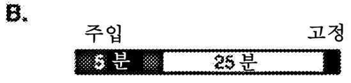
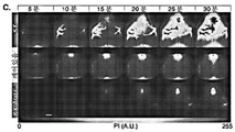

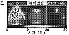
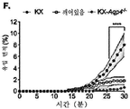


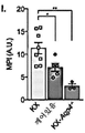


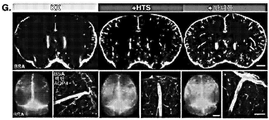


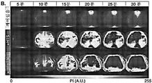


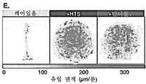

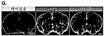


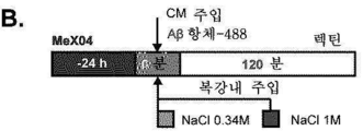
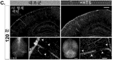


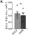
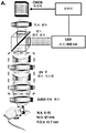
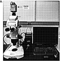


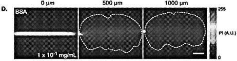
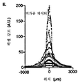

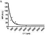


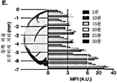


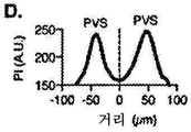

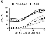


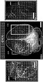
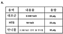
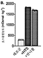


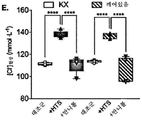

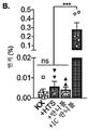

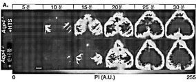

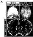


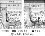

Classifications
-
- A—HUMAN NECESSITIES
- A61—MEDICAL OR VETERINARY SCIENCE; HYGIENE
- A61B—DIAGNOSIS; SURGERY; IDENTIFICATION
- A61B5/00—Measuring for diagnostic purposes; Identification of persons
- A61B5/0033—Features or image-related aspects of imaging apparatus classified in A61B5/00, e.g. for MRI, optical tomography or impedance tomography apparatus; arrangements of imaging apparatus in a room
- A61B5/004—Features or image-related aspects of imaging apparatus classified in A61B5/00, e.g. for MRI, optical tomography or impedance tomography apparatus; arrangements of imaging apparatus in a room adapted for image acquisition of a particular organ or body part
- A61B5/0042—Features or image-related aspects of imaging apparatus classified in A61B5/00, e.g. for MRI, optical tomography or impedance tomography apparatus; arrangements of imaging apparatus in a room adapted for image acquisition of a particular organ or body part for the brain
-
- A—HUMAN NECESSITIES
- A61—MEDICAL OR VETERINARY SCIENCE; HYGIENE
- A61K—PREPARATIONS FOR MEDICAL, DENTAL OR TOILETRY PURPOSES
- A61K49/00—Preparations for testing in vivo
- A61K49/001—Preparation for luminescence or biological staining
- A61K49/0013—Luminescence
- A61K49/0017—Fluorescence in vivo
- A61K49/005—Fluorescence in vivo characterised by the carrier molecule carrying the fluorescent agent
- A61K49/0058—Antibodies
-
- A—HUMAN NECESSITIES
- A61—MEDICAL OR VETERINARY SCIENCE; HYGIENE
- A61K—PREPARATIONS FOR MEDICAL, DENTAL OR TOILETRY PURPOSES
- A61K9/00—Medicinal preparations characterised by special physical form
- A61K9/0002—Galenical forms characterised by the drug release technique; Application systems commanded by energy
- A61K9/0004—Osmotic delivery systems; Sustained release driven by osmosis, thermal energy or gas
-
- A—HUMAN NECESSITIES
- A61—MEDICAL OR VETERINARY SCIENCE; HYGIENE
- A61B—DIAGNOSIS; SURGERY; IDENTIFICATION
- A61B5/00—Measuring for diagnostic purposes; Identification of persons
- A61B5/0059—Measuring for diagnostic purposes; Identification of persons using light, e.g. diagnosis by transillumination, diascopy, fluorescence
- A61B5/0077—Devices for viewing the surface of the body, e.g. camera, magnifying lens
-
- A—HUMAN NECESSITIES
- A61—MEDICAL OR VETERINARY SCIENCE; HYGIENE
- A61K—PREPARATIONS FOR MEDICAL, DENTAL OR TOILETRY PURPOSES
- A61K31/00—Medicinal preparations containing organic active ingredients
-
- A—HUMAN NECESSITIES
- A61—MEDICAL OR VETERINARY SCIENCE; HYGIENE
- A61K—PREPARATIONS FOR MEDICAL, DENTAL OR TOILETRY PURPOSES
- A61K31/00—Medicinal preparations containing organic active ingredients
- A61K31/70—Carbohydrates; Sugars; Derivatives thereof
- A61K31/7088—Compounds having three or more nucleosides or nucleotides
-
- A—HUMAN NECESSITIES
- A61—MEDICAL OR VETERINARY SCIENCE; HYGIENE
- A61K—PREPARATIONS FOR MEDICAL, DENTAL OR TOILETRY PURPOSES
- A61K38/00—Medicinal preparations containing peptides
-
- A—HUMAN NECESSITIES
- A61—MEDICAL OR VETERINARY SCIENCE; HYGIENE
- A61K—PREPARATIONS FOR MEDICAL, DENTAL OR TOILETRY PURPOSES
- A61K45/00—Medicinal preparations containing active ingredients not provided for in groups A61K31/00 - A61K41/00
- A61K45/06—Mixtures of active ingredients without chemical characterisation, e.g. antiphlogistics and cardiaca
-
- A—HUMAN NECESSITIES
- A61—MEDICAL OR VETERINARY SCIENCE; HYGIENE
- A61K—PREPARATIONS FOR MEDICAL, DENTAL OR TOILETRY PURPOSES
- A61K47/00—Medicinal preparations characterised by the non-active ingredients used, e.g. carriers or inert additives; Targeting or modifying agents chemically bound to the active ingredient
- A61K47/02—Inorganic compounds
-
- A—HUMAN NECESSITIES
- A61—MEDICAL OR VETERINARY SCIENCE; HYGIENE
- A61K—PREPARATIONS FOR MEDICAL, DENTAL OR TOILETRY PURPOSES
- A61K47/00—Medicinal preparations characterised by the non-active ingredients used, e.g. carriers or inert additives; Targeting or modifying agents chemically bound to the active ingredient
- A61K47/06—Organic compounds, e.g. natural or synthetic hydrocarbons, polyolefins, mineral oil, petrolatum or ozokerite
- A61K47/26—Carbohydrates, e.g. sugar alcohols, amino sugars, nucleic acids, mono-, di- or oligo-saccharides; Derivatives thereof, e.g. polysorbates, sorbitan fatty acid esters or glycyrrhizin
-
- A—HUMAN NECESSITIES
- A61—MEDICAL OR VETERINARY SCIENCE; HYGIENE
- A61K—PREPARATIONS FOR MEDICAL, DENTAL OR TOILETRY PURPOSES
- A61K49/00—Preparations for testing in vivo
- A61K49/001—Preparation for luminescence or biological staining
- A61K49/0013—Luminescence
- A61K49/0017—Fluorescence in vivo
- A61K49/0019—Fluorescence in vivo characterised by the fluorescent group, e.g. oligomeric, polymeric or dendritic molecules
- A61K49/0021—Fluorescence in vivo characterised by the fluorescent group, e.g. oligomeric, polymeric or dendritic molecules the fluorescent group being a small organic molecule
- A61K49/0032—Methine dyes, e.g. cyanine dyes
-
- A—HUMAN NECESSITIES
- A61—MEDICAL OR VETERINARY SCIENCE; HYGIENE
- A61K—PREPARATIONS FOR MEDICAL, DENTAL OR TOILETRY PURPOSES
- A61K49/00—Preparations for testing in vivo
- A61K49/001—Preparation for luminescence or biological staining
- A61K49/0013—Luminescence
- A61K49/0017—Fluorescence in vivo
- A61K49/0019—Fluorescence in vivo characterised by the fluorescent group, e.g. oligomeric, polymeric or dendritic molecules
- A61K49/0021—Fluorescence in vivo characterised by the fluorescent group, e.g. oligomeric, polymeric or dendritic molecules the fluorescent group being a small organic molecule
- A61K49/0041—Xanthene dyes, used in vivo, e.g. administered to a mice, e.g. rhodamines, rose Bengal
-
- A—HUMAN NECESSITIES
- A61—MEDICAL OR VETERINARY SCIENCE; HYGIENE
- A61K—PREPARATIONS FOR MEDICAL, DENTAL OR TOILETRY PURPOSES
- A61K49/00—Preparations for testing in vivo
- A61K49/001—Preparation for luminescence or biological staining
- A61K49/0013—Luminescence
- A61K49/0017—Fluorescence in vivo
- A61K49/005—Fluorescence in vivo characterised by the carrier molecule carrying the fluorescent agent
- A61K49/0056—Peptides, proteins, polyamino acids
-
- A—HUMAN NECESSITIES
- A61—MEDICAL OR VETERINARY SCIENCE; HYGIENE
- A61K—PREPARATIONS FOR MEDICAL, DENTAL OR TOILETRY PURPOSES
- A61K9/00—Medicinal preparations characterised by special physical form
- A61K9/0012—Galenical forms characterised by the site of application
- A61K9/0085—Brain, e.g. brain implants; Spinal cord
-
- A—HUMAN NECESSITIES
- A61—MEDICAL OR VETERINARY SCIENCE; HYGIENE
- A61K—PREPARATIONS FOR MEDICAL, DENTAL OR TOILETRY PURPOSES
- A61K9/00—Medicinal preparations characterised by special physical form
- A61K9/08—Solutions
-
- A—HUMAN NECESSITIES
- A61—MEDICAL OR VETERINARY SCIENCE; HYGIENE
- A61M—DEVICES FOR INTRODUCING MEDIA INTO, OR ONTO, THE BODY; DEVICES FOR TRANSDUCING BODY MEDIA OR FOR TAKING MEDIA FROM THE BODY; DEVICES FOR PRODUCING OR ENDING SLEEP OR STUPOR
- A61M5/00—Devices for bringing media into the body in a subcutaneous, intra-vascular or intramuscular way; Accessories therefor, e.g. filling or cleaning devices, arm-rests
-
- A—HUMAN NECESSITIES
- A61—MEDICAL OR VETERINARY SCIENCE; HYGIENE
- A61P—SPECIFIC THERAPEUTIC ACTIVITY OF CHEMICAL COMPOUNDS OR MEDICINAL PREPARATIONS
- A61P25/00—Drugs for disorders of the nervous system
-
- C—CHEMISTRY; METALLURGY
- C07—ORGANIC CHEMISTRY
- C07K—PEPTIDES
- C07K16/00—Immunoglobulins [IGs], e.g. monoclonal or polyclonal antibodies
- C07K16/18—Immunoglobulins [IGs], e.g. monoclonal or polyclonal antibodies against material from animals or humans
-
- A—HUMAN NECESSITIES
- A61—MEDICAL OR VETERINARY SCIENCE; HYGIENE
- A61K—PREPARATIONS FOR MEDICAL, DENTAL OR TOILETRY PURPOSES
- A61K39/00—Medicinal preparations containing antigens or antibodies
- A61K2039/505—Medicinal preparations containing antigens or antibodies comprising antibodies
Abstract
본 발명은 중추 신경계로의 작용제 전달을 개선하는 것에 관한 것이다.The present invention relates to improving the delivery of agents to the central nervous system.
Description
관련 출원의 교차 참조Cross-reference to related applications
본원은 2018년 10월 4일자로 출원된 미국 가출원 제62/741,295호에 대한 우선권을 주장한다. 상기 출원의 내용은 그 전체가 원용에 의해 본원에 포함된다.This application claims priority to U.S. Provisional Application No. 62/741,295, filed on October 4, 2018. The contents of the above application are incorporated herein by reference in their entirety.
정부의 이해관계government interest
이 발명은 미국 국립 보건원 (National Institutes of Health)이 수여하는 R01NS100366 및 RF1AG057575 및 미 국방부 보건 업무 담당 차관보실 (Office of the Assistant Secretary of Defense for Health Affairs)이 수여하는 W81XWH-16-1-0555에 따라 정부 지원을 받아 만들어졌다. 정부는 본 발명에 대한 특정 권리를 갖는다.This invention is in accordance with R01NS100366 and RF1AG057575 awarded by the National Institutes of Health, and W81XWH-16-1-0555 awarded by the Office of the Assistant Secretary of Defense for Health Affairs. It was built with government support. The government has certain rights in this invention.
기술분야technical field
본 발명은 중추 신경계로의 작용제 전달을 개선하는 것에 관한 것이다.The present invention relates to improving the delivery of agents to the central nervous system.
중추 신경계 (central nervous system; CNS) 질환을 치료하기 위한 다양한 치료제가 개발되었다. 그러나, 대체로 불침투성인 혈액-뇌 장벽 (blood-brain barrier; BBB)과 뇌로의 치료제의 불량한 침투성으로 인해, 치료제를 뇌로 전달하는 것은 심각하게 제한된다. CNS로의 약물 전달을 개선하는 것은, 특히 면역 요법의 환경에서, 상당한 임상적 과제이다 (4, 19, 55). CNS 질환, 예를 들어 알츠하이머병 (AD) (1), 파킨슨병 (2), 근위축성 측삭 경화증 (ALS) (3), 전측두엽 치매 (FTLD) (4) 및 CNS 종양 (5)에 대해, 예를 들어, 단클론 항체 (mAb)에 기반한 요법이 현재 개발되고 있다. 그러나 유망한 전임상 결과에도 불구하고, 임상 시험은 인상적이지 않았고 부작용으로 시달렸다 (8-10). 이는 치료용 mAb가 뇌에 제대로 침투하지 않아, 부적합한 표적 결합을 초래하였다는 것을 반영할 수 있다 (11). 항-Aβ 면역 요법이 뇌심부 구조에 위치한 플라크 (plaque)와 결합하지 못하는 것은 이러한 요법이 일상적인 AD 치료제로 변환되지 않는 것에 기여할 수 있다 (4, 12). 뇌척수액 (CSF)에 직접 주입하는 것과 관련된 침습성과 더 높은 정도의 합병증으로 인해, 치료용 항체는, 가장 일반적으로, 정맥내 주입에 의해 투여된다 (13-15). 그러나, 순환 항체는 BBB 침투성이 낮으며 0.1-0.2%만이 뇌로 진입한다 (16). 결과적으로, 치료용 항체는 종종, 말초 조직에서 표적 항원과 적합한 결합을 달성하는 데 필요한 농도보다 1000배 더 많은 용량으로 투여되며 (1, 17), 임상 시험에서의 이러한 높은 용량은 부작용 (1, 9, 18), 예를 들어 아밀로이드 관련 영상 이상 (amyloid-related imaging abnormalities; ARIA) (19, 20)의 유병률을 증가시킨다. 따라서, CNS로의 치료제의 개선된 전달에 대한 요구가 존재한다.Various therapeutic agents have been developed to treat central nervous system (CNS) diseases. However, due to the largely impermeable blood-brain barrier (BBB) and poor penetration of therapeutic agents into the brain, delivery of therapeutic agents to the brain is severely limited. Improving drug delivery to the CNS is a significant clinical challenge, especially in the context of immunotherapy (4, 19, 55). For CNS diseases such as Alzheimer's disease (AD) (1), Parkinson's disease (2), amyotrophic lateral sclerosis (ALS) (3), frontotemporal dementia (FTLD) (4) and CNS tumors (5); For example, therapies based on monoclonal antibodies (mAbs) are currently being developed. However, despite promising preclinical results, clinical trials have been unimpressive and plagued with side effects (8-10). This may reflect that the therapeutic mAb did not penetrate the brain properly, resulting in inappropriate target binding (11). The failure of anti-Aβ immunotherapy to bind plaques located in deep brain structures may contribute to the failure of these therapies to convert into routine AD treatments (4, 12). Because of the invasiveness and higher degree of complications associated with direct infusion into the cerebrospinal fluid (CSF), therapeutic antibodies are, most commonly, administered by intravenous infusion (13-15). However, circulating antibodies have low BBB permeability and only 0.1-0.2% enter the brain (16). As a result, therapeutic antibodies are often administered at doses 1000-fold higher than the concentration required to achieve proper binding to the target antigen in peripheral tissues (1, 17), and these high doses in clinical trials are associated with adverse side effects (1, 9, 18), for example amyloid-related imaging abnormalities (ARIA) (19, 20). Accordingly, there is a need for improved delivery of therapeutic agents to the CNS.
본 발명은 CNS로의 조성물의 전달을 개선하는 방법을 제공함으로써 상기 요구를 해결한다.The present invention addresses this need by providing methods for improving the delivery of compositions to the CNS.
일 양태에서, 본 발명은 대상의 중추 신경계 간질, 뇌 간질 및/또는 척수 간질로의 조성물의 전달을 개선하는 방법을 제공한다. 상기 방법은 (1) 글림프계 (glymphatic system) 유입을 향상시키는 단계 및 (2) 중추 신경계 간질, 뇌 간질 및/또는 척수 간질에 상기 조성물을 전달하는 단계를 포함한다. 글림프계 유입을 향상시키는 단계는 다수의 방식으로 수행될 수 있다. 예를 들어, 상기 단계는 중추 신경계 간질을 통해 유체를 펌핑하거나, 또는 상기 대상에게 작용제를 투여하는 단계를 포함할 수 있다.In one aspect, the present invention provides a method for improving the delivery of a composition to central nervous system epilepsy, brain epilepsy, and/or spinal cord epilepsy in a subject. The method comprises the steps of (1) enhancing glymphatic system uptake and (2) delivering the composition to central nervous system epilepsy, brain epilepsy and/or spinal cord epilepsy. The step of enhancing glymphatic influx can be accomplished in a number of ways. For example, the step can include pumping a fluid through the central nervous system epilepsy, or administering an agent to the subject.
상기 작용제는 고장성 용액 (hypertonic solution)일 수 있다. 일례에서, 상기 고장성 용액은 상기 대상의 혈액 또는 혈장에 투여된다. 상기 고장성 용액은 NaCl 또는 만니톨을 포함할 수 있다. 다른 예에서, 상기 작용제는 Stat-3 억제제, 골형성 단백질 (BMP) 신호전달 축 분자, AVP (바소프레신)의 길항제, 심방 나트륨이뇨 펩타이드 (ANP)의 길항제, 안지오텐신 II의 길항제, AT2R 수용체의 길항제 또는 AT1 수용체의 길항제를 포함할 수 있다.The agent may be a hypertonic solution. In one example, the hypertonic solution is administered to the blood or plasma of the subject. The hypertonic solution may contain NaCl or mannitol. In another example, the agonist is a Stat-3 inhibitor, a bone morphogenetic protein (BMP) signaling axis molecule, an antagonist of AVP (vasopressin), an antagonist of atrial natriuretic peptide (ANP), an angiotensin II antagonist, an antagonist of the AT2R receptor, or an antagonist of the AT1 receptor.
상기 조성물은 임의의 적합한 방식으로, 예를 들어, 수조내로 (intracisternally) 또는 경막내로 (intrathecally) 전달될 수 있다. 상기 조성물은, 글림프계 유입이 향상되는 것과 거의 같은 시간에, 또는 그 후나 전에 전달될 수 있다. 상기 조성물은 이미징 조성물 (imaging composition) 또는 치료 조성물일 수 있다. 상기 조성물은 소분자, 바이러스, 거대 분자, 펩타이드, 항체, 핵산 (예를 들어, 안티센스 분자 및 RNAi 작용제) 또는 이의 생물학적으로 활성인 단편을 포함할 수 있다. 일례에서, 상기 치료 조성물은 항체를 포함한다. 상기 항체는 혈액-뇌 장벽 (일명 "BBB")을 통과하는 수송을 용이하게 하는 리간드에 접합될 수 있다. 예를 들어, 상기 리간드는 BBB 수용체 (예를 들어 트랜스페린 수용체, IGF-R, LDL-R, LRP1, LRP2, 및 LRP8)에 특이적으로 결합할 수 있다.The composition may be delivered in any suitable manner, for example, intracisternally or intrathecally. The composition can be delivered at about the same time as glymphatic uptake is enhanced, or after or before. The composition may be an imaging composition or a therapeutic composition. The composition may include small molecules, viruses, macromolecules, peptides, antibodies, nucleic acids (eg, antisense molecules and RNAi agents) or biologically active fragments thereof. In one example, the therapeutic composition comprises an antibody. The antibody may be conjugated to a ligand that facilitates transport across the blood-brain barrier (aka “BBB”). For example, the ligand may specifically bind to a BBB receptor (eg, transferrin receptor, IGF-R, LDL-R, LRP1, LRP2, and LRP8).
다른 양태에서, 본 발명은 대상에서 신경 장애를 치료하는 방법을 제공한다. 상기 장애의 예는 신경병증, 아밀로이드증, 암, 안과 질환 또는 장애, 바이러스 또는 미생물 감염, 염증, 허혈, 신경퇴행성 질환, 발작, 행동 장애 및 리소좀 축적 질환을 포함한다. 상기 방법은 (1) 글림프계 유입을 향상시키는 단계 및 (2) 중추 신경계 간질, 뇌 간질 및/또는 척수 간질에 치료 조성물을 전달하는 단계를 포함한다. 글림프계 유입을 향상시키는 단계는 전술된 다수의 방식으로 수행될 수 있다. 일례에서, 상기 단계는 중추 신경계 간질을 통해 유체를 펌핑하는 단계를 포함한다. 다른 예에서, 글림프계 유입을 향상시키는 단계는 상기 대상에게 작용제를 투여하는 단계를 포함한다.In another aspect, the invention provides a method of treating a neurological disorder in a subject. Examples of such disorders include neuropathy, amyloidosis, cancer, ophthalmic diseases or disorders, viral or microbial infections, inflammation, ischemia, neurodegenerative diseases, seizures, behavioral disorders and lysosomal storage diseases. The method comprises the steps of (1) enhancing glymphatic uptake and (2) delivering a therapeutic composition to central nervous system epilepsy, brain epilepsy and/or spinal cord epilepsy. The step of enhancing glymphatic influx can be accomplished in a number of ways as described above. In one example, the step comprises pumping the fluid through the central nervous system epilepsy. In another example, enhancing glymphatic uptake comprises administering to the subject an agent.
상기 작용제는 고장성 용액일 수 있다. 일례에서, 상기 고장성 용액은 상기 대상의 혈액 또는 혈장에 투여된다. 상기 고장성 용액은 NaCl 또는 만니톨을 포함할 수 있다. 다른 예에서, 상기 작용제는 Stat-3 억제제, BMP 신호전달 축 분자, AVP (바소프레신)의 길항제, ANP의 길항제, 안지오텐신 II의 길항제, AT2R 수용체의 길항제 또는 AT1 수용체의 길항제를 포함할 수 있다.The agent may be a hypertonic solution. In one example, the hypertonic solution is administered to the blood or plasma of the subject. The hypertonic solution may contain NaCl or mannitol. In another example, the agonist may include a Stat-3 inhibitor, a BMP signaling axis molecule, an antagonist of AVP (vasopressin), an antagonist of ANP, an antagonist of angiotensin II, an antagonist of the AT2R receptor, or an antagonist of the AT1 receptor.
상기 조성물은 임의의 적합한 방식으로, 예를 들어, 수조내로 또는 경막내로 전달될 수 있다. 상기 조성물은, 글림프계 유입이 향상되는 것과 거의 같은 시간에, 또는 그 후나 전에 전달될 수 있다. 상기 조성물은 소분자, 바이러스, 거대 분자, 펩타이드, 항체, 핵산 (예를 들어, 안티센스 분자 및 RNAi 작용제) 또는 이의 생물학적으로 활성인 단편을 포함할 수 있다. 일례에서, 상기 치료 조성물은 항체를 포함한다. 상기 항체는 혈액-뇌 장벽을 통과하는 수송을 용이하게 하는 리간드에 접합될 수 있다. 예를 들어, 상기 리간드는 BBB 수용체, 예를 들어 트랜스페린 수용체, IGF-R, LDL-R, LRP1, LRP2, 및 LRP8에 특이적으로 결합할 수 있다.The composition may be delivered in any suitable manner, for example, intracisternally or intrathecally. The composition can be delivered at about the same time as glymphatic uptake is enhanced, or after or before. The composition may include small molecules, viruses, macromolecules, peptides, antibodies, nucleic acids (eg, antisense molecules and RNAi agents) or biologically active fragments thereof. In one example, the therapeutic composition comprises an antibody. The antibody may be conjugated to a ligand that facilitates transport across the blood-brain barrier. For example, the ligand may specifically bind to a BBB receptor, such as the transferrin receptor, IGF-R, LDL-R, LRP1, LRP2, and LRP8.
상기 항체는 항-Aβ 항체일 수 있다. 상기 대상은 포유동물, 예를 들어 인간 또는 비인간 영장류일 수 있다. 일 구현예에서, 상기 포유동물은 치료를 필요로 하는 환자, 예를 들어 고령의 또는 연세가 드신 사람이다.The antibody may be an anti-Aβ antibody. The subject may be a mammal, such as a human or non-human primate. In one embodiment, the mammal is a patient in need of treatment, eg, an elderly or elderly person.
또 다른 양태에서, 본 발명은 대상의 CNS로의 조성물 (예를 들어, 이미징 조성물 또는 치료 조성물)의 전달을 개선하기 위한 키트를 특징으로 한다. 상기 키트는 상기 조성물 및 글림프계 유입을 향상시키는 작용제를 포함한다. 상기 작용제는 고장성 용액, 예를 들어 NaCl 또는 만니톨을 포함하는 고장성 용액일 수 있다. 상기 작용제는 Stat-3 억제제, BMP 신호전달 축 분자, AVP (바소프레신)의 길항제, ANP의 길항제, 안지오텐신 II의 길항제, AT2R 수용체의 길항제 또는 AT1 수용체의 길항제일 수 있다. 상기 조성물은 소분자, 바이러스, 거대 분자, 펩타이드, 항체, 핵산 또는 이의 생물학적으로 활성인 단편을 포함할 수 있다. 상기 항체는 혈액-뇌 장벽을 통과하는 수송을 용이하게 하는 리간드에 접합될 수 있다. 상기 항체의 일례는 항-Aβ 항체이다.In another aspect, the invention features a kit for improving delivery of a composition (eg, an imaging composition or a therapeutic composition) to the CNS of a subject. The kit includes the composition and an agent that enhances glymphatic entry. The agent may be a hypertonic solution, for example a hypertonic solution comprising NaCl or mannitol. The agent may be a Stat-3 inhibitor, a BMP signaling axis molecule, an antagonist of AVP (vasopressin), an antagonist of ANP, an antagonist of angiotensin II, an antagonist of the AT2R receptor or an antagonist of the AT1 receptor. The composition may include small molecules, viruses, macromolecules, peptides, antibodies, nucleic acids or biologically active fragments thereof. The antibody may be conjugated to a ligand that facilitates transport across the blood-brain barrier. An example of such an antibody is an anti-Aβ antibody.
추가 양태에서, 본 발명은 경두개 거시적 이미징 방법 (transcranial macroscopic imaging method)을 제공한다. 상기 방법은 대상의 중추 신경계에 유효량의 이미징제 (imaging agent)를 도입하는 단계, 및 상기 대상의 뇌를 이미징하는 단계를 포함한다. 상기 이미징제는 수조내로 또는 경막내로 도입될 수 있다. 바람직한 구현예에서, 상기 이미징제는 형광단을 포함하고 상기 이미징 단계는 형광 매크로스코피 (fluorescence macroscopy)를 포함한다. 일부 예에서, 상기 형광단은 여기시 적외선 영역 (예를 들어, 근적외선 영역, 중적외선 영역 또는 원적외선 영역)의 빛을 재방출한다.In a further aspect, the present invention provides a transcranial macroscopic imaging method. The method comprises introducing an effective amount of an imaging agent into the subject's central nervous system, and imaging the subject's brain. The imaging agent may be introduced intrathecally or intrathecally. In a preferred embodiment, the imaging agent comprises a fluorophore and the imaging step comprises fluorescence macroscopy. In some examples, the fluorophore re-emits light in the infrared region (eg, near-infrared region, mid-infrared region, or far-infrared region) upon excitation.
본 발명의 하나 이상의 구현예의 세부 사항은 하기 설명에서 설명된다. 본 발명의 다른 특징, 목적 및 장점은 하기의 설명 및 청구범위로부터 명백해질 것이다.The details of one or more embodiments of the invention are set forth in the description below. Other features, objects and advantages of the present invention will become apparent from the following description and claims.
도 1a, 1b, 1c, 1d, 1e, 1f, 1g, 1h 및 1i은 CSF 유입의 생체내 경두개 뇌 전체 이미징 (brain-wide imaging)을 보여주는 일련의 도표 및 사진이다. (a). 매크로스코프 (macroscope)를 사용하여 손상되지 않은 두개골을 통해 마우스를 이미징하였다. (b) 형광 단백질 추적자 (fluorescent protein tracer) (BSA-647nm)를 대조 (cisterna magna)에 전달하고 (2 μL/분, 5분), 주입 시작부터 30분 동안 추적자 유입을 이미징하였다. 모든 마우스는 수조내 주입 시작시 등장성 식염수를 i.p. 투여 받았다. (c) 마취된 (KX) 및 깨어있는 야생형 마우스뿐만 아니라 마취된 Aqp4 -/- 마우스 (KX-Aqp4 -/- )에 추적자를 주입한 후 처음 30분 동안의 CSF 유입의 대표적인 시간 경과 이미지 (time-lapse image). 이미지 (8-비트 픽셀 깊이)는 임의 단위 (A.U.)로 픽셀 강도 (PI)를 나타내기 위해 색상-코딩된다 (color-coded). 눈금 막대 (Scale bar) = 2 mm. 등쪽 피질 표면 아래 약 5-6 mm의 뇌 기저부에 주입 후 5분째에 이미 형광이 검출되었다. (d) (c)에 도시된 30분의 생체내 이미징 시리즈에 대한 평균 픽셀 강도 (mean pixel intensity; MPI)의 정량화 (평균 ± SEM; n = 5-7 마우스/그룹; 반복 측정 이원 ANOVA, 시닥의 다중 비교 검정 (Sidak's multiple comparisons test); *P<0.05, **P<0.01, ***P<0.001, ****P<0.0001 (KX 대비)). (e) 이미징 세션 (imaging session)에 걸친 CSF 추적자 유입의 대표적인 전방 추적 분석 (front-tracking analysis). 전방 (front)은 분 단위로 시간-코딩된다 (time-coded). (f) 분석 (e)로부터의 시간 경과에 따른 유입 면적의 정량화 (평균 ± SEM; n = 5-7 마우스/그룹; 반복 측정 이원 ANOVA, 시닥의 다중 비교 검정; ****P<0.0001 (KX 대 깨어있음 및 KX-Aqp4 -/- )). (g) (c) 및 (e)의 그룹 데이터로부터 생성된 평균 유입 속도 맵 (influx speed map) (μm/분). (h) KX 및 깨어있는 야생형, 및 KX-마취된 Aqp4 -/- 그룹에서의, 두개골로부터 제거시 (좌측 하단; 눈금 막대 = 2 mm) 및 피질 표면 내로의 추적자 침투도를 평가하기 위한 관상 절편화 후의 (상단; 눈금 막대 = 1 mm) 뇌의 대표적인 기존의 생체외 형광 이미지. 공초점 레이저 주사 현미경법 (laser scanning confocal microscopy)를 사용하여 혈관주위 추적자의 고배율 이미지를 획득하였다 (우측 하단; 눈금 막대 = 50 μm). (i) KX 및 깨어있는 야생형 및 KX-마취된 Aqp4 -/- 그룹에 대한 생체외 관상 절편 형광 MPI의 정량화 (평균 ± SEM; n = 3-8 마우스/그룹; 일원 ANOVA, 투키의 다중 비교 검정 (Tukey's multiple comparisons test); *P<0.05, **P<0.01).
도 2a, 2b, 2c, 2d, 2e, 2f, 2g, 2h, 2i, 및 2j는 혈장 고장성이 마취된 마우스에서의 CSF 유입을 증가시켰음을 보여주는 일련의 도표 및 사진이다. (a) 마취된 마우스의 대조 (CM)에 형광 BSA-647을 전달하였다. 마우스는 CM 주입 시작시, 등장성 식염수 (KX), 고장성 식염수 (+HTS) 또는 고장성 만니톨 (+만니톨)을 i.p. 투여 받았다. (b) KX, +HTS 및 +만니톨 그룹에서의 CM 주입 직후 30분 동안의 BSA-647 유입에 대한 대표적인 시간 경과 이미지. 이미지 (8-비트 픽셀 깊이)는 임의 단위 (A.U.)로 픽셀 강도 (PI)를 나타내기 위해 색상-코딩된다 (color-coded). 눈금 막대 (Scale bar) = 2 mm. (c) 모든 그룹에 대한 이미징 세션에 걸친 CSF 추적자 유입의 대표적인 전방 추적 분석. 전방 (front)은 분 단위로 시간-코딩된다 (time-coded). (d) 시간 경과에 따른 유입 면적의 정량화 (평균 ± SEM; n = 6-7 마우스/그룹; 반복 측정 이원 ANOVA, 시닥의 다중 비교 검정; ****P<0.0001 (KX 대 +HTS 및 +만니톨)). (e) 모든 그룹에 대한 추적자 유입 속도 맵 (μm/분) 및 (f) 평균 유입 속도의 정량화 (평균 ± SEM; n = 6 마우스/그룹, 일원 ANOVA, 투키의 다중 비교 검정, *P<0.05, ***P=0.001). (g) 모든 그룹에 대한, 두개골로부터 제거시의 손상되지 않은 뇌 (좌측 하단; 눈금 막대 = 2 mm) 및 관상 절편화 후의 (상단; 눈금 막대 = 1 mm) 대표적인 기존의 생체외 형광 이미지. 혈관주위 추적자를 평가하기 위해 고출력 공초점 레이저 주사 현미경법을 사용하여 관상 절편을 이미징하였다 (우측 하단; 눈금 막대 = 50 μm). (h) 생체외 관상 절편 형광 MPI의 정량화 (평균 ± SEM; n = 5-7 마우스/그룹; 일원 ANOVA, 투키의 다중 비교 검정; **P<0.01, ***P=0.003). 세 그룹 모두에서의 CSF-전달 된 (i) 3H-덱스트란 (40 kDa) 또는 (j) 14C-이눌린 (6 kDa)의 총 뇌 흡수 (brain uptake) (평균 ± SEM; n = 5 마우스/그룹; 일원 ANOVA, 투키의 다중 비교 검정; **P=0.001, ***P=0.0009, ****P<0.0001). 주입된 용량에 대한 백분율 (percent injected dose; % ID)로 표시하였다. KX 그룹은 도 1과 같다.
도 3a, 3b, 3c, 3d, 3e, 3f, 3g 및 3h는 혈장 고장성이 글림프 기능 (glymphatic function)의 각성 상태 (arousal state) 억제를 중단시켰음을 보여주는 일련의 도표 및 사진이다. (a) 헤드플레이트가 배치된 (head-plated) 깨어있는 마우스에 수조내 BSA-647을 투여하였다. 마우스는 대조 (CM) 주입 시작시, 등장성 식염수 (깨어있음), 고장성 식염수 (+HTS) 또는 고장성 만니톨 (+만니톨)을 i.p. 투여 받았다. (b) 깨어있는, +HTS 및 +만니톨 그룹에서의 CM 주입 직후 30분 동안의 BSA-647 유입에 대한 대표적인 시간 경과 이미지. 이미지 (8-비트 픽셀 깊이)는 임의 단위 (A.U.)로 픽셀 강도 (PI)를 나타내기 위해 색상-코딩된다 (color-coded). 눈금 막대 (Scale bar) = 2 mm. 등쪽 피질 표면 아래 약 5-6 mm의 뇌 기저부에서 형광이 처음 검출되었다. (c) 모든 그룹에 대한 이미징 세션에 걸친 CSF 추적자 유입의 대표적인 전방 추적 분석. 전방 (front)은 분 단위로 시간-코딩된다 (time-coded). (d) 시간 경과에 따른 유입 면적의 정량화 (평균 ± SEM; n = 5-7 마우스/그룹; 반복 측정 이원 ANOVA, 시닥의 다중 비교 검정; ****P<0.0001 (깨어있음 대 +HTS 및 +만니톨)). (e) 모든 그룹에 대한 추적자 유입 속도 맵 (μm/분) 및 (f) 평균 유입 속도의 정량화 (평균 ± SEM; n = 5-7 마우스/그룹, 일원 ANOVA, 투키의 다중 비교 검정, **P=0.0024, ***P=0.0003). (g) 모든 그룹으로부터의 대표적인 생체외 관상 절편 (눈금 막대 = 1 mm). (h) 생체외 관상 절편 형광 MPI의 정량화 (평균 ± SEM; n = 5-6 마우스/그룹; 일원 ANOVA, 투키의 다중 비교 검정; **P=0.0063, ***P=0.003). 깨어있는 그룹은 도 1과 같다.
도 4a, 4b, 4c, 4d, 4e, 4f, 4g, 4h, 4i, 4j 및 4k는 혈장 고장성이 6개월령 APP/PS1 마우스에서의 Aβ 항체의 전달을 개선하였으며 표적 결합을 향상시켰음을 보여주는 일련의 도표 및 사진이다. (a)(b) 아밀로이드 플라크는 24시간 전에 메톡시-X04 (MeX04)로 표지되었다. 그런 다음 마우스를 마취하고 형광 항-Aβ 항체를 수조내로 주입하였다. 마우스는 수조내 주입 시작시, 등장성 식염수 (대조군) 또는 고장성 식염수 (+HTS)를 i.p. 투여 받았다. 120분 후, 맥관 구조를 표지하기 위해 마우스를 형광성 렉틴으로 관류 고정 (perfusion-fix)하였다. (c) 두개골로부터 제거시의 손상되지 않은 뇌 (좌측 하단; 눈금 막대 = 2 mm) 및 뇌로의 항체 침투도를 평가하기 위한 관상 절편화 후의 (상단; 눈금 막대 = 500 μm) 대표적인 생체외 이미지. 관통 동맥의 혈관주위 공간을 둘러싼 Aβ 플라크 (화살표) 및 항체의 공초점 이미지 (우측 하단; 눈금 막대 = 100 μm). (d) 생체외 관상 절편 Aβ 항체 형광 MPI의 정량화 (평균 ± SEM; n = 5 마우스/그룹; 짝을 이루지 않은 양측 t-검정 (unpaired two-tailed t-test); **P=0.0039). (e) 혈관주위 Aβ 플라크의 대표적인 고배율 공초점 이미지 (눈금 막대 = 20 μm). (f) MeX04+ Aβ 플라크와 항체의 공동-표지 (co-labeling)에 의해 표시된 표적 결합의 백분율 (평균 ± SEM; n = 5 마우스/그룹; 짝을 이루지 않은 t-검정; **P=0.005). (g) μm 단위의 가장 가까운 혈관주위 공간 (nearest perivascular space; PVS)로부터의 공동-표지된 플라크의 평균 거리에 대한 최근린 분석 (nearest neighbor analysis) (평균 ± SEM; 공동-표지된 플라크의 총 수/그룹 내 마우스의 수; 짝을 이루지 않은 t-검정; ****P<0.0001). (h) 가장 가까운 PVS로부터의 거리와 공동-표지된 플라크의 수의 히스토그램 및 누적 빈도 도표. (i) 플라크 (화살표)의 표면에 결합한 항-Aβ 항체를 보여주는 직각면 영상 (orthogonal view)을 갖는 대표적인 고배율 공초점 이미지. 눈금 막대 = 20 μm. (j) 항체 표적화 및 결합 플라크 표면을 보여주는 +HTS-치료된 마우스로부터의 Aβ 플라크의 3차원 재구성 (둘 모두에서 눈금 막대 = 20 μm). (k) 플라크 부담 (plaque burden)은 그룹 간에 동일하였다 (평균 ± SEM; n = 5 마우스/그룹; 짝을 이루지 않은 t-검정; P=0.6165).
도 5a 및 5b는 경두개 거시적 이미징 시스템을 보여주는 일련의 도표 및 사진이다. (a) 광학 개략도. 이 시스템은, 최대 16개의 상이한 파장을 개별적으로 제어할 수 있는, 조정가능한 LED 조명 시스템을 사용한다. 647 nm 형광단에 대한 여기는 635 nm 파장을 사용하여 달성되었다. 이미징 소프트웨어는 파장 간의 빠른 전환을 제어하며, 쿼드 필터 큐브 (quad filter cube)와 결합되는 경우, 필터 터렛 (turret)을 회전하지 않고도 고속 4 채널 이미징 (약 100Hz)을 가능하게 한다. 매크로스코프는 총 배율이 4-40배이고 0.63X 대물 렌즈를 사용하며, 이 연구에 사용된 배율에서 약 11.7 mm의 시야 (field of view; F.O.V.)로 긴 작동 거리 (working distance; W.D.)와 높은 개구수 (numerical aperture; N.A.)를 허용한다. 이 구성은, 유효 면적이 13.312 x 13.312 mm이고 전해상도가 2048 x 2048 픽셀인 과학 CMOS 카메라를 사용하며, 이는 빠른 이미지 획득 (초당 100 프레임)을 가능하게 한다. 상기 시스템은 이중 CSF 추적자 연구를 위한 동시 2-채널 응용 분야를 위한 이미지 분할 광학과 호환된다. (b) 거시적 이미징 시스템의 사진.
도 6a, 6b, 6c, 6d, 6e, 6f 및 6g는 이미징 침투 깊이 분석 (imaging penetration depth analysis)을 보여주는 일련의 도표 및 사진이다. (a) AlexaFluor 647 (BSA)에 접합된 소 혈청 알부민을 인공 CSF (aCSF)에서 10에서 1 x 10⁻⁴ mg/mL로 연속 희석하였다. 10 μl 액적을 96-웰 플레이트에 분취하고 생체내 실험에 사용된 것과 동일한 배율 및 노출 시간을 사용하여 635 nm 파장에서 매크로스코프에서 이미징하였다. 이미지는 0 내지 255의 임의 단위 (A.U.)로 픽셀 강도 (PI)를 나타내기 위해 색상-코딩되었다 (눈금 막대 = 2mm). (b) 각각의 10 μl 액적에 대해 평균 픽셀 강도 (MPI)를 계산하고 추적자 농도의 함수로서 도표화하였다. 데이터를 가변 기울기 S자형 함수에 피팅시켰다. 상기 생체내 실험의 범위 내에 속하는 추적자의 최적 희석은 0.1 mg/ml였다. (c) 한천에 삽입된 BSA-647로 충전된 모세관 위에 배치된, 증가하는 두께의 급성 관상 절편을 보여주는 실험 구성의 개략도. (d) 초점면에서의 형광 모세관의 대표적인 PI 색상-코딩된 이미지: 모세관 위에 조직이 배치되지 않은 경우 (0 μm), 500 μm 두께 관상 절편이 배치된 경우, 및 1,000 μm 두께 관상 절편이 배치된 경우 (눈금 막대 = 2mm). (e) 200 내지 4000 μm의 증가하는 두께를 갖는 관상 단면을 통해 획득된, 모세관을 중심으로 한 6000 μm 라인 주사 (line scan)의 미가공 PI. (f) (e)의 미가공 데이터에 대한 가우스 핏 (Gaussian fit)은 4,000 μm (R2 = 0.55)를 제외한 모두 (R2 > 0.924)에 대해 양호한 일치를 나타냈다. (g) 관상 단면의 둘레 내의 모세관 위에 관심 영역을 그리고, MPI를 측정하고 깊이의 함수로서 도표화하였다. 1상 지수 감쇠 함수를 사용하여 데이터를 피팅하였으며 (R2 = 0.96), 이는 형광 신호가 1-2 mm의 조직 두께에서 안정되어 있음을 보여주며, 이는 (f)의 결과와 일치한다.
도 7a, 7b, 7c, 7d, 7e, 7f 및 7g는 생체내 경두개 이미징이 모든 시점에서 등쪽 피질로의 추적자 수송 및 생체외 정량화와 상관 관계가 있음을 보여주는 일련의 도표 및 사진이다. 마취된 마우스는 생체내 이미징 동안 BSA-647 (BSA)의 대조 (CM) 주입을 투여 받았으며, 주입 시작 후 5분 내지 30분째에 5분 간격으로 뇌를 추출하고 고정하였다 (n = 3 마우스/6개 시점). (a) 시상 (sagittal) 및 관상 투영에서 CM 주입 후 시간의 함수로서의 색상-코딩된 추적자 수송. (b) CM 주입 후 5분, 15분 및 30분 시점의 관상 절편은 추적자가 측면 피질 만곡을 따라 뇌 기저부로부터 수송되어, 추후 시점에 등쪽 볼록면에 도달했음을 보여준다 (눈금 막대 = 2 mm). (c) 경두개 생체내 이미징의 마지막 프레임의 평균 픽셀 강도 (MPI)는 6개 시점 모두에 걸친 동일한 뇌로부터의 6개의 관상 절편의 MPI와 상관 관계가 있었다 (피어슨 상관 관계 (Pearson correlation), P = 0.0003). (d) 생체내 및 생체외 둘 모두에서 정량화하였을 때, MPI는 유사한 동역학을 가지며, 두 데이터 세트 모두는 1상 지수 감쇠 함수와 잘 일치한다 (생체내: R2 = 0.838; 생체외: R2 = 0.834). (e) 추적자의 깊이를 추정하기 위해, 모든 시점에 대해 등쪽 피질로부터 시작하여 1 mm ROI에서 임의 단위 (A.U.)의 MPI를 정량화하였다. 추적자의 존재는 배경 (점선)보다 2 표준 편차 위의 MPI 값으로서 결정되었다. (f) 수조내 주입 시작 후 등쪽 피질 아래의 추적자의 추정 거리. 오차 막대는 1 mm 너비의 관심 영역을 반영한다. 1상 지수 감쇠에 데이터를 또한 피팅하였다 (R2 = 0.976). (g) (d) 및 (f) 각각으로부터의 생체내, 생체외 및 추적자 깊이에 대해 z-점수 변환을 수행하고, 1상 지수 감쇠 함수로 피팅하였다. 추가 자승합 F 검정 (extra sum-of-squares F test)은, 세 개의 데이터세트 모두가 단일 전역 모델 (single global model)에 의해 피팅되었고 유의하게 상이하지 않다는 결론을 내렸으며 (P = 0.288; R2 = 0.913), 이는 경두개 광학 이미징에서의 MPI의 증가가 뇌 기저부로부터 등쪽 피질로의 추적자 이동과 상관 관계가 있음을 시사한다. 데이터는, 추적자의 대부분이 피질 표면 아래 5-6 mm에 위치하는 때인, CM 주입 후 5분째에 이미 형광이 검출됨을 입증하며, 이는 최종 생체외 방법과 유사한 뇌 전체 추적자 정량화에 대한 높은 민감도를 제공한다.
도 8a, 8b,.8c, 8d, 8e, 8f 및 8g는 경두개 이미징에서 나타난 CSF 추적자 유입이 동맥을 둘러싼 혈관주위 공간을 따라 발생했으며 이전의 생체내 이미징 방식과 유사한 동역학을 가졌음을 보여주는 일련의 도표 및 사진이다. (a) 수조내 주입 후 2개의 중간 대뇌 동맥 (middle cerebral arteries; MCA)의 분포를 따라 뇌 전체 CSF 추적자 (BSA-647) 유입을 보여주는 전형적인 실험의 거시적 이미지 (눈금 막대 = 2 mm). (b) CSF 유입이 발생하는 해부학적 경로를 평가하기 위해, 맥관 구조를 표지하기 위한 i.v. FITC 덱스트란 및 BSA-Texas Red (TxRd)의 수조내 주입 후에 2-광자 레이저 주사 현미경법을 사용하여 두개골 창 (cranial window)을 통해 별도의 마우스 그룹을 이미징하였다 (눈금 막대 = 50 μm). 경막은 손상되지 않았고, 두개골 창은 두개내 압력 손실을 방지하기 위해 아가로스와 커버 슬립 (cover slip)으로 밀봉되었다. 이미징은, 추적자가 경막의 혈관 (화살표) 아래에서, MCA의 각각의 면 상의 2개의 혈관주위 공간 (PVS)을 따라 흐르는 것을 보여주었다. (c) (a)의 삽도로부터 확대된 이미지는, MCA의 좌측 후지 (posterior branch)의 각각의 면 상의 CSF 추적자를 보여준다 (눈금 막대 = 500 μm). (d) (c)의 흑색 라인으로부터의 라인 주사는 MCA의 양쪽에서의 임의 단위 (A.U.)의 높은 픽셀 강도 (PI) 및 동맥 위에서의 형광 감소를 보여준다. (d)에서 측정된 공간의 너비는 (b)에서 측정된 것과 유사하다. (e) (b)의 청색 선으로부터의 직교 재구성 (orthogonal reconstruction)은, CSF 추적자가 지주막하 공간 (subarachnoid space; SAS, 눈금 막대 = 50 μm) 내가 아닌 MCA 주변의 연막하 PVS에 국한되어 있음을 보여준다. (f) 백분율로 표현된 이미징 세션의 최대 형광 강도 (ΔF/F최대)로 정규화된 MCA를 따라 위치한 3개의 혈관주위 관심 영역의 평균으로부터의 정량화. 혈관 표지 채널로부터의 출혈로 인해 더 높은 기준선 배경 형광이 2-P에서 나타난다. (g) 수조내 주입 시작 후 추적자 출현까지의 시간. (평균 ± SEM; n = 4-5 마우스/그룹; ns: 유의하지 않음; 짝을 이루지 않은 t-검정; P=0.8761).
도 9는 CSF 유입 경로의 경두개 거시적 이미징을 보여주는 일련의 사진이다. 대조에 추적자를 전달한 후, 손상되지 않은 두개골을 통해 여러개의 두개내 구조를 식별할 수 있다 (점선). (상단 패널) 마취된 마우스에서, 후각동, 상시상 정맥동, 좌우 횡정맥동과 같은 뇌막 구조를 관찰할 수 있다 (청색). 이전에 나타난 바와 같이, 추적자는 후각전두 수조 (olfactofrontal cistern) (보라색) 및 송과체와 (pineal recess) (녹색) 주변의 것들과 같은 뇌를 둘러싼 지주막하 CSF의 거대 풀 (pool)에서 처음 발견된다. 추적자의 뇌 흡수는, 특히 중간 대뇌 동맥의 전방 및 등쪽 피질 부분의 분포를 따라, 연막 동맥 (적색)의 혈관주위 공간 내에서 발생하며, 그 다음 관통 동맥을 따라 뇌로 계속 내려간다. 결국 추적자는 뇌막동 (meningeal sinus)을 둘러싼 피질 가교 정맥의 주변 공간에서 발견될 수 있다. (하단 패널) 깨어있는 마우스에서, 최전방 및 최후방 구조 중 일부는 헤드플레이트 (headplate)로 덮여 있지만 모든 연막 혈관주위 공간을 쉽게 식별할 수 있다. 헤드플레이트의 가장자리에서 볼 수 있는 것처럼 투명한 치과용 시멘트를 통해 추적자 흐름을 식별할 수 있기 때문에 이 접근법은 만성 이미징 (chronic imaging)에도 사용할 수 있다.
도 10은 손상되지 않은 두개골을 통해 이미징된 CSF 추적자 유입 경로가 생체외 뇌에서도 발견되었음을 보여주는 일련의 사진이다. (중앙) 수조내 주입 후 30분째에서의, 마취된 마우스로부터의 생체외 뇌 전체 이미징 (눈금 막대 = 2 mm). (좌측) CSF 추적자가 전방 중간 대뇌 동맥 (aMCA) 및 후방 MCA (pMCA)의 가지를 따라 발견될 수 있음을 보여주는 (적색으로 추적됨, 눈금 막대 = 1 mm), 좌측 피질 표면으로부터의 더 높은 배율의 삽도 (짙은 청색). (우측) aMCA와 pMCA의 동일한 부분을 따라 추적자 유입이 발생함을 입증하는 (적색 트레이스 (trace); 눈금 막대 = 1 mm) 우측 피질으로부터의 삽도 (연한 청색).
도 11a, 11b, 11c, 11d, 및 11e는 혈장 고오스몰농도 유도를 나타내는 표 및 일련의 도표이다. (a) 연구 전반에 걸쳐 사용된 용액 조성 및 용량. (b) 혈장 장성 변형 용액 (tonicity-shifting solution)의 측정된 오스몰랄농도 (osmolality). (c) 마취 (KX) 및 깨어있는 조건 둘 모두에 대해 대조군, +HTS 및 +만니톨 그룹에서 복강내 주입 후 30분째에서의 혈장 오스몰랄농도 (평균 ± SEM; n = 5-15 마우스/그룹, 일반 이원 ANOVA, 투키의 다중 비교 검정; *P=0.0151, ***P=0.0001, ****P<0.0001). (d) i.p. 주입 후 30분째에서의 혈장 Na+ ([Na+]혈장) 및 (e) Cl- ([Cl-]혈장) 농도. +HTS 그룹의 높은 [Na+]혈장 및 [Cl-]혈장은 (c)에서 나타난 고오스몰농도를 설명한다. 만니톨-유도된 혈장 고오스몰농도는 [Na+]혈장 및 [Cl-]혈장에 영향을 주지 않으며, 높은 오스몰 간격 (osmolal gap)에 의해 생성된다. (평균 ± SEM; n = 5 마우스/그룹, 일반 이원 ANOVA, 투키의 다중 비교 검정, ***P<0.001, ****P<0.0001).
도 12a, 12b 및 12c는 혈장 장성의 조작이 혈액-뇌 장벽을 파괴하지 않았음을 보여주는 일련의 도표 및 사진이다. (a) 마취된 동물에서의 복강내 등장성 식염수 (KX), 고장성 식염수 (+HTS) 및 고장성 만니톨 (+만니톨) 용액 투여 후 30분째에서의 FITC-덱스트란 (1% m/v, 70 kDa) 유출의 대표적인 생체외 관상 단면 이미지. 양성 대조군은 경동맥내 2 M 만니톨 (+IC 만니톨)을 투여 받았다. (눈금 막대 = 1 mm). (b) (a)에 도시된 6개의 관상 절편으로부터의 면적 백분율로 표현된 임계처리된 형광의 정량화는, 실험 그룹 간에는 유출된 FITC-덱스트란에 있어서의 유의한 증가가 없었지만 실험 그룹과 양성 대조군 간에는 유의한 차이가 나타났음을 보여주었다 (평균 ± SEM; n = 5-6 마우스/그룹; 일원 ANOVA, 투키의 다중 비교 검정, ns: 유의하지 않음, P >0.999; ***P<0.0002). (c) FITC-덱스트란의 혈장 농도를 분광광도계로 평가하였으며, 이는 실험 그룹들 중 임의의 것 사에는 유의한 차이가 없었지만 양성 대조군 그룹에서는 덱스트란의 유출이 증가한 것을 나타냈다 (평균 ± SEM; n = 5-6 마우스/그룹; 일원 ANOVA, 투키의 다중 비교 검정, *P=0.0426).
도 13a 및 13b는 혈장 고장성이 Aqp4 -/- 마우스에서의 글림프 억제 (glymphatic inhibition)를 중단시켰음을 보여주는 일련의 사진 및 도표이다. 마취된 Aqp4 -/- 마우스의 대조 (CM)에 형광 BSA-647을 전달하였다. 마우스는 CM 주입 시작시, 고장성 식염수 (Aqp4 -/- +HTS) 또는 고장성 만니톨 (Aqp4 -/- +만니톨)을 i.p. 투여 받았다. (a) Aqp4 -/- +HTS, 및 Aqp4 -/- +만니톨 그룹에서의 CM 주입 직후 30분 동안의 BSA-647 유입에 대한 대표적인 시간 경과 이미지. 이미지 (8-비트 픽셀 깊이, 0-255)는 임의 단위 (A.U.)로 픽셀 강도 (PI)를 나타내기 위해 색상-코딩된다. 눈금 막대 (Scale bar) = 2 mm. (b) 도 2의 야생형 그룹에 대비한 시간 경과에 따른 평균 픽셀 강도 (MPI)의 정량화 (WT+HTS, WT+만니톨; 평균 ± SEM; n = 3-5 마우스/그룹; 반복 측정 이원 ANOVA, 시닥의 다중 비교 검정; 그룹 효과: P=0.1029; ns: 유의하지 않음).
도 14a, 14b, 14c 및 14d는 생체내 경두개 이미징이 형광 및 방사성-표지된 추적자의 생체외 정량화와 상관 관계가 있었음을 보여주는 일련의 도표 및 사진이다. (a) 생체내 이미징 후 30분째에 획득된 이미지에 대해 전방 추적 소프트웨어를 사용하여 유입 면적 (녹색) 및 평균 픽셀 강도 (MPI; 보라색)를 분석하였다. 마우스를 고정하고 매크로스코프를 사용하여 동일한 뇌로부터 등쪽 뇌 전체 (청색) 및 관상 절편 (적색)의 이미지를 획득하였다. (b) 모든 결과에 대해 Z-점수를 계산하였으며 데이터로부터 다중 선형 회귀 모델을 생성하였고 95% 신뢰 구간으로 도표화하였다. 모든 메트릭스 (metrics)는 유의한 양의 선형 관계 (positive linear relationship)를 가졌으며 생체외 관상 절편과 강한 상관 관계가 있었다 (뇌 전체: R 2 =0.8473; 생체내 MPI: R 2 =0.7354; 생체내 유입 면적: R 2 = 0.8355). 3개의 회귀 분석 모두의 기울기는 서로 유의하게 상이하지 않았다 (P=0.8209). (c) 생체외 관상 절편 및 (d) 생체내 이미징 (유입 면적)으로부터의 형광 추적자 정량화는 예측 인자로서 혈장 오스몰랄농도를 사용할 때 2개의 개별 방사성추적자 (radiotracer), 즉, 3H-덱스트란 (40 kDa) 및 14C-이눌린 (6 kDa)의 정량화와 밀접하게 상관 관계가 있었다 (관상 절편: R 2 =0.589; 생체내: R 2 =0.8072; 3H-덱스트란: R 2 = 0.6832; 14C-이눌린: R 2 = 0.6973). 모든 회귀의 전체 기울기는 유의하게 상이하지 않았다 (P>0.05).
도 15a, 15b, 15c 및 15d는 혈장 고오스몰농도가 평균 동맥 혈압 또는 뇌 혈류를 변경하지 않으면서 두개내압 및 간질액 부피를 감소시켰음을 보여주는 일련의 도표이다. (a) 등장성 식염수 (대조군), 고장성 식염수 (+HTS) 및 고장성 만니톨 용액 (+만니톨)을 주입하기 5분 전에 시작하여, 30분 동안, 마취된 마우스 (KX)의 대퇴 동맥에서의 평균 동맥 혈압 (MAP)을 mmHg 단위로 기록하였다 (평균 ± SEM n = 4-5 마우스/그룹, 반복 측정 이원 ANOVA, 투키의 다중 비교 검정; **P<0.01, 색상-코딩된 별표는 상이한 시점들에서의 KX와 +HTS 간의 또는 KX와 +만니톨 간의 차이를 나타낸다). (b) 레이저 도플러 유속측정법 (laser Doppler flowmetry)을 사용하여 상대 대뇌 혈류 (relative cerebral blood flow; rCBF; 압력 단위 p.U.)를 측정하였다 (평균 ± SEM; n = 3-5 마우스/그룹; 반복 측정 이원 ANOVA, 투키의 다중 비교 검정; **P<0.01, 색상-코딩된 별표는 상이한 시점들에서의 KX와 +만니톨 간의 차이를 나타낸다). (c) 0분째에서의 i.p. 주입 전 5분 동안 및 주입 후 30분 동안의 두개내압 (ICP) 기록 (평균 ± SEM; n = 4-5 마우스/그룹; 반복 측정 이원 ANOVA, 투키의 다중 비교 검정; ****P<0.0001). (d) 대조군 및 고장성 그룹에서의 i.p. 주입 후 30분 째에서의 뇌수 함량 (평균 ± SEM; n = 4-10 마우스/그룹; 일반적인 이원 ANOVA, 투키의 다중 비교 검정; ***P=0.0001, ****P<0.0001).
도 16은 등장성 및 고장성 조건 하에서 혈장, 뇌 및 CSF 간의 관계에 대한 3구획 모델을 보여주는 일련의 도표이다. 등장성 혈장의 상황에서, 간질액 부피 (VISF; 뇌수 함량 (BWC)) 또는 압력 (PISF; 두개내압 (ICP))에 변화가 없으며, 결과적으로 글림프 흐름 (glymphatic flow)의 순 방향 (net direction) 또는 크기에 변화가 없다. 고장성 조건에서는, 혈장 삼투질 함량이 증가하면 ISF의 순 흡수가 발생하여 ISF 부피가 감소하고, 뇌로의 CSF 유입을 향상시키는 음의 ISF 압력이 발생한다.
도 17은 경두개 광학 이미징 및 CSF로의 Aβ 항체 전달 (좌측 패널) 및 등장성 및 고장성 조건 하에서의 CSF 추적자 및 Aβ 항체 (우측 패널)를 보여주는 일련의 도표 및 사진이다.1A, 1B, 1C, 1D, 1E, 1F, 1G, 1H and 1I are a series of diagrams and photographs showing in vivo transcranial brain-wide imaging of CSF influx. ( a ). Mice were imaged through the intact skull using a macroscope. ( b ) A fluorescent protein tracer (BSA-647nm) was transferred to a control (cisterna magna) (2 μL/min, 5 min), and tracer influx was imaged for 30 min from the start of injection. All mice were administered ip with isotonic saline at the beginning of the infusion in the water bath. ( c ) Representative time-lapse images of CSF influx during the first 30 min after tracer injection into anesthetized (KX) and awake wild-type mice as well as anesthetized Aqp4 −/- mice (KX- Aqp4 −/- ) -lapse image). The image (8-bit pixel depth) is color-coded to indicate pixel intensity (PI) in arbitrary units (AU). Scale bar = 2 mm. Fluorescence was already detected 5 minutes after injection into the brain base, about 5-6 mm below the dorsal cortical surface. ( d ) Quantification of mean pixel intensity (MPI) for the 30 min in vivo imaging series shown in (c) (mean ± SEM; n = 5-7 mice/group; repeated measures two-way ANOVA, Sidak) Sidak's multiple comparisons test; * P <0.05, ** P <0.01, *** P <0.001, **** P <0.0001 (versus KX)). ( e ) Representative front-tracking analysis of CSF tracer influx over imaging sessions. The front is time-coded in minutes. ( f ) Quantification of influx area over time from analysis (e) (mean ± SEM; n = 5-7 mice/group; repeated measures two-way ANOVA, Sidak's multiple comparison test; **** P <0.0001 ( KX vs. awake and KX- Aqp4 -/- )). ( g ) Mean influx speed map (μm/min) generated from group data in (c) and (e). ( h ) Coronary sectioning to assess tracer penetration into the cortical surface and upon removal from the skull (lower left; scale bar = 2 mm) in KX and awake wild-type, and KX-anesthetized Aqp4 −/− groups Representative conventional ex vivo fluorescence images of the posterior (top; scale bar = 1 mm) brain. High magnification images of the perivascular tracers were obtained using laser scanning confocal microscopy (bottom right; scale bar = 50 μm). ( i ) Quantification of in vitro coronal section fluorescence MPI for KX and awake wild-type and KX-anesthetized Aqp4 −/− groups (mean±SEM; n=3-8 mice/group; one-way ANOVA, Tukey’s multiple comparison assay) (Tukey's multiple comparisons test); * P <0.05, ** P <0.01).
2a, 2b, 2c, 2d, 2e, 2f, 2g, 2h, 2i, and 2j are a series of diagrams and photographs showing that plasma hypertonicity increased CSF influx in anesthetized mice. ( a ) Fluorescent BSA-647 was delivered to the control (CM) of anesthetized mice. Mice received isotonic saline (KX), hypertonic saline (+HTS) or hypertonic mannitol (+mannitol) ip at the start of CM infusion. ( b ) Representative time-lapse images of BSA-647 influx for 30 min immediately after CM injection in the KX, +HTS and +mannitol groups. The image (8-bit pixel depth) is color-coded to indicate pixel intensity (PI) in arbitrary units (AU). Scale bar = 2 mm. ( c ) Representative forward follow-up analysis of CSF tracer influx across imaging sessions for all groups. The front is time-coded in minutes. ( d ) Quantification of entrainment area over time (mean ± SEM; n = 6-7 mice/group; repeated measures two-way ANOVA, Sidak's multiple comparison test; **** P <0.0001 (KX versus +HTS and + mannitol)). ( e ) tracer influx velocity maps (μm/min) and ( f ) quantification of mean influx velocity (mean ± SEM; n = 6 mice/group, one-way ANOVA, Tukey's multiple comparison test, * P <0.05) for all groups , *** P = 0.001). ( g ) Representative conventional ex vivo fluorescence images of the intact brain upon removal from the skull (bottom left; scale bar = 2 mm) and after coronal sectioning (top; scale bar = 1 mm) for all groups. Coronary sections were imaged using high-power confocal laser scanning microscopy to evaluate perivascular tracers (bottom right; scale bar = 50 μm). ( h ) Quantification of in vitro coronal section fluorescence MPI (mean ± SEM; n = 5-7 mice/group; one-way ANOVA, Tukey's multiple comparison test; ** P <0.01, *** P =0.003). Total brain uptake of CSF-delivered ( i ) 3 H-dextran (40 kDa) or ( j ) 14 C-inulin (6 kDa) in all three groups (mean ± SEM; n = 5 mice) /group; one-way ANOVA, Tukey's multiple comparison test; ** P =0.001, *** P =0.0009, **** P <0.0001). It was expressed as a percentage of the injected dose (% ID). The KX group is shown in FIG. 1 .
3A, 3B, 3C, 3D, 3E, 3F, 3G and 3H are a series of diagrams and photographs showing that plasma hypertonicity abrogated the arousal state inhibition of glymphatic function. ( a ) Head-plated awake mice were administered intra-bath BSA-647. Mice received isotonic saline (wake), hypertonic saline (+HTS) or hypertonic mannitol (+mannitol) ip at the start of control (CM) infusion. ( b ) Representative time-lapse images of BSA-647 influx during 30 min immediately after CM injection in the awake, +HTS and +mannitol groups. The image (8-bit pixel depth) is color-coded to indicate pixel intensity (PI) in arbitrary units (AU). Scale bar = 2 mm. Fluorescence was first detected at the base of the brain about 5–6 mm below the dorsal cortical surface. ( c ) Representative forward follow-up analysis of CSF tracer influx across imaging sessions for all groups. The front is time-coded in minutes. ( d ) Quantification of entrainment area over time (mean ± SEM; n = 5-7 mice/group; repeated measures two-way ANOVA, Sidak's multiple comparison test; **** P < 0.0001 (wake versus +HTS and +mannitol)). ( e ) tracer influx rate maps (μm/min) and ( f ) quantification of mean influx rates (mean ± SEM; n = 5-7 mice/group, one-way ANOVA, Tukey's multiple comparison test for all groups), ** P =0.0024, *** P =0.0003). ( g ) Representative ex vivo coronal sections from all groups (scale bar = 1 mm). ( h ) Quantification of in vitro coronal section fluorescence MPI (mean ± SEM; n = 5-6 mice/group; one-way ANOVA, Tukey's multiple comparison test; ** P =0.0063, *** P =0.003). The awake group is shown in FIG. 1 .
Figures 4a, 4b, 4c, 4d, 4e, 4f, 4g, 4h, 4i, 4j and 4k are a series showing that plasma hypertonicity improved delivery of Aβ antibodies and enhanced target binding in 6-month-old APP/PS1 mice. of the diagrams and photos. ( a )( b ) Amyloid plaques were labeled with methoxy-X04 (MeX04) 24 h before. The mice were then anesthetized and the fluorescent anti-Aβ antibody was injected into the water bath. Mice were administered ip with isotonic saline (control) or hypertonic saline (+HTS) at the beginning of the intra-bath infusion. After 120 minutes, mice were perfusion-fixed with fluorescent lectin to label vasculature. ( c ) Representative ex vivo images of the intact brain upon removal from the skull (lower left; scale bar = 2 mm) and after coronal sectioning to assess antibody penetration into the brain (top; scale bar = 500 μm). Confocal images of Aβ plaques (arrows) and antibodies surrounding the perivascular space of the penetrating artery (bottom right; scale bar = 100 μm). ( d ) Quantification of in vitro coronal section Aβ antibody fluorescence MPI (mean±SEM; n=5 mice/group; unpaired two-tailed t-test; ** P =0.0039). ( e ) Representative high-magnification confocal image of perivascular Aβ plaques (scale bar = 20 μm). ( f ) Percentage of target binding as indicated by co-labeling of MeX04 + Aβ plaques with antibody (mean±SEM; n=5 mice/group; unpaired t-test; ** P =0.005 ). ( g ) Nearest neighbor analysis of mean distance of co-labeled plaques from the nearest perivascular space (PVS) in μm (mean ± SEM; total of co-labeled plaques) Number/number of mice in group; unpaired t-test; **** P <0.0001). ( h ) Histograms and cumulative frequency plots of the distance from the nearest PVS and the number of co-labeled plaques. ( i ) Representative high-magnification confocal image with orthogonal view showing anti-Aβ antibody bound to the surface of the plaque (arrow). Scale bar = 20 μm. ( j ) Three-dimensional reconstruction of Aβ plaques from +HTS-treated mice showing antibody targeting and binding plaque surfaces (scale bars = 20 μm in both). ( k ) Plaque burden was the same between groups (mean±SEM; n=5 mice/group; unpaired t-test; P =0.6165).
5A and 5B are a series of diagrams and photographs showing the transcranial macroscopic imaging system. ( a ) Optical schematic. The system uses a tunable LED lighting system that can individually control up to 16 different wavelengths. Excitation for the 647 nm fluorophore was achieved using a 635 nm wavelength. The imaging software controls the fast switching between wavelengths, and when combined with a quad filter cube, enables high-speed 4-channel imaging (about 100 Hz) without rotating the filter turret. The macroscope has a 4-40x total magnification and uses a 0.63X objective, a long working distance (WD) and high aperture with a field of view (FOV) of approximately 11.7 mm at the magnification used in this study. Allows for numerical aperture (NA). This configuration uses a scientific CMOS camera with an effective area of 13.312 x 13.312 mm and a resolution of 2048 x 2048 pixels, which enables fast image acquisition (100 frames per second). The system is compatible with image segmentation optics for simultaneous two-channel applications for dual CSF tracer studies. ( b ) Photograph of the macroscopic imaging system.
6a, 6b, 6c, 6d, 6e, 6f and 6g are a series of diagrams and photographs showing imaging penetration depth analysis. ( a ) Bovine serum albumin conjugated to AlexaFluor 647 (BSA) was serially diluted from 10 to 1×10 −4 mg/mL in artificial CSF (aCSF). A 10 μl droplet was aliquoted into a 96-well plate and imaged on a macroscope at a wavelength of 635 nm using the same magnification and exposure time used for the in vivo experiments. Images were color-coded to represent pixel intensity (PI) in arbitrary units (AU) from 0 to 255 (scale bar = 2 mm). ( b ) Mean pixel intensity (MPI) was calculated for each 10 μl droplet and plotted as a function of tracer concentration. Data were fitted to a variable slope sigmoid function. The optimal dilution of the tracer falling within the scope of the in vivo experiment was 0.1 mg/ml. ( c ) Schematic of the experimental setup showing an acute coronal section of increasing thickness placed over a capillary filled with BSA-647 embedded in agar. ( d ) Representative PI color-coded images of fluorescent capillaries in the focal plane: with no tissue placed over the capillary (0 μm), with a 500 μm thick coronal section placed, and with a 1,000 μm thick coronal section placed over the capillary. case (scale bar = 2mm). ( e ) Raw PI of a 6000 μm line scan centered on the capillary, obtained through coronal sections with increasing thicknesses from 200 to 4000 μm. ( f ) Gaussian fit to the raw data in (e ) showed good agreement for all but 4,000 μm (R 2 = 0.55) (R 2 >0.924). ( g ) The region of interest was drawn over the capillary within the perimeter of the coronal section, the MPI was measured and plotted as a function of depth. A one-phase exponential decay function was used to fit the data (R 2 = 0.96), which shows that the fluorescence signal is stable at tissue thicknesses of 1-2 mm, which is consistent with the results in (f).
7A, 7B, 7C, 7D, 7E, 7F and 7G are a series of diagrams and photographs showing that in vivo transcranial imaging correlates with tracer transport to the dorsal cortex at all time points and with ex vivo quantification. Anesthetized mice received a control (CM) injection of BSA-647 (BSA) during in vivo imaging, and brains were extracted and fixed at 5-minute intervals 5-30 minutes after initiation of infusion (n = 3 mice/6). dog point of view). ( a ) Color-coded tracer transport as a function of time after CM injection in sagittal and coronal projections. ( b ) Coronary sections at 5, 15 and 30 min time points after CM injection show that tracers are transported from the base of the brain along the lateral cortical curvature, reaching the dorsal convex plane at a later time point (scale bar = 2 mm). ( c ) The mean pixel intensity (MPI) of the last frame of transcranial in vivo imaging correlated with the MPI of 6 coronal slices from the same brain across all 6 time points (Pearson correlation, P = 0.0003). ( d ) When quantified both in vivo and ex vivo, MPIs have similar kinetics, and both data sets are in good agreement with
Figures 8a, 8b, .8c, 8d, 8e, 8f and 8g are a series of diagrams showing that CSF tracer influx shown in transcranial imaging occurred along the perivascular space surrounding the artery and had similar kinetics to previous in vivo imaging modalities. diagrams and photos. ( a ) Macroscopic image of a typical experiment showing brain whole CSF tracer (BSA-647) influx along the distribution of two middle cerebral arteries (MCA) after intracisternal injection (scale bar = 2 mm). ( b ) To evaluate the anatomical pathways through which CSF influx occurs, the skull was used two-photon laser scanning microscopy after intracisternal injection of iv FITC dextran and BSA-Texas Red (TxRd) to label vasculature. Separate groups of mice were imaged through a cranial window (scale bar = 50 μm). The dura mater was intact, and the cranial window was sealed with agarose and a cover slip to prevent loss of intracranial pressure. Imaging showed that the tracer flows along two perivascular spaces (PVS) on each side of the MCA, under the vessels (arrows) of the dura mater. ( c ) The magnified image from the inset in (a) shows the CSF tracer on each side of the left posterior branch of the MCA (scale bar = 500 μm). ( d ) Line scans from the black line in (c) show high pixel intensity (PI) in arbitrary units (AU) on either side of the MCA and reduced fluorescence above arteries. The width of the space measured in (d) is similar to that measured in (b). ( e ) Orthogonal reconstruction from the blue line in (b) showed that the CSF tracer was localized to the subsupplemental PVS around the MCA and not within the subarachnoid space (SAS, scale bar = 50 μm). show ( f ) Quantification from the average of three perivascular regions of interest located along the MCA normalized to the maximum fluorescence intensity (ΔF/F max) of the imaging session expressed as a percentage. Higher baseline background fluorescence appears at 2-P due to bleeding from the vascular marker channel. ( g ) Time from the start of infusion in the bath to the appearance of tracers. (mean ± SEM; n = 4-5 mice/group; ns: not significant; unpaired t-test; P=0.8761).
9 is a series of photographs showing transcranial macroscopic imaging of the CSF entry pathway. After passing the tracer to the control, several intracranial structures can be identified through the intact skull (dotted lines). ( Top panel ) In anesthetized mice, meningeal structures such as the olfactory sinuses, the superior sagittal sinuses, and the left and right transverse sinuses can be observed (blue). As previously shown, tracers are first found in the large pool of subarachnoid CSF surrounding the brain, such as those around the olfactofrontal cistern (purple) and the pineal recess (green). Brain uptake of tracers occurs within the perivascular space of the pupillary artery (red), particularly along the distribution of the anterior and dorsal cortical parts of the middle cerebral artery, and then continues down the perforating artery to the brain. Eventually, tracers can be found in the surrounding space of the cortical bridging veins surrounding the meningeal sinus. ( Bottom panel ) In awake mice, some of the anterior and posterior structures are covered by a headplate, but all the perivascular spaces are readily identifiable. This approach can also be used for chronic imaging because tracer flows can be discerned through the transparent dental cement as seen from the edge of the headplate.
10 is a series of photographs showing that CSF tracer entry pathways imaged through an intact skull were also found in the brain ex vivo. ( Center ) Ex vivo whole brain imaging from anesthetized mice (scale bar = 2 mm), 30 min after intravaginal injection. ( Left ) Higher magnification from the left cortical surface, showing that CSF tracers can be found along the branches of the anterior middle cerebral artery (aMCA) and posterior MCA (pMCA) (traced in red, scale bar = 1 mm). inset (dark blue). ( Right ) Inset (light blue) from the right cortex (red trace; scale bar = 1 mm) demonstrating that tracer influx occurs along the same portion of aMCA and pMCA.
11A, 11B, 11C, 11D, and 11E are tables and series of diagrams showing the induction of plasma hyperosmolarity. ( a ) Solution composition and dose used throughout the study. ( b ) Measured osmolality of plasma tonicity-shifting solution. ( c ) Plasma osmolality (mean ± SEM; n = 5-15 mice/group) at 30 min after intraperitoneal infusion in control, +HTS and +mannitol groups for both anesthesia (KX) and awake conditions , normal two-way ANOVA, Tukey's multiple comparison test; * P = 0.0151, *** P =0.0001, **** P <0.0001). ( d ) Plasma Na + ([Na + ] plasma ) and ( e ) Cl − ([Cl − ] plasma ) concentrations at 30 min after ip infusion. + High [Na +] in the blood plasma and the HTS group [Cl -] The plasma will be described Koji small concentrations shown in (c). Mannitol-induced plasma hyperosmolarity has no effect on [Na + ] plasma and [Cl − ] plasma , and is generated by a high osmolal gap. (mean ± SEM; n = 5 mice/group, general two-way ANOVA, Tukey's multiple comparison test, *** P <0.001, **** P <0.0001).
12A, 12B and 12C are a series of diagrams and photographs showing that manipulation of plasma tonicity did not disrupt the blood-brain barrier. ( a ) FITC-dextran (1% m/v; 70 kDa) representative ex vivo coronal cross-sectional image of efflux. The positive control group received intra-carotid 2 M mannitol (+IC mannitol). (scale bar = 1 mm). ( b ) Quantification of thresholded fluorescence expressed as a percentage of area from the six coronal sections shown in (a), there was no significant increase in effluxed FITC-dextran between experimental groups and positive controls. showed significant differences between livers (mean ± SEM; n = 5-6 mice/group; one-way ANOVA, Tukey's multiple comparison test, ns: not significant, P >0.999; *** P <0.0002). ( c ) Plasma concentrations of FITC-dextran were assessed spectrophotometrically, indicating that there was no significant difference between any of the experimental groups but an increased efflux of dextran in the positive control group (mean ± SEM; n = 5-6 mice/group; one-way ANOVA, Tukey's multiple comparison test, * P= 0.0426).
13A and 13B are a series of photographs and diagrams showing that plasma hypertonicity abrogated glymphatic inhibition in Aqp4 −/− mice. Anesthetized Aqp4 --/- Fluorescent BSA-647 was delivered to control (CM) of mice. Mice were administered ip with hypertonic saline (Aqp4 −/- +HTS) or hypertonic mannitol (Aqp4 −/- +mannitol) at the start of CM injection. ( a ) Representative time-lapse images of BSA-647 influx for 30 min immediately after CM injection in the Aqp4 −/- +HTS, and Aqp4 −/- +mannitol groups. Images (8-bit pixel depth, 0-255) are color-coded to represent pixel intensity (PI) in arbitrary units (AU). Scale bar = 2 mm. ( b ) Quantification of mean pixel intensity (MPI) over time (WT+HTS, WT+mannitol; mean±SEM; n=3–5 mice/group; repeated measures two-way ANOVA, Sidak) versus the wild-type group of FIG. 2 . multiple comparison test of ; group effect: P = 0.1029; ns: not significant).
14A, 14B, 14C and 14D are a series of diagrams and photographs showing that in vivo transcranial imaging correlated with in vitro quantification of fluorescent and radio-labeled tracers. ( a ) Influx area (green) and mean pixel intensity (MPI; purple) were analyzed using forward tracking software for images acquired 30 min after in vivo imaging. Mice were fixed and images of the whole dorsal brain (blue) and coronal sections (red) were acquired from the same brain using a macroscope. ( b ) Z-scores were calculated for all results and multiple linear regression models were generated from the data and plotted with 95% confidence intervals. All metrics had a significant positive linear relationship and strongly correlated with coronal slices ex vivo (whole brain: R 2 =0.8473; in vivo MPI: R 2 =0.7354; in vivo). Inlet area: R 2 = 0.8355). The slopes of all three regressions were not significantly different from each other ( P = 0.8209). Fluorescence tracer quantification from ( c ) in vitro coronal sections and ( d ) in vivo imaging (entry area) was obtained using two separate radiotracers, i.e., 3 H-dex, when using plasma osmolality as predictors. Closely correlated with quantification of tran (40 kDa) and 14 C-inulin (6 kDa) (coronary intercept: R 2 =0.589; in vivo: R 2 =0.8072; 3 H-dextran: R 2 = 0.6832) ; 14 C-inulin: R 2 =0.6973). The overall slopes of all regressions were not significantly different ( P > 0.05).
15A, 15B, 15C and 15D are a series of plots showing that plasma hyperosmolarity reduced intracranial pressure and interstitial fluid volume without altering mean arterial blood pressure or cerebral blood flow. ( a ) Isotonic saline (control), hypertonic saline (+HTS) and hypertonic mannitol solution (+mannitol) in the femoral artery of anesthetized mice (KX) for 30 min, starting 5 min before injection. Mean arterial blood pressure (MAP) was recorded in mmHg (mean ± SEM n = 4-5 mice/group, repeated measures two-way ANOVA, Tukey's multiple comparison test; ** P < 0.01, color-coded asterisks indicate different time points) between KX and +HTS or between KX and +mannitol). ( b ) Relative cerebral blood flow (rCBF; pressure units pU) was measured using laser Doppler flowmetry (mean ± SEM; n = 3-5 mice/group; repeated measures two-way). ANOVA, Tukey's multiple comparison test; ** P < 0.01, color-coded asterisks indicate differences between KX and +mannitol at different time points). ( c ) Recording of intracranial pressure (ICP) for 5 min before ip injection and for 30 min post injection at 0 min (mean ± SEM; n = 4-5 mice/group; repeated measures two-way ANOVA, Tukey's multiple comparison test) ; **** P < 0.0001). ( d ) Brain water content at 30 min after ip injection in control and hypertonic groups (mean ± SEM; n = 4-10 mice/group; general two-way ANOVA, Tukey's multiple comparison test; *** P = 0.0001 , **** P < 0.0001).
16 is a series of plots showing a three-compartment model for the relationship between plasma, brain and CSF under isotonic and hypertonic conditions. In the context of isotonic plasma, there is no change in interstitial fluid volume (V ISF; brain water content (BWC)) or pressure (P ISF ; intracranial pressure (ICP)) and consequently the forward direction of glymphatic flow ( net direction) or size. In hypertonic conditions, increasing plasma osmolality results in a net absorption of ISF resulting in a decrease in ISF volume and a negative ISF pressure that enhances CSF influx into the brain.
17 is a series of diagrams and photographs showing transcranial optical imaging and Aβ antibody delivery to CSF (left panel) and CSF tracer and Aβ antibody under isotonic and hypertonic conditions (right panel).
CNS 질환에 대한 면역 요법의 초기의 유망성에도 불구하고, 최근의 복수의 임상 시험이 실패하였다. 이는 부분적으로, 뇌척수액 (CSF) 및 뇌 실질에 대한 항체의 낮은 침투성으로 인해 표적 결합이 불량해진 것에 기인할 수 있다. CNS로의 치료제 및 다른 작용제의 개선된 전달에 대한 요구가 존재한다.Despite the initial promise of immunotherapy for CNS disease, several recent clinical trials have failed. This may be due, in part, to poor target binding due to the low permeability of the antibody to the cerebrospinal fluid (CSF) and brain parenchyma. A need exists for improved delivery of therapeutics and other agents to the CNS.
본 발명은 CSF 형광 추적자의 생체내 전달 경로를 비침습적으로 평가하기 위한 신규한 경두개 거시적 이미징을 사용하는 것을 개시한다. CSF의 추적자는 글림프계로 표시되는 동맥주위 공간의 뇌 전체 네트워크를 통해 분배되는 것으로 입증되었다. 예기치 않게, 혈액-뇌 장벽의 파괴없이 혈장 오스몰랄농도를 증가시킴으로써 CSF 추적자 진입이 상당히 향상될 수 있다는 것이 발견되었다. 또한, 혈장 고오스몰랄농도가, 알츠하이머병의 마우스 모델에서 글림프 억제를 역전시키고 깨어있는 상태를 특징짓는 글림프 수송의 억제를 중단시키는 것은 예상치 못한 일이었다. 본원에 개시된 바와 같이, 혈장 고오스몰랄농도는 아밀로이드-β (Aβ) 항체의 전달을 향상시켜, Aβ 플라크에 대한 항체 결합을 5배 증가를 획득하였다. 따라서, 글림프 활성의 조작은 CNS에 대한 치료제, 예를 들어 항체의 침투를 개선하기 위한 새로운 전략을 나타낸다.The present invention discloses the use of novel transcranial macroscopic imaging to non-invasively evaluate the in vivo delivery pathway of CSF fluorescent tracers. Tracers of CSF have been demonstrated to distribute throughout the brain's network of periarterial spaces represented by the glymphatic system. Unexpectedly, it has been found that CSF tracer entry can be significantly improved by increasing plasma osmolality without disruption of the blood-brain barrier. In addition, it was unexpected that plasma hyperosmolality reversed glymph inhibition in a mouse model of Alzheimer's disease and halted the inhibition of glymph transport that characterizes the awake state. As disclosed herein, plasma high osmolality enhanced delivery of amyloid-β (Aβ) antibody, resulting in a 5-fold increase in antibody binding to Aβ plaques. Thus, manipulation of glymph activity represents a novel strategy for improving the penetration of therapeutic agents, such as antibodies, to the CNS.
뇌로의 CSF 순환을 위한 연막 및 관통 동맥을 둘러싼 혈관주위 공간 (PVS)을 따라 벌크 흐름 경로 (bulk flow pathway)가 존재한다 (21-25). 항체는 CNS 세포외 공간에서의 제한된 확산 수송을 나타내지만 (26, 27), 혈관주위 및 실질 대류 흐름을 활용하면 뇌로의 이들의 전달을 향상시킬 수 있다. 이 벌크 흐름 경로 (용질 제거에서의 이의 역할과 신경교 수로 아쿠아포린-4 (AQP4)에 대한 이의 의존성 때문에 글림프계라고 불림 (21))는 CNS로의 약물 전달을 위한 이상적인 기전을 나타낸다. 글림프계 내의 빠른 대류성 유체 흐름은 고분자량의 용질을 효과적으로 전달하지만 (21) 뇌 상태 (28), 노화 (29), 동맥 박동 (30) 및 신체 자세 (31)에 의해 강하게 조절된다. Bulk flow pathways exist along the perivascular space (PVS) surrounding the perforating arteries and the aponeurosis for CSF circulation to the brain (21-25). Antibodies exhibit limited diffuse transport in the CNS extracellular space (26, 27), but exploiting perivascular and parenchymal convective flow may enhance their delivery to the brain. This bulk flow pathway (called the glymphatic system because of its role in solute removal and its dependence on the glial tract aquaporin-4 (AQP4) (21)) represents an ideal mechanism for drug delivery to the CNS. Rapid convective fluid flow within the glymphatic system effectively delivers high molecular weight solutes (21) but is strongly regulated by brain condition (28), aging (29), arterial rhythm (30) and body posture (31).
본 발명은 살아있는 마우스의 손상되지 않은 뇌에서 실시간으로 피질 CSF 흐름을 추적할 수 있는 신규한 비침습적 경두개 거시적 이미징 접근법을 제공한다. 본 발명에서, 이 기술은 글림프 유입의 치료적 향상이 뇌로의 CSF 기반 추적자의 전달을 증가시키는지를 평가하기 위해 사용되었다. 예기치 않게, 고장성 용액, 예를 들어 고장성 식염수 또는 만니톨로 혈장 오스몰랄농도를 증가시키는 것이 BBB의 파괴없이 글림프 유입을 증가시킨다는 것이 발견되었다.The present invention provides a novel non-invasive transcranial macroscopic imaging approach capable of tracking cortical CSF flow in real-time in the intact brain of live mice. In the present invention, this technique was used to evaluate whether therapeutic enhancement of glymph influx increases the delivery of CSF-based tracers to the brain. Unexpectedly, it was found that increasing plasma osmolality with a hypertonic solution, such as hypertonic saline or mannitol, increases glymph influx without disrupting the BBB.
설치류에서 CSF 흐름 패턴을 평가하기 위한 비침습적 경두개 거시적 이미징의 용도를 기술하는 첫 번째 연구가 본원에 개시된다. 하기 실시예에 개시된 바와 같이, 발명자들은 거대 대뇌 동맥을 둘러싼 연수막 PVS 내에서 추적자 이류 유입 (advective tracer inflow)을 관찰했으며, 이는 이전에 검증된 방사 측정 및 형광 생체외 정량화 방법에서의 연구 결과와 일치하였다 (21). 또한 거시적 이미징은 각성 상태 및 AQP4 발현과 관련한 이전의 연구 결과를 확증하였다 (21). 중요한 것은, 예를 들어, 복강내 고장성 식염수 또는 만니톨을 사용한 고삼투압 요법이 수조내-전달된 CSF 추적자의 침투를 2배로 증가시키면서 유입 속도를 약 70% 증가시킨다는 것이 입증되었다는 것이다. 이 반응은 ISF-대-혈장 유출 (ISF-to-plasma efflux)의 증가로 인한 것이며, 이는 BBB 파괴없이 ICP의 감소를 초래한다 (도 16). 혈액으로부터 CNS로의 약물의 더 많은 진입을 허용하는 BBB의 제어된 개방은 약물 전달을 개선하는 데 확실한 결과를 보여주었지만, 이 개입이 뇌 기능 및 글림프 제거에 미치는 영향은 아직 알려지지 않았으며, 추가 평가를 필요로 한다. 이 개입은 깨어있는 상태, AQP4 고갈 및 질병에 걸린 AD 뇌를 특징짓는 CSF 유입의 억제를 극복하였다 (28, 35). 보다 구체적으로, 혈장 고장성은 형광단-접합된 Aβ 항체의 전달을 급격히 개선하였다. 상기 항체의 뇌 전체 분포는 유의하게 더 높은 플라크 결합을 초래했으며, 짧은 CSF 순환 시간에도 불구하고 표적 플라크가 PVS로부터 뚜렷하게 더 멀리 떨어져 위치하였다. 정맥내 고장성 용액을 사용한 고삼투압 요법은 뇌부종 치료에 대해 이미 임상적으로 승인되었다 (49). 고삼투압 요법은 뇌 실질 내 깊은 곳으로의 면역 요법 전달을 향상시켜야 한다. 항체는 거대 분자이지만 2,000 kDa 정도의 단백질은 수조내 전달 후 뇌 실질에 진입할 수 있다 (21, 26). 실제로, AD와 같은 병리학적 병태 하에서, 항체 (> 100 kDa)는 PVS를 통해 수송된다 (27). 이 수송은 주로 벌크 흐름에 의해 매개되기 때문에, 더 작은 분자의 수송도 마찬가지로 향상될 수 있다.Disclosed herein is the first study describing the use of non-invasive transcranial macroscopic imaging to assess CSF flow patterns in rodents. As disclosed in the Examples below, we observed an advective tracer inflow within the leptomeningeal PVS surrounding the great cerebral artery, which is consistent with findings from previously validated radiometric and fluorescence in vitro quantification methods. did (21). Macroscopic imaging also corroborated previous findings regarding arousal status and AQP4 expression (21). Importantly, it has been demonstrated that hyperosmotic therapy with, for example, intraperitoneal hypertonic saline or mannitol increases the rate of inflow by about 70% while doubling the penetration of intracisternal-delivered CSF tracers. This response is due to an increase in ISF-to-plasma efflux, which results in a decrease in ICP without destruction of the BBB ( FIG. 16 ). Controlled opening of the BBB, allowing more entry of drugs from the blood to the CNS, has shown clear results in improving drug delivery, but the effect of this intervention on brain function and glymph clearance is still unknown, and further requires evaluation. This intervention overcame the waking state, AQP4 depletion and inhibition of CSF influx that characterizes the diseased AD brain (28, 35). More specifically, plasma hypertonicity dramatically improved delivery of fluorophore-conjugated Aβ antibodies. The brain-wide distribution of these antibodies resulted in significantly higher plaque binding, and the target plaques were located distinctly further away from the PVS despite the short CSF circulation time. Hyperosmotic therapy with intravenous hypertonic solutions has already been clinically approved for the treatment of cerebral edema (49). Hyperosmotic therapy should enhance the delivery of immunotherapy deep within the brain parenchyma. Antibodies are macromolecules, but proteins as high as 2,000 kDa can enter the brain parenchyma after intracisternal delivery (21, 26). Indeed, under pathological conditions such as AD, antibodies (>100 kDa) are transported via PVS (27). Since this transport is mainly mediated by bulk flow, transport of smaller molecules can likewise be enhanced.
본원에 공개된 연구는 혈장 고장성이 쥐 AD 모델에서 손상된 글림프 기능을 구제하여 Aβ에 대한 수동 면역 치료제의 전달 및 표적 결합을 향상시킬 수 있음을 보여준다. 상기 연구는 또한 이전 연구에서 요구된 것보다 훨씬 더 적은 항체를 사용하면서 더 많은 표적 결합을 달성할 수 있음을 보여준다 (27, 58). 본원에 개시된 바와 같이, 깨어있는 상태, 노화 및 질환에서 감소하는 글림프 흐름을 극복하기 위해 고삼투압 치료를 사용하는 것은 대류 향상 전달 (convection enhanced delivery) 전략과 조합될 수 있다.The studies published herein show that plasma hypertonicity can rescue impaired glymphatic function in a murine AD model to enhance delivery and target binding of passive immunotherapeutic agents to Aβ. This study also shows that more target binding can be achieved while using much less antibody than required in previous studies (27, 58). As disclosed herein, the use of hyperosmotic therapy to overcome decreasing glymph flow in awake states, aging and disease can be combined with a convection enhanced delivery strategy.
혈장 고오스몰랄농도 및 CSF 유입 증가Increased plasma high osmolality and CSF influx
본 발명은 대상의 중추 신경계 간질, 뇌 간질 및/또는 척수 간질로의 조성물의 전달을 개선하는 방법을 제공한다. 상기 방법은 글림프계 유입을 향상시키는 단계 및 중추 신경계 간질, 뇌 간질 및/또는 척수 간질에 상기 조성물을 전달하는 단계를 포함한다.The present invention provides methods for improving the delivery of a composition to central nervous system epilepsy, brain epilepsy and/or spinal cord epilepsy in a subject. The method comprises enhancing glymphatic uptake and delivering the composition to central nervous system epilepsy, brain epilepsy and/or spinal cord epilepsy.
다수의 방식을 통해 글림프계 유입을 향상시킬 수 있다. 예를 들어, WO2014130777에 기술된 것과 같은 당업계에 공지된 방법 및 작용제를 사용하여 중추 신경계 간질을 통해 유체를 펌핑할 수 있다. 예를 들어, 글림프계 유입을 향상시키는 단계는 글림프 제거를 증가시키는 작용제, 예를 들어 Stat-3 억제제 또는 BMP 신호전달 축 분자를 대상 (예를 들어 포유동물)에게 투여하는 단계를 포함할 수 있다. 다른 구현예에서, 상기 작용제는 톨밥탄, 코니밥탄 또는 VPA-985와 같은 AVP (바소프레신)의 길항제, 아난틴 (anantin)과 같은 심방 나트륨이뇨 펩타이드 (ANP)의 길항제, 로자탄과 같은 안지오텐신 II의 길항제, PD12331 9와 같은 AT2R 수용체의 길항체, 또는 발사르탄과 같은 AT1 수용체의 길항제이다. 다른 구현예에서, 상기 작용제는, 하기에 나열된 것들을 포함하지만 이에 제한되지는 않는, 불면증 치료에 사용하기 위한 또는 수면 보조제로서 사용하기 위한 작용제이다:Glymphatic uptake can be enhanced in a number of ways. For example, methods and agents known in the art such as those described in WO2014130777 can be used to pump fluid through the central nervous system epilepsy. For example, enhancing glymphatic uptake may comprise administering to the subject (eg, a mammal) an agent that increases glymph clearance, eg, a Stat-3 inhibitor or a BMP signaling axis molecule. there is. In other embodiments, the agonist is an antagonist of AVP (vasopressin) such as tolvaptan, conivaptan or VPA-985, an antagonist of atrial natriuretic peptide (ANP) such as anantin, angiotensin II such as losartan an antagonist, an antagonist of the AT2R receptor, such as PD12331 9, or an antagonist of the AT1 receptor, such as valsartan. In another embodiment, the agent is an agent for use in the treatment of insomnia or as a sleep aid, including but not limited to those listed below:

다른 구현예에서, 상기 작용제는 JNJ-1 7299425 또는 JNJ-17306861과 같은 AQP4 탈분극 또는 AQP4 분극의 손실을 방지하는 작용제일 수 있다. 다른 구현예에서, 글림프 유입을 증가시키는 단계는 중추 신경계 간질을 통해 유체를 펌핑하는 단계를 포함한다. 펌핑은, 예를 들어 기계식 펌프, 주입 펌프 등을 사용하여, 당업계에 공지된 임의의 장치 또는 방법에 의해 달성될 수 있다.In other embodiments, the agent may be an agent that prevents AQP4 depolarization or loss of AQP4 polarization, such as JNJ-1 7299425 or JNJ-17306861. In another embodiment, increasing glymph flow comprises pumping fluid through the central nervous system epilepsy. Pumping may be accomplished by any device or method known in the art, for example using a mechanical pump, infusion pump, or the like.
대안적으로, 글림프계 유입을 향상시키는 단계는 상기 대상에게 고장성 작용제를 투여하는 단계를 포함한다. 바람직하게는, 상기 고장성 작용제는 상기 대상의 혈장에 투여될 수 있는 고장성 용액이다.Alternatively, enhancing glymphatic uptake comprises administering to the subject a hypertonic agent. Preferably, the hypertonic agent is a hypertonic solution that can be administered to the plasma of the subject.
상기에 기술된 작용제 각각은 단독으로 또는 다른 작용제 중 하나 이상과 조합하여 사용될 수 있다.Each of the agents described above can be used alone or in combination with one or more of the other agents.
본원에서, "고장성 (hypertonic)" 및 "저장성 (hypotonic)"은, 예를 들어 생리학적 오스몰랄농도와 관련하여 상대적인 용어이지만, 중추 신경계 간질, 뇌 간질 및/또는 척수 간질로의 글림프 흐름의 유입을 촉진하도록 삼투압 차이 또는 구배의 궁극적인 목표가 두 구획 (예를 들어 혈장 및 중추 신경계 간질) 사이에 달성되는 한, 이로부터 벗어날 수 있다. 따라서, "고장성 용액"은 생리학적 오스몰랄농도와 관련하여 고장성인 임의의 생리학적으로 및/또는 약학적으로 허용가능한 용액을 지칭하며, 고장성 식염수 또는 당 용액을 포함한다. 본원에서 언급된 바와 같이, 본 발명에서 바람직한 고장성 용액은 BBB 파괴를 일으키지 않는다.As used herein, "hypertonic" and "hypotonic" are relative terms, for example with reference to physiological osmolality, but glymph to central nervous system epilepsy, brain epilepsy and/or spinal cord epilepsy. As long as the ultimate goal of an osmotic pressure differential or gradient is achieved between the two compartments (eg plasma and central nervous system epilepsy) to facilitate the influx of flow, it can deviate therefrom. Thus, "hypertonic solution" refers to any physiologically and/or pharmaceutically acceptable solution that is hypertonic with respect to physiological osmolality, including hypertonic saline or sugar solutions. As mentioned herein, the hypertonic solutions preferred in the present invention do not cause BBB disruption.
본 발명의 방법은 혈액에 대해 고장성인 주사용 작용제 (예를 들어, 약학적 제제)를 제공한다. 약학적 제제가 혈액에 대해 고장성인지 여부를 측정하기 위해, 희석제를 포함한 용액의 모든 화학 성분에 대한 오스몰농도를 계산한다. 장성은 유체 및 용해되거나 희석된 약물에 대해 계산될 수 있으며, 이는 유체 1 리터당 밀리오스몰 (mOsm/L) 또는 용매 1 킬로그램당 밀리오스몰 (mOsm/kg)의 수치로 표현된다. 이 두 값은 각각 오스몰농도 (osmolarity) 및 오스몰랄농도 (osmolality)로도 알려져 있다. 혈액의 오스몰농도 범위는 285 내지 310 mOsm/L이고 혈액의 오스몰랄농도 범위는 275 내지 299 mOsm/kg이다.The methods of the present invention provide injectable agents (eg, pharmaceutical agents) that are hypertonic to blood. To determine whether a pharmaceutical agent is hypertonic to blood, the osmolarity of all chemical components of the solution, including the diluent, is calculated. Tonicity can be calculated for fluids and dissolved or diluted drug, expressed in terms of milliosmoles per liter of fluid (mOsm/L) or milliosmoles per kilogram of solvent (mOsm/kg). These two values are also known as osmolarity and osmolality, respectively. The blood osmolality range is 285 to 310 mOsm/L, and the blood osmolality range is 275 to 299 mOsm/kg.
용액 오스몰농도는 부분적으로 삼투와 삼투압의 개념을 기반으로 한다. 삼투는 용질 (용해된 입자)의 확산 또는 혈관이나 세포막과 같은 반투과성 막을 통한 액체의 이동이다. 세포막을 통과하는 분자의 수송을 용이하게 하는 삼투압은 오스몰농도로 표현되며, 혈액이나 혈장과 같은 체액과 비교하여 저삼투성 (저장성), 등삼투성 (등장성) 또는 고삼투성 (고장성)으로 지칭된다. 용어 "장성" 및 "삼투압"은 종종 동의어로 간주된다.Solution osmolarity is based in part on the concepts of osmosis and osmotic pressure. Osmosis is the diffusion of solutes (dissolved particles) or the movement of liquids through semipermeable membranes such as blood vessels or cell membranes. The osmotic pressure that facilitates the transport of molecules across the cell membrane is expressed in terms of osmolarity, and compared to body fluids such as blood or plasma, hypoosmotic (hypotonic), isosmotic (isotonic) or hyperosmotic (hypertonic) is referred to as The terms “tonicity” and “osmolarity” are often considered synonymous.
삼투압은 '삼투 구배' (즉, 막의 양면 상에서의 상이한 입자 농도)에 대한 반응으로서의 반투과성 막을 통한 물의 이동에 대항하는 데 필요한 정수압 (또는 수압)이다. 혈청 오스몰랄농도는 삼투압계 (하기 실시예 3 참조)를 사용하여 측정될 수 있거나 또는 용액에 존재하는 용질의 농도의 합으로서 계산될 수 있다.Osmotic pressure is the hydrostatic pressure (or hydraulic pressure) required to counteract the movement of water through a semipermeable membrane in response to an 'osmotic gradient' (ie, different particle concentrations on both sides of the membrane). Serum osmolality can be measured using an osmometer (see Example 3 below) or can be calculated as the sum of the concentrations of solutes present in solution.
본원에서, 장성 및 삼투압은 동의어로 간주되어야 하며 넓게 이해되어야 한다. 장성은 효과적인 오스몰랄농도를 의미할 수 있으며 세포막을 포함한 막을 통해 삼투력을 발휘할 수 있는 능력을 가진 용액 중의 용질의 농도의 합과 같다. 엄밀히 말하면, 오스몰랄농도는 특정 용액의 속성이며 임의의 막과 무관하다. 장성은 특정 막과 관련한 용액의 속성이다. 그러나, 본 발명은 혈액 또는 혈장과 같은 생물학적 용액과 관련하여 등장성, 고장성 또는 저장성인 용액을 언급하며, 이 언급은 상기 특정 용액이, 혈액 또는 혈장 또는 다른 생물학적 용액 내의 세포의 세포막과 관련하여, 혈액 또는 혈장에 대해 등장성, 고장성 또는 저장성이라는 의미를 포함한다.Herein, tonicity and osmotic pressure are to be considered synonymous and should be broadly understood. Tonicity can mean effective osmolality and is equal to the sum of the concentrations of solutes in a solution that have the ability to exert osmotic force through membranes including cell membranes. Strictly speaking, osmolality is a property of a particular solution and is independent of any membrane. Tonicity is a property of a solution with respect to a particular membrane. However, the present invention refers to a solution that is isotonic, hypertonic or hypotonic with respect to a biological solution, such as blood or plasma, which reference indicates that said particular solution, in relation to the cell membrane of a cell in blood or plasma or other biological solution, is , meaning isotonic, hypertonic or hypotonic with respect to blood or plasma.
상기 용어를 설명하기 위해 장성에 대한 실무적 정의가 사용될 수 있다. 이는 전혈에 시험 용액을 첨가하고 결과를 관찰하는 실험을 기반으로 할 수 있다. 전혈의 적혈구가 팽창하여 파열되는 경우, 그 시험 용액은 정상 혈장에 대해 저장성인 것으로 불리운다. 적혈구가 수축하여 무딘 톱날 모양이 되는 경우, 그 시험 용액은 정상 혈장에 대해 고장성인 것으로 불리운다. 적혈구가 동일하게 유지되는 경우, 그 시험 용액은 혈장에 대해 등장성인 것으로 불리운다. 적혈구 세포막이 기준 막일 수 있다. 예를 들어, 정상 식염수 (즉, 0.9% 염화나트륨)에 넣은 전혈은 팽창하지 않으며, 따라서 정상 식염수는 등장성인 것으로 불리운다.A practical definition of genitalia may be used to explain the above term. This could be based on an experiment in which the test solution is added to whole blood and the results are observed. When red blood cells in whole blood swell and rupture, the test solution is said to be hypotonic to normal plasma. When red blood cells contract and become blunt jagged, the test solution is said to be hypertonic to normal plasma. If the red blood cells remain the same, the test solution is said to be isotonic to the plasma. The red blood cell membrane may be a reference membrane. For example, whole blood placed in normal saline (ie, 0.9% sodium chloride) does not swell, and therefore normal saline is said to be isotonic.
본원에 기술된 방법은 혈장 또는 혈액에 대해 고장성인 약학적 용액 또는 제제를 대상에게 투여하는 단계를 포함한다. 일단 혈액에 주입되면, 고장성 용액은 세포 밖으로의 유체 이동과 다양한 부정적 효과를 유발할 수 있으므로, 유의한 혈전증 및/또는 혈관 자극을 유발할만큼 고장성이 아닌 적합한 오스몰랄농도를 선택하도록 주의해야 한다. 일 구현예에서, 하기 실시예에 기술된 방식으로 대상에게 주입된 후 30분째에, 생성된 혈장 오스몰랄농도가 약 320 mOsml.kg-1 초과 및 약 600 mOsml.kg-1 미만인 경우, 예를 들어 약 340 또는 350 초과 및 약 375, 400, 425, 450, 475, 500, 또는 약 575 mOsml.kg-1 미만인 경우, 상기 용액/제제는 적합한 오스몰랄농도를 갖는 것으로 간주된다. 일반적으로, 본 발명에 유용한 고장성 용액은 약 320 mOsml.kg-1 초과, 예를 들어, 340 내지 3,000 (예를 들어, 500 내지 2,000, 1,000 내지 2,000, 1,500 내지 1,800) mOsml.kg-1의 장성을 나타낸다. 약 600 mOsml.kg-1 초과의 오스몰랄농도를 갖는 용액은 주입시 주의해서 사용해야 한다.The methods described herein comprise administering to a subject a pharmaceutical solution or formulation that is hypertonic to plasma or blood. Once infused into the blood, hypertonic solutions can cause fluid migration out of cells and various negative effects, so care must be taken to select an appropriate osmolality that is not hypertonic enough to cause significant thrombosis and/or vascular irritation. . In one embodiment, 30 minutes after infusion to a subject in the manner described in the Examples below, the resulting plasma osmolality is greater than about 320 mOsml.kg -1 and less than about 600 mOsml.kg -1 , yes For example, greater than about 340 or 350 and less than about 375, 400, 425, 450, 475, 500, or about 575 mOsml.kg -1 , the solution/formulation is considered to have a suitable osmolality. In general, hypertonic solutions useful in the present invention contain greater than about 320 mOsml.kg -1 , such as 340 to 3,000 (eg, 500 to 2,000, 1,000 to 2,000, 1,500 to 1,800) mOsml.kg -1 of indicates maturity. Solutions with an osmolality greater than about 600 mOsml.kg -1 should be used with caution when injecting.
다양한 1차 증량제를 사용하여 정맥내 주사용 고장성 용액/제제를 제조할 수 있다. 그 예는 NaCl과 같은 이온화제 및 비이온화 증량제를 포함한다. 비이온화 증량제의 예는 만니톨, 글리신, 수크로스, 락토스, 다른 이당류, 치료용 단백질 또는 제제의 활성 성분 그 자체, 또는 당업자에게 공지된 다른 증량제를 포함하지만 이에 제한되지는 않는다. 비이온화 증량제의 농도는 용액이 충분한 이온 강도를 갖는지 여부에 큰 영향을 미치지 않는다. 그러나, 이들의 농도는 오스몰농도에 영향을 미치므로, 이들의 농도는 장성에 영향을 미칠 수 있다. 특정 예에서, NaCl 또는 만니톨이 사용된다. 삼투성 이뇨 만니톨 또는 고장성 식염수는 혈장과 뇌 세포 사이의 삼투 구배를 형성할 수 있으며, BBB를 가로 질러 혈관 구획으로 물을 끌어들인다. 마우스에 대한 예시적인 용량은 하기의 실시예에 기술되어 있다. 인간 등가 용량 (human equivalent dose; HED)은 당업계에 공지된 방법을 사용하여 얻을 수 있다. 예를 들어, 문헌 [Nair AB, Jacob S. J Basic Clin Pharm. 2016 Mar;7(2):27-31. doi: 10.4103/0976-0105.177703] 및 FDA의 업계용 가이던스 [Estimating the Maximum Safe Starting Dose in Initial Clinical Trials for Therapeutics in Adult Healthy] 참조. 예를 들어, 인간 대상에게, NaCl은 30 mg/kg 이상 (예를 들어 30 내지 300 mg/kg)으로 투여될 수 있고, 만니톨은 130 mg/kg 이상 (예를 들어 130 내지 1300 mg/kg)으로 투여될 수 있다.A variety of primary bulking agents can be used to prepare hypertonic solutions/formulations for intravenous injection. Examples include ionizing agents such as NaCl and non-ionizing extenders. Examples of non-ionizing bulking agents include, but are not limited to, mannitol, glycine, sucrose, lactose, other disaccharides, therapeutic proteins or active ingredients of the formulation per se, or other bulking agents known to those skilled in the art. The concentration of the non-ionizing extender does not significantly affect whether the solution has sufficient ionic strength. However, their concentration affects osmolarity, so their concentration can affect tonicity. In certain instances, NaCl or mannitol is used. Osmotic diuretic mannitol or hypertonic saline can form an osmotic gradient between plasma and brain cells, drawing water across the BBB into the vascular compartment. Exemplary doses for mice are described in the Examples below. A human equivalent dose (HED) can be obtained using methods known in the art. See, eg, Nair AB, Jacob S. J Basic Clin Pharm. 2016 Mar;7(2):27-31. doi: 10.4103/0976-0105.177703] and FDA's guidance for industry use [Estimating the Maximum Safe Starting Dose in Initial Clinical Trials for Therapeutics in Adult Healthy]. For example, to a human subject, NaCl may be administered at 30 mg/kg or greater (eg 30-300 mg/kg) and mannitol at 130 mg/kg or greater (eg 130-1300 mg/kg). can be administered.
도 16은 등장성 및 고장성 조건 하에서 혈장, 뇌 및 CSF 간의 관계에 대한 3구획 모델을 보여준다. 등장성 혈장의 상황에서, 간질액 부피 (VISF; 뇌수 함량 (BWC)) 또는 압력 (PISF; 두개내압 (ICP))에 변화가 없으며, 결과적으로 글림프 흐름 (glymphatic flow)의 순 방향 (net direction) 또는 크기에 변화가 없다. 고장성 조건에서는, 혈장 삼투질 함량이 증가하면 ISF의 순 흡수가 발생하여 ISF 부피가 감소하고, 뇌로의 CSF 유입을 향상시키는 음의 ISF 압력이 발생한다.16 shows a three-compartment model for the relationship between plasma, brain and CSF under isotonic and hypertonic conditions. In the context of isotonic plasma, there is no change in interstitial fluid volume (V ISF; brain water content (BWC)) or pressure (P ISF ; intracranial pressure (ICP)) and consequently the forward direction of glymphatic flow ( net direction) or size. In hypertonic conditions, increasing plasma osmolality results in a net absorption of ISF resulting in a decrease in ISF volume and a negative ISF pressure that enhances CSF influx into the brain.
실제로, 하기 실시예에 나타난 바와 같이, 복강내 고장성 식염수 또는 만니톨을 사용한 고삼투압 요법은 수조내-전달된 CSF 추적자의 침투를 2배로 증가시키면서 유입 속도를 약 70% 증가시켰다. 이 반응은 ISF-대-혈장 유출 (ISF-to-plasma efflux)의 증가로 인한 것이며, 이는 BBB 파괴없이 ICP의 감소를 초래한다. 따라서, CNS 간질 내로의 임의의 관심 조성물 또는 화합물의 전달을 개선하기 위해 동일한 접근법이 사용될 수 있다.Indeed, as shown in the Examples below, hyperosmotic therapy with intraperitoneal hypertonic saline or mannitol increased the influx rate by about 70% while doubling the penetration of intra-bath-delivered CSF tracers. This response is due to an increase in ISF-to-plasma efflux, which results in a decrease in ICP without destruction of the BBB. Thus, the same approach can be used to improve delivery of any composition or compound of interest into the CNS interstitium.
일부 구현예에서, 전달될 조성물 또는 화합물 (예를 들어, 치료 조성물 또는 이미징 조성물)은 수조내로 또는 경막내로 투여된다. 다른 투여 경로 (예를 들어, 비경구 전달, 정맥내 전달, 피내 또는 근육내)도 사용될 수 있다. 이 경우, 상기 조성물 또는 화합물은 BBB를 통과해야 한다. 이 경우, 당업계에 공지된 다양한 수단을 사용하여 BBB 횡단을 용이하게 할 수 있다. 예를 들어, 미국 특허 제9675849호, 미국 특허 제7943129호, 미국 특허출원공개 US20180134797호, 미국 특허출원공개 US20180237496호, 및 미국 특허출원공개 US 20170145076호 참조.In some embodiments, the composition or compound to be delivered (eg, a therapeutic composition or imaging composition) is administered intracisternally or intrathecally. Other routes of administration (eg, parenteral delivery, intravenous delivery, intradermal or intramuscular) may also be used. In this case, the composition or compound must cross the BBB. In this case, BBB crossing can be facilitated using various means known in the art. See, for example, U.S. Patent No. 9675849, U.S. Patent No. 7943129, U.S. Patent Application Publication No. US20180134797, U.S. Patent Application Publication No. US20180237496, and U.S. Patent Application Publication No. US 20170145076.
예를 들어, 상기 조성물 또는 화합물은, BBB 수용체에 결합하고 BBB를 통과하여 수송될 수 있는 폴리펩타이드를 사용하여 변형, 연결 또는 접합될 수 있다. BBB 수용체는 BBB 내피뿐만 아니라 다른 세포 및 조직 유형 상에서도 발현된다. BBB 수용체에 대한 폴리펩타이드의 결합은 상기 폴리펩타이드의 내재화 및 BBB를 통과하는 수송을 개시할 수 있다. 이러한 수용체는 TMEM30A, 트랜스페린 수용체 (TfR), 인슐린 수용체, 인슐린-유사 성장 인자 수용체 (IGF-R), 저밀도 지단백질 수용체 (LDLR), 저밀도 지단백질 수용체-관련 단백질 1 (LRP1), 저밀도 지단백질 수용체-관련 단백질 2 (LRP2), 저밀도 지단백질 수용체-관련 단백질 8 (LRP8), GLUT1, 바시진 (basigin), 디프테리아 독소 수용체, 헤파린 결합 표피 성장 인자-유사 성장 인자 (HB-EGF)의 막-결합된 전구체, 멜라노트랜스페린 및 바소프레신 수용체를 포함하지만 이에 제한되지는 않는다.For example, the composition or compound can be modified, linked, or conjugated using a polypeptide capable of binding to the BBB receptor and being transported across the BBB. BBB receptors are expressed on the BBB endothelium as well as on other cell and tissue types. Binding of a polypeptide to the BBB receptor can initiate internalization of the polypeptide and transport across the BBB. These receptors are TMEM30A, transferrin receptor (TfR), insulin receptor, insulin-like growth factor receptor (IGF-R), low density lipoprotein receptor (LDLR), low density lipoprotein receptor-related protein 1 (LRP1), low density lipoprotein receptor-related protein 2 (LRP2), low density lipoprotein receptor-associated protein 8 (LRP8), GLUT1, basigin, diphtheria toxin receptor, membrane-bound precursor of heparin binding epidermal growth factor-like growth factor (HB-EGF), melanocytes transferrin and vasopressin receptors.
상기 화합물이 항체인 경우, 혈액-뇌 장벽 수용체에 결합할 수 있는 이중특이적 항체 또는 돌연변이체 Fc 영역을 생성하기 위해 상기 항체의 특정 도메인들 (예를 들어 Fc 영역 또는 항원 결합 도메인 중 하나)이 변형될 수 있다. 미국 특허 제9676849호, 미국 특허출원공개 US20180134797호, 및 미국 특허출원공개 US20180237496호.When the compound is an antibody, specific domains of the antibody (eg one of the Fc region or the antigen binding domain) of the antibody to generate a bispecific antibody or mutant Fc region capable of binding to a blood-brain barrier receptor can be deformed. US Patent No. 9676849, US Patent Application Publication No. US20180134797, and US Patent Application Publication No. US20180237496.
비-폴리펩타이드 화합물이 또한 BBB 수용체-결합 폴리펩타이드에 연결될 수 있다. 이러한 작용제는 세포독성제, 이미징제, DNA 또는 RNA 분자 또는 소분자 화합물을 포함한다. 일부 구현예에서, 상기 화합물은 소분자, 예를 들어 1000 Da 미만, 750 Da 미만, 또는 500 Da 미만이다.Non-polypeptide compounds may also be linked to a BBB receptor-binding polypeptide. Such agents include cytotoxic agents, imaging agents, DNA or RNA molecules or small molecule compounds. In some embodiments, the compound is a small molecule, eg, less than 1000 Da, less than 750 Da, or less than 500 Da.
폴리펩타이드 또는 비-폴리펩타이드인 화합물은 BBB 수용체-결합 폴리펩타이드의 N-말단 또는 C-말단 영역에 연결되거나, 또는 상기 화합물이 상기 BBB 수용체-결합 폴리펩타이드와 BBB 수용체의 결합을 방해하지 않는 한, 상기 폴리펩타이드의 임의의 영역에 부착될 수 있다. 다양한 구현예에서, 상기 접합체는 널리 공지된 화학적 가교 시약 및 프로토콜을 사용하여 생성될 수 있다. 예를 들어, 당업자에게 공지되어 있고 관심 화합물과 상기 폴리펩타이드를 가교시키는 데 유용한 많은 화학적 가교제가 존재한다.A compound that is a polypeptide or a non-polypeptide is linked to the N-terminal or C-terminal region of the BBB receptor-binding polypeptide, or the compound does not interfere with binding of the BBB receptor-binding polypeptide to the BBB receptor , can be attached to any region of the polypeptide. In various embodiments, the conjugates can be generated using well known chemical crosslinking reagents and protocols. For example, there are many chemical crosslinking agents known to those of skill in the art and useful for crosslinking the polypeptide of interest with a compound of interest.
치료 방법treatment method
전술된 고오스몰랄농도-매개된 CSF 유입이 치료 방법에 사용될 수 있다. 일부 양태에서, 본 발명은 치료 조성물 또는 화합물을 환자 또는 대상의 CNS로 수송하는 방법을 제공한다. 상기 환자 또는 대상은 알츠하이머병 (AD), 뇌졸중, 치매, 근이영양증 (MD), 다발성 경화증 (MS), 근위축성 측삭 경화증 (ALS), 낭포성 섬유증, 안젤만 증후군, 리들 증후군, 파킨슨병, 피크병, 파제트병, 암, 외상성 뇌 손상 등을 포함하지만 이에 제한되지는 않는 신경 장애를 갖는 환자 또는 대상일 수 있다.The high osmolality-mediated CSF influx described above may be used in a method of treatment. In some embodiments, the present invention provides a method of transporting a therapeutic composition or compound to the CNS of a patient or subject. The patient or subject has Alzheimer's disease (AD), stroke, dementia, muscular dystrophy (MD), multiple sclerosis (MS), amyotrophic lateral sclerosis (ALS), cystic fibrosis, Angelman's syndrome, Riddle's syndrome, Parkinson's disease, Pick's disease , Paget's disease, cancer, traumatic brain injury, and the like.
일부 구현예에서, 상기 신경 장애는 신경병증, 아밀로이드증, 암 (예를 들어 CNS 또는 뇌와 관련된 것), 안과 질환 또는 장애, 바이러스 또는 미생물 감염, (예를 들어 CNS 또는 뇌의) 염증, 허혈, 신경퇴행성 질환, 발작, 행동 장애 및 리소좀 축적 질환 등으로부터 선택된다. 본 발명의 방법은, 이러한 장애가 이들의 분자, 세포 또는 바이러스/미생물 기반을 발견하는 CNS/뇌로 하나 이상의 관련 활성 성분 또는 치료 화합물을 수송하는 능력으로 인해, 이러한 신경 장애의 치료에 특히 적합하다.In some embodiments, the neurological disorder is neuropathy, amyloidosis, cancer (eg associated with the CNS or brain), ophthalmic disease or disorder, viral or microbial infection, inflammation (eg, of the CNS or brain), ischemia, neurodegenerative diseases, seizures, behavioral disorders and lysosomal storage diseases and the like. The methods of the present invention are particularly suitable for the treatment of neurological disorders due to their ability to transport one or more relevant active ingredients or therapeutic compounds to the CNS/brain where they find their molecular, cellular or viral/microbial basis.
신경병증 장애는 부적합하거나 제어되지 않은 신경 신호전달 또는 이의 결여를 특징으로 하는 신경계의 질환 또는 이상이며, 만성 통증 (통각수용기성 통증), 신체 조직 손상으로 인한 통증, 예를 들어 암-관련 통증, 신경병성 통증 (신경, 척수 또는 뇌의 이상으로 인한 통증) 및 심인성 통증 (전체적으로 또는 대부분 심리적 장애와 관련됨), 두통, 편두통, 신경병증 및 이러한 신경병증 장애를 수반하는 증상 및 증후군, 예를 들어 현기증이나 구토를 포함하지만 이에 제한되지는 않는다.Neuropathic disorders are diseases or conditions of the nervous system characterized by inadequate or uncontrolled nerve signaling or lack thereof, including chronic pain (nociceptive pain), pain due to damage to body tissues, such as cancer-related pain; Neuropathic pain (pain due to abnormalities in nerves, spinal cord, or brain) and psychogenic pain (associated with psychological disorders wholly or mostly), headache, migraine, neuropathy and the symptoms and syndromes accompanying these neuropathic disorders, such as dizziness including, but not limited to, vomiting.
신경병증 장애의 경우, 하기를 포함하지만 이에 제한되지는 않는, 진통제인 신경 약물이 선택될 수 있다: 마약성/오피오이드 진통제 (즉, 모르핀, 펜타닐, 하이드로코돈, 메페리딘, 메타돈, 옥시모르폰, 펜타조신, 프로폭시펜, 트라마돌, 코데인 및 옥시코돈), 비스테로이드성 소염 약물(NSAID) (즉, 이부프로펜, 나프록센, 디클로페낙, 디플루니살, 에토돌락, 페노프로펜, 플루르비프로펜, 인도메타신, 케토롤락, 메페남산, 멜록시캄, 나부메톤, 옥사프로진, 피록시캄, 술린닥 및 톨메틴), 코르티코스테로이드 (즉, 코르티손, 프레드니손, 프레드니솔론, 덱사메타손, 메틸프레드니솔론 및 트리암시놀론), 항-편두통 약제 (즉, 수마트립틴, 알모트립탄, 프로바트립탄, 수마트립탄, 리자트립탄, 일레트립탄, 졸미트립탄, 디하이드로에르고타민, 일레트립탄 및 에르코타민), 아세타미노펜, 살리실레이트 (즉, 아스피린, 콜린 살리실레이트, 마그네슘 살리실레이트, 디플루니살 및 살사레이트), 항경련제 (즉, 카바마제핀, 클로나제팜, 가바펜틴, 라모트리진, 프레가발린, 티아가빈 및 토피라메이트), 마취약 (즉, 이소플루란, 트리클로로에틸렌, 할로탄, 세보플루란, 벤조카인, 클로로프로카인, 코카인, 사이클로메티카인, 디메토카인, 프로폭시카인, 프로카인, 노보카인, 프로파라카인, 테트라카인, 아르티카인, 부피바카인, 카르티카인, 신초카인, 에티도카인, 레보부피바카인, 리도카인, 메피바카인, 피페로카인, 프릴로카인, 로피바카인, 트리메카인, 삭시토신 및 테트로도톡신), 및 콕스-2-억제제 (즉, 셀레콕시브, 로페콕시브 및 발데콕시브). 현기증이 수반되는 신경병증 질환의 경우, 메클리진, 디펜하이드라민, 프로메타진 및 디아제팜을 포함하지만 이에 제한되지는 않는 항-현기증 약제인 신경 약물이 선택될 수 있다. 구토가 수반되는 신경병증 질환의 경우, 프로메타진, 클로르프로마진, 프로클로르페라진, 트리메토벤즈아미드 및 메토클로프라미드를 포함하지만 이에 제한되지는 않는 항-구토 약제인 신경 약물이 선택될 수 있다.For neuropathic disorders, neurologic drugs that are analgesics can be selected, including but not limited to: narcotic/opioid analgesics (i.e., morphine, fentanyl, hydrocodone, meperidine, methadone, oxymorphone) , pentazocine, propoxyfen, tramadol, codeine and oxycodone), nonsteroidal anti-inflammatory drugs (NSAIDs) (i.e. ibuprofen, naproxen, diclofenac, diflunisal, etodolac, fenoprofen, flurbiprofen, indomethacin, ketorolac, mefenamic acid, meloxicam, nabumetone, oxaprozine, piroxicam, sulindac and tolmethine), corticosteroids (i.e., cortisone, prednisone, prednisolone, dexamethasone, methylprednisolone and triamcinolone) , anti-migraine medications (i.e., sumatriptin, almotriptan, provatriptan, sumatriptan, rizatriptan, eletriptan, zolmitriptan, dihydroergotamine, eletriptan and ercottamine), acetonitrile Taminophen, salicylates (i.e. aspirin, choline salicylate, magnesium salicylate, diflunisal and salsarate), anticonvulsants (i.e. carbamazepine, clonazepam, gabapentin, lamotrigine, pregabalin, thiagabine and topiramate), anesthetics (i.e. isoflurane, trichloroethylene, halothane, sevoflurane, benzocaine, chloroprocaine, cocaine, cyclomethicaine, dimethocaine, propoxycaine, procaine Novocaine, proparacaine, tetracaine, articaine, bupivacaine, carticaine, synchocaine, etidocaine, levobupivacaine, lidocaine, mepivacaine, piperocaine, prilocaine, lopivacaine , trimecaine, saxitocin and tetrodotoxin), and cox-2-inhibitors (ie, celecoxib, rofecoxib and valdecoxib). For neuropathic diseases accompanied by vertigo, neurologic drugs that are anti-vertigo agents including, but not limited to, meclizine, diphenhydramine, promethazine and diazepam may be selected. For neuropathic diseases accompanied by vomiting, neurologic drugs that are anti-emetic agents including but not limited to promethazine, chlorpromazine, prochlorperazine, trimethobenzamide and metoclopramide may be of choice. can
아밀로이드증은 하기를 포함하지만 이에 제한되지는 않는, CNS에서의 세포외 단백질 침착과 관련된 일군의 질환 및 장애이다: 속발성 아밀로이드증, 연령-관련 아밀로이드증, 알츠하이머병 (AD), 경도 인지 장애 (MCI), 루이소체 치매, 다운 증후군 (Down's syndrome), 아밀로이드증을 수반한 유전성 뇌출혈 (네덜란드형); 괌 파킨슨-치매 복합증 (Guam Parkinson-Dementia complex), 아밀로이드 뇌혈관병증, 헌팅톤병, 진행성 핵상 마비, 다발성 경화증; 크로이츠펠트-야콥병, 파킨슨병, 전염성 해면상 뇌병증, HIV-관련 치매, 근위축성 측삭 경화증 (ALS), 봉입체 근염 (IBM), 및 베타-아밀로이드 침착과 관련된 안과 질환 (즉, 황반 변성, 드루젠-관련 시신경병증 및 백내장).Amyloidosis is a group of diseases and disorders associated with extracellular protein deposition in the CNS, including but not limited to: secondary amyloidosis, age-related amyloidosis, Alzheimer's disease (AD), mild cognitive impairment (MCI), Lewy. body dementia, Down's syndrome, hereditary cerebral hemorrhage with amyloidosis (Dutch type); Guam Parkinson-Dementia complex, amyloid cerebrovascular disease, Huntington's disease, progressive supranuclear palsy, multiple sclerosis; Creutzfeldt-Jakob disease, Parkinson's disease, infectious spongiform encephalopathy, HIV-associated dementia, amyotrophic lateral sclerosis (ALS), inclusion body myositis (IBM), and ophthalmic diseases associated with beta-amyloid deposition (i.e., macular degeneration, Drusen-associated optic neuropathy and cataracts).
아밀로이드증의 경우, 하기로부터 선택된 표적에 특이적으로 결합하는 항체 또는 다른 결합 분자 (소분자, 펩타이드, 압타머 또는 기타 단백질 결합제를 포함하지만 이에 제한되지는 않음)를 포함하지만 이에 제한되지는 않는 신경 약물이 선택될 수 있다: 베타 세크레타제, 타우, 프레세닐린, 아밀로이드 전구체 단백질 또는 이의 일부분, 아밀로이드 베타 펩타이드 또는 이의 올리고머 또는 피브릴, 사멸 수용체 6 (DR6), 최종 당화 산물에 대한 수용체 (RAGE), 파킨 및 헌팅틴; 콜린에스터라제 억제제(즉, 갈란타민, 도네페질, 리바스티그민 및 타크린); NMDA 수용체 길항제 (즉, 메만틴), 모노아민 제거제 (즉, 테트라베나진); 에르골로이드 메실레이트; 항콜린성 항파킨슨제 (즉, 프로사이클리딘, 디펜하이드라민, 트리헥실페니딜, 벤즈트로핀, 비페리덴 및 트리헥실페니딜); 도파민성 항파킨슨제 (즉, 엔타카폰, 셀레질린, 프라미펙솔, 브로모크립틴, 로티고틴, 셀레질린, 로피니롤, 라사질린, 아포모르핀, 카비도파, 레보도파, 퍼골리드, 톨카폰 및 아만타딘); 테트라베나진; 소염제 (비스테로이드성 소염 약물 (즉, 인도메티신 및 상기에 열거된 다른 화합물)을 포함하지만 이에 제한되지는 않음); 호르몬 (즉, 에스트로겐, 프로게스테론 및 류프롤리드); 비타민 (즉, 폴레이트 및 니코틴아미드); 디메볼린; 호모타우린 (즉, 3-아미노프로판설폰산; 3APS); 세로토닌 수용체 활성 조절제 (즉, 잘리프로덴); 인터페론, 및 글루코코르티코이드.In the case of amyloidosis, a neurological drug including, but not limited to, an antibody or other binding molecule (including but not limited to, a small molecule, peptide, aptamer or other protein binding agent) that specifically binds to a target selected from may be selected: beta secretase, tau, presenilin, amyloid precursor protein or a portion thereof, amyloid beta peptide or oligomers or fibrils thereof, death receptor 6 (DR6), receptor for final glycosylation products (RAGE), Parkin and Huntingtin; cholinesterase inhibitors (ie, galantamine, donepezil, rivastigmine and tacrine); NMDA receptor antagonists (ie memantine), monoamine scavengers (ie tetrabenazine); ergoloid mesylate; anticholinergic antiparkinsonian agents (ie, procyclidine, diphenhydramine, trihexylphenidyl, benztropine, biperidene and trihexylphenidyl); Dopaminergic antiparkinson agents (i.e., entacapone, selegiline, pramipexole, bromocriptine, rotigotine, selegiline, ropinirole, lasaziline, apomorphine, carbidopa, levodopa, pergolide, tolcapone and amantadine); tetrabenazine; anti-inflammatory agents (including but not limited to non-steroidal anti-inflammatory drugs (ie, indomethicin and other compounds listed above); hormones (ie, estrogen, progesterone and leuprolide); vitamins (ie, folate and nicotinamide); Dimebolin; homotaurine (ie, 3-aminopropanesulfonic acid; 3APS); serotonin receptor activity modulators (ie, zaliproden); interferon, and glucocorticoids.
CNS의 암은 하나 이상의 CNS 세포 (즉, 신경 세포)의 비정상적 증식을 특징으로 하며, 신경교종, 다형성 교모세포종, 뇌수막종, 성상세포종, 청신경초종, 연골종, 핍돌기교종, 수모세포종, 신경절교세포종, 신경초종 (Schwannoma), 신경섬유종, 신경모세포종, 및 경막외, 수질내 또는 경막내 종양을 포함하지만 이에 제한되지는 않는다. 암의 경우, 화학 치료제인 신경 약물이 선택될 수 있다.Cancer of the CNS is characterized by abnormal proliferation of one or more CNS cells (ie, nerve cells), including glioma, glioblastoma multiforme, meningioma, astrocytoma, acoustic schwannoma, chondroma, oligodendroglioma, medulloblastoma, gangliocytoma, Schwannoma, neurofibroma, neuroblastoma, and epidural, intramedullary, or intrathecal tumors. In the case of cancer, neurological drugs that are chemotherapeutic agents may be of choice.
화학 치료제의 예는 하기를 포함한다: 알킬화제, 예를 들어, 티오테파 및 CYTOXANO 사이클로스포스파미드; 알킬 설포네이트, 예를 들어, 부설판, 임프로설판 및 피포설판; 아지리딘, 예를 들어, 벤조도파, 카보쿠온, 메투레도파 및 우레도파; 알트레타민, 트리에틸렌멜라민, 트리에틸렌포스포르아미드, 트리에틸렌티오포스포르-아미드 및 트리메틸올로멜라민을 포함한 에틸렌이민 및 메틸아멜라민; 아세토제닌 (특히 불라타신 및 불라타시논); 델타-9-테트라하이드로칸나비놀(드로나비놀, MARINOL); 베타-라파콘; 라파콜; 콜히친; 베툴린산; 캄토테신 (합성 유사체 토포테칸 (HYCAMTIN), CPT-11 (이리노테칸, CAMPTOSAR), 아세틸캄토테신, 스코폴렉틴 및 9-아미노캄토테신 포함); 브리오스타틴; 칼리스타틴; CC-1065 (이의 아도젤레신, 카르젤레신 및 비젤레신 합성 유사체 포함); 포도필로톡신; 포도필린산; 테니포시드; 크립토피신 (특히 크립토피신 1 및 크립토피신 8); 돌라스타틴; 듀오카르마이신 (합성 유사체, KW-2189 및 CB1-TM1 포함); 엘류테로빈; 판크라티스타틴; 사르코딕틴; 스폰지스타틴; 질소 머스터드, 예를 들어, 클로람부실, 클로르나파진, 콜로포스파미드, 에스트라무스틴, 이포스파미드, 메클로레타민, 메클로레타민 옥시드 하이드로클로라이드, 멜팔란, 노벰비친, 페네스테린, 프레드니무스틴, 트로포스파미드, 우라실 머스타드; 니트로소우레아, 예를 들어, 카무스틴, 클로로조토신, 포테무스틴, 로무스틴, 니무스틴 및 라니무스틴; 항생물질, 예를 들어, 엔다이인 항생물질 (예를 들어, 칼리키아마이신, 특히 칼리키아마이신 감마11 및 칼리키아마이신 오메가11(예를 들어, 문헌 [Agnew, Chem Intl. Ed. Engl., 33: 183-186 (1994)] 참조); 디네마이신 A를 포함한 디네마이신; 에스페라마이신; 및 네오카르지노스타틴 발색단 및 관련 색소단백질 엔다이인 항생물질 발색단), 아클라시노마이신, 악티노마이신, 오트라마이신, 아자세린, 블레오마이신, 칵티노마이신, 카라비신, 카르미노마이신, 카르지노필린, 크로모마이신, 닥티노마이신, 다우노루비신, 데토루비신, 6-디아조-5-옥소-L-노르류신, ADRIAMYCIN, 독소루비신 (모르폴리노-독소루비신, 시아노모르폴리노-독소루비신, 2-피롤리노-독소루비신 및 데옥시독소루비신 포함), 에피루비신, 에소루비신, 이다루비신, 마르셀로마이신, 미토마이신, 예를 들어, 미토마이신 C, 미코페놀산, 노갈라마이신, 올리보마이신, 페플로마이신, 포트피로마이신, 퓨로마이신, 쿠엘라마이신, 로도루비신, 스트렙토니그린, 스트렙토조신, 투베르시딘, 우베니멕스, 지노스타틴, 조루비신; 항-대사물질, 예를 들어, 메토트렉세이트 및 5-플루오로우라실 (5-FU); 폴산 유사체, 예를 들어, 데노프테린, 메토트렉세이트, 프테로프테린, 트리메트렉세이트; 퓨린 유사체, 예를 들어, 플루다라빈, 6-머캅토퓨린, 티아미프린, 티오구아닌; 피리미딘 유사체, 예를 들어, 안시타빈, 아자시티딘, 6-아자우리딘, 카르모푸르, 시타라빈, 디데옥시우리딘, 독시플루리딘, 에노시타빈, 플록스우리딘; 안드로겐, 예를 들어, 칼루스테론, 드로모스타놀론 프로피오네이트, 에피티오스타놀, 메피티오스탄, 테스토락톤; 항-아드레날, 예를 들어, 아미노글루테티미드, 미토탄, 트리로스탄; 폴산 보충제, 예를 들어, 프롤린산; 아세글라톤; 알도포스파미드 글리코시드; 아미노레불린산; 에닐우라실; 암사크린; 베스트라부실; 비산트렌; 에다트랙세이트; 데포파민; 데메콜신; 디아지쿠온; 엘포르니틴; 엘립티늄 아세테이트; 에포틸론; 에토글루시드; 갈륨 니트레이트; 하이드록시우레아; 렌티난; 로니다이닌; 메이탄시노이드, 예를 들어, 메이탄신 및 안사미토신; 미토구아존; 미토잔트론; 모피단몰; 니트라에린; 펜토스타틴; 페나메트; 피라루비신; 로소잔트론; 2-에틸하이드라지드; 프로카바진; PSK 폴리사카라이드 복합체 (JHS Natural Products, Eugene, OR); 라족산; 리족신; 시조피란; 스피로게르마늄; 테누아존산; 트리아지쿠온; 2,2',2"-트리클로로트리에틸아민; 트리코테센 (특히 T-2 독소, 베라큐린 A, 로리딘 A 및 안귀딘); 우레탄; 빈데신 (ELDISINEO, FILDESIN); 다카바진; 만노무스틴; 미토브로니톨; 미토락톨; 피포브로만; 가시토신; 아라비노시드 ("Ara-C"); 티오테파; 탁소이드, 예를 들어, TAXOL 파클리탁셀), ABRAXANE 크레모포어-프리(Cremophor-free), 파클리탁셀의 알부민-조작 나노입자 제제 (American Pharmaceutical Partners, Schaumberg, Ill.), 및 TAXOTERE 도세탁셀(Rhone-Poulenc Rorer, Antony, France); 클로람부실; 겜시타빈 (GEMZAR); 6-티오구아닌; 머캅토퓨린; 메토트렉세이트; 백금 유사체, 예를 들어, 시스플라틴 및 카보플라틴; 빈블라스틴 (VELBAN); 백금; 에토포시드(VP-16); 이포스파미드; 미토잔트론; 빈크리스틴(ONCOVIN); 옥살리플라틴; 류코보빈; 비노렐빈 (NAVELBINE); 노반트론; 에다트렉세이트; 다우노마이신; 아미노프테린; 이반드로네이트; 토포이소머라제 억제제 RFS 2000; 디플루오로메틸로르니틴 (DMFO); 레티노이드, 예를 들어, 레티노산; 카페시타빈 (XELODA); 상기 중 임의의 것의 약학적으로 허용가능한 염, 산 또는 유도체; 및 상기 중 둘 이상의 조합, 예를 들어, 사이클로포스파미드, 독소루비신, 빈크리스틴 및 프레드니솔론의 병용 요법에 대한 약자인 CHOP, 및 5-FU 및 류코보빈과 조합된 옥살리플라틴 (ELOXATIN)을 사용한 치료 방법에 대한 약자인 FOLFOX.Examples of chemotherapeutic agents include: alkylating agents such as thiotepa and CYTOXANO cyclosphosphamide; alkyl sulfonates such as busulfan, improsulfan and piposulfan; aziridines such as benzodopa, carboquone, meturedopa and uredopa; ethylenimine and methylamelamine, including altretamine, triethylenemelamine, triethylenephosphoramide, triethylenethiophosphoramide and trimethylolomelamine; acetogenins (especially bullatacin and bullatacinone); delta-9-tetrahydrocannabinol (dronabinol, MARINOL); beta-rapacon; Rafachol; colchicine; betulinic acid; camptothecins (including synthetic analogues topotecan (HYCAMTIN), CPT-11 (irinotecan, CAMPTOSAR), acetylcamptothecin, scopoleectin and 9-aminocamptothecin); bryostatin; callistatin; CC-1065 (including its adozelesin, carzelesin and bizelesin synthetic analogs); podophyllotoxin; podophyllic acid; teniposide; cryptophycins (especially cryptophycin 1 and cryptophycin 8); dolastatin; duocarmycin (including synthetic analogs, KW-2189 and CB1-TM1); eluterobin; pancratistatin; sarcodictin; spongestatin; Nitrogen mustards such as chlorambucil, chlornaphazine, colophosphamide, estramustine, ifosfamide, mechlorethamine, mechlorethamine oxide hydrochloride, melphalan, novembicin, feneste Lin, prednimustine, trophosphamide, uracil mustard; nitrosoureas such as carmustine, chlorozotocin, potemustine, lomustine, nimustine and ranimustine; Antibiotics such as endoyin antibiotics (eg, calicheamicin, particularly calicheamicin gamma 11 and calicheamicin omega 11 (eg, Agnew, Chem Intl. Ed. Engl., 33: 183-186 (1994)); , otramycin, azaserine, bleomycin, cactinomycin, carabicin, carminomycin, carzinophylline, chromomycin, dactinomycin, daunorubicin, detorubicin, 6-diazo-5-oxo -L-norleucine, ADRIAMYCIN, doxorubicin (including morpholino-doxorubicin, cyanomorpholino-doxorubicin, 2-pyrrolino-doxorubicin and deoxydoxorubicin), epirubicin, esorubicin, idarubicin, Marcelomycin, mitomycin such as mitomycin C, mycophenolic acid, nogalamicin, olibomycin, peplomycin, portpyromycin, puromycin, quelamycin, rhodorubicin, streptonigrin, streptozocin , tubercidine, ubenimex, ginostatin, zorubicin; anti-metabolites such as methotrexate and 5-fluorouracil (5-FU); folic acid analogs such as denopterin, methotrexate, pteropterin, trimetrexate; purine analogs such as fludarabine, 6-mercaptopurine, thiamiprine, thioguanine; pyrimidine analogs such as ancitabine, azacitidine, 6-azauridine, carmofur, cytarabine, dideoxyuridine, doxyfluridine, enocitabine, floxuridine; androgens such as calusterone, dromostanolone propionate, epithiostanol, mepitiostan, testolactone; anti-adrenergics such as aminoglutethimide, mitotan, trirostan; folic acid supplements such as prolinic acid; aceglatone; aldophosphamide glycoside; aminolevulinic acid; enyluracil; amsacrine; bestlabucil; bisantrene; edatraxate; depopamine; demecholcin; diagequon; elpornitine; elliptinium acetate; epothilone; etoglucide; gallium nitrate; hydroxyurea; lentinan; Ronidinine; maytansinoids such as maytansine and ansamitocin; mitoguazone; mitoxantrone; fur short mole; nitraerin; pentostatin; phenamet; pyrarubicin; Losoxantrone; 2-ethylhydrazide; procarbazine; PSK polysaccharide complex (JHS Natural Products, Eugene, OR); Lazoxic acid; lyzoxine; sijopiran; spirogermanium; tenuazonic acid; triaziquone; 2,2′,2″-trichlorotriethylamine; trichothecenes (especially T-2 toxins, veracurin A, loridine A and anguidin); urethanes; vindesine (ELDISINEO, FILDESIN); dacarbazine; mannomustine ; mitobronitol; mitolactol; fipobroman; gysitocin; arabinoside ("Ara-C"); thiotepa; taxoids such as TAXOL paclitaxel), ABRAXANE Cremophor-free ), an albumin-engineered nanoparticle formulation of paclitaxel (American Pharmaceutical Partners, Schaumberg, Ill.), and TAXOTERE docetaxel (Rhone-Poulenc Rorer, Antony, France); chlorambucil; gemcitabine (GEMZAR); 6-thioguanine; mercaptopurine; methotrexate; platinum analogs such as cisplatin and carboplatin; vinblastine (VELBAN); platinum; etoposide (VP-16); ifosfamide; mitoxantrone; vincristine (ONCOVIN) ; oxaliplatin; leucobovine; vinorelbine (NAVELBINE); novantron; edatrexate; daunomycin; aminopterin; ibandronate; topoisomerase inhibitor RFS 2000; difluoromethylornithine (DMFO); retinoid , for example, retinoic acid; capecitabine (XELODA); a pharmaceutically acceptable salt, acid or derivative of any of the above; CHOP, an abbreviation for the combination therapy of Christine and prednisolone, and FOLFOX, an abbreviation for a treatment method with oxaliplatin (ELOXATIN) in combination with 5-FU and leukobovin.
또한, 화학 치료제의 상기 정의에는, 암 성장을 촉진할 수 있는 호르몬의 효과를 조절하거나 감소시키거나 차단하거나 억제하도록 작용하고 종종 전신 또는 온몸 치료의 형태인 항-호르몬제가 포함된다. 이들은 호르몬 자체일 수 있다.Also included in the above definition of chemotherapeutic agents are anti-hormonal agents that act to modulate, reduce, block or inhibit the effects of hormones that can promote cancer growth and are often in the form of systemic or systemic therapy. These may be hormones themselves.
암 치료 또는 예방을 위한 신경 약물로서 선택될 수 있는 또 다른 화합물 군은 항암 면역글로불린 (트라스투주맙, 퍼투주맙, 베바시주맙, 알렘툭스맙, 세툭시맙, 겜투주맙 오조가마이신, 이브리투모맙 티우제탄, 파니투무맙 및 리툭시맙을 포함하지만 이에 제한되지 않음)이다. 일부 경우에서, 목적 세포 (예를 들어, 암 세포)를 표적화하고 사멸시키기 위해, 항체는 독성 표지 또는 접합체와 함께 사용할 수 있으며, 이는 1311 방사성표지를 갖는 토시투모맙 또는 트라스투주맙 엠탄신을 포함하지만 이에 제한되지는 않는다.Another group of compounds that may be selected as neurologic drugs for the treatment or prevention of cancer are anticancer immunoglobulins (trastuzumab, pertuzumab, bevacizumab, alemtuzumab, cetuximab, gemtuzumab ozogamicin, ibritumo Mab Tiusetan, Panitumumab and Rituximab). In some cases, the target cell (e.g., cancer cells) to a targeting and killing, the antibody can be used with a toxic marker or conjugate, which is a wristlet Tomorrow Thank or trastuzumab M. tansin having 131 1 radiolabeled including but not limited to.
CNS의 바이러스 또는 미생물 감염은 바이러스 (즉, 인플루엔자, HIV, 폴리오바이러스, 풍진), 세균 (즉, 나이세리아속 (Neisseria sp.), 스트렙토코커스속 (Streptococcus sp.), 슈도모나스속 (Pseudomonas sp.), 프로테우스속 (Proteus sp.), 이. 콜라이 (E. coli), 에스. 아우레우스 (S. aureus), 뉴모코커스속 (Pneumococcus sp.), 메닌고코커스속 (Meningococcus sp.), 헤모필루스속(Haemophilus sp.), 및 마이코박테리움 투베르큘로시스 (Mycobacterium tuberculosis)) 및 기타 미생물, 예를 들어, 진균류 (즉, 효모, 크립토코커스 네오포르만스 (Cryptococcus neoformans)), 기생충 (즉, 톡소플라스마 곤디 (toxoplasma gondii)), 또는 급성 또는 만성일 수 있는 뇌수막염, 뇌염, 척수염, 혈관염 및 농양을 포함하지만 이에 제한되지 않는 CNS 병태생리를 야기하는 아메바를 포함하지만 이에 제한되지는 않는다.Viral or microbial infections of the CNS viruses (eg, influenza, HIV, polio virus, rubella), bacteria (eg, Neisseria genus (Neisseria sp.), Streptococcus genus (Streptococcus sp.), Pseudomonas species (Pseudomonas sp.) , in Proteus (Proteus sp.), E. coli (E. coli), S. aureus (S. aureus), in pneumophila Caucus (pneumococcus sp.), Tome ningo in caucus (Meningococcus sp.), Haemophilus in (Haemophilus sp.), and Mycobacterium-to-Vere particulate tuberculosis (Mycobacterium tuberculosis)), and other microorganisms, eg, fungi (eg, yeast, Cryptococcus Caucus neo Fort scanned only (Cryptococcus neoformans)), parasites (ie, Toxoplasma gondi include (toxoplasma gondii)), or acute or chronic, which may be meningitis, encephalitis, myelitis, vasculitis and abscesses but including amoebae that cause non-limiting CNS pathogenesis thereto, but are not limited to.
바이러스 또는 미생물 질환의 경우, 하기를 포함하지만 이에 제한되지는 않는 신경 약물이 선택될 수 있다: 항바이러스 화합물 (아다만탄 항바이러스제 (즉, 리만타딘 및 아만타딘), 항바이러스성 인터페론 (즉, 페그인터페론 알파-2b), 케모카인 수용체 길항제 (즉, 마라비록), 인터그라제 스트랜드 전달 억제제 (즉, 랄테그라비르), 뉴라미니다제 억제제(즉, 오셀타미비르 및 자나미비르), 비-뉴클레오시드 역전사효소 억제제 (즉, 에파비렌즈, 에트라비린, 델라비르딘 및 네비라핀), 뉴클레오시드 역전사효소 억제제 (테노포비르, 아바카비르, 라미부딘, 지도부딘, 스타부딘, 엔테카비르, 엠트리시타빈, 아데포비르, 잘시타빈, 텔비부딘 및 디다노신), 프로테아제 억제제 (즉, 다루나비르, 아타자나비르, 포스암프레나비르, 티프라나비르, 리토나비르, 넬프마비르, 암프레나비르, 인디나비르 및 사퀴나비르), 퓨린 뉴클레오시드 (즉, 발라시클로비르, 팜시클로비르, 아시클로비르, 리바비린, 간시클로비르, 발간시클로비르 및 시도포비르), 및 각종 항바이러스제 (즉, 엔푸버타이드, 포스카넷, 팔리비주맙 및 포미비르센)을 포함하지만 이에 제한되지는 않음), 항생물질 (아미노페니실린 (즉, 아목시실린, 암피실린, 옥사실린, 나프실린, 클록사실린, 디클록사실린, 플루콕사실린, 테모실린, 아즐로실린, 카르베니실린, 티카르실린, 메즐로실린, 피페라실린 및 바캄피실린), 세팔로스포린(즉, 세파졸린, 세팔렉신, 세팔로틴, 세파만돌, 세프트리악손, 세포탁심, 세프포독심, 세프타지딤, 세파드록실, 세프라딘, 로라카베프, 세포테탄, 세푸록심, 세프프로질, 세파클로르 및 세폭시틴), 카바페네미페넴 (즉, 이미페넴, 메로페넴, 에르타페넴, 파로페넴 및 도리페넴), 모노박탐 (즉, 아즈트레오남, 티게모남, 노르카르디신 A 및 타브톡시닌-베타-락탐), 또 다른 베타-락탐 항생물질과 조합된 베타-락타마제 억제제 (즉, 클라불란산, 타조박탐 및 설박탐), 아미노글리코시드 (즉, 아미카신, 젠타마이신, 카나마이신, 네오마이신, 네틸마이신, 스트렙토마이신, 토브라마이신 및 파로모마이신), 안사마이신 (즉, 겔다나마이신 및 허비마이신), 카바세펨 (즉, 로라카베프), 글리코펩타이드 (즉, 테이코플라닌 및 반코마이신), 마클로라이드 (즉, 아지트로마이신, 클라리트로마이신, 디리트로마이신, 에리트로마이신, 록시트로마이신, 트로레안도마이신, 텔리트로마이신 및 스펙티노마이신), 모노박탐 (즉, 아즈트레오남), 퀴놀론 (즉, 시프로플록사신, 에녹사신, 가티플록사신, 레보플록사신, 로메플록사신, 목시플록사신, 노르플록사신, 오플록사신, 트로바플록사신, 그레파플록사신, 스파플록사신 및 테마플록사신), 설폰아미드 (즉, 마페니드, 설폰아미도크리소이딘, 설파세타미드, 설파디아진, 설파메티졸, 설파닐아미드, 설파살라진, 설프이속사졸, 트리메토프림, 및 설파메톡사졸), 테트라사이클린 (즉, 테트라사이클린, 데메클로사이클린, 독시사이클린, 미노사이클린 및 옥시테트라사이클린), 항종양 또는 세포독성 항생물질 (즉, 독소루비신, 미토잔트론, 블레오마이신, 다우노루비신, 닥티노마이신, 에피루비신, 이다루비신, 플리카마이신, 미토마이신, 펜토스타틴 및 발루비신) 및 각종 항균 화합물 (즉, 바시트라신, 콜리스틴 및 폴리믹신 B)을 포함하지만 이에 제한되지는 않음), 항진균제 (즉, 메트로니다졸, 니타족사니드, 티니다졸, 클로로퀸, 요오도퀴놀 및 파로모마이신), 및 항기생충제 (퀴닌, 클로로퀸, 아모디아퀸, 피리메타민, 설파독신, 프로구아닐, 메플로퀸, 아토바쿠온, 프리마퀸, 아르테메시닌, 할로판트린, 독시사이클린, 클린다마이신, 메벤다졸, 피란텔 파모에이트, 티아벤다졸, 디에틸카바마진, 이베르멕틴, 리팜핀, 암포테리신 B, 멜라소프롤, 에포르니틴 및 알벤다졸을 포함하지만 이에 제한되지는 않음).For viral or microbial diseases, neurological drugs may be selected including, but not limited to: antiviral compounds (adamantane antiviral agents (ie, rimantadine and amantadine), antiviral interferons (ie, peg) interferon alpha-2b), chemokine receptor antagonists (ie maraviroc), integrase strand transfer inhibitors (ie raltegravir), neuraminidase inhibitors (ie oseltamivir and zanamivir), non- Nucleoside reverse transcriptase inhibitors (i.e. efavirenz, etravirine, delavirdin and nevirapine), nucleoside reverse transcriptase inhibitors (tenofovir, abacavir, lamivudine, zidovudine, stavudine, entecavir, M Tricitabine, adefovir, zalcitabine, telbivudine and didanosine), protease inhibitors (i.e. darunavir, atazanavir, fosamprenavir, tipranavir, ritonavir, nelfmavir, amprenavir , indinavir and saquinavir), purine nucleosides (i.e., valacyclovir, famciclovir, acyclovir, ribavirin, ganciclovir, valganciclovir, and cidofovir), and various antiviral agents (i.e., , enfuvertide, foscanet, palivizumab and fomivirsen), antibiotics (aminopenicillins (i.e., amoxicillin, ampicillin, oxacillin, nafcillin, cloxacillin, dicloc) ciloxillin, flucoxacillin, temocillin, azlocillin, carbenicillin, ticarcillin, mezlocillin, piperacillin and bcampicillin), cephalosporins (i.e. cefazolin, cephalexin, cephalosporins) tin, cefamandol, ceftriaxone, cefotaxime, cefpodoxime, ceftazidim, cefadroxil, cepradine, lorakabev, sefothetan, cefuroxime, cefprozil, cefachlor and cefoxitin), carbapenemipenem (i.e. imipenem, meropenem, ertapenem, faropenem and doripenem), monobactam (i.e. aztreonam, tigemonam, norcardicin A and taboxinin-beta-lactam), and Beta-lactamase inhibitors (i.e. clavulanic acid, tazobactam and sulbactam) in combination with other beta-lactam antibiotics, aminoglycosides (i.e. amikacin, gentamicin, kanamycin, yes Omycin, netylmycin, streptomycin, tobramycin and paromomycin), ansamycin (ie geldanamycin and herbimycin), carbacepem (ie lorakabev), glycopeptide (ie teicoplanin) and vancomycin); ), quinolones (i.e., ciprofloxacin, enoxacin, gatifloxacin, levofloxacin, lomefloxacin, moxifloxacin, norfloxacin, ofloxacin, trovafloxacin, grepafloxacin, spafloxacin and temfloxacin ), sulfonamides (i.e., mafenide, sulfonamidocrysoidine, sulfacetamide, sulfadiazine, sulfamethizole, sulfanilamide, sulfasalazine, sulfisoxazole, trimethoprim, and sulfamethoxazole) , tetracyclines (i.e. tetracycline, demeclocycline, doxycycline, minocycline and oxytetracycline), antitumor or cytotoxic antibiotics (i.e. doxorubicin, mitoxantrone, bleomycin, daunorubicin, dactinomycin, epi rubicin, idarubicin, plicamycin, mitomycin, pentostatin and valrubicin) and various antibacterial compounds (i.e., bacitracin, colistin and polymyxin B); antifungal agents ( i.e. metronidazole, nitazoxanide, tinidazole, chloroquine, iodoquinol and paromomycin), and antiparasitic agents (quinine, chloroquine, amodiaquine, pyrimethamine, sulfadoxine, proguanil, mefloquine, ato Bakuon, Primaquine, Artemisinin, Halopantrine, Doxycycline, Clindamycin, Mebendazole, Pyrantel pamoate, Thiabendazole, Diethylcarbamazine, Ivermectin, Rifampin, Amphotericin B, Melasoprol , epornitine and albendazole).
CNS의 염증은 CNS에 대한 손상에 의해 야기되는 염증을 포함하지만 이에 제한되지는 않으며, 상기 손상은 물리적 손상 (즉, 사고, 수술, 뇌 외상, 척수 손상, 뇌진탕으로 인함) 또는 CNS의 하나 이상의 다른 질환 또는 장애 (즉, 농양, 암, 바이러스 또는 미생물 감염)에 기인하거나 이와 관련된 손상일 수 있다. CNS 염증의 경우, 염증 자체를 처리하는 신경 약물 (즉, 비스테로이드성 소염제, 예를 들어, 이부프로펜 또는 나프록센), 또는 염증의 근본 원인을 치료하는 신경 약물 (즉, 항바이러스제 또는 항암제)이 선택될 수 있다.Inflammation of the CNS includes, but is not limited to, inflammation caused by damage to the CNS, wherein the damage can be physical (i.e., due to an accident, surgery, brain trauma, spinal cord injury, concussion) or one or more other of the CNS. an injury resulting from or associated with a disease or disorder (ie, an abscess, cancer, viral or microbial infection). For CNS inflammation, either a neurological drug that treats the inflammation itself (i.e., a nonsteroidal anti-inflammatory drug, e.g., ibuprofen or naproxen), or a neurological drug that treats the underlying cause of the inflammation (i.e., an antiviral or anticancer drug) may be chosen. can
본원에서, CNS의 허혈은, 뇌에서의 비정상적 혈류 또는 혈관 상태 또는 이에 대한 원인과 관련한 일군의 장애를 지칭하며, 국소 뇌 허혈, 전뇌 허혈, 뇌졸중 (즉, 지주막하 출혈 및 뇌내 출혈), 및 동맥류를 포함하지만 이에 제한되지는 않는다. 허혈의 경우, 하기를 포함하지만 이에 제한되지는 않는 신경 약물이 선택될 수 있다: 혈전용해제 (즉, 우로키나제, 알테플라제, 레테플라제 및 테넥테플라제), 혈소판 응집 억제제 (즉, 아스피린, 실로스타졸, 클로피도그렐, 프라수그렐 및 디피리다몰), 스타틴 (즉, 로바스타틴, 프라바스타틴, 플루바스타틴, 로수바스타틴, 아토르바스타틴, 심바스타틴, 세리바스타틴 및 피타바스타틴), 및 예를 들어, 혈압약을 포함한, 혈류 또는 혈관 유연성을 개선하기 위한 화합물.As used herein, ischemia of the CNS refers to a group of disorders associated with, or causes for, abnormal blood flow or vascular conditions in the brain, focal cerebral ischemia, forebrain ischemia, stroke (ie, subarachnoid hemorrhage and intracerebral hemorrhage), and aneurysms. including, but not limited to. In the case of ischemia, neurological drugs may be selected including, but not limited to: thrombolytics (i.e. urokinase, alteplase, reteplase and tenecteplase), platelet aggregation inhibitors (i.e. aspirin) , cilostazol, clopidogrel, prasugrel and dipyridamole), statins (i.e. lovastatin, pravastatin, fluvastatin, rosuvastatin, atorvastatin, simvastatin, cerivastatin and pitavastatin), and for example, Compounds for improving blood flow or vascular flexibility, including blood pressure medications.
신경퇴행성 질환은 CNS에서의 신경 세포 기능 손실 또는 사멸과 관련된 일군의 질환 및 장애이며, 부신백질이영양증, 알렉산더병, 알퍼병 (Alper's disease), 근위축성 측삭 경화증, 모세혈관확장성 운동실조, 바텐병 (Batten disease), 코케인 증후군, 피질기저 퇴화, 아밀로이드증에 의해 야기되거나 이와 관련된 퇴화, 프리드라이히 운동실조 (Friedreich's ataxia), 전측두엽 퇴화, 케네디병 (Kennedy's disease), 다계통 위축증, 다발성 경화증, 원발성 측삭 경화증, 진행성 핵상 마비, 척수 근위축증, 횡단성 척수염, 레프섬병 (Refsum's disease) 및 척수소뇌성 실조증을 포함하지만 이에 제한되지는 않는다.Neurodegenerative diseases are a group of diseases and disorders associated with loss or death of nerve cell function in the CNS, adrenal leukodystrophy, Alexander's disease, Alper's disease, amyotrophic lateral sclerosis, telangiectatic ataxia, Batten's disease (Batten disease), Cockane's syndrome, cortical degeneration, degeneration caused by or associated with amyloidosis, Friedreich's ataxia, frontotemporal degeneration, Kennedy's disease, multiple system atrophy, multiple sclerosis, primary lateral sclerosis, progressive supranuclear palsy, spinal muscular atrophy, transverse myelitis, Refsum's disease, and spinocerebellar ataxia.
신경퇴행성 질환의 경우, 성장 호르몬 또는 신경영양 인자인 신경 약물이 선택될 수 있으며; 그 예는 하기를 포함하지만 이에 제한되지는 않는다: 뇌-유래 신경영양 인자 (BDNF), 신경 성장 인자(NGF), 뉴로트로핀-4/5, 섬유아세포 성장 인자 (FGF)-2 및 다른 FGF, 뉴로트로핀 (NT)-3, 에리트로포이에틴 (EPO), 간세포 성장 인자 (HGF), 상피 성장 인자 (EGF), 형질전환 성장 인자 (TGF)-알파, TGF-베타, 혈관 내피 성장 인자 (VEGF), 인터루킨-1 수용체 길항제 (IL-1ra), 섬모 신경영양 인자(CNTF), 신경교세포-유래 신경영양 인자 (GDNF), 뉴투린, 혈소판-유래 성장 인자 (PDGF), 헤레귤린, 뉴레귤린, 아르테민, 퍼세핀, 인터루킨, 신경교세포주-유래 신경영양 인자 (GFR), 과립구-콜로니 자극 인자 (CSF), 과립구-대식세포-CSF, 네트린, 카디오트로핀-1, 헤지호그, 백혈병 억제 인자 (LIF), 미드카인, 플레이오트로핀, 골형성 단백질 (BMP), 네트린, 사포신, 세마포린 및 줄기세포 인자 (SCF).In the case of neurodegenerative diseases, neurological drugs that are growth hormones or neurotrophic factors may be selected; Examples include, but are not limited to: brain-derived neurotrophic factor (BDNF), nerve growth factor (NGF), neurotrophin-4/5, fibroblast growth factor (FGF)-2 and other FGFs. , neurotrophin (NT)-3, erythropoietin (EPO), hepatocyte growth factor (HGF), epithelial growth factor (EGF), transforming growth factor (TGF)-alpha, TGF-beta, vascular endothelial growth factor ( VEGF), interleukin-1 receptor antagonist (IL-1ra), ciliary neurotrophic factor (CNTF), glial cell-derived neurotrophic factor (GDNF), nuturin, platelet-derived growth factor (PDGF), heregulin, neuregulin , artemin, percepin, interleukin, glial cell line-derived neurotrophic factor (GFR), granulocyte-colony stimulating factor (CSF), granulocyte-macrophage-CSF, netrin, cardiotropin-1, hedgehog, leukemia inhibition factor (LIF), midkine, pliotrophin, bone morphogenetic protein (BMP), netrin, saposin, semaphorin and stem cell factor (SCF).
CNS의 발작 질환 및 장애는 CNS에서 부적합하고/거나 비정상적인 전기전도를 수반하며, 하기를 포함하지만 이에 제한되지는 않는다: 간질 (즉, 소발작, 무긴장성 발작, 양성 롤란딕 간질 (benign Rolandic epilepsy), 소아 소발작, 간대성 발작, 복합 부분 발작, 전두엽 간질, 열성 발작, 영아 연축, 청소년 근간대성 간질, 청소년 소발작 간질, 레녹스-가스토 증후군 (Lennox-Gastaut syndrome), 랜도-클레프너 증후군 (Landau-Kleffner Syndrome), 드라벳 증후군 (Dravet's syndrome), 오타하라 증후군 (Otahara syndrome), 웨스트 증후군 (West syndrome), 근간대성 발작, 미토콘드리아병, 진행성 근간대성 간질, 심인성 발작, 반사성 간질, 라즈뮤센 증후군 (Rasmussen's Syndrome), 단순 부분 발작, 2차성 전신화 발작, 측두엽 간질, 강직간대성 발작, 강직성 발작, 정신운동 발작, 변연계 간질, 부분적 발병 발작, 전신성 발작, 간질지속증, 복부성 간질, 운동불능 발작, 자율신경성 발작, 중증 양측성 근간대경련, 월경성 간질, 적하 발작, 감정 발작, 초점성 발작, 젤라스틱 발작, 잭슨 발작 (Jacksonian March), 라포라병 (Lafora Disease), 운동 발작, 다초점성 발작, 야간 발작, 광과민성 발작, 가성 발작, 감각성 발작, 비정형적 발작, 실비안 발작 (sylvan seizures), 금단 발작, 및 시각 반사성 발작).Seizure diseases and disorders of the CNS involve inappropriate and/or abnormal electrical conduction in the CNS, including but not limited to: epilepsy (i.e., minor seizures, atonic seizures, benign Rolandic epilepsy), Pediatric minor seizures, clonic seizures, complex partial seizures, frontal lobe epilepsy, febrile seizures, infantile spasms, juvenile myoclonic epilepsy, juvenile minor seizure epilepsy, Lennox-Gastaut syndrome, Landau-Kleffner Syndrome), Dravet's syndrome, Otahara syndrome, West syndrome, myoclonic seizures, mitochondrial disease, progressive myoclonic epilepsy, psychogenic seizures, reflex epilepsy, Rasmussen's syndrome ), simple partial seizures, secondary generalized seizures, temporal lobe epilepsy, tonic-clonic seizures, tonic seizures, psychomotor seizures, limbic epilepsy, partial onset seizures, generalized seizures, continual epilepsy, abdominal epilepsy, ataxia seizures, autonomic Neurogenic seizures, severe bilateral myoclonus, menstrual epilepsy, dripping seizures, emotional seizures, focal seizures, gelastic seizures, Jacksonian March, Lafora Disease, motor seizures, multifocal seizures , nocturnal seizures, photosensitivity seizures, pseudo seizures, sensory seizures, atypical seizures, sylvan seizures, withdrawal seizures, and visual reflex seizures).
발작 장애의 경우, 하기를 포함하지만 이에 제한되지는 않는 항경련제 또는 항간질제인 신경 약물이 선택될 수 있다: 바르비투르산염 항경련제 (즉, 프리미돈, 메타르비탈, 메포바르비탈, 알로바르비탈, 아모바르비탈, 아프로바르비탈, 알페날, 바르비탈, 브랄로바르비탈 및 페노바르비탈), 벤조디아제핀 항경련제 (즉, 디아제팜, 클로나제팜 및 로라제팜), 카바메이트 항경련제 (즉, 펠바메이트), 탄산 무수화효소 억제제 항경련제 (즉, 아세타졸아미드, 토피라메이트 및 조니사미드), 디벤즈아제핀 항경련제 (즉, 루피나미드, 카바마제핀 및 옥스카바제핀), 지방산 유도체 항경련제 (즉, 디발프로엑스 및 발프로산), 감마-아미노부티르산 유사체 (즉, 프레가발린, 가바펜틴 및 비가바트린), 감마-아미노부티르산 재흡수 억제제 (즉, 티아가빈), 감마-아미노부티르산 트랜스아미나제 억제제 (즉, 비가바트린), 히단토인 항경련제 (즉, 페니토인, 에토토인, 포스페니토인 및 메페니토인), 각종 항경련제 (즉, 라코사미드 및 마그네슘 설페이트), 프로게스틴 (즉, 프로게스테론), 옥사졸리딘디온 항경련제 (즉, 파라메타디온 및 트리메타디온), 피롤리딘 항경련제 (즉, 레베티라세탐), 숙신이미드 항경련제 (즉, 에토숙시미드 및 메트숙시미드), 트리아진 항경련제 (즉, 라모트리진) 및 우레아 항경련제 (즉, 페나세미드 및 페네투리드).For seizure disorders, neurological drugs that are anticonvulsants or antiepileptics, including but not limited to: barbiturates anticonvulsants (i.e., primidone, metarbital, mepobarbital, allobarbital, amobarbital, aprobarbital, alfenal, barbital, bralobarbital and phenobarbital), benzodiazepine anticonvulsants (i.e. diazepam, clonazepam and lorazepam), carbamate anticonvulsants (i.e. pelbamate), carbonic acid anhydrase inhibitor anticonvulsants (i.e., acetazolamide, topiramate and zonisamide), dibenzazepine anticonvulsants (i.e. lufinamide, carbamazepine and oxcarbazepine), fatty acid derivative anticonvulsants (i.e., divalproex and valproic acid); (i.e. vigabatrin), hydantoin anticonvulsants (i.e. phenytoin, etotoin, phosphenytoin and mephenytoin), various anticonvulsants (i.e. lacosamide and magnesium sulfate), progestins (i.e. progesterone), oxa Zolidinedione anticonvulsants (i.e. paramethadione and trimethadione), pyrrolidine anticonvulsants (i.e. levetiracetam), succinimide anticonvulsants (i.e. etosuccimide and metsuccimide), triazine anticonvulsants (i.e. lamotrigine) and urea anticonvulsants (i.e., phenasemide and feneturide).
행동 장애는 발병 대상에 의한 비정상적 행동을 특징으로 하는 CNS의 질병이며, 하기를 포함하지만 이에 제한되지는 않는다: 수면 장애 (즉, 불면증, 사건수면, 야경증, 일주기 리듬 수면 장애 및 기면증), 기분 장애 (즉, 우울증, 자살충동 우울증, 불안, 만성 정동장애, 공포증, 공황 발작, 강박 장애, 주의력 결핍 과잉행동 장애 (ADHD), 주의력 결핍 장애 (ADD), 만성 피로 증후군, 광장공포증, 외상후 스트레스 장애, 양극성 장애), 섭식 장애 (즉, 거식증 또는 폭식증), 정신병, 발달 행동 장애 (즉, 자폐증, 레트 증후군, 아스프버거 증후군 (Aspberger's syndrome)), 인격 장애 및 정신이상 (즉, 조현병, 망상장애 등).Behavioral disorders are diseases of the CNS characterized by abnormal behavior by the affected subject, including but not limited to: sleep disorders (i.e., insomnia, parasomnia, night terrors, circadian rhythm sleep disorders and narcolepsy), mood Disorders (i.e., depression, suicidal depression, anxiety, chronic affective disorder, phobias, panic attacks, obsessive compulsive disorder, attention deficit hyperactivity disorder (ADHD), attention deficit disorder (ADD), chronic fatigue syndrome, agoraphobia, post-traumatic stress disorders, bipolar disorder), eating disorders (i.e. anorexia or bulimia), psychosis, developmental behavior disorders (i.e. autism, Rett syndrome, Aspberger's syndrome), personality disorders and psychoses (i.e. schizophrenia, delusional disorder, etc.).
행동 장애의 경우, 하기를 포함하지만 이에 제한되지는 않는 행동-수정 화합물로부터 신경 약물이 선택될 수 있다: 비정형 항정신병약 (즉, 리스페리돈, 올란자핀, 아프리피프라졸, 퀘티아핀, 팔리페리돈, 아세나핀, 클로자핀, 일로페리돈 및 지프라시돈), 페노티아진 항정신병약 (즉, 프로클로르페라진, 클로르프로마진, 플루페나진, 페르페나진, 트리플루오페라진, 티오리다진 및 메소리다진), 티오잔텐 (즉, 티오틱센), 각종 항정신병약 (즉, 피모지드, 리튬, 몰린돈, 할로페리돌 및 록사핀), 선택적 세로토닌 재흡수 억제제 (즉, 시탈로프람, 에스시탈로프람, 파록세틴, 플루옥세틴 및 서트랄린), 세로토닌-노르에피네프린 재흡수 억제제 (즉, 듈록세틴, 벤라팍신, 데스벤라팍신), 트리사이클릭 항우울제 (즉, 독세핀, 클로미프라민, 아목사핀, 노르트립틸린, 아미트립틸린, 트리미프라민, 이미프라민, 프로트립틸린 및 데시프라민), 테트라사이클릭 항우울제 (즉, 미르타자핀 및 마프로틸린), 페닐피페라진 항우울제 (즉, 트라조돈 및 네파조돈), 모노아민 옥시다제 억제제(즉, 이소카복사지드, 페넬진, 셀레질린 및 트라닐시프로민), 벤조디아제핀 (즉, 알프라졸람, 에스타졸람, 플루라제프탐, 클로나제팜, 로라제팜 및 디아제팜), 노르에피네프린-도파민 재흡수 억제제 (즉, 부프로피온), CNS 자극제 (즉, 펜터민, 디에틸프로피온, 메탐페타민, 덱스트로암페타민, 암페타민, 메틸페니데이트, 덱스메틸페니데이트, 리스덱스암페타민, 모다피닐, 페몰린, 펜디메트라진, 벤즈페타민, 펜디메트라진, 아르모다피닐, 디에틸프로피온, 카페인, 아토목세틴, 독사프람 및 마진돌), 항불안제/진정제/수면제 (바르비투르산염 (즉, 세코바르비탈, 페노바르비탈 및 메포바르비탈), 벤조다이아제핀(전술한 바와 같음) 및 각종 항불안제/진정제/수면제 (즉, 디펜하이드라민, 나트륨 옥시베이트, 잘레플론, 하이드록시진, 클로랄 하이드레이트, 아올피뎀, 부스피론, 독세핀, 에스조피클론, 라멜테온, 메프로바메이트 및 에트클로르비놀)을 포함하지만 이에 제한되지 않음), 세크레틴 (예를 들어, 문헌 [Ratliff-Schaub et al. Autism 9: 256-265 (2005)] 참조), 오피오이드 펩타이드(예를 들어, 문헌 [Cowen et al., J. Neurochem. 89:273-285 (2004)] 참조), 및 신경펩타이드(예를 들어, 문헌 [Hethwa et al. Am. J. Physiol. 289: E301-305 (2005)] 참조).For behavioral disorders, neurological drugs can be selected from behavior-modifying compounds including, but not limited to: atypical antipsychotics (i.e., risperidone, olanzapine, apripiprazole, quetiapine, paliperidone, Asenapine, clozapine, iloperidone and ziprasidone), phenothiazine antipsychotics (i.e., prochlorperazine, chlorpromazine, fluphenazine, perphenazine, trifluoperazine, thioridazine, and Soridazine), thioxanthen (i.e. thiothixene), various antipsychotics (i.e. pimozide, lithium, molindone, haloperidol and loxapine), selective serotonin reuptake inhibitors (i.e. citalopram, escitalop) ram, paroxetine, fluoxetine and sertraline), serotonin-norepinephrine reuptake inhibitors (i.e. duloxetine, venlafaxine, desvenlafaxine), tricyclic antidepressants (i.e. doxepin, clomipramine, amoxapine, nortrip) tyline, amitriptyline, trimipramine, imipramine, protriptyline and desipramine), tetracyclic antidepressants (i.e., mirtazapine and maprotylin), phenylpiperazine antidepressants (i.e., trazodone and nefazodone), monoamine oxidase inhibitors (i.e. isokaboxazide, phenelzine, selegiline and tranylcypromine), benzodiazepines (i.e. alprazolam, estazolam, flurazeptam, clonazepam) , lorazepam and diazepam), norepinephrine-dopamine reuptake inhibitors (i.e. bupropion), CNS stimulants (i.e. phentermine, diethylpropion, methamphetamine, dextroamphetamine, amphetamine, methylphenidate, dexmethylphenidate, lys) Dexamphetamine, modafinil, pemoline, fendimethrazine, benzphetamine, fendimethrazine, armodafinil, diethylpropion, caffeine, atomoxetine, doxapram and mazindol), anxiolytics/sedatives/hypnotics (barbiturs) acid salts (i.e., secobarbital, phenobarbital, and mepobarbital), benzodiazepines (as described above) and various anxiolytic/sedative/hypnotic agents (i.e., diphenhydramine, sodium oxybate, zaleflon, hydroxyzine, clo ral hydrate, aolpidem, buspirone, doxepin, eszopiclone, ramelteon, meprobamate and etchlorbinol) including but not limited to), secretins (see, eg, Ratliff-Schaub et al. Autism 9: 256-265 (2005)), opioid peptides (see, eg, Cowen et al., J. Neurochem. 89:273-285 (2004)), and neuropeptides (eg, , Hethwa et al. Am. J. Physiol. 289: E301-305 (2005)).
리소좀 축적 장애는, 일부 경우에서 CNS와 관련되거나 CNS-특이적 증상을 갖는 대사 장애이며; 이러한 장애는 하기를 포함하지만 이에 제한되지는 않는다: 테이-삭스병 (Tay-Sachs disease), 고셰병 (Gaucher's disease), 파브리병(Fabry disease), 점액다당류증 (I, II, III, IV, V, VI 및 VII형), 당원 축적 질환, GM1-강글리오시드축적증, 이염성 백질이영양증, 파버병 (Farber's disease), 카나반 백질이영양증 및 제1 및 2형 신경 세로이드 지질갈색소증, 니만-픽병 (Niemann-Pick disease), 폼피병 (Pompe disease), 및 크라베병 (Krabbe's disease).Lysosomal storage disorders are, in some cases, metabolic disorders associated with the CNS or having CNS-specific symptoms; Such disorders include, but are not limited to: Tay-Sachs disease, Gaucher's disease, Fabry disease, mucopolysaccharidosis (I, II, III, IV, types V, VI and VII), glycogen storage disease, GM1-ganglioside accumulation, leukotrophic dystrophy, Farber's disease, Carnaban leukodystrophy and
리소좀 축적 질환의 경우, 그 자체이거나 아니면 상기 질환에서 손상되는 효소의 활성을 모방하는 신경 약물이 선택될 수 있다. 리소좀 축적 장애의 치료를 위한 대표적인 재조합 효소는, 예를 들어, 미국 특허출원공개 2005/0142141호에 나타낸 것들 (즉, 알파-L-이두로니다제, 이두로네이트-2-설파타제, N-설파타제, 알파-N-아세틸글루코사미니다제, N-아세틸-갈락토사민-6-설파타제, 베타-갈락토시다제, 아릴설파타제 B, 베타-글루쿠로니다제, 산 알파-글루코시다제, 글루코세레브로시다제, 알파-갈락토시다제 A, 헥소사미니다제 A, 산 스핀고마이엘리나제, 베타-갈락토세레브로시다제, 베타-갈락토시다제, 아릴설파타제 A, 산 세라미다제, 아스파르토아실라제, 팔미토일-단백질 티오에스터라제 1 및 트리펩티딜 아미노 펩티다제 1)을 포함하지만 이에 제한되지는 않는다.In the case of lysosomal storage diseases, neurologic drugs that mimic the activity of enzymes that are themselves or otherwise impaired in the disease may be selected. Representative recombinant enzymes for the treatment of lysosomal storage disorders are, for example, those shown in US Patent Application Publication No. 2005/0142141 (ie, alpha-L-iduronidase, iduronate-2-sulfatase, N- Sulfatase, alpha-N-acetylglucosaminidase, N-acetyl-galactosamine-6-sulfatase, beta-galactosidase, arylsulfatase B, beta-glucuronidase, acid alpha-gluco Sidase, glucocerebrosidase, alpha-galactosidase A, hexosaminidase A, acid sphingomyelinase, beta-galactocerebrosidase, beta-galactosidase, arylsulfatase A , acid ceramidase, aspartoacylase, palmitoyl-
전술된 치료 조성물 작용제는 비경구, 폐내 및 비강내를 포함한 임의의 적합한 수단에 의해, 및 국소 치료를 위해 필요한 경우, 병변내 투여에 의해 투여될 수 있다. 비경구 주입은 근육내, 정맥내, 동맥내, 복강내 또는 피하 투여를 포함한다. 투약은, 부분적으로 투여가 단기간인지 또는 만성적인지에 따라, 임의의 적합한 경로에 의해, 예를 들어, 정맥내 또는 피하 주사와 같은 주사에 의해 이루어질 수 있다. 여러 시점에 걸친 단일 또는 다중 투여, 볼루스 (bolus) 투여 및 펄스 주입을 포함하지만 이에 제한되지는 않는 다양한 투약 스케줄이 본원에서 고려된다.The therapeutic composition agents described above may be administered by any suitable means, including parenteral, intrapulmonary and intranasal, and, if necessary for topical treatment, by intralesional administration. Parenteral infusions include intramuscular, intravenous, intraarterial, intraperitoneal or subcutaneous administration. Dosing may be by any suitable route, eg by injection, such as intravenous or subcutaneous injection, depending in part on whether the administration is short-term or chronic. A variety of dosing schedules are contemplated herein, including, but not limited to, single or multiple dosing over multiple time points, bolus dosing, and pulse infusion.
핵산 분자는 벡터, 플라스미드, 리포솜, DNA 주입, 전기 천공, 유전자 총 (gene gun), 정맥내 주사 또는 간 동맥 주입을 포함한 당업계에 공지된 기술을 사용하여 투여될 수 있다. 유전자 요법 구현예들에 사용하기 위한 벡터 (바이러스 벡터 포함)는 당업계에 공지되어 있다.Nucleic acid molecules can be administered using techniques known in the art, including vectors, plasmids, liposomes, DNA injection, electroporation, gene gun, intravenous injection, or hepatic artery injection. Vectors (including viral vectors) for use in gene therapy embodiments are known in the art.
특정 구현예에 따르면, 본 발명의 방법은 고도로 제어된 방식으로 뇌로부터 혈액으로의 물 흐름을 허용한다. 상기 치료 조성물은 고장성 용액과 조합하여 사용될 수 있다. 유리하게는, 상기 치료 조성물 및 고장성 용액은 같은 시간에 동시에 투여될 수 있다.According to a specific embodiment, the method of the present invention allows the flow of water from the brain to the blood in a highly controlled manner. The therapeutic composition may be used in combination with a hypertonic solution. Advantageously, the therapeutic composition and the hypertonic solution may be administered simultaneously at the same time.
거시적 이미징macroscopic imaging
다른 양태에서, 본 발명은 살아있는 대상의 손상되지 않은 뇌에서 실시간으로 피질 CSF 흐름을 추적할 수 있는 신규한 비침습적 경두개 거시적 이미징 접근법을 제공한다. 상기 방법은 대상의 중추 신경계에 유효량의 이미징제 (imaging agent)를 도입하는 단계, 및 상기 대상의 뇌를 이미징하는 단계를 포함한다. 상기 이미징제는 수조내로 또는 경막내로 전달될 수 있다. 바람직한 구현예에서, 상기 이미징제는 형광단을 포함하고 상기 이미징 단계는 형광 매크로스코피 (fluorescence macroscopy)를 포함한다. 일례에서, 상기 형광단은 여기시 적외선 영역 (예를 들어, 근적외선 영역, 중적외선 영역 또는 원적외선 영역)의 빛을 재방출한다.In another aspect, the present invention provides a novel non-invasive transcranial macroscopic imaging approach capable of tracking cortical CSF flow in real time in the intact brain of a living subject. The method comprises introducing an effective amount of an imaging agent into the subject's central nervous system, and imaging the subject's brain. The imaging agent may be delivered intrathecally or intrathecally. In a preferred embodiment, the imaging agent comprises a fluorophore and the imaging step comprises fluorescence macroscopy. In one example, the fluorophore re-emits light in the infrared region (eg, near-infrared region, mid-infrared region, or far-infrared region) upon excitation.
본원에 개시된 바와 같이, 이 새로운 이미징 접근법은 치료제의 전달을 빠르고 효과적으로 향상시키기 위해 혈관주위 공간의 뇌 전체 시스템을 사용한다. 이를 위해, 본 발명은 또한 CSF 수송의 비침습적 및 동적 측정을 가능하게 하는 신규한 경두개 광학 이미징 접근법을 제공한다. 이를 통해, 2-광자 현미경법으로 시각화된 좁은 시야와 달리, CSF 추적자의 뇌 전체 이미징을 얻을 수 있으며, MRI로는 얻을 수 없는 공간 및 시간 해상도를 얻을 수 있다. 높은 프레임 속도 획득은 픽셀 수준 해상도에서 추적자 전방의 진행을 측정함으로써 설치류 뇌에서의 CSF 수송을 정량화하기 위한 전방 추적 소프트웨어의 사용과 양립할 수 있다 (56). 매크로스코프는 고정된 또는 행동하는 구성 (immobilized or behaving configuration) (예를 들어 주행 휠 (running wheel) 또는 인지 시험)에서 작은 동물을 이미징하는 대형 갠트리 (gantry)를 갖는다. 비침습적 만성 시아노아크릴레이트 두개골 창을 배치하면 반복 이미징이 가능하다 (57). 따라서, 경두개 광학 이미징은 제거를 평가하기 위해 뇌실내 또는 실질내 추적자 연구에 적용될 수 있다.As disclosed herein, this novel imaging approach uses the whole brain system of the perivascular space to rapidly and effectively enhance the delivery of therapeutic agents. To this end, the present invention also provides a novel transcranial optical imaging approach that enables non-invasive and dynamic measurement of CSF transport. This allows to obtain whole brain imaging of CSF tracers, unlike the narrow field of view visualized by two-photon microscopy, and achieve spatial and temporal resolution that cannot be achieved with MRI. High frame rate acquisition is compatible with the use of forward tracking software to quantify CSF transport in the rodent brain by measuring the progression of the tracer forward at pixel-level resolution (56). A macroscope has a large gantry for imaging small animals in an immobilized or behaving configuration (eg a running wheel or cognitive test). Deploying a non-invasive chronic cyanoacrylate cranial window allows for repeat imaging (57). Therefore, transcranial optical imaging can be applied to intraventricular or intraparenchymal tracer studies to evaluate ablation.
키트 및 제조 물품Kits and articles of manufacture
본 발명의 다른 양태에서, 본 발명은 전술된 방법에 유용한 물질을 함유하는 키트 또는 제조 물품을 제공한다. 상기 제조 물품은 용기, 및 상기 용기 상의 또는 상기 용기와 연결된 표지 또는 패키지 삽입물을 포함한다. 적합한 용기는 예를 들어 병, 바이알, 주사기, IV 용액 주머니 등을 포함한다. 상기 용기는 유리 또는 플라스틱과 같은 다양한 물질로부터 형성될 수 있다. 상기 용기는 조성물을 단독으로, 또는 (1) 대상의 중추 신경계 간질, 뇌 간질 및/또는 척수 간질로의 조성물 전달을 개선하거나 또는 (2) 전술된 병태 중 하나 이상을 치료, 예방 및/또는 진단하기 위한 다른 조성물과 조합하여, 포함한다. 상기 용기는 멸균 진입구를 가질 수 있다 (예를 들어, 상기 용기는 피하 주사 바늘에 의해 관통될 수 있는 스토퍼 (stopper)를 갖는 정맥내 용액 주머니 또는 바이알일 수 있다). 상기 조성물 내의 적어도 하나의 활성제는 거대분자 치료제, 예를 들어 항체일 수 있다. 상기 표지 또는 패키지 삽입물은 상기 조성물이 선택된 병태를 치료하기 위해 사용된다는 것을 나타낸다.In another aspect of the invention, the invention provides a kit or article of manufacture containing materials useful in the methods described above. The article of manufacture includes a container and a label or package insert on or associated with the container. Suitable containers include, for example, bottles, vials, syringes, IV solution bags, and the like. The container may be formed from a variety of materials such as glass or plastic. The container may contain the composition alone, or (1) improve delivery of the composition to central nervous system epilepsy, brain epilepsy and/or spinal cord epilepsy in a subject, or (2) treat, prevent and/or diagnose one or more of the foregoing conditions. In combination with other compositions for The container may have a sterile access port (eg, the container may be an intravenous solution bag or vial having a stopper pierceable by a hypodermic injection needle). At least one active agent in the composition may be a macromolecular therapeutic agent, such as an antibody. The label or package insert indicates that the composition is used to treat the selected condition.
또한, 상기 제조 물품은 (a) 글림프계 유입을 향상시키는 작용제를 포함하는 조성물이 그 안에 함유된 제1 용기 및 (b) 치료제 또는 이미징제를 포함하는 조성물이 그 안에 함유된 제2 용기를 포함할 수 있다. 본 발명의 이 구현예에서, 상기 제조 물품은 상기 조성물이 특정 병태를 치료하기 위해 사용될 수 있음을 나타내는 패키지 삽입물을 추가로 포함할 수 있다. 대안적으로 또는 추가적으로, 상기 제조 물품은 약학적으로 허용가능한 완충제, 예를 들어, 주사용 정균수 (BWFI), 포스페이트-완충 식염수, 링거액 및 덱스트로스 용액을 포함하는 제3 용기를 추가로 포함할 수 있다. 이는, 다른 완충제, 희석제, 충전제, 바늘 및 주사기를 포함한, 상업적 및 사용자의 관점에서 바람직한 다른 물질을 추가로 포함할 수 있다.Also, the article of manufacture comprises (a) a first container having a composition contained therein comprising an agent that enhances glymphatic system entry and (b) a second container having therein a composition comprising a therapeutic or imaging agent. can do. In this embodiment of the invention, the article of manufacture may further comprise a package insert indicating that the composition can be used to treat a particular condition. Alternatively or additionally, the article of manufacture may further comprise a third container comprising a pharmaceutically acceptable buffer, for example, bacteriostatic water for injection (BWFI), phosphate-buffered saline, Ringer's solution, and dextrose solution. there is. It may further include other materials desirable from a commercial and user standpoint, including other buffers, diluents, fillers, needles and syringes.
일부 구현예에서, 상기 키트 또는 제조 물품은 본원에 기술된 방법의 실행을 위한 지침 (즉, 프로토콜)을 포함하는 지침 자료 (예를 들어, 조성물을 투여하기 위한 키트를 사용하기 위한 지침)를 추가로 포함한다. 상기 지침 자료는 전형적으로 서면 또는 인쇄 자료를 포함하지만 이에 제한되지는 않는다. 이러한 지침을 저장하고 최종 사용자에게 이를 전달할 수 있는 임의의 매체가 본 발명에 의해 고려된다. 이러한 매체는 전자 저장 매체 (예를 들어, 자기 디스크, 테이프, 카트리지, 칩), 광학 매체 (예를 들어, CD-ROM) 등을 포함하지만 이에 제한되지는 않는다. 이러한 매체는 이러한 지침 자료를 제공하는 인터넷 사이트 주소를 포함할 수 있다.In some embodiments, the kit or article of manufacture is supplemented with instructional material (e.g., instructions for using the kit for administering the composition) comprising instructions (i.e., protocol) for practicing the methods described herein. include as Such instructional materials typically include, but are not limited to, written or printed materials. Any medium capable of storing such instructions and delivering them to an end user is contemplated by the present invention. Such media include, but are not limited to, electronic storage media (eg, magnetic disks, tapes, cartridges, chips), optical media (eg, CD-ROM), and the like. Such media may include the address of an Internet site providing such instructional material.
정의Justice
본원에서, 용액의 "오스몰랄농도"는 용매 1 킬로그램당 용질의 오스몰 수이다. 오스몰랄농도는 용액에 존재하는 입자의 수의 척도이며 입자의 크기 또는 중량과는 무관하다. 이는, 입자 농도에만 의존하는 용액의 특성을 사용하여 측정될 수 있다. 이러한 특성은 증기압 강하, 빙점 강하, 비등점 상승 및 삼투압이며, 총괄하여 총괄성 (colligative properties)으로 지칭된다. 용액의 "오스몰농도"는 용액 1 리터당 용질의 오스몰 수이다.As used herein, the “osmolality” of a solution is the number of osmolarities of solute per kilogram of solvent. Osmolality is a measure of the number of particles present in a solution and is independent of the size or weight of the particles. This can be measured using properties of the solution that depend only on particle concentration. These properties are vapor pressure drop, freezing point drop, boiling point elevation and osmotic pressure, and are collectively referred to as colligative properties. The “osmolarity” of a solution is the number of osmolarities of the solute per liter of solution.
용어 "폴리펩타이드" 및 "펩타이드"는 단쇄의 아미노산 잔기의 중합체를 지칭하기 위해 본원에서 상호교환적으로 사용된다. 이 용어는 하나 이상의 아미노산 잔기가 상응하는 자연 발생 아미노산의 인공적인 화학적 모방체인 아미노산 중합체뿐만 아니라 자연 발생 아미노산 중합체 및 비-자연 발생 아미노산 중합체에도 적용된다. 아미노산 중합체는 전부 L-아미노산만을, 전부 D-아미노산을, 또는 L 및 D 아미노산의 혼합물을 포함할 수 있다. 본원에서, 용어 "단백질"은 폴리펩타이드, 또는 단쇄 폴리펩타이드의 이량체 (즉, 2개) 또는 다량체 (즉, 3개 이상)를 지칭한다. 단백질의 단쇄 폴리펩타이드는 공유 결합, 예를 들어 이황화 결합 또는 비공유 상호 작용에 의해 연결될 수 있다.The terms “polypeptide” and “peptide” are used interchangeably herein to refer to a polymer of short chain amino acid residues. The term applies to naturally occurring amino acid polymers and non-naturally occurring amino acid polymers as well as amino acid polymers in which one or more amino acid residues are artificial chemical mimics of the corresponding naturally occurring amino acid. The amino acid polymer may comprise all L-amino acids only, all D-amino acids, or a mixture of L and D amino acids. As used herein, the term “protein” refers to a dimer (ie, two) or multimer (ie, three or more) of a polypeptide, or a single chain polypeptide. The single-chain polypeptides of a protein may be linked by covalent bonds, such as disulfide bonds or non-covalent interactions.
용어 "핵산"은 당, 포스페이트, 및 퓨린 또는 피리미딘인 염기를 함유하는 단량체 (뉴클레오타이드)로 구성된 단일- 또는 이중-가닥 형태의 데옥시리보뉴클레오타이드 또는 리보뉴클레오타이드 및 이의 중합체를 지칭한다. 구체적으로 제한되지 않는 한, 상기 용어는 기준 핵산과 유사한 결합 특성을 갖고 자연 발생 뉴클레오타이드와 유사한 방식으로 대사되는 천연 뉴클레오타이드의 공지된 유사체를 함유하는 핵산을 포함한다. "핵산 단편"은 특정 핵산 분자의 일부이다. 용어 "뉴클레오타이드 서열"은, 단일- 또는 이중-가닥일 수 있고, 선택적으로, DNA 또는 RNA 중합체에 편입될 수 있는 합성, 비-천연 또는 변경된 뉴클레오타이드 염기를 함유하는 DNA 또는 RNA의 중합체를 지칭한다. 용어 "핵산", "핵산 분자", "핵산 단편", "핵산 서열 또는 세그먼트" 또는 "폴리뉴클레오타이드"는 또한 유전자, 유전자에 의해 암호화된 cDNA, DNA 및 RNA와 상호교환적으로 사용될 수 있다.The term “nucleic acid” refers to deoxyribonucleotides or ribonucleotides and polymers thereof in single- or double-stranded form composed of monomers (nucleotides) containing a sugar, a phosphate, and a base that is a purine or pyrimidine. Unless specifically limited, the term includes nucleic acids containing known analogs of natural nucleotides that have binding properties similar to those of a reference nucleic acid and are metabolized in a manner similar to naturally occurring nucleotides. A “nucleic acid fragment” is a part of a particular nucleic acid molecule. The term “nucleotide sequence” refers to a polymer of DNA or RNA containing synthetic, non-natural or altered nucleotide bases, which may be single- or double-stranded and optionally incorporated into the DNA or RNA polymer. The terms "nucleic acid", "nucleic acid molecule", "nucleic acid fragment", "nucleic acid sequence or segment" or "polynucleotide" may also be used interchangeably with genes, cDNAs encoded by genes, DNA and RNA.
본원에서, 용어 "항체"는 가장 넓은 의미로 사용되며, 목적하는 항원 결합 활성을 나타내는 한, 단클론 항체, 다클론 항체, 다중특이적 항체 (예를 들어 이중특이적 항체) 및 항체 단편을 포함하지만 이에 제한되지는 않는 다양한 항체 구조를 포함한다. "항체 단편"은 손상되지 않은 항체가 결합하는 항원에 결합하는 손상되지 않은 항체의 일부를 포함하는 손상되지 않은 항체가 아닌 분자를 지칭한다. 항체 단편의 예는 당업계에 널리 공지되어 있으며 (예를 들어, 문헌 [Nelson, MAbs (2010) 2(1): 77-83] 참조) Fab, Fab', Fab'-SH, F(ab')2, 및 Fv; 디아바디; 선형 항체; 단쇄 가변 단편 (scFv), 링커를 포함하거나 포함하지 않는 (및 선택적으로 탠덤 형태인) 경쇄 및/또는 중쇄 항원 결합 도메인의 융합부를 포함하지만 이에 제한되지는 않는 단쇄 항체 분자; 및 항체 단편으로부터 형성된 단일특이적 또는 다중특이적 항원 결합 분자 (Fc 영역이 결여된 다중 가변 도메인으로부터 구축된 다중특이적 항체를 포함하지만 이에 제한되지는 않음)를 포함하지만 이에 제한되지는 않는다.As used herein, the term "antibody" is used in its broadest sense and includes monoclonal antibodies, polyclonal antibodies, multispecific antibodies (eg bispecific antibodies) and antibody fragments, provided that they exhibit the desired antigen-binding activity. It includes, but is not limited to, various antibody structures. “Antibody fragment” refers to a molecule that is not an intact antibody comprising a portion of an intact antibody that binds to an antigen to which the intact antibody binds. Examples of antibody fragments are well known in the art (see, e.g., Nelson, MAbs (2010) 2(1): 77-83) and Fab, Fab', Fab'-SH, F(ab') ) 2 , and Fv; diabody; linear antibody; single chain antibody molecules including, but not limited to, single chain variable fragments (scFvs), fusions of light and/or heavy chain antigen binding domains with or without a linker (and optionally in tandem form); and monospecific or multispecific antigen binding molecules formed from antibody fragments (including but not limited to multispecific antibodies constructed from multiple variable domains lacking an Fc region).
"항-Aβ 항체"는 인간 Aβ에 특이적으로 결합하는 항체를 지칭한다. 항-Aβ 항체의 비제한적인 예는 크레네주맙이다. 항-Aβ 항체의 다른 비제한적인 예는 솔라네주맙, 바피뉴주맙, 간테네루맙, 아두카누맙, 포네주맙 및 하기 간행물에 개시된 임의의 항-A베타 항체이다: WO2000162801, WO2002046237, WO2002003911, WO2003016466, WO2003016467, WO2003077858, WO2004029629, WO2004032868, WO2004032868, WO2004108895, WO2005028511, WO2006039470, WO2006036291, WO2006066089, WO2006066171, WO2006066049, WO2006095041, 및 WO2009027105.“Anti-Aβ antibody” refers to an antibody that specifically binds to human Aβ. A non-limiting example of an anti-Aβ antibody is crenezumab. Other non-limiting examples of anti-Aβ antibodies are solanezumab, bafinuzumab, gantenerumab, aducanumab, ponezumab and any anti-Abeta antibody disclosed in the following publications: WO2000162801, WO2002046237, WO2002003911, WO2003016466 , WO2003016467, WO2003077858, WO2004029629, WO2004032868, WO2004032868, WO2004108895, WO2005028511, WO2006039470, WO2006036291, WO2006066089, WO2006066171, WO2006066049, WO2006095041, and WO2009027105.
본원에서, "신경 장애"는 CNS에 발병하고/거나 CNS에 병인을 갖는 질환 또는 장애를 지칭한다. 예시적인 CNS 질환 또는 장애는 신경병증, 아밀로이드증, 암, 안과 질환 또는 장애, 바이러스 또는 미생물 감염, 염증, 허혈, 신경퇴행성 질환, 발작, 행동 장애 및 리소좀 축적 질환을 포함하지만 이에 제한되지는 않는다. 신경 장애의 구체적인 예는 하기를 포함하지만 이에 제한되지는 않는다: 신경퇴행성 질환 (루이소체병, 척수회백질염후성 증후군 (postpoliomyelitis syndrome), 샤이-드레거 증후군, 올리브교소뇌위축, 파킨슨병, 다계통 위축, 선상체흑질 변성증, 타우병증 (알츠하이머병 및 핵상 마비를 포함하지만 이에 제한되지 않음), 프리온 질환 (소해면상뇌병증, 스크래피 (scrapie), 크로이츠펠트-야콥 증후군 (Creutzfeldt-Jakob syndrome), 쿠루병, 게르스트만-슈트라우슬러-샤인커병 (Gerstmann-Straussler-Scheinker disease), 만성 소모성 질환 및 치명적 가족성 불면증을 포함하지만 이에 제한되지는 않음), 구마비 (bulbar palsy), 운동 뉴런 질환 및 신경계 이형퇴행성 장애 (카나반병 (Canavan disease), 헌팅톤병, 신경 세로이드 지질갈색소증, 알렉산더병 (Alexander's disease), 투렛 증후군 (Tourette's syndrome), 멘케스 킨키 헤어 증후군 (Menkes kinky hair syndrome), 코케인 증후군 (Cockayne syndrome), 할러포르덴-스파츠 증후군 (Halervorden-Spatz syndrome), 라포라병(lafora disease), 레트 증후군 (Rett syndrome), 간렌즈핵변성, 레쉬-니한 증후군 (Lesch-Nyhan syndrome), 및 운베리히트-룬드보그 증후군 (Unverricht-Lundborg syndrome)을 포함하지만 이에 제한되지는 않음), 치매 (픽병 (Pick's disease) 및 척수소뇌성 실조증을 포함하지만 이에 제한되지 않음), 암 (예를 들어, 신체 다른 부위의 암으로부터 비롯된 뇌 전이를 포함한 CNS의 암).As used herein, “neurological disorder” refers to a disease or disorder that affects and/or has an etiology in the CNS. Exemplary CNS diseases or disorders include, but are not limited to, neuropathy, amyloidosis, cancer, ophthalmic diseases or disorders, viral or microbial infections, inflammation, ischemia, neurodegenerative diseases, seizures, behavioral disorders, and lysosomal storage diseases. Specific examples of neurological disorders include, but are not limited to: neurodegenerative diseases (Lewy body disease, postpoliomyelitis syndrome, Schey-Dreger syndrome, olivine glial cerebellar atrophy, Parkinson's disease, multiple system atrophy) , striatum nigra degeneration, tauopathy (including but not limited to Alzheimer's disease and supranuclear palsy), prion disease (bovine spongiform encephalopathy, scrapie, Creutzfeldt-Jakob syndrome), kuru disease, including but not limited to Gerstmann-Straussler-Scheinker disease, chronic wasting disease and fatal familial insomnia), bulbar palsy, motor neuron disease and nervous system anomalies Degenerative disorders (Canavan disease, Huntington's disease, neuroserolithiasis, Alexander's disease, Tourette's syndrome, Menkes kinky hair syndrome, Cockayne syndrome), Halervorden-Spatz syndrome, lafora disease, Rett syndrome, hepatic lens nuclear degeneration, Lesch-Nyhan syndrome, and luck including but not limited to Unverricht-Lundborg syndrome), dementia (including but not limited to Pick's disease and spinocerebellar ataxia), cancer (e.g., the body Cancers of the CNS, including brain metastases resulting from cancers elsewhere).
"신경 장애 약물"은 하나 이상의 신경 장애(들)를 치료하는 약물 또는 치료제이다. 본 발명의 신경 장애 약물은 항체, 펩타이드, 단백질, 하나 이상의 CNS 표적(들)의 천연 리간드, 하나 이상의 CNS 표적(들)의 천연 리간드의 변형체, 압타머, 억제 핵산 (즉, 소형 억제 RNA (siRNA) 및 짧은 헤어핀 RNA (short hairpin RNA; shRNA)), 리보자임 및 소분자, 또는 전술한 것들 중 임의의 것의 활성 단편을 포함하지만 이에 제한되지는 않는다. 신경 장애 약물 및 이들을 사용하여 치료할 수 있는 장애의 비제한적인 예가 본원에 제공된다.A “neurologic disorder drug” is a drug or therapeutic agent that treats one or more neurological disorder(s). The neurological disorder drug of the present invention is an antibody, a peptide, a protein, a natural ligand of one or more CNS target(s), a variant of a natural ligand of one or more CNS target(s), an aptamer, an inhibitory nucleic acid (i.e., a small inhibitory RNA (siRNA) ) and short hairpin RNA (shRNA)), ribozymes and small molecules, or active fragments of any of the foregoing. Non-limiting examples of neurological disorder drugs and disorders that can be treated using them are provided herein.
본원에서, 용어 "세포독성제"는 세포 기능을 억제 또는 방지하고/거나 세포 사멸 또는 파괴를 야기하는 물질을 지칭한다. 세포독성제는 하기를 포함하지만 이에 제한되지는 않는다: 방사성 동위원소 (예를 들어, At211, I131, I125, Y90, Rc186, Rc188, Sm153, Bi212, P32, Pb212 및 Lu의 방사성 동위원소); 화학 치료제 또는 약물 (예를 들어, 메토트렉세이트, 아드리아마이신, 빈카 알칼로이드 (빈크리스틴, 빈블라스틴, 에토포시드), 독소루비신, 멜팔란, 미토마이신 C, 클로람부실, 다우노루비신 또는 다른 끼어들기 약물 (intercalating agent)); 성장 억제제; 효소 및 이의 단편, 예를 들어, 핵산분해 효소; 항생물질; 독소, 예를 들어, 세균, 진균, 식물 또는 동물 기원의 소분자 독소 또는 효소 활성 독소 및 이의 단편 및/또는 변이체; 및 본원에 개시된 다양한 항종양제 또는 항암제.As used herein, the term “cytotoxic agent” refers to a substance that inhibits or prevents cell function and/or causes cell death or destruction. Cytotoxic agents include, but are not limited to: radioactive isotopes (eg, At 211 , I 131 , I 125 , Y 90 , Rc 186 , Rc 188 , Sm 153 , Bi 212 , P 32 , Pb 212 and radioactive isotopes of Lu); Chemotherapeutic agents or drugs (e.g., methotrexate, adriamycin, vinca alkaloids (vincristine, vinblastine, etoposide), doxorubicin, melphalan, mitomycin C, chlorambucil, daunorubicin or other intervening drugs (intercalating agent)); growth inhibitory agents; enzymes and fragments thereof, such as nucleases; antibiotics; toxins such as small molecule toxins or enzymatically active toxins of bacterial, fungal, plant or animal origin and fragments and/or variants thereof; and various anti-tumor or anti-cancer agents disclosed herein.
본원에서, "억제 핵산"은 포유동물 세포에 투여되는 경우 표적 유전자의 발현 감소를 초래하는 이중가닥 RNA, RNA 간섭, miRNA, siRNA, shRNA 또는 안티센스 RNA, 또는 이의 일부 또는 이의 모방체이다. 전형적으로, 핵산 억제제는 표적 핵산 분자 또는 이의 이종상동체의 적어도 일부를 포함하거나, 또는 표적 핵산 분자의 상보적 가닥의 적어도 일부를 포함한다. 전형적으로, 표적 유전자의 발현이 10%, 25%, 50%, 75% 또는 심지어 90-100% 감소된다.As used herein, an "inhibitory nucleic acid" is a double-stranded RNA, RNA interference, miRNA, siRNA, shRNA, or antisense RNA, or a portion thereof or a mimic thereof, which, when administered to a mammalian cell, results in a decrease in the expression of a target gene. Typically, a nucleic acid inhibitor comprises at least a portion of a target nucleic acid molecule or an ortholog thereof, or comprises at least a portion of a complementary strand of a target nucleic acid molecule. Typically, the expression of the target gene is reduced by 10%, 25%, 50%, 75% or even 90-100%.
본원에서, "치료용 RNA 분자" 또는 "기능성 RNA 분자"는 안티센스 핵산, 리보자임 (예를 들어, 미국 특허 제5,877,022호에 기술된 바와 같음), 스플라이소솜-매개된 트랜스-스플라이싱 (spliceosome-mediated trans-splicing)에 영향을 미치는 RNA (문헌 [Puttaraju et al. (1999) Nature Biotech. 17:246]; 미국 특허 제6,013,487호; 미국 특허 제6,083,702호 참조), 유전자 침묵을 매개하는 siRNA, shRNA 또는 miRNA를 포함한 간섭 RNA (RNAi) (문헌 [Sharp et al., (2000) Science 287:2431] 참조), 및 당업계에 공지된 바와 같은 "가이드" RNA 및 CRISPR RNA (문헌 [Gorman et al. (1998) Proc. Nat. Acad. Sci. USA 95:4929]; 미국 특허 제5,869,248호) 등과 같은 임의의 다른 비번역 RNA일 수 있다.As used herein, "therapeutic RNA molecule" or "functional RNA molecule" refers to an antisense nucleic acid, a ribozyme (eg, as described in US Pat. No. 5,877,022), spliceosome-mediated trans-splicing ( RNA that affects spliceosome-mediated trans-splicing (Puttaraju et al. (1999) Nature Biotech. 17:246; see US Pat. No. 6,013,487; US Pat. No. 6,083,702), siRNA that mediates gene silencing , interfering RNA (RNAi), including shRNA or miRNA (see Sharp et al., (2000) Science 287:2431), and “guide” RNA and CRISPR RNA as known in the art (Gorman et al. al (1998) Proc. Nat. Acad. Sci. USA 95:4929];
"안티센스"는, 길이에 관계없이, 핵산 서열의 암호화 가닥 또는 mRNA에 상보적인 핵산 서열을 지칭한다. 안티센스 RNA는 개별 세포, 조직 또는 오가나노이드 (organanoid)에 도입될 수 있다. 안티센스 핵산은 변형된 백본, 예를 들어, 포스포로티오에이트, 포스포로디티오에이트, 또는 당업계에 공지된 다른 변형된 백본을 함유할 수 있거나, 또는 비-천연 뉴클레오시드간 연결을 함유할 수 있다.“Antisense” refers to a nucleic acid sequence that is complementary to the coding strand or mRNA of a nucleic acid sequence, regardless of length. Antisense RNA can be introduced into individual cells, tissues or organanoids. Antisense nucleic acids may contain a modified backbone, for example, phosphorothioate, phosphorodithioate, or other modified backbone known in the art, or may contain non-natural internucleoside linkages. can
본원에서, "상보적 핵산 서열"은 상보적인 뉴클레오타이드 염기 쌍으로 구성된 다른 핵산 서열과 혼성화될 수 있는 핵산 서열이다. "혼성화하다 (hybridize)"는, 엄격성의 적합한 조건 하에 상보적 뉴클레오타이드 염기 사이에 짝을 지어 이중가닥 분자를 형성하는 것을 의미한다 (예를 들어, DNA에서 구아닌 (G)과 시토신 (C)의 경우처럼, 아데닌 (A)은 티민 (T)과 염기 쌍을 형성한다). (예를 들어, 문헌 [Wahl, G. M. and S. L. Berger (1987) Methods Enzymol. 152:399]; [Kimmel, A. R. (1987) Methods Enzymol. 152:507] 참조).As used herein, a "complementary nucleic acid sequence" is a nucleic acid sequence capable of hybridizing with another nucleic acid sequence consisting of complementary nucleotide base pairs. "Hybridize" means pairing between complementary nucleotide bases under suitable conditions of stringency to form a double-stranded molecule (e.g. for guanine (G) and cytosine (C) in DNA) Like, adenine (A) base pairs with thymine (T)). (See, e.g., Wahl, G. M. and S. L. Berger (1987) Methods Enzymol. 152:399; Kimmel, A. R. (1987) Methods Enzymol. 152:507).
본원에서, 용어 "siRNA"는 전사 후 특정 유전자 또는 유전자들의 발현을 방해하는 이중가닥 RNA 분자를 의미한다. 일부 구현예에서, 상기 siRNA는 RNA 간섭 경로를 사용하여 유전자 발현을 방해하거나 억제하는 기능을 한다. 유사한 간섭 또는 억제 효과는, 하나 이상의 변형된 핵산 잔기를 포함하는 짧은 헤어핀 RNA (shRNA), 마이크로RNA (mRNA) 및/또는 핵산 (예를 들어 siRNA, shRNA 또는 miRNA) 중 하나 이상, 예를 들어 펩타이드 핵산 (peptide nucleic acid; PNA), 잠금 핵산 (locked nucleic acid; LNA), 잠금 해제 핵산 (unlocked nucleic acid; UNA) 또는 트리아졸-연결된 DNA 중 하나를 사용하여 달성될 수 있다. 최적으로, siRNA는 길이가 18, 19, 20, 21, 22, 23 또는 24개 뉴클레오타이드이고 3' 말단에 2-염기 오버행 (overhang)을 갖는다. 이러한 dsRNA는 개별 세포 또는 배양 시스템에 도입될 수 있다. 이러한 siRNA는 mRNA 수준 또는 프로모터 활성을 하향조절하는 데 사용된다.As used herein, the term “siRNA” refers to a double-stranded RNA molecule that interferes with the expression of a specific gene or genes after transcription. In some embodiments, the siRNA functions to disrupt or inhibit gene expression using an RNA interference pathway. A similar interfering or inhibitory effect is one or more of short hairpin RNA (shRNA), microRNA (mRNA) and/or nucleic acid (eg siRNA, shRNA or miRNA) comprising one or more modified nucleic acid residues, for example a peptide This can be accomplished using either peptide nucleic acid (PNA), locked nucleic acid (LNA), unlocked nucleic acid (UNA) or triazole-linked DNA. Optimally, the siRNA is 18, 19, 20, 21, 22, 23 or 24 nucleotides in length and has a 2-base overhang at the 3' end. These dsRNAs can be introduced into individual cells or culture systems. These siRNAs are used to downregulate mRNA levels or promoter activity.
"이미징제 (imaging agent)"는 이의 존재 및/또는 위치가 직접 또는 간접적으로 검출되게 하는 하나 이상의 특성을 갖는 화합물이다. 상기 이미징제의 예는, 검출을 가능하게 하는 표지된 모이어티를 포함하는 단백질 및 소분자 화합물을 포함한다. 이미징제는 이미징에서 신호 또는 콘트라스트를 제공하기 위해 사용되는 임의의 화학 물질 또는 물질일 수 있다. 그 예는 유기 분자, 금속 이온, 염 또는 킬레이트, 입자, 표지된 펩타이드, 단백질, 중합체 또는 리포솜을 포함한다.An “imaging agent” is a compound having one or more properties that allow its presence and/or location to be detected, either directly or indirectly. Examples of such imaging agents include proteins and small molecule compounds comprising a labeled moiety that enables detection. The imaging agent may be any chemical or substance used to provide a signal or contrast in imaging. Examples include organic molecules, metal ions, salts or chelates, particles, labeled peptides, proteins, polymers or liposomes.
"개체" 또는 "대상"은 포유동물이다. 포유동물은 가축 (예를 들어 소, 양, 고양이, 개 및 말), 영장류 (예를 들어 인간, 및 원숭이와 같은 비인간 영장류), 토끼 및 설치류 (예를 들어 마우스 및 랫트)를 포함하지만 이에 제한되지는 않는다. 특정 구현예에서, 상기 개체 또는 대상은 인간이다.An “individual” or “subject” is a mammal. Mammals include, but are not limited to, livestock (eg cattle, sheep, cats, dogs and horses), primates (eg humans, and non-human primates such as monkeys), rabbits and rodents (eg mice and rats). it doesn't happen In certain embodiments, the subject or subject is a human.
용어 "투여하다"는 생물학적 작용의 목적하는 부위에 작용제, 화합물 또는 조성물을 전달하는 방법을 지칭한다. 이러한 방법은 국부 전달, 비경구 전달, 정맥내 전달, 피내 전달, 근육내 전달, 경막내 전달, 결장 전달, 직장 전달 또는 복강내 전달을 포함하지만 이에 제한되지는 않는다. 일 구현예에서, 본원에 기술된 폴리펩타이드는 정맥내로 투여된다.The term “administer” refers to a method of delivering an agent, compound or composition to a desired site of biological action. Such methods include, but are not limited to, topical delivery, parenteral delivery, intravenous delivery, intradermal delivery, intramuscular delivery, intrathecal delivery, colonic delivery, rectal delivery, or intraperitoneal delivery. In one embodiment, the polypeptides described herein are administered intravenously.
본원에서, "치료" (및 "치료하다" 또는 "치료하는"과 같은 이의 문법적 변형)은 치료되는 개체의 자연적 과정을 변화시키기 위한 시도의 임상적 개입을 지칭하며, 예방을 위해 또는 임상 병리 과정 동안 수행될 수 있다. 치료의 바람직한 효과는 질환의 발생 또는 재발 예방, 증상의 완화, 질환의 임의의 직접 또는 간접적 병리학적 결과의 경감, 전이 예방, 질환 진행 속도의 감소, 질환 상태의 개선 또는 완화, 및 차도 또는 개선된 예후를 포함하지만 이에 제한되지는 않는다. 일부 구현예에서, 본 발명의 항체는 질환의 진전을 지연시키거나 또는 질환의 진행을 늦추기 위해 사용된다.As used herein, "treatment" (and grammatical variations thereof such as "treat" or "treating") refers to clinical intervention in an attempt to alter the natural course of the individual being treated, either for prophylaxis or for clinical pathological processes. can be performed while Desirable effects of treatment include prevention of occurrence or recurrence of disease, alleviation of symptoms, alleviation of any direct or indirect pathological consequences of disease, prevention of metastasis, reduction of the rate of disease progression, amelioration or amelioration of disease state, and remission or amelioration prognosis, including but not limited to. In some embodiments, an antibody of the invention is used to delay the progression of a disease or to slow the progression of a disease.
작용제, 예를 들어 약학적 제제의 "유효량"은 목적하는 치료적 또는 예방적 결과를 달성하는 데 필요한 용량 및 기간 동안 효과적인 양을 지칭한다.An “effective amount” of an agent, e.g., a pharmaceutical agent, refers to an amount effective at the dose and duration necessary to achieve the desired therapeutic or prophylactic result.
본원에서, "약학적으로 허용가능한"은, 건전한 의학적 판단의 범위 내에서, 과도한 독성, 자극, 알레르기 반응, 또는 합리적인 이익/위험 비율에 비례하는 다른 문제 또는 합병증 없이, 인간 및 동물의 조직과 접촉하여 사용하기에 적합한 화합물, 물질, 조성물 및/또는 투여 형태를 지칭하기 위해 사용된다. 본원에서, 용어 "약학적 조성물"은 제약 산업에서 일반적으로 사용되는 약학적으로 허용가능한 담체와 조합된 활성제를 지칭한다.As used herein, "pharmaceutically acceptable" means, within the scope of sound medical judgment, contact with tissues of humans and animals without undue toxicity, irritation, allergic reaction, or other problems or complications proportional to a reasonable benefit/risk ratio. It is used to refer to a compound, substance, composition, and/or dosage form suitable for use with the present invention. As used herein, the term “pharmaceutical composition” refers to an active agent in combination with a pharmaceutically acceptable carrier commonly used in the pharmaceutical industry.
본원에서, 용어 "바이러스 벡터", "벡터" 또는 "유전자 전달 벡터"는 핵산 전달 비히클로서 기능하고 비리온 내에 패키징된 벡터 게놈 (예를 들어 바이러스 DNA [vDNA])을 포함하는 바이러스 (예를 들어 AAV) 입자를 지칭한다. 대안적으로, 일부 맥락에서, 용어 "벡터"는 벡터 게놈/vDNA 단독을 지칭하기 위해 사용될 수 있다.As used herein, the term "viral vector", "vector" or "gene transfer vector" refers to a virus (e.g., viral DNA [vDNA]) comprising a vector genome (e.g. viral DNA [vDNA]) that functions as a nucleic acid delivery vehicle and is packaged within a virion. AAV) particles. Alternatively, in some contexts, the term “vector” may be used to refer to the vector genome/vDNA alone.
본원에 개시된 바와 같이, 값의 다수의 범위가 제공된다. 문맥이 명확히 다르게 지시하지 않는 한, 그 범위의 상한과 하한 사이에서의, 하한의 단위의 1/10까지의 각각의 중간 값이 또한 구체적으로 개시되는 것으로 이해되어야 한다. 언급된 범위 내의 임의의 언급된 값 또는 중간 값과 그 언급된 범위 내의 임의의 다른 언급된 값 또는 중간 값 사이의 각각의 더 작은 범위가 본 발명 내에 포함된다. 이러한 더 작은 범위의 상한 및 하한은 상기 범위에 독립적으로 포함되거나 이로부터 배제될 수 있으며, 더 작은 범위 내에 두 한계 중 어느 하나 또는 둘 모두가 포함되는, 또는 둘 모두가 포함되지 않는 각각의 범위는 언급된 범위 내의 임의의 구체적으로 배제된 한계를 조건으로 하여 본 발명 내에 또한 포함된다. 언급된 범위가 상기 한계들 중 하나 또는 둘 모두를 포함하는 경우, 이러한 포함된 한계들 중 하나 또는 둘 모두를 배제하는 범위도 또한 본 발명에 포함된다.As disclosed herein, multiple ranges of values are provided. Unless the context clearly dictates otherwise, it is to be understood that each intermediate value between the upper and lower limits of that range, up to tenths of the unit of the lower limit, is also specifically disclosed. Each smaller range between any stated value or intervening value within a stated range and any other stated or intervening value within that stated range is encompassed within the invention. The upper and lower limits of such smaller ranges can be independently included or excluded from the range, and each range in which either or both, or both, is included in the smaller range is Also included within the invention, subject to any specifically excluded limit within the stated range. Where the stated range includes one or both of the limits, ranges excluding either or both of those included limits are also included in the invention.
용어 "약" 또는 "대략"은 당업자에 의해 측정된 특정 값에 대해 허용가능한 범위 내를 의미하며, 이는 값이 측정 또는 결정되는 방법, 예를 들어 측정 시스템의 한계에 부분적으로 좌우된다. 예를 들어, "약"은 특정 값의 최대 20%, 바람직하게는 최대 10%, 더욱 바람직하게는 최대 5%, 더욱 바람직하게는 최대 1%의 범위를 의미할 수 있다. 대안적으로, 특히 생물학적 시스템 또는 과정과 관련하여, 상기 용어는 값의 증폭의 정도, 바람직하게는 5배 이내, 더욱 바람직하게는 2 배 이내를 의미할 수 있다. 달리 명시되지 않는 한, 용어 "약"은 특정 값에 대해 허용가능한 오차 범위 내를 의미한다.The term “about” or “approximately” means within an acceptable range for a particular value as measured by one of ordinary skill in the art, depending in part on how the value is measured or determined, eg, the limitations of the measurement system. For example, “about” can mean a range of at most 20%, preferably at most 10%, more preferably at most 5%, more preferably at most 1% of a particular value. Alternatively, particularly in the context of biological systems or processes, the term may refer to the degree of amplification of a value, preferably within 5 fold, more preferably within 2 fold. Unless otherwise specified, the term “about” means within an acceptable error range for a particular value.
실시예Example
실시예 1 물질 및 방법Example 1 Materials and Methods
이 실시예는 하기 실시예 2-5에서 사용된 물질 및 방법을 기술한다.This example describes the materials and methods used in Examples 2-5 below.
동물. 모든 실험에 대해, 8-12주령 수컷 C57BL/6 마우스 (Charles River)를 사용하였다. 표시된 경우에, 8-12주령 C57BL/6 배경 상의 수컷 글로벌 아쿠아포린-4 녹아웃 (Aqp4 -/-) 마우스를 사용하였다 (21). 6개월령 수컷 APP/PS1+/- 마우스 (Jackson Laboratory)를 Aβ 항체 실험에 사용하였다. animal. For all experiments, 8-12 week old male C57BL/6 mice (Charles River) were used. When indicated, male global aquaporin-4 knockout ( Aqp4 −/− ) mice on a C57BL/6 background 8-12 weeks old were used (21). 6-month-old male APP/PS1 +/- mice (Jackson Laboratory) were used for the Aβ antibody experiments.
수조내 주입. KX 그룹의 마우스를 칭량하고 케타민 (100 mg/kg, i.p.) 및 자일라진 (10 mg/kg, i.p.)의 혼합물로 마취하였다. 그 후, 동물을 정위 프레임에 고정하였고, 입체 현미경을 사용하여 대조를 외과적으로 노출시켰다. 폴리에틸렌 튜브를 통해 해밀턴 주사기에 부착된 30G 바늘을 사용하여 수조 공간에 캐뉼러를 삽입하였다. 시아노아크릴레이트 접착제를 사용하여 바늘을 고정하였고, 실험 패러다임에 따라 주사기 펌프 (Harvard Apparatus)를 사용하여 추적자를 주입하였다 (하기 참조). 깨어있는 그룹으로 무작위 배정된 마우스를 2% 이소플루란으로 마취하고, 두개골을 맞춤형 헤드플레이트에 접합시키고, 구속 튜브 (restraint tube)에 배치한 다음, 전술된 것과 동일한 수술 절차를 거쳤다. 주입 시작 시간 후 30분 동안 추적자가 순환되도록 하고, CSF 구획이 감압되는 것을 방지하기 위해, 실험 기간 동안 바늘을 그대로 두었다. 실험 내내 심부 온도 (core temperature) (37℃)와 마취 깊이를 유지하였다. 30분 후, 동물을 참수하고, 형광 또는 방사성 동위원소 추적자 분석을 위해 뇌를 처리하였다. Intravaginal injection. Mice in the KX group were weighed and anesthetized with a mixture of ketamine (100 mg/kg, ip) and xylazine (10 mg/kg, ip). The animals were then fixed in stereotaxic frames and the controls were surgically exposed using a stereoscopic microscope. The water bath space was cannulated using a 30 G needle attached to a Hamilton syringe through a polyethylene tube. The needle was fixed using cyanoacrylate adhesive and the tracer was injected using a syringe pump (Harvard Apparatus) according to the experimental paradigm (see below). Mice randomized to the awake group were anesthetized with 2% isoflurane, their skulls joined to a custom headplate, placed in a restraint tube, and then underwent the same surgical procedure as described above. The needle was left in place for the duration of the experiment to allow the tracer to circulate for 30 min after the start of infusion and to prevent decompression of the CSF compartment. The core temperature (37° C.) and the depth of anesthesia were maintained throughout the experiment. After 30 minutes, animals were decapitated and brains were processed for fluorescence or radioisotope tracer analysis.
CSF 추적자. AlexaFluor647-접합된 소 혈청 알부민 (BSA-647, 66 kDa, Invitrogen)을 인공 CSF (0.5% m/v) 중에 구성하고 형광 CSF 추적자로 사용하였다. 방사성 표지된 14C-이눌린 (6 kDa, Perkin Elmer) 및 3H-덱스트란 (40 kDa, American Radiolabeled Chemicals)을 인공 CSF 중에 각각 0.1 및 10 μCi/μL의 농도로 용해시켰다. APP/PS1 실험을 위해 AlexaFluor488-접합된 항-β-아밀로이드 항체 (클론 6E10, 1mg/mL; BioLegend, 카탈로그 번호 803013)를 주입하고 120분 동안 순환되도록 하였다. 대조에 2 μl/분 또는 1 μl/분의 속도로 총 부피 10 μl로 형광 추적자와 항체를 주입하였다. 1 μl/분으로 총 부피 5 μl로 방사성 동위원소 추적자를 주입하였다. 2 μl/분 및 1 μl/분 주입 속도의 직접 비교는, 생체내 이미징 (P=0.971, 이원 반복 측정 ANOVA) 또는 생체외 관상 절편 (P=0.939, 짝을 이루지 않은 t-검정)에서 BSA-647 유입에 있어 차이를 나타내지 않았다. CSF Tracker. AlexaFluor647-conjugated bovine serum albumin (BSA-647, 66 kDa, Invitrogen) was constructed in artificial CSF (0.5% m/v) and used as a fluorescent CSF tracer. Radiolabeled 14 C-inulin (6 kDa, Perkin Elmer) and 3 H-dextran (40 kDa, American Radiolabeled Chemicals) were dissolved in artificial CSF at concentrations of 0.1 and 10 μCi/μL, respectively. For APP/PS1 experiments, AlexaFluor488-conjugated anti-β-amyloid antibody (clone 6E10, 1 mg/mL; BioLegend, Cat. No. 803013) was injected and allowed to cycle for 120 min. Controls were injected with the fluorescent tracer and antibody at a rate of 2 μl/min or 1 μl/min in a total volume of 10 μl. The radioisotope tracer was injected in a total volume of 5 μl at 1 μl/min. A direct comparison of the 2 μl/min and 1 μl/min infusion rates showed that BSA- 647 showed no difference in influx.
경두개 생체내 거시적 이미징. 생체내 이미징을 위해, 등쪽 두개관을 덮고 있는 피부를 절개하고 횡방향으로 반사시킨 다음 대조에 캐뉼러를 삽입하였다. PRIOR Lumen 1600-LED 광원과 Flash 4.0 디지털 카메라 (Hamamatsu)를 사용하여 형광 매크로스코피 (MVX10, Olympus)로 뇌로의 CSF 추적자의 진입을 이미징하였다. 정위 프레임에 고정된 마우스를 현미경 스테이지에 놓고, 원적외선 방출 주파수대 (647 nm)에서 20x 배율로 이미지를 획득하였다. MetaMorph Basic 이미징 소프트웨어 (Molecular Devices)를 사용하여 주입 시작 후 0-30분 동안 1분 간격으로 이미지 (2048 x 2048 픽셀; 5.7120 μm/픽셀)를 수집하였다. 노출 시간은 이미징 시퀀스 (imaging sequence) 기간과 실험 그룹 전체에 걸쳐 일정하게 유지하였다. Transcranial In Vivo Macroscopic Imaging. For in vivo imaging, the skin covering the dorsal cranial duct was incised, transversely reflected, and cannulated into the control. The entry of CSF tracers into the brain was imaged by fluorescence macroscopy (MVX10, Olympus) using a PRIOR Lumen 1600-LED light source and a Flash 4.0 digital camera (Hamamatsu). A mouse fixed in a stereotaxic frame was placed on a microscope stage, and images were acquired at 20x magnification in the far-infrared emission frequency band (647 nm). Images (2048 x 2048 pixels; 5.7120 μm/pixel) were collected at 1-minute intervals for 0-30 minutes after the start of injection using MetaMorph Basic imaging software (Molecular Devices). The exposure time was kept constant throughout the imaging sequence and throughout the experimental group.
전방 추적. 뇌에서의 형광 CSF 추적자 유입의 면적과 속도를 정량화하기 위해, 발명자들은, 이류-반응-확산 (advection-reaction-diffusion)과 관련하여 최근 개발된 알고리즘을 사용하였다 (56). 2차원 농도장 (concentration field)의 시계열을 고려하여, 이 알고리즘은 저농도 영역과 고농도 영역을 구분하는 "전방 (front)"의 위치를 추적하였다. 상기 알고리즘은 전방 전파 (front propagation)를 정량화하는 공간 및 시간적으로 분해된 속도 측정값을 출력한다. 각각의 그룹에 대한 평균 전방 속도 측정값을 얻기 위해 전체 그룹 데이터에 대해 평균함으로써 유입 속도를 계산하였다. 임계값 175 (8 비트 스케일에서 0 내지 255)를 사용하여 전파 전방을 식별하였지만; 그 결과가 상이한 임계값 선택에 대해 상당히 강인했다는 점이 주목되었다. 모든 시계열의 이미지에 동일한 임계값이 사용되었으며, 모든 실험에 걸쳐 유사한 이미징 조건을 유지하도록 주의를 기울였기 때문에 이는 정당화되었다. 고정 임계값은 전방의 물리적 의미를 보존하였고, 상이한 실험들로부터의 모집단 간의 정량적 비교를 허용하였다. MATLAB (MathWorks)용으로 작성된 코드 사본과 더 자세한 내용은 온라인에서 입수가능하다 (56). Forward tracking. To quantify the area and rate of influx of fluorescent CSF tracers in the brain, we used a recently developed algorithm with respect to advection-reaction-diffusion (56). Considering the time series of the two-dimensional concentration field, the algorithm tracks the location of the "front" that separates the low and high concentration areas. The algorithm outputs spatially and temporally resolved velocity measurements that quantify front propagation. Inflow velocities were calculated by averaging over the entire group data to obtain average forward velocity measurements for each group. A threshold of 175 (0-255 on an 8-bit scale) was used to identify the propagation front; It was noted that the results were quite robust for different threshold selections. The same threshold was used for images in all time series, and this was justified because care was taken to maintain similar imaging conditions across all experiments. A fixed threshold preserved the physical significance of the anterior and allowed quantitative comparisons between populations from different experiments. Copies of the code written for MATLAB (MathWorks) and further details are available online (56).
생체내 2-광자 이미징. 마취된 마우스의 우측 두정골 위에 3 mm 두개골 창을 배치하였다. 창을 아가로스 (37℃, 0.8%)로 덮고 유리 커버슬립으로 밀봉하였다. Chameleon Ultra II 레이저 (Coherent) 및 수침 20x 대물 렌즈 (1.0 NA, Olympus)와 함께 공진 스캐너 B 스코프 (resonant scanner B scope) (Thorlabs)를 사용하여 이미징을 수행하였다. Texas Red에 접합된 소 혈청 알부민 (BSA-TxRd, 66 kDa)의 CM 추적자 주입 전에 혈관내 플루오레세인 이소티오시아네이트-덱스트란 (FITC-덱스트란, 2,000 kDa)을 제공하였다. 주입 시작부터 30분 동안 매분마다 MCA에 걸쳐 Z-스택 (Z-stack)을 취하였다. CSF 유입을 측정하기 위해, 혈관주위 공간에 3개의 원형 관심 영역 (ROI)의 윤곽을 그리고, 모든 시점에서 각각의 ROI에 대한 측정값을 취하였다 (ThorImageLS). 3개의 ROI 모두에 대한 형광 강도를 평균하고 피크 형광으로 정규화하고 (ΔF/F최대) 백분율로 표시하였다. 추적자 출현까지의 시간은, 형광이 배경 신호보다 높은 첫 번째 시점으로서 계산되었다. Imaris (Bitplane)를 사용하여 직교 재구성을 수행하였다. In vivo two-photon imaging. A 3 mm cranial window was placed over the right parietal bone of anesthetized mice. The window was covered with agarose (37° C., 0.8%) and sealed with a glass coverslip. Imaging was performed using a resonant scanner B scope (Thorlabs) with a Chameleon Ultra II laser (Coherent) and a water immersion 20x objective (1.0 NA, Olympus). Intravascular fluorescein isothiocyanate-dextran (FITC-dextran, 2,000 kDa) was given prior to CM tracer injection of bovine serum albumin (BSA-TxRd, 66 kDa) conjugated to Texas Red. Z-stacks were taken across the MCA every minute for 30 minutes from the start of the injection. To measure CSF influx, three circular regions of interest (ROIs) were outlined in the perivascular space, and measurements were taken for each ROI at all time points (ThorImageLS). Fluorescence intensities for all three ROIs were averaged and normalized to peak fluorescence (ΔF/F max ) and expressed as a percentage. The time to tracer appearance was calculated as the first time point at which fluorescence was higher than the background signal. Orthogonal reconstruction was performed using Imaris (Bitplane).
용액. 대조군 마우스에 등삼투압 식염수 (ddH2O 중 0.34 M NaCl; 20 μL/g, i.p.)를 투여하였다. 고오스몰랄농도는 만니톨 (0.34 M NaCl 중 1 M, 30 μL/g, i.p.) 또는 고장성 식염수 (ddH2O 중 1 M NaCl; 20 μL/g, i.p.)를 사용하여 유도되었다. 복강내 주입 후 30분째에, 혈장 샘플을 채취하고 마우스를 참수하였다. 혈장 오스몰랄농도는 마이크로 삼투압계 (Advanced Instruments)를 사용하여 3회 측정하였다. solution. Control mice were administered isosmotic saline ( 0.34 M NaCl in ddH 2 O; 20 μL/g, ip). High osmolality was induced using mannitol (1 M in 0.34 M NaCl, 30 μL/g, ip) or hypertonic saline ( 1 M NaCl in ddH 2 O; 20 μL/g, ip). Thirty minutes after intraperitoneal injection, plasma samples were taken and mice were decapitated. Plasma osmolality was measured three times using a micro-osmometer (Advanced Instruments).
뇌수 함량 측정. 뇌를 절개하고 즉시 칭량하였다 (w 습윤; g). 일정한 중량에 도달할 때까지 (약 48시간) 65℃에서 조직을 건조시켰다. 뇌를 다시 칭량하고 (w 건조) 조직 수분 함량 (ml/건조 중량 g)을 계산하였다 (![]()
![]()
두개내 및 동맥 혈압 측정. 별도의 동물 그룹을 케타민/자일라진의 혼합물로 마취시켰다. 그 후, aCSF로 충전된 경질 폴리에틸렌 튜브에 연결된 30G 바늘을 전술한 바와 같이 대조에 삽입하고, 동맥 카테터를 대퇴 동맥에 배치하였다. 라인을 압력 변환기 및 모니터 (World Precision Instruments)에 연결하였다. 기록이 안정화되도록 한 다음, 35분 동안 연속적으로 기록하였다 (5분 기준선 및 i.p. 주입 후 30분). DigiData 1440A 디지타이저 및 AxoScope 소프트웨어 (Axon Instruments)로 신호를 디지털화하고 기록하였다. MATLAB (MathWorks)을 사용하여 두개내압 및 평균 동맥압 기록을 처리 및 분석하였다. Measurement of intracranial and arterial blood pressure. A separate group of animals was anesthetized with a mixture of ketamine/xylazine. A 30G needle connected to a hard polyethylene tube filled with aCSF was then inserted into the control as described above, and an arterial catheter was placed in the femoral artery. The line was connected to a pressure transducer and monitor (World Precision Instruments). Recordings were allowed to stabilize and then recorded continuously for 35 minutes (5 minutes baseline and 30 minutes after ip injection). Signals were digitized and recorded with a DigiData 1440A digitizer and AxoScope software (Axon Instruments). Intracranial pressure and mean arterial pressure recordings were processed and analyzed using MATLAB (MathWorks).
레이저 도플러 유속측정법. 레이저 도플러 유속측정법 (마이크로팁을 갖는 PF5010 레이저 도플러 관류 모듈, PR 418-1, Perimed)을 사용하여 대뇌 혈류의 상대적 변화 (rCBF)를 측정하였다. 시아노아크릴레이트 접착제를 사용하여 광섬유 프로브의 끝을 노출된 두개골에 직접 고정하였다. 1440A 디지타이저 및 AxoScope 소프트웨어 (Axon Instruments)를 사용하여 신호를 수집하였다. 각각의 마우스에 대해, i.p. 용액의 투여 전 5분 및 투여 후 30분째 모두에서 rCBF를 기록하였다. MATLAB (MathWorks)을 사용하여 rCBF 기록을 처리 및 분석하였다. Laser Doppler velocimetry. Relative changes in cerebral blood flow (rCBF) were measured using laser Doppler velocimetry (PF5010 Laser Doppler Perfusion Module with Microtip, PR 418-1, Perimed). The tip of the fiber optic probe was fixed directly to the exposed skull using cyanoacrylate adhesive. Signals were collected using a 1440A digitizer and AxoScope software (Axon Instruments). For each mouse, rCBF was recorded both 5 min before and 30 min post-dose of the ip solution. MATLAB (MathWorks) was used to process and analyze rCBF records.
BBB 투과성 평가. BBB 파괴의 정량화를 위해, 정상 식염수 중의 FITC-접합된 덱스트란 (70 kDa; Sigma-Aldrich)의 1% 용액 (4 mL/체중 kg)을 대퇴 정맥을 통해 주입하였다. 혈장 장성 조작 후 30분 동안 덱스트란이 순환되도록 하였으며, 이 시점에서 혈장 샘플을 취하고, 하기에 기술한 바와 같이 마우스를 관류 고정하였다. 양성 대조군에는 덱스트란 후 5분째에 우측 외경 동맥에 카테터를 통해 2 M 만니톨 주입을 투여하였다 (30초 동안 0.64 mL/분). 뇌를 수확하고, 절편화하고, FITC 유출을 이미징하고 정량화하였다 (하기 참조). 혈장 샘플을 PBS로 1:4 희석하고 458 nm 여기, 538 nm 방출, 실온 (24℃)에서 530 nm 초과의 컷오프를 사용하여 형광 마이크로플레이트 판독기 (SpectraMax M2, Molecular Devices)에서 3회 분석함으로써, FITC 덱스트란의 혈장 농도를 계산하였다. 선형 회귀로 피팅된 표준 곡선 (0.0-1.0 mg/mL, 0.1 mg/mL 단계; PBS 중의 FITC-덱스트란 70 kDa)에 대한 샘플의 상대 형광을 비교함으로써, 덱스트란 농도를 추정하였다. 용혈이 있는 혈액 샘플은 분광광도계 흡광도 판독값과의 간섭으로 인해 분석에서 제외되었다. BBB permeability assessment. For quantification of BBB destruction, a 1% solution (4 mL/kg body weight) of FITC-conjugated dextran (70 kDa; Sigma-Aldrich) in normal saline was injected via femoral vein. Dextran was allowed to circulate for 30 minutes after plasmatonicity manipulation, at which point plasma samples were taken and mice were perfusion-fixed as described below. The positive control group was administered 2 M mannitol infusion via catheter into the right external
이미징 깊이 분석. 거시적 이미징을 사용한 형광 검출의 깊이는 조명원의 전력, 형광단의 여기/방출 파장 및 노출 시간에 크게 의존한다. 그러나, 발명자들은 경두개 광학 이미징에 대한 검출 깊이를 추정하려고 시도하였다. 이를 위해, 이전과 같이 BSA-647의 수조내 주입을 투여 받는 동안 매크로스코프를 사용하여, 마취된 마우스를 이미징하였다. 5, 10, 15, 20, 25 또는 30 분의 기간 동안 60초마다 이미지를 획득하였다. 상응하는 중지 시간 직후에 마우스를 희생시키고; 뇌를 수확하고 4% PFA에 밤새 점적 고정 (drop fix)한 다음 절편화하였다 (하기 참조). 형광 검출의 깊이를 측정하기 위해 관상 절편에서 추적자 형광을 정량화하였다 (이미지 분석 참조). 별도 세트의 실험들에서, 635 nm 파장에 대해 이미징의 침투 깊이를 계산하였다. 이미징을 위한 최적의 추적자 농도를 측정하기 위해, BSA-647을 연속적으로 희석하고 (10 내지 1 x 10⁻⁴ mg/ml, 10씩 증가), 10μl를 흑색 96-웰 플레이트에 분취하였다. 생체내 실험 (100 ms)과 동일한 배율 및 노출 시간을 사용하여 매크로스코프에서 각각의 웰을 이미징하였다. 그 후, 모세관에 0.1 mg/ml BSA-647을 탑재하고, 양면을 밀봉하고, 표면과 같은 높이의 4% 아가로스를 갖는 페트리 접시에 삽입하였다. 증가하는 두께 (200-4000 μm)의 급성 관상 슬라이스를 대조군 마우스로부터 얻었고 이미징을 위해 염료-충전된 모세관 위에 절편을 배치하였다. 조직의 광학적 특성을 보존하기 위해, 슬라이스는 이미징 중에 aCSF에서 유지되었다. Imaging depth analysis. The depth of fluorescence detection using macroscopic imaging is highly dependent on the power of the illumination source, the excitation/emission wavelength of the fluorophore, and the exposure time. However, the inventors have attempted to estimate the depth of detection for transcranial optical imaging. For this purpose, anesthetized mice were imaged using a macroscope while receiving intracisternal injection of BSA-647 as before. Images were acquired every 60 seconds for periods of 5, 10, 15, 20, 25 or 30 minutes. Mice were sacrificed immediately after the corresponding pause time; Brains were harvested, drop fixed overnight in 4% PFA, and sectioned (see below). To measure the depth of fluorescence detection, tracer fluorescence was quantified in coronal sections (see image analysis). In a separate set of experiments, the penetration depth of imaging was calculated for a wavelength of 635 nm. To determine the optimal tracer concentration for imaging, BSA-647 was serially diluted (10 to 1 x 10 -4 mg/ml in increments of 10), and 10 μl was aliquoted into a black 96-well plate. Each well was imaged on a macroscope using the same magnification and exposure time as the in vivo experiments (100 ms). Thereafter, 0.1 mg/ml BSA-647 was mounted on the capillary, both sides were sealed, and inserted into a Petri dish with 4% agarose flush with the surface. Acute coronal slices of increasing thickness (200-4000 μm) were obtained from control mice and sections were placed over dye-filled capillaries for imaging. To preserve the optical properties of the tissue, slices were maintained in aCSF during imaging.
아밀로이드-β 플라크 표지화. 이전에 설명된 바와 같이 (59), 대조 주입 24시간 전에, DMSO (10%), 프로필렌 글리콜 (45%) 및 PBS (45%)에 용해된 메톡시-X04 (MeX04, Tocris, 10 mg/kg, ip)를 APP/PS1 마우스에 복강내로 주입하였다. Amyloid-β plaque labeling. Methoxy-X04 (MeX04, Tocris, 10 mg/kg) dissolved in DMSO (10%), propylene glycol (45%) and PBS (45%) as previously described (59), 24 h prior to control injection. , ip) were intraperitoneally injected into APP/PS1 mice.
조직 수집 및 처리. 형광 CSF 추적자 유입 분석을 위해, 주입 시작 후 30분째에 마우스를 참수하였고, 4℃에서 밤새 4% 파라포름알데히드 (PFA; Sigma-Aldrich)에 뇌를 점적-고정하였다. 이미지 획득 완료 후 30초 이내에 뇌를 수확하고 고정하였다. BBB 투과성의 평가를 위해, 마우스를 얼음처럼 차가운 0.1 M PBS (pH 7.4, Sigma-Aldrich) 및 이어서 4% PFA로 경심 관류시켰다. 뇌 조직을 두개골과 경막으로부터 조심스럽게 절개한 다음 4℃에서 4% PFA에서 밤새 후-고정 (post-fix)하였다. 면역 조직 화학을 위해, 이전과 같이 마우스를 관류 고정하였지만, PFA 관류 단계 전에 트리티쿰 불가리스 (Triticum vulgaris) (25 μg/mL; Sigma-Aldrich)로부터의 FITC-접합된 렉틴을 얼음처럼 차가운 PBS 용액에 첨가하였다. APP/PS1 실험의 경우, 상기와 같이 마우스를 렉틴-관류시켰지만, AlexaFluor647-접합된 밀 배아 응집소 (WGA) 렉틴 (15 μg/mL; Invitrogen)을 사용하였다. Tissue collection and processing. For fluorescent CSF tracer influx analysis, mice were decapitated 30 min after the start of injection and brains were drop-fixed in 4% paraformaldehyde (PFA; Sigma-Aldrich) at 4°C overnight. Brains were harvested and fixed within 30 seconds of completion of image acquisition. For assessment of BBB permeability, mice were transcardially perfused with ice-cold 0.1 M PBS (pH 7.4, Sigma-Aldrich) followed by 4% PFA. Brain tissue was carefully dissected from the skull and dura mater and then post-fixed overnight in 4% PFA at 4°C. For immunohistochemistry, but flows through the fixed mouse as before, tri tikum vulgaris (Triticum vulgaris) (25 μg / mL; Sigma-Aldrich) before the perfusion step a PFA FITC- conjugated lectin from the ice-cold PBS solution added. For the APP/PS1 experiment, mice were lectin-perfused as above, but AlexaFluor647-conjugated wheat germ agglutinin (WGA) lectin (15 μg/mL; Invitrogen) was used.
면역 조직 화학. CSF 추적자가 동맥주위 공간을 통해 뇌에 진입했는지 확인하기 위해, 자유 부유 (free-float)방법을 사용하여 AQP4에 대해 관상 슬라이스를 염색하였다. 슬라이스를 실온에서 1시간 동안 차단한 다음 (PBS 중의 0.5% Triton 중의 7% 정상 당나귀 혈청 (normal donkey serum; NDS)), 1차 토끼 항-AQP4 (1:500; 0.1% Triton/PBS 중의 1% NDS, Millipore, 카탈로그 번호 AB3594) 항체와 함께 4℃에서 밤새 배양하였다. 이어서, 절편을 2차 Cy3-접합된 당나귀 항-토끼 (1:250; Jackson ImmunoResearch 카탈로그 번호 711-165-152) 항체와 함께 2시간 동안 실온에서 배양하고 세척하였다. 뇌 절편에 DAPI (Invitrogen)와 함께 ProLong Gold Antifade를 탑재하고, 이미징 전에 24시간 동안 건조시켰다. Immunohistochemistry. To confirm that CSF tracers entered the brain through the periarterial space, coronal slices were stained for AQP4 using a free-float method. Slices were blocked for 1 h at room temperature (7% normal donkey serum (NDS) in 0.5% Triton in PBS), followed by primary rabbit anti-AQP4 (1:500; 0.1% Triton/1% in PBS). NDS, Millipore, Cat. No. AB3594) antibody was incubated overnight at 4°C. Sections were then incubated with secondary Cy3-conjugated donkey anti-rabbit (1:250; Jackson ImmunoResearch cat #711-165-152) antibody for 2 h at room temperature and washed. Brain sections were loaded with ProLong Gold Antifade with DAPI (Invitrogen) and dried for 24 hours before imaging.
생체외 형광 이미징. 절편화 전에, 16x 배율에서 입체 현미경 (MVX10, Olympus)에서 CSF 추적자 (BSA-647)에 대해 등쪽 뇌 전체 이미지를 획득하였다. 그 후, 보정된 바이브라톰 (VT1200S, Leica)을 사용하여 관상 슬라이스 (100 μm 두께)를 얻었다. 뇌량의 전방 측면에서 시작하여, 각각의 동물에 대해 총 6개의 절편이 획득될 때까지 5개의 절편마다 하나의 절편을 수집하였다. MeX04 실험을 위해 뇌 절편에 DAPI (Invitrogen) 및 Fluoromount-G (SouthernBiotech)와 함께 ProLong Gold Antifade를 탑재하였다. 뇌로의 CSF 추적자의 진입은 표면형광 (epifluorescence) 매크로스코피 (MVX10, Olympus)에 의해 평가하였다. MetaMorph Basic 소프트웨어 (Molecular Devices)를 사용하여 저배율 (20x)에서 단일 채널 이미지를 획득하였다. 노출은 대조군 뇌를 기반으로 결정하였으며, 모든 그룹에 대해 일정하게 유지하였다. 뇌로의 추적자 이동을 더 양호하게 시각화하기 위해, 발명자들은, CSF 추적자 (BSA-647), 생체내 렉틴 (FITC)을 가지며 AQP4 (Cy3)에 대해 염색된 관상 슬라이스를 FluoView (FV500, Olympus) 소프트웨어를 사용하여 공초점 레이저 주사 현미경 (IX81, Olympus)에서 이미징하였다. 좌측 및 우측 등쪽 피질 둘 모두로부터 다중 채널 이미지를 획득하였다 (40x 배율에서 2 μm 단계 크기의 40 μm z-스택). APP/PS1 마우스에서, 몽타주 (montage) 표면형광 현미경 (BX51 Olympus 및 CellSens 소프트웨어)을 사용하여 4x 배율로 관상 절편을 이미징하고 공초점 (Leica SP8 및 LASX 소프트웨어)에서 고배율 20x 및 100x 이미지를 획득하였다. In vitro fluorescence imaging. Prior to sectioning, whole dorsal brain images were acquired for CSF tracers (BSA-647) in a stereomicroscope (MVX10, Olympus) at 16× magnification. Then, coronal slices (100 μm thick) were obtained using a calibrated vibratome (VT1200S, Leica). Starting from the anterior aspect of the corpus callosum, one section was collected for every 5 sections until a total of 6 sections were obtained for each animal. For the MeX04 experiment, the brain sections were loaded with ProLong Gold Antifade together with DAPI (Invitrogen) and Fluoromount-G (SouthernBiotech). Entry of CSF tracers into the brain was assessed by epifluorescence macroscopy (MVX10, Olympus). Single channel images were acquired at low magnification (20x) using MetaMorph Basic software (Molecular Devices). Exposure was determined based on control brains and kept constant for all groups. To better visualize tracer migration into the brain, the inventors analyzed coronal slices with CSF tracer (BSA-647), an in vivo lectin (FITC) and stained for AQP4 (Cy3) using FluoView (FV500, Olympus) software. was used for imaging on a confocal laser scanning microscope (IX81, Olympus). Multi-channel images were acquired from both the left and right dorsal cortex (40 μm z-stacks with 2 μm step size at 40× magnification). In APP/PS1 mice, coronal sections were imaged at 4x magnification using a montage epifluorescence microscope (BX51 Olympus and CellSens software) and high magnification 20x and 100x images were acquired at confocal (Leica SP8 and LASX software).
이미지 분석. 모든 이미지는 ImageJ 소프트웨어 (미국 국립 보건원, imagej.nih.gov/ij/)를 사용하여 분석하였다 (60). 생체내 글림프 유입을 측정하기 위해, 맞춤형 매크로 (macro)를 개발하였다. 관심 영역 (ROI)은 노출된 두개골 둘레를 기반으로 정의되었으며 이미징 세션을 통해 수집된 31-이미지 (8-비트, 2048x2048 픽셀) 스택에 오버레이되었다. 매크로는 각각의 시점 (0-30분)에 대한 평균 픽셀 강도를 정량화하였다. 픽셀 강도 (0-255)를 더 양호하게 표시하기 위해 ImageJ 룩업 테이블 (Jet)을 사용하여 이미지를 의사 채색 (pseudo-color)하였다. 이전에 설명된 바와 같이1, 추적자 및 항체 침투성 또한 관상 절편에서 생체외에서 정량화하였다. 각각의 슬라이스에 대해 평균 픽셀 강도를 분석하였으며, 하나의 뇌로부터 취한 6개 절편 모두에 대해 평균을 계산하였다. Aβ 플라크 정량화를 위해, Fiji를 사용하여 4x 몽타주 관상 이미지를 분석하였다 (61). MeX04 채널에서 이미지를 자동으로 임계처리하고 (Yen 방법) 각각의 플라크에 대해 ROI를 생성하였다. 각각의 마우스의 3개의 관상 절편으로부터의 cm2당 평균 플라크 수로부터 플라크 부담을 계산하였다. 임계처리된 MeX04 이미지를 마스크로 변환하고 MeX04-양성 영역 백분율을 계산하는 데 사용하였다. 동일한 과정에 따라 Aβ 항체 형광에 대해 마스크를 생성하였으며 전체 MeX04-양성 영역에 비해 MeX04- 및 Aβ 항체-양성인 영역의 백분율을 표적 결합으로 간주하였다. 동일한 관상 절편을 사용하여, Fiji 및 Amira (FEI)를 사용하여 공동-표지된 플라크 및 가장 가까운 혈관주위 공간에 대한 최근린 분석을 수행하였다. 100x 공초점 z-스택 (0.5 μm 단계 크기, Leica SP8)로부터 Amira (FEI)를 사용하여 3차원 재구성을 수행하였다. 형광 검출의 깊이를 추정하기 위해, Fiji를 사용하여 개별 마우스로부터의 6개의 관상 절편에 대해 등쪽 볼록면으로부터 뇌 기저부까지 (총 7 mm), 각각 1-mm 깊이의 7개의 관심 영역에 대한 평균 픽셀 강도를 추출하였다. 5분 시점의 모든 관상 슬라이스로부터 배경 형광을 계산하였으며, 신호에 대한 임계값은 배경보다 2 표준 편차 위에 배치되었다. 개별 마우스에 대해, 각각의 관상 절편으로부터의 모든 1 mm ROI에 대한 평균을 계산하였다. 그룹 내의 모든 마우스의 MPI가 임계값보다 높은 경우 추적자가 존재하는 것으로 간주되었다. FITC-덱스트란에 대한 BBB 투과성은 임계처리 접근법을 사용하여 각각의 마우스에 대한 6개의 관상 절편에서의 면적 백분율로서 정량화되었다. 양성 대조군 그룹의 모든 관상 절편에 대해 Fiji (Otsu)를 사용하고 모든 슬라이스의 평균을 계산함으로써 임계값을 확립하였다. 그런 다음, 평균 임계값 수준을 실험 그룹의 모든 절편에 적용하였다. image analysis. All images were analyzed using ImageJ software (National Institutes of Health, imagej.nih.gov/ij/) (60). To measure glymph influx in vivo, a custom macro was developed. Regions of interest (ROIs) were defined based on the exposed skull circumference and overlaid on a stack of 31-images (8-bit, 2048x2048 pixels) collected throughout the imaging session. The macro quantified the average pixel intensity for each time point (0-30 min). Images were pseudo-colored using ImageJ lookup tables (Jet) to better represent pixel intensities (0-255). As previously described 1 , tracer and antibody permeability were also quantified ex vivo in coronal sections. The average pixel intensity was analyzed for each slice, and the average was calculated for all 6 slices taken from one brain. For Aβ plaque quantification, 4x montage coronal images were analyzed using Fiji (61). Images in the MeX04 channel were automatically thresholded (Yen method) and ROIs were generated for each plaque. Plaque burden was calculated from the average number of plaques per cm 2 from three coronal sections of each mouse. Thresholded MeX04 images were transformed into masks and used to calculate the percentage of MeX04-positive areas. Masks were generated for Aβ antibody fluorescence following the same procedure and the percentage of MeX04- and Aβ antibody-positive regions compared to the total MeX04-positive regions was considered target binding. Using the same coronal sections, near-line analysis was performed for co-labeled plaques and nearest perivascular space using Fiji and Amira (FEI). Three-dimensional reconstructions were performed using Amira (FEI) from a 100x confocal z-stack (0.5 μm step size, Leica SP8). To estimate the depth of fluorescence detection, Fiji was used to average pixels for 7 regions of interest, each 1-mm deep, from the dorsal convex plane to the base of the brain (7 mm total) for 6 coronal sections from individual mice. The intensity was extracted. Background fluorescence was calculated from all coronal slices at the 5 min time point, and the threshold for the signal was placed 2 standard deviations above the background. For individual mice, the mean was calculated for all 1 mm ROIs from each coronal section. A tracer was considered present if the MPI of all mice in the group was above the threshold. BBB permeability to FITC-dextran was quantified as a percentage of area in 6 coronal sections for each mouse using a thresholding approach. Thresholds were established by using Fiji (Otsu) for all coronal sections in the positive control group and calculating the mean of all slices. Then, the mean threshold level was applied to all intercepts in the experimental group.
방사성 동위원소 유입. 뇌로의 용질 유입을 평가하기 위해, 방사성표지된 추적자 3H-덱스트란 (50 μCi) 및 14C-inulin (0.5 μCi)을 전술한 바와 같이 대조에 주입하였다. 30분 후, 동물을 빠르게 참수하고 두개골과 경막을 제거하고 뇌를 수확하였다. 수조내 주입 직전에 신틸레이션 바이알 (scintillation vial)에 바로 넣은 5 μL 분취량에서 검출된 총 방사능으로 뇌 방사능을 정규화하였다. 모든 뇌 조직을 칭량하고 0.5 mL 조직 가용화제 (Solvable, PerkinElmer)에 밤새 가용화시켰다. 가용화시, 5 mL의 신틸레이션 칵테일 (scintillation cocktail) (Ultima Gold, PerkinElmer)을 첨가하였다. 주입 대조군은 조직 샘플과 동일한 방식으로 처리되었다. 모든 샘플은 신틸레이션 카운터 (scintillation counter) (LS 6500 Multipurpose Scintillation Counter, Beckman Coulter)를 사용하여 액체 신틸레이션 분광법 (liquid scintillation spectrometry)으로 분석되었다. 주입 후 뇌에 잔류하는 방사능 (분당 붕괴: dpm) (주입된 용량에 대한 비율: %ID)은 ![]()
![]()
통계 분석. 모든 통계 검정은 GraphPad Prism 7 (GraphPad 소프트웨어)에서 수행되었다. 검정은 분석 중인 데이터 세트를 기반으로 선택되었으며, 도면 범례에 보고된다. 모든 통계 검정은 양측이었고 정확한 P 값은 0.05 유의 수준에서 계산되었다. 달리 명시되지 않는 한, 모든 값은 평균 ± SEM으로 표시된다. Statistical analysis. All statistical tests were performed on GraphPad Prism 7 (GraphPad software). Assays were selected based on the data set being analyzed and reported in the drawing legend. All statistical tests were two-sided and exact P values were calculated at the 0.05 significance level. Unless otherwise specified, all values are expressed as mean ± SEM.
연구 승인. 모든 실험은 미국 법률, 미국 농무부의 규정을 준수하였으며, 미국 국립 보건원의 지침에 따라 수행되었다. 실험은 로체스터 대학교 (University of Rochester)의 동물 자원에 관한 대학 위원회 (University Committee on Animal Resources) (프로토콜 번호 2011-023)의 승인을 받았으며, 사용되는 동물의 수를 최소화하기 위해 노력하였다. Research approval. All experiments complied with US laws and regulations of the US Department of Agriculture, and were performed in accordance with the guidelines of the US National Institutes of Health. The experiment was approved by the University Committee on Animal Resources of the University of Rochester (Protocol No. 2011-023), and efforts were made to minimize the number of animals used.
실시예 2 생체내 경두개 형광 매크로스코피는 글림프 흐름의 비침습적 뇌 전체 이미징을 허용하였고 깨어있는 및 Aqp4 -/- 마우스에서의 감소된 CSF 유입에 대한 2-광자 및 생체외 현미경 연구 결과를 확증한다. Example 2 In Vivo Transcranial Fluorescence Macroscopy Allowed Non-invasive Whole Brain Imaging of Glymph Flow and Confirms Two-Photon and In Vitro Microscopy Results of Reduced CSF Influx in awake and Aqp4 −/− Mice do.
글림프계 내에서의 CSF-간질액 (ISF) 교환에 대한 선행 연구는, 기존의 생체외 형광 현미경법 및 생체내 2-광자 현미경법을 사용하여 CSF- 또는 ISF-기반 추적자 유입을 이미징하였다 (21, 22, 28, 29, 35, 36). 뇌 절편의 생체외 이미징은, 상세한 세포 및 분자 조직을 포함한 뇌 전체 글림프 기능의 평가에는 우수하지만, 동적 시간적 정보가 부족하여, 글림프 흐름의 속도 및 시간적 패턴에 대한 간접적인 추론을 요구한다. 반대로, 생체내 2-광자 이미징은, 상대적으로 좁은 시야와 얕은 이미징 깊이를 갖지만, 개별 마우스의 대뇌 조직에서의 CSF 추적자 출현 속도의 실시간 측정을 허용한다 (21, 28, 29).Previous studies of CSF-interstitial fluid (ISF) exchange within the glymphatic system have imaged CSF- or ISF-based tracer influx using conventional in vitro fluorescence microscopy and in vivo two-photon microscopy (21). , 22, 28, 29, 35, 36). Ex vivo imaging of brain slices is excellent for evaluation of brain-wide glymph function, including detailed cellular and molecular organization, but lacks dynamic temporal information, requiring indirect inferences about the rate and temporal pattern of glymph flow. Conversely, in vivo two-photon imaging, although with a relatively narrow field of view and shallow imaging depth, allows for real-time measurement of the rate of CSF tracer appearance in cerebral tissues of individual mice (21, 28, 29).
따라서, 발명자들은 형광 매크로스코피를 사용하는 경두개 글림프 흐름의 비침습적 생체내 시간 경과 이미징을 위한 새로운 기술을 개발하였다. (도 5). 상기 기술은 형광단 여기를 위한 LED 조명원 및 형광 검출을 위한 고효율 CMOS 카메라를 갖는 매크로 줌 현미경을 사용하면서 손상되지 않은 두개골을 통해 살아있는 설치류의 대조에 전달되는 이미징 형광 추적자로 구성된다. 조정 가능한 LED는 파장 간 빠른 전환을 달성할 수 있으며 쿼드 필터 큐브는 고속, 다중 채널 이미지 획득을 허용한다. 상기 매크로스코프는 전체 피질 표면의 메조스코픽 이미징 (mesoscopic imaging)을 가능하게 하는 넓은 시야 및 최대 1-2 mm의 침투 깊이를 갖는다 (도 6).Therefore, the inventors have developed a novel technique for non-invasive in vivo time-lapse imaging of transcranial glymph flow using fluorescence macroscopy. ( FIG. 5 ). The technique consists of imaging fluorescence tracers delivered to a control of live rodents through an intact skull while using a macro zoom microscope with an LED illumination source for fluorophore excitation and a high-efficiency CMOS camera for fluorescence detection. Tunable LEDs can achieve fast switching between wavelengths, and quad filter cubes allow for high-speed, multi-channel image acquisition. The macroscope has a wide field of view and a depth of penetration of up to 1-2 mm allowing mesoscopic imaging of the entire cortical surface ( FIG. 6 ).
이 기술을 검증함에 있어서, 발명자들은 AQP4가 결여된 (Aqp4 -/- ) 마우스 및 깨어있는 마우스에서의 감소된 글림프 CSF 유입에 대한 이전의 생체내 및 생체외 연구 결과를 먼저 되풀이하였다 (21, 28). 모든 그룹의 마취된 마우스는 등쪽 두개관 위에 있는 두피 및 대조 내의 캐뉼라 배치가 반사되었다. 이후 야생형 및 Aqp4 -/- 마우스의 그룹은 케타민/자일라진 (KX) 마취 하에 유지된 반면 (KX 및 KX-Aqp4 -/- ), 별도의 야생형 마우스 그룹에는 머리 고정을 위해 두개골에 금속판이 고정된 다음 플라스틱 구속 튜브에 배치된 후 각성되었다 (깨어있음).In validating this technique, we first recapitulated the results of previous in vivo and ex vivo studies of reduced glymphic CSF influx in AQP4 deficient ( Aqp4 −/− ) and awake mice (21, 28). All groups of anesthetized mice had reflexes of the scalp over the dorsal cranial duct and cannula placement in the control. Thereafter, groups of wild-type and Aqp4 -/- mice were maintained under ketamine/xylazine (KX) anesthesia (KX and KX- Aqp4 -/- ), while separate groups of wild-type mice had a metal plate immobilized on the skull for head immobilization. Awakened (waking) after being placed in the next plastic restraint tube.
그 후, 동적 이미지 획득을 위해 형광 매크로스코프의 스테이지로 마우스를 이동시키고, AlexaFluor647-접합된 소 혈청 알부민 (BSA-647, 66 kDa)을 대조에 주입하였다 (도 1a 및 1b). 수조내 주입은 ICP의 경미하고 일시적인 증가를 일으켰으며, 이는 추적자 출현 전에 정상화되었다 (30). 추적자가 피질 표면 아래 약 5-6 mm의 기저부 수조에 도착하자마자 형광이 감지되었다 (도 7). 30분에 걸쳐 뇌로의 BSA-647의 유입을 이미징하였고, 이후 뇌를 수확하고 4% 파라포름알데히드 (PFA)에서 고정하고 완충제에서 세척하였다 (도 1b).Then, the mice were moved to the stage of the fluorescence macroscope for dynamic image acquisition, and AlexaFluor647-conjugated bovine serum albumin (BSA-647, 66 kDa) was injected into the control ( FIGS. 1A and 1B ). Intravaginal injection caused a mild, transient increase in ICP, which normalized before tracer emergence (30). Fluorescence was detected as soon as the tracer arrived at the basal cistern approximately 5–6 mm below the cortical surface ( Fig. 7 ). The influx of BSA-647 into the brain was imaged over 30 min, after which the brains were harvested, fixed in 4% paraformaldehyde (PFA) and washed in buffer ( FIG. 1B ).
마취된 마우스와 깨어있는 마우스 둘 모두에서, 이 경두개 거시적 접근법은, 생체내 2- 광자 (도 8) 및 생체외 이미징 기술을 사용하여 이전에 특징지어진 것과 동일한 글림프 CSF 유입 패턴을 나타냈다. 형광 단백질 추적자는 거대 입쪽 및 꼬리쪽 지주막하 공간, 예를 들어 후각전두 수조 및 송과체와 내에서 처음 나타났고, 연막 동맥주위 공간 내의 등쪽 대뇌 볼록면을 통해 몇 분 이내에 운반되었으며 (도 8); 이 국소형태검사는 중간 대뇌 동맥 (MCA; 도 8 및 9)의 전방 및 후방 피질 부분의 영역을 관찰하였다.In both anesthetized and awake mice, this transcranial macroscopic approach revealed the same glymphic CSF entry pattern as previously characterized using in vivo two-photon ( FIG. 8 ) and ex vivo imaging techniques. Fluorescent protein tracers first appeared in the large rostral and caudal subarachnoid spaces, such as the olfactory-frontal cistern and pineal fossa, and were transported within minutes through the dorsal cerebral convex surface in the pupillary periarterial space ( FIG. 8 ); This local morphology looked at regions of the anterior and posterior cortical sections of the middle cerebral artery (MCA; FIGS. 8 and 9 ).
5분째에, 또는 MCA 기저부에서 희미한 형광 신호가 처음 발견되었을 때 주입 펌프가 중지된 것이 주목되었으며, 이는 모든 후속 추적자 출현이 생리학적 벌크 흐름에 기인할 수 있음을 나타낸다. 30분 이미징 실험이 끝날 무렵, 추적자는 경막동에 인접한 피질 가교 정맥의 PVS 내에 축적되기 시작하였다. KX 마우스에 비해 깨어있는 및 KX-Aqp4 -/- 그룹 둘 모두에서 글림프 유입이 유의하게 더 적었으며, 이는 이전에 보고된 연구 결과를 확증한다 (도 1c 및 1d). 흥미롭게도, 30분 시점에서 글림프 유입은 깨어있는 그룹보다 마취된 Aqp4 -/- 마우스에서 유의하게 더 낮았으며 (도 1d), 이는 수로의 제거로 인한 글림프 기능에 대한 영향이 자각 상태의 효과를 초과했음을 시사한다.It was noted that the infusion pump was stopped at 5 min, or when a faint fluorescence signal was first observed at the base of the MCA, indicating that any subsequent tracer appearance may be due to physiological bulk flow. At the end of the 30-minute imaging experiment, tracers began to accumulate in the PVS of the cortical bridging vein adjacent to the dural sinus. There was significantly less glymph influx in both the awake and KX-Aqp4 −/− groups compared to KX mice, confirming the findings of a previously reported study ( FIGS. 1C and 1D ). Interestingly, at the 30 min time point, glymph influx was significantly lower in anesthetized Aqp4 −/− mice than in the awake group ( FIG. 1D ), suggesting that the effect on glymph function due to evacuation of the duct was the effect of the awake state. indicates that it has exceeded
전술된 바와 같이, 뇌로의 경막내 용질의 CSF-기반 전달에 대한 선행 연구는 추적자에 의해 덮인 표면적과 유입 동역학을 동시에 정량화할 수 없었다. MRI조차도, 공간 분포 및 시간 역학을 특성화하는 고유한 능력에도 불구하고, PVS 수준에서 CSF 흐름을 평가하는 데 필요한 공간 해상도가 부족하다 (22). 이 한계를 극복하기 위해 거시적 이미징 패러다임과 함께 전방 추적 분석을 사용하여, 발명자들은, 연막 동맥주위 공간 내의 CSF 흐름이 KX 마취된 마우스에서 더 높았고, 깨어있는 및 KX-Aqp4 -/- 그룹에서의 2% 미만에 비해 등쪽 피질 표면의 약 10%를 차지했음을 입증할 수 있었다 (도 1e-1g). 또한, 깨어있는 및 녹아웃 그룹에서, 혈관주위 공간에는 본질적으로 추적자가 없었으며 대부분의 형광 영역은 후각전두 수조에 국한되었다 (도 1e).As mentioned above, previous studies of CSF-based delivery of intrathecal solutes to the brain were unable to simultaneously quantify the surface area covered by the tracer and the influx kinetics. Even MRI lacks the spatial resolution required to evaluate CSF flow at the PVS level, despite its unique ability to characterize spatial distribution and temporal dynamics (22). To overcome this limitation, using forward follow-up analysis in conjunction with a macroscopic imaging paradigm, the inventors found that CSF flow in the periarterial space was higher in KX anesthetized mice, and 2 in the awake and KX-Aqp4 −/− groups. It could be demonstrated that it occupied about 10% of the dorsal cortical surface compared to less than % ( FIGS. 1E-1G ). Furthermore, in the awake and knockout groups, the perivascular space was essentially free of tracers and most of the fluorescent regions were localized to the olfactory-frontal cistern ( Fig. 1e ).
이미징된 글림프 유입이 지주막하 공간 내에서 독점적으로 발생했을 가능성을 배제하기 위해, 수확된 뇌에 대해 기존의 생체외 형광 이미징을 수행하였다. 이 분석은 등쪽 피질 전체에 걸친 형광 신호의 분포가 생체내 시퀀스의 말기 30분 시점에서 관찰된 것과 일치함을 보여주었으며 (도 10), 따라서 매크로스코프로 관찰된 CSF 흐름이 지주막하 공간 내에서가 아닌 조직 수준에서 발생했음을 뒷받침한다.To rule out the possibility that the imaged glymph influx occurred exclusively within the subarachnoid space, conventional ex vivo fluorescence imaging was performed on the harvested brain. This analysis showed that the distribution of the fluorescence signal throughout the dorsal cortex was consistent with that observed at the terminal 30 min time point of the in vivo sequence ( FIG. 10 ), so that the macroscopically observed CSF flow was within the subarachnoid space. support that it occurred at the organizational level rather than at the organizational level.
관상 절편화 후, 뇌 슬라이스를 이미징하여 더 깊은 대뇌 구조로의 추적자 침투도의 정도를 측정하였다. 다시 말하지만, 생체내 이미징 시리즈 동안의 관찰과 병행하여, KX 그룹에서보다 깨어있는 및 KX-Aqp4 -/- 마우스의 뇌 조직에서 유의하게 더 적은 추적자가 존재하였다 (도 1h의 상단 패널; 1i). 마지막으로, 고해상도 공초점 이미지는, 깨어있는 및 KX-Aqp4 -/- 마우스에서보다 KX 코호트에서 혈관주위 공간 추적자 분포가 더 높았음을 보여주었으며 (도 1h의 우측 하단 패널), 이는 이들 그룹 내의 혈관주위 유입 부재에 대한 생체내 관찰을 확증하며, 추적자 유입이 연막 표면을 따른 확산을 통해 발생하지 않음을 확증한다 (27).After coronal sectioning, brain slices were imaged to measure the degree of tracer penetration into deeper cerebral structures. Again, in parallel with observations during the in vivo imaging series, there were significantly fewer tracers in the brain tissue of awake and KX-Aqp4 -/- mice than in the KX group (Fig. 1h) . top panel; 1i ). Finally, high-resolution confocal images showed a higher perivascular spatial tracer distribution in the KX cohort than in awake and KX-Aqp4 −/- mice (bottom right panel of Fig. 1h ), indicating that the blood vessels within these groups were This confirms the in vivo observation of the absence of ambient entry and confirms that tracer entry does not occur via diffusion along the smoke screen surface (27).
총체적으로, 이러한 연구 결과는, 생체내 글림프 CSF 유입에 대한 뇌 전체 경두개 이미징을 위한 형광 매크로스코피의 용도를 검증하고, 혈관주위 AQP4 발현에 대한 이러한 흐름의 의존성과 수면에 수반되는 간질 공간 부피의 확대를 입증하는 선행 연구를 재정리한다 (21, 28). 또한, 이러한 새로운 데이터는, AQP4 결실로 인해 혈관주위 공간과 간질 공간 사이의 유체 흐름에 대한 저항이 증가하면 간질 공간의 각성-관련 수축보다 글림프 흐름에 훨씬 더 심각한 억제 효과가 있음을 시사한다. 글림프 기능에서의 AQP4의 주요한 역할에 대한 최근의 비평 (37)은 마우스 뇌의 용질 분산액에서의 AQP4의 필수적인 역할을 입증하는 4개의 독립적인 실험실의 최근 연구에 의해 도전되었다 (38).Collectively, these findings validate the use of fluorescence macroscopy for whole-brain transcranial imaging of glymphic CSF influx in vivo, and the dependence of this flow on perivascular AQP4 expression and interstitial space volume accompanying sleep. We rearrange previous studies demonstrating the expansion of (21, 28). Furthermore, these new data suggest that increased resistance to fluid flow between the perivascular and interstitial spaces due to AQP4 deletion has a much more severe inhibitory effect on glymph flow than the wake-related contraction of the interstitial space. A recent critique of AQP4's major role in glymph function (37) was challenged by a recent study by four independent laboratories demonstrating an essential role of AQP4 in solutes in the mouse brain (38).
실시예 3 마취된 마우스에서 혈장 고오스몰랄농도는 CSF 유입을 유의하게 증가시켰다Example 3 Plasma High Osmolality Significantly Increases CSF Influx in Anesthetized Mice
이 실시예에서는, 혈장 고오스몰랄농도가 더 많은 양의 뇌로 CSF-기반 추적자의 전달을 향상시킬 수 있는지 여부와 이러한 향상이 글림프 경로를 포함하는 혈관주위 공간의 네트워크를 통해 발생하는지 여부를 연구하기 위한 분석이 수행되었다.In this example, we study whether plasma hyperosmolarity can enhance the delivery of CSF-based tracers to higher amounts of the brain and whether such enhancement occurs through a network of perivascular spaces involving the glymph pathway. An analysis was performed to
혈장 고장성은, 높은 ICP를 치료하기 위해 고장성 식염수 (HTS) 또는 만니톨 주입을 사용하여 종종 임상적으로 유도된다 (49). 글림프 기능에 대한 영향을 평가하기 위해, 2개의 고장성 방법 모두를 사용하여 HTS 또는 만니톨에 대한 특이성을 배제하였다. 간단히 말해서, 상기와 같이 준비된 KX-마취된 야생형 마우스에 등장성 식염수 (KX), 고장성 식염수 (+HTS) 또는 고장성 만니톨 (+만니톨)의 복강내 (i.p.) 주입을 투여하였으며 (도 11a 및 11b), 이는 유의하게 증가된 혈장 오스몰랄농도를 30분 동안 지속적으로 초래하였다 (도 11c-11e). 특히, HTS 및 만니톨-주입된 마우스에서 달성된 혈장 장성에서, BBB 투과성의 유의한 증가가 존재하지 않았다 (도 12a-12c). 두 치료 그룹 모두에서, 등장성 또는 고장성 용액의 복강내 주입 직후, BSA-647을 대조에 주입하고, 추적자의 분포 면적 및 동역학을 전술된 경두개 현미경 접근법을 사용하여 30분 동안 이미징하였다 (도 2a). 선행 연구 (27, 50, 51)와 일치하게도, 뇌로의 CSF 추적자 진입의 주요 경로는 MCA를 둘러싼 혈관주위 공간을 통한 것이었다 (도 2b).Plasma hypertonicity is often clinically induced using hypertonic saline (HTS) or mannitol infusion to treat high ICP (49). To assess the effect on glymph function, both hypertonic methods were used to exclude specificity for HTS or mannitol. Briefly, KX-anesthetized wild-type mice prepared as above were administered an intraperitoneal (ip) injection of isotonic saline (KX), hypertonic saline (+HTS) or hypertonic mannitol (+mannitol) ( FIG. 11a and 11b ), which resulted in a significantly increased plasma osmolality consistently for 30 min ( FIGS. 11c-11e ). In particular, in the plasma tonicity achieved in HTS and mannitol-injected mice, there was no significant increase in BBB permeability ( FIGS. 12A-12C ). In both treatment groups, immediately after intraperitoneal injection of isotonic or hypertonic solutions, BSA-647 was injected into controls, and the distribution area and kinetics of tracers were imaged for 30 min using the transcranial microscopy approach described above ( Fig. 2a ). Consistent with previous studies (27, 50, 51), the main route of entry of CSF tracers into the brain was through the perivascular space surrounding the MCA ( Fig. 2b ).
이 데이터는, 부피 조절 도전 (volume regulatory challenge)에 대응하여, 혈관주위 공간이 실제로 부피-고갈된 뇌로 지주막하 CSF를 전달하는 빠른 도관 역할을 한다는 것을 보여준다. 그러나 더욱 놀라운 사실은, HTS 또는 만니톨의 도전으로 인한 혈장 고장성이 유입 면적을 거의 5배 증가시켰으며, CSF 추적자가 등쪽 피질 표면의 약 60%를 덮었고, 전달 시간을 대략 절반 줄였다는 연구 결과이다 (도 2c 및 2d). 발명자들은 등장성 대조군에 비해 HTS 및 만니톨 그룹의 전체 피질 표면에 걸쳐 유입 속도가 유의하게 증가한 것을 확인하였다 (도 2e 및 2f). 이 효과는 AQP4 발현과 무관하였는데, 이는 혈장 고장성이 Aqp4 -/- 마우스에서 관찰되는 CSF 유입 억제를 야생형과 필적할만한 수준으로 중단시킬 수 있었기 때문이다 (도 13).These data show that, in response to a volume regulatory challenge, the perivascular space actually serves as a rapid conduit for delivery of subarachnoid CSF to the volume-depleted brain. More surprising, however, was a study that showed that plasma hypertonicity induced by HTS or mannitol challenge increased the influx area nearly fivefold, CSF tracers covered about 60% of the dorsal cortical surface, and cut transit time by approximately half. is ( Fig. 2c and 2d ). The inventors confirmed that the influx rate was significantly increased across the entire cortical surface of the HTS and mannitol groups compared to the isotonic control group ( FIGS. 2e and 2f ). This effect was independent of AQP4 expression, as plasma hypertonicity was able to abort the inhibition of CSF influx observed in Aqp4 −/− mice to a level comparable to that of wildtype ( FIG. 13 ).
이러한 향상된 추적자 유입이 지주막하 공간으로 제한되었을 가능성을 배제하기 위해, 뇌와 연수막을 두개관으로부터 제거하고, 기존의 생체외 형광 현미경법 전에 대뇌 조직을 세척하였다. 여기서, 추적자 분포의 패턴은, 생체내 이미징 실험의 30분 시점에서 나타난 것을 반영하였으며, 대부분의 추적자가 연막하 또는 연막 혈관주위 공간을 차지하였다 (도 2g의 하단 좌측 패널 대 도 2b의 30분 아래 패널). 또한, 관상 뇌 절편의 이미징은 HTS 및 만니톨 그룹의 뇌 실질 내 깊숙한 위치에서 유의하게 더 큰 추적자 신호를 보여주었으며, 이는 분명히 피질 관통 동맥의 혈관주위 공간을 통해 뇌로 전달된 것으로 보인다 (도 2g의 우측 상단 및 하단 패널; 도 2h). 상관 관계 분석은 새로 기술된 생체내 경두개 거시적 접근법 및 생체외 뇌 전체 형광을 사용하여 캡처된 형광 신호와 생체외 관상 절편으로부터 전통적으로 획득된 신호 사이에 강력한 양의 관계를 나타냈으며 (도 14a 및 14b), 이는 생체내 추적자 역학은 조직 수준 CSF 및 용질 유입을 반영함을 다시금 시사한다.To rule out the possibility that this enhanced tracer influx was restricted to the subarachnoid space, the brain and leptomeningeal membranes were removed from the calvaria and cerebral tissues were washed prior to conventional in vitro fluorescence microscopy. Here, the pattern of tracer distribution reflected that shown at the 30-minute time point of the in vivo imaging experiment, and most of the tracers occupied the subpomium or perivascular space (bottom left panel in Fig. 2g vs. 30 min below in Fig. 2b). panel). In addition, imaging of coronal brain sections showed significantly greater tracer signals at locations deep within the brain parenchyma of the HTS and mannitol groups, apparently transmitted through the perivascular space of the penetrating arteries to the brain ( Fig. 2g , right side). top and bottom panels; FIG. 2h ). Correlation analysis revealed a strong positive relationship between fluorescence signals captured using the newly described in vivo transcranial macroscopic approach and ex vivo whole brain fluorescence and signals traditionally obtained from ex vivo coronal sections ( Fig. 14a and 14b ), which again suggests that in vivo tracer kinetics reflects tissue-level CSF and solute influx.
마지막으로, 형광 추적자 양과 상대 형광 단위 사이의 관계는 준포화 (sub-saturated) 신호 수준에서만 선형이므로, 발명자들은 2개의 방사성 동위원소 추적자, 즉, 3H-덱스트란 (40 kDa) 및 14C-이눌린 (6 kDa)을 사용하여 대뇌 조직으로의 글림프 CSF 유입의 고장성-유도된 향상을 정량화하였다. 뇌 실질 내 추적자의 진입이 주입 패러다임의 인공적 산물이 아니라 실제로 생리학적 벌크 흐름을 나타냈음을 확증하기 위해, 발명자들은 방사성 동위원소 추적자를 수조 CSF에 형광 추적자의 속도와 부피의 절반의 속도와 부피로 주입하였다 (1 대 2 μL/분, 각각 5분). 발명자들은, 혈장 고오스몰랄농도의 두 조건 모두에서 뇌에 대한 부분 추적자 흡수가 약 125% 증가했으며, 주입된 총 추적자의 대략 40%가 뇌 실질로 전달된 것을 발견하였다 (도 2i 및 2j). 특히, 혈장 오스몰랄농도를 예측 인자로 사용하였을 때, 생체내 경두개 또는 생체외 관상 단면 형광 면적에 대비한 조직 방사성 동위원소 흡수의 선형 회귀 분석은 유사한 회귀 기울기와 함께 유의한 양의 관계를 보여주었다 (도 14c 및 14d). 이 두 가지 독립적인 증거는, 형광 추적자와 방사성 동위원소 추적자 간의 10배 초과의 분자량 차이와 관계없이, 벌크 흐름이 추적자 유입을 매개한다는 개념을 뒷받침하며, 비침습적으로 획득된 생체내 형광 데이터와 최종 방사성 동위원소 연구 둘 모두가 글림프 역학에 대한 유사한 정량적 평가를 제공함을 추가로 시사한다.Finally, since the relationship between fluorescence tracer amounts and relative fluorescence units is linear only at sub-saturated signal levels, the inventors have developed two radioisotope tracers, 3 H-dextran (40 kDa) and 14 C- Inulin (6 kDa) was used to quantify the hypertonic-induced enhancement of glymph CSF influx into cerebral tissue. To confirm that the entry of the tracer into the brain parenchyma was not an artifact of the injection paradigm, but actually represented a physiological bulk flow, the inventors injected a radioisotope tracer into a water bath CSF at half the rate and volume of the fluorescent tracer. (1 vs. 2 μL/min, 5 min each). We found that both conditions of plasma high osmolality increased partial tracer uptake to the brain by about 125%, and that approximately 40% of the total infused tracer was delivered to the brain parenchyma ( FIGS. 2I and 2J ). In particular, when plasma osmolality was used as a predictor, linear regression analysis of tissue radioisotope uptake versus in vivo transcranial or ex vivo coronal cross-sectional fluorescence area showed a significant positive relationship with a similar regression slope. shown ( FIGS. 14c and 14d ). These two independent pieces of evidence support the notion that bulk flow mediates tracer influx, irrespective of greater than 10-fold molecular weight differences between fluorescent and radioisotope tracers, and the non-invasively acquired in vivo fluorescence data and final It further suggests that both radioisotope studies provide similar quantitative assessments of glymph dynamics.
실시예 4 혈장 고오스몰랄농도는 글림프 기능의 각성 상태-의존성 억제 (arousal state-dependent inhibition)를 중지시킨다Example 4 Plasma High Osmolality Stops Arousal State-dependent Inhibition of Glymph Function
선행 연구에서 각성 조건에서의 글림프 기능의 심각한 억제가 입증되었기 때문에 (28), 혈장 고장성이 CSF 유입의 각성-관련된 억제를 극복할 수 있는지 여부를 검사하기 위한 분석을 수행하였다. 상기와 유사한 접근법을 사용하여, 등장성 식염수, HTS 또는 고장성 만니톨을 복강내 주입한 후, 깨어있는 마우스에 BSA-647의 수조내 주입을 투여하고, 뇌 제거 및 고정 전에 30분 기간 동안 경두개 거시적 이미징을 수행하였다 (도 3a).As previous studies demonstrated severe inhibition of glymph function in arousal conditions (28), an analysis was performed to examine whether plasma hypertonicity could overcome arousal-related inhibition of CSF influx. Using an approach similar to the above, following an intraperitoneal injection of isotonic saline, HTS or hypertonic mannitol, awake mice were administered an intracisternal injection of BSA-647 and transcranial for a 30-minute period prior to brain removal and fixation. Macroscopic imaging was performed ( FIG. 3A ).
놀랍게도, 생체내 경두개 이미징 시퀀스의 전방 추적 분석은 혈장 고오스몰랄농도가 30분째에 추적자 유입 면적에 있어서 약 20배 증가를 유발했음을 보여주었다 (도 3b-3d). 또한, 추적자 유입 속도는 깨어있는 고삼투압 도전 그룹에서 전체 등쪽 피질에 걸쳐 대략 1.5배 더 빨랐다 (도 3e 및 3f). 마지막으로, 관상 절편의 검사는 HTS 및 만니톨- 주입된 그룹 둘 모두에서 깊은 대뇌 구조로의 추적자 유입이 유의하게 증가했음을 보여주었으며, 이는 KX-마취된 코호트에서의 관찰과 일치한다. 다시 말하지만, 이러한 향상된 추적자 유입은 관통 동맥의 혈관주위 공간을 통해 발생하는 경향이 있었다 (도 3g 및 3h).Surprisingly, forward follow-up analysis of in vivo transcranial imaging sequences showed that plasma hyperosmolality induced an approximately 20-fold increase in tracer entry area at 30 min ( FIGS. 3B-3D ). In addition, the rate of tracer influx was approximately 1.5-fold faster across the entire dorsal cortex in the awake hyperosmolar challenge group ( FIGS. 3E and 3F ). Finally, examination of coronal sections showed a significant increase in tracer influx into deep cerebral structures in both the HTS and mannitol-injected groups, consistent with observations in the KX-anesthetized cohort. Again, this enhanced tracer influx tended to occur through the perivascular space of the penetrating arteries ( FIGS. 3G and 3H ).
HTS 및 만니톨과 같은 고삼투압제 (hyperosmolar agent)는 평균 동맥 혈압 (MAP), 뇌 혈류 및 ICP에 영향을 미치는 것으로 이전에 입증되었다 (52, 53). 따라서, 이러한 매개 변수 중 하나에서의 장성-유도된 변화가 글림프 CSF 유입에 대한 향상된 관찰에 책임이 있는지 여부를 측정하기 위해 분석을 수행하였다. 여기서, 2개의 고삼투압 도전 모두가 MAP를 감소시켰지만 HTS의 효과는 일시적었이으며 복강내 주사 15-20분 이내에 해결된 것이 발견되었다 (도 15a). 유사하게, 만니톨 투여 후 상대 대뇌 혈류 (rCBF)가 약간 감소했지만, HTS 그룹에서는 rCBF가 30분 기록 기간 동안 보존되었다 (도 15b). 반대로, ICP는 등장성 대조군에 비해 두 고삼투압 그룹 모두에서 유의하고 일관되게 감소하였다 (도 15c). 이러한 순 음의 ICP (net negative ICP)는 BBB를 통한 ISF의 유출로 인해 발생했으며 (도 15d), 뇌 조직으로의 CSF 유입을 증가시키는 데 필요한 원동력을 제공했을 가능성이 높다. 중요한 것은, 혈관 기둥으로의 뇌수 전달이 손상되지 않은 BBB를 통해 발생하였다는 것이다 (도 12a-12c).Hyperosmolar agents such as HTS and mannitol have been previously demonstrated to affect mean arterial blood pressure (MAP), cerebral blood flow, and ICP (52, 53). Therefore, an analysis was performed to determine whether a tonicity-induced change in one of these parameters was responsible for the enhanced observation of glymph CSF influx. Here, it was found that both hyperosmotic challenges reduced MAP, but the effect of HTS was transient and resolved within 15-20 minutes of intraperitoneal injection ( FIG. 15A ). Similarly, although relative cerebral blood flow (rCBF) decreased slightly after mannitol administration, rCBF was preserved during the 30 min recording period in the HTS group ( FIG. 15B ). Conversely, ICP was significantly and consistently decreased in both hyperosmolarity groups compared to the isotonic control group ( FIG. 15C ). These net negative ICPs (ICPs) occurred due to the outflow of ISF through the BBB ( Fig. 15d ), and most likely provided the necessary driving force to increase CSF influx into brain tissue. Importantly, transmission of the brain to the vascular column occurred through the intact BBB ( FIGS. 12A-12C ).
실시예 5 혈장 고오스몰랄농도는 6개월령 APP/PS1 마우스에서 글림프 수송을 구제하고 항-Aβ 항체의 전달 및 표적 결합을 향상시킨다.Example 5 Plasma High Osmolality Rescues Glymph Transport and Enhances Delivery and Target Binding of Anti-Aβ Antibodies in 6-month-old APP/PS1 Mice.
마취 및 각성 조건에서 혈장 고오스몰랄농도가 뇌로의 CSF 유입을 증가시킨다는 것을 입증한 후, 발명자들은 다음으로 이 패러다임이 글림프 경로를 통해 실험 치료제의 뇌 전체 분포를 개선하는 도구로 사용될 수 있는지 여부를 측정하고자 하였다. 따라서, 발명자들은, 혈장 고장성이 AD의 쥐 형질전환 모델 (APP/PS1+/-)에서 글림프 CSF 유입의 손상을 구제할 수 있는지 여부, 및 또한, 글림프 기능의 향상이 항-Aβ 항체의 뇌 전체 전달 및 혈관주위 및 실질 Aβ 플라크 둘 모두와의 이의 상호 작용을 향상시킬 수 있는지 여부에 대해 질문하였다.After demonstrating that plasma high osmolality increases CSF influx into the brain under anesthetic and arousal conditions, the inventors next asked whether this paradigm could be used as a tool to improve the brain-wide distribution of experimental therapeutics via the glymph pathway. was intended to measure. Therefore, the inventors have found that whether plasma hypertonicity can rescue the impairment of glymph CSF influx in a murine transgenic model of AD (APP/PS1 +/- ), and also that enhancement of glymph function can be attributed to anti-Aβ antibodies We questioned whether it could enhance the brain-wide delivery and its interaction with both perivascular and parenchymal Aβ plaques.
전술된 것과 유사한 접근법을 사용하여, KX 마취된 6개월령 APP/PS1+/- 마우스에 등장성 식염수 (대조군) 또는 고장성 식염수 (+HTS)의 복강내 주입을 투여하였고, 그 직후에 AlexaFluor488-접합된 항-Aβ 항체 (클론 6E10)의 수조내 전달을 수행하였고, 이를 뇌 제거 및 고정 전에 120분 동안 순환하도록 하였다 (도 4a 및 4b). 수조내 항체 주입 하루 전에, 복강내 MeX04를 사용하여 Aβ 플라크의 생체내 표지화를 획득하였다 (도 4b). rCBF에 대한 효과 결여 및 MAP에 대한 단지 일시적인 영향때문에, HTS가 CSF 유입을 향상시키는 데 사용되었다 (도 15a 및 15b).Using an approach similar to that described above, KX anesthetized 6-month-old APP/PS1 +/- mice were administered an intraperitoneal injection of isotonic saline (control) or hypertonic saline (+HTS), immediately thereafter, with AlexaFluor488-conjugated Intravaginal delivery of the incubated anti-Aβ antibody (clone 6E10) was performed, which was allowed to circulate for 120 min prior to brain removal and fixation ( FIGS. 4A and 4B ). One day prior to intra-bath antibody injection, intraperitoneal MeX04 was used to obtain in vivo labeling of Aβ plaques ( FIG. 4B ). Because of the lack of effect on rCBF and only a transient effect on MAP, HTS was used to enhance CSF uptake ( FIGS. 15A and 15B ).
HTS는 APP/PS1+/- 마우스에서 이전에 기록된 글림프 손상을 역전시켰고 (35) 등장성 대조군에 비해 대뇌 전체에 걸쳐 항-Aβ 항체 전달을 유의하게 증가시켰다. 또한, 상기 항체는 혈관주위 공간을 통해 뇌 실질에 접근하는 것으로 나타났다 (도 4c 및 4d). 항-Aβ 항체는 대조군 그룹에서 관통 동맥 혈관주위 공간에 국한되었지만, HTS-치료된 마우스에서는 MeX04+ 플라크와의 유의한 항체 결합이 존재하였으며, 이는 혈장 고장성이 더 깊은 간질 부위로 혈관주위 용질을 재분배한다는 것을 시사한다 (도 4c의 우측 하단 패널; 4e 및 4f). 이는, 등장성 대조군에 비해 +HTS 그룹에서, 가장 가까운 혈관주위 공간으로부터의 공동-표지된 플라크 거리가 더 큼을 보여주는 최근린 분석에 의해 뒷받침된다 (도 4g).HTS reversed previously documented glymph damage in APP/PS1 +/- mice (35) and significantly increased anti-Aβ antibody delivery throughout the cerebrum compared to isotonic controls. In addition, the antibody was shown to access the brain parenchyma via the perivascular space ( FIGS. 4C and 4D ). Anti-Aβ antibody was localized to the perforating arterial perivascular space in the control group, but there was significant antibody binding to MeX04 + plaques in HTS-treated mice, indicating that plasma hypertonicity transported perivascular solutes to deeper interstitial regions. redistribution (bottom right panel of Fig. 4c ; 4e and 4f ). This is supported by a nearest-neighbor analysis showing a greater co-labeled plaque distance from the nearest perivascular space in the +HTS group compared to the isotonic control group ( FIG. 4G ).
공동-표지된 플라크가 가장 풍부한 곳은 관통 동맥을 바로 둘러싼 영역에서 발생했으며, 혈관주위 공간으로부터 더 먼 거리에서는 빈도가 감소하였다. 그러나, 대조군에서는 거의 모든 공동-표지된 플라크가 가장 가까운 동맥주위 공간의 100 μm 내에서 발생한 반면, 이 평균 이격은 HTS 그룹에서는 300 μm 이상으로 증가하였다 (도 4h). 공초점 z-스택의 3차원 재구성은 +HTS 그룹에서의 플라크 표면에 대한 항체 결합 증가를 입증하였지만, 흥미롭게도, 그룹 간에 플라크 부담에는 유의한 차이가 없었다 (도 4i-4k). 이는 항체 결합 120분 후에 종료되는 실험의 급성 환경 (acute setting) 때문일 수 있다. 혈장 고장성 환경에서의 보다 연장된 기간의 향상된 플라크 결합은 궁극적으로 플라크 부담을 감소시키고 AD 마우스의 인지 능력을 구제할 것으로 예상된다. 현재의 관찰은 또한, APP/PS1 형질전환 마우스에서의 AQP4의 유전적 결실이 인지 저하와 아밀로이드 부담을 가속화한다는 선행 연구 결과를 확장한다 (54).The greatest abundance of co-labeled plaques occurred in the region immediately surrounding the penetrating artery, and decreased in frequency at greater distances from the perivascular space. However, in the control group, almost all co-labeled plaques occurred within 100 μm of the nearest periarterial space, whereas this mean spacing increased to more than 300 μm in the HTS group ( Fig. 4h ). Three-dimensional reconstruction of the confocal z-stack demonstrated increased antibody binding to the plaque surface in the +HTS group, but interestingly, there was no significant difference in plaque burden between the groups ( FIGS. 4I-4K ). This may be due to the acute setting of the experiment being terminated after 120 minutes of antibody binding. It is expected that a longer period of enhanced plaque binding in a plasma hypertonic environment will ultimately reduce plaque burden and rescue cognitive abilities in AD mice. Current observations also extend previous findings that genetic deletion of AQP4 in APP/PS1 transgenic mice accelerated cognitive decline and amyloid burden (54).
참조문헌References
1. Sevigny J et al. The antibody aducanumab reduces Abeta plaques in Alzheimer's disease. Nature. 2016;537(7618):50-6.1. Sevigny J et al. The antibody aducanumab reduces Abeta plaques in Alzheimer's disease. Nature. 2016;537(7618):50-6.
2. Schenk DB et al. First-in-human assessment of PRX002, an anti-alpha-synuclein monoclonal antibody, in healthy volunteers. Mov Disord. 2017;32(2):211-218.2. Schenk DB et al. First-in-human assessment of PRX002, an anti-alpha-synuclein monoclonal antibody, in healthy volunteers. Mov Disord. 2017;32(2):211-218.
3. Gros-Louis F, Soucy G, Lariviere R, Julien JP. Intracerebroventricular infusion of monoclonal antibody or its derived Fab fragment against misfolded forms of SOD1 mutant delays mortality in a mouse model of ALS. J Neurochem. 2010;113(5):1188-99.3. Gros-Louis F, Soucy G, Lariviere R, Julien JP. Intracerebroventricular infusion of monoclonal antibody or its derived Fab fragment against misfolded forms of SOD1 mutant delays mortality in a mouse model of ALS. J Neurochem. 2010;113(5):1188-99.
4. Gallardo G, Holtzman DM. Antibody Therapeutics Targeting Abeta and Tau. Cold Spring Harb Perspect Med. 2017;7(10)4. Gallardo G, Holtzman DM. Antibody Therapeutics Targeting Abeta and Tau. Cold Spring Harb Perspective Med. 2017;7(10)
5. Sampson JH, Maus MV, June CH. Immunotherapy for Brain Tumors. J Clin Oncol. 2017;35(21):2450-2456.5. Sampson JH, Maus MV, June CH. Immunotherapy for Brain Tumors. J Clin Oncol. 2017;35(21):2450-2456.
6. Selkoe DJ, Hardy J. The amyloid hypothesis of Alzheimer's disease at 25 years. EMBO Mol Med. 2016;8(6):595-608.6. Selkoe DJ, Hardy J. The amyloid hypothesis of Alzheimer's disease at 25 years. EMBO Mol Med. 2016;8(6):595-608.
7. Klyubin I et al. Amyloid beta protein immunotherapy neutralizes Abeta oligomers that disrupt synaptic plasticity in vivo. Nat Med. 2005;11(5):556-61.7. Klyubin I et al. Amyloid beta protein immunotherapy neutralizes Abeta oligomers that disrupt synaptic plasticity in vivo . Nat Med. 2005;11(5):556-61.
8. Salloway S et al. Two phase 3 trials of bapineuzumab in mild-to-moderate Alzheimer's disease. N Engl J Med. 2014;370(4):322-33.8. Salloway S et al. Two
9. Doody RS et al. Phase 3 trials of solanezumab for mild-to-moderate Alzheimer's disease. N Engl J Med. 2014;370(4):311-21.9. Doody RS et al.
10. Honig LS et al. Trial of Solanezumab for Mild Dementia Due to Alzheimer's Disease. N Engl J Med. 2018;378(4):321-330.10. Honig LS et al. Trial of Solanezumab for Mild Dementia Due to Alzheimer's Disease. N Engl J Med. 2018;378(4):321-330.
11. Vandenberghe R et al. Bapineuzumab for mild to moderate Alzheimer's disease in two global, randomized, phase 3 trials. Alzheimers Res Ther. 2016;8(1):18.11. Vandenberghe R et al. Bapineuzumab for mild to moderate Alzheimer's disease in two global, randomized,
12. Brody DL, Holtzman DM. Active and passive immunotherapy for neurodegenerative disorders. Annu Rev Neurosci. 2008;31:175-93.12. Brody DL, Holtzman DM. Active and passive immunotherapy for neurodegenerative disorders. Annu Rev Neurosci. 2008;31:175-93.
13. Calias P, Banks WA, Begley D, Scarpa M, Dickson P. Intrathecal delivery of protein therapeutics to the brain: a critical reassessment. Pharmacol Ther. 2014;144(2):114-22.13. Calias P, Banks WA, Begley D, Scarpa M, Dickson P. Intrathecal delivery of protein therapeutics to the brain: a critical reassessment. Pharmacol Ther. 2014;144(2):114-22.
14. Cohen-Pfeffer JL et al. Intracerebroventricular Delivery as a Safe, Long-Term Route of Drug Administration. Pediatr Neurol. 2017;67:23-35.14. Cohen-Pfeffer JL et al. Intracerebroventricular Delivery as a Safe, Long-Term Route of Drug Administration. Pediatr Neurol. 2017;67:23-35.
15. Prins ND, Scheltens P. Treating Alzheimer's disease with monoclonal antibodies: current status and outlook for the future. Alzheimers Res Ther. 2013;5(6):56.15. Prins ND, Scheltens P. Treating Alzheimer's disease with monoclonal antibodies: current status and outlook for the future. Alzheimer's Res Ther. 2013;5(6):56.
16. Banks WA, Terrell B, Farr SA, Robinson SM, Nonaka N, Morley JE. Passage of amyloid beta protein antibody across the blood-brain barrier in a mouse model of Alzheimer's disease. Peptides. 2002;23(12):2223-6.16. Banks WA, Terrell B, Farr SA, Robinson SM, Nonaka N, Morley JE. Passage of amyloid beta protein antibody across the blood-brain barrier in a mouse model of Alzheimer's disease. Peptides. 2002;23(12):2223-6.
17. Banks WA, Farr SA, Morley JE, Wolf KM, Geylis V, Steinitz M. Anti-amyloid beta protein antibody passage across the blood-brain barrier in the SAMP8 mouse model of Alzheimer's disease: an age-related selective uptake with reversal of learning impairment. Exp Neurol. 2007;206(2):248-56.17. Banks WA, Farr SA, Morley JE, Wolf KM, Geylis V, Steinitz M. Anti-amyloid beta protein antibody passage across the blood-brain barrier in the SAMP8 mouse model of Alzheimer's disease: an age-related selective uptake with reversal of learning impairment. Exp Neurol. 2007;206(2):248-56.
18. Salloway S, Sperling R, Brashear HR. Phase 3 trials of solanezumab and bapineuzumab for Alzheimer's disease. N Engl J Med. 2014;370(15):1460.18. Salloway S, Sperling R, Brashear HR.
19. Thakker DR et al. Intracerebroventricular amyloid-beta antibodies reduce cerebral amyloid angiopathy and associated micro-hemorrhages in aged Tg2576 mice. Proc Natl Acad Sci U S A. 2009;106(11):4501-6.19. Thakker DR et al. Intracerebroventricular amyloid-beta antibodies reduce cerebral amyloid angiopathy and associated micro-hemorrhages in aged Tg2576 mice. Proc Natl Acad Sci US A. 2009;106(11):4501-6.
20. Sperling RA et al. Amyloid-related imaging abnormalities in amyloid-modifying therapeutic trials: recommendations from the Alzheimer's Association Research Roundtable Workgroup. Alzheimers Dement. 2011;7(4):367-85.20. Sperling RA et al. Amyloid-related imaging abnormalities in amyloid-modifying therapeutic trials: recommendations from the Alzheimer's Association Research Roundtable Workgroup. Alzheimer's Dement. 2011;7(4):367-85.
21. Iliff JJ et al. A paravascular pathway facilitates CSF flow through the brain parenchyma and the clearance of interstitial solutes, including amyloid beta. Sci Transl Med. 2012;4(147):147ra111.21. Iliff JJ et al. A paravascular pathway facilitates CSF flow through the brain parenchyma and the clearance of interstitial solutes, including amyloid beta. Sci Transl Med. 2012;4(147):147ra111.
22. Iliff JJ et al. Brain-wide pathway for waste clearance captured by contrast-enhanced MRI. J Clin Invest. 2013;123(3):1299-309.22. Iliff JJ et al. Brain-wide pathway for waste clearance captured by contrast-enhanced MRI. J Clin Invest. 2013;123(3):1299-309.
23. Jessen NA, Munk AS, Lundgaard I, Nedergaard M. The Glymphatic System: A Beginner's Guide. Neurochem Res. 2015;40(12):2583-99.23. Jessen NA, Munk AS, Lundgaard I, Nedergaard M. The Glymphatic System: A Beginner's Guide. Neurochem Res. 2015;40(12):2583-99.
24. Rennels ML, Gregory TF, Blaumanis OR, Fujimoto K, Grady PA. Evidence for a 'paravascular' fluid circulation in the mammalian central nervous system, provided by the rapid distribution of tracer protein throughout the brain from the subarachnoid space. Brain Res. 1985;326(1):47-63.24. Rennels ML, Gregory TF, Blaumanis OR, Fujimoto K, Grady PA. Evidence for a 'paravascular' fluid circulation in the mammalian central nervous system, provided by the rapid distribution of tracer protein throughout the brain from the subarachnoid space. Brain Res. 1985;326(1):47-63.
25. Rennels ML, Blaumanis OR, Grady PA. Rapid solute transport throughout the brain via paravascular fluid pathways. Adv Neurol. 1990;52:431-9.25. Rennels ML, Blaumanis OR, Grady PA. Rapid solute transport throughout the brain via paravascular fluid pathways. Adv Neurol. 1990;52:431-9.
26. Wolak DJ, Pizzo ME, Thorne RG. Probing the extracellular diffusion of antibodies in brain using in vivo integrative optical imaging and ex vivo fluorescence imaging. J Control Release. 2015;197:78-86.26. Wolak DJ, Pizzo ME, Thorne RG. Probing the extracellular diffusion of antibodies in brain using in vivo integrative optical imaging and ex vivo fluorescence imaging. J Control Release. 2015;197:78-86.
27. Pizzo ME et al. Intrathecal antibody distribution in the rat brain: surface diffusion, perivascular transport, and osmotic enhancement of delivery. J Physiol. 2018;596(3):445-475.27. Pizzo ME et al. Intrathecal antibody distribution in the rat brain: surface diffusion, perivascular transport, and osmotic enhancement of delivery. J Physiol. 2018;596(3):445-475.
28. Xie L et al. Sleep drives metabolite clearance from the adult brain. Science. 2013;342(6156):373-7.28. Xie L et al. Sleep drives metabolite clearance from the adult brain. Science. 2013;342(6156):373-7.
29. Kress BT et al. Impairment of paravascular clearance pathways in the aging brain. Ann Neurol. 2014;76(6):845-61.29. Kress BT et al. Impairment of paravascular clearance pathways in the aging brain. Ann Neurol. 2014;76(6):845-61.
30. Iliff JJ et al. Cerebral arterial pulsation drives paravascular CSF-interstitial fluid exchange in the murine brain. J Neurosci. 2013;33(46):18190-9.30. Iliff JJ et al. Cerebral arterial pulsation drives paravascular CSF-interstitial fluid exchange in the murine brain. J Neurosci. 2013;33(46):18190-9.
31. Lee H et al. The Effect of Body Posture on Brain Glymphatic Transport. J Neurosci. 2015;35(31):11034-44.31. Lee H et al. The Effect of Body Posture on Brain Glymphatic Transport. J Neurosci. 2015;35(31):11034-44.
32. Pullen RG, DePasquale M, Cserr HF. Bulk flow of cerebrospinal fluid into brain in response to acute hyperosmolality. Am J Physiol. 1987;253(3 Pt 2):F538-45.32. Pullen RG, DePasquale M, Cserr HF. Bulk flow of cerebrospinal fluid into brain in response to acute hyperosmolality. Am J Physiol. 1987;253(3 Pt 2):F538-45.
33. Cserr HF, DePasquale M, Patlak CS. Volume regulatory influx of electrolytes from plasma to brain during acute hyperosmolality. Am J Physiol. 1987;253(3 Pt 2):F530-7.33. Cserr HF, DePasquale M, Patlak CS. Volume regulatory influx of electrolytes from plasma to brain during acute hyperosmolality. Am J Physiol. 1987;253(3 Pt 2):F530-7.
34. Cserr HF, DePasquale M, Nicholson C, Patlak CS, Pettigrew KD, Rice ME. Extracellular volume decreases while cell volume is maintained by ion uptake in rat brain during acute hypernatremia. J Physiol. 1991;442:277-95.34. Cserr HF, DePasquale M, Nicholson C, Patlak CS, Pettigrew KD, Rice ME. Extracellular volume decreases while cell volume is maintained by ion uptake in rat brain during acute hypernatremia. J Physiol. 1991;442:277-95.
35. Peng W et al. Suppression of glymphatic fluid transport in a mouse model of Alzheimer's disease. Neurobiol Dis. 2016;93:215-25.35. Peng W et al. Suppression of glymphatic fluid transport in a mouse model of Alzheimer's disease. Neurobiol Dis. 2016;93:215-25.
36. Yang L et al. Evaluating glymphatic pathway function utilizing clinically relevant intrathecal infusion of CSF tracer. J Transl Med. 2013;11:107.36. Yang L et al. Evaluating glymphatic pathway function utilizing clinically relevant intrathecal infusion of CSF tracer. J Transl Med. 2013;11:107.
37. Smith AJ, Yao X, Dix JA, Jin BJ, Verkman AS. Test of the 'glymphatic' hypothesis demonstrates diffusive and aquaporin-4-independent solute transport in rodent brain parenchyma. Elife. 2017;6.37. Smith AJ, Yao X, Dix JA, Jin BJ, Verkman AS. Test of the 'glymphatic' hypothesis demonstrates diffusive and aquaporin-4-independent solute transport in rodent brain parenchyma. Elife. 2017;6.
38. Mestre H et al. Aquaporin-4 dependent glymphatic solute transport in rodent brain. bioRxiv. 2017.38. Mestre H et al. Aquaporin-4 dependent glymphatic solute transport in rodent brain. bioRxiv. 2017.
39. Lundgaard I et al. Glymphatic clearance controls state-dependent changes in brain lactate concentration. J Cereb Blood Flow Metab. 2017;37(6):2112-2124.39. Lundgaard I et al. Glymphatic clearance controls state-dependent changes in brain lactate concentration. J Cereb Blood Flow Metab. 2017;37(6):2112-2124.
40. Murlidharan G, Crowther A, Reardon RA, Song J, Asokan A. Glymphatic fluid transport controls paravascular clearance of AAV vectors from the brain. JCI Insight. 2016;1(14):e88034.40. Murlidhran G, Crowther A, Reardon RA, Song J, Asokan A. Glymphatic fluid transport controls paravascular clearance of AAV vectors from the brain. JCI Insight. 2016;1(14):e88034.
41. Jiang Q et al. Impairment of the glymphatic system after diabetes. J Cereb Blood Flow Metab. 2017;37(4):1326-1337.41. Jiang Q et al. Impairment of the glymphatic system after diabetes. J Cereb Blood Flow Metab. 2017;37(4):1326-1337.
42. Rangroo Thrane V et al. Paravascular microcirculation facilitates rapid lipid transport and astrocyte signaling in the brain. Sci Rep. 2013;3:2582.42. Rangroo Thrane V et al. Paravascular microcirculation facilitates rapid lipid transport and astrocyte signaling in the brain. Sci Rep. 2013;3:2582.
43. Achariyar TM et al. Glymphatic distribution of CSF-derived apoE into brain is isoform specific and suppressed during sleep deprivation. Mol Neurodegener. 2016;11(1):74.43. Achariyar TM et al. Glymphatic distribution of CSF-derived apoE into brain is isoform specific and suppressed during sleep deprivation. Mol Neurodegener. 2016;11(1):74.
44. Lundgaard I et al. Direct neuronal glucose uptake heralds activity-dependent increases in cerebral metabolism. Nat Commun. 2015;6:6807.44. Lundgaard I et al. Direct neuronal glucose uptake heralds activity-dependent increases in cerebral metabolism. Nat Commun. 2015;6:6807.
45. Venkat P et al. White matter damage and glymphatic dysfunction in a model of vascular dementia in rats with no prior vascular pathologies. Neurobiol Aging. 2017;50:96-106.45. Venkat P et al. White matter damage and glymphatic dysfunction in a model of vascular dementia in rats with no prior vascular pathologies. Neurobiol Aging. 2017;50:96-106.
46. Benveniste H et al. Anesthesia with Dexmedetomidine and Low-dose Isoflurane Increases Solute Transport via the Glymphatic Pathway in Rat Brain When Compared with High-dose Isoflurane. Anesthesiology. 2017;127(6):976-988.46. Benveniste H et al. Anesthesia with Dexmedetomidine and Low-dose Isoflurane Increases Solute Transport via the Glymphatic Pathway in Rat Brain When Compared with High-dose Isoflurane. Anesthesiology. 2017;127(6):976-988.
47. Gaberel T et al. Impaired glymphatic perfusion after strokes revealed by contrast-enhanced MRI: a new target for fibrinolysis? Stroke. 2014;45(10):3092-6.47. Gaberel T et al. Impaired glymphatic perfusion after strokes revealed by contrast-enhanced MRI: a new target for fibrinolysis? Stroke. 2014;45(10):3092-6.
48. Ringstad G, Vatnehol SAS, Eide PK. Glymphatic MRI in idiopathic normal pressure hydrocephalus. Brain. 2017;140(10):2691-2705.48. Ringstad G, Vatnehol SAS, Eide PK. Glymphatic MRI in idiopathic normal pressure hydrocephalus. Brain. 2017;140(10):2691-2705.
49. Brain Trauma F, American Association of Neurological S, Congress of Neurological S, Joint Section on N, Critical Care AC, Bratton SL, et al. Guidelines for the management of severe traumatic brain injury. II. Hyperosmolar therapy. J Neurotrauma. 2007;24 Suppl 1:S14-20.49. Brain Trauma F, American Association of Neurological S, Congress of Neurological S, Joint Section on N, Critical Care AC, Bratton SL, et al. Guidelines for the management of severe traumatic brain injury. II. Hyperosmolar therapy. J Neurotrauma. 2007;24 Suppl 1:S14-20.
50. Ma Q, Ineichen BV, Detmar M, Proulx ST. Outflow of cerebrospinal fluid is predominantly through lymphatic vessels and is reduced in aged mice. Nat Commun. 2017;8(1):1434.50. Ma Q, Ineichen BV, Detmar M, Proulx ST. Outflow of cerebrospinal fluid is predominantly through lymphatic vessels and is reduced in aged mice. Nat Commun. 2017;8(1):1434.
51. Bedussi B et al. Paravascular channels, cisterns, and the subarachnoid space in the rat brain: A single compartment with preferential pathways. J Cereb Blood Flow Metab. 2017;37(4):1374-1385.51. Bedussi B et al. Paravascular channels, cisterns, and the subarachnoid space in the rat brain: A single compartment with preferential pathways. J Cereb Blood Flow Metab. 2017;37(4):1374-1385.
52. Muizelaar JP, Lutz HA 3rd, Becker DP. Effect of mannitol on ICP and CBF and correlation with pressure autoregulation in severely head-injured patients. J Neurosurg. 1984;61(4):700-6.52. Muizelaar JP, Lutz HA 3rd, Becker DP. Effect of mannitol on ICP and CBF and correlation with pressure autoregulation in severely head-injured patients. J Neurosurg. 1984;61(4):700-6.
53. Domaingue CM, Nye DH. Hypotensive effect of mannitol administered rapidly. Anaesth Intensive Care. 1985;13(2):134-6.53. Domaingue CM, Nye DH. Hypotensive effect of mannitol administered rapidly. Anaesth Intensive Care. 1985;13(2):134-6.
54. Xu Z et al. Deletion of aquaporin-4 in APP/PS1 mice exacerbates brain Abeta accumulation and memory deficits. Mol Neurodegener. 2015;10:58.54. Xu Z et al. Deletion of aquaporin-4 in APP/PS1 mice exacerbates brain Abeta accumulation and memory deficits. Mol Neurodegener. 2015;10:58.
55. Golde TE. Open questions for Alzheimer's disease immunotherapy. Alzheimers Res Ther. 2014;6(1):3.55. Golde TE. Open questions for Alzheimer's disease immunotherapy. Alzheimer's Res Ther. 2014;6(1):3.
56. Nevins TD, Kelley DH. Front tracking for quantifying advection-reaction-diffusion. Chaos. 2017;27(4):043105.56. Nevins TD, Kelley DH. Front tracking for quantifying advection-reaction-diffusion. Chaos. 2017;27(4):043105.
57. Silasi G, Xiao D, Vanni MP, Chen AC, Murphy TH. Intact skull chronic windows for mesoscopic wide-field imaging in awake mice. J Neurosci Methods. 2016;267:141-9.57. Silasi G, Xiao D, Vanni MP, Chen AC, Murphy TH. Intact skull chronic windows for mesoscopic wide-field imaging in awake mice. J Neurosci Methods. 2016;267:141-9.
58. Demattos RB et al. A plaque-specific antibody clears existing beta-amyloid plaques in Alzheimer's disease mice. Neuron. 2012;76(5):908-20.58. Demattos RB et al. A plaque-specific antibody clears existing beta-amyloid plaques in Alzheimer's disease mice. Neuron. 2012;76(5):908-20.
59. Klunk WE et al. Imaging Abeta plaques in living transgenic mice with multiphoton microscopy and methoxy-X04, a systemically administered Congo red derivative. J Neuropathol Exp Neurol. 2002;61(9):797-805.59. Klunk WE et al. Imaging Abeta plaques in living transgenic mice with multiphoton microscopy and methoxy-X04, a systemically administered Congo red derivative. J Neuropathol Exp Neurol. 2002;61(9):797-805.
60. Schneider CA, Rasband WS, Eliceiri KW. NIH Image to ImageJ: 25 years of image analysis. Nat Methods. 2012;9(7):671-5.60. Schneider CA, Rasband WS, Eliceiri KW. NIH Image to ImageJ: 25 years of image analysis. Nat Methods. 2012;9(7):671-5.
61. Schindelin J et al. Fiji: an open-source platform for biological-image analysis. Nat Methods. 2012;9(7):676-82.61. Schindelin J et al. Fiji: an open-source platform for biological-image analysis. Nat Methods. 2012;9(7):676-82.
바람직한 구현예들에 대한 전술한 실시예 및 설명은 청구범위에 의해 정의된 바와 같은 본 발명을 제한하는 것이 아니라, 예시적인 것으로 간주되어야 한다. 쉽게 이해할 수 있는 바와 같이, 청구범위에 제시된 본 발명으로부터 벗어나지 않으면서, 상기에 제시된 특징들의 다양한 변형 및 조합이 사용될 수 있다. 이러한 변형은 본 발명의 범위를 벗어나는 것으로 간주되지 않으며, 이러한 모든 변형은 하기 청구범위 내에 포함되도록 의도된다. 본원에 인용된 모든 참조문헌은 그 전체가 원용에 의해 본원에 포함된다.The foregoing examples and descriptions of preferred embodiments are to be regarded as illustrative and not restrictive of the invention as defined by the claims. As will be readily understood, various modifications and combinations of the features set forth above may be employed without departing from the invention set forth in the claims. Such modifications are not to be considered as a departure from the scope of the present invention, and all such modifications are intended to be included within the scope of the following claims. All references cited herein are incorporated herein by reference in their entirety.
Claims (45)
(1) 글림프계 (glymphatic system) 유입을 향상시키는 단계; 및
(2) 중추 신경계 간질, 뇌 간질 및/또는 척수 간질에 상기 조성물을 전달하는 단계를 포함하는, 방법.A method for improving delivery of a composition to central nervous system epilepsy, brain epilepsy and/or spinal cord epilepsy in a subject, comprising:
(1) enhancing glymphatic system influx; and
(2) delivering said composition to central nervous system epilepsy, brain epilepsy and/or spinal cord epilepsy.
(1) 글림프계 (glymphatic system) 유입을 향상시키는 단계; 및
(2) 중추 신경계 간질, 뇌 간질 및/또는 척수 간질에 치료 조성물을 전달하는 단계를 포함하는, 방법.A method of treating a neurological disorder in a subject, comprising:
(1) enhancing glymphatic system influx; and
(2) delivering a therapeutic composition to central nervous system epilepsy, brain epilepsy and/or spinal cord epilepsy.
대상의 중추 신경계에 유효량의 이미징제 (imaging agent)를 도입하는 단계, 및
상기 대상의 뇌를 이미징하는 단계를 포함하는, 방법.A transcranial macroscopic imaging method comprising:
introducing an effective amount of an imaging agent into the subject's central nervous system, and
imaging the subject's brain.
Applications Claiming Priority (3)
| Application Number | Priority Date | Filing Date | Title |
|---|---|---|---|
| US201862741295P | 2018-10-04 | 2018-10-04 | |
| US62/741,295 | 2018-10-04 | ||
| PCT/US2019/053808 WO2020072357A1 (en) | 2018-10-04 | 2019-09-30 | Improvement of glymphatic delivery by manipulating plasma osmolarity |
Publications (1)
| Publication Number | Publication Date |
|---|---|
| KR20210100086A true KR20210100086A (en) | 2021-08-13 |
Family
ID=68502006
Family Applications (1)
| Application Number | Title | Priority Date | Filing Date |
|---|---|---|---|
| KR1020217013589A KR20210100086A (en) | 2018-10-04 | 2019-09-30 | Improvement of glymph delivery by manipulation of plasma osmolarity |
Country Status (8)
| Country | Link |
|---|---|
| US (1) | US20220031867A1 (en) |
| EP (1) | EP3860564A1 (en) |
| JP (1) | JP2022508609A (en) |
| KR (1) | KR20210100086A (en) |
| CN (1) | CN113226283A (en) |
| AU (1) | AU2019354965A1 (en) |
| CA (1) | CA3115071A1 (en) |
| WO (1) | WO2020072357A1 (en) |
Families Citing this family (2)
| Publication number | Priority date | Publication date | Assignee | Title |
|---|---|---|---|---|
| CN116648275A (en) * | 2020-08-03 | 2023-08-25 | 半透明系统有限责任公司 | Device and method for transarterial osmotic embolization of pathological tissue |
| WO2023073526A1 (en) * | 2021-10-25 | 2023-05-04 | Novartis Ag | Methods for improving adeno-associated virus (aav) delivery |
Family Cites Families (35)
| Publication number | Priority date | Publication date | Assignee | Title |
|---|---|---|---|---|
| US5869248A (en) | 1994-03-07 | 1999-02-09 | Yale University | Targeted cleavage of RNA using ribonuclease P targeting and cleavage sequences |
| US5599706A (en) | 1994-09-23 | 1997-02-04 | Stinchcomb; Dan T. | Ribozymes targeted to apo(a) mRNA |
| US6083702A (en) | 1995-12-15 | 2000-07-04 | Intronn Holdings Llc | Methods and compositions for use in spliceosome mediated RNA trans-splicing |
| WO1997022250A1 (en) | 1995-12-15 | 1997-06-26 | Intronn Llc | Therapeutic molecules generated by trans-splicing |
| KR100767146B1 (en) | 2000-02-24 | 2007-10-15 | 워싱톤 유니버시티 | Humanized Antibodies That Sequester Amyloid Beta Peptide |
| EP1328626B1 (en) | 2000-05-26 | 2013-04-17 | National Research Council Of Canada | Single-domain brain-targeting antibody fragments derived from llama antibodies |
| WO2002003911A2 (en) | 2000-07-07 | 2002-01-17 | Lars Lannfelt | Prevention and treatment of alzheimer's disease |
| PE20020574A1 (en) | 2000-12-06 | 2002-07-02 | Wyeth Corp | HUMANIZED ANTIBODIES THAT RECOGNIZE THE AMYLOID PEPTIDE BETA |
| JP2005503789A (en) | 2001-08-17 | 2005-02-10 | イーライ・リリー・アンド・カンパニー | Anti-Aβ antibody |
| CA2452104A1 (en) | 2001-08-17 | 2003-02-27 | Eli Lilly And Company | Use of antibodies having high affinity for soluble ass to treat conditions and diseases related to ass |
| MY139983A (en) | 2002-03-12 | 2009-11-30 | Janssen Alzheimer Immunotherap | Humanized antibodies that recognize beta amyloid peptide |
| WO2004029629A1 (en) | 2002-09-27 | 2004-04-08 | Janssen Pharmaceutica N.V. | N-11 truncated amyloid-beta nomoclonal antibodies, compositions, methods and uses |
| PL377769A1 (en) | 2002-10-09 | 2006-02-20 | Rinat Neuroscience Corp. | Methods of treating alzheimer's disease using antibodies directed against amyloid beta peptide and compositions thereof |
| US20050142141A1 (en) | 2002-11-27 | 2005-06-30 | Pardridge William M. | Delivery of enzymes to the brain |
| US20050129695A1 (en) | 2003-03-28 | 2005-06-16 | Marc Mercken | Anti-amyloid antibodies, compositions, methods and uses |
| PE20050627A1 (en) | 2003-05-30 | 2005-08-10 | Wyeth Corp | HUMANIZED ANTIBODIES THAT RECOGNIZE THE BETA AMYLOID PEPTIDE |
| US8801541B2 (en) | 2007-09-27 | 2014-08-12 | Taylor Made Golf Company, Inc. | Golf club |
| US7955812B2 (en) * | 2004-07-19 | 2011-06-07 | The General Hospital Corporation | Methods of diagnosing alzheimer's disease by detecting antibodies to cross-linked β-amyloid oligomers |
| CA2575663C (en) | 2004-07-30 | 2013-04-23 | Rinat Neuroscience Corp. | Antibodies directed against amyloid-beta peptide and methods using same |
| EP1799260A4 (en) | 2004-09-29 | 2011-09-28 | Centocor Inc | Anti- amyloid antibodies, compositions, methods and uses |
| EP1838854B1 (en) | 2004-12-15 | 2012-10-31 | Janssen Alzheimer Immunotherapy | Antibodies that recognize Beta Amyloid Peptide |
| JP2008524247A (en) | 2004-12-15 | 2008-07-10 | エラン ファーマ インターナショナル リミテッド | Amyloid beta antibody for use in cognitive improvement |
| CA2590337C (en) | 2004-12-15 | 2017-07-11 | Neuralab Limited | Humanized amyloid beta antibodies for use in improving cognition |
| ES2259270B1 (en) | 2005-03-09 | 2007-11-01 | Consejo Superior De Investigaciones Cientificas | IN VITRO DIAGNOSTIC METHOD OF ALZHEIMER'S DISEASE BY A MONOCLONAL ANTIBODY. |
| EP2185160B1 (en) | 2007-08-31 | 2019-02-13 | Neurimmune Holding AG | Method of providing patient specific immune response in amyloidoses and protein aggregation disorders |
| EP3000481A3 (en) | 2009-07-14 | 2016-05-11 | Mayo Foundation for Medical Education and Research | Peptide-mediated non-covalent delivery of active agent agents across the blood brain barrier |
| DK2802606T3 (en) | 2012-01-10 | 2018-06-25 | Biogen Ma Inc | INCREASE TRANSPORT OF THERAPEUTIC MOLECULES OVER THE BLOOD-BRAIN BARRIER |
| US9901650B2 (en) * | 2013-02-21 | 2018-02-27 | University Of Rochester | Methods for evaluating brain-wide paravascular pathway for waste clearance function and methods for treating neurodegenerative disorders based thereon |
| WO2016081643A1 (en) | 2014-11-19 | 2016-05-26 | Genentech, Inc. | Anti-transferrin receptor antibodies and methods of use |
| WO2017044759A1 (en) * | 2015-09-11 | 2017-03-16 | The Johns Hopkins University | Compositions and methods to improve nanoparticle distribution within the brain interstitium |
| US10123969B2 (en) * | 2015-10-15 | 2018-11-13 | Wisconsin Alumni Research Foundation | Osmotic enhancement of drug/therapeutic delivery to the brain following infusion or injection into the cerebrospinal fluid |
| JP6930052B2 (en) * | 2016-01-15 | 2021-09-01 | サンガモ セラピューティクス, インコーポレイテッド | Methods and compositions for the treatment of neurological disorders |
| US11944665B2 (en) * | 2016-06-01 | 2024-04-02 | University Of Virginia Patent Foundation | Methods and compositions for modulating lymphatic vessels in the central nervous system |
| EP3487519B1 (en) * | 2016-07-20 | 2023-06-07 | P&X Medical NV | Compounds for use in methods for treating glaucoma and retinal diseases |
| US10457717B2 (en) | 2017-02-17 | 2019-10-29 | Denali Therapeutics Inc. | Engineered polypeptides |
-
2019
- 2019-09-30 US US17/280,606 patent/US20220031867A1/en active Pending
- 2019-09-30 JP JP2021543970A patent/JP2022508609A/en active Pending
- 2019-09-30 WO PCT/US2019/053808 patent/WO2020072357A1/en unknown
- 2019-09-30 AU AU2019354965A patent/AU2019354965A1/en active Pending
- 2019-09-30 KR KR1020217013589A patent/KR20210100086A/en unknown
- 2019-09-30 CA CA3115071A patent/CA3115071A1/en active Pending
- 2019-09-30 CN CN201980075683.8A patent/CN113226283A/en active Pending
- 2019-09-30 EP EP19801124.9A patent/EP3860564A1/en active Pending
Also Published As
| Publication number | Publication date |
|---|---|
| WO2020072357A1 (en) | 2020-04-09 |
| JP2022508609A (en) | 2022-01-19 |
| CN113226283A (en) | 2021-08-06 |
| AU2019354965A1 (en) | 2021-05-06 |
| US20220031867A1 (en) | 2022-02-03 |
| EP3860564A1 (en) | 2021-08-11 |
| CA3115071A1 (en) | 2020-04-09 |
Similar Documents
| Publication | Publication Date | Title |
|---|---|---|
| Plog et al. | Transcranial optical imaging reveals a pathway for optimizing the delivery of immunotherapeutics to the brain | |
| Liu et al. | Secreted protein acidic and rich in cysteine mediated biomimetic delivery of methotrexate by albumin-based nanomedicines for rheumatoid arthritis therapy | |
| Jain | Taming vessels to treat cancer | |
| Austin et al. | The effects of intrathecal injection of a hyaluronan-based hydrogel on inflammation, scarring and neurobehavioural outcomes in a rat model of severe spinal cord injury associated with arachnoiditis | |
| Kress et al. | Impairment of paravascular clearance pathways in the aging brain | |
| Lundy et al. | Inducing a transient increase in blood–brain barrier permeability for improved liposomal drug therapy of glioblastoma multiforme | |
| Pardridge | Drug transport across the blood–brain barrier | |
| Ali et al. | VEGF signalling causes stalls in brain capillaries and reduces cerebral blood flow in Alzheimer’s mice | |
| Peng et al. | Therapeutic time window and dose dependence of xenon delivered via echogenic liposomes for neuroprotection in stroke | |
| Sang et al. | A self-assembling nanomaterial reduces acute brain injury and enhances functional recovery in a rat model of intracerebral hemorrhage | |
| WO2016025745A1 (en) | Dendrimer compositions and use in treatment of neurological and cns disorders | |
| CN108992668A (en) | Method for improving the safety of blood brain barrier transport | |
| RU2011127422A (en) | ALBUMINE-BINDING PEPTIDE-MEDIATED AFFECTED IMPACT IMPACT ON DISEASES | |
| Uhl et al. | The endothelial glycocalyx controls interactions of quantum dots with the endothelium and their translocation across the blood–tissue border | |
| Fang et al. | The adhesion and migration of microglia to β-amyloid (Aβ) is decreased with aging and inhibited by Nogo/NgR pathway | |
| KR20210100086A (en) | Improvement of glymph delivery by manipulation of plasma osmolarity | |
| Nakamura et al. | Local convection-enhanced delivery of chemotherapeutic agent transiently opens blood–brain barrier and improves efficacy of systemic chemotherapy in intracranial xenograft tumor model | |
| Li et al. | Shield and sword nano-soldiers ameliorate rheumatoid arthritis by multi-stage manipulation of neutrophils | |
| Sato et al. | Remodeling of the neurovascular unit following cerebral ischemia and hemorrhage | |
| Waggoner et al. | Porous silicon nanoparticles targeted to the extracellular matrix for therapeutic protein delivery in traumatic brain injury | |
| Prakash et al. | NLRP3 inflammasome-targeting nanomicelles for preventing ischemia–reperfusion-induced inflammatory injury | |
| Spang et al. | Intravascularly infused extracellular matrix as a biomaterial for targeting and treating inflamed tissues | |
| US9072713B2 (en) | Perlecan domain V protects, repairs and restores ischemic brain stroke injury and motor function | |
| Israel et al. | Signature effects of vector-guided systemic nano bioconjugate delivery across blood-brain barrier of normal, Alzheimer’s, and tumor mouse models | |
| Han et al. | Progress on the pathological tissue microenvironment barrier-modulated nanomedicine |