KR20210031479A - Antibody molecule that binds to CD137 and OX40 - Google Patents
Antibody molecule that binds to CD137 and OX40 Download PDFInfo
- Publication number
- KR20210031479A KR20210031479A KR1020217003610A KR20217003610A KR20210031479A KR 20210031479 A KR20210031479 A KR 20210031479A KR 1020217003610 A KR1020217003610 A KR 1020217003610A KR 20217003610 A KR20217003610 A KR 20217003610A KR 20210031479 A KR20210031479 A KR 20210031479A
- Authority
- KR
- South Korea
- Prior art keywords
- antibody molecule
- antibody
- mab
- cells
- seq
- Prior art date
Links
Images


Classifications
-
- C—CHEMISTRY; METALLURGY
- C07—ORGANIC CHEMISTRY
- C07K—PEPTIDES
- C07K16/00—Immunoglobulins [IGs], e.g. monoclonal or polyclonal antibodies
- C07K16/18—Immunoglobulins [IGs], e.g. monoclonal or polyclonal antibodies against material from animals or humans
- C07K16/28—Immunoglobulins [IGs], e.g. monoclonal or polyclonal antibodies against material from animals or humans against receptors, cell surface antigens or cell surface determinants
- C07K16/2803—Immunoglobulins [IGs], e.g. monoclonal or polyclonal antibodies against material from animals or humans against receptors, cell surface antigens or cell surface determinants against the immunoglobulin superfamily
- C07K16/2818—Immunoglobulins [IGs], e.g. monoclonal or polyclonal antibodies against material from animals or humans against receptors, cell surface antigens or cell surface determinants against the immunoglobulin superfamily against CD28 or CD152
-
- A—HUMAN NECESSITIES
- A61—MEDICAL OR VETERINARY SCIENCE; HYGIENE
- A61P—SPECIFIC THERAPEUTIC ACTIVITY OF CHEMICAL COMPOUNDS OR MEDICINAL PREPARATIONS
- A61P35/00—Antineoplastic agents
-
- A—HUMAN NECESSITIES
- A61—MEDICAL OR VETERINARY SCIENCE; HYGIENE
- A61P—SPECIFIC THERAPEUTIC ACTIVITY OF CHEMICAL COMPOUNDS OR MEDICINAL PREPARATIONS
- A61P35/00—Antineoplastic agents
- A61P35/02—Antineoplastic agents specific for leukemia
-
- C—CHEMISTRY; METALLURGY
- C07—ORGANIC CHEMISTRY
- C07K—PEPTIDES
- C07K16/00—Immunoglobulins [IGs], e.g. monoclonal or polyclonal antibodies
- C07K16/18—Immunoglobulins [IGs], e.g. monoclonal or polyclonal antibodies against material from animals or humans
- C07K16/28—Immunoglobulins [IGs], e.g. monoclonal or polyclonal antibodies against material from animals or humans against receptors, cell surface antigens or cell surface determinants
- C07K16/2803—Immunoglobulins [IGs], e.g. monoclonal or polyclonal antibodies against material from animals or humans against receptors, cell surface antigens or cell surface determinants against the immunoglobulin superfamily
- C07K16/2827—Immunoglobulins [IGs], e.g. monoclonal or polyclonal antibodies against material from animals or humans against receptors, cell surface antigens or cell surface determinants against the immunoglobulin superfamily against B7 molecules, e.g. CD80, CD86
-
- C—CHEMISTRY; METALLURGY
- C07—ORGANIC CHEMISTRY
- C07K—PEPTIDES
- C07K16/00—Immunoglobulins [IGs], e.g. monoclonal or polyclonal antibodies
- C07K16/18—Immunoglobulins [IGs], e.g. monoclonal or polyclonal antibodies against material from animals or humans
- C07K16/28—Immunoglobulins [IGs], e.g. monoclonal or polyclonal antibodies against material from animals or humans against receptors, cell surface antigens or cell surface determinants
- C07K16/2878—Immunoglobulins [IGs], e.g. monoclonal or polyclonal antibodies against material from animals or humans against receptors, cell surface antigens or cell surface determinants against the NGF-receptor/TNF-receptor superfamily, e.g. CD27, CD30, CD40, CD95
-
- C—CHEMISTRY; METALLURGY
- C07—ORGANIC CHEMISTRY
- C07K—PEPTIDES
- C07K16/00—Immunoglobulins [IGs], e.g. monoclonal or polyclonal antibodies
- C07K16/46—Hybrid immunoglobulins
- C07K16/468—Immunoglobulins having two or more different antigen binding sites, e.g. multifunctional antibodies
-
- A—HUMAN NECESSITIES
- A61—MEDICAL OR VETERINARY SCIENCE; HYGIENE
- A61K—PREPARATIONS FOR MEDICAL, DENTAL OR TOILETRY PURPOSES
- A61K39/00—Medicinal preparations containing antigens or antibodies
- A61K2039/505—Medicinal preparations containing antigens or antibodies comprising antibodies
-
- C—CHEMISTRY; METALLURGY
- C07—ORGANIC CHEMISTRY
- C07K—PEPTIDES
- C07K2317/00—Immunoglobulins specific features
- C07K2317/30—Immunoglobulins specific features characterized by aspects of specificity or valency
- C07K2317/32—Immunoglobulins specific features characterized by aspects of specificity or valency specific for a neo-epitope on a complex, e.g. antibody-antigen or ligand-receptor
-
- C—CHEMISTRY; METALLURGY
- C07—ORGANIC CHEMISTRY
- C07K—PEPTIDES
- C07K2317/00—Immunoglobulins specific features
- C07K2317/50—Immunoglobulins specific features characterized by immunoglobulin fragments
- C07K2317/56—Immunoglobulins specific features characterized by immunoglobulin fragments variable (Fv) region, i.e. VH and/or VL
- C07K2317/565—Complementarity determining region [CDR]
-
- C—CHEMISTRY; METALLURGY
- C07—ORGANIC CHEMISTRY
- C07K—PEPTIDES
- C07K2317/00—Immunoglobulins specific features
- C07K2317/60—Immunoglobulins specific features characterized by non-natural combinations of immunoglobulin fragments
- C07K2317/64—Immunoglobulins specific features characterized by non-natural combinations of immunoglobulin fragments comprising a combination of variable region and constant region components
-
- C—CHEMISTRY; METALLURGY
- C07—ORGANIC CHEMISTRY
- C07K—PEPTIDES
- C07K2317/00—Immunoglobulins specific features
- C07K2317/70—Immunoglobulins specific features characterized by effect upon binding to a cell or to an antigen
- C07K2317/75—Agonist effect on antigen
-
- C—CHEMISTRY; METALLURGY
- C07—ORGANIC CHEMISTRY
- C07K—PEPTIDES
- C07K2317/00—Immunoglobulins specific features
- C07K2317/90—Immunoglobulins specific features characterized by (pharmaco)kinetic aspects or by stability of the immunoglobulin
- C07K2317/92—Affinity (KD), association rate (Ka), dissociation rate (Kd) or EC50 value
Abstract
본 출원은 CD137 및 OX40 둘 다에 결합하고 이들 둘 다를 효능화할 수 있는 항체 분자에 관한 것이다. 항체 분자는 CD137에 대한 CDR-기반 결합 부위 및 항체 분자의 불변 도메인에 위치한 OX40 항원-결합 부위를 포함한다. 본 발명의 항체 분자는, 예를 들면, 암 및 감염병과 같은 질환의 치료에 적용된다.This application relates to antibody molecules capable of binding both CD137 and OX40 and agonizing both. The antibody molecule comprises a CDR-based binding site for CD137 and an OX40 antigen-binding site located in the constant domain of the antibody molecule. The antibody molecule of the present invention is applied, for example, to the treatment of diseases such as cancer and infectious diseases.
Description
본 발명은 CD137 및 OX40 둘 다에 결합하고 이들 둘 다를 효능화할 수 있는 항체 분자에 관한 것이다. 항체 분자는 CD137에 대한 CDR-기반 결합 부위, 및 항체 분자의 불변 도메인에 위치한 OX40 항원-결합 부위(antigen-binding site)를 포함한다. 본 발명의 항체 분자는, 예를 들면, 암 및 감염성 질환과 같은 질환의 치료에 적용된다.The present invention relates to antibody molecules capable of binding both CD137 and OX40 and agonizing both. The antibody molecule comprises a CDR-based binding site for CD137, and an OX40 antigen-binding site located in the constant domain of the antibody molecule. The antibody molecule of the present invention is applied, for example, for the treatment of diseases such as cancer and infectious diseases.
포유류 면역계는 암과 같은 질환에 의해 때때로 방해를 받는 정교하게 균형잡힌 시스템이다. 체크포인트 수용체는 공동-자극 또는 공동-억제 효과를 발휘함으로써 질환에 대한 면역계의 반응에 중요한 역할을 하며, 이의 균형이 면역 반응의 운명을 결정한다 (Pardoll, 2012). 공동-억제제는 T 세포 증식을 억제하고 항염증성 사이토카인의 방출을 유도한다. 이들은 염증을 완화하고 과도한 면역 반응으로부터의 장기/조직 손상을 방지한다. 다른 한편으로, 공동-자극제는 보호 면역 반응의 발달을 촉진하기 위해 T 세포 클론 확장, 효과기 분화 및 생존을 촉진한다.The mammalian immune system is a finely balanced system that is sometimes hampered by diseases such as cancer. Checkpoint receptors play an important role in the immune system's response to disease by exerting co-stimulatory or co-inhibitory effects, and their balance determines the fate of the immune response (Pardoll, 2012). Co-inhibitors inhibit T cell proliferation and induce the release of anti-inflammatory cytokines. They relieve inflammation and prevent organ/tissue damage from excessive immune responses. On the other hand, co-stimulators promote T cell clonal expansion, effector differentiation and survival to promote the development of a protective immune response.
한 가지 입증된 암 면역요법 접근법은 공동-억제 수용체의 기능을 차단하거나 공동-자극 수용체의 활성을 유도하는 항체로 이러한 체크포인트 수용체를 표적화함으로써 면역계가 종양 세포를 인식하고 죽이도록 촉발한다 (Pardoll, 2012). 공동-억제 수용체의 활성을 차단하는 항체는 양호한 임상 활성을 나타냈으며 현재 암 치료용으로 승인되었다 (Larkin et al., 2015).One proven cancer immunotherapy approach triggers the immune system to recognize and kill tumor cells by targeting these checkpoint receptors with antibodies that block the function of co-suppressor receptors or induce the activity of co-stimulatory receptors (Pardoll, 2012). Antibodies that block the activity of co-inhibitory receptors showed good clinical activity and are currently approved for cancer treatment (Larkin et al., 2015).
공동-자극 수용체의 활성을 유도하는 항체는 전임상 모델 시스템에서 큰 잠재능을 입증하였으며 (Moran et al., 2013; Schaer et al., 2014) 몇몇 제제가 현재 임상 시험중이다 (Mayes et al., 2018; Melero et al., 2013). 이들 항체는 이러한 공동-자극 수용체의 리간드를 모방하는 것을 목표로 하기 때문에 효능제 항체 (agonist antibody)라고도 한다.Antibodies that induce the activity of co-stimulatory receptors have demonstrated great potential in preclinical model systems (Moran et al., 2013; Schaer et al., 2014) and several agents are currently in clinical trials (Mayes et al., 2018). ; Melero et al., 2013). These antibodies are also referred to as agonist antibodies because they aim to mimic the ligands of these co-stimulatory receptors.
몇몇 T 세포 공동-자극 수용체는 세포 표면에서 발현되는 면역 및 비-면역 세포 기능 둘 다에 관여하는 단백질의 거대 패밀리인 TNF 수용체 수퍼패밀리의 구성원이다 (Bremer, 2013). TNF 패밀리 수용체와 이들의 동족 리간드 사이에 형성된 복합체의 구조 분석은 대부분의 경우에 삼량체 대 삼량체 화학량론이 있고 TNFR 패밀리 리간드가 전형적으로 세포 표면에서 삼량체로서 발현됨을 나타낸다 (Wajant, 2015). TNFR 활성화를 위해 제안된 모델은 삼량체 리간드와의 상호작용이 단량체 수용체의 삼량체화를 유도하고 신호 전달을 개시한다는 것이다. 이것은 TNFR 패밀리 구성원이 단량체로서 발현되고 리간드 상호작용 만이 수용체 삼량체의 형성을 유도한다고 예상한다. 이 모델은 최근에 의문이 제기되었으며 (Vanamee & Faustman, 2018) 리간드 상호작용의 부재하에서 이들 단량체를 고차 구조로 결합하는 것은 여전히 논쟁의 여지가 있다. 다중 수용체 복합체의 추가 클러스터링(clustering)을 필요로 하는 사전-조립된 수용체 이량체 또는 심지어 비활성 삼량체의 존재는 리간드 슈퍼클러스터를 형성하고 TNF 수용체 수퍼클러스터를 유도하여 보다 높은 수준의 수용체 활성화를 유도할 수 있는 이들의 막 결합된 형태와 비교하여 일부 가용성인 삼량체-단독 TNF 리간드의 낮은 활성을 설명한다 (Muller et al., 2008). 이 이론은 또한 TNFR-특이 항체가 전형적으로 효능제 활성이 없거나 낮으며 충분한 수용체 클러스터링 및 활성화를 유도하기 위해 항체-TNF 수용체 복합체의 이차 가교결합을 필요로 하여 TNF 리간드 슈퍼클러스터를 모방한다는 관찰과 일치한다 (Wajant, 2015).Several T cell co-stimulatory receptors are members of the TNF receptor superfamily, a large family of proteins involved in both immune and non-immune cell functions expressed on the cell surface (Bremer, 2013). Structural analysis of the complexes formed between the TNF family receptors and their cognate ligands indicates that in most cases there is a trimer versus trimer stoichiometry and the TNFR family ligand is typically expressed as a trimer on the cell surface (Wajant, 2015). A proposed model for TNFR activation is that interaction with a trimer ligand induces trimerization of the monomeric receptor and initiates signaling. This predicts that TNFR family members are expressed as monomers and that only ligand interactions induce the formation of receptor trimers. This model has recently been questioned (Vanamee & Faustman, 2018) and the binding of these monomers to higher order structures in the absence of ligand interactions is still debatable. The presence of pre-assembled receptor dimers or even inactive trimers that require further clustering of multiple receptor complexes will form ligand superclusters and induce TNF receptor superclusters, leading to higher levels of receptor activation. It explains the low activity of some soluble trimeric-only TNF ligands compared to their membrane bound form (Muller et al., 2008). This theory is also consistent with the observation that TNFR-specific antibodies typically have no or low agonist activity and require secondary crosslinking of the antibody-TNF receptor complex to induce sufficient receptor clustering and activation, thus mimicking the TNF ligand supercluster. Do (Wajant, 2015).
항체-TNF 수용체 복합체의 이차 가교결합은 단백질 A 또는 G 또는 TNF 수용체-특이 효능제 항체의 불변 도메인을 표적으로 하는 이차 항체와 같은 가교결합제에 의해 시험관내에서 달성될 수 있다 (Vanamee & Faustman, 2018; Wajant, 2015). 그러나, 생체내에서, 이러한 이차 가교결합은 대식세포, NK 세포 또는 B 세포와 같은 면역 세포의 표면에 존재하는 Fc 감마 수용체와의 상호작용을 필요로 한다. 항체와 Fc 감마 수용체의 상호작용은 4개의 인간 IgG 이소타입에 대해 상이한 발현 패턴과 친화성을 가진 6개의 Fc 감마 수용체가 인간에게 있기 때문에 복잡하다 (Bruhns et al., 2009). Fc 감마 수용체는 생체내에서 TNF 수용체 수퍼패밀리 표적을 표적으로 하는 효능제 항체의 최적 항종양 활성에 필요한 것으로 나타났다 (Bulliard et al., 2013; Bulliard et al., 2014). 그러나, 수용체의 강력한 활성화를 유도하기 위한 Fc 감마 수용체 매개된 가교결합에 대한 TNFR 효능제 항체의 의존성은 몇 가지 이유로 인해 생체내에서 이들의 전체 활성을 제한할 가능성이 있다: 1) 항체 결합된 세포는 트랜스로 Fc 감마 수용체 발현 세포와 상호작용해야하며 이러한 상호작용의 빈도가 TNFR-발현 세포의 활성화를 제한할 것이고; 2) 인간 IgG에 대한 Fc 감마 수용체의 친화성은 전형적으로 이의 표적에 대한 전형적인 치료 항체의 친화성에 비해 훨씬 낮고 (각각 마이크로몰 범위 대 나노몰 범위); 3) Fc 감마 수용체는 ADCC (항체-의존성 세포-매개된 세포독성) 및 ADCP (항체-의존성 세포 식균작용)와 같은 항체의 효과기 기능을 매개하며 이에 따라 효능제 항체가 활성화하려는 하는 바로 그 세포를 제거할 가능성이 있다 (Mayes et al., 2018).Secondary crosslinking of antibody-TNF receptor complexes can be achieved in vitro by crosslinking agents such as secondary antibodies targeting protein A or G or the constant domain of a TNF receptor-specific agonist antibody (Vanamee & Faustman, 2018 ; Wajant, 2015). However, in vivo, such secondary crosslinking requires interaction with Fc gamma receptors present on the surface of immune cells such as macrophages, NK cells or B cells. The interaction of antibodies and Fc gamma receptors is complex because humans have six Fc gamma receptors with different expression patterns and affinity for four human IgG isotypes (Bruhns et al., 2009). The Fc gamma receptor has been shown to be required for optimal antitumor activity of agonist antibodies targeting the TNF receptor superfamily target in vivo (Bulliard et al., 2013; Bulliard et al., 2014). However, the dependence of TNFR agonist antibodies on Fc gamma receptor mediated crosslinking to induce potent activation of the receptor has the potential to limit their overall activity in vivo for several reasons: 1) antibody bound cells Must interact with Fc gamma receptor expressing cells in trans and the frequency of these interactions will limit the activation of TNFR-expressing cells; 2) The affinity of the Fc gamma receptor for human IgG is typically much lower than that of a typical therapeutic antibody for its target (micromolar range versus nanomolar range, respectively); 3) The Fc gamma receptor mediates the effector functions of antibodies such as ADCC (antibody-dependent cell-mediated cytotoxicity) and ADCP (antibody-dependent cell phagocytosis), and accordingly, the agonist antibody targets the very cells it is trying to activate. It is likely to be eliminated (Mayes et al., 2018).
TNF 수용체 효능제와 가교결합하기 위해 동족 항원 중 하나를 사용하는 2가 이중특이 항체는 Fc 감마 수용체 매개된 가교결합에 대한 대안을 나타낸다. 항체 가교결합 효과는 동일한 세포에서, 시스로 또는 다른 세포에서, 트랜스로 TNF 수용체 패밀리 구성원 및 다른 세포 표면 발현된 수용체에의 결합으로부터 야기될 것이다. 이러한 항체 가교결합 메커니즘은 제2 표적이 높은 수준으로 발현되는 한 TNF 수용체의 슈퍼클러스터링을 초래하여 TNF 리간드 슈퍼클러스터를 모방할 것이다. TNFR 효능제 항체 개발에 대한 이중특이 항체 접근법은 단일특이 효능제 항체에 대한 몇 가지 이론적 이점을 갖는다: 1) TNFR 효능작용은 이중특이 항체의 제2 특이성으로서 체크포인트 수용체 또는 종양 관련 항원과 같은 제2 항원을 표적으로 함으로써 종양 미세환경 및 말초의 특정 면역 세포에 지시될 수 있고; 2) 이중특이 항체의 가교결합 결합 도메인의 친화성은 Fc 감마 수용체에 대한 항체의 친화성보다 높게 설계될 수 있으며, 이에 따라 가교결합이 보다 효과적이게 만들고; 3) 항체 효과기 기능은 돌연변이를 사용하여 선택적으로 불능으로 될 수 있어, 활성화하려는 세포가 고갈되는 일이 없도록 보장하고; 4) 두 개의 개별 TNF 수용체의 효능작용은 단일 이중 효능제 분자에서 달성될 수 있어, 상이한 면역 세포의 활성화를 면역 반응의 더 강력한 자극으로 결합하고; 5) 공동-발현된 수용체를 표적화하면 두 세포가 함께 상호작용할 필요없이 시스로 단일 세포의 활성화를 야기할 수 있다.Bivalent bispecific antibodies that use one of their cognate antigens to crosslink with a TNF receptor agonist represent an alternative to Fc gamma receptor mediated crosslinking. Antibody crosslinking effects will result from binding to TNF receptor family members and other cell surface expressed receptors in the same cell, cis or other cells, in trans. This antibody crosslinking mechanism will result in superclustering of the TNF receptor as long as the second target is expressed at high levels, thereby mimicking the TNF ligand supercluster. The bispecific antibody approach to the development of TNFR agonist antibodies has several theoretical advantages over monospecific agonist antibodies: 1) TNFR agonism is the second specificity of the bispecific antibody, which is an agent such as a checkpoint receptor or tumor-associated antigen. 2 By targeting the antigen, it can be directed to the tumor microenvironment and to specific immune cells peripherally; 2) The affinity of the crosslinking binding domain of the bispecific antibody can be designed to be higher than that of the antibody for the Fc gamma receptor, thus making the crosslinking more effective; 3) Antibody effector function can be selectively disabled using mutations, ensuring that the cells to be activated are not depleted; 4) The agonism of two separate TNF receptors can be achieved in a single dual agonist molecule, binding the activation of different immune cells to a stronger stimulation of the immune response; 5) Targeting a co-expressed receptor can cause activation of a single cell in cis without the need for the two cells to interact together.
TNF 수용체 패밀리 구성원 중 몇 가지는 면역 세포에서 중복 발현 패턴을 갖는다. 구용적으로 OX40, CD137, GITR 및 CD27은 활성화된 T 세포에서 발현되며, OX40 및 CD137의 공동-발현은 실험적으로 검증되었다 (Ma et al., 2005).Several of the members of the TNF receptor family have a pattern of overlapping expression in immune cells. Conventionally, OX40, CD137, GITR and CD27 are expressed in activated T cells, and co-expression of OX40 and CD137 has been experimentally verified (Ma et al., 2005).
OX40은 CD4+ T 세포, CD8+ T 세포, 1형 및 2형 T 헬퍼 (Th1 및 Th2) 세포 및 조절 T (Treg) 세포를 포함한 활성화된 T 세포에서 주로 발현되며, 활성화된 자연 살해 (NK) 세포에서도 발현된다. 항원 제시 세포 (APC)에서 발현되는 OX40와 이의 리간드인 OX40 리간드 (OX40L)의 상호작용은 T 세포 클론 확장, 분화 및 생존을 증가시키고, 기억 T 세포의 생성을 향상시킨다 (Croft et al., 2009). OX40 자극은 T 세포 상에서 이들의 증식과 생존을 촉진하는 직접적인 효과를 갖거나 IL2 및 IFNγ와 같은 염증성 사이토카인의 향상된 생산을 통해 간접적인 효과를 가질 수 있다. OX40 신호전달은 또한 Tregs의 기능을 조절할 수 있지만, 이들 세포에서 이들의 억제 활동을 폐지(abrogating)한다 (Takeda et al., 2004). 암에서 OX40은 두경부암, 흑색종 및 결장직장암을 가진 환자로부터의 종양 침윤 T 세포에서 발현되는 것으로 밝혀졌으며, 여기서 높은 수준의 OX40 양성 림프구는 더 나은 생존율과 상관성이 있다 (Petty et al., 2002; Vetto et al., 1997). 마우스에서 OX40 효능제 항체의 전임상 연구는 여러 동계 종양 모델에서 치료 효능을 입증했지만 단일요법으로서 OX40을 표적화하는 효과는 가변적이며 종양의 면역원성과 상관성이 있는 것으로 보인다 (Kjaergaard et al., 2000). 이것은 종양-특이 T 세포 상의 OX40 발현이 면역원성이 좋지 않은 종양에 의해 제공될 것 같지 않은 충분한 프라이밍을 필요로 한다는 견해와 일치한다. 특정 동계 모델에서 OX40 항체 0X86의 항종양 활성은 Fc 감마 수용체 의존적 방식으로 높은 수준의 OX40을 발현하는 종양내 Treg를 고갈시키는 능력으로부터 야기되는 것으로 밝혀졌다 (Bulliard et al., 2014).OX40 is primarily expressed on activated T cells, including CD4+ T cells, CD8+ T cells,
OX40에 대한 효능제 항체는 현재 암에 대해 임상 시험중이며 대부분은 우수한 안전성 프로파일을 보이지만 임상 활성은 제한적이다 (Curti et al., 2013). 이들 항체에 대해 선택된 이소타입은 다양하지만 몇 가지 연구 약물은 Fc 감마 수용체 가능 인간 lgG1 항체이며, 이것은 가능하게는 작동 메커니즘으로서 Tregs를 고갈시키는 것을 목표로 한다. 이들 항체의 명확한 임상 활성의 부족은 OX40 효능제 항체와 PD1/PD-L1 또는 CTLA4 억제, 항-VEGF 요법 및 티로신 키나제 억제제 악시티닙 (axitinib)을 포함한 여러 다른 요법과의 조합 시도를 촉발하였다.An agonist antibody against OX40 is currently in clinical trials for cancer and most of them show a good safety profile, but their clinical activity is limited (Curti et al., 2013). The isotypes selected for these antibodies vary, but several research drugs are Fc gamma receptor capable human lgG1 antibodies, which aim to deplete Tregs possibly as a mechanism of action. The lack of clear clinical activity of these antibodies has triggered an attempt to combine OX40 agonist antibodies with several other therapies including PD1/PD-L1 or CTLA4 inhibition, anti-VEGF therapy and the tyrosine kinase inhibitor axitinib.
이러한 Treg 고갈 메커니즘은 전임상 모델에서 매우 효과적인 것으로 입증되었으며 GITR (Bulliard et al., 2014) 및 CTLA4 (Simpson et al., 2013)와 같은 Treg를 제거하기 위해 몇 가지 수용체를 표적화할 수 있다. 그러나, 인간에서 등가 수용체를 표적화하는 항체가 클리닉에서 동일한 수준의 항종양 효능을 갖지 않는 것으로 나타났다 (Glisson et al., 2016; Tran et al., 2017). 이에 대한 이유는 명확하지 않지만 마우스 동계 종양 모델 (Milas et al., 1987)과 비교하여 인간 종양에서 대식세포와 같은 Fc 감마 수용체 발현 세포의 보다 낮은 수준이 이 항체의 작용 메커니즘의 임상적 번역 가능성의 부족에 대한 설명의 일부일 수 있다. 다른 이유는 마우스 Tregs와 비교하여 인간 Tregs에서의 이러한 마커의 상이한 발현 수준일 수 있다 (Aspeslagh et al., 2016).This Treg depletion mechanism has proven to be very effective in preclinical models and can target several receptors to eliminate Tregs such as GITR (Bulliard et al., 2014) and CTLA4 (Simpson et al., 2013). However, it has been shown that antibodies targeting the equivalent receptor in humans do not have the same level of anti-tumor efficacy in the clinic (Glisson et al., 2016; Tran et al., 2017). The reason for this is not clear, but the lower levels of Fc gamma receptor expressing cells, such as macrophages, in human tumors compared to the mouse syngeneic tumor model (Milas et al., 1987) indicate the possibility of clinical translation of the mechanism of action of this antibody. May be part of the description of the lack. Another reason may be the different expression levels of these markers in human Tregs compared to mouse Tregs (Aspeslagh et al., 2016).
CD137은 또한 CD4+, CD8+, Th1, Th2 및 Tregs를 포함한 활성화된 T 세포에서 발현되지만, 이의 발현 프로파일은 또한 B 세포, 자연 살해 (NK) 세포, 자연 살해 T (NKT) 세포 및 수지상 세포 (DC)를 포함한다 (Bartkowiak & Curran, 2015). OX40의 경우와 같이, CD137과 이의 리간드의 상호작용은 T 세포 생존, 증식 및 세포독성 활성의 유도를 초래하는 세포내 신호전달 경로의 활성화를 유발한다. CD137 자극은 CD4+ T 세포와 비교할 때 CD8+ T 세포를 우선적으로 자극하고 염증성 사이토카인의 생산을 통해 이들의 증식, 생존 및 세포독성 효과기 기능을 야기하며 또한 기억 CD8+ T 세포의 분화 및 유지에 기여한다. CD137은 또한 종양-침윤 림프구 (TIL)의 종양-반응성 하위집합에서 특이적으로 발현되는 것으로 입증되었으며 (Weigelin et al., 2016), 이것은 생체내 이의 효능적 관여 (agonistic engagement)와 입양 전달을 위한 TIL 선택에서의 이의 사용에 대한 근거의 일부를 제공한다. CD137 단일요법은 MC38, CT26 및 B 세포 림프종과 같은 몇 가지 전임상 면역원성 종양 모델에서 효과적이다. 그러나, 확립된 종양의 더욱 효과적인 치료를 위해, 화학요법, 사이토카인 및 기타 체크포인트 조절제와 같은 다른 제제와 조합된 CD137 관여가 종양 성장 감소에 있어 향상된 유익한 효과를 나타내었다 (Bartkowiak & Curran, 2015). 효능제 항체로 전임상 모델에서 CD137을 표적화하는 것은 골수성 세포에 의한 IL27 생산에 의존하는 증가된 CD8+ T 세포 축적으로부터 야기된 간 염증 및 트랜스아미나제 증가증 (transaminitis)과도 관련된다 (Bartkowiak et al., 2018).CD137 is also expressed on activated T cells, including CD4+, CD8+, Th1, Th2 and Tregs, but its expression profile also includes B cells, natural killer (NK) cells, natural killer T (NKT) cells, and dendritic cells (DC). Includes (Bartkowiak & Curran, 2015). As in the case of OX40, the interaction of CD137 with its ligand triggers activation of intracellular signaling pathways leading to induction of T cell survival, proliferation and cytotoxic activity. CD137 stimulation preferentially stimulates CD8+ T cells when compared to CD4+ T cells, causes their proliferation, survival and cytotoxic effector functions through the production of inflammatory cytokines, and also contributes to the differentiation and maintenance of memory CD8+ T cells. CD137 has also been demonstrated to be specifically expressed in a tumor-reactive subset of tumor-infiltrating lymphocytes (TILs) (Weigelin et al., 2016), which is used for its agonistic engagement in vivo and for adoptive delivery. Provides part of the rationale for its use in TIL selection. CD137 monotherapy is effective in several preclinical immunogenic tumor models such as MC38, CT26 and B cell lymphoma. However, for more effective treatment of established tumors, CD137 involvement in combination with other agents such as chemotherapy, cytokines and other checkpoint modulators showed improved beneficial effects in reducing tumor growth (Bartkowiak & Curran, 2015). . Targeting CD137 in preclinical models with agonist antibodies is also associated with liver inflammation and transaminitis resulting from increased CD8+ T cell accumulation dependent on IL27 production by myeloid cells (Bartkowiak et al., 2018). ).
CD137에 대한 효능제 항체는 현재 암에 대한 임상 시험중이지만, 마우스에서 이루어진 관찰과 비슷할 것 같은 용량-제한적 고-등급 간 염증에 의해 임상 진행이 느려졌다 (Sanchez-Paulete et al., 2016). 우렐루맙 (Urelumab) (BMS-663513)은 임상 시험에 들어간 최초의 CD137 효능제 항체였으며 1 mg/kg 이상의 용량에서 치명적인 간독성으로 인해 시험이 중단되기 전에 임상 활성 징후를 보였다 (Segal et al., 2017). 이것은 가교결합 없이 CD137을 활성화할 수 있는 인간 lgG4 항체이지만 (미국 제8,137,667 B2호), 수퍼클러스터링-매개된 완전 수용체 활성화의 이론에 따라 예상되는 바와 같이 가교결합시 활성이 증가된다. 이와 달리, 10 mg/kg까지 시험했을 때 우토밀루맙 (utomilumab)(PF-05082566)에서는 용량-제한 독성이 관찰되지 않았다 (Tolcher et al., 2017). 이것은 인간 lgG2 항체이며, 가교결합시에만 CD137을 활성화할 수 있다 (미국 제8,337,850 B2호). 단일요법 및 방사선요법과 화학요법의 병용 둘 다 뿐만 아니라 기존 표적 요법 및 면역-종양 요법을 시험하는 추가 임상 시험이 두 항체 모두에 대해 진행중이다. 우렐루맙 (urelumab)에서 보여지는 간독성으로 인해, 이 항체는 매우 낮은 수준으로 투여되어야 했으며 임상 활성의 초기 징후는 아직 이 수준에서 관찰되지 않았다.An agonist antibody against CD137 is currently in clinical trials for cancer, but clinical progression has been slowed by dose-limited high-grade liver inflammation, likely similar to observations made in mice (Sanchez-Paulete et al., 2016). Urerumab (BMS-663513) was the first CD137 agonist antibody to enter clinical trials and showed signs of clinical activity before discontinuation of the trial due to lethal hepatotoxicity at doses above 1 mg/kg (Segal et al., 2017) ). Although it is a human lgG4 antibody capable of activating CD137 without crosslinking (US 8,137,667 B2), the activity is increased upon crosslinking as expected according to the theory of superclustering-mediated complete receptor activation. In contrast, no dose-limiting toxicity was observed in utomilumab (PF-05082566) when tested up to 10 mg/kg (Tolcher et al., 2017). It is a human lgG2 antibody and can only activate CD137 upon crosslinking (US 8,337,850 B2). Further clinical trials examining both monotherapy and combination of radiotherapy and chemotherapy as well as existing targeted therapy and immuno-oncology therapy are ongoing for both antibodies. Due to the hepatotoxicity seen with urelumab, this antibody had to be administered at very low levels and no early signs of clinical activity have yet been observed at this level.
CD137 또는 OX40을 표적화하는 몇 가지 이중특이 분자가 여러 회사에 의해 개발 초기 단계에 있다. CD137 자극의 종양 표적화는 HER2- 및 EphA2-표적화 CD137 효능제 DART 분자를 사용하여 Macrogenics에 의해, FAPalpha- 또는 CD20-표적화 CD137 리간드 융합 단백질을 사용하여 Roche에 의해 및 HER2-표적화 CD137 효능제 안티칼린 분자를 사용하여 Pieris Pharmaceuticals에 의해 시험중이다. OX40 및 CTLA4 이중 표적화는 두 표적 모두의 높은 수준을 발현할 것으로 예상되는 종양내 Treg를 특이적으로 고갈시키기 위해 Aligator Biosciences에 의해 시험중이다.Several bispecific molecules targeting CD137 or OX40 are in the early stages of development by several companies. Tumor targeting of CD137 stimulation is by Macrogenics using HER2- and EphA2-targeting CD137 agonist DART molecules, by Roche using FAPalpha- or CD20-targeting CD137 ligand fusion protein and by HER2-targeting CD137 agonist anticalin molecule. Is being tested by Pieris Pharmaceuticals. OX40 and CTLA4 dual targeting is being tested by Aligator Biosciences to specifically deplete Tregs in tumors that are expected to express high levels of both targets.
생체내 OX40 및 CD137의 공동-자극은 CD4+ 및 CD8+ T 세포 둘 다를 자극하고 항원-경험된 및 항원-경험되지 않은 방관자 CD4+ T 세포 둘 다의 세포독성 기능을 유도하는 것으로 나타났다 (Qui et al., 2011). 흥미롭게도, 이중 공동-자극은 이식된 CD4+ T 세포가 흑색종 동계 종양 모델 (B16-F10)을 접종한 면역 결핍 마우스에서 종양 성장을 감소시키도록 유도할 수 있었으며, 이것은 CD4+ T 세포의 종양제거 활성을 유도하는 이 치료법의 능력을 강조하였다 (Qui et al., 2011). OX40 효능제 (PF-04518600)와 CD137 효능제 (우토밀루맙 - PF-05082566)의 조합 효과를 연구하는 I상 용량 증량 임상 시험이 이 조합의 안전성을 평가하기 위해 현재 진행중이며 (NCT02315066), 동일한 TNFR 효능제와 아벨루맙 (avelumab)을 통한 PD-1 차단을 조합한 Ib/II상 임상 시험도 현재 진행중이다 (NCT02554812). 이들 연구는 이들의 효능작용을 위해 Fc 감마 수용체 가교결합을 필요로 하는 단순 단일특이 효능제 항체의 조합을 살펴볼 것이며, 따라서 이들 수용체를 조합하여 표적화하는 임상 활성을 과소평가할 수 있다.Co-stimulation of OX40 and CD137 in vivo has been shown to stimulate both CD4+ and CD8+ T cells and induce cytotoxic functions of both antigen-experienced and antigen-unexperienced bystander CD4+ T cells (Qui et al., 2011). Interestingly, double co-stimulation was able to induce transplanted CD4+ T cells to reduce tumor growth in immunodeficient mice inoculated with a melanoma syngeneic tumor model (B16-F10), which is the tumor-removing activity of CD4+ T cells. The ability of this therapy to induce a strain was emphasized (Qui et al., 2011). A phase I dose escalation clinical trial studying the combination effect of an OX40 agonist (PF-04518600) and a CD137 agonist (utomilumab-PF-05082566) is currently ongoing to evaluate the safety of this combination (NCT02315066), and the same A phase Ib/II clinical trial combining a TNFR agonist with PD-1 blockade via avelumab is also currently in progress (NCT02554812). These studies will look at combinations of simple monospecific agonist antibodies that require Fc gamma receptor crosslinking for their efficacies, and thus may underestimate the clinical activity targeting these receptors in combination.
OX40 및 CD137의 이중 공동-자극은 최근에 OX40 및 CD137에 대한 두 개의 기존 항체를 화학적으로 접합함으로써 이중특이 항체 접근법을 사용하여 마우스에서 시험되었다 (Ryan et al., 2018). OrthomAb이라고 불리는 분자는 시험관내에서 CD4+ 및 CD8+ T 세포의 증식 뿐만 아니라 염증성 사이토카인 IL-2 및 IFNγ의 생산을 유도할 수 있었다. 생체내에서, OrthomAb는 또한 흑색종 동계 종양 모델 (B16-F10)의 종양 성장을 감소시킬 수 있었다. OrthomAb의 2가 이중특이 성질은 두 표적 모두에 관여될 때 분자의 효율적인 가교결합을 허용하여 OX40 및 CD137 수용체의 클러스터링 및 결과적으로 T 세포 활성화를 야기할 것으로 예측된다. 이러한 결과는 단일 분자에서 OX40 및 CD137을 표적화하는 이중특이 항체 접근법을 검증한다. OrthomAb 분자의 제조 공정은 수 차례의 크기 배제 단계에 의해 추가로 정제되어야 하는 원하는 항체 이량체 뿐만 아니라 다중 고차 화학종을 생성한다. 이 제조 공정은 표적의 특정 조합을 검증하기 위한 연구 도구 이외의 다른 어떤 것에도 이 접근법이 실행되게 하지 못할 것 같다. 게다가, 두 개의 큰 거대분자가 작은 화학적 링커에 의해 결합되어 있는 이러한 이중특이 항체의 구조는 생체내에서 불안정기 쉬우며 이를 해결하기 위한 약동학적 데이터는 나타나 있지 않았다. 불행하게도, OrthomAb의 생체내 항종양 효과는 단지 OX40 또는 CD137 효능제 항체 중의 어느 하나의 활성과 비교되었을 뿐이고 이들의 조합과 비교되지는 않았으므로, 이는 분자가 이의 이중특이성으로 인해 효과를 갖는지 또는 OX40 및 CD137에 대한 단일-제제 효능제 항체의 조합으로서 행동하는 OrthomAb로 인해 효과를 갖는지 불분명하게 만든다.The dual co-stimulation of OX40 and CD137 was recently tested in mice using a bispecific antibody approach by chemically conjugating two existing antibodies against OX40 and CD137 (Ryan et al., 2018). A molecule called OrthomAb was able to induce the proliferation of CD4+ and CD8+ T cells in vitro, as well as the production of the inflammatory cytokines IL-2 and IFNγ. In vivo, OrthomAb was also able to reduce tumor growth in the melanoma syngeneic tumor model (B16-F10). The bivalent bispecific nature of OrthomAb is predicted to allow efficient crosslinking of molecules when involved in both targets, resulting in clustering of OX40 and CD137 receptors and consequently T cell activation. These results validate a bispecific antibody approach targeting OX40 and CD137 in a single molecule. The manufacturing process of the OrthomAb molecule produces multiple higher order species as well as the desired antibody dimers that must be further purified by a number of size exclusion steps. This manufacturing process is unlikely to allow this approach to be implemented for anything other than research tools to validate specific combinations of targets. In addition, the structure of this bispecific antibody, in which two large macromolecules are bound by a small chemical linker, is susceptible to instability in vivo, and pharmacokinetic data for solving this are not shown. Unfortunately, the in vivo anti-tumor effect of OrthomAb was only compared to the activity of either the OX40 or CD137 agonist antibody and not to a combination thereof, so it is possible that the molecule has an effect due to its bispecificity or OX40. And OrthomAb acting as a combination of a single-agent agonist antibody against CD137 makes it unclear if it has an effect.
따라서 단일 2가, 이중특이성 안정한 분자에 TNF 수용체 패밀리 구성원 OX40 및 CD137의 효능작용을 조합하기 위한 근거가 확립되고 OX40 및 CD137의 Fc 감마 수용체-비의존성 수퍼클러스터링을 수행하여 CD4+ 및 CD8+ T 세포 둘 다를 활성화시켜 효과적인 항종양 면역 반응을 일으킬 수 있는 가능성을 갖는다. OX40 또는 CD137 경로를 표적으로 하는 단클론 항체로 생성된 전임상 조합 데이터를 기반으로, 이 분자는 또한 환자에게 혜택을 제공하기 위해 치료 기준 암 요법의 효과를 향상시킬 수 있는 조합 파트너로서의 가능성을 갖는다.Thus, the basis for combining the agonism of TNF receptor family members OX40 and CD137 on a single bivalent, bispecific stable molecule was established, and Fc gamma receptor-independent superclustering of OX40 and CD137 was performed to achieve both CD4+ and CD8+ T cells. It has the potential to trigger an effective anti-tumor immune response by activation. Based on preclinical combinatorial data generated with monoclonal antibodies targeting the OX40 or CD137 pathway, this molecule also has the potential as a combinatorial partner that can enhance the effectiveness of treatment-based cancer therapy to provide benefits to patients.
본 발명자들은 CD137 및 OX40 둘 다에 결합하고 두 표적 모두에 결합될 때 OX40 및/또는 CD137의 클러스터링 및 신호전달을 유도할 수 있는 항체 분자가 예를 들면 종양 미세환경에서 면역 세포를 활성화시키는데 매우 효과적이라는 것을 인식하였다. 또한, 본 발명자들은 CD137의 활성화를 CD137 및 OX40이 공동-발현되는 위치로 제한하는 것이 알려진 항-CD137 효능제 분자와 관련된 독성을 유발하지 않으면서 면역 세포를 활성화하는데 매우 효과적이라는 것을 인식하였다. 이것은, 예를 들면, 암 및 기타 질환의 치료를 위한 면역요법에 유용할 것으로 예상된다.We found that antibody molecules that bind to both CD137 and OX40 and are capable of inducing clustering and signaling of OX40 and/or CD137 when bound to both targets are very effective, for example, in activating immune cells in the tumor microenvironment. Was recognized. In addition, the inventors have recognized that limiting the activation of CD137 to the site where CD137 and OX40 are co-expressed is very effective in activating immune cells without causing toxicity associated with known anti-CD137 agonist molecules. It is expected to be useful in immunotherapy, for example, for the treatment of cancer and other diseases.
상기 배경 단락에 기술된 바와 같이, 각각 OX40 또는 CD137에의 OX40 리간드 또는 CD137 리간드의 초기 결찰은 수용체 삼량체화에 이어 수용체 클러스터링, 활성화 및 후속적인 강력한 항종양 T 세포 활성의 개시를 유도하는 연쇄 사건을 개시하는 것으로 생각된다. 따라서, 치료제가 OX40 또는 CD137의 활성화를 효율적으로 달성하기 위해서는, 여러 수용체 단량체가 삼량체 리간드에 의한 브릿징을 모방하는 방식으로 함께 브릿징될 필요가 있을 것으로 예상된다.As described in the background section above, the initial ligation of the OX40 ligand or CD137 ligand to OX40 or CD137, respectively, initiates a chain event leading to receptor trimerization followed by receptor clustering, activation and subsequent initiation of potent anti-tumor T cell activity. I think it is. Therefore, it is expected that in order for a therapeutic agent to efficiently achieve activation of OX40 or CD137, several receptor monomers will need to be bridged together in a manner that mimics bridging by the trimer ligand.
본 발명자들은 CD137에 대한 상보성 결정 영역 (complementarity determining region; CDR)-기반 항원-결합 부위 및 항체 분자의 불변 도메인에 위치한 OX40 항원-결합 부위를 포함하는 항체 분자를 단리(isolating)하였다. 본 발명자들은 이러한 항체 분자는 두 표적이 공동-발현될 때 두 표적 모두에 동시에 결합할 수 있음을 보여 주었다. 이러한 의미에서 공동-발현은 CD137 및 OX40이 동일한 세포, 예를 들면 면역 세포에서 발현되는 상황 및 CD137과 OX40이 다른 세포에서, 예를 들면 종양 미세환경에서 서로 인접하게 위치한 두 개의 상이한 면역 세포에서 발현되는 상황을 포함한다. 따라서, 본 발명의 항체 분자는 단일 세포에서 발현된 두 표적 모두에 시스로 결합할 수 있을 뿐만 아니라 상이한 세포에서 발현된 두 표적에 트랜스로 결합할 수 있는 것으로 믿어진다.The present inventors isolated an antibody molecule comprising a complementarity determining region (CDR)-based antigen-binding site for CD137 and an OX40 antigen-binding site located in the constant domain of the antibody molecule. The inventors have shown that these antibody molecules can bind both targets simultaneously when both targets are co-expressed. In this sense, co-expression is a situation in which CD137 and OX40 are expressed in the same cell, e.g., immune cells, and CD137 and OX40 are expressed in two different immune cells located adjacent to each other in the tumor microenvironment, e.g., in different cells. Include situations that become. Accordingly, it is believed that the antibody molecules of the present invention are capable of cis binding to both targets expressed in a single cell as well as trans binding to two targets expressed in different cells.
본 발명자들은 추가로 CD137에 대한 CDR-기반 항원-결합 부위 및 항체 분자의 불변 도메인에 위치하는 OX40 항원-결합 부위를 포함하는 항체 분자가 두 표적 모두에 2가 결합할 수 있음을 보여 주었다. 구용적으로, 본 발명자들은 이러한 항체 분자가 OX40 및 CD137에 결합할 수 있고 생성된 복합체가 가교결합되고 질량분광 분석에 적용될 때 복합체의 19%가 2개의 OX40 모이어티와 2개의 CD137 모이어티를 포함하는 것으로 나타났으며, 이것이 항체 분자가 두 표적 모두에 2가 결합되었음을 입증한다는 것을 보여 주었다.The inventors further showed that an antibody molecule comprising a CDR-based antigen-binding site for CD137 and an OX40 antigen-binding site located in the constant domain of the antibody molecule can bivalently bind to both targets. Conventionally, the inventors found that when these antibody molecules are capable of binding to OX40 and CD137 and the resulting complexes are crosslinked and subjected to mass spectrometry, 19% of the complexes contain two OX40 moieties and two CD137 moieties. And this showed that the antibody molecule demonstrated binding of 2 to both targets.
또한, 본 발명자들은 이들 항체 분자가 두 표적 모두에 결합될 때 시험관내에서 OX40 및 CD137의 클러스터링 및 신호전달을 유도할 수 있음을 보여 주었다. 이러한 방식으로 작용함으로써, 이러한 항체 분자는 "이중 효능제"로 불리며, 즉 항체 분자는 OX40 및 CD137 둘 다에 대한 이중 결합에 의한 가교결합의 결과로서 수용체를 통한 신호전달을 유도할 수 있다.In addition, the inventors have shown that these antibody molecules can induce clustering and signaling of OX40 and CD137 in vitro when bound to both targets. By acting in this way, these antibody molecules are referred to as “dual agonists”, ie the antibody molecules are capable of inducing signaling through the receptor as a result of crosslinking by double binding to both OX40 and CD137.
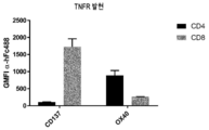
실시예에서 입증된 바와 같이, OX40은 CD4+ T 세포에서 우선적으로 발현되고 CD137은 CD8+ T 세포에서 우선적으로 발현된다. 본 발명자들은 항체 분자가 CD4+ T 세포에서 OX40의 효능작용을 유도할 수 있음을 입증하였다. 이러한 경우, 항체 분자는 CDR-기반 항원-결합 도메인을 통해 CD137에 결합하여 항체 분자를 가교결합시키고 OX40 항원-결합 도메인은 동시에 CD4+ T 세포에서 발현된 OX40에 결합하고 클러스터링하고 활성화할 수 있는 것으로 믿어진다. 유사하게, 본 발명자들은 항체 분자가 CD8+ T 세포에서 CD137의 효능작용을 유도할 수 있음을 입증하였다. 이러한 경우, 항체 분자는 OX40 항원-결합 도메인을 통해 OX40에 결합하여 항체 분자를 가교결합시키고 OX40 항원-결합 도메인은 동시에 CD8+ T 세포에서 발현된 CD137에 결합하고 클러스터링하고 활성화할 수 있는 것으로 믿어진다.As demonstrated in the examples, OX40 is preferentially expressed on CD4+ T cells and CD137 is preferentially expressed on CD8+ T cells. The inventors have demonstrated that antibody molecules can induce the agonism of OX40 in CD4+ T cells. In this case, it is believed that the antibody molecule binds to CD137 via the CDR-based antigen-binding domain and crosslinks the antibody molecule, and the OX40 antigen-binding domain can simultaneously bind, cluster and activate OX40 expressed in CD4+ T cells. Lose. Similarly, the inventors have demonstrated that antibody molecules can induce the agonism of CD137 in CD8+ T cells. In this case, it is believed that the antibody molecule binds to OX40 via the OX40 antigen-binding domain and crosslinks the antibody molecule, and the OX40 antigen-binding domain can simultaneously bind, cluster and activate CD137 expressed in CD8+ T cells.
게다가, 본 발명자들은 상기한 바와 같은 2개의 항원-결합 부위를 포함하고 Fcγ 수용체에 대한 결합을 감소 또는 폐지하도록 변형된 항체 분자가 CD137 및 OX40이 공동-발현될 때 수용체를 통한 신호전달을 유도할 수 있어 Fcγ 수용체에 의한 가교결합을 필요로 하지 않으면서 일어나는 효능작용을 나타낸다는 것을 보여 주었다. Fcγ 수용체-매개된 가교결합이 본 발명의 항체 분자의 활성에 필요하지 않기 때문에, OX40 또는 CD137 수용체를 통한 신호전달은 종양 미세환경에서와 같이 두 표적이 모두 존재하는 부위에 국한될 것으로 예상된다. 따라서, 항체 분자는 두 개의 특정 표적 모두의 발현을 기반으로 하고 추가의 가교결합제를 필요로 하지 않으면서 효능작용을 자율적으로 유도할 수 있다.In addition, the inventors have shown that an antibody molecule comprising two antigen-binding sites as described above and modified to reduce or abolish binding to the Fcγ receptor can induce signaling through the receptor when CD137 and OX40 are co-expressed. It was shown that it exhibits an efficacious action that occurs without the need for crosslinking by the Fcγ receptor. Since Fcγ receptor-mediated crosslinking is not required for the activity of the antibody molecules of the present invention, signaling through the OX40 or CD137 receptor is expected to be localized to the site where both targets are present, such as in the tumor microenvironment. Thus, antibody molecules can autonomously induce agonism based on the expression of both specific targets and without the need for additional crosslinking agents.
또한, Fcγ 수용체-결합이 ADCC에 필요하기 때문에, 이러한 Fcγ 수용체에 대한 결합의 감소는 또한 표적 면역 세포가 본 발명의 항체 분자에 의해 고갈되지 않도록, 감소된 ADCC를 초래할 것으로 예상된다. 본 발명자들은 항체 분자가 면역 반응을 촉진하기 위해 CD137 및/또는 OX40을 발현하는 면역 세포를 활성화하도록 설계되었기 때문에 이것이 중요하다고 간주하였다. 따라서 이러한 면역 세포의 고갈은 바람직하지 않다. 본 발명자들은 본원에 정의된 특성을 갖는 항체 분자가 면역 세포, 특히 CD137 및/또는 OX40을 발현하는 T 세포를 활성화하고 증식을 유도할 수 있음을 입증하였다.In addition, since Fcγ receptor-binding is required for ADCC, this reduction in binding to Fcγ receptors is also expected to result in decreased ADCC, such that the target immune cells are not depleted by the antibody molecules of the invention. The inventors considered this to be important because antibody molecules were designed to activate immune cells expressing CD137 and/or OX40 to promote an immune response. Therefore, depletion of these immune cells is not desirable. The inventors have demonstrated that antibody molecules having the properties defined herein can activate immune cells, in particular T cells expressing CD137 and/or OX40 and induce proliferation.
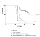
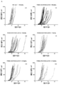
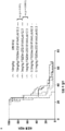
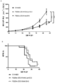
본 발명자들은 상기한 바와 같은 CD137 및 OX40 항원-결합 부위를 포함하는 항체 분자가 마우스에서 생체내 종양 성장을 억제할 수 있다는 것을 추가로 보여 주었다. 게다가, 항체 분자 중 하나가 CD137에 대한 CDR-기반 항원-결합 부위를 포함하고 다른 분자가 OX40에 대한 CDR-기반 항원-결합 부위를 포함하는 2개의 단일특이 항체 분자의 조합에 비해 이중특이 항체 분자에서 보다 효과적인 종양 성장 억제가 관찰되었으며, 이것은 OX40 및 CD137의 동시 관여 및 효능작용이 개선된 항종양 효능을 초래함을 입증하였다. 또한, 항체 분자는 CT26 마우스 종양 모델에서 종양 세포를 사용한 재유발시험 (re-challenge)에 대해 한 완전한 종양 퇴행 및 보호 면역 기억의 확립을 유도할 수 있는 것으로 나타났다. 따라서, 본 발명의 항체 분자는 인간 환자의 암 치료에 효능을 보일 것으로 예상된다. 따라서, 이러한 항체 분자는 ADCC 활성을 폐지하기 때문에, 이러한 유익한 T 세포 (기억 및 효과기 세포)를 현저하게 고갈시키지 않으면서 표적 면역 세포를 효능화함으로써 종양 성장을 억제할 것으로 예상된다.The inventors further showed that antibody molecules comprising the CD137 and OX40 antigen-binding sites as described above can inhibit tumor growth in vivo in mice. Moreover, compared to the combination of two monospecific antibody molecules, one of the antibody molecules contains a CDR-based antigen-binding site for CD137 and the other molecule contains a CDR-based antigen-binding site for OX40. A more effective tumor growth inhibition was observed in, demonstrating that the simultaneous involvement and agonism of OX40 and CD137 resulted in improved anti-tumor efficacy. In addition, antibody molecules have been shown to be able to induce complete tumor regression and the establishment of a protective immune memory against re-challenge using tumor cells in the CT26 mouse tumor model. Therefore, the antibody molecule of the present invention is expected to show efficacy in the treatment of cancer in human patients. Thus, since these antibody molecules abolish ADCC activity, it is expected to inhibit tumor growth by agonizing target immune cells without significantly depleting these beneficial T cells (memory and effector cells).
마우스의 생체내 연구에서 관찰된 바와 같이, 본원에 기술된 항체 분자에 의해 유도된 T 세포의 활성화 및 증식은 종양-국소화 효과가 아니라 전신 효과였다. 게다가, 말초 중심 기억 및 효과기 기억 CD4+ 및 CD8+ T 세포의 증식 및 활성화의 증가가 본 발명의 항체 분자를 투여한 시노몰구스 원숭이의 예비 용량 범위 조사 연구에서 관찰되었다. 따라서, 종양 미세환경에서의 T 세포의 표적화 뿐만 아니라 OX40 및 CD 137을 발현하는 말초 기억 T 세포가 항체 분자에 의해 표적화되어 종양-반응성 T 세포의 확장을 유도하여 이들의 항종양 효과를 제공할 것으로 예상된다.As observed in in vivo studies of mice, activation and proliferation of T cells induced by the antibody molecules described herein were systemic, not tumor-localizing effects. In addition, an increase in proliferation and activation of peripheral central and effector memory CD4+ and CD8+ T cells was observed in a preliminary dose range investigation study of cynomolgus monkeys administered with the antibody molecules of the present invention. Therefore, targeting T cells in the tumor microenvironment as well as peripheral memory T cells expressing OX40 and CD 137 are targeted by antibody molecules to induce expansion of tumor-reactive T cells, thereby providing their anti-tumor effects. It is expected.
따라서, 실제 종양 자체의 부위 외에도, 종양에 의해 영향을 받는 해부학적 위치는 또한 종양-특이 면역 반응이 생성되는 신체의 다른 곳의 위치, 예를 들어 말초 림프절을 포함하는 것으로 간주될 수 있다. Thus, in addition to the site of the actual tumor itself, the anatomical locations affected by the tumor can also be considered to include locations elsewhere in the body where a tumor-specific immune response is generated, for example peripheral lymph nodes.
상기 배경 부분에 설명된 바와 같이, CD137 효능제 분자의 임상 개발은 용량-제한적 고-등급 간 염증 (우렐루맙) 또는 낮은 임상 효능 (우토밀루맙)과 관련된 치료로 인해 적어도 부분적으로 저지되었다.As described in the background section above, the clinical development of the CD137 agonist molecule has been at least partially hindered by treatments associated with dose-limited high-grade liver inflammation (urelumab) or low clinical efficacy (utomilumab).
이론에 결부시키고자 함이 없이, 간에 존재하는 T 세포는 항-CD137 효능제 분자에 의해 활성화되어 간 염증을 유발할 수 있는 잠재능을 가질 수 있다고 생각된다. CD8+ T 세포는 패혈증/바이러스 감염 후 간 염증 및 세포 자멸사(apoptosis)를 촉진하는 것으로 나타났다 (Wesche-Soldato et al., 2007). 마우스에서 항-CD137 효능제 항체 요법은 간으로의 CD137-의존성 T 세포 침윤을 초래하는 것으로 나타났다 (Dubrot J et al., 2010). 이러한 연구의 결과를 종합하면 우렐루맙과 같이 높은 활성을 갖는 항-CD137 효능제 항체가 간으로의 활성화된 CD8+ T 세포의 침윤을 유발하여 간 염증을 야기할 수 있음을 나타낸다. 이 효과를 관찰하기에는 우토밀루맙의 활성이 너무 낮을 수 있다. 대안적으로, 우렐루맙 치료로 관찰된 용량-제한적 간 독성은 이 항체에 의해 결합된 특정 에피토프 때문일 수 있다.Without wishing to be bound by theory, it is believed that T cells present in the liver may have the potential to induce liver inflammation by being activated by anti-CD137 agonist molecules. CD8+ T cells have been shown to promote liver inflammation and apoptosis after sepsis/viral infection (Wesche-Soldato et al., 2007). Anti-CD137 agonist antibody therapy in mice has been shown to result in CD137-dependent T cell infiltration into the liver (Dubrot J et al., 2010). Taken together, the results of these studies indicate that anti-CD137 agonist antibodies with high activity, such as urelumab, can cause invasion of activated CD8+ T cells into the liver, resulting in liver inflammation. Utomylumab's activity may be too low to observe this effect. Alternatively, the dose-limiting liver toxicity observed with urelumab treatment may be due to the specific epitope bound by this antibody.
본 발명자들은 높은 친화도로, 즉 높은 결합력(avidity)으로 CD137에 결합할 것으로 예상되는 이량체 인간 CD137에 결합하는 항체 분자를 단리하기 위해 광범위한 선택 프로그램을 수행하였다. 사용된 선택 프로토콜의 관점에서, 항체 분자는 이량체 CD137에 대해 관찰된 친화도보다 낮은 친화도로 단량체 CD137에 결합할 것으로 예상된다.We performed a wide selection program to isolate antibody molecules that bind to dimeric human CD137, which are expected to bind to CD137 with high affinity, i.e., with high avidity. In view of the selection protocol used, the antibody molecule is expected to bind monomeric CD137 with an affinity lower than the observed affinity for dimer CD137.
본원에서 언급되는 '친화도'는 KD에 의해 측정되는 항체 분자와 이의 동족 항원 사이의 결합 상호작용의 강도를 지칭할 수 있다. 당해 분야 기술자에게 쉽게 자명한 바와 같이, 항체 분자가 항원과 다중 결합 상호작용을 형성할 수 있는 경우 (예를 들어, 항체 분자가 항원에 2가 결합할 수 있고, 임의로, 항원이 이량체인 경우) KD에 의해 측정되는 친화도는 또한 결합력에 의해 영향을 받을 수 있으며, 결합력은 항체-항원 복합체의 전반적인 강도를 지칭한다.As referred to herein,'affinity' may refer to the strength of the binding interaction between an antibody molecule and its cognate antigen as measured by K D. As will be readily apparent to those skilled in the art, when the antibody molecule is capable of forming multiple binding interactions with the antigen (e.g., when the antibody molecule is capable of divalent binding to the antigen and, optionally, the antigen is a dimer). The affinity measured by K D can also be influenced by the avidity, which refers to the overall strength of the antibody-antigen complex.
T 세포와 같은 면역 세포에 의한 CD137의 발현은 활성화시 상향조절된다. 이론에 결부시키고자 함이 없이, 활성화된 면역 세포에서의 CD137의 높은 발현으로 인해, CD137은 이러한 세포의 표면에서 이량체, 삼량체 및 고차 다량체의 형태일 것으로 생각된다. 이와 달리, 나이브 T 세포와 같은 나이브 면역 세포는 이들의 세포 표면에서 낮거나 무시할 수 있는 수준의 CD137을 발현하며 따라서 존재하는 모든 CD137은 단량체 형태일 것 같다. 따라서, 높은 결합력으로 CD137에 결합하는 항체 분자는 나이브 면역 세포와 반대로 활성화된 T 세포와 같은 활성화된 면역 세포에 우선적으로 결합할 것으로 예상된다.Expression of CD137 by immune cells such as T cells is upregulated upon activation. Without wishing to be bound by theory, it is believed that due to the high expression of CD137 in activated immune cells, CD137 will be in the form of dimers, trimers and higher order multimers on the surface of these cells. In contrast, naïve immune cells, such as naive T cells, express low or negligible levels of CD137 on their cell surface and therefore all CD137 present is likely to be in monomeric form. Therefore, antibody molecules that bind to CD137 with high avidity are expected to preferentially bind to activated immune cells such as activated T cells as opposed to naive immune cells.
따라서, 상기에 비추어 볼 때, 본 발명의 항체 분자는 대체로 OX40과의 관여를 통한 가교결합의 부재하에서 CD137을 활성화할 수 없을 것으로 예상된다. 또한, 상기한 바와 같이, 본 발명자들은 OX40의 공동-발현이 거의 또는 전혀 없는 위치에서 CD137의 활성화를 피할 것이라는 기대로 Fcγ 수용체 매개된 가교결합이 감소되거나 폐지된 항체 분자를 개발하였다. Fcγ 수용체 결합의 불능은 항체 분자의 항종양 활성에 영향을 미치지 않는 것으로 나타났다. 이론에 결부시키고자 함이 없이, 이러한 항체 분자는 환자에게 투여될 때 감소된 독성을 보일 것으로 믿어진다. 이것은 CD137 활성화가 OX40 및 CD137이 CD137의 클러스터링 및 활성화를 유도하기에 충분한 수준으로 공동-발현되는 위치로 크게 제한되기 때문이라고 생각된다. 본 발명자들은 시노몰구스 원숭이의 예비 용량 범위 조사 연구에서, 본 발명의 항체 분자의 용량이 30 mg/kg까지 잘 견딘다는 것을 보여 주었다.Therefore, in light of the above, it is expected that the antibody molecules of the present invention are generally unable to activate CD137 in the absence of crosslinking through involvement with OX40. In addition, as described above, the present inventors have developed antibody molecules with reduced or abolished Fcγ receptor-mediated crosslinking with the expectation that it will avoid activation of CD137 at positions where there is little or no co-expression of OX40. It has been shown that the inability to bind Fcγ receptors does not affect the antitumor activity of the antibody molecule. Without wishing to be bound by theory, it is believed that such antibody molecules will exhibit reduced toxicity when administered to a patient. This is thought to be because CD137 activation is largely limited to the site where OX40 and CD137 are co-expressed to a level sufficient to induce clustering and activation of CD137. In a preliminary dose range investigation study of cynomolgus monkeys, the present inventors have shown that the dose of the antibody molecule of the invention is well tolerated up to 30 mg/kg.
본 발명자들은 본 발명의 항체 분자가 가교결합의 부재하에서도 낮은 수준의 OX40 클러스터링 및 활성화를 유도할 수 있음을 보여 주었다. CD137 효능제 항체와 달리, OX40 효능제 항체는 클리닉에서 용량-제한적 독성 (DLT)을 나타내지 않았으며, 따라서 가교결합의 부재하에서 OX40 효능제 활성은 임상 치료에 문제를 나타낼 것으로 예상되지 않는다. 이와 달리, 치료할 상태에 따라, 가교결합의 부재하에서 항체 분자에 의한 낮은 수준의 OX40 효능제 활성이 유리할 수 있다. 이론에 결부시키고자 함이 없이, 이러한 특성을 가진 OX40 항원-결합 부위를 포함하는 항체 분자는 가교결합의 부재하에서 종양-반응성 T 세포의 제한된 활성화 및 확장을 유도하여, 종양 미세환경에서 가교결합된 Fcab 분자에 의해 더욱 활성화될 수 있는 더 큰 종양--반응성 T 세포의 풀을 야기함으로써 암 치료의 맥락에서 유용할 수 있다고 생각된다.The present inventors have shown that the antibody molecules of the present invention can induce low levels of OX40 clustering and activation even in the absence of crosslinking. Unlike the CD137 agonist antibody, the OX40 agonist antibody did not exhibit dose-limiting toxicity (DLT) in the clinic, so OX40 agonist activity in the absence of crosslinking is not expected to present a problem in clinical treatment. Alternatively, depending on the condition to be treated, low levels of OX40 agonist activity by antibody molecules in the absence of crosslinking may be advantageous. Without wishing to be bound by theory, antibody molecules comprising OX40 antigen-binding sites with these properties induce limited activation and expansion of tumor-reactive T cells in the absence of crosslinking, resulting in crosslinking in the tumor microenvironment. It is thought that it may be useful in the context of cancer treatment by causing a larger pool of tumor-reactive T cells that can be further activated by the Fcab molecule.
Fcγ 수용체에 대한 결합을 감소 또는 폐지하도록 변형된 본 발명의 항체 분자의 추가 이점은 이러한 항체 분자가 OX40-발현 조절 T 세포 (Tregs)의 고갈에 의존하지 않는 항종양 활성을 갖는다는 점일 수 있다. Tregs는 말초에 위치하여 잠재적으로 보호할 수 있으며 면역계를 과도하게 자극함으로써 유발될 수 있는 자가면역의 영향을 줄일 수 있다 (Vignali DA et al., 2008). 따라서, Treg 고갈이 마우스 모델에서 종양 성장을 감소시키는데 상당한 영향을 미칠 수 있다고 가정되었다 (Bulliard et al., 2014; Simpson et al., 2013). 그러나, 인간 종양에서 Treg 고갈이 ADCC에 의해 달성될 수 있다는 제한된 증거가 있으며, Treg 고갈이 인간에서 발생하는 경우, 이것은 마우스 모델에서 관찰된 바와 같은 극적인 항종양 활성을 초래하지는 않는 것으로 보인다 (Powell et al., 2007; Nizar S et al., 2009; Glisson BS et al., 2016; Tran B et al., 2017). 따라서, 항체 분자가 Tregs를 현저하게 고갈시키지는 않지만 여전히 항종양 활성을 갖는다면, 이것은 항체 분자가 Fcγ 수용체-매개된 Treg 고갈과는 관계없이 항종양 활성을 가짐을 나타낼 수 있다.A further advantage of the antibody molecules of the invention modified to reduce or abolish binding to the Fcγ receptor may be that such antibody molecules have anti-tumor activity that does not depend on depletion of OX40-expressing regulatory T cells (Tregs). Tregs are located peripherally and can potentially protect and reduce the effects of autoimmunity that can be induced by overstimulating the immune system (Vignali DA et al., 2008). Therefore, it was hypothesized that Treg depletion could have a significant effect on reducing tumor growth in a mouse model (Bulliard et al., 2014; Simpson et al., 2013). However, there is limited evidence that Treg depletion in human tumors can be achieved by ADCC, and when Treg depletion occurs in humans, it does not appear to result in dramatic anti-tumor activity as observed in mouse models (Powell et al. al., 2007; Nizar S et al., 2009; Glisson BS et al., 2016; Tran B et al., 2017). Thus, if the antibody molecule does not significantly deplete Tregs but still has anti-tumor activity, this could indicate that the antibody molecule has anti-tumor activity independent of Fcγ receptor-mediated Tregs depletion.
항체 분자는 인간 및 시노몰구스 CD137과 인간 및 시노몰구스 OX40 둘 다에 높은 친화도로 결합할 수 있는 것으로 추가로 나타났다. 이러한 교차-반응성은 항체 분자의 투여 및 안전성 시험이 전임상 개발 동안 시노몰구스 원숭이에서 수행될 수 있게 하기 때문에 유리하다.The antibody molecule was further shown to be capable of binding with high affinity to both human and cynomolgus CD137 and human and cynomolgus OX40. This cross-reactivity is advantageous because it allows administration and safety testing of antibody molecules to be performed in cynomolgus monkeys during preclinical development.
본 발명자들에 의해 확인된 항체 분자의 추가의 특징은 CD137에 대한 항원-결합 부위와 OX40에 대한 항원-결합 부위가 둘 다 항체 구조 자체 내에 포함된다는 것이다. 특히, 항체 분자는 다른 단백질이 링커 또는 다른 수단을 통해 항체 분자에 융합되는 것을 필요로 하지 않아 이의 표적 둘 다에 2가 결합할 수 있는 분자를 야기한다. 이것은 다수의 장점을 갖는다. 구용적으로, 본 발명자들에 의해 확인된 항체 분자는 추가의 융합된 부분을 포함하지 않기 때문에 표준 항체의 생산에 사용되는 것과 유사한 방법을 사용하여 생산될 수 있다. 구조는 또한 링커가 시간이 지남에 따라 분해되어 항체 분자의 이종 집단을 생성할 수 있기 때문에 개선된 항체 안정성을 야기할 것으로 예상된다. 단 하나의 단백질이 융합된 집단의 이러한 항체는 OX40 및 CD137 둘 다에 결합함으로써 가교결합의 결과로서 수용체를 통해 이중 효능제 및 신호로서 작용하지 못할 수 있다. 링커의 절단 또는 분해는 개체(individual)에게 치료제를 투여하기 전 또는 투여한 후에 (예를 들어, 개체의 효소적 절단 또는 생체내 pH를 통해) 발생하여, 개체에서 순환하는 동안 그 효과가 감소될 수 있다. 본 발명자들에 의해 확인된 항체 분자에는 링커가 없기 때문에, 항체 분자는 투여 전과 투여 후 둘 다에 동일한 수의 결합 부위를 유지할 것으로 예상된다. 게다가, 융합된 단백질 또는 링커 또는 둘 다의 도입은 분자가 개체에게 투여될 때 면역원성을 유도하여 치료제의 효과를 감소시킬 수 있기 때문에 본 발명자들에 의해 확인된 항체 분자의 구조는 또한 분자의 면역원성의 관점에서 바람직하다.A further feature of the antibody molecules identified by the inventors is that both the antigen-binding site for CD137 and the antigen-binding site for OX40 are contained within the antibody structure itself. In particular, antibody molecules do not require other proteins to be fused to the antibody molecule through a linker or other means, resulting in a molecule capable of divalent binding to both its targets. This has a number of advantages. Typically, antibody molecules identified by the present inventors can be produced using methods similar to those used for the production of standard antibodies since they do not contain additional fused moieties. The structure is also expected to lead to improved antibody stability as the linker can degrade over time to create a heterogeneous population of antibody molecules. These antibodies in a population fused with only one protein may not act as a dual agonist and signal through the receptor as a result of crosslinking by binding to both OX40 and CD137. Cleavage or degradation of the linker occurs before or after administration of the therapeutic agent to the individual (e.g., through enzymatic cleavage of the subject or in vivo pH), so that its effect will be reduced during circulation in the subject. I can. Since the antibody molecules identified by the present inventors do not have linkers, it is expected that the antibody molecules will retain the same number of binding sites both before and after administration. In addition, the structure of the antibody molecule identified by the inventors is also dependent on the structure of the antibody molecule, as the introduction of a fused protein or linker or both can induce immunogenicity when the molecule is administered to an individual, thereby reducing the effectiveness of the therapeutic It is preferable from the viewpoint of originality.
따라서, 본 발명은 다음을 제공한다:Thus, the present invention provides:
[1] [One]
(a) CD137에 대한 상보성 결정 영역 (CDR)-기반 항원-결합 부위; 및 (a) a complementarity determining region (CDR)-based antigen-binding site for CD137; And
(b) 항체 분자의 CH3 도메인에 위치한 OX40 항원-결합 부위를 포함하고; (b) contains an OX40 antigen-binding site located in the CH3 domain of the antibody molecule;
여기서 CDR-기반 항원-결합 부위가 Where the CDR-based antigen-binding site is
(i) 각각 서열 번호 1, 2, 3, 4, 5 및 6 [FS30-10-16];(i) SEQ ID NOs: 1, 2, 3, 4, 5 and 6 [FS30-10-16], respectively;
(ii) 각각 서열 번호 1, 2, 16, 4, 5 및 6 [FS30-10-3];(ii) SEQ ID NOs: 1, 2, 16, 4, 5 and 6 [FS30-10-3], respectively;
(iii) 각각 서열 번호 1, 2, 21, 4, 5 및 6 [FS30-10-12];(iii) SEQ ID NOs: 1, 2, 21, 4, 5 and 6 [FS30-10-12], respectively;
(iv) 각각 서열 번호 25, 26, 27, 4, 5 및 28 [FS30-35-14]; 또는 (iv) SEQ ID NOs: 25, 26, 27, 4, 5 and 28 [FS30-35-14], respectively; or
(v) 각각 서열 번호 33, 34, 35, 4, 5 및 36 [FS30-5-37]에 제시된 CDR 1-6을 포함하고; (v) comprises CDRs 1-6 set forth in SEQ ID NOs: 33, 34, 35, 4, 5 and 36 [FS30-5-37], respectively;
여기서 OX40 항원-결합 부위가 CH3 도메인의 AB, CD 및 EF 구조적 루프에 각각 위치한 제1 서열, 제2 서열 및 제3 서열을 포함하고, 여기서 제1, 제2 및 제3 서열이 각각 서열 번호 51, 52 및 53 [FS20-22-49]에 제시된 서열을 갖는, CD137 및 OX40에 결합하는 항체 분자.Wherein the OX40 antigen-binding site comprises a first sequence, a second sequence and a third sequence, respectively located in the AB, CD and EF structural loops of the CH3 domain, wherein the first, second and third sequences are each SEQ ID NO: 51 , 52 and 53 [FS20-22-49], an antibody molecule that binds to CD137 and OX40.
[2] [2]
(a) CD137에 대한 상보성 결정 영역 (CDR)-기반 항원-결합 부위; 및(a) a complementarity determining region (CDR)-based antigen-binding site for CD137; And
(b) 항체 분자의 CH3 도메인에 위치한 OX40 항원-결합 부위를 포함하고;(b) contains an OX40 antigen-binding site located in the CH3 domain of the antibody molecule;
여기서 CDR-기반 항원-결합 부위가 Where the CDR-based antigen-binding site is
(i) 각각 번호 7, 8, 9, 10, 11 및 6 [FS30-10-16];(i)
(ii) 각각 서열 번호 7, 8, 17, 10, 11 및 6 [FS30-10-3];(ii) SEQ ID NOs: 7, 8, 17, 10, 11 and 6 [FS30-10-3], respectively;
(iii) 각각 서열 번호 7, 8, 22, 10, 11 및 6 [FS30-10-12];(iii) SEQ ID NOs: 7, 8, 22, 10, 11 and 6 [FS30-10-12], respectively;
(iv) 각각 서열 번호 29, 30, 31, 10, 11 및 28 [FS30-35-14]; 또는 (iv) SEQ ID NOs: 29, 30, 31, 10, 11 and 28 [FS30-35-14], respectively; or
(v) 각각 서열 번호 37, 38, 39, 10, 11 및 36 [FS30-5-37]에 제시된 CDR 1-6을 포함하고; (v) comprises CDRs 1-6 set forth in SEQ ID NOs: 37, 38, 39, 10, 11 and 36 [FS30-5-37], respectively;
여기서 OX40 항원-결합 부위가 CH3 도메인의 AB, CD 및 EF 구조적 루프에 각각 위치한 제1 서열, 제2 서열 및 제3 서열을 포함하고, 여기서 제1, 제2 및 제3 서열이 각각 서열 번호 51, 52 및 53 [FS20-22-49]에 제시된 서열을 갖는, CD137 및 OX40에 결합하는 항체 분자.Wherein the OX40 antigen-binding site comprises a first sequence, a second sequence and a third sequence, respectively located in the AB, CD and EF structural loops of the CH3 domain, wherein the first, second and third sequences are each SEQ ID NO: 51 , 52 and 53 [FS20-22-49], an antibody molecule that binds to CD137 and OX40.
[3] [3]
(i) 제1 서열이 항체 분자의 CH3 도메인의 위치 14 내지 18에 위치하고;(i) the first sequence is located at
(ii) 제2 서열이 항체 분자의 CH3 도메인의 위치 45.1 내지 77에 위치하고/하거나; (ii) the second sequence is located at positions 45.1 to 77 of the CH3 domain of the antibody molecule;
(iii) 제3 서열이 항체 분자의 CH3 도메인의 위치 93 내지 101에 위치하고; (iii) the third sequence is located at positions 93-101 of the CH3 domain of the antibody molecule;
여기서 아미노산 잔기 번호매김(amino acid residue numbering)은 IMGT 번호매김 체계에 따르는, [1] 또는 [2]에 따르는 항체 분자.Here, amino acid residue numbering is an antibody molecule according to [1] or [2], according to the IMGT numbering system.
[4] 항체 분자가 서열 번호 54 [FS20-22-49]에 제시된 CH3 도메인 서열을 포함하는, [1] 내지 [3] 중의 어느 하나에 따르는 항체 분자.[4] The antibody molecule according to any one of [1] to [3], wherein the antibody molecule comprises the CH3 domain sequence set forth in SEQ ID NO: 54 [FS20-22-49].
[5] 항체 분자가 [1] 또는 [2]의 (i) 내지 (iv) 중의 어느 하나에 제시된 CDR 1-6을 포함하는, [1] 내지 [4] 중의 어느 하나에 따르는 항체 분자.[5] The antibody molecule according to any one of [1] to [4], wherein the antibody molecule comprises CDR 1-6 as set forth in any one of [1] or [2] (i) to (iv).
[6] 항체 분자가 [1] 또는 [2]의 (i) 내지 (iii) 중의 어느 하나에 제시된 CDR 1-6을 포함하는, [1] 내지 [5] 중의 어느 하나에 따르는 항체 분자.[6] The antibody molecule according to any one of [1] to [5], wherein the antibody molecule contains CDRs 1-6 shown in any one of [1] or [2] (i) to (iii).
[7] 항체 분자가 [1] 또는 [2]의 (i)에 제시된 CDR 1-6을 포함하는, [1] 내지 [6] 중의 어느 하나에 따르는 항체 분자.[7] The antibody molecule according to any one of [1] to [6], wherein the antibody molecule comprises CDR 1-6 shown in [1] or [2] (i).
[8] 항체 분자가 중쇄 가변 (VH) 도메인 및/또는 경쇄 가변 (VL) 도메인, 바람직하게는 VH 도메인 및 VL 도메인을 포함하는, [1] 내지 [7] 중의 어느 하나에 따르는 항체 분자.[8] The antibody molecule according to any one of [1] to [7], wherein the antibody molecule comprises a heavy chain variable (VH) domain and/or a light chain variable (VL) domain, preferably a VH domain and a VL domain.
[9] 항체 분자가 면역글로불린 중쇄 및/또는 면역글로불린 경쇄, 바람직하게는 면역글로불린 중쇄 및 면역글로불린 경쇄를 포함하는, [1] 내지 [8] 중의 어느 하나에 따르는 항체 분자.[9] The antibody molecule according to any one of [1] to [8], wherein the antibody molecule comprises an immunoglobulin heavy chain and/or an immunoglobulin light chain, preferably an immunoglobulin heavy chain and an immunoglobulin light chain.
[10] 항체 분자가 VH 도메인 및/또는 VL 도메인, 바람직하게는 [10] The antibody molecule is a VH domain and/or a VL domain, preferably
(i) 각각 서열 번호 12 및 14 [FS30-10-16];(i) SEQ ID NOs: 12 and 14 [FS30-10-16], respectively;
(ii) 각각 서열 번호 18 및 14 [FS30-10-3];(ii) SEQ ID NOs: 18 and 14 [FS30-10-3], respectively;
(iii) 각각 서열 번호 23 및 14 [FS30-10-12];(iii) SEQ ID NOs: 23 and 14 [FS30-10-12], respectively;
(iv) 각각 서열 번호 170 및 172 [FS30-35-14]; (iv) SEQ ID NOs: 170 and 172 [FS30-35-14], respectively;
(v) 각각 서열 번호 40 및 42 [FS30-5-37]에 제시된 VH 도메인 및 VL 도메인을 포함하는, [8] 또는 [9]에 따르는 항체 분자.(v) An antibody molecule according to [8] or [9], comprising a VH domain and a VL domain set forth in SEQ ID NOs: 40 and 42 [FS30-5-37], respectively.
[11] 항체 분자가 [10]의 (i) 내지 (iv) 중의 어느 하나에 제시된 VH 도메인 및 VL 도메인을 포함하는, [10]에 따르는 항체 분자.[11] The antibody molecule according to [10], wherein the antibody molecule comprises a VH domain and a VL domain as set forth in any one of (i) to (iv) of [10].
[12] 항체 분자가 [10]의 (i) 내지 (iii) 중의 어느 하나에 제시된 VH 및 VL 도메인을 포함하는, [10] 또는 [11]에 따르는 항체 분자.[12] The antibody molecule according to [10] or [11], wherein the antibody molecule comprises the VH and VL domains set forth in any one of (i) to (iii) of [10].
[13] 항체 분자가 [10]의 (i)에 제시된 VH 도메인 및 VL 도메인을 포함하는, [10] 내지 [12] 중의 어느 하나에 따르는 항체 분자.[13] The antibody molecule according to any one of [10] to [12], wherein the antibody molecule comprises a VH domain and a VL domain as set forth in (i) of [10].
[14] 항체 분자가 인간 lgG1 분자인, [1] 내지 [13] 중의 어느 하나에 따르는 항체 분자.[14] The antibody molecule according to any one of [1] to [13], wherein the antibody molecule is a human lgG1 molecule.
[15] 항체 분자가 항체의 중쇄 및 경쇄:[15] The antibody molecule is the heavy and light chain of an antibody:
(i) 각각 서열 번호 95 및 97에 제시된 FS20-22-49AA/FS30-10-16;(i) FS20-22-49AA/FS30-10-16 set forth in SEQ ID NOs: 95 and 97, respectively;
(ii) 각각 번호 99 및 97에 제시된 FS20-22-49AA/FS30-10-3;(ii) FS20-22-49AA/FS30-10-3 set forth in
(iii) 각각 서열 번호 103 및 97에 제시된 FS20-22-49AA/FS30-10-12;(iii) FS20-22-49AA/FS30-10-12 set forth in SEQ ID NOs: 103 and 97, respectively;
(iv) 각각 서열 번호 105 및 107에 제시된 FS20-22-49AA/FS30-35-14; 또는 (iv) FS20-22-49AA/FS30-35-14 set forth in SEQ ID NOs: 105 and 107, respectively; or
(v) 각각 번호 109 및 111에 제시된 FS20-22-49AA/FS30-5-37을 포함하는, [1] 내지 [14] 중의 어느 하나에 따르는 항체 분자.(v) An antibody molecule according to any one of [1] to [14], comprising FS20-22-49AA/FS30-5-37 shown in
[16] 항체 분자가 [15]의 (i) 내지 (iv) 중의 어느 하나에 제시된 경쇄 및 중쇄를 포함하는, [15]에 따르는 항체 분자.[16] The antibody molecule according to [15], wherein the antibody molecule comprises a light chain and a heavy chain as set forth in any one of (i) to (iv) of [15].
[17] 항체 분자가 [15]의 (i) 내지 (iii) 중의 어느 하나에 제시된 경쇄 및 중쇄를 포함하는, [15]에 따르는 항체 분자.[17] The antibody molecule according to [15], wherein the antibody molecule comprises a light chain and a heavy chain as set forth in any one of (i) to (iii) of [15].
[18] 항체 분자가 [15]의 (i)에 제시된 경쇄 및 중쇄를 포함하는, [15]에 따르는 항체 분자.[18] The antibody molecule according to [15], wherein the antibody molecule comprises a light chain and a heavy chain as set forth in (i) of [15].
[19] 항체 분자가 인간 CD137 및 인간 OX40에 결합하는, [1] 내지 [18] 중의 어느 하나에 따르는 항체 분자.[19] The antibody molecule according to any one of [1] to [18], wherein the antibody molecule binds to human CD137 and human OX40.
[20] 인간 CD137이 서열 번호 127에 제시된 서열로 구성되거나 이를 포함하는, [19]에 따르는 항체 분자.[20] The antibody molecule according to [19], wherein human CD137 consists of or comprises the sequence set forth in SEQ ID NO: 127.
[21] 인간 OX40이 서열 번호 130에 제시된 서열로 구성되거나 이를 포함하는, [19] 또는 [20]에 따르는 항체 분자.[21] The antibody molecule according to [19] or [20], wherein human OX40 consists of or comprises the sequence set forth in SEQ ID NO: 130.
[22] 항체 분자가 시노몰구스 CD137 및 시노몰구스 OX40에 결합하는, [1] 내지 [21] 중의 어느 하나에 따르는 항체 분자.[22] The antibody molecule according to any one of [1] to [21], wherein the antibody molecule binds to cynomolgus CD137 and cynomolgus OX40.
[23] 시노몰구스 CD137이 서열 번호 129에 제시된 서열로 구성되거나 이를 포함하는, [22]에 따르는 항체 분자.[23] The antibody molecule according to [22], wherein the cynomolgus CD137 consists of or comprises the sequence set forth in SEQ ID NO: 129.
[24] 시노몰구스 OX40이 서열 번호 131에 제시된 서열로 구성되거나 이를 포함하는, [23] 또는 [24]에 따르는 항체 분자.[24] The antibody molecule according to [23] or [24], wherein the cynomolgus OX40 consists of or comprises the sequence set forth in SEQ ID NO: 131.
[25] 항체 분자가 인간 CD137 및 인간 OX40에 결합하고, 항체 분자가 인간 CD137에 결합하는 친화도 (KD)가 항체 분자가 인간 OX40에 결합하는 친화도 (KD)의 2배 이내인, [5] 내지 [7], [11] 내지 [13] 및 [16] 내지 [18] 중의 어느 하나에 따르는 항체 분자.[25] The antibody molecule binds to human CD137 and human OX40, and the affinity for the antibody molecule to bind to human CD137 (K D ) is within twice the affinity of the antibody molecule to bind to human OX40 (K D ), The antibody molecule according to any one of [5] to [7], [11] to [13] and [16] to [18].
[26] 항체 분자가 인간 CD137 및 인간 OX40에 동시에 결합할 수 있는, [19] 내지 [25] 중의 어느 하나에 따르는 항체 분자.[26] The antibody molecule according to any one of [19] to [25], wherein the antibody molecule is capable of simultaneously binding to human CD137 and human OX40.
[27] 항체 분자가 세포-표면 발현된 CD137의 존재하에서 면역 세포 상의 OX40을 활성화할 수 있는, [1] 내지 [26] 중의 어느 하나에 따르는 항체 분자.[27] The antibody molecule according to any one of [1] to [26], wherein the antibody molecule is capable of activating OX40 on immune cells in the presence of cell-surface-expressed CD137.
[28] 면역 세포 상의 OX40에 및 CD137에 대한 항체 분자의 결합이 면역 세포 상의 OX40의 클러스터링을 유발하는, [1] 내지 [27] 중의 어느 하나에 따르는 항체 분자.[28] The antibody molecule according to any one of [1] to [27], wherein the binding of the antibody molecule to OX40 on immune cells and to CD137 causes clustering of OX40 on immune cells.
[29] 항체 분자가 세포-표면 발현된 OX40의 존재하에서 면역 세포 상의 CD137을 활성화할 수 있는, [1] 내지 [28] 중의 어느 하나에 따르는 항체 분자.[29] The antibody molecule according to any one of [1] to [28], wherein the antibody molecule is capable of activating CD137 on immune cells in the presence of cell-surface expressed OX40.
[30] 면역 세포 상의 CD137에 및 OX40에 대한 항체 분자의 결합이 면역 세포 상의 CD137의 클러스터링을 유발하고, OX40이 동일한 면역 세포에서 또는 별도의 세포에서 발현되는, [1] 내지 [29] 중의 어느 하나에 따르는 항체 분자.[30] Any of [1] to [29], wherein binding of an antibody molecule to CD137 on immune cells and to OX40 causes clustering of CD137 on immune cells, and OX40 is expressed in the same immune cell or in separate cells. Antibody molecule according to one.
[31] 면역 세포가 T 세포인, [27] 내지 [30] 중의 어느 하나에 따르는 항체 분자.[31] The antibody molecule according to any one of [27] to [30], wherein the immune cell is a T cell.
[32] 항체 분자가 하나 이상의 Fcγ 수용체에 대한 항체 분자의 CH2 도메인의 결합을 감소시키거나 폐지하도록 변형되는, [1] 내지 [31] 중의 어느 하나에 따르는 항체 분자.[32] The antibody molecule according to any one of [1] to [31], wherein the antibody molecule is modified to reduce or abolish the binding of the CH2 domain of the antibody molecule to one or more Fcγ receptors.
[33] 항체 분자가 하나 이상의 Fcγ 수용체에 결합하지 않는, [1] 내지 [32] 중의 어느 하나에 따르는 항체 분자.[33] The antibody molecule according to any one of [1] to [32], wherein the antibody molecule does not bind to one or more Fcγ receptors.
[34] Fcγ 수용체가 FcγRI, FcγRIIa, FcγRIIb 및 FcγRIII으로 구성된 그룹으로부터 선택되는, [32] 또는 [33]에 따르는 항체 분자.[34] The antibody molecule according to [32] or [33], wherein the Fcγ receptor is selected from the group consisting of FcγRI, FcγRIIa, FcγRIIb and FcγRIII.
[35] 항체 분자가 T 세포의 증식을 유도할 수 있는, [1] 내지 [34] 중의 어느 하나에 따르는 항체 분자.[35] The antibody molecule according to any one of [1] to [34], wherein the antibody molecule can induce proliferation of T cells.
[36] [1] 내지 [35] 중의 어느 하나에 따르는 항체 분자 및 생체활성 분자를 포함하는 접합체.[36] A conjugate comprising the antibody molecule and bioactive molecule according to any one of [1] to [35].
[37] [1] 내지 [36] 중의 어느 하나에 따르는 항체 분자 및 검출 가능한 표지를 포함하는 접합체.[37] A conjugate comprising the antibody molecule according to any one of [1] to [36] and a detectable label.
[38] [1] 내지 [35] 중의 어느 하나에 따르는 항체 분자를 암호화하는 핵산 분자 또는 분자들.[38] A nucleic acid molecule or molecules encoding the antibody molecule according to any one of [1] to [35].
[39] 핵산 분자(들)가 [39] the nucleic acid molecule(s)
(i) 각각 서열 번호 96 및 98에 제시된 FS20-22-49AA/FS30-10-16;(i) FS20-22-49AA/FS30-10-16 set forth in SEQ ID NOs: 96 and 98, respectively;
(ii) 각각 서열 번호 100 및 102에 제시된 FS20-22-49AA/FS30-10-3;(ii) FS20-22-49AA/FS30-10-3 set forth in SEQ ID NOs: 100 and 102, respectively;
(iii) 각각 서열 번호 104 및 102에 제시된 FS20-22-49AA/FS30-10-12;(iii) FS20-22-49AA/FS30-10-12 set forth in SEQ ID NOs: 104 and 102, respectively;
(iv) 각각 서열 번호 106 및 108에 제시된 FS20-22-49AA/FS30-35-14; 또는(iv) FS20-22-49AA/FS30-35-14 set forth in SEQ ID NOs: 106 and 108, respectively; or
(v) 각각 서열 번호 110 및 112에 제시된 FS20-22-49AA/FS30-5-37의 중쇄 핵산 서열 및/또는 경쇄 핵산 서열을 포함하는, [1] 내지 [4], [8] 내지 [10], [14] 내지 [15], 및 [19] 내지 [35] 중의 어느 하나에 따르는 항체 분자를 암호화하는 핵산 분자 또는 분자들.(v) [1] to [4], [8] to [10, respectively, comprising the heavy chain nucleic acid sequence and/or light chain nucleic acid sequence of FS20-22-49AA/FS30-5-37 shown in SEQ ID NOs: 110 and 112, respectively. ], [14] to [15], and [19] to [35] a nucleic acid molecule or molecules encoding the antibody molecule according to any one of.
[40] [38] 내지 [39] 중의 어느 하나에 따르는 핵산 분자 또는 분자들을 포함하는 벡터(vector) 또는 벡터들.[40] A vector or vectors comprising the nucleic acid molecule or molecules according to any one of [38] to [39].
[41] [38] 내지 [39] 중의 어느 하나에 따르는 핵산 분자(들), 또는 [40]에 따르는 벡터(들)을 포함하는 재조합 숙주 세포(recombinant host cell).[41] A recombinant host cell comprising the nucleic acid molecule(s) according to any one of [38] to [39], or the vector(s) according to [40].
[42] [41]의 재조합 숙주 세포를 항체 분자의 생산을 위한 조건하에서 배양함을 포함하여, [1] 내지 [35] 중의 어느 하나에 따르는 항체 분자를 생산하는 방법.[42] A method for producing an antibody molecule according to any one of [1] to [35], comprising culturing the recombinant host cell of [41] under conditions for production of the antibody molecule.
[43] 항체 분자를 단리 및/또는 정제함을 추가로 포함하는, [42]에 따르는 방법.[43] The method according to [42], further comprising isolating and/or purifying the antibody molecule.
[44] [1] 내지 [37] 중의 어느 하나에 따르는 항체 분자 또는 접합체 및 약제학적으로 허용되는 부형제를 포함하는 약제학적 조성물.[44] A pharmaceutical composition comprising the antibody molecule or conjugate according to any one of [1] to [37] and a pharmaceutically acceptable excipient.
[45] 치료법에 의한 인간 또는 동물 신체의 치료 방법에서 사용하기 위한, [1] 내지 [37] 중의 어느 하나에 따르는 항체 분자 또는 접합체.[45] The antibody molecule or conjugate according to any one of [1] to [37], for use in a method of treating a human or animal body by therapy.
[46] 개체에게 [1] 내지 [37] 중의 어느 하나에 따르는 치료적 유효량의 항체 분자 또는 접합체를 투여함을 포함하여, 개체에서 질환 또는 장애를 치료하는 방법.[46] A method of treating a disease or disorder in an individual, comprising administering to the individual a therapeutically effective amount of an antibody molecule or conjugate according to any one of [1] to [37].
[47] 항체 분자 또는 접합체가 개체에서 암 또는 감염성 질환을 치료하는데 사용하기 위한 것인, [45]에 따르는 사용을 위한 항체 분자 또는 접합체.[47] An antibody molecule or conjugate for use according to [45], wherein the antibody molecule or conjugate is for use in treating cancer or an infectious disease in a subject.
[48] 질환 또는 장애가 개체에서 암 또는 감염성 질환인, [46]의 방법.[48] The method of [46], wherein the disease or disorder is a cancer or an infectious disease in the individual.
[49] 암 또는 감염성 질환의 치료를 위한 의약의 제조에 있어서의, [1] 내지 [37] 중의 어느 하나에 따르는 항체 분자 또는 접합체의 용도.[49] The use of the antibody molecule or conjugate according to any one of [1] to [37] in the manufacture of a medicament for the treatment of cancer or infectious disease.
[50] 암이 고형암이고, 임의로 고형암이 흑색종, 방광암, 뇌암, 유방암, 난소암, 폐암, 대장암, 자궁경부암, 간암, 두경부암, 췌장암, 신장암 및 위암으로 구성된 그룹으로부터 선택되는, [47]에 따라 사용하기 위한 항체 분자 또는 접합체, [48]의 방법, 또는 [49]에 따르는 항체 분자 또는 접합체의 용도.[50] The cancer is solid cancer, and the solid cancer is optionally selected from the group consisting of melanoma, bladder cancer, brain cancer, breast cancer, ovarian cancer, lung cancer, colon cancer, cervical cancer, liver cancer, head and neck cancer, pancreatic cancer, kidney cancer, and stomach cancer, [ 47], the method of [48], or the use of the antibody molecule or conjugate according to [49].
[51] 감염성 질환이 지속성 바이러스 감염이고, 임의로 지속성 바이러스 감염이 인간 면역결핍 바이러스 (human immunodeficiency virus) (HIV), 엡스타인-바 바이러스 (Epstein-Barr virus), 시토메갈로바이러스 (Cytomegalovirus), B형 간염 바이러스 (Hepatitis B virus), C형 간염 바이러스 (Hepatitis C virus), 수두 대상포진 바이러스 (Varicella Zoster virus)로 구성된 그룹으로부터 선택되는, [47]에 따라 사용하기 위한 항체 분자 또는 접합체, [48]의 방법, 또는 [49]에 따르는 항체 분자 또는 접합체의 용도.[51] The infectious disease is a persistent viral infection, and optionally, the persistent viral infection is human immunodeficiency virus (HIV), Epstein-Barr virus, Cytomegalovirus, and hepatitis B. Antibody molecules or conjugates for use according to [47], selected from the group consisting of Hepatitis B virus, Hepatitis C virus, Varicella Zoster virus, of [48] The method, or the use of an antibody molecule or conjugate according to [49].
[52] 감염성 질환이 지속성 박테리아 감염이고, 임의로 지속성 박테리아 감염이 스타필로코커스 아우레우스 (Staphylococcus aureus), 헤모필루스 인플루엔자 (Hemophilus influenza), 마이코박테리움 투베르쿨로시스 (Mycobacterium tuberculosis), 마이코박테리움 레프라에 (Mycobacterium leprae), 헬리코박터 파일로리 (Helicobacter pylori), 트레포네마 팔리둠 (Treponema pallidum), 엔테로코커스 파에칼리스 (Enterococcus faecalis), 또는 스트렙토코커스 뉴모니에 (Streptococcus pneumoniae)의 지속성 감염인, [47]에 따라 사용하기 위한 항체 분자 또는 접합체, [48]의 방법, 또는 [49]에 따르는 항체 분자 또는 접합체의 용도.[52] The infectious disease is a persistent bacterial infection, and optionally, the persistent bacterial infection is Staphylococcus aureus, Hemophilus influenza, Mycobacterium tuberculosis, Mycobacterium A persistent infection of Mycobacterium leprae, Helicobacter pylori, Treponema pallidum, Enterococcus faecalis, or Streptococcus pneumoniae. 47], the method of [48], or the use of the antibody molecule or conjugate according to [49].
[53] 감염성 질환이 지속성 진균 감염이고, 임의로 지속성 진균 감염이 칸디다, 예를 들어, 칸디다 알비칸스 (Candida albicans), 크립토코커스 (Cryptococcus) (가티이 (gattii) 및 네오프 오르만스(neof ormans)), 탈라로마이세스 (Talaromyces) (Penicillium) marneffe, 마이크로스포룸 (Microsporum), 예를 들어 마이크로스포룸 아우도우이니 (Microsporum audouinii), 및 트리코피톤 톤수란스 (Trichophyton tonsurans)의 지속성 감염인, [47]에 따라 사용하기 위한 항체 분자 또는 접합체, [48]의 방법, 또는 [49]에 따르는 항체 분자 또는 접합체의 용도.[53] The infectious disease is a persistent fungal infection, and optionally the persistent fungal infection is Candida, for example Candida albicans, Cryptococcus (gattii and neoof ormans) ), Talaromyces (Penicillium) marneffe, Microsporum, for example Microsporum audouinii, and persistent infection of Trichophyton tonsurans, [47 ], the method of [48], or the use of the antibody molecule or conjugate according to [49].
[54] 감염성 질환이 지속성 기생충 감염이고, 임의로 지속성 기생충 감염이 플라스모듐 팔시파룸 (Plasmodium falciparum)과 같은 플라스모듐 (Plasmodium), 또는 리슈마니아 도노바니 (Leishmania donovani)와 같은 리슈마니아 (Leishmania)의 지속성 감염인, [47]에 따라 사용하기 위한 항체 분자 또는 접합체, [48]의 방법, 또는 [49]에 따르는 항체 분자 또는 접합체의 용도.[54] The infectious disease is a persistent parasitic infection, and optionally, the persistent parasitic infection is Plasmodium, such as Plasmodium falciparum, or Leishmania, such as Leishmania donovani. ), the use of an antibody molecule or conjugate for use according to [47], a method of [48], or an antibody molecule or conjugate according to [49].
[55] 치료가 항체 분자 또는 접합체를 제2 치료제와 조합하여 개체에게 투여함을 포함하는, [45], [47] 및 [50] 내지 [54] 중의 어느 하나에 따라 사용하기 위한 항체 분자 또는 접합체.[55] An antibody molecule for use according to any one of [45], [47] and [50] to [54], wherein the treatment comprises administering to the subject in combination with a second therapeutic agent an antibody molecule or conjugate, or Conjugate.
[56] 방법이 치료적 유효량의 제2 치료제를 개체에게 투여함을 추가로 포함하는, [46], [48] 및 [50] 내지 [54]에 따르는 방법.[56] The method according to [46], [48] and [50] to [54], wherein the method further comprises administering to the subject a therapeutically effective amount of a second therapeutic agent.
[57] 방법이 항체 분자 또는 접합체를 PD-1 또는 PD-L1에 결합하는 항체와 조합하여 개체에게 투여함을 포함하는, [47] 또는 [50]에 따라 개체에서 암을 치료하는 방법에서 사용하기 위한 항체 분자 또는 접합체.[57] Use in a method of treating cancer in an individual according to [47] or [50], wherein the method comprises administering to the individual in combination with an antibody molecule or conjugate with an antibody that binds to PD-1 or PD-L1. Antibody molecule or conjugate for
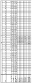
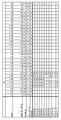
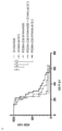
도 1은 Fcab FS20-22-38, FS20-22-41, FS20-22-47, FS20-22-49, FS20-22-85, FS20-31-58, FS20-31-66, FS20-31-94, FS20-31-102, FS20-31-108, 및 FS20-31-115 뿐만 아니라 야생형 (WT) Fcab의 CH3 도메인의 서열의 정렬을 보여준다. AB, CD 및 EF 구조적 루프의 위치 뿐만 아니라 WT 서열과 비교하여 Fcab의 CH3 도메인에 존재하는 임의의 아미노산 치환, 결실 (물결표 "~"로 표시됨) 또는 삽입이 표시된다. IMGT, IMGT 엑손 (연속 번호매김), EU 및 Kabat 번호매김 시스템에 따른 잔기의 번호가 나타내어져 있다.
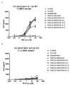
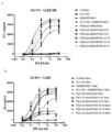
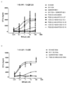
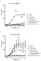
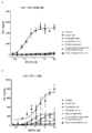
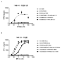
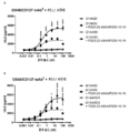
도 2는 가교결합의 존재 및 부재하에 인간 CD137 T 세포 활성화 분석에서 CD137 mAb 및 OX40/CD137 mAb2의 활성을 보여준다. 도 2a 및 b는 가교결합 항체의 존재 (도 2a) 또는 부재 (도 2b) 하에 증가하는 농도의 항-CD137mAb의 존재하에서의 IL2 방출을 보여준다. G1AA/20H4.9는 가교결합 항체의 존재 및 부재하에서 활성을 보이는 반면 G1AA/MOR7480.1 및 G1AA/FS30-10-16 항체의 활성은 가교결합 항체의 존재하에서만 관찰되었다. 도 2c 및 d는 가교결합제의 존재 (도 2c) 또는 부재 (도 2d) 하에 항-인간 OX40 Fcab를 포함하는 mAb2 형식 (FS20-22-49AA/FS30-5-37, FS20-22-49AA/FS30-10-3, FS20-22-49AA/FS30-10-12, FS20-22-49AA/FS30-10-16 및 FS20-22-49AA/FS30-35-14)의 증가하는 농도의 항-CD137 FS30 mAb의 존재하에서의 IL-2 방출을 보여준다. 대조군은 다음과 같이 포함되었다: 항-CD137 항체 G2/MOR7480.1 (양성 대조군); 항-OX40 mAb G1/11D4 및 mAb2 FS20-22-49AA/4420 (음성 대조군); 항-FITC mAb G1/4420 (동형 음성 대조군). 도 2c는 가교결합된 양성 대조군 mAb (G2/MOR7480.1) 및 항-CD137 FS30 mAb2 (FS20-22-49AA/FS30-5-37, FS20-22-49AA/FS30-10-3, FS20-22-49AA/FS30-10-12, FS20-22-49AA/FS30-10-16 및 FS20-22-49AA/FS30-35-14)의 존재하에서는 마우스 IL-2 방출의 증가에 의해 입증된 바와 같이 D011.10-hCD137 세포의 활성화에 있어서 농도 의존적 증가가 있지만 음성 대조군 mAbs 및 mAb2 (G1/4420, FS20-22-49AA/4420 및 G1/11D4)의 존재하에서는 그렇지 않음을 보여준다. 도 2d는 가교결합의 부재하에서, 양성 대조군 G2/MOR7480.1, mAb2 FS20-22-49AA/FS30-5-37, FS20-22-49AA/FS30-10-3, FS20-22-49AA/FS30-10-12, FS20-22-49AA/FS30-10-16 및 FS20-22-49AA/FS30-35-14, 및 음성 대조군 G1/4420, FS20-22-49AA/4420 및 G1/11D4는, 측정된 IL-2의 낮은 기저 수준에 의해 입증된 바와 같이, T 세포 활성화를 전혀 나타내지 않거나 약한 T 세포 활성화를 나타내었음을 보여준다.
도 3은 포도상 구균 장독소 A (SEA) 분석에서 CD 137 mAb, OX40 Fcab 및 OX40/CD137 mAb2의 활성을 보여준다. IL-2 방출은 표시된 mAb/mAb2의 존재하에서 및 가교결합제 (항-FITC mAb 및 OX40/FITC 모의 mAb2 대조군의 경우 FITC-덱스트란, 및 시험된 모든 다른 분자의 경우 항-인간 CH2 항체)의 존재 및 부재하에서 측정되었다. 도 3a는 3.7 nM의 농도에서 mAbs G1/4420 (항-FITC; 동형 대조군), G1AA/MOR7480.1 (항-CD137), G1AA/FS30-10-16 (항-CD137), G1AA/20H4.9 (항-CD137), G1AA/11D4 (항-OX40), FS20-22-49AA/4420 (OX40/FITC 모의 mAb2) 및 FS20-22-49AA/4420와 G1AA/FS30-10-16 조합, 뿐만 아니라 mAb2 FS20-22-49AA/FS30-10-16의 존재하에서의 IL-2 방출을 보여준다. 결과는 OX40/CD137 mAb2 만이 동형 대조군에 비해 인공 가교결합제의 부재하에서 T 세포의 활성화를 증가시킨 반면 OX40-표적화 항체 G1AA/11D4와 FS20-22-49AA/4420 및 항-CD137 항체 G1AA/20H4.9 만이 동형 대조군에 비해 인공 가교결합제의 존재하에서 증가된 T 세포 활성화를 나타냈으며, 항-CD137 항체 G1AA/MOR7480.1 및 G1AA/FS30-10-16은 인공 가교결합제의 존재하에서도 통계적으로 유의한 활성을 나타내지 않았음을 보여준다. 도 3b는 인공 가교결합제 (항-인간 CH2 항체)의 존재 및 부재하에서 증가하는 농도의 OX40/CD137 mAb2 FS20-22-49AA/FS30-10-16의 존재하에서의 IL-2 방출을 보여준다. 결과는 항-인간 CH2 항체의 부재하에서 OX40/CD137 mAb2에 의해 유도된 T 세포의 활성화가 이러한 인공 가교결합제의 존재하에서 시험한 경우에 필적하였음을 보여준다. 도 3c 및 d는 인공 가교결합제 (항-FITC mAb 및 OX40/FITC 모의 mAb2 대조군의 경우 FITC-덱스트란, 및 시험된 모든 다른 분자의 경우 항-인간 CH2 항체)의 존재 (도 3d) 및 부재 (도 3c) 하에서 증가하는 농도의 mAb 및 mAb2의 존재하에서의 IL-2 방출을 보여준다. 대조군은 다음과 같았다: G1/4420 (항-FITC), G1/11D4 (항-OX40), G2/MOR7480.1 (항-CD137), G1/11D4와 G2/MOR7480.1 조합, 및 FS20-22-49AA/4420 (OX40/FITC 모의 mAb2). 결과는 OX40이 대조군 G1/11D4에 의해 둘 다 단독으로 결합된 경우 및 항-CD137 mAb G2/MOR7480.1 및 FS20-22-49A/4420와 조합하여 이들이 가교결합되었을 때 투여되는 경우 T 세포의 활성화에 농도 의존적 증가가 있었음을 보여준다. OX40/CD137 mAb2는 인공 가교결합제의 존재 및 부재하에서 필적하는 활성을 가졌으며, 활성은 가교결합된 OX40 Fcab (FS20-22-49AA/4420 Xlink)의 활성과 유사하였다. 항-CD 137 대조군 항체 (G2/MOR7480.1)에서만 가교결합 유무에 관계없이 활성이 거의 나타나지 않았다.
도 4는 인간 pan-T 세포 활성화 분석에서 CD137 mAb, OX40 Fcab 및 OX40/CD137 mAb2의 활성을 보여준다. IL-2 방출은 표시된 mAb/mAb2의 존재하에서 및 가교결합제 (항-FITC mAb 및 OX40/FITC 모의 mAb2 대조군의 경우 FITC-덱스트란, 및 시험된 모든 다른 분자의 경우 항-인간 CH2 항체)의 존재 및 부재하에서 측정되었다. 도 4a는 3.7 nM 농도의 mAb 및 mAb2 의 존재하에서의 IL-2 방출을 보여준다. 결과는 OX40/CD137 mAb2 만이 인공 가교결합제의 부재하에서 T 세포의 활성화를 증가시켰음을 보여준다. OX40-표적화 항체 G1AA/11D4 및 FS20-22-49AA/4420 및 항-CD137 항체 G1AA/20H4.9 만이 가교결합제의 존재하에서 증가된 T 세포 활성화를 나타내었다. 인공 가교결합제의 존재하에서도 항-CD137 항체 G1AA/MOR7480.1 및 G1AA/FS30-10-16에 대해 활성이 검출되지 않았으며, 이것은 도 3a에 보고된 바와 같은 SEA 분석의 결과를 확인시켜 준다. 도 4b는 인공 가교결합제 (항-인간 CH2 항체)의 존재 및 부재하에서 증가하는 농도의 OX40/CD137 mAb2 FS20-22-49AA/FS30-10-16에 의해 유도된 IL-2 방출을 보여준다. OX40/CD137 mAb2는 인공 가교결합제의 존재 및 부재하에서 필적하는 활성을 가졌다. 도 4c는 인공 가교결합제의 부재하에서 증가하는 농도의 OX40/CD137 mAb2 및 대조군의 존재하에서의 IL-2 방출을 보여주는 반면 도 4d는 인공 가교결합제 (경우에 따라 FITC-덱스트란 또는 항-인간 CH2 항체)의 존재하에서 증가하는 농도의 단일-제제 대조군 G1/4420, G1/11D4, G2/MOR7480.1 및 FS20-22-49AA/4420의 존재하에서의 IL-2 방출을 보여준다. 결과는 OX40/CD137 mAb2가 인공 가교결합제의 부재하에서 나노몰-이하 또는 한자릿수 나노몰 활성을 가졌음을 보여준다. 예상대로, G1/4420 대조군은 가교결합제의 존재 여부에 관계없이 활성을 갖지 않았다. 가교결합제의 존재없이, 대조군 G1/11D4, FS20-22-49AA/4420, G2/MOR7480.1, 및 G1/11D4와 G2/MOR7480.1의 조합은 활성이 거의 또는 전혀 없었다. 항-인간 CH2 항체 또는 FITC-덱스트란에 의해 가교결합될 때, 단일-제제 항-OX40 및 항-CD 137 대조군은 T 세포의 활성화에서 농도 의존적 증가를 나타내어, 분석이 T 세포에서 OX40 또는 CD137 수용체를 통해 신호를 검출할 수 있음을 입증하였다.
도 5는 CD4+ 및 CD8+ T 세포 활성화 분석에서 인간 OX40/CD137 mAb2의 활성을 보여준다. 도 5a 및 b는 표시된 바와 같이 증가하는 농도의 mAb 및 mAb2의 존재하에 CD4+ T 세포 활성화 분석에서의 IL-2 방출을 보여준다. mAb 및 mAb2는 인공 가교결합제 (항-FITC mAb 및 OX40/FITC 모의 mAb2 대조군의 경우 FITC-덱스트란, 및 시험된 모든 다른 분자의 경우 항-인간 CH2 항체)의 존재 (도 5b) 또는 부재 (도 5a) 하에서 시험되었다. 결과는 OX40/CD137 mAb2가 인공 가교결합제의 부재하에서 CD4+ T 세포를 활성화시킬 수 있었음을 보여준다. CD4+ T 세포는 가교결합된 항-OX40 대조군 G1AA/11D4 및 FS20-22-49AA/4420 (단독으로 및 G1AA/FS30-10-16과 조합하여)에 의해 활성화되었지만 단일-제제 항-CD137 대조군 G1AA/MOR7480.1 및 G1AA/FS30-10-16에 의해서는 활성화되지 않았다. 항-OX40 대조군 FS20-22-49AA/4420은 또한 가교결합되지 않았을 때 CD4+ T 세포의 존재하에서 낮은 수준의 활성을 보였으며, 이것은 항체의 가교결합시 크게 증가되었다. 따라서, FS20-22-49AA/4420 모의 mAb2 및 FS20-22-49AA/FS30-10-16 mAb2 둘 다에 의해 공유된 항-OX40 Fcab는 항체가 인공 가교결합제 또는 CD137에의 Fab-결합에 의해 가교결합되었을 때 OX40의 효능작용을 통해 CD4+ T 세포를 활성화시킬 수 있는 것으로 나타났다. 도 5c 및 d는 표시된 바와 같은 증가하는 농도의 mAb 및 mAb2의 존재하에서 CD8+ T 세포 활성화 분석에서의 IL-2 방출을 보여준다. mAb 및 mAb2는 인공 가교결합제 (자세한 내용은 도 5a 및 b의 범례를 참조함)의 존재 (도 5d) 또는 부재 (도 5c) 하에서 시험하였다. 결과는 OX40/CD137 mAb2가 인공 가교결합제의 부재하에서 CD8+ T 세포를 활성화시킬 수 있었음을 보여준다. CD8+ T 세포의 활성화는 인공 가교결합제의 존재하에서 항-CD137 대조군 G1AA/MOR7480.1 및 G1AA/FS30-10-16 둘 다 (단독으로 및 FS20-22-49AA/4420과 조합하여)에 대해 뿐만 아니라 항-OX40 대조군 FS20-22-49AA/4420에 의해 관찰되었으며, 덜한 정도로 G1AA/11D4에 대해 관찰되었다. 따라서, G1AA/FS30-10-16 대조군 mAb 및 FS20-22-49AA/FS30-10-16 mAb2 둘 다에 공통인 항-CD137 Fab 아암은 항체가 인공 가교결합제 또는 OX40에의 Fcab-결합에 의해 가교결합되었을 때 CD8+ T 세포에서 발현되는 CD 137을 효능화시킬 수 있는 반면, FS20-22-49AA/4420 모의 mAb2 및 FS20-22-49AA/FS30-10-16 mAb2 둘 다에 의해 공유된 항-OX40 Fcab는 항체가 인공 가교결합제 또는 CD137에의 Fab-결합에 의해 가교결합되었을 때 OX40의 효능작용을 통해 CD8+ T 세포를 활성화시킬 수 있는 것으로 나타났다. 도 5e 및 f는 3.7 nM 농도의 mAb/mAb2의 존재하에서 및 인공 가교결합제 (자세한 내용은 도 5a 및 b의 범례를 참조함)의 존재 또는 부재하에서 각각 CD4+ 및 CD8+ T 세포 활성화 분석에서의 IL-2 방출을 보여준다. 도 5e는 OX40/CD137 mAb2가 인공 가교결합제의 부재하에서 CD4+ T 세포를 활성화시킬 수 있었음을 보여준다. CD4+ T 세포는 가교결합된 항-OX40 대조군 G1AA/11D4 및 FS20-22-49AA/4420에 의해 활성화되었지만 단일-제제 항-CD137 대조군 G1AA/MOR7480.1 및 G1AA/FS30-10-16에 의해서는 활성화되지 않았다. 항-OX40 대조군 FS20-22-49AA/4420은 또한 가교결합되지 않을 경우 낮은 수준의 활성을 보였으며, 이것은 항체의 가교결합시 크게 증가되었다. 따라서, FS20-22-49AA/4420 모의 mAb2 및 FS20-22-49AA/FS30-10-16 mAb2 둘 다에 의해 공유된 항-OX40 Fcab는 항체가 인공 가교결합제 또는 CD137에의 Fab-결합에 의해 가교결합될 때 OX40의 효능작용을 통해 CD4+ T 세포를 활성화시킬 수 있는 것으로 나타났다. 도 5f는 OX40/CD137 mAb2가 인공 가교결합제의 부재하에서 CD8+ T 세포를 활성화할 수 있었음을 보여준다. CD8+ T 세포의 활성화는 인공 가교결합제의 존재하에서 항-CD137 대조군 G1AA/20H4.9 및 G1AA/FS30-10-16 (단독으로 및 FS20-22-49AA/4420과 조합하여)에 대해 관찰되었지만, 항-CD137 대조군 G1AA/MOR7480.1에 대해 또는 가교결합된 항-OX40 대조군 G1AA/11D4 및 FS20-22-49AA/4420에 대해서는 관찰되지 않았다. CD8+ T 세포의 활성화는 또한 인공 가교결합제의 부재하에서 항-CD137 대조군 G1AA/20H4.9에 대해 관찰되었다. 따라서, G1AA/FS30-10-16 대조군 mAb 및 FS20-22-49AA/FS30-10-16 mAb2 둘 다에 공통인 항-CD137 Fab 아암은 항체가 인공 가교결합제 또는 OX40에의 Fcab-결합에 의해 가교결합되었을 때 CD8+ T 세포에서 발현된 CD 137을 효능화할 수 있는 것으로 나타났다.
도 6은 CD4+ T 세포가 CD8+ T 세포보다 낮은 수준의 CD137 및 높은 수준의 OX40을 발현함을 보여준다. 그래프는 G1AA/MOR7480.1 또는 G1AA/11D4로 처리된 CD4+ 또는 CD8+ T 세포의 기하 평균 형광 강도 (GMFI)를 보여준다. CD137에 대한 G1AA/MOR7480.1의 결합은 CD137 발현의 척도이고 OX40에 대한 G1AA/11D4의 결합은 OX40 발현의 척도이다.
도 7은 T 세포 활성화 분석에서 항-마우스 CD137 mAb 및 mAb2의 활성을 보여준다. 도 7a 및 b는 인공 가교결합제 (경우에 따라 항-인간 CH2 항체 또는 FITC-덱스트란)의 부재 (도 7a) 및 존재 (도 7b) 하에서 마우스 OX40 및 마우스 CD137 수용체 (FS20m-232-91AA/Lob12.3)에 결합하는 증가하는 농도의 mAb2, 및 대조군 항체의 존재하에서의 IL-2 방출을 보여준다. 대조군은 항체 G1/4420 (항-FITC), G1AA/OX86 (항-mOX40), G1AA/Lob12.3 (항-mCD137), G1AA/OX86와 G1AA/Lob12.3 조합, 및 FS20m-232-91AA/4420 (mOX40/FITC mock mAb2)이었다. 결과는, 가교결합제의 부재하에서, 대조군 G1AA/OX86, FS20m-232-91 AA/4420, G1 AA/Lob12.3, 및 G1AA/OX86과 G1AA/Lob12.3의 조합은 활성을 갖지 않았음을 보여준다. 항-인간 CH2 항체 또는 FITC-덱스트란에 의해 가교결합될 때, G1AA/OX86, FS20m-232-91 AA/4420, 및 G1AA/OX86 + G1AA/Lob12.3 대조군은 T 세포의 활성화에 있어 농도 의존적 증가를 나타내었다. 가교결합될 때 G1AA/Lob12.3 대조군에 대해 활성의 미미한 증가가 관찰되었다. OX40/CD137 mAb2는 인공 가교결합제의 존재 여부와 관계없이 양호한 활성을 나타내었다. 도 7c 및 d는 CD3-자극된 DO11.10-mCD137 세포에서 가교결합 항체 (클론 MK1A6)의 부재 (도 7c) 또는 존재 (도 7d) 하에서 상이한 항-마우스 CD137 항체 (G1AA/Lob12.3 및 G1AA/3H3)의 활성을 보여준다. G1AA/3H3의 활성은 가교결합 항체의 존재 및 부재하에서 관찰된 반면 G1AA/Lob12.3 항체의 활성은 가교결합 항체의 존재하에서만 관찰되었다. 따라서, G1AA/3H3 항체는 '가교결합-비의존적'이라고 하고 G1AA/Lob12.3 항체는 '가교결합-의존적'이라고 한다.
도 8은 100배 과량의 인간 OX40-표적화 모의 mAb2 (FS20-22-49AA/4420), 항-인간 CD137 항체 (G1AA/FS30-10-16) 또는 이들의 조합의 존재하에서 인간 OX40/CD137 mAb2 클론 FS20-22-49AA/FS30-10-16의 활성을 시험하기 위한 경쟁 분석을 보여준다. 중복 데이터는 평균 플러스 또는 마이너스 표준 편차 (SD)로 표시된다. 통계 검정은 일원 ANOVA 및 Dunnett의 다중 비교 검정에 의해 수행되었다. 오차 막대 위의 별표는 동형 대조군 (G1/4420)-처리된 샘플과 비교하여 유의한 차이를 나타낸다 (*** p<0.0002). 결과는 OX40/CD137 mAb2의 활성은 OX40/CD137 mAb2가 항-OX40 및 항-CD137 항체의 부재하에 두 수용체 모두에 결합할 수 있을 때에 비해 OX40에 대한 결합을 위해 FS20-22-49AA/4420 모의 mAb2 및 CD137에 대한 결합을 위해 G1AA/FS30-10-16 mAb 둘 다에 의해 경쟁할 때 크게 감소되었음을 보여준다. OX40-표적화 모의 mAb2 FS20-22-49AA/4420과 항-CD137 mAb G1AA/FS30-10-16의 조합은 OX40/CD137 mAb2의 활성을 더욱 감소시켰다. 이러한 결과는 OX40/CD137 mAb2가 OX40 및 CD137의 클러스터링 및 효능작용을 통해 T 세포 활성화를 유도하기 위해서는 두 수용체에 대한 mAb2의 이중 결합이 필요함을 나타낸다.
도 9는 100배 과량의 OX40-표적화 모의 mAb2 FS20m-232-91AA/4420, 항-CD137 mAb G1/Lob12.3 또는 음성 대조군 mAb G1AA/4420 (항-FITC)의 존재하에서 마우스 OX40/CD137 mAb2 FS20m-232-91 AA/Lob12.3의 활성을 시험하기 위한 경쟁 분석을 보여준다. 결과는 mAb2의 활성이 mAb2가 항-OX40 및 항-CD137 항체의 부재하에서 두 수용체 모두에 결합할 수 있었을 때에 비해 CD137에 대한 결합을 위해 G1/Lob12.3 mAb에 의해 경쟁될 때 크게 감소되고 또한 OX40에 대한 결합을 위해 FS20m-232-91AA/4420 모의 mAb2에 의해 경쟁될 때 낮은 수준으로 감소되었음을 보여준다. 예상대로, 이러한 항-OX40 및 항-CD137 항체의 부재하에서와 같이 과량의 음성 대조군 mAb의 존재하에서 mAb2에 대해 유사한 수준의 활성이 관찰되었다. 이러한 결과는 mAb2가 OX40 및 CD137의 클러스터링 및 효능작용을 통한 T 세포 활성화를 유도하기 위해서는 두 수용체에 대한 mAb2의 이중 결합이 필요함을 나타낸다.
도 10은 CT26 동계 종양 모델에서 항-마우스 OX40/CD137 mAb2의 항-종양 활성을 보여준다. 도 10a에서, G1/OX86 (LALA 돌연변이가 없는 항-OX40 양성 대조군), G1/Lob12.3 (LALA 돌연변이가 없는 항-CD137 양성 대조군), G1/4420 (IgG 대조군), G1/OX86과 G1/Lob12.3 조합, 항-OX40 mAb G1AA/OX86과 항-CD137 mAb G1AA/Lob12.3의 조합 (둘 다 LALA 돌연변이를 가짐), FS20m-232-91/Lob12.3 (LALA 돌연변이가 없는 OX40/CD137 mAb2) 및 FS20m-232-91AA/Lob12.3 (LALA 돌연변이가 있는 OX40/CD137 mAb2)으로 처리된 Balb/c 마우스의 평균 CT26 종양 용적 (평균의 플러스 또는 마이너스 표준 편차)이 표시된다. 결과는 LALA 돌연변이를 갖는 및 갖지 않는 OX40/CD137 mAb (각각 FS20m-232-91AA/Lob12.3 및 FS20m-232-91/Lob12.3)로의 처리가 항-OX40 항체 G1/OX86, 항-CD137 항체 G1/Lob12.3, 이들 두 개의 항체의 조합 (G1/OX86 + G1/Lob12.3), 및 LALA-함유 항-OX40 및 항-CD137 항체의 조합 (G1AA/OX86 + G1AA/Lob12.3)으로의 처리에 비해 종양 성장의 감소를 초래하였음을 보여준다. 도 10b는 3 mg/kg의 동형 대조군 (클론 G1AA/4420), mOX40/FITC 모의 mAb2 (클론 FS20m-232-91AA/4420), 항-mCD137 mAb (클론 G1AA/Lob12.3), mOX40/FITC 모의 mAb2와 항-mCD137 mAb의 조합, 또는 mOX40/CD137 mAb2 (클론 FS20m-232-91AA/Lob12.3)로 복강내 주사를 통해 처리된 개별 CT26 종양-보유 마우스의 (시간 경과에 따른) 종양 용적을 보여준다. 수평 파선은 y 축에서 0 mm3가 있는 경우를 나타낸다. 정량적으로, mOX40/CD137 mAb2 및 mOX40/FITC 모의 mAb2와 항-mCD137 mAb의 조합은 동물의 하위세트에서 CT26 종양 성장을 억제하였다. 도 10c는 도 10b에 개별적으로 나타낸 CT26-종양 보유 마우스의 평균 종양 용적 (평균의 플러스 또는 마이너스 표준 편차)을 보여준다. mOX40/CD137 mAb2로 처리된 그룹은 동형 대조군에 비해 초기 종양 성장기를 지연시켰다 (10 내지 22일). 항-mCD137 mAb 및 mOX40/FITC 모의 mAb2는 단일 제제로서 또는 조합으로 초기 종양 성장률에 영향을 미치지 않았다. 도 10d는 도 10b 및 10c에 나타낸 동일한 CT26 종양-보유 마우스의 카플란-마이어 (Kaplan-Meier) 생존 플롯을 보여준다. 생존 분석은 단일 제제로 또는 조합하여 항-mCD137 mAb 및 mOX40/FITC 모의 mAb2로의 처리는 그렇지 않지만 mOX40/CD137 mAb2로의 처리는 동형 대조군에 비해 통계적으로 유의한 생존 증가를 야기하였음을 보여준다. (쌍별 비교는 로그 순위 (Mantel-Cox) 검정을 사용하여 수행하였다; **** p ≤ 0.0001, ns = 통계적으로 유의하지 않음.)
도 11은 B16-F10 동계 종양 모델에서 항-마우스 OX40/CD137 mAb2의 항-종양 활성을 보여준다. 마우스는 FS20m-232-91AA/Lob12.3 (OX40/CD137 mAb2) 또는 G1/4420 (IgG 대조군)으로 처리하였다. 평균 종양 용적 플러스 또는 마이너스 표준 오차 평균이 플롯팅된다. 결과는 OX40/CD137 mAb2가 G1/4420 대조군 항체로 처리된 마우스와 비교하여 B16-F10 동계 모델에서 종양 성장을 유의적으로 감소시킬 수 있었음을 보여준다.
도 12는 SEA 분석에서 항-PD-1 또는 항-PD-L1 항체와 조합된 OX40/CD137 mAb2의 활성을 보여준다. 시험된 mAb2는 FS20-22-49AA/FS30-10-16이었다. 대조군은 G1/4420 (항-FITC), G1AA/S1 (항-PD-L1; 도 12a), G1AA/5C4 (항-PD-1; 도 12b)이었으며, FS20-22-49AA/FS30-10-16 mAb2의 존재 또는 부재하에서 시험되었다. 결과는 FS20-22-49AA/FS30-10-16이 존재할 때 T 세포의 활성화의 농도-의존적 증가를 보이며 FS20-22-49AA/FS30-10-16 mAb2에 G1AA/S1 또는 G1AA/5C4의 첨가가 mAb2 단독으로 처리된 T 세포에 비해 IL-2 방출 (최대 반응)을 증가시켰음을 보여준다. T 세포가 대조군 항체 단독으로 처리되었을 때 활성은 보이지 않았다. 그룹 G 1/4420 + FS20-22-49AA/FS30-10-16 및 G1AA/S1 + FS20-22-49AA/FS30-10-16 (도 12a) 또는 G1/4420 + FS20-22-49AA/FS30-10-16 및 G1AA/5C4 + FS20-22-49AA/FS30-10-16 (도 12b) 간의 통계 검정은 이원 ANOVA 및 Tukey 다중 비교 검정을 사용하여 수행하였으며 별표는 p-값을 나타낸다 (* p < 0.032, ** p < 0.0021, *** p < 0.0002, **** p < 0.0001).
도 13은 단독으로 및 조합하여 시험된, CT26 마우스 종양 모델에서 항-마우스 OX40/CD137 mAb2 및 PD-1 길항제의 항-종양 활성을 보여준다. (도 13a) 동형 대조군 항체의 조합 (G1AA/4420 및 mlgG1/4420), (도 13b) 항-마우스 PD-1 항체, (도 13c) 항-마우스 OX40/CD137 mAb2 (FS20m-232-91AA/Lob12.3 mAb2), 또는 (도 13d) 항-마우스 PD-1 항체와 항-마우스 OX40/CD137 mAb2 FS20m-232-91AA/Lob12.3 mAb2의 조합으로 처리된 CT26-종양 보유 마우스에서의 종양 용적이 표시된다. 세포 접종 60일 후, 연구 종료시 퇴행된 종양 (62.5 mm3 이하의 종양 용적으로 정의됨)을 갖는 마우스의 비율이 각 처리군에 대해 표시된다. 결과는 항-PD-1 길항제 항체 및 FS20m-232-91AA/Lob12.3의 조합이 완전한 종양 퇴행 반응을 가진, 15마리 중 7마리 (47%)의 가장 높은 동물 비율을 야기하였음을 보여준다 (도 13d). 항-PD-1 항체 (도 13b) 또는 FS20m-232-91AA/Lob12.3 (도 13c)의 단일 제제로 처리된 마우스는 연구 말기에 각각 0% 및 7% 종양 퇴행을 보였다. 도 13e는 도 13a-d에 대해 기술된 바와 같이 처리된 CT26-종양 보유 마우스의 카플란-마이어 생존 플롯을 보여준다. 생존 분석은 FS20m-232-91AA/Lob12.3과 항 PD-1 항체의 조합이 동형 대조군 항체에 비해 통계적으로 유의한 생존 이득을 초래함을 보여주었다 (로그 순위 (Mantel Cox) 검정, p < 0.0001). 동형 대조군 항체에 비해 단일 제제 처리에 대해 유의한 생존 차이는 관찰되지 않았다.
도 14는 CT26 동계 종양 모델에서 항-마우스 OX40/CD137 mAb2의 용량-의존적, 항-종양 활성을 보여준다. 도 14a는 10 mg/kg 동형 대조군 항체 (G1AA/4420), 또는 0.1, 0.3, 1, 3 또는 10 mg/kg의 FS20m-232-91AA/Lob12.3로 복강내 (i.p.) 주사를 통해 처리된 CT26-종양 보유 마우스의 종양 용적을 보여준다. 세포 접종 67일 후, 연구 종료시 퇴행된 종양 (62.5 mm3 이하의 종양 용적으로 정의됨)을 갖는 마우스의 비율이 각 처리군에 대해 표시된다 (각 그래프의 오른쪽 상단 참조). 결과는 0.3, 1, 3 또는 10 mg/kg의 FS20m-232-91AA/Lob12.3이 연구의 말기에 각각 동물의 4% (1/25), 4% (1/25), 8% (2/25) 및 4% (1/25)에서 종양 퇴행을 초래하였음을 보여준다. 동형 대조군 및 0.1 mg/kg FS20m-232-91AA/Lob12.3 그룹의 동물 중 어느 것에서도 종양 퇴행을 보이지 않았다. 도 14b는 도 14a에 대해 기술된 바와 같이 처리된 CT26-종양 보유 마우스의 카플란-마이어 생존 플롯을 보여준다. 생존 분석은 FS20m-232-91AA/Lob12.3이 시험된 모든 용량 수준에서 동형 대조군에 비해 통계적으로 유의한 생존 이득을 초래함을 보여주었다. 1 및 3 mg/kg 그룹, 및 3 및 10 mg/kg 그룹의 비교는 생존에 있어 통계적 차이를 보이지 않았다. 쌍별 비교는 달리 나타내지 않는 한 로그-순위 (Mantel-Cox) 검정을 사용하여 각 그룹 및 10 mg/kg 동형 대조군 간에 수행되었다; * p ≤ 0.05, *** p ≤ 0.0005, **** p ≤ 0.0001, ns = 통계적으로 유의하지 않음.
도 15는 CT26 동계 종양 모델에서 상이한 항-CD137 Fab 클론을 함유하는 OX40/CD137 mAb2 항체의 항-종양 효능의 비교를 보여준다. 도 15a는 G1/4420 (IgG 대조군), FS20m-232-91AA/Lob12.3 (가교결합-의존적 CD137 효능제 클론 Lob12.3을 갖는 OX40/CD137 mAb2) 및 FS20m-232-91AA/3H3 (가교결합-비의존적 CD137 효능제 클론 3H3을 갖는 OX40/CD137 mAb2)로 처리된 BALB/c 마우스에서의 평균 CT26 종양 용적을 보여준다. 평균 종양 용적 플러스 또는 마이너스 평균의 표준 오차가 표시된다. 결과는 OX40/CD137 mAb2 항체 (FS20m-232-91AA/Lob12.3 또는 FS20m-232-91AA/3H3)로의 처리가 동형 대조군 항체 (G1/4420)로의 처리에 비해 종양 성장의 감소를 초래하였으며 FS20m-232-91AA/Lob12.3 또는 FS20m-232-91AA/3H3으로 처리된 마우스에서 감소 수준의 차이는 관찰되지 않았음을 보여준다. 도 15b는 도 15a에 대해 기술된 바와 같이 처리된 CT26-종양 보유 마우스의 카플란-마이어 생존 플롯을 보여준다. 생존 분석은 OX40/CD137 mAb2 (FS20m-232-91AA/Lob12.3 또는 FS20m-232-91 AA/3H3)로의 처리가 동형 대조군 항체로의 처리에 비해 통계적으로 유의한 생존 이득을 초래하였지만 (로그 순위 (Mantel Cox) 검정; p < 0.05), OX40/CD137 mAb2로 처리된 마우스 간에 차이는 관찰되지 않았음을 보여준다. 1 shows Fcab FS20-22-38, FS20-22-41, FS20-22-47, FS20-22-49, FS20-22-85, FS20-31-58, FS20-31-66, FS20-31- 94, FS20-31-102, FS20-31-108, and FS20-31-115 as well as the sequence alignment of the CH3 domain of wild-type (WT) Fcab. The positions of the AB, CD and EF structural loops as well as any amino acid substitutions, deletions (indicated by tilde "~") or insertions present in the CH3 domain of Fcab compared to the WT sequence are indicated. Residue numbers according to IMGT, IMGT exon (continuous numbering), EU and Kabat numbering systems are shown.
Figure 2 shows the activity of CD137 mAb and OX40/CD137 mAb 2 in human CD137 T cell activation assay with and without crosslinking. Figures 2a and b show that in the presence of IL2 release -CD137mAb wherein the concentration increase in the presence of a crosslinking antibody (Fig. 2a) or absence (Fig. 2b). G1AA/20H4.9 showed activity in the presence and absence of crosslinking antibodies, whereas the activities of G1AA/MOR7480.1 and G1AA/FS30-10-16 antibodies were observed only in the presence of crosslinking antibodies. Figures 2c and d show mAb 2 format (FS20-22-49AA/FS30-5-37, FS20-22-49AA/ with anti-human OX40 Fcab in the presence ( Figure 2c ) or in the absence ( Figure 2d) of a crosslinking agent). FS30-10-3, FS20-22-49AA/FS30-10-12, FS20-22-49AA/FS30-10-16 and FS20-22-49AA/FS30-35-14) at increasing concentrations of anti-CD137 IL-2 release in the presence of FS30 mAb is shown. Controls were included as follows: anti-CD137 antibody G2/MOR7480.1 (positive control); Anti-OX40 mAb G1/11D4 and mAb 2 FS20-22-49AA/4420 (negative control); Anti-FITC mAb G1/4420 (isotype negative control). Figure 2c shows crosslinked positive control mAb (G2/MOR7480.1) and anti-CD137 FS30 mAb 2 (FS20-22-49AA/FS30-5-37, FS20-22-49AA/FS30-10-3, FS20- 22-49AA/FS30-10-12, FS20-22-49AA/FS30-10-16 and FS20-22-49AA/FS30-35-14) as evidenced by an increase in mouse IL-2 release. It shows that there is a concentration dependent increase in the activation of D011.10-hCD137 cells, but not in the presence of negative control mAbs and mAb 2 (G1/4420, FS20-22-49AA/4420 and G1/11D4). Figure 2d is, in the absence of crosslinking, positive control G2/MOR7480.1, mAb 2 FS20-22-49AA/FS30-5-37, FS20-22-49AA/FS30-10-3, FS20-22-49AA/FS30 -10-12, FS20-22-49AA/FS30-10-16 and FS20-22-49AA/FS30-35-14, and negative control G1/4420, FS20-22-49AA/4420 and G1/11D4 are measured Showed no or weak T cell activation, as evidenced by the low basal level of IL-2.
3 shows the activity of CD 137 mAb, OX40 Fcab and OX40/CD137 mAb 2 in Staphylococcal Enterotoxin A (SEA) assay. IL-2 release is in the presence of the indicated mAb/mAb 2 and a crosslinking agent (FITC-dextran for anti-FITC mAb and OX40/FITC mock mAb 2 control, and anti-human CH2 antibody for all other molecules tested). Was measured in the presence and absence of. 3A shows mAbs G1/4420 (anti-FITC; isotype control), G1AA/MOR7480.1 (anti-CD137), G1AA/FS30-10-16 (anti-CD137), G1AA/20H4.9 at a concentration of 3.7 nM. (Anti-CD137), G1AA/11D4 (anti-OX40), FS20-22-49AA/4420 (OX40/FITC mock mAb 2 ) and FS20-22-49AA/4420 and G1AA/FS30-10-16 combinations, as well as IL-2 release in the presence of mAb 2 FS20-22-49AA/FS30-10-16 is shown. Results showed that only OX40/CD137 mAb 2 increased T cell activation in the absence of artificial crosslinkers compared to the isotype control, whereas OX40-targeting antibodies G1AA/11D4 and FS20-22-49AA/4420 and anti-CD137 antibodies G1AA/20H4. Only 9 showed increased T cell activation in the presence of an artificial crosslinker compared to the isotype control, and the anti-CD137 antibodies G1AA/MOR7480.1 and G1AA/FS30-10-16 were statistically significant even in the presence of the artificial crosslinker. It shows that it did not show activity. Figure 3b is an artificial cross-linking agent - shows the IL-2 release in the presence of OX40 / CD137 mAb 2 FS20-22-49AA / FS30-10-16 of concentration to increase in the presence and absence of the (CH2 anti-human antibody). The results show that the activation of T cells induced by OX40/CD137 mAb 2 in the absence of anti-human CH2 antibody was comparable when tested in the presence of this artificial crosslinker. Figures 3c and d show the presence (Figure 3d ) and absence of artificial crosslinkers (FITC-dextran for anti-FITC mAb and OX40/FITC mock mAb 2 control, and anti-human CH2 antibody for all other molecules tested). ( FIG. 3C ) shows IL-2 release in the presence of increasing concentrations of mAb and mAb 2 under (FIG. 3C ). Controls were as follows: G1/4420 (anti-FITC), G1/11D4 (anti-OX40), G2/MOR7480.1 (anti-CD137), G1/11D4 and G2/MOR7480.1 combination, and FS20-22 -49AA/4420 (OX40/FITC simulated mAb 2 ). Results show that when OX40 was both bound alone by control G1/11D4 and when administered when they were crosslinked in combination with anti-CD137 mAb G2/MOR7480.1 and FS20-22-49A/4420, activation of T cells. Shows that there was a concentration-dependent increase in. The OX40/CD137 mAb 2 had comparable activity in the presence and absence of an artificial crosslinker, and the activity was similar to that of the crosslinked OX40 Fcab (FS20-22-49AA/4420 Xlink). Only the anti-CD 137 control antibody (G2/MOR7480.1) showed little activity with or without crosslinking.
Figure 4 shows the activity of CD137 mAb, OX40 Fcab and OX40/CD137 mAb 2 in human pan-T cell activation assay. IL-2 release is in the presence of the indicated mAb/mAb 2 and a crosslinking agent (FITC-dextran for anti-FITC mAb and OX40/FITC mock mAb 2 control, and anti-human CH2 antibody for all other molecules tested). Was measured in the presence and absence of. 4A shows IL-2 release in the presence of mAb and mAb 2 at a concentration of 3.7 nM. The results show that only OX40/CD137 mAb 2 increased activation of T cells in the absence of artificial crosslinkers. Only OX40-targeting antibodies G1AA/11D4 and FS20-22-49AA/4420 and anti-CD137 antibody G1AA/20H4.9 showed increased T cell activation in the presence of a crosslinking agent. No activity was detected for the anti-CD137 antibodies G1AA/MOR7480.1 and G1AA/FS30-10-16 even in the presence of an artificial crosslinking agent, which confirms the results of the SEA assay as reported in FIG. 3A. Figure 4b is a man-made cross-linking agent - shows the IL-2 release induced by the OX40 / CD137 mAb 2 FS20-22-49AA / FS30-10-16 of concentration to increase in the presence and absence of the (CH2 anti-human antibody). OX40/CD137 mAb 2 had comparable activity in the presence and absence of artificial crosslinkers. Figure 4c shows IL-2 release in the presence of increasing concentrations of OX40/CD137 mAb 2 and controls in the absence of an artificial crosslinking agent, whereas Figure 4d is an artificial crosslinking agent (optionally FITC-dextran or anti-human CH2 antibody. ) In the presence of increasing concentrations of single-formulation controls G1/4420, G1/11D4, G2/MOR7480.1 and FS20-22-
5 shows the activity of human OX40/CD137 mAb 2 in CD4+ and CD8+ T cell activation assays. Figure 5a and b shows the release of IL-2 in CD4 + T cell activation assay in the presence of mAb and mAb 2 at a concentration which increases as indicated. mAb and mAb 2 are in the presence of artificial crosslinkers (FITC-dextran for anti-FITC mAb and OX40/FITC mock mAb 2 control, and anti-human CH2 antibody for all other molecules tested) ( FIG. 5B ) or absent. It was tested under (FIG. 5A ). The results show that OX40/CD137 mAb 2 was able to activate CD4+ T cells in the absence of an artificial crosslinker. CD4+ T cells were activated by crosslinked anti-OX40 control G1AA/11D4 and FS20-22-49AA/4420 (alone and in combination with G1AA/FS30-10-16), but single-agent anti-CD137 control G1AA/ It was not activated by MOR7480.1 and G1AA/FS30-10-16. The anti-OX40 control FS20-22-49AA/4420 also showed a low level of activity in the presence of CD4+ T cells when not crosslinked, which was greatly increased upon crosslinking of the antibody. Thus, the anti-OX40 Fcab shared by both the FS20-22-49AA/4420 mock mAb 2 and the FS20-22-49AA/FS30-10-16 mAb 2 shows that the antibody is not by artificial crosslinking agent or by Fab-binding to CD137. It has been shown that when crosslinked, it can activate CD4+ T cells through the agonism of OX40. 5C and D show IL-2 release in CD8+ T cell activation assays in the presence of increasing concentrations of mAb and mAb 2 as indicated. mAb and mAb 2 were tested in the presence (FIG. 5D ) or in the absence ( FIG. 5C ) of an artificial crosslinker (see the legend in FIGS. 5A and B for details). The results show that OX40/CD137 mAb 2 was able to activate CD8+ T cells in the absence of an artificial crosslinker. Activation of CD8+ T cells was not only for anti-CD137 controls G1AA/MOR7480.1 and G1AA/FS30-10-16 (alone and in combination with FS20-22-49AA/4420) in the presence of an artificial crosslinker. Observed by the anti-OX40 control FS20-22-49AA/4420, and to a lesser extent against G1AA/11D4. Thus, the anti-CD137 Fab arm, common to both the G1AA/FS30-10-16 control mAb and FS20-22-49AA/FS30-10-16 mAb 2 , shows that the antibody is crosslinked by artificial crosslinking agent or Fcab-binding to OX40. While being able to agonize CD 137 expressed on CD8+ T cells when bound, an anti shared by both FS20-22-49AA/4420 mock mAb 2 and FS20-22-49AA/FS30-10-16 mAb 2 The -OX40 Fcab has been shown to be able to activate CD8+ T cells through the agonism of OX40 when the antibody is crosslinked either by artificial crosslinking agents or by Fab-binding to CD137. Figures 5e and f show IL in CD4+ and CD8+ T cell activation assays in the presence or absence of a 3.7 nM concentration of mAb/mAb 2 and in the presence or absence of an artificial crosslinker (see legends in Figures 5a and b for details). -2 shows emission. Figure 5e shows that there were OX40 / CD137 mAb 2 is to activate CD4 + T cells in the absence of artificial cross-linking agent. CD4+ T cells were activated by crosslinked anti-OX40 controls G1AA/11D4 and FS20-22-49AA/4420, but by single-drug anti-CD137 controls G1AA/MOR7480.1 and G1AA/FS30-10-16. Didn't. The anti-OX40 control FS20-22-49AA/4420 also showed a low level of activity when not crosslinked, which was greatly increased upon crosslinking of the antibody. Thus, the anti-OX40 Fcab shared by both the FS20-22-49AA/4420 mock mAb 2 and the FS20-22-49AA/FS30-10-16 mAb 2 shows that the antibody is not by artificial crosslinking agent or by Fab-binding to CD137. It has been shown that when crosslinked, it is possible to activate CD4+ T cells through the agonism of OX40. Figure 5f shows that there were OX40 / CD137 mAb is 2 can activate CD8 + T cells in the absence of artificial cross-linking agent. Activation of CD8+ T cells was observed for anti-CD137 controls G1AA/20H4.9 and G1AA/FS30-10-16 (alone and in combination with FS20-22-49AA/4420) in the presence of an artificial crosslinker, but anti It was not observed for -CD137 control G1AA/MOR7480.1 or for crosslinked anti-OX40 controls G1AA/11D4 and FS20-22-
6 shows that CD4+ T cells express lower levels of CD137 and higher levels of OX40 than CD8+ T cells. The graph shows the geometric mean fluorescence intensity (GMFI) of CD4+ or CD8+ T cells treated with G1AA/MOR7480.1 or G1AA/11D4. Binding of G1AA/MOR7480.1 to CD137 is a measure of CD137 expression and binding of G1AA/11D4 to OX40 is a measure of OX40 expression.
7 shows the activity of anti-mouse CD137 mAb and mAb 2 in T cell activation assay. 7A and B show mouse OX40 and mouse CD137 receptors (FS20m-232-91AA/Lob12 ) in the absence (FIG. 7A ) and presence ( FIG. 7B ) of an artificial crosslinker (optionally anti-human CH2 antibody or FITC-dextran). .3) increasing concentrations of mAb 2 , and IL-2 release in the presence of a control antibody. Controls were antibodies G1/4420 (anti-FITC), G1AA/OX86 (anti-mOX40), G1AA/Lob12.3 (anti-mCD137), G1AA/OX86 and G1AA/Lob12.3 combination, and FS20m-232-91AA/ 4420 (mOX40/FITC mock mAb 2 ). The results show that in the absence of a crosslinker, the control G1AA/OX86, FS20m-232-91 AA/4420, G1 AA/Lob12.3, and the combination of G1AA/OX86 and G1AA/Lob12.3 had no activity. . When crosslinked by anti-human CH2 antibody or FITC-dextran, G1AA/OX86, FS20m-232-91 AA/4420, and G1AA/OX86 + G1AA/Lob12.3 controls are concentration dependent on activation of T cells. Showed an increase. When crosslinked, a slight increase in activity was observed relative to the G1AA/Lob12.3 control. OX40/CD137 mAb 2 showed good activity regardless of the presence or absence of an artificial crosslinker. Fig. 7c and d are different CD3- wherein in the absence (Fig. 7c) or presence (Fig. 7d) of the DO11.10-mCD137 crosslinking antibody (clone MK1A6) in cells stimulated-mouse CD137 antibody (G1AA / Lob12.3 and G1AA /3H3) activity. The activity of G1AA/3H3 was observed in the presence and absence of the crosslinking antibody, whereas the activity of G1AA/Lob12.3 antibody was observed only in the presence of the crosslinking antibody. Therefore, the G1AA/3H3 antibody is said to be'crosslink-independent' and the G1AA/Lob12.3 antibody is said to be'crosslink-dependent'.
Figure 8 is a human OX40/CD137 mAb in the presence of a 100-fold excess of human OX40-targeting simulated mAb 2 (FS20-22-49AA/4420), anti-human CD137 antibody (G1AA/FS30-10-16) or combinations thereof. 2 shows a competition assay to test the activity of clones FS20-22-49AA/FS30-10-16. Duplicate data are expressed as mean plus or minus standard deviation (SD). Statistical tests were performed by one-way ANOVA and Dunnett's multiple comparison test. Asterisks above error bars indicate significant differences compared to homozygous control (G1/4420)-treated samples (*** p<0.0002). The results OX40 / CD137 mAb 2 activity of the OX40 / CD137 mAb 2 wherein the -OX40 -CD137 and wherein the antibody when coupled to both the receptor in the absence FS20-22-49AA / 4420 for binding to OX40 compared It shows that it was significantly reduced when competing by both G1AA/FS30-10-16 mAb for binding to mock mAb 2 and CD137. The combination of OX40-targeting simulated mAb 2 FS20-22-49AA/4420 and anti-CD137 mAb G1AA/FS30-10-16 further reduced the activity of OX40/CD137 mAb 2. These results indicate that OX40/CD137 mAb 2 requires double binding of mAb 2 to both receptors in order to induce T cell activation through clustering and agonism of OX40 and CD137.
Figure 9 is a mouse OX40/CD137 mAb in the presence of a 100-fold excess of OX40-targeting simulated mAb 2 FS20m-232-91AA/4420, anti-CD137 mAb G1/Lob12.3 or negative control mAb G1AA/4420 (anti-FITC). 2 shows a competition assay to test the activity of FS20m-232-91 AA/Lob12.3. The result is the activity of the mAb 2 2 mAb is greatly reduced when the competition by the G1 / Lob12.3 mAb for binding to CD137 than it was capable of binding to both receptors in the absence of antibody, wherein -OX40 and wherein -CD137 And was also reduced to a low level when competing by FS20m-232-91AA/4420 simulated mAb 2 for binding to OX40. As expected, similar levels of activity were observed for mAb 2 in the presence of excess negative control mAb as in the absence of these anti-OX40 and anti-CD137 antibodies. These results indicate that mAb 2 requires double binding of mAb 2 to both receptors in order to induce T cell activation through clustering and agonism of OX40 and CD137.
10 shows the anti-tumor activity of anti-mouse OX40/CD137 mAb 2 in the CT26 syngeneic tumor model. In Figure 10a , G1/OX86 (anti-OX40 positive control without LALA mutation), G1/Lob12.3 (anti-CD137 positive control without LALA mutation), G1/4420 (IgG control), G1/OX86 and G1/ Lob12.3 combination, combination of anti-OX40 mAb G1AA/OX86 and anti-CD137 mAb G1AA/Lob12.3 (both have LALA mutation), FS20m-232-91/Lob12.3 (OX40/CD137 without LALA mutation) mAb 2 ) and the mean CT26 tumor volume (plus or minus standard deviation of the mean) of Balb/c mice treated with FS20m-232-91AA/Lob12.3 (OX40/CD137 mAb 2 with LALA mutation) are shown. Results showed that treatment with OX40/CD137 mAb with and without LALA mutation (FS20m-232-91AA/Lob12.3 and FS20m-232-91/Lob12.3 respectively) was anti-OX40 antibody G1/OX86, anti-CD137 antibody G1/Lob12.3, a combination of these two antibodies (G1/OX86 + G1/Lob12.3), and a combination of LALA-containing anti-OX40 and anti-CD137 antibodies (G1AA/OX86 + G1AA/Lob12.3) Showed that it resulted in a reduction in tumor growth compared to the treatment of. Figure 10b is 3 mg / kg of isotype control (clone G1AA/4420), mOX40/FITC mock mAb 2 (clone FS20m-232-91AA/4420), anti-mCD137 mAb (clone G1AA/Lob12.3), mOX40/FITC Combination of mock mAb 2 and anti-mCD137 mAb, or (over time) of individual CT26 tumor-bearing mice treated via intraperitoneal injection with mOX40/CD137 mAb 2 (clone FS20m-232-91AA/Lob12.3) Show the tumor volume. The horizontal dashed line indicates the case where there is 0 mm 3 in the y axis. Quantitatively, the combination of mOX40/CD137 mAb 2 and mOX40/FITC mock mAb 2 and anti-mCD137 mAb inhibited CT26 tumor growth in a subset of animals. FIG. 10C shows the mean tumor volume (plus or minus standard deviation of the mean) of CT26-tumor bearing mice individually shown in FIG . 10B. The group treated with mOX40/CD137 mAb 2 delayed the initial tumor growth phase compared to the isotype control (
11 shows the anti-tumor activity of anti-mouse OX40/CD137 mAb 2 in the B16-F10 syngeneic tumor model. Mice were treated with FS20m-232-91AA/Lob12.3 (OX40/CD137 mAb 2 ) or G1/4420 (IgG control). Mean tumor volume plus or minus standard error mean is plotted. The results show that OX40/CD137 mAb 2 was able to significantly reduce tumor growth in the B16-F10 syngeneic model compared to mice treated with the G1/4420 control antibody.
12 shows the activity of OX40/CD137 mAb 2 in combination with anti-PD-1 or anti-PD-L1 antibody in SEA assay. The mAb 2 tested was FS20-22-49AA/FS30-10-16. Controls were G1/4420 (anti-FITC), G1AA/S1 (anti-PD-L1; FIG. 12A ), G1AA/5C4 (anti-PD-1; FIG. 12B ), and FS20-22-49AA/FS30-10- Tested in the presence or absence of 16 mAb 2. The results show a concentration-dependent increase in T cell activation in the presence of FS20-22-49AA/FS30-10-16, and addition of G1AA/S1 or G1AA/5C4 to FS20-22-49AA/ FS30-10-16 mAb 2 Showed increased IL-2 release (maximum response) compared to T cells treated with mAb 2 alone. No activity was seen when T cells were treated with the control antibody alone.
13 shows the anti-tumor activity of anti-mouse OX40/CD137 mAb 2 and PD-1 antagonists in the CT26 mouse tumor model, tested alone and in combination. ( FIG. 13A ) Combination of isotype control antibodies (G1AA/4420 and mlgG1/4420), ( FIG. 13B ) anti-mouse PD-1 antibody, ( FIG. 13C ) anti-mouse OX40/CD137 mAb 2 (FS20m-232-91AA/ In CT26-tumor bearing mice treated with a combination of Lob12.3 mAb 2 ), or ( FIG. 13D ) anti-mouse PD-1 antibody and anti-mouse OX40/CD137 mAb 2 FS20m-232-91AA/Lob12.3 mAb 2 The tumor volume of is indicated. After 60 days of cell inoculation, the proportion of mice with regressed tumors (defined as a tumor volume of 62.5 mm 3 or less) at the end of the study is indicated for each treatment group. The results show that the combination of anti-PD-1 antagonist antibody and FS20m-232-91AA/Lob12.3 resulted in the highest proportion of animals in 7 of 15 (47%) with a complete tumor regression response ( FIG. 13d ). Mice treated with a single agent of anti-PD-1 antibody ( FIG. 13B ) or FS20m-232-91AA/Lob12.3 ( FIG. 13C ) showed 0% and 7% tumor regression, respectively, at the end of the study. Shows a Meyer survival plot - Figure 13e is a Kaplan of CT26- tumor retention mice treated as described for Fig. 13a-d. Survival analysis showed that the combination of FS20m-232-91AA/Lob12.3 and anti-PD-1 antibody resulted in a statistically significant survival gain compared to the isotype control antibody (Log Rank (Mantel Cox) test, p <0.0001 ). No significant difference in survival was observed for single agent treatment compared to the isotype control antibody.
14 shows the dose-dependent, anti-tumor activity of anti-mouse OX40/CD137 mAb 2 in the CT26 syngeneic tumor model. Figure 14a is processed through the 10 mg / kg isotype control antibody (G1AA / 4420), or 0.1, 0.3, intraperitoneal (ip) injection into FS20m-232-91AA / Lob12.3 of 1, 3 or 10 mg / kg The tumor volume of CT26-tumor bearing mice is shown. 67 days after cell inoculation, the proportion of mice with regressed tumors (defined as a tumor volume of 62.5 mm 3 or less) at the end of the study is indicated for each treatment group (see top right of each graph). Results were 0.3, 1, 3 or 10 mg/kg of FS20m-232-91AA/Lob12.3 of 4% (1/25), 4% (1/25), and 8% (2) of animals at the end of the study, respectively. /25) and 4% (1/25) showed tumor regression. None of the animals in the isotype control group and the 0.1 mg/kg FS20m-232-91AA/Lob12.3 group showed tumor regression. Figure 14b is a Kaplan of CT26- tumor retention mice treated as described for Fig. 14a - shows Meier survival plots. Survival analysis showed that FS20m-232-91AA/Lob12.3 resulted in a statistically significant survival benefit compared to the isotype control at all dose levels tested. Comparisons of the 1 and 3 mg/kg groups, and the 3 and 10 mg/kg groups showed no statistical difference in survival. Pairwise comparisons were performed between each group and 10 mg/kg isotype control using log-rank (Mantel-Cox) test unless otherwise indicated; * p ≤ 0.05, *** p ≤ 0.0005, **** p ≤ 0.0001, ns = not statistically significant.
15 shows a comparison of anti-tumor efficacy of OX40/CD137 mAb 2 antibodies containing different anti-CD137 Fab clones in the CT26 syngeneic tumor model. Figure 15a is a G1 / 4420 (control IgG), FS20m-232-91AA / Lob12.3 (cross-linked-dependent OX40 / CD137 CD137 mAb 2 efficacy with the clone Lob12.3) and FS20m-232-91AA / 3H3 (cross-linked Mean CT26 tumor volume in BALB/c mice treated with OX40/CD137 mAb 2 ) with binding-independent CD137 agonist clone 3H3 is shown. The standard error of the mean tumor volume plus or minus the mean is shown. Results showed that treatment with OX40/CD137 mAb 2 antibody (FS20m-232-91AA/Lob12.3 or FS20m-232-91AA/3H3) resulted in a reduction in tumor growth compared to treatment with the isotype control antibody (G1/4420) and FS20m It shows that no difference in the level of reduction was observed in mice treated with -232-91AA/Lob12.3 or FS20m-232-91AA/3H3. Figure 15b is a Kaplan of CT26- tumor retention mice treated as described for Fig. 15a - shows Meier survival plots. Survival analysis showed that treatment with OX40/CD137 mAb 2 (FS20m-232-91AA/Lob12.3 or FS20m-232-91 AA/3H3) resulted in a statistically significant survival benefit compared to treatment with the isotype control antibody (log Rank (Mantel Cox) assay; p <0.05), showing that no differences were observed between mice treated with OX40/CD137 mAb 2.
이하에서는 본 발명의 양태 및 구현예가 첨부된 도면을 참조하여 논의될 것이다. 추가 양태 및 구현예는 당업자에게 자명할 것이다. 이 텍스트에 언급된 모든 문서는 본원에 참고로 포함된다.In the following, aspects and implementations of the present invention will be discussed with reference to the accompanying drawings. Further aspects and embodiments will be apparent to those skilled in the art. All documents mentioned in this text are incorporated herein by reference.
본 발명은 CD137 및 OX40 둘 다에 결합하는 항체 분자에 관한 것이다. 구용적으로, 본 발명의 항체 분자는 CD137에 대한 CDR-기반 항원 결합 부위 및 항체 분자의 불변 도메인에 위치한 및 OX40 항원 결합 부위를 포함한다. 용어 "CD137" 및 "OX40"은, 문맥이 달리 요구하지 않는 한, 인간 CD137 및 인간 OX40, 뮤린 CD137 및 뮤린 OX40, 및/또는 시노몰구스 원숭이 CD137 및 시노몰구스 원숭이 OX40을 지칭할 수 있다. 바람직하게는 용어 "CD137" 및 "OX40"은, 문맥이 달리 요구하지 않는 한, 인간 CD137 및 인간 OX40을 지칭한다.The present invention relates to antibody molecules that bind both CD137 and OX40. Specifically, the antibody molecule of the present invention comprises a CDR-based antigen binding site for CD137 and an OX40 antigen binding site located in the constant domain of the antibody molecule. The terms “CD137” and “OX40” may refer to human CD137 and human OX40, murine CD137 and murine OX40, and/or cynomolgus monkey CD137 and cynomolgus monkey OX40, unless the context requires otherwise. Preferably the terms “CD137” and “OX40” refer to human CD137 and human OX40 unless the context requires otherwise.
용어 "항체 분자"는 천연 또는 일부 또는 전부 합성에 의해 생산된 면역글로불린을 기술한다. 항체 분자는 인간 또는 인간화 항체 분자, 바람직하게는 인간 항체 분자일 수 있다. 항체 분자는 바람직하게는 단클론 항체 분자이다. 항체의 에는 면역글로불린 G와 같은 면역글로불린 동형 및 lgG1, lgG2, lgG3 및 lgG4와 같은 이들의 동형 서브클래스 뿐만 아니라 이의 단편이다. 항체 분자는 다른 폴리펩티드 및/또는 혈청 성분에 결합할 수 있는 항체와 같은 오염 물질이 없다는 의미에서 단리될 수 있다.The term “antibody molecule” describes an immunoglobulin produced naturally or synthetically in part or in whole. The antibody molecule may be a human or humanized antibody molecule, preferably a human antibody molecule. The antibody molecule is preferably a monoclonal antibody molecule. Examples of antibodies are immunoglobulin isotypes such as immunoglobulin G and their isotype subclasses such as lgG1, lgG2, lgG3 and lgG4, as well as fragments thereof. Antibody molecules can be isolated in the sense that they are free of contaminants such as antibodies that can bind to other polypeptides and/or serum components.
따라서 본원에서 사용되는 용어 "항체 분자"는 항체 단편을 포함하며, 단 상기 단편은 CD137에 대한 CDR-기반 항원 결합 부위 및 불변 도메인에 위치한 OX40 항원 결합 부위를 포함한다. 따라서, 문맥이 달리 요구하지 않는 한, 본원에서 사용되는 용어 "항체 분자"는 "항체 분자 또는 이의 단편"과 같다.Thus, the term “antibody molecule” as used herein includes antibody fragments provided that such fragments include a CDR-based antigen binding site for CD137 and an OX40 antigen binding site located in the constant domain. Thus, unless the context requires otherwise, the term “antibody molecule” as used herein is equivalent to “antibody molecule or fragment thereof”.
단클론 항체 및 기타 항체를 취하고 재조합 DNA 기술을 사용하여 원래 항체의 특이성을 유지하는 다른 항체 또는 키메라 분자를 생산하는 것이 가능하다. 이러한 기술은 CDR, 또는 가변 영역, 및/또는 OX40 항원 결합 부위를 제공하는 불변 도메인 서열을 상이한 면역글로불린으로 도입하는 것을 포함할 수 있다. 하나의 면역글로불린의 CDR을 다른 면역글로불린으로 도입하는 것은 예를 들면 제EP-A-184187호, 제GB 2188638A호 또는 제EP-A-239400호에 기술되어 있다. 관련 불변 도메인 서열에 대해 유사한 기술이 사용될 수 있다. 대안적으로, 항체 분자를 생산하는 하이브리도마 또는 다른 세포는 유전적 돌연변이 또는 기타 변화를 겪을 수 있으며, 이것은 생산된 항체의 결합 특이성을 변경하거나 변경하지 않을 수 있다.It is possible to take monoclonal antibodies and other antibodies and use recombinant DNA technology to produce other antibodies or chimeric molecules that retain the specificity of the original antibody. Such techniques may include introducing CDRs, or variable regions, and/or constant domain sequences that provide the OX40 antigen binding site into different immunoglobulins. Introduction of the CDRs of one immunoglobulin into another immunoglobulin is described, for example, in EP-A-184187, GB 2188638A or EP-A-239400. Similar techniques can be used for the relevant constant domain sequences. Alternatively, hybridomas or other cells producing the antibody molecule may undergo genetic mutations or other changes, which may or may not alter the binding specificity of the antibody produced.
항체는 다양한 방식으로 변형될 수 있기 때문에, 용어 "항체 분자"는 천연 또는 전부 또는 부분 합성적 면역글로불린 결합 도메인을 포함하는 임의의 폴리펩티드를 포함하여 항체의 항체 단편, 유도체, 기능적 등가물 및 상동체를 포함하는 것으로 해석되어야 한다. 따라서 다른 폴리펩티드에 융합된 면역글로불린 결합 도메인 또는 등가물을 포함하는 키메라 분자가 포함된다. 키메라 항체의 클로닝 및 발현은 제EP-A-0120694호 및 제EP-A-0125023호에 기술되어 있다.Because antibodies can be modified in a variety of ways, the term “antibody molecule” refers to antibody fragments, derivatives, functional equivalents and homologs of an antibody, including any polypeptide comprising a natural or all or partially synthetic immunoglobulin binding domain. Should be interpreted as including. Thus, chimeric molecules comprising immunoglobulin binding domains or equivalents fused to other polypeptides are included. The cloning and expression of chimeric antibodies are described in EP-A-0120694 and EP-A-0125023.
CDR 서열과 CH3 도메인 둘 다를 포함하는 항체 단편의 예는 CH3 도메인에 연결된 scFv를 포함하는 미니바디이다 (Hu et al., 1996).An example of an antibody fragment comprising both a CDR sequence and a CH3 domain is a minibody comprising an scFv linked to a CH3 domain (Hu et al., 1996).
본 발명의 항체 분자는 CD137 및 OX40에 결합한다. 이러한 맥락에서 결합은 특이 결합을 지칭할 수 있다. 용어 "특이적"은 항체 분자가 이의 특이 결합 파트너(들) (여기서는 CD137 및 OX40) 이외의 분자에 대해 어떠한 유의한 결합도 나타내지 않을 상황을 지칭할 수 있다. 용어 "특이적"은 또한 항체 분자가 에피토프를 지닌 다양한 항원에 결합할 수 있는 경우에 다수의 항원이 지니는 CD137 및 OX40 상의 에피토프와 같은 특정 에피토프에 항체 분자가 특이적인 경우에 적용 가능하다. 바람직한 구현예에서, 본 발명의 항체 분자는 TNFRSF1A, TNFRSF1B, GITR, NGFR, CD40 및/또는 DR6에 결합하지 않거나, 이에 어떠한 유의한 결합을 보이지 않는다.The antibody molecule of the present invention binds to CD137 and OX40. Binding in this context can refer to a specific binding. The term “specific” may refer to a situation in which an antibody molecule will not exhibit any significant binding to a molecule other than its specific binding partner(s) (here CD137 and OX40). The term “specific” is also applicable where the antibody molecule is specific for a particular epitope, such as the epitope on CD137 and OX40, which multiple antigens carry, where the antibody molecule is capable of binding to various antigens with an epitope. In a preferred embodiment, the antibody molecules of the invention do not bind to, or show any significant binding to, TNFRSF1A, TNFRSF1B, GITR, NGFR, CD40 and/or DR6.
항체 및 이의 작제 및 사용을 위한 방법은 당해 분야에 잘 알려져 있으며, 예를 들면, Holliger and Hudson 2005에 기술되어 있다. 단클론 항체 및 기타 항체를 취하고 재조합 DNA 기술을 사용하여 원래 항체의 특이성을 유지하는 다른 항체 또는 키메라 분자를 생산하는 것이 가능하다. 이러한 기술은 하나의 항체 분자의 CDR 또는 가변 영역을 다른 항체 분자에 도입하는 것을 포함할 수 있다 (제EP-A-184187호, 제GB 2188638A호 및 제EP-A-239400호).Antibodies and methods for their construction and use are well known in the art and are described, for example, in Holliger and Hudson 2005. It is possible to take monoclonal antibodies and other antibodies and use recombinant DNA technology to produce other antibodies or chimeric molecules that retain the specificity of the original antibody. Such techniques may involve introducing the CDRs or variable regions of one antibody molecule into another antibody molecule (EP-A-184187, GB 2188638A and EP-A-239400).
CDR-기반 항원-결합 부위는 항체 가변 영역의 항원-결합 부위이다. CDR-기반 항원-결합 부위는 3개의 경쇄 가변 도메인 (VL) CDR 또는 3개의 중쇄 가변 도메인 (VH) CDR과 같은 3개의 CDR에 의해 형성될 수 있다. 바람직하게는 CDR-기반 항원-결합 부위는 6개의 CDR, 즉 3개의 VL CDR과 3개의 VH CDR에 의해 형성된다. 항원의 결합에 대한 상이한 CDR의 기여는 상이한 항원 결합 부위에서 변할 수 있다.The CDR-based antigen-binding site is the antigen-binding site of the variable region of an antibody. The CDR-based antigen-binding site can be formed by three CDRs, such as three light chain variable domain (VL) CDRs or three heavy chain variable domain (VH) CDRs. Preferably the CDR-based antigen-binding site is formed by 6 CDRs,
항원-결합 부위의 3개의 VH 도메인 CDR은 면역글로불린 VH 도메인 내에 위치할 수 있고 3개의 VL 도메인 CDR은 면역글로불린 VL 도메인 내에 위치할 수 있다. 예를 들면, CDR-기반 항원-결합 부위는 항체 가변 영역에 위치할 수 있다.The three VH domain CDRs of the antigen-binding site may be located within the immunoglobulin VH domain and the three VL domain CDRs may be located within the immunoglobulin VL domain. For example, a CDR-based antigen-binding site can be located in an antibody variable region.
항체 분자는 제1 항원에 대해 하나 또는 바람직하게는 하나 이상, 예를 들면 2개의 CDR-기반 항원 결합 부위를 가질 수 있다. 따라서 항체 분자는 1개의 VH 및 1개의 VL 도메인을 포함할 수 있지만 예를 들면, 자연-발생 IgG 분자의 경우와 같이 바람직하게는 2개의 VH 및 2개의 VL 도메인, 즉 2개의 VH/VL 도메인 쌍을 포함한다.The antibody molecule may have one or preferably one or more, for example two CDR-based antigen binding sites for the first antigen. Thus, the antibody molecule may comprise one VH and one VL domain, but preferably two VH and two VL domains, i.e. two VH/VL domain pairs, as for example in the case of a naturally-occurring IgG molecule. Includes.
CDR-기반 항원-결합 부위는 3개 VH CDR 또는 3개 VL CDR, 바람직하게는 항체 FS30-10-16, FS30-10-3, FS30-10-12, 또는 FS30-35-14, 또는 FS30-5-37, 바람직하게는 항체 FS30-10-16의 3개 VH CDR 및 3개 VL CDR을 포함할 수 있다.CDR-based antigen-binding sites are 3 VH CDRs or 3 VL CDRs, preferably antibodies FS30-10-16, FS30-10-3, FS30-10-12, or FS30-35-14, or FS30- 5-37, preferably 3 VH CDRs and 3 VL CDRs of antibody FS30-10-16.
이들 항체의 VH 및 VL 도메인 서열은 다음과 같이 제시된다:The VH and VL domain sequences of these antibodies are shown as follows:
(i) 서열 번호 FS30-10-16에 대한 VH 및 VL 도메인 서열은 각각 서열 번호 12 및 14에 표시되고;(i) the VH and VL domain sequences for SEQ ID NO: FS30-10-16 are shown in SEQ ID NOs: 12 and 14, respectively;
(ii) 서열 번호 FS30-10-3에 대한 VH 및 VL 도메인 서열은 각각 서열 번호 18 및 14에 표시되고;(ii) the VH and VL domain sequences for SEQ ID NO: FS30-10-3 are shown in SEQ ID NOs: 18 and 14, respectively;
(iii) 서열 번호 FS30-10-12에 대한 VH 및 VL 도메인 서열은 각각 서열 번호 23 및 14에 표시되고;(iii) the VH and VL domain sequences for SEQ ID NO: FS30-10-12 are shown in SEQ ID NO: 23 and 14, respectively;
(iv) 서열 번호 FS30-35-14에 대한 VH 및 VL 도메인 서열은 각각 서열 번호 170 및 172에 표시되고; (iv) the VH and VL domain sequences for SEQ ID NOs: FS30-35-14 are shown in SEQ ID NOs: 170 and 172, respectively;
(v) 서열 번호 FS30-5-37에 대한 VH 및 VL 도메인 서열은 각각 서열 번호 40 및 42에 표시된다.(v) The VH and VL domain sequences for SEQ ID NO: FS30-5-37 are shown in SEQ ID NOs: 40 and 42, respectively.
당해 분야 기술자는 위에 제시된 항체의 VH 및 VL 도메인 서열로부터 CDR의 서열을 결정하는데 어려움이 없을 것이다. CDR 서열은, 예를 들면, Kabat (Kabat et al., 1991) 또는 국제 ImMunoGeneTics 정보 시스템 (IMGT) (Lefranc et al., 2015)에 따라 결정될 수 있다.Those skilled in the art will have no difficulty in determining the sequence of the CDRs from the VH and VL domain sequences of the antibodies presented above. CDR sequences can be determined according to, for example, Kabat (Kabat et al., 1991) or the International ImMunoGeneTics Information System (IMGT) (Lefranc et al., 2015).
IMGT 번호매김에 따른 항체 분자의 VH 도메인 CDR1, CDR2 및 CDR3 서열은 각각 항체 분자의 VH 도메인의 위치 27-38, 56-65, 및 105-117에 위치한 서열일 수 있다.VH domain CDR1, CDR2 and CDR3 sequences of the antibody molecule according to IMGT numbering may be sequences located at positions 27-38, 56-65, and 105-117 of the VH domain of the antibody molecule, respectively.
Kabat 번호매김에 따른 항체 분자의 VH 도메인 CDR1, CDR2 및 CDR3 서열은 각각 VH 도메인의 위치 31-35, 50-65, 및 95-102에 위치한 서열일 수 있다.The VH domain CDR1, CDR2, and CDR3 sequences of the antibody molecule according to Kabat numbering may be sequences located at positions 31-35, 50-65, and 95-102 of the VH domain, respectively.
IMGT 번호매김에 따른 항체 분자의 VL 도메인 CDR1, CDR2 및 CDR3 서열은 각각 VL 도메인의 위치 27-38, 56-65, 및 105-117에 위치한 서열일 수 있다.VL domain CDR1, CDR2 and CDR3 sequences of the antibody molecule according to IMGT numbering may be sequences located at positions 27-38, 56-65, and 105-117 of the VL domain, respectively.
Kabat 번호매김에 따른 항체 분자의 VL 도메인 CDR1, CDR2 및 CDR3 서열은 각각 VL 도메인의 위치 24-34, 50-56, 및 89-97에 위치한 서열일 수 있다.VL domain CDR1, CDR2 and CDR3 sequences of the antibody molecule according to Kabat numbering may be sequences located at positions 24-34, 50-56, and 89-97 of the VL domain, respectively.
예를 들면, 항체 분자는 다음의 VH 도메인 CDR1, CDR2 및 CDR3의 서열을 포함할 수 있다:For example, the antibody molecule may comprise the sequence of the following VH domains CDR1, CDR2 and CDR3:
(i) 각각 서열 번호 1, 2 및 3 [FS30-10-16];(i) SEQ ID NOs: 1, 2 and 3 [FS30-10-16], respectively;
(ii) 각각 서열 번호 1, 2 및 16 [FS30-10-3];(ii) SEQ ID NOs: 1, 2 and 16 [FS30-10-3], respectively;
(iii) 각각 서열 번호 1, 2 및 21 [FS30-10-12];(iii) SEQ ID NOs: 1, 2 and 21 [FS30-10-12], respectively;
(iv) 각각 서열 번호 25, 26 및 27 [FS30-35-14]; 또는(iv) SEQ ID NOs: 25, 26 and 27 [FS30-35-14], respectively; or
(v) 각각 서열 번호 33, 34 및 35 [FS30-5-37],(v) SEQ ID NOs: 33, 34 and 35 [FS30-5-37], respectively,
여기서 CDR 서열은 ImMunoGeneTics (IMGT) 번호매김 체계에 따라 정의된다.Here the CDR sequences are defined according to the ImMunoGeneTics (IMGT) numbering system.
항체 분자는 다음의 VH 도메인 CDR1, CDR2 및 CDR3의 서열을 포함할 수 있다:The antibody molecule may comprise the sequence of the following VH domains CDR1, CDR2 and CDR3:
(i) 각각 서열 번호 7, 8 및 9 [FS30-10-16];(i) SEQ ID NOs: 7, 8 and 9 [FS30-10-16], respectively;
(ii) 각각 서열 번호 7, 8 및 17 [FS30-10-3];(ii) SEQ ID NOs: 7, 8 and 17 [FS30-10-3], respectively;
(iii) 각각 서열 번호 7, 8 및 22 [FS30-10-12];(iii) SEQ ID NOs: 7, 8 and 22 [FS30-10-12], respectively;
(iv) 각각 서열 번호 29, 30 및 31 [FS30-35-14]; 또는 (iv) SEQ ID NOs: 29, 30 and 31 [FS30-35-14], respectively; or
(v) 각각 서열 번호 37, 38 및 39 [FS30-5-37],(v) SEQ ID NOs: 37, 38 and 39 [FS30-5-37], respectively,
여기서 CDR 서열은 Kabat 번호매김 체계에 따라 정의된다.Here the CDR sequences are defined according to the Kabat numbering system.
예를 들면, 항체 분자는 다음의 VL 도메인 CDR1, CDR2 및 CDR3의 서열을 포함할 수 있다:For example, the antibody molecule may comprise the sequence of the following VL domains CDR1, CDR2 and CDR3:
(i) 각각 서열 번호 4, 5 및 6 [FS30-10-16];(i) SEQ ID NOs: 4, 5 and 6 [FS30-10-16], respectively;
(ii) 각각 서열 번호 4, 5 및 6 [FS30-10-3];(ii) SEQ ID NOs: 4, 5 and 6 [FS30-10-3], respectively;
(iii) 각각 서열 번호 4, 5 및 6 [FS30-10-12];(iii) SEQ ID NOs: 4, 5 and 6 [FS30-10-12], respectively;
(iv) 각각 서열 번호 4, 5 및 28 [FS30-35-14]; 또는 (iv) SEQ ID NOs: 4, 5 and 28 [FS30-35-14], respectively; or
(v) 각각 서열 번호 4, 5 및 36 [FS30-5-37],(v) SEQ ID NOs: 4, 5 and 36 [FS30-5-37], respectively,
여기서 CDR 서열은 ImMunoGeneTics (IMGT) 번호매김 체계에 따라 정의된다. Here the CDR sequences are defined according to the ImMunoGeneTics (IMGT) numbering system.
예를 들면, 항체 분자는 다음의 VL 도메인 CDR1, CDR2 및 CDR3의 서열을 포함할 수 있다:For example, the antibody molecule may comprise the sequence of the following VL domains CDR1, CDR2 and CDR3:
(i) 각각 서열 번호 10, 11 및 6 [FS30-10-16];(i) SEQ ID NOs: 10, 11 and 6 [FS30-10-16], respectively;
(ii) 각각 서열 번호 10, 11 및 6 [FS30-10-3];(ii) SEQ ID NOs: 10, 11 and 6 [FS30-10-3], respectively;
(iii) 각각 서열 번호 10, 11 및 6 [FS30-10-12];(iii) SEQ ID NOs: 10, 11 and 6 [FS30-10-12], respectively;
(iv) 각각 서열 번호 10, 11 및 28 [FS30-35-14]; 또는(iv) SEQ ID NOs: 10, 11 and 28 [FS30-35-14], respectively; or
(v) 각각 서열 번호 10, 11 및 36 [FS30-5-37],(v) SEQ ID NOs: 10, 11 and 36 [FS30-5-37], respectively,
여기서 CDR 서열은 Kabat 번호매김 체계에 따라 정의된다.Here the CDR sequences are defined according to the Kabat numbering system.
항체 FS30-10-16, FS30-10-3, 및 FS30-10-12의 VH 및 VL 서열은 IMGT 번호매김 체계에 따른 VH의 위치 109에 있는 잔기 (Kabat 번호매김 체계에 따른 VH의 잔기 97)를 제외하고는 동일하다. 따라서, 항체 분자는 항체 FS30-10-16의 VH 도메인 CDR1, CDR2 및 CDR3 서열 및/또는 VL 도메인 CDR1, CDR2 및 CDR3 서열, VH 도메인 서열 및/또는 VL 도메인 서열을 포함할 수 있으며, 여기서 항체 분자는 임의로 IMGT 번호매김 체계에 따른 중쇄의 위치 109 (Kabat 번호매김 체계에 따른 중쇄의 잔기 97)에 아미노산 치환을 포함하며, 여기서 상기 위치의 잔기는 바람직하게는 아스파라긴 (N), 트레오닌 (T) 및 류신 (L)으로 이루어진 그룹으로부터 선택된다.The VH and VL sequences of antibodies FS30-10-16, FS30-10-3, and FS30-10-12 are residues at
CDR-기반 항원-결합 부위는 VH 또는 VL 도메인, 바람직하게는 항체 FS30-10-16, FS30-10-3, FS30-10-12, FS30-35-14, 또는 FS30-5-37, 바람직하게는 항체 FS30-10-16, FS30-10-3, FS30-10-12, 또는 FS30-35-14, 보다 바람직하게는 항체 FS30-10-16, FS30-10-3, 또는 FS30-10-12, 가장 바람직하게는 항체 FS30-10-16의 VH 및 VL 도메인을 포함할 수 있다.The CDR-based antigen-binding site is a VH or VL domain, preferably antibody FS30-10-16, FS30-10-3, FS30-10-12, FS30-35-14, or FS30-5-37, preferably Is the antibody FS30-10-16, FS30-10-3, FS30-10-12, or FS30-35-14, more preferably the antibody FS30-10-16, FS30-10-3, or FS30-10-12 , Most preferably the VH and VL domains of antibody FS30-10-16.
항체 FS30-10-16, FS30-10-3, FS30-10-12, FS30-35-14, 및 FS30-5-37의 VH 도메인은 각각 서열 번호 12, 18, 23, 170, 및 40에 제시된 서열을 가질 수 있다. 항체 FS30-10-16, FS30-10-3, FS30-10-12, FS30-35-14, 및 FS30-5-37의 VL 도메인은 각각 서열 번호 14, 14, 14, 172, 및 42에 제시된 서열을 가질 수 있다.The VH domains of antibodies FS30-10-16, FS30-10-3, FS30-10-12, FS30-35-14, and FS30-5-37 are shown in SEQ ID NOs: 12, 18, 23, 170, and 40, respectively. It can have a sequence. The VL domains of antibodies FS30-10-16, FS30-10-3, FS30-10-12, FS30-35-14, and FS30-5-37 are shown in SEQ ID NOs: 14, 14, 14, 172, and 42, respectively. It can have a sequence.
본 발명의 항체 분자는 항체 분자의 불변 도메인에 위치한 OX40 항원-결합 부위를 포함한다. 불변 도메인은 CL, CH1, CH2, CH3, 또는 CH4 도메인일 수 있으며, 바람직하게는 불변 도메인은 CH1, CH2, 또는 CH3 도메인, 보다 바람직하게는 CH2 또는 CH3 도메인, 가장 바람직하게는 CH3 도메인이다.The antibody molecule of the present invention comprises an OX40 antigen-binding site located in the constant domain of the antibody molecule. The constant domain may be a CL, CH1, CH2, CH3, or CH4 domain, preferably the constant domain is a CH1, CH2, or CH3 domain, more preferably a CH2 or CH3 domain, most preferably a CH3 domain.
불변 도메인의 아미노산 잔기 위치는 달리 명시하지 않는 한 ImMunoGeneTics (IMGT) 번호매김 체계에 따라 본원에서 번호매겨진다. IMGT 번호매김 체계은 문헌 [Lefranc et al., Dev. Comp. Immunol., 29, 185-203 (2005)]에 기술되어 있다.The amino acid residue positions of the constant domains are numbered herein according to the ImMunoGeneTics (IMGT) numbering system unless otherwise specified. The IMGT numbering system is described in Lefranc et al., Dev. Comp. Immunol., 29, 185-203 (2005).
OX40 항원-결합 부위는 각각 불변 도메인의 제1, 제2 및 제3 구조적 루프에 위치한 제1, 제2 및 제3 서열을 포함할 수 있다. 표적 항원에 대한 항원-결합 부위를 생성하기 위한 항체 불변 도메인 구조적 루프의 조작은 당해 분야에 알려져 있으며, 예를 들면, 문헌 [Wozniak-Knopp et al., 2010], 및 특허 공개 제W02006/072620호 및 제W02009/132876호에 기술되어 있다. 바람직하게는, 제1, 제2 및 제3 구조적 루프는 각각 항체 분자의 CH3 도메인의 AB, CD, 및 EF 구조적 루프이다. CH3 도메인에서, AB, CD, 및 EF 구조적 루프는 각각 CH3 도메인의 잔기 11-18, 43-78 및 92-101에 위치한다. 새로운 항원-결합 부위를 생성하기 위한 항체 불변 도메인의 구조적 루프 서열의 변형은, 예를 들면, 제W02006/072620호 및 제W02009/132876호에 기술되어 있다.The OX40 antigen-binding site may comprise first, second and third sequences located in the first, second and third structural loops of the constant domain, respectively. Engineering of antibody constant domain structural loops to generate antigen-binding sites for target antigens is known in the art and, for example, Wozniak-Knopp et al., 2010, and Patent Publication No. WO2006/072620 And WO2009/132876. Preferably, the first, second and third structural loops are the AB, CD, and EF structural loops of the CH3 domain of the antibody molecule, respectively. In the CH3 domain, the AB, CD, and EF structural loops are located at residues 11-18, 43-78 and 92-101 of the CH3 domain, respectively. Modification of the structural loop sequence of an antibody constant domain to create a new antigen-binding site is described, for example, in WO2006/072620 and WO2009/132876.
바람직한 구현예에서, 항체 분자의 OX40 항원-결합 부위는 다음의 제1, 제2 및 제3 서열을 포함한다:In a preferred embodiment, the OX40 antigen-binding site of the antibody molecule comprises the following first, second and third sequences:
(i) 각각 서열 번호 51, 52 및 53에 제시된 FS20-22-49;(i) FS20-22-49 set forth in SEQ ID NOs: 51, 52 and 53, respectively;
(ii) 각각 서열 번호 51, 59 및 60에 제시된 FS20-22-38;(ii) FS20-22-38 set forth in SEQ ID NOs: 51, 59 and 60, respectively;
(iii) 각각 서열 번호 51, 52 및 60에 제시된 FS20-22-41;(iii) FS20-22-41 set forth in SEQ ID NOs: 51, 52 and 60, respectively;
(iv) 각각 서열 번호 51, 52 및 65에 제시된 FS20-22-47; 또는(iv) FS20-22-47 as set forth in SEQ ID NOs: 51, 52 and 65, respectively; or
(v) 각각 서열 번호 51, 52 및 68에 제시된 FS20-22-85.(v) FS20-22-85 set forth in SEQ ID NOs: 51, 52 and 68, respectively.
OX40 항원-결합 부위는 FS20-22-49, FS20-22-38, FS20-22-41 , FS20-22-47, 및 FS20-22-85의 AB, CD 및 EF 구조적 루프를 포함할 수 있으며, 여기서 AB, CD 및 EF 구조적 루프는 각각 CH3 도메인의 잔기 11-18, 43-78 및 92-101에 위치한 잔기이고, FS20-22-49, FS20-22-38, FS20-22-41, FS20-22-47, 또는 FS20-22-85의 CH3 도메인은 각각 서열 번호 54, 61, 63, 66, 및 69에 제시되어 있다.The OX40 antigen-binding site may comprise the AB, CD and EF structural loops of FS20-22-49, FS20-22-38, FS20-22-41, FS20-22-47, and FS20-22-85, Wherein the AB, CD and EF structural loops are residues located at residues 11-18, 43-78 and 92-101 of the CH3 domain, respectively, and FS20-22-49, FS20-22-38, FS20-22-41, FS20- The CH3 domains of 22-47, or FS20-22-85 are shown in SEQ ID NOs: 54, 61, 63, 66, and 69, respectively.
보다 바람직한 구현예에서, 항체 분자의 OX40 항원-결합 부위는 각각 서열 번호 51, 52 및 53에 제시된 FS20-22-49의 제1, 제2 및 제3 서열을 포함한다. 예를 들면, OX40 항원-결합 부위는 각각 서열 번호 56, 57 및 58에 제시된 FS20-22-49의 AB, CD 및 EF 구조적 루프 서열을 포함할 수 있다.In a more preferred embodiment, the OX40 antigen-binding site of the antibody molecule comprises the first, second and third sequences of FS20-22-49 set forth in SEQ ID NOs: 51, 52 and 53, respectively. For example, the OX40 antigen-binding site may comprise the AB, CD and EF structural loop sequences of FS20-22-49 set forth in SEQ ID NOs: 56, 57 and 58, respectively.
항체 분자의 OX40 항원-결합 부위가 FS20-22-38, FS20-22-41, FS20-22-47, FS20-22-49, 또는 FS20-22-85의 제1, 제2 및 제3 서열을 포함하는 경우, 제1, 제2 및 제3 서열은 바람직하게는 각각 항체 분자의 CH3 도메인의 위치 14 내지 18, 45.1 내지 77, 및 93 내지 101에 위치한다.The OX40 antigen-binding site of the antibody molecule comprises the first, second and third sequences of FS20-22-38, FS20-22-41, FS20-22-47, FS20-22-49, or FS20-22-85. When included, the first, second and third sequences are preferably located at
OX40 항원-결합 부위가 FS20-22-38, FS20-22-41, FS20-22-47, FS20-22-49, 또는 FS20-22-85의 AB, CD 및 EF 구조적 루프 서열을 포함하는 경우, AB, CD 및 EF 구조적 루프 서열은 바람직하게는 각각 항체 분자의 CH3 도메인의 위치 11 내지 18, 43 내지 78, 및 92 내지 101에 위치한다.When the OX40 antigen-binding site comprises the AB, CD and EF structural loop sequences of FS20-22-38, FS20-22-41, FS20-22-47, FS20-22-49, or FS20-22-85, The AB, CD and EF structural loop sequences are preferably located at
항체 분자는 항체 분자의 CH3 도메인의 위치 91에 류신 (L)을 추가로 포함할 수 있다. 특히, FS20-22-85의 제1, 제2 및 제3 서열을 포함하는 OX40 항원-결합 부위를 포함하는 항체 분자는 항체 분자의 CH3 도메인의 위치 91에 류신을 포함할 수 있다.The antibody molecule may further comprise a leucine (L) at
대안적인 구현예에서, 항체 분자의 OX40 항원-결합 부위는 다음의 제1, 제2 및 제3 서열을 포함한다:In an alternative embodiment, the OX40 antigen-binding site of the antibody molecule comprises the following first, second and third sequences:
(i) 각각 서열 번호 71, 72 및 73에 제시된 FS20-31-58;(i) FS20-31-58 set forth in SEQ ID NOs: 71, 72 and 73, respectively;
(ii) 각각 서열 번호 71, 72 및 76에 제시된 FS20-31-66;(ii) FS20-31-66 set forth in SEQ ID NOs: 71, 72 and 76, respectively;
(iii) 각각 서열 번호 79, 80 및 81에 제시된 FS20-31-94;(iii) FS20-31-94 set forth in SEQ ID NOs: 79, 80 and 81, respectively;
(iv) 각각 서열 번호 84, 85 및 76에 제시된 FS20-31-102;(iv) FS20-31-102 set forth in SEQ ID NOs: 84, 85 and 76, respectively;
(v) 각각 서열 번호 84, 88 및 89에 제시된 FS20-31-108; 또는(v) FS20-31-108 set forth in SEQ ID NOs: 84, 88 and 89, respectively; or
(vi) 각각 서열 번호 84, 92 및 89에 제시된 FS20-31-115.(vi) FS20-31-115 set forth in SEQ ID NOs: 84, 92 and 89, respectively.
OX40 항원-결합 부위는 FS20-31-58, FS20-31-66, FS20-31-94, FS20-31-102, FS20-31-108, 또는 FS20-31-115의 AB, CD 및 EF 구조적 루프 서열을 포함할 수 있으며, 여기서 AB, CD 및 EF 구조적 루프는 각각 CH3 도메인의 잔기 11-18, 43-78 및 92-101에 위치한 서열이고 FS20-31-58, FS20-31-66, FS20-31-94, FS20-31-102, FS20-31-108, 또는 FS20-31-115의 CH3 도메인은 각각 서열 번호 54, 61, 63, 66, 및 69에 제시된다.The OX40 antigen-binding site is the AB, CD and EF structural loops of FS20-31-58, FS20-31-66, FS20-31-94, FS20-31-102, FS20-31-108, or FS20-31-115. Sequences, wherein the AB, CD and EF structural loops are sequences located at residues 11-18, 43-78 and 92-101 of the CH3 domain, respectively, and FS20-31-58, FS20-31-66, FS20- The CH3 domains of 31-94, FS20-31-102, FS20-31-108, or FS20-31-115 are shown in SEQ ID NOs: 54, 61, 63, 66, and 69, respectively.
항체 분자의 OX40 항원-결합 부위가 FS20-31-58, FS20-31-66, FS20-31-94, FS20-31-102, FS20-31-108, 또는 FS20-31-115의 제1, 제2 및 제3 서열을 포함하는 경우, 제1, 제2 및 제3 서열은 바람직하게는 각각 항체 분자의 CH3 도메인의 위치 14 내지 18, 45.1 내지 77, 및 92 내지 101에 위치한다.The OX40 antigen-binding site of the antibody molecule is FS20-31-58, FS20-31-66, FS20-31-94, FS20-31-102, FS20-31-108, or FS20-31-115. When including the 2 and 3 sequences, the first, second and third sequences are preferably located at
OX40 항원-결합 부위가 FS20-31-58, FS20-31-66, FS20-31-94, FS20-31-102, FS20-31-108, 또는 FS20-31-115의 AB, CD 및 EF 구조적 루프 서열을 포함하는 경우, AB, CD 및 EF 구조적 루프 서열은 바람직하게는 각각 항체 분자의 CH3 도메인의 위치 11 내지 18, 43 내지 78, 및 92 내지 101에 위치한다.The OX40 antigen-binding site is the AB, CD and EF structural loops of FS20-31-58, FS20-31-66, FS20-31-94, FS20-31-102, FS20-31-108, or FS20-31-115. When comprising sequences, the AB, CD and EF structural loop sequences are preferably located at
IMGT 번호매김에 대한 대안으로서, 본원에 기술된 바와 같은 아미노산 서열, 치환, 결실 및 삽입의 위치를 포함한 불변 도메인에서의 아미노산 잔기 위치는 IMGT 엑손 번호매김 (연속 번호매김이라고도 함), EU 번호매김, 또는 Kabat 번호매김에 따라 번호매김될 수 있다. CH3 도메인의 잔기 위치의 IMGT 번호매김, IMGT 엑손 번호매김, EU 번호매김, 및 Kabat 번호매김 간의 일치는 도 1에 도시되어 있다.As an alternative to IMGT numbering, amino acid residue positions in the constant domain, including amino acid sequences as described herein, positions of substitutions, deletions and insertions, are IMGT exon numbering (also referred to as serial numbering), EU numbering, Or it can be numbered according to Kabat numbering. The correspondence between IMGT numbering, IMGT exon numbering, EU numbering, and Kabat numbering of the residue positions of the CH3 domain is shown in FIG. 1.
따라서, 예를 들면, 본 출원이 지칭하는 경우에, 도 1에 도시된 바와 같이, 잔기 위치가 IMGT 번호매김 체계에 따라 번호매김되는 경우, 제1, 제2 및 제3 서열은 각각 항체 분자의 CH3 도메인의 위치 14 내지 18, 45.1 내지 77, 및 93 내지 101에 위치하고, 잔기 위치가 IMGT 엑손 번호매김 체계에 따라 번호매김되는 경우, 제1, 제2 및 제3 서열은 CH3 도메인의 위치 18 내지 22, 46 내지 50, 및 74 내지 82에 위치한다.Thus, for example, when the present application refers, as shown in Figure 1, when the residue positions are numbered according to the IMGT numbering system, the first, second and third sequences are each of the antibody molecule. If located at
하나의 구현예에서, 항체 분자는 FS20-22-38, FS20-22-41, FS20-22-47, FS20-22-49, FS20-22-85, FS20-31-58, FS20-31-66, FS20-31-94, FS20-31-102, FS20-31-108, 또는 FS20-31-115의 CH3 도메인 서열을 갖거나 이들로 구성된 CH3 도메인을 포함하고, 여기서 FS20-22-38, FS20-22-41, FS20-22-47, FS20-22-49, FS20-22-85, FS20-31-58, FS20-31-66, FS20-31-94, FS20-31-102, FS20-31-108, 및 FS20-31-115의 CH3 도메인 서열은 각각 서열 번호 54, 61, 63, 66, 69, 74, 77, 82, 86, 90, 및 93에 제시된다.In one embodiment, the antibody molecule is FS20-22-38, FS20-22-41, FS20-22-47, FS20-22-49, FS20-22-85, FS20-31-58, FS20-31-66 , FS20-31-94, FS20-31-102, FS20-31-108, or a CH3 domain consisting of or having a CH3 domain sequence of FS20-31-115, wherein FS20-22-38, FS20- 22-41, FS20-22-47, FS20-22-49, FS20-22-85, FS20-31-58, FS20-31-66, FS20-31-94, FS20-31-102, FS20-31- The CH3 domain sequences of 108, and FS20-31-115 are shown in SEQ ID NOs: 54, 61, 63, 66, 69, 74, 77, 82, 86, 90, and 93, respectively.
바람직한 구현예에서, 항체 분자는 서열 번호 54에 제시된 FS20-22-49의 CH3 도메인 서열을 포함하거나, 갖거나, 이들로 구성된 CH3 도메인을 포함한다.In a preferred embodiment, the antibody molecule comprises, has, or comprises a CH3 domain consisting of the CH3 domain sequence of FS20-22-49 set forth in SEQ ID NO: 54.
항체 분자의 CH3 도메인은 CH3 도메인 서열의 바로 C-말단에 추가의 리신 잔기 (K)를 임의로 포함할 수 있다.The CH3 domain of the antibody molecule may optionally comprise an additional lysine residue (K) immediately C-terminus of the CH3 domain sequence.
또한, 본 발명의 항체 분자는 lgG1, lgG2, lgG3, 또는 lgG4 분자의 CH2 도메인과 같은 면역글로불린 G 분자의 CH2 도메인을 포함할 수 있다. 바람직하게는 본 발명의 항체 분자는 lgG1 분자의 CH2 도메인을 포함한다. CH2 도메인은 서열 번호 48에 제시된 서열을 가질 수 있다.In addition, the antibody molecule of the present invention may comprise the CH2 domain of an immunoglobulin G molecule, such as the CH2 domain of an lgG1, lgG2, lgG3, or lgG4 molecule. Preferably, the antibody molecule of the present invention comprises the CH2 domain of an lgG1 molecule. The CH2 domain may have the sequence set forth in SEQ ID NO: 48.
항체 분자의 CH2 도메인은 FcγRI, FcγRIIa, FcγRIIb, FcγRIII과 같은 하나 이상의 Fcγ 수용체, 및/또는 보체에 대한 CH2 도메인의 결합을 감소시키거나 없애는 하나 이상의 돌연변이를 포함할 수 있다. 본 발명자들은 Fcγ 수용체에 대한 결합을 감소시키거나 없애는 것이 항체 분자에 의해 매개되는 ADCC를 감소시키거나 제거할 것이라고 가정한다. 유사하게, 보체에 대한 결합을 감소시키거나 없애는 것은 항체 분자에 의해 매개되는 CDC를 감소시키거나 제거할 것으로 예상된다. 하나 이상의 Fcγ 수용체 및/또는 보체에 대한 CH2 도메인의 결합을 감소시키거나 없애기 위한 돌연변이는 당해 분야에 공지되어 있다 (Wang et al., 2018). 이러한 돌연변이는 문헌 [Bruhns et al., 2009 and Hezareh et al., 2001]에 기술된 "LALA 돌연변이"를 포함하며, 이것은 CH2 도메인의 IMGT 위치 1.3 및 1.2에 류신 잔기를 알라닌 (L1.3A 및 L1.2A)으로 치환함을 포함한다. 대안적으로, CH2 도메인의 IMGT 위치 84.4에서 아스파라긴 (N)을 알라닌, 글리신 또는 글루타민 (N84.4A, N84.4G 또는 N84.4Q)으로 돌연변이시킴으로써 보존된 N-연결 글리코실화 부위의 돌연변이를 통한 a-글리코실 항체의 생성은 또한 lgG1 효과기 기능을 감소시키는 것으로 알려져 있다 (Wang et al., 2018). 추가의 대안으로서, 보체 활성화 (C1q 결합) 및 ADCC는 CH2 도메인의 IMGT 위치 114에서 프롤린의 알라닌 또는 글리신 (P114A 또는 P114G)으로의 돌연변이를 통해 감소되는 것으로 알려져 있다 (Idusogie et al., 2000; Klein et al., 2016). 이러한 돌연변이는 또한 ADCC 또는 CDC 활성이 더욱 감소되거나 없는 항체 분자를 생성하기 위해 조합될 수 있다.The CH2 domain of the antibody molecule may comprise one or more Fcγ receptors such as FcγRI, FcγRIIa, FcγRIIb, FcγRIII, and/or one or more mutations that reduce or eliminate binding of the CH2 domain to complement. We hypothesize that reducing or eliminating binding to the Fcγ receptor will reduce or eliminate ADCC mediated by the antibody molecule. Similarly, reducing or eliminating binding to complement is expected to reduce or eliminate CDC mediated by antibody molecules. Mutations to reduce or eliminate binding of the CH2 domain to one or more Fcγ receptors and/or complement are known in the art (Wang et al., 2018). These mutations include the "LALA mutation" described in Bruhns et al., 2009 and Hezareh et al., 2001, which alanine leucine residues at IMGT positions 1.3 and 1.2 of the CH2 domain (L1.3A and L1 Includes substitution with .2A). Alternatively, a via mutation of the conserved N-linked glycosylation site by mutating asparagine (N) at IMGT position 84.4 of the CH2 domain to alanine, glycine or glutamine (N84.4A, N84.4G or N84.4Q). -The production of glycosyl antibodies is also known to reduce lgG1 effector function (Wang et al., 2018). As a further alternative, complement activation (C1q binding) and ADCC are known to be reduced through mutation of proline to alanine or glycine (P114A or P114G) at
따라서, 항체 분자는 CH2 도메인을 포함할 수 있고, 여기서 CH2 도메인은 다음을 포함한다:Thus, the antibody molecule may comprise a CH2 domain, wherein the CH2 domain comprises:
(i) 위치 1.3 및 1.2의 알라닌 잔기; 및/또는(i) an alanine residue at positions 1.3 and 1.2; And/or
(ii) 위치 114의 알라닌 또는 글리신; 및/또는 (ii) an alanine or glycine at
(iii) 위치 84.4의 알라닌, 글루타민 또는 글리신;(iii) an alanine, glutamine or glycine at position 84.4;
여기서 아미노산 잔기 번호매김은 IMGT 번호매김 체계에 따라 이루어진다.Here, amino acid residue numbering is performed according to the IMGT numbering system.
바람직한 구현예에서, 항체 분자는 CH2 도메인을 포함하고, 여기서 CH2 도메인은 다음을 포함한다:In a preferred embodiment, the antibody molecule comprises a CH2 domain, wherein the CH2 domain comprises:
(i) 위치 1.3의 알라닌 잔기; 및(i) an alanine residue at position 1.3; And
(ii) 위치 1.2의 알라닌 잔기;(ii) an alanine residue at position 1.2;
여기서 아미노산 잔기 번호매김은 IMGT 번호매김 체계에 따라 이루어진다.Here, amino acid residue numbering is performed according to the IMGT numbering system.
예를 들면, CH2 도메인은 서열 번호 49에 제시된 서열을 가질 수 있다.For example, the CH2 domain can have the sequence set forth in SEQ ID NO: 49.
대안적인 바람직한 구현예에서, 항체 분자는 CH2 도메인을 포함하고, 여기서 CH2 도메인은 다음을 포함한다:In an alternative preferred embodiment, the antibody molecule comprises a CH2 domain, wherein the CH2 domain comprises:
(i) 위치 1.3의 알라닌 잔기;(i) an alanine residue at position 1.3;
(ii) 위치 1.2의 알라닌 잔기; 및(ii) an alanine residue at position 1.2; And
(iii) 위치 114의 알라닌;(iii) an alanine at
여기서 아미노산 잔기 번호매김은 IMGT 번호매김 체계에 따라 이루어진다.Here, amino acid residue numbering is performed according to the IMGT numbering system.
예를 들면, CH2 도메인은 서열 번호 50에 제시된 서열을 가질 수 있다.For example, the CH2 domain can have the sequence set forth in SEQ ID NO: 50.
바람직한 구현예에서, CD137 및 OX40에 결합하는 항체 분자는 In a preferred embodiment, the antibody molecule that binds to CD137 and OX40 is
(a) CD137에 대한 CDR-기반 항원-결합 부위; 및(a) a CDR-based antigen-binding site for CD137; And
(b) 항체 분자의 CH3 도메인에 위치한 OX40 항원-결합 부위를 포함하고; (b) contains an OX40 antigen-binding site located in the CH3 domain of the antibody molecule;
여기서 CDR-기반 항원-결합 부위는 항체 FS30-10-16, FS30-10-3, FS30-10-12, FS30-35-14, 또는 FS30-5-37, 바람직하게는 FS30-10-16, FS30-10-3, 또는 FS30-10-12, 보다 바람직하게는 FS30-10-16 또는 FS30-10-3, 가장 바람직하게는 FS30-10-16의 3개 VH CDR 및 3개 VL CDR (CDR 1-6)을 포함하고; Wherein the CDR-based antigen-binding site is the antibody FS30-10-16, FS30-10-3, FS30-10-12, FS30-35-14, or FS30-5-37, preferably FS30-10-16, 3 VH CDRs and 3 VL CDRs of FS30-10-3, or FS30-10-12, more preferably FS30-10-16 or FS30-10-3, most preferably FS30-10-16 (CDR 1-6);
여기서 OX40 항원-결합 부위는 각각 CH3 도메인의 AB, CD 및 EF 구조적 루프에 위치한 제1 서열, 제2 서열 및 제3 서열을 포함하고, 각각 제1, 제2 및 제3 서열은 각각 서열 번호 51, 52 및 53에 제시된 FS20-22-49의 서열을 갖는다.Here, the OX40 antigen-binding site includes a first sequence, a second sequence, and a third sequence located in the AB, CD and EF structural loops of the CH3 domain, respectively, and the first, second and third sequences are respectively SEQ ID NO: 51 , 52 and 53 have the sequence of FS20-22-49.
추가의 바람직한 구현예에서, CD137 및 OX40에 결합하는 항체 분자는 In a further preferred embodiment, the antibody molecule that binds CD137 and OX40 is
(a) CD137에 대한 CDR-기반 항원-결합 부위; 및(a) a CDR-based antigen-binding site for CD137; And
(b) 서열 번호 54 [FS20-22-49]에 제시된 서열을 포함하거나, 갖거나, 이들로 구성된 CH3 도메인을 포함하고;(b) a CH3 domain comprising, having, or consisting of the sequence set forth in SEQ ID NO: 54 [FS20-22-49];
여기서 CDR-기반 항원-결합 부위는 항체 FS30-10-16, FS30-10-3, FS30-10-12, FS30-35-14, 또는 FS30-5-37, 바람직하게는 FS30-10-16, FS30-10-3, 또는 FS30-10-12, 보다 바람직하게는 FS30-10-16 또는 FS30-10-3, 가장 바람직하게는 FS30-10-16의 3개 VH CDR 및 3개 VL CDR (CDR 1-6)을 포함한다.Wherein the CDR-based antigen-binding site is the antibody FS30-10-16, FS30-10-3, FS30-10-12, FS30-35-14, or FS30-5-37, preferably FS30-10-16, 3 VH CDRs and 3 VL CDRs of FS30-10-3, or FS30-10-12, more preferably FS30-10-16 or FS30-10-3, most preferably FS30-10-16 (CDR 1-6).
추가의 바람직한 구현예에서, CD137 및 OX40에 결합하는 항체 분자는 In a further preferred embodiment, the antibody molecule that binds CD137 and OX40 is
(a) CD137에 대한 CDR-기반 항원 결합 부위를 포함하는 VH 도메인 및 VL 도메인; 및(a) a VH domain and a VL domain comprising a CDR-based antigen binding site for CD137; And
(b) 서열 번호 54 [FS20-22-49]에 제시된 서열을 포함하거나, 갖거나, 이들로 구성된 CH3 도메인을 포함하고;(b) a CH3 domain comprising, having, or consisting of the sequence set forth in SEQ ID NO: 54 [FS20-22-49];
여기서 VH 및 VL 도메인은 항체 FS30-10-16, FS30-10-3, FS30-10-12, FS30-35-14, 또는 FS30-5-37, 바람직하게는 FS30-10-16, FS30-10-3, 또는 FS30-10-12, 보다 바람직하게는 FS30-10-16 또는 FS30-10-3, 가장 바람직하게는 FS30-10-16의 VH 및 VL을 포함하거나, 갖거나, 이들로 구성된다.Wherein the VH and VL domains are antibodies FS30-10-16, FS30-10-3, FS30-10-12, FS30-35-14, or FS30-5-37, preferably FS30-10-16, FS30-10 -3, or FS30-10-12, more preferably FS30-10-16 or FS30-10-3, most preferably FS30-10-16, comprises, has, or consists of the VH and VL .
추가의 바람직한 구현예에서, CD137 및 OX40에 결합하는 항체 분자는 다음의 항체의 중쇄 및 경쇄를 포함하거나, 갖거나, 이들로 구성된 중쇄를 포함한다:In a further preferred embodiment, the antibody molecule that binds CD137 and OX40 comprises, has, or comprises a heavy chain consisting of the heavy and light chains of the following antibodies:
(i) 각각 서열 번호 95 및 97에 제시된 FS20-22-49AA/FS30-10-16;(i) FS20-22-49AA/FS30-10-16 set forth in SEQ ID NOs: 95 and 97, respectively;
(ii) 각각 서열 번호 99 및 97에 제시된 FS20-22-49AA/FS30-10-3;(ii) FS20-22-49AA/FS30-10-3 set forth in SEQ ID NOs: 99 and 97, respectively;
(iii) 각각 서열 번호 103 및 97에 제시된 FS20-22-49AA/FS30-10-12;(iii) FS20-22-49AA/FS30-10-12 set forth in SEQ ID NOs: 103 and 97, respectively;
(iv) 각각 서열 번호 105 및 107에 제시된 FS20-22-49AA/FS30-35-14; 또는 (iv) FS20-22-49AA/FS30-35-14 set forth in SEQ ID NOs: 105 and 107, respectively; or
(v) 각각 서열 번호 109 및 111에 제시된 FS20-22-49AA/FS30-5-37;(v) FS20-22-49AA/FS30-5-37 set forth in SEQ ID NOs: 109 and 111, respectively;
여기서 항체 분자는 바람직하게는 (i) 내지 (iv)에 제시된 경쇄 및 중쇄를 포함하고, 보다 바람직하게는 (i) 내지 (iii)에 제시된 경쇄 및 중쇄를 포함하고, 가장 바람직하게는 (i)에 제시된 경쇄 및 중쇄를 포함한다.Here, the antibody molecule preferably comprises the light and heavy chains set forth in (i) to (iv), more preferably comprises the light and heavy chains set forth in (i) to (iii), and most preferably (i) It includes the light chain and heavy chain shown in.
본 발명의 항체 분자는 또한 본원에 개시된 변이체 제1, 제2 또는 제3 서열, AB, CD 또는 EF 구조적 루프 서열, CH3 도메인, CH2 도메인, CH2 및 CH3 도메인, CDR, VH 도메인, VL 도메인, 경쇄 및/또는 중쇄 서열을 포함할 수 있다. 적합한 변이체는 서열 변경 또는 돌연변이 및 스크리닝의 방법에 의해 수득될 수 있다. 바람직한 구현예에서, 하나 이상의 변이체 서열을 포함하는 항체 분자는 CD137 및 OX40에 대한 결합 특이성 및/또는 결합 친화도와 같은 모 항체 분자의 기능적 특징 중 하나 이상을 보유한다. 예를 들면, 하나 이상의 변이체 서열을 포함하는 항체 분자는 (모) 항체 분자와 동일한 친화도 또는 더 높은 친화도로 CD137 및/또는 OX40에 결합한다. 모 항체 분자는 변이체 항체 분자에 포함된 아미노산 치환(들), 결실(들) 및/또는 삽입(들)을 포함하지 않는 항체 분자이다.Antibody molecules of the invention also contain variant first, second or third sequences disclosed herein, AB, CD or EF structural loop sequences, CH3 domains, CH2 domains, CH2 and CH3 domains, CDRs, VH domains, VL domains, light chains. And/or a heavy chain sequence. Suitable variants can be obtained by methods of sequence alteration or mutation and screening. In a preferred embodiment, the antibody molecule comprising one or more variant sequences retains one or more of the functional characteristics of the parent antibody molecule, such as binding specificity and/or binding affinity for CD137 and OX40. For example, an antibody molecule comprising one or more variant sequences binds CD137 and/or OX40 with the same or higher affinity as the (parent) antibody molecule. A parent antibody molecule is an antibody molecule that does not contain amino acid substitution(s), deletion(s) and/or insertion(s) included in the variant antibody molecule.
예를 들면, 본 발명의 항체 분자는 본원에 개시된 구조적 루프, CH3 도메인, CH2 도메인, CH2 및 CH3 도메인, CDR, VH 도메인, VL 도메인, 경쇄 또는 중쇄 서열과 적어도 70%, 적어도 75%, 적어도 80%, 적어도 85%, 적어도 90%, 적어도 95%, 적어도 96%, 적어도 97%, 적어도 98%, 적어도 99%, 적어도 99.1%, 적어도 99.2%, 적어도 99.3%, 적어도 99.4%, 적어도 99.5%, 적어도 99.6%, 적어도 99.7%, 적어도 99.8%, 또는 적어도 99.9% 서열 동일성을 갖는 제1, 제2 또는 제3 서열, AB, CD 또는 EF 구조적 루프 서열, CH3 도메인, CH2 도메인, CH2 및 CH3 도메인, CDR, VH 도메인, VL 도메인, 경쇄 및/또는 중쇄 서열을 포함할 수 있다.For example, the antibody molecule of the invention comprises at least 70%, at least 75%, at least 80 of the structural loops, CH3 domains, CH2 domains, CH2 and CH3 domains, CDRs, VH domains, VL domains, light or heavy chain sequences disclosed herein. %, at least 85%, at least 90%, at least 95%, at least 96%, at least 97%, at least 98%, at least 99%, at least 99.1%, at least 99.2%, at least 99.3%, at least 99.4%, at least 99.5%, A first, second or third sequence having at least 99.6%, at least 99.7%, at least 99.8%, or at least 99.9% sequence identity, an AB, CD or EF structural loop sequence, a CH3 domain, a CH2 domain, a CH2 and a CH3 domain, CDR, VH domain, VL domain, light chain and/or heavy chain sequences.
바람직한 구현예에서, 본 발명의 항체 분자는 서열 번호 54 [FS20-22-49]에 제시된 CH3 도메인 서열과 적어도 97%, 적어도 98%, 적어도 99%, 적어도 99.1%, 적어도 99.2%, 적어도 99.3%, 적어도 99.4%, 적어도 99.5%, 적어도 99.6%, 적어도 99.7%, 적어도 99.8%, 또는 적어도 99.9% 서열 동일성을 갖는 CH3 도메인 서열을 포함한다.In a preferred embodiment, the antibody molecule of the invention comprises at least 97%, at least 98%, at least 99%, at least 99.1%, at least 99.2%, at least 99.3% with the CH3 domain sequence set forth in SEQ ID NO: 54 [FS20-22-49]. , At least 99.4%, at least 99.5%, at least 99.6%, at least 99.7%, at least 99.8%, or at least 99.9% sequence identity.
추가의 바람직한 구현예에서, 항체 분자는 서열 번호 48 또는 49에 제시된 CH2 도메인 서열과 적어도 95%, 적어도 96%, 적어도 97%, 적어도 98%, 적어도 99%, 적어도 99.1%, 적어도 99.2%, 적어도 99.3%, 적어도 99.4%, 적어도 99.5%, 적어도 99.6%, 적어도 99.7%, 적어도 99.8%, 또는 적어도 99.9% 서열 동일성을 갖는 CH2 도메인 서열을 갖거나 포함한다.In a further preferred embodiment, the antibody molecule is at least 95%, at least 96%, at least 97%, at least 98%, at least 99%, at least 99.1%, at least 99.2%, at least with the CH2 domain sequence set forth in SEQ ID NO: 48 or 49 It has or comprises a CH2 domain sequence having 99.3%, at least 99.4%, at least 99.5%, at least 99.6%, at least 99.7%, at least 99.8%, or at least 99.9% sequence identity.
서열 동일성은 일반적으로 알고리즘 GAP를 참조하여 정의된다 (Wisconsin GCG package, Accelerys Inc, San Diego USA). GAP는 Needleman 및 Wunsch 알고리즘을 사용하여 두 개의 완전한 서열을 정렬하여 매치의 수를 최대화하고 갭의 수를 최소화한다. 일반적으로, 디폴트 모수가 사용되며 갭 생성 패널티는 12이고 갭 확장 패널티는 4와 같다. GAP의 사용이 선호될 수 있지만 다른 알고리즘, 예를 들어, BLAST (Altschul 등(1990)의 방법 사용), FASTA (Pearson 및 Lipman (1988)의 방법 사용), 또는 Smith-Waterman 알고리즘 (Smith and Waterman, 1981), 또는 일반적으로 디폴트 모수를 사용하는 상기 Altschul 등 (1990)의 TBLASTN 프로그램이 사용될 수 있다. 특히, psi-Blast 알고리즘 (Altschul et al., 1997)이 사용될 수 있다.Sequence identity is generally defined with reference to algorithm GAP (Wisconsin GCG package, Accelerys Inc, San Diego USA). GAP uses Needleman and Wunsch algorithms to align two complete sequences to maximize the number of matches and minimize the number of gaps. In general, the default parameter is used and the gap creation penalty is 12 and the gap expansion penalty is equal to 4. The use of GAP may be preferred, but other algorithms, such as BLAST (using the method of Altschul et al. (1990)), FASTA (using the method of Pearson and Lipman (1988)), or the Smith-Waterman algorithm (Smith and Waterman, 1981), or the TBLASTN program of Altschul et al. (1990), which generally uses default parameters, can be used. In particular, the psi-Blast algorithm (Altschul et al., 1997) can be used.
본 발명의 항체 분자는 또한 본원에 개시된 제1, 제2 및 제3 서열, AB, CD 또는 EF 구조적 루프 서열, CH3 도메인, CH2 도메인, CH2 및 CH3 도메인, Fcab, CDR, VH 도메인, VL 도메인, 경쇄 또는 중쇄 서열에 비해 하나 이상의 아미노산 서열 변형 (아미노산 잔기의 부가, 결실, 치환 및/또는 삽입), 바람직하게는 20개 이하 변형, 15개 이하 변형, 10개 이하 변형, 5개 이하 변형, 4개 이하 변형, 3개 이하 변형, 2개 이하 변형, 또는 1개 변형을 갖는 제1, 제2 및 제3 서열, AB, CD or EF 구조적 루프 서열, CH3 도메인, CH2 도메인, CH2 및 CH3 도메인, CDR, VH 도메인, VL 도메인, 경쇄 및/또는 중쇄를 포함할 수 있다. 특히, 변형은 VH 및 VL 도메인 서열 이외의 항체 분자의 하나 이상의 프레임워크 영역에서 및/또는 CH3 도메인의 하나 이상의 프레임워크 영역에서 이루어질 수 있다. 예를 들면, 변형은 제1, 제2 및 제3 서열로서, 또는 AB, CD 또는 EF 구조적 루프 서열로서 본원에 기술된 서열 이외의 CH3 도메인에서 이루어질 수 있다.Antibody molecules of the invention also contain the first, second and third sequences disclosed herein, AB, CD or EF structural loop sequences, CH3 domains, CH2 domains, CH2 and CH3 domains, Fcab, CDRs, VH domains, VL domains, One or more amino acid sequence modifications (addition, deletion, substitution and/or insertion of amino acid residues) compared to the light or heavy chain sequence, preferably 20 or less, 15 or less, 10 or less, 5 or less, 4 First, second and third sequences, AB, CD or EF structural loop sequences, CH3 domains, CH2 domains, CH2 and CH3 domains with no more than 4 modifications, no more than 3 modifications, no more than 2 modifications, or 1 modification, CDR, VH domain, VL domain, light chain and/or heavy chain. In particular, modifications can be made in one or more framework regions of the antibody molecule other than the VH and VL domain sequences and/or in one or more framework regions of the CH3 domain. For example, modifications can be made in a CH3 domain other than the sequences described herein as first, second and third sequences, or as AB, CD or EF structural loop sequences.
바람직한 구현예에서, 본 발명의 항체 분자는 서열 번호 54, 61, 63, 66, 69, 74, 77, 82, 86, 90, 또는 93에 제시된 CH3 도메인 서열에 비해 하나 이상의 아미노산 서열 변형 (아미노산 잔기의 부가, 결실, 치환 및/또는 삽입), 바람직하게는 20개 이하 변형, 15개 이하 변형, 10개 이하 변형, 5개 이하 변형, 4개 이하 변형, 3개 이하 변형, 2개 이하 변형, 또는 1개의 변형을 갖는 CH3 도메인 서열을 포함할 수 있다.In a preferred embodiment, the antibody molecule of the invention comprises one or more amino acid sequence modifications (amino acid residues) compared to the CH3 domain sequence set forth in SEQ ID NO: 54, 61, 63, 66, 69, 74, 77, 82, 86, 90, or 93. Addition, deletion, substitution and/or insertion of), preferably 20 or less modifications, 15 or less modifications, 10 or less modifications, 5 or less modifications, 4 or less modifications, 3 or less modifications, 2 or less modifications, Or a CH3 domain sequence with one modification.
추가의 바람직한 구현예에서, 항체 분자는 서열 번호 48 또는 49에 제시된 CH2 도메인 서열에 비해 하나 이상의 아미노산 서열 변형 (아미노산 잔기의 부가, 결실, 치환 및/또는 삽입), 바람직하게는 20개 이하 변형, 15개 이하 변형, 10개 이하 변형, 5개 이하 변형, 4개 이하 변형, 3개 이하 변형, 2개 이하 변형, 또는 1개의 변형을 가진 CH2 도메인 서열을 포함한다. In a further preferred embodiment, the antibody molecule has one or more amino acid sequence modifications (additions, deletions, substitutions and/or insertions of amino acid residues), preferably no more than 20 modifications, compared to the CH2 domain sequence set forth in SEQ ID NO: 48 or 49, CH2 domain sequences with no more than 15 modifications, no more than 10 modifications, no more than 5 modifications, no more than 4 modifications, no more than 3 modifications, no more than 2 modifications, or 1 modification.
하나 이상의 아미노산이 다른 아미노산으로 치환된 바람직한 구현예에서, 치환은, 예를 들면, 하기 표에 따르는 보존적 치환일 수 있다. 일부 구현예에서, 중간 열에서 동일한 범주의 아미노산이 서로 치환되며, 즉, 예를 들면 비극성 아미노산이 다른 비극성 아미노산으로 치환된다. 일부 구현예에서, 맨 오른쪽 열의 같은 줄에 있는 아미노산이 서로 치환된다.In a preferred embodiment in which one or more amino acids are substituted with other amino acids, the substitution may be, for example, a conservative substitution according to the table below. In some embodiments, amino acids of the same category in the middle row are substituted with each other, ie, a non-polar amino acid is substituted with another non-polar amino acid, for example. In some embodiments, amino acids on the same line in the rightmost column are substituted for each other.

일부 구현예에서, 치환(들)은 기능적으로 보존적일 수 있다. 즉, 일부 구현예에서 치환은 등가의 비치환된 항체 분자에 비해 치환을 포함하는 항체 분자의 하나 이상의 기능적 특성 (예를 들면, 결합 친화도)에 영향을 미치지 않을 수 있다 (또는 실질적으로 영향을 미치지 않을 수 있다).In some embodiments, the substitution(s) can be functionally conservative. That is, in some embodiments, the substitution may not affect (or substantially affect) one or more functional properties (e.g., binding affinity) of the antibody molecule comprising the substitution compared to the equivalent unsubstituted antibody molecule. It may not be crazy).
항체 분자는 바람직하게는 인간 CD137 및 인간 OX40에 결합한다. 바람직하게는, 항체 분자는 인간 CD137 및 인간 OX40에 동시에 결합할 수 있으며, 여기서 인간 CD137 및 인간 OX40은 공동-발현된다. 이러한 의미에서 공동-발현은 CD137 및 OX40이 동일한 세포, 예를 들면 T 세포와 같은 면역 세포에서 발현되는 상황 및 CD137 및 OX40이 상이한 세포, 예를 들어 종양 미세환경에서 서로 인접하여 위치한 두 개의 상이한 면역 세포에서 발현되는 상황을 포함한다. 따라서, 본 발명의 항체 분자는 시스에서 단일 세포 상의 두 표적 모두에 결합할 수 있을 뿐만 아니라 트랜스에서 상이한 세포에서 발현된 두 표적에 결합할 수 있는 것으로 믿어진다.The antibody molecule preferably binds human CD137 and human OX40. Preferably, the antibody molecule is capable of simultaneously binding human CD137 and human OX40, wherein human CD137 and human OX40 are co-expressed. In this sense, co-expression is a situation in which CD137 and OX40 are expressed in the same cells, e.g., immune cells such as T cells, and CD137 and OX40 are two different immunity located adjacent to each other in different cells, e.g., tumor microenvironments. Includes situations expressed in cells. Thus, it is believed that the antibody molecules of the invention are capable of binding both targets on a single cell in cis as well as two targets expressed in different cells in trans.
항체 분자는 바람직하게는 8 nM, 7 nM, 6 nM, 5 nM, 4 nM, 3 nM, 2 nM, 1 nM, 0.5 nM, 0.4 nM, 또는 0.3 nM의 친화도 (KD)로 또는 보다 높은 친화도로 이량체성 인간 CD137에 결합한다. 바람직하게는, 항체 분자는 0.3 nM의 친화도 (KD)로 또는 보다 높은 친화도로 인간 CD137에 결합한다. 항체 분자는 단량체성 CD137보다 높은 친화도로 이량체성 CD137에 결합할 수 있다. 인간 CD137은, 예를 들면, 서열 번호 127에 제시된 서열을 가질 수 있다. The antibody molecule preferably has an affinity (K D ) of 8 nM, 7 nM, 6 nM, 5 nM, 4 nM, 3 nM, 2 nM, 1 nM, 0.5 nM, 0.4 nM, or 0.3 nM or higher. It binds to dimeric human CD137 with affinity. Preferably, the antibody molecule binds to human CD137 with an affinity (K D) of 0.3 nM or higher. The antibody molecule is capable of binding dimeric CD137 with a higher affinity than monomeric CD137. Human CD137 may, for example, have the sequence set forth in SEQ ID NO: 127.
항체 분자는 바람직하게는 8 nM, 7 nM, 6 nM, 5 nM, 4 nM, 3 nM, 2 nM, 1 nM, 0.5 nM, 0.4 nM, 또는 0.3 nM의 친화도 (KD)로 또는 보다 높은 친화도로 이량체성 인간 OX40에 결합한다. 바람직하게는, 항체 분자는 0.3 nM의 친화도 (KD)로 또는 보다 높은 친화도로 인간 OX40에 결합한다. 항체 분자는 단량체성 OX40보다 높은 친화도로 이량체성 OX40에 결합할 수 있다. 인간 OX40은, 예를 들면, 서열 번호 130에 제시된 서열을 가질 수 있다. The antibody molecule preferably has an affinity (K D ) of 8 nM, 7 nM, 6 nM, 5 nM, 4 nM, 3 nM, 2 nM, 1 nM, 0.5 nM, 0.4 nM, or 0.3 nM or higher. It binds to dimeric human OX40 with affinity. Preferably, the antibody molecule binds to human OX40 with an affinity (K D) of 0.3 nM or higher. Antibody molecules can bind dimeric OX40 with a higher affinity than monomeric OX40. Human OX40 may, for example, have the sequence set forth in SEQ ID NO: 130.
항체 분자는 바람직하게는 시노몰구스 CD137 및 시노몰구스 OX40에 결합한다. 시노몰구스 CD137 및 OX40 뿐만 아니라 인간 CD137 및 OX40에의 결합은 인간에 투여하기 전에 효능 및 독성에 대해 시노몰구스 원숭이에서 항체 분자를 시험할 수 있기 때문에 유익하다. 바람직하게는, 항체 분자는 시노몰구스 CD137 및 시노몰구스 OX40에 동시에 결합할 수 있으며, 여기서 시노몰구스 CD137 및 시노몰구스 OX40은 공동-발현된다.The antibody molecule preferably binds cynomolgus CD137 and cynomolgus OX40. Binding to cynomolgus CD137 and OX40 as well as human CD137 and OX40 is beneficial because antibody molecules can be tested in cynomolgus monkeys for efficacy and toxicity prior to administration to humans. Preferably, the antibody molecule is capable of simultaneously binding cynomolgus CD137 and cynomolgus OX40, wherein cynomolgus CD137 and cynomolgus OX40 are co-expressed.
항체 분자는 바람직하게는 10 nM, 9 nM, 8 nM, 7 nM, 6 nM, 5 nM, 4 nM, 3 nM, 2 nM, 1 nM, 0.5 nM, 0.4 nM, 또는 0.3 nM의 친화도 (KD)로 또는 보다 높은 친화도로 이량체성 시노몰구스 CD137에 결합한다. 바람직하게는, 항체 분자는 0.3 nM의 친화도 (KD) 또는 보다 높은 친화도로 이량체성 시노몰구스 CD137에 결합한다. 시노몰구스 CD137은, 예를 들면, 서열 번호 129에 제시된 서열을 가질 수 있다.The antibody molecule is preferably 10 nM, 9 nM, 8 nM, 7 nM, 6 nM, 5 nM, 4 nM, 3 nM, 2 nM, 1 nM, 0.5 nM, 0.4 nM, or 0.3 nM of affinity (K D ) binds to the dimeric cynomolgus CD137 with or with higher affinity. Preferably, the antibody molecule binds to the dimeric cynomolgus CD137 with an affinity (K D) of 0.3 nM or a higher affinity. Cynomolgus CD137 may, for example, have the sequence set forth in SEQ ID NO: 129.
항체 분자는 바람직하게는 8 nM, 7 nM, 6 nM, 5 nM, 4 nM, 3 nM, 2.5 nM, 2 nM, 1.5 nM, 또는 1 nM의 친화도 (KD)로 또는 보다 높은 친화도로 이량체성 시노몰구스 OX40에 결합한다. 바람직하게는, 항체 분자는 1 nM의 친화도 (KD)로 또는 보다 높은 친화도로 시노몰구스 OX40에 결합한다. 시노몰구스 OX40은, 예를 들면, 서열 번호 131에 제시된 서열을 가질 수 있다.The antibody molecule is preferably dimeric with an affinity (K D ) of 8 nM, 7 nM, 6 nM, 5 nM, 4 nM, 3 nM, 2.5 nM, 2 nM, 1.5 nM, or 1 nM or higher affinity. Binds to somatic cynomolgus OX40. Preferably, the antibody molecule binds cynomolgus OX40 with an affinity (K D ) of 1 nM or with a higher affinity. Cynomolgus OX40 may, for example, have the sequence set forth in SEQ ID NO: 131.
항체 분자는 바람직하게는 항체 분자가 이량체성 인간 OX40에 결합하는 친화도 (KD)의 10배, 9배, 8배, 7배, 6배, 또는 5배 이내의 친화도 (KD)로 이량체성 시노몰구스 OX40에 결합한다. 바람직하게는, 항체 분자는 항체 분자가 이량체성 인간 OX40에 결합하는 친화도 (KD)의 5배 이내의 친화도 (KD)로 이량체성 시노몰구스 OX40에 결합한다. The antibody molecule preferably has an affinity (K D ) within 10 times, 9 times, 8 times, 7 times, 6 times, or 5 times the affinity for binding of the antibody molecule to dimeric human OX40 (K D ). Binds to dimeric cynomolgus OX40. Preferably, the antibody molecule binds to dimeric cynomolgus OX40 with an affinity (K D ) within 5 times the affinity (K D ) that the antibody molecule binds to dimeric human OX40.
항체 분자는 바람직하게는 항체 분자가 이량체성 인간 CD137에 결합하는 친화도 (KD)의 30배, 20배, 10배, 5배, 4배, 3배, 또는 2배 이내의 친화도 (KD)로 이량체성 시노몰구스 CD137에 결합한다. 바람직하게는, 항체 분자는 항체 분자가 이량체성 인간 CD137에 결합하는 친화도 (KD)의 2배 이내의 친화도 (KD)로 이량체성 시노몰구스 CD137에 결합한다.Antibody molecule is preferably 30 times the FIG (K D) affinity of the antibody molecule binds to a dimeric human CD137, 20 times, 10 times, 5 times, 4 times, also (KD affinity of less than three times, or two times ) To the dimeric cynomolgus CD137. Preferably, the antibody molecule binds dimeric cynomolgus CD137 with an affinity (K D ) that is within twice the affinity (K D ) that the antibody molecule binds to dimeric human CD137.
본 실시예에 기술된 바와 같이, 인간 및 시노몰구스 항원에 대한 결합의 유사성은 시노몰구스 원숭이 연구에서 mAb2의 행동이 인간에게 외삽될 수 있기를 기대하기 때문에 유리할 수 있다고 생각된다. 이것은 시노몰구스 원숭이에서 항체 분자로 수행된 효능 및 독성 연구를 수행하는데 유익한 것으로 생각되며, 이것은 인간에서 항체 분자의 효능 및 독성을 예측할 수 있다.As described in this example, it is believed that the similarity of binding to human and cynomolgus antigens may be advantageous as we expect the behavior of mAb 2 to be extrapolated to humans in cynomolgus monkey studies. It is believed to be beneficial in conducting efficacy and toxicity studies conducted with antibody molecules in cynomolgus monkeys, which can predict the efficacy and toxicity of antibody molecules in humans.
항체 분자는 바람직하게는 항체 분자가 이량체성 인간 OX40에 결합하는 친화도 (KD)의 10배, 9배, 8배, 7배, 6배, 또는 5배 이내의 친화도 (KD)로 이량체성 인간 CD137에 결합한다. 바람직하게는, 항체 분자는 항체 분자가 이량체성 인간 OX40에 결합하는 친화도 (KD)의 2배 이내의 친화도 (KD)로 이량체성 인간 CD137에 결합한다. The antibody molecule preferably has an affinity (K D ) within 10 times, 9 times, 8 times, 7 times, 6 times, or 5 times the affinity for binding of the antibody molecule to dimeric human OX40 (K D ). Binds to dimeric human CD137. Preferably, the antibody molecule binds to the antibody molecules are affinity dimeric human CD137 in affinity (K D) of less than twice that of (K D) that bind to dimeric human OX40.
항체 분자는 바람직하게는 항체 분자가 이량체성 시노몰구스 OX40에 결합하는 친화도 (KD)의 10배, 9배, 8배, 7배, 6배, 또는 5배 이내의 친화도 (KD)로 이량체성 시노몰구스 CD137에 결합한다.Antibody molecule affinity of preferably 10 times the FIG (K D) affinity of the antibody molecule binds to a dimeric Sino mole Goose OX40, 9 times, 8 times, 7 times, 6 times, or less than five times the Figure (K D ) To the dimeric cynomolgus CD137.
본 실시예에 기술된 바와 같이, 두 표적, 즉 CD137 및 OX40의 결합에 대해 유사한 친화도를 갖는 항체 분자가 유리할 수 있는데, 이는 항체 분자가 두 표적 모두를 발현하는 세포에 더 잘 결합할 수 있기 때문이라고 생각된다.As described in this example, antibody molecules with similar affinity for the binding of two targets, namely CD137 and OX40, may be advantageous, which allows the antibody molecule to bind better to cells expressing both targets. I think that's because.
인간 OX40, 인간 CD137, 시노몰구스 OX40, 또는 시노몰구스 CD137과 같은 동족 항원에 대한 항체 분자의 결합 친화도는 예를 들면 Biacore와 같은 표면 플라스몬 공명 (SPR)에 의해 결정될 수 있다. 세포 표면에서 발현되는 OX40 및 CD137에 대한 항체 분자의 결합 친화도는 유세포 분석에 의해 결정될 수 있다.The binding affinity of an antibody molecule for a cognate antigen such as human OX40, human CD137, cynomolgus OX40, or cynomolgus CD137 can be determined by surface plasmon resonance (SPR) such as, for example, Biacore. The binding affinity of antibody molecules for OX40 and CD137 expressed on the cell surface can be determined by flow cytometry.
항체 분자는 리간드 결합에 대해 다양한 활성을 갖는 것으로 나타났다. 예를 들면 항체 분자는 CD137에 대한 CD137L의 결합을 차단할 수 있거나, 차단할 수 없거나, 부분적으로 차단할 수 있다.Antibody molecules have been shown to have various activities for ligand binding. For example, the antibody molecule can block, cannot block, or partially block the binding of CD137L to CD137.
바람직하게는, 항체 분자는 CD137에 대한 CD137L의 결합을 차단할 수 있거나, 차단할 수 없거나, 부분적으로 차단할 수 있다. 보다 바람직하게는, 항체 분자는 CD137에 대한 CD137L의 결합을 부분적으로 차단할 수 있다.Preferably, the antibody molecule can block, cannot block, or partially block the binding of CD137L to CD137. More preferably, the antibody molecule is capable of partially blocking the binding of CD137L to CD137.
바람직하게는, 항체 분자는 두 표적이 공동-발현될 때 OX40 및 CD137 둘 다에 대한 이중 결합에 의한 가교결합의 결과로서 OX40 및/또는 CD137의 신호전달을 유도할 수 있다. 이러한 방식으로 작용함으로써, 이러한 항체 분자를 "이중 효능제"라고 하며, 즉, 항체 분자는 OX40 및 CD137 둘 다에 대한 이중 결합에 의한 가교결합의 결과로서 수용체를 통한 신호전달을 유도할 수 있다. 따라서, 바람직하게는 항체 분자는 OX40과 CD137 둘 다가 공동-발현될 때 이중 효능작용을 유발할 수 있다. 본원에 기술된 바와 같이, 이러한 이중 효능제는 유리할 것으로 예상된다. 예를 들면, 이러한 이중 효능제는 다른 면역 세포의 활성화를 조합하고, 예를 들어 트랜스로 상이한 세포에서 두 표적에 결합하여 CD8+ 및 CD4+ T 세포의 활성을 조합할 수 있기 때문에 면역 반응의 더 강한 자극을 유도할 수 있다고 믿어진다. 추가의 예로서, 이러한 이중 효능제는 시스로 두 표적 모두에 결합함으로써 두 세포가 함께 상호작용할 필요없이 두 표적 모두를 공동-발현하는 단일 세포의 활성화를 초래할 수 있다고 믿어진다.Preferably, the antibody molecule is capable of inducing signaling of OX40 and/or CD137 as a result of crosslinking by double binding to both OX40 and CD137 when both targets are co-expressed. By acting in this manner, such antibody molecules are referred to as “dual agonists”, ie, the antibody molecule is capable of inducing signaling through the receptor as a result of crosslinking by double binding to both OX40 and CD137. Thus, preferably the antibody molecule can induce dual agonism when both OX40 and CD137 are co-expressed. As described herein, such dual agonists are expected to be advantageous. For example, such dual agonists combine the activation of different immune cells and, e.g., bind to two targets in different cells in trans, thereby combining the activities of CD8+ and CD4+ T cells, so a stronger stimulation of the immune response. It is believed to be able to induce. As a further example, it is believed that such dual agonists can bind to both targets in cis, resulting in activation of a single cell that co-expresses both targets without the need for the two cells to interact together.
보다 바람직하게는, 이중 효능제는 특정 표적 (OX40 및 CD137)과의 동시 관여에 의해 그리고 추가적인 가교결합, 예를 들어 가교결합제 또는 Fcγ 수용체의 필요없이 자율적으로 효능작용을 유도할 수 있어야 한다. 본원에 기술된 바와 같이, 이러한 자율적 활성은 두 표적이 공동-발현되는 위치로 제한되기 때문에 유리한 것으로 예상되며 따라서 OX40의 공동-발현이 거의 또는 전혀없는 위치에서 CD137의 활성화와 잠재적으로 관련된 독성을 감소시킬 것으로 예상된다. More preferably, the dual agonist should be able to induce agonism autonomously by simultaneous involvement with specific targets (OX40 and CD137) and without the need for additional crosslinking, for example crosslinking agents or Fcγ receptors. As described herein, this autonomic activity is expected to be advantageous as the two targets are limited to the co-expression site, thus reducing the toxicity potentially associated with the activation of CD137 at the site with little or no co-expression of OX40. It is expected to do.
T 세포를 활성화하는 항체 분자의 능력은 T 세포 활성화 분석을 사용하여 측정될 수 있다. T 세포는 활성화시 IL-2를 방출한다. 따라서 T 세포 활성화 분석은 IL-2 방출을 측정하여 항체 분자에 의해 유도된 T 세포 활성화의 수준을 결정할 수 있다.The ability of an antibody molecule to activate T cells can be measured using a T cell activation assay. T cells release IL-2 upon activation. Thus, a T cell activation assay can determine the level of T cell activation induced by an antibody molecule by measuring IL-2 release.
예를 들면, T 세포를 활성화하는 항체 분자의 능력은 T 세포 활성화 분석에서 T 세포에 의해 IL-2의 절반-최대 방출을 달성하는데 필요한 항체 분자의 농도를 측정함으로써 결정된다. 이를 아래에서 EC50이라고 한다.For example, the ability of an antibody molecule to activate a T cell is determined by measuring the concentration of the antibody molecule required to achieve half-maximal release of IL-2 by the T cell in a T cell activation assay. This is referred to as EC 50 below.
바람직한 구현예에서, 항체 분자는 T 세포 활성화 분석에서 FS20-22-49AA/FS30-10-16의 EC50의 50배, 40배, 30배, 20배, 10배, 또는 5배 이내인 T 세포 활성화 분석에서의 EC50을 가지며, 여기서 FS20-22-49AA/FS30-10-16은 서열 번호 95의 중쇄 및 서열 번호 97의 경쇄로 구성된다.In a preferred embodiment, the antibody molecule is a T cell within 50, 40, 30, 20, 10, or 5 times the EC 50 of FS20-22-49AA/FS30-10-16 in a T cell activation assay. It has an EC 50 in the activation assay, where FS20-22-49AA/FS30-10-16 consists of the heavy chain of SEQ ID NO: 95 and the light chain of SEQ ID NO: 97.
예를 들면, 항체 분자는 가교결합될 때 T 세포 활성화 분석에서 30 nM 이하, 25 nM 이하, 20 nM 이하, 14 nM 이하, 10 nM 이하, 5 nM 이하, 4 nM 이하, 3 nM 이하, 2 nM 이하, 1.5 nM, 1 nM 또는 0.5 nM 이하, 바람직하게는 1.5 nM 이하, 보다 바람직하게는 1 nM 이하의 EC50을 가질 수 있다.For example, the antibody molecule when crosslinked is 30 nM or less, 25 nM or less, 20 nM or less, 14 nM or less, 10 nM or less, 5 nM or less, 4 nM or less, 3 nM or less, 2 nM or less in T cell activation assays. Hereinafter, it may have an EC 50 of 1.5 nM, 1 nM or 0.5 nM or less, preferably 1.5 nM or less, and more preferably 1 nM or less.
추가로, 또는 대안적으로, T 세포를 활성화하는 항체 분자의 능력은 항체 분자의 존재하에 T 세포 활성화 분석에서 T 세포에 의해 방출되는 IL-2의 최대 농도를 측정함으로써 결정될 수 있다.Additionally, or alternatively, the ability of an antibody molecule to activate a T cell can be determined by measuring the maximum concentration of IL-2 released by the T cell in a T cell activation assay in the presence of the antibody molecule.
바람직한 구현예에서, 항체 분자의 존재하에 T 세포 활성화 분석에서 T 세포에 의해 방출되는 IL-2의 최대 농도는 동일 분석에서 FS20-22-49AA/FS30-10-16의 존재하에 T 세포에 의해 방출되는 IL-2의 최대 농도의 20% 또는 10% 이내이며, 여기서 FS20-22-49AA/FS30-10-16은 서열 번호 95의 중쇄 및 서열 번호 97의 경쇄로 구성된다.In a preferred embodiment, the maximum concentration of IL-2 released by T cells in a T cell activation assay in the presence of an antibody molecule is released by T cells in the presence of FS20-22-49AA/FS30-10-16 in the same assay. Is within 20% or 10% of the maximum concentration of IL-2, wherein FS20-22-49AA/FS30-10-16 consists of the heavy chain of SEQ ID NO: 95 and the light chain of SEQ ID NO: 97.
T 세포 활성화 분석은 바람직하게는 OX40 및 CD137을 공동-발현하는 T 세포를 포함한다. 바람직한 구현예에서, T 세포 활성화 분석은 CD137 및 OX40 이외의 항체 분자를 가교결합할 수 있는 임의의 제제를 포함하지 않는다.The T cell activation assay preferably comprises T cells co-expressing OX40 and CD137. In a preferred embodiment, the T cell activation assay does not include any agents capable of crosslinking antibody molecules other than CD137 and OX40.
T 세포 활성화 분석은 본 실시예에 기술된 바와 같은 pan-T 세포 분석, CD4+ T 세포 분석, 또는 CD8+ T 세포 분석과 같은 본원에 기술된 바와 같은 T 세포 분석일 수 있다.The T cell activation assay can be a T cell assay as described herein, such as a pan-T cell assay, a CD4+ T cell assay, or a CD8+ T cell assay as described in this example.
예를 들면, T 세포 활성화 분석은 인간 말초 혈액 단핵 세포 (PBMC)로부터 단리된 T 세포에 기초한 IL-2 방출 분석일 수 있다. CD4+ T 세포 활성화 분석 또는 CD8+ T 세포 활성화 분석은 각각 인간 PBMC로부터 단리된 CD4+ T 세포 또는 CD8+ T 세포에 기초한 IL-2 방출 분석일 수 있다. 본 실시예에 설명된 바와 같이, CD4+ 및 CD8+ T 세포 분석 둘 다에서 T 세포를 활성화시킬 수 있는 항체 분자는 OX40 및 CD137 둘 다를 활성화시킬 수 있다 ('이중 효능제'라고도 함). 예를 들면, T 세포 활성화 분석은 백혈구 고갈 콘 (leucocyte depletion cones)으로부터 인간 PBMC를 단리함을 포함할 수 있다. PBMC를 단리하는 방법은 당해 분야에 공지되어 있으며 본 실시예에 기술되어 있다. 그후 T 세포를 PBMC로부터 단리할 수 있다. PBMC로부터 T 세포 (모든 T 세포, CD4+ T 세포, 또는 CD8+ T 세포)를 단리하는 방법은 당해 분야에 다시 알려져 있고 본 실시예에 기술되어 있다.For example, the T cell activation assay can be an IL-2 release assay based on T cells isolated from human peripheral blood mononuclear cells (PBMC). The CD4+ T cell activation assay or the CD8+ T cell activation assay may be an IL-2 release assay based on CD4+ T cells or CD8+ T cells isolated from human PBMCs, respectively. As described in this example, antibody molecules capable of activating T cells in both CD4+ and CD8+ T cell assays can activate both OX40 and CD137 (also referred to as'dual agonists'). For example, T cell activation assays can include isolating human PBMCs from leucocyte depletion cones. Methods for isolating PBMCs are known in the art and described in this example. T cells can then be isolated from PBMCs. Methods for isolating T cells (all T cells, CD4+ T cells, or CD8+ T cells) from PBMCs are again known in the art and described in this Example.
활성화 분석은 예를 들면 T 세포 배지와 같은 실험 배지에서 필요한 수의 T 세포를 준비함을 포함할 수 있다. 필요한 수의 T 세포는 1.0 x 10⁶개 세포/ml의 농도로 준비될 수 있다. 그후 T 세포는 T 세포 활성화에 필요한 신호를 제공하는 적절한 T 세포 활성화 시약을 사용하여 자극할 수 있다. 예를 들면, T 세포 활성화 시약은 CD3 및 CD28을 포함하는 비드와 같은 CD3 및 CD28을 포함하는 시약일 수 있다. 단리된 T 세포를 T 세포 활성화 시약과 함께 밤새 배양하여 T 세포를 활성화할 수 있다. 그후, 활성화된 T 세포는 T 세포 활성화 시약으로부터 T 세포를 분리하기 위해 세척할 수 있고 2.0 x 10⁶개 세포/ml와 같은 적절한 농도로 T 세포 배지에 재현탁할 수 있다. 그후 활성화된 T 세포를 항-인간 CD3 항체로 코팅된 플레이트에 첨가할 수 있다.Activation assays may include preparing the required number of T cells in an experimental medium such as, for example, T cell medium. The required number of T cells can be prepared at a concentration of 1.0 x 10 6 cells/ml. T cells can then be stimulated using an appropriate T cell activation reagent that provides the necessary signals for T cell activation. For example, the T cell activating reagent can be a reagent comprising CD3 and CD28, such as beads comprising CD3 and CD28. Isolated T cells can be cultured overnight with a T cell activating reagent to activate T cells. The activated T cells can then be washed to separate T cells from the T cell activation reagent and resuspended in T cell medium at an appropriate concentration such as 2.0 x 10 6 cells/ml. The activated T cells can then be added to the plate coated with anti-human CD3 antibody.
각 시험 항체 분자의 적절한 희석액을 제조하여 웰에 첨가할 수 있다. 그후 T 세포를 시험 항체와 함께 37℃, 5% CO2에서 24시간 동안 배양할 수 있다. 상청액을 수집하고 상청액 중의 IL-2의 농도를 결정하기 위해 분석할 수 있다. 용액 중의 IL-2의 농도를 결정하는 방법은 당해 분야에 공지되어 있으며 본 실시예에 기술되어 있다. 인간 IL-2의 농도를 항체 분자의 로그 농도에 대해 플롯팅할 수 있다. 생성된 곡선은 로그 (효능제) 대 반응 방정식을 사용하여 피팅할 수 있다.Appropriate dilutions of each test antibody molecule can be prepared and added to the wells. T cells can then be cultured with the test antibody at 37° C. and 5% CO 2 for 24 hours. The supernatant can be collected and analyzed to determine the concentration of IL-2 in the supernatant. Methods for determining the concentration of IL-2 in solution are known in the art and described in this example. The concentration of human IL-2 can be plotted against the log concentration of the antibody molecule. The resulting curve can be fitted using the log (agonist) versus reaction equation.
항체 분자를 생체활성 분자 또는 검출 가능한 표지에 접합시킬 수 있다. 이 경우, 항체 분자를 접합체라고 할 수 있다. 이러한 접합체는 본원에 기술된 바와 같은 질환의 치료에 적용된다.The antibody molecule can be conjugated to a bioactive molecule or a detectable label. In this case, the antibody molecule can be referred to as a conjugate. Such conjugates are applied in the treatment of diseases as described herein.
예를 들면, 생체활성 분자는 사이토카인, 바람직하게는 인간 사이토카인과 같은 면역계 조절제일 수 있다. 예를 들면, 사이토카인은 T 세포 활성화 및/또는 증식을 자극하는 사이토카인일 수 있다. 항체 분자에 접합하기 위한 사이토카인의 예는 IL-2, IL-10, IL-12, IL-15, IL-21, GM-CSF 및 IFN-감마를 포함한다. For example, the bioactive molecule may be a cytokine, preferably an immune system modulator, such as a human cytokine. For example, a cytokine can be a cytokine that stimulates T cell activation and/or proliferation. Examples of cytokines for conjugation to antibody molecules include IL-2, IL-10, IL-12, IL-15, IL-21, GM-CSF and IFN-gamma.
대안적으로, 생체활성 분자는 사이토카인의, 예를 들어 TGF-베타 또는 IL-6의 리간드 트랩과 같은 리간드 트랩일 수 있다.Alternatively, the bioactive molecule can be a ligand trap of a cytokine, such as a ligand trap of TGF-beta or IL-6.
대안적으로, 생체활성 분자는 치료학적 방사성 동위원소일 수 있다.Alternatively, the bioactive molecule can be a therapeutic radioactive isotope.
방사면역요법은 예를 들면 암 치료에 사용된다. 방사면역요법에 적합한 치료학적 방사성 동위원소는 당해 분야에 공지되어 있으며 이트륨-90, 요오드-131, 비스무트-213, 아스타틴-211, 루테튬 177, 레늄-188, 구리-67, 악티늄-225, 및 요오드-125 및 테르븀-161을 포함한다.Radioimmunotherapy is used, for example, in the treatment of cancer. Therapeutic radioactive isotopes suitable for radioimmunotherapy are known in the art and are yttrium-90, iodine-131, bismuth-213, astatin-211, lutetium 177, rhenium-188, copper-67, actinium-225, and iodine. -125 and terbium-161.
항체 분자에 접합될 수 있는 적합한 검출 가능한 표지는 당해 분야에 공지되어 있으며, 방사성 동위원소, 예를 들어 요오드-125, 요오드-131, 이트륨-90, 인듐-111 및 테크네튬-99; 형광 색소, 예를 들어 플루오레세인, 로다민, 피코에리트린, Texas Red 및 시아닌 염료 유도체, 예를 들면, Cy7 및 Alexa750; 색원체성 염료, 예를 들어 디아미노벤지딘; 라텍스 비드; 효소 표지, 예를 들어 서양고추냉이 퍼옥시다제; 스펙트럼적으로 단리된 흡수 또는 방출 특성을 가진 인 또는 레이저 염료; 및 화학적 모이어티, 예를 들면, 바이오틴을 포함하며, 이는 특이적인 동족의 검출가능한 모이어티, 예컨대, 표지된 아비딘에 대한 결합을 통해 검출될 수 있다.Suitable detectable labels that can be conjugated to antibody molecules are known in the art and include radioactive isotopes such as iodine-125, iodine-131, yttrium-90, indium-111 and technetium-99; Fluorescent dyes such as fluorescein, rhodamine, phycoerythrin, Texas Red and cyanine dye derivatives such as Cy7 and Alexa750; Chromogenic dyes such as diaminobenzidine; Latex beads; Enzyme labels such as horseradish peroxidase; Phosphorus or laser dyes with spectrally isolated absorption or emission properties; And chemical moieties, such as biotin, which can be detected through binding to specific cognate detectable moieties, such as labeled avidin.
항체 분자는 디설파이드 또는 펩티드 결합과 같은 임의의 적합한 공유 또는 비공유 결합에 의해 생체활성 분자 또는 검출 가능한 표지에 접합될 수 있다. 생체활성 분자가 사이토카인인 경우, 사이토카인은 펩티드 링커에 의해 항체 분자에 결합될 수 있다. 적합한 펩티드 링커가 당해 분야에 공지되어 있으며 5 내지 25, 5 내지 20, 5 내지 15, 10 내지 25, 10 내지 20, 또는 10 내지 15개 아미노산 길이일 수 있다.The antibody molecule can be conjugated to a bioactive molecule or detectable label by any suitable covalent or non-covalent bond, such as a disulfide or peptide bond. When the bioactive molecule is a cytokine, the cytokine can be bound to the antibody molecule by a peptide linker. Suitable peptide linkers are known in the art and may be 5 to 25, 5 to 20, 5 to 15, 10 to 25, 10 to 20, or 10 to 15 amino acids long.
일부 구현예에서, 생체활성 분자는 절단 가능한 링커에 의해 항체 분자에 접합될 수 있다. 링커는 치료 부위에서 항체 분자로부터 생체활성 분자를 방출할 수 있게 한다. 링커는 아미드 결합 (예를 들어, 펩티드 링커), 디설파이드 결합 또는 하이드라존을 포함할 수 있다. 예를 들면 펩티드 링커는 부위 특이적 프로테아제에 의해 절단될 수 있고, 디설파이드 결합은 사이토졸의 환원 환경에 의해 절단될 수 있으며, 하이드라존은 산-매개 가수분해에 의해 절단될 수 있다.In some embodiments, the bioactive molecule can be conjugated to the antibody molecule by a cleavable linker. The linker makes it possible to release the bioactive molecule from the antibody molecule at the therapeutic site. The linker may comprise an amide bond (eg, a peptide linker), a disulfide bond or a hydrazone. For example, peptide linkers can be cleaved by site-specific proteases, disulfide bonds can be cleaved by the reducing environment of the cytosol, and hydrazone can be cleaved by acid-mediated hydrolysis.
본 발명은 또한 본 발명의 항체 분자를 암호화하는 단리된 핵산 분자 또는 분자들을 제공한다. 당해 분야 기술자는 당해 분야에 잘 알려진 방법을 사용하여 이러한 핵산 분자를 제조하는데 어려움이 없을 것이다.The invention also provides isolated nucleic acid molecules or molecules encoding the antibody molecules of the invention. Those skilled in the art will have no difficulty in preparing such nucleic acid molecules using methods well known in the art.
핵산 분자 또는 분자들은, 예를 들면, 각각 FS20-22-49, FS20-22-38, FS20-22-41, FS20-22-47, FS20-22-85, FS20-31-58, FS20-31-66, FS20-31-94, FS20-31-102, FS20-31-108 및 FS20-31-115의 CH3 도메인을 암호화하는 서열 번호 55 또는 113, 62, 64, 67, 70, 75, 78, 83, 87, 91, 또는 94에 제시된 서열을 포함할 수 있다. 예를 들면, 핵산 분자 또는 분자들은 서열 번호 55 또는 113에 제시된 서열을 포함할 수 있으며, 이들 서열 둘 다는 FS20-22-49의 CH3 도메인을 암호화한다. 일부 구현예에서, 핵산 분자 또는 분자들은 FS20-22-49의 CH3 도메인을 암호화하는 서열 번호 113에 제시된 서열을 포함한다. 바람직하게는, 핵산 분자 또는 분자들은 FS20-22-49의 CH3 도메인을 암호화하는 서열 번호 55에 제시된 서열을 포함한다.Nucleic acid molecules or molecules are, for example, FS20-22-49, FS20-22-38, FS20-22-41, FS20-22-47, FS20-22-85, FS20-31-58, FS20-31, respectively. SEQ ID NO: 55 or 113, 62, 64, 67, 70, 75, 78, encoding the CH3 domain of -66, FS20-31-94, FS20-31-102, FS20-31-108 and FS20-31-115, 83, 87, 91, or 94. For example, the nucleic acid molecule or molecules may comprise the sequence set forth in SEQ ID NO: 55 or 113, both of which encode the CH3 domain of FS20-22-49. In some embodiments, the nucleic acid molecule or molecules comprise the sequence set forth in SEQ ID NO: 113 encoding the CH3 domain of FS20-22-49. Preferably, the nucleic acid molecule or molecules comprise the sequence set forth in SEQ ID NO: 55 encoding the CH3 domain of FS20-22-49.
핵산 분자 또는 분자들은 VH 도메인 및/또는 VL 도메인, 바람직하게는 항체 FS30-10-16, FS30-10-3, FS30-10-12, FS30-35-14, 또는 FS30-5-37, 바람직하게는 항체 FS30-10-16, FS30-10-3, FS30-10-12, 또는 FS30-35-14, 보다 바람직하게는 항체 FS30-10-16, FS30-10-3, 또는 FS30-10-12, 가장 바람직하게는 항체 FS30-10-16의 VH 도메인 및 VL 도메인을 암호화할 수 있다. 이들 항체의 VH 및 VL 도메인 서열은 본원에 기술되어 있다.The nucleic acid molecule or molecules are the VH domain and/or the VL domain, preferably the antibody FS30-10-16, FS30-10-3, FS30-10-12, FS30-35-14, or FS30-5-37, preferably Is the antibody FS30-10-16, FS30-10-3, FS30-10-12, or FS30-35-14, more preferably the antibody FS30-10-16, FS30-10-3, or FS30-10-12 , Most preferably can encode the VH domain and the VL domain of the antibody FS30-10-16. The VH and VL domain sequences of these antibodies are described herein.
예를 들면, 핵산 분자(들)는 다음을 포함할 수 있다:For example, the nucleic acid molecule(s) may include:
(i) 서열 번호 13에 제시된 항체 FS30-10-16의 VH 도메인 핵산 서열, 및/또는 서열 번호 15에 제시된 항체 FS30-10-16의 VL 도메인 핵산 서열; 또는 (i) the VH domain nucleic acid sequence of antibody FS30-10-16 set forth in SEQ ID NO: 13, and/or the VL domain nucleic acid sequence of antibody FS30-10-16 set forth in SEQ ID NO: 15; or
(ii) 서열 번호 19에 제시된 항체 FS30-10-3의 VH 도메인 핵산 서열, 및/또는 서열 번호 20에 제시된 항체 FS30-10-3의 VL 도메인 핵산 서열;(ii) the VH domain nucleic acid sequence of antibody FS30-10-3 set forth in SEQ ID NO: 19, and/or the VL domain nucleic acid sequence of antibody FS30-10-3 set forth in SEQ ID NO: 20;
(iii) 서열 번호 24에 제시된 항체 FS30-10-12의 VH 도메인 핵산 서열, 및/또는 서열 번호 20에 제시된 항체 FS30-10-12의 VL 도메인 핵산 서열;(iii) the VH domain nucleic acid sequence of antibody FS30-10-12 set forth in SEQ ID NO: 24, and/or the VL domain nucleic acid sequence of antibody FS30-10-12 set forth in SEQ ID NO: 20;
(iv) 서열 번호 171에 제시된 항체 FS30-35-14의 VH 도메인 핵산 서열, 및/또는 서열 번호 32에 제시된 항체 FS30-35-14의 VL 도메인 핵산 서열; 또는(iv) the VH domain nucleic acid sequence of antibody FS30-35-14 set forth in SEQ ID NO: 171, and/or the VL domain nucleic acid sequence of antibody FS30-35-14 set forth in SEQ ID NO: 32; or
(v) 서열 번호 41에 제시된 항체 FS30-5-37의 VH 도메인 핵산 서열, 및/또는 서열 번호 43에 제시된 항체 FS30-5-37의 VL 도메인 핵산 서열.(v) the VH domain nucleic acid sequence of antibody FS30-5-37 set forth in SEQ ID NO: 41, and/or the VL domain nucleic acid sequence of antibody FS30-5-37 set forth in SEQ ID NO: 43.
핵산 분자 또는 분자들은 중쇄 및/또는 경쇄, 바람직하게는 항체 FS20-22-49AA/FS30-10-16, FS20-22-49AA/FS30-10-3, FS20-22-49AA/FS30-10-12, FS20-22-49AA/FS30-35-14, 또는 FS20-22-49AA/FS30-5-37, 바람직하게는 항체 FS20-22-49AA/FS30-10-16, FS20-22-49AA/FS30-10-3, FS20-22-49AA/FS30-10-12, 또는 FS20-22-49AA/FS30-35-14, 보다 바람직하게는 항체 FS20-22-49AA/FS30-10-16, FS20-22-49AA/FS30-10-3 또는 FS20-22-49AA/FS30-10-12, 가장 바람직하게는 FS20-22-49AA/FS30-10-16의 중쇄 및 경쇄를 암호화할 수 있다. 이들 항체의 VH 및 VL 도메인 서열은 본원에 기술되어 있다.Nucleic acid molecules or molecules are heavy chain and/or light chain, preferably antibodies FS20-22-49AA/FS30-10-16, FS20-22-49AA/FS30-10-3, FS20-22-49AA/FS30-10-12 , FS20-22-49AA/FS30-35-14, or FS20-22-49AA/FS30-5-37, preferably the antibody FS20-22-49AA/FS30-10-16, FS20-22-49AA/FS30- 10-3, FS20-22-49AA/FS30-10-12, or FS20-22-49AA/FS30-35-14, more preferably the antibody FS20-22-49AA/FS30-10-16, FS20-22- It can encode the heavy and light chains of 49AA/FS30-10-3 or FS20-22-49AA/FS30-10-12, most preferably FS20-22-49AA/FS30-10-16. The VH and VL domain sequences of these antibodies are described herein.
예를 들면, 핵산 분자(들)는 다음을 포함할 수 있다:For example, the nucleic acid molecule(s) may include:
(i) 서열 번호 96에 제시된 항체 FS20-22-49AA/FS30-10-16의 중쇄 핵산 서열, 및/또는 서열 번호 98에 제시된 항체 FS20-22-49AA/FS30-10-16의 경쇄 핵산 서열; 또는(i) the heavy chain nucleic acid sequence of antibody FS20-22-49AA/FS30-10-16 set forth in SEQ ID NO: 96, and/or the light chain nucleic acid sequence of antibody FS20-22-49AA/FS30-10-16 set forth in SEQ ID NO: 98; or
(ii) 서열 번호 100에 제시된 항체 FS20-22-49AA/FS30-10-3의 중쇄 핵산 서열, 및/또는 서열 번호 102에 제시된 항체 FS20-22-49AA/FS30-10-3의 경쇄 핵산 서열;(ii) the heavy chain nucleic acid sequence of antibody FS20-22-49AA/FS30-10-3 set forth in SEQ ID NO: 100, and/or the light chain nucleic acid sequence of antibody FS20-22-49AA/FS30-10-3 set forth in SEQ ID NO: 102;
(iii) 서열 번호 104에 제시된 항체 FS20-22-49AA/FS30-10-12의 중쇄 핵산 서열, 및/또는 서열 번호 102에 제시된 항체 FS20-22-49AA/FS30-10-12의 경쇄 핵산 서열;(iii) the heavy chain nucleic acid sequence of antibody FS20-22-49AA/FS30-10-12 set forth in SEQ ID NO: 104, and/or the light chain nucleic acid sequence of antibody FS20-22-49AA/FS30-10-12 set forth in SEQ ID NO: 102;
(iv) 서열 번호 106에 제시된 항체 FS20-22-49AA/FS30-35-14의 중쇄 핵산 서열, 및/또는 서열 번호 108에 제시된 항체 FS20-22-49AA/FS30-35-14의 경쇄 핵산 서열; 또는(iv) the heavy chain nucleic acid sequence of antibody FS20-22-49AA/FS30-35-14 set forth in SEQ ID NO: 106, and/or the light chain nucleic acid sequence of antibody FS20-22-49AA/FS30-35-14 set forth in SEQ ID NO: 108; or
(v) 서열 번호 110에 제시된 항체 FS20-22-49AA/FS30-5-37의 중쇄 핵산 서열, 및/또는 서열 번호 112에 제시된 항체 FS20-22-49AA/FS30-5-37의 경쇄 핵산 서열.(v) the heavy chain nucleic acid sequence of antibody FS20-22-49AA/FS30-5-37 set forth in SEQ ID NO: 110, and/or the light chain nucleic acid sequence of antibody FS20-22-49AA/FS30-5-37 set forth in SEQ ID NO: 112.
핵산이 본 발명의 항체 분자의 VH 및 VL 도메인, 또는 중쇄 및 경쇄를 암호화하는 경우, 2개의 도메인 또는 쇄는 2개의 별도의 핵산 분자에서 암호화될 수 있다.If the nucleic acid encodes the VH and VL domains, or heavy and light chains of the antibody molecule of the present invention, the two domains or chains may be encoded in two separate nucleic acid molecules.
단리된 핵산 분자가 본 발명의 항체 분자를 발현하는데 사용될 수 있다. 핵산은 일반적으로 발현을 위한 재조합 벡터의 형태로 제공될 것이다. 따라서 본 발명의 또 다른 양태는 상기 기술된 바와 같은 핵산을 포함하는 벡터를 제공한다. 프로모터 서열, 종결자 단편, 폴리아데닐화 서열, 인핸서 서열, 마커 유전자 및 기타 적절한 서열을 포함하는 적절한 조절 서열을 함유하는 적합한 벡터가 선택되거나 작제될 수 있다. 바람직하게는, 벡터는 숙주 세포에서 핵산의 발현을 유도하는 적절한 조절 서열을 함유한다. 벡터는 경우에 따라 플라스미드, 바이러스 예를 들어 파지 또는 파지미드일 수 있다.Isolated nucleic acid molecules can be used to express the antibody molecules of the invention. The nucleic acid will generally be provided in the form of a recombinant vector for expression. Accordingly, another aspect of the present invention provides a vector comprising a nucleic acid as described above. Suitable vectors may be selected or constructed containing appropriate regulatory sequences including promoter sequences, terminator fragments, polyadenylation sequences, enhancer sequences, marker genes and other suitable sequences. Preferably, the vector contains appropriate regulatory sequences that direct the expression of the nucleic acid in the host cell. The vector may optionally be a plasmid, a virus such as a phage or a phagemid.
본원에 기술된 바와 같은 핵산 분자 또는 벡터는 숙주 세포에 도입될 수 있다. 숙주 세포로 핵산 또는 벡터를 도입하는 기술은 당해 분야에 잘 확립되어 있으며 임의의 적합한 기술이 사용될 수 있다. 재조합 항체 분자의 생산에 적합한 다양한 숙주 세포가 당해 분야에 공지되어 있으며, 박테리아, 효모, 곤충 또는 포유류 숙주 세포를 포함한다. 바람직한 숙주 세포는 포유동물 세포, 예를 들어 CHO, NS0, 또는 HEK 세포, 예를 들면 HEK293 세포이다.A nucleic acid molecule or vector as described herein can be introduced into a host cell. Techniques for introducing nucleic acids or vectors into host cells are well established in the art, and any suitable technique can be used. A variety of host cells suitable for the production of recombinant antibody molecules are known in the art and include bacterial, yeast, insect or mammalian host cells. Preferred host cells are mammalian cells, such as CHO, NS0, or HEK cells, such as HEK293 cells.
본 발명의 또 다른 양태는 숙주 세포에서 항체 분자를 암호화하는 핵산을 발현하는 단계 및 임의로 이렇게 생산된 항체 분자를 단리 및/또는 정제하는 단계를 포함하여 본 발명의 항체 분자를 생산하는 방법을 제공한다. 숙주 세포를 배양하는 방법은 당해 분야에 잘 알려져 있다. 상기 방법은 항체 분자를 단리 및/또는 정제하는 단계를 추가로 포함할 수 있다. 재조합 항체 분자의 정제를 위한 기술은 당해 분야에 잘 알려져 있으며, 예를 들면 단백질 A 또는 단백질 L를 사용하는 HPLC, FPLC 또는 친화 크로마토그래피를 포함한다. 일부 구현예에서, 정제는 항체 분자 상의 친화성 태그를 사용하여 수행될 수 있다. 상기 방법은 또한 항체 분자를 임의로 약제학적으로 허용되는 부형제 또는 아래 기술된 바와 같은 다른 성분들을 사용하여 약제학적 조성물로 제형화하는 단계를 포함할 수 있다. 상기 설명된 바와 같이, CD137 및 OX40은 둘 다 T 세포를 포함한 면역계의 세포 상에서 발현된다. 예를 들면, OX40은 활성화된 T 세포, 특히 CD4+ T 세포, CD8+ T 세포, 1형 T 헬퍼 (Th1) 세포, 2형 T 헬퍼 (Th2) 세포 및 조절 T (Treg) 세포, 및 종양-침윤 T 세포, 뿐만 아니라 활성화된 자연 살해 (NK) 세포를 포함한 면역계의 세포 상에서 발현된다. CD137은 T 세포, 특히 CD8+ T 세포, B 세포, NK 세포 및 종양-침윤 림프구 (TIL)를 포함한 면역계의 세포 상에서 발현된다. CD137은 CD8+ T 세포보다 낮은 수준으로 CD4+ T 세포 상에서 발현되지만 (실시예 14 및 도 6 참조) 또한 CD4+ T 세포의 일부 하위세트의 증식 및 활성화를 유도하는데 관여하는 것으로 나타났다 (Wen et al., 2002).Another aspect of the invention provides a method of producing an antibody molecule of the invention comprising expressing a nucleic acid encoding the antibody molecule in a host cell and optionally isolating and/or purifying the antibody molecule thus produced. . Methods of culturing host cells are well known in the art. The method may further comprise isolating and/or purifying the antibody molecule. Techniques for purification of recombinant antibody molecules are well known in the art and include, for example, HPLC, FPLC or affinity chromatography using protein A or protein L. In some embodiments, purification can be performed using an affinity tag on an antibody molecule. The method may also include formulating the antibody molecule into a pharmaceutical composition, optionally using a pharmaceutically acceptable excipient or other ingredients as described below. As explained above, both CD137 and OX40 are expressed on cells of the immune system, including T cells. For example, OX40 is an activated T cell, especially CD4+ T cells, CD8+ T cells, type 1 T helper (Th1) cells, type 2 T helper (Th2) cells and regulatory T (Treg) cells, and tumor-infiltrating T cells. It is expressed on cells, as well as cells of the immune system, including activated natural killer (NK) cells. CD137 is expressed on T cells, in particular on cells of the immune system including CD8+ T cells, B cells, NK cells and tumor-infiltrating lymphocytes (TIL). CD137 is expressed on CD4+ T cells at lower levels than CD8+ T cells (see Example 14 and Figure 6), but has also been shown to be involved in inducing proliferation and activation of some subsets of CD4+ T cells (Wen et al., 2002 ).
OX40 활성화는 T 세포 활성화, T 세포 클론 확장, T 세포 분화 및 생존, 및 기억 T 세포의 생성을 향상시키는 역할을 하는 것으로 나타났다. CD 137 활성화는 CD8+ T 세포의 증식, 생존 및 세포독성 효과기 기능, 뿐만 아니라 CD8+ T 세포 분화 및 기억 CD8+ T 세포의 유지를 향상시키는 역할을 하는 것으로 나타났다. CD137의 활성화는 또한 NK 세포-매개된 ADCC, 뿐만 아니라 B 세포 증식, 생존 및 사이토카인 생산을 향상시키는 것으로 입증되었다.OX40 activation has been shown to play a role in enhancing T cell activation, T cell clonal expansion, T cell differentiation and survival, and generation of memory T cells. CD 137 activation has been shown to play a role in enhancing the proliferation, survival and cytotoxic effector functions of CD8+ T cells, as well as CD8+ T cell differentiation and maintenance of memory CD8+ T cells. Activation of CD137 has also been demonstrated to enhance NK cell-mediated ADCC, as well as B cell proliferation, survival and cytokine production.
OX40 및 CD137의 면역 반응 강화 활성에 비추어, OX40 및 CD137 효능제 분자를 암 치료의 맥락에서 조사하였으며 감염성 질환의 치료에도 적용될 것으로 예상된다.In view of the immune response enhancing activity of OX40 and CD137, OX40 and CD137 agonist molecules have been investigated in the context of cancer treatment and are expected to be applied to the treatment of infectious diseases as well.
따라서 본원에 기술된 바와 같은 항체 분자는 특히 암 및 감염성 질환의 치료에서 치료적 응용에 유용할 수 있다.Thus, antibody molecules as described herein may be useful for therapeutic applications, particularly in the treatment of cancer and infectious diseases.
본원에 기술된 바와 같은 항체 분자는 인간 또는 동물 신체의 치료 방법에 사용될 수 있다. 본 발명의 관련 양태는 다음을 제공한다;Antibody molecules as described herein can be used in methods of treatment of the human or animal body. Related aspects of the present invention provide the following;
(i) 의약으로서 사용하기 위한 본원에 기술된 항체 분자,(i) an antibody molecule described herein for use as a medicament,
(ii) 질환 또는 장애의 치료 방법에 사용하기 위한 본원에 기술된 항체 분자,(ii) an antibody molecule described herein for use in a method of treating a disease or disorder,
(iii) 질환 또는 장애의 치료에 사용하기 위한 의약의 제조에 있어서의 본원에 기술된 항체 분자의 용도; 및(iii) the use of the antibody molecules described herein in the manufacture of a medicament for use in the treatment of a disease or disorder; And
(iv) 본원에 기술된 바와 같은 치료적 유효량의 항체 분자를 개체에게 투여함을 포함하여, 개체에서 질환 또는 장애를 치료하는 방법.(iv) A method of treating a disease or disorder in an individual comprising administering to the individual a therapeutically effective amount of an antibody molecule as described herein.
개체는 환자, 바람직하게는 인간 환자일 수 있다.The subject may be a patient, preferably a human patient.
치료는 일부 원하는 치료 효과, 예를 들면 상태 진행의 억제 또는 지연이 달성되는 임의의 치료 또는 치료법일 수 있으며, 진행률의 감소, 진행률의 중단, 상태의 개선, 상태의 치료 또는 완화 (부분적 또는 전용적), 상태의 하나 이상의 증상 및/또는 징후 예방, 개선, 지연, 완화 또는 정지 또는 치료의 부재하에서 예상되는 것 이상으로 개체 또는 환자의 생존 연장을 포함한다.Treatment may be any treatment or treatment in which some desired therapeutic effect, e.g., inhibition or delay in progression of the condition is achieved, reducing progression, halting progression, improving the condition, treating or alleviating the condition (partial or dedicated ), preventing, ameliorating, delaying, alleviating or stopping one or more symptoms and/or signs of the condition or prolonging the survival of the individual or patient beyond what would be expected in the absence of treatment.
예방 조치 (즉, 예방)로서의 치료가 또한 포함된다. 예를 들면, 암과 같은 질환의 발생 또는 재발에 민감하거나 위험이 있는 개체가 본원에 기술된 바와 같이 치료될 수 있다. 이러한 치료는 개체에서 질환의 발생 또는 재발을 예방하거나 지연시킬 수 있다.Treatment as a preventive measure (ie, prophylaxis) is also included. For example, individuals susceptible to or at risk for the occurrence or recurrence of a disease such as cancer can be treated as described herein. Such treatment can prevent or delay the onset or recurrence of the disease in an individual.
기술된 바와 같은 치료 방법은 항체 분자에 추가하여 개체에게 적어도 하나의 추가 치료를 투여하는 것을 포함할 수 있다. 따라서 본원에 기술된 항체 분자는 개체에게 단독으로 또는 하나 이상의 다른 치료와 조합하여 투여될 수 있다. 항체 분자가 다른 치료와 조합하여 개체에게 투여되는 경우, 추가 치료는 항체 분자의 투여와 동시에, 연속적으로 또는 별도로 개체에게 투여될 수 있다. 추가 치료가 항체 분자와 동시에 투여되는 경우, 항체 분자 및 추가 치료는 조합된 제제로서 개체에게 투여될 수 있다. 예를 들면, 추가 요법은 치료하고자 하는 질환에 대한 공지된 치료법 또는 치료제일 수 있다.The method of treatment as described may comprise administering to the subject at least one additional treatment in addition to the antibody molecule. Thus, the antibody molecules described herein can be administered to a subject alone or in combination with one or more other treatments. When the antibody molecule is administered to the subject in combination with other treatments, the additional treatment may be administered to the subject simultaneously, sequentially or separately with administration of the antibody molecule. When the additional treatment is administered concurrently with the antibody molecule, the antibody molecule and the additional treatment can be administered to the individual as a combined agent. For example, the additional therapy may be a known treatment or treatment for the disease to be treated.
항체 분자는 단독으로 투여될 수 있지만, 항체 분자는 통상적으로 항체 분자에 추가하여 적어도 하나의 성분을 포함할 수 있는 약제학적 조성물의 형태로 투여될 것이다. 따라서 본 발명의 또 다른 양태는 본원에 기술된 바와 같은 항체 분자를 포함하는 약제학적 조성물을 제공한다. 항체 분자를 약제학적 조성물로 제형화하는 것을 포함하는 방법이 또한 제공된다.The antibody molecule may be administered alone, but the antibody molecule will typically be administered in the form of a pharmaceutical composition that may contain at least one component in addition to the antibody molecule. Thus, another aspect of the invention provides a pharmaceutical composition comprising an antibody molecule as described herein. Also provided is a method comprising formulating the antibody molecule into a pharmaceutical composition.
약제학적 조성물은, 항체 분자에 추가하여, 약제학적으로 허용되는 부형제, 담체, 완충제, 안정화제 또는 당해 분야의 당해 분야 기술자들에게 잘 알려진 다른 물질을 포함할 수 있다. 본원에서 사용되는 용어 "약제학적으로 허용되는"은 건전한 의학적 판단 범위 내에서 과도한 독성, 자극, 알레르기 반응, 또는 기타 문제 또는 합병증 없이 대상체 (예를 들어, 인간)의 조직과 접촉하여 사용하기에 적합하고 합당한 이익/위험 비에 상응하는 화합물, 물질, 조성물, 및/또는 투여 형태에 관한 것이다. 각 담체, 부형제 등은 또한 제형의 다른 성분과 상용성이라는 의미에서 "허용 가능"해야 한다. 담체 또는 기타 물질의 정확한 특성은 주입, 주사 또는 아래에 논의된 바와 같은 임의의 다른 적절한 경로 일 수 있는 투여 경로에 따라 좌우될 것이다.The pharmaceutical composition may contain, in addition to the antibody molecule, pharmaceutically acceptable excipients, carriers, buffers, stabilizers or other substances well known to those skilled in the art. As used herein, the term “pharmaceutically acceptable” is suitable for use in contact with the tissue of a subject (eg, human) without excessive toxicity, irritation, allergic reaction, or other problems or complications within the scope of sound medical judgment. And to a compound, substance, composition, and/or dosage form corresponding to a reasonable benefit/risk ratio. Each carrier, excipient, etc. should also be "acceptable" in the sense of being compatible with the other ingredients of the formulation. The exact nature of the carrier or other material will depend on the route of administration, which may be infusion, injection or any other suitable route as discussed below.
예를 들어 주사에 의한 비경구, 예를 들면 피하 또는 정맥내 투여의 경우, 항체 분자를 포함하는 약제학적 조성물은 피로겐이 없고 적절한 pH, 등장성 및 안정성을 갖는 비경구적으로 허용되는 수용액의 형태일 수 있다. 당해 분야의 관련 당해 분야 기술자들은, 예를 들면, 등장성 비히클, 예를 들어 염화나트륨 주사액, 링거 주사액, 젖산 링거 주사액을 사용하여 적합한 용액을 잘 제조할 수 있다. 포스페이트, 시트레이트 및 기타 유기산과 같은 완충제; 아스코르브산 및 메티오닌과 같은 항산화제; 방부제 (예를 들어 옥타데실디메틸벤질 암모늄 클로라이드; 헥사메토늄 클로라이드; 벤즈알코늄 클로라이드; 벤즈에토늄 클로라이드; 페놀, 부틸 또는 벤질 알콜; 메틸 또는 에틸 파라벤과 같은 알킬 파라벤; 카테콜; 레조르시놀; 사이클로헥산올; 3'-펜타놀; 및 m-크레졸); 저분자량 폴리펩티드; 혈청 알부민, 젤라틴 또는 면역글로불린과 같은 단백질; 폴리비닐피롤리돈과 같은 친수성 중합체; 글리신, 글루타민, 아스파라긴, 히스티딘, 아르기닌 또는 리신과 같은 아미노산; 글루코스, 만노스 또는 덱스트린을 포함한 단당류, 이당류 및 기타 탄수화물; EDTA와 같은 킬레이트화제; 수크로스, 만니톨, 트레할로스 또는 소르비톨과 같은 당; 나트륨과 같은 염-형성 짝이온; 금속 복합체 (예를 들어 Zn-단백질 복합체); 및/또는 TWEEN™, PLURONICS™ 또는 폴리에틸렌 글리콜 (PEG)과 같은 비이온성 계면활성제를 포함하여 필요에 따라 방부제, 안정제, 완충제, 항산화제 및/또는 기타 첨가제가 사용될 수 있다. For parenteral, e.g., subcutaneous or intravenous administration, for example by injection, the pharmaceutical composition containing the antibody molecule is free from pyrogen and is in the form of a parenterally acceptable aqueous solution having an appropriate pH, isotonicity and stability. Can be Those skilled in the art may well prepare suitable solutions using, for example, isotonic vehicles such as sodium chloride injection, Ringer's injection, and lactic acid Ringer's injection. Buffering agents such as phosphate, citrate and other organic acids; Antioxidants such as ascorbic acid and methionine; Preservatives (e.g. octadecyldimethylbenzyl ammonium chloride; hexamethonium chloride; benzalkonium chloride; benzethonium chloride; phenol, butyl or benzyl alcohol; alkyl parabens such as methyl or ethyl parabens; catechol; resorcinol; Cyclohexanol; 3'-pentanol; and m-cresol); Low molecular weight polypeptides; Proteins such as serum albumin, gelatin or immunoglobulins; Hydrophilic polymers such as polyvinylpyrrolidone; Amino acids such as glycine, glutamine, asparagine, histidine, arginine, or lysine; Monosaccharides, disaccharides and other carbohydrates including glucose, mannose or dextrin; Chelating agents such as EDTA; Sugars such as sucrose, mannitol, trehalose or sorbitol; Salt-forming counterions such as sodium; Metal complexes (eg Zn-protein complexes); And/or nonionic surfactants such as TWEEN™, PLURONICS™ or polyethylene glycol (PEG), and preservatives, stabilizers, buffers, antioxidants and/or other additives may be used as needed.
일부 구현예에서, 항체 분자는 투여 전 재구성을 위해 동결건조된 형태로 제공될 수 있다. 예를 들면, 동결건조된 항체 분자는 개체에게 투여하기 전에 멸균수에서 재구성되고 염수와 혼합될 수 있다.In some embodiments, antibody molecules may be provided in lyophilized form for reconstitution prior to administration. For example, lyophilized antibody molecules can be reconstituted in sterile water and mixed with saline prior to administration to a subject.
투여는 "치료적 유효량"으로 이루어질 수 있으며, 이것은 개체에게 이익을 보여주기에 충분하다. 투여되는 실제 양, 및 투여 속도와 시간-과정은 치료 대상의 성격과 중증도, 치료할 특정 개체, 개체의 임상 상태, 장애의 원인, 조성물의 전달 부위, 항체 분자의 유형, 투여 방법, 투여 일정 및 의료 종사자에게 알려진 기타 요인에 따라 좌우될 것이다. 치료의 처방, 예를 들어 투여량 등에 대한 결정은 일반의 및 다른 의사의 책임이며, 증상의 중증도 및/또는 치료중인 질환의 진행에 따라 좌우될 수 있다. 항체 분자의 적절한 용량은 당해 분야에 잘 알려져 있다 (Ledermann et al., 1991; Bagshawe et al., 1991 ). 투여되는 항체 분자에 적절한 것으로 본원에서 또는 문헌 [Physician 's Desk Reference (2003)]에 나타낸 특정 투여량이 사용될 수 있다. 항체 분자의 치료적 유효량 또는 적합한 용량은 동물 모델에서 시험관내 활성 및 생체내 활성을 비교함으로써 결정될 수 있다. 인간에게 마우스 및 기타 시험 동물의 유효량을 외삽하는 방법이 알려져 있다. 정확한 용량은 치료할 부위의 크기와 위치, 및 항체 분자의 정확한 특성을 포함한 다수의 요인에 따라 좌우될 것이다.Administration may consist of a “therapeutically effective amount”, which is sufficient to show a benefit to the subject. The actual amount administered, and the rate and time-course of administration, depends on the nature and severity of the subject being treated, the particular subject being treated, the subject's clinical condition, the cause of the disorder, the site of delivery of the composition, the type of antibody molecule, the method of administration, the schedule of administration and the medical care. It will depend on other factors known to the practitioner. Determination of the treatment regimen, eg, dosage, etc., is the responsibility of the general practitioner and other physicians and may depend on the severity of the symptoms and/or the progression of the disease being treated. Appropriate dosages of antibody molecules are well known in the art (Ledermann et al., 1991; Bagshawe et al., 1991). The specific dosages indicated herein or in the Physician's Desk Reference (2003) as appropriate for the antibody molecule to be administered can be used. A therapeutically effective amount or suitable dose of an antibody molecule can be determined by comparing the in vitro and in vivo activity in an animal model. Methods are known to extrapolate effective amounts of mice and other test animals to humans. The exact dosage will depend on a number of factors including the size and location of the site to be treated, and the exact nature of the antibody molecule.
전형적인 항체 용량은 전신 적용의 경우 100 ㎍ 내지 1 g, 국소 적용의 경우 1 ㎍ 내지 1 mg의 범위이다. 초기 더 높은 부하량에 이어 1회 이상의 더 낮은 용량이 투여될 수 있다. 이것은 성인 개체의 단일 치료에 대한 용량이며, 어린이와 유아의 경우 비례하여 조정될 수 있고 분자량에 비례하여 다른 항체 형식에 대해서도 조정될 수 있다.Typical antibody doses range from 100 μg to 1 g for systemic application and 1 μg to 1 mg for topical application. An initial higher loading may be followed by one or more lower doses. This is the dose for a single treatment in an adult individual, and for children and infants it can be adjusted proportionally and for other antibody types proportional to the molecular weight.
치료는 의사의 재량에 따라 매일, 매주 두 번, 매주 또는 매월 간격으로 반복될 수 있다. 개체에 대한 치료 일정은 항체 조성물의 약동학적 및 약력학적 특성, 투여 경로 및 치료될 상태의 특성에 따라 좌우될 수 있다.Treatment may be repeated daily, twice weekly, weekly or monthly at the discretion of the physician. The treatment schedule for an individual may depend on the pharmacokinetic and pharmacodynamic properties of the antibody composition, the route of administration, and the nature of the condition to be treated.
치료는 주기적일 수 있으며, 투여 사이의 기간은 약 2주 이상, 예를 들어 약 3주 이상, 약 4주 이상, 약 한 달에 한 번 이상, 약 5주 이상 또는 약 6주 이상일 수 있다. 예를 들면, 치료는 2 내지 4주마다 또는 4 내지 8주마다 이루어질 수 있다. 적합한 제형 및 투여 경로는 위에 기술되어 있다.The treatment may be periodic, and the period between administrations may be about 2 weeks or more, such as about 3 weeks or more, about 4 weeks or more, about once a month or more, about 5 weeks or more, or about 6 weeks or more. For example, treatment can occur every 2 to 4 weeks or every 4 to 8 weeks. Suitable formulations and routes of administration are described above.
바람직한 구현예에서, 본원에 기술된 바와 같은 항체 분자는 암을 치료하는 방법에서 사용하기 위한 것일 수 있다.In a preferred embodiment, the antibody molecule as described herein may be for use in a method of treating cancer.
암은 악성 암세포의 비정상적인 증식을 특징으로 할 수 있다. 유방암과 같은 특정 유형의 암이 언급되는 경우, 이것은 유방 조직과 같은 관련 조직의 악성 세포의 비정상적인 증식을 지칭한다. 유방에 위치하지만 난소 조직과 같은 다른 조직의 악성 세포의 비정상적인 증식의 결과인 이차 암은 본원에는 언급되는 유방암이 아니라 난소암이다.Cancer can be characterized by abnormal proliferation of malignant cancer cells. When certain types of cancer, such as breast cancer, are mentioned, this refers to the abnormal proliferation of malignant cells in related tissues, such as breast tissue. Secondary cancer, which is located in the breast but is the result of abnormal proliferation of malignant cells in other tissues such as ovarian tissue, is ovarian cancer, not the breast cancer referred to herein.
암은 원발성 또는 이차성 암일 수 있다. 따라서, 본원에 기술된 바와 같은 항체 분자는 개체에서 암을 치료하는 방법에 사용하기 위한 것일 수 있으며, 여기서 암은 원발성 종양 및/또는 종양 전이이다.Cancer can be primary or secondary cancer. Thus, an antibody molecule as described herein may be for use in a method of treating cancer in an individual, wherein the cancer is a primary tumor and/or tumor metastasis.
본원에 기술된 바와 같은 항체 분자를 사용하여 치료하고자 하는 암의 종양은 예를 들어 이들의 세포 표면 상에서 OX40 및/또는 CD137을 발현하는 TIL를 포함할 수 있다. 하나의 구현예에서, 종양은 OX40 및 CD137 중 하나 또는 둘 다를 발현하는 TIL을 포함하는 것으로 결정되었을 수 있다. 세포 표면에서 항원의 발현을 결정하는 방법은 당해 분야에 공지되어 있으며, 예를 들면, 유세포 분석을 포함한다.Tumors of a cancer to be treated using an antibody molecule as described herein can include, for example, TILs that express OX40 and/or CD137 on their cell surface. In one embodiment, the tumor may have been determined to comprise a TIL that expresses one or both of OX40 and CD137. Methods for determining the expression of an antigen on the cell surface are known in the art and include, for example, flow cytometry.
예를 들면, 본원에 기술된 바와 같은 항체 분자를 사용하여 치료하고자 하는 암은 백혈병, 예를 들어 급성 골수성 백혈병 (acute myeloid leukaemia) (AML), 만성 골수성 백혈병 (chronic myeloid leukaemia) (CML), 급성 림프구성 백혈병 (acute lymphoblastic leukaemia) (ALL) 및 만성 림프구성 백혈병 (chronic lymphocytic leukaemia) (CLL); 림프종, 예를 들어 호지킨 림프종 (Hodgkin lymphoma), 비호지킨 림프종 (non-Hodgkin lymphoma) 및 다발성 골수종 (multiple myeloma); 및 고형 암, 예를 들어 육종 (예를 들어 연조직 육종), 피부암 (예를 들어 메르켈 세포 암종 (Merkel cell carcinoma)), 흑색종, 방광암 (예를 들어 방광 요로 상피 세포 암종 (bladder urothelial carcinoma)), 뇌암 (예를 들어 다형 교모세포종 (glioblastoma multiforme)), 유방암, 자궁/내막 암, 난소암 (예를 들어 난소 장액 낭선종 (ovarian serous cystadenoma)), 전립선암, 폐암 (예를 들어 비-소세포 폐 암종 (non-small cell lung carcinoma) (NSCLC), 예를 들어 폐 편평 세포 암종 (lung squamous cell carcinoma), 및 소세포 폐 암종 (small cell lung cancer) (SCLC)), 결직장암 (예를 들어 결직장 선암(colorectal adenocarcinoma)), 자궁경부암 (예를 들어 자궁경부 편평 세포 암 (cervical squamous cell cancer) 및 자궁경부 선암(endocervical adenocarcinoma)), 간암 (예를 들어 간세포 암종 (hepatocellular carcinoma)), 두경부암 (예를 들어 두경부 편평 세포 암종 (head and neck squamous-cell carcinoma)), 식도암 (예를 들어 식도 암종 (oesophageal carcinoma)), 췌장암, 신장암 (예를 들어 신세포 암 (renal cell cancer)), 부신암, 위암 (예를 들어 위 선암 (stomach adenocarcinoma)), 고환암 (예를 들어 고환 생식 세포 종양 (testicular germ cell tumours)), 담낭 및 담도의 암 (예를 들어 담관암 (cholangiocarcinoma)), 갑상선암, 흉선암, 골암, 및 대뇌 암으로 구성된 그룹으로부터 선택될 수 있다.For example, cancers to be treated using an antibody molecule as described herein are leukemias such as acute myeloid leukaemia (AML), chronic myeloid leukaemia (CML), acute Acute lymphoblastic leukaemia (ALL) and chronic lymphocytic leukaemia (CLL); Lymphomas such as Hodgkin lymphoma, non-Hodgkin lymphoma and multiple myeloma; And solid cancers such as sarcoma (eg soft tissue sarcoma), skin cancer (eg Merkel cell carcinoma), melanoma, bladder cancer (eg bladder urothelial carcinoma). , Brain cancer (e.g. glioblastoma multiforme), breast cancer, uterine/endomembrane cancer, ovarian cancer (e.g. ovarian serous cystadenoma), prostate cancer, lung cancer (e.g. non-small cell lung Non-small cell lung carcinoma (NSCLC), e.g. lung squamous cell carcinoma, and small cell lung cancer (SCLC)), colorectal cancer (e.g. colorectal Colorectal adenocarcinoma), cervical cancer (e.g. cervical squamous cell cancer and endocervical adenocarcinoma), liver cancer (e.g. hepatocellular carcinoma), head and neck cancer ( For example, head and neck squamous-cell carcinoma), esophageal cancer (e.g. esophageal carcinoma), pancreatic cancer, kidney cancer (e.g. renal cell cancer), minor Renal cancer, gastric cancer (e.g. gastric adenocarcinoma), testicular cancer (e.g. testicular germ cell tumours), cancers of the gallbladder and biliary tract (e.g. cholangiocarcinoma), thyroid cancer, thymus Cancer, bone cancer, and cerebral cancer.
바람직한 구현예에서, 본원에 기술된 바와 같은 항체 분자를 사용하여 치료하고자 하는 암은 고형 암이다.In a preferred embodiment, the cancer to be treated with an antibody molecule as described herein is a solid cancer.
보다 바람직하게는, 본원에 기술된 바와 같은 항체 분자를 사용하여 치료하고자 하는 암은 흑색종, 방광암, 뇌암, 유방암, 난소암, 폐암, 결직장암, 자궁경부암, 간암, 두경부암, 췌장암, 신장암 및 위암으로 구성된 그룹으로부터 선택된 고형 암이다.More preferably, the cancer to be treated using an antibody molecule as described herein is melanoma, bladder cancer, brain cancer, breast cancer, ovarian cancer, lung cancer, colorectal cancer, cervical cancer, liver cancer, head and neck cancer, pancreatic cancer, kidney cancer. And a solid cancer selected from the group consisting of gastric cancer.
추가의 바람직한 구현예에서, 본원에 기술된 바와 같은 항체 분자를 사용하여 치료하고자 하는 암은 PD-1, PD-L1 또는 CTLA4에 결합하는 항체와 같은 하나 이상의 체크-포인트 억제제로의 치료에 반응하는 암일 수 있다. 이러한 종양은 체크-포인트 억제제 요법에 반응하지 않는 종양보다 더 높은 TIL 수준 및/또는 더 높은 종양 돌연변이 부담을 갖는 것으로 생각된다. 이러한 종양을 따뜻한 종양 (warm tumor) 또는 뜨거운 종양 (hot tumor)이라고도 한다.In a further preferred embodiment, the cancer to be treated using an antibody molecule as described herein is responsive to treatment with one or more check-point inhibitors, such as antibodies that bind to PD-1, PD-L1 or CTLA4. It could be cancer. These tumors are thought to have higher TIL levels and/or higher tumor mutation burden than tumors that do not respond to check-point inhibitor therapy. Such tumors are also referred to as warm tumors or hot tumors.
이러한 종양의 예는 두경부 편평 세포 암종 (head and neck squamous-cell carcinoma) (HNSCC), 흑색종 (melanoma), 폐암 (예를 들어 편평 폐암 (squamous lung cancer), 폐 선암 (lung adenocarcinoma), 비-소세포 폐암 (non-small cell lung carcinoma) [NSCLC] 또는 소세포 폐암 (small cell lung carcinoma) [SCLC]), 전립선암, 자궁경부암, 방광암, 유방암, 갑상선암, 신장암, 결직장암 (MSI 또는 MSS; 예를 들어, 결직장 선암종(colorectal adenocarcinoma)), 식도암, 비호지킨 림프종 (non-Hodgkin’s lymphoma) (NHL), 위암, 자궁내막암, 췌장암, 난소암, 간세포 암종, 중피종 (mesothelioma) 및 요로 상피암 (urothelial cancer)을 포함한다. 바람직한 구현예에서, 암은 위암이다. 암은 추가로 이전에 화학요법제 또는 방사선치료제로 치료된 적이 없는 암일 수 있으며, 즉 치료될 개체는 해당 암에 대해 화학요법제 또는 방사선치료제로 치료받지 않은 암 환자일 수 있다. 바람직한 구현예에서, 본원에 기술된 바와 같은 항체 분자는 개체에서 하나 이상의 면역-체크포인트 억제제에 반응하는 암을 치료하는 방법에 사용하기 위한 것이며, 여기서 상기 방법은 항체 분자를 PD-1과 PD-L1 사이의 상호작용을 억제하는 제제와 조합하여 환자를 치료하는 것을 포함한다.Examples of such tumors are head and neck squamous-cell carcinoma (HNSCC), melanoma, lung cancer (e.g. squamous lung cancer, lung adenocarcinoma, non- Non-small cell lung carcinoma [NSCLC] or small cell lung carcinoma [SCLC]), prostate cancer, cervical cancer, bladder cancer, breast cancer, thyroid cancer, kidney cancer, colorectal cancer (MSI or MSS; Yes For example, colorectal adenocarcinoma), esophageal cancer, non-Hodgkin's lymphoma (NHL), gastric cancer, endometrial cancer, pancreatic cancer, ovarian cancer, hepatocellular carcinoma, mesothelioma and urothelial cancer. cancer). In a preferred embodiment, the cancer is gastric cancer. The cancer may additionally be a cancer that has not been previously treated with chemotherapy or radiotherapy, ie the subject to be treated may be a cancer patient who has not been treated with chemotherapy or radiotherapy for the cancer in question. In a preferred embodiment, the antibody molecule as described herein is for use in a method of treating cancer in a subject responsive to one or more immune-checkpoint inhibitors, wherein the method comprises the antibody molecules PD-1 and PD- It includes treating the patient in combination with an agent that inhibits the interaction between L1.
대안적으로, 본원에 기술된 바와 같은 항체 분자를 사용하여 치료하고자 하는 암은 PD-1, PD-L1 또는 CTLA4에 결합하는 항체와 같은 하나 이상의 체크-포인트 억제제로의 치료에 반응하지 않는 췌장암 또는 전립선암과 같은 암일 수 있다. 이러한 종양을 차가운 종양 (cold tumour)이라고도 한다.Alternatively, cancers to be treated using an antibody molecule as described herein are pancreatic cancer that does not respond to treatment with one or more check-point inhibitors, such as antibodies that bind PD-1, PD-L1 or CTLA4, or It can be a cancer such as prostate cancer. These tumors are also referred to as cold tumours.
본 발명자들은 항-PD-1 또는 항-PD-L1 항체 단독으로의 치료에 반응하지 않는 종양이 본원에 기술된 바와 같은 항체 분자와 조합된 항-PD-1 또는 항-PD-L1 항체로의 치료에 반응하였음을 보여주었다. 따라서, 본 발명의 항체 분자는 개체에서 암을 치료하는 방법에서 사용하기 위한 것일 수 있으며, 여기서 암은 하나 이상의 체크-포인트 억제제 단독으로의 처리에 반응하지 않거나 불응성이고, 상기 방법은 항체 분자를 PD-1과 PD-L1 사이의 상호작용을 억제하는 제제와 조합하여 개체에게 투여하는 것을 포함한다. 항체 분자를 PD-1과 PD-L1 사이의 상호작용을 억제하는 제제와 조합하여 개체에게 투여함을 포함하여, 개체에서 하나 이상의 체크-포인트 억제제 단독으로의 처리에 반응하지 않거나 불응성인 암을 치료하는 방법이 또한 고려된다.We believe that tumors that do not respond to treatment with anti-PD-1 or anti-PD-L1 antibodies alone are treated with anti-PD-1 or anti-PD-L1 antibodies in combination with an antibody molecule as described herein. It has been shown to respond to treatment. Thus, the antibody molecule of the invention may be for use in a method of treating cancer in an individual, wherein the cancer is not responsive or refractory to treatment with one or more check-point inhibitors alone, the method comprising And administering to the subject in combination with an agent that inhibits the interaction between PD-1 and PD-L1. Treatment of a cancer that does not respond to or is refractory to treatment with one or more check-point inhibitors alone, including administering to the individual an antibody molecule in combination with an agent that inhibits the interaction between PD-1 and PD-L1. How to do is also contemplated.
이론에 결부시키고자 함이 없이, 하나 이상의 체크-포인트 억제제 단독, 화학요법, 방사선 요법, 면역자극제와 같은 면역치료제 또는 항종양 백신을 사용한 치료에 반응하지 않는 암의 치료는 암세포 사멸을 초래할 것이며, 이것이 차례로 종양의 TIL의 증가와 면역억제 수용체의 더 높은 발현을 초래하고, 이것이 차례로 암이 체크-포인트 억제제를 사용한 치료에 반응하게 만들고, 즉, 차가운 종양을 따뜻한 종양으로 바꿀 것으로 생각된다. 따라서, 본 발명의 항체 분자는 개체에서 암을 치료하는 방법에 사용하기 위한 것일 수 있으며, 여기서 암은 하나 이상의 체크-포인트 억제제 단독을 사용한 치료에 반응하지 않거나 불응성이고, 여기서 상기 방법은 항체 분자를 화학요법제, 방사선 치료제, 또는 면역자극제, 또는 항암 백신 및 임의로 PD-1과 PD-L1 간의 상호작용을 억제하는 제제와 조합하여 개체에게 투여하는 것을 포함한다. 항체 분자를 화학요법제, 방사선 치료제, 또는 면역자극제, 또는 항암 백신 및 임의로 PD-1과 PD-L1 간의 상호작용을 억제하는 제제와 조합하여 개체에게 투여함을 포함하여, 개체에서 하나 이상의 체크-포인트 억제제 단독을 사용한 치료에 반응하지 않거나 불응성인 암을 치료하는 방법이 또한 고려된다. 바람직한 구현예에서, PD-1과 PD-L1 간의 상호작용을 억제하는 제제는 PD-1 또는 PD-L1에 결합하는 항체이다.Without wishing to be bound by theory, treatment of cancer that does not respond to treatment with one or more check-point inhibitors alone, immunotherapeutic agents such as chemotherapy, radiation therapy, immunostimulants, or anti-tumor vaccines will result in cancer cell death, This in turn leads to an increase in the tumor's TIL and a higher expression of immunosuppressive receptors, which in turn is thought to cause the cancer to respond to treatment with a check-point inhibitor, i. Thus, the antibody molecule of the invention may be for use in a method of treating cancer in an individual, wherein the cancer is not responsive or refractory to treatment with one or more check-point inhibitors alone, wherein the method is an antibody molecule And administering to the subject in combination with a chemotherapeutic agent, a radiotherapy agent, or an immunostimulatory agent, or an anticancer vaccine and optionally an agent that inhibits the interaction between PD-1 and PD-L1. One or more checks in an individual, comprising administering to the individual an antibody molecule in combination with a chemotherapeutic agent, radiation therapy, or immunostimulatory agent, or an anticancer vaccine and optionally an agent that inhibits the interaction between PD-1 and PD-L1. Methods of treating cancer that do not respond or are refractory to treatment with point inhibitors alone are also contemplated. In a preferred embodiment, the agent that inhibits the interaction between PD-1 and PD-L1 is an antibody that binds to PD-1 or PD-L1.
암의 맥락에서, 치료는 완전한 암 완화를 포함한 암 성장 억제 및/또는 암 전이 억제 뿐만 아니라 암 재발 억제를 포함할 수 있다. 암 성장은 일반적으로 더 발달된 형태로의 암 내에서의 변화를 나타내는 다수의 지표 중 어느 하나를 나타낸다. 따라서, 암 성장 억제를 측정하기 위한 지표는 암세포 생존의 감소, 종양 용적 또는 형태의 감소 (예를 들면, 컴퓨터 단층 촬영 (CT), 초음파 검사 또는 기타 영상화 방법을 사용하여 결정됨), 종양 성장 지연, 종양 혈관구조의 파괴, 지연 과민 피부 검사의 성능 향상, 항암 면역 세포 또는 기타 항암 면역 반응의 활성 증가, 및 종양-특이 항원 수준의 감소를 포함한다. 개체에서 암성 종양에 대한 면역 반응을 활성화하거나 향상시키는 것은 개체의 암 성장, 특히 대상체에 이미 존재하는 암의 성장에 저항하는 능력을 개선하고/하거나 개체에서 암 성장 성향을 감소시킬 수 있다.In the context of cancer, treatment may include inhibition of cancer growth and/or inhibition of cancer metastasis, including complete cancer remission, as well as inhibition of cancer recurrence. Cancer growth generally represents any one of a number of indicators of changes within the cancer to a more developed form. Thus, indicators for measuring cancer growth inhibition are decreased cancer cell survival, decreased tumor volume or morphology (e.g., determined using computed tomography (CT), ultrasound or other imaging methods), delayed tumor growth, Disruption of tumor vasculature, improving the performance of delayed hypersensitivity skin tests, increasing the activity of anti-cancer immune cells or other anti-cancer immune responses, and decreasing the level of tumor-specific antigens. Activating or enhancing the immune response to a cancerous tumor in an individual can improve the individual's ability to resist cancer growth, particularly the growth of cancer already existing in the subject, and/or reduce the propensity for cancer growth in the individual.
암 치료의 맥락에서, 본원에 기술된 바와 같은 항체 분자는 다른 항암 요법 또는 치료제, 예를 들어 해당 암의 치료에 적합하거나 잠재적으로 적합한 것으로 밝혀진 항암 요법 또는 치료제와 조합하여 개체에게 투여될 수 있다. 예를 들면, 항체 분자는 화학요법제, 방사선 요법, 방사성 핵종, 면역치료제, 항종양 백신, 종양용해성 바이러스, 입양 NK 세포 요법과 같은 입양 세포 전이 (ACT) 요법 또는 키메라 항원 수용체 (CAR) T-세포, 자가 TIL 또는 감마/델타 T 세포를 사용한 요법, 또는 호르몬 요법용 제제와 조합하여 개체에 투여될 수 있다. 본원에 기술된 바와 같은 항체 분자는 또한 보조제 (adjuvant) 또는 신보조제 (neoadjuvant), 예를 들어, 신보조 호르몬 요법, 항-혈관형성제, 예를 들어 항-VEGF 또는 항-VEGFR2 항체, 또는 세포독성제와 조합하여 개체에게 투여될 수 있다.In the context of cancer treatment, antibody molecules as described herein can be administered to a subject in combination with other anti-cancer therapies or therapeutic agents, e.g., anti-cancer therapies or therapeutic agents that have been found to be suitable or potentially suitable for the treatment of the cancer in question. For example, the antibody molecule may be a chemotherapeutic agent, radiation therapy, radionuclide, immunotherapy, anti-tumor vaccine, oncolytic virus, adoptive cell metastasis (ACT) therapy, such as adoptive NK cell therapy, or chimeric antigen receptor (CAR) T- Cells, autologous TIL or gamma/delta T cells, or in combination with agents for hormone therapy. Antibody molecules as described herein can also be used as adjuvants or neoadjuvants, e.g., neoadjuvant hormone therapy, anti-angiogenic agents, e.g. anti-VEGF or anti-VEGFR2 antibodies, or cells. It can be administered to a subject in combination with a toxic agent.
이론에 결부시키고자 함이 없이, 본원에 기술된 항체 분자는 항암 요법에서 보조제로서 작용할 수 있는 것으로 생각된다. 구용적으로, 화학요법 또는 방사선 요법과 조합하여 항체 분자를 개체에게 투여하는 것은, 예를 들면, 화학요법 또는 방사선 요법 단독으로 달성되는 것보다 암에 대한 더 큰 면역 반응을 유발할 것으로 생각된다.Without wishing to be bound by theory, it is believed that the antibody molecules described herein may act as adjuvants in anticancer therapy. Ordinarily, administration of an antibody molecule to a subject in combination with chemotherapy or radiation therapy is thought to elicit a greater immune response to cancer than is achieved, for example, by chemotherapy or radiation therapy alone.
본원에 기술된 바와 같은 항체 분자와 조합하여 투여하기 위한 하나 이상의 화학요법제는 다음으로 구성된 그룹으로부터 선택될 수 있다: 탁산, 세포독성 항생제, 티로신 키나제 억제제, PARP 억제제, B-Raf 효소 억제제, MEK 억제제, c-MET 억제제, VEGFR 억제제, PDGFR 억제제, 알킬화제, 백금 유사체, 뉴클레오시드 유사체, 항엽산, 탈리도마이드 유도체, 항신생물성 화학요법제 및 기타. 탁산은 도세탁셀, 파클리탁셀 및 nab-파클리탁셀을 포함하고; 세포독성 항생제는 악티노마이신, 블레오마이신 및 안트라사이클린, 예를 들어 독소루비신, 미톡산트론 및 발루비신을 포함하고; 티로신 키나제 억제제는 에를로티닙, 게피티닙, 악시티닙, PLX3397, 이마티닙, 코베미티닙 및 트라메티닙을 포함하고; PARP 억제제는 피라파립을 포함하고; B-Raf 효소 억제제는 베무라페닙 및 다브라페닙을 포함하고; 알킬화제는 다카르바진, 사이클로포스파미드 및 테모졸로미드를 포함하고; 백금 유사체는 카보플라틴, 시스플라틴 및 옥살리플라틴을 포함하고; 뉴클레오시드 유사체는 아자시티딘, 카페시타빈, 플루다라빈, 플루오로우라실 및 젬시타빈을 포함하고; 항 엽산은 메토트렉세이트 및 페메트렉시드를 포함한다. 본 발명에 사용하기에 적합한 다른 화학요법제는 데팩티닙, 엔티노스타트, 에리불린, 이리노테칸 및 빈블라스틴을 포함한다. 본원에 기술된 바와 같은 항체 분자와 조합하여 투여하기 위한 화학요법제는 플루오로피리미딘일 수 있다. 예를 들면, 치료하고자 하는 암이 HER2 음성인 경우, 예를 들어 HER2 음성 위암인 경우, 본원에 기술된 바와 같은 항체 분자는 백금, 백금 유사체 및 플루오로피리미딘과 조합하여 투여될 수 있다. 치료하고자 하는 암이 HER2 양성인 경우, 예를 들어 HER2 양성 위암인 경우, 본원에 기술된 바와 같은 항체 분자는 백금 또는 백금 유사체, 플루오로피리미딘 및 트라스투주맙과 조합하여 투여될 수 있다.One or more chemotherapeutic agents for administration in combination with an antibody molecule as described herein may be selected from the group consisting of: taxanes, cytotoxic antibiotics, tyrosine kinase inhibitors, PARP inhibitors, B-Raf enzyme inhibitors, MEK. Inhibitors, c-MET inhibitors, VEGFR inhibitors, PDGFR inhibitors, alkylating agents, platinum analogs, nucleoside analogs, antifolic acid, thalidomide derivatives, anti-neoplastic chemotherapeutic agents and others. Taxanes include docetaxel, paclitaxel and nab-paclitaxel; Cytotoxic antibiotics include actinomycin, bleomycin and anthracyclines such as doxorubicin, mitoxantrone and valubicin; Tyrosine kinase inhibitors include erlotinib, gefitinib, axitinib, PLX3397, imatinib, cobemitinib and trametinib; PARP inhibitors include pyraparib; B-Raf enzyme inhibitors include vemurafenib and dabrafenib; Alkylating agents include dacarbazine, cyclophosphamide and temozolomide; Platinum analogs include carboplatin, cisplatin and oxaliplatin; Nucleoside analogs include azacytidine, capecitabine, fludarabine, fluorouracil and gemcitabine; Anti-folic acids include methotrexate and pemetrexed. Other chemotherapeutic agents suitable for use in the present invention include defectinib, entinostat, eribuline, irinotecan and vinblastine. The chemotherapeutic agent for administration in combination with an antibody molecule as described herein may be a fluoropyrimidine. For example, if the cancer to be treated is HER2 negative, for example HER2 negative gastric cancer, an antibody molecule as described herein can be administered in combination with platinum, platinum analogs and fluoropyrimidines. If the cancer to be treated is HER2 positive, for example HER2 positive gastric cancer, the antibody molecule as described herein can be administered in combination with platinum or platinum analogs, fluoropyrimidines and trastuzumab.
본원에 기술된 바와 같은 항체 분자와 투여하기에 바람직한 치료제는 독소루비신, 미톡산트론, 사이클로포스파미드, 시스플라틴, 및 옥살리플라틴이다.Preferred therapeutic agents for administration with antibody molecules as described herein are doxorubicin, mitoxantrone, cyclophosphamide, cisplatin, and oxaliplatin.
본원에 기술된 바와 같은 항체 분자와 조합하여 투여하기 위한 방사선 요법은 외부 빔 방사선 요법 또는 근접요법일 수 있다.Radiation therapy for administration in combination with an antibody molecule as described herein may be external beam radiation therapy or brachytherapy.
본원에 기술된 바와 같은 항체 분자와 투여하기 위한 방사성 핵종은 다음으로 구성된 그룹으로부터 선택될 수 있다: 이트륨-90, 요오드-131, 비스무트-213, 아스타틴-211, 루테튬 177, 레늄-188, 구리-67, 악티늄-225, 요오드-125 및 테르븀-161.Radionuclides for administration with antibody molecules as described herein may be selected from the group consisting of: yttrium-90, iodine-131, bismuth-213, astatin-211, lutetium 177, rhenium-188, copper- 67, actinium-225, iodine-125 and terbium-161.
본원에 기술된 바와 같은 항체 분자와 조합하여 투여하기 위한 면역치료제는 치료 항체 분자, 뉴클레오티드, 사이토카인, 또는 사이토카인-기반 요법일 수 있다. 예를 들면, 치료 항체 분자는 면역 조절 분자, 예를 들어 억제성 체크포인트 분자 또는 면역 공동자극 분자, 선천 면역계의 수용체, 또는 종양 항원, 예를 들어. 세포 표면 종양 항원 또는 가용성 종양 항원에 결합할 수 있다. 치료 항체 분자가 결합할 수 있는 면역 조절 세포의 예는 CTLA-4, LAG-3, TIGIT, TIM-3, VISTA, 프로그램된 사멸-리간드 1 (PD-L1), 프로그램된 세포 사멸 단백질 1 (PD-1), CD47, CD73, CSF-1 R, KIR, CD40, HVEM, IL-10 및 CSF-1을 포함한다. 치료 항체 분자가 결합할 수 있는 선천 면역계의 수용체의 에는 TLR1, TLR2, TLR4, TLR5, TLR7, TLR9, RIG-l-유사 수용체 (예를 들어 RIG-I 및 MDA-5), 및 STING을 포함한다. 치료 항체 분자가 결합할 수 있는 종양 항원의 예는 HER2, EGFR, CD20 및 TGF-베타를 포함한다.An immunotherapeutic agent for administration in combination with an antibody molecule as described herein may be a therapeutic antibody molecule, nucleotide, cytokine, or cytokine-based therapy. For example, the therapeutic antibody molecule may be an immunomodulatory molecule, such as an inhibitory checkpoint molecule or an immune co-stimulatory molecule, a receptor of the innate immune system, or a tumor antigen, eg. It can bind to a cell surface tumor antigen or a soluble tumor antigen. Examples of immunomodulatory cells to which the therapeutic antibody molecule can bind are CTLA-4, LAG-3, TIGIT, TIM-3, VISTA, programmed death-ligand 1 (PD-L1), programmed apoptosis protein 1 (PD -1), CD47, CD73, CSF-1 R, KIR, CD40, HVEM, IL-10 and CSF-1. The receptors of the innate immune system to which the therapeutic antibody molecule can bind include TLR1, TLR2, TLR4, TLR5, TLR7, TLR9, RIG-l-like receptors (e.g. RIG-I and MDA-5), and STING. . Examples of tumor antigens to which therapeutic antibody molecules can bind include HER2, EGFR, CD20 and TGF-beta.
본 발명자들은 항-PD-1 또는 항-PD-L1 항체와 조합된 본 발명의 항체 분자의 투여가 본 발명의 항체 분자 또는 항-PD-1 또는 항-PD-L1 항체 단독으로의 치료에 비해 마우스 종양 모델에서 향상된 T 세포 활성화 및 종양 퇴행을 초래하였음을 보여 주었다. 이론에 결부시키고자 함이 없이, 이러한 결과는 PD-1과 PD-L1 사이의 상호작용을 억제할 수 있는 제제와 조합된 본 발명의 항체 분자의 투여가 항-종양 효과를 향상시킬 뿐만 아니라 이러한 조합 투여가 PD-1 또는 PD-L1 항체 단일요법 후 불응성이거나 저항성이거나 재발된 종양의 치료에 적합할 수 있음을 시사한다.We found that administration of an antibody molecule of the invention in combination with an anti-PD-1 or anti-PD-L1 antibody compared to treatment with an antibody molecule of the invention or an anti-PD-1 or anti-PD-L1 antibody alone. It was shown that it resulted in improved T cell activation and tumor regression in a mouse tumor model. Without wishing to be bound by theory, these results show that administration of the antibody molecule of the present invention in combination with an agent capable of inhibiting the interaction between PD-1 and PD-L1 not only enhances the anti-tumor effect, but It suggests that combination administration may be suitable for treatment of refractory, resistant or relapsed tumors following PD-1 or PD-L1 antibody monotherapy.
따라서, 본 발명의 항체 분자는 개체에서 암을 치료하는 방법에 사용하기 위한 것일 수 있으며, 여기서 상기 방법은 항체 분자를 PD-1과 PD-L1 사이의 상호작용을 억제할 수 있는 제제와 조합하여 투여하는 것을 포함한다. 또한 개체에서 암을 치료하는 방법에 사용하기 위한, PD1과 PD-L1 사이의 상호작용을 억제할 수 있는 제제, 예를 들어 PD-1 또는 PD-L1에 결합하는 항체 분자가 제공되며, 여기서 상기 방법은 본 발명의 항체와 조합하여 PD-1과 PD-L1 사이의 상호작용을 억제할 수 있는 제제를 투여하는 것을 포함한다. 개체에서 암을 치료하는 방법은 치료적 유효량의 본 발명의 항체 분자 및 치료적 유효량의 PD-1과 PD-L1 사이의 상호작용을 억제할 수 있는 제제를 개체에게 투여하는 것을 포함한다.Thus, the antibody molecule of the present invention may be for use in a method of treating cancer in an individual, wherein the method comprises combining the antibody molecule with an agent capable of inhibiting the interaction between PD-1 and PD-L1. Includes administration. Also provided is an agent capable of inhibiting the interaction between PD1 and PD-L1, for example an antibody molecule that binds to PD-1 or PD-L1, for use in a method of treating cancer in an individual, wherein said The method comprises administering an agent capable of inhibiting the interaction between PD-1 and PD-L1 in combination with an antibody of the invention. A method of treating cancer in a subject comprises administering to the subject a therapeutically effective amount of an antibody molecule of the invention and a therapeutically effective amount of an agent capable of inhibiting the interaction between PD-1 and PD-L1.
바람직한 구현예에서, PD-1과 PD-L1 사이의 상호작용을 억제할 수 있는 제제는 PD-1 또는 PD-L1에 결합하는 항체 분자이다. PD-1에 결합하는 항체는 당해 분야에 공지되어 있으며 니볼루맙 (5C4) 및 펨브롤리주맙을 포함한다. PD-L1에 결합하는 알려진 항체는 YW243.55.S1, 두르발루맙, 아테졸리주맙 및 아벨루맙을 포함한다. 본 발명의 항체 분자는 이러한 알려진 항-PD-1 또는 PD-L1 항체 중의 하나와, 또는 다른 항-PD-1 또는 PD-L1 항체와 투여하기 위한 것일 수 있다. PD-1 또는 PD-L1에 결합하는 대체 항체의 제조는 일상적인 방법을 사용하는 당해 분야 기술자의 능력 내에 있다.In a preferred embodiment, the agent capable of inhibiting the interaction between PD-1 and PD-L1 is an antibody molecule that binds to PD-1 or PD-L1. Antibodies that bind PD-1 are known in the art and include nivolumab (5C4) and pembrolizumab. Known antibodies that bind PD-L1 include YW243.55.S1, durvalumab, atezolizumab and avelumab. The antibody molecules of the invention may be for administration with one of these known anti-PD-1 or PD-L1 antibodies, or with other anti-PD-1 or PD-L1 antibodies. The preparation of alternative antibodies that bind to PD-1 or PD-L1 is within the capabilities of those skilled in the art using routine methods.
본원에 기술된 바와 같은 항체 분자와 조합하여 투여하기 위한 핵산은 siRNA일 수 있다.A nucleic acid for administration in combination with an antibody molecule as described herein may be an siRNA.
사이토카인 또는 사이토카인-기반 요법은 IL-2, 접합된 IL2의 전구약물, GM-CSF, IL-7, IL-12, IL-9, IL-15, IL-18, IL-21, 및 I형 인터페론으로 구성된 그룹으로부터 선택될 수 있다.Cytokine or cytokine-based therapies include IL-2, a prodrug of conjugated IL2, GM-CSF, IL-7, IL-12, IL-9, IL-15, IL-18, IL-21, and I It can be selected from the group consisting of type interferon.
암의 치료를 위한 항종양 백신은 둘 다 클리닉에서 구현되었으며 과학 문헌 (예를 들어 Rosenberg, 2000)에서 상세히 논의되었다. 이것은 주로 면역계가 과립구-대식세포 콜로니-자극 인자 (GM-CSF)의 유무에 관계없이 이들 세포를 백신화 방법으로 사용함으로써 자가 또는 동종 암세포에 의해 발현되는 다양한 세포 마커에 반응하도록 유도하는 전략을 포함한다. GM-CSF는 항원 제시에서 강한 반응을 일으키며 상기 전략과 함께 사용될 때 특히 잘 작동한다.Anti-tumor vaccines for the treatment of cancer have both been implemented in the clinic and discussed in detail in the scientific literature (eg Rosenberg, 2000). This mainly involves strategies to induce the immune system to respond to various cellular markers expressed by autologous or allogeneic cancer cells by using these cells as a vaccination method with or without granulocyte-macrophage colony-stimulating factor (GM-CSF). do. GM-CSF produces a strong response in antigen presentation and works particularly well when used in conjunction with this strategy.
본원에 기술된 바와 같은 항체 분자는 또한 암을 가진 개체, 특히 위암을 가진 개체에서 라무시루맙 및/또는 파클리탁셀; 이리노테칸 및 도세탁셀 또는 파클리탁셀; 또는 펨브롤리주맙과 조합하여 투여될 수 있다. 펨브롤리주맙과 조합된 본원에 기술된 바와 같은 항체 분자로의 치료가 MSI-H 및/또는 dMMR 위암의 치료에 바람직하다.Antibody molecules as described herein may also include ramucirumab and/or paclitaxel in individuals with cancer, particularly in individuals with gastric cancer; Irinotecan and docetaxel or paclitaxel; Or it can be administered in combination with pembrolizumab. Treatment with an antibody molecule as described herein in combination with pembrolizumab is preferred for the treatment of MSI-H and/or dMMR gastric cancer.
OX40 및 CD137의 면역 반응 강화 활성에 비추어 볼 때, OX40 및 CD137 이중 효능제 분자는 감염성 질환의 치료에 적용될 것으로 예상된다. 따라서, 또 다른 바람직한 구현예에서, 본원에 기술된 바와 같은 항체 분자는 급성 또는 지속성 감염성 질환과 같은 감염성 질환을 치료하는 방법에서 사용하기 위한 것일 수 있다.In view of the immune response enhancing activity of OX40 and CD137, OX40 and CD137 dual agonist molecules are expected to be applied in the treatment of infectious diseases. Thus, in another preferred embodiment, the antibody molecule as described herein may be for use in a method of treating an infectious disease, such as an acute or persistent infectious disease.
이론에 결부시키고자 함이 없이, OX40 및 CD137 효능제 분자는 호중구 및 단핵구와 같은 선천성 면역 세포의 신속한 침투 및 활성화를 유도함으로써 병원균에 의해 유발되는 급성 감염성 질환에 대한 면역 반응을 향상시켜 급성 감염성 질환을 책임지는 병원균의 제거를 촉진할 수 있다고 생각된다. 따라서, 추가의 구현예에서, 본원에 기술된 바와 같은 항체 분자는 급성 바이러스 질환과 같은 급성 감염성 질환을 치료하는 방법에 사용하기 위한 것일 수 있다. 바람직한 구현예에서, 급성 감염성 질환은 리스테리아 (Listeria), 스트렙토코커스 뉴모니에 (Streptococcus pneumoniae) 또는 스타필로코커스 아우레우스 (Staphylococcus aureus) 속의 박테리아와 같은 그람-양성 박테리아에 의한 감염에 의해 유발된 급성 박테리아 질환이다.Without wishing to be bound by theory, OX40 and CD137 agonist molecules enhance the immune response to acute infectious diseases caused by pathogens by inducing rapid penetration and activation of innate immune cells such as neutrophils and monocytes. It is thought that it can promote the elimination of the pathogens responsible for it. Thus, in a further embodiment, the antibody molecule as described herein can be for use in a method of treating an acute infectious disease, such as an acute viral disease. In a preferred embodiment, the acute infectious disease is an acute infection caused by infection by Gram-positive bacteria, such as bacteria of the genus Listeria, Streptococcus pneumoniae or Staphylococcus aureus. It is a bacterial disease.
감염성 질환은 통상적으로 면역계에 의해 제거되지만 일부 감염은 수개월 또는 수년과 같이 오랜 기간 동안 지속되며 면역계에 의해 비효과적으로 퇴치된다. 이러한 감염을 지속성 또는 만성 감염이라고도 한다.Infectious diseases are usually eliminated by the immune system, but some infections persist for long periods, such as months or years, and are ineffectively eradicated by the immune system. These infections are also referred to as persistent or chronic infections.
바람직하게는, 본원에 기술된 바와 같은 항체 분자는 지속성 바이러스, 박테리아, 진균 또는 기생충 감염, 바람직하게는 지속성 바이러스 또는 박테리아 감염과 같은 지속성 감염성 질환을 치료하는데 사용된다.Preferably, antibody molecules as described herein are used to treat persistent infectious diseases such as persistent viral, bacterial, fungal or parasitic infections, preferably persistent viral or bacterial infections.
바람직한 구현예에서, 본원에 기술된 바와 같은 항체 분자를 사용하여 치료하고자 하는 지속성 바이러스 감염은 인간 면역결핍 바이러스 (HIV), 엡스타인-바 바이러스, 시토메갈로바이러스, B형 간염 바이러스, C형 간염 바이러스, 수두 대상포진 바이러스의 지속성 감염이다.In a preferred embodiment, the persistent viral infection to be treated using an antibody molecule as described herein is human immunodeficiency virus (HIV), Epstein-Barr virus, cytomegalovirus, hepatitis B virus, hepatitis C virus, It is a persistent infection of the varicella zoster virus.
바람직한 구현예에서, 본원에 기술된 바와 같은 항체 분자를 사용하여 치료하고자 하는 지속성 박테리아 감염은 스타필로코커스 아우레우스 (Staphylococcus aureus), 헤모필러스 인플루엔자 (Hemophilus influenza), 마이코박테리움 투베르쿨로시스 (Mycobacterium tuberculosis), 마이코박테리움 레프라에 (Mycobacterium leprae), 헬리코박터 파일로리 (Helicobacter pylori), 트레포네마 팔리둠 (Treponema pallidum), 엔테로코커스 파에칼리스 (Enterococcus faecalis), 또는 스트렙토코커스 뉴모니에 (Streptococcus pneumoniae)의 지속성 감염이다.In a preferred embodiment, the persistent bacterial infection to be treated using an antibody molecule as described herein is Staphylococcus aureus, Hemophilus influenza, Mycobacterium tubercul. Mycobacterium tuberculosis, Mycobacterium leprae, Helicobacter pylori, Treponema pallidum, Enterococcus faecalis, or Streptococcus pneumoniae (Streptococcus pneumoniae) is a persistent infection.
CD137 효능작용은 그람 양성 박테리아에 의한 감염의 치료의 맥락에서 유익한 것으로 기술되었다. 따라서, 바람직한 구현예에서, 본원에 기술된 바와 같은 항체 분자를 사용하여 치료하고자 하는 지속성 박테리아 감염은 그람-양성 박테리아에 의한 지속성 감염이다. 보다 바람직한 구현예에서, 지속성 박테리아 감염은 스타필로코커스 아우레우스 (Staphylococcus aureus), 마이코박테리움 레프라에 (Mycobacterium leprae), 엔테로코커스 파에칼리스 (Enterococcus faecalis) 및 스트렙토코커스 뉴모니에 (Streptococcus pneumoniae)로 구성된 그룹으로부터 선택된 그람-양성 박테리아에 의한 지속성 감염이다. CD137 agonism has been described as beneficial in the context of the treatment of infections with Gram-positive bacteria. Thus, in a preferred embodiment, the persistent bacterial infection to be treated with an antibody molecule as described herein is a persistent infection with Gram-positive bacteria. In a more preferred embodiment, the persistent bacterial infection is Staphylococcus aureus, Mycobacterium leprae, Enterococcus faecalis and Streptococcus pneumoniae ) Is a persistent infection by Gram-positive bacteria selected from the group consisting of.
바람직한 구현예에서, 본원에 기술된 바와 같은 항체 분자를 사용하여 치료하고자 하는 지속성 진균 감염은 칸디다, 예를 들어 칸디다 알비칸스 (Candida albicans), 크립토코커스 (Cryptococcus) (가티이 및 네오프로만스), 탈라로마이세스 (Talaromyces) (페니실륨) 마르네페, 마이크로스포룸, 예를 들어 마이크로스포룸 오두이니 (Microsporum audouinii), 및 트리코피톤 톤수란스 (Trichophyton tonsurans)의 지속성 감염이다.In a preferred embodiment, the persistent fungal infection to be treated using an antibody molecule as described herein is Candida, e.g. Candida albicans, Cryptococcus (Gatii and Neoromans), It is a persistent infection of Talaromyces (penicillium) Marnefe, Microsporum, e.g. Microsporum audouinii, and Trichophyton tonsurans.
바람직한 구현예에서, 본원에 기술된 바와 같은 항체 분자를 사용하여 치료하고자 하는 지속성 기생충 감염은 플라스모듐 (Plasmodium), 예를 들어 플라스모듐 팔시파룸(Plasmodium falciparum), 또는 리슈마니아 (Leishmania), 예를 들어 리슈마니아 도노바니 (Leishmania donovani)의 지속성 감염이다.In a preferred embodiment, the persistent parasitic infection to be treated using an antibody molecule as described herein is Plasmodium, e.g. Plasmodium falciparum, or Leishmania. , For example, a persistent infection of Leishmania donovani.
지속성 감염성 질환의 치료의 맥락에서, 항체 분자는 해당 병원체의 치료에 적합하거나 적합할 것으로 예상되는 제2 요법 또는 치료제와 조합하여 개체에게 투여될 수 있다. 예를 들면, 항체 분자는 면역치료제와 조합하여 개체에게 투여될 수 있다. 본원에 기술된 바와 같은 항체 분자와 조합하여 투여하기 위한 면역치료제는 치료 항체 분자일 수 있다. 예를 들면, 치료 항체 분자는 선천 면역계의 수용체에 결합할 수 있다. 치료 항체 분자가 결합할 수 있는 선천 면역계의 수용체의 예는 TLR1, TLR2, TLR4, TLR5, TLR7, TLR9, RIG-l-유사 수용체 (예를 들어 RIG-I 및 MDA-5), 및 STING을 포함한다.In the context of the treatment of persistent infectious disease, the antibody molecule may be administered to a subject in combination with a second therapy or therapeutic agent that is suitable or expected to be suitable for the treatment of the pathogen in question. For example, the antibody molecule can be administered to an individual in combination with an immunotherapeutic agent. An immunotherapeutic agent for administration in combination with an antibody molecule as described herein may be a therapeutic antibody molecule. For example, the therapeutic antibody molecule can bind to a receptor of the innate immune system. Examples of receptors of the innate immune system to which therapeutic antibody molecules can bind include TLR1, TLR2, TLR4, TLR5, TLR7, TLR9, RIG-l-like receptors (e.g. RIG-I and MDA-5), and STING. do.
항체 분자가 감염성 질환을 예방하기 위해 사용되는 경우, 항체 분자는 해당 병원체에 대한 백신과 조합하여 투여될 수 있다. 이론에 결부시키고자 함이 없이, 본원에 기술된 항체 분자는 백신 접종에서 보조제로서 작용할 수 있다고 생각된다. 구용적으로, 항체 분자를 백신과 조합하여 개체에 투여하면 백신 단독으로 달성되는 것보다 병원체에 대한 더 큰 면역 반응을 유발할 것으로 생각된다. When an antibody molecule is used to prevent an infectious disease, the antibody molecule can be administered in combination with a vaccine against the pathogen in question. Without wishing to be bound by theory, it is believed that the antibody molecules described herein may act as adjuvants in vaccination. Conventionally, it is believed that administration of an antibody molecule to a subject in combination with a vaccine will elicit a greater immune response to the pathogen than would be achieved with the vaccine alone.
지속성 감염성 질환의 치료의 맥락에서, 치료는 감염 제거, 개체의 병원성 부하 감소 및 감염 재발 방지를 포함할 수 있다. 예를 들면, 치료는 지속성 감염의 하나 이상의 증상 및/또는 징후를 예방, 개선, 지연, 경감 또는 정지시키는 것을 포함할 수 있다. 대안적으로, 치료는 감염성 질환 예방을 포함할 수 있다. In the context of treatment of persistent infectious disease, treatment may include eliminating infection, reducing the pathogenic load of the individual and preventing recurrence of infection. For example, treatment may include preventing, ameliorating, delaying, alleviating or arresting one or more symptoms and/or signs of persistent infection. Alternatively, treatment may include prevention of infectious disease.
특정 형태로 표현되거나 개시된 기능을 수행하기 위한 수단 또는 개시된 결과를 얻기 위한 방법 또는 공정의 관점에서 적절하게 표현된 전술한 설명, 또는 하기 청구 범위, 또는 첨부 도면에 개시된 특징은 별도로 또는 이러한 특징의 임의의 조합으로 본 발명을 이의 다양한 형태로 실현하기 위해 이용될 수 있다.The foregoing description, or the features disclosed in the following claims, or in the accompanying drawings, as appropriate in terms of means for performing the functions disclosed or expressed in a specific form or method or process for obtaining the disclosed results, may be used separately or in any of these features. The combination of can be used to realize the present invention in its various forms.
본 발명이 전술한 예시적인 구현예와 관련하여 기술되었지만, 본 개시가 주어질 때 다수의 동등한 수정 및 변경이 당해 분야의 당해 분야 기술자들에게 명백할 것이다. 따라서, 상기 제시된 본 발명의 예시적인 구현예는 예시적인 것이며 제한하지 않는 것으로 간주된다. 기술된 구현예에 대한 다양한 변경이 본 발명의 취지 및 범위를 벗어나지 않으면서 이루어질 수 있다.While the present invention has been described in connection with the foregoing exemplary embodiments, a number of equivalent modifications and changes will be apparent to those skilled in the art given this disclosure. Accordingly, the exemplary embodiments of the present invention presented above are considered to be illustrative and not limiting. Various changes to the described embodiments can be made without departing from the spirit and scope of the present invention.
의심의 여지를 없애기 위해, 본원에 제공된 이론적 설명은 독자의 이해를 높이기 위한 목적으로 제공된다. 본 발명자들은 이러한 이론적 설명에 구속되기를 원하지 않는다.For the avoidance of doubt, the rationale provided herein is provided for the purpose of enhancing the reader's understanding. The inventors do not wish to be bound by this rationale.
본원에서 사용된 모든 섹션 제목은 구성 목적으로만 사용되며 기술된 주제를 제한하는 것으로 해석되어서는 안된다.All section headings used herein are for organizational purposes only and should not be construed as limiting the subject matter described.
뒤따르는 청구 범위를 포함하여 본 명세서 전반에 걸쳐, 문맥이 달리 요구하지 않는 한, 단어 "포함하다 (comprise)" 및 "포함하다 (include)", 및 "포함하다 (comprises)", "포함하는 (comprising)" 및 "포함하는 (including)"과 같은 변형은 명시된 정수 또는 단계 또는 정수 또는 단계의 그룹을 포함하지만 다른 정수 또는 단계 또는 정수 또는 단계의 그룹을 제외하지 않음을 의미하는 것으로 이해될 것이다. Throughout this specification, including the claims that follow, the words "comprise" and "include", and "comprises", "including," unless the context requires otherwise. Variations such as "(comprising)" and "including" will be understood to mean including the specified integer or step or group of integers or steps but not excluding other integers or steps or groups of integers or steps. .
명세서 및 첨부된 청구 범위에서 사용된 바와 같이, 단수 형태 "하나(a, an)" 및 "그(the)"는 문맥상 달리 명백하게 지시하지 않는 한 복수의 지시대상을 포함한다는 것을 주지해야 한다. 범위는 본원에서 "약" 하나의 특정 값 및/또는 "약" 다른 특정 값으로 표현될 수 있다. 이러한 범위가 표현될 때, 다른 구현예는 하나의 특정 값으로부터 및/또는 다른 특정 값까지를 포함한다. 유사하게, 값이 선행사 "약"의 사용에 의해 근사치로 표현될 때, 특정 값이 또 다른 구현예를 형성하는 것으로 이해될 것이다. 수치와 관련하여 용어 "약"은 선택적이며 예를 들면 +/- 10%를 의미한다.It should be noted that, as used in the specification and appended claims, the singular forms "a, an" and "the" include plural referents unless the context clearly dictates otherwise. Ranges may be expressed herein as “about” one particular value and/or “about” another particular value. When this range is expressed, other implementations include from one specific value and/or to another specific value. Similarly, when a value is expressed as an approximation by use of the antecedent “about”, it will be understood that the particular value forms another embodiment. The term "about" with respect to the numerical value is optional and means, for example +/- 10%.
실시예Example
본 발명자들은 인공 가교결합제 또는 Fcγ 수용체-매개 가교결합의 부재하에 OX40 및 CD137 둘 다를 효능화할 수 있고 암과 같은 질환에 대해 향상된 면역 반응을 생성할 수 있는 mAb2를 생성하는 것을 목표로 하였다. 이와 관련하여, mAb2는 CD137에 결합하는 CDR-기반 항원-결합 부위 및 항체 분자의 CH3 도메인에 위치한 OX40 항원-결합 부위를 포함하는 항체 분자이다. The present inventors aimed to generate mAb 2 capable of agonizing both OX40 and CD137 in the absence of artificial crosslinkers or Fcγ receptor-mediated crosslinking and capable of generating an enhanced immune response against diseases such as cancer. In this regard, mAb 2 is an antibody molecule comprising a CDR-based antigen-binding site that binds CD137 and an OX40 antigen-binding site located in the CH3 domain of the antibody molecule.
이러한 목적을 달성하기 위해, 본 발명자들은 먼저 각각 인간과 마우스에서 OX40에 결합하고 T 세포 활성화를 유도할 수 있는 Fcab를 확인하기 위해 선택 및 친화성 성숙 방법을 사용하였다 (실시예 2 및 3 참조). 본 발명자들은 이후에 이들 Fcab로부터의 OX40 항원-결합 부위를 mAb2 형식으로 도입하였고, 이들 항-인간 OX40 "모의" mAb2 중 여러 개가 높은 친화도로 인간 및 시노몰구스 OX40에 결합하고 가교결합될 때 T 세포를 활성화할 수 있음을 보여 주었다 (실시예 4 참조). 이들 중에서, 클론 FS20-22-49는 가교결합시 효능적 활성의 가장 높은 증가를 보였고 또한 가교결합의 존재하에 이의 효능적 활성에 대해 가장 낮은 EC50을 가졌으며 따라서 대상 mAb2의 개발을 위해 OX40 항원-결합 부위로서 진행시켰다.To achieve this object, the present inventors first used a selection and affinity maturation method to identify Fcabs that can bind to OX40 and induce T cell activation in humans and mice, respectively ( see Examples 2 and 3 ). . We later introduced the OX40 antigen-binding site from these Fcabs in mAb 2 format, and several of these anti-human OX40 "mock" mAb 2 would bind and crosslink human and cynomolgus OX40 with high affinity. Has been shown to be able to activate T cells ( see Example 4 ). Of these, clone FS20-22-49 showed the highest increase in efficacy activity upon cross-linking were also had the lowest EC 50 for its efficacy activity in the presence of cross-linking therefore OX40 mAb for the development of target 2 It proceeded as an antigen-binding site.
CD137에 결합하고 이를 효능화할 수 있는 CDR-기반 항원 결합 부위를 개발하기 위해, 본 발명자들은 선택 방법을 사용하여 인간 CD137에 결합할 수 있고 가교결합될 때만 T 세포를 활성화할 수 있는 단클론 항체 (mAb)를 확인하였다 (실시예 5 참조). 이들 확인된 mAb로부터의 CDR을 FS20-22-49 OX40 항원 결합 부위를 포함하는 mAb2로 후속적으로 클로닝하였다. 다음의 mAb2를 생산하기 위해 이들 mAb2의 CDR을 서열 최적화하였다: FS20-22-49AA/FS30-10-3, FS20-22-49AA/FS30-10-12, FS20-22-49AA/FS30-10-16, FS20-22-49AA/FS30-35-14 및 FS20-22-49AA/FS30-5-37 (실시예 6 참조). 이러한 mAb2 모두는 인간 CD137에 대해 높은 수준의 특이성을 가지고 T 세포 활성화 분석에서 가교결합될 때 CD137을 활성화할 수 있음이 입증되었다 (실시예 7 참조). 어떠한 mAb2도 가교결합의 부재하에서 CD137을 활성화하는 상당한 능력을 나타내지 않았다.In order to develop a CDR-based antigen binding site capable of binding and agonizing CD137, we used a selection method to bind human CD137 and activate T cells only when crosslinked to a monoclonal antibody (mAb ) Was confirmed ( see Example 5 ). CDRs from these identified mAbs were subsequently cloned into mAb 2 containing the FS20-22-49 OX40 antigen binding site. The CDRs of these mAb 2 were sequence optimized to produce the following mAb 2 : FS20-22-49AA/FS30-10-3, FS20-22-49AA/FS30-10-12, FS20-22-49AA/FS30- 10-16, FS20-22-49AA/FS30-35-14 and FS20-22-49AA/FS30-5-37 ( see Example 6 ). It has been demonstrated that all of these mAb 2 have a high level of specificity for human CD137 and can activate CD137 when crosslinked in a T cell activation assay ( see Example 7 ). No mAb 2 showed significant ability to activate CD137 in the absence of crosslinking.
선택된 mAb2에서 FS20-22-49AA OX40 항원-결합 부위가 가교결합될 때 OX40에 결합하여 활성화할 수 있고, 별도로, FS30-10-3, FS30-10-12, FS30-10-16, FS30-35-14 및 FS30-5-37 CD137 CDR-기반 항원-결합 부위가 가교결합될 때 CD137에 결합하여 활성화할 수 있는 것으로 확립되었지만, 본 발명자들은 이러한 항원-결합 도메인을 함유하는 mAb2가 OX40 및 CD137 둘 다를 활성화할 수 있음 ('이중 효능작용"이라고도 함)을 입증하고자 하였다. 이러한 이중 효능제는 i) OX40에 결합하여 mAb2를 가교결합시키고, CD137에 결합하고, 클러스터링하고, 활성화 (효능화)할 수 있고, ii) CD137에 결합하여 mAb2를 가교결합시키고, OX40에 결합하고, 클러스터링하고, 활성화 (효능화)할 수 있었다. 중요하게도, 이중 효능제는 특정 표적 (OX40 및 CD137)의 발현을 기반으로 하고 추가의 가교결합제에 대한 필요 없이도 효능작용을 자율적으로 유도할 수 있어야 한다.When the FS20-22-49AA OX40 antigen-binding site in the selected mAb 2 is crosslinked, it can bind to and activate OX40, and separately, FS30-10-3, FS30-10-12, FS30-10-16, FS30- Although 35-14 and FS30-5-37 CD137 CDR-based antigen-binding sites have been established to be able to bind and activate CD137 when crosslinked, the present inventors have shown that mAb 2 containing these antigen-binding domains is OX40 and It was intended to demonstrate that it can activate both CD137 (also referred to as'dual agonist'). This dual agonist i) binds to OX40 and crosslinks mAb 2 , binds to CD137, clusters, and activates (potency Can), ii) bind to CD137 to crosslink mAb 2 , bind to OX40, cluster, and activate (efficacy). Importantly, the dual agonists are specific targets (OX40 and CD137). Is based on the expression of and should be able to autonomously induce an agonistic action without the need for an additional crosslinking agent.
본 발명자들은 시험된 mAb2 분자가 인간 CD137, 인간 OX40, 시노몰구스 CD137 및 시노몰구스 OX40에 결합할 수 있고 (실시예 8 참조) 시험된 mAb2 분자가 인간 CD137 및 인간 OX40에 동시에 결합할 수 있음을 입증하였다 (실시예 9 참조). 본 발명자들은 mAb2의 CH2 도메인에서 'LALA' 돌연변이가 Fcγ 수용체에 대한 이들의 결합을 감소시켰으며 mAb2 클론 FS20-22-49AA/FS30-10-16이 ADCC 생물학적 분석에서 ADCC 활성화를 유도할 수 없었음을 보여 주었다 (실시예 10 참조). 본 발명자들은 또한 시험된 OX40/CD137 mAb2 분자가 세포-발현된 인간 및 시노몰구스 OX40 및 CD137에 결합하고, 비특이적 결합은 관찰되지 않음을 보여주었다 (실시예 11 참조).We found that the tested mAb 2 molecules could bind human CD137, human OX40, cynomolgus CD137 and cynomolgus OX40 ( see Example 8 ) and the tested mAb 2 molecules would bind human CD137 and human OX40 at the same time. It was demonstrated that it can ( see Example 9 ). The inventors have had the 'LALA' mutations in the CH2 domain of mAb 2 reduced the combination thereof for the Fcγ receptor mAb clone 2 FS20-22-49AA / FS30-10-16 can induce ADCC ADCC active in biological assays Showed no ( see Example 10 ). We also showed that the tested OX40/CD137 mAb 2 molecule binds to cell-expressed human and cynomolgus OX40 and CD137, and no nonspecific binding is observed ( see Example 11 ).
그후 본 발명자들은 이러한 LALA 돌연변이를 함유하는 시험된 mAb2 분자가 포도상 구균 장독소 A (SEA; 실시예 12 참조)를 사용한 T 세포 활성화 분석에서 인공 가교결합제의 부재하에 T-세포 활성화를 유도할 수 있음을 입증하였다. 본 발명자들은 또한 시험된 mAb2 분자가 pan-T 세포 활성화 분석에서 인공 가교결합제의 부재하에 T-세포 활성화를 유도할 수 있고 이러한 활성이 OX40 및 CD137 둘 다에 동시에 관여하는 mAb2에 의존한다는 것을 입증하였다 (실시예 13 및 16 참조). 본 발명자들은 추가로 FS20-22-49AA/FS30-10-16 mAb2가 가교결합제의 부재하에 각각 CD4+ 및 CD8+ T 세포에서 이들 수용체를 활성화할 수 있음을 확인하였다 (실시예 14 참조).We then found that tested mAb 2 molecules containing this LALA mutation could induce T-cell activation in the absence of artificial crosslinking agents in a T cell activation assay using Staphylococcal enterotoxin A (SEA; see Example 12). Proved that there is. We also found that the tested mAb 2 molecules can induce T-cell activation in the absence of artificial crosslinkers in pan-T cell activation assays and that this activity is dependent on mAb 2, which is simultaneously involved in both OX40 and CD137. Demonstrated ( see Examples 13 and 16 ). The present inventors further confirmed that FS20-22-49AA/FS30-10-16 mAb 2 can activate these receptors in CD4+ and CD8+ T cells, respectively, in the absence of a crosslinking agent ( see Example 14 ).
항-인간 OX40/CD137 mAb2가 마우스 단백질에 결합하지 않기 때문에, OX40/CD137 mAb2가 T-세포 매개된 항종양 반응을 일으킬 가능성을 시험하기 위해 LALA 돌연변이를 갖는 및 갖지 않는 (각각 FS20m-232-91AA/Lob12.3 및 FS20m-232-91/Lob12.3으로 표시됨) 마우스 OX40 및 마우스 CD137을 표적으로 하는 병렬 mAb2를 만들었다.Because anti-human OX40/CD137 mAb 2 does not bind to mouse protein, to test the likelihood that OX40/CD137 mAb 2 triggers a T-cell mediated anti-tumor response, with and without the LALA mutation (FS20m-232 respectively -91AA/Lob12.3 and FS20m-232-91/Lob12.3) A parallel mAb 2 targeting mouse OX40 and mouse CD137 was created.
본 발명자들은 FS20m-232-91AA/Lob12.3 mAb2가 추가의 가교결합제없이 T 세포 활성화를 유도할 수 있으며 이러한 활성이 OX40 및 CD137 둘 다에 동시에 관여하는 mAb2에 의존한다는 것을 보여주었다 (실시예 15 및 16 참조).The present inventors have FS20m-232-91AA / Lob12.3 mAb 2 to induce T cell activation without further cross-linking agent of which have shown that dependent on mAb 2 to this active at the same time involved in both OX40 and CD137 (embodiment See examples 15 and 16 ).
본 발명자들은 FS20m-232-91 AA/Lob12.3 및 FS20m-232-91/Lob12.3 mAb2가 CT26 동계 종양 모델에서 생체내 항종양 효능을 가짐을 입증한다 (실시예 17 참조). 본 발명자들은 FS20m-232-91AA/Lob12.3 mAb2가 순환 T 세포에 영향을 미쳐, 활성화된 및 증식 T 세포의 빈도를 증가시킨다는 것을 추가로 입증하였다 (실시예 18 및 19 참조). 본 발명자들은 FS20m-232-91AA/Lob12.3 mAb2가 B16-F10 동계 종양 모델에서 생체내 항종양 효능을 가짐을 입증하였다 (실시예 20 참조).We demonstrate that FS20m-232-91 AA/Lob12.3 and FS20m-232-91/Lob12.3 mAb 2 have anti-tumor efficacy in vivo in the CT26 syngeneic tumor model ( see Example 17 ). We further demonstrated that FS20m-232-91AA/Lob12.3 mAb 2 affects circulating T cells, increasing the frequency of activated and proliferating T cells ( see Examples 18 and 19 ). The present inventors demonstrated that FS20m-232-91AA/Lob12.3 mAb 2 has anti-tumor efficacy in vivo in a B16-F10 syngeneic tumor model ( see Example 20 ).
본 발명자들은 mAb2의 분석적 특성화 및 예비 안정성 평가를 수행하였다 (실시예 21 참조). 시험된 5개의 mAb2 모두 양호한 분석 특성과 양호한 안정성을 나타내었다.We performed analytical characterization and preliminary stability evaluation of mAb 2 (see Example 21 ). All five mAb 2 tested showed good analytical properties and good stability.
본 발명자들은 SEA를 사용한 T 세포 활성화 분석에서 FS20-22-49AA/FS30-10-16 mAb2와 항-PD-L1 또는 항-PD-1 항체의 조합이 OX40/CD137 mAb2 단독에서 보여진 것보다 높은 시험관내 T 세포의 최대 활성의 증가를 초래할 수 있음을 입증하였다. 본 발명자들은 CT26 마우스 종양 모델에서 생체내 FS20m-232-91AA/Lob12.3 mAb2와 항-PD-1 항체의 조합으로의 치료가 단일 제제로의 치료에 비해 항종양 활성의 증가를 초래하여 생존 이득을 제공하고 증식 T 세포 및 NK 세포의 약력학적 조절을 향상시킬 수 있음을 추가로 보여주었다 (실시예 22 참조).In the T cell activation assay using SEA, we found that the combination of FS20-22-49AA/FS30-10-16 mAb 2 with anti-PD-L1 or anti-PD-1 antibody was more than that seen with OX40/CD137 mAb 2 alone. It has been demonstrated that high in vitro T cells can lead to an increase in maximal activity. The present inventors found that in the CT26 mouse tumor model, treatment with the combination of FS20m-232-91AA/Lob12.3 mAb 2 and anti-PD-1 antibody in vivo resulted in an increase in anti-tumor activity compared to treatment with a single agent. It was further shown that it can provide benefits and enhance pharmacodynamic regulation of proliferating T cells and NK cells ( see Example 22 ).
본 발명자들은 FS20m-232-91AA/Lob12.3 mAb2가 특정 용량 수준까지 CT26 동계 종양 모델에서 생체내 용량-의존적 항종양 활성을 가지며 이러한 활성이 더 높은 용량 수준에서 유지됨을 입증하였다. 본 발명자들은 또한 FS20m-232-91AA/Lob12.3 mAb2가 "완전 반응자" 마우스에서 보호 면역 기억의 확립을 유도하고 CT26 세포의 재접종으로부터 보호할 수 있음을 보여주었다 (실시예 23 참조). 본 발명자들은 FS20m-232-91AA/Lob12.3 mAb2가 순환 T 세포에 영향을 미치며, 다양한 용량 수준에서 CD4+ 및 CD8+ T 세포의 증식 빈도를 상당히 증가시킨다는 것을 입증하였다 (실시예 24 참조). 본 발명자들은 FS20m-232-91AA/Lob12.3 mAb2가 활성화된 (CD69+) 및 증식하는 (Ki67+) CD8 T 세포의 빈도를 증가시킬 수 있고 CD4 T-세포 고갈이 FS20m-232-91AA/Lob12.3 mAb2에 의해 매개되는 이러한 말초 약력학적 반응에 해로운 영향을 미친다는 것을 추가로 보여주었다 (실시예 25 참조). 본 발명자들은 FS20-22-49AA/FS30-10-16 mAb2가 동등한 인간 분석에 비해 1차 시노몰구스 원숭이 PBMC 분석에서 유사한 기능적 활성을 가졌고, mAb2가 시노몰구스 원숭이에서 최대 30 mg/kg 용량에서 잘 견디며 시노몰구스 원숭이에서 중추 기억 및 효과기 기억 CD4+ 및 CD8+ T 세포와 NK 세포의 증식 및 활성화에 있어 약물-관련 증가를 유도할 수 있다는 것을 보여주었다 (실시예 26 참조).We have demonstrated that FS20m-232-91AA/Lob12.3 mAb 2 has in vivo dose-dependent anti-tumor activity in the CT26 syngeneic tumor model up to a specific dose level and that this activity is maintained at higher dose levels. We also showed that FS20m-232-91AA/Lob12.3 mAb 2 can induce the establishment of a protective immune memory and protect against reinoculation of CT26 cells in “complete responder” mice (see Example 23 ). We demonstrated that FS20m-232-91AA/Lob12.3 mAb 2 affects circulating T cells and significantly increases the proliferation frequency of CD4+ and CD8+ T cells at various dose levels ( see Example 24 ). We found that FS20m-232-91AA/Lob12.3 mAb 2 was able to increase the frequency of activated (CD69+) and proliferating (Ki67+) CD8 T cells and depleted of CD4 T-cells was FS20m-232-91AA/Lob12. It was further shown to have a detrimental effect on this peripheral pharmacodynamic response mediated by 3 mAb 2 (see Example 25 ). We found that FS20-22-49AA/FS30-10-16 mAb 2 had similar functional activity in the primary cynomolgus monkey PBMC assay compared to the equivalent human assay, and mAb 2 was up to 30 mg/kg in cynomolgus monkeys. It has been shown to be well tolerated at dose and can induce drug-related increases in proliferation and activation of central and effector memory CD4+ and CD8+ T cells and NK cells in cynomolgus monkeys ( see Example 26 ).
본 발명자들은 또한, BALB/c 마우스에서 연구하였을 때, FS20m-232-91AA/Lob12.3 mAb2가 가교결합-비의존적 CD137 효능제에 비해 간에서 T 세포 침윤 및 증식 수준의 중간 정도 및 일시적인 증가를 유도하여 상승되고 지속적인 간 T 세포 침윤, 증식 및 활성화를 유도함을 보여주었다 (실시예 27 참조). 마지막으로, CT26 동계 마우스 종양 모델에서, 본 발명자들은 FS20m-232-91AA/Lob12.3 mAb2의 가교결합-의존적 항-CD137 Lob12.3 클론에 비해 증가된 T 세포 수준 및 증식을 유도하는 가교결합-비의존적 Fab 클론의 능력에도 불구하고 가교결합-비의존적 항-CD137 Fab 클론과 쌍을 이루는 동일한 OX40 Fcab을 포함하는 OX40/CD137 mAb2 또는 FS20m-232-91AA/Lob12.3 mAb2로 처리된 마우스 간에, 종양 성장 또는 생존에 차이가 없음을 보여주었다 (실시예 28 참조).We also found that when studied in BALB/c mice, FS20m-232-91AA/Lob12.3 mAb 2 showed a moderate and transient increase in the level of T cell invasion and proliferation in the liver compared to a crosslink-independent CD137 agonist. Was shown to induce elevated and sustained liver T cell invasion, proliferation and activation ( see Example 27 ). Finally, in the CT26 syngeneic mouse tumor model, we found that the crosslinking of FS20m-232-91AA/Lob12.3 mAb 2 induces increased T cell levels and proliferation compared to the crosslinking-dependent anti-CD137 Lob12.3 clone. Treated with OX40/CD137 mAb 2 or FS20m-232-91AA/Lob12.3 mAb 2 containing the same OX40 Fcab paired with a crosslink-independent anti-CD137 Fab clone despite the ability of the independent Fab clone. There was no difference in tumor growth or survival between mice ( see Example 28 ).
이러한 실험은 하기 실시예에서 보다 상세히 기술된다.These experiments are described in more detail in the Examples below.
실시예Example 1 - 항원 선택 및 특성화 1-antigen selection and characterization
OX40 및 CD137 둘 다에 결합하여 효능화할 수 있는 mAb2를 확인하는데 사용되는 선택 및 스크리닝 방법은 다양한 OX40 및 CD137 항원의 사용을 필요로 하였다. 이러한 항원의 생산은 아래에 보다 상세하게 기술된다. The selection and screening method used to identify mAb 2 capable of binding and agonizing both OX40 and CD137 required the use of various OX40 and CD137 antigens. The production of these antigens is described in more detail below.
1.1 OX40 항원1.1 OX40 antigen
인간 및 마우스 OX40에 특이적인 Fcab의 선택 및 시노몰구스 OX40과의 선택된 Fcab의 교차-반응성 시험에 사용된 OX40 항원은 사내에서 제조되거나 아래에 기술된 바와 같이 상업적 공급원으로부터 입수되었다.The OX40 antigens used in selection of Fcabs specific for human and mouse OX40 and cross-reactivity tests of selected Fcabs with cynomolgus OX40 were either prepared in-house or obtained from commercial sources as described below.
1.1.1 재조합, 가용성 인간, 시노몰구스 및 마우스 OX40 항원의 제조1.1.1 Preparation of recombinant, soluble human, cynomolgus and mouse OX40 antigens
재조합, 가용성, 이량체 OX40 항원을 제조하기 위해, OX40의 세포외 도메인을 마우스 Fc에 융합하여 항원의 용해도와 안정성을 개선시켰다. 구용적으로, 관련 OX40 (인간, 시노몰구스 또는 마우스)의 세포외 도메인을 EcoRI-HF 및 Bglll 제한 효소를 사용하여 pFUSE-mIgG2aFc2 벡터 (Invivogen cat no pfuse-mg2afc2)에 클로닝하여 C-말단에 마우스 IgG2a Fc 도메인을 갖는 항원을 생산하였다. 그후 재조합 OX40 항원을 HEK293-6E 세포 (캐나다 국립 연구위원회)에서 일시적인 발현에 의해 생산하고, 생성된 항원이 단일 종이고 응집체를 포함하지 않았음을 보장하기 위해 mAb Select SuRe 단백질 A 컬럼 (GE Healthcare, 1 1003494)에 이어 크기 배제 크로마토그래피 (SEC)를 사용하여 정제하였다.To prepare a recombinant, soluble, dimeric OX40 antigen, the extracellular domain of OX40 was fused to a mouse Fc to improve the solubility and stability of the antigen. Specifically, the extracellular domain of the relevant OX40 (human, cynomolgus or mouse) was cloned into the pFUSE-mIgG2aFc2 vector (Invivogen cat no pfuse-mg2afc2) using EcoRI-HF and Bglll restriction enzymes, and the mouse at the C-terminus. An antigen with an IgG2a Fc domain was produced. The recombinant OX40 antigen was then produced by transient expression in HEK293-6E cells (National Research Council of Canada), and mAb Select SuRe Protein A column (GE Healthcare, 1 1003494) followed by size exclusion chromatography (SEC).
재조합 OX40 항원의 비오틴화된 버전을 제조하기 위해, 제조업체의 프로토콜에 따라 EZ-Link™ Sulfo-NHS-SS-Biotin 키트 (Thermo Fisher Scientific, cat no 21331)를 사용하여 항원을 비오틴화하였다. 비오틴화된 OX40 항원은 결합 친화도 측정이 아닌 아래 기술된 선택 실험에 사용되었다. 비오틴화된 OX40 항원의 정제는 PD-10 탈염 컬럼 (GE Healthcare, 17-0851-01)에 이어 Amicon 30k 스핀 컬럼 Millipore, UFC903024)를 사용하여 제조업체의 지침에 따라 두 단계로 수행하였다. 재조합 항원의 생물물리학적 특성은 응집체가 존재하지 않도록 보장하기 위한 SE-HPLC 분석과 분자 크기를 확인하기 위한 PAGE에 의해 특성화되었다. PAGE에 의한 크기 결정은 추정된 분자량이 단량체의 예측된 분자량의 두 배였기 때문에 가용성 항원이 이량체임을 나타내었다. 재조합 항원을 또한 겔-이동 분석으로 분석하였으며, 비오틴화의 정도가 90% 이상임을 보여주었다. ELISA 및 표면 플라스몬 공명 (SPR)을 사용하여 비오틴화된 재조합 인간 (hOX40-mFc), 마우스 (mOX40-mFc) 및 시노몰구스 (cOX40-mFc) OX40 항원이 OX40-특이 항체 (인간 및 시노몰구스 OX40에 대해 항체 11D4 [유럽 특허 제2242771호]; 시노몰구스 OX40에 대해 다클론 양 항-인간 OX40 항체 [R&D Systems cat no AF3388]; 인간 OX40에 대해 항체 ACT35 [Biolegend cat no 35002] 및 마우스 OX40에 대해 항체 0X86 [Biolegend cat no 1 19408])에 의해 결합될 수 있음을 확인하였다. 이러한 항원은 아래 표 2에 열거되어 있다.To prepare a biotinylated version of the recombinant OX40 antigen, the antigen was biotinylated using the EZ-Link™ Sulfo-NHS-SS-Biotin kit (Thermo Fisher Scientific, cat no 21331) according to the manufacturer's protocol. The biotinylated OX40 antigen was used in the selection experiments described below, not for binding affinity measurements. Purification of the biotinylated OX40 antigen was performed in two steps using a PD-10 desalting column (GE Healthcare, 17-0851-01) followed by an Amicon 30k spin column Millipore, UFC903024) according to the manufacturer's instructions. The biophysical properties of the recombinant antigen were characterized by SE-HPLC analysis to ensure that no aggregates were present and PAGE to confirm the molecular size. Size determination by PAGE indicated that the soluble antigen was a dimer because the estimated molecular weight was twice the predicted molecular weight of the monomer. Recombinant antigens were also analyzed by gel-migration analysis and showed that the degree of biotinylation was more than 90%. Recombinant human (hOX40-mFc), mouse (mOX40-mFc) and cynomolgus (cOX40-mFc) OX40 antigen biotinylated using ELISA and surface plasmon resonance (SPR) OX40-specific antibodies (human and cynomol Antibody 11D4 against goose OX40 [European Patent No. 2242771]; polyclonal sheep anti-human OX40 antibody against cynomolgus OX40 [R&D Systems cat no AF3388]; antibody ACT35 against human OX40 [Biolegend cat no 35002] and mice It was confirmed that it can be bound by antibody 0X86 [Biolegend cat no 1 19408]) against OX40. These antigens are listed in Table 2 below.
1.1.2 인간, 시노몰구스 및 마우스 OX40을 발현하는 세포주의 제조1.1.2 Preparation of cell lines expressing human, cynomolgus and mouse OX40
인간, 시노몰구스 및 마우스 OX40 (서열에 대해서는 표 1 참조)을 Spel-HF 및 Notl-HF 제한 효소를 사용하여 벡터 pLVX-EF1a-IRES-puro (Clontech, Cat. No 631253)에 클로닝하였다. 그후 벡터를 Lenti-X HTX 패키징 믹스 (Clontech 카탈로그 번호 631249)와 함께 Lenti-X 293T 세포주 (Clontech, Cat. No. 632180)로 형질전환하여 렌티바이러스를 생성하였다. 그후 렌티바이러스를 사용하여 D011.10 세포 (National Jewish Health)를 형질도입하였다. OX40을 과발현하는 세포는 세포를 5μg/ml 퓨로마이신 (Life T echnologies cat no A11 113803)과 함께 대략 2주 동안 배양한 다음 연속 희석에 의해 세포주 클로닝함으로써 선택되었다. 세포주에 의한 OX40의 발현은 형광 표지된 OX40-특이 항체를 사용하여 유세포 분석으로 시험하였다 (0X86; ACT35; 및 다클론 양 항-인간 OX40, 실시예 1.1.1 및 표 2에 기술된 바와 같음). 모든 세포가 유세포 분석에서 형질도입되지 않은 세포보다 적어도 10배 더 높은 형광 값을 나타내었던, 인간 (D011.10-hOX40), 마우스 (D011.10-mOX40) 또는 시노몰구스 (D011.10-cOX40) OX40을 발현하는 세포주가 선택되었다. 이러한 세포주는 아래 표 2에 열거되어 있다.Human, cynomolgus and mouse OX40 (see Table 1 for the sequence) were cloned into the vector pLVX-EF1a-IRES-puro (Clontech, Cat. No 631253) using Spel-HF and Notl-HF restriction enzymes. Then, the vector was transformed with the Lenti-X 293T cell line (Clontech, Cat. No. 632180) together with the Lenti-X HTX packaging mix (Clontech catalog number 631249) to generate a lentivirus. Then, D011.10 cells (National Jewish Health) were transduced using lentivirus. Cells overexpressing OX40 were selected by incubating the cells with 5 μg/ml puromycin (Life T echnologies cat no A11 113803) for approximately 2 weeks and then cloning the cell line by serial dilution. Expression of OX40 by the cell line was tested by flow cytometry using a fluorescently labeled OX40-specific antibody (0X86; ACT35; and polyclonal sheep anti-human OX40, as described in Example 1.1.1 and Table 2). . Human (D011.10-hOX40), mouse (D011.10-mOX40) or cynomolgus (D011.10-cOX40), where all cells showed at least 10-fold higher fluorescence values than untransduced cells in flow cytometry. ) Cell lines expressing OX40 were selected. These cell lines are listed in Table 2 below.
표 1 :OX40 서열 Table 1 : OX40 sequence

1.1.3 상업적으로 이용 가능한 OX40 항원1.1.3 Commercially Available OX40 Antigen
몇 가지 상업적으로 이용 가능한 OX40 항원을 시험하였다.Several commercially available OX40 antigens were tested.
재조합 His-태그된 인간 OX40 세포외 도메인은 SinoBiologicals (Cat #10481-H08H-50)로부터 입수하였다. 그러나, 이 항원의 SE-HPLC 분석은 항원의 50% 미만이 단량체성, 비-응집된 형태임을 보여주었다. 따라서, 이 항원은 후속 분석에 사용되지 않았다.Recombinant His-tagged human OX40 extracellular domain was obtained from SinoBiologicals (Cat #10481-H08H-50). However, SE-HPLC analysis of this antigen showed that less than 50% of the antigen was in a monomeric, non-aggregated form. Therefore, this antigen was not used for subsequent analysis.
C-말단에 인간 IgG1 Fc 도메인을 포함하는 재조합 인간 OX40/인간 Fc (hOX40-hFc) 및 재조합 마우스 OX40/인간 Fc (mOX40-hFc)는 R&D Systems (hOX40-hFc: Cat # 3388-OX-050; mOX40-hFc: Cat # 1256-OX-050)으로부터 입수하였으며 사내 바이오틴화하였다. 이러한 가용성 항원의 생물물리학적 특성은 응집체가 존재하지 않도록 보장하기 위한 SE-HPLC 분석과 분자 크기를 확인하기 위한 PAGE에 의해 특성화되었다. PAGE에 의한 크기 결정은 추정된 분자량이 단량체성 항원에 대해 예측된 것의 두 배였기 때문에 가용성 항원이 이량체임을 나타내었다. 가용성 항원을 또한 겔-이동 분석으로 분석하였으며, 비오틴화의 정도가 90% 이상임을 보여주었다. ELISA 및 SPR을 사용하여 비오틴화된 재조합 인간 (hOX40-hFc) 및 마우스 (mOX40-hFc) OX40 항원이 OX40-특이 항체 (11D4; ACT35; 및 0X86, 아래 실시예 1.1.1 및 표 2에 기술된 바와 같음)에 의해 결합될 수 있음을 확인하였다.Recombinant human OX40/human Fc (hOX40-hFc) and recombinant mouse OX40/human Fc (mOX40-hFc) comprising a human IgG1 Fc domain at the C-terminus are R&D Systems (hOX40-hFc: Cat # 3388-OX-050; mOX40-hFc: Cat # 1256-OX-050) and biotinylated in-house. The biophysical properties of these soluble antigens were characterized by SE-HPLC analysis to ensure that no aggregates were present and PAGE to confirm the molecular size. Size determination by PAGE indicated that the soluble antigen was a dimer because the estimated molecular weight was twice that predicted for the monomeric antigen. Soluble antigen was also analyzed by gel-migration analysis and showed that the degree of biotinylation was more than 90%. Biotinylated recombinant human (hOX40-hFc) and mouse (mOX40-hFc) OX40 antigens using ELISA and SPR were OX40-specific antibodies (11D4; ACT35; and 0X86, as described in Example 1.1.1 and Table 2 below. It was confirmed that it can be combined by).
표 2 : OX40 항원 Table 2 : OX40 antigen

1.2 CD137 항원1.2 CD137 antigen
인간 CD137에 특이적인 mAb의 선택 및 시노몰구스 OX40과의 선택된 Fcab의 교차-반응성 검사에 사용된 CD137 항원은 사내에서 제조하거나 아래에 기술된 바와 같이 상업적 공급원으로부터 입수하였다.The CD137 antigen used for selection of mAbs specific for human CD137 and cross-reactivity testing of selected Fcabs with cynomolgus OX40 was either prepared in-house or obtained from commercial sources as described below.
1.2.1 재조합, 가용성 인간 및 시노몰구스 CD137 항원의 제조1.2.1 Preparation of Recombinant, Soluble Human and Cynomolgus CD137 Antigen
시험할 때 허용할 수 없는 수준의 응집체가 존재하기 때문에 몇 가지 상업적으로 이용 가능한 재조합 항원이 사용하기에 부적합한 것으로 밝혀졌으므로, 항-CD137 mAb의 선택, 스크리닝 및 추가 특성화에 사용하기 위해 다음의 재조합 이량체 및 단량체 항원 (표 3)을 사내에서 생산하였다.Several commercially available recombinant antigens were found to be unsuitable for use due to the presence of unacceptable levels of aggregates when tested, so the following recombinant dimers for use in selection, screening and further characterization of anti-CD137 mAb Sieve and monomeric antigens ( Table 3 ) were produced in-house.
표 3: 재조합 인간 및 시노몰구스 CD137 항원 Table 3 : Recombinant human and cynomolgus CD137 antigen

단량체 항원은 인간 CD137의 세포외 도메인을 암호화하는 DNA를 Avi 서열 및 6개의 C-말단 히스티딘 잔기와 함께 EcoRI-HF 및 BamHI-HF 제한 효소를 사용하여 변형된 pFUSE 벡터 (Invivogen cat no pfuse-mg2afc2)로 클로닝함으로써 생산하였다. 벡터를 HEK293-6E 세포로 형질감염시키고, 발현된 CD137을 항원이 단일 종이고 응집체를 함유하지 않았음을 보장하기 위해 HisTrap™ 엑셀 니켈 컬럼 (GE Healthcare, 17-3712-06) 및 크기-배제 크로마토그래피 (SEC)를 사용하여 정제하였다.The monomeric antigen is a pFUSE vector modified using EcoRI-HF and BamHI-HF restriction enzymes (Invivogen cat no pfuse-mg2afc2) with DNA encoding the extracellular domain of human CD137 with Avi sequence and 6 C-terminal histidine residues. It was produced by cloning with. The vector was transfected with HEK293-6E cells, and the expressed CD137 was subjected to HisTrap™ Excel Nickel column (GE Healthcare, 17-3712-06) and size-exclusion chromatography to ensure that the antigen was a single species and did not contain aggregates. Purified using photography (SEC).
이량체 항원을 생산하기 위해, Avi 서열과 함께 mIgG2a Fc 도메인과 융합된 인간 또는 시노몰구스 (cyno) CD137의 세포외 도메인을 암호화하는 DNA 작제물을 변형된 pFUSE 벡터로 클로닝하고 HEK293-6E 세포로 형질감염시켰다. 재조합 CD137을 항원이 단일 종이며 응집체를 함유하지 않았음을 보장하기 위해 MabSelect SuRe™ 단백질 A 컬럼 (GE Healthcare, 11003494) 및 크기-배제 크로마토그래피 (SEC)를 사용하여 정제하였다.To produce a dimeric antigen, a DNA construct encoding the extracellular domain of human or cynomolgus CD137 fused with an mIgG2a Fc domain with an Avi sequence was cloned into a modified pFUSE vector and into HEK293-6E cells. Transfected. Recombinant CD137 was purified using a MabSelect SuRe™ Protein A column (GE Healthcare, 11003494) and size-exclusion chromatography (SEC) to ensure that the antigen was a single species and did not contain aggregates.
이량체성 및 단량체성 CD 137 항원의 비오틴화된 버전을 BirA 비오틴-비오틴 단백질 리가제 반응 키트 (Avidity LLC, BirA500)를 사용하여 제조하여 단일 비오틴 분자로 표지된 단량체성 CD137 항원과 두 개의 단량체 각각 당 하나인 두 개의 비오틴 분자로 표지된 이량체성 CD137 항원을 생산하였다. 구용적으로, 3 mg의 CD137 항원을 7.8 ㎕ BirA 효소 혼합물과 1:50의 효소 대 기질의 몰 비로 혼합하였다. 그후 제조업체의 권장사항 (142 ㎕ Biomix A, 142 ㎕ Biomix B, 142 ㎕ Biotin)에 따라 첨가제를 첨가하고 반응 혼합물을 실온에서 2시간 동안 배양하였다. 비오틴화된 항원의 무결성을 유지하기 위해, 반응 혼합물을 Amicon 30 μm 필터를 사용하여 즉시 DPBS로 완충액 교체하였다.Biotinylated versions of the dimeric and monomeric CD 137 antigens were prepared using the BirA Biotin-Biotin Protein Ligase Reaction Kit (Avidity LLC, BirA500) to obtain a monomeric CD137 antigen labeled with a single biotin molecule and two monomers per each. Dimeric CD137 antigens were produced, labeled with two biotin molecules, one. Typically, 3 mg of CD137 antigen were mixed with 7.8 μl BirA enzyme mixture at a molar ratio of 1:50 enzyme to substrate. Then, additives were added according to the manufacturer's recommendations (142 µl Biomix A, 142 µl Biomix B, 142 µl Biotin), and the reaction mixture was incubated at room temperature for 2 hours. To maintain the integrity of the biotinylated antigen, the reaction mixture was immediately buffered with DPBS using an
CD137 항원을 BirA 효소의 제거를 보장하기 위해 SEC에 의해 추가로 정제하고 고분자량 응집체가 없는 최종 고품질 단분산 단백질 제제를 생성하였다. 구용적으로, 동일한 생산 로트로부터의 항원을 함께 혼합하고 크기-배제 고성능 액체 크로마토그래피 (SE-HPLC), SDS 폴리아크릴아미드 겔 전기영동 (SDS-PAGE), 및 다중-각도 광 산란을 사용한 크기-배제 크로마토그래피 (SEC-MALS)에 의해 안정성과 순도를 분석하였다. 단백질의 완전한 비오틴화는 스트렙타비딘-이동 SDS-PAGE 겔에 의해 확인하였다. 재조합 인간 CD137 항원은 표면 플라스몬 공명 (SPR)에 의해 시험관내에서 항-인간 CD137 양성 대조군 항체, 20H4.9 (미국 특허 제7288638호)에 결합하고 유세포 분석에 의해 인간 CD137 리간드를 발현하는 D011.10 세포에 결합하는 것으로 확인되었다. 재조합 cyno CD137 항원은 유세포 분석에 의해 cyno CD137 리간드를 발현하는 D011.10 세포에 결합하는 것으로 확인되었다. 선택 프로토콜에 사용된 CD137 항원에 대해 가능한 한 높은 순도를 보장하기 위해, 존재하는 단백질 응집체의 백분율이 2%를 초과하지 않도록 항원의 철저한 단백질 특성화를 수행하였다.The CD137 antigen was further purified by SEC to ensure removal of the BirA enzyme, resulting in a final high quality monodisperse protein preparation free of high molecular weight aggregates. Typically, antigens from the same production lot are mixed together and size-excluded using high performance liquid chromatography (SE-HPLC), SDS polyacrylamide gel electrophoresis (SDS-PAGE), and multi-angle light scattering. Stability and purity were analyzed by exclusion chromatography (SEC-MALS). Complete biotinylation of the protein was confirmed by streptavidin-transfer SDS-PAGE gel. The recombinant human CD137 antigen binds to the anti-human CD137 positive control antibody, 20H4.9 (U.S. Patent No.7288638) in vitro by surface plasmon resonance (SPR) and expresses the human CD137 ligand by flow cytometry. It has been found to bind to 10 cells. The recombinant cyno CD137 antigen was confirmed to bind to D011.10 cells expressing the cyno CD137 ligand by flow cytometry. In order to ensure the highest possible purity for the CD137 antigen used in the selection protocol, thorough protein characterization of the antigen was performed so that the percentage of protein aggregates present did not exceed 2%.
1.2.2 인간, 시노몰구스 및 마우스 CD137을 발현하는 세포주의 제조1.2.2 Preparation of cell lines expressing human, cynomolgus and mouse CD137
선택된 항-CD137 mAb의 선택 및 추가 특성화 동안 항원을 이의 가장 자연적인 형태로 제시하기 위해 각각 'D011.10-hCD137' 및 'D011.10-cCD137' (표 4 참조)로 지정된 전장 인간 또는 cyno CD137을 발현하는 D011.10 세포 (National Jewish Health)를 생산하였다.Full-length human or cyno CD137 designated'D011.10 -hCD137'and'D011.10-cCD137' (see Table 4 ), respectively, to present the antigen in its most natural form during selection and further characterization of the selected anti-CD137 mAb. D011.10 cells expressing (National Jewish Health) were produced.
항-마우스 OX40/CD137 mAb2가 세포-발현된 마우스 CD137에 결합하는지를 결정하기 위해, 'D011.10-mCD137'로 지정된 전장 마우스 CD137을 발현하는 DO11.10 세포를 또한 생성하였다 (실시예 11.2 참조).To determine whether anti-mouse OX40/CD137 mAb 2 binds to cell-expressed mouse CD137, DO11.10 cells expressing full-length mouse CD137 designated'D011.10-mCD137' were also generated ( see Example 11.2 ). ).
Lenti-X HTX 패키징 시스템 (Clontech, 631249)을 사용하여 인간, cyno 또는 마우스 CD137 수용체를 과발현하는 DO11.10 세포를 생성하기 위해 렌티바이러스 형질도입이 사용되었다. 인간 CD137 (서열 번호 126), cyno CD137 (서열 번호 128) 또는 마우스 CD137 (서열 번호 164)를 암호화하는 cDNA를 함유하는 Lenti-X 발현 벡터 (pLVX) (Clontech, 631253)를 Lenti-X HTX 패키징 믹스로 Lenti-X 293T 세포주 (Clontech, 632180)에 공동-형질감염시켜 바이러스를 생성하였다. 그후 D011.10 세포주를 이러한 렌티바이러스 벡터로 형질도입하였다.Lentiviral transduction was used to generate DO11.10 cells overexpressing the human, cyno or mouse CD137 receptor using the Lenti-X HTX packaging system (Clontech, 631249). Lenti-X expression vector (pLVX) (Clontech, 631253) containing cDNA encoding human CD137 (SEQ ID NO: 126), cyno CD137 (SEQ ID NO: 128) or mouse CD137 (SEQ ID NO: 164) was added to the Lenti-X HTX packaging mix Lenti-X 293T cell line (Clontech, 632180) was co-transfected to generate a virus. The D011.10 cell line was then transduced with this lentiviral vector.
이러한 세포 상의 인간, cyno 또는 마우스 CD137의 발현은 유세포 분석을 사용하여 세포에 대한 항-CD137 양성 대조군 항체 (각각 20H4.9, MOR7480.1 (특허 공개 제US 2012/0237498 A1호) 및 Lob12.3 (University of Southampton))의 결합에 의해 확인하였다.Expression of human, cyno or mouse CD137 on these cells was determined using flow cytometry to determine the anti-CD137 positive control antibodies (20H4.9, MOR7480.1 (Patent Publication No. US 2012/0237498 A1, respectively) and Lob12.3 against cells, respectively). (University of Southampton)).
표 4: 세포 표면-발현된 인간 및 시노몰구스 CD137 항원 Table 4: Cell surface-expressed human and cynomolgus CD137 antigens

실시예Example 2 - 항-인간 OX40 2-anti-human OX40 Fcab의Fcab 선택 및 특성화 Selection and characterization
2.1 항-인간 OX40 Fcab의 나이브 선택2.1 Naive Selection of Anti-Human OX40 Fcab
나이브 파지 라이브러리로부터 인간 OX40에 특이적인 Fcab를 선택하기 위해 재조합 비오틴화된 가용성, 이량체성 인간 OX40 (hOX40-mFc; 표 2 참조) 및 세포-발현된 인간 OX40 (D01110-hOX40)을 항원으로서 사용하였다. 인간 OX40을 발현하는 세포를 선택된 Fcab이 세포 표면에서 자연적인 형태로 OX40에 결합할 수 있도록 보장하기 위해 선택 프로토콜의 일부에서 재조합 비오틴화된 가용성 이량체성 인간 OX40에 추가하여 사용하였다.Recombinant biotinylated soluble, dimeric human OX40 (hOX40-mFc; see Table 2 ) and cell-expressed human OX40 (D01110-hOX40) were used as antigens to select Fcab specific for human OX40 from naive phage library. . Cells expressing human OX40 were used in addition to recombinant biotinylated soluble dimeric human OX40 in part of the selection protocol to ensure that the selected Fcab was able to bind to OX40 in its natural form on the cell surface.
CH3 도메인에 부분적으로 무작위화된 AB 루프 (IMGT 번호매김 체계에 따른 잔기 14 내지 18) 및 EF 루프 (IMGT 번호매김 체계에 따른 잔기 92 내지 101)를 포함하는 CH3 도메인 (IMGT 번호매김 1.4-130)을 표시하는 6개의 나이브 파지 라이브러리를 작제하였다. 6개의 라이브러리 중 하나는 CH3 도메인의 EF 루프의 위치 101에 2개 또는 4개의 아미노산 (2개 또는 4개의 NNK 코돈으로 암호화됨)이 삽입된 클론을 추가로 포함하였다 (삽입된 잔기는 IMGT 번호매김 체계에 따라 101.1에서 101.4로 번호매겨진다).A CH3 domain (IMGT numbering 1.4-130) comprising an AB loop (
6개의 라이브러리 모두를 재조합 비오틴화된 가용성 이량체성 인간 OX40 (hOX40-mFc; 표 2 참조)을 사용하여 3차례의 선택 라운드에 적용하였다. 6개의 라이브러리 모두를 또한 첫 번째 선택 라운드에서 hOX40-mFc에 이어 세포-발현된 인간 OX40 (2개의 추가 선택 라운드에서 D01110-hOX40; 표 2 참조)를 사용한 추가 선택 캠페인에 적용하였다.All six libraries were subjected to three selection rounds using recombinant biotinylated soluble dimeric human OX40 (hOX40-mFc; see Table 2). All six libraries were also subjected to a further selection campaign with hOX40-mFc in the first selection round followed by cell-expressed human OX40 (D01110-hOX40 in two additional selection rounds; see Table 2).
6개의 라이브러리로부터 3차례의 선택 라운드 후 확인된 2133개 클론을 인간 OX40에 대한 결합에 대해 ELISA에 의해 스크리닝하였다. 이로 인해 32개의 고유한 양성 결합제가 확인되었으며, 이것을 HEK Expi293 세포 (ExpiFectamine 293 형질감염 키트 [Life Technologies, A14524]를 사용하여 Expi293F 세포 [Life technologies, A14527]로 형질감염된 pTT5 벡터 [National Research Council of Canada]로 클로닝된 Fcab)에서 서브-클로닝하고 가용성 Fcab (절단된 힌지 [서열 번호 101], CH2 및 CH3 도메인으로 구성됨)으로서 발현하였다.2133 clones identified after 3 selection rounds from 6 libraries were screened by ELISA for binding to human OX40. As a result, 32 unique positive binding agents were identified, and these were pTT5 vectors transfected with Expi293F cells [Life technologies, A14527] using HEK Expi293 cells (ExpiFectamine 293 transfection kit [Life Technologies, A14524] [National Research Council of Canada] ] And expressed as a soluble Fcab (consisting of cleaved hinge [SEQ ID NO: 101], CH2 and CH3 domains).
32개의 고유한 Fcab를 세포-발현된 인간 OX40 (D011.10-hOX40)에 결합하는 능력에 대해 시험하였다. 스크리닝된 32개 Fcab 중 15개는 D011.10-hOX40에 대한 세포 결합을 보였으며 이러한 상호작용에 대한 EC50은 0.1 내지 62 nM 범위였다. D011.10-hOX40에 대한 결합을 보이는 15개의 Fcab를 NF-kB 신호전달 경로의 활성화를 시험하는 사내 인간 NF-kB 리포터 분석을 사용하여 시험하였다. 15개의 Fcab 중 6개는 인간 NF-kB 리포터 분석에서 항-인간 Fc 항체와 가교결합될 때 활성이 증가한 것으로 나타났으며, 이것은 이러한 Fcab이 OX40 신호전달을 활성화할 수 있음을 시사한다. FS20-22 및 FS20-31로 지정된 Fcab는 이 분석에서 높은 수준의 활성을 보였으며, Fcab이 항-인간 CH2 mAb (클론 MK1A6 (Jefferis et al., 1985 and Jefferis et al., 1992), 사내 제조됨)와 가교결합될 때 이들의 활성이 증가하였다. 이들을 친화성 성숙을 위해 선택하였다.Thirty-two unique Fcabs were tested for their ability to bind to cell-expressed human OX40 (D011.10-hOX40). Fifteen of the 32 Fcabs screened showed cell binding to D011.10-hOX40 and the EC 50 for this interaction ranged from 0.1 to 62 nM. Fifteen Fcabs showing binding to D011.10-hOX40 were tested using an in-house human NF-kB reporter assay testing the activation of the NF-kB signaling pathway. Six of the 15 Fcabs showed increased activity when crosslinked with anti-human Fc antibodies in human NF-kB reporter assay, suggesting that these Fcabs can activate OX40 signaling. Fcabs designated FS20-22 and FS20-31 showed high levels of activity in this assay, and Fcab was an anti-human CH2 mAb (clone MK1A6 (Jefferis et al., 1985 and Jefferis et al., 1992), manufactured in-house). When crosslinked with), their activity increased. These were selected for affinity maturation.
2.2 항-인간 OX40 Fcab의 친화성 성숙2.2 Affinity maturation of anti-human OX40 Fcab
FS20-22 및 FS20-31에 대한 친화성 성숙 라이브러리는 시스테인을 제외한 아미노산의 등몰 분포를 사용한 ELLA Biotech로부터의 무작위 프라이머를 사용하여 CH3 도메인의 AB 루프 (잔기 14 내지 18)에 5개의 잔기 또는 CD 루프 (잔기 45.1 내지 77)에 5개의 잔기를 무작위화함으로써 또는 CH3 도메인의 EF 루프의 일부 (CH3 도메인의 잔기 92 내지 94 및 97 내지 101)를 무작위화함으로써 생성하였다 (모든 잔기는 IMGT 번호매김 체계에 따라 번호매겨진다).Affinity maturation libraries for FS20-22 and FS20-31 were 5 residues or CD loops in the AB loop (residues 14-18) of the CH3 domain using random primers from ELLA Biotech using an equimolar distribution of amino acids excluding cysteine. (Residues 45.1 to 77) were generated by randomizing 5 residues or by randomizing a portion of the EF loop of the CH3 domain (
친화성 성숙의 출력으로부터의 1410개 Fcab를 인간 OX40에 대한 결합에 대해 ELISA에 의해 스크리닝하고, 상기 실시예 2.1에 기술된 바와 같이 204개의 고유한 양성 결합제를 확인하고, 서브-클로닝하고, HEK Expi293 세포에서 가용성 Fcab으로서 발현시켰다.1410 Fcabs from the output of affinity maturation were screened by ELISA for binding to human OX40, 204 unique positive binding agents were identified, sub-cloned, and HEK Expi293 as described in Example 2.1 above. Expressed as soluble Fcab in cells.
hOX40-mFc에 결합될 때 가용성 Fcab의 오프-레이트 (off-rate)를 항-인간 CH2 mAb 클론 MK1A6을 사용하여 항-CH2 가교결합의 부재 및 존재하에 Biacore 3000 (GE Healthcare)을 사용하여 측정하였다 (실시예 2.1 참조). 관련 모 Fcab과 비교하여 개선된 오프-레이트를 갖는 Fcab를 세포-발현된 인간 OX40에 대한 결합 및 사내 인간 T 세포 활성화 분석에서의 활성에 대해 추가로 스크리닝하였다. 모든 Fcab는 세포-발현된 인간 OX40에 결합하였다. 인간 T 세포 활성화 분석에서 높은 수준의 활성을 보인, FS20-22 계통으로부터의 10개 Fcab 및 FS20-31 계통으로부터의 18개 Fcab를 아래 기술된 바와 같이 루프 셔플링을 위해 선택하였다.The off-rate of soluble Fcab when bound to hOX40-mFc was measured using Biacore 3000 (GE Healthcare) in the absence and presence of anti-CH2 crosslinking using anti-human CH2 mAb clone MK1A6. ( See Example 2.1 ). Fcabs with improved off-rate compared to related parental Fcabs were further screened for binding to cell-expressed human OX40 and activity in in-house human T cell activation assays. All Fcab bound to cell-expressed human OX40. Ten Fcabs from the FS20-22 lineage and 18 Fcabs from the FS20-31 lineage, which showed high levels of activity in the human T cell activation assay, were selected for loop shuffling as described below.
FS20-22 계통의 경우, CD 루프 3개, EF 루프 6개, 및 모 AB 루프 또는 친화성 성숙된 AB 루프를 셔플링하여 2개의 루프-셔플링된 라이브러리를 생성하였다. FS20-31 계통의 경우, 4개의 AB 루프, 7개의 CD 루프 및 7개의 EF 루프를 함유하는 하나의 루프-셔플링된 라이브러리를 생성하였다.For the FS20-22 line, 3 CD loops, 6 EF loops, and parent AB loops or affinity matured AB loops were shuffled to generate 2 loop-shuffled libraries. For the FS20-31 line, one loop-shuffled library containing 4 AB loops, 7 CD loops and 7 EF loops was generated.
셔플링된 서열을 상기 실시예 2.1에 기술된 바와 같이 HEK Expi293 세포에서 가용성 Fcab로서 발현하고 Octet QKe 시스템 (Pall ForteBio) 상의 Dip and Read™ 스트렙타비딘 바이오센서 (Pall ForteBio, 18-5050)를 사용하여 비오틴화된 hOX40-mFc 항원에 대한 결합에 대해 스크리닝하였다. 모 Fcab에 비해 hOX40-mFc에 결합될 때 개선된 오프-레이트를 갖는 Fcab를 서열분석하여, FS20-22 계통으로부터 35개의 고유 Fcab 및 FS20-31 계통으로부터 62개를 생성하였다. 확인된 고유의 Fcab를 Biacore 3000 기기 (GE Healthcare)를 사용하여 항-인간 CH2 mAb 클론 MK1A6을 사용한 CH2 가교결합의 존재 및 부재하에 hOX40-mFc 항원에 대한 결합에 대해 시험하였다.The shuffled sequence was expressed as a soluble Fcab in HEK Expi293 cells as described in Example 2.1 above and using Dip and Read™ streptavidin biosensor (Pall ForteBio, 18-5050) on Octet QKe system (Pall ForteBio). And screened for binding to the biotinylated hOX40-mFc antigen. Fcabs with improved off-rate when bound to hOX40-mFc compared to the parental Fcab were sequenced, resulting in 35 native Fcabs from the FS20-22 line and 62 from the FS20-31 line. The identified native Fcab was tested for binding to the hOX40-mFc antigen with and without CH2 crosslinking using anti-human CH2 mAb clone MK1A6 using a Biacore 3000 instrument (GE Healthcare).
FS20-22 계통의 경우, hOX40-mFc에 결합될 때 CH2 가교결합으로의 가장 느린 오프-레이트, hOX40-mFc에 결합될 때 비-가교결합된 및 CH2 가교결합된 오프-레이트 간의 오프-레이트 차이 및 상기와 같은 hOX40-mFc에 대한 결합의 강도를 기반으로 하여 모의 (4420 LALA) mAb2 형식에서의 발현 및 추가의 특성화를 위해 18개의 Fcab를 선택하였다. FS20-31 계통의 경우, CH2 가교결합으로 hOX40-mFc에 결합될 때 가장 느린 오프-레이트를 갖는 9개의 Fcab 및 CH2 가교결합 없이 hOX40-mFc에 결합될 때 가장 느린 오프-레이트를 갖는 9개의 Fcab를 모의 (4420 LALA) mAb2 형식에서의 발현 및 추가 특성화를 위해 선택하였다. 9개 Fcab의 이들 그룹 둘 다에 다수의 Fcab이 공통이기 때문에, CH2 가교결합의 부재하에 hOX40-mFc에 결합될 때 느린 오프-레이트를 보인 추가 Fcab을 FS20-31 계통으로부터 선택하여 모의 mAb2 형식에서의 발현 및 추가 특성화를 위한 이 계통으로부터의 총 Fcab 수를 18로 가져왔다. T 세포 활성화 분석으로부터의 데이터를 사용하여, 이 분석에서 높은 활성을 보였고 이에 따라 또한 모의 (4420 LALA) mAb2 형식에서 발현되고 추가로 특성화된 FS20-22 계통으로부터의 추가 6개의 Fcab 및 FS20-31 계통으로부터의 8개 Fcab를 확인하였다 (실시예 4 참조).For the FS20-22 strain, the slowest off-rate to CH2 crosslinking when bound to hOX40-mFc, the off-rate difference between non-crosslinked and CH2 crosslinked off-rates when bound to hOX40-mFc And based on the strength of binding to hOX40-mFc as described above, 18 Fcabs were selected for expression and further characterization in the mock (4420 LALA) mAb 2 format. For the FS20-31 lineage, 9 Fcabs with the slowest off-rate when bound to hOX40-mFc by CH2 crosslinking and 9 Fcabs with the slowest off-rate when bound to hOX40-mFc without CH2 crosslinking Were selected for expression and further characterization in the mock (4420 LALA) mAb 2 format. Since multiple Fcabs are common to both of these groups of 9 Fcabs, additional Fcabs that showed a slow off-rate when bound to hOX40-mFc in the absence of CH2 crosslinking were selected from the FS20-31 line to simulate the mAb 2 format. The total number of Fcabs from this line was brought to 18 for expression and further characterization in Using data from the T cell activation assay, an additional 6 Fcabs and FS20-31 from the FS20-22 lineage that showed high activity in this assay and thus also expressed and further characterized in the mock (4420 LALA) mAb 2 format. Eight Fcabs from lineage were identified ( see Example 4 ).
실시예Example 3 - 항-마우스 OX40 3-anti-mouse OX40 Fcab의Fcab 선택 및 특성화 Selection and characterization
3.1. 항-마우스 OX40 Fcab의 나이브 선택3.1. Naive Selection of Anti-Mouse OX40 Fcab
CH3 도메인의 AB 루프 (IMGT 번호매김 체계에 따른 잔기 11-18) 및 EF 루프 (IMGT 번호매김 체계에 따른 잔기 92-101)에 무작위화를 함유하고 AB 루프의 (IMGT 번호매김 체계에 따라) 잔기 16과 17 사이에 5개-잔기 무작위 삽입을 포함하는 인간 IgG1의 CH1 내지 CH3 도메인을 표시하는 나이브 효모 라이브러리를 선택을 위해 사용하였다. 효모를 인간 IgG Fc 도메인 (mOX40-hFc; 표 2)에 융합된 비오틴화된 재조합 뮤린 OX40과 함께 배양하고 스트렙타비딘 코팅된 비드를 사용하여 MACS에 의해 분류하였다. 그후 5배 몰 과량의 hFc의 존재하에 감소하는 농도의 비오틴화된 mOX40-hFc를 사용하여 3 라운드의 FACS 선택을 수행하였다. 세포를 스트렙타비딘-알로피코시아닌 (APC) (BD Bioscience, 349024) 또는 항-비오틴-APC (Miltenyi Biotec, 130-090-856)로 염색하고 FACSAria (BD Bioscience) 세포 분류기를 사용하여 분류하였다. 풍부한 집단으로부터의 182개의 개별 Fcab을 항원 결합에 대해 스크리닝하고, 실시예 2.1에 앞서 기술된 바와 같이 2개의 고유한 양성 결합제를 서브클로닝하고 가용성 Fcab로서 발현시켰다. Fcab를 ELISA에 의해 mOX40-hFc에 대한 결합에 대해 특성화하고 사내 마우스 NF-kB 리포터 분석에서 활성에 대해 특성화하였다. 단 하나의 Fcab, FS20m-232만이 NF-kB 리포터 분석에서 활성이었으며 마우스 OX40을 발현하는 세포에 대한 결합을 나타내었으므로 이러한 Fcab를 친화성 성숙을 위해 선택하였다.Contains randomization in the AB loop of the CH3 domain (residues 11-18 according to the IMGT numbering system) and the EF loop (residues 92-101 according to the IMGT numbering system) and residues of the AB loop (according to the IMGT numbering system) A naive yeast library displaying the CH1 to CH3 domains of human IgG1 containing a 5-residue random insertion between 16 and 17 was used for selection. Yeast was incubated with biotinylated recombinant murine OX40 fused to a human IgG Fc domain (mOX40-hFc; Table 2 ) and sorted by MACS using streptavidin coated beads. Three rounds of FACS selection were then performed using decreasing concentrations of biotinylated mOX40-hFc in the presence of a 5-fold molar excess of hFc. Cells were stained with streptavidin-allopicocyanin (APC) (BD Bioscience, 349024) or anti-biotin-APC (Miltenyi Biotec, 130-090-856) and sorted using a FACSAria (BD Bioscience) cell sorter. . 182 individual Fcabs from the enriched population were screened for antigen binding, and two unique positive binding agents were subcloned and expressed as soluble Fcabs as previously described in Example 2.1. Fcab was characterized for binding to mOX40-hFc by ELISA and for activity in an in-house mouse NF-kB reporter assay. Only one Fcab, FS20m-232, was active in the NF-kB reporter assay and showed binding to cells expressing mouse OX40, so this Fcab was selected for affinity maturation.
3.2 mOX40 Fcab의 친화성 성숙3.2 Affinity maturation of mOX40 Fcab
3개의 파지 디스플레이 친화성 성숙 라이브러리는 시스테인을 제외한 아미노산의 등몰 분포를 사용하는 ELLA Biotech로부터의 무작위화된 프라이머를 사용하여 FS20m-232 Fcab의 AB 루프의 7개 잔기 (IMGT 번호매김 체계에 따른 잔기 15-16.5) (라이브러리 1), CD 루프의 6개 잔기 (IMGT 번호매김 체계에 따른 잔기 45.1-78) (라이브러리 2) 또는 EF 루프의 5개 잔기 (IMGT 번호매김 체계에 따른 잔기 92-94 및 97-98) (라이브러리 3)을 무작위화함으로써 작제하였다. 스트렙타비딘-코팅된 (ThermoFisher Scientific, 11205D) 및 뉴트라비딘-코팅된 (ThermoFisher Scientific, 14203 및 A2666) Dynabead 상에 교대로 캡처된 재조합 비오틴화된 mOX40-mFc를 사용하여 친화성 성숙 라이브러리에서 3회 선택 라운드를 수행하였다. 50 nM (라운드 1)에서 10 nM (라운드 2)로, 1 nM (라운드 3)로 감소하는 항원 농도를 사용하여 고친화성 결합제를 확인하였다. 세 번째 선택 라운드로부터의 1655개 개별 파지를 mOX40-mFc에 대한 결합에 대해 파지 ELISA에 의해 스크리닝하고, 실시예 2.1에 기술된 바와 같이 HEK Expi293 세포에서 98개의 고유한 양성 결합제를 확인하고 서브클로닝하고 가용성 Fcab로서 발현시켰다. Fcab를 마우스 NF-kB 리포터 분석에서 세포 결합 및 활성에 대해 추가로 스크리닝하였다. 루프 셔플링을 위해 가장 활성인 Fcab를 선택하였다.The three phage display affinity maturation libraries were 7 residues of the AB loop of FS20m-232 Fcab (
37개 EF 루프 (파지 ELISA에서 마우스 OX40에 가장 잘 결합된 것 및 WT 서열)로 셔플링된 27개 CD 루프 (친화성 성숙으로부터 확인된 모든 26개의 고유한 서열 및 WT 서열)를 함유하는 루프-셔플링된 라이브러리를 생성하였으며, 모든 셔플링된 클론은 FS20m-232 Fcab의 AB 루프를 함유하였다. 상기한 바와 같이 HEK Expi293 세포에서 750개의 셔플링된 서열을 (절단된 힌지를 함유하는) 가용성 Fcab로서 발현하였다. Fcab를 함유하는 HEK 상청액을 Octet QKe 시스템 (Pall ForteBio) 상의 Dip and Read™ 스트렙타비딘 바이오센서 (Pall ForteBio, 18-5050)를 사용하여 비오틴화된 mOX40-mFc (표 2)에 대한 Fcab의 결합을 측정함으로써 개선된 오프-레이트에 대해 스크리닝하였다. 11개의 고유한 AB 루프 무작위화 Fcab 및 60개의 고유한 EF 루프 무작위화 Fcab을 상기한 바와 같이 서브클로닝하고 HEK Expi293 세포에서 가용성 Fcab로서 발현시켰다. 이들 Fcab를 마우스 T 세포 활성화 분석에서 세포 결합 및 활성에 대해 가장 느린 오프-레이트를 갖는 43개의 셔플링된 Fcab과 함께 추가로 스크리닝하였다. FS20m-232-91 Fcab은 비오틴화된 mOX40-mFc에 결합될 때 가장 느린 오프-레이트를 가졌고 항-인간 CH2 mAb 클론 MK1A6에 의해 가교결합되었을 때 마우스 T 세포 활성화 분석에서 가장 높은 활성을 가졌으며 따라서 후속 실험에 사용하기 위한 마우스 Fcab (대리물)로서 선택되었다.Loops containing 27 CD loops (all 26 unique sequences and WT sequences identified from affinity maturation) shuffled into 37 EF loops (best binding to mouse OX40 and WT sequence in phage ELISA)- A shuffled library was generated, and all shuffled clones contained the AB loop of FS20m-232 Fcab. 750 shuffled sequences were expressed as soluble Fcabs (containing truncated hinges) in HEK Expi293 cells as described above. Binding of Fcab to biotinylated mOX40-mFc (Table 2 ) using a Dip and Read™ streptavidin biosensor (Pall ForteBio, 18-5050) on an Octet QKe system (Pall ForteBio) containing Fcab. Was screened for improved off-rate by measuring. 11 unique AB loop randomized Fcabs and 60 unique EF loop randomized Fcabs were subcloned as described above and expressed as soluble Fcabs in HEK Expi293 cells. These Fcabs were further screened with 43 shuffled Fcabs with the slowest off-rate for cell binding and activity in the mouse T cell activation assay. FS20m-232-91 Fcab had the slowest off-rate when bound to biotinylated mOX40-mFc and the highest activity in mouse T cell activation assay when crosslinked by anti-human CH2 mAb clone MK1A6 and thus It was selected as a mouse Fcab (surrogate) for use in subsequent experiments.
실시예Example 4 - 4 - mAbmAb 22 형식의 항-OX40 Type of anti-OX40 Fcab의Fcab 작제Construction , 발현 및 특성화, Expression and characterization
4.1 모의 mAb2의 작제 및 발현4.1 Construction and expression of mock mAb 2
상기 확인된 항-인간 OX40 및 항-마우스 OX40 Fcab를 포함하는 "모의" mAb2를 mAb2 형식으로의 이들 Fcab의 특성화를 가능하게 하기 위해 제조하였다. 이들 모의 mAb2는 항-FITC 및 항-HEL 항체의 CH3 도메인을 인간 IgG1의 변형되지 않은 CH3 도메인의 서열에 존재하는 Xhol 및 BamHI 부위 내의 항-OX40 Fcab의 CH3 도메인으로 대체함으로써 항-OX40 Fcab 및 인간 IgG1 골격 (자세한 내용에 대해서는 서열 번호 114, 서열 번호 115, 및 서열 번호 116 참조)의 항-FITC 항체 4420의 가변 영역 (Bedzyk et al., 1989 and Bedzyk et al., 1990) 또는 인간 IgG1 골격 (자세한 내용에 대해서는 서열 번호 117 및 서열 번호 118 참조)의 항-암탉 난백 리소자임 (HEL) 항체 D1.3의 가변 영역 (Braden et al., 1996)으로부터 제조하였다. 모의 mAb2는 항-FITC mAb 4420 (서열 번호 116) 또는 항-HEL mAb D1.3 (서열 번호 118)의 경쇄를 각각 포함하였으며, 중쇄의 CH2 도메인에 LALA 돌연변이를 함유하여 Fc-감마 수용체 상호작용 및 잠재적인 Fc-감마 수용체-유도된 가교결합을 감소시켰다. 모의 mAb2 및 이들 실시예에서 언급된 mAb2에서 LALA 돌연변이의 존재는 이들의 클론 명칭의 Fcab 부분 끝에 접미사 'AA'로 표시된다. A “mock” mAb 2 comprising the identified anti-human OX40 and anti-mouse OX40 Fcabs was prepared to allow characterization of these Fcabs in mAb 2 format. These mock mAb 2 replaced anti-OX40 Fcab and anti-OX40 Fcab by replacing the CH3 domain of anti-FITC and anti-HEL antibodies with the CH3 domain of the anti-OX40 Fcab within the Xhol and BamHI sites present in the sequence of the unmodified CH3 domain of human IgG1 The variable region of the anti-FITC antibody 4420 (Bedzyk et al., 1989 and Bedzyk et al., 1990) of the human IgG1 framework (see SEQ ID NO: 114, SEQ ID NO: 115, and SEQ ID NO: 116 for details) or the human IgG1 framework (Refer to SEQ ID NO: 117 and SEQ ID NO: 118 for details) from the variable region of the anti-hen egg white lysozyme (HEL) antibody D1.3 (Braden et al., 1996). The mock mAb 2 contained the light chain of anti-FITC mAb 4420 (SEQ ID NO: 116) or anti-HEL mAb D1.3 (SEQ ID NO: 118), respectively, and contained a LALA mutation in the CH2 domain of the heavy chain to interact with Fc-gamma receptors. And reduced potential Fc-gamma receptor-induced crosslinking. The presence of the LALA mutation in the mock mAb 2 and the mAb 2 mentioned in these examples is indicated by the suffix'AA' at the end of the Fcab portion of their clone name.
모의 mAb2는 HEK293-6E 세포에서 일시적인 발현에 의해 생산하였고 mAb Select SuRe 단백질 A 컬럼을 사용하여 정제하였다.Mock mAb 2 was produced by transient expression in HEK293-6E cells and purified using a mAb Select SuRe protein A column.
4.2 세포-발현된 인간 및 시노몰구스 OX40에 대한 모의 mAb2 형식의 항-인간 OX40 Fcab의 결합 친화도4.2 Binding affinity of anti-human OX40 Fcab in mock mAb 2 format to cell-expressed human and cynomolgus OX40
세포-발현된 인간 또는 시노몰구스 OX40 (인간 [D011.10-hOX40] 또는 시노몰구스 OX40 [D011.10-cOX40]을 발현하는 D011.10 세포; 표 2 참조)에 대한 결합에 대해 모의 (4420 LALA) mAb2 형식의 항-인간 OX40 Fcab의 친화도를 유세포 분석을 사용하여 측정하였다. 비특이 결합은 또한 유세포 분석에 의해 OX40을 발현하지 않는 HEK 세포에 대한 결합을 시험함으로써 평가하였다.Mock for binding to cell-expressed human or cynomolgus OX40 (D011.10 cells expressing human [D011.10-hOX40] or cynomolgus OX40 [D011.10-cOX40]; see Table 2) 4420 LALA) The affinity of the anti-human OX40 Fcab in mAb 2 format was measured using flow cytometry. Non-specific binding was also assessed by testing binding to HEK cells that do not express OX40 by flow cytometry.
모의 (4420 LALA) mAb2 및 대조군 mAb 희석물 (2 x 최종 농도)을 1 x DPBS (Gibco, 14190-094)에서 삼중으로 제조하였다. D011.10-hOX40 또는 D011.10-cOX40 또는 HEK 세포 현탁액을 PBS+2% BSA (Sigma, A7906)에서 제조하고 V-바닥 96-웰 플레이트에 50 ㎕/웰로 4 x 10⁶개 세포/ml로 시딩하였다 (Costar, 3897). 50㎕의 모의 (4420 LALA) mAb2 또는 대조군 mAb (항-인간 OX40 mAb, 11.D4) 희석물을 세포를 함유하는 웰에 첨가하고 (최종 용적 100 ㎕) 4℃에서 1시간 동안 배양하였다. 플레이트를 세척한 다음 PBS+2% BSA에서 1:1000으로 희석된 100 ㎕/웰의 2차 항체 (항-인간 Fc-488 항체, Jackson ImmunoResearch, 109-546-098)를 첨가하고 암흑에서 4℃에서 30분 동안 배양하였다. 플레이트를 세척하고 DAPI (Biotium, cat no 40043)를 함유하는 PBS 100 ㎕에 1 ㎍/ml로 재현탁시켰다. Canto II 유세포 분석기 (BD Bioscience)를 사용하여 플레이트를 판독하였다. 죽은 세포를 제외하고 FITC 채널 (488nm/530/30)의 형광을 측정하였다. 데이터를 GraphPad Prism 소프트웨어에서 로그 (효능제) 대 반응을 사용하여 피팅하였다.Mock (4420 LALA) mAb 2 and control mAb dilutions (2 x final concentration) were prepared in triplicate in 1 x DPBS (Gibco, 14190-094). D011.10-hOX40 or D011.10-cOX40 or HEK cell suspension was prepared in PBS+2% BSA (Sigma, A7906) and in a V-bottom 96-well plate at 50 μl/well at 4 x 10 6 cells/ml. Seed (Costar, 3897). 50 μl of mock (4420 LALA) mAb 2 or control mAb (anti-human OX40 mAb, 11.D4) dilution was added to the wells containing cells (
인간 IgG1 골격에서 및 중쇄 (G1AA/11D4; 서열 번호 173 및 175)의 CH2 도메인에 LALA 돌연변이를 함유하는 Fcab (모두 모의 [4420 LALA] mAb2 형식으로 시험됨) 및 양성-대조군 항-인간 OX40 mAb, 11D4는 다양한 친화도로 인간 OX40에 결합하였다. FS20-22 계통으로부터의 5개 클론 및 FS20-31 계통으로부터의 6개 클론을 세포-발현된 인간 및 시노몰구스 OX40에 결합하는 능력에 대해 시험하였으며; 이러한 클론의 결합 친화도가 표 5에 제시되어 있다. Fcab (all tested in mock [4420 LALA] mAb 2 format) and positive-control anti-human OX40 mAb containing LALA mutations in the human IgG1 backbone and in the CH2 domain of the heavy chain (G1AA/11D4; SEQ ID NOs: 173 and 175) , 11D4 bound to human OX40 with various affinities. Five clones from the FS20-22 line and 6 clones from the FS20-31 line were tested for their ability to bind to cell-expressed human and cynomolgus OX40; The binding affinity of these clones is shown in Table 5.
표 5: 세포-발현된 인간 또는 시노몰구스 OX40에 대한 모의 (4420 LALA) mAb2 형식의 항-OX40 Fcab의 결합 친화도 Table 5 : Binding affinity of anti-OX40 Fcab in mock (4420 LALA) mAb 2 format to cell-expressed human or cynomolgus OX40

4.3 모의 mAb2 형식의 항-OX40 Fcab에 의한 시험관내 OX40의 활성화4.3 Activation of OX40 in vitro by simulated mAb 2 format anti-OX40 Fcab
활성화된 T 세포는 이들의 세포 표면 상에 OX40을 발현한다. OX40에 대한 삼량체성 OX40 리간드의 결합은 수용체의 삼량체화를 초래한다. OX40 리간드가 항원-제시 세포의 세포 표면에서 클러스터로서 발현되기 때문에, OX40 리간드와 OX40 간의 상호작용은 OX40의 클러스터링을 초래하며, 이것이 OX40 신호전달 및 추가 T 세포 활성화에 필수적인 것으로 알려져 있다. OX40을 효능화하는 항체는 OX40 리간드의 이러한 클러스터링 활성을 모방해야 한다. 단일특이성 항-OX40 항체의 경우, Fc 감마 수용체가 항체의 Fc 도메인에 결합하여 이들을 가교결합하여 OX40 클러스터링을 초래한다.Activated T cells express OX40 on their cell surface. Binding of the trimeric OX40 ligand to OX40 results in trimerization of the receptor. Because OX40 ligand is expressed as a cluster on the cell surface of antigen-presenting cells, the interaction between OX40 ligand and OX40 results in clustering of OX40, which is known to be essential for OX40 signaling and further T cell activation. Antibodies that agonize OX40 should mimic this clustering activity of the OX40 ligand. In the case of monospecific anti-OX40 antibodies, the Fc gamma receptor binds to the Fc domain of the antibody and crosslinks them, resulting in OX40 clustering.
상기한 LALA 돌연변이-함유 모의 (4420) mAb2 형식의 항-인간 OX40 및 항-마우스 OX40 Fcab를 가교결합제의 존재하에 Fcab의 가교결합시 T 세포에서 발현된 OX40을 활성화하는 이들의 능력에 대해 T 세포 활성화 분석에서 시험하였다. 모의 (4420 LALA) mAb2 형식으로 항-인간 OX40 Fcab를 시험하기 위한 인간 T 세포 활성화 분석은 인간 말초 혈액 단핵 세포 (PBMC)로부터의 T 세포의 단리를 수반하였으며 T 세포 활성화의 마커인 IL-2의 방출에 대해 시험하였다. 분석은 실시예 13에서 후술되는 바와 유사한 방식으로 수행하였으며, 양성-대조군 항체 (11D4) 또는 모의 (4420 LALA) mAb2 형식의 Fcab를 가교결합하기 위해 각각 항-인간 CH2 mAb 클론 MK1A6 또는 FITC-덱스트란 (Sigma)의 사용을 수반하였다.The above described LALA mutation-containing mock (4420) mAb 2 format of anti-human OX40 and anti-mouse OX40 Fcabs were T for their ability to activate OX40 expressed in T cells upon crosslinking of Fcabs in the presence of a crosslinking agent. Tested in cell activation assay. Human T cell activation assay to test anti-human OX40 Fcab in mock (4420 LALA) mAb 2 format involved isolation of T cells from human peripheral blood mononuclear cells (PBMC) and IL-2, a marker of T cell activation. Was tested for the release of. The analysis was performed in a manner similar to that described below in Example 13 , and anti-human CH2 mAb clones MK1A6 or FITC-dex, respectively, to crosslink Fcab of the positive-control antibody (11D4) or mock (4420 LALA) mAb 2 format. It entailed the use of Sigma.
Fab 표적 (FITC-덱스트란)에 의해 가교결합될 때 모의 (4420 LALA) mAb2 형식의 항-인간 OX40 Fcab는 T 세포 활성화 분석에서 광범위한 활성을 나타내었다. Fcab 모두는 항-CD3 항체의 존재하에서 T 세포를 공동-자극하고 인간 IL2의 생산을 유도하는 능력을 가졌다. FS20-22 및 FS20-31 계통으로부터의 Fcab는 가교결합 존재 및 부재 둘 다에서 활성을 나타내었다. 구용적으로, 이들 계통으로부터의 Fcab는 가교결합제의 부재하에서 활성을 가졌으며 활성은 가교결합시 상당히 증가되었다. 이러한 Fcab는 (인간 OX40 결합에 필적하는) 시노몰구스 OX40에 대한 높은 교차-반응성을 갖기 때문에, 이 종에서 독성 연구가 가능할 것이다. FS20-22 계통의 클론 중, FS20-22-41, FS20-22-47, FS20-22-49 및 FS20-22-85 클론은 가교결합될 때 이들의 효능작용 활성에 대해 가장 낮은 EC50 값을 가졌으며 따라서 이러한 계통으로부터의 바람직한 클론이다. 이들 중에서, 클론 FS20-22-49는 가교결합시 효능제 활성의 가장 높은 증가를 나타내었고 또한 가교결합의 존재하에 효능제 활성에 대해 가장 낮은 EC50을 가졌으며 따라서 바람직한 클론이다. The anti-human OX40 Fcab in the mock (4420 LALA) mAb 2 format when crosslinked by the Fab target (FITC-dextran) showed broad activity in T cell activation assays. Both Fcab had the ability to co-stimulate T cells and induce the production of human IL2 in the presence of anti-CD3 antibodies. Fcab from the FS20-22 and FS20-31 lines showed activity in both the presence and absence of crosslinks. In practice, Fcabs from these lines had activity in the absence of a crosslinking agent and the activity was significantly increased upon crosslinking. Because this Fcab has a high cross-reactivity to cynomolgus OX40 (comparable to human OX40 binding), toxicity studies in this species would be possible. Of the clones of the FS20-22 lineage, the FS20-22-41, FS20-22-47, FS20-22-49 and FS20-22-85 clones had the lowest EC 50 values for their agonistic activity when crosslinked. Had and is therefore a preferred clone from this lineage. Among them, clone FS20-22-49 showed the highest increase in agonist activity upon crosslinking and also had the lowest EC 50 for agonist activity in the presence of crosslinking and is therefore a preferred clone.
상기한 바와 같이, 본 발명자들은 추가의 가교결합제의 부재하에 OX40 및 CD137 둘 다를 효능화할 수 있는 mAb2를 생성하는 것을 목표로 하였다. 위의 실험은 FS20-22-49 Fcab이 추가 가교결합제의 존재하에 OX40을 활성화할 수 있음을 입증하였다. 추가 가교결합제를 필요로 하지 않는 이중 효능제를 생성하기 위해, 본 발명자들은 최종 OX40 및 CD137-표적화 mAb2 분자에서 이들 항체로부터의 CDR을 사용하려는 의도로 항-CD137 항체를 생성하기로 하였다.As noted above, we aimed to generate mAb 2 capable of agonizing both OX40 and CD137 in the absence of an additional crosslinking agent. The above experiment demonstrated that FS20-22-49 Fcab can activate OX40 in the presence of an additional crosslinking agent. To create dual agonists that do not require additional crosslinking agents, we opted to generate anti-CD137 antibodies with the intention of using the CDRs from these antibodies in the final OX40 and CD137-targeting mAb 2 molecules.
실시예Example 5 - 항-인간 CD137 항체의 선택 및 특성화 5-Selection and characterization of anti-human CD137 antibodies
CDR1, CDR2 및 CDR3 (MSM Technologies)에 무작위화를 갖는 인간 생식계의 Fab 도메인을 표시하는 합성 나이브 파지미드 라이브러리를 실시예 1.2에 기술된 재조합 및 세포 표면-발현된 CD137 항원과 함께 항-인간 CD137 mAb의 나이브 선택을 위해 사용하였다.A synthetic naive phagemid library representing the Fab domains of the human reproductive system with randomization in CDR1, CDR2 and CDR3 (MSM Technologies) was combined with the recombinant and cell surface-expressed CD137 antigen described in Example 1.2. Was used for naive selection.
Fab 라이브러리를 스트렙타비딘 Dynabeads (Thermo Fisher Scientific, 11205D) 및 Dynabeads에 결합된 뉴트라비딘-결합 단백질 (Thermo Fisher Scientific, 31000)을 사용하여 세 개 라운드에서 선택하여 비오틴화된 인간 CD137-mFc-Avi 또는 인간 CD137-Avi-His에 결합된 파지를 단리하였다. 세포 표면-발현된 CD137에 대한 Fab 결합을 보장하기 위해, 재조합 CD137 항원을 사용하는 선택으로부터의 첫 번째 라운드 출력을 또한 D011.10-hCD137 세포를 사용하는 두 번의 추가 선택 라운드 및 D011.10-cCD137 세포를 사용하는 네 번째 라운드에 적용하였다.The Fab library was selected from three rounds using streptavidin Dynabeads (Thermo Fisher Scientific, 11205D) and neutravidin-binding protein bound to Dynabeads (Thermo Fisher Scientific, 31000) and biotinylated human CD137-mFc-Avi or Phage bound to human CD137-Avi-His was isolated. To ensure Fab binding to cell surface-expressed CD137, the first round output from selection using the recombinant CD137 antigen is also given two additional selection rounds using D011.10-hCD137 cells and D011.10-cCD137. It was applied in the fourth round using cells.
라운드 3 및 4 출력으로부터의 약 2200개 클론을 인간 및 cyno CD137-mFc-Avi에 대한 결합에 대해 파지 ELISA에 의해 스크리닝하였다. 비오틴화된 mFc는 음성 대조군으로 포함되었다. 양성 클론 (mFc에 대한 결합 신호보다 적어도 4배 높은 CD137 결합 신호를 갖는 클론)의 가변 영역을 서열분석하여 36개의 고유한 VH/VL 서열 조합을 확인하였다. 확인된 서열은 재조합 CD137 항원 및 세포 표면-발현된 CD137 항원을 사용한 두 가지 선택 모두로부터 기원하였으며, 몇 가지 클론이 두 선택 전략을 사용하여 단리되었다. 파지 ELISA에 기반하여, 36개 클론 중 22개는 시노몰구스 원숭이 (cyno) 교차반응성이었지만, 파지 ELISA의 민감도가 약한 cyno 교차반응성 결합제를 검출하기에 충분하지 않았을 수 있기 때문에, 36개 클론 모두를 IgG1 분자로의 재포맷을 위해 진행시켰다. 각 클론에 대해 VH 및 VL 도메인을 CH1, CH2 (각각 CH2 도메인 및 CH3 도메인, 또는 CL 도메인에 LALA 돌연변이를 가짐)를 함유하는 pTT5 발현 벡터 (캐나다 국립 연구위원회)에 개별적으로 클로닝하였다. 생성된 LALA 돌연변이 (AA)를 갖는 pTT5-FS30 VH 및 pTT5-FS30 VL 벡터를 HEK293-6E 세포로 일시적으로 공동형질감염시켰다. 28개 클론이 가용성 IgG1 분자로서 발현되었다. 이들을 mAb Select SuRe 단백질 A 컬럼으로 정제하고 추가 시험에 적용하였다.About 2200 clones from
항-CD137 mAb의 결합은 인간 및 cyno CD137-mFc-Avi를 사용하여 ELISA에서 분석하였다. 시험된 28개 클론 중, 10개는 인간 CD137-mFc-Avi에 대한 용량-의존적 결합을 나타내었고, 인간 OX40-mFc-Avi, mFc 또는 스트렙타비딘에는 결합하지 않았다. 이 그룹 내에서, 4개의 클론 FS30-5, FS30-10, FS30-15 및 FS30-16은 cyno CD137-mFc-Avi에 대해 교차-반응성이었다. 수득된 cyno 교차반응성 클론의 낮은 수로 인해, 추가 클론을 스크리닝하고 전술한 바와 같이 발현시켰다. 이것은 하나의 추가 cyno 교차반응성 결합제 FS30-35의 단리를 초래하였다.Binding of anti-CD137 mAb was analyzed in ELISA using human and cyno CD137-mFc-Avi. Of the 28 clones tested, 10 showed dose-dependent binding to human CD137-mFc-Avi and did not bind human OX40-mFc-Avi, mFc or streptavidin. Within this group, four clones FS30-5, FS30-10, FS30-15 and FS30-16 were cross-reactive to cyno CD137-mFc-Avi. Due to the low number of cyno cross-reactive clones obtained, additional clones were screened and expressed as described above. This resulted in the isolation of one additional cyno cross-reactive binder FS30-35.
항-인간 CD137 mAb FS30-5, FS30-10, FS30-15 및 FS30-16을 유세포 분석을 사용하여 인간 또는 시노몰구스 CD137 (D011.10-hCD137 또는 DO11.10-cCD137)을 발현하는 세포에 대한 결합에 대해 시험하였다. 비특이 결합은 또한 CD137 발현이 결여된 DO11.10 세포 및 HEK293 세포에 대한 결합을 시험함으로써 평가하였다. 결합 친화도를 2개의 양성 대조군 mAb, MOR7480.1 (미국 특허 제2012/0237498호) 및 20H4.9 (미국 특허 제7288638호)와 비교하였으며, 이의 가변 도메인을 CH2 도메인에 LALA 돌연변이를 포함하는 인간 IgG1 형식 (G1AA 형식)으로 클로닝하고 발현시켰다.Anti-human CD137 mAb FS30-5, FS30-10, FS30-15 and FS30-16 were added to cells expressing human or cynomolgus CD137 (D011.10-hCD137 or DO11.10-cCD137) using flow cytometry. Was tested for binding to. Non-specific binding was also evaluated by testing binding to DO11.10 cells and HEK293 cells lacking CD137 expression. The binding affinity was compared to two positive control mAbs, MOR7480.1 (US Patent No. 2012/0237498) and 20H4.9 (U.S. Patent No.7288638), and their variable domains were humans containing LALA mutations in the CH2 domain. It was cloned and expressed in IgG1 format (G1AA format).
FS30-5, FS30-10, FS30-15 및 FS30-16 클론은 양성 대조군 mAb에 필적하는 0.15-0.57 nM 범위의 EC50 값으로 세포 표면-발현된 인간 및 cyno CD 137 수용체에 결합하는 것으로 밝혀졌다. 결합의 특이성을 보여주는 모 DO11.10 또는 HEK293 세포에 대한 결합은 관찰되지 않았다. cyno CD137에 대한 20H4.9 양성 대조군 항-CD137 항체의 결합은 이들 세포에서 관찰되지 않았다. 공개된 데이터 (미국 특허 제7288638호)는 IgG1 형식의 20H4.9가 PMA (Phorbol Myristate Acetate) 유도된 cyno PMBC에서 cyno CD137에 결합함을 보여준다. 본 발명자들의 손에서, G1AA 형식의 20H4.9는 재조합 cyno CD137에 결합하였지만 친화도는 인간 CD137보다 훨씬 낮았으며 (데이터는 표시되지 않음), 이것이 D011.10-cCD137 세포에 대한 항체에서 관찰된 결합의 결여를 설명할 수 있다.The FS30-5, FS30-10, FS30-15 and FS30-16 clones were found to bind cell surface-expressed human and cyno CD 137 receptors with EC 50 values in the range of 0.15-0.57 nM comparable to the positive control mAb. . No binding to parental DO11.10 or HEK293 cells showing the specificity of binding was observed. Binding of the 20H4.9 positive control anti-CD137 antibody to cyno CD137 was not observed in these cells. Published data (U.S. Patent No.7288638) show that 20H4.9 of the IgG1 format binds to cyno CD137 in PMA (Phorbol Myristate Acetate) induced cyno PMBC. In our hands, 20H4.9 of the G1AA format bound to recombinant cyno CD137, but the affinity was much lower than that of human CD137 (data not shown), which is the binding observed in antibodies to D011.10-cCD137 cells. Can explain the lack of
FS30 mAb의 생물물리학적 특성을 결정하기 위해, 이들을 크기 배제 크로마토그래피 (SEC)에 적용하여 단량체 분획의 백분율을 분석하였다. 시험된 4개의 FS30 mAb 모두는 단일-피크 프로파일을 보였으며 >97% 단량체였다. 이러한 높은 수준의 단량체 단백질은 기능적 활성 시험을 진행할 수 있게 하였다.To determine the biophysical properties of FS30 mAb, they were subjected to size exclusion chromatography (SEC) to analyze the percentage of monomer fraction. All four FS30 mAbs tested showed a single-peak profile and were >97% monomer. These high levels of monomeric proteins made it possible to conduct functional activity tests.
그후 항-CD137 mAb의 기능적 활성을 1차 T 세포 활성화 분석에서 분석하였다. 생체내에서, 항-CD137 mAb는 Fcγ의 모집에 의해 효능작용을 유도하여 mAb와 CD137 수용체의 클러스터링을 유발하였다. 표면 CD137 수용체 분자를 클러스터링하는 mAb의 최대 능력을 모방하기 위해, 분석 전에 항-인간 CH2 항체 (클론 MK1A6, 사내 생산)를 사용하여 FS30 mAb를 가교결합하였다. T 세포 활성화를 가교결합되지 않은 mAb와 비교하였다. LALA 돌연변이를 갖는 인간 IgG1 골격의 항-암탉 난백 리소자임 (HEL) 항체 D1.3 (G1AA/HelD1.3)이 음성 대조군으로 포함되었다.The functional activity of the anti-CD137 mAb was then analyzed in the primary T cell activation assay. In vivo, anti-CD137 mAb induced agonism by recruitment of Fcγ, resulting in clustering of mAb and CD137 receptor. To mimic the maximum ability of the mAb to cluster surface CD137 receptor molecules, the FS30 mAb was crosslinked using an anti-human CH2 antibody (clone MK1A6, produced in-house) prior to analysis. T cell activation was compared to non-crosslinked mAb. Anti-hen egg white lysozyme (HEL) antibody D1.3 (G1AA/HelD1.3) of a human IgG1 skeleton with a LALA mutation was included as a negative control.
가교결합된 경우, FS30-5, FS30-10, FS30-15 및 FS30-16 mAb는 T 세포 활성화 분석에서 강력한 활성을 보였으며, EC50 값은 10 nM 미만이고 IL-2의 최대 수준 (Emax)은 양성 대조군 항-CD137 mAb (항-CD137 MOR7480.1 mAb, 5637 hlL-2 pg/ml; 및 항-CD137 20H4.9 mAb, 10232 hlL-2 pg/ml)과 유사하였다. FS30-6 mAb의 Emax (1512 hlL-2 pg/ml)는 양성 대조군 및 다른 FS30 mAb보다 상당히 낮았으며, 이것은 T 세포 활성화의 보다 낮은 전체 수준을 나타낸다. 가교결합의 부재하에서 활성을 보인 양성 대조군 항-CD137 20H4.9 mAb (3174 pg/ml의 hL-2 생산)와 달리, FS30 mAb는 활성을 나타내지 않았다 (측정된 IL-2의 배경 반응 수준에 의해 표시되는 바와 같이 가교결합되지 않은 경우).When crosslinked, FS30-5, FS30-10, FS30-15 and FS30-16 mAb showed potent activity in T cell activation assay, EC 50 values were less than 10 nM and the maximum level of IL-2 (E max ) Was similar to the positive control anti-CD137 mAb (anti-CD137 MOR7480.1 mAb, 5637 hlL-2 pg/ml; and anti-CD137 20H4.9 mAb, 10232 hlL-2 pg/ml). The E max of FS30-6 mAb (1512 hlL-2 pg/ml) was significantly lower than that of the positive control and other FS30 mAb, indicating a lower overall level of T cell activation. Unlike the positive control anti-CD137 20H4.9 mAb (producing 3174 pg/ml of hL-2), which showed activity in the absence of crosslinking, the FS30 mAb did not show activity (by the measured background response level of IL-2. If not crosslinked as indicated).
실시예Example 6 - 인간 OX40 및 인간 CD137을 표적으로 하는 6-targeting human OX40 and human CD137 mAbmAb 22 의of 작제Construction 및 발현 And expression
항-인간 CD137 Fab와 쌍을 이루는 항-인간 OX40 Fcab를 포함하는 mAb2를 제조하였다. 인간 OX40-표적화 Fcab FS20-22-49를 T 세포 분석에서 이의 보다 높은 활성 때문에 CD137-표적화 Fab와의 페어링을 위해 선택하였다 (실시예 4.3 참조). A mAb 2 comprising an anti-human OX40 Fcab paired with an anti-human CD137 Fab was prepared. Human OX40-targeting Fcab FS20-22-49 was selected for pairing with CD137-targeting Fab due to its higher activity in T cell assay ( see Example 4.3 ).
6.1 mAb2 형식의 mAb의 발현 및 특성화6.1 Expression and characterization of mAb in mAb 2 format
FS30-5, FS30-10, FS30-15, FS30-16 또는 FS30-35 클론의 CDR을 포함하고 CH2 도메인에 LALA 돌연변이 및 CH3 도메인에 FS20-22-49 인간 OX40 수용체-결합 부위를 포함하는 IgG1 분자로 구성된 mAb2 분자를 제조하였다. 이러한 mAb2 분자는 항-인간 OX40 mAb2, FS20-22-49AA/HelD1.3의 VH 도메인을 FS30 클론의 해당 VH 도메인으로 대체하고 생성된 VH를 FS30 mAb의 해당 경쇄로 공동-형질감염시킴으로써 생성하였다. IgG1 분자의 CH2 도메인에서의 LALA 돌연변이가 생성된 mAb2 분자에서 유지되었다. 이러한 mAb2 분자를 FS20-22-49AA/FS30-5, FS20-22-49AA/FS30-10, FS20-22-49AA/FS30-15, FS20-22-49AA/FS30-16 및 FS20-22-49AA/FS30-35라고 하였다. mAb2를 HEK293-6E 세포에서 일시적인 발현에 의해 생산하고 mAb Select SuRe 단백질 A 컬럼을 사용하여 정제하였다.IgG1 molecule comprising the CDRs of FS30-5, FS30-10, FS30-15, FS30-16 or FS30-35 clones and comprising a LALA mutation in the CH2 domain and a FS20-22-49 human OX40 receptor-binding site in the CH3 domain A mAb 2 molecule composed of was prepared. These mAb 2 molecules are generated by replacing the VH domain of the anti-human OX40 mAb 2 , FS20-22-49AA/HelD1.3 with the corresponding VH domain of the FS30 clone and co-transfecting the resulting VH with the corresponding light chain of the FS30 mAb. I did. LALA mutations in the CH2 domain of the IgG1 molecule were maintained in the resulting mAb 2 molecule. These mAb 2 molecules were transferred to FS20-22-49AA/FS30-5, FS20-22-49AA/FS30-10, FS20-22-49AA/FS30-15, FS20-22-49AA/FS30-16 and FS20-22-49AA. It was called /FS30-35. mAb 2 was produced by transient expression in HEK293-6E cells and purified using a mAb Select SuRe protein A column.
CD137은 사이토카인 수용체의 종양 괴사 인자 수용체 수퍼패밀리 (TNFRSF)에 속한다 (Moran et al., 2013). 5개의 mAb2 분자의 항-CD137 Fab 결합 부위의 특이성을 분석하기 위해, 인간 CD137 및 5개의 밀접하게 관련된 인간 TNFRSF 구성원 (TNFRSF1 A, TNFRSF1 B, GITR, NGFR 및 CD40)에 대한 mAb2의 결합을 SPR을 사용하여 시험하였다. 목적은 mAb2가 1 μM 농도에서는 밀접하게 관련된 항원에 대한 결합을 보이지 않지만 1 nM 농도에서 CD137 수용체에 대한 결합을 보여줌으로써 1000배 특이성을 입증하는 것이었다.CD137 belongs to the tumor necrosis factor receptor superfamily (TNFRSF) of cytokine receptors (Moran et al., 2013). To analyze the specificity of the anti--CD137 Fab binding site of
FS20-22-49AA/FS30-5, FS20-22-49AA/FS30-10, FS20-22-49AA/FS30-16 및 FS20-22-49AA/FS30-35 mAb2는 (1000배에 가까운) 높은 수준의 특이성을 보인 반면 FS20-22-49AA/FS30-15 mAb2는 시험된 모든 5개의 밀접하게-관련된 TNFRSF 구성원에 대해 특이-결합을 보이지 않았다. 이러한 클론에 의해 나타나는 비특이 결합은 동일한 농도에서 CD137 수용체에 대한 결합보다 평균 약 5-10배 더 낮았으며, 이것은 FS30-15 mAb가 CD137과 밀접하게 관련된 동일한 5개의 TNFRSF 구성원에 대한 결합에 대해 시험될 때 동일한 결합 프로파일을 보여 주었기 때문에 mAb2 분자의 Fab 결합 부위로 인한 것으로 결론지어졌다. 이러한 데이터에 기반하여, FS30-15 클론은 추가 선택 캠페인에서 생략하였다.FS20-22-49AA/FS30-5, FS20-22-49AA/FS30-10, FS20-22-49AA/FS30-16 and FS20-22-49AA/FS30-35 mAb 2 are high (close to 1000x) FS20-22-49AA/FS30-15 mAb 2 showed no specific binding to all five closely-related TNFRSF members tested. The nonspecific binding exhibited by these clones was, on average, about 5-10 times lower than binding to the CD137 receptor at the same concentration, which was tested for the binding of the FS30-15 mAb to the same five TNFRSF members closely related to CD137. It was concluded that it was due to the Fab binding site of the mAb 2 molecule because it showed the same binding profile when it was formed. Based on these data, the FS30-15 clone was omitted from the further selection campaign.
6.2 항-CD137 mAb의 서열 최적화6.2 Sequence optimization of anti-CD137 mAb
FS30-5, FS30-10, FS30-16 및 FS30-35 항-CD137 mAb는 CD137에 대한 높은 친화성과 특이성, 및 T 세포 활성화 분석에서 활성을 보였지만, 이들은 CDR 루프 내에 하나 이상의 잠재적 번역후 변형 (PTM) 부위를 함유하였다. 결합 및 활성을 유지하거나 개선하면서 이들 부위에서 치환될 수 있는 아미노산 잔기를 확인하기 위한 시도로 이러한 클론을 추가로 조작하기로 결정하였다. 확인된 잠재적 PTM 부위는 VH CDR3의 메티오닌 잔기 (FS30-5의 Kabat 위치 M100D 및 M100H, FS30-10의 M97, FS30-16의 M100A, FS30-35의 M100F), VH CDR2의 잠재적 아스파르테이트 이성체화 모티프 (FS30-16의 Kabat 위치 D54G55) 및 VL CDR3의 잠재적 탈아미드화 부위 (FS30-16의 Kabat 위치 Q90G91)를 포함하였다.The FS30-5, FS30-10, FS30-16 and FS30-35 anti-CD137 mAbs showed high affinity and specificity for CD137, and activity in T cell activation assays, but they had one or more potential post-translational modifications (PTM ) Site. It was decided to further manipulate these clones in an attempt to identify amino acid residues that may be substituted at these sites while maintaining or improving binding and activity. The identified potential PTM sites are the methionine residues of VH CDR3 (Kabat positions M100D and M100H of FS30-5, M97 of FS30-10, M100A of FS30-16, M100F of FS30-35), potential aspartate isomerization of VH CDR2. A motif (Kabat position D54G55 of FS30-16) and a potential deamidation site of VL CDR3 (Kabat position Q90G91 of FS30-16) were included.
모든 가능한 아미노산 치환을 허용하기 위해 메티오닌, 아스파르테이트 또는 글리신 잔기를 암호화하는 부위에 동의 코돈 NNK를 함유하는 5개의 FS20-22-49AA/FS30 mAb2 클론을 주형 및 프라이머로서 사용하여 부위-지정 돌연변이유발을 수행하였다. 시스테인 잔기 및 새로운 잠재적 PTM 모티프를 생성할 수 있는 아미노산은 제외되었다. 클론을 발현시키고 D011.10-hCD137 세포에 대한 결합에 대해 스크리닝하였다. 모 mAb2 클론과 비교하여 10 nM에서 유사하거나 (2배 이내) 개선된 결합을 갖는 클론을 30-50ml 규모의 발현을 위해 선택하고, 단백질 A 컬럼에서 정제하고 D011.10-hCD137 세포 및 항-인간 CH2 항체 MK1A6을 가교결합제로서 사용하여 T 세포 활성화 분석에서 스크리닝하였다.Site-directed mutations using five FS20-22-49AA/FS30 mAb 2 clones containing the codon NNK for the site encoding methionine, aspartate or glycine residues as templates and primers to allow for all possible amino acid substitutions. Triggering was performed. Cysteine residues and amino acids that could generate new potential PTM motifs were excluded. Clones were expressed and screened for binding to D011.10-hCD137 cells. Clones with similar (within 2 fold) improved binding at 10 nM compared to parental mAb 2 clones were selected for expression on a 30-50 ml scale, purified on a Protein A column and purified on D011.10-hCD137 cells and anti- Human CH2 antibody MK1A6 was used as a crosslinking agent to be screened in T cell activation assay.
D011.10-hCD137 세포를 PBS에서 1회 세척하고 D011.10 세포 배지 (10% FBS (Life Technologies) 및 5 ㎍/ml 퓨로마이신 (Life Technologies, A11 113803)을 갖는 RPMI 배지 (Life Technologies))에 1.0 x 10⁶개 세포/ml의 농도로 재현탁하였다. PBS에 희석한 0.1 ㎍/ml 항-마우스 CD3 항체를 37℃, 5% CO2에서 2시간 동안 배양함으로써 96-웰 평저 플레이트를 항-마우스 CD3 항체 (Thermo Fisher Scientific, clone 17A2)로 코팅한 다음 PBS로 2회 세척하였다. D011.10-hCD137 세포를 1 x 10⁵개 세포/웰로 플레이트에 첨가하였다. 각 시험 항체의 2 μM 희석물을 DPBS (Gibco)에서 제조하고 D011.10 세포 배지에서 1:10 (30 μl + 270 μl)으로 추가로 희석하여 200 nM 희석물을 수득하였다. MK1 A6 가교결합제를 가교결합시키고자 하는 시험 항체 샘플과 1:1 몰 비로 웰에 첨가하였다. 96-웰 플레이트에서, 각 항체 또는 항체/가교결합제 혼합물의 연속 희석물을 제조하였다. 100 μl의 희석된 항체 또는 항체/가교결합제 혼합물을 플레이트 상에서 D011.10-hCD137 세포에 첨가하였다. 세포를 37℃, 5% CO2에서 72시간 동안 배양하였다. 상청액을 수집하고 제조업체의 지침에 따라 마우스 IL-2 ELISA 키트 (eBioscience or R&D Systems)로 분석하였다. 플레이트를 Gen5 소프트웨어, BioTek를 갖는 플레이트 판독기를 사용하여 450nm에서 판독하였다. 450nm의 흡광도 값에서 630nm의 흡광도 값을 뺏다 (보정). 사이토카인 농도 계산을 위한 표준 곡선은 4-모수 로지스틱 곡선 맞춤 (Gen5 Software, BioTek)에 기반하였다. 마우스 IL-2 (mlL-2)의 농도를 항체의 로그 농도에 대해 플롯팅하고 생성된 곡선을 GraphPad Prism에서 로그 (효능제) 대 반응 방정식을 사용하여 피팅하였다.D011.10-hCD137 cells were washed once in PBS and in D011.10 cell medium (RPI medium (Life Technologies) with 10% FBS (Life Technologies) and 5 μg/ml puromycin (Life Technologies, A11 113803)). Resuspended at a concentration of 1.0 x 10 6 cells/ml. A 96-well flat-bottom plate was coated with an anti-mouse CD3 antibody (Thermo Fisher Scientific, clone 17A2) by incubating 0.1 μg/ml anti-mouse CD3 antibody diluted in PBS for 2 hours at 37° C. and 5% CO 2. Washed twice with PBS. D011.10-hCD137 cells were added to the plate at 1 x 10 5 cells/well. A 2 μM dilution of each test antibody was prepared in DPBS (Gibco) and further diluted 1:10 (30 μl + 270 μl) in D011.10 cell medium to obtain a 200 nM dilution. The MK1 A6 crosslinker was added to the wells in a 1:1 molar ratio with the test antibody sample to be crosslinked. In 96-well plates, serial dilutions of each antibody or antibody/crosslinker mixture were prepared. 100 μl of the diluted antibody or antibody/crosslinker mixture was added to D011.10-hCD137 cells on the plate. The cells were incubated at 37° C., 5% CO 2 for 72 hours. The supernatant was collected and analyzed with a mouse IL-2 ELISA kit (eBioscience or R&D Systems) according to the manufacturer's instructions. Plates were read at 450 nm using a plate reader with Gen5 software, BioTek. The absorbance value of 630 nm was subtracted from the absorbance value of 450 nm (corrected). The standard curve for cytokine concentration calculation was based on a 4-parameter logistic curve fit (Gen5 Software, BioTek). The concentration of mouse IL-2 (mlL-2) was plotted against the log concentration of antibody and the resulting curve was fitted using the log (agonist) versus reaction equation in GraphPad Prism.
각각의 클론에 대해, 세포-표면 CD137에 대한 결합을 유지하거나 개선시킨 제한된 수의 아미노산을 중쇄 CDR3에서 메티오닌 잔기의 치환에 대해 확인하였다. FS20-22-49AA/FS30-16 mAb2 클론은 3개의 잠재적 PTM 부위를 함유하였으며 이들 각각의 돌연변이가 결합 친화도의 작은 감소를 야기하였다. 이들이 하나의 분자로 조합되었을 때 감소된 결합은 부가적이었고 (데이터는 표시되지 않음), 그 결과, 이 클론은 더 이상 추구되지 않았다. 관련 모 클론과 비교하여 CD137에 대한 결합 및 기능적 활성을 개선시킨 돌연변이는 거의 발견되지 않았다. FS20-22-49AA/FS30-10 mAb2 클론으로부터 모두 유래된 3개의 돌연변이 mAb2 클론은 개선된 결합 친화도 및 기능적 활성을 갖는 것으로 밝혀졌다. 이들 mAb2는 모 FS20-22-49AA/FS30-10 mAb2의 위치 97에서 메티오닌 잔기를 치환하는 아스파라긴, 트레오닌 또는 류신 잔기를 함유하였으며 각각 FS20-22-49AA/FS30-10-3, FS20-22-49AA/FS30-10-12 및 FS20-22-49AA/FS30-10-16으로 지정되었다. FS20-22-49AA/FS30-35 모 mAb2 클론으로부터 유래된 돌연변이 클론에 대한 EC50 값은 모 클론에 비해 기능적 활성의 개선을 보이지 않았지만, 모 클론의 위치 100F에서 메티오닌 잔기를 치환하는 알라닌 잔기를 함유하는, FS20-22-49AA/FS30-35-14로 지정된 하나의 돌연변이 클론은 개선된 결합을 보였다. FS20-22-49AA/FS30-5 모 mAb2 클론의 경우, 위치 100D의 메티오닌 잔기와 위치 100H의 메티오닌 잔기가 동일한 분자에서 각각 이소류신 잔기 및 류신 잔기로 변경되어, FS20-22-49AA/FS30-5-37로 지정된 돌연변이 mAb2 클론을 초래하였다. FS20-22-49AA/FS30-10-3, FS20-22-49AA/FS30-10-12, FS20-22-49AA/FS30-10-16, FS20-22-49AA/FS30-35-14 및 FS20-22-49AA/FS30-5-37을 추가 특성화를 위해 선택하였다.For each clone, a limited number of amino acids that retained or improved binding to cell-surface CD137 were identified for substitution of methionine residues in the heavy chain CDR3. The FS20-22-49AA/FS30-16 mAb 2 clone contained three potential PTM sites and each of these mutations resulted in a small decrease in binding affinity. When they were combined into one molecule, the reduced binding was additive (data not shown), and as a result, this clone was no longer pursued. Few mutations were found that improved binding and functional activity to CD137 compared to the related parental clones. Three mutant mAb 2 clones all derived from FS20-22-49AA/FS30-10 mAb 2 clones were found to have improved binding affinity and functional activity. These mAb 2 contained asparagine, threonine or leucine residues replacing the methionine residue at
6.3 인간 CD137 리간드 차단 분석6.3 Human CD137 Ligand Blocking Assay
CD137-CD137L 상호작용은 CD137 수용체의 활성화에 필요하다. 효능제 항-CD137 항체는 리간드 상호작용을 모방함으로써 CD137의 활성화를 유도하여 리간드 결합을 잠재적으로 차단하거나 리간드 결합을 방해하지 않으면서 수용체의 클러스터링 및 활성화를 유도할 수 있다. 항체가 잠재적으로 CD137L을 모방하는 경우, 수용체와 리간드의 상호작용을 차단할 수 있다. MOR7480.1은 리간드/수용체 상호작용을 차단하는 것(US 2012/0237498)으로 알려진 반면 20H4.9 항체는 CD137과 이의 리간드 사이의 상호작용을 차단하지 않는 것으로 이전에 보고된 바 있다 (미국 특허 제7288638호).The CD137-CD137L interaction is required for activation of the CD137 receptor. An agonist anti-CD137 antibody can induce activation of CD137 by mimicking ligand interactions, potentially blocking ligand binding or inducing clustering and activation of receptors without interfering with ligand binding. If the antibody potentially mimics CD137L, it can block the interaction of the receptor with the ligand. MOR7480.1 was previously reported to block the ligand/receptor interaction (US 2012/0237498), whereas the 20H4.9 antibody did not block the interaction between CD137 and its ligand (U.S. Patent No. 7288638).
항-인간 CD137 mAb2 클론 FS20-22-49AA/FS30-5-37, FS20-22-49AA/FS30-10-3, FS20-22-49AA/FS30-10-12, FS20-22-49AA/FS30-10-16 및 FS20-22-49AA/FS30-35-14를 ELISA-기반 방법을 사용하여 CD137-CD137L 상호작용을 차단하는 이들의 능력에 대해 시험하였다. IgG1 형식 (G1/11D4; 서열 번호 174 및 175)의 항-OX40 mAb 11D4 (유럽 특허 제2242771호)를 동형/음성 대조군으로서 사용하고; 항-OX40 Fcab 클론 FS20-22-49AA 및 항-FITC 항체 4420의 Fab 영역을 포함하는 mAb2 FS20-22-49AA/4420을 OX40 결합에 대한 음성 대조군 mAb2로서 사용하며; 항-CD137 mAb G1/MOR7480.1 (서열 번호 119 및 120) 및 G1/20H4.9 (서열 번호 121 및 122)를 CD137 결합 및 리간드 차단 활성에 대한 양성 대조군으로서 사용하였다.Anti-human CD137 mAb 2 clones FS20-22-49AA/FS30-5-37, FS20-22-49AA/FS30-10-3, FS20-22-49AA/FS30-10-12, FS20-22-49AA/FS30 -10-16 and FS20-22-49AA/FS30-35-14 were tested for their ability to block the CD137-CD137L interaction using an ELISA-based method. Anti-OX40 mAb 11D4 (European Patent No. 2242771) of IgG1 format (G1/11D4; SEQ ID NOs: 174 and 175) was used as isotype/negative control; MAb 2 FS20-22-49AA/4420 comprising the Fab region of anti-OX40 Fcab clone FS20-22-49AA and
구용적으로, 재조합 인간 CD137-mFc-Avi 항원을 PBS에서 1 μg/ml의 농도로 Maxisorp 96-웰 플레이트 상에 4℃에서 밤새 코팅하였다. 다음날, 플레이트를 PBST (PBS + 0.05% Tween20™)로 세척하고 교반하면서 실온에서 1시간 동안 PBS + 1% BSA (Sigma, A3059-500G)로 차단하였다. 차단 후, 플레이트를 PBST로 다시 세척하였다. 각 시험 항체의 100 nM 희석물을 PBS + 1% BSA에서 제조하고 CD137-코팅된 플레이트에 첨가하고 교반하면서 실온에서 1시간 동안 배양하였다. 이러한 배양 후, 플레이트를 PBST로 세척한 다음 교반하면서 실온에서 1시간 동안 PBS에서 20 ng/ml CD137L-His (R&D Systems, 2295-4L-025/CF)와 함께 배양하였다. 그후 플레이트를 PBST로 세척한 다음 교반하면서 실온에서 1시간 동안 PBS에서 1:1000 희석으로 항-his 이차 항체 (R&D Systems, MAB050H)와 함께 배양하였다. 그후 플레이트를 PBST로 세척하고 양성 대조군 웰이 청색으로 변할 때까지 TMB 검출 시약 (Thermo Fisher Scientific, 002023)과 함께 배양한 다음 2N H2SO4를 첨가하여 반응을 중지시켰다. 플레이트를 Gen5 소프트웨어, BioTek를 갖는 플레이트 판독기를 사용하여 450nm에서 판독하였다. 450nm의 흡광도 값에서 630nm의 흡광도 값을 뺐다 (보정). 감산된 흡광도 값을 항체의 로그 농도에 대해 플롯팅하고 생성된 곡선을 GraphPad Prism의 로그 (억제제) 대 반응 방정식을 사용하여 피팅하였다. G1/11D4 및 G1/MOR7480.1 대조군 mAb를 각각 0 및 100 % 차단 값으로 설정함으로써 값을 정규화하였다. 데이터를 GraphPad Prism을 사용한 일원 ANOVA 검정 및 Holm-Sidak의 다중 비교 검정을 사용하여 분석하였다.Typically, recombinant human CD137-mFc-Avi antigen was coated overnight at 4° C. on Maxisorp 96-well plates at a concentration of 1 μg/ml in PBS. The next day, the plate was washed with PBST (PBS + 0.05% Tween20™) and blocked with PBS + 1% BSA (Sigma, A3059-500G) for 1 hour at room temperature while stirring. After blocking, the plate was washed again with PBST. A 100 nM dilution of each test antibody was prepared in PBS + 1% BSA and added to a CD137-coated plate and incubated for 1 hour at room temperature with stirring. After this incubation, the plate was washed with PBST and then incubated with 20 ng/ml CD137L-His (R&D Systems, 2295-4L-025/CF) in PBS for 1 hour at room temperature while stirring. Thereafter, the plate was washed with PBST and then incubated with anti-his secondary antibody (R&D Systems, MAB050H) at 1:1000 dilution in PBS for 1 hour at room temperature while stirring. Thereafter, the plate was washed with PBST and incubated with TMB detection reagent (Thermo Fisher Scientific, 002023) until the positive control well turned blue, and then 2N H 2 SO 4 was added to stop the reaction. Plates were read at 450 nm using a plate reader with Gen5 software, BioTek. The absorbance value of 630 nm was subtracted from the absorbance value of 450 nm (corrected). The subtracted absorbance values were plotted against the log concentration of the antibody and the resulting curve was fitted using GraphPad Prism's log (inhibitor) versus reaction equation. Values were normalized by setting the G1/11D4 and G1/MOR7480.1 control mAb to 0 and 100% blocking values, respectively. Data were analyzed using a one-way ANOVA test using GraphPad Prism and Holm-Sidak's multiple comparison test.
시험된 5개의 항-인간 CD137 mAb2 클론에 대해 다양한 차단 활성이 관찰되었다. FS20-22-49AA/FS30-5-37은, 양성 대조군 항체와 같이, 수용체-리간드 상호작용의 완전한 억제를 나타내었다. FS30-10 계통의 항-CD137 mAb의 Fab 영역을 함유하는 모든 mAb2 클론 (즉, FS20-22-49AA/FS30-10-3, FS20-22-49AA/FS30-10-12 및 FS20-22-49AA/FS30-10-16)은 CD137과 CD137L 간의 상호작용을 49-54%까지 억제하였으며 따라서 부분 차단제로 간주되었다. CD137과 CD137L 사이의 상호작용을 부분적으로만 차단함으로써, 이들 항체 중 하나가 결합되어 있어도 일부 CD137 신호전달이 여전히 이 메커니즘을 통해 발생할 수 있도록 이러한 mAb가 CD137L과 이의 수용체의 자연 상호작용을 완전히 억제하지 못할 수 있다. FS20-22-49AA/FS30-35-14 클론은, 음성 대조군 FS20-22-49AA/4420 mAb2 분자와 같이, 수용체-리간드 상호작용을 상당히 억제하는 능력이 부족하며 따라서 비-차단제로 간주되었다.Various blocking activities were observed for the 5 anti-human CD137 mAb 2 clones tested. FS20-22-49AA/FS30-5-37, like the positive control antibody, showed complete inhibition of receptor-ligand interactions. All mAb 2 clones containing the Fab region of the anti-CD137 mAb of the FS30-10 line (i.e. FS20-22-49AA/FS30-10-3, FS20-22-49AA/FS30-10-12 and FS20-22- 49AA/FS30-10-16) inhibited the interaction between CD137 and CD137L by 49-54% and was therefore considered a partial blocker. By only partially blocking the interaction between CD137 and CD137L, these mAbs do not completely inhibit the natural interaction of CD137L with its receptor so that even if one of these antibodies is bound, some CD137 signaling can still occur through this mechanism. It may not be possible. The FS20-22-49AA/FS30-35-14 clone, like the negative control FS20-22-49AA/4420 mAb 2 molecule, lacked the ability to significantly inhibit receptor-ligand interactions and was therefore considered a non-blocker.
요약하면, 이러한 ELISA-기반 분석의 결과는 시험된 항-CD137 mAb의 패널이 완전한 차단 활성, 부분적 차단 활성 및 차단 활성 없음을 포함한 다양한 리간드 차단 능력을 나타낸다는 것을 보여주었다. 클론 FS20-22-49AA/FS30-10-3, FS20-22-49AA/FS30-10-12, FS20-22-49AA/FS30-10-16 및 FS20-22-49AA/FS30-35-14 각각은 양성-대조군 항-CD137 mAb과는 상이한 차단 활성을 나타내었다. 다양한 리간드 차단 활성이 확인되었기 때문에, 각 항체의 기능적 활성을 시험하였다.In summary, the results of this ELISA-based assay showed that a panel of tested anti-CD137 mAbs exhibited a variety of ligand blocking abilities including complete blocking activity, partial blocking activity and no blocking activity. The clones FS20-22-49AA/FS30-10-3, FS20-22-49AA/FS30-10-12, FS20-22-49AA/FS30-10-16 and FS20-22-49AA/FS30-35-14 respectively It showed a different blocking activity than the positive-control anti-CD137 mAb. Since various ligand blocking activities were identified, the functional activity of each antibody was tested.
클론 FS20-22-49AA/FS30-5-37, FS20-22-49AA/FS30-10-3, FS20-22-49AA/FS30-10-12 및 FS20-22-49AA/FS30-10-16을 세포-기반 방법을 사용하여 CD137-CD137L 상호작용을 차단하는 이들의 능력에 대해 추가로 시험하였다. 다양한 차단 활성이 관찰되었으며, FS20-22-49AA/FS30-5-37은, 이러한 분석에 사용된 양성 대조군 항체 (G1/MOR7480.1)와 같이, 수용체-리간드 상호작용의 완전한 억제를 보여주었다. FS30-10 계통의 항-CD137 mAb의 Fab 영역을 함유하는 모든 세 개의 mAb2 클론 (즉, FS20-22-49AA/FS30-10-3, FS20-22-49AA/FS30-10-12 및 FS20-22-49AA/FS30-10-16)은 CD137과 CD137L 사이의 상호작용을 46-76%까지 억제하였으며 따라서 부분 차단제로 간주되었다. 따라서, 이 분석의 결과는 ELISA-기반 차단 분석의 결과와 유사하였으며 시험된 항-CD137 mAb의 패널은 완전 내지 부분 차단 활성에 이르는 다양한 리간드 차단 능력을 나타내었다. 클론 FS20-22-49AA/FS30-10-3, FS20-22-49AA/FS30-10-12 및 FS20-22-49AA/FS30-10-16 각각은 양성-대조군 항체와는 상이한 차단 활성을 나타내었다.Cell clones FS20-22-49AA/FS30-5-37, FS20-22-49AA/FS30-10-3, FS20-22-49AA/FS30-10-12 and FS20-22-49AA/FS30-10-16 The -based method was further tested for their ability to block the CD137-CD137L interaction. Various blocking activities were observed, and FS20-22-49AA/FS30-5-37 showed complete inhibition of receptor-ligand interaction, like the positive control antibody (G1/MOR7480.1) used in this assay. All three mAb 2 clones containing the Fab region of the anti-CD137 mAb of the FS30-10 lineage (i.e. FS20-22-49AA/FS30-10-3, FS20-22-49AA/FS30-10-12 and FS20- 22-49AA/FS30-10-16) inhibited the interaction between CD137 and CD137L by 46-76% and was therefore considered a partial blocker. Thus, the results of this assay were similar to those of the ELISA-based blocking assay and a panel of tested anti-CD137 mAbs showed a variety of ligand blocking abilities ranging from full to partial blocking activity. Each of the clones FS20-22-49AA/FS30-10-3, FS20-22-49AA/FS30-10-12 and FS20-22-49AA/FS30-10-16 showed different blocking activity from the positive-control antibody. .
실시예Example 7 - 인간 CD137 T 세포 활성화 분석에서 7-in human CD137 T cell activation assay mAbmAb 및 And mAbmAb 22 클론의 결합 특이성 및 기능적 활성 Clone binding specificity and functional activity
7.1 mAb2 클론의 결합 특이성7.1 mAb 2 Clone Binding Specificity
CD137 및 OX40은 사이토카인 수용체의 종양 괴사 인자 수용체 수퍼패밀리 (TNFRSF)에 속한다 (Moran et al., 2013). 5개의 mAb2 분자의 항-CD137 Fab 뿐만 아니라 OX40 Fcab 결합 부위의 특이성을 분석하기 위해, 인간 CD137, 인간 OX40 및 6개의 밀접하게 관련된 인간 TNFRSF 구성원에 대한 FS20-22-49AA/FS30-10-3, FS20-22-49AA/FS30-10-12, FS20-22-49AA/FS30-10-16, FS20-22-49AA/FS30-35-14 및 FS20-22-49AA/FS30-5-37 mAb2의 결합을 표면 플라스몬 공명 (SPR)을 사용하여 검사하였다. 목적은 1 μM 농도에서는 mAb2가 밀접하게 관련된 항원에 대한 결합은 보이지 않지만 1 nM 농도에서는 CD137 및 0X40 수용체에 대한 결합을 보여줌으로써 1000배 특이성을 입증하는 것이었다. 항-CD137 mAb MOR7480.1 및 항-0X40 mAb 11D4를 양성 대조군으로서 사용하였다.CD137 and OX40 belong to the tumor necrosis factor receptor superfamily (TNFRSF) of cytokine receptors (Moran et al., 2013). To analyze the specificity of the OX40 Fcab binding site as well as the anti-CD137 Fab of the 5 mAb 2 molecules, FS20-22-49AA/FS30-10-3 against human CD137, human OX40 and 6 closely related human TNFRSF members , FS20-22-49AA/FS30-10-12, FS20-22-49AA/FS30-10-16, FS20-22-49AA/FS30-35-14 and FS20-22-49AA/FS30-5-37 mAb 2 The binding of was examined using surface plasmon resonance (SPR). The objective was to demonstrate 1000-fold specificity by showing binding of mAb 2 to closely related antigens at 1 μM concentration, but to CD137 and 0X40 receptors at 1 nM concentration. Anti-CD137 mAb MOR7480.1 and anti-0X40 mAb 11D4 were used as positive controls.
간단히 말해서, CM5 칩 상의 플로우 셀을 대략 1000 RU의 인간 CD137-mFc-Avi (표 3), OX40-mFc (표 2), 재조합 인간 TNFRSF1A-Fc, 재조합 인간 TNFRSF1 B-Fc, 재조합 인간 GITR-Fc, 재조합 인간 NGFR-Fc, 재조합 인간 CD40-Fc 또는 재조합 인간 DR6-Fc로 고정하였다. 플로우 셀 1은 블랭크 고정을 위해 남겨 두었다. 5개의 mAb2를 1x HBS-EP 완충액 (GE Healthcare, 제품 코드 BR100188)에서 1 μM 및 1 nM으로 희석하고, 칩 위로 3분 동안 유동시킨 다음 4분 동안 해리되도록 두었다. 재생을 위해 10 mM 글리신 pH 1.5의 30초 주입을 사용하였다. 양성 대조군 mAb를 각 항원의 코팅을 입증하기 위해 50-100 nM로 주입하였다. 결합 수준을 결합 단계의 말기에 결정하여 비교하였다.Briefly, the flow cell on the CM5 chip is approximately 1000 RU of human CD137-mFc-Avi ( Table 3 ), OX40-mFc ( Table 2 ), recombinant human TNFRSF1A-Fc, recombinant human TNFRSF1 B-Fc, recombinant human GITR-Fc. , Recombinant human NGFR-Fc, recombinant human CD40-Fc or recombinant human DR6-Fc. Flow
선택된 모든 mAb2는 각각 MOR7480.1 및 11D4 양성 대조군과 유사하거나 더 높은 인간 CD137 및 OX40 수용체에 대한 높은 수준의 특이성을 나타내었다.All mAb 2 selected showed a high level of specificity for the human CD137 and OX40 receptors similar or higher than the MOR7480.1 and 11D4 positive controls, respectively.
7.2 인간 CD137 T 세포 활성화 분석에서 CD137 효능성 항체의 기능적 활성7.2 Functional activity of CD137 potent antibody in human CD137 T cell activation assay
상이한 항-CD137 효능제 항체의 활성을 이해하기 위해, D011.10-hCD137 세포를 사용한 T 세포 활성화 분석을 사용하였다. 항-CD137 효능제 항체 G1AA/MOR7480.1 (서열 번호: 125 및 120), G1AA/20H4.9 (서열 번호: 165 및 122) 및 G1AA/FS30-10-16 (서열 번호: 154 및 97) 뿐만 아니라 동형 음성 대조군으로서 IgG1 형식 (G1/4420; 서열 번호: 115 및 116)의 항-FITC 항체 4420을 시험하였다. 항체 분자를 가교결합 항-인간 CH2 항체 MK1A6의 존재 및 부재 둘 다에서 시험하였다 (실시예 2.1 참조). 마우스 IL-2 생산을 T 세포 활성화의 척도로서 사용하였다.To understand the activity of different anti-CD137 agonist antibodies, T cell activation assays using D011.10-hCD137 cells were used. Anti-CD137 agonist antibodies G1AA/MOR7480.1 (SEQ ID NOs: 125 and 120), G1AA/20H4.9 (SEQ ID NOs: 165 and 122) and G1AA/FS30-10-16 (SEQ ID NOs: 154 and 97), as well as But as an isotype negative control,
D011.10-hCD137 세포를 PBS에서 한 번 세척하고 1.0 x 10⁶개 세포/ml의 농도로 D011.10 세포 배지 (10% FBS (Life Technologies) 및 5 μg/ml 퓨로마이신 (Life Technologies, A11 113803)을 갖는 RPMI 배지 (Life Technologies))에 재현탁시켰다. 96-웰 평저 플레이트를 PBS에 희석된 0.1 μg/ml 항-마우스 CD3 항체와 함께 2시간 동안 37℃, 5% CO2에서 배양함으로써 항-마우스 CD3 항체 (Thermo Fisher Scientific, clone 17A2)로 코팅한 다음 PBS로 2회 세척하였다. D011.10-hCD137 세포를 1 x 10⁵개 세포/웰로 플레이트에 첨가하였다. 각 시험 항체의 2 μM 희석물을 DPBS (Gibco)에서 제조하고 D011.10 세포 배지에서 1:10 (30 μl + 270 μl)로 추가로 희석하여 200 nM 희석물을 수득하였다. MK1A6 가교결합제를 필요에 따라 시험 항체와 1:1 몰 비로 웰에 첨가하였다. 96-웰 플레이트에서, 항체 또는 항체/가교결합 항체 혼합물의 연속 희석물을 제조하였다. 100 μl의 희석된 항체 또는 항체/가교결합 항체 혼합물을 플레이트 상에 D011.10-hCD137 세포에 첨가하였다. 세포를 37℃, 5% CO2에서 72시간 동안 배양하였다. 상청액을 수집하고 제조업체의 지침에 따라 마우스 IL-2 ELISA 키트 (eBioscience or R&D Systems)로 분석하였다. 플레이트를 Gen5 소프트웨어, BioTek를 갖는 플레이트 판독기를 사용하여 450nm에서 판독하였다. 450nm의 흡광도 값에서 630nm의 흡광도 값을 뺐다 (보정). 사이토카인 농도 계산을 위한 표준 곡선은 4-모수 로지스틱 곡선 맞춤 (Gen5 Software, BioTek)에 기반하였다. 마우스 IL-2 (mlL-2)의 농도를 항체의 로그 농도에 대해 플롯팅하고 생성된 곡선을 GraphPad Prism에서 로그 (효능제) 대 반응 방정식을 사용하여 피팅하였다.D011.10-hCD137 cells were washed once in PBS and D011.10 cell medium (10% FBS (Life Technologies) and 5 μg/ml puromycin (Life Technologies, A11 113803) at a concentration of 1.0 x 10 6 cells/ml. ) In RPMI medium (Life Technologies)). A 96-well flat-bottom plate was coated with anti-mouse CD3 antibody (Thermo Fisher Scientific, clone 17A2) by incubating with 0.1 μg/ml anti-mouse CD3 antibody diluted in PBS for 2 hours at 37°C, 5% CO 2 Then, it was washed twice with PBS. D011.10-hCD137 cells were added to the plate at 1 x 10 5 cells/well. A 2 μM dilution of each test antibody was prepared in DPBS (Gibco) and further diluted 1:10 (30 μl + 270 μl) in D011.10 cell medium to obtain a 200 nM dilution. MK1A6 crosslinker was added to the wells as needed in a 1:1 molar ratio with the test antibody. In 96-well plates, serial dilutions of the antibody or antibody/crosslinked antibody mixture were prepared. 100 μl of the diluted antibody or antibody/crosslinked antibody mixture was added to D011.10-hCD137 cells on the plate. The cells were incubated at 37° C., 5% CO 2 for 72 hours. The supernatant was collected and analyzed with a mouse IL-2 ELISA kit (eBioscience or R&D Systems) according to the manufacturer's instructions. Plates were read at 450 nm using a plate reader with Gen5 software, BioTek. The absorbance value of 630 nm was subtracted from the absorbance value of 450 nm (corrected). The standard curve for cytokine concentration calculation was based on a 4-parameter logistic curve fit (Gen5 Software, BioTek). The concentration of mouse IL-2 (mlL-2) was plotted against the log concentration of antibody and the resulting curve was fitted using the log (agonist) versus reaction equation in GraphPad Prism.
분석의 결과가 도 2c 및 d에 나타내어져 있다. 항-CD137 항체는 이들의 활성을 위한 가교결합 항체에 대한 요건에 있어서 상이하였으며, 3개의 항-CD 137 항체 모두가 가교결합 항체의 존재하에 IL-2 생산에서 농도-의존적 증가를 보였지만 G1AA/20H4.9 항체만이 가교결합 항체의 부재하에서 활성을 보였다. 따라서, G1AA/MOR7480.1 및 G1AA/FS30-10-16은 가교결합 항체의 첨가를 필요로 하였으며, 즉 이들의 활성은 '가교결합-의존적'인 반면 G1AA/20H4.9는 가교결합 항체의 존재 및 부재 둘 다에서 활성을 보였으며, 즉 이의 활성은 '가교결합-비의존적'이었다.The results of the analysis are shown in Figs. 2c and d . Anti-CD137 antibodies differed in the requirements for crosslinking antibodies for their activity, and although all three anti-CD 137 antibodies showed a concentration-dependent increase in IL-2 production in the presence of crosslinking antibodies, G1AA/20H4 Only .9 antibodies showed activity in the absence of crosslinking antibodies. Therefore, G1AA/MOR7480.1 and G1AA/FS30-10-16 required the addition of crosslinking antibodies, that is, their activity is'crosslink-dependent', whereas G1AA/20H4.9 is the presence of crosslinking antibodies. It showed activity in both and in the absence, ie its activity was'crosslink-independent'.
7.3 인간 CD137 T 세포 활성화 분석에서 mAb2 클론의 기능적 활성7.3 Functional activity of mAb 2 clones in human CD137 T cell activation assay
선택된 FS20-22-49AA/FS30-5-37, FS20-22-49AA/FS30-10-3, FS20-22-49AA/FS30-10-12 및 FS20-22-49AA/FS30-10-16 mAb2 클론의 기능적 활성을 D011.10-hCD137 세포를 사용한 T 세포 활성화 분석에서 시험하였다. IgG1 형식 (G1/4420; 서열 번호 115 및 116)의 항-FITC 항체 4420을 동형 음성 대조군으로서 사용하고; 항-OX40 mAb G1/11D4 (서열 번호 174 및 175) 및 mAb2 클론 FS20-22-49AA/4420 (서열 번호 123 및 116)을 음성 대조군으로서 사용하며; IgG1 (G1/MOR7480.1; 서열 번호 119 및 120) 및 IgG2 (G2/MOR7480.1; 서열 번호 124 및 120) 형식 둘다의 항-CD137 항체 MOR7480.1 (여기서, IgG2 형식은 항체가 임상 시험으로 시험된 형식이다 (Gopal et al., 2017; Tolcher et al., 2017))을 양성 대조군으로서 사용하였다. mAb 및 mAb2 분자를 항-인간 CH2 항체, MK1A6와 가교결합하였으며 (실시예 2.1 참조), 한 실험에서 가교되지 않은 mAb 및 mAb2 분자의 활성을 조사하였다. 마우스 IL-2 생산을 T 세포 활성화의 척도로서 사용하였다. 실험은 실시예 7.2에 기술된 바와 같이 수행하였다.FS20-22-49AA/FS30-5-37, FS20-22-49AA/FS30-10-3, FS20-22-49AA/FS30-10-12 and FS20-22-49AA/FS30-10-16 mAb 2 selected The functional activity of the clones was tested in a T cell activation assay using D011.10-hCD137 cells.
가교결합된 경우, 선택된 5개 mAb2 클론 모두는 T 세포 활성화 분석에서 강력한 활성을 보였으며, 평균 EC50 값은 15 nM 미만이고 평균 Emax 값은 약 16000-20000 pg/ml IL-2 범위였다 (표 6 및 도 2a). 가교결합의 부재하에서 시험된 mAb2 클론의 활성은 관찰되지 않았다 (도 2b). MOR7480.1 양성 대조군 항체는 가교결합될 때에만 활성인 것으로 관찰되었다 (G1/MOR7480.1의 경우 3.3 nM의 EC50 및 12575 pg/ml의 Emax, G2/MOR7480.1의 경우 2.4 nM의 EC50 및 8547 pg/ml의 Emax). 가교결합된 항-OX40 mAb (G1/11D4)의 활성 부족과 가교결합되지 않은 항-OX40 Fcab-함유 mAb2 분자에 대해 관찰된 낮은 배경 신호의 조합은 이 분석의 결과가 CD137 활성 만의 판독치이며, 이것은 높은 수준의 CD137 수용체 발현 및 D011.10 세포에 의한 검출 불가능한 수준의 OX40 수용체 발현 때문일 가능성이 높다는 것을 보여준다 (데이터는 표시되지 않음).When crosslinked, all five selected mAb 2 clones showed strong activity in the T cell activation assay, with an average EC 50 value of less than 15 nM and an average E max value ranging from about 16000-20000 pg/ml IL-2 ( Table 6 and Figure 2a ). No activity of the tested mAb 2 clones was observed in the absence of crosslinking (FIG. 2B ). The MOR7480.1 positive control antibody was observed to be active only when crosslinked (EC 50 of 3.3 nM for G1/MOR7480.1 and E max of 12575 pg/ml, EC of 2.4 nM for G2/MOR7480.1. E max of 50 and 8547 pg/ml). The combination of the lack of activity of the crosslinked anti-OX40 mAb (G1/11D4) and the low background signal observed for the uncrosslinked anti-OX40 Fcab-containing mAb 2 molecule, the result of this assay is a reading of CD137 activity only. , Shows that this is likely due to high levels of CD137 receptor expression and undetectable levels of OX40 receptor expression by D011.10 cells (data not shown).
표 6: 인간 CD137 T 세포 활성화 분석에서 mAb2의 활성 Table 6 : Activity of mAb 2 in human CD137 T cell activation assay

N/A: 낮은 신호로는 의미있는 EC50/Emax 결정을 할 수 없으므로 해당없음N/A: Not applicable because it is not possible to determine meaningful EC 50 /E max with a low signal.
NM: 측정 불가NM: Not measurable
따라서, 항-인간 CD137 단클론 항체 FS30-5-37, FS30-10-3, FS30-10-12, FS30-10-16 및 FS30-35-14의 CDR을 포함하는 mAb2는 가교결합될 때 D011.10-hCD137 T 세포 활성화 분석에서 CD137을 활성화할 수 있는 강력한 활성을 나타내었다. 가교결합의 부재하에서 유의한 활성은 관찰되지 않았다. 이러한 mAb2는 항-인간 OX40 Fcab FS20-22-49로부터의 CH3 도메인을 함유하였으며, 이것은 또한 T 세포 분석에서 가교결합될 때 높은 활성을 나타내었다 (실시예 4.3 참조). LALA 돌연변이를 갖고 제조된 mAb2는 FS20-22-49AA/FS30-5-37, FS20-22-49AA/FS30-10-3, FS20-22-49AA/FS30-10-12, FS20-22-49AA/FS30-10-16로 지정되었다. Thus, mAb 2 comprising the CDRs of anti-human CD137 monoclonal antibodies FS30-5-37, FS30-10-3, FS30-10-12, FS30-10-16 and FS30-35-14 when crosslinked D011 The .10-hCD137 T cell activation assay showed potent activity capable of activating CD137. No significant activity was observed in the absence of crosslinking. This mAb 2 contained the CH3 domain from the anti-human OX40 Fcab FS20-22-49, which also showed high activity when crosslinked in T cell assay ( see Example 4.3 ). MAb 2 prepared with the LALA mutation was FS20-22-49AA/FS30-5-37, FS20-22-49AA/FS30-10-3, FS20-22-49AA/FS30-10-12, FS20-22-49AA It was designated as /FS30-10-16.
이들 mAb2를 특정 표적의 발현에 기반하여 추가적인 가교결합제를 필요로 하지 않으면서 OX40 및 CD137 둘 다를 자율적으로 효능화시킬 수 있는 이중 효능제로서 작용할 수 있는지를 결정하기 위한 추가 분석을 위해 선택하였다.These mAb 2 were selected for further analysis to determine if they could act as dual agonists capable of autonomously agonizing both OX40 and CD137 without the need for additional crosslinkers based on the expression of a particular target.
실시예Example 8 - 인간 및 8-human and 시노몰구스Cynomolgus OX40 및 CD137에 대한 For OX40 and CD137 mAbmAb 22 의of 결합 친화도 Binding affinity
CD137 친화도 결정을 위해, Biacore CM5 칩 (GE Healthcare)을 제조업체의 조건에 따라 인간 항체 포획 키트 (GE Healthcare)를 사용하여 대략 4000 RU의 표면 밀도로 항-인간 Fc로 코팅하였다. 시험 항체의 샘플 (mAb2 FS20-22-49AA/FS30-5-37, FS20-22-49AA/FS30-10-3, FS20-22-49AA/FS30-10-12 및 FS20-22-49AA/FS30-10-16, 항-CD137 양성 대조군 G1/MOR7480.1 및 항-hOX40 음성 대조군 G1/11D4)을 대략 80 RU로 포획하였다. 인간 또는 시노몰구스 CD137 (hCD137-mFc-Avi 또는 cCD137-mFc-Avi)을 70 μl/min의 유속으로 200 nM에서 시작하여 3배 희석 시리즈의 농도 범위로 유동시켰다. 결합 시간은 2분이고 해리 시간은 8분이었다. 실행 완충액은 HBS-EP (GE Healthcare BR100188)였다. 3M 염화마그네슘을 30 μl/min의 유속으로 30초 동안 주입하여 플로우 셀을 재생하였다.For CD137 affinity determination, a Biacore CM5 chip (GE Healthcare) was coated with anti-human Fc at a surface density of approximately 4000 RU using a human antibody capture kit (GE Healthcare) according to the manufacturer's conditions. Samples of test antibodies (mAb 2 FS20-22-49AA/FS30-5-37, FS20-22-49AA/FS30-10-3, FS20-22-49AA/FS30-10-12 and FS20-22-49AA/FS30 -10-16, anti-CD137 positive control G1/MOR7480.1 and anti-hOX40 negative control G1/11D4) were captured at approximately 80 RU. Human or cynomolgus CD137 (hCD137-mFc-Avi or cCD137-mFc-Avi) was flowed in a concentration range of a 3-fold dilution series starting at 200 nM at a flow rate of 70 μl/min. The binding time was 2 minutes and the dissociation time was 8 minutes. The running buffer was HBS-EP (GE Healthcare BR100188). The flow cell was regenerated by injecting 3M magnesium chloride at a flow rate of 30 μl/min for 30 seconds.
OX40 친화도 결정을 위해 Biacore CM5 칩을 제조업체의 조건에 따라 인간 Fab 포획 키트 (GE Healthcare 28958325)를 사용하여 대략 8000 RU의 표면 밀도로 항-인간 Fab로 코팅하였다. 시험 항체의 샘플 (FS20-22-49AA/FS30-5-37, FS20-22-49AA/FS30-10-3, FS20-22-49AA/FS30-10-12 및 FS20-22-49AA/FS30-10-16 mAb2, G1/MOR7480.1 (음성 대조군) 및 G1/11D4 (양성 대조군))을 대략 80 RU로 포획한 다음 인간 또는 시노몰구스 OX40 항원 (hOX40-mFc 또는 cOX40-mFc)을 70 μl/min의 유속으로 200 nM에서 시작하는 3배 희석 시리즈의 농도 범위로 유동시켰다. 결합 시간은 2분이고 해리 시간은 8분이었다. 실행 완충액은 HBS-EP였다. pH 2.1의 글리신-HCl을 30 μl/min의 유속으로 30초 동안 주입하여 플로우 셀을 재생하였다.For OX40 affinity determination, the Biacore CM5 chip was coated with anti-human Fab at a surface density of approximately 8000 RU using a human Fab capture kit (GE Healthcare 28958325) according to the manufacturer's conditions. Samples of test antibodies (FS20-22-49AA/FS30-5-37, FS20-22-49AA/FS30-10-3, FS20-22-49AA/FS30-10-12 and FS20-22-49AA/FS30-10 -16 mAb2, G1/MOR7480.1 (negative control) and G1/11D4 (positive control)) were captured at approximately 80 RU, followed by human or cynomolgus OX40 antigen (hOX40-mFc or cOX40-mFc) at 70 μl/ Flow rates of min were flowed over a concentration range of a 3-fold dilution series starting at 200 nM. The binding time was 2 minutes and the dissociation time was 8 minutes. The running buffer was HBS-EP. Glycine-HCl of pH 2.1 was injected for 30 seconds at a flow rate of 30 μl/min to regenerate the flow cell.
데이터를 의도적으로 블랭크로 남겨진 (항체 결합 없음) 플로우 셀에 대한 이중 참조에 의해 분석하였다. 결합 동역학을 1:1 Langmuir 모델과 맞추어 결합 (ka) 및 해리 (kd) 속도를 생성하였다. 평형 결합 상수 (KD)는 각 샘플에 대해 해리 속도를 결합 속도로 나누어 계산하였다. 데이터 분석은 BiaEvaluation 소프트웨어 버전 3.2로 수행하였다. 결과는 표 7에 나타내어져 있다.Data were analyzed by double reference to flow cells that were intentionally left blank (no antibody binding). The binding kinetics were fitted with a 1:1 Langmuir model to generate binding (k a ) and dissociation (k d ) rates. The equilibrium binding constant (K D ) was calculated for each sample by dividing the rate of dissociation by the rate of binding. Data analysis was performed with BiaEvaluation software version 3.2. The results are shown in Table 7.
표 7 :SPR에 의해 결정되는 인간 및 시노몰구스 CD137 및 OX40에 대한 mAb2의 결합 친화도 Table 7 : Binding affinity of mAb 2 to human and cynomolgus CD137 and OX40 as determined by SPR

NB - 결합 검출 불가. NM - 측정 불가.NB-no binding detection. NM-not measurable.
OX40/CD137 mAb2에 대한 결합 친화도는 이들 분자가 수용체 둘 다에 높은 친화도로 결합함을 보여준다. 인간 OX40에 대한 이들 분자의 친화도는 유사하며, 이것은 이들 분자가 모두 OX40 Fcab을 공유하기 때문으로 예상된다. 시노몰구스 OX40에 대한 친화도는 인간 OX40의 5배 이내이다. 인간 CD137에 대한 친화도는 4-0.2 nM 범위이고, 항-CD137 Fab가 각 분자에서 상이하기 때문에 시노몰구스 CD137에 대한 교차-반응성도 가변적이다. FS20-22-49AA/FS30-10-16은 인간 CD137에 대해 보다 높은 친화도를 가질 뿐만 아니라 시노몰구스 CD137에 대해 유사한 친화도를 갖는다. 인간 및 cyno 항원에 대한 결합의 유사성은 시노몰구스 원숭이 연구에서 mAb2의 행동이 인간에게 외삽될 수 있기를 희망하기 때문에 유리할 수 있다.The binding affinity for the OX40/CD137 mAb 2 shows that these molecules bind both receptors with high affinity. The affinity of these molecules for human OX40 is similar, which is expected as these molecules all share the OX40 Fcab. The affinity for cynomolgus OX40 is within 5 times that of human OX40. The affinity for human CD137 is in the range of 4-0.2 nM, and the cross-reactivity to cynomolgus CD137 is also variable because the anti-CD137 Fab is different in each molecule. FS20-22-49AA/FS30-10-16 has a higher affinity for human CD137 as well as a similar affinity for cynomolgus CD137. The similarity of binding to human and cyno antigens may be advantageous because we hope that the behavior of mAb 2 can be extrapolated to humans in cynomolgus monkey studies.
또한, FS20-22-49AA/FS30-10-16은 인간 OX40 및 인간 CD137에 대해 유사한 친화도를 가지므로 mAb2가 이들이 공동-발현될 때 두 표적 모두에 동일하게 잘 결합할 것으로 예상된다.In addition, FS20-22-49AA/FS30-10-16 has similar affinity for human OX40 and human CD137, so mAb 2 is expected to bind equally well to both targets when they are co-expressed.
OX40 및 CD137에 결합하고 표적 둘 다의 클러스터링 및 활성화를 동시에 유도하는 mAb2는 이중 효능제로서 작용할 것으로 예상된다. OX40 및 CD137 둘 다는 T 세포 상에 존재하는 것으로 알려져 있다 (Ma, et al., 2005). 이론에 결부시키고자 함이 없이, mAb2가 표적 둘 다를 발현하는 세포에 결합할 가능성이 더 높기 때문에, 표적 둘 다에 대한 결합에 대해 유사한 친화도를 갖는 mAb2는 이중 효능제로서 유리할 수 있다고 생각된다. 다른 것보다 유의적으로 더 높은 친화도로 하나의 표적을 우선적으로 결합하는 mAb2는 두 표적 모두를 발현하지 않는 세포에 우선적으로 결합할 수 있기 때문에 이중 효능제로서 작용할 수 없다. 2 mAb binding to OX40 and CD137 and induce the clustering and activation of the target, both at the same time is expected to serve as a double effect. Both OX40 and CD137 are known to be present on T cells (Ma, et al., 2005). Since bound by theory chair also without, mAb 2 is high, are more likely to bind to cells expressing target both, mAb 2 having an affinity comparable to the binding to the target than is said it is advantageous as the dual efficacy I think. MAb 2 , which preferentially binds one target with significantly higher affinity than the other, cannot act as a dual agonist because it can preferentially bind to cells that do not express both targets.
실시예Example 9 - OX40 및 CD137에 대한 9-for OX40 and CD137 mAbmAb 22 의of 동시 결합 Simultaneous combination
9.1 인간 OX40 및 인간 CD137에 대한 mAb2의 동시 결합9.1 Simultaneous binding of mAb 2 to human OX40 and human CD137
OX40/CD137 mAb2 FS20-22-49AA/FS30-5-37, FS20-22-49AA/FS30-10-3 및 FS20-22-49AA/FS30-10-16이 OX40 및 CD137에 동시에 결합하는 능력을 Biacore 3000에서 SPR에 의해 시험하였다. G1/MOR7480.1을 대조군으로서 사용하였다. 제조업체의 지침에 따라, 비오틴화된 인간 CD137 (hCD137-mFc-Avi-Bio)을 HBS-EP 완충액에서 100 nM로 희석하고 약 1000 RU의 표면 밀도로 스트렙타비딘 (SA) 칩 (GE Healthcare BR100032) 상에 고정하고, 플로우 셀을 배경 공제를 위해 고정된 단백질없이 활성화 및 비활성화시켰다. HBS-EP 완충액에서 100 nM으로 희석된 항체를 100 nM의 인간 OX40 (hOX40-mFc) 또는 HBS-EP 완충액과 함께 30 μl/min의 유속으로 공동-주입하였다. 각 결합 단계에 대해, 3분 동안 해리가 이어졌다. 센서 칩은 30 μl/min의 유속으로 글리신 2.5 (GE Healthcare)의 15 μl 주입으로 각 사이클 후에 재생되었다. 시험된 모든 mAb2는 OX40 및 CD137에 동시에 결합할 수 있었다. 대조군 mAb, G1/MOR7480.1은 CD137에만 결합하였다.OX40/CD137 mAb 2 FS20-22-49AA/FS30-5-37, FS20-22-49AA/FS30-10-3, and FS20-22-49AA/FS30-10-16 simultaneously Tested by SPR on Biacore 3000. G1/MOR7480.1 was used as a control. According to the manufacturer's instructions, biotinylated human CD137 (hCD137-mFc-Avi-Bio) was diluted to 100 nM in HBS-EP buffer and streptavidin (SA) chip (GE Healthcare BR100032) at a surface density of about 1000 RU. The phase was fixed and the flow cell was activated and deactivated without immobilized protein for background subtraction. Antibody diluted to 100 nM in HBS-EP buffer was co-injected with 100 nM human OX40 (hOX40-mFc) or HBS-EP buffer at a flow rate of 30 μl/min. For each binding step, dissociation followed for 3 minutes. The sensor chip was regenerated after each cycle with 15 μl injection of glycine 2.5 (GE Healthcare) at a flow rate of 30 μl/min. All mAb 2 tested were able to bind OX40 and CD137 simultaneously. The control mAb, G1/MOR7480.1, only bound to CD137.
9.2 뮤린 OX40 및 뮤린 CD137에 대한 뮤린 수용체-표적화 mAb2의 동시 결합9.2 Simultaneous binding of murine receptor-targeting mAb 2 to murine OX40 and murine CD137
항-마우스 OX40 Fcab와 항-마우스 CD137 Fab를 포함하는 mAb2를 뮤린 OX40 및 뮤린 CD137에 동시에 결합하는 이들의 능력을 시험하기 위해 준비하였다. 마우스 OX40-표적화 Fcab FS20m-232-91을 T 세포 분석에서의 이의 보다 높은 활성 때문에 선택하였으며 인간 IgG1 동형 형식 (G1/Lob12.3; University of Southampton)의 항-마우스 CD137 항체 Lob12.3 (Taraban et al., 2002)의 Fab를 FS20m-232-91 Fcab과의 페어링을 위해 선택하였으며, 이것은 마우스 CD137-발현 세포에 대한 우수한 세포 결합을 보였고 시험관내 및 생체내 활성을 갖는 효능적 CD137 항체로서 문헌에서 널리 사용되기 때문이다. FS20m-232-91 CH3 도메인 및 항-마우스 CD137 항체 Lob12.3의 Fab 및 LALA 돌연변이를 함유하는 mAb2는 'FS20m-232-91AA/Lob12.3'으로 지정한 반면 LALA 돌연변이 없이 FS20m-232-91 CH3 도메인 및 항-마우스 CD137 항체 Lob12.3의 Fab를 함유하는 mAb2는 'FS20m-232-91/Lob12.3'으로 지정하였다. MAb 2 comprising anti-mouse OX40 Fcab and anti-mouse CD137 Fab was prepared to test their ability to bind murine OX40 and murine CD137 simultaneously. Mouse OX40-targeting Fcab FS20m-232-91 was selected because of its higher activity in T cell assays and anti-mouse CD137 antibody Lob12.3 (Taraban et al.) of the human IgG1 isotype (G1/Lob12.3; University of Southampton). al., 2002) was selected for pairing with FS20m-232-91 Fcab, which showed excellent cell binding to mouse CD137-expressing cells and has in vitro and in vivo activity as an efficacious CD137 antibody in the literature. Because it is widely used. FS20m-232-91 CH3 domain and mAb 2 containing Fab and LALA mutations of anti-mouse CD137 antibody Lob12.3 were designated as'FS20m-232-91AA/Lob12.3' whereas FS20m-232-91 CH3 without LALA mutations The mAb 2 containing the Fab of the domain and anti-mouse CD137 antibody Lob12.3 was designated as'FS20m-232-91/Lob12.3'.
두 표적에 동시에 결합하는 FS20m-232-91AA/Lob12.3 mAb2의 능력은 BIAcore 3000 기기 (GE Healthcare) 상에서 SPR에 의해 시험하였다. G1/Lob12.3을 양성 대조군으로서 사용하였다. 제조업체의 지침에 따라, 재조합 마우스 CD137 (mCD137-hFc; R&D Systems, cat. no. 937-4B-050)를 아세트산나트륨 pH 5.0 (GE Healthcare)에서 200 nM로 희석하고 대략 1000 RU의 표면 밀도로 Biacore CM5 칩 상에 고정하고, 플로우 셀을 배경 공제를 위해 고정된 단백질없이 활성화 및 비활성화시켰다. HBS-EP 완충액에서 100 nM으로 희석된, mAb2 및 양성 대조군을 100 nM의 인간 OX40 (mOX40-mFc) 또는 HBS-EP 완충액과 함께 30 μl/min의 유속으로 공동-주입하였다. 각 결합 단계에 대해 3분 동안 해리가 이어졌다. 센서 칩은 20 μl/min의 유속으로 pH 1.7의 수성 글리신-HCl의 30초 주입으로 각 사이클 후에 재생되었다. mAb2는 OX40 및 CD137에 동시에 결합할 수 있었다. G1/Lob12.3 mAb는 CD137에만 결합하였다.The ability of FS20m-232-91AA/Lob12.3 mAb 2 to bind to both targets simultaneously was tested by SPR on a BIAcore 3000 instrument (GE Healthcare). G1/Lob12.3 was used as a positive control. According to the manufacturer's instructions, recombinant mouse CD137 (mCD137-hFc; R&D Systems, cat. no. 937-4B-050) was diluted to 200 nM in sodium acetate pH 5.0 (GE Healthcare) and Biacore Immobilized on a CM5 chip and the flow cell was activated and inactivated without immobilized protein for background subtraction. MAb 2 and positive control, diluted to 100 nM in HBS-EP buffer, were co-injected with 100 nM human OX40 (mOX40-mFc) or HBS-EP buffer at a flow rate of 30 μl/min. Dissociation followed for 3 minutes for each binding step. The sensor chip was regenerated after each cycle with a 30 second injection of aqueous glycine-HCl pH 1.7 at a flow rate of 20 μl/min. mAb 2 was able to bind OX40 and CD137 simultaneously. The G1/Lob12.3 mAb bound only to CD137.
실시예Example 10 - 10- FcγFcγ 수용체에 대한 For the receptor mAbmAb 22 의of 결합 Combination
TNFR 패밀리 구성원을 표적으로 하는 효능성 항체는 생체내 활성을 위해 표적의 클러스터링 및 활성화를 유도하기 위해 Fcγ 수용체를 통한 가교결합을 필요로 하는 것으로 문헌에서 알려져 있다 (Wajant, 2015). 그러나, 이것은 이중 효능제로 의도된 항체에 대해서는 바람직하지 않을 수 있다. 따라서 LALA 돌연변이의 삽입에 의해 Fcγ 수용체에 결합하는 mAb2의 능력을 감소시키기로 결정하였다.Potent antibodies targeting TNFR family members are known in the literature to require crosslinking through the Fcγ receptor to induce clustering and activation of the target for in vivo activity (Wajant, 2015). However, this may not be desirable for antibodies intended as dual agonists. Therefore, it was decided to reduce the ability of mAb 2 to bind to the Fcγ receptor by insertion of the LALA mutation.
인간 IgG1 동형 항체는 Fcγ 수용체에 결합할 수 있다. 이것은 표적을 발현하는 세포가 Fcγ 수용체에 결합할 때 이의 항체 의존적 세포성 세포독성 (ADCC)과 같은 효과기 기능을 유도하여 세포 용해를 초래할 수 있다. OX40/CD137 mAb2의 의도된 메커니즘은 OX40 및 CD137을 발현하는 세포를 사멸시키지 않고 활성화하기 때문에, mAb2에 의해 유도된 ADCC의 감소가 바람직하다. 또한, OX40/CD137 mAb2는 이중 효능제로 기능하도록 의도되기 때문에, 이들의 의도된 작용 메커니즘은 동일한 세포에서 공동-발현되거나 상이한 세포에서 발현될 때 OX40 및 CD 137 둘 다에 이중 결합에 의한 가교결합의 결과로 수용체를 통해 신호를 보내는 것이며, 그래서 Fcγ 수용체를 통해 가교결합하는 능력은 기능을 위한 요건이 아니다.Human IgG1 isotype antibodies are capable of binding to the Fcγ receptor. This can lead to cell lysis by inducing effector functions such as its antibody dependent cellular cytotoxicity (ADCC) when cells expressing the target bind to the Fcγ receptor. Since the intended mechanism of OX40/CD137 mAb 2 activates cells expressing OX40 and CD137 without killing, a reduction in ADCC induced by mAb 2 is desirable. In addition, since OX40/CD137 mAb 2 is intended to function as a dual agonist, their intended mechanism of action is cross-linking by double bonds to both OX40 and CD 137 when co-expressed in the same cell or expressed in different cells. The result of this is signaling through the receptor, so the ability to crosslink through the Fcγ receptor is not a requirement for function.
또한, CD137-표적화 항체는 임상에서 간 독성을 나타낸 것으로 알려져 있으며 (Segal et al., 2017) 독성 메커니즘은 알려지지 않았지만 항-CD137 항체의 FcγR-매개된 가교결합 및 간에서 또는 말초에서의 CD137-발현 세포의 활성화에 의존할 수 있다. FcγR-매개된 가교결합을 통해 CD137 효능작용을 방지하는 것은 이들 분자가 OX40 및 CD137에 대한 이중 결합을 통해서만 가교결합될 것이기 때문에 본 발명의 OX40/CD137 mAb2의 독성 위험을 감소시킬 수 있다.In addition, CD137-targeting antibodies are known to exhibit liver toxicity in clinical practice (Segal et al., 2017), and although the toxicity mechanism is unknown, FcγR-mediated crosslinking of anti-CD137 antibodies and CD137-expression in the liver or peripherally. It can depend on the activation of the cell. Preventing CD137 agonism through FcγR-mediated crosslinking can reduce the risk of toxicity of the OX40/CD137 mAb 2 of the present invention since these molecules will only crosslink through double bonds to OX40 and CD137.
SPR에 의한 결합은 mAb2 FS20-22-49AA/FS30-10-16에 LALA 돌연변이의 존재가 Fcγ 수용체, 구용적으로 hFcγR1 (R&D Systems, cat. no. 1257-FC-050/CF), hFcγR2a (R&D Systems, cat. no. 1330-CD-050/CF), hFcγR2b (R&D Systems, cat. no. 1460-CD-050/CF) 및 hFcγR3a (R&D Systems, cat. no. 4325-FC-050/CF)에 대한 결합 친화도를 감소시켰음을 확인하기 위해 사용되었다. hIgG1 동형 형식의 항-hOX40 mAb G1AA/11D4 및 G1/11D4 (각각 LALA 돌연변이 포함 및 제외) 및 항-CD137 mAb G1AA/20H4.9 및 G1/20H4.9 (각각 LALA 돌연변이 포함 및 제외), 및 hIgG4 동형 형식의 항-hCD137 mAb G4/20H4.9가 대조군 항체로서 사용되었다. 결합은 Biacore 3000 기기 (GE Healthcare)에서 시험하였다. 인간 OX40 (BPS Bioscience cat no 71310) 및 인간 CD137 (사내에서 제조됨) 비오틴화된 his-태그 항원을 2μM 농도로 SA 칩 (GE Healthcare cat no BR100398) 상에 코팅하였다. 인간 OX40 및 인간 CD137은 별도의 플로우 셀에 코팅한 반면 또 다른 플로우 셀은 배경 공제를 위해 블랭크로 남겨 두었다. 재생 조건은 20 μl/min 유속에서 pH 2.0의 12μl 수성 10 mM 글리신-HCl로 결정되었다. 항체 (표 8 참조) 및 인간 FcγR (표 8 참조)를 HBS-P (0.01 M HEPES pH 7.4, 0.15 M NaCl, 0.005% v/v 계면활성제 P20, GE Healthcare, BR-1003-68)에서 100 nM (항체) 또는 500 nM (인간 FcγR)로 희석하고 20 μl/min 유속으로 공동-주입하고 5분 동안 해리가 이어졌다.Binding by SPR is due to the presence of a LALA mutation in mAb 2 FS20-22-49AA/FS30-10-16 Fcγ receptor, specifically hFcγR1 (R&D Systems, cat. no. 1257-FC-050/CF), hFcγR2a ( R&D Systems, cat. no. 1330-CD-050/CF), hFcγR2b (R&D Systems, cat. no. 1460-CD-050/CF) and hFcγR3a (R&D Systems, cat. no. 4325-FC-050/CF) ) Was used to confirm that the binding affinity for) was reduced. anti-hOX40 mAb G1AA/11D4 and G1/11D4 (with and without LALA mutation, respectively) and anti-CD137 mAb G1AA/20H4.9 and G1/20H4.9 (with and without LALA mutation, respectively) in the hIgG1 isotype, and hIgG4 An isotype of anti-hCD137 mAb G4/20H4.9 was used as a control antibody. Binding was tested on a Biacore 3000 instrument (GE Healthcare). Human OX40 (BPS Bioscience cat no 71310) and human CD137 (manufactured in-house) biotinylated his-tag antigen were coated on SA chips (GE Healthcare cat no BR100398) at a concentration of 2 μM. Human OX40 and human CD137 were coated on separate flow cells while another flow cell was left blank for background subtraction. Regeneration conditions were determined with 12 μl aqueous 10 mM glycine-HCl at pH 2.0 at a flow rate of 20 μl/min. Antibody ( see Table 8 ) and human FcγR ( see Table 8 ) were treated with 100 nM in HBS-P (0.01 M HEPES pH 7.4, 0.15 M NaCl, 0.005% v/v surfactant P20, GE Healthcare, BR-1003-68). Diluted with (antibody) or 500 nM (human FcγR) and co-injected at a flow rate of 20 μl/min followed by dissociation for 5 minutes.
데이터 분석은 블랭크 플로우 셀을 기준으로 하고 항체 결합 후 곡선을 정렬함으로써 BiaEvaluation 소프트웨어 버전 3.2 RC1로 수행하였다. OX40 및 CD137 수용체에 결합하는 항체의 효과를 정규화하기 위해 항체의 결합 단계의 말기의 절대 반응으로부터 FcγR의 결합 단계의 말기의 절대 반응을 공제함으로써 결합 단계의 말기에 결합 반응에 대한 값을 생성하였다.Data analysis was performed with BiaEvaluation software version 3.2 RC1 by aligning the curves after antibody binding and based on a blank flow cell. In order to normalize the effect of the antibody binding to the OX40 and CD137 receptors, values for the binding reaction at the end of the binding phase were generated by subtracting the absolute response at the end of the binding phase of FcγR from the absolute response at the end of the binding phase of the antibody.
이러한 FcγR에 대한 결합이 완전히 제거되지 않은 상태에서 FcγRI 결합의 오프-레이트를 증가시키는데 있어서의 LALA 돌연변이의 효과를 입증하기 위해 FcγRI의 해리 단계의 말기의 결합 반응에 대한 값을 측정하였다. 이들은 FcγR의 해리 단계의 말기의 절대 반응으로부터 FcγR의 결합 단계의 말기의 절대 반응을 공제함으로써 생성하였다. 항-CD137 항체에 대한 값은 CD137-his 항원으로 코팅된 플로우 셀로부터 취하고, 항-OX40 항체에 대한 값은 OX40-his 항원으로 코팅된 플로우 셀로부터 취하고, OX40/CD137 mAb2에 대해서는 OX40-his 항원으로 코팅된 플로우 셀 및 CD137-his 항원으로 코팅된 플로우 셀 둘 다로부터 취하였다. 결과는 표 8에 나타내어져 있다.In order to demonstrate the effect of the LALA mutation in increasing the off-rate of FcγRI binding in a state in which the binding to this FcγR was not completely removed, the value for the binding reaction at the end of the dissociation stage of FcγRI was measured. These were generated by subtracting the absolute response at the end of the binding step of FcγR from the absolute response at the end of the dissociation step of FcγR. Values for anti-CD137 antibody were taken from flow cells coated with CD137-his antigen, values for anti-OX40 antibody were taken from flow cells coated with OX40-his antigen, and OX40-his for OX40/CD137 mAb 2 Taken from both antigen-coated flow cells and CD137-his antigen-coated flow cells. The results are shown in Table 8.
표 8 :SPR에 의한 인간 Fcγ 수용체에 대한 항체의 결합 반응 Table 8 : Binding response of antibodies to human Fcγ receptors by SPR

LALA 돌연변이가 없는 mAb2 및 대조군 항체는 모두 IgG1 및 IgG4 형식으로 예상대로 Fcγ 수용체 각각에 결합하였다. LALA 돌연변이를 함유하는 IgG1 형식의 mAb2 및 대조군 항체는 LALA 돌연변이를 갖지 않는 IgG1 형식의 대조군 항체 및 IgG4 형식의 대조군 항체에 비해 FcγRI을 제외한 시험된 Fcγ 수용체 각각에 대해 결합 단계 (온-레이트)의 말기에 유의적으로 감소된 결합을 나타내었다. hIgG1 LALA-함유 항체에 대한 고친화성 Fcγ 수용체, FcγRI의 온 레이트 결합은 비-LALA-함유 IgG1 항체에 비해 단지 미미하게 감소하여, 돌연변이 도입에 의해 유의적으로 변하지 않았다. 그러나, FcγRI의 해리 단계의 말기에서 결합 반응의 보다 큰 감소에 의해 나타나는 바와 같이, FcγRI에 대한 오프-레이트는 LALA가 없는 항체보다 LALA 돌연변이를 함유하는 항체에서 더 빨랐다 (비-LALA 함유 항체의 경우 60 RU 미만에 비해 LALA 함유 항체 각각에 대해 200 RU 이상). Both mAb 2 and control antibodies without the LALA mutation bound each of the Fcγ receptors as expected in IgG1 and IgG4 formats. The mAb 2 of the IgG1 type and the control antibody containing the LALA mutation were compared to the control antibody of the IgG1 type and the control antibody of the IgG4 type without the LALA mutation for each of the tested Fcγ receptors except for FcγRI (on-rate). It showed significantly reduced binding at the end. The on-rate binding of the high affinity Fcγ receptor, FcγRI to the hIgG1 LALA-containing antibody, was only slightly reduced compared to the non-LALA-containing IgG1 antibody, and was not significantly changed by the introduction of mutations. However, the off-rate for FcγRI was faster for antibodies containing the LALA mutation than for antibodies without LALA, as indicated by a greater reduction in the binding response at the end of the dissociation phase of FcγRI (for non-LALA-containing antibodies. 200 RU or more for each LALA containing antibody compared to less than 60 RU).
전반적으로, LALA 돌연변이를 함유하는 OX40/CD137 mAb2는 야생형 인간 IgG1과 비교할 때 다른 LALA-함유 hIgG1 항체와 유사한 방식으로 IgG4 대조군 항체보다 낮은 수준으로 Fcγ 수용체에 대한 결합을 감소시켰다. Fcγ 수용체-결합은 ADCC 활성을 위해 필요하기 때문에, LALA 돌연변이에 의해 유발된 Fcγ 수용체에 대한 결합의 이러한 감소는 또한 표적 세포가 mAb2 결합에 의해 고갈되지 않도록 감소된 ADCC를 초래할 것으로 예상된다. 이것은 OX40/CD137 mAb2가 효능성 항체이기 때문에 중요한 것으로 간주되며, 따라서 이것이 mAb2가 자극하려는 세포이기 때문에 표적 세포의 고갈이 바람직하지 않다.Overall, the OX40/CD137 mAb 2 containing the LALA mutation reduced binding to the Fcγ receptor at a lower level than the IgG4 control antibody in a manner similar to other LALA-containing hIgG1 antibodies when compared to wild-type human IgG1. Since Fcγ receptor-binding is required for ADCC activity, this reduction in binding to Fcγ receptors induced by LALA mutations is also expected to result in reduced ADCC such that target cells are not depleted by mAb 2 binding. This is considered important because OX40/CD137 mAb 2 is an agonistic antibody, and therefore depletion of target cells is not desirable since this is the cell that mAb 2 is trying to stimulate.
FcγRIIIa는 자연 살해 (NK) 세포와 같은 면역 효과기 세포에서 발현되며, ADCC를 매개하는데 중요한 것으로 나타났다 (Chan et al., 2015). SPR 데이터에 의해 확인되는 바와 같은 FcγRIIIa에 대한 FS20-22-49AA/FS30-10-16 mAb2의 감소된 결합이 ADCC 경로의 낮거나 무시할 정도의 활성화로 번역되는지 여부를 결정하기 위해, FcγRIIIa를 발현하는 조작된 Jurkat 세포를 효과기 세포로서 사용하고 인간 OX40 또는 인간 CD137을 과발현하는 Raji 세포를 표적 세포로서 사용하여 ADCC 생물학적 검정을 수행하였다. mAb2는 분석에 사용된 음성 및 양성 대조군에 대해 관찰된 반응과 비교하여 OX40-발현 또는 CD137-발현 Raji 세포에서 ADCC 활성화를 유도하지 않는 것으로 관찰되었다.FcγRIIIa is expressed in immune effector cells such as natural killer (NK) cells and has been shown to be important in mediating ADCC (Chan et al., 2015). Expression of FcγRIIIa to determine whether the reduced binding of FS20-22-49AA/FS30-10-16 mAb 2 to FcγRIIIa as confirmed by SPR data translates into low or negligible activation of the ADCC pathway. ADCC bioassays were performed using engineered Jurkat cells as effector cells and Raji cells overexpressing human OX40 or human CD137 as target cells. mAb 2 was observed to not induce ADCC activation in OX40-expressing or CD137-expressing Raji cells compared to the observed responses for the negative and positive controls used in the assay.
다른 효능성 항체는 항체의 Fcγ 수용체-가교결합에 의존하여 고차 구조를 생성하여 (Stewart et al., 2014; Wajant, 2015), 세포 표면 상의 수용체의 클러스터링 및 활성화를 초래하여 이들의 효능적 활성을 발휘하는 것으로 알려져 있다. Fcγ-매개된 가교결합이 본 발명의 mAb2의 활성에 필요하지 않기 때문에, 세포의 효능작용은 두 표적이 모두 존재하는 부위에 국한될 것이다. OX40/CD137 mAb2에서 LALA 돌연변이가 Fcγ 수용체에 대한 결합을 감소시키기 때문에, CD137-결합만을 통한 Fcγ 수용체 가교결합-유도된 활성화가 가능할 것으로 예상되지 않는다. 결과적으로, mAb2는 OX40의 발현의 부재하에서 CD137-발현 세포를 활성화할 가능성이 낮다. 인간에서 CD137을 표적화하는 것과 관련된 알려진 간 독성 위험이 있기 때문에 (예를 들면, 우렐루맙 (BMS-663513)으로의 치료에서 알 수 있는 바와 같이 (Segal et al., 2017)), OX40이 또한 발현되는 곳에서만 CD137이 활성화되기 때문에 LALA 돌연변이를 포함하는 mAb2의 Fcγ 수용체-유도된 가교결합의 가능성의 감소가 mAb2로의 치료시 발생하는 이러한 간 독성의 가능성을 감소시키기를 희망한다. CD137-유도된 간 독성의 현재 이론은 CD137을 발현하는 골수성 세포가 CD137 효능제로 처리된 마우스에서 보이는 간 염증의 원인이 되는 세포 유형임을 나타낸다 (Bartkowiak, et al., 2018).Other efficacious antibodies rely on the Fcγ receptor-crosslinking of the antibody to produce higher order structures (Stewart et al., 2014; Wajant, 2015), resulting in clustering and activation of receptors on the cell surface, resulting in their efficacious activity. It is known to exert. Since Fcγ-mediated crosslinking is not required for the activity of the mAb 2 of the present invention, the agonism of the cell will be localized to the site where both targets are present. Since the LALA mutation in OX40/CD137 mAb 2 reduces binding to the Fcγ receptor, it is not expected that Fcγ receptor crosslink-induced activation through CD137-binding alone is possible. Consequently, mAb 2 is less likely to activate CD137-expressing cells in the absence of expression of OX40. Because there is a known risk of hepatotoxicity associated with targeting CD137 in humans (eg, as seen in treatment with urelumab (BMS-663513) (Segal et al., 2017)), OX40 is also expressed. Since CD137 is only activated wherever possible, it is hoped that the reduction in the likelihood of Fcγ receptor-induced crosslinking of mAb 2 containing the LALA mutation will reduce the likelihood of this hepatic toxicity occurring upon treatment with mAb 2. The current theory of CD137-induced liver toxicity indicates that myeloid cells expressing CD137 are the cell type responsible for liver inflammation seen in mice treated with CD137 agonists (Bartkowiak, et al., 2018).
대식세포는 잠재적으로 CD137 표적화 항체의 가교결합을 매개할 수 있는 FcγRI를 발현하는 것으로 알려져 있지만, 이들 세포는 OX40을 발현하는 것으로는 알려져 있지 않다. 따라서, LALA 돌연변이를 함유하는 본 발명의 mAb2는 이론상으로는 CD137을 발현하지만 OX40을 발현하지 않는 간 대식세포를 활성화할 수 없어야 한다. 이것은 활성을 위해 Fcγ 수용체 가교결합을 필요로 하는 CD137 효능제 또는 활성을 위해 가교결합을 필요로 하지 않는 CD137 효능제와 비교할 때 본 발명의 OX40/CD137 mAb2의 간 독성 위험을 감소시키는 것으로 간주된다. OX40의 경우, CD137 결합의 부재하에서 OX40의 일부 활성화로 이어질 수 있는 Fcab의 일부 잔류 활성이 가교결합의 부재하에서 관찰되었지만, 용량-제한 독성이 OX40 효능제를 사용한 임상 연구에서 현재까지 보고되지 않았기 때문에 이것은 위험으로 간주되지 않는다.Macrophages are known to express FcγRI, which can potentially mediate crosslinking of CD137 targeting antibodies, but these cells are not known to express OX40. Therefore, mAb 2 of the present invention containing the LALA mutation should in theory be unable to activate hepatic macrophages that express CD137 but not OX40. This is considered to reduce the risk of hepatic toxicity of the OX40/CD137 mAb 2 of the present invention when compared to CD137 agonists that require Fcγ receptor crosslinking for activity or CD137 agonists that do not require crosslinking for activity. . For OX40, some residual activity of Fcab, which can lead to partial activation of OX40 in the absence of CD137 binding, was observed in the absence of crosslinking, but since dose-limiting toxicity has not been reported to date in clinical studies with OX40 agonists. This is not considered a risk.
실시예Example 11 - OX40 또는 CD137을 발현하는 세포에 대한 11-against cells expressing OX40 or CD137 mAbmAb 22 의of 결합 Combination
11.1. 인간 또는 시노몰구스 OX40 또는 CD137을 발현하는 세포에 대한 mAb2의 결합11.1. Binding of mAb 2 to cells expressing human or cynomolgus OX40 or CD137
세포-발현된 인간 또는 시노몰구스 OX40 및 CD137에 대한 mAb2 FS20-22-49AA/FS30-5-37, FS20-22-49AA/FS30-10-3, FS20-22-49AA/FS30-10-12 및 FS20-22-49AA/FS30-10-16의 결합 친화도는 유세포 분석을 사용하여 결정하였다. 이러한 mAb2 항체 및 대조군 항체 G1/4420 (FITC), G1/11D4 (OX40), G1/MOR7480.1 (CD137) 및 FS20-22-49AA/4420 (OX40/FITC 모의 mAb2) (모두 IgG1 동형 형식)의 희석물 (2 x 최종 농도)을 1 x DPBS (Gibco, 14190-094)에서 제조하였다. D011.10-hOX40, D011.10-COX40, D011.10-hCD137, D011.10-cCD137 또는 HEK 세포 현탁액을 PBS+2% BSA (Sigma, A7906)에서 제조하고 V-바닥 96-웰 플레이트 (Costar, 3897)에서 50 μl/웰로 4 x 10⁶개 세포/ml로 시딩하였다. 50 μl의 항체 희석물을 세포를 함유하는 웰에 첨가하고 (최종 용적 100 μl) 4℃에서 1시간 동안 배양하였다. 플레이트를 세척한 다음 PBS+2% BSA에서 1:1000으로 희석한 100 μl/웰의 이차 항체 (항-인간 Fc-488 항체, Jackson ImmunoResearch, 109-546-098)를 첨가하고 암흑에서 4℃에서 30분 동안 배양하였다. 플레이트를 세척하고 DAPI를 함유하는 PBS (Biotium, cat no 40043) 100 μl에 1 μg/ml로 재현탁시켰다. 플레이트를 Canto II 유세포 분석기 (BD Bioscience)를 사용하여 분석하고 데이터를 FlowJo를 사용하여 분석하였다. 죽은 세포를 UV (405nm/450/50) 채널에서의 이들의 보다 높은 형광에 의해 식별하고 분석에서 제외시켰다. FITC 채널 (488nm/530/30)의 기하 평균 형광 강도 (GMFI)를 항체 결합의 척도로서 사용하였다. GMFI 데이터를 GraphPad Prism 소프트웨어에서 로그 (효능제) 대 반응 (3 모수)을 사용하여 피팅하여 EC50 값을 생성하였다. MAb 2 for cell-expressed human or cynomolgus OX40 and CD137 FS20-22-49AA/FS30-5-37, FS20-22-49AA/FS30-10-3, FS20-22-49AA/FS30-10- The binding affinities of 12 and FS20-22-49AA/FS30-10-16 were determined using flow cytometry. These mAb 2 antibodies and control antibodies G1/4420 (FITC), G1/11D4 (OX40), G1/MOR7480.1 (CD137) and FS20-22-49AA/4420 (OX40/FITC mock mAb 2 ) (both in IgG1 isotypes ) Of (2 x final concentration) was prepared in 1 x DPBS (Gibco, 14190-094). D011.10-hOX40, D011.10-COX40, D011.10-hCD137, D011.10-cCD137 or HEK cell suspensions were prepared in PBS+2% BSA (Sigma, A7906) and V-bottom 96-well plates (Costar , 3897) at 50 μl/well at 4 x 10 6 cells/ml. 50 μl of the antibody dilution was added to the wells containing cells (
표 9 :유세포 분석에 의해 결정되는 인간 또는 시노몰구스 OX40 또는 CD137을 발현하는 D011.10 세포에 대한 항-OX40/CD137 mAb2의 결합 친화도 Table 9 : Binding affinity of anti-OX40/CD137 mAb 2 to D011.10 cells expressing human or cynomolgus OX40 or CD137 as determined by flow cytometry

NB: 관찰된 결합 없음.NB: No binding observed.
결과는 시험된 OX40/CD137 mAb2가 DO11.10 세포에서 발현된 인간 및 시노몰구스 OX40 및 CD137에 결합함을 확인시켜 준다. mAb2 및 양성 대조군 (항-인간 OX40 mAb, G1/11D4, 인간 IgG1 골격에서; 및 항-인간 CD137 mAb G1/MOR7480.1, 인간 IgG1 골격에서)은 다양한 친화도로 인간 및 시노몰구스 OX40 및 CD137 둘 다에 결합하였다 (표 9 참조). 시험된 항체 중의 어느 것에 대해 이 세포주와는 결합이 검출되지 않았기 때문에, HEK 세포주의 표면에 발현된 다른 단백질과의 교차-반응성은 관찰되지 않았다. 따라서, OX40/CD137 mAb2는 인간 OX40 및 인간 CD137에 특이적으로 결합하였으며, 비특이 결합은 관찰되지 않았다.The results confirm that the tested OX40/CD137 mAb 2 binds to human and cynomolgus OX40 and CD137 expressed in DO11.10 cells. mAb 2 and positive controls (anti-human OX40 mAb, G1/11D4, in human IgG1 skeleton; and anti-human CD137 mAb G1/MOR7480.1, in human IgG1 skeleton) were human and cynomolgus OX40 and CD137 with varying affinities. Bound to both ( see Table 9 ). Since binding to this cell line was not detected for any of the tested antibodies, no cross-reactivity with other proteins expressed on the surface of the HEK cell line was observed. Therefore, OX40/CD137 mAb 2 specifically bound to human OX40 and human CD137, and no non-specific binding was observed.
11.2 인간 또는 시노몰구스 OX40 또는 CD137을 발현하는 세포에 대한 FS20-22-49AA/FS30-10-16 mAb2 및 이의 구성 요소의 결합 11.2 Binding of FS20-22-49AA/FS30-10-16 mAb 2 and components thereof to cells expressing human or cynomolgus OX40 or CD137
세포-발현된 인간 또는 시노몰구스 OX40 및 CD137에 대한 mAb2 FS20-22-49AA/FS30-10-16 및 이의 구성 요소, 즉 OX40 Fcab (OX40/FITC 모의 mAb2 형식, FS20-22-49AA/4420) 및 CD137 Fab (IgG1 형식; FS30-010-016)의 친화도를 비교하기 위해, 실시예 11.1에 기술된 바와 동일한 방법을 사용하였다. 그러나, 이러한 실험에서, 비특이 결합을 분석하기 위해 HEK 세포를 사용하는 대신, 형질도입되지 않은 D011.10 세포를 사용하였다. G1/4420 항-FITC 항체를 대조군으로서 사용하였다. 계산된 EC50 값의 신뢰도를 증가시키기 위해 실험을 3회 반복하였다. 시험된 분자에 대한 평균 EC50 값이 표 10에 나타내어져 있다.MAb 2 FS20-22-49AA/FS30-10-16 and its components, namely OX40 Fcab (OX40/FITC mock mAb 2 format, FS20-22-49AA/ 4420) and CD137 Fab (IgG1 format; FS30-010-016) the same method as described in Example 11.1 was used to compare the affinity. However, in these experiments, instead of using HEK cells to analyze non-specific binding, untransduced D011.10 cells were used. G1/4420 anti-FITC antibody was used as a control. The experiment was repeated 3 times to increase the reliability of the calculated EC 50 value. Average EC 50 values for the tested molecules are shown in Table 10.
표 10 :유세포 분석에 의해 결정되는 인간 또는 시노몰구스 OX40 또는 CD137을 발현하는 D011.10 세포에 대한 항-OX40/CD137 mAb2 FS20-22-49AA/FS30-10-16 및 이의 구성 요소의 결합 친화도. Table 10 : Binding of anti-OX40/CD137 mAb 2 FS20-22-49AA/FS30-10-16 and components thereof to D011.10 cells expressing human or cynomolgus OX40 or CD137 as determined by flow cytometry Affinity.

Avg: 평균; SD: 표준 편차; NB: 관찰된 결합 없음.Avg: average; SD: standard deviation; NB: No binding observed.
결과는 OX40/CD137 mAb2 (FS20-22-49AA/FS30-10-16)가 DO11.10 세포에 발현되는 인간 및 시노몰구스 OX40 및 CD137에 나노몰 미만의 친화도로 결합하고, OX40 mAb2의 Fcab 성분이 OX40/CD137 mAb2에 필적하는 친화도로 인간 및 시노몰구스 OX40에 결합하며, mAb2의 CD137 Fab 성분이 OX40/CD137 mAb2에 필적하는 친화도로 인간 및 시노몰구스 CD137에 결합함을 확인시켜 준다. 형질도입되지 않은 DO11.10 세포에 대한 비특이 결합은 OX40/CD137 mAb2, 이의 구성 요소 또는 동형 대조군 항체 (G1/4420)에 대해 관찰되지 않았다. 결과는 세포-발현된 시노몰구스 OX40에 대한 FS20-22-49AA/FS30-10-16 OX40/CD137 mAb2 및 FS20-22-49AA OX40 Fcab의 친화도가 이전에 관찰된 것보다 (더 낮은 EC50 값에 의해 나타내어지는 바와 같이) 더 크며 (실시예 11.1 및 표 9) SPR에 의해 결정된 친화도 결과와 유사하다 (실시예 8 및 표 7)는 것을 나타낸다. 표 10에 자세히 기술된 평균 EC50 값은 세 번의 독립적인 실험의 산물이기 때문에, 이들은 D011.10 세포에서 발현된 인간 및 시노몰구스 OX40 및 CD137에 대한 시험된 분자의 친화도를 더 잘 나타낸다.The results show that OX40/CD137 mAb 2 (FS20-22-49AA/FS30-10-16) binds to human and cynomolgus OX40 and CD137 expressed on DO11.10 cells with sub-nanomolar affinity, and that of OX40 mAb 2 that the Fcab component affinity and coupled to the road human and Sino mole Goose OX40, the CD137 Fab component of the mAb 2 coupled to the affinity of human and Sino mole Goose CD137 comparable to OX40 / CD137 mAb 2 comparable to the OX40 / CD137 mAb 2 Confirm. Non-specific binding to untransduced DO11.10 cells was not observed for OX40/CD137 mAb 2 , its constituents or isotype control antibody (G1/4420). Results show that the affinity of FS20-22-49AA/FS30-10-16 OX40/CD137 mAb 2 and FS20-22-49AA OX40 Fcab for cell-expressed cynomolgus OX40 was previously observed (lower EC As indicated by the 50 value) it is larger ( Example 11.1 and Table 9 ) and similar to the affinity results determined by SPR ( Example 8 and Table 7 ). Since the mean EC 50 values detailed in Table 10 are the product of three independent experiments, they better represent the affinity of the tested molecules for human and cynomolgus OX40 and CD137 expressed in D011.10 cells.
11.3 마우스 OX40 또는 CD137을 발현하는 세포에 대한 mAb2의 결합 11.3 Binding of mAb 2 to cells expressing mouse OX40 or CD137
세포-발현된 마우스 OX40 및 CD137에 대한 FS20m-232-91AA/Lob12.3 mAb2의 결합 친화도를 유세포 분석을 사용하여 결정하였다. FS20m-232-91AA/Lob12.3 및 대조군 항체 G1/4420 (FITC), G1/Lob12.3 (CD137), G1/0X86 (OX40) 및 FS20m-232-91AA/HEL D1.3 (OX40/HEL 모의 mAb2)의 희석물 (2 x 최종 농도)을 1 x DPBS (Gibco, 14190-094)에서 제조하였다. D011.10-mOX40, D011.10-mCD137, 또는 HEK 세포 현탁액을 PBS+2% BSA (Sigma, A7906)에서 제조하고 V-바닥 96-웰 플레이트 (Costar, 3897)에서 50 μl/웰로 4 x 10⁶개 세포/ml로 시딩하였다. 50 μl의 항체 희석물을 세포를 함유하는 웰에 첨가하고 (최종 용적 100 μl) 4℃에서 1시간 동안 배양하였다. 플레이트를 세척한 다음 PBS+2% BSA에서 1:1000으로 희석한 100 μl/웰의 이차 항체 (항-인간 Fc-488 항체, Jackson ImmunoResearch, 109-546-098)를 첨가하고 암흑에서 4℃에서 30분 동안 배양하였다. 플레이트를 세척하고 DAPI를 함유하는 PBS (Biotium, cat no 40043) 100 μl에 1 μg/ml로 재현탁시켰다. 플레이트를 Canto II 유세포 분석기 (BD Bioscience)를 사용하여 분석하고 데이터를 FlowJo를 사용하여 분석하였다. 죽은 세포를 UV (405nm/450/50) 채널에서의 이들의 보다 높은 형광에 의해 식별하고 분석에서 제외시켰다. FITC 채널 (488nm/530/30)의 기하 평균 형광 강도 (GMFI)를 항체 결합의 척도로 사용하였다. GMFI 데이터를 GraphPad Prism 소프트웨어에서 로그 (효능제) 대 반응 (3 모수)을 사용하여 피팅하여 EC50 값을 생성하였다. 결과가 표 11에 나타내어져 있다. The binding affinity of FS20m-232-91AA/Lob12.3 mAb 2 to cell-expressed mouse OX40 and CD137 was determined using flow cytometry. FS20m-232-91AA/Lob12.3 and control antibodies G1/4420 (FITC), G1/Lob12.3 (CD137), G1/0X86 (OX40) and FS20m-232-91AA/HEL D1.3 (OX40/HEL mock A dilution (2 x final concentration) of mAb 2 ) was prepared in 1 x DPBS (Gibco, 14190-094). D011.10-mOX40, D011.10-mCD137, or HEK cell suspensions were prepared in PBS+2% BSA (Sigma, A7906) and 4 x 10 at 50 μl/well in a V-bottom 96-well plate (Costar, 3897). Seed at 6 cells/ml. 50 μl of the antibody dilution was added to the wells containing cells (
[표 11][Table 11]
유세포 분석에 의해 결정되는 마우스 OX40 또는 CD137을 발현하는 D011.10 세포에 대한 항-마우스 OX40/CD137 mAb2의 결합 친화도.Binding affinity of anti-mouse OX40/CD137 mAb 2 to D011.10 cells expressing mouse OX40 or CD137 as determined by flow cytometry.

NB: 관찰된 결합 없음.NB: No binding observed.
결과는 FS20m-232-91AA/Lob12.3 mAb2가 DO11.10 세포에서 발현된 마우스 OX40 및 CD137에 결합함을 확인시켜 준다. mAb2 및 양성-대조군 (항-마우스 OX40 mAb, 0X86, 인간 IgG1 골격에서; 및 항-마우스 CD137 mAb Lob12.3, 인간 IgG1 골격에서)은 다양한 친화도로 마우스 OX40 및/또는 CD137에 결합하였다 (표 11 참조). 시험된 항체 중의 어느 것에 대해 이 세포주와는 결합이 검출되지 않았기 때문에, HEK 세포주의 표면에 발현된 다른 단백질과의 교차-반응성은 관찰되지 않았다.The results confirm that FS20m-232-91AA/Lob12.3 mAb 2 binds to mouse OX40 and CD137 expressed in DO11.10 cells. mAb 2 and positive-control (anti-mouse OX40 mAb, 0X86, in human IgG1 skeleton; and anti-mouse CD137 mAb Lob12.3, in human IgG1 skeleton) bound to mouse OX40 and/or CD137 with varying affinities ( Table 11 ). Since binding to this cell line was not detected for any of the tested antibodies, no cross-reactivity with other proteins expressed on the surface of the HEK cell line was observed.
따라서, 항-마우스 OX40/CD137 mAb2는 마우스 OX40 및 마우스 CD137에 특이적으로 결합하였으며, 비특이 결합은 관찰되지 않았다.Therefore, anti-mouse OX40/CD137 mAb 2 specifically bound to mouse OX40 and mouse CD137, and no non-specific binding was observed.
실시예Example 12 - 포도상 구균 12-staphylococcus 장독소Enterotoxin A (SEA) 분석에서 공동-발현된 수용체를 표적으로 하는 OX40/CD137 mAb OX40/CD137 mAb targeting co-expressed receptors in A (SEA) assay 22 의 활성Activity of
종양 침윤 림프구 상의 OX40 발현은 이들 두 분자가 종종 활성화된 T 세포에서 공동-발현되기 때문에 CD137의 발현을 동반할 가능성이 있다 (Ma et al., 2005). 이들 2개의 공동-발현된 수용체를 표적으로 하는 mAb2에 의해 OX40 및 CD137을 효능화하는 것은 예비-활성화된 T 세포에 의한 염증성 사이토카인의 증식 및 생산을 유도할 수 있다.OX40 expression on tumor infiltrating lymphocytes is likely to accompany the expression of CD137 because these two molecules are often co-expressed in activated T cells (Ma et al., 2005). Enabling OX40 and CD137 by mAb 2 targeting these two co-expressed receptors can induce proliferation and production of inflammatory cytokines by pre-activated T cells.
완전히 활성화되기 위해, T 세포는 두 가지 신호를 필요로 하며, 첫 번째 신호는 항원 특이적이고 항원 제시 세포 (APC)의 막에 펩티드 항원을 표시하는 MHC (주요 조직적합성 복합체) 분자와 상호작용하는 T-세포 수용체를 통해 제공되고, 두 번째 항원-비특이적 신호-공동자극 신호-는 APC의 막에 발현된 공동자극 분자와 T 세포 사이의 상호작용에 의해 제공된다.In order to be fully activated, T cells need two signals, the first one is antigen specific and interacts with the MHC (major histocompatibility complex) molecule that displays the peptide antigen on the membrane of the antigen presenting cell (APC). -Provided through a cell receptor, and a second antigen-a non-specific signal-a co-stimulatory signal-is provided by the interaction between the T cells and the co-stimulatory molecule expressed on the membrane of the APC.
OX40/CD137 mAb2의 활성을 시험하기 위해, 포도상 구균 장독소 A (SEA) 초항원을 첫 번째 신호로 사용하는 T 세포 활성화 분석을 확립하였다. SEA는 APC 표면의 MHC 클래스 II 분자와 T 세포의 TCR을 가교결합하여 T 세포 활성화를 위한 첫 번째 신호를 제공한다. 완전한 활성화를 위해, T 세포는 또한 경우에 따라 가교결합된 대조군 분자 또는 mAb2에 의해 두 번째 공동자극 신호를 받아야 한다. 이 분석은 혈액으로부터 단리된 PBMC로 수행되며 단리된 T 세포로 수행된 분석에 비해 생체내에서 발생할 것으로 예상되는 사항을 더 면밀히 나타내야 한다.To test the activity of OX40/CD137 mAb 2 , a T cell activation assay was established using the staphylococcal enterotoxin A (SEA) superantigen as the first signal. SEA provides the first signal for T cell activation by crosslinking the MHC class II molecules on the APC surface with the TCR of T cells. For complete activation, T cells must also receive a second costimulatory signal , optionally by crosslinked control molecules or mAb 2. This assay is performed with PBMCs isolated from blood and should more closely reveal what is expected to occur in vivo compared to assays performed with isolated T cells.
SEA-자극 분석을 사용하여, 인공 가교결합제의 존재 또는 부재하에 상이한 OX40 및 CD137 효능제 항체, 및 OX40/CD137 mAb2 항체의 활성을 확립하고, 상이한 OX40/CD137 mAb2 클론을 비교하고, 10명의 PBMC 기증자 그룹에서 OX40/CD137 mAb2 클론 FS20-22-49AA/FS30-10-16에 대한 대표적인 EC50 값을 확립하였다.Using the SEA-stimulation assay, the activity of different OX40 and CD137 agonist antibodies, and OX40/CD137 mAb 2 antibodies in the presence or absence of artificial crosslinking agents was established, and different OX40/CD137 mAb 2 clones were compared, and 10 Representative EC 50 values were established for the OX40/CD137 mAb 2 clone FS20-22-49AA/FS30-10-16 in the PBMC donor group.
12.1 SEA-자극된 PBMC에 대한 OX40 및 CD137 효능제 항체의 활성12.1 Activity of OX40 and CD137 agonist antibodies against SEA-stimulated PBMC
상이한 OX40 및 CD137 효능제 항체에 대한 SEA 분석의 민감도를 확립하기 위해, 표 12에 열거된 mAb2 항체 (FS22-20-49AA/FS30-10-16) 및 대조군 항체를 분석에서 이들의 활성에 대해 시험하였다. G1/4420 (항-FITC), G1AA/MOR7480.1 (항-CD 137), G1AA/FS30-10-16 (항-CD137), G1AA/20H4.9 (항-CD137), G1AA/11D4 (항-OX40), 및 FS20-22-49AA/4420 (OX40/FITC 모의 mAb2)을 대조군으로서 사용하였다. IL-2 생산을 T 세포 활성화의 척도로서 사용하였다.To establish the sensitivity of the SEA assay for different OX40 and CD137 agonist antibodies, mAb 2 antibodies listed in Table 12 (FS22-20-49AA/FS30-10-16) and control antibodies were tested for their activity in the assay. Tested. G1/4420 (anti-FITC), G1AA/MOR7480.1 (anti-CD 137), G1AA/FS30-10-16 (anti-CD137), G1AA/20H4.9 (anti-CD137), G1AA/11D4 (anti-CD137) -OX40), and FS20-22-49AA/4420 (OX40/FITC mock mAb 2 ) were used as controls. IL-2 production was used as a measure of T cell activation.
표 12: 시험된 항체 및 mAb2의 세부사항 Table 12 : Details of the tested antibodies and mAb 2

혈소판 기증의 부산물인 백혈구 고갈 콘 (NHS 혈액 및 이식 서비스)으로부터 말초 혈액 단핵 세포 (PBMC)를 단리하였다. 요약하면, 백혈구 콘 내용물을 PBS로 수세하고 Ficoll 구배 (GE Lifesciences cat no 17144002)에 오버레이하였다. Ficoll 구배를 통과하지 않는 세포의 원심분리 및 회수에 의해 PBMC를 단리하였다. PBMC를 PBS로 추가로 세척하고 남은 적혈구를 제조업체의 지침에 따라 10 ml 적혈구 용해 완충액 (eBioscience)의 첨가를 통해 용해시켰다. PBMC를 계수하고 T 세포 배지 (10% FBS (Life Technologies), 1x 페니실린 스트렙토마이신 (Life Technologies), 피루브산나트륨 (Gibco), 10mM Hepes (Gibco), 2mM L-글루타민 (Gibco) 및 50 μM 2-머캅토에탄올 (Gibco)을 갖는 RPMI 배지 (Life Technologies))에서 2.0 x 10⁶개 세포/ml로 재현탁시켰다. 그후 SEA (Sigma cat no S9399)를 200 ng/ml로 PBMC에 첨가하고 세포를 2 x 10⁵개 세포/웰 (100 μl/웰)로 플레이트에 첨가하였다.Peripheral blood mononuclear cells (PBMC) were isolated from leukocyte depleted cones (NHS Blood and Transplant Service), a by-product of platelet donation. Briefly, the leukocyte cone contents were washed with PBS and overlaid on a Ficoll gradient (GE Lifesciences cat no 17144002). PBMCs were isolated by centrifugation and recovery of cells that did not pass through the Ficoll gradient. PBMC were further washed with PBS and the remaining red blood cells were lysed through the addition of 10 ml red blood cell lysis buffer (eBioscience) according to the manufacturer's instructions. PBMCs were counted and T cell medium (10% FBS (Life Technologies), 1x penicillin streptomycin (Life Technologies), sodium pyruvate (Gibco), 10 mM Hepes (Gibco), 2 mM L-glutamine (Gibco)) and 50 μM 2-mer Resuspended at 2.0 x 10 6 cells/ml in RPMI medium (Life Technologies) with captoethanol (Gibco)). Then, SEA (Sigma cat no S9399) was added to the PBMC at 200 ng/ml, and the cells were added to the plate at 2 x 10 5 cells/well (100 μl/well).
각 시험 항체 (자세한 설명에 대해서는 표 12 참조)의 2 μM 희석물을 DPBS (Gibco)에서 제조하고 T 세포 배지에서 1:10 (30 μl + 270 μl)으로 추가로 희석하여 200 nM 희석물을 수득하였다. 인공 가교결합제 (항-인간 CH2 항체 (클론 MK1A6, 사내에서 제조됨) 또는 FITC-덱스트란(Sigma) (표 12 참조)을 필요한 경우 시험 항체와 1:1 몰 비로 웰에 첨가하였다. 96-웰 플레이트에서, 시험 항체의 연속 희석물을 제조하고 100 μl의 희석된 항체 혼합물을 플레이트 상의 활성화된 T 세포에 첨가하였다.A 2 μM dilution of each test antibody (see Table 12 for detailed description) was prepared in DPBS (Gibco) and further diluted 1:10 (30 μl + 270 μl) in T cell medium to obtain a 200 nM dilution. I did. Artificial crosslinking agent (anti-human CH2 antibody (clone MK1A6, manufactured in-house) or FITC-dextran (Sigma) ( see Table 12 ) was added to the wells in a 1:1 molar ratio with the test antibody, if necessary. 96-well In the plate, serial dilutions of the test antibody were prepared and 100 μl of the diluted antibody mixture was added to the activated T cells on the plate.
세포를 37℃, 5% CO2에서 120시간 동안 배양하였다. 상청액을 수집하고 제조업체의 지침에 따라 인간 IL-2 ELISA 키트 (eBioscience or R&D Systems)를 사용하여 IL-2 방출을 측정하였다. 플레이트를 Gen5 소프트웨어, BioTek를 갖는 플레이트 판독기를 사용하여 450nm에서 판독하였다. 450nm의 흡광도 값에서 630nm의 흡광도 값을 뺐다 (보정). 사이토카인 농도 계산을 위한 표준 곡선은 4-모수 로지스틱 곡선 맞춤 (Gen5 Software, BioTek)에 기반하였다. 인간 IL-2 (hlL-2)의 농도를 시험 항체의 로그 농도에 대해 플롯팅하고 생성된 곡선을 GraphPad Prism에서 로그 (효능제) 대 반응 방정식을 사용하여 피팅하였다. 표 13은 인공 가교결합제로의 가교결합의 존재 또는 부재하에 SEA 분석에서 관찰된 IL-2 방출의 EC50 값 및 최대 반응을 보여준다. 도 3a는 SEA 분석에서 단일 농도 (3.7 nM)에서 시험된 항체에 의해 유도된 IL-2 방출 수준을 보여준다. 이들 항체가 가장 높은 수준의 IL-2 생산을 유도하는 농도를 이 분석을 위해 선택하였다. 통계 분석은 이원 ANOVA 및 Tukey의 다중 비교 검정에 의해 수행하였다. 오차 막대 위의 별표는 동형 대조군 (G1/4420)-처리된 샘플과 비교하여 유의한 차이를 나타낸다 (* p<0.032, ** p<0.0021, *** p<0.0002, **** p<0.0001). 도 3b는 SEA 분석에서 인공 가교결합제의 존재 또는 부재하에 OX40/CD137 mAb2 (FS20-22-49AA/FS30-10-16)에 의해 유도된 IL-2 방출의 플롯을 보여준다.Cells were incubated at 37° C., 5% CO 2 for 120 hours. The supernatant was collected and IL-2 release was measured using a human IL-2 ELISA kit (eBioscience or R&D Systems) according to the manufacturer's instructions. Plates were read at 450 nm using a plate reader with Gen5 software, BioTek. The absorbance value of 630 nm was subtracted from the absorbance value of 450 nm (corrected). The standard curve for cytokine concentration calculation was based on a 4-parameter logistic curve fit (Gen5 Software, BioTek). The concentration of human IL-2 (hlL-2) was plotted against the log concentration of the test antibody and the resulting curve was fitted using the log (agonist) versus reaction equation in GraphPad Prism. Table 13 shows the EC 50 values and maximal responses of IL-2 release observed in SEA assays with or without crosslinking with artificial crosslinkers. Figure 3a shows the level of IL-2 release induced by the tested antibodies in a single concentration (3.7 nM) in SEA analysis. The concentration at which these antibodies induce the highest levels of IL-2 production were selected for this assay. Statistical analysis was performed by two-way ANOVA and Tukey's multiple comparison test. Asterisks above error bars indicate significant differences compared to homozygous control (G1/4420)-treated samples (* p<0.032, ** p<0.0021, *** p<0.0002, **** p< 0.0001). Figure 3b shows a plot of the IL-2 release induced by the OX40 / CD137 mAb 2 (FS20-22-49AA / FS30-10-16) in the presence or absence of a man-made cross-linking agent in the SEA analysis.
표 13: OX40 및 CD137 효능제 항체 및 mAb2를 사용한 SEA 분석 Table 13 : SEA analysis with OX40 and CD137 agonist antibodies and mAb 2

NAD = 검출된 활성 없음.NAD = no activity detected.
결과는 OX40/CD137 mAb2 (FS20-22-49AA/FS30-10-16) 만이 인공 가교결합제의 부재하에서 IL-2 수준을 증가시킬 수 있었으며 인공 가교결합제의 첨가가 EC50 또는 최대 반응의 양태에서 OX40/CD137 mAb2의 활성을 증가시키지 못했음을 보여준다. OX40-표적화 항체 G1AA/11D4 및 FS20-22-49AA/4420와 항-CD137 항체 G1AA/20H4.9의 활성은 인공 가교결합제의 존재하에서만 관찰되었으며, 인공 가교결합제의 존재하에서조차 동형 대조군에 비해 항-CD137 항체 G1AA/MOR7480.1 및 G1AA/FS30-10-16에 대해 통계적으로 유의한 활성이 검출되지 않았다. G1AA/11D4 항체는 이의 현저히 낮은 EC50 값에 의해 표시된 바와 같이 G1AA/20H4.9 항체보다 더 큰 효능을 갖는 것으로 관찰되었지만, 항-OX40 항체 G1AA/11D4는 항-CD137 항체 G1AA/MOR7480.1 및 G1AA/FS30-10-16보다 더 높은 IL-2 수준 및 항-CD137 항체 G1AA/20H4.9에 필적하는 IL-2 수준을 유도하였다. 이러한 결과는 이 SEA 분석이 CD137 효능작용보다 OX40 효능작용에 더 민감하다는 것을 나타낸다. 이것은 아마도 OX40이 CD4+ T 세포에서 우선적으로 발현되고 CD 137이 CD8+ T 세포에서 우선적으로 발현되는 것과 관련이 있을 수 있고 (Croft, 2014 및 도 6에 표시된 내부 데이터), 전형적으로 인간 PBMC에는 CD8+ T 세포보다 CD4+ T 세포가 더 많기 때문이다. The results showed that only OX40/CD137 mAb 2 (FS20-22-49AA/FS30-10-16) was able to increase IL-2 levels in the absence of artificial crosslinking agents, and the addition of artificial crosslinkers was found to be at EC 50 or in the mode of maximal response. It shows that it did not increase the activity of OX40/CD137 mAb 2. The activities of the OX40-targeting antibodies G1AA/11D4 and FS20-22-49AA/4420 and anti-CD137 antibody G1AA/20H4.9 were observed only in the presence of an artificial crosslinker, and even in the presence of an artificial crosslinker, they were No statistically significant activity was detected for the -CD137 antibodies G1AA/MOR7480.1 and G1AA/FS30-10-16. The G1AA/11D4 antibody was observed to have greater efficacy than the G1AA/20H4.9 antibody as indicated by its significantly lower EC 50 value, whereas the anti-OX40 antibody G1AA/11D4 was the anti-CD137 antibody G1AA/MOR7480.1 and Higher IL-2 levels than G1AA/FS30-10-16 and IL-2 levels comparable to anti-CD137 antibody G1AA/20H4.9 were induced. These results indicate that this SEA assay is more sensitive to OX40 agonism than to CD137 agonism. This could possibly be related to OX40 preferentially expressed in CD4+ T cells and CD 137 preferentially expressed in CD8+ T cells (Croft, 2014 and internal data shown in Figure 6 ), and typically human PBMCs contain CD8+ T cells. This is because there are more CD4+ T cells than that.
12.2 SEA-자극된 PBMC에 대한 상이한 OX40/CD137 mAb2 클론의 활성12.2 Activity of different OX40/CD137 mAb 2 clones against SEA-stimulated PBMC
5개의 상이한 OX40/CD137 mAb2 클론을 SEA 분석에서 이의 활성에 대해 시험하였다. 분석에 사용된 mAb2 및 대조군 항체에 대한 자세한 설명은 표 14에 제공된다. G1/4420 (항-FITC), G1/11D4 (항-OX40), G2/MOR7480.1 (항-CD137), G1/11D4 + G2/MOR7480.1 조합, 및 FS20-22-49AA/4420 (OX40/FITC mock mAb2)을 대조군으로서 사용하였다. 분석은 실시예 12.1에 기술된 바와 같이 수행하였다.Five different OX40/CD137 mAb 2 clones were tested for their activity in the SEA assay. A detailed description of the mAb 2 and control antibodies used in the assay is provided in Table 14. G1/4420 (anti-FITC), G1/11D4 (anti-OX40), G2/MOR7480.1 (anti-CD137), G1/11D4 + G2/MOR7480.1 combination, and FS20-22-49AA/4420 (OX40 /FITC mock mAb 2 ) was used as a control. Analysis was performed as described in Example 12.1.
표 14: 시험된 항체 및 mAb2의 세부사항 Table 14 : Details of the tested antibodies and mAb 2

표 15는 인공 가교결합제로의 가교결합의 존재 또는 부재하에 SEA 분석에서 관찰된 IL-2 방출의 EC50 값 및 최대 반응을 보여준다. 도 3c 및 d는 SEA 분석에 대한 IL-2 방출의 플롯을 보여준다. Table 15 shows the EC 50 values and maximal response of IL-2 release observed in SEA assays with or without crosslinking with artificial crosslinkers. Figure 3c and d show a plot of the IL-2 release for SEA analysis.
표 15: OX40 및 CD137을 표적으로 하는 mAb2를 사용한 SEA 분석 Table 15 : SEA analysis with mAb 2 targeting OX40 and CD137

NAD = 검출된 활성 없음NAD = no activity detected
도 3c 및 d와 표 15는 예상대로 비-가교결합된 또는 가교결합된 항 -FITC 항체 G1/4420 또는 비-가교결합된 항-OX40 항체 (G1/11D4 단독 또는 G2/MOR7480.1과 조합하여)에서는 IL-2 생산이 관찰되지 않았음을 보여준다. IL-2는 OX40이 항-OX40 양성 대조군 항체의 결합에 의해 활성화되었을 때지만 인공 가교결합제가 존재할 때만 T 세포에 의해 생성되었다 (G1/11D4 단독의 경우 0.13 nM의 EC50, G2/MOR7480.1과 조합될 경우 0.11 nM의 EC50). 모의 mAb2 형식 (4420 LALA) FS20-22-49AA/4420의 OX40-표적화 Fcab은 이 분석에서 가교결합의 부재하에 일부 효능적 활성 (8.53 nM의 EC50)을 보였지만, FITC-덱스트란에 대한 Fab 아암의 결합에 의해 가교결합될 때, IL-2의 증가된 생산에 의해 나타내어지는 바와 같이 EC50의 감소 (0.31 nM) 및 생산된 IL-2의 최대 양 (최대 반응)에 의해 입증된 바와 같이 증가된 활성을 가졌다. 3C and D and Table 15 show, as expected, non-crosslinked or crosslinked anti-FITC antibody G1/4420 or non-crosslinked anti-OX40 antibody (G1/11D4 alone or in combination with G2/MOR7480.1. ) Shows that no IL-2 production was observed. IL-2 was produced by T cells when OX40 was activated by binding of an anti-OX40 positive control antibody, but only in the presence of an artificial crosslinker (EC 50 of 0.13 nM for G1/11D4 alone, G2/MOR7480.1 EC 50 of 0.11 nM when combined with). The OX40-targeting Fcab of mock mAb 2 format (4420 LALA) FS20-22-49AA/4420 showed some efficacious activity (EC 50 of 8.53 nM) in the absence of crosslinking in this assay, but Fab against FITC-dextran When crosslinked by binding of the arm, as evidenced by a decrease in EC 50 (0.31 nM) and the maximum amount of IL-2 produced (maximum response) as indicated by increased production of IL-2 Had increased activity.
가교결합된 CD137-표적화 항체 G2/MOR7480.1 단독으로는 활성이 관찰되지 않았고, 가교결합될 때 OX40-표적화 항체 G1/11D4 및 CD137-표적화 항체 G2/MOR7480.1의 조합의 활성은 가교결합된 OX40-표적화 항체 G1/11D4 단독과 유사하였다.No activity was observed with crosslinked CD137-targeting antibody G2/MOR7480.1 alone, and the activity of the combination of OX40-targeting antibody G1/11D4 and CD137-targeting antibody G2/MOR7480.1 when crosslinked was crosslinked. Similar to the OX40-targeting antibody G1/11D4 alone.
이러한 SEA T 세포 활성화 분석에서, 5개 OX40/CD137 mAb2 클론 (표 15 참조)의 활성은 인공 가교결합제의 존재 여부와 관계없이 비슷하였다. 인공 가교결합제의 존재하에 OX40/CD137 mAb2의 활성은 가교결합된 FS20-22-49AA/4420 모의 mAb2와도 비슷하였다. 이러한 SEA 분석의 결과는 OX40/CD137 mAb2가 mAb2의 항-CD137 Fab 아암의 결합에 의해 제공되는 가교결합의 결과로 인공 가교결합제를 필요로 하지 않으면서 OX40을 통해 신호를 보낼 수 있음을 보여준다. In this SEA T cell activation assay, the activities of five OX40/CD137 mAb 2 clones ( see Table 15 ) were similar regardless of the presence of artificial crosslinkers. The activity of OX40/CD137 mAb 2 in the presence of an artificial crosslinker was similar to that of the crosslinked FS20-22-49AA/4420 simulated mAb 2. The results of this SEA analysis show that OX40/CD137 mAb 2 can signal through OX40 without the need for artificial crosslinkers as a result of the crosslinking provided by the binding of mAb 2's anti-CD137 Fab arm. .
이 분석에서 가교결합된 CD137-표적화 항체 G2/MOR7480.1에 대해 활성이 검출되지 않았지만, CD137은 mAb2의 가교결합이 발생할 수 있도록 하는 수준으로 T 세포에서 발현된 것으로 예상된다. 이 발현은 CD137에 결합될 때 5개의 mAb2 클론 각각이 또한 OX40에 결합할 수 있고 비-가교결합된 FS20-22-49AA/4420 모의 mAb2에 의해 유도된 낮은 수준의 활성보다 훨씬 더 높은 수준으로 이의 활성화를 유도할 수 있는 수준에 있다고 가정된다.Although no activity was detected against the crosslinked CD137-targeting antibody G2/MOR7480.1 in this assay, CD137 is expected to be expressed in T cells at a level that allows crosslinking of mAb 2 to occur. This expression is much higher than the low level of activity induced by the non-crosslinked FS20-22-49AA/4420 mock mAb 2, where each of the 5 mAb 2 clones can also bind OX40 when bound to CD137. It is assumed that it is at a level that can induce its activation.
인공 가교결합제의 부재하에서 OX40/CD137 mAb2로 관찰된 T 세포 활성화는 또한 이들 분자가 OX40과 CD137 둘 다가 생체내에서 발현되는 경우 T 세포를 활성화할 수 있음을 시사한다. The T cell activation observed with OX40/CD137 mAb 2 in the absence of an artificial crosslinker also suggests that these molecules can activate T cells when both OX40 and CD137 are expressed in vivo.
12.3 10명의 PBMC 기증자로부터의 SEA-자극된 PBMC에 대한 OX40/CD137 mAb2 클론 FS20-22-49AA/FS30-10-16의 활성 12.3 Activity of OX40/CD137 mAb 2 clone FS20-22-49AA/FS30-10-16 against SEA-stimulated PBMCs from 10 PBMC donors
OX40/CD137 mAb2 클론 FS20-22-49AA/FS30-10-16을 이의 활성에 대한 정확한 EC20, EC30 및 EC50 값을 확립하기 위해 10명의 상이한 기증자로부터의 PBMC로 SEA 분석에서 시험하였다. 분석은 인공 가교결합제의 부재하에서 실시예 12.1에 기술된 바와 같이 수행하였다. 평균값 + 또는 - 표준 편차 (SD)는 각 기증자에 대한 원시 데이터로부터 계산하였다. EC50 값을 계산하기 위해, 원시 데이터를 로지스틱 함수 (4 모수: 상단, 하단, 언덕 경사 및 EC50)에 피팅하였다:The OX40/CD137 mAb 2 clone FS20-22-49AA/FS30-10-16 was tested in the SEA assay with PBMCs from 10 different donors to establish the correct EC 20 , EC 30 and EC 50 values for their activity. Analysis was performed as described in Example 12.1 in the absence of artificial crosslinking agents. Mean value + or-standard deviation (SD) was calculated from the raw data for each donor. To calculate the EC 50 value, the raw data were fitted to the logistic function (4 parameters: top, bottom, hill slope and EC 50 ):

y 축은 측정된 반응 (IL-2 수준)을 log10 (c)의 함수로서 보여주며, 여기서 c는 시험 제품의 농도를 나타낸다.The y-axis shows the measured response (IL-2 level) as a function of log 10 (c), where c represents the concentration of the test product.
적합치로부터의 각 모수 추정치는 해당 추정치의 정밀도를 나타내는 표준 오차를 갖는다. 주어진 실험을 위한 상이한 기증자 및/또는 기술적 복제가 (예를 들면, 각 경우에 데이터의 품질에 따라) 상이한 모수 추정치 및 상이한 수준의 정밀도를 제공할 것이기 때문에, 각 기증자 및/또는 기술적 복제의 모수가 가중 평균에 포함되었다. 가중치는 모수 정규성의 가정하에 모수의 표준 오차 제곱의 역으로 정의되었다.Each parameter estimate from the fitted value has a standard error indicating the precision of that estimate. Since different donors and/or technical replicates for a given experiment will provide different parameter estimates and different levels of precision (e.g., depending on the quality of the data in each case), the parameters of each donor and/or technical replicate Included in the weighted average. Weights were defined as the inverse of the squared standard error of the parameter under the assumption of parameter normality.
추가로, log10 (EC20) 및 log10 (EC30) 값은 데이터를 유사한 방정식에 피팅하여 계산하였다:Additionally, log 10 (EC 20 ) and log 10 (EC 30 ) values were calculated by fitting the data to a similar equation:

모든 로지스틱 피팅은 GraphPad Prism을 사용하여 수행하였으며, 가중 평균화는 Microsoft Excel을 사용하여 이루어졌다. 가중 평균에 사용되는 공식 및 가중 평균의 표준 오차는 아래에 제공된다:All logistic fittings were performed using GraphPad Prism, and weighted averaging was performed using Microsoft Excel. The formula used for the weighted mean and the standard error of the weighted mean are given below:

가중 표준 편차는 다음과 같이 추정되었다:The weighted standard deviation was estimated as follows:

SEA 분석에서 OX40/CD137 mAb2에 대해 관찰된 IL-2 방출에 대한 EC20, EC30 및 EC50 값이 표 16에 나타내어져 있다. The EC 20 , EC 30 and EC 50 values for the observed IL-2 release for OX40/CD137 mAb 2 in the SEA assay are shown in Table 16.
표 16: SEA 분석에서 OX40/CD137 mAb2에 대한 EC20, EC30 및 EC50 값 Table 16 : EC 20 , EC 30 and EC 50 values for OX40/CD137 mAb 2 in SEA analysis

이러한 결과는 OX40/CD137 mAb2가 상이한 기증자로부터의 PBMC와 필적하는 활성을 가짐을 보여준다.These results show that OX40/CD137 mAb 2 has comparable activity to PBMCs from different donors.
실시예 13 - pan-T 세포 활성화 분석에서 인간 OX40/CD137 mAbExample 13-Human OX40/CD137 mAb in pan-T cell activation assay 22 의 활성Activity of
실시예 12에 기술된 SEA T 세포 활성화 분석은 T 세포를 자극하기 위해 PBMC 및 초항원 SEA를 사용하였다. 단리된 T 세포에 대한 OX40 및 CD137 효능제의 효과를 평가하기 위해, T 세포 활성화 분석을 확립하였다. 이 분석에서, T 세포를 단리하고 플라스틱 표면 상에 고정된 항-CD3 항체를 사용하여 자극하였다. 고정된 항-CD3 항체는 T 세포의 TCR을 클러스터링할 수 있어, T 세포 활성화에 필요한 첫 번째 신호를 제공하고 시험 분자가 두 번째 신호를 제공하였다. The SEA T cell activation assay described in Example 12 used PBMC and superantigen SEA to stimulate T cells. To evaluate the effect of OX40 and CD137 agonists on isolated T cells, a T cell activation assay was established. In this assay, T cells were isolated and stimulated using an anti-CD3 antibody immobilized on a plastic surface. The immobilized anti-CD3 antibody was able to cluster the TCR of T cells, giving the first signal required for T cell activation and the test molecule giving the second signal.
T 세포-자극 분석을 사용하여, 가교결합제의 존재 또는 부재하에 상이한 OX40 및 CD137 효능제 항체 및 OX40/CD137 mAb2 항체의 활성을 확립하고, 상이한 OX40/CD137 mAb2 클론을 비교하고, 9명의 PBMC 기증자 그룹에서 OX40/CD137 mAb2 클론 FS20-22-49AA/FS30-10-16에 대한 대표적인 EC50 값을 확립하였다.Using a T cell-stimulation assay, the activities of different OX40 and CD137 agonist antibodies and OX40/CD137 mAb 2 antibodies in the presence or absence of a crosslinking agent were established, and different OX40/CD137 mAb 2 clones were compared, and 9 PBMCs Representative EC 50 values were established for the OX40/CD137 mAb 2 clone FS20-22-49AA/FS30-10-16 in the donor group.
13.1 pan-T 세포 활성화 분석에서 OX40 및 CD137 효능제 항체의 활성13.1 Activity of OX40 and CD137 agonist antibodies in pan-T cell activation assay
상이한 OX40 및 CD137 효능제 항체에 대한 T 세포 활성화 분석의 민감도를 확립하기 위해, 표 17에 열거된 mAb2 항체 (FS20-22-49AA/FS30-10-16) 및 대조군 항체를 분석에서 이들의 활성에 대해 시험하였다. G1/4420 (항-FITC), G1AA/MOR7480.1 (항-CD137), G1AA/FS30-10-16 (항-CD137), G1AA/20H4.9 (항- CD137), G1AA/11D4 (항-OX40), 및 FS20-22-49AA/4420 (OX40/FITC 모의 mAb2)을 대조군으로서 사용하였다. IL-2 생산을 T 세포 활성화의 척도로서 사용하였다.To establish the sensitivity of T cell activation assays for different OX40 and CD137 agonist antibodies, mAb 2 antibodies listed in Table 17 (FS20-22-49AA/FS30-10-16) and control antibodies were tested for their activity in the assay. Was tested for. G1/4420 (anti-FITC), G1AA/MOR7480.1 (anti-CD137), G1AA/FS30-10-16 (anti-CD137), G1AA/20H4.9 (anti-CD137), G1AA/11D4 (anti-CD137) OX40), and FS20-22-49AA/4420 (OX40/FITC mock mAb 2 ) were used as controls. IL-2 production was used as a measure of T cell activation.
표 17: 시험된 항체 및 mAb2의 세부사항 Table 17 : Details of the tested antibodies and mAb 2

인간 PBMC를 실시예 12.1에 기술된 바와 같이 단리하였다. 그후 제조업체의 지침에 따라 Pan T 세포 단리 키트 II (Miltenyi Biotec Ltd)를 사용하여 PBMC로부터 T 세포를 단리하였다.Human PBMCs were isolated as described in Example 12.1. T cells were then isolated from PBMCs using Pan T cell isolation kit II (Miltenyi Biotec Ltd) according to the manufacturer's instructions.
인간 T-활성제 CD3/CD28 Dynabeads (Life technologiesl 1452D)를 보텍싱에 의해 재현탁시켰다. 비드를 T 세포 배지 (10% FBS (Life Technologies), 1x 페니실린 스트렙토마이신 (Life Technologies), 피루브산나트륨 (Gibco), 10mM Hepes (Gibco), 2mM L-글루타민 (Gibco) 및 50μM 2-머캅토에탄올 (Gibco)을 갖는 RPMI 배지 (Life Technologies))로 2회 세척하였다.The human T-activator CD3/CD28 Dynabeads (Life technologiesl 1452D) was resuspended by vortexing. Beads were placed in T cell medium (10% FBS (Life Technologies), 1x penicillin streptomycin (Life Technologies), sodium pyruvate (Gibco), 10 mM Hepes (Gibco), 2 mM L-glutamine (Gibco)) and 50 μM 2-mercaptoethanol ( Gibco) was washed twice with RPMI medium (Life Technologies)).
T 세포 배지에서 1.0 x 10⁶개 세포/ml 농도의 필요한 수의 T 세포를 세척된 인간 T-활성제 CD3/CD28 Dynabeads로 T-25 플라스크 (Sigma)에서 2:1 세포 대 비드 비율로 자극하고 37℃, 5% CO2에서 밤새 배양하여 T 세포를 활성화시켰다. 활성화된 T 세포를 Dynabeads로부터 세척하고 2.0 x 10⁶개 세포/ml 농도로 T 세포 배지에 재현탁시켰다. 96-웰 평저 플레이트를 37℃, 5% CO2에서 2시간 동안 PBS에 희석된 2.5 μg/ml 항-인간 CD3 항체 (R&D Systems 클론 UHCT 1)와 함께 배양을 통해 항-인간 CD3 항체로 코팅한 다음 PBS로 2회 세척하였다. 활성화된 T 세포를 2 x 10⁵개 세포/웰로 플레이트에 첨가하였다. The required number of T cells at a concentration of 1.0 x 10 6 cells/ml in T cell medium was stimulated with washed human T-activator CD3/CD28 Dynabeads in a T-25 flask (Sigma) at a 2:1 cell to bead ratio and 37 The T cells were activated by culturing overnight at °C and 5% CO 2. Activated T cells were washed from Dynabeads and resuspended in T cell medium at a concentration of 2.0 x 10 6 cells/ml. A 96-well flat-bottom plate was coated with anti-human CD3 antibody through incubation with 2.5 μg/ml anti-human CD3 antibody (R&D Systems clone UHCT 1) diluted in PBS for 2 hours at 37° C., 5% CO 2. Then, it was washed twice with PBS. Activated T cells were added to the plate at 2 x 10 5 cells/well.
각 시험 항체 (자세한 설명에 대해서는 표 17 참조)의 2 μM 희석물을 제조하고 실시예 12.1에서 상기 기술된 바와 같이 필요에 따라 가교결합제 (항-인간 CH2 항체 (클론 MK1A6, 사내 제조됨) 또는 FITC-덱스트란(Sigma) (표 17 참조))와 1:1 몰 비로 웰에 첨가하였다. 96-웰 플레이트에서, 시험 항체의 연속 희석물을 제조하고 100 μl의 희석된 항체 혼합물을 플레이트 상의 활성화된 T 세포에 첨가하였다.Prepare a 2 μM dilution of each test antibody (see Table 17 for detailed description) and crosslinking agent (anti-human CH2 antibody (clone MK1A6, manufactured in-house) or FITC as needed) as described above in Example 12.1. -Dextran (Sigma) ( see Table 17 )) was added to the wells in a 1:1 molar ratio. In a 96-well plate, serial dilutions of the test antibody were prepared and 100 μl of the diluted antibody mixture was added to the activated T cells on the plate.
T 세포를 37℃, 5% CO2에서 72시간 동안 배양하였다. 그후 상청액을 수집하고, IL-2 방출을 측정하고 데이터를 실시예 12.1에 기술된 바와 같이 제조하였다. 표 18은 가교결합제로의 가교결합의 존재 또는 부재하에 T 세포 활성화 분석에서 관찰된 IL-2 방출의 EC50 값 및 최대 반응을 보여준다. 도 4a는 T 세포 활성화 분석에서 단일 농도 (3.7 nM)의 시험된 항체에 의해 유도된 IL-2 방출의 수준을 보여준다. 이들 항체가 가장 높은 수준의 IL-2 생산을 유도한 농도를 이 분석을 위해 선택하였다. 통계 분석은 이원 ANOVA 및 Tukey의 다중 비교 검정에 의해 이루어졌다. 오차 막대 위의 별표는 동형 대조군 (G1/4420)-처리된 샘플과 비교하여 유의한 차이를 나타낸다 (* p<0.032, ** p<0.0021, *** p<0.0002, **** p<0.0001). 도 4b는 T 세포 활성화 분석에서 가교결합제의 존재 또는 부재하에 OX40/CD137 mAb2 (FS20-22-49AA/FS30-10-16)에 의해 유도된 IL-2 방출의 플롯을 보여준다.T cells were cultured at 37° C. and 5% CO 2 for 72 hours. The supernatant was then collected, IL-2 release was measured and data were prepared as described in Example 12.1. Table 18 shows the EC 50 values and maximal response of IL-2 release observed in T cell activation assays with or without crosslinking with a crosslinking agent. Figure 4a shows the level of IL-2 release induced by the tested antibody of a single concentration (3.7 nM) in a T cell activation assay. The concentration at which these antibodies induced the highest levels of IL-2 production were selected for this assay. Statistical analysis was done by two-way ANOVA and Tukey's multiple comparison test. Asterisks above error bars indicate significant differences compared to homozygous control (G1/4420)-treated samples (* p<0.032, ** p<0.0021, *** p<0.0002, **** p< 0.0001). Figure 4b shows a plot of the IL-2 release induced by the OX40 / CD137 mAb 2 (FS20-22-49AA / FS30-10-16) in the presence or absence of a crosslinking agent in the T cell activation analysis.
표 18: OX40 및 CD137 효능제 항체 및 mAb2를 사용한 T 세포 활성화 분석 Table 18 : T cell activation assay using OX40 and CD137 agonist antibodies and mAb 2

NAD - 검출된 활성 없음.NAD-no activity detected.
결과는 OX40/CD137 mAb2 (FS20-22-49AA/FS30-10-16)만이 인공 가교결합제의 부재하에 IL-2 수준을 증가시킬 수 있었고 인공 가교결합제의 첨가가 EC50 또는 최대 반응 양태에서 OX40/CD137 mAb2의 활성을 증가시키지 않았음을 보여준다. OX40-표적화 항체 G1AA/11D4 및 FS20-22-49AA/4420와 항-CD137 항체 G1AA/20H4.9의 활성은 인공 가교결합제의 존재하에서만 관찰되었으며, 인공 가교결합제의 존재하에서조차 항-CD137 항체 G1AA/MOR7480.1 및 G1AA/FS30-10-16에 대해 활성이 검출되지 않았다. OX40 효능제 항체 FS20-22-49AA/4420은 모든 세 개의 CD137 효능제 항체보다 더 높은 IL-2 수준을 유도하였다. 항-OX40 항체 G1AA/11D4는 항-CD137 항체 G1AA/MOR7480.1 및 G1AA/FS30-10-16보다 더 높은 IL-2 수준, 및 항-CD137 항체 G1AA/20H4.9에 필적하는 IL-2 수준을 유도하였지만, G1AA/11D4 항체는 이의 보다 낮은 EC50 값에 의해 표시되는 바와 같이 G1AA/20H4.9 항체보다 더 큰 효능을 갖는 것으로 관찰되었다. 이러한 결과는 T 세포 활성화 분석이 CD137 효능작용보다 OX40 효능작용에 더 민감하다는 것을 나타낸다. 실시예 12.1에서 추측되는 바와 같이, 이것은 아마도 OX40이 CD4+ T 세포에서 우선적으로 발현되고 CD137이 CD8+ T 세포에서 우선적으로 발현되는 것과 관련이 있을 수 있고 (Croft, 2014 및 도 6에 표시된 내부 데이터), 전형적으로 인간 PBMC에는 CD8+ T 세포보다 CD4+ T 세포가 더 많기 때문이다. The results showed that only OX40/CD137 mAb 2 (FS20-22-49AA/FS30-10-16) was able to increase IL-2 levels in the absence of artificial crosslinking agents, and the addition of artificial crosslinking agents was found to be at EC 50 or OX40 in maximal response mode. /CD137 showed that it did not increase the activity of mAb 2. The activities of the OX40-targeting antibodies G1AA/11D4 and FS20-22-49AA/4420 and the anti-CD137 antibody G1AA/20H4.9 were observed only in the presence of an artificial crosslinker, and even in the presence of an artificial crosslinker, the anti-CD137 antibody G1AA No activity was detected for /MOR7480.1 and G1AA/FS30-10-16. The OX40 agonist antibody FS20-22-49AA/4420 induced higher IL-2 levels than all three CD137 agonist antibodies. Anti-OX40 antibodies G1AA/11D4 have higher IL-2 levels than anti-CD137 antibodies G1AA/MOR7480.1 and G1AA/FS30-10-16, and IL-2 levels comparable to anti-CD137 antibodies G1AA/20H4.9. Was induced, but the G1AA/11D4 antibody was observed to have a greater potency than the G1AA/20H4.9 antibody as indicated by its lower EC 50 value. These results indicate that the T cell activation assay is more sensitive to OX40 agonism than to CD137 agonism. As conjectured in Example 12.1 , this may be related to OX40 preferentially expressed in CD4+ T cells and CD137 preferentially expressed in CD8+ T cells (Croft, 2014 and internal data shown in Figure 6), This is because human PBMCs typically have more CD4+ T cells than CD8+ T cells.
13.2 pan-T 세포 활성화 분석에서 OX40 및 CD137 효능제 항체의 활성의 다중 사이토카인 분석13.2 Multiple cytokine analysis of the activity of OX40 and CD137 agonist antibodies in pan-T cell activation assay
T 세포 활성화 분석에 대한 OX40 및 CD137 자극의 효과를 더 잘 이해하기 위해, 다중 사이토카인의 수준을 분석하였다. 항체 및 mAb2 항체 (FS20-22-49AA/FS30-10-16) 및 표 19에 열거된 대조군 항체를 사용하였다. 대조군 항체 G1/4420 (항-FITC), G1AA/FS30-10-16 (항-CD137) 및 FS20-22-49AA/4420 (OX40/FITC 모의 mAb2)를 인공 가교결합제의 존재하에서 실험하고 OX40/CD137 mAb2를 인공 가교결합제의 부재하에서 시험하였다. 모든 항체는 단일 농도 (10 nM)로 사용하였다. 분석은 실시예 13.1에 기술된 바와 같이 수행하였다.To better understand the effects of OX40 and CD137 stimulation on T cell activation assays, the levels of multiple cytokines were analyzed. Antibodies and mAb 2 antibodies (FS20-22-49AA/FS30-10-16) and control antibodies listed in Table 19 were used. Control antibodies G1/4420 (anti-FITC), G1AA/FS30-10-16 (anti-CD137) and FS20-22-49AA/4420 (OX40/FITC simulated mAb 2 ) were tested in the presence of artificial crosslinkers and OX40/ CD137 mAb 2 was tested in the absence of artificial crosslinkers. All antibodies were used at a single concentration (10 nM). Analysis was performed as described in Example 13.1.
표 19: 시험된 항체 및 mAb2의 세부사항 Table 19 : Details of the tested antibodies and mAb 2

그런 다음 배앙 후 수집된 상청액 중의 사이토카인 IL-2, IL-6, I L12p70, IL-13, TNFα, IFNγ 및 IL-10의 수준을 제조업체의 지침에 따라 전염증성 V-plex 키트 (MSD, K15049D-1)를 사용하여 결정하였다. 결과는 OX40/CD137 mAb2 (FS20-22-49AA/FS30-10-16) 및 가교결합된 OX40-표적화 항체 (FS20-22-49AA/4420)가 IL-2, IL-6, IL-12p70, IL-13 및 TNFα 사이토카인 방출을 증가시키고 T 세포에 의한 IL-10 방출을 감소시켰음을 보여주었다. 항-CD137 항체 (G1AA/FS30-10-16)에 대해서는 활성이 검출되지 않았다.The levels of the cytokines IL-2, IL-6, I L12p70, IL-13, TNFα, IFNγ and IL-10 in the supernatant collected after incubation were then determined according to the manufacturer's instructions. It was determined using -1). The results showed that OX40/CD137 mAb 2 (FS20-22-49AA/FS30-10-16) and crosslinked OX40-targeting antibody (FS20-22-49AA/4420) were IL-2, IL-6, IL-12p70, It was shown to increase IL-13 and TNFα cytokine release and decrease IL-10 release by T cells. No activity was detected for the anti-CD137 antibody (G1AA/FS30-10-16).
13.3 pan-T 세포 활성화 분석에서 상이한 OX40/CD137 mAb2 클론의 활성13.3 Activity of different OX40/CD137 mAb 2 clones in pan-T cell activation assay
이 분석에서 시험된 분자 및, 해당하는 경우, 이들의 각각의 가교결합제에 대한 세부사항은 아래 표 20에 제공된다. G1/4420 (항-FITC), G1/11D4 (항-OX40), G2/MOR7480.1 (항-CD137), G1/11D4 + G2/MOR7480.1 조합, 및 FS20-22-49AA/4420 (OX40/FITC 모의 mAb2)을 대조군으로서 사용하였다. 모든 분자는 인공 가교결합제의 부재하에서 시험하였다. 단일-제제 대조군 G1/4420, G1/11D4, G2/MOR7480.1 및 FS20-22-49AA/4420을 추가로 인공 가교결합제의 존재하에서 시험하였다. 분석은 실시예 13.1에 기술된 바와 같이 수행하였다.Details of the molecules tested in this assay and, where applicable, their respective crosslinkers are provided in Table 20 below. G1/4420 (anti-FITC), G1/11D4 (anti-OX40), G2/MOR7480.1 (anti-CD137), G1/11D4 + G2/MOR7480.1 combination, and FS20-22-49AA/4420 (OX40 /FITC mock mAb 2 ) was used as a control. All molecules were tested in the absence of artificial crosslinkers. Single-agent controls G1/4420, G1/11D4, G2/MOR7480.1 and FS20-22-49AA/4420 were further tested in the presence of artificial crosslinkers. Analysis was performed as described in Example 13.1.
표 20: 시험된 항체 및 mAb2의 세부사항 Table 20 : Details of the tested antibodies and mAb 2

표 21은 가교결합의 부재하에서 T 세포 활성화 분석에서 시험된 모든 분자에 대해 관찰된 IL-2 방출의 EC50 값 및 최대 반응을 보여준다. 표 22는 가교결합제의 존재하에서 추가로 실험된 단일-제제 대조군 G1/4420, G1/11 D4, G2/MOR7480.1 및 FS20-22-49AA/4420에 대해 관찰된 IL-2 방출의 EC50 값 및 최대 반응을 보여준다. 도 4c 및 d는 T 세포 활성화 분석에 대한 IL-2 방출의 플롯을 보여준다. Table 21 shows the EC 50 values and maximal response of IL-2 release observed for all molecules tested in the T cell activation assay in the absence of crosslinking. Table 22 shows the EC 50 values of IL-2 release observed for single-agent controls G1/4420, G1/11 D4, G2/MOR7480.1 and FS20-22-49AA/4420 further tested in the presence of a crosslinking agent. And shows the maximum response. Fig. 4c and d shows a plot of the IL-2 release for analysis T cell activation.
표 21: 가교결합제의 부재하에서 공동-발현된 수용체를 표적으로 하는 mAb2를 사용한 T 세포 활성화 분석 Table 21 : T cell activation assay with mAb 2 targeting co-expressed receptors in the absence of crosslinking agents

NAD = 검출된 활성 없음NAD = no activity detected
ND = 결정 불가ND = undecidable
표 22: 가교결합제의 존재하에서의 단일-제제 대조군 Table 22 : Single-agent control in the presence of a crosslinking agent

NAD = 검출된 활성 없음NAD = no activity detected
ND = 결정 불가ND = undecidable
표 21 및 도 4c는 비-가교결합된 OX40/CD137 mAb2가 활성 (0.2019 내지 1.201 nM 범위의 EC50 값)을 가졌고 따라서 두 표적 모두에 결합할 수 있어 이들 중 하나 또는 둘 다의 클러스터링을 야기하여 T 세포 활성화를 유도할 수 있음을 보여준다. 예상대로 비-가교결합된 또는 가교결합된 항-FITC 항체 G1/4420 또는 비-가교결합된 항-OX40 항체 (G1/11D4 단독 또는 G2/MOR7480.1과 조합하여)에서는 IL-2 생산이 관찰되지 않았다. IL-2는 OX40 수용체가 가교결합제의 존재하에 항-OX40 양성 대조군 항체에 의해 표적화되었을 때 T 세포에 의해 생성되었다 (G1/11D4 단독의 경우 0.05 nM의 EC50, G2/MOR7480.1와 조합할 때 0.02 nM의 EC50). Table 21 and FIG. 4C show that the non-crosslinked OX40/CD137 mAb 2 had activity (EC 50 values ranging from 0.2019 to 1.201 nM) and thus could bind to both targets resulting in clustering of one or both of them. It shows that it can induce T cell activation. As expected, IL-2 production was observed in non-crosslinked or crosslinked anti-FITC antibody G1/4420 or non-crosslinked anti-OX40 antibody (G1/11D4 alone or in combination with G2/MOR7480.1). Didn't. IL-2 was produced by T cells when the OX40 receptor was targeted by an anti-OX40 positive control antibody in the presence of a crosslinking agent (0.05 nM of EC 50 for G1/11D4 alone, G2/MOR7480.1 in combination with When an EC of 0.02 nM is 50 ).
모의 mAb2 형식 (4420 LALA) FS20-22-49AA/4420의 OX40-표적화 Fcab는 SEA 분석에서 알 수 있는 바와 같이 가교결합제의 부재하에서 약간의 효능적 활성을 가졌으며 (5.02 nM의 EC50 및 1508 pg/ml hlL-2의 최대 반응), 이 활성은 모의 mAb2가 FITC-덱스트란에 대한 이의 Fab 아암의 결합에 의해 가교결합되었을 때 더욱 향상되었다.The OX40-targeting Fcab of mock mAb 2 format (4420 LALA) FS20-22-49AA/4420 had some efficacious activity in the absence of crosslinking agents (5.02 nM of EC 50 and 1508 pg/ml hlL-2), this activity was further enhanced when the mock mAb 2 was crosslinked by binding of its Fab arm to FITC-dextran.
비-가교결합된 항-CD137 항체 G2/MOR7480.1 단독에서는 활성이 관찰되지 않았지만, 가교결합될 경우, T 세포 활성화를 유도할 수 있었으며, 이것은 SEA T 세포 활성화 분석 (실시예 12)과 달리, 이 분석이 상기 확인된 OX40 신호전달 뿐만 아니라 항-CD137 클론에 의한 CD137 신호전달을 측정할 수 있음을 나타낸다. 인공 가교결합제의 부재 또는 존재하에서 활성이 검출되지 않았던, 실시예 13.1에서 IgG1 형식 (G1AA/MOR7480.1)의 동일한 항-CD137 클론에 비해 이러한 가교결합된 항체에 대해 관찰된 활성의 차이는 일부 기증자가 다른 기증자보다 CD137 자극에 더 잘 반응할 수 있는 T-세포 기증자 가변성에 의해 설명될 수 있다.No activity was observed in the non-crosslinked anti-CD137 antibody G2/MOR7480.1 alone, but when crosslinked, it was possible to induce T cell activation, which was SEA T cell activation assay ( Example 12 ), it indicates that this assay can measure not only the OX40 signaling identified above, but also CD137 signaling by the anti-CD137 clone. The difference in activity observed for this crosslinked antibody compared to the same anti-CD137 clone of the IgG1 format (G1AA/MOR7480.1) in Example 13.1, in which no activity was detected in the absence or presence of an artificial crosslinking agent, was determined by some donors. Can be explained by the T-cell donor variability that can respond better to CD137 stimulation than other donors.
실시예 7.1에 기술된 D011.10-hCD137 세포를 사용한 인간 CD137 T 세포 활성화 분석에서, 시험 OX40/CD137 mAb2 (FS20-22-49AA/FS30-5-37, FS20-22-49AA/FS30-10-3, FS20-22-49AA/FS30-10-12, FS20-22-49AA/FS30-10-16 및 FS20-22-49AA/FS30-35-14) 및 G2/MOR7480.1 대조군은 IL-2 생산을 강력하게 유도하였다. 따라서 OX40/CD137 mAb2의 항-CD137 Fab 아암은 또한 T 세포-발현된 CD137을 효능화하여 본 실시예의 1차 T 세포 활성화 분석에서 검출 가능한 IL-2 신호를 생성할 수 있다고 가정된다. In the human CD137 T cell activation assay using D011.10-hCD137 cells described in Example 7.1 , test OX40/CD137 mAb 2 (FS20-22-49AA/FS30-5-37, FS20-22-49AA/FS30-10 -3, FS20-22-49AA/FS30-10-12, FS20-22-49AA/FS30-10-16 and FS20-22-49AA/FS30-35-14) and G2/MOR7480.1 control were IL-2 It strongly induced production. Therefore, it is hypothesized that the anti-CD137 Fab arm of OX40/CD137 mAb 2 can also agonize T cell-expressed CD137 to generate a detectable IL-2 signal in the primary T cell activation assay of this example.
13.4 9명의 PBMC 기증자로부터의 T 세포를 사용한 pan-T 세포 활성화 분석에서 OX40/CD137 mAb2 클론 FS20-22-49AA/FS30-10-16의 활성 13.4 Activity of OX40/CD137 mAb 2 clone FS20-22-49AA/FS30-10-16 in pan-T cell activation assay using T cells from 9 PBMC donors
OX40/CD137 mAb2 클론 FS20-22-49AA/FS30-10-16을 9명의 상이한 기증자로부터의 PBMC를 사용한 T 세포 활성화 분석에서 시험하여 이의 활성에 대해 정확한 EC20, EC30 및 EC50 값을 확립하였다. 분석은 인공 가교결합제의 부재하에서 실시예 13.1에 기술된 바와 같이 수행하였다.OX40/CD137 mAb 2 clones FS20-22-49AA/FS30-10-16 were tested in T cell activation assays using PBMCs from 9 different donors to establish accurate EC 20 , EC 30 and EC 50 values for their activity. I did. Analysis was performed as described in Example 13.1 in the absence of artificial crosslinking agents.
평균 값 + 또는 - 표준 편차 (SD)는 각 기증자에 대해 실시예 12.3에 기술된 바와 같은 원시 데이터로부터 계산하였다. T 세포 분석에서 OX40/CD137 mAb2 (FS20-22-49AA/FS30-10-16)에 대해 관찰된 IL-2 방출에 대한 EC20, EC30 및 EC50 값을 또한 실시예 12.3에 기술된 바와 같이 계산하였으며 표 23에 나타내어져 있다.The mean value + or-standard deviation (SD) was calculated from the raw data as described in Example 12.3 for each donor. EC 20 , EC 30 and EC 50 values for IL-2 release observed for OX40/CD137 mAb 2 (FS20-22-49AA/FS30-10-16) in T cell assay were also described in Example 12.3. It was calculated as and shown in Table 23.
표 23: T 세포 활성화 분석에서 OX40/CD137 mAb2의 EC20, EC30 및 EC50 값 Table 23 : EC 20 , EC 30 and EC 50 values of OX40/CD137 mAb 2 in T cell activation assay

이러한 결과는 OX40/CD137 mAb2가 상이한 기증자로부터의 T 세포에 대해 필적하는 활성을 가짐을 보여준다.These results show that OX40/CD137 mAb 2 has comparable activity against T cells from different donors.
실시예Example 14 - CD4+ 및 CD8+ T 세포 활성화 분석에서 인간 OX40/CD137 14-Human OX40/CD137 in CD4+ and CD8+ T cell activation assay mAbmAb 22 의of 활성 activation
T 세포는 면역계에서의 이들의 기능에 따라 CD4+ 및 CD8+ T 세포로 세분화될 수 있다. CD4+ T 세포는 T 헬퍼 세포라고 하며 면역 반응을 조절하는 사이토카인을 생산하고 CD8+ T 세포는 T 살해 세포라고 하며 표적 세포를 직접 제거한다. OX40의 발현은 CD4+ T 세포에서 CD137 발현보다 높은 것으로 관찰되었으며, 반대로 CD137의 발현은 CD8+ T 세포에서 OX40 발현보다 높은 것으로 나타났다 (Croft, 2014 및 도 6 참조). 이러한 발현 수준의 차이에도 불구하고, CD4+ 및 CD8+ T 세포 둘 다는 두 수용체를 공동-발현한다 (Ma et al., 2005).T cells can be subdivided into CD4+ and CD8+ T cells according to their function in the immune system. CD4+ T cells are called T helper cells and produce cytokines that regulate the immune response, and CD8+ T cells are called T killer cells and directly eliminate target cells. The expression of OX40 was observed to be higher than that of CD137 in CD4+ T cells, whereas the expression of CD137 was found to be higher than that of OX40 in CD8+ T cells (Croft, 2014 and Fig. 6 ). Despite these differences in expression levels, both CD4+ and CD8+ T cells co-express both receptors (Ma et al., 2005).
이러한 두 T 세포 집단에서 OX40/CD137 mAb2의 활성을 추가로 조사하기 위해, CD4+ 및 CD8+ T 세포를 아래 표 24에 열거된 분자의 능력을 시험하기 위해 단리하여 별도의 CD4+ 및 CD8+ T 세포 활성화 분석에서 각 T 세포 집단을 활성화하였다. 이러한 분석에서, OX40과 CD137의 공동-발현을 이용하여 OX40/CD137 mAb2 FS20-22-49AA/FS30-10-16의 가교결합을 결정하였다. G1/4420 (항-FITC), G1AA/11D4 (항-OX40), G1AA/MOR7480.1 (항-CD137) G1AA/FS30-10-16 (항-CD137), FS20-22-49AA/4420 (OX40/FITC 모의 mAb2), 및 FS20-22-49AA/4420 + G1AA/FS30-10-16 조합을 대조군으로서 사용하였다. IL-2 생산을 T 세포 활성화의 척도로서 사용하였다. To further investigate the activity of OX40/CD137 mAb 2 in these two T cell populations, CD4+ and CD8+ T cells were isolated to test the ability of the molecules listed in Table 24 below to separate CD4+ and CD8+ T cell activation assays. Each T cell population was activated at. In this assay, co-expression of OX40 and CD137 was used to determine crosslinking of OX40/CD137 mAb 2 FS20-22-49AA/FS30-10-16. G1/4420 (anti-FITC), G1AA/11D4 (anti-OX40), G1AA/MOR7480.1 (anti-CD137) G1AA/FS30-10-16 (anti-CD137), FS20-22-49AA/4420 (OX40 /FITC simulated mAb 2 ), and FS20-22-49AA/4420 + G1AA/FS30-10-16 combinations were used as controls. IL-2 production was used as a measure of T cell activation.
표 24: 시험된 항체 및 mAb2의 세부사항 Table 24 : Details of the tested antibodies and mAb 2

인간 CD4+ 및 CD8+ T 세포를 단리하기 위해, PBMC를 먼저 실시예 13.1에 기술된 바와 같이 단리하였다. 그후 CD4+ 및 CD8+ T 세포를 제조업체의 지침에 따라 각각 CD4+ T 세포 단리 키트 (human) (Miltenyi Biotec, 130-096-533) 및 CD8+ T 세포 단리 키트 (human) (Miltenyi Biotec, 130-096-495)를 사용하여 PBMC로부터 별도로 단리하였다.To isolate human CD4+ and CD8+ T cells, PBMCs were first isolated as described in Example 13.1. CD4+ and CD8+ T cells were then harvested according to the manufacturer's instructions, respectively, CD4+ T cell isolation kit (human) (Miltenyi Biotec, 130-096-533) and CD8+ T cell isolation kit (human) (Miltenyi Biotec, 130-096-495), respectively. It was isolated separately from PBMC using.
CD4+ 또는 CD8+ T 세포를 실시예 13.1에 기술된 바와 같이 인간 T-활성제 CD3/CD28 Dynabeads를 사용하여 T 세포 배지에서 1.0 x 10⁶개 세포/ml의 농도로 필요한 양을 밤새 활성화시켰다.CD4+ or CD8+ T cells were activated overnight at a concentration of 1.0 x 10 6 cells/ml in T cell medium using the human T-activator CD3/CD28 Dynabeads as described in Example 13.1.
활성화된 CD4+ 또는 CD8+ T 세포를 Dynabeads로부터 세척하고 2.0 x 10⁶개 세포/ml의 농도로 T 세포 배지에 재현탁시켰다. 96-웰 평저 플레이트를 37℃, 5% CO2에서 2시간 동안 PBS에 희석시킨 2.5 μg/ml (CD4+ T 세포 활성화 분석의 경우) 또는 10 μg/ml (CD8+ T 세포 활성화 분석의 경우) 항-인간 CD3 항체 (R&D Systems, 클론 UHCT1 )와 함께 배양을 통해 항-인간 CD3 항체로 코팅한 다음 PBS로 2회 세척하였다. 그후 활성화된 CD4+ 또는 CD8+ T 세포를 2 x 10⁵개 세포/웰로 각 플레이트에 첨가하였다.Activated CD4+ or CD8+ T cells were washed from Dynabeads and resuspended in T cell medium at a concentration of 2.0 x 10 6 cells/ml. 2.5 μg/ml (for CD4+ T cell activation assay) or 10 μg/ml (for CD8+ T cell activation assay) diluted in PBS at 37° C., 5% CO 2 for 2 hours anti- Through incubation with human CD3 antibody (R&D Systems, clone UHCT1), it was coated with anti-human CD3 antibody and then washed twice with PBS. Then activated CD4+ or CD8+ T cells were added to each plate at 2 x 10 5 cells/well.
각 시험 항체 (자세한 설명에 대해서는 표 24 참조)의 2 μM 희석물을 제조하고 실시예 6에 기술된 바와 같이 필요에 따라 가교결합제 (항-인간 CH2 항체 또는 FITC-덱스트란(Sigma) (표 24 참조))와 1:1 몰 비로 웰에 첨가하였다. 96-웰 플레이트에서, 시험 항체의 연속 희석물을 제조하고 100 μl의 희석된 항체 혼합물을 각 플레이트 상의 활성화된 CD4+ 또는 CD8+ T 세포에 첨가하였다.Prepare 2 μM dilutions of each test antibody (see Table 24 for detailed description) and crosslinker (anti-human CH2 antibody or FITC-dextran (Sigma) ( Table 24 ) as needed as described in Example 6 Reference)) and a 1:1 molar ratio to the wells. In 96-well plates, serial dilutions of the test antibodies were prepared and 100 μl of the diluted antibody mixture was added to the activated CD4+ or CD8+ T cells on each plate.
T 세포를 37℃, 5% CO2에서 72시간 동안 배양하였다. 상청액을 수집하고, IL-2 방출을 측정하고 데이터를 실시예 12.1에 기술된 바와 같이 제조하였다. 표 25는 가교결합제로의 가교결합의 존재 또는 부재하에 별도의 T 세포 활성화 분석에서 관찰된 IL-2 방출의 EC50 값 및 최대 반응을 보여준다. 도 5a 내지 c는 각각 CD4+ 또는 CD8+ T 세포 활성화 분석에 대한 IL-2 방출의 플롯을 보여준다.T cells were cultured at 37° C. and 5% CO 2 for 72 hours. The supernatant was collected, IL-2 release was measured and data were prepared as described in Example 12.1. Table 25 shows the EC 50 values and maximal response of IL-2 release observed in separate T cell activation assays with or without crosslinking with a crosslinking agent. Figure 5a-c shows a plot of the IL-2 release for CD4 + or CD8 + T cell activation assay, respectively.
상청액을 수집한 후, T 세포를 PBS에서 세척하고 4℃에서 1시간 동안 PBS에서 1:1000으로 희석시킨 Alexa Fluor 488-표지된 항-인간 Fc 이차 항체 (Jackson Immunoresearch, cat. no. 109-546-098)로 염색하였다. 그후 세포를 PBS로 한 번 세척하고 DAPI (Biotium, cat. no. 89139-054)를 갖는 100 μl/웰 PBS에 1 μg/ml로 재현탁시켰다. 그후, 세포를 BD FACSCanto II 유세포 분석기 (BD Biosciences)에서 분석하였다. 도 6은 G1AA/MOR7480.1 또는 G1AA/11D4로 처리된 CD4+ 또는 CD8+ T 세포의 488 채널에서의 기하 평균 형광 강도를 보여준다.After collecting the supernatant, the T cells were washed in PBS and diluted 1:1000 in PBS for 1 hour at 4° C. Alexa Fluor 488-labeled anti-human Fc secondary antibody (Jackson Immunoresearch, cat. no. 109-546 -098). The cells were then washed once with PBS and resuspended at 1 μg/ml in 100 μl/well PBS with DAPI (Biotium, cat. no. 89139-054). The cells were then analyzed on a BD FACSCanto II flow cytometer (BD Biosciences). Figure 6 shows the geometric mean fluorescence intensity at channel 488 of the CD4 + or CD8 + T cells treated with G1AA / or MOR7480.1 G1AA / 11D4.
표 25: 공동-발현된 수용체를 표적으로 하는 mAb2를 사용한 CD4+ 및 CD8+ T 세포 활성화 분석 Table 25 : Analysis of CD4+ and CD8+ T cell activation using mAb 2 targeting co-expressed receptors

NAD = 검출된 활성 없음NAD = no activity detected
ND = 결정 불가ND = undecidable
표 25 및 도 5b는 CD4+ T 세포가 가교결합된 항-OX40 대조군 G1AA/11D4 및 FS20-22-49AA/4420 (단독으로 및 G1AA/FS30-10-16과 조합하여 둘 다)에 의해 활성화될 수 있지만 단일-제제 항-CD137 대조군 G1AA/MOR7480.1 및 G1AA/FS30-10-16에 의해서는 활성화될 수 없음을 보여준다. 도 5c는 CD8+ T 세포가, 다른 한편으로, 가교결합될 때 항-CD137 대조군 G1AA/MOR7480.1 및 G1AA/FS30-10-16 둘 다에 의해서 뿐만 아니라 가교결합된 항-OX40 대조군 G1AA/11D4 및 FS20-22-49AA/4420에 의해 활성화되었지만, 단일-제제 항-CD137 대조군 G1AA/FS30-10-16에 대한 반응의 수준은 단일-제제 항-OX40 대조군 둘 다보다 더 컸다는 것을 보여준다. SEA 분석 (실시예 12.2) 및 인간 pan-T 세포 활성화 분석 (실시예 13. 3)에서 관찰된 바와 같이, 모의 mAb2 형식 (FS20-22-49AA/4420)의 OX40 Fcab는 CD4+ T 세포의 존재하에 가교결합의 부재하에서 약간의 활성을 보였으며 이 활성은 항체가 가교결합될 때 증가하였다. OX40/CD137 mAb2 (FS20-22-49AA/FS30-10-16)는 이전의 결과 (실시예 12 및 13 참조)로부터 예상되는 바와 같이 가교결합의 부재하에서 CD4+ 및 CD8+ T 세포 둘 다의 존재하에서 활성을 나타내었다. Table 25 and Figure 5B show that CD4+ T cells can be activated by crosslinked anti-OX40 controls G1AA/11D4 and FS20-22-49AA/4420 (both alone and in combination with G1AA/FS30-10-16). However, it shows that it cannot be activated by the single-drug anti-CD137 controls G1AA/MOR7480.1 and G1AA/FS30-10-16. Figure 5c is a CD8 + T cells, on the other hand, when cross-linked, wherein the control -CD137 G1AA / MOR7480.1 and G1AA / FS30-10-16 wherein the combination, as well as by both crosslinking -OX40 control G1AA / 11D4 and Although activated by FS20-22-49AA/4420, it shows that the level of response to the single-agent anti-CD137 control G1AA/FS30-10-16 was greater than both single-agent anti-OX40 controls. As observed in the SEA assay ( Example 12.2 ) and human pan-T cell activation assay ( Example 13. 3 ), the OX40 Fcab of mock mAb 2 format (FS20-22-49AA/4420) in the presence of CD4+ T cells. Showed some activity in the absence of crosslinking under and this activity increased when the antibody was crosslinked. OX40/CD137 mAb 2 (FS20-22-49AA/FS30-10-16) in the presence of both CD4+ and CD8+ T cells in the absence of crosslinking as expected from previous results (see Examples 12 and 13). Showed activity.
도 6은 CD4+ T 세포가 CD8+ T 세포보다 더 낮은 수준의 CD137 및 더 높은 수준의 OX40을 발현한다는 것을 보여준다. CD137에 대한 G1AA/MOR7480.1의 결합은 CD137 발현의 척도이고 OX40에 대한 G1AA/11D4의 결합은 OX40 발현의 척도이다. 6 shows that CD4+ T cells express lower levels of CD137 and higher levels of OX40 than CD8+ T cells. Binding of G1AA/MOR7480.1 to CD137 is a measure of CD137 expression and binding of G1AA/11D4 to OX40 is a measure of OX40 expression.
단리된 CD4+ 및 CD8+ T 세포를 사용한 이러한 T 세포 분석은 위에 기술된 바와 동일한 프로토콜을 따르되 상이한 PBMC 기증자로부터 단리된 T 세포를 사용하여 항-CD137 항체 G1AA/20H4.9를 첨가하여 반복하였다 (표 24 참조). 도 5a 내지 d에 나타낸 결과와 일치하게, 도 5e 및 5f는 CD8+ T 세포는 CD137 효능작용에 더 잘 반응하고 CD4+ T 세포는 OX40 효능작용에 더 잘 반응함을 보여준다. 항-OX40 항체 (모의 mAb2 형식 FS20-22-49AA/4420의 G1AA/11D4 및 항-OX40 Fcab)는 가교결합될 때 CD4+ T 세포를 활성화시켰지만 CD8+ T 세포를 활성화시키지는 못했으며, CD137 항체 (G1AA/20H4.9 및 G1AA/FS30-10-16)는 가교결합될 때 CD8+ T 세포를 활성화시켰지만 CD4+ T 세포를 활성화시키지는 못했다. G1AA/20H4.9 항체는 또한 실시예 7.1에 기술된 D011.10-hCD137 세포 분석에서 수득된 결과와 유사하게 가교결합 항체의 부재하에서 CD8+ T 세포를 활성화시켰다. 이러한 반복 실험에서 G1AA/MOR7480.1 항체는 가교결합될 때 CD8+ T 세포를 활성화시키지 않았다. 일부 PBMC 기증자는 다른 기증자보다 CD137 공동-자극에 더 민감할 수 있으며 이 실험에서 수득된 다른 결과는 이러한 자연적인 변화의 결과일 수 있다.This T cell assay using isolated CD4+ and CD8+ T cells was repeated following the same protocol as described above, but with the addition of anti-CD137 antibody G1AA/20H4.9 using T cells isolated from different PBMC donors ( Table 24 Reference). Consistent with the results shown in Figures 5A- D , Figures 5E and 5F show that CD8+ T cells respond better to CD137 agonism and CD4+ T cells respond better to OX40 agonism. Anti-OX40 antibodies ( G1AA/11D4 and anti-OX40 Fcab in mock mAb 2 format FS20-22-49AA/4420) activated CD4+ T cells when crosslinked, but not CD8+ T cells, and CD137 antibody (G1AA /20H4.9 and G1AA/FS30-10-16) activated CD8+ T cells when crosslinked, but not CD4+ T cells. The G1AA/20H4.9 antibody also activated CD8+ T cells in the absence of crosslinking antibodies similar to the results obtained in the D011.10-hCD137 cell assay described in Example 7.1. In these replicates, the G1AA/MOR7480.1 antibody did not activate CD8+ T cells when crosslinked. Some PBMC donors may be more sensitive to CD137 co-stimulation than others, and other results obtained in this experiment may be the result of these natural changes.
이러한 데이터는 CD4+ T 세포가 CD8+ T 세포보다 OX40 효능작용을 통한 활성화에 더 민감하고, 반대로 CD8+ T 세포가 CD4+ T 세포보다 CD137 효능작용을 통한 활성화에 더 민감하다는 것을 나타낸다. 이것은 CD4+ T 세포 및 CD8+ T 세포에서 OX40 및 CD137 수용체의 발현 수준의 보고된 차이와 상관성이 있으며, 전자는 CD137보다 높은 수준의 OX40을 발현하고 후자는 OX40보다 높은 수준의 CD137을 발현한다. Fab 아암이 OX40/CD137 mAb2 FS20-22-49AA/FS30-10-16에 존재하는 가교결합된 항-CD137 대조군 항체 G1AA/FS30-10-16의 CD8+ T 세포 존재하에서의 활성은 mAb2가 OX40에 대한 이의 Fcab의 결합에 의해 가교결합될 때 CD137 수용체를 활성화하는 능력을 가짐을 입증한다.These data indicate that CD4+ T cells are more sensitive to activation through OX40 agonism than CD8+ T cells, and conversely, CD8+ T cells are more sensitive to activation through CD137 agonism than CD4+ T cells. This correlates with the reported differences in the expression levels of OX40 and CD137 receptors in CD4+ T cells and CD8+ T cells, the former expressing higher levels of OX40 than CD137 and the latter expressing higher levels of CD137 than OX40. The activity in the presence of CD8+ T cells of the crosslinked anti-CD137 control antibody G1AA/FS30-10-16, present in the Fab arm at OX40/CD137 mAb 2 FS20-22-49AA/FS30-10-16, showed that mAb 2 was at OX40 It demonstrates that it has the ability to activate the CD137 receptor when crosslinked by binding of its Fcab to.
게다가, OX40/CD137 mAb2 FS20-22-49AA/FS30-10-16에 또한 존재하는 모의 mAb2 형식 (FS20-22-49AA/4420)의 가교결합된 항-OX40 Fcab의 CD4+ T 세포의 존재하에서의 활성은 mAb2가 CD137에 대한 이의 Fab 아암의 결합에 의해 가교결합될 때 OX40 수용체를 활성화하는 능력을 가짐을 보여준다. 따라서 FS20-22-49AA/FS30-10-16 mAb2는 OX40의 효능작용을 통해 CD4+ T 세포를 활성화시키고 CD137 및 더 적은 정도로 OX40의 효능작용을 통해 CD8+ T 세포를 활성화시킴으로써 이중 효능제로서 기능할 수 있는 잠재능을 갖는다고 결론지을 수 있다. mAb2에 의한 OX40의 활성화는 이의 Fcab를 통해 일어나며, 이의 Fab 아암을 통해 CD137에 결합될 때 mAb2의 가교결합에 의해 증가하는 반면, CD137의 활성화는 CD137에 대한 이의 Fab 아암의 결합과 이의 Fcab를 통해 OX40에 결합될 때 mAb2의 가교결합을 통해 일어난다.In addition, in the presence of CD4+ T cells of the crosslinked anti-OX40 Fcab of the mock mAb 2 format (FS20-22-49AA/4420) also present in OX40/CD137 mAb 2 FS20-22-49AA/FS30-10-16. Activity shows that mAb 2 has the ability to activate the OX40 receptor when crosslinked by binding of its Fab arm to CD137. Therefore, FS20-22-49AA/FS30-10-16 mAb 2 could function as a dual agonist by activating CD4+ T cells through the agonism of OX40 and CD8+ T cells through the agonism of CD137 and, to a lesser extent, OX40. You can conclude that you have the potential to do it. Activation of OX40 by mAb 2 occurs through its Fcab and is increased by cross-linking of mAb 2 when bound to CD137 through its Fab arm, whereas activation of CD137 is associated with binding of its Fab arm to CD137 and its Fcab. It occurs through cross-linking of mAb 2 when bound to OX40 via.
실시예Example 15 - T세포 활성화 분석에서 마우스 OX40/CD137 15-mouse OX40/CD137 in T cell activation assay mAbmAb 22 및 항-마우스 CD137 항체의 활성 And the activity of anti-mouse CD137 antibody
항-인간 OX40/CD137 mAb2는 마우스 단백질에 결합하지 않기 때문에, T-세포 매개된 항-종양 반응을 일으키는 OX40/CD137 mAb2의 가능성을 시험하기 위해 마우스 OX40 및 마우스 CD 137을 표적으로 하는 병렬 시약을 제조하였다 (실시예 8.2 참조).Since anti-human OX40/CD137 mAb 2 does not bind to mouse protein, a parallel targeting mouse OX40 and mouse CD 137 to test the possibility of OX40/CD137 mAb 2 causing a T-cell mediated anti-tumor response. A reagent was prepared ( see Example 8.2 ).
15.1 pan-T 세포 활성화 분석에서 마우스 OX40/CD137 mAb2의 활성15.1 Activity of mouse OX40/CD137 mAb 2 in pan-T cell activation assay
이러한 두 개의 공동-발현된 수용체를 표적으로 하는 마우스 OX40/CD137 mAb2 (FS20m-232-91AA/Lob12.3)가 예비-활성화된 T 세포에 의한 염증성 사이토카인의 생산을 유도할 수 있는지 시험하기 위해, 마우스 T 세포 활성화 분석을 확립하였다. 항체 G1/4420 (항-FITC), G1AA/OX86 (항-mOX40), G1AA/Lob12.30 (항-mCD137), G1AA/OX86과 G1AA/Lob12.3 조합, 및 FS20m-232-91AA/4420 (mOX40/FITC 모의 mAb2)을 대조군으로서 사용하고 (자세한 설명에 대해서는 표 26 참조) IL-2 생산을 T 세포 자극의 척도로서 사용하였다.To test whether mouse OX40/CD137 mAb 2 (FS20m-232-91AA/Lob12.3) targeting these two co-expressed receptors can induce production of inflammatory cytokines by pre-activated T cells. For this purpose, a mouse T cell activation assay was established. Antibodies G1/4420 (anti-FITC), G1AA/OX86 (anti-mOX40), G1AA/Lob12.30 (anti-mCD137), G1AA/OX86 and G1AA/Lob12.3 combination, and FS20m-232-91AA/4420 ( mOX40/FITC simulated mAb 2 ) was used as a control (see Table 26 for details) and IL-2 production was used as a measure of T cell stimulation.
표 26: 시험된 항체 및 mAb2의 세부사항 Table 26 : Details of the tested antibodies and mAb 2

T 세포를 단리하기 위해, 4-8주령 암컷 Balb/C 마우스 (Charles River)로부터 비장을 수집하였다. 마우스를 인도적으로 안락사시키고 비장을 해부에 의해 단리하였다. 5 ml 플라스틱 시린지 내부를 사용하여 70 μm 세포 여과기 (Corning)를 통해 비장을 밀어넣어 비장세포를 단리하였다. 세포 여과기를 1 ml Dulbecco의 인산염 완충 염수 (DPBS) (Gibco)로 10회 세척하고 용출액을 50ml 튜브에 수집하였다. 용출액에 존재하는 적혈구를 제조업체의 지침에 따라 10 ml 적혈구 용해 완충액 (eBioscience)의 첨가를 통해 용해시켰다. 제조업체의 지침에 따라 Pan T 세포 단리 키트 II (마우스) (Miltenyi Biotec Ltd)를 사용하여 용출액에 존재하는 비장세포로부터 T 세포를 단리한 다음 실시예 13.1에 기술된 인간 T 세포 활성화 분석과 본질적으로 동일한 프로토콜로 활성화시키고 사용하되, 대신에 T 세포의 활성을 위해 마우스 T-활성제 CD3/CD28 Dynabeads (Life Technologies), 플레이트의 코팅을 위해 항-마우스 CD3 항체 (Biolegend 클론 145-2C11 ), 및 IL-2 방출의 측정을 위해 마우스 IL-2 ELISA 키트 (eBioscience or R&D systems)를 사용하였다.To isolate T cells, spleens were collected from 4-8 week old female Balb/C mice (Charles River). Mice were humanely euthanized and the spleen was isolated by dissection. Spleen cells were isolated by pushing the spleen through a 70 μm cell filter (Corning) using the inside of a 5 ml plastic syringe. The cell filter was washed 10 times with 1 ml Dulbecco's phosphate buffered saline (DPBS) (Gibco) and the eluate was collected in a 50 ml tube. Red blood cells present in the eluate were lysed through the addition of 10 ml red blood cell lysis buffer (eBioscience) according to the manufacturer's instructions. T cells were isolated from splenocytes present in the eluate using Pan T Cell Isolation Kit II (mouse) (Miltenyi Biotec Ltd) according to the manufacturer's instructions and then essentially identical to the human T cell activation assay described in Example 13.1. Activated and used as a protocol, but instead mouse T-activator CD3/CD28 Dynabeads (Life Technologies) for activation of T cells, anti-mouse CD3 antibody (Biolegend clone 145-2C11) for coating of plates, and IL-2 A mouse IL-2 ELISA kit (eBioscience or R&D systems) was used to measure the release.
표 27은 시험된 mAb2 및 mAb의 존재하에 T 세포 활성화 분석에서 관찰된 IL-2 방출의 EC50 값 및 최대 반응을 보여준다. 도 7a 및 b는 T 세포 활성화 분석에 대한 IL-2 방출의 대표적인 플롯을 보여준다. Table 27 shows the EC 50 values and maximal response of IL-2 release observed in T cell activation assays in the presence of the tested mAb 2 and mAb. Figure 7a and b shows a typical plot of the IL-2 release for analysis T cell activation.
표 27: 공동-발현된 수용체를 표적으로 하는 mAb2를 사용한 T 세포 활성화 분석 Table 27 : T cell activation assay using mAb 2 targeting co-expressed receptors

NAD = 검출된 활성 없음NAD = no activity detected
표 27 및 도 7b는 OX40 수용체가 표적화되고 항-OX40 항체가 가교결합될 때 T 세포의 활성화가 증가함을 보여준다. 예상대로 가교결합된 또는 비-가교결합된 항-FITC 항체 G1/4420 또는 비-가교결합된 항-OX40 항체 (G1AA/OX86 단독 또는 G1AA/Lob12.3과 조합하여)에서는 T 세포 활성화가 관찰되지 않았다. IL-2는 가교결합제의 존재하에 OX40 수용체가 항-OX40 항체 G1AA/OX86에 의해 표적화되었을 때 T 세포에 의해 생성되었다 (G1AA/OX86 단독의 경우 2.41 nM의 EC50, 및 G1AA/Lob12.3와 조합할 때 1.72 nM의 EC50). Table 27 and Figure 7b show that the activation of T cells increases when the OX40 receptor is targeted and the anti-OX40 antibody is crosslinked. As expected, no T cell activation was observed with crosslinked or non-crosslinked anti-FITC antibody G1/4420 or non-crosslinked anti-OX40 antibody (G1AA/OX86 alone or in combination with G1AA/Lob12.3). Did. IL-2 was produced by T cells when the OX40 receptor was targeted by the anti-OX40 antibody G1AA/OX86 in the presence of a crosslinking agent (2.41 nM EC 50 for G1AA/OX86 alone, and G1AA/Lob12.3 and 1.72 nM of EC 50 when combined).
모의 mAb2 형식 (FS20m-232-91 AA/4420)의 OX40-표적화 Fcab는 가교결합의 부재하에서는 효능적 활성을 갖지 않았지만 FITC-덱스트란에 대한 Fab 아암의 결합에 의해 가교결합될 때 강력한 T 세포 활성화를 보여주었다. OX40-표적화 Fcab가 항-CD137 Fab (Lob12.3)와 쌍을 이룰 경우, mAb2는 임의의 추가의 가교결합제의 부재하에서 T 세포 활성을 보였다. 이것은 mAb2가 동일한 세포 표면 상에 공동-발현된 수용체에 결합함으로써 가교결합됨을 나타낸다.The OX40-targeted Fcab of the mock mAb 2 format (FS20m-232-91 AA/4420) did not have efficacious activity in the absence of crosslinking, but strong T cells when crosslinked by binding of the Fab arm to FITC-dextran. Showed activation. When the OX40-targeting Fcab was paired with an anti-CD137 Fab (Lob12.3), mAb 2 showed T cell activity in the absence of any additional crosslinking agent. This indicates that mAb 2 is crosslinked by binding to a co-expressed receptor on the same cell surface.
한계 활성은 가교결합된 CD137-표적화 항체 G1AA/Lob12.3 단독에서 관찰되었으며, 가교결합될 때 OX40-표적화 항체 G1AA/OX86과 CD137-표적화 항체 G1AA/Lob12.3의 조합의 활성은 가교결합된 OX40-표적화 항체 G1AA/OX86 단독의 활성에 필적하며, 이것은 분석이 Lob12.3에 의한 CD137의 효능작용의 검출에 대해 낮은 민감도를 가짐을 나타낸다. 이것은 가교결합된 항-CD137 대조군 G2/MOR7480.1에 대해 더 강한 CD137-특이 신호 (IL-2 방출의 최대 반응)가 관찰된 실시예 13.3에 기술된 인간 T 세포 분석과 대조적이다. 항-마우스 CD137 및 항-인간 CD137 대조군 항체에 대해 보여진 기능적 활성의 이러한 차이는 이들이 각각의 CD137 표적에 대해 상이한 친화성을 갖는 것과 관련이 있을 수 있다. 이것은 또한 세포의 공급원 (인간 PBMC 대 마우스 비장세포) 또는 마우스 대 인간 시스템의 표적 생물학 간의 미묘한 차이를 반영할 수 있다.Limiting activity was observed with crosslinked CD137-targeting antibody G1AA/Lob12.3 alone, and when crosslinked, the activity of the combination of OX40-targeting antibody G1AA/OX86 and CD137-targeting antibody G1AA/Lob12.3 was crosslinked OX40 -Comparable to the activity of the targeting antibody G1AA/OX86 alone, indicating that the assay has a low sensitivity for detection of the agonism of CD137 by Lob12.3. This is in contrast to the human T cell assay described in Example 13.3 in which a stronger CD137-specific signal (maximum response of IL-2 release) was observed for the crosslinked anti-CD137 control G2/MOR7480.1. This difference in functional activity shown for anti-mouse CD137 and anti-human CD137 control antibodies may be related to their having different affinity for each CD137 target. This may also reflect subtle differences between the source of cells (human PBMC versus mouse splenocytes) or the target biology of the mouse versus human system.
이러한 데이터는 FS20m-232-91AA/Lob12.3 OX40/CD137 mAb2가 두 수용체 모두를 동시에 관여함으로써 추가의 가교결합제 없이 T 세포 활성화를 유도할 수 있음을 보여준다.These data show that FS20m-232-91AA/Lob12.3 OX40/CD137 mAb 2 is able to induce T cell activation without additional crosslinking agents by simultaneously engaging both receptors.
항-인간 OX40/CD137 mAb2 분자는 마우스 교차-반응성이 아니고 항-마우스 OX40/CD137 mAb2는 병렬 시험관내 실험 시스템에서 인간에 기능적으로 필적하기 때문에, 항-마우스 분자는 OX40/CD137 mAb2가 생체내에서 항종양 면역을 유도할 가능성을 추론하는데 적합한 대리물로 간주된다.Because the anti-human OX40/CD137 mAb 2 molecule is not mouse cross-reactive and the anti-mouse OX40/CD137 mAb 2 is functionally comparable to humans in a parallel in vitro experimental system, the anti-mouse molecule is OX40/CD137 mAb 2 It is considered a suitable surrogate for inducing the possibility of inducing anti-tumor immunity in vivo.
15.2 마우스 CD137 T 세포 활성화 분석에서 항-마우스 CD137 항체의 활성15.2 Activity of anti-mouse CD137 antibody in mouse CD137 T cell activation assay
실시예 15.1의 pan-T 세포 분석에서 마우스 OX40/CD137 mAb2의 항-CD137 Fab 클론 (Lob12.3)의 활성이 거의 또는 전혀 검출되지 않았기 때문에, 상이한 항-CD137 효능제 항체의 활성을 이해하기 위해, D011.10-mCD137 세포를 사용한 T 세포 활성화 분석을 수행하였다. 항-CD137 효능제 항체 G1AA/Lob12.3 (표 26 참조) 및 G1AA/3H3 (서열 번호: 166 및 167) 뿐만 아니라 동형 음성 대조군으로서의 IgG1 형식 (G1/4420; 서열 번호 115 및 116)의 항-FITC 항체 4420을 시험하였다. mAb 분자를 가교결합 항-인간 CH2 항체, MK1A6 (실시예 2.1 참조)의 존재 및 부재하에 시험하였다. 마우스 IL-2 생산을 T 세포 활성화의 척도로서 사용하였다.To understand the activity of different anti-CD137 agonist antibodies, because little or no activity of the anti-CD137 Fab clone (Lob12.3) of mouse OX40/CD137 mAb 2 was detected in the pan-T cell assay of Example 15.1 For this purpose, T cell activation assay using D011.10-mCD137 cells was performed. Anti-CD137 agonist antibodies G1AA/Lob12.3 ( see Table 26 ) and G1AA/3H3 (SEQ ID NOs: 166 and 167) as well as of the IgG1 format (G1/4420; SEQ ID NOs: 115 and 116) as isotype negative controls.
분석은 실시예 6.2에 기술된 바와 같이 수행하되 D011 10-hCD137 세포 대신에 D011.10-mCD137 세포를 사용하였다. 플레이트를 Gen5 소프트웨어 (BioTek)를 갖는 플레이트 판독기를 사용하여 450nm에서 판독하였다. 450nm의 흡광도 값에서 630nm의 흡광도 값을 뺐다 (보정). 사이토카인 농도 계산을 위한 표준 곡선은 4 모수 로지스틱 곡선 맞춤 (Gen5 Software, BioTek)에 기반하였다. 마우스 IL-2 (mlL-2)의 농도를 항체의 로그 농도에 대해 플롯팅하고 생성된 곡선을 GraphPad Prism에서 로그 (효능제) 대 반응 방정식을 사용하여 피팅하였다.Assays were performed as described in Example 6.2 , but D011.10-mCD137 cells were used instead of D011 10-hCD137 cells. Plates were read at 450 nm using a plate reader with Gen5 software (BioTek). The absorbance value of 630 nm was subtracted from the absorbance value of 450 nm (corrected). The standard curve for cytokine concentration calculation was based on a 4 parameter logistic curve fit (Gen5 Software, BioTek). The concentration of mouse IL-2 (mlL-2) was plotted against the log concentration of antibody and the resulting curve was fitted using the log (agonist) versus reaction equation in GraphPad Prism.
결과는 도 7c 및 d에 도시되어 있다. 항-CD137 항체는 활성을 유도하기 위한 가교결합 항체에 대한 요건에 있어서 상이하였다. G1AA/Lob12.3은 활성을 위해 가교결합 항체의 첨가를 필요로 하는 것으로 관찰되고, 즉 이의 활성을 위해 가교결합-의존적인 반면 G1AA/3H3은 가교결합 항체의 존재 및 부재 둘 다에서 활성을 보였으므로 가교결합-비의존적 활성을 가졌다.The results are shown in Figures 7c and d . Anti-CD137 antibodies differed in the requirements for crosslinking antibodies to induce activity. It was observed that G1AA/Lob12.3 required the addition of a crosslinking antibody for its activity, i.e., crosslinking-dependent for its activity, whereas G1AA/3H3 showed activity in both the presence and absence of a crosslinking antibody. Therefore, it had a crosslinking-independent activity.
실시예Example 16 - OX40 및 CD137의 이중 관여가 OX40/CD137 16-Dual involvement of OX40 and CD137 is OX40/CD137 mAbmAb 22 의of 활성을 위해 필요하다 Need to be active
16.1 인간 OX40/CD137 mAb2 16.1 human OX40/CD137 mAb 2
OX40/CD137-표적화 mAb2는 SEA 분석(실시예 12), 인간 pan-T 세포 분석 (실시예 13) 및 T 세포가 OX40 및 CD137을 공동-발현하는 인간 CD4+ 및 CD8+ T 세포 분석 (실시예 14)에서 추가 가교결합제의 부재하에서 활성을 나타내었다. 이 활성이 OX40/CD137 mAb2가 두 수용체에 동시에 결합하는 것을 필요로 하는지 시험하기 위해, T 세포 경쟁 분석을 수행하여 mAb2 FS20-22-49AA/FS30-10-16이 100배 과량의 OX40-표적화 FS20-22-49AA/4420 모의 mAb2, 항-CD137 mAb G1AA/FS30-10-16, FS20-22-49AA/4420 모의 mAb2 + G1AA/FS30-10-16 mAb의 조합, 또는 동형 대조군 mAb G1/4420의 존재하에서 단리된 T 세포를 활성화시키는 능력을 평가하였다. IL-2 생산을 T 세포 활성화의 척도로서 사용하였다.OX40/CD137-targeting mAb 2 was analyzed for SEA analysis ( Example 12 ), human pan-T cell analysis ( Example 13 ) and human CD4+ and CD8+ T cell analysis in which T cells co-express OX40 and CD137 ( Example 14 ) showed activity in the absence of an additional crosslinking agent. To test if this activity requires OX40/CD137 mAb 2 to bind to both receptors simultaneously, a T cell competition assay was performed to yield a 100-fold excess of OX40- mAb 2 FS20-22-49AA/FS30-10-16. Combination of targeting FS20-22-49AA/4420 mock mAb 2 , anti-CD137 mAb G1AA/FS30-10-16, FS20-22-49AA/4420 mock mAb 2 + G1AA/FS30-10-16 mAb, or isotype control mAb The ability to activate isolated T cells in the presence of G1/4420 was evaluated. IL-2 production was used as a measure of T cell activation.
T 세포를 실시예 13에 기술된 바와 같이 단리하였다. 그후 실시예 13에 기술된 바와 같이 단리된 T 세포를 활성화시키고 플레이트를 항-CD3 항체로 코팅하였다. 활성화된 T 세포를 2 nM OX40/CD137 mAb2 (FS20-22-49AA/FS30-10-16)로 보충하고 2 x 10⁵개/웰로 100 μl로 플레이트에 첨가하였다. 따라서 OX40/CD137 mAb2의 최종 농도는 1 nM이었다.T cells were isolated as described in Example 13. The isolated T cells were then activated as described in Example 13 and the plate was coated with an anti-CD3 antibody. Activated T cells were supplemented with 2 nM OX40/CD137 mAb 2 (FS20-22-49AA/FS30-10-16) and added to the plate at 100 μl at 2 x 10 5 pcs/well. Therefore, the final concentration of OX40/CD137 mAb 2 was 1 nM.
각 시험 항체의 2 μM 희석물을 DPBS (Gibco)에서 제조하고 T 세포 배지에서 1:10 (30 μl + 270 μl)으로 추가로 희석하여 200 nM 희석물을 수득하고 100 μl의 각 희석된 항체를 플레이트 상의 활성화된 T 세포에 첨가하였다.A 2 μM dilution of each test antibody was prepared in DPBS (Gibco) and further diluted 1:10 (30 μl + 270 μl) in T cell medium to obtain a 200 nM dilution, and 100 μl of each diluted antibody was prepared. It was added to the activated T cells on the plate.
T 세포를 배양하고, 상청액을 수집하고 실시예 13에 기술된 바와 같이 IL-2 방출을 측정하였다. 사이토카인 농도 계산을 위한 표준 곡선은 4-모수 로지스틱 곡선 맞춤 (Gen5 Software, BioTek)에 기반하였다. 통계 분석은 GraphPad Prism 소프트웨어 패키지를 사용하는 일원 ANOVA 검정 및 Dunnett의 다중 비교 검정을 사용하여 수행하였다.T cells were cultured, supernatants were collected and IL-2 release was measured as described in Example 13. The standard curve for cytokine concentration calculation was based on a 4-parameter logistic curve fit (Gen5 Software, BioTek). Statistical analysis was performed using a one-way ANOVA test using the GraphPad Prism software package and Dunnett's multiple comparison test.
도 8은 경쟁 분석에 대한 IL-2 방출을 보여준다. mAb2의 활성은 mAb2가 항-OX40 및 항-CD137 항체의 부재하에서 두 수용체 모두에 결합할 수 있는 경우에 비해 OX40에 대한 결합을 위해 FS20-22-49AA/4420 모의 mAb2 및 CD137에 대한 결합을 위해 G1AA/FS30-10-16 mAb 둘 다에 의해 경쟁했을 때 크게 감소하였다. OX40-표적화 모의 mAb2 FS20-22-49AA/4420과 항-CD137 mAb G1AA/FS30-10-16의 조합은 OX40/CD137 mAb2의 활성을 더욱 감소시켰다. 이러한 결과는 mAb2가 OX40 및 CD137의 클러스터링 및 효능작용을 통해 T 세포 활성화를 유도하기 위해서는, 두 수용체 모두에 대한 mAb2의 이중 결합이 필요하다는 것을 나타낸다. 8 shows IL-2 release for competition assay. activity of the mAb is a mAb 2 2 wherein the -OX40, and wherein compared to in the absence of antibody when -CD137 capable of binding to both receptors for FS20-22-49AA / 2 mAb and 4420 Simulation for binding to CD137 OX40 It was greatly reduced when competing by both G1AA/FS30-10-16 mAb for binding. The combination of OX40-targeting simulated mAb 2 FS20-22-49AA/4420 and anti-CD137 mAb G1AA/FS30-10-16 further reduced the activity of OX40/CD137 mAb 2. These results indicate that in order to have 2 mAb induces T cell activation through clustering and efficacy action of OX40 and CD137, requires a double bond of 2 mAb for both receptors.
16.2 마우스 OX40/CD137 mAb2 16.2 Mouse OX40/CD137 mAb 2
마우스 OX40/CD137-표적화 mAb2는 T 세포가 두 수용체를 공동-발현하는 T 세포 분석에서 추가의 가교결합제의 부재하에서 활성을 보인다. 이 활성이 OX40/CD137 mAb2가 두 수용체에 동시에 결합하는 것을 필요로 하는지를 시험하기 위해, 경쟁 분석을 수행하여 FS20m-232-91 AA/Lob12.3 mAb2가 100배 과량의 OX40-표적화 모의 mAb2 FS20m-232-91AA/4420, 항-CD137 mAb G1/Lob12.3 또는 음성 대조군 mAb G1AA/4420 (FITC)의 존재하에서 단리된 T 세포를 활성화시키는 능력을 평가하였다. T 세포를 실시예 15.1에 기술된 바와 같이 단리하였다. 그후, 실시예 13.1 (인간 pan-T 세포 활성화 분석)에 기술된 바와 같이 단리된 T 세포를 활성화시키고 플레이트를 항-CD3 항체로 코팅시키되, 대신에 T 세포의 활성화를 위해 마우스 T-활성제 CD3/CD28 Dynabeads (Life Technologies)를 사용하고 플레이트의 코팅을 위해 항-마우스 CD3 항체 (Biolegend 클론 145-2C11)를 사용하였다. 활성화된 T 세포를 2 nM OX40/CD137 mAb2 (FS20m-232-91AA/Lob12.3)로 보충하고 2 x 10⁵개 세포/웰로 플레이트에 첨가하였다.Mouse OX40/CD137-targeting mAb 2 shows activity in the absence of additional crosslinking agents in T cell assays where T cells co-express both receptors. To test whether this activity requires OX40/CD137 mAb 2 to bind to both receptors simultaneously, a competition assay was performed to obtain a 100-fold excess of OX40-targeting simulated mAb FS20m-232-91 AA/Lob12.3 mAb 2 The ability to activate isolated T cells in the presence of 2 FS20m-232-91AA/4420, anti-CD137 mAb G1/Lob12.3 or negative control mAb G1AA/4420 (FITC) was evaluated. T cells were isolated as described in Example 15.1. The isolated T cells were then activated as described in Example 13.1 (human pan-T cell activation assay) and the plate was coated with an anti-CD3 antibody, but instead mouse T-activator CD3/ CD28 Dynabeads (Life Technologies) were used and anti-mouse CD3 antibody (Biolegend clone 145-2C11) was used for the coating of the plate. Activated T cells were supplemented with 2 nM OX40/CD137 mAb 2 (FS20m-232-91AA/Lob12.3) and added to the plate at 2 x 10 5 cells/well.
각 시험 항체의 2 μM 희석물을 DPBS (Gibco)에서 제조하고 T 세포 배지에서 1:10 (30 μl + 270 μl)으로 추가로 희석하여 200 nM 희석물을 수득하고 100 μl의 각 희석된 항체를 플레이트 상의 활성화된 T 세포에 첨가하였다.A 2 μM dilution of each test antibody was prepared in DPBS (Gibco) and further diluted 1:10 (30 μl + 270 μl) in T cell medium to obtain a 200 nM dilution, and 100 μl of each diluted antibody was prepared. It was added to the activated T cells on the plate.
T 세포를 배양하고, 상청액을 수집하고 실시예 12.1에 기술된 바와 같이 IL-2 방출을 측정하되, 대신에 IL-2 방출의 측정을 위해 마우스 IL-2 ELISA 키트 (eBioscience 또는 R&D systems)를 사용하였다. 사이토카인 농도 계산을 위한 표준 곡선은 4-모수 로지스틱 곡선 맞춤 (Gen5 Software, BioTek)에 기반하였다. 통계 분석은 GraphPad Prism 소프트웨어 패키지를 사용하는 일원 ANOVA 검정 및 Dunnett의 다중 비교 검정을 사용하여 수행하였다. 도 9는 경쟁 분석에 대한 IL-2 방출의 대표적인 플롯을 보여준다.Cultivate T cells, collect supernatant and measure IL-2 release as described in Example 12.1 , but instead use mouse IL-2 ELISA kit (eBioscience or R&D systems) for measurement of IL-2 release. I did. The standard curve for cytokine concentration calculation was based on a 4-parameter logistic curve fit (Gen5 Software, BioTek). Statistical analysis was performed using a one-way ANOVA test using the GraphPad Prism software package and Dunnett's multiple comparison test. Figure 9 shows a typical plot of the IL-2 release for competition analysis.
도 9는 OX40 또는 CD137 결합에 대해 경쟁하는 항체가 과량으로 도입될 때 OX40/CD137 mAb2에 의해 유도된 IL-2 생산량의 감소가 있음을 보여준다. 사용된 경쟁 항체는 동일한 에피토프가 표적화되도록 보장하기 위해 mAb2 구성 요소 (모의 (4420) mAb2 형식의 Fcab 및 Fcab가 없는 Fab)이었다. 이러한 경쟁 항체의 첨가는 OX40/CD137 mAb2에 의해 유도된 IL-2 방출의 양을 감소시켰으며, 이것은 이 분자가 이의 활성을 위해 이중 결합을 필요로 한다는 것을 나타낸다. 이것은 OX40/CD137 mAb2 활성이 OX40과 CD137을 동시에 관여시키는 것에 의존하여 두 수용체를 클러스터링하고 효능작용한다는 것을 보여준다. 9 shows that there is a decrease in IL-2 production induced by OX40/CD137 mAb 2 when an antibody competing for OX40 or CD137 binding is introduced in excess. The competing antibody used was a mAb 2 component (Fcab in mock (4420) mAb 2 format and Fab without Fcab) to ensure that the same epitope was targeted. The addition of this competing antibody reduced the amount of IL-2 release induced by OX40/CD137 mAb 2 , indicating that this molecule requires a double bond for its activity. This shows that OX40/CD137 mAb 2 activity clusters and agonizes both receptors depending on the simultaneous involvement of OX40 and CD137.
실시예 17 - CT26 동계 종양 모델에서 OX40/CD137 mAbExample 17-OX40/CD137 mAb in CT26 syngeneic tumor model 22 의 활성Activity of
17.1 LALA 돌연변이를 갖거나 갖지 않는 OX40/CD137 mAb2의 항-종양 활성의 비교17.1 Comparison of anti-tumor activity of OX40/CD137 mAb 2 with or without LALA mutation
CT26 Balb/c 동계 마우스 결직장 종양 모델을 사용하여 생체내에서 항-마우스 OX40/CD137 mAb2의 항-종양 활성을 시험하였다. CT26 종양 모델은 OX40 및 CD137 효능제 항체 둘 다에 민감한 것으로 이전에 나타났으며 (Sadun et al., 2008), CT26 종양으로부터 단리된 종양 침윤 림프구 (TIL)는 OX40 및 CD137 둘 다를 발현할 것으로 예상된다. 시험된 항체는 표 28에 자세히 설명되어 있다.The anti-tumor activity of anti-mouse OX40/CD137 mAb 2 was tested in vivo using a CT26 Balb/c syngeneic mouse colorectal tumor model. The CT26 tumor model has previously been shown to be sensitive to both OX40 and CD137 agonist antibodies (Sadun et al., 2008), and tumor infiltrating lymphocytes (TIL) isolated from CT26 tumors are expected to express both OX40 and CD137. do. The tested antibodies are detailed in Table 28.
표 28: 시험된 항체 및 mAb2의 세부사항 Table 28 : Details of tested antibodies and mAb 2

LALA 돌연변이를 갖거나 갖지 않는 mAb2 (각각 FS20m-232-91AA/Lob12.3 및 FS20m-232-91/Lob12.3)가 종양 성장을 억제하는 능력을 동형 대조군 mAb G1/4420 (항-FITC), 단일-제제 mAb G1/OX86 (LALA 돌연변이가 없는 항-OX40 대조군) 또는 G1/Lob12.3 (LALA 돌연변이가 없는 항-CD137 대조군), G1/OX86 + G1/Lob12.3의 조합, 또는 G1AA/OX86 (LALA 돌연변이를 갖는 항-OX40 mAb) + G1AA/Lob12.3 (LALA 돌연변이를 갖는 항-CD137 mAb)의 조합과 비교하였다. MAb 2 with or without LALA mutation (FS20m-232-91AA/Lob12.3 and FS20m-232-91/Lob12.3, respectively) isotype control mAb G1/4420 (anti-FITC) , Single-agent mAb G1/OX86 (anti-OX40 control without LALA mutation) or G1/Lob12.3 (anti-CD137 control without LALA mutation), a combination of G1/OX86 + G1/Lob12.3, or G1AA/ Compared to the combination of OX86 (anti-OX40 mAb with LALA mutation) + G1AA/Lob12.3 (anti-CD137 mAb with LALA mutation).
대략 20 g 체중의 8-10주령 BALB/c 암컷 마우스 (Charles River)를 연구 시작 전에 1주일 동안 적응시켰다. 모든 동물은 마이크로 칩을 사용하고 고유 식별자를 제공받았다. 각 코호트는 12마리의 마우스를 가졌다. CT26 결장 암종 세포주 (ATCC, CRL-2638)를 확장하고, 뱅킹한 다음 IMPACT I 프로토콜을 사용하여 병원체에 대해 IDEXX Bioresearch에 의해 사전-스크리닝하였으며 병원체가 없는 것으로 나타났다. CT26 세포 (대략 3-5x10⁶개)를 -150℃ 저장고에서 해동하고 T175 조직 배양 플라스크에서 10% FCS (Gibco, 10270-106)를 갖는 20 ml DMEM (Gibco, 61965-026)에 첨가하였다. 마우스를 이소플루란 (Abbott Laboratories)을 사용하여 마취시켰으며 각 동물은 종양을 생성하기 위해 왼쪽 옆구리에 피하 주사된 1 x 10⁶개의 세포를 제공받았다. 종양 세포 접종 후 10일째에, 종양을 측정하고 마우스를 종양 용적에 기준하여 연구 코호트로 무작위화하였다. 이 시점에서 종양을 갖지 않는 마우스는 연구에서 제거하였다.8-10 week old BALB/c female mice (Charles River) weighing approximately 20 g were acclimated for 1 week prior to study commencement. All animals used microchips and were provided with unique identifiers. Each cohort had 12 mice. The CT26 colon carcinoma cell line (ATCC, CRL-2638) was expanded, banked and then pre-screened by IDEXX Bioresearch for pathogens using the IMPACT I protocol and found to be pathogen free. CT26 cells (approximately 3-5×10 6 ) were thawed in a -150° C. reservoir and added to 20 ml DMEM (Gibco, 61965-026) with 10% FCS (Gibco, 10270-106) in a T175 tissue culture flask. Mice were anesthetized using isoflurane (Abbott Laboratories) and each animal received 1 x 10 6 cells injected subcutaneously in the left flank to generate tumors. Ten days after tumor cell inoculation, tumors were measured and mice were randomized into study cohorts based on tumor volume. At this point, tumor-free mice were removed from the study.
주사 전 24시간 이내에, 항체를 SEC-HPLC 프로파일링으로 분석하고 불순물을 확인하였다. 항체를 PBS에서 0.1 mg/ml의 최종 농도로 희석하고, 200 μl/마우스를 복강내 (IP) 주사하여 20 g 마우스에 대해 1 mg/kg의 최종 용량을 제공하였다. 종양 접종 후 13일, 15일 및 17일 (2일마다 3회 투여)에 주사를 수행하였다. 동물은 일주일에 세 번 마취하에 건강 검진을 받았으며, 그 시간 동안 종양을 정확하게 측정하였다. 종양 용적 측정은 종양의 가장 긴 축과 가장 짧은 축을 결정하기 위해 캘리퍼스로 수행하였다. 다음 공식을 사용하여 종양 용적을 계산하였다:Within 24 hours before injection, antibodies were analyzed by SEC-HPLC profiling and identified for impurities. Antibodies were diluted in PBS to a final concentration of 0.1 mg/ml and 200 μl/mouse was injected intraperitoneally (IP) to give a final dose of 1 mg/kg for 20 g mice. Injections were performed on the 13th, 15th and 17th days (three doses every 2 days) after tumor inoculation. Animals were under anesthesia three times a week for medical examination, during which time tumors were accurately measured. Tumor volume measurements were performed with a caliper to determine the longest and shortest axes of the tumor. Tumor volume was calculated using the following formula:
L x (S2)/2L x (S 2 )/2
(여기서 L = 가장 긴 축; S = 가장 짧은 축)(Where L = longest axis; S = shortest axis)
시험은 영국 동물 (과학 절차) 법 및 EU 지침 EU86/609에 따라 종양 용적이 인도적 종점에 도달한 27일에 중단하였다.The test was stopped on
통계 검정을 위해 종양 용적을 혼합 모델을 사용하여 로그 스케일에서 분석한다. 관심있는 각 치료 쌍에 별도의 모델을 피팅하였다. 모델은 다음과 같다:For statistical testing, tumor volumes are analyzed on a log scale using a mixed model. A separate model was fitted to each treatment pair of interest. The model is as follows:
log10(용적) = A + B X (일 - 출발 일) + εlog 10 (volume) = A + BX (day-departure date) + ε
A 및 B는 각각 절편 및 기울기이고; 이들은 마우스마다 상이하며, 그룹에 대한 고정 효과와 동물에 대한 랜덤 효과를 포함한다:A and B are the intercept and slope, respectively; These vary from mouse to mouse and include fixed effects for groups and random effects for animals:
A = A0 + A1T + εA A = A 0 + A 1 T + ε A
B = B0 + B1T + εB B = B 0 + B 1 T + ε B
T는 한 그룹에서 값이 0이고 다른 그룹에서 1인 처리군을 나타내는 더미 변수이다. 랜덤 효과는 정규 분포로 분포된다:T is a dummy variable representing a treatment group whose value is 0 in one group and 1 in the other group. The random effects are distributed with a normal distribution:
εA∼N(0,σA), εB∼N(0,σB)ε A ∼N(0,σ A ), ε B ∼N(0,σ B )
여기서 σA 및 σB는 각각 절편과 기울기에서의 동물간 변동성의 표준 편차이다. 동물내 변동성도 표준 편차 σ로 정규 분포된다:Where σ A and σ B are the standard deviations of the inter-animal variability in the intercept and slope, respectively. In-animal variability is also normally distributed with the standard deviation σ:
ε∼N(0,σ)ε∼N(0,σ)
각 처리 쌍에 대해, 위의 모델을 데이터에 피팅하였다. A1과 B1에 대해, 0으로부터의 차이에 대한 (양측) p-값을 계산하였다; 0.05 미만의 p-값은 처리군 간의 차이에 대한 통계적으로 유의한 증거이다.For each treatment pair, the above model was fitted to the data. For A 1 and B 1 , the (bilateral) p-value for the difference from 0 was calculated; A p-value of less than 0.05 is statistically significant evidence for differences between treatment groups.
결과는 도 10a에 도시되어 있다. 평균 CT26 종양 용적 + 또는 - 표준 오차 평균이 플롯팅된다. 결과는 LALA 돌연변이를 갖는 및 갖지 않는 OX40/CD137 mAb2 (각각 FS20m-232-91AA/Lob12.3 및 FS20m-232-91/Lob12.3)로의 처리가 항-OX40 대조군 (G1/OX86), 항-CD137 대조군 (G1/Lob12.3), 이들 두 항체의 조합 (G1/OX86 + G1/Lob12.3), 또는 LALA-함유 항-OX40과 항-CD137 항체의 조합 (G1AA/OX86+ G1AA/Lob12.3)으로의 처리에 비해 종양 성장 감소를 야기하였음을 보여준다.The results are shown in Figure 10A . Mean CT26 tumor volume + or-standard error mean is plotted. Results showed that treatment with OX40/CD137 mAb 2 (FS20m-232-91AA/Lob12.3 and FS20m-232-91/Lob12.3, respectively) with and without LALA mutations was anti-OX40 control (G1/OX86), anti -CD137 control (G1/Lob12.3), a combination of these two antibodies (G1/OX86 + G1/Lob12.3), or a combination of LALA-containing anti-OX40 and anti-CD137 antibodies (G1AA/OX86+ G1AA/Lob12. It shows that it caused a reduction in tumor growth compared to treatment with 3).
결과는 대조군 항체 (G1/4420)에 비해 OX40/CD137 mAb2 (FS20m-232-91AA/Lob12.3 및 FS20-232-91/Lob12.3)의 항-종양 효과가 통계적으로 유의하다는 것을 보여준다. OX40- 및 CD137-표적화 항원의 조합 (G1/OX86 + G1/Lob12.3, 또는 G1AA/OX86 + G1AA/Lob12.3)의 활성은 종양 성장을 유의적으로 억제하지 않았으며 단일-제제 대조군 (G1/OX86 또는 G1/Lob12.3)도 마찬가지였다. The results show that the anti-tumor effect of OX40/CD137 mAb 2 (FS20m-232-91AA/Lob12.3 and FS20-232-91/Lob12.3) is statistically significant compared to the control antibody (G1/4420). The activity of the combination of OX40- and CD137-targeting antigens (G1/OX86 + G1/Lob12.3, or G1AA/OX86 + G1AA/Lob12.3) did not significantly inhibit tumor growth and the single-drug control (G1 /OX86 or G1/Lob12.3) was the same.
OX40/CD137 mAb2의 인간 IgG1 골격의 Fc 영역에서 LALA 돌연변이의 도입은 OX40- 또는 CD137-발현 세포의 ADCC 및 ADCP 및 이들 수용체를 발현하는 세포에서 OX40 또는 CD137에 결합될 때 mAb2의 Fcγ 수용체-매개된 가교결합을 방지할 것으로 예상된다. 따라서, FS20m-232-91AA/Lob12.3 mAb2의 활성은 Fc-매개된 효과기 기능 또는 Fcγ 수용체-매개된 가교결합을 통해서가 아니라 수용체 중 하나 또는 둘 다를 통한 신호전달을 야기하는 OX40 및 CD137의 공동-결합을 통해 유도되는 것으로 믿어진다. 후속적으로, 이것은 OX40- 및 CD137-발현 T 세포의 활성화를 야기하여 궁극적으로 T-세포 매개된 항종양 활성을 초래할 것으로 예상된다.The introduction of LALA mutations in the Fc region of the human IgG1 framework of OX40/CD137 mAb 2 resulted in ADCC and ADCP of OX40- or CD137-expressing cells and the Fcγ receptor of mAb 2 when bound to OX40 or CD137 in cells expressing these receptors- It is expected to prevent mediated crosslinking. Thus, the activity of FS20m-232-91AA/Lob12.3 mAb 2 is not through Fc-mediated effector function or Fcγ receptor-mediated crosslinking, but of OX40 and CD137, resulting in signaling through one or both of the receptors. It is believed to be induced through co-bonding. Subsequently, it is expected to cause activation of OX40- and CD137-expressing T cells, ultimately resulting in T-cell mediated anti-tumor activity.
이러한 결과는 OX40/CD137 mAb2 항체가 OX40 및 CD137 발현 TIL을 포함할 것으로 예상되는 종양에 대해 생체내 항-종양 효능을 가짐을 입증하며, 이것은 OX40/CD137 mAb2에 의한 OX40 및 CD137의 이중특이 결합에 의해 매개된 OX40 및 CD137의 생체내 활성화가 종양 성장을 제어하는데 효과적임을 나타낸다.These results demonstrate that the OX40/CD137 mAb 2 antibody has anti-tumor efficacy in vivo against tumors expected to contain OX40 and CD137 expressing TILs, which is the bispecific of OX40 and CD137 by OX40 /CD137 mAb 2 It is shown that the in vivo activation of OX40 and CD137 mediated by binding is effective in controlling tumor growth.
상기 배경 부분에 기술된 바와 같이, 간 독성은 CD137 효능제 항체를 사용하는 클리닉에서 관찰되었다 (Segal et al., 2017). 이러한 독성 효과의 메커니즘은 완전히 결정되지 않았지만 전임상 모델의 연구에서 CD137 효능제 항체에 대한 반응으로 IL-27을 생성하는 CD137-발현 골수성 세포의 역할을 강조하였다 (Bartkowiak et al., 2018). 이러한 간 독성 메커니즘에서 Fcγ 수용체의 역할은 연구되지 않았지만 관찰된 독성에 대한 가능한 설명은 골수성 세포에서 CD137과 Fcγ 수용체의 공동-발현이 이들 세포에서 CD137 효능제 항체의 가교결합을 초래하여 염증성 사이토카인의 생산을 유발할 수 있다는 것이다. 따라서 분자의 Fcγ 수용체-가교결합이 종양 미세환경 또는 말초로부터 떨어진 위치에서 OX40의 부재하에 CD137을 발현하는 세포의 임의의 활성화를 야기할 수 있는 경우에 본 발명의 OX40/CD137 이중 효능제 항체 분자에 LALA 돌연변이를 포함시키는 것이 바람직한 것으로 간주되었다. 따라서, 두 표적 모두를 동시에 관여함으로써 OX40과 CD137 둘 다를 발현하는 T 세포를 자극하지만 분자에서 LALA 돌연변이의 존재로 인해 OX40의 부재하에 Fcγ 수용체-매개된 가교결합을 통해 CD137-발현 세포를 활성화하지 않는 이중 효능제 항체 분자를 조작함으로써, 본 발명의 항체 분자는 클리닉에서 감소된 독성 가능성을 가질 것으로 생각된다.As described in the background section above, hepatic toxicity was observed in clinics using the CD137 agonist antibody (Segal et al., 2017). The mechanism of these toxic effects has not been fully determined, but studies in preclinical models have highlighted the role of CD137-expressing myeloid cells in producing IL-27 in response to CD137 agonist antibodies (Bartkowiak et al., 2018). Although the role of the Fcγ receptor in these hepatic toxicity mechanisms has not been studied, a possible explanation for the observed toxicity is that co-expression of CD137 and Fcγ receptors in myeloid cells results in crosslinking of CD137 agonist antibodies in these cells, resulting in inflammatory cytokines. It can trigger production. Thus, when the Fcγ receptor-crosslinking of the molecule can cause any activation of cells expressing CD137 in the absence of OX40 in the tumor microenvironment or at a location remote from the periphery, the OX40/CD137 dual agonist antibody molecule of the present invention It was considered desirable to include the LALA mutation. Thus, by simultaneously engaging both targets, it stimulates T cells expressing both OX40 and CD137, but does not activate CD137-expressing cells through Fcγ receptor-mediated crosslinking in the absence of OX40 due to the presence of the LALA mutation in the molecule. By engineering the dual agonist antibody molecule, it is believed that the antibody molecules of the invention will have a reduced toxicity potential in the clinic.
본 발명의 항체 분자에 LALA 돌연변이를 포함시키는 추가의 이유는 분자가 종양 성장을 억제하기 위해 활성화되도록 의도되는 OX40- 및 CD137-발현 세포의 Fcγ 수용체-매개된 사멸을 피하는 역할을 한다는 것이다. 특정 전임상 종양 모델에서 OX40 효능제 항체의 작용 메커니즘은 종양 미세환경에서 Fcγ 수용체-매개된 Tregs 고갈을 통해서인 것으로 기술되었으며, 이러한 분자에서 Fcγ 수용체 기능-불능화 돌연변이의 도입은 항-종양 활성을 손상시켰다 (Bulliard et al., 2014). LALA 돌연변이의 효과는 Treg 세포의 고갈의 부족을 수반하는 본 발명의 항체 분자에 의해 활성화되도록 의도된 유익한 면역 세포의 보존일 수 있지만, 전임상 종양 모델에서 알 수 있는 바와 같이 동일한 종양 Treg 고갈 메커니즘을 일으키도록 설계된 OX40-표적화 인간 IgG1 항체는 종양 성장을 제어하는 동일한 능력을 나타내지 않았음을 주지한다 (Glisson et al., 2016). Tregs를 고갈시키도록 설계된 다른 분자들도 높은 수준의 임상 활성을 나타내지 않았다 (Powell et al., 2007; Tran et al., 2017). 동계 마우스 종양 모델에서 보이는 Treg 고갈 효과의 임상적 번역 가능성의 이러한 결여는 종양 미세환경에서 더 낮은 수준의 Fcγ 수용체-발현 세포 (Milas et al., 1987), 인간과 마우스 사이의 Treg 생물학의 차이 (Liu et al., 2016) 또는 기타 알려지지 않은 요인 (Stewart et al., 2014)으로 인한 것일 수 있다.A further reason for including LALA mutations in the antibody molecules of the invention is that the molecule serves to avoid Fcγ receptor-mediated death of OX40- and CD137-expressing cells, which are intended to be activated to inhibit tumor growth. The mechanism of action of the OX40 agonist antibody in certain preclinical tumor models has been described as being through Fcγ receptor-mediated Tregs depletion in the tumor microenvironment, and the introduction of Fcγ receptor dysfunctional mutations in these molecules impaired anti-tumor activity. (Bulliard et al., 2014). The effect of the LALA mutation may be the preservation of beneficial immune cells intended to be activated by the antibody molecules of the invention, which accompanies the lack of depletion of Treg cells, but results in the same tumor Treg depletion mechanisms as seen in preclinical tumor models. It is noted that the OX40-targeting human IgG1 antibody designed to be used did not show the same ability to control tumor growth (Glisson et al., 2016). Other molecules designed to deplete Tregs also did not show high levels of clinical activity (Powell et al., 2007; Tran et al., 2017). This lack of clinical translation potential of the Treg depletion effect seen in syngeneic mouse tumor models is attributed to lower levels of Fcγ receptor-expressing cells in the tumor microenvironment (Milas et al., 1987), differences in Treg biology between humans and mice ( Liu et al., 2016) or other unknown factors (Stewart et al., 2014).
놀랍게도, FS20m-232-91AA/Lob12.3 mAb2에 LALA 돌연변이를 포함시키는 것은 CT26 모델에서 이의 항-종양 활성을 손상시키지 않았으며, 이는 이것이 Fcγ 수용체-발현 세포와의 상호작용에 의존하지 않는 Fcγ 수용체-비의존적 작용 메커니즘을 가짐을 나타낸다. 실시예 19에 기술된 "작용 메카니즘" 연구에서 LALA 돌연변이-함유 mAb2에 의한 종양 Treg의 관찰 가능한 고갈의 부족 및 혈액에서 강한 T 세포 증식의 유도는 본원에 기술된 바와 같이 OX40/CD137 이중 효능제 mAb2의 Fcγ 수용체-비의존적 작용 메카니즘에 대한 추가의 뒷받침을 제공한다. 활성을 위해 Fcγ 수용체-상호작용에 의존하는 항체에서 보여지는 불량한 임상 활성을 감안할 때, 본 발명의 항체 분자의 Fcγ 수용체-비의존적 작용 메커니즘은 클리닉에서 더 큰 효능을 초래할 것으로 예상된다.Surprisingly, inclusion of the LALA mutation in FS20m-232-91AA/Lob12.3 mAb 2 did not impair its anti-tumor activity in the CT26 model, indicating that it was not dependent on interactions with Fcγ receptor-expressing cells. It is shown to have a receptor-independent mechanism of action. In the “mechanism of action” study described in Example 19 , the lack of observable depletion of tumor Tregs by LALA mutation-containing mAb 2 and the induction of strong T cell proliferation in the blood as described herein is a OX40/CD137 dual agonist as described herein. It provides additional support for the Fcγ receptor-independent mechanism of action of mAb 2. Given the poor clinical activity seen in antibodies that rely on Fcγ receptor-interaction for activity, the Fcγ receptor-independent mechanism of action of the antibody molecules of the present invention is expected to lead to greater efficacy in the clinic.
17.2 OX40/CD137 mAb2 및 이의 구성 Fcab 및 Fab 요소의 항종양 활성의 비교17.2 Comparison of antitumor activity of OX40/CD137 mAb 2 and its constituent Fcab and Fab elements
마우스 pan-T 세포 활성화 분석 (실시예 15)에서, 마우스 OX40/CD137 mAb2 (FS20m-232-91AA/Lob12.3)는 CD137 및 OX40 수용체 둘 다를 동시에 관여함으로써 (실시예 16.2) 단일특이성 대조군 항체 G1AA/Lob12.3 (항-mCD137 mAb) 및 FS20m-232-91AA/4420 (mOX40/FITC 모의 mAb2)와 달리 추가의 가교결합제의 부재하에서 시험관내 활성을 나타내었다. pan-T 세포 활성화 분석 이후에, FS20m-232-91 AA/Lob12.3의 항-종양 활성을 CT26 종양 모델에서 이의 구성 요소, 즉, 모의 (항-FITC) mAb2 형식 (FS20m-232-91 AA/4420)의 FS20m-232-91 AA Fcab 및 Fcab가 없는 단일특이성 항-마우스 CD137 mAb (G1AA/Lob12.3) 단일 제제로서 또는 조합하여, 또는 동형 대조군 (G1AA/4420)과 비교하였다.Mouse pan-T cell activation assay ( Example 15 ), the mouse OX40/CD137 mAb 2 (FS20m-232-91AA/Lob12.3) was involved in both CD137 and OX40 receptors simultaneously ( Example 16.2 ), thereby monospecific control antibody G1AA/Lob12.3 (anti-mCD137 mAb ) And FS20m-232-91AA/4420 (mOX40/FITC simulated mAb 2 ) showed in vitro activity in the absence of an additional crosslinking agent. After pan-T cell activation assay, the anti-tumor activity of FS20m-232-91 AA/Lob12.3 was identified as its component in the CT26 tumor model, i.e., the mock (anti-FITC) mAb 2 format (FS20m-232-91). AA/4420) FS20m-232-91 AA Fcab and monospecific anti-mouse CD137 mAb without Fcab (G1AA/Lob12.3) as a single agent or in combination, or compared to an isotype control (G1AA/4420).
실시예 17.1에 기술된 바와 동일한 방법에 따라, CT26 종양을 BALB/c 암컷 마우스에 피하로 확립하였다. CT26 세포-접종 후 10일에, 종양-보유 마우스를 그룹당 25마리 마우스의 연구 코호트로 무작위화하고 항체 치료를 제공하였다. Following the same method as described in Example 17.1 , CT26 tumors were established subcutaneously in BALB/c female mice. Ten days after CT26 cell-inoculation, tumor-bearing mice were randomized into a study cohort of 25 mice per group and given antibody treatment.
항체를 PBS에서 0.3 mg/ml의 최종 농도로 희석하고, 200 μl 용적을 각 마우스에 복강내 주사하여 20 g 마우스에 대해 3 mg/kg의 최종 용량을 제공하였다 (각 항체 60 μg의 고정 용량). 주사는 종양 접종 후 10일에 시작하여 총 3회 용량에 대해 2일에 한 번 (Q2D) 수행하였다. 종양 용적은 앞서 기술한 바와 같이 캘리퍼 측정에 의해 결정하였다. 이 연구는 세포 접종 후 64일에 종료하였으며, 종양 용적 및 상태를 기반으로 인도적 종점에 도달했을 때 동물은 연구를 중단하였다.The antibody was diluted to a final concentration of 0.3 mg/ml in PBS, and a 200 μl volume was injected intraperitoneally into each mouse to give a final dose of 3 mg/kg for 20 g mice (fixed dose of 60 μg of each antibody). . Injections were performed on
개별 동물에 대한 시간에 따른 종양 용적 데이터가 도 10b에 도시되어 있으며, 도 10c에 나타낸 평균 결과는 FS20m-232-91AA/Lob12.3 mAb2가 동형 대조군 항체 (G1AA/4420)에 비해 초기 CT26 종양 성장률 (10일 내지 22일 사이)을 억제하였음을 시사한다. 항-마우스 CD137 mAb, 마우스 OX40/FITC 모의 mAb2 또는 이들의 조합으로 처리된 코호트에서는 명백한 종양 성장 억제가 관찰되지 않았다.The tumor volume data over time for individual animals is shown in FIG. 10B , and the average result shown in FIG. 10C shows that FS20m-232-91AA/Lob12.3 mAb 2 was compared to the isotype control antibody (G1AA/4420). It is suggested that the growth rate (between 10 and 22 days) was suppressed. No apparent tumor growth inhibition was observed in cohorts treated with anti-mouse CD137 mAb, mouse OX40/FITC mock mAb 2, or a combination thereof.
이전에 기술된 동일한 혼합 모델 방법에 따라, (세포 접종 후, 표 29) 22일까지 종양 용적 데이터의 분석은 FS20m-232-91AA/Lob12.3이 동형 대조군에 비해 평균 종양 성장률에서 통계적으로 유의한 (p = 0.003) 감소를 초래하였음을 보여주었다. 비교하자면, 항-마우스 CD137 mAb, 마우스 OX40/FITC 모의 mAb2 또는 이들의 조합으로의 처리는 동형 대조군에 비해 유의적으로 상이한 종양 성장률을 초래하지 않았다. 혼합 모델 방법을 사용한 전체 연구 기간 (64일)에 걸친 종양 성장률의 비교는 동형 대조군에 비해 모든 처리군에서 종양 성장률의 통계적으로 유의한 감소를 나타내었다 (분석은 표시되지 않음).According to the same mixed model method described previously (after cell inoculation, Table 29 ), analysis of tumor volume data until
표 29: 혼합 효과 모델 분석을 사용한 평균 CT26 종양 성장률의 쌍별 비교 Table 29 : Pairwise comparison of mean CT26 tumor growth rates using mixed effect model analysis

ns = 통계적으로 유의하지 않음; TGR = 종양 성장률; CI = 신뢰 구간ns = not statistically significant; TGR = tumor growth rate; CI = confidence interval
주의: 초기 종양 성장률을 비교하기 위해, 접종 후 첫 22일 동안의 종양 용적 데이터를 혼합 효과 모델에 사용하였다. 각 쌍별 비교를 위해, p-값을 계산하는데 관련된 그룹 중 적어도 하나는 50% 이상의 유의적으로 비-로그 정규 분포된 종양 성장률을 포함한다.Note: To compare the initial tumor growth rates, tumor volume data for the first 22 days after inoculation were used in the mixed effect model. For each pairwise comparison, at least one of the groups involved in calculating the p-value contains a significantly non-log normally distributed tumor growth rate of at least 50%.
생존 분석은 FS20m-232-91AA/Lob12.3이 로그 순위 (Mantel-Cox) 검정 (p ≤ 0.0001)를 사용하여 동형 대조군에 비해 통계적으로 유의한 생존 개선을 야기하였음을 보여주었다 (도 10d). 항-마우스 CD137 mAb, 마우스 OX40/FITC 모의 mAb2 또는 이들의 조합을 제공받은 종양-보유 마우스는 동형 대조군에 비해 생존에 있어 통계적으로 유의한 차이를 나타내지 않았다.Survival analysis showed that FS20m-232-91AA/Lob12.3 caused a statistically significant improvement in survival compared to the isotype control group using the log rank (Mantel-Cox) test (p ≤ 0.0001) ( FIG. 10D ). Tumor-bearing mice that received anti-mouse CD137 mAb, mouse OX40/FITC mock mAb 2, or a combination thereof did not show statistically significant differences in survival compared to the isotype control group.
결론적으로, 결과는 FS20m-232-91AA/Lob12.3 mAb2가 이의 구성 Fcab 및 Fab 요소의 조합 또는 구성 요소 단독에 대해 더 크고 동등하지 않은 항-종양 활성을 가졌음을 입증한다.In conclusion, the results demonstrate that FS20m-232-91AA/Lob12.3 mAb 2 had larger and unequal anti-tumor activity against the combination of its constituent Fcab and Fab elements or component alone.
실시예Example 18 - CT26 동계 종양 모델에서 OX40/CD137 18-OX40/CD137 in CT26 syngeneic tumor model mAbmAb 22 의of 약력학적Pharmacodynamic 반응 reaction
18.1 OX40/CD137 mAb2와 항-OX40 및 항-CD137 대조군 mAb의 약력학적 반응의 비교18.1 Comparison of pharmacodynamic responses of OX40/CD137 mAb 2 with anti-OX40 and anti-CD137 control mAb
OX40/CD137 대리 mAb2의 약력학적 반응을 CT26 동계 종양을 갖는 마우스에서 평가하였다. 이를 위해, 시간 경과에 따라 FS20m-232-91AA/Lob12.3 mAb2, 동형 대조군 (G1/4420), 단일-제제 항-마우스 OX40 대조군 (G1/OX86), 단일-제제 항-마우스 CD137 대조군 (G1/Lob12.3) 또는 이들 항-OX40 및 항-CD137 대조군의 조합 (G1/OX86 plus G1/Lob12.3)을 접종한 CT26-보유 마우스로부터 혈액 샘플을 채취하고 T 세포 활성화 및 증식 마커에 대해 유세포 분석법으로 분석하였다.The pharmacodynamic response of the OX40/CD137 surrogate mAb 2 was evaluated in mice bearing CT26 syngeneic tumors. To this end, over time FS20m-232-91AA/Lob12.3 mAb 2 , isotype control (G1/4420), single-drug anti-mouse OX40 control (G1/OX86), single-drug anti-mouse CD137 control ( G1/Lob12.3) or a combination of these anti-OX40 and anti-CD137 controls (G1/OX86 plus G1/Lob12.3) from CT26-bearing mice inoculated with blood samples and for T cell activation and proliferation markers. It was analyzed by flow cytometry.
실시예 17에 기술된 바와 동일한 프로토콜에 따라, 대략 20 g 체중의 8-10주령 BALB/c 암컷 마우스 (Charles River)를 연구 시작을 위해 준비하고 CT26 결장 암종 세포주 (ATCC, CRL-2638)를 접종하였다. 종양 세포 접종 후 10일째에, 종양을 측정하고 종양 용적을 기준으로 마우스를 그룹당 10마리의 마우스의 연구 코호트로 무작위화하였다. 이 시점에서 종양을 갖지 않는 임의의 마우스는 연구에서 제거하였다. Following the same protocol as described in Example 17 , approximately 20 g body weight of 8-10 week old BALB/c female mice (Charles River) were prepared for study start and inoculated with CT26 colon carcinoma cell line (ATCC, CRL-2638). I did. Ten days after tumor cell inoculation, tumors were measured and mice were randomized to a study cohort of 10 mice per group based on tumor volume. Any mice without tumors at this point were removed from the study.
항체를 이전에 기술된 바와 같이 분석하고 불순물을 확인하며, PBS에서 0.1 mg/ml의 최종 농도로 희석하고, 200 μl/마우스를 주사하여, 20마리 마우스에 대해 1 mg/kg의 최종 용량을 제공하였다. 항체는 종양 접종 후 10, 12 및 14일에 복강내 (IP) 주사에 의해 마우스에 투여하였다.Antibodies were analyzed as previously described and impurities were identified, diluted in PBS to a final concentration of 0.1 mg/ml, and 200 μl/mouse was injected to give a final dose of 1 mg/kg for 20 mice. I did. Antibodies were administered to mice by intraperitoneal (IP) injection on
혈액을 투여 1시간 전 10일째, 11일째 (첫 번째 투여 후 24시간), 15일째 (세 번째 투여 후 24시간) 꼬리 정맥으로부터, 및 심장 천자에 의해 17일째 및 24일째 EDTA-함유 튜브에 수집하였다. 응고되지 않은 혈액의 적혈구를 제조업체의 지침에 따라 적혈구 용해 완충액 (eBioscience cat no 00-4300-54)에서 2회 용해시켰다. 세포를 Fc 블럭 (eBioscience cat no 14-0161-86, 1:100으로)의 존재하에 염색제 1 (CD4-E450 (클론 GK1.5), Ki67-FITC (클론 SolA15), Foxp3-PE (클론 FJK-16s), CD69-PECy5 (클론 H1.2F3), CD3-PECy7 (클론 145-2C11), CD8-APC (클론 53-6.7), 고정 가능한 생존성 염료 780, 모두 eBioscience에서 공급; 및 CD45-V500 (클론 30-F11), BD Bioscience에 의해 공급) 또는 염색제 2 (CD49b-E450 (클론 DX5), F4/80-PE (클론 6F12), CD69-PECy5 (클론 H1.2F3), CD19-PECy7 (클론 1D3), CD3-APC (클론 145-2C11), 및 고정 가능한 생존성 염료 780, 모두 eBioscience에 의해 공급; CD45-V500 (클론 30-F11), BD Bioscience에 의해 공급; 및 항-hFc-488 (다클론성), Jackson ImmunoResearch에 의해 공급)를 사용하여 유세포 분석을 위해 염색하였다. 그후 세포를 PBS로 한 번 세척하고 염색제 2로 염색된 샘플을 200 μl PBS에 재현탁시키고 FACS Canto II에서 실행하였다. 염색제 1로 염색된 샘플에 대해, 세포를 초기에 100 μl의 항체 혼합물 1 (Ki67 및 FoxP3 항체를 제외한 모두)로 4℃에서 30분 동안 염색하였다. 그후 세포를 고정하고 제조업체의 지침에 따라 eBioscience Foxp3 염색 키트 (eBioscience cat no 00-5523-00)로 투과시켰다. 간단히 말해서, 200 μl의 고정 용액을 각 웰에 첨가하고 4℃의 암흑에서 밤새 방치하였다. 그후 세포를 200 μl 투과 완충액으로 세척하였다. 그후 세포를 다시 회전시키고 Fc 블럭의 존재하에 Ki67 및 Foxp3 항체를 갖는 100 μl 투과 완충액에 재현탁시키고 (모두 1:100 희석) 4℃의 암흑에서 30분 동안 배양하였다. 그후 세포를 투과 완충액으로 한 번 세척하고 200 μl PBS에 재현탁시켰다. 그후 세포를 BD FACS Cantoll 세포분석기에서 분석하였다. 데이터는 FlowJoX, Excell 및 GraphPad Prism으로 분석하였다. 총 T 세포 뿐만 아니라 CD4+ 및 CD8+ 소집단에 대한 시간 경과에 따라 관찰된 T 세포 활성화 및 증식을 측정하였다.Blood is collected from the tail vein on
이 실험은 OX40/CD137 mAb2가 순환 T 세포에 영향을 미쳐, 모든 대조물-처리군에 비해 활성화된 T 세포 (CD45+ CD3+ CD69+) 및 CD4+ T 세포 (CD45+ CD3+ CD4+ CD69+) 및 증식 T (CD45+ CD3+ Ki67+), CD4+ T 세포 (CD45+ CD3+ CD4+ K?67+) 및 CD8+ T 세포 (CD45+ CD3+ CD8+ K?67+)의 빈도를 증가시키고, 또한 항-OX40 대조군 또는 항-CD137 대조군 단독으로의 처리, 또는 동형 대조군에 비해 활성화된 CD8+ T 세포 (CD45+ CD3+ CD8+ CD69+)의 빈도를 증가시킴을 보여주었다. 활성화된 CD8+ T 세포 (CD45+ CD3+ CD8+ CD69+)의 빈도에 있어서의 유사한 증가는 항-OX40 및 항-CD137 대조군 mAb의 조합으로 처리된 대조군에 대해 관찰되었다. 이러한 결과는 OX40/CD137 mAb2가 또한 T 세포의 증식에 관여하는 사이토카인으로도 알려져 있는 IL-2의 생산에 의해 측정되는 바와 같이 T 세포의 활성화 증가를 나타내었다는 관찰된 시험관내 결과와 일치한다.This experiment showed that OX40/CD137 mAb 2 affected circulating T cells, so activated T cells (CD45+ CD3+ CD69+) and CD4+ T cells (CD45+ CD3+ CD4+ CD69+) and proliferative T (CD45+ CD3+) compared to all control-treated groups. Ki67+), CD4+ T cells (CD45+ CD3+ CD4+ K?67+) and CD8+ T cells (CD45+ CD3+ CD8+ K?67+), and also treatment with anti-OX40 control or anti-CD137 control alone, or It was shown to increase the frequency of activated CD8+ T cells (CD45+ CD3+ CD8+ CD69+) compared to the isotype control. A similar increase in the frequency of activated CD8+ T cells (CD45+ CD3+ CD8+ CD69+) was observed for the control treated with the combination of anti-OX40 and anti-CD137 control mAb. These results are consistent with the observed in vitro results that OX40/CD137 mAb 2 exhibited increased T cell activation as measured by the production of IL-2, also known as a cytokine involved in T cell proliferation. .
18.2 OX40/CD137 mAb2 및 이의 구성 Fcab 및 Fab 요소의 약력학적 반응의 비교18.2 Comparison of pharmacodynamic responses of OX40/CD137 mAb 2 and its constituent Fcab and Fab elements
마우스 OX40/CD137 mAb2 (FS20m-232-91AA/Lob12.3)의 말초 약력학적 반응을 CT26 종양 모델에서 이의 구성 요소, 구용적으로 단일 제제 또는 조합한 모의 (4420) mAb2 형식 (FS20m-232-91AA/4420)의 FS20m-232-91AA Fcab 및 Fcab (G1AA/Lob12.3)를 갖지 않는 단일특이성 항-마우스 CD137 mAb, 또는 동형 대조군 (G1 AA/4420)과 비교하였다.Peripheral pharmacodynamic responses of mouse OX40/CD137 mAb 2 (FS20m-232-91AA/Lob12.3) were evaluated in the CT26 tumor model as a component of it, either as a single agent or in combination mock (4420) mAb 2 format (FS20m-232 -91AA/4420) of FS20m-232-91AA Fcab and monospecific anti-mouse CD137 mAb without Fcab (G1AA/Lob12.3), or isotype control (G1 AA/4420).
실시예 17.2에 기술된 동일한 연구에서, CT26 세포-접종 후 16일에, 혈액 샘플을 그룹당 10마리 마우스의 꼬리 정맥에서 채취하여 EDTA-함유 튜브에 수집하였다. 실시예 18.1에 기술된 동일한 방법에 따라, 적혈구를 용해시키고, 남은 세포를 생존성 염료로 염색한 다음 Fc 블럭의 존재하에 항-Ki67 및 항-Foxp3 항체를 제외하고 실시예 22.2.2 (항-마우스 CD4 클론 GK1.5 (BD Bioscience, catalogue no. 563790)가 항-CD4 클론 RM4-5 대신에 이 연구에 사용되는 것을 제외하고는)에 열거된 시약으로 표면 염색하였다. 그후 세포를 고정하고 제조업체의 지침에 따라 eBioscience Foxp3 염색 키트 (eBioscience)로 밤새 투과시켰다. 그후 세포를 항-Ki67 및 항-Foxp3 항체로 세포내 염색하였다. 세척 후, 세포를 BD Fortessa 유세포 분석기를 사용하여 분석하였다. 데이터 분석은 FlowJo, Excel 및 Graph Pad Prism 7 소프트웨어를 사용하여 수행하였다. In the same study described in Example 17.2 , 16 days after CT26 cell-inoculation, blood samples were taken from the tail vein of 10 mice per group and collected in EDTA-containing tubes. According to the same method described in Example 18.1 , red blood cells were lysed, the remaining cells were stained with a viability dye, and then Example 22.2.2 (anti-Foxp3 antibodies were excluded in the presence of an Fc block). Mouse CD4 clone GK1.5 (BD Bioscience, catalog no. 563790) was surface stained with the reagents listed in (except that it was used in this study instead of anti-CD4 clone RM4-5). The cells were then fixed and permeabilized overnight with the eBioscience Foxp3 staining kit (eBioscience) according to the manufacturer's instructions. The cells were then stained intracellularly with anti-Ki67 and anti-Foxp3 antibodies. After washing, cells were analyzed using a BD Fortessa flow cytometer. Data analysis was performed using FlowJo, Excel and
FS20m-232-91AA/Lob12.3은 동형 대조군 처리에 비해 혈액에서 Ki67+ CD4+ 효과기 (총 CD4+ Foxp3- 세포의 %로서) 및 Ki67+ CD8+ 말초 T-세포 (총 CD8+ 세포의 %로서)의 비율을 상당히 증가시키는 것으로 관찰되었다. 항-마우스 CD137 mAb 및 FS20m-232-91 AA/4420 모의 mAb2는, 단일 제제로서 또는 조합하여, 동형 대조군-처리된 마우스에 비해 증식 Ki67+ CD4+ 효과기 및 Ki67+ CD8+ T-세포의 수준의 상당한 증가를 또한 유도할 수 있었다. 그러나, FS20m-232-91AA/Lob12.3을 투여한 후 Ki67+ CD8+ 증식 T-세포 수준의 증가는 항-마우스 CD137 mAb 단독, FS20m-232-91AA/4420 모의 mAb2 단독 또는 이들의 조합에 대해 관찰된 것보다 상당히 더 컸다. FS20m-232-91AA/Lob12.3 showed Ki67 + CD4 + effectors ( as% of total CD4 + Foxp3 - cells) and Ki67 + CD8 + peripheral T-cells (as% of total CD8 + cells) in blood compared to isotype control treatment. ) Was observed to increase significantly. Anti-mouse CD137 mAb and FS20m-232-91 AA/4420 mock mAb 2 as a single agent or in combination, levels of proliferation Ki67 + CD4 + effector and Ki67 + CD8 + T-cells compared to isotype control-treated mice. It could also induce a significant increase in. However, after administration of FS20m-232-91AA/Lob12.3, an increase in Ki67 + CD8 + proliferative T-cell levels was observed with anti-mouse CD137 mAb alone, FS20m-232-91AA/4420 mock mAb 2 alone or a combination thereof. It was significantly larger than what was observed for.
결론적으로, 이러한 발견은 FS20m-232-91AA/Lob12.3 mAb2가 이의 구성 Fcab 및 Fab 요소의 조합, 또는 구성 요소 단독에 비해 Ki67+ CD8+ 증식 T 세포의 빈도 증가와 관련하여 향상된 말초 약력학적 반응을 유도할 수 있었음을 입증한다.In conclusion, these findings suggest that FS20m-232-91AA/Lob12.3 mAb 2 is associated with a combination of its constituent Fcab and Fab elements, or an increased frequency of Ki67 + CD8 + proliferating T cells compared to the component alone. It demonstrates that it was possible to induce a response.
실시예 19 - CT26 동계 종양 모델에서 OX40/CD137 mAbExample 19-OX40/CD137 mAb in CT26 syngeneic tumor model 22 의 작용 메카니즘Mechanism of action
CT26 동계 종양 모델을 사용하여 생체내 항-마우스 OX40/CD137 mAb2의 항종양 활성의 작용 메카니즘 (MOA)을 결정하였다. CT26 동계 종양 모델은 OX40 및 CD137 효능제 항체 둘 다에 민감한 것으로 이전에 나타났으며 (Sadun et al., 2008), CT26 종양으로부터 단리된 종양 침윤 림프구 (TIL)는 OX40 및 CD137 둘 다를 발현할 것으로 예상된다. 시험된 항체는 표 30에 상세하게 기술되어 있다.The CT26 syngeneic tumor model was used to determine the mechanism of action (MOA) of the anti-tumor activity of anti-mouse OX40/CD137 mAb 2 in vivo. The CT26 syngeneic tumor model was previously shown to be sensitive to both OX40 and CD137 agonist antibodies (Sadun et al., 2008), and tumor-infiltrating lymphocytes (TIL) isolated from CT26 tumors were expected to express both OX40 and CD137. It is expected. The tested antibodies are described in detail in Table 30.
표 30: 사용된 항체 및 mAb2의 세부사항 Table 30 : Details of antibodies and mAb 2 used

LALA 돌연변이를 갖거나 갖지 않는 mAb2 (각각 FS20m-232-91AA/Lob12.3 및 FS20m-232-91/Lob12.3)가 혈액 및 종양에서 T 세포를 활성화시키고 증식을 유도하는 능력을 동형 대조군 mAb G1/4420 (항-FITC), 단일-제제 mAb G1/OX86 (LALA 돌연변이를 갖지 않는 항-OX40 대조군) 또는 G1/Lob12.3 (LALA 돌연변이를 갖지 않는 항-CD137 대조군), G1/OX86 + G1/Lob12.3의 조합, 또는 G1AA/OX86 (LALA 돌연변이를 갖는 항-OX40 mAb) + G1AA/Lob12.3 (LALA 돌연변이를 갖는 항-CD137 mAb)의 조합과 비교하였다. MAb 2 with or without LALA mutation The ability of (FS20m-232-91AA/Lob12.3 and FS20m-232-91/Lob12.3, respectively) to activate T cells and induce proliferation in blood and tumors was demonstrated by the isotype control mAb G1/4420 (anti-FITC), Single-agent mAb G1/OX86 (anti-OX40 control without LALA mutation) or G1/Lob12.3 (anti-CD137 control without LALA mutation), a combination of G1/OX86 + G1/Lob12.3, or G1AA /OX86 (anti-OX40 mAb with LALA mutation) + G1AA/Lob12.3 (anti-CD137 mAb with LALA mutation).
대략 20 g 체중의 8-10주령 BALB/c 암컷 마우스 (Charles River)를 연구 시작 전 일주일 동안 휴식시켰다. 모든 동물은 마이크로 칩을 사용하고 고유 식별자를 제공받았다. 각 코호트는 5마리의 마우스를 가졌다. CT26 결장 암종 세포주 (ATCC, CRL-2638)를 초기에 확장하고, 저장한 다음 IMPACT I 프로토콜을 사용하여 병원체에 대해 IDEXX Bioresearch에 의해 사전-스크리닝하였으며 병원체가 없는 것으로 나타났다. CT26 세포 (대략 3-5x10⁶개)를 -150℃ 저장고에서 해동하고 T175 조직 배양 플라스크에서 10% FCS (Gibco, 10270-106)를 갖는 20 ml DMEM (Gibco, 61965-026)에 첨가하였다. 마우스를 이소플루란 (Abbott Laboratories)을 사용하여 마취시켰으며 각 동물은 왼쪽 옆구리에 피하 주사된 1 x 10⁶개의 세포를 제공받았다. 종양 세포 접종 후 10일째에, 마우스를 건강에 대해 모니터링하고, 종양을 캘리퍼스를 사용하여 측정하고 마우스를 종양 용적에 기준하여 연구 코호트로 무작위화하였다. 이 시점에서 종양을 갖지 않는 마우스는 연구에서 제거하였다.8-10 week old BALB/c female mice (Charles River) weighing approximately 20 g were rested for a week prior to the start of the study. All animals used microchips and were provided with unique identifiers. Each cohort had 5 mice. The CT26 colon carcinoma cell line (ATCC, CRL-2638) was initially expanded, stored and then pre-screened by IDEXX Bioresearch for pathogens using the IMPACT I protocol and found to be pathogen free. CT26 cells (approximately 3-5×10 6 ) were thawed in a -150° C. reservoir and added to 20 ml DMEM (Gibco, 61965-026) with 10% FCS (Gibco, 10270-106) in a T175 tissue culture flask. Mice were anesthetized using isoflurane (Abbott Laboratories) and each animal received 1 x 10 6 cells injected subcutaneously in the left flank. Ten days after tumor cell inoculation, mice were monitored for health, tumors were measured using calipers and mice were randomized into study cohorts based on tumor volume. At this point, tumor-free mice were removed from the study.
주사된 항체를 주사 24시간 이내에 SEC-HPLC 프로파일링에 의해 분석하고 불순물을 확인하였다. 항체를 PBS에서 0.1 mg/ml의 최종 농도로 희석하고 200 μl/마우스를 주사하여, 20 g 마우스에 대해 1 mg/kg의 최종 용량을 제공하였다. 항체를 종양 접종 후 10, 12 및 14일에 복강내 (IP) 주사에 의해 마우스에 투여하였다. 종양 용적 측정은 종양의 가장 긴 축과 가장 짧은 축을 결정하기 위해 캘리퍼스로 일주일에 세 번 수행하였다. 세 번째 투여 후 7일 (종양 접종 후 21일)에 마우스를 안락사시키고, 종양을 절개하여 분리하고, 심장 천자에 의해 혈액을 수집하였다.Injected antibodies were analyzed by SEC-HPLC profiling within 24 hours of injection and identified for impurities. Antibodies were diluted in PBS to a final concentration of 0.1 mg/ml and 200 μl/mouse was injected, giving a final dose of 1 mg/kg for 20 g mice. Antibodies were administered to mice by intraperitoneal (IP) injection on
제조업체의 지침에 따라 종양 분해 키트, 마우스 (Miltenyi 130-096-730)를 사용하여 종양을 분해하였다. 간단히 말해서, 종양 당 2.35 ml RPMI 1640, 100 μl 효소 D, 50 μl 효소 R 및 12.5 μl 효소 A를 첨가하여 효소 혼합물을 제조하고 각 종양을 부드러운 MACS C 튜브에 배치하고 해당 튜브를 Gentle MACS 분해기에 배치하고 m_TDK_1 프로그램을 실행한 다음 37℃에서 1시간 동안 진탕시키면서 (200rpm) 배양하였다. 생성된 세포 현탁액을 70μM 세포 여과기 (Corning cat no 352350)를 사용하여 여과하고, 원심분리 (1500rpm에서 10분)하고, PBS에서 1회 세척하고 5 ml PBS에 재현탁시켰다.Tumors were digested using a tumor digestion kit, mice (Miltenyi 130-096-730) according to the manufacturer's instructions. Briefly, make an enzyme mixture by adding 2.35
혈액을 심장 천자에 의해 EDTA 함유 튜브에 수집하였다. 응고되지 않은 혈액의 적혈구를 제조업체의 지침에 따라 적혈구 용해 완충액 (eBioscience cat no 00-4300-54)에서 두 번 용해시켰다.Blood was collected in EDTA containing tubes by cardiac puncture. Red blood cells from uncoagulated blood were lysed twice in red blood cell lysis buffer (eBioscience cat no 00-4300-54) according to the manufacturer's instructions.
종양 및 혈액으로부터 단리된 세포를 다음의 항체 패널 및 시약 (염색제 1)을 사용하여 유세포 분석을 위해 염색하였다: CD4-E450 (클론 GK1.1), Ki67-FITC (클론 SolA15), Foxp3-PE (FJK-16s), CD69-PECy5 (클론 H1.2F3), CD3-PECy7 (클론 145-2C11), CD8-APC (클론 53-6.7), 고정 가능한 생존성 염료 780, 및 Fc 블럭 (클론 93), 모두 eBioscience에 의해 공급; 및 CD45-V500 (클론 30-F11), BD Bioscience에 의해 공급. 세포를 PBS에 세척한 다음 100 μl의 항체 혼합물 1 (Ki67 및 FoxP3 항체를 제외한 모두)과 4℃에서 30분 동안 배양하였다. 그후 세포를 PBS로 세척한 다음 고정하고 제조업체의 지침에 따라 eBioscience Foxp3 염색 키트 (eBioscience cat no 00-5523-00)로 투과시켰다. 간단히 말해서, 200 μl 고정 용액을 각 웰에 첨가하고 4℃의 암흑에서 밤새 방치하였다. 그후 세포를 200 μl 투과 완충액에서 세척하였다. 그후 세포를 다시 회전시키고 Fc 블럭의 존재하에 Ki67 및 Foxp3 항체를 갖는 100 μl 투과 완충액에 재현탁시키고 (모두 1:100 희석) 4℃의 암흑에서 30분 동안 배양하였다. 그후 세포를 투과 완충액으로 한 번 세척하고 200 μl PBS에 재현탁시켰다. 그후 세포를 BD FACS Cantoll 세포분석기에서 분석하였다.Cells isolated from tumors and blood were stained for flow cytometry using the following antibody panel and reagent (stain 1): CD4-E450 (clone GK1.1), Ki67-FITC (clone SolA15), Foxp3-PE ( FJK-16s), CD69-PECy5 (clone H1.2F3), CD3-PECy7 (clone 145-2C11), CD8-APC (clone 53-6.7), fixable viability dye 780, and Fc block (clone 93), All supplied by eBioscience; And CD45-V500 (clone 30-F11), supplied by BD Bioscience. After washing the cells in PBS, 100 μl of antibody mixture 1 (all except Ki67 and FoxP3 antibodies) and incubated at 4° C. for 30 minutes. The cells were then washed with PBS, fixed, and permeabilized with an eBioscience Foxp3 staining kit (eBioscience cat no 00-5523-00) according to the manufacturer's instructions. Briefly, 200 μl fixative solution was added to each well and left overnight in the dark at 4°C. The cells were then washed in 200 μl permeation buffer. Thereafter, the cells were rotated again and resuspended in 100 μl permeation buffer with Ki67 and Foxp3 antibodies in the presence of an Fc block (all 1:100 dilutions) and incubated in the dark at 4° C. for 30 minutes. The cells were then washed once with permeation buffer and resuspended in 200 μl PBS. The cells were then analyzed on a BD FACS Cantoll cytometer.
데이터는 FlowJoX, Excell 및 GraphPad Prism으로 분석하였다. 그룹을 비교하기 위한 통계 분석은 GraphPad Prism 소프트웨어 패키지를 사용한 모든 쌍의 일원 ANOVA에 이은 Tukey의 다중 비교 검정을 사용하여 수행하였다.Data were analyzed with FlowJoX, Excell and GraphPad Prism. Statistical analysis to compare groups was performed using all pairs of one-way ANOVA using the GraphPad Prism software package followed by Tukey's multiple comparison test.
OX40/CD137 mAb2 또는 대조군으로의 처리 후 CT26 세포를 접종받은 마우스의 혈액 또는 종양에서 T 세포 (CD45+CD3+), 증식 T 세포 (CD45+ CD3+ Ki67+) 및 T 조절 세포 (CD45+ CD3+ CD4+ FoxP3+)의 빈도를 결정하였다. FS20m-232-91AA/Lob12.3 mAb2는 동형 대조군 (G1/4420)에 비해 증식 T 세포의 통계적으로 유의한 증가 뿐만 아니라 혈중 Treg의 증가를 나타내었다. 종양에서 FS20m-232-91AA/Lob12.3 mAb2가 T 세포의 빈도를 증가시키는 경향이 있었다.Frequency of T cells (CD45+CD3+), proliferating T cells (CD45+ CD3+ Ki67+) and T regulatory cells (CD45+ CD3+ CD4+ FoxP3+) in blood or tumors of mice inoculated with CT26 cells after treatment with OX40/CD137 mAb 2 or control Was determined. FS20m-232-91AA/Lob12.3 mAb 2 showed a statistically significant increase in proliferating T cells as well as an increase in blood Tregs compared to the isotype control (G1/4420). In tumors, FS20m-232-91AA/Lob12.3 mAb 2 tended to increase the frequency of T cells.
동형 대조군으로의 처리에 비해 항-CD137 항체 G1/Lob12.3 및 이러한 항-CD137 항체와 항-OX40 항체 G1/OX86의 조합으로 처리된 마우스의 종양에서 Treg 수준이 통계적으로 유의하게 감소하였다. 그러나, LALA-돌연변이가 항-OX40 및 항-CD137 항체에 도입되었을 때, 이들 항체의 조합 (G1AA/OX86 + G1AA/Lob12.3)으로의 처리가 종양에서 Treg의 수준을 더 이상 감소시키지 못했다. LALA 돌연변이를 함유하는 OX40/CD137 mAb2 (FS20m-232-91AA/Lob12.3)는 Treg의 수준을 감소시키지 않았지만 LALA 돌연변이가 없는 OX40/CD137 mAb2 (FS20m-232-91/Lob12.3)의 야생형 인간 IgG1 버전은 종양에서 Treg의 수준의 통계적으로 유의한 감소를 보였다.Treg levels were statistically significantly reduced in tumors of mice treated with anti-CD137 antibody G1/Lob12.3 and the combination of this anti-CD137 antibody and anti-OX40 antibody G1/OX86 compared to treatment with the isotype control. However, when LALA-mutations were introduced into the anti-OX40 and anti-CD137 antibodies, treatment with a combination of these antibodies (G1AA/OX86 + G1AA/Lob12.3) no longer reduced the level of Tregs in the tumor. OX40/CD137 mAb 2 (FS20m-232-91AA/Lob12.3) containing the LALA mutation did not reduce the level of Tregs, but the OX40/CD137 mAb 2 (FS20m-232-91/Lob12.3) without the LALA mutation The wild-type human IgG1 version showed a statistically significant reduction in the level of Tregs in the tumor.
이러한 데이터는 인간 IgG1에의 LALA 돌연변이의 도입은 Tregs를 고갈시키는 OX40/CD137 mAb2의 능력을 폐지하고, 따라서 OX40/CD137 mAb2 (FS20m-232-91AA/Lob12.3)의 인간 IgG1 LALA 변이체로 관찰된 항종양 활성은 Treg 고갈과 무관하다는 것을 입증한다. 게다가, FS20m-232-91AA/Lob12.3 mAb2는 평가된 시점에서 말초에 T 세포 증식을 유도하는 것으로 관찰되었으며, 항종양 면역 반응을 유도하는 T 세포 풀을 확장시킬 것으로 예상된다. 이러한 데이터는 인간 IgG1 LALA-함유 OX40/CD137 mAb2가 종양에서 만연할 수도 있고 그렇지 않을 수도 있는 mAb2와 Fcγ와의 관여의 부재하에 암에서 항-종양 활성에 대한 가능성을 가짐을 시사한다.These data show that the introduction of the LALA mutation into human IgG1 abolished the ability of OX40/CD137 mAb 2 to deplete Tregs, and thus observed with the human IgG1 LALA variant of OX40/CD137 mAb 2 (FS20m-232-91AA/Lob12.3). It is demonstrated that the resulting antitumor activity is independent of Treg depletion. In addition, FS20m-232-91AA/Lob12.3 mAb 2 was observed to induce peripheral T cell proliferation at the time point evaluated, and is expected to expand the T cell pool that induces an anti-tumor immune response. These data suggest that human IgG1 LALA-containing OX40/CD137 mAb 2 has potential for anti-tumor activity in cancer in the absence of involvement with mAb 2 and Fcγ, which may or may not be prevalent in tumors.
실시예 20 - B16-F10 동계 종양 모델에서 항-마우스 OX40/CD137 mAbExample 20-Anti-mouse OX40/CD137 mAb in B16-F10 syngeneic tumor model 22 의 활성Activity of
B16-F10 동계 종양 모델을 사용하여 생체내에서 항-마우스 OX40/CD137 mAb2 (FS20m-232-91AA/Lob12.3)의 항종양 활성을 시험하였다. 항체 G1/4420을 연구에서 대조군으로서 사용하였다. B16-F10 동계 종양 모델은 OX40 또는 CD137 효능제 항체에 민감한 것으로 이전에 나타났다 (Hirschhorn-Cymerman et al., 2009; Wilcox et al., 2002). 그러나, B16-F10 종양으로부터 단리된 종양 침윤 림프구 (TIL)는 OX40 및 CD137 둘 다를 발현할 것으로 예상된다. The anti-tumor activity of anti-mouse OX40/CD137 mAb 2 (FS20m-232-91AA/Lob12.3) was tested in vivo using a B16-F10 syngeneic tumor model. Antibody G1/4420 was used as a control in the study. The B16-F10 syngeneic tumor model has previously been shown to be sensitive to OX40 or CD137 agonist antibodies (Hirschhorn-Cymerman et al., 2009; Wilcox et al., 2002). However, tumor infiltrating lymphocytes (TIL) isolated from B16-F10 tumors are expected to express both OX40 and CD137.
대략 20 g 체중의 8-10주령의 C57BL/6 암컷 마우스 (Charles River)를 연구 시작 전 1주일 동안 적응시켰다. 모든 동물은 마이크로 칩을 사용하고 고유 식별자를 제공받았다. 각 코호트는 10마리의 마우스를 가졌다. B16-F10 결장 암종 세포주 (ATCC cat. no. CRL-6475)를 초기에 확장하고, 저장한 다음 IMPACT I 프로토콜을 사용하여 병원체에 대해 IDEXX Bioresearch에에 의해 사전-스크리닝하였으며 병원체가 없는 것으로 나타났다. C57BL/6 female mice (Charles River), 8-10 weeks old, weighing approximately 20 g body weight, were acclimated for 1 week prior to study commencement. All animals used microchips and were provided with unique identifiers. Each cohort had 10 mice. The B16-F10 colon carcinoma cell line (ATCC cat. no. CRL-6475) was initially expanded, stored and then pre-screened by IDEXX Bioresearch for pathogens using the IMPACT I protocol and found to be free of pathogens.
B16-F10 세포를 -150℃ 저장고에서 해동하고 T175 조직 배양 플라스크에서 10% FCS (Gibco, 10270-106)를 갖는 20 ml DMEM (Gibco, 61965-026)에 첨가하였다. 각 동물은 왼쪽 옆구리에 피하 주사된 1 x 10⁶개의 세포를 제공받았다. 종양 세포 접종 후 7-8일째에, 이 시점에서 종양을 갖지 않는 마우스는 연구에서 제거하였다. 항체를 200 μl/마우스의 용적으로 PBS 중의 0.1 mg/ml의 최종 농도로 주사하기 전에 이전에 기술된 바와 같이 분석하고 불순물을 확인하여 20 g 마우스에 대해 1 mg/kg의 최종 용량을 제공하였다. 각 마우스는 종양 접종 후 8, 10 및 12일에 복강내 (IP) 주사에 의해 항체를 제공받았다. 캘리퍼스 (실시예 17에 기술된 바와 같음)를 사용하여 측정함으로써 종양 용적을 결정하고 해당 일에 예정된 임의의 투여를 수행하였다.B16-F10 cells were thawed in a -150°C storage and added to 20 ml DMEM (Gibco, 61965-026) with 10% FCS (Gibco, 10270-106) in a T175 tissue culture flask. Each animal received 1 x 10 6 cells injected subcutaneously in the left flank. On days 7-8 after tumor cell inoculation, tumor-free mice at this time point were removed from the study. Antibodies were analyzed as previously described before injection at a final concentration of 0.1 mg/ml in PBS in a volume of 200 μl/mouse and impurities were identified to give a final dose of 1 mg/kg for 20 g mice. Each mouse received antibodies by intraperitoneal (IP) injection on
종양 용적 및 상태에 따라 인도적 종점에 도달했을 때 마우스를 희생시켰다. 종양 성장의 통계 분석은 실시예 17에 기술된 혼합 모델 통계 분석을 사용하여 수행하였다. 연구 결과는 도 11에 나타내어져 있다.Mice were sacrificed when humanitarian endpoints were reached depending on tumor volume and condition. Statistical analysis of tumor growth was performed using the mixed model statistical analysis described in Example 17. The results of the study are shown in FIG. 11 .
OX40/CD137 mAb2 (FS20m-232-91AA/Lob12.3)는 대조군 항체 (G1/4420)로 주사된 대조군 동물에 비해 유의적인 항종양 활성을 보였다. 이 모델은 이전에 OX40 또는 CD137 자극에 둔감한 것으로 나타났기 때문에 이것은 놀랍다 (Hirschhorn-Cymerman et al., 2009; Wilcox et al., 2002). 중요한 것은, OX40/CD137 mAb2에 대해 관찰된 활성이 LALA 돌연변이의 존재하에서 있었으며 따라서 종양 Treg 고갈에 의존하지 않았다는 것이다. 이것은 OX40/CD137 mAb2의 MOA가 다양한 동계 종양 모델, 심지어 B16-F10과 같이 면역 침윤물 수준이 보다 낮은 모델에서도 항종양 활성을 초래한다는 것을 나타낸다.OX40/CD137 mAb 2 (FS20m-232-91AA/Lob12.3) showed significant anti-tumor activity compared to control animals injected with the control antibody (G1/4420). This is surprising because this model has previously been shown to be insensitive to OX40 or CD137 stimulation (Hirschhorn-Cymerman et al., 2009; Wilcox et al., 2002). Importantly, the observed activity for the OX40/CD137 mAb 2 was in the presence of the LALA mutation and thus did not depend on tumor Treg depletion. This indicates that the MOA of OX40/CD137 mAb 2 leads to anti-tumor activity in various syngeneic tumor models, even in models with lower levels of immune infiltrates such as B16-F10.
실시예 21 - OX40/CD137 mAbExample 21-OX40/CD137 mAb 22 의 분석적 특성화 및 예비 안정성 평가Analytical characterization and preliminary stability evaluation of
21.1 mAb2의 발현, 정제 및 분석적 특성화21.1 Expression, purification and analytical characterization of mAb 2
mAb2 FS20-22-49AA/FS30-5-37, FS20-22-49AA/FS30-10-3, FS20-22-49AA/FS30-10-12, FS20-22-49AA/FS30-10-16 및 FS20-22-49AA/FS30-35-14를 실험실 규모로 생산하고, SE-HPLC 및 SDS-PAGE를 사용하는 표준 분석 방법에 의해 특성화하였다.mAb 2 FS20-22-49AA/FS30-5-37, FS20-22-49AA/FS30-10-3, FS20-22-49AA/FS30-10-12, FS20-22-49AA/FS30-10-16 and FS20-22-49AA/FS30-35-14 was produced on a laboratory scale and characterized by standard analytical methods using SE-HPLC and SDS-PAGE.
mAb2를 암호화하는 DNA 서열을 HEK293-6E (National Research Council Canada)에서 일시적으로 발현시켰다. 5일 후, 세포 배양액을 수확하고 AKTAxpress 기기를 사용하여 MabSelect 단백질-A 사전-패키징된 컬럼 (모두 GE Healthcare)에서 정제하였다. 컬럼의 평형화를 50mM Tris-HCl, 250mM NaCl pH 7.0에서 수행한 다음 수확된 세포 배양액을 부하하였다. 그후 수지를 pH 7.0에서 50mM Tris-HCl, 250mM NaCl을 사용하여 세척한 다음 3.5의 pH에서 완충액을 사용하여 mAb2를 용출시켰다. mAb2를 PD-10 탈염 컬럼을 사용하여 사전-제형화 완충액으로 완충액 교체하였다 (GE Healthcare, product no. 17085101).The DNA sequence encoding mAb 2 was transiently expressed in HEK293-6E (National Research Council Canada). After 5 days, cell cultures were harvested and purified on MabSelect Protein-A pre-packaged columns (all GE Healthcare) using an AKTAxpress instrument. Equilibration of the column was performed in 50mM Tris-HCl, 250mM NaCl pH 7.0, and then the harvested cell culture solution was loaded. Thereafter, the resin was washed with 50mM Tris-HCl and 250mM NaCl at pH 7.0, and then mAb 2 was eluted using a buffer solution at a pH of 3.5. mAb 2 was buffered with pre-formulation buffer using a PD-10 desalting column (GE Healthcare, product no. 17085101).
20 mM 인산나트륨, 200 mM 염화나트륨, pH 6.8을 이동상으로 사용하여 TSK-GEL SUPERSW3000 4.6 mm ID x 30.0 cm 컬럼 (Tosoh Bioscience)이 장착된 Agilent 1100 Series HPLC 시스템 (Agilent)에서 SE-HPLC를 수행하였다. Chemstation 소프트웨어 (Agilent)를 사용하여 단량체 백분율의 정량화를 수행하였다. SE-HPLC 분석의 결과는 표 31에 요약되어 있다.SE-HPLC was performed on an Agilent 1100 Series HPLC system (Agilent) equipped with a TSK-GEL SUPERSW3000 4.6 mm ID x 30.0 cm column (Tosoh Bioscience) using 20 mM sodium phosphate, 200 mM sodium chloride, pH 6.8 as the mobile phase. Quantification of percent monomer was performed using Chemstation software (Agilent). The results of the SE-HPLC analysis are summarized in Table 31.
표 31. SE-HPLC에 의한 분석적 특성화 Table 31 . Analytical characterization by SE-HPLC

SDS-PAGE 분석은 필수적으로 제조업체의 지침에 따라 NuPAGE® Novex® 4-12% Bis-Tris 단백질 겔 및 1 x MOPS 분리 완충액 (Thermo Fisher Scientific)을 사용하여 수행하였다. 비-환원 SDS-PAGE를 위해, 샘플을 변성 단계 이전에 알킬화제, N-에틸말레이미드 (Sigma-Aldrich) 시약에 노출시켰으며, 변성 혼합물에서 2-머캅토에탄올을 생략하였다. 단백질 밴드는 Coomassie InstantBlue (Expedeon)에 의해 시각화하였다.SDS-PAGE analysis was performed using a NuPAGE® Novex® 4-12% Bis-Tris protein gel and 1 x MOPS separation buffer (Thermo Fisher Scientific) essentially according to the manufacturer's instructions. For non-reducing SDS-PAGE, samples were exposed to an alkylating agent, N-ethylmaleimide (Sigma-Aldrich) reagent, prior to the denaturation step, and 2-mercaptoethanol was omitted from the denaturation mixture. Protein bands were visualized by Coomassie InstantBlue (Expedeon).
모든 5개의 mAb2는 SE-HPLC에 의해 측정했을 때 95% 이상의 단량체 순도를 갖는 단백질 A 정제 후 유리한 분석적 특성화 매개변수를 나타냈다. SDS-PAGE 분석은 재조합 IgG1에 전형적인 단백질 밴드 패턴을 나타냈다. 따라서, 비-환원 조건하에서는 단일 밴드가 예상 분자량에 해당하는 영역으로 이동하고, 환원 조건에서는 두 밴드가 각각 중쇄 및 경쇄에 해당하는 51 kDa 및 28 kDa 분자량 마커에 가깝게 이동하였다. 단편화는 관찰되지 않았다 (데이터는 표시되지 않음).All five mAb 2 exhibited advantageous analytical characterization parameters after Protein A purification with a monomer purity of 95% or higher as measured by SE-HPLC. SDS-PAGE analysis revealed a protein band pattern typical for recombinant IgG1. Accordingly, under non-reducing conditions, a single band moved to the region corresponding to the expected molecular weight, and under reducing conditions, the two bands moved closer to the 51 kDa and 28 kDa molecular weight markers corresponding to the heavy and light chains, respectively. No fragmentation was observed (data not shown).
21.2 OX40/CD137 mAb2의 예비 안정성 평가21.2 Preliminary stability evaluation of OX40/CD137 mAb 2
mAb2 FS20-22-49AA/FS30-5-37, FS20-22-49AA/FS30-10-3, FS20-22-49AA/FS30-10-12, FS20-22-49AA/FS30-10-16 및 FS20-22-49AA/FS30-35-14의 안정성의 예비 평가를 수행하였다. 예비 안정성 평가에 들어가기 전에, mAb2를 사전-제형화 완충액으로 평형화된 Superdex HiLoad 26/600 200 pg 컬럼 (GE Healthcare)을 사용하여 크기 배제 크로마토그래피 (SEC)에 의해 추가로 정제하였다. 안정성 샘플을 5℃에서 저장하고 2주 및 4주 후 SE-HPLC 및 모세관 전기영동 나트륨 도데실 설페이트 (CE-SDS)를 사용한 표준 분석 방법에 의해 분석하였다.mAb 2 FS20-22-49AA/FS30-5-37, FS20-22-49AA/FS30-10-3, FS20-22-49AA/FS30-10-12, FS20-22-49AA/FS30-10-16 and A preliminary evaluation of the stability of FS20-22-49AA/FS30-35-14 was performed. Prior to entering the preliminary stability evaluation, mAb 2 was further purified by size exclusion chromatography (SEC) using a
20 mM 인산나트륨, 200 mM 염화나트륨, pH 6.8을 이동상으로 사용하여 TSK-GEL SUPERSW3000 4.6 mm ID x 30.0 cm 컬럼 (Tosoh Bioscience)이 장착된 Agilent 1100 Series HPLC 시스템 (Agilent)에서 SE-HPLC를 수행하였다. 데이터 획득 및 단량체 함량의 정량은 Chemstation 소프트웨어 (Agilent)를 사용하여 수행하였다. 결과는 표 32에 요약되어 있다.SE-HPLC was performed on an Agilent 1100 Series HPLC system (Agilent) equipped with a TSK-GEL SUPERSW3000 4.6 mm ID x 30.0 cm column (Tosoh Bioscience) using 20 mM sodium phosphate, 200 mM sodium chloride, pH 6.8 as the mobile phase. Data acquisition and quantification of monomer content were performed using Chemstation software (Agilent). The results are summarized in Table 32.
5℃에서 4주간 저장한 후, 시험된 모든 mAb2에 대해 SE-HPLC에 의해 결정된 바와 같은 단량체 함량은 출발 물질 (T=0)과 비슷하게 (± 0.9% 이내) 유지되었다. 따라서, 시험된 모든 mAb2는 유리한 안정성 프로파일을 나타냈다.After 4 weeks of storage at 5° C., the monomer content as determined by SE-HPLC for all mAb 2 tested remained similar (within ± 0.9%) to the starting material (T=0). Thus, all mAb 2 tested showed a favorable stability profile.
표 32: SE-HPLC에 의한 안정성 분석 Table 32 : Stability analysis by SE-HPLC


CE-SDS 분석은 제조업체의 권장사항에 따라 2100 Bioanalyzer 모세관 전기영동 시스템 (Agilent)에서 수행하였다. 환원 CE-SDS를 위해, DTT를 첨가하고 샘플을 70℃에서 5분간 변성시켰다. 중쇄 및 경쇄 물질의 데이터 획득 및 백분율 정량화는 2100 Expert 소프트웨어 (Agilent)를 사용하여 수행하였다. 순도 백분율은 중쇄 물질의 백분율과 경쇄 물질의 백분율의 합으로 계산되었다. 분석 결과는 표 33에 요약되어 있다.CE-SDS analysis was performed on a 2100 Bioanalyzer Capillary Electrophoresis System (Agilent) according to the manufacturer's recommendations. For reduced CE-SDS, DTT was added and the samples were denatured at 70° C. for 5 minutes. Data acquisition and percentage quantification of heavy and light chain materials were performed using 2100 Expert software (Agilent). The purity percentage was calculated as the sum of the percentage of heavy chain material and the percentage of light chain material. The analysis results are summarized in Table 33.
환원 조건하에서 CE-SDS에 의한 중쇄 물질과 경쇄 물질의 백분율의 합으로서 결정된 시험된 모든 mAb2의 순도는 출발 물질과 비슷하게 (±1.0% 이내) 유지되었다. 따라서, 다시, 시험된 모든 mAb2는 유리한 안정성을 나타내었다. The purity of all mAb 2 tested, determined as the sum of the percentages of heavy and light chain material by CE-SDS under reducing conditions, remained similar (within ±1.0%) to the starting material. Thus, again, all mAb 2 tested showed favorable stability.
표 33. CE-SDS에 의한 안정성 분석 Table 33 . Stability analysis by CE-SDS

실시예 22 - 항-PD-1 또는 항-PD-L1 항체와 조합한 OX40/CD137 mAbExample 22-OX40/CD137 mAb in combination with anti-PD-1 or anti-PD-L1 antibody 22 의 활성Activity of
항원 제시 세포 (예를 들어 수지상 세포, 대식세포, B 세포), 종양 세포 및 종양 미세 환경의 세포에서의 PD-L1 발현은 PD-1 상호작용을 통해 T 세포의 활성화, 증식, 및 효과기 및 세포독성 기능을 억제하는 것으로 알려져 있다. PD-1 또는 PD-L1에 대한 단클론 항체를 사용하여 이러한 상호작용을 차단하면 여러 유형의 암을 가진 환자에서 증가된 생존율을 초래하는 것으로 나타났다.PD-L1 expression in antigen-presenting cells (e.g. dendritic cells, macrophages, B cells), tumor cells, and cells of the tumor microenvironment, through PD-1 interactions, activate, proliferate, and effector and cellular It is known to inhibit toxic functions. Blocking this interaction with a monoclonal antibody against PD-1 or PD-L1 has been shown to result in increased survival in patients with several types of cancer.
그러나, 일부 종양에서, 항-PD-L1 및 항-PD-1 항체는 거의 또는 전혀 영향을 미치지 않는다. 본 발명자들은 OX40/CD137 mAb2와 항-PD-L1 또는 항-PD-1 항체의 조합을 시험관내 및 생체내 연구에서 시험하여 조합의 사용이 OX40/CD137 mAb2, 항-PD-L1 항체 또는 항-PD-1 항체 단독 사용과 비교하여 개선된 효과를 초래할 수 있는지를 알아보았다. However, in some tumors, anti-PD-L1 and anti-PD-1 antibodies have little or no effect. We tested the combination of OX40/CD137 mAb 2 with anti-PD-L1 or anti-PD-1 antibody in in vitro and in vivo studies so that the use of the combination is OX40/CD137 mAb 2 , anti-PD-L1 antibody or Compared with the use of anti-PD-1 antibody alone, it was examined whether it could lead to an improved effect.
22.1 포도상 구균 장독소 A (SEA) 분석에서 PD-1 또는 PD-L1 차단제와 조합된 OX40/CD137 mAb2의 활성 22.1 Activity of OX40/CD137 mAb 2 in combination with PD-1 or PD-L1 blockers in Staphylococcal Enterotoxin A (SEA) Assay
OX40/CD137 mAb2의 활성을 상기 실시예 12에 기술된 바와 같이 첫 번째 신호로서 포도상 구균 장독소 A (SEA) 초항원을 사용하여 T 세포 활성화 분석에서 시험하였다. PD-1과 PD-L1 사이의 상호작용의 차단과 조합하여 T 세포 자극 활성에 대한 OX40/CD137 mAb2의 효과를 시험하기 위해, SEA 분석에서 PD-1 또는 PD-L1 차단 항체를 OX40/CD137 mAb2와 조합하였다.The activity of OX40/CD137 mAb 2 was tested in a T cell activation assay using a staphylococcal enterotoxin A (SEA) superantigen as the first signal as described in Example 12 above. In order to test the effect of OX40/CD137 mAb 2 on T cell stimulating activity in combination with blocking the interaction between PD-1 and PD-L1, PD-1 or PD-L1 blocking antibodies were applied to OX40/CD137 in SEA assays. Combined with mAb 2.
SEA 분석에서 사용된 항체 및 mAb2가 아래 표 34에 열거되어 있다. G1/4420 (항-FITC)을 FS20-22-49AA/FS30-10-16 mAb2와 조합하여, G1AA/S1 (항-PD-L1), G1AA/5C4 (항-PD-1)을 단독으로 또는 FS20-22-49AA/FS30-10-16 mAb2와 조합하여 시험하였다. 인터류킨-2 (IL-2) 생산을 T 세포 활성화의 척도로서 사용하였다. The antibodies and mAb 2 used in the SEA assay are listed in Table 34 below. G1/4420 (anti-FITC) combined with FS20-22-49AA/FS30-10-16 mAb 2 , G1AA/S1 (anti-PD-L1), G1AA/5C4 (anti-PD-1) alone Or FS20-22-49AA/FS30-10-16 mAb 2 . Interleukin-2 (IL-2) production was used as a measure of T cell activation.
표 34: 시험된 항체 및 mAb2의 세부사항 Table 34 : Details of the tested antibodies and mAb 2

5C4 및 YW243.55.S1 (S1) 항체의 가변 도메인 서열은 또한 각각 미국 제8,008,449 B2호 및 미국 제2013/0045202 A1호에 개시되어 있다.The variable domain sequences of the 5C4 and YW243.55.S1 (S1) antibodies are also disclosed in US 8,008,449 B2 and US 2013/0045202 A1, respectively.
PBMC를 단리하고 SEA 분석을 본질적으로 상기 실시예 12.1에 기술된 바와 같이 수행하였다. G1/4420을 동형 대조군으로서 사용하였으며 가교결합제는 이 분석에 사용하지 않았다.PBMC were isolated and SEA analysis was performed essentially as described in Example 12.1 above. G1/4420 was used as the isotype control and the crosslinking agent was not used in this analysis.
항-PD-L1 (G1AA/S1) 또는 항-PD-1 항체 (G1AA/5C4)와 조합된 OX40/CD137 mAb2 (FS20-22-49AA/FS30-10-16)의 활성을 FS20-22-49AA/FS30-10-16 mAb2 + 동형 대조군 (G1/4420)의 활성 또는 PD-L1 항체 (G1AA/S1), 또는 PD-1 항체 (G1AA/5C4), 또는 동형 대조군 (G1/4420) 단독의 활성과 비교하였다. SEA 분석에서 관찰된 IL-2 방출의 EC50 값 및 최대 반응은 표 35에 나타내어져 있다. 도 12a 및 b는 SEA 분석에 대한 IL-2 방출의 플롯을 보여준다. FS20-22- activity of OX40/CD137 mAb 2 (FS20-22-49AA/FS30-10-16) in combination with anti-PD-L1 (G1AA/S1) or anti-PD-1 antibody (G1AA/5C4) 49AA/FS30-10-16 mAb 2 + activity of isotype control (G1/4420) or PD-L1 antibody (G1AA/S1), or PD-1 antibody (G1AA/5C4), or isotype control (G1/4420) alone Compared with the activity of. The EC 50 values and maximal responses of IL-2 release observed in the SEA assay are shown in Table 35. Figure 12a and b shows plots of IL-2 released to the SEA analysis.
표 35: PD-1 또는 PD-L1 사이의 상호작용을 차단하는 항체와 조합하여 OX40 및 CD137을 표적으로 하는 mAb2를 사용한 SEA 분석 Table 35 : SEA analysis using mAb 2 targeting OX40 and CD137 in combination with antibodies that block the interaction between PD-1 or PD-L1

NAD = 검출된 활성 없음NAD = no activity detected
예상대로, 동형 대조군 (G1/4420)에서는 활성이 관찰되지 않았다. 마찬가지로, PD-1과 PD-L1 단독 사이의 상호작용 차단은 이 분석에서 활성을 갖지 않았다. 그러나, OX40 및 CD137 수용체의 자극 (OX40/CD137 mAb2에 의한)과 PD-1과 PD-L1 사이의 상호작용의 차단 (항-PD-L1 또는 항-PD-1 항체에 의한)의 조합은 최대 IL-2 생산에 의해 측정되는 바와 같은 T 세포의 최대 활성을 OX40/CD137 mAb2 단독에서 보여지는 것 이상으로 증가시켰다. OX40/CD137 mAb2가 항-PD-L1 또는 항-PD-1 항체와 조합되었을 때 보여지는 T 세포의 최대 활성의 증가는 유사하였다.As expected, no activity was observed in the isotype control group (G1/4420). Likewise, blocking the interaction between PD-1 and PD-L1 alone had no activity in this assay. However, the combination of stimulation of the OX40 and CD137 receptors (by OX40/CD137 mAb 2 ) and blocking the interaction between PD-1 and PD-L1 (by anti-PD-L1 or anti-PD-1 antibodies) The maximal activity of T cells as measured by maximal IL-2 production was increased beyond that seen with OX40/CD137 mAb 2 alone. The increase in maximal activity of T cells seen when OX40/CD137 mAb 2 was combined with anti-PD-L1 or anti-PD-1 antibodies was similar.
22.2 CT26 마우스 종양 모델에서 항-마우스 OX40/CD137 mAb2 및 PD-1 길항제의 투여의 항종양 활성 및 약력학적 반응22.2 Anti-tumor activity and pharmacodynamic response of administration of anti-mouse OX40/CD137 mAb 2 and PD-1 antagonists in CT26 mouse tumor model
CT26 마우스 종양 모델을 사용하여 단일 제제에 비해 FS20m-232-91AA/Lob12.3과 PD-1 길항제 항체 (클론 RMP1-14 마우스 IgG1)의 조합의 항종양 활성 및 약력학적 반응을 확립하였다.The CT26 mouse tumor model was used to establish the anti-tumor activity and pharmacodynamic response of the combination of FS20m-232-91AA/Lob12.3 and PD-1 antagonist antibody (clone RMP1-14 mouse IgG1) compared to a single agent.
22.2.1 항종양 활성의 평가22.2.1 Evaluation of anti-tumor activity
실시예 17에 기술된 바와 동일한 프로토콜에 따라, 대략 20 g 체중의 8-10주령 BALB/c 암컷 마우스 (Charles River)를 연구 시작을 위해 준비하고 CT26 결장 암종 세포주를 접종하였다. 종양 세포 접종 후 10일째에, 종양을 측정하고, 종양을 갖지 않는 마우스는 연구에서 제거하고, 남은 마우스를 그룹당 15마리 동물로 하여 4개의 처리군 (표 36)으로 무작위화하였다. 동물에게 PBS에서 다음을 복강내 주사하였다: (1 ) 1 mg/kg의 G1AA/4420 및 10 mg/kg의 mIgG1/4420 동형 (Absolute Antibodies, 클론 4420, Catalogue number Ab00102-1.1) 대조 항체의 조합, (2) 10 mg/kg의 항-마우스 PD-1 항체 (Absolute Antibodies, 클론 RMP1-14 마우스 IgG1, Catalogue number Ab00813-1.1), (3) 1 mg/kg의 FS20m-232-91AA/Lob12.3 mAb2, 또는 (4) 10 mg/kg 항-마우스 PD-1 항체 및 1 mg/kg FS20m-232-91AA/Lob12.3 mAb2. 동물은 종양 접종 후 10일째에 시작하여 총 3회 용량에 대해 2일마다 1회 G1AA/4420 또는 FS20m-232-91AA/Lob12.3의 복강내 (IP) 주사를 받았다. mIgG1/4420 또는 항-마우스 PD-1 항체를 종양 접종 후 10일째에 시작하여 총 4회 용량에 대해 4일마다 1회 투여하였다. 종양 용적은 캘리퍼 측정에 의해 결정하였다 (실시예 17에 기술된 바와 같음). 연구는 종양 세포 접종 후 60일에 종료하였으며, 종양 용적 및 상태에 기반하여 인도적 종점에 도달했을 때 동물은 연구를 중단하였다. 처리군, 시험된 분자, 투여량 및 투여 일정은 표 36에 요약되어 있다. Following the same protocol as described in Example 17 , 8-10 week old BALB/c female mice (Charles River) weighing approximately 20 g body weight were prepared for study start and inoculated with the CT26 colon carcinoma cell line. Ten days after tumor cell inoculation, tumors were measured, mice without tumors were removed from the study, and the remaining mice were randomized into 4 treatment groups (Table 36) as 15 animals per group. Animals were injected intraperitoneally with the following in PBS: (1) a combination of 1 mg/kg of G1AA/4420 and 10 mg/kg of mIgG1/4420 isotype (Absolute Antibodies,
표 36. 시험된 처리군 및 분자의 요약 Table 36 . Summary of treatment groups and molecules tested

도 13a-d에 도시된 바와 같이, 항-PD-1 길항제 항체와 1 mg/kg의 FS20m-232-91AA/Lob12.3의 조합은 연구 종료시 완전한 종양 퇴행 반응 (≤ 62.5 mm3의 종양 용적으로서 정의됨)을 갖는 동물이 15마리 중 7마리 (47%)로 가장 높은 비율을 야기하였다 (도 13d). 동형 대조군 항체 (도 13a), 단일 제제 항-PD-1 항체 (도 13b), 및 1 mg/kg FS20m-232-91AA/Lob12.3 (도 13c)은 연구의 말기에 각각 0%, 0% 및 7% 종양 퇴행을 보였다. 13A-D , the combination of an anti-PD-1 antagonist antibody and 1 mg/kg of FS20m-232-91AA/Lob12.3 at the end of the study resulted in a complete tumor regression response (≤ 62.5 mm 3 as a tumor volume). Defined) caused the highest proportion with 7 out of 15 (47%) ( FIG. 13D ). Isotype control antibody ( FIG. 13A ), single agent anti-PD-1 antibody ( FIG. 13B ), and 1 mg/kg FS20m-232-91AA/Lob12.3 ( FIG. 13C ) were 0% and 0%, respectively, at the end of the study. And 7% tumor regression.
생존 분석은 FS20m-232-91AA/Lob12.3과 항-PD-1 항체의 조합이 동형 대조군 항체에 비해 통계적으로 유의한 생존 이득을 초래함을 나타내었다 (로그-순위 (Mantel Cox) 검정, p <0.0001) (도 13e). 동형 대조군 항체와 비교하여 단일 제제 처리 간에 유의한 생존 차이는 관찰되지 않았다. 이러한 결과는 이 모델에서 길항제를 사용한 PD-1/PD-L1 억제 경로의 차단 및 항-OX40/CD137 mAb2를 사용한 OX40 및 CD137의 이중 효능작용이 단일 제제에 비해 항-종양 활성을 증가시키고 생존 이득을 제공할 수 있었음을 입증한다.Survival analysis indicated that the combination of FS20m-232-91AA/Lob12.3 and anti-PD-1 antibody resulted in a statistically significant survival gain compared to the isotype control antibody (log-rank (Mantel Cox) assay, p <0.0001) ( Fig. 13E ). No significant difference in survival was observed between single agent treatments compared to the isotype control antibody. These results show that the blockade of the PD-1/PD-L1 inhibitory pathway with antagonists in this model and the dual agonism of OX40 and CD137 with anti-OX40/CD137 mAb 2 increased anti-tumor activity and survival compared to a single agent. Demonstrate that it was able to provide benefits.
22.2.2 말초 약력학적 반응의 평가22.2.2 Evaluation of peripheral pharmacodynamic responses
실시예 22.2.1에 기술된 연구에서, 항-PD-1 길항제가 FS20m-232-91AA/Lob12.3에 대한 약력학적 반응을 조절하는 능력을 또한 실험하고 단일-제제 처리와 비교하였다. 투여 개시 후 6일 (종양 세포 접종 후 16일)에, 혈액을 처리군 1, 2, 3 및 5로부터의 무작위로 선택된 6마리의 CT26 종양-보유 마우스의 꼬리 정맥으로부터 EDTA-함유 튜브로 수집하였다 (표 36). 응고되지 않은 혈액의 적혈구를 제조업체의 지침에 따라 적혈구 용해 완충액 (Miltenyi Biotech, #130-094-183)에서 2회 용해시켰다. 세포를 Fc 블럭 (eBioscience cat no 14-0161-86, 1:50으로)의 존재하에 시약 CD4-BUV395 (클론 RM4-5), CD8-BUV737 (클론 53-6.7), CD44-BV510 (클론 IM7), 및 CD3e-BV786 (클론 145-2C11), 모두 BD Bioscience에 의해 공급; CD69-FITC (클론 H1.2F3), NKp46-PE (클론 29A1.4), PD-1-APC (클론 J43), CD45-Alexa700 (클론 30-F11 ), 및 고정 가능한 생존성 염료 780, 모두 eBioscience에 의해 공급; 및 Biolegend에 의해 공급된 CD62L-BV421 (클론 MEL-14)로 4℃에서 30분 동안 유세포 분석을 위해 염색하였다. 그후 세포를 고정하고 제조업체의 지침에 따라 eBioscience Foxp3 염색 키트 (eBioscience cat no 00-5523-00)로 밤새 투과시켰다. 세포를 Ki67 및 Foxp3 항체 (Ki67-PE-Cy7 (클론 SolA15) 및 Foxp3-PerCP-Cy5.5 (클론 FJK-16s), 둘 다 eBioscience에 의해 공급)를 갖는 100 μl 투과 완충액에 재현탁시키고 암흑에서 실온에서 30분 동안 배양하였다. 그후 세포를 투과 완충액으로 2회 세척하고 PBS + 0.5% BSA에 재현탁시켰다. 그후 세포를 BD Fortessa 유세포 분석기로 분석하였다. 데이터 분석은 FlowJo, Excel 및 Graph Pad Prism 7 소프트웨어로 수행하였다. In the study described in Example 22.2.1 , the ability of anti-PD-1 antagonists to modulate the pharmacodynamic response to FS20m-232-91AA/Lob12.3 was also tested and compared to single-drug treatment. On
증식 Ki67+ CD4+ T-세포 (총 CD45+ CD3+ CD4+), Ki67+ CD8+ T-세포 (총 CD45+ CD3+ CD8+) 및 Ki67+ NKp46+ NK 세포 (총 CD45+ CD3-NKp46+)의 빈도를 상기한 바와 같이 유세포 분석법에 의해 결정하였다. 동형 대조군에 비해, FS20m-232-91AA/Lob12.3은 이전 결과를 확인시켜주는 (실시예 18) 증식 Ki67+ CD4+ 및 Ki67+ CD8+ T-세포, 및 증식 Ki67+ NK 세포의 통계적으로 유의한 증가를 유도하였다 (쌍대 비교 Mann-Whitney 비모수 검정; 세 가지 면역 세포 집단 모두에 대해 p ≤ 0.005). 단일 제제 항-PD-1 길항제 항체는 동형 대조군에 비해 세 가지 면역 세포 집단에 대해 주목할 만한 효과를 가지지 않았다. 1 mg/kg FS20m-232-91AA/Lob12.3와 항-PD-1 항체의 조합은 단일 제제 또는 동형 대조군에 비해 통계적으로 유의한 보다 높은 수준의 증식 Ki67+ CD4+ T-세포, Ki67+ CD8+ T-세포 및 Ki67+ NKp46+ NK 세포를 초래하였다 (p ≤ 0.05인 FS20m-232-91AA/Lob12.3 단독과 비교하여 증식 Ki67+ CD4+ T-세포 수준에 대한 조합의 효과를 제외하고는 모든 통계적으로 유의한 비교에 대해 p ≤ 0.005).The frequencies of proliferating Ki67+ CD4+ T-cells (total CD45+ CD3+ CD4+), Ki67+ CD8+ T-cells (total CD45+ CD3+ CD8+) and Ki67+ NKp46+ NK cells (total CD45+ CD3-NKp46+) were determined by flow cytometry as described above. Compared to the isotype control, FS20m-232-91AA/Lob12.3 induced a statistically significant increase in proliferating Ki67+ CD4+ and Ki67+ CD8+ T-cells, and proliferating Ki67+ NK cells confirming the previous results (Example 18). (Pair comparison Mann-Whitney nonparametric test; p ≤ 0.005 for all three immune cell populations). The single agent anti-PD-1 antagonist antibody had no noticeable effect on the three immune cell populations compared to the isotype control. The combination of 1 mg/kg FS20m-232-91AA/Lob12.3 and anti-PD-1 antibody showed statistically significant higher levels of proliferation compared to a single agent or isotype control, Ki67+ CD4+ T-cells, Ki67+ CD8+ T-cells. And Ki67+ NKp46+ NK cells (for all statistically significant comparisons except for the effect of the combination on proliferating Ki67+ CD4+ T-cell levels compared to FS20m-232-91AA/Lob12.3 alone with p ≤ 0.05. p ≤ 0.005).
말초 혈액 PD-1 발현 T-세포 (CD4+ 및 CD8+ T 세포) 및 NK 세포에 대한 단일 제제 항-PD-1 항체, mAb2 FS20m-232-91AA/Lob12.3, 및 항-PD-1 항체와 FS20m-232-91AA/Lob12.3의 조합의 효과를 또한 상기한 바와 같이 유세포 분석법에 의해 결정하였다. 단일 제제 항-PD-1 항체 및 FS20m-232-91 AA/Lob12.3은 항-PD-1 단독에 비해 FS20m-232-91AA/Lob12.3 처리 후 PD-1+ 세포의 보다 더 높은 빈도 중간값으로 동형 대조군에 비해 PD-1-발현 CD4+ 및 CD8+ T-세포의 비율을 증가시켰다. FS20m-232-91AA/Lob12.3 단독은 동형 대조군에 비해 PD-1+ NK 세포의 빈도를 증가시켰다. 조합은 단일 제제 또는 동형 대조군에 비해 통계적으로 유의한 보다 높은 수준의 PD-1-발현 CD4+ 및 CD8+ T-세포(NK 세포는 제외)를 초래하였다 (쌍별 비교 Mann-Whitney 비모수 검정; p ≤ 0.05인 FS20m-232-91AA/Lob12.3 단독과 비교하여 PD-1-발현 CD4+ T 세포의 빈도에 대한 조합의 효과를 제외하고는 모든 통계적으로 유의한 비교에 대해 p ≤ 0.005).Peripheral blood PD-1 expressing T-cells (CD4+ and CD8+ T cells) and single agent anti-PD-1 antibody against NK cells, mAb 2 FS20m-232-91AA/Lob12.3, and anti-PD-1 antibody The effect of the combination of FS20m-232-91AA/Lob12.3 was also determined by flow cytometry as described above. Single agent anti-PD-1 antibody and FS20m-232-91 AA/Lob12.3 median higher frequency of PD-1+ cells after FS20m-232-91AA/Lob12.3 treatment compared to anti-PD-1 alone. As a value, the proportion of PD-1-expressing CD4+ and CD8+ T-cells was increased compared to the isotype control. FS20m-232-91AA/Lob12.3 alone increased the frequency of PD-1+ NK cells compared to the isotype control. The combination resulted in statistically significant higher levels of PD-1-expressing CD4+ and CD8+ T-cells (excluding NK cells) compared to a single agent or isotype control (pairwise comparison Mann-Whitney nonparametric test; p ≤ 0.05. P <0.005 for all statistically significant comparisons, except for the effect of the combination on the frequency of PD-1-expressing CD4+ T cells compared to FS20m-232-91AA/Lob12.3 alone).
항종양 활성의 평가로부터의 결과와 일치하게, 길항제를 사용한 PD-1/PD-L1 억제 경로의 동시 차단 및 항-OX40/CD137 mAb2를 사용한 OX40 및 CD137의 이중 효능작용은 증식 T 세포 및 NK 세포의 약동학적 조절을 향상시켜 항종양 면역을 유도하기 위해 조합 접근법을 활용하는 것을 지원한다.Consistent with the results from the evaluation of anti-tumor activity, the simultaneous blockade of the PD-1/PD-L1 inhibitory pathway with antagonists and the dual agonism of OX40 and CD137 with anti-OX40/CD137 mAb 2 are proliferative T cells and NK It supports the use of combinatorial approaches to induce anti-tumor immunity by enhancing the pharmacokinetic regulation of cells.
결론적으로, PD-1/PD-L1 축을 차단하면서 또한 OX40 및 CD137을 효능화하는 것은 PD-1/PD-L1 단독 차단을 능가하는 증가된 효과를 초래한다. 특히, 항-PD-1 또는 항-PD-L1 항체와 항-OX40/CD137 mAb2의 조합은 항체 중 하나 단독의 반응을 능가하는 개선된 효과를 초래하였다. 실시예 22.1에 기술된 SEA 분석에서, 시험된 항-PD-1 또는 PD-L1 항체 중 어느 것도 0.1474 nM의 EC50을 갖는 항-OX40/CD137 mAb2에 비해 활성을 갖지 않았다. 그러나, 항-OX40/CD137 mAb2를 항-PD-1 또는 항-PD-L1 항체와 조합하여 시험하였을 때, EC50 값은 각각 0.2373 nM 및 0.5961 nM이었다. 게다가, 조합에 의해 생산된 IL-2의 최대 반응은 항-OX40/CD137 mAb2 단독의 두 배 이상이었다. 이러한 시험관내 데이터는 항-PD-L1 또는 항-PD-1 항체로 활성이 관찰되지 않는 시스템에서 이들 항체 중 어느 하나를 항-OX40/CD137 mAb2와 조합하면 활성이 크게 개선된다는 것을 입증한다. In conclusion, blocking the PD-1/PD-L1 axis while also agonizing OX40 and CD137 results in an increased effect over blocking PD-1/PD-L1 alone. In particular, the combination of anti-PD-1 or anti-PD-L1 antibody and anti-OX40/CD137 mAb 2 resulted in an improved effect over the response of either antibody alone. In the SEA assay described in Example 22.1 , none of the tested anti-PD-1 or PD-L1 antibodies had activity compared to anti-OX40/CD137 mAb 2 with an EC 50 of 0.1474 nM. However, when anti-OX40/CD137 mAb 2 was tested in combination with an anti-PD-1 or anti-PD-L1 antibody, the EC 50 values were 0.2373 nM and 0.5961 nM, respectively. Moreover, the maximal response of IL-2 produced by the combination was more than twice that of anti-OX40/CD137 mAb 2 alone. These in vitro data demonstrate that in systems where no activity is observed with anti-PD-L1 or anti-PD-1 antibodies, combining either of these antibodies with anti-OX40/CD137 mAb 2 significantly improves the activity.
이러한 시험관내 활성은 실시예 22.2에 기술된 바와 같이 CT26 종양 모델에서 단독 또는 항-PD-1 항체와 조합한 항-마우스 OX40/CD137 mAb2의 생체내 시험에 의해 추가로 뒷받침되었다. 이러한 연구의 결과는 OX40/CD137 mAb2 또는 항-PD-1 항체 단독 (종양이 없는 동물이 없는 경우)와 비교하여 항-PD-1 항체와 OX40/CD137 mAb2의 조합으로 처리된 그룹으로부터의 연구 말기에 더 많은 수의 동물이 종양이 없음을 보여주었다. 또한, 통계적으로 유의한 생존 이득이 또한 관찰되었고 (도 13e) 증식 T 세포 및 NK 세포의 약력학적 조절은 mAb2 또는 항-PD-1 항체 단독에 비해 조합으로의 처리에 의해 향상되었다.This in vitro activity was further supported by in vivo testing of anti-mouse OX40/CD137 mAb 2 alone or in combination with anti-PD-1 antibody in the CT26 tumor model as described in Example 22.2. The results of this study were from the group treated with the combination of anti-PD-1 antibody and OX40/CD137 mAb 2 compared to OX40/CD137 mAb 2 or anti-PD-1 antibody alone (in the absence of tumor-free animals). At the end of the study, a larger number of animals showed no tumors. In addition, statistically significant survival gains were also observed ( FIG. 13E ) and pharmacokinetic regulation of proliferating T cells and NK cells was enhanced by treatment with the combination compared to mAb 2 or anti-PD-1 antibody alone.
항-PD-1 또는 항-PD-L1 항체에 대한 시험관내 또는 생체내 연구에서 활성이 관찰되지 않았지만 OX40/CD137 mAb2를 투여했을 때 유의적인 개선이 관찰되었기 때문에, 이것은 이러한 항체와 조합된 OX40/CD137 mAb2가 향상된 항종양 효능을 초래할 뿐만 아니라 이러한 조합이 반응하지 않거나, 예를 들면 불응성이거나 저항성이거나 항-PD-1 또는 항-PD-L1 항체 단일요법 후 재발된 종양의 치료에 적합할 수 있다는 것을 나타낼 수 있다.This is because no activity was observed in in vitro or in vivo studies on anti-PD-1 or anti-PD-L1 antibodies, but significant improvement was observed when OX40/CD137 mAb 2 was administered. /CD137 mAb 2 not only leads to improved anti-tumor efficacy, but is also suitable for the treatment of tumors that do not respond, e.g. refractory or resistant, or relapsed after anti-PD-1 or anti-PD-L1 antibody monotherapy. It can indicate that you can do it.
실시예Example 23 - CT26 동계 종양 모델에서 항-마우스 OX40/CD137 23-anti-mouse OX40/CD137 in CT26 syngeneic tumor model mAbmAb 22 의of 용량-의존적 항종양 활성 및 CT26 종양 세포를 사용한 Dose-dependent anti-tumor activity and using CT26 tumor cells 재유발시험에 대한 보호 면역Protective immunity against re-initiation test 기억의 확립 Establishment of memory
CT26 동계 마우스 결직장 종양 모델에서 OX40/CD137 대리 mAb2의 용량과 항종양 활성 사이의 관계를 평가하기 위해 0.1 내지 10 mg/kg의 5가지 상이한 용량 수준을 평가하였다.Five different dose levels of 0.1-10 mg/kg were evaluated to evaluate the relationship between the dose of OX40/CD137 surrogate mAb 2 and anti-tumor activity in the CT26 syngeneic mouse colorectal tumor model.
실시예 17에 기술된 바와 동일한 프로토콜에 따라, 대략 20 g 체중의 8-10주령 BALB/c 암컷 마우스 (Charles River)에게 각 동물의 왼쪽 옆구리에 CT26 결장 암종 세포를 피하 주사하였다. 종양 세포 접종 후 10일에, 종양을 측정하고 종양이 확립되지 않은 동물은 연구에서 제거하였다. 남은 마우스를 그룹당 25마리 동물로 하여 6개의 처리군으로 무작위화하였다.Following the same protocol as described in Example 17, 8-10 week old BALB/c female mice (Charles River) weighing approximately 20 g body weight were injected subcutaneously with CT26 colon carcinoma cells in the left flank of each animal. Ten days after tumor cell inoculation, tumors were measured and animals with no established tumors were removed from the study. The remaining mice were randomized into 6 treatment groups with 25 animals per group.
동형 대조군 항체 (G1AA/4420) 및 OX40/CD137 대리 mAb2 (FS20m-232-91 AA/Lob12.3)를 주사 전에 여과하고 PBS에 희석한다. 각 동물에게 투여당 200 μl 용적의 희석된 항체를 복강내 투여하여, 20 g 마우스에 대해 투여당 10 mg/kg의 G1AA/4420 또는 0.1, 0.3, 1, 3 또는 10 mg/kg의 FS20m-232-91AA/Lob12.3의 최종 용량을 제공하였다. 주사는 종양 접종 후 10일에 시작하여 총 3회 용량에 대해 2일에 한 번 (Q2D) 수행하였다. 종양 용적은 실시예 17에 기술된 바와 같이 캘리퍼 측정에 의해 결정하였다. 연구는 세포 접종 후 67일에 종료하였으며, 종양 용적 및 상태를 기반으로 인도적 종점에 도달했을 때 동물은 연구를 중단하였다.Isotype control antibody (G1AA/4420) and OX40/CD137 surrogate mAb 2 (FS20m-232-91 AA/Lob12.3) are filtered prior to injection and diluted in PBS. Each animal was administered intraperitoneally with a volume of 200 μl of diluted antibody per dose, with 10 mg/kg of G1AA/4420 per dose or 0.1, 0.3, 1, 3 or 10 mg/kg of FS20m-232 per dose for 20 g mice. It gave a final dose of -91AA/Lob12.3. Injections were performed on
상이한 용량 수준의 G1AA/4420 또는 FS20m-232-91 AA/Lob12.3로 처리된 개별 동물에 대한 시간 경과에 따른 종양 용적이 도 14a에 도시되어 있다. 0.3, 1, 3 또는 10 mg/kg 용량 수준의 FS20m-232-91AA/Lob12.3은 각각 그룹당 동물의 4% (1/25), 4% (1/25), 8% (2/25) 및 4% (1/25)에서 완전한 종양 퇴행 (60일째에 ≤62.5 mm3으로 정의됨)을 야기하였다. 동형 대조군 및 0.1 mg/kg 대리 mAb2 그룹의 동물 중 어느 것도 완전한 종양 퇴행을 경험하지 못했다.Tumor volumes over time for individual animals treated with different dose levels of G1AA/4420 or FS20m-232-91 AA/Lob12.3 are shown in Figure 14A . FS20m-232-91AA/Lob12.3 at the 0.3, 1, 3 or 10 mg/kg dose levels were 4% (1/25), 4% (1/25), and 8% (2/25) animals per group, respectively. And complete tumor regression (defined as ≦62.5 mm 3 on day 60) in 4% (1/25). None of the animals in the isotype control and 0.1 mg/kg surrogate mAb 2 group experienced complete tumor regression.
FS20m-232-91AA/Lob12.3- 및 G1AA/4420-처리된 그룹 간의 평균 종양 성장률의 쌍별 비교는 실시예 17에 기술된 바와 같이 혼합 모델 통계학적 분석을 사용하여 수행하였으며, 동형 대조군과 비교했을 때 시험된 모든 용량 수준 (0.1, 0.3, 1, 3 및 10 mg/kg)에 걸쳐 통계적으로 유의한 차이 (p < 0.01)가 관찰되었다 (표 37). FS20m-232-91AA/Lob12.3은 0.1 내지 3 mg/kg으로 투여했을 때 용량-의존적 방식으로 평균 종양 성장률 (TGR)을 감소시켰다 (각각 0.255529 내지 0.156767의 평균 로그 (TGR)). 3 mg/kg FS20m-232-91AA/Lob12.3에 대한 평균 TGR은 1 mg/kg 용량 수준에 대한 것과 통계적으로 차이가 없었다 (p = 0.18). 그러나, 용량 수준을 10 mg/kg로 증가시키면 3 mg/kg 용량 그룹에 비해 더 빠른 TGR을 야기하였다 (p < 0.001).Pairwise comparisons of mean tumor growth rates between FS20m-232-91AA/Lob12.3- and G1AA/4420-treated groups were performed using mixed model statistical analysis as described in Example 17 , which would have been compared to the homozygous control group. A statistically significant difference (p <0.01) was observed across all dose levels (0.1, 0.3, 1, 3 and 10 mg/kg) tested ( Table 37 ). FS20m-232-91AA/Lob12.3 decreased mean tumor growth rate (TGR) in a dose-dependent manner when administered at 0.1-3 mg/kg (mean logarithm (TGR) of 0.255529-0.156767, respectively). The mean TGR for 3 mg/kg FS20m-232-91AA/Lob12.3 was not statistically different from that for the 1 mg/kg dose level (p = 0.18). However, increasing the dose level to 10 mg/kg resulted in a faster TGR compared to the 3 mg/kg dose group (p <0.001).
표 37: 혼합 모델 통계학적 분석을 사용한 평균 CT26 종양 성장률의 쌍별 비교 Table 37 : Pairwise comparison of mean CT26 tumor growth rates using mixed model statistical analysis

약어: ns = 통계적으로 유의하지 않음; TGR = 종양 성장률Abbreviation: ns = not statistically significant; TGR = tumor growth rate
주의: 각각의 쌍별 비교를 위해, p-값을 계산하는데 관련되는 그룹들 중의 적어도 하나는 50% 이상의 유의하게 비로그 정규 분포된 종양성장률을 포함한다Note: For each pairwise comparison, at least one of the groups involved in calculating the p-value contains a significantly non-lognormally distributed tumor growth rate of 50% or more.
생존 분석은 모든 용량 수준에서 시험된 FS20m-232-91AA/Lob12.3이 로그-순위 (Mantel-Cox) 검정을 사용하여 동형 대조군에 비해 통계적으로 유의한 생존 이득을 초래하였음을 보여주었다 (도 14b). 1 mg/kg과 3 mg/kg 그룹의 비교는 생존에 있어서 통계적 차이를 보이지 않았다.Survival analysis showed that FS20m-232-91AA/Lob12.3 tested at all dose levels resulted in a statistically significant survival benefit compared to the isotype control using the log-rank (Mantel-Cox) test ( FIG. 14B ). ). Comparison between the 1 mg/kg and 3 mg/kg groups showed no statistical difference in survival.
결론적으로, 도 14a, b 및 표 37에 나타낸 종양 용적 및 생존 데이터는 OX40/CD137 대리 mAb2가 CT26 마우스 종양 모델에서 생체내 항종양 활성을 유도할 수 있다는 실시예 17의 발견을 뒷받침한다. 게다가, 관찰된 항종양 활성은 0.1 mg/kg에서 1 mg/kg까지 용량-의존적으로 증가하였으며 시험된 보다 높은 용량 수준 (3 mg/kg 및 10 mg/kg)에서 유지되었다.In conclusion, the tumor volume and survival data shown in FIGS . 14A, B and Table 37 support the discovery of Example 17 that OX40/CD137 surrogate mAb 2 can induce anti-tumor activity in vivo in the CT26 mouse tumor model. In addition, the observed antitumor activity increased dose-dependently from 0.1 mg/kg to 1 mg/kg and was maintained at the higher dose levels tested (3 mg/kg and 10 mg/kg).
OX40/CD137 대리 mAb2가 CT26 종양 세포에 대해 보호 면역 기억을 유도할 수 있는지 시험하기 위해, 상기한 본 실시예의 용량-범위 연구로부터 완전한 종양 퇴행을 경험한 동물 (완전 반응자)에게 첫 번째 세포 접종 후 84일에 1 x 10⁵개 CT26 세포를 피하에 재접종하였다. 처리-경험이 없는 비-종양 보유 BALB/c 마우스에게도 대조군으로서 CT26 세포를 접종하였다. 종양 용적을 상기한 바와 같이 모니터링하였다. 이 연구는 첫 번째 세포 접종 후 137일에 종료하였으며, 종양 용적 및 상태에 기반하여 인도적 종점에 도달했을 때 동물은 연구를 중단하였다. 연구의 말기에, 대조군의 마우스의 0% (0/4)가 생존한 것과는 달리 완전 반응자 동물의 100% (4/4)가 생존하였다. 이러한 결과는 마우스의 하위집합에서 OX40/CD137 대리 mAb2가 완전한 종양 퇴행 및 CT26 세포를 사용한 재유발시험에 대한 보호 면역 기억의 확립을 유도할 수 있음을 보여준다.To test whether the OX40/CD137 surrogate mAb 2 can induce protective immune memory against CT26 tumor cells, the first cell inoculation to animals (complete responders) who experienced complete tumor regression from the dose-range study of this example described above. On
실시예Example 24 - CT26 마우스 종양 모델에서 항-마우스 OX40/CD137 Anti-mouse OX40/CD137 in 24-CT26 mouse tumor model mAbmAb 22 의of 용량-의존적 약력학적 반응 Dose-dependent pharmacodynamic response
OX40/CD137 대리 mAb2 (FS20m-232-91AA/Lob12.3)의 용량 수준, 투여 빈도 및 말초 약력학적 반응 간의 관계를 CT26 동계 마우스 결직장 종양 모델을 사용하여 평가하였다. 1, 3, 10 또는 30 mg/kg의 상이한 용량 수준에서의 FS20m-232-91AA/Lob12.3의 단일 복강내 (i.p.) 주사, 또는 2일에 한 번 (Q2D) 제공된 1 mg/kg의 FS20m-232-91AA/Lob12.3의 3번의 i.p. 주사를 비교하였다. FS20m-232-91AA/Lob12.3의 약력학적 반응, 구용적으로 순환 T 세포에 대한 대리 mAb2의 효과를 실시예 18에 기술된 바와 같이 혈액에서 면역 세포 하위세트의 유세포 분석법 분석에 의해 평가하였다.The relationship between dose level, frequency of administration and peripheral pharmacodynamic response of OX40/CD137 surrogate mAb 2 (FS20m-232-91AA/Lob12.3) was evaluated using a CT26 syngeneic mouse colorectal tumor model. Single intraperitoneal (ip) injection of FS20m-232-91AA/Lob12.3 at different dose levels of 1, 3, 10 or 30 mg/kg, or 1 mg/kg of FS20m given once every 2 days (Q2D) Three ip injections of -232-91AA/Lob12.3 were compared. The pharmacodynamic response of FS20m-232-91AA/Lob12.3, specifically the effect of surrogate mAb 2 on circulating T cells, was evaluated by flow cytometry analysis of a subset of immune cells in the blood as described in Example 18. .
실시예 17에 기술된 바와 동일한 프로토콜에 따라, 대략 20 g 체중의 8-10주령 BALB/c 암컷 마우스 (Charles River)에게 각 동물의 왼쪽 옆구리에 CT26 결장 암종 세포를 피하 주사하였다. 종양 세포 접종 후 10일에, 종양을 측정하고 종양이 확립되지 않은 동물은 연구에서 제거하였다. 남은 마우스를 그룹당 6마리 동물로 하여 6개의 처리군으로 무작위화하였다. Following the same protocol as described in Example 17 , 8-10 week old BALB/c female mice (Charles River) weighing approximately 20 g body weight were injected subcutaneously with CT26 colon carcinoma cells in the left flank of each animal. Ten days after tumor cell inoculation, tumors were measured and animals with no established tumors were removed from the study. The remaining mice were randomized into 6 treatment groups with 6 animals per group.
동형 대조군 항체 (G1AA/4420) 및 FS20m-232-91AA/Lob12.3을 주사 전에 여과하고 희석하였다. 각 동물에게 투여당 200 μl 용적의 희석된 항체를 복강내 투여하여, 20 g 마우스에 대해 투여당 30 mg/kg의 G1AA/4420 또는 1, 3, 10 또는 30 mg/kg의 FS20m-232-91AA/Lob12.3의 최종 용량을 제공하였다. 동물은 종양 접종 후 10일에 시작하여 2일에 한 번 (Q2D) 제공된 G1AA/4420 (30 mg/kg) 또는 FS20m-232-91AA/Lob12.3 (1, 3, 10 또는 30 mg/kg) 또는 총 3회 용량의 FS20m-232-91AA/Lob12.3 (용량당 1 mg/kg)의 단일 i.p. 주사를 제공받았다. 종양 용적은 실시예 17에 기술된 바와 같이 캘리퍼 측정에 의해 결정하였다. 투여 시작으로부터 6일 (세포 접종 후 16일) 후에 동물은 연구를 중단하였다.Isotype control antibody (G1AA/4420) and FS20m-232-91AA/Lob12.3 were filtered and diluted before injection. Each animal was administered intraperitoneally with a volume of 200 μl of diluted antibody per dose, 30 mg/kg of G1AA/4420 or 1, 3, 10 or 30 mg/kg of FS20m-232-91AA per dose for 20 g mice. A final dose of /Lob12.3 was given. Animals are given G1AA/4420 (30 mg/kg) or FS20m-232-91AA/Lob12.3 (1, 3, 10 or 30 mg/kg) given once every 2 days (Q2D) starting 10 days after tumor inoculation. Alternatively, a total of 3 doses of FS20m-232-91AA/Lob12.3 (1 mg/kg per dose) were given a single ip injection. Tumor volume was determined by caliper measurement as described in Example 17. Animals stopped the study after 6 days (16 days after cell inoculation) from the start of dosing.
혈액을 심장 천자에 의해 EDTA-함유 튜브에 수집하였다. 응고되지 않은 혈액의 적혈구를 제조업체의 지침에 따라 적혈구 용해 완충액 (Miltenyi Biotech, #130-094-183)에서 두 번 용해시켰다. 세포를 Fc 블럭 (eBioscience, cat. no. 14-0161-86)의 존재하에 시약 CD4-BUV395 (클론 RM4-5), CD8-BUV737 (클론 53-6.7), CD44-BV510 (클론 IM7), 및 CD3e-BV786 (클론 145-2C11), 모두 BD Bioscience에 의해 공급); CD69-FITC (클론 H1.2F3), NKp46-PE (클론 29A1.4), CD45-Alexa700, 및 고정 가능한 생존성 염료 780, 모두 eBioscience에 의해 공급; 및 Biolegend에 의해 공급된 CD62L-BV421 (클론 MEL-14)로 유세포 분석을 위해 염색하였다. 그후 세포를 고정하고 제조업체의 지침에 따라 eBioscience Foxp3 염색 키트 (eBioscience, cat no 00-5523-00)로 밤새 투과시켰다. 세포를 항-Gzmb, 항-Ki67 및 항-Foxp3 항체 (Biolegend에 의해 공급된 Gzmb-AF647 (클론 GB1 1), 및 둘 다 eBioscience에 의해 공급된 Ki67-PE-Cy7 (클론 SolA15) 및 Foxp3-PerCP-Cy5.5 (클론 FJK-16s))를 갖는 100 μl 투과 완충액에 재현탁시키고 실온에서 암흑에서 30분 동안 배양하였다. 그후 세포를 투과 완충액으로 2회 세척하고 PBS + 0.5% BSA에 재현탁시켰다. 그후 세포를 BD Fortessa 유세포 분석기에서 분석하였다. 데이터 분석은 FlowJo, Excel 및 Graph Pad Prism 7 소프트웨어를 사용하여 수행하였다.Blood was collected in EDTA-containing tubes by cardiac puncture. Red blood cells from uncoagulated blood were lysed twice in red blood cell lysis buffer (Miltenyi Biotech, #130-094-183) according to the manufacturer's instructions. In the presence of the Fc block (eBioscience, cat. no. 14-0161-86) reagents CD4-BUV395 (clone RM4-5), CD8-BUV737 (clone 53-6.7), CD44-BV510 (clone IM7), and CD3e-BV786 (clone 145-2C11), all supplied by BD Bioscience); CD69-FITC (clone H1.2F3), NKp46-PE (clone 29A1.4), CD45-Alexa700, and fixable viability dye 780, all supplied by eBioscience; And CD62L-BV421 (clone MEL-14) supplied by Biolegend for flow cytometry. The cells were then fixed and permeabilized overnight with the eBioscience Foxp3 staining kit (eBioscience, cat no 00-5523-00) according to the manufacturer's instructions. Cells were treated with anti-Gzmb, anti-Ki67 and anti-Foxp3 antibodies (Gzmb-AF647 supplied by Biolegend (clone GB1 1), and Ki67-PE-Cy7 (clone SolA15) supplied by eBioscience) and Foxp3-PerCP. -Cy5.5 (clone FJK-16s)) was resuspended in 100 μl permeation buffer and incubated for 30 min in the dark at room temperature. The cells were then washed twice with permeation buffer and resuspended in PBS + 0.5% BSA. The cells were then analyzed on a BD Fortessa flow cytometer. Data analysis was performed using FlowJo, Excel and
첫 번째 용량을 투여한지 6일 후 말초 혈액에서 Ki67+ CD8+ (총 CD8+) 및 Ki67+ CD4+ (총 CD4+) 증식 T 세포의 빈도를 유세포 분석법 분석에 의해 결정하였다. Ki67+ CD4+ 증식 T 세포의 빈도의 통계적으로 유의한 증가가 동형 대조군에 비해 1 및 10 mg/kg 단일 용량의 FS20m-232-91AA/Lob12.3에서 관찰되었다. Ki67+ CD8+ 증식 T 세포의 빈도에 있어서의 통계적으로 유의한 증가는 동형 대조군에 비해 1, 3 및 10 mg/kg 단일 용량의 FS20m-232-91AA/Lob12.3에서 관찰되었다.Six days after administration of the first dose, the frequency of Ki67+ CD8+ (total CD8+) and Ki67+ CD4+ (total CD4+) proliferating T cells in peripheral blood was determined by flow cytometry analysis. A statistically significant increase in the frequency of Ki67+ CD4+ proliferating T cells was observed in FS20m-232-91AA/Lob12.3 at 1 and 10 mg/kg single doses compared to the isotype control. Statistically significant increases in the frequency of Ki67+ CD8+ proliferating T cells were observed in FS20m-232-91AA/Lob12.3 at 1, 3 and 10 mg/kg single doses compared to the isotype control.
Ki67+ CD8+ 증식 T 세포는 1 mg/kg 단일-용량 수준에서 가장 높은 경향이 있는 반면 Ki67+ CD4+ 증식 T 세포는 1 및 10 mg/kg 용량 수준에서 가장 높은 경향이 있었다. 용량 수준을 30 mg/kg로 증가시키는 것이 동형 대조군에 비해 Ki67+ CD8+ 및 Ki67+ CD4+ T 세포에 유의적인 효과를 초래하지 않았다. 중요하게도, 어떠한 용량 수준에서도 명백한 임상 관찰이나 체중 감소는 관찰되지 않았다.Ki67+ CD8+ proliferating T cells tended to be highest at the 1 mg/kg single-dose level, while Ki67+ CD4+ proliferating T cells tended to be highest at the 1 and 10 mg/kg dose levels. Increasing the dose level to 30 mg/kg did not result in a significant effect on Ki67+ CD8+ and Ki67+ CD4+ T cells compared to the isotype control. Importantly, no obvious clinical observations or weight loss were observed at any dose level.
다중 투여 그룹 (1 mg/kg Q2D 3회 용량의 FS20m-232-91AA/Lob12.3) 및 1 mg/kg 단일-용량 그룹의 비교는 Ki67+ CD8+ 및 Ki67+ CD4+ T-세포 수준에서 통계적 유의성을 보이지 않았다 (비-쌍별 Mann-Whitney 검정, 각각 p = 0.4848 및 p = 0.0931). 이 데이터는 다중 투여가, 이 연구에서 평가된 적어도 6일 기간 내에, 말초 Ki67+ 약동학적 조절에 추가 효과를 제공하지 않았음을 시사한다.Comparison of multiple dose groups (1 mg/
실시예 18의 결과와 일치하게, 이 실험은 shows that OX40/CD137 대리 mAb2가 순환 T 세포에 영향을 미쳐, 1 mg/kg 내지 10 mg/kg의 용량 수준의 증식 (Ki67+) CD8+ T 세포 및 1 mg/kg 및 10 mg/kg의 용량 수준의 증식 (Ki67+) CD4+ T 세포의 빈도를 유의적으로 증가시킴을 보여준다. Consistent with the results of Example 18 , this experiment shows that OX40/CD137 surrogate mAb 2 affected circulating T cells, proliferating (Ki67+) CD8+ T cells at dose levels of 1 mg/kg to 10 mg/kg and It has been shown to significantly increase the frequency of proliferating (Ki67+) CD4+ T cells at dose levels of 1 mg/kg and 10 mg/kg.
실시예Example 25 - CT26 마우스 종양 모델에서 항-마우스 OX40/CD137 25-Anti-mouse OX40/CD137 in CT26 mouse tumor model mAbmAb 22 의of 약력학적Pharmacodynamic 반응에 대한 CD4 T-세포 고갈의 효과 Effect of CD4 T-cell depletion on response
CD137- 및 OX40-표적화 공동자극 항체의 조합은 마우스에서 포도상 구균 장독소 A 투여 후 이들 두 제제 중의 어느 것 단독에 비해 특정 CD8+ T 세포 클론 확장을 상승적으로 향상시키는 것으로 이전에 나타났다 (Lee et al., 2004). 기계론적으로, Lee 등은 CD4 T 세포가 향상된 특정 CD8+ T-세포 반응을 유도하는 역할을 한다는 것을 입증하였다. CT26 마우스 종양 모델 및 마우스 CD4 T 세포-고갈 항체를 사용하여 숙주 CD4 T 세포가 OX40/CD137 대리 mAb2를 사용한 처리에 반응하여 말초 CD8+ T 세포의 활성화 및 증식에 필요한지 또는 이에 기여하는지를 시험하였다.The combination of CD137- and OX40-targeted co-stimulatory antibodies has previously been shown to synergistically enhance certain CD8+ T cell clonal expansion compared to either of these two agents alone after Staphylococcal enterotoxin A administration in mice (Lee et al. , 2004). Mechanistically, Lee et al. demonstrated that CD4 T cells play a role in inducing enhanced specific CD8+ T-cell responses. The CT26 mouse tumor model and mouse CD4 T cell-depleting antibody were used to test whether host CD4 T cells were required or contributed to activation and proliferation of peripheral CD8+ T cells in response to treatment with OX40/CD137 surrogate mAb 2.
실시예 17에 기술된 바와 동일한 프로토콜에 따라, 대략 20 g 체중의 8-10주령 BALB/c 암컷 마우스 (Charles River)에게 각 동물의 왼쪽 옆구리에 CT26 결장 암종 세포를 피하 주사하였다. 동물을 시점당 그룹당 5마리 동물로 하여 7일째에 처리군으로 무작위화하였다. Following the same protocol as described in Example 17 , 8-10 week old BALB/c female mice (Charles River) weighing approximately 20 g body weight were injected subcutaneously with CT26 colon carcinoma cells in the left flank of each animal. Animals were randomized to treatment groups on
항체를 이전에 기술된 바와 같이 분석하고 불순물을 확인하였다. 동형 대조군 항체 (G1/4420) 및 OX40/CD137 대리 mAb2 (FS20m-232-91AA/Lob12.3)를 PBS에서 0.1 mg/ml의 최종 농도로 희석하였다. 항-마우스 CD4 항체 (GK1.5; BioXCell, cat. no. BE0003-1)를 PBS에서 1 mg/ml의 최종 농도로 희석하였다. 각 동물은 투여당 200 μl 용적의 희석된 항체를 제공받아, 20 g 마우스에 대해 1 mg/kg (G1/4420 또는 FS20m-232-91AA/Lob12.3) 또는 10 mg/kg (GK1.5)의 최종 용량을 제공하였다. G1/4420 및 FS20m-232-91AA/Lob12.3을 세포 접종 후 10, 12 및 14일에 복강내 (i.p.) 주사를 통해 동물에게 투여하였다. GK1.5의 I.p. 주사는 8, 9, 11, 13 및 15일에 제공하였다.Antibodies were analyzed as previously described and identified for impurities. Isotype control antibody (G1/4420) and OX40/CD137 surrogate mAb 2 (FS20m-232-91AA/Lob12.3) were diluted in PBS to a final concentration of 0.1 mg/ml. Anti-mouse CD4 antibody (GK1.5; BioXCell, cat. no. BE0003-1) was diluted in PBS to a final concentration of 1 mg/ml. Each animal received 200 μl volume of diluted antibody per dose, 1 mg/kg (G1/4420 or FS20m-232-91AA/Lob12.3) or 10 mg/kg (GK1.5) for 20 g mice. The final dose of was given. G1/4420 and FS20m-232-91AA/Lob12.3 were administered to animals via intraperitoneal (ip) injection at 10, 12 and 14 days after cell inoculation. Ip injections of GK1.5 were given on
세포 접종 후 16일에 동물은 연구를 중단하였으며 유세포 분석을 위해 조직을 수집하였다. 혈액을 심장 천자에 의해 EDTA-함유 튜브에 수집하였다. 실시예 19에 기술된 바와 동일한 프로토콜에 따라, 응고되지 않은 혈액의 적혈구를 제조업체의 지침에 따라 적혈구 용해 완충액 (Miltenyi Biotech, #130-094-183)에서 두 번 용해시키고, 종양을 제조업체의 지침에 따라 종양 분해 키트, 마우스 (Miltenyi 130-096-730) 및 gentleMACS 분해기 (Miltenyi Biotech)를 사용하여 분해하였다. 생성된 종양 세포 현탁액을 70 μm 세포 여과기 (Corning, cat. no. 352350)를 사용하여 여과하고, 세척하고 PBS에 재현탁하였다. 70 μm 세포 여과기 (Corning)를 통해 비장을 밀어넣고, 적혈구 용해 완충액 (Milteny Biotech)에서 배양에 의해 적혈구를 용해시키고, 남은 비장세포를 세척하고 이들을 PBS에 재현탁함으로써 비장으로부터의 세포 현탁액을 제조하였다.On
세포를 먼저 Fc 블럭 (eBioscience, cat. no. 14-0161-86)의 존재하에 시약 CD4-E450 (클론 GK1.5), CD69-PE-Cy5 (클론 H1.2F3), CD3-PE-Cy7 (클론 145-2C1 1 ), CD8-APC (클론 53-6.7), 및 고정 가능한 생존성 염료 780, 모두 eBioscience에 의해 공급; 및 BD Bioscience에 의해 공급된 CD45-V500 (클론 30-F11)으로 염색하였다. 그후 세포를 고정하고 제조업체의 지침에 따라 eBioscience Foxp3 염색 키트 (eBioscience, cat. no. 00-5523-00)로 투과시켰다. 세포를 Fc 블럭의 존재하에 항-Ki67 및 항-Foxp3 항체 (Ki67-FITC (클론 SolA15) 및 Foxp3-PE (클론 FJK-16s), 둘 다 eBioscience에 공급)를 갖는 100 μl 투과 완충액에 재현탁하고 (모두 1:100) 4℃의 암흑에서 30분 동안 배양하였다. 그후 세포를 투과 완충액으로 한 번 세척하고 200 ul PBS에 재현탁시켰다. 세포를 BD FACSCanto II 세포분석기에서 분석하였다. 데이터 분석은 FlowJo, Excel 및 GraphPad Prism 소프트웨어를 사용하여 수행하였다. 처리군 간의 쌍별 비교는 GraphPad Prism 소프트웨어 내에서 양측 Mann-Whitney 검정을 사용하여 수행하였다.Cells were first harvested in the presence of an Fc block (eBioscience, cat. no. 14-0161-86), reagents CD4-E450 (clone GK1.5), CD69-PE-Cy5 (clone H1.2F3), CD3-PE-Cy7 ( Clone 145-2C1 1 ), CD8-APC (clone 53-6.7), and fixable viability dye 780, all supplied by eBioscience; And CD45-V500 (clone 30-F11) supplied by BD Bioscience. The cells were then fixed and permeabilized with the eBioscience Foxp3 staining kit (eBioscience, cat. no. 00-5523-00) according to the manufacturer's instructions. Cells were resuspended in 100 μl permeation buffer with anti-Ki67 and anti-Foxp3 antibodies (Ki67-FITC (clone SolA15) and Foxp3-PE (clone FJK-16s), both supplied to eBioscience) in the presence of an Fc block and (All 1:100) Incubated for 30 minutes in the dark at 4 ℃. The cells were then washed once with permeation buffer and resuspended in 200 ul PBS. Cells were analyzed on a BD FACSCanto II cytometer. Data analysis was performed using FlowJo, Excel and GraphPad Prism software. Pairwise comparisons between treatment groups were performed using a two-sided Mann-Whitney test within GraphPad Prism software.
FS20m-232-91AA/Lob12.3 단독으로의 처리는 동형 대조군-처리된 동물에 비해 혈액 및 비장에서 활성화된 CD69+ 및 증식 Ki67+ CD8+ T 세포 및 종양에서 증식 Ki67+ CD8+ T 세포의 비율의 통계적으로 유의한 증가를 유도하였다.Treatment with FS20m-232-91AA/Lob12.3 alone was statistically significant in the proportion of activated CD69+ and proliferating Ki67+ CD8+ T cells in blood and spleen and proliferating Ki67+ CD8+ T cells in tumor compared to isotype control-treated animals. Induced an increase.
FS20m-232-91AA/Lob12.3을 CD4+ T 세포-고갈 항체 GK1.5와 조합하는 것도 동형 대조군에 비해 혈액에서 증식 Ki67+ CD8+ T 세포의 통계적으로 유의한 증가를 야기하였지만, 이러한 증가는 FS20m-232-91AA/Lob12.3 단일 제제-처리된 동물에서 관찰된 것보다 유의적으로 더 낮았다. FS20m-232-91AA/Lob12.3 + CD4+ T 세포-고갈 항체 GK1.5로의 처리와 비교하여 FS20m-232-91AA/Lob12.3 단독으로의 처리 후 비장 및 종양 조직에서 증식 CD8+ T 세포의 수준에 있어서의 통계적으로 유의한 차이는 관찰되지 않았다.Combining FS20m-232-91AA/Lob12.3 with the CD4+ T cell-depleting antibody GK1.5 also resulted in a statistically significant increase in proliferating Ki67+ CD8+ T cells in the blood compared to the isotype control, but this increase was FS20m-232 -91AA/Lob12.3 was significantly lower than that observed in single agent-treated animals. On the level of proliferating CD8+ T cells in spleen and tumor tissues after treatment with FS20m-232-91AA/Lob12.3 alone compared to treatment with FS20m-232-91AA/Lob12.3 + CD4+ T cell-depleting antibody GK1.5. No statistically significant difference was observed.
동형 대조군 그룹과 FS20m-232-91AA/Lob12.3 + CD4-고갈 그룹 사이에 관찰된 통계적으로 유의한 차이가 없었기 때문에 혈액에서 활성화된 CD69+ CD8+ T 세포의 FS20m-232-91AA/Lob12.3-유도된 증가는 GK1.5 항체에 의해 억제되었다 (각각 총 CD8 T 그룹의 중간값 1.6% 및 2.33%). FS20m-232-91AA/Lob12.3 단일 제제 그룹과 FS20m-232-91AA/Lob12.3 + CD4-고갈 그룹의 비교는 활성화된 CD8+ T 세포의 빈도가 비장에서 (고갈의 존재 및 부재에서 각각 29.3% 대 6.45% 빈도 중간값), 및 종양에서 (고갈의 존재 및 부재에서 각각 86.4% 대 66.5% 빈도 중간값) 유의적으로 감소되었음을 보여주었다.FS20m-232-91AA/Lob12.3-induction of activated CD69+ CD8+ T cells in blood, as there were no statistically significant differences observed between the isotype control group and the FS20m-232-91AA/Lob12.3 + CD4-depleted group. The increased increase was suppressed by the GK1.5 antibody (median 1.6% and 2.33% of the total CD8 T group, respectively). Comparison of the FS20m-232-91AA/Lob12.3 single agent group and the FS20m-232-91AA/Lob12.3 + CD4-depleted group showed that the frequency of activated CD8+ T cells was found in the spleen (29.3% in the presence and absence of depletion, respectively. Vs. 6.45% frequency median), and in tumors (86.4% vs. 66.5% frequency median, respectively, with and without depletion).
실시예 18에 기술된 이전의 발견과 일치하게, OX40/CD137 대리 mAb2는 활성화된 (CD69+) 및 증식 (Ki67+) CD8 T 세포의 빈도를 증가시켰으며, 본 연구의 결과는 CD4+ T-세포 고갈이 이러한 OX40/CD137 mAb2-매개된 말초 약력학적 반응에 불리한 영향을 미쳤다는 것을 보여준다. 더욱이, 데이터는 생체내 항-OX40/CD137 mAb2 활성을 매개하는데 있어서의 CD4+ 및 CD8+ T 세포의 잠재적인 상호작용을 시사하며, CD4+ T 세포가 항-OX40/CD137 mAb2를 사용한 생체내 CD8+ T-세포 면역의 최적의 공동-자극을 위해 필요할 수 있음을 시사한다.Consistent with previous findings described in Example 18 , OX40/CD137 surrogate mAb 2 increased the frequency of activated (CD69+) and proliferating (Ki67+) CD8 T cells, and the results of this study were CD4+ T-cell depletion. This shows that this OX40/CD137 mAb 2 -mediated peripheral pharmacodynamic response had an adverse effect. Moreover, the data suggest a potential interaction of CD4+ and CD8+ T cells in mediating anti-OX40/CD137 mAb 2 activity in vivo, CD4+ T cells in vivo CD8+ T with anti-OX40/CD137 mAb 2 -Suggests that it may be necessary for optimal co-stimulation of cellular immunity.
실시예Example 26 - 26- 시노몰구스Cynomolgus 원숭이 세포-기반 분석에서 OX40/CD137 OX40/CD137 in monkey cell-based assay mAbmAb 22 의of 기능적 활성 및 Functional activity and 시노몰구스Cynomolgus 원숭이에서 OX40/CD137 OX40/CD137 in monkeys mAbmAb 22 에on 대한 About 약력학적Pharmacodynamic 반응 및 Reaction and 내약성Tolerability
26.1 시노몰구스 원숭이 세포-기반 분석에서 OX40/CD137 mAb2의 기능적 활성26.1 Functional activity of OX40/CD137 mAb 2 in cynomolgus monkey cell-based assay
실시예 13에 기술된 1차 T 세포 분석과 유사하지만 단리된 활성화된 T 세포 대신 PBMC를 사용하는 1차 PBMC 분석을 내인성으로 발현된 인간 및 시노몰구스 원숭이 수용체에 대한 항-인간 FS20-22-49AA/FS30-10-16 mAb2의 상대적 효능을 확립하기 위해 수행하였다. 간단히 말해서, 시노몰구스 원숭이 또는 인간 PBMC를 단리하고 3일 (시노몰구스 원숭이) 또는 4일 (인간) 동안 증가하는 농도의 FS20-22-49AA/FS30-10-16 mAb2 또는 동형 대조군의 존재하에 코팅된 항-CD3 항체로 자극하였으며, IL-2 방출이 T-세포 활성화의 척도로서 작용하였다. A primary PBMC assay similar to the primary T cell assay described in Example 13 , but using PBMCs instead of isolated activated T cells, was performed on endogenously expressed human and cynomolgus monkey receptors for anti-human FS20-22- It was performed to establish the relative efficacy of 49AA/FS30-10-16 mAb 2. Briefly, cynomolgus monkeys or human PBMCs are isolated and the presence of increasing concentrations of FS20-22-49AA/FS30-10-16 mAb 2 or isotype controls for 3 days (cynomolgus monkey) or 4 days (human). Under coated anti-CD3 antibody, and IL-2 release served as a measure of T-cell activation.
시노몰구스 원숭이 PBMC에 대한 mAb2의 기능적 활성 (평균 EC50 = 0.28 ± 0.15 nM)은 동등한 인간 분석에서 관찰된 활성 (평균 EC50 = 0.26 ± 0.1 nM; IL-2)과 유사한 것으로 관찰되었다. 따라서 시노몰구스 원숭이가 mAb2에 대한 독성 연구를 위한 약리학적으로 관련된 종인 것으로 간주된다.The functional activity of mAb 2 against cynomolgus monkey PBMC (mean EC 50 = 0.28 ± 0.15 nM) was observed to be similar to the activity observed in the equivalent human assay (mean EC 50 = 0.26 ± 0.1 nM; IL-2). Therefore, cynomolgus monkey is considered to be a pharmacologically relevant species for toxicity studies on mAb 2.
26.2 시노몰구스 원숭이에서 OX40/CD137 mAb2에 대한 내약성 및 약력학적 반응26.2 Tolerability and pharmacodynamic responses to OX40/CD137 mAb 2 in cynomolgus monkeys
시노몰구스 원숭이에서 항-인간 OX40/CD137 mAb2 FS20-22-49AA/FS30-10-16의 내약성을 평가하고 주요 백혈구 집단의 비율에 있어서의 잠재적인 약력학적 변화 뿐만 아니라 FS20-22-49AA/FS30-10-16에 대한 반응으로 특정 T-세포 하위세트의 증식 및 활성화 유도를 평가하기 위해 예비 용량 범위 조사 연구를 수행하였다. 간단히 말해서, FS20-22-49AA/FS30-10-16 mAb2를 단일 용량으로서 또는 반복 용량 투여로서 정맥내 주입을 통해 시노몰구스 원숭이에 투여하였다. 연구 기간에 걸쳐 내약성의 평가를 위해 체중, 음식 섭취, 임상 관찰, 혈액학 및 혈액 화학과 같은 표준 독성 매개변수를 평가하였다.Assess the tolerability of anti-human OX40/CD137 mAb 2 FS20-22-49AA/FS30-10-16 in cynomolgus monkeys and FS20-22-49AA/ as well as potential pharmacodynamic changes in the proportion of major leukocyte populations. A preliminary dose range investigation study was conducted to evaluate the induction of proliferation and activation of a specific T-cell subset in response to FS30-10-16. Briefly, FS20-22-49AA/FS30-10-16 mAb 2 was administered to cynomolgus monkeys via intravenous infusion as a single dose or as repeated dose administration. Standard toxicity parameters such as body weight, food intake, clinical observation, hematology, and blood chemistry were evaluated for the evaluation of tolerability throughout the study period.
FS20-22-49AA/FS30-10-16 mAb2는 임상 화학 및 조직병리학 결과에 의해 결정된 바와 같이 매주 투여되는 최대 30 mg/kg까지 잘 견디었다.FS20-22-49AA/FS30-10-16 mAb 2 tolerated well up to 30 mg/kg administered weekly as determined by clinical chemistry and histopathology results.
CT26 동계 마우스 종양 모델에서 순환 T 세포에 대한 항-마우스 OX40/CD137 mAb2의 효과를 평가하기 위한 연구 (실시예 18)의 결과와 일치하게, 세포 증식 및 활성화에 있어서의 약물-관련 증가가 중심 기억 및 효과기 기억 CD4+ 및 CD8+ T 세포에서, 및 또한 NK 세포에서 관찰되었으며, 이것은 Ki67 및 어느 정도는 CD69의 발현 증가에 의해 측정되었다.Study to evaluate the effect of anti-mouse OX40/CD137 mAb 2 on circulating T cells in a CT26 syngeneic mouse tumor model (Example 18 ), a drug-related increase in cell proliferation and activation was observed in central and effector memory CD4+ and CD8+ T cells, and also in NK cells, which increased the expression of Ki67 and to some extent CD69. Was measured by
이러한 결과를 종합하면 항-인간 FS20-22-49AA/FS30-10-16 mAb2가 시노몰구스 원숭이에서 강력한 생체내 약력학적 활성을 가지며 최대 30mg/kg까지 잘 견딜 수 있음을 강력하게 나타낸다. 게다가, 본 연구에서 생성된 약력학적 데이터는 실시예 18에 기술된 마우스 약동학적 연구에서 OX40/CD137 대리 mAb2에 대해 관찰된 데이터와 일치하며, 인간 암 환자에서 FS20-22-49AA/FS30-10-16 mAb2와 같은 OX40 및 CD137에 결합하는 mAb2의 예상된 항종양 효능 및 내약성에 대한 추가 증거를 제공한다.Taken together, these results strongly indicate that anti-human FS20-22-49AA/FS30-10-16 mAb 2 has potent in vivo pharmacodynamic activity in cynomolgus monkeys and can tolerate well up to 30 mg/kg. In addition, the pharmacokinetic data generated in this study is consistent with the data observed for OX40/CD137 surrogate mAb 2 in the mouse pharmacokinetic study described in Example 18 , and FS20-22-49AA/FS30-10 in human cancer patients. Provides additional evidence for the expected antitumor efficacy and tolerability of mAb 2 binding to OX40 and CD137, such as -16 mAb 2.
실시예 27 - BALB/c 마우스에서 OX40/CD137 mAbExample 27-OX40/CD137 mAb in BALB/c mice 22 의 간 약리학Pharmacology of the liver
CD137 효능제 항체는 마우스 전임상 모델에서 증가된 간 T 세포 침윤을 유도하는 것으로 나타났으며, 하나의 CD137 효능제 항체는 클리닉에서 1 mg/kg 이상의 용량에서 간 독성을 유도하였다 (Dubrot et al., 2010; Segal et al., 2017). 따라서 CD137 효능제 항체와 비교하여 증가된 간 T 세포 침윤이 있는지 알아보기 위해 BALB/c 마우스에서 항-마우스 OX40/CD137 mAb2의 효과를 연구하였다. 혈액 및 비장 조직을 대조군으로서 사용하고 T 세포 수준 뿐만 아니라 T 세포 증식 및 활성화를 연구하였다. 시험된 항체의 세부사항은 표 38에 제시되어 있다.CD137 agonist antibodies have been shown to induce increased hepatic T cell invasion in a mouse preclinical model, and one CD137 agonist antibody induced liver toxicity at doses of 1 mg/kg or more in the clinic (Dubrot et al., 2010; Segal et al., 2017). Therefore, we studied the effect of anti-mouse OX40/CD137 mAb 2 in BALB/c mice to see if there is increased hepatic T cell infiltration compared to the CD137 agonist antibody. Blood and spleen tissues were used as controls and T cell levels as well as T cell proliferation and activation were studied. Details of the tested antibodies are presented in Table 38.
표 38: 시험된 항체 및 mAb2의 세부사항 Table 38 : Details of the tested antibodies and mAb 2

혈액, 비장 및 간에서 T 세포를 증가, 활성화 및 유도하는 mAb2 (FS20m-232-91AA/Lob12.3)의 능력을 단일-제제 mAb (G1/0X86, G1/Lob12.3, G1/3H3 및 G1/4420) 및 조합 (G1/OX86 및 G1/Lob12.3) 대조군과 비교하였다. 대략 20 g 체중의 8-10주령 BALB/c 암컷 마우스 (Charles River)를 연구 시작 전에 1주일 동안 휴식시켰다. 모든 동물은 마이크로 칩을 사용하고 고유 식별자를 제공받았다. 각 코호트는 6마리의 마우스를 가졌다. The ability of mAb 2 (FS20m-232-91AA/Lob12.3) to increase, activate and induce T cells in the blood, spleen and liver was demonstrated by the single-agent mAb (G1/0X86, G1/Lob12.3, G1/3H3 and G1/4420) and combination (G1/OX86 and G1/Lob12.3) control. 8-10 week old BALB/c female mice (Charles River) weighing approximately 20 g body weight were rested for 1 week prior to the start of the study. All animals used microchips and were provided with unique identifiers. Each cohort had 6 mice.
주사 전 24시간 이내에, 항체를 SEC-HPLC 프로파일링으로 분석하고 불순물을 확인하였다. 항체를 PBS에서 1 mg/ml의 최종 농도로 희석하고, 200 μl/마우스를 복강내 (IP) 주사하여, 20 g 마우스에 대해 10 mg/kg의 최종 용량을 제공하였다. 주사는 연구의 0, 2 및 4일에 (2일마다 한 번 용량) 수행하였다. 3차 투여 후 7일 및 14일에, 그룹당 3 마리의 마우스를 안락사시키고, 비장과 간을 절개에 의해 단리하고 혈액을 심장 천자에 의해 수집하였다.Within 24 hours before injection, antibodies were analyzed by SEC-HPLC profiling and identified for impurities. Antibodies were diluted in PBS to a final concentration of 1 mg/ml and 200 μl/mouse was injected intraperitoneally (IP) to give a final dose of 10 mg/kg for 20 g mice. Injections were performed on
간 및 비장은 제조업체의 지침에 따라 Miltenyi 분해 키트, (간 - Miltenyi, 130-105-807; 비장 - Miltenyi, 130-095-926)을 사용하여 분해하였다. 생성된 세포 현탁액을 70 M 세포 여과기 (Corning, cat no 352350)를 사용하여 여과하고, 원심분리 (1500 rpm에서 10분)하고, PBS에서 한 번 세척하고 5 ml PBS에 재현탁하였다.Liver and spleen were digested using a Miltenyi digestion kit, (liver-Miltenyi, 130-105-807; spleen-Miltenyi, 130-095-926) according to the manufacturer's instructions. The resulting cell suspension was filtered using a 70 M cell filter (Corning, cat no 352350), centrifuged (10 minutes at 1500 rpm), washed once in PBS and resuspended in 5 ml PBS.
혈액을 심장 천자에 의해 EDTA-함유 튜브에 수집하였다. 응고되지 않은 혈액의 적혈구를 제조업체의 지침에 따라 적혈구 용해 완충액 (eBioscience, catalogue no. 00-4300-54)에서 두 번 용해시켰다.Blood was collected in EDTA-containing tubes by cardiac puncture. Red blood cells from uncoagulated blood were lysed twice in red blood cell lysis buffer (eBioscience, catalog no. 00-4300-54) according to the manufacturer's instructions.
종양 및 혈액으로부터 단리된 세포를 실시예 19에 상세하게 기술된 항체 패널 및 시약을 사용하여 유세포 분석을 위해 염색하였다 (염색제 1). 세포를 PBS에서 세척한 다음 100 μl의 항체 혼합물 1 (Ki67 및 FoxP3 항체를 제외한 모두)과 함께 4℃에서 30분 동안 배양하였다. 그후 세포를 PBS로 세척한 다음 고정하고 제조업체의 지침에 따라 eBioscience Foxp3 염색 키트 (eBioscience, catalogue no. 00-5523-00)를 사용하여 투과시켰다. 간단히 말해서, 200 μl 고정 용액을 각 웰에 첨가하고 4℃의 암흑에서 밤새 방치하였다. 그후 세포를 200 μl 투과 완충액에서 세척하였다. 그후 세포를 다시 회전시키고 Fc 블럭의 존재하에 Ki67 및 Foxp3 항체를 갖는 100 μl 투과 완충액에 재현탁시키고 (모두 1:100 희석) 4℃의 암흑에서 30분 동안 배양하였다. 그후 세포를 투과 완충액으로 한 번 세척하고 200 μl PBS에 재현탁시켰다. 그후 세포를 BD FACSCantoll 유세포 분석기에서 분석하였다.Cells isolated from tumors and blood were stained for flow cytometry using the antibody panels and reagents detailed in Example 19 (Staining Agent 1). Cells were washed in PBS and then incubated with 100 μl of antibody mixture 1 (all except Ki67 and FoxP3 antibodies) at 4° C. for 30 minutes. The cells were then washed with PBS, fixed, and permeated using the eBioscience Foxp3 staining kit (eBioscience, catalog no. 00-5523-00) according to the manufacturer's instructions. Briefly, 200 μl fixative solution was added to each well and left overnight in the dark at 4°C. The cells were then washed in 200 μl permeation buffer. Thereafter, the cells were rotated again and resuspended in 100 μl permeation buffer with Ki67 and Foxp3 antibodies in the presence of an Fc block (all 1:100 dilutions) and incubated for 30 minutes in the dark at 4°C. The cells were then washed once with permeation buffer and resuspended in 200 μl PBS. The cells were then analyzed on a BD FACSCantoll flow cytometer.
데이터는 FlowJoX, Excell 및 GraphPad Prism 소프트웨어로 분석하였다. 그룹을 비교하기 위한 통계 분석은 GraphPad Prism 소프트웨어 패키지를 사용한 모든 쌍의 일원 ANOVA에 이은 Tukey의 다중 비교 검정을 사용하여 수행하였다. 데이터는 모 집단의 백분율로서 표현하였다.Data were analyzed with FlowJoX, Excell and GraphPad Prism software. Statistical analysis to compare groups was performed using all pairs of one-way ANOVA using the GraphPad Prism software package followed by Tukey's multiple comparison test. Data are expressed as a percentage of the parent population.
결과는 가교결합-비의존적 CD137 효능제 항체 (G1/3H3)가 7일 및 14일 둘 다에서 간, 비장 및 혈액에서 증가된 T 세포 수준을 유도하였으며, 이러한 T 세포는 동형 대조군 항체 (G1/4420)에 비해 증가된 수준의 증식 및 활성화를 나타냄을 보여주었다. 가교결합-의존적 CD137 효능제 항체 (G1/Lob12.3)는 간, 비장 또는 혈액에서 T 세포 수준, 증식 또는 활성화에 유의적인 증가를 나타내지 않았다. OX40 효능제 항체 (G1/OX86)는 어떠한 조직에서도 증가된 T 세포 수준을 유도하지 않았지만 연구 7일째에 간, 비장 및 혈액에서 증가된 T 세포 증식 수준을 보였으며, 이것은 14일까지 동형 대조군 수준으로 되돌아갔다. OX40과 가교결합-의존적 CD137 효능제 항체 (G1/OX86 및 G1/Lob12.3)의 조합은 7일째 간 T 세포 침윤 수준 증가, 7일째 간에서 및 7일 및 14일에 비장 (유의하지 않음) 및 혈액에서 증가된 T 세포 증식, 및 14일에 간 및 혈액에서 및 7일 및 14일에 비장에서 증가된 T 세포 활성화를 보였다. OX40/CD137 mAb2는 7일에 간 T 세포 침윤 수준 (유의하지 않음) 및 혈액 T 세포 수준의 증가를 보였으며, 이것은 14일까지 동형 대조군 수준으로 되돌아갔고, 7일째에 간 (유의하지 않음), 비장 및 혈액에서 증가된 T 세포 증식을 보였으며, 이것 또한 14일까지 동형 대조군 수준으로 되돌아갔다. 이러한 결과는 가교결합-비의존적 CD137 효능제 (G1/3H3) 만이 간에서, 및 또한 비장 및 혈액에서 증가되고 지속적인 T 세포 침윤, 증식 및 활성화를 유도하였음을 나타내며, 본 발명의 OX40/CD137-표적화 항체 분자가 가교결합-비의존적 CD137 효능제 분자보다 더 낮은 간독성 위험을 가질 수 있음을 시사한다. 이러한 결과는 클론 3H3에 의해 유도된 가교결합-비의존적 CD137 효능작용과 본 연구에서 이러한 가교결합-비의존적 클론에 대해 관찰된 증가된 간 T 세포 염증 사이의 연관 가능성을 증가시킨다.The results showed that the crosslinking-independent CD137 agonist antibody (G1/3H3) induced increased T cell levels in liver, spleen and blood on both 7 and 14 days, and these T cells were the isotype control antibody (G1/ 4420) showed increased levels of proliferation and activation. Crosslink-dependent CD137 agonist antibody (G1/Lob12.3) did not show a significant increase in T cell levels, proliferation or activation in liver, spleen or blood. The OX40 agonist antibody (G1/OX86) did not induce increased T cell levels in any tissues, but showed increased T cell proliferation levels in liver, spleen and blood on
실시예Example 28 - CT26 동계 종양 모델에서 상이한 항-CD137 28-Different anti-CD137 in CT26 syngeneic tumor model FabFab 클론을 함유하는 OX40/CD137 mAb OX40/CD137 mAb containing clones 22 항체의 비교 Comparison of antibodies
실시예 27에서, 가교결합-비의존적 CD137 효능제 항체 (G1/3H3)는 BALB/c 마우스에서 상승되고 지속적인 T 세포 침윤, 증식 및 활성화 수준을 유도하는 것으로 관찰되었다. 이러한 증가된 활성이 OX40/CD137 mAb2의 맥락에서 유익한 항종양 활성을 갖는지 여부를 시험하기 위해, CT26 동계 종양 모델을 사용하여 생체내에서 두 개의 상이한 항-마우스 OX40/CD137 mAb2의 활성을 비교하였으며, 하나는 CD137 효능제가 가교결합-의존적 클론 Lob1.23이고 다른 하나는 CD137 효능제가 가교결합-비의존적 클론 3H3이다. CT26 동계 종양 모델은 OX40 및 CD137 효능제 항체 둘 다에 민감한 것으로 이전에 나타났으며, CT26 종양으로부터 단리된 종양 침윤 림프구 (TIL)는 OX40 및 CD137 둘 다를 발현한다. In Example 27 , a crosslink-independent CD137 agonist antibody (G1/3H3) was observed to induce elevated and sustained levels of T cell invasion, proliferation and activation in BALB/c mice. To test whether this increased activity has beneficial anti-tumor activity in the context of OX40/CD137 mAb 2 , a CT26 syngeneic tumor model was used to compare the activity of two different anti-mouse OX40/CD137 mAb 2 in vivo. In one, the CD137 agonist is a crosslink-dependent clone Lob1.23 and the other is the CD137 agonist is a crosslink-independent clone 3H3. The CT26 syngeneic tumor model has previously been shown to be sensitive to both OX40 and CD137 agonist antibodies, and tumor infiltrating lymphocytes (TIL) isolated from CT26 tumors express both OX40 and CD137.
28.1 CT26 동계 종양 모델에서 상이한 항-CD137 Fab 클론을 함유하는 OX40/CD137 mAb2 항체의 항종양 활성28.1 Anti-tumor activity of OX40/CD137 mAb 2 antibodies containing different anti-CD137 Fab clones in the CT26 syngeneic tumor model
두 개의 상이한 OX40/CD137 mAb2, FS20m-232-91AA/3H3 (서열 번호: 169 및 167) 및 FS20m-232-91AA/Lob12.3 (표 38 참조)의 항종양 활성을 CT26 동계 마우스 종양 모델에서 생체내에서 결정하고 동형 대조군 항체 (G1/4420; 표 38 참조)의 활성과 비교하였다. 또한, 두 가지 OX40/CD137 mAb2에 의해 혈액에서 유도된 T 세포 증식 및 활성화의 수준을 분석하고 동형 대조군 항체에 의해 유도된 것과 비교하였다. 대략 20 g 체중의 8-10주령 BALB/c 암컷 마우스 (Charles River)를 연구 시작 전에 1주일 동안 휴식시켰다. 모든 동물은 마이크로 칩을 사용하고 고유 식별자를 제공받았다. 각 코호트는 10마리의 마우스를 가졌다. CT26 결장 암종 세포주 (ATCC, CRL-2638)를 초기에 확장하고, 저장한 다음 IMPACT I 프로토콜을 사용하여 병원체에 대해 IDEXX Bioresearch에 의해 사전-스크리닝하였으며 병원체가 없는 것으로 나타났다. CT26 세포 (대략 3-5x10⁶개)를 150℃ 저장고에서 해동하고 T175 조직 배양 플라스크에서 10% FCS (Gibco, 10270-106)를 갖는 20 ml DMEM (Gibco, 61965-026)에 첨가하였다. 마우스를 이소플루란 (Abbott Laboratories)을 사용하여 마취시켰으며 각 동물은 왼쪽 옆구리에 피하 주사된 1 x 10⁶개의 세포를 제공받았다. 종양 세포 접종 후 10일째에, 마우스를 건강 및 종양 성장에 대해 모니터링하고, 연구 코호트로 분류하여 무작위화하였다. 이 시점에서 종양을 갖지 않는 마우스는 연구에서 제거하였다.Anti-tumor activity of two different OX40/CD137 mAb 2 , FS20m-232-91AA/3H3 (SEQ ID NOs: 169 and 167) and FS20m-232-91AA/Lob12.3 ( see Table 38 ) in CT26 syngeneic mouse tumor model. Determined in vivo and compared to the activity of an isotype control antibody (G1/4420; see Table 38). In addition, the level of T cell proliferation and activation induced in blood by the two OX40/CD137 mAb 2 was analyzed and compared to that induced by the isotype control antibody. 8-10 week old BALB/c female mice (Charles River) weighing approximately 20 g body weight were rested for 1 week prior to the start of the study. All animals used microchips and were provided with unique identifiers. Each cohort had 10 mice. The CT26 colon carcinoma cell line (ATCC, CRL-2638) was initially expanded, stored and then pre-screened by IDEXX Bioresearch for pathogens using the IMPACT I protocol and found to be pathogen free. CT26 cells (approximately 3-5x10 6 ) were thawed in a 150° C. reservoir and added to 20 ml DMEM (Gibco, 61965-026) with 10% FCS (Gibco, 10270-106) in a T175 tissue culture flask. Mice were anesthetized using isoflurane (Abbott Laboratories) and each animal received 1 x 10 6 cells injected subcutaneously in the left flank. Ten days after tumor cell inoculation, mice were monitored for health and tumor growth, sorted into study cohorts and randomized. At this point, tumor-free mice were removed from the study.
주사 전 24시간 이내에, 항체를 SEC-HPLC 프로파일링으로 분석하고 불순물을 확인하였다. 항체를 PBS에서 0.1 mg/ml의 최종 농도로 희석하고, 200 μl/마우스를 복강내 (IP) 주사하여, 20 g 마우스에 대해 1 mg/kg의 최종 용량을 제공하였다. 주사는 종양 접종 후 10, 12 및 14일에 (2일마다 한 번 용량) 수행하였다. 동물을 맹검 방식으로 일주일에 세 번 마취하에 건강검진하였으며, 그 시간 동안 종양을 정확하게 측정하였다. 종양 용적은 캘리퍼 측정 (실시예 17에 기술된 바와 같음)에 의해 결정하였다. 연구는 종양 세포 접종 후 35일에 종료하였으며 종양 용적 및 상태에 기반하여 인도적 종점에 도달했을 때 동물은 연구를 중단하였다. 처리군, 시험된 분자, 용량 및 투여 일정은 표 39에 요약되어 있다. 21일째의 종양 용적을 GraphPad Prism 소프트웨어를 사용하여 이원 ANOVA 및 Tukey의 다중 비교 검정에 의해 통계적으로 검정하였다. GraphPad Prism 소프트웨어를 사용하여 로그 순위 검정 (Mantel-Cox)에 의해 생존의 통계 검정을 수행하였다.Within 24 hours before injection, antibodies were analyzed by SEC-HPLC profiling and identified for impurities. Antibodies were diluted in PBS to a final concentration of 0.1 mg/ml and 200 μl/mouse was injected intraperitoneally (IP) to give a final dose of 1 mg/kg for 20 g mice. Injections were performed on
표 39: 처리군 및 시험된 분자의 요약 Table 39 : Summary of treatment groups and tested molecules

도 15a 및 15b에 도시된 바와 같이, 2개의 OX40/CD137 mAb2 항체 중의 어느 하나를 사용한 처리는 동형 대조군 항체를 사용한 처리에 비해 종양 성장을 지연시키고 생존을 증가시켰다. 각각 FS20m-232-91AA/3H3 mAb2 및 FS20m-232-91AA/Lob12.3 mAb2로 처리된 마우스 간에 종양 성장 또는 생존의 차이는 관찰되지 않았다. 이러한 데이터는 실시예 27에 기술된 바와 같이 가교결합-비의존적 CD137 효능제 (G1/3H3)에 대해 관찰된 증가된 T 세포 활성화 및 증식에도 불구하고, 항-CD137 Fab 클론이 가교결합-의존적 클론 Lob12.3 (FS20m-232-91AA/Lob12.3)인 OX40/CD137 mAb2에 비해 항-CD137 Fab 클론이 가교결합-비의존적 클론 3H3 (FS20m-232-91AA/3H3)인 OX40/CD137 mAb2의 항종양 활성이 증가되지 않음을 시사한다. 15A and 15B , treatment with either of the two OX40/CD137 mAb 2 antibodies delayed tumor growth and increased survival compared to treatment with the isotype control antibody. No differences in tumor growth or survival were observed between mice treated with FS20m-232-91AA/3H3 mAb 2 and FS20m-232-91AA/Lob12.3 mAb 2, respectively. These data show that despite the increased T cell activation and proliferation observed for the crosslink-independent CD137 agonist (G1/3H3) as described in Example 27, the anti-CD137 Fab clone was a crosslink-dependent clone. Lob12.3 (FS20m-232-91AA / Lob12.3) of OX40 / CD137, wherein compared to mAb 2 -CD137 Fab clone the crosslinking - of OX40 / CD137 mAb 2-independent clone 3H3 (FS20m-232-91AA / 3H3) It suggests that the antitumor activity of is not increased.
28.2 CT26 동계 종양 모델에서 상이한 항-CD137 Fab 클론을 함유하는 OX40/CD137 mAb2의 말초 약력학적 반응의 평가28.2 Assessment of the peripheral pharmacodynamic response of OX40/CD137 mAb 2 containing different anti-CD137 Fab clones in the CT26 syngeneic tumor model
실시예 28.1에 기술된 연구의 연장에서, 3차 용량의 투여 5일 후 (즉, 종양 접종 후 19일), 혈액을 5마리 마우스의 꼬리 정맥으로부터 EDTA 함유 튜브로 수집하였다. 응고되지 않은 혈액의 적혈구를 제조업체의 지침에 따라 적혈구 세포 용해 완충액 (eBioscience, catalogue no. 00-4300-54)에서 두 번 용해시켰다. In the extension of the study described in Example 28.1 , 5 days after administration of the third dose (ie, 19 days after tumor inoculation), blood was collected from the tail vein of 5 mice into EDTA containing tubes. Red blood cells from uncoagulated blood were lysed twice in red blood cell lysis buffer (eBioscience, catalog no. 00-4300-54) according to the manufacturer's instructions.
혈액으로부터 단리된 세포를 실시예 19에 상세하게 기술된 항체 패널 및 시약을 사용하여 유세포 분석을 위해 염색하였다 (염색제 1). 세포를 PBS에서 세척한 다음 100 μl의 항체 혼합물 1 (Ki67 및 FoxP3 항체를 제외한 모두)과 함께 4℃에서 30분 동안 배양하였다. 그후 세포를 PBS로 세척한 다음 고정하고 제조업체의 지침에 따라 Foxp3 염색 키트 (eBioscience, cat no 00-5523-00)로 투과시켰다. 간단히 말해서, 200 μl 고정 용액을 각 웰에 첨가하고 4℃의 암흑에서 밤새 방치하였다. 그후 세포를 200 μl 투과 완충액에서 세척하였다. 그후 세포를 다시 회전시키고 Fc 블럭의 존재하에 Ki67 및 Foxp3 항체를 갖는 100 μl 투과 완충액에 재현탁시키고 (모두 1:100 희석) 4℃의 암흑에서 30분 동안 배양하였다. 그후 세포를 투과 완충액으로 한 번 세척하고 200 μl PBS에 재현탁시켰다. 그후 세포를 BD FACSCantoll 유세포 분석기에서 분석하였다.Cells isolated from blood were stained for flow cytometry using the antibody panel and reagents detailed in Example 19 (Staining Agent 1). Cells were washed in PBS and then incubated with 100 μl of antibody mixture 1 (all except Ki67 and FoxP3 antibodies) at 4° C. for 30 minutes. The cells were then washed with PBS, fixed, and permeated with a Foxp3 staining kit (eBioscience, cat no 00-5523-00) according to the manufacturer's instructions. Briefly, 200 μl fixative solution was added to each well and left overnight in the dark at 4°C. The cells were then washed in 200 μl permeation buffer. Thereafter, the cells were rotated again and resuspended in 100 μl permeation buffer with Ki67 and Foxp3 antibodies in the presence of an Fc block (all 1:100 dilutions) and incubated for 30 minutes in the dark at 4°C. The cells were then washed once with permeation buffer and resuspended in 200 μl PBS. The cells were then analyzed on a BD FACSCantoll flow cytometer.
데이터는 FlowJoX, Excell 및 GraphPad Prism 소프트웨어로 분석하였다. 그룹을 비교하기 위한 통계 분석은 GraphPad Prism 소프트웨어 패키지를 사용한 모든 쌍의 일원 ANOVA에 이은 Tukey의 다중 비교 검정을 사용하여 수행하였다.Data were analyzed with FlowJoX, Excell and GraphPad Prism software. Statistical analysis to compare groups was performed using all pairs of one-way ANOVA using the GraphPad Prism software package followed by Tukey's multiple comparison test.
FS20m-232-91AA/3H3은 동형 대조군 항체 (G1/4420) 및 FS20m-232-91AA/Lob12.3 둘 다에 비해 혈액 T 세포 수준에 있어서 통계적으로 유의한 증가를 유도하였다. FS20m-232-91AA/3H3에 의해 유도된 이러한 증가된 T 세포 수준은 G1/4420 동형 대조군 및 FS20m-232-91AA/Lob12.3 mAb2에 대해 관찰된 이러한 세포 유형의 상대 백분율에 비해 CD4+ T 세포의 상대적 백분율의 통계적으로 유의한 감소 및 CD8+ T 세포의 상대적 백분율의 통계적으로 유의한 증가를 수반하였다. 두 가지 OX40/CD137 mAb2 항체 모두는 또한 CD4+ 및 CD8+ T 세포의 증식을 유도하였지만 FS20m-232-91AA/3H3에 의해 유도된 수준이 FS20m-232-91AA/Lob12.3 mAb2에 의해 유도된 수준보다 유의적으로 높았다. FS20m-232-91AA/3H3mAb2는 동형 대조군에 비해 증가된 수준의 활성화된 CD4+ T 세포를 유도하였다. 동형 대조군-처리된 코호트에 비해 FS20m-232-91AA/Lob12.3 또는 FS20m-232-91AA/3H3으로 처리된 마우스에서 활성화된 T 세포 및 활성화된 CD8+ T 세포의 수준의 변화는 FS20m-232-91AA/Lob12.3으로 처리된 마우스에서 활성화된 CD4+ T 세포의 수준의 변화가 그러하듯이 대단하지 않으며 통계적으로 유의하지 않았다. 이러한 결과는 가교결합-비의존적 CD137 효능제 클론 3H3이 OX40/CD137 mAb2의 맥락에서 활성이며, OX40/CD137 mAb2의 맥락에서 가교결합-의존적 CD137 효능제 클론 Lob12.3에 비해 증가된 T 세포 수준 및 증식을 유도할 수 있으며, 따라서 실시예 27에 기술된 BALB/c 마우스 연구에서 관찰된 바와 같이 단클론 항체 (mAb)로서 클론 3H3에 의해 유도된 증가된 T 세포 수준 및 증식과 일치한다는 것을 나타낸다.FS20m-232-91AA/3H3 induced a statistically significant increase in blood T cell levels compared to both the isotype control antibody (G1/4420) and FS20m-232-91AA/Lob12.3. This increased T cell level induced by FS20m-232-91AA/3H3 was compared to the relative percentage of these cell types observed for the G1/4420 isotype control and FS20m-232-91AA/Lob12.3 mAb 2 CD4+ T cells. Was accompanied by a statistically significant decrease in the relative percentage of and a statistically significant increase in the relative percentage of CD8+ T cells. Both OX40/CD137 mAb 2 antibodies also induced proliferation of CD4+ and CD8+ T cells, but the level induced by FS20m-232-91AA/3H3 was the level induced by FS20m-232-91AA/Lob12.3 mAb 2 Was significantly higher. FS20m-232-91AA/3H3mAb 2 induced increased levels of activated CD4+ T cells compared to the isotype control. Changes in the levels of activated T cells and activated CD8+ T cells in mice treated with FS20m-232-91AA/Lob12.3 or FS20m-232-91AA/3H3 compared to the isotype control-treated cohort were FS20m-232-91AA The change in the level of activated CD4+ T cells in mice treated with /Lob12.3 was not as significant and was not statistically significant. These results crosslinking-CD137 agonist-independent clones 3H3 The OX40 / CD137 is active in the context of a mAb 2, OX40 / CD137 mAb cross-linking, in the context of 2-dependent CD137 agonist cloned T cells increased compared to Lob12.3 Levels and proliferation can be induced, and thus is consistent with the increased T cell levels and proliferation induced by clone 3H3 as a monoclonal antibody (mAb) as observed in the BALB/c mouse study described in Example 27. .
항종양 활성 데이터와 함께, 이러한 결과는 OX40/CD137 mAb2의 맥락에서 가교결합-비의존적 CD137 효능제에 의해 유도된 증가된 T 세포 수준 및 증식의 항종양 반응의 양태에서 추가적인 이점이 없음을 시사한다. 이러한 결과는, 클론 3H3에 의해 유도된 가교결합-비의존적 CD137 효능작용에 대해 증가된 간 T 세포 염증이 관찰되었다는 실시예 27의 결과와 함께, OX40/CD137 mAb2를 사용하면, OX40에 대한 결합에 의존적인 CD137 효능작용이 암과 싸우기 위해 면역계를 자극하는 안전하고 효과적인 방법을 제공할 수 있음을 시사한다.Together with the anti-tumor activity data, these results suggest that there is no additional benefit in the aspect of the anti-tumor response of proliferation and increased T cell levels induced by a crosslink-independent CD137 agonist in the context of OX40/CD137 mAb 2. do. These results, together with the results of Example 27 that increased liver T cell inflammation was observed for the crosslinking-independent CD137 agonism induced by clone 3H3, using OX40/CD137 mAb 2 , binding to OX40 This suggests that CD137 agonism, which is dependent on CD137, may provide a safe and effective way to stimulate the immune system to fight cancer.
서열 목록Sequence list


참고문헌references
본 명세서에 언급된 모든 문서는 전문이 본원에 참고로 포함된다.All documents mentioned herein are incorporated herein by reference in their entirety.


Claims (21)
(b) 항체 분자의 CH3 도메인에 위치한 OX40 항원-결합 부위를 포함하고;
여기서 CDR-기반 항원-결합 부위가
(i) 각각 서열 번호 1, 2, 3, 4, 5 및 6 [FS30-10-16];
(ii) 각각 서열 번호 1, 2, 16, 4, 5 및 6 [FS30-10-3];
(iii) 각각 서열 번호 1, 2, 21, 4, 5 및 6 [FS30-10-12];
(iv) 각각 서열 번호 25, 26, 27, 4, 5 및 28 [FS30-35-14]; 또는
(v) 각각 서열 번호 33, 34, 35, 4, 5 및 36 [FS30-5- 37]에 제시된 CDR 1-6을 포함하고;
여기서 OX40 항원-결합 부위가 CH3 도메인의 AB, CD 및 EF 구조적 루프(structural loop)에 각각 위치한 제1 서열, 제2 서열 및 제3 서열을 포함하고, 여기서 제1, 제2 및 제3 서열이 각각 서열 번호 51, 52 및 53 [FS20-22- 49]에 제시된 서열을 갖는, CD137 및 OX40에 결합하는 항체 분자.(a) a complementarity determining region (CDR)-based antigen-binding site for CD137; And
(b) contains an OX40 antigen-binding site located in the CH3 domain of the antibody molecule;
Where the CDR-based antigen-binding site is
(i) SEQ ID NOs: 1, 2, 3, 4, 5 and 6, respectively [FS30-10-16] ;
(ii) SEQ ID NOs: 1, 2, 16, 4, 5 and 6, respectively [FS30-10-3] ;
(iii) SEQ ID NOs: 1, 2, 21, 4, 5 and 6, respectively [FS30-10-12] ;
(iv) SEQ ID NOs: 25, 26, 27, 4, 5 and 28 [FS30-35-14], respectively ; or
(v) comprises CDRs 1-6 set forth in SEQ ID NOs: 33, 34, 35, 4, 5 and 36 [FS30-5-37 ], respectively;
Wherein the OX40 antigen-binding site includes a first sequence, a second sequence and a third sequence, respectively located in the AB, CD and EF structural loops of the CH3 domain, wherein the first, second and third sequences are An antibody molecule that binds to CD137 and OX40, having the sequences set forth in SEQ ID NOs: 51, 52 and 53 [FS20-22-49 ], respectively.
(i) 제1 서열이 항체 분자의 CH3 도메인의 위치 14 내지 18에 위치하고;
(ii) 제2 서열이 항체 분자의 CH3 도메인의 위치 45.1 내지 77에 위치하고/하거나;
(iii) 제3 서열이 항체 분자의 CH3 도메인의 위치 93 내지 101에 위치하고;
여기서 아미노산 잔기 번호매김(amino acid residue numbering)이 IMGT 번호매김 체계(numbering scheme)에 따르는 항체 분자.The method of claim 1,
(i) the first sequence is located at positions 14 to 18 of the CH3 domain of the antibody molecule;
(ii) the second sequence is located at positions 45.1 to 77 of the CH3 domain of the antibody molecule;
(iii) the third sequence is located at positions 93-101 of the CH3 domain of the antibody molecule;
The antibody molecule where amino acid residue numbering follows the IMGT numbering scheme.
(i) 각각 서열 번호 12 및 14 [FS30-10-16];
(ii) 각각 서열 번호 18 및 14 [FS30-10-3];
(iii) 각각 서열 번호 23 및 14 [FS30-10-12];
(iv) 각각 서열 번호 170 및 172 [FS30-35-14]; 또는
(v) 각각 서열 번호 40 및 42 [FS30-5- 37]에 제시된 VH 도메인 및 VL 도메인을 포함하는 항체 분자.The method according to any one of claims 1 to 3, wherein the antibody molecule is
(i) SEQ ID NOs: 12 and 14 [FS30-10-16], respectively ;
(ii) SEQ ID NOs: 18 and 14 [FS30-10-3], respectively ;
(iii) SEQ ID NOs: 23 and 14 [FS30-10-12], respectively ;
(iv) SEQ ID NOs: 170 and 172 [FS30-35-14], respectively ; or
(v) An antibody molecule comprising a VH domain and a VL domain set forth in SEQ ID NOs: 40 and 42 [FS30-5-37 ], respectively.
(i) 각각 서열 번호 95 및 97에 제시된 FS20-22-49AA/FS30-10-16;
(ii) 각각 번호 99 및 97에 제시된 FS20-22-49AA/FS30-10-3;
(iii) 각각 서열 번호 103 및 97에 제시된 FS20-22-49AA/FS30-10-12;
(iv) 각각 서열 번호 105 및 107에 제시된 FS20-22-49AA/FS30-35-14; 또는
(v) 각각 번호 109 및 111에 제시된 FS20-22- 49AA /FS30-5-37을 포함하는 항체 분자.The method according to any one of claims 1 to 4, wherein the antibody molecule is the heavy and light chains of the antibody:
(i) FS20-22-49AA/FS30-10-16 set forth in SEQ ID NOs: 95 and 97 , respectively;
(ii) FS20-22-49AA/FS30-10-3 set forth in Nos. 99 and 97 , respectively;
(iii) FS20-22-49AA/FS30-10-12 set forth in SEQ ID NOs: 103 and 97 , respectively;
(iv) FS20-22-49AA/FS30-35-14 set forth in SEQ ID NOs: 105 and 107 , respectively; or
(v) the antibody molecule comprising a FS20-22- 49AA / FS30-5-37 provided in each No. 109 and 111.
(i) 각각 서열 번호 1, 2, 3, 4, 5 및 6 [FS30-10-16]에 제시된 CDR 1-6;
(ii) 각각 서열 번호 12 및 14 [FS30-10- 16]에 제시된 VH 도메인 및 VL 도메인; 및/또는
(iii) 각각 서열 번호 95 및 97 [FS20-22- 49AA /FS30-10- 16]에 제시된 중쇄 및 경쇄를 포함하는 항체 분자.The method according to any one of claims 1 to 5, wherein the antibody molecule is
(i) CDRs 1-6 shown in SEQ ID NOs: 1, 2, 3, 4, 5 and 6 [FS30-10-16] , respectively;
(ii) the VH domain and VL domain set forth in SEQ ID NOs: 12 and 14 [FS30-10-16 ], respectively; And/or
(iii) An antibody molecule comprising a heavy chain and a light chain set forth in SEQ ID NOs: 95 and 97 [ FS20-22-49AA /FS30-10-16 ], respectively.
Applications Claiming Priority (7)
| Application Number | Priority Date | Filing Date | Title |
|---|---|---|---|
| GB1811407.4 | 2018-07-12 | ||
| GBGB1811407.4A GB201811407D0 (en) | 2018-07-12 | 2018-07-12 | Antibody molecules that bind CD137 and OX40 |
| GB1818281.6 | 2018-11-09 | ||
| GBGB1818281.6A GB201818281D0 (en) | 2018-11-09 | 2018-11-09 | Antibody molecules that bind CD137 and OX40 |
| GBGB1902598.0A GB201902598D0 (en) | 2019-02-26 | 2019-02-26 | Antibody molecules that bind cd137 and ox40 |
| GB1902598.0 | 2019-02-26 | ||
| PCT/EP2019/068796 WO2020011966A1 (en) | 2018-07-12 | 2019-07-12 | Antibody molecules that bind cd137 and ox40 |
Publications (1)
| Publication Number | Publication Date |
|---|---|
| KR20210031479A true KR20210031479A (en) | 2021-03-19 |
Family
ID=67383740
Family Applications (1)
| Application Number | Title | Priority Date | Filing Date |
|---|---|---|---|
| KR1020217003610A KR20210031479A (en) | 2018-07-12 | 2019-07-12 | Antibody molecule that binds to CD137 and OX40 |
Country Status (13)
| Country | Link |
|---|---|
| US (1) | US20220048996A1 (en) |
| EP (1) | EP3820898A1 (en) |
| JP (2) | JP7397055B2 (en) |
| KR (1) | KR20210031479A (en) |
| CN (1) | CN112424229A (en) |
| AU (1) | AU2019301206A1 (en) |
| BR (1) | BR112021000282A2 (en) |
| CA (1) | CA3106048A1 (en) |
| IL (1) | IL280002A (en) |
| MX (1) | MX2021000399A (en) |
| SG (1) | SG11202100172XA (en) |
| TW (1) | TW202019963A (en) |
| WO (1) | WO2020011966A1 (en) |
Families Citing this family (8)
| Publication number | Priority date | Publication date | Assignee | Title |
|---|---|---|---|---|
| GB201612520D0 (en) | 2016-07-19 | 2016-08-31 | F-Star Beta Ltd | Binding molecules |
| AU2018387741A1 (en) | 2017-12-19 | 2020-07-23 | F-Star Therapeutics Limited | FC binding fragments comprising a PD-L1 antigen-binding site |
| WO2019209995A2 (en) | 2018-04-25 | 2019-10-31 | Precision Ibd, Inc. | Optimized anti-tl1a antibodies |
| KR20220103721A (en) | 2019-10-24 | 2022-07-22 | 프로메테우스 바이오사이언시즈, 인크. | Humanized Antibodies to TNF-Like Ligand 1A (TL1A) and Uses Thereof |
| CA3208339A1 (en) * | 2021-02-17 | 2022-08-25 | David Leonard BIENVENUE | Compositions comprising 4-1bb and ox40 binding proteins and methods of use |
| CN117222672A (en) * | 2021-04-22 | 2023-12-12 | 安斯泰来制药株式会社 | anti-CLDN 4-anti-CD 137 bispecific antibodies |
| CN114478771A (en) * | 2021-12-14 | 2022-05-13 | 英诺湖医药(杭州)有限公司 | OX40 antibodies and medical uses thereof |
| WO2024040195A1 (en) | 2022-08-17 | 2024-02-22 | Capstan Therapeutics, Inc. | Conditioning for in vivo immune cell engineering |
Family Cites Families (15)
| Publication number | Priority date | Publication date | Assignee | Title |
|---|---|---|---|---|
| GB8308235D0 (en) | 1983-03-25 | 1983-05-05 | Celltech Ltd | Polypeptides |
| US4816567A (en) | 1983-04-08 | 1989-03-28 | Genentech, Inc. | Recombinant immunoglobin preparations |
| JPS61134325A (en) | 1984-12-04 | 1986-06-21 | Teijin Ltd | Expression of hybrid antibody gene |
| GB8607679D0 (en) | 1986-03-27 | 1986-04-30 | Winter G P | Recombinant dna product |
| US7288638B2 (en) | 2003-10-10 | 2007-10-30 | Bristol-Myers Squibb Company | Fully human antibodies against human 4-1BB |
| PL1772465T3 (en) | 2005-01-05 | 2009-08-31 | F Star Biotechnologische Forschungs Und Entw M B H | Synthetic immunoglobulin domains with binding properties engineered in regions of the molecule different from the complementarity determining regions |
| DK2161336T4 (en) | 2005-05-09 | 2017-04-24 | Ono Pharmaceutical Co | Human monoclonal antibodies for programmed death 1 (PD-1) and methods for treating cancer using anti-PD-1 antibodies alone or in combination with other immunotherapies |
| GB0624500D0 (en) * | 2006-12-07 | 2007-01-17 | Istituto Superiore Di Sanito | A novel passive vaccine for candida infections |
| MX2010006466A (en) | 2007-12-14 | 2010-09-28 | Bristol Myers Squibb Co | Binding molecules to the human ox40 receptor. |
| EP2113255A1 (en) | 2008-05-02 | 2009-11-04 | f-star Biotechnologische Forschungs- und Entwicklungsges.m.b.H. | Cytotoxic immunoglobulin |
| PL2376535T3 (en) | 2008-12-09 | 2017-09-29 | F.Hoffmann-La Roche Ag | Anti-pd-l1 antibodies and their use to enhance t-cell function |
| MY162737A (en) | 2010-09-09 | 2017-07-14 | Pfizer | 4-1bb binding molecules |
| CA2874083C (en) * | 2014-12-05 | 2024-01-02 | Universite Laval | Tdp-43-binding polypeptides useful for the treatment of neurodegenerative diseases |
| EP3988577A1 (en) * | 2015-05-21 | 2022-04-27 | Alligator Bioscience AB | Anti-cd137 antibodies |
| GB201519481D0 (en) * | 2015-11-04 | 2015-12-16 | Cancer Rec Tech Ltd | Immunomodulatory antibodies |
-
2019
- 2019-07-12 AU AU2019301206A patent/AU2019301206A1/en active Pending
- 2019-07-12 SG SG11202100172XA patent/SG11202100172XA/en unknown
- 2019-07-12 CA CA3106048A patent/CA3106048A1/en active Pending
- 2019-07-12 TW TW108124753A patent/TW202019963A/en unknown
- 2019-07-12 US US17/259,796 patent/US20220048996A1/en active Pending
- 2019-07-12 CN CN201980046865.2A patent/CN112424229A/en active Pending
- 2019-07-12 KR KR1020217003610A patent/KR20210031479A/en unknown
- 2019-07-12 BR BR112021000282-0A patent/BR112021000282A2/en unknown
- 2019-07-12 EP EP19742152.2A patent/EP3820898A1/en active Pending
- 2019-07-12 WO PCT/EP2019/068796 patent/WO2020011966A1/en active Application Filing
- 2019-07-12 MX MX2021000399A patent/MX2021000399A/en unknown
- 2019-07-12 JP JP2021500295A patent/JP7397055B2/en active Active
-
2021
- 2021-01-07 IL IL280002A patent/IL280002A/en unknown
-
2023
- 2023-11-30 JP JP2023202717A patent/JP2024019415A/en active Pending
Also Published As
| Publication number | Publication date |
|---|---|
| US20220048996A1 (en) | 2022-02-17 |
| IL280002A (en) | 2021-03-01 |
| BR112021000282A2 (en) | 2021-04-06 |
| AU2019301206A1 (en) | 2021-03-04 |
| SG11202100172XA (en) | 2021-02-25 |
| JP2024019415A (en) | 2024-02-09 |
| EP3820898A1 (en) | 2021-05-19 |
| JP7397055B2 (en) | 2023-12-12 |
| CA3106048A1 (en) | 2020-01-16 |
| MX2021000399A (en) | 2021-05-27 |
| WO2020011966A1 (en) | 2020-01-16 |
| TW202019963A (en) | 2020-06-01 |
| JP2021524267A (en) | 2021-09-13 |
| CN112424229A (en) | 2021-02-26 |
Similar Documents
| Publication | Publication Date | Title |
|---|---|---|
| US11214620B2 (en) | Binding molecules binding PD-L1 and LAG-3 | |
| JP6875295B2 (en) | TIGIT binding substance and its usage | |
| JP7397055B2 (en) | Antibody molecules that bind to CD137 and OX40 | |
| JP7360440B2 (en) | Antibody molecules that bind to PD-L1 and CD137 | |
| JP7360441B2 (en) | Mesothelin and CD137 binding molecules | |
| CN112739379A (en) | Fc-binding fragment comprising OX40 antigen binding site | |
| KR20210030406A (en) | FC binding fragment comprising the CD137 antigen-binding site | |
| KR20220025739A (en) | Antibodies for T-cell activation | |
| US20220112283A1 (en) | Antibodies specific to human nectin-2 | |
| RU2815066C2 (en) | Molecules binding mesothelin and cd137 | |
| RU2817602C2 (en) | Antibody molecules that bind cd137 and ox40 | |
| JP7474235B2 (en) | Fc-binding fragment containing the OX40 antigen-binding site |