KR20200123134A - Combinations of peptides, and peptides of irregular origin, for use in immunotherapy against different types of cancer - Google Patents
Combinations of peptides, and peptides of irregular origin, for use in immunotherapy against different types of cancer Download PDFInfo
- Publication number
- KR20200123134A KR20200123134A KR1020207024053A KR20207024053A KR20200123134A KR 20200123134 A KR20200123134 A KR 20200123134A KR 1020207024053 A KR1020207024053 A KR 1020207024053A KR 20207024053 A KR20207024053 A KR 20207024053A KR 20200123134 A KR20200123134 A KR 20200123134A
- Authority
- KR
- South Korea
- Prior art keywords
- cancer
- peptide
- cells
- peptides
- cell
- Prior art date
Links
- 108090000765 processed proteins & peptides Proteins 0.000 title claims abstract description 600
- 102000004196 processed proteins & peptides Human genes 0.000 title claims abstract description 301
- 206010028980 Neoplasm Diseases 0.000 title claims abstract description 262
- 201000011510 cancer Diseases 0.000 title claims abstract description 99
- 238000009169 immunotherapy Methods 0.000 title abstract description 15
- 230000001788 irregular Effects 0.000 title 1
- 210000004027 cell Anatomy 0.000 claims abstract description 207
- 108091008874 T cell receptors Proteins 0.000 claims abstract description 135
- 102000016266 T-Cell Antigen Receptors Human genes 0.000 claims abstract description 135
- 210000001744 T-lymphocyte Anatomy 0.000 claims abstract description 129
- 238000000034 method Methods 0.000 claims abstract description 121
- 230000027455 binding Effects 0.000 claims abstract description 64
- 150000007523 nucleic acids Chemical class 0.000 claims abstract description 51
- 102000039446 nucleic acids Human genes 0.000 claims abstract description 46
- 108020004707 nucleic acids Proteins 0.000 claims abstract description 46
- 229960005486 vaccine Drugs 0.000 claims abstract description 36
- 230000004936 stimulating effect Effects 0.000 claims abstract description 7
- 210000001519 tissue Anatomy 0.000 claims description 110
- 108090000623 proteins and genes Proteins 0.000 claims description 100
- 239000000427 antigen Substances 0.000 claims description 88
- 108091007433 antigens Proteins 0.000 claims description 88
- 102000036639 antigens Human genes 0.000 claims description 88
- 241000282414 Homo sapiens Species 0.000 claims description 67
- 230000014509 gene expression Effects 0.000 claims description 66
- 102000004169 proteins and genes Human genes 0.000 claims description 62
- 150000001413 amino acids Chemical class 0.000 claims description 58
- 206010009944 Colon cancer Diseases 0.000 claims description 54
- 229920001184 polypeptide Polymers 0.000 claims description 54
- 208000010839 B-cell chronic lymphocytic leukemia Diseases 0.000 claims description 53
- 201000001441 melanoma Diseases 0.000 claims description 53
- 208000031261 Acute myeloid leukaemia Diseases 0.000 claims description 52
- 208000031422 Lymphocytic Chronic B-Cell Leukemia Diseases 0.000 claims description 52
- 206010033128 Ovarian cancer Diseases 0.000 claims description 52
- 206010061535 Ovarian neoplasm Diseases 0.000 claims description 52
- 206010060862 Prostate cancer Diseases 0.000 claims description 52
- 208000000236 Prostatic Neoplasms Diseases 0.000 claims description 52
- 206010041067 Small cell lung cancer Diseases 0.000 claims description 52
- 208000005718 Stomach Neoplasms Diseases 0.000 claims description 52
- 208000032852 chronic lymphocytic leukemia Diseases 0.000 claims description 52
- 206010017758 gastric cancer Diseases 0.000 claims description 52
- 208000002154 non-small cell lung carcinoma Diseases 0.000 claims description 52
- 208000000587 small cell lung carcinoma Diseases 0.000 claims description 52
- 201000011549 stomach cancer Diseases 0.000 claims description 52
- 208000029729 tumor suppressor gene on chromosome 11 Diseases 0.000 claims description 52
- 206010006187 Breast cancer Diseases 0.000 claims description 51
- 208000026310 Breast neoplasm Diseases 0.000 claims description 51
- 208000022072 Gallbladder Neoplasms Diseases 0.000 claims description 51
- 208000033776 Myeloid Acute Leukemia Diseases 0.000 claims description 51
- 206010061902 Pancreatic neoplasm Diseases 0.000 claims description 51
- 201000010175 gallbladder cancer Diseases 0.000 claims description 51
- 208000015486 malignant pancreatic neoplasm Diseases 0.000 claims description 51
- 201000002528 pancreatic cancer Diseases 0.000 claims description 51
- 208000008443 pancreatic carcinoma Diseases 0.000 claims description 51
- 208000015914 Non-Hodgkin lymphomas Diseases 0.000 claims description 50
- 208000006265 Renal cell carcinoma Diseases 0.000 claims description 50
- 208000005017 glioblastoma Diseases 0.000 claims description 50
- 206010073071 hepatocellular carcinoma Diseases 0.000 claims description 50
- 231100000844 hepatocellular carcinoma Toxicity 0.000 claims description 50
- 208000000102 Squamous Cell Carcinoma of Head and Neck Diseases 0.000 claims description 48
- 201000000459 head and neck squamous cell carcinoma Diseases 0.000 claims description 48
- 208000000461 Esophageal Neoplasms Diseases 0.000 claims description 47
- 206010030155 Oesophageal carcinoma Diseases 0.000 claims description 47
- 201000004101 esophageal cancer Diseases 0.000 claims description 47
- 206010014733 Endometrial cancer Diseases 0.000 claims description 46
- 206010014759 Endometrial neoplasm Diseases 0.000 claims description 46
- 239000012634 fragment Substances 0.000 claims description 45
- 208000006990 cholangiocarcinoma Diseases 0.000 claims description 44
- 208000029742 colonic neoplasm Diseases 0.000 claims description 43
- 125000003275 alpha amino acid group Chemical group 0.000 claims description 39
- 238000000338 in vitro Methods 0.000 claims description 39
- 238000004519 manufacturing process Methods 0.000 claims description 37
- 239000003814 drug Substances 0.000 claims description 36
- 238000011282 treatment Methods 0.000 claims description 35
- 239000013598 vector Substances 0.000 claims description 31
- 210000000612 antigen-presenting cell Anatomy 0.000 claims description 30
- 210000003236 esophagogastric junction Anatomy 0.000 claims description 26
- 239000013604 expression vector Substances 0.000 claims description 26
- 230000005847 immunogenicity Effects 0.000 claims description 26
- 239000003446 ligand Substances 0.000 claims description 24
- 150000003839 salts Chemical class 0.000 claims description 23
- 230000000875 corresponding effect Effects 0.000 claims description 22
- 108091023037 Aptamer Proteins 0.000 claims description 20
- 239000008194 pharmaceutical composition Substances 0.000 claims description 20
- 230000005867 T cell response Effects 0.000 claims description 18
- 230000006870 function Effects 0.000 claims description 18
- 238000001727 in vivo Methods 0.000 claims description 15
- 230000035772 mutation Effects 0.000 claims description 15
- 210000004443 dendritic cell Anatomy 0.000 claims description 14
- 206010005003 Bladder cancer Diseases 0.000 claims description 13
- 201000001531 bladder carcinoma Diseases 0.000 claims description 13
- 208000010570 urinary bladder carcinoma Diseases 0.000 claims description 13
- 102000004127 Cytokines Human genes 0.000 claims description 10
- 108090000695 Cytokines Proteins 0.000 claims description 10
- 238000004949 mass spectrometry Methods 0.000 claims description 10
- 238000003745 diagnosis Methods 0.000 claims description 9
- 108020001507 fusion proteins Proteins 0.000 claims description 9
- 102000037865 fusion proteins Human genes 0.000 claims description 9
- 238000012163 sequencing technique Methods 0.000 claims description 9
- 150000001875 compounds Chemical class 0.000 claims description 8
- 239000003085 diluting agent Substances 0.000 claims description 8
- 239000012636 effector Substances 0.000 claims description 8
- 230000002018 overexpression Effects 0.000 claims description 8
- 238000012360 testing method Methods 0.000 claims description 8
- 208000002495 Uterine Neoplasms Diseases 0.000 claims description 7
- 238000003556 assay Methods 0.000 claims description 7
- 238000010195 expression analysis Methods 0.000 claims description 7
- 238000010186 staining Methods 0.000 claims description 7
- 206010046766 uterine cancer Diseases 0.000 claims description 7
- 239000000872 buffer Substances 0.000 claims description 6
- 239000003053 toxin Substances 0.000 claims description 6
- 231100000765 toxin Toxicity 0.000 claims description 6
- 108700012359 toxins Proteins 0.000 claims description 6
- 102000006354 HLA-DR Antigens Human genes 0.000 claims description 5
- 108010058597 HLA-DR Antigens Proteins 0.000 claims description 5
- 238000002659 cell therapy Methods 0.000 claims description 5
- 238000012258 culturing Methods 0.000 claims description 5
- 239000012931 lyophilized formulation Substances 0.000 claims description 5
- 239000000546 pharmaceutical excipient Substances 0.000 claims description 5
- 230000002596 correlated effect Effects 0.000 claims description 4
- 239000001963 growth medium Substances 0.000 claims description 4
- 230000006698 induction Effects 0.000 claims description 4
- 230000003211 malignant effect Effects 0.000 claims description 4
- 239000012528 membrane Substances 0.000 claims description 3
- 125000001429 N-terminal alpha-amino-acid group Chemical group 0.000 claims description 2
- 229940022399 cancer vaccine Drugs 0.000 claims description 2
- 210000000822 natural killer cell Anatomy 0.000 claims description 2
- 230000017074 necrotic cell death Effects 0.000 claims description 2
- 229940038309 personalized vaccine Drugs 0.000 claims description 2
- 229920002477 rna polymer Polymers 0.000 claims description 2
- 239000004480 active ingredient Substances 0.000 claims 2
- 238000002493 microarray Methods 0.000 claims 2
- 238000011510 Elispot assay Methods 0.000 claims 1
- 238000003114 enzyme-linked immunosorbent spot assay Methods 0.000 claims 1
- 230000003834 intracellular effect Effects 0.000 claims 1
- 239000003381 stabilizer Substances 0.000 claims 1
- 238000012070 whole genome sequencing analysis Methods 0.000 claims 1
- 108700018351 Major Histocompatibility Complex Proteins 0.000 abstract description 80
- 230000020382 suppression by virus of host antigen processing and presentation of peptide antigen via MHC class I Effects 0.000 abstract description 80
- 239000000203 mixture Substances 0.000 abstract description 26
- 230000005975 antitumor immune response Effects 0.000 abstract description 5
- 238000012546 transfer Methods 0.000 abstract description 4
- 239000005426 pharmaceutical component Substances 0.000 abstract 1
- 235000001014 amino acid Nutrition 0.000 description 62
- 239000000523 sample Substances 0.000 description 60
- 229940024606 amino acid Drugs 0.000 description 59
- 235000018102 proteins Nutrition 0.000 description 55
- 108020004414 DNA Proteins 0.000 description 39
- 208000033749 Small cell carcinoma of the bladder Diseases 0.000 description 38
- 201000007710 urinary bladder small cell neuroendocrine carcinoma Diseases 0.000 description 38
- 238000006467 substitution reaction Methods 0.000 description 31
- 102100028972 HLA class I histocompatibility antigen, A alpha chain Human genes 0.000 description 28
- 108010075704 HLA-A Antigens Proteins 0.000 description 28
- 102000043129 MHC class I family Human genes 0.000 description 27
- 108091054437 MHC class I family Proteins 0.000 description 27
- 239000002671 adjuvant Substances 0.000 description 25
- 230000028993 immune response Effects 0.000 description 22
- 108091054438 MHC class II family Proteins 0.000 description 21
- 125000000539 amino acid group Chemical group 0.000 description 20
- 210000004881 tumor cell Anatomy 0.000 description 19
- 210000000056 organ Anatomy 0.000 description 18
- 108700026244 Open Reading Frames Proteins 0.000 description 17
- 238000001514 detection method Methods 0.000 description 17
- 229940079593 drug Drugs 0.000 description 17
- 239000000243 solution Substances 0.000 description 17
- IAZDPXIOMUYVGZ-UHFFFAOYSA-N Dimethylsulphoxide Chemical compound CS(C)=O IAZDPXIOMUYVGZ-UHFFFAOYSA-N 0.000 description 16
- 239000000047 product Substances 0.000 description 16
- 102000043131 MHC class II family Human genes 0.000 description 15
- 238000004458 analytical method Methods 0.000 description 15
- 210000004698 lymphocyte Anatomy 0.000 description 15
- -1 clones Substances 0.000 description 14
- 238000002955 isolation Methods 0.000 description 14
- 230000004048 modification Effects 0.000 description 14
- 238000012986 modification Methods 0.000 description 14
- 102000040430 polynucleotide Human genes 0.000 description 14
- 108091033319 polynucleotide Proteins 0.000 description 14
- 239000002157 polynucleotide Substances 0.000 description 14
- 230000000638 stimulation Effects 0.000 description 14
- 239000002585 base Substances 0.000 description 13
- 201000010099 disease Diseases 0.000 description 13
- 208000037265 diseases, disorders, signs and symptoms Diseases 0.000 description 13
- 208000020816 lung neoplasm Diseases 0.000 description 13
- 108020004999 messenger RNA Proteins 0.000 description 13
- 238000002360 preparation method Methods 0.000 description 13
- 108700028369 Alleles Proteins 0.000 description 12
- 210000001266 CD8-positive T-lymphocyte Anatomy 0.000 description 12
- 206010058467 Lung neoplasm malignant Diseases 0.000 description 12
- 241000700605 Viruses Species 0.000 description 12
- 230000000694 effects Effects 0.000 description 12
- 201000005202 lung cancer Diseases 0.000 description 12
- 230000004044 response Effects 0.000 description 12
- 208000001333 Colorectal Neoplasms Diseases 0.000 description 11
- 238000013459 approach Methods 0.000 description 11
- 238000002474 experimental method Methods 0.000 description 11
- 238000007920 subcutaneous administration Methods 0.000 description 11
- 201000009030 Carcinoma Diseases 0.000 description 10
- 108060003951 Immunoglobulin Proteins 0.000 description 10
- 108010038807 Oligopeptides Proteins 0.000 description 10
- 102000015636 Oligopeptides Human genes 0.000 description 10
- 210000000013 bile duct Anatomy 0.000 description 10
- 210000004899 c-terminal region Anatomy 0.000 description 10
- 238000006243 chemical reaction Methods 0.000 description 10
- 210000000987 immune system Anatomy 0.000 description 10
- 102000018358 immunoglobulin Human genes 0.000 description 10
- 230000001939 inductive effect Effects 0.000 description 10
- 238000004895 liquid chromatography mass spectrometry Methods 0.000 description 10
- QTBSBXVTEAMEQO-UHFFFAOYSA-N Acetic acid Chemical compound CC(O)=O QTBSBXVTEAMEQO-UHFFFAOYSA-N 0.000 description 9
- 102100039620 Granulocyte-macrophage colony-stimulating factor Human genes 0.000 description 9
- KDXKERNSBIXSRK-UHFFFAOYSA-N Lysine Natural products NCCCCC(N)C(O)=O KDXKERNSBIXSRK-UHFFFAOYSA-N 0.000 description 9
- 230000015572 biosynthetic process Effects 0.000 description 9
- 230000000295 complement effect Effects 0.000 description 9
- 210000001151 cytotoxic T lymphocyte Anatomy 0.000 description 9
- 210000003819 peripheral blood mononuclear cell Anatomy 0.000 description 9
- 108010017213 Granulocyte-Macrophage Colony-Stimulating Factor Proteins 0.000 description 8
- 102100028976 HLA class I histocompatibility antigen, B alpha chain Human genes 0.000 description 8
- 241001213909 Human endogenous retroviruses Species 0.000 description 8
- 108091034117 Oligonucleotide Proteins 0.000 description 8
- 239000008280 blood Substances 0.000 description 8
- 210000004369 blood Anatomy 0.000 description 8
- 239000011575 calcium Substances 0.000 description 8
- 239000003795 chemical substances by application Substances 0.000 description 8
- 239000002299 complementary DNA Substances 0.000 description 8
- 238000011161 development Methods 0.000 description 8
- 230000018109 developmental process Effects 0.000 description 8
- 230000004927 fusion Effects 0.000 description 8
- 210000003128 head Anatomy 0.000 description 8
- 238000002347 injection Methods 0.000 description 8
- 239000007924 injection Substances 0.000 description 8
- 239000000463 material Substances 0.000 description 8
- 239000002773 nucleotide Substances 0.000 description 8
- 125000003729 nucleotide group Chemical group 0.000 description 8
- 238000011002 quantification Methods 0.000 description 8
- 239000000126 substance Substances 0.000 description 8
- 230000001225 therapeutic effect Effects 0.000 description 8
- 238000002560 therapeutic procedure Methods 0.000 description 8
- MTCFGRXMJLQNBG-REOHCLBHSA-N (2S)-2-Amino-3-hydroxypropansäure Chemical compound OC[C@H](N)C(O)=O MTCFGRXMJLQNBG-REOHCLBHSA-N 0.000 description 7
- 241000894006 Bacteria Species 0.000 description 7
- 102000004190 Enzymes Human genes 0.000 description 7
- 108090000790 Enzymes Proteins 0.000 description 7
- 241000238631 Hexapoda Species 0.000 description 7
- 102000018713 Histocompatibility Antigens Class II Human genes 0.000 description 7
- DCXYFEDJOCDNAF-REOHCLBHSA-N L-asparagine Chemical compound OC(=O)[C@@H](N)CC(N)=O DCXYFEDJOCDNAF-REOHCLBHSA-N 0.000 description 7
- AYFVYJQAPQTCCC-GBXIJSLDSA-N L-threonine Chemical compound C[C@@H](O)[C@H](N)C(O)=O AYFVYJQAPQTCCC-GBXIJSLDSA-N 0.000 description 7
- OUYCCCASQSFEME-QMMMGPOBSA-N L-tyrosine Chemical compound OC(=O)[C@@H](N)CC1=CC=C(O)C=C1 OUYCCCASQSFEME-QMMMGPOBSA-N 0.000 description 7
- 241000699670 Mus sp. Species 0.000 description 7
- 108091028043 Nucleic acid sequence Proteins 0.000 description 7
- DTQVDTLACAAQTR-UHFFFAOYSA-N Trifluoroacetic acid Chemical compound OC(=O)C(F)(F)F DTQVDTLACAAQTR-UHFFFAOYSA-N 0.000 description 7
- JLCPHMBAVCMARE-UHFFFAOYSA-N [3-[[3-[[3-[[3-[[3-[[3-[[3-[[3-[[3-[[3-[[3-[[5-(2-amino-6-oxo-1H-purin-9-yl)-3-[[3-[[3-[[3-[[3-[[3-[[5-(2-amino-6-oxo-1H-purin-9-yl)-3-[[5-(2-amino-6-oxo-1H-purin-9-yl)-3-hydroxyoxolan-2-yl]methoxy-hydroxyphosphoryl]oxyoxolan-2-yl]methoxy-hydroxyphosphoryl]oxy-5-(5-methyl-2,4-dioxopyrimidin-1-yl)oxolan-2-yl]methoxy-hydroxyphosphoryl]oxy-5-(6-aminopurin-9-yl)oxolan-2-yl]methoxy-hydroxyphosphoryl]oxy-5-(6-aminopurin-9-yl)oxolan-2-yl]methoxy-hydroxyphosphoryl]oxy-5-(6-aminopurin-9-yl)oxolan-2-yl]methoxy-hydroxyphosphoryl]oxy-5-(6-aminopurin-9-yl)oxolan-2-yl]methoxy-hydroxyphosphoryl]oxyoxolan-2-yl]methoxy-hydroxyphosphoryl]oxy-5-(5-methyl-2,4-dioxopyrimidin-1-yl)oxolan-2-yl]methoxy-hydroxyphosphoryl]oxy-5-(4-amino-2-oxopyrimidin-1-yl)oxolan-2-yl]methoxy-hydroxyphosphoryl]oxy-5-(5-methyl-2,4-dioxopyrimidin-1-yl)oxolan-2-yl]methoxy-hydroxyphosphoryl]oxy-5-(5-methyl-2,4-dioxopyrimidin-1-yl)oxolan-2-yl]methoxy-hydroxyphosphoryl]oxy-5-(6-aminopurin-9-yl)oxolan-2-yl]methoxy-hydroxyphosphoryl]oxy-5-(6-aminopurin-9-yl)oxolan-2-yl]methoxy-hydroxyphosphoryl]oxy-5-(4-amino-2-oxopyrimidin-1-yl)oxolan-2-yl]methoxy-hydroxyphosphoryl]oxy-5-(4-amino-2-oxopyrimidin-1-yl)oxolan-2-yl]methoxy-hydroxyphosphoryl]oxy-5-(4-amino-2-oxopyrimidin-1-yl)oxolan-2-yl]methoxy-hydroxyphosphoryl]oxy-5-(6-aminopurin-9-yl)oxolan-2-yl]methoxy-hydroxyphosphoryl]oxy-5-(4-amino-2-oxopyrimidin-1-yl)oxolan-2-yl]methyl [5-(6-aminopurin-9-yl)-2-(hydroxymethyl)oxolan-3-yl] hydrogen phosphate Polymers Cc1cn(C2CC(OP(O)(=O)OCC3OC(CC3OP(O)(=O)OCC3OC(CC3O)n3cnc4c3nc(N)[nH]c4=O)n3cnc4c3nc(N)[nH]c4=O)C(COP(O)(=O)OC3CC(OC3COP(O)(=O)OC3CC(OC3COP(O)(=O)OC3CC(OC3COP(O)(=O)OC3CC(OC3COP(O)(=O)OC3CC(OC3COP(O)(=O)OC3CC(OC3COP(O)(=O)OC3CC(OC3COP(O)(=O)OC3CC(OC3COP(O)(=O)OC3CC(OC3COP(O)(=O)OC3CC(OC3COP(O)(=O)OC3CC(OC3COP(O)(=O)OC3CC(OC3COP(O)(=O)OC3CC(OC3COP(O)(=O)OC3CC(OC3COP(O)(=O)OC3CC(OC3COP(O)(=O)OC3CC(OC3COP(O)(=O)OC3CC(OC3CO)n3cnc4c(N)ncnc34)n3ccc(N)nc3=O)n3cnc4c(N)ncnc34)n3ccc(N)nc3=O)n3ccc(N)nc3=O)n3ccc(N)nc3=O)n3cnc4c(N)ncnc34)n3cnc4c(N)ncnc34)n3cc(C)c(=O)[nH]c3=O)n3cc(C)c(=O)[nH]c3=O)n3ccc(N)nc3=O)n3cc(C)c(=O)[nH]c3=O)n3cnc4c3nc(N)[nH]c4=O)n3cnc4c(N)ncnc34)n3cnc4c(N)ncnc34)n3cnc4c(N)ncnc34)n3cnc4c(N)ncnc34)O2)c(=O)[nH]c1=O JLCPHMBAVCMARE-UHFFFAOYSA-N 0.000 description 7
- 239000002253 acid Substances 0.000 description 7
- 210000004556 brain Anatomy 0.000 description 7
- 239000003153 chemical reaction reagent Substances 0.000 description 7
- 229940088598 enzyme Drugs 0.000 description 7
- 238000009472 formulation Methods 0.000 description 7
- 210000004185 liver Anatomy 0.000 description 7
- 210000004072 lung Anatomy 0.000 description 7
- 210000004962 mammalian cell Anatomy 0.000 description 7
- 239000007787 solid Substances 0.000 description 7
- 230000008685 targeting Effects 0.000 description 7
- WEVYAHXRMPXWCK-UHFFFAOYSA-N Acetonitrile Chemical compound CC#N WEVYAHXRMPXWCK-UHFFFAOYSA-N 0.000 description 6
- 108091026890 Coding region Proteins 0.000 description 6
- 108020004705 Codon Proteins 0.000 description 6
- 241000701022 Cytomegalovirus Species 0.000 description 6
- 108010058607 HLA-B Antigens Proteins 0.000 description 6
- 101000662909 Homo sapiens T cell receptor beta constant 1 Proteins 0.000 description 6
- 101000662902 Homo sapiens T cell receptor beta constant 2 Proteins 0.000 description 6
- XUJNEKJLAYXESH-REOHCLBHSA-N L-Cysteine Chemical compound SC[C@H](N)C(O)=O XUJNEKJLAYXESH-REOHCLBHSA-N 0.000 description 6
- 239000004472 Lysine Substances 0.000 description 6
- 241000699666 Mus <mouse, genus> Species 0.000 description 6
- 240000004808 Saccharomyces cerevisiae Species 0.000 description 6
- 235000014680 Saccharomyces cerevisiae Nutrition 0.000 description 6
- 102100037272 T cell receptor beta constant 1 Human genes 0.000 description 6
- 102100037298 T cell receptor beta constant 2 Human genes 0.000 description 6
- 102000002689 Toll-like receptor Human genes 0.000 description 6
- 108020000411 Toll-like receptor Proteins 0.000 description 6
- 230000004075 alteration Effects 0.000 description 6
- 230000000890 antigenic effect Effects 0.000 description 6
- 210000000601 blood cell Anatomy 0.000 description 6
- 210000004204 blood vessel Anatomy 0.000 description 6
- DOUYETYNHWVLEO-UHFFFAOYSA-N imiquimod Chemical compound C1=CC=CC2=C3N(CC(C)C)C=NC3=C(N)N=C21 DOUYETYNHWVLEO-UHFFFAOYSA-N 0.000 description 6
- 238000007912 intraperitoneal administration Methods 0.000 description 6
- 150000002500 ions Chemical class 0.000 description 6
- 239000002609 medium Substances 0.000 description 6
- 239000000178 monomer Substances 0.000 description 6
- 239000013612 plasmid Substances 0.000 description 6
- 125000002924 primary amino group Chemical group [H]N([H])* 0.000 description 6
- 210000002307 prostate Anatomy 0.000 description 6
- 230000001105 regulatory effect Effects 0.000 description 6
- WSFSSNUMVMOOMR-UHFFFAOYSA-N Formaldehyde Chemical compound O=C WSFSSNUMVMOOMR-UHFFFAOYSA-N 0.000 description 5
- WHUUTDBJXJRKMK-UHFFFAOYSA-N Glutamic acid Natural products OC(=O)C(N)CCC(O)=O WHUUTDBJXJRKMK-UHFFFAOYSA-N 0.000 description 5
- 241000282412 Homo Species 0.000 description 5
- 102000008394 Immunoglobulin Fragments Human genes 0.000 description 5
- 108010021625 Immunoglobulin Fragments Proteins 0.000 description 5
- CKLJMWTZIZZHCS-REOHCLBHSA-N L-aspartic acid Chemical compound OC(=O)[C@@H](N)CC(O)=O CKLJMWTZIZZHCS-REOHCLBHSA-N 0.000 description 5
- COLNVLDHVKWLRT-QMMMGPOBSA-N L-phenylalanine Chemical compound OC(=O)[C@@H](N)CC1=CC=CC=C1 COLNVLDHVKWLRT-QMMMGPOBSA-N 0.000 description 5
- 241001465754 Metazoa Species 0.000 description 5
- 241001529936 Murinae Species 0.000 description 5
- 208000009956 adenocarcinoma Diseases 0.000 description 5
- 210000000481 breast Anatomy 0.000 description 5
- 238000002619 cancer immunotherapy Methods 0.000 description 5
- 210000000232 gallbladder Anatomy 0.000 description 5
- 210000002216 heart Anatomy 0.000 description 5
- 210000002443 helper t lymphocyte Anatomy 0.000 description 5
- HNDVDQJCIGZPNO-UHFFFAOYSA-N histidine Natural products OC(=O)C(N)CC1=CN=CN1 HNDVDQJCIGZPNO-UHFFFAOYSA-N 0.000 description 5
- 229960002751 imiquimod Drugs 0.000 description 5
- 230000002163 immunogen Effects 0.000 description 5
- 238000007918 intramuscular administration Methods 0.000 description 5
- 210000003734 kidney Anatomy 0.000 description 5
- 239000007788 liquid Substances 0.000 description 5
- 210000001165 lymph node Anatomy 0.000 description 5
- 238000005259 measurement Methods 0.000 description 5
- 210000000496 pancreas Anatomy 0.000 description 5
- 238000010647 peptide synthesis reaction Methods 0.000 description 5
- 238000002823 phage display Methods 0.000 description 5
- 238000000746 purification Methods 0.000 description 5
- 102000005962 receptors Human genes 0.000 description 5
- 108020003175 receptors Proteins 0.000 description 5
- 230000028327 secretion Effects 0.000 description 5
- 210000003491 skin Anatomy 0.000 description 5
- 239000011734 sodium Substances 0.000 description 5
- 210000000952 spleen Anatomy 0.000 description 5
- 230000004083 survival effect Effects 0.000 description 5
- 230000009466 transformation Effects 0.000 description 5
- 238000013519 translation Methods 0.000 description 5
- 210000003932 urinary bladder Anatomy 0.000 description 5
- XLYOFNOQVPJJNP-UHFFFAOYSA-N water Substances O XLYOFNOQVPJJNP-UHFFFAOYSA-N 0.000 description 5
- YBJHBAHKTGYVGT-ZKWXMUAHSA-N (+)-Biotin Chemical compound N1C(=O)N[C@@H]2[C@H](CCCCC(=O)O)SC[C@@H]21 YBJHBAHKTGYVGT-ZKWXMUAHSA-N 0.000 description 4
- 229910020366 ClO 4 Inorganic materials 0.000 description 4
- CMSMOCZEIVJLDB-UHFFFAOYSA-N Cyclophosphamide Chemical compound ClCCN(CCCl)P1(=O)NCCCO1 CMSMOCZEIVJLDB-UHFFFAOYSA-N 0.000 description 4
- DHMQDGOQFOQNFH-UHFFFAOYSA-N Glycine Chemical compound NCC(O)=O DHMQDGOQFOQNFH-UHFFFAOYSA-N 0.000 description 4
- VEXZGXHMUGYJMC-UHFFFAOYSA-N Hydrochloric acid Chemical compound Cl VEXZGXHMUGYJMC-UHFFFAOYSA-N 0.000 description 4
- 108010002350 Interleukin-2 Proteins 0.000 description 4
- 102000000588 Interleukin-2 Human genes 0.000 description 4
- WHUUTDBJXJRKMK-VKHMYHEASA-N L-glutamic acid Chemical compound OC(=O)[C@@H](N)CCC(O)=O WHUUTDBJXJRKMK-VKHMYHEASA-N 0.000 description 4
- ZDXPYRJPNDTMRX-VKHMYHEASA-N L-glutamine Chemical compound OC(=O)[C@@H](N)CCC(N)=O ZDXPYRJPNDTMRX-VKHMYHEASA-N 0.000 description 4
- 102100028389 Melanoma antigen recognized by T-cells 1 Human genes 0.000 description 4
- 108020004511 Recombinant DNA Proteins 0.000 description 4
- 206010070834 Sensitisation Diseases 0.000 description 4
- FAPWRFPIFSIZLT-UHFFFAOYSA-M Sodium chloride Chemical compound [Na+].[Cl-] FAPWRFPIFSIZLT-UHFFFAOYSA-M 0.000 description 4
- 108010060818 Toll-Like Receptor 9 Proteins 0.000 description 4
- 102100033117 Toll-like receptor 9 Human genes 0.000 description 4
- 108010067390 Viral Proteins Proteins 0.000 description 4
- 210000000577 adipose tissue Anatomy 0.000 description 4
- 230000000735 allogeneic effect Effects 0.000 description 4
- 239000002246 antineoplastic agent Substances 0.000 description 4
- 239000011324 bead Substances 0.000 description 4
- 239000000090 biomarker Substances 0.000 description 4
- 125000002915 carbonyl group Chemical group [*:2]C([*:1])=O 0.000 description 4
- 238000012512 characterization method Methods 0.000 description 4
- 229960004397 cyclophosphamide Drugs 0.000 description 4
- 235000018417 cysteine Nutrition 0.000 description 4
- XUJNEKJLAYXESH-UHFFFAOYSA-N cysteine Natural products SCC(N)C(O)=O XUJNEKJLAYXESH-UHFFFAOYSA-N 0.000 description 4
- 210000003238 esophagus Anatomy 0.000 description 4
- 210000001508 eye Anatomy 0.000 description 4
- 238000001943 fluorescence-activated cell sorting Methods 0.000 description 4
- 230000036039 immunity Effects 0.000 description 4
- 210000000936 intestine Anatomy 0.000 description 4
- 230000002147 killing effect Effects 0.000 description 4
- 208000032839 leukemia Diseases 0.000 description 4
- 239000003550 marker Substances 0.000 description 4
- BDAGIHXWWSANSR-UHFFFAOYSA-N methanoic acid Natural products OC=O BDAGIHXWWSANSR-UHFFFAOYSA-N 0.000 description 4
- 239000013642 negative control Substances 0.000 description 4
- 210000002990 parathyroid gland Anatomy 0.000 description 4
- 239000002245 particle Substances 0.000 description 4
- 210000000578 peripheral nerve Anatomy 0.000 description 4
- 210000004303 peritoneum Anatomy 0.000 description 4
- 210000004224 pleura Anatomy 0.000 description 4
- 238000012545 processing Methods 0.000 description 4
- 230000010076 replication Effects 0.000 description 4
- 230000001177 retroviral effect Effects 0.000 description 4
- 230000008313 sensitization Effects 0.000 description 4
- BNRNXUUZRGQAQC-UHFFFAOYSA-N sildenafil Chemical compound CCCC1=NN(C)C(C(N2)=O)=C1N=C2C(C(=CC=1)OCC)=CC=1S(=O)(=O)N1CCN(C)CC1 BNRNXUUZRGQAQC-UHFFFAOYSA-N 0.000 description 4
- 210000002027 skeletal muscle Anatomy 0.000 description 4
- 210000000130 stem cell Anatomy 0.000 description 4
- 238000003786 synthesis reaction Methods 0.000 description 4
- 210000001550 testis Anatomy 0.000 description 4
- 210000001685 thyroid gland Anatomy 0.000 description 4
- 238000013518 transcription Methods 0.000 description 4
- 230000035897 transcription Effects 0.000 description 4
- 210000000626 ureter Anatomy 0.000 description 4
- 108091032973 (ribonucleotides)n+m Proteins 0.000 description 3
- 102000040650 (ribonucleotides)n+m Human genes 0.000 description 3
- FWMNVWWHGCHHJJ-SKKKGAJSSA-N 4-amino-1-[(2r)-6-amino-2-[[(2r)-2-[[(2r)-2-[[(2r)-2-amino-3-phenylpropanoyl]amino]-3-phenylpropanoyl]amino]-4-methylpentanoyl]amino]hexanoyl]piperidine-4-carboxylic acid Chemical compound C([C@H](C(=O)N[C@H](CC(C)C)C(=O)N[C@H](CCCCN)C(=O)N1CCC(N)(CC1)C(O)=O)NC(=O)[C@H](N)CC=1C=CC=CC=1)C1=CC=CC=C1 FWMNVWWHGCHHJJ-SKKKGAJSSA-N 0.000 description 3
- 239000004475 Arginine Substances 0.000 description 3
- DCXYFEDJOCDNAF-UHFFFAOYSA-N Asparagine Natural products OC(=O)C(N)CC(N)=O DCXYFEDJOCDNAF-UHFFFAOYSA-N 0.000 description 3
- 210000003359 CD4-positive helper T lymphocyte Anatomy 0.000 description 3
- 108010078791 Carrier Proteins Proteins 0.000 description 3
- 241000282693 Cercopithecidae Species 0.000 description 3
- VEXZGXHMUGYJMC-UHFFFAOYSA-M Chloride anion Chemical compound [Cl-] VEXZGXHMUGYJMC-UHFFFAOYSA-M 0.000 description 3
- RTZKZFJDLAIYFH-UHFFFAOYSA-N Diethyl ether Chemical compound CCOCC RTZKZFJDLAIYFH-UHFFFAOYSA-N 0.000 description 3
- 241000255581 Drosophila <fruit fly, genus> Species 0.000 description 3
- 238000002965 ELISA Methods 0.000 description 3
- 102000001706 Immunoglobulin Fab Fragments Human genes 0.000 description 3
- 108010054477 Immunoglobulin Fab Fragments Proteins 0.000 description 3
- 102000006992 Interferon-alpha Human genes 0.000 description 3
- 108010047761 Interferon-alpha Proteins 0.000 description 3
- 108010065805 Interleukin-12 Proteins 0.000 description 3
- 102000013462 Interleukin-12 Human genes 0.000 description 3
- AGPKZVBTJJNPAG-WHFBIAKZSA-N L-isoleucine Chemical compound CC[C@H](C)[C@H](N)C(O)=O AGPKZVBTJJNPAG-WHFBIAKZSA-N 0.000 description 3
- ROHFNLRQFUQHCH-YFKPBYRVSA-N L-leucine Chemical compound CC(C)C[C@H](N)C(O)=O ROHFNLRQFUQHCH-YFKPBYRVSA-N 0.000 description 3
- OFOBLEOULBTSOW-UHFFFAOYSA-N Malonic acid Chemical compound OC(=O)CC(O)=O OFOBLEOULBTSOW-UHFFFAOYSA-N 0.000 description 3
- 108090000526 Papain Proteins 0.000 description 3
- KWYUFKZDYYNOTN-UHFFFAOYSA-M Potassium hydroxide Chemical compound [OH-].[K+] KWYUFKZDYYNOTN-UHFFFAOYSA-M 0.000 description 3
- 239000004365 Protease Substances 0.000 description 3
- 108010026552 Proteome Proteins 0.000 description 3
- 108010008281 Recombinant Fusion Proteins Proteins 0.000 description 3
- 102000007056 Recombinant Fusion Proteins Human genes 0.000 description 3
- MTCFGRXMJLQNBG-UHFFFAOYSA-N Serine Natural products OCC(N)C(O)=O MTCFGRXMJLQNBG-UHFFFAOYSA-N 0.000 description 3
- HEMHJVSKTPXQMS-UHFFFAOYSA-M Sodium hydroxide Chemical compound [OH-].[Na+] HEMHJVSKTPXQMS-UHFFFAOYSA-M 0.000 description 3
- AYFVYJQAPQTCCC-UHFFFAOYSA-N Threonine Natural products CC(O)C(N)C(O)=O AYFVYJQAPQTCCC-UHFFFAOYSA-N 0.000 description 3
- 239000004473 Threonine Substances 0.000 description 3
- 241000251539 Vertebrata <Metazoa> Species 0.000 description 3
- 230000002378 acidificating effect Effects 0.000 description 3
- 230000009471 action Effects 0.000 description 3
- 230000004913 activation Effects 0.000 description 3
- 235000004279 alanine Nutrition 0.000 description 3
- 125000003295 alanine group Chemical group N[C@@H](C)C(=O)* 0.000 description 3
- ODKSFYDXXFIFQN-UHFFFAOYSA-N arginine Natural products OC(=O)C(N)CCCNC(N)=N ODKSFYDXXFIFQN-UHFFFAOYSA-N 0.000 description 3
- 229910052785 arsenic Inorganic materials 0.000 description 3
- RQNWIZPPADIBDY-UHFFFAOYSA-N arsenic atom Chemical compound [As] RQNWIZPPADIBDY-UHFFFAOYSA-N 0.000 description 3
- 235000009582 asparagine Nutrition 0.000 description 3
- 229960001230 asparagine Drugs 0.000 description 3
- 230000001580 bacterial effect Effects 0.000 description 3
- 230000000903 blocking effect Effects 0.000 description 3
- 230000001413 cellular effect Effects 0.000 description 3
- 238000007385 chemical modification Methods 0.000 description 3
- 210000004978 chinese hamster ovary cell Anatomy 0.000 description 3
- KRKNYBCHXYNGOX-UHFFFAOYSA-N citric acid Chemical compound OC(=O)CC(O)(C(O)=O)CC(O)=O KRKNYBCHXYNGOX-UHFFFAOYSA-N 0.000 description 3
- 238000010367 cloning Methods 0.000 description 3
- 210000000805 cytoplasm Anatomy 0.000 description 3
- 230000002950 deficient Effects 0.000 description 3
- 230000001419 dependent effect Effects 0.000 description 3
- 230000029087 digestion Effects 0.000 description 3
- 238000011156 evaluation Methods 0.000 description 3
- 238000000684 flow cytometry Methods 0.000 description 3
- 239000007850 fluorescent dye Substances 0.000 description 3
- 238000013467 fragmentation Methods 0.000 description 3
- 238000006062 fragmentation reaction Methods 0.000 description 3
- 210000004602 germ cell Anatomy 0.000 description 3
- 239000011521 glass Substances 0.000 description 3
- 230000013595 glycosylation Effects 0.000 description 3
- 238000006206 glycosylation reaction Methods 0.000 description 3
- 230000002209 hydrophobic effect Effects 0.000 description 3
- 238000003384 imaging method Methods 0.000 description 3
- 230000003053 immunization Effects 0.000 description 3
- 238000002649 immunization Methods 0.000 description 3
- 230000016784 immunoglobulin production Effects 0.000 description 3
- 238000003364 immunohistochemistry Methods 0.000 description 3
- 238000011534 incubation Methods 0.000 description 3
- 238000001990 intravenous administration Methods 0.000 description 3
- 238000002372 labelling Methods 0.000 description 3
- 210000000265 leukocyte Anatomy 0.000 description 3
- 239000002502 liposome Substances 0.000 description 3
- 210000002540 macrophage Anatomy 0.000 description 3
- 230000007246 mechanism Effects 0.000 description 3
- 239000011859 microparticle Substances 0.000 description 3
- 210000005087 mononuclear cell Anatomy 0.000 description 3
- 238000011275 oncology therapy Methods 0.000 description 3
- 229940055729 papain Drugs 0.000 description 3
- 235000019834 papain Nutrition 0.000 description 3
- 210000005259 peripheral blood Anatomy 0.000 description 3
- 239000011886 peripheral blood Substances 0.000 description 3
- 230000001817 pituitary effect Effects 0.000 description 3
- 210000002826 placenta Anatomy 0.000 description 3
- 239000004033 plastic Substances 0.000 description 3
- 229920003023 plastic Polymers 0.000 description 3
- 108700002563 poly ICLC Proteins 0.000 description 3
- 229920001223 polyethylene glycol Polymers 0.000 description 3
- 230000004481 post-translational protein modification Effects 0.000 description 3
- 239000002243 precursor Substances 0.000 description 3
- 230000008569 process Effects 0.000 description 3
- 230000001737 promoting effect Effects 0.000 description 3
- 230000009145 protein modification Effects 0.000 description 3
- 239000002516 radical scavenger Substances 0.000 description 3
- 239000011347 resin Substances 0.000 description 3
- 229920005989 resin Polymers 0.000 description 3
- 229910052701 rubidium Inorganic materials 0.000 description 3
- 239000007790 solid phase Substances 0.000 description 3
- 239000002904 solvent Substances 0.000 description 3
- 241000894007 species Species 0.000 description 3
- 210000000278 spinal cord Anatomy 0.000 description 3
- 206010041823 squamous cell carcinoma Diseases 0.000 description 3
- 238000001356 surgical procedure Methods 0.000 description 3
- 210000001541 thymus gland Anatomy 0.000 description 3
- 230000004614 tumor growth Effects 0.000 description 3
- OUYCCCASQSFEME-UHFFFAOYSA-N tyrosine Natural products OC(=O)C(N)CC1=CC=C(O)C=C1 OUYCCCASQSFEME-UHFFFAOYSA-N 0.000 description 3
- 230000003612 virological effect Effects 0.000 description 3
- NHJVRSWLHSJWIN-UHFFFAOYSA-N 2,4,6-trinitrobenzenesulfonic acid Chemical compound OS(=O)(=O)C1=C([N+]([O-])=O)C=C([N+]([O-])=O)C=C1[N+]([O-])=O NHJVRSWLHSJWIN-UHFFFAOYSA-N 0.000 description 2
- OSWFIVFLDKOXQC-UHFFFAOYSA-N 4-(3-methoxyphenyl)aniline Chemical compound COC1=CC=CC(C=2C=CC(N)=CC=2)=C1 OSWFIVFLDKOXQC-UHFFFAOYSA-N 0.000 description 2
- JVJFIQYAHPMBBX-UHFFFAOYSA-N 4-hydroxynonenal Chemical compound CCCCCC(O)C=CC=O JVJFIQYAHPMBBX-UHFFFAOYSA-N 0.000 description 2
- IJGRMHOSHXDMSA-UHFFFAOYSA-N Atomic nitrogen Chemical compound N#N IJGRMHOSHXDMSA-UHFFFAOYSA-N 0.000 description 2
- 102100039510 Cancer/testis antigen 2 Human genes 0.000 description 2
- LEVWYRKDKASIDU-QWWZWVQMSA-N D-cystine Chemical compound OC(=O)[C@H](N)CSSC[C@@H](N)C(O)=O LEVWYRKDKASIDU-QWWZWVQMSA-N 0.000 description 2
- YVGGHNCTFXOJCH-UHFFFAOYSA-N DDT Chemical compound C1=CC(Cl)=CC=C1C(C(Cl)(Cl)Cl)C1=CC=C(Cl)C=C1 YVGGHNCTFXOJCH-UHFFFAOYSA-N 0.000 description 2
- 102000053602 DNA Human genes 0.000 description 2
- 229920002307 Dextran Polymers 0.000 description 2
- QSJXEFYPDANLFS-UHFFFAOYSA-N Diacetyl Chemical compound CC(=O)C(C)=O QSJXEFYPDANLFS-UHFFFAOYSA-N 0.000 description 2
- 241000588724 Escherichia coli Species 0.000 description 2
- VZCYOOQTPOCHFL-OWOJBTEDSA-N Fumaric acid Chemical compound OC(=O)\C=C\C(O)=O VZCYOOQTPOCHFL-OWOJBTEDSA-N 0.000 description 2
- AEMRFAOFKBGASW-UHFFFAOYSA-N Glycolic acid Chemical compound OCC(O)=O AEMRFAOFKBGASW-UHFFFAOYSA-N 0.000 description 2
- 102100028971 HLA class I histocompatibility antigen, C alpha chain Human genes 0.000 description 2
- 102210042925 HLA-A*02:01 Human genes 0.000 description 2
- 102000025850 HLA-A2 Antigen Human genes 0.000 description 2
- 108010074032 HLA-A2 Antigen Proteins 0.000 description 2
- 108010008553 HLA-B*07 antigen Proteins 0.000 description 2
- 108010052199 HLA-C Antigens Proteins 0.000 description 2
- 108010088652 Histocompatibility Antigens Class I Proteins 0.000 description 2
- 101000889345 Homo sapiens Cancer/testis antigen 2 Proteins 0.000 description 2
- 101000578784 Homo sapiens Melanoma antigen recognized by T-cells 1 Proteins 0.000 description 2
- 101000798076 Homo sapiens T cell receptor delta constant Proteins 0.000 description 2
- 241000005822 Human endogenous retrovirus W Species 0.000 description 2
- MHAJPDPJQMAIIY-UHFFFAOYSA-N Hydrogen peroxide Chemical compound OO MHAJPDPJQMAIIY-UHFFFAOYSA-N 0.000 description 2
- 108090000467 Interferon-beta Proteins 0.000 description 2
- 102000008070 Interferon-gamma Human genes 0.000 description 2
- 108010074328 Interferon-gamma Proteins 0.000 description 2
- QNAYBMKLOCPYGJ-REOHCLBHSA-N L-alanine Chemical compound C[C@H](N)C(O)=O QNAYBMKLOCPYGJ-REOHCLBHSA-N 0.000 description 2
- 150000008575 L-amino acids Chemical class 0.000 description 2
- 125000000174 L-prolyl group Chemical group [H]N1C([H])([H])C([H])([H])C([H])([H])[C@@]1([H])C(*)=O 0.000 description 2
- QIVBCDIJIAJPQS-VIFPVBQESA-N L-tryptophane Chemical compound C1=CC=C2C(C[C@H](N)C(O)=O)=CNC2=C1 QIVBCDIJIAJPQS-VIFPVBQESA-N 0.000 description 2
- 125000000510 L-tryptophano group Chemical group [H]C1=C([H])C([H])=C2N([H])C([H])=C(C([H])([H])[C@@]([H])(C(O[H])=O)N([H])[*])C2=C1[H] 0.000 description 2
- ROHFNLRQFUQHCH-UHFFFAOYSA-N Leucine Natural products CC(C)CC(N)C(O)=O ROHFNLRQFUQHCH-UHFFFAOYSA-N 0.000 description 2
- 206010025323 Lymphomas Diseases 0.000 description 2
- 108010010995 MART-1 Antigen Proteins 0.000 description 2
- AFVFQIVMOAPDHO-UHFFFAOYSA-N Methanesulfonic acid Chemical compound CS(O)(=O)=O AFVFQIVMOAPDHO-UHFFFAOYSA-N 0.000 description 2
- 108700011259 MicroRNAs Proteins 0.000 description 2
- PCLIMKBDDGJMGD-UHFFFAOYSA-N N-bromosuccinimide Chemical compound BrN1C(=O)CCC1=O PCLIMKBDDGJMGD-UHFFFAOYSA-N 0.000 description 2
- 241000283973 Oryctolagus cuniculus Species 0.000 description 2
- 102000002508 Peptide Elongation Factors Human genes 0.000 description 2
- 108010068204 Peptide Elongation Factors Proteins 0.000 description 2
- ISWSIDIOOBJBQZ-UHFFFAOYSA-N Phenol Chemical compound OC1=CC=CC=C1 ISWSIDIOOBJBQZ-UHFFFAOYSA-N 0.000 description 2
- 102000011755 Phosphoglycerate Kinase Human genes 0.000 description 2
- NBIIXXVUZAFLBC-UHFFFAOYSA-N Phosphoric acid Chemical compound OP(O)(O)=O NBIIXXVUZAFLBC-UHFFFAOYSA-N 0.000 description 2
- 239000004952 Polyamide Substances 0.000 description 2
- 239000004698 Polyethylene Substances 0.000 description 2
- 239000004793 Polystyrene Substances 0.000 description 2
- LCTONWCANYUPML-UHFFFAOYSA-N Pyruvic acid Chemical compound CC(=O)C(O)=O LCTONWCANYUPML-UHFFFAOYSA-N 0.000 description 2
- 239000013614 RNA sample Substances 0.000 description 2
- 230000004570 RNA-binding Effects 0.000 description 2
- 241000283984 Rodentia Species 0.000 description 2
- 108010090804 Streptavidin Proteins 0.000 description 2
- QAOWNCQODCNURD-UHFFFAOYSA-N Sulfuric acid Chemical compound OS(O)(=O)=O QAOWNCQODCNURD-UHFFFAOYSA-N 0.000 description 2
- 230000024932 T cell mediated immunity Effects 0.000 description 2
- 102100032272 T cell receptor delta constant Human genes 0.000 description 2
- 108700042076 T-Cell Receptor alpha Genes Proteins 0.000 description 2
- 108700042077 T-Cell Receptor beta Genes Proteins 0.000 description 2
- 101001099217 Thermotoga maritima (strain ATCC 43589 / DSM 3109 / JCM 10099 / NBRC 100826 / MSB8) Triosephosphate isomerase Proteins 0.000 description 2
- 108700019146 Transgenes Proteins 0.000 description 2
- 108090000631 Trypsin Proteins 0.000 description 2
- 102000004142 Trypsin Human genes 0.000 description 2
- QIVBCDIJIAJPQS-UHFFFAOYSA-N Tryptophan Natural products C1=CC=C2C(CC(N)C(O)=O)=CNC2=C1 QIVBCDIJIAJPQS-UHFFFAOYSA-N 0.000 description 2
- KZSNJWFQEVHDMF-UHFFFAOYSA-N Valine Natural products CC(C)C(N)C(O)=O KZSNJWFQEVHDMF-UHFFFAOYSA-N 0.000 description 2
- 102000005789 Vascular Endothelial Growth Factors Human genes 0.000 description 2
- 108010019530 Vascular Endothelial Growth Factors Proteins 0.000 description 2
- PAAZCQANMCYGAW-UHFFFAOYSA-N acetic acid;2,2,2-trifluoroacetic acid Chemical compound CC(O)=O.OC(=O)C(F)(F)F PAAZCQANMCYGAW-UHFFFAOYSA-N 0.000 description 2
- 239000008186 active pharmaceutical agent Substances 0.000 description 2
- 230000003044 adaptive effect Effects 0.000 description 2
- 230000002730 additional effect Effects 0.000 description 2
- 125000001931 aliphatic group Chemical group 0.000 description 2
- 125000003368 amide group Chemical group 0.000 description 2
- 150000001408 amides Chemical class 0.000 description 2
- RDOXTESZEPMUJZ-UHFFFAOYSA-N anisole Chemical compound COC1=CC=CC=C1 RDOXTESZEPMUJZ-UHFFFAOYSA-N 0.000 description 2
- 239000005557 antagonist Substances 0.000 description 2
- 230000005875 antibody response Effects 0.000 description 2
- 239000007864 aqueous solution Substances 0.000 description 2
- 125000000637 arginyl group Chemical group N[C@@H](CCCNC(N)=N)C(=O)* 0.000 description 2
- 125000003118 aryl group Chemical group 0.000 description 2
- 235000003704 aspartic acid Nutrition 0.000 description 2
- 230000001363 autoimmune Effects 0.000 description 2
- 230000008901 benefit Effects 0.000 description 2
- WPYMKLBDIGXBTP-UHFFFAOYSA-N benzoic acid Chemical compound OC(=O)C1=CC=CC=C1 WPYMKLBDIGXBTP-UHFFFAOYSA-N 0.000 description 2
- OQFSQFPPLPISGP-UHFFFAOYSA-N beta-carboxyaspartic acid Natural products OC(=O)C(N)C(C(O)=O)C(O)=O OQFSQFPPLPISGP-UHFFFAOYSA-N 0.000 description 2
- 229960000397 bevacizumab Drugs 0.000 description 2
- 230000004071 biological effect Effects 0.000 description 2
- 238000001574 biopsy Methods 0.000 description 2
- 229960002685 biotin Drugs 0.000 description 2
- 235000020958 biotin Nutrition 0.000 description 2
- 239000011616 biotin Substances 0.000 description 2
- 239000008364 bulk solution Substances 0.000 description 2
- LYQFWZFBNBDLEO-UHFFFAOYSA-M caesium bromide Chemical compound [Br-].[Cs+] LYQFWZFBNBDLEO-UHFFFAOYSA-M 0.000 description 2
- AIYUHDOJVYHVIT-UHFFFAOYSA-M caesium chloride Chemical compound [Cl-].[Cs+] AIYUHDOJVYHVIT-UHFFFAOYSA-M 0.000 description 2
- 238000004364 calculation method Methods 0.000 description 2
- 230000021235 carbamoylation Effects 0.000 description 2
- 125000003178 carboxy group Chemical group [H]OC(*)=O 0.000 description 2
- 230000036755 cellular response Effects 0.000 description 2
- 230000008859 change Effects 0.000 description 2
- 239000002738 chelating agent Substances 0.000 description 2
- 230000002759 chromosomal effect Effects 0.000 description 2
- 210000000349 chromosome Anatomy 0.000 description 2
- 238000003776 cleavage reaction Methods 0.000 description 2
- 239000010949 copper Substances 0.000 description 2
- 230000000139 costimulatory effect Effects 0.000 description 2
- 230000008878 coupling Effects 0.000 description 2
- 238000010168 coupling process Methods 0.000 description 2
- 238000005859 coupling reaction Methods 0.000 description 2
- 238000004132 cross linking Methods 0.000 description 2
- 229960003067 cystine Drugs 0.000 description 2
- 229940127089 cytotoxic agent Drugs 0.000 description 2
- 230000034994 death Effects 0.000 description 2
- 238000006731 degradation reaction Methods 0.000 description 2
- 239000005547 deoxyribonucleotide Substances 0.000 description 2
- 125000002637 deoxyribonucleotide group Chemical group 0.000 description 2
- 230000004069 differentiation Effects 0.000 description 2
- XBDQKXXYIPTUBI-UHFFFAOYSA-N dimethylselenoniopropionate Natural products CCC(O)=O XBDQKXXYIPTUBI-UHFFFAOYSA-N 0.000 description 2
- OQALFHMKVSJFRR-UHFFFAOYSA-N dityrosine Chemical compound OC(=O)C(N)CC1=CC=C(O)C(C=2C(=CC=C(CC(N)C(O)=O)C=2)O)=C1 OQALFHMKVSJFRR-UHFFFAOYSA-N 0.000 description 2
- 239000003937 drug carrier Substances 0.000 description 2
- 230000009977 dual effect Effects 0.000 description 2
- 238000004520 electroporation Methods 0.000 description 2
- 230000002708 enhancing effect Effects 0.000 description 2
- DNJIEGIFACGWOD-UHFFFAOYSA-N ethanethiol Chemical compound CCS DNJIEGIFACGWOD-UHFFFAOYSA-N 0.000 description 2
- 210000003527 eukaryotic cell Anatomy 0.000 description 2
- 238000000605 extraction Methods 0.000 description 2
- 239000003889 eye drop Substances 0.000 description 2
- 229940012356 eye drops Drugs 0.000 description 2
- 235000019253 formic acid Nutrition 0.000 description 2
- 125000000524 functional group Chemical group 0.000 description 2
- 230000002068 genetic effect Effects 0.000 description 2
- 102000054766 genetic haplotypes Human genes 0.000 description 2
- 235000013922 glutamic acid Nutrition 0.000 description 2
- 125000000291 glutamic acid group Chemical group N[C@@H](CCC(O)=O)C(=O)* 0.000 description 2
- ZDXPYRJPNDTMRX-UHFFFAOYSA-N glutamine Natural products OC(=O)C(N)CCC(N)=O ZDXPYRJPNDTMRX-UHFFFAOYSA-N 0.000 description 2
- 230000012010 growth Effects 0.000 description 2
- 230000036541 health Effects 0.000 description 2
- 229920001519 homopolymer Polymers 0.000 description 2
- 210000004408 hybridoma Anatomy 0.000 description 2
- 125000001165 hydrophobic group Chemical group 0.000 description 2
- 210000002865 immune cell Anatomy 0.000 description 2
- 229940124452 immunizing agent Drugs 0.000 description 2
- 239000002955 immunomodulating agent Substances 0.000 description 2
- 230000008676 import Effects 0.000 description 2
- 238000010348 incorporation Methods 0.000 description 2
- 208000015181 infectious disease Diseases 0.000 description 2
- 238000001802 infusion Methods 0.000 description 2
- 239000004615 ingredient Substances 0.000 description 2
- 230000005764 inhibitory process Effects 0.000 description 2
- 230000010354 integration Effects 0.000 description 2
- 229960003130 interferon gamma Drugs 0.000 description 2
- 108010074108 interleukin-21 Proteins 0.000 description 2
- 238000011835 investigation Methods 0.000 description 2
- 229910052740 iodine Inorganic materials 0.000 description 2
- AGPKZVBTJJNPAG-UHFFFAOYSA-N isoleucine Natural products CCC(C)C(N)C(O)=O AGPKZVBTJJNPAG-UHFFFAOYSA-N 0.000 description 2
- 229960000310 isoleucine Drugs 0.000 description 2
- 108010045069 keyhole-limpet hemocyanin Proteins 0.000 description 2
- 210000003292 kidney cell Anatomy 0.000 description 2
- AMXOYNBUYSYVKV-UHFFFAOYSA-M lithium bromide Chemical compound [Li+].[Br-] AMXOYNBUYSYVKV-UHFFFAOYSA-M 0.000 description 2
- KWGKDLIKAYFUFQ-UHFFFAOYSA-M lithium chloride Chemical compound [Li+].[Cl-] KWGKDLIKAYFUFQ-UHFFFAOYSA-M 0.000 description 2
- 238000011068 loading method Methods 0.000 description 2
- 238000012423 maintenance Methods 0.000 description 2
- 230000014759 maintenance of location Effects 0.000 description 2
- 230000013011 mating Effects 0.000 description 2
- 210000002752 melanocyte Anatomy 0.000 description 2
- 125000001360 methionine group Chemical group N[C@@H](CCSC)C(=O)* 0.000 description 2
- 239000011325 microbead Substances 0.000 description 2
- 238000002170 nanoflow liquid chromatography-tandem mass spectrometry Methods 0.000 description 2
- 210000005036 nerve Anatomy 0.000 description 2
- 230000007935 neutral effect Effects 0.000 description 2
- 238000007481 next generation sequencing Methods 0.000 description 2
- 238000010606 normalization Methods 0.000 description 2
- 210000001672 ovary Anatomy 0.000 description 2
- 229940023041 peptide vaccine Drugs 0.000 description 2
- 239000012071 phase Substances 0.000 description 2
- COLNVLDHVKWLRT-UHFFFAOYSA-N phenylalanine Natural products OC(=O)C(N)CC1=CC=CC=C1 COLNVLDHVKWLRT-UHFFFAOYSA-N 0.000 description 2
- 229920002647 polyamide Polymers 0.000 description 2
- 229940115272 polyinosinic:polycytidylic acid Drugs 0.000 description 2
- 229920002223 polystyrene Polymers 0.000 description 2
- 239000013641 positive control Substances 0.000 description 2
- 230000003389 potentiating effect Effects 0.000 description 2
- 230000002265 prevention Effects 0.000 description 2
- 238000004393 prognosis Methods 0.000 description 2
- 210000001236 prokaryotic cell Anatomy 0.000 description 2
- 230000002062 proliferating effect Effects 0.000 description 2
- 125000006239 protecting group Chemical group 0.000 description 2
- 238000000159 protein binding assay Methods 0.000 description 2
- 230000006337 proteolytic cleavage Effects 0.000 description 2
- 230000002285 radioactive effect Effects 0.000 description 2
- 230000008707 rearrangement Effects 0.000 description 2
- 230000002829 reductive effect Effects 0.000 description 2
- 230000001850 reproductive effect Effects 0.000 description 2
- 238000011160 research Methods 0.000 description 2
- 229950010550 resiquimod Drugs 0.000 description 2
- BXNMTOQRYBFHNZ-UHFFFAOYSA-N resiquimod Chemical compound C1=CC=CC2=C(N(C(COCC)=N3)CC(C)(C)O)C3=C(N)N=C21 BXNMTOQRYBFHNZ-UHFFFAOYSA-N 0.000 description 2
- 238000004007 reversed phase HPLC Methods 0.000 description 2
- 230000002441 reversible effect Effects 0.000 description 2
- 238000012552 review Methods 0.000 description 2
- YGSDEFSMJLZEOE-UHFFFAOYSA-N salicylic acid Chemical compound OC(=O)C1=CC=CC=C1O YGSDEFSMJLZEOE-UHFFFAOYSA-N 0.000 description 2
- 230000007017 scission Effects 0.000 description 2
- 230000035939 shock Effects 0.000 description 2
- 229960003310 sildenafil Drugs 0.000 description 2
- JHJLBTNAGRQEKS-UHFFFAOYSA-M sodium bromide Chemical compound [Na+].[Br-] JHJLBTNAGRQEKS-UHFFFAOYSA-M 0.000 description 2
- 239000011780 sodium chloride Substances 0.000 description 2
- DAEPDZWVDSPTHF-UHFFFAOYSA-M sodium pyruvate Chemical compound [Na+].CC(=O)C([O-])=O DAEPDZWVDSPTHF-UHFFFAOYSA-M 0.000 description 2
- 230000009870 specific binding Effects 0.000 description 2
- 230000003595 spectral effect Effects 0.000 description 2
- UCSJYZPVAKXKNQ-HZYVHMACSA-N streptomycin Chemical compound CN[C@H]1[C@H](O)[C@@H](O)[C@H](CO)O[C@H]1O[C@@H]1[C@](C=O)(O)[C@H](C)O[C@H]1O[C@@H]1[C@@H](NC(N)=N)[C@H](O)[C@@H](NC(N)=N)[C@H](O)[C@H]1O UCSJYZPVAKXKNQ-HZYVHMACSA-N 0.000 description 2
- 229940021747 therapeutic vaccine Drugs 0.000 description 2
- JOXIMZWYDAKGHI-UHFFFAOYSA-N toluene-4-sulfonic acid Chemical compound CC1=CC=C(S(O)(=O)=O)C=C1 JOXIMZWYDAKGHI-UHFFFAOYSA-N 0.000 description 2
- VZCYOOQTPOCHFL-UHFFFAOYSA-N trans-butenedioic acid Natural products OC(=O)C=CC(O)=O VZCYOOQTPOCHFL-UHFFFAOYSA-N 0.000 description 2
- 230000002103 transcriptional effect Effects 0.000 description 2
- GETQZCLCWQTVFV-UHFFFAOYSA-N trimethylamine Chemical compound CN(C)C GETQZCLCWQTVFV-UHFFFAOYSA-N 0.000 description 2
- 239000012588 trypsin Substances 0.000 description 2
- 108010060175 trypsinogen activation peptide Proteins 0.000 description 2
- 241000701161 unidentified adenovirus Species 0.000 description 2
- 241001430294 unidentified retrovirus Species 0.000 description 2
- 239000000277 virosome Substances 0.000 description 2
- 239000003643 water by type Substances 0.000 description 2
- QBYIENPQHBMVBV-HFEGYEGKSA-N (2R)-2-hydroxy-2-phenylacetic acid Chemical compound O[C@@H](C(O)=O)c1ccccc1.O[C@@H](C(O)=O)c1ccccc1 QBYIENPQHBMVBV-HFEGYEGKSA-N 0.000 description 1
- FYGDTMLNYKFZSV-URKRLVJHSA-N (2s,3r,4s,5s,6r)-2-[(2r,4r,5r,6s)-4,5-dihydroxy-2-(hydroxymethyl)-6-[(2r,4r,5r,6s)-4,5,6-trihydroxy-2-(hydroxymethyl)oxan-3-yl]oxyoxan-3-yl]oxy-6-(hydroxymethyl)oxane-3,4,5-triol Chemical compound O[C@@H]1[C@@H](O)[C@H](O)[C@@H](CO)O[C@H]1OC1[C@@H](CO)O[C@@H](OC2[C@H](O[C@H](O)[C@H](O)[C@H]2O)CO)[C@H](O)[C@H]1O FYGDTMLNYKFZSV-URKRLVJHSA-N 0.000 description 1
- MZOFCQQQCNRIBI-VMXHOPILSA-N (3s)-4-[[(2s)-1-[[(2s)-1-[[(1s)-1-carboxy-2-hydroxyethyl]amino]-4-methyl-1-oxopentan-2-yl]amino]-5-(diaminomethylideneamino)-1-oxopentan-2-yl]amino]-3-[[2-[[(2s)-2,6-diaminohexanoyl]amino]acetyl]amino]-4-oxobutanoic acid Chemical compound OC[C@@H](C(O)=O)NC(=O)[C@H](CC(C)C)NC(=O)[C@H](CCCN=C(N)N)NC(=O)[C@H](CC(O)=O)NC(=O)CNC(=O)[C@@H](N)CCCCN MZOFCQQQCNRIBI-VMXHOPILSA-N 0.000 description 1
- DMQYDVBIPXAAJA-VHXPQNKSSA-N (3z)-5-[(1-ethylpiperidin-4-yl)amino]-3-[(3-fluorophenyl)-(5-methyl-1h-imidazol-2-yl)methylidene]-1h-indol-2-one Chemical compound C1CN(CC)CCC1NC1=CC=C(NC(=O)\C2=C(/C=3NC=C(C)N=3)C=3C=C(F)C=CC=3)C2=C1 DMQYDVBIPXAAJA-VHXPQNKSSA-N 0.000 description 1
- POVNCJSPYFCWJR-USZUGGBUSA-N (4s)-4-[[(2s)-2-[[(2s)-2-amino-3-(4-hydroxyphenyl)propanoyl]amino]-4-methylpentanoyl]amino]-5-[(2s)-2-[[2-[(2s)-2-[[(2s)-1-[[(2s,3r)-1-[[(1s)-1-carboxy-2-methylpropyl]amino]-3-hydroxy-1-oxobutan-2-yl]amino]-3-methyl-1-oxobutan-2-yl]carbamoyl]pyrrolidin-1- Chemical compound C([C@H](N)C(=O)N[C@@H](CC(C)C)C(=O)N[C@@H](CCC(O)=O)C(=O)N1[C@@H](CCC1)C(=O)NCC(=O)N1[C@@H](CCC1)C(=O)N[C@@H](C(C)C)C(=O)N[C@@H]([C@@H](C)O)C(=O)N[C@@H](C(C)C)C(O)=O)C1=CC=C(O)C=C1 POVNCJSPYFCWJR-USZUGGBUSA-N 0.000 description 1
- BJEPYKJPYRNKOW-REOHCLBHSA-N (S)-malic acid Chemical compound OC(=O)[C@@H](O)CC(O)=O BJEPYKJPYRNKOW-REOHCLBHSA-N 0.000 description 1
- 125000003088 (fluoren-9-ylmethoxy)carbonyl group Chemical group 0.000 description 1
- WBYWAXJHAXSJNI-VOTSOKGWSA-M .beta-Phenylacrylic acid Natural products [O-]C(=O)\C=C\C1=CC=CC=C1 WBYWAXJHAXSJNI-VOTSOKGWSA-M 0.000 description 1
- ASOKPJOREAFHNY-UHFFFAOYSA-N 1-Hydroxybenzotriazole Chemical compound C1=CC=C2N(O)N=NC2=C1 ASOKPJOREAFHNY-UHFFFAOYSA-N 0.000 description 1
- DURPTKYDGMDSBL-UHFFFAOYSA-N 1-butoxybutane Chemical compound CCCCOCCCC DURPTKYDGMDSBL-UHFFFAOYSA-N 0.000 description 1
- VUCNQOPCYRJCGQ-UHFFFAOYSA-N 2-[4-(hydroxymethyl)phenoxy]acetic acid Chemical class OCC1=CC=C(OCC(O)=O)C=C1 VUCNQOPCYRJCGQ-UHFFFAOYSA-N 0.000 description 1
- IOJUJUOXKXMJNF-UHFFFAOYSA-N 2-acetyloxybenzoic acid [3-(nitrooxymethyl)phenyl] ester Chemical compound CC(=O)OC1=CC=CC=C1C(=O)OC1=CC=CC(CO[N+]([O-])=O)=C1 IOJUJUOXKXMJNF-UHFFFAOYSA-N 0.000 description 1
- OZDAOHVKBFBBMZ-UHFFFAOYSA-N 2-aminopentanedioic acid;hydrate Chemical compound O.OC(=O)C(N)CCC(O)=O OZDAOHVKBFBBMZ-UHFFFAOYSA-N 0.000 description 1
- IZQAUUVBKYXMET-UHFFFAOYSA-N 2-bromoethanamine Chemical compound NCCBr IZQAUUVBKYXMET-UHFFFAOYSA-N 0.000 description 1
- JQPFYXFVUKHERX-UHFFFAOYSA-N 2-hydroxy-2-cyclohexen-1-one Natural products OC1=CCCCC1=O JQPFYXFVUKHERX-UHFFFAOYSA-N 0.000 description 1
- MWOOKDULMBMMPN-UHFFFAOYSA-N 3-(2-ethyl-1,2-oxazol-2-ium-5-yl)benzenesulfonate Chemical compound O1[N+](CC)=CC=C1C1=CC=CC(S([O-])(=O)=O)=C1 MWOOKDULMBMMPN-UHFFFAOYSA-N 0.000 description 1
- HXHAJRMTJXHJJZ-UHFFFAOYSA-N 3-[(4-bromo-2,6-difluorophenyl)methoxy]-5-(4-pyrrolidin-1-ylbutylcarbamoylamino)-1,2-thiazole-4-carboxamide Chemical compound S1N=C(OCC=2C(=CC(Br)=CC=2F)F)C(C(=O)N)=C1NC(=O)NCCCCN1CCCC1 HXHAJRMTJXHJJZ-UHFFFAOYSA-N 0.000 description 1
- BMYNFMYTOJXKLE-UHFFFAOYSA-N 3-azaniumyl-2-hydroxypropanoate Chemical compound NCC(O)C(O)=O BMYNFMYTOJXKLE-UHFFFAOYSA-N 0.000 description 1
- WHNPOQXWAMXPTA-UHFFFAOYSA-N 3-methylbut-2-enamide Chemical compound CC(C)=CC(N)=O WHNPOQXWAMXPTA-UHFFFAOYSA-N 0.000 description 1
- XXJWYDDUDKYVKI-UHFFFAOYSA-N 4-[(4-fluoro-2-methyl-1H-indol-5-yl)oxy]-6-methoxy-7-[3-(1-pyrrolidinyl)propoxy]quinazoline Chemical compound COC1=CC2=C(OC=3C(=C4C=C(C)NC4=CC=3)F)N=CN=C2C=C1OCCCN1CCCC1 XXJWYDDUDKYVKI-UHFFFAOYSA-N 0.000 description 1
- QTBSBXVTEAMEQO-UHFFFAOYSA-M Acetate Chemical compound CC([O-])=O QTBSBXVTEAMEQO-UHFFFAOYSA-M 0.000 description 1
- 206010069754 Acquired gene mutation Diseases 0.000 description 1
- 108010085238 Actins Proteins 0.000 description 1
- 102000007469 Actins Human genes 0.000 description 1
- VHUUQVKOLVNVRT-UHFFFAOYSA-N Ammonium hydroxide Chemical compound [NH4+].[OH-] VHUUQVKOLVNVRT-UHFFFAOYSA-N 0.000 description 1
- 108010049777 Ankyrins Proteins 0.000 description 1
- 102000008102 Ankyrins Human genes 0.000 description 1
- 241000272478 Aquila Species 0.000 description 1
- 241000228257 Aspergillus sp. Species 0.000 description 1
- MLDQJTXFUGDVEO-UHFFFAOYSA-N BAY-43-9006 Chemical compound C1=NC(C(=O)NC)=CC(OC=2C=CC(NC(=O)NC=3C=C(C(Cl)=CC=3)C(F)(F)F)=CC=2)=C1 MLDQJTXFUGDVEO-UHFFFAOYSA-N 0.000 description 1
- BXTVQNYQYUTQAZ-UHFFFAOYSA-N BNPS-skatole Chemical class N=1C2=CC=CC=C2C(C)(Br)C=1SC1=CC=CC=C1[N+]([O-])=O BXTVQNYQYUTQAZ-UHFFFAOYSA-N 0.000 description 1
- 241001289141 Babr Species 0.000 description 1
- 244000063299 Bacillus subtilis Species 0.000 description 1
- 235000014469 Bacillus subtilis Nutrition 0.000 description 1
- 108020000946 Bacterial DNA Proteins 0.000 description 1
- 108020004513 Bacterial RNA Proteins 0.000 description 1
- 102100021663 Baculoviral IAP repeat-containing protein 5 Human genes 0.000 description 1
- 239000005711 Benzoic acid Substances 0.000 description 1
- 102000015735 Beta-catenin Human genes 0.000 description 1
- 108060000903 Beta-catenin Proteins 0.000 description 1
- 229920002498 Beta-glucan Polymers 0.000 description 1
- BVKZGUZCCUSVTD-UHFFFAOYSA-M Bicarbonate Chemical compound OC([O-])=O BVKZGUZCCUSVTD-UHFFFAOYSA-M 0.000 description 1
- 125000001433 C-terminal amino-acid group Chemical group 0.000 description 1
- YVJLCSBNFHDUCX-UHFFFAOYSA-N CC(O)=O.F.F.F Chemical compound CC(O)=O.F.F.F YVJLCSBNFHDUCX-UHFFFAOYSA-N 0.000 description 1
- 210000001239 CD8-positive, alpha-beta cytotoxic T lymphocyte Anatomy 0.000 description 1
- 101100314454 Caenorhabditis elegans tra-1 gene Proteins 0.000 description 1
- 102100025570 Cancer/testis antigen 1 Human genes 0.000 description 1
- 241000282465 Canis Species 0.000 description 1
- 208000005623 Carcinogenesis Diseases 0.000 description 1
- 101710098119 Chaperonin GroEL 2 Proteins 0.000 description 1
- 201000006082 Chickenpox Diseases 0.000 description 1
- 108090000317 Chymotrypsin Proteins 0.000 description 1
- WBYWAXJHAXSJNI-SREVYHEPSA-N Cinnamic acid Chemical compound OC(=O)\C=C/C1=CC=CC=C1 WBYWAXJHAXSJNI-SREVYHEPSA-N 0.000 description 1
- 101100007328 Cocos nucifera COS-1 gene Proteins 0.000 description 1
- 108010071942 Colony-Stimulating Factors Proteins 0.000 description 1
- 108091035707 Consensus sequence Proteins 0.000 description 1
- JPVYNHNXODAKFH-UHFFFAOYSA-N Cu2+ Chemical compound [Cu+2] JPVYNHNXODAKFH-UHFFFAOYSA-N 0.000 description 1
- 108010025464 Cyclin-Dependent Kinase 4 Proteins 0.000 description 1
- 102000013701 Cyclin-Dependent Kinase 4 Human genes 0.000 description 1
- 150000008574 D-amino acids Chemical class 0.000 description 1
- 108091008102 DNA aptamers Proteins 0.000 description 1
- 230000004543 DNA replication Effects 0.000 description 1
- 230000006820 DNA synthesis Effects 0.000 description 1
- 102000016928 DNA-directed DNA polymerase Human genes 0.000 description 1
- 108010014303 DNA-directed DNA polymerase Proteins 0.000 description 1
- 102000004163 DNA-directed RNA polymerases Human genes 0.000 description 1
- 108090000626 DNA-directed RNA polymerases Proteins 0.000 description 1
- 241000702421 Dependoparvovirus Species 0.000 description 1
- FEWJPZIEWOKRBE-JCYAYHJZSA-N Dextrotartaric acid Chemical compound OC(=O)[C@H](O)[C@@H](O)C(O)=O FEWJPZIEWOKRBE-JCYAYHJZSA-N 0.000 description 1
- QOSSAOTZNIDXMA-UHFFFAOYSA-N Dicylcohexylcarbodiimide Chemical compound C1CCCCC1N=C=NC1CCCCC1 QOSSAOTZNIDXMA-UHFFFAOYSA-N 0.000 description 1
- 238000012286 ELISA Assay Methods 0.000 description 1
- 241000196324 Embryophyta Species 0.000 description 1
- 108020004437 Endogenous Retroviruses Proteins 0.000 description 1
- 108010042407 Endonucleases Proteins 0.000 description 1
- 102000004533 Endonucleases Human genes 0.000 description 1
- 108010059378 Endopeptidases Proteins 0.000 description 1
- 102000005593 Endopeptidases Human genes 0.000 description 1
- 240000008168 Ficus benjamina Species 0.000 description 1
- 241000233866 Fungi Species 0.000 description 1
- 229910052688 Gadolinium Inorganic materials 0.000 description 1
- 241001663880 Gammaretrovirus Species 0.000 description 1
- 229930182566 Gentamicin Natural products 0.000 description 1
- CEAZRRDELHUEMR-URQXQFDESA-N Gentamicin Chemical compound O1[C@H](C(C)NC)CC[C@@H](N)[C@H]1O[C@H]1[C@H](O)[C@@H](O[C@@H]2[C@@H]([C@@H](NC)[C@@](C)(O)CO2)O)[C@H](N)C[C@@H]1N CEAZRRDELHUEMR-URQXQFDESA-N 0.000 description 1
- 102000006395 Globulins Human genes 0.000 description 1
- 108010044091 Globulins Proteins 0.000 description 1
- WQZGKKKJIJFFOK-GASJEMHNSA-N Glucose Natural products OC[C@H]1OC(O)[C@H](O)[C@@H](O)[C@@H]1O WQZGKKKJIJFFOK-GASJEMHNSA-N 0.000 description 1
- SXRSQZLOMIGNAQ-UHFFFAOYSA-N Glutaraldehyde Chemical compound O=CCCCC=O SXRSQZLOMIGNAQ-UHFFFAOYSA-N 0.000 description 1
- 239000004471 Glycine Substances 0.000 description 1
- 102000004457 Granulocyte-Macrophage Colony-Stimulating Factor Human genes 0.000 description 1
- 108060005986 Granzyme Proteins 0.000 description 1
- 102000001398 Granzyme Human genes 0.000 description 1
- 101150009006 HIS3 gene Proteins 0.000 description 1
- 102000015789 HLA-DP Antigens Human genes 0.000 description 1
- 108010010378 HLA-DP Antigens Proteins 0.000 description 1
- 108010027412 Histocompatibility Antigens Class II Proteins 0.000 description 1
- 101000856237 Homo sapiens Cancer/testis antigen 1 Proteins 0.000 description 1
- 101000746373 Homo sapiens Granulocyte-macrophage colony-stimulating factor Proteins 0.000 description 1
- 101000608935 Homo sapiens Leukosialin Proteins 0.000 description 1
- 101001133056 Homo sapiens Mucin-1 Proteins 0.000 description 1
- 101000952113 Homo sapiens Probable ATP-dependent RNA helicase DDX5 Proteins 0.000 description 1
- 101000820777 Homo sapiens Syncytin-1 Proteins 0.000 description 1
- 101000914514 Homo sapiens T-cell-specific surface glycoprotein CD28 Proteins 0.000 description 1
- 101000669460 Homo sapiens Toll-like receptor 5 Proteins 0.000 description 1
- 108090000144 Human Proteins Proteins 0.000 description 1
- 102000003839 Human Proteins Human genes 0.000 description 1
- 241000701024 Human betaherpesvirus 5 Species 0.000 description 1
- 241000192019 Human endogenous retrovirus K Species 0.000 description 1
- 206010062767 Hypophysitis Diseases 0.000 description 1
- 102000018071 Immunoglobulin Fc Fragments Human genes 0.000 description 1
- 108010091135 Immunoglobulin Fc Fragments Proteins 0.000 description 1
- 108700005091 Immunoglobulin Genes Proteins 0.000 description 1
- 102000012745 Immunoglobulin Subunits Human genes 0.000 description 1
- 108010079585 Immunoglobulin Subunits Proteins 0.000 description 1
- 206010061218 Inflammation Diseases 0.000 description 1
- 108010064593 Intercellular Adhesion Molecule-1 Proteins 0.000 description 1
- 102100037877 Intercellular adhesion molecule 1 Human genes 0.000 description 1
- 102100026720 Interferon beta Human genes 0.000 description 1
- 102000003996 Interferon-beta Human genes 0.000 description 1
- 108010002352 Interleukin-1 Proteins 0.000 description 1
- 102000000589 Interleukin-1 Human genes 0.000 description 1
- 102000003816 Interleukin-13 Human genes 0.000 description 1
- 108090000176 Interleukin-13 Proteins 0.000 description 1
- 102100030703 Interleukin-22 Human genes 0.000 description 1
- 108090000978 Interleukin-4 Proteins 0.000 description 1
- 102000004388 Interleukin-4 Human genes 0.000 description 1
- 108010002586 Interleukin-7 Proteins 0.000 description 1
- 102000015696 Interleukins Human genes 0.000 description 1
- 108010063738 Interleukins Proteins 0.000 description 1
- 108091092195 Intron Proteins 0.000 description 1
- XVOYSCVBGLVSOL-REOHCLBHSA-N L-cysteic acid Chemical compound OC(=O)[C@@H](N)CS(O)(=O)=O XVOYSCVBGLVSOL-REOHCLBHSA-N 0.000 description 1
- FFEARJCKVFRZRR-BYPYZUCNSA-N L-methionine Chemical compound CSCC[C@H](N)C(O)=O FFEARJCKVFRZRR-BYPYZUCNSA-N 0.000 description 1
- KZSNJWFQEVHDMF-BYPYZUCNSA-N L-valine Chemical compound CC(C)[C@H](N)C(O)=O KZSNJWFQEVHDMF-BYPYZUCNSA-N 0.000 description 1
- 239000002147 L01XE04 - Sunitinib Substances 0.000 description 1
- 239000005511 L01XE05 - Sorafenib Substances 0.000 description 1
- 239000003798 L01XE11 - Pazopanib Substances 0.000 description 1
- 241000713666 Lentivirus Species 0.000 description 1
- 102100039564 Leukosialin Human genes 0.000 description 1
- 229910018119 Li 3 PO 4 Inorganic materials 0.000 description 1
- 229910013684 LiClO 4 Inorganic materials 0.000 description 1
- 229910013553 LiNO Inorganic materials 0.000 description 1
- PEEHTFAAVSWFBL-UHFFFAOYSA-N Maleimide Chemical compound O=C1NC(=O)C=C1 PEEHTFAAVSWFBL-UHFFFAOYSA-N 0.000 description 1
- 241000124008 Mammalia Species 0.000 description 1
- 229920000057 Mannan Polymers 0.000 description 1
- 206010027476 Metastases Diseases 0.000 description 1
- 102100034256 Mucin-1 Human genes 0.000 description 1
- 241001492353 Murine endogenous retrovirus Species 0.000 description 1
- 241000699660 Mus musculus Species 0.000 description 1
- 101100096028 Mus musculus Smok1 gene Proteins 0.000 description 1
- 101710135898 Myc proto-oncogene protein Proteins 0.000 description 1
- 102100038895 Myc proto-oncogene protein Human genes 0.000 description 1
- GUVMFDICMFQHSZ-UHFFFAOYSA-N N-(1-aminoethenyl)-1-[4-[[5-(4-amino-5-methyl-2-oxopyrimidin-1-yl)-3-[[5-(4-amino-5-methyl-2-oxopyrimidin-1-yl)-3-[[3-[[3-[[3-[[3-[[3-[[3-[[5-(4-amino-5-methyl-2-oxopyrimidin-1-yl)-3-[[5-(4-amino-5-methyl-2-oxopyrimidin-1-yl)-3-[[5-(4-amino-5-methyl-2-oxopyrimidin-1-yl)-3-[[5-(4-amino-5-methyl-2-oxopyrimidin-1-yl)-3-[[5-(2-amino-6-oxo-1H-purin-9-yl)-3-[[3-[[3-[[5-(2-amino-6-oxo-1H-purin-9-yl)-3-[hydroxy-[[3-[hydroxy-[[3-hydroxy-5-(5-methyl-2,4-dioxopyrimidin-1-yl)oxolan-2-yl]methoxy]phosphinothioyl]oxy-5-(5-methyl-2,4-dioxopyrimidin-1-yl)oxolan-2-yl]methoxy]phosphinothioyl]oxyoxolan-2-yl]methoxy-hydroxyphosphinothioyl]oxy-5-(4-amino-2-oxopyrimidin-1-yl)oxolan-2-yl]methoxy-hydroxyphosphinothioyl]oxy-5-(5-methyl-2,4-dioxopyrimidin-1-yl)oxolan-2-yl]methoxy-hydroxyphosphinothioyl]oxyoxolan-2-yl]methoxy-hydroxyphosphinothioyl]oxyoxolan-2-yl]methoxy-hydroxyphosphinothioyl]oxyoxolan-2-yl]methoxy-hydroxyphosphinothioyl]oxyoxolan-2-yl]methoxy-hydroxyphosphinothioyl]oxyoxolan-2-yl]methoxy-hydroxyphosphinothioyl]oxy-5-(2-amino-6-oxo-1H-purin-9-yl)oxolan-2-yl]methoxy-hydroxyphosphinothioyl]oxy-5-(4-amino-2-oxopyrimidin-1-yl)oxolan-2-yl]methoxy-hydroxyphosphinothioyl]oxy-5-(5-methyl-2,4-dioxopyrimidin-1-yl)oxolan-2-yl]methoxy-hydroxyphosphinothioyl]oxy-5-(2-amino-6-oxo-1H-purin-9-yl)oxolan-2-yl]methoxy-hydroxyphosphinothioyl]oxy-5-(5-methyl-2,4-dioxopyrimidin-1-yl)oxolan-2-yl]methoxy-hydroxyphosphinothioyl]oxy-5-(5-methyl-2,4-dioxopyrimidin-1-yl)oxolan-2-yl]methoxy-hydroxyphosphinothioyl]oxyoxolan-2-yl]methoxy-hydroxyphosphinothioyl]oxyoxolan-2-yl]methoxy-hydroxyphosphinothioyl]oxy-5-[[[2-[[[2-[[[5-(2-amino-6-oxo-1H-purin-9-yl)-2-[[[5-(4-amino-2-oxopyrimidin-1-yl)-2-[[hydroxy-[2-(hydroxymethyl)-5-(5-methyl-2,4-dioxopyrimidin-1-yl)oxolan-3-yl]oxyphosphinothioyl]oxymethyl]oxolan-3-yl]oxy-hydroxyphosphinothioyl]oxymethyl]oxolan-3-yl]oxy-hydroxyphosphinothioyl]oxymethyl]-5-(5-methyl-2,4-dioxopyrimidin-1-yl)oxolan-3-yl]oxy-hydroxyphosphinothioyl]oxymethyl]-5-(4-amino-2-oxopyrimidin-1-yl)oxolan-3-yl]oxy-hydroxyphosphinothioyl]oxymethyl]oxolan-2-yl]-5-methylimidazole-4-carboxamide Chemical compound CC1=C(C(=O)NC(N)=C)N=CN1C1OC(COP(O)(=S)OC2C(OC(C2)N2C(N=C(N)C=C2)=O)COP(O)(=S)OC2C(OC(C2)N2C(NC(=O)C(C)=C2)=O)COP(O)(=S)OC2C(OC(C2)N2C3=C(C(NC(N)=N3)=O)N=C2)COP(O)(=S)OC2C(OC(C2)N2C(N=C(N)C=C2)=O)COP(O)(=S)OC2C(OC(C2)N2C(NC(=O)C(C)=C2)=O)CO)C(OP(O)(=S)OCC2C(CC(O2)N2C(N=C(N)C(C)=C2)=O)OP(O)(=S)OCC2C(CC(O2)N2C(N=C(N)C(C)=C2)=O)OP(O)(=S)OCC2C(CC(O2)N2C(NC(=O)C(C)=C2)=O)OP(O)(=S)OCC2C(CC(O2)N2C(NC(=O)C(C)=C2)=O)OP(O)(=S)OCC2C(CC(O2)N2C3=C(C(NC(N)=N3)=O)N=C2)OP(O)(=S)OCC2C(CC(O2)N2C(NC(=O)C(C)=C2)=O)OP(O)(=S)OCC2C(CC(O2)N2C(N=C(N)C=C2)=O)OP(O)(=S)OCC2C(CC(O2)N2C3=C(C(NC(N)=N3)=O)N=C2)OP(O)(=S)OCC2C(CC(O2)N2C(N=C(N)C(C)=C2)=O)OP(O)(=S)OCC2C(CC(O2)N2C(N=C(N)C(C)=C2)=O)OP(O)(=S)OCC2C(CC(O2)N2C(N=C(N)C(C)=C2)=O)OP(O)(=S)OCC2C(CC(O2)N2C(N=C(N)C(C)=C2)=O)OP(O)(=S)OCC2C(CC(O2)N2C3=C(C(NC(N)=N3)=O)N=C2)OP(O)(=S)OCC2C(CC(O2)N2C(NC(=O)C(C)=C2)=O)OP(O)(=S)OCC2C(CC(O2)N2C(N=C(N)C=C2)=O)OP(O)(=S)OCC2C(CC(O2)N2C3=C(C(NC(N)=N3)=O)N=C2)OP(O)(=S)OCC2C(CC(O2)N2C(NC(=O)C(C)=C2)=O)OP(O)(=S)OCC2C(CC(O2)N2C(NC(=O)C(C)=C2)=O)O)C1 GUVMFDICMFQHSZ-UHFFFAOYSA-N 0.000 description 1
- VIHYIVKEECZGOU-UHFFFAOYSA-N N-acetylimidazole Chemical compound CC(=O)N1C=CN=C1 VIHYIVKEECZGOU-UHFFFAOYSA-N 0.000 description 1
- 108010084333 N-palmitoyl-S-(2,3-bis(palmitoyloxy)propyl)cysteinyl-seryl-lysyl-lysyl-lysyl-lysine Proteins 0.000 description 1
- 229930193140 Neomycin Natural products 0.000 description 1
- GRYLNZFGIOXLOG-UHFFFAOYSA-N Nitric acid Chemical compound O[N+]([O-])=O GRYLNZFGIOXLOG-UHFFFAOYSA-N 0.000 description 1
- 108700001237 Nucleic Acid-Based Vaccines Proteins 0.000 description 1
- 108700006640 OspA Proteins 0.000 description 1
- 229910019142 PO4 Inorganic materials 0.000 description 1
- 239000012648 POLY-ICLC Substances 0.000 description 1
- 102100035593 POU domain, class 2, transcription factor 1 Human genes 0.000 description 1
- 101710084414 POU domain, class 2, transcription factor 1 Proteins 0.000 description 1
- 229930182555 Penicillin Natural products 0.000 description 1
- JGSARLDLIJGVTE-MBNYWOFBSA-N Penicillin G Chemical compound N([C@H]1[C@H]2SC([C@@H](N2C1=O)C(O)=O)(C)C)C(=O)CC1=CC=CC=C1 JGSARLDLIJGVTE-MBNYWOFBSA-N 0.000 description 1
- 102000057297 Pepsin A Human genes 0.000 description 1
- 108090000284 Pepsin A Proteins 0.000 description 1
- 101800001442 Peptide pr Proteins 0.000 description 1
- 101710159752 Poly(3-hydroxyalkanoate) polymerase subunit PhaE Proteins 0.000 description 1
- 239000002202 Polyethylene glycol Substances 0.000 description 1
- 102100037434 Probable ATP-dependent RNA helicase DDX5 Human genes 0.000 description 1
- 101710130262 Probable Vpr-like protein Proteins 0.000 description 1
- ONIBWKKTOPOVIA-UHFFFAOYSA-N Proline Natural products OC(=O)C1CCCN1 ONIBWKKTOPOVIA-UHFFFAOYSA-N 0.000 description 1
- 102000004245 Proteasome Endopeptidase Complex Human genes 0.000 description 1
- 108090000708 Proteasome Endopeptidase Complex Proteins 0.000 description 1
- IWYDHOAUDWTVEP-UHFFFAOYSA-N R-2-phenyl-2-hydroxyacetic acid Natural products OC(=O)C(O)C1=CC=CC=C1 IWYDHOAUDWTVEP-UHFFFAOYSA-N 0.000 description 1
- 241000700159 Rattus Species 0.000 description 1
- 101100394989 Rhodopseudomonas palustris (strain ATCC BAA-98 / CGA009) hisI gene Proteins 0.000 description 1
- 238000010847 SEQUEST Methods 0.000 description 1
- 229920002684 Sepharose Polymers 0.000 description 1
- 238000012300 Sequence Analysis Methods 0.000 description 1
- 108020004682 Single-Stranded DNA Proteins 0.000 description 1
- 108020004459 Small interfering RNA Proteins 0.000 description 1
- KDYFGRWQOYBRFD-UHFFFAOYSA-N Succinic acid Natural products OC(=O)CCC(O)=O KDYFGRWQOYBRFD-UHFFFAOYSA-N 0.000 description 1
- 108010002687 Survivin Proteins 0.000 description 1
- 102100021696 Syncytin-1 Human genes 0.000 description 1
- 108700005078 Synthetic Genes Proteins 0.000 description 1
- 102100027213 T-cell-specific surface glycoprotein CD28 Human genes 0.000 description 1
- 239000012163 TRI reagent Substances 0.000 description 1
- FEWJPZIEWOKRBE-UHFFFAOYSA-N Tartaric acid Natural products [H+].[H+].[O-]C(=O)C(O)C(O)C([O-])=O FEWJPZIEWOKRBE-UHFFFAOYSA-N 0.000 description 1
- 108010017842 Telomerase Proteins 0.000 description 1
- BPEGJWRSRHCHSN-UHFFFAOYSA-N Temozolomide Chemical compound O=C1N(C)N=NC2=C(C(N)=O)N=CN21 BPEGJWRSRHCHSN-UHFFFAOYSA-N 0.000 description 1
- CBPNZQVSJQDFBE-FUXHJELOSA-N Temsirolimus Chemical compound C1C[C@@H](OC(=O)C(C)(CO)CO)[C@H](OC)C[C@@H]1C[C@@H](C)[C@H]1OC(=O)[C@@H]2CCCCN2C(=O)C(=O)[C@](O)(O2)[C@H](C)CC[C@H]2C[C@H](OC)/C(C)=C/C=C/C=C/[C@@H](C)C[C@@H](C)C(=O)[C@H](OC)[C@H](O)/C(C)=C/[C@@H](C)C(=O)C1 CBPNZQVSJQDFBE-FUXHJELOSA-N 0.000 description 1
- NYTOUQBROMCLBJ-UHFFFAOYSA-N Tetranitromethane Chemical compound [O-][N+](=O)C([N+]([O-])=O)([N+]([O-])=O)[N+]([O-])=O NYTOUQBROMCLBJ-UHFFFAOYSA-N 0.000 description 1
- RYYWUUFWQRZTIU-UHFFFAOYSA-N Thiophosphoric acid Chemical group OP(O)(S)=O RYYWUUFWQRZTIU-UHFFFAOYSA-N 0.000 description 1
- 102100039357 Toll-like receptor 5 Human genes 0.000 description 1
- 108091023040 Transcription factor Proteins 0.000 description 1
- 102000040945 Transcription factor Human genes 0.000 description 1
- 101710150448 Transcriptional regulator Myc Proteins 0.000 description 1
- UATJOMSPNYCXIX-UHFFFAOYSA-N Trinitrobenzene Chemical compound [O-][N+](=O)C1=CC([N+]([O-])=O)=CC([N+]([O-])=O)=C1 UATJOMSPNYCXIX-UHFFFAOYSA-N 0.000 description 1
- 108060008682 Tumor Necrosis Factor Proteins 0.000 description 1
- 102000000852 Tumor Necrosis Factor-alpha Human genes 0.000 description 1
- 102000003425 Tyrosinase Human genes 0.000 description 1
- 108060008724 Tyrosinase Proteins 0.000 description 1
- 108090000848 Ubiquitin Proteins 0.000 description 1
- 102000044159 Ubiquitin Human genes 0.000 description 1
- 108020004417 Untranslated RNA Proteins 0.000 description 1
- 102000039634 Untranslated RNA Human genes 0.000 description 1
- 241000700618 Vaccinia virus Species 0.000 description 1
- 206010046865 Vaccinia virus infection Diseases 0.000 description 1
- SECKRCOLJRRGGV-UHFFFAOYSA-N Vardenafil Chemical compound CCCC1=NC(C)=C(C(N=2)=O)N1NC=2C(C(=CC=1)OCC)=CC=1S(=O)(=O)N1CCN(CC)CC1 SECKRCOLJRRGGV-UHFFFAOYSA-N 0.000 description 1
- 206010046980 Varicella Diseases 0.000 description 1
- 108020005202 Viral DNA Proteins 0.000 description 1
- 108020000999 Viral RNA Proteins 0.000 description 1
- 108700020467 WT1 Proteins 0.000 description 1
- 101150084041 WT1 gene Proteins 0.000 description 1
- 241001492404 Woodchuck hepatitis virus Species 0.000 description 1
- RAMJAOQFYZREOM-UHFFFAOYSA-N [I].CC(N)=O Chemical compound [I].CC(N)=O RAMJAOQFYZREOM-UHFFFAOYSA-N 0.000 description 1
- 230000002159 abnormal effect Effects 0.000 description 1
- 238000010521 absorption reaction Methods 0.000 description 1
- KRHYYFGTRYWZRS-BJUDXGSMSA-N ac1l2y5h Chemical compound [18FH] KRHYYFGTRYWZRS-BJUDXGSMSA-N 0.000 description 1
- 159000000021 acetate salts Chemical class 0.000 description 1
- 150000001242 acetic acid derivatives Chemical class 0.000 description 1
- 125000002777 acetyl group Chemical group [H]C([H])([H])C(*)=O 0.000 description 1
- 238000005903 acid hydrolysis reaction Methods 0.000 description 1
- 238000010306 acid treatment Methods 0.000 description 1
- 150000008043 acidic salts Chemical class 0.000 description 1
- 150000007513 acids Chemical class 0.000 description 1
- 230000003213 activating effect Effects 0.000 description 1
- 230000010933 acylation Effects 0.000 description 1
- 238000005917 acylation reaction Methods 0.000 description 1
- 239000000654 additive Substances 0.000 description 1
- 210000004100 adrenal gland Anatomy 0.000 description 1
- 229940060265 aldara Drugs 0.000 description 1
- 239000003513 alkali Substances 0.000 description 1
- 229910052784 alkaline earth metal Inorganic materials 0.000 description 1
- 239000013566 allergen Substances 0.000 description 1
- BJEPYKJPYRNKOW-UHFFFAOYSA-N alpha-hydroxysuccinic acid Natural products OC(=O)C(O)CC(O)=O BJEPYKJPYRNKOW-UHFFFAOYSA-N 0.000 description 1
- 229940037003 alum Drugs 0.000 description 1
- AZDRQVAHHNSJOQ-UHFFFAOYSA-N alumane Chemical class [AlH3] AZDRQVAHHNSJOQ-UHFFFAOYSA-N 0.000 description 1
- 229910000147 aluminium phosphate Inorganic materials 0.000 description 1
- 230000009435 amidation Effects 0.000 description 1
- 238000007112 amidation reaction Methods 0.000 description 1
- 150000003862 amino acid derivatives Chemical class 0.000 description 1
- 125000003277 amino group Chemical group 0.000 description 1
- 239000000908 ammonium hydroxide Substances 0.000 description 1
- 229960000723 ampicillin Drugs 0.000 description 1
- AVKUERGKIZMTKX-NJBDSQKTSA-N ampicillin Chemical compound C1([C@@H](N)C(=O)N[C@H]2[C@H]3SC([C@@H](N3C2=O)C(O)=O)(C)C)=CC=CC=C1 AVKUERGKIZMTKX-NJBDSQKTSA-N 0.000 description 1
- 238000004873 anchoring Methods 0.000 description 1
- 239000004037 angiogenesis inhibitor Substances 0.000 description 1
- 150000008064 anhydrides Chemical class 0.000 description 1
- 210000004102 animal cell Anatomy 0.000 description 1
- 238000010171 animal model Methods 0.000 description 1
- 150000001450 anions Chemical class 0.000 description 1
- 239000003242 anti bacterial agent Substances 0.000 description 1
- 230000001093 anti-cancer Effects 0.000 description 1
- 230000000259 anti-tumor effect Effects 0.000 description 1
- 230000005809 anti-tumor immunity Effects 0.000 description 1
- 230000006023 anti-tumor response Effects 0.000 description 1
- 229940088710 antibiotic agent Drugs 0.000 description 1
- 230000030741 antigen processing and presentation Effects 0.000 description 1
- 230000014102 antigen processing and presentation of exogenous peptide antigen via MHC class I Effects 0.000 description 1
- 238000002617 apheresis Methods 0.000 description 1
- 239000008346 aqueous phase Substances 0.000 description 1
- 230000006472 autoimmune response Effects 0.000 description 1
- 230000005784 autoimmunity Effects 0.000 description 1
- 238000011888 autopsy Methods 0.000 description 1
- 210000003719 b-lymphocyte Anatomy 0.000 description 1
- 238000007630 basic procedure Methods 0.000 description 1
- 230000006399 behavior Effects 0.000 description 1
- 235000010233 benzoic acid Nutrition 0.000 description 1
- 238000005574 benzylation reaction Methods 0.000 description 1
- 239000011230 binding agent Substances 0.000 description 1
- 102000023732 binding proteins Human genes 0.000 description 1
- 108091008324 binding proteins Proteins 0.000 description 1
- 230000000975 bioactive effect Effects 0.000 description 1
- 238000002306 biochemical method Methods 0.000 description 1
- 230000003115 biocidal effect Effects 0.000 description 1
- 230000008827 biological function Effects 0.000 description 1
- 239000012620 biological material Substances 0.000 description 1
- 230000005540 biological transmission Effects 0.000 description 1
- 229960000074 biopharmaceutical Drugs 0.000 description 1
- 238000005422 blasting Methods 0.000 description 1
- 210000001185 bone marrow Anatomy 0.000 description 1
- KDYFGRWQOYBRFD-NUQCWPJISA-N butanedioic acid Chemical compound O[14C](=O)CC[14C](O)=O KDYFGRWQOYBRFD-NUQCWPJISA-N 0.000 description 1
- 125000004744 butyloxycarbonyl group Chemical group 0.000 description 1
- AXCZMVOFGPJBDE-UHFFFAOYSA-L calcium dihydroxide Chemical compound [OH-].[OH-].[Ca+2] AXCZMVOFGPJBDE-UHFFFAOYSA-L 0.000 description 1
- 239000000920 calcium hydroxide Substances 0.000 description 1
- 229910001861 calcium hydroxide Inorganic materials 0.000 description 1
- 239000001506 calcium phosphate Substances 0.000 description 1
- 229910000389 calcium phosphate Inorganic materials 0.000 description 1
- 235000011010 calcium phosphates Nutrition 0.000 description 1
- 230000036952 cancer formation Effects 0.000 description 1
- 238000009566 cancer vaccine Methods 0.000 description 1
- 238000005251 capillar electrophoresis Methods 0.000 description 1
- 231100000504 carcinogenesis Toxicity 0.000 description 1
- 239000000969 carrier Substances 0.000 description 1
- 210000000845 cartilage Anatomy 0.000 description 1
- 230000015556 catabolic process Effects 0.000 description 1
- 125000002091 cationic group Chemical group 0.000 description 1
- 229920006317 cationic polymer Polymers 0.000 description 1
- 150000001768 cations Chemical class 0.000 description 1
- 229960002412 cediranib Drugs 0.000 description 1
- 229940047495 celebrex Drugs 0.000 description 1
- RZEKVGVHFLEQIL-UHFFFAOYSA-N celecoxib Chemical compound C1=CC(C)=CC=C1C1=CC(C(F)(F)F)=NN1C1=CC=C(S(N)(=O)=O)C=C1 RZEKVGVHFLEQIL-UHFFFAOYSA-N 0.000 description 1
- 230000022534 cell killing Effects 0.000 description 1
- 230000006037 cell lysis Effects 0.000 description 1
- 239000002771 cell marker Substances 0.000 description 1
- 210000000170 cell membrane Anatomy 0.000 description 1
- 230000005859 cell recognition Effects 0.000 description 1
- 229940030156 cell vaccine Drugs 0.000 description 1
- 210000002421 cell wall Anatomy 0.000 description 1
- 230000007969 cellular immunity Effects 0.000 description 1
- 230000033077 cellular process Effects 0.000 description 1
- 210000003169 central nervous system Anatomy 0.000 description 1
- 208000019065 cervical carcinoma Diseases 0.000 description 1
- QUPYHCHUQVNFJW-UHFFFAOYSA-M cesium;thiocyanate Chemical compound [Cs+].[S-]C#N QUPYHCHUQVNFJW-UHFFFAOYSA-M 0.000 description 1
- 125000003636 chemical group Chemical group 0.000 description 1
- VDQQXEISLMTGAB-UHFFFAOYSA-N chloramine T Chemical compound [Na+].CC1=CC=C(S(=O)(=O)[N-]Cl)C=C1 VDQQXEISLMTGAB-UHFFFAOYSA-N 0.000 description 1
- 238000004587 chromatography analysis Methods 0.000 description 1
- 229960002376 chymotrypsin Drugs 0.000 description 1
- 235000013985 cinnamic acid Nutrition 0.000 description 1
- 229930016911 cinnamic acid Natural products 0.000 description 1
- 235000015165 citric acid Nutrition 0.000 description 1
- 239000011248 coating agent Substances 0.000 description 1
- 238000000576 coating method Methods 0.000 description 1
- 210000001072 colon Anatomy 0.000 description 1
- 230000002281 colonystimulating effect Effects 0.000 description 1
- 238000011284 combination treatment Methods 0.000 description 1
- 230000000052 comparative effect Effects 0.000 description 1
- 239000002131 composite material Substances 0.000 description 1
- 238000002591 computed tomography Methods 0.000 description 1
- 238000004624 confocal microscopy Methods 0.000 description 1
- 239000000470 constituent Substances 0.000 description 1
- 238000010276 construction Methods 0.000 description 1
- 238000011109 contamination Methods 0.000 description 1
- 238000007796 conventional method Methods 0.000 description 1
- 229910001431 copper ion Inorganic materials 0.000 description 1
- 230000037029 cross reaction Effects 0.000 description 1
- 150000001913 cyanates Chemical class 0.000 description 1
- ATDGTVJJHBUTRL-UHFFFAOYSA-N cyanogen bromide Chemical compound BrC#N ATDGTVJJHBUTRL-UHFFFAOYSA-N 0.000 description 1
- OILAIQUEIWYQPH-UHFFFAOYSA-N cyclohexane-1,2-dione Chemical compound O=C1CCCCC1=O OILAIQUEIWYQPH-UHFFFAOYSA-N 0.000 description 1
- 125000000151 cysteine group Chemical group N[C@@H](CS)C(=O)* 0.000 description 1
- LEVWYRKDKASIDU-IMJSIDKUSA-N cystine group Chemical group C([C@@H](C(=O)O)N)SSC[C@@H](C(=O)O)N LEVWYRKDKASIDU-IMJSIDKUSA-N 0.000 description 1
- 230000009089 cytolysis Effects 0.000 description 1
- 230000001086 cytosolic effect Effects 0.000 description 1
- 231100000433 cytotoxic Toxicity 0.000 description 1
- 230000001472 cytotoxic effect Effects 0.000 description 1
- 125000001295 dansyl group Chemical group [H]C1=C([H])C(N(C([H])([H])[H])C([H])([H])[H])=C2C([H])=C([H])C([H])=C(C2=C1[H])S(*)(=O)=O 0.000 description 1
- 238000007405 data analysis Methods 0.000 description 1
- 230000007812 deficiency Effects 0.000 description 1
- 239000003405 delayed action preparation Substances 0.000 description 1
- 238000012217 deletion Methods 0.000 description 1
- 230000037430 deletion Effects 0.000 description 1
- 229940029030 dendritic cell vaccine Drugs 0.000 description 1
- 108010017271 denileukin diftitox Proteins 0.000 description 1
- 238000010511 deprotection reaction Methods 0.000 description 1
- 125000004386 diacrylate group Chemical group 0.000 description 1
- 150000004985 diamines Chemical class 0.000 description 1
- 125000004989 dicarbonyl group Chemical group 0.000 description 1
- 239000000539 dimer Substances 0.000 description 1
- 238000006471 dimerization reaction Methods 0.000 description 1
- IEJIGPNLZYLLBP-UHFFFAOYSA-N dimethyl carbonate Chemical compound COC(=O)OC IEJIGPNLZYLLBP-UHFFFAOYSA-N 0.000 description 1
- 238000010494 dissociation reaction Methods 0.000 description 1
- 230000005593 dissociations Effects 0.000 description 1
- 238000009826 distribution Methods 0.000 description 1
- 150000002019 disulfides Chemical class 0.000 description 1
- 239000002552 dosage form Substances 0.000 description 1
- 239000000975 dye Substances 0.000 description 1
- 210000003162 effector t lymphocyte Anatomy 0.000 description 1
- 229940056913 eftilagimod alfa Drugs 0.000 description 1
- 238000001493 electron microscopy Methods 0.000 description 1
- 238000001962 electrophoresis Methods 0.000 description 1
- 238000000132 electrospray ionisation Methods 0.000 description 1
- 238000001437 electrospray ionisation time-of-flight quadrupole detection Methods 0.000 description 1
- 210000002308 embryonic cell Anatomy 0.000 description 1
- 239000000839 emulsion Substances 0.000 description 1
- 230000012202 endocytosis Effects 0.000 description 1
- 230000002357 endometrial effect Effects 0.000 description 1
- 210000001163 endosome Anatomy 0.000 description 1
- 238000005516 engineering process Methods 0.000 description 1
- YQGOJNYOYNNSMM-UHFFFAOYSA-N eosin Chemical compound [Na+].OC(=O)C1=CC=CC=C1C1=C2C=C(Br)C(=O)C(Br)=C2OC2=C(Br)C(O)=C(Br)C=C21 YQGOJNYOYNNSMM-UHFFFAOYSA-N 0.000 description 1
- CCIVGXIOQKPBKL-UHFFFAOYSA-M ethanesulfonate Chemical compound CCS([O-])(=O)=O CCIVGXIOQKPBKL-UHFFFAOYSA-M 0.000 description 1
- 238000001704 evaporation Methods 0.000 description 1
- 230000008020 evaporation Effects 0.000 description 1
- 230000007717 exclusion Effects 0.000 description 1
- 239000000284 extract Substances 0.000 description 1
- 210000002950 fibroblast Anatomy 0.000 description 1
- 239000012467 final product Substances 0.000 description 1
- 239000000796 flavoring agent Substances 0.000 description 1
- 108700014844 flt3 ligand Proteins 0.000 description 1
- GNBHRKFJIUUOQI-UHFFFAOYSA-N fluorescein Chemical compound O1C(=O)C2=CC=CC=C2C21C1=CC=C(O)C=C1OC1=CC(O)=CC=C21 GNBHRKFJIUUOQI-UHFFFAOYSA-N 0.000 description 1
- 238000000799 fluorescence microscopy Methods 0.000 description 1
- 238000001215 fluorescent labelling Methods 0.000 description 1
- 238000002594 fluoroscopy Methods 0.000 description 1
- 235000013355 food flavoring agent Nutrition 0.000 description 1
- 239000012458 free base Substances 0.000 description 1
- 238000004108 freeze drying Methods 0.000 description 1
- 239000001530 fumaric acid Substances 0.000 description 1
- 230000005714 functional activity Effects 0.000 description 1
- UIWYJDYFSGRHKR-UHFFFAOYSA-N gadolinium atom Chemical compound [Gd] UIWYJDYFSGRHKR-UHFFFAOYSA-N 0.000 description 1
- 238000001415 gene therapy Methods 0.000 description 1
- 229960002518 gentamicin Drugs 0.000 description 1
- 239000008103 glucose Substances 0.000 description 1
- 239000004220 glutamic acid Substances 0.000 description 1
- 150000004676 glycans Chemical class 0.000 description 1
- PCHJSUWPFVWCPO-UHFFFAOYSA-N gold Chemical compound [Au] PCHJSUWPFVWCPO-UHFFFAOYSA-N 0.000 description 1
- 229910052737 gold Inorganic materials 0.000 description 1
- 239000010931 gold Substances 0.000 description 1
- 210000003714 granulocyte Anatomy 0.000 description 1
- 238000003306 harvesting Methods 0.000 description 1
- 238000010438 heat treatment Methods 0.000 description 1
- 208000024200 hematopoietic and lymphoid system neoplasm Diseases 0.000 description 1
- 210000003494 hepatocyte Anatomy 0.000 description 1
- 229920000140 heteropolymer Polymers 0.000 description 1
- 238000004128 high performance liquid chromatography Methods 0.000 description 1
- 125000000487 histidyl group Chemical group [H]N([H])C(C(=O)O*)C([H])([H])C1=C([H])N([H])C([H])=N1 0.000 description 1
- 229940088597 hormone Drugs 0.000 description 1
- 239000005556 hormone Substances 0.000 description 1
- 210000005260 human cell Anatomy 0.000 description 1
- 239000000017 hydrogel Substances 0.000 description 1
- 239000001257 hydrogen Substances 0.000 description 1
- 229910052739 hydrogen Inorganic materials 0.000 description 1
- 238000004191 hydrophobic interaction chromatography Methods 0.000 description 1
- 125000002887 hydroxy group Chemical group [H]O* 0.000 description 1
- 230000005965 immune activity Effects 0.000 description 1
- 230000007124 immune defense Effects 0.000 description 1
- 230000001900 immune effect Effects 0.000 description 1
- 230000037451 immune surveillance Effects 0.000 description 1
- 229940072221 immunoglobulins Drugs 0.000 description 1
- 238000011532 immunohistochemical staining Methods 0.000 description 1
- 239000000568 immunological adjuvant Substances 0.000 description 1
- 229940121354 immunomodulator Drugs 0.000 description 1
- 230000002584 immunomodulator Effects 0.000 description 1
- 238000001114 immunoprecipitation Methods 0.000 description 1
- 230000003308 immunostimulating effect Effects 0.000 description 1
- 230000001024 immunotherapeutic effect Effects 0.000 description 1
- 230000002637 immunotoxin Effects 0.000 description 1
- 239000002596 immunotoxin Substances 0.000 description 1
- 229940051026 immunotoxin Drugs 0.000 description 1
- 231100000608 immunotoxin Toxicity 0.000 description 1
- 238000011503 in vivo imaging Methods 0.000 description 1
- 238000011065 in-situ storage Methods 0.000 description 1
- 239000000411 inducer Substances 0.000 description 1
- 230000004054 inflammatory process Effects 0.000 description 1
- 239000003112 inhibitor Substances 0.000 description 1
- 238000003780 insertion Methods 0.000 description 1
- 230000037431 insertion Effects 0.000 description 1
- 230000003993 interaction Effects 0.000 description 1
- 229940117681 interleukin-12 Drugs 0.000 description 1
- 210000002570 interstitial cell Anatomy 0.000 description 1
- 230000002601 intratumoral effect Effects 0.000 description 1
- 238000010253 intravenous injection Methods 0.000 description 1
- PGLTVOMIXTUURA-UHFFFAOYSA-N iodoacetamide Chemical compound NC(=O)CI PGLTVOMIXTUURA-UHFFFAOYSA-N 0.000 description 1
- 238000004255 ion exchange chromatography Methods 0.000 description 1
- 125000000741 isoleucyl group Chemical group [H]N([H])C(C(C([H])([H])[H])C([H])([H])C([H])([H])[H])C(=O)O* 0.000 description 1
- 229910052747 lanthanoid Inorganic materials 0.000 description 1
- 150000002602 lanthanoids Chemical class 0.000 description 1
- 208000003849 large cell carcinoma Diseases 0.000 description 1
- 210000002429 large intestine Anatomy 0.000 description 1
- 239000002960 lipid emulsion Substances 0.000 description 1
- 239000012669 liquid formulation Substances 0.000 description 1
- 239000006193 liquid solution Substances 0.000 description 1
- 239000000314 lubricant Substances 0.000 description 1
- 210000003563 lymphoid tissue Anatomy 0.000 description 1
- 239000006166 lysate Substances 0.000 description 1
- 125000003588 lysine group Chemical group [H]N([H])C([H])([H])C([H])([H])C([H])([H])C([H])([H])C([H])(N([H])[H])C(*)=O 0.000 description 1
- 230000002101 lytic effect Effects 0.000 description 1
- 108010051618 macrophage stimulatory lipopeptide 2 Proteins 0.000 description 1
- 230000005291 magnetic effect Effects 0.000 description 1
- VZCYOOQTPOCHFL-UPHRSURJSA-N maleic acid Chemical compound OC(=O)\C=C/C(O)=O VZCYOOQTPOCHFL-UPHRSURJSA-N 0.000 description 1
- 239000011976 maleic acid Substances 0.000 description 1
- 239000001630 malic acid Substances 0.000 description 1
- 235000011090 malic acid Nutrition 0.000 description 1
- 230000036210 malignancy Effects 0.000 description 1
- 229960002510 mandelic acid Drugs 0.000 description 1
- 238000001819 mass spectrum Methods 0.000 description 1
- 239000011159 matrix material Substances 0.000 description 1
- 238000000816 matrix-assisted laser desorption--ionisation Methods 0.000 description 1
- 230000035800 maturation Effects 0.000 description 1
- 230000010534 mechanism of action Effects 0.000 description 1
- 230000001404 mediated effect Effects 0.000 description 1
- 230000008099 melanin synthesis Effects 0.000 description 1
- 210000004379 membrane Anatomy 0.000 description 1
- 229910052751 metal Inorganic materials 0.000 description 1
- 239000002184 metal Substances 0.000 description 1
- 230000001394 metastastic effect Effects 0.000 description 1
- 206010061289 metastatic neoplasm Diseases 0.000 description 1
- 229940098779 methanesulfonic acid Drugs 0.000 description 1
- 229930182817 methionine Natural products 0.000 description 1
- UZKWTJUDCOPSNM-UHFFFAOYSA-N methoxybenzene Substances CCCCOC=C UZKWTJUDCOPSNM-UHFFFAOYSA-N 0.000 description 1
- ZCQGVFNHUATAJY-UHFFFAOYSA-N methyl 2-[methyl(prop-2-enoyl)amino]acetate Chemical compound COC(=O)CN(C)C(=O)C=C ZCQGVFNHUATAJY-UHFFFAOYSA-N 0.000 description 1
- WBYWAXJHAXSJNI-UHFFFAOYSA-N methyl p-hydroxycinnamate Natural products OC(=O)C=CC1=CC=CC=C1 WBYWAXJHAXSJNI-UHFFFAOYSA-N 0.000 description 1
- 230000000813 microbial effect Effects 0.000 description 1
- 230000005012 migration Effects 0.000 description 1
- 238000013508 migration Methods 0.000 description 1
- 150000007522 mineralic acids Chemical class 0.000 description 1
- 239000011259 mixed solution Substances 0.000 description 1
- 238000002156 mixing Methods 0.000 description 1
- 108091005601 modified peptides Proteins 0.000 description 1
- 238000012544 monitoring process Methods 0.000 description 1
- 229940035032 monophosphoryl lipid a Drugs 0.000 description 1
- 239000004570 mortar (masonry) Substances 0.000 description 1
- 229940035036 multi-peptide vaccine Drugs 0.000 description 1
- 201000006417 multiple sclerosis Diseases 0.000 description 1
- 210000003205 muscle Anatomy 0.000 description 1
- 238000002703 mutagenesis Methods 0.000 description 1
- 231100000350 mutagenesis Toxicity 0.000 description 1
- 230000007886 mutagenicity Effects 0.000 description 1
- 231100000299 mutagenicity Toxicity 0.000 description 1
- CMWYAOXYQATXSI-UHFFFAOYSA-N n,n-dimethylformamide;piperidine Chemical compound CN(C)C=O.C1CCNCC1 CMWYAOXYQATXSI-UHFFFAOYSA-N 0.000 description 1
- OHDXDNUPVVYWOV-UHFFFAOYSA-N n-methyl-1-(2-naphthalen-1-ylsulfanylphenyl)methanamine Chemical compound CNCC1=CC=CC=C1SC1=CC=CC2=CC=CC=C12 OHDXDNUPVVYWOV-UHFFFAOYSA-N 0.000 description 1
- 239000002105 nanoparticle Substances 0.000 description 1
- 239000005445 natural material Substances 0.000 description 1
- 229930014626 natural product Natural products 0.000 description 1
- 210000003739 neck Anatomy 0.000 description 1
- 230000014399 negative regulation of angiogenesis Effects 0.000 description 1
- 230000006654 negative regulation of apoptotic process Effects 0.000 description 1
- 229960004927 neomycin Drugs 0.000 description 1
- FEMOMIGRRWSMCU-UHFFFAOYSA-N ninhydrin Chemical compound C1=CC=C2C(=O)C(O)(O)C(=O)C2=C1 FEMOMIGRRWSMCU-UHFFFAOYSA-N 0.000 description 1
- 229910017604 nitric acid Inorganic materials 0.000 description 1
- 229910052757 nitrogen Inorganic materials 0.000 description 1
- 231100001221 nontumorigenic Toxicity 0.000 description 1
- 238000002414 normal-phase solid-phase extraction Methods 0.000 description 1
- 230000005937 nuclear translocation Effects 0.000 description 1
- 230000000269 nucleophilic effect Effects 0.000 description 1
- 229940100027 ontak Drugs 0.000 description 1
- 230000003287 optical effect Effects 0.000 description 1
- 238000000399 optical microscopy Methods 0.000 description 1
- 238000005457 optimization Methods 0.000 description 1
- 150000007524 organic acids Chemical class 0.000 description 1
- 235000005985 organic acids Nutrition 0.000 description 1
- 230000008520 organization Effects 0.000 description 1
- 230000003647 oxidation Effects 0.000 description 1
- 238000007254 oxidation reaction Methods 0.000 description 1
- FJKROLUGYXJWQN-UHFFFAOYSA-N papa-hydroxy-benzoic acid Natural products OC(=O)C1=CC=C(O)C=C1 FJKROLUGYXJWQN-UHFFFAOYSA-N 0.000 description 1
- 208000003154 papilloma Diseases 0.000 description 1
- 239000012188 paraffin wax Substances 0.000 description 1
- 230000005298 paramagnetic effect Effects 0.000 description 1
- 244000052769 pathogen Species 0.000 description 1
- 230000001717 pathogenic effect Effects 0.000 description 1
- 230000037361 pathway Effects 0.000 description 1
- CUIHSIWYWATEQL-UHFFFAOYSA-N pazopanib Chemical compound C1=CC2=C(C)N(C)N=C2C=C1N(C)C(N=1)=CC=NC=1NC1=CC=C(C)C(S(N)(=O)=O)=C1 CUIHSIWYWATEQL-UHFFFAOYSA-N 0.000 description 1
- 229960000639 pazopanib Drugs 0.000 description 1
- 229940049954 penicillin Drugs 0.000 description 1
- 229940111202 pepsin Drugs 0.000 description 1
- 108010011903 peptide receptors Proteins 0.000 description 1
- 229940125667 peptide vaccine candidate Drugs 0.000 description 1
- 239000000816 peptidomimetic Substances 0.000 description 1
- 229940124531 pharmaceutical excipient Drugs 0.000 description 1
- NBIIXXVUZAFLBC-UHFFFAOYSA-K phosphate Chemical compound [O-]P([O-])([O-])=O NBIIXXVUZAFLBC-UHFFFAOYSA-K 0.000 description 1
- 239000010452 phosphate Substances 0.000 description 1
- 230000004962 physiological condition Effects 0.000 description 1
- 210000003635 pituitary gland Anatomy 0.000 description 1
- 239000013600 plasmid vector Substances 0.000 description 1
- 229940115270 poly iclc Drugs 0.000 description 1
- 229920002401 polyacrylamide Polymers 0.000 description 1
- 229920000642 polymer Polymers 0.000 description 1
- 238000003752 polymerase chain reaction Methods 0.000 description 1
- 229920001282 polysaccharide Polymers 0.000 description 1
- 239000005017 polysaccharide Substances 0.000 description 1
- 238000010837 poor prognosis Methods 0.000 description 1
- 238000002600 positron emission tomography Methods 0.000 description 1
- 230000001124 posttranscriptional effect Effects 0.000 description 1
- 230000001323 posttranslational effect Effects 0.000 description 1
- GKKCIDNWFBPDBW-UHFFFAOYSA-M potassium cyanate Chemical compound [K]OC#N GKKCIDNWFBPDBW-UHFFFAOYSA-M 0.000 description 1
- ZNNZYHKDIALBAK-UHFFFAOYSA-M potassium thiocyanate Chemical compound [K+].[S-]C#N ZNNZYHKDIALBAK-UHFFFAOYSA-M 0.000 description 1
- 239000003755 preservative agent Substances 0.000 description 1
- 230000002335 preservative effect Effects 0.000 description 1
- 230000037452 priming Effects 0.000 description 1
- 230000035755 proliferation Effects 0.000 description 1
- 230000000069 prophylactic effect Effects 0.000 description 1
- 229940021993 prophylactic vaccine Drugs 0.000 description 1
- 235000019260 propionic acid Nutrition 0.000 description 1
- 230000016434 protein splicing Effects 0.000 description 1
- 229940023143 protein vaccine Drugs 0.000 description 1
- 230000017854 proteolysis Effects 0.000 description 1
- 229940107700 pyruvic acid Drugs 0.000 description 1
- 239000001397 quillaja saponaria molina bark Substances 0.000 description 1
- IUVKMZGDUIUOCP-BTNSXGMBSA-N quinbolone Chemical compound O([C@H]1CC[C@H]2[C@H]3[C@@H]([C@]4(C=CC(=O)C=C4CC3)C)CC[C@@]21C)C1=CCCC1 IUVKMZGDUIUOCP-BTNSXGMBSA-N 0.000 description 1
- 238000007347 radical substitution reaction Methods 0.000 description 1
- 150000003254 radicals Chemical group 0.000 description 1
- 230000007420 reactivation Effects 0.000 description 1
- 230000009257 reactivity Effects 0.000 description 1
- 230000029610 recognition of host Effects 0.000 description 1
- 238000001953 recrystallisation Methods 0.000 description 1
- 230000009467 reduction Effects 0.000 description 1
- 238000006722 reduction reaction Methods 0.000 description 1
- 238000005932 reductive alkylation reaction Methods 0.000 description 1
- 239000012925 reference material Substances 0.000 description 1
- 230000012385 regulation of binding Effects 0.000 description 1
- 230000022983 regulation of cell cycle Effects 0.000 description 1
- 230000008439 repair process Effects 0.000 description 1
- 230000003252 repetitive effect Effects 0.000 description 1
- 230000003362 replicative effect Effects 0.000 description 1
- 238000002271 resection Methods 0.000 description 1
- 210000002345 respiratory system Anatomy 0.000 description 1
- 108091008146 restriction endonucleases Proteins 0.000 description 1
- 230000000717 retained effect Effects 0.000 description 1
- 238000004366 reverse phase liquid chromatography Methods 0.000 description 1
- PYWVYCXTNDRMGF-UHFFFAOYSA-N rhodamine B Chemical compound [Cl-].C=12C=CC(=[N+](CC)CC)C=C2OC2=CC(N(CC)CC)=CC=C2C=1C1=CC=CC=C1C(O)=O PYWVYCXTNDRMGF-UHFFFAOYSA-N 0.000 description 1
- 229960004889 salicylic acid Drugs 0.000 description 1
- 229930182490 saponin Natural products 0.000 description 1
- 150000007949 saponins Chemical class 0.000 description 1
- 238000004621 scanning probe microscopy Methods 0.000 description 1
- 201000000980 schizophrenia Diseases 0.000 description 1
- 238000012216 screening Methods 0.000 description 1
- DCKVNWZUADLDEH-UHFFFAOYSA-N sec-butyl acetate Chemical compound CCC(C)OC(C)=O DCKVNWZUADLDEH-UHFFFAOYSA-N 0.000 description 1
- 230000011218 segmentation Effects 0.000 description 1
- 238000000926 separation method Methods 0.000 description 1
- 210000002966 serum Anatomy 0.000 description 1
- 230000019491 signal transduction Effects 0.000 description 1
- 230000011664 signaling Effects 0.000 description 1
- 239000000377 silicon dioxide Substances 0.000 description 1
- VYPSYNLAJGMNEJ-UHFFFAOYSA-N silicon dioxide Inorganic materials O=[Si]=O VYPSYNLAJGMNEJ-UHFFFAOYSA-N 0.000 description 1
- 229960000714 sipuleucel-t Drugs 0.000 description 1
- 238000002741 site-directed mutagenesis Methods 0.000 description 1
- 238000001542 size-exclusion chromatography Methods 0.000 description 1
- 210000000813 small intestine Anatomy 0.000 description 1
- 150000003384 small molecules Chemical class 0.000 description 1
- 230000000391 smoking effect Effects 0.000 description 1
- 229940054269 sodium pyruvate Drugs 0.000 description 1
- VGTPCRGMBIAPIM-UHFFFAOYSA-M sodium thiocyanate Chemical compound [Na+].[S-]C#N VGTPCRGMBIAPIM-UHFFFAOYSA-M 0.000 description 1
- 238000010532 solid phase synthesis reaction Methods 0.000 description 1
- 230000037439 somatic mutation Effects 0.000 description 1
- 229960003787 sorafenib Drugs 0.000 description 1
- 238000001228 spectrum Methods 0.000 description 1
- 238000012421 spiking Methods 0.000 description 1
- 238000012420 spiking experiment Methods 0.000 description 1
- 230000003393 splenic effect Effects 0.000 description 1
- 230000007480 spreading Effects 0.000 description 1
- 238000003892 spreading Methods 0.000 description 1
- 230000000087 stabilizing effect Effects 0.000 description 1
- 230000010473 stable expression Effects 0.000 description 1
- 210000002784 stomach Anatomy 0.000 description 1
- 238000005728 strengthening Methods 0.000 description 1
- 229960005322 streptomycin Drugs 0.000 description 1
- 238000006277 sulfonation reaction Methods 0.000 description 1
- WINHZLLDWRZWRT-ATVHPVEESA-N sunitinib Chemical compound CCN(CC)CCNC(=O)C1=C(C)NC(\C=C/2C3=CC(F)=CC=C3NC\2=O)=C1C WINHZLLDWRZWRT-ATVHPVEESA-N 0.000 description 1
- 229960001796 sunitinib Drugs 0.000 description 1
- 239000006228 supernatant Substances 0.000 description 1
- 239000013589 supplement Substances 0.000 description 1
- 230000008093 supporting effect Effects 0.000 description 1
- 230000002195 synergetic effect Effects 0.000 description 1
- 230000009897 systematic effect Effects 0.000 description 1
- 230000009885 systemic effect Effects 0.000 description 1
- 229960000835 tadalafil Drugs 0.000 description 1
- IEHKWSGCTWLXFU-IIBYNOLFSA-N tadalafil Chemical compound C1=C2OCOC2=CC([C@@H]2C3=C([C]4C=CC=CC4=N3)C[C@H]3N2C(=O)CN(C3=O)C)=C1 IEHKWSGCTWLXFU-IIBYNOLFSA-N 0.000 description 1
- 238000004885 tandem mass spectrometry Methods 0.000 description 1
- 238000002626 targeted therapy Methods 0.000 description 1
- 239000011975 tartaric acid Substances 0.000 description 1
- 235000002906 tartaric acid Nutrition 0.000 description 1
- 229960004964 temozolomide Drugs 0.000 description 1
- 230000002123 temporal effect Effects 0.000 description 1
- QFJCIRLUMZQUOT-UHFFFAOYSA-N temsirolimus Natural products C1CC(O)C(OC)CC1CC(C)C1OC(=O)C2CCCCN2C(=O)C(=O)C(O)(O2)C(C)CCC2CC(OC)C(C)=CC=CC=CC(C)CC(C)C(=O)C(OC)C(O)C(C)=CC(C)C(=O)C1 QFJCIRLUMZQUOT-UHFFFAOYSA-N 0.000 description 1
- 229960000235 temsirolimus Drugs 0.000 description 1
- 238000010998 test method Methods 0.000 description 1
- 229940124597 therapeutic agent Drugs 0.000 description 1
- 239000010409 thin film Substances 0.000 description 1
- 125000003396 thiol group Chemical group [H]S* 0.000 description 1
- 150000003573 thiols Chemical class 0.000 description 1
- 238000003325 tomography Methods 0.000 description 1
- 231100000331 toxic Toxicity 0.000 description 1
- 230000002588 toxic effect Effects 0.000 description 1
- 230000001988 toxicity Effects 0.000 description 1
- 231100000419 toxicity Toxicity 0.000 description 1
- 238000001890 transfection Methods 0.000 description 1
- 230000001131 transforming effect Effects 0.000 description 1
- 230000009261 transgenic effect Effects 0.000 description 1
- 238000011830 transgenic mouse model Methods 0.000 description 1
- 230000010474 transient expression Effects 0.000 description 1
- 230000014621 translational initiation Effects 0.000 description 1
- 230000005945 translocation Effects 0.000 description 1
- 102000035160 transmembrane proteins Human genes 0.000 description 1
- 108091005703 transmembrane proteins Proteins 0.000 description 1
- QORWJWZARLRLPR-UHFFFAOYSA-H tricalcium bis(phosphate) Chemical compound [Ca+2].[Ca+2].[Ca+2].[O-]P([O-])([O-])=O.[O-]P([O-])([O-])=O QORWJWZARLRLPR-UHFFFAOYSA-H 0.000 description 1
- 125000002221 trityl group Chemical group [H]C1=C([H])C([H])=C([H])C([H])=C1C([*])(C1=C(C(=C(C(=C1[H])[H])[H])[H])[H])C1=C([H])C([H])=C([H])C([H])=C1[H] 0.000 description 1
- 210000003171 tumor-infiltrating lymphocyte Anatomy 0.000 description 1
- 125000001493 tyrosinyl group Chemical group [H]OC1=C([H])C([H])=C(C([H])=C1[H])C([H])([H])C([H])(N([H])[H])C(*)=O 0.000 description 1
- 238000004704 ultra performance liquid chromatography Methods 0.000 description 1
- 238000000108 ultra-filtration Methods 0.000 description 1
- 238000009281 ultraviolet germicidal irradiation Methods 0.000 description 1
- 241000701447 unidentified baculovirus Species 0.000 description 1
- 241001529453 unidentified herpesvirus Species 0.000 description 1
- 238000011144 upstream manufacturing Methods 0.000 description 1
- 210000004291 uterus Anatomy 0.000 description 1
- 239000012646 vaccine adjuvant Substances 0.000 description 1
- 229940124931 vaccine adjuvant Drugs 0.000 description 1
- 208000007089 vaccinia Diseases 0.000 description 1
- 239000004474 valine Substances 0.000 description 1
- 229960002381 vardenafil Drugs 0.000 description 1
- 235000013311 vegetables Nutrition 0.000 description 1
- 238000012795 verification Methods 0.000 description 1
- 239000013603 viral vector Substances 0.000 description 1
- 238000012800 visualization Methods 0.000 description 1
- 238000005406 washing Methods 0.000 description 1
- 239000008215 water for injection Substances 0.000 description 1
- 238000001262 western blot Methods 0.000 description 1
- 210000005253 yeast cell Anatomy 0.000 description 1
Images
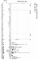


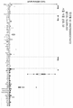
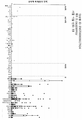

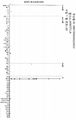

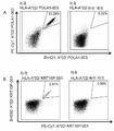


Classifications
-
- A—HUMAN NECESSITIES
- A61—MEDICAL OR VETERINARY SCIENCE; HYGIENE
- A61K—PREPARATIONS FOR MEDICAL, DENTAL OR TOILETRY PURPOSES
- A61K35/00—Medicinal preparations containing materials or reaction products thereof with undetermined constitution
- A61K35/12—Materials from mammals; Compositions comprising non-specified tissues or cells; Compositions comprising non-embryonic stem cells; Genetically modified cells
- A61K35/14—Blood; Artificial blood
- A61K35/17—Lymphocytes; B-cells; T-cells; Natural killer cells; Interferon-activated or cytokine-activated lymphocytes
-
- C—CHEMISTRY; METALLURGY
- C07—ORGANIC CHEMISTRY
- C07D—HETEROCYCLIC COMPOUNDS
- C07D513/00—Heterocyclic compounds containing in the condensed system at least one hetero ring having nitrogen and sulfur atoms as the only ring hetero atoms, not provided for in groups C07D463/00, C07D477/00 or C07D499/00 - C07D507/00
- C07D513/02—Heterocyclic compounds containing in the condensed system at least one hetero ring having nitrogen and sulfur atoms as the only ring hetero atoms, not provided for in groups C07D463/00, C07D477/00 or C07D499/00 - C07D507/00 in which the condensed system contains two hetero rings
- C07D513/04—Ortho-condensed systems
-
- A—HUMAN NECESSITIES
- A61—MEDICAL OR VETERINARY SCIENCE; HYGIENE
- A61K—PREPARATIONS FOR MEDICAL, DENTAL OR TOILETRY PURPOSES
- A61K31/00—Medicinal preparations containing organic active ingredients
- A61K31/33—Heterocyclic compounds
- A61K31/395—Heterocyclic compounds having nitrogen as a ring hetero atom, e.g. guanethidine or rifamycins
- A61K31/40—Heterocyclic compounds having nitrogen as a ring hetero atom, e.g. guanethidine or rifamycins having five-membered rings with one nitrogen as the only ring hetero atom, e.g. sulpiride, succinimide, tolmetin, buflomedil
- A61K31/407—Heterocyclic compounds having nitrogen as a ring hetero atom, e.g. guanethidine or rifamycins having five-membered rings with one nitrogen as the only ring hetero atom, e.g. sulpiride, succinimide, tolmetin, buflomedil condensed with other heterocyclic ring systems, e.g. ketorolac, physostigmine
-
- A—HUMAN NECESSITIES
- A61—MEDICAL OR VETERINARY SCIENCE; HYGIENE
- A61K—PREPARATIONS FOR MEDICAL, DENTAL OR TOILETRY PURPOSES
- A61K38/00—Medicinal preparations containing peptides
- A61K38/04—Peptides having up to 20 amino acids in a fully defined sequence; Derivatives thereof
- A61K38/08—Peptides having 5 to 11 amino acids
-
- A—HUMAN NECESSITIES
- A61—MEDICAL OR VETERINARY SCIENCE; HYGIENE
- A61K—PREPARATIONS FOR MEDICAL, DENTAL OR TOILETRY PURPOSES
- A61K39/00—Medicinal preparations containing antigens or antibodies
- A61K39/0005—Vertebrate antigens
- A61K39/0011—Cancer antigens
- A61K39/001102—Receptors, cell surface antigens or cell surface determinants
- A61K39/001111—Immunoglobulin superfamily
- A61K39/001114—CD74, Ii, MHC class II invariant chain or MHC class II gamma chain
-
- A—HUMAN NECESSITIES
- A61—MEDICAL OR VETERINARY SCIENCE; HYGIENE
- A61K—PREPARATIONS FOR MEDICAL, DENTAL OR TOILETRY PURPOSES
- A61K39/00—Medicinal preparations containing antigens or antibodies
- A61K39/0005—Vertebrate antigens
- A61K39/0011—Cancer antigens
- A61K39/001102—Receptors, cell surface antigens or cell surface determinants
- A61K39/001116—Receptors for cytokines
- A61K39/001118—Receptors for colony stimulating factors [CSF]
-
- A—HUMAN NECESSITIES
- A61—MEDICAL OR VETERINARY SCIENCE; HYGIENE
- A61K—PREPARATIONS FOR MEDICAL, DENTAL OR TOILETRY PURPOSES
- A61K39/00—Medicinal preparations containing antigens or antibodies
- A61K39/12—Viral antigens
-
- A—HUMAN NECESSITIES
- A61—MEDICAL OR VETERINARY SCIENCE; HYGIENE
- A61K—PREPARATIONS FOR MEDICAL, DENTAL OR TOILETRY PURPOSES
- A61K39/00—Medicinal preparations containing antigens or antibodies
- A61K39/39—Medicinal preparations containing antigens or antibodies characterised by the immunostimulating additives, e.g. chemical adjuvants
-
- A—HUMAN NECESSITIES
- A61—MEDICAL OR VETERINARY SCIENCE; HYGIENE
- A61K—PREPARATIONS FOR MEDICAL, DENTAL OR TOILETRY PURPOSES
- A61K39/00—Medicinal preparations containing antigens or antibodies
- A61K39/395—Antibodies; Immunoglobulins; Immune serum, e.g. antilymphocytic serum
- A61K39/39533—Antibodies; Immunoglobulins; Immune serum, e.g. antilymphocytic serum against materials from animals
- A61K39/39558—Antibodies; Immunoglobulins; Immune serum, e.g. antilymphocytic serum against materials from animals against tumor tissues, cells, antigens
-
- A—HUMAN NECESSITIES
- A61—MEDICAL OR VETERINARY SCIENCE; HYGIENE
- A61K—PREPARATIONS FOR MEDICAL, DENTAL OR TOILETRY PURPOSES
- A61K39/00—Medicinal preparations containing antigens or antibodies
- A61K39/46—Cellular immunotherapy
- A61K39/461—Cellular immunotherapy characterised by the cell type used
- A61K39/4611—T-cells, e.g. tumor infiltrating lymphocytes [TIL], lymphokine-activated killer cells [LAK] or regulatory T cells [Treg]
-
- A—HUMAN NECESSITIES
- A61—MEDICAL OR VETERINARY SCIENCE; HYGIENE
- A61K—PREPARATIONS FOR MEDICAL, DENTAL OR TOILETRY PURPOSES
- A61K39/00—Medicinal preparations containing antigens or antibodies
- A61K39/46—Cellular immunotherapy
- A61K39/464—Cellular immunotherapy characterised by the antigen targeted or presented
- A61K39/4643—Vertebrate antigens
- A61K39/4644—Cancer antigens
-
- A—HUMAN NECESSITIES
- A61—MEDICAL OR VETERINARY SCIENCE; HYGIENE
- A61K—PREPARATIONS FOR MEDICAL, DENTAL OR TOILETRY PURPOSES
- A61K45/00—Medicinal preparations containing active ingredients not provided for in groups A61K31/00 - A61K41/00
- A61K45/05—Immunological preparations stimulating the reticulo-endothelial system, e.g. against cancer
-
- A—HUMAN NECESSITIES
- A61—MEDICAL OR VETERINARY SCIENCE; HYGIENE
- A61K—PREPARATIONS FOR MEDICAL, DENTAL OR TOILETRY PURPOSES
- A61K45/00—Medicinal preparations containing active ingredients not provided for in groups A61K31/00 - A61K41/00
- A61K45/06—Mixtures of active ingredients without chemical characterisation, e.g. antiphlogistics and cardiaca
-
- A—HUMAN NECESSITIES
- A61—MEDICAL OR VETERINARY SCIENCE; HYGIENE
- A61K—PREPARATIONS FOR MEDICAL, DENTAL OR TOILETRY PURPOSES
- A61K48/00—Medicinal preparations containing genetic material which is inserted into cells of the living body to treat genetic diseases; Gene therapy
-
- A—HUMAN NECESSITIES
- A61—MEDICAL OR VETERINARY SCIENCE; HYGIENE
- A61P—SPECIFIC THERAPEUTIC ACTIVITY OF CHEMICAL COMPOUNDS OR MEDICINAL PREPARATIONS
- A61P31/00—Antiinfectives, i.e. antibiotics, antiseptics, chemotherapeutics
- A61P31/12—Antivirals
- A61P31/20—Antivirals for DNA viruses
-
- A—HUMAN NECESSITIES
- A61—MEDICAL OR VETERINARY SCIENCE; HYGIENE
- A61P—SPECIFIC THERAPEUTIC ACTIVITY OF CHEMICAL COMPOUNDS OR MEDICINAL PREPARATIONS
- A61P35/00—Antineoplastic agents
-
- A—HUMAN NECESSITIES
- A61—MEDICAL OR VETERINARY SCIENCE; HYGIENE
- A61P—SPECIFIC THERAPEUTIC ACTIVITY OF CHEMICAL COMPOUNDS OR MEDICINAL PREPARATIONS
- A61P35/00—Antineoplastic agents
- A61P35/02—Antineoplastic agents specific for leukemia
-
- A—HUMAN NECESSITIES
- A61—MEDICAL OR VETERINARY SCIENCE; HYGIENE
- A61P—SPECIFIC THERAPEUTIC ACTIVITY OF CHEMICAL COMPOUNDS OR MEDICINAL PREPARATIONS
- A61P37/00—Drugs for immunological or allergic disorders
-
- C—CHEMISTRY; METALLURGY
- C07—ORGANIC CHEMISTRY
- C07D—HETEROCYCLIC COMPOUNDS
- C07D291/00—Heterocyclic compounds containing rings having nitrogen, oxygen and sulfur atoms as the only ring hetero atoms
- C07D291/08—Heterocyclic compounds containing rings having nitrogen, oxygen and sulfur atoms as the only ring hetero atoms condensed with carbocyclic rings or ring systems
-
- C—CHEMISTRY; METALLURGY
- C07—ORGANIC CHEMISTRY
- C07D—HETEROCYCLIC COMPOUNDS
- C07D513/00—Heterocyclic compounds containing in the condensed system at least one hetero ring having nitrogen and sulfur atoms as the only ring hetero atoms, not provided for in groups C07D463/00, C07D477/00 or C07D499/00 - C07D507/00
- C07D513/12—Heterocyclic compounds containing in the condensed system at least one hetero ring having nitrogen and sulfur atoms as the only ring hetero atoms, not provided for in groups C07D463/00, C07D477/00 or C07D499/00 - C07D507/00 in which the condensed system contains three hetero rings
- C07D513/20—Spiro-condensed systems
-
- C—CHEMISTRY; METALLURGY
- C07—ORGANIC CHEMISTRY
- C07D—HETEROCYCLIC COMPOUNDS
- C07D515/00—Heterocyclic compounds containing in the condensed system at least one hetero ring having nitrogen, oxygen, and sulfur atoms as the only ring hetero atoms, not provided for in groups C07D463/00, C07D477/00 or C07D499/00 - C07D507/00
- C07D515/02—Heterocyclic compounds containing in the condensed system at least one hetero ring having nitrogen, oxygen, and sulfur atoms as the only ring hetero atoms, not provided for in groups C07D463/00, C07D477/00 or C07D499/00 - C07D507/00 in which the condensed system contains two hetero rings
- C07D515/04—Ortho-condensed systems
-
- C—CHEMISTRY; METALLURGY
- C07—ORGANIC CHEMISTRY
- C07D—HETEROCYCLIC COMPOUNDS
- C07D515/00—Heterocyclic compounds containing in the condensed system at least one hetero ring having nitrogen, oxygen, and sulfur atoms as the only ring hetero atoms, not provided for in groups C07D463/00, C07D477/00 or C07D499/00 - C07D507/00
- C07D515/12—Heterocyclic compounds containing in the condensed system at least one hetero ring having nitrogen, oxygen, and sulfur atoms as the only ring hetero atoms, not provided for in groups C07D463/00, C07D477/00 or C07D499/00 - C07D507/00 in which the condensed system contains three hetero rings
- C07D515/20—Spiro-condensed systems
-
- C—CHEMISTRY; METALLURGY
- C07—ORGANIC CHEMISTRY
- C07K—PEPTIDES
- C07K14/00—Peptides having more than 20 amino acids; Gastrins; Somatostatins; Melanotropins; Derivatives thereof
- C07K14/435—Peptides having more than 20 amino acids; Gastrins; Somatostatins; Melanotropins; Derivatives thereof from animals; from humans
- C07K14/46—Peptides having more than 20 amino acids; Gastrins; Somatostatins; Melanotropins; Derivatives thereof from animals; from humans from vertebrates
- C07K14/47—Peptides having more than 20 amino acids; Gastrins; Somatostatins; Melanotropins; Derivatives thereof from animals; from humans from vertebrates from mammals
- C07K14/4701—Peptides having more than 20 amino acids; Gastrins; Somatostatins; Melanotropins; Derivatives thereof from animals; from humans from vertebrates from mammals not used
- C07K14/4748—Tumour specific antigens; Tumour rejection antigen precursors [TRAP], e.g. MAGE
-
- C—CHEMISTRY; METALLURGY
- C07—ORGANIC CHEMISTRY
- C07K—PEPTIDES
- C07K14/00—Peptides having more than 20 amino acids; Gastrins; Somatostatins; Melanotropins; Derivatives thereof
- C07K14/435—Peptides having more than 20 amino acids; Gastrins; Somatostatins; Melanotropins; Derivatives thereof from animals; from humans
- C07K14/52—Cytokines; Lymphokines; Interferons
- C07K14/54—Interleukins [IL]
- C07K14/5434—IL-12
-
- C—CHEMISTRY; METALLURGY
- C07—ORGANIC CHEMISTRY
- C07K—PEPTIDES
- C07K16/00—Immunoglobulins [IGs], e.g. monoclonal or polyclonal antibodies
- C07K16/18—Immunoglobulins [IGs], e.g. monoclonal or polyclonal antibodies against material from animals or humans
- C07K16/24—Immunoglobulins [IGs], e.g. monoclonal or polyclonal antibodies against material from animals or humans against cytokines, lymphokines or interferons
- C07K16/244—Interleukins [IL]
-
- C—CHEMISTRY; METALLURGY
- C07—ORGANIC CHEMISTRY
- C07K—PEPTIDES
- C07K16/00—Immunoglobulins [IGs], e.g. monoclonal or polyclonal antibodies
- C07K16/18—Immunoglobulins [IGs], e.g. monoclonal or polyclonal antibodies against material from animals or humans
- C07K16/28—Immunoglobulins [IGs], e.g. monoclonal or polyclonal antibodies against material from animals or humans against receptors, cell surface antigens or cell surface determinants
- C07K16/2803—Immunoglobulins [IGs], e.g. monoclonal or polyclonal antibodies against material from animals or humans against receptors, cell surface antigens or cell surface determinants against the immunoglobulin superfamily
- C07K16/2818—Immunoglobulins [IGs], e.g. monoclonal or polyclonal antibodies against material from animals or humans against receptors, cell surface antigens or cell surface determinants against the immunoglobulin superfamily against CD28 or CD152
-
- C—CHEMISTRY; METALLURGY
- C07—ORGANIC CHEMISTRY
- C07K—PEPTIDES
- C07K16/00—Immunoglobulins [IGs], e.g. monoclonal or polyclonal antibodies
- C07K16/18—Immunoglobulins [IGs], e.g. monoclonal or polyclonal antibodies against material from animals or humans
- C07K16/28—Immunoglobulins [IGs], e.g. monoclonal or polyclonal antibodies against material from animals or humans against receptors, cell surface antigens or cell surface determinants
- C07K16/2803—Immunoglobulins [IGs], e.g. monoclonal or polyclonal antibodies against material from animals or humans against receptors, cell surface antigens or cell surface determinants against the immunoglobulin superfamily
- C07K16/2821—Immunoglobulins [IGs], e.g. monoclonal or polyclonal antibodies against material from animals or humans against receptors, cell surface antigens or cell surface determinants against the immunoglobulin superfamily against ICAM molecules, e.g. CD50, CD54, CD102
-
- C—CHEMISTRY; METALLURGY
- C07—ORGANIC CHEMISTRY
- C07K—PEPTIDES
- C07K7/00—Peptides having 5 to 20 amino acids in a fully defined sequence; Derivatives thereof
- C07K7/04—Linear peptides containing only normal peptide links
- C07K7/06—Linear peptides containing only normal peptide links having 5 to 11 amino acids
-
- C—CHEMISTRY; METALLURGY
- C12—BIOCHEMISTRY; BEER; SPIRITS; WINE; VINEGAR; MICROBIOLOGY; ENZYMOLOGY; MUTATION OR GENETIC ENGINEERING
- C12N—MICROORGANISMS OR ENZYMES; COMPOSITIONS THEREOF; PROPAGATING, PRESERVING, OR MAINTAINING MICROORGANISMS; MUTATION OR GENETIC ENGINEERING; CULTURE MEDIA
- C12N5/00—Undifferentiated human, animal or plant cells, e.g. cell lines; Tissues; Cultivation or maintenance thereof; Culture media therefor
- C12N5/06—Animal cells or tissues; Human cells or tissues
- C12N5/0602—Vertebrate cells
- C12N5/0634—Cells from the blood or the immune system
- C12N5/0636—T lymphocytes
-
- A—HUMAN NECESSITIES
- A61—MEDICAL OR VETERINARY SCIENCE; HYGIENE
- A61K—PREPARATIONS FOR MEDICAL, DENTAL OR TOILETRY PURPOSES
- A61K39/00—Medicinal preparations containing antigens or antibodies
- A61K2039/51—Medicinal preparations containing antigens or antibodies comprising whole cells, viruses or DNA/RNA
- A61K2039/515—Animal cells
- A61K2039/5154—Antigen presenting cells [APCs], e.g. dendritic cells or macrophages
-
- A—HUMAN NECESSITIES
- A61—MEDICAL OR VETERINARY SCIENCE; HYGIENE
- A61K—PREPARATIONS FOR MEDICAL, DENTAL OR TOILETRY PURPOSES
- A61K39/00—Medicinal preparations containing antigens or antibodies
- A61K2039/51—Medicinal preparations containing antigens or antibodies comprising whole cells, viruses or DNA/RNA
- A61K2039/515—Animal cells
- A61K2039/5156—Animal cells expressing foreign proteins
-
- A—HUMAN NECESSITIES
- A61—MEDICAL OR VETERINARY SCIENCE; HYGIENE
- A61K—PREPARATIONS FOR MEDICAL, DENTAL OR TOILETRY PURPOSES
- A61K39/00—Medicinal preparations containing antigens or antibodies
- A61K2039/51—Medicinal preparations containing antigens or antibodies comprising whole cells, viruses or DNA/RNA
- A61K2039/515—Animal cells
- A61K2039/5158—Antigen-pulsed cells, e.g. T-cells
-
- A—HUMAN NECESSITIES
- A61—MEDICAL OR VETERINARY SCIENCE; HYGIENE
- A61K—PREPARATIONS FOR MEDICAL, DENTAL OR TOILETRY PURPOSES
- A61K39/00—Medicinal preparations containing antigens or antibodies
- A61K2039/555—Medicinal preparations containing antigens or antibodies characterised by a specific combination antigen/adjuvant
- A61K2039/55511—Organic adjuvants
- A61K2039/55516—Proteins; Peptides
-
- A—HUMAN NECESSITIES
- A61—MEDICAL OR VETERINARY SCIENCE; HYGIENE
- A61K—PREPARATIONS FOR MEDICAL, DENTAL OR TOILETRY PURPOSES
- A61K39/00—Medicinal preparations containing antigens or antibodies
- A61K2039/555—Medicinal preparations containing antigens or antibodies characterised by a specific combination antigen/adjuvant
- A61K2039/55511—Organic adjuvants
- A61K2039/55522—Cytokines; Lymphokines; Interferons
- A61K2039/55527—Interleukins
- A61K2039/55533—IL-2
-
- A—HUMAN NECESSITIES
- A61—MEDICAL OR VETERINARY SCIENCE; HYGIENE
- A61K—PREPARATIONS FOR MEDICAL, DENTAL OR TOILETRY PURPOSES
- A61K39/00—Medicinal preparations containing antigens or antibodies
- A61K2039/555—Medicinal preparations containing antigens or antibodies characterised by a specific combination antigen/adjuvant
- A61K2039/55511—Organic adjuvants
- A61K2039/55522—Cytokines; Lymphokines; Interferons
- A61K2039/55527—Interleukins
- A61K2039/55538—IL-12
-
- A—HUMAN NECESSITIES
- A61—MEDICAL OR VETERINARY SCIENCE; HYGIENE
- A61K—PREPARATIONS FOR MEDICAL, DENTAL OR TOILETRY PURPOSES
- A61K39/00—Medicinal preparations containing antigens or antibodies
- A61K2039/555—Medicinal preparations containing antigens or antibodies characterised by a specific combination antigen/adjuvant
- A61K2039/55511—Organic adjuvants
- A61K2039/55561—CpG containing adjuvants; Oligonucleotide containing adjuvants
-
- A—HUMAN NECESSITIES
- A61—MEDICAL OR VETERINARY SCIENCE; HYGIENE
- A61K—PREPARATIONS FOR MEDICAL, DENTAL OR TOILETRY PURPOSES
- A61K39/00—Medicinal preparations containing antigens or antibodies
- A61K2039/57—Medicinal preparations containing antigens or antibodies characterised by the type of response, e.g. Th1, Th2
- A61K2039/572—Medicinal preparations containing antigens or antibodies characterised by the type of response, e.g. Th1, Th2 cytotoxic response
-
- A—HUMAN NECESSITIES
- A61—MEDICAL OR VETERINARY SCIENCE; HYGIENE
- A61K—PREPARATIONS FOR MEDICAL, DENTAL OR TOILETRY PURPOSES
- A61K39/00—Medicinal preparations containing antigens or antibodies
- A61K2039/58—Medicinal preparations containing antigens or antibodies raising an immune response against a target which is not the antigen used for immunisation
- A61K2039/585—Medicinal preparations containing antigens or antibodies raising an immune response against a target which is not the antigen used for immunisation wherein the target is cancer
-
- C—CHEMISTRY; METALLURGY
- C12—BIOCHEMISTRY; BEER; SPIRITS; WINE; VINEGAR; MICROBIOLOGY; ENZYMOLOGY; MUTATION OR GENETIC ENGINEERING
- C12N—MICROORGANISMS OR ENZYMES; COMPOSITIONS THEREOF; PROPAGATING, PRESERVING, OR MAINTAINING MICROORGANISMS; MUTATION OR GENETIC ENGINEERING; CULTURE MEDIA
- C12N2510/00—Genetically modified cells
Landscapes
- Health & Medical Sciences (AREA)
- Life Sciences & Earth Sciences (AREA)
- Chemical & Material Sciences (AREA)
- Immunology (AREA)
- General Health & Medical Sciences (AREA)
- Medicinal Chemistry (AREA)
- Organic Chemistry (AREA)
- Pharmacology & Pharmacy (AREA)
- Veterinary Medicine (AREA)
- Public Health (AREA)
- Animal Behavior & Ethology (AREA)
- Epidemiology (AREA)
- Engineering & Computer Science (AREA)
- Cell Biology (AREA)
- Microbiology (AREA)
- Mycology (AREA)
- Genetics & Genomics (AREA)
- Oncology (AREA)
- Biochemistry (AREA)
- Biomedical Technology (AREA)
- Proteomics, Peptides & Aminoacids (AREA)
- Zoology (AREA)
- Molecular Biology (AREA)
- Bioinformatics & Cheminformatics (AREA)
- Biophysics (AREA)
- Biotechnology (AREA)
- Hematology (AREA)
- Chemical Kinetics & Catalysis (AREA)
- General Chemical & Material Sciences (AREA)
- Gastroenterology & Hepatology (AREA)
- Wood Science & Technology (AREA)
- Nuclear Medicine, Radiotherapy & Molecular Imaging (AREA)
- Virology (AREA)
- Toxicology (AREA)
- Developmental Biology & Embryology (AREA)
- General Engineering & Computer Science (AREA)
- Communicable Diseases (AREA)
- Peptides Or Proteins (AREA)
- Medicines That Contain Protein Lipid Enzymes And Other Medicines (AREA)
- Medicines Containing Antibodies Or Antigens For Use As Internal Diagnostic Agents (AREA)
Abstract
본 발명은 면역요법 방법에 사용하기 위한 펩티드, 핵산 및 세포에 관한 것이다. 특히, 본 발명은 암의 면역요법에 관한 것이다. 본 발명은 또한 단독 또는, 예를 들어 백신 조성물의 활성 약학 성분으로 역할가능한 기타 종양-연관 펩티드와 조합으로 항종양 면역 반응을 자극하거나 T 세포를 생체외 자극하여 환자로 이전하기 위한 종양-연관 T 세포 에피톱에 관한 것이다. 주조직적합 복합체(MHC)의 분자에 결합된 펩티드 또는 펩티드 자체는 항체, 가용성 T 세포 수용체 및 기타 결합하는 분자의 표적이 될 수도 있다.The present invention relates to peptides, nucleic acids and cells for use in immunotherapy methods. In particular, the present invention relates to immunotherapy for cancer. The present invention is also a tumor-associated T for transfer to a patient by stimulating an anti-tumor immune response or stimulating T cells ex vivo, either alone or in combination with, for example, other tumor-associated peptides capable of serving as an active pharmaceutical component of a vaccine composition. It relates to cell epitopes. The peptide bound to the molecule of the major histocompatibility complex (MHC) or the peptide itself may be a target of antibodies, soluble T cell receptors, and other binding molecules.
Description
본 발명은 면역요법 방법에서 사용을 위한 펩티드, 단백질, 핵산 및 세포에 관한 것이다. 특히 본 발명은 암의 면역요법에 관한 것이다. 본 발명은 또한 단독 또는 예를 들어 백신 조성물의 활성 약학적 성분으로 역할가능한 기타 종양 연관 펩티드들과 병용하여 항종양 면역 반응을 촉진하거나 세포를 생체외 촉진하여 환자로 이전하기 위한 종양 연관 T 세포 에피톱에 관한 것이다. 주조직적합 복합체(MHC)의 분자에 결합된 펩티드 또는 그러한 펩티드는 항체, 가용성 T 세포 수용체 및 기타 결합하는 분자의 표적이 될 수도 있다.The present invention relates to peptides, proteins, nucleic acids and cells for use in immunotherapy methods. In particular, the present invention relates to cancer immunotherapy. The present invention is also a tumor-associated T cell epitaxial for stimulating an anti-tumor immune response or promoting cells ex vivo to transfer to a patient, either alone or in combination with other tumor-associated peptides, e.g., capable of serving as active pharmaceutical ingredients of a vaccine composition. It's about the saw. Peptides bound to molecules of the major histocompatibility complex (MHC) or such peptides can also be targets of antibodies, soluble T cell receptors, and other binding molecules.
본 발명은 항종양 면역 반응을 이끌어내는 백신 조성물에서 또는 약학적/면역학적 활성 화합물 및 세포의 개발을 위한 표적으로 사용할 수 있는 인간 종양 세포의 HLA 클래스 I 분자로부터 유래하는 여러 신규 펩티드 서열 및 이들의 변이체에 관한 것이다.The present invention provides several novel peptide sequences derived from HLA class I molecules of human tumor cells that can be used in vaccine compositions eliciting anti-tumor immune responses or as targets for the development of pharmaceutical/immunologically active compounds and cells, and their It's about variants.
세계보건기구(WHO)에 따르면, 2012년 암은 세계적으로 4가지 주요 비전염성 치명적 질병에 속했다. 같은 해 동안, 고소득 국가들에서 대장암, 유방암 및 호흡기도 암들이 10대 사망 원인에 포함되었다.According to the World Health Organization (WHO), cancer in 2012 was among the four major non-communicable fatal diseases worldwide. During the same year, colon cancer, breast and respiratory tract cancers were among the top 10 causes of death in high-income countries.
GBM은 가장 흔한 중추신경계 악성 종양으로서 미국내 연령 조절 발생률은 거주자 100,000명 중 3.19명이다. GBM은 예후가 매우 불량하며 1년 생존율이 35%이고 5년 생존율은 5% 미만이다. 남성, 높은 연령 및 민족이 GBM의 위험 인자인 것으로 보인다(Thakkar et al., 2014).GBM is the most common central nervous system malignancies, and the age-adjusted incidence rate in the United States is 3.19 out of 100,000 residents. GBM has a very poor prognosis, a 1-year survival rate of 35% and a 5-year survival rate of less than 5%. Male, high age and ethnicity appear to be risk factors for GBM (Thakkar et al., 2014).
CLL은 서구 세계에서 가장 흔한 백혈병이며 모든 백혈병의 약 3분의 1을 차지한다. 발생률은 미국과 유럽에서 유사하며 연간 신규 건수는 16,000건으로 추정된다. CLL은 아프리카인보다 백인에서 더 흔하며 히스패닉과 미국 원주민에서는 더 드물고 아시아인에서는 거의 없다. 아시아계의 경우 CLL 발생률은 백인보다 3배 더 낮다(Gunawardana et al., 2008). CLL 환자의 5년 전체 생존율은 약 79%이다.CLL is the most common leukemia in the western world and accounts for about a third of all leukemias. The incidence rates are similar in the US and Europe, and the number of new cases per year is estimated at 16,000. CLL is more common in whites than in Africans, more rare in Hispanics and Native Americans, and few in Asians. The incidence of CLL in Asians is three times lower than in Caucasians (Gunawardana et al., 2008). The overall 5-year survival rate for CLL patients is approximately 79%.
AML은 성인과 아동 모두에서 진단되는 두 번째로 흔한 유형의 백혈병이다. 미국에서 추정된 신규 사례는 1년에 약 21,000건이다. AML 환자의 5년 생존율은 약 25%이다.AML is the second most common type of leukemia diagnosed in both adults and children. The estimated new cases in the United States are about 21,000 per year. The 5-year survival rate for AML patients is about 25%.
폐암은 세계적으로 가장 흔한 유형의 암이며 다수 국가에서 첫 번째 사망 원인이다. 폐암은 소세포 폐암과 비소세포 폐암으로 세분화된다. NSCLC에는 선암종, 편평 세포 암종 및 대세포 암종의 조직학적 유형들이 포함되며 미국의 모든 폐암의 85%를 차지한다. NSCLC의 발생률은 현재 및 과거 흡연자 등의 흡연율과 밀접한 상관관계가 있으며 5년 생존율은 15%로 보고되었다(World Cancer Report, 2014; Molina et al., 2008).Lung cancer is the most common type of cancer worldwide and is the number one cause of death in many countries. Lung cancer is subdivided into small cell lung cancer and non-small cell lung cancer. NSCLC includes histological types of adenocarcinoma, squamous cell carcinoma, and large cell carcinoma and accounts for 85% of all lung cancers in the United States. The incidence of NSCLC is closely correlated with the smoking rate of current and past smokers, and the 5-year survival rate was reported as 15% (World Cancer Report, 2014; Molina et al., 2008).
암 치료와 연관된 심한 부작용과 비용을 고려할 때, 일반적인 암, 특히 급성 골수성 백혈병(AML), 유방암(BRCA), 담관세포 암종(CCC), 만성 림프구성 백혈병(CLL), 대장암(CRC), 담낭암(GBC), 교모세포종(GBM), 위암(GC), 위-식도 접합부 암(GEJC), 간세포 암종(HCC), 두경부 편평세포 암종(HNSCC), 흑색종(MEL), 비호지킨 림프종(NHL), 비소세포 폐암(NSCLC), 난소암(OC), 식도암(OSCAR), 췌장암(PACA), 전립선암(PRCA), 신세포 암종(RCC), 소세포 폐암(SCLC), 방광 암종(UBC) 및 자궁내막암(UEC)의 치료에 사용가능한 요인들을 확인할 필요가 있다. 또한 더 나은 진단과 예후의 평가 및 치료 성공의 예측을 유도하는 일반적인 암, 특히 급성 골수성 백혈병, 유방암, 담관세포 암종, 만성 림프구성 백혈병, 대장암, 담낭암, 교모세포종, 위암, 위-식도 접합부 암, 간세포 암종, 두경부 편평세포 암종, 흑색종, 비호지킨 림프종, 비소세포 폐암, 난소암, 식도암, 췌장암, 전립선암, 신세포 암종, 소세포 폐암, 방광 암종 및 자궁내막암의 바이오마커를 대표하는 요인들을 확인할 필요가 있다. Considering the severe side effects and costs associated with cancer treatment, common cancers, especially acute myeloid leukemia (AML), breast cancer (BRCA), cholangiocarcinoma (CCC), chronic lymphocytic leukemia (CLL), colon cancer (CRC), and gallbladder cancer. (GBC), glioblastoma (GBM), gastric cancer (GC), gastric-esophageal junction cancer (GEJC), hepatocellular carcinoma (HCC), squamous cell carcinoma of the head and neck (HNSCC), melanoma (MEL), non-Hodgkin lymphoma (NHL) , Non-small cell lung cancer (NSCLC), ovarian cancer (OC), esophageal cancer (OSCAR), pancreatic cancer (PACA), prostate cancer (PRCA), renal cell carcinoma (RCC), small cell lung cancer (SCLC), bladder carcinoma (UBC) and uterus There is a need to identify factors that can be used in the treatment of endometrial cancer (UEC). Also common cancers leading to better diagnosis and assessment of prognosis and prediction of treatment success, especially acute myelogenous leukemia, breast cancer, cholangiocarcinoma, chronic lymphocytic leukemia, colon cancer, gallbladder cancer, glioblastoma, gastric cancer, gastroesophageal junction cancer. , Hepatocellular carcinoma, squamous cell carcinoma of the head and neck, melanoma, non-Hodgkin's lymphoma, non-small cell lung cancer, ovarian cancer, esophageal cancer, pancreatic cancer, prostate cancer, renal cell carcinoma, small cell lung cancer, bladder carcinoma, and endometrial cancer. You need to check them out.
암의 면역요법은 부작용을 최소화시키는 반면 암 세포에 대한 특이적 표적화의 옵션을 나타낸다. 암 면역요법은 종양 관련 항원의 존재를 이용한다. Immunotherapy of cancer presents an option of specific targeting to cancer cells while minimizing side effects. Cancer immunotherapy uses the presence of tumor-associated antigens.
종양 관련 항원(TAA)의 현재 분류는 다음과 같은 주요 그룹을 포함한다:The current classification of tumor-associated antigens (TAA) includes the following main groups:
a) 암-고환 항원: 제일 처음으로 T 세포에 의해 인식된 TAA는 이 종류에 속하며, 이는 그 멤버들의 발현이 조직학적으로 인간 종양과 다르며, 정상 조직 중, 고환의 정모 세포/정원 세포에서만 있으며, 때로는 태반에서도 있기 때문에 암-고환(CT)이라고 최초에 불려졌다. 고환의 세포들이 클래스 I 또는 II HLA 분자를 발현하지 않기 때문에, 이 항원들은 정상 세포에 있는 T 세포에 의해 인식될 수 없으며 면역학적으로 종양-특이적이라고 간주될 수 있다. CT 항원의 잘 알려진 예로는 MAGE 계열 구성원들 및 NY_ESO_1이 있다. a) Cancer-testis antigen: The first TAA recognized by T cells belongs to this class, and its members' expression is histologically different from that of human tumors, and only in normal tissues, sperm cells/garden cells of testis. , It was initially called cancer-testis (CT) because it is sometimes also present in the placenta. Since the cells of the testis do not express class I or II HLA molecules, these antigens cannot be recognized by T cells in normal cells and can be considered immunologically tumor-specific. Well-known examples of CT antigens are members of the MAGE family and NY_ESO_1.
b) 다른 항원들: 이 TAA는 종양과 그 종양이 발생한 정상 조직 사이에 공유된다. 대부분의 알려진 분화 항원들은 흑색종과 정상 멜라닌 세포에서 발견된다. 이러한 멜라닌 세포 혈통 관련 단백질의 다수는 멜라닌의 생합성에 관련되어 있으며, 그러므로 종양 특이적이 아니지만 암 면역요법에 널리 사용된다. 예로는, 티로시나아제 및 흑색종을 위한 멜란-A/MART-1 또는 전립선암을 위한 PSA가 포함되지만 이로 국한되지 않는다. b) Other antigens: This TAA is shared between the tumor and the normal tissue in which it originates. Most of the known differentiation antigens are found in melanoma and normal melanocytes. Many of these melanocyte lineage related proteins are involved in the biosynthesis of melanin, and therefore are not tumor specific but are widely used in cancer immunotherapy. Examples include, but are not limited to, tyrosinase and melan-A/MART-1 for melanoma or PSA for prostate cancer.
c) 과발현된 TAA: 일반적으로 낮은 발현 수준이 있는 많은 정상 조직뿐만 아니라 조직학적으로 다른 유형의 종양에서 유전자 인코딩에서 널리 발현된 TAA가 발견된다. 정상 조직에 의해 처리되고 잠재적으로 제시되는 많은 에피톱이 T 세포 인식의 임계치 수준보다 낮을 수 있고, T 세포의 종양 세포에서의 과발현은 이전에 확립된 내성을 중단시켜 항암 반응을 개시할 수 있다. 이 클래스 TAA의 두드러진 예로는 Her-2/neu, 서바이빈, 텔로머라제 또는 WT1가 있다.c) Overexpressed TAA: TAA that is widely expressed in gene encoding is found in many normal tissues, usually with low expression levels, as well as histologically different types of tumors. Many epitopes processed and potentially presented by normal tissue can be below the threshold level of T cell recognition, and overexpression of T cells in tumor cells can stop previously established resistance and initiate an anticancer response. Prominent examples of this class TAA are Her-2/neu, survivin, telomerase or WT1.
d) 종양 특이적 항원: 이러한 독특한 TAA는 정상 유전자의 변이로써 발생한다(β-카테닌, CDK4 등). 이러한 분자 변경의 일부는 종양 변이 및/또는 진행과 관련되어 있다. 종양 특이적 항원은 일반적으로 강한 면역 반응을 정상 조직에 대한 자기 면역 반응에 대한 위험성 없이 유도할 수 있다. 반면에, 대부분의 경우 TAA는 발견되어 일반적으로 많은 개별 종양에서 대개 공유되지 않은 해당 종양에만 적절하다. 종양 특이적(연관된) 아형을 갖는 단백질의 경우 펩티드가 종양(관련) 엑손으로부터 비롯된다면 펩티드의 종양 특이성(또는 연관성) 또한 발생할 수 있다. d) Tumor specific antigens: These unique TAAs arise as mutations in normal genes (β-catenin, CDK4, etc.). Some of these molecular alterations are associated with tumor mutation and/or progression. Tumor-specific antigens are generally capable of inducing a strong immune response without risk of an autoimmune response against normal tissue. On the other hand, in most cases TAA is found and is generally only appropriate for those tumors that are not usually shared among many individual tumors. For proteins with tumor specific (associated) subtypes, tumor specificity (or association) of the peptide may also arise if the peptide originates from a tumor (associated) exon.
e) 비정상 번역 후 변형에서 발생하는 TAA: 이러한 TAA는 특이적이 아니고 과발현되지도 않는 단백질에서 발생할 수 있지만 종양에서 주로 활동적인 번역 후 과정에 의해 종양과 관련된다. 이 클래스의 예는 종양 특이적일 수도 있고 아닐 수도 있는 분해 과정 동안 단백질 스플라이싱과 같은 이벤트 또는 MUC1의 경우 종양의 새로운 에피톱으로 이끄는 변화된 당화 패턴에서 발생한다. e) TAA arising from abnormal post-translational modifications: These TAAs may arise from proteins that are neither specific nor overexpressed, but are associated with tumors primarily by active post-translational processes in tumors. Examples of this class arise from events such as protein splicing during degradation processes, which may or may not be tumor-specific, or in the case of MUC1, altered glycosylation patterns leading to new epitopes of the tumor.
f) 종양 바이러스 단백질: 이러한 TAA는 종양발생 과정에서 중요한 역할을 할 수 있는 바이러스 단백질이며, 그들이 이질적이기 때문에(인간 태생이 아닌), T 세포 반응을 일으킬 수 있다. 이런 단백질의 예로는 인간 유두종 타입 16 바이러스 단백질, 및 경부 암종에서 발현하는 E6와 E7이 있다. f) Tumor viral proteins: These TAAs are viral proteins that can play an important role in the oncogenesis process, and because they are heterogeneous (not human-born), they can trigger T cell responses. Examples of such proteins are the human papilloma type 16 viral protein, and E6 and E7, which are expressed in cervical carcinoma.
인간 프로테옴의 상당 부분은 대안의 오픈 리딩 프레임(altORF, (Vanderperre et al., 2013)), 내인성 레트로바이러스 요소와 같은 비정규 출처로부터 유래하거나 추가의 전사 후(RNA의 대체 스플라이싱(Nilsen and Graveley, 2010)) 또는 번역 후 처리(번역 후 변형(Khoury et al., 2011), 프로테아솜 스플라이싱(Liepe et al., 2016)) 단계가 관여된다. 이 세대의 비정규 단백질과 펩티드에 관여하는 세포 과정의 다수가 암 세포에서 빈번하게 변경되므로 프로테옴의 이 부분은 TAA에 대한 풍부한 출처를 제시한다(Laumont and Perreault, 2018). Much of the human proteome is derived from alternative open reading frames (altORF, (Vanderperre et al., 2013)), non-canonical sources such as endogenous retroviral elements, or after additional transcription (alternative splicing of RNA (Nilsen and Graveley , 2010)) or post-translational processing (post-translational modification (Khoury et al., 2011), proteasome splicing (Liepe et al., 2016)) steps are involved. Since many of the cellular processes involved in this generation of noncanonical proteins and peptides are frequently altered in cancer cells, this part of the proteome provides a rich source for TAA (Laumont and Perreault, 2018).
다수의 전령 RNA(mRNA)는 기준 오픈 리딩 프레임(ORF) 외에도 비전통적인 대체 오픈 리딩 프레임(altORF, (de Klerk and 't Hoen, 2015))을 포함한다. 이러한 추가의 코딩 서열은 크기가 대개 100개 미만(소형 오픈 리딩 프레임; sORF(Olexiouk et al., 2016))에서 수백 개의 코돈까지 다양할 수 있으며 상류, 하류에 위치하거나 심지어 기준 ORF와 중복될 수 있다. 이러한 altORF의 번역은 여러 번역 개시 부위로부터 발생하며 기준 ORF와 다른 리딩 프레임에서 발생할 수 있다. 소형 오픈 리딩 프레임의 존재는 mRNA에 제약되지 않으며 이전에 비코드(ncRNA, (Nam et al., 2016))로 생각되었던 기타 조절 RNA에 대해서도 기술된 바 있다. 다수의 긴 비코드 RNA(lncRNA)와 microRNA(miRNA)가 작은 펩티드를 인코딩하며 광범위하게 번역하는 것으로 나타났다(Anderson et al., 2015; Aspden et al., 2014).In addition to the reference open reading frame (ORF), many messenger RNAs (mRNAs) include non-traditional alternative open reading frames (altORF, (de Klerk and't Hoen, 2015)). These additional coding sequences can vary in size from usually less than 100 (small open reading frames; sORF (Olexiouk et al., 2016)) to hundreds of codons and can be located upstream, downstream, or even overlap the reference ORF. have. Translation of this altORF occurs from several translation initiation sites and may occur in a different reading frame than the reference ORF. The presence of a small open reading frame is not restricted to mRNA and has also been described for other regulatory RNAs previously thought to be non-coded (ncRNA, (Nam et al., 2016)). A number of long non-coded RNAs (lncRNAs) and microRNAs (miRNAs) have been shown to encode small peptides and translate widely (Anderson et al., 2015; Aspden et al., 2014).
인간 내인성 레트로바이러스(HERV)는 인간 게놈의 상당한 부분(약 8%)을 차지한다. 이러한 바이러스 요소들은 수백만 년 전에 게놈에 통합되었으며, 그 이후 수 세대를 통해 수직으로 전파되었다. HERV의 거의 대부분은 돌연변이나 절단을 통해 기능적 활성도를 상실했지만, HERV-K 클레이드의 구성원과 같은 일부 내인성 레트로바이러스는 여전히 기능적 유전자를 인코딩하며 레트로바이러스 유사 입자를 형성하는 것으로 나타났다(Subramanian et al., 2011). HERV 프로바이러스의 전사는 후생유전적으로 제어되며 정상 생리적 조건하에서 침묵된다. 하지만 바이러스 단백질의 활성적 번역을 초래하는 재활성화 및 과발현이 일부 질환과 특히 여러 유형의 암에 대해 기술된 바 있다(Gonzalez-Cao et al., 2016; Kassiotis and Stoye, 2017). 이러한 HERV 유래된 단백질의 종양 특이적 발현은 여러 유형의 암 면역요법을 위해 활용될 수 있다(Krishnamurthy et al., 2015; Schiavetti et al., 2002).Human endogenous retroviruses (HERV) make up a significant portion (about 8%) of the human genome. These viral elements were incorporated into the genome millions of years ago, and have since spread vertically through generations. Almost all of HERVs have lost their functional activity through mutation or cleavage, but some endogenous retroviruses, such as members of the HERV-K clade, still encode functional genes and have been shown to form retrovirus-like particles (Subramanian et al. , 2011). The transcription of HERV provirus is epigeneticly controlled and silenced under normal physiological conditions. However, reactivation and overexpression leading to active translation of viral proteins have been described for some diseases and especially for several types of cancer (Gonzalez-Cao et al., 2016; Kassiotis and Stoye, 2017). Tumor-specific expression of these HERV-derived proteins can be utilized for several types of cancer immunotherapy (Krishnamurthy et al., 2015; Schiavetti et al., 2002).
T 세포 기반의 면역요법은 종양 관련 또는 종양 특이적 단백질로부터 유래된 펩티드 에피톱을 표적으로 하며, 이는 주조직적합 복합체(MHC)의 분자에 의해 제시된다. 종양 특이적 세포 독성 T 림프구에 의해 인식되는 항원, 즉 그들의 에피톱은, 발현되고 같은 원조의 변형없는 세포들에 비해서, 각각의 종양의 대개 상향조절된 세포인 효소, 수용체, 전사 인자 등과 같은 모든 단백질 클래스에서 유도된 분자일 수 있다. T cell-based immunotherapy targets peptide epitopes derived from tumor-associated or tumor-specific proteins, which are presented by molecules of the major histocompatibility complex (MHC). Antigens recognized by tumor-specific cytotoxic T lymphocytes, i.e. their epitopes, are expressed and compared to all of the cells that are usually upregulated in each tumor, such as enzymes, receptors, transcription factors, etc. It may be a molecule derived from the protein class.
MHC 분자에는 MHC 클래스 I 및 MHC 클래스 II의 두 클래스가 있다. MHC 클래스 I 분자는 알파 중쇄와 베타-2-마이크로글로불린으로 구성되어 있고, MHC 클래스 II 분자는 알파와 베타 쇄로 구성되어 있다. 이들의 3차원 형태는 결합 홈을 형성하며, 이는 펩티드와의 비공유결합에 사용된다. There are two classes of MHC molecules: MHC class I and MHC class II. MHC class I molecules are composed of alpha heavy chains and beta-2-microglobulins, and MHC class II molecules are composed of alpha and beta chains. Their three-dimensional shape forms bonding grooves, which are used for non-covalent bonding with peptides.
MHC 클래스 I 분자는 대부분의 유핵 세포에서 찾을 수 있다. 이는 대부분 내인성 단백질, 결손 리보솜 생성물(DRIP)과 보다 큰 펩티드의 단백질분해성 절단으로 인해 생성된 펩티드를 제시한다. 하지만 엔도솜 구획이나 외생 출처로부터 유래된 펩티드 또한 MHC 클래스 I 분자에서 발견된다. 이러한 비고전적 방식의 클래스 I 제시를 문헌에서는 교차 제시라고 칭한다(Brossart and Bevan, 1997; Rock et al., 1990). MHC 클래스 II 분자는 대부분 전문적인 항원 제시 세포(APC)에서 찾아볼 수 있으며, 이들은 주로 APC에 의해 섭취된 외인성 또는 막횡단 단백질의 펩티드를 제시하며(예컨대, 세포내이입 동안), 이는 차후에 처리된다. MHC class I molecules can be found in most nucleated cells. This presents mostly endogenous proteins, defective ribosome products (DRIPs) and peptides resulting from proteolytic cleavage of larger peptides. However, peptides derived from endosome compartments or exogenous sources are also found in MHC class I molecules. Class I presentation in this non-classical manner is referred to as cross presentation in the literature (Brossart and Bevan, 1997; Rock et al., 1990). MHC class II molecules are mostly found in specialized antigen presenting cells (APCs), which present primarily peptides of exogenous or transmembrane proteins taken up by APC (e.g., during endocytosis), which are subsequently processed. .
펩티드와 MHC 클래스 I 분자의 복합체는 적절한 T 세포 수용체(TCR)를 갖는 CD8 양성 T 세포에 의해서 인식되는 반면, 펩티드와 MHC 클래스 II 분자의 복합체는 적절한 TCR을 갖는 CD4 양성 조력 T 세포에 의해 인식된다. TCR, 펩티드 및 MHC가 이로써 1:1:1의 화학량적 반응량으로 존재한다는 사실은 잘 알려져 있다. The complex of peptide and MHC class I molecule is recognized by CD8 positive T cells with appropriate T cell receptor (TCR), whereas the complex of peptide and MHC class II molecule is recognized by CD4 positive helper T cells with appropriate TCR. . It is well known that TCR, peptide and MHC are thereby present in a stoichiometric reaction amount of 1:1:1.
CD4-양성 보조 T 세포는 CD8-양성 세포 독성 T 세포에 의한 효과적인 반응을 유도하고 지속하는 데 중요한 역할을 한다. 종양 연관 항원(TAA)에서 유도된 CD4 양성 T 세포 에피톱의 동정은 항종양 면역 반응을 일으키기 위한 의약품의 개발에 아주 중요하다(Gnjatic et al., 2003. 종양 부위에서, 보조 T 세포는, 세포독성 T 세포(CTL) 친화적 사이토킨 주위 환경을 지원하고(Mortara et al., 2006), 효과기 세포, 예를 들어 CTL, 자연 살해(NK) 세포, 대식 세포, 과립구를 유치한다(Hwang et al., 2007). CD4-positive helper T cells play an important role in inducing and sustaining an effective response by CD8-positive cytotoxic T cells. Identification of CD4-positive T cell epitopes induced by tumor-associated antigen (TAA) is very important in the development of pharmaceuticals for eliciting an anti-tumor immune response (Gnjatic et al., 2003. In the tumor site, helper T cells are cells, which are cells) Supports toxic T-cell (CTL) friendly cytokine periphery (Mortara et al., 2006) and attracts effector cells such as CTL, natural killer (NK) cells, macrophages, granulocytes (Hwang et al., 2007).
염증이 없는 경우, MHC 클래스 II 분자의 발현은 주로 면역계의 세포, 특히 전문적 항원-나타내는 세포(APC)로 제한된다, 예를 들어, 단핵 세포, 단핵 세포-유도된 세포, 대식 세포, 모수석 세포. 암 환자에서, 종양의 세포는 MHC 클래스 II를 발현하는 것으로 밝혀졌다(Dengjel et al., 2006). In the absence of inflammation, the expression of MHC class II molecules is primarily limited to cells of the immune system, particularly specialized antigen-presenting cells (APCs), e.g., mononuclear cells, mononuclear cell-derived cells, macrophages, medullary cells. . In cancer patients, tumor cells have been found to express MHC class II (Dengjel et al., 2006).
본 발명의 연장된(더 기다란) 펩티드는 MHC 클래스 II 활성 에피톱으로 작용할 수 있다. The extended (longer) peptides of the present invention can act as MHC class II active epitopes.
MHC 클래스 II에 의해 활성화된 조력 T 세포는 항종양 면역에서 CTL 효과기 기능을 조절하는 데 중요한 역할을 한다. CD8 양성 킬러 T 세포의 TH1 유형 지원 효과기 기능의 조력 T 세포 반응을 일으키는 조력 T 세포 에피톱은, 세포 표면에 종양 관련 펩티드/MHC 복합체를 보이는 종양 세포에 대한 세포 독성 기능을 포함한다. 이런 식으로 종양 관련 조력 T 세포 펩티드 에피톱은, 따로 또는 다른 종양 관련 펩티드와 함께, 항종양 면역 반응을 자극하는 백신 조성의 활성 약학 원료로 사용될 수 있다. Helper T cells activated by MHC class II play an important role in regulating CTL effector function in anti-tumor immunity. Helper T cell epitopes that trigger a TH1 type support effector function of CD8 positive killer T cells, which trigger a T cell response, contain a cytotoxic function against tumor cells that show a tumor-associated peptide/MHC complex on the cell surface. In this way, the tumor-associated aiding T-cell peptide epitope, separately or in combination with other tumor-associated peptides, can be used as an active pharmaceutical ingredient in a vaccine composition that stimulates anti-tumor immune responses.
예를 들어, 생쥐 같은 포유류 동물 모델에서는 심지어 CD8 양성 T 림프구의 부재하에서도, CD4 양성 T 세포가 인터페론-감마(IFNγ)의 분비에 의한 혈관신생의 저해를 통해 종양 발현을 억제하는 데 충분한 것으로 나타났다(Beatty and Paterson, 2001; Mumberg et al., 1999). CD4 T 세포가 직접적 항종양 작용기라는 증거가 있다(Braumuller et al., 2013; Tran et al., 2014). For example, in mammalian animal models such as mice, even in the absence of CD8 positive T lymphocytes, CD4 positive T cells were shown to be sufficient to inhibit tumor expression through inhibition of angiogenesis by secretion of interferon-gamma (IFNγ). (Beatty and Paterson, 2001; Mumberg et al., 1999). There is evidence that CD4 T cells are direct anti-tumor agents (Braumuller et al., 2013; Tran et al., 2014).
이전에는 HLA 클래스 II 분자의 구성적 발현이 일반적으로 면역 세포로 제한되기 때문에, 클래스 II 펩티드를 원발성 종양에서 직접 분리하는 것은 가능하지 않다고 간주되었다. 그러나, Dengjel 등은 MHC 클래스 II 에피톱을 종양에서 직접적으로 식별하는 데 성공했다(WO 2007/028574, EP 1 760 088 B1). Previously, it was considered not possible to isolate class II peptides directly from primary tumors, as constitutive expression of HLA class II molecules is generally limited to immune cells. However, Dengjel et al. succeeded in identifying MHC class II epitopes directly in tumors (WO 2007/028574, EP 1 760 088 B1).
CD8 및 CD4에 의존하는 두 유형의 반응이 함께 상승작용에 의해 항종양 효과에 기여하므로, CD8+ T 세포(리간드:MHC 클래스 I 분자 + 펩티드 에피톱) 또는 CD4 양성 조력 T 세포(리간드:MHC 클래스 II 분자 + 펩티드 에피톱)에 의해 인식되는 종양 관련 항원의 식별 및 특성화가 종양 백신의 개발에 중요하다. CD8+ T cells (ligand: MHC class I molecule + peptide epitope) or CD4 positive helper T cells (ligand: MHC class II), as both CD8 and CD4 dependent reactions contribute synergistically to the anti-tumor effect. The identification and characterization of tumor-associated antigens recognized by molecules + peptide epitopes) is important for the development of tumor vaccines.
MHC 클래스 I 펩티드가 세포 면역 반응을 촉발(유도)하려면 MHC 분자에도 결합해야 한다. 이 과정은 MHC 분자의 대립형질 그리고 그 펩티드의 아미노산 서열의 특정적 다형성에 의존한다. MHC 클래스 I 결합 펩티드는 대개 길이가 8 내지 12개의 아미노산 잔기이며 또한 대개 그 서열 내에 MHC 분자의 상응하는 결합 홈과 상호작용하는 두 개의 유지되는 잔기("고정부")를 포함하고 있다. 이렇게 함으로써 각 MHC 대립형질은 어떤 펩티드가 결합 홈에 특이적으로 결합할 수 있는지 결정하는 "결합 모티프"를 갖는다. MHC class I peptides must also bind to MHC molecules to trigger (induce) a cellular immune response. This process relies on the allele of the MHC molecule and the specific polymorphism of the amino acid sequence of the peptide. MHC class I binding peptides are usually 8 to 12 amino acid residues in length and also usually contain two retained residues (“immobilized”) within their sequence that interact with the corresponding binding grooves of the MHC molecule. By doing so, each MHC allele has a "binding motif" that determines which peptides can specifically bind to the binding groove.
MHC 클래스 I 의존 면역 반응에서, 펩티드는 종양 세포에 의해 발현되는 특정 MHC 클래스 I 분자와 결합할 수 있어야 하며 또한 추후 특정한 T 세포 수용체(TCR)를 갖는 T 세포에 의해 인식될 수 있어야 한다.In an MHC class I dependent immune response, the peptide must be able to bind to specific MHC class I molecules expressed by tumor cells and must be able to be subsequently recognized by T cells with specific T cell receptors (TCRs).
종양 특이 또는 연관 항원으로서 T 림프구에 의해 단백질이 인식되고, 치료에 사용되려면, 특정한 전제 조건이 충족되어야 한다. 항원은 주로 종양 세포에 의해 발현되어야 하며 비교적 소량일지라도 건강한 정상 조직에 의해서 발현되어서는 안 된다. 바람직한 구현에서, 펩티드는 건강한 정상 조직과 비교하여 종양 세포에 의해 과다 제시되어야 한다. 더 바람직하게는, 각각의 항원이 하나의 유형의 종양에서뿐만 아니라 높은 농도로 나타나는 것이다(즉 세포 마다 펩티드의 각각의 카피 수). 종양 특이 및 종양 관련 항원은 흔히 기능(예컨대, 세포 주기 조절 또는 세포자멸사의 억제) 때문에 정상 세포를 종양 세포로 전환하는 것과 직접 관련된 단백질에서 유도된다. 또한, 변환을 직접적으로 일으키는 단백질의 하류 표적은 상향조절될 수 있으며, 따라서 간접적으로 종양과 연관될 수 있다. 이러한 간접적 종양 관련 항원은 백신 접근법의 표적이 될 수도 있다(Singh-Jasuja et al., 2004). 종양 관련 항원에서 유도된 그러한 펩티드("면역성 펩티드")가 시험관내 또는 생체내 T 세포 반응을 유도하도록 하기 위해서 에피톱이 항원의 아미노산 서열에 나타나는 것이 필수적이다. For proteins to be recognized by T lymphocytes as tumor specific or associated antigens and to be used in therapy, certain prerequisites must be met. Antigens should be expressed primarily by tumor cells and, even in relatively small amounts, should not be expressed by healthy normal tissues. In a preferred embodiment, the peptide should be over-presented by tumor cells compared to healthy normal tissue. More preferably, each antigen is present in a high concentration as well as in one type of tumor (ie, the number of copies of each peptide per cell). Tumor-specific and tumor-associated antigens are often derived from proteins that are directly involved in the conversion of normal cells to tumor cells because of their function (eg, cell cycle regulation or inhibition of apoptosis). In addition, downstream targets of proteins that directly cause transformation can be upregulated and thus indirectly associated with tumors. These indirect tumor-associated antigens may also be targets for vaccine approaches (Singh-Jasuja et al., 2004). It is essential that epitopes appear in the amino acid sequence of the antigen in order for such peptides ("immune peptides") derived from tumor-associated antigens to induce T cell responses in vitro or in vivo.
기본적으로, MHC 분자에 결합할 수 있는 모든 펩티드는 T 세포 에피톱으로서 기능할 수 있다. 시험관내 또는 생체내 T 세포 반응의 유도의 전제 조건은 상응하는 TCR이 있는 T 세포의 존재와 이 특정 에피톱에 대한 면역성 내성의 부재이다.Basically, any peptide capable of binding an MHC molecule can function as a T cell epitope. A prerequisite for induction of a T cell response in vitro or in vivo is the presence of T cells with the corresponding TCR and the absence of immune resistance to this particular epitope.
그러므로, TAA는 종양 백신으로 제한되지 않지만 이를 포함하는 T 세포 기반 요법의 개발에 있어서 시작점이 된다. TAA의 식별과 특성화의 방법은 대개 환자 또는 건강한 대상에서 단리될 수 있는 CTL의 사용에 기반하거나, 다른 전송 프로파일 또는 종양과 정상 조직 사이의 차등 전사 프로파일이나 차등 펩티드 발현 패턴의 생성에 기반한다. 그러나, 종양 조직 또는 인간 종양 세포주에서 과발현되거나 그런 조직 또는 세포주에서 선택적으로 발현되는 유전자의 식별은 면역 요법에서 이런 유전자에서 전사된 항원의 이용에 대한 정밀한 정보를 제공하지 않는다. 이것은 상응하는 TCR이 있는 T 세포가 나타나야 하고 이 특정 에피톱에 대한 면역 내성이 없거나 최소화되어야 하므로 이러한 항원의 에피톱의 개개의 하위집단만이 그런 응용에 적합하기 때문이다. 따라서 본 발명의 매우 바람직한 구현에서, 기능성 T 세포가 있는 MHC 분자와 관련이 있게 나타나는 과다 발현된 그런 펩티드 또는 선택적으로 발현된 단백질만 선택하는 것은 중요하다. 그런 기능성 T 세포는 특정 항원에 의해 자극되었을 때 클론적으로 확대되고 효과기 기능("효과기 T 세포")을 실행할 수 있는 T 세포로 정의된다. Therefore, TAA is not limited to tumor vaccines, but is a starting point in the development of T cell based therapies including them. Methods of identification and characterization of TAAs are usually based on the use of CTLs that can be isolated from patients or healthy subjects, or based on the generation of different transmission profiles or differential transcriptional profiles or differential peptide expression patterns between tumor and normal tissues. However, the identification of genes that are overexpressed in tumor tissues or human tumor cell lines or that are selectively expressed in such tissues or cell lines do not provide precise information about the use of antigens transcribed from these genes in immunotherapy. This is because only individual subpopulations of epitopes of these antigens are suitable for such applications as T cells with the corresponding TCR must appear and there is no or minimal immune resistance to this particular epitope. Therefore, in a very preferred embodiment of the present invention, it is important to select only those peptides or selectively expressed proteins that are overexpressed that appear to be related to MHC molecules with functional T cells. Such functional T cells are defined as T cells capable of clonal expansion and effector function (“effector T cells”) when stimulated by a specific antigen.
본 발명에 따른 특정 TCR(예컨대, 가용성 TCR) 및 항체 또는 다른 결합 분자(골격)에 의해 펩티드-MHC를 표적화하는 경우, 기저 펩티드의 면역원성은 이차적인 것이다. 이러한 경우 그 제시가 결정 요인이다. When targeting a peptide-MHC by a specific TCR (eg, soluble TCR) and antibody or other binding molecule (skeleton) according to the invention, the immunogenicity of the underlying peptide is secondary. In this case, the presentation is the determining factor.
본 발명의 제 1 양태에서, 본 발명은 서열 식별 번호 1 내지 서열 식별 번호 101 또는 서열 식별 번호 1 내지 서열 식별 번호 101에 대해 적어도 77%, 바람직하게는 적어도 88% 상동성인(바람직하게는 적어도 77% 또는 적어도 88% 동일한) 이의 변이체 서열로 구성되는 군으로부터 선택되는 아미노산 서열을 포함하는 펩티드에 관한 것으로, 상기 변이체는 MHC에 결합 및/또는 상기 펩티드와 교차 반응하는 T 세포를 유도하며, 상기 펩티드는 기저의 전장 폴리펩티드가 아니다. In a first aspect of the invention, the invention is at least 77%, preferably at least 88% homologous (preferably at least 77% homologous to SEQ ID NO: 1 to SEQ ID NO: 101 or SEQ ID NO: 1 to SEQ ID NO: 101) % Or at least 88% identical) to a peptide comprising an amino acid sequence selected from the group consisting of variant sequences thereof, wherein the variant induces T cells that bind to MHC and/or cross-react with the peptide, wherein the peptide Is not the underlying full-length polypeptide.
본 발명은 또한 서열 식별 번호 1 내지 서열 식별 번호 101 또는 서열 식별 번호 1 내지 서열 식별 번호 101에 대해 적어도 77%, 바람직하게는 적어도 88% 상동성인(바람직하게는 적어도 77% 또는 적어도 88% 동일한) 이의 변이체 서열로 구성되는 군으로부터 선택되는 아미노산 서열을 포함하는 펩티드에 관한 것으로, 상기 펩티드나 변이체가 갖는 전체 길이는 8 내지 100개, 바람직하게는 8 내지 30개, 가장 바람직하게는 8 내지 14개의 아미노산이다.The invention also provides at least 77%, preferably at least 88% homology (preferably at least 77% or at least 88% identical) to SEQ ID NO: 1 to SEQ ID NO: 101 or SEQ ID NO: 1 to SEQ ID NO: 101. It relates to a peptide comprising an amino acid sequence selected from the group consisting of its variant sequence, wherein the total length of the peptide or variant is 8 to 100, preferably 8 to 30, most preferably 8 to 14 It is an amino acid.
다음 표에는 본 발명에 따른 펩티드들과 각각의 서열 식별 번호 그리고 이 펩티드들의 장래의 출처(기저) 유전자들이 나와 있다. 표 3의 펩티드는 소위 "대안적" 또는 "짧은" 오픈 리딩 프레임으로부터 유래된 펩티드이다. 각각의 펩티드 서열에 대하여, 하나의 예시적 출처 전사물 ID(Ensemble(Aken et al., 2016) 또는 RefSeq(O'Leary et al., 2016) 주석)가 제시된다. 펩티드는 더 나아가 여기에 나열되지 않은 추가적 또는 대안적 전사물로부터 유래할 수 있다. The following table lists the peptides according to the invention, their respective sequence identification numbers, and the future source (base) genes of these peptides. The peptides in Table 3 are peptides derived from so-called “alternative” or “short” open reading frames. For each peptide sequence, one exemplary source transcript ID (Ensemble (Aken et al., 2016) or RefSeq (O'Leary et al., 2016) annotation) is given. Peptides may further be derived from additional or alternative transcripts not listed herein.
표 3에서, 서열 식별 번호 1 내지 서열 식별 번호 71의 펩티드는 HLA-A*02에 결합한다. 표 4의 펩티드는 인간 내인성 레트로바이러스로부터 유래하는 펩티드이다. 각 펩티드마다 하나의 예시적 염색체 위치가 제시된다. 펩티드는 더 나아가 여기에 나열되지 않는 추가적 또는 대안적 염색체 위치에 맵핑될 수 있다. 표 4에서, 서열 식별 번호 72 내지 서열 식별 번호 74의 펩티드는 HLA-A*02에 결합하며, 서열 식별 번호 75 내지 서열 식별 번호 95의 펩티드는 다른 HLA-클래스 I에 결합한다(HLA 대립유전자 참조). 표 5의 펩티드는 인간 게놈에 대한 직접 참조가 없는 펩티드이다. 표 5에서, 서열 식별 번호 96 내지 서열 식별 번호 101의 펩티드는 HLA-A*02에 결합한다.In Table 3, the peptides of SEQ ID NO: 1 to SEQ ID NO: 71 bind to HLA-
[표 3][Table 3]
대안적 또는 짧은 오픈 리딩 프레임에서 본 발명에 따른 펩티드.Peptides according to the invention in an alternative or short open reading frame.


[표 4][Table 4]
인간 내인성 레트로바이러스에서 본 발명에 따른 추가의 펩티드. Additional peptides according to the invention in human endogenous retroviruses.


[표 5][Table 5]
본 발명에 따른 인간 게놈에 대한 직접 참조가 없는 본 발명에 따른 추가의 펩티드.Further peptides according to the invention without direct reference to the human genome according to the invention.

[표 6][Table 6]
예를 들어, 개인별 맞춤형 암 요법에 유용한 본 발명에 따른 펩티드. Peptides according to the invention useful, for example, in personalized cancer therapy.

놀랍게도 본 발명의 맥락에서 대안적 오픈 리딩 프레임이 효과적인 종양 연관 항원의 출처인 것으로 나타났다. 지금까지 매우 소수의 보고서만이 그러한 항원, 예를 들어, gp75(Wang et al., Utilization of an alternative open reading frame of a normal gene in generating a novel human cancer antigen. J Exp Med. 1996 Mar 1;183(3):1131-40) 및 NY-ESO-1/LAGE-1 ORF2(Mandic, et al., The alternative open reading frame of LAGE-1 gives rise to multiple promiscuous HLA-DR-restricted epitopes recognized by T-helper 1-type tumor-reactive CD4+ T cells, Cancer Res. 2003 Oct 1;63(19):6506-15)로부터 유래한 항원을 기술하고 있다. 비슷하게, 매우 소수의 보고서만이 종양 연관 항원의 실제 출처로 내인성 레트로바이러스(HERV) 서열을 논의했다(Mullins CS and Linnebacher M. Endogenous retrovirus sequences as a novel class of tumor-specific antigens: an example of HERV-H env encoding strong CTL epitopes. Cancer Immunol Immunother. 2012 Jul;61(7):1093-100; 및 Attermann AS, et al., Human endogenous retroviruses and their implication for immunotherapeutics of cancer. Ann Oncol. 2018 Nov 1;29(11):2183-2191). HERV가 면역학적 인식에 대해 암 세포를 감작시키는 가능성이 "내재성 보강제"로 또는 다발성 경화증과 정신분열증과 같은 신경정신 질환에서 자가면역성 유도가 가능한 자가 항원으로 제안되고 있다(Tu X, et al., Human leukemia antigen-A*0201-restricted epitopes of human endogenous retrovirus W family envelope(HERV-W env) induce strong cytotoxic T lymphocyte responses. Virol Sin. 2017 Aug;32(4):280-289). Surprisingly, it has been shown in the context of the present invention that an alternative open reading frame is a source of effective tumor associated antigens. To date very few reports of such antigens, e.g. gp75 (Wang et al., Utilization of an alternative open reading frame of a normal gene in generating a novel human cancer antigen. J Exp Med. 1996 Mar 1;183 (3):1131-40) and NY-ESO-1/LAGE-1 ORF2 (Mandic, et al., The alternative open reading frame of LAGE-1 gives rise to multiple promiscuous HLA-DR-restricted epitopes recognized by T- Helper 1-type tumor-reactive CD4+ T cells, Cancer Res. 2003 Oct 1;63(19):6506-15). Similarly, very few reports have discussed endogenous retrovirus sequences as a novel class of tumor-specific antigens: an example of HERV- (Mullins CS and Linnebacher M. Endogenous retrovirus sequences as a real source of tumor-specific antigens: H env encoding strong CTL epitopes.Cancer Immunol Immunother. 2012 Jul;61(7):1093-100; And Attermann AS, et al., Human endogenous retroviruses and their implication for immunotherapeutics of cancer. Ann Oncol. 2018 Nov 1;29 (11):2183-2191). The possibility that HERV sensitizes cancer cells to immunological recognition has been proposed as an "intrinsic adjuvant" or as an autoimmune induction potential in neuropsychiatric diseases such as multiple sclerosis and schizophrenia (Tu X, et al. , Human leukemia antigen-A*0201-restricted epitopes of human endogenous retrovirus W family envelope(HERV-W env) induce strong cytotoxic T lymphocyte responses.Virol Sin. 2017 Aug;32(4):280-289).
본 발명은 더 나아가 일반적으로 증식성 질병, 예를 들어, 급성 골수성 백혈병, 유방암, 담관세포 암종, 만성 림프구성 백혈병, 대장암, 담낭암, 교모세포종, 위암, 위-식도 접합부 암, 간세포 암종, 두경부 편평세포 암종, 흑색종, 비호지킨 림프종, 비소세포 폐암, 난소암, 식도암, 췌장암, 전립선암, 신세포 암종, 소세포 폐암, 방광 암종 및 자궁내막암 치료에서의 사용을 위한 본 발명에 따른 펩티드에 관한 것이다. The present invention further generally relates to proliferative diseases such as acute myelogenous leukemia, breast cancer, cholangiocarcinoma, chronic lymphocytic leukemia, colon cancer, gallbladder cancer, glioblastoma, gastric cancer, gastroesophageal junction cancer, hepatocellular carcinoma, head and neck. In the peptide according to the invention for use in the treatment of squamous cell carcinoma, melanoma, non-Hodgkin's lymphoma, non-small cell lung cancer, ovarian cancer, esophageal cancer, pancreatic cancer, prostate cancer, renal cell carcinoma, small cell lung cancer, bladder carcinoma and endometrial cancer About.
특히 바람직하기로는 서열 식별 번호 1 내지 서열 식별 번호 101로 구성되는 군으로부터 선택되는 본 발명에 따른 펩티드들의 하나 또는 그 조합이다. 더 바람직하기로는 서열 식별 번호 1 내지 서열 식별 번호 14, 서열 식별 번호 72 내지 서열 식별 번호 81, 서열 식별 번호 96 내지 서열 식별 번호 101(표 3, 표 4 및 표 5 참조)로 구성되는 군으로부터 선택되는 펩티드들의 하나 또는 그 조합이며 급성 골수성 백혈병, 유방암, 담관세포 암종, 만성 림프구성 백혈병, 대장암, 담낭암, 교모세포종, 위암, 위-식도 접합부 암, 간세포 암종, 두경부 편평세포 암종, 흑색종, 비호지킨 림프종, 비소세포 폐암, 난소암, 식도암, 췌장암, 전립선암, 신세포 암종, 소세포 폐암, 방광 암종 및 자궁내막암에서 이들의 사용이다. Particularly preferably, it is one or a combination of the peptides according to the present invention selected from the group consisting of SEQ ID NO: 1 to SEQ ID NO: 101. More preferably, it is selected from the group consisting of SEQ ID NO: 1 to SEQ ID NO: 14, SEQ ID NO: 72 to SEQ ID NO: 81, and SEQ ID NO: 96 to SEQ ID NO: 101 (see Table 3, Table 4 and Table 5) Acute myelogenous leukemia, breast cancer, cholangiocarcinoma, chronic lymphocytic leukemia, colon cancer, gallbladder cancer, glioblastoma, gastric cancer, gastroesophageal junction cancer, hepatocellular carcinoma, head and neck squamous cell carcinoma, melanoma, Non-Hodgkin's lymphoma, non-small cell lung cancer, ovarian cancer, esophageal cancer, pancreatic cancer, prostate cancer, renal cell carcinoma, small cell lung cancer, bladder carcinoma and endometrial cancer.
따라서 본 발명의 또 다른 양태는 급성 골수성 백혈병, 유방암, 담관세포 암종, 만성 림프구성 백혈병, 대장암, 담낭암, 교모세포종, 위암, 위-식도 접합부 암, 간세포 암종, 두경부 편평세포 암종, 흑색종, 비호지킨 림프종, 비소세포 폐암, 난소암, 식도암, 췌장암, 전립선암, 신세포 암종, 소세포 폐암, 방광 암종 및 자궁 및 자궁 내막암의 군으로부터 선택되는 증식성 질병의 바람직하게는 병용 치료를 위한 본 발명에 따른 펩티드의 사용에 관한 것이다. Accordingly, another aspect of the present invention is acute myelogenous leukemia, breast cancer, cholangiocarcinoma, chronic lymphocytic leukemia, colon cancer, gallbladder cancer, glioblastoma, gastric cancer, gastric-esophageal junction cancer, hepatocellular carcinoma, squamous cell carcinoma of the head and neck, melanoma, The present for the preferred combination treatment of a proliferative disease selected from the group of non-Hodgkin's lymphoma, non-small cell lung cancer, ovarian cancer, esophageal cancer, pancreatic cancer, prostate cancer, renal cell carcinoma, small cell lung cancer, bladder carcinoma and uterine and endometrial cancer It relates to the use of the peptides according to the invention.
본 발명은 또한 인간 주조직 적합 복합체(MHC) 클래스 I 또는 길이 변이체와 같은 연장된 형태의 MHC 클래스 II의 분자와 결합하는 능력을 가진 본 발명에 따른 펩티드에 관한 것이다. The invention also relates to a peptide according to the invention having the ability to bind molecules of the extended form of MHC class II, such as human major tissue compatibility complex (MHC) class I or length variants.
본 발명은 또한 본 발명에 따른 펩티드에 관한 것으로 상기 펩티드는 (각각) 서열 식별 번호 1 내지 서열 식별 번호 101에 따른 아미노산으로 구성되거나 필수적으로 구성된다. The invention also relates to a peptide according to the invention, wherein said peptide (respectively) consists or consists essentially of the amino acids according to SEQ ID NO: 1 to SEQ ID NO: 101.
본 발명은 또한 본 발명에 따른 펩티드에 관한 것으로 상기 펩티드는 변형되고/거나 비펩티드 결합을 포함한다. The invention also relates to the peptides according to the invention, wherein the peptides are modified and/or contain non-peptide bonds.
본 발명은 또한 본 발명에 따른 펩티드에 관한 것으로, 상기 펩티드는 융합 단백질의 일부이며 특히 HLA-DR 항원 연관된 불변쇄(Ii)에 융합되거나 예를 들어 수지상 세포에 특이적인 항체와 같은 항체에(또는 그 서열 안으로) 융합된다.The invention also relates to a peptide according to the invention, wherein the peptide is part of a fusion protein and in particular is fused to the HLA-DR antigen associated constant chain (Ii) or to an antibody such as an antibody specific for, for example, dendritic cells (or Into that sequence).
본 발명은 또한 본 발명에 따른 펩티드를 인코딩하는 핵산에 관한 것이다. 본 발명은 또한 DNA, cDNA, PNA, RNA 또는 이들의 조합인 본 발명에 따른 핵산에 관한 것이다. The invention also relates to nucleic acids encoding the peptides according to the invention. The invention also relates to a nucleic acid according to the invention that is DNA, cDNA, PNA, RNA or a combination thereof.
본 발명은 또한 본 발명에 따른 핵산을 발현할 수 있고/거나 발현하는 발현 벡터에 관한 것이다. The invention also relates to an expression vector capable of and/or expressing a nucleic acid according to the invention.
본 발명은 질병의 치료 및 의학 특히 암의 치료에서의 사용을 위한 본 발명에 따른 펩티드, 본 발명에 따른 핵산 또는 본 발명에 따른 발현 벡터에 관한 것이다. The invention relates to a peptide according to the invention, a nucleic acid according to the invention or an expression vector according to the invention for use in the treatment of diseases and medicine, in particular in the treatment of cancer.
본 발명은 또한 본 발명에 따른 펩티드 또는 본 발명에 따른 상기 펩티드와 MHC의 복합체에 대해 특이적인 항체 및 이의 제조 방법에 관한 것이다. The present invention also relates to an antibody specific for the peptide according to the present invention or the complex of the peptide and MHC according to the present invention, and a method for preparing the same.
본 발명은 또한 T 세포 수용체(TCR), 특히 용해성 TCR(sTCR) 및 자가조직이나 동종이형 T 세포 안으로 조작된 클론된 TCR 그리고 이들은 물론 NK 세포나 상기 TCR을 포함하거나 상기 TCR과 교차 반응하는 다른 세포들을 만드는 방법에 관한 것이다. The present invention also relates to T cell receptors (TCRs), in particular soluble TCRs (sTCRs) and cloned TCRs engineered into autologous or allogeneic T cells, and these as well as NK cells or other cells comprising or cross-reacting with the TCR. It's about how to make them.
항체 및 TCR은 현재 본 발명에 따른 펩티드의 면역요법 사용에 대한 추가적 예이다. Antibodies and TCRs are currently additional examples of immunotherapy use of the peptides according to the invention.
본 발명은 또한 본 발명에 따른 핵산 또는 이전에 기술한 발현 벡터를 포함하는 숙주 세포에 관한 것이다. 본 발명은 또한 항원 제시 세포이며 바람직하게는 수지상 세포인 본 발명에 따른 숙주 세포에 관한 것이다. The invention also relates to a host cell comprising the nucleic acid according to the invention or the previously described expression vector. The invention also relates to a host cell according to the invention which is an antigen presenting cell and preferably a dendritic cell.
본 발명은 또한 본 발명에 따른 펩티드 제조 방법에 관한 것으로, 그 방법은 본 발명에 따른 숙주 세포의 배양과 숙주 세포 또는 그 배양 배지로부터 펩티드의 분리를 포함한다. The present invention also relates to a method for producing a peptide according to the present invention, the method comprising culturing a host cell according to the present invention and isolating the peptide from the host cell or its culture medium.
본 발명은 또한 본 발명에 따른 상기 방법에 관한 것으로, 충분한 수량의 항원을 항원 제시 세포와 접촉시킴으로써 항원이 적합한 항원 제시 세포 또는 인공 항원 제시 세포의 표면에 발현된 클래스 I 또는 II MHC 분자 위에 로딩된다. The invention also relates to the above method according to the invention, wherein the antigen is loaded onto a class I or II MHC molecule expressed on the surface of a suitable antigen-presenting cell or artificial antigen-presenting cell by contacting a sufficient quantity of the antigen with the antigen-presenting cell. .
본 발명은 또한 본 발명에 따른 방법에 관한 것으로, 그 항원 제시 세포가 서열 식별 번호 1 내지 서열 식별 번호 101, 바람직하게는 서열 식별 번호 1 내지 서열 식별 번호 14, 서열 식별 번호 72 내지 서열 식별 번호 81, 서열 식별 번호 96 내지 서열 식별 번호 101, 또는 변이체 아미노산 서열을 함유한다.The invention also relates to a method according to the invention, wherein the antigen-presenting cell comprises SEQ ID NO: 1 to SEQ ID NO: 101, preferably SEQ ID NO: 1 to SEQ ID NO: 14, SEQ ID NO: 72 to SEQ ID NO: 81 , SEQ ID NO: 96 to SEQ ID NO: 101, or a variant amino acid sequence.
본 발명은 또한 본 발명에 따른 아미노산 서열을 포함하는 폴리펩티드를 발현하는 세포를 선택적으로 인식하는, 본 발명에 따른 방법에 의해 생산된 활성화 T 세포에 관한 것이다.The invention also relates to activated T cells produced by the method according to the invention, which selectively recognize cells expressing a polypeptide comprising an amino acid sequence according to the invention.
본 발명은 또한 본 발명에 따른 일체의 아미노산 서열을 포함하는 폴리펩티드를 표적 세포가 이상 발현하는 환자에서 표적 세포를 살해하는 방법에 관한 것으로, 상기 방법은 본 발명에 따른 효과적인 숫자의 T 세포를 환자에게 투여하는 것을 포함한다. The present invention also relates to a method for killing a target cell in a patient in which a target cell abnormally expresses a polypeptide comprising an amino acid sequence according to the present invention, wherein the method provides an effective number of T cells according to the present invention to a patient. Includes administration.
본 발명은 또한 약제로서 또는 약제의 제조에 있어서, 설명된 일체의 펩티드, 본 발명에 따른 핵산, 본 발명에 따른 발현 벡터, 본 발명에 따른 세포, 본 발명에 따른 활성화 T 림프구, T 세포 수용체 또는 기타 펩티드 및/또는 펩티드 MHC 결합 분자의 용도에 관한 것이다. 바람직하게는 상기 약제가 암에 대해 활성이다. The present invention also provides as a medicament or in the manufacture of a medicament, any peptides described, nucleic acids according to the invention, expression vectors according to the invention, cells according to the invention, activated T lymphocytes according to the invention, T cell receptors or Other peptides and/or peptide MHC binding molecules. Preferably, the drug is active against cancer.
바람직하게는 상기 약제가 세포 요법, 백신 또는 가용성 TCR 또는 항체에 근거한 단백질이다. Preferably the medicament is a cell therapy, a vaccine or a protein based on a soluble TCR or antibody.
본 발명은 또한 본 발명에 따른 용도에 관한 것으로, 상기 암 세포가 급성 골수성 백혈병, 유방암, 담관세포 암종, 만성 림프구성 백혈병, 대장암, 담낭암, 교모세포종, 위암, 위-식도 접합부 암, 간세포 암종, 두경부 편평세포 암종, 흑색종, 비호지킨 림프종, 비소세포 폐암, 난소암, 식도암, 췌장암, 전립선암, 신세포 암종, 소세포 폐암, 방광 암종 및 자궁내막암이며, 바람직하게는 급성 골수성 백혈병, 유방암, 담관세포 암종, 만성 림프구성 백혈병, 대장암, 담낭암, 교모세포종, 위암, 위-식도 접합부 암, 간세포 암종, 두경부 편평세포 암종, 흑색종, 비호지킨 림프종, 비소세포 폐암, 난소암, 식도암, 췌장암, 전립선암, 신세포 암종, 소세포 폐암, 방광 암종 및 자궁내막암의 세포이다. The present invention also relates to the use according to the present invention, wherein the cancer cells are acute myelogenous leukemia, breast cancer, cholangiocarcinoma, chronic lymphocytic leukemia, colon cancer, gallbladder cancer, glioblastoma, gastric cancer, gastric-esophageal junction cancer, hepatocellular carcinoma. , Head and neck squamous cell carcinoma, melanoma, non-Hodgkin's lymphoma, non-small cell lung cancer, ovarian cancer, esophageal cancer, pancreatic cancer, prostate cancer, renal cell carcinoma, small cell lung cancer, bladder carcinoma and endometrial cancer, preferably acute myelogenous leukemia, breast cancer. , Cholangiocarcinoma, chronic lymphocytic leukemia, colon cancer, gallbladder cancer, glioblastoma, gastric cancer, gastric-esophageal junction cancer, hepatocellular carcinoma, squamous cell carcinoma of the head and neck, melanoma, non-Hodgkin lymphoma, non-small cell lung cancer, ovarian cancer, esophageal cancer, It is a cell of pancreatic cancer, prostate cancer, renal cell carcinoma, small cell lung cancer, bladder carcinoma and endometrial cancer.
본 발명은 더 나아가 암, 바람직하게는 급성 골수성 백혈병, 유방암, 담관세포 암종, 만성 림프구성 백혈병, 대장암, 담낭암, 교모세포종, 위암, 위-식도 접합부 암, 간세포 암종, 두경부 편평세포 암종, 흑색종, 비호지킨 림프종, 비소세포 폐암, 난소암, 식도암, 췌장암, 전립선암, 신세포 암종, 소세포 폐암, 방광 암종 및 자궁내막암의 진단에 사용될 수 있는, 여기서 "표적"이라 불리는, 본 발명에 따른 펩티드에 기반한 바이오마커에 관한 것이다. 이 표지자는 펩티드(들) 자체의 과다제시 또는 상응하는 유전자(들)의 과발현일 수 있다. 이 표지자는 또한 치료, 바람직하게는 면역요법, 가장 바람직하게는 바이오마커에 의해 확인된 동일한 표적을 표적화하는 면역요법의 성공의 확률을 예측하는 데 사용될 수 있다. 예를 들어, 항체 또는 가용성 TCR은 관심 대상의 MHC와 복합체인 펩티드의 존재 검출을 위해 종양의 일부를 염색하는 데 사용할 수 있다. The present invention further provides cancer, preferably acute myelogenous leukemia, breast cancer, cholangiocarcinoma, chronic lymphocytic leukemia, colon cancer, gallbladder cancer, glioblastoma, gastric cancer, gastric-esophageal junction cancer, hepatocellular carcinoma, head and neck squamous cell carcinoma, black. In the present invention, referred to herein as "target", which can be used in the diagnosis of tumors, non-Hodgkin's lymphoma, non-small cell lung cancer, ovarian cancer, esophageal cancer, pancreatic cancer, prostate cancer, renal cell carcinoma, small cell lung cancer, bladder carcinoma and endometrial cancer. It relates to a biomarker based on the following peptides. This marker can be overexpression of the peptide(s) itself or overexpression of the corresponding gene(s). This marker can also be used to predict the probability of success of a treatment, preferably immunotherapy, most preferably immunotherapy targeting the same target identified by the biomarker. For example, antibodies or soluble TCRs can be used to stain a portion of a tumor for detection of the presence of a peptide complexed with the MHC of interest.
선택적으로, 항체는 면역 자극 도메인 또는 독소와 같은 추가의 작용기 기능을 보유한다. Optionally, the antibody retains additional functional group functions such as immune stimulating domains or toxins.
본 발명은 또한 암 치료의 환경에서 이러한 신규 표적들의 용도에 관한 것이다. The invention also relates to the use of these novel targets in the context of cancer treatment.
발명의 상세한 설명Detailed description of the invention
면역 반응 자극은 숙주 면역계가 외부 물질로 인식하는 항원의 존재에 의존한다. 종양 연관 항원의 발견은 숙주의 면역계를 종양 성장에 개입하는 데 사용할 가능성을 제기했다. 면역계의 체액성 그리고 세포성 지류를 이용하는 다양한 기전이 현재 암 면역 치료에서 탐색되고 있다. Stimulation of the immune response depends on the presence of antigens that the host immune system recognizes as foreign substances. The discovery of tumor-associated antigens raised the possibility of using the host's immune system to intervene in tumor growth. Various mechanisms using the humoral and cellular tributaries of the immune system are currently being explored in cancer immunotherapy.
세포 면역 반응의 특정 요소는 종양 세포를 특정하게 인식하고 파괴하는 능력이 있다. 종양 침윤 세포 모집단 또는 말초 혈액으로부터 T 세포의 분리는 그러한 세포가 암에 대한 자연 면역 방어에서 중요한 역할을 수행함을 시사한다. 특히 CD8-양성 T 세포는 세포질에 위치한 단백질이나 결손 리보솜 산물들(DRIPS)로부터 유래된 보통 8 내지 10개의 주조직적합 복합체(MHC)-포함 펩티드의 클래스 I 분자를 인지하며, 이 반응에서 중요한 역할을 한다. 인간의 MHC 분자는 또한 인간 백혈구 항원(HLA)으로 지정된다. Certain elements of the cellular immune response have the ability to specifically recognize and destroy tumor cells. Isolation of T cells from tumor-infiltrating cell populations or from peripheral blood suggests that such cells play an important role in natural immune defense against cancer. In particular, CD8-positive T cells recognize class I molecules of usually 8 to 10 major histocompatibility complex (MHC)-containing peptides derived from proteins or defective ribosome products (DRIPS) located in the cytoplasm, and play an important role in this response. Do it. Human MHC molecules are also designated human leukocyte antigen (HLA).
별도로 설명되지 않은 이상 본원에 사용된 모든 용어는 아래와 같이 정의된다. Unless otherwise stated, all terms used herein are defined as follows.
"T 세포 반응"이란 용어는 특이적 증식과 시험관 내에서 또는 생체 내에서 유도되는 작용기 기능의 활성화를 의미한다. MHC 클래스 I 제한 세포 독성 T 세포의 경우, 효과기 기능이 펩티드 펄스, 펩티드 전조 펄스 또는 자연적 펩티드 제시 표적 세포들의 용해, 사이토킨의 분비, 바람직하게는 펩티드에서 유도되는 인터페론-감마, TNF-알파, 또는 IL-2, 효과기 분자의 분비, 바람직하게는 펩티드에서 유도되는 그랜자임 또는 퍼로린 또는 탈과립일 수 있다. The term “T cell response” refers to specific proliferation and activation of functional group functions induced in vitro or in vivo. For MHC class I restricted cytotoxic T cells, the effector function is peptide pulse, peptide precursor pulse or lysis of natural peptide presenting target cells, secretion of cytokines, preferably peptide-induced interferon-gamma, TNF-alpha, or IL. -2, secretion of effector molecules, preferably granzyme or perolin or degranulation derived from peptides.
"펩티드"란 용어는 여기서 이웃 아미노산의 알파 아미노와 카보닐기 사이에 펩티드 결합으로 보통 서로 연결되는 일련의 아미노산 잔기를 지정하기 위해 사용한다. 펩티드는 아미노산 9개의 길이가 바람직하지만 짧게는 아미노산 8개일 수도 있고 길게는 10, 11 또는 12개 이상일 수도 있으며, MHC 클래스 II 펩티드의 경우(본 발명의 펩티드의 연장된 변이) 그 길이가 13, 14, 15, 16, 17, 18, 19 또는 20개의 아미노산일 수 있다. The term "peptide" is used herein to designate a series of amino acid residues that are usually linked together by peptide bonds between the alpha amino and carbonyl groups of neighboring amino acids. Peptides are preferably 9 amino acids in length, but may be as short as 8 amino acids or as long as 10, 11 or 12 or more, and in the case of MHC class II peptides (extended variation of the peptide of the present invention), the length is 13, 14 , 15, 16, 17, 18, 19 or 20 amino acids.
더욱이 "펩티드"라는 용어는 알파 아미노기와 이웃 아미노산의 카보닐기 사이의 펩티드 결합으로 보통 서로 연결되는 아미노산 잔기의 서열에 의한 염을 포함한다. 바람직하게는 그 염은 예를 들어 염화물 또는 아세트산(삼불화아세트산) 염과 같은 약학적으로 허용가능한 펩티드의 염이다. 본 발명에 따른 펩티드의 염은 펩티드가 염의 형태가 아니거나 생체내 반대이온과 연관되지 않으므로 생체내 상태의 펩티드와는 실질적으로 다르다는 점을 유의해야 한다. Furthermore, the term "peptide" includes salts by a sequence of amino acid residues that are usually linked to each other by a peptide bond between an alpha amino group and a carbonyl group of a neighboring amino acid. Preferably the salt is a salt of a pharmaceutically acceptable peptide, for example chloride or acetic acid (trifluoroacetic acid) salt. It should be noted that the salt of the peptide according to the present invention is substantially different from the peptide in the in vivo state since the peptide is not in the form of a salt or is not associated with an in vivo counterion.
"펩티드"라는 용어는 "올리고펩티드"도 포함한다. 여기에서 "올리고펩티드"라는 용어는 인접 아미노산의 알파 아미노기 및 카보닐기 사이에서 일반적으로 펩티드 결합으로 연결되는, 아미노산 잔기를 지정하는 데 사용된다. 본 발명에서 올리고펩티드의 길이는 정확한 에피톱 또는 에피톱들이 유지되는 이상 중요하지 않다. 올리고펩티드는 일반적으로 약 30개 아미노산 잔기보다 길이가 짧고, 15개 아미노산보다 길다. The term “peptide” also includes “oligopeptide”. The term "oligopeptide" is used herein to designate an amino acid residue, which is generally linked by a peptide bond between the alpha amino and carbonyl groups of adjacent amino acids. In the present invention, the length of the oligopeptide is not important as long as the correct epitopes or epitopes are maintained. Oligopeptides are generally shorter than about 30 amino acid residues and longer than 15 amino acids.
"폴리펩티드"라는 용어는 인접 아미노산의 알파 아미노기 및 카보닐기 사이에서 일반적으로 펩티드 결합으로 연결되는, 일련의 아미노산 잔기를 지정하는 데 사용된다. 본 발명에서 폴리펩티드의 길이는 정확한 에피톱들이 유지되는 이상 중요하지 않다. 펩티드 또는 올리고 펩티드와 다르게, 폴리펩티드는 약 30개 아미노산 잔기 이상의 분자를 가지고 있는 것을 지칭한다. The term “polypeptide” is used to designate a series of amino acid residues, which are generally linked by peptide bonds between the alpha amino and carbonyl groups of adjacent amino acids. In the present invention, the length of the polypeptide is not critical as long as the correct epitopes are maintained. Unlike peptides or oligopeptides, a polypeptide refers to a molecule having at least about 30 amino acid residues.
이런 분자의 펩티드, 올리고펩티드, 단백질, 또는 폴리뉴클레오티드 코딩은 면역 반응을 유도할 수 있으면 "면역성"(따라서 본 발명에서의 "면역원")이다. 본 발명의 경우, 면역성은 T 세포 반응을 유도하는 능력으로 특정하게 정의된다. 그러므로, "면역원"은 면역 반응을 유도할 수 있는 분자이고, 본 발명의 경우, T 세포 반응을 유도할 수 있는 분자이다. 다른 양태에서, 면역원은 펩티드, 펩티드와 MHC의 복합체, 올리고펩티드 및/또는 특정 항체나 항체에 대한 TCR을 높이는데 사용되는 단백질일 수 있다. The coding of a peptide, oligopeptide, protein, or polynucleotide of such a molecule is "immune" (hence the "immunogen" in the present invention) as long as it can elicit an immune response. In the case of the present invention, immunity is specifically defined as the ability to induce a T cell response. Therefore, "immunogen" is a molecule capable of inducing an immune response, and in the case of the present invention, a molecule capable of inducing a T cell response. In other embodiments, the immunogen may be a peptide, a complex of peptide and MHC, an oligopeptide, and/or a protein used to increase the TCR for a specific antibody or antibody.
클래스 I T 세포 "에피톱"은 적당한 친화력을 가진 MHC/펩티드 혼합물에 결합하는 T 세포 수용체에 일치하는 T 세포에 의해 인식되는 삼항 혼합물(MHC 클래스 I 알파 쇄, 베타-2-미세글로블린 및 펩티드)을 생성하는 클래스 I 또는 II MHC 수용체에 결합하는 짧은 펩티드를 필요로 한다. MHC 클래스 I 분자에 결합하는 펩티드는 일반적으로 길이가 8 내지 14개 아미노산이며, 가장 일반적으로 길이가 9개 아미노산이다. Class IT cell “epitopes” contain a ternary mixture (MHC class I alpha chain, beta-2-microglobulins and peptides) recognized by T cells that match T cell receptors that bind to the MHC/peptide mixture with the appropriate affinity. It requires short peptides that bind to the producing class I or II MHC receptors. Peptides that bind MHC class I molecules are generally 8 to 14 amino acids in length, and most commonly 9 amino acids in length.
인간에는 MHC 클래스 I 분자를 인코딩하는 세 개의 유전자 좌가 있다(인간의 MCH-분자는 또한 지정된 인간 백혈구 항원이다(HLA)): HLA-A, HLA-B 및 HLA-C. HLA-A*01, HLA-A*02 및 HLA-A*07은 이러한 유전자 좌에서 발현할 수 있는 다른 MHC 클래스 I 대립형질의 예이다. In humans, there are three loci encoding MHC class I molecules (human MCH-molecules are also designated human leukocyte antigens (HLA)): HLA-A, HLA-B and HLA-C. HLA-A*01, HLA-
표 7은 HLA-A*02, HLA-A*01, HLA-A*03, HLA-A*24, HLA-B*07, HLA-B*08 및 HLA-B*44 혈청형의 발현 빈도 F이다. 일배체형 빈도 Gf는 미국에서 650만 명이 넘는 자원 공여자의 레지스트리로부터 얻은 HLA형 결정 데이터를 사용한 연구에서 유래한다(Gragert et al., 2013). 일배체형 빈도란 개별 염색체 상의 뚜렷한 대립유전자의 빈도를 말한다. 포유동물 세포 내의 이배수 염색체 조합으로 인해, 이 대립유전자의 유전형 발생 빈도가 더 높으며 하디-와인버그 원칙(F = 1 - (1-Gf)2)을 사용하여 계산할 수 있다.Table 7 shows the expression frequency F of the serotypes HLA-
[표 7][Table 7]


본 발명의 펩티드는 바람직하게 본 발명의 백신에 포함되면 여기서 설명된 바와 같이 A*02에 결합한다. 백신은 또한 범결합 MHC 클래스 II 펩티드를 포함할 수 있다. 그러므로 본 발명의 백신은 A*02 양성인 환자에서 암의 치료에 사용할 수 있는 반면, 이러한 펩티드의 범결합 성격으로 인해 MHC 클래스 II 동종이형에 대한 선택이 필요하지 않다. The peptides of the present invention preferably bind to A*02 as described herein when included in the vaccine of the present invention. Vaccines may also contain pan-binding MHC class II peptides. Therefore, while the vaccine of the present invention can be used for the treatment of cancer in A*02 positive patients, there is no need for selection for MHC class II allotypes due to the pan-binding nature of these peptides.
본 발명의 A*02 펩티드가 다른 대립유전자, 예를 들어 A*24에 결합하는 펩티드와 합쳐지면, MHC 클래스 I 대립자만을 해결하는 것에 비해 더 높은 비율의 환자 모집단을 치료할 수 있다. 하나의 대립 유전자로는 대부분의 모집단에서 50% 미만의 환자를 해결할 수 있는 반면, HLA-A*24 및 HLA-A*02 에피톱을 포함하는 백신은 모든 관련된 모집단에서 60% 이상의 환자를 치료할 수 있다. 구체적으로 다양한 지역에서 이러한 대립유전자의 적어도 하나에 양성인 환자의 비율은 다음과 같다: 미국 61%, 서유럽 62%, 중국 75%, 대한민국 77%, 일본 86%(www.allelefrequencies.net으로부터 계산했음). When the A*02 peptide of the present invention is combined with a peptide that binds to other alleles, such as A*24, it is possible to treat a higher proportion of the patient population compared to solving only the MHC class I allele. While a single allele can resolve less than 50% of patients in most of the population, vaccines containing HLA-
[표 8][Table 8]
유럽 백인 인구에서 HLA 대립유전자의 범위(계산 출처: (Gragert et al., 2013)). Range of HLA alleles in the European Caucasian population (source of calculation: (Gragert et al., 2013)).

한 바람직한 구현에서 "뉴클레오티드 서열"이란 용어는 디옥시리보뉴클레오티드의 이종중합체를 말한다. In one preferred embodiment the term "nucleotide sequence" refers to a heteropolymer of deoxyribonucleotides.
특정 펩티드, 올리고펩티드 또는 폴리펩티드에 대한 뉴클레오티드 서열 코딩은 자연적으로 발생하거나 합성 구축될 수도 있다. 일반적으로, 본 발명의 펩티드, 폴리펩티드 및 단백질을 인코딩하는 DNA 분절은 cDNA 단편들과 짧은 올리고뉴클레오티드 링커, 또는 올리고뉴클레오티드의 연속에서 미생물이나 바이러스 오페론에서 유도된 규제 요소를 가진 재조합 모사 단위에서 발현될 수 있는 합성 유전자를 제공하기 위해서 조립된다. The nucleotide sequence coding for a particular peptide, oligopeptide or polypeptide may occur naturally or may be constructed synthetically. In general, DNA segments encoding the peptides, polypeptides and proteins of the present invention can be expressed in cDNA fragments and short oligonucleotide linkers, or recombinant mimetic units with regulatory elements derived from microbial or viral operons in a sequence of oligonucleotides. Assembled to provide a synthetic gene that is present.
본원에 사용된 용어 "펩티드를 코딩(또는 인코딩)하는 뉴클레오티드"는 예를 들어, 수지상 세포 또는 TCR 제조에 유용한 다른 세포계에 의해 서열이 발현되는 생물학적 체계와 호환가능한 인공(사람이 만든) 시작 및 정지 코돈을 포함하는 펩티드에 대한 뉴클레오티드 서열 코딩을 지칭한다. As used herein, the term "nucleotide encoding (or encoding) a peptide" refers to artificial (man-made) start and stop compatible with biological systems in which the sequence is expressed by, for example, dendritic cells or other cell lines useful for the production of TCRs. Refers to the coding of nucleotide sequences for peptides containing codons.
본원에 사용된 핵산 서열에 대한 지칭에는 한 가닥 및 두 가닥 핵산 모두가 포함된다. 그러므로, 예를 들어 DNA의 경우 특정 서열이란 환경에서 대안적으로 나타내지 않는 한, 이러한 서열의 한 가닥 DNA, 보완이 있는 이러한 서열의 중복(두 가닥 DNA) 및 이러한 서열의 보완을 지칭한다. References to nucleic acid sequences as used herein include both single-stranded and double-stranded nucleic acids. Thus, for example, in the case of DNA, a specific sequence refers to a single-stranded DNA of such a sequence, a duplication of such a sequence with complementary (two-stranded DNA) and the complement of such a sequence, unless otherwise indicated in the environment.
용어 "코딩 영역"은 자연적인 게놈 환경에서 유전자의 발현 생성물에 대해 자연스럽게 또는 정상적으로 코딩하는 유전자의 부분을 지칭한다(즉, 유전자 본래의 발현 생성물에 대한 생체내 영역 코딩). The term “coding region” refers to the portion of a gene that naturally or normally encodes for the expression product of the gene in its natural genomic environment (ie, coding the region in vivo for the original expression product of the gene).
코딩 영역은 비변이("정상")이거나 변이 또는 개조된 유전자로부터 유래할 수 있거나, 심지어 DNA 서열, 또는 실험실에서 DNA 합성의 당업자에게 잘 알려진 방법을 사용해서 실험실에서 전적으로 합성된 유전자로부터 유래할 수 있다.The coding region may be unmutated (“normal”) or derived from a mutated or modified gene, or even derived from a DNA sequence, or a gene synthesized entirely in the laboratory using methods well known to those skilled in the art of DNA synthesis in the laboratory. have.
용어 "발현 생성물"은 유전자의 자연 번역 제품인 폴리펩티드 또는 단백질 또는 유전적 코딩 퇴화로 인해 결과된 어떤 핵산 서열 코딩과 상응하며 그러므로 같은 아미노산을 코딩한다. The term “expression product” corresponds to the polypeptide or protein, which is the product of the natural translation of a gene, or any nucleic acid sequence coding resulting from genetic coding degradation, and therefore encodes the same amino acid.
코딩 서열을 말할 때, 용어 "단편"은 발현 제품이 완전한 코딩 영역의 발현 생성물과 같은 생물학적 기능이나 활동을 유지하는 완전한 코딩 영역보다 적은 부분의 DNA를 지칭한다. When referring to a coding sequence, the term “fragment” refers to a portion of DNA that is less than the complete coding region in which the expression product retains a biological function or activity such as the expression product of the complete coding region.
용어 "DNA 분절"은 최소한 하나의 상당히 순수한 형태로 분리되는 DNA에서 유도된 별도의 단편 또는 큰 DNA 구성의 구성 요소로서의 형태의 DNA 중합체를 말한다, 예를 들어, 내생 오염이 없고 분절의 식별, 조작 및 복구를 가능하게 하는 양 또는 농도이며 그의 표준 생화학 방법에 따른 구성 요소 뉴클레오티드 서열은, 예를 들어, 복제 벡터이다. 이러한 분절은 일반적으로 진핵 생물 유전자에 존재하는 내부 비번역 서열, 또는 인트론에 의해 방해되지 않는 오픈 리딩 프레임의 형태로 제공된다. 비번역 DNA 서열은 동일 서열이 코딩 영역의 조작이나 발현을 방해하지 않는 오픈 리딩 프레임의 하단에 나타날 수 있다. The term "DNA segment" refers to a DNA polymer in its form as a component of a large DNA construct or a separate fragment derived from DNA that is separated into at least one fairly pure form, for example, the identification, manipulation of segments without endogenous contamination. And the constituent nucleotide sequence according to standard biochemical methods, which is an amount or concentration that allows for repair, for example a replicating vector. These segments are generally provided in the form of open reading frames that are not disturbed by internal untranslated sequences, or introns, present in eukaryotic genes. Untranslated DNA sequences can appear at the bottom of an open reading frame in which the same sequence does not interfere with manipulation or expression of the coding region.
용어 "프라이머"는 하나의 표준 DNA와 짝을 이룰 수 있는 짧은 핵산 서열을 의미하며 DNA 폴리머라아제가 디옥시리보뉴클레오티드 쇄 합성을 시작하는 유리 3'-OH 말단을 제공한다. The term "primer" refers to a short nucleic acid sequence that can be paired with one standard DNA and provides a free 3'-OH end from which the DNA polymerase starts synthesis of the deoxyribonucleotide chain.
용어 "프로모터"는 전사를 시작하기 위한 RNA 폴리머라아제의 결합에 관여하는 DNA 영역을 뜻한다. The term “promoter” refers to a region of DNA that is involved in the binding of RNA polymerase to initiate transcription.
용어 "단리"는 물질이 원래의 환경(예컨대, 자연적으로 발생하는 경우에는 자연 환경)에서 제거되는 것을 뜻한다. 예를 들어, 자연 발생하는 살아 있는 동물에 존재하는 폴리뉴클레오티드 또는 폴리펩티드는 단리되지 않지만, 자연계에서 공존하는 물질의 부분 또는 전체에서 분리되는 같은 뉴클레오티드 또는 폴리펩티드는 단리된다. 그런 폴리뉴클레오티드는 벡터의 부분이고/거나 그런 뉴클레오티드 또는 폴리펩티드는 구성의 한 부분일 수 있으며, 그런 벡터 또는 구성이 자연 환경의 한 부분이 아닐 때 여전히 단리될 수 있다. The term "isolated" means that a material is removed from its original environment (eg, the natural environment if it occurs naturally). For example, a polynucleotide or polypeptide present in a naturally occurring living animal is not isolated, but the same nucleotide or polypeptide that is isolated from part or all of a substance coexisting in nature is isolated. Such polynucleotides are part of a vector and/or such nucleotides or polypeptides may be part of a construct, and can still be isolated when such vector or construct is not part of the natural environment.
본 발명에 의거해 밝혀지는 폴리뉴클레오티드 및 재조합 또는 면역성의 폴리펩티드는 "정제" 상태일 수도 있다. 용어 "정제"는 절대 순도를 필요로 하지 않고; 오히려, 그것은 상대적인 정의로 만들어 졌으며, 관련된 당업자들이 이해하는 용어로써 상당히 정제된 제제 또는 오직 부분적으로 정제된 제제를 포함할 수 있다. 예를 들면, cDNA 라이브러리에서 단리된 각각의 클론은 전기 영동 동일성으로 통상적으로 정제된다. 최소한 한자리 수, 바람직하게 두자리 또는 세자리 수, 더 바람직하게는 네자리 또는 다섯자리 수의 시작 또는 자연의 물질의 정제는 명시적으로 심사숙고된다. 또한 바람직하게는 99.999 중량%, 또는 최소한 99.99 중량% 또는 99.9 중량%; 및 심지어 99 중량%보다 큰 정제도를 가진 제시된 폴리펩티드는 명시적으로 심사숙고된다.Polynucleotides and recombinant or immunogenic polypeptides identified in accordance with the present invention may be in a "purified" state. The term "purification" does not require absolute purity; Rather, it is made with a relative definition and may include highly purified preparations or only partially purified preparations as terms understood by those skilled in the art concerned. For example, each clone isolated from a cDNA library is routinely purified by electrophoretic identity. The beginning of at least one digit, preferably two or three digits, more preferably four or five digits, or purification of the natural substance is explicitly contemplated. Also preferably 99.999% by weight, or at least 99.99% or 99.9% by weight; And even greater than 99% by weight of a given polypeptide are explicitly contemplated.
본 발명에서 밝혀진 핵산과 폴리펩티드 발현 생성물뿐만 아니라, 그런 핵산 및/또는 그런 폴리펩티드를 가진 발현 벡터는 "강화된 형태"일 수 있다. 본원에 사용된 것처럼, 용어 "강화"는 물질의 농도가 최소한 그것의 자연적 농도의(예를 들어) 최소한 2, 5, 10, 100, 또는 1000 배 정도이고, 유리하게 0.01 중량%, 바람직하게는 최소한 약 0.1 중량% 정도이다. 약 0.5 중량%, 1 중량%, 5 중량%, 10 중량%, 및 20 중량% 강화된 제제도 심사숙고된다. 서열, 구성, 벡터, 클론, 및 본 발명의 다른 물질들은 유리하게 강화된 또는 단리된 형태가 될 수 있다. 용어 "활성 단편"은 예를 들어 토끼 또는 쥐, 및 인간을 포함한 포유류 같은 동물에게 단독으로 또는 임의적으로 적당한 보조제와 함께 투여했을 때 면역 반응을(즉, 면역성 활동이 있는) 생성하는 단편이고, 상기 면역 반응은 인간과 같은 수용체 동물 내에서 T 세포 반응을 자극하는 형태로 면역 반응을 나타내는 펩티드, 폴리펩티드 또는 핵산 서열의 단편을 의미한다. 그렇지 않으면, "활성 단편"은 또한 시험관내 T 세포 반응을 유도하기 위해서 사용될 수도 있다.The nucleic acid and polypeptide expression products disclosed in the present invention, as well as expression vectors having such nucleic acids and/or such polypeptides, may be in “enhanced form”. As used herein, the term "strengthening" means that the concentration of the substance is at least on the order of at least 2, 5, 10, 100, or 1000 times its natural concentration (for example), advantageously 0.01% by weight, preferably It is at least about 0.1% by weight. About 0.5%, 1%, 5%, 10%, and 20% by weight fortified formulations are also contemplated. Sequences, constructs, vectors, clones, and other materials of the present invention can advantageously be in enhanced or isolated form. The term “active fragment” is a fragment that produces an immune response (ie, with immune activity) when administered alone or optionally in combination with suitable adjuvants to animals such as rabbits or mice, and mammals, including humans, wherein Immune response refers to a fragment of a peptide, polypeptide or nucleic acid sequence that exhibits an immune response in a form that stimulates a T cell response in a recipient animal such as a human. Otherwise, “active fragment” may also be used to induce a T cell response in vitro.
본원에 사용된 "부분", "분절" 및 "단편"이라는 용어는, 폴리펩티드와 관련하여 사용될 때, 서열이 더 큰 서열의 하위 집합을 형성하는 경우 아미노산 잔기와 같은 잔기의 지속적인 서열을 언급한다. 예를 들어, 만약에 폴리펩티드가 트립신이나 키모트립신 같은 일반적인 엔도펩티다아제 치료를 받는다면, 그런 치료로 인해 생기는 올리고펩티드는 폴리펩티드의 시작 부분, 분절 또는 단편을 나타낼 것이다. 폴리뉴클레오티드와 관련해서 사용될 때, 이러한 용어들은 일반적인 엔도뉴클레아제 중 어떤 것과 함께 상기 폴리뉴클레오티드의 치료에 의해 생성된 생성물을 지칭한다. As used herein, the terms “portion”, “segment” and “fragment” when used in connection with a polypeptide refer to a continuous sequence of residues, such as amino acid residues, when the sequence forms a subset of a larger sequence. For example, if the polypeptide is subjected to conventional endopeptidase treatment such as trypsin or chymotrypsin, the oligopeptide resulting from such treatment will represent the beginning, segment or fragment of the polypeptide. When used in connection with a polynucleotide, these terms refer to the product produced by treatment of the polynucleotide with any of the common endonucleases.
본 발명에 따라, 서열을 말할 때 용어 "백분율 동일성" 또는 "백분율 동등한"은 설명되거나 명시된 서열("기준 서열")에 비교될 서열의 정렬 후("비교 서열") 서열이 명시된 또는 설명된 서열에 비교되는 것을 말한다. 백분율 동일성은 하기 공식에 따라서 결정된다:According to the invention, the term "percent identity" or "percentage equivalent" when referring to a sequence means the sequence to which the sequence is specified or described after alignment of the sequence to be compared to the described or specified sequence ("reference sequence") ("comparative sequence"). It says what is compared to. Percent identity is determined according to the following formula:
백분율 동일성 = 100 [1 -(C/R)] Percent identity = 100 [1 -(C/R)]
여기서 C는 기준 서열과 비교 서열의 정렬의 길이에 비해 기준 서열과 비교 서열의 차이의 개수이다. 또한, Here, C is the number of differences between the reference sequence and the comparison sequence compared to the length of the alignment between the reference sequence and the comparison sequence. In addition,
(i) 비교 서열에서 상응하는 정렬된 염기 또는 아미노산이 없는 기준 서열에 각 염기 또는 아미노산, 및 (i) each base or amino acid in the reference sequence without a corresponding aligned base or amino acid in the comparison sequence, and
(ii) 기준 서열의 각 차이, 및 (ii) each difference in the reference sequence, and
(iii) 비교 서열의 정렬된 기초 또는 아미노산과 다른 기준 서열에서 각 정렬된 염기 또는 아미노산은 차이를 구성하며, (iii) each aligned base or amino acid in the reference sequence different from the aligned base or amino acid of the comparison sequence constitutes a difference,
(iv) 정렬은 정렬된 서열의 위치 1에서 시작해야 하고;(iv) the alignment must start at position 1 of the aligned sequence;
R은 비교 서열과의 정렬의 길이에 대한 기준 서열 염기 또는 아미노산의 수이고 기준 서열에 생긴 공백도 염기 또는 아미노산 개수로 계산된다. R is the number of base sequence bases or amino acids with respect to the length of the alignment with the comparison sequence, and the blanks in the base sequence are also calculated by the base or amino acid number.
만약에 위에서와 같이 계산된 비교 서열과 기준 서열 사이의 백분율 동일성이 특정 최소 백분율과 같거나 더 크면 위에서 계산된 백분율 동일성이 특정 백분율 동일성보다 작은 경우에 정렬이 있더라도 비교 서열은 기준 서열에 대해 특정한 최소 백분율 동일성이 있다. If the percentage identity between the comparison sequence calculated as above and the reference sequence is equal to or greater than a specific minimum percentage, the comparison sequence is a specific minimum for the reference sequence even if there is an alignment if the percent identity calculated above is less than a specific percentage identity. There is a percentage identity.
위에서 언급된 바와 같이, 본 발명은 서열 식별 번호 1 내지 서열 식별 번호 101로 구성되는 군으로부터 선택되는 서열을 포함하는 팹티드 또는 서열 식별 번호 1 내지 서열 식별 번호 101에 대해 88% 상동성인 그 변이체 또는 상기 펩티드와 교차 반응하는 T 세포를 유도하는 그 변이체를 제공한다. 본 발명의 펩티드는 인간 주조직적합 복합체(MHC) 클래스 I의 분자에 결합하는 능력 또는 연장된 버전의 상기 펩티드가 클래스 II에 결합하는 능력을 갖는다. As mentioned above, the present invention is a fabted comprising a sequence selected from the group consisting of SEQ ID NO: 1 to SEQ ID NO: 101 or a variant thereof that is 88% homologous to SEQ ID NO: 1 to SEQ ID NO: 101 or A variant thereof that induces T cells cross-reacting with the peptide is provided. The peptides of the invention have the ability to bind to molecules of the human major histocompatibility complex (MHC) class I or the ability of an extended version of the peptide to bind to class II.
본 발명에서, "상동"이라는 용어는 두 개의 아미노산 서열, 즉, 펩티드 또는 폴리펩티드 서열 사이의 일치 정도를 일컷는다. 전술한 "상동"은 비교될 두 개의 서열을 최적 상태에서 나란히 정렬하여 비교하여 결정된다. 이러한 서열 상동 관계는 예를 들어 ClustalW 알고리즘을 이용하여 정렬을 만들어 계산할 수 있다. 일반적으로 사용이 가능한 서열 분석 소프트웨어, 더 구체적으로 벡터 NTI, GENETYX 또는 다른 도구들은 공용 데이터베이스에서 제공된다. In the present invention, the term "homologous" refers to the degree of correspondence between two amino acid sequences, ie peptide or polypeptide sequences. The aforementioned "homology" is determined by comparing the two sequences to be compared by aligning them side-by-side in an optimal state. Such sequence homology can be calculated by making an alignment using, for example, the ClustalW algorithm. Commonly available sequencing software, more specifically Vector NTI, GENETYX or other tools, are provided in public databases.
이 분야의 당업자는 특정 펩티드의 변이체에 의해서 유도된 T 세포가 그 펩티드 자체와 교차 반응할 수 있을지를 평가할 수 있을 것이다(Appay et al., 2006; Colombetti et al., 2006; Fong et al., 2001; Zaremba et al., 1997). One of skill in the art will be able to evaluate whether T cells induced by a variant of a particular peptide can cross-react with the peptide itself (Appay et al., 2006; Colombetti et al., 2006; Fong et al., 2001; Zaremba et al., 1997).
주어진 아미노산 서열의 "변이체"에 의해, 발명자들은 예를 들어, 하나 또는 두 개의 아미노산 잔기의 측쇄가 변경되어 그 펩티드가 여전히 서열 식별 번호 1 내지 서열 식별 번호 101의 아미노산 서열로 구성된 펩티드와 실질적으로 같은 방법으로 HLA 분자와 결합할 수 있음을 의미한다(예를 들어 그들을 자연적으로 발생하는 다른 아미노산 잔기 또는 다른 측쇄로 펩티드로 구성된다). 예를 들어, 펩티드 변형에 의해 HLA-A*02 또는 -DR과 같은 적합한 MHC 분자의 결합 홈과 상호 작용하여 결합하는 능력을 향상시키지는 않더라도 적어도 유지할 수 있으며 이에 따라 활성화된 T 세포의 TCR과 결합할 수 있는 능력을 향상시키지 않더라도 적어도 유지한다. By "variant" of a given amino acid sequence, the inventors have found that the side chains of, for example, one or two amino acid residues have been altered so that the peptide is still substantially identical to the peptide consisting of the amino acid sequence of SEQ ID NO: 1 to SEQ ID NO: 101. This means that they can bind to HLA molecules in a way (for example, they are composed of peptides with different amino acid residues or different side chains occurring naturally). For example, peptide modifications can at least maintain the ability to interact and bind with the binding groove of a suitable MHC molecule such as HLA-A*02 or -DR, but not at least, thereby binding to the TCR of activated T cells. Even if you don't improve your ability to do, at least keep it.
이 T 세포들은 그 결과로 본 발명의 한 양태에서 정의가 된 바 있는 같은 혈족의 펩티드의 자연 아미노산 서열을 포함하는 폴리펩티드를 발현하는 세포와 상호 반응을 하고 그 세포들을 죽인다. 과학 문헌 및 데이터 베이스(Rammensee et al., 1999; Godkin et al., 1997)에서 얻을 수 있듯이, HLA 결합 펩티드의 특정 위치는 전형적으로 HLA 수용기의 결합 모티프에 일치하는 핵심 고정 잔기이며 이는 결합 홈을 이루고 있는 폴리펩티드의 극성, 전기 물리성, 소수성 및 공간적 특성에 의해 정의된다. 따라서 이 분야의 당업자는 알려진 고정 잔기를 유지함으로써 서열 식별 번호 1 내지 서열 식별 번호 101에 정해진 아미노산을 변형할 수 있을 것이며 이러한 변이체들이 MHC 클래스 I 또는 II 분자와 결합할 수 있는 능력을 유지할 수 있는지를 결정할 수 있어야 한다. 이 본 발명의 변이체는 활성화된 T 세포의 TCR과 결합할 수 있는 능력을 유지하고, 이는 그 결과로 본 발명의 양태에서 같은 혈족의 펩티드라고 정의가 된 자연 아미노산 서열을 포함하고 있는 폴리펩티드를 발현하는 세포와 상호반응을 하고 이들을 죽인다. These T cells as a result interact with and kill cells expressing a polypeptide containing the natural amino acid sequence of the peptide of the same blood family as defined in one aspect of the present invention. As can be obtained from scientific literature and databases (Rammensee et al., 1999; Godkin et al., 1997), specific positions of HLA binding peptides are typically key anchoring residues that match the binding motif of the HLA receptor, which defines the binding groove. It is defined by the polarity, electrophysical properties, hydrophobicity, and spatial properties of the constituting polypeptide. Therefore, one of ordinary skill in the art will be able to modify the amino acids set forth in SEQ ID NO: 1 to SEQ ID NO: 101 by maintaining known fixed residues and see if these variants are capable of maintaining the ability to bind to MHC class I or II molecules. You must be able to decide. This variant of the present invention retains the ability to bind to the TCR of activated T cells, and as a result, expresses a polypeptide containing a natural amino acid sequence defined as a peptide of the same blood family in the embodiment of the present invention. It interacts with cells and kills them.
여기에서 밝혀진 본래(변형되지 않은) 펩티드는 달리 언급되지 않는 이상 다른, 아마도 선택적인, 펩티드 쇄의 부위의 하나 이상의 잔기의 치환에 의해 변형될 수 있다. 바람직하게 이러한 치환은 아미노산 쇄의 끝에 위치한다. 그런 치환은, 예를 들어, 소수성의 아미노산이 다른 소수성의 아미노산으로 치환되는 것처럼, 한 아미노산이 구조 및 특성이 유사한 아미노산에 의해 치환됨으로써 그 성격이 보존적일 수 있다. 심지어 더 보존적인 것은 류신이 이소류신으로 치환되는 것 같이, 같거나 비슷한 크기와 화학적 특성을 가진 아미노산으로 치환되는 것이다. 자연적으로 발생하는 동종 단백질의 종족에서의 서열 변화의 연구에서, 특정 아미노산의 치환은 다른 것들보다 더 자주 용납되고 있으며, 이들은 본래의 아미노산과 치환 사이에서 크기, 전하, 극성, 및 소수성이 비슷한 상관관계를 보이며, 이것은 "보존적 치환"를 정의하는 데 기본이 된다. The original (unmodified) peptides identified herein may be modified by substitution of one or more residues of other, possibly optional, sites of the peptide chain, unless otherwise stated. Preferably this substitution is located at the end of the amino acid chain. Such substitutions may be conservative in nature by replacing one amino acid with an amino acid of similar structure and character, for example, as a hydrophobic amino acid is substituted with another hydrophobic amino acid. Even more conservative is the substitution of amino acids of the same or similar size and chemical properties, such as leucine for isoleucine. In the study of sequence changes in species of naturally occurring homologous proteins, substitutions of certain amino acids are more frequently tolerated than others, and these are similarly correlated in size, charge, polarity, and hydrophobicity between the original amino acid and the substitution. And this is the basis for defining "conservative substitution".
여기서 보존적 치환은 아래의 다섯 개 그룹 중 하나의 교환으로 정의된다: 그룹 1: 작은 지방족, 비극성 또는 약간 극성의 잔기(Ala, Ser, Thr, Pro, Gly); 그룹 2: 극성, 음전하를 가진 잔기와 그 아미드(Asp, Asn, Glu, Gln); 그룹 3: 극성, 양전하를 가진 잔기(His, Arg, Lys); 그룹 4: 큰, 지방족, 비극성 잔기(Met, Leu, Ile, Val, Cys); 및 그룹 5: 큰, 방향족 잔기(Phe, Tyr, Trp).Conservative substitutions herein are defined as exchanges of one of the following five groups: Group 1: Small aliphatic, non-polar or slightly polar residues (Ala, Ser, Thr, Pro, Gly); Group 2: polar, negatively charged residues and their amides (Asp, Asn, Glu, Gin); Group 3: polar, positively charged residues (His, Arg, Lys); Group 4: large, aliphatic, non-polar residues (Met, Leu, Ile, Val, Cys); And Group 5: large, aromatic moieties (Phe, Tyr, Trp).
한 양태에서, 보존적 치환은 "The Atlas of Protein Sequence and Structure.Vol.5", Natl.Biomedical Research에서 Dayhoff에 기술된 것을 포함할 수 있으며, 그 전문이 참조문헌으로 본원에 포함된다. 예를 들어, 한 양태에서, 다음 그룹 중 하나에 속하는 아미노산들은 서로 교환될 수 있으므로 보존적 치환이 된다: 그룹 1: 알라닌(A), 프롤린(P), 글리신(G), 아스파라긴(N), 세린(S), 트레오닌(T); 그룹 2: 시스테인(C), 세린(S), 티로신(Y), 트레오닌(T); 그룹 3: 발린(V), 이소류신(I), 류신(L), 메티오닌(M), 알라닌(A), 페닐알라닌(F); 그룹 4: 리신(K), 아르기닌(R), 히스티딘(H); 그룹 5: 페닐알리닌(F), 티로신(Y), 트립토판(W), 히스티딘(H); 및 그룹 6: 아스파르트산(D), 글루탐산(E). 한 양태에서, 상보적 아미노산 치환은 다음으로부터 선택될 수 있다: T→A, G→A, A→I, T→V, A→M, T→I, A→V, T→G 및/또는 T→S. In one aspect, conservative substitutions may include those described in Dayhoff at “The Atlas of Protein Sequence and Structure.Vol.5”, Natl . Biomedical Research , which is incorporated herein by reference in its entirety. For example, in one embodiment, amino acids belonging to one of the following groups can be exchanged with each other, resulting in a conservative substitution: Group 1: alanine (A), proline (P), glycine (G), asparagine (N), Serine (S), threonine (T); Group 2: Cysteine (C), serine (S), tyrosine (Y), threonine (T); Group 3: valine (V), isoleucine (I), leucine (L), methionine (M), alanine (A), phenylalanine (F); Group 4: Lysine (K), arginine (R), histidine (H); Group 5: Phenylalanine (F), tyrosine (Y), tryptophan (W), histidine (H); And Group 6: Aspartic acid (D), glutamic acid (E). In one embodiment, complementary amino acid substitutions can be selected from: T→A, G→A, A→I, T→V, A→M, T→I, A→V, T→G and/or T→S.
한 양태에서, 보존적 아미노산 치환에는 같은 클래스의 다른 아미노산에 의한 아미노산의 치환이 포함될 수 있으며, 예를 들어 클래스는 다음과 같다: (1) 비극성: Ala, Val, Leu, Ile, Pro, Met, Phe, Trp; (2) 비하전 극성: Gly, Ser, Thr, Cys, Tyr, Asn, Gln; (3) 산성: Asp, Glu; 및 (4) 염기성: Lys, Arg, His. 다른 보존적 아미노산 치환은 다음과 같이 이루어질 수도 있다: (1) 방향성: Phe, Tyr, His; (2) 양성자 주개: Asn, Gln, Lys, Arg, His, Trp 및 (3) 양성자 받개: Glu, Asp, Thr, Ser, Tyr, Asn, Gln(예를 들어 미국 특허 10,106,805를 참조하며, 그 전문이 참조문헌으로 본원에 포함된다). In one embodiment, conservative amino acid substitutions may include substitutions of amino acids by other amino acids of the same class, for example classes as follows: (1) Nonpolar: Ala, Val, Leu, Ile, Pro, Met, Phe, Trp; (2) uncharged polarity: Gly, Ser, Thr, Cys, Tyr, Asn, Gin; (3) acidic: Asp, Glu; And (4) basic: Lys, Arg, His. Other conservative amino acid substitutions may be made as follows: (1) aromatic: Phe, Tyr, His; (2) Proton donor: Asn, Gln, Lys, Arg, His, Trp, and (3) Proton acceptor: Glu, Asp, Thr, Ser, Tyr, Asn, Gln (see, for example, U.S. Patent 10,106,805, Incorporated herein by this reference).
다른 양태에서, 보존적 치환은 표 1에 따라 이루어질 수 있다. 단백질 변형에 대한 내성의 예측 방법은 예를 들어, Guo et al., Proc. Natl. Acad. Sci., USA, 101(25):9205-9210 (2004)에서 찾을 수 있으며, 그 전문이 참조문헌으로 본원에 포함된다. In other embodiments, conservative substitutions can be made according to Table 1. Methods for predicting resistance to protein modification are described, for example, in Guo et al., Proc. Natl. Acad. Sci., USA , 101(25):9205-9210 (2004), the entire contents of which are incorporated herein by reference.
[표 1][Table 1]

다른 양태에서, 보존적 치환은 표 2에서 "보존적 치환"이란 제목 하에 있는 것들을 포함할 수 있다. 그러한 치환이 생물학적 활성도의 변경을 초래하는 경우, 표 2에서 "예시적 치환"으로 분류된 보다 실질적인 변경이 개입될 수 있으며 필요에 따라 그 생성물이 선별될 수 있다. In other embodiments, conservative substitutions may include those under the heading “conservative substitutions” in Table 2. If such substitution results in an alteration in biological activity, more substantial alterations classified as “exemplary substitutions” in Table 2 may be involved and the product may be selected as necessary.
[표 2][Table 2]

덜 보존적 치환은 알라닌을 이소류신 잔기로 치환하는 것처럼, 비슷한 성질이지만 크기가 어느 정도 다른 아미노산으로 치환하는 것을 포함한다. 아주 비보존적인 치환은 산성 아미노산을 극성, 또는 심지어 염기성 아미노산으로 치환하는 것을 포함할 수도 있다. 그러나 이러한 "급진적" 치환은 화학적 작용이 완전히 예측 불가능하고 급진적 치환은 단순한 화학 원리에서 예측가능하지 않은 뜻밖의 발생이 있을 수 있기 때문에 효과가 없다고 기각할 수는 없다. Less conservative substitutions involve substitutions for amino acids of similar nature but somewhat different sizes, such as substitution of alanine for an isoleucine residue. Very non-conservative substitutions may include substitution of acidic amino acids with polar or even basic amino acids. However, these "radical" substitutions cannot be dismissed as ineffective because the chemical action is completely unpredictable, and radical substitutions may have unexpected and unpredictable occurrences from simple chemical principles.
물론, 이런 치환은 일반적인 L-아미노산 외 다른 구조를 포함할 수 있다. 그러므로, D-아미노산이 본 발명의 항원 펩티드에서 흔히 발견되지만 여기에서 아직 공개가 되어야하는 L-아미노산을 치환할 수 있다. 또한, 비표준 아미노산(즉, 흔히 자연적으로 발생하는 단백질성 아미노산이 아닌) 역시 본 발명에 따라 면역원과 면역원성 폴리펩티드를 생산하기 위해서 치환 목적으로 사용될 수 있다. Of course, such substitutions may include structures other than the usual L-amino acids. Therefore, although D-amino acids are commonly found in the antigenic peptides of the present invention, it is possible to substitute L-amino acids which are still to be published here. In addition, non-standard amino acids (ie, not often naturally occurring proteinaceous amino acids) can also be used for substitution purposes to produce immunogens and immunogenic polypeptides according to the present invention.
아래에서 정의된 것과 상당히 동일하거나 더 큰 항원 활성이 있는 펩티드의 하나 이상의 위치에서 치환이 발견되면, 이러한 치환의 조합은 조합된 치환이 펩티드의 항원성에 추가적 효과 또는 시너지 효과의 결과를 내는지 결정하기 위해 테스트된다. 최대한으로, 펩티드의 4개가 넘는 위치에서 동시에 치환될 수 없을 것이다. If a substitution is found at one or more positions of the peptide with antigenic activity substantially equal to or greater than those defined below, the combination of these substitutions is used to determine whether the combined substitution results in an additional or synergistic effect on the antigenicity of the peptide. Is tested. As far as possible, there will be no simultaneous substitution at more than four positions of the peptide.
여기서 명시한 바와 같이 아미노산 서열로 필수적으로 구성된 펩티드는, 인간 주조직 적합 복합체(MHC) 클래스 I 또는 클래스 II의 분자에 결합하는 능력이 비변형 펩티드에 비하여 상당히 변경되거나 부정적으로 영향받지 않고, 하나 또는 두 개의 비앵커 아미노산(앵커 모티프에 대해서는 아래 참조)이 교환될 수 있다. 또 다른 구현에서, 여기서 명시한 바와 같이 아미노산 서열로 필수적으로 구성되는 펩티드에 있어서, 비변형 펩티드에 비해 인간 주조직적합 복합체(MHC) 클래스-I 또는 클래스 II의 분자에 결합하는 능력이 상당히 변경되거나 부정적으로 영향을 받지 않고, 하나 또는 두 개의 아미노산이 해당되는 상보적 교환 파트너들(아래 참조)과 교환될 수 있다. Peptides consisting essentially of amino acid sequences, as specified herein, are not significantly altered or negatively affected in their ability to bind to molecules of the human major tissue compatibility complex (MHC) class I or class II, compared to the unmodified peptide, Canine non-anchor amino acids (see below for anchor motifs) can be exchanged. In another embodiment, for a peptide consisting essentially of an amino acid sequence as specified herein, the ability to bind to molecules of human major histocompatibility complex (MHC) class-I or class II is significantly altered or negatively compared to the unmodified peptide. Without being affected by, one or two amino acids can be exchanged with the corresponding complementary exchange partners (see below).
T 세포 수용체와의 상호작용에 상당히 기여를 하지 않는 아미노산 잔기들은, 이와 결합됨으로써 T 세포의 반응성에 큰 영향을 주지 않고 관련된 MHC와의 결합을 제거하지 않는 다른 아미노산과의 교체에 의해 변형될 수 있다. 따라서, 주어진 조건 외에도, 본 발명의 펩티드는 아미노산 서열 또는 그 한 부분 또는 주어진 변이체를 포함하는(발명자들이 올리고펩티드 또는 폴리펩티드를 포함한다고 일컷는) 어떠한 펩티드가 될 수도 있다. Amino acid residues that do not significantly contribute to the interaction with the T cell receptor can be modified by replacement with other amino acids that do not significantly affect the reactivity of the T cell by binding to it and do not remove the binding to the associated MHC. Thus, in addition to the conditions given, the peptides of the present invention may be any peptide comprising an amino acid sequence or a portion thereof or a given variant (the inventors claim to include an oligopeptide or polypeptide).
[표 9][Table 9]
다음 서열 식별 번호에 따른 펩티드의 변이체 및 모티프: 4, 8, 72, 74, 96 및 97.Variants and motifs of the peptides according to the following SEQ ID NO: 4, 8, 72, 74, 96 and 97.


![]()
![]()
더 긴(연장된) 펩티드도 적합할 수 있다. 또한 대개 길이가 8 내지 11개의 아미노산이지만, MHC 클래스 I 에피톱이 더 긴 펩티드로부터 처리되는 펩티드 또는 실제 에피톱을 포함하는 단백질에 의해 생성될 가능성도 있다. 실제 에피톱이 양측에 있는 장기는 처리 동안 실제 에피톱을 노출하는 데 필요한 단백질 분해에 의한 분할에 상당한 영향을 주지 못하는 잔기이다. Longer (extended) peptides may also be suitable. Also, although usually 8 to 11 amino acids in length, there is also the possibility that MHC class I epitopes are produced by peptides that are processed from longer peptides or by proteins containing actual epitopes. Organs on both sides of the actual epitope are residues that do not significantly affect the proteolytic cleavage required to expose the actual epitope during treatment.
본 발명의 펩티드는 4개까지의 아미노산만큼 연장할 수 있는데, 즉, 1, 2, 3 및 4개의 아미노산이 4:0과 0:4 사이에서 일체의 조합으로 양쪽 어디로든 추가될 수 있다. 본 발명에 따른 연장의 조합은 표 10에서 찾아볼 수 있다:The peptides of the present invention can be extended by up to 4 amino acids, i.e., 1, 2, 3 and 4 amino acids can be added anywhere between 4:0 and 0:4 in any combination. Combinations of extensions according to the invention can be found in Table 10:
[표 10][Table 10]
본 발명의 펩티드 연장의 조합.Combination of peptide extensions of the invention.

연장/확장을 위한 아미노산은 해당 단백질의 원래 서열의 펩티드나 기타 모든 아미노산일 수 있다. 연장은 펩티드의 안정성이나 가용성 강화를 위해 사용될 수 있다. The amino acid for extension/expansion may be a peptide of the original sequence of the protein or any other amino acid. Extension can be used to enhance the stability or solubility of the peptide.
따라서, 본 발명의 에피톱은 자연적으로 발생하는 종양 관련 또는 종양 특이적 에피톱과 동일하거나 실질상 동등한 항원적 활동력을 가지고 있는 한, 참조 펩티드와 비교 시 4개 이하의 다른 잔기를 가지고 있는 에피톱을 포함한다. Therefore, as long as the epitope of the present invention has the same or substantially equivalent antigenic activity as a naturally occurring tumor-associated or tumor-specific epitope, the epitope having 4 or less different residues compared to the reference peptide. Includes.
다른 구현에 있어서 펩티드는 한쪽 또는 양쪽에서 4개가 넘는 아미노산으로 연장되며, 바람직하게는 최대 30개의 아미노산 길이로 연장된다. 이는 MHC 클래스 II 결합 펩티드로 이어질 수 있다. MHC 클래스 II에 대한 결합은 당업계에서 알려진 방법으로 시험할 수 있다. In other embodiments the peptide extends on one or both sides to more than 4 amino acids, preferably up to 30 amino acids in length. This can lead to MHC class II binding peptides. Binding to MHC class II can be tested by methods known in the art.
따라서, 본 발명은 MHC 클래스 I 에피톱의 펩티드와 변이체를 제공하며 여기서 펩티드 또는 변이체는 전체 길이가 8 내지 100개, 바람직하게는 8 내지 30개, 가장 바람직하게는 8 내지 14개이고, 이는 즉 8, 9, 10, 11, 12, 13, 14개의 아미노산이다. 연장된 클래스 II 결합 펩티드의 경우 그 길이는 15, 16, 17, 18, 19, 20, 21 또는 22개의 아미노산일 수도 있다. Accordingly, the present invention provides peptides and variants of MHC class I epitopes, wherein the peptides or variants have a total length of 8 to 100, preferably 8 to 30, most preferably 8 to 14, i.e. 8 , 9, 10, 11, 12, 13, 14 amino acids. In the case of an extended class II binding peptide, the length may be 15, 16, 17, 18, 19, 20, 21 or 22 amino acids.
물론, 본 발명에 따른 펩티드 또는 변이체는 인간 주조직적합 복합체(MHC) 클래스 I 또는 II의 분자에 결합하는 능력을 갖게 된다. 펩티드 또는 변이체의 MHC 복합체에 대한 결합은 당업계의 방법에 의해 시험할 수 있다. Of course, the peptides or variants according to the present invention have the ability to bind to molecules of the human major histocompatibility complex (MHC) class I or II. Binding of the peptide or variant to the MHC complex can be tested by methods in the art.
바람직하게는, 본 발명에 따른 펩티드 특정 T 세포가 치환 펩티드에 대해 시험될 때, 치환 펩티드가 배경에 상대적으로 세포 용해의 최대 증가의 절반을 달성할 때의 펩티드 농도는 약 1 mM 이하, 바람직하게 약 1 μM 이하, 더 바람직하게 약 1 nM 이하, 여전히 더 바람직하게 약 100 pM 이하, 및 가장 바람직하게는 약 10 pM 이하이다. 치환 펩티드가 1명 이상, 최소 2명 및 더 바람직하게는 3명의 개인으로부터의 T 세포에 의해 인식되는 것 또한 바람직하다. Preferably, when the peptide-specific T cells according to the present invention are tested against the substituted peptide, the peptide concentration when the substituted peptide achieves half of the maximum increase in cell lysis relative to the background is about 1 mM or less, preferably About 1 μM or less, more preferably about 1 nM or less, still more preferably about 100 pM or less, and most preferably about 10 pM or less. It is also preferred that the substitutional peptide is recognized by T cells from at least one, at least two and more preferably three individuals.
본 발명의 특히 바람직한 구현에서 그 펩티드는 서열 식별 번호 1 내지 서열 식별 번호 101에 따른 아미노산 서열로 구성되거나 본질적으로 구성된다. In a particularly preferred embodiment of the invention the peptide consists or consists essentially of the amino acid sequence according to SEQ ID NO: 1 to SEQ ID NO: 101.
"본질적으로 구성되는"이란 서열 식별 번호 1 내지 서열 식별 번호 101의 어느 서열 또는 이 변이체 외에도 본 발명에 따른 추가적인 N- 및/또는 C-말단에 위치하고 있는 아미노산이 MHC 분자 에피톱에 대한 에피톱으로 기능하는 펩티드의 한 부분을 반드시 형성하지 않는 펩티드를 일컷는다. "Consisting essentially of" means any sequence of SEQ ID NO. Peptides that do not necessarily form part of a functional peptide are identified.
그럼에도 불구하고, 이 길이의 아미노산은 본 발명에 따르면 세포 안으로의 효율적인 펩티드의 도입에 중요한 역할을 할 수 있다. 본 발명의 하나의 구현에서는, 펩티드는 예를 들어 NCBI, GenBank Accession-number X00497에서 유도된 것처럼 HLA-DR 항원-결합 불변 쇄(p33, 다음의 "Ii")의 80개 N-말단 아미노산을 포함하고 있는 융합 단백질의 일부이다. 다른 융합에서는, 본 발명의 펩티드는 항체에 의해 특이적으로 표적이 될 수 있도록 여기서 설명된 상기 항체나 그 기능적 일부 특히 항체의 서열에 대해 또는 예를 들어 수지상 세포에 특이적인 항체에 대해 또는 그 안으로 융합될 수 있다. Nevertheless, amino acids of this length can play an important role in the efficient introduction of peptides into cells according to the invention. In one embodiment of the present invention, the peptide comprises 80 N-terminal amino acids of the HLA-DR antigen-binding constant chain (p33, hereinafter "Ii") as derived from NCBI, GenBank Accession-number X00497, for example It is part of the fusion protein that is doing. In other fusions, the peptides of the invention can be specifically targeted by the antibody, such that the antibody described herein, or a functional part thereof, in particular against the sequence of the antibody or against or into, for example, an antibody specific for dendritic cells. Can be fused.
추가적으로, 펩티드 또는 변이체는 안정성 및/또는 MHC 분자와의 결합성을 높여 더 강한 면역 반응을 일으킬 수 있도록 변형될 수 있다. 펩티드 서열의 최적화를 위한 방법은 이 분야에서 잘 알려져 있으며, 예를 들어 반대 펩티드 결합 또는 비-펩티드 결합을 도입하는 것이 있다. Additionally, peptides or variants can be modified to increase stability and/or binding to MHC molecules to elicit stronger immune responses. Methods for optimizing peptide sequences are well known in the art, for example introducing opposite peptide bonds or non-peptide bonds.
반대 펩티드 결합에서는 아미노산 잔기가 펩티드(-CO-NH-) 연결로 결합되어 있지 않으나 펩티드 결합이 반대로 되어있다. 이러한 역-인버스 펩티드 모방형 물질은 이 분야에서 잘 알려진 방법으로 생성될 수 있으며, 이 방법의 예는 이 문헌의 참조문헌으로 포함된 Meziere 등(1997)(Meziere et al., 1997)이다. 이 방법은 백본의 변경을 포함하지만, 측쇄의 방향을 바꾸지 않는 유사펩티드를 만드는 것을 포함한다. Meziere 등(Meziere et al., 1997)은 MHC 결합과 T 조력 세포 반응에서 이 유사펩티드가 유용하다는 것을 보여준다. CO-NH 대신에 NH-CO 결합을 포함하고 있는 역-인버스 펩티드는 단백질 가수 분해에 대한 저항력이 훨씬 높다. In the opposite peptide bond, the amino acid residue is not bonded by a peptide (-CO-NH-) bond, but the peptide bond is reversed. Such reverse-inverse peptidomimetic materials can be produced by methods well known in the art, examples of which are Meziere et al. (1997) (Meziere et al., 1997), which are incorporated by reference in this document. This method involves making a pseudopeptide that involves altering the backbone, but does not change the orientation of the side chains. Meziere et al. (Meziere et al., 1997) show that this pseudopeptide is useful in MHC binding and T helper cell responses. Reverse-inverse peptides containing NH-CO bonds instead of CO-NH are much more resistant to proteolysis.
비-펩티드 결합의 예는 -CH2-NH, -CH2S-, -CH2CH2-, -CH=CH-, -COCH2-, -CH(OH)CH2-, 및 -CH2SO-이다. 미국 특허 4,897,445는 기본 과정을 거쳐 합성된 폴리펩티드와 아미노 알데히드와 아미노산을 NaCNBH3의 존재 하에 반응시켜 생성된 비-펩티드 결합을 포함한 폴리펩티드 쇄의 비-펩티드 결합(-CH2-NH) 고체상 합성의 방법을 제공한다. Examples of non-peptide bonds are -CH 2 -NH, -CH 2 S-, -CH 2 CH 2 -, -CH=CH-, -COCH 2 -, -CH(OH)CH 2 -, and -CH 2 SO-. U.S. Patent 4,897,445 is the ratio produced by reacting an amino acid and a synthetic polypeptide and the amino-aldehyde in the presence of NaCNBH 3 through the basic procedure - the ratio of the polypeptide chain, including the peptide bond-peptide bond (-CH 2 -NH) method for solid phase synthesis Provides.
위에서 기술된 서열을 가지고 있는 펩티드는 안정성, 생물가용성, 및/또는 펩티드의 결합을 증가시키기 위해 추가적인 화학기를 아미노 및 또는 카복시 말단에 결합할 수도 있다. 예를 들어, 카보벤족실, 단실, 또는 t-부틸옥시카보닐 기 등의 소수성 기가 펩티드의 아미노 말단에 추가될 수 있다. 비슷하게, 아세틸기 또는 9-플루오레닐메톡시-카보닐기가 펩티드의 아미노 말단에 위치할 수도 있다. 또한, 소수성 기, t-부틸옥시카보닐, 또는 아미도기 또한 펩티드의 카복시 말단에 추가될 수 있다. Peptides having the sequence described above may also attach additional chemical groups to the amino and or carboxy termini to increase stability, bioavailability, and/or binding of the peptide. For example, a hydrophobic group such as a carbobenzoxyl, dansyl, or t-butyloxycarbonyl group may be added to the amino terminus of the peptide. Similarly, an acetyl group or a 9-fluorenylmethoxy-carbonyl group may be located at the amino terminus of the peptide. In addition, a hydrophobic group, t-butyloxycarbonyl, or an amido group may also be added to the carboxy terminus of the peptide.
또한, 이 발명의 펩티드는 그들의 입체 배치를 변화시키기 위해 생성될 수도 있다. 예를 들어, 펩티드의 하나 이상의 아미노산 잔기의 D-이성질체가 보통의 L-이성질체 대신에 사용될 수도 있다. 더 나아가서, 발명의 펩티드의 하나 이상의 아미노산 잔기가 비-자연적으로 일어나는 아미노산 잔기와 치환될 수도 있다. 이와 같은 변화는 안정성, 생물가용성, 및/또는 본 발명의 펩티드의 결합을 증가시킬 수 있다. In addition, the peptides of this invention can also be produced to change their conformation. For example, the D-isomer of one or more amino acid residues of the peptide may be used in place of the usual L-isomer. Furthermore, one or more amino acid residues of the peptides of the invention may be substituted with non-naturally occurring amino acid residues. Such changes can increase stability, bioavailability, and/or binding of the peptides of the invention.
유사하게, 이 발명의 펩티드 또는 변이체는 특정한 아미노산을 펩티드 생성 전 또는 후에 반응시킴으로써 화학적으로 변형될 수 있다. 이러한 변형의 예는 이 분야에서 잘 알려져 있으며, 예를 들어 이 문헌의 참조문헌에 포함되어 있는 R. Lundblad, Chemical Reagents for Protein Modification, 3rd ed. CRC Press, 2004(Lundblad, 2004)에 잘 묘사되어 있다. 아미노산의 화학 변형은 아실화, 아미딘화, 리신의 피리독실화, 환원성 알킬화 반응, 아미노산의 2,4,6-트리니트로벤젠 설폰화(TNBS)에 의한 트라이니트로 벤질화, 카복실기의 아미드 변형 및 퍼포민산에 의한 설피드릴 변형, 시스틴의 시스테릭산으로의 산화, 머큐리얼 유도체 생성, 다른 티올 합성의 다이설피드 생성, 말레이미드와의 반응, 요오드화 아세트산 또는 요오드 아세트 아미드에 의한 카복시메틸화 및 시안산 염에 알칼리성 산도에서의 의한 카바밀화를 포함하지만 이에 국한되지 않은 변형을 말한다. 이에 관해서, 당업자는 더 광대한 단백질의 화학 변형에 대한 방법론에 대해서는 Current Protocols In Protein Science, Eds. Coligan et al. (John Wiley and Sons NY 1995-2000)(Coligan et al., 1995)의 15장을 참조하길 바란다. Similarly, the peptides or variants of this invention can be chemically modified by reacting certain amino acids before or after peptide production. Examples of such modifications are well known in the art, for example R. Lundblad, Chemical Reagents for Protein Modification, 3rd ed., incorporated by reference in this document. It is well described in CRC Press, 2004 (Lundblad, 2004). Chemical modifications of amino acids include acylation, amidation, pyridoxylation of lysine, reductive alkylation reaction, trinitro benzylation by 2,4,6-trinitrobenzene sulfonation (TNBS) of amino acids, amide modification of carboxyl groups, and perforation. Sulfhydryl modification with minic acid, oxidation of cystine to cysteric acid, formation of mercurial derivatives, production of disulfides in other thiol synthesis, reaction with maleimide, carboxymethylation with iodized acetic acid or iodine acetamide, and cyanate salts Modifications including, but not limited to, carbamylation at alkaline acidity. In this regard, those skilled in the art will refer to the broader methodology for chemical modification of proteins in Current Protocols In Protein Science, Eds. Coligan et al. (John Wiley and Sons NY 1995-2000) (Coligan et al., 1995), see Chapter 15.
간단히 말하면, 예를 들어 단백질의 아르기닌 잔기는 흔히 페닐글리옥산, 2,3-부탄디온 및 1,2-사이클로헥산디온과 같은 인접한 디카르보닐 화합물과의 반응에 근거하여 부가물을 형성한다. 다른 예는 메틸글로옥살과 아르기닌 잔기와의 반응이다. 시스테인은 리신과 히스티딘과 같은 다른 친핵성 좌의 동시 변형 없이 변형시킬 수 있다. 그 결과 다수의 시약들이 시스테인 변형에 사용가능하다. Sigma-Aldrich(http://www.sigma-aldrich.com)와 같은 회사들의 웹사이트에서는 특정한 시약에 관한 정보를 제공하고 있다. Briefly, arginine residues in proteins, for example, often form adducts based on reaction with adjacent dicarbonyl compounds such as phenylglyoxane, 2,3-butanedione and 1,2-cyclohexanedione. Another example is the reaction of methylglooxal with an arginine residue. Cysteine can be modified without simultaneous modification of other nucleophilic loci such as lysine and histidine. As a result, a number of reagents are available for cysteine modification. Websites of companies such as Sigma-Aldrich (http://www.sigma-aldrich.com) provide information on specific reagents.
단백질에서 이황화 결합의 선택적 환원 또한 흔하다. 단백질에서 이황화 결합은 생물약제의 열 처리 동안 형성되어 산화될 수 있다. 우드워드의 시약 K는 특정 글루탐산 잔기의 변형에 사용될 수 있다. N-(3-디메틸아미노)프로필-l)-N'-에틸카르보디이미드를 사용하여 리신 잔기와 글루탐산 잔기 사이의 분자 내 가교를 형성할 수 있다. 예를 들어 디메틸카보네이트는 단백질에서 히스티딘 잔기의 변형을 위한 시약이다. 히스티딘은 4-하이드록시-2-노네날을 사용하여 변형시킬 수 있다. 리신 잔기와 다른 α-아미노기의 작용은, 예를 들어, 펩티드의 표현 결합 또는 단백질/펩티드들의 가교에 유용하다. 리신은 폴리(에틸렌)글리콜의 부착의 부위이며 단백질의 당화에서 중요 변형 부위이다. 단백질에서 메티오닌 잔기는 예를 들어 이오도아세트아미드, 브로모에틸아민 및 클로르아민 T를 사용하여 변형시킬 수 있다. Selective reduction of disulfide bonds in proteins is also common. Disulfide bonds in proteins can form and oxidize during heat treatment of biopharmaceuticals. Woodward's reagent K can be used to modify certain glutamic acid residues. N-(3-dimethylamino)propyl-l)-N'-ethylcarbodiimide can be used to form intramolecular bridges between lysine and glutamic acid residues. For example, dimethyl carbonate is a reagent for modification of histidine residues in proteins. Histidine can be modified using 4-hydroxy-2-nonenal. The action of the lysine residue and other α-amino groups is useful, for example, for expression binding of peptides or for crosslinking proteins/peptides. Lysine is the site of attachment of poly(ethylene) glycol and is an important modification site in the glycosylation of proteins. Methionine residues in proteins can be modified using, for example, iodoacetamide, bromoethylamine and chloramine T.
테트라니트로메탄 및 N-아세틸이미다졸은 티로신 잔기의 변형에 사용할 수 있다. 디티로신의 형성을 통한 가교 형성은 과산화 수소/구리 이온으로써 성취할 수 있다. Tetranitromethane and N-acetylimidazole can be used for modification of tyrosine residues. Crosslinking through formation of dityrosine can be achieved with hydrogen peroxide/copper ions.
트립토판의 변형에 대한 최근의 연구에서는 N-브로모숙신이미드, 브롬화 2-하이드록시-5-니트로벤질 또는 3-브로모-3-메틸-2-(2-니트로페닐머캅토)-3H-인돌(BPNS-스카톨)이 사용된 바 있다. Recent studies on the modification of tryptophan have shown N-bromosuccinimide, brominated 2-hydroxy-5-nitrobenzyl or 3-bromo-3-methyl-2-(2-nitrophenylmercapto)-3H- Indole (BPNS-Scatol) has been used.
PEG를 이용한 치료용 단백질과 펩티드의 성공적인 변형은 흔히 동시 순환 반감기의 증가를 시키는 것과 연관되어 있는 반면, 단백질과 글루타르알데히드, 폴리에틸렌글리콜 디아크릴레이트 및 포름알데히드와 교차 결합이 히드로겔의 제조에 사용된다. 면역 치료를 위한 알레르겐의 화학적 변형은 종종 칼륨 시안산염의 카바밀화와 관련이 있다. The successful modification of therapeutic proteins and peptides with PEG is often associated with an increase in simultaneous circulatory half-life, whereas cross-links with proteins and glutaraldehyde, polyethylene glycol diacrylate and formaldehyde are used in the preparation of hydrogels. do. Chemical modification of allergens for immunotherapy is often associated with carbamylation of potassium cyanate.
펩티드 또는 변이체, 여기에서 펩티드는 변환되었거나 비-펩티드 결합을 포함하는 것이 본 발명 구현에서 바람직하다. It is preferred in embodiments of the present invention that the peptide or variant, wherein the peptide is transformed or contains a non-peptide bond.
본 발명의 또 다른 구현은 비자연적으로 발생하는 펩티드에 관한 것으로, 상기 펩티드는 서열 식별 번호 1 내지 서열 식별 번호 101에 따른 아미노산 서열로 구성되거나 본질적으로 구성되며 약학적으로 허용가능한 염으로 합성적으로 생산되었다(예컨대, 합성되었다). 펩티드를 합성적으로 생성하는 방법들은 당업계에서 잘 알려져 있다. 본 발명에 따른 펩티드의 염은 생체내 상태(들)에서의 펩티드와 실질적으로 다르며, 이는 생체 내에서 생성된 펩티드는 염이 아니기 때문이다. 펩티드의 비자연적 염 형태는 특히 예를 들어 여기서 공개된 펩티드 백신과 같이 펩티드를 포함하는 약학 조성물의 맥락에서 상기 펩티드의 용해도를 매개한다. 치료할 시험대상자에게 펩티드를 효율적으로 제공하기 위해서는 충분하며 적어도 실질적인 펩티드(들)의 용해도가 요구된다. 바람직하게, 상기 염은 펩티드의 약학적으로 허용가능한 염이다. 본 발명에 따른 이러한 염에는 음이온으로 PO4 3-, SO4 2-, CH3COO-, Cl-, Br-, NO3 -, ClO4 -, I-, SCN-, 그리고 양이온으로 NH4 +, Rb+, K+, Na+, Cs+, Li+, Zn2+, Mg2+, Ca2+, Mn2+, Cu2+ 및 Ba2+를 포함하는 호프마이스터 계열의 염과 같은 알칼리 금속 및 알칼리 토금속 염이 포함된다. 특히 염은 (NH4)3PO4, (NH4)2HPO4, (NH4)H2PO4, (NH4)2SO4, NH4CH3COO, NH4Cl, NH4Br, NH4NO3, NH4CIO4, NH4I, NH4SCN, Rb3PO4, Rb2HPO4, RbH2PO4, Rb2SO4, Rb4CH3COO, Rb4Cl, Rb4Br, Rb4NO3, Rb4CIO4, Rb4I, Rb4SCN, K3PO4, K2HPO4, KH2PO4, K2SO4, KCH3COO, KCl, KBr, KNO3, KClO4, KI, KSCN, Na3PO4, Na2HPO4, NaH2PO4, Na2SO4, NaCH3COO, NaCl, NaBr, NaNO3, NaCIO4, NaI, NaSCN, ZnCI2 Cs3PO4, Cs2HPO4, CsH2PO4, Cs2SO4, CsCH3COO, CsCl, CsBr, CsNO3, CsCIO4, CsI, CsSCN, Li3PO4, Li2HPO4, LiH2PO4, Li2SO4, LiCH3COO, LiCl, LiBr, LiNO3, LiClO4, LiI, LiSCN, Cu2SO4, Mg3(PO4)2, Mg2HPO4, Mg(H2PO4)2, Mg2SO4, Mg(CH3COO)2, MgCl2, MgBr2, Mg(NO3)2, Mg(ClO4)2, MgI2, Mg(SCN)2, MnCl2, Ca3(PO4),, Ca2HPO4, Ca(H2PO4)2, CaSO4, Ca(CH3COO)2, CaCl2, CaBr2, Ca(NO3)2, Ca(ClO4)2, CaI2, Ca(SCN)2, Ba3(PO4)2, Ba2HPO4, Ba(H2PO4)2, BaSO4, Ba(CH3COO)2, BaCl2, BaBr2, Ba(NO3)2, Ba(ClO4)2, BaI2 및 Ba(SCN)2로부터 선택된다. 특히 바람직하기로는 예를 들어 염화물 또는 아세트산(트리플루오로아세트산) 염과 같은 NH 아세테이트, MgCl2, KH2PO4, Na2SO4, KCl, NaCl 및 CaCl2이다. Another embodiment of the present invention relates to a non-naturally occurring peptide, wherein the peptide consists of or consists essentially of the amino acid sequence according to SEQ ID NO: 1 to SEQ ID NO: 101, and is synthetically composed of a pharmaceutically acceptable salt. Was produced (eg, synthesized). Methods for synthetically producing peptides are well known in the art. The salts of the peptides according to the present invention are substantially different from the peptides in the in vivo state(s), since the peptides produced in vivo are not salts. The unnatural salt form of the peptide mediates the solubility of the peptide, in particular in the context of pharmaceutical compositions comprising the peptide, for example the peptide vaccines disclosed herein. Sufficient and at least substantial solubility of the peptide(s) is required to efficiently provide the peptide to the subject to be treated. Preferably, the salt is a pharmaceutically acceptable salt of the peptide. Such salts include the anions of the present invention, PO 4 3-, SO 4 2-, CH 3 COO -, Cl -, Br -, NO 3 -, ClO 4 - NH 4 + , a cation and -, I -, SCN , Rb + , K + , Na + , Cs + , Li + , Zn 2+ , Mg 2+ , Ca 2+ , Mn 2+ , Cu 2+ and Ba 2+ Alkali such as Hofmeister series salts including Metal and alkaline earth metal salts. In particular, salts are (NH 4 ) 3 PO 4 , (NH 4 ) 2 HPO 4 , (NH 4 )H 2 PO 4 , (NH 4 ) 2 SO 4 , NH 4 CH 3 COO, NH 4 Cl, NH 4 Br, NH 4 NO 3 , NH 4 CIO 4 , NH 4 I, NH 4 SCN, Rb 3 PO 4 , Rb 2 HPO 4 , RbH 2 PO 4 , Rb 2 SO 4 , Rb 4 CH 3 COO, Rb 4 Cl, Rb 4 Br, Rb 4 NO 3 , Rb 4 CIO 4 , Rb 4 I, Rb 4 SCN, K 3 PO 4 , K 2 HPO 4 , KH 2 PO 4 , K 2 SO 4 , KCH 3 COO, KCl, KBr, KNO 3 , KClO 4 , KI, KSCN, Na 3 PO 4 , Na 2 HPO 4 , NaH 2 PO 4 , Na 2 SO 4 , NaCH 3 COO, NaCl, NaBr, NaNO 3 , NaCIO 4 , NaI, NaSCN, ZnCI 2 Cs 3 PO 4 , Cs 2 HPO 4 , CsH 2 PO 4 , Cs 2 SO 4 , CsCH 3 COO, CsCl, CsBr, CsNO 3 , CsCIO 4 , CsI, CsSCN, Li 3 PO 4 , Li 2 HPO 4 , LiH 2 PO 4 , Li 2 SO 4 , LiCH 3 COO, LiCl, LiBr, LiNO 3 , LiClO 4 , LiI, LiSCN, Cu 2 SO 4 , Mg 3 (PO 4 ) 2 , Mg 2 HPO 4 , Mg(H 2 PO 4 ) 2 , Mg 2 SO 4 , Mg(CH 3 COO) 2 , MgCl 2 , MgBr 2 , Mg(NO 3 ) 2 , Mg(ClO 4 ) 2 , MgI 2 , Mg(SCN) 2 , MnCl 2 , Ca 3 (PO 4 ),, Ca 2 HPO 4 , Ca(H 2 PO 4 ) 2 , CaSO 4 , Ca(CH 3 COO) 2 , CaCl 2 , CaBr 2 , Ca(NO 3 ) 2 , Ca(ClO 4 ) 2 , CaI 2 , Ca(SCN) 2 , Ba 3 (PO 4 ) 2 , Ba 2 HPO 4 , Ba(H 2 PO 4 ) 2 , BaSO 4 , Ba(CH 3 COO) 2 , BaCl 2 , BaBr 2 , Ba(NO 3 ) 2 , Ba(ClO 4 ) 2 , BaI 2 and Ba(SCN) 2 . Particularly preferred are NH acetates, for example chloride or acetic acid (trifluoroacetic acid) salts, MgCl 2 , KH 2 PO 4 , Na 2 SO 4 , KCl, NaCl and CaCl 2 .
보통, 펩티드와 변이체(적어도 펩티드 링크를 아미노산 잔기 사이에 포함하는 것들)는 Lukas et al. 및 그 문헌의 참조 자료에서 기술된 바처럼 고체상 펩티드 합성의 Fmoc-폴리아미드 모드에서 합성될 수 있다(Lukas et al., 1981). 그리고 여기에 인용된 참조문헌에서 공개된 Fmoc-폴리아미드 방식의 고체상 펩티드 합성에 의해 합성될 수 있다. 일시적인 N-말단 기 보호는 9-플루오레닐메틸옥시카보닐(Fmoc)에 의해 제공된다. 이렇게 염기 불안정한 보호기의 반복적인 절단은 N,N-디메틸포름아미드의 20% 피페리딘을 이용하여 이루어진다. 측쇄 기능은 부틸 에터(세린, 트레오닌 및 티로신의 경우), 부틸 에스터(글루탐산 및 아스파르트산의 경우), 부틸옥시카보닐 변이체(리신과 히스티딘의 경우), 트라이틸 변이체(아르기닌의 경우) 그리고 4-메톡시-2,3,6-트리메틸벤젠술포닐 유도체(아르기닌의 경우)로서 보호될 수 있다. 글루타민 또는 아스파라긴이 C-말단의 잔기인 경우, 4,4'-디메톡시벤즈히드릴이 사용되어 측쇄 아미도 기능을 보호한다. 고체형 보조제는 디메틸아크릴아미드(백본-모노머), 비스아크릴로일에틸렌 디아민(가교 결합) 및 아크릴로일사르코신 메틸 에스터(기능 작용제)의 3개의 모노머로 만들어진 폴리디메틸-아크릴아미드 중합체에 기반을 둔다. 펩티드 대 레진 절단가능 연결 작용제로 사용되는 것은 산-불안정 4-하이드록시메틸-페녹시아세트산 유도체이다. 모든 아미노산 유도체는 역 N,N-디사이클로헥실-카보다이이미드/1-하이드록시벤조트리아졸에 의한 커플링 과정에 의해 추가되는 아스파라진과 글루타민을 제외하여 미리 생성된 대칭의 무수물 유도체로서 추가된다. 모든 커플링과 탈보호 반응은 닌히드린, 트리니트로벤젠 술폰산 또는 이소틴 실험 과정에 의해 모니터링된다. 합성 완성 시에, 펩티드는 레진 기반에서 50% 스캐빈저 믹스를 포함한 95% 트리플루오로아세트산에 의한 측쇄 보호기 제거와 동시에 절단된다. 일반적으로 사용되는 스캐빈저에는 에탄디티올, 페놀, 아니솔 및 물이 포함되며, 정확한 선택은 합성되는 펩티드를 구성하는 아미노산에 따라 달라진다. 또한 펩티드의 합성에서 고체상 및 용액상의 조합 방식도 가능하다(예를 들어 (Bruckdorfer et al., 2004) 및 여기에 인용된 참조 문헌 참고). Usually, peptides and variants (at least those containing peptide links between amino acid residues) are described in Lukas et al. And the Fmoc-polyamide mode of solid-phase peptide synthesis as described in the reference materials of the literature (Lukas et al., 1981). And it can be synthesized by Fmoc-polyamide method of solid-phase peptide synthesis disclosed in the references cited herein. Temporary N-terminal group protection is provided by 9-fluorenylmethyloxycarbonyl (Fmoc). This repetitive cleavage of the base labile protecting group is accomplished using 20% piperidine in N,N-dimethylformamide. Side chain functions include butyl ether (for serine, threonine and tyrosine), butyl ester (for glutamic and aspartic acid), butyloxycarbonyl variants (for lysine and histidine), trityl variants (for arginine) and 4- It can be protected as a methoxy-2,3,6-trimethylbenzenesulfonyl derivative (in the case of arginine). When glutamine or asparagine is a C-terminal residue, 4,4'-dimethoxybenzhydryl is used to protect the side chain amido function. The solid adjuvant is based on a polydimethyl-acrylamide polymer made of three monomers: dimethylacrylamide (backbone-monomer), bisacryloylethylene diamine (crosslinked) and acryloylsarcosine methyl ester (functional agent). Put. Used as the peptide to resin cleavable linking agent is an acid-labile 4-hydroxymethyl-phenoxyacetic acid derivative. All amino acid derivatives are added as pre-generated symmetric anhydride derivatives except asparagine and glutamine added by the coupling process with reverse N,N-dicyclohexyl-carbodiimide/1-hydroxybenzotriazole. . All coupling and deprotection reactions are monitored by ninhydrin, trinitrobenzene sulfonic acid or isotine experimental procedures. Upon completion of the synthesis, the peptide is cleaved concurrently with removal of the side chain protecting group by 95% trifluoroacetic acid with 50% scavenger mix on a resin basis. Commonly used scavengers include ethanethiol, phenol, anisole and water, and the exact choice depends on the amino acids that make up the peptide being synthesized. It is also possible to combine solid and solution phases in the synthesis of peptides (see, for example (Bruckdorfer et al., 2004) and references cited therein).
트리플루오로아세트산은 진공하 증발에 의해 제거되며 그 이후 디에틸 에터에 의한 분쇄로 조 펩티드를 얻는다. 존재하는 일체의 스캐빈저는 수성상의 냉동건조로써 스캐빈저가 없는 조 펩티드를 생성하는 간단한 추출 과정에 의해 제거된다. 펩티드 합성 시약들은 일반적으로 예를 들어 Calbiochem-Novabiochem(Nottingham, 영국)으로부터 얻을 수 있다. Trifluoroacetic acid is removed by evaporation under vacuum and then triturated with diethyl ether to give the crude peptide. Any scavenger present is removed by a simple extraction procedure that produces a scavenger free crude peptide by lyophilization of the aqueous phase. Peptide synthesis reagents can generally be obtained from, for example, Calbiochem-Novabiochem (Nottingham, UK).
정제는 재결정화, 크기 배제 크로마토그래피, 이온 교환 크로마토그래피, 소수성 상호작용 크로마토그래피 및 예를 들어 아세토니트릴/물 구배 분리를 사용하는 (대개) 액상 고성능 액체 크로마토그래피와 같은 기법의 하나 또는 그 조합에 의해 수행할 수 있다. Purification can be achieved by one or a combination of techniques such as recrystallization, size exclusion chromatography, ion exchange chromatography, hydrophobic interaction chromatography and (usually) liquid high performance liquid chromatography using, for example, acetonitrile/water gradient separation. Can be done by
펩티드의 분석은 박막 크로마토그래피, 전기 이동, 특히 모세관 전기 이동, 고체상 추출(CSPE), 역상 고성능 액체 크로마토그래피, 산 가수분해 후 아미노산 분석 및 고속 원자 폭격 질량 분광분석, 및 MALDI와 ESI-QTOF 질량 분광분석 등에 의해 이루어진다. Analysis of peptides includes thin film chromatography, electrophoresis, especially capillary electrophoresis, solid phase extraction (CSPE), reverse phase high performance liquid chromatography, amino acid analysis after acid hydrolysis and fast atomic bombardment mass spectrometry, and MALDI and ESI-QTOF mass spectrometry. It is made by analysis, etc.
과도 제시된 펩티드를 선택하기 위해, 중간값 샘플 제시는 물론 복제 변이를 보여주는 제시 프로필이 계산된다. 이 프로필은 관심 대상의 종양 객체 샘플을 정상 조직 샘플의 기준에 병치된다. 다음은 선형 혼합 효과 모형(Pinheiro et al., 2015)의 P 값을 계산하되 위발견율(Benjamini and Hochberg, 1995)에 의해 다중 검증에 대해 조절함으로써 이러한 프로필을 하나씩 과다제시 점수로 강화시킬 수 있다(비교: 실시예 1, 도 1).To select over-presented peptides, a presentation profile showing the median sample presentation as well as replication variation is calculated. This profile juxtaposes the tumor object sample of interest to the baseline of a normal tissue sample. Next, these profiles can be reinforced with over-presentation scores one by one by calculating the P-value of the linear mixed-effects model (Pinheiro et al., 2015) but adjusting for multiple tests by false detection rate (Benjamini and Hochberg, 1995) ( Comparison: Example 1, Fig. 1).
질량 분석에 의한 HLA 리간드의 식별 및 상대적 정량화를 위해, 충격 동결된 조직 샘플에서 얻어진 HLA를 정제화하고 HLA-연관 펩티드를 분리했다. 단리된 펩티드는 분리하여 그 서열을 온라인 나노-전기분무-이온화(nanoESI) 액체 크로마토그래피-질량 분석(LC-MS) 실험으로 식별했다. 얻어진 펩티드 서열을 급성 골수성 백혈병, 유방암, 담관세포 암종, 만성 림프구성 백혈병, 대장암, 담낭암, 교모세포종, 위암, 위-식도 접합부 암, 간세포 암종, 두경부 편평세포 암종, 흑색종, 비호지킨 림프종, 비소세포 폐암, 난소암, 식도암, 췌장암, 전립선암, 신세포 암종, 소세포 폐암, 방광 암종 및 자궁내막암 샘플(N = 490 샘플)로부터 기록된 자연적 종양 관련 펩티드(TUMAP)의 단편화 패턴과 동일한 서열을 갖는 상응하는 합성 참조 펩티드의 단편화 패턴과 비교하여 확인했다. 이 펩티드들이 원발성 종양의 HLA 분자의 리간드로 직접 식별되었으므로, 이 결과는 급성 골수성 백혈병, 유방암, 담관세포 암종, 만성 림프구성 백혈병, 대장암, 담낭암, 교모세포종, 위암, 위-식도 접합부 암, 간세포 암종, 두경부 편평세포 암종, 흑색종, 비호지킨 림프종, 비소세포 폐암, 난소암, 식도암, 췌장암, 전립선암, 신세포 암종, 소세포 폐암, 방광 암종 및 자궁내막암 환자들로부터 얻어진 원발성 암 조직 상에서 식별된 펩티드의 자연적 처리 및 제시에 대한 직접적 증거를 제공하는 것이다. For identification and relative quantification of HLA ligands by mass spectrometry, HLA obtained from shock frozen tissue samples were purified and HLA-associated peptides were isolated. The isolated peptides were isolated and their sequences identified by on-line nano-electrospray-ionization (nanoESI) liquid chromatography-mass spectrometry (LC-MS) experiments. The obtained peptide sequence was used for acute myelogenous leukemia, breast cancer, cholangiocarcinoma, chronic lymphocytic leukemia, colon cancer, gallbladder cancer, glioblastoma, gastric cancer, gastric-esophageal junction cancer, hepatocellular carcinoma, squamous cell carcinoma of the head and neck, melanoma, non-Hodgkin lymphoma, Sequence identical to the fragmentation pattern of natural tumor-associated peptide (TUMAP) recorded from non-small cell lung cancer, ovarian cancer, esophageal cancer, pancreatic cancer, prostate cancer, renal cell carcinoma, small cell lung cancer, bladder carcinoma and endometrial cancer samples (N = 490 samples) Confirmed by comparison with the fragmentation pattern of the corresponding synthetic reference peptide with Since these peptides were directly identified as ligands of the HLA molecule in primary tumors, these results indicate acute myelogenous leukemia, breast cancer, cholangiocarcinoma, chronic lymphocytic leukemia, colon cancer, gallbladder cancer, glioblastoma, gastric cancer, gastroesophageal junction cancer, and hepatocytes. Identification on primary cancer tissues obtained from patients with carcinoma, squamous cell carcinoma of the head and neck, melanoma, non-Hodgkin's lymphoma, non-small cell lung cancer, ovarian cancer, esophageal cancer, pancreatic cancer, prostate cancer, renal cell carcinoma, small cell lung cancer, bladder carcinoma and endometrial cancer It is to provide direct evidence for the natural processing and presentation of the resulting peptide.
발견 파이프라인 XPRESIDENT® v2.1(예를 들어 전체가 본원에 포함되는 US 2013-0096016을 참고)은 몇 가지 다른 비암성 조직 및 기관에 비해 암 조직에 대한 HLA-제한 펩티드 수준의 직접적인 상대적 정량화를 기준으로 관련 있는 과도-제시된 펩티드 백신 후보의 식별과 선택을 허용한다. 이는 서열 식별, 스펙트럼 집락화, 전화 이온 계수화, 정체 시간 정렬, 상태 디컨볼루션 및 정상화에 필요한 알고리즘을 조합시킨 독점 데이터 분석 파이프라인에 의해 처리하여 획득한 LC-MS 데이터를 사용한 label-free 차등 정량화의 개발에 의해 성취되었다. The discovery pipeline XPRESIDENT ® v2.1 (see, for example, US 2013-0096016, which is incorporated herein in its entirety), allows direct relative quantification of HLA-restricted peptide levels in cancerous tissues compared to several other non-cancerous tissues and organs. It allows the identification and selection of relevant over-presented peptide vaccine candidates as criteria. This is a label-free differential quantification using LC-MS data obtained by processing by a proprietary data analysis pipeline that combines algorithms required for sequence identification, spectral colonization, inversion ion counting, retention time alignment, state deconvolution, and normalization. Was achieved by the development of.
공개 출처(Olexiouk et al., 2016; Subramanian et al., 2011)의 추가 서열 정보가 XPRESIDENT® 발견 파이프라인에 통합되어 비정규 출처의 TUMAP에 대한 식별이 가능하게 되었다. XPRESIDENT®에 의해 결정된 종양 특이적 스펙트럼 클러스터의 펩티드 서열을 식별하기 위해 직교성 데이터베이스 비의존 검색 전략에서 드누보(De-novo) 서열 분석법을 채택했다. 그리하여 인간 게놈이나 프로테옴 데이터베이스에 대한 직접적 참조가 없는 신규 TUMAP를 식별할 수 있었다. 펩티드와 샘플 각각에 대한 오류 측정치 등 제시 수준이 확립되었다. 종양 조직에 배타적으로 제시된 펩티드 그리고 종양에서 과도-제시된 펩티드 대비 비암성 조직 및 기관이 식별된 바 있다. Additional sequencing information from public sources (Olexiouk et al., 2016; Subramanian et al., 2011) has been incorporated into the XPRESIDENT ® discovery pipeline to enable identification of TUMAPs from non-canonical sources. In order to identify tumor-specific peptide sequence of the cluster determined by the enemy spectrum XPRESIDENT ® adopted de nouveau (De-novo) sequence analysis in database independent orthogonal search strategy. Thus, it was possible to identify a new TUMAP without direct reference to the human genome or proteome database. The level of presentation was established, including measurement of errors for each of the peptides and samples. Peptides presented exclusively in tumor tissue and non-cancerous tissues and organs have been identified compared to peptides over-presented in tumors.
급성 골수성 백혈병, 유방암, 담관세포 암종, 만성 림프구성 백혈병, 대장암, 담낭암, 교모세포종, 위암, 위-식도 접합부 암, 간세포 암종, 두경부 편평세포 암종, 흑색종, 비호지킨 림프종, 비소세포 폐암, 난소암, 식도암, 췌장암, 전립선암, 신세포 암종, 소세포 폐암, 방광 암종 및 자궁내막암 조직의 샘플에서 얻은 HLA-펩티드 복합체를 정제하고 HLA 관련 펩티드를 분리한 다음 LC-MS로 분석했다(실시예 1 참조). 본 출원서에 포함된 모든 TUMAP를 이 접근방식을 이용하여 급성 골수성 백혈병, 유방암, 담관세포 암종, 만성 림프구성 백혈병, 대장암, 담낭암, 교모세포종, 위암, 위-식도 접합부 암, 간세포 암종, 두경부 편평세포 암종, 흑색종, 비호지킨 림프종, 비소세포 폐암, 난소암, 식도암, 췌장암, 전립선암, 신세포 암종, 소세포 폐암, 방광 암종 및 자궁내막암 샘플에 대해 식별하여, 급성 골수성 백혈병, 유방암, 담관세포 암종, 만성 림프구성 백혈병, 대장암, 담낭암, 교모세포종, 위암, 위-식도 접합부 암, 간세포 암종, 두경부 편평세포 암종, 흑색종, 비호지킨 림프종, 비소세포 폐암, 난소암, 식도암, 췌장암, 전립선암, 신세포 암종, 소세포 폐암, 방광 암종 및 자궁내막암에 대한 제시가 확인되었다. Acute myeloid leukemia, breast cancer, cholangiocarcinoma, chronic lymphocytic leukemia, colon cancer, gallbladder cancer, glioblastoma, gastric cancer, gastric-esophageal junction cancer, hepatocellular carcinoma, squamous cell carcinoma of the head and neck, melanoma, non-Hodgkin's lymphoma, non-small cell lung cancer, HLA-peptide complexes obtained from samples of ovarian cancer, esophageal cancer, pancreatic cancer, prostate cancer, renal cell carcinoma, small cell lung cancer, bladder carcinoma, and endometrial cancer tissue were purified, and HLA-related peptides were isolated and analyzed by LC-MS. See example 1). Using this approach, all TUMAPs included in this application can be treated with acute myelogenous leukemia, breast cancer, cholangiocarcinoma, chronic lymphocytic leukemia, colon cancer, gallbladder cancer, glioblastoma, gastric cancer, gastroesophageal junction cancer, hepatocellular carcinoma, head and neck squamous. Cell carcinoma, melanoma, non-Hodgkin's lymphoma, non-small cell lung cancer, ovarian cancer, esophageal cancer, pancreatic cancer, prostate cancer, renal cell carcinoma, small cell lung cancer, bladder carcinoma and endometrial cancer samples were identified, and acute myeloid leukemia, breast cancer, bile duct Cell carcinoma, chronic lymphocytic leukemia, colon cancer, gallbladder cancer, glioblastoma, gastric cancer, gastroesophageal junction cancer, hepatocellular carcinoma, squamous cell carcinoma of the head and neck, melanoma, non-Hodgkin's lymphoma, non-small cell lung cancer, ovarian cancer, esophageal cancer, pancreatic cancer, Prostate cancer, renal cell carcinoma, small cell lung cancer, bladder carcinoma and endometrial cancer have been suggested.
복수의 급성 골수성 백혈병, 유방암, 담관세포 암종, 만성 림프구성 백혈병, 대장암, 담낭암, 교모세포종, 위암, 위-식도 접합부 암, 간세포 암종, 두경부 편평세포 암종, 흑색종, 비호지킨 림프종, 비소세포 폐암, 난소암, 식도암, 췌장암, 전립선암, 신세포 암종, 소세포 폐암, 방광 암종 및 자궁내막암 그리고 정상 조직에서 식별된 TUMAP를 무표지 LC-MS 데이터의 이온 계수화를 사용하여 정량화했다. 이 방법은 펩티드의 LC-MS 신호 영역이 샘플에 존재하는 풍부함과 상관관계가 있음을 가정한다. 다양한 LC-MS 실험에서 펩티드의 모든 정량적 신호들을 LS-MS 실험을 중심 경향에 근거하여 정상화하고 샘플 당 평균화하여 제시프로필이라 부르는 막대 도표에 통합시켰다. 이 제시 프로필은 단백질 데이터베이스 검색, 스펙트럼 군락화, 충전 상태 디컨벌루션(방전) 및 시간 성격 및 정체 정상화와 같은 다른 분석 방법들을 통합시킨다. Multiple acute myeloid leukemia, breast cancer, cholangiocarcinoma, chronic lymphocytic leukemia, colon cancer, gallbladder cancer, glioblastoma, gastric cancer, gastroesophageal junction cancer, hepatocellular carcinoma, squamous cell carcinoma of the head and neck, melanoma, non-Hodgkin lymphoma, non-small cell TUMAPs identified in lung cancer, ovarian cancer, esophageal cancer, pancreatic cancer, prostate cancer, renal cell carcinoma, small cell lung cancer, bladder carcinoma and endometrial cancer, and normal tissue were quantified using ion counting of label-free LC-MS data. This method assumes that the LC-MS signal region of the peptide correlates with the abundance present in the sample. All quantitative signals of peptides in various LC-MS experiments were normalized based on the central trend in LS-MS experiments, averaged per sample, and incorporated into a bar chart called the presentation profile. This presentation profile incorporates other analytical methods such as protein database search, spectral colonization, state of charge deconvolution (discharge) and temporal characterization and congestion normalization.
또한, 발견 파이프라인 XPRESIDENT®로 암 또는 다른 감염된 조직 상의 MHC, 바람직하게는 HLA 제한 펩티드 수준에 대한 직접 절대 정량화가 가능하다. 간단히 말하면 총 세포수를 분석한 조직 샘플의 DNA 총량으로부터 계산했다. 조직 샘플에서 TUMAP의 펩티드 총량을 천연 TUMAP와 TUMAP의 동위원소로 표지화된 버전의 알려진 양, 소위 내부 표준물질의 비율로서 nanoLC-MS/MS로 측정하였다. TUMAP 단리의 효율은 TUMAP 단리 절차의 가능한 가장 이른 시점에서 모든 선택되는 TUMAP의 펩티드:MHC 복합체를 조직 용해물로 스파이킹하여 측정했으며 펩티드 단리 절차의 완료 이후 nanoLC-MS/MS로 검출하였다. 총 세포수와 총 펩티드의 수량은 조직 샘플당 3회 반복 측정치로부터 계산했다. 펩티드 특이적 단리 효율은 각 3회 반복 측정된 9회의 스파이킹 실험의 평균으로 계산했다(실시예 6 및 표 15 참조). In addition, the discovery pipeline XPRESIDENT ® allows direct absolute quantification of MHC, preferably HLA restriction peptide levels, on cancer or other infected tissues. Briefly, the total number of cells was calculated from the total amount of DNA in the analyzed tissue sample. The total amount of peptides of TUMAP in the tissue sample was determined by nanoLC-MS/MS as the ratio of the natural TUMAP and the isotope-labeled version of TUMAP, the so-called internal standard. The efficiency of TUMAP isolation was determined by spiking the peptide:MHC complexes of all selected TUMAPs with tissue lysate at the earliest possible time point of the TUMAP isolation procedure and detected by nanoLC-MS/MS after completion of the peptide isolation procedure. Total cell number and total peptide quantity were calculated from three replicate measurements per tissue sample. Peptide specific isolation efficiency was calculated as the average of 9 spiking experiments measured in 3 replicates each (see Example 6 and Table 15).
펩티드의 과다제시 외에도, 기저 유전자의 mRNA 발현을 시험했다. 정상 조직 및 암 조직의 RNASeq 분석을 통해 mRNA 데이터를 획득했다(실시예 2, 도 2 참고). 정상 조직 데이터의 추가 출처는 약 3000개 정상 조직 샘플로부터 공개적으로 이용가능한 RNA 발현 데이터의 데이터베이스였다(Lonsdale, 2013). 코딩 mRNA가 암 조직에서 고도로 발현되지만 필수 정상 조직에서는 매우 낮거나 부재하는 단백질들로부터 유래된 펩티드는 본 발명에 바람직하게 포함되었다.In addition to overrepresentation of the peptide, mRNA expression of the basal gene was tested. MRNA data was obtained through RNASeq analysis of normal and cancer tissues (see Example 2 and FIG. 2). An additional source of normal tissue data was a database of publicly available RNA expression data from approximately 3000 normal tissue samples (Lonsdale, 2013). Peptides derived from proteins in which the coding mRNA is highly expressed in cancer tissues but very low or absent in essential normal tissues are preferably included in the present invention.
본 발명은 본 발명의 펩티드를 과다 또는 배타적으로 제시하는 암/종양, 바람직하게는 급성 골수성 백혈병, 유방암, 담관세포 암종, 만성 림프구성 백혈병, 대장암, 담낭암, 교모세포종, 위암, 위-식도 접합부 암, 간세포 암종, 두경부 편평세포 암종, 흑색종, 비호지킨 림프종, 비소세포 폐암, 난소암, 식도암, 췌장암, 전립선암, 신세포 암종, 소세포 폐암, 방광 암종 및 자궁내막암의 치료에 유용한 펩티드를 제공한다. 이 펩티드들은 질량분석법에서 원발성 인간 급성 골수성 백혈병, 유방암, 담관세포 암종, 만성 림프구성 백혈병, 대장암, 담낭암, 교모세포종, 위암, 위-식도 접합부 암, 간세포 암종, 두경부 편평세포 암종, 흑색종, 비호지킨 림프종, 비소세포 폐암, 난소암, 식도암, 췌장암, 전립선암, 신세포 암종, 소세포 폐암, 방광 암종 및 자궁내막암의 샘플에서 HLA 분자에 의해 자연적으로 제시되는 것으로 나타났다. The present invention is a cancer/tumor, preferably acute myelogenous leukemia, breast cancer, cholangiocarcinoma, chronic lymphocytic leukemia, colon cancer, gallbladder cancer, glioblastoma, gastric cancer, gastric-esophageal junction which excessively or exclusively presents the peptide of the present invention. Peptides useful in the treatment of cancer, hepatocellular carcinoma, squamous cell carcinoma of the head and neck, melanoma, non-Hodgkin's lymphoma, non-small cell lung cancer, ovarian cancer, esophageal cancer, pancreatic cancer, prostate cancer, renal cell carcinoma, small cell lung cancer, bladder carcinoma, and endometrial cancer. to provide. These peptides were analyzed by mass spectrometry as primary human acute myelogenous leukemia, breast cancer, cholangiocarcinoma, chronic lymphocytic leukemia, colon cancer, gallbladder cancer, glioblastoma, gastric cancer, gastroesophageal junction cancer, hepatocellular carcinoma, squamous cell carcinoma of the head and neck, melanoma, It has been shown to be naturally presented by HLA molecules in samples of non-Hodgkin's lymphoma, non-small cell lung cancer, ovarian cancer, esophageal cancer, pancreatic cancer, prostate cancer, renal cell carcinoma, small cell lung cancer, bladder carcinoma and endometrial cancer.
펩티드가 유래한 근원 유전자/단백질("전장 단백질"이나 "기저 단백질"로도 지정됨)의 다수는 정상 조직에 비해 고도로 과발현되는 것으로 나타났다 - 본 발명과 관련하여 "정상 조직"이란 건강한 혈액, 뇌, 심장, 간, 폐, 지방조직, 부신, 담관, 방광, 골수, 식도, 눈, 담낭, 두경부, 대장, 소장, 신장, 림프절, 중추 신경, 말초 신경, 췌장, 부갑상선, 복막, 뇌하수체, 흉막, 골격근, 피부, 척수, 비장, 위, 갑상선, 기관 및 요관 세포 또는 기타 정상 조직 세포를 의미하며 근원 유전자와 높은 정도의 종양 연관을 보여준다(실시예 2 참조). 더욱이 펩티드 자체가 종양 조직에서 강력히 과다제시된다 - 본 발명과 관련하여 "종양 조직"은 급성 골수성 백혈병, 유방암, 담관세포 암종, 만성 림프구성 백혈병, 대장암, 담낭암, 교모세포종, 위암, 위-식도 접합부 암, 간세포 암종, 두경부 편평세포 암종, 흑색종, 비호지킨 림프종, 비소세포 폐암, 난소암, 식도암, 췌장암, 전립선암, 신세포 암종, 소세포 폐암, 방광 암종 및 자궁내막암을 앓고 있는 환자의 샘플을 의미하며, 정상 조직에서는 과다제시되지 않는다(실시예 1 참조). Many of the source genes/proteins from which the peptides are derived (also designated as "full-length proteins" or "basal proteins") have been shown to be highly overexpressed compared to normal tissues-in the context of the present invention, "normal tissue" refers to healthy blood, brain, and heart. , Liver, lung, adipose tissue, adrenal gland, bile duct, bladder, bone marrow, esophagus, eye, gallbladder, head and neck, large intestine, small intestine, kidney, lymph node, central nerve, peripheral nerve, pancreas, parathyroid gland, peritoneum, pituitary gland, pleura, skeletal muscle, It refers to skin, spinal cord, spleen, stomach, thyroid, organ and ureter cells or other normal tissue cells and shows a high degree of tumor association with the source gene (see Example 2). Moreover, the peptide itself is strongly overrepresented in tumor tissue-in the context of the present invention, "tumor tissue" refers to acute myeloid leukemia, breast cancer, cholangiocarcinoma, chronic lymphocytic leukemia, colon cancer, gallbladder cancer, glioblastoma, gastric cancer, gastric-esophageal Patients suffering from junctional cancer, hepatocellular carcinoma, squamous cell carcinoma of the head and neck, melanoma, non-Hodgkin's lymphoma, non-small cell lung cancer, ovarian cancer, esophageal cancer, pancreatic cancer, prostate cancer, renal cell carcinoma, small cell lung cancer, bladder carcinoma, and endometrial cancer. It refers to a sample and is not over-presented in normal tissues (see Example 1).
HLA 결합된 펩티드는 면역계 특히 T 림프구에 의해서 인식될 수 있다. T 세포는 인식된 HLA/펩티드 복합체를 제시하는 세포를 파괴할 수 있으며, 예를 들어 유래된 펩티드를 제시하는 급성 골수성 백혈병, 유방암, 담관세포 암종, 만성 림프구성 백혈병, 대장암, 담낭암, 교모세포종, 위암, 위-식도 접합부 암, 간세포 암종, 두경부 편평세포 암종, 흑색종, 비호지킨 림프종, 비소세포 폐암, 난소암, 식도암, 췌장암, 전립선암, 신세포 암종, 소세포 폐암, 방광 암종 및 자궁내막암의 세포이다. HLA bound peptides can be recognized by the immune system, especially T lymphocytes. T cells can destroy cells presenting the recognized HLA/peptide complex, e.g., acute myeloid leukemia presenting derived peptides, breast cancer, cholangiocarcinoma, chronic lymphocytic leukemia, colon cancer, gallbladder cancer, glioblastoma , Gastric cancer, gastroesophageal junction cancer, hepatocellular carcinoma, squamous cell carcinoma of the head and neck, melanoma, non-Hodgkin's lymphoma, non-small cell lung cancer, ovarian cancer, esophageal cancer, pancreatic cancer, prostate cancer, renal cell carcinoma, small cell lung cancer, bladder carcinoma and endometrial It is a cell of cancer.
본 발명의 펩티드는 T 세포 반응을 자극할 수 있고/거나 과다제시되는 것으로 나타났으므로, 본 발명에 따른 항체 및/또는 TCR 특히 sTCR의 생산에 사용될 수 있다(실시예 3 참조). 더욱이 해당되는 MHC와 복합된 펩티드는 본 발명에 따른 항체 및/또는 TCR 특히 sTCR의 생산에도 사용될 수 있다. 해당 방법들은 당업자에게 잘 알려져 있으며 해당 문헌에서도 찾을 수 있다(다음 참고). 그러므로, 본 발명의 펩티드는 환자의 종양 세포를 파괴할 수 있는 면역 반응을 생성하는 데 유용하다. 환자의 면역 반응은 설명된 펩티드의 직접적인 투여 또는 면역성을 강화할 수 있는 제제(즉, 보조제)와 섞인 적당한 전조 물질(예를 들어, 연장된 펩티드, 단백질, 또는 이러한 펩티드를 인코딩하는 핵산)을 환자에게 투여하는 것으로 유도될 수 있다. 이런 치료적 백신에서 생긴 면역 반응은 본 발명의 목적 펩티드가 비교가능한 카피 수로 정상 조직에서는 나타나지 않고, 환자의 정상 세포에 대한 기피되는 자기 면역 반응의 위험을 배제하기 때문에 종양 세포에 아주 특정할 수 있다. Since the peptides of the present invention have been shown to be capable of stimulating and/or over-presenting T cell responses, they can be used in the production of antibodies and/or TCRs in particular sTCRs according to the invention (see Example 3). Moreover, the peptides complexed with the corresponding MHCs can also be used in the production of antibodies and/or TCRs according to the invention, especially sTCRs. These methods are well known to those skilled in the art and can also be found in the literature (see next). Therefore, the peptides of the present invention are useful for generating an immune response capable of destroying tumor cells in a patient. The patient's immune response is to direct administration of the described peptide or an appropriate precursor (e.g., an extended peptide, protein, or nucleic acid encoding such a peptide) mixed with an agent capable of enhancing immunity (i.e., adjuvant) to the patient. It can be induced by administration. The immune response resulting from such therapeutic vaccines can be very specific to tumor cells because the peptide of interest of the present invention does not appear in normal tissues with comparable copy numbers, and excludes the risk of avoided autoimmune reactions against the patient's normal cells. .
본 발명은 알파 쇄 및 베타 쇄("알파/베타 TCR")를 포함하는 T 세포 수용체(TCR)에 관한 것이다. 또한 MHC 분자에 의해 제시되는 경우 TCR 및 항체에 결합할 수 있는 펩티드가 제공된다. The present invention relates to a T cell receptor (TCR) comprising an alpha chain and a beta chain ("alpha/beta TCR"). Also provided are peptides capable of binding to TCR and antibodies when presented by MHC molecules.
본 발명은 또한 HLA 분자에 의해 제시될 때 본 발명에 따른 펩티드 항원에 결합할 수 있는 본 발명에 따른 TCR의 단편들에 관한 것이다. 이 용어는 특히 예를 들어 막횡단 부분 및/또는 불변 부위가 누락된 TCR과 같은 가용성 TCR 단편, 단쇄 TCR 및 예를 들어 Ig와 같은 이에 대한 융합에 관한 것이다. The invention also relates to fragments of the TCR according to the invention capable of binding to the peptide antigen according to the invention when presented by an HLA molecule. This term particularly relates to soluble TCR fragments such as, for example, TCRs missing transmembrane and/or constant regions, short-chain TCRs and fusions thereto, such as, for example, Ig.
본 발명은 또한 본 발명의 TCR 및 펩티드를 발현하는 핵산, 벡터 및 숙주 세포; 그리고 이를 사용하는 방법에 관한 것이다. The present invention also provides nucleic acids, vectors and host cells expressing the TCRs and peptides of the present invention; And it's about how to use it.
용어 "T 세포 수용체"(약자로 TCR)는 알파 폴리펩티드 쇄(알파 쇄) 및 베타 폴리펩티드 쇄(베타 쇄)를 포함하는 이질이합체 분자를 지칭하며, 여기서 이질이합체 수용체는 HLA 분자에 의해 제시된 펩티드 항원에 결합하는 능력이 있다. 이 용어는 소위 감마/델타 TCR 또한 포함한다. The term “T cell receptor” (abbreviated TCR) refers to a heterodimeric molecule comprising an alpha polypeptide chain (alpha chain) and a beta polypeptide chain (beta chain), wherein the heterodimeric receptor is a peptide antigen presented by an HLA molecule. Have the ability to unite. This term also includes the so-called gamma/delta TCR.
한 구현에서, 본 발명은 여기서 기술된 대로 TCR의 생산 방법을 제공하며, 이 방법은 TCR의 발현 촉진에 적합한 조건 하에서 TCR을 발현시킬 수 있는 숙주 세포의 배양을 포함한다. In one embodiment, the present invention provides a method for producing TCR as described herein, which method comprises culturing a host cell capable of expressing TCR under conditions suitable for promoting expression of TCR.
본 발명은 다른 양태에서 본 발명에 따른 방법에 관한 것으로, 충분한 수량의 항원을 항원 제시 세포와 접촉시킴으로써 그 항원이 적합한 항원 제시 세포 또는 인공 항원 제시 세포의 표면에 발현된 클래스 I 또는 II MHC 분자 위에 로딩되거나 항원/클래스 I 또는 II MHC 복합체 단량체의 사량체화에 의해 그 항원이 클래스 I 또는 II MHC 사합체 위에 로딩된다. In another aspect, the present invention relates to a method according to the invention, wherein the antigen is placed on a class I or II MHC molecule expressed on the surface of a suitable antigen-presenting cell or artificial antigen-presenting cell by contacting an antigen-presenting cell with a sufficient quantity The antigen is loaded onto the class I or II MHC tetramer by loading or by tetramerization of the antigen/class I or II MHC complex monomer.
알파/베타 TCR의 알파 및 베타 쇄 그리고 감마/델타 TCR의 감마 및 델타 쇄는 일반적으로 각각 2개의 "도메인" 즉, 가변 및 불변 도메인을 갖는 것으로 간주된다. 가변 도메인은 가변 영역(V) 및 결합 영역(J)의 연쇄로 구성된다. 가변 도메인은 리더 영역(L) 또한 포함할 수 있다. 베타 및 델타 쇄 또한 다양성 영역(D)을 포함할 수 있다. 알파 및 베타 불변 도메인은 또한 알파 및 베타 쇄를 세포막에 고정시키는 C-말단 막횡단(TM) 도메인을 포함할 수 있다. The alpha and beta chains of the alpha/beta TCR and the gamma and delta chains of the gamma/delta TCR are generally considered to each have two “domains”, variable and constant domains. The variable domain is composed of a chain of variable regions (V) and binding regions (J). The variable domain may also include a leader region (L). Beta and delta chains may also contain a region of diversity (D). The alpha and beta constant domains can also include C-terminal transmembrane (TM) domains that anchor the alpha and beta chains to the cell membrane.
감마/델타 TCR에 대하여, 본원에 사용된 용어 "TCR 감마 가변 도메인"은 리더 영역(L)이 없는 TCR 감마 V(TRGV) 영역과 TCR 감마 J(TRGJ) 영역의 연계를 지칭하며, 또한 용어 TCR 감마 불변 도메인은 세포외 TRGC 영역이나 C-말단 절단된 TRGC 서열을 지칭한다. 마찬가지로 용어 "TCR 델타 가변 도메인"은 리더 영역(L)이 없는 TCR 델타 V(TRDV) 영역과 TCR 델타 D/J(TRDD/TRDJ) 영역의 연계를 지칭하며, 용어 "TCR 델타 불변 도메인"은 세포외 TRDC 영역이나 C-말단 절단된 TRDC 서열을 지칭한다. For gamma/delta TCR, the term "TCR gamma variable domain" as used herein refers to the linkage of the TCR gamma V (TRGV) region without the leader region (L) and the TCR gamma J (TRGJ) region, and also the term TCR The gamma constant domain refers to an extracellular TRGC region or a C-terminal truncated TRGC sequence. Similarly, the term "TCR delta variable domain" refers to the linkage of the TCR delta V (TRDV) region without the leader region (L) and the TCR delta D/J (TRDD/TRDJ) region, and the term "TCR delta constant domain" refers to the cell It refers to an external TRDC region or a C-terminal truncated TRDC sequence.
본 발명의 TCR은 바람직하게는 결합 친화도(KD)가 약 100 μM 이하, 약 50 μM 이하, 약 25 μM 이하 또는 약 10 μM 이하인 펩티드-HLA 분자 복합체에 결합한다. 더욱 바람직하게는, 결합 친화도가 약 1 μM 이하, 약 100 nM 이하, 약 50 nM 이하, 약 25 nM 이하를 갖는 고친화도 TCR이다. 본 발명의 TCR은 바람직한 결합 친화도 범위에 대한 제한되지 않는 예에는 약 1 nM 내지 약 10 nM, 약 10 nM 내지 약 20 nM, 약 20 nM 내지 약 30 nM, 약 30 nM 내지 약 40 nM, 약 40 nM 내지 약 50 nM, 약 50 nM 내지 약 60 nM, 약 60 nM 내지 약 70 nM, 약 70 nM 내지 약 80 nM, 약 80 nM 내지 약 90 nM 및 약 90 nM 내지 약 100 nM가 포함된다. The TCR of the present invention preferably binds to a peptide-HLA molecule complex having a binding affinity (KD) of about 100 μM or less, about 50 μM or less, about 25 μM or less, or about 10 μM or less. More preferably, it is a high affinity TCR having a binding affinity of about 1 μM or less, about 100 nM or less, about 50 nM or less, about 25 nM or less. The TCR of the present invention includes, but is not limited to, a preferred binding affinity range of about 1 nM to about 10 nM, about 10 nM to about 20 nM, about 20 nM to about 30 nM, about 30 nM to about 40 nM, about 40 nM to about 50 nM, about 50 nM to about 60 nM, about 60 nM to about 70 nM, about 70 nM to about 80 nM, about 80 nM to about 90 nM, and about 90 nM to about 100 nM.
본 발명의 TCR와 관련하여 본원에 사용된, "특이적 결합" 및 그 문법적 변형어는 100 μM 이하의 펩티드-HLA 분자 복합체에 대한 결합 친화도(KD)를 갖는 TCR을 의미하는 것으로 사용된다. As used herein in connection with the TCR of the present invention, “specific binding” and its grammatical modifications are used to mean a TCR having a binding affinity (KD) for a peptide-HLA molecule complex of 100 μM or less.
본 발명의 알파/베타 이질이합체 TCR은 그 불변 도메인 사이에 도입된 이황화 결합을 가질 수 있다. 이러한 유형의 바람직한 TCR에는 TRAC 불변 도메인 서열 및 TRBC1이나 TRBC2 불변 도메인 서열을 갖는 것들이 포함되는데, TRAC의 Thr 48 및 TRBC1이나 TRBC2의 Ser 57이 시스테인 잔기로 교체는 경우는 예외로, 상기 시스테인은 TCR의 TRAC 불변 도메인 서열과 TRBC1이나 TRBC2 불변 도메인 사이에 이황화 결합을 형성한다. The alpha/beta heterodimeric TCR of the present invention may have a disulfide bond introduced between its constant domains. Preferred TCRs of this type include those with a TRAC constant domain sequence and a TRBC1 or TRBC2 constant domain sequence, except that
위에서 언급한 도입된 쇄간 결합의 유무에 관계 없이, 본 발명의 알파/베타 이질이합체 TCR은 TRAC 불변 도메인 서열 및 TRBC1이나 TRBC2 불변 도메인 서열을 가질 수 있으며, TCR의 TRAC 불변 도메인 서열 및 TRBC1이나 TRBC2 불변 도메인 서열은 TRAC의 엑손 2의 Cys4와 TRBC1이나 TRBC2의 엑손 2의 Cys2 사이에 있는 자연 이황화 결합에 의해 연계될 수 있다. Regardless of the presence or absence of the introduced interchain bonds mentioned above, the alpha/beta heterodimeric TCR of the present invention may have a TRAC constant domain sequence and a TRBC1 or TRBC2 constant domain sequence, and the TRAC constant domain sequence of the TCR and the TRBC1 or TRBC2 constant The domain sequence can be linked by a natural disulfide bond between Cys4 of exon 2 of TRAC and Cys2 of exon 2 of TRBC1 or TRBC2.
본 발명의 TCR은 방사성핵종, 형광단 및 비오틴으로 구성되는 군으로부터 선택되는 검출가능한 라벨을 포함할 수 있다. 본 발명의 TCR은 방사성핵종, 화학요법제, 독소와 같은 활성 치료제에 접합될 수 있다. The TCR of the present invention may include a detectable label selected from the group consisting of radionuclides, fluorophores and biotin. The TCR of the present invention can be conjugated to active therapeutic agents such as radionuclides, chemotherapeutic agents, and toxins.
한 구현에서, 알파 쇄에 적어도 1개의 돌연변이를 갖고/거나 베타 쇄에 적어도 1개의 돌연변이를 갖는 본 발명의 TCR은 돌연변이되지 않은 TCR에 비해 변형된 당화를 갖는다. In one embodiment, a TCR of the invention having at least 1 mutation in the alpha chain and/or at least 1 mutation in the beta chain has modified glycosylation compared to an unmutated TCR.
한 구현에서, TCR 알파 쇄에 적어도 1개의 돌연변이를 갖고/거나 TCR 베타 쇄에 적어도 1개의 돌연변이를 갖는 TCR은 펩티드-HLA 분자 복합체에 대해 결합 친화도 및/또는 결합 반감기를 가지며, 이는 비돌연변이 TCR 알파 쇄 및/또는 비돌연변이 TCR 베타 쇄를 포함하는 TCR의 그것에 대해 적어도 2배이다. 종양 특이적 TCR의 친화도 강화 및 그 이용은 최적의 TCR 친화도에 대한 범위의 존재에 의존한다. 그러한 창의 존재는, 예를 들어 HLA-A2 제한 병원체에 특이적인 TCR의 KD 값이 HLA-A2 제한 종양 관련 자가 항원에 특이적인 TCR에 대해 일반적으로 약 10배 낮다는 관찰에 근거한다. 현재 종양은 개인 자체의 세포로부터 발생하기 때문에 종양 항원이 면역성일 수 있음에도 불구하고, 돌연변이된 단백질이나 변형된 번역 처리를 가진 단백질만이 면역계에 의해 이물질로 보이게 된다. 상향조절되거나 과발현되는 항원(소위 자가 항원)이 반드시 종양에 대한 기능적 면역 반응을 유도하지는 않을 것이다: 이러한 항원에 고도로 반응성인 TCR을 발현하는 T 세포는 중추 관용으로 알려진 과정에서 흉선 내부로부터 음적으로 선택되었을 것이며, 자가 항원에 대해 낮은 친화도 TCR을 갖는 T 세포만 남는다는 것을 의미한다. 그러므로 본 발명의 TCR이나 변이체에 대한 친화도는 다음에 설명된 바와 같이 당업계에서 잘 알려진 방법들로 강화되어 왔다. In one embodiment, a TCR having at least one mutation in the TCR alpha chain and/or at least one mutation in the TCR beta chain has a binding affinity and/or binding half-life for the peptide-HLA molecule complex, which is a non-mutant TCR It is at least twice that of a TCR comprising an alpha chain and/or a non-mutant TCR beta chain. The affinity enhancement and use of tumor specific TCRs depends on the existence of a range for optimal TCR affinity. The presence of such a window is based on the observation that, for example, the KD value of a TCR specific for an HLA-A2 restricted pathogen is generally about 10 times lower for a TCR specific for an HLA-A2 restricted tumor associated autoantigen. Currently, tumors arise from the individual's own cells, so although tumor antigens may be immune, only mutated proteins or proteins with modified translation processing are seen as foreign by the immune system. Antigens that are upregulated or overexpressed (so-called autoantigens) will not necessarily induce a functional immune response against the tumor: T cells expressing a TCR highly responsive to these antigens are negatively selected from inside the thymus in a process known as central tolerance. This means that only T cells with a low affinity TCR for the autoantigen remain. Therefore, the affinity for the TCR or variant of the present invention has been enhanced by methods well known in the art as described below.
본 발명은 또한 본 발명에 따른 TCR의 식별 및 단리 방법에 관한 것이며, 상기 방법은 HLA-A*02 음성의 건강한 공여자로부터의 PBMC를 A2/펩티드 단량체와 함께 배양, PBMC를 사합체-피코에리트린(PE)으로 배양 그리고 형광 활성화 세포 분류(FACS)-캘리버 분석에 의한 고결합성 T 세포의 단리를 포함한다. The present invention also relates to a method for identification and isolation of TCR according to the present invention, wherein PBMC from HLA-A*02 negative healthy donors are cultured with A2/peptide monomers, PBMC are tetramer-phycoerythrin Incubation with (PE) and isolation of highly bound T cells by fluorescence activated cell sorting (FACS)-calibur analysis.
본 발명은 또한 본 발명에 따른 TCR의 식별 및 단리 방법에 관한 것으로, 상기 방법은 T 세포가 마우스 TCR 결핍을 보상하는 방대한 TCR 레퍼토리를 발현하는 전체 인간 TCRαβ 유전자 좌(1.1 및 0.7 Mb)를 이용한 유전자이식 마우스의 획득, 펩티드에 의한 마우스의 면역접종, 유전자이식 마우스로부터 얻어진 PBMC의 사합체-피코에리트린(PE)으로 배양, 그리고 형광 활성화 세포 분류(FACS)-캘리버 분석에 의한 고결합성 T 세포의 단리를 포함한다.The present invention also relates to a method for identifying and isolating TCR according to the present invention, wherein the method comprises a gene using the entire human TCRαβ locus (1.1 and 0.7 Mb) expressing a vast TCR repertoire in which T cells compensate for mouse TCR deficiency. Acquisition of transplanted mice, immunization of mice with peptides, incubation with tetramer-phycoerythrin (PE) of PBMCs obtained from transgenic mice, and fluorescence activated cell sorting (FACS) of high-binding T cells by caliber analysis. Includes isolation.
한 양태에서, 본 발명의 TCR을 발현하는 T 세포를 얻기 위해, 본 발명의 TCR-알파 및/또는 TCR-베타 쇄를 인코딩하는 핵산을 감마 레트로바이러스 또는 렌티바이러스와 같은 발현 벡터로 클로닝한다. 재조합 바이러스를 생성한 다음 항원 특이성 및 기능적 결합성과 같은 기능성에 대해 시험했다. 다음에는 최종 생성물의 분취물을 사용하여 표적 T 세포 모집단(일반적으로 환자 PBMC로부터 정제된)을 형질도입하며, 모집단은 환자에 주입하기 전에 팽창시킨다. In one embodiment, in order to obtain T cells expressing the TCR of the present invention, the nucleic acid encoding the TCR-alpha and/or TCR-beta chain of the present invention is cloned into an expression vector such as gamma retrovirus or lentivirus. Recombinant viruses were generated and then tested for functionality such as antigen specificity and functional binding. An aliquot of the final product is then used to transduce the target T cell population (typically purified from patient PBMCs), and the population is expanded prior to injection into the patient.
또 다른 양태에서, 본 발명의 TCR을 발현하는 T 세포를 얻기 위해, 당업계에서 알려진 기법(예컨대, 시험관내 전사 시스템)으로 TCR RNA를 합성한다. 다음에는 시험관 내에서 합성된 TCR RNA를 종양 특이적 TCR-알파 및/또는 TCR-베타 쇄의 재발현을 위하여, 전기천공에 의해 건강한 공여자로부터 얻은 일차 CD8+ T 세포 내로 도입시킨다. In another embodiment, to obtain T cells expressing the TCR of the present invention, TCR RNA is synthesized by techniques known in the art (eg, in vitro transcription systems). TCR RNA synthesized in vitro is then introduced into primary CD8+ T cells obtained from healthy donors by electroporation for re-expression of tumor-specific TCR-alpha and/or TCR-beta chains.
이러한 발현을 증가시키려면, 본 발명의 TCR을 인코딩하는 핵산을 레트로바이러스 긴 말단 반복(LTR), 거대세포바이러스(CMV), 쥐 줄기세포 바이러스(MSCV) U3, 포스포글리세레이트 키나제(PGK), β-액틴, 유비퀴틴 및 원숭이 바이러스 40(SV40)/CD43 복합 프로모터, 연장 인자(EF)-1a, 비장 포커스-형성 바이러스(SFFV) 프로모터와 같은 강력한 프로모터에 작동하도록 연계시킬 수 있다. 바람직한 한 구현에서, 이 프로모터는 발현되고 있는 핵산에 대해 이종이다. In order to increase this expression, the nucleic acid encoding the TCR of the present invention is retroviral long terminal repeat (LTR), cytomegalovirus (CMV), murine stem cell virus (MSCV) U3, phosphoglycerate kinase (PGK), It can be linked to work with strong promoters such as β-actin, ubiquitin and monkey virus 40 (SV40)/CD43 complex promoter, elongation factor (EF)-1a, splenic focus-forming virus (SFFV) promoter. In one preferred embodiment, this promoter is heterologous to the nucleic acid being expressed.
강력한 프로모터 외에도, 본 발명의 TCR 발현 카세트가 도입 유전자 발현을 강화시킬 수 있는 추가의 요소를 포함할 수 있으며, 여기에는 렌티바이러스 구성(Follenzi et al., 2000)의 핵 전위를 촉진시키는 중추 폴리퓨린 관(cPPT), 그리고 RNA 안정성의 증가로 도입 유전자 발현을 증가시키는 우드척 간염 바이러스 전사후 조절요소(wPRE)(Zufferey et al., 1999)가 포함된다. In addition to strong promoters, the TCR expression cassettes of the present invention may contain additional elements capable of enhancing transgene expression, including the central polypurine promoting nuclear translocation of the lentiviral construct (Follenzi et al., 2000). Tube (cPPT), and Woodchuck hepatitis virus post-transcriptional regulatory element (wPRE) (Zufferey et al., 1999), which increases transgene expression with increased RNA stability.
본 발명의 TCR의 알파 및 베타 쇄는 별개의 벡터에 위치한 핵산에 의해 인코딩될 수 있거나 동일한 벡터에 위치한 폴리뉴클레오티드에 의해 인코딩될 수 있다. The alpha and beta chains of the TCR of the present invention may be encoded by nucleic acids located in separate vectors or may be encoded by polynucleotides located in the same vector.
높은 수준의 TCR 표면 발현을 성취하려면 도입된 TCR의 TCR-알파 및 TCR-베타 쇄가 높은 수준으로 전사되는 것이 요구된다. 이를 위해서, 본 발명의 TCR-알파 및 TCR-베타 쇄를 단일 벡터에서 바이시스트로닉(bicistronic) 구성으로 클로닝할 수 있으며, 이는 이러한 장애를 극복할 수 있는 것으로 나타났다. TCR-알파 TCR-베타 쇄 사이의 바이러스성 내재 리보솜 진입 부위(IRES)의 사용은 두 쇄의 조율된 발현을 초래하며, 이는 TCR-알파 및 TCR-베타 쇄가 번역 도중 단일 전사체로부터 생성되어 TCR-알파 및 TCR-베타 쇄의 동등한 몰비의 생산을 보장하기 때문이다(Schmitt et al., 2009). To achieve high levels of TCR surface expression, it is required that the TCR-alpha and TCR-beta chains of the introduced TCR are transcribed at high levels. To this end, the TCR-alpha and TCR-beta chains of the present invention can be cloned into a bicistronic configuration in a single vector, which has been shown to overcome these obstacles. The use of the viral intrinsic ribosome entry site (IRES) between the TCR-alpha and TCR-beta chains results in coordinated expression of the two chains, which causes the TCR-alpha and TCR-beta chains to be generated from a single transcript during translation, resulting in TCR. This is because it ensures the production of equivalent molar ratios of -alpha and TCR-beta chains (Schmitt et al., 2009).
본 발명의 TCR을 인코딩하는 핵산은 코돈 최적화에 의해 숙주 세균의 발현을 증가시킬 수 있다. 유전자 코드의 반복성은 하나를 초과하는 코돈에 의해 일부의 아미노산 인코딩을 허용하지만, 일부 코돈은 다른 코돈에 비해 "최적" 미만이며 이것은 일치하는 tRNA 그리고 다른 요인들의 상대적 가용성 때문이다(Gustafsson et al., 2004). 각 아미노산이 포유류 유전자 발현을 위한 최적의 코돈에 의해 인코딩되도록 TCR-알파 및 TCR-베타 유전자 서열의 변경 그리고 mRNA 불안정성이나 잠재 스플라이스 부위의 제거는 TCR-알파 및 TCR-베타 유전자 발현을 유의하게 강화시키는 것으로 나타났다(Scholten et al., 2006). A nucleic acid encoding the TCR of the present invention can increase the expression of host bacteria by codon optimization. The repeatability of the genetic code allows encoding some amino acids by more than one codon, but some codons are less than "optimal" compared to others because of the relative availability of the matching tRNA and other factors (Gustafsson et al., 2004). Alterations of the TCR-alpha and TCR-beta gene sequences and removal of mRNA instability or latent splice sites significantly enhance TCR-alpha and TCR-beta gene expression so that each amino acid is encoded by the optimal codon for mammalian gene expression. (Scholten et al., 2006).
더욱이, 도입된 TCR 및 내생 TCR 사이의 틀린 짝짓기는 자가면역성에 대해 유의한 위험을 제기하는 특이성의 획득을 초래할 수 있다. 예를 들어, 혼합된 TCR 이합체의 형성은 적절하게 짝지어진 TCR 복합체의 형성에 이용할 수 있는 CD3 분자의 숫자를 감소시킬 수 있으므로, 도입된 TCR을 발현하는 세포의 기능적 결합성을 유의하게 감소시킬 수 있다(Kuball et al., 2007). Moreover, incorrect mating between the introduced TCR and the endogenous TCR can lead to the acquisition of specificity that poses a significant risk for autoimmunity. For example, the formation of mixed TCR dimers can reduce the number of CD3 molecules available for formation of an appropriately paired TCR complex, thus significantly reducing the functional binding of cells expressing the introduced TCR. Yes (Kuball et al., 2007).
틀린 짝짓기를 감소하기 위해 도입된 쇄의 내생 TCR과의 짝짓기 능력은 감소시키면서 쇄간 친화도를 촉진시키기 위해 본 발명의 도입된 TCR 쇄의 C-말단 도메인을 변형시킬 수 있다. 이러한 전략에는 인간 TCR-알파 및 TCR-베타 C-말단 도메인을 이의 쥐 상대물(쥐 C-말단 도메인)로 교체, 두 번째 시스틴 잔기를 도입된 TCR의 TCR-알파 및 TCR-베타 쇄로 도입하여(시스틴 변형) C-말단 도메인의 두 번째 쇄간 이황화 결합의 생성, TCR-쇄 및 TCR-베타 쇄의 C-말단 도메인("놉-인-홀(knob-in-hole)")에서 상호작용하는 잔기의 교환, 그리고 TCR-알파 및 TCR-베타 쇄의 가변 도메인을 CD3ζ에 직접 융합(CD3ζ 융합) 등이 포함될 수 있다(Schmitt et al., 2009). The C-terminal domain of the introduced TCR chain of the present invention can be modified to promote interchain affinity while reducing the ability of the introduced chain to mate with the endogenous TCR to reduce false mating. This strategy involves replacing the human TCR-alpha and TCR-beta C-terminal domains with their murine counterparts (rat C-terminal domain), and introducing a second cystine residue into the TCR-alpha and TCR-beta chains of the introduced TCR ( Cystine modification) the generation of the second interchain disulfide bond of the C-terminal domain, residues that interact in the C-terminal domain ("knob-in-hole") of the TCR- and TCR-beta chains Exchange of, and direct fusion of the variable domains of the TCR-alpha and TCR-beta chains to CD3ζ (CD3ζ fusion) may be included (Schmitt et al., 2009).
한 구현에서, 숙주 세포는 본 발명의 TCR을 발현하도록 공학제조된다. 바람직한 한 구현에서, 숙주 세포는 인간 T 세포나 T 세포 전구세포이다. 일부 구현에서, 이 T 세포나 T 세포 전구세포는 암 환자로부터 얻는다. 다른 구현에서, 이 T 세포나 T 세포 전구세포는 건강한 공여자로부터 얻는다. 본 발명의 숙주 세포는 치료할 환자에 대해 동종 또는 자가 세포이다. 한 구현에서, 숙주는 알파/베타 TCR을 발현하도록 형질전환된 감마/델타 T 세포이다. In one embodiment, the host cell is engineered to express the TCR of the invention. In one preferred embodiment, the host cell is a human T cell or a T cell progenitor cell. In some embodiments, this T cell or T cell progenitor cell is obtained from a cancer patient. In another embodiment, this T cell or T cell progenitor cell is obtained from a healthy donor. The host cells of the invention are allogeneic or autologous to the patient to be treated. In one embodiment, the host is a gamma/delta T cell transformed to express an alpha/beta TCR.
"약학 조성물"이란 의학적 환경에서 인간에서 투여하기에 적합한 조성물이다. 바람직하게, 약학 조성물은 무균 상태이며 GMP 지침에 따라 생산된다. "Pharmaceutical composition" is a composition suitable for administration to humans in a medical environment. Preferably, the pharmaceutical composition is sterile and produced according to GMP guidelines.
약학 조성물은 유리 형태 또는 약학적으로 허용가능한 염의 형태로 된 펩티드를 포함한다(위의 내용 참조). 본원에 사용된 것처럼, "약학적으로 허용가능한 염"은 펩티드가 산 또는 제제의 염기 염을 만들며 변형되는 공개된 펩티드의 유도체를 말한다. 예를 들어, 산성 염은 적당한 산과의 반응을 가진 자유 염기(일반적으로 중성 NH2 기가 있는 약물의 중성 형태)로부터 제조된다. 산 염을 준비할 때 적당한 산은 예를 들어, 염산, 브롬화 수소산, 황산, 질산, 인산과 같은 같은 무기산뿐만 아니라 초산, 프로피온산, 글리콜산, 피루브산, 수산, 말산, 말론산, 호박산, 말레산, 푸마르산, 주석산, 구연산, 벤조산, 계피산, 맨델릭산, 메탄술폰산, 에탄술폰산, p-톨루엔술폰산, 살리실산같은 유기산을 포함한다. 반대로, 펩티드에서 나타날 수 있는 산성 모이어티의 염기 염의 제조는 수산화나트륨, 수산화칼륨, 수산화암모늄, 수산화칼슘, 트리메틸아민과 같은 약학적으로 허용가능한 염기를 사용한다.Pharmaceutical compositions include peptides in free form or in the form of pharmaceutically acceptable salts (see above). As used herein, “pharmaceutically acceptable salt” refers to a derivative of a published peptide in which the peptide is modified to form an acid or base salt of an agent. For example, acidic salts are prepared from a free base (usually the neutral form of a drug with a neutral NH 2 group) with a reaction with an appropriate acid. When preparing acid salts, suitable acids are, for example, inorganic acids such as hydrochloric acid, hydrobromic acid, sulfuric acid, nitric acid and phosphoric acid, as well as acetic acid, propionic acid, glycolic acid, pyruvic acid, hydroxyl acid, malic acid, malonic acid, succinic acid, maleic acid, fumaric acid. , Tartaric acid, citric acid, benzoic acid, cinnamic acid, mandelic acid, methanesulfonic acid, ethanesulfonic acid, p-toluenesulfonic acid, and organic acids such as salicylic acid. Conversely, the preparation of the base salt of the acidic moiety that may appear in the peptide uses a pharmaceutically acceptable base such as sodium hydroxide, potassium hydroxide, ammonium hydroxide, calcium hydroxide, and trimethylamine.
특별히 바람직한 약학 조성물의 구현은 초산(아세트산염), 삼불화 초산 또는 염산(염화물)의 염으로의 펩티드를 가진다. An embodiment of a particularly preferred pharmaceutical composition has a peptide as a salt of acetic acid (acetate), trifluoroacetic acid or hydrochloric acid (chloride).
바람직하게는, 본 발명의 약제는 백신과 같은 면역치료제이다. 이는 환자의 영향을 받은 기관에 직접적으로 투여될 수 있거나 i.d., i.m., s.c., i.p. 및 i.v.로서 전신에 투여되거나 환자에게서 또는 나중에 환자에게 투여될 인간 세포주에 생체 외로 적용될 수도 있거나 나중에 환자에게 다시 투여될 환자로부터 유래하는 면역 세포의 부분 집단에 시험관 내로 사용될 수도 있다. 만약 핵산이 시험관 내에서 세포에 투여 되면, 인터루킨-2와 같은 면역 유도 사이토킨과 함께 발현되는 것이 유용할 수도 있다. 펩티드는 상당히 순도가 높을 수도 있고, 또는 면역-유도 보조제(아래 참조)와 합성되어 있거나 면역-유도 사이토킨과 함께 합성체로 사용되거나, 리포좀과 같은 적당한 전달 체계와 함께 투여될 수도 있다. 펩티드는 또한 keyhole limpet haemocyanin(KLH) 또는 mannan(WO 95/18145 및 (Longenecker et al(1993) 참조)과 같은 적당한 담체와 함께 복합될 수도 있다. 펩티드는 또한 꼬리표를 달거나, 융합 단백질이거나 혼성 분자일 수도 있다. 본 발명에 서열이 주어진 이 펩티드들은 CD4 또는 CD8 T 세포를 자극할 것으로 기대한다. 하지만 CD8 T 세포의 자극은 CD4 T 조력 세포에 의해 제공되는 조력의 존재에 더 효율적이다. 그러므로 CD8 CTL을 자극하는 MHC 클래스 I 에피톱의 경우, 융합 짝 또는 하이브리드 분자의 섹션은 CD4-양성 T 세포를 자극한다. CD4-와 CD8-자극유도 에피톱은 이 분야에서 잘 알려졌고 본 발명에서 식별된 것들을 포함한다.Preferably, the medicament of the present invention is an immunotherapeutic agent such as a vaccine. It can be administered directly to the affected organ of the patient or i.d., i.m., s.c., i.p. And i.v. may be administered systemically, applied ex vivo to a human cell line to be administered from a patient or later to be administered to a patient, or used in vitro in a subset of immune cells derived from a patient to be administered back to the patient later. If the nucleic acid is administered to a cell in vitro, it may be useful to express it with an immune-inducing cytokine such as interleukin-2. The peptides may be of fairly high purity, or may be synthesized with an immune-inducing adjuvant (see below), used as a compound with an immune-inducing cytokine, or administered with a suitable delivery system such as liposomes. Peptides may also be complexed with a suitable carrier such as keyhole limpet haemocyanin (KLH) or mannan (see WO 95/18145 and Longenecker et al (1993)) Peptides may also be tagged, fusion proteins or hybrid molecules. The peptides sequenced in the present invention are expected to stimulate CD4 or CD8 T cells, but stimulation of CD8 T cells is more efficient in the presence of the help provided by CD4 T helper cells, therefore CD8 CTL In the case of the MHC class I epitope that stimulates the fusion partner or the section of the hybrid molecule stimulates CD4-positive T cells, CD4- and CD8-stimulating epitopes are well known in the art and include those identified in the present invention. do.
한 양태에서, 이 백신은 서열 식별 번호 1 내지 서열 식별 번호 101을 명시하는 아미노산 서열을 갖는 하나 이상의 펩티드 그리고 하나 이상의 추가적인 펩티드 바람직하게는 2개 내지 50개이고, 더 바람직하게는 2개 내지 25개이며, 보다 더 바람직하게는 2개 내지 20개이고 가장 바람직하게는 2, 3, 4, 5, 6, 7, 8, 9, 10, 11, 12, 13, 14, 15, 16, 17 또는 18개의 펩티드를 포함한다. 펩티드(들)는 하나 이상의 특정한 TAA에서 유도되었고 이는 MHC 클래스 I 및/또는 클래스 II 분자에 결합할 수 있다. In one embodiment, the vaccine is at least one peptide having an amino acid sequence specifying SEQ ID NO: 1 to SEQ ID NO: 101 and at least one additional peptide, preferably 2 to 50, more preferably 2 to 25, , Even more preferably 2 to 20 and most preferably 2, 3, 4, 5, 6, 7, 8, 9, 10, 11, 12, 13, 14, 15, 16, 17 or 18 peptides Includes. The peptide(s) has been derived from one or more specific TAAs, which are capable of binding MHC class I and/or class II molecules.
본 발명의 다른 양태는 본 발명의 펩티드 또는 펩티드 변이체를 인코딩하는 핵산(예를 들어 폴리뉴클레오티드)에 대한 정보를 제공한다. 폴리뉴클레오티드는 예를 들어, DNA, cDNA, PNA, CNA, RNA 또는 이의 조합일 수 있으며, 한 가닥 및/또는 이중 쇄로 되어 있을 수 있고, 또는 본래의 형일 수도 또는 예를 들어 포스포로티오에이트 백본을 가지고 있는 폴리뉴클레오티드의 안정화된 형태로 되어 있을 수도 있으며, 펩티드를 코딩하는 한 인트론을 포함할 수도 또는 그렇지 않을 수도 있다. 물론, 자연적으로 발생하는 펩티드 결합에 의해 결합이 된 자연적으로 발생하는 아미노산 잔기를 포함하는 펩티드만이 폴리뉴클레오티드에 의해서 인코딩될 수 있다. 본 발명의 또 다른 양태는 본 발명에 따른 폴리펩티드를 발현할 수 있는 발현 벡터에 대한 설명을 제공한다. Another aspect of the invention provides information on a nucleic acid (eg, a polynucleotide) encoding a peptide or peptide variant of the invention. The polynucleotide may be, for example, DNA, cDNA, PNA, CNA, RNA, or a combination thereof, and may be single-stranded and/or double-stranded, or may be of the original type or, for example, a phosphorothioate backbone. It may be in a stabilized form of a polynucleotide, and may or may not contain an intron as long as it encodes a peptide. Of course, only peptides containing naturally occurring amino acid residues bound by naturally occurring peptide bonds can be encoded by polynucleotides. Another aspect of the invention provides a description of an expression vector capable of expressing a polypeptide according to the invention.
특히 DNA와 같은 폴리뉴클레오티드를 예를 들어 보완 응집성 말단을 이용하여 연결하는 여러 가지 방법이 개발되었다. 예를 들어, 보완 동종중합체 트랙트가 벡터 DNA에 삽입될 DNA 분절에 추가될 수 있다. 벡터와 DNA 분절은 이후 보완 동종중합체 꼬리와 함께 수소 결합을 이용하여 재조합 DNA 분자를 형성할 수 있다. In particular, various methods have been developed for linking polynucleotides such as DNA using, for example, complementary cohesive ends. For example, a complementary homopolymer tract can be added to a DNA segment to be inserted into the vector DNA. The vector and DNA fragments can then use hydrogen bonds with complementary homopolymer tails to form recombinant DNA molecules.
하나 이상의 제한 부위를 포함하는 합성된 링커는 DNA 분절과 벡터를 결합하는 다른 방법을 제시한다. 여러 가지의 제한 엔도뉴클레아제 부위를 포함하는 합성 링커는 상업적으로 International Biotechnologies Inc.(New Haven, CN, USA)를 비롯한 곳에서 구입이 가능하다. Synthesized linkers comprising one or more restriction sites provide an alternative way to join DNA segments and vectors. Synthetic linkers comprising several restriction endonuclease sites are commercially available from International Biotechnologies Inc. (New Haven, CN, USA).
본 발명에서 폴리펩티드를 인코딩하는 DNA를 변환하는 바람직한 방법은Saiki RK 등(Saiki et al., 1988)에 의해 발표된 바 있는 폴리머라아제 연쇄 반응을 이용한다. 이 방법은 예를 들어 적당한 제한 부위를 만들어 적당한 벡터로의 DNA 도입 또는 이 분야에서 알려져 있는 DNA를 다른 용도를 위해 변환하는 데에 사용될 수도 있다. 만약 바이러스 벡터가 사용된다면, 수두 또는 아데노바이러스 벡터가 바람직하다. In the present invention, a preferred method for converting the DNA encoding the polypeptide uses a polymerase chain reaction disclosed by Saiki RK et al. (Saiki et al., 1988). This method may be used, for example, to introduce DNA into suitable vectors by creating suitable restriction sites, or to transform DNA known in the art for other uses. If viral vectors are used, chickenpox or adenovirus vectors are preferred.
DNA(또는 레트로 바이러스 벡터일 시, RNA)는 그 후 적당한 숙주에서 발현되며 이는 본 발명의 펩티드 또는 변이체를 가지고 있는 폴리펩티드를 생성한다. 따라서, 본 발명의 펩티드 또는 변이체를 인코딩하는 DNA는 알려진 기술과 여기에서 배울 수 있는 것과 함께 적당히 사용되어 적당한 숙주 세포를 본 발명의 폴리펩티드를 발현하고 생성하는 것으로 형질전환을 시키는 데에 사용된다. 이러한 기법은 예를 들어 다음에 공개되어 있다: 미국 특허 4,440,859, 4,530,901, 4,582,800, 4,677,063, 4,678,751, 4,704,362, 4,710,463, 4,757,006, 4,766,075 및 4,810,648. DNA (or RNA, if it is a retroviral vector) is then expressed in a suitable host, which produces a polypeptide bearing the peptide or variant of the invention. Accordingly, DNA encoding a peptide or variant of the present invention is suitably used in conjunction with known techniques and those taught herein to be used to transform suitable host cells into expressing and producing the polypeptides of the present invention. Such techniques are disclosed, for example: U.S. Patents 4,440,859, 4,530,901, 4,582,800, 4,677,063, 4,678,751, 4,704,362, 4,710,463, 4,757,006, 4,766,075 and 4,810,648.
본 발명의 합성물을 만드는 폴리펩티드를 인코딩하는 DNA(또는 레트로바이러스 벡터일 경우, RNA)는 많은 종류의 다른 DNA 서열과 결합되어, 적당한 숙주로의 도입을 유도할 수 있다. 동반 DNA는 숙주의 특성, DNA를 숙주로 도입하는 방법, 및 에피소말 유지 또는 통합이 필요한 지에 따라 결정될 것이다. The DNA (or RNA in the case of a retroviral vector) encoding the polypeptide to make the composite of the present invention can be combined with many kinds of other DNA sequences to induce introduction into a suitable host. Companion DNA will depend on the nature of the host, how the DNA is introduced into the host, and whether episomal maintenance or integration is required.
일반적으로, DNA는 플라스미드와 같은 발현 벡터로 발현을 위한 적합한 방향 및 올바른 리딩 프레임에 맞추어 삽입된다. 필요할 경우, DNA는 바람직한 숙주에 의해 인식되는 적당한 전사 및 번역 조절 제어 뉴클레오티드 서열(하지만, 이 제어는 대부분의 경우 발현 벡터 내에 이미 존재한다)과 함께 연결될 수도 있다. 벡터는 그 후 숙주로 기본적인 기술을 통해 도입된다. 보통, 모든 숙주가 벡터에 의해 형질전환되지 않는다. 따라서, 형질전환된 세포를 선택하는 것이 필요할 것이다. 하나의 선택 기술은 발현 벡터에 필요한 형질전환된 세포에서 선택이 가능한 예를 들어, 항생제 저항력과 같은, 제어 요소를 통합시키는 것이다. In general, DNA is inserted into an expression vector, such as a plasmid, in a suitable orientation for expression and in the correct reading frame. If necessary, the DNA can also be linked with an appropriate transcriptional and translational regulatory control nucleotide sequence (although this control is already present in the expression vector in most cases) recognized by the desired host. The vector is then introduced into the host through basic techniques. Usually, not all hosts are transformed by the vector. Therefore, it will be necessary to select the transformed cells. One selection technique is to incorporate control elements, such as antibiotic resistance, that can be selected in the transformed cells required for the expression vector.
다른 방법으로는, 이러한 선택이 가능한 특성이 다른 벡터에 있을 수도 있으며, 이는 바람직한 숙주 세포를 동시 형질전환하는 데에 사용된다. Alternatively, these selectable properties may be present in other vectors, which are used to co-transform the desired host cells.
발명의 재조합 DNA에 의해 형질전환된 숙주 세포는 충분한 시간 동안 적당한 상태에서 당업자에 의해서 배양되고 이는 폴리펩티드의 발현을 가능하게 하며, 이는 후에 회복된다. Host cells transformed with the recombinant DNA of the invention are cultured by a person skilled in the art for a sufficient period of time in an appropriate state, which allows expression of the polypeptide, which is later recovered.
박테리아(예컨대, 대장균(Escherichia coli)과 바실러스 서브틸리스(Bacillus subtilis)), 효모(예를 들어 사카로마이세스 세레비지애(Saccharomyces cerevisiae)), 사상균류(예를 들어 아스페르길스 스피시즈(Aspergillus spec.)), 식물 세포, 동물 세포 및 곤충 세포 등의 많은 발현 체계가 알려져 있다. 바람직하게는, 그 체계는 ATCC Cell Biology Collection에서 구할 수 있는 CHO 세포와 같은 포유류 세포일 수 있다. Bacteria (e.g., Escherichia coli and Bacillus subtilis) , yeast (e.g. Saccharomyces cerevisiae ), filamentous fungi (e.g. Aspergillus sp. spec.) ), plant cells, animal cells, and insect cells. Preferably, the system may be a mammalian cell such as CHO cells available from the ATCC Cell Biology Collection.
구조성 발현을 위한 전형적인 포유류 세포 벡터 플라스미드는 CMV 또는 SV40 촉진제와 적당한 폴리 A 꼬리 및 네오마이신과 같은 저항 표지자를 포함한다. 하나의 예는, Pharmacia(미국 뉴저지주 Piscataway)에서 구할 수 있는 pSVL이다. 유도가능 포유류 발현 벡터의 예는 pMSG이며 이 또한 Pharmacia에서 구할 수 있다. 유용한 효소 플라스미드 벡터는 pRS403-406과 pRS413-416이며 이는 대부분 Stratagene Cloning Systems(미국 캘리포니아주 La Jolla 92037)에서 구할 수 있다. 플라스미드 pRS403, pRS404, pRS405 및 pRS406은 효소 통합 플라스미드(YIps)이며 이는 효소 선택 표지자 HIS3, TRP1, LEU2와 URA3을 통합한다. 플라스미드 pRS413-416은 효모 동원체 플라스미드(Ycps)이다. (예를 들어 Sigma-Aldrich로부터 입수한), CMV 프로모터 기반 벡터는 일시적인 또는 안정된 발현, 세포질 발현 또는 분비, 및 FLAG, 3xFLAG, c-myc 또는 MAT 등의 다양한 조합으로 N-말단 또는 C-말단 표지 등을 제공한다. 이러한 융합 단백질은 재조합 단백질의 검출, 정제와 분석을 가능하게 한다. 듀얼-태깅된 융합은 검출의 유연성을 제공한다. Typical mammalian cell vector plasmids for constitutive expression include CMV or SV40 promoters and suitable poly A tails and resistance markers such as neomycin. One example is pSVL, available from Pharmacia (Piscataway, NJ, USA). An example of an inducible mammalian expression vector is pMSG, which is also available from Pharmacia. Useful enzyme plasmid vectors are pRS403-406 and pRS413-416, which are mostly available from Stratagene Cloning Systems (La Jolla, CA 92037). Plasmids pRS403, pRS404, pRS405 and pRS406 are enzyme integration plasmids (YIps), which incorporate the enzyme selection markers HIS3, TRP1, LEU2 and URA3. Plasmid pRS413-416 is a yeast homogenous plasmid (Ycps). (E.g., obtained from Sigma-Aldrich), CMV promoter-based vectors are transient or stable expression, cytoplasmic expression or secretion, and N-terminal or C-terminal labeling in various combinations such as FLAG, 3xFLAG, c-myc or MAT. Etc. These fusion proteins allow the detection, purification and analysis of recombinant proteins. Dual-tagged fusion provides flexibility in detection.
강한 인간 사이토메갈로 바이러스(CMV) 프로모터 조절 영역은 구성적인 단백질 발현 수준을 높게는 COS 세포에서 1mg/L까지 구동시킨다. 좀 더 효능이 약한 세포주에서는, 단백질 수준이 전형적으로 약 0.1mg/L 정도이다. SV40 복제 원점이 있음으로서 SV40, 복제를 가능하게 하는 COS 세포 DNA 복제의 수준이 높은 결과를 낳는다. CMV 벡터는 예를 들어 세균 세포에서의 복제를 위한 pMB1(pBR322의 유도체) 기원, 즉, 세균, hGH polyA 및 f1 원천에서 암피실린 내성 선택에 필요한 b-락타마아제를 포함할 수 있다. 프리-프로-트립신 리더(PPT) 서열을 포함하는 벡터는 FLAG 융합 단백질을 ANTI-FLAG 항체, 수지 및 플레이트를 사용하여 정제하기 위한 배양 배지로 분비하도록 방향을 정할 수가 있다. 다른 벡터와 발현 체계는 여러 가지의 숙주 세포 사용에 대해 널리 알려져 있다. The strong human cytomegalovirus (CMV) promoter regulatory region drives constitutive protein expression levels as high as 1 mg/L in COS cells. In less potent cell lines, protein levels are typically around 0.1 mg/L. The presence of the SV40 origin of replication results in a high level of SV40, COS cell DNA replication that enables replication. The CMV vector may contain, for example, pMB1 (a derivative of pBR322) origin for replication in bacterial cells, i.e., b-lactamase required for selection of ampicillin resistance from bacteria, hGH polyA and f1 sources. Vectors containing the pre-pro-trypsin leader (PPT) sequence can be directed to secrete the FLAG fusion protein into the culture medium for purification using ANTI-FLAG antibodies, resins and plates. Other vectors and expression systems are well known for the use of several host cells.
다른 구현에서는 본 발명의 2개 이상 펩티드나 펩티드 변이체가 인코딩됨으로써 연속적인 순서로 발현된다("줄로 엮은 유리알" 구조와 유사). 그렇게 함으로써 펩티드나 펩티드 변이체는 예를 들어 LLLLLL과 같은 링커 아미노산의 퍼짐에 의해 함께 연결 또는 융합될 수 있으며, 또한 그 사이에 추가의 펩티드 없이 연결될 수 있다. 이러한 구조들은 암 요법에서도 사용할 수 있으며 MHC I 및 MHC II 모두가 연관되는 면역 반응을 유도할 수 있다. In another embodiment, two or more peptides or peptide variants of the invention are encoded and expressed in successive order (similar to a "lined glass ball" structure). In doing so, the peptides or peptide variants can be linked or fused together, for example by spreading of a linker amino acid such as LLLLLL, and also linked without additional peptides in between. These structures can also be used in cancer therapy and can elicit an immune response involving both MHC I and MHC II.
본 발명은 또한 본 발명의 폴리뉴클레오티드 벡터 구성에 의해 형질전환된 숙주 세포에 관한 것이다. 숙주 세포는 원핵 세포이거나 진핵 세포일 수 있다. 세균 세포가 일부의 상황에서 바람직한 원핵 숙주 세포일 수 있으며, 보통 예를 들어 Bethesda Research Laboratories Inc.(미국 메릴랜드주 Bethesda)에서 구할 수 있는 대장균 균주 DH5, 그리고 American Type Culture Collection(ATCC)(미국 메릴랜드주 Rockville, No ATCC 31343)의 RR1과 같은 대장균 균주이다. 바람직한 진핵 숙주 세포는 효모, 곤충, 포유류 세포를 포함하고, 생쥐, 쥐, 원숭이 또는 인간 섬유아세포와 대장 세포주 등의 척추 동물 세포가 바람직하다. 효모 숙주 세포는 YPH499, YPH500 및 YPH501을 포함하며, 이는 대부분 Stratagene Cloning Systems(미국 캘리포니아주 La Jolla, 92037)에서 구입이 가능하다. 바람직한 포유류 숙주 세포는 ATCC에서 구입이 가능한 CCL61로 알려져 있는 중국 햄스터 난소 세포, CRL 1658로 알려져 있는 스위스 생쥐 배아 세포 NIH/3T3, CRL 1650 세포로 알려져 있는 원숭이 신장 유도 COS-1 세포와 293 세포로 알려져 있는 인간 배아 신장 세포를 들 수 있다. 바람직한 곤충 세포는 Sf9 세포이며 이는 배큘로바이러스 발현 벡터에 의해 세포로 감염될 수 있다. 발현을 위한 적당한 숙주 세포의 선택에 대한 개관은 예를 들어 Paulina Balbas and Argelia Lorence "Methods in Molecular Biology Recombinant Gene Expression, Reviews and Protocols," Part One, Second Edition, ISBN 978-1-58829-262-9 및 당해 분야의 숙련자에게 공지된 문헌에서 찾을 수 있다. The invention also relates to host cells transformed by the construction of the polynucleotide vectors of the invention. The host cell can be a prokaryotic cell or a eukaryotic cell. Bacterial cells may be the preferred prokaryotic host cells in some situations, and are usually E. coli strain DH5, available for example from Bethesda Research Laboratories Inc. (Bethesda, MD), and the American Type Culture Collection (ATCC) (Maryland, USA). Rockville, No ATCC 31343). Preferred eukaryotic host cells include yeast, insect, and mammalian cells, and vertebrate cells such as mouse, murine, monkey or human fibroblasts and colon cell lines are preferred. Yeast host cells include YPH499, YPH500 and YPH501, which are mostly available from Stratagene Cloning Systems (La Jolla, CA, USA, 92037). Preferred mammalian host cells are Chinese hamster ovary cells known as CCL61 available from ATCC, Swiss mouse embryonic cells NIH/3T3 known as CRL 1658, monkey kidney induced COS-1 cells known as CRL 1650 cells and 293 cells known as 293 cells. And human embryonic kidney cells. Preferred insect cells are Sf9 cells, which can be infected with the cells by baculovirus expression vectors. An overview of the selection of suitable host cells for expression can be found in, for example, Paulina Balbas and Argelia Lorence "Methods in Molecular Biology Recombinant Gene Expression, Reviews and Protocols," Part One, Second Edition, ISBN 978-1-58829-262-9. And in literature known to those skilled in the art.
적당한 세포 숙주를 본 발명의 DNA 구성으로 형질전환하는 것은 보통 사용되는 벡터의 유형에 따라 결정되는 잘 알려진 방법으로 완성된다. 원핵 숙주 세포의 형질전환에 대해서는 예를 들어 Cohen 등(Cohen et al., 1972) 및 (Green and Sambrook, 2012)을 참조한다. 효모 세포의 형질 전환은 Sherman 등(Sherman et al., 1986)에 나와있다. Beggs의 방법 또한 유용하다(Beggs, 1978). 척추 동물 세포에 대해서는, 이러한 세포의 형질주입에 유용한 시약, 예를 들어 칼슘 인산염과 DEAE-덱스트란 또는 리포좀 공식에 대한 내용은 Stratagene Cloning Systems, 또는 Life Technologies Inc.(미국 메릴랜드주 Gaithersburg 20877)에 나와 있다. 전기 천공법 역시 형질전환 또는/및 세포를 감염시키는 데에 유용하며 이는 효소 세포, 박테리아 세포, 곤충세포 및 척추동물 세포 형질전환에 잘 알려져 있다. Transformation of a suitable cellular host with the DNA construct of the present invention is usually accomplished by a well-known method that is determined by the type of vector used. For transformation of prokaryotic host cells, see, for example, Cohen et al. (Cohen et al., 1972) and (Green and Sambrook, 2012). Transformation of yeast cells has been described by Sherman et al. (Sherman et al., 1986). Beggs' method is also useful (Beggs, 1978). For vertebrate cells, information on reagents useful for transfection of such cells, such as calcium phosphate and DEAE-dextran or liposome formulations, can be found in Stratagene Cloning Systems, or Life Technologies Inc. (Gaithersburg, MD 20877). have. Electroporation is also useful for transforming or/and infecting cells, which are well known for transformation of enzyme cells, bacterial cells, insect cells and vertebrate cells.
성공적으로 형질전환이 된 세포는, 즉 본 발명의 DNA 구성을 가지고 있는 세포는, 잘 알려진 PCR과 같은 기술로 식별된다. 다른 방법으로는, 상청액에 존재하는 단백질은 항생제를 사용함으로써 검출될 수 있다. Cells that have been successfully transformed, that is, cells having the DNA constitution of the present invention, are identified by techniques such as well-known PCR. Alternatively, proteins present in the supernatant can be detected by using antibiotics.
본 발명의 특정한 숙주 세포, 예를 들어 박테리아, 효소 및 곤충 세포와 같은 세포는 본 발명의 펩티드의 준비에 유용하다는 것을 알 수 있을 것이다. 하지만, 다른 숙주 세포 또한 특정한 치료 방법에 유용할 수도 있다. 예를 들어, 수지상 세포와 같은 항원-제시 세포는 적당한 MHC 분자에 적재가 되도록 하는 본 발명의 펩티드를 발현하는 데 유용하게 사용될 수 있다. 따라서, 본 발명은 핵산 또는 발명에 따른 발현 벡터를 포함하는 숙주 세포를 제공한다. It will be appreciated that certain host cells of the invention, for example cells such as bacteria, enzymes and insect cells, are useful for the preparation of the peptides of the invention. However, other host cells may also be useful for certain treatment methods. For example, antigen-presenting cells, such as dendritic cells, can be usefully used to express the peptides of the present invention, allowing them to be loaded onto suitable MHC molecules. Accordingly, the present invention provides a host cell comprising a nucleic acid or an expression vector according to the invention.
바람직한 구현에서, 숙주 세포는 항원 제시 세포이며, 특히 수지상 세포 또는 항원 제시 세포이다. 전립선산 인산효소(PAP)를 포함하는 재조합 융합 단백질로써 적재된 APC가 무증상이나 최소 증상의 전이성 HRPC(Sipuleucel-T)의 치료를 위해 2010년 4월 29일 미국 식품의약청(FDA)에 의해 승인되었다(Rini et al., 2006; Small et al., 2006).In a preferred embodiment, the host cell is an antigen presenting cell, in particular a dendritic cell or an antigen presenting cell. APC loaded as a recombinant fusion protein containing prostate phosphate (PAP) was approved by the US Food and Drug Administration (FDA) on April 29, 2010 for the treatment of asymptomatic or minimally symptomatic metastatic HRPC (Sipuleucel-T). (Rini et al., 2006; Small et al., 2006).
본 발명의 다른 양태는 펩티드 또는 그의 변이체의 숙주 세포를 배양하고 펩티드를 숙주 세포 또는 배지에서 단리하는 것을 포함한 생산 방법을 제공한다. Another aspect of the present invention provides a method of production comprising culturing a host cell of a peptide or a variant thereof and isolating the peptide from the host cell or medium.
다른 구현에서는, 본 발명의 펩티드, 핵산 또는 발현 벡터가 의학에서 사용된다. 예를 들어, 펩티드나 그 변이체는 정맥내(i.v.) 주사, 피하(s.c.) 주사, 피내(i.d.) 주사, 복강내(i.p.) 주사, 근육내(i.m.) 주사용으로 제조될 수 있다. 펩티드 투여의 바람직한 방법은 s.c., i.d., i.p., i.m., 및 i.v. 투여를 포함한다. DNA 투여의 바람직한 방법은 i.d., i.m., s.c., i.p. 및 i.v.투여를 포함한다. 펩티드 또는 DNA의 50 μg 및 1.5 mg, 바람직하게는 125 μg 내지 500 μg 사이의 용량이 각각의 펩티드 또는 DNA에 따라서 투여될 수 있다. 이러한 범위의 용량이 이전의 임상시험에서 성공적으로 사용된 바 있다(Walter et al., 2012). In another embodiment, the peptides, nucleic acids or expression vectors of the invention are used in medicine. For example, the peptide or its variant can be prepared for intravenous (i.v.) injection, subcutaneous (s.c.) injection, intradermal (i.d.) injection, intraperitoneal (i.p.) injection, intramuscular (i.m.) injection. Preferred methods of administration of the peptides are s.c., i.d., i.p., i.m., and i.v. Includes administration. The preferred method of DNA administration is i.d., i.m., s.c., i.p. And i.v. administration. Doses between 50 μg and 1.5 mg, preferably 125 μg to 500 μg of the peptide or DNA can be administered depending on the respective peptide or DNA. Doses in this range have been used successfully in previous clinical trials (Walter et al., 2012).
면역 접종에 사용된 폴리뉴클레오티드는 상당히 순도가 높거나 적당한 벡터 또는 전달 체계에 포함되어 있을 수 있다. 핵산은 DNA, cDNA, PNA, CNA, RNA 또는 이들의 조합일 수 있다. 이러한 핵산을 설계하고 도입하는 방법은 이 업계에 잘 알려져 있다. 개요는 예를 들어, Teufel 등에 제공되어 있다(Teufel et al., 2005). 폴리뉴클레오티드 백신은 만들기가 쉽지만, 이들 벡터의 면역 반응을 유도하는 동작 모드는 완전히 이해가 되지 않았다. 적당한 벡터와 전달 체계는 아데노바이러스, 백시니아 바이러스, 레트로바이러스, 헤르페스 바이러스, 아데노 연관 바이러스 또는 한 개 이상의 바이러스를 포함하는 혼성체 등의 바이러스 DNA 및/또는 RNA를 포함한다. 비-바이러스 전달 체계는 양이온 지질과 양이온 중합체를 포함하고 있으며 DNA 전달 분야에서 잘 알려져 있다. "유전자 총"과 같은 물리적 전달 또한 사용될 수 있다. 펩티드 또는 핵산에 의해 인코딩된 펩티드는 융합 단백질이 될 수도 있고, 예로서는 위에서 언급한 각각의 반대 CDR을 위한 T 세포를 유도하는 에피톱을 들 수 있다. Polynucleotides used for immunization may be of very high purity or contained in suitable vectors or delivery systems. The nucleic acid may be DNA, cDNA, PNA, CNA, RNA, or a combination thereof. Methods for designing and introducing such nucleic acids are well known in the art. An overview is provided, for example, in Teufel et al. (Teufel et al., 2005). Polynucleotide vaccines are easy to make, but the mode of action that drives the immune response of these vectors is not fully understood. Suitable vectors and delivery systems include viral DNA and/or RNA, such as adenovirus, vaccinia virus, retrovirus, herpes virus, adeno-associated virus, or hybrids comprising one or more viruses. Non-viral delivery systems contain cationic lipids and cationic polymers and are well known in the field of DNA delivery. Physical delivery such as “gene gun” can also be used. Peptides or peptides encoded by nucleic acids may be fusion proteins, for example, epitopes that induce T cells for each of the opposite CDRs mentioned above.
이 발명의 약제는 하나 이상의 보조제를 포함할 수 있다. 보조제는 면역 반응을 비특이적으로 향상시키거나 강력하게 하는 물질이다(예컨대, 항원에 대한 CD8-양성 T 세포 및 조력 T(TH) 세포에 의해서 중재된 면역 반응). 따라서 본 발명에서 보조제는 유용한 약제 구성이라고 간주할 수 있다. 적당한 보조제는 다음을 포함하지만 이에 국한되지 않는다: 1018 ISS, 알루미늄 염, 암플리박스®, AS15, BCG, CP-870,893, CpG7909, CyaA, dSLIM, 플라젤린 또는 플라젤린에서 유도된 TLR5 리간드, FLT3 리간드, GM-CSF, IC30, IC31, 이미퀴모드(ALDARA®), 레스퀴미드, ImuFact IMP321, IL-2, IL-13, IL-21과 같은 인터루킨, 인터페론 알파 또는 베타, 또는 이들의 페길화된 유도체, IS 패치, ISS, ISCOMATRIX, ISCOMs, 주브이뮨(JuvImmune), LipoVac, MALP2, MF59, 모노포스포릴 지질 A, 몬타나이드 IMS 1312, 몬타나이드 ISA 206, 몬타나이드 ISA 50V, 몬타나이드 ISA-51, 유중수형과 수중유형 에멀전, OK-432, OM-174, OM-197-MP-EC, ONTAK, OspA, PepTel® 벡터 시스템, PLG와 덱스트란 극미립자, 탈락토페린, SRL172, 비로솜(Virosomes) 및 다른 바이러스-유사 입자, YF-17D, VEGF 트랩, R848, 베타글루칸, Pam3Cys, 사포닌에서 유도된 아퀼라스 QS21 스틸물론, 마이코박테리아 추출물과 합성 세균의 세포 벽 모방제, 및 다른 리비의 데톡스(Ribi's Detox), 쿠일(Quil), 또는 수퍼포스(Superfos)와 같은 독점 보조제 등이 있다. Freund's 또는 GM-CSF와 같은 보조제가 바람직하다. 여러 면역적 보조제(예컨대, MF59)는 수지상 세포에 특이적이고, 그 제조 방법은 이전에 설명된 바 있다(Allison and Krummel, 1995). 또한, 사이토킨도 사용될 수 있다. 여러 사이토킨은 수지상 세포의 림프 조직으로의 이동에 영향을 주는데 직접적으로 연관된 바 있으며(예컨대, TNF-), 이는 수지상 세포의 더 효율적인 T 림프구에 대한 항원 제시 세포로의 성장을 가속시키고(예컨대, GM-CSF, IL-1 및 IL-4)(미국 특허 5,849,589, 구체적으로 여기에 그 전문이 참조 문헌으로 포함됨) 면역보조제의 역학을 한다(예컨대, IL-12, IL-15, IL-23, IL-7, IFN-알파. IFN-베타)(Gabrilovich et al., 1996). The medicament of this invention may contain one or more adjuvants. Adjuvants are substances that nonspecifically enhance or potentiate the immune response (eg, an immune response mediated by CD8-positive T cells and helper T(TH) cells to the antigen). Therefore, adjuvants in the present invention can be regarded as useful pharmaceutical compositions. Suitable adjuvants include, but are not limited to: 1018 ISS, aluminum salts, cancer replicon box ®, AS15, BCG, a TLR5 ligand, FLT3 ligand derived from CP-870,893, CpG7909, CyaA, dSLIM, Plastic jelrin or Plastic jelrin , GM-CSF, IC30, IC31, imiquimod (ALDARA ® ), resquimid, ImuFact IMP321, IL-2, IL-13, interleukin such as IL-21, interferon alpha or beta, or pegylated thereof Derivatives, IS Patch, ISS, ISCOMATRIX, ISCOMs, JuvImmune, LipoVac, MALP2, MF59, Monophosphoryl Lipid A, Montanide IMS 1312, Montanide ISA 206, Montanide ISA 50V, Montanide ISA-51 , Water-in-oil and water-in-water emulsions, OK-432, OM-174, OM-197-MP-EC, ONTAK, OspA, PepTel ® vector system, PLG and dextran microparticles, decaltoferrin, SRL172, Virosomes And other virus-like particles, YF-17D, VEGF trap, R848, beta-glucan, Pam3Cys, saponin-derived Aquilas QS21 stilmulone, mycobacterial extracts and cell wall mimetics of synthetic bacteria, and other Libi's detox (Ribi's Detox), Quil, or proprietary supplements such as Superfos. Adjuvants such as Freund's or GM-CSF are preferred. Several immunological adjuvants (eg MF59) are specific for dendritic cells, and their preparation has been previously described (Allison and Krummel, 1995). In addition, cytokines can also be used. Several cytokines have been directly involved in affecting the migration of dendritic cells to lymphoid tissue (e.g., TNF-), which accelerates the growth of dendritic cells into antigen presenting cells against more efficient T lymphocytes (e.g., GM -CSF, IL-1 and IL-4) (U.S. Patent 5,849,589, specifically incorporated herein by reference in its entirety) to the dynamics of adjuvants (e.g., IL-12, IL-15, IL-23, IL -7, IFN-alpha.IFN-beta) (Gabrilovich et al., 1996).
CpG 면역자극 올리고뉴클레오티드는 백신 환경에서 보조제의 효율성을 높이는 것으로 나타났다. 이 이론에 국한하지 않고, CpG 올리고뉴클레오티드는 톨-유사 수용체(TLR), 특히 TLR9를 통해 천성(비-적응) 면역 체계를 활성화시킨다. CpG에 의해 유도된 TLR9의 활성화는 펩티드 또는 단백질 항원, 살아 있거나 죽은 바이러스, 수지성 세포 백신, 자가 세포 백신 및 예방적 및 치료적 백신의 다당류 접합체를 포함한 넓은 종류의 항원에 대한 항원 특이적 체액성 및 세포성 반응을 향상시킨다. 더 중요하게 이는 수지상 세포의 성숙 및 차별화를 향상시키고 이는 TH1 세포의 활성화를 향상시키며 강한 세포독성 T-림프구(CTL) 생성을 CD4 T 세포의 도움이 없을 때에도 향상시킨다. TLR9 자극에 의해 일어난 TH1 바이러스는 보통 TH2 바이러스를 촉진시키는 alum 또는 비완성된 프로인트 보조제(Freund's adjuvant(IFA))와 같은 백신 보조제가 존재할 때도 유지된다. CpG 올리고뉴클레오티드는 다른 보조제와 함께 제조되거나 함께 투여될 시 또는 특히 항원이 상대적으로 약할 때 강한 반응을 얻어 내기 위해 필요한 미세입자, 나노입자, 지질 에멀전 또는 비슷한 제약으로 되어 있을 경우 더 큰 보조제 활동을 보인다. 그들은 또한 면역 반응을 가속화시키고, 여러 연구에서 보여진 바 있듯이 CpG가 없을 때 백신 전체 용량이 일으키는 항체 반응 수준으로 항원의 용량을 약 두 배 정도 줄일 수 있도록 한다(Krieg, 2006). 미국 특허 6,406,705 B1은 CpG 올리고뉴클레오티드, 비핵산 보조제 및 항원 특정 면역 반응을 일으키는 항원의 병용에 대해 설명한다. CpG TLR9 길항제는 Mologen(Berlin, Germany)에 의해 만들어진 dSLIM(double Stem Loop Immunomodulator)이며 이는 본 발명의 제약 조성의 바람직한 성분이다. RNA 결합 TLR 7, TLR 8 및/또는 TLR 9 등의 다른 TLR 결합 분자 또한 사용이 가능하다. CpG immunostimulatory oligonucleotides have been shown to increase the effectiveness of adjuvants in a vaccine environment. Without being bound by this theory, CpG oligonucleotides activate the innate (non-adaptive) immune system through Toll-like receptors (TLRs), especially TLR9. CpG-induced activation of TLR9 is an antigen-specific humoral activity against a wide range of antigens, including peptide or protein antigens, live or dead viruses, dendritic cell vaccines, autologous cell vaccines, and polysaccharide conjugates in prophylactic and therapeutic vaccines. And enhances cellular responses. More importantly, it enhances the maturation and differentiation of dendritic cells, which enhances the activation of TH1 cells and enhances the production of strong cytotoxic T-lymphocytes (CTLs) even in the absence of the aid of CD4 T cells. The TH1 virus caused by TLR9 stimulation is usually maintained when a vaccine adjuvant such as an alum or unfinished Freund's adjuvant (IFA) that promotes the TH2 virus is present. CpG oligonucleotides exhibit greater adjuvant activity when prepared with or administered with other adjuvants, or when formulated into microparticles, nanoparticles, lipid emulsions or similar pharmaceuticals required to obtain a strong response, especially when the antigen is relatively weak. . They also accelerate the immune response and, as has been shown in several studies, enable the dose of antigen to be reduced by about doubling to the level of the antibody response that the entire dose of the vaccine produces in the absence of CpG (Krieg, 2006). U.S. Patent 6,406,705 B1 describes the combination of CpG oligonucleotides, non-nucleic acid adjuvants and antigens that elicit an antigen-specific immune response. The CpG TLR9 antagonist is a double Stem Loop Immunomodulator (dSLIM) made by Mologen (Berlin, Germany), which is a preferred component of the pharmaceutical composition of the present invention. Other TLR binding molecules such as RNA binding TLR 7, TLR 8 and/or TLR 9 may also be used.
다른 유용한 보조제의 예는 다음을 포함하지만 이에 국한되지 않는다. 화학적으로 변형된 CpGs(예컨대, CpR, Idera), Poly(I:C)와 같은 dsRNA 유사체 및 AmpliGen, Hiltonol®, poly-(ICLC), poly(IC-R), poly(I:C12U), 비-CpG 박테리아 DNA 또는 RNA 및 면역활성적인 작은 분자, 사이클로포스아미드, 수니티닙, 베바시주맙®, 셀레브렉스, NCX-4016, 실데나필, 타달라필, 바르데나필, 소라페닙, 테모졸로마이드, 템시롤리무스, XL-999, CP-547632, 파조파닙, VEGF 트랩, ZD2171, AZD2171, 안티-CTLA4, 면역계의 주요 구조를 표적으로 하는 기타 항체(예컨대, 안티-CD40, 안티-TGFbeta, 안티-TNFalpha 수용체) 및 SC58175와 같은 항원을 들 수 있으며 이는 치료적으로 또는 보조제의 역할을 할 수 있다. 본 발명에서 사용되는 보조제와 첨가제의 양과 농도는 특별히 다른 실험할 필요 없이 기술이 뛰어난 개인에 의해서 쉽게 결정될 수 있다. Examples of other useful adjuvants include, but are not limited to: Chemically modified CpGs (e.g., CpR, Idera), dsRNA analogs such as Poly(I:C) and AmpliGen, Hiltonol ® , poly-(ICLC), poly(IC-R), poly(I:C12U), ratio -CpG bacterial DNA or RNA and immunoactive small molecules, cyclophosamide, sunitinib, bevacizumab ® , celebrex, NCX-4016, sildenafil, tadalafil, vardenafil, sorafenib, temozolomide, temsi Rolimus, XL-999, CP-547632, Pazopanib, VEGF Trap, ZD2171, AZD2171, anti-CTLA4, other antibodies targeting key structures of the immune system (e.g., anti-CD40, anti-TGFbeta, anti-TNFalpha Receptors) and antigens such as SC58175, which may serve therapeutically or as adjuvants. The amount and concentration of the adjuvants and additives used in the present invention can be easily determined by a skilled individual without the need for special experiments.
바람직한 보조제들은 항-CD40, 이미퀴모드, 레시퀴모드, GM-CSF, 사이클로포스파마이드, 수리티닙, 베바시주맙, 인터페론-알파, CpG, 올리고뉴클레오티드 및 유도체, poly-(I:C) 및 유도체, RNA, 실데나필, 그리고 PLG 또는 비로솜 미립자 제제이다. Preferred adjuvants are anti-CD40, imiquimod, reciquimod, GM-CSF, cyclophosphamide, suritinib, bevacizumab, interferon-alpha, CpG, oligonucleotides and derivatives, poly-(I:C) and Derivatives, RNA, sildenafil, and PLG or virosome particulate preparations.
본 발명에 따른 약학 조성물의 바람직한 구현에서, 보조제는 그래뉼로사이트 마크로파지 콜로니-자극 인자(GMCSF, 사르가라모스팀), 사이클로포스파미드, 이미퀴모드, 레시퀴모드 및 인테페론-알파 같은 콜로니-자극 인자로 구성되는 군으로부터 선택된다. In a preferred embodiment of the pharmaceutical composition according to the present invention, the adjuvant is colony-stimulating such as granulocyte macrophage colony-stimulating factor (GMCSF, sargaramostim), cyclophosphamide, imiquimod, resiquimod and inteferon-alpha. It is selected from the group consisting of factors.
본 발명에 따른 약학 조성물의 바람직한 구현에서, 보조제는 그래뉼로사이트 마크로파지 콜로니-자극 인자(GM-CSF, 사르가라모스팀), 사이클로포스파미드, 이미퀴모드 및 레시퀴모드와 같은 콜로니-자극 인자로 구성되는 군으로부터 선택된다. 본 발명에 따른 약학 조성물의 바람직한 구현에서, 보조제는 사이클로포스파미드, 이미퀴모드 또는 레시퀴모드이다. 훨씬 더 바람직한 보조제는 Montanide IMS 1312, Montanide ISA 206, Montanide ISA 50V, Montanide ISA-51, poly-ICLC(Hiltonol®) 및 anti-CD40 mAB 또는 이들의 조합들이다. In a preferred embodiment of the pharmaceutical composition according to the present invention, the adjuvant is a colony-stimulating factor such as granulocyte macrophage colony-stimulating factor (GM-CSF, sargaramostim), cyclophosphamide, imiquimod and reciquimod. It is selected from the group consisting of. In a preferred embodiment of the pharmaceutical composition according to the invention, the adjuvant is cyclophosphamide, imiquimod or reciquimod. More preferred adjuvants include Montanide IMS 1312, Montanide ISA 206, Montanide ISA 50V, Montanide ISA-51, poly-ICLC (Hiltonol ®) and anti-CD40 mAB or are a combination thereof.
이 조성물은 피하, 피부내, 근육내와 같은 비경구 투약 또는 경구 투약에 사용된다. 이를 위해, 펩티드 그리고 선택적으로 다른 분자들을 약학적으로 허용되는, 바람직하게는 수용성 담체에 용해 또는 현탁한다. 추가로, 이 조성물은 완충액, 결합제, 발파제, 희석제, 향미료, 윤활제 등의 부형제를 포함할 수 있다. 또한 펩티드는 사이토킨 같은 면역 자극 물질과 함께 투여될 수 있다. 이러한 성분에 사용될 수 있는 방대한 부형제 목록은 예를 들어 A. Kibbe, 약학적 부형제 핸드북(Kibbe, 2000)에서 확인할 수 있다. 이러한 조성물은 선종성 또는 암성 질병의 방지, 예방 및/또는 치료에 사용될 수 있다. 예시적 제제는 예를 들어 EP2112253에서 찾아볼 수 있다. This composition is used for parenteral or oral administration such as subcutaneous, intradermal, intramuscular. To this end, the peptide and optionally other molecules are dissolved or suspended in a pharmaceutically acceptable, preferably water-soluble carrier. Additionally, the composition may contain excipients such as buffers, binders, blasting agents, diluents, flavoring agents, lubricants and the like. In addition, the peptide can be administered with an immune stimulating substance such as a cytokine. An extensive list of excipients that can be used for these ingredients can be found, for example, in A. Kibbe, Handbook on Pharmaceutical Excipients (Kibbe, 2000). Such compositions can be used for the prevention, prevention and/or treatment of adenomastic or cancerous diseases. Exemplary formulations can be found in EP2112253, for example.
본 발명에 따른 백신에 의해 일어나는 면역 반응이 다른 세포 단계 및 다른 개발 단계에서는 암을 공격한다는 것을 인지하는 것이 중요하다. 더욱이, 암과 연관된 다른 신호전달 경로가 공격을 받는다. 이것은 단 하나 또는 여러 표적만을 다루는 백신에 비해 이점이며, 이로 인해 종양이 공격에 쉽게 적응(종양 탈출)하도록 유발할 수 있다. 더욱이, 모든 개별 종양들이 같은 패턴의 항원을 발현하는 것은 아니다. 그러므로 종양 연관된 여러 펩티드들의 조합은 모든 개별 종양이 적어도 표적의 일부를 갖도록 보장한다. 이 조성물은 각 종양이 여러 항원을 발현하도록 예상되며 또한 종양 성장 및 유지에 필요한 여러 독립적 경로를 다루도록 설계되었다. 그리하여 백신은 보다 큰 환자 모집단을 위해 "기성품으로" 쉽게 사용이 가능하다. 이는 백신으로 치료할 환자의 사전 선택을 HLA 형결정으로 제한할 수 있고, 항원 발현을 위해 어떠한 추가의 바이오마커 평가를 요구하지 않음을 의미하지만, 효능에 중요한 여러 표적들이 유도된 면역 반응에 의해 동시적으로 공격됨을 여전히 보장한다(Banchereau et al., 2001; Walter et al., 2012). It is important to recognize that the immune response caused by the vaccine according to the present invention attacks cancer at different cellular and other stages of development. Moreover, other signaling pathways associated with cancer are under attack. This is an advantage over vaccines that deal with only one or several targets, which can cause the tumor to easily adapt to attack (tumor escape). Moreover, not all individual tumors express the same pattern of antigen. Therefore, the combination of several tumor-associated peptides ensures that every individual tumor has at least a portion of the target. This composition is expected to express multiple antigens for each tumor and is also designed to handle multiple independent pathways required for tumor growth and maintenance. Thus, the vaccine is readily available "off-the-shelf" for a larger patient population. This means that the preselection of patients to be treated with the vaccine can be limited to HLA typing and does not require any additional biomarker evaluation for antigen expression, but several targets important for efficacy are simultaneously driven by the induced immune response. (Banchereau et al., 2001; Walter et al., 2012).
본원에 사용된 "골격"이란 용어는 (예컨대, 항원성) 결정인자에 특이적으로 결합하는 분자를 말한다. 한 구현에서, 골격은 그것이 부착되는 객체를 (예컨대, (두 번째) 항원 결합 모이어티) 표적 부위, 예를 들어 특정 유형의 종양 세포 또는 항원성 결정인자를 갖는 종양 기질(예컨대, 현재 용도에 따른 MHC와 펩티드의 복합체)로 향하도록 지시할 수 있다. 또 다른 구현에서 골격은 그 표적 항원, 예를 들어 T 세포 수용체 복합체 항원을 통해 신호전달을 활성화할 수 있다. 골격은 항체 및 그 단편, 항체 중쇄 변수 영역 및 항체 경쇄 변수 영역을 포함하는 항체의 도메인에 결합하는 항원, 하나 이상의 안키린 반복 모티프 및 단일 도메인 항원 결합(SDAB) 분자를 포함하는 결합 단백질, 압타머, (가용성) TCR 그리고 동종 또는 자가 T 세포와 같은 (변형된) 세포를 포함하지만 이로써 제한되지는 않는다. 어떤 분자가 표적에 결합하는 골격인지 평가하기 위해, 결합 분석을 수행할 수 있다. The term “skeleton” as used herein refers to a molecule that specifically binds to a (eg, antigenic) determinant. In one embodiment, the scaffold allows the object to which it is attached (e.g., a (second) antigen binding moiety) to target a site, e.g., a tumor cell of a particular type or a tumor matrix (e.g., according to current use) with an antigenic determinant. MHC and peptide complex). In another embodiment the scaffold is capable of activating signaling through its target antigen, such as a T cell receptor complex antigen. The framework comprises an antibody and its fragments, an antigen that binds to a domain of an antibody comprising an antibody heavy chain variable region and an antibody light chain variable region, a binding protein comprising one or more ankyrin repeat motifs and a single domain antigen binding (SDAB) molecule, aptamer. , (Soluble) TCR and (modified) cells such as allogeneic or autologous T cells, but are not limited thereto. To evaluate which molecule is the backbone that binds the target, a binding assay can be performed.
"특이적" 결합이란 그 골격이 다른 자연적으로 발생하는 펩티드-MHC 복합체보다 관심 대상의 펩티드-MHC 복합체에 더 잘 결합하며, 그 정도는 특정 표적이 포함된 세포를 줄일 수 있는 활성 분자로 무장한 골격이 특정 표적이 없는 다른 세포는 죽일 수 없으며 다른 펩티드-MHC 복합체를 제시하는 것을 의미한다. 교차 반응성 펩티드-MHC의 펩티드가 자연적으로 발생하지 않는다면, 즉, 인간 HLA-펩티돔으로부터 유래되지 않는다면, 다른 펩티드-MHC 복합체에 대한 결합은 관련이 없다. 표적 세포 살해를 평가하는 검사는 당업계에 잘 알려져 있다. 이 검사는 변경되지 않는 펩티드-MHC 제시를 가진 표적 세포(일차 세포 또는 세포주) 또는 자연적으로 발생되는 펩티드-MHC 수준이 도달되는 정도로 펩티드가 포함된 세포를 사용하여 수행해야 한다. "Specific" binding means that its backbone binds to the peptide-MHC complex of interest better than other naturally occurring peptide-MHC complexes, to the extent that it is armed with active molecules capable of reducing cells containing a specific target. It means that the scaffold cannot kill other cells that do not have a specific target and present a different peptide-MHC complex. If the peptide of the cross-reactive peptide-MHC does not occur naturally, ie it is not derived from human HLA-peptidom, the binding to other peptide-MHC complexes is irrelevant. Assays to assess target cell killing are well known in the art. This test should be performed using target cells (primary cells or cell lines) with unaltered peptide-MHC presentation, or cells containing the peptide to the extent that naturally occurring peptide-MHC levels are reached.
각 골격은 라벨이 제공하는 신호의 존재나 부재를 판단함으로써 결합된 골격이 검출가능하도록 제공하는 라벨링을 포함할 수 있다. 예를 들어, 이 골격은 형광 염료나 임의의 다른 해당되는 세포 표지자 분자를 사용하여 라벨링이 가능하다. 그러한 표지자 분자는 당업계에 잘 알려져 있다. 예를 들어, 형광 라벨링은(예를 들어 형광 염료에 의해 제공되는) 형광이나 레이저 스캐닝 현미경 또는 유세포 분석에 의해 결합된 압타머의 가시화를 제공할 수 있다. Each skeleton may include labeling to provide the associated skeleton to be detectable by determining the presence or absence of a signal provided by the label. For example, this scaffold can be labeled using fluorescent dyes or any other corresponding cell marker molecule. Such marker molecules are well known in the art. For example, fluorescent labeling can provide visualization of bound aptamers by fluorescence or laser scanning microscopy or flow cytometry (for example provided by a fluorescent dye).
각 골격은 IL-21, 항-CD3, 항-CD28과 같은 두 번째 활성 분자와 접합될 수 있다. Each backbone can be conjugated with a second active molecule such as IL-21, anti-CD3, and anti-CD28.
폴리펩티드 골격에 대한 추가 정보는 예를 들어 WO 2014/071978A1의 배경 부분과 거기에 인용된 참조문헌을 참고한다. For further information on the polypeptide backbone, see, for example, the background section of WO 2014/071978A1 and the references cited therein.
본 발명은 또한 압타머에 관한 것이다. 압타머란(예는 WO 2014/191359 및 여기에 인용된 문헌을 참조) 정의된 삼차원적 구조로 접힐 수 있으며 특정 표적 구조를 인식할 수 있는 짧은 단일 핵산 분자이다. 이것은 표적 요법의 개발에 필요한 적합한 대안인 것으로 보여진 바 있다. 압타머는 높은 친화도 및 특이성으로 다양한 복합체 표적에 선택적으로 결합하는 것으로 나타난 바 있다. The invention also relates to an aptamer. An aptamer is a short single nucleic acid molecule that can be folded into a defined three-dimensional structure (see for example WO 2014/191359 and the literature cited therein) and is capable of recognizing a specific target structure. This has been shown to be a suitable alternative for the development of targeted therapy. Aptamers have been shown to selectively bind to various complex targets with high affinity and specificity.
세포 표면에 위치한 분자를 인식하는 압타머들이 지난 십 년 동안 식별되었으며 진단 및 치료 접근 방식을 개발하기 위한 수단을 제공한다. 압타머가 독성 및 면역원성을 거의 보유하지 않는 것으로 나타났으므로, 생의학적 용도를 위한 유력한 후보이다. 실제로 압타머, 예를 들어 전립선 특이적 막-항원을 인식하는 압타머는 표적 대상의 요법을 위해 성공적으로 사용되어 왔으며 또한 생체내 모델에서의 이종이식에 기능적인 것으로 나타난 바 있다. 더욱이, 특이적 종양 세포주를 인식하는 압타머가 식별된 바 있다. Aptamers that recognize molecules located on the cell surface have been identified over the past decade and provide a means to develop diagnostic and therapeutic approaches. As aptamers have been shown to possess little toxicity and immunogenicity, they are a promising candidate for biomedical use. Indeed, aptamers, such as aptamers that recognize prostate-specific membrane-antigens, have been successfully used for the therapy of target subjects and have also been shown to be functional in xenografts in in vivo models. Moreover, aptamers that recognize specific tumor cell lines have been identified.
DNA 압타머는 다양한 암 세포 그리고 특히 고형 종양으로부터 유래하는 것들을 위한 광범위 인식 성질을 밝히기 위해 선택가능한 반면에, 비종양형성의 그리고 원발성의 건강한 세포는 인식되지 않는다. 식별된 압타머가 특이적 종양 아형을 인식할 뿐만 아니라 일련의 종양과 상호작용한다면, 이로 인해 압타머는 소위 광범위 진단 및 치료에 적용될 수 있다. DNA aptamers are selectable to reveal a wide range of recognition properties for a variety of cancer cells and especially those derived from solid tumors, while non-tumorigenic and primary healthy cells are not recognized. If the identified aptamer not only recognizes a specific tumor subtype, but also interacts with a series of tumors, this makes it possible for the aptamer to be applied in so-called broad-spectrum diagnosis and treatment.
더욱이, 유세포 분석을 사용한 세포 결합 거동의 조사에 의하면 압타머는 나노몰 범위에서 매우 양호하고 뚜렷한 친화성을 드러내는 것으로 나타났다. Moreover, investigation of cell binding behavior using flow cytometry revealed that aptamers exhibited very good and distinct affinity in the nanomolar range.
압타머는 진단 및 치료 목적으로 유용하다. 더욱이, 일부 압타머를 종양 세포가 취하며 그리하여 종양 세포 안으로의 siRNA와 같은 항암제의 표적 인도를 위한 분자 부형제로서 기능이 가능할 수 있다. Aptamers are useful for diagnostic and therapeutic purposes. Moreover, some aptamers may be taken up by tumor cells and thus function as molecular excipients for targeted delivery of anticancer agents such as siRNA into tumor cells.
압타머는 세포와 조직 그리고 바람직하게는 현재 본 발명에 따른 서열 식별 번호 1 내지 서열 식별 번호 101의 어떤 것에 따른 서열로 구성되며 이를 포함하는 펩티드나 TCR과 MHC 분자와의 복합체에 대하여, 세포 SELEX(Systematic Evolution of Ligands by Exponential enrichment) 기법을 사용하여 선택이 가능하다. The aptamer is composed of a sequence according to any one of SEQ ID NO: 1 to SEQ ID NO: 101 according to the present invention, and is composed of cells and tissues, and for a peptide or a complex of a TCR and MHC molecule comprising the same, a cell SELEX (Systematic Evolution of Ligands by Exponential enrichment) can be used.
본 발명의 펩티드는 MHC/펩티드 합성물에 대한 특정 항체를 생성하고 개발하는 데 사용될 수 있다. 이들은 병변 조직에 치료, 독성 물질 표적화 또는 방사능 물질로 사용될 수 있다. 이런 항체의 또 다른 사용은 PET 같은 이미지 목적을 위해 병변 조직의 방사성 핵종을 표적화할 수도 있다. 이 사용은 작은 전이를 감지하거나 병든 조직의 크기와 정확한 위치를 결정하는 데 도움이 될 수 있다. The peptides of the present invention can be used to generate and develop specific antibodies to MHC/peptide compounds. They can be used for treatment, targeting toxins or as radioactive agents to lesioned tissue. Another use of these antibodies may be targeting radionuclides in lesioned tissue for imaging purposes such as PET. This use can help detect small metastases or determine the size and exact location of the diseased tissue.
그러므로 본 발명의 다른 양태는 HLA-제한 항원과 복합되는 인간 주조직적합 복합체(MHC) 클래스 I 또는 II와 특이적으로 결합하는 재조합 항체의 생산 방법을 제공하는 것으로, 이 방법은 상기 HLA-제한 항원과 복합되는 용해성 형태의 MHC 클래스 I 또는 II 분자로써 상기 인간 주조직적합 복합체(MHC) 클래스 I 또는 II를 발현하는 세포를 포함하는 유전자 조작된 비인간 포유동물의 면역화; 상기 비인간 포유동물의 세포를 생산하는 항체로부터 mRNA 분자의 단리; 상기 mRNA 분자에 의해 인코딩된 단백질 분자를 표시하는 파지 디스플레이 라이브러리의 생산; 및 상기 파지 디스플레이 라이브러리로부터 하나 이상의 파지의 단리를 포함하며 상기 하나 이상의 파지는 상기 HLA-제한 항원과 복합되는 상기 인간 주 조직적합 복합체(MHC) 클래스 I 또는 클래스 II와 특이적으로 결합하는 상기 항체를 나타낸다. Therefore, another aspect of the present invention provides a method for producing a recombinant antibody that specifically binds to a human major histocompatibility complex (MHC) class I or II complexed with an HLA-restricted antigen, the method comprising the HLA-restricted antigen Immunization of genetically engineered non-human mammals comprising cells expressing the human major histocompatibility complex (MHC) class I or II as a soluble form of MHC class I or II molecules that are complexed with; Isolation of an mRNA molecule from the antibody producing said non-human mammalian cell; Production of a phage display library displaying protein molecules encoded by the mRNA molecules; And isolation of one or more phages from the phage display library, wherein the one or more phages contain the antibody specifically binding to the human major histocompatibility complex (MHC) class I or class II complexed with the HLA-restricted antigen. Show.
본 발명의 다른 양태는 HLA 제한 항원과 복합되는 인간 주조직적합 복합체(MHC) 클래스 I 또는 II에 특이적으로 결합하는 항체를 제공하는 것으로 이 항체는 바람직하게는 다클론 항체, 단클론 항체, 양특이성 항체 및/또는 키메라 항체이다. Another aspect of the present invention provides an antibody that specifically binds to a human major histocompatibility complex (MHC) class I or II complexed with an HLA restriction antigen, and the antibody is preferably a polyclonal antibody, a monoclonal antibody, or a bispecific antibody. Antibodies and/or chimeric antibodies.
그러한 항체 및 단일 쇄 클래스 I 주조직 적합 복합체의 생산을 위한 해당 방법들 그리고 이러한 항체의 생산을 위한 다른 도구들은 WO 03/068201, WO 2004/084798, WO 01/72768, WO 03/070752 및 출판물(Cohen et al., 2003a; Cohen et al., 2003b; Denkberg et al., 2003)에 공개되어 있으며, 이들은 본 발명의 목적을 위해 그 전문이 참조문헌에 명백히 포함되어 있다. Corresponding methods for the production of such antibodies and single chain class I major tissue-compatible complexes and other tools for the production of such antibodies are described in WO 03/068201, WO 2004/084798, WO 01/72768, WO 03/070752 and publications ( Cohen et al., 2003a; Cohen et al., 2003b; Denkberg et al., 2003), which are expressly incorporated by reference in their entirety for the purposes of the present invention.
바람직하게는 항체는 20 nm 미만, 바람직하게는 10 nm 미만의 결합 친화력으로 복합체와 결합하는 데, 이는 본 발명의 문맥 상 "특이적"으로 간주된다. Preferably the antibody binds the complex with a binding affinity of less than 20 nm, preferably less than 10 nm, which is considered "specific" in the context of the present invention.
본 발명은 또한 서열 식별 번호 1 내지 서열 식별 번호 101로 구성되는 군으로부터 선택되는 서열 또는 서열 식별 번호 1 내지 서열 식별 번호 101에 대해 적어도 88% 상동성인(바람직하게는 동등한) 이의 변이체 또는 상기 펩티드와 T 세포 교차 반응을 유도하는 이의 변이체를 포함하는 펩티드에 관한 것으로, 상기 펩티드는 전장 폴리펩티드가 아니다. The present invention also relates to a sequence selected from the group consisting of SEQ ID NO: 1 to SEQ ID NO: 101 or a variant thereof or the peptide that is at least 88% homologous (preferably equivalent) to SEQ ID NO: 1 to SEQ ID NO: 101. It relates to a peptide comprising a variant thereof that induces a T cell cross-reaction, wherein the peptide is not a full-length polypeptide.
본 발명은 또한 서열 식별 번호 1 내지 서열 식별 번호 101로 구성되는 군으로부터 선택되는 서열 또는 서열 식별 번호 1 내지 서열 식별 번호 101에 대해 적어도 88% 상동성인(바람직하게는 동등한) 이의 변이체에 관한 것으로, 상기 펩티드나 변이체가 갖는 전체 길이는 8 내지 100개, 바람직하게는 8 내지 30개, 가장 바람직하게는 8 내지 14개의 아미노산이다. The present invention also relates to a sequence selected from the group consisting of SEQ ID NO: 1 to SEQ ID NO: 101 or a variant thereof that is at least 88% homologous (preferably equivalent) to SEQ ID NO: 1 to SEQ ID NO: 101, The total length of the peptide or variant is 8 to 100, preferably 8 to 30, and most preferably 8 to 14 amino acids.
본 발명은 또한 인간 주조직적합, 복합체(MHC) 클래스-I 또는 -II의 분자에 결합하는 능력을 갖는 본 발명에 따른 펩티드에 관한 것이다. The invention also relates to a peptide according to the invention having the ability to bind to molecules of human major histocompatibility, complex (MHC) class-I or -II.
본 발명은 또한 본 발명에 따른 펩티드에 관한 것으로, 상기 펩티드는 서열 식별 번호 1 내지 서열 식별 번호 101에 따른 아미노산 서열로 구성되거나 본질적으로 구성된다. The invention also relates to a peptide according to the invention, which peptide consists or consists essentially of the amino acid sequence according to SEQ ID NO: 1 to SEQ ID NO: 101.
본 발명은 또한 본 발명에 의한 펩티드에 관한 것으로, 상기 펩티드는 (화학적으로) 변형되고/거나 비펩티드 결합을 포함한다. The invention also relates to a peptide according to the invention, wherein the peptide is (chemically) modified and/or contains non-peptide bonds.
본 발명은 또한 본 발명에 의한 펩티드에 관한 것으로, 상기 펩티드는 특히 HLA-DR 항원-연관된 불변쇄(Ii)의 N-말단을 포함하는 융합 단백질의 일부이거나, 예를 들어 수지상 세포에 특이적인 항체와 같은 항체에(또는 안으로) 융합된다.The invention also relates to a peptide according to the invention, wherein the peptide is part of a fusion protein comprising the N-terminus of the HLA-DR antigen-associated constant chain (Ii) in particular or, for example, an antibody specific for dendritic cells. Is fused to (or into) the same antibody.
본 발명은 또한 본 발명이 따른 펩티드를 인코딩하는 핵산에 관한 것으로, 펩티드가 완전한(전체) 인간 단백질은 아니다. The invention also relates to a nucleic acid encoding a peptide according to the invention, wherein the peptide is not a complete (whole) human protein.
본 발명은 또한 DNA, cDNA, PNA, RNA 또는 이들의 조합인 본 발명에 따른 핵산에 관한 것이다. The invention also relates to a nucleic acid according to the invention that is DNA, cDNA, PNA, RNA or a combination thereof.
본 발명은 또한 본 발명에 따른 핵산을 발현할 수 있는 발현 벡터에 관한 것이다. The present invention also relates to an expression vector capable of expressing the nucleic acid according to the present invention.
본 발명은 더 나아가 의학, 특히 급성 골수성 백혈병, 유방암, 담관세포 암종, 만성 림프구성 백혈병, 대장암, 담낭암, 교모세포종, 위암, 위-식도 접합부 암, 간세포 암종, 두경부 편평세포 암종, 흑색종, 비호지킨 림프종, 비소세포 폐암, 난소암, 식도암, 췌장암, 전립선암, 신세포 암종, 소세포 폐암, 방광 암종 및 자궁내막암의 치료에서의 사용을 위한 본 발명에 따른 펩티드, 본 발명에 따른 핵산 또는 본 발명에 따른 발현 벡터에 관한 것이다. The present invention further relates to medicine, especially acute myelogenous leukemia, breast cancer, cholangiocarcinoma, chronic lymphocytic leukemia, colon cancer, gallbladder cancer, glioblastoma, gastric cancer, gastric-esophageal junction cancer, hepatocellular carcinoma, squamous cell carcinoma of the head and neck, melanoma, Peptides according to the invention, nucleic acids according to the invention for use in the treatment of non-Hodgkin's lymphoma, non-small cell lung cancer, ovarian cancer, esophageal cancer, pancreatic cancer, prostate cancer, renal cell carcinoma, small cell lung cancer, bladder carcinoma and endometrial cancer, or It relates to the expression vector according to the present invention.
본 발명은 또한 본 발명에 따른 핵산을 또는 전에 설명한 발현 벡터를 포함하는 숙주 세포에 관한 것이다. The invention also relates to a host cell comprising a nucleic acid according to the invention or an expression vector as previously described.
본 발명은 또한 항원 제시 세포, 바람직하게는 수지상 세포인 본 발명에 따른 숙주 세포에 관한 것이다. The invention also relates to a host cell according to the invention which is an antigen presenting cell, preferably a dendritic cell.
본 발명은 또한 본 발명에 따른 펩티드를 생산하는 방법에 관한 것으로, 상기 방법은 본 발명에 따른 숙주 세포의 배양, 및 상기 숙주 세포나 그 배양 배지로부터 펩티드의 단리를 포함한다.The present invention also relates to a method of producing a peptide according to the present invention, said method comprising culturing a host cell according to the present invention, and isolation of the peptide from said host cell or its culture medium.
본 발명은 또한 본 발명에 따른 방법에 관한 것으로, 충분한 수량의 항원을 항원 제시 세포와 접촉시킴으로써 항원이 적합한 항원 제시 세포의 표면에 발현된 클래스 I 또는 II MHC 분자 위에 로딩된다. The invention also relates to a method according to the invention, wherein the antigen is loaded onto a class I or II MHC molecule expressed on the surface of a suitable antigen presenting cell by contacting a sufficient quantity of the antigen with the antigen presenting cell.
본 발명은 또한 본 발명에 따른 방법에 관한 것으로, 항원 제시 세포가 서열 식별 번호 1 내지 서열 식별 번호 101을 포함하는 상기 펩티드 또는 상기 변이체 아미노산 서열을 발현할 수 있는 발현 벡터를 포함한다. The invention also relates to a method according to the invention, comprising an expression vector capable of expressing said peptide or said variant amino acid sequence comprising SEQ ID NO: 1 to SEQ ID NO: 101 by the antigen presenting cell.
본 발명은 또한 본 발명에 따른 아미노산 서열을 포함하는 폴리펩티드를 이상 발현하는 세포를 선택적으로 인식하는, 본 발명에 따른 방법에 의해 생산된 활성화 T 세포에 관한 것이다. The present invention also relates to activated T cells produced by the method according to the present invention, which selectively recognize cells that abnormally express a polypeptide comprising an amino acid sequence according to the present invention.
본 발명은 또한 본 발명에 따른 일체의 아미노산 서열을 포함하는 폴리펩티드를 표적 세포가 이상 발현하는 환자에서 표적 세포를 살해하는 방법에 관한 것으로, 상기 방법은 본 발명에 따른 효과적인 숫자의 T 세포를 환자에게 투여하는 것을 포함한다. The present invention also relates to a method for killing a target cell in a patient in which a target cell abnormally expresses a polypeptide comprising an amino acid sequence according to the present invention, wherein the method provides an effective number of T cells according to the present invention to a patient. Includes administration.
본 발명은 또한 설명한 일체의 펩티드, 본 발명에 따른 핵산, 본 발명에 따른 발현 벡터, 본 발명에 따른 세포, 또는 본 발명에 따른 활성화된 세포 독성 T 림프구의 약제로서 또는 약제의 제조에서의 용도에 관한 것이다. 본 발명은 또한 상기 약제가 암에 대해 활성을 갖는, 본 발명에 따른 용도에 관한 것이다.The present invention is also suitable for use as a medicament or in the manufacture of a medicament of any of the peptides described, nucleic acids according to the invention, expression vectors according to the invention, cells according to the invention, or activated cytotoxic T lymphocytes according to the invention. About. The invention also relates to the use according to the invention, wherein the medicament is active against cancer.
본 발명은 또한 상기 약제가 백신인, 본 발명에 따른 용도에 관한 것이다. 본 발명은 또한 상기 약제가 암에 대해 활성을 갖는, 본 발명에 따른 용도에 관한 것이다.The invention also relates to the use according to the invention, wherein the medicament is a vaccine. The invention also relates to the use according to the invention, wherein the medicament is active against cancer.
본 발명은 더 나아가 상기 암 세포가 급성 골수성 백혈병, 유방암, 담관세포 암종, 만성 림프구성 백혈병, 대장암, 담낭암, 교모세포종, 위암, 위-식도 접합부 암, 간세포 암종, 두경부 편평세포 암종, 흑색종, 비호지킨 림프종, 비소세포 폐암, 난소암, 식도암, 췌장암, 전립선암, 신세포 암종, 소세포 폐암, 방광 암종 및 자궁내막암의 세포이거나 급성 골수성 백혈병, 유방암, 담관세포 암종, 만성 림프구성 백혈병, 대장암, 담낭암, 교모세포종, 위암, 위-식도 접합부 암, 간세포 암종, 두경부 편평세포 암종, 흑색종, 비호지킨 림프종, 비소세포 폐암, 난소암, 식도암, 췌장암, 전립선암, 신세포 암종, 소세포 폐암, 방광 암종 및 자궁내막암과 같은 기타 고형이나 혈액 종양의 세포인, 본 발명에 따른 용도에 관한 것이다.In the present invention, the cancer cells further include acute myelogenous leukemia, breast cancer, cholangiocarcinoma, chronic lymphocytic leukemia, colon cancer, gallbladder cancer, glioblastoma, gastric cancer, gastric-esophageal junction cancer, hepatocellular carcinoma, head and neck squamous cell carcinoma, melanoma. , Non-Hodgkin lymphoma, non-small cell lung cancer, ovarian cancer, esophageal cancer, pancreatic cancer, prostate cancer, renal cell carcinoma, small cell lung cancer, bladder carcinoma and endometrial cancer, or acute myeloid leukemia, breast cancer, cholangiocarcinoma, chronic lymphocytic leukemia, Colon cancer, gallbladder cancer, glioblastoma, gastric cancer, gastroesophageal junction cancer, hepatocellular carcinoma, squamous cell carcinoma of the head and neck, melanoma, non-Hodgkin's lymphoma, non-small cell lung cancer, ovarian cancer, esophageal cancer, pancreatic cancer, prostate cancer, renal cell carcinoma, small cell It relates to the use according to the invention, which is cells of other solid or blood tumors such as lung cancer, bladder carcinoma and endometrial cancer.
본 발명은 더 나아가 급성 골수성 백혈병, 유방암, 담관세포 암종, 만성 림프구성 백혈병, 대장암, 담낭암, 교모세포종, 위암, 위-식도 접합부 암, 간세포 암종, 두경부 편평세포 암종, 흑색종, 비호지킨 림프종, 비소세포 폐암, 난소암, 식도암, 췌장암, 전립선암, 신세포 암종, 소세포 폐암, 방광 암종 및 자궁내막암의 진단 및/또는 예후에 사용가능한 여기서 "표적"이라 불리는 본 발명에 따른 펩티드에 근거한 특별 표지 단백질 및 바이오마커에 관한 것이다. 본 발명은 또한 암 치료를 위해 이러한 신규 표적의 용도에 관한 것이다.The present invention further relates to acute myelogenous leukemia, breast cancer, cholangiocarcinoma, chronic lymphocytic leukemia, colon cancer, gallbladder cancer, glioblastoma, gastric cancer, gastric-esophageal junction cancer, hepatocellular carcinoma, squamous cell carcinoma of the head and neck, melanoma, non-Hodgkin lymphoma. , Non-small cell lung cancer, ovarian cancer, esophageal cancer, pancreatic cancer, prostate cancer, renal cell carcinoma, small cell lung cancer, bladder carcinoma and endometrial cancer, which can be used in the diagnosis and/or prognosis of the peptides according to the present invention, referred to herein as "targets" It relates to specially labeled proteins and biomarkers. The invention also relates to the use of these novel targets for the treatment of cancer.
용어 "항체" 또는 "항체들"은 여기에서 광범위한 의미로 사용되며, 다클론 및 단클론 항체를 둘 다 포함한다. 무손상 또는 "온전한" 면역글로불린 분자뿐만 아니라, 용어 "항체들"에는 그런 면역글로불린 분자와 인간화된 버전의 면역글로불린 분자의 단편(예컨대, CDR, Fv, Fab 및 Fc 단편) 또는 중합체가 포함되며, 이 단편은 본 발명에 따른 바람직한 특성(예를 들어, 급성 골수성 백혈병, 유방암, 담관세포 암종, 만성 림프구성 백혈병, 대장암, 담낭암, 교모세포종, 위암, 위-식도 접합부 암, 간세포 암종, 두경부 편평세포 암종, 흑색종, 비호지킨 림프종, 비소세포 폐암, 난소암, 식도암, 췌장암, 전립선암, 신세포 암종, 소세포 폐암, 방광 암종 및 자궁내막암의 표지자 (폴리)펩티드의 특이적 결합, 높아진 수준으로 암 표지자를 발현하는 급성 골수성 백혈병, 유방암, 담관세포 암종, 만성 림프구성 백혈병, 대장암, 담낭암, 교모세포종, 위암, 위-식도 접합부 암, 간세포 암종, 두경부 편평세포 암종, 흑색종, 비호지킨 림프종, 비소세포 폐암, 난소암, 식도암, 췌장암, 전립선암, 신세포 암종, 소세포 폐암, 방광 암종 및 자궁내막암의 세포로의 독소 전달, 및/또는 급성 골수성 백혈병, 유방암, 담관세포 암종, 만성 림프구성 백혈병, 대장암, 담낭암, 교모세포종, 위암, 위-식도 접합부 암, 간세포 암종, 두경부 편평세포 암종, 흑색종, 비호지킨 림프종, 비소세포 폐암, 난소암, 식도암, 췌장암, 전립선암, 신세포 암종, 소세포 폐암, 방광 암종 및 자궁내막암의 표지 펩티드의 활성도 억제)을 나타내면 된다.The terms “antibody” or “antibodies” are used herein in a broad sense and include both polyclonal and monoclonal antibodies. Intact or “intact” immunoglobulin molecules, as well as the term “antibodies” include fragments (eg, CDRs, Fvs, Fab and Fc fragments) or polymers of such immunoglobulin molecules and humanized versions of immunoglobulin molecules, This fragment is a preferred characteristic according to the invention (e.g., acute myelogenous leukemia, breast cancer, cholangiocarcinoma, chronic lymphocytic leukemia, colon cancer, gallbladder cancer, glioblastoma, gastric cancer, gastric-esophageal junction cancer, hepatocellular carcinoma, head and neck squamous. Specific binding of markers (poly) peptides for cell carcinoma, melanoma, non-Hodgkin's lymphoma, non-small cell lung cancer, ovarian cancer, esophageal cancer, pancreatic cancer, prostate cancer, renal cell carcinoma, small cell lung cancer, bladder carcinoma and endometrial cancer, elevated levels Acute myelogenous leukemia, breast cancer, cholangiocarcinoma, chronic lymphocytic leukemia, colon cancer, gallbladder cancer, glioblastoma, gastric cancer, gastroesophageal junction cancer, hepatocellular carcinoma, squamous cell carcinoma of the head and neck, melanoma, non-Hodgkin Toxin delivery to the cells of lymphoma, non-small cell lung cancer, ovarian cancer, esophageal cancer, pancreatic cancer, prostate cancer, renal cell carcinoma, small cell lung cancer, bladder carcinoma and endometrial cancer, and/or acute myelogenous leukemia, breast cancer, cholangiocarcinoma, chronic Lymphocytic leukemia, colon cancer, gallbladder cancer, glioblastoma, gastric cancer, gastroesophageal junction cancer, hepatocellular carcinoma, squamous cell carcinoma of the head and neck, melanoma, non-Hodgkin's lymphoma, non-small cell lung cancer, ovarian cancer, esophageal cancer, pancreatic cancer, prostate cancer, kidney Cell carcinoma, small cell lung cancer, bladder carcinoma, and inhibition of the activity of marker peptides for endometrial cancer).
가능한 한, 본 발명의 항체는 상용 공급원에서 구입해야 한다. 본 발명의 항체는 잘 알려진 방법을 통해서 만들어질 수도 있다. 당업자는 급성 골수성 백혈병, 유방암, 담관세포 암종, 만성 림프구성 백혈병, 대장암, 담낭암, 교모세포종, 위암, 위-식도 접합부 암, 간세포 암종, 두경부 편평세포 암종, 흑색종, 비호지킨 림프종, 비소세포 폐암, 난소암, 식도암, 췌장암, 전립선암, 신세포 암종, 소세포 폐암, 방광 암종 및 자궁내막암의 전장의 표지 폴리펩티드 또는 그 단편이 본 발명의 항체를 생성하는 데 사용될 수 있음을 이해할 것이다. 본 발명의 항체를 생성하는 데 사용될 폴리펩티드는 자연적인 원천에서 부분적으로 또는 완전히 정제될 수 있거나, 재조합 DNA 기법을 이용하여 만들어질 수도 있다.Whenever possible, antibodies of the invention should be purchased from commercial sources. Antibodies of the present invention may be made through well-known methods. Those skilled in the art are skilled in the art of acute myelogenous leukemia, breast cancer, cholangiocarcinoma, chronic lymphocytic leukemia, colon cancer, gallbladder cancer, glioblastoma, gastric cancer, gastroesophageal junction cancer, hepatocellular carcinoma, squamous cell carcinoma of the head and neck, melanoma, non-Hodgkin lymphoma, non-small cell It will be appreciated that full-length labeling polypeptides or fragments thereof of lung cancer, ovarian cancer, esophageal cancer, pancreatic cancer, prostate cancer, renal cell carcinoma, small cell lung cancer, bladder carcinoma and endometrial cancer can be used to generate the antibodies of the present invention. Polypeptides to be used to generate the antibodies of the invention may be partially or completely purified from natural sources, or may be made using recombinant DNA techniques.
예를 들어, 서열 식별 번호 1 내지 서열 식별 번호 101의 폴리펩티드 또는 그 변이체나 단편에 따른 펩티드와 같은 본 발명에 따른 펩티드를 인코딩하는 cDNA는 원핵 세포(예컨대, 세균) 또는 진핵 세포(예컨대, 효모, 곤충 또는 포유동물의 세포)에서 발현될 수 있으며, 그 다음 재조합 단백질을 정제하여, 본 발명에 따른 항체의 생성에 사용되는 급성 골수성 백혈병, 유방암, 담관세포 암종, 만성 림프구성 백혈병, 대장암, 담낭암, 교모세포종, 위암, 위-식도 접합부 암, 간세포 암종, 두경부 편평세포 암종, 흑색종, 비호지킨 림프종, 비소세포 폐암, 난소암, 식도암, 췌장암, 전립선암, 신세포 암종, 소세포 폐암, 방광 암종 및 자궁내막암의 표지 폴리펩티드에 특이적으로 결합하는 단클론성 또는 다클론성 항체 제제를 생성하는 데 사용할 수 있다. For example, cDNA encoding a peptide according to the invention, such as a peptide according to SEQ ID NO: 1 to SEQ ID NO: 101 or a variant or fragment thereof, is a prokaryotic cell (e.g., a bacterium) or a eukaryotic cell (e.g., yeast, Insects or mammalian cells), and then purified recombinant protein to be used in the production of antibodies according to the present invention, acute myelogenous leukemia, breast cancer, cholangiocarcinoma, chronic lymphocytic leukemia, colon cancer, gallbladder cancer , Glioblastoma, gastric cancer, gastroesophageal junction cancer, hepatocellular carcinoma, squamous cell carcinoma of the head and neck, melanoma, non-Hodgkin's lymphoma, non-small cell lung cancer, ovarian cancer, esophageal cancer, pancreatic cancer, prostate cancer, renal cell carcinoma, small cell lung cancer, bladder carcinoma And a monoclonal or polyclonal antibody preparation that specifically binds to a labeled polypeptide for endometrial cancer.
당업자는 두 개 이상의 다른 단클론 또는 다클론 항체의 조합의 생성을 의도하는 용도(예컨대, ELISA, 면역조직화학, 생체내 이미징, 면역 독소 요법)에 요구되는 특이성 및 친화성을 갖는 항체의 획득가능성을 최대화함을 인식할 것이다. 이 항체들은 그 항체들이 사용되는 목적에 의거하여 알려진 방법에 의해 바람직한 활성도에 대해 시험된다(예컨대, ELISA, 면역조직화학, 면역요법 등; 항체의 생성과 시험에 관한 추가 지침은 예를 들어 Greenfield, 2014를 참고한다(Greenfield, 2014)). 예를 들어, 항체는 포르말린 고정 폐암이나 동결된 조직 절편의 ELISA 분석법, 웨스턴 블롯, 면역조직화학 염색으로 시험할 수 있다. 치료 또는 생체내 진단용을 위한 항체는 초기의 시험관내 특성화 이후, 알려진 임상 시험 방법에 따라 시험한다. Those of skill in the art will appreciate the possibility of obtaining antibodies with the specificity and affinity required for applications intended to produce a combination of two or more different monoclonal or polyclonal antibodies (e.g., ELISA, immunohistochemistry, in vivo imaging, immunotoxin therapy). You will recognize maximizing. These antibodies are tested for the desired activity by known methods depending on the purpose for which they are used (e.g. ELISA, immunohistochemistry, immunotherapy, etc.; For further guidance on the generation and testing of antibodies, see e.g. Greenfield, See 2014 (Greenfield, 2014)). For example, antibodies can be tested by formalin-fixed lung cancer or ELISA assays of frozen tissue sections, Western blots, and immunohistochemical staining. Antibodies for therapeutic or in vivo diagnostic use are tested according to known clinical test methods after initial in vitro characterization.
"단클론 항체"라는 용어는 여기서 상당한 균등질 항체 개체군에서 획득이 된 것을 말한다. 즉, 이 개체군이 포함하는 각각의 항체는 자연적으로 일어날 수 있는 소수의 돌연변이체를 제외하고는 동일하다. 단클론 항체는 여기서 특히 중쇄 및/또는 경쇄의 한 부분이 특정한 종 또는 특정한 항체 클래스 또는 서브클래스에서 유도된 항체의 서열과 상응하거나 일치하고, 쇄의 나머지 부분은 다른 종 또는 다른 항체 클래스 또는 서브클래스에서 유도된 항체, 및 이러한 항체의 단편의 서열과 상응하거나 일치하고, 희망하는 상반되는 활동성을 보여주는 "키메릭(chimeric)" 항체를 포함한다(미국 특허 4,816,567, 여기에 그 전문이 참조 문헌으로 포함됨). The term "monoclonal antibody" refers herein to that obtained from a significant homogeneous antibody population. That is, each antibody contained in this population is identical except for a small number of naturally occurring mutants. Monoclonal antibodies are, in particular, wherein one part of the heavy and/or light chain corresponds to or matches the sequence of an antibody derived from a particular species or particular antibody class or subclass, and the remainder of the chain is from another species or another antibody class or subclass. Derived antibodies, and "chimeric" antibodies that correspond to or match the sequence of fragments of such antibodies and exhibit the desired opposite activity (US Pat. No. 4,816,567, incorporated herein by reference in its entirety). .
본 발명의 단클론 항체는 하이브리도마 방법을 사용하여 준비될 수 있다. 하이브리도마 방법에서는, 생쥐 또는 다른 적당한 숙주 동물이 보통 면역성을 주는 작용제에 의해 면역되어, 면역성을 주는 작용제에 특이적으로 결합하는 항체를 생산하거나 이를 생산할 수 있는 능력을 가지고 있는 림프구를 유도해 낸다. 다른 방법으로는, 림프구는 시험관 내에서 면역될 수도 있다.The monoclonal antibody of the present invention can be prepared using the hybridoma method. In the hybridoma method, a mouse or other suitable host animal is usually immunized by an immunizing agent, producing an antibody that specifically binds to the immunizing agent, or inducing lymphocytes that have the ability to produce it. . Alternatively, lymphocytes may be immunized in vitro.
단클론 항체는 또한 미국 특허 4,816,567에서 설명된 바와 같은 재조합 DNA 방법에 의해 만들어질 수 있다. 본 발명의 단클론 항체를 인코딩하는 DNA는 손쉽게 단리되고 전통적인 방법을 사용하여 염기 서열 분석이 가능하다(예, 마우스 유래 항체의 중쇄 및 경쇄를 인코딩하는 유전자에 특이적으로 결합할 수 있는 능력이 있는 올리고뉴클레오티드 프로브를 사용함으로서). Monoclonal antibodies can also be made by recombinant DNA methods as described in US Pat. No. 4,816,567. DNA encoding the monoclonal antibody of the present invention is easily isolated and can be sequenced using conventional methods (e.g., oligonucleotides capable of specifically binding to genes encoding heavy and light chains of mouse-derived antibodies. By using a nucleotide probe).
시험관내 방법 또한 1가의(monovalent) 항체를 준비하는 데에 적당하다. 이 분야에서 잘 알려진 방법을 사용하여 항체를 절편화시켜 항체의 단편을 특히 여기서는 Fab 단편을 생산할 수 있다. 예를 들어, 파파인을 사용하여 소화가 이루어질 수 있다. 파파인 소화의 예는 WO 94/29348 및 미국 특허 4,342,566에 설명되어 있다. 항체의 파파인 소화는 보통 각각 하나의 항원 결합 위치 및 남은 Fe 단편을 포함한 Fab 단편이라 부르는 동등한 두 개의 항원 결합 단편을 생산한다. 펩신 처리는 하나의 F(ab')2 단편과 하나의 pFc' 단편을 제공한다. The in vitro method is also suitable for preparing monovalent antibodies. Antibodies can be fragmented using methods well known in the art to produce fragments of antibodies, particularly Fab fragments. Digestion can be done, for example, using papain. Examples of papain digestion are described in WO 94/29348 and U.S. Patent 4,342,566. Papain digestion of antibodies usually produces two equivalent antigen-binding fragments called Fab fragments, each containing one antigen-binding site and the remaining Fe fragment. Pepsin treatment gives one F(ab')2 fragment and one pFc' fragment.
항체 단편도, 이것이 다른 서열과 붙어 있든 그렇지 않든 간에, 이 단편의 활동력이 변경되지 않은 항체 또는 항체 단편에 비교할 시, 현저하게 바뀌거나 손상이 되지 않는 한, 삽입, 삭제, 치환, 또는 특정한 위치의 또는 특정한 아미노산 잔기의 다른 선택되는 변경을 포함할 수 있다. 이러한 변경은 이황화물 결합의 능력이 있는 아미노산의 제거/추가, 생체물 수명의 증가, 분비 특징의 변화 등의 추가적인 특성을 제공할 수 있다. 어떤 경우에도, 항체 단편은 결합 활성, 결합 도메인에서의 결합 조절 등의 생활성물 성질을 소유하고 있어야 한다. 항체의 기능 또는 활동 범위는 단백질의 특정한 지역의 돌연변이 생성, 발현 및 발현된 폴리펩티드의 실험에 의해 확인될 수 있다. 이러한 방법은 이 분야의 기술자가 손쉽게 알 수 있는 기술이며 이는 항체 단편을 인코딩하는 핵산의 위치 특이적 돌연변이 생성을 포함할 수 있다. Antibody fragments, whether or not they are attached to other sequences, are inserted, deleted, substituted, or placed at specific positions, unless significantly altered or damaged when compared to an antibody or antibody fragment whose activity has not been altered. Or other selected alterations of specific amino acid residues. Such alterations can provide additional properties, such as removal/addition of amino acids capable of disulfide bonds, increased lifespan of biomaterials, and changes in secretion characteristics. In any case, the antibody fragment must possess bioactive properties such as binding activity and regulation of binding in the binding domain. The function or range of activity of an antibody can be confirmed by mutagenesis, expression of a specific region of the protein, and experimentation of the expressed polypeptide. Such methods are readily apparent to those skilled in the art and may include site-specific mutagenesis of nucleic acids encoding antibody fragments.
본 발명의 항체는 인간화된 항체 또는 인간 항체를 더욱 포함할 수 있다. 인간화된 형태의 비-인간(예, 쥐과 동물) 항체는 최소의 비-인간 면역글로불린 항체에서 유도된 서열을 포함하는 키메릭 면역글로불린항체, 면역글로불린 쇄 또는 이들의 단편(Fv, Fab, Fab' 또는 다른 항체의 항원 결합의 결과)이다. 인간화된 항체는 상보적 결정 영역(complementary determining region(CDR))의 잔기가 생쥐, 쥐 또는 토끼와 같은 비인간 종(공여 항체)의 CDR의 잔유물로 바뀐 요구되는 특이성, 친화도 및 수용력을 가진 인간 면역글로불린(수납 항체)이다. 일부 예에서는, 인간 면역글로불린의 Fv 구조(FR) 잔기가 상응하는 비인간 잔기로 교체된다. 인간화된 항체는 수납 항체 또는 수입된 CDR 또는 구조 서열의 어디에서도 찾을 수 없는 서열을 포함하기도 한다. 보통, 인간 항체는 변이성 도메인을 적어도 하나 또는 거의 대부분 두 개를 포함하며, 모든 또는 실질상 모든 CDR 범위는 비-인간 면역글로불린에 상응하고 모든 또는 실질상 모든 구조 범위는 인간 면역글로불린 일치 서열이다. 인간화된 항체는 이상적으로 적어도 보통 인간 면역 글로불린의 면역글로불린 불변 범위(Fc)의 한 부분 또한 포함할 것이다. The antibodies of the present invention may further include humanized antibodies or human antibodies. Humanized forms of non-human (e.g., murine) antibodies include chimeric immunoglobulin antibodies, immunoglobulin chains or fragments thereof (Fv, Fab, Fab' containing sequences derived from minimal non-human immunoglobulin antibodies). Or the result of antigen binding of other antibodies). Humanized antibodies are human immunity with the required specificity, affinity, and capacity in which the residues of the complementary determining region (CDR) have been replaced with the residues of the CDRs of a non-human species (donor antibody) such as a mouse, rat or rabbit. It is a globulin (contained antibody). In some instances, the Fv structural (FR) residues of the human immunoglobulin are replaced with corresponding non-human residues. Humanized antibodies may contain sequences not found anywhere in the recipient antibody or imported CDR or structural sequence. Usually, human antibodies comprise at least one or almost all two of the variant domains, all or substantially all of the CDR ranges correspond to non-human immunoglobulins and all or substantially all of the structural ranges are human immunoglobulin consensus sequences. The humanized antibody will ideally also comprise at least a portion of the immunoglobulin constant range (Fc) of an ordinary human immunoglobulin.
이 분야에서 비인간 항체를 인간화하는 방법은 잘 알려져 있다. 보통, 인간화된 항체는 하나 이상의 아미노산 잔기가 비인간 근원에서 이에 도입된다. 이 비인간 아미노산 잔류들은 종종 "수입" 잔기이라고 일컬어지며, 이는 대개 "수입" 변수 도메인에서 출처한다. 인간화는 본질적으로 설치류의 CDR 또는 상응하는 인간 항체 서열의 CDR 서열을 인간의 것으로 교체하는 것으로 실행될 수 있다. 따라서, 그러한 "인간화" 항체는 키메릭 항체(미국 특허 4,816,567)이며, 이 때 상당히 온전하지 않은 인간 변수 도메인이 비인간 종의 해당 서열로 치환되었다. 실제적으로, 인간화된 항체는 보통 여러 CDR 잔기 및 가능하게는 여러 구조 잔기가 이 설치류의 유사한 영역의 잔기와 치환된 인간 항체이다. Methods for humanizing non-human antibodies are well known in the art. Usually, humanized antibodies have one or more amino acid residues introduced thereto from non-human sources. These non-human amino acid residues are often referred to as "import" residues, which usually originate from the "import" variable domain. Humanization can essentially be accomplished by replacing the CDR sequences of rodents or of the corresponding human antibody sequences with humans. Thus, such a “humanized” antibody is a chimeric antibody (US Pat. No. 4,816,567), in which a fairly intact human variable domain has been replaced with the corresponding sequence of a non-human species. In practice, humanized antibodies are usually human antibodies in which several CDR residues and possibly several structural residues have been replaced with residues of similar regions of this rodent.
면역되었을 때, 내생의 면역글로불린 생성이 없는 중에 전체 인간 항체를 생산할 수 있는 유전자이식 동물(예, 생쥐)이 사용될 수 있다. 예를 들어, 키메릭 배선 돌연변이 생쥐의 항체의 중쇄 결합 영역 유전자 동형 삭제는 내생 항체 생산의 완전한 억제 결과를 가져온다. 이러한 배선 돌연변이 생쥐로의 인간 배선 면역글로불린 유전자 정렬 이입은 항원이 존재할 때 인간 항체 생산의 결과를 낳을 것이다. 인간 항체는 파지 디스플레이 라이브러리에서도 생산될 수 있다.When immunized, transgenic animals (eg mice) capable of producing whole human antibodies in the absence of endogenous immunoglobulin production can be used. For example, homozygous deletion of the heavy chain binding region gene of antibodies in chimeric germline mutant mice results in complete inhibition of endogenous antibody production. The human germline immunoglobulin gene alignment translocation into these germline mutant mice will result in human antibody production in the presence of the antigen. Human antibodies can also be produced in phage display libraries.
발명의 항체는 바람직하게는 약학적으로 허용가능한 담체에 의해서 피험자에게 투여된다. 보통, 적당한 약학적으로 허용가능한 염이 제형을 등장 상태로 만들기 위해 제형에 사용된다. 약학적으로 허용가능한 담체의 예로는, 링거 용액(Ringer's solution) 및 포도당 용액이 있다. 이 용액의 pH는 바람직하게는 약 5 내지 약 8이며, 더 바람직하게는 약 7 내지 약 7.5이다. 더 많은 담체는 항체를 포함하는 세포간질이 막, 리포좀 또는 미세입자와 같은 형태로 되어 있는 고체 소수성 중합체 반투성의 세포간질과 같은 지속적인 방출 준비를 포함한다. 예를 들어, 투여 방법 및 투여되는 항체의 농도에 따라 어떤 담체가 더 바람직한지는 이 분야의 당업자에게는 명백할 것이다.The antibody of the invention is preferably administered to a subject by a pharmaceutically acceptable carrier. Usually, suitable pharmaceutically acceptable salts are used in the formulation to bring the formulation into an isotonic state. Examples of pharmaceutically acceptable carriers include Ringer's solution and glucose solution. The pH of this solution is preferably about 5 to about 8, more preferably about 7 to about 7.5. Further carriers include sustained release preparations such as solid hydrophobic polymeric semipermeable cytoplasm, in which the cytoplasm containing the antibody is in the form of a membrane, liposome or microparticle. It will be apparent to those skilled in the art which carrier is more preferred depending on, for example, the method of administration and the concentration of antibody to be administered.
항체는 피험자, 환자 또는 세포에 주사(예, 정맥내, 복강내, 피하, 근육내), 또는 주입과 같은 혈액으로서의 전달이 효율적으로 이루어질 수 있는 다른 방법으로 투여될 수 있다. 항체는 국소뿐만이 아니라 전신의 치료 효과를 얻기 위해 종양 내 또는 종괴 주의의 방법으로도 투여될 수도 있다. 국소 또는 정맥내 주사가 바람직하다. Antibodies can be administered to a subject, patient, or cell by injection (eg, intravenous, intraperitoneal, subcutaneous, intramuscular), or by other means such that delivery as blood can be effected efficiently, such as infusion. Antibodies may be administered not only locally, but also by intratumoral or mass-causing methods to obtain a systemic therapeutic effect. Local or intravenous injection is preferred.
항체 투여의 효율적인 용량과 일정은 경험적으로 결정될 수 있으며, 이러한 결정을 내리는 것은 이 분야의 기술 중의 하나다. 이 분야의 당업자는 투여되는 항체의 용량은 예를 들어 이 항체를 받는 대상, 투여 방법, 사용되는 특정한 항체의 종류 및 투여되는 다른 약들에 따라 달라진 다는 것을 이해할 것이다. 단독으로 사용될 시 전형적인 항체의 일일 용량은 1μg/kg 내지 100 mg/kg로 다를 수 있으며, 위에 언급된 요인을 고려할 때 이보다 더 높을 수도 있다. 바람직하게는 급성 골수성 백혈병, 유방암, 담관세포 암종, 만성 림프구성 백혈병, 대장암, 담낭암, 교모세포종, 위암, 위-식도 접합부 암, 간세포 암종, 두경부 편평세포 암종, 흑색종, 비호지킨 림프종, 비소세포 폐암, 난소암, 식도암, 췌장암, 전립선암, 신세포 암종, 소세포 폐암, 방광 암종 및 자궁내막암의 치료를 위한 항체 투여 후, 그 치료용 항체의 효능을 당업자에게 잘 알려진 다양한 방법으로 평가할 수 있다. 예를 들어, 치료를 받고 있는 시험대상자에서 암의 크기, 수 및/또는 분포 등이 종양 영상 기술을 이용하여 모니터링될 수 있다. 항체의 투여가 없을 경우 발생할 질병 경과에 비교하여, 종양의 성장을 정지하고/거나, 종양을 오그라들게 하고/거나, 새로운 종양의 성장을 예방하는 치료의 목적으로 투여된 항체는 효과있는 폐암의 치료라고 할 수 있다.The effective dose and schedule of antibody administration can be determined empirically, and making this decision is one of the techniques in the art. Those skilled in the art will understand that the dose of antibody administered will depend, for example, on the subject receiving the antibody, the method of administration, the type of particular antibody used, and the other drugs being administered. When used alone, the daily dose of a typical antibody may vary from 1 μg/kg to 100 mg/kg, and may be higher, given the factors mentioned above. Preferably, acute myeloid leukemia, breast cancer, cholangiocarcinoma, chronic lymphocytic leukemia, colon cancer, gallbladder cancer, glioblastoma, gastric cancer, gastric-esophageal junction cancer, hepatocellular carcinoma, squamous cell carcinoma of the head and neck, melanoma, non-Hodgkin lymphoma, arsenic After administration of an antibody for the treatment of cell lung cancer, ovarian cancer, esophageal cancer, pancreatic cancer, prostate cancer, renal cell carcinoma, small cell lung cancer, bladder carcinoma, and endometrial cancer, the efficacy of the therapeutic antibody can be evaluated by various methods well known to those skilled in the art. have. For example, the size, number, and/or distribution of cancer in a subject receiving treatment can be monitored using tumor imaging techniques. Antibodies administered for the purpose of stopping tumor growth, shrinking tumors, and/or preventing the growth of new tumors, compared to the course of the disease that would occur without the administration of antibodies, are effective treatment of lung cancer. It can be said.
본 발명의 다른 양태는 특이적 펩티드-MHC 복합체를 인지하는 용해성 T 세포 수용체의 생산 방법을 제공하는 것이다. 이러한 가용성 T 세포 수용체는 특이적 T 세포 클론으로부터 생성가능하며, 그 친화력은 보완결정 부위를 표적으로 하는 돌연변이유발성에 의해 증가시킬 수 있다. T 세포 수용체 선택의 목적 상, 파지 디스플레이를 사용할 수 있다(US 2010/0113300, (Liddy et al., 2012)). 파지 디스플레이 동안 T 세포 수용체의 안정화 목적 상 그리고 약물로서의 실용적인 용도의 경우, 알파 및 베타 쇄는 연계될 수 있으며, 예를 들어, 비선천적 이황화 결합, 기타 공유 결합(단일 쇄 T 세포 수용체) 또는 이합체화 도메인에 의해 연결된다(Boulter et al., 2003; Card et al., 2004; Willcox et al., 1999). T 세포 수용체는 표적 세포에 대한 특정 기능의 실행을 목적으로 독소, 약물, 사이토킨(예를 들어 미국 특허 2013/0115191 참고), 항-CD3 도메인 등과 같은 효과기 세포를 모집하는 도메인과 연결될 수 있다. 더욱이 이것은 입양 전달에 사용되는 T 세포에서 발견될 수 있다. 추가 정보는 WO 2004/033685A1 및 WO 2004/074322A1에서 찾을 수 있다. sTCR의 조합은 WO 2012/056407A1에 설명되어 있다. 이 생산에 관한 추가 방법들은 WO 2013/057586A1에 공개되어 있다. Another aspect of the present invention is to provide a method for producing a soluble T cell receptor that recognizes a specific peptide-MHC complex. These soluble T cell receptors can be generated from specific T cell clones, and their affinity can be increased by mutagenicity targeting the complement determining site. For the purpose of T cell receptor selection, phage display can be used (US 2010/0113300, (Liddy et al., 2012)). For the purpose of stabilizing T cell receptors during phage display and for practical use as drugs, the alpha and beta chains can be linked, e.g., non-innate disulfide bonds, other covalent bonds (single chain T cell receptors) or dimerization. Domains (Boulter et al., 2003; Card et al., 2004; Willcox et al., 1999). T cell receptors can be linked with domains that recruit effector cells such as toxins, drugs, cytokines (see, for example, US Patent 2013/0115191), anti-CD3 domains, and the like for the purpose of performing specific functions on target cells. Moreover, it can be found in T cells used for adoptive delivery. Additional information can be found in WO 2004/033685A1 and WO 2004/074322A1. The combination of sTCR is described in WO 2012/056407A1. Additional methods for this production are disclosed in WO 2013/057586A1.
그 밖에, 본 발명의 펩티드 및/또는 TCR 또는 항체 또는 다른 결합하는 분자는 생검 샘플에 근거한 병리학자의 암 진단을 확인하는 데 사용이 가능하다. In addition, the peptides and/or TCRs or antibodies or other binding molecules of the present invention can be used to confirm the diagnosis of cancer by a pathologist based on a biopsy sample.
항체 또는 TCR은 생체 내의 진단 분석에 사용될 수도 있다. 일반적으로, 항체는 방사성핵종으로 라벨링되고(예, 111In, 99Tc, 14C, 131I, 3H, 32P 또는 35S) 면역섬광계수법(immunoscintiography)을 사용하여 종양의 위치가 결정될 수 있다. 한 구현에서는, 항체 또는 그 단편은 위에 언급된 단백질로 구성되는 군으로부터 선택한 2개 이상의 단백질 표적의 세포외 도메인에 결합을 하고 친화도 값(Kd)은 1x10μM 미만이다. Antibodies or TCRs can also be used for in vivo diagnostic assays. Typically, antibodies are labeled with radionuclides (e.g., 111 In, 99 Tc, 14 C, 131 I, 3 H, 32 P or 35 S) and the location of the tumor can be determined using immunoscintiography. . In one embodiment, the antibody or fragment thereof binds to the extracellular domain of two or more protein targets selected from the group consisting of the proteins mentioned above and has an affinity value (Kd) of less than 1×10 μM.
진단의 용도로 사용되는 항체는 여러가지의 영상 방법으로 감지될 수 있는 적당한 프로브로 라벨이 될 수 있다. 프로브의 검출 방법은 형광, 광학, 공초점 및 전자 현미경, 자기 공명 단층 촬영 영상 및 분광기 형광 투시법, 전산화 단층 촬영과 양전자 방사 단층 촬영법을 포함하지만 이에 국한되지 않는다. 적당한 프로브는 플루오레세인, 로다민, 에오신과 다른 형광체, 방사성 동위 원소, 금, 가돌리늄과 다른 란탄계열 원소, 상자성체의 이온, 플루오르-18 및 다른 양전자 방출 방사성 핵종 등을 포함하지만, 이에 국한되지 않는다. 또한, 프로브는 2개 이상의 기능을 가지고 있을 수 있으며, 여기에 나열된 하나 이상의 방법으로 검출할 수 있다. 이러한 항체는 직접적으로 또는 간접적으로 여기에 나열된 프로브로 라벨이 될 수 있 다. 이렇게 항체와 프로브를 연결하는 것으로는 이 분야에서 잘 알려진 것들 중에 프로브의 공유원자 연결, 프로브의 항체로의 편입, 및 프로브의 결합을 위한 킬레이트 화합물의 공유원자 연결 등을 들 수 있다. 면역조직 화학을 위해서, 질병 조직 샘플은 신선하거나 냉동되었거나 파라핀에 포매되어 포르말린과 같은 방부제로 고정되어 있을 수 있다. 고정되었거나 포매되어 있는 샘플을 포함하고 있는 조직 절편은 라벨링된 1차 항체 및 2차 항체와 접촉되며 여기서 항체는 제자리 단백질 발현의 검출에 사용된다. Antibodies used for diagnostic purposes can be labeled with suitable probes that can be detected by a variety of imaging methods. Methods of detecting probes include, but are not limited to, fluorescence, optical, confocal and electron microscopy, magnetic resonance tomography and spectroscopic fluoroscopy, computed tomography and positron emission tomography. Suitable probes include, but are not limited to, fluorescein, rhodamine, eosin and other phosphors, radioactive isotopes, gold, gadolinium and other lanthanide elements, paramagnetic ions, fluorine-18 and other positron emitting radionuclides, etc. . Further, a probe may have two or more functions, and can be detected by one or more methods listed here. These antibodies can be directly or indirectly labeled with the probes listed here. Examples of linking the antibody and the probe include covalent atom linkage of the probe, incorporation of the probe into the antibody, and covalent linkage of a chelating compound for binding of the probe, among those well known in the art. For immunohistochemistry, disease tissue samples may be fresh, frozen, or embedded in paraffin and fixed with a preservative such as formalin. Tissue sections containing fixed or embedded samples are contacted with labeled primary and secondary antibodies, where the antibodies are used for detection of in situ protein expression.
본 발명의 다른 양태는 활성화된 T 세포와 항원 적재된 적당한 항원-제시 세포의 표면에서 시험관내 T 세포를 항원이 본 발명의 펩티드라고 할 때 항원 특이적인 방식으로서 활성화시키는 데 충분한 시간 동안에 발현되는 인간 클래스 I 또는 MHC 분자의 접촉을 포함하는 시험관 내의 방법으로 생성하는 방법을 포함한다. 바람직하게는, 충분한 양의 항원이 항원-제시 세포와 함께 사용된다. Another aspect of the invention is a human being expressed for a time sufficient to activate T cells in vitro on the surface of activated T cells and antigen-loaded suitable antigen-presenting cells in an antigen-specific manner, when the antigen is referred to as a peptide of the invention. It includes methods of producing by in vitro methods involving contact of class I or MHC molecules. Preferably, a sufficient amount of antigen is used with the antigen-presenting cells.
바람직하게는, 포유류 세포는 TAP 펩티드 트랜스포터가 결핍되어 있거나, 감소된 수준 또는 기능을 가지고 있다. TAP 펩티드 트랜스포터가 결핍되어 있는 적당한 세포로는 T2, RMA-S 및 초파리 세포를 들 수 있다. TAP이란 항원 처리에 관련된 트랜스포터를 일컷는다. Preferably, the mammalian cell lacks or has a reduced level or function of the TAP peptide transporter. Suitable cells lacking the TAP peptide transporter include T2, RMA-S and Drosophila cells. TAP refers to a transporter involved in antigen processing.
인간 펩티드 적재 결핍 세포주 T2는 Type Culture Collection, 12301 Parklawn Drive, Rockville, Maryland 20852, USA의 카탈로그 번호 CRL 1992에서 구입이 가능하고, 초파리 세포주 Schneider 2는 ATCC의 카탈로그 번호 CRL 19863에서 구입이 가능하며, 생쥐 RMA-S 세포주는 Ljunggren 등에서 묘사가 된 바 있다(Ljunggren and Karre, 1985). Human peptide loading-deficient cell line T2 is available from Type Culture Collection, 12301 Parklawn Drive, Rockville, Maryland 20852, USA, catalog number CRL 1992, and Drosophila cell line Schneider 2, is available from ATCC catalog number CRL 19863, mouse The RMA-S cell line has been described by Ljunggren et al. (Ljunggren and Karre, 1985).
바람직하게는, 숙주 세포는 감염전에 대부분 MHC 클래스 I 분자를 발현하지 않는다. 또한 프로모터 세포가 B7.1, B7.2, ICAM-1 및 LFA 3과 같은 T 세포에 대한 공동자극 신호를 제공하는 데 중요한 분자를 발현하는 것이 바람직하다. 다수의 MHC 클래스 II 분자의 핵산 서열 및 동시자극 분자의 서열은 GenBank와 EMBL 데이터베이스에서 공개적으로 제공된다. Preferably, host cells do not express most of the MHC class I molecules prior to infection. It is also desirable for the promoter cells to express molecules that are important for providing costimulatory signals for T cells such as B7.1, B7.2, ICAM-1 and LFA 3. Nucleic acid sequences of many MHC class II molecules and sequences of costimulatory molecules are publicly available in GenBank and EMBL databases.
MHC 클래스 I 에피톱이 항원으로 사용되는 경우, T 세포는 CD8-양성 T 세포이다. When the MHC class I epitope is used as the antigen, the T cells are CD8-positive T cells.
만약 항원-제시 세포가 형질감염되어 그리한 에피톱을 발현한다면, 바람직하게는 그 세포는 서열 식별 번호 1 내지 서열 식별 번호 101을 포함하는 펩티드 또는 이의 변이체 아미노산 서열을 발현할 수 있는 발현 벡터를 포함한다. If the antigen-presenting cell is transfected to express such an epitope, preferably the cell contains an expression vector capable of expressing a peptide comprising SEQ ID NO: 1 to SEQ ID NO: 101 or a variant amino acid sequence thereof. do.
T 세포를 시험관 내에서 생성하는 여러 다른 방법도 사용될 수 있다. 예를 들어, 자가 조직 종양-침윤 림프구는 CTL의 생성에 사용이 가능하다. Plebanski 등(Plebanski et al., 1995)은 자가 조직 말초의 혈액(PLB)을 T 세포의 준비 시 사용한다. 더욱이 수지성 세포를 펩티드 또는 폴리펩티드로 펄스시키거나, 재조합 바이러스에 의한 감염에 의해 자가조직 T 세포의 생산도 가능하다. 또한 자가조직 T 세포의 생산에서 B 세포도 사용할 수 있다. 또한, 펩티드 또는 폴리펩티드로 펄스된 또는 재조합된 바이러스에 의해 감염된 대식세포가 자가조직 T 세포의 준비 시 사용될 수 있다. S. Walter 등(Walter et al., 2003)은 인공 항원 제시 세포(aAPCs)를 사용하는 시험관내 T 세포의 프라이밍을 기술하며, 이는 역시 선택되는 펩티드에 대한 펩티드에 대해 T 세포를 생성하는 데에도 사용될 수 있다. 본 발명에서, aAPC는 미리 생성된 MHC:펩티드 복합체를 포리스티렌 입자(마이크로비드)의 표면과 커플링시키고 바이오틴-스트렙트아비딘 생화학을 이용함으로써 생성되었다. 이 체계는 aAPC의 정확한 MHC 밀도를 제어할 수 있도록 허용하며, 이는 항원-특이적 T 세포 반응을 혈액 샘플에서 높은 효율성으로 고- 또는 저-산염기도를 선택적으로 유도해낼 수 있게 한다. MHC:펩티드 합성체를 제외하고, aAPC는 그들의 표면에 커플된 항-CD28 항원과 같은 동시-자극 활동을 가지고 다른 단백질을 운반해야 한다. 더욱이 이러한 aAPC-기반 체계는 종종 예를 들어, 인터루킨-12와 같은 사이토킨 등 적당한 용해 요소의 추가를 요구한다.Several other methods of generating T cells in vitro can also be used. For example, autologous tumor-infiltrating lymphocytes can be used for the production of CTLs. Plebanski et al. (Plebanski et al., 1995) use autologous peripheral blood (PLB) for the preparation of T cells. Moreover, production of autologous T cells is also possible by pulsed dendritic cells with peptides or polypeptides or by infection with recombinant viruses. B cells can also be used in the production of autologous T cells. In addition, macrophages pulsed with a peptide or polypeptide or infected by a recombinant virus can be used in the preparation of autologous T cells. S. Walter et al. (Walter et al., 2003) describe the priming of T cells in vitro using artificial antigen presenting cells (aAPCs), which are also used to generate T cells against the peptides of the selected peptide. Can be used. In the present invention, aAPC was generated by coupling a pre-generated MHC:peptide complex with the surface of polystyrene particles (microbeads) and using biotin-streptavidin biochemistry. This system allows control of the exact MHC density of aAPCs, which makes it possible to selectively elicit high- or low-acid airways with high efficiency in antigen-specific T cell responses in blood samples. Except for the MHC:peptide complexes, aAPCs must carry other proteins with co-stimulatory activity such as anti-CD28 antigens coupled to their surface. Moreover, these aAPC-based systems often require the addition of suitable lytic elements, for example cytokines such as interleukin-12.
동종이계 세포는 T 세포의 준비에 역시 사용될 수 있으며 방법은 WO 97/26328에 더 자세하게 기술되어 있으며, 여기에 참조 문헌으로 포함되어 있다. 예를 들어, 초파리 세포와 T2 세포에 추가적으로, CHO 세포, 배큘로바이러스-감염 곤충 세포, 박테리아, 효소, 백시니아-감염된 표적 세포 등과 같은 다른 세포들도 항원을 제시하는 데에 사용될 수 있다. 또한 식물성 바이러스를 사용할 수 있다(예를 들어, Porta 등(Porta et al., 1994)을 참조). 이는 외부 펩티드의 제시를 위한 카우피 모자이크 바이러스의 높은 수확 체계로서의 개발을 기술한다. Allogeneic cells can also be used for the preparation of T cells and the method is described in more detail in WO 97/26328, incorporated herein by reference. For example, in addition to Drosophila cells and T2 cells, other cells such as CHO cells, baculovirus-infected insect cells, bacteria, enzymes, vaccinia-infected target cells, etc. can also be used to present the antigen. In addition, vegetable viruses can be used (see, for example, Porta et al. (Porta et al., 1994)). This describes the development of a cowpie mosaic virus as a high harvest system for the presentation of foreign peptides.
이 실험의 펩티드에 대해 활성화된 T 세포는 치료에서 유용하다. 따라서, 본 발명의 더 나아간 양태는 앞서 말한 발명의 방법으로 획득할 수 있는 활성화된 T 세포에 대한 내용을 제공한다. T cells activated against the peptides in this experiment are useful in therapy. Thus, a further aspect of the present invention provides information on activated T cells that can be obtained by the method of the invention described above.
위에서 말한 방법으로 생성된 활성화된 T 세포는, 서열 식별 번호 1 내지 서열 식별 번호 101의 아미노산 서열을 포함하는 폴리펩티드를 이상 발현하는 세포를 산택적으로 인식한다. The activated T cells produced by the above-described method selectively recognize cells that abnormally express a polypeptide comprising the amino acid sequence of SEQ ID NO: 1 to SEQ ID NO: 101.
바람직하게는, T 세포는 그의 TCR을 HLA/펩티드-합성체(예, 결합)와 상호작용을 함으로서 이런 세포를 인식한다. 이 T 세포는 효율적인 수의 활성화된 T 세포가 투여되었을 때, 발명의 아미노산 서열을 가지고 있는 폴리펩티드를 비정상적으로 발현하는 환자의 표적 세포를 죽이는 데 유용하다. 환자에게 투여된 T 세포는 위에서 기술된 바와 같이 환자에게 유도되고 활성화된다(즉, 이는 자가조직 T 세포이다). 다른 방법으로는, T 세포는 환자에게 유도가 안 되고, 다른 개인에서 유도된다. 물론, 이 개인이 건강한 개인인 것이 바람직하다. 발명자들이 말하는 "건강한 개인"이라고 함은 개인이 전체적으로 좋은 건강을 가지고 있으며, 바람직하게는 충분한 면역 체계를 가지고 있으며, 더 바람직하게는, 손쉽게 실험되고 감지될 수 있는 어떤 질병도 겪고 있지 않다는 것이다. Preferably, the T cell recognizes such cell by interacting its TCR with the HLA/peptide-synthesizer (eg, binding). These T cells are useful for killing target cells of patients that abnormally express a polypeptide having the amino acid sequence of the invention when an effective number of activated T cells are administered. T cells administered to a patient are induced and activated in the patient as described above (ie, they are autologous T cells). Alternatively, T cells are not induced in patients, but in other individuals. Of course, it is desirable for this individual to be a healthy individual. The term "healthy individual" as the inventors refer to is that the individual has good overall health, preferably has a sufficient immune system, and more preferably, does not suffer from any disease that can be easily tested and detected.
생체 내에서, 본 발명에 따른 CD8-양성 T 세포는 종양 세포(이들은 가끔 MHC 클래스 II를 발현하기도 한다)이거나 종양을 감싸고 있는 간질성 세포(종양 세포)(이는 가끔 MHC 클래스 II를 또한 발현하기도 한다(Dengjel et al., 2006))이다.In vivo, the CD8-positive T cells according to the present invention are either tumor cells (they sometimes express MHC class II) or interstitial cells surrounding the tumor (tumor cells) (which sometimes also express MHC class II). (Dengjel et al., 2006)).
본 발명의 T 세포는 활성적인 치료 구성의 성분으로 사용될 수도 있다. 따라서, 본 발명은 본 발명의 아미노산 서열을 가지고 있는 폴리펩티드를 비정상적으로 발현하는 환자의 표적 세포를 죽이고 환자에게 위에 정의된 바처럼 효율적인 수의 T 세포를 투여하는 방법을 제공한다. The T cells of the present invention can also be used as a component of an active therapeutic component. Accordingly, the present invention provides a method of killing target cells of a patient that abnormally expresses a polypeptide having an amino acid sequence of the present invention and administering to the patient an efficient number of T cells as defined above.
본 발명자는 "비정상적으로 발현되는"이라는 구절에 의해, 폴리펩티드가 정상 수준에 비교했을 때 과다 발현되거나 유전자가 종양이 유도된 조직에서는 침묵적이지만(silent) 종양에서는 발현되는 것 또한 의미한다. 본 발명자는 "과다 발현"이라는 구절에 의해, 폴리 펩티드가 정상 조직에서 존재하는 수준의 적어도 1.2배 이상으로 존재하는 것을 말하고, 바람직하게는 적어도 2배, 더 바람직하게는 적어도 5배 또는 10배로 존재하는 것을 의미한다. By the phrase "abnormally expressed", the present inventors also mean that the polypeptide is overexpressed compared to the normal level or that the gene is silent in the tumor-derived tissue but expressed in the tumor. By the phrase "overexpression", the present inventors refer to the presence of a polypeptide at least 1.2 times the level present in normal tissue, preferably at least 2 times, more preferably at least 5 times or 10 times. Means to do.
T 세포는 예를 들어 위에서 기술된 바와 같은 방법으로 얻어질 수 있다. T cells can be obtained, for example, by a method as described above.
흔히 T 세포의 입양 전송이라고 불리우는 것에 대한 프로토콜은 이 업계에서 잘 알려져 있다. 그 리뷰는 다음에서 찾을 수 있다: Gattioni 등 및 Morgan 등(Gattinoni et al., 2006; Morgan et al., 2006). Protocols for what is commonly referred to as adoptive transfer of T cells are well known in the art. The review can be found in: Gattioni et al. and Morgan et al. (Gattinoni et al., 2006; Morgan et al., 2006).
본 발명의 다른 양태는 핵산이 클로닝되며 숙주 세포, 바람직하게는 T 세포에 개입되는 T 세포 수용체를 생성하는 데 있어서 MHC와 복합된 펩티드의 용도이다. 다음 이 유전자 조합된 T 세포는 암의 요법을 위해 환자로 이전시킬 수 있다. Another aspect of the present invention is the use of peptides in combination with MHCs in generating T cell receptors that have nucleic acids cloned and intervened in host cells, preferably T cells. These gene-combined T cells can then be transferred to the patient for cancer therapy.
본 발명의 모든 분자(즉, 펩티드, 핵산, 항체, 발현 벡터, 세포, 활성화된 T 세포, T 세포, T 세포 수용체 또는 이를 인코딩하는 핵산)가 병을 고치는 데에 있어서 면역 반응을 피할 수 있는 성질을 가진 세포에 포함되어 있는 것이 중요하다. 따라서 본 발명에 포함되어 있는 어떤 분자든지 약제로 사용되거나 약제를 만드는 데에 사용될 수 있다. 이 분자는 단독으로 사용될 수도 있고, 어떤 알려진 분자와 함께 결합하여 사용될 수도 있다. All molecules of the present invention (i.e., peptides, nucleic acids, antibodies, expression vectors, cells, activated T cells, T cells, T cell receptors or nucleic acids encoding the same) are capable of avoiding an immune response in curing diseases. It is important to be contained in cells with Therefore, any molecule included in the present invention can be used as a drug or used to make a drug. This molecule may be used alone or in combination with any known molecule.
본 발명은 다음을 포함한 키트(kit)를 포함한다:The present invention includes a kit including:
(a) 위에 설명된 바와 같은 약학 조성물을 용액 또는 동결건조된 형태로 포함하고 있는 용기; (a) a container containing a pharmaceutical composition as described above in a solution or lyophilized form;
(b) 임의적으로 희석제 또는 동결건조된 배합을 위한 재구성 용액을 포함하는 제2 용기; 및(b) a second container optionally containing a diluent or reconstitution solution for lyophilized formulation; And
(c) 임의적으로 (i) 용액의 사용, 또는 (ii) 동결건조된 배합의 재구성 및/또는 사용을 위한 설명서.(c) Optionally, instructions for (i) use of a solution, or (ii) reconstitution and/or use of the lyophilized formulation.
이 키트는 하나 이상의 (iii) 완충액, (iv) 희석제, (v) 필터, (vi) 주사바늘, 또는 (vii) 주사기를 더욱 포함할 수 있다. 용기는 바람직하게는 병, 바이알, 주사기 또는 시험관이며 또한 반복사용 용기일 수 있다. 약학 조성물은 바람직하게는 동결건조된다.The kit may further include one or more (iii) buffers, (iv) diluents, (v) filters, (vi) needles, or (vii) syringes. The container is preferably a bottle, vial, syringe or test tube and may also be a repeat-use container. The pharmaceutical composition is preferably lyophilized.
본 발명에 사용되는 키트는 바람직하게는 동결건조되어 있는 약제를 적당한 용기와 이들의 재구성 및/또는 사용에 관한 지침서를 함께 포함한다. 적당한 용기는 예를 들어, 병, 바이알(예컨대, 듀얼 챔버 바이알), 주사기(듀얼 챔버 주사기 등) 및 시험관을 포함한다. 용기는 유리나 플라스틱과 같은 다양한 물질로 형성할 수 있다. 바람직하게는 키트 및/또는 용기에는 재구성 및/또는 사용에 필요한 지시사항을 나타내는 설명서가 포함된다. 예를 들어, 라벨 상에서 동결건조된 배합을 상기 펩티드 농도로 재구성해야 한다고 지시할 수 있다. 라벨은 또한 약제가 피하투여가 가능한지 아니면 유용한지를 보여줄 수도 있다. The kit for use in the present invention preferably contains a lyophilized drug in a suitable container and instructions for reconstitution and/or use thereof. Suitable containers include, for example, bottles, vials (eg, dual chamber vials), syringes (dual chamber syringes, etc.) and test tubes. Containers can be formed from a variety of materials such as glass or plastic. Preferably, the kit and/or container contains instructions indicating instructions necessary for reconstitution and/or use. For example, it could be indicated on the label that the lyophilized formulation should be reconstituted to the peptide concentration. The label may also show whether a drug is available or useful for subcutaneous administration.
배합을 포함한 용기는 재구성된 약제의 반복된 투여를 가능하게 하는 다용도 바이알일 수 있다(예컨대, 약 2 내지 6회의 투여). 키트는 또한 적당한 희석제(예컨대, 중탄산 용액)를 포함하는 두 번째의 용기를 포함할 수도 있다.The container containing the formulation may be a multipurpose vial that allows repeated administration of the reconstituted medicament (eg, about 2 to 6 doses). The kit may also include a second container containing a suitable diluent (eg, bicarbonate solution).
희석제와 동결건조된 약제를 섞을 때는, 재구성된 약제의 최종 펩티드 농도가 바람직하게는 적어도 0.15 mg/mL/펩티드(=75μg)이고 바람직하게는 3 mg/mL/펩티드(=1500μg)를 넘지 않아야 한다. 키트는 다른 완충액, 희석제, 필터, 바늘, 주사기, 및 패키지 삽입 및 사용 설명서 등의 사용자의 입장에서 필요한 것을 더 추가로 포함할 수도 있다. When mixing the diluent and the lyophilized drug, the final peptide concentration of the reconstituted drug should preferably be at least 0.15 mg/mL/peptide (=75 μg) and preferably not exceed 3 mg/mL/peptide (=1500 μg). . The kit may further include other buffers, diluents, filters, needles, syringes, and what is needed from the user's point of view, such as package insertion and instructions for use.
본 발명의 키트는 본 발명에 따른 약학 조성물을 포함하는 하나의 용기를 다른 제품과 함께 또는 단독으로 포함하거나(예컨대, 다른 화합물들 또는 이 다른 화합물들의 약학 조성물) 각각의 조성물을 각자 다른 용기에 포함할 수 있다.The kit of the present invention contains one container containing the pharmaceutical composition according to the present invention together with or alone with other products (e.g., other compounds or pharmaceutical compositions of these other compounds), or each composition in a different container. can do.
바람직하게는, 본 발명의 키트는 다음과 같은 두 번째의 화합물 또는 그들의 조성물과 함께 투여될 수 있는 약학 조성물을 포함한다. 보조제(예컨대, GM-CSF), 화학요법제, 자연 생산품, 호르몬 또는 길항제, 항-혈관신생 제제 또는 억제제, 괴사유도제 또는 킬레이터. 키트의 조성물은 미리 혼합되어 있을 수도 있고 또는 각각의 조성물이 각각 따로 용기에 담겨 있을 수도 있다. 키트의 조성물은 하나 이상의 액체 용액에 제공될 수도 있고, 바람직하게는 수용액, 더욱 바람직하게는 무균 수용액에 제공된다. 키트의 조성물은 적당한 용해제를 첨가함으로서 액체로 바뀔 수 있는 고체로 제공될 수도 있고, 바람직하게는 이는 다른 용기에 담겨 제공된다. Preferably, the kit of the present invention comprises a pharmaceutical composition that can be administered with the following second compound or composition thereof. Adjuvants (eg GM-CSF), chemotherapeutic agents, natural products, hormones or antagonists, anti-angiogenic agents or inhibitors, necrosis inducers or chelators. The composition of the kit may be premixed, or each composition may be separately contained in a container. The composition of the kit may be provided in one or more liquid solutions, preferably an aqueous solution, more preferably a sterile aqueous solution. The composition of the kit may be provided as a solid that can be turned into a liquid by adding a suitable solubilizing agent, preferably it is provided in another container.
치료 키트의 용기는 바이알, 실험관, 플라스크, 병, 주사기 또는 고체나 액체를 담을 수 있는 다른 어떤 용기일 수 있다. 보통, 하나 이상의 구축물이 있을 경우, 키트는 두 번째의 물약병 또는 다른 용기를 포함하고, 이는 각각의 복용을 가능하게 한다. 키트는 약학적으로 허용가능한 액체를 포함하고 있는 용기를 포함할 수도 있다. 바람직하게는, 치료 키트는 현재 키트의 조성물인 발명의 제제를 투여할 수 있는 기구를 포함한다(예컨대, 하나 이상의 바늘, 주사기, 안약 투입제, 피펫 등).The container of the treatment kit may be a vial, test tube, flask, bottle, syringe, or any other container capable of holding a solid or liquid. Usually, when there is more than one construct, the kit includes a second vial or other container, which allows each dose. The kit may also include a container containing a pharmaceutically acceptable liquid. Preferably, the treatment kit includes a device capable of administering an agent of the invention that is currently a composition of the kit (eg, one or more needles, syringes, eye drops, pipettes, etc.).
본 배합은 구강(경구), 비강, 안약 형태, 피하, 피내, 근육내, 혈관내 또는 경피 등 어떠한 투여 형태로든 펩티드 투여에 적당한 약제이다. 바람직하게는 투여는 피하(s.c.)이며, 가장 바람직하게는 주입 펌프에 의한 피내(i.d.)일 수 있다.This formulation is a drug suitable for administration of peptides in any dosage form, such as oral (oral), nasal, eye drops, subcutaneous, intradermal, intramuscular, intravascular or transdermal. Preferably, the administration is subcutaneous (s.c.), most preferably intradermally (i.d.) by an infusion pump.
본 발명의 펩티드가 급성 골수성 백혈병, 유방암, 담관세포 암종, 만성 림프구성 백혈병, 대장암, 담낭암, 교모세포종, 위암, 위-식도 접합부 암, 간세포 암종, 두경부 편평세포 암종, 흑색종, 비호지킨 림프종, 비소세포 폐암, 난소암, 식도암, 췌장암, 전립선암, 신세포 암종, 소세포 폐암, 방광 암종 및 자궁내막암으로부터 단리되었으므로, 본 발명의 약제는 바람직하게는 급성 골수성 백혈병, 유방암, 담관세포 암종, 만성 림프구성 백혈병, 대장암, 담낭암, 교모세포종, 위암, 위-식도 접합부 암, 간세포 암종, 두경부 편평세포 암종, 흑색종, 비호지킨 림프종, 비소세포 폐암, 난소암, 식도암, 췌장암, 전립선암, 신세포 암종, 소세포 폐암, 방광 암종 및 자궁내막암의 치료에 사용된다. The peptide of the present invention is acute myelogenous leukemia, breast cancer, bile duct cell carcinoma, chronic lymphocytic leukemia, colon cancer, gallbladder cancer, glioblastoma, gastric cancer, gastroesophageal junction cancer, hepatocellular carcinoma, squamous cell carcinoma of the head and neck, melanoma, non-Hodgkin's lymphoma , Non-small cell lung cancer, ovarian cancer, esophageal cancer, pancreatic cancer, prostate cancer, renal cell carcinoma, small cell lung cancer, bladder carcinoma, and endometrial cancer, so that the agent of the present invention is preferably acute myelogenous leukemia, breast cancer, cholangiocarcinoma, Chronic lymphocytic leukemia, colon cancer, gallbladder cancer, glioblastoma, gastric cancer, gastroesophageal junction cancer, hepatocellular carcinoma, squamous cell carcinoma of the head and neck, melanoma, non-Hodgkin's lymphoma, non-small cell lung cancer, ovarian cancer, esophageal cancer, pancreatic cancer, prostate cancer, It is used in the treatment of renal cell carcinoma, small cell lung cancer, bladder carcinoma and endometrial cancer.
본 발명은 또한 사전선별된 TUMAP의 창고로부터 선택되는 하나 이상의 펩티드가 포함된 약학 조성물의 제조를 포함하는 개별 환자의 개인화 약학 조성물의 생산 방법을 포함하며, 여기서 약학 조성물에 사용되는 하나 이상의 펩티드는 개별 환자의 적합성을 위해 선택된다. 한 구현에서, 약학적 성분은 백신이다. 이 방법은 TCR 단리 또는 용해성 항체 그리고 다른 치료 옵션과 같은 하류 용도를 위해 T 세포 클론의 생산에도 적응시킬 수 있다. The invention also includes a method of producing a personalized pharmaceutical composition for an individual patient comprising the preparation of a pharmaceutical composition comprising one or more peptides selected from a warehouse of preselected TUMAPs, wherein the one or more peptides used in the pharmaceutical composition are individually It is selected for patient suitability. In one embodiment, the pharmaceutical ingredient is a vaccine. This method can also be adapted to the production of T-cell clones for downstream applications such as TCR isolation or soluble antibodies and other treatment options.
"개인화 약학"이란, 활성적으로 개인화된 암 백신 및 자가 환자 조직을 사용하는 적응적 세포 세포 요법을 포함하는 한 명의 개별 환자의 치료를 위해서만 사용되는 그러한 개별 환자를 위해 구체적으로 맞춤화된 요법을 의미해야 한다. “Personalized pharmacy” means a therapy specifically tailored for that individual patient used solely for the treatment of one individual patient, including adaptive cell cell therapy using actively personalized cancer vaccines and autologous patient tissues. Should be.
본원에 사용된 용어 "창고"란 특정한 종양 유형에서 면역원성 및/또는 과다제시에 대해 사전선별된 펩티드의 군을 지칭해야 한다. "창고"란 용어는 백신에 포함된 특정한 펩티드가 물리적 시설에서 사전제조되어 보관되는 백신에 포함된다는 것을 내포함을 의도하지 않지만 그 가능성은 심사숙고된다. 이 펩티드는 생산된 개인화 백신마다 새로 제조될 수 있거나 사전제조되어 보관될 수 있음이 명시적으로 심사숙고된다. 창고(예를 들어, 데이터베이스의 형태)는 다양한 HLA-A, HLA-B 및 HLA-C 대립유전자를 가진 급성 골수성 백혈병, 유방암, 담관세포 암종, 만성 림프구성 백혈병, 대장암, 담낭암, 교모세포종, 위암, 위-식도 접합부 암, 간세포 암종, 두경부 편평세포 암종, 흑색종, 비호지킨 림프종, 비소세포 폐암, 난소암, 식도암, 췌장암, 전립선암, 신세포 암종, 소세포 폐암, 방광 암종 및 자궁내막암 환자의 종양 조직에서 고도로 과발현된 종양 연관 펩티드로 구성된다. 창고는 MHC 클래스 I 및 MHC 클래스 II 펩티드나 연장된 MHC 클래스 I 펩티드를 포함할 수 있다. 여러 개의 급성 골수성 백혈병, 유방암, 담관세포 암종, 만성 림프구성 백혈병, 대장암, 담낭암, 교모세포종, 위암, 위-식도 접합부 암, 간세포 암종, 두경부 편평세포 암종, 흑색종, 비호지킨 림프종, 비소세포 폐암, 난소암, 식도암, 췌장암, 전립선암, 신세포 암종, 소세포 폐암, 방광 암종 및 자궁내막암 조직으로부터 수집된 종양 연관 펩티드뿐만 아니라, 창고는 HLA-A*02, HLA-A*01, HLA-A*03, HLA-A*24, HLA-B*07, HLA-B*08 및 HLA-B*44 표지 펩티드를 포함할 수 있다. 이 펩티드들은 TUMAPS에 의해 유도된 T 세포 면역성의 크기 비교를 허용하며 그에 따라 항종양 반응을 유발하는 백신의 용량에 대해 중요한 결정을 내리도록 허용한다. 둘째로, 이들은 환자에서 "자가" 항원으로부터 유래된 TUMAP에 대한 일체의 백신 유도된 T 세포 반응이 관찰되지 않는 경우 "비자가" 항원으로부터 유래된 중요한 양성 대조 펩티드로 기능한다. 그리고 세째로, 환자의 면역적격의 상태에 대한 결론을 내리도록 허용할 수 있다. As used herein, the term “ warehouse” should refer to a group of peptides that have been preselected for immunogenicity and/or overrepresentation in a particular tumor type. The term "warehouse" is not intended to imply that the particular peptide contained in the vaccine is included in a vaccine that is pre-manufactured and stored in a physical facility, but the possibilities are contemplated. It is expressly contemplated that this peptide may be freshly prepared for each personalized vaccine produced or that it may be pre-manufactured and stored. Warehouses (e.g., in the form of databases) include acute myeloid leukemia, breast cancer, cholangiocarcinoma, chronic lymphocytic leukemia, colon cancer, gallbladder cancer, glioblastoma, with various HLA-A, HLA-B and HLA-C alleles, Gastric cancer, gastroesophageal junction cancer, hepatocellular carcinoma, squamous cell carcinoma of the head and neck, melanoma, non-Hodgkin's lymphoma, non-small cell lung cancer, ovarian cancer, esophageal cancer, pancreatic cancer, prostate cancer, renal cell carcinoma, small cell lung cancer, bladder carcinoma and endometrial cancer It consists of a tumor-associated peptide highly overexpressed in the patient's tumor tissue. Warehouses may contain MHC class I and MHC class II peptides or extended MHC class I peptides. Multiple acute myelogenous leukemia, breast cancer, cholangiocarcinoma, chronic lymphocytic leukemia, colon cancer, gallbladder cancer, glioblastoma, gastric cancer, gastroesophageal junction cancer, hepatocellular carcinoma, squamous cell carcinoma of the head and neck, melanoma, non-Hodgkin lymphoma, non-small cell In addition to tumor-associated peptides collected from lung cancer, ovarian cancer, esophageal cancer, pancreatic cancer, prostate cancer, renal cell carcinoma, small cell lung cancer, bladder carcinoma and endometrial cancer tissues, warehouses are HLA-
창고용 TUMAP는 유전자 발현 분석, 질량 분석 및 T 세포 면역학(XPresident ®)이 조합된 통합 기능적 유전체 접근방식을 사용하여 동정된다. 이 접근 방식은 정상 조직에서는 발현되지 않거나 최소한으로만 발현되지만 고비율의 종양에서는 진정하게 존재하는 TUMAP만을 추가 분석을 위해 선택하도록 보증한다. 초기 펩티드 선택을 위해, 급성 골수성 백혈병, 유방암, 담관세포 암종, 만성 림프구성 백혈병, 대장암, 담낭암, 교모세포종, 위암, 위-식도 접합부 암, 간세포 암종, 두경부 편평세포 암종, 흑색종, 비호지킨 림프종, 비소세포 폐암, 난소암, 식도암, 췌장암, 전립선암, 신세포 암종, 소세포 폐암, 방광 암종 및 자궁내막암 환자의 샘플과 건강한 공여자의 혈액을 단계적 접근방식으로 분석했다. Warehouse TUMAPs are identified using an integrated functional genomic approach that combines gene expression analysis, mass spectrometry and T cell immunology (XPresident ® ). This approach ensures that only TUMAPs that are either not expressed in normal tissues or only minimally expressed, but truly present in high proportions of tumors, are selected for further analysis. For initial peptide selection, acute myelogenous leukemia, breast cancer, cholangiocarcinoma, chronic lymphocytic leukemia, colon cancer, gallbladder cancer, glioblastoma, gastric cancer, gastroesophageal junction cancer, hepatocellular carcinoma, squamous cell carcinoma of the head and neck, melanoma, non-Hodgkin Samples from patients with lymphoma, non-small cell lung cancer, ovarian cancer, esophageal cancer, pancreatic cancer, prostate cancer, renal cell carcinoma, small cell lung cancer, bladder carcinoma and endometrial cancer and blood from healthy donors were analyzed in a step-by-step approach.
1. 악성 종양 물질로부터의 HLA 리간드는 질량 분석에 의해 파악되었다 1. HLA ligand from malignant tumor material was identified by mass spectrometry
2. 광범위한 정상 기관 및 조직과 비교하여, 악성 종양 조직(급성 골수성 백혈병, 유방암, 담관세포 암종, 만성 림프구성 백혈병, 대장암, 담낭암, 교모세포종, 위암, 위-식도 접합부 암, 간세포 암종, 두경부 편평세포 암종, 흑색종, 비호지킨 림프종, 비소세포 폐암, 난소암, 식도암, 췌장암, 전립선암, 신세포 암종, 소세포 폐암, 방광 암종 및 자궁내막암)에서 과발현되는 유전자를 식별하기 위해 게놈 전체 전령 리보핵산(mRNA) 발현 분석을 사용했다. 2. Compared with a wide range of normal organs and tissues, malignant tumor tissues (acute myelogenous leukemia, breast cancer, cholangiocarcinoma, chronic lymphocytic leukemia, colon cancer, gallbladder cancer, glioblastoma, gastric cancer, gastroesophageal junction cancer, hepatocellular carcinoma, head and neck Genome-wide messenger to identify genes overexpressed in squamous cell carcinoma, melanoma, non-Hodgkin's lymphoma, non-small cell lung cancer, ovarian cancer, esophageal cancer, pancreatic cancer, prostate cancer, renal cell carcinoma, small cell lung cancer, bladder carcinoma and endometrial cancer) Ribonucleic acid (mRNA) expression analysis was used.
3. 확인된 HLA 리간드를 유전자 발현 데이터와 비교했다. 종양 조직 상에서 과다제시되거나 선택적으로 제시된 펩티드들, 바람직하게는 2 단계에서 검출된 선택적으로 발현되거나 과발현된 유전자에 의해 인코딩된 펩티드들은 다중-펩티드 백신용으로 적절한 TUMAP 후보로 간주했다. 3. The identified HLA ligands were compared with gene expression data. Peptides overexpressed or selectively presented on tumor tissue, preferably peptides encoded by a selectively expressed or overexpressed gene detected in step 2, were considered suitable TUMAP candidates for multi-peptide vaccines.
4. TUMAP로서 동정된 펩티드의 관련성을 지지하는 추가의 증거를 파악하기 위해 문헌 연구를 수행했다.4. Literature studies were conducted to identify additional evidence supporting the relevance of peptides identified as TUMAP.
5. mRNA 수준에서의 과다 발현의 타당성은 3 단계에서 종양 조직으로부터 선택되는 TUMAP의 재검출 그리고 건강한 조직에 대한 검출의 부재(또는 낮은 빈도)에 의해 확인되었다. 5. The validity of overexpression at the mRNA level was confirmed by the re-detection of TUMAP selected from tumor tissues in step 3 and the absence (or low frequency) of detection in healthy tissues.
6. 선택되는 펩티드에 의한 생체내 T 세포 반응 유도가 가능할 수 있는지 평가하기 위해, 건강한 공여자 그리고 급성 골수성 백혈병, 유방암, 담관세포 암종, 만성 림프구성 백혈병, 대장암, 담낭암, 교모세포종, 위암, 위-식도 접합부 암, 간세포 암종, 두경부 편평세포 암종, 흑색종, 비호지킨 림프종, 비소세포 폐암, 난소암, 식도암, 췌장암, 전립선암, 신세포 암종, 소세포 폐암, 방광 암종 및 자궁내막암 환자로부터 얻은 인간 T 세포를 사용하여 시험관내 면역원성 분석을 수행했다.6. To evaluate whether it is possible to induce T cell responses in vivo by the selected peptide, healthy donors and acute myelogenous leukemia, breast cancer, cholangiocarcinoma, chronic lymphocytic leukemia, colon cancer, gallbladder cancer, glioblastoma, gastric cancer, gastric cancer. -Obtained from patients with esophageal junction cancer, hepatocellular carcinoma, squamous cell carcinoma of the head and neck, melanoma, non-Hodgkin's lymphoma, non-small cell lung cancer, ovarian cancer, esophageal cancer, pancreatic cancer, prostate cancer, renal cell carcinoma, small cell lung cancer, bladder carcinoma, and endometrial cancer. In vitro immunogenicity assays were performed using human T cells.
한 양태에서, 펩티드를 창고에 포함시키기 전에 면역원성에 대해 사전선별이 수행된다. 제한이 아니라 한 예로서, 펩티드/MHC 복합체와 항-CD28 항체가 적재된 인공 항원 제시 세포를 가진 건강한 공여자의 CD8+ T 세포를 반복적으로 자극하는 시험관내 T 세포 초기감작을 포함하는 방법을 사용하여 창고에 포함된 펩티드의 면역원성을 결정한다.In one embodiment, pre-screening for immunogenicity is performed prior to incorporation of the peptide into the warehouse. By way of example, and not limitation, using a method comprising in vitro T cell early sensitization to repeatedly stimulate CD8+ T cells from healthy donors with artificial antigen presenting cells loaded with a peptide/MHC complex and an anti-CD28 antibody. The immunogenicity of the peptide contained in is determined.
이 방법은 희귀한 암과 드문 발현 프로필을 가진 환자에서 바람직하다. 현재 개발되고 있는 고정 성분을 가진 다중펩티드 칵테일과는 대조적으로, 창고는 종양의 항원이 실제 발현과 백신과 유의하게 더 높은 일치를 허용한다. 다중표적 접근 방식에서는 단일 펩티드 또는 "재고" 펩티드들의 조합을 각 환자에 대해 사용한다. 이론상으로, 예를 들어 50개 라이브러리로부터 선택되는 5개의 다른 항원 펩티드에 근거한 접근방법은 이미 약 17백만가지의 가능한 약품(DP) 성분을 초래할 것이다.This method is desirable in patients with rare cancers and rare expression profiles. In contrast to the currently being developed multipeptide cocktails with fixed components, the warehouse allows the antigens of the tumor to be significantly more consistent with actual expression and vaccines. In the multitarget approach, a single peptide or a combination of “stock” peptides is used for each patient. In theory, for example, an approach based on 5 different antigenic peptides selected from 50 libraries will already result in about 17 million possible drug (DP) components.
한 양태에서 여기 그리고 다음에 설명되는 본 발명에 따른 방법에 근거하여, 이 펩티드들은 개별 환자를 위한 적합성에 근거하여 백신에 포함되도록 선택된다.In one embodiment, based on the method according to the invention described herein and below, these peptides are selected for inclusion in a vaccine based on their suitability for the individual patient.
HLA 표현형, 전사체학 및 펩티도믹스 데이터는 환자의 종양 물질과 혈액 샘플로부터 수집하여 창고(데이터베이스)와 환자 고유의(즉 돌연변이된) TUMAP를 포함하는 각 환자에 대해 가장 적절한 펩티드를 동정한다. 환자 종양에서 선택적으로 발현되거나 과다 발현되는 펩티드를 선택하게 되며, 이는 가능한 경우 환자의 개별 PBMC로써 시험했을 때 강한 시험관내 면역원성을 나타냈다.HLA phenotype, transcriptome, and peptidomiics data are collected from patient tumor material and blood samples to identify the most appropriate peptides for each patient, including warehouse (database) and patient-specific (i.e. mutated) TUMAP. Peptides that are selectively expressed or overexpressed in the patient's tumor will be selected, which, when possible, exhibited strong in vitro immunogenicity when tested with individual PBMCs from the patient.
바람직하게는 백신에 포함되는 펩티드는 다음으로 구성되는 방법에 의해 확인된다: (a) 개별 환자의 종양 샘플에 의해 제시된 종양-연관 펩티드들(TUMAP)의 동정; (b) (a)에서 동정된 펩티드를 위에 설명된 펩티드들의 창고(데이터베이스)와 비교; 및 (c) 그 환자에서 동정된 종양-연관 펩티드와 상관관계가 있는 창고(데이터베이스)로부터 하나 이상의 펩티드 선택을 포함하는 방법에 의해 확인된다. 예를 들어, 종양 샘플에 의해 제시되는 TUMAP는 다음에 의해 확인된다: (a1) 종양 샘플에서 과다 발현되거나 이상 발현된 단백질의 파악을 위해 종양 샘플의 발현 데이터를 모양 샘플의 조직 유형에 상응하는 정상 조직의 샘플에서 얻은 발현 데이터와 비교; 및 (a2) 종양에 의해 과다 발현되거나 이상 발현된 단백질로부터 유래한 MHC 리간드를 파악하기 위해, 발현 데이터를 MHC 클래스 I 및/또는 클래스 II 분자에 결합된 MHC 리간드의 서열과 상관관계 결정. 바람직하게는, MHC 리간드의 서열은 종양 샘플로부터 단리된 MHC 분자로부터 결합된 펩티드를 용출시키고 용출된 리간드의 서열결정에 의해 확인된다. 바람직하게는, 종양 샘플과 정상 조직을 같은 환자로부터 얻는다. Preferably, the peptides included in the vaccine are identified by a method consisting of: (a) identification of tumor-associated peptides (TUMAP) presented by tumor samples from individual patients; (b) comparing the peptides identified in (a) with a warehouse (database) of peptides described above; And (c) selection of one or more peptides from a warehouse (database) that correlates with the tumor-associated peptides identified in the patient. For example, the TUMAP presented by the tumor sample is identified by: (a1) the expression data of the tumor sample is converted to normal corresponding to the tissue type of the shape sample to identify overexpressed or abnormally expressed proteins in the tumor sample. Comparison with expression data obtained from tissue samples; And (a2) determining the correlation of the expression data with the sequence of the MHC ligand bound to the MHC class I and/or class II molecule in order to identify the MHC ligand derived from the protein overexpressed or abnormally expressed by the tumor. Preferably, the sequence of the MHC ligand is confirmed by eluting the bound peptide from the MHC molecule isolated from the tumor sample and sequencing the eluted ligand. Preferably, the tumor sample and normal tissue are obtained from the same patient.
창고(데이터베이스) 모델을 사용한 펩티드의 선택 외에도 또는 이에 대한 대안으로, TUMAP가 환자에서 새로 확인된 다음 백신에 포함될 수 있다. 한 예로서, 후보 TUMAP 환자에서, (a1) 종양 샘플에서 과다 발현되거나 이상 발현된 단백질의 파악을 위해 종양 샘플의 발현 데이터를 모양 샘플의 조직 유형에 상응하는 정상 조직의 샘플에서 얻은 발현 데이터와 비교; 및 (a2) 종양에 의해 과다 발현되거나 이상 발현된 단백질로부터 유래한 MHC 리간드를 파악하기 위해, 발현 데이터를 MHC 클래스 I 및/또는 클래스 II 분자에 결합된 MHC 리간드의 서열과 상관관계의 결정에 의해 동정할 수 있다. 다른 예로서, 개별 환자로부터의 정상의 상응하는 조직과 비교해서 종양 샘플에 대해 고유한 돌연변이를 포함하는 단백질을 확인할 수 있으며 그 돌연변이를 특이적으로 표적화하는 TUMAP의 파악이 가능하다. 예를 들어, 종양 및 상응하는 정상 조직의 유전체는 전체 유전체의 서열 결정에 의해 서열을 결정할 수 있다: 유전자의 단백질 코딩 영역에서 비동의 돌연변이 발견을 위해, 종양 조직으로부터 유전체의 DNA 및 RNA를 추출하고 비돌연변이 유전체의 배자계역은 말초 혈액 단핵 세포(PBMC)로부터 추출한다. 응용 NGS 접근방식은 단백질 코딩 영역의 재배열결정으로 제약된다(엑손 재배열결정). 이 목적을 위해, 인간 샘플로부터 엑손 DNA를 업체가 공급한 표적 강화 키트를 사용한 다음, 예를 들어 HiSeq2000(Illumina)로 서열을 결정하여 포획한다. 추가적으로 유전자 발현의 직접적 정량화 그리고 돌연변이된 유전자가 환자의 종양에서 발현된다는 검증을 위해 종양 mRNA의 서열을 결정한다. 그에 따른 수백만의 서열 리드는 소프트웨어 알고리즘을 통해 처리된다. 출력 목록에는 돌연변이와 유전자 발현이 포함된다. 종양 특이적 체세포 돌연변이는 PBMC 유래된 종자계 변이와 비교하여 결정한 다음 우선순위화한다. 다음에는 새로 동정된 펩티드를 위에 설명한 창고의 면역원성에 대해 시험할 수 있으며, 적절한 면역원성을 소유하는 후보 TUMAP를 선택하여 백신에 포함시킨다.In addition to or as an alternative to the selection of peptides using the warehouse (database) model, TUMAP can be newly identified in the patient and then included in the vaccine. As an example, in a candidate TUMAP patient, (a1) comparing the expression data of the tumor sample with the expression data obtained from the sample of normal tissue corresponding to the tissue type of the shape sample to identify proteins that are overexpressed or abnormally expressed in the tumor sample. ; And (a2) in order to identify the MHC ligand derived from the protein overexpressed or abnormally expressed by the tumor, the expression data is determined by determining the correlation with the sequence of the MHC ligand bound to the MHC class I and/or class II molecule. I can sympathize. As another example, it is possible to identify proteins containing mutations unique to a tumor sample compared to normal corresponding tissues from individual patients and to identify TUMAPs that specifically target the mutations. For example, the genomes of tumors and corresponding normal tissues can be sequenced by sequencing the entire genome: for detection of asynchronous mutations in the protein coding region of the gene, DNA and RNA of the genome are extracted from tumor tissue and The embryonic region of the non-mutant genome is extracted from peripheral blood mononuclear cells (PBMC). The applied NGS approach is constrained by rearrangement determination of the protein coding region (exon rearrangement determination). For this purpose, exon DNA from human samples is captured by using a target enrichment kit supplied by the company, and then sequenced with, for example, HiSeq2000 (Illumina). Additionally, the sequence of tumor mRNA is determined for direct quantification of gene expression and verification that the mutated gene is expressed in the patient's tumor. The resulting millions of sequence reads are processed through software algorithms. The output list includes mutations and gene expression. Tumor specific somatic mutations are determined by comparison with PBMC-derived seed line mutations and then prioritized. next time New The identified peptides can be tested for immunogenicity in the warehouses described above, and candidate TUMAPs possessing appropriate immunogenicity are selected for inclusion in the vaccine.
한 예시적 구현에서, 백신에 포함된 펩티드는 다음에 의해 확인된다: (a) 개별 환자의 종양 샘플에 의해 제시된 종양-연관 펩티드들(TUMAP)을 위에 설명한 방법으로 동정하고; (b) 상응하는 정상 조직과 비교하여 종양에서 면역원성과 과다 제시에 대해 사전선별된 펩티드들의 창고를 이용하여 (a)에서 동정된 펩티드와 비교하고; (c) 환자에서 동정된 종양-연관 펩티드와 상관관계가 있는 하나 이상의 펩티드를 창고(데이터베이스)에서 선택하고; (d) 선택적으로 (a)에서 새로 동정된 하나 이상의 펩티드를 선택하고 그 면역원성을 확인한다.In one exemplary embodiment, the peptides included in the vaccine are identified by: (a) identifying the tumor-associated peptides (TUMAP) presented by the individual patient's tumor sample by the method described above; (b) comparing the peptides identified in (a) using a warehouse of pre-screened peptides for immunogenicity and over-presentation in tumors compared to the corresponding normal tissue; (c) selecting one or more peptides from the warehouse (database) that correlate with the tumor-associated peptides identified in the patient; (d) Optionally, one or more peptides newly identified in (a) are selected and their immunogenicity is confirmed.
한 예시적 구현에서, 백신에 포함된 펩티드는 다음에 의해 확인된다: (a) 개인 환자의 종양 샘플에 의해 제시된 종양-연관 펩티드(TUMAP)를 동정하고; (b) (a)에서 새로 동정된 하나 이상의 펩티드를 선택하고 그 면역원성을 확인한다.In one exemplary embodiment, the peptides included in the vaccine are identified by: (a) identifying a tumor-associated peptide (TUMAP) presented by a tumor sample from an individual patient; (b) At least one peptide newly identified in (a) is selected and its immunogenicity is confirmed.
개인화 펩티드 기반 백신에 대한 펩티드를 선택하면, 백신이 생산된다. 백신은 바람직하게는 20 내지 40% DMSO, 바람직하게는 약 30 내지 35% DMSO(약 33% DMSO 등)에 용해된 개별 펩티드들로 구성된 액체 배합이다.When a peptide is selected for a personalized peptide based vaccine, a vaccine is produced. The vaccine is preferably a liquid formulation consisting of individual peptides dissolved in 20-40% DMSO, preferably about 30-35% DMSO (about 33% DMSO, etc.).
제품에 포함되어야 할 펩티드는 각각 DMSO에 용해한다. 단일 펩티드 용액의 농도는 제품에 포함시킬 펩티드의 수에 의존하여 선택해야 한다. 단일 펩티드-DMSO 용액은 동등하게 혼합하여 제품에 포함되는 모든 펩티드를 포함하는 용액이 펩티드 당 내지 2.5 mg/mL의 농도를 갖도록 한다. 다음 혼합된 용액을 주사 용수와 1:3의 비율로 희석하여 33% DMSO에서 펩티드 당 0.826 mg/mL의 농도를 달성한다. 희석된 용액을 0.22μm 무균 필터를 통해 여과시킨다. 최종 벌크 용액을 얻는다.Each peptide to be included in the product is dissolved in DMSO. The concentration of a single peptide solution should be selected depending on the number of peptides to be included in the product. The single peptide-DMSO solution is mixed equally so that the solution containing all the peptides contained in the product has a concentration of to 2.5 mg/mL per peptide. The mixed solution is then diluted 1:3 with water for injection to achieve a concentration of 0.826 mg/mL per peptide in 33% DMSO. The diluted solution is filtered through a 0.22 μm sterile filter. The final bulk solution is obtained.
최종 벌크 용액을 바이알에 채운 다음 사용 때까지 -20℃에서 보관한다. 바이알 하나에는 700 μL의 용액을 함유하고 각 펩티드를 0.578 mg씩 함유한다. 이 가운데 500 μL(펩티드 당 약 400 μg)는 피내 주사에 적용한다. The final bulk solution is filled into vials and stored at -20°C until use. Each vial contains 700 μL of solution and 0.578 mg of each peptide. Of these, 500 μL (about 400 μg per peptide) is applied for intradermal injection.
암 치료에 유용함에 추가하여, 본 발명의 펩티드는 진단에 유용하다. 펩티드가 급성 골수성 백혈병, 유방암, 담관세포 암종, 만성 림프구성 백혈병, 대장암, 담낭암, 교모세포종, 위암, 위-식도 접합부 암, 간세포 암종, 두경부 편평세포 암종, 흑색종, 비호지킨 림프종, 비소세포 폐암, 난소암, 식도암, 췌장암, 전립선암, 신세포 암종, 소세포 폐암, 방광 암종 및 자궁내막암의 세포로부터 생성되었으며 또한 이러한 펩티드가 정상 조직에서 존재하지 않거나 낮은 수준으로 존재하므로, 이러한 펩티드를 사용하여 암의 존재를 진단할 수 있다.In addition to being useful for cancer treatment, the peptides of the present invention are useful for diagnosis. Peptides include acute myeloid leukemia, breast cancer, cholangiocarcinoma, chronic lymphocytic leukemia, colon cancer, gallbladder cancer, glioblastoma, gastric cancer, gastroesophageal junction cancer, hepatocellular carcinoma, squamous cell carcinoma of the head and neck, melanoma, non-Hodgkin lymphoma, non-small cell It is produced from cells of lung cancer, ovarian cancer, esophageal cancer, pancreatic cancer, prostate cancer, renal cell carcinoma, small cell lung cancer, bladder carcinoma and endometrial cancer, and because these peptides are not present in normal tissues or are present at low levels, these peptides are used. Thus, the presence of cancer can be diagnosed.
혈액 샘플 내 조직 생검물에서 주장된 펩티드의 존재는 암의 진단 시 병리학자를 도울 수 있다. 항체, 질량 분석 또는 당업계에 알려진 기타 방법의 수단에 의한 특정 펩티드의 검출은 조직 샘플이 악성이나 염증이 있거나 일반적으로 질병 상태에 있음을 조직학자에게 알려줄 수 있거나 급성 골수성 백혈병, 유방암, 담관세포 암종, 만성 림프구성 백혈병, 대장암, 담낭암, 교모세포종, 위암, 위-식도 접합부 암, 간세포 암종, 두경부 편평세포 암종, 흑색종, 비호지킨 림프종, 비소세포 폐암, 난소암, 식도암, 췌장암, 전립선암, 신세포 암종, 소세포 폐암, 방광 암종 및 자궁내막암의 바이오마커로 사용될 수 있다. 펩티드의 그룹의 존재는 병든 조직의 분류 또는 하위 분류를 가능하게 한다.The presence of the claimed peptides in tissue biopsies in blood samples can aid pathologists in the diagnosis of cancer. Detection of specific peptides by means of antibodies, mass spectrometry, or other methods known in the art may inform histologists that the tissue sample is malignant, inflamed, or generally in a disease state, or acute myelogenous leukemia, breast cancer, cholangiocarcinoma , Chronic lymphocytic leukemia, colon cancer, gallbladder cancer, glioblastoma, gastric cancer, gastroesophageal junction cancer, hepatocellular carcinoma, squamous cell carcinoma of the head and neck, melanoma, non-Hodgkin's lymphoma, non-small cell lung cancer, ovarian cancer, esophageal cancer, pancreatic cancer, prostate cancer , Renal cell carcinoma, small cell lung cancer, bladder carcinoma, and endometrial cancer. The presence of a group of peptides enables classification or subclassification of diseased tissues.
병든 조직 표본의 펩티드의 탐지는 특히 만약 T 림프구가 작용 기전에 참여하는 것으로 알려져 있거나 기대할 때, 면역계를 포함한 치료의 이익에 대한 결정을 내릴 수 있게 한다. MHC 발현의 손실은 감염된 악성 세포가 면역 감시를 탈출하는 데에서 잘 설명되는 기전이다. 펩티드의 존재는 이 기전이 분석된 세포에서 악용되지 않는 것을 보여준다. The detection of peptides in diseased tissue specimens makes it possible to make decisions about the benefits of treatment, including the immune system, particularly if T lymphocytes are known or expected to participate in the mechanism of action. Loss of MHC expression is a well explained mechanism by which infected malignant cells escape immune surveillance. The presence of the peptide shows that this mechanism is not exploited in the cells analyzed.
본 발명의 펩티드는 펩티드 또는 MHC 분자에 합성된 펩티드에 대한 T 세포 반응 또는 항체 반응 같은 펩티드에 대한 림프구 반응을 분석하는 데 사용될 수 있다. 이런 림프구 반응은 추가 치료 단계에서 결정을 내릴 때 전조 표지자로 사용될 수 있다. 이런 반응은 또한 림프구 반응을 여러 방법으로, 예를 들어, 단백질 백신, 핵산, 자가 조직 물질, 림프구의 양자 전송을 유도하는 것을 목표로 하는 면역치료 접근방식의 대리 반응 표지자로 사용될 수 있다. 유전자 치료 설정에서, 펩티드에 대한 림프구 반응은 부작용의 평가에서 고려할 수 있다. 림프구 반응의 모니터링은 이식 요법의 후속 시험, 예를 들어, 융합의 이식편 대 숙주 및 숙주 대 이식편 질병의 검출을 위한 중요한 도구가 될 수도 있다.The peptides of the present invention can be used to analyze lymphocyte responses to peptides, such as T cell responses or antibody responses to peptides or peptides synthesized on MHC molecules. These lymphocyte responses can be used as prognostic markers when making decisions in further treatment steps. These responses can also be used as surrogate response markers in immunotherapy approaches aimed at inducing proton transfer of lymphocytes to lymphocyte responses in several ways, for example, protein vaccines, nucleic acids, autologous materials, and lymphocytes. In a gene therapy setting, the lymphocyte response to the peptide can be considered in the evaluation of side effects. Monitoring of lymphocyte responses may be an important tool for subsequent testing of transplant therapy, eg, graft versus host of fusion and detection of host versus graft disease.
본 발명을 그 바람직한 구현을 설명하는 다음 실시예를 통해 그리고 동반되는 도면을 참조하여 하되 이로써 제한됨이 없이 설명한다. 본 발명의 목적 상, 본원에 인용된 모든 참조문헌은 이의 전문이 참조로 혼입된다.The present invention will be described with reference to the accompanying drawings, but without limitation, through the following examples illustrating preferred implementations thereof. For the purposes of the present invention, all references cited herein are incorporated by reference in their entirety.
도 1a 내지 도 1f는 상이한 암 조직(검은 점)에서 다양한 펩티드의 과다제시를 보여준다. 상부: 기술적인 반복 측정치의 MS 신호 강도 중간값이 단일 HLA-A*02 양성 정상(회색 점, 그림의 좌측) 그리고 펩티드가 검출된 종양 샘플(검은 점, 그림의 우측)에 대해 점으로 표시되어 있다. 상자는 정규화 신호 강도의 중간값, 25번째와 75번째 백분위수를 표시하는 반면, 수염은 여전히 더 낮은 사분위의 1.5 사분위간 범위(IQR)에 있는 가장 낮은 데이터 점 및 여전히 더 높은 사분위의 1.5 IQR 내에 있는 가장 높은 데이터 점으로 연장된다. 정상 기관은 위험 범주에 따라 순서가 매겨진다(혈액 세포, 혈관, 뇌, 간, 폐: 고위험, 회색 점; 생식 기관, 유방, 전립선: 저위험, 회색 점; 다른 모든 기관들: 중간 위험; 회색 점). 하부: 각 기관에서 상대적 펩티드 검출 빈도가 막대 도표로 표시되어 있다. 패널 아래의 숫자는 각 기관에 대해 분석한 샘플의 합계 가운데 펩티드가 검출된 샘플 수를 나타낸다(정상 샘플 N = 440, 종양 샘플 N = 490). 한 샘플에서 펩티드가 검출되었지만 기술적인 이유로 정량화할 수 없었다면, 그 샘플은 검출 빈도의 제시에 포함되지만 도면의 상부에는 점으로 표시되지 않는다. 조직들(왼쪽부터 오른쪽으로): 정상 샘플: 혈액세포; 혈관; 뇌; 심장; 간; 폐; 단핵세포; T 세포; 지방 조직; 부신; 담관; 방광; 골수; 식도; 눈; 담낭; 두경부; 대장; 소장; 신장; 림프절; 중추 신경; 말초 신경; 췌장; 부갑상선; 복막; 뇌하수체; 흉막; 골격근; 피부; 척수; 비장; 위; 갑상선; 기관; 요관; 유방; 난소; 태반; 전립선; 고환; 흉선; 자궁. 종양 샘플: AML(급성 골수성 백혈병); BRCA(유방암); CCC(담관세포 암종); CLL(만성 림프구성 백혈병); CRC(대장암); GBC(담낭암); GBM(교모세포종); GC(위암); GEJC(위-식도 접합부 암), HCC(간세포 암종); HNSCC(두경부 편평세포 암종); MEL(흑색종); NHL(비호지킨 림프종); NSCLCadeno(비소세포 폐암 선암종); NSCLCother(NSCLCadeno이나 NSCLCsquam으로 분명히 지정될 수 없는 NSCLC 샘플); NSCLCsquam(평편세포 비소세포 폐암); OC(난소암); OSCAR(식도암); PACA(췌장암); PRCA(전립선암); RCC(신세포 암종); SCLC(소세포 폐암); UBC(방광 암종); UEC(자궁내막암). 도 1a) 펩티드: KLLDFSTRI(서열 식별 번호 1), 도 1b) 펩티드: ALLDVLVKL(서열 식별 번호 2), 도 1c) 펩티드: FLLVPSPIWQL(서열 식별 번호 3), 도 1d) 펩티드: LVWEVVESV(서열 식별 번호 5), 도 1e) 펩티드: SLLDKLSGI(서열 식별 번호 10). 도 1f는 상이한 암 조직(검은 점)에서 다양한 펩티드의 과다제시를 보여준다. 상부: 기술적인 반복 측정치의 MS 신호 강도 중간값이 단일 HLA-A*03 양성 정상(회색 점, 그림의 좌측) 그리고 펩티드가 검출된 종양 샘플(검은 점, 그림의 우측)에 대해 점으로 표시되어 있다. 상자는 정규화 신호 강도의 중간값, 25번째와 75번째 백분위수를 표시하는 반면, 수염은 여전히 더 낮은 사분위의 1.5 사분위간 범위(IQR)에 있는 가장 낮은 데이터 점 및 여전히 더 높은 사분위의 1.5 IQR 내에 있는 가장 높은 데이터 점으로 연장된다. 정상 기관은 위험 범주에 따라 순서가 매겨진다(혈액 세포, 혈관, 뇌, 간, 폐: 고위험, 회색 점; 생식 기관, 유방, 전립선: 저위험, 회색 점; 다른 모든 기관들: 중간 위험; 회색 점). 하부: 각 기관에서 상대적 펩티드 검출 빈도가 막대 도표로 표시되어 있다. 패널 아래의 숫자는 각 기관에 대해 분석한 샘플의 합계 가운데 펩티드가 검출된 샘플 수를 나타낸다(정상 샘플 N = 36, 종양 샘플 N = 107). 한 샘플에서 펩티드가 검출되었지만 기술적인 이유로 정량화할 수 없었다면, 그 샘플은 검출 빈도의 제시에 포함되지만 도면의 상부에는 점으로 표시되지 않는다. 조직들(왼쪽부터 오른쪽으로): 정상 샘플: 혈액세포; 혈관; 뇌; 심장; 간; 폐; 부신; 방광; 담낭; 소장; 림프절; 췌장; 피부; 비장; 기관. 종양 샘플: AML(급성 골수성 백혈병); BRCA(유방암); CCC(담관세포 암종); CLL(만성 림프구성 백혈병); CRC(대장암); GBC(담낭암); GBM(교모세포종); GC(위암); HCC(간세포 암종); HNSCC(두경부 편평세포 암종); MEL(흑색종); NHL(비호지킨 림프종); NSCLCadeno(비소세포 폐암 선암종); NSCLCother(NSCLCadeno이나 NSCLCsquam으로 분명히 지정될 수 없는 NSCLC 샘플); NSCLCsquam(평편세포 비소세포 폐암); OC(난소암); OSCAR(식도암); PACA(췌장암); PRCA(전립선암); RCC(신세포 암종); SCLC(소세포 폐암); UBC(방광 암종); UEC(자궁내막암), 도 1f) 펩티드: SLLGAATVEPPK(서열 식별 번호 81; A*03).
도 2a 내지 도 2g는 여러 암 샘플에서 고도로 발현된 본 발명의 출처 유전자에 대한 예시적 발현 프로파일을 보여준다. 종양(검은 색 점) 및 정상(회색 점) 샘플은 원발 장기에 따라 그룹화되어 있다. 상자 수염 도표는 FPKM 중간값, 25번째와 75번째 백분위수(상자) 그리고 여전히 더 낮은 사분위의 1.5 사분위간 범위(IQR)에 있는 가장 낮은 데이터 점 및 여전히 더 높은 사분위의 1.5 IQR 내에 있는 가장 높은 데이터 점으로 연장되는 수염들을 나타낸다. 정상 기관은 위험 범주에 따라 순서대로 나와 있다. FPKM: 킬로베이스 당 및 매핑된 리드 백만개당 단편. 조직들(왼쪽부터 오른쪽으로): 정상 샘플: 혈액세포; 혈관; 뇌; 심장; 간; 폐; 지방 조직; 부신; 담관; 방광; 골수; 식도; 눈; 담낭; 두경부; 대장; 소장; 신장; 림프절; 말초 신경; 췌장; 부갑상선; 복막; 뇌하수체; 흉막; 골격근; 피부; 비장; 위; 갑상선; 기관; 요관; 유방; 난소; 태반; 전립선; 고환; 흉선; 자궁. 종양 샘플:AML(급성 골수성 백혈병); BRCA(유방암); CCC(담관세포 암종); CLL(만성 림프구성 백혈병); CRC(대장암); GBC(담낭암); GBM(교모세포종); GC(위암); HCC(간세포 암종); HNSCC(두경부 편평세포 암종); MEL(흑색종); NHL(비호지킨 림프종); NSCLCadeno(비소세포 폐암 선암종); NSCLCother(NSCLCadeno이나 NSCLCsquam으로 분명히 지정될 수 없는 NSCLC 샘플); NSCLCsquam(평편세포 비소세포 폐암); OC(난소암); OSCAR(식도암); PACA(췌장암); PRCA(전립선암); RCC(신세포 암종); SCLC(소세포 폐암); UBC(방광 암종); UEC(자궁내막암). 도 2a) 앙상블 ID: ENST00000225964, 펩티드: ALLDVLVKL(서열 식별 번호 2), 도 2b) 앙상블 ID: ENST00000374472, 펩티드: SLLDKLSGI(서열 식별 번호 10), 도 2c) 앙상블 ID: ENST00000617924, 펩티드: FASERPPSV(서열 식별 번호 33), 도 2d) 앙상블 ID: ENST00000603198, 펩티드: YIYEDEVRL(서열 식별 번호 39), 도 2e) 앙상블 ID: ENST00000420453, 펩티드: AIWSTILIA(서열 식별 번호 43), 도 2f) 앙상블 ID: ENST00000473984, 펩티드: IAISQLTFV(서열 식별 번호 65), 도 2g) 앙상블 ID ENST00000375105.7, 펩티드: LLLALRLSL(서열 식별 번호 64).
도 3은 건강한 HLA-A*02+ 공여자에서 펩티드 특이적 시험관내 CD8+ T 세포 반응의 예시적 결과를 보여준다. 서열 식별 번호 102 펩티드(GLDPTQFRV, 펩티드 코드: POLA1-003)(A, 왼쪽 패널) 및 서열 식별 번호 103 펩티드(SLVSYLDKV, 펩티드 코드: KRT16P-001)(B, 왼쪽 패널)와의 각 복합체에서 항-CD28 mAb 및 HLA-A*02로 코팅된 인공 APC를 사용하여 CD8+ T 세포를 초회감작시켰다. 세 주기의 자극 후, A*02/서열 식별 번호 102(A) 및 A*02/서열 식별 번호 103(B)으로 염색한 2D 다합체를 사용하여 펩티드-반응성 세포의 검출을 수행했다. 오른쪽 패널들(A 및 B)은 관련이 없는 A*02/펩티드 복합체로 자극한 세포의 대조 염색을 보여준다. 생존가능한 단일 유리(singlet) 세포를 CD8+ 림프구에 대해 가두었다. 부울 게이트는 다른 펩티드에 대해 특이적인 다합체로 검출된 허위 양성 사건의 배제에 도움이 되었다. CD8+ 림프구 가운데 특이적 다합체+ 세포의 빈도가 표시되어 있다.
도 4는 건강한 HLA-A*02+ 공여자에서 펩티드 특이적 시험관내 CD8+ T 세포 반응의 예시적 결과를 보여준다. 서열 식별 번호 18 펩티드(KMMTFFQGL)(A, 왼쪽 패널), 서열 식별 번호 68 펩티드(KLLADAFKV)(B, 왼쪽 패널), 서열 식별 번호 40 펩티드(FTLPFLVNL)(C, 왼쪽 패널), 서열 식별 번호 19 펩티드(MLLPWLPKL)(D, 왼쪽 패널), 또는 서열 식별 번호 48 펩티드(MLAEIHPKA)(E, 왼쪽 패널)와의 각 복합체에서 항-CD28 mAb 및 HLA-A*02로 코팅된 인공 APC를 사용하여 CD8+ T 세포를 초회감작시켰다. 세 주기의 자극 후, A*02/서열 식별 번호 18(A), A*02/서열 식별 번호 68(B), A*02/서열 식별 번호 40(C), A*02/서열 식별 번호 19(D) 또는 A*02/서열 식별 번호 48(E)에 대해 2D 다합체 염색에 의해 펩티드 반응성 세포의 검출을 수행했다. 오른쪽 패널(A, B, C, D 및 E)은 관련이 없는 A*02/펩티드 복합체로 자극한 세포의 대조 염색을 보여준다. 생존가능한 단일 세포를 CD8+ 림프구에 대해 가두었다. 부울 게이트는 다른 펩티드에 대해 특이적인 다합체로 검출된 허위 양성 사건의 배제에 도움이 되었다. CD8+ 림프구 가운데 특이적 다합체+ 세포의 빈도가 표시되어 있다.1A-1F show overrepresentation of various peptides in different cancer tissues (black dots). Top: The median MS signal intensity of the technical repeat measurements is plotted against a single HLA-A*02 positive normal (gray dot, left side of the figure) and a tumor sample (black dot, right side of the figure) in which the peptide was detected. have. The boxes show the median, 25th and 75th percentiles of the normalized signal strength, while whiskers are still the lowest data point in the 1.5 interquartile range (IQR) of the lower quartile and still the higher quartile. It extends to the highest data point within 1.5 IQR. Normal organs are ordered according to risk category (blood cells, blood vessels, brain, liver, lung: high risk, gray dots; reproductive organs, breast, prostate: low risk, gray dots; all other organs: medium risk; gray) point). Bottom: The relative frequency of peptide detection in each organ is shown as a bar chart. The numbers under the panel represent the number of samples in which peptides were detected among the sum of samples analyzed for each organ (normal sample N = 440, tumor sample N = 490). If a peptide was detected in one sample but could not be quantified for technical reasons, the sample is included in the presentation of the detection frequency but is not indicated by dots at the top of the figure. Tissues (from left to right): Normal sample: blood cells; blood vessel; brain; Heart; liver; lungs; Mononuclear cells; T cells; Adipose tissue; suprarenal body; Bile duct; bladder; marrow; esophagus; Eye; Gallbladder; Head and neck; Leader; Intestine; kidney; Lymph nodes; Central nerve; Peripheral nerves; Pancreas; Parathyroid gland; peritoneum; pituitary; Pleura; Skeletal muscle; skin; spinal cord; spleen; top; thyroid; Agency; Ureter; breast; ovary; placenta; prostate; testicle; Thymus; Womb. Tumor Sample: AML (Acute Myeloid Leukemia); BRCA (breast cancer); CCC (Bile Duct Cell Carcinoma); CLL (chronic lymphocytic leukemia); CRC (colorectal cancer); GBC (gallbladder cancer); GBM (glioblastoma); GC (stomach cancer); GEJC (gastric-esophageal junction cancer), HCC (hepatocellular carcinoma); HNSCC (head and neck squamous cell carcinoma); MEL (melanoma); NHL (non-Hodgkin's lymphoma); NSCLCadeno (non-small cell lung cancer adenocarcinoma); NSCLCother (an NSCLC sample that cannot be explicitly designated NSCLCadeno or NSCLCsquam); NSCLCsquam (squamous cell non-small cell lung cancer); OC (ovarian cancer); OSCAR (esophageal cancer); PACA (pancreatic cancer); PRCA (prostate cancer); RCC (renal cell carcinoma); SCLC (small cell lung cancer); UBC (bladder carcinoma); Endometrial Cancer (UEC). Figure 1a) Peptide: KLLDFSTRI (SEQ ID NO: 1), Figure 1b) Peptide: ALLDVLVKL (SEQ ID NO: 2), Figure 1c) Peptide: FLLVPSPIWQL (SEQ ID NO: 3), Figure 1d) Peptide: LVWEVVESV (SEQ ID NO: 5) ), Figure 1e) Peptide: SLLDKLSGI (SEQ ID NO: 10). 1F shows overrepresentation of various peptides in different cancer tissues (black dots). Top: The median MS signal intensity of the technical repeated measurements is plotted against a single HLA-A*03 positive normal (gray dot, left side of the figure) and a tumor sample (black dot, right side of the figure) in which the peptide was detected. have. The boxes show the median, 25th and 75th percentiles of the normalized signal strength, while whiskers are still the lowest data point in the 1.5 interquartile range (IQR) of the lower quartile and still the higher quartile. It extends to the highest data point within 1.5 IQR. Normal organs are ordered according to risk category (blood cells, blood vessels, brain, liver, lung: high risk, gray dots; reproductive organs, breast, prostate: low risk, gray dots; all other organs: medium risk; gray) point). Bottom: The relative frequency of peptide detection in each organ is shown as a bar chart. The numbers below the panel indicate the number of samples in which peptides were detected among the sum of samples analyzed for each organ (normal sample N = 36, tumor sample N = 107). If a peptide was detected in one sample but could not be quantified for technical reasons, the sample is included in the presentation of the detection frequency but is not indicated by dots at the top of the figure. Tissues (from left to right): Normal sample: blood cells; blood vessel; brain; Heart; liver; lungs; suprarenal body; bladder; Gallbladder; Intestine; Lymph nodes; Pancreas; skin; spleen; Agency. Tumor Sample: AML (Acute Myeloid Leukemia); BRCA (breast cancer); CCC (Bile Duct Cell Carcinoma); CLL (chronic lymphocytic leukemia); CRC (colorectal cancer); GBC (gallbladder cancer); GBM (glioblastoma); GC (stomach cancer); Hepatocellular carcinoma (HCC); HNSCC (head and neck squamous cell carcinoma); MEL (melanoma); NHL (non-Hodgkin's lymphoma); NSCLCadeno (non-small cell lung cancer adenocarcinoma); NSCLCother (an NSCLC sample that cannot be explicitly designated NSCLCadeno or NSCLCsquam); NSCLCsquam (squamous cell non-small cell lung cancer); OC (ovarian cancer); OSCAR (esophageal cancer); PACA (pancreatic cancer); PRCA (prostate cancer); RCC (renal cell carcinoma); SCLC (small cell lung cancer); UBC (bladder carcinoma); UEC (endometrial cancer), Fig. 1f) Peptide: SLLGAATVEPPK (SEQ ID NO: 81; A*03).
2A-2G show exemplary expression profiles for genes of the present invention highly expressed in several cancer samples. Tumor (black dots) and normal (gray dots) samples are grouped by primary organ. The box and whisker plot shows the median FPKM, the 25th and 75th percentiles (boxes), and the lowest data point still in the 1.5 interquartile range (IQR) of the lower quartile and still within the 1.5 IQR of the higher quartile. It represents the whiskers extending to the highest data point. Normal organs are listed in order by risk category. FPKM: Fragments per kilobase and per million mapped reads. Tissues (from left to right): Normal sample: blood cells; blood vessel; brain; Heart; liver; lungs; Adipose tissue; suprarenal body; Bile duct; bladder; marrow; esophagus; Eye; Gallbladder; Head and neck; Leader; Intestine; kidney; Lymph nodes; Peripheral nerves; Pancreas; Parathyroid gland; peritoneum; pituitary; Pleura; Skeletal muscle; skin; spleen; top; thyroid; Agency; Ureter; breast; ovary; placenta; prostate; testicle; Thymus; Womb. Tumor Sample: AML (Acute Myeloid Leukemia); BRCA (breast cancer); CCC (Bile Duct Cell Carcinoma); CLL (chronic lymphocytic leukemia); CRC (colorectal cancer); GBC (gallbladder cancer); GBM (glioblastoma); GC (stomach cancer); Hepatocellular carcinoma (HCC); HNSCC (head and neck squamous cell carcinoma); MEL (melanoma); NHL (non-Hodgkin's lymphoma); NSCLCadeno (non-small cell lung cancer adenocarcinoma); NSCLCother (an NSCLC sample that cannot be explicitly designated NSCLCadeno or NSCLCsquam); NSCLCsquam (squamous cell non-small cell lung cancer); OC (ovarian cancer); OSCAR (esophageal cancer); PACA (pancreatic cancer); PRCA (prostate cancer); RCC (renal cell carcinoma); SCLC (small cell lung cancer); UBC (bladder carcinoma); Endometrial Cancer (UEC). Figure 2a) Ensemble ID: ENST00000225964, peptide: ALLDVLVKL (SEQ ID NO: 2), Figure 2b) Ensemble ID: ENST00000374472, peptide: SLLDKLSGI (SEQ ID NO: 10), Figure 2c) Ensemble ID: ENST00000617924, peptide: FASERPPSV (sequence identification 33), Figure 2d) Ensemble ID: ENST00000603198, peptide: YIYEDEVRL (SEQ ID NO: 39), Figure 2e) Ensemble ID: ENST00000420453, peptide: AIWSTILIA (SEQ ID NO: 43), Figure 2f) Ensemble ID: ENST00000473984, peptide: IAISQLTFV (SEQ ID NO: 65), Fig. 2G) Ensemble ID ENST00000375105.7, peptide: LLLALRLSL (SEQ ID NO: 64).
3 shows exemplary results of a peptide specific in vitro CD8+ T cell response in healthy HLA-A*02+ donors. Anti-CD28 in each complex with SEQ ID NO: 102 peptide (GLDPTQFRV, peptide code: POLA1-003) (A, left panel) and SEQ ID NO: 103 peptide (SLVSYLDKV, peptide code: KRT16P-001) (B, left panel) CD8+ T cells were first sensitized using artificial APC coated with mAb and HLA-
4 shows exemplary results of peptide specific in vitro CD8+ T cell responses in healthy HLA-A*02+ donors. SEQ ID NO: 18 peptide (KMMTFFQGL) (A, left panel), SEQ ID NO: 68 peptide (KLLADAFKV) (B, left panel),
실시예Example
실시예 1Example 1
세포 표면에 제시간 종양 연관 펩티드의 동정 및 정량화Identification and quantification of tumor-associated peptides on the cell surface in time
조직 샘플Tissue sample
환자의 종양 조직은 다음으로부터 얻었다: Asterand(Detroit, MI, 미국 & Royston, Herts, 영국); Bio-Options Inc.(Brea, CA, 미국); Geneticist Inc.(Glendale, CA, 미국); University Hospital Heidelberg(Heidelberg, 독일); ProteoGenex Inc.(Culver City, CA, 미국); Tissue Solutions Ltd(Glasgow, 영국); University Hospital Munich(Munich, 독일). 정상 조직은 Asterand(Detroit, MI, 미국 & Royston, Herts, 영국); Bio-Options Inc.(Brea, CA, 미국); BioServe(Beltsville, MD, 미국); Capital BioScience Inc.(Rockville, MD, 미국); Centre for Clinical Transfusion Medicine Tuebingen(Tuebingen, 독일); Geneticist Inc.(Glendale, CA, 미국); Kyoto Prefectural University of Medicine(KPUM)(Kyoto, 일본); Osaka City University(OCU)(Osaka, 일본); ProteoGenex Inc.(Culver City, CA, 미국); Tissue Solutions Ltd(Glasgow, 영국); University Hospital Geneva(Geneva, 스위스); University Hospital Heidelberg(Heidelberg, 독일); University Hospital Tuebingen(Tuebingen, 독일); University Hospital Munich(Munich, 독일)로부터 얻었다. The patient's tumor tissue was obtained from: Asterand (Detroit, MI, USA & Royston, Herts, UK); Bio-Options Inc. (Brea, CA, USA); Geneticist Inc. (Glendale, CA, USA); University Hospital Heidelberg (Heidelberg, Germany); ProteoGenex Inc. (Culver City, CA, USA); Tissue Solutions Ltd (Glasgow, UK); University Hospital Munich (Munich, Germany). Normal tissues include Asterand (Detroit, MI, USA & Royston, Herts, UK); Bio-Options Inc. (Brea, CA, USA); BioServe (Beltsville, MD, USA); Capital BioScience Inc. (Rockville, MD, USA); Center for Clinical Transfusion Medicine Tuebingen (Tuebingen, Germany); Geneticist Inc. (Glendale, CA, USA); Kyoto Prefectural University of Medicine (KPUM) (Kyoto, Japan); Osaka City University (OCU) (Osaka, Japan); ProteoGenex Inc. (Culver City, CA, USA); Tissue Solutions Ltd (Glasgow, UK); University Hospital Geneva (Geneva, Switzerland); University Hospital Heidelberg (Heidelberg, Germany); University Hospital Tuebingen (Tuebingen, Germany); Obtained from University Hospital Munich (Munich, Germany).
수술이나 부검 전에 모든 환자의 동의서가 제출되었다. 조직은 절제 후 즉시 충격동결한 다음 TUMAP의 단리까지 -70℃ 이하에서 보관했다.All patient informed consents were submitted prior to surgery or autopsy. Tissues were immediately shock-frozen after resection and stored at -70°C or less until isolation of TUMAP.
조직 샘플에서 HLA 펩티드의 단리Isolation of HLA peptides from tissue samples
충격 냉동된 조직 샘플의 HLA 펩티드 풀은 약간 변형된 프로토콜(Falk et al., 1991; Seeger et al., 1999)에 따라 고형 조직의 면역 침전에 의해 HLA-A*02-특이적 항체 BB7.2, HLA-A, -B, -C-특이적 항체 W6/32, HLA-DR 특이적 항체 L243 및 HLA DP 특이적 항체 B7/21, CNBr 활성화 세파로오스, 산 처리 및 한외 여과를 이용해 취득되었다.HLA peptide pools of shock frozen tissue samples were prepared by immunoprecipitation of solid tissues according to a slightly modified protocol (Falk et al., 1991; Seeger et al., 1999) to obtain the HLA-A*02-specific antibody BB7.2. , HLA-A, -B, -C-specific antibody W6/32, HLA-DR specific antibody L243 and HLA DP specific antibody B7/21, CNBr activated Sepharose, acid treatment and ultrafiltration were obtained using .
질량분광 분석Mass spectrometric analysis
얻어진 HLA 펩티드 풀은 그들의 소수성에 의해 역상 크로마토그래피를 이용하여 분리되었고(nanoAcquity UPLC system, Waters) 용출하는 펩티드는 ESI 소스를 갖춘 LTQ-velos 및 융합 하이브리드 질량 분석기(ThermoElectron)에 의해 분석되었다. 펩티드 풀은 분당 400nL의 유량을 적용하여 1.7 μm C18 역상 물질(Waters)로 충전된 합성-실리카 마이크로-모세관 칼럼(75 μm i.d. x 250 mm)에 바로 로딩된다. 이어서, 펩티드는 두-단계 180분-10% 내지 33%의 이진 구배를 이용하여 분리되고, 여기서 유량은 분당 300nL이다. 구배는 용매 A(물의 0.1% 포름산)와 용매 B(아세토니트릴의 0.1% 포름산)로 이루어져 있다. 금이 입혀진 유리 모세관(PicoTip, New Objective)이 미세-ESI 소스로의 도입에 사용되었다. LTQ-Orbitrap 질량 분석기가 TOP5 전략을 이용한 데이터-의존 모드에서 작동되었다. 간략하게, 스캔 사이클은 오비트랩(orbitrap)의 높은 질량 정확도(R = 30 000)의 전스캔으로 시작되고, 이후 5개의 가장 많은 이전에 선택한 이온의 동적 배제에 의한 전조 이온에 대한 오비트랩(R = 7500)의 MS/MS 스캔이 이어진다. 랜덤 질량 스펙트럼은 고정된 위발견율(q≤0.05) 및 추가의 수동 제어로 SEQUEST에 의해 해석했다. 동정된 펩티드 서열이 불확실한 경우, 그것은 서열이 동일한 합성 표준 펩티드의 단편화 패턴과 생성된 천연 펩티드의 단편화 패턴과 비교하여 이를 추가로 검증하였다. The obtained HLA peptide pools were separated using reverse phase chromatography by their hydrophobicity (nanoAcquity UPLC system, Waters) and the eluting peptides were analyzed by LTQ-velos equipped with an ESI source and a fusion hybrid mass spectrometer (ThermoElectron). The peptide pool is loaded directly onto a synthetic-silica micro-capillary column (75 μm i.d. x 250 mm) filled with 1.7 μm C18 reverse phase material (Waters) at a flow rate of 400 nL per minute. The peptides are then separated using a two-step 180 min -10% to 33% binary gradient, where the flow rate is 300 nL per minute. The gradient consists of solvent A (0.1% formic acid in water) and solvent B (0.1% formic acid in acetonitrile). A cracked glass capillary (PicoTip, New Objective) was used for the introduction into the micro-ESI source. The LTQ-Orbitrap mass spectrometer was operated in data-dependent mode using the TOP5 strategy. Briefly, the scan cycle begins with a full scan of Orbitrap's high mass accuracy (R = 30 000), followed by an orbitrap (R) for the precursor ions by dynamic exclusion of the 5 most previously selected ions. = 7500) followed by an MS/MS scan. Random mass spectra were interpreted by SEQUEST with a fixed false discovery rate (q≦0.05) and additional manual control. If the identified peptide sequence was uncertain, it was further verified by comparing the fragmentation pattern of the synthetic standard peptide with the same sequence to that of the resulting natural peptide.
비표지 상대적 LC-MS 정량화는 이온 계측 즉, LC-MS 특징의 추출 및 분석에 의해 수행했다(Mueller et al., 2007). 이 방법은 펩티드의 LC-MS 신호 영역이 샘플 내의 풍부함과 상관관계가 있음을 가정으로 한다. 추출된 특징은 전하 상태 디컨볼루션 및 정체 시간 정렬에 의해 더욱 처리되었다(Mueller et al., 2008; Sturm et al., 2008). 마지막으로 모든 LC-MS 특징은 서열 확인 결과와 상호 참조하여 다른 샘플과 조직의 정량 데이터를 펩티드 제시 프로필에 합쳤다. 이 정량 데이터를 중심 경향성에 따라 2단 형태로 정상화함으로써 기술적인 생물학적 복제 내에서의 변동을 고려했다. 그리하여 동정된 펩티드는 하나씩 정량 데이터와 연관시키며 샘플과 조직 사이의 상대적 정량화를 허용할 수 있다. 그 밖에 펩티드 후보로부터 얻어진 모든 정량 데이터를 수동으로 검사하여 데이터 일관성을 보증하고 자동화 분석의 정확성을 확인했다. 펩티드마다 평균 샘플 제시는 물론 복사 변이를 보여주는 제시 프로필을 계산했다. 이 프로필은 급성 골수성 백혈병, 유방암, 담관세포 암종, 만성 림프구성 백혈병, 대장암, 담낭암, 교모세포종, 위암, 위-식도 접합부 암, 간세포 암종, 두경부 편평세포 암종, 흑색종, 비호지킨 림프종, 비소세포 폐암, 난소암, 식도암, 췌장암, 전립선암, 신세포 암종, 소세포 폐암, 방광 암종 및 자궁 및 자궁 내막암의 샘플을 정상 조직 샘플의 기준선에 병치한다. 예시적인 과다제시된 펩티드의 제시 프로필이 도 1에 나와있다. 예시적 펩티드의 종양 상의 펩티드 제시는 표 11에 나와있다. Label-free relative LC-MS quantification was performed by ion counting, i.e., extraction and analysis of LC-MS features (Mueller et al., 2007). This method assumes that the LC-MS signal region of the peptide correlates with the abundance in the sample. The extracted features were further processed by charge state deconvolution and retention time alignment (Mueller et al., 2008; Sturm et al., 2008). Finally, all LC-MS features were cross-referenced with sequencing results, and quantitative data from different samples and tissues were combined into the peptide presentation profile. This quantitative data was normalized in a two-stage form according to a central trend to account for variability within technical biological replicates. The identified peptides can thus be correlated with quantitative data one by one and allow for relative quantification between the sample and the tissue. In addition, all quantitative data obtained from peptide candidates were manually examined to ensure data consistency and to verify the accuracy of the automated analysis. A presentation profile was calculated showing the average sample presentation as well as the copy variation per peptide. This profile includes acute myelogenous leukemia, breast cancer, cholangiocarcinoma, chronic lymphocytic leukemia, colon cancer, gallbladder cancer, glioblastoma, gastric cancer, gastroesophageal junction cancer, hepatocellular carcinoma, squamous cell carcinoma of the head and neck, melanoma, non-Hodgkin lymphoma, arsenic. Samples of cell lung cancer, ovarian cancer, esophageal cancer, pancreatic cancer, prostate cancer, renal cell carcinoma, small cell lung cancer, bladder carcinoma, and uterine and endometrial cancer are juxtaposed to the baseline of normal tissue samples. The presentation profile of an exemplary overpresented peptide is shown in FIG. 1. Peptide presentation on tumors of exemplary peptides is shown in Table 11.
표 11은 선택되는 펩티드에서 다양한 암 객체에 대한 제시를 보여주며 따라서 표시된 암의 진단 및/또는 치료에 대해 언급된 펩티드의 특별한 관련성을 보여준다(예컨대, 급성 골수성 백혈병, 대장암, 교모세포종, 위암, 간세포 암종, 두경부 편평세포 암종, 흑색종, 비호지킨 림프종, 비소세포 폐암, 난소암, 식도암, 전립선암, 신세포 암종, 소세포 폐암, 방광 암종, 자궁 및 자궁내막암은 서열 식별 번호 1, 유방암, 대장암, 담낭암, 위-식도 접합부 암, 두경부 편평세포 암종, 흑색종, 비호지킨 림프종, 비소세포 폐암, 난소암, 식도암, 췌장암, 소세포 폐암, 자긍 및 자궁내막암은 서열 식별 번호 2). Table 11 shows the presentation for various cancer subjects in the selected peptides and thus shows the specific relevance of the mentioned peptides to the diagnosis and/or treatment of the indicated cancer (e.g., acute myelogenous leukemia, colon cancer, glioblastoma, gastric cancer, Hepatocellular carcinoma, squamous cell carcinoma of the head and neck, melanoma, non-Hodgkin's lymphoma, non-small cell lung cancer, ovarian cancer, esophageal cancer, prostate cancer, renal cell carcinoma, small cell lung cancer, bladder carcinoma, uterine and endometrial cancer are SEQ ID NO: 1, breast cancer, Colorectal cancer, gallbladder cancer, gastroesophageal junction cancer, head and neck squamous cell carcinoma, melanoma, non-Hodgkin's lymphoma, non-small cell lung cancer, ovarian cancer, esophageal cancer, pancreatic cancer, small cell lung cancer, autogenous and endometrial cancer are SEQ ID NO: 2).
표 11은 객체에 걸쳐 본 발명의 선택되는 종양연관 펩티드 제시의 개요이다.Table 11 is an overview of the presentation of selected tumor-associated peptides of the invention across subjects.
암 유형: AML(급성 골수성 백혈병); BRCA(유방암); CCC(담관세포 암종); CLL(만성 림프구성 백혈병); CRC(대장암); GBC(담낭암); GBM(교모세포종); GC(위암); GEJC(위-식도 접합부 암); HCC(간세포 암종); HNSCC(두경부 편평세포 암종); MEL(흑색종); NHL(비호지킨 림프종); NSCLCadeno(비소세포 폐암 선암종); NSCLCother(NSCLCadeno이나 NSCLCsquam으로 분명히 지정될 수 없는 NSCLC 샘플); NSCLCsquam(평편세포 비소세포 폐암); OC(난소암); OSCAR(식도암); PACA(췌장암); PRCA(전립선암); RCC(신세포 암종); SCLC(소세포 폐암); UBC(방광 암종); UEC(자궁 및 자궁내막 암).Cancer type: AML (acute myelogenous leukemia); BRCA (breast cancer); CCC (Bile Duct Cell Carcinoma); CLL (chronic lymphocytic leukemia); CRC (colorectal cancer); GBC (gallbladder cancer); GBM (glioblastoma); GC (stomach cancer); GEJC (gastric-esophageal junction arm); Hepatocellular carcinoma (HCC); HNSCC (head and neck squamous cell carcinoma); MEL (melanoma); NHL (non-Hodgkin's lymphoma); NSCLCadeno (non-small cell lung cancer adenocarcinoma); NSCLCother (an NSCLC sample that cannot be explicitly designated NSCLCadeno or NSCLCsquam); NSCLCsquam (squamous cell non-small cell lung cancer); OC (ovarian cancer); OSCAR (esophageal cancer); PACA (pancreatic cancer); PRCA (prostate cancer); RCC (renal cell carcinoma); SCLC (small cell lung cancer); UBC (bladder carcinoma); UEC (uterine and endometrial cancer).
[표 11][Table 11]


실시예 2Example 2
본 발명의 펩티드를 인코딩하는 유전자의 발현 프로필Expression profile of the gene encoding the peptide of the present invention
정상 세포와 비교한 종양 세포 상의 펩티드의 과다 제시 또는 특이적 제시는 면역요법에서의 그 유용성에 대해 충분하며 일부 펩티드는 정상 조직에서도 발생하는 그 근원 단백질에도 불구하고 종양 특이적이다. 여전히 mRNA 발현 양상 분석은 면역요법을 위한 펩티드 표적의 선택에서 추가적 안정성 수준을 추가한다. 특히 친화도 성숙 TCR과 같은 안정성 위험이 높은 치료적 옵션의 경우에는, 이상적 표적 펩티드는 종양에 고유하며 정상 조직에는 없는 단백질로부터 유래될 것이다.Over-presentation or specific presentation of peptides on tumor cells compared to normal cells is sufficient for their utility in immunotherapy, and some peptides are tumor-specific despite their underlying proteins that also occur in normal tissues. Still, analysis of the mRNA expression pattern adds an additional level of stability in the selection of peptide targets for immunotherapy. Particularly for high-risk therapeutic options such as affinity matured TCR, the ideal target peptide will be derived from a protein that is unique to the tumor and not present in normal tissue.
RNA 원천 및 준비RNA source and preparation
수술로 제거된 조직 검체는 위에서 나타낸 바와 같이(실시예 1 참조) 각각의 환자로부터 동의서를 얻은 다음 제공되었다. 종양 조직 검체는 수술 직후 스냅-냉동되었고 절구와 유봉을 이용하여 액체 질소 환경 아래에서 균질화되었다. 총 RNA는 TRI 시약(Invitrogen, Karlsruhe, Germany)을 이용하여 샘플로부터 만들었으며, 이는 RNeasy(QIAGEN, Hilden, Germany)에 의해 청소되었다; 이 두 가지 방법은 모두 제조업체의 설명서에 따라 실행되었다. Tissue specimens removed by surgery were provided after obtaining informed consent from each patient as indicated above (see Example 1). Tumor tissue specimens were snap-frozen immediately after surgery and homogenized under a liquid nitrogen environment using a mortar and pestle. Total RNA was made from samples using TRI reagent (Invitrogen, Karlsruhe, Germany), which was cleaned by RNeasy (QIAGEN, Hilden, Germany); Both of these methods were implemented according to the manufacturer's instructions.
RNASeq 실험을 위한 건강한 인체 조직의 총 RNA는 다음으로부터 얻었다. Asterand(Detroit, MI, 미국 & Royston, Herts, 영국); Bio-Options Inc.(Brea, CA, 미국); Geneticist Inc.(Glendale, CA, 미국); ProteoGenex Inc.(Culver City, CA, 미국); Tissue Solutions Ltd(Glasgow, 영국). Total RNA of healthy human tissues for RNASeq experiments was obtained from: Asterand (Detroit, MI, USA & Royston, Herts, UK); Bio-Options Inc. (Brea, CA, USA); Geneticist Inc. (Glendale, CA, USA); ProteoGenex Inc. (Culver City, CA, USA); Tissue Solutions Ltd (Glasgow, UK).
RNASeq 실험을 위한 종양 조직의 총 RNA는 다음으로부터 얻었다: Asterand(Detroit, MI, 미국 & Royston, Herts, 영국); BioCat GmbH(Heidelberg, 독일); BioServe(Beltsville, MD, 미국); Geneticist Inc.(Glendale, CA, 미국); Istituto Nazionale Tumori "Pascale"(Naples, 이태리); ProteoGenex Inc.(Culver City, CA, 미국); University Hospital Heidelberg(Heidelberg, 독일). Total RNA of tumor tissue for RNASeq experiments was obtained from: Asterand (Detroit, MI, USA & Royston, Herts, UK); BioCat GmbH (Heidelberg, Germany); BioServe (Beltsville, MD, USA); Geneticist Inc. (Glendale, CA, USA); Istituto Nazionale Tumori "Pascale" (Naples, Italy); ProteoGenex Inc. (Culver City, CA, USA); University Hospital Heidelberg (Heidelberg, Germany).
RNA 샘플의 양과 질은 Agilent 2100 Bioanalyzer(Agilent, Waldbronn, Germany)에 의해 RNA 6000 Pico LabChip Kit(Agilent)를 사용하여 분석되었다.The quantity and quality of RNA samples were analyzed by the Agilent 2100 Bioanalyzer (Agilent, Waldbronn, Germany) using the RNA 6000 Pico LabChip Kit (Agilent).
RNAseq 실험RNAseq experiment
종양 및 정상 조직 RNA 샘플의 유전자 발현 분석은 CeGaT(Tuebingen, 독일)사의 차세대 서열결정(RNAseq)으로 수행하였다. 간단히 말하면, 서열결정 라이브러리는 제공사의 프로토콜(Illumina Inc., San Diego, CA, 미국)에 따라 Illumina HiSeq v4 시약 키트를 사용하여 준비하며, 여기에는 RNA 분절, cDNA 변환 및 서열결정 어댑터 추가가 포함된다. 복수의 샘플에서 유래한 라이브러리들은 동일한 몰로 혼합한 다음, Illumina HiSeq 2500 시퀀서에서 그 제조사의 설명에 따라 서열을 결정함으로써 50 bp의 싱글 엔드 리드(single end read)를 생성한다. 처리된 리드는 STAR 소프트웨어를 사용하여 인간 게놈(GRCh38)에 대해 매핑한다. 발현 데이터는 RPKM(백만개 매핑된 리드당 킬로베이스당 리드, Cufflinks 소프트웨어에 의해 생성)으로 전사물 수준 상 그리고 엑손 수준 상(총 리드, Bedtools 소프트웨어에 의해 생성)에서 제공되며, 앙상블 서열 데이터베이스(Ensembl77)의 주석에 기반한다. 엑손 리드는 엑손 길이 및 정렬 크기에 대해 정규화하여 RPKM 수치를 얻는다. Gene expression analysis of tumor and normal tissue RNA samples was performed by CeGaT (Tuebingen, Germany) next-generation sequencing (RNAseq). Briefly, sequencing libraries are prepared using the Illumina HiSeq v4 reagent kit according to the provider's protocol (Illumina Inc., San Diego, CA, USA), which includes RNA segmentation, cDNA conversion and sequencing adapter addition. . Libraries derived from multiple samples are mixed in the same mole, and then sequenced according to the manufacturer's instructions in an Illumina HiSeq 2500 sequencer to generate a single end read of 50 bp. The processed reads are mapped to the human genome (GRCh38) using STAR software. Expression data is provided on the transcript level as RPKM (reads per kilobase per million mapped reads, generated by Cufflinks software) and on exon level (total reads, generated by Bedtools software), ensemble sequence database (Ensembl77) Is based on the comments of. Exon reads are normalized to exon length and alignment size to obtain RPKM values.
도 1에는 급성 골수성 백혈병, 유방암, 담관세포 암종, 만성 림프구성 백혈병, 대장암, 담낭암, 교모세포종, 위암, 위-식도 접합부 암, 간세포 암종, 두경부 편평세포 암종, 흑색종, 비호지킨 림프종, 비소세포 폐암, 난소암, 식도암, 췌장암, 전립선암, 신세포 암종, 소세포 폐암, 방광 암종 및 자궁 및 자궁 내막암에서 고도로 과발현되거나 배타적으로 발현되는 본 발명의 출처 유전자의 예시적 발현 프로파일이 나와있다. 추가의 예시적 유전자의 발현 점수가 표 12에 나와 있다.Figure 1 shows acute myelogenous leukemia, breast cancer, cholangiocarcinoma, chronic lymphocytic leukemia, colon cancer, gallbladder cancer, glioblastoma, gastric cancer, gastroesophageal junction cancer, hepatocellular carcinoma, squamous cell carcinoma of the head and neck, melanoma, non-Hodgkin's lymphoma, arsenic. Exemplary expression profiles of a source gene of the present invention that are highly overexpressed or exclusively expressed in cell lung cancer, ovarian cancer, esophageal cancer, pancreatic cancer, prostate cancer, renal cell carcinoma, small cell lung cancer, bladder carcinoma, and uterine and endometrial cancer are shown. Expression scores for additional exemplary genes are shown in Table 12.
표 12는 발현 점수이다. 이 표는 정상 조직의 패널에 비해 종양에서 매우 고도로 과발현된 유전자로부터의 펩티드들(+++), 정상 조직의 패널에 비해 종양에서 고도로 과발현된 유전자로부터의 펩티드들(++) 또는 정상 조직의 패널에 비해 종양에서 과발현된 유전자로부터의 펩티드들(+)을 나열한다. 이 점수의 베이스라인은 다음의 관련성 있는 정상 조직의 측정치로부터 계산했다: 혈액 세포; 혈관; 뇌; 심장; 간; 폐; 지방조직; 부신; 담관; 방광; 골수; 연골; 식도; 눈; 담낭; 두경부; 대장; 소장; 신장; 림프절; 중추 신경; 말초 신경; 췌장; 부갑상선; 복막; 뇌하수체; 흉막; 골격근; 피부; 척수; 비장; 위; 갑상선; 기관; 요관. Table 12 is the expression score. This table shows peptides from genes that are highly overexpressed in tumors (+++), compared to panels of normal tissues (++), or peptides from genes that are highly overexpressed in tumors compared to the panel of normal tissues. Peptides (+) from genes overexpressed in tumors are listed compared to the panel. The baseline of this score was calculated from measurements of the following relevant normal tissue: blood cells; blood vessel; brain; Heart; liver; lungs; Adipose tissue; suprarenal body; Bile duct; bladder; marrow; cartilage; esophagus; Eye; Gallbladder; Head and neck; Leader; Intestine; kidney; Lymph nodes; Central nerve; Peripheral nerves; Pancreas; Parathyroid gland; peritoneum; pituitary; Pleura; Skeletal muscle; skin; spinal cord; spleen; top; thyroid; Agency; Ureter.
동일한 조직 유형에 대해 여러 샘플의 발현 데이터가 있는 경우, 모든 해당 샘플의 산술 평균을 그 계산에 사용했다.If there were expression data from multiple samples for the same tissue type, the arithmetic mean of all those samples was used for the calculation.
[표 12][Table 12]


실시예 3Example 3
MHC 클래스 I 제시 펩티드의 시험관내 면역원성In vitro immunogenicity of MHC class I presenting peptides
본 발명의 TUMAP의 면역원성에 대한 정보를 얻기 위해, 본 발명자는 펩티드/MHC 복합체 및 항-CD28 항체가 적재된 인공 항원 제시 세포(aAPC)를 갖는 CD8+ T 세포의 반복된 자극을 근거로 시험관내 T 세포 초회 감작을 사용하여 조사를 수행했다. 이 방법으로 본 발명자는 지금까지 HLA-A*02:01 제한된 TUMAP의 면역원성을 보여줄 수 있었으며, 이것은 이 펩티드들이 인간에 존재하는 CD8+ 전조 T 세포에 대한 T 세포 에피톱이라는 것을 보여주었다(표 13a 및 13b).In order to obtain information on the immunogenicity of the TUMAP of the present invention, the present inventors in vitro based on repeated stimulation of CD8+ T cells with artificial antigen presenting cells (aAPC) loaded with peptide/MHC complex and anti-CD28 antibody. The investigation was performed using T cell initial sensitization. In this way, the inventors have been able to show the immunogenicity of HLA-A*02:01 restricted TUMAP so far, which has shown that these peptides are T cell epitopes against CD8+ precursor T cells present in humans (Table 13a). And 13b).
CD8+ T 세포의 시험관내 감작In vitro sensitization of CD8+ T cells
펩티드-MHC 복합체(pMHC) 및 항-CD28 항체가 적재된 인공 항원 제시 세포에 의한 시험관내 자극을 수행하기 위해, 본 발명자는 독일 소재 Transfusion Medicine Tuebingen에서 동의서를 받은 건강한 공여자로부터의 CD8 마이크로비드(Miltenyi Biotec, Bergisch-Gladbach, 독일)를 사용한 양성 선택을 통해 신선한 HLA-A*02 백혈구 성분 채집 산물로부터 CD8+ T 세포를 먼저 단리했다.In order to perform in vitro stimulation by artificial antigen presenting cells loaded with peptide-MHC complex (pMHC) and anti-CD28 antibody, the present inventors have obtained CD8 microbeads (Miltenyi) from healthy donors who received consent from Transfusion Medicine Tuebingen, Germany. Biotec, Bergisch-Gladbach, Germany) CD8+ T cells were first isolated from fresh HLA-A*02 leukocyte apheresis products through positive selection.
PBMC 및 단리된 CD8+ 림프구는 10% 열 비활성화된 인간 AB 혈청(PAN-Biotech, Aidenbach, 독일), 100 U/mL 페니실린/100 μg/mL 스트렙토마이신(Cambrex, Cologne, 독일), 1 mM 피루브산 나트륨(CC Pro, Oberdorla, 독일), 20 μg/mL 젠타마이신(Cambrex)으로 보충된 RPMI-Glutamax(Invitrogen, Karlsruhe, 독일)를 포함하는 T 세포 배지(TCM)에서 사용할 때까지 배양했다. 이 단계에서 2.5 ng/mL IL-7(PromoCell, Heidelberg, 독일) 및 10 U/mL IL-2(Novartis Pharma, Nuernberg, 독일) 또한 TCM에 추가했다.PBMC and isolated CD8+ lymphocytes were 10% heat inactivated human AB serum (PAN-Biotech, Aidenbach, Germany), 100 U/mL penicillin/100 μg/mL streptomycin (Cambrex, Cologne, Germany), 1 mM sodium pyruvate ( CC Pro, Oberdorla, Germany), T cell medium (TCM) containing RPMI-Glutamax (Invitrogen, Karlsruhe, Germany) supplemented with 20 μg/mL gentamicin (Cambrex) until use. At this step 2.5 ng/mL IL-7 (PromoCell, Heidelberg, Germany) and 10 U/mL IL-2 (Novartis Pharma, Nuernberg, Germany) were also added to the TCM.
pMHC/항-CD28 코팅된 비드의 생성, T 세포 자극 및 판독은 고도로 정의된 시험관내 체계에서 수행했으며, 자극 조건 당 4가지 다른 pMHC 분자 그리고 판독 조건 당 8가지 다른 pMHC 분자를 각각 사용했다.Generation of pMHC/anti-CD28 coated beads, T cell stimulation and readout were performed in a highly defined in vitro system, using 4 different pMHC molecules per stimulation condition and 8 different pMHC molecules per read condition, respectively.
정화된 공동-자극 쥐 IgG2a 항인체 CD28 Ab 9.3(Jung et al., 1987)은 제조사가 권장하는 술폰-N-하이드록시숙신이미도비오틴을 사용하여 화학적으로 비오틴닐화했다(Perbio, Bonn, 독일). 사용된 비드는 스트렙티비딘으로 코팅된 직경 5.6 μm의 폴리스티렌 입자였다(Bangs Laboratories, Illinois, 미국).The purified co-stimulated murine IgG2a anti-human CD28 Ab 9.3 (Jung et al., 1987) was chemically biotinylated using sulfone-N-hydroxysuccinimidobiotin recommended by the manufacturer (Perbio, Bonn, Germany). The beads used were polystyrene particles with a diameter of 5.6 μm coated with streptavidin (Bangs Laboratories, Illinois, USA).
양성과 음성 대조 자극으로 사용되는 pMHC는 각각 A*0201/MLA-001(변형된 Melan-A/MART-1의 펩티드 ELAGIGILTV(서열 식별 번호 104))와 A*0201/DDX5- 001(DDX5의 YLLPAIVHI, 서열 식별 번호 105)였다. The pMHCs used as positive and negative control stimuli were A*0201/MLA-001 (modified peptide ELAGIGILTV of Melan-A/MART-1 (SEQ ID NO: 104)) and A*0201/DDX5-001 (YLLPAIVHI of DDX5), respectively. , SEQ ID NO: 105).
800.000 비드/200 μL는 4 x 12.5 ng 비오틴-pMHC 존재 하에 96-웰 플레이트에서 코팅하고 세척한 다음 600 ng 비오틴 항-CD28을 첨가하여 200 μL의 부피로 만들었다. 자극은 1x10⁶ CD8+ T 세포를 5 ng/mL IL-12(PromoCell)로 보충된 200μL TCM에서 37℃ 및 3일 동안 세척하고 코팅된 2x10⁵ 비드와 함께 96-웰 플레이트에서 공동배양함으로써 개시했다. 매체의 반은 80 U/mL IL-2로 추가된 새로운 TCM에 의해 교환되고 배양은 37℃에서 4일간 계속되었다. 이 자극 주기는 총 세 번 수행되었다. 조건 당 8가지 다른 pMHC 분자를 사용하는 pMHC 멀티머의 판독을 위해, 이미 설명된 바와 같이(Andersen et al., 2012) 2차원 조합대수적 코팅 접근 방식을 사용했으며, 5가지 다른 형광색소와의 결합을 허용하는 약간의 변형이 있었다. 마지막으로 다합체 분석은 Live/dead 근 IR 염료(Invitrogen, Karlsruhe, 독일), CD8-FITC 항체 클론 SK1(BD, Heidelberg, 독일) 및 형광 pMHC 다합체로써 세포를 염색하여 수행했다. 분석에는 적절한 레이저 및 필터가 장착된 BD LSRII SORP 세포측정기를 사용했다. 펩티드 특이 세포는 총 CD8+ T 세포의 백분율로 계산되었다. 다합체 분석의 평가는 FCS Express 소프트웨어(De Novo Software)를 사용하여 수행되었다. 특정 다합체 + CD8+ 림프구의 시험관내 초기감작은 음성 대조군 자극과 비교함으로써 감지되었다. 주어진 항원의 면역성은 건강한 기증자의 최소한 하나의 시험관내 자극된 평가가능한 웰이 시험관내 자극 후 CD8+ T 세포주를 보이면 발견되었다(예를 들어, 이 웰은 CD8+ T 세포 중 최소한 1%의 특정 다합체+를 가졌으며 특정 테트라머+ 세포의 백분율은 최소한 음성 대조군 자극의 매체보다 10배여야 한다).800.000 beads/200 μL were coated and washed in a 96-well plate in the presence of 4 x 12.5 ng biotin-pMHC, then 600 ng biotin anti-CD28 was added to a volume of 200 μL. Stimulation was initiated by washing 1×10 6 CD8+ T cells in 200 μL TCM supplemented with 5 ng/mL IL-12 (PromoCell) at 37° C. for 3 days and co-cultured in 96-well plates with coated 2×10 5 beads. Half of the medium was exchanged by fresh TCM added with 80 U/mL IL-2 and incubation continued at 37° C. for 4 days. This stimulation cycle was performed a total of three times. For the reading of pMHC multimers using 8 different pMHC molecules per condition, a two-dimensional combinatorial algebraic coating approach was used as already described (Andersen et al., 2012), and binding with 5 different fluorochromes. There were some variations that allowed it. Finally, multimer analysis was performed by staining the cells with Live/dead muscle IR dye (Invitrogen, Karlsruhe, Germany), CD8-FITC antibody clone SK1 (BD, Heidelberg, Germany) and fluorescent pMHC multimer. A BD LSRII SORP cytometer equipped with an appropriate laser and filter was used for the analysis. Peptide specific cells were calculated as the percentage of total CD8+ T cells. Evaluation of the multimer analysis was performed using FCS Express software (De Novo Software). In vitro initial sensitization of certain multimer + CD8+ lymphocytes was detected by comparing with negative control stimulation. Immunity of a given antigen was found if at least one in vitro stimulated evaluable well of a healthy donor exhibited a CD8+ T cell line after in vitro stimulation (e.g., this well had a specific multimer+ of at least 1% of CD8+ T cells). And the percentage of specific tetramer+ cells should be at least 10 times that of the medium of the negative control stimulation).
급성 골수성 백혈병, 유방암, 담관세포 암종, 만성 림프구성 백혈병, 대장암, 담낭암, 교모세포종, 위암, 위-식도 접합부 암, 간세포 암종, 두경부 편평세포 암종, 흑색종, 비호지킨 림프종, 비소세포 폐암, 난소암, 식도암, 췌장암, 전립선암, 신세포 암종, 소세포 폐암, 방광 암종 및 자궁 및 자궁 내막암의 펩티드에 대한 시험관내 면역원성Acute myeloid leukemia, breast cancer, cholangiocarcinoma, chronic lymphocytic leukemia, colon cancer, gallbladder cancer, glioblastoma, gastric cancer, gastric-esophageal junction cancer, hepatocellular carcinoma, squamous cell carcinoma of the head and neck, melanoma, non-Hodgkin's lymphoma, non-small cell lung cancer, In vitro immunogenicity against peptides of ovarian cancer, esophageal cancer, pancreatic cancer, prostate cancer, renal cell carcinoma, small cell lung cancer, bladder carcinoma, and uterine and endometrial cancer
실험된 HLA 클래스 I 펩티드에서, 시험관내 면역원성은 펩티드 특정 T 세포주의 형성에 의해 입증될 수 있었다. 도 3A 및 도 3B에는 본 발명의 7개 펩티드에 대한 TUMAP 특이적 다합체 염색 후 얻은 예시적 유세포 측정 결과가 제시되고, 도 4A 내지 도 4E에는상응하는 음성 대조가 나타났다. 본 발명의 30개의 펩티드에 대한 결과는 표 13a 및 표 13b에 요약되어 있다.In the tested HLA class I peptides, immunogenicity in vitro could be demonstrated by the formation of peptide specific T cell lines. 3A and 3B show exemplary flow cytometry results obtained after TUMAP-specific multimer staining for the seven peptides of the present invention, and corresponding negative controls were shown in FIGS. 4A to 4E. The results for the 30 peptides of the invention are summarized in Tables 13a and 13b.
[표 13a][Table 13a]
본 발명의 HLA 클래스 I 펩티드의 시험관내 면역원성. 본 발명의 펩티드 발명자 실행한 시험관내 면역원성 실험의 예시적 결과. <20 % = +; 20 % - 49 % = ++; 50 % - 69 %= +++; >= 70 % = ++++.In vitro immunogenicity of HLA class I peptides of the invention. Exemplary results of in vitro immunogenicity experiments conducted by the inventors of the peptides of the present invention. <20% = +; 20%-49% = ++; 50%-69%= +++; >= 70% = ++++.

[표 13b][Table 13b]
본 발명의 HLA 클래스 I 펩티드의 시험관내 면역원성. 본 발명의 펩티드 발명자가 실행한 시험관내 면역원성 실험의 예시적 결과. <20% = +; 20-49% = ++; 50-69%= +++; ≥ 70% = ++++.In vitro immunogenicity of HLA class I peptides of the invention. Exemplary results of in vitro immunogenicity experiments conducted by the inventors of the peptides of the present invention. <20% = +; 20-49% = ++; 50-69%= +++; ≥ 70% = ++++.

실시예 4Example 4
펩티드의 합성Peptide synthesis
모든 펩티드는 Fmoc-전략을 사용하여 표준 및 확립된 고체상 펩티드 합성으로써 합성되었다. 각 개별 펩티드의 정체와 순도로 질량 분석법과 분석 RP-HPLC에 의해 결정했다. 펩티드는 순도가 >50%인 흰색에서 황백색의 동결건조물(삼불화 초산염)로 얻어졌다. 모든 TUMAP는 바람직하게는 삼불화아세트산 염 또는 아세트산 염으로 투여되지만, 다른 염의 형태 또한 가능하다.All peptides were synthesized by standard and established solid phase peptide synthesis using the Fmoc-strategy. The identity and purity of each individual peptide was determined by mass spectrometry and analytical RP-HPLC. The peptide was obtained as a white to off-white lyophilisate (trifluoride acetate) with a purity of >50%. All TUMAPs are preferably administered as trifluoroacetic acid salts or acetate salts, although other salt forms are also possible.
실시예 5Example 5
MHC 결합 검정MHC binding assay
본 발명에 따른 T 세포 기반 요법의 후보 펩티드를 MHC 결합 능력(친화도)에 대해 더 실험했다. 본 발명의 79개의 펩티드에 대한 결과는 표 14에 요약되어 있다.Candidate peptides of T cell based therapy according to the invention were further tested for MHC binding ability (affinity). The results for the 79 peptides of the invention are summarized in Table 14.
개별 펩티드-MHC 복합체는 UV-리간드 교환에 의해 만들었으며, UV에 민감한 펩티드가 UV 조사 후 분할된 다음 분석된 관심 대상의 펩티드로 교환되었다. 펩티드 수용성 MHC 분자를 효과적으로 결합하고 안정화시킬 수 있는 펩티드 후보만이 MHC 복합체의 해리를 막는다. 이 교환작용의 수율을 결정하기 위해, 안정화된 MHC 복합체의 경쇄(β2m) 검출에 근거하여 ELISA를 수행했다. 이 검정은 Rodenko 등에서 일반적으로 설명된대로 수행했다(Rodenko et al., 2006). Individual peptide-MHC complexes were created by UV-ligand exchange, and UV-sensitive peptides were split after UV irradiation and then exchanged for the peptides of interest analyzed. Only peptide candidates that can effectively bind and stabilize the peptide water-soluble MHC molecule prevent dissociation of the MHC complex. To determine the yield of this exchange, ELISA was performed based on detection of the light chain (β2m) of the stabilized MHC complex. This assay was performed as generally described by Rodenko et al. (Rodenko et al., 2006).
96 웰 MAXISorp 플레이트(NUNC)를 실온 그리고 PBS에서 2ug/mL 스트렙타비딘으로 밤새 코팅하고 4회 세척한 다음 블로킹 완충액을 함유하는 2% BSA와 37℃에서 1시간 동안 블로킹을 진행했다. 되접기된 HLA-A*02:01/MLA-001 단량체가 표준의 역할을 했으며, 그 범위는 15 내지 500ng/mL였다. UV-교환 반응을 위한 펩티드-MHC 단량체는 블로킹 완충액으로 100배 희석되었다. 샘플은 37℃에서 1시간 배양 후 4회 세척하고 2ug/mL HRP 접합된 항-β2m으로 37℃에서 1시간 배양한 다음 NH2SO4로 정지시킨 TMB 용액으로 검출했다. 흡광은 450nm에서 측정했다. 높은 교환 수율(바람직하게는 50% 초과, 가장 바람직하게는 75% 초과)을 보이는 후보 펩티드가 일반적으로 항체나 그 단편 및/또는 T 세포 수용체나 그 단편의 생성 및 생산을 위해 선호되는데, 이는 MHC 분자에 대한 충분한 결합성을 보이며 MHC 복합체의 해리를 막기 때문이다.A 96-well MAXISorp plate (NUNC) was coated overnight with 2ug/mL streptavidin in PBS at room temperature and washed 4 times, followed by blocking with 2% BSA containing blocking buffer at 37°C for 1 hour. The refolded HLA-A*02:01/MLA-001 monomer served as a standard, and the range was 15 to 500 ng/mL. Peptide-MHC monomer for UV-exchange reaction was diluted 100-fold with blocking buffer. Samples were incubated for 1 hour at 37°C, washed 4 times, incubated with 2ug/mL HRP-conjugated anti-β2m for 1 hour at 37°C, and then detected with a TMB solution stopped with NH 2 SO 4 . Absorption was measured at 450 nm. Candidate peptides showing high exchange yields (preferably more than 50%, most preferably more than 75%) are generally preferred for the production and production of antibodies or fragments thereof and/or T cell receptors or fragments thereof, which are MHC This is because it shows sufficient binding to the molecule and prevents the dissociation of the MHC complex.
[표 14][Table 14]
MHC 클래스 I 결합 점수. HLA 클래스 I 제한 펩티드의 HLA-A*02:01에 대한 결합 범위는 펩티드 교환 수율에 의해 정해졌다: ≥10% = +; ≥20% = ++; ≥50 = +++; ≥75% = ++++.MHC Class I binding score. HLA class I restricted HLA-


실시예 6Example 6
세포 표면에 제시된 종양 연관 펩티드의 절대 정량화.Absolute quantification of tumor-associated peptides presented on the cell surface.
항체 및/또는 TCR과 같은 결합제의 생성은 힘든 과정이며, 이는 다수의 선택되는 표적만을 위해 실행할 수 있다. 종양 연관 및 종양 특이적 펩티드의 경우, 선택 기준에는 제시의 배타성 및 세포 표면 상에 제시된 펩티드의 밀도가 포함되지만 이로써 제한되지는 않는다. 실시예 1에서 기술된 바와 같이 펩티드의 분리 및 상대적 정량화 외에도 본 발명자들은 WO 2016/107740 특허에 기술된 세포 당 절대 펩티드 카피 수를 분석했다. 고형 종양 샘플에서 세포 당 TUMAP 카피 수의 정량화에는 분리된 TUMAP의 절대 정량화, TUMAP 단리 과정의 효율 및 분석된 조직 샘플의 세포수가 요구된다.The generation of binding agents such as antibodies and/or TCRs is a laborious process, which is feasible only for a large number of selected targets. For tumor-associated and tumor-specific peptides, selection criteria include, but are not limited to, the exclusivity of presentation and the density of the peptides presented on the cell surface. In addition to the isolation and relative quantification of peptides as described in Example 1, the inventors analyzed the absolute peptide copy number per cell described in the WO 2016/107740 patent. Quantification of the number of TUMAP copies per cell in a solid tumor sample requires absolute quantification of the isolated TUMAP, the efficiency of the TUMAP isolation process, and the number of cells in the analyzed tissue sample.
nanoLC-MS/MS에 의한 펩티드 정량화Peptide quantification by nanoLC-MS/MS
질량 분석에 의한 펩티드의 정확한 정량화를 위해서, 두 개의 다른 동위원소로 표지된 펩티드 변이체를 사용하여 펩티드마다 보정 곡선을 생성했다(TUMAP 합성 동안 하나 또는 두 개의 동위원소 표지된 아미노산이 포함된다). 이러한 동위원소 표지된 변이체는 질량에서만 종양 연관 펩티드와 다르며 다른 물리화학적 물성에서는 차이를 보이지 않는다(Anderson et al., 2012). 펩티드 보정 곡선을 위해, 일련의 nano LC-MS/MS 측정을 수행하여 일정한(이중 동위원소 표지된 펩티드) 동위원소 표지된 펩티드에 대해 적정된 펩티드(단일 동위원소 표지된 펩티드)의 MS/MS 신호이 비율을 결정했다. For accurate quantification of the peptide by mass spectrometry, a calibration curve was generated for each peptide using two different isotope labeled peptide variants (one or two isotope labeled amino acids are included during TUMAP synthesis). These isotope-labeled variants differ from tumor-associated peptides only in mass and do not show differences in other physicochemical properties (Anderson et al., 2012). For the peptide calibration curve, a series of nano LC-MS/MS measurements were performed to obtain the MS/MS signal of the titrated peptide (single isotope labeled peptide) against a constant (double isotope labeled peptide) isotope labeled peptide. Determined the ratio.
내부 표준물질로도 불리는 이중 동위원소 표지된 펩티드를 각 MS 샘플에 스파이킹했으며 모든 MS 신호는 내부 표준물질의 MS 신호에 대해 정규화하여 MS 실험 사이의 가능한 기술적 변동을 없앴다. Double isotope labeled peptides, also called internal standards, were spiked into each MS sample and all MS signals were normalized to the MS signal of the internal standard to eliminate possible technical fluctuations between MS experiments.
보정 곡선은 적어도 3개의 다른 기질, 즉, 정규 MS 샘플과 유사한 천연 샘플로부터 HLA 펩티드 용출액에서 작성했으며, 작성마다 MS 중복 실행을 사용했다. 평가를 위해 MS 신호를 내부 표준 물질의 신호에 대해 정규화했으며 보정 곡선을 로지스틱 회귀에 의해 계산했다.Calibration curves were drawn from HLA peptide eluates from at least 3 different substrates, i.e. natural samples similar to regular MS samples, and MS duplicate runs were used per preparation. For evaluation, the MS signal was normalized to the signal of the internal standard and the calibration curve was calculated by logistic regression.
조직 샘플의 종양 연관 펩티드에 대한 정량화에서는, 해당 샘플 또한 내부 표준물질로써 스파이킹하고; MS 신호를 내부 표준물질에 대해 정규화한 다음 펩티드 보정 곡선을 사용하여 정량화했다.In the quantification of a tissue sample for a tumor associated peptide, the sample is also spiked with an internal standard; MS signals were normalized to internal standards and then quantified using a peptide calibration curve.
펩티드/MHC 단리의 효율Efficiency of peptide/MHC isolation
모든 단백질 정제 과정과 마찬가지로, 조직 샘플에서 단백질의 단리는 관심 대상 단백질에 대한 일정한 손실과 연관이 있다. TUMAP 단리의 효율을 결정하기 위해, 절대 정량화를 위해 선택되는 모든 TUMAP에 대해 펩티드/MHC 복합체를 생성했다. 천연 펩티드/MHC 복합체로부터 스파이킹된 것을 구별할 수 있도록, TUMAP의 단일 동위원소 표지 버전을 사용했다. 즉, TUMAP 합성에 하나의 동위원소 표지 아미노산이 포함되었다. 이 복합체를 새로 만들어진 조직 용해물에 스파이킹했으며, 즉, TUMAP 단리 절차에서 가능한 가장 이른 시점에 스파이킹한 후, 다음 친화도 정제에서 천연 펩티드/MHC 복합체와 같이 포획했다. 그러므로 단일 표지 TUMAP의 회수 측정은 개별 천연 TUMAP의 단리 효율에 대해 결론을 내리는 것을 허용한다.As with all protein purification processes, the isolation of a protein from a tissue sample is associated with a constant loss of the protein of interest. To determine the efficiency of TUMAP isolation, peptide/MHC complexes were generated for all TUMAPs selected for absolute quantification. A single isotope labeled version of TUMAP was used in order to be able to differentiate spiking from the natural peptide/MHC complex. That is, one isotope-labeled amino acid was included in the synthesis of TUMAP. This complex was spiked to the newly made tissue lysate, i.e., at the earliest possible point in the TUMAP isolation procedure, and then captured like the native peptide/MHC complex in the next affinity purification. Therefore, measuring the recovery of single-labeled TUMAPs allows conclusions to be drawn about the isolation efficiency of individual natural TUMAPs.
단리 효율은 적은 숫자의 샘플에서 분석했으며 이러한 조직 샘플 사이에 유사했다. 이와 반대로 단리 효율은 개별 펩티드 사이에 차이가 있다. 이것은 단리 효율이 제한된 숫자의 조직 샘플에서만 결정되었더라도 다른 조직의 표본으로 외삽할 수 있음을 시사한다. 하지만 그 단리 효율이 한 펩티드에서 다른 펩티드로는 외삽할 수 없을 수 있기 때문에 각 TUMAP를 개별적으로 분석하는 것이 필요하다.Isolation efficiency was analyzed on a small number of samples and was similar between these tissue samples. Conversely, the efficiency of isolation differs between individual peptides. This suggests that the isolation efficiency can be extrapolated to samples from other tissues, even if determined only in a limited number of tissue samples. However, since the isolation efficiency may not be extrapolated from one peptide to another, it is necessary to analyze each TUMAP individually.
고형 동결 조직에서 세포수의 결정Determination of cell number in solid frozen tissue
절대 펩티드 정량화를 실시한 조직 샘플의 세포수를 결정하기 위해, 본 발명자들은 DNA 함량 분석을 적용했다. 이 방법은 출처가 다른 광범위한 샘플들에 적용이 가능하며 가장 중요하게는 동결 샘플에 적용이 가능하다(Alcoser et al., 2011; Forsey and Chaudhuri, 2009; Silva et al., 2013). 펩티드 단리 프로토콜 시, 조직 샘플이 균질한 용해물로 처리되며 이로부터 소량의 용해물 일정부분을 취한다. 이 일정부분을 세 부분으로 나누며 그로부터 DNA가 단리된다(QiaAmp DNA Mini Kit, Qiagen, Hilden, Germany). 각 DNA 단리로부터 얻는 총 DNA 함량은 형광 기반 DNA 정량화 분석을 사용하여(Qubit dsDNA HS Assay Kit, Life Technologies, Darmstadt, Germany) 2번 이상 반복하여 정량화한다.To determine the cell number of tissue samples subjected to absolute peptide quantification, the present inventors applied DNA content analysis. This method can be applied to a wide range of samples from different sources, most importantly, to frozen samples (Alcoser et al., 2011; Forsey and Chaudhuri, 2009; Silva et al., 2013). In the peptide isolation protocol, a tissue sample is treated with a homogeneous lysate from which a small portion of the lysate is taken. This part is divided into three parts, and DNA is isolated therefrom (QiaAmp DNA Mini Kit, Qiagen, Hilden, Germany). The total DNA content obtained from each DNA isolation is quantified by repeating at least two times using a fluorescence-based DNA quantification assay (Qubit dsDNA HS Assay Kit, Life Technologies, Darmstadt, Germany).
세포수를 계산하기 위해, 정의된 세포수 범위를 갖는 여러 공여자부터 단리된 분취물로부터 건강한 혈액 세포의 DNA 표준 곡선을 생성했다. 이 표준 곡선을 사용하여 각 DNA 단리물의 총 DNA 함량으로부터 총 세포 함량을 계산한다. 펩티드 단리에 사용된 조직 샘플의 평균 총 세포수를 용해물의 일정 부분의 알려진 용적과 총 용해물 용적을 고려하여 외삽한다.To calculate the cell number, a DNA standard curve of healthy blood cells was generated from aliquots isolated from several donors with a defined cell number range. This standard curve is used to calculate the total cell content from the total DNA content of each DNA isolate. The average total cell number of the tissue sample used for peptide isolation is extrapolated by taking into account the known volume of a portion of the lysate and the total lysate volume.
세포 당 펩티드 카피 수Number of peptide copies per cell
위에서 언급된 실험의 데이터를 사용하여, 본 발명자들은 샘플의 펩티드 총량을 총 세포수로 나눈 다음 단리 효율에 의한 나누기를 하여 세포 당 TUMAP 카피 수를 계산했다. 선택되는 펩티드의 복제 세포수가 표 15에 나와 있다. Using the data from the experiments mentioned above, the inventors calculated the number of TUMAP copies per cell by dividing the total amount of peptides in the sample by the total number of cells and then dividing by isolation efficiency. Table 15 shows the number of cloned cells of the selected peptide.
표 15는 절대 카피 수이다. 이 표는 종양 샘플에서 절대 펩티드 정량화의 결과를 나열하고 있다. 세포 당 카피 수의 중간값이 각 펩티드에 대해 표시되어 있다: <25 = +; ≥25 = ++; ≥50 = +++; ≥75 = ++++. 평가가능한 고품질 MS 데이터를 얻을 수 있는 샘플의 수가 표시되어 있다.Table 15 is the absolute copy number. This table lists the results of absolute peptide quantification in tumor samples. The median number of copies per cell is indicated for each peptide: <25 = +; ≥25 = ++; ≥50 = +++; ≥75 = ++++. The number of samples from which evaluable high quality MS data can be obtained is indicated.
[표 15][Table 15]

참조 문헌 목록Reference List
Accardi, L. et al., Int.J Cancer 134 (2014): 2742-2747Accardi, L. et al., Int. J Cancer 134 (2014): 2742-2747
Aken, B. L. et al., Database.(Oxford) 2016 (2016)Aken, BL et al., Database. (Oxford) 2016 (2016)
Alcoser, S. Y. et al., BMC.Biotechnol. 11 (2011): 124Alcoser, SY et al., BMC. Biotechnol. 11 (2011): 124
Allison, J. P. et al., Science 270 (1995): 932-933Allison, JP et al., Science 270 (1995): 932-933
American Cancer Society, (2015a), www.cancer.org American Cancer Society, (2015a), www.cancer.org
American Cancer Society, (2015b), www.cancer.org American Cancer Society, (2015b), www.cancer.org
Ampie, L. et al., Front Oncol. 5 (2015): 12Ampie, L. et al., Front Oncol. 5 (2015): 12
Andersen, R. S. et al., Nat.Protoc. 7 (2012): 891-902Andersen, RS et al., Nat. Protoc. 7 (2012): 891-902
Anderson, D. M. et al., Cell 160 (2015): 595-606Anderson, DM et al., Cell 160 (2015): 595-606
Anderson, N. L. et al., J Proteome.Res 11 (2012): 1868-1878Anderson, NL et al., J Proteome. Res 11 (2012): 1868-1878
Appay, V. et al., Eur.J Immunol. 36 (2006): 1805-1814Appay, V. et al., Eur. J Immunol. 36 (2006): 1805-1814
Armitage, J. O., Blood 110 (2007): 29-36Armitage, JO, Blood 110 (2007): 29-36
Aspden, J. L. et al., Elife. 3 (2014): e03528Aspden, JL et al., Elife. 3 (2014): e03528
Avigan, D. et al., Clin Cancer Res. 10 (2004): 4699-4708Avigan, D. et al., Clin Cancer Res. 10 (2004): 4699-4708
Azevedo, R. et al., J Control Release 214 (2015): 40-61Azevedo, R. et al., J Control Release 214 (2015): 40-61
Banchereau, J. et al., Cell 106 (2001): 271-274Banchereau, J. et al., Cell 106 (2001): 271-274
Beatty, G. et al., J Immunol 166 (2001): 2276-2282Beatty, G. et al., J Immunol 166 (2001): 2276-2282
Beggs, J. D., Nature 275 (1978): 104-109Beggs, JD, Nature 275 (1978): 104-109
Benjamini, Y. et al., Journal of the Royal Statistical Society.Series B (Methodological), Vol.57 (1995): 289-300Benjamini, Y. et al., Journal of the Royal Statistical Society. Series B (Methodological), Vol . 57 (1995): 289-300
Berman, R. S. et al., National Cancer Institute: PDQ(R) Colon Cancer Treatment (2015a)Berman, R. S. et al., National Cancer Institute: PDQ(R) Colon Cancer Treatment (2015a)
Berman, R. S. et al., National Cancer Institute: PDQ(R) Rectal Cancer Treatment (2015b)Berman, R. S. et al., National Cancer Institute: PDQ(R) Rectal Cancer Treatment (2015b)
Boulter, J. M. et al., Protein Eng 16 (2003): 707-711Boulter, JM et al., Protein Eng 16 (2003): 707-711
Braumuller, H. et al., Nature (2013)Braumuller, H. et al., Nature (2013)
Bray, F. et al., Int J Cancer 132 (2013): 1133-1145Bray, F. et al., Int J Cancer 132 (2013): 1133-1145
Bridgewater, J. et al., J Hepatol. 60 (2014): 1268-1289Bridgewater, J. et al., J Hepatol. 60 (2014): 1268-1289
Brossart, P. et al., Blood 90 (1997): 1594-1599Brossart, P. et al., Blood 90 (1997): 1594-1599
Bruckdorfer, T. et al., Curr.Pharm.Biotechnol. 5 (2004): 29-43Bruckdorfer, T. et al., Curr. Pharma. Biotechnol. 5 (2004): 29-43
Bujas, T. et al., Eur.J Histochem. 55 (2011): e7Bujas, T. et al., Eur. J Histochem. 55 (2011): e7
Butterfield, L. H. et al., Clin.Cancer Res. 12 (2006): 2817-2825Butterfield, LH et al., Clin. Cancer Res. 12 (2006): 2817-2825
Butterfield, L. H. et al., Clin.Cancer Res. 9 (2003): 5902-5908Butterfield, LH et al., Clin. Cancer Res. 9 (2003): 5902-5908
Byrd, J. C. et al., N.Engl.J Med. 369 (2013): 32-42Byrd, JC et al., N. Engl. J Med. 369 (2013): 32-42
Carballido, E. et al., Cancer Control 19 (2012): 54-67Carballido, E. et al., Cancer Control 19 (2012): 54-67
Card, K. F. et al., Cancer Immunol.Immunother. 53 (2004): 345-357Card, KF et al., Cancer Immunol. Immunother. 53 (2004): 345-357
Chang, Y. S. et al., Cancer Chemother.Pharmacol. 59 (2007): 561-574Chang, YS et al., Cancer Chemother. Pharmacol. 59 (2007): 561-574
Chapiro, J. et al., Radiol.Med. 119 (2014): 476-482Chapiro, J. et al., Radiol. Med. 119 (2014): 476-482
ClinicalTrials.gov, (2015), http://www.clinicaltrials.gov ClinicalTrials.gov, (2015), http://www.clinicaltrials.gov
Cohen, C. J. et al., J Mol.Recognit. 16 (2003a): 324-332Cohen, CJ et al., J Mol. Recognit. 16 (2003a): 324-332
Cohen, C. J. et al., J Immunol. 170 (2003b): 4349-4361Cohen, CJ et al., J Immunol. 170 (2003b): 4349-4361
Cohen, S. N. et al., Proc.Natl.Acad.Sci.U.S.A 69 (1972): 2110-2114Cohen, SN et al., Proc. Natl. Acad. Sci. USA 69 (1972): 2110-2114
Coligan, J. E. et al., Current Protocols in Protein Science (1995)Coligan, J. E. et al., Current Protocols in Protein Science (1995)
Colombetti, S. et al., J Immunol. 176 (2006): 2730-2738Colombetti, S. et al., J Immunol. 176 (2006): 2730-2738
Coosemans, A. et al., Anticancer Res 33 (2013): 5495-5500Coosemans, A. et al., Anticancer Res 33 (2013): 5495-5500
Counter, C. M. et al., Blood 85 (1995): 2315-2320Counter, CM et al., Blood 85 (1995): 2315-2320
de, Klerk E. et al., Trends Genet. 31 (2015): 128-139de, Klerk E. et al., Trends Genet. 31 (2015): 128-139
Dedes, K. J. et al., Sci.Transl.Med. 2 (2010): 53ra75Dedes, KJ et al., Sci. Transl. Med. 2 (2010): 53ra75
Dengjel, J. et al., Clin Cancer Res 12 (2006): 4163-4170Dengjel, J. et al., Clin Cancer Res 12 (2006): 4163-4170
Denkberg, G. et al., J Immunol. 171 (2003): 2197-2207Denkberg, G. et al., J Immunol. 171 (2003): 2197-2207
Economopoulou, P. et al., Ann.Transl.Med. 4 (2016): 173Economopoulou, P. et al., Ann. Transl. Med. 4 (2016): 173
Eichhorst, B. F. et al., Blood 107 (2006): 885-891Eichhorst, BF et al., Blood 107 (2006): 885-891
Emens, L. A., Expert.Rev.Anticancer Ther. 12 (2012): 1597-1611Emens, LA, Expert. Rev. Anticancer Ther. 12 (2012): 1597-1611
Enguita-German, M. et al., World J Hepatol. 6 (2014): 716-737Enguita-German, M. et al., World J Hepatol. 6 (2014): 716-737
Estey, E. H., Am.J Hematol. 89 (2014): 1063-1081Estey, EH, Am. J Hematol. 89 (2014): 1063-1081
Falk, K. et al., Nature 351 (1991): 290-296Falk, K. et al., Nature 351 (1991): 290-296
Ferlay et al., GLOBOCAN 2012 v1.0, Cancer Incidence and Mortality Worldwide: IARC CancerBase No.11 [Internet], (2013), http://globocan.iarc.fr Ferlay et al., GLOBOCAN 2012 v1.0, Cancer Incidence and Mortality Worldwide: IARC CancerBase No. 11 [Internet], (2013), http://globocan.iarc.fr
Follenzi, A. et al., Nat Genet. 25 (2000): 217-222Follenzi, A. et al., Nat Genet. 25 (2000): 217-222
Fong, L. et al., Proc.Natl.Acad.Sci.U.S.A 98 (2001): 8809-8814Fong, L. et al., Proc. Natl. Acad. Sci. USA 98 (2001): 8809-8814
Forsey, R. W. et al., Biotechnol.Lett. 31 (2009): 819-823Forsey, RW et al., Biotechnol. Lett. 31 (2009): 819-823
Fuge, O. et al., Res Rep.Urol. 7 (2015): 65-79Fuge, O. et al., Res Rep. Urol. 7 (2015): 65-79
Furman, R. R. et al., N.Engl.J Med. 370 (2014): 997-1007Furman, RR et al., N. Engl. J Med. 370 (2014): 997-1007
Gabrilovich, D. I. et al., Nat.Med 2 (1996): 1096-1103Gabrilovich, DI et al., Nat. Med 2 (1996): 1096-1103
Gandhi, A. V. et al., Ann Surg.Oncol 20 Suppl 3 (2013): S636-S643Gandhi, AV et al., Ann Surg. Oncol 20 Suppl 3 (2013): S636-S643
Gattinoni, L. et al., Nat.Rev.Immunol. 6 (2006): 383-393Gattinoni, L. et al., Nat. Rev. Immunol. 6 (2006): 383-393
Giannopoulos, K. et al., Leukemia 24 (2010): 798-805Giannopoulos, K. et al., Leukemia 24 (2010): 798-805
Giannopoulos, K. et al., Int.J Oncol 29 (2006): 95-103Giannopoulos, K. et al., Int. J Oncol 29 (2006): 95-103
Gnjatic, S. et al., Proc Natl.Acad.Sci.U.S.A 100 (2003): 8862-8867Gnjatic, S. et al., Proc Natl. Acad. Sci. USA 100 (2003): 8862-8867
Godkin, A. et al., Int.Immunol 9 (1997): 905-911Godkin, A. et al., Int. Immunol 9 (1997): 905-911
Goede, V. et al., N.Engl.J Med. 370 (2014): 1101-1110Goede, V. et al., N. Engl. J Med. 370 (2014): 1101-1110
Gonzalez-Cao, M. et al., Cancer Biol Med 13 (2016): 483-488Gonzalez-Cao, M. et al., Cancer Biol Med 13 (2016): 483-488
Gragert, L. et al., Hum.Immunol. 74 (2013): 1313-1320Gragert, L. et al., Hum. Immunol. 74 (2013): 1313-1320
Granziero, L. et al., Blood 97 (2001): 2777-2783Granziero, L. et al., Blood 97 (2001): 2777-2783
Green, J. et al., Cochrane.Database.Syst.Rev (2005): CD002225Green, J. et al., Cochrane. Database. Syst. Rev (2005): CD002225
Green, M. R. et al., Molecular Cloning, A Laboratory Manual 4th (2012)Green, MR et al., Molecular Cloning, A Laboratory Manual 4th (2012)
Greenfield, E. A., Antibodies: A Laboratory Manual 2nd (2014)Greenfield, EA, Antibodies: A Laboratory Manual 2nd (2014)
Grivas, P. D. et al., Semin.Cancer Biol 35 (2015): 125-132Grivas, PD et al., Semin. Cancer Biol 35 (2015): 125-132
Gunawardana, C. et al., Br.J Haematol. 142 (2008): 606-609Gunawardana, C. et al., Br. J Haematol. 142 (2008): 606-609
Gustafsson, C. et al., Trends Biotechnol. 22 (2004): 346-353Gustafsson, C. et al., Trends Biotechnol. 22 (2004): 346-353
Hallek, Michael et al., ASH Annual Meeting Abstracts 112 (2008): 325Hallek, Michael et al., ASH Annual Meeting Abstracts 112 (2008): 325
Harig, S. et al., Blood 98 (2001): 2999-3005Harig, S. et al., Blood 98 (2001): 2999-3005
Hinrichs, C. S. et al., Nat Biotechnol. 31 (2013): 999-1008Hinrichs, CS et al., Nat Biotechnol. 31 (2013): 999-1008
Holtl, L. et al., Clin.Cancer Res. 8 (2002): 3369-3376Holtl, L. et al., Clin. Cancer Res. 8 (2002): 3369-3376
Horig, H. et al., Cancer Immunol.Immunother. 49 (2000): 504-514Horig, H. et al., Cancer Immunol. Immunother. 49 (2000): 504-514
Hung, C. F. et al., Immunol.Rev 222 (2008): 43-69Hung, CF et al., Immunol. Rev 222 (2008): 43-69
Hus, I. et al., Oncol Rep. 20 (2008): 443-451Hus, I. et al., Oncol Rep. 20 (2008): 443-451
Hwang, M. L. et al., J Immunol. 179 (2007): 5829-5838Hwang, ML et al., J Immunol. 179 (2007): 5829-5838
Inoue, H. et al., Int.J Cancer 63 (1995): 523-526Inoue, H. et al., Int. J Cancer 63 (1995): 523-526
Jones, R. T. et al., Urol.Clin North Am. 43 (2016): 77-86Jones, RT et al., Urol. Clin North Am. 43 (2016): 77-86
Jung, G. et al., Proc Natl Acad Sci U S A 84 (1987): 4611-4615Jung, G. et al., Proc Natl Acad Sci USA 84 (1987): 4611-4615
Kalos, M. et al., Sci.Transl.Med 3 (2011): 95ra73Kalos, M. et al., Sci. Transl. Med 3 (2011): 95ra73
Kanthan, R. et al., J Oncol 2015 (2015): 967472Kanthan, R. et al., J Oncol 2015 (2015): 967472
Kassiotis, G. et al., Philos.Trans.R Soc.Lond B Biol Sci. 372 (2017)Kassiotis, G. et al., Philos. Trans. R Soc. Lond B Biol Sci. 372 (2017)
Kaufman, H. L. et al., Clin Cancer Res. 14 (2008): 4843-4849Kaufman, HL et al., Clin Cancer Res. 14 (2008): 4843-4849
Khoury, G. A. et al., Sci.Rep. 1 (2011)Khoury, GA et al., Sci. Rep. 1 (2011)
Kibbe, A. H., Handbook of Pharmaceutical Excipients rd (2000)Kibbe, AH, Handbook of Pharmaceutical Excipients rd (2000)
Kimura, H. et al., Int.J Oncol 30 (2007): 171-179Kimura, H. et al., Int. J Oncol 30 (2007): 171-179
Knollman, H. et al., Ther.Adv.Urol. 7 (2015a): 312-330Knollman, H. et al., Ther. Adv. Urol. 7 (2015a): 312-330
Knollman, H. et al., Ther.Adv.Urol. 7 (2015b): 312-330Knollman, H. et al., Ther. Adv. Urol. 7 (2015b): 312-330
Koido, S. et al., World J Gastroenterol. 19 (2013): 8531-8542Koido, S. et al., World J Gastroenterol. 19 (2013): 8531-8542
Kono, K. et al., Cancer Sci. 100 (2009): 1502-1509Kono, K. et al., Cancer Sci. 100 (2009): 1502-1509
Krackhardt, A. M. et al., Blood 100 (2002): 2123-2131Krackhardt, AM et al., Blood 100 (2002): 2123-2131
Krieg, A. M., Nat.Rev.Drug Discov. 5 (2006): 471-484Krieg, AM, Nat. Rev. Drug Discov. 5 (2006): 471-484
Krishnamurthy, J. et al., Clin Cancer Res 21 (2015): 3241-3251Krishnamurthy, J. et al., Clin Cancer Res 21 (2015): 3241-3251
Kronenberger, K. et al., J Immunother. 31 (2008): 723-730Kronenberger, K. et al., J Immunother. 31 (2008): 723-730
Kuball, J. et al., Blood 109 (2007): 2331-2338Kuball, J. et al., Blood 109 (2007): 2331-2338
Laumont, C. M. et al., Cell Mol.Life Sci. 75 (2018): 607-621Laumont, CM et al., Cell Mol. Life Sci. 75 (2018): 607-621
Lee, W. C. et al., J Immunother. 28 (2005): 496-504Lee, WC et al., J Immunother. 28 (2005): 496-504
Leitlinie Endometriumkarzinom, 032/034, (2008)Leitlinie Endometriumkarzinom, 032/034 , (2008)
Leitlinie Magenkarzinom, 032-009OL, (2012)Leitlinie Magenkarzinom, 032-009OL , (2012)
Leitlinien fuer Diagnostik und Therapie in der Neurologie, 030/099, (2014)Leitlinien fuer Diagnostik und Therapie in der Neurologie, 030/099 , (2014)
Li, Y. et al., Cancer Epidemiol. 39 (2015): 8-13Li, Y. et al., Cancer Epidemiol. 39 (2015): 8-13
Liang, Z. et al., Zhonghua Zhong.Liu Za Zhi. 27 (2005): 534-537Liang, Z. et al., Zhonghua Zhong. Liu Za Zhi. 27 (2005): 534-537
Liddy, N. et al., Nat.Med. 18 (2012): 980-987Liddy, N. et al., Nat. Med. 18 (2012): 980-987
Liepe, J. et al., Science 354 (2016): 354-358Liepe, J. et al., Science 354 (2016): 354-358
Ljunggren, H. G. et al., J Exp.Med 162 (1985): 1745-1759Ljunggren, HG et al., J Exp. Med 162 (1985): 1745-1759
Llovet, J. M. et al., N.Engl.J Med. 359 (2008): 378-390Llovet, JM et al., N. Engl. J Med. 359 (2008): 378-390
Longenecker, B. M. et al., Ann N.Y.Acad.Sci. 690 (1993): 276-291Longenecker, BM et al., Ann NY Acad. Sci. 690 (1993): 276-291
Lonsdale, J., Nat.Genet. 45 (2013): 580-585Lonsdale, J., Nat. Genet. 45 (2013): 580-585
Lukas, T. J. et al., Proc.Natl.Acad.Sci.U.S.A 78 (1981): 2791-2795Lukas, TJ et al., Proc. Natl. Acad. Sci. USA 78 (1981): 2791-2795
Lukka, H. et al., Clin Oncol (R Coll.Radiol.) 14 (2002): 203-212Lukka, H. et al., Clin Oncol (R Coll. Radiol.) 14 (2002): 203-212
Lundblad, R. L., Chemical Reagents for Protein Modification 3rd (2004)Lundblad, RL, Chemical Reagents for Protein Modification 3rd (2004)
Mantia-Smaldone, G. M. et al., Hum.Vaccin.Immunother. 8 (2012): 1179-1191Mantia-Smaldone, GM et al., Hum. Vaccin. Immunother. 8 (2012): 1179-1191
Marten, A. et al., Cancer Immunol.Immunother. 51 (2002): 637-644Marten, A. et al., Cancer Immunol. Immunother. 51 (2002): 637-644
Massari, F. et al., Cancer Treat.Rev. 41 (2015): 114-121Massari, F. et al., Cancer Treat. Rev. 41 (2015): 114-121
Matsueda, S. et al., World J Gastroenterol. 20 (2014): 1657-1666Matsueda, S. et al., World J Gastroenterol. 20 (2014): 1657-1666
Maus, M. V. et al., Blood 123 (2014): 2625-2635Maus, MV et al., Blood 123 (2014): 2625-2635
Mayr, C. et al., Exp.Hematol. 34 (2006): 44-53Mayr, C. et al., Exp. Hematol. 34 (2006): 44-53
Mayr, C. et al., Blood 105 (2005): 1566-1573Mayr, C. et al., Blood 105 (2005): 1566-1573
Meziere, C. et al., J Immunol 159 (1997): 3230-3237Meziere, C. et al., J Immunol 159 (1997): 3230-3237
Miyagi, Y. et al., Clin.Cancer Res. 7 (2001): 3950-3962Miyagi, Y. et al., Clin. Cancer Res. 7 (2001): 3950-3962
Molina, J. R. et al., Mayo Clin Proc. 83 (2008): 584-594Molina, JR et al., Mayo Clin Proc. 83 (2008): 584-594
Morgan, R. A. et al., Science 314 (2006): 126-129Morgan, RA et al., Science 314 (2006): 126-129
Mortara, L. et al., Clin Cancer Res. 12 (2006): 3435-3443Mortara, L. et al., Clin Cancer Res. 12 (2006): 3435-3443
Moulton, H. M. et al., Clin Cancer Res. 8 (2002): 2044-2051Moulton, HM et al., Clin Cancer Res. 8 (2002): 2044-2051
Mueller, L. N. et al., J Proteome.Res. 7 (2008): 51-61Mueller, LN et al., J Proteome. Res. 7 (2008): 51-61
Mueller, L. N. et al., Proteomics. 7 (2007): 3470-3480Mueller, LN et al., Proteomics. 7 (2007): 3470-3480
Muller, M. R. et al., Blood 103 (2004): 1763-1769Muller, MR et al., Blood 103 (2004): 1763-1769
Mumberg, D. et al., Proc.Natl.Acad.Sci.U.S.A 96 (1999): 8633-8638Mumberg, D. et al., Proc. Natl. Acad. Sci. USA 96 (1999): 8633-8638
Nam, J. W. et al., Mol.Cells 39 (2016): 367-374Nam, JW et al., Mol. Cells 39 (2016): 367-374
National Cancer Institute, (6-5-2015), www.cancer.gov National Cancer Institute, (6-5-2015), www.cancer.gov
National Cancer Institute (NCI), (1-19-2011), http://www.cancer.gov/cancertopics/wyntk/kidney/page3 National Cancer Institute (NCI), (1-19-2011), http://www.cancer.gov/cancertopics/wyntk/kidney/page3
Nilsen, T. W. et al., Nature 463 (2010): 457-463Nilsen, TW et al., Nature 463 (2010): 457-463
O'Brien, S. et al., Lancet Oncol 15 (2014): 48-58O'Brien, S. et al., Lancet Oncol 15 (2014): 48-58
O'Leary, N. A. et al., Nucleic Acids Res 44 (2016): D733-D745O'Leary, NA et al., Nucleic Acids Res 44 (2016): D733-D745
Ohigashi, Y. et al., Clin Cancer Res. 11 (2005): 2947-2953Ohigashi, Y. et al., Clin Cancer Res. 11 (2005): 2947-2953
Okuno, K. et al., Exp.Ther.Med. 2 (2011): 73-79Okuno, K. et al., Exp. Ther. Med. 2 (2011): 73-79
Olexiouk, V. et al., Nucleic Acids Res 44 (2016): D324-D329Olexiouk, V. et al., Nucleic Acids Res 44 (2016): D324-D329
Palma, M. et al., Cancer Immunol.Immunother. 57 (2008): 1705-1710Palma, M. et al., Cancer Immunol. Immunother. 57 (2008): 1705-1710
Palmer, D. H. et al., Hepatology 49 (2009): 124-132Palmer, DH et al., Hepatology 49 (2009): 124-132
Palomba, M. L., Curr.Oncol Rep. 14 (2012): 433-440Palomba, ML, Curr. Oncol Rep. 14 (2012): 433-440
Parikh, S. A. et al., Blood 118 (2011): 2062-2068Parikh, SA et al., Blood 118 (2011): 2062-2068
Phan, G. Q. et al., Cancer Control 20 (2013): 289-297Phan, GQ et al., Cancer Control 20 (2013): 289-297
Pinheiro, J. et al., nlme: Linear and Nonlinear Mixed Effects Models (http://CRAN.R-project.org/packe=nlme) (2015)Pinheiro, J. et al., nlme: Linear and Nonlinear Mixed Effects Models ( http://CRAN.R-project.org/packe=nlme ) (2015)
Plebanski, M. et al., Eur.J Immunol 25 (1995): 1783-1787Plebanski, M. et al., Eur. J Immunol 25 (1995): 1783-1787
Porta, C. et al., Virology 202 (1994): 949-955Porta, C. et al., Virology 202 (1994): 949-955
Porter, D. L. et al., N.Engl.J Med. 365 (2011): 725-733Porter, DL et al., N. Engl. J Med. 365 (2011): 725-733
Quillien, V. et al., Anticancer Res. 17 (1997): 387-391Quillien, V. et al., Anticancer Res. 17 (1997): 387-391
Quinn, D. I. et al., Urol.Oncol 33 (2015): 245-260Quinn, DI et al., Urol. Oncol 33 (2015): 245-260
Rakic, M. et al., Hepatobiliary.Surg.Nutr. 3 (2014): 221-226Rakic, M. et al., Hepatobiliary. Surg. Nutr. 3 (2014): 221-226
Rammensee, H. G. et al., Immunogenetics 50 (1999): 213-219Rammensee, HG et al., Immunogenetics 50 (1999): 213-219
Reinisch, W. et al., J Immunother. 25 (2002): 489-499Reinisch, W. et al., J Immunother. 25 (2002): 489-499
Reinmuth, N. et al., Dtsch.Med.Wochenschr. 140 (2015): 329-333Reinmuth, N. et al., Dtsch. Med. Wochenschr. 140 (2015): 329-333
Richards, S. et al., J Natl.Cancer Inst. 91 (1999): 861-868Richards, S. et al., J Natl. Cancer Inst. 91 (1999): 861-868
Rini, B. I. et al., Curr.Opin.Oncol. 20 (2008): 300-306Rini, BI et al., Curr. Opin. Oncol. 20 (2008): 300-306
Rini, B. I. et al., Cancer 107 (2006): 67-74Rini, BI et al., Cancer 107 (2006): 67-74
Robak, T. et al., Expert.Opin.Biol.Ther 14 (2014): 651-661Robak, T. et al., Expert. Opin. Biol. Ther 14 (2014): 651-661
Rock, K. L. et al., Science 249 (1990): 918-921Rock, KL et al., Science 249 (1990): 918-921
Rodenko, B. et al., Nat.Protoc. 1 (2006): 1120-1132Rodenko, B. et al., Nat. Protoc. 1 (2006): 1120-1132
Rouanne, M. et al., Crit Rev Oncol Hematol. 98 (2016): 106-115Rouanne, M. et al., Crit Rev Oncol Hematol. 98 (2016): 106-115
Rucki, A. A. et al., World J Gastroenterol. 20 (2014): 2237-2246Rucki, AA et al., World J Gastroenterol. 20 (2014): 2237-2246
S3-Leitlinie Exokrines Pankreaskarzinom, 032-010OL, (2013)S3-Leitlinie Exokrines Pankreaskarzinom, 032-010OL , (2013)
S3-Leitlinie Lungenkarzinom, 020/007, (2011a)S3-Leitlinie Lungenkarzinom, 020/007 , (2011a)
S3-Leitlinie Lungenkarzinom, 020/007, (2011b)S3-Leitlinie Lungenkarzinom, 020/007 , (2011b)
S3-Leitlinie maligne Ovarialtumore, 032-035OL, (2013)S3-Leitlinie maligne Ovarialtumore, 032-035OL , (2013)
S3-Leitlinie Mammakarzinom, 032-045OL, (2012)S3-Leitlinie Mammakarzinom, 032-045OL , (2012)
S3-Leitlinie Melanom, 032-024OL, (2013)S3-Leitlinie Melanom, 032-024OL , (2013)
S3-Leitlinie Prostatakarzinom, 043/022OL, (2014)S3-Leitlinie Prostatakarzinom, 043/022OL , (2014)
S3-Leitlinie Zervixkarzinom, 032/033OL, (2014)S3-Leitlinie Zervixkarzinom, 032/033OL , (2014)
Saiki, R. K. et al., Science 239 (1988): 487-491Saiki, RK et al., Science 239 (1988): 487-491
Salman, B. et al., Oncoimmunology. 2 (2013): e26662Salman, B. et al., Oncoimmunology. 2 (2013): e26662
Sangro, B. et al., J Clin.Oncol. 22 (2004): 1389-1397Sangro, B. et al., J Clin. Oncol. 22 (2004): 1389-1397
Schetelig, J. et al., J Clin Oncol 26 (2008): 5094-5100Schetelig, J. et al., J Clin Oncol 26 (2008): 5094-5100
Schiavetti, F. et al., Cancer Res 62 (2002): 5510-5516Schiavetti, F. et al., Cancer Res 62 (2002): 5510-5516
Schmidt, S. M. et al., Cancer Res. 64 (2004): 1164-1170Schmidt, SM et al., Cancer Res. 64 (2004): 1164-1170
Schmitt, T. M. et al., Hum.Gene Ther. 20 (2009): 1240-1248Schmitt, TM et al., Hum. Gene Ther. 20 (2009): 1240-1248
Scholten, K. B. et al., Clin Immunol. 119 (2006): 135-145Scholten, KB et al., Clin Immunol. 119 (2006): 135-145
Seeger, F. H. et al., Immunogenetics 49 (1999): 571-576Seeger, FH et al., Immunogenetics 49 (1999): 571-576
Sherman, F. et al., Laboratory Course Manual for Methods in Yeast Genetics (1986)Sherman, F. et al., Laboratory Course Manual for Methods in Yeast Genetics (1986)
Shi, M. et al., World J Gastroenterol. 10 (2004): 1146-1151Shi, M. et al., World J Gastroenterol. 10 (2004): 1146-1151
Showel, M. M. et al., F1000Prime.Rep. 6 (2014): 96Showel, MM et al., F1000 Prime. Rep. 6 (2014): 96
Siegel, S. et al., Blood 102 (2003): 4416-4423Siegel, S. et al., Blood 102 (2003): 4416-4423
Silva, L. P. et al., Anal.Chem. 85 (2013): 9536-9542Silva, LP et al., Anal. Chem. 85 (2013): 9536-9542
Singh-Jasuja, H. et al., Cancer Immunol.Immunother. 53 (2004): 187-195Singh-Jasuja, H. et al., Cancer Immunol. Immunother. 53 (2004): 187-195
Small, E. J. et al., J Clin Oncol. 24 (2006): 3089-3094Small, EJ et al., J Clin Oncol. 24 (2006): 3089-3094
Spaner, D. E. et al., Cancer Immunol.Immunother. 54 (2005): 635-646Spaner, DE et al., Cancer Immunol. Immunother. 54 (2005): 635-646
Srivastava, N. et al., Cancer Manag.Res. 6 (2014): 279-289Srivastava, N. et al., Cancer Manag. Res. 6 (2014): 279-289
Stahl, M. et al., Ann.Oncol. 24 Suppl 6 (2013): vi51-vi56Stahl, M. et al., Ann. Oncol. 24 Suppl 6 (2013): vi51-vi56
Steinberg, R. L. et al., Urol.Oncol (2016a)Steinberg, R. L. et al., Urol. Oncol (2016a)
Steinberg, R. L. et al., Urol.Oncol (2016b)Steinberg, R. L. et al., Urol. Oncol (2016b)
Stevanovic, S. et al., J Clin Oncol 33 (2015): 1543-1550Stevanovic, S. et al., J Clin Oncol 33 (2015): 1543-1550
Stintzing, S., F1000Prime.Rep. 6 (2014): 108Stintzing, S., F1000 Prime. Rep. 6 (2014): 108
Sturm, M. et al., BMC.Bioinformatics. 9 (2008): 163Sturm, M. et al., BMC. Bioinformatics. 9 (2008): 163
Su, Z. et al., Cancer Res. 63 (2003): 2127-2133Su, Z. et al., Cancer Res. 63 (2003): 2127-2133
Subramanian, R. P. et al., Retrovirology. 8 (2011): 90Subramanian, RP et al., Retrovirology. 8 (2011): 90
Takayama, T. et al., Cancer 68 (1991): 2391-2396Takayama, T. et al., Cancer 68 (1991): 2391-2396
Takayama, T. et al., Lancet 356 (2000): 802-807Takayama, T. et al., Lancet 356 (2000): 802-807
Tanaka, F. et al., Int.J Oncol 10 (1997): 1113-1117Tanaka, F. et al., Int. J Oncol 10 (1997): 1113-1117
Teufel, R. et al., Cell Mol.Life Sci. 62 (2005): 1755-1762Teufel, R. et al., Cell Mol. Life Sci. 62 (2005): 1755-1762
Thakkar, J. P. et al., Cancer Epidemiol.Biomarkers Prev. 23 (2014): 1985-1996Thakkar, JP et al., Cancer Epidemiol. Biomarkers Prev. 23 (2014): 1985-1996
Toh, U. et al., Int.J Clin Oncol 7 (2002): 372-375Toh, U. et al., Int. J Clin Oncol 7 (2002): 372-375
Toh, U. et al., Clin Cancer Res. 6 (2000): 4663-4673Toh, U. et al., Clin Cancer Res. 6 (2000): 4663-4673
Toomey, P. G. et al., Cancer Control 20 (2013): 32-42Toomey, PG et al., Cancer Control 20 (2013): 32-42
Tran, E. et al., Science 344 (2014): 641-645Tran, E. et al., Science 344 (2014): 641-645
Vanderperre, B. et al., PLoS.One. 8 (2013): e70698Vanderperre, B. et al., PLoS. One. 8 (2013): e70698
Vici, P. et al., J Exp.Clin Cancer Res 33 (2014): 29Vici, P. et al., J Exp. Clin Cancer Res 33 (2014): 29
Von Hoff, D. D. et al., N.Engl.J Med. 369 (2013): 1691-1703Von Hoff, DD et al., N. Engl. J Med. 369 (2013): 1691-1703
von Rundstedt, F. C. et al., Transl.Androl Urol. 4 (2015): 244-253von Rundstedt, FC et al., Transl. Androl Urol. 4 (2015): 244-253
Walter, S. et al., J.Immunol. 171 (2003): 4974-4978Walter, S. et al., J. Immunol. 171 (2003): 4974-4978
Walter, S. et al., Nat Med. 18 (2012): 1254-1261Walter, S. et al., Nat Med. 18 (2012): 1254-1261
Wang, N. et al., Arch.Gynecol.Obstet. 283 (2011): 103-108Wang, N. et al., Arch. Gynecol. Obstet. 283 (2011): 103-108
Wierda, W. G. et al., Blood 118 (2011): 5126-5129Wierda, WG et al., Blood 118 (2011): 5126-5129
Wilhelm, S. M. et al., Cancer Res. 64 (2004): 7099-7109Wilhelm, SM et al., Cancer Res. 64 (2004): 7099-7109
Willcox, B. E. et al., Protein Sci. 8 (1999): 2418-2423Willcox, BE et al., Protein Sci. 8 (1999): 2418-2423
Wilson, P. M. et al., Nat Rev.Clin Oncol 11 (2014): 282-298Wilson, PM et al., Nat Rev. Clin Oncol 11 (2014): 282-298
Wittig, B. et al., Hum.Gene Ther. 12 (2001): 267-278Wittig, B. et al., Hum. Gene Ther. 12 (2001): 267-278
World Cancer Report, (2014)World Cancer Report, (2014)
World Health Organization, (2014), http://www.who.int/en/ World Health Organization, (2014), http://www.who.int/en/
Zaremba, S. et al., Cancer Res. 57 (1997): 4570-4577Zaremba, S. et al., Cancer Res. 57 (1997): 4570-4577
Zufferey, R. et al., J Virol. 73 (1999): 2886-2892.Zufferey, R. et al., J Virol. 73 (1999): 2886-2892.
SEQUENCE LISTING <110> Immatics Biotechnologies GmbH <120> Peptides and combination of peptides of non-canonical origin for use in immunotherapy against different types of cancers <130> I33148WO <140> PCT/EP2019/053168 <141> 2019-02-08 <150> US 62/633,325 <151> 2018-02-21 <150> DE 10 2018 103 944.1 <151> 2018-02-21 <150> DE 10 2018 107 224.4 <151> 2018-03-27 <160> 105 <170> PatentIn version 3.5 <210> 1 <211> 9 <212> PRT <213> Homo sapiens <400> 1 Lys Leu Leu Asp Phe Ser Thr Arg Ile 1 5 <210> 2 <211> 9 <212> PRT <213> Homo sapiens <400> 2 Ala Leu Leu Asp Val Leu Val Lys Leu 1 5 <210> 3 <211> 11 <212> PRT <213> Homo sapiens <400> 3 Phe Leu Leu Val Pro Ser Pro Ile Trp Gln Leu 1 5 10 <210> 4 <211> 9 <212> PRT <213> Homo sapiens <400> 4 Tyr Leu Gly Asp Ser His Val Leu Leu 1 5 <210> 5 <211> 9 <212> PRT <213> Homo sapiens <400> 5 Leu Val Trp Glu Val Val Glu Ser Val 1 5 <210> 6 <211> 9 <212> PRT <213> Homo sapiens <400> 6 Ala Leu His Asp Ser Pro Val Tyr Leu 1 5 <210> 7 <211> 11 <212> PRT <213> Homo sapiens <400> 7 Ala Leu Trp Glu Glu Val Lys Ala Thr Ser Leu 1 5 10 <210> 8 <211> 9 <212> PRT <213> Homo sapiens <400> 8 Ile Leu Gln Ser Leu Val Pro Ala Ala 1 5 <210> 9 <211> 10 <212> PRT <213> Homo sapiens <400> 9 Phe Leu Gln Glu Gly Asp Leu Ile Ser Val 1 5 10 <210> 10 <211> 9 <212> PRT <213> Homo sapiens <400> 10 Ser Leu Leu Asp Lys Leu Ser Gly Ile 1 5 <210> 11 <211> 10 <212> PRT <213> Homo sapiens <400> 11 Ala Leu Leu Pro His Ala Pro Glu Ala Val 1 5 10 <210> 12 <211> 9 <212> PRT <213> Homo sapiens <400> 12 His Leu Asp Ser Met Asn Val Ser Ile 1 5 <210> 13 <211> 10 <212> PRT <213> Homo sapiens <400> 13 Phe Leu Asp Glu Gly Ser Leu Leu Arg Leu 1 5 10 <210> 14 <211> 9 <212> PRT <213> Homo sapiens <400> 14 Leu Leu Ile Glu Val Ser Glu Glu Leu 1 5 <210> 15 <211> 9 <212> PRT <213> Homo sapiens <400> 15 Asn Leu Val Met Pro Leu Leu His Ile 1 5 <210> 16 <211> 12 <212> PRT <213> Homo sapiens <400> 16 Ala Leu Leu Asp Ala Glu Gln Ser Pro Val Ala Leu 1 5 10 <210> 17 <211> 11 <212> PRT <213> Homo sapiens <400> 17 Val Leu Trp Asp Leu Arg Pro Ser Ser Leu Ile 1 5 10 <210> 18 <211> 9 <212> PRT <213> Homo sapiens <400> 18 Lys Met Met Thr Phe Phe Gln Gly Leu 1 5 <210> 19 <211> 9 <212> PRT <213> Homo sapiens <400> 19 Met Leu Leu Pro Trp Leu Pro Lys Leu 1 5 <210> 20 <211> 9 <212> PRT <213> Homo sapiens <400> 20 Val Leu Ile Ser Leu Pro Gly Lys Val 1 5 <210> 21 <211> 9 <212> PRT <213> Homo sapiens <400> 21 Phe Val Phe Ile Ser Pro Ser Phe Leu 1 5 <210> 22 <211> 9 <212> PRT <213> Homo sapiens <400> 22 Ser Leu Tyr Asp Val Pro Val Gly Ala 1 5 <210> 23 <211> 9 <212> PRT <213> Homo sapiens <400> 23 Gly Leu Glu Val Leu Asp Ala Leu Leu 1 5 <210> 24 <211> 9 <212> PRT <213> Homo sapiens <400> 24 Thr Leu Thr Ser Leu Asn Ile Leu Leu 1 5 <210> 25 <211> 9 <212> PRT <213> Homo sapiens <400> 25 Ile Ser Val Leu Asn Leu Ser Ala Ile 1 5 <210> 26 <211> 9 <212> PRT <213> Homo sapiens <400> 26 Lys Leu Trp Thr Ser Leu Val Asn Leu 1 5 <210> 27 <211> 10 <212> PRT <213> Homo sapiens <400> 27 Ile Ala Ala Gly Val Pro Asn Thr Asp Ala 1 5 10 <210> 28 <211> 9 <212> PRT <213> Homo sapiens <400> 28 Ser Gln Leu Glu Lys Pro Glu Thr Ala 1 5 <210> 29 <211> 9 <212> PRT <213> Homo sapiens <400> 29 Leu Leu Trp Glu Phe Pro Ser Met Ala 1 5 <210> 30 <211> 9 <212> PRT <213> Homo sapiens <400> 30 Leu Leu Arg Leu Thr Leu Leu Pro Leu 1 5 <210> 31 <211> 9 <212> PRT <213> Homo sapiens <400> 31 Val Val Leu Pro Ile Val Ile Thr Leu 1 5 <210> 32 <211> 10 <212> PRT <213> Homo sapiens <400> 32 Val Leu Ser Val Ser Ala Val Leu Gly Ala 1 5 10 <210> 33 <211> 9 <212> PRT <213> Homo sapiens <400> 33 Phe Ala Ser Glu Arg Pro Pro Ser Val 1 5 <210> 34 <211> 9 <212> PRT <213> Homo sapiens <400> 34 Leu Leu Asn Val Glu Pro Ala Gly Ala 1 5 <210> 35 <211> 9 <212> PRT <213> Homo sapiens <400> 35 Val Leu Leu Asn Ser Asn Tyr Pro Val 1 5 <210> 36 <211> 9 <212> PRT <213> Homo sapiens <400> 36 Phe Gln Val Thr Arg Thr Thr Gly Val 1 5 <210> 37 <211> 9 <212> PRT <213> Homo sapiens <400> 37 Lys Ile Leu Asp Glu Phe Tyr Asn Val 1 5 <210> 38 <211> 9 <212> PRT <213> Homo sapiens <400> 38 Ser Leu Ser Ala Trp Leu Pro Ser Leu 1 5 <210> 39 <211> 9 <212> PRT <213> Homo sapiens <400> 39 Tyr Ile Tyr Glu Asp Glu Val Arg Leu 1 5 <210> 40 <211> 9 <212> PRT <213> Homo sapiens <400> 40 Phe Thr Leu Pro Phe Leu Val Asn Leu 1 5 <210> 41 <211> 12 <212> PRT <213> Homo sapiens <400> 41 Leu Met Ala Ser Glu Gly Ile Trp Glu Ser Ser Leu 1 5 10 <210> 42 <211> 9 <212> PRT <213> Homo sapiens <400> 42 Trp Ile Thr Pro Val Ile Pro Ala Leu 1 5 <210> 43 <211> 9 <212> PRT <213> Homo sapiens <400> 43 Ala Ile Trp Ser Thr Ile Leu Ile Ala 1 5 <210> 44 <211> 10 <212> PRT <213> Homo sapiens <400> 44 Trp Leu Ile Pro Arg Gln Leu Ala Ala Ala 1 5 10 <210> 45 <211> 9 <212> PRT <213> Homo sapiens <400> 45 Ala Leu Tyr His Gln Ser Pro Leu Leu 1 5 <210> 46 <211> 9 <212> PRT <213> Homo sapiens <400> 46 Ala Met Val Glu Ile Ile Pro Lys Val 1 5 <210> 47 <211> 9 <212> PRT <213> Homo sapiens <400> 47 Ala Leu Leu Pro Gly Val Pro Gly Leu 1 5 <210> 48 <211> 9 <212> PRT <213> Homo sapiens <400> 48 Met Leu Ala Glu Ile His Pro Lys Ala 1 5 <210> 49 <211> 10 <212> PRT <213> Homo sapiens <400> 49 Phe Leu Trp Asp Pro Arg Asp Val Val Leu 1 5 10 <210> 50 <211> 9 <212> PRT <213> Homo sapiens <400> 50 Gly Leu Ala Ser Tyr Leu Asp Arg Val 1 5 <210> 51 <211> 9 <212> PRT <213> Homo sapiens <400> 51 Gly Leu Leu Thr Gln Val His Ile Leu 1 5 <210> 52 <211> 9 <212> PRT <213> Homo sapiens <400> 52 Leu Ala Phe Val Ser His Val Leu Ile 1 5 <210> 53 <211> 9 <212> PRT <213> Homo sapiens <400> 53 Thr Ile Ser Ile Ser Leu Ser Ser Val 1 5 <210> 54 <211> 9 <212> PRT <213> Homo sapiens <400> 54 Gly Leu Ser Pro Asp Gln Val Phe Leu 1 5 <210> 55 <211> 9 <212> PRT <213> Homo sapiens <400> 55 Met Val Gln Gln Glu Lys Leu Phe Val 1 5 <210> 56 <211> 9 <212> PRT <213> Homo sapiens <400> 56 Ile Ile Thr Asn Leu Ile Val Asn Ile 1 5 <210> 57 <211> 9 <212> PRT <213> Homo sapiens <400> 57 Tyr Val Leu Met Thr Ser Leu Leu Leu 1 5 <210> 58 <211> 10 <212> PRT <213> Homo sapiens <400> 58 Met Ile Ile Ser His Arg Ala Leu Glu Leu 1 5 10 <210> 59 <211> 10 <212> PRT <213> Homo sapiens <400> 59 Leu Ala Ala Ser Thr Thr Phe Leu Gly Val 1 5 10 <210> 60 <211> 9 <212> PRT <213> Homo sapiens <400> 60 Leu Leu Leu Ala Thr Leu Glu Asn Leu 1 5 <210> 61 <211> 9 <212> PRT <213> Homo sapiens <400> 61 Val Leu Pro Trp Gln Pro Leu Leu Leu 1 5 <210> 62 <211> 10 <212> PRT <213> Homo sapiens <400> 62 Ser Leu Leu Gly Lys Pro Gly Leu Thr Ile 1 5 10 <210> 63 <211> 9 <212> PRT <213> Homo sapiens <400> 63 Leu Ser Phe Lys Arg Ser Leu Ser Ile 1 5 <210> 64 <211> 9 <212> PRT <213> Homo sapiens <400> 64 Leu Leu Leu Ala Leu Arg Leu Ser Leu 1 5 <210> 65 <211> 9 <212> PRT <213> Homo sapiens <400> 65 Ile Ala Ile Ser Gln Leu Thr Phe Val 1 5 <210> 66 <211> 9 <212> PRT <213> Homo sapiens <400> 66 Ile Leu Asn Glu Leu Leu Asn Ser Ile 1 5 <210> 67 <211> 9 <212> PRT <213> Homo sapiens <400> 67 Ala Leu Lys Glu Leu Met Gly Pro Ala 1 5 <210> 68 <211> 9 <212> PRT <213> Homo sapiens <400> 68 Lys Leu Leu Ala Asp Ala Phe Lys Val 1 5 <210> 69 <211> 9 <212> PRT <213> Homo sapiens <400> 69 Leu Leu Cys Pro Val Val Leu Gln Leu 1 5 <210> 70 <211> 9 <212> PRT <213> Homo sapiens <400> 70 Leu Leu Leu Gln Ile Glu Pro Ala Ala 1 5 <210> 71 <211> 9 <212> PRT <213> Homo sapiens <400> 71 Trp Leu Met Pro Val Met Pro Ala Leu 1 5 <210> 72 <211> 9 <212> PRT <213> Homo sapiens <400> 72 Tyr Leu Ser Phe Ile Lys Ile Leu Leu 1 5 <210> 73 <211> 9 <212> PRT <213> Homo sapiens <400> 73 Ser Thr Thr Ile Ile Asn Leu Ile Leu 1 5 <210> 74 <211> 9 <212> PRT <213> Homo sapiens <400> 74 Thr Leu Leu Ser Tyr Ser Ile Pro Leu 1 5 <210> 75 <211> 11 <212> PRT <213> Homo sapiens <400> 75 Thr Thr Gln Glu Ala Glu Lys Leu Leu Glu Arg 1 5 10 <210> 76 <211> 10 <212> PRT <213> Homo sapiens <400> 76 Thr Glu Gln Gly Pro Thr Gly Val Thr Met 1 5 10 <210> 77 <211> 11 <212> PRT <213> Homo sapiens <400> 77 Val Pro Ala Gly Val Asp Val Ile Thr Glu Tyr 1 5 10 <210> 78 <211> 9 <212> PRT <213> Homo sapiens <400> 78 Gly Leu Leu Pro Pro Val Arg Ala Met 1 5 <210> 79 <211> 9 <212> PRT <213> Homo sapiens <400> 79 Lys Ile Gln Asp Pro Gly Thr Ala Phe 1 5 <210> 80 <211> 9 <212> PRT <213> Homo sapiens <400> 80 Arg Asp Gln Ile Val Thr Val Ser Val 1 5 <210> 81 <211> 12 <212> PRT <213> Homo sapiens <400> 81 Ser Leu Leu Gly Ala Ala Thr Val Glu Pro Pro Lys 1 5 10 <210> 82 <211> 9 <212> PRT <213> Homo sapiens <400> 82 Leu Ala Pro Gln Met Ile Ile Ala Leu 1 5 <210> 83 <211> 8 <212> PRT <213> Homo sapiens <400> 83 Lys Pro Arg Gly Pro Thr Pro Leu 1 5 <210> 84 <211> 10 <212> PRT <213> Homo sapiens <400> 84 Arg Leu Cys Pro Ala Ala Pro Ser Glu Lys 1 5 10 <210> 85 <211> 9 <212> PRT <213> Homo sapiens <400> 85 Val Tyr Leu Leu Thr Phe Pro Pro Leu 1 5 <210> 86 <211> 8 <212> PRT <213> Homo sapiens <400> 86 Leu Met Ile Gly Lys Arg Ile Leu 1 5 <210> 87 <211> 12 <212> PRT <213> Homo sapiens <400> 87 Leu Asn Leu Val Ser Glu Thr Glu Ala Met Val Lys 1 5 10 <210> 88 <211> 10 <212> PRT <213> Homo sapiens <400> 88 Asp Glu Gln Glu Thr Asp Ala Phe Leu Leu 1 5 10 <210> 89 <211> 8 <212> PRT <213> Homo sapiens <400> 89 Met Ile Phe Tyr Val Leu Gln Lys 1 5 <210> 90 <211> 9 <212> PRT <213> Homo sapiens <400> 90 Tyr Leu Arg Asp Phe Lys Ile Lys Arg 1 5 <210> 91 <211> 9 <212> PRT <213> Homo sapiens <400> 91 Ser Ser His Phe Ile Leu Val Thr Phe 1 5 <210> 92 <211> 9 <212> PRT <213> Homo sapiens <400> 92 Glu Leu Val Ala Val Thr Ser Val Leu 1 5 <210> 93 <211> 8 <212> PRT <213> Homo sapiens <400> 93 Trp Gln Lys Asn Ser Met Arg Leu 1 5 <210> 94 <211> 8 <212> PRT <213> Homo sapiens <400> 94 Met Gly Arg Arg Arg Asn Leu Tyr 1 5 <210> 95 <211> 8 <212> PRT <213> Homo sapiens <400> 95 Gln Val Lys Ile Val Thr Leu Leu 1 5 <210> 96 <211> 10 <212> PRT <213> Homo sapiens <400> 96 Lys Ile Ile Glu Asp Leu Ala Asn Thr Val 1 5 10 <210> 97 <211> 11 <212> PRT <213> Homo sapiens <400> 97 Gly Leu Ile Asp Asp Lys Gly Thr Ile Lys Leu 1 5 10 <210> 98 <211> 9 <212> PRT <213> Homo sapiens <400> 98 Ser Leu Met Glu Val Thr His Asp Leu 1 5 <210> 99 <211> 12 <212> PRT <213> Homo sapiens <400> 99 Ala Leu Met Asp Gly Ser Glu Ser Arg Phe Phe Val 1 5 10 <210> 100 <211> 9 <212> PRT <213> Homo sapiens <400> 100 Ser Leu Gly Pro Pro Pro Val Gly Val 1 5 <210> 101 <211> 10 <212> PRT <213> Homo sapiens <400> 101 Lys Leu Pro Glu Gly His Leu Pro Glu Val 1 5 10 <210> 102 <211> 9 <212> PRT <213> Homo sapiens <400> 102 Gly Leu Asp Pro Thr Gln Phe Arg Val 1 5 <210> 103 <211> 9 <212> PRT <213> Homo sapiens <400> 103 Ser Leu Val Ser Tyr Leu Asp Lys Val 1 5 <210> 104 <211> 10 <212> PRT <213> Homo sapiens <400> 104 Glu Leu Ala Gly Ile Gly Ile Leu Thr Val 1 5 10 <210> 105 <211> 9 <212> PRT <213> Homo sapiens <400> 105 Tyr Leu Leu Pro Ala Ile Val His Ile 1 5 SEQUENCE LISTING <110> Immatics Biotechnologies GmbH <120> Peptides and combination of peptides of non-canonical origin for use in immunotherapy against different types of cancers <130> I33148WO <140> PCT/EP2019/053168 <141> 2019-02-08 <150> US 62/633,325 <151> 2018-02-21 <150> DE 10 2018 103 944.1 <151> 2018-02-21 <150> DE 10 2018 107 224.4 <151> 2018-03-27 <160> 105 <170> PatentIn version 3.5 <210> 1 <211> 9 <212> PRT <213> Homo sapiens <400> 1 Lys Leu Leu Asp Phe Ser Thr Arg Ile 1 5 <210> 2 <211> 9 <212> PRT <213> Homo sapiens <400> 2 Ala Leu Leu Asp Val Leu Val Lys Leu 1 5 <210> 3 <211> 11 <212> PRT <213> Homo sapiens <400> 3 Phe Leu Leu Val Pro Ser Pro Ile Trp Gln Leu 1 5 10 <210> 4 <211> 9 <212> PRT <213> Homo sapiens <400> 4 Tyr Leu Gly Asp Ser His Val Leu Leu 1 5 <210> 5 <211> 9 <212> PRT <213> Homo sapiens <400> 5 Leu Val Trp Glu Val Val Glu Ser Val 1 5 <210> 6 <211> 9 <212> PRT <213> Homo sapiens <400> 6 Ala Leu His Asp Ser Pro Val Tyr Leu 1 5 <210> 7 <211> 11 <212> PRT <213> Homo sapiens <400> 7 Ala Leu Trp Glu Glu Val Lys Ala Thr Ser Leu 1 5 10 <210> 8 <211> 9 <212> PRT <213> Homo sapiens <400> 8 Ile Leu Gln Ser Leu Val Pro Ala Ala 1 5 <210> 9 <211> 10 <212> PRT <213> Homo sapiens <400> 9 Phe Leu Gln Glu Gly Asp Leu Ile Ser Val 1 5 10 <210> 10 <211> 9 <212> PRT <213> Homo sapiens <400> 10 Ser Leu Leu Asp Lys Leu Ser Gly Ile 1 5 <210> 11 <211> 10 <212> PRT <213> Homo sapiens <400> 11 Ala Leu Leu Pro His Ala Pro Glu Ala Val 1 5 10 <210> 12 <211> 9 <212> PRT <213> Homo sapiens <400> 12 His Leu Asp Ser Met Asn Val Ser Ile 1 5 <210> 13 <211> 10 <212> PRT <213> Homo sapiens <400> 13 Phe Leu Asp Glu Gly Ser Leu Leu Arg Leu 1 5 10 <210> 14 <211> 9 <212> PRT <213> Homo sapiens <400> 14 Leu Leu Ile Glu Val Ser Glu Glu Leu 1 5 <210> 15 <211> 9 <212> PRT <213> Homo sapiens <400> 15 Asn Leu Val Met Pro Leu Leu His Ile 1 5 <210> 16 <211> 12 <212> PRT <213> Homo sapiens <400> 16 Ala Leu Leu Asp Ala Glu Gln Ser Pro Val Ala Leu 1 5 10 <210> 17 <211> 11 <212> PRT <213> Homo sapiens <400> 17 Val Leu Trp Asp Leu Arg Pro Ser Ser Leu Ile 1 5 10 <210> 18 <211> 9 <212> PRT <213> Homo sapiens <400> 18 Lys Met Met Thr Phe Phe Gln Gly Leu 1 5 <210> 19 <211> 9 <212> PRT <213> Homo sapiens <400> 19 Met Leu Leu Pro Trp Leu Pro Lys Leu 1 5 <210> 20 <211> 9 <212> PRT <213> Homo sapiens <400> 20 Val Leu Ile Ser Leu Pro Gly Lys Val 1 5 <210> 21 <211> 9 <212> PRT <213> Homo sapiens <400> 21 Phe Val Phe Ile Ser Pro Ser Phe Leu 1 5 <210> 22 <211> 9 <212> PRT <213> Homo sapiens <400> 22 Ser Leu Tyr Asp Val Pro Val Gly Ala 1 5 <210> 23 <211> 9 <212> PRT <213> Homo sapiens <400> 23 Gly Leu Glu Val Leu Asp Ala Leu Leu 1 5 <210> 24 <211> 9 <212> PRT <213> Homo sapiens <400> 24 Thr Leu Thr Ser Leu Asn Ile Leu Leu 1 5 <210> 25 <211> 9 <212> PRT <213> Homo sapiens <400> 25 Ile Ser Val Leu Asn Leu Ser Ala Ile 1 5 <210> 26 <211> 9 <212> PRT <213> Homo sapiens <400> 26 Lys Leu Trp Thr Ser Leu Val Asn Leu 1 5 <210> 27 <211> 10 <212> PRT <213> Homo sapiens <400> 27 Ile Ala Ala Gly Val Pro Asn Thr Asp Ala 1 5 10 <210> 28 <211> 9 <212> PRT <213> Homo sapiens <400> 28 Ser Gln Leu Glu Lys Pro Glu Thr Ala 1 5 <210> 29 <211> 9 <212> PRT <213> Homo sapiens <400> 29 Leu Leu Trp Glu Phe Pro Ser Met Ala 1 5 <210> 30 <211> 9 <212> PRT <213> Homo sapiens <400> 30 Leu Leu Arg Leu Thr Leu Leu Pro Leu 1 5 <210> 31 <211> 9 <212> PRT <213> Homo sapiens <400> 31 Val Val Leu Pro Ile Val Ile Thr Leu 1 5 <210> 32 <211> 10 <212> PRT <213> Homo sapiens <400> 32 Val Leu Ser Val Ser Ala Val Leu Gly Ala 1 5 10 <210> 33 <211> 9 <212> PRT <213> Homo sapiens <400> 33 Phe Ala Ser Glu Arg Pro Pro Ser Val 1 5 <210> 34 <211> 9 <212> PRT <213> Homo sapiens <400> 34 Leu Leu Asn Val Glu Pro Ala Gly Ala 1 5 <210> 35 <211> 9 <212> PRT <213> Homo sapiens <400> 35 Val Leu Leu Asn Ser Asn Tyr Pro Val 1 5 <210> 36 <211> 9 <212> PRT <213> Homo sapiens <400> 36 Phe Gln Val Thr Arg Thr Thr Gly Val 1 5 <210> 37 <211> 9 <212> PRT <213> Homo sapiens <400> 37 Lys Ile Leu Asp Glu Phe Tyr Asn Val 1 5 <210> 38 <211> 9 <212> PRT <213> Homo sapiens <400> 38 Ser Leu Ser Ala Trp Leu Pro Ser Leu 1 5 <210> 39 <211> 9 <212> PRT <213> Homo sapiens <400> 39 Tyr Ile Tyr Glu Asp Glu Val Arg Leu 1 5 <210> 40 <211> 9 <212> PRT <213> Homo sapiens <400> 40 Phe Thr Leu Pro Phe Leu Val Asn Leu 1 5 <210> 41 <211> 12 <212> PRT <213> Homo sapiens <400> 41 Leu Met Ala Ser Glu Gly Ile Trp Glu Ser Ser Leu 1 5 10 <210> 42 <211> 9 <212> PRT <213> Homo sapiens <400> 42 Trp Ile Thr Pro Val Ile Pro Ala Leu 1 5 <210> 43 <211> 9 <212> PRT <213> Homo sapiens <400> 43 Ala Ile Trp Ser Thr Ile Leu Ile Ala 1 5 <210> 44 <211> 10 <212> PRT <213> Homo sapiens <400> 44 Trp Leu Ile Pro Arg Gln Leu Ala Ala Ala 1 5 10 <210> 45 <211> 9 <212> PRT <213> Homo sapiens <400> 45 Ala Leu Tyr His Gln Ser Pro Leu Leu 1 5 <210> 46 <211> 9 <212> PRT <213> Homo sapiens <400> 46 Ala Met Val Glu Ile Ile Pro Lys Val 1 5 <210> 47 <211> 9 <212> PRT <213> Homo sapiens <400> 47 Ala Leu Leu Pro Gly Val Pro Gly Leu 1 5 <210> 48 <211> 9 <212> PRT <213> Homo sapiens <400> 48 Met Leu Ala Glu Ile His Pro Lys Ala 1 5 <210> 49 <211> 10 <212> PRT <213> Homo sapiens <400> 49 Phe Leu Trp Asp Pro Arg Asp Val Val Leu 1 5 10 <210> 50 <211> 9 <212> PRT <213> Homo sapiens <400> 50 Gly Leu Ala Ser Tyr Leu Asp Arg Val 1 5 <210> 51 <211> 9 <212> PRT <213> Homo sapiens <400> 51 Gly Leu Leu Thr Gln Val His Ile Leu 1 5 <210> 52 <211> 9 <212> PRT <213> Homo sapiens <400> 52 Leu Ala Phe Val Ser His Val Leu Ile 1 5 <210> 53 <211> 9 <212> PRT <213> Homo sapiens <400> 53 Thr Ile Ser Ile Ser Leu Ser Ser Val 1 5 <210> 54 <211> 9 <212> PRT <213> Homo sapiens <400> 54 Gly Leu Ser Pro Asp Gln Val Phe Leu 1 5 <210> 55 <211> 9 <212> PRT <213> Homo sapiens <400> 55 Met Val Gln Gln Glu Lys Leu Phe Val 1 5 <210> 56 <211> 9 <212> PRT <213> Homo sapiens <400> 56 Ile Ile Thr Asn Leu Ile Val Asn Ile 1 5 <210> 57 <211> 9 <212> PRT <213> Homo sapiens <400> 57 Tyr Val Leu Met Thr Ser Leu Leu Leu 1 5 <210> 58 <211> 10 <212> PRT <213> Homo sapiens <400> 58 Met Ile Ile Ser His Arg Ala Leu Glu Leu 1 5 10 <210> 59 <211> 10 <212> PRT <213> Homo sapiens <400> 59 Leu Ala Ala Ser Thr Thr Phe Leu Gly Val 1 5 10 <210> 60 <211> 9 <212> PRT <213> Homo sapiens <400> 60 Leu Leu Leu Ala Thr Leu Glu Asn Leu 1 5 <210> 61 <211> 9 <212> PRT <213> Homo sapiens <400> 61 Val Leu Pro Trp Gln Pro Leu Leu Leu 1 5 <210> 62 <211> 10 <212> PRT <213> Homo sapiens <400> 62 Ser Leu Leu Gly Lys Pro Gly Leu Thr Ile 1 5 10 <210> 63 <211> 9 <212> PRT <213> Homo sapiens <400> 63 Leu Ser Phe Lys Arg Ser Leu Ser Ile 1 5 <210> 64 <211> 9 <212> PRT <213> Homo sapiens <400> 64 Leu Leu Leu Ala Leu Arg Leu Ser Leu 1 5 <210> 65 <211> 9 <212> PRT <213> Homo sapiens <400> 65 Ile Ala Ile Ser Gln Leu Thr Phe Val 1 5 <210> 66 <211> 9 <212> PRT <213> Homo sapiens <400> 66 Ile Leu Asn Glu Leu Leu Asn Ser Ile 1 5 <210> 67 <211> 9 <212> PRT <213> Homo sapiens <400> 67 Ala Leu Lys Glu Leu Met Gly Pro Ala 1 5 <210> 68 <211> 9 <212> PRT <213> Homo sapiens <400> 68 Lys Leu Leu Ala Asp Ala Phe Lys Val 1 5 <210> 69 <211> 9 <212> PRT <213> Homo sapiens <400> 69 Leu Leu Cys Pro Val Val Leu Gln Leu 1 5 <210> 70 <211> 9 <212> PRT <213> Homo sapiens <400> 70 Leu Leu Leu Gln Ile Glu Pro Ala Ala 1 5 <210> 71 <211> 9 <212> PRT <213> Homo sapiens <400> 71 Trp Leu Met Pro Val Met Pro Ala Leu 1 5 <210> 72 <211> 9 <212> PRT <213> Homo sapiens <400> 72 Tyr Leu Ser Phe Ile Lys Ile Leu Leu 1 5 <210> 73 <211> 9 <212> PRT <213> Homo sapiens <400> 73 Ser Thr Thr Ile Ile Asn Leu Ile Leu 1 5 <210> 74 <211> 9 <212> PRT <213> Homo sapiens <400> 74 Thr Leu Leu Ser Tyr Ser Ile Pro Leu 1 5 <210> 75 <211> 11 <212> PRT <213> Homo sapiens <400> 75 Thr Thr Gln Glu Ala Glu Lys Leu Leu Glu Arg 1 5 10 <210> 76 <211> 10 <212> PRT <213> Homo sapiens <400> 76 Thr Glu Gln Gly Pro Thr Gly Val Thr Met 1 5 10 <210> 77 <211> 11 <212> PRT <213> Homo sapiens <400> 77 Val Pro Ala Gly Val Asp Val Ile Thr Glu Tyr 1 5 10 <210> 78 <211> 9 <212> PRT <213> Homo sapiens <400> 78 Gly Leu Leu Pro Pro Val Arg Ala Met 1 5 <210> 79 <211> 9 <212> PRT <213> Homo sapiens <400> 79 Lys Ile Gln Asp Pro Gly Thr Ala Phe 1 5 <210> 80 <211> 9 <212> PRT <213> Homo sapiens <400> 80 Arg Asp Gln Ile Val Thr Val Ser Val 1 5 <210> 81 <211> 12 <212> PRT <213> Homo sapiens <400> 81 Ser Leu Leu Gly Ala Ala Thr Val Glu Pro Pro Lys 1 5 10 <210> 82 <211> 9 <212> PRT <213> Homo sapiens <400> 82 Leu Ala Pro Gln Met Ile Ile Ala Leu 1 5 <210> 83 <211> 8 <212> PRT <213> Homo sapiens <400> 83 Lys Pro Arg Gly Pro Thr Pro Leu 1 5 <210> 84 <211> 10 <212> PRT <213> Homo sapiens <400> 84 Arg Leu Cys Pro Ala Ala Pro Ser Glu Lys 1 5 10 <210> 85 <211> 9 <212> PRT <213> Homo sapiens <400> 85 Val Tyr Leu Leu Thr Phe Pro Pro Leu 1 5 <210> 86 <211> 8 <212> PRT <213> Homo sapiens <400> 86 Leu Met Ile Gly Lys Arg Ile Leu 1 5 <210> 87 <211> 12 <212> PRT <213> Homo sapiens <400> 87 Leu Asn Leu Val Ser Glu Thr Glu Ala Met Val Lys 1 5 10 <210> 88 <211> 10 <212> PRT <213> Homo sapiens <400> 88 Asp Glu Gln Glu Thr Asp Ala Phe Leu Leu 1 5 10 <210> 89 <211> 8 <212> PRT <213> Homo sapiens <400> 89 Met Ile Phe Tyr Val Leu Gln Lys 1 5 <210> 90 <211> 9 <212> PRT <213> Homo sapiens <400> 90 Tyr Leu Arg Asp Phe Lys Ile Lys Arg 1 5 <210> 91 <211> 9 <212> PRT <213> Homo sapiens <400> 91 Ser Ser His Phe Ile Leu Val Thr Phe 1 5 <210> 92 <211> 9 <212> PRT <213> Homo sapiens <400> 92 Glu Leu Val Ala Val Thr Ser Val Leu 1 5 <210> 93 <211> 8 <212> PRT <213> Homo sapiens <400> 93 Trp Gln Lys Asn Ser Met Arg Leu 1 5 <210> 94 <211> 8 <212> PRT <213> Homo sapiens <400> 94 Met Gly Arg Arg Arg Asn Leu Tyr 1 5 <210> 95 <211> 8 <212> PRT <213> Homo sapiens <400> 95 Gln Val Lys Ile Val Thr Leu Leu 1 5 <210> 96 <211> 10 <212> PRT <213> Homo sapiens <400> 96 Lys Ile Ile Glu Asp Leu Ala Asn Thr Val 1 5 10 <210> 97 <211> 11 <212> PRT <213> Homo sapiens <400> 97 Gly Leu Ile Asp Asp Lys Gly Thr Ile Lys Leu 1 5 10 <210> 98 <211> 9 <212> PRT <213> Homo sapiens <400> 98 Ser Leu Met Glu Val Thr His Asp Leu 1 5 <210> 99 <211> 12 <212> PRT <213> Homo sapiens <400> 99 Ala Leu Met Asp Gly Ser Glu Ser Arg Phe Phe Val 1 5 10 <210> 100 <211> 9 <212> PRT <213> Homo sapiens <400> 100 Ser Leu Gly Pro Pro Pro Val Gly Val 1 5 <210> 101 <211> 10 <212> PRT <213> Homo sapiens <400> 101 Lys Leu Pro Glu Gly His Leu Pro Glu Val 1 5 10 <210> 102 <211> 9 <212> PRT <213> Homo sapiens <400> 102 Gly Leu Asp Pro Thr Gln Phe Arg Val 1 5 <210> 103 <211> 9 <212> PRT <213> Homo sapiens <400> 103 Ser Leu Val Ser Tyr Leu Asp Lys Val 1 5 <210> 104 <211> 10 <212> PRT <213> Homo sapiens <400> 104 Glu Leu Ala Gly Ile Gly Ile Leu Thr Val 1 5 10 <210> 105 <211> 9 <212> PRT <213> Homo sapiens <400> 105 Tyr Leu Leu Pro Ala Ile Val His Ile 1 5
Claims (32)
클래스 I 또는 II MHC 분자에 결합하는 능력을 갖고, MHC와 결합할 때 CD4 및/또는 CD8 T 세포에 의해 인식될 수 있는 펩티드. The method of claim 1,
Peptides that have the ability to bind to class I or II MHC molecules, and can be recognized by CD4 and/or CD8 T cells when binding to MHC.
아미노산 서열이 서열 식별 번호 1 내지 서열 식별 번호 101 중 어느 하나에 따른 아미노산의 연속적인 신장을 포함하는, 펩티드 또는 이의 변이체.The method according to claim 1 or 2,
The peptide or variant thereof, wherein the amino acid sequence comprises a continuous extension of an amino acid according to any one of SEQ ID NO: 1 to SEQ ID NO: 101.
8 내지 100개, 바람직하게는 8 내지 30개, 더욱 바람직하게는 8 내지 16개의 아미노산의 전체 길이를 갖고, 가장 바람직하게는 서열 식별 번호 1 내지 서열 식별 번호 101 중 어느 하나에 따른 아미노산 서열로 구성되거나 본질적으로 구성되는 펩티드 또는 이의 변이체.The method according to any one of claims 1 to 3,
It has a total length of 8 to 100, preferably 8 to 30, more preferably 8 to 16 amino acids, most preferably consisting of an amino acid sequence according to any one of SEQ ID NOs: 1 to 101 Or consisting essentially of a peptide or a variant thereof.
변형되고/거나 비펩티드 결합을 포함하는 펩티드 또는 이의 변이체.The method according to any one of claims 1 to 4,
Peptides or variants thereof that are modified and/or contain non-peptide bonds.
융합 단백질의 일부, 특히 HLA-DR 항원-연관 불변 쇄(Ii)의 N-말단 아미노산을 포함하는 융합 단백질의 일부인 펩티드 또는 이의 변이체.The method according to any one of claims 1 to 5,
A peptide or a variant thereof that is part of a fusion protein, in particular a part of a fusion protein comprising the N-terminal amino acid of the HLA-DR antigen-associated constant chain (Ii).
리간드의 아미노산 서열이 서열 식별 번호 1 내지 서열 식별 번호 101 중 어느 하나에 대해 88% 이상의 동일성을 갖거나, 리간드의 아미노산 서열이 서열 식별 번호 1 내지 서열 식별 번호 101 중 어느 하나로 구성되는, T 세포 수용체.The method of claim 8,
The amino acid sequence of the ligand has 88% or more identity to any one of SEQ ID NO: 1 to SEQ ID NO: 101, or the amino acid sequence of the ligand is composed of any one of SEQ ID NO: 1 to SEQ ID NO: 101, T cell receptor .
가용성 분자로서 제공되고, 임의적으로 면역 자극 도메인 또는 독소와 같은 추가 효과기 기능을 갖는 T 세포 수용체.The method according to claim 8 or 9,
T cell receptors provided as soluble molecules and optionally having additional effector functions such as immune stimulating domains or toxins.
를 포함하되, 상기 항원이 제1항 내지 제4항 중 어느 한 항에 따른 펩티드인, 활성화된 T 림프구의 시험관내 생산 방법.T cells with antigen-loaded human class I or II MHC molecules expressed on the surface of appropriate antigen-presenting cells or artificial constructs mimicking antigen-presenting cells and for a time sufficient to activate the T cells in an antigen-specific manner. Contacting in a test tube
Including, wherein the antigen is a peptide according to any one of claims 1 to 4, a method for producing activated T lymphocytes in vitro.
제13항에 따른 숙주 세포를 배양하는 단계; 및
상기 펩티드 또는 이의 변이체, 상기 항체 또는 이의 단편, 또는 상기 T 세포 수용체 또는 이의 단편을 상기 숙주 세포 및/또는 이의 배양 배지로부터 단리하는 단계
를 포함하는 생산 방법.A method for producing a peptide or a variant thereof according to any one of claims 1 to 6, an antibody or fragment thereof according to claim 7, or a T cell receptor or fragment thereof according to claim 8 or 9,
Culturing the host cell according to claim 13; And
Isolating the peptide or variant thereof, the antibody or fragment thereof, or the T cell receptor or fragment thereof from the host cell and/or the culture medium thereof.
Production method comprising a.
효과적인 수의 제15항에 따른 활성화된 T 림프구를 상기 환자에게 투여함을 포함하는 방법.A method of necrosis of a target cell in a patient in which the target cell presents a polypeptide comprising the amino acid sequence defined in any one of claims 1 to 4,
A method comprising administering to said patient an effective number of activated T lymphocytes according to claim 15.
(b) 임의적으로, 희석제, 또는 동결건조된 제형을 위한 재구성 용액을 함유하는 제2 용기;
(c) 임의적으로, 서열 식별 번호 1 내지 서열 식별 번호 103으로 구성되는 군으로부터 선택되는 하나 이상의 펩티드; 및
(d) 임의적으로, (i) 용액의 사용, 또는 (ii) 동결건조된 제형의 재구성 및/또는 사용을 위한 설명서
를 포함하는 키트.(a) a peptide according to any one of claims 1 to 6 or a variant thereof, an antibody or fragment thereof according to claim 7, a T cell receptor according to claim 8 or 9 or a fragment thereof, claim 11 A container comprising a pharmaceutical composition containing an aptamer according to claim 12, a nucleic acid or expression vector according to claim 12, a host cell according to claim 13, or an activated T lymphocyte according to claim 15 in a solution or lyophilized form;
(b) optionally a second container containing a diluent, or a reconstitution solution for the lyophilized formulation;
(c) optionally, one or more peptides selected from the group consisting of SEQ ID NO: 1 to SEQ ID NO: 103; And
(d) optionally, instructions for (i) use of a solution, or (ii) reconstitution and/or use of the lyophilized formulation.
Kit comprising a.
(iii) 완충액, (iv) 희석제, (v) 필터, (vi) 주사바늘, 및 (vii) 주사기 중 하나 이상을 추가로 포함하는 키트.The method of claim 22,
A kit further comprising one or more of (iii) a buffer, (iv) a diluent, (v) a filter, (vi) a needle, and (vii) a syringe.
b) 단계 a)에서 동정된 펩티드를 정상 조직과 비교하여 종양에서 면역원성 및/또는 과다-제시에 대해 사전선별된 펩티드의 창고(warehouse)와 비교하는 단계;
c) 개별 환자에서 동정된 TUMAP와 일치하는 하나 이상의 펩티드를 상기 창고로부터 선택하는 단계; 및
d) 단계 c)에 근거하여 개인화된 백신, 또는 화합물-기반 또는 세포 치료제를 생산하고/거나 제형화하는 단계
를 포함하는, 개별 환자를 위한 개인화된 항암 백신, 또는 화합물-기반 및/또는 세포 치료제의 생산 방법.a) identifying tumor-associated peptides (TUMAPs) presented by tumor samples from individual patients;
b) comparing the peptides identified in step a) to normal tissues to a warehouse of preselected peptides for immunogenicity and/or over-presentation in tumors;
c) selecting from the warehouse one or more peptides matching the TUMAP identified in the individual patient; And
d) producing and/or formulating a personalized vaccine, or compound-based or cell therapy based on step c).
A method of producing a personalized anticancer vaccine, or compound-based and/or cell therapy for an individual patient comprising a.
TUMAP가
a1) 종양 샘플의 발현 데이터를 종양 샘플의 조직 유형에 상응하는 정상 조직의 샘플로부터의 발현 데이터와 비교하여 종양 샘플에서 과발현되거나 이상 발현된 단백질을 동정하는 단계; 및
a2) 상기 발현 데이터와 종양 샘플에서 클래스 I 및/또는 클래스 II MHC 분자에 결합된 MHC 리간드의 서열의 상관관계 결정에 의해 종양에 의해 과발현되거나 이상 발현된 단백질로부터 유래하는 MHC 리간드를 동정하는 단계
에 의해 동정되는, 생산 방법.The method of claim 24,
TUMAP is
a1) comparing the expression data of the tumor sample with the expression data from the sample of normal tissue corresponding to the tissue type of the tumor sample to identify a protein overexpressed or abnormally expressed in the tumor sample; And
a2) identifying an MHC ligand derived from a protein overexpressed or abnormally expressed by the tumor by determining the correlation between the expression data and the sequence of the MHC ligand bound to the class I and/or class II MHC molecule in the tumor sample
Identified by the production method.
MHC 리간드의 서열이 종양 샘플로부터 단리된 MHC 분자로부터 결합된 펩티드를 용출시키는 단계; 및 용출된 리간드를 서열결정(sequencing)하는 단계에 의해 동정되는, 생산 방법.The method of claim 24 or 25,
Eluting the bound peptide from the MHC molecule in which the sequence of the MHC ligand is isolated from the tumor sample; And sequencing the eluted ligand.
종양 샘플의 조직 유형에 상응하는 정상 조직이 동일한 환자로부터 수득되는, 생산 방법.The method according to any one of claims 24-26,
A method of production, wherein normal tissue corresponding to the tissue type of the tumor sample is obtained from the same patient.
창고에 포함된 펩티드가
aa) 정상 조직과 비교하여 악성 조직에서 과발현된 유전자의 동정을 포함하는, 마이크로어레이 또는 서열결정-기반의 발현 프로파일링(profiling)과 같은 고도의 병렬 방법(parallel method)에 의한 게놈-전체 전령 리보핵산(mRNA) 발현 분석을 수행하는 단계;
ab) 단계 aa)에서 검출된 선택적으로 발현되거나 과발현된 유전자에 의해 인코딩된 펩티드를 선택하는 단계; 및
ac) 건강한 공여자 또는 환자로부터의 인간 T 세포를 사용하는 시험관내 면역원성 검정을 포함하는, 선택되는 펩티드에 의한 생체내 T 세포 반응의 유도를 결정하는 단계; 또는
ba) 질량 분광분석법을 사용하여 종양 샘플로부터 HLA 리간드를 동정하는 단계;
bb) 정상 조직과 비교하여 악성 조직에서 과발현된 유전자의 동정을 포함하는, 마이크로어레이 또는 서열결정-기반의 발현 프로파일링과 같은 고도의 병렬 방법에 의한 게놈-전체 전령 리보핵산(mRNA) 발현 분석을 수행하는 단계;
bc) 동정된 HLA 리간드를 유전자 발현 데이터와 비교하는 단계;
bd) 단계 bc)에서 검출된 선택적으로 발현되거나 과발현된 유전자에 의해 인코딩된 펩티드를 선택하는 단계;
be) 종양 조직 상에 존재하고 건강한 조직에서 결여되거나 드물게 검출되는, 단게 bd)로부터 선택되는 TUMAP를 재검출하고, mRNA 수준에서 과발현의 관련성을 확인하는 단계; 및
bf) 건강한 공여자 또는 환자의 인간 T 세포를 사용하는 시험관내 면역원성 검정을 포함하는, 선택되는 펩티드에 의한 생체내 T 세포 반응의 유도를 결정하는 단계
에 따라 동정되는, 생산 방법.The method according to any one of claims 24-27,
The peptides in the warehouse
aa) Genome-wide messenger ribometry by highly parallel methods such as microarrays or sequencing-based expression profiling, including the identification of genes overexpressed in malignant tissues compared to normal tissues. Performing nucleic acid (mRNA) expression analysis;
ab) selecting a peptide encoded by the selectively expressed or overexpressed gene detected in step aa); And
ac) determining the induction of a T cell response in vivo by the selected peptide, comprising an in vitro immunogenicity assay using human T cells from a healthy donor or patient; or
ba) identifying HLA ligands from tumor samples using mass spectrometry;
bb) Genome-wide messenger ribonucleic acid (mRNA) expression analysis by highly parallel methods such as microarray or sequencing-based expression profiling, including the identification of genes overexpressed in malignant tissues compared to normal tissues. Performing;
bc) comparing the identified HLA ligand with gene expression data;
bd) selecting a peptide encoded by the selectively expressed or overexpressed gene detected in step bc);
be) re-detecting the TUMAP selected from step bd), which is present on tumor tissue and is absent or rarely detected in healthy tissue, and ascertaining the association of overexpression at the mRNA level; And
bf) determining the induction of a T cell response in vivo by the selected peptide, comprising an in vitro immunogenicity assay using human T cells from a healthy donor or patient.
Identified according to the production method.
창고에 포함된 펩티드의 면역원성이 시험관내 면역원성 검정, 개별 HLA 결합에 대한 환자 면역모니터링, MHC 멀티머 염색, ELISPOT 검정 및/또는 세포내 사이토킨 염색을 포함하는 방법에 의해 결정되는, 생산 방법.The method according to any one of claims 24-28,
The method of production, wherein the immunogenicity of the peptides contained in the warehouse is determined by methods comprising in vitro immunogenicity assays, patient immunomonitoring for individual HLA binding, MHC multimer staining, ELISPOT assay and/or intracellular cytokine staining.
창고가 서열 식별 번호 1 내지 서열 식별 번호 103으로 구성되는 군으로부터 선택되는 복수의 펩티드를 포함하는, 생산 방법.The method according to any one of claims 24-29,
The production method, wherein the warehouse contains a plurality of peptides selected from the group consisting of SEQ ID NO: 1 to SEQ ID NO: 103.
개별 환자로부터의 상응하는 정상 조직과 비교하여 종양 샘플에 고유한 하나 이상의 돌연변이를 동정하고, 상기 돌연변이와 상관관계가 있는 펩티드를 백신에 포함시키거나 세포 치료제를 생성하기 위해 선택하는 단계를 추가로 포함하는 생산 방법.The method according to any one of claims 24 to 30,
Further comprising the step of identifying one or more mutations specific to the tumor sample compared to the corresponding normal tissue from the individual patient, and selecting to include in the vaccine or to generate a cell therapy, a peptide correlated with the mutation. Production method.
하나 이상의 돌연변이가 전체 게놈 서열결정에 의해 동정되는, 생산 방법.The method of claim 31,
A method of production, wherein one or more mutations are identified by whole genome sequencing.
Applications Claiming Priority (7)
| Application Number | Priority Date | Filing Date | Title |
|---|---|---|---|
| US201862633325P | 2018-02-21 | 2018-02-21 | |
| US62/633,325 | 2018-02-21 | ||
| DE102018103944.1 | 2018-02-21 | ||
| DE102018103944 | 2018-02-21 | ||
| DE102018107224.4A DE102018107224A1 (en) | 2018-02-21 | 2018-03-27 | Peptides and combinations of peptides of non-canonical origin for use in immunotherapy against various cancers |
| DE102018107224.4 | 2018-03-27 | ||
| PCT/EP2019/053168 WO2019162110A1 (en) | 2018-02-21 | 2019-02-08 | Peptides and combination of peptides of non-canonical origin for use in immunotherapy against different types of cancers |
Related Child Applications (1)
| Application Number | Title | Priority Date | Filing Date |
|---|---|---|---|
| KR1020207027250A Division KR20200116527A (en) | 2018-02-21 | 2019-02-08 | Peptides and combination of peptides of non-canonical origin for use in immunotherapy against different types of cancers |
Publications (1)
| Publication Number | Publication Date |
|---|---|
| KR20200123134A true KR20200123134A (en) | 2020-10-28 |
Family
ID=67482070
Family Applications (2)
| Application Number | Title | Priority Date | Filing Date |
|---|---|---|---|
| KR1020207027250A KR20200116527A (en) | 2018-02-21 | 2019-02-08 | Peptides and combination of peptides of non-canonical origin for use in immunotherapy against different types of cancers |
| KR1020207024053A KR20200123134A (en) | 2018-02-21 | 2019-02-08 | Combinations of peptides, and peptides of irregular origin, for use in immunotherapy against different types of cancer |
Family Applications Before (1)
| Application Number | Title | Priority Date | Filing Date |
|---|---|---|---|
| KR1020207027250A KR20200116527A (en) | 2018-02-21 | 2019-02-08 | Peptides and combination of peptides of non-canonical origin for use in immunotherapy against different types of cancers |
Country Status (21)
| Country | Link |
|---|---|
| US (79) | US10993962B2 (en) |
| EP (2) | EP3755708A1 (en) |
| JP (2) | JP2021514178A (en) |
| KR (2) | KR20200116527A (en) |
| CN (1) | CN114040921A (en) |
| AU (5) | AU2019225879B2 (en) |
| BR (2) | BR122020014377A8 (en) |
| CA (1) | CA3086916A1 (en) |
| CL (4) | CL2020002123A1 (en) |
| CO (1) | CO2020009869A2 (en) |
| CR (1) | CR20200365A (en) |
| DE (1) | DE102018107224A1 (en) |
| EA (2) | EA202091754A1 (en) |
| IL (2) | IL276845A (en) |
| MA (1) | MA51890A (en) |
| MX (2) | MX2020007767A (en) |
| PE (2) | PE20211300A1 (en) |
| PH (1) | PH12020551295A1 (en) |
| SG (2) | SG10202010171VA (en) |
| TW (2) | TWI776160B (en) |
| WO (1) | WO2019162110A1 (en) |
Families Citing this family (14)
| Publication number | Priority date | Publication date | Assignee | Title |
|---|---|---|---|---|
| DE102018107224A1 (en) | 2018-02-21 | 2019-08-22 | Immatics Biotechnologies Gmbh | Peptides and combinations of peptides of non-canonical origin for use in immunotherapy against various cancers |
| PE20220164A1 (en) | 2019-05-27 | 2022-01-28 | Immatics Us Inc | VIRAL VECTORS AND THEIR USE IN ADOPTIVE CELLULAR THERAPIES |
| BR112021024540A2 (en) | 2019-06-06 | 2022-03-22 | Immatics Biotechnologies Gmbh | Methods for selecting a cell or a virus, determining the sequence of a nucleic acid, producing a cell that expresses a nucleic acid and treating a subject, selected immune cell and method for selecting an immune cell |
| CN116326069A (en) * | 2020-10-16 | 2023-06-23 | 苹果公司 | System and method for fast beam tracking in a high frequency wireless communication system |
| DE102021100038A1 (en) | 2020-12-31 | 2022-06-30 | Immatics US, Inc. | MODIFIED CD8 POLYPEPTIDES, COMPOSITIONS AND METHODS OF USE THEREOF |
| JP2024502034A (en) | 2020-12-31 | 2024-01-17 | イマティクス ユーエス,アイエヌシー. | CD8 polypeptides, compositions, and methods of using them |
| US20220356252A1 (en) | 2021-05-05 | 2022-11-10 | Immatics Biotechnologies Gmbh | Bma031 antigen binding polypeptides |
| AU2022348080A1 (en) * | 2021-09-17 | 2024-05-02 | Université de Montréal | Novel tumor-specific antigens for colorectal cancer and uses thereof |
| WO2023144231A1 (en) | 2022-01-25 | 2023-08-03 | Ervaccine Technologies | New method for identifying herv-derived epitopes |
| WO2023212691A1 (en) | 2022-04-28 | 2023-11-02 | Immatics US, Inc. | DOMINANT NEGATIVE TGFβ RECEPTOR POLYPEPTIDES, CD8 POLYPEPTIDES, CELLS, COMPOSITIONS, AND METHODS OF USING THEREOF |
| WO2023212655A1 (en) | 2022-04-28 | 2023-11-02 | Immatics US, Inc. | Il-12 polypeptides, il-15 polypeptides, il-18 polypeptides, cd8 polypeptides, compositions, and methods of using thereof |
| WO2023212697A1 (en) | 2022-04-28 | 2023-11-02 | Immatics US, Inc. | Membrane-bound il-15, cd8 polypeptides, cells, compositions, and methods of using thereof |
| US20230355678A1 (en) | 2022-05-05 | 2023-11-09 | Immatics US, Inc. | Methods for improving t cell efficacy |
| WO2024077601A1 (en) * | 2022-10-14 | 2024-04-18 | Guangdong Tcrcure Biopharma Technology Co., Ltd. | Peptide vaccines against glioma and uses thereof |
Family Cites Families (60)
| Publication number | Priority date | Publication date | Assignee | Title |
|---|---|---|---|---|
| US4440859A (en) | 1977-05-27 | 1984-04-03 | The Regents Of The University Of California | Method for producing recombinant bacterial plasmids containing the coding sequences of higher organisms |
| US4704362A (en) | 1977-11-08 | 1987-11-03 | Genentech, Inc. | Recombinant cloning vehicle microbial polypeptide expression |
| ES8105035A1 (en) | 1978-12-22 | 1981-05-16 | Biogen Nv | DNA sequences. |
| US4530901A (en) | 1980-01-08 | 1985-07-23 | Biogen N.V. | Recombinant DNA molecules and their use in producing human interferon-like polypeptides |
| US4342566A (en) | 1980-02-22 | 1982-08-03 | Scripps Clinic & Research Foundation | Solid phase anti-C3 assay for detection of immune complexes |
| US4678751A (en) | 1981-09-25 | 1987-07-07 | Genentech, Inc. | Hybrid human leukocyte interferons |
| US4766075A (en) | 1982-07-14 | 1988-08-23 | Genentech, Inc. | Human tissue plasminogen activator |
| US4582800A (en) | 1982-07-12 | 1986-04-15 | Hoffmann-La Roche Inc. | Novel vectors and method for controlling interferon expression |
| US4816567A (en) | 1983-04-08 | 1989-03-28 | Genentech, Inc. | Recombinant immunoglobin preparations |
| US4757006A (en) | 1983-10-28 | 1988-07-12 | Genetics Institute, Inc. | Human factor VIII:C gene and recombinant methods for production |
| US4677063A (en) | 1985-05-02 | 1987-06-30 | Cetus Corporation | Human tumor necrosis factor |
| US4810648A (en) | 1986-01-08 | 1989-03-07 | Rhone Poulenc Agrochimie | Haloarylnitrile degrading gene, its use, and cells containing the gene |
| US4897445A (en) | 1986-06-27 | 1990-01-30 | The Administrators Of The Tulane Educational Fund | Method for synthesizing a peptide containing a non-peptide bond |
| ES2138662T3 (en) | 1993-06-03 | 2000-01-16 | Therapeutic Antibodies Inc | PRODUCTION OF ANTIBODY FRAGMENTS. |
| AUPM322393A0 (en) | 1993-12-24 | 1994-01-27 | Austin Research Institute, The | Mucin carbohydrate compounds and their use in immunotherapy |
| DE69723230T2 (en) | 1996-01-17 | 2004-05-27 | Imperial College Innovations Ltd. | IMMUNOTHERAPY USING CYTOTOXIC T LYMPHOCYTES (CTL) |
| US5840839A (en) | 1996-02-09 | 1998-11-24 | The United States Of America As Represented By The Secretary Of The Department Of Health And Human Services | Alternative open reading frame DNA of a normal gene and a novel human cancer antigen encoded therein |
| US5849589A (en) | 1996-03-11 | 1998-12-15 | Duke University | Culturing monocytes with IL-4, TNF-α and GM-CSF TO induce differentiation to dendric cells |
| US5759812A (en) | 1996-11-15 | 1998-06-02 | Incyte Pharmaceuticals, Inc. | Human selenium-binding protein |
| US6406705B1 (en) | 1997-03-10 | 2002-06-18 | University Of Iowa Research Foundation | Use of nucleic acids containing unmethylated CpG dinucleotide as an adjuvant |
| AU6027399A (en) | 1998-09-04 | 2000-03-27 | Ludwig Institute For Cancer Research | An antigenic peptide encoded by an alternative open reading frame of human macrophage colony-stimulating factor |
| EP1001022A1 (en) | 1998-10-16 | 2000-05-17 | Boehringer Ingelheim International GmbH | CAMEL, an alternative translation product of the tumour antigen LAGE-1 |
| US7504490B1 (en) | 1998-10-16 | 2009-03-17 | Oscient Pharmaceuticals Corporation | Nucleic acid and amino acid sequences relating to Apergillus fumigatus for diagnostics and therapeutics |
| EP1214344B1 (en) * | 1999-09-10 | 2006-07-05 | The University Of Sydney | Dipeptidyl peptidases |
| US20040191260A1 (en) | 2003-03-26 | 2004-09-30 | Technion Research & Development Foundation Ltd. | Compositions capable of specifically binding particular human antigen presenting molecule/pathogen-derived antigen complexes and uses thereof |
| IL151860A0 (en) | 2000-03-27 | 2003-04-10 | Technion Res & Dev Foundation | Single chain class i major histocompatibility complexes, constructs encoding same and methods of generating same |
| WO2001093913A2 (en) | 2000-06-05 | 2001-12-13 | Sunol Molecular Corporation | T cell receptor fusions and conjugates and methods of use thereof |
| US20040142325A1 (en) * | 2001-09-14 | 2004-07-22 | Liat Mintz | Methods and systems for annotating biomolecular sequences |
| US6992176B2 (en) | 2002-02-13 | 2006-01-31 | Technion Research & Development Foundation Ltd. | Antibody having a T-cell receptor-like specificity, yet higher affinity, and the use of same in the detection and treatment of cancer, viral infection and autoimmune disease |
| JP2005533486A (en) | 2002-02-20 | 2005-11-10 | ダイアックス、コープ | MHC-peptide complex binding ligand |
| US7569664B2 (en) | 2002-10-09 | 2009-08-04 | Immunocore Limited | Single chain recombinant T cell receptors |
| ATE432290T1 (en) | 2002-11-09 | 2009-06-15 | Immunocore Ltd | T CELL RECEPTOR ßDISPLAYß |
| GB0304068D0 (en) | 2003-02-22 | 2003-03-26 | Avidex Ltd | Substances |
| DE102005041616B4 (en) * | 2005-09-01 | 2011-03-17 | Johannes-Gutenberg-Universität Mainz | Melanoma associated MHC class I associated oligopeptides and polynucleotides encoding them and their uses |
| PT1806359E (en) | 2005-09-05 | 2010-05-25 | Immatics Biotechnologies Gmbh | Tumor-associated peptides binding promiscuously to human leukocyte antigen (hla) class ii molecules |
| US8097256B2 (en) | 2006-09-28 | 2012-01-17 | Cedars-Sinai Medical Center | Cancer vaccines and vaccination methods |
| US20090263574A1 (en) | 2008-04-21 | 2009-10-22 | Quinn Daniel E | Method of restoring an article |
| DE602008000891D1 (en) | 2008-04-30 | 2010-05-12 | Immatics Biotechnologies Gmbh | Novel formulations of tumor-associated peptides which bind to human leukocyte antigens of class I or II for vaccination |
| HUE024541T2 (en) * | 2008-05-14 | 2016-01-28 | Immatics Biotechnologies Gmbh | Novel and powerful MHC-class II peptides derived from survivin and neurocan |
| US9840695B2 (en) | 2009-04-28 | 2017-12-12 | Agriculture Victoria Services Pty Ltd | Plant technology |
| GB201006360D0 (en) | 2010-04-16 | 2010-06-02 | Immatics Biotechnologies Gmbh | Method for differentially quantifying naturally processed HLA-restricted peptides for cancer, autoimmune and infectious diseases immunotherapy development |
| BR112013010213A2 (en) | 2010-10-26 | 2019-09-24 | Technion Research & Development Foundation Ltd | antibodies that bind soluble t-cell receptor ligands |
| WO2013057586A1 (en) | 2011-10-19 | 2013-04-25 | Oslo Universitetssykehus Hf | Compositions and methods for producing soluble t - cell receptors |
| ES2603589T3 (en) | 2012-11-08 | 2017-02-28 | F. Hoffmann-La Roche Ag | Nucleic acids encoding chimeric polypeptides for systematic library identification |
| EP2935325B1 (en) | 2012-12-21 | 2018-02-07 | Technische Universität Dresden | Inhibitors of hairy and split 3 (hes3) as a new anti-cancer strategy |
| EP2808392A1 (en) | 2013-05-28 | 2014-12-03 | Rheinische Friedrich-Wilhelms-Universität Bonn | Aptamers and use of the aptamers in the diagnosis and treatment of cancer |
| TWI775117B (en) | 2013-08-05 | 2022-08-21 | 德商伊瑪提克斯生物科技有限公司 | Novel peptides, cells, and their use against several tumors, methods for production thereof and pharmaceutical composition comprising the same |
| GB201319446D0 (en) * | 2013-11-04 | 2013-12-18 | Immatics Biotechnologies Gmbh | Personalized immunotherapy against several neuronal and brain tumors |
| GB201411037D0 (en) * | 2014-06-20 | 2014-08-06 | Immatics Biotechnologies Gmbh | Novel immunotherapy against several tumors of the blood, in particular chronic lymphoid leukemai (CLL) |
| GB201423361D0 (en) | 2014-12-30 | 2015-02-11 | Immatics Biotechnologies Gmbh | Method for the absolute Quantification of naturally processed HLA-Restricted cancer peptides |
| GB201505305D0 (en) * | 2015-03-27 | 2015-05-13 | Immatics Biotechnologies Gmbh | Novel Peptides and combination of peptides for use in immunotherapy against various tumors |
| WO2016172722A1 (en) * | 2015-04-23 | 2016-10-27 | Nantomics, Llc | Cancer neoepitopes |
| GB201515321D0 (en) * | 2015-08-28 | 2015-10-14 | Immatics Biotechnologies Gmbh | Novel peptides, combination of peptides and scaffolds for use in immunotherapeutic treatment of various cancers |
| GB201517538D0 (en) | 2015-10-05 | 2015-11-18 | Immatics Biotechnologies Gmbh | Novel peptides and combination of peptides for use in immunotherapy against small cell lung cancer and other cancers |
| GB201520579D0 (en) * | 2015-11-23 | 2016-01-06 | Immunocore Ltd & Adaptimmune Ltd | Peptides |
| GB201521894D0 (en) * | 2015-12-11 | 2016-01-27 | Immatics Biotechnologies Gmbh | Novel peptides and combination of peptides for use in immunotherapy against various cancers |
| GB201602918D0 (en) | 2016-02-19 | 2016-04-06 | Immatics Biotechnologies Gmbh | Novel peptides and combination of peptides for use in immunotherapy against NHL and other cancers |
| WO2019051001A1 (en) * | 2017-09-06 | 2019-03-14 | California Institute Of Technology | Signaling and antigen-presenting bifunctional receptors (sabr) |
| FR3070845B1 (en) | 2017-09-13 | 2021-07-30 | Oreal | TOOL FOR MEASURING AT LEAST ONE PROPERTY OF A BODY SURFACE AND ASSOCIATED MEASUREMENT METHOD |
| DE102018107224A1 (en) | 2018-02-21 | 2019-08-22 | Immatics Biotechnologies Gmbh | Peptides and combinations of peptides of non-canonical origin for use in immunotherapy against various cancers |
-
2018
- 2018-03-27 DE DE102018107224.4A patent/DE102018107224A1/en not_active Withdrawn
-
2019
- 2019-02-08 WO PCT/EP2019/053168 patent/WO2019162110A1/en active Application Filing
- 2019-02-08 EA EA202091754A patent/EA202091754A1/en unknown
- 2019-02-08 EP EP19704315.1A patent/EP3755708A1/en active Pending
- 2019-02-08 KR KR1020207027250A patent/KR20200116527A/en active Application Filing
- 2019-02-08 AU AU2019225879A patent/AU2019225879B2/en active Active
- 2019-02-08 EA EA202091833A patent/EA202091833A1/en unknown
- 2019-02-08 CA CA3086916A patent/CA3086916A1/en active Pending
- 2019-02-08 EP EP23216383.2A patent/EP4321529A2/en active Pending
- 2019-02-08 PE PE2020001911A patent/PE20211300A1/en unknown
- 2019-02-08 JP JP2020536952A patent/JP2021514178A/en active Pending
- 2019-02-08 BR BR122020014377A patent/BR122020014377A8/en unknown
- 2019-02-08 SG SG10202010171VA patent/SG10202010171VA/en unknown
- 2019-02-08 KR KR1020207024053A patent/KR20200123134A/en active Search and Examination
- 2019-02-08 CN CN201980013690.5A patent/CN114040921A/en active Pending
- 2019-02-08 BR BR112020014272-7A patent/BR112020014272A2/en unknown
- 2019-02-08 SG SG11202006630RA patent/SG11202006630RA/en unknown
- 2019-02-08 PE PE2020001251A patent/PE20210376A1/en unknown
- 2019-02-08 MA MA051890A patent/MA51890A/en unknown
- 2019-02-08 MX MX2020007767A patent/MX2020007767A/en unknown
- 2019-02-08 CR CR20200365A patent/CR20200365A/en unknown
- 2019-02-13 US US16/274,505 patent/US10993962B2/en active Active
- 2019-02-21 TW TW109118042A patent/TWI776160B/en active
- 2019-02-21 TW TW108105788A patent/TW201936630A/en unknown
- 2019-04-05 US US16/376,727 patent/US11077142B2/en active Active
- 2019-04-05 US US16/376,676 patent/US10894064B2/en active Active
- 2019-04-05 US US16/376,663 patent/US11033583B2/en active Active
- 2019-10-21 US US16/659,347 patent/US10695375B2/en active Active
- 2019-10-21 US US16/659,256 patent/US10695373B2/en active Active
- 2019-10-21 US US16/659,291 patent/US10695374B2/en active Active
- 2019-11-22 US US16/692,035 patent/US10695376B2/en active Active
- 2019-11-22 US US16/692,108 patent/US10695377B2/en active Active
- 2019-12-04 US US16/703,034 patent/US10702553B2/en active Active
- 2019-12-04 US US16/703,061 patent/US10709736B2/en active Active
-
2020
- 2020-02-05 US US16/782,252 patent/US10709737B2/en active Active
- 2020-02-05 US US16/782,648 patent/US10772915B2/en active Active
- 2020-02-05 US US16/782,620 patent/US10709738B2/en active Active
- 2020-02-13 US US16/790,062 patent/US10813953B2/en active Active
- 2020-02-14 US US16/791,591 patent/US10792307B2/en active Active
- 2020-02-14 US US16/791,552 patent/US10799537B2/en active Active
- 2020-02-14 US US16/791,598 patent/US10751369B2/en active Active
- 2020-03-10 US US16/814,508 patent/US10888582B2/en active Active
- 2020-03-27 US US16/833,146 patent/US10780123B2/en active Active
- 2020-03-27 US US16/833,187 patent/US10792308B2/en active Active
- 2020-03-27 US US16/833,110 patent/US10780122B2/en active Active
- 2020-03-27 US US16/833,175 patent/US10780124B2/en active Active
- 2020-03-27 US US16/833,162 patent/US10888583B2/en active Active
- 2020-05-01 US US16/864,929 patent/US10874695B2/en active Active
- 2020-05-06 US US16/867,823 patent/US10905719B2/en active Active
- 2020-05-06 US US16/867,844 patent/US11013766B2/en active Active
- 2020-05-11 US US16/871,156 patent/US10869890B2/en active Active
- 2020-05-11 US US16/871,173 patent/US10869891B2/en active Active
- 2020-05-22 US US16/881,758 patent/US10869894B2/en active Active
- 2020-05-22 US US16/881,795 patent/US10874696B2/en active Active
- 2020-05-22 US US16/881,572 patent/US10869893B2/en active Active
- 2020-05-22 US US16/881,547 patent/US10869892B2/en active Active
- 2020-06-12 US US16/900,417 patent/US10888585B2/en active Active
- 2020-06-12 US US16/900,401 patent/US10888584B2/en active Active
- 2020-06-12 US US16/900,334 patent/US10869895B2/en active Active
- 2020-06-12 US US16/900,430 patent/US10888586B2/en active Active
- 2020-06-12 US US16/900,360 patent/US10869896B2/en active Active
- 2020-06-26 US US16/914,073 patent/US11020433B2/en active Active
- 2020-06-26 US US16/914,059 patent/US10898518B2/en active Active
- 2020-06-26 US US16/914,135 patent/US11033585B2/en active Active
- 2020-06-26 US US16/914,088 patent/US10898519B2/en active Active
- 2020-06-30 US US16/917,263 patent/US11020434B2/en active Active
- 2020-06-30 US US16/917,211 patent/US10905720B2/en active Active
- 2020-07-08 AU AU2020204568A patent/AU2020204568B2/en active Active
- 2020-07-22 MX MX2020007808A patent/MX2020007808A/en unknown
- 2020-08-11 CO CONC2020/0009869A patent/CO2020009869A2/en unknown
- 2020-08-18 CL CL2020002123A patent/CL2020002123A1/en unknown
- 2020-08-20 PH PH12020551295A patent/PH12020551295A1/en unknown
- 2020-08-20 IL IL276845A patent/IL276845A/en unknown
- 2020-08-27 IL IL276987A patent/IL276987A/en unknown
- 2020-09-15 CL CL2020002387A patent/CL2020002387A1/en unknown
- 2020-11-04 US US17/089,499 patent/US11040069B2/en active Active
- 2020-11-04 US US17/089,486 patent/US11026977B2/en active Active
- 2020-11-04 US US17/089,279 patent/US10993964B2/en active Active
- 2020-11-04 US US17/089,502 patent/US11052113B2/en active Active
- 2020-11-04 US US17/089,330 patent/US11013768B2/en active Active
- 2020-11-04 US US17/089,353 patent/US11013769B2/en active Active
- 2020-11-13 US US17/097,692 patent/US11052114B2/en active Active
- 2020-11-13 US US17/097,798 patent/US11065279B2/en active Active
- 2020-12-10 US US17/117,611 patent/US11058727B2/en active Active
- 2020-12-16 US US17/124,185 patent/US11103535B2/en active Active
-
2021
- 2021-01-15 US US17/150,257 patent/US11141437B2/en active Active
- 2021-01-15 US US17/150,110 patent/US11154576B2/en active Active
- 2021-01-22 US US17/155,933 patent/US11801264B2/en active Active
- 2021-01-22 US US17/155,960 patent/US11779606B2/en active Active
- 2021-01-22 US US17/155,494 patent/US11147839B2/en active Active
- 2021-01-22 US US17/155,468 patent/US11147838B2/en active Active
- 2021-04-05 CL CL2021000842A patent/CL2021000842A1/en unknown
- 2021-04-05 CL CL2021000841A patent/CL2021000841A1/en unknown
- 2021-04-13 US US17/229,411 patent/US11179418B2/en active Active
- 2021-04-13 US US17/229,447 patent/US11197892B2/en active Active
- 2021-07-02 US US17/366,894 patent/US11376281B2/en active Active
- 2021-07-16 US US17/377,724 patent/US11918607B2/en active Active
- 2021-07-16 US US17/377,735 patent/US11951132B2/en active Active
- 2021-09-10 US US17/472,293 patent/US11369636B2/en active Active
- 2021-09-17 US US17/478,036 patent/US11357797B2/en active Active
- 2021-12-08 JP JP2021199105A patent/JP7384884B2/en active Active
-
2022
- 2022-04-14 AU AU2022202512A patent/AU2022202512B2/en active Active
- 2022-04-14 AU AU2022202522A patent/AU2022202522B2/en active Active
- 2022-04-14 AU AU2022202520A patent/AU2022202520A1/en active Pending
-
2023
- 2023-01-31 US US18/162,380 patent/US20230285460A1/en active Pending
- 2023-02-03 US US18/164,418 patent/US20230241110A1/en active Pending
- 2023-02-03 US US18/164,407 patent/US20230241108A1/en active Pending
- 2023-02-03 US US18/164,413 patent/US20230241109A1/en active Pending
- 2023-02-17 US US18/170,698 patent/US20230241113A1/en active Pending
- 2023-02-17 US US18/170,690 patent/US20230241112A1/en active Pending
- 2023-02-23 US US18/173,174 patent/US20230241114A1/en active Pending
- 2023-02-23 US US18/173,159 patent/US20230263830A1/en active Pending
- 2023-02-23 US US18/173,177 patent/US20230241115A1/en active Pending
- 2023-03-01 US US18/176,678 patent/US20230233612A1/en active Pending
- 2023-03-17 US US18/185,653 patent/US20230285462A1/en active Pending
- 2023-03-17 US US18/185,649 patent/US20230285461A1/en active Pending
Also Published As
Similar Documents
| Publication | Publication Date | Title |
|---|---|---|
| US11376281B2 (en) | Peptides and combination of peptides of non-canonical origin for use in immunotherapy against different types of cancers | |
| JP2023093483A (en) | Uterine cancer treatment method | |
| KR20210039334A (en) | B*07 Pharmaceutical Peptide and Immunotherapy for Cancer by Combination of Peptides and Related Methods | |
| KR20210061340A (en) | B*44 pharmaceutical peptides and related methods for immunotherapy against cancer | |
| KR20210063320A (en) | A*01 Pharmaceutical Peptides and Immunotherapy for Cancer by Combination of Peptides and Related Methods | |
| KR20210025534A (en) | A*03 restriction peptides and related methods for use in immunotherapy against cancer | |
| KR20210104674A (en) | B*08 Pharmaceutical Peptides and Immunotherapy and Related Methods for Cancer by Combination of Peptides | |
| KR20210010459A (en) | Peptides for use in immunotherapy against cancer | |
| TWI837747B (en) | Peptides and combination of peptides of non-canonical origin for use in immunotherapy against different types of cancers |
Legal Events
| Date | Code | Title | Description |
|---|---|---|---|
| A107 | Divisional application of patent | ||
| A201 | Request for examination |