KR20200090757A - Histological image automation pattern recognition and scoring method - Google Patents
Histological image automation pattern recognition and scoring method Download PDFInfo
- Publication number
- KR20200090757A KR20200090757A KR1020207012031A KR20207012031A KR20200090757A KR 20200090757 A KR20200090757 A KR 20200090757A KR 1020207012031 A KR1020207012031 A KR 1020207012031A KR 20207012031 A KR20207012031 A KR 20207012031A KR 20200090757 A KR20200090757 A KR 20200090757A
- Authority
- KR
- South Korea
- Prior art keywords
- fibrosis
- liver
- score
- subject
- nash
- Prior art date
Links
- 238000013077 scoring method Methods 0.000 title claims abstract description 9
- 238000012567 pattern recognition method Methods 0.000 title abstract description 14
- 208000008338 non-alcoholic fatty liver disease Diseases 0.000 claims description 199
- 206010016654 Fibrosis Diseases 0.000 claims description 193
- 230000004761 fibrosis Effects 0.000 claims description 176
- 206010053219 non-alcoholic steatohepatitis Diseases 0.000 claims description 157
- 238000000034 method Methods 0.000 claims description 155
- 210000004027 cell Anatomy 0.000 claims description 133
- 206010061218 Inflammation Diseases 0.000 claims description 129
- 230000004054 inflammatory process Effects 0.000 claims description 129
- 208000019425 cirrhosis of liver Diseases 0.000 claims description 99
- 210000003494 hepatocyte Anatomy 0.000 claims description 95
- 210000001519 tissue Anatomy 0.000 claims description 67
- 238000012317 liver biopsy Methods 0.000 claims description 63
- 238000003909 pattern recognition Methods 0.000 claims description 50
- 102000008186 Collagen Human genes 0.000 claims description 47
- 108010035532 Collagen Proteins 0.000 claims description 47
- 229920001436 collagen Polymers 0.000 claims description 47
- 238000010186 staining Methods 0.000 claims description 44
- 230000000694 effects Effects 0.000 claims description 42
- 238000001574 biopsy Methods 0.000 claims description 41
- 210000004185 liver Anatomy 0.000 claims description 38
- 230000007170 pathology Effects 0.000 claims description 36
- 238000004458 analytical method Methods 0.000 claims description 34
- 210000004969 inflammatory cell Anatomy 0.000 claims description 31
- 238000012549 training Methods 0.000 claims description 29
- 238000001514 detection method Methods 0.000 claims description 21
- 230000007863 steatosis Effects 0.000 claims description 20
- 231100000240 steatosis hepatitis Toxicity 0.000 claims description 20
- 230000008569 process Effects 0.000 claims description 19
- 238000003384 imaging method Methods 0.000 claims description 17
- 230000009466 transformation Effects 0.000 claims description 14
- 238000000605 extraction Methods 0.000 claims description 12
- 230000001575 pathological effect Effects 0.000 claims description 12
- 230000007768 histopathological growth pattern Effects 0.000 claims description 11
- 230000002757 inflammatory effect Effects 0.000 claims description 11
- 230000002792 vascular Effects 0.000 claims description 11
- 150000001875 compounds Chemical class 0.000 claims description 10
- 230000001086 cytosolic effect Effects 0.000 claims description 9
- 210000003462 vein Anatomy 0.000 claims description 9
- 210000003855 cell nucleus Anatomy 0.000 claims description 8
- 239000011521 glass Substances 0.000 claims description 7
- 230000000007 visual effect Effects 0.000 claims description 6
- 230000002300 anti-fibrosis Effects 0.000 claims description 5
- 238000012512 characterization method Methods 0.000 claims description 5
- 238000012545 processing Methods 0.000 claims description 5
- 210000003850 cellular structure Anatomy 0.000 claims description 4
- 230000000295 complement effect Effects 0.000 claims description 4
- 239000000428 dust Substances 0.000 claims description 4
- 208000019423 liver disease Diseases 0.000 claims description 4
- 210000003240 portal vein Anatomy 0.000 claims description 4
- 238000004043 dyeing Methods 0.000 claims description 3
- 238000012544 monitoring process Methods 0.000 claims description 3
- 238000003860 storage Methods 0.000 claims description 3
- 230000001413 cellular effect Effects 0.000 claims description 2
- 238000004590 computer program Methods 0.000 claims description 2
- 230000002093 peripheral effect Effects 0.000 claims description 2
- 238000007781 pre-processing Methods 0.000 claims description 2
- 238000013136 deep learning model Methods 0.000 claims 7
- DUJFDTGMUKWELI-UHFFFAOYSA-N 2-[(4-chlorobenzoyl)amino]-4,5,6,7-tetrahydro-1-benzothiophene-3-carboxylic acid Chemical compound S1C=2CCCCC=2C(C(=O)O)=C1NC(=O)C1=CC=C(Cl)C=C1 DUJFDTGMUKWELI-UHFFFAOYSA-N 0.000 claims 2
- 101100313153 Oryza sativa subsp. japonica TBT1 gene Proteins 0.000 claims 2
- 101100313154 Oryza sativa subsp. japonica TBT2 gene Proteins 0.000 claims 1
- 230000004807 localization Effects 0.000 claims 1
- 238000002203 pretreatment Methods 0.000 claims 1
- 239000003112 inhibitor Substances 0.000 description 59
- 208000032170 Congenital Abnormalities Diseases 0.000 description 46
- 206010061619 Deformity Diseases 0.000 description 45
- 102000005962 receptors Human genes 0.000 description 27
- 108020003175 receptors Proteins 0.000 description 27
- -1 ND-654 Chemical compound 0.000 description 26
- WZUVPPKBWHMQCE-UHFFFAOYSA-N Haematoxylin Chemical compound C12=CC(O)=C(O)C=C2CC2(O)C1C1=CC=C(O)C(O)=C1OC2 WZUVPPKBWHMQCE-UHFFFAOYSA-N 0.000 description 24
- 230000003902 lesion Effects 0.000 description 23
- 238000010200 validation analysis Methods 0.000 description 23
- 239000000556 agonist Substances 0.000 description 22
- 208000037265 diseases, disorders, signs and symptoms Diseases 0.000 description 22
- 239000000975 dye Substances 0.000 description 22
- 238000011002 quantification Methods 0.000 description 20
- 150000002632 lipids Chemical class 0.000 description 18
- 210000003934 vacuole Anatomy 0.000 description 17
- 230000000877 morphologic effect Effects 0.000 description 16
- 230000003562 morphometric effect Effects 0.000 description 14
- 238000013425 morphometry Methods 0.000 description 14
- 210000004940 nucleus Anatomy 0.000 description 14
- 230000007882 cirrhosis Effects 0.000 description 13
- 239000005557 antagonist Substances 0.000 description 12
- 238000011161 development Methods 0.000 description 12
- 230000018109 developmental process Effects 0.000 description 12
- 238000005259 measurement Methods 0.000 description 12
- YQGOJNYOYNNSMM-UHFFFAOYSA-N eosin Chemical compound [Na+].OC(=O)C1=CC=CC=C1C1=C2C=C(Br)C(=O)C(Br)=C2OC2=C(Br)C(O)=C(Br)C=C21 YQGOJNYOYNNSMM-UHFFFAOYSA-N 0.000 description 11
- 229960003151 mercaptamine Drugs 0.000 description 11
- 102000004887 Transforming Growth Factor beta Human genes 0.000 description 10
- 108090001012 Transforming Growth Factor beta Proteins 0.000 description 10
- UFULAYFCSOUIOV-UHFFFAOYSA-N cysteamine Chemical compound NCCS UFULAYFCSOUIOV-UHFFFAOYSA-N 0.000 description 10
- 239000003814 drug Substances 0.000 description 10
- 238000013507 mapping Methods 0.000 description 10
- 238000004422 calculation algorithm Methods 0.000 description 9
- 201000010099 disease Diseases 0.000 description 9
- 239000003446 ligand Substances 0.000 description 9
- 238000011160 research Methods 0.000 description 9
- 238000012795 verification Methods 0.000 description 9
- 239000002253 acid Substances 0.000 description 8
- 238000013527 convolutional neural network Methods 0.000 description 8
- 238000001647 drug administration Methods 0.000 description 8
- 238000011156 evaluation Methods 0.000 description 8
- 239000000194 fatty acid Substances 0.000 description 8
- 102000006495 integrins Human genes 0.000 description 8
- 108010044426 integrins Proteins 0.000 description 8
- 239000000463 material Substances 0.000 description 8
- 230000037361 pathway Effects 0.000 description 8
- HYAFETHFCAUJAY-UHFFFAOYSA-N pioglitazone Chemical compound N1=CC(CC)=CC=C1CCOC(C=C1)=CC=C1CC1C(=O)NC(=O)S1 HYAFETHFCAUJAY-UHFFFAOYSA-N 0.000 description 8
- 238000013135 deep learning Methods 0.000 description 7
- 238000010801 machine learning Methods 0.000 description 7
- 238000012986 modification Methods 0.000 description 7
- 230000004048 modification Effects 0.000 description 7
- 239000000523 sample Substances 0.000 description 7
- 102100031734 Fibroblast growth factor 19 Human genes 0.000 description 6
- 101710153349 Fibroblast growth factor 19 Proteins 0.000 description 6
- 102100030412 Matrix metalloproteinase-9 Human genes 0.000 description 6
- 102000004722 NADPH Oxidases Human genes 0.000 description 6
- 108010002998 NADPH Oxidases Proteins 0.000 description 6
- 208000008589 Obesity Diseases 0.000 description 6
- 238000009826 distribution Methods 0.000 description 6
- 229940079593 drug Drugs 0.000 description 6
- 230000006870 function Effects 0.000 description 6
- 206010073071 hepatocellular carcinoma Diseases 0.000 description 6
- 231100000844 hepatocellular carcinoma Toxicity 0.000 description 6
- 239000003550 marker Substances 0.000 description 6
- 239000000021 stimulant Substances 0.000 description 6
- BBGOSBDSLYHMRA-UHFFFAOYSA-N 4-[1-[4-cyclobutyl-2-methyl-5-(5-methyl-1h-1,2,4-triazol-3-yl)benzoyl]piperidin-4-yl]benzonitrile Chemical compound N1C(C)=NN=C1C1=CC(C(=O)N2CCC(CC2)C=2C=CC(=CC=2)C#N)=C(C)C=C1C1CCC1 BBGOSBDSLYHMRA-UHFFFAOYSA-N 0.000 description 5
- 101000990902 Homo sapiens Matrix metalloproteinase-9 Proteins 0.000 description 5
- 108090000608 Phosphoric Monoester Hydrolases Proteins 0.000 description 5
- 102000004160 Phosphoric Monoester Hydrolases Human genes 0.000 description 5
- 125000000217 alkyl group Chemical group 0.000 description 5
- 229950006323 angiotensin ii Drugs 0.000 description 5
- 210000000805 cytoplasm Anatomy 0.000 description 5
- 230000001419 dependent effect Effects 0.000 description 5
- 235000014113 dietary fatty acids Nutrition 0.000 description 5
- 235000019197 fats Nutrition 0.000 description 5
- 229930195729 fatty acid Natural products 0.000 description 5
- 238000010191 image analysis Methods 0.000 description 5
- 208000027866 inflammatory disease Diseases 0.000 description 5
- 238000007491 morphometric analysis Methods 0.000 description 5
- 229960003512 nicotinic acid Drugs 0.000 description 5
- 230000003287 optical effect Effects 0.000 description 5
- RGYQPQARIQKJKH-UHFFFAOYSA-N setanaxib Chemical compound CN(C)C1=CC=CC(C2=C3C(=O)N(C=4C(=CC=CC=4)Cl)NC3=CC(=O)N2C)=C1 RGYQPQARIQKJKH-UHFFFAOYSA-N 0.000 description 5
- IPBCWPPBAWQYOO-UHFFFAOYSA-N 2-(tetradecylthio)acetic acid Chemical compound CCCCCCCCCCCCCCSCC(O)=O IPBCWPPBAWQYOO-UHFFFAOYSA-N 0.000 description 4
- CZRCFAOMWRAFIC-UHFFFAOYSA-N 5-(tetradecyloxy)-2-furoic acid Chemical compound CCCCCCCCCCCCCCOC1=CC=C(C(O)=O)O1 CZRCFAOMWRAFIC-UHFFFAOYSA-N 0.000 description 4
- JBYXPOFIGCOSSB-GOJKSUSPSA-N 9-cis,11-trans-octadecadienoic acid Chemical compound CCCCCC\C=C\C=C/CCCCCCCC(O)=O JBYXPOFIGCOSSB-GOJKSUSPSA-N 0.000 description 4
- QTBSBXVTEAMEQO-UHFFFAOYSA-N Acetic acid Natural products CC(O)=O QTBSBXVTEAMEQO-UHFFFAOYSA-N 0.000 description 4
- 102100022089 Acyl-[acyl-carrier-protein] hydrolase Human genes 0.000 description 4
- 102400000345 Angiotensin-2 Human genes 0.000 description 4
- 101800000733 Angiotensin-2 Proteins 0.000 description 4
- ULGZDMOVFRHVEP-RWJQBGPGSA-N Erythromycin Chemical compound O([C@@H]1[C@@H](C)C(=O)O[C@@H]([C@@]([C@H](O)[C@@H](C)C(=O)[C@H](C)C[C@@](C)(O)[C@H](O[C@H]2[C@@H]([C@H](C[C@@H](C)O2)N(C)C)O)[C@H]1C)(C)O)CC)[C@H]1C[C@@](C)(OC)[C@@H](O)[C@H](C)O1 ULGZDMOVFRHVEP-RWJQBGPGSA-N 0.000 description 4
- HWVNEWGKWRGSRK-UHFFFAOYSA-N GW 0742 Chemical compound CC=1N=C(C=2C=C(F)C(=CC=2)C(F)(F)F)SC=1CSC1=CC=C(OCC(O)=O)C(C)=C1 HWVNEWGKWRGSRK-UHFFFAOYSA-N 0.000 description 4
- 206010019708 Hepatic steatosis Diseases 0.000 description 4
- CZGUSIXMZVURDU-JZXHSEFVSA-N Ile(5)-angiotensin II Chemical compound C([C@@H](C(=O)N[C@@H]([C@@H](C)CC)C(=O)N[C@@H](CC=1NC=NC=1)C(=O)N1[C@@H](CCC1)C(=O)N[C@@H](CC=1C=CC=CC=1)C([O-])=O)NC(=O)[C@@H](NC(=O)[C@H](CCCNC(N)=[NH2+])NC(=O)[C@@H]([NH3+])CC([O-])=O)C(C)C)C1=CC=C(O)C=C1 CZGUSIXMZVURDU-JZXHSEFVSA-N 0.000 description 4
- 208000000501 Lipidoses Diseases 0.000 description 4
- 102000003820 Lipoxygenases Human genes 0.000 description 4
- 108090000128 Lipoxygenases Proteins 0.000 description 4
- 102000004861 Phosphoric Diester Hydrolases Human genes 0.000 description 4
- 108090001050 Phosphoric Diester Hydrolases Proteins 0.000 description 4
- YASAKCUCGLMORW-UHFFFAOYSA-N Rosiglitazone Chemical class C=1C=CC=NC=1N(C)CCOC(C=C1)=CC=C1CC1SC(=O)NC1=O YASAKCUCGLMORW-UHFFFAOYSA-N 0.000 description 4
- KPWYNAGOBXLMSE-UHFFFAOYSA-N Tipelukast Chemical compound CCCC1=C(O)C(C(C)=O)=CC=C1SCCCOC1=CC=C(C(C)=O)C(OCCCC(O)=O)=C1CCC KPWYNAGOBXLMSE-UHFFFAOYSA-N 0.000 description 4
- MECHNRXZTMCUDQ-UHFFFAOYSA-N Vitamin D2 Natural products C1CCC2(C)C(C(C)C=CC(C)C(C)C)CCC2C1=CC=C1CC(O)CCC1=C MECHNRXZTMCUDQ-UHFFFAOYSA-N 0.000 description 4
- 239000003858 bile acid conjugate Substances 0.000 description 4
- 229960005084 calcitriol Drugs 0.000 description 4
- GMRQFYUYWCNGIN-NKMMMXOESA-N calcitriol Chemical compound C1(/[C@@H]2CC[C@@H]([C@]2(CCC1)C)[C@@H](CCCC(C)(C)O)C)=C\C=C1\C[C@@H](O)C[C@H](O)C1=C GMRQFYUYWCNGIN-NKMMMXOESA-N 0.000 description 4
- 229940108924 conjugated linoleic acid Drugs 0.000 description 4
- 238000007405 data analysis Methods 0.000 description 4
- 238000013523 data management Methods 0.000 description 4
- 230000034994 death Effects 0.000 description 4
- 238000003745 diagnosis Methods 0.000 description 4
- 230000009266 disease activity Effects 0.000 description 4
- 229960002061 ergocalciferol Drugs 0.000 description 4
- 150000004665 fatty acids Chemical class 0.000 description 4
- 125000005843 halogen group Chemical group 0.000 description 4
- 208000006454 hepatitis Diseases 0.000 description 4
- 230000010354 integration Effects 0.000 description 4
- OYHQOLUKZRVURQ-IXWMQOLASA-N linoleic acid Natural products CCCCC\C=C/C\C=C\CCCCCCCC(O)=O OYHQOLUKZRVURQ-IXWMQOLASA-N 0.000 description 4
- 239000011664 nicotinic acid Substances 0.000 description 4
- 229940021182 non-steroidal anti-inflammatory drug Drugs 0.000 description 4
- ZXERDUOLZKYMJM-ZWECCWDJSA-N obeticholic acid Chemical compound C([C@@]12C)C[C@@H](O)C[C@H]1[C@@H](CC)[C@@H](O)[C@@H]1[C@@H]2CC[C@]2(C)[C@@H]([C@H](C)CCC(O)=O)CC[C@H]21 ZXERDUOLZKYMJM-ZWECCWDJSA-N 0.000 description 4
- 229960001601 obeticholic acid Drugs 0.000 description 4
- 229960005095 pioglitazone Drugs 0.000 description 4
- 238000004393 prognosis Methods 0.000 description 4
- 150000003839 salts Chemical class 0.000 description 4
- 230000011218 segmentation Effects 0.000 description 4
- BJYLYJCXYAMOFT-RSFVBTMBSA-N tacalcitol Chemical compound C1(/[C@@H]2CC[C@@H]([C@]2(CCC1)C)[C@H](C)CC[C@@H](O)C(C)C)=C\C=C1\C[C@@H](O)C[C@H](O)C1=C BJYLYJCXYAMOFT-RSFVBTMBSA-N 0.000 description 4
- 238000012360 testing method Methods 0.000 description 4
- MECHNRXZTMCUDQ-RKHKHRCZSA-N vitamin D2 Chemical compound C1(/[C@@H]2CC[C@@H]([C@]2(CCC1)C)[C@H](C)/C=C/[C@H](C)C(C)C)=C\C=C1\C[C@@H](O)CCC1=C MECHNRXZTMCUDQ-RKHKHRCZSA-N 0.000 description 4
- 235000001892 vitamin D2 Nutrition 0.000 description 4
- 239000011653 vitamin D2 Substances 0.000 description 4
- JTZRECOPNKCRTE-MOROJQBDSA-N (2s,3s,4r,5r)-3,4-dihydroxy-5-[6-[(4-iodophenyl)methylamino]purin-9-yl]-n-methyloxolane-2-carboxamide Chemical compound O[C@@H]1[C@H](O)[C@@H](C(=O)NC)O[C@H]1N1C2=NC=NC(NCC=3C=CC(I)=CC=3)=C2N=C1 JTZRECOPNKCRTE-MOROJQBDSA-N 0.000 description 3
- JWHYSEDOYMYMNM-QGZVFWFLSA-N 2-[4-[(2r)-2-ethoxy-3-[4-(trifluoromethyl)phenoxy]propyl]sulfanyl-2-methylphenoxy]acetic acid Chemical compound C([C@@H](OCC)CSC=1C=C(C)C(OCC(O)=O)=CC=1)OC1=CC=C(C(F)(F)F)C=C1 JWHYSEDOYMYMNM-QGZVFWFLSA-N 0.000 description 3
- CZGVOBIGEBDYTP-VSGBNLITSA-N 3-[[(3r,5r)-3-butyl-3-ethyl-7-methoxy-1,1-dioxo-5-phenyl-4,5-dihydro-2h-1$l^{6},4-benzothiazepin-8-yl]methylamino]pentanedioic acid Chemical compound C1([C@@H]2C3=CC(OC)=C(CNC(CC(O)=O)CC(O)=O)C=C3S(=O)(=O)C[C@@](N2)(CC)CCCC)=CC=CC=C1 CZGVOBIGEBDYTP-VSGBNLITSA-N 0.000 description 3
- YAUMOGALQJYOJQ-UHFFFAOYSA-N 5-[[4-[2-(3-methoxyphenyl)-2-oxoethoxy]phenyl]methyl]-1,3-thiazolidine-2,4-dione Chemical compound COC1=CC=CC(C(=O)COC=2C=CC(CC3C(NC(=O)S3)=O)=CC=2)=C1 YAUMOGALQJYOJQ-UHFFFAOYSA-N 0.000 description 3
- 102100031168 CCN family member 2 Human genes 0.000 description 3
- 241000282472 Canis lupus familiaris Species 0.000 description 3
- FDTZUTSGGSRHQF-UHFFFAOYSA-N Desacetyl-nitazoxanide Chemical compound OC1=CC=CC=C1C(=O)NC1=NC=C([N+]([O-])=O)S1 FDTZUTSGGSRHQF-UHFFFAOYSA-N 0.000 description 3
- 102000003973 Fibroblast growth factor 21 Human genes 0.000 description 3
- 108090000376 Fibroblast growth factor 21 Proteins 0.000 description 3
- YDBLKRPLXZNVNB-UHFFFAOYSA-N GW 501516 Chemical compound CC=1N=C(C=2C=CC(=CC=2)C(F)(F)F)SC=1CSC1=CC=C(OCC(O)=O)C(C)=C1 YDBLKRPLXZNVNB-UHFFFAOYSA-N 0.000 description 3
- 206010019668 Hepatic fibrosis Diseases 0.000 description 3
- 206010024585 Lipidosis Diseases 0.000 description 3
- 102100027159 Membrane primary amine oxidase Human genes 0.000 description 3
- 101710132836 Membrane primary amine oxidase Proteins 0.000 description 3
- PVNIIMVLHYAWGP-UHFFFAOYSA-N Niacin Chemical compound OC(=O)C1=CC=CN=C1 PVNIIMVLHYAWGP-UHFFFAOYSA-N 0.000 description 3
- 229940121373 acetyl-coa carboxylase inhibitor Drugs 0.000 description 3
- 230000003510 anti-fibrotic effect Effects 0.000 description 3
- 238000013459 approach Methods 0.000 description 3
- 125000000753 cycloalkyl group Chemical group 0.000 description 3
- 230000007850 degeneration Effects 0.000 description 3
- 230000003176 fibrotic effect Effects 0.000 description 3
- 230000012010 growth Effects 0.000 description 3
- 125000000623 heterocyclic group Chemical group 0.000 description 3
- 238000010562 histological examination Methods 0.000 description 3
- 150000002617 leukotrienes Chemical class 0.000 description 3
- 229950007685 lobeglitazone Drugs 0.000 description 3
- CHHXEZSCHQVSRE-UHFFFAOYSA-N lobeglitazone Chemical compound C1=CC(OC)=CC=C1OC1=CC(N(C)CCOC=2C=CC(CC3C(NC(=O)S3)=O)=CC=2)=NC=N1 CHHXEZSCHQVSRE-UHFFFAOYSA-N 0.000 description 3
- 238000010606 normalization Methods 0.000 description 3
- 238000012758 nuclear staining Methods 0.000 description 3
- XULSCZPZVQIMFM-IPZQJPLYSA-N odevixibat Chemical compound C12=CC(SC)=C(OCC(=O)N[C@@H](C(=O)N[C@@H](CC)C(O)=O)C=3C=CC(O)=CC=3)C=C2S(=O)(=O)NC(CCCC)(CCCC)CN1C1=CC=CC=C1 XULSCZPZVQIMFM-IPZQJPLYSA-N 0.000 description 3
- 108090000623 proteins and genes Proteins 0.000 description 3
- 230000001105 regulatory effect Effects 0.000 description 3
- 230000008685 targeting Effects 0.000 description 3
- 102000009310 vitamin D receptors Human genes 0.000 description 3
- 108050000156 vitamin D receptors Proteins 0.000 description 3
- IXXFZUPTQVDPPK-ZAWHAJPISA-N (1r,2r,4r,6r,7r,8r,10s,13r,14s)-17-[4-[4-(3-aminophenyl)triazol-1-yl]butyl]-7-[(2s,3r,4s,6r)-4-(dimethylamino)-3-hydroxy-6-methyloxan-2-yl]oxy-13-ethyl-10-fluoro-6-methoxy-2,4,6,8,10,14-hexamethyl-12,15-dioxa-17-azabicyclo[12.3.0]heptadecane-3,9,11,16-tet Chemical compound O([C@@H]1[C@@H](C)C(=O)[C@](C)(F)C(=O)O[C@@H]([C@]2(OC(=O)N(CCCCN3N=NC(=C3)C=3C=C(N)C=CC=3)[C@@H]2[C@@H](C)C(=O)[C@H](C)C[C@@]1(C)OC)C)CC)[C@@H]1O[C@H](C)C[C@H](N(C)C)[C@H]1O IXXFZUPTQVDPPK-ZAWHAJPISA-N 0.000 description 2
- QINWTFKFPMRNJP-UHFFFAOYSA-N (2-chloro-4-nitrophenoxy)-ethyl-propan-2-yloxy-sulfanylidene-$l^{5}-phosphane Chemical class CC(C)OP(=S)(CC)OC1=CC=C([N+]([O-])=O)C=C1Cl QINWTFKFPMRNJP-UHFFFAOYSA-N 0.000 description 2
- QLJYLJGYIDIJPT-VIFPVBQESA-N (2s)-3-(4-aminophenyl)-2-methoxypropanoic acid Chemical compound CO[C@H](C(O)=O)CC1=CC=C(N)C=C1 QLJYLJGYIDIJPT-VIFPVBQESA-N 0.000 description 2
- QNLWMPLUWMWDMQ-YTTGMZPUSA-N (2s)-3-[4-(2-carbazol-9-ylethoxy)phenyl]-2-[2-(4-fluorobenzoyl)anilino]propanoic acid Chemical compound N([C@@H](CC=1C=CC(OCCN2C3=CC=CC=C3C3=CC=CC=C32)=CC=1)C(=O)O)C1=CC=CC=C1C(=O)C1=CC=C(F)C=C1 QNLWMPLUWMWDMQ-YTTGMZPUSA-N 0.000 description 2
- QKDRXGFQVGOQKS-CRSSMBPESA-N (2s,3r,4r,5s,6r)-2-[4-chloro-3-[(4-ethoxyphenyl)methyl]phenyl]-6-methylsulfanyloxane-3,4,5-triol Chemical compound C1=CC(OCC)=CC=C1CC1=CC([C@H]2[C@@H]([C@@H](O)[C@H](O)[C@@H](SC)O2)O)=CC=C1Cl QKDRXGFQVGOQKS-CRSSMBPESA-N 0.000 description 2
- BTCRKOKVYTVOLU-SJSRKZJXSA-N (2s,3r,4r,5s,6r)-2-[4-chloro-3-[[4-(2-cyclopropyloxyethoxy)phenyl]methyl]phenyl]-6-(hydroxymethyl)oxane-3,4,5-triol Chemical compound O[C@@H]1[C@@H](O)[C@H](O)[C@@H](CO)O[C@H]1C1=CC=C(Cl)C(CC=2C=CC(OCCOC3CC3)=CC=2)=C1 BTCRKOKVYTVOLU-SJSRKZJXSA-N 0.000 description 2
- IPSYPUKKXMNCNQ-PFHKOEEOSA-N (2s,3s,4r,5r)-5-[2-chloro-6-[(3-iodophenyl)methylamino]purin-9-yl]-3,4-dihydroxy-n-methyloxolane-2-carboxamide Chemical compound O[C@@H]1[C@H](O)[C@@H](C(=O)NC)O[C@H]1N1C2=NC(Cl)=NC(NCC=3C=C(I)C=CC=3)=C2N=C1 IPSYPUKKXMNCNQ-PFHKOEEOSA-N 0.000 description 2
- JSYGLDMGERSRPC-FQUUOJAGSA-N (2s,4s)-4-fluoro-1-[2-[[(1r,3s)-3-(1,2,4-triazol-1-ylmethyl)cyclopentyl]amino]acetyl]pyrrolidine-2-carbonitrile Chemical compound C1[C@@H](F)C[C@@H](C#N)N1C(=O)CN[C@H]1C[C@@H](CN2N=CN=C2)CC1 JSYGLDMGERSRPC-FQUUOJAGSA-N 0.000 description 2
- ZMXBIIQMSGOIRZ-WIOPSUGQSA-N (3S)-3-[3-(3,5-dimethylpyrazol-1-yl)phenyl]-4-[(3S)-3-[2-(5,6,7,8-tetrahydro-1,8-naphthyridin-2-yl)ethyl]pyrrolidin-1-yl]butanoic acid Chemical compound Cc1cc(C)n(n1)-c1cccc(c1)[C@@H](CN1CC[C@H](CCc2ccc3CCCNc3n2)C1)CC(O)=O ZMXBIIQMSGOIRZ-WIOPSUGQSA-N 0.000 description 2
- RUDATBOHQWOJDD-UHFFFAOYSA-N (3beta,5beta,7alpha)-3,7-Dihydroxycholan-24-oic acid Natural products OC1CC2CC(O)CCC2(C)C2C1C1CCC(C(CCC(O)=O)C)C1(C)CC2 RUDATBOHQWOJDD-UHFFFAOYSA-N 0.000 description 2
- VMPUKWSLCRUGSR-SDBXPKJASA-N (3s)-3-[[(2s)-2-[[2-(2,6-difluoroanilino)-2-oxoacetyl]amino]propanoyl]amino]-4-oxo-5-(2,3,5,6-tetrafluorophenoxy)pentanoic acid Chemical compound N([C@@H](C)C(=O)N[C@@H](CC(O)=O)C(=O)COC=1C(=C(F)C=C(F)C=1F)F)C(=O)C(=O)NC1=C(F)C=CC=C1F VMPUKWSLCRUGSR-SDBXPKJASA-N 0.000 description 2
- PPUXXDKQNAHHON-BJLQDIEVSA-N (3s)-n-cyclopropyl-3-[[(2r)-3-(cyclopropylmethylsulfonyl)-2-[[(1s)-2,2,2-trifluoro-1-(4-fluorophenyl)ethyl]amino]propanoyl]amino]-2-oxopentanamide Chemical compound C([C@@H](C(=O)N[C@@H](CC)C(=O)C(=O)NC1CC1)N[C@@H](C=1C=CC(F)=CC=1)C(F)(F)F)S(=O)(=O)CC1CC1 PPUXXDKQNAHHON-BJLQDIEVSA-N 0.000 description 2
- GZPQTZIUWCQXCF-JOCHJYFZSA-N (4S)-N-(1-aminoethylidene)-5-(4-chlorophenyl)-N'-(4-chlorophenyl)sulfonyl-4-phenyl-3,4-dihydropyrazole-2-carboximidamide Chemical compound CC(N)=NC(=NS(=O)(=O)c1ccc(Cl)cc1)N1C[C@@H](C(=N1)c1ccc(Cl)cc1)c1ccccc1 GZPQTZIUWCQXCF-JOCHJYFZSA-N 0.000 description 2
- SHKXZIQNFMOPBS-OOMQYRRCSA-N (4r)-4-[(3s,5s,7r,8r,9s,10s,12s,13r,14s,17r)-7,12-dihydroxy-3-(icosanoylamino)-10,13-dimethyl-2,3,4,5,6,7,8,9,11,12,14,15,16,17-tetradecahydro-1h-cyclopenta[a]phenanthren-17-yl]pentanoic acid Chemical compound O[C@H]1C[C@@H]2[C@@]3(C)CC[C@H](NC(=O)CCCCCCCCCCCCCCCCCCC)C[C@H]3C[C@@H](O)[C@H]2[C@@H]2CC[C@H]([C@H](C)CCC(O)=O)[C@]21C SHKXZIQNFMOPBS-OOMQYRRCSA-N 0.000 description 2
- STPKWKPURVSAJF-LJEWAXOPSA-N (4r,5r)-5-[4-[[4-(1-aza-4-azoniabicyclo[2.2.2]octan-4-ylmethyl)phenyl]methoxy]phenyl]-3,3-dibutyl-7-(dimethylamino)-1,1-dioxo-4,5-dihydro-2h-1$l^{6}-benzothiepin-4-ol Chemical compound O[C@H]1C(CCCC)(CCCC)CS(=O)(=O)C2=CC=C(N(C)C)C=C2[C@H]1C(C=C1)=CC=C1OCC(C=C1)=CC=C1C[N+]1(CC2)CCN2CC1 STPKWKPURVSAJF-LJEWAXOPSA-N 0.000 description 2
- POMVPJBWDDJCMP-RUKDTIIFSA-M (4r,5r)-5-[4-[[4-(1-aza-4-azoniabicyclo[2.2.2]octan-4-ylmethyl)phenyl]methoxy]phenyl]-3,3-dibutyl-7-(dimethylamino)-1,1-dioxo-4,5-dihydro-2h-1$l^{6}-benzothiepin-4-ol;chloride Chemical compound [Cl-].O[C@H]1C(CCCC)(CCCC)CS(=O)(=O)C2=CC=C(N(C)C)C=C2[C@H]1C(C=C1)=CC=C1OCC(C=C1)=CC=C1C[N+]1(CC2)CCN2CC1 POMVPJBWDDJCMP-RUKDTIIFSA-M 0.000 description 2
- AXJQVVLKUYCICH-OAQYLSRUSA-N (4s)-5-(4-chlorophenyl)-n-(4-chlorophenyl)sulfonyl-n'-methyl-4-phenyl-3,4-dihydropyrazole-2-carboximidamide Chemical compound C=1C=C(Cl)C=CC=1C([C@H](C1)C=2C=CC=CC=2)=NN1C(=NC)NS(=O)(=O)C1=CC=C(Cl)C=C1 AXJQVVLKUYCICH-OAQYLSRUSA-N 0.000 description 2
- GVJHHUAWPYXKBD-UHFFFAOYSA-N (±)-α-Tocopherol Chemical compound OC1=C(C)C(C)=C2OC(CCCC(C)CCCC(C)CCCC(C)C)(C)CCC2=C1C GVJHHUAWPYXKBD-UHFFFAOYSA-N 0.000 description 2
- GMRQFYUYWCNGIN-UHFFFAOYSA-N 1,25-Dihydroxy-vitamin D3' Natural products C1CCC2(C)C(C(CCCC(C)(C)O)C)CCC2C1=CC=C1CC(O)CC(O)C1=C GMRQFYUYWCNGIN-UHFFFAOYSA-N 0.000 description 2
- KAIYFJUWIBTYPK-UHFFFAOYSA-N 1-(2,4-dichlorophenyl)-n-piperidin-1-yl-4-[(pyrrolidin-1-ylsulfonylamino)methyl]-5-[5-[2-[4-(trifluoromethyl)phenyl]ethynyl]thiophen-2-yl]pyrazole-3-carboxamide Chemical compound C1=CC(C(F)(F)F)=CC=C1C#CC1=CC=C(C=2N(N=C(C=2CNS(=O)(=O)N2CCCC2)C(=O)NN2CCCCC2)C=2C(=CC(Cl)=CC=2)Cl)S1 KAIYFJUWIBTYPK-UHFFFAOYSA-N 0.000 description 2
- SJKLCUGQVVYDCX-HRNVLBFRSA-N 1-(4-tert-butylphenyl)sulfonyl-3-[(3R)-3-[(3R,5S,6R,7R,8S,9S,10S,13R,14S,17R)-6-ethyl-3,7-dihydroxy-10,13-dimethyl-2,3,4,5,6,7,8,9,11,12,14,15,16,17-tetradecahydro-1H-cyclopenta[a]phenanthren-17-yl]butyl]urea Chemical compound CC[C@H]1[C@@H](O)[C@H]2[C@@H]3CC[C@H]([C@H](C)CCNC(=O)NS(=O)(=O)c4ccc(cc4)C(C)(C)C)[C@@]3(C)CC[C@@H]2[C@@]2(C)CC[C@@H](O)C[C@@H]12 SJKLCUGQVVYDCX-HRNVLBFRSA-N 0.000 description 2
- GDVRVPIXWXOKQO-UHFFFAOYSA-N 1-[(3-hydroxyphenyl)methyl]-3-(4-pyridin-4-yl-1,3-thiazol-2-yl)urea Chemical compound OC1=CC=CC(CNC(=O)NC=2SC=C(N=2)C=2C=CN=CC=2)=C1 GDVRVPIXWXOKQO-UHFFFAOYSA-N 0.000 description 2
- WLKCSMCLEKGITB-XWJJKCKWSA-N 15-HEPE Chemical compound CC\C=C/CC(O)\C=C\C=C/C\C=C/C\C=C/CCCC(O)=O WLKCSMCLEKGITB-XWJJKCKWSA-N 0.000 description 2
- PXFBZOLANLWPMH-UHFFFAOYSA-N 16-Epiaffinine Natural products C1C(C2=CC=CC=C2N2)=C2C(=O)CC2C(=CC)CN(C)C1C2CO PXFBZOLANLWPMH-UHFFFAOYSA-N 0.000 description 2
- NMRWDFUZLLQSBN-UHFFFAOYSA-N 2,4-dichloro-n-(3,5-dichloro-4-quinolin-3-yloxyphenyl)benzenesulfonamide Chemical class ClC1=CC(Cl)=CC=C1S(=O)(=O)NC(C=C1Cl)=CC(Cl)=C1OC1=CN=C(C=CC=C2)C2=C1 NMRWDFUZLLQSBN-UHFFFAOYSA-N 0.000 description 2
- 239000003315 2-(4-chlorophenoxy)-2-methylpropanoic acid Substances 0.000 description 2
- VOGXDRFFBBLZBT-AAQCHOMXSA-N 2-[(5z,8z,11z,14z,17z)-icosa-5,8,11,14,17-pentaenoxy]butanoic acid Chemical compound CC\C=C/C\C=C/C\C=C/C\C=C/C\C=C/CCCCOC(CC)C(O)=O VOGXDRFFBBLZBT-AAQCHOMXSA-N 0.000 description 2
- KZSKGLFYQAYZCO-UHFFFAOYSA-N 2-[3-[2-chloro-4-[[5-cyclopropyl-3-(2,6-dichlorophenyl)-1,2-oxazol-4-yl]methoxy]phenyl]-3-hydroxyazetidin-1-yl]pyridine-4-carboxylic acid Chemical compound OC(=O)C1=CC=NC(N2CC(O)(C2)C=2C(=CC(OCC=3C(=NOC=3C3CC3)C=3C(=CC=CC=3Cl)Cl)=CC=2)Cl)=C1 KZSKGLFYQAYZCO-UHFFFAOYSA-N 0.000 description 2
- YXFNPRHZMOGREC-UHFFFAOYSA-N 2-[4-[4-(6-carbamoyl-3,5-dimethylpyrazin-2-yl)phenyl]cyclohexyl]acetic acid Chemical compound N1=C(C(N)=O)C(C)=NC(C)=C1C1=CC=C(C2CCC(CC(O)=O)CC2)C=C1 YXFNPRHZMOGREC-UHFFFAOYSA-N 0.000 description 2
- UWSKGQJKQNVRJZ-QGZVFWFLSA-N 2-[[7-[(3r)-3-aminopiperidin-1-yl]-3,5-dimethyl-2-oxoimidazo[4,5-b]pyridin-1-yl]methyl]benzonitrile Chemical compound C=12N(CC=3C(=CC=CC=3)C#N)C(=O)N(C)C2=NC(C)=CC=1N1CCC[C@@H](N)C1 UWSKGQJKQNVRJZ-QGZVFWFLSA-N 0.000 description 2
- UYGZODVVDUIDDQ-UHFFFAOYSA-N 3-[(2,4-dichlorophenyl)methyl]-2-methyl-n-pentylsulfonylbenzimidazole-5-carboxamide Chemical class C12=CC(C(=O)NS(=O)(=O)CCCCC)=CC=C2N=C(C)N1CC1=CC=C(Cl)C=C1Cl UYGZODVVDUIDDQ-UHFFFAOYSA-N 0.000 description 2
- GONUYDANRODTCF-IBYPIGCZSA-N 3-[[(2s)-2-[(1,3-dimethylindole-2-carbonyl)amino]-3-methylbutanoyl]amino]-5-fluoro-4-oxopentanoic acid Chemical compound C1=CC=C2N(C)C(C(=O)N[C@@H](C(C)C)C(=O)NC(CC(O)=O)C(=O)CF)=C(C)C2=C1 GONUYDANRODTCF-IBYPIGCZSA-N 0.000 description 2
- ZGCMQKWOUIMBEP-UHFFFAOYSA-N 4-(4-cyclopropylimidazol-1-yl)-n-[3-(4-cyclopropyl-1,2,4-triazol-3-yl)phenyl]pyridine-2-carboxamide Chemical compound C=1C(N2C=C(N=C2)C2CC2)=CC=NC=1C(=O)NC(C=1)=CC=CC=1C1=NN=CN1C1CC1 ZGCMQKWOUIMBEP-UHFFFAOYSA-N 0.000 description 2
- OQDQIFQRNZIEEJ-UHFFFAOYSA-N 4-[1-(1,3-benzothiazol-6-ylsulfonyl)-5-chloroindol-2-yl]butanoic acid Chemical compound C1=C2N=CSC2=CC(S(=O)(=O)N2C3=CC=C(Cl)C=C3C=C2CCCC(=O)O)=C1 OQDQIFQRNZIEEJ-UHFFFAOYSA-N 0.000 description 2
- SILHYVDKGHXGBL-UHFFFAOYSA-N 4-[1-(3-carboxypropyl)-4-fluoro-7-[2-[4-[4-(3-fluoro-2-methylphenyl)butoxy]phenyl]ethynyl]-2-methylindol-3-yl]butanoic acid Chemical compound C=12N(CCCC(O)=O)C(C)=C(CCCC(O)=O)C2=C(F)C=CC=1C#CC(C=C1)=CC=C1OCCCCC1=CC=CC(F)=C1C SILHYVDKGHXGBL-UHFFFAOYSA-N 0.000 description 2
- AQTPWCUIYUOEMG-INIZCTEOSA-N 4-[4-(1-benzofuran-5-yl)phenyl]-3-[[(3s)-1-(cyclopropanecarbonyl)pyrrolidin-3-yl]methyl]-1h-1,2,4-triazol-5-one Chemical compound C([C@H](CC=1N(C(=O)NN=1)C=1C=CC(=CC=1)C=1C=C2C=COC2=CC=1)CC1)N1C(=O)C1CC1 AQTPWCUIYUOEMG-INIZCTEOSA-N 0.000 description 2
- SZBGQDXLNMELTB-UHFFFAOYSA-N 4-fluoro-n-methyl-n-[1-[4-(2-methylpyrazol-3-yl)phthalazin-1-yl]piperidin-4-yl]-2-(trifluoromethyl)benzamide Chemical compound C=1C=C(F)C=C(C(F)(F)F)C=1C(=O)N(C)C(CC1)CCN1C(C1=CC=CC=C11)=NN=C1C1=CC=NN1C SZBGQDXLNMELTB-UHFFFAOYSA-N 0.000 description 2
- HMXDWDSNPRNUKI-UHFFFAOYSA-N 5-(4-bromophenyl)-1-(2,4-dichlorophenyl)-4-ethyl-N-(1-piperidinyl)-3-pyrazolecarboxamide Chemical compound CCC=1C(C(=O)NN2CCCCC2)=NN(C=2C(=CC(Cl)=CC=2)Cl)C=1C1=CC=C(Br)C=C1 HMXDWDSNPRNUKI-UHFFFAOYSA-N 0.000 description 2
- FTAGQROYQYQRHF-FCWZHQICSA-N 5-HEPE Chemical compound CC\C=C/C\C=C/C\C=C/C\C=C/C=C/C(O)CCCC(O)=O FTAGQROYQYQRHF-FCWZHQICSA-N 0.000 description 2
- XBHQLFVDGLPBCK-UHFFFAOYSA-N 5-[4-(4-cyanobut-1-ynyl)phenyl]-1-(2,4-dichlorophenyl)-n-(1,1-dioxo-1,4-thiazinan-4-yl)-4-methylpyrazole-3-carboxamide Chemical compound CC=1C(C(=O)NN2CCS(=O)(=O)CC2)=NN(C=2C(=CC(Cl)=CC=2)Cl)C=1C1=CC=C(C#CCCC#N)C=C1 XBHQLFVDGLPBCK-UHFFFAOYSA-N 0.000 description 2
- SMTKPLISDFNJDJ-UHFFFAOYSA-N 6-(2-aminophenoxy)-1,2-benzothiazol-3-amine;dihydrochloride Chemical compound Cl.Cl.C=1C=C2C(N)=NSC2=CC=1OC1=CC=CC=C1N SMTKPLISDFNJDJ-UHFFFAOYSA-N 0.000 description 2
- IXJCHVMUTFCRBH-SDUHDBOFSA-N 7-[(1r,2s,3e,5z)-10-(4-acetyl-3-hydroxy-2-propylphenoxy)-1-hydroxy-1-[3-(trifluoromethyl)phenyl]deca-3,5-dien-2-yl]sulfanyl-4-oxochromene-2-carboxylic acid Chemical compound CCCC1=C(O)C(C(C)=O)=CC=C1OCCCC\C=C/C=C/[C@@H]([C@H](O)C=1C=C(C=CC=1)C(F)(F)F)SC1=CC=C2C(=O)C=C(C(O)=O)OC2=C1 IXJCHVMUTFCRBH-SDUHDBOFSA-N 0.000 description 2
- 102100026802 72 kDa type IV collagenase Human genes 0.000 description 2
- RZVAJINKPMORJF-UHFFFAOYSA-N Acetaminophen Chemical compound CC(=O)NC1=CC=C(O)C=C1 RZVAJINKPMORJF-UHFFFAOYSA-N 0.000 description 2
- 102000005606 Activins Human genes 0.000 description 2
- 108010059616 Activins Proteins 0.000 description 2
- CIWBSHSKHKDKBQ-JLAZNSOCSA-N Ascorbic acid Chemical compound OC[C@H](O)[C@H]1OC(=O)C(O)=C1O CIWBSHSKHKDKBQ-JLAZNSOCSA-N 0.000 description 2
- MLDQJTXFUGDVEO-UHFFFAOYSA-N BAY-43-9006 Chemical compound C1=NC(C(=O)NC)=CC(OC=2C=CC(NC(=O)NC=3C=C(C(Cl)=CC=3)C(F)(F)F)=CC=2)=C1 MLDQJTXFUGDVEO-UHFFFAOYSA-N 0.000 description 2
- 102100038495 Bile acid receptor Human genes 0.000 description 2
- 102100031151 C-C chemokine receptor type 2 Human genes 0.000 description 2
- 101710149815 C-C chemokine receptor type 2 Proteins 0.000 description 2
- JHQXCEBEGVCTTB-UHFFFAOYSA-N CSc1ccc(cc1)C(=O)C=Cc1cc(C)c(C(O)=O)c(C)c1OC(C)C Chemical compound CSc1ccc(cc1)C(=O)C=Cc1cc(C)c(C(O)=O)c(C)c1OC(C)C JHQXCEBEGVCTTB-UHFFFAOYSA-N 0.000 description 2
- 229940123158 Cannabinoid CB1 receptor antagonist Drugs 0.000 description 2
- 102000005600 Cathepsins Human genes 0.000 description 2
- 108010084457 Cathepsins Proteins 0.000 description 2
- KPSRODZRAIWAKH-JTQLQIEISA-N Ciprofibrate Natural products C1=CC(OC(C)(C)C(O)=O)=CC=C1[C@H]1C(Cl)(Cl)C1 KPSRODZRAIWAKH-JTQLQIEISA-N 0.000 description 2
- AEMZEDNMNLIDSL-YGCVIUNWSA-N Cl.CC(C)(C)NC(=O)C1=CC=C(OC\C(CN)=C\F)C=C1 Chemical compound Cl.CC(C)(C)NC(=O)C1=CC=C(OC\C(CN)=C\F)C=C1 AEMZEDNMNLIDSL-YGCVIUNWSA-N 0.000 description 2
- BMOVQUBVGICXQN-UHFFFAOYSA-N Clinofibrate Chemical compound C1=CC(OC(C)(CC)C(O)=O)=CC=C1C1(C=2C=CC(OC(C)(CC)C(O)=O)=CC=2)CCCCC1 BMOVQUBVGICXQN-UHFFFAOYSA-N 0.000 description 2
- 235000015655 Crocus sativus Nutrition 0.000 description 2
- JVHXJTBJCFBINQ-ADAARDCZSA-N Dapagliflozin Chemical compound C1=CC(OCC)=CC=C1CC1=CC([C@H]2[C@@H]([C@@H](O)[C@H](O)[C@@H](CO)O2)O)=CC=C1Cl JVHXJTBJCFBINQ-ADAARDCZSA-N 0.000 description 2
- URRAHSMDPCMOTH-LNLFQRSKSA-N Denagliptin Chemical compound C=1C=C(F)C=CC=1C([C@H](N)C(=O)N1[C@@H](C[C@H](F)C1)C#N)C1=CC=C(F)C=C1 URRAHSMDPCMOTH-LNLFQRSKSA-N 0.000 description 2
- YZCKVEUIGOORGS-OUBTZVSYSA-N Deuterium Chemical group [2H] YZCKVEUIGOORGS-OUBTZVSYSA-N 0.000 description 2
- 102100036869 Diacylglycerol O-acyltransferase 1 Human genes 0.000 description 2
- 108050004099 Diacylglycerol O-acyltransferase 1 Proteins 0.000 description 2
- 102100025012 Dipeptidyl peptidase 4 Human genes 0.000 description 2
- DVJAMEIQRSHVKC-BDAKNGLRSA-N Dutogliptin Chemical compound OB(O)[C@@H]1CCCN1C(=O)CN[C@H]1CNCC1 DVJAMEIQRSHVKC-BDAKNGLRSA-N 0.000 description 2
- MCIACXAZCBVDEE-CUUWFGFTSA-N Ertugliflozin Chemical compound C1=CC(OCC)=CC=C1CC1=CC([C@@]23O[C@@](CO)(CO2)[C@@H](O)[C@H](O)[C@H]3O)=CC=C1Cl MCIACXAZCBVDEE-CUUWFGFTSA-N 0.000 description 2
- LFQSCWFLJHTTHZ-UHFFFAOYSA-N Ethanol Chemical compound CCO LFQSCWFLJHTTHZ-UHFFFAOYSA-N 0.000 description 2
- LCDDAGSJHKEABN-MLGOLLRUSA-N Evogliptin Chemical compound C1CNC(=O)[C@@H](COC(C)(C)C)N1C(=O)C[C@H](N)CC1=CC(F)=C(F)C=C1F LCDDAGSJHKEABN-MLGOLLRUSA-N 0.000 description 2
- 108010011459 Exenatide Proteins 0.000 description 2
- HTQBXNHDCUEHJF-XWLPCZSASA-N Exenatide Chemical compound C([C@@H](C(=O)N[C@@H]([C@@H](C)CC)C(=O)N[C@@H](CCC(O)=O)C(=O)N[C@@H](CC=1C2=CC=CC=C2NC=1)C(=O)N[C@@H](CC(C)C)C(=O)N[C@@H](CCCCN)C(=O)N[C@@H](CC(N)=O)C(=O)NCC(=O)NCC(=O)N1[C@@H](CCC1)C(=O)N[C@@H](CO)C(=O)N[C@@H](CO)C(=O)NCC(=O)N[C@@H](C)C(=O)N1[C@@H](CCC1)C(=O)N1[C@@H](CCC1)C(=O)N1[C@@H](CCC1)C(=O)N[C@@H](CO)C(N)=O)NC(=O)[C@H](CC(C)C)NC(=O)[C@H](CCCNC(N)=N)NC(=O)[C@@H](NC(=O)[C@H](C)NC(=O)[C@H](CCC(O)=O)NC(=O)[C@H](CCC(O)=O)NC(=O)[C@H](CCC(O)=O)NC(=O)[C@H](CCSC)NC(=O)[C@H](CCC(N)=O)NC(=O)[C@H](CCCCN)NC(=O)[C@H](CO)NC(=O)[C@H](CC(C)C)NC(=O)[C@H](CC(O)=O)NC(=O)[C@H](CO)NC(=O)[C@@H](NC(=O)[C@H](CC=1C=CC=CC=1)NC(=O)[C@@H](NC(=O)CNC(=O)[C@H](CCC(O)=O)NC(=O)CNC(=O)[C@@H](N)CC=1NC=NC=1)[C@@H](C)O)[C@@H](C)O)C(C)C)C1=CC=CC=C1 HTQBXNHDCUEHJF-XWLPCZSASA-N 0.000 description 2
- 102000003688 G-Protein-Coupled Receptors Human genes 0.000 description 2
- 108090000045 G-Protein-Coupled Receptors Proteins 0.000 description 2
- 229940126134 GSK 3008348 Drugs 0.000 description 2
- 102000013446 GTP Phosphohydrolases Human genes 0.000 description 2
- 108091006109 GTPases Proteins 0.000 description 2
- HEMJJKBWTPKOJG-UHFFFAOYSA-N Gemfibrozil Chemical compound CC1=CC=C(C)C(OCCCC(C)(C)C(O)=O)=C1 HEMJJKBWTPKOJG-UHFFFAOYSA-N 0.000 description 2
- DTHNMHAUYICORS-KTKZVXAJSA-N Glucagon-like peptide 1 Chemical compound C([C@@H](C(=O)N[C@@H]([C@@H](C)CC)C(=O)N[C@@H](C)C(=O)N[C@@H](CC=1C2=CC=CC=C2NC=1)C(=O)N[C@@H](CC(C)C)C(=O)N[C@@H](C(C)C)C(=O)N[C@@H](CCCCN)C(=O)NCC(=O)N[C@@H](CCCNC(N)=N)C(N)=O)NC(=O)[C@H](CCC(O)=O)NC(=O)[C@H](CCCCN)NC(=O)[C@H](C)NC(=O)[C@H](C)NC(=O)[C@H](CCC(N)=O)NC(=O)CNC(=O)[C@H](CCC(O)=O)NC(=O)[C@H](CC(C)C)NC(=O)[C@H](CC=1C=CC(O)=CC=1)NC(=O)[C@H](CO)NC(=O)[C@H](CO)NC(=O)[C@@H](NC(=O)[C@H](CC(O)=O)NC(=O)[C@H](CO)NC(=O)[C@@H](NC(=O)[C@H](CC=1C=CC=CC=1)NC(=O)[C@@H](NC(=O)CNC(=O)[C@H](CCC(O)=O)NC(=O)[C@H](C)NC(=O)[C@@H](N)CC=1N=CNC=1)[C@@H](C)O)[C@@H](C)O)C(C)C)C1=CC=CC=C1 DTHNMHAUYICORS-KTKZVXAJSA-N 0.000 description 2
- 229940089838 Glucagon-like peptide 1 receptor agonist Drugs 0.000 description 2
- 108060003393 Granulin Proteins 0.000 description 2
- 206010019663 Hepatic failure Diseases 0.000 description 2
- 206010019799 Hepatitis viral Diseases 0.000 description 2
- 101000627872 Homo sapiens 72 kDa type IV collagenase Proteins 0.000 description 2
- 101000603876 Homo sapiens Bile acid receptor Proteins 0.000 description 2
- 101000777550 Homo sapiens CCN family member 2 Proteins 0.000 description 2
- 101000908391 Homo sapiens Dipeptidyl peptidase 4 Proteins 0.000 description 2
- 101001018196 Homo sapiens Mitogen-activated protein kinase kinase kinase 5 Proteins 0.000 description 2
- 101000966872 Homo sapiens Myotubularin-related protein 2 Proteins 0.000 description 2
- 101000669921 Homo sapiens Rho-associated protein kinase 2 Proteins 0.000 description 2
- 101000659879 Homo sapiens Thrombospondin-1 Proteins 0.000 description 2
- DGAQECJNVWCQMB-PUAWFVPOSA-M Ilexoside XXIX Chemical compound C[C@@H]1CC[C@@]2(CC[C@@]3(C(=CC[C@H]4[C@]3(CC[C@@H]5[C@@]4(CC[C@@H](C5(C)C)OS(=O)(=O)[O-])C)C)[C@@H]2[C@]1(C)O)C)C(=O)O[C@H]6[C@@H]([C@H]([C@@H]([C@H](O6)CO)O)O)O.[Na+] DGAQECJNVWCQMB-PUAWFVPOSA-M 0.000 description 2
- 102000002746 Inhibins Human genes 0.000 description 2
- 108010004250 Inhibins Proteins 0.000 description 2
- 102100033421 Keratin, type I cytoskeletal 18 Human genes 0.000 description 2
- 108010066327 Keratin-18 Proteins 0.000 description 2
- KZSNJWFQEVHDMF-BYPYZUCNSA-N L-valine Chemical compound CC(C)[C@H](N)C(O)=O KZSNJWFQEVHDMF-BYPYZUCNSA-N 0.000 description 2
- 239000005511 L01XE05 - Sorafenib Substances 0.000 description 2
- LTXREWYXXSTFRX-QGZVFWFLSA-N Linagliptin Chemical compound N=1C=2N(C)C(=O)N(CC=3N=C4C=CC=CC4=C(C)N=3)C(=O)C=2N(CC#CC)C=1N1CCC[C@@H](N)C1 LTXREWYXXSTFRX-QGZVFWFLSA-N 0.000 description 2
- 108010019598 Liraglutide Proteins 0.000 description 2
- YSDQQAXHVYUZIW-QCIJIYAXSA-N Liraglutide Chemical compound C([C@@H](C(=O)N[C@@H](CC(C)C)C(=O)N[C@@H](CCC(O)=O)C(=O)NCC(=O)N[C@@H](CCC(N)=O)C(=O)N[C@@H](C)C(=O)N[C@@H](C)C(=O)N[C@@H](CCCCNC(=O)CC[C@H](NC(=O)CCCCCCCCCCCCCCC)C(O)=O)C(=O)N[C@@H](CCC(O)=O)C(=O)N[C@@H](CC=1C=CC=CC=1)C(=O)N[C@@H]([C@@H](C)CC)C(=O)N[C@@H](C)C(=O)N[C@@H](CC=1C2=CC=CC=C2NC=1)C(=O)N[C@@H](CC(C)C)C(=O)N[C@@H](C(C)C)C(=O)N[C@@H](CCCNC(N)=N)C(=O)NCC(=O)N[C@@H](CCCNC(N)=N)C(=O)NCC(O)=O)NC(=O)[C@H](CO)NC(=O)[C@H](CO)NC(=O)[C@@H](NC(=O)[C@H](CC(O)=O)NC(=O)[C@H](CO)NC(=O)[C@@H](NC(=O)[C@H](CC=1C=CC=CC=1)NC(=O)[C@@H](NC(=O)CNC(=O)[C@H](CCC(O)=O)NC(=O)[C@H](C)NC(=O)[C@@H](N)CC=1NC=NC=1)[C@@H](C)O)[C@@H](C)O)C(C)C)C1=CC=C(O)C=C1 YSDQQAXHVYUZIW-QCIJIYAXSA-N 0.000 description 2
- XVVOERDUTLJJHN-UHFFFAOYSA-N Lixisenatide Chemical compound C=1NC2=CC=CC=C2C=1CC(C(=O)NC(CC(C)C)C(=O)NC(CCCCN)C(=O)NC(CC(N)=O)C(=O)NCC(=O)NCC(=O)N1C(CCC1)C(=O)NC(CO)C(=O)NC(CO)C(=O)NCC(=O)NC(C)C(=O)N1C(CCC1)C(=O)N1C(CCC1)C(=O)NC(CO)C(=O)NC(CCCCN)C(=O)NC(CCCCN)C(=O)NC(CCCCN)C(=O)NC(CCCCN)C(=O)NC(CCCCN)C(=O)NC(CCCCN)C(N)=O)NC(=O)C(CCC(O)=O)NC(=O)C(C(C)CC)NC(=O)C(NC(=O)C(CC(C)C)NC(=O)C(CCCNC(N)=N)NC(=O)C(NC(=O)C(C)NC(=O)C(CCC(O)=O)NC(=O)C(CCC(O)=O)NC(=O)C(CCC(O)=O)NC(=O)C(CCSC)NC(=O)C(CCC(N)=O)NC(=O)C(CCCCN)NC(=O)C(CO)NC(=O)C(CC(C)C)NC(=O)C(CC(O)=O)NC(=O)C(CO)NC(=O)C(NC(=O)C(CC=1C=CC=CC=1)NC(=O)C(NC(=O)CNC(=O)C(CCC(O)=O)NC(=O)CNC(=O)C(N)CC=1NC=NC=1)C(C)O)C(C)O)C(C)C)CC1=CC=CC=C1 XVVOERDUTLJJHN-UHFFFAOYSA-N 0.000 description 2
- WHSOLWOTCHFFBK-ZQGJOIPISA-N Luseogliflozin Chemical compound C1=CC(OCC)=CC=C1CC1=CC([C@H]2[C@@H]([C@@H](O)[C@H](O)[C@@H](CO)S2)O)=C(OC)C=C1C WHSOLWOTCHFFBK-ZQGJOIPISA-N 0.000 description 2
- 108091054455 MAP kinase family Proteins 0.000 description 2
- 102000043136 MAP kinase family Human genes 0.000 description 2
- 108010031099 Mannose Receptor Proteins 0.000 description 2
- 241001465754 Metazoa Species 0.000 description 2
- 102100033127 Mitogen-activated protein kinase kinase kinase 5 Human genes 0.000 description 2
- UCHDWCPVSPXUMX-TZIWLTJVSA-N Montelukast Chemical compound CC(C)(O)C1=CC=CC=C1CC[C@H](C=1C=C(\C=C\C=2N=C3C=C(Cl)C=CC3=CC=2)C=CC=1)SCC1(CC(O)=O)CC1 UCHDWCPVSPXUMX-TZIWLTJVSA-N 0.000 description 2
- 241000204801 Muraenidae Species 0.000 description 2
- IRLWJILLXJGJTD-UHFFFAOYSA-N Muraglitazar Chemical compound C1=CC(OC)=CC=C1OC(=O)N(CC(O)=O)CC(C=C1)=CC=C1OCCC1=C(C)OC(C=2C=CC=CC=2)=N1 IRLWJILLXJGJTD-UHFFFAOYSA-N 0.000 description 2
- 101001092933 Mycobacterium tuberculosis (strain ATCC 25618 / H37Rv) Phosphatidylinositol-3-phosphatase Proteins 0.000 description 2
- 102100040602 Myotubularin-related protein 2 Human genes 0.000 description 2
- 108010050652 N-((1,3-dimethylindole-2-carbonyl)-valinyl)-3-amino-4-oxo-5-fluoropentanoic acid Proteins 0.000 description 2
- WJBLNOPPDWQMCH-MBPVOVBZSA-N Nalmefene Chemical compound N1([C@@H]2CC3=CC=C(C=4O[C@@H]5[C@](C3=4)([C@]2(CCC5=C)O)CC1)O)CC1CC1 WJBLNOPPDWQMCH-MBPVOVBZSA-N 0.000 description 2
- 102000014736 Notch Human genes 0.000 description 2
- 108010070047 Notch Receptors Proteins 0.000 description 2
- 108020005497 Nuclear hormone receptor Proteins 0.000 description 2
- 102100028448 Nuclear receptor subfamily 2 group C member 2 Human genes 0.000 description 2
- VYLOOGHLKSNNEK-PIIMJCKOSA-N OC(=O)c1cc(F)c2nc(sc2c1)N1[C@H]2CC[C@@H]1C[C@@H](C2)OCc1c(onc1-c1ccccc1OC(F)(F)F)C1CC1 Chemical compound OC(=O)c1cc(F)c2nc(sc2c1)N1[C@H]2CC[C@@H]1C[C@@H](C2)OCc1c(onc1-c1ccccc1OC(F)(F)F)C1CC1 VYLOOGHLKSNNEK-PIIMJCKOSA-N 0.000 description 2
- 229940124754 PPAR-alpha/gamma agonist Drugs 0.000 description 2
- HZLFFNCLTRVYJG-WWGOJCOQSA-N Patidegib Chemical compound C([C@@]1(CC(C)=C2C3)O[C@@H]4C[C@H](C)CN[C@H]4[C@H]1C)C[C@H]2[C@H]1[C@H]3[C@@]2(C)CC[C@@H](NS(C)(=O)=O)C[C@H]2CC1 HZLFFNCLTRVYJG-WWGOJCOQSA-N 0.000 description 2
- 108700027412 Pegbelfermin Proteins 0.000 description 2
- 108091005804 Peptidases Proteins 0.000 description 2
- 102000035195 Peptidases Human genes 0.000 description 2
- RJKFOVLPORLFTN-LEKSSAKUSA-N Progesterone Chemical compound C1CC2=CC(=O)CC[C@]2(C)[C@@H]2[C@@H]1[C@@H]1CC[C@H](C(=O)C)[C@@]1(C)CC2 RJKFOVLPORLFTN-LEKSSAKUSA-N 0.000 description 2
- 239000004365 Protease Substances 0.000 description 2
- 102000006478 Protein Phosphatase 2 Human genes 0.000 description 2
- 108010058956 Protein Phosphatase 2 Proteins 0.000 description 2
- 102100033810 RAC-alpha serine/threonine-protein kinase Human genes 0.000 description 2
- 108010024116 RAP-103 Proteins 0.000 description 2
- GSINGUMRKGRYJP-VZWAGXQNSA-N Remogliflozin Chemical compound C1=CC(OC(C)C)=CC=C1CC1=C(C)N(C(C)C)N=C1O[C@H]1[C@H](O)[C@@H](O)[C@H](O)[C@@H](CO)O1 GSINGUMRKGRYJP-VZWAGXQNSA-N 0.000 description 2
- 102100039314 Rho-associated protein kinase 2 Human genes 0.000 description 2
- DLSWIYLPEUIQAV-UHFFFAOYSA-N Semaglutide Chemical compound CCC(C)C(NC(=O)C(Cc1ccccc1)NC(=O)C(CCC(O)=O)NC(=O)C(CCCCNC(=O)COCCOCCNC(=O)COCCOCCNC(=O)CCC(NC(=O)CCCCCCCCCCCCCCCCC(O)=O)C(O)=O)NC(=O)C(C)NC(=O)C(C)NC(=O)C(CCC(N)=O)NC(=O)CNC(=O)C(CCC(O)=O)NC(=O)C(CC(C)C)NC(=O)C(Cc1ccc(O)cc1)NC(=O)C(CO)NC(=O)C(CO)NC(=O)C(NC(=O)C(CC(O)=O)NC(=O)C(CO)NC(=O)C(NC(=O)C(Cc1ccccc1)NC(=O)C(NC(=O)CNC(=O)C(CCC(O)=O)NC(=O)C(C)(C)NC(=O)C(N)Cc1cnc[nH]1)C(C)O)C(C)O)C(C)C)C(=O)NC(C)C(=O)NC(Cc1c[nH]c2ccccc12)C(=O)NC(CC(C)C)C(=O)NC(C(C)C)C(=O)NC(CCCNC(N)=N)C(=O)NCC(=O)NC(CCCNC(N)=N)C(=O)NCC(O)=O DLSWIYLPEUIQAV-UHFFFAOYSA-N 0.000 description 2
- 108010080361 Sodium-Glucose Transport Proteins Proteins 0.000 description 2
- 102000000070 Sodium-Glucose Transport Proteins Human genes 0.000 description 2
- 102100033456 TGF-beta receptor type-1 Human genes 0.000 description 2
- 101710084191 TGF-beta receptor type-1 Proteins 0.000 description 2
- 108010065917 TOR Serine-Threonine Kinases Proteins 0.000 description 2
- 102000013530 TOR Serine-Threonine Kinases Human genes 0.000 description 2
- 102100036034 Thrombospondin-1 Human genes 0.000 description 2
- 102100039360 Toll-like receptor 4 Human genes 0.000 description 2
- 102000011117 Transforming Growth Factor beta2 Human genes 0.000 description 2
- 101800000304 Transforming growth factor beta-2 Proteins 0.000 description 2
- KZSNJWFQEVHDMF-UHFFFAOYSA-N Valine Natural products CC(C)C(N)C(O)=O KZSNJWFQEVHDMF-UHFFFAOYSA-N 0.000 description 2
- QYSXJUFSXHHAJI-XFEUOLMDSA-N Vitamin D3 Natural products C1(/[C@@H]2CC[C@@H]([C@]2(CCC1)C)[C@H](C)CCCC(C)C)=C/C=C1\C[C@@H](O)CCC1=C QYSXJUFSXHHAJI-XFEUOLMDSA-N 0.000 description 2
- DIPPFEXMRDPFBK-UHFFFAOYSA-N Vitamin D4 Natural products C1CCC2(C)C(C(C)CCC(C)C(C)C)CCC2C1=CC=C1CC(O)CCC1=C DIPPFEXMRDPFBK-UHFFFAOYSA-N 0.000 description 2
- 108050003627 Wnt Proteins 0.000 description 2
- DQWSENNLMPJNRJ-WHLIWEHUSA-N [(1s,3r)-3-[[(3s,4s)-3-methoxyoxan-4-yl]amino]-1-propan-2-ylcyclopentyl]-[4-[4-(trifluoromethyl)pyridin-2-yl]piperazin-1-yl]methanone Chemical compound CO[C@@H]1COCC[C@@H]1N[C@H]1C[C@](C(=O)N2CCN(CC2)C=2N=CC=C(C=2)C(F)(F)F)(C(C)C)CC1 DQWSENNLMPJNRJ-WHLIWEHUSA-N 0.000 description 2
- ULVBLFBUTQMAGZ-RTNCXNSASA-N [(2r,3r,4s,5r,6r)-6-[[3-[(3s,4r,5r)-3-butyl-7-(dimethylamino)-3-ethyl-4-hydroxy-1,1-dioxo-4,5-dihydro-2h-1$l^{6}-benzothiepin-5-yl]phenyl]carbamoylamino]-3,5-dihydroxy-4-phenylmethoxyoxan-2-yl]methyl hydrogen sulfate Chemical compound O([C@H]1[C@H](O)[C@@H](COS(O)(=O)=O)O[C@H]([C@@H]1O)NC(=O)NC=1C=CC=C(C=1)[C@@H]1C2=CC(=CC=C2S(=O)(=O)C[C@@]([C@@H]1O)(CC)CCCC)N(C)C)CC1=CC=CC=C1 ULVBLFBUTQMAGZ-RTNCXNSASA-N 0.000 description 2
- XGIYOABXZNJOHV-APIYUPOTSA-N [(3r)-3-[(3r,5s,6r,7r,8s,9s,10s,13r,14s,17r)-6-ethyl-3,7-dihydroxy-10,13-dimethyl-2,3,4,5,6,7,8,9,11,12,14,15,16,17-tetradecahydro-1h-cyclopenta[a]phenanthren-17-yl]butyl] hydrogen sulfate Chemical compound C([C@@]12C)C[C@@H](O)C[C@H]1[C@@H](CC)[C@@H](O)[C@@H]1[C@@H]2CC[C@]2(C)[C@@H]([C@H](C)CCOS(O)(=O)=O)CC[C@H]21 XGIYOABXZNJOHV-APIYUPOTSA-N 0.000 description 2
- PIUZYOCNZPYXOA-ZHHJOTBYSA-N [(3s,8s,9s,10r,13r,14s,17r)-17-[(2r)-6-hydroxy-6-methylheptan-2-yl]-10,13-dimethyl-2,3,4,7,8,9,11,12,14,15,16,17-dodecahydro-1h-cyclopenta[a]phenanthren-3-yl] hydrogen sulfate Chemical compound C1C=C2C[C@@H](OS(O)(=O)=O)CC[C@]2(C)[C@@H]2[C@@H]1[C@@H]1CC[C@H]([C@@H](CCCC(C)(C)O)C)[C@@]1(C)CC2 PIUZYOCNZPYXOA-ZHHJOTBYSA-N 0.000 description 2
- AUAVZPQYSXVTLQ-UHFFFAOYSA-N [4-[2-(2,4-dichlorophenyl)-4-methyl-5-(piperidin-1-ylcarbamoyl)pyrazol-3-yl]phenyl] 3,3,3-trifluoropropane-1-sulfonate Chemical compound CC=1C(C(=O)NN2CCCCC2)=NN(C=2C(=CC(Cl)=CC=2)Cl)C=1C1=CC=C(OS(=O)(=O)CCC(F)(F)F)C=C1 AUAVZPQYSXVTLQ-UHFFFAOYSA-N 0.000 description 2
- NHIXRYPUVCFFAF-UHFFFAOYSA-N [4-[2-(2,4-dichlorophenyl)-5-methyl-4-(piperidin-1-ylcarbamoyl)imidazol-1-yl]phenyl] 3,3,3-trifluoropropane-1-sulfonate Chemical compound C=1C=C(OS(=O)(=O)CCC(F)(F)F)C=CC=1N1C(C)=C(C(=O)NN2CCCCC2)N=C1C1=CC=C(Cl)C=C1Cl NHIXRYPUVCFFAF-UHFFFAOYSA-N 0.000 description 2
- 238000009825 accumulation Methods 0.000 description 2
- 230000004913 activation Effects 0.000 description 2
- 239000012190 activator Substances 0.000 description 2
- 239000000488 activin Substances 0.000 description 2
- 230000003044 adaptive effect Effects 0.000 description 2
- 229960004733 albiglutide Drugs 0.000 description 2
- OGWAVGNOAMXIIM-UHFFFAOYSA-N albiglutide Chemical compound O=C(O)C(NC(=O)CNC(=O)C(NC(=O)C(NC(=O)C(NC(=O)C(NC(=O)C(NC(=O)C(NC(=O)C(NC(=O)C(NC(=O)C(NC(=O)C(NC(=O)C(NC(=O)C(NC(=O)CNC(=O)C(NC(=O)C(NC(=O)C(NC(=O)C(NC(=O)C(NC(=O)C(NC(=O)C(NC(=O)C(NC(=O)C(NC(=O)C(NC(=O)C(NC(=O)CNC(=O)C(NC(=O)CNC(=O)C(N)CC=1(N=CNC=1))CCC(=O)O)C(O)C)CC2(=CC=CC=C2))C(O)C)CO)CC(=O)O)C(C)C)CO)CO)CC3(=CC=C(O)C=C3))CC(C)C)CCC(=O)O)CCC(=O)N)C)C)CCCCN)CCC(=O)O)CC4(=CC=CC=C4))C(CC)C)C)CC=6(C5(=C(C=CC=C5)NC=6)))CC(C)C)C(C)C)CCCCN)CCCNC(=N)N OGWAVGNOAMXIIM-UHFFFAOYSA-N 0.000 description 2
- 229940083712 aldosterone antagonist Drugs 0.000 description 2
- 229960002535 alfacalcidol Drugs 0.000 description 2
- OFHCOWSQAMBJIW-AVJTYSNKSA-N alfacalcidol Chemical compound C1(/[C@@H]2CC[C@@H]([C@]2(CCC1)C)[C@H](C)CCCC(C)C)=C\C=C1\C[C@@H](O)C[C@H](O)C1=C OFHCOWSQAMBJIW-AVJTYSNKSA-N 0.000 description 2
- MBMBGCFOFBJSGT-KUBAVDMBSA-N all-cis-docosa-4,7,10,13,16,19-hexaenoic acid Chemical compound CC\C=C/C\C=C/C\C=C/C\C=C/C\C=C/C\C=C/CCC(O)=O MBMBGCFOFBJSGT-KUBAVDMBSA-N 0.000 description 2
- 229960001667 alogliptin Drugs 0.000 description 2
- ZSBOMTDTBDDKMP-OAHLLOKOSA-N alogliptin Chemical compound C=1C=CC=C(C#N)C=1CN1C(=O)N(C)C(=O)C=C1N1CCC[C@@H](N)C1 ZSBOMTDTBDDKMP-OAHLLOKOSA-N 0.000 description 2
- 229960003731 amlexanox Drugs 0.000 description 2
- SGRYPYWGNKJSDL-UHFFFAOYSA-N amlexanox Chemical compound NC1=C(C(O)=O)C=C2C(=O)C3=CC(C(C)C)=CC=C3OC2=N1 SGRYPYWGNKJSDL-UHFFFAOYSA-N 0.000 description 2
- 229950009977 anagliptin Drugs 0.000 description 2
- LDXYBEHACFJIEL-HNNXBMFYSA-N anagliptin Chemical compound C=1N2N=C(C)C=C2N=CC=1C(=O)NCC(C)(C)NCC(=O)N1CCC[C@H]1C#N LDXYBEHACFJIEL-HNNXBMFYSA-N 0.000 description 2
- 239000000074 antisense oligonucleotide Substances 0.000 description 2
- 238000012230 antisense oligonucleotides Methods 0.000 description 2
- YZXBAPSDXZZRGB-DOFZRALJSA-N arachidonic acid Chemical compound CCCCC\C=C/C\C=C/C\C=C/C\C=C/CCCC(O)=O YZXBAPSDXZZRGB-DOFZRALJSA-N 0.000 description 2
- 238000013473 artificial intelligence Methods 0.000 description 2
- 229960004099 azithromycin Drugs 0.000 description 2
- MQTOSJVFKKJCRP-BICOPXKESA-N azithromycin Chemical compound O([C@@H]1[C@@H](C)C(=O)O[C@@H]([C@@]([C@H](O)[C@@H](C)N(C)C[C@H](C)C[C@@](C)(O)[C@H](O[C@H]2[C@@H]([C@H](C[C@@H](C)O2)N(C)C)O)[C@H]1C)(C)O)CC)[C@H]1C[C@@](C)(OC)[C@@H](O)[C@H](C)O1 MQTOSJVFKKJCRP-BICOPXKESA-N 0.000 description 2
- 229960000397 bevacizumab Drugs 0.000 description 2
- 229950003611 bexagliflozin Drugs 0.000 description 2
- 229960000516 bezafibrate Drugs 0.000 description 2
- IIBYAHWJQTYFKB-UHFFFAOYSA-N bezafibrate Chemical compound C1=CC(OC(C)(C)C(O)=O)=CC=C1CCNC(=O)C1=CC=C(Cl)C=C1 IIBYAHWJQTYFKB-UHFFFAOYSA-N 0.000 description 2
- 102000030904 bile acid binding Human genes 0.000 description 2
- 108091022863 bile acid binding Proteins 0.000 description 2
- 230000000975 bioactive effect Effects 0.000 description 2
- 235000020964 calcitriol Nutrition 0.000 description 2
- 239000011612 calcitriol Substances 0.000 description 2
- 238000004364 calculation method Methods 0.000 description 2
- 229960001713 canagliflozin Drugs 0.000 description 2
- VHOFTEAWFCUTOS-TUGBYPPCSA-N canagliflozin hydrate Chemical compound O.CC1=CC=C([C@H]2[C@@H]([C@@H](O)[C@H](O)[C@@H](CO)O2)O)C=C1CC(S1)=CC=C1C1=CC=C(F)C=C1.CC1=CC=C([C@H]2[C@@H]([C@@H](O)[C@H](O)[C@@H](CO)O2)O)C=C1CC(S1)=CC=C1C1=CC=C(F)C=C1 VHOFTEAWFCUTOS-TUGBYPPCSA-N 0.000 description 2
- 229960005057 canrenone Drugs 0.000 description 2
- UJVLDDZCTMKXJK-WNHSNXHDSA-N canrenone Chemical compound C([C@H]1[C@H]2[C@@H]([C@]3(CCC(=O)C=C3C=C2)C)CC[C@@]11C)C[C@@]11CCC(=O)O1 UJVLDDZCTMKXJK-WNHSNXHDSA-N 0.000 description 2
- PNDKCRDVVKJPKG-WHERJAGFSA-N cenicriviroc Chemical compound C1=CC(OCCOCCCC)=CC=C1C1=CC=C(N(CC(C)C)CCC\C(=C/2)C(=O)NC=3C=CC(=CC=3)[S@@](=O)CC=3N(C=NC=3)CCC)C\2=C1 PNDKCRDVVKJPKG-WHERJAGFSA-N 0.000 description 2
- 229950011033 cenicriviroc Drugs 0.000 description 2
- 230000008859 change Effects 0.000 description 2
- 239000003795 chemical substances by application Substances 0.000 description 2
- HVYWMOMLDIMFJA-DPAQBDIFSA-N cholesterol Chemical compound C1C=C2C[C@@H](O)CC[C@]2(C)[C@@H]2[C@@H]1[C@@H]1CC[C@H]([C@H](C)CCCC(C)C)[C@@]1(C)CC2 HVYWMOMLDIMFJA-DPAQBDIFSA-N 0.000 description 2
- 229960004588 cilostazol Drugs 0.000 description 2
- RRGUKTPIGVIEKM-UHFFFAOYSA-N cilostazol Chemical compound C=1C=C2NC(=O)CCC2=CC=1OCCCCC1=NN=NN1C1CCCCC1 RRGUKTPIGVIEKM-UHFFFAOYSA-N 0.000 description 2
- 229960002174 ciprofibrate Drugs 0.000 description 2
- KPSRODZRAIWAKH-UHFFFAOYSA-N ciprofibrate Chemical compound C1=CC(OC(C)(C)C(O)=O)=CC=C1C1C(Cl)(Cl)C1 KPSRODZRAIWAKH-UHFFFAOYSA-N 0.000 description 2
- 238000003759 clinical diagnosis Methods 0.000 description 2
- 229950003072 clinofibrate Drugs 0.000 description 2
- 229960001214 clofibrate Drugs 0.000 description 2
- KNHUKKLJHYUCFP-UHFFFAOYSA-N clofibrate Chemical compound CCOC(=O)C(C)(C)OC1=CC=C(Cl)C=C1 KNHUKKLJHYUCFP-UHFFFAOYSA-N 0.000 description 2
- 229950008441 clofibric acid Drugs 0.000 description 2
- TXCGAZHTZHNUAI-UHFFFAOYSA-N clofibric acid Chemical compound OC(=O)C(C)(C)OC1=CC=C(Cl)C=C1 TXCGAZHTZHNUAI-UHFFFAOYSA-N 0.000 description 2
- 229960003834 dapagliflozin Drugs 0.000 description 2
- 229950010300 denagliptin Drugs 0.000 description 2
- 229910052805 deuterium Inorganic materials 0.000 description 2
- 235000005911 diet Nutrition 0.000 description 2
- 230000037213 diet Effects 0.000 description 2
- 229960000465 dihydrotachysterol Drugs 0.000 description 2
- ILYCWAKSDCYMBB-OPCMSESCSA-N dihydrotachysterol Chemical compound C1(/[C@@H]2CC[C@@H]([C@]2(CCC1)C)[C@H](C)/C=C/[C@H](C)C(C)C)=C\C=C1/C[C@@H](O)CC[C@@H]1C ILYCWAKSDCYMBB-OPCMSESCSA-N 0.000 description 2
- 229940090124 dipeptidyl peptidase 4 (dpp-4) inhibitors for blood glucose lowering Drugs 0.000 description 2
- IQQBRKLVEALROM-UHFFFAOYSA-N drinabant Chemical compound C=1C(F)=CC(F)=CC=1N(S(=O)(=O)C)C(C1)CN1C(C=1C=CC(Cl)=CC=1)C1=CC=C(Cl)C=C1 IQQBRKLVEALROM-UHFFFAOYSA-N 0.000 description 2
- 229950010628 drinabant Drugs 0.000 description 2
- 229960005175 dulaglutide Drugs 0.000 description 2
- 108010005794 dulaglutide Proteins 0.000 description 2
- 229950003693 dutogliptin Drugs 0.000 description 2
- 229950001279 elafibranor Drugs 0.000 description 2
- 108010007192 elobixibat Proteins 0.000 description 2
- XFLQIRAKKLNXRQ-UUWRZZSWSA-N elobixibat Chemical compound C12=CC(SC)=C(OCC(=O)N[C@@H](C(=O)NCC(O)=O)C=3C=CC=CC=3)C=C2S(=O)(=O)CC(CCCC)(CCCC)CN1C1=CC=CC=C1 XFLQIRAKKLNXRQ-UUWRZZSWSA-N 0.000 description 2
- 229950000820 elobixibat Drugs 0.000 description 2
- 229960003345 empagliflozin Drugs 0.000 description 2
- OBWASQILIWPZMG-QZMOQZSNSA-N empagliflozin Chemical compound O[C@@H]1[C@@H](O)[C@H](O)[C@@H](CO)O[C@H]1C1=CC=C(Cl)C(CC=2C=CC(O[C@@H]3COCC3)=CC=2)=C1 OBWASQILIWPZMG-QZMOQZSNSA-N 0.000 description 2
- 230000007705 epithelial mesenchymal transition Effects 0.000 description 2
- 229950009760 epratuzumab Drugs 0.000 description 2
- BPSMYQFMCXXNPC-MFCPCZTFSA-N eritoran Chemical compound O[C@H]1[C@H](OCCCCCCCCCC)[C@@H](NC(=O)CC(=O)CCCCCCCCCCC)[C@@H](OP(O)(O)=O)O[C@@H]1CO[C@H]1[C@H](NC(=O)CCCCCCCCC\C=C/CCCCCC)[C@@H](OCC[C@@H](CCCCCCC)OC)[C@H](OP(O)(O)=O)[C@@H](COC)O1 BPSMYQFMCXXNPC-MFCPCZTFSA-N 0.000 description 2
- 229950007107 eritoran Drugs 0.000 description 2
- 229950006535 ertugliflozin Drugs 0.000 description 2
- 229960003276 erythromycin Drugs 0.000 description 2
- KYAKGJDISSNVPZ-UHFFFAOYSA-N etofylline clofibrate Chemical compound C1=2C(=O)N(C)C(=O)N(C)C=2N=CN1CCOC(=O)C(C)(C)OC1=CC=C(Cl)C=C1 KYAKGJDISSNVPZ-UHFFFAOYSA-N 0.000 description 2
- 229950009036 etofylline clofibrate Drugs 0.000 description 2
- 229950011259 evogliptin Drugs 0.000 description 2
- 229960001519 exenatide Drugs 0.000 description 2
- 239000000284 extract Substances 0.000 description 2
- 229940121360 farnesoid X receptor (fxr) agonists Drugs 0.000 description 2
- 229960002297 fenofibrate Drugs 0.000 description 2
- YMTINGFKWWXKFG-UHFFFAOYSA-N fenofibrate Chemical compound C1=CC(OC(C)(C)C(=O)OC(C)C)=CC=C1C(=O)C1=CC=C(Cl)C=C1 YMTINGFKWWXKFG-UHFFFAOYSA-N 0.000 description 2
- 229940126864 fibroblast growth factor Drugs 0.000 description 2
- 238000001914 filtration Methods 0.000 description 2
- 229960003627 gemfibrozil Drugs 0.000 description 2
- 229950001864 gemilukast Drugs 0.000 description 2
- 238000001415 gene therapy Methods 0.000 description 2
- AFLFKFHDSCQHOL-IZZDOVSWSA-N gft505 Chemical compound C1=CC(SC)=CC=C1C(=O)\C=C\C1=CC(C)=C(OC(C)(C)C(O)=O)C(C)=C1 AFLFKFHDSCQHOL-IZZDOVSWSA-N 0.000 description 2
- 108010063245 glucagon-like peptide 1 (7-36)amide Proteins 0.000 description 2
- 239000008103 glucose Substances 0.000 description 2
- 238000007490 hematoxylin and eosin (H&E) staining Methods 0.000 description 2
- 231100000283 hepatitis Toxicity 0.000 description 2
- 229940088597 hormone Drugs 0.000 description 2
- 239000005556 hormone Substances 0.000 description 2
- 125000004435 hydrogen atom Chemical group [H]* 0.000 description 2
- 229950011100 ibipinabant Drugs 0.000 description 2
- 239000002955 immunomodulating agent Substances 0.000 description 2
- 229940121354 immunomodulator Drugs 0.000 description 2
- 238000012744 immunostaining Methods 0.000 description 2
- CGIGDMFJXJATDK-UHFFFAOYSA-N indomethacin Chemical compound CC1=C(CC(O)=O)C2=CC(OC)=CC=C2N1C(=O)C1=CC=C(Cl)C=C1 CGIGDMFJXJATDK-UHFFFAOYSA-N 0.000 description 2
- 229950005015 inebilizumab Drugs 0.000 description 2
- 239000000893 inhibin Substances 0.000 description 2
- ZPNFWUPYTFPOJU-LPYSRVMUSA-N iniprol Chemical compound C([C@H]1C(=O)NCC(=O)NCC(=O)N[C@H]2CSSC[C@H]3C(=O)N[C@@H](CCCCN)C(=O)N[C@@H](C)C(=O)N[C@@H](CCCNC(N)=N)C(=O)N[C@H](C(N[C@H](C(=O)N[C@@H](CCCNC(N)=N)C(=O)N[C@@H](CC=4C=CC(O)=CC=4)C(=O)N[C@@H](CC=4C=CC=CC=4)C(=O)N[C@@H](CC=4C=CC(O)=CC=4)C(=O)N[C@@H](CC(N)=O)C(=O)N[C@@H](C)C(=O)N[C@@H](CCCCN)C(=O)N[C@@H](C)C(=O)NCC(=O)N[C@@H](CC(C)C)C(=O)N[C@@H](CSSC[C@H](NC(=O)[C@H](CC(O)=O)NC(=O)[C@H](CCC(O)=O)NC(=O)[C@H](C)NC(=O)[C@H](CO)NC(=O)[C@H](CCCCN)NC(=O)[C@H](CC=4C=CC=CC=4)NC(=O)[C@H](CC(N)=O)NC(=O)[C@H](CC(N)=O)NC(=O)[C@H](CCCNC(N)=N)NC(=O)[C@H](CCCCN)NC(=O)[C@H](C)NC(=O)[C@H](CCCNC(N)=N)NC2=O)C(=O)N[C@@H](CCSC)C(=O)N[C@@H](CCCNC(N)=N)C(=O)N[C@@H]([C@@H](C)O)C(=O)N[C@@H](CSSC[C@H](NC(=O)[C@H](CC=2C=CC=CC=2)NC(=O)[C@H](CC(O)=O)NC(=O)[C@H]2N(CCC2)C(=O)[C@@H](N)CCCNC(N)=N)C(=O)N[C@@H](CC(C)C)C(=O)N[C@@H](CCC(O)=O)C(=O)N2[C@@H](CCC2)C(=O)N2[C@@H](CCC2)C(=O)N[C@@H](CC=2C=CC(O)=CC=2)C(=O)N[C@@H]([C@@H](C)O)C(=O)NCC(=O)N2[C@@H](CCC2)C(=O)N3)C(=O)NCC(=O)NCC(=O)N[C@@H](C)C(O)=O)C(=O)N[C@@H](CCC(N)=O)C(=O)N[C@H](C(=O)N[C@@H](CC=2C=CC=CC=2)C(=O)N[C@H](C(=O)N1)C(C)C)[C@@H](C)O)[C@@H](C)CC)=O)[C@@H](C)CC)C1=CC=C(O)C=C1 ZPNFWUPYTFPOJU-LPYSRVMUSA-N 0.000 description 2
- 230000003834 intracellular effect Effects 0.000 description 2
- 229950000831 iralukast Drugs 0.000 description 2
- NXFFJDQHYLNEJK-CYBMUJFWSA-N laropiprant Chemical compound C=1([C@@H](CC(O)=O)CCC=1C=1C=C(F)C=C(C2=1)S(=O)(=O)C)N2CC1=CC=C(Cl)C=C1 NXFFJDQHYLNEJK-CYBMUJFWSA-N 0.000 description 2
- 229950008292 laropiprant Drugs 0.000 description 2
- 229950002183 lebrikizumab Drugs 0.000 description 2
- 229960002397 linagliptin Drugs 0.000 description 2
- 229960002701 liraglutide Drugs 0.000 description 2
- 231100000835 liver failure Toxicity 0.000 description 2
- 208000007903 liver failure Diseases 0.000 description 2
- 208000018191 liver inflammation Diseases 0.000 description 2
- 210000005228 liver tissue Anatomy 0.000 description 2
- 229960001093 lixisenatide Drugs 0.000 description 2
- 108010004367 lixisenatide Proteins 0.000 description 2
- MBBCVAKAJPKAKM-UHFFFAOYSA-N lomitapide Chemical compound C12=CC=CC=C2C2=CC=CC=C2C1(C(=O)NCC(F)(F)F)CCCCN(CC1)CCC1NC(=O)C1=CC=CC=C1C1=CC=C(C(F)(F)F)C=C1 MBBCVAKAJPKAKM-UHFFFAOYSA-N 0.000 description 2
- 229960003566 lomitapide Drugs 0.000 description 2
- 230000007774 longterm Effects 0.000 description 2
- 229950004397 luseogliflozin Drugs 0.000 description 2
- 239000003120 macrolide antibiotic agent Substances 0.000 description 2
- 229940041033 macrolides Drugs 0.000 description 2
- AEDDIBAIWPIIBD-ZJKJAXBQSA-N mangiferin Chemical compound O[C@@H]1[C@@H](O)[C@H](O)[C@@H](CO)O[C@H]1C1=C(O)C=C(OC=2C(=CC(O)=C(O)C=2)C2=O)C2=C1O AEDDIBAIWPIIBD-ZJKJAXBQSA-N 0.000 description 2
- 229940125484 maralixibat Drugs 0.000 description 2
- 229950008001 matuzumab Drugs 0.000 description 2
- 229950009585 melogliptin Drugs 0.000 description 2
- IVAQJHSXBVHUQT-ZVHZXABRSA-N methyl (e)-3-(3,5-dimethoxyphenyl)-2-[4-[4-[(2,4-dioxo-1,3-thiazolidin-5-yl)methyl]phenoxy]phenyl]prop-2-enoate Chemical class C=1C=C(OC=2C=CC(CC3C(NC(=O)S3)=O)=CC=2)C=CC=1/C(C(=O)OC)=C\C1=CC(OC)=CC(OC)=C1 IVAQJHSXBVHUQT-ZVHZXABRSA-N 0.000 description 2
- UJFMZASWWHGROF-UHFFFAOYSA-N methyl 2-hydroxy-3-[N-[1-(2-morpholin-4-ylacetyl)-2,3-dihydroindol-5-yl]-C-phenylcarbonimidoyl]-1H-indole-6-carboxylate Chemical compound COC(=O)C1=CC2=C(C=C1)C(=C(N2)O)C(=NC3=CC4=C(C=C3)N(CC4)C(=O)CN5CCOCC5)C6=CC=CC=C6 UJFMZASWWHGROF-UHFFFAOYSA-N 0.000 description 2
- WIIAMRXFUJLYEF-SNVBAGLBSA-N methyl 7-[(3r)-3-amino-4-(2,4,5-trifluorophenyl)butanoyl]-3-(trifluoromethyl)-6,8-dihydro-5h-imidazo[1,5-a]pyrazine-1-carboxylate Chemical compound C([C@@H](N)CC(=O)N1CCN2C(=NC(=C2C1)C(=O)OC)C(F)(F)F)C1=CC(F)=C(F)C=C1F WIIAMRXFUJLYEF-SNVBAGLBSA-N 0.000 description 2
- 239000002679 microRNA Substances 0.000 description 2
- 229950001907 monalizumab Drugs 0.000 description 2
- 229960005127 montelukast Drugs 0.000 description 2
- 229950001135 muraglitazar Drugs 0.000 description 2
- KLRRGBHZCJLIEL-UHFFFAOYSA-N n-[2-methyl-5-(methylaminomethyl)phenyl]-4-[(4-phenylquinazolin-2-yl)amino]benzamide Chemical compound CNCC1=CC=C(C)C(NC(=O)C=2C=CC(NC=3N=C4C=CC=CC4=C(C=4C=CC=CC=4)N=3)=CC=2)=C1 KLRRGBHZCJLIEL-UHFFFAOYSA-N 0.000 description 2
- GWCQNKRMTGVYIZ-UHFFFAOYSA-N n-naphthalen-1-yl-1-pentylindole-3-carboxamide Chemical compound C12=CC=CC=C2N(CCCCC)C=C1C(=O)NC1=CC=CC2=CC=CC=C12 GWCQNKRMTGVYIZ-UHFFFAOYSA-N 0.000 description 2
- DQCKKXVULJGBQN-XFWGSAIBSA-N naltrexone Chemical compound N1([C@@H]2CC3=CC=C(C=4O[C@@H]5[C@](C3=4)([C@]2(CCC5=O)O)CC1)O)CC1CC1 DQCKKXVULJGBQN-XFWGSAIBSA-N 0.000 description 2
- 229960003086 naltrexone Drugs 0.000 description 2
- 229950005171 nicofibrate Drugs 0.000 description 2
- RARQHAFNGNPQCZ-UHFFFAOYSA-N nicofibrate Chemical compound C=1C=CN=CC=1COC(=O)C(C)(C)OC1=CC=C(Cl)C=C1 RARQHAFNGNPQCZ-UHFFFAOYSA-N 0.000 description 2
- 235000001968 nicotinic acid Nutrition 0.000 description 2
- 102000006255 nuclear receptors Human genes 0.000 description 2
- 108020004017 nuclear receptors Proteins 0.000 description 2
- QNTASHOAVRSLMD-FCARAQADSA-N olesoxime Chemical compound C1CC2=C\C(=N/O)CC[C@]2(C)[C@@H]2[C@@H]1[C@@H]1CC[C@H]([C@H](C)CCCC(C)C)[C@@]1(C)CC2 QNTASHOAVRSLMD-FCARAQADSA-N 0.000 description 2
- 229960000470 omalizumab Drugs 0.000 description 2
- MKMPWKUAHLTIBJ-ISTRZQFTSA-N omarigliptin Chemical compound C1([C@H]2OC[C@@H](C[C@@H]2N)N2CC3=CN(N=C3C2)S(=O)(=O)C)=CC(F)=CC=C1F MKMPWKUAHLTIBJ-ISTRZQFTSA-N 0.000 description 2
- 229950000074 omarigliptin Drugs 0.000 description 2
- UNAZAADNBYXMIV-UHFFFAOYSA-N otenabant Chemical compound C1CC(NCC)(C(N)=O)CCN1C1=NC=NC2=C1N=C(C=1C(=CC=CC=1)Cl)N2C1=CC=C(Cl)C=C1 UNAZAADNBYXMIV-UHFFFAOYSA-N 0.000 description 2
- 229950005009 otenabant Drugs 0.000 description 2
- 229960000987 paricalcitol Drugs 0.000 description 2
- BPKAHTKRCLCHEA-UBFJEZKGSA-N paricalcitol Chemical compound C1(/[C@@H]2CC[C@@H]([C@]2(CCC1)C)[C@@H](\C=C\[C@H](C)C(C)(C)O)C)=C\C=C1C[C@@H](O)C[C@H](O)C1 BPKAHTKRCLCHEA-UBFJEZKGSA-N 0.000 description 2
- 125000001997 phenyl group Chemical group [H]C1=C([H])C([H])=C(*)C([H])=C1[H] 0.000 description 2
- ISWRGOKTTBVCFA-UHFFFAOYSA-N pirfenidone Chemical compound C1=C(C)C=CC(=O)N1C1=CC=CC=C1 ISWRGOKTTBVCFA-UHFFFAOYSA-N 0.000 description 2
- 229960003073 pirfenidone Drugs 0.000 description 2
- OBRIOHODJVKWGS-WSZABJOTSA-N potassium;(1r,5s)-2-hydroxy-3-(3-methylbutanoyl)-5-(3-methylbutyl)-1-(4-methylpentanoyl)-4-oxocyclopent-2-en-1-olate Chemical compound [K+].CC(C)CC[C@@H]1C(=O)C(C(=O)CC(C)C)=C(O)[C@@]1([O-])C(=O)CCC(C)C OBRIOHODJVKWGS-WSZABJOTSA-N 0.000 description 2
- 235000019833 protease Nutrition 0.000 description 2
- 229940076376 protein agonist Drugs 0.000 description 2
- 229940076155 protein modulator Drugs 0.000 description 2
- 108700027806 rGLP-1 Proteins 0.000 description 2
- 229960003876 ranibizumab Drugs 0.000 description 2
- 229940044601 receptor agonist Drugs 0.000 description 2
- 239000000018 receptor agonist Substances 0.000 description 2
- 108091008600 receptor tyrosine phosphatases Proteins 0.000 description 2
- 229940126844 remogliflozin Drugs 0.000 description 2
- FDBYIYFVSAHJLY-UHFFFAOYSA-N resmetirom Chemical compound N1C(=O)C(C(C)C)=CC(OC=2C(=CC(=CC=2Cl)N2C(NC(=O)C(C#N)=N2)=O)Cl)=N1 FDBYIYFVSAHJLY-UHFFFAOYSA-N 0.000 description 2
- 230000004044 response Effects 0.000 description 2
- 229940126951 retagliptin Drugs 0.000 description 2
- 238000012552 review Methods 0.000 description 2
- JZCPYUJPEARBJL-UHFFFAOYSA-N rimonabant Chemical compound CC=1C(C(=O)NN2CCCCC2)=NN(C=2C(=CC(Cl)=CC=2)Cl)C=1C1=CC=C(Cl)C=C1 JZCPYUJPEARBJL-UHFFFAOYSA-N 0.000 description 2
- 229960003015 rimonabant Drugs 0.000 description 2
- 229960004586 rosiglitazone Drugs 0.000 description 2
- 235000013974 saffron Nutrition 0.000 description 2
- 239000004248 saffron Substances 0.000 description 2
- 229960005569 saridegib Drugs 0.000 description 2
- MRWFZSLZNUJVQW-DEOSSOPVSA-N saroglitazar Chemical compound C1=CC(C[C@H](OCC)C(O)=O)=CC=C1OCCN1C(C=2C=CC(SC)=CC=2)=CC=C1C MRWFZSLZNUJVQW-DEOSSOPVSA-N 0.000 description 2
- 229950006544 saroglitazar Drugs 0.000 description 2
- 229960004937 saxagliptin Drugs 0.000 description 2
- 108010033693 saxagliptin Proteins 0.000 description 2
- QGJUIPDUBHWZPV-SGTAVMJGSA-N saxagliptin Chemical compound C1C(C2)CC(C3)CC2(O)CC13[C@H](N)C(=O)N1[C@H](C#N)C[C@@H]2C[C@@H]21 QGJUIPDUBHWZPV-SGTAVMJGSA-N 0.000 description 2
- 229950011186 semaglutide Drugs 0.000 description 2
- 108010060325 semaglutide Proteins 0.000 description 2
- 229940126842 sergliflozin Drugs 0.000 description 2
- HFLCZNNDZKKXCS-OUUBHVDSSA-N sergliflozin Chemical compound C1=CC(OC)=CC=C1CC1=CC=CC=C1O[C@H]1[C@H](O)[C@@H](O)[C@H](O)[C@@H](CO)O1 HFLCZNNDZKKXCS-OUUBHVDSSA-N 0.000 description 2
- PEGQOIGYZLJMIB-UHFFFAOYSA-N setogepram Chemical compound CCCCCC1=CC=CC(CC(O)=O)=C1 PEGQOIGYZLJMIB-UHFFFAOYSA-N 0.000 description 2
- 230000019491 signal transduction Effects 0.000 description 2
- 230000011664 signaling Effects 0.000 description 2
- 229960002930 sirolimus Drugs 0.000 description 2
- 229960004034 sitagliptin Drugs 0.000 description 2
- MFFMDFFZMYYVKS-SECBINFHSA-N sitagliptin Chemical compound C([C@H](CC(=O)N1CC=2N(C(=NN=2)C(F)(F)F)CC1)N)C1=CC(F)=C(F)C=C1F MFFMDFFZMYYVKS-SECBINFHSA-N 0.000 description 2
- 230000008410 smoothened signaling pathway Effects 0.000 description 2
- 239000011734 sodium Substances 0.000 description 2
- 229910052708 sodium Inorganic materials 0.000 description 2
- 229950008588 solithromycin Drugs 0.000 description 2
- 229960005325 sonidegib Drugs 0.000 description 2
- VZZJRYRQSPEMTK-CALCHBBNSA-N sonidegib Chemical compound C1[C@@H](C)O[C@@H](C)CN1C(N=C1)=CC=C1NC(=O)C1=CC=CC(C=2C=CC(OC(F)(F)F)=CC=2)=C1C VZZJRYRQSPEMTK-CALCHBBNSA-N 0.000 description 2
- 229960003787 sorafenib Drugs 0.000 description 2
- 229950005268 sotagliflozin Drugs 0.000 description 2
- 238000011425 standardization method Methods 0.000 description 2
- 238000001356 surgical procedure Methods 0.000 description 2
- 229950003174 surinabant Drugs 0.000 description 2
- 229960004907 tacalcitol Drugs 0.000 description 2
- CXGTZJYQWSUFET-IBGZPJMESA-N tesaglitazar Chemical compound C1=CC(C[C@H](OCC)C(O)=O)=CC=C1OCCC1=CC=C(OS(C)(=O)=O)C=C1 CXGTZJYQWSUFET-IBGZPJMESA-N 0.000 description 2
- 229950004704 tesaglitazar Drugs 0.000 description 2
- 108091008743 testicular receptors 4 Proteins 0.000 description 2
- 230000001225 therapeutic effect Effects 0.000 description 2
- 229950004996 tipelukast Drugs 0.000 description 2
- 229950005856 tocofibrate Drugs 0.000 description 2
- VPRFDABTJNLKKR-XHZSPPMBSA-N tocofibrate Chemical compound C([C@@](OC1=C(C)C=2C)(C)CCC[C@H](C)CCC[C@H](C)CCCC(C)C)CC1=C(C)C=2OC(=O)C(C)(C)OC1=CC=C(Cl)C=C1 VPRFDABTJNLKKR-XHZSPPMBSA-N 0.000 description 2
- MWYHLEQJTQJHSS-UHFFFAOYSA-N tomelukast Chemical compound C1=CC(C(C)=O)=C(O)C(CCC)=C1OCCCCC1=NNN=N1 MWYHLEQJTQJHSS-UHFFFAOYSA-N 0.000 description 2
- 229950010953 tomelukast Drugs 0.000 description 2
- RUDATBOHQWOJDD-UZVSRGJWSA-N ursodeoxycholic acid Chemical compound C([C@H]1C[C@@H]2O)[C@H](O)CC[C@]1(C)[C@@H]1[C@@H]2[C@@H]2CC[C@H]([C@@H](CCC(O)=O)C)[C@@]2(C)CC1 RUDATBOHQWOJDD-UZVSRGJWSA-N 0.000 description 2
- 239000004474 valine Substances 0.000 description 2
- 201000001862 viral hepatitis Diseases 0.000 description 2
- QYSXJUFSXHHAJI-YRZJJWOYSA-N vitamin D3 Chemical compound C1(/[C@@H]2CC[C@@H]([C@]2(CCC1)C)[C@H](C)CCCC(C)C)=C\C=C1\C[C@@H](O)CCC1=C QYSXJUFSXHHAJI-YRZJJWOYSA-N 0.000 description 2
- 235000005282 vitamin D3 Nutrition 0.000 description 2
- 239000011647 vitamin D3 Substances 0.000 description 2
- DIPPFEXMRDPFBK-JPWDPSJFSA-N vitamin D4 Chemical compound C1(/[C@@H]2CC[C@@H]([C@]2(CCC1)C)[C@H](C)CC[C@H](C)C(C)C)=C\C=C1\C[C@@H](O)CCC1=C DIPPFEXMRDPFBK-JPWDPSJFSA-N 0.000 description 2
- RMDJVOZETBHEAR-LQYWTLTGSA-N vitamin D5 Chemical compound C1(/[C@@H]2CC[C@@H]([C@]2(CCC1)C)[C@H](C)CC[C@@H](CC)C(C)C)=C\C=C1\C[C@@H](O)CCC1=C RMDJVOZETBHEAR-LQYWTLTGSA-N 0.000 description 2
- 229940046009 vitamin E Drugs 0.000 description 2
- 239000011709 vitamin E Substances 0.000 description 2
- 229940021056 vitamin d3 Drugs 0.000 description 2
- 229950003931 volixibat Drugs 0.000 description 2
- GAYWHRPOIWFKIF-DDDALXFXSA-N (1s,2r,3s,4r,5s)-4-[2-chloro-6-[(3-chlorophenyl)methylamino]purin-9-yl]-2,3-dihydroxy-n-methylbicyclo[3.1.0]hexane-1-carboxamide Chemical compound N1=C(Cl)N=C2N([C@@H]3[C@H]4C[C@]4([C@H]([C@H]3O)O)C(=O)NC)C=NC2=C1NCC1=CC=CC(Cl)=C1 GAYWHRPOIWFKIF-DDDALXFXSA-N 0.000 description 1
- WDBIPGHUEJEKTC-VWQPKTIXSA-N (2S,3R,4R,5S,6R)-2-[3-[[4-[[(1R,5S)-3-bicyclo[3.1.0]hexanyl]oxy]phenyl]methyl]-4-chlorophenyl]-6-(hydroxymethyl)oxane-3,4,5-triol Chemical compound OC[C@H]1O[C@H]([C@H](O)[C@@H](O)[C@@H]1O)c1ccc(Cl)c(Cc2ccc(OC3C[C@@H]4C[C@@H]4C3)cc2)c1 WDBIPGHUEJEKTC-VWQPKTIXSA-N 0.000 description 1
- ZHKNLJLMDFQVHJ-RUZDIDTESA-N (2r)-2-[3-[[1,3-benzoxazol-2-yl-[3-(4-methoxyphenoxy)propyl]amino]methyl]phenoxy]butanoic acid Chemical compound CC[C@H](C(O)=O)OC1=CC=CC(CN(CCCOC=2C=CC(OC)=CC=2)C=2OC3=CC=CC=C3N=2)=C1 ZHKNLJLMDFQVHJ-RUZDIDTESA-N 0.000 description 1
- KOCIMZNSNPOGOP-IWCJZZDYSA-N (2r,3r,4s,5r)-2-[2-hex-1-ynyl-6-(methylamino)purin-9-yl]-5-(hydroxymethyl)oxolane-3,4-diol Chemical compound C12=NC(C#CCCCC)=NC(NC)=C2N=CN1[C@@H]1O[C@H](CO)[C@@H](O)[C@H]1O KOCIMZNSNPOGOP-IWCJZZDYSA-N 0.000 description 1
- OJQLGILETRTDGQ-IRXDYDNUSA-N (2s)-1-[3-[2-[3-[[(5s)-5-amino-5-carboxypentyl]amino]propoxy]ethoxy]propyl]pyrrolidine-2-carboxylic acid Chemical compound OC(=O)[C@@H](N)CCCCNCCCOCCOCCCN1CCC[C@H]1C(O)=O OJQLGILETRTDGQ-IRXDYDNUSA-N 0.000 description 1
- NPOCDVAOUKODSQ-ZDUSSCGKSA-N (2s)-2-amino-6-[6-(2-methoxyethoxy)hexanoylamino]hexanoic acid Chemical compound COCCOCCCCCC(=O)NCCCC[C@H](N)C(O)=O NPOCDVAOUKODSQ-ZDUSSCGKSA-N 0.000 description 1
- WFRYPIJMCFQCGT-MHMFGPJMSA-N (2s,3s,4r,5r)-3-amino-5-[6-[(2,5-dichlorophenyl)methylamino]purin-9-yl]-4-hydroxy-n-methyloxolane-2-carboxamide Chemical compound O[C@@H]1[C@H](N)[C@@H](C(=O)NC)O[C@H]1N1C2=NC=NC(NCC=3C(=CC=C(Cl)C=3)Cl)=C2N=C1 WFRYPIJMCFQCGT-MHMFGPJMSA-N 0.000 description 1
- SCVHJVCATBPIHN-SJCJKPOMSA-N (3s)-3-[[(2s)-2-[[2-(2-tert-butylanilino)-2-oxoacetyl]amino]propanoyl]amino]-4-oxo-5-(2,3,5,6-tetrafluorophenoxy)pentanoic acid Chemical compound N([C@@H](C)C(=O)N[C@@H](CC(O)=O)C(=O)COC=1C(=C(F)C=C(F)C=1F)F)C(=O)C(=O)NC1=CC=CC=C1C(C)(C)C SCVHJVCATBPIHN-SJCJKPOMSA-N 0.000 description 1
- VYFGDLGHHBUDTQ-ZLGUVYLKSA-N (5r)-n-[(2s,3s)-2-(fluoromethyl)-2-hydroxy-5-oxooxolan-3-yl]-3-isoquinolin-1-yl-5-propan-2-yl-4h-1,2-oxazole-5-carboxamide Chemical compound O=C([C@]1(ON=C(C1)C=1C2=CC=CC=C2C=CN=1)C(C)C)N[C@H]1CC(=O)O[C@]1(O)CF VYFGDLGHHBUDTQ-ZLGUVYLKSA-N 0.000 description 1
- YCHYFHOSGQABSW-RTBURBONSA-N (6ar,10ar)-1-hydroxy-6,6-dimethyl-3-(2-methyloctan-2-yl)-6a,7,10,10a-tetrahydrobenzo[c]chromene-9-carboxylic acid Chemical compound C1C(C(O)=O)=CC[C@H]2C(C)(C)OC3=CC(C(C)(C)CCCCCC)=CC(O)=C3[C@@H]21 YCHYFHOSGQABSW-RTBURBONSA-N 0.000 description 1
- GMVPRGQOIOIIMI-UHFFFAOYSA-N (8R,11R,12R,13E,15S)-11,15-Dihydroxy-9-oxo-13-prostenoic acid Natural products CCCCCC(O)C=CC1C(O)CC(=O)C1CCCCCCC(O)=O GMVPRGQOIOIIMI-UHFFFAOYSA-N 0.000 description 1
- MTJHLONVHHPNSI-IBGZPJMESA-N 1-ethyl-3-[2-methoxy-4-[5-methyl-4-[[(1S)-1-(3-pyridinyl)butyl]amino]-2-pyrimidinyl]phenyl]urea Chemical compound N([C@@H](CCC)C=1C=NC=CC=1)C(C(=CN=1)C)=NC=1C1=CC=C(NC(=O)NCC)C(OC)=C1 MTJHLONVHHPNSI-IBGZPJMESA-N 0.000 description 1
- RUTVRAJKELSHCC-UHFFFAOYSA-N 1-propan-2-yl-5-benzotriazolecarboxylic acid Chemical compound OC(=O)C1=CC=C2N(C(C)C)N=NC2=C1 RUTVRAJKELSHCC-UHFFFAOYSA-N 0.000 description 1
- CKDHSXVPEJKVAJ-UHFFFAOYSA-N 2,6-dimethyl-4-[3-oxo-3-[4-(trifluoromethoxy)phenyl]prop-1-enyl]-3-propan-2-yloxybenzoic acid Chemical compound FC(OC1=CC=C(C=C1)C(C=CC1=C(C(=C(C(=C1)C)C(=O)O)C)OC(C)C)=O)(F)F CKDHSXVPEJKVAJ-UHFFFAOYSA-N 0.000 description 1
- MIBIHJTXYCJMMF-UHFFFAOYSA-N 2,6-dimethyl-4-[3-oxo-3-[4-(trifluoromethyl)phenyl]prop-1-enyl]-3-propan-2-yloxybenzoic acid Chemical compound FC(C1=CC=C(C=C1)C(C=CC1=C(C(=C(C(=C1)C)C(=O)O)C)OC(C)C)=O)(F)F MIBIHJTXYCJMMF-UHFFFAOYSA-N 0.000 description 1
- VTAKZNRDSPNOAU-UHFFFAOYSA-M 2-(chloromethyl)oxirane;hydron;prop-2-en-1-amine;n-prop-2-enyldecan-1-amine;trimethyl-[6-(prop-2-enylamino)hexyl]azanium;dichloride Chemical compound Cl.[Cl-].NCC=C.ClCC1CO1.CCCCCCCCCCNCC=C.C[N+](C)(C)CCCCCCNCC=C VTAKZNRDSPNOAU-UHFFFAOYSA-M 0.000 description 1
- GJQQFDFMSNAWEF-UHFFFAOYSA-N 2-[2,6-dimethyl-4-[3-(4-methylsulfanylphenyl)-3-oxopropyl]phenoxy]-2-methylpropanoic acid Chemical compound C1=CC(SC)=CC=C1C(=O)CCC1=CC(C)=C(OC(C)(C)C(O)=O)C(C)=C1 GJQQFDFMSNAWEF-UHFFFAOYSA-N 0.000 description 1
- NSKJTUFFDRENDM-ZVGUSBNCSA-N 2-aminoethanethiol;(2r,3r)-2,3-dihydroxybutanedioic acid Chemical compound NCCS.OC(=O)[C@H](O)[C@@H](O)C(O)=O NSKJTUFFDRENDM-ZVGUSBNCSA-N 0.000 description 1
- UWJVRSIGHHSDSJ-UHFFFAOYSA-N 2-cyclohexyl-n-(3,4-dichlorophenyl)-3h-imidazo[4,5-c]quinolin-4-amine Chemical compound C1=C(Cl)C(Cl)=CC=C1NC1=NC2=CC=CC=C2C2=C1N=C(C1CCCCC1)N2 UWJVRSIGHHSDSJ-UHFFFAOYSA-N 0.000 description 1
- IGRCWJPBLWGNPX-UHFFFAOYSA-N 3-(2-chlorophenyl)-n-(4-chlorophenyl)-n,5-dimethyl-1,2-oxazole-4-carboxamide Chemical compound C=1C=C(Cl)C=CC=1N(C)C(=O)C1=C(C)ON=C1C1=CC=CC=C1Cl IGRCWJPBLWGNPX-UHFFFAOYSA-N 0.000 description 1
- LTQYSJKGRPGMPO-UHFFFAOYSA-N 3-(2h-tetrazol-5-yl)-1,4,5,6-tetrahydrocyclopenta[c]pyrazole Chemical compound C1=2CCCC=2NN=C1C1=NN=NN1 LTQYSJKGRPGMPO-UHFFFAOYSA-N 0.000 description 1
- AXUZQJFHDNNPFG-LHAVAQOQSA-N 3-[(r)-[3-[(e)-2-(7-chloroquinolin-2-yl)ethenyl]phenyl]-[3-(dimethylamino)-3-oxopropyl]sulfanylmethyl]sulfanylpropanoic acid Chemical compound CN(C)C(=O)CCS[C@H](SCCC(O)=O)C1=CC=CC(\C=C\C=2N=C3C=C(Cl)C=CC3=CC=2)=C1 AXUZQJFHDNNPFG-LHAVAQOQSA-N 0.000 description 1
- VPCSYAVXDAUHLT-UHFFFAOYSA-N 3-[3,5-dibromo-4-(4-hydroxy-3-propan-2-ylphenoxy)anilino]-3-oxopropanoic acid Chemical compound C1=C(O)C(C(C)C)=CC(OC=2C(=CC(NC(=O)CC(O)=O)=CC=2Br)Br)=C1 VPCSYAVXDAUHLT-UHFFFAOYSA-N 0.000 description 1
- ULMFXAMQUGLVGA-LJQANCHMSA-N 3-[[2-methoxy-4-[(2-methylphenyl)sulfonylcarbamoyl]phenyl]methyl]-1-methyl-n-[(2r)-4,4,4-trifluoro-2-methylbutyl]indole-5-carboxamide Chemical compound C=1C=C(CC=2C3=CC(=CC=C3N(C)C=2)C(=O)NC[C@H](C)CC(F)(F)F)C(OC)=CC=1C(=O)NS(=O)(=O)C1=CC=CC=C1C ULMFXAMQUGLVGA-LJQANCHMSA-N 0.000 description 1
- ICDQFUFDAFKCAX-UHFFFAOYSA-N 4-[1-[5-(4,5-dimethyl-1h-pyrazol-3-yl)-2,4-dimethylbenzoyl]azetidin-3-yl]benzonitrile Chemical compound CC1=C(C)NN=C1C1=CC(C(=O)N2CC(C2)C=2C=CC(=CC=2)C#N)=C(C)C=C1C ICDQFUFDAFKCAX-UHFFFAOYSA-N 0.000 description 1
- YFEOVRCUSPPGFZ-UHFFFAOYSA-N 4-[1-[5-[5-(methoxymethyl)-2-(trifluoromethyl)-1H-imidazol-4-yl]-2,4-dimethylbenzoyl]azetidin-3-yl]benzonitrile Chemical compound COCc1[nH]c(nc1-c1cc(C(=O)N2CC(C2)c2ccc(cc2)C#N)c(C)cc1C)C(F)(F)F YFEOVRCUSPPGFZ-UHFFFAOYSA-N 0.000 description 1
- JCYNMRJCUYVDBC-UHFFFAOYSA-N 5-[[4-[[6-(4-amino-3,5-dimethylphenoxy)-1-methylbenzimidazol-2-yl]methoxy]phenyl]methyl]-1,3-thiazolidine-2,4-dione Chemical class CC1=C(N)C(C)=CC(OC=2C=C3N(C)C(COC=4C=CC(CC5C(NC(=O)S5)=O)=CC=4)=NC3=CC=2)=C1 JCYNMRJCUYVDBC-UHFFFAOYSA-N 0.000 description 1
- ZJTXSGLJNBAMJS-UHFFFAOYSA-N 5-butyl-1h-pyrazole-3-carboxylic acid Chemical compound CCCCC1=CC(C(O)=O)=NN1 ZJTXSGLJNBAMJS-UHFFFAOYSA-N 0.000 description 1
- MJZJYWCQPMNPRM-UHFFFAOYSA-N 6,6-dimethyl-1-[3-(2,4,5-trichlorophenoxy)propoxy]-1,6-dihydro-1,3,5-triazine-2,4-diamine Chemical compound CC1(C)N=C(N)N=C(N)N1OCCCOC1=CC(Cl)=C(Cl)C=C1Cl MJZJYWCQPMNPRM-UHFFFAOYSA-N 0.000 description 1
- SDMBRCRVFFHJKR-UHFFFAOYSA-N 6-(5-carboxy-5-methylhexoxy)-2,2-dimethylhexanoic acid Chemical compound OC(=O)C(C)(C)CCCCOCCCCC(C)(C)C(O)=O SDMBRCRVFFHJKR-UHFFFAOYSA-N 0.000 description 1
- ZADWXQMNNVICKB-UHFFFAOYSA-N 6-ethyl-n-[1-(2-hydroxyacetyl)piperidin-4-yl]-1-methyl-4-oxo-5-phenacyl-3-(2,2,2-trifluoroethoxy)pyrrolo[3,2-c]pyridine-2-carboxamide Chemical compound FC(F)(F)COC=1C=2C(=O)N(CC(=O)C=3C=CC=CC=3)C(CC)=CC=2N(C)C=1C(=O)NC1CCN(C(=O)CO)CC1 ZADWXQMNNVICKB-UHFFFAOYSA-N 0.000 description 1
- CGAMDQCXAAOFSR-UHFFFAOYSA-N 8-chloro-3-pentyl-7h-purine-2,6-dione Chemical compound O=C1NC(=O)N(CCCCC)C2=C1NC(Cl)=N2 CGAMDQCXAAOFSR-UHFFFAOYSA-N 0.000 description 1
- MDBGGTQNNUOQRC-UHFFFAOYSA-N Allidochlor Chemical compound ClCC(=O)N(CC=C)CC=C MDBGGTQNNUOQRC-UHFFFAOYSA-N 0.000 description 1
- BSYNRYMUTXBXSQ-UHFFFAOYSA-N Aspirin Chemical compound CC(=O)OC1=CC=CC=C1C(O)=O BSYNRYMUTXBXSQ-UHFFFAOYSA-N 0.000 description 1
- XUKUURHRXDUEBC-UHFFFAOYSA-N Atorvastatin Natural products C=1C=CC=CC=1C1=C(C=2C=CC(F)=CC=2)N(CCC(O)CC(O)CC(O)=O)C(C(C)C)=C1C(=O)NC1=CC=CC=C1 XUKUURHRXDUEBC-UHFFFAOYSA-N 0.000 description 1
- XUKUURHRXDUEBC-KAYWLYCHSA-N Atorvastatin Chemical compound C=1C=CC=CC=1C1=C(C=2C=CC(F)=CC=2)N(CC[C@@H](O)C[C@@H](O)CC(O)=O)C(C(C)C)=C1C(=O)NC1=CC=CC=C1 XUKUURHRXDUEBC-KAYWLYCHSA-N 0.000 description 1
- 206010003827 Autoimmune hepatitis Diseases 0.000 description 1
- VOCGSQHKPZSIKB-FQEVSTJZSA-N Bavachinin Chemical compound C1([C@H]2OC=3C=C(C(=CC=3C(=O)C2)CC=C(C)C)OC)=CC=C(O)C=C1 VOCGSQHKPZSIKB-FQEVSTJZSA-N 0.000 description 1
- SJDDOCKBXFJEJB-MOKWFATOSA-N Belnacasan Chemical compound CCO[C@@H]1OC(=O)C[C@@H]1NC(=O)[C@H]1N(C(=O)[C@@H](NC(=O)C=2C=C(Cl)C(N)=CC=2)C(C)(C)C)CCC1 SJDDOCKBXFJEJB-MOKWFATOSA-N 0.000 description 1
- 208000017283 Bile Duct disease Diseases 0.000 description 1
- 208000008439 Biliary Liver Cirrhosis Diseases 0.000 description 1
- 102100036849 C-C motif chemokine 24 Human genes 0.000 description 1
- 239000002947 C09CA04 - Irbesartan Substances 0.000 description 1
- 102000009135 CB2 Cannabinoid Receptor Human genes 0.000 description 1
- 108010073376 CB2 Cannabinoid Receptor Proteins 0.000 description 1
- KRSXWFQIZOXHPX-XTWYBNIISA-N CC(C)C[C@H](NC(=O)[C@@H](NC(=O)[C@@H]1CSSC[C@H](NC(C)=O)C(=O)N[C@@H](CO)C(=O)N[C@@H](CC(N)=O)C(=O)N[C@@H](CC(C)C)C(=O)N[C@@H](CO)C(=O)N[C@@H]([C@@H](C)O)C(=O)N1)C(C)C)C(=O)NCC(=O)N[C@@H](CCCCN)C(=O)N[C@@H](CC(C)C)C(=O)N[C@@H](CO)C(=O)N[C@@H](CCC(N)=O)C(=O)N[C@@H](CCC(O)=O)C(=O)N[C@@H](CC(C)C)C(=O)N[C@@H](Cc1c[nH]cn1)C(=O)N[C@@H](CCCCN)C(=O)N[C@@H](CC(C)C)C(=O)N[C@@H](CCC(N)=O)C(=O)N[C@@H]([C@@H](C)O)C(=O)N[C@@H](Cc1ccc(O)cc1)C(=O)N1CCC[C@H]1C(=O)N[C@@H](CCCNC(N)=N)C(=O)N[C@@H]([C@@H](C)O)C(=O)N[C@@H](CC(O)=O)C(=O)N[C@@H](C(C)C)C(=O)NCC(=O)N[C@@H](C)C(=O)N[C@@H](CC(N)=O)C(=O)N[C@@H](C)C(=O)N1CCC[C@H]1C(N)=O Chemical compound CC(C)C[C@H](NC(=O)[C@@H](NC(=O)[C@@H]1CSSC[C@H](NC(C)=O)C(=O)N[C@@H](CO)C(=O)N[C@@H](CC(N)=O)C(=O)N[C@@H](CC(C)C)C(=O)N[C@@H](CO)C(=O)N[C@@H]([C@@H](C)O)C(=O)N1)C(C)C)C(=O)NCC(=O)N[C@@H](CCCCN)C(=O)N[C@@H](CC(C)C)C(=O)N[C@@H](CO)C(=O)N[C@@H](CCC(N)=O)C(=O)N[C@@H](CCC(O)=O)C(=O)N[C@@H](CC(C)C)C(=O)N[C@@H](Cc1c[nH]cn1)C(=O)N[C@@H](CCCCN)C(=O)N[C@@H](CC(C)C)C(=O)N[C@@H](CCC(N)=O)C(=O)N[C@@H]([C@@H](C)O)C(=O)N[C@@H](Cc1ccc(O)cc1)C(=O)N1CCC[C@H]1C(=O)N[C@@H](CCCNC(N)=N)C(=O)N[C@@H]([C@@H](C)O)C(=O)N[C@@H](CC(O)=O)C(=O)N[C@@H](C(C)C)C(=O)NCC(=O)N[C@@H](C)C(=O)N[C@@H](CC(N)=O)C(=O)N[C@@H](C)C(=O)N1CCC[C@H]1C(N)=O KRSXWFQIZOXHPX-XTWYBNIISA-N 0.000 description 1
- 108010086433 CDX 1307 Proteins 0.000 description 1
- QDMONAIGPRKTII-UHFFFAOYSA-N CSc1ccc(cc1)C(=O)C=Cc1cc(C)c(C(=O)OC(C)C)c(C)c1OC(C)C Chemical compound CSc1ccc(cc1)C(=O)C=Cc1cc(C)c(C(=O)OC(C)C)c(C)c1OC(C)C QDMONAIGPRKTII-UHFFFAOYSA-N 0.000 description 1
- 108010001789 Calcitonin Receptors Proteins 0.000 description 1
- 102100038520 Calcitonin receptor Human genes 0.000 description 1
- 108010083647 Chemokine CCL24 Proteins 0.000 description 1
- 102000019034 Chemokines Human genes 0.000 description 1
- 108010012236 Chemokines Proteins 0.000 description 1
- 206010008609 Cholangitis sclerosing Diseases 0.000 description 1
- 229920002905 Colesevelam Polymers 0.000 description 1
- 108010039419 Connective Tissue Growth Factor Proteins 0.000 description 1
- ZZZCUOFIHGPKAK-UHFFFAOYSA-N D-erythro-ascorbic acid Natural products OCC1OC(=O)C(O)=C1O ZZZCUOFIHGPKAK-UHFFFAOYSA-N 0.000 description 1
- 241000196324 Embryophyta Species 0.000 description 1
- KPBNHDGDUADAGP-VAWYXSNFSA-N FK-866 Chemical compound C=1C=CN=CC=1/C=C/C(=O)NCCCCC(CC1)CCN1C(=O)C1=CC=CC=C1 KPBNHDGDUADAGP-VAWYXSNFSA-N 0.000 description 1
- VSXRMURGJRAOCU-UHFFFAOYSA-N Fasnall Chemical compound C=12C(C)=C(C)SC2=NC=NC=1NC(C1)CCN1CC1=CC=CC=C1 VSXRMURGJRAOCU-UHFFFAOYSA-N 0.000 description 1
- 102100026148 Free fatty acid receptor 1 Human genes 0.000 description 1
- 229940118503 G protein-coupled receptor 84 antagonist Drugs 0.000 description 1
- 102100025353 G-protein coupled bile acid receptor 1 Human genes 0.000 description 1
- 102100033864 G-protein coupled receptor 84 Human genes 0.000 description 1
- ZWPRRQZNBDYKLH-VIFPVBQESA-N Gemigliptin Chemical compound C([C@@H](N)CC(=O)N1CC2=C(C(=NC(=N2)C(F)(F)F)C(F)(F)F)CC1)N1CC(F)(F)CCC1=O ZWPRRQZNBDYKLH-VIFPVBQESA-N 0.000 description 1
- 241000385654 Gymnothorax tile Species 0.000 description 1
- 101150103290 Hcar2 gene Proteins 0.000 description 1
- 208000005176 Hepatitis C Diseases 0.000 description 1
- 206010019728 Hepatitis alcoholic Diseases 0.000 description 1
- 208000002972 Hepatolenticular Degeneration Diseases 0.000 description 1
- 241000282412 Homo Species 0.000 description 1
- 101000912510 Homo sapiens Free fatty acid receptor 1 Proteins 0.000 description 1
- 101000857733 Homo sapiens G-protein coupled bile acid receptor 1 Proteins 0.000 description 1
- 101001069589 Homo sapiens G-protein coupled receptor 84 Proteins 0.000 description 1
- 101001043352 Homo sapiens Lysyl oxidase homolog 2 Proteins 0.000 description 1
- 101000692455 Homo sapiens Platelet-derived growth factor receptor beta Proteins 0.000 description 1
- 101000617738 Homo sapiens Survival motor neuron protein Proteins 0.000 description 1
- 101000669447 Homo sapiens Toll-like receptor 4 Proteins 0.000 description 1
- HEFNNWSXXWATRW-UHFFFAOYSA-N Ibuprofen Chemical compound CC(C)CC1=CC=C(C(C)C(O)=O)C=C1 HEFNNWSXXWATRW-UHFFFAOYSA-N 0.000 description 1
- 101000668058 Infectious salmon anemia virus (isolate Atlantic salmon/Norway/810/9/99) RNA-directed RNA polymerase catalytic subunit Proteins 0.000 description 1
- 102000005856 Inhibitory Smad Proteins Human genes 0.000 description 1
- 108010005239 Inhibitory Smad Proteins Proteins 0.000 description 1
- 229930010555 Inosine Natural products 0.000 description 1
- UGQMRVRMYYASKQ-KQYNXXCUSA-N Inosine Chemical compound O[C@@H]1[C@H](O)[C@@H](CO)O[C@H]1N1C2=NC=NC(O)=C2N=C1 UGQMRVRMYYASKQ-KQYNXXCUSA-N 0.000 description 1
- 229940122355 Insulin sensitizer Drugs 0.000 description 1
- 108700023649 KBP-042 Proteins 0.000 description 1
- 108010063702 KBP-089 Proteins 0.000 description 1
- 102100023972 Keratin, type II cytoskeletal 8 Human genes 0.000 description 1
- 108010070511 Keratin-8 Proteins 0.000 description 1
- 150000008575 L-amino acids Chemical class 0.000 description 1
- VPVOXUSPXFPWBN-UHFFFAOYSA-N L-sepiapterin Natural products N1=C(N)NC(=O)C2=C1NCC(C(=O)C(O)C)=N2 VPVOXUSPXFPWBN-UHFFFAOYSA-N 0.000 description 1
- OUYCCCASQSFEME-QMMMGPOBSA-N L-tyrosine Chemical compound OC(=O)[C@@H](N)CC1=CC=C(O)C=C1 OUYCCCASQSFEME-QMMMGPOBSA-N 0.000 description 1
- 208000010557 Lipid storage disease Diseases 0.000 description 1
- 102100021948 Lysyl oxidase homolog 2 Human genes 0.000 description 1
- 102100025354 Macrophage mannose receptor 1 Human genes 0.000 description 1
- YWQSXCGKJDUYTL-UHFFFAOYSA-N Mangiferin Natural products CC(CCC=C(C)C)C1CC(C)C2C3CCC4C(C)(C)CCCC45CC35CCC12C YWQSXCGKJDUYTL-UHFFFAOYSA-N 0.000 description 1
- 108010015302 Matrix metalloproteinase-9 Proteins 0.000 description 1
- 101150106280 Mchr1 gene Proteins 0.000 description 1
- SBDNJUWAMKYJOX-UHFFFAOYSA-N Meclofenamic Acid Chemical compound CC1=CC=C(Cl)C(NC=2C(=CC=CC=2)C(O)=O)=C1Cl SBDNJUWAMKYJOX-UHFFFAOYSA-N 0.000 description 1
- 102100027375 Melanin-concentrating hormone receptor 1 Human genes 0.000 description 1
- 108700036626 Melanin-concentrating hormone receptor 1 Proteins 0.000 description 1
- 108010072388 Methyl-CpG-Binding Protein 2 Proteins 0.000 description 1
- 102100039124 Methyl-CpG-binding protein 2 Human genes 0.000 description 1
- 101710111869 Mitochondrial phosphate carrier protein Proteins 0.000 description 1
- 102000008109 Mixed Function Oxygenases Human genes 0.000 description 1
- 108010074633 Mixed Function Oxygenases Proteins 0.000 description 1
- 229940122159 Myeloperoxidase inhibitor Drugs 0.000 description 1
- CMWTZPSULFXXJA-UHFFFAOYSA-N Naproxen Natural products C1=C(C(C)C(O)=O)C=CC2=CC(OC)=CC=C21 CMWTZPSULFXXJA-UHFFFAOYSA-N 0.000 description 1
- NPGIHFRTRXVWOY-UHFFFAOYSA-N Oil red O Chemical compound Cc1ccc(C)c(c1)N=Nc1cc(C)c(cc1C)N=Nc1c(O)ccc2ccccc12 NPGIHFRTRXVWOY-UHFFFAOYSA-N 0.000 description 1
- 102000004316 Oxidoreductases Human genes 0.000 description 1
- 108090000854 Oxidoreductases Proteins 0.000 description 1
- 102400000319 Oxyntomodulin Human genes 0.000 description 1
- 101800001388 Oxyntomodulin Proteins 0.000 description 1
- 229940125819 PF-06835919 Drugs 0.000 description 1
- 108010015181 PPAR delta Proteins 0.000 description 1
- 229940123333 Phosphodiesterase 5 inhibitor Drugs 0.000 description 1
- 102000045595 Phosphoprotein Phosphatases Human genes 0.000 description 1
- 108700019535 Phosphoprotein Phosphatases Proteins 0.000 description 1
- 108091000080 Phosphotransferase Proteins 0.000 description 1
- 102100026547 Platelet-derived growth factor receptor beta Human genes 0.000 description 1
- ZLMJMSJWJFRBEC-UHFFFAOYSA-N Potassium Chemical compound [K] ZLMJMSJWJFRBEC-UHFFFAOYSA-N 0.000 description 1
- 208000012654 Primary biliary cholangitis Diseases 0.000 description 1
- 102000001708 Protein Isoforms Human genes 0.000 description 1
- 108010029485 Protein Isoforms Proteins 0.000 description 1
- 108091030071 RNAI Proteins 0.000 description 1
- 241000773293 Rappaport Species 0.000 description 1
- 201000007981 Reye syndrome Diseases 0.000 description 1
- YIQKLZYTHXTDDT-UHFFFAOYSA-H Sirius red F3B Chemical compound C1=CC(=CC=C1N=NC2=CC(=C(C=C2)N=NC3=C(C=C4C=C(C=CC4=C3[O-])NC(=O)NC5=CC6=CC(=C(C(=C6C=C5)[O-])N=NC7=C(C=C(C=C7)N=NC8=CC=C(C=C8)S(=O)(=O)[O-])S(=O)(=O)[O-])S(=O)(=O)O)S(=O)(=O)O)S(=O)(=O)[O-])S(=O)(=O)[O-].[Na+].[Na+].[Na+].[Na+].[Na+].[Na+] YIQKLZYTHXTDDT-UHFFFAOYSA-H 0.000 description 1
- 102000011990 Sirtuin Human genes 0.000 description 1
- 108050002485 Sirtuin Proteins 0.000 description 1
- 102000007374 Smad Proteins Human genes 0.000 description 1
- 108010007945 Smad Proteins Proteins 0.000 description 1
- QNAZTOHXCZPOSA-UHFFFAOYSA-N Sobetirome Chemical compound C1=C(O)C(C(C)C)=CC(CC=2C(=CC(OCC(O)=O)=CC=2C)C)=C1 QNAZTOHXCZPOSA-UHFFFAOYSA-N 0.000 description 1
- 102100021947 Survival motor neuron protein Human genes 0.000 description 1
- 239000004098 Tetracycline Substances 0.000 description 1
- ZXOCGDDVNPDRIW-NHFZGCSJSA-N Tofogliflozin Chemical compound O.C1=CC(CC)=CC=C1CC1=CC=C(CO[C@@]23[C@@H]([C@@H](O)[C@H](O)[C@@H](CO)O3)O)C2=C1 ZXOCGDDVNPDRIW-NHFZGCSJSA-N 0.000 description 1
- 108010060804 Toll-Like Receptor 4 Proteins 0.000 description 1
- 102100024333 Toll-like receptor 2 Human genes 0.000 description 1
- 108010060888 Toll-like receptor 2 Proteins 0.000 description 1
- 229940098113 Transglutaminase inhibitor Drugs 0.000 description 1
- 229940076850 Tyrosine phosphatase inhibitor Drugs 0.000 description 1
- 229930003537 Vitamin B3 Natural products 0.000 description 1
- 229930003268 Vitamin C Natural products 0.000 description 1
- 229930003427 Vitamin E Natural products 0.000 description 1
- 208000018839 Wilson disease Diseases 0.000 description 1
- YEEZWCHGZNKEEK-UHFFFAOYSA-N Zafirlukast Chemical compound COC1=CC(C(=O)NS(=O)(=O)C=2C(=CC=CC=2)C)=CC=C1CC(C1=C2)=CN(C)C1=CC=C2NC(=O)OC1CCCC1 YEEZWCHGZNKEEK-UHFFFAOYSA-N 0.000 description 1
- YQNQNVDNTFHQSW-UHFFFAOYSA-N acetic acid [2-[[(5-nitro-2-thiazolyl)amino]-oxomethyl]phenyl] ester Chemical compound CC(=O)OC1=CC=CC=C1C(=O)NC1=NC=C([N+]([O-])=O)S1 YQNQNVDNTFHQSW-UHFFFAOYSA-N 0.000 description 1
- 150000001242 acetic acid derivatives Chemical class 0.000 description 1
- 229960001138 acetylsalicylic acid Drugs 0.000 description 1
- DFDGRKNOFOJBAJ-UHFFFAOYSA-N acifran Chemical compound C=1C=CC=CC=1C1(C)OC(C(O)=O)=CC1=O DFDGRKNOFOJBAJ-UHFFFAOYSA-N 0.000 description 1
- 229950000146 acifran Drugs 0.000 description 1
- 208000005652 acute fatty liver of pregnancy Diseases 0.000 description 1
- 239000002593 adenosine A3 receptor agonist Substances 0.000 description 1
- 239000002671 adjuvant Substances 0.000 description 1
- 208000002353 alcoholic hepatitis Diseases 0.000 description 1
- 239000002170 aldosterone antagonist Substances 0.000 description 1
- DAYKLWSKQJBGCS-NRFANRHFSA-N aleglitazar Chemical compound C1=2C=CSC=2C(C[C@H](OC)C(O)=O)=CC=C1OCCC(=C(O1)C)N=C1C1=CC=CC=C1 DAYKLWSKQJBGCS-NRFANRHFSA-N 0.000 description 1
- 229950010157 aleglitazar Drugs 0.000 description 1
- 125000003545 alkoxy group Chemical group 0.000 description 1
- 125000004414 alkyl thio group Chemical group 0.000 description 1
- JAZBEHYOTPTENJ-JLNKQSITSA-N all-cis-5,8,11,14,17-icosapentaenoic acid Chemical compound CC\C=C/C\C=C/C\C=C/C\C=C/C\C=C/CCCC(O)=O JAZBEHYOTPTENJ-JLNKQSITSA-N 0.000 description 1
- 208000006682 alpha 1-Antitrypsin Deficiency Diseases 0.000 description 1
- 229960000711 alprostadil Drugs 0.000 description 1
- MZZLGJHLQGUVPN-HAWMADMCSA-N anacetrapib Chemical compound COC1=CC(F)=C(C(C)C)C=C1C1=CC=C(C(F)(F)F)C=C1CN1C(=O)O[C@H](C=2C=C(C=C(C=2)C(F)(F)F)C(F)(F)F)[C@@H]1C MZZLGJHLQGUVPN-HAWMADMCSA-N 0.000 description 1
- 229950000285 anacetrapib Drugs 0.000 description 1
- 238000000540 analysis of variance Methods 0.000 description 1
- RWZYAGGXGHYGMB-UHFFFAOYSA-N anthranilic acid Chemical class NC1=CC=CC=C1C(O)=O RWZYAGGXGHYGMB-UHFFFAOYSA-N 0.000 description 1
- 229940111131 antiinflammatory and antirheumatic product propionic acid derivative Drugs 0.000 description 1
- NETXMUIMUZJUTB-UHFFFAOYSA-N apabetalone Chemical compound C=1C(OC)=CC(OC)=C(C(N2)=O)C=1N=C2C1=CC(C)=C(OCCO)C(C)=C1 NETXMUIMUZJUTB-UHFFFAOYSA-N 0.000 description 1
- 229950002797 apabetalone Drugs 0.000 description 1
- 229950009097 apararenone Drugs 0.000 description 1
- 229940114079 arachidonic acid Drugs 0.000 description 1
- 235000021342 arachidonic acid Nutrition 0.000 description 1
- 238000013528 artificial neural network Methods 0.000 description 1
- 125000003118 aryl group Chemical group 0.000 description 1
- QVGXLLKOCUKJST-UHFFFAOYSA-N atomic oxygen Chemical compound [O] QVGXLLKOCUKJST-UHFFFAOYSA-N 0.000 description 1
- 229960005370 atorvastatin Drugs 0.000 description 1
- VOCGSQHKPZSIKB-HXUWFJFHSA-N bavachinin Natural products C1([C@@H]2OC=3C=C(C(=CC=3C(=O)C2)CC=C(C)C)OC)=CC=C(O)C=C1 VOCGSQHKPZSIKB-HXUWFJFHSA-N 0.000 description 1
- VOCGSQHKPZSIKB-UHFFFAOYSA-N bavachinin A Natural products C1C(=O)C=2C=C(CC=C(C)C)C(OC)=CC=2OC1C1=CC=C(O)C=C1 VOCGSQHKPZSIKB-UHFFFAOYSA-N 0.000 description 1
- 239000011324 bead Substances 0.000 description 1
- 229950010527 belnacasan Drugs 0.000 description 1
- 230000008901 benefit Effects 0.000 description 1
- QZVNQOLPLYWLHQ-ZEQKJWHPSA-N benidipine Chemical compound C1([C@H]2C(=C(C)NC(C)=C2C(=O)OC)C(=O)O[C@H]2CN(CC=3C=CC=CC=3)CCC2)=CC=CC([N+]([O-])=O)=C1 QZVNQOLPLYWLHQ-ZEQKJWHPSA-N 0.000 description 1
- 229960004916 benidipine Drugs 0.000 description 1
- 229950010015 bertilimumab Drugs 0.000 description 1
- 229950010582 betaine anhydrous Drugs 0.000 description 1
- 239000003613 bile acid Substances 0.000 description 1
- 229950004495 binifibrate Drugs 0.000 description 1
- BFYRHDVAEJIBON-UHFFFAOYSA-N binifibrate Chemical compound C=1C=CN=CC=1C(=O)OCC(COC(=O)C=1C=NC=CC=1)OC(=O)C(C)(C)OC1=CC=C(Cl)C=C1 BFYRHDVAEJIBON-UHFFFAOYSA-N 0.000 description 1
- 229910052797 bismuth Inorganic materials 0.000 description 1
- JCXGWMGPZLAOME-UHFFFAOYSA-N bismuth atom Chemical compound [Bi] JCXGWMGPZLAOME-UHFFFAOYSA-N 0.000 description 1
- 238000000339 bright-field microscopy Methods 0.000 description 1
- LWQQLNNNIPYSNX-UROSTWAQSA-N calcipotriol Chemical compound C1([C@H](O)/C=C/[C@@H](C)[C@@H]2[C@]3(CCCC(/[C@@H]3CC2)=C\C=C\2C([C@@H](O)C[C@H](O)C/2)=C)C)CC1 LWQQLNNNIPYSNX-UROSTWAQSA-N 0.000 description 1
- 229960002882 calcipotriol Drugs 0.000 description 1
- 159000000007 calcium salts Chemical class 0.000 description 1
- 239000003557 cannabinoid Substances 0.000 description 1
- 230000008777 canonical pathway Effects 0.000 description 1
- 229950005629 carotuximab Drugs 0.000 description 1
- 229960000590 celecoxib Drugs 0.000 description 1
- RZEKVGVHFLEQIL-UHFFFAOYSA-N celecoxib Chemical compound C1=CC(C)=CC=C1C1=CC(C(F)(F)F)=NN1C1=CC=C(S(N)(=O)=O)C=C1 RZEKVGVHFLEQIL-UHFFFAOYSA-N 0.000 description 1
- 230000030833 cell death Effects 0.000 description 1
- 230000010261 cell growth Effects 0.000 description 1
- 230000005754 cellular signaling Effects 0.000 description 1
- CJHXBFSJXDUJHP-UHFFFAOYSA-N chembl1086657 Chemical compound OC(=O)C=1CCCCC=1NC(=O)C(C)(C)CC(ON=1)=NC=1C1=CC=C(O)C=N1 CJHXBFSJXDUJHP-UHFFFAOYSA-N 0.000 description 1
- 235000012000 cholesterol Nutrition 0.000 description 1
- 229960001231 choline Drugs 0.000 description 1
- OEYIOHPDSNJKLS-UHFFFAOYSA-N choline Chemical compound C[N+](C)(C)CCO OEYIOHPDSNJKLS-UHFFFAOYSA-N 0.000 description 1
- 239000011248 coating agent Substances 0.000 description 1
- 238000000576 coating method Methods 0.000 description 1
- 229960001152 colesevelam Drugs 0.000 description 1
- 210000002808 connective tissue Anatomy 0.000 description 1
- 238000013270 controlled release Methods 0.000 description 1
- 238000007796 conventional method Methods 0.000 description 1
- 238000012937 correction Methods 0.000 description 1
- 229940111134 coxibs Drugs 0.000 description 1
- 238000004132 cross linking Methods 0.000 description 1
- 125000005366 cycloalkylthio group Chemical group 0.000 description 1
- 239000003255 cyclooxygenase 2 inhibitor Substances 0.000 description 1
- 230000006378 damage Effects 0.000 description 1
- 229950006593 daporinad Drugs 0.000 description 1
- 238000013075 data extraction Methods 0.000 description 1
- 238000000354 decomposition reaction Methods 0.000 description 1
- 230000002950 deficient Effects 0.000 description 1
- 230000003111 delayed effect Effects 0.000 description 1
- 238000013461 design Methods 0.000 description 1
- 229960001259 diclofenac Drugs 0.000 description 1
- DCOPUUMXTXDBNB-UHFFFAOYSA-N diclofenac Chemical compound OC(=O)CC1=CC=CC=C1NC1=C(Cl)C=CC=C1Cl DCOPUUMXTXDBNB-UHFFFAOYSA-N 0.000 description 1
- METQSPRSQINEEU-UHFFFAOYSA-N dihydrospirorenone Natural products CC12CCC(C3(CCC(=O)C=C3C3CC33)C)C3C1C1CC1C21CCC(=O)O1 METQSPRSQINEEU-UHFFFAOYSA-N 0.000 description 1
- 239000003603 dipeptidyl peptidase IV inhibitor Substances 0.000 description 1
- 235000020669 docosahexaenoic acid Nutrition 0.000 description 1
- 229940090949 docosahexaenoic acid Drugs 0.000 description 1
- METQSPRSQINEEU-HXCATZOESA-N drospirenone Chemical compound C([C@]12[C@H]3C[C@H]3[C@H]3[C@H]4[C@@H]([C@]5(CCC(=O)C=C5[C@@H]5C[C@@H]54)C)CC[C@@]31C)CC(=O)O2 METQSPRSQINEEU-HXCATZOESA-N 0.000 description 1
- 229960004845 drospirenone Drugs 0.000 description 1
- 230000009977 dual effect Effects 0.000 description 1
- 229950002458 efatutazone Drugs 0.000 description 1
- 229950004145 efpeglenatide Drugs 0.000 description 1
- 235000020673 eicosapentaenoic acid Nutrition 0.000 description 1
- 229960005135 eicosapentaenoic acid Drugs 0.000 description 1
- JAZBEHYOTPTENJ-UHFFFAOYSA-N eicosapentaenoic acid Natural products CCC=CCC=CCC=CCC=CCC=CCCCC(O)=O JAZBEHYOTPTENJ-UHFFFAOYSA-N 0.000 description 1
- 229960001069 eltrombopag Drugs 0.000 description 1
- XDXWLKQMMKQXPV-QYQHSDTDSA-N eltrombopag Chemical compound CC1=NN(C=2C=C(C)C(C)=CC=2)C(=O)\C1=N/NC(C=1O)=CC=CC=1C1=CC=CC(C(O)=O)=C1 XDXWLKQMMKQXPV-QYQHSDTDSA-N 0.000 description 1
- 229950000234 emricasan Drugs 0.000 description 1
- 238000005516 engineering process Methods 0.000 description 1
- 229960001208 eplerenone Drugs 0.000 description 1
- JUKPWJGBANNWMW-VWBFHTRKSA-N eplerenone Chemical compound C([C@@H]1[C@]2(C)C[C@H]3O[C@]33[C@@]4(C)CCC(=O)C=C4C[C@H]([C@@H]13)C(=O)OC)C[C@@]21CCC(=O)O1 JUKPWJGBANNWMW-VWBFHTRKSA-N 0.000 description 1
- 229950011248 eprotirome Drugs 0.000 description 1
- DTMGIJFHGGCSLO-FIAQIACWSA-N ethyl (4z,7z,10z,13z,16z,19z)-docosa-4,7,10,13,16,19-hexaenoate;ethyl (5z,8z,11z,14z,17z)-icosa-5,8,11,14,17-pentaenoate Chemical compound CCOC(=O)CCC\C=C/C\C=C/C\C=C/C\C=C/C\C=C/CC.CCOC(=O)CC\C=C/C\C=C/C\C=C/C\C=C/C\C=C/C\C=C/CC DTMGIJFHGGCSLO-FIAQIACWSA-N 0.000 description 1
- LEEIJTHMHDMWLJ-CQSZACIVSA-N ethyl (6r)-6-[(2-chloro-4-fluorophenyl)sulfamoyl]cyclohexene-1-carboxylate Chemical compound CCOC(=O)C1=CCCC[C@H]1S(=O)(=O)NC1=CC=C(F)C=C1Cl LEEIJTHMHDMWLJ-CQSZACIVSA-N 0.000 description 1
- 229950004341 evinacumab Drugs 0.000 description 1
- 238000004299 exfoliation Methods 0.000 description 1
- 238000002474 experimental method Methods 0.000 description 1
- 229950001563 felvizumab Drugs 0.000 description 1
- 239000000835 fiber Substances 0.000 description 1
- 229940125753 fibrate Drugs 0.000 description 1
- 208000036449 fibrotic liver disease Diseases 0.000 description 1
- 235000021323 fish oil Nutrition 0.000 description 1
- 229960004369 flufenamic acid Drugs 0.000 description 1
- LPEPZBJOKDYZAD-UHFFFAOYSA-N flufenamic acid Chemical compound OC(=O)C1=CC=CC=C1NC1=CC=CC(C(F)(F)F)=C1 LPEPZBJOKDYZAD-UHFFFAOYSA-N 0.000 description 1
- 239000007850 fluorescent dye Substances 0.000 description 1
- 229950004356 foralumab Drugs 0.000 description 1
- 238000013467 fragmentation Methods 0.000 description 1
- 238000006062 fragmentation reaction Methods 0.000 description 1
- WIGCFUFOHFEKBI-UHFFFAOYSA-N gamma-tocopherol Natural products CC(C)CCCC(C)CCCC(C)CCCC1CCC2C(C)C(O)C(C)C(C)C2O1 WIGCFUFOHFEKBI-UHFFFAOYSA-N 0.000 description 1
- 229950004781 gemcabene Drugs 0.000 description 1
- 229960002458 gemigliptin Drugs 0.000 description 1
- 230000009368 gene silencing by RNA Effects 0.000 description 1
- 108091006104 gene-regulatory proteins Proteins 0.000 description 1
- 102000034356 gene-regulatory proteins Human genes 0.000 description 1
- SIGSPDASOTUPFS-XUDSTZEESA-N gestodene Chemical compound O=C1CC[C@@H]2[C@H]3CC[C@](CC)([C@](C=C4)(O)C#C)[C@@H]4[C@@H]3CCC2=C1 SIGSPDASOTUPFS-XUDSTZEESA-N 0.000 description 1
- 229960005352 gestodene Drugs 0.000 description 1
- SFNSLLSYNZWZQG-VQIMIIECSA-N glasdegib Chemical compound N([C@@H]1CCN([C@H](C1)C=1NC2=CC=CC=C2N=1)C)C(=O)NC1=CC=C(C#N)C=C1 SFNSLLSYNZWZQG-VQIMIIECSA-N 0.000 description 1
- 239000003877 glucagon like peptide 1 receptor agonist Substances 0.000 description 1
- 229950005754 gosogliptin Drugs 0.000 description 1
- QWEWGXUTRTXFRF-KBPBESRZSA-N gosogliptin Chemical compound C1C(F)(F)CCN1C(=O)[C@H]1NC[C@@H](N2CCN(CC2)C=2N=CC=CN=2)C1 QWEWGXUTRTXFRF-KBPBESRZSA-N 0.000 description 1
- 238000009499 grossing Methods 0.000 description 1
- 239000003102 growth factor Substances 0.000 description 1
- 239000000122 growth hormone Substances 0.000 description 1
- CUTZNERBKDMLAP-UHFFFAOYSA-N gtpl5785 Chemical compound C1C2=C(C(=O)O)NN=C2C2CC21 CUTZNERBKDMLAP-UHFFFAOYSA-N 0.000 description 1
- 229910052736 halogen Inorganic materials 0.000 description 1
- 230000002440 hepatic effect Effects 0.000 description 1
- 238000007489 histopathology method Methods 0.000 description 1
- 108091008039 hormone receptors Proteins 0.000 description 1
- 229960001680 ibuprofen Drugs 0.000 description 1
- 229950002806 icosabutate Drugs 0.000 description 1
- 210000003405 ileum Anatomy 0.000 description 1
- 229950005411 imeglimin Drugs 0.000 description 1
- GFICWFZTBXUVIG-SCSAIBSYSA-N imeglimin Chemical compound C[C@H]1NC(N(C)C)=NC(N)=N1 GFICWFZTBXUVIG-SCSAIBSYSA-N 0.000 description 1
- 230000000984 immunochemical effect Effects 0.000 description 1
- 230000002055 immunohistochemical effect Effects 0.000 description 1
- 238000010348 incorporation Methods 0.000 description 1
- 229960000905 indomethacin Drugs 0.000 description 1
- 230000002401 inhibitory effect Effects 0.000 description 1
- 229960003786 inosine Drugs 0.000 description 1
- 230000000968 intestinal effect Effects 0.000 description 1
- 238000011835 investigation Methods 0.000 description 1
- 229950000991 ipragliflozin Drugs 0.000 description 1
- AHFWIQIYAXSLBA-RQXATKFSSA-N ipragliflozin Chemical compound O[C@@H]1[C@@H](O)[C@H](O)[C@@H](CO)O[C@H]1C1=CC=C(F)C(CC=2SC3=CC=CC=C3C=2)=C1 AHFWIQIYAXSLBA-RQXATKFSSA-N 0.000 description 1
- YCPOHTHPUREGFM-UHFFFAOYSA-N irbesartan Chemical compound O=C1N(CC=2C=CC(=CC=2)C=2C(=CC=CC=2)C=2[N]N=NN=2)C(CCCC)=NC21CCCC2 YCPOHTHPUREGFM-UHFFFAOYSA-N 0.000 description 1
- 229960002198 irbesartan Drugs 0.000 description 1
- 230000002427 irreversible effect Effects 0.000 description 1
- 229950003484 lexibulin Drugs 0.000 description 1
- 230000004130 lipolysis Effects 0.000 description 1
- 239000002502 liposome Substances 0.000 description 1
- 210000005229 liver cell Anatomy 0.000 description 1
- ZEKZLJVOYLTDKK-UHFFFAOYSA-N lomefloxacin Chemical compound FC1=C2N(CC)C=C(C(O)=O)C(=O)C2=CC(F)=C1N1CCNC(C)C1 ZEKZLJVOYLTDKK-UHFFFAOYSA-N 0.000 description 1
- 229960002422 lomefloxacin Drugs 0.000 description 1
- 238000007726 management method Methods 0.000 description 1
- 229940043357 mangiferin Drugs 0.000 description 1
- 229950001257 masilukast Drugs 0.000 description 1
- 238000013178 mathematical model Methods 0.000 description 1
- 229960003803 meclofenamic acid Drugs 0.000 description 1
- 239000002207 metabolite Substances 0.000 description 1
- XZWYZXLIPXDOLR-UHFFFAOYSA-N metformin Chemical compound CN(C)C(=N)NC(N)=N XZWYZXLIPXDOLR-UHFFFAOYSA-N 0.000 description 1
- 229960003105 metformin Drugs 0.000 description 1
- 230000003278 mimic effect Effects 0.000 description 1
- 239000002394 mineralocorticoid antagonist Substances 0.000 description 1
- 239000000203 mixture Substances 0.000 description 1
- PXZWGQLGAKCNKD-DPNMSELWSA-N molport-023-276-326 Chemical compound C([C@@H](C(=O)N[C@H](C(=O)N[C@@H](CCC(N)=O)C(=O)N[C@@H](CC=1C2=CC=CC=C2NC=1)C(=O)N[C@@H](CC(C)C)C(=O)N[C@@H](CCSC)C(=O)N[C@@H](CC(N)=O)C(=O)N[C@H](C(=O)N[C@@H](CCCCN)C(=O)N[C@@H](CCCNC(N)=N)C(=O)N[C@@H](CC(N)=O)C(=O)N[C@@H](CCCCN)C(=O)N[C@@H](CC(N)=O)C(=O)N[C@@H](CC(N)=O)C(=O)N[C@@H]([C@@H](C)CC)C(=O)N[C@@H](C)C(O)=O)[C@@H](C)O)C(C)C)NC(=O)[C@H](CC(O)=O)NC(=O)[C@H](CCC(N)=O)NC(=O)[C@H](C)NC(=O)[C@H](CCCNC(N)=N)NC(=O)[C@H](CCCNC(N)=N)NC(=O)[C@H](CO)NC(=O)[C@H](CC(O)=O)NC(=O)[C@H](CC(C)C)NC(=O)[C@H](CC=1C=CC(O)=CC=1)NC(=O)[C@H](CCCCN)NC(=O)[C@H](CO)NC(=O)[C@H](CC=1C=CC(O)=CC=1)NC(=O)[C@H](CC(O)=O)NC(=O)[C@H](CO)NC(=O)[C@@H](NC(=O)[C@H](CC=1C=CC=CC=1)NC(=O)[C@@H](NC(=O)CNC(=O)[C@H](CCC(N)=O)NC(=O)[C@H](CO)NC(=O)[C@@H](N)CC=1NC=NC=1)[C@@H](C)O)[C@@H](C)O)C1=CC=CC=C1 PXZWGQLGAKCNKD-DPNMSELWSA-N 0.000 description 1
- 230000004660 morphological change Effects 0.000 description 1
- 230000006740 morphological transformation Effects 0.000 description 1
- RTGDFNSFWBGLEC-SYZQJQIISA-N mycophenolate mofetil Chemical compound COC1=C(C)C=2COC(=O)C=2C(O)=C1C\C=C(/C)CCC(=O)OCCN1CCOCC1 RTGDFNSFWBGLEC-SYZQJQIISA-N 0.000 description 1
- WPHXYKUPFJRJDK-AHWVRZQESA-N n-[(3s,5s)-1-(1,3-benzodioxol-5-ylmethyl)-5-(piperazine-1-carbonyl)pyrrolidin-3-yl]-n-[(3-methoxyphenyl)methyl]-3,3-dimethylbutanamide Chemical compound COC1=CC=CC(CN([C@@H]2CN(CC=3C=C4OCOC4=CC=3)[C@@H](C2)C(=O)N2CCNCC2)C(=O)CC(C)(C)C)=C1 WPHXYKUPFJRJDK-AHWVRZQESA-N 0.000 description 1
- AZNHWXAFPBYFGH-UHFFFAOYSA-N n-[4-(4-fluorophenyl)-2,2-dimethyl-3-oxo-1,4-benzoxazin-7-yl]methanesulfonamide Chemical compound O=C1C(C)(C)OC2=CC(NS(C)(=O)=O)=CC=C2N1C1=CC=C(F)C=C1 AZNHWXAFPBYFGH-UHFFFAOYSA-N 0.000 description 1
- 229950009784 namodenoson Drugs 0.000 description 1
- 229960002009 naproxen Drugs 0.000 description 1
- CMWTZPSULFXXJA-VIFPVBQESA-N naproxen Chemical compound C1=C([C@H](C)C(O)=O)C=CC2=CC(OC)=CC=C21 CMWTZPSULFXXJA-VIFPVBQESA-N 0.000 description 1
- 239000013642 negative control Substances 0.000 description 1
- DFPAKSUCGFBDDF-UHFFFAOYSA-N nicotinic acid amide Natural products NC(=O)C1=CC=CN=C1 DFPAKSUCGFBDDF-UHFFFAOYSA-N 0.000 description 1
- 229960000965 nimesulide Drugs 0.000 description 1
- HYWYRSMBCFDLJT-UHFFFAOYSA-N nimesulide Chemical compound CS(=O)(=O)NC1=CC=C([N+]([O-])=O)C=C1OC1=CC=CC=C1 HYWYRSMBCFDLJT-UHFFFAOYSA-N 0.000 description 1
- XZXHXSATPCNXJR-ZIADKAODSA-N nintedanib Chemical compound O=C1NC2=CC(C(=O)OC)=CC=C2\C1=C(C=1C=CC=CC=1)\NC(C=C1)=CC=C1N(C)C(=O)CN1CCN(C)CC1 XZXHXSATPCNXJR-ZIADKAODSA-N 0.000 description 1
- 229960004378 nintedanib Drugs 0.000 description 1
- 229950007726 nivocasan Drugs 0.000 description 1
- 239000000041 non-steroidal anti-inflammatory agent Substances 0.000 description 1
- 230000008779 noncanonical pathway Effects 0.000 description 1
- 108020004707 nucleic acids Proteins 0.000 description 1
- 102000039446 nucleic acids Human genes 0.000 description 1
- 150000007523 nucleic acids Chemical class 0.000 description 1
- 235000020824 obesity Nutrition 0.000 description 1
- 229950001051 olesoxime Drugs 0.000 description 1
- 235000020660 omega-3 fatty acid Nutrition 0.000 description 1
- 229940012843 omega-3 fatty acid Drugs 0.000 description 1
- 239000006014 omega-3 oil Substances 0.000 description 1
- 229910052760 oxygen Inorganic materials 0.000 description 1
- 239000001301 oxygen Substances 0.000 description 1
- 229960005489 paracetamol Drugs 0.000 description 1
- 229960004662 parecoxib Drugs 0.000 description 1
- TZRHLKRLEZJVIJ-UHFFFAOYSA-N parecoxib Chemical compound C1=CC(S(=O)(=O)NC(=O)CC)=CC=C1C1=C(C)ON=C1C1=CC=CC=C1 TZRHLKRLEZJVIJ-UHFFFAOYSA-N 0.000 description 1
- 238000010827 pathological analysis Methods 0.000 description 1
- 239000013610 patient sample Substances 0.000 description 1
- 108010024815 pegvaliase Proteins 0.000 description 1
- 229950009453 pegvaliase Drugs 0.000 description 1
- 229950009401 pemafibrate Drugs 0.000 description 1
- 125000000951 phenoxy group Chemical group [H]C1=C([H])C([H])=C(O*)C([H])=C1[H] 0.000 description 1
- AZUIUVJESCFSLJ-UHFFFAOYSA-N phenyl 6-[[3-methoxy-2-[4-(trifluoromethyl)phenyl]benzoyl]amino]-3,4-dihydro-1h-isoquinoline-2-carboxylate Chemical compound C=1C=C(C(F)(F)F)C=CC=1C=1C(OC)=CC=CC=1C(=O)NC(C=C1CC2)=CC=C1CN2C(=O)OC1=CC=CC=C1 AZUIUVJESCFSLJ-UHFFFAOYSA-N 0.000 description 1
- 229960002895 phenylbutazone Drugs 0.000 description 1
- VYMDGNCVAMGZFE-UHFFFAOYSA-N phenylbutazonum Chemical compound O=C1C(CCCC)C(=O)N(C=2C=CC=CC=2)N1C1=CC=CC=C1 VYMDGNCVAMGZFE-UHFFFAOYSA-N 0.000 description 1
- 239000002590 phosphodiesterase V inhibitor Substances 0.000 description 1
- 102000020233 phosphotransferase Human genes 0.000 description 1
- 229950001448 piclidenoson Drugs 0.000 description 1
- 229950000957 pirifibrate Drugs 0.000 description 1
- YJBIJSVYPHRVCI-UHFFFAOYSA-N pirifibrate Chemical compound C=1C=CC(CO)=NC=1COC(=O)C(C)(C)OC1=CC=C(Cl)C=C1 YJBIJSVYPHRVCI-UHFFFAOYSA-N 0.000 description 1
- 229950010439 plafibride Drugs 0.000 description 1
- DDDQVDIPBFGVIG-UHFFFAOYSA-N plafibride Chemical compound C1COCCN1CNC(=O)NC(=O)C(C)(C)OC1=CC=C(Cl)C=C1 DDDQVDIPBFGVIG-UHFFFAOYSA-N 0.000 description 1
- 235000020777 polyunsaturated fatty acids Nutrition 0.000 description 1
- 239000011591 potassium Substances 0.000 description 1
- 229910052700 potassium Inorganic materials 0.000 description 1
- 229960004583 pranlukast Drugs 0.000 description 1
- UAJUXJSXCLUTNU-UHFFFAOYSA-N pranlukast Chemical compound C=1C=C(OCCCCC=2C=CC=CC=2)C=CC=1C(=O)NC(C=1)=CC=C(C(C=2)=O)C=1OC=2C=1N=NNN=1 UAJUXJSXCLUTNU-UHFFFAOYSA-N 0.000 description 1
- 238000002360 preparation method Methods 0.000 description 1
- 201000000742 primary sclerosing cholangitis Diseases 0.000 description 1
- 229940015574 procysbi Drugs 0.000 description 1
- 229940002612 prodrug Drugs 0.000 description 1
- 239000000651 prodrug Substances 0.000 description 1
- 239000000186 progesterone Substances 0.000 description 1
- 229960003387 progesterone Drugs 0.000 description 1
- 230000000750 progressive effect Effects 0.000 description 1
- 150000005599 propionic acid derivatives Chemical class 0.000 description 1
- GMVPRGQOIOIIMI-DWKJAMRDSA-N prostaglandin E1 Chemical compound CCCCC[C@H](O)\C=C\[C@H]1[C@H](O)CC(=O)[C@@H]1CCCCCCC(O)=O GMVPRGQOIOIIMI-DWKJAMRDSA-N 0.000 description 1
- 229940121649 protein inhibitor Drugs 0.000 description 1
- 239000012268 protein inhibitor Substances 0.000 description 1
- 102000004169 proteins and genes Human genes 0.000 description 1
- 230000005180 public health Effects 0.000 description 1
- 229940045847 receptor mimetic Drugs 0.000 description 1
- 230000009467 reduction Effects 0.000 description 1
- 230000008439 repair process Effects 0.000 description 1
- 229950006318 resatorvid Drugs 0.000 description 1
- 229960003254 reslizumab Drugs 0.000 description 1
- HJORMJIFDVBMOB-UHFFFAOYSA-N rolipram Chemical compound COC1=CC=C(C2CC(=O)NC2)C=C1OC1CCCC1 HJORMJIFDVBMOB-UHFFFAOYSA-N 0.000 description 1
- 229950005741 rolipram Drugs 0.000 description 1
- 229960000804 ronifibrate Drugs 0.000 description 1
- AYJVGKWCGIYEAK-UHFFFAOYSA-N ronifibrate Chemical compound C=1C=CN=CC=1C(=O)OCCCOC(=O)C(C)(C)OC1=CC=C(Cl)C=C1 AYJVGKWCGIYEAK-UHFFFAOYSA-N 0.000 description 1
- YGSDEFSMJLZEOE-UHFFFAOYSA-M salicylate Chemical compound OC1=CC=CC=C1C([O-])=O YGSDEFSMJLZEOE-UHFFFAOYSA-M 0.000 description 1
- 229960001860 salicylate Drugs 0.000 description 1
- FNKQXYHWGSIFBK-RPDRRWSUSA-N sapropterin Chemical compound N1=C(N)NC(=O)C2=C1NC[C@H]([C@@H](O)[C@@H](O)C)N2 FNKQXYHWGSIFBK-RPDRRWSUSA-N 0.000 description 1
- 229960004617 sapropterin Drugs 0.000 description 1
- 208000010157 sclerosing cholangitis Diseases 0.000 description 1
- 238000012216 screening Methods 0.000 description 1
- 229950009639 seladelpar Drugs 0.000 description 1
- 229940126478 sepiapterin Drugs 0.000 description 1
- VPVOXUSPXFPWBN-VKHMYHEASA-N sepiapterin Chemical compound N1C(N)=NC(=O)C2=C1NCC(C(=O)[C@@H](O)C)=N2 VPVOXUSPXFPWBN-VKHMYHEASA-N 0.000 description 1
- 229950009513 simtuzumab Drugs 0.000 description 1
- 229950007873 sobetirome Drugs 0.000 description 1
- 108010003524 sodium-bile acid cotransporter Proteins 0.000 description 1
- 229960002256 spironolactone Drugs 0.000 description 1
- LXMSZDCAJNLERA-ZHYRCANASA-N spironolactone Chemical compound C([C@@H]1[C@]2(C)CC[C@@H]3[C@@]4(C)CCC(=O)C=C4C[C@H]([C@@H]13)SC(=O)C)C[C@@]21CCC(=O)O1 LXMSZDCAJNLERA-ZHYRCANASA-N 0.000 description 1
- 230000000087 stabilizing effect Effects 0.000 description 1
- 238000007619 statistical method Methods 0.000 description 1
- 239000000126 substance Substances 0.000 description 1
- 125000004434 sulfur atom Chemical group 0.000 description 1
- YPHOSUPSOWQQCB-AFOLHBCXSA-N sulukast Chemical compound CCCCCCCCC\C=C/C=C/[C@@H](SCCC(O)=O)[C@@H](O)C1=CC=CC(C2=NNN=N2)=C1 YPHOSUPSOWQQCB-AFOLHBCXSA-N 0.000 description 1
- 229950009709 sulukast Drugs 0.000 description 1
- 229950000062 taladegib Drugs 0.000 description 1
- 108010048573 taspoglutide Proteins 0.000 description 1
- WRGVLTAWMNZWGT-VQSPYGJZSA-N taspoglutide Chemical compound C([C@@H](C(=O)N[C@@H]([C@@H](C)CC)C(=O)N[C@@H](C)C(=O)N[C@@H](CC=1C2=CC=CC=C2NC=1)C(=O)N[C@@H](CC(C)C)C(=O)N[C@@H](C(C)C)C(=O)N[C@@H](CCCCN)C(=O)NC(C)(C)C(=O)N[C@@H](CCCNC(N)=N)C(N)=O)NC(=O)[C@H](CCC(O)=O)NC(=O)[C@H](CCCCN)NC(=O)[C@H](C)NC(=O)[C@H](C)NC(=O)[C@H](CCC(N)=O)NC(=O)CNC(=O)[C@H](CCC(O)=O)NC(=O)[C@H](CC(C)C)NC(=O)[C@H](CC=1C=CC(O)=CC=1)NC(=O)[C@H](CO)NC(=O)[C@H](CO)NC(=O)[C@@H](NC(=O)[C@H](CC(O)=O)NC(=O)[C@H](CO)NC(=O)[C@@H](NC(=O)[C@H](CC=1C=CC=CC=1)NC(=O)[C@@H](NC(=O)CNC(=O)[C@H](CCC(O)=O)NC(=O)C(C)(C)NC(=O)[C@@H](N)CC=1NC=NC=1)[C@@H](C)O)[C@@H](C)O)C(C)C)C1=CC=CC=C1 WRGVLTAWMNZWGT-VQSPYGJZSA-N 0.000 description 1
- 229950007151 taspoglutide Drugs 0.000 description 1
- 229940120811 technetium tc 99m tilmanocept Drugs 0.000 description 1
- 229950000034 teneligliptin Drugs 0.000 description 1
- WGRQANOPCQRCME-PMACEKPBSA-N teneligliptin Chemical compound O=C([C@H]1NC[C@H](C1)N1CCN(CC1)C1=CC(=NN1C=1C=CC=CC=1)C)N1CCSC1 WGRQANOPCQRCME-PMACEKPBSA-N 0.000 description 1
- VHLOANBRCFGJQF-UHFFFAOYSA-N tert-butyl 2,6-dimethyl-4-[3-(4-methylsulfanylphenyl)-3-oxoprop-1-enyl]-3-propan-2-yloxybenzoate Chemical compound CSC1=CC=C(C=C1)C(C=CC1=C(C(=C(C(=C1)C)C(=O)OC(C)(C)C)C)OC(C)C)=O VHLOANBRCFGJQF-UHFFFAOYSA-N 0.000 description 1
- ZYCGMEWAIXWTMF-UHFFFAOYSA-N tert-butyl 2,6-dimethyl-4-[3-oxo-3-[4-(trifluoromethoxy)phenyl]prop-1-enyl]-3-propan-2-yloxybenzoate Chemical compound CC(C)Oc1c(C)c(C(=O)OC(C)(C)C)c(C)cc1C=CC(=O)c1ccc(OC(F)(F)F)cc1 ZYCGMEWAIXWTMF-UHFFFAOYSA-N 0.000 description 1
- TXBJLAMSZFLJCY-UHFFFAOYSA-N tert-butyl 2,6-dimethyl-4-[3-oxo-3-[4-(trifluoromethyl)phenyl]prop-1-enyl]-3-propan-2-yloxybenzoate Chemical compound CC(C)Oc1c(C)c(C(=O)OC(C)(C)C)c(C)cc1C=CC(=O)c1ccc(cc1)C(F)(F)F TXBJLAMSZFLJCY-UHFFFAOYSA-N 0.000 description 1
- 229960002180 tetracycline Drugs 0.000 description 1
- 229930101283 tetracycline Natural products 0.000 description 1
- 235000019364 tetracycline Nutrition 0.000 description 1
- 150000003522 tetracyclines Chemical class 0.000 description 1
- 238000002560 therapeutic procedure Methods 0.000 description 1
- 125000003396 thiol group Chemical group [H]S* 0.000 description 1
- 239000005495 thyroid hormone Substances 0.000 description 1
- 229940036555 thyroid hormone Drugs 0.000 description 1
- 229950006757 timolumab Drugs 0.000 description 1
- 229950006667 tofogliflozin Drugs 0.000 description 1
- 229940072041 transforming growth factor beta 2 Drugs 0.000 description 1
- 238000002054 transplantation Methods 0.000 description 1
- 229950010728 trelagliptin Drugs 0.000 description 1
- IWYJYHUNXVAVAA-OAHLLOKOSA-N trelagliptin Chemical compound C=1C(F)=CC=C(C#N)C=1CN1C(=O)N(C)C(=O)C=C1N1CCC[C@@H](N)C1 IWYJYHUNXVAVAA-OAHLLOKOSA-N 0.000 description 1
- OUYCCCASQSFEME-UHFFFAOYSA-N tyrosine Natural products OC(=O)C(N)CC1=CC=C(O)C=C1 OUYCCCASQSFEME-UHFFFAOYSA-N 0.000 description 1
- 238000002604 ultrasonography Methods 0.000 description 1
- 229960001661 ursodiol Drugs 0.000 description 1
- 229960003824 ustekinumab Drugs 0.000 description 1
- 229950003905 verlukast Drugs 0.000 description 1
- 229960004449 vismodegib Drugs 0.000 description 1
- BPQMGSKTAYIVFO-UHFFFAOYSA-N vismodegib Chemical compound ClC1=CC(S(=O)(=O)C)=CC=C1C(=O)NC1=CC=C(Cl)C(C=2N=CC=CC=2)=C1 BPQMGSKTAYIVFO-UHFFFAOYSA-N 0.000 description 1
- 238000012800 visualization Methods 0.000 description 1
- 239000011708 vitamin B3 Substances 0.000 description 1
- 235000019160 vitamin B3 Nutrition 0.000 description 1
- 235000019154 vitamin C Nutrition 0.000 description 1
- 239000011718 vitamin C Substances 0.000 description 1
- 239000011710 vitamin D Substances 0.000 description 1
- 235000019165 vitamin E Nutrition 0.000 description 1
- 229940046008 vitamin d Drugs 0.000 description 1
- 229960004764 zafirlukast Drugs 0.000 description 1
Images


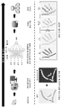


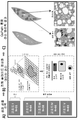


Classifications
-
- G—PHYSICS
- G06—COMPUTING; CALCULATING OR COUNTING
- G06T—IMAGE DATA PROCESSING OR GENERATION, IN GENERAL
- G06T7/00—Image analysis
- G06T7/0002—Inspection of images, e.g. flaw detection
- G06T7/0012—Biomedical image inspection
-
- A—HUMAN NECESSITIES
- A61—MEDICAL OR VETERINARY SCIENCE; HYGIENE
- A61B—DIAGNOSIS; SURGERY; IDENTIFICATION
- A61B10/00—Other methods or instruments for diagnosis, e.g. instruments for taking a cell sample, for biopsy, for vaccination diagnosis; Sex determination; Ovulation-period determination; Throat striking implements
- A61B10/02—Instruments for taking cell samples or for biopsy
-
- A—HUMAN NECESSITIES
- A61—MEDICAL OR VETERINARY SCIENCE; HYGIENE
- A61B—DIAGNOSIS; SURGERY; IDENTIFICATION
- A61B5/00—Measuring for diagnostic purposes; Identification of persons
- A61B5/42—Detecting, measuring or recording for evaluating the gastrointestinal, the endocrine or the exocrine systems
- A61B5/4222—Evaluating particular parts, e.g. particular organs
- A61B5/4244—Evaluating particular parts, e.g. particular organs liver
-
- A—HUMAN NECESSITIES
- A61—MEDICAL OR VETERINARY SCIENCE; HYGIENE
- A61B—DIAGNOSIS; SURGERY; IDENTIFICATION
- A61B5/00—Measuring for diagnostic purposes; Identification of persons
- A61B5/72—Signal processing specially adapted for physiological signals or for diagnostic purposes
- A61B5/7235—Details of waveform analysis
- A61B5/7264—Classification of physiological signals or data, e.g. using neural networks, statistical classifiers, expert systems or fuzzy systems
-
- A—HUMAN NECESSITIES
- A61—MEDICAL OR VETERINARY SCIENCE; HYGIENE
- A61B—DIAGNOSIS; SURGERY; IDENTIFICATION
- A61B5/00—Measuring for diagnostic purposes; Identification of persons
- A61B5/72—Signal processing specially adapted for physiological signals or for diagnostic purposes
- A61B5/7271—Specific aspects of physiological measurement analysis
- A61B5/7278—Artificial waveform generation or derivation, e.g. synthesising signals from measured signals
-
- G—PHYSICS
- G01—MEASURING; TESTING
- G01N—INVESTIGATING OR ANALYSING MATERIALS BY DETERMINING THEIR CHEMICAL OR PHYSICAL PROPERTIES
- G01N33/00—Investigating or analysing materials by specific methods not covered by groups G01N1/00 - G01N31/00
- G01N33/48—Biological material, e.g. blood, urine; Haemocytometers
- G01N33/50—Chemical analysis of biological material, e.g. blood, urine; Testing involving biospecific ligand binding methods; Immunological testing
- G01N33/53—Immunoassay; Biospecific binding assay; Materials therefor
- G01N33/576—Immunoassay; Biospecific binding assay; Materials therefor for hepatitis
-
- G—PHYSICS
- G06—COMPUTING; CALCULATING OR COUNTING
- G06N—COMPUTING ARRANGEMENTS BASED ON SPECIFIC COMPUTATIONAL MODELS
- G06N3/00—Computing arrangements based on biological models
- G06N3/02—Neural networks
- G06N3/04—Architecture, e.g. interconnection topology
-
- G—PHYSICS
- G06—COMPUTING; CALCULATING OR COUNTING
- G06N—COMPUTING ARRANGEMENTS BASED ON SPECIFIC COMPUTATIONAL MODELS
- G06N3/00—Computing arrangements based on biological models
- G06N3/02—Neural networks
- G06N3/08—Learning methods
-
- G—PHYSICS
- G16—INFORMATION AND COMMUNICATION TECHNOLOGY [ICT] SPECIALLY ADAPTED FOR SPECIFIC APPLICATION FIELDS
- G16H—HEALTHCARE INFORMATICS, i.e. INFORMATION AND COMMUNICATION TECHNOLOGY [ICT] SPECIALLY ADAPTED FOR THE HANDLING OR PROCESSING OF MEDICAL OR HEALTHCARE DATA
- G16H10/00—ICT specially adapted for the handling or processing of patient-related medical or healthcare data
- G16H10/40—ICT specially adapted for the handling or processing of patient-related medical or healthcare data for data related to laboratory analysis, e.g. patient specimen analysis
-
- G—PHYSICS
- G16—INFORMATION AND COMMUNICATION TECHNOLOGY [ICT] SPECIALLY ADAPTED FOR SPECIFIC APPLICATION FIELDS
- G16H—HEALTHCARE INFORMATICS, i.e. INFORMATION AND COMMUNICATION TECHNOLOGY [ICT] SPECIALLY ADAPTED FOR THE HANDLING OR PROCESSING OF MEDICAL OR HEALTHCARE DATA
- G16H20/00—ICT specially adapted for therapies or health-improving plans, e.g. for handling prescriptions, for steering therapy or for monitoring patient compliance
-
- G—PHYSICS
- G16—INFORMATION AND COMMUNICATION TECHNOLOGY [ICT] SPECIALLY ADAPTED FOR SPECIFIC APPLICATION FIELDS
- G16H—HEALTHCARE INFORMATICS, i.e. INFORMATION AND COMMUNICATION TECHNOLOGY [ICT] SPECIALLY ADAPTED FOR THE HANDLING OR PROCESSING OF MEDICAL OR HEALTHCARE DATA
- G16H30/00—ICT specially adapted for the handling or processing of medical images
- G16H30/20—ICT specially adapted for the handling or processing of medical images for handling medical images, e.g. DICOM, HL7 or PACS
-
- G—PHYSICS
- G16—INFORMATION AND COMMUNICATION TECHNOLOGY [ICT] SPECIALLY ADAPTED FOR SPECIFIC APPLICATION FIELDS
- G16H—HEALTHCARE INFORMATICS, i.e. INFORMATION AND COMMUNICATION TECHNOLOGY [ICT] SPECIALLY ADAPTED FOR THE HANDLING OR PROCESSING OF MEDICAL OR HEALTHCARE DATA
- G16H30/00—ICT specially adapted for the handling or processing of medical images
- G16H30/40—ICT specially adapted for the handling or processing of medical images for processing medical images, e.g. editing
-
- G—PHYSICS
- G16—INFORMATION AND COMMUNICATION TECHNOLOGY [ICT] SPECIALLY ADAPTED FOR SPECIFIC APPLICATION FIELDS
- G16H—HEALTHCARE INFORMATICS, i.e. INFORMATION AND COMMUNICATION TECHNOLOGY [ICT] SPECIALLY ADAPTED FOR THE HANDLING OR PROCESSING OF MEDICAL OR HEALTHCARE DATA
- G16H50/00—ICT specially adapted for medical diagnosis, medical simulation or medical data mining; ICT specially adapted for detecting, monitoring or modelling epidemics or pandemics
- G16H50/20—ICT specially adapted for medical diagnosis, medical simulation or medical data mining; ICT specially adapted for detecting, monitoring or modelling epidemics or pandemics for computer-aided diagnosis, e.g. based on medical expert systems
-
- G—PHYSICS
- G16—INFORMATION AND COMMUNICATION TECHNOLOGY [ICT] SPECIALLY ADAPTED FOR SPECIFIC APPLICATION FIELDS
- G16H—HEALTHCARE INFORMATICS, i.e. INFORMATION AND COMMUNICATION TECHNOLOGY [ICT] SPECIALLY ADAPTED FOR THE HANDLING OR PROCESSING OF MEDICAL OR HEALTHCARE DATA
- G16H50/00—ICT specially adapted for medical diagnosis, medical simulation or medical data mining; ICT specially adapted for detecting, monitoring or modelling epidemics or pandemics
- G16H50/30—ICT specially adapted for medical diagnosis, medical simulation or medical data mining; ICT specially adapted for detecting, monitoring or modelling epidemics or pandemics for calculating health indices; for individual health risk assessment
-
- G—PHYSICS
- G16—INFORMATION AND COMMUNICATION TECHNOLOGY [ICT] SPECIALLY ADAPTED FOR SPECIFIC APPLICATION FIELDS
- G16H—HEALTHCARE INFORMATICS, i.e. INFORMATION AND COMMUNICATION TECHNOLOGY [ICT] SPECIALLY ADAPTED FOR THE HANDLING OR PROCESSING OF MEDICAL OR HEALTHCARE DATA
- G16H50/00—ICT specially adapted for medical diagnosis, medical simulation or medical data mining; ICT specially adapted for detecting, monitoring or modelling epidemics or pandemics
- G16H50/50—ICT specially adapted for medical diagnosis, medical simulation or medical data mining; ICT specially adapted for detecting, monitoring or modelling epidemics or pandemics for simulation or modelling of medical disorders
-
- G—PHYSICS
- G16—INFORMATION AND COMMUNICATION TECHNOLOGY [ICT] SPECIALLY ADAPTED FOR SPECIFIC APPLICATION FIELDS
- G16H—HEALTHCARE INFORMATICS, i.e. INFORMATION AND COMMUNICATION TECHNOLOGY [ICT] SPECIALLY ADAPTED FOR THE HANDLING OR PROCESSING OF MEDICAL OR HEALTHCARE DATA
- G16H50/00—ICT specially adapted for medical diagnosis, medical simulation or medical data mining; ICT specially adapted for detecting, monitoring or modelling epidemics or pandemics
- G16H50/70—ICT specially adapted for medical diagnosis, medical simulation or medical data mining; ICT specially adapted for detecting, monitoring or modelling epidemics or pandemics for mining of medical data, e.g. analysing previous cases of other patients
-
- G—PHYSICS
- G06—COMPUTING; CALCULATING OR COUNTING
- G06T—IMAGE DATA PROCESSING OR GENERATION, IN GENERAL
- G06T2207/00—Indexing scheme for image analysis or image enhancement
- G06T2207/10—Image acquisition modality
- G06T2207/10056—Microscopic image
-
- G—PHYSICS
- G06—COMPUTING; CALCULATING OR COUNTING
- G06T—IMAGE DATA PROCESSING OR GENERATION, IN GENERAL
- G06T2207/00—Indexing scheme for image analysis or image enhancement
- G06T2207/20—Special algorithmic details
- G06T2207/20081—Training; Learning
-
- G—PHYSICS
- G06—COMPUTING; CALCULATING OR COUNTING
- G06T—IMAGE DATA PROCESSING OR GENERATION, IN GENERAL
- G06T2207/00—Indexing scheme for image analysis or image enhancement
- G06T2207/30—Subject of image; Context of image processing
- G06T2207/30004—Biomedical image processing
- G06T2207/30024—Cell structures in vitro; Tissue sections in vitro
-
- G—PHYSICS
- G06—COMPUTING; CALCULATING OR COUNTING
- G06T—IMAGE DATA PROCESSING OR GENERATION, IN GENERAL
- G06T2207/00—Indexing scheme for image analysis or image enhancement
- G06T2207/30—Subject of image; Context of image processing
- G06T2207/30004—Biomedical image processing
- G06T2207/30056—Liver; Hepatic
Abstract
본 발명은 조직학적 영상의 신규 자동화된 패턴 인식 및 점수화 방법에 관한 것이다.The present invention relates to a novel automated pattern recognition and scoring method for histological images.

Description
본 발명은 조직학적 영상의 자동화된 패턴 인식 및 점수화 방법에 관한 것이다.The present invention relates to an automated pattern recognition and scoring method for histological images.
비알코올성 지방간염(NASH)의 유병률은 빠르게 증가하고 있고 NASH가 궁극적으로 간경화증 및 간세포 암종(HCC)을 유발하는 진행성 섬유증 축적을 유도한다는 것을 알면서 주요 공중 보건 문제를 대표한다.The prevalence of nonalcoholic steatohepatitis (NASH) is rapidly increasing and represents a major public health problem, knowing that NASH induces progressive fibrosis accumulation that ultimately leads to cirrhosis and hepatocellular carcinoma (HCC).
비알코올성 지방간 질환(NAFLD)은 초음파 기법을 이용하여 간 내로의 지방 축적의 존재를 검출함으로써 진단될 수 있는 반면, NASH 및 NASH 관련 간 섬유증은 오늘날 간 생검의 조직학적 검사에 의해서만 진단될 수 있다. NAFLD 활성 수준 및 섬유증 병기를 평가하고 임상 간 결과로의 진전의 위험을 추정하기 위한 조직학적 점수화/병기분류 시스템이 개발되었다. 질환의 활성을 평가하기 위한 NALFD 활성 점수(NAS)가 개발되었다.Non-alcoholic fatty liver disease (NAFLD) can be diagnosed by detecting the presence of fat accumulation into the liver using ultrasound techniques, whereas NASH and NASH related liver fibrosis can only be diagnosed today by histological examination of a liver biopsy. A histological scoring/staging system has been developed to evaluate NAFLD activity levels and stages of fibrosis and to estimate the risk of progression to clinical liver outcomes. A NALFD activity score (NAS) was developed to assess the activity of the disease.
NAS는 지방증(0 내지 3), 소엽 염증(0 내지 2) 및 간세포 풍선변형(0 내지 2)에 대한 생검의 개별 점수의 합계이거나, 비가중된 개별 생검의 개별 점수의 합계이다.NAS is the sum of the individual scores of biopsies for adiposis (0-3), lobular inflammation (0-2) and hepatocellular balloon deformity (0-2), or the sum of the individual scores of unweighted individual biopsies.
문헌[Kleiner et al, Hepatology, 2005; 41:1313-21](Kleiner et al, 2005)에 따르면, NAS는 간 생검 슬라이스로부터 얻은 3개의 조직학적 점수들의 합계이다:Kleiner et al, Hepatology, 2005; 41:1313-21] (Kleiner et al, 2005), NAS is the sum of three histological scores obtained from liver biopsy slices:
- 지방증 점수: 0: <5%; 1: 5% 내지 33%; 2: 34% 내지 66%, 및 3: >66% -Fatosis score: 0: <5%; 1: 5% to 33%; 2: 34% to 66%, and 3: >66%
- 소엽 염증 점수(병소/20배 시야): 0: 없음; 1: <2; 2: 2 내지 4; 및 3: >4-Lobule inflammation score (lesion/20x field of view): 0: none; 1: <2; 2: 2 to 4; And 3: >4
- 풍선변형 변성 점수: 0: 없음; 1: 소수; 2: 다수.-Strain degeneration score: 0: None; 1: prime number; 2: Many.
이 점수화 시스템을 이용할 때, NASH를 가진 환자는 지방증에서 적어도 1점, 소엽 염증에서 적어도 1점 및 간세포 풍선변형에서 적어도 1점을 가짐으로써 3 이상의 NAS를 가진다. 환자는 지방증에서 적어도 1점, 염증에서 적어도 1점 및 간세포 풍선변형에서 적어도 1점을 가짐으로써 4 이상의 NAS를 가질 때 활성 NASH를 가진 것으로서 간주된다.When using this scoring system, patients with NASH have 3 or more NAS by having at least 1 point in fatosis, at least 1 point in lobular inflammation and at least 1 point in hepatocellular balloon deformity. Patients are considered to have active NASH when they have 4 or more NAS by having at least 1 point in fatosis, at least 1 point in inflammation and at least 1 point in hepatocellular balloon deformity.
조직학적 검사에서 섬유증의 위치확인 및 정도는 질환의 중증도(진행)를 표시하고, NASH-CRN은 전용 섬유증 병기분류 시스템을 개발하였다(Kleiner et al., Hepatology, 2005; 41:1313-21).The location and extent of fibrosis in histological examination indicate the severity (progression) of the disease, and NASH-CRN developed a dedicated fibrosis staging system (Kleiner et al., Hepatology, 2005; 41:1313-21).
동모양혈관주위 또는 문맥주위 섬유증 1Periplasmic or
경증 동모양혈관주위 섬유증(대역 3) 1aMild sinusoidal fibrosis (band 3) 1a
중등도 동모양혈관주위 섬유증(대역 3) 1bModerate sinusoidal fibrosis (band 3) 1b
문맥/문맥주위 섬유증 1cFibrosis around the context/context 1c
동모양혈관주위 및 문맥/문맥주위 섬유증 2Fibrosis around the sinus vascular and portal/
가교 섬유증 3
간경변증 4
이 섬유증 병기분류 시스템을 이용할 때, 섬유증을 갖지 않거나 최소한의 섬유증을 가진 환자(F=0 또는 1)는 일반적으로 간경변증, HCC 또는 간 사망의 위험에 있는 것으로 간주되지 않는다. 유의미한 섬유증(F=2) 및 중등도 섬유증(F=3)을 가진 환자는 간경변증, 간 부전, HCC 및 간 사망을 발생시킬 증가하는 위험에 있다. 대상성 간경변증을 가진 환자는 중증 섬유증(F=4)을 갖고 간 부전(비대상성 간경변증), HCC 및 간 사망의 높은 위험에 있다.When using this fibrosis staging system, patients with or without fibrosis (F=0 or 1) are generally not considered at risk of cirrhosis, HCC or liver death. Patients with significant fibrosis (F=2) and moderate fibrosis (F=3) are at increased risk of developing cirrhosis, liver failure, HCC and liver death. Patients with decompensated cirrhosis have severe fibrosis (F=4) and are at high risk of liver failure (decompensated cirrhosis), HCC and liver death.
최근에, 이들 널리 받아들여지는 2종의 점수화 및 병기분류 시스템들로부터 유도된, 소엽 염증 점수와 간세포 풍선변형 점수의 합계로서 정의될 수 있는, 활성 지수(AI)에 특별한 관심이 기울여지고 있다. 추가로, 문헌[Munteanu et al., Aliment Pharmacol Ther., 2016, 44(8):877-89](Munteanu et al., 2016)은 지방증, 질환 활성 및 섬유증의 점수를 따로 보고하기 위한 SAF 시그니처를 제안하였다. 지방증 점수(S)는 포말성 미세소포를 제외한 큰 또는 중간 크기의 지질 소적의 양을 평가하고, 0부터 3까지 등급을 매긴다(S0: <5%; S1: 5% 내지 33%, 경증; S2: 34% 내지 66%, 중등도; S3: >66%, 현저함). 활성 등급(A, 0부터 4까지)은 간세포 풍선변형(0 내지 2) 및 소엽 염증(0 내지 2)의 비가중된 합산이다. A0(A = 0)을 가진 경우는 활성을 갖지 않았고; A1(A = 1)을 가진 경우는 경증 활성을 가졌고; A2(A = 2)를 가진 경우는 중등도 활성을 가졌고; A3(A = 3)을 가진 경우는 중증 활성을 가졌고; A4(A = 4)를 가진 경우는 고도 중증 활성을 가졌다. 섬유증 병기(F)는 다음과 같이 클레이너(Kleiner)에 의해 기재된 점수를 사용함으로써 평가되었다: 0 기(F0) = 없음; 1 기(F1) = 1a 또는 1b 동모양혈관주위 대역 3 또는 1c 문맥 섬유증; 2 기(F2) = 가교 없는 동모양혈관주위 및 문맥주위 섬유증; 3 기(F3) = 가교 섬유증; 및 4 기(F4) = 간경변증.Recently, special attention has been paid to the activity index (AI), which can be defined as the sum of lobular inflammation scores and hepatocyte ballooning scores, derived from these widely accepted two scoring and staging systems. Additionally, Munteanu et al., Aliment Pharmacol Ther., 2016, 44(8):877-89 (Munteanu et al., 2016), SAF signature for separately reporting scores of fatosis, disease activity and fibrosis Proposed. Lipidosis score (S) evaluates the amount of large or medium sized lipid droplets excluding foamy microvesicles and is graded from 0 to 3 (S0: <5%; S1: 5% to 33%, mild; S2 : 34% to 66%, moderate; S3: >66%, significant). The activity grade (A, 0 to 4) is an unweighted summation of hepatocellular ballooning (0 to 2) and lobular inflammation (0 to 2). A0 ( A = 0) had no activity; A1 ( A = 1) had mild activity; When A2 ( A = 2) had moderate activity; A3 ( A = 3) had severe activity; A4 ( A = 4) had a highly severe activity. Fibrosis stage ( F ) was assessed by using a score described by Kleiner as follows: Stage 0 (F0) = none; Stage 1 (F1) = 1a or 1b
브런트(Brunt) 시스템은 NASH의 중증도를 등급화하는 방법으로서 주로 제안되었고, 지방간염에 대한 최소한의 기준을 충족시키지 않은 경우에 적용되기 위한 것이 아니었다. 경증, 중등도 및 중증으로의 지방간염의 등급화는 지방증, 염증 및 풍선변형의 중증도의 전체 영향에 기반하였으나, 대부분의 가중을 풍선변형에 주었다(Brunt et al., 1999).The Brunt system was primarily proposed as a method of grading the severity of NASH and was not intended to be applied if the minimum criteria for hepatitis were not met. Grading of hepatitis to mild, moderate and severe was based on the overall effect of the severity of fatosis, inflammation and balloon deformity, but most of the weighting was attributed to balloon deformity (Brunt et al., 1999).
이들 시스템들 사이의 개별 조직학적 특징의 등급화 및 병기분류의 비교는 표 1에 제시되어 있다.The grading and comparison of staging of individual histological features between these systems is presented in Table 1.

병리학은 진단, 연구 또는 다른 목적을 위한 조직 샘플의 실험실 검사를 다루는 과학의 한 분야이다. 병리학자뿐만 아니라, 의학 또는 생물학 연구에 종사하는 다른 학자들도 질환 상태를 평가하거나 생물학적 특징을 이해하기 위해 인간, 동물, 식물 또는 다른 조직을 검사한다. 병리학의 한 양태에서, 분석을 위해 환자로부터 조직을 생검하고 광학 현미경 하에서 관찰하기 위해 유리 슬라이드 상에 고정시킨다. 그러나, 이러한 형태의 조직 평가는 다양한 이유로 신중을 요한다. 예를 들면, 이러한 단점들 중 일부는 주관성 및 낮은 처리율을 포함한다. 또 다른 문제점은 고품질 진단이 종종 단일 판독자에 의해 처리되기 어려운 다수의 샘플들을 분석할 것을 요구한다는 점이다. 많은 유사한 샘플들과 대면한 수동 판독자는 판독의 불균일성을 가중시키는 피로도 경험할 수 있다. Pathology is a field of science that deals with laboratory testing of tissue samples for diagnosis, research or other purposes. In addition to pathologists, other scholars engaged in medical or biological research examine humans, animals, plants, or other tissues to assess disease conditions or to understand biological characteristics. In one aspect of the pathology, tissue is biopsied from the patient for analysis and fixed on a glass slide for observation under an optical microscope. However, this type of organizational evaluation is cautious for a variety of reasons. For example, some of these shortcomings include subjectivity and low throughput. Another problem is that high quality diagnostics often require analyzing multiple samples that are difficult to process by a single reader. Passive readers facing many similar samples may also experience fatigue that adds to the non-uniformity of the readings.
조직학적 특징의 평가에 있어서 관찰자 가변성을 정량하고자 시도한 여러 연구들은 섬유증의 등급에 대한 중간 내지 상당한 일치로 결론에 도달한다. 또한, 간세포 염증 및 풍선변형은 전체 병리학적 진단시 63%의 관찰자간 일치를 이끌어낼 정도로 병리학자들 사이에 잘 일치하지 않았다(Younossi et al, 1998). 꽤 균질한 환자(비만치료 수술을 받은 병적 비만 환자) 군에서 NASH의 유병률의 검토 분석은 수술시 수득된 수술 생검의 조직병리학적 분석에 기반한 NASH의 유병률이 24%부터 98%까지 변경되었다는 결론을 내렸다(Machado et al, 2006).Several studies attempting to quantify observer variability in the evaluation of histological features come to a conclusion with moderate to significant agreement on the grade of fibrosis. In addition, hepatocellular inflammation and balloon deformity were not well-matched among pathologists, leading to an agreement between 63% of observers in the overall pathological diagnosis (Younossi et al, 1998). A review analysis of the prevalence of NASH in a group of fairly homogeneous patients (pathologically obese patients undergoing obesity treatment surgery) concluded that the prevalence of NASH changed from 24% to 98% based on histopathological analysis of surgical biopsies obtained during surgery. (Machado et al, 2006).
병리학자 점수는 생검 박편에서의 마이크로/마크로 조직학적 패턴의 해석에 기반한다. 이들 임상 파라미터들을 점수화하기 위한 현재의 표준은 병리학자 또는 훈련된 조직학자에 의한 간 생검 박편의 수동 해석 및 점수화에 머물러 있다. 수동 점수화는 지루하다는 점 이외에(예를 들면, 풍선변형) 주로 조직학적 패턴의 해석의 주관성으로 인해 관찰자간 일치를 감소시키는 것으로 알려져 있다(지방증: κ(일치 계수) = 0.6, 염증: κ = 0.5, 풍선변형: κ = 0.7, 섬유증: κ = 0.5)(Bedossa, 2014).Pathologist scores are based on the interpretation of micro/macro histological patterns in biopsy slices. The current standard for scoring these clinical parameters remains in the manual interpretation and scoring of liver biopsy slices by pathologists or trained histologists. Passive scoring is known to reduce inter-observer agreement mainly due to the subjectivity of interpretation of histological patterns, in addition to being tedious (e.g. balloon deformation) (fatosis: κ (coincidence factor) = 0.6, inflammation: κ = 0.5 , Balloon deformation: κ = 0.7, fibrosis: κ = 0.5) (Bedossa, 2014).
이 편향은 각각의 파라미터를 개별적으로 고려할 때 더 증강된다:This bias is enhanced when each parameter is considered individually:
I) 간세포 풍선변형: 이러한 빈번하지 않은 사건의 경우, 철저하지 않은 스크리닝으로 인해, 일부 세포들이 누락될 수 있다. 전체적으로 변성된 풍선변형 간세포(대세포)로부터 풍선변형 전 세포(소세포)를 구별하기 위해 종종 사용되는 세포 크기에 대한 주관적 평가도 이루어진다.I) Hepatocellular balloon deformity: In the case of these infrequent events, some cells may be missing due to incomplete screening. A subjective assessment of the cell size often used to differentiate pre-balloon deformed cells (small cells) from degenerated balloon-deformed hepatocytes (large cells) is also made.
II) 간 또는 간세포 소엽 염증: HPF(고배율 시야) 선택으로 인한 주관성은 점수화 과정을 재현하는 것을 어렵게 만들고 잠재적 편향에 노출시킨다.II) Liver or hepatocyte lobular inflammation: Subjectivity due to HPF (high magnification field of view) selection makes it difficult to reproduce the scoring process and exposes it to potential bias.
III) 간 섬유증: 간 섬유증의 평가도 1) 콜라겐 비례 면적(CPA)의 정량에 대한 조직병리학적 요인의 영향 및 2) 섬유증 병기분류에 대한 인간 주관적 해석(관찰자간 일치를 감소시킴)(Bedossa, 2014)로 인해 어렵다. CPA 정량은 방법이 (본질적으로 조직학 및 영상화로 인한) 염색 편차에 강하고 콜라겐의 비병리학적 염색(혈관 영역)을 배제할 것을 요구한다.III) Liver fibrosis: Evaluation of liver fibrosis 1) The effect of histopathological factors on the quantitation of collagen proportional area (CPA) and 2) Human subjective interpretation of fibrosis staging (reducing agreement among observers) (Bedossa, 2014). CPA quantification requires the method to be resistant to staining variance (essentially due to histology and imaging) and exclude non-pathological staining of the collagen (vascular region).
IV) 지방증: 지방의 세포내 액포를 함유하는 간세포의 비율의 시각적 추정 및 중증도의 넓은 범위 내로의 지방증의 등급화는 다소 주관적이므로, 조직병리학에서 지방증 등급화에 있어서 상당한 개체간 및 개체내 편차가 있다: 0 등급(정상) = 최대 5%의 영향을 받은 세포, 1 등급(경증) = 5% 내지 33%의 영향을 받은 세포, 2 등급(중등도) = 34% 내지 66%의 영향을 받은 세포, 및 3 등급(중증) = 67% 이상의 영향을 받은 세포.IV) Fatosis: A visual estimate of the proportion of hepatocytes containing intracellular vacuoles of fat and the grading of fatosis within a wide range of severity is somewhat subjective, so there is considerable inter-individual and intra-individual variation in grading fatosis in histopathology. Yes: Grade 0 (normal) = cells affected up to 5%, grade 1 (mild) = cells affected from 5% to 33%, grade 2 (moderate) = cells affected from 34% to 66% , And Grade 3 (severe) = 67% or more affected cells.
더욱이, 전체적으로 자동화된 방법은 현재의 정확도, 재현성 및 처리율 문제를 다루기 위해 절실히 요구된다. 임상 및 연구 적용을 위한 전체 슬라이드 영상화(WSI) 시스템의 보급 후, 조직병리학적 데이터에 적용된 기계 학습 애플리케이션의 개발은 최근에 매우 활발한 연구 영역이 되었다(LeCun et al, 2015). 심층 학습(DL) 패턴 인식 알고리즘은 최근에 마이크로 조직 패턴 및 마크로 조직 패턴 둘 다를 정확히 포착할 수 있고 나아가 병리학자의 진단 워크플로우(workflow)를 모방할 수 있는 것으로 밝혀졌다. 이러한 기술의 이용에 의해, 조직학적 패턴은 자동으로 예측될 수 있고, NASH 진단 및 간 섬유증 점수화의 재현성은 개선될 수 있다. 추가로, NASH 특이적 섬유증 조직학적 패턴의 인식은 콜라겐 형태(격막(Septa)) 및 비례 면적(CPA)의 보다 더 정확한 정량을 가능하게 할 것이다.Moreover, a fully automated method is desperately needed to address current accuracy, reproducibility and throughput issues. After the prevalence of the entire slide imaging (WSI) system for clinical and research applications, the development of machine learning applications applied to histopathological data has recently become a very active research area (LeCun et al, 2015). In-depth learning (DL) pattern recognition algorithms have recently been found to be able to accurately capture both micro-tissue patterns and macro-tissue patterns and further mimic pathologists' diagnostic workflows. By using this technique, histological patterns can be predicted automatically, and the reproducibility of NASH diagnosis and liver fibrosis scoring can be improved. Additionally, recognition of NASH specific fibrosis histological patterns will enable more accurate quantification of collagen morphology (Septa) and proportional area (CPA).
NASH 조직학적 해석을 전체적으로 자동화하기 위해, 본 발명자들은 NASH 질환 활성, NASH 섬유증 점수 및 간 섬유증의 평가를 위한 통합된 자동화된 데이터 관리 및 분석 파이프라인을 확립하였다. 본 발명자들은 낮은 내지 중요한 코호트내 및 코호트간 염료 강도 편차를 보상하기 위해 강력한 염색 표준화 방법을 개발하였다. 본 발명자들은 NASH 점수화(염증, 풍선변형 및 섬유증)를 자동화하기 위해 3개의 독특한 전체적으로 자동화된 기계 학습 애플리케이션을 통한 DL 및 형태계측 분석의 이용도 연구하였다. 영상 분석 방법을 이용한 지방증 정량도 NAFLD 진단 및 NASH 점수화의 자동화된 과정을 완성하기 위해 상기 분석에 포함되었다. 지방증 정량 방법은 DL 방법일 수 있다.To fully automate NASH histological interpretation, we have established an integrated automated data management and analysis pipeline for the evaluation of NASH disease activity, NASH fibrosis score and liver fibrosis. The inventors have developed a robust dye standardization method to compensate for dye intensity variations within and between low and important cohorts. The inventors also studied the use of DL and morphometric analysis through three unique fully automated machine learning applications to automate NASH scoring (inflammation, balloon deformity and fibrosis). Lipidosis quantification using image analysis was also included in the analysis to complete the automated process of NAFLD diagnosis and NASH scoring. The method of quantifying fatosis may be a DL method.
각각의 임상 파라미터의 상이한 병리학적 및 조직학적 성질을 고려하여, 본 발명자들은 개별 분석 파이프라인 내로 채널화된 개발 과정을 수행하였다:Taking into account the different pathological and histological properties of each clinical parameter, we conducted a development process channeled into individual analysis pipelines:
I. 전처리(시야(FOV) 추출, 조직 분절화, 표준화).I. Pretreatment (field of view (FOV) extraction, tissue segmentation, standardization).
II. NASH 병리학적 마이크로/마크로패턴 DL(염증, 풍선변형 세포/간 섬유증 FOV 유형)의 예측 및 지방증 영역의 정량.II. Prediction of NASH pathological micro/macropattern DL (inflammation, balloon deformed cell/hepatic fibrosis FOV type) and quantification of fatosis area.
III. 생검 수준까지 데이터 집계.III. Aggregate data to the level of biopsy.
개발, 시험 및 검증을 위해 독립적 코호트를 사용하여 각각의 NASH 임상 성분에 대한 정량 파이프라인의 일반화 성질을 평가하였다. 최종 목적은 마이크로/마크로 예측 및 정량(CPA/격막 형태)을 임상적으로 관련된 정보로 해석하고 기준 병리학자 어노테이션(annotation)과 대조하여 검증하는 것이었다.Independent cohorts were used for development, testing and validation to evaluate the generalization properties of the quantitative pipeline for each NASH clinical component. The final objective was to interpret the micro/macro prediction and quantification (CPA/diaphragm morphology) as clinically relevant information and validate it against a reference pathologist annotation.
따라서, 본 발명은 간 생검으로부터의 면역화학 영상의 강력한 자동화된 패턴 인식 방법 및 점수화 방법을 제공한다. Thus, the present invention provides a powerful automated pattern recognition method and scoring method for immunochemical images from liver biopsies.
이 방법들은 임계 NASH 개별 파라미터(염증, 풍선변형 및 섬유증) 점수화를 자동화하기 위해 3개의 독특한 전체적으로 자동화된 기계 학습 애플리케이션을 통한 심층 학습 및 형태계측 분석에 기반을 둔다. 상기 방법들은 지방증 정량을 자동화하기 위해 전체적으로 자동화된 제4 애플리케이션에 의해 완료될 수도 있다.These methods are based on in-depth learning and morphometric analysis through three unique fully automated machine learning applications to automate critical NASH individual parameter (inflammation, balloon deformation and fibrosis) scoring. The methods may be completed by a fully automated fourth application to automate the determination of fatosis.
본 발명의 방법에서, 간세포 풍선변형(HB)은 DL을 이용하여 병리학적으로 관련된 세포 패턴(즉, 풍선변형 간세포, 세포 크기)을 예측함으로써 평가된다. 제2 단계에서, 이 예측은 간 생검 샘플 수준에서 집계될 수 있다.In the method of the present invention, hepatocellular balloon deformity (HB) is evaluated by predicting pathologically relevant cell patterns (ie, balloon deformed hepatocytes, cell size) using DL. In a second step, this prediction can be aggregated at the liver biopsy sample level.
본 발명의 방법에서, 소엽 염증(LI)은 DL을 이용하여 병리학적으로 관련된 세포 패턴(즉, 염증 세포)을 예측함으로써 평가된다. 제2 단계에서, 이 예측은 간 생검 샘플 수준에서 집계될 수 있다.In the method of the present invention, lobular inflammation (LI) is assessed by using DL to predict pathologically relevant cell patterns (ie, inflammatory cells). In a second step, this prediction can be aggregated at the liver biopsy sample level.
본 발명의 방법에서, 섬유증은 DL을 이용하여, 특히 후속 CPA 및 섬유증 격막 형태계측 정량에 의해 더 확인될 수 있는 병리학적으로 관련된 간 섬유증 영역을 예측함으로써 평가된다. 섬유증 점수화는 본 발명의 자동화된 판독 및 점수화 방법에 의한 간 섬유증의 로버스트(robust) 점수화에 기반하여 NAFLD 또는 NASH 환자의 간 섬유증이 간경변증 및 간 결과로 진화할 위험의 예후인 섬유증의 임상 예후를 가능하게 할 수 있다.In the method of the invention, fibrosis is assessed using DL, in particular by predicting the pathologically relevant liver fibrosis region that can be further confirmed by subsequent CPA and fibrosis septum morphometric quantification. Fibrosis scoring is based on the robust scoring of liver fibrosis by the automated reading and scoring method of the present invention and the clinical prognosis of fibrosis is a prognosis of the risk of liver fibrosis in NAFLD or NASH patients evolving into cirrhosis and liver outcome. It can be enabled.
본 발명의 방법에서, 지방증(S) 평가는 특히 DL 모델을 이용하여 병리학적으로 관련된 세포 구조물을 예측함으로써 자동화된 과정일 수 있다.In the method of the present invention, fatosis (S) evaluation can be an automated process, particularly by predicting pathologically relevant cellular structures using a DL model.
이러한 패턴은 지질 액포 또는 지방증의 영역(AOS)을 함유하는 지방 간세포의 백분율일 수 있다.This pattern can be the percentage of adipose hepatocytes containing lipid vacuoles or areas of fatosis (AOS).
지방증의 영역은 액포 유형 구조물이다.The area of fatosis is vacuole-type structures.
구체적인 실시양태에서, 액포 크기는 적어도 핵의 크기이다.In specific embodiments, the vacuole size is at least the size of the nucleus.
구체적인 실시양태에서, 지방증은 지방증의 영역의 자동화된 측정에 의해 평가된다.In specific embodiments, fatosis is assessed by automated measurement of the area of fatosis.
제2 단계에서, 이 예측은 간 생검 샘플 수준에서 집계될 수 있다.In a second step, this prediction can be aggregated at the liver biopsy sample level.
모든 기계 학습 애플리케이션들의 경우, 각각의 DL 모델 성질은 결정적으로 정확하면서도 재현가능한 객관적 결과를 생성하도록 디자인되었다.For all machine learning applications, each DL model property is designed to produce decisively accurate and reproducible objective results.
따라서, 구체적인 실시양태에서, 대상체에서의 간세포 풍선변형의 검출 및 정량은 패턴 인식 및 점수화에 기반한다: HB=0, 간세포 풍선변형 없음, HB=1, 중등도 간세포 풍선변형, 및 HB=2, 중증 간세포 풍선변형(Kleiner & Makhlouf, 2016).Thus, in a specific embodiment, detection and quantification of hepatocyte ballooning in a subject is based on pattern recognition and scoring: HB=0, no hepatocyte ballooning, HB=1, moderate hepatocyte ballooning, and HB=2, severe Hepatocellular balloon transformation (Kleiner & Makhlouf, 2016).
구체적인 실시양태에서, 대상체에서의 소엽 염증의 검출 및 정량은 패턴 인식 및 점수화에 기반한다: LI=0, 소엽 염증 없음, LI>0, 소엽 염증 및 등급화: LI=1 경증 소엽 염증, LI=2, 중등도 소엽 염증, 및 LI=3, 중증 소엽 염증(Kleiner & Makhlouf, 2016).In specific embodiments, detection and quantification of lobular inflammation in subjects is based on pattern recognition and scoring: LI=0, no lobular inflammation, LI>0, lobular inflammation and grading: LI=1 mild lobular inflammation, LI= 2, moderate lobular inflammation, and LI=3, severe lobular inflammation (Kleiner & Makhlouf, 2016).
추가 실시양태에서, 본 발명은 간세포 풍선변형 및 소엽 염증의 개별 점수를 더하여 NASH 활성 지수(AI)를 측정하고 병기분류하는 방법도 제공한다: AI=0 또는 1, NASH 없음; 적어도 HB=1 및 LI=1을 가진 AI>1.In a further embodiment, the invention also provides a method of measuring and staging the NASH activity index (AI) by adding individual scores of hepatocellular balloon deformity and lobular inflammation: AI=0 or 1, no NASH; AI>1 with at least HB=1 and LI=1.
또 다른 실시양태에서, 본 발명은 간 생검에서의 지방증 점수의 측정 및 1의 최소 지방증 점수에 기반하여 건강한 대상체 또는 환자에서 NAFLD의 임상 진단을 가능하게 한다.In another embodiment, the present invention enables clinical diagnosis of NAFLD in a healthy subject or patient based on a determination of the steatosis score in a liver biopsy and a minimum steatosis score of 1.
또 다른 실시양태에서, 본 발명은 NAFLD 환자의 간 생검에서의 활성 지수의 측정에 기반하여 대상체, 특히 NAFLD 환자에서 NASH의 임상 진단을 가능하게 한다: 적어도 HB=1 및 LI=1을 가진 AI≥2.In another embodiment, the present invention enables clinical diagnosis of NASH in subjects, particularly NAFLD patients, based on the measurement of activity index in liver biopsy of NAFLD patients: AI≥ with at least HB=1 and LI=1 2.
또 다른 실시양태에서, 대상체에서의 간 섬유증의 검출 및 정량은 간 생검에서의 패턴 인식 및 점수화에 기반한다: F=0, 섬유증 없음, F>0, 섬유증의 검출 및 점수화: F=1, 최소 섬유증, F=2, 상당한 섬유증, F=3, 중등도 섬유증, 및 F=4, 중증 섬유증.In another embodiment, detection and quantification of liver fibrosis in a subject is based on pattern recognition and scoring in a liver biopsy: F=0, no fibrosis, F>0, detection and scoring of fibrosis: F=1, minimal Fibrosis, F=2, significant fibrosis, F=3, moderate fibrosis, and F=4, severe fibrosis.
구체적인 실시양태에서, 대상체에서의 간 지방증의 검출 및 정량은 간 생검에서의 패턴 인식 및 점수화에 기반한다: S=0, 5% 미만의 지방증; S=1, 6% 내지 33% 지방증; S=2, 34% 내지 66% 지방증; 및 S=3, 66% 초과의 지방증(Kleiner & Makhlouf, 2016).In a specific embodiment, detection and quantification of hepatic steatosis in a subject is based on pattern recognition and scoring in a liver biopsy: S=0, less than 5% steatosis; S=1, 6% to 33% fatosis; S=2, 34% to 66% fatosis; And S=3, more than 66% fatosis (Kleiner & Makhlouf, 2016).
추가 구체적인 실시양태에서, 대상체에서의 마이크로지방증 및 마크로지방증의 특징규명은 간 생검에서의 패턴 인식, 세포 크기 및 형태 분석, 및 점수화에 기반한다.In a further specific embodiment, characterization of microlipidosis and macrolipidosis in the subject is based on pattern recognition in liver biopsy, cell size and morphology analysis, and scoring.
추가 실시양태에서, 문맥 정맥, 동모양혈관, 소엽 정맥 또는 다른 하위구조물을 기준으로 한 특정 조직학적 패턴의 위치확인 및 배치는 간 병리학에 대한 귀중한 보완 정보를 제공할 수 있다.In a further embodiment, the positioning and placement of specific histological patterns relative to the portal vein, sinusoidal vein, lobular vein, or other substructure can provide valuable complementary information for liver pathology.
구체적인 실시양태에서, 조직학적 프로파일의 분석은 전체적으로 자동화될 수 있다. 또 다른 실시양태에서, 조직학적 NASH 파라미터의 정량은 자동화될 수 있다. 추가 실시양태에서, 조직학적 NASH 파라미터의 정량은 자동화될 수 있고 생체영상 분석 소프트웨어(특히, QuPath 플랫폼) 내로 통합될 수 있다.In specific embodiments, analysis of the histological profile can be entirely automated. In another embodiment, the quantification of histological NASH parameters can be automated. In further embodiments, the quantification of histological NASH parameters can be automated and integrated into bioimaging analysis software (especially the QuPath platform).
추가 목적에 따라, 본 발명은 본원에서 제공된 자동화된 방법 덕분에, NAFLD 환자에서의 활성 지수 및 섬유증의 신뢰할만한 검출 및 정량, 및 하기 정의된 TBT(치료되는) 환자와 NTBT(치료되지 않는) 환자 사이의 차이 분석에 기반하여 NAFLD, NASH 또는 간 섬유증에 대한 치료의 잠재적 수용자(치료되는, 또는 TBT) 또는 비-수용자(치료되지 않는, 또는 NTBT)로서 대상체를 분류하는 방법에 관한 것이다.For further purposes, the present invention provides reliable detection and quantitation of activity index and fibrosis in NAFLD patients, thanks to the automated methods provided herein, and TBT (treated) and NTBT (untreated) patients as defined below. A method of classifying subjects as potential recipients of treatment for NAFLD, NASH or liver fibrosis (treated, or TBT) or non-receptive (untreated, or NTBT) based on analysis of differences between.
또 다른 양태를 통해, 본 발명은 간 생검의 조직학적 슬라이드에의 섬유증 DL 모델 및 간 섬유증 형태계측 모델 적용에 기반하여 NAFLD 또는 NASH 환자의 간 섬유증이 간경변증 및 간 결과로 진화할 위험의 예후인 섬유증의 임상 예후도 가능하게 한다.In another aspect, the present invention provides fibrosis, which is a prognosis of the risk that liver fibrosis in NAFLD or NASH patients will evolve to cirrhosis and liver outcomes based on the application of the fibrosis DL model and liver fibrosis morphometric model to histological slides of liver biopsies It also enables clinical prognosis.
본 발명은 대상체의 간 생검의 조직학적 슬라이드의 DL 모델을 사용한 패턴 인식 및 점수화, 및 상기 대상체에서 과거에 수집된 생검을 사용한 차이 분석에 기반하여 NAFLD 활성, NASH 활성 및/또는 간 섬유증 병기의 진화를 모니터링하는 방법도 제공한다. 자동화된 지방증 점수화도 상기 방법에 포함될 수 있다.The present invention is an evolution of NAFLD activity, NASH activity and/or liver fibrosis stage based on pattern recognition and scoring using a DL model of a histological slide of a liver biopsy of a subject, and difference analysis using a biopsy collected in the subject in the past. It also provides a way to monitor. Automated steatosis scoring can also be included in the method.
본 발명은 대상체의 간 생검에서의 패턴 및 점수와, 상기 대상체에서 과거에 수집된 또 다른 생검에서의 패턴 및 점수 사이의 차이 분석으로 간 생검의 면역조직학적 슬라이드에의 간세포 풍선변형, 소엽 염증, 섬유증 DL 모델 및 간 섬유증 형태계측 모델 적용에 기반하여 대상체에서 NAFLD, NASH 또는 간 섬유증의 치료 효율을 측정하는 방법도 제공한다. 자동화된 지방증 점수화도 상기 방법에 포함될 수 있다.The present invention analyzes the differences between patterns and scores in a subject's liver biopsy and patterns and scores in another biopsy collected in the past in the subject, resulting in hepatocellular balloon deformity, lobular inflammation on the immunohistochemical slide of the liver biopsy, Also provided is a method of measuring the therapeutic efficiency of NAFLD, NASH or liver fibrosis in a subject based on the application of a fibrosis DL model and a liver fibrosis morphometric model. Automated steatosis scoring can also be included in the method.
본 발명은 간 생검의 면역조직학적 슬라이드에의 간세포 풍선변형, 소엽 염증, 섬유증 DL 모델 및 간 섬유증 형태계측 모델 적용, 및 반응자와 비-반응자 사이의 비교에 기반하여 특정 치료에 대한 대상체(반응자 대상체)의 반응을 예측(NAFLD, NASH 활성 및 간 섬유증 병기에서의 변화를 예측)하는 방법도 제공한다. 자동화된 지방증 점수화도 상기 방법에 포함될 수 있다.The present invention is based on the application of hepatocellular balloon transformation, lobular inflammation, fibrosis DL model and liver fibrosis morphometric model to an immunohistologic slide of a liver biopsy, and subjects for a specific treatment (responder subjects) It also provides a method of predicting the response of () predicting changes in NAFLD, NASH activity, and liver fibrosis stage. Automated steatosis scoring can also be included in the method.
도 1: 패턴 인식 및 점수화 방법 개발을 위한 프로토콜의 도식적 개요.
물리적 유리 슬라이드의 수령부터 자동화된 NASH 및 간 점수의 예측까지 전산 병리학 파이프라인의 개요.
I 상: 전체 슬라이드 영상화(WSI). 슬라이드를 유리 슬라이드 스캐너로 스캔하고 저장, 관찰 및 분석을 용이하게 하기 위해 DICOM 권고에 따라 큰 다중해상 다면 영상을 저장한다.
II 상: WSI의 전처리. 5배 배율에서 슬라이드 표시를 이용하여, 영상화 인공물(artifact)(기포, 먼지, 커버슬립 잔해물) 및 조직 처리 인공물(조직 접힘, 조직 잔해물)을 자동으로 발견한다.
III 상: WSI로부터의 생체의학적 관련 영상의 추출. 마이크로패턴(세포) 및 마크로패턴(조직 영역)을 나타내는 영상을 추출하여 분석한다.
IV 상: NASH 및 간 섬유증 자동화된 점수화. 먼저 마이크로패턴(풍선변형 및 염증) 및 마크로패턴(NASH 섬유증, 간 섬유증)에 대해 예측 모델을 훈련한다. 그 다음, 이 모델을 개별 마이크로패턴 및 마크로패턴 영상에 적용하여 각각의 생검 유리 슬라이드에 대해 예측한다: (각각의 마크로패턴에 대한) 풍선변형 간세포의 수, 염증 세포의 수, NASH 섬유증 패턴, 간 섬유증 콜라겐 비례 면적 및 간 섬유증 형태 지수. 그 다음, 마이크로패턴 및 마크로패턴 정량을 풍선변형 점수, 염증 점수, 섬유증 점수, 간 콜라겐 비례 면적 및 형태 지수로 변환한다.
도 2: 염증, 풍선변형, 섬유증 및 간 섬유증 형태계측 분석을 위해 심층 학습 모델을 생성하는 데 사용된 환자 샘플 및 영상 데이터의 분해.
도 3: 전체 슬라이드 영상화(WSI) 데이터 관리 분석 및 가시화를 커버하는, 사내 개발된 전산 병리학 파이프라인의 개요.
도 4: 염증, 풍선변형 및 섬유증 심층 학습 모델의 개발을 위한 훈련 및 검증 세트를 나타내는 세포 기반 영상 및 시야(FOV) 영상의 실례.
A) 좌측 패널은 염증 모델의 훈련 및 검증을 위해 사용된 염증 세포 및 정상 간세포 핵의 훈련 및 검증 세트를 나타내는 세포 영상을 보여준다. 우측 패널은 풍선변형 모델의 검증을 위해 사용된 풍선변형 간세포 및 정상 간세포의 몇몇 예를 보여준다. B) 심층 학습 섬유증 모델에 의해 자동으로 인식되는 훈련 및 검증 데이터세트에서 5개의 패턴을 나타내는 FOV의 예. 배경 노이즈(noise) 또는 매우 적은 생검 물질(<=5%)만을 보여주는 모든 FOV들은 본 발명자들의 전산 병리학 파이프라인의 초기 전처리 단계에서 자동으로 여과된다. C) 섬유증 형태계측 분석의 실례. 좌측에서 우측으로, 미처리 영상(제1 그래픽)을 표준화하고, 최전면 콜라겐 염색(제2 그래픽)을 배경으로부터 추출한다. 그 다음, 표준화된 콜라겐 영상(제3 그래픽)을 분석하여 섬유증 격막의 형태학적 성질을 추출하고(제4 그래픽) 및 CPA를 측정한다(제5 그래픽, 청색 원은 비-격막 콜라겐 면적을 표시함).
도 5: 염증, 풍선변형 및 섬유증의 심층 학습 모델의 개발 및 검증 과정으로부터의 결과.
A) 좌측 패널은 학습 주기 반복에 맞게 학습률을 동력학적으로 조정하는 데 이용된 방식을 도식적으로 보여준다. 적색 직선은 비용/손실 함수(즉, 음의 확률)를 학습 주기 반복의 함수로서 나타내는 반면, 청색 직선은 본 발명자들이 극소치에 접근할 때 학습률의 조정(즉, 동력학적 감소)을 보여준다. 강조된 적색 직선은 최적 해법에 가능한 가까워지기 위해 여러 번 반복된 과정인, 극소치 파라미터를 개량하도록 실행된 제2 학습 과정을 보여준다. B) 학습 반복 주기의 함수로서 훈련 및 검증 세트에 대한 풍선변형 모델 성능. C) 훈련 및 검증 세트에 대한 훈련 및 검증 강도 수준(x 축에 표시된 모든 연구 샘플들에 걸쳐 강도 수준당 1개의 선). D) 훈련 코호트에서 CPA의 반자동화된 측정과 전체적으로 자동화된 측정의 상관관계.
도 6: 자동화된 염증 점수화 파이프라인의 검증으로부터의 결과. A) 10개의 고배율 시야(HPF)의 자동화된 무작위 선택의 실례. (좌측에서 우측으로) 제1 영상은 미처리 간 생검 영상(헤마톡실린 및 에오신으로 염색됨, H&E)의 일례를 보여주고, 제2 영상은 현미경 하에서 20배 배율에서 HPF와 동등한 모든 가능한 HPF의 생성을 보여주고(배경 및 혈관 영역의 대부분을 가진 HFP는 청색으로 강조되어 있고, 염증 평가에 유효한 HPF는 적색으로 강조되어 있음), 제3 영상은 염증 점수화(즉, 자동 및 수동)를 위해 (모든 유효 HPF 중에서) 선택된 20개의 무작위적으로 선택된 HPF를 보여준다. B) WSI 방식을 이용함으로써 무작위적으로 선택된 10개의 HPF 및 표준 현미경 상에서 병소의 평균 수의 수동 카운팅의 상관관계. C) (지침(Kliener, 2005, 6:1313-1321)에 의해 정의된 바와 같이 평균 병소 수로부터 변환된) 현미경관찰 기반 염증 점수와 WSI 기반 염증 점수 사이의 일치를 보여주는 표. D) 각각의 염증 점수 범주(x 축 군)에 대한 환자(코호트 I)당 (DL 모델에 의해 자동으로 인식된) 염증 세포의 총 수의 분포를 보여주는 그래픽.
도 7: 간 섬유증 CPA 측정의 개발 및 검증 과정으로부터의 결과.
A) 각각의 섬유증 점수 환자 군(코호트 VI)에 대한 전체적으로 자동화된 CPA 측정치의 분포. B) 중등도(F1&F2) 섬유증 점수 및 진행된(F3) 섬유증 점수를 가진 환자(코호트 VI)에 대한 전체적으로 자동화된 격막 형태의 분포.
도 8: 자동화된 활성 점수화 파이프라인에 대한 데이터 워크플로우의 도식적 표시.
A) 자동화된 점수화 파이프라인을 검증하는 데 사용된 상이한 연구 코호트의 실례. B) 정량 과정 전 QuPath 오픈 소스 프레임워크 내의 영상 조작 워크플로우의 도식적 표시. C) 검증하거나 진단 보조제로서 사용되는, 사용자를 위한 염증 및 활성 점수화의 시각적 맵핑의 실례. 저해상 조직 박편 영상 상에서 원은 광학 현미경 하에서 20배 배율에서 시야의 크기 및 모양에 상응한다. 이 박편의 아래 부분에는, 염증 병소 및 풍선변형 간세포 사건이 검증하거나 보정하기 위해 QuPath에서 최종 사용자에게 어떻게 어노테이션되는지가 예로서 제시되어 있다.
도 9: 오픈 소스 플랫폼 QuPath 내로의 지방증, 염증 및 풍선변형 결과의 통합.
A) 브라우징, 프로젝트 WSI 관리, 영상 분석, 패턴 인식 및 데이터 분석을 가능하게 하는, QuPath 사용자 인터페이스의 뷰. B) QuPath의 데이터 추출 및 분석력을 보여주는, H&E로 염색된 간 박편에 대한 상이한 영상 분석 출력물. C) 디지털 슬라이드 상에서 세포 기반 결과(예를 들면, 지질 액포) 맵핑의 실례.
도 10: NASH 및 간 섬유증 자동화된 점수화를 위한 IV 상 세부절차 및 워크플로우.1: Schematic overview of the protocol for developing pattern recognition and scoring methods.
An overview of the computational pathology pipeline, from the receipt of physical glass slides to the prediction of automated NASH and liver scores.
Phase I: Whole slide imaging (WSI). Scan slides with a glass slide scanner and store large multiresolution multi-faceted images according to DICOM recommendations to facilitate storage, observation and analysis.
Phase II: WSI pretreatment. Using the slide display at 5x magnification, imaging artifacts (bubbles, dust, coverslip debris) and tissue processing artifacts (tissue folding, tissue debris) are automatically discovered.
Phase III: Extraction of biomedical related images from WSI. Images representing micro-patterns (cells) and macro-patterns (tissue areas) are extracted and analyzed.
Phase IV: NASH and liver fibrosis automated scoring. First, a predictive model is trained on micropatterns (balloon deformation and inflammation) and macropatterns (NASH fibrosis, liver fibrosis). The model is then applied to individual micropattern and macropattern images to predict for each biopsy glass slide: number of balloon-deformed hepatocytes (for each macropattern), number of inflammatory cells, NASH fibrosis pattern, liver Fibrosis collagen proportional area and liver fibrosis morphology index. The micropattern and macropattern quantities are then converted to balloon deformity score, inflammation score, fibrosis score, liver collagen proportional area and morphology index.
Figure 2: Decomposition of patient sample and image data used to generate an in-depth learning model for morphometric analysis of inflammation, balloon deformity, fibrosis and liver fibrosis.
Figure 3: Overview of an in-house developed computational pathology pipeline covering full slide imaging (WSI) data management analysis and visualization.
Figure 4: Example of cell-based and field of view (FOV) images showing a training and validation set for the development of an in-depth learning model of inflammation, balloon deformity and fibrosis.
A) The left panel shows cell images showing the training and verification set of inflammatory cells and normal hepatocyte nuclei used for training and verification of the inflammatory model. The right panel shows some examples of balloon deformed hepatocytes and normal hepatocytes used for validation of the balloon deformity model. B) Example of FOV showing five patterns in the training and verification dataset automatically recognized by the deep learning fibrosis model. All FOVs showing only background noise or very little biopsy material (<=5%) are automatically filtered in the initial pretreatment stage of our computational pathology pipeline. C) An example of fibrosis morphometric analysis. From left to right, the unprocessed image (first graphic) is normalized, and the frontmost collagen stain (second graphic) is extracted from the background. Then, standardized collagen images (3rd graphic) are analyzed to extract the morphological properties of the fibrous septum (4th graphic) and CPA is measured (5th graphic, blue circles indicate non-spherical collagen area) ).
Figure 5: Results from the development and verification process of an in-depth learning model of inflammation, balloon deformity and fibrosis.
A) The left panel schematically shows the method used to dynamically adjust the learning rate to fit the learning cycle repetition. The red straight line represents the cost/loss function (i.e., negative probability) as a function of learning cycle iteration, while the blue straight line shows the adjustment of the learning rate (i.e., kinetic reduction) as we approach the minimum. The highlighted red line shows the second learning process, which has been repeated several times to improve the minimum parameters, to get as close as possible to the optimal solution. B) Balloon deformation model performance for training and verification sets as a function of learning iteration period. C) Training and verification intensity levels for the training and verification set (one line per intensity level across all study samples displayed on the x-axis). D) Correlation between semi-automated and fully automated measurements of CPA in the training cohort.
Figure 6: Results from validation of the automated inflammation scoring pipeline. A) An example of automated random selection of 10 high magnification fields of view (HPF). The first image (from left to right) shows an example of an untreated liver biopsy image (stained with hematoxylin and eosin, H&E), and the second image produces all possible HPF equivalent to HPF at 20x magnification under a microscope (HFP with most of the background and vascular regions are highlighted in blue, HPF effective for inflammation evaluation is highlighted in red), and the third image is for inflammation scoring (i.e. automatic and manual) (all Shows 20 randomly selected HPFs selected from among effective HPFs. B) Correlation of manual counting of the average number of lesions on 10 HPF and standard microscopes randomly selected by using the WSI method. C) Table showing agreement between microscopic observation-based inflammation scores and WSI-based inflammation scores (converted from the average number of lesions as defined by the guidelines (Kliener, 2005, 6:1313-1321)). D) Graphic showing the distribution of the total number of inflammatory cells (automatically recognized by the DL model) per patient (cohort I) for each category of inflammation score (x-axis group).
Figure 7: Results from the development and validation process of liver fibrosis CPA measurements.
A) Distribution of fully automated CPA measurements for each fibrosis score patient group (Cohort VI). B) Overall automated diaphragm distribution for patients with moderate (F1&F2) fibrosis score and advanced (F3) fibrosis score (Cohort VI).
Figure 8: Schematic representation of data workflow for automated active scoring pipeline.
A) Examples of different study cohorts used to validate automated scoring pipelines. B) Schematic representation of the image manipulation workflow within the QuPath open source framework prior to the quantitation process. C) An example of a visual mapping of inflammation and activity scoring for a user, to be used as a validation or diagnostic adjuvant. The circles on the low-resolution tissue slice images correspond to the size and shape of the field of view at 20x magnification under an optical microscope. In the lower part of this slice, an example is presented of how inflammatory lesions and balloon-deformed hepatocyte events are annotated by QuPath to end users for verification or correction.
Figure 9: Integration of fatosis, inflammation and ballooning results into the open source platform QuPath.
A) A view of the QuPath user interface that enables browsing, project WSI management, image analysis, pattern recognition and data analysis. B) Different image analysis outputs for liver slices stained with H&E, showing QuPath's data extraction and analysis power. C) An example of mapping cell based results (eg, lipid vacuoles) on a digital slide.
10: Phase IV detailed procedure and workflow for automated scoring of NASH and liver fibrosis.
본 발명자들은 조직학적 영상의 신규 자동화된 패턴 인식 방법 및 점수화 방법을 제공한다.The present inventors provide a novel automated pattern recognition method and scoring method for histological images.
본 발명은 간세포 풍선변형, 간 소엽 염증, NASH 섬유증 및 간 섬유증 조직학적 패턴을 FOV 수준에서 자동으로 인식하고 점수화하기 위해 디지털화된 슬라이드에 대한 심층 학습 프레임워크를 제공한다.The present invention provides an in-depth learning framework for digitized slides to automatically recognize and score hepatocyte ballooning, hepatic lobular inflammation, NASH fibrosis and hepatic fibrosis histological patterns at the FOV level.
이 프레임워크는 디지털화된 슬라이드 상에서의 지방증 인식, 정량, 점수화 및/또는 위치확인까지 확장될 수도 있다.The framework can also be extended to fatosis recognition, quantification, scoring and/or positioning on digitized slides.
본 발명에 따라, 용어 "간 생검"은 손상 또는 질환의 징후에 대해 현미경으로 검사하기 위해 간 조직의 작은 조각을 채취하는 단계를 포함하는 절차이다.According to the present invention, the term "liver biopsy" is a procedure comprising the step of taking a small piece of liver tissue for microscopic examination for signs of damage or disease.
본 발명에 따라, 용어 "조직학적 영상"은 특정 세포 또는 조직 특징의 관찰에 적합한 염색으로 염색된 조직병리학적 슬라이드이다. 헤마톡실린 및 에오신(H&E)은 예시적 염료이다. 명시야 현미경 하에서 가시화되었을 때, 헤마톡실린은 핵산을 염색하고 청색/자주색으로 보이는 반면, 에오신은 단백질을 염색하고 분홍색/적색으로 보인다. 따라서, 대다수의 조직들의 경우, 세포 핵은 청색인 반면, 세포질은 그의 성분에 따라 투명한 색부터 적색 내지 자주색까지 변경될 수 있다.According to the present invention, the term "histological image" is a histopathological slide stained with staining suitable for observation of specific cell or tissue characteristics. Hematoxylin and eosin (H&E) are exemplary dyes. When visualized under brightfield microscopy, hematoxylin stains nucleic acids and looks blue/purple, whereas eosin stains proteins and looks pink/red. Thus, for most tissues, the cell nucleus is blue, while the cytoplasm can change from transparent to red to purple depending on its composition.
본 발명에 따라, 용어 "지방증"은 통상적으로 광학 현미경 수준에서 헤마톡실린 및 에오신(H&E) 염색에 기반하여, 지질을 함유하는 투명한 액포로서 정의된다. 간세포 지방증은 NAFLD의 특징이고, 현재 NAFLD의 진단을 위해서는 5% 초과의 간세포에서의 지방증이 요구된다.According to the present invention, the term “lipidosis” is defined as a clear vacuole containing lipids, typically based on hematoxylin and eosin (H&E) staining at the level of an optical microscope. Hepatocellular steatosis is a hallmark of NAFLD, and current diagnosis of NAFLD requires more than 5% of hepatocyte steatosis.
본 발명에 따라, 용어 "마크로지방증" 또는 "마크로소포 지방증"은 지방 변성의 가장 흔한 형태에 상응하고, 지질의 과다공급에 의해 야기될 수 있다.According to the present invention, the terms "macrolipidosis" or "macrovesic lipoidosis" correspond to the most common form of fat degeneration and can be caused by an oversupply of lipids.
본 발명에 따라, 용어 "마이크로지방증" 또는 "마이크로소포 지방증"은 세포에 축적되는 작은 세포질내 지방 액포(리포좀)를 특징으로 한다. 공통된 원인은 테트라사이클린, 임신성 급성 지방 간, 라이 증후군(Reye's syndrome) 및 C형 간염이다.According to the present invention, the terms "microlipidosis" or "microvesicle lipolysis" are characterized by small intracellular cytoplasmic fat vacuoles (liposomes) that accumulate in cells. Common causes are tetracycline, gestational acute fatty liver, Reye's syndrome and hepatitis C.
본 발명에 따라, 세포 또는 조직 구조물의 "기준 영상"은 병리학자에 의해 확인된, 상기 세포 또는 조직 구조물을 나타내는 스냅샷이다. 이러한 기준 영상은 간 생검의 슬라이드에 존재할 가능성이 있는 상이한 세포 패턴을 나타내는 영상을 수득하기 위해 상이한 영상 처리로 처리될 수 있다. 예를 들면, 기준 영상은 스케일링, 염색 및/또는 형태학적 변환으로 처리될 수 있다. 나아가, 하나 이상의 기준 영상으로부터 한 세트의 기준 영상을 생성할 수 있다. 예를 들면, 1개의 기준 영상을 앞서 언급된 바와 같이 변환하여, 한 세트의 기준 영상을 나타내는 적어도 2개, 3개, 4개, 5개, 6개, 7개, 8개, 9개 또는 적어도 10개의 상이한 변환된 영상을 수득할 수 있다. 실례로서, 스케일링, 염색 및 형태학적 변형을 이용하여 100개의 개별 세포 또는 조직 기준 영상으로부터 각각의 가능한 세포 또는 조직 패턴을 나타내는 한 세트의 1000개 기준 영상을 생성할 수 있다.According to the present invention, a “reference image” of a cell or tissue structure is a snapshot representing the cell or tissue structure, as confirmed by a pathologist. These reference images can be processed with different image processing to obtain images representing different cell patterns likely to be present on the slides of the liver biopsy. For example, the reference image can be processed by scaling, staining and/or morphological transformation. Furthermore, a set of reference images may be generated from one or more reference images. For example, at least 2, 3, 4, 5, 6, 7, 8, 9 or at least one reference image is transformed as described above to represent a set of reference images. Ten different transformed images can be obtained. As an example, scaling, staining, and morphological modification can be used to generate a set of 1000 reference images representing each possible cell or tissue pattern from 100 individual cell or tissue reference images.
스케일링, 염색 및 형태학적 선형 변형은 범위가 0% 내지 20%의 기준 영상일 수 있다.Scaling, staining and morphological linear transformation can range from 0% to 20% of the reference image.
구체적인 실시양태에서, 스케일링, 염색 및 형태학적 선형 변형은 범위가 0% 내지 20%의 기준 영상일 수 있다.In specific embodiments, scaling, staining, and morphological linear transformation can be a reference image ranging from 0% to 20%.
구체적인 실시양태에서, 형태학적 및 스케일링 변형은 형태에 적용될 수 있다: 즉, 0% 내지 20%의 선형 및 아핀(affine) 변환.In specific embodiments, morphological and scaling modifications can be applied to morphology: i.e. 0% to 20% linear and affine transformation.
더 구체적인 실시양태에서, 형태학적 및 스케일링 변형은 형태에 적용될 수 있다: 즉, 0% 내지 10%의 선형 및 아핀 변환.In a more specific embodiment, morphological and scaling modifications can be applied to the morphology: i.e. 0% to 10% linear and affine transformation.
구체적인 실시양태에서, 염색 변형은 0% 내지 5%의 색도의 무작위 변형일 수 있다. In specific embodiments, the staining modification can be a random modification of chromaticity from 0% to 5%.
본 발명에 따라, "염색 및 패턴 어노테이션에 특이적인 기준 영상"은 조직의 병리학적 특징당 염색 표준화를 위해 사용된 영상이다. According to the present invention, "a reference image specific for staining and pattern annotation" is an image used for standardizing staining per pathological feature of tissue.
본 발명에 따라, "염색 및 NASH 파라미터에 특이적인 기준 영상"은 NASH 파라미터당 염색 표준화를 위해 사용된 영상이다.In accordance with the present invention, "staining and reference images specific to NASH parameters" are images used for standardization of staining per NASH parameter.
본 발명에 따라, "패턴 어노테이션"은 조직의 병리학적 특징으로서 정의된다. NAFLD 환자에서, 간 조직의 특정 병리학적 특징은 간세포 풍선변형, 소엽 염증, 지방증, 간 또는 NASH 섬유증일 수 있다.According to the present invention, "pattern annotation" is defined as a pathological characteristic of tissue. In patients with NAFLD, certain pathological features of liver tissue may be hepatocellular balloon deformity, lobular inflammation, steatosis, liver or NASH fibrosis.
본 발명에 따라, "마이크로패턴"은 세포 구조물, 즉 풍선변형 세포, 염증 세포로서 정의된다.According to the present invention, a "micropattern" is defined as a cell structure, i.e. balloon deformed cell, inflammatory cell.
본 발명에 따라, "마크로패턴"은 인접 조직 영역, 즉 섬유증으로서 정의된다. According to the present invention, a “macro pattern” is defined as an adjacent tissue area, ie fibrosis.
본 발명에 따라, 용어 "간세포 풍선변형"은 통상적으로 광학 현미경 수준에서 헤마톡실린 및 에오신(H&E) 염색에 기반하여, 희박한 세포질을 가진 정상 간세포 직경의 1.5배 내지 2배 세포 확장으로서 정의된다. 이 용어는 보다 일반적으로 간세포 세포 사멸의 과정을 지칭한다. 당연히, 본 발명에서 H&E 이외의 다른 염색을 사용하여 상기 특징의 존재를 확인할 수 있다.According to the present invention, the term "hepatocyte ballooning" is defined as cell expansion 1.5 to 2 times the diameter of normal hepatocytes with lean cytoplasm, typically based on hematoxylin and eosin (H&E) staining at the optical microscopic level. This term more generally refers to the process of hepatocyte cell death. Naturally, in the present invention, the existence of the above characteristics can be confirmed by using dyes other than H&E.
본 발명에 따라, 용어 "소엽 염증"은 간 생검의 헤마톡실린 및 에오신(H&E) 염색된 슬라이스의 현미경 검사에서 소엽 염증 병소(군을 이룬 염증 세포)의 존재를 지칭한다. 당연히, 본 발명에서 H&E 이외의 다른 염색을 사용하여 상기 특징의 존재를 확인할 수 있다.According to the present invention, the term "lobular inflammation" refers to the presence of lobular inflammation lesions (grouped inflammatory cells) in microscopic examination of hematoxylin and eosin (H&E) stained slices of a liver biopsy. Naturally, in the present invention, the existence of the above characteristics can be confirmed by using dyes other than H&E.
본 발명에 따라, 용어 "섬유증" 또는 "간 섬유증"은 간 생검의 염색된(예를 들면, 헤마톡실린 및 에오신 및 사프란(HES), 트리크롬, 시리우스 레드(Sirius red) 또는 피크로시리우스 레드(picrosirius red) 염색) 슬라이스의 현미경 검사에서 섬유성 결합 조직의 존재를 지칭한다.In accordance with the present invention, the term “fibrosis” or “liver fibrosis” refers to stained liver biopsies (eg, hematoxylin and eosin and saffron (HES), trichrome, Sirius red, or Pyrocirius red) (picrosirius red staining) refers to the presence of fibrous connective tissue in microscopic examination of slices.
본 발명에 따라, "활성 지수"는 간세포 풍선변형 및 소엽 염증 점수의 합계를 지칭한다.According to the present invention, "activity index" refers to the sum of hepatocyte ballooning and lobular inflammation scores.
본 발명에 따라, "NAFLD 활성 점수" 또는 "NAS"는 다음과 같이 지방증, 간세포 풍선변형, 소엽 염증의 합계를 지칭한다:According to the present invention, "NAFLD activity score" or "NAS" refers to the sum of fatosis, hepatocyte ballooning, and lobular inflammation as follows:
- 지방증 점수: 0: <5%; 1: 5% 내지 33%; 2: 34% 내지 66%, 및 3: >66%; -Fatosis score: 0: <5%; 1: 5% to 33%; 2: 34% to 66%, and 3: >66%;
- 소엽 염증 점수(병소/20배 시야): 0: 없음; 1: <2; 2: 2 내지 4, 및 3: >4;-Lobule inflammation score (lesion/20x field of view): 0: none; 1: <2; 2: 2 to 4, and 3: >4;
- 풍선변형 변성 점수: 0: 없음; 1: 소수; 2: 다수.-Strain degeneration score: 0: None; 1: prime number; 2: Many.
본 발명에 따라, 용어 "NAFLD" 또는 "비알코올성 지방 간 질환"은 과도한 알코올 소비의 부재 하에서 염증 및 섬유증의 징후를 갖거나 갖지 않으면서 지방이 간에 침착되는 상태(간 지방증)를 지칭한다.According to the present invention, the term "NAFLD" or "non-alcoholic fatty liver disease" refers to a condition in which fat is deposited in the liver (hepatic steatosis) with or without signs of inflammation and fibrosis in the absence of excessive alcohol consumption.
본 발명에 따라, 용어 "NAFLD 활성 수준"은 본원에서 정의된 바와 같이 NAFLD 진행을 지칭하고 지방증 점수의 증가에 의해 정의된다. NAFLD 활성 수준은 NASH 또는 섬유증을 향한 NAFLD 진행 및 NASH 중증도를 지칭하기도 한다.In accordance with the present invention, the term “NAFLD activity level” refers to NAFLD progression as defined herein and is defined by an increase in fatosis score. NAFLD activity levels also refer to NAFLD progression and NASH severity towards NASH or fibrosis.
본 발명에 따라, 용어 "NASH" 또는 "비알코올성 지방간염"은 과도한 알코올 소비의 부재 하에서, 그리고 바이러스성 간염(HCV, HBV 등)과 같은 다른 간 질환을 제외시킨 후, 조직학적 검사에서 간 지방증, 간세포 풍선변형 및 간 염증의 동시다발적 존재(즉, 지방증에서 적어도 1점, 소엽 염증에서 적어도 1점, 그리고 간세포 풍선변형 점수에서 적어도 1점을 가짐으로써 NAS≥3)를 특징으로 하는 NAFLD 상태를 지칭한다.According to the present invention, the terms "NASH" or "non-alcoholic steatohepatitis" are liver fatosis in histological examination in the absence of excessive alcohol consumption and after excluding other liver diseases such as viral hepatitis (HCV, HBV, etc.). , NAFLD status characterized by simultaneous presence of hepatocyte ballooning and liver inflammation (i.e., NAS≥3 by having at least 1 point in steatosis, at least 1 point in lobular inflammation, and at least 1 point in hepatocyte ballooning score) Refers to.
본 발명에 따라, 용어 "NASH 활성 수준"은 NASH 진행을 지칭하고, NASH를 정의하기 위한 최소 파라미터인 S=1, LI=1 및 HB=1을 초과하는 NAS 점수의 증가에 의해 정의된다. NASH 활성 수준은 비가역적 NASH 및/또는 섬유증을 향한 NASH 진행 및 NASH 중증도를 지칭하기도 한다.In accordance with the present invention, the term “NASH activity level” refers to NASH progression and is defined by an increase in NAS scores exceeding the minimum parameters S=1, LI=1 and HB=1 for defining NASH. NASH activity level also refers to NASH progression and NASH severity towards irreversible NASH and/or fibrosis.
본 발명에 따라, 용어 "활성 NASH"는 지방증에서 적어도 1점, 소엽 염증에서 적어도 1점, 그리고 간세포 풍선변형 점수에서 적어도 1점을 가짐으로써 NAS≥4를 특징으로 하는 NASH를 지칭한다.According to the present invention, the term “active NASH” refers to NASH characterized by NAS≧4 by having at least 1 point in steatosis, at least 1 point in lobular inflammation, and at least 1 point in hepatocellular balloon deflection score.
본 발명에 따라, "치료받을 대상체" 또는 "TBT 대상체"는 대상체를 NAFLD, NASH 및/또는 간 섬유증에 대한 치료를 받을 자격이 있게 만드는 질환 활성 점수(예를 들면, NAS 또는 활성 지수) 및/또는 간 섬유증 병기를 가진 대상체이다. 대조적으로, "치료받지 않을 대상체" 또는 "NTBT 대상체"는 NAFLD, NASH 및/또는 간 섬유증에 대한 치료를 받기에 충분히 높지 않은 질환 활성 점수(예를 들면, NAS 또는 활성 지수) 및/또는 간 섬유증 병기를 가진 대상체이다. 따라서, TBT 대상체는 NAFLD, NASH 및/또는 간 섬유증 치료를 위한 "수용자" 또는 "잠재적 수용자"로서도 지칭된다. 본 발명에서, 바람직한 TBT 대상체는 i) NASH를 가진 대상체, ii) 활성 NASH를 가진 대상체, iii) 유의미한, 중등도 또는 중증 간 섬유증을 가진 대상체, iv) NASH 및 섬유증을 가진 대상체이다.According to the present invention, “subject to be treated” or “TBT subject” refers to a disease activity score (eg, NAS or activity index) that makes a subject eligible for treatment for NAFLD, NASH and/or liver fibrosis and/or Or a liver fibrotic stage. In contrast, “subject not to be treated” or “NTBT subject” is a disease activity score (eg, NAS or activity index) and/or liver fibrosis that is not high enough to receive treatment for NAFLD, NASH and/or liver fibrosis Subject with stage. Accordingly, TBT subjects are also referred to as “recipients” or “potential recipients” for the treatment of NAFLD, NASH and/or liver fibrosis. In the present invention, preferred TBT subjects are i) subjects with NASH, ii) subjects with active NASH, iii) subjects with significant, moderate or severe liver fibrosis, iv) subjects with NASH and fibrosis.
상기 정의는 본 발명의 상이한 변이체를 정의하는 다양한 NASH 활성 값 및 섬유증 병기를 포괄한다.The definition encompasses various NASH activity values and fibrosis stages that define different variants of the invention.
본 발명의 바람직한 변이체는 다음과 같이 상세하게 설명된다.Preferred variants of the invention are described in detail as follows.
제1 TBT 변이체(TBT2): First TBT variant (TBT2) :
TBT2 대상체는 하기 간 생검 유래 등급을 제시하는 대상체로서 정의된다:TBT2 subjects are defined as subjects presenting the following liver biopsy grades:
- 지방증 점수 ≥ 1-Fatosis score ≥ 1
- 간세포 풍선변형 점수 ≥ 1-Hepatocyte balloon transformation score ≥ 1
- 소엽 염증 점수 ≥ 1-Lobule inflammation score ≥ 1
- NAS(NAFLD 활성 점수) ≥ 4 -NAS (NAFLD active score) ≥ 4
- 섬유증 병기 ≥ 2(예컨대, 2, 3 또는 4, 특히 2 또는 3에 해당하는 섬유증 병기).-Fibrosis stage ≥ 2 (eg, 2, 3 or 4, especially 2 or 3 fibrosis stages).
더 나아가, NTBT2 대상체는 지방증, 간세포 풍선변형, 소엽 염증 점수, NAS 및/또는 섬유증 병기에서 적어도 1점 더 작은 등급에 의해 TBT2 대상체와 상이하다.Furthermore, NTBT2 subjects differ from TBT2 subjects by at least 1 point smaller grades in adiposis, hepatocyte ballooning, lobular inflammation score, NAS and/or fibrosis stage.
제2 TBT 변이체(TBT1): Second TBT variant (TBT1) :
TBT1 대상체는 하기 간 생검 유래 등급을 제시하는 대상체로서 정의된다:TBT1 subjects are defined as subjects presenting the following liver biopsy grades:
- 지방증 점수 ≥ 1-Fatosis score ≥ 1
- 간세포 풍선변형 점수 ≥ 1-Hepatocyte balloon transformation score ≥ 1
- 소엽 염증 점수 ≥ 1-Lobule inflammation score ≥ 1
- NAS(NAFLD 활성 점수) ≥ 4 -NAS (NAFLD active score) ≥ 4
- 섬유증 병기 ≥ 1(예컨대, 1, 2, 3 또는 4에 해당하는 섬유증 병기).-Fibrosis stage ≥ 1 (e.g., fibrosis stage corresponding to 1, 2, 3 or 4).
더 나아가, NTBT1 대상체는 지방증, 간세포 풍선변형, 소엽 염증 점수, NAS 및/또는 섬유증 병기에서 적어도 1점 더 작은 등급에 의해 TBT1 대상체와 상이하다.Furthermore, NTBT1 subjects differ from TBT1 subjects by at least 1 point smaller grades in adiposis, hepatocyte ballooning, lobular inflammation score, NAS and/or fibrosis stage.
제3 TBT 변이체(TBT7): Third TBT variant (TBT7) :
TBT7 대상체는 하기 간 생검 유래 등급을 제시하는 대상체로서 정의된다:TBT7 subjects are defined as subjects presenting the following liver biopsy grades:
- 지방증 점수 ≥ 1-Fatosis score ≥ 1
- 간세포 풍선변형 점수 ≥ 1-Hepatocyte balloon transformation score ≥ 1
- 소엽 염증 점수 ≥ 1-Lobule inflammation score ≥ 1
- NAS(NAFLD 활성 점수) ≥ 4 -NAS (NAFLD active score) ≥ 4
- 섬유증 병기 = 1b, 1c, 2, 3 또는 4.-Stage of fibrosis = 1b, 1c, 2, 3 or 4.
더 나아가, NTBT7 대상체는 지방증, 간세포 풍선변형, 소엽 염증 점수, NAS 및/또는 섬유증 병기에서 적어도 1점 더 작은 등급에 의해 TBT7 대상체와 상이하다.Furthermore, NTBT7 subjects differ from TBT7 subjects by at least 1 point smaller grades in adiposis, hepatocellular balloon deformity, lobular inflammation score, NAS and/or fibrosis stage.
본 발명에 따라, 용어 "CPA" 또는 "콜라겐 비례 면적"은 콜라겐 염색된 면적 대 총 조직 면적의 총 비율이다. 표지는 피크로시리우스 레드 또는 다른 염료(즉, 트리크롬)일 수 있다. CPA는 생검 수준에서 총 병리학적 콜라겐 면적 대 총 생검 조직 면적의 비로서 보고된다. CPA는 비알코올성 지방간 질환을 가진 환자에서 장기간 결과의 독립적 예후자이다.According to the present invention, the term “CPA” or “collagen proportional area” is the total ratio of collagen stained area to total tissue area. The label can be Pyrocyrus red or other dye (ie trichrome). CPA is reported as the ratio of total pathological collagen area to total biopsy tissue area at the biopsy level. CPA is an independent prognostic of long-term outcomes in patients with nonalcoholic fatty liver disease.
본 발명에 따라, 용어 "DL" 또는 "심층 학습"은 기계 학습을 그의 원래의 목표들 중 하나에 더 가깝게 이동시키는 것을 목적으로 도입된 기계 학습 연구의 새로운 영역이다: 인공 지능. 심층 학습은 패턴 분석 및 분류를 위해 영상과 같은 데이터를 이해하는 것을 돕는 다단계의 표시 및 추출을 학습하는 것에 관한 것이다. According to the present invention, the term "DL" or "deep learning" is a new area of machine learning research introduced with the aim of moving machine learning closer to one of its original goals: artificial intelligence. Deep learning is about learning multi-level display and extraction to help understand data such as images for pattern analysis and classification.
본 발명에 따라, 용어 "CNN" 또는 콘볼루션 신경 네트워크는 시각적 영상을 분석하기 위해(영상 인식 및 분류)에 적용된 기계 학습에 있어서 심층 피드-포워드(feed-forward) 인공 신경 네트워크의 한 부류이다.According to the present invention, the term "CNN" or convolutional neural network is a class of deep feed-forward artificial neural networks in machine learning applied to analyze visual images (image recognition and classification).
본 발명에 따라, 용어 "DL 모델"은 패턴 인식 또는 분석, 및 분류를 위해 CNN을 사용할 수 있다.According to the present invention, the term "DL model" may use CNN for pattern recognition or analysis, and classification.
본 발명은 대상체에서 간세포 풍선변형의 발생 및 등급을 특징규명하는 방법으로서, 상기 대상체의 간 생검에서 간세포 풍선변형 DL 모델을 사용하여 간세포 풍선변형 점수를 측정하는 단계를 포함하는 방법에 관한 것이다.The present invention relates to a method for characterizing the incidence and grade of hepatocellular balloon deformity in a subject, and relates to a method comprising measuring a hepatocellular balloon deflection score using a hepatocyte balloon deformed DL model in the liver biopsy of the subject.
본 발명은 대상체에서 간세포 소엽 염증의 발생 및 등급을 특징규명하는 방법으로서, 상기 대상체의 간 생검에서 소엽 염증 DL 모델을 사용하여 소엽 염증 점수를 측정하는 단계를 포함하는 방법에 관한 것이다.The present invention relates to a method of characterizing the incidence and grade of hepatocellular lobular inflammation in a subject, and relates to a method comprising measuring a lobular inflammation score using a lobular inflammation DL model in a liver biopsy of the subject.
본 발명은 대상체에서 지방증의 발생 및 등급을 특징규명하는 방법으로서, 상기 대상체의 간 생검에서 자동화된 과정을 이용하여 지방증 점수를 측정하는 단계를 포함하는 방법에 관한 것이다.The present invention relates to a method for characterizing the incidence and grade of fatosis in a subject, and relates to a method comprising measuring a fatosis score using an automated process in a liver biopsy of the subject.
또한, 본 발명은 지방증, 간세포 풍선변형, 소엽 염증, NASH 섬유증 및 간 섬유증에 대한 전용 표준화 방법에 관한 것이다.In addition, the present invention relates to a dedicated standardization method for adiposis, hepatocellular balloon deformity, lobular inflammation, NASH fibrosis and liver fibrosis.
본 발명은 간 생검에서의 패턴 인식, 세포 크기 및 형태 분석, 및 점수화에 기반한, 대상체에서의 마이크로지방증 및 마크로지방증의 특징규명에 관한 것이다.The present invention relates to characterization of microlipidosis and macrolipidosis in subjects, based on pattern recognition, cell size and morphology analysis, and scoring in liver biopsies.
또한, 본 발명은 간 병리학에 대한 귀중한 보완 정보를 제공할 수 있는, 문맥 정맥, 동모양혈관, 소엽 정맥 또는 다른 하위구조물을 기준으로 한 특정 조직학적 패턴의 위치확인 및 배치에 관한 것이다.The present invention also relates to the positioning and placement of specific histological patterns based on portal vein, sinus veins, lobular veins or other substructures, which can provide valuable complementary information for liver pathology.
본 발명은 조직학적 프로파일 분석의 전체 자동화에 관한 것이다.The present invention relates to full automation of histological profile analysis.
또한, 본 발명은 조직학적 NASH 파라미터의 정량의 자동화에 관한 것이다.In addition, the present invention relates to the automation of quantification of histological NASH parameters.
본 발명은 조직학적 NASH 파라미터의 정량의 생체영상 분석 소프트웨어(특히, QuPath 플랫폼)에서의 자동화 및 통합에 관한 것이기도 하다.The present invention also relates to the automation and integration of quantitative histological NASH parameters in bioimaging analysis software (especially the QuPath platform).
또한, 본 발명은 대상체에서 NASH 활성 지수를 측정하고 점수화하는 방법으로서, 상기 대상체의 간 생검에서 간세포 풍선변형 및 소엽 염증 점수를 측정하는 단계를 포함하는 방법에 관한 것이다.In addition, the present invention relates to a method for measuring and scoring a NASH activity index in a subject, the method comprising measuring hepatocellular balloon deformity and lobular inflammation score in a liver biopsy of the subject.
본 발명은 NAFLD 환자에서 NASH를 진단하는 방법으로서, 상기 대상체의 간 생검에서 DL 모델을 사용하여 간세포 풍선변형 및 소엽 염증 점수를 측정하는 단계를 포함하는 방법에 관한 것이기도 하다. AI=0 또는 1, NASH 없음; 적어도 HB=1 및 LI=1을 가짐으로써 AI>1.The present invention also relates to a method for diagnosing NASH in a patient with NAFLD, and also comprising a step of measuring hepatocellular balloon deformity and lobular inflammation score using a DL model in the liver biopsy of the subject. AI=0 or 1, no NASH; AI>1 by having at least HB=1 and LI=1.
또한, 본 발명은 대상체에서 간 섬유증의 발생 및 병기를 확인하는 방법으로서, 상기 대상체의 간 생검에서 섬유증 점수, 및 DL 분석을 위한 NASH 섬유증 점수, 및 CPA 및 형태계측 분석을 위한 간 섬유증 점수를 측정하는 단계를 포함하는 방법에 관한 것이다.In addition, the present invention is a method for confirming the incidence and stage of liver fibrosis in a subject, measuring fibrosis score in the liver biopsy of the subject, and NASH fibrosis score for DL analysis, and liver fibrosis score for CPA and morphometric analysis It relates to a method comprising a step.
간 섬유증은 대다수의 만성 간 질환들의 공통된 결과이기 때문에, 본 발명은 다른 섬유증성 간 질환, 예컨대, 바이러스성 간염(HBV, HCV 등), 알코올성 지방간염, 담관 질환(원발성 담즙성 담관염, 원발성 경화성 담관염, 자가면역 간염, 윌슨병, 알파1 항트립신 결핍)으로 인한 유의미한 또는 진행된 간 섬유증의 진단 및 검출에 관한 것이기도 하다.Since liver fibrosis is a common result of the majority of chronic liver diseases, the present invention provides other fibrotic liver diseases such as viral hepatitis (HBV, HCV, etc.), alcoholic hepatitis, bile duct disease (primary biliary cholangitis, primary sclerosing cholangitis) , Autoimmune hepatitis, Wilson's disease, alpha1 antitrypsin deficiency).
또한, 본 발명은 대상체를 NASH 및/또는 섬유증에 대한 치료의 잠재적 수용자(TBT) 또는 비-수용자(NTBT)로서 분류하는 방법으로서, 간세포 풍선변형, 소엽 염증 및 섬유증 DL 모델, 섬유증 형태계측 모델, 및 지방증 정량을 상기 대상체의 간 생검에 적용하는 단계를 포함하는 방법에 관한 것이다.In addition, the present invention is a method for classifying a subject as a potential recipient (TBT) or non-receptor (NTBT) of treatment for NASH and/or fibrosis, hepatocyte ballooning, lobular inflammation and fibrosis DL model, fibrosis morphometric model, And applying an amount of fatosis to a liver biopsy of the subject.
또한, 본 발명은 대상체에서 치료의 부재 하에서 NAFLD 또는 NASH 활성 진화의 위험을 예후하는 방법으로서, 간세포 풍선변형, 소엽 염증 및 섬유증 DL 모델, 섬유증 형태계측 모델, 및 지방증 정량을 상기 대상체의 간 생검에 적용하는 단계를 포함하는 방법에 관한 것이다.In addition, the present invention is a method for prognosing the risk of NAFLD or NASH active evolution in the absence of treatment in a subject, hepatocyte ballooning, lobular inflammation and fibrosis DL model, fibrosis morphometric model, and fatosis quantitation in the liver biopsy of the subject It relates to a method comprising the step of applying.
본 발명은 대상체에서 치료의 부재 하에서 섬유증이 간경변증 및 간 임상 결과로 진화할 위험을 예후하는 방법으로서, 간세포 풍선변형, 소엽 염증 및 섬유증 DL 모델, 섬유증 형태계측 모델, 및 지방증 정량을 상기 대상체의 간 생검에 적용하는 단계를 포함하는 방법에 관한 것이다.The present invention is a method for prognosing the risk that fibrosis will evolve into cirrhosis and liver clinical outcomes in the absence of treatment in a subject, liver cell balloon deformity, lobular inflammation and fibrosis DL model, fibrosis morphometric model, and fatosis quantitation in the subject's liver It relates to a method comprising the step of applying to a biopsy.
또한, 본 발명은 대상체에서 NAFLD 또는 NASH 활성의 진화(즉, 진행 또는 퇴행)을 모니터링하는 방법으로서, 간세포 풍선변형, 소엽 염증 및 섬유증 DL 모델, 섬유증 형태계측 모델, 및 지방증 정량을 상기 대상체의 간 생검에 적용하는 단계를 포함하는 방법에 관한 것이다.In addition, the present invention is a method for monitoring the evolution (i.e., progression or regression) of NAFLD or NASH activity in a subject, hepatocyte ballooning, lobular inflammation and fibrosis DL model, fibrosis morphometric model, and fatosis quantitation in the subject's liver It relates to a method comprising the step of applying to a biopsy.
본 발명은 대상체에서 간 섬유증의 진화(즉, 진행 또는 퇴행)을 모니터링하는 방법으로서, 간세포 풍선변형, 소엽 염증 및 섬유증 DL 모델, 섬유증 형태계측 모델, 및 지방증 정량을 상기 대상체의 간 생검에 적용하는 단계를 포함하는 방법에 관한 것이기도 하다.The present invention is a method for monitoring the evolution (i.e., progression or regression) of liver fibrosis in a subject, wherein hepatocyte balloon deformity, lobular inflammation and fibrosis DL model, fibrosis morphometric model, and steatosis quantification are applied to the liver biopsy of the subject It is also about how to include steps.
본 발명은 대상체에서 NAFLD, NASH 및/또는 간 섬유증의 특정 치료에 대한 환자의 반응을 예측하는 방법으로서, 간세포 풍선변형, 소엽 염증 및 섬유증 DL 모델, 섬유증 형태계측 모델, 및 지방증 정량을 상기 대상체의 간 생검에 적용하는 단계를 포함하는 방법에 관한 것이기도 하다.The present invention is a method for predicting a patient's response to a specific treatment of NAFLD, NASH and/or liver fibrosis in a subject, wherein hepatocellular balloon deformity, lobular inflammation and fibrosis DL model, fibrosis morphometric model, and fatosis quantitation of the subject It also relates to a method comprising the step of applying to a liver biopsy.
따라서, 본 발명은 간세포 풍선변형, 소엽 염증 및 섬유증 DL 모델, 섬유증 형태계측 모델, 및 지방증 정량을 대상체의 간 생검에 적용하는 것에 기반한, 대상체에서 NAFLD를 진단하고 검출하는 방법에 관한 것이다.Accordingly, the present invention relates to a method for diagnosing and detecting NAFLD in a subject, based on applying hepatocellular balloon deformity, lobular inflammation and fibrosis DL model, fibrosis morphometric model, and fatosis quantification to the liver biopsy of the subject.
본 발명에 따라, 간 지방증의 발생을 특징규명하거나, 대상체에서 간세포성 또는 간세포 풍선변형의 발생을 특징규명하거나, 대상체에서 소엽 염증의 발생을 특징규명하거나, 대상체에서 간 섬유증 및 NASH 섬유증의 발생을 특징규명하거나, 대상체에서 NASH의 발생을 특징규명하는 방법을 제공한다.According to the present invention, characterize the occurrence of hepatic steatosis, characterize the occurrence of hepatocellular or hepatocellular balloon deformity in a subject, characterize the occurrence of lobular inflammation in a subject, or develop liver fibrosis and NASH fibrosis in a subject A method for characterizing or characterizing the occurrence of NASH in a subject is provided.
따라서, 본 발명에 따라, 대상체에서 간세포성 또는 간세포 풍선변형의 발생을 특징규명하거나, 대상체에서 소엽 염증의 발생을 특징규명하거나, 대상체에서 간 섬유증 및 NASH 섬유증의 발생을 특징규명하거나, 대상체에서 NASH의 발생을 특징규명하는 방법을 제공한다.Thus, according to the present invention, the occurrence of hepatocellular or hepatocellular balloon deformity in a subject is characterized, the occurrence of lobular inflammation in the subject is characterized, the occurrence of liver fibrosis and NASH fibrosis in the subject is characterized, or NASH in the subject It provides a method to characterize the occurrence of.
추가로, 본 발명에 따라, 대상체가 지방증 및/또는 더 진행된 간 지방증을 갖는지를 진단하거나, 대상체가 간세포 풍선변형 및/또는 더 진행된 간세포 풍선변형 점수를 갖는지를 진단하거나, 대상체가 소엽 염증 및/또는 더 진행된 소엽 염증 점수를 갖는지를 진단하거나, 대상체가 간 섬유증 및/또는 더 진행된 간 섬유증 병기 및 NASH 섬유증을 갖는지를 진단하거나, 대상체가 NAFLD 환자들 중에서 NASH를 갖는지를 진단하는 방법을 제공한다. Additionally, according to the present invention, the subject is diagnosed to have steatosis and/or more advanced hepatic steatosis, diagnoses whether the subject has hepatocellular balloon deformity and/or more advanced hepatocellular balloon deformity score, or subject has lobular inflammation and/or Or diagnosing whether the subject has a more advanced lobular inflammation score, whether the subject has liver fibrosis and/or more advanced liver fibrosis stage and NASH fibrosis, or whether the subject has NASH among NAFLD patients.
나아가, 본 발명에 따라, 대상체가 간세포 풍선변형 및/또는 더 진행된 간세포 풍선변형 점수를 갖는지를 진단하거나, 대상체가 소엽 염증 및/또는 더 진행된 소엽 염증 점수를 갖는지를 진단하거나, 대상체가 간 섬유증 및/또는 더 진행된 간 섬유증 병기 및 NASH 섬유증을 갖는지를 진단하거나, 대상체가 NAFLD 환자들 중에서 NASH를 갖는지를 진단하는 방법을 제공한다. Furthermore, according to the present invention, the subject is diagnosed with hepatocellular balloon deformity and/or more advanced hepatocellular balloon deformity score, diagnosed whether the subject has lobular inflammation and/or more advanced lobular inflammation score, or the subject has liver fibrosis and Provided is a method for diagnosing whether the liver has advanced liver fibrosis stage and NASH fibrosis, or whether the subject has NASH among NAFLD patients.
나아가, 본 발명에 따른 방법은 지방증 점수를 측정할 수 있게 하거나, 간세포 풍선변형 점수를 측정할 수 있게 하거나, 소엽 염증 점수를 측정할 수 있게 하거나, NASH 관련 활성 지수를 측정할 수 있게 하거나, 대상체에서 NASH 섬유증 병기를 확인할 수 있게 하거나, 대상체에서 간 섬유증 병기를 확인할 수 있게 하거나, 대상체에서 NASH의 중증도를 확인할 수 있게 하거나, NASH 대상체에서 병리의 진행 또는 퇴행을 확인할 수 있게 한다.Furthermore, the method according to the present invention enables to measure the fatosis score, the hepatocyte ballooning score, the lobular inflammation score, the NASH-related activity index, or the subject The NASH fibrosis stage can be identified, the subject can confirm the liver fibrosis stage, the subject can confirm the severity of NASH, or the NASH subject can confirm the progression or regression of the pathology.
더 나아가, 본 발명에 따른 방법은 간세포 풍선변형 점수를 측정할 수 있게 하거나, 소엽 염증 점수를 측정할 수 있게 하거나, NASH 관련 활성 지수를 측정할 수 있게 하거나, 대상체에서 NASH 섬유증 병기를 확인할 수 있게 하거나, 대상체에서 간 섬유증 병기를 확인할 수 있게 하거나, 대상체에서 NASH의 중증도를 확인할 수 있게 하거나, NASH 대상체에서 병리의 진행 또는 퇴행을 확인할 수 있게 한다.Furthermore, the method according to the present invention enables the hepatocyte ballooning score to be measured, the lobular inflammation score to be measured, the NASH-related activity index to be measured, or the NASH fibrosis stage to be identified in a subject. Alternatively, it is possible to confirm the stage of liver fibrosis in the subject, the severity of NASH in the subject, or the progression or regression of the pathology in the NASH subject.
추가로, 본 발명에 따라, 마이크로지방증 또는 마크로지방증을 확인하거나, 소엽 또는 문맥 염증을 확인하거나, 동모양혈관주위, 문맥주위, 문맥 섬유증을 확인하거나, 조직 생검에서 NASH 파라미터의 구체적인 위치(병소의 위치, 풍선변형 세포의 위치 등)를 확인하거나, 흥미로운 생물학적 정보의 정성적 맵핑을 확인하는 방법을 제공한다.Additionally, according to the present invention, microlipidosis or macrolipidosis is identified, lobular or portal inflammation is identified, allovascular, periportal, portal fibrosis, or specific location of NASH parameters in a tissue biopsy (of lesions) It provides a method to confirm the location, location of balloon-deformed cells, etc.), or to identify qualitative mapping of interesting biological information.
더욱이, 본 발명에 따른 방법은 대상체를 NAFLD에 대한 치료의 수용자 또는 비-수용자로서 분류할 수 있게 하거나, 대상체를 NASH에 대한 치료의 수용자 또는 비-수용자로서 분류할 수 있게 하거나, 대상체를 간 섬유증에 대한 치료의 수용자 또는 비-수용자로서 분류할 수 있게 하거나, 대상체를 간세포 풍선변형에 대한 치료의 수용자 또는 비-수용자로서 분류할 수 있게 하거나, 대상체를 소엽 염증에 대한 치료의 수용자 또는 비-수용자로서 분류할 수 있게 한다.Moreover, the method according to the present invention enables a subject to be classified as a recipient or non-receptor of treatment for NAFLD, or a subject can be classified as a recipient or non-receptor of treatment for NASH, or the subject is characterized by liver fibrosis Allows classification as a recipient or non-receptor of treatment for treatment, or allows subjects to be classified as a recipient or non-receptor of treatment for hepatocellular balloon deformity, or a subject as a recipient or non-receptor of treatment for lobular inflammation It can be classified as.
나아가, 본 발명에 따른 방법은 대상체를 NASH에 대한 치료의 수용자 또는 비-수용자로서 분류할 수 있게 하거나, 대상체를 간 섬유증에 대한 치료의 수용자 또는 비-수용자로서 분류할 수 있게 하거나, 대상체를 간세포 풍선변형에 대한 치료의 수용자 또는 비-수용자로서 분류할 수 있게 하거나, 대상체를 소엽 염증에 대한 치료의 수용자 또는 비-수용자로서 분류할 수 있게 한다.Furthermore, the method according to the invention enables the subject to be classified as a recipient or non-receptor of treatment for NASH, or a subject can be classified as a recipient or non-receptor of treatment for liver fibrosis, or the subject is a hepatocyte Allows classification as a recipient or non-receptor of treatment for balloon deformity, or a subject as a recipient or non-receptor of treatment for lobular inflammation.
나아가, 본 발명에 따른 방법은 NAFLD 질환을 치료하기 위한 약물 투여에 기반한 의학적 치료의 효능을 평가할 수 있게 하거나, NASH 질환을 치료하기 위한 약물 투여에 기반한 의학적 치료의 효능을 평가할 수 있게 하거나, 섬유증 질환을 치료하기 위한 약물 투여에 기반한 의학적 치료의 효능을 평가할 수 있게 하거나, 간세포 풍선변형 질환을 치료하기 위한 약물 투여에 기반한 의학적 치료의 효능을 평가할 수 있게 하거나, 소엽 염증 질환을 치료하기 위한 약물 투여에 기반한 의학적 치료의 효능을 평가할 수 있게 한다.Furthermore, the method according to the present invention allows to evaluate the efficacy of a medical treatment based on drug administration to treat NAFLD disease, or to evaluate the efficacy of a medical treatment based on drug administration to treat NASH disease, or fibrosis disease To evaluate the efficacy of medical treatment based on drug administration to treat, or to evaluate the efficacy of medical treatment based on drug administration to treat hepatocellular balloon deformity disease, or to administer medication to treat lobular inflammatory disease It enables evaluation of the efficacy of the medical treatment based.
더욱이, 본 발명에 따른 방법은 NASH 질환을 치료하기 위한 약물 투여에 기반한 의학적 치료의 효능을 평가할 수 있게 하거나, 섬유증 질환을 치료하기 위한 약물 투여에 기반한 의학적 치료의 효능을 평가할 수 있게 하거나, 간세포 풍선변형 질환을 치료하기 위한 약물 투여에 기반한 의학적 치료의 효능을 평가할 수 있게 하거나, 소엽 염증 질환을 치료하기 위한 약물 투여에 기반한 의학적 치료의 효능을 평가할 수 있게 한다.Moreover, the method according to the present invention allows to evaluate the efficacy of a medical treatment based on drug administration to treat NASH disease, or to evaluate the efficacy of a medical treatment based on drug administration to treat fibrosis disease, hepatocyte balloon It is possible to evaluate the efficacy of a medical treatment based on drug administration to treat a modified disease or to evaluate the efficacy of a medical treatment based on drug administration to treat a lobular inflammatory disease.
나아가, 본 발명에 따른 방법은 의학적 치료의 투여 후 NAFLD 환자에서 병리의 진행 또는 퇴행을 확인할 수 있게 하거나, 의학적 치료의 투여 후 NASH 환자에서 병리의 진행 또는 퇴행을 확인할 수 있게 하거나, 의학적 치료의 투여 후 섬유증을 앓고 있는 환자에서 병리의 진행 또는 퇴행을 확인할 수 있게 하거나, 의학적 치료의 투여 후 간세포 풍선변형 질환을 앓고 있는 환자에서 병리의 진행 또는 퇴행을 확인할 수 있게 하거나, 의학적 치료의 투여 후 소엽 염증 질환을 앓고 있는 환자에서 병리의 진행 또는 퇴행을 확인할 수 있게 한다.Furthermore, the method according to the present invention enables to confirm the progression or regression of pathology in NAFLD patients after administration of medical treatment, or to confirm the progression or regression of pathology in NASH patients after administration of medical treatment, or the administration of medical treatment It is possible to confirm the progression or regression of pathology in patients with post-fibrosis, or to confirm the progression or regression of pathology in patients with hepatocellular balloon deformity disease after administration of medical treatment, or to lobular inflammation after administration of medical treatment It is possible to confirm the progression or regression of pathology in a patient suffering from a disease.
뿐만 아니라, 본 발명에 따른 방법은 의학적 치료의 투여 후 NASH 환자에서 병리의 진행 또는 퇴행을 확인할 수 있게 하거나, 의학적 치료의 투여 후 섬유증을 앓고 있는 환자에서 병리의 진행 또는 퇴행을 확인할 수 있게 하거나, 의학적 치료의 투여 후 간세포 풍선변형 질환을 앓고 있는 환자에서 병리의 진행 또는 퇴행을 확인할 수 있게 하거나, 의학적 치료의 투여 후 소엽 염증 질환을 앓고 있는 환자에서 병리의 진행 또는 퇴행을 확인할 수 있게 한다.In addition, the method according to the present invention makes it possible to confirm the progression or regression of pathology in a NASH patient after administration of medical treatment, or to confirm the progression or regression of pathology in a patient suffering from fibrosis after administration of medical treatment, It is possible to confirm the progression or regression of the pathology in a patient suffering from hepatocellular balloon deformity disease after administration of medical treatment, or to confirm the progression or regression of the pathology in a patient suffering from lobular inflammatory disease after administration of medical treatment.
나아가, 본 발명에 따른 방법은 환자가 NAFLD 질환을 치료하기 위한 의학적 치료를 수용할지 아니면 수용하지 않을지, 즉 (잠재적) 반응자일지 아니면 (잠재적) 비-반응자일지를 예측할 수 있게 하거나, 환자가 NASH 질환을 치료하기 위한 의학적 치료를 수용할지 아니면 수용하지 않을지, 즉 (잠재적) 반응자일지 아니면 (잠재적) 비-반응자일지를 예측할 수 있게 하거나, 환자가 간 섬유증을 치료하기 위한 의학적 치료를 수용할지 아니면 수용하지 않을지, 즉 (잠재적) 반응자일지 아니면 (잠재적) 비-반응자일지를 예측할 수 있게 하거나, 환자가 간세포 질환을 치료하기 위한 의학적 치료를 수용할지 아니면 수용하지 않을지, 즉 (잠재적) 반응자일지 아니면 (잠재적) 비-반응자일지를 예측할 수 있게 하거나, 환자가 소엽 염증 질환을 치료하기 위한 의학적 치료를 수용할지 아니면 수용하지 않을지, 즉 (잠재적) 반응자일지 아니면 (잠재적) 비-반응자일지를 예측할 수 있게 한다. Furthermore, the method according to the invention enables the patient to predict whether to accept or not to accept medical treatment to treat NAFLD disease, i.e. whether it is a (potential) responder or a (potential) non-responder, or the patient has NASH disease Whether to accept or not to accept medical treatment to treat patients, i.e. whether they are (potential) responders or (potential) non-responders, or if patients will accept or accept medical treatment to treat liver fibrosis Predict whether or not (potential) responder or (potential) non-responder, or whether the patient accepts or does not accept medical treatment to treat hepatocellular disease, i.e. (potential) responder or (potential) It is possible to predict whether the non-responder will be, or whether the patient will accept or not accept medical treatment to treat lobular inflammatory disease, that is, whether it is a (potential) responder or a (potential) non-responder.
나아가, 본 발명에 따른 방법은 환자가 NASH 질환을 치료하기 위한 의학적 치료를 수용할지 아니면 수용하지 않을지, 즉 (잠재적) 반응자일지 아니면 (잠재적) 비-반응자일지를 예측할 수 있게 하거나, 환자가 간 섬유증을 치료하기 위한 의학적 치료를 수용할지 아니면 수용하지 않을지, 즉 (잠재적) 반응자일지 아니면 (잠재적) 비-반응자일지를 예측할 수 있게 하거나, 환자가 간세포 질환을 치료하기 위한 의학적 치료를 수용할지 아니면 수용하지 않을지, 즉 (잠재적) 반응자일지 아니면 (잠재적) 비-반응자일지를 예측할 수 있게 하거나, 환자가 소엽 염증 질환을 치료하기 위한 의학적 치료를 수용할지 아니면 수용하지 않을지, 즉 (잠재적) 반응자일지 아니면 (잠재적) 비-반응자일지를 예측할 수 있게 한다.Furthermore, the method according to the present invention allows the patient to predict whether or not to accept medical treatment to treat NASH disease, i.e. whether it is a (potential) responder or a (potential) non-responder, or the patient has liver fibrosis Whether to accept or not accept medical treatment to treat patients, i.e. whether they are (potential) responders or (potential) non-responders, or patients accept or do not accept medical treatment to treat hepatocellular disease Whether it is (potential) responder or (potential) non-responder, or whether the patient accepts or does not accept medical treatment to treat lobular inflammatory disease, i.e. (potential) responder or (potential) ) It makes it possible to predict the non-responder journal.
일부 실시양태에서, 대상체가 NAFLD를 갖는지를 확인하는 방법은 대상체, 특히 평가된 상태를 가진 것으로 의심받는 대상체로부터 간 생검을 제공하는 단계, 및 DL 모델을 지방증 파라미터에 적용하는 단계를 포함하고, 평가된 상태의 존재를 표시한다. In some embodiments, a method of determining whether a subject has NAFLD comprises providing a liver biopsy from a subject, particularly a subject suspected of having an assessed condition, and applying a DL model to the fatosis parameters, and evaluating It indicates the existence of the status.
일부 실시양태에서, 대상체가 NASH 또는 활성 NASH, 또는 유의미한 간 섬유증, 소엽 염증 또는 간세포 풍선변형을 갖는지를 확인하거나, 대상체가 약물 수용자(TBT) 또는 특정 약물에 대한 잠재적 반응자인지를 확인하는 방법은 평가된 상태를 가진 것으로 의심받는 대상체로부터 간 생검을 제공하는 단계, 및 DL 모델을 개별 조직학적 파라미터에 적용하는 단계를 포함하고, 평가된 상태의 존재 또는 부재, 또는 NASH, 또는 유의미한 간 섬유증, 소엽 염증 또는 간세포 풍선변형을 가진 대상체로서, 또는 잠재적 약물 수용자(TBT) 또는 반응자로서 대상체의 진단을 표시한다.In some embodiments, methods for determining whether a subject has NASH or active NASH, or significant liver fibrosis, lobular inflammation or hepatocellular balloon deformity, or determining whether the subject is a drug recipient (TBT) or a potential responder to a particular drug are evaluated Providing a liver biopsy from a subject suspected of having a developed condition, and applying a DL model to individual histological parameters, the presence or absence of an evaluated condition, or NASH, or significant liver fibrosis, lobular inflammation Or as a subject with hepatocellular balloon deformity, or as a potential drug recipient (TBT) or responder.
구체적인 실시양태에서, 대상체는 NAFLD를 앓고 있으므로, 본 발명의 방법은 NAFLD 질환의 치료에 대한 약물의 효능을 확인할 수 있게 하거나, 대상체를 NAFLD에 대한 치료에 대한 반응자/비-반응자로서 분류할 수 있게 하거나, 대상체의 NAFLD 상태의 진화를 모니터링할 수 있게 한다.In a specific embodiment, the subject suffers from NAFLD, so the methods of the invention can confirm the efficacy of the drug for treatment of NAFLD disease, or classify the subject as a responder/non-responder to treatment for NAFLD Alternatively, it is possible to monitor the evolution of the subject's NAFLD status.
구체적인 실시양태에서, 대상체는 NASH를 앓고 있으므로, 본 발명의 방법은 NASH 질환의 치료에 대한 약물의 효능을 확인할 수 있게 하거나, 대상체를 NASH에 대한 치료에 대한 반응자/비-반응자로서 분류할 수 있게 하거나, 대상체의 NASH 상태의 진화를 모니터링할 수 있게 한다.In a specific embodiment, the subject suffers from NASH, so the methods of the present invention can confirm the efficacy of the drug for the treatment of NASH disease, or classify the subject as a responder/non-responder to treatment for NASH. Alternatively, it is possible to monitor the evolution of the NASH state of the subject.
조직학적 영상의 전처리부터 패턴 인식까지: From pre-processing of histological images to pattern recognition :
본 발명의 방법은 조직학적 영상을 분석한다.The method of the present invention analyzes histological images.
조직학적 영상은 염색된 조직병리학적 슬라이드이다. 당분야에서 숙련된 자는 어떻게 이러한 영상을 생성하는지를 알고 있다. 주요 단계는 조직 수집, 조직을 화학적 및 물리적으로 안정화시키는 단계를 포함하는 처리, 지지체 물질의 블록 내로의 매립, 박편화 및 명암을 생성하기 위한 염색이다.Histological images are stained histopathological slides. One skilled in the art knows how to create such an image. The main steps are tissue collection, treatment including chemically and physically stabilizing the tissue, embedding of the support material into the block, exfoliation and staining to create contrast.
구체적인 실시양태에서, 실시된 염색은 세포 성분 또는 구성성분을 구별할 수 있게 한다. 예를 들면, 염색은 핵, 세포질 및 조직 성분을 구별할 수 있게 하기에 적합할 수 있다.In a specific embodiment, the staining carried out makes it possible to distinguish cell components or components. For example, staining may be suitable to enable discrimination of nuclear, cytoplasmic, and tissue components.
예시적 염료는 오일 레드 O, 헤마톡실린 및 에오신, 시리우스 페드, 트리크롬, 헤마토실린/에오신/사프란(HES), 피크로시리우스 레드, 케라틴 18, 형광 염료 및 면역염색을 포함한다.Exemplary dyes include Oil Red O, Hematoxylin and Eosin, Sirius Fed, Trichrome, Hematoxylin/Eosin/Saffron (HES), Pyrocirius Red, Keratin 18, Fluorescent Dye and Immunostaining.
구체적인 실시양태에서, 본 발명에서 사용된 염료는 제1 슬라이드 상의 헤마톡실린 및 에오신(H&E) 및 제2 슬라이드 상의 피크로시리우스 레드이다.In a specific embodiment, the dye used in the present invention is hematoxylin and eosin (H&E) on the first slide and Pyrocirius red on the second slide.
구체적인 실시양태에서, 염료는 한 슬라이드 상의 트리크롬일 수 있다.In a specific embodiment, the dye can be trichrome on one slide.
그 다음, 조직학적 영상은 디지털화된다.The histological image is then digitized.
영상 디지털화Digitalizing the image
본 발명의 방법은 간 생검의 디지털화된 영상의 분석에 기반을 둔다. 영상 획득은 임의의 영상 획득 수단에 의해 수행될 수 있다. 예를 들면, 영상은 전체 슬라이드 스캐너 또는 카메라에 의해 디지털화된다.The method of the present invention is based on the analysis of digitized images of liver biopsies. Image acquisition may be performed by any image acquisition means. For example, the image is digitized by the entire slide scanner or camera.
구체적인 실시양태에서, 획득은 고해상 전체 슬라이드 영상(WSI)을 생성한다. 이러한 고해상은 범위가 1,000 x 1,000 화소 내지 1,000 x 200,000 화소, 예컨대, 2,000 x 2,000 화소 내지 2,000 x 200,000 화소, 예를 들면, 3,000 x 5,000 화소 내지 3,000 x 150,000 화소, 예컨대, 10,000 x 20,000 화소 내지 10,000 x 150,000 화소일 수 있다. 예를 들면, 화소당 0.25 ㎛의 해상도로 슬라이드 5 mm X 35 mm에 대해 20,000 x 140,000 화소의 해상도를 가진 영상을 생성할 수 있다.In a specific embodiment, the acquisition produces a high resolution full slide image (WSI). Such high resolution ranges from 1,000 x 1,000 pixels to 1,000 x 200,000 pixels, such as 2,000 x 2,000 pixels to 2,000 x 200,000 pixels, such as 3,000 x 5,000 pixels to 3,000 x 150,000 pixels, such as 10,000 x 20,000 pixels to 10,000 x It may be 150,000 pixels. For example, an image with a resolution of 20,000 x 140,000 pixels for a
또 다른 실시양태에서, 디지털화는 (예를 들면, svs(레이카(Leica)) 또는 mrx(3D히스텍(3DHistech)) 유형의) 전매특허 포맷으로 수행된 후, 디지털 영상은 전산 병리학 워크플로우에 더 적합한 또 다른 포맷(특히, 타일 기반 DICOM 포맷)으로 변환될 수 있다. 이러한 적합한 포맷은 타일 기반 포맷, 예컨대, DICOM 포맷을 포함한다.In another embodiment, digitization is performed in a proprietary format (e.g., of the type svs (Leica) or mrx (3DHistech)), and then the digital image is further added to the computational pathology workflow. It can be converted to another suitable format (especially tile based DICOM format). Such suitable formats include tile-based formats, such as DICOM format.
또 다른 실시양태에서, 디지털화는 전산 병리학 워크플로우 또는 생체영상 분석 소프트웨어에 적합한 포맷, 예컨대, 타일 기반 포맷, 예를 들면, DICOM 포맷으로 직접 수행된다.In another embodiment, digitization is performed directly in a format suitable for computational pathology workflow or bioimaging analysis software, such as tile-based format, eg, DICOM format.
그 후, 디지털화된 슬라이드는 더 작은 크기의 타일로 분해될 수 있다. 예를 들면, 디지털화된 슬라이드는 관심 있는 조직 또는 세포 패턴에 상응하는 크기를 가진 타일로 분해될 수 있다. 구체적인 실시양태에서, 타일은 세포의 예상된 크기보다 약간 더 큰 크기를 가진다(세포가 풍선변형될 수 있다는, 즉 정상 세포보다 2배 더 큰 크기를 가질 가능성이 있다는 사실, 또는 생검 슬라이드 제조가 세포를 변형시킬 수 있다는 사실을 참착함). 예를 들면, 512 X 512 화소의 크기를 가진 타일을 생성할 수 있다.The digitized slide can then be broken down into smaller sized tiles. For example, a digitized slide can be disassembled into tiles of a size corresponding to the tissue or cell pattern of interest. In a specific embodiment, the tile has a size slightly larger than the expected size of the cell (the fact that the cell may be balloon deformed, i.e. likely to have a size twice as large as a normal cell, or the biopsy slide preparation may be The fact that it can transform). For example, a tile having a size of 512 X 512 pixels may be generated.
관심 있는 영역 확인Identify areas of interest
분석을 위해, 각각의 가변성 공급원은 유의미한 장애물을 나타낼 수 있다. 잘 고정되지 않는 조직은 잘 절단되지 않을 것이고 가장자리와 같은 중요한 단서를 제거하는 흐릿한 외관을 가질 수 있다. 건조된 조직은 수축될 수 있고 좋지 않은 형태 및 염색 명암을 가진다. 박편화 인공물은 가장 흔히 우발적으로 생성되는 생성물들 중 일부이고 조직에서 접힘을 생성할 수 있거나, 둔한 칼날로부터 불필요한 인공물(교대로 존재하는 밝은 영역과 어두운 영역으로서 관찰됨)을 생성할 수 있거나, 조직의 누락 조각을 생성할 수 있다.For analysis, each variable source can represent a significant obstacle. Tissues that do not fix well will not cut well and can have a hazy appearance that removes important clues such as edges. The dried tissue may shrink and have poor shape and staining contrast. Fragmentation artifacts are some of the most often accidentally produced products and can produce folds in tissue, or can produce unnecessary artifacts (observed as alternating light and dark areas) from dull blades, or tissue Can create missing pieces.
본 발명의 방법은 분석될 조직학적 영상 상의 선택된 조직 대역에 대해 실시된다.The method of the present invention is performed on selected tissue bands on the histological image to be analyzed.
구체적인 실시양태에 따라, 패턴 인식 모델은 슬라이드의 배경 총 면적(예를 들면, mm2로 표현됨)으로부터 조직을 구별할 수 있도록 슬라이드 영상에 대해 실시된다. 슬라이드의 중공 및 비-조직 영역을 제외한 생검 부분의 윤곽은 뚜렷해지고 조직 생검 총 면적을 나타낸다. 조직 생검 면적의 보수는 슬라이드 배경 총 면적으로서 정의된다.According to a specific embodiment, a pattern recognition model is performed on the slide image to distinguish tissue from the total area of the background of the slide (eg, expressed in mm 2 ). The outline of the biopsy section, excluding the hollow and non-tissue areas of the slide, becomes clear and represents the total area of the tissue biopsy. The repair of the tissue biopsy area is defined as the total area of the slide background.
구체적인 실시양태에서, 패턴 인식은 전체 슬라이드 영상에 적용된다. 추가 구체적인 실시양태에서, 패턴 인식은 전체 슬라이드 영상의 5배 표시에 대해 수행된다.In a specific embodiment, pattern recognition is applied to the entire slide image. In a further specific embodiment, pattern recognition is performed on a 5-fold representation of the entire slide image.
또 다른 구체적인 실시양태에서, 패턴 인식은 세포 구획 및/또는 섬유증을 구별하기 위해, 예컨대, 피크로시리우스 레드로 염색된 전체 슬라이드 영상의 5배 표시에 적용된다.In another specific embodiment, pattern recognition is applied to differentiate cell compartments and/or fibrosis, e.g., five-fold representation of the entire slide image stained with Pyrocillius red.
패턴 인식 모델은 기포, 조직 잔해물 및 먼지를 나타내는 작은 단리된 영역뿐만 아니라 조직 접힘 인공물도 제외시키도록 적용된다.The pattern recognition model is applied to exclude tissue folding artifacts as well as small isolated areas representing bubbles, tissue debris and dust.
본 발명의 방법은 분석하기 위해 조직학적 영상 상의 선택된 조직 대역을 제공한다.The method of the present invention provides selected tissue bands on histological images for analysis.
염색 표준화Dye standardization
염색은 슬라이드 상에서 색상 및 명암을 생성하기 때문에 매우 중요한 가변성 공급원일 수 있다. 인공물의 염색은 헤마톡실린 또는 에오신을 사용한 광학 염색, 침전된 헤마톡실린(현미경 하에서 청색 덩어리로서 관찰됨), 또는 헤마톡실린, 에오신 또는 이들 둘 다를 사용한 염색의 결여를 포함할 수 있다. 이 인공물들은 병리학자에 의해 잘 이해되어 있으나, 색상에 의존하는 자동화된 조직학 분석 시스템의 경우에는 문제가 될 수 있다. 따라서, 바람직하게는 자동화된 조직학 영상 분석을 위해 염색 표준화를 수행한다.Dyeing can be a very important variable source because it creates color and contrast on the slide. Staining of artifacts can include optical staining with hematoxylin or eosin, precipitated hematoxylin (observed as a blue mass under a microscope), or lack of staining with hematoxylin, eosin, or both. These artifacts are well understood by pathologists, but can be a problem for automated histological analysis systems that rely on color. Therefore, staining standardization is preferably performed for automated histological image analysis.
구체적인 실시양태에서, 염색 표준화는 히스토그램 평활화와 같은 색상 표준화 기법을 이용한다.In a specific embodiment, dye normalization uses a color normalization technique such as histogram smoothing.
또 다른 구체적인 실시양태에서, 조직학적 영상을 헤마톡실린만으로(H만으로) 염색된 영상 및 에오신만으로(E만으로) 염색된 영상으로 분리한 후(종종 색상 데콘볼루션으로서 지칭됨), 이 영상들을 따로 표준화한 다음 재조합한다.In another specific embodiment, the histological images are separated into hematoxylin-only (H only) and eosin-only (E only) stained images (often referred to as color deconvolution), and then these images Standardize separately and then recombine.
또 다른 실시양태에서, 상이한 방식은 단일 염색 영상을 직접 평가하는 것이다.In another embodiment, a different way is to directly evaluate a single stained image.
또 다른 실시양태에서, 상이한 방식은 색상 반전 기법이다.In another embodiment, the different way is a color reversal technique.
바람직한 실시양태에서, 20배 배율에서 영상으로부터, 염색을 임의의 추가 염료 반대염색으로부터 데콘볼루션하고 염색 및 패턴 어노테이션에 특이적인 기준 영상과 대비하여 표준화한다. In a preferred embodiment, from the image at 20x magnification, staining is deconvolved from any additional dye counterstaining and normalized against a reference image specific for staining and pattern annotation.
바람직한 실시양태에서, 표준화된 영상은 자동화된 분석을 가능하게 한다.In a preferred embodiment, standardized images enable automated analysis.
세포 구조물 확인Cell structure identification
본 발명의 방법은 표준화된 조직학적 영상 상에서 세포 구조물을 확인할 수 있다.The method of the present invention can identify cell structures on standardized histological images.
핵 검출Nuclear detection
핵은 조밀하게 클러스터링될 수 있고 그의 세포 유형에 따라 크기, 모양 및 색상 면에서 상이할 수 있기 때문에, 조직학 영상에서의 핵 검출은 어렵다. 핵을 검출하는 기본 방식은 화소의 색상 클러스터링을 포함한다. Because the nuclei can be clustered densely and can differ in size, shape and color depending on their cell type, nuclear detection in histological images is difficult. The basic method of detecting nuclei involves color clustering of pixels.
구체적인 실시양태에서, 20배 배율에서 영상으로부터, 세포 핵의 염색을 임의의 추가 염료 반대염색으로부터 데콘볼루션한다.In a specific embodiment, from the image at 20x magnification, staining of the cell nuclei is deconvolved from any further dye counterstaining.
핵 염색 형태학적 연산을 적용하여 원형의 둥그스름한 구조물의 화질을 높인다.Nuclear staining morphological calculations are applied to improve the image quality of circular rounded structures.
또 다른 실시양태에서, 능동 윤곽선을 사용하는 다른 방식을 이용하여 핵 초기 색상 분절화를 개량하거나, 핵 시드 점을 검출하거나, 핵 검출을 분류 문제로서 짜 맞출 수 있다.In another embodiment, other ways of using active contours can be used to improve nuclear initial color segmentation, detect nuclear seed points, or tailor nuclear detection as a classification problem.
구체적인 실시양태에서, 면적(10 화소), 원형도(완벽한 원형도와의 상관관계 = 0.7) 또는 진원도(완벽한 원형도와의 상관관계 = 0.7)에 대한 일정 역치 미만의 핵을 여과한다.In a specific embodiment, nuclei below a certain threshold for area (10 pixels), circularity (correlation with perfect circularity = 0.7) or roundness (correlation with perfect circularity = 0.7) are filtered.
바람직한 실시양태에서, 핵 염색 형태학적 연산을 적용하여 원형의 둥그스름한 구조물의 화질을 높이고, 생성된 영상을 적응 로버스트 배경 분절화 알고리즘으로 처리하여 모든 세포 핵들을 분절한다. 면적, 원형도 및 진원도에 대한 일정 역치 미만의 핵을 여과한다.In a preferred embodiment, nuclear staining morphological operations are applied to improve the image quality of the circular rounded structure, and the resulting image is processed with an adaptive robust background segmentation algorithm to segment all cell nuclei. The nuclei below a certain threshold for area, roundness and roundness are filtered.
세포질 확장 위치확인Cytoplasmic expansion positioning
세포질 확장을 발견하는 방식은 화소의 색상 클러스터링을 포함한다.The way to detect cytoplasmic expansion involves color clustering of pixels.
구체적인 실시양태에서, 염색이 더 이상 없거나 인접 세포의 경계에 도달되면, 핵의 중심을 사용하여 반복적 마스크 성장 과정으로 세포질의 확장을 자극한다.In specific embodiments, when staining is no longer or the boundary of adjacent cells is reached, the center of the nucleus is used to stimulate the expansion of the cytoplasm with an iterative mask growth process.
또 다른 실시양태에서, 염색이 더 이상 없거나 인접 세포의 경계에 도달되면, 핵의 중심 및 추가 염료를 사용하여 반복적 마스크 성장 과정으로 세포질의 확장을 자극한다.In another embodiment, when staining is no longer or the border of adjacent cells is reached, the center of the nucleus and additional dye are used to stimulate the expansion of the cytoplasm with repeated mask growth processes.
확장된 세포 영역을 원래의 영상으로부터 추출하고 세포 스냅샷 영상을 정의한다. 세포 스냅샷은 경계 상자(bounding box)로서 정의된다.The expanded cell region is extracted from the original image and a cell snapshot image is defined. Cell snapshots are defined as bounding boxes.
개별 세포 영상(마이크로패턴)의 추출Extraction of individual cell images (micropatterns)
본 발명의 방법은 표준화된 조직학적 영상 상에서 세포 구조물로부터 개별 세포 영상을 추출한다.The method of the present invention extracts individual cell images from cell structures on standardized histological images.
현재의 방법은 세포 스냅샷을 추출하기 위해 조직학 특이적 특징에 의존한다.Current methods rely on histology-specific features to extract cell snapshots.
구체적인 실시양태에서, 세포 구조물의 기준 영상을 사용하여 개별 영상에 대한 표준화를 수행한다.In specific embodiments, standardization of individual images is performed using a reference image of the cell structure.
바람직한 실시양태에서, 세포 스냅샷을 추출하고 기준 영상 색도 프로파일과 대비하여 표준화한다(염료 조합당 1개의 프로필).In a preferred embodiment, cell snapshots are extracted and normalized against a reference image chromaticity profile (one profile per dye combination).
예시적 표준화 과정은 세포 스냅샷에 대한 주요 염색 색도 채널의 데콘볼루션, 기준 프로파일 염색 색도 채널의 데콘볼루션, 기준 프로파일에 대하여 세포 스냅샷으로부터의 각각의 채널의 로버스트 z-점수화, 및 1개의 세포 스냅샷 영상으로의 표준화된 데콘볼루션된 채널의 병합을 포함할 수 있다. Exemplary normalization procedures include deconvolution of the main staining chromaticity channel for the cell snapshot, deconvolution of the reference profile staining chromaticity channel, robust z-scoring of each channel from the cell snapshot for the reference profile, and 1 And merging of standardized deconvolved channels into dog cell snapshot images.
인접 조직 영역(마크로패턴)의 추출Extraction of adjacent tissue areas (macro pattern)
본 발명의 방법은 표준화된 조직학적 영상 상에서 세포 구조물로부터 인접 세포 조직 영역도 추출할 수 있다.The method of the present invention can also extract adjacent cell tissue regions from cell structures on standardized histological images.
인접 조직을 발견하는 기본 방식은 화소의 색상 클러스터링을 포함한다.The basic method of discovering adjacent tissue involves color clustering of pixels.
구체적인 실시양태에서, 각각의 세포 영상 또는 FOV를 기준 영상으로 표준화된다.In specific embodiments, each cell image or FOV is normalized to a reference image.
구체적인 실시양태에서, 기준 영상 색도 프로파일을 사용하여 개별 영상에 대해 표준화를 수행한다.In a specific embodiment, standardization is performed on individual images using a reference image chromaticity profile.
바람직한 실시양태에서, 마이크로패턴을 추출하고 기준 영상 색도 프로파일과 대비하여 표준화한다(염료 조합당 1개의 프로파일).In a preferred embodiment, the micropattern is extracted and normalized against the reference image chromaticity profile (one profile per dye combination).
표준화 과정은 인접 세포 조직 영역에 대한 주요 염색 색도 채널의 데콘볼루션, 기준 프로파일 염색 색도 채널의 데콘볼루션, 기준 프로파일에 대하여 인접 세포 조직 영역으로부터의 각각의 채널의 로버스트 z-점수화, 및 1개의 인접 세포 조직 영역 영상으로의 표준화된 데콘볼루션된 채널의 병합을 포함한다. The standardization process includes deconvolution of the main staining chromaticity channel for the adjacent cell tissue region, deconvolution of the reference profile staining chromaticity channel, robust z-scoring of each channel from the adjacent cell tissue region for the reference profile, and 1 And incorporation of standardized deconvoluted channels into images of the dog's adjacent cell tissue area.
간세포 풍선변형을 점수화하는 방법How to score hepatocyte balloon deformation
영상 획득, 디지털화, 관심 있는 영역 확인, 표준화, 및 마이크로패턴 및 마크로패턴의 위치확인을 사전에 수행한다.Image acquisition, digitization, area of interest identification, standardization, and micropattern and macropattern positioning are performed in advance.
바람직한 실시양태에서, H&E로 염색을 수행한다.In a preferred embodiment, staining is performed with H&E.
추가 구체적인 실시양태에서, 20배 배율에서 영상에 대해 판독을 수행한다.In a further specific embodiment, readings are performed on images at 20x magnification.
본 발명의 방법은 표준화된 조직학적 전체 조직 영상 상에서 자동으로 풍선변형 세포를 확인할 수 있고 풍선변형 세포의 수를 정량할 수 있고 간세포 풍선변형을 점수화할 수 있다. 풍선변형 간세포의 패턴 인식 모델 및 형태학적 기준에 기반하여 풍선변형 간세포를 정상 간세포와 구별하기 위해 패턴 인식 방식 및 풍선변형 세포 검출 DL 모델이 적용된다.The method of the present invention can automatically identify balloon deformed cells on a standardized histological whole tissue image, quantify the number of deformed cells, and score hepatocyte balloon deformities. Based on the pattern recognition model and morphological criteria of balloon-modified hepatocytes, a pattern recognition method and a balloon-modified cell detection DL model are applied to distinguish balloon-modified hepatocytes from normal hepatocytes.
마이크로패턴 풍선변형 간세포에 대한 상기 모델은 세포 구조물의 기준 영상과 대비하여 클래스 확률 및 예측된 클래스 표지(가장 높은 확률 표지)를 계산하기 위해 모든 세포 스냅샷에 적용된다.The model for micropattern balloon-deformed hepatocytes is applied to all cell snapshots to calculate class probabilities and predicted class markers (highest probability markers) against a reference image of the cell structure.
구체적인 실시양태에서, 최적 풍선변형 세포 검출 DL 모델을 사용하여 풍선변형 간세포를 확인할 예측 확률은 90%보다 더 높다. In a specific embodiment, the predicted probability of identifying balloon-deformed hepatocytes using an optimal balloon-deformed cell detection DL model is higher than 90%.
바람직한 실시양태에서, 최적 풍선변형 세포 검출 DL 모델을 사용하여 풍선변형 간세포를 확인할 예측 확률은 95%보다 더 높다.In a preferred embodiment, the predicted probability of identifying balloon-deformed hepatocytes using an optimal balloon-deformed cell detection DL model is higher than 95%.
구체적인 실시양태에서, 세포질의 케라틴 8/18 면역염색의 손실은 풍선변형 세포의 확인을 확인시켜줄 수 있다.In a specific embodiment, the loss of
풍선변형 세포의 총 수는 세포의 총 수에 대한 풍선변형 세포의 백분율, 또는 NASH 임상 연구 네트워크(CRN)에 따른 간세포 풍선변형 점수로서 표현될 수 있다: 간세포 풍선변형 점수 = 0은 NAFLD 활성 점수에서 "없음"에 상응하고, 간세포 풍선변형 점수 = 1은 "소수의 풍선변형 세포"에 상응하고, 간세포 풍선변형 점수 = 2는 "다수의 세포/현저한 풍선변형"에 상응한다.The total number of deformed cells can be expressed as the percentage of deformed cells relative to the total number of cells, or hepatocyte deformity score according to the NASH Clinical Research Network (CRN): hepatocyte deformity score = 0 is the NAFLD activity score. Corresponds to "none", hepatocyte ballooning score = 1 corresponds to "a small number of ballooning cells", and hepatocyte ballooning score = 2 corresponds to "multiple cells/significant ballooning".
구체적인 실시양태에서, 간세포 풍선변형 점수 = 0은 모든 간세포들 중에서 평균 0.02%의 풍선변형 간세포에 상응할 수 있고, 간세포 풍선변형 점수 = 1은 평균 0.11%의 풍선변형 간세포에 상응할 수 있고, 간세포 풍선변형 점수 = 2는 평균 0.51%의 풍선변형 간세포에 상응할 수 있다.In a specific embodiment, the hepatocyte ballooning score = 0 may correspond to an average of 0.02% ballooning hepatocytes among all hepatocytes, and the hepatocyte ballooning score = 1 may correspond to an average of 0.11% ballooning hepatocytes, and hepatocytes The balloon deflection score = 2 may correspond to an average of 0.51% of balloon deformed hepatocytes.
간세포 풍선변형에 대한 패턴 인식 및 풍선변형 세포 점수화 DL 모델은 사용자 수용 및 경험을 증가시키기 위해 오픈 소스 전산 병리학 소프트웨어 또는 생체영상 분석 소프트웨어(예컨대, QuPath) 내로 통합될 수 있다.Pattern recognition for hepatocyte ballooning and ballooning cell scoring DL models can be integrated into open source computational pathology software or bioimage analysis software (eg, QuPath) to increase user acceptance and experience.
풍선변형 세포의 위치는 흥미로운 생물학적 정보를 제공할 수 있다.The location of the balloon deforming cells can provide interesting biological information.
구체적인 실시양태에서, 패턴 인식 및 풍선변형 세포 점수화 DL 모델(풍선변형 파이프라인)이 전산 병리학 소프트웨어 또는 생체영상 분석 소프트웨어(예컨대, QuPath) 내로 통합될 때, 흥미로운 생물학적 정보의 정성적 맵핑을 수행할 수 있다.In specific embodiments, when the pattern recognition and balloon deforming cell scoring DL model (balloon deforming pipeline) is integrated into computational pathology software or bioimaging analysis software (eg, QuPath), it is possible to perform qualitative mapping of interesting biological information. have.
소엽 염증을 점수화하는 방법How to score lobular inflammation
영상 획득, 디지털화, 관심 있는 영역 확인, 표준화, 및 마이크로패턴 및 마크로패턴의 위치확인을 사전에 수행한다.Image acquisition, digitization, area of interest identification, standardization, and micropattern and macropattern positioning are performed in advance.
바람직한 실시양태에서, H&E 또는 H&E 및 피크로시리우스 레드를 사용하여 염색을 수행한다.In a preferred embodiment, staining is carried out using H&E or H&E and Pyrocirius red.
추가 구체적인 실시양태에서, 20배 배율에서 영상에 대해 판독을 수행한다.In a further specific embodiment, readings are performed on images at 20x magnification.
구체적인 실시양태에서, 현미경 하에서 20배 고배율 시야와 동등한 모든 FOV를 확인한다.In a specific embodiment, all FOVs equivalent to a 20x high magnification field of view under a microscope are identified.
관심 있는 영역을 가진 시야(FOV)를 컴퓨터로 전체 슬라이드 영상 상에서 무작위적으로 확인한다.The field of view (FOV) with the region of interest is randomly checked on the entire slide image with a computer.
구체적인 실시양태에서, 전체 슬라이드 영상 상에서 모든 FOV를 확인한다. 특히, 상기 FOV는 현미경 하에서 20배 고배율 시야와 동등하다.In a specific embodiment, all FOVs are identified on the entire slide image. In particular, the FOV is equivalent to a 20x high magnification field of view under a microscope.
추가 구체적인 실시양태에서, 다수의 FOV를 컴퓨터 프로그램으로 자동으로 그리고 무작위적으로 선택한다. 예를 들면, 2개, 3개, 4개, 5개, 6개, 7개, 8개, 9개, 10개, 11개, 12개, 13개, 14개, 15개, 16개, 17개, 18개, 19개, 20개 또는 40개 초과의 FOV를 무작위적으로 선택할 수 있다. 구체적인 실시양태에서, 선택된 FOV의 수는 5개 내지 15개의 FOV이다. 구체적인 실시양태에서, 이 수는 전체 슬라이드 영상 상에서 선택된, 관심 있는 영역을 가진 8개 내지 12개의 FOV, 특히 9개 내지 11개의 FOV, 예컨대, 10개의 FOV이다.In further specific embodiments, multiple FOVs are automatically and randomly selected by a computer program. For example, 2, 3, 4, 5, 6, 7, 8, 9, 10, 11, 12, 13, 14, 15, 16, 17 Dogs, 18, 19, 20 or more than 40 FOVs can be randomly selected. In specific embodiments, the number of FOVs selected is between 5 and 15 FOVs. In a specific embodiment, this number is 8 to 12 FOVs with regions of interest, in particular 9 to 11 FOVs, such as 10 FOVs, selected on the entire slide image.
이들 무작위적으로 선택된 FOV는 실험한 작동자에 의해 더 검증됨으로써, 실험한 작동자에게 도움을 제공할 수 있거나 본 발명의 모델에 의해 자동으로 검증될 수 있는 흥미로운 추가 연구의 잠재적 영역의 자동화된 선택을 대표한다. These randomly selected FOVs are further validated by the experimented operator, thereby providing automated assistance to the tested operator or automated selection of potential areas of interesting further research that can be automatically verified by the model of the present invention. It represents
구체적인 실시양태에서, FOV의 선택은 1명 이상의 사용자에 의해 수동으로 특정된 FOV를 추가로 포함한다.In specific embodiments, the selection of FOVs further includes FOVs manually specified by one or more users.
문맥 영역은 소엽 염증의 판독으로부터 제외된다.The portal area is excluded from the reading of lobular inflammation.
구체적인 실시양태에서, 관심 있는 영역을 가진 시야는 비-문맥 영역에서 한 세트의 무작위적으로 생성된 FOV로부터 선택된다.In a specific embodiment, the field of view with the region of interest is selected from a set of randomly generated FOVs in the non-contextual region.
보다 더 구체적인 실시양태에서, 관심 있는 영역을 가진 시야는 소엽내 영역에서 한 세트의 무작위적으로 생성된 FOV로부터 선택된다.In a more specific embodiment, the field of view with the region of interest is selected from a set of randomly generated FOVs in the intralobular region.
구체적인 실시양태에서, 시야는 모델에 의해 선택되고 모델 또는 실험한 작동자에 의해 검증된다.In specific embodiments, the field of view is selected by the model and verified by the model or operator tested.
구체적인 실시양태에서, 시야는 염증 세포 DL 모델에 의해 소엽내 영역에서 생성되고 선택된 시야, 특히 10개의 시야를 검증하기 위해 실험한 어노테이터(annotator)에게 무작위적으로 제안된다. In a specific embodiment, the field of view is generated in the intralobular region by the inflammatory cell DL model and is randomly proposed to an annotator who has been tested to verify the selected field of view, in particular 10 fields.
본 발명의 방법은 표준화된 조직학적 영상 상에서 자동으로 염증 병소를 확인할 수 있고 염증 병소의 수를 정량할 수 있고 소엽 염증을 점수화할 수 있다. 염증 세포와 다른 세포를 구별하기 위해 패턴 인식 방식 및 염증 세포 DL 모델이 적용된다. 디지털화된 생검 영상으로부터 문맥 영역 면적을 인식하기 위해 패턴 인식 방식 및 모델이 적용된다.The method of the present invention can automatically identify inflammatory lesions on a standardized histological image, quantify the number of inflammatory lesions, and score lobular inflammation. In order to distinguish inflammatory cells from other cells, a pattern recognition method and an inflammatory cell DL model are applied. In order to recognize the area of the context area from the digitized biopsy image, a pattern recognition method and a model are applied.
마이크로패턴 염증 세포에 대한 상기 모델은 세포 구조물의 기준 영상과 대비하여 클래스 확률 및 예측된 클래스 표지(가장 높은 확률 표지)를 계산하기 위해 모든 세포 스냅샷에 적용된다.The model for micropattern inflammatory cells is applied to all cell snapshots to calculate class probabilities and predicted class markers (highest probability markers) against a reference image of the cell structure.
구체적인 실시양태에서, 최적 염증 세포 검출 모델을 사용하여 염증 세포 또는 병소를 확인할 예측 확률은 90%보다 더 높다.In specific embodiments, the predicted probability of identifying inflammatory cells or lesions using an optimal inflammatory cell detection model is higher than 90%.
바람직한 실시양태에서, 최적 염증 세포 검출 모델을 사용하여 염증 세포 또는 병소를 확인할 예측 확률은 95%보다 더 높다.In a preferred embodiment, the predicted probability of identifying inflammatory cells or lesions using an optimal inflammatory cell detection model is higher than 95%.
10개의 선택된 시야 상에서 염증 병소의 평균 수는 소엽 염증 점수로서 전환된다. 구체적인 실시양태에서, 상기 점수는 NASH 임상 연구 네트워크(CRN)에 따른다: 소엽 염증 점수 = 0은 NAFLD 활성 점수에서 "병소 없음"에 상응하고, 소엽 염증 점수 = 1은 "2개 미만의 병소/200배"에 상응하고, 소엽 염증 점수 = 2는 "2개 내지 4개의 병소/200배"에 상응하고, 소엽 염증 점수 = 3은 "4개 초과의 병소/200배"에 상응한다.The average number of inflammatory lesions on the 10 selected visual fields is converted as a lobular inflammation score. In a specific embodiment, the score is according to the NASH Clinical Research Network (CRN): Lobule inflammation score = 0 corresponds to "no lesion" in the NAFLD activity score, and Lobule inflammation score = 1 is "less than 2 lesions/200 Fold", and lobular inflammation score = 2 corresponds to "2 to 4 lesions/200 fold", and lobular inflammation score = 3 corresponds to "more than 4 lesions/200 fold".
구체적인 실시양태에서, 10개의 FOV 상에서 염증 병소의 평균 수는 점수로서 전환되지 않고, 선형 진화를 따른다.In a specific embodiment, the average number of inflammatory lesions on 10 FOVs does not translate as a score, but follows a linear evolution.
간 염증에 대한 패턴 인식 및 염증 세포 점수화 DL 모델은 사용자 수용 및 경험을 증가시키기 위해 오픈 소스 전산 병리학 소프트웨어 또는 생체영상 분석 소프트웨어(예컨대, QuPath) 내로 통합될 수 있다.The pattern recognition and inflammatory cell scoring DL model for liver inflammation can be integrated into open source computational pathology software or bioimage analysis software (eg, QuPath) to increase user acceptance and experience.
염증 세포 또는 병소의 위치는 흥미로운 생물학적 정보를 제공할 수 있다.The location of inflammatory cells or lesions can provide interesting biological information.
구체적인 실시양태에서, 패턴 인식 및 염증 세포 점수화 DL 모델(염증 파이프라인)이 전산 병리학 소프트웨어 또는 생체영상 분석 소프트웨어(특히, QuPath) 내로 통합될 때, 흥미로운 생물학적 정보의 정성적 맵핑을 수행할 수 있다.In specific embodiments, qualitative mapping of interesting biological information can be performed when pattern recognition and inflammatory cell scoring DL models (inflammation pipelines) are integrated into computational pathology software or bioimaging analysis software (especially QuPath).
지방증을 점수화하는 방법How to score fatosis
영상 획득, 디지털화, 관심 있는 영역 확인을 수행한다. Perform image acquisition, digitization, and identification of areas of interest.
바람직한 실시양태에서, H&E로 염색을 수행한다.In a preferred embodiment, staining is performed with H&E.
추가 구체적인 실시양태에서, 5배 배율에서 영상에 대해 판독을 수행한다.In a further specific embodiment, readings are performed on images at 5x magnification.
세포 구조물과 액포를 구별하기 위해 역치를 측정하면서 각각의 시야에 대해 염색 표준화를 수행한다.Dye standardization is performed for each field of view while measuring thresholds to distinguish cell structures and vacuoles.
본 발명의 방법은 표준화된 조직학적 영상 상에서 자동으로 지질 액포를 확인할 수 있고, 지질의 면적을 정량할 수 있고 지방증을 점수화할 수 있다. 패턴 인식 및 지방증 면적 점수화 방법은 지질 액포의 패턴 인식 모델 및 형태학적 기준에 기반하여 지질 액포와 백색 영역을 구별한다. 마이크로패턴 지질 액포에 대한 모델은 모든 세포 스냅샷에 적용된다.The method of the present invention can automatically identify lipid vacuoles on standardized histological images, quantify the area of lipids, and score fatosis. The pattern recognition and fatosis area scoring method distinguishes the lipid vacuole from the white region based on the pattern recognition model and morphological criteria of the lipid vacuole. Models for micropattern lipid vacuoles are applied to all cell snapshots.
구체적인 실시양태에서, 세포 구조물로부터 지질 액포를 구별하기 위해 시야당 1개의 역치가 측정된다.In a specific embodiment, one threshold per field of view is measured to distinguish lipid vacuoles from cellular structures.
패턴 인식 방식 및 지방증 면적 점수화 모델은 지질 액포와 형태학적으로 유사하지 않은(너무 작거나, 충분히 크지만 소정의 형태학적 성질에 상응하지 않는) 백색 영역을 선택적으로 여과한다.The pattern recognition method and the fatosis area scoring model selectively filter white areas that are not morphologically similar to lipid vacuoles (too small or sufficiently large but do not correspond to certain morphological properties).
지방증의 총 면적은 NASH 임상 연구 네트워크(CRN)에 따라 지방증 점수로서 표현된다: 지방증 점수 = 0은 NAFLD 활성 점수에서 "5% 미만"에 상응하고, 지방증 점수 = 1은 "5% 내지 33%"에 상응하고, 지방증 점수 = 2는 "33% 내지 66%"에 상응하고, 지방증 점수 = 3은 "66% 초과"에 상응한다.The total area of steatosis is expressed as steatosis score according to the NASH Clinical Research Network (CRN): steatosis score = 0 corresponds to "less than 5%" in NAFLD activity score, steatosis score = 1 is "5% to 33%" Corresponding to, fatosis score = 2 corresponds to "33% to 66%", fatosis score = 3 corresponds to "greater than 66%".
구체적인 실시양태에서, 지방증의 총 면적은 전체 조직 영역의 백분율로서 표현된다.In specific embodiments, the total area of fatosis is expressed as a percentage of the total tissue area.
구체적인 실시양태에서, 마이크로지방증은 마크로지방증과 구별될 수 있다.In specific embodiments, microlipidosis can be distinguished from macrolipidosis.
구체적인 실시양태에서, 지방증의 위치는 흥미로운 생물학적 정보를 제공할 수 있다.In specific embodiments, the location of fatosis can provide interesting biological information.
또 다른 구체적인 실시양태에서, 지방증 세포 카운트는 지방증의 총 면적을 대체할 수 있다.In another specific embodiment, the fatosis cell count can replace the total area of fatosis.
지방증에 대한 패턴 인식 및 지방증 면적 점수화 모델은 사용자 수용 및 경험을 증가시키기 위해 오픈 소스 전산 병리학 소프트웨어 또는 생체영상 분석 소프트웨어(예컨대, QuPath) 내로 통합될 수 있다. Pattern recognition and fatosis area scoring models for fatosis can be integrated into open source computational pathology software or bioimage analysis software (eg, QuPath) to increase user acceptance and experience.
구체적인 실시양태에서, 패턴 인식 및 지방증 면적 점수화 DL 모델이 전산 병리학 소프트웨어 또는 생체영상 분석 소프트웨어(예컨대, QuPath) 내로 통합될 때, 흥미로운 생물학적 정보의 정성적 맵핑을 수행할 수 있다.In specific embodiments, qualitative mapping of interesting biological information can be performed when the pattern recognition and steatosis area scoring DL model is integrated into computational pathology software or bioimaging analysis software (eg, QuPath).
NASH 섬유증을 점수화하는 방법How to score NASH fibrosis
영상 획득, 디지털화, 관심 있는 영역 확인, 표준화, 및 마이크로패턴 및 마크로패턴의 위치확인을 사전에 수행한다.Image acquisition, digitization, area of interest identification, standardization, and micropattern and macropattern positioning are performed in advance.
바람직한 실시양태에서, H&E 또는 H&E 및 피크로시리우스 레드를 사용하여 염색을 수행한다.In a preferred embodiment, staining is carried out using H&E or H&E and Pyrocirius red.
추가 구체적인 실시양태에서, 5배 배율에서 영상에 대해 판독을 수행한다.In a further specific embodiment, readings are performed on images at 5x magnification.
마크로패턴 NASH 섬유증에 대한 패턴 인식 및 NASH 섬유증 점수화 모델은 전체 슬라이드 영상의 모든 미리 표준화된 FOV에 적용된다.The pattern recognition for macropattern NASH fibrosis and the NASH fibrosis scoring model are applied to all pre-standardized FOVs of the entire slide image.
NASH CRN/클레이너(Kleiner)에 기반한 NASH 섬유증 점수는 다음과 같이 정의된다: 섬유증 병기 = 0 "없음", 섬유증 병기 = 1 "동모양혈관주위(경증 내지 중등도) 또는 문맥주위", 섬유증 병기 = 2 "동모양혈관주위 및 문맥주위", 섬유증 병기 = 3 "가교 섬유증", 및 섬유증 병기 = 4 "간경변증".NASH fibrosis scores based on NASH CRN/Kleiner are defined as follows: fibrosis stage = 0 "none", fibrosis stage = 1 "periplasmic (mild to moderate) or periportal" or fibrosis stage = 2 "peripheral and perivascular", fibrosis stage = 3 "cross-fibrosis", and fibrosis stage = 4 "cirrhosis".
구체적인 실시양태에서, 문맥 및 문맥주위 섬유증은 동모양혈관 또는 동모양혈관주위 섬유증과 구별될 수 있다. In specific embodiments, the portal vein and peri-portal fibrosis can be distinguished from sinusoidal or perivascular fibrosis.
추가 구체적인 실시양태에서, 가교 섬유증이 진단될 수 있다.In further specific embodiments, cross-linked fibrosis can be diagnosed.
NASH 섬유증에 대한 패턴 인식 및 점수화 모델은 사용자 수용 및 경험을 증가시키기 위해 오픈 소스 전산 병리학 소프트웨어 또는 생체영상 분석 소프트웨어(예컨대, QuPath) 내로 통합될 수 있다. Pattern recognition and scoring models for NASH fibrosis can be integrated into open source computational pathology software or bioimage analysis software (eg, QuPath) to increase user acceptance and experience.
NASH 섬유증의 위치는 흥미로운 생물학적 정보를 제공할 수 있다.The location of NASH fibrosis can provide interesting biological information.
구체적인 실시양태에서, 패턴 인식 및 NASH 섬유증 점수화 DL 모델(염증 파이프라인)이 전산 병리학 소프트웨어 또는 생체영상 분석 소프트웨어(예컨대, QuPath) 내로 통합될 때, 흥미로운 생물학적 정보의 정성적 맵핑을 수행할 수 있다.In specific embodiments, qualitative mapping of interesting biological information can be performed when pattern recognition and NASH fibrosis scoring DL model (inflammation pipeline) are integrated into computational pathology software or bioimage analysis software (eg, QuPath).
간 섬유증을 점수화하는 방법How to score liver fibrosis
영상 획득, 디지털화, 관심 있는 영역 확인, 표준화, 및 마이크로패턴 및 마크로패턴의 위치확인을 사전에 수행한다.Image acquisition, digitization, area of interest identification, standardization, and micropattern and macropattern positioning are performed in advance.
바람직한 실시양태에서, H&E 또는 H&E 및 피크로시리우스 레드로 염색을 수행한다.In a preferred embodiment, staining is carried out with H&E or H&E and Pyrocirius red.
추가 구체적인 실시양태에서, 5배 배율에서 영상에 대해 판독을 수행한다.In a further specific embodiment, readings are performed on images at 5x magnification.
마크로패턴 간 섬유증에 대한 패턴 인식 및 점수화 모델은 전체 슬라이드 영상의 모든 미리 표준화된 FOV에 적용된다.The pattern recognition and scoring model for macropattern interfibrosis is applied to all pre-standardized FOVs of the entire slide image.
간 섬유증 점수는 모든 FOV에 대한 평균 프랙탈 지수(fractal index), 문맥 영역으로서 예측된 FOV를 제외한 모든 FOV에 대한 평균 프랙탈 지수, 또는 FOV의 범주당 평균 프랙탈 지수로서 정의된다.The liver fibrosis score is defined as the average fractal index for all FOVs, the average fractal index for all FOVs except for the predicted FOV as the context region, or the average fractal index per category of FOVs.
간 섬유증에 대한 패턴 인식 및 점수화 모델은 사용자 수용 및 경험을 증가시키기 위해 오픈 소스 전산 병리학 소프트웨어 또는 생체영상 분석 소프트웨어(예컨대, QuPath) 내로 통합될 수 있다.The pattern recognition and scoring model for liver fibrosis can be integrated into open source computational pathology software or bioimage analysis software (eg, QuPath) to increase user acceptance and experience.
간 섬유증의 위치는 흥미로운 생물학적 정보를 제공할 수 있다.The location of liver fibrosis can provide interesting biological information.
구체적인 실시양태에서, 패턴 인식 및 간섬유증 점수화 DL 모델(염증 파이프라인)이 전산 병리학 소프트웨어 또는 생체영상 분석 소프트웨어(예컨대, QuPath) 내로 통합될 때, 흥미로운 생물학적 정보의 정성적 맵핑을 수행할 수 있다.In specific embodiments, when the pattern recognition and hepatic fibrosis scoring DL model (inflammatory pipeline) is integrated into computational pathology software or bioimaging analysis software (eg, QuPath), qualitative mapping of interesting biological information can be performed.
구체적인 실시양태에 따라, 간 조직학적 슬라이드의 이 신규 패턴 인식 및 점수화 자동화된 방법을 위한 순차적 연산이 제공되고 전산 병리학 파이프라인의 혁신적 기능을 기술한다. 예시적으로, 하기 1 내지 4 상은 본 발명에 따라 수행된 완전한 분석의 일례로서 제공된다.According to a specific embodiment, sequential operations for this novel pattern recognition and scoring automated method of liver histological slides are provided and describe the innovative functions of the computational pathology pipeline. Illustratively, the following 1-4 phases are provided as an example of a complete analysis performed according to the present invention.
1 상 : Phase 1 :
디지털화: 유리 슬라이드를 디지털화한다. 본 발명자들은 유형 svs(레이카) 또는 mrxs(3D히스텍)의 전매특허 포맷 하에서 상기 슬라이드를 디지털화하였으나, 획득은 임의의 다른 영상 판독자에 의해 수행될 수 있다. 또 다른 실시양태에 따라, 전산 병리학 워크플로우에 적합한 포맷, 예컨대, 타일 기반 DICOM 포맷으로 디지털화를 수행한다. Digitization : Digitize glass slides. We digitized the slides under a proprietary format of type svs (Leica) or mrxs (3D Histec), but the acquisition can be performed by any other image reader. According to another embodiment, digitization is performed in a format suitable for a computational pathology workflow, such as a tile-based DICOM format.
저장: 그 다음, 디지털 파일을 데이터베이스에서 생체의학적 관련 정보에 연결한다. Storage : The digital file is then linked to biomedical related information in a database.
재포맷화: 디지털화가 전매특허 포맷 하에서 수행되었을 경우, 예를 들면, 무료 소프트웨어(예컨대, OpenSlide)를 이용하여 전매특허 포맷 하의 디지털 영상을 전산 병리학 워크플로우에 더 적합한 또 다른 포맷(예컨대, 타일 기반 DICOM 포맷)으로 변환한다. Reformatting : When digitization is performed under a proprietary patent format, for example, using free software (e.g. OpenSlide), digital images under the proprietary patent format are more suitable for computational pathology workflows (e.g. tile-based) DICOM format).
2 상 : Phase 2 :
조직 발견: 패턴 인식 모델을 WSI의 5배 표시에 적용한다(표준 H&E/피크로시리우스로 염색된 슬라이드). 슬라이드의 중공 및 비-조직 영역을 제외한 생검 부분의 윤곽이 뚜렷해진다. Tissue discovery : A pattern recognition model is applied to the 5x mark of WSI (slides stained with standard H&E/Picrocillius). The outline of the biopsy section is clear except for the hollow and non-tissue areas of the slide.
슬라이드 배경 발견: 패턴 인식 모델을 WSI의 5배 표시에 적용한다(표준 H&E/피크로시리우스로 염색된 슬라이드). 슬라이드의 중공 및 비-조직 영역을 제외한 슬라이드 배경 부분의 윤곽이 뚜렷해진다. 총 WSI 면적으로부터, 슬라이드 배경 총 면적(mm2)의 보수가 조직 생검 총 면적으로서 정의된다. Slide Background Discovery : Pattern recognition model is applied to the 5x display of WSI (slides stained with standard H&E/Picrocillius). The outline of the portion of the slide background is clear except for the hollow and non-tissue areas of the slide. From the total WSI area, the complement of the slide background total area (mm 2 ) is defined as the total area of tissue biopsy.
조직 및 슬라이드 인공물의 발견: 패턴 인식 모델을 WSI의 5배 표시에 적용한다(표준 헤마톡실린-에오신(H&E) 염색/피크로시리우스로 염색된 슬라이드). 기포, 조직 잔해물 및 먼지를 나타내는 작은 단리된 영역뿐만 아니라 조직 접힘 및 염색 인공물도 자동으로 확인되고 윤곽이 드러난다. Discovery of tissue and slide artefacts : A pattern recognition model is applied to a 5x representation of WSI (standard hematoxylin-eosin (H&E) staining/picrosirius stained slide). Tissue folding and staining artifacts as well as small isolated areas representing air bubbles, tissue debris and dust are automatically identified and outlined.
3 상 : 3 phase :
세포 핵 발견: Cell nucleus discovery :
20배 배율에서 원래의 영상으로부터, 먼저 세포 핵의 염색을 임의의 추가 염료 반대염색으로부터 데콘볼루션한다.From the original image at 20x magnification, staining of the cell nuclei is first deconvoluted from any additional dye counterstaining.
형태학적 연산을 핵 염색에 적용하여 원형의 둥그스름한 구조물의 화질을 높인다.Morphological calculations are applied to nuclear staining to enhance the image quality of round, rounded structures.
적응 로버스트 배경 분절화 알고리즘을 전단계로부터 생성된 영상에 적용하여 모든 세포 핵을 분절한다.All cell nuclei are segmented by applying an adaptive robust background segmentation algorithm to the image generated from the previous step.
면적, 원형도 및 진원도에 대한 일정 역치 미만의 핵을 여과한다.The nuclei below a certain threshold for area, roundness and roundness are filtered.
각각의 핵 주위에서 경계 상자가 정의된다.A bounding box is defined around each nucleus.
세포 세포질 확장의 발견: Discovery of cellular cytoplasmic expansion :
염색이 더 이상 없거나 인접 세포의 경계에 도달하면, 핵의 중심 및 추가 염료를 사용하여 반복적 마스크 성장 과정으로 세포질의 확장을 자극한다.When the staining is no longer or reaches the border of adjacent cells, the center of the nucleus and additional dyes are used to stimulate the expansion of the cytoplasm with an iterative mask growth process.
확장된 세포 영역에 기반하여 최대 경계 상자를 정의하고 원래의 영상으로부터 추출하여 세포 스냅샷 영상을 정의한다.The maximum bounding box is defined based on the expanded cell area, and a cell snapshot image is defined by extracting from the original image.
세포 스냅샷 영상은 경계 상자로서 정의된다.Cell snapshot images are defined as bounding boxes.
개별 세포 영상(마이크로패턴) 추출: Individual cell image (micro pattern) extraction :
세포 스냅샷을 추출하고, 기준 영상 색도 프로파일과 대비하여 표준화한다(염료 조합당 1개의 프로파일).Cell snapshots are extracted and normalized against the baseline image chromaticity profile (one profile per dye combination).
표준화 단계는 하기 단계들을 포함한다: The standardization step includes the following steps:
세포 스냅샷에 대한 주요 염색 색도 채널의 데콘볼루션 단계;Deconvolution step of the main chromaticity chroma channel for cell snapshots;
기준 프로파일 염색 색도 채널의 데콘볼루션 단계;Deconvolution step of the reference profile dye chromaticity channel;
기준 프로파일에 대하여 세포 스냅샷으로부터의 각각의 채널을 로버스트 z-점수화하는 단계;Robust z-scoring each channel from the cell snapshot against a reference profile;
표준화된 데콘볼루션된 채널을 1개의 세포 스냅샷 영상으로 병합하고, 추가 처리를 위해 로컬 물리적 파일로 저장하는 단계; Merging the standardized deconvolutional channels into one cell snapshot image and storing it as a local physical file for further processing;
경계 상자 주위를 제거한 후 표준화된 데콘볼루션된 채널을 1개의 핵 스냅샷 영상으로 병합하는 단계; 및Removing the periphery of the bounding box and merging the standardized deconvolutional channels into one nuclear snapshot image; And
핵 스냅샷을 로컬 물리적 파일로 저장하는 단계.Saving the nuclear snapshot as a local physical file.
인접 세포 조직 영역(마크로패턴)의 추출: Extraction of adjacent cell tissue regions (macro pattern) :
전단계에서의 각각의 세포 영상에 대해, 이 영상이 추출된 FOV(영상 타일)를 마이크로패턴과 관련된 표준화 단계 후 표준화한다.For each cell image in the previous step, the FOV (image tile) from which the image is extracted is normalized after the standardization step associated with the micropattern.
표준화 후, FOV 영상을 물리적 로컬 파일로 저장한다.After standardization, the FOV image is saved as a physical local file.
4 상 : 4 phase :
마이크로패턴 풍선변형 간세포에 대한 훈련 모델: Training model for micropattern balloon-deformed hepatocytes :
경험있는 사용자는 정상 간세포 및 풍선변형 간세포를 나타내는 한 세트의 세포 스냅샷 영상을 수동으로 선택한다.Experienced users manually select a set of cell snapshot images representing normal and balloon-deformed hepatocytes.
이미지네트(ImageNet) 데이터 세트에 대해 미리 훈련된 심층 학습 모델을, 앞서 정의된 한 세트의 세포 스냅샷 영상으로 적응시킨다.The deep training model previously trained on the ImageNet data set is adapted to a set of cell snapshot images previously defined.
경험있는 사용자는 경사 하강법(gradient descent)을 이용하여 풍선변형 세포 스냅샷 패턴 인식을 위한 최적 심층 학습 구성을 정의한다.Experienced users use gradient descent to define an optimal deep learning configuration for balloon deformed cell snapshot pattern recognition.
마이크로패턴 염증 세포에 대한 모델 훈련: Model training for micropattern inflammatory cells :
염증 세포 및 정상 간세포 세포 핵 스냅샷에 대한 훈련 단계를 반복한다.Repeat training steps for inflammatory cells and normal hepatocyte cell nuclear snapshot.
마크로패턴 NASH 섬유증에 대한 모델 훈련: Model training for macropattern NASH fibrosis :
경험있는 사용자는 정상 FOV(콜라겐 없음), 동모양혈관주위 FOV(동모양혈관 주위의 콜라겐 분지화), 문맥주위 FOV(문맥 영역 주위의 콜라겐 분지화), 문맥 FOV(문맥 영역에 존재하는 콜라겐) 및 가교 FOV(주요 혈관 구조물 문맥 영역과 중심 정맥 사이에 가교 형태로 존재하는 콜라겐)를 나타내는 섬유증 염색된 WSI 영상으로부터 한 세트의 세포 FOV 영상을 수동으로 선택한다.Experienced users are normal FOV (no collagen), perivascular FOV (collagen branching around the sinus vessels), periportal FOV (collagen branching around the portal region), portal FOV (collagen present in the portal region) And fibrosis stained WSI images showing cross-linked FOV (collagen present in cross-linked form between the main vascular structure portal region and central vein) are manually selected.
이미지네트 데이터 세트에 대해 미리 훈련된 심층 학습 모델을 앞서 정의된 한 세트의 FOV 영상으로 적응시킨다.The deep training model previously trained on the image net data set is adapted to a set of FOV images previously defined.
경험있는 사용자는 경사 하강법을 이용하여 NASH 섬유증 패턴 인식을 위한 최적 심층 학습 구성을 정의한다. Experienced users use gradient descent to define the optimal deep learning configuration for NASH fibrosis pattern recognition.
마크로패턴 간 섬유증에 대한 모델 훈련: Model training for macropattern liver fibrosis :
경험있는 사용자는 정상 FOV(콜라겐 없음), 동모양혈관주위 FOV(동모양혈관 주위의 콜라겐 분지화), 문맥주위 FOV(문맥 영역 주위의 콜라겐 분지화), 문맥 FOV(문맥 영역에 존재하는 콜라겐) 및 가교 FOV(주요 혈관 구조물 문맥 영역과 중심 정맥 사이에 가교 형태로 존재하는 콜라겐)를 나타내는 섬유증 염색된 WSI 영상으로부터 한 세트의 세포 FOV 영상을 수동으로 선택한다.Experienced users are normal FOV (no collagen), perivascular FOV (collagen branching around the sinus vessels), periportal FOV (collagen branching around the portal region), portal FOV (collagen present in the portal region) And fibrosis stained WSI images showing cross-linked FOV (collagen present in cross-linked form between the main vascular structure portal region and central vein) are manually selected.
데콘볼루션을 미리 정의된 데이터 세트에 적용하여 임의의 다른 추가 염색으로부터 섬유증 염색을 분리한다.Deconvolution is applied to a predefined data set to separate fibrosis staining from any other additional staining.
경험있는 사용자에 의해 선택된 FOV 영상에서 정의된 각각의 FOV 유형의 형태학적 성질을 특징규명하기 위해 프랙탈 분석을 적용한다.Fractal analysis is applied to characterize the morphological properties of each FOV type defined in FOV images selected by experienced users.
기준 패턴으로서 "드래곤 곡선(dragon curve)"을 사용하여 프랙탈 분석을 섬유증 데콘볼루션된 채널에 적용한다.Fractal analysis is applied to the fibrosis deconvolved channel using a "dragon curve" as a reference pattern.
경험있는 사용자에 의해 선택된 FOV 영상에서 정의된 FOV의 각각의 서브세트에서 통계학적 분석을 수행하여, 평균, 중간값, 최소치, 최대치 및 표준 편차를 측정한다.Statistical analysis is performed on each subset of FOVs defined in the FOV image selected by the experienced user to measure the mean, median, minimum, maximum and standard deviation.
프랙탈 지수에 기반하여 각각의 FOV의 분류 경계를 정의하는 역치는 평균 +/- 2 X 표준 편차로서 정의된다. The threshold defining the classification boundary of each FOV based on the fractal index is defined as the mean +/- 2 X standard deviation.
전형적 WSI 영상에의 모델 적용: Model application to a typical WSI image :
마이크로패턴 및 마크로패턴 추출: 영상 추출 및 표준화를 위해 3 상에서 정의된 바와 같이 진행한다. Micro-pattern and macro-pattern extraction : Proceed as defined in 3 for image extraction and standardization.
풍선변형Balloon deformation
모델을 모든 세포 스냅샷에 적용하고 클래스 확률 및 예측된 클래스 표지(가장 높은 확률 표지)를 계산한다.The model is applied to all cell snapshots and the class probability and predicted class marker (highest probability marker) are calculated.
WSI에 대한 풍선변형 점수 세포 카운트는 예측된 표지 "풍선변형 세포" 및 95% 초과의 예측 확률을 가진 풍선변형 세포의 수로서 정의된다.The balloon deflection score cell count for WSI is defined as the number of balloon deforming cells with predicted marker "balloon deforming cells" and a predictive probability of greater than 95%.
WSI에 대한 풍선변형 점수 세포 비율은 풍선변형 세포의 수 대 세포의 총 수의 비로서 정의된다.The ratio of balloon deformed score cells to WSI is defined as the ratio of the number of deformed cells to the total number of cells.
염증Inflammation
문헌[Rexhepaj et al, Eur Respir J. 2015 Dec; 46(6):1762-72]의 방법을 이용하여 WSI의 생검 부분으로부터 FOV만을 임의추출한다.See Rexhepaj et al, Eur Respir J. 2015 Dec; 46(6):1762-72], only FOV is randomly extracted from the biopsy portion of the WSI.
큰 혈관 구조물을 가진 FOV를 여과한다.FOV with large vascular structures is filtered.
경험있는 사용자는 먼저 10개의 FOV를 선택한다.Experienced users first select 10 FOVs.
모델을 상기 FOV로부터의 모든 세포 스냅샷에 적용하고 클래스 확률 및 예측된 클래스 표지(가장 높은 확률 표지)를 계산한다.The model is applied to all cell snapshots from the FOV and the class probability and predicted class marker (highest probability marker) are calculated.
WSI에 대한 염증 점수 세포 카운트는 예측된 표지 "염증 세포" 및 95% 초과의 예측 확률을 가진 염증 세포의 수로서 정의된다.Inflammation score cell count for WSI is defined as the number of inflammatory cells with predicted marker “inflammatory cells” and a predicted probability of greater than 95%.
문헌[Kleiner et al (2005)]에 의해 정의된 염증 점수는 상기 10개의 FOV 상에서 경험있는 사용자에 의해 수동으로 정의된다.Inflammation scores defined by Kleiner et al (2005) are manually defined by experienced users on the 10 FOVs.
NASH 섬유증NASH fibrosis
훈련 모델에서 정의된 모델을 모든 미리 표준화된 FOV에서 마크로패턴 NASH 섬유증에 적용한다.The model defined in the training model is applied to macropattern NASH fibrosis in all pre-standardized FOVs.
문헌[Kleiner et al (2005)]으로부터의 지침에 기반하여 NASH 섬유증 점수를 정의한다.NASH fibrosis scores are defined based on guidelines from Kleiner et al (2005).
간 섬유증Liver fibrosis
훈련 모델에서 정의된 모델을 모든 미리 표준화된 FOV에서 마크로패턴 간 섬유증에 적용한다.The model defined in the training model is applied to macropattern interfibrosis in all pre-standardized FOVs.
간 섬유증 점수를 모든 FOV에 대한 평균 프랙탈 지수, 문맥 영역으로서 예측된 FOV를 제외한 모든 FOV에 대한 평균 프랙탈 지수, 또는 FOV의 범주당 정의된 평균 프랙탈 지수로서 정의한다.The liver fibrosis score is defined as the average fractal index for all FOVs, the average fractal index for all FOVs except the predicted FOV as the context region, or the average fractal index defined per category of FOVs.
추가 양태에 따라, 본 발명은 지방증 패턴 인식 및 점수화를 포함하는 완전한 자동화 및/또는 디지털화에 관한 것이기도 하다. 지방증 점수는 고전적 방법 또는 패턴 인식 및 DL 방법을 이용한 점수화에 의해 측정될 수 있다.According to a further aspect, the present invention also relates to complete automation and/or digitization, including fatosis pattern recognition and scoring. Fatosis scores can be measured by classical methods or by pattern recognition and scoring using the DL method.
마지막으로, 본 발명은 전산 병리학 파이프라인의 하기 혁신적인 기능을 제공한다:Finally, the present invention provides the following innovative functions of the computational pathology pipeline:
1) 패턴 인식1) Pattern recognition
![]()
![]()
- 지질 액포와 형태학적으로 유사한 거짓 양성 영역(지질 액포와 흡사하나 혈관 구조물에 속하는 백색 공간)을 여과하기 위한 알고리즘.-Algorithm for filtering false positive regions (white space that resembles lipid vacuoles but belongs to vascular structures) morphologically similar to lipid vacuoles.
- 지질 액포와 형태학적으로 유사하지 않은(너무 작거나, 충분히 크지만 소정의 형태학적 성질에 상응하지 않은) 백색 영역을 선택적으로 여과하기 위한 알고리즘.-Algorithm for selectively filtering white areas that are not morphologically similar to lipid vacuoles (too small or sufficiently large but do not correspond to certain morphological properties).
- 기저 지방증의 더 확실한 추정을 제공하는 데 있어서 인간 어노테이터를 돕는 반자동화된 안내(companion) 방법.-A semi-automated guidance method to assist human annotators in providing a more robust estimate of basal fatosis.
![]()
![]()
- 염증 세포 핵과 정상 간세포 핵을 구별하는 신규 패턴 인식 방식 및 모델.-New pattern recognition method and model to distinguish inflammatory cell nucleus from normal hepatocyte nucleus.
- 디지털화된 생검 영상으로부터 문맥 영역 면적을 인식하는 신규 패턴 인식 방식 및 모델.-New pattern recognition method and model for recognizing the area of the portal area from digitized biopsy images.
- 세포 기반 예측을 임상 염증 점수로 해석하는 방식.How to interpret cell-based predictions into clinical inflammation scores.
![]()
![]()
- 풍선변형 간세포의 패턴 인식 모델 및 형태학적 기준에 기반하여 풍선변형 핵과 정상 간세포를 구별하는 신규 패턴 인식 방식 및 모델.-A new pattern recognition method and model that distinguishes balloon deforming nuclei from normal hepatocytes based on the pattern recognition model and morphological criteria of balloon-deformed hepatocytes.
![]()
![]()
- 생검 박편에서 주요 섬유증 패턴을 구별하는 신규 패턴 인식 방식 및 모델.-New pattern recognition method and model to distinguish major fibrosis patterns in biopsy slices.
- 비병리학적 콜라겐 양성 영역으로부터의 편향을 피하기 위해 슬라이드의 영역을 여과하는 신규 방식.-A new way to filter the area of the slide to avoid bias from non-pathological collagen positive areas.
- 격막 분석을 이용하여, 하위군 분석을 위한 섬유증 점수 3 및 4의 세분화도(granularity)를 증가시키는 신규 방법.-New method of increasing the granularity of
2) 데이터 관리 및 분석2) Data management and analysis
- 본 발명자들은 종래 조직병리학적 및 분석적 지식에 기반하여 핵심 패턴 및 세포 유형을 나타내기 위해 수학 모델을 정의하였다.-The inventors have defined a mathematical model to represent key patterns and cell types based on conventional histopathological and analytical knowledge.
- 누락 해석될 수 있는 데이터를 유발하는, 실험 동안 잠재적 인공물의 모델.-A model of potential artifacts during the experiment, causing data that could be interpreted as missing.
- 염색 가변성을 다루고 상기 모든 패턴 인식 모델들의 일반화를 용이하게 하기 위해 개발되고 워크플로우 내로 통합된 신규 방식.-A new method developed to integrate dyeing variability and to facilitate the generalization of all the above pattern recognition models and integrated into the workflow.
- 상기 작업의 대부분은 종래 데이터 분석적 및 조직병리학적 도메인 지식에 기반하여 채택/변형된 종래 기술에 기반한다.Much of the work is based on prior art adopted/modified based on prior data analytical and histopathological domain knowledge.
- 모든 종래 방법들은 완전한 활용 및 상업화를 가능하게 하는 허가증에 의해 커버된다.-All conventional methods are covered by a permit allowing full utilization and commercialization.
일부 실시양태에서, 본 발명의 방법 덕분에, 생활방식 권고를 대상체에게 제공하거나 적어도 하나의 NAFLD, NASH 또는 간 섬유증 요법을 투여하는 결정을 할 수 있다. 수용자 또는 TBT 환자로서의 대상체의 이러한 분류는 DL 모델 적용, 및 NTBT와 비교된 간세포 풍선변형, 소엽 염증 및 섬유증의 점수화에 기반한다.In some embodiments, thanks to the methods of the present invention, a decision can be made to provide a lifestyle recommendation to a subject or administer at least one NAFLD, NASH or liver fibrosis therapy. This classification of subjects as recipients or TBT patients is based on DL model application, and scoring of hepatocellular balloon deformity, lobular inflammation and fibrosis compared to NTBT.
따라서, 본 발명은 본 발명에 따른 방법으로 인해 확인된, NAFLD, NASH 또는 간 섬유증의 치료를 필요로 하는 대상체에서 NAFLD, NASH 또는 간 섬유증을 치료하는 방법에서 사용할 항-NAFLD, 항-NASH 또는 항-섬유증 화합물에 관한 것이기도 하다.Accordingly, the present invention provides anti-NAFLD, anti-NASH or anti-NAFLD, NASH or liver fibrosis in a subject in need of treatment of NAFLD, NASH or liver fibrosis identified by the method according to the invention -It is also about fibrosis compounds.
예시적 항-NASH 및 항-섬유증 화합물은 아래에 나열되어 있다:Exemplary anti-NASH and anti-fibrosis compounds are listed below:
- 화학식 (I)의 화합물 또는 이의 약학적으로 허용가능한 염:-A compound of formula (I) or a pharmaceutically acceptable salt thereof:

상기 식에서,In the above formula,
X1은 할로겐, R1 또는 G1-R1 기를 나타내고;X1 represents a halogen, R1 or G1-R1 group;
A는 CH=CH 또는 CH2-CH2 기를 나타내고;A represents a CH=CH or CH2-CH2 group;
X2는 G2-R2 기를 나타내고;X2 represents a G2-R2 group;
동일하거나 상이한 G1 및 G2는 산소 또는 황 원자를 나타내고;The same or different G1 and G2 represent oxygen or sulfur atoms;
R1은 수소 원자, 비치환된 알킬 기, 하나 이상의 할로겐 원자로 치환된 아릴 기 또는 알킬 기, 알콕시 또는 알킬티오 기, 사이클로알킬 기, 사이클로알킬티오 기 또는 헤테로사이클릭 기를 나타내고;R1 represents a hydrogen atom, an unsubstituted alkyl group, an aryl group or an alkyl group substituted with one or more halogen atoms, an alkoxy or alkylthio group, a cycloalkyl group, a cycloalkylthio group or a heterocyclic group;
R2는 적어도 -COOR3 기로 치환된 알킬 기를 나타내고, 이때 R3은 수소 원자, 또는 하나 이상의 할로겐 원자로 치환되거나 치환되지 않은 알킬 기, 사이클로알킬 기, 또는 헤테로사이클릭 기를 나타내고;R2 represents an alkyl group substituted with at least a -COOR3 group, wherein R3 represents a hydrogen atom, or an alkyl group, a cycloalkyl group, or a heterocyclic group, substituted or unsubstituted with one or more halogen atoms;
동일하거나 상이한 R4 및 R5는 하나 이상의 할로겐 원자로 치환되거나 치환되지 않은 알킬 기, 사이클로알킬 기 또는 헤테로사이클릭 기를 나타낸다. The same or different R4 and R5 represent an alkyl group, a cycloalkyl group or a heterocyclic group, substituted or unsubstituted with one or more halogen atoms.
- 아세틸-CoA 카르복실라제 억제제, 예컨대, GS-0976, ND-654, AC-8632 및 PF05175157.-Acetyl-CoA carboxylase inhibitors, such as GS-0976, ND-654, AC-8632 and PF05175157.
- 항-LPS 항체, 예컨대, IMM-124-E.-Anti-LPS antibodies, such as IMM-124-E.
- 첨단부(apical) 나트륨 공의존성 담즙산 수송제 억제제, 예컨대, A-4250, 볼릭시바트(volixibat), 마랄릭시바트(maralixibat)(이전 명칭: SHP-625), GSK-2330672, 엘로빅시바트(elobixibat) 및 CJ-14199.-Apical sodium co-dependent bile acid transporter inhibitors, such as A-4250, volixibat, maralixibat (formerly SHP-625), GSK-2330672, Elobicsi Elobixibat and CJ-14199.
- 생체활성 지질, 예컨대, 5-하이드록시에이코사펜타엔산(15-HEPE, DS-102), 아라키돈산, 에이코사펜타엔산 및 도코사헥사엔산.Bioactive lipids such as 5-hydroxyeicosapentaenoic acid (15-HEPE, DS-102), arachidonic acid, eicosapentaenoic acid and docosahexaenoic acid.
- 칸나비노이드 CB1 수용체 길항제, 예컨대, GRC-10801, MRI-1569, MRI-1867, DBPR-211, AM-6527, AM-6545, NESS-11-SM, CXB-029, GCC-2680, TM-38837, Org-50189, PF-514273, BMS-812204, ZYO-1, AZD-2207, AZD-1175, 오테나반트(otenabant), 이비피나반트(ibipinabant), 수리나반트(surinabant), 리모나반트(rimonabant), 드리나반트(drinabant), SLV-326, V-24343 및 O-2093.-Cannabinoid CB1 receptor antagonists such as GRC-10801, MRI-1569, MRI-1867, DBPR-211, AM-6527, AM-6545, NESS-11-SM, CXB-029, GCC-2680, TM- 38837, Org-50189, PF-514273, BMS-812204, ZYO-1, AZD-2207, AZD-1175, otenabant, ibipinabant, surinabant, rimonabant ), drinabant, SLV-326, V-24343 and O-2093.
- 캐스파제 억제제, 예컨대, 엠리카산(emricasan), 벨나카산(belnacasan), 니보카산(nivocasan), IDN-7314, F-573, VX-166, YJP-60107, MX-1122, IDN-6734, TLC-144, SB-234470, IDN-1965, VX-799, SDZ-220-976 및 L-709049.-Caspase inhibitors such as emricasan, belnacasan, nivocasan, IDN-7314, F-573, VX-166, YJP-60107, MX-1122, IDN- 6734, TLC-144, SB-234470, IDN-1965, VX-799, SDZ-220-976 and L-709049.
- 카텝신 억제제, 예컨대, VBY-376, VBY-825, VBY-036, VBY-129, VBY-285, Org-219517, LY3000328, RG-7236 및 BF/PC-18.Cathepsin inhibitors such as VBY-376, VBY-825, VBY-036, VBY-129, VBY-285, Org-219517, LY3000328, RG-7236 and BF/PC-18.
- CCR 길항제, 예컨대, 세니크리비록(cenicriviroc)(CCR2/5 길항제); PG-092, RAP-310, INCB-10820, RAP-103, PF-04634817 및 CCX-872.-CCR antagonists, such as cenicriviroc (CCR2/5 antagonists); PG-092, RAP-310, INCB-10820, RAP-103, PF-04634817 and CCX-872.
- 디아실글리세롤-O-아실트랜스퍼라제(DGAT) 억제제, 예컨대, IONIS-DGAT2Rx(이전 명칭: ISIS-DGAT2Rx), LY-3202328, BH-03004, KR-69530, OT-13540, AZD-7687 및 ABT-046.-Diacylglycerol-O-acyltransferase (DGAT) inhibitors such as IONIS-DGAT2Rx (formerly ISIS-DGAT2Rx), LY-3202328, BH-03004, KR-69530, OT-13540, AZD-7687 and ABT -046.
- 디펩티딜 펩티다제 IV(DPP4) 억제제, 예컨대, 에보글립틴(evogliptin), 비다글립틴(vidagliptin), 포타글립틴(fotagliptin), 알로글립틴(alogliptin), 삭사글립틴(saxagliptin), 틸로글립틴(tilogliptin), 아나글립틴(anagliptin), 시타글립틴(sitagliptin), 레타글립틴(retagliptin), 멜로글립틴(melogliptin), 고소글립틴(gosogliptin), 트렐라글립틴(trelagliptin), 테넬리글립틴(teneligliptin), 듀토글립틴(dutogliptin), 리나글립틴(linagliptin), 게미글립틴(gemigliptin), 요글립틴(yogliptin), 베타글립틴(betagliptin), 이미글립틴(imigliptin), 오마리글립틴(omarigliptin), 비다글립틴(vidagliptin) 및 데나글립틴(denagliptin).-Dipeptidyl peptidase IV (DPP4) inhibitors, such as evogliptin, vidagliptin, fotagliptin, alogliptin, saxagliptin, tilo Gliptin, anagliptin, sitagliptin, retagliptin, melogliptin, gosogliptin, trelagliptin, te Neligliptin, dutogliptin, linagliptin, gemigliptin, yogliptin, betagliptin, imigliptin, omarigle Omarigliptin, vidagliptin and denagliptin.
- 이중 NOX(NADPH 산화효소) 1 및 4 억제제, 예컨대, GKT-831(2-(2-클로로페닐)-4-[3-(디메틸아미노)페닐]-5-메틸-1H-피라졸로[4,3-c]피리딘-3,6(2H,5H)-디온)(이전 명칭: GKT137831) 및 GKT-901.-Double NOX (NADPH oxidase) 1 and 4 inhibitors, such as GKT-831(2-(2-chlorophenyl)-4-[3-(dimethylamino)phenyl]-5-methyl-1H-pyrazolo[4 ,3-c]pyridine-3,6(2H,5H)-dione) (formerly GKT137831) and GKT-901.
- 세포외 매트릭스 단백질 조절제, 예컨대, CNX-024, CNX-025 및 SB-030.-Extracellular matrix protein modulators, such as CNX-024, CNX-025 and SB-030.
- 지방산 합성효소 억제제, 예컨대, TVB-2640, TVB-3664, TVB-3166, TVB-3150, TVB-3199, TVB-3693; BZL-101, 2-옥타데신산, MDX-2, 파스날(Fasnall), MT-061, G28UCM, MG-28, HS-160, GSK-2194069, KD-023 및 실로스타졸(cilostazol).-Fatty acid synthase inhibitors, such as TVB-2640, TVB-3664, TVB-3166, TVB-3150, TVB-3199, TVB-3693; BZL-101, 2-octadecic acid, MDX-2, Fasnall, MT-061, G28UCM, MG-28, HS-160, GSK-2194069, KD-023 and cilostazol.
- 스테아로일 CoA 데사투라제(desaturase)-1 억제제/지방산 담즙산 접합체(FABAC).-Stearoyl CoA desaturase-1 inhibitor/fatty acid bile acid conjugate (FABAC).
- 파르네소이드(Farnesoid) X 수용체(FXR) 아고니스트, 예컨대, 오베티콜산, GS-9674, LJN-452, 및 EDP-305, AKN-083, INT-767, GNF-5120, LY2562175, INV-33, NTX-023-1, EP-024297, Px-103, 및 SR-45023.-Farnesoid X receptor (FXR) agonists such as obeticholic acid, GS-9674, LJN-452, and EDP-305, AKN-083, INT-767, GNF-5120, LY2562175, INV- 33, NTX-023-1, EP-024297, Px-103, and SR-45023.
- 섬유모세포 성장 인자 19(FGF-19) 재조합체, 예컨대, NGM-282.Fibroblast growth factor 19 (FGF-19) recombinant, such as NGM-282.
- 섬유모세포 성장 인자 21(FGF-21) 아고니스트, 예컨대, PEG-FGF21(이전 명칭: BMS-986036), YH-25348, BMS-986171, YH-25723, LY-3025876, 및 NNC-0194-0499.Fibroblast growth factor 21 (FGF-21) agonists, such as PEG-FGF21 (formerly BMS-986036), YH-25348, BMS-986171, YH-25723, LY-3025876, and NNC-0194-0499 .
- 갈렉틴(Galectin) 3 억제제, 예컨대, GR-MD-02, TD-139, ANG-4021, 갈렉틴-3C, LJPC-201, TFD-100, GR-MD-03, GR-MD-04, GM-MD-01, GM-CT-01, GM-CT-02, Gal-100, 및 Gal-200.-
- 글루카곤 유사 펩타이드-1(GLP-1) 유사체, 예컨대, 세마글루타이드(semaglutide), 리라글루타이드(liraglutide), 엑세나타이드(exenatide), 알비글루타이드(albiglutide), 둘라글루타이드(dulaglutide), 릭시세나타이드(lixisenatide), 록세나타이드(loxenatide), 에프페글레나타이드(efpeglenatide), 타스포글루타이드(taspoglutide), MKC-253, DLP-205, 및 ORMD-0901.-Glucagon-like peptide-1 (GLP-1) analogs, such as semaglutide, liraglutide, exenatide, albiglutide, dulaglutide, Lixisenatide, loxenatide, efpeglenatide, taspoglutide, MKC-253, DLP-205, and ORMD-0901.
- GLP-1 수용체 아고니스트.-GLP-1 receptor agonist.
- G-단백질 커플링된 수용체(GPCR) 조절제, 예컨대, CNX-023.-G-protein coupled receptor (GPCR) modulators, such as CNX-023.
- 인테그린(integrin) 억제제; 플라이언트 테라퓨틱(Pliant Therapeutic)의 인테그린 억제제, 인달로 테라퓨틱스(Indalo Therapeutics)의 인테그린 억제제, 세인트 루이스 유니버시티(St Louis University)의 인테그린 억제제, 프로아지오(ProAgio) 및 GSK-3008348.-Integrin inhibitors; Integrin inhibitors from Pliant Therapeutic, Integrin inhibitors from Indalo Therapeutics, Integrin inhibitors from St Louis University, ProAgio and GSK-3008348.
- 류코트리엔(LT)/포스포디에스터라제(PDE)/리폭시게나제(LO) 억제제, 예컨대, 티펠루카스트(tipelukast)(이전 명칭: MN-001), 토멜루카스트(tomelukast), 설루카스트(sulukast), 마실루카스트(masilukast), 자피르루카스트(zafirlukast), 프란루카스트(pranlukast), 몬텔루카스트(montelukast), 게밀루카스트(gemilukast), 베르루카스트(verlukast), 아클루카스트(aklukast), 포빌리카스트(pobilikast), 시날루카스트(cinalukast) 및 이랄루카스트(iralukast).Leukotriene (LT)/phosphodiesterase (PDE)/lipoxygenase (LO) inhibitors such as tipelukast (formerly MN-001), tomelukast, sulucast (sulukast), masilukast, zafirlukast, pranlukast, montelukast, gemilukast, verlukast, aclukast (aklukast), pobilikast, sinalukast and iralukast.
- 마크롤라이드, 예컨대, 솔리쓰로마이신(solithromycin), 아지쓰로마이신(azithromycin) 및 에리쓰로마이신(erythromycin).Macrolides, such as solithromycin, azithromycin and erythromycin.
- miRNA 길항제, 예컨대, RG-125(이전 명칭: AZD4076), RGLS-5040, RG-101, MGN-5804 및 MRG-201.-miRNA antagonists such as RG-125 (formerly AZD4076), RGLS-5040, RG-101, MGN-5804 and MRG-201.
- MMP9 자극제, 예컨대, 엘라스토믹(Elastomic) Ab의 MMP9 자극제.-MMP9 stimulants, such as MMP9 stimulants of Elastomic Ab.
- 단일클론 항체, 예컨대, 베르틸리무맙(bertilimumab), NGM-313, IL-20 표적화 mAb, 프레솔리무맙(fresolimumab)(항-TGFβ)(이전 명칭: GC1008), 티몰루맙(timolumab)(이전 명칭: BTT-1023), 나막시주맙(namacizumab), 오말리주맙(omalizumab), 라니비주맙(ranibizumab), 베바시주맙(bevacizumab), 레브리키주맙(lebrikizumab), 에프라투주맙(epratuzumab), 펠비주맙(felvizumab), 마투주맙(matuzumab), 모날리주맙(monalizumab), 레슬리주맙(reslizumab), 이네빌리주맙(inebilizumab), 포랄루맙(foralumab)(NI-0401, 항-CD3), 심티주맙(simtizumab)(GS-6624)(LOXL2에 대한 mAb), 및 유스테키누맙(ustekinumab)(항-TNF 항체).-Monoclonal antibodies such as bertilimumab, NGM-313, IL-20 targeting mAb, presolimumab (anti-TGFβ) (formerly GC1008), timolumab (formerly name) : BTT-1023), namacizumab, omalizumab, ranibizumab, bevacizumab, lebrikizumab, epratuzumab, pelbizumab (felvizumab), matuzumab, monalizumab, reslizumab, inebilizumab, foralumab (NI-0401, anti-CD3), simtizumab ) (GS-6624) (mAb against LOXL2), and ustekinumab (anti-TNF antibody).
- mTOR 조절제, 예컨대, MSDC-0602, SVP-시롤리무스(sirolimus)와 공투여되는 AAV 유전자 요법.-AAV gene therapy co-administered with mTOR modulators, such as MSDC-0602, SVP-sirolimus.
- 핵 수용체 리간드, 예컨대, DUR-928(이전 명칭: DV 928).Nuclear receptor ligand, such as DUR-928 (formerly DV 928).
- P2Y13 단백질 아고니스트, 예컨대, CER-209.-P2Y13 protein agonist, such as CER-209.
- 단백질분해효소 활성화 수용체(PAR)-2 길항제, 예컨대, PZ-235 및 NP-003.-Protease-activated receptor (PAR)-2 antagonists, such as PZ-235 and NP-003.
- 단백질 인산화효소(kinase) 조절제, 예컨대, CNX-014, MB-11055, ALF-1, 만기페린(mangiferin), 암렉사녹스(amlexanox), GS-444217, REG-101 및 발린(valine).-Protein kinase modulators, such as CNX-014, MB-11055, ALF-1, mangiferin, amlexanox, GS-444217, REG-101 and valine.
- PPAR 알파 아고니스트, 예컨대, 페노피브레이트(fenofibrate), 시프로피브레이트(ciprofibrate), 페마피브레이트(pemafibrate), 겜피브로질(gemfibrozil), 클로피브레이트(clofibrate), 비니피브레이트(binifibrate), 클리노피브레이트(clinofibrate), 클로피브르산(clofibric acid), 니코피브레이트(nicofibrate), 피리피브레이트(pirifibrate), 플라피브라이드(plafibride), 로니피브레이트(ronifibrate), 테오피브레이트(theofibrate), 토코피브레이트(tocofibrate) 및 SR10171.-PPAR alpha agonists, such as fenofibrate, ciprofibrate, pemafibrate, gemfibrozil, clofibrate, binifibrate, clyfibrate Clinofibrate, clofibric acid, nicofibrate, pirifibrate, plafibride, ronifibrate, theofibrate, sat Tocofibrate and SR10171.
- PPAR 감마 아고니스트, 예컨대, 피오글리타존(Pioglitazone), 중수소로 치환된 피오글리타존, 로시글리타존(Rosiglitazone), 에파투타존(efatutazone), ATx08-001, OMS-405, CHS-131, THR-0921, SER-150-DN, KDT-501, GED-0507-34-Levo, CLC-3001 및 ALL-4.-PPAR gamma agonists such as pioglitazone, deuterium substituted pioglitazone, rosiglitazone, efatutazone, ATx08-001, OMS-405, CHS-131, THR-0921, SER-150 -DN, KDT-501, GED-0507-34-Levo, CLC-3001 and ALL-4.
- PPAR 델타 아고니스트, 예컨대, GW501516(엔두라볼(Endurabol) 또는 ({4-[({4-메틸-2-[4-(트리플루오로메틸)페닐]-1,3-티아졸-5-일}메틸)설파닐]-2-메틸페녹시}아세트산)) 또는 MBX8025(셀라델파르(Seladelpar) 또는 {2-메틸-4-[5-메틸-2-(4-트리플루오로메틸-페닐)-2H-[l,2,3]트리아졸-4-일메틸실파닐]-페녹시}-아세트산) 또는 GW0742([4-[[[2-[3-플루오로-4-(트리플루오로메틸)페닐]-4-메틸-5-티아졸릴]메틸]티오]-2-메틸페녹시]아세트산) 또는 L165041 또는 HPP-593 또는 NCP-1046.PPAR delta agonists, such as GW501516 (Endurabol or ({4-[({4-methyl-2-[4-(trifluoromethyl)phenyl]-1,3-thiazole-5) -Yl}methyl)sulfanyl]-2-methylphenoxy}acetic acid)) or MBX8025 (Seladelpar or {2-methyl-4-[5-methyl-2-(4-trifluoromethyl- Phenyl)-2H-[l,2,3]triazol-4-ylmethylsilfanyl]-phenoxy}-acetic acid) or GW0742([4-[[[2-[3-fluoro-4-(tri Fluoromethyl)phenyl]-4-methyl-5-thiazolyl]methyl]thio]-2-methylphenoxy]acetic acid) or L165041 or HPP-593 or NCP-1046.
- PPAR 알파/감마 아고니스트, 예컨대, 사로글리타자르(Saroglitazar), 알레글리타자르(Aleglitazar), 무라글리타자르(Muraglitazar), 테사글리타자르(Tesaglitazar) 및 DSP-8658.-PPAR alpha/gamma agonists such as Saroglitazar, Aleglitazar, Muraglitazar, Tesaglitazar and DSP-8658.
- PPAR 알파/델타 아고니스트, 예컨대, 엘라피브라노르(Elafibranor), T913659.-PPAR alpha/delta agonists, such as Elafibranor, T913659.
- PPAR 감마/델타, 예컨대, 접합된 리놀레산(CLA), 예컨대, T3D-959.-PPAR gamma/delta, such as conjugated linoleic acid (CLA), such as T3D-959.
- PPAR 알파/감마/델타 아고니스트 또는 PPARpan 아고니스트, 예컨대, IVA337 또는 TTA(테트라데실티오아세트산) 또는 바바치닌(Bavachinin) 또는 GW4148 또는 GW9135, 또는 베자피브레이트(Bezafibrate) 또는 로베글리타존(Lobeglitazone), 또는 CS038. -PPAR alpha/gamma/delta agonists or PPARpan agonists, such as IVA337 or TTA (tetradecylthioacetic acid) or Bavachinin or GW4148 or GW9135, or bezafibrate or lobeglitazone ( Lobeglitazone), or CS038.
- Rho 관련 단백질 인산화효소 2(ROCK2) 억제제, 예컨대, KD-025, TRX-101, BA-1049, LYC-53976, INS-117548, 및 RKI-1447.-Rho-related protein phosphatase 2 (ROCK2) inhibitors such as KD-025, TRX-101, BA-1049, LYC-53976, INS-117548, and RKI-1447.
- 신호 조절 인산화효소 1(ASK1) 억제제, 예컨대, GS-4997.-Signal regulatory phosphatase 1 (ASK1) inhibitor, eg GS-4997.
- 나트륨-글루코스 수송(SGLT) 2 억제제, 예컨대, 레모글리플로진(remogliflozin), 다파글리플로진(dapagliflozin), 엠파글리플로진(empagliflozin), 에르투글리플로진(ertugliflozin), 소타글리플로진(sotagliflozin), 이프라글리플로진(ipragliflozin), 티아나글리플로진(tianagliflozin), 카나글리플로진(canagliflozin), 토포글리플로진(tofogliflozin), 자나글리플로진(janagliflozin), 벡사글리플로진(bexagliflozin), 루세오글리플로진(luseogliflozin), 세르글리플로진(sergliflozin), HEC-44616, AST-1935 및 PLD-101.-Sodium-glucose transport (SGLT) 2 inhibitors such as remogliflozin, dapagliflozin, empagliflozin, ertugliflozin, sotaggle Sotagliflozin, ipragliflozin, tianagliflozin, canagliflozin, tofogliflozin, janagliflozin ), bexagliflozin, luseogliflozin, sergliflozin, HEC-44616, AST-1935 and PLD-101.
- 스테아로일 CoA 데사투라제-1 억제제/지방산 담즙산 접합체, 예컨대, 아람콜(aramchol), GRC-9332, 스테암콜(steamchol), TSN-2998, GSK-1940029 및 XEN-801.-Stearoyl CoA desaturase-1 inhibitor/fatty acid bile acid conjugates such as aramchol, GRC-9332, steamchol, TSN-2998, GSK-1940029 and XEN-801.
- 갑상선 호르몬 수용체 β(THR β) 아고니스트, 예컨대, VK-2809, MGL-3196, MGL-3745, SKL-14763, 소베티롬(sobetirome), BCT-304, ZYT-1, MB-07811 및 에프로티롬(eprotirome).-Thyroid hormone receptor β (THR β) agonists, such as VK-2809, MGL-3196, MGL-3745, SKL-14763, sobetirome, BCT-304, ZYT-1, MB-07811 and F. Eprotirome.
- Toll 유사 수용체 2 및 4(TLR-2 및 4) 길항제, 예컨대, 날트렉손(naltrexone), JKB-121, M-62812, 레사토르비드(resatorvid), 덴드로필린(dendrophilin), CS-4771, AyuV-1, AyuV-25, NI-0101, EDA-HPVE7, 및 에리토란(eritoran).Toll-
- 티로신 인산화효소 수용체(RTK) 조절제; CNX-025 및 KBP-7018.-Tyrosine phosphatase receptor (RTK) modulators; CNX-025 and KBP-7018.
- 혈관 부착 단백질-1(VAP-1) 억제제, 예컨대, PXS-4728A, CP-664511, PRX-167700, ASP-8232, RTU-1096, RTU-007 및 BTT-1023.Vascular adhesion protein-1 (VAP-1) inhibitors such as PXS-4728A, CP-664511, PRX-167700, ASP-8232, RTU-1096, RTU-007 and BTT-1023.
- 비타민 D 수용체(VDR) 아고니스트, 예컨대, 칼시페롤(calciferol), 알파칼시돌(alfacalcidol), 1,25-디하이드록시비타민(dihydroxyvitamin) D3, 비타민 D2, 비타민 D3, 칼시트리올(calcitriol), 비타민 D4, 비타민 D5, 디하이드로타키스테롤(dihydrotachysterol), 칼시포트리올(calcipotriol), 타칼시톨(tacalcitol), 1,24-디하이드록시비타민 D3, 및 파리칼시톨(paricalcitol).Vitamin D receptor (VDR) agonists such as calciferol, alfacalcidol, 1,25-dihydroxyvitamin D3, vitamin D2, vitamin D3, calcitriol, Vitamin D4, vitamin D5, dihydrotachysterol, calcipotriol, tacalcitol, 1,24-dihydroxyvitamin D3, and paricalcitol.
- 아세틸-CoA 카르복실라제 억제제, 예컨대, GS-0976, ND-654, AC-8632 및PF05175157.-Acetyl-CoA carboxylase inhibitors, such as GS-0976, ND-654, AC-8632 and PF05175157.
- 다른 아세틸-CoA 카르복실라제 억제제, 예컨대, CP640186, 겜카벤(gemcabene) 및 MK-4074.-Other acetyl-CoA carboxylase inhibitors, such as CP640186, gemcabene and MK-4074.
- 아데노신 A3 수용체 아고니스트, 예컨대, 2-(1-헥시닐)-N-메틸아데노신, 피클리데노손(Piclidenoson) CF101(IB-MECA), 나모데노손(Namodenoson) CF-102, 2-Cl-IB-MECA, CP-532,903, 이노신, LUF-6000 및 MRS-3558.-Adenosine A3 receptor agonists, such as 2-(1-hexynyl)-N-methyladenosine, Piclidenoson CF101 (IB-MECA), Namodenoson CF-102, 2-Cl- IB-MECA, CP-532,903, Inosine, LUF-6000 and MRS-3558.
- 알도스테론 길항제 및 미네랄로코르티코이드 수용체 길항제, 예컨대, 아파라레논(Apararenone)(MT 3995), 아밀로라이드(Amiloride), 스피로놀락톤(Spironolactone), 에플레레논(Eplerenone), 칸레논(Canrenone) 및 칸레노산칼륨, 프로게스테론(progesterone), 드로스피레논(drospirenone), 게스토덴(gestodene) 및 베니디핀(benidipine).-Aldosterone antagonists and mineralocorticoid receptor antagonists such as Apararenone (MT 3995), Amyloide, Spironolactone, Eplerenone, Canrenone and Canrenone Potassium renoate, progesterone, drospirenone, gestodene and benidipine.
- AMP 활성화 단백질 인산화효소 자극제, 예컨대, PXL-770, MB-11055 데비오(Debio)-0930B 메트포르민(metformin), CNX-012, O-304, 만기페린 칼슘 염, 엘트롬보파그(eltrombopag), 카로툭시맙(carotuximab) 및 이메글리민(Imeglimin).-AMP-activating protein phosphatase stimulators, such as PXL-770, MB-11055 Debio-0930B metformin, CNX-012, O-304, mangiperin calcium salt, eltrombopag, Carotuximab and imeglimin.
- 아밀린 수용체 아고니스트 및 칼시토닌 수용체 아고니스트는 KBP-042 및 KBP-089를 포함하나, 이들로 한정되지 않는다. -Amylin receptor agonists and calcitonin receptor agonists include, but are not limited to, KBP-042 and KBP-089.
- 안티센스 올리고뉴클레오타이드 표적화 형질전환 성장 인자 베타 2는 ASPH-0047, IMC-TR1 및 ISTH-0047을 포함하나, 이들로 한정되지 않는다.-Antisense oligonucleotide targeted transformation
- 안지오포이에틴 관련 단백질-3 억제제, 예컨대, ARO-ANG3, IONIS-ANGGPTL3-LRx 또는 AKCEA-ANGPTL3LRx, 에비나쿠맙(evinacumab) 및 ALN-ANG.-Angiopoietin related protein-3 inhibitors such as ARO-ANG3, IONIS-ANGGPTL3-LRx or AKCEA-ANGPTL3LRx, ebinakumab (evinacumab) and ALN-ANG.
- 항-LPS 항체, 예컨대, IMM-124-E.-Anti-LPS antibodies, such as IMM-124-E.
- 첨단부 나트륨 공의존성 담즙산 수송제 억제제, 예컨대, A-4250, 볼릭시바트, 마랄릭시바트(이전 명칭: SHP-625), GSK-2330672, 엘로빅시바트 및 CJ-14199. -Tip sodium co-dependent bile acid transporter inhibitors, such as A-4250, volixibat, maralixibat (formerly SHP-625), GSK-2330672, elobixibat and CJ-14199.
- 무수 베타인 또는 RM-003. -Betaine anhydrous or RM-003.
- 담즙산, 예컨대, 오베티콜산(obeticholic acid)(OCA) 및 노르우르소데옥시콜산(UDCA) 및 우르소디올(ursodiol).Bile acids, such as obeticholic acid (OCA) and norsodeoxycholic acid (UDCA) and ursodiol.
- 생체활성 지질, 예컨대, 5-하이드록시에이코사펜타엔산(15-HEPE, DS-102).Bioactive lipids, such as 5-hydroxyeicosapentaenoic acid (15-HEPE, DS-102).
- 칸나비노이드 CB1 수용체 길항제, 예컨대, GRC-10801, MRI-1569, MRI-1867, DBPR-211, AM-6527, AM-6545, NESS-11-SM, CXB-029, GCC-2680, TM-38837, Org-50189, PF-514273, BMS-812204, ZYO-1, AZD-2207, AZD-1175, 오테나반트, 이비피나반트, 수리나반트, 리모나반트, 드리나반트, SLV-326, V-24343 및 O-2093.-Cannabinoid CB1 receptor antagonists such as GRC-10801, MRI-1569, MRI-1867, DBPR-211, AM-6527, AM-6545, NESS-11-SM, CXB-029, GCC-2680, TM- 38837, Org-50189, PF-514273, BMS-812204, ZYO-1, AZD-2207, AZD-1175, Otenabant, Ibipinabant, Surinabant, Rimonabant, Drinabant, SLV-326, V-24343 And O-2093.
- 칸나비노이드 CB2 수용체 모방체, 예컨대, 아나바숨(anabasum)(레수납(Resunab), JKT-101).-Cannabinoid CB2 receptor mimetics, such as anabasum (Resunab, JKT-101).
- 캐스파제 억제제, 예컨대, 엠리카산, 벨나카산, 니보카산, IDN-7314, F-573, VX-166, YJP-60107, MX-1122, IDN-6734, TLC-144, SB-234470, IDN-1965, VX-799, SDZ-220-976, L-709049.-Caspase inhibitors such as emrika acid, belenaka acid, niboca acid, IDN-7314, F-573, VX-166, YJP-60107, MX-1122, IDN-6734, TLC-144, SB-234470 , IDN-1965, VX-799, SDZ-220-976, L-709049.
- 카텝신 억제제, 예컨대, VBY-376, VBY-825, VBY-036, VBY-129, VBY-285, Org-219517, LY3000328, RG-7236, BF/PC-18.Cathepsin inhibitors such as VBY-376, VBY-825, VBY-036, VBY-129, VBY-285, Org-219517, LY3000328, RG-7236, BF/PC-18.
- CCR 길항제, 예컨대, 세니크리비록(CCR2/5 길항제), 예컨대, PG-092, RAP-310, INCB-10820, RAP-103, PF-04634817, CCX-872.-CCR antagonists, such as cenicriviroc (CCR2/5 antagonist), such as PG-092, RAP-310, INCB-10820, RAP-103, PF-04634817, CCX-872.
- CCR3 케모카인 조절제 및 에오탁신 2 리간드 억제제.-CCR3 chemokine modulators and
- 디아실글리세롤-O-아실트랜스퍼라제(DGAT) 억제제, 예컨대, IONIS-DGAT2Rx(이전 명칭: ISIS-DGAT2Rx), LY-3202328, BH-03004, KR-69530, OT-13540, AZD-7687, ABT-046.-Diacylglycerol-O-acyltransferase (DGAT) inhibitors such as IONIS-DGAT2Rx (formerly ISIS-DGAT2Rx), LY-3202328, BH-03004, KR-69530, OT-13540, AZD-7687, ABT -046.
- 디펩티딜 펩티다제 IV(DPP4) 억제제, 예컨대, 에보글립틴, 비다글립틴, 포타글립틴, 알로글립틴, 삭사글립틴, 틸로글립틴, 아나글립틴, 시타글립틴, 레타글립틴, 멜로글립틴, 고소글립틴, 트렐라글립틴, 테넬리글립틴, 듀토글립틴, 리나글립틴, 게미글립틴, 요글립틴, 베타글립틴, 이미글립틴, 오마리글립틴, 비다글립틴, 데나글립틴.-Dipeptidyl peptidase IV (DPP4) inhibitors such as evogliptin, vidagliptin, potagliptin, alogliptin, saxagliptin, tilogliptin, anagliptin, sitagliptin, retagliptin, Melogliptin, gosoliptin, trellagliptin, teneligliptin, dutogliptin, linagliptin, hemigliptin, yogliptin, betagliptin, imigliptin, omarigliptin, vidagliptin, dena Gliptin.
- 이중 NOX(NADPH 산화효소) 1 및 4 억제제, 예컨대, GKT-831(이전 명칭: GKT137831), GKT-901.-Double NOX (NADPH oxidase) 1 and 4 inhibitors, such as GKT-831 (formerly GKT137831), GKT-901.
- 세포외 매트릭스 단백질 조절제; CNX-024, CNX-025, SB-030.-Extracellular matrix protein modulators; CNX-024, CNX-025, SB-030.
- 스테아로일 CoA 데사투라제-1 억제제/지방산 담즙산 접합체(FABAC).-Stearoyl CoA desaturase-1 inhibitor/fatty acid bile acid conjugate (FABAC).
- 파르네소이드 X 수용체(FXR) 아고니스트, 예컨대, 오베티콜산, GS-9674, LJN-452, EDP-305, AKN-083, INT-767, GNF-5120, LY2562175, INV-33, NTX-023-1, EP-024297, Px-103, SR-45023.-Farnesoid X receptor (FXR) agonists such as obeticholic acid, GS-9674, LJN-452, EDP-305, AKN-083, INT-767, GNF-5120, LY2562175, INV-33, NTX- 023-1, EP-024297, Px-103, SR-45023.
- 지방산, 예컨대, 오메가-3 지방산, 오마코르(Omacor) 또는 MF4637, 어류 오일, 다중불포화된 지방산(에파맥스(efamax), optiEPA).-Fatty acids, such as omega-3 fatty acids, Omacor or MF4637, fish oil, polyunsaturated fatty acids (efamax, optiEPA).
- 지방산 합성효소(FAS) 억제제, 예컨대, TVB-2640, TVB-3199, TVB-3693BZL-101, 2-옥타데신산, MDX-2, 파스날, MT-061, G28UCM, MG-28, HS-160, GSK-2194069, KD-023, 및 실로스타졸. -Fatty acid synthase (FAS) inhibitors such as TVB-2640, TVB-3199, TVB-3693BZL-101, 2-octadecic acid, MDX-2, Parsnal, MT-061, G28UCM, MG-28, HS- 160, GSK-2194069, KD-023, and cilostazol.
- 섬유모세포 성장 인자 19(FGF-19) 수용체 리간드 또는 FGF-19의 기능적 조작된 변이체.-Fibroblast growth factor 19 (FGF-19) receptor ligand or functional engineered variant of FGF-19.
- 섬유모세포 성장 인자 19(FGF-19) 재조합체, 예컨대, NGM-282.Fibroblast growth factor 19 (FGF-19) recombinant, such as NGM-282.
- 섬유모세포 성장 인자 21(FGF-21) 아고니스트, 예컨대, PEG-FGF21(이전 명칭: BMS-986036), YH-25348, BMS-986171, YH-25723, LY-3025876, NNC-0194-049.-Fibroblast growth factor 21 (FGF-21) agonist, such as PEG-FGF21 (formerly BMS-986036), YH-25348, BMS-986171, YH-25723, LY-3025876, NNC-0194-049.
- 갈렉틴 3 억제제, 예컨대, GR-MD-02, TD-139, ANG-4021, 갈렉틴-3C, LJPC-201, TFD-100, GR-MD-03, GR-MD-04, GM-MD-01, GM-CT-01, GM-CT-02, Gal-100, Gal-200.-
- 글루카곤 유사 펩타이드-1(GLP-1) 유사체, 예컨대, 세마글루타이드, 리라글루타이드, 엑세나타이드, 알비글루타이드, 둘라글루타이드, 릭시세나타이드, 록세나타이드, 에프페글레나타이드, 타스포글루타이드, MKC-253, DLP-205, 및 ORMD-0901.-Glucagon-like peptide-1 (GLP-1) analogues, such as semaglutide, liraglutide, exenatide, albiglutide, dulaglutide, lixisenatide, loxenatide, eppeglenatide, tata Spoglutide, MKC-253, DLP-205, and ORMD-0901.
- GLP-1 수용체 아고니스트, 예컨대, LY-3305677 및 장기 작용 옥신토모둘린(Oxyntomodulin).-GLP-1 receptor agonists, such as LY-3305677 and long-acting oxyntomodulin.
- G-단백질 커플링된 수용체(GPCR) 조절제, 예컨대, CNX-023.-G-protein coupled receptor (GPCR) modulators, such as CNX-023.
- G-단백질 커플링된 수용체 84 길항제(GPR84 길항제), 결합 조직 성장 인자 리간드 억제제 및 지방산 수용체 1 아고니스트(FFAR1 아고니스트), 예컨대, PBI-4050, PBI-4265, PBI-4283 및 PBI-4299.-G-protein coupled
- 성장 호르몬.-Growth hormone.
- 헤지호그 세포 신호전달 경로 억제제, 예컨대, 비스모데깁(Vismodegib), TAK-441, IPI-926, 사리데깁(Saridegib), 소니데깁(Sonidegib)/에리스모데깁(Erismodegib), BMS-833923/XL139, PF-04449913, 탈라데깁(Taladegib)/LY2940680, ETS-2400, SHR-1539 및 CUR61414.-Hedgehog cell signaling pathway inhibitors, such as Vismodegib, TAK-441, IPI-926, Saridegib, Sonidegib/Erismodegib, BMS-833923/XL139 , PF-04449913, Taladegib/LY2940680, ETS-2400, SHR-1539 and CUR61414.
- 회장 나트륨 담즙산 공수송제 억제제, 예컨대, A-4250, GSK-2330672, 볼릭시바트, CJ-1514199 및 에를로빅시바트.-Inhibitors of ileum sodium bile acid cotransporters, such as A-4250, GSK-2330672, Bolixibat, CJ-1514199 and Erobixibat.
- 면역조절제, 예컨대, PBI-4050, PBI-4265, PBI-4283, PBI-4299 및 AIC-649.-Immunomodulators such as PBI-4050, PBI-4265, PBI-4283, PBI-4299 and AIC-649.
- 인슐린 감작제 및 MCH 수용체-1 길항제, 예컨대, MSDC-0602k, MSDC-0602, CSTI-100 및 AMRI.Insulin sensitizers and MCH receptor-1 antagonists such as MSDC-0602k, MSDC-0602, CSTI-100 and AMRI.
- 인테그린 억제제, 예컨대, 플라이언트 테라퓨틱의 인테그린 억제제, 인달로 테라퓨틱스의 인테그린 억제제, 세인트 루이스 유니버시티의 인테그린 억제제, 프로아지오 및 GSK-3008348.-Integrin inhibitors, such as integrin inhibitors of pliant therapeutics, integrin inhibitors of indalo therafutics, integrin inhibitors of St. Louis University, proaggio and GSK-3008348.
- 케토헥소인산화효소 억제제, 예컨대, JNJ-28165722, JNJ-42065426, JNJ-42152981, JNJ-42740815, JNJ-42740828 및 PF-06835919.-Ketohexophosphorylase inhibitors such as JNJ-28165722, JNJ-42065426, JNJ-42152981, JNJ-42740815, JNJ-42740828 and PF-06835919.
- 류코트리엔(LT)/포스포디에스터라제(PDE)/리폭시게나제(LO) 억제제, 예컨대, 티펠루카스트(이전 명칭: MN-001), 토멜루카스트, 설루카스트, 마실루카스트, 자피르루카스트, 프란루카스트, 몬텔루카스트, 게밀루카스트, 베르루카스트, 아클루카스트, 포빌리카스트, 시날루카스트 및 이랄루카스트.-Leukotriene (LT)/phosphodiesterase (PDE)/lipoxygenase (LO) inhibitors such as tipelukast (formerly MN-001), tomelukast, sulucast, masilicast, purple Pirlukast, Franlukast, Montelukast, Gemilukast, Berlukast, Aklukast, Pobilicast, Sinalukast and Iralukast.
- 라이실 산화효소 상동체 2 억제제, 예컨대, 라파포트(Rappaport), 인터뮨(InterMune), 파르막시스(Pharmaxis), AB-0023, 심투주맙(Simtuzumab), PXS-5382A 및 PXS-5338.-
- 마크롤라이드; 솔리쓰로마이신, 아지쓰로마이신, 에리쓰로마이신.-Macrolides; Solithromycin, azithromycin, and erythromycin.
- miRNA 길항제, 예컨대, RG-125(이전 명칭 AZD4076), RGLS-5040, RG-101, MGN-5804, MRG-201.-miRNA antagonists such as RG-125 (formerly AZD4076), RGLS-5040, RG-101, MGN-5804, MRG-201.
- 대식세포 만노스 수용체 조절제, 예컨대, AB-0023, MT-1001, [18F]FB18mHSA, 제미스(Xemys), 테크네튬 Tc 99m 틸마노셉트(tilmanocept) 및 CDX-1307.Macrophage mannose receptor modulators such as AB-0023, MT-1001, [18F]FB18mHSA, Xemys, Technetium Tc 99m tilmanocept and CDX-1307.
- 시스테아민, EC 시스테아민, 장코팅 시스테아민 중주석산염, 시스테아민 중주석산염(장코팅), 벤누(Bennu), 시스테아민 중주석산염(장코팅), 랩터(Raptor), 시스테아민 중주석산염, DR 시스테아민, 지연 방출 장코팅 시스테아민 중주석산염, 머캡타민(mercaptamine), 머캡타민(장코팅), 벤누, 머캡타민(장코팅), 랩터, RP-103, RP-104, PROCYSBI, 및 머캡타민(장코팅)을 포함하나, 이들로 한정되지 않는 메틸 CpG 결합 단백질 2 조절제 및 트랜스글루타미나제(transglutaminase) 억제제.-Cysteamine, EC cysteamine, enteric-coated cysteamine bitinate, cysteamine bi-tinate (long coat), Bennu, cysteamine bi-tinate (long coat), raptor (Raptor) , Cysteamine bismuth acid salt, DR cysteamine, delayed release long coat cysteamin bisulfite salt, mercaptamine, mercaptamine (long coat), bennu, mercaptamine (long coat), raptor, RP- 103, RP-104, PROCYSBI, and mercaptamine (intestinal coating), methyl
- 금속단백질분해효소(Metalloproteinase), MMP9 자극제, 예컨대, 엘라스토믹 Ab의 MMP9 자극제.-Metalloproteinases, MMP9 stimulants, such as MMP9 stimulators of elastomeric Ab.
- TRO-19622, 트로포스(Trophos), 올레속심(olesoxime), RG-6083 또는 RO-7090919를 포함하나, 이들로 한정되지 않는 미토콘드리아 담체 패밀리 억제제 및 미토콘드리아 인산염 담체 단백질 억제제.-Mitochondrial carrier family inhibitors and mitochondrial phosphate carrier protein inhibitors including, but not limited to, TRO-19622, Trophos, olesoxime, RG-6083 or RO-7090919.
- PF-06667272를 포함하나, 이것으로 한정되지 않는 미엘로퍼록시다제(Myeloperoxidase) 억제제.-Myeloperoxidase inhibitors, including but not limited to PF-06667272.
- 단일클론 항체, 예컨대, 베르틸리무맙, NGM-313, IL-20 표적화 mAb, 프레솔리무맙(항-TGFβ)(이전 명칭: GC1008), 티몰루맙(이전 명칭: BTT-1023), 나막시주맙, 오말리주맙, 라니비주맙, 베바시주맙, 레브리키주맙, 에프라투주맙, 펠비주맙, 마투주맙, 모날리주맙, 레슬리주맙, 이네빌리주맙.-Monoclonal antibodies, such as vertilimumab, NGM-313, IL-20 targeting mAb, presolimumab (anti-TGFβ) (formerly GC1008), thymolumab (formerly BTT-1023), namakizumab , Omalizumab, ranibizumab, bevacizumab, lebrikizumab, epratuzumab, pelbizumab, matuzumab, monalizumab, lesleyumab, inebilizumab.
- mTOR 조절제, 예컨대, MSDC-0602, SVP-시롤리무스와 공투여되는 AAV 유전자 요법.-AAV gene therapy co-administered with mTOR modulators, such as MSDC-0602, SVP-sirolimus.
- NAD 의존성 데아세틸라제 시르투인 자극제, PDE 5 억제제, 예컨대, NS-0200.-NAD dependent deacetylase sirtuin stimulants,
- NF-카파 B 억제제, 예컨대, LC-280126.-NF-kappa B inhibitors such as LC-280126.
- 니코틴산, 예컨대, 니아신 또는 비타민 B3.-Nicotinic acid, such as niacin or vitamin B3.
- 니코틴산 수용체(GPR109) 아고니스트, 예컨대, ARI-3037MO, MMF, LUF 6283, 악시프란(Acifran), IBC 293, MK-1903, GSK256073, MK-6892, MK-0354, SLx-4090, 로미타파이드(lomitapide), 렉시불린(lexibulin), 아파베탈론(apabetalone), 악시프란, 라로피프란트(laropiprant), 다포리나드(daporinad), 아나세트라핍(anacetrapib), INCB-19602, ST-07-02, 로메플록사신(lomefloxacin), 니아신 및 조절 방출/라로피프란트.-Nicotinic acid receptor (GPR109) agonists, such as ARI-3037MO, MMF, LUF 6283, Acifran, IBC 293, MK-1903, GSK256073, MK-6892, MK-0354, SLx-4090, Lomitapide (lomitapide), lexibulin, apabetalone, axipran, laropiprant, daporinad, anacetrapib, INCB-19602, ST-07-02 , Lomefloxacin, niacin and controlled release/laropiprant.
- 니타족사나이드(nitazoxanide)(NTZ), 이의 활성 대사물질 티족산나이드( tizoxanide)(TZ) 또는 TZ의 다른 전구약물, 예컨대, RM-5061.-Nitrazoxanide (NTZ), its active metabolite tizoxanide (TZ) or other prodrugs of TZ, such as RM-5061.
- F-351, 살리실산염(아스피린), 아세트아미노펜, 프로피온산 유도체(이부프로펜, 나프록센), 아세트산 유도체(인도메타신, 디클로페낙), 에놀산 유도체(피록시캄, 페닐부타존), 안쓰라닐산 유도체(메클로페남산, 플루페남산), 선택적 25 COX-2 억제제(셀레콕십, 파레콕십), 및 설폰아닐라이드(니메설라이드)를 포함하나, 이들로 한정되지 않는 비-스테로이드 소염 약물(NSAID).-F-351, salicylate (aspirin), acetaminophen, propionic acid derivatives (ibuprofen, naproxen), acetic acid derivatives (indomethacin, diclofenac), enolic acid derivatives (pyroxicam, phenylbutazone), anthranilic acid derivatives ( Non-steroidal anti-inflammatory drugs (NSAIDs) including, but not limited to, meclofenamic acid, flufenamic acid), selective 25 COX-2 inhibitors (celecoxib, parecoxib), and sulfonanilide (nimesulide). .
- 핵 수용체 리간드, 예컨대, DUR-928(이전 명칭: DV 928).Nuclear receptor ligand, such as DUR-928 (formerly DV 928).
- P2Y13 단백질 아고니스트, 예컨대, CER-209.-P2Y13 protein agonist, such as CER-209.
- PDGFR 조절제, 예컨대, BOT-501 및 BOT-191.PDGFR modulators such as BOT-501 and BOT-191.
- 페닐알라닌 하이드록실라제(hydroxylase) 자극제, 예컨대, 페그발리아제(Pegvaliase), 사프로프테린(sapropterin), AAV-PAH, CDX-6114, 세피아프테린(sepiapterin), RMN-168, ALTU-236, ETX-101, 헤파스템(HepaStem), 롤리프람(rolipram) 및 알프로스타딜(alprostadil). -Phenylalanine hydroxylase stimulants, such as pegvaliase, sapropterin, AAV-PAH, CDX-6114, sepiapterin, RMN-168, ALTU-236, ETX-101, HepaStem, rolipram and alprostadil.
- 단백질분해효소 활성화 수용체(PAR)-2 길항제, 예컨대, PZ-235, NP-003.-Protease-activated receptor (PAR)-2 antagonists, such as PZ-235, NP-003.
- 단백질 인산화효소 조절제, 예컨대, CNX-014, MB-11055, ALF-1, 만기페린, 암렉사녹스, GS-444217, REG-101, 발린.-Protein phosphatase modulators, such as CNX-014, MB-11055, ALF-1, maturine, amlexanox, GS-444217, REG-101, valine.
- PPAR 알파 아고니스트, 예컨대, 페노피브레이트, 시프로피브레이트, 페마피브레이트, 겜피브로질, 클로피브레이트, 비니피브레이트, 클리노피브레이트, 클로피브르산, 니코피브레이트, 피리피브레이트, 플라피브라이드, 로니피브레이트, 테오피브레이트, 토코피브레이트, SR10171.-PPAR alpha agonists, such as fenofibrate, ciprofibrate, femafibrate, gemfibrozil, clofibrate, vinibibrate, clinofibrate, clofibric acid, nicofibrate, pyrifibrate, flafibride , Ronnie fibrate, Theofibrate, Tocofibrate, SR10171.
- PPAR 감마 아고니스트, 예컨대, 피오글리타존, 중수소로 치환된 피오글리타존, 로시글리타존, 에파투타존, ATx08-001, OMS-405, CHS-131, THR-0921, SER-150-DN, KDT-501, GED-0507-34-Levo, CLC-3001, ALL-4. -PPAR gamma agonists such as pioglitazone, deuterium substituted pioglitazone, rosiglitazone, epatutazone, ATx08-001, OMS-405, CHS-131, THR-0921, SER-150-DN, KDT-501, GED- 0507-34-Levo, CLC-3001, ALL-4.
- PPAR 델타 아고니스트, 예컨대, GW501516(엔두라볼 또는 ({4-[({4-메틸-2-[4-(트리플루오로메틸)페닐]-1,3-티아졸-5-일}메틸)설파닐]-2-메틸페녹시}아세트산)) 또는 MBX8025(셀라델파르 또는 {2-메틸-4-[5-메틸-2-(4-트리플루오로메틸-페닐)-2H-[l,2,3]트리아졸-4-일메틸실파닐]-페녹시}-아세트산) 또는 GW0742([4-[[[2-[3-플루오로-4-(트리플루오로메틸)페닐]-4-메틸-5-티아졸릴]메틸]티오]-2-메틸페녹시]아세트산) 또는 L165041 또는 HPP-593 또는 NCP-1046.-PPAR delta agonists, such as GW501516 (enduravol or ({4-[({4-methyl-2-[4-(trifluoromethyl)phenyl]-1,3-thiazol-5-yl} Methyl)sulfanyl]-2-methylphenoxy}acetic acid)) or MBX8025 (celladelpar or {2-methyl-4-[5-methyl-2-(4-trifluoromethyl-phenyl)-2H-[ l,2,3]triazol-4-ylmethylsilfanyl]-phenoxy}-acetic acid) or GW0742([4-[[[2-[3-fluoro-4-(trifluoromethyl)phenyl] -4-methyl-5-thiazolyl]methyl]thio]-2-methylphenoxy]acetic acid) or L165041 or HPP-593 or NCP-1046.
- PPAR 알파/감마 아고니스트, 예컨대, 사로글리타자르, 알레글리타자르, 무라글리타자르, 테사글리타자르, DSP-8658.-PPAR alpha/gamma agonists, such as saroglitazar, alleglitazar, muraglitazar, tesaglitazar, DSP-8658.
- PPAR 알파/델타 아고니스트, 예컨대, 엘라피브라노르, T913659.-PPAR alpha/delta agonists, such as Elafibranor, T913659.
- PPAR 감마/델타, 예컨대, 접합된 리놀레산(CLA), T3D-959.-PPAR gamma/delta, such as conjugated linoleic acid (CLA), T3D-959.
- PPAR 알파/감마/델타 아고니스트 또는 PPARpan 아고니스트, 예컨대, IVA337 또는 TTA(테트라데실티오아세트산) 또는 바바치닌 또는 GW4148 또는 GW9135, 또는 베자피브레이트 또는 로베글리타존, 또는 CS038.-PPAR alpha/gamma/delta agonists or PPARpan agonists, such as IVA337 or TTA (tetradecylthioacetic acid) or bachacinine or GW4148 or GW9135, or bezafibrate or lobeglitazone, or CS038.
- - Rho 관련 단백질 인산화효소 2(ROCK2) 억제제, 예컨대, KD-025, TRX-101, BA-1049, LYC-53976, INS-117548, RKI-1447.--Rho related protein phosphatase 2 (ROCK2) inhibitors, such as KD-025, TRX-101, BA-1049, LYC-53976, INS-117548, RKI-1447.
- 신호 조절 인산화효소 1(ASK1) 억제제, 예컨대, GS-4997.-Signal regulatory phosphatase 1 (ASK1) inhibitor, eg GS-4997.
- 나트륨-글루코스 수송(SGLT) 2 억제제, 예컨대, 레모글리플로진, 다파글리플로진, 엠파글리플로진, 에르투글리플로진, 소타글리플로진, 이프라글리플로진, 티아나글리플로진, 카나글리플로진, 토포글리플로진, 자나글리플로진, 벡사글리플로진, 루세오글리플로진, 세르글리플로진, HEC-44616, AST-1935, PLD-101.-Sodium-glucose transport (SGLT) 2 inhibitors, such as remogliflozin, dapagliflozin, empagliflozin, ertugliflozin, sotagliflozin, ifragliflozin, thianagle Riflozin, Canagliflozin, Topogliflozin, Xanagliflozin, Bexagliflozin, Luseogliflozin, Sergliflozin, HEC-44616, AST-1935, PLD -101.
- 스테아로일 CoA 데사투라제-1 억제제/지방산 담즙산 접합체, 예컨대, 아람콜, GRC-9332, 스테암콜, TSN-2998, GSK-1940029, XEN-801.-Stearoyl CoA desaturase-1 inhibitor/fatty acid bile acid conjugate, such as Aramchol, GRC-9332, Steamchol, TSN-2998, GSK-1940029, XEN-801.
- 갑상선 수용체 β(THR β) 아고니스트, 예컨대, VK-2809, MGL-3196, MGL-3745, SKL-14763, 소베티롬, BCT-304, ZYT-1, MB-07811, 에프로티롬.-Thyroid receptor β (THR β) agonists, such as VK-2809, MGL-3196, MGL-3745, SKL-14763, Sobetirom, BCT-304, ZYT-1, MB-07811, Eprotirom.
- Toll 유사 수용체 4(TLR-4) 길항제, 예컨대, 날트렉손, JKB-121, M-62812, 레사토르비드, 덴드로필린, CS-4771, AyuV-1, AyuV-25, NI-0101, EDA-HPVE7, 에리토란.-Toll-like receptor 4 (TLR-4) antagonists such as naltrexone, JKB-121, M-62812, resator bead, dendrophyllin, CS-4771, AyuV-1, AyuV-25, NI-0101, EDA-HPVE7 , Eritoran.
- 티로신 인산화효소 수용체(RTK) 조절제, 예컨대, CNX-025, KBP-7018.Tyrosine phosphatase receptor (RTK) modulators such as CNX-025, KBP-7018.
- 혈관 부착 단백질-1(VAP-1) 억제제, 예컨대, PXS-4728A, CP-664511, PRX-167700, ASP-8232, RTU-1096, RTU-007, BTT-1023.-Vascular adhesion protein-1 (VAP-1) inhibitors, such as PXS-4728A, CP-664511, PRX-167700, ASP-8232, RTU-1096, RTU-007, BTT-1023.
- 비타민 D 수용체(VDR) 아고니스트, 예컨대, 칼시페롤, 알파칼시돌, 1,25-디하이드록시비타민 D3, 비타민 D2, 비타민 D3, 칼시트리올, 비타민 D4, 비타민 D5, 디하이드로타키스테롤, 칼시포트리올, 타칼시톨, 1,24-디하이드록시비타민 D3, 파리칼시톨.-Vitamin D receptor (VDR) agonists, such as calciferol, alphacalcidol, 1,25-dihydroxyvitamin D3, vitamin D2, vitamin D3, calcitriol, vitamin D4, vitamin D5, dihydrotachysterol, calci Potrol, tacalcitol, 1,24-dihydroxyvitamin D3, paricalcitol.
- 비타민 E 및 이소폼, 비타민 C와 조합된 비타민 E, 및 아토르바스타틴. -Vitamin E and isoform, vitamin E in combination with vitamin C, and atorvastatin.
구체적인 실시양태에서, FAS 억제제는 하기 목록의 화합물들로부터 선택된 화합물이다:In a specific embodiment, the FAS inhibitor is a compound selected from the following list of compounds:


또 다른 구체적인 실시양태에서, FAS 억제제는 

구체적인 실시양태에서, FAS 억제제는 TVB-2640이다. In a specific embodiment, the FAS inhibitor is TVB-2640.
다른 항-NASH 물질은 KB-GE-001, NGM-386, NGM-395, NC-10 및 TCM-606F를 포함한다. 추가 항-NASH 물질은 이코사부테이트(icosabutate), NC-101, NAIA-101 콜레세벨람(colesevelam) 및 PRC-4016을 포함한다. Other anti-NASH materials include KB-GE-001, NGM-386, NGM-395, NC-10 and TCM-606F. Additional anti-NASH materials include icosabutate, NC-101, NAIA-101 colesevelam and PRC-4016.
본 발명의 실시에 유용한 예시적 비한정적 항-섬유증 물질은 하기 물질들을 포함한다:Exemplary non-limiting anti-fibrosis materials useful in the practice of the present invention include the following materials:
- 이르베사르탄(Irbesartan)을 포함하나 이것으로 한정되지 않는 안지오텐신 II 수용체 차단제;-Angiotensin II receptor blockers, including but not limited to Irbesartan;
- 안티센스 올리고뉴클레오타이드 표적화 형질전환 성장 인자 베타 2(TGF-β2);-Antisense oligonucleotide targeting transforming growth factor beta 2 (TGF-β2);
- 생체활성 지질;-Bioactive lipids;
- 캐스파제 억제제;-Caspase inhibitors;
- 이중 파르네소이드 X 수용체(FXR)/TGR5 아고니스트;-Double farnesoid X receptor (FXR)/TGR5 agonists;
- NOX(NADPH 산화효소) 억제제, 예컨대, 이중 NOX(NADPH 산화효소) 1 및 4 억제제;-NOX (NADPH oxidase) inhibitors, such as dual NOX (NADPH oxidase) 1 and 4 inhibitors;
- 갈렉틴 3 억제제;-
- 면역조절제;-Immunomodulators;
- 인테그린 억제제;-Integrin inhibitors;
- 대식세포 만노스 수용체 조절제;-Macrophage mannose receptor modulators;
- 금속단백질분해효소-9(MMP-9) 자극제;-Metalloproteinase-9 (MMP-9) stimulants;
- 단일클론 항체;-Monoclonal antibodies;
- NF-카파 B 억제제;-NF-kappa B inhibitors;
- 비-스테로이드 소염성 약물((NSAID); 및-Non-steroidal anti-inflammatory drugs ((NSAID)); and
- PDGFR 조절제.-PDGFR modulator.
다른 항-섬유증 물질은 HEC-585, INV-240, RNAi 치료제(예를 들면, 사일런스 테라퓨틱스(Silence Therapeutics)) 및 SAMiRNA 프로그램(바이오니어 코포레이션(Bioneer Corp))을 포함한다. Other anti-fibrosis materials include HEC-585, INV-240, RNAi therapeutics (eg Silence Therapeutics) and SAMiRNA program (Bioneer Corp).
다른 예시적 항-섬유증 물질은 피르페니돈(pirfenidone) 또는 수용체 티로신 인산화효소 억제제(RTKI), 예컨대, 닌테다닙(Nintedanib), 소라페닙(Sorafenib) 및 다른 RTKI, 또는 안지오텐신 II(AT1) 수용체 차단제, 또는 CTGF 억제제, 또는 휴면 TGFβ 복합체의 활성화제, 예컨대, MMP2, MMP9, THBS1 또는 세포 표면 인테그린, TGFβ 수용체 I형(TGFBRI) 또는 II형(TGFBRII) 및 이들의 리간드, 예컨대, TGFβ, 액티빈(Activin), 인히빈(inhibin), 노달(Nodal), 항-뮬러관 호르몬, GDF 또는 BMP, 보조 공수용체(III형 수용체로서도 알려져 있음)를 포함하는, TGFβ 및 BMP 활성화 경로를 방해할 가능성이 높은 임의의 항-섬유증 화합물, 또는 조절 또는 억제 SMAD 단백질을 포함하는, SMAD 의존성 정규 경로의 성분, 또는 MAPK 신호전달의 다양한 분지들, TAK1, Rho 유사 GTPase 신호전달 경로, 포스파티딜이노시톨-3 인산화효소/AKT 경로, TGFβ 유도 EMT 과정, 또는 Hh 리간드 또는 표적 유전자를 포함하는 정규 및 비정규 헤지호그 신호전달 경로를 포함하는 SMAD 비의존성 또는 비정규 경로의 구성원, 또는 TGFβ에 영향을 미칠 가능성이 높은 WNT 또는 노취(Notch) 경로의 임의의 구성원을 포함한다.Other exemplary anti-fibrotic agents include pirfenidone or receptor tyrosine phosphatase inhibitors (RTKI), such as Nintedanib, Sorafenib and other RTKI, or angiotensin II (AT1) receptor blockers, Or CTGF inhibitors, or activators of dormant TGFβ complexes, such as MMP2, MMP9, THBS1 or cell surface integrins, TGFβ receptor type I (TGFBRI) or type II (TGFBRII) and their ligands, such as TGFβ, activin ), inhibin, Nodal, anti-Muller tube hormone, GDF or BMP, any other likely to interfere with TGFβ and BMP activation pathways, including co-receptors (also known as type III receptors) Component of the SMAD-dependent canonical pathway, including the anti-fibrotic compound, or the regulatory or inhibitory SMAD protein, or various branches of MAPK signaling, TAK1, Rho-like GTPase signaling pathway, phosphatidylinositol-3 phosphatase/AKT pathway , A member of the SMAD-independent or non-canonical pathway, including the TGFβ-induced EMT process, or Hh ligand or canonical and non-canonical hedgehog signaling pathway containing the target gene, or WNT or Notch likely to affect TGFβ Include any member of the route.
구체적인 실시양태에 따라, 항-섬유증 물질은 피르페니돈 또는 수용체 티로신 인산화효소 억제제(RTKI), 예컨대, 닌테다닙, 소라페닙 및 다른 RTKI, 또는 안지오텐신 II(AT1) 수용체 차단제, 또는 CTGF 억제제, 또는 휴면 TGFβ 복합체의 활성화제, 예컨대, MMP2, MMP9, THBS1 또는 세포 표면 인테그린, TGFβ 수용체 I형(TGFBRI) 또는 II형(TGFBRII) 및 이들의 리간드, 예컨대, TGFβ, 액티빈, 인히빈, 노달, 항-뮬러관 호르몬, GDF 또는 BMP, 보조 공수용체(III형 수용체로서도 알려져 있음)를 포함하는, TGFβ 및 BMP 활성화 경로를 방해할 가능성이 높은 임의의 항-섬유증 화합물, 또는 조절 또는 억제 SMAD 단백질을 포함하는, SMAD 의존성 정규 경로의 성분, 또는 MAPK 신호전달의 다양한 분지들, TAK1, Rho 유사 GTPase 신호전달 경로, 포스파티딜이노시톨-3 인산화효소/AKT 경로, TGFβ 유도 EMT 과정, 또는 Hh 리간드 또는 표적 유전자를 포함하는 정규 및 비정규 헤지호그 신호전달 경로를 포함하는 SMAD 비의존성 또는 비정규 경로의 구성원, 또는 TGFβ에 영향을 미칠 가능성이 높은 WNT 또는 노취 경로의 임의의 구성원으로부터 선택된 물질이 아니다.According to a specific embodiment, the anti-fibrosis agent may be a pirfenidone or receptor tyrosine phosphatase inhibitor (RTKI), such as nintandab, sorafenib and other RTKI, or angiotensin II (AT1) receptor blockers, or CTGF inhibitors, or dormant Activators of TGFβ complexes, such as MMP2, MMP9, THBS1 or cell surface integrins, TGFβ receptor type I (TGFBRI) or type II (TGFBRII) and their ligands such as TGFβ, activin, inhibin, nodal, anti- Müller duct hormone, GDF or BMP, including any co-receptor (also known as type III receptor), TGFβ and any anti-fibrotic compounds likely to interfere with the BMP activation pathway, or containing a modulating or inhibiting SMAD protein , Component of the SMAD dependent regular pathway, or various branches of MAPK signaling, TAK1, Rho-like GTPase signaling pathway, phosphatidylinositol-3 phosphatase/AKT pathway, TGFβ induced EMT process, or Hh ligand or target gene It is not a substance selected from any member of the SMNT-independent or non-regular pathway, including the regular and non-normal hedgehog signaling pathways, or any member of the WNT or Notch pathway that is likely to affect TGFβ.
구체적인 실시양태에서, NASH 또는 간 섬유증의 치료는 1-[4-메틸티오페닐]-3-[3,5-디메틸-4-카르복시디메틸메틸옥시페닐]프로프-2-엔-1-온, 1-[4-메틸티오페닐]-3-[3,5-디메틸-4-이소프로필옥시카르보닐디메틸메틸옥시페닐]프로프-2-엔-1-온, 1-[4-메틸티오페닐]-3-[3,5-디메틸-4-tert-부틸옥시카르보닐디메틸메틸옥시페닐]프로프-2-엔-1-온, 1-[4-트리플루오로메틸페닐]-3-[3,5-디메틸-4-tert-부틸옥시카르보닐디메틸메틸옥시페닐]프로프-2-엔-1-온, 1-[4-트리플루오로메틸페닐]-3-[3,5-디메틸-4-카르복시디메틸메틸옥시페닐]프로프-2-엔-1-온, 1-[4-트리플루오로메틸옥시페닐]-3-[3,5-디메틸-4-tert-부틸옥시카르보닐디메틸메틸옥시페닐]프로프-2-엔-1-온, 1-[4-트리플루오로메틸옥시페닐]-3-[3,5-디메틸-4-카르복시디메틸메틸옥시페닐]프로프-2-엔-1-온, 2-[2,6-디메틸-4-[3-[4-(메틸티오)페닐]-3-옥소-프로필]페녹시]-2-메틸프로판산, 및 2-[2,6-디메틸-4-[3-[4-(메틸티오)페닐]-3-옥소-프로필]페녹시]-2-메틸-프로판산 이소프로필 에스테르로 구성된 군으로부터 선택된 화학식 (I)의 화합물 또는 이의 약학적으로 허용가능한 염을 투여하는 단계를 포함한다. 본 발명의 추가 구체적인 실시양태에서, 화학식 (I)의 화합물은 1-[4-메틸티오페닐]-3-[3,5-디메틸-4-카르복시디메틸메틸옥시페닐]프로프-2-엔-1-온 또는 이의 약학적으로 허용가능한 염이다.In a specific embodiment, treatment of NASH or liver fibrosis is 1-[4-methylthiophenyl]-3-[3,5-dimethyl-4-carboxydimethylmethyloxyphenyl]prop-2-en-1-one, 1-[4-methylthiophenyl]-3-[3,5-dimethyl-4-isopropyloxycarbonyldimethylmethyloxyphenyl]prop-2-en-1-one, 1-[4-methylthiophenyl ]-3-[3,5-dimethyl-4-tert-butyloxycarbonyldimethylmethyloxyphenyl]prop-2-en-1-one, 1-[4-trifluoromethylphenyl]-3-[3 ,5-dimethyl-4-tert-butyloxycarbonyldimethylmethyloxyphenyl]prop-2-en-1-one, 1-[4-trifluoromethylphenyl]-3-[3,5-dimethyl-4 -Carboxydimethylmethyloxyphenyl]prop-2-en-1-one, 1-[4-trifluoromethyloxyphenyl]-3-[3,5-dimethyl-4-tert-butyloxycarbonyldimethylmethyl Oxyphenyl]prop-2-en-1-one, 1-[4-trifluoromethyloxyphenyl]-3-[3,5-dimethyl-4-carboxydimethylmethyloxyphenyl]prop-2-ene -1-one, 2-[2,6-dimethyl-4-[3-[4-(methylthio)phenyl]-3-oxo-propyl]phenoxy]-2-methylpropanoic acid, and 2-[2 Compound of formula (I) selected from the group consisting of,6-dimethyl-4-[3-[4-(methylthio)phenyl]-3-oxo-propyl]phenoxy]-2-methyl-propanoic acid isopropyl ester Or administering a pharmaceutically acceptable salt thereof. In a further specific embodiment of the invention, the compound of formula (I) is 1-[4-methylthiophenyl]-3-[3,5-dimethyl-4-carboxydimethylmethyloxyphenyl]prop-2-ene- 1-on or a pharmaceutically acceptable salt thereof.
상기 설명 및 하기 실시예는 본 발명의 범위를 예시하기 위한 것이고 한정하기 위한 것이 아니라는 것을 이해해야 한다. 본 발명의 범위 내의 다른 양태, 장점 및 변형은 본 발명이 속하는 분야에서 숙련된 자에게 자명할 것이다.It should be understood that the above description and the following examples are intended to illustrate and not limit the scope of the present invention. Other aspects, advantages and modifications within the scope of the invention will be apparent to those skilled in the art to which this invention pertains.
실시예Example
재료 및 방법Materials and methods
임상전 연구 디자인Preclinical study design
개발 코호트 I(N=111)로부터 선택된, H&E로 염색된 생검 샘플로부터의 세포 스냅샷을 사용하여, 풍선변형 및 염증 예측 모델들을 훈련하고 시험하고 검증하였다.Balloon deformation and inflammation prediction models were trained, tested and validated using cell snapshots from H&E stained biopsy samples selected from development cohort I (N=111).
이 모델들의 독립적 검증을 위해, 본 발명자들은 코호트 II(N=92)로부터의 한 세트의 세포 스냅샷을 사용하였다(도 2).For independent validation of these models, we used a set of cell snapshots from Cohort II (N=92) (FIG. 2 ).
훈련, 시험 및 검증 코호트(III 내지 V)에서, 콜레스테롤로 보충된 콜린 결핍 L-아미노산 규정식(CDAA/chol 규정식)을 동물에게 먹여 섬유증을 유도하였다.In the training, testing and validation cohorts (III to V), fibrosis was induced by feeding animals with a choline-deficient L-amino acid diet supplemented with cholesterol (CDAA/chol diet).
코호트 III(N=72)을 사용하여 섬유증 병기 예측 방식을 훈련하고 보정하였다.Cohort III (N=72) was used to train and calibrate the fibrotic staging method.
독립적 검증을 위해 코호트 IV(N=28) 및 V(N=105)를 사용하였다(도 2). 동시에, 숙련된 사용자는 연구에 포함된 모든 조직학적 샘플들에 대해 NASH 활성(즉, 염증 및 풍선변형 간세포) 및 클레이너 섬유증 병기를 평가하였다.Cohort IV (N=28) and V (N=105) were used for independent verification (FIG. 2 ). At the same time, an experienced user evaluated NASH activity (i.e., inflammatory and balloon-deformed hepatocytes) and Cleaner fibrosis stages for all histological samples included in the study.
염증 정량 파이프라인의 독립적 검증을 위해 하나의 추가 코호트(코호트 VI, N=79)를 사용하였다.One additional cohort (Cohort VI, N=79) was used for independent validation of the inflammation quantitative pipeline.
모든 염증, 풍선변형 및 섬유증 병기분류 어노테이션은 숙련된 조직학자에 의해 수행되었다. 풍선변형 파라미터의 경우, 숙련된 사용자는 (모든 생검 샘플들에서) 모든 존재하는 풍선변형된 간세포 및 소수의 인접 정상 간세포를 디지털 어노테이션하였다.All inflammation, balloon deformity, and fibrosis staging annotations were performed by experienced histologists. For the balloon deformity parameter, an experienced user digitally annotated all present balloon deformed hepatocytes and a small number of adjacent normal hepatocytes (in all biopsy samples).
영상화Imaging
사내 개발된 데이터 관리 파이프라인을 사용하여 모든 디지털화된 슬라이드들을 512x512 화소의 타일로 분해하였다(도 3). NASH 활성을 점수화하기 위해, 염증 세포 및 풍선변형 간세포를 나타내도록 20배 배율에서 개별 세포 스냅샷을 여러 박편들로부터 선택하였다. 동등한 수의 정상 간세포 스냅샷을 각각의 모델에 대한 음성 대조군으로서 선택하였다(도 4의 A). 간 섬유증을 점수화하기 위해, 간 섬유증의 주요 조직학적 패턴을 나타내는 510개의 시야(FOV)를 훈련 세트로서 선택하였다(도 4의 B). 173개의 FOV로 이루어진 별도의 시험 세트를 사용하여 추가 검증을 위한 최적 모델을 선택하였다. 그 다음, 콜라겐 형태계측 측정을 위해 간 섬유증 DL 모델에 의해 정의된 병리학적으로 관련된 FOV(음성, 동모양혈관주위, 문맥주위, 가교)를 선택하였다. 사내 (염색 독립적) 알고리즘을 개발하여, (소정의 해상도에서) 모든 조직학적 영상들을 기준 영상으로 표준화하였다. 이 표준화 단계 후, 본 발명자들은 격막 형태 측정 알고리즘 및 CPA 정량을 병리학적으로 관련된 FOV에 적용하였다(도 4의 C). 숙련된 어노테이터는 중요한 혈관 영역을 수동으로 제외시킨 후 훈련 코호트의 모든 샘플들에 대해 CPA 수준을 독립적으로 정량하였다(3D히스텍, 퀀터센터(QuantCenter)).Using the in-house developed data management pipeline, all digitized slides were decomposed into 512x512 pixel tiles (Fig. 3). To score NASH activity, individual cell snapshots were selected from several slices at 20x magnification to represent inflammatory cells and balloon-deformed hepatocytes. An equal number of normal hepatocyte snapshots were selected as negative controls for each model (A in Figure 4). To score liver fibrosis, 510 visual fields (FOVs) representing the major histological pattern of liver fibrosis were selected as a training set (FIG. 4B ). The optimal model for further validation was selected using a separate test set consisting of 173 FOVs. The pathologically relevant FOV (negative, perivascular, periportal, and cross-linked) defined by the liver fibrosis DL model was then selected for collagen morphometric measurements. An in-house (dye-independent) algorithm was developed to standardize all histological images (at a given resolution) as reference images. After this standardization step, we applied the diaphragm morphology measurement algorithm and CPA quantification to the pathologically relevant FOV (FIG. 4C ). Experienced annotators manually quantified CPA levels for all samples in the training cohort after manually excluding important vascular regions (3D Histec, QuantCenter).
디지털화 후, 고배율 표시(20x0.5 ㎛) 및 저배율 표시(4x2 ㎛)를 추출하도록 디지털 유리 슬라이드를 처리하였다. 섬유증 CPA의 반자동화된 정량을 위해, 숙련된 어노테이터는 각각의 샘플 디지털 표시로부터 중요한 혈관 영역을 수동으로 제외시켰다. 가능한 가장 우수한 정량 결과를 달성하기 위해 영상 분석 전에 염색 배경으로부터 콜라겐을 분리하는 반자동화된 강도 역치를 수동으로 조절하였다(3D히스텍, 퀀터센터). 각각의 생검 샘플에 대해, 모든 FOV에 대한 DL 섬유증 병기분류 패턴 인식 후, 전체적으로 자동화된 CPA 및 격막 분석 파이프라인을 독립적으로 적용하여 콜라겐 형태 및 총 발현을 정량하였다. DL 예측을 이용하여 비병리학적 FOV를 여과하였다. 염증의 전체적으로 자동화된 점수화를 수동 점수화와 비교할 수 있도록, 애드-혹(ad-hoc) 단계를 염증 파이프라인에 추가하였다:After digitization, digital glass slides were processed to extract high magnification display (20x0.5 μm) and low magnification display (4x2 μm). For semi-automated quantification of fibrosis CPA, skilled annotators manually excluded important vascular regions from each sample digital representation. To achieve the best possible quantitative results, semi-automated intensity thresholds separating collagen from the stained background were manually adjusted prior to image analysis (3D Histek, Quanter Center). For each biopsy sample, after recognizing the DL fibrosis staging pattern for all FOVs, the fully automated CPA and diaphragm analysis pipelines were independently applied to quantify collagen morphology and total expression. Nonpathological FOV was filtered using DL prediction. The ad-hoc step was added to the inflammation pipeline so that the overall automated scoring of inflammation could be compared to manual scoring:
a) WSI의 조직 영역을 발견한다.a) Discover the tissue area of WSI.
b) 현미경 하에서 20배 고배율 시야와 동등한 모든 FOV를 발견한다.b) Under the microscope, all FOVs equivalent to a 20x high magnification field of view are found.
c) 중요한 문맥 영역을 포함하지 않는 검출된 조직 영역에서 20개의 FOV를 자동으로 무작위적으로 선택한다. 제2 수동 단계에서, 숙련된 어노테이터는 먼저 확립된 점수화 기준에 부합하는 10개의 FOV를 선택할 수 있다(Kleiner, 2005, 41:1313-1321).c) Twenty FOVs are automatically randomly selected from the detected tissue areas that do not contain important context regions. In the second manual step, an experienced annotator can select 10 FOVs that meet the established scoring criteria first (Kleiner, 2005, 41:1313-1321).
d) 선택된 FOV를 사용하여 DL 염증 모델을 20배 배율에서 추출된 개별 세포 핵에 적용하였다.d) The DL inflammation model was applied to individual cell nuclei extracted at 20x magnification using the selected FOV.
조직학자는 자동화된 점수화와 비교하기 위해 지침(Kleiner, 2005, 41:1313-1321)에 의해 정의된 바와 같이 상기 FOV를 수동으로 점수화하였다.Histologists scored the FOV manually as defined by the guidelines (Kleiner, 2005, 41:1313-1321) for comparison with automated scoring.
통계학적 학습Statistical learning
콘볼루션 신경 네트워크(CNN)를 사용하여, 염증 세포 및 풍선변형 간세포를 검출하기 위한 개별 모델, 및 NASH 섬유증의 조직학적 패턴을 인식하기 위한 제3 모델을 구축하였다. 모든 CNN을 위해, 이미지네트 데이터세트(Deng, 2009)에 대해 훈련된 종래 네트워크(Szegedy, 2015)로부터 초기 가중치 및 아키텍처를 적응시켰다. 그 다음, 동등한 병리학자 점수와 비교하기 위해 FOV 예측을 NASH 클레이너 섬유증 병기로 변환하였다.Using a convolutional neural network (CNN), individual models for detecting inflammatory cells and balloon-deformed hepatocytes, and a third model for recognizing the histological pattern of NASH fibrosis were constructed. For all CNNs, the initial weights and architecture were adapted from a conventional network (Szegedy, 2015) trained on the Imagenet dataset (Deng, 2009). The FOV predictions were then converted to NASH Cleaner fibrosis stages for comparison with equivalent pathologist scores.
동등한 병리학자 점수와 비교하기 위해 DL 간 섬유증 점수화로부터의 FOV 예측을 지침(Kleiner, 2005, 41:1313-1321)에서 정의된 NASH 클레이너 섬유증 점수로 변환하였다. CPA를 총 병리학적 콜라겐 면적 대 총 생검 조직 면적의 비로서 생검 수준에서 보고하였다. 코헨(Cohen)의 카파(Kappa)를 사용하여, 모든 이용가능한 코호트들에 대한 수동 섬유증 점수와 전체적으로 자동화된 섬유증 점수 사이의 일치를 정량하였다.To compare with the equivalent pathologist scores, FOV predictions from DL liver fibrosis scoring were converted to NASH Cleaner fibrosis scores defined in the guidelines (Kleiner, 2005, 41:1313-1321). CPA was reported at the biopsy level as the ratio of total pathological collagen area to total biopsy tissue area. Cohen's Kappa was used to quantify the agreement between the passive fibrosis score and the overall automated fibrosis score for all available cohorts.
결과result
NASH 소엽 염증 DL 모델의 개발Development of NASH lobular inflammation DL model
제1 학습 반복에서, 학습 단계의 단계별 반복을 채택하였다(도 5의 A). 손실 함수(즉, 음의 확률)의 여러 극소치를 확인하고 조사하였다. 독립적 데이터세트(코호트 I)에서 염증 세포를 정상 간세포와 구별하는 데 있어서 현저한 정확도(91%)를 보여주는 가장 최적의 네트워크(훈련 손실과 검증 손실 사이의 가장 우수한 균형)를 최종 모델로서 선택하였다.In the first learning iteration, step-by-step iteration of the learning stage was adopted (A in Fig. 5). Several small values of the loss function (ie, negative probability) were identified and investigated. In the independent dataset (Cohort I), the most optimal network (the best balance between training loss and validation loss) showing a significant accuracy (91%) in distinguishing inflammatory cells from normal hepatocytes was selected as the final model.
NASH 간세포 풍선변형 DL 모델의 개발NASH hepatocyte balloon deformed DL model development
이 CNN 모델을 위해 염증에 대해 유사한 개발 방식을 이용하였다. 흥미롭게도, 제2 반복에서 극소치의 훨씬 더 우수한 조사가 달성되었고(도 5의 B), 이 조사도 풍선변형 세포를 정상 간세포와 구별하는 데 있어서 현저한 정확도(98.2%)를 보여준다(코호트 I). 나아가, 571개의 풍선변형 간세포들의 독립적 데이터세트에서 526개의 풍선변형 간세포들이 정확히(92.1%) 인식되었다(코호트 II).A similar development approach to inflammation was used for this CNN model. Interestingly, a much better investigation was achieved at the second repetition (Fig. 5B), which also showed a significant accuracy (98.2%) in distinguishing balloon deforming cells from normal hepatocytes (Cohort I). Furthermore, 526 balloon-modified hepatocytes were correctly recognized (92.1%) in an independent dataset of 571 balloon-modified hepatocytes (Cohort II).
간 섬유증 DL 모델의 개발Development of liver fibrosis DL model
염증 및 풍선변형 모델의 경우, CNN 파라미터 공간이 최적으로 조사되었다. 최종 모델은 독립적 검증 세트(코호트 III)에서 모든 섬유증 패턴을 인식하는 데 있어서 뛰어난 정확도를 보였다: (a) 콜라겐 음성 영역(40개의 FOV 전체에 걸쳐 99.24% 정확도), (b) 콜라겐 양성 동모양혈관(35개의 FOV 전체에 걸쳐 79% 정확도), (c) 혈관 영역(35개의 FOV 전체에 걸쳐 86% 정확도), (d) 콜라겐 양성 문맥 삼조(portal-triad)(26개의 FOV 전체에 걸쳐 88% 정확도), (e) 가교 섬유증(37개의 FOV 전체에 걸쳐 92.24% 정확도).For the inflammatory and balloon deforming models, the CNN parameter space was optimally investigated. The final model showed excellent accuracy in recognizing all fibrosis patterns in an independent validation set (Cohort III): (a) collagen negative region (99.24% accuracy across 40 FOVs), (b) collagen positive sinusoids (79% accuracy across 35 FOVs), (c) vascular regions (86% accuracy across 35 FOVs), (d) collagen positive portal-triad (88% across 26 FOVs) Accuracy), (e) cross-linked fibrosis (92.24% accuracy across 37 FOVs).
간 섬유증 형태계측 모델의 개발Development of liver fibrosis morphology model
콜라겐 강도 수준의 코호트내 및 코호트간 편차의 초기 평가는 유의미한 차이를 보여주었다(도 5의 C). 이것은 콜라겐 형태계측 측정의 편향에 대한 가능한 영향 및 표준화 단계에 대한 필요성을 분명히 보여준다. 표준화 단계 후, 훈련 코호트에서 반자동화된 CPA 측정과 전체적으로 자동화된 CPA 측정의 비교는 우수한 상관관계 수준을 보여주었다(도 5의 D). 두 세트들 사이에 결과의 차이는 전체적으로 자동화된 방식에 의해 제거된 비병리학적 영역 및 배경 염색에 기인한다.Initial evaluation of intracohort and intercohort deviations in collagen intensity levels showed significant differences (FIG. 5C ). This clearly shows the possible impact on bias in collagen morphometric measurements and the need for a standardization step. After the standardization step, the comparison of the semi-automated CPA measurement and the overall automated CPA measurement in the training cohort showed good correlation level (D in FIG. 5). The difference in results between the two sets is due to the non-pathological area and background staining removed by an entirely automated manner.
섬유증 모델의 임상 검증Clinical validation of fibrosis model
상이한 섬유증 점수들(코호트 V) 전체에 걸쳐 CPA 수준에 대한 분산 분석은 전체적으로 자동화된 CPA 측정시만 보이는 FO와 F1(p 값 = 0.006) 또는 F2(p 값 = 0.032) 사이의 유의미한 차이를 보여주었다(도 6의 A 및 B). 콜라겐 격막의 형태 분석(도 6의 C)은 상이한 유형의 FOV 사이에(즉, 모든 병리학적 FOV 전체에 걸쳐) 유의미한 차이를 보여주었다. 질환 중증도(섬유증 병기)에 따라 콜라겐 발현의 형태학적 변화가 예상되기 때문에, 이 차이는 DL 예측의 정확도를 확인시켜준다. 더욱이, 클레이너 섬유증 병기(Kleiner, 2005, 41:1313-1321)로서 변환된 FOV DL 패턴 예측과 잘 훈련된 조직학자에 의해 수동으로 측정된 섬유증 점수 사이에 우수한 일치가 관찰되었다(훈련 세트에서 k=0.77).Analysis of variance for CPA levels across the different fibrosis scores (Cohort V) showed a significant difference between FO and F1 (p value = 0.006) or F2 (p value = 0.032), which is only seen with fully automated CPA measurements. (A and B in Fig. 6). Morphological analysis of the collagen septum (FIG. 6C) showed significant differences between different types of FOVs (ie, across all pathological FOVs). This difference confirms the accuracy of DL prediction because morphological changes in collagen expression are expected depending on the severity of the disease (stage of fibrosis). Moreover, a good agreement was observed between the FOV DL pattern prediction transformed as a Clayner fibrosis stage (Kleiner, 2005, 41:1313-1321) and fibrosis scores manually measured by a well trained histologist (k in the training set). =0.77).
NASH 간세포 풍선변형 패턴 인식 모델의 검증Validation of NASH hepatocyte balloon deformation pattern recognition model
가장 최적의 풍선변형 세포 검출 DL 모델(정확도=98%, 코호트 I)을 검증 코호트(코호트 II)에 대한 모든 이용가능한 생검에 세포 수준에서 적용하기 위해 선택하였다.The most optimal balloon deforming cell detection DL model (accuracy=98%, cohort I) was selected for application at the cell level to all available biopsies for the validation cohort (cohort II).
어노테이션된 모든 풍선변형 간세포(N=422) 및 인접 정상 간세포(N=149)에서 상기 모델의 검증을 조사하였다. 결과는 이 코호트 독립적 세포 세트에 대한 뛰어난 예측 정확도(93.8%)를 보여주었다(표 1). 독립적 어노테이터는 잘못 표지된 세포 어노테이션의 양을 조사하기 위해 거짓 양성(FP) 예측된 세포 및 거짓 음성(FN) 예측된 세포를 재검토하였다(표 1). 제2 검토자는 FN 중에서 23개의 세포들 중 21개의 세포들을 풍선변형 간세포인 것으로 간주하였다. The validation of this model was examined in all ballooned hepatocytes annotated (N=422) and adjacent normal hepatocytes (N=149). The results showed excellent prediction accuracy (93.8%) for this cohort independent cell set (Table 1). Independent annotators reviewed false positive (FP) predicted and false negative (FN) predicted cells to investigate the amount of mislabeled cell annotation (Table 1). The second reviewer considered 21 of the 23 cells in the FN to be balloon deformed hepatocytes.
[표 1][Table 1]

다음 단계는 전체 생검에의 파이프라인의 적용 및 수동 어노테이션과의 비교이었다. 염증 정량의 경우, 풍선변형 모델을 검증 코호트의 서브세트(N=19)에 적용하였다. 본 발명자들은 점수 전체에 걸쳐 풍선변형 세포 카운트, 및 수동 풍선변형 점수와 자동 풍선변형 점수 사이의 일치 면에서 차이를 관찰하였다(표 2).The next step was the application of the pipeline to the entire biopsy and comparison with manual annotation. For the determination of inflammation, a balloon deformation model was applied to a subset of the validation cohort (N=19). The inventors observed a difference in the number of balloon deformed cell counts across the score, and the match between the manual deformed score and the automatic deformed score (Table 2).
[표 2][Table 2]

NASH 자동화된 간세포 소엽 염증 파이프라인의 검증Validation of the NASH automated hepatocyte lobular inflammation pipeline
염증 파이프라인을 먼저 20개의 대표적 샘플들(코호트 VI)에 적용하여, 염증의 점수화를 위한 10개의 HPF의 자동화된 선택 과정을 검증하였다(도 6). 현미경관찰과 WSI 기반 점수화 사이의 병소의 평균 수의 비교는 현저한 상관관계(R=0.907)를 보여주었다. 더욱이, 병소의 수가 점수로 변환되었을 때 완전한 일치가 있었다(Kleiner, 2005, 41:1313-1321). 이 결과는 후자 방법이 추적검사 연구에서 모든 남은 검증 생검들(코호트 VI)의 대규모 분석에 적합하다는 것을 보여주었다.The inflammation pipeline was first applied to 20 representative samples (Cohort VI) to verify the automated selection process of 10 HPFs for scoring of inflammation (FIG. 6 ). Comparison of the average number of lesions between microscopic observation and WSI-based scoring showed a significant correlation (R=0.907). Moreover, there was a perfect match when the number of lesions was converted to scores (Kleiner, 2005, 41:1313-1321). These results showed that the latter method was suitable for large-scale analysis of all remaining validation biopsies (cohort VI) in follow-up studies.
염증 세포 패턴 인식 모델의 검증Validation of inflammatory cell pattern recognition model
가장 최적의 염증 세포 검출 DL 모델(정확도=91%, 코호트 I)을 전체 코호트(코호트 I 및 VI)에 적용하기 위해 선택하였다. The most optimal inflammatory cell detection DL model (accuracy=91%, cohort I) was selected for application to the entire cohort (cohorts I and VI).
샘플당 세포의 중요한 수(약 106)를 고려하여 완전한 데이터(즉, 전체 생검)를 갖기 위해, 염증의 분석을, 모든 염증 점수를 대표하는 소수의 샘플들(N=31)에 적용하였다(코호트 I). 총 소엽 염증의 분포를 관찰하였을 때, 낮은 점수와 높은 점수 사이의 유의미한 차이(p 값 < 0.001)가 관찰되었다.Analysis of inflammation was applied to a small number of samples representing all inflammation scores (N=31) to have complete data (i.e., full biopsy) taking into account the significant number of cells per sample (about 10 6 ) ( Cohort I). When observing the distribution of total lobular inflammation, a significant difference (p value <0.001) between low and high scores was observed.
섬유증 패턴 인식 모델의 임상 검증Clinical validation of fibrosis pattern recognition model
심층 학습 알고리즘은 FOV 수준에서 섬유증 조직학적 패턴을 예측하는 데 있어서 탁월한 정확도를 보였다(정확도=88%, 코호트 III). 게다가, 클레이너 섬유증 병기(Kleiner, 2005, 41:1313-1321)로서 변환된 FOV 값과 잘 훈련된 조직학자에 의해 수동으로 측정된 섬유증 점수 사이에 우수한 일치가 관찰되었다(훈련 세트에서 k=0.77 및 검증 연구에서 k=0.93, k=0.70)(표 3).The deep learning algorithm showed excellent accuracy in predicting fibrosis histological patterns at the FOV level (accuracy=88%, cohort III). In addition, good agreement was observed between the FOV value converted as a Clayner fibrosis stage (Kleiner, 2005, 41:1313-1321) and a fibrosis score manually measured by a well trained histologist (k=0.77 in the training set). And k=0.93, k=0.70) in the validation study (Table 3).
[표 3][Table 3]

NASH 섬유증 형태계측 분석 파이프라인의 검증NASH Fibrosis Morphology Analysis Pipeline Validation
DL 섬유증 패턴 모델을 모든 이용가능한 생검 샘플 FOV에 적용하였다(코호트 III 내지 V). 섬유증 자동화된 점수화 예측시, 이 데이터를 수동 섬유증 점수와 비교하였다(표 3). 콜라겐 발현의 경우, 섬유증의 상이한 병기들 전체에 걸쳐 형태계측 측정치의 분포를 조사하였다(코호트 V). 이미 밝혀진 바와 같이(Huang, 2013), F0, F1 및 F2, 그리고 F3 사이에 CPA 평균 수준의 유의미한 차이가 있다(도 7의 A). 그러나, F1 및 F2에서 검출된 CPA 수준은 기저 생검 박편에서 관찰된 콜라겐의 양의 유의미하지 않은 변화로 인해 유사한 상태로 유지되었다. 격막 형태 분석의 분포를 조사하였을 때(도 7의 B), F1과 F2 사이의 차이를 더 잘 해상하는 이 메트릭(metric)이 관찰되었다(p 값=0.042).The DL fibrosis pattern model was applied to all available biopsy sample FOVs (Cohorts III-V). In predicting fibrosis automated scoring, this data was compared to a passive fibrosis score (Table 3). For collagen expression, the distribution of morphometric measurements across different stages of fibrosis was investigated (Cohort V). As already revealed (Huang, 2013), there is a significant difference in the average level of CPA between F0, F1 and F2, and F3 (A in Fig. 7). However, the CPA levels detected in F1 and F2 remained similar due to a non-significant change in the amount of collagen observed in basal biopsy slices. When examining the distribution of diaphragm morphology analysis (B in FIG. 7 ), this metric was observed to better resolve the difference between F1 and F2 (p value = 0.042).
생체영상 분석 소프트웨어, 예컨대, QuPath 내로의 통합Integration into bioimaging analysis software, such as QuPath
QuPath는 셀프-서비스 애플리케이션 내의 인공 지능(AI) 알고리즘의 최종 사용자 경험을 개선하는 플러그인을 개발하는 능력을 제공한다(Bankhead et al, 2017). 본 발명자들은 사용자 수용 및 경험을 증가시키기 위해 어떻게 DL 모델을 오픈 소스 전산 병리학 소프트웨어(예컨대, QuPath) 내로 통합시킬 수 있는지를 예시한다(도 8). QuPath provides the ability to develop plug-ins that improve the end-user experience of artificial intelligence (AI) algorithms in self-service applications (Bankhead et al, 2017). We illustrate how a DL model can be incorporated into open source computational pathology software (eg, QuPath) to increase user acceptance and experience (FIG. 8 ).
모든 파이프라인을 오픈 소스 플랫폼 QuPath 내로 통합시켜, 사용자가 자동화된 활성 점수화를 검토하고 검증할 수 있게 하고(그러나, 섬유증 및 지방증 파이프라인도 통합됨), 점수화/진단 보조로서 조직 박편의 디지털 표시 상으로의 관심 있는 영역(예를 들면, 지방증 액포, 염증 병소, 풍선변형 세포, 콜라겐 섬유)의 맵핑을 이용할 수 있게 한다(도 9). Integrate all pipelines into the open-source platform QuPath, allowing users to review and verify automated active scoring (however, fibrosis and steatosis pipelines are also integrated) and as a scoring/diagnostic aid on a digital representation of tissue slices The mapping of the region of interest (eg, fatosis vacuoles, inflammatory lesions, balloon deforming cells, collagen fibers) is made available (FIG. 9 ).
결론: Conclusion :
심층 학습 패러다임은 NASH 조직학적 해석을 자동화하는 데 있어서 탁월한 성능을 가진 모델을 생성하는 데 이용될 수 있다. 이러한 자동화된 해석은 마이크로 또는 마크로 세포(FOV) 수준 및 생검 수준 둘 다에서 정확하다. 모든 파이프라인 내로의 강력한 표준화 단계의 통합은 임의의 변형 없이 정량 과정을 새로운 코호트까지 확장할 수 있게 만든다.The deep learning paradigm can be used to generate models with excellent performance in automating NASH histological analysis. This automated interpretation is accurate at both micro or macro cell (FOV) level and biopsy level. The integration of strong standardization steps into all pipelines makes it possible to extend the quantitative process to new cohorts without any modification.
풍선변형의 점수화는 큰 정확도로 전체적으로 자동화될 수 있고 풍선변형 간세포가 누락되지 않도록 보장하는 점수화 안내자(잠재적 풍선변형 간세포를 강조함)로서 사용될 수도 있다. Scoring of balloon deformity can be entirely automated with great accuracy and can also be used as a scoring guide (highlighting potential deformable hepatocytes) to ensure that balloon deformed hepatocytes are not missed.
염증의 점수화를 위해, WSI 기반 방식은 현미경관찰 기반 방식의 대안일 수 있다. 이 방식을 DL 염증 세포 패턴 인식 및 병소 카운트와 조합하여 점수화 과정을 전체적으로 자동화할 수 있다.For scoring of inflammation, the WSI-based method may be an alternative to the microscopic observation-based method. This approach can be combined with DL inflammatory cell pattern recognition and lesion counting to automate the scoring process as a whole.
섬유증 모델의 경우, 생검 수준에서 예측을 집계한 후, 인간 전문가 어노테이션과 비교하였을 때 예측된 임상 점수의 정확도를 검증하였다. 섬유증 FOV 패턴의 예측은 생검 박편의 병리학적으로 관련된 영역에 특별히 초점이 맞춰질 수 있는 CPA 및 격막 정량에 있어서도 가치가 있다. CPA 및 격막 형태는 모든 섬유증 점수의 더 우수한 구별을 위해 사용될 수 있다.In the case of the fibrosis model, after the predictions were compiled at the biopsy level, the accuracy of the predicted clinical score was verified when compared to the human expert annotation. Prediction of the fibrotic FOV pattern is also valuable for CPA and diaphragm quantification, which can be specifically focused on the pathologically relevant areas of biopsy slices. CPA and diaphragm morphology can be used to better distinguish all fibrosis scores.
생검 영상으로부터 콜라겐 발현 패턴(CPA, 격막 형태)의 정량을 자동화하기 위해 여러 방법들이 제안되었지만, 종래 연구는 NASH 섬유증 점수의 점수화를 자동화하는 것의 실현가능성을 입증하지 못하였다. 본 발명자들은 어떻게 상기 과정을 전체적으로 자동화할 수 있는지, 그리고 나아가 병리학적으로 관련된 영역에서 콜라겐 발현의 정량에 초점을 맞추기 위해 어떻게 DL 예측 정보를 조사할 수 있는지를 보여준다.Several methods have been proposed to automate the quantification of collagen expression patterns (CPA, diaphragm morphology) from biopsy images, but prior studies have not demonstrated the feasibility of automating the scoring of NASH fibrosis scores. The present inventors show how this process can be fully automated, and further how DL predictive information can be investigated to focus on quantifying collagen expression in pathologically relevant regions.
마지막으로, NASH 활성의 수준을 섬유증 진화의 위험과 연결하고 섬유증 병기를 장기간 간 결과(간경변증, 간 이식, HCC 또는 간 사망)과 연결하는 최신기술은 간경변증으로의 섬유증 진화의 위험을 평가하고 장기간 심각한 합병증의 위험을 추정하기 위한 DL의 사용을 뒷받침한다.Finally, state-of-the-art technologies linking the level of NASH activity to the risk of fibrosis evolution and linking fibrosis stages to long-term liver outcomes (cirrhosis, liver transplantation, HCC or liver death) assess the risk of fibrosis evolution to cirrhosis and prolong Supports the use of DL to estimate the risk of complications.
염증 및 풍선변형 파이프라인은 QuPath 내로 통합되어, 흥미로운 생물학적 정보(즉, 병소의 위치, 풍선변형 세포의 위치, 지방증의 위치 등)의 정량적 결과 및 정성적 맵핑 둘 다를 사용자에게 제공하였다.Inflammation and balloon transformation pipelines were integrated into QuPath, providing both quantitative results and qualitative mapping of interesting biological information (ie, location of lesions, location of balloon deforming cells, location of fatosis, etc.).
수동 어노테이션과 비교될 때, QuPath 내로 통합된 DL 기반 점수화 시스템은 생검에서 모든 세포들의 철저한 재현가능한 분석을 가능하게 한다. 예를 들면, 이 시스템은 전문가에 의해 해석되기에 더 어려운 세포의 특정 영역에 특별히 주의를 기울였다. 이 예측 점수는 임상전 연구에서 고처리율 활성 점수화를 위해 사용될 수 있고 향후에 임상 적용을 위한 안내자 진단 수단으로서 사용될 수 있다.When compared to manual annotation, the DL-based scoring system integrated into QuPath allows a thorough reproducible analysis of all cells in the biopsy. For example, this system paid particular attention to certain areas of cells that were more difficult to interpret by experts. This predicted score can be used for high throughput activity scoring in preclinical studies and can be used as a guide diagnostic tool for clinical applications in the future.
참고문헌references


Claims (35)
- 상기 슬라이드를 심층 학습 모델에 제출하여, 상기 간 생검 슬라이드로부터 간세포 풍선변형의 패턴을 인식하고 간세포 풍선변형을 점수화하는 단계
를 포함하는, 상기 대상체로부터의 간 생검에서 간세포 풍선변형을 확인하고 점수화하는 방법으로서, 간세포 풍선변형이 0 내지 2의 범위 내에서 다음과 같이 점수화되는 것인 방법: HB=0, 간세포 풍선변형 없음; HB=1, 중등도 간세포 풍선변형; 및 HB=2, 중증 간세포 풍선변형.-Providing a liver biopsy slide from a subject; And
-Submitting the slide to an in-depth learning model, recognizing a pattern of hepatocyte balloon deformation from the liver biopsy slide and scoring the hepatocyte balloon deformation
A method of identifying and scoring hepatocellular balloon deformity in a liver biopsy from the subject, wherein the hepatocyte balloon deformity is scored as follows within a range of 0 to 2: HB=0, no hepatocyte balloon deformity ; HB=1, moderate hepatocyte balloon deformity; And HB=2, severe hepatocyte ballooning.
- 상기 슬라이드를 심층 학습 모델에 제출하여, 상기 간 생검 슬라이드로부터 간세포 소엽 염증의 패턴을 인식하고 간세포 소엽 염증을 점수화하는 단계
를 포함하는, 상기 대상체로부터의 간 생검에서 간세포 소엽 염증을 확인하고 점수화하는 방법으로서, 소엽 염증이 0 내지 3의 범위 내에서 다음과 같이 점수화되는 것인 방법: LI=0, 염증 없음; LI=1, 경증 염증; LI=2, 중등도 염증; 및 LI=3, 중증 염증.-Providing a liver biopsy slide from a subject; And
-Submitting the slide to an in-depth learning model, recognizing a pattern of hepatocyte lobular inflammation from the liver biopsy slide and scoring hepatocyte lobular inflammation
A method for identifying and scoring hepatocellular lobular inflammation in a liver biopsy from the subject, wherein the lobular inflammation is scored as follows within a range of 0 to 3: LI=0, no inflammation; LI=1, mild inflammation; LI=2, moderate inflammation; And LI=3, severe inflammation.
- 상기 슬라이드를 심층 학습 모델에 제출하여, 상기 간 생검 슬라이드로부터 간 섬유증의 패턴을 인식하고/하거나 콜라겐 비례 면적(CPA)을 측정하고 간 섬유증을 점수화하는 단계
를 포함하는, 상기 대상체에서 NASH 섬유증 또는 간 섬유증을 확인하고 점수화하는 방법으로서, 간 섬유증이 다음과 같이 점수화되는 것인 방법: F=0, 간 섬유증 없음; F=1, 최소한의 간 섬유증; F=2, 유의미한 간 섬유증; F=3, 중등도 간 섬유증; 및 F=4, 중증 간 섬유증.-Providing a liver biopsy slide from a subject; And
-Submitting the slide to an in-depth learning model, recognizing a pattern of liver fibrosis from the liver biopsy slide and/or measuring collagen proportional area (CPA) and scoring liver fibrosis
As a method for identifying and scoring NASH fibrosis or liver fibrosis in the subject, wherein the liver fibrosis is scored as follows: F=0, no liver fibrosis; F=1, minimal liver fibrosis; F=2, significant liver fibrosis; F=3, moderate liver fibrosis; And F=4, severe liver fibrosis.
정상 시야(FOV)(콜라겐 없음);
동모양혈관주위 FOV(동모양혈관 주위의 콜라겐 분지화);
문맥주위 FOV(문맥 영역 주위의 콜라겐 분지화);
문맥 FOV(문맥 영역에 존재하는 콜라겐); 및
가교 FOV(주요 혈관 구조물 문맥 영역과 중심 정맥 사이에 가교 형태로 존재하는 콜라겐)
로부터의 데이터인 방법.The training data according to claim 9,
Normal field of view (FOV) (no collagen);
Perivascular FOV (collagen branching around sinus vessels);
Peripheral FOV (collagen branching around the portal area);
Portal FOV (collagen present in the portal area); And
Cross-linked FOV (collagen present in cross-linked form between the main vascular structure portal area and the central vein)
Method that is data from.
- 상기 슬라이드를 자동화된 과정에 제출하여, 상기 간 생검 슬라이드로부터 간의 지방증 또는 간 지방증의 패턴을 인식하는 단계
를 포함하는, 상기 대상체에서 간 또는 간의 지방증을 확인하고 점수화하는 방법으로서, 간의 또는 간 지방증이 다음과 같이 점수화되는 것인 방법: S=0, 5% 미만의 지방증; S=1, 6% 내지 33% 지방증; S=2, 34% 내지 66% 지방증; 및 S=3, 66% 초과의 지방증.-Providing a liver biopsy slide from a subject; And
-Submitting the slide to an automated process to recognize liver hepatosis or liver fatosis pattern from the liver biopsy slide
A method of identifying and scoring liver or liver fatosis in the subject, wherein the liver or liver fatosis is scored as follows: S=0, fatosis less than 5%; S=1, 6% to 33% fatosis; S=2, 34% to 66% fatosis; And S=3, more than 66% fatosis.
- 상기 슬라이드를 심층 학습 모델에 제출하여, 상기 간 생검 슬라이드로부터 간의 지방증 또는 간 지방증의 패턴을 인식하는 단계
를 포함하는, 상기 대상체에서 간 또는 간의 지방증을 확인하고 점수화하는 방법으로서, 간의 또는 간 지방증이 다음과 같이 점수화되는 것인 방법: S=0, 5% 미만의 지방증; S=1, 6% 내지 33% 지방증; S=2, 34% 내지 66% 지방증; 및 S=3, 66% 초과의 지방증.-Providing a liver biopsy slide from a subject; And
-Submitting the slide to an in-depth learning model, recognizing hepatosis or liver fatosis pattern from the liver biopsy slide
A method of identifying and scoring liver or liver fatosis in the subject, wherein the liver or liver fatosis is scored as follows: S=0, fatosis less than 5%; S=1, 6% to 33% fatosis; S=2, 34% to 66% fatosis; And S=3, more than 66% fatosis.
a) TBT1:
지방증 점수 ≥ 1
간세포 풍선변형 점수 ≥ 1
소엽 염증 점수 ≥ 1
NAS(NAFLD 활성 점수) ≥ 4
섬유증 병기 ≥ 1(예컨대, 1, 2, 3 또는 4에 해당하는 섬유증 병기)
NTBT1:
지방증 점수, 간세포 풍선변형 점수, 소엽 염증 점수, NAS 또는 섬유증 병기 중 어느 하나에서 적어도 1점 더 작은 등급에 의해 TBT1과 상이함; 또는
b) TBT2:
지방증 점수 ≥ 1
간세포 풍선변형 점수 ≥ 1
소엽 염증 점수 ≥ 1
NAS(NAFLD 활성 점수) ≥ 4
섬유증 병기 ≥ 2(예컨대, 2, 3 또는 4, 특히 2 또는 3에 해당하는 섬유증 병기)
NTBT2:
지방증 점수, 간세포 풍선변형 점수, 소엽 염증 점수, NAS 또는 섬유증 병기 중 어느 하나에서 적어도 1점 더 작은 등급에 의해 TBT와 상이함; 또는
c) TBT7:
지방증 점수 ≥ 1
간세포 풍선변형 점수 ≥ 1
소엽 염증 점수 ≥ 1
NAS(NAFLD 활성 점수) ≥ 4
섬유증 병기 = 1b, 1c, 2, 3 또는 4
NTBT7:
지방증 점수, 간세포 풍선변형 점수, 소엽 염증 점수, NAS 또는 섬유증 병기 중 어느 하나에서 적어도 1점 더 작은 등급에 의해 TBT와 상이함.The method of claim 16, wherein the TBT and NTBT subjects have a grade derived from liver biopsy:
a) TBT1 :
Fatosis score ≥ 1
Hepatocellular balloon transformation score ≥ 1
Lobule inflammation score ≥ 1
NAS (NAFLD active score) ≥ 4
Fibrosis stage ≥ 1 (e.g., fibrosis stage corresponding to 1, 2, 3 or 4)
NTBT1 :
Different from TBT1 by at least 1 point smaller grade in any of the following: fatosis score, hepatocyte ballooning score, lobular inflammation score, NAS or fibrosis stage; or
b) TBT2 :
Fatosis score ≥ 1
Hepatocellular balloon transformation score ≥ 1
Lobule inflammation score ≥ 1
NAS (NAFLD active score) ≥ 4
Fibrosis stage ≥ 2 (e.g., 2, 3 or 4, especially 2 or 3 fibrosis stages)
NTBT2 :
Differs from TBT by at least 1 point smaller grades in any of the fatosis score, hepatocyte ballooning score, lobular inflammation score, NAS or fibrosis stage; or
c) TBT7 :
Fatosis score ≥ 1
Hepatocellular balloon transformation score ≥ 1
Lobule inflammation score ≥ 1
NAS (NAFLD active score) ≥ 4
Stage of fibrosis = 1b, 1c, 2, 3 or 4
NTBT7 :
Different from TBT by at least 1-point smaller grades in either fatosis score, hepatocyte ballooning score, lobular inflammation score, NAS or fibrosis stage.
- 전체 슬라이드의 영상을 디지털화하고 저장하는 것을 포함하는, 전체 슬라이드 영상화(WSI) 단계;
- 인공물, 예컨대, 영상화 인공물(예를 들면, 기포, 먼지 또는 커버슬립 잔해물) 및 조직 처리 인공물(예를 들면, 조직 접힘 또는 조직 잔해물)을 자동으로 발견하는 것을 포함하는 WSI 전처리 단계;
- 마이크로패턴(예를 들면, 세포, 예컨대, 정상 세포 및 병리학적 세포, 특히 풍선변형 및/또는 염증 간세포) 및 마크로패턴(예를 들면, 섬유증을 포함하거나 포함하지 않는 조직 영역)을 표시하는 영상의 추출을 포함하는, 전처리된 WSI 영상으로부터 생체의학적 관련 영상을 추출하는 단계; 및
- i) 추출된 영상을 마이크로패턴 및 마크로패턴 훈련 데이터 세트에 기반하여 이전에 훈련된 심층 학습 모델에게 제공하는 단계,
ii) 상기 모델을 개별 마이크로패턴 및 마크로패턴 영상에 적용하여 각각의 생검 유리 슬라이드에 대해 풍선변형 간세포의 수, 염증 세포의 수, NASH 섬유증 패턴, 간 섬유증 콜라겐 비례 면적 및 간 섬유증 형태 지수(각각의 마크로패턴에 대해)를 예측하는 단계, 및
iii) 마이크로패턴 및 마크로패턴 정량 데이터를 풍선변형 점수, 염증 점수, 섬유증 점수, 간 콜라겐 비례 면적 및 형태 지수로 변환하는 단계
를 포함하는, NASH 및 간 섬유증의 자동화된 점수화 단계.An automated scoring method for NASH or liver fibrosis comprising providing a slide of a liver biopsy from a subject and performing the following steps:
Digitizing and storing the image of the entire slide, the whole slide imaging (WSI) step;
A WSI pre-treatment step comprising automatically discovering artifacts, such as imaging artifacts (eg, air bubbles, dust or coverslip debris) and tissue processing artifacts (eg, tissue folding or tissue debris);
-Images displaying micropatterns (e.g., cells, e.g. normal cells and pathological cells, in particular balloon deformity and/or inflammatory hepatocytes) and macropatterns (e.g. tissue regions with or without fibrosis) Extracting a biomedical related image from the pre-processed WSI image, including extraction of a; And
-i) providing the extracted image to a previously trained deep learning model based on the micropattern and macropattern training data sets,
ii) The model was applied to individual micropattern and macropattern images to determine the number of balloon-deformed hepatocytes, the number of inflammatory cells, NASH fibrosis pattern, liver fibrosis collagen proportional area, and liver fibrosis morphology index for each biopsy glass slide (each Predicting a macro pattern), and
iii) Converting micropattern and macropattern quantitative data into balloon deformation score, inflammation score, fibrosis score, liver collagen proportional area and shape index
Automated scoring of NASH and liver fibrosis comprising a.
- 세포 핵의 발견;
- 세포 세포질 확장의 발견;
- 개별 세포 영상(마이크로패턴)의 추출; 및
- 인접 세포 조직 영역(마크로패턴)의 추출
에 의해 수행되는 것인 방법.The method of claim 20, wherein the step of extracting biomedical related images
-Discovery of cell nuclei;
-Discovery of cellular cytoplasmic expansion;
-Extraction of individual cell images (micropatterns); And
-Extraction of adjacent cell tissue regions (macro pattern)
The method is performed by.
- 마이크로패턴 및 마크로패턴 특징규명을 위해 조직학적 영상의 염색을 염색 및 패턴 어노테이션(annotation)에 특이적인 기준 영상과 대비하여 표준화하는 단계; 및
- 마이크로패턴 특징규명을 위해 세포 구조물을 특히:
- 핵 특징규명의 제1 단계; 및
- 세포질 확장 위치확인의 제2 단계
로 검출하는 단계
를 포함하는 조직학적 영상 전처리 방법.-Providing an image of a liver biopsy;
-Standardizing the staining of histological images against the reference images specific for dyeing and pattern annotation for characterization of micropatterns and macropatterns; And
Cellular structures, in particular, for characterization of micropatterns:
-The first stage of nuclear characterization; And
-The second step of the location of cytoplasmic expansion
Detecting by
Histological image pre-processing method comprising a.
- 풍선변형 세포 패턴 인식 및 점수화 DL 모델을 사용하여 마이크로패턴을 확인하는 단계; 및
- 간세포 풍선변형을 등급화하는 단계.Histological imaging pattern annotation method comprising hepatocyte ballooning deformation detection and scoring using the following steps:
-Identifying a micropattern using a balloon deformed cell pattern recognition and scoring DL model; And
-Grade the hepatocellular balloon deformation.
- 대표적 수의 FOV를 선택하는 단계;
- 염증 세포 패턴 인식 및 점수화 DL 모델을 사용하여 마이크로패턴을 확인하는 단계; 및
- 소엽 염증을 등급화하는 단계.Histological imaging pattern annotation method including lobular inflammation detection and scoring using the following steps:
-Selecting a representative number of FOVs;
-Identifying a micropattern using an inflammatory cell pattern recognition and scoring DL model; And
-Grade the lobular inflammation.
- 시야의 개별 염색 표준화 단계;
- 지방증 영역 패턴을 확인하는 단계; 및
- 지방증을 등급화하는 단계.Histological imaging pattern annotation method, including steatosis detection and scoring using the following steps:
-Individual dye standardization step of the visual field;
-Identifying the fatosis area pattern; And
-Grade of fatosis.
- 인접 조직 영역(마크로패턴)을 추출하는 단계;
- 섬유증을 병기분류하는 단계; 및
- 검출 및 점수화 단계.Histological imaging pattern annotation method comprising NASH fibrosis using the following steps:
-Extracting adjacent tissue regions (macro patterns);
-Staging fibrosis; And
-Detection and scoring steps.
- 인접 조직 영역(마크로패턴)을 추출하는 단계;
- 모든 FOV에 대한 평균 프랙탈(fractal) 지수, 문맥 영역으로서 예측된 FOV를 제외한 모든 FOV에 대한 평균 프랙탈 지수, 또는 FOV의 범주당 평균 프랙탈 지수를 측정하는 단계; 및
- 검출 및 점수화 단계.Histological imaging pattern annotation method comprising liver fibrosis using the following steps:
-Extracting adjacent tissue regions (macro patterns);
-Measuring the average fractal index for all FOVs, the average fractal index for all FOVs except for the FOV predicted as the context region, or the average fractal index per category of the FOV; And
-Detection and scoring steps.
- 간 생검의 디지털화된 영상에서 관심 있는 영역을 확인하는 단계;
- 임의적으로 염색을 표준화하는 단계;
- 세포 구조물을 특히 핵 확인 및 세포질 확장 위치확인으로 확인하는 단계; 및
- ("마이크로패턴"으로서도 지칭되는) 개별 세포 영상을 추출하는 단계; 및/또는
- ("마크로패턴"으로서도 지칭되는) 인접 조직 영역을 추출하는 단계
를 포함하는 방법.A histological imaging pattern annotation method of a liver biopsy performed by a computer,
-Identifying a region of interest in the digitized image of the liver biopsy;
-Optionally standardizing staining;
-Identifying the cell structure, in particular by nuclear identification and cytoplasmic expansion localization; And
-Extracting individual cell images (also referred to as "micropatterns"); And/or
-Extracting adjacent tissue regions (also referred to as "macropatterns")
How to include.
Applications Claiming Priority (5)
| Application Number | Priority Date | Filing Date | Title |
|---|---|---|---|
| EP17197623 | 2017-10-20 | ||
| EP17197623.6 | 2017-10-20 | ||
| EP18166425 | 2018-04-09 | ||
| EP18166425.1 | 2018-04-09 | ||
| PCT/EP2018/078711 WO2019077108A2 (en) | 2017-10-20 | 2018-10-19 | Automated pattern recognition and scoring method of histological images |
Publications (1)
| Publication Number | Publication Date |
|---|---|
| KR20200090757A true KR20200090757A (en) | 2020-07-29 |
Family
ID=63862171
Family Applications (1)
| Application Number | Title | Priority Date | Filing Date |
|---|---|---|---|
| KR1020207012031A KR20200090757A (en) | 2017-10-20 | 2018-10-19 | Histological image automation pattern recognition and scoring method |
Country Status (9)
| Country | Link |
|---|---|
| US (1) | US11676269B2 (en) |
| EP (1) | EP3698377A2 (en) |
| JP (1) | JP7293243B2 (en) |
| KR (1) | KR20200090757A (en) |
| CN (1) | CN111247596A (en) |
| AU (1) | AU2018351635A1 (en) |
| CA (1) | CA3078672A1 (en) |
| MX (1) | MX2020003990A (en) |
| WO (1) | WO2019077108A2 (en) |
Families Citing this family (19)
| Publication number | Priority date | Publication date | Assignee | Title |
|---|---|---|---|---|
| CN112514001A (en) | 2018-07-30 | 2021-03-16 | 新加坡科技研究局 | Method and system for evaluating fibrosis of tissue sample |
| KR20210031269A (en) * | 2019-09-11 | 2021-03-19 | 삼성전자주식회사 | Electronic device and operating method for training an image recognition model |
| US11527319B1 (en) * | 2019-09-13 | 2022-12-13 | PathAI, Inc. | Systems and methods for frame-based validation |
| WO2021141537A1 (en) * | 2020-01-08 | 2021-07-15 | Histoindex Pte Ltd | Method and system for evaluating efficacy of a therapeutic intervention |
| EP4128140A1 (en) | 2020-03-24 | 2023-02-08 | Biocellvia | System and method for producing an indicator from an image of a histological section |
| CN112200763A (en) * | 2020-08-24 | 2021-01-08 | 江苏科技大学 | Liver function grading method based on liver CT image |
| CN112233811A (en) * | 2020-10-19 | 2021-01-15 | 江苏珂玛麒生物科技有限公司 | NASH liver digital pathological analysis system, working method and application |
| US11798165B2 (en) | 2020-10-28 | 2023-10-24 | Sony Group Corporation | Tumor detection and segmentation for DPI AI platform routes |
| WO2022101853A1 (en) * | 2020-11-16 | 2022-05-19 | Novartis Ag | Method of determining liver fibrosis |
| US11651496B2 (en) * | 2021-03-11 | 2023-05-16 | Ping An Technology (Shenzhen) Co., Ltd. | Liver fibrosis recognition method based on medical images and computing device using thereof |
| WO2022191781A1 (en) * | 2021-03-12 | 2022-09-15 | Histoindex Pte Ltd | Method and system for assessing nonalcoholic steatohepatitis |
| CN113469939B (en) * | 2021-05-26 | 2022-05-03 | 透彻影像(北京)科技有限公司 | HER-2 immunohistochemical automatic interpretation system based on characteristic curve |
| WO2023086032A2 (en) * | 2021-11-11 | 2023-05-19 | Histoindex Pte Ltd | Method and system for assessing nash cirrhosis |
| CN114550942B (en) * | 2022-02-16 | 2023-06-30 | 四川大学华西医院 | Liver significant fibrosis prediction model and construction method |
| WO2023164164A1 (en) * | 2022-02-24 | 2023-08-31 | The Johns Hopkins University | System and method for annotating pathology images to predict patient outcome |
| WO2023204768A1 (en) * | 2022-04-21 | 2023-10-26 | Histoindex Pte Ltd | Method and system for evaluating a therapeutic intervention |
| CN115565698B (en) * | 2022-10-26 | 2024-03-29 | 南方医科大学珠江医院 | Method and system for evaluating kidney supply quality by artificial intelligence |
| CN115439701B (en) * | 2022-11-07 | 2023-04-18 | 中国医学科学院北京协和医院 | RA activity deep learning method and device for multi-modal ultrasound images |
| CN116682109B (en) * | 2023-08-02 | 2023-11-17 | 深圳金质科技有限公司 | Pathological microscopic image analysis method, device, equipment and storage medium |
Family Cites Families (10)
| Publication number | Priority date | Publication date | Assignee | Title |
|---|---|---|---|---|
| JP5813951B2 (en) * | 2007-09-21 | 2015-11-17 | ライカ バイオシステムズ イメージング インコーポレイテッドAperio Technologies, Inc. | Improved image quality for diagnostic resolution digital slide images |
| JP5771513B2 (en) | 2011-11-24 | 2015-09-02 | 学校法人慶應義塾 | Pathological diagnosis support apparatus, pathological diagnosis support method, and pathological diagnosis support program |
| CN103164599A (en) * | 2011-12-10 | 2013-06-19 | 西安百利信息科技有限公司 | Liver cirrhosis remote consultation system based on abdominal computed tomography (CT) image transmission |
| EP2864793B1 (en) * | 2012-06-21 | 2020-03-11 | LURIC Datenbank GbR | Methods for determining the risk of mortality in patients with cardiovascular disease |
| SG11201505294SA (en) | 2013-01-08 | 2015-08-28 | Agency Science Tech & Res | A method and system for assessing fibrosis in a tissue |
| US9775533B2 (en) * | 2013-03-08 | 2017-10-03 | Singapore Health Services Pte Ltd | System and method of determining a risk score for triage |
| US10061003B2 (en) * | 2014-09-01 | 2018-08-28 | bioProtonics, L.L.C. | Selective sampling for assessing structural spatial frequencies with specific contrast mechanisms |
| US10509023B2 (en) | 2014-12-09 | 2019-12-17 | Konica Minolta, Inc. | Image processing apparatus and computer readable medium for image processing |
| US10803143B2 (en) * | 2015-07-30 | 2020-10-13 | Siemens Healthcare Gmbh | Virtual biopsy techniques for analyzing diseases |
| JP6170994B2 (en) | 2015-12-22 | 2017-07-26 | 株式会社フジミインコーポレーテッド | Materials for modeling for use in powder additive manufacturing |
-
2018
- 2018-10-19 KR KR1020207012031A patent/KR20200090757A/en not_active Application Discontinuation
- 2018-10-19 CN CN201880068174.8A patent/CN111247596A/en active Pending
- 2018-10-19 WO PCT/EP2018/078711 patent/WO2019077108A2/en unknown
- 2018-10-19 EP EP18786791.6A patent/EP3698377A2/en active Pending
- 2018-10-19 CA CA3078672A patent/CA3078672A1/en active Pending
- 2018-10-19 JP JP2020542685A patent/JP7293243B2/en active Active
- 2018-10-19 US US16/757,752 patent/US11676269B2/en active Active
- 2018-10-19 AU AU2018351635A patent/AU2018351635A1/en active Pending
- 2018-10-19 MX MX2020003990A patent/MX2020003990A/en unknown
Also Published As
| Publication number | Publication date |
|---|---|
| JP7293243B2 (en) | 2023-06-19 |
| EP3698377A2 (en) | 2020-08-26 |
| CN111247596A (en) | 2020-06-05 |
| US11676269B2 (en) | 2023-06-13 |
| JP2021500691A (en) | 2021-01-07 |
| US20210192722A1 (en) | 2021-06-24 |
| WO2019077108A2 (en) | 2019-04-25 |
| CA3078672A1 (en) | 2019-04-25 |
| WO2019077108A3 (en) | 2019-06-20 |
| AU2018351635A1 (en) | 2020-05-07 |
| MX2020003990A (en) | 2020-08-13 |
Similar Documents
| Publication | Publication Date | Title |
|---|---|---|
| KR20200090757A (en) | Histological image automation pattern recognition and scoring method | |
| Bredfeldt et al. | Automated quantification of aligned collagen for human breast carcinoma prognosis | |
| JP6604960B2 (en) | Medical image analysis to identify biomarker positive tumor cells | |
| JP2018504674A (en) | Computational pathology system and method for early cancer prediction | |
| Combes et al. | An integrated pipeline for the multidimensional analysis of branching morphogenesis | |
| JP7080976B2 (en) | Systems and methods for generating selective dye segmentation images for the cell type of interest | |
| CN106462767A (en) | Examining device for processing and analyzing an image | |
| JP2020530611A (en) | Automated methods and systems for detecting cells in stained specimen images | |
| JP7011067B2 (en) | Systems and methods for classifying cells in tissue images based on membrane characteristics | |
| Swiderska-Chadaj et al. | Convolutional neural networks for lymphocyte detection in immunohistochemically stained whole-slide images | |
| Ratziu et al. | Digital pathology and artificial intelligence in non-alcoholic steatohepatitis: current status and future directions | |
| Reijntjes et al. | Preparation of the intact rodent organ of Corti for RNAscope and immunolabeling, confocal microscopy, and quantitative analysis | |
| US11948296B2 (en) | Method and system for assessing fibrosis in a tissue sample | |
| Sanyal et al. | Digital pathology for nonalcoholic steatohepatitis assessment | |
| Rittscher et al. | Methods and algorithms for extracting high-content signatures from cells, tissues, and model organisms | |
| US20200372640A1 (en) | Systems and methods for quantitative phenotyping of fibrosis | |
| Bonaccorso Marinelli et al. | Automated quantification of dopaminergic immunostained neurons in substantia nigra using freely available software | |
| Corrigan et al. | Image analysis in drug discovery | |
| Bai et al. | Deep Learning-based Virtual Immunohistochemical HER2 staining of Label-Free Breast Tissue | |
| Green et al. | Quantifying the relationship between cell proliferation and morphology during development of the face | |
| Karz et al. | MetFinder: a neural network-based tool for automated quantitation of metastatic burden in histological sections from animal models | |
| Hellen et al. | BiliQML: A supervised machine-learning model to quantify biliary forms from digitized whole-slide liver histopathological images | |
| On | Computational Video Bioinformatics for Understanding the Dynamics of Living Cells | |
| Chakravarty et al. | Using high content analysis for pharmacodynamic assays in tissue | |
| JP2008212066A (en) | Method for counting number of positive cells of stained tissue specimen |
Legal Events
| Date | Code | Title | Description |
|---|---|---|---|
| E902 | Notification of reason for refusal | ||
| E601 | Decision to refuse application |