KR20200083512A - Imaging method and system for surgical margin evaluation during surgery - Google Patents
Imaging method and system for surgical margin evaluation during surgery Download PDFInfo
- Publication number
- KR20200083512A KR20200083512A KR1020207015037A KR20207015037A KR20200083512A KR 20200083512 A KR20200083512 A KR 20200083512A KR 1020207015037 A KR1020207015037 A KR 1020207015037A KR 20207015037 A KR20207015037 A KR 20207015037A KR 20200083512 A KR20200083512 A KR 20200083512A
- Authority
- KR
- South Korea
- Prior art keywords
- image
- target anatomy
- fov
- collapsed
- fluorescence
- Prior art date
Links
- 238000003384 imaging method Methods 0.000 title claims description 32
- 238000001356 surgical procedure Methods 0.000 title abstract description 14
- 238000011156 evaluation Methods 0.000 title abstract description 5
- 208000035346 Margins of Excision Diseases 0.000 title abstract description 4
- 210000003484 anatomy Anatomy 0.000 claims abstract description 75
- 230000005284 excitation Effects 0.000 claims abstract description 38
- 230000035790 physiological processes and functions Effects 0.000 claims abstract description 17
- 238000000034 method Methods 0.000 claims description 88
- 238000005286 illumination Methods 0.000 claims description 21
- 206010027476 Metastases Diseases 0.000 claims description 18
- 238000001514 detection method Methods 0.000 claims description 8
- 230000009401 metastasis Effects 0.000 claims description 8
- 230000002503 metabolic effect Effects 0.000 claims description 6
- 238000012937 correction Methods 0.000 claims description 3
- 238000003491 array Methods 0.000 claims description 2
- 210000001519 tissue Anatomy 0.000 description 56
- 210000004027 cell Anatomy 0.000 description 28
- 230000006870 function Effects 0.000 description 19
- 206010028980 Neoplasm Diseases 0.000 description 17
- 230000000875 corresponding effect Effects 0.000 description 16
- 238000001727 in vivo Methods 0.000 description 14
- 230000000849 parathyroid Effects 0.000 description 13
- 238000000338 in vitro Methods 0.000 description 11
- 239000003925 fat Substances 0.000 description 10
- 210000003205 muscle Anatomy 0.000 description 9
- 238000004422 calculation algorithm Methods 0.000 description 8
- 102000008186 Collagen Human genes 0.000 description 7
- 108010035532 Collagen Proteins 0.000 description 7
- 229920001436 collagen Polymers 0.000 description 7
- 238000005516 engineering process Methods 0.000 description 7
- 230000008569 process Effects 0.000 description 7
- 208000000102 Squamous Cell Carcinoma of Head and Neck Diseases 0.000 description 6
- 238000012545 processing Methods 0.000 description 6
- 238000004590 computer program Methods 0.000 description 5
- 238000005259 measurement Methods 0.000 description 5
- 238000002271 resection Methods 0.000 description 5
- 238000004364 calculation method Methods 0.000 description 4
- 238000010586 diagram Methods 0.000 description 4
- 201000000459 head and neck squamous cell carcinoma Diseases 0.000 description 4
- 230000003902 lesion Effects 0.000 description 4
- 230000003211 malignant effect Effects 0.000 description 4
- 239000006185 dispersion Substances 0.000 description 3
- 210000000214 mouth Anatomy 0.000 description 3
- 230000003287 optical effect Effects 0.000 description 3
- 230000004044 response Effects 0.000 description 3
- 210000004761 scalp Anatomy 0.000 description 3
- 206010061218 Inflammation Diseases 0.000 description 2
- 230000008901 benefit Effects 0.000 description 2
- 230000036978 cell physiology Effects 0.000 description 2
- 230000007423 decrease Effects 0.000 description 2
- 238000002292 fluorescence lifetime imaging microscopy Methods 0.000 description 2
- 210000003128 head Anatomy 0.000 description 2
- 230000002757 inflammatory effect Effects 0.000 description 2
- 230000004054 inflammatory process Effects 0.000 description 2
- 238000013507 mapping Methods 0.000 description 2
- 201000002740 oral squamous cell carcinoma Diseases 0.000 description 2
- 210000002990 parathyroid gland Anatomy 0.000 description 2
- 230000002093 peripheral effect Effects 0.000 description 2
- 210000003800 pharynx Anatomy 0.000 description 2
- 102000004169 proteins and genes Human genes 0.000 description 2
- 108090000623 proteins and genes Proteins 0.000 description 2
- 230000009467 reduction Effects 0.000 description 2
- 230000035945 sensitivity Effects 0.000 description 2
- 238000012360 testing method Methods 0.000 description 2
- 210000001685 thyroid gland Anatomy 0.000 description 2
- 238000012800 visualization Methods 0.000 description 2
- 102000015404 Amino Acid Receptors Human genes 0.000 description 1
- 108010025177 Amino Acid Receptors Proteins 0.000 description 1
- 206010061819 Disease recurrence Diseases 0.000 description 1
- 102100035650 Extracellular calcium-sensing receptor Human genes 0.000 description 1
- 101710159793 Extracellular calcium-sensing receptor Proteins 0.000 description 1
- 201000002980 Hyperparathyroidism Diseases 0.000 description 1
- 206010020707 Hyperparathyroidism primary Diseases 0.000 description 1
- 238000012404 In vitro experiment Methods 0.000 description 1
- 208000003445 Mouth Neoplasms Diseases 0.000 description 1
- 201000000981 Primary Hyperparathyroidism Diseases 0.000 description 1
- 208000030714 Recurrent Laryngeal Nerve injury Diseases 0.000 description 1
- 238000002679 ablation Methods 0.000 description 1
- 238000010521 absorption reaction Methods 0.000 description 1
- 150000001413 amino acids Chemical class 0.000 description 1
- 238000004458 analytical method Methods 0.000 description 1
- 230000002238 attenuated effect Effects 0.000 description 1
- 208000035269 cancer or benign tumor Diseases 0.000 description 1
- 230000008859 change Effects 0.000 description 1
- 239000003086 colorant Substances 0.000 description 1
- 230000002596 correlated effect Effects 0.000 description 1
- 230000004069 differentiation Effects 0.000 description 1
- 210000005069 ears Anatomy 0.000 description 1
- 230000000694 effects Effects 0.000 description 1
- 210000003743 erythrocyte Anatomy 0.000 description 1
- 230000005281 excited state Effects 0.000 description 1
- 238000009472 formulation Methods 0.000 description 1
- 208000007565 gingivitis Diseases 0.000 description 1
- PCHJSUWPFVWCPO-UHFFFAOYSA-N gold Chemical compound [Au] PCHJSUWPFVWCPO-UHFFFAOYSA-N 0.000 description 1
- 238000007489 histopathology method Methods 0.000 description 1
- 229940088597 hormone Drugs 0.000 description 1
- 239000005556 hormone Substances 0.000 description 1
- 238000003703 image analysis method Methods 0.000 description 1
- 230000006872 improvement Effects 0.000 description 1
- 238000011503 in vivo imaging Methods 0.000 description 1
- 238000011835 investigation Methods 0.000 description 1
- 208000012987 lip and oral cavity carcinoma Diseases 0.000 description 1
- 230000004807 localization Effects 0.000 description 1
- 210000003563 lymphoid tissue Anatomy 0.000 description 1
- 238000004519 manufacturing process Methods 0.000 description 1
- 239000000463 material Substances 0.000 description 1
- 238000013178 mathematical model Methods 0.000 description 1
- 210000003739 neck Anatomy 0.000 description 1
- 238000010606 normalization Methods 0.000 description 1
- 210000000056 organ Anatomy 0.000 description 1
- 238000002559 palpation Methods 0.000 description 1
- 238000010827 pathological analysis Methods 0.000 description 1
- 238000005057 refrigeration Methods 0.000 description 1
- 238000012827 research and development Methods 0.000 description 1
- 238000004088 simulation Methods 0.000 description 1
- 238000001228 spectrum Methods 0.000 description 1
- 230000002269 spontaneous effect Effects 0.000 description 1
- 206010041823 squamous cell carcinoma Diseases 0.000 description 1
- 230000003068 static effect Effects 0.000 description 1
- 210000000130 stem cell Anatomy 0.000 description 1
- 239000000758 substrate Substances 0.000 description 1
- 210000001541 thymus gland Anatomy 0.000 description 1
- 238000002604 ultrasonography Methods 0.000 description 1
- 230000000007 visual effect Effects 0.000 description 1
Images
Classifications
-
- A—HUMAN NECESSITIES
- A61—MEDICAL OR VETERINARY SCIENCE; HYGIENE
- A61B—DIAGNOSIS; SURGERY; IDENTIFICATION
- A61B5/00—Measuring for diagnostic purposes; Identification of persons
- A61B5/0059—Measuring for diagnostic purposes; Identification of persons using light, e.g. diagnosis by transillumination, diascopy, fluorescence
- A61B5/0071—Measuring for diagnostic purposes; Identification of persons using light, e.g. diagnosis by transillumination, diascopy, fluorescence by measuring fluorescence emission
-
- A—HUMAN NECESSITIES
- A61—MEDICAL OR VETERINARY SCIENCE; HYGIENE
- A61B—DIAGNOSIS; SURGERY; IDENTIFICATION
- A61B5/00—Measuring for diagnostic purposes; Identification of persons
- A61B5/0059—Measuring for diagnostic purposes; Identification of persons using light, e.g. diagnosis by transillumination, diascopy, fluorescence
- A61B5/0077—Devices for viewing the surface of the body, e.g. camera, magnifying lens
-
- A—HUMAN NECESSITIES
- A61—MEDICAL OR VETERINARY SCIENCE; HYGIENE
- A61B—DIAGNOSIS; SURGERY; IDENTIFICATION
- A61B5/00—Measuring for diagnostic purposes; Identification of persons
- A61B5/0059—Measuring for diagnostic purposes; Identification of persons using light, e.g. diagnosis by transillumination, diascopy, fluorescence
- A61B5/0082—Measuring for diagnostic purposes; Identification of persons using light, e.g. diagnosis by transillumination, diascopy, fluorescence adapted for particular medical purposes
-
- A—HUMAN NECESSITIES
- A61—MEDICAL OR VETERINARY SCIENCE; HYGIENE
- A61B—DIAGNOSIS; SURGERY; IDENTIFICATION
- A61B5/00—Measuring for diagnostic purposes; Identification of persons
- A61B5/44—Detecting, measuring or recording for evaluating the integumentary system, e.g. skin, hair or nails
- A61B5/441—Skin evaluation, e.g. for skin disorder diagnosis
- A61B5/444—Evaluating skin marks, e.g. mole, nevi, tumour, scar
-
- A—HUMAN NECESSITIES
- A61—MEDICAL OR VETERINARY SCIENCE; HYGIENE
- A61B—DIAGNOSIS; SURGERY; IDENTIFICATION
- A61B5/00—Measuring for diagnostic purposes; Identification of persons
- A61B5/48—Other medical applications
- A61B5/4866—Evaluating metabolism
-
- A—HUMAN NECESSITIES
- A61—MEDICAL OR VETERINARY SCIENCE; HYGIENE
- A61B—DIAGNOSIS; SURGERY; IDENTIFICATION
- A61B5/00—Measuring for diagnostic purposes; Identification of persons
- A61B5/48—Other medical applications
- A61B5/4887—Locating particular structures in or on the body
-
- A—HUMAN NECESSITIES
- A61—MEDICAL OR VETERINARY SCIENCE; HYGIENE
- A61B—DIAGNOSIS; SURGERY; IDENTIFICATION
- A61B90/00—Instruments, implements or accessories specially adapted for surgery or diagnosis and not covered by any of the groups A61B1/00 - A61B50/00, e.g. for luxation treatment or for protecting wound edges
- A61B90/39—Markers, e.g. radio-opaque or breast lesions markers
-
- G—PHYSICS
- G01—MEASURING; TESTING
- G01N—INVESTIGATING OR ANALYSING MATERIALS BY DETERMINING THEIR CHEMICAL OR PHYSICAL PROPERTIES
- G01N21/00—Investigating or analysing materials by the use of optical means, i.e. using sub-millimetre waves, infrared, visible or ultraviolet light
- G01N21/17—Systems in which incident light is modified in accordance with the properties of the material investigated
- G01N21/25—Colour; Spectral properties, i.e. comparison of effect of material on the light at two or more different wavelengths or wavelength bands
- G01N21/27—Colour; Spectral properties, i.e. comparison of effect of material on the light at two or more different wavelengths or wavelength bands using photo-electric detection ; circuits for computing concentration
-
- G—PHYSICS
- G01—MEASURING; TESTING
- G01N—INVESTIGATING OR ANALYSING MATERIALS BY DETERMINING THEIR CHEMICAL OR PHYSICAL PROPERTIES
- G01N21/00—Investigating or analysing materials by the use of optical means, i.e. using sub-millimetre waves, infrared, visible or ultraviolet light
- G01N21/62—Systems in which the material investigated is excited whereby it emits light or causes a change in wavelength of the incident light
- G01N21/63—Systems in which the material investigated is excited whereby it emits light or causes a change in wavelength of the incident light optically excited
- G01N21/64—Fluorescence; Phosphorescence
-
- A—HUMAN NECESSITIES
- A61—MEDICAL OR VETERINARY SCIENCE; HYGIENE
- A61B—DIAGNOSIS; SURGERY; IDENTIFICATION
- A61B90/00—Instruments, implements or accessories specially adapted for surgery or diagnosis and not covered by any of the groups A61B1/00 - A61B50/00, e.g. for luxation treatment or for protecting wound edges
- A61B90/39—Markers, e.g. radio-opaque or breast lesions markers
- A61B2090/3904—Markers, e.g. radio-opaque or breast lesions markers specially adapted for marking specified tissue
-
- A—HUMAN NECESSITIES
- A61—MEDICAL OR VETERINARY SCIENCE; HYGIENE
- A61B—DIAGNOSIS; SURGERY; IDENTIFICATION
- A61B90/00—Instruments, implements or accessories specially adapted for surgery or diagnosis and not covered by any of the groups A61B1/00 - A61B50/00, e.g. for luxation treatment or for protecting wound edges
- A61B90/39—Markers, e.g. radio-opaque or breast lesions markers
- A61B2090/3937—Visible markers
- A61B2090/3941—Photoluminescent markers
-
- A—HUMAN NECESSITIES
- A61—MEDICAL OR VETERINARY SCIENCE; HYGIENE
- A61B—DIAGNOSIS; SURGERY; IDENTIFICATION
- A61B2505/00—Evaluating, monitoring or diagnosing in the context of a particular type of medical care
- A61B2505/05—Surgical care
-
- A—HUMAN NECESSITIES
- A61—MEDICAL OR VETERINARY SCIENCE; HYGIENE
- A61B—DIAGNOSIS; SURGERY; IDENTIFICATION
- A61B2560/00—Constructional details of operational features of apparatus; Accessories for medical measuring apparatus
- A61B2560/02—Operational features
- A61B2560/0223—Operational features of calibration, e.g. protocols for calibrating sensors
-
- A—HUMAN NECESSITIES
- A61—MEDICAL OR VETERINARY SCIENCE; HYGIENE
- A61B—DIAGNOSIS; SURGERY; IDENTIFICATION
- A61B2562/00—Details of sensors; Constructional details of sensor housings or probes; Accessories for sensors
- A61B2562/02—Details of sensors specially adapted for in-vivo measurements
- A61B2562/0233—Special features of optical sensors or probes classified in A61B5/00
-
- A—HUMAN NECESSITIES
- A61—MEDICAL OR VETERINARY SCIENCE; HYGIENE
- A61B—DIAGNOSIS; SURGERY; IDENTIFICATION
- A61B2562/00—Details of sensors; Constructional details of sensor housings or probes; Accessories for sensors
- A61B2562/04—Arrangements of multiple sensors of the same type
-
- G—PHYSICS
- G01—MEASURING; TESTING
- G01N—INVESTIGATING OR ANALYSING MATERIALS BY DETERMINING THEIR CHEMICAL OR PHYSICAL PROPERTIES
- G01N21/00—Investigating or analysing materials by the use of optical means, i.e. using sub-millimetre waves, infrared, visible or ultraviolet light
- G01N21/62—Systems in which the material investigated is excited whereby it emits light or causes a change in wavelength of the incident light
- G01N21/63—Systems in which the material investigated is excited whereby it emits light or causes a change in wavelength of the incident light optically excited
- G01N21/64—Fluorescence; Phosphorescence
- G01N21/6408—Fluorescence; Phosphorescence with measurement of decay time, time resolved fluorescence
Landscapes
- Health & Medical Sciences (AREA)
- Life Sciences & Earth Sciences (AREA)
- Physics & Mathematics (AREA)
- General Health & Medical Sciences (AREA)
- Pathology (AREA)
- Engineering & Computer Science (AREA)
- Surgery (AREA)
- Heart & Thoracic Surgery (AREA)
- Animal Behavior & Ethology (AREA)
- Veterinary Medicine (AREA)
- Public Health (AREA)
- Molecular Biology (AREA)
- Medical Informatics (AREA)
- Biomedical Technology (AREA)
- Biophysics (AREA)
- Immunology (AREA)
- Analytical Chemistry (AREA)
- Chemical & Material Sciences (AREA)
- General Physics & Mathematics (AREA)
- Biochemistry (AREA)
- Nuclear Medicine, Radiotherapy & Molecular Imaging (AREA)
- Dermatology (AREA)
- Mathematical Physics (AREA)
- Theoretical Computer Science (AREA)
- Spectroscopy & Molecular Physics (AREA)
- Obesity (AREA)
- Oral & Maxillofacial Surgery (AREA)
- Investigating, Analyzing Materials By Fluorescence Or Luminescence (AREA)
- Investigating Or Analysing Biological Materials (AREA)
Abstract
본 발명은 상이한 생리학적 과정을 갖는 다양한 조직과 세포 그룹 사이에서 수술 중 외과 마진 평가를 위해 개시되어 있다. 본 시스템은 짧은 여기 펄스로 표적 해부학구조를 펌프하기 위해 LED 어레이를 사용하고 대조 생성을 위하여 형광단의 수명을 측정한다. 상대 형광 수명 맵이 다양한 세포 그룹핑 및 조직 내에서 경계를 식별하기 위하여 측정된 수명에 대응하도록 생성된다. The present invention is disclosed for the evaluation of surgical margins during surgery between various tissue and cell groups with different physiological processes. The system uses an LED array to pump the target anatomy with short excitation pulses and measures the lifespan of the fluorophore for control generation. Relative fluorescence lifetime maps are generated to correspond to various cell groupings and measured lifetimes to identify boundaries within tissue.
Description
관련 출원에 대한 상호 참조Cross reference to related applications
본 출원은 2017년 11월 1일에 출원된 미국 가출원 62/580,383호의 우선권 및 그 이익을 주장하며, 그 전문이 본원에 참조로 포함된다. This application claims the priority and benefit of U.S. Provisional Application No. 62/580,383, filed on November 1, 2017, the entirety of which is incorporated herein by reference.
연방 후원 연구 개발에 관한 성명서Federal-sponsored research and development statement
적용할 수 없음Not applicable
저작권 보호에 관한 자료 고지 Notice on copyright protection
이 특허 문서에 포함된 자료의 일부는 미국 및 기타 국가의 저작권법에 따라 저작권 보호를 받을 수 있다. 저작권 소유자는 특허 문서 또는 특허 공개에 의해 팩시밀리 재생산에 반대하지 않으며, 미국 특허청에 공개된 파일 또는 기록에 나와 있지만, 그 밖의 모든 저작권을 보유한다. 이에 따라 저작권 소유자는 37 C.F.R. § 1.14에 따른 권리를 포함하되 이에 국한되지 않고 이 특허 문서를 기밀로 유지해야하는 권리를 포기하지 않는다. Portions of the material contained in this patent document may be protected under copyright laws of the United States and other countries. Copyright owners do not object to facsimile reproduction by patent documents or patent disclosures, and although they appear in files or records published by the United States Patent and Trademark Office, they retain all other copyrights. Accordingly, the copyright owner is entitled to 37 C.F.R. It does not waive the right to keep this patent document confidential, including but not limited to the rights under § 1.14.
본 개시의 기술은 일반적으로 수술에 관한 것으로, 더욱 상세하게는 수술 중의 외과적 마진 평가에 관한 것이다. The techniques of the present disclosure generally relate to surgery, and more particularly, to evaluation of surgical margin during surgery.
종양 마진을 수술중 매핑하기 위한 실시간 방법에 대한 요구가 충족되지 않았다. 외과의는 절제술의 초과/미달을 최소화하기 위하여 수술 중 종양 마진을 정확하게 결정해야한다. 이것은 종종 다음을 초래한다: (a)절제 미만(양성 마진), 이는 질병의 재발 위험을 증가시킨다; (b)과도한 절제 (과도함 음의 마진), 이는 환자의 삶의 질을 현저하게 감소시킬 수 있다 (예를 들어 감소된 이동성, 언어 등).The need for a real-time method to map tumor margins during surgery has not been met. The surgeon must accurately determine the tumor margin during surgery to minimize the excess/below resection. This often leads to: (a) Less than ablation (positive margin), which increases the risk of disease recurrence; (b) Excessive resection (excessive negative margin), which can significantly reduce the patient's quality of life (eg reduced mobility, language, etc.).
임상의의 손끝(즉, 촉진)은 수술중 마진 평가를 위한 현재의 금 표준(gold standard)이며, 이는 각 개인의 접촉에 따라 달라진다. 기존의 다른 방법은 다음을 포함한다: (a) 일반적으로 인원팀이 필요한 시간이 많이 걸리는 냉동 구역; 및 (b) 감도 및 대비가 부족한 종래의 초음파, CT, 또는 MRI. The clinician's fingertip (i.e. palpation) is the current gold standard for assessing margins during surgery, depending on each individual's contact. Other existing methods include: (a) Refrigeration zones, which are typically time consuming, requiring teams; And (b) conventional ultrasound, CT, or MRI lacking sensitivity and contrast.
두경부 편평 상피 세포 암종(head and neck squamous cell carcinoma: HNSCC)의 경우, 종양의 67%만 적절하게 절제되고, 마진이 양성일 때 국소 재발은 80%이다. 이 문제는 외과적 제거를 받는 모든 암에서 나타난다. For head and neck squamous cell carcinoma (HNSCC), only 67% of tumors are adequately excised, and local recurrence is 80% when the margin is positive. This problem appears in all cancers undergoing surgical removal.
다른 조직 유형의 식별도 문제가 될 수 있다. 예를 들어, 부갑상선의 가변 위치와 불명료한 외부 특징은 특히 근접한 지방이느 림프 조직과 구 별할 때 수술중 식별이 어려워질 수 있다. Identification of other tissue types can also be a problem. For example, the variable location of the parathyroid gland and obscure external features can be difficult to identify during surgery, especially when distinguishing from nearby fat or lymphatic tissue.
부갑상선 기능 항진증 및 재발성 후두 신경 손상과 같은 합병증은, 일반적으로 제한적이지만 수정 수술(revision surgery) 및 종합적인 탐색은 수술적 이환율(morbidity)를 증가시킬 수 있다. 수술 전 영상 연구가 가능하나, 생체 내 부갑상선 조직을 효율적으로 국소화할 수 있는 실시간 영상 방법은 애매하게 남아있다. Complications such as hyperparathyroidism and recurrent laryngeal nerve injury are generally limited, but revision surgery and comprehensive exploration can increase surgical morbidity. Preoperative imaging studies are possible, but real-time imaging methods that can efficiently localize parathyroid tissue in vivo remain elusive.
본 발명에 따른 일 측면은, 상이한 생리학적 과정을 갖는 세포 그룹 또는 상이한 조직 사이의 수술중 외과 마진 평가를 위한 이미징 시스템 및 방법에 관한 것으로, 예를 들어, 전-암성(pre-cancerous), 전-암성(pre-malignant), 암성(예를 들어, 구강 및 두경부 편평 세포 암종(OSCC)) 및 비-암성(non-cancerous) 또는 양성(dPfmf 들어, 염증성) 조직 또는 세포 그룹 사이의 마진으로 제한되지 않는다. 이미징 시스템 및 방법은 본 명세서에서 시간 분해 자기 형광(time-resolved autofluorescence)으로 지칭되는 기술을 사용하는데, 여기에서는 짧은 여기 펄스로 샘플을 펌핑하고 형광 수명(밝은 것에서 어두운 것으로 감소함에 따라 방출 강도)를 측정하여 대비를 생성한다. 측정된 stnaud에 대응하여 가색(false collor) 맵 또는, 예시적인 툴이 생성될 수 있다. 조직자가 형광일 경우, 자연 발생 형광단을 사용하여 대비(예를 들어, 흑광 이미징)를 만든다. 방출 파장의 정보. One aspect according to the present invention relates to an imaging system and method for evaluating surgical margins during surgery between groups of cells or tissues having different physiological processes, for example, pre-cancerous, -Limited to margins between pre-malignant, cancerous (e.g. oral and head and neck squamous cell carcinoma (OSCC)) and non-cancerous or benign (for dPfmf, inflammatory) tissues or groups of cells Does not work. Imaging systems and methods use a technique referred to herein as time-resolved autofluorescence, where samples are pumped with short excitation pulses and the fluorescence lifetime (emission intensity as it decreases from light to dark). Measure to create contrast. A false collor map or an exemplary tool may be generated corresponding to the measured stnaud. If the tissue is fluorescent, a contrast (eg, black light imaging) is made using a naturally occurring fluorophore. Information of the emission wavelength.
본 명세서에 설명된 기술의 다른 측면은 본 명세서의 다음 부분에서 설명 될 것이다. 여기서 상세한 설명은 이에 제한을 두지 않고 기술의 바람직한 실시 예를 완전히 공개하기 위한 것이다.Other aspects of the technology described herein will be described in the next part of the specification. The detailed description herein is intended to fully disclose preferred embodiments of the technology without limiting it.
본 명세서에 설명된 기술은 단지 예시적인 목적을 위해 다음 도면을 참조하여 보다 완전히 이해될 것이다:
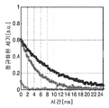
도 1a는 시간에 따른 정규화된 세기(intensity)에 대한 미가공 값의 플롯을 도시한다.
도 1b는 측정된 수명의 플롯을 도시한다.
도 1c는 예시적인 수명 맵을 도시한다.
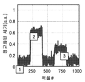
도 1d는 도 1c의 맵 내 픽셀 어레이를 가로지르는 정규화된 세기를 도시한다.
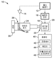
도 2는 본 기술에 따른 예시적인 DOCI 시스템의 다양한 구성 요소를 도시하는 개략적인 블록도이다.
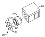
도 3은 도 2의 시스템 내 LED 어레이, 렌즈, 및 카메라의 사시도를 도시한다.
도 4는 본 설명에 따른 UV 다이오드의 단면도를 도시한다.
도 5는 본 설명에 따른 시스템을 사용하여 샘플을 이미징하기 위한 알고리즘 방법의 흐름도를 도시한다.
도 6은 비순차적 레이 트레이싱(광선 추적: Ray tracing)을 통한 LED 어레이 및 대응하는 조명 분산의 실시예를 도시한다.
도 7은 본 설명의 시스템에 따른 예시적인 LED 배열로부터의 표적 조사의 예시적인 플롯을 도시한다.
도 8a는 조명 펄스로부터 시뮬레이션된 임펄스 응답이다.
도 8b는 시뮬레이션된 형광단 방출의 플롯이다.
도 8c는 노이즈 및 오프셋이 도입된 시뮬레이션된 검출된 방출의 플롯이다.
도 8d는 교정 및 붕괴 이미지 (암전류에 의한 오프셋에 의해 교정된, Δ) 및 붕괴 이미지 게이트 폭의 함수로서의 값의 시뮬레이션 플롯이다.
도 9a는 도 9b의 두피 조직 샘플 이미지에 대응하는 출력 형광의 예시를 도시한다.
도 10a는 도 10b의 혀 조직 샘플 이미지에 대응하는 출력 형광의 예시를 도시한다.
도 11은 종양, 근육, 지방, 콜라겐에 대한 파장의 함수로서 계산된 상대 수명의 플롯을 도시하며, 각 조직 유형 사이의 뚜렷한 차이를 나타낸다.
도 12는 근육, 콜라겐 및 지방에 대한 다양한 파장에서의 통계적 유의성을 나타내는 플롯이다.
도 13은 환자 구강 조직의 생체 내 이미지이다.
도 14는 도 13의 영역의 일부의 생체 외 H&E 이미지이다.
도 15a는 도 13의 혀 조직의 확대 재구성된 RGB 이미지이다.
도 15b 내지 도 15e는 407nm, 434nm, 465nm, 및 494nm에서 생체 내 DOCI 이미지를 도시한 것으로, 도 15a의 재구성된 이미지의 FOA(Field of view)를 도시한다.
도 16a는 도 15a의 재구성된 RGB 이미지의 확대된 부분을 도시한다.
도 16b 내지 도 16e는 각각 도 16a와 동일한 관측시야(FOV)를 갖는 407nm, 434nm, 465nm, 및 494nm에서 생체 외 이미지를 나타낸다.
도 17a는 부갑상선 조직의 가시적인 이미지를 도시한다.
도 17b는 도 17a의 조직의 DOCI 이미지를 도시한다.
도 17c는 도 17a의 조직의 조직학 이미지를 도시한다.
도 18a는 전암성 세포 생리학을 가지는 입술을 갖는 대상의 입의 이미지이다.
도 18b는 염증이 있는 입술 (양성 세포 생리학)을 갖는 제 2 대상의 입의 이미지이다.
도 18c는 대상의 입술의 DOCI 이미지와 함께 겹쳐져있는 도 18a의 이미지이다.
도 18d는 대상의 입술의 DOCI이미지와 함께 겹쳐져있는 도 18b의 이미지이다. The techniques described herein will be more fully understood with reference to the following figures for illustrative purposes only:
1A shows a plot of raw values versus normalized intensity over time.
1B shows a plot of the measured lifetime.
1C shows an example life map.
1D shows the normalized intensity across the pixel array in the map of FIG. 1C.
2 is a schematic block diagram showing various components of an exemplary DOCI system according to the present technology.
FIG. 3 shows a perspective view of the LED array, lens, and camera in the system of FIG. 2.
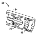
4 shows a cross-sectional view of a UV diode according to the present description.
5 shows a flow diagram of an algorithm method for imaging a sample using a system according to the present description.
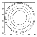
FIG. 6 shows an embodiment of an LED array and corresponding illumination dispersion through non-sequential ray tracing (ray tracing).
7 shows an exemplary plot of target irradiation from an exemplary LED arrangement according to the system of the present description.
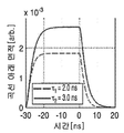
8A is a simulated impulse response from illumination pulses.
8B is a plot of simulated fluorophore emission.
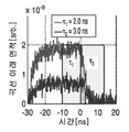
8C is a plot of simulated detected emission with noise and offset introduced.
8D is a simulation plot of calibration and collapse images (Δ, corrected by offset by dark current) and values as a function of collapse image gate width.
9A shows an example of output fluorescence corresponding to the scalp tissue sample image of FIG. 9B.
10A shows an example of output fluorescence corresponding to the tongue tissue sample image of FIG. 10B.
FIG. 11 shows a plot of relative lifespan calculated as a function of wavelength for tumors, muscles, fats, and collagen, showing distinct differences between each tissue type.
12 is a plot showing statistical significance at various wavelengths for muscle, collagen and fat.
13 is an in vivo image of patient oral tissue.
14 is an in vitro H&E image of a portion of the region of FIG. 13.
15A is an enlarged reconstructed RGB image of the tongue tissue of FIG. 13.
15B-15E show DOCI images in vivo at 407 nm, 434 nm, 465 nm, and 494 nm, and show the field of view (FOA) of the reconstructed image of FIG. 15A.
16A shows an enlarged portion of the reconstructed RGB image of FIG. 15A.
16B to 16E show in vitro images at 407 nm, 434 nm, 465 nm, and 494 nm, respectively, having the same field of view (FOV) as in FIG. 16A.
17A shows a visible image of parathyroid tissue.
17B shows the DOCI image of the tissue of FIG. 17A.
17C shows the histological image of the tissue of FIG. 17A.
18A is an image of the mouth of a subject with lips with precancerous cell physiology.
18B is an image of the mouth of a second subject with inflamed lips (positive cell physiology).
18C is the image of FIG. 18A superimposed with the DOCI image of the subject's lips.
18D is the image of FIG. 18B superimposed with the DOCI image of the subject's lips.
본 명세서의 시스템 및 방법은 상이한 생리학적 과정을 갖는 세포 그룹 사이의 형광단 수명의 자연발생적 차이를 구현하여 대비를 생성하는데 사용되고, 고유한 알고리즘이 적용되어 기술적 요구를 완화시킨다. 조직자가 형광의 경우, 자연발생 형광단을 사용하여 대비를 만든다(예: 흑광 이미징). 일 실시예에서, 표적은 짧은 펄스광으로 조명되고, 밝고 어두운 것으로 감쇄될 때 방출의 세기가 측정된다. 어떤 영역이 "빛나는(glows)" 기간은 어떤 유형의 조직이 조명되었는지를 결정한다. 예를 들어, 암성 조직은 일반적으로 빠른 붕괴(decay)와 관련이 있고, 비-암성 조직은 느린 붕괴와 관련이 있다.The systems and methods herein are used to create contrast by implementing naturally occurring differences in fluorophore lifetimes between groups of cells with different physiological processes, and unique algorithms are applied to alleviate technical needs. In the case of organ fluorescence, contrast is made using a naturally occurring fluorophore (eg, black light imaging). In one embodiment, the target is illuminated with a short pulsed light, and the intensity of the emission is measured when attenuated to light and dark. The period in which an area “glows” determines which type of tissue is illuminated. For example, cancerous tissue is generally associated with rapid decay, and non-cancerous tissue is associated with slow decay.
본원에 개시된 시스템 및 방법은 상이한 생리학적 과정 또는 상이한 조직을 갖는 세포 그룹 사이의 마진 검출을 위해 구성되며, 예를 들어, 전암성(pre-cancerous), 전-암성(pre-malignant), 암성(cancerous) (예를 들어, 구강 및 두경부 편평 세포 암종 (OSCC)) 및 비 암성 또는 양성 (예를 들어, 염증성) 조직 또는 세포 그룹 사이의 마진으로 제한되지는 않는다.The systems and methods disclosed herein are configured for detecting margins between groups of cells having different physiological processes or different tissues, for example, pre-cancerous, pre-malignant, cancerous ( cancerous) (eg, oral and head and neck squamous cell carcinoma (OSCC)) and non-cancerous or benign (eg, inflammatory) tissues or groups of cells.
A. 시스템 및 방법 A. System and method
도 1a 내지 도1d는 본 기술에 따른 시간-분해된 자가 형광을 수행하기 위한 예시적인 과정을 도시한다. 수명 붕괴 곡선을 생성하기 위한 경광은 함수로 측정된다. 형광은 일반적으로 여기 펄스 후 피코초(picoseconds)에서 나노초(nanoseconds)의 기간에 걸쳐 붕괴된다. 이미지의 각 지점에서의 형광의 붕괴 속도 (즉, "수명")은 형광 '수명'값의 분 포로 표시된다. 형광단의 존재하에서, 붕괴 곡선의 기울기는 유한한 여기 상태의 존재로 인하여 덜 가파르다. 따라서, 더 긴 수명을 갖는 형광단은 더 큰 기울기를 갖는다. 형광단의 특정 수명에 기초하여, 상이한 조직 (예를 들어, 정상 및 암성 조직) 사이의 형광이 구별될수 있다. 도 1a는 도시된 바와 같이, 원시 세기(raw intensity) 값의 예시적인 플롯이 먼저 얻어진다는 것을 도시한다. 도 1b는 측정된 수명의 예시적인 플롯을 도시한다. 그 후 수명 뱁은 도 1c에 도시된 바와 같이 생성된다. 도 1d는 도 1c의 맵 내 픽셀 어레이에 걸친 정규화된 세기를 도시한다. 표준 형광과 비교하여, 평생 형광은 클러터(clutter)에 대한 견고성, 최대 대비 생성을 향상시켰으며, 생체 내 영상화에 이상적이다. 1A-1D show an exemplary process for performing time-resolved autofluorescence according to the present technology. The light spectra to create a life decay curve are measured as a function. Fluorescence generally decays over a period of picoseconds to nanoseconds after the excitation pulse. The rate at which fluorescence decays at each point in the image (ie, “life”) is expressed as a distribution of fluorescence “life” values. In the presence of the fluorophore, the slope of the decay curve is less steep due to the presence of a finite excited state. Thus, fluorophores with longer lifetimes have a larger slope. Based on the specific lifetime of the fluorophore, fluorescence between different tissues (eg, normal and cancerous tissues) can be distinguished. 1A shows that, as shown, an exemplary plot of raw intensity values is obtained first. 1B shows an exemplary plot of measured life. The lifetime 뱁 is then created as shown in FIG. 1C. 1D shows the normalized intensity across the pixel array in the map of FIG. 1C. Compared to standard fluorescence, lifetime fluorescence improves clutter resistance, maximum contrast generation, and is ideal for in vivo imaging.
도 2는 본 기술에 따른 예시적인 동적 광학 대비 이미징 (Dynamic Optical Contrast Imaging: DOCI) 시스템(10)의 다양한 구성 요소를 도시하는 개략적인 블록도이다. 바람직한 실시예에서, DOCI 시스템(10)은 이미징 렌즈(24), 및 렌즈(24)의 전방에 배치된 UV 다이오드(28)(LED) 어레이(26)를 포함한다. UV 다이오드(28)의 어레이(26)는 펄스 발생기(12) 및 다이오드 드라이버(14)에 의하여 생성된 신호를 통하여 샘플(30)을 조명하도록 구성된다. 펄스 발생기(12)는 또한 딜레이 라인(16)을 통하여 게이트 카메라(20)에 연결된다. 카메라(20)는 냉각된 iCCD (18)와 렌즈(24) 사이에 배치된 UV 레이저 라인 필터(22)를 포함하는 것이 바람직하다 (필터(22)는 광학 경로 내의 어느곳에나 배치될 수 있다). LED 어레이(26) 및 카메라(20) 출력은 프로세서(42), 어플리케이션 소프트웨어(46), 및 프로세서(42) 상 실행을 위한 어플리케이션 소프트웨어(46)를 저장하는 메모리(44)를 포함하는 컴퓨터(40) (또는 컴퓨팅 장치와 유사)에 연결된다. 어플리케이션 소프트웨어(46)는, 시스템의 구성 요소 (예를 들어, 펄스 발생기(12), LED 어레이(26), 다이오드 드라이버(14), 등)을 동작시키고, iCCD(18)로부터 획득된 데이터를 처리하기 위한 명령(예를 들어, 아래에 상세히 설명되고 도 5에 도시된 방법(50)을 수행하기 위한 명령)을 포함한다. 2 is a schematic block diagram showing various components of an exemplary Dynamic Optical Contrast Imaging (DOCI)
필터(22)는 iCCD(18)에 의해 수신된 광을 제한하도록 구성된 필터 휠을 포함할 수 있어서, 주어진 시간에 특정 파장 또는 파장 범위만 이미징된다. 예를 들어, 제 1 이미지는 오로지 적색광만으로 획득되고 제 2 및 제 3 이미지는 청색 및 녹색광으로만 제한된다. 이러한 이미지들은 디스플레이를 위해 상이한 패널에 동시에 디스플레이되거나, 예를 들어, 하나 이상의 생성된 DOCI 이미지의 다양한 파장(도 15a 내지 도 15c 참조)에서의 시각화(예를 들어, 나란히 디스플레이)와 함께 수탁 이미지(fiduciary image)로서 사용될 수 있는 재구성된 RGB 이미지를 생성하기 위해 조합될 수 있다. 일 실시예에서, 필터(22)는 다음 방출 대역에 중심을 둔 광을 제한하기 위해 10 개의 필터를 포함하는 필터 휠을 포함 한다: 407nm, 434nm, 465nm, 494nm, 520nm, 542nm, 572nm, 605nm, 632nm 및 676nm. 상기 밴드는 단지 예시적인 목적을 위한 것이며, 다른 변형이 고려된다는 것이 이해된다.
일 실시예에서, UV 다이오드 어레이(26)는 375nm 의 파장에서 조명된다 (이것은 표적 조직/ 장치 사양에 기초하여 변경될 수 있다). 발광 다이오드 조명 회로 (다이오드 드라이버 (14))는 370nm의 중심 파장, 약 4.5μm/l의 평균 광 파워 및 30ns의 펄스 폭에서 동작한다. 평균 전력이 낮고 파장이 길면 단백질, DNA 및 기타 분자가 영상의 영향을 받지 않는다. In one embodiment, the
도 3은 카메라(20), 렌즈(24), 및 LED 어레이(26)의 분해 사시도를 도시한다. 바람직한 실시예에서, LED 어레이(26)는 개별 LED(28)가 렌즈(24) 주위에서 원주 방향으로 정렬되도록 렌즈(24)의 전방과 정렬된다. 프레임(32)은 개별 LED(28)를 적절한 정렬 상태로 유지하고, 어레이(26)를 렌즈(24)에 결합할 수 있게 한다. 3 shows an exploded perspective view of the
도 4는 본 설명에 따른 UV 다이오드(28)의 단면도를 도시한다. 각각의 UV 다이오드(28)는 UV LED(36)를 수용하도록 구성된 하우징(30) 및 전체 시야(field of view) 또는 그 상당 부분에 걸쳐 집중된 분산을 위해 투과된 광을 성형하도록 구성된 구면 렌즈(38)를 포함한다.4 shows a cross-sectional view of a
도 5는 본 설명의 시스템(10)을 사용하여 샘플(30)을 이미징하기위한 알고리즘 방법(50)의 흐름도를 도시한다. 방법(50)은 복잡한 수학적 모델을 획득된 데이터에 맞추지 않고도 프로브된 조직의 집합 형광단에 비례하는 픽셀값을 생성하기 위해 고유의 이미지 프레임 정규화 방식을 적용한다. 이것은 조명 펄스의 시간 프로파일에 대한 요구를 완화시키고 피코초 펄스 레이저(FLIM 에 일반적으로 필요함)를 나노초 펄스 발광 다이오드(LED)로 대체할 수 있게 한다. 조명은 짧은 펄스 (nanosecond order) 상승 및 하강 시간을 갖는 긴 펄스 지속 시간 (약 30 ns)으로 UV 광원 (26) (예를 들어, 375nm에서)을 통해 수행되어 차이 붕괴율의 형광단 사이의 대비를 생성한다. 이 방식으로, 모든 픽셀이 동시에 획득되는 비교적 짧은 시간 프레임 (방출 대역 당 ~ 10 초) 내에서 거시적 (미시적이지 않은) 시야(FOV)에 대한 형광단 수명의 확장 가능한 매핑이 가능하다. 따라서, 이러한 개선은 수술 중 임상 사용을 향한 중요한 단계를 제공한다. 5 shows a flow diagram of an
도 5에 도시된 바와 같이, 2개의 게이트, 한 개의 교정 획득 기간(Tc), 1개의 붕괴 이미지 획득 기간 (n)이 획득된다. 두경부 이미징에 관심이 있는 대부분의 조직 구성 요소의 형광 수명은 1ns 에서 10ns의 범위에 속한다. 그러므로, 초기 조명 후 >10ns에서 획득된 방출 이미지는, 정상 상태의 조직의 자발형광의 정확한 표현으로 간주될 수 있다. 5, two gates, one calibration acquisition period (T c ), and one collapsed image acquisition period (n) are obtained. The fluorescence lifetime of most tissue components interested in head and neck imaging is in the range of 1 ns to 10 ns. Therefore, the emission image obtained at >10 ns after initial illumination can be regarded as an accurate representation of spontaneous fluorescence of the steady-state tissue.
획득된 형광 방출을 교정하기 위해, 이미지는 교정 획득 기간(Tc)에서 UV 펄스 중반 동안 캡쳐되어 이미지(58)를 생성하며, 이하 "FOV 교정 이미지"로 지칭된다. 계속해서, 조명 펄스 붕괴 (붕괴 이미지 획득 기간 (τ1))에서, 제 2 이미지(56)가 갭처되며, 이하 "FOV 붕괴 이미지"로 지칭된다. To correct the acquired fluorescence emission, the image is captured during the middle of the UV pulse in the calibration acquisition period T c to generate the
도 5에서, 점선(52)은 온/오프 단계 동안의 LED 강도를 나타내고, 실선(54)은 (각 개별 픽셀에 대해) 획득된 형광 세기를 나타낸다. FOV 붕괴 이미지(56)는 FOV 붕괴 이미지(56)를 FOV 교정 이미지(58)로 나누어 FOV 상대 수명 맵(60)을 생성함으로써(교정 이미지에 의해 픽셀 단위로) 정규화된다. 바람직한 실시예에서, 이미지는 수만은 파장에서 획득될 수 있다(예를 들어, 비아 필터(22)는 특정 파장이 카메라(20)에 의해 수신될 수 있게 하는 다수의 상이한 파장 범위 중에서 선택을 허용하는 필터 휠(filter wheel) 또는 유사한 장치를 포함할 수 있다).In Fig. 5, the dotted
바람직한 실시예에서, FOV 붕괴 이미지(56) 및 결과적인 픽셀 값은 조명된 영역의 집합 형광단 붕괴 시간에 비례한다. 이 픽셀값은 상대 조직 수명을 나타내며 DOCI 픽셀값이라고 칭한다. DOIC 는 수명이 긴 형광단이 정상상태 형광과 관련하여 수명이 짧은 형광단보다 더 많은 신호를 생성한다는 사실에 의존한다. 상대 수명 맵(60)을 추가로 처리하고 생성하기 위해 추가 이미지 (예를 들어, 배경 이미지 등)이 획득될 수 있다는 것이 또한 이해된다. In a preferred embodiment, the FOV collapsed
상대 수명 맵(60)은 가색 맵으로서, 또는 오퍼레이터에게 라인, 모양, 색상, 청각 신호의 정량적 상대 수명 픽셀 값의 시각적 표현으로서 디스플레이될 수 있다. The
도 6은 개별 LED (28)로부터 조명 빔(62a 내지 62f)을 통한 비순차적 광선 추적(레이 트레이싱)을 도시한 본 LED 어레이(26)의 실시예를 도시하고, FOV를 가로질러 각 LED 로부터의 여기 광을 집속 및 곱한다. 바람직한 실시예에서, 비순차적 광선 추적의 패턴은 LED 전구(38) 및 렌즈(38)(도 4)의 선택에 따라 변화되는 조명 분포 및 세기 조정 및 형상이다. 도 7은 예시적인 LED 어레이(26) 및 결과적인 광선 추적 조명 패턴으로 인한 타겟 조사의 예시적인 플롯을 도시한다.FIG. 6 shows an embodiment of the
B. B. DOCIDOCI : 운영 원칙 : Operating principles
분석의 목적을 위해, 조명 펄스는 조명 펄스의 대역한계를 모델링하기 위해 단일 극 저역 통과 필터의 임펄스 응답과 관련된 이상적인 직사각형 펄스로서 모델링된다 (도8a). 단일 시간 상수 지수 임펄스 응답은 식(1)에 기술되어 있다:For analysis purposes, the illumination pulse is modeled as an ideal rectangular pulse associated with the impulse response of a single pole low pass filter to model the band limit of the illumination pulse (Figure 8A). The single time constant exponential impulse response is described in Eq. (1):
식 1: , 여기서 τk= τd (조명 시간 상수), T1 (형광단 1 시간 상수), T2 (형광단 2 시간 상수).Equation 1: , Where τk = τd (illumination time constant), T1 (
이러한 조명 프로파일은 식 2에서 설명된다:This lighting profile is described in Equation 2:
식 2: , 여기서 T0는 펄스 폭. Equation 2: Where T 0 is the pulse width.
형광단 특정 수명은 수식 1로 모델링 될 수 있다. UV 펑핑된 형광단의 형광 방출은 수식 3에 따른 다이오드 조명 및 형광 붕괴 시간의 컨볼루션으로 기록된다:The fluorophore specific lifetime can be modeled with
식 3: Equation 3:
이들 컨불루션 적분의 그래픽 표현은 도 8b에 도시된 것으로, 각각의 미량은 형광단 1 및 2의 형광 방출이다. The graphical representation of these convolutional integrals is shown in FIG. 8B, each trace being the fluorescence emission of
다음으로 대역 제한 화이트 가우시안 잡음(band limited white Gaussian Noise) 및 (암전류로 인한) 오프셋이 도입되며, 그 출력은 도 8c에 도시된다. 추가 이미지 혼동기로서, 관심있는 형광단을 보유하는 픽셀은 1)조명의 효과 및 2) 형광단 1에서 검출되는 형광 방출의 90% 감소 및 형광단 2에서 97.5%감소로 임의로 선택된 방해물을 차단한다. 이러한 결과는 도 9C의 이미지에 도시된 출력에 통합된다. 플루언스(fluence) 흡수, 상관되지 않은(uncorrelated) 백색 측정 노이즈 및 암전류의 조합은 수신된 형광된 2 내지 6dB의 피크 SNR 의 세기를 감소시킨다. (도 8b 내지 도8b의 시간축은 조명/방출 붕괴가 t=0에서 발생하도록 정의된다)Next, band limited white Gaussian Noise and offset (due to dark current) are introduced, the output of which is shown in FIG. 8C. As a further image confuser, the pixels holding the fluorophore of interest block randomly selected obstructions with 1) the effect of illumination and 2) a 90% reduction in fluorescence emission detected in
교정 측정은 조명 펄스가 Ti 게이트 폭으로 붕괴되기 시작하기 직전에 획득된다. 이러한 과정은 도 8c에 도시되고, 수식 4로 설명된다:Calibration measurements are taken just before the illumination pulse begins to collapse into the Ti gate width. This process is illustrated in Figure 8c and is described by Equation 4:
식 4: Equation 4:
붕괴 측정은 게이트 폭 T2를 사용한 수식 5(또한 도 8c에 도시된)에 설명된 유사한 획득 방법론에 따른다:The collapse measurement follows a similar acquisition methodology described in Equation 5 (also shown in Figure 8C) using gate width T2:
식 5: Equation 5:
결과적인 DOCI 픽셀값은 수식 6에 의하여 계산된다:The resulting DOCI pixel value is calculated by Equation 6:
식 6: , 이는 교정 이미지와 붕괴 이미지(암전류,△로 인한 오프셋에 의해 교정된)의 비율이고, 이 값은 붕괴 이미지 게이트 폭의 함수로서 도 8d에 도시되었으며, 여기서 게이트 길이가 증가될수록 두 형광단 사이의 계산된 차이 신호가 증가하고, 두 신호는 조명 및 형광단 붕괴시간의 합으로 (이상적으로) 수렴한다. Equation 6: , This is the ratio of the calibration image and the collapsed image (corrected by the offset due to dark current, Δ), this value is shown in Figure 8d as a function of the collapsed image gate width, where the gate length increases as the distance between the two fluorophores increases. The calculated difference signal increases, and the two signals converge (ideally) to the sum of the illumination and fluorophore decay times.
DOCI 시스템 및 방법의 하나의 강점은 정상상태 형광으로 정규화된 붕괴 시간 곡선 하의 면적을 계산함으로써 형광단 수명을 재비로 변환한다는 것이다. 정지 잡음의 한계에서, 이러한 과정은 가려짐(obscurants)의 변화에 강하고 낮은 SNR 하에서 상댕한 대비를 생성할 수 있다. One strength of the DOCI system and method is that it converts the fluorophore lifetime into a refuel by calculating the area under the decay time curve normalized to steady-state fluorescence. At the limit of static noise, this process is resistant to changes in obscurants and can produce subdued contrast under low SNR.
이 방법은 임상 이미징에 이상적인 여러가지 주요 이점을 제공한다. 첫째, 위에서 논의한 바와 같이, 계산 기술이 간단하다; 수명이 계산되지 않으므로, 곡선 피팅이 필요하지 않다. 둘째, 완화된 수명 계산은 더 긴 펄스 지속 시간 간격과 하강 시간 (>1ns)를 허용한다. 따라서, 고가의 레이저 대신 전자 펄스에 의하여 구동되는 저렴한 LED 가 사용될 수 있다. 셋째, 두 형광단의 방출 붕괴 사이의 신호 차이는 게이트 시간과 양의 상관관계가 있다. 다시 말해, 붕괴 이미지동안 게이트가 더 오래 개방될수록, 차이 신호가 더 커진다. 또한, 신호 대 잡음비(SNR)은 검출기의 통합 특성으로 인한 신호 증가 및 측정 노이즈 감소로 인하여 크게 증가한다. 이는 붕괴 시간을 정확하게 샘플링하기 위해 게이트가 짧아야하는 FLIM과는 완전히 대조적이다. 오히려, DOCI 과정의 경우, 게이트 폭이 증가하면 대비가 향상되어 노이즈 분산을 줄이면서 수집된 광자의 전체수를 증가시킨다. 이러한 기술의 단순성 및 고유 감도는 임상 이미징의 실용적인 대형 FOV 를 가능하게 한다. This method provides several major advantages that are ideal for clinical imaging. First, as discussed above, the calculation technique is simple; No curve fitting is required since the lifetime is not calculated. Second, relaxed life calculations allow longer pulse duration intervals and fall times (>1 ns). Therefore, inexpensive LEDs driven by electron pulses can be used instead of expensive lasers. Third, the signal difference between the emission collapse of the two fluorophores is positively correlated with the gate time. In other words, the longer the gate opens during the collapsed image, the greater the difference signal. In addition, the signal-to-noise ratio (SNR) increases significantly due to increased signal and reduced measurement noise due to the integrated nature of the detector. This is in stark contrast to FLIM, where the gate must be short to accurately sample the decay time. Rather, in the case of the DOCI process, as the gate width increases, the contrast improves, reducing noise dispersion and increasing the total number of collected photons. The simplicity and intrinsic sensitivity of this technique enables practical large-scale FOV of clinical imaging.
C.실험 결과(Experimental Results)C.Experimental Results
DOCI 시스템의 효능 및 상기 상세히 기재된 방법을 입증하기 위해 신선한 조직 시험 (84명의 환자 및 190개의 별개의 이미지)를 통해 다수의 생체 외 시험을 수행하였다. 도 9a는 도 9b의 두피 조직 샘플 이미지에 대응하는 예시적인 출력 형광을 도시한다. 점선 영역 1은 종양조직에 해당하고, 점선 영역 2는 근육조직에 해당한다. 도 10a는 도 10b의 혀 조직 샘플 이미지에 대응하는 예시적인 출력 형광을 도시한다. 점선 영역 1은 종양 조직에 해당하고, 점선 영역 2는 근육 조직에 대응한다. A number of in vitro tests were performed through fresh tissue tests (84 patients and 190 separate images) to demonstrate the efficacy of the DOCI system and the methods described in detail above. 9A shows exemplary output fluorescence corresponding to the scalp tissue sample image of FIG. 9B. The dotted
도 11은 종양, 근육, 지방, 및 콜라겐에 대한 파장의 함수로서 계산된 상대 수명의 플롯을 도시한다. 결과는 DOCI 수명 맵핑이 대부분의 방출 파장에 걸쳐 조사중인 모든 4가지 조직 유형 (종양, 지방, 근육 및 콜라겐)간의 대조적인 통계로서 유의미한 차이를 생성했음을 입증한다. 악성 조직에서 형광 수명의 감소가 관찰되었으며, 종양의 생화학적 마커에 대해 보고된 짧은 수명과 일치한다. 11 shows a plot of relative lifespan calculated as a function of wavelength for tumor, muscle, fat, and collagen. The results demonstrate that DOCI lifespan mapping produced a significant difference as a contrasting statistic between all four tissue types under investigation (tumor, fat, muscle and collagen) across most emission wavelengths. A decrease in fluorescence lifespan was observed in malignant tissue, consistent with the short lifespan reported for biochemical markers of tumors.
도 12는 근육, 콜라겐 및 지방에 대한 다양한 파장에서의 통계적 유의성응ㄹ 나타내는 플롯이다. 근육과 종양 사이의 통계적 유의성 (P<0.05)는 10개의 방출 파장 중 10개, 콜라겐과 종양 사이의 방출 파장 10개 중 8개, 지방과 종양 사이의 파장 10개 중 2개가 확립되었다. 이 연구는 DOCI 가 OSCC 와 주변 정상 조직 및 수술 절제의 효능을 극대화할 수 있는 잠재력을 정확하게 구별할 수 있는 가능성을 보여준다. 12 is a plot showing statistical significance at various wavelengths for muscle, collagen and fat. Statistical significance (P<0.05) between muscle and tumor was established in 10 of 10 emission wavelengths, 8 of 10 emission wavelengths between collagen and tumor, and 2 of 10 wavelengths between fat and tumor. This study demonstrates the potential for DOCI to accurately differentiate between OSCC and its potential to maximize the efficacy of peripheral normal tissue and surgical resection.
OSCC 의 수술 중 검출에서 DOCI의 진단적 유용성을 평가하기 위해, OSCC에 대한 외과적 절제를 받는 15명의 연속 환자에 대한 생체 내 연구를 수행하였다. 생검으로 입증된(Biopsy-proven) 편평 상피 세포 암종 신 생물(squamous cell carcinoma meoplasms)은 다음의 머리와 목 부위와 하위 부위에서 얻어졌다: 귀, 턱선, 두피, 구강, 구강 인두, 인두, 및 목. 모든 표본은 절개 전에 DOCI 시스템 (10) (도 2)로 이미지화되었다. 종양 절제 후, 표본을 즉시 의심스러운 병변의 종양 및 인접한 정상 조직을 포함하는 다수의 새로운 샘플로 나누고 조직학적 평가를 위해 제출하였다. 병리학자에 의해 신생물의 영역을 확인하고, DOCI 이미지 결과에 대해 눈을 멀게하고, 상대적인 수명 값을 병리학적 진단과 무관하게 계산하였다. To evaluate the diagnostic usefulness of DOCI in surgical detection of OSCC, an in vivo study of 15 consecutive patients undergoing surgical resection for OSCC was performed. Biopsy-proven squamous cell carcinoma meoplasms were obtained from the following head and neck regions and subregions: ears, jawline, scalp, oral cavity, oral pharynx, pharynx, and neck . All specimens were imaged with the DOCI system 10 (Figure 2) prior to incision. After tumor resection, samples were immediately divided into a number of new samples containing tumors of suspicious lesions and adjacent normal tissues and submitted for histological evaluation. The area of the neoplasm was confirmed by the pathologist, blinded to the results of the DOCI image, and the relative lifespan values were calculated independent of the pathological diagnosis.
혀 OSCC의 DOCI 및 가시 이미지가 도 13 내지 도 16에 도시된다. DOCI 시스템 및 본 개시에 따른 방법의 바람직한 실시예에서, DOCI 이미지 색상 맵은 청색을 전체 최소 상대 붕괴 수명으로, 적생을 최대 상대 붕괴 수명으로 변환한다. 감소된 DOCI 픽셀 값은 보다 빠른 형광 신호의 붕괴를 나타내며, 전체적으로 더 짧은 수명을 나타낸다. DOCI 이미지는 도 15b 내지 도 18E에 그레이 스케일(grey scale)로 가변적으로 도시되었지만, DOCI 이미지에서 더 밝은 양태는 최소 상대 붕괴 수명(청색), DOCI 이미지에서 더 어두운 양태는 최대 상대 붕괴 수명(적색)에 대응한다는 것이 이해된다. DOCI and visible images of the tongue OSCC are shown in Figures 13-16. In a preferred embodiment of the DOCI system and the method according to the present disclosure, the DOCI image color map converts blue to the total minimum relative decay lifetime and lifetime to maximum relative decay lifetime. Reduced DOCI pixel values indicate faster decay of the fluorescence signal and overall shorter lifetime. DOCI images are variably shown on a gray scale in FIGS. 15B-18E, but the lighter aspect in the DOCI image is the minimum relative collapse life (blue), and the darker aspect in the DOCI image is the maximum relative collapse life (red). It is understood that it corresponds to.
도 14가 도 13의 영역의 일부의 생체 외 H&E 이미지를 도시한 반면, 도 13은 환자 구강 조직의 생체 내 이미지이다. 도 15a는 도 13의 혀 조직의 확대된, 재구성된 RGB 이미지를 도시한다. 도 15b 내지 도 15e는 407nm, 434nm, 465nm, 및 494nm에서 생체 내 DOCI 이미지를 도시한 것으로, 도 15a의 재구성된 이미지의 FOA(Field of view)를 도시한다. 도 16a는 도 15a의 확대한 재구성된 RGB 이미지를 도시한다. 도 16b 내지 도 16e는 각각 도 16a의 혀 이미지 부분의 생체 외 DOCI 이미지로, 도 16a의 이미지의 동일한 관측시야(FOV)를 갖는 407nm, 434nm, 465nm, 및 494nm에서 생체 외 이미지를 나타낸다. 14 shows an in vitro H&E image of a portion of the region of FIG. 13, while FIG. 13 is an in vivo image of patient oral tissue. 15A shows an enlarged, reconstructed RGB image of the tongue tissue of FIG. 13. 15B-15E show DOCI images in vivo at 407 nm, 434 nm, 465 nm, and 494 nm, and show the field of view (FOA) of the reconstructed image of FIG. 15A. 16A shows an enlarged reconstructed RGB image of FIG. 15A. 16B to 16E are in vitro DOCI images of the tongue image portion of FIG. 16A, respectively, and showing in vitro images at 407 nm, 434 nm, 465 nm, and 494 nm with the same field of view (FOV) of the image of FIG. 16A.
바람직한 실시예에서, 어플리케이션 소프트웨어(46)(도 2)는 하나 이상의 파장에서 (도 15b 내지 도 15e) 하나 이상의 DOCI 이미지와 함께 (예를 들어, 개별 패널로서 나란히 재구성된 RGB 이미지를 동시에 출력하도록 구성될 수 있다. 재구성된 RGB 이미지를 획득하기 위하여, 제 1 (DOCI가 아닌) 이미지는 오로지 적색 광(예를 들어, 필터 휠(22) 상의 적절한 필터의 선택을 통해)만으로 획득될 수 있고(도 2), 제 2 및 제 3 이미지는 청색 및 녹색 광으로만 제한될 수 있다 (iCCD(18)dms 데이터 수집을 위한 하나의 빈(일반적으로 다중 빈 RGB 검출기와 반대)를 포함함). 이들 이미지는 동일한 검출기(18)를 통해 획득되는 다양한 파장(도 15a 내지 도 15c)에서 하나 이상의 생성된 DOCI 이미지의 시각화(나란히 디스플레이)와 함께 실시간 수탁 이미지로서 사용될 수 있는 재구성된 RGB 이미지(도 15b)를 생성하기 위해 조합되거나 융합될 수 있다. In a preferred embodiment, application software 46 (FIG. 2) is configured to simultaneously output one or more DOCI images at one or more wavelengths (FIGS. 15B-15E) (eg, reconstructed RGB images side-by-side as separate panels). In order to obtain a reconstructed RGB image, the first (not DOCI) image can only be acquired with red light (eg, through selection of an appropriate filter on filter wheel 22) (FIG. 2), the second and third images can be limited to blue and green light only (including one bin for collecting iCCD(18)dms data (usually as opposed to multiple bin RGB detectors). Is a reconstructed RGB image (FIG. 15B) that can be used as a real-time trust image with visualization of one or more generated DOCI images (display side by side) at various wavelengths (FIGS. 15A-15C) obtained through the
도 15b 내지 도 15e에 도시된 바와 같이, 생체 내 DOCI 이미지는 OSCC 조직과 주변 정상 조직 사이의 뚜렷한 대조를 입증하였다. OSCC의 영역은 주변 정상 조직의 수명과ㅏ 비교하여 감소된 상대 수명을 특징으로 한다. 15B-15E, DOCI images in vivo demonstrated a clear contrast between OSCC tissue and surrounding normal tissue. The area of OSCC is characterized by a reduced relative life compared to the life of surrounding normal tissue.
여기된 조직의 절제 후 생체 외 이미지에서 유사한 상대 수명 측정이 관찰되었다 (도 16b 내지 도 16c). 전 범위의 방출 파장에 걸쳐 생체 외 OSCC 와 생체 내 OSCC 사이의 강한 양의 상관관계는 DOCI 및 이와 관련된 이미지 분석 방법이 생체 내 임상 용도로 직접 번역될 수 있음을 제안한다. 생체 내 상피 표면으로부터 DOCI 이미지가 획득됨에 따라서, 동일한 이미지 평면을 따라 획득된 생체 외 표본의 DOCI 이미지는 임상적으로 관련이 있으며, 전체 검사에서 분명하지 않은 조직 구조/유형에 대한 세부 사항을 밝혀냈다. Similar relative life measurements were observed in the in vitro images after excision of the excited tissue (FIGS. 16B-16C ). The strong positive correlation between in vitro OSCC and in vivo OSCC over the full range of emission wavelengths suggests that DOCI and related image analysis methods can be translated directly into in vivo clinical use. As DOCI images were obtained from in vivo epithelial surfaces, DOCI images of ex vivo specimens acquired along the same image plane were clinically relevant and revealed full details of tissue structures/types that were not apparent in the overall examination.
생체 외 조직 및 상응하는 조직학을 영상화된 평면 및 상피 표면에 평행하게 단면화하여 암성 및 인접 기질을 포획하였다. DOCI 로 평가된 종양, 지방, 근육 및 콜라겐 영역 사이의 상대 수명의 차이가 보고되었다. In vitro tissue and corresponding histology were cross sectioned parallel to the imaged plane and epithelial surface to capture cancerous and adjacent substrates. Differences in relative lifespan between tumor, fat, muscle and collagen regions evaluated with DOCI were reported.
부갑상선 국소화를 위한 실시간 생체 내 사용을 위해 DOCI 시스템 및 방법을 조사하였다. 부갑상선 조직의 생체 외 DOCI 데이터는 이 기술을 조직의 "상대적 붕괴 맵"을 생성하는 신뢰할 수 있는 생체 내 기술로 만드는 잠재력을 보여주며, 부갑상선 샘 위치에 해당하는 수술중 색상 아틀라스를 묘사한다. 일차 부갑상선 기능항진증이 있는 전향적 일련의 환자 (n=81)를 검사했다. 부갑상선 병변과 주변 조직을 수집하였다; 형광 붕괴 이미지는 DOCI 를 통해 획득되었다; 및 개별 생체 외 표본 (n=127 샘플)을 조직학적 평가를 위해 처리하였다. 손으로 묘사된 관심 영역(ROIs)는 조직 병리학적 분석에 의해 결정되었고, 대응하는 고화질 가시 이미지 상에 중첩되었다. 그런 다음 가시 이미지를 수동으로 침식하여 동반 DOCI 이미지에 등록했다. 마지막으로, ROI는 지방 (n = 43), 부갑상선 (n=85), 흉선(n=30) 및 갑상선 조직(n=45)으로부터 평균화되었다. The DOCI system and method were investigated for real-time in vivo use for parathyroid localization. The in vitro DOCI data of parathyroid tissue demonstrates the potential to make this technique a reliable in vivo technique that produces a "relative collapse map" of tissue, and depicts the color atlas during surgery corresponding to the location of the parathyroid gland. A prospective series of patients with primary hyperparathyroidism (n=81) were examined. Parathyroid lesions and surrounding tissue were collected; Fluorescence decay images were obtained through DOCI; And individual ex vivo samples (n=127 samples) were processed for histological evaluation. Regions of interest (ROIs) depicted by hand were determined by histopathological analysis and superimposed on the corresponding high-definition visible image. The visible image was then manually eroded and registered in the accompanying DOCI image. Finally, ROI was averaged from fat (n=43), parathyroid (n=85), thymus (n=30) and thyroid tissue (n=45).
도 17a 내지 도 17c는 생체 외 실험으로부터 샘플링된 부갑상선 조직의 일예를 보여주며, 전체 FOV 에 걸친 DOCI 대조를 입증한다. 이미지에서 관찰된 바와 같이, 부갑상선 조직은 사용된 모든 필터에서 지방과 비교할 때 감소된 상대 수명을 나타냈다. 상이한 생리학적 과정 (예를 들어 부갑상선 대 갑상선 세포 그룹)을 갖는 조직 유형과 세포 그룹 사이의 대조는 모든 방출 파장에 걸쳐 명백하였다. DOCI 부갑상선 영상에서의 조직 대조의 기초는 밀집된 부갑상선 선조 세포 내에 호르몬-특이 단백질, 아미노산, 및 세포 외 갈슘-감지 수용체의 존재에 기인한 것으로 보인다. 이 생체 외 DOCI 데이터는 부갑상선 위치에 상응하는 수술중 색상 아틀라스를 도시하는 부갑상선 조직의 "상대 붕괴 맵"을 생성하기 위해 본 명세서의 기술을 신뢰할 수 있는 생체 내 기술을 적용하는 효능을 입증한다. 17A-17C show an example of parathyroid tissue sampled from in vitro experiments, demonstrating DOCI control across the entire FOV. As observed in the image, parathyroid tissue showed reduced relative lifespan compared to fat in all filters used. The contrast between tissue types and cell groups with different physiological processes (eg parathyroid versus thyroid cell groups) was evident across all emission wavelengths. The basis of tissue control in DOCI parathyroid imaging seems to be due to the presence of hormone-specific proteins, amino acids, and extracellular calcium-sensing receptors in dense parathyroid progenitor cells. This in vitro DOCI data demonstrates the efficacy of applying reliable in vivo techniques to the techniques herein to produce a "relative collapse map" of parathyroid tissue showing the color atlas during surgery corresponding to the parathyroid location.
도 18a 내지 도 18d를 참조하면, 구강 암의 이미징을 통하여 전-암성(pre-cancerous) 또는 전-암성(pre-malignant) 조직 또는 세포 그룹의 생체 내 검출을 위한 연구가 또한 수행되었다. DOC 시스템 및 방법은 한 환자에서 아세트산 성 치염/적혈구증 (전-암성 병변)의 분화를 제공하고, 다른 환자에서는 입술의 염증(예를 들어 햇" 화상 또는 이와 같은 양성 상태)를 제공할 수 있다. 입술의 염증(도 18d)의 DOCI 오버레이를 갖는 가시 이미지(도 18b)와 비교하여, 입술 상의 전-암성 병변의 DOCI 오버레이 (도 18a)를 갖는 가시 이미지(도 18a)가 획득되었다. 18A-18D, studies for in vivo detection of pre-cancerous or pre-malignant tissue or cell groups through imaging of oral cancer have also been performed. DOC systems and methods provide differentiation of acetic gingivitis/erythrocytes (pre-cancerous lesions) in one patient, and inflammation of the lips (eg sun" in the other patient) Burns or a positive condition like this). A visible image (FIG. 18A) with a DOCI overlay of the pre-cancerous lesions on the lips (FIG. 18A) was obtained, compared to a visible image (FIG. 18B) with a DOCI overlay of inflammation of the lips (FIG. 18D).
본 기술의 실시 예는 본 기술의 실시 예에 따른 방법 및 시스템의 흐름도 및/또는 절차, 알고리즘, 단계, 연산, 공식 또는 다른 컴퓨터 묘사를 참조하여 본 명세서에서 설명될 수 있으며, 이는 컴퓨터로서 또한 구현 될 수 있다. 이와 관련하여, 순서도의 각 블록 또는 단계, 및 순서도의 블록 (및/또는 단계)의 조합뿐만 아니라 임의의 절차, 알고리즘, 단계, 동작, 공식 또는 계산 묘사는 다양한 수단에 의해 구현될 수있다. 컴퓨터 판독 가능 프로그램 코드로 구현된 하나 이상의 컴퓨터 프로그램 명령을 포함하는 하드웨어, 펌웨어 및/또는 소프트웨어와 같은 것을 포함 한다. 알 수있는 바와같이, 임의의 이러한 컴퓨터 프로그램 명령은 범용 컴퓨터 또는 특수 목적 컴퓨터, 또는 기계를 생성하기 위한 다른 프로그램 가능한 처리 장치를 포함하지만 이에 제한되지 않는 하나 이상의 컴퓨터 프로세서에 의해 실행될 수 있다. 컴퓨터 프로세서(들) 또는 다른 프로그램 가능한 처리 장치에서 실행되어 지정된 기능(들)을 구현하기 위한 수단을 생성한다.Embodiments of the present technology may be described herein with reference to flowcharts and/or procedures, algorithms, steps, operations, formulas, or other computer depictions of methods and systems according to embodiments of the present technology, which are also implemented as computers Can be. In this regard, any block, step, or combination of blocks (and/or steps) in the flowcharts, as well as any block, step, or description of any procedure, algorithm, step, operation, formula, or calculation can be implemented by various means. And hardware, firmware and/or software including one or more computer program instructions embodied in computer readable program code. As can be seen, any such computer program instructions may be executed by one or more computer processors including, but not limited to, general purpose computers or special purpose computers, or other programmable processing devices for generating machines. It is executed on a computer processor(s) or other programmable processing device to generate means for implementing designated function(s).
따라서, 흐름도의 블록, 및 본 명세서에 기술 된 절차, 알고리즘, 단계, 연산, 공식 또는 계산 묘사특정 기능을 수행하기 위한 수단의 조합, 특정 기능을 수행하기 위한 단계의 조합, 및 특정 기능을 수행하기위한 컴퓨터 판독 가능 프로그램 코드 논리 수단으로 구현 된 것과 같은 컴퓨터 프로그램 명령을 지원한다. 또한, 흐름도의 각 블록뿐만 아니라 본 명세서에 설명 된 절차, 알고리즘, 단계, 연산, 공식 또는 계산 묘사 및 이들의 조합, 특정 기능 또는 단계(들)를 수행하는 특수 목적 하드웨어 기반 컴퓨터 시스템, 또는 특수 목적 하드웨어 및 컴퓨터 판독 가능 프로그램 코드의 조합에 의해 구현 될 수 있다.Accordingly, a block of a flow chart, and the procedures, algorithms, steps, operations, formulas, or calculations described herein, a combination of means for performing a particular function, a combination of steps for performing a particular function, and performing a particular function Computer-readable program code for supporting computer program instructions such as those implemented by means of logic. In addition, each block of the flow chart, as well as the procedures, algorithms, steps, operations, formulas or computational descriptions and combinations thereof described herein, special purpose hardware based computer systems that perform specific functions or step(s), or special purpose It can be implemented by a combination of hardware and computer readable program code.
또한, 컴퓨터 판독 가능 프로그램 코드로 구현되는 것과 같은 이러한 컴퓨터 프로그램 명령은, 컴퓨터 프로세서 또는 다른 프로그램 가능한 처리 장치가 특정 방식으로 기능하도록 지시 할 수있는 하나 이상의 컴퓨터 판독 가능 메모리 또는 메모리 장치에 저장될 수도 있고, 컴퓨터 판독 가능 메모리 또는 메모리 장치에 저장된 명령은 흐름도(들)의 블록(들)에 지정된 기능을 구현하는 명령 수단을 포함하는 제조 물품을 생성하도록한다. 컴퓨터 프로그램 명령은 또한 컴퓨터로 구현되는 프로세스를 생성하기 위해 컴퓨터 프로세서 또는 다른 프로그램 가능한 처리 장치에서 일련의 동작 단계가 수행되도록 실행되는 명령이 단계를 제공하도록 플로우 차트(들), 절차 알고리즘(들), 단계(들), 연산(들), 공식(들) 또는 계산 묘사(들)의 블록(들)에 지정된 기능들을 구현하기위한 것으로, 컴퓨터 프로세서 또는 다른 프로그램 가능한 처리 장치에 의해 실행될 수있다. In addition, such computer program instructions, such as those implemented in computer readable program code, may be stored in one or more computer readable memory or memory devices that can direct a computer processor or other programmable processing device to function in a particular way. , Instructions stored in a computer readable memory or memory device to produce an article of manufacture comprising instruction means for implementing the functions specified in block(s) of the flow chart(s). Computer program instructions may also be flow chart(s), procedural algorithm(s), such that instructions executed to perform a sequence of operational steps are performed on a computer processor or other programmable processing device to produce a computer-implemented process. For implementing functions specified in block(s) of step(s), operation(s), formula(s) or computational description(s), which may be executed by a computer processor or other programmable processing device.
본 명세서에서 사용 된 "프로그래밍"또는 "프로그램 실행 가능"이라는 용어는 본 명세서에 설명 된 바와 같은 하나 이상의 기능을 수행하기 위해 하나 이상의 컴퓨터 프로세서에 의해 실행될 수 있는 하나 이상의 명령어를 지칭하는 것으로 이해될 것이다. 이 지침은 소프트웨어, 펌웨어 또는 소프트웨어와 펌웨어의 조합으로 구현될 수 있다. 명령은 비일시적 매체에 장치의 국부에 저장되거나 서버와 같은 원격에 저장될 수 있거나, 또는 명령의 전부 또는 일부가 로컬 및 원격에 저장될 수있다. 원격으로 저장된 명령은 사용자가 시작하거나 하나 이상의 요소에 따라 자동으로 장치에 다운로드(푸시)될 수 있다.The term "programming" or "program executable" as used herein will be understood to refer to one or more instructions that can be executed by one or more computer processors to perform one or more functions as described herein. . These instructions can be implemented in software, firmware or a combination of software and firmware. The instructions can be stored locally on the device on a non-transitory medium or remotely, such as a server, or all or part of the instructions can be stored locally and remotely. Remotely stored commands may be initiated by the user or automatically downloaded (pushed) to the device depending on one or more elements.
본 명세서에서 사용되는 바와 같이, 프로세서, 하드웨어 프로세서, 컴퓨터 프로세서, 중앙 처리 장치 (CPU) 및 컴퓨터라는 용어는 명령을 실행하고 입력/출력 인터페이스와 통신 할 수있는 장치를 나타 내기 위해 동의어로 사용되며, 및/또는 주변 장치 및 프로세서, 하드웨어 프로세서, 컴퓨터 프로세서, CPU 및 컴퓨터라는 용어는 단일 또는 다중 장치, 단일 코어 및 다중 코어 장치 및 이들의 변형을 포함하도록 의도된다.As used herein, the terms processor, hardware processor, computer processor, central processing unit (CPU) and computer are used synonymously to denote a device capable of executing instructions and communicating with an input/output interface, And/or the terms peripherals and processors, hardware processors, computer processors, CPUs and computers are intended to include single or multiple devices, single-core and multi-core devices and variations thereof.
본 명세서의 설명으로부터, 본 개시는 다음을 포함하지만 이에 제한되지 않는 다수의 실시 예를 포함한다는 것을 이해할 것이다:From the description of this specification, it will be understood that the present disclosure includes a number of embodiments, including but not limited to:
1.표적 해부학구조(anatomy) 내의 경계 검출 장치에 있어서: (a)프로세서; 및 (b)상기 프로세서에 의해 실행 가능한 명령을 저장하는 비-일시적 메모리; 를 포함하고, (c) 상기 명령은, 상기 프로세서에 의하여 실행될 때, (i)제 1 조직 및 제 2 조직에 대응하는 형광단을 여기시키기위해 여기 펄스의 광으로 상기 표적 해부학구조를 조명하고;(ii) 상기 여기 펄스동안 상기 표적 해부학구조의 교정 이미지를 획득하고, 상기 교정 이미지는 상기 여기 형광단의 방출로부터의 형광값을 포함하고; (iii) 상기 여기 펄스 이후에 상기 표적 해부학구조의 붕괴 이미지를 획득하고, 상기 붕괴 이미지는 상기 방출이 밝은 곳에서 어두운 곳으로 붕괴됨에 따라 붕괴된 형광값을 포함하고; (iv) 상기 붕괴 이미지를 상기 교정 이미지로 분할하여 상기 표적 해부학구조의 상대 수명 맵을 생성하고; 및 (v) 상기 상대 수명 맵의 값을 사용하여, 제 1 생리학적 과정을 갖는 제 1 세포 그룹과 제 2 생리학적 과정을 갖는 제 2 세포 그룹 사이의 경계를 식별하는; 단계를 포함한다. 1. A boundary detection device in a target anatomy, comprising: (a) a processor; And (b) a non-transitory memory storing instructions executable by the processor. And, (c) the instruction, when executed by the processor, (i) illuminates the target anatomy with light of an excitation pulse to excite fluorophores corresponding to the first and second tissues; (ii) obtaining a corrected image of the target anatomy during the excitation pulse, the corrected image comprising fluorescence values from emission of the excitation fluorophore; (iii) after the excitation pulse, a collapsed image of the target anatomy is obtained, and the collapsed image includes a fluorescence value that collapsed as the emission collapsed from light to dark; (iv) dividing the collapsed image into the corrected image to generate a relative life map of the target anatomy; And (v) using the value of the relative life map to identify a boundary between a first cell group having a first physiological process and a second cell group having a second physiological process; Steps.
2. 임의의 선행 또는 후속 실시예의 시스템, 장치 또는 방법에 있어서, 상기 경계를 식별하는 것은 상이한 응집체 유형 또는 대사 프로파일의 세포 사이의 전이를 식별하는 것을 포함한다. 2. In any preceding or subsequent embodiment of the system, device, or method, identifying the boundary includes identifying metastasis between cells of different aggregate types or metabolic profiles.
3. 임의의 선행 또는 후속 실시예의 시스템, 장치 또는 방법에 있어서, 경계를 식별하는 것은 전-암성(pre-cancerous) 세포 및 양성 세포(benign) 세포 사이의 전이를 식별하는 것을 포함한다. 3. In any preceding or subsequent embodiment of the system, device, or method, identifying a boundary includes identifying metastasis between pre-cancerous cells and benign cells.
4. 임의의 선행 또는 후속 실시예의 시스템, 장치 또는 방법에 있어서, 경계를 식별하는 것은 암성(cancerous) 세포 및 비-암성(non-cancerous) 세포 사이의 전이를 식별하는 것을 포함한다. 4. In any preceding or subsequent embodiment of the system, apparatus, or method, identifying a boundary includes identifying metastasis between cancerous and non-cancerous cells.
5. 임의의 선행 또는 후속 실시예의 시스템, 장치 또는 방법에 있어서: 교정 이미지 및 상기 붕괴 이미지는 상기 표적 해부학구조의 FOV(field of view)를 가로지르는 픽셀 어레이를 포함하고; 상기 픽셀 어레이의 상기 픽셀은 상기 교정 이미지 및 상기 붕괴 이미지 모두의 FOV에 걸쳐 동시에 획득되는 형광 수명 값을 포함한다. 5. In any preceding or subsequent embodiment of the system, apparatus or method: a calibration image and the collapsed image comprise an array of pixels across a field of view (FOV) of the target anatomy; The pixels of the pixel array contain fluorescence lifetime values obtained simultaneously over the FOV of both the corrected image and the collapsed image.
6. 임의의 선행 또는 후속 실시예의 시스템, 장치 또는 방법에 있어서: 상기 명령은, 상기 프로세서에 의해 실행될 때, 표적 해부학구조의 재구성된 RGB 이미지를 생성하는 단계; 및 상기 재구성된 이미지를 상기 표적 해부학구조의 상대 수명 맵과 동시에 디스플레이하는 단계;를 포함한다. 6. In any preceding or subsequent embodiment of the system, apparatus or method: the instructions, when executed by the processor, generate a reconstructed RGB image of the target anatomy; And displaying the reconstructed image simultaneously with a relative life map of the target anatomy.
7. 임의의 선행 또는 후속 실시예의 시스템, 장치 또는 방법에 있어서: 상기 재구성된 RGB 이미지는 연속적인 이미지 캡쳐 내에 적색, 청색, 및 녹색 파장으로만 각 이미지의 획득을 제한함으로써 표적 해부학구조의 별개의 이미지를 획득한 다음, 별개의 적색, 청색, 및 녹색 이미지 캡쳐를 조합하여 재구성된 RGB 이미지를 형성함으로써 생성된다. 7. In any preceding or subsequent embodiment of the system, apparatus or method: the reconstructed RGB image is distinct from the target anatomy by limiting the acquisition of each image only to red, blue, and green wavelengths within successive image capture. It is created by acquiring an image and then combining separate red, blue, and green image captures to form a reconstructed RGB image.
8. 임의의 선행 또는 후속 실시예의 시스템, 장치 또는 방법에 있어서, 상대 수명 맵은 상기 상대 수명 맵 내의 픽셀들의 어레이의 정규화된 형광 수명 강도의 가색(false color) 맵을 포함한다. 8. In any preceding or subsequent embodiment system, apparatus or method, a relative life map comprises a false color map of normalized fluorescence lifetime intensity of an array of pixels within the relative life map.
9. 임의의 선행 또는 후속 실시예의 시스템, 장치 또는 방법에 있어서, 상대 수명 맵은 상기 FOV 의 집합 형광 붕괴 시간에 비례하는 픽셀 값을 포함한다. 9. In any preceding or subsequent embodiment of the system, apparatus or method, the relative life map includes pixel values proportional to the set fluorescence decay time of the FOV.
10. 임의의 선행 또는 후속 실시예의 시스템, 장치 또는 방법에 있어서, FOV 는 상기 표적 해부학구조의 거시적 FOV 를 포함한다. 10. In any preceding or subsequent embodiment of the system, apparatus or method, a FOV includes a macroscopic FOV of the target anatomy.
11. 임의의 선행 또는 후속 실시예의 시스템, 장치 또는 방법에 있어서, 상기 여기 펄스는 대략 30ns의 펄스 지속 시간을 포함한다. 11. In any preceding or subsequent embodiment of the system, apparatus, or method, the excitation pulse comprises a pulse duration of approximately 30 ns.
12. 제 1항의 장치에 있어서, (d)이미징 렌즈; (e) 상기 렌즈의 전면에 놓인 LED어레이; (f) 상기 LED 어레이는 특정 기간동안 상기 여기 펄스의 광으로 상기 표적 해부학구조를 조명하도록 구성되며, 상기 LED 어레이는 상기 이미징 렌즈의 FOV 를 가로질로 상기 표적 해부학구조의 조명을 집중시키고 곱하고; 및 (g)상기 이미징 렌즈에 결합된 검출기로서, 상기 검출기는 상기 형광 방출의 세기 데이터를 획득하는 구성을 더 포함한다.12. The apparatus of
13. 임의의 선행 또는 후속 실시예의 시스템, 장치 또는 방법에 있어서, 상기 LED 어레이의 각각의 LED는 상기 FOV를 가로질러 여기 광의 여기 펄스를 포커싱하기 위해 비구면 렌즈를 포함한다. 13. In the system, apparatus or method of any preceding or subsequent embodiment, each LED of the LED array comprises an aspherical lens to focus the excitation pulse of the excitation light across the FOV.
14. 제 12항의 장치에서, (h)상기 LED 어레이에 결합된 다이오드 드라이버; 및 (i) 상기 다이오드 드라이버 및 프로세서에 결합된 펄스 생성기; 및 (j) 상기 다이오드 드라이버, 펄스 생성기 및 LED 어레이는 상기 LED 어레이 각각의 어레이가 비 순차적 레이 트레이싱(ray tracing)을 통해 상기 FOV 를 조명하도록 구성되도록 결합된 구성을 더 포함한다. 14. In the device of
15. 표적 해부학구조 내의 경계 검출을 위한 시스템에 있어서, 상기 시스템은 (a)이미징 렌즈; (b)상기 이미징 렌즈에 또는 근처에 배치된 LED 어레이; (c) 상기 이미징 렌즈에 결합된 검출기에 있어서, 상기 검출기는 표적 해부학고조로부터 형광 방출의 세기 데이터를 획득하고; (d) 상기 검출기에 결합된 프로세서; 및 (e) 상기 프로세서에 의해 실행되는 명령을 저장하는 비-일시적 메모리;에 있어서, (f) 상기 명령은, 상기 프로세서에 의하여 실행될 때, (i)제 1 조직 및 제 2 조직에 대응하는 형광단을 여기시키기위해 여기 펄스의 광으로 상기 표적 해부학구조를 조명하도록 상기 LED 어레이를 동작하고; (ii)펄스동안 상기 표적 해부학구조의 교정 이미지를 획득하고, 상기 교정 이미지는 상기 여기 형광단의 방출로부터의 형광값을 포함하고; (iii)상기 펄스 이후에 상기 표적 해부학구조의 붕괴 이미지를 획득하고, 상기 붕괴 이미지는 상기 방출이 밝은 곳에서 어두운 곳으로 붕괴됨에 따라 붕괴된 형광값을 포함하고; (iv)상기 붕괴 이미지를 상기 교정 이미지로 분할하여 상기 표적 해부학구조의 상대 수명 맵을 생성하고; 및 (v)상기 상대 수명 맵의 값을 사용하여, 제 1 생리학적 과정을 갖는 제 1 세포 그룹과 제 2 생리학적 과정을 갖는 제 2 세포 그룹 사이의 경계를 식별하는; 단계를 포함한다.15. A system for detecting a boundary within a target anatomy, the system comprising: (a) an imaging lens; (b) an LED array disposed at or near the imaging lens; (c) in a detector coupled to the imaging lens, the detector acquires intensity data of fluorescence emission from target anatomical enhancement; (d) a processor coupled to the detector; And (e) a non-transitory memory that stores instructions executed by the processor; wherein (f) the instructions, when executed by the processor, comprise (i) fluorescence corresponding to the first tissue and the second tissue. Operate the LED array to illuminate the target anatomy with light of an excitation pulse to excite the stage; (ii) obtaining a corrected image of the target anatomy during a pulse, the corrected image comprising fluorescence values from the emission of the excitation fluorophore; (iii) after the pulse, a collapsed image of the target anatomy is obtained, and the collapsed image includes a fluorescence value that collapsed as the emission collapsed from light to dark; (iv) dividing the collapsed image into the corrected image to generate a relative life map of the target anatomy; And (v) using the value of the relative life map to identify a boundary between a first cell group having a first physiological process and a second cell group having a second physiological process; Steps.
16. 임의의 선행 또는 후속 실시예의 시스템, 장치 또는 방법에 있어서, 경계를 식별하는 것은 상이한 응집체 유형 또는 대사 프로파일의 세포 사이의 전이를 식별하는 것을 포함한다. 16. In any preceding or subsequent embodiment of the system, device, or method, identifying boundaries includes identifying metastasis between cells of different aggregate types or metabolic profiles.
17. 임의의 선행 또는 후속 실시예의 시스템, 장치 또는 방법에 있어서, 경계를 식별하는 것은 전-암성(pre-cancerous) 세포 및 양성 세포(benign) 세포 사이의 전이를 식별하는 단계를 포함한다. 17. In any preceding or subsequent embodiment of the system, device, or method, identifying a boundary includes identifying metastasis between pre-cancerous cells and benign cells.
18. 임의의 선행 또는 후속 실시예의 시스템, 장치 또는 방법에 있어서, 경계를 식별하는 것은 암성(cancerous) 세포 및 비-암성(non-cancerous) 세포 사이의 전이를 식별하는 단계를 포함한다. 18. In any preceding or subsequent embodiment of the system, apparatus, or method, identifying a boundary includes identifying metastases between cancerous and non-cancerous cells.
19. 임의의 선행 또는 후속 실시예의 시스템, 장치 또는 방법에 있어서, 상기 교정 이미지 및 상기 붕괴 이미지는 상기 표적 해부학구조의 FOV(field of view)를 가로지르는 픽셀의 어레이를 포함하고, 및 상기 픽셀 어레이의 상기 픽셀은 상기 교정 이미지 및 상기 붕괴 이미지 모두의 FOV에 걸쳐 동시에 획득되는 형광 수명 값을 포함한다. 19. The system, apparatus or method of any preceding or subsequent embodiment, wherein the corrected image and the collapsed image include an array of pixels across a field of view (FOV) of the target anatomy, and the pixel array The pixel of the contains a fluorescence lifetime value obtained simultaneously over the FOV of both the calibration image and the collapsed image.
20. 임의의 선행 또는 후속 실시예의 시스템, 장치 또는 방법에 있어서, 상기 명령은, 상기 프로세서에 의해 실행될 때, 표적 해부학구조의 재구성된 RGB 이미지를 생성하는 단계; 및 상기 재구성된 이미지를 상기 표적 해부학구조의 상대 수명 맵과 동시에 디스플레이하는 단계;를 포함한다. 20. The system, apparatus or method of any preceding or subsequent embodiment, wherein the instructions, when executed by the processor, generate a reconstructed RGB image of the target anatomy; And displaying the reconstructed image simultaneously with a relative life map of the target anatomy.
21. 임의의 선행 또는 후속 실시예의 시스템, 장치 또는 방법에 있어서, 상기 재구성된 RGB 이미지는 연속적인 이미지 캡쳐 내에 적색, 청색, 및 녹색 파장으로만 각 이미지의 획득을 제한함으로써 표적 해부학구조의 별개의 이미지를 획득한 다음, 별개의 적색, 청색, 및 녹색 이미지 캡쳐를 조합하여 재구성된 RGB 이미지를 형성함으로써 생성된다. 21. In any preceding or subsequent embodiment of the system, apparatus or method, the reconstructed RGB image is distinct from the target anatomy by limiting the acquisition of each image only to red, blue, and green wavelengths within successive image capture. It is created by acquiring an image and then combining separate red, blue, and green image captures to form a reconstructed RGB image.
22. 임의의 선행 또는 후속 실시예의 시스템, 장치 또는 방법에 있어서, 상기 상대 수명 맵은 상기 상대 수명 맵 내의 픽셀들의 어레이의 정규화된 형광 수명 강도의 가색(false color) 맵을 포함한다. 22. In any preceding or subsequent embodiment of the system, apparatus or method, the relative life map comprises a false color map of normalized fluorescence life intensity of an array of pixels within the relative life map.
23. 임의의 선행 또는 후속 실시예의 시스템, 장치 또는 방법에 있어서, 상기 상대 수명 맵은 상기 FOV 의 집합 형광 붕괴 시간에 비례하는 픽셀 값을 포함할 수 있다. 23. In any preceding or subsequent embodiment of the system, apparatus or method, the relative lifetime map may include a pixel value proportional to the set fluorescence decay time of the FOV.
24. 임의의 선행 또는 후속 실시예의 시스템, 장치 또는 방법에 있어서, 상기 FOV 는 상기 표적 해부학구조의 거시적 FOV 를 포함한다. 24. In any preceding or subsequent embodiment of the system, device or method, the FOV includes a macroscopic FOV of the target anatomy.
25. 임의의 선행 또는 후속 실시예의 시스템, 장치 또는 방법에 있어서, 상기 여기 펄스는 대략 30ns의 펄스 지속 시간을 포함한다. 25. In any preceding or subsequent embodiment of the system, apparatus, or method, the excitation pulse comprises a pulse duration of approximately 30 ns.
26. 임의의 선행 또는 후속 실시예의 시스템, 장치 또는 방법에 있어서, 상기 LED 어레이는 특정 기간동안 상기 여기 펄스의 광으로 표적 해부학구조를 조명하기 위해 상기 이미징 렌즈를 둘러싸는 원주 어레이를 포함하고, 상기 LED 어레이는 상기 이미징 렌즈의 FOV를 가로질러 상기 표적 해부학구조의 조명을 집중시키고 곱한다. 26. The system, device or method of any preceding or subsequent embodiment, wherein the LED array comprises a circumferential array surrounding the imaging lens to illuminate a target anatomy with the light of the excitation pulses for a specified period of time. The LED array focuses and multiplies the illumination of the target anatomy across the FOV of the imaging lens.
27. 임의의 선행 또는 후속 실시예의 시스템, 장치 또는 방법에 있어서, 상기 LED 어레이 내 각각의 LED는 상기 FOV를 가로질러 광의 상기 여기 펄스를 포커싱하기 위해 비구면 렌즈를 포함한다. 27. In any preceding or subsequent embodiment of the system, apparatus, or method, each LED in the LED array includes an aspherical lens to focus the excitation pulse of light across the FOV.
28. 임의의 선행 또는 후속 실시예의 시스템, 장치 또는 방법에 있어서, (h) 상기 LED 어레이에 결합된 다이오드 드라이버; 및 (i) 상기 다이오드 드라이버 및 프로세서에 결합된 펄스 발생기;를 더 포함하고, (j) 상기 다이오드 드라이버, 펄스 발생기 및 LED 어레이는 LED의 각각의 어레이가 비순차적 레이 트레이싱을 통해 상기 FOV 를 조명하도록 구성되도록 결합한다. 28. The system, apparatus or method of any preceding or subsequent embodiment, comprising: (h) a diode driver coupled to the LED array; And (i) a pulse generator coupled to the diode driver and processor; and (j) the diode driver, pulse generator and LED array to allow each array of LEDs to illuminate the FOV through out-of-order ray tracing. Combine to make up.
29. 표적 해부학구조 내의 경계 검출을 위한 방법에 있어서, 상기 방법은, 29. A method for detecting a boundary in a target anatomy, the method comprising:
(a) 제 1 조직 및 제 2 조직에 대응하는 형광단을 여기시키기 위해 광의 여기 펄스로 표적 해부학구조를 조명하고; (b) 교정 이미지는 상기 여기된 형광단의 방출로부터 형광 수명 값을 포함하고, 상기 여기 펄스동안, 표적 해부학구조의 상기 교정 이미지를 획득하고; (d) 상기 여기 펄스 이후에 상기 표적 해부학구조의 붕괴 이미지를 획득하고, 상기 붕괴 이미지는 상기 방출이 밝은 곳에서 어두운 곳으로 붕괴됨에 따라 붕괴된 형광의 수명 값을 포함하고; (e) 상기 표적 해부학구조의 상대 수명 맵을 생성하기 위하여 상기 교정 이미지에 의해 붕괴 이미지를 분할하고; (f) 상기 상대 수명 맵을 이용하여, 제 1 생리학적 프로세스를 갖는 제 1 그룹의 세포와 제 2 생리학적 프로세스를 갖는 제 2 그룹의 세포 사이의 경계를 식별하는 단계;를 포함하고, (g) 상기 방법은 비-일시적 메모리에 저장된 실행 명령을 프로세서에 의해 수행한다. (a) illuminating the target anatomy with an excitation pulse of light to excite the fluorophores corresponding to the first and second tissues; (b) the calibration image includes a fluorescence lifetime value from the emission of the excited fluorophore, and during the excitation pulse, obtains the calibration image of the target anatomy; (d) after the excitation pulse, a collapsed image of the target anatomy is obtained, and the collapsed image includes a lifetime value of collapsed fluorescence as the emission collapses from light to dark; (e) segmenting a collapsed image by the correction image to generate a relative life map of the target anatomy; (f) using the relative life map, identifying a boundary between cells of a first group having a first physiological process and cells of a second group having a second physiological process; including, (g ) The method executes execution instructions stored in non-transitory memory by a processor.
30. 30.
30. 임의의 선행 또는 후속 실시예의 시스템, 장치 또는 방법에 있어서, 상기 경계를 식별하는 단계는, 상이한 응집체 유형 또는 대사 프로파일 세포 사이의 전이를 식별하는 것을 포함한다. 30. In any preceding or subsequent embodiment of the system, device, or method, identifying the boundary comprises identifying metastases between cells of different aggregate types or metabolic profiles.
31. 임의의 선행 또는 후속 실시예의 시스템, 장치 또는 방법에 있어서, 경계를 식별하는 것은 전-암성(pre-cancerous) 세포 및 양성 세포(benign) 세포 사이의 전이를 식별하는 단계를 포함한다. 31. In any preceding or subsequent embodiment of the system, apparatus, or method, identifying a boundary includes identifying metastasis between pre-cancerous cells and benign cells.
32. 임의의 선행 또는 후속 실시예의 시스템, 장치 또는 방법에 있어서, 경계를 식별하는 것은 암성(cancerous) 세포 및 비-암성(non-cancerous) 세포 사이의 전이를 식별하는 단계를 포함한다. 32. In any preceding or subsequent embodiment of the system, apparatus, or method, identifying a boundary includes identifying metastases between cancerous and non-cancerous cells.
33. 표적 해부학구조 내 암성 세포를 검출하기 위한 장치는 (a) 프로세서; 및 (b) 상기 프로세서에 의해 실행 가능한 명령을 저장하는 비-일시적 메모리를 포함하고, (c) 상기 명령은, 상기 프로세서에 의하여 실행될 때, (i) 광의 짧은 펄스로 표적 해부학구조를 조명하고; (ii)밝은 곳에서 어두운곳으로 방출이 붕괴됨에 따라 표적 해부학구저로부터 형광 방출의 세기를 측정하는 단계; (iii) 표적 해부학구조 내의 영역이 방출의 형광 붕괴 수명의 함수로서 암성 또는 비암성인지를 결정하는 단계;를 포함한다. 33. An apparatus for detecting cancerous cells in a target anatomy includes (a) a processor; And (b) a non-transitory memory that stores instructions executable by the processor, and (c) the instructions, when executed by the processor, (i) illuminate a target anatomy with a short pulse of light; (ii) measuring the intensity of fluorescence emission from the target anatomical sphere as the emission decays from bright to dark; (iii) determining whether the area within the target anatomy is cancerous or noncancerous as a function of the fluorescence decay lifetime of the emission.
34. 임의의 선행 또는 후속 실시예의 시스템, 장치 또는 방법에 있어서, 34. The system, apparatus or method of any preceding or subsequent embodiment,
상기 명령은 프로세서에 의해서 실행될 때, 방출의 측정된 붕괴 수명에 대응하는 가색 맵을 생성하는 단계;를 포함한다. The instructions include, when executed by the processor, generating a false color map corresponding to the measured collapse life of the emission.
35. 프로세서에 의하여 실행 가능한 명령어를 저장하는 비 일시적 매체로서, 상기 명령어에 의해 실행될 때, 상기 명령은: 광의 짧은 펄스로 표적 해부학 구조를 조명하고; 방출이 밝은 곳에서 어두운 곳으로 붕괴함에 따라 표적 해부학으로부터 형광 방출의 세기를 측정하고; 및 상기 표적 해 부학 구조 내 영역이 상기 방출의 형광 붕괴 수명의 함수로서 암성 또는 비암성인지를 결정하는 단계;를 포함한다.35. A non-transitory medium storing instructions executable by a processor, which, when executed by the instructions, comprises: illuminating a target anatomy with a short pulse of light; Measure the intensity of fluorescence emission from the target anatomy as the emission decays from light to dark; And determining whether a region within the target anatomical structure is cancerous or noncancerous as a function of the fluorescence decay lifetime of the emission.
36. 표적 해부학구조 내에서 암성 세포를 검출하는 방법으로서,(a) 짧은 펄스 광으로 표적해부학구조를 조명하는 단계; (b) 방출이 밝은 곳에서 어두운 곳으로 붕괴함에 따라 표적 해부학구조로부터 형광 방출의 강도를 측정하는 단계; 와 (c) 표적 해부학구조 내의 영역이 방출의 형광 붕괴 수명의 함수로서 암성 또는 비암 성인지를 결정하는 단계;(d)상기 방법은 비 일시적 매체에 저장된 명령을 실행하는 프로세서에 의해 수행되는 단계;를 포함한다. 36. A method for detecting cancerous cells in a target anatomy, comprising: (a) illuminating the target anatomy with short pulsed light; (b) measuring the intensity of fluorescence emission from the target anatomy as the emission decays from bright to dark; And (c) determining whether a region within the target anatomy is cancerous or non-cancerous as a function of the fluorescence decay lifetime of the emission; (d) the method being performed by a processor executing instructions stored in a non-transitory medium; Includes.
37. 임의의 선행 또는 후속 실시예의 시스템, 장치 또는 방법에 있어서, 프로세서에 의해 실행될 때 상기 명령어는; 방출의 측정된 붕괴 수명에 대응하는 가색 맵을 생성하는 단계를 포함한다. 37. The system, apparatus or method of any preceding or subsequent embodiment, when executed by a processor, the instruction comprises; And generating a false color map corresponding to the measured collapse life of the emission.
본 명세서에 사용된 바와 같이, 단수 용어 "a", "an"및 "the"는 문맥 상 명백하게 다르게 지시하지 않는한 복수의 지시 대상을 포함할 수 있다. 단수의 대상에 대한 언급은 명시적으로 언급되지 않는한 "하나만"을 의미하는 것이 아니라 "하나 이상"을 의미하는 것으로 의도된다.As used herein, the singular terms “a”, “an”, and “the” may include multiple points of indication unless the context clearly indicates otherwise. References to singular objects are intended to mean "one or more", not "one", unless explicitly stated.
본원에 사용된 용어 "세트"는 하나 이상의 객체의 집합을 지칭한다. 따라서, 예를 들어, 일련의 객체는 단일 객체 또는 다수의 객체를 포함할 수 있다.The term “set” as used herein refers to a collection of one or more objects. Thus, for example, a series of objects may include a single object or multiple objects.
본원에 사용된 용어 "실질적으로" 및 "약"은 작은 변형을 설명하기 위해 사용된다. 사건 또는 상황과 관련하여 사용될 때, 용어는 사건 또는 상황이 정확하게 발생하는 경우뿐만 아니라 사건 또는 상황이 근사치에 근접한 경우를 지칭할 수 있다. 숫자값과 함께 사용되는 경우 용어는 해당 숫자 값의 ± 10 % 이하의 변동 범위를 나타낼 수 있는 것으로, 예를 들어, ± 5 % 이하, ± 4 % 이하, ± 3 % 이하, ± 2 % 이하, ± 1 % 이하, 또는 이하 ± 0.5 % 이하, ± 0.1 % 이하, 또는 ± 0.05 % 이하를 나타낼 수 있다. 예를 들어, "실질적으로" 정렬 된 것은 ±10°이하의 각도 변화 범위를 나타낼 수 있으며, ± 5 ° 이하, ± 4 ° 이하, ± 3 ° 이하, ± 2 ° 이하, ± 1 ° 이하, ± 이하 ± 0.5 ° 이하, ± 0.1 ° 이하 또는 ± 0.05 ° 이하의 각도 범위를 나타낼 수 있다.The terms "substantially" and "about" as used herein are used to describe small variations. When used in connection with an event or situation, the term can refer to the case in which the event or situation occurs accurately, as well as the case in which the event or situation approximates. When used in conjunction with a numeric value, the term may refer to a variation range of ± 10% or less of the corresponding numeric value, for example, ± 5% or less, ± 4% or less, ± 3% or less, ± 2% or less, ± 1% or less, or ± 0.5% or less, ± 0.1% or less, or ± 0.05% or less. For example, “substantially” aligned can represent an angular change range of ±10° or less, ± 5° or less, ± 4° or less, ± 3° or less, ± 2° or less, ± 1° or less, ± Less than ± 0.5 ° or less, ± 0.1 ° or less or ± 0.05 ° or less can represent an angular range.
또한, 양, 비율 및 다른 수치는 때때로 본 명세서에서 범위 형식으로 제시 될 수 있다. 이러한 범위 형식은 편의성과 간결성을 위해 사용되며 범위의 한계로 명시적으로 지정된 수치를 포함하도록 유연하게 이해 되어야 한다. 각 수치 및 하위 범위가 명시적으로 지정된 것처럼 해당 범위 내에 포함된 모든 개별 수치 또는 하위 범위를 포함해야 한다. 예를 들어, 약 1 내지 약 200 범위의 비는 명시적으로 언급된 약 1 내지 약 200의 한계를 포함하는 것으로 이해되어야하고, 또한 약 2, 약 3 및 약 4와 같은 개별 비율, 및 약 10 내지 약 50, 약 20 내지 약 100 등과 같은 하위 범위를 포함하는 것도 포함된다.In addition, amounts, proportions and other figures may sometimes be presented in a range format herein. These range formats are used for convenience and brevity and should be flexibly understood to include numerical values explicitly specified as range limits. As with each value and sub-range explicitly specified, all individual values or sub-ranges within that range should be included. For example, ratios in the range of about 1 to about 200 should be understood to include the limits of about 1 to about 200 explicitly mentioned, and also individual ratios, such as about 2, about 3 and about 4, and about 10 To about 50, about 20 to about 100, and the like.
본 명세서의 설명은 많은 세부 사항을 포함하지만, 이들은 본 개시의 범위를 제한하는 것이 아니라 현재 바람직한 실시 예들 중 일부의 예시를 제공하는 것으로 해석되어야한다. 그러므로, 본 개시의 범위는 당업자에게 명백 할 수있는 다른 실시 예들을 완전히 포함한다는 것이 이해 될 것이다.Although the description herein contains many details, these should not be construed as limiting the scope of the present disclosure, but should be interpreted as providing examples of some of the presently preferred embodiments. Therefore, it will be understood that the scope of the present disclosure fully encompasses other embodiments that may be apparent to those skilled in the art.
당업자에게 공지된 개시된 실시 예들의 요소들에 대한 모든 구조적 및 기능적 등가물들이 본 명세서에 명백히 참조로 포함되고 본 청구 범위에 포함되도록 의도된다. 또한, 본 개시의 요소, 구성 요소 또는 방법 단계는 요소, 구성 요소 또는 방법 단계가 청구 범위에 명시적으로 언급되는지 여부에 관계없이 대중에게 전용되도록 의도되지 않는다. 본 명세서에서 청구 범위 요소는 "수단(means for)"이라는 문구를 사용하여 요소가 명시 적으로 언급되지 않는 한 "수단 플러스 기능(means plus function)"요소로 해석되지 않아야한다. 본 명세서에서 청구 범위 요소는 문구 "단계(steps)"를 사용하여 명시 적으로 언급되지 않는 한 "단계 플러스 기능(step plus function)"요소로 해석되지 않아야 한다.All structural and functional equivalents to elements of the disclosed embodiments known to those skilled in the art are expressly incorporated herein by reference and are intended to be included in the claims. Moreover, elements, components, or method steps of the present disclosure are not intended to be dedicated to the public regardless of whether the elements, components, or method steps are explicitly recited in the claims. In this specification, claims elements should not be construed as "means plus function" elements unless the element is explicitly stated using the phrase "means for." Claim elements in this specification should not be construed as "step plus function" elements unless explicitly stated using the phrase "steps".
Claims (32)
(a) 프로세서; 및
(b) 상기 프로세서에 의해 실행 가능한 명령을 저장하는 비-일시적 메모리; 를 포함하고,
(c) 상기 명령은, 상기 프로세서에 의하여 실행될 때,
(i) 제 1 조직 및 제 2 조직에 대응하는 형광단을 여기시키기위해 여기 펄스의 광으로 상기 표적 해부학구조를 조명하고;
(ii) 상기 여기 펄스동안 상기 표적 해부학구조의 교정 이미지를 획득하고, 상기 교정 이미지는 상기 여기 형광단의 방출로부터의 형광값을 포함하고;
(iii) 상기 여기 펄스 이후에 상기 표적 해부학구조의 붕괴 이미지를 획득하고, 상기 붕괴 이미지는 상기 방출이 밝은 곳에서 어두운 곳으로 붕괴됨에 따라 붕괴된 형광값을 포함하고;
(iv) 상기 붕괴 이미지를 상기 교정 이미지로 분할하여 상기 표적 해부학구조의 상대 수명 맵을 생성하고; 및
(v) 상기 상대 수명 맵의 값을 사용하여, 제 1 생리학적 과정을 갖는 제 1 세포 그룹과 제 2 생리학적 과정을 갖는 제 2 세포 그룹 사이의 경계를 식별하는; 단계를 포함하는 표적 해부학구조 내의 경계 검출 장치. In the boundary detection device in the target anatomy:
(a) a processor; And
(b) non-transitory memory storing instructions executable by the processor; Including,
(c) the instruction, when executed by the processor,
(i) illuminating the target anatomy with light from an excitation pulse to excite the fluorophores corresponding to the first and second tissues;
(ii) obtaining a corrected image of the target anatomy during the excitation pulse, the corrected image comprising fluorescence values from emission of the excitation fluorophore;
(iii) after the excitation pulse, a collapsed image of the target anatomy is obtained, and the collapsed image includes a fluorescence value that collapsed as the emission collapsed from light to dark;
(iv) dividing the collapsed image into the corrected image to generate a relative life map of the target anatomy; And
(v) using the value of the relative lifespan map to identify a boundary between a first cell group having a first physiological process and a second cell group having a second physiological process; A boundary detection device in a target anatomy comprising a step.
교정 이미지 및 상기 붕괴 이미지는 상기 표적 해부학구조의 FOV(field of view)를 가로지르는 픽셀 어레이를 포함하고;
상기 픽셀 어레이의 상기 픽셀은 상기 교정 이미지 및 상기 붕괴 이미지 모두의 FOV에 걸쳐 동시에 획득되는 형광 수명 값을 포함하는 표적 해부학구조 내의 경계 검출 장치. According to claim 1,
The corrected image and the collapsed image include a pixel array traversing the field of view (FOV) of the target anatomy;
The pixel of the pixel array includes a fluorescence lifetime value obtained simultaneously over the FOV of both the corrected image and the collapsed image.
표적 해부학구조의 재구성된 RGB 이미지를 생성하는 단계; 및 상기 재구성된 이미지를 상기 표적 해부학구조의 상대 수명 맵과 동시에 디스플레이하는 단계;를 포함하는 표적 해부학구조 내의 경계 검출 장치. The method of claim 1, wherein the instructions, when executed by the processor,
Generating a reconstructed RGB image of the target anatomy; And displaying the reconstructed image simultaneously with a relative life map of the target anatomy.
상기 재구성된 RGB 이미지는 연속적인 이미지 캡쳐 내에 적색, 청색, 및 녹색 파장으로만 각 이미지의 획득을 제한함으로써 표적 해부학구조의 별개의 이미지를 획득한 다음, 별개의 적색, 청색, 및 녹색 이미지 캡쳐를 조합하여 재구성된 RGB 이미지를 형성함으로써 생성되는 표적 해부학구조 내의 경계 검출 장치. The method of claim 5,
The reconstructed RGB image acquires separate images of the target anatomy by limiting the acquisition of each image only with red, blue, and green wavelengths within the continuous image capture, and then captures separate red, blue, and green images. A boundary detection device in the target anatomy created by combining to form a reconstructed RGB image.
(d) 이미징 렌즈;
(e) 상기 렌즈의 전면에 놓인 LED어레이;
(f) 상기 LED 어레이는 특정 기간동안 상기 여기 펄스의 광으로 상기 표적 해부학구조를 조명하도록 구성되며, 상기 LED 어레이는 상기 이미징 렌즈의 FOV 를 가로질로 상기 표적 해부학구조의 조명을 집중시키고 곱하고; 및
(g) 상기 이미징 렌즈에 결합된 검출기로서, 상기 검출기는 상기 형광 방출의 세기 데이터를 획득하도록 구성된, 표적 해부학구조 내의 경계 검출 장치. According to claim 1,
(d) imaging lenses;
(e) an LED array placed on the front surface of the lens;
(f) the LED array is configured to illuminate the target anatomy with light of the excitation pulse for a specified period of time, the LED array focusing and multiplying the illumination of the target anatomy by traversing the FOV of the imaging lens; And
(g) A detector coupled to the imaging lens, wherein the detector is configured to acquire intensity data of the fluorescence emission.
(h) 상기 LED 어레이에 결합된 다이오드 드라이버; 및
(i) 상기 다이오드 드라이버 및 프로세서에 결합된 펄스 생성기; 및
(j) 상기 다이오드 드라이버, 펄스 생성기 및 LED 어레이는 상기 LED 어레이 각각의 어레이가 비 순차적 레이 트레이싱(ray tracing)을 통해 상기 FOV 를 조명하도록 구성되도록 결합된, 표적 해부학구조 내의 경계 검출 장치. The method of claim 12,
(h) a diode driver coupled to the LED array; And
(i) a pulse generator coupled to the diode driver and processor; And
(j) The diode driver, pulse generator, and LED array are coupled such that each array of LED arrays is configured to illuminate the FOV through non-sequential ray tracing.
(a) 이미징 렌즈;
(b) 상기 이미징 렌즈에 또는 근처에 배치된 LED 어레이;
(c) 상기 이미징 렌즈에 결합된 검출기에 있어서, 상기 검출기는 표적 해부학구조로부터 형광 방출의 세기 데이터를 획득하고;
(d) 상기 검출기에 결합된 프로세서; 및
(e) 상기 프로세서에 의해 실행되는 명령을 저장하는 비-일시적 메모리;에 있어서,
(f) 상기 명령은, 상기 프로세서에 의하여 실행될 때,
(i) 제 1 조직 및 제 2 조직에 대응하는 형광단을 여기시키기위해 여기 펄스의 광으로 상기 표적 해부학구조를 조명하도록 상기 LED 어레이를 동작하고;
(ii) 상기 여기 펄스동안 상기 표적 해부학구조의 교정 이미지를 획득하고, 상기 교정 이미지는 상기 여기 형광단의 방출로부터의 형광값을 포함하고;
(iii) 상기 여기 펄스 이후에 상기 표적 해부학구조의 붕괴 이미지를 획득하고, 상기 붕괴 이미지는 상기 방출이 밝은 곳에서 어두운 곳으로 붕괴됨에 따라 붕괴된 형광값을 포함하고;
(iv) 상기 붕괴 이미지를 상기 교정 이미지로 분할하여 상기 표적 해부학구조의 상대 수명 맵을 생성하고; 및
(v) 상기 상대 수명 맵의 값을 사용하여, 제 1 생리학적 과정을 갖는 제 1 세포 그룹과 제 2 생리학적 과정을 갖는 제 2 세포 그룹 사이의 경계를 식별하는; 단계를 포함하는 표적 해부학구조 내의 경계 검출을 위한 시스템.A system for detecting a boundary within a target anatomy, the system comprising
(a) an imaging lens;
(b) an LED array disposed at or near the imaging lens;
(c) in a detector coupled to the imaging lens, the detector acquires intensity data of fluorescence emission from a target anatomy;
(d) a processor coupled to the detector; And
(e) a non-transitory memory storing instructions executed by the processor;
(f) the instruction, when executed by the processor,
(i) operating the LED array to illuminate the target anatomy with light from an excitation pulse to excite fluorophores corresponding to the first and second tissues;
(ii) obtaining a corrected image of the target anatomy during the excitation pulse, the corrected image comprising fluorescence values from emission of the excitation fluorophore;
(iii) after the excitation pulse, a collapsed image of the target anatomy is obtained, and the collapsed image includes a fluorescence value that collapsed as the emission collapsed from light to dark;
(iv) dividing the collapsed image into the corrected image to generate a relative life map of the target anatomy; And
(v) using the value of the relative lifespan map to identify a boundary between a first cell group having a first physiological process and a second cell group having a second physiological process; A system for detecting boundaries in a target anatomy comprising steps.
상기 픽셀 어레이의 상기 픽셀은 상기 교정 이미지 및 상기 붕괴 이미지 모두의 FOV에 걸쳐 동시에 획득되는 형광 수명 값을 포함하는 표적 해부학구조 내의 경계 검출을 위한 시스템. 16. The method of claim 15, wherein the corrected image and the collapsed image include an array of pixels across a field of view (FOV) of the target anatomy, and
The pixel of the pixel array comprises a fluorescence lifetime value obtained simultaneously over the FOV of both the corrected image and the collapsed image.
표적 해부학구조의 재구성된 RGB 이미지를 생성하는 단계; 및 상기 재구성된 이미지를 상기 표적 해부학구조의 상대 수명 맵과 동시에 디스플레이하는 단계;를 포함하는 표적 해부학구조 내의 경계 검출을 위한 시스템. The method of claim 19, wherein the instruction, when executed by the processor,
Generating a reconstructed RGB image of the target anatomy; And displaying the reconstructed image simultaneously with a relative life map of the target anatomy.
(h) 상기 LED 어레이에 결합된 다이오드 드라이버; 및
(i) 상기 다이오드 드라이버 및 프로세서에 결합된 펄스 발생기;를 더 포함하고,
(j) 상기 다이오드 드라이버, 펄스 발생기 및 LED 어레이는 LED의 각각의 어레이가 비순차적 레이 트레이싱을 통해 상기 FOV 를 조명하도록 구성되도록 결합하는, 표적 해부학구조 내의 경계 검출을 위한 시스템. The method of claim 26,
(h) a diode driver coupled to the LED array; And
(i) a pulse generator coupled to the diode driver and the processor; further comprising,
(j) The diode driver, pulse generator, and LED array are coupled such that each array of LEDs is configured to illuminate the FOV through non-sequential ray tracing.
(a) 제 1 조직 및 제 2 조직에 대응하는 형광단을 여기시키기 위해 광의 여기 펄스로 표적 해부학구조를 조명하고;
(b) 교정 이미지는 상기 여기된 형광단의 방출로부터 형광 수명 값을 포함하고, 상기 여기 펄스동안, 표적 해부학구조의 상기 교정 이미지를 획득하고;
(d) 상기 여기 펄스 이후에 상기 표적 해부학구조의 붕괴 이미지를 획득하고, 상기 붕괴 이미지는 상기 방출이 밝은 곳에서 어두운 곳으로 붕괴됨에 따라 붕괴된 형광의 수명 값을 포함하고;
(e) 상기 표적 해부학구조의 상대 수명 맵을 생성하기 위하여 상기 교정 이미지에 의해 붕괴 이미지를 분할하고;
(f) 상기 상대 수명 맵을 이용하여, 제 1 생리학적 프로세스를 갖는 제 1 그룹의 세포와 제 2 생리학적 프로세스를 갖는 제 2 그룹의 세포 사이의 경계를 식별하는 단계;를 포함하고,
(g) 상기 방법은 비-일시적 메모리에 저장된 실행 명령을 프로세서에 의해 수행하는 방법. A method for detecting a boundary in a target anatomy, the method comprising:
(a) illuminating the target anatomy with an excitation pulse of light to excite the fluorophores corresponding to the first and second tissues;
(b) the calibration image includes a fluorescence lifetime value from the emission of the excited fluorophore, and during the excitation pulse, obtains the calibration image of the target anatomy;
(d) after the excitation pulse, a collapsed image of the target anatomy is obtained, and the collapsed image includes a lifetime value of collapsed fluorescence as the emission collapses from light to dark;
(e) segmenting a collapsed image by the correction image to generate a relative life map of the target anatomy;
(f) using the relative life map, identifying a boundary between cells of a first group having a first physiological process and cells of a second group having a second physiological process;
(g) The method is performed by a processor to execute execution instructions stored in non-transitory memory.
30. The method of claim 29, wherein identifying the border comprises identifying metastases between cancerous and non-cancerous cells.
Applications Claiming Priority (3)
| Application Number | Priority Date | Filing Date | Title |
|---|---|---|---|
| US201762580383P | 2017-11-01 | 2017-11-01 | |
| US62/580,383 | 2017-11-01 | ||
| PCT/US2018/058806 WO2019089998A1 (en) | 2017-11-01 | 2018-11-01 | Imaging method and system for intraoperative surgical margin assessment |
Publications (1)
| Publication Number | Publication Date |
|---|---|
| KR20200083512A true KR20200083512A (en) | 2020-07-08 |
Family
ID=66333381
Family Applications (1)
| Application Number | Title | Priority Date | Filing Date |
|---|---|---|---|
| KR1020207015037A KR20200083512A (en) | 2017-11-01 | 2018-11-01 | Imaging method and system for surgical margin evaluation during surgery |
Country Status (6)
| Country | Link |
|---|---|
| US (1) | US20200323431A1 (en) |
| EP (1) | EP3704482A4 (en) |
| JP (1) | JP7221285B2 (en) |
| KR (1) | KR20200083512A (en) |
| CN (1) | CN111433603B (en) |
| WO (1) | WO2019089998A1 (en) |
Families Citing this family (5)
| Publication number | Priority date | Publication date | Assignee | Title |
|---|---|---|---|---|
| EP3523054A4 (en) | 2016-10-04 | 2020-06-03 | The Regents of The University of California | Multi-frequency harmonic acoustography for target identification and border detection |
| JP7313595B2 (en) | 2019-06-03 | 2023-07-25 | メルノヴァ プロプライエタリー リミテッド | MRI post-processing system and method |
| CN114554933A (en) * | 2019-08-20 | 2022-05-27 | 直观外科手术操作公司 | System and method for guiding an imaging device to detect fluorescence and determine the lifetime of the fluorescence |
| US20230218154A1 (en) * | 2020-04-29 | 2023-07-13 | The Regents Of The University Of California | System and Method for Dynamic Optical Contrast Imaging |
| WO2024060018A1 (en) * | 2022-09-20 | 2024-03-28 | Juan Liu | Non-invasive method for detection, visualization and/or quantification of an endogenous fluorophore such as melanin in a biological tissue |
Family Cites Families (12)
| Publication number | Priority date | Publication date | Assignee | Title |
|---|---|---|---|---|
| US5467767A (en) * | 1991-11-25 | 1995-11-21 | Alfano; Robert R. | Method for determining if tissue is malignant as opposed to non-malignant using time-resolved fluorescence spectroscopy |
| EP1372483A2 (en) | 2001-03-01 | 2004-01-02 | Trustees of Dartmouth College | Fluorescence lifetime spectrometer (fls) and methods of detecting diseased tissues |
| IL159700A0 (en) | 2001-07-06 | 2004-06-20 | Palantyr Res Llc | Imaging system and methodology employing reciprocal space optical design |
| WO2003025838A1 (en) * | 2001-09-14 | 2003-03-27 | The Arizona Board Of Regents On Behalf Of The University Of Arizona | Inter-objective baffle system |
| US20120302892A1 (en) * | 2005-07-25 | 2012-11-29 | Niyom Lue | Portable optical fiber probe-based spectroscopic scanner for rapid cancer diagnosis |
| WO2010009106A1 (en) * | 2008-07-14 | 2010-01-21 | Bourke Frederic A Jr | Advanced methods and systems for treating cell proliferation disorders |
| JP5415805B2 (en) | 2009-03-31 | 2014-02-12 | オリンパスメディカルシステムズ株式会社 | Diagnosis support device |
| SG10201703990WA (en) | 2010-01-20 | 2017-06-29 | Emd Millipore Corp | Cell image capturing and remote monitoring systems |
| AU2013225655A1 (en) | 2012-03-02 | 2014-09-18 | The Regents Of The University Of California | System and method for time-resolved fluorescence imaging and pulse shaping |
| GB201205607D0 (en) * | 2012-03-29 | 2012-05-16 | Ltd Technopath Distrib | A fluorescence microtitre plate reader |
| CN107072505B (en) * | 2014-04-05 | 2021-03-12 | 手术感应设备公司 | Devices, systems, and methods for mapping of tissue oxygenation |
| US10712272B2 (en) * | 2016-09-27 | 2020-07-14 | Trustees Of Tufts College | System and method for assessing cellular metabolic activity |
-
2018
- 2018-11-01 CN CN201880076496.7A patent/CN111433603B/en active Active
- 2018-11-01 EP EP18872191.4A patent/EP3704482A4/en not_active Withdrawn
- 2018-11-01 JP JP2020523716A patent/JP7221285B2/en active Active
- 2018-11-01 WO PCT/US2018/058806 patent/WO2019089998A1/en unknown
- 2018-11-01 KR KR1020207015037A patent/KR20200083512A/en not_active Application Discontinuation
-
2020
- 2020-04-25 US US16/858,594 patent/US20200323431A1/en active Pending
Also Published As
| Publication number | Publication date |
|---|---|
| US20200323431A1 (en) | 2020-10-15 |
| EP3704482A4 (en) | 2021-08-18 |
| CN111433603A (en) | 2020-07-17 |
| JP2021501883A (en) | 2021-01-21 |
| WO2019089998A1 (en) | 2019-05-09 |
| EP3704482A1 (en) | 2020-09-09 |
| JP7221285B2 (en) | 2023-02-13 |
| CN111433603B (en) | 2022-08-02 |
Similar Documents
| Publication | Publication Date | Title |
|---|---|---|
| KR20200083512A (en) | Imaging method and system for surgical margin evaluation during surgery | |
| US6902935B2 (en) | Methods of monitoring effects of chemical agents on a sample | |
| US10634615B2 (en) | Method of correcting a fluorescence image | |
| US20080015446A1 (en) | Systems and methods for generating fluorescent light images | |
| Jo et al. | Endogenous fluorescence lifetime imaging (FLIM) endoscopy for early detection of oral cancer and dysplasia | |
| US11977027B2 (en) | Device and method for determining the depth of a subsurface fluorescent object within an optically absorbing and scattering medium and for determining concentration of fluorophore of the object | |
| Xie et al. | Wide-field spectrally resolved quantitative fluorescence imaging system: toward neurosurgical guidance in glioma resection | |
| Takahama Junior et al. | Usefulness of tissue autofluorescence imaging in actinic cheilitis diagnosis | |
| US10422749B2 (en) | Facilitating real-time visualization of tissue features derived from optical signals | |
| Patalay et al. | Non-invasive imaging of skin cancer with fluorescence lifetime imaging using two photon tomography | |
| Sherman et al. | Normalized fluorescence lifetime imaging for tumor identification and margin delineation | |
| Jo et al. | Autofluorescence lifetime endoscopy for early detection of oral dysplasia and cancer | |
| Duran Sierra | Computational Algorithms for Automated Early Diagnosis of Oral Cancer Based on Multispectral Fluorescence Lifetime Imaging Endoscopy | |
| Talbot et al. | A multispectral FLIM tomograph for in-vivo imaging of skin cancer | |
| WO2021009205A2 (en) | Method and system for electromagnetic spectroscopy of a sample | |
| Hu et al. | Dynamic optical contrast imaging for real-time delineation of tumor resection margins using head and neck cancer as a model | |
| US10605736B2 (en) | Optical pathology systems and methods | |
| Cuenca Martinez | Novel Technologies for Real-Time Fluorescent Lifetime Imagind Data Acquisition and Processing for Clinical Diagnosis | |
| EP4143538A1 (en) | System and method for dynamic optical contrast imaging | |
| JPWO2015037055A1 (en) | Fluorescence image acquisition device | |
| Schwarz et al. | A comparative study of different instrumental concepts for spectrally and lifetime-resolved multiphoton intravital tomography (5D-IVT) in dermatological applications | |
| Schwarz et al. | Real-time spectroscopic evaluation of oral lesions and comparisons with histopathology | |
| Themelis et al. | Advancing the technology and applications of surgical fluorescence imaging with targeted fluorochromes | |
| Wu et al. | Fluorescence detection of neoplastic growths in vivo |
Legal Events
| Date | Code | Title | Description |
|---|---|---|---|
| E902 | Notification of reason for refusal | ||
| E601 | Decision to refuse application |
