KR20200076754A - Bone substitute - Google Patents
Bone substitute Download PDFInfo
- Publication number
- KR20200076754A KR20200076754A KR1020207016845A KR20207016845A KR20200076754A KR 20200076754 A KR20200076754 A KR 20200076754A KR 1020207016845 A KR1020207016845 A KR 1020207016845A KR 20207016845 A KR20207016845 A KR 20207016845A KR 20200076754 A KR20200076754 A KR 20200076754A
- Authority
- KR
- South Korea
- Prior art keywords
- hap
- cap
- bone
- hydroxyapatite
- sintered
- Prior art date
Links
Images


Classifications
-
- A—HUMAN NECESSITIES
- A61—MEDICAL OR VETERINARY SCIENCE; HYGIENE
- A61L—METHODS OR APPARATUS FOR STERILISING MATERIALS OR OBJECTS IN GENERAL; DISINFECTION, STERILISATION OR DEODORISATION OF AIR; CHEMICAL ASPECTS OF BANDAGES, DRESSINGS, ABSORBENT PADS OR SURGICAL ARTICLES; MATERIALS FOR BANDAGES, DRESSINGS, ABSORBENT PADS OR SURGICAL ARTICLES
- A61L27/00—Materials for grafts or prostheses or for coating grafts or prostheses
- A61L27/02—Inorganic materials
- A61L27/12—Phosphorus-containing materials, e.g. apatite
-
- A—HUMAN NECESSITIES
- A61—MEDICAL OR VETERINARY SCIENCE; HYGIENE
- A61F—FILTERS IMPLANTABLE INTO BLOOD VESSELS; PROSTHESES; DEVICES PROVIDING PATENCY TO, OR PREVENTING COLLAPSING OF, TUBULAR STRUCTURES OF THE BODY, e.g. STENTS; ORTHOPAEDIC, NURSING OR CONTRACEPTIVE DEVICES; FOMENTATION; TREATMENT OR PROTECTION OF EYES OR EARS; BANDAGES, DRESSINGS OR ABSORBENT PADS; FIRST-AID KITS
- A61F2/00—Filters implantable into blood vessels; Prostheses, i.e. artificial substitutes or replacements for parts of the body; Appliances for connecting them with the body; Devices providing patency to, or preventing collapsing of, tubular structures of the body, e.g. stents
- A61F2/02—Prostheses implantable into the body
- A61F2/28—Bones
-
- A—HUMAN NECESSITIES
- A61—MEDICAL OR VETERINARY SCIENCE; HYGIENE
- A61L—METHODS OR APPARATUS FOR STERILISING MATERIALS OR OBJECTS IN GENERAL; DISINFECTION, STERILISATION OR DEODORISATION OF AIR; CHEMICAL ASPECTS OF BANDAGES, DRESSINGS, ABSORBENT PADS OR SURGICAL ARTICLES; MATERIALS FOR BANDAGES, DRESSINGS, ABSORBENT PADS OR SURGICAL ARTICLES
- A61L27/00—Materials for grafts or prostheses or for coating grafts or prostheses
- A61L27/28—Materials for coating prostheses
- A61L27/30—Inorganic materials
- A61L27/32—Phosphorus-containing materials, e.g. apatite
-
- A—HUMAN NECESSITIES
- A61—MEDICAL OR VETERINARY SCIENCE; HYGIENE
- A61L—METHODS OR APPARATUS FOR STERILISING MATERIALS OR OBJECTS IN GENERAL; DISINFECTION, STERILISATION OR DEODORISATION OF AIR; CHEMICAL ASPECTS OF BANDAGES, DRESSINGS, ABSORBENT PADS OR SURGICAL ARTICLES; MATERIALS FOR BANDAGES, DRESSINGS, ABSORBENT PADS OR SURGICAL ARTICLES
- A61L27/00—Materials for grafts or prostheses or for coating grafts or prostheses
- A61L27/40—Composite materials, i.e. containing one material dispersed in a matrix of the same or different material
- A61L27/42—Composite materials, i.e. containing one material dispersed in a matrix of the same or different material having an inorganic matrix
- A61L27/425—Composite materials, i.e. containing one material dispersed in a matrix of the same or different material having an inorganic matrix of phosphorus containing material, e.g. apatite
-
- A—HUMAN NECESSITIES
- A61—MEDICAL OR VETERINARY SCIENCE; HYGIENE
- A61L—METHODS OR APPARATUS FOR STERILISING MATERIALS OR OBJECTS IN GENERAL; DISINFECTION, STERILISATION OR DEODORISATION OF AIR; CHEMICAL ASPECTS OF BANDAGES, DRESSINGS, ABSORBENT PADS OR SURGICAL ARTICLES; MATERIALS FOR BANDAGES, DRESSINGS, ABSORBENT PADS OR SURGICAL ARTICLES
- A61L27/00—Materials for grafts or prostheses or for coating grafts or prostheses
- A61L27/50—Materials characterised by their function or physical properties, e.g. injectable or lubricating compositions, shape-memory materials, surface modified materials
- A61L27/58—Materials at least partially resorbable by the body
-
- A—HUMAN NECESSITIES
- A61—MEDICAL OR VETERINARY SCIENCE; HYGIENE
- A61F—FILTERS IMPLANTABLE INTO BLOOD VESSELS; PROSTHESES; DEVICES PROVIDING PATENCY TO, OR PREVENTING COLLAPSING OF, TUBULAR STRUCTURES OF THE BODY, e.g. STENTS; ORTHOPAEDIC, NURSING OR CONTRACEPTIVE DEVICES; FOMENTATION; TREATMENT OR PROTECTION OF EYES OR EARS; BANDAGES, DRESSINGS OR ABSORBENT PADS; FIRST-AID KITS
- A61F2/00—Filters implantable into blood vessels; Prostheses, i.e. artificial substitutes or replacements for parts of the body; Appliances for connecting them with the body; Devices providing patency to, or preventing collapsing of, tubular structures of the body, e.g. stents
- A61F2/02—Prostheses implantable into the body
- A61F2/28—Bones
- A61F2002/2835—Bone graft implants for filling a bony defect or an endoprosthesis cavity, e.g. by synthetic material or biological material
-
- A—HUMAN NECESSITIES
- A61—MEDICAL OR VETERINARY SCIENCE; HYGIENE
- A61F—FILTERS IMPLANTABLE INTO BLOOD VESSELS; PROSTHESES; DEVICES PROVIDING PATENCY TO, OR PREVENTING COLLAPSING OF, TUBULAR STRUCTURES OF THE BODY, e.g. STENTS; ORTHOPAEDIC, NURSING OR CONTRACEPTIVE DEVICES; FOMENTATION; TREATMENT OR PROTECTION OF EYES OR EARS; BANDAGES, DRESSINGS OR ABSORBENT PADS; FIRST-AID KITS
- A61F2310/00—Prostheses classified in A61F2/28 or A61F2/30 - A61F2/44 being constructed from or coated with a particular material
- A61F2310/00005—The prosthesis being constructed from a particular material
- A61F2310/00179—Ceramics or ceramic-like structures
- A61F2310/00293—Ceramics or ceramic-like structures containing a phosphorus-containing compound, e.g. apatite
-
- A—HUMAN NECESSITIES
- A61—MEDICAL OR VETERINARY SCIENCE; HYGIENE
- A61L—METHODS OR APPARATUS FOR STERILISING MATERIALS OR OBJECTS IN GENERAL; DISINFECTION, STERILISATION OR DEODORISATION OF AIR; CHEMICAL ASPECTS OF BANDAGES, DRESSINGS, ABSORBENT PADS OR SURGICAL ARTICLES; MATERIALS FOR BANDAGES, DRESSINGS, ABSORBENT PADS OR SURGICAL ARTICLES
- A61L24/00—Surgical adhesives or cements; Adhesives for colostomy devices
- A61L24/0047—Composite materials, i.e. containing one material dispersed in a matrix of the same or different material
- A61L24/0052—Composite materials, i.e. containing one material dispersed in a matrix of the same or different material with an inorganic matrix
- A61L24/0063—Phosphorus containing materials, e.g. apatite
-
- A—HUMAN NECESSITIES
- A61—MEDICAL OR VETERINARY SCIENCE; HYGIENE
- A61L—METHODS OR APPARATUS FOR STERILISING MATERIALS OR OBJECTS IN GENERAL; DISINFECTION, STERILISATION OR DEODORISATION OF AIR; CHEMICAL ASPECTS OF BANDAGES, DRESSINGS, ABSORBENT PADS OR SURGICAL ARTICLES; MATERIALS FOR BANDAGES, DRESSINGS, ABSORBENT PADS OR SURGICAL ARTICLES
- A61L2400/00—Materials characterised by their function or physical properties
- A61L2400/12—Nanosized materials, e.g. nanofibres, nanoparticles, nanowires, nanotubes; Nanostructured surfaces
-
- A—HUMAN NECESSITIES
- A61—MEDICAL OR VETERINARY SCIENCE; HYGIENE
- A61L—METHODS OR APPARATUS FOR STERILISING MATERIALS OR OBJECTS IN GENERAL; DISINFECTION, STERILISATION OR DEODORISATION OF AIR; CHEMICAL ASPECTS OF BANDAGES, DRESSINGS, ABSORBENT PADS OR SURGICAL ARTICLES; MATERIALS FOR BANDAGES, DRESSINGS, ABSORBENT PADS OR SURGICAL ARTICLES
- A61L2400/00—Materials characterised by their function or physical properties
- A61L2400/18—Modification of implant surfaces in order to improve biocompatibility, cell growth, fixation of biomolecules, e.g. plasma treatment
-
- A—HUMAN NECESSITIES
- A61—MEDICAL OR VETERINARY SCIENCE; HYGIENE
- A61L—METHODS OR APPARATUS FOR STERILISING MATERIALS OR OBJECTS IN GENERAL; DISINFECTION, STERILISATION OR DEODORISATION OF AIR; CHEMICAL ASPECTS OF BANDAGES, DRESSINGS, ABSORBENT PADS OR SURGICAL ARTICLES; MATERIALS FOR BANDAGES, DRESSINGS, ABSORBENT PADS OR SURGICAL ARTICLES
- A61L2430/00—Materials or treatment for tissue regeneration
- A61L2430/02—Materials or treatment for tissue regeneration for reconstruction of bones; weight-bearing implants
-
- A—HUMAN NECESSITIES
- A61—MEDICAL OR VETERINARY SCIENCE; HYGIENE
- A61L—METHODS OR APPARATUS FOR STERILISING MATERIALS OR OBJECTS IN GENERAL; DISINFECTION, STERILISATION OR DEODORISATION OF AIR; CHEMICAL ASPECTS OF BANDAGES, DRESSINGS, ABSORBENT PADS OR SURGICAL ARTICLES; MATERIALS FOR BANDAGES, DRESSINGS, ABSORBENT PADS OR SURGICAL ARTICLES
- A61L27/00—Materials for grafts or prostheses or for coating grafts or prostheses
- A61L27/36—Materials for grafts or prostheses or for coating grafts or prostheses containing ingredients of undetermined constitution or reaction products thereof, e.g. transplant tissue, natural bone, extracellular matrix
- A61L27/3604—Materials for grafts or prostheses or for coating grafts or prostheses containing ingredients of undetermined constitution or reaction products thereof, e.g. transplant tissue, natural bone, extracellular matrix characterised by the human or animal origin of the biological material, e.g. hair, fascia, fish scales, silk, shellac, pericardium, pleura, renal tissue, amniotic membrane, parenchymal tissue, fetal tissue, muscle tissue, fat tissue, enamel
- A61L27/3608—Bone, e.g. demineralised bone matrix [DBM], bone powder
-
- A—HUMAN NECESSITIES
- A61—MEDICAL OR VETERINARY SCIENCE; HYGIENE
- A61L—METHODS OR APPARATUS FOR STERILISING MATERIALS OR OBJECTS IN GENERAL; DISINFECTION, STERILISATION OR DEODORISATION OF AIR; CHEMICAL ASPECTS OF BANDAGES, DRESSINGS, ABSORBENT PADS OR SURGICAL ARTICLES; MATERIALS FOR BANDAGES, DRESSINGS, ABSORBENT PADS OR SURGICAL ARTICLES
- A61L27/00—Materials for grafts or prostheses or for coating grafts or prostheses
- A61L27/50—Materials characterised by their function or physical properties, e.g. injectable or lubricating compositions, shape-memory materials, surface modified materials
Abstract
소결된 CAP 코어 및 소결된 CAP 코어의 외표면에 침적된 나노결정질 HAP 의 적어도 하나의 폐쇄된 에피택시 성장한 층을 포함하는 2상 칼슘 포스페이트/하이드록시아파타이트 (CAP/HAP) 골 대체 물질로서, 에피택시 성장한 나노결정은 인간 골 무기물과 동일한 크기 및 형태를 가지며, 소결된 CAP 코어의 외표면에 침적된 나노결정질 HAP 의 폐쇄된 에피택시 성장한 층은 에피택시 성장한 HAP 나노결정으로 이루어지는 평평한 결정 소판의 개별 클러스터 및 개별 클러스터 사이의 거친 영역을 포함하는 비-균질 외표면을 가지며, SEM 에 의해 측정되는 개별 클러스터 사이의 거친 영역의 백분율은 총 표면의 적어도 20% 이며, 증가된 골 형성 유도 능력을 보이는 물질, 및 그것의 제조 방법.A biphasic calcium phosphate/hydroxyapatite (CAP/HAP) bone replacement material comprising at least one closed epitaxy grown layer of sintered CAP core and nanocrystalline HAP deposited on the outer surface of the sintered CAP core, epi The taxi-grown nanocrystals have the same size and shape as human bone minerals, and the closed epitaxially grown layer of nanocrystalline HAP deposited on the outer surface of the sintered CAP core is an individual of flat crystalline platelets made of epitaxially grown HAP nanocrystals. Substances that have a non-homogeneous outer surface comprising clusters and rough areas between individual clusters, the percentage of rough areas between individual clusters measured by SEM is at least 20% of the total surface, and exhibits increased bone formation inducing ability , And its manufacturing method.
Description
본 발명은 비-균질 외표면을 갖는 칼슘 포스페이트/하이드록시아파타이트 (CAP/HAP) 에 기초하는 이중층 구조를 갖는 신규 2상 골 대체 물질, 그 물질을 제조하는 방법 및 인간 또는 동물의 결함 부위에서 골 형성, 골 재생, 골 복원 및/또는 골 대체를 지지하는 임플란트 또는 인공기관으로서의 그것의 용도에 관한 것이다.The present invention is a novel biphasic bone replacement material having a bilayer structure based on calcium phosphate/hydroxyapatite (CAP/HAP) having a non-homogeneous outer surface, a method for manufacturing the material and bones at defect sites in humans or animals It relates to its use as an implant or artificial organ that supports formation, bone regeneration, bone restoration and/or bone replacement.
골 구조의 결함은, 외상, 질병, 및 수술과 같은 여러 가지 상황에서 발생하고, 다양한 수술 분야에서 골 결함의 효과적 복원에 대한 필요가 여전히 존재한다.Defects in the bone structure occur in various situations such as trauma, disease, and surgery, and there remains a need for effective restoration of bone defects in various surgical fields.
골 결함 부위에서 치유를 자극하기 위해 많은 천연 및 합성 물질 및 조성물이 사용되어 왔다. 치주 및 악안면 골성 결함에서 골 성장을 촉진하는 잘 알려진 천연 골전도성 골 대체 물질은 Geistlich Pharma AG 로부터 상업적으로 입수가능한 Geistlich Bio-Oss® 이다. 그 물질은 미국 특허 제 5,167,961 호에 기술된 방법에 의해 천연 골로부터 제조되며, 이는 천연 골의 섬유주 구성 및 나노결정질 구조의 보존을 가능하게 하여, 재흡수되지 않거나 재흡수가 매우 느린 우수한 골전도성 매트릭스를 초래한다.Many natural and synthetic materials and compositions have been used to stimulate healing at the site of bone defects. The well-known natural bone-conducting bone substitute that promotes bone growth in periodontal and maxillofacial osteogenic defects is Geistlich Bio-Oss ® commercially available from Geistlich Pharma AG. The material is made from natural bone by the method described in U.S. Patent No. 5,167,961, which enables preservation of the natural bone's fibrous composition and nanocrystalline structure, resulting in an excellent bone conduction matrix that is not reabsorbed or very reabsorbed. Results in
트리칼슘 포스페이트/하이드록시아파타이트 (TCP/HAP) 시스템 및 골 대체 물질로서의 그의 용도는, 예를 들어, 암모늄 포스페이트 및 HAP 의 분말 혼합물을 1200-1500℃ 에서 가열함으로써 α-TCP/HAP 의 2상 시멘트를 제조하는 방법을 공개하는 US-6,338,752 에 기술되어 있다.Tricalcium phosphate/hydroxyapatite (TCP/HAP) systems and their use as bone substitutes are, for example, two-phase cements of α-TCP/HAP by heating a powder mixture of ammonium phosphate and HAP at 1200-1500° C. It is described in US-6,338,752, which discloses a method of manufacturing.
유럽 특허 EP-285826 은 α-TCP 의 층을 도포하고, 80-100℃ 에서 pH 2 내지 7 의 물과의 반응에 의해 α-TCP 층을 HAP 로 완전히 전환시킴으로써 임플란트용 금속성 및 비금속성 바디 상에 HAP 의 층을 생성하는 방법을 기술한다. 얻어진 생성물은 HAP 의 층으로 덮인 금속성 및 비금속성 바디이다.European patent EP-285826 applies a layer of α-TCP and completely converts the α-TCP layer to HAP by reaction with water of pH 2 to 7 at 80-100° C. on metallic and non-metallic bodies for implants. Describes how to create a layer of HAP. The product obtained is a metallic and non-metallic body covered with a layer of HAP.
WO 97/41273 은 (a) 50℃ 미만의 온도에서 칼슘 이온, 포스페이트 이온 및 바이카르보네이트 이온을 함유하는 pH 6.8 내지 8.0 의 용액에 기재를 침지시키는 단계, (b) 기재와 접촉하고 있는 용액의 부분을 pH 가 8 을 초과할 때까지 50 내지 80℃ 의 온도로 가열하는 단계, (c) 기재를 단계 (b) 에서 얻어진 알칼리 용액과 접촉된 상태로 유지하여 카르보네이트화된 하이드록시아파타이트 코팅을 형성하는 단계, 및 (d) 기재를 용액으로부터 꺼내고 코팅을 건조하는 단계를 포함하는 방법에 의해, 카르보네이트화된 하이드록시아파타이트, 즉, 포스페이트 및/또는 하이드록실 이온이 바이카르보네이트 이온으로 부분적으로 대체되어 있는 하이드록시아파타이트의 코팅으로 기재 예컨대 특히 하이드록시아파타이트 (HAP) 또는 다른 칼슘 포스페이트 (CAP) 를 코팅하는 방법이 기술되어 있다. 바이카르보네이트 이온은 하이드록시아파타이트 결정 성장의 억제제로서 작용하여, 결함을 함유하고 상당히 작은 치수, 즉 10-40 ㎚ 길이 및 3-10 ㎚ 너비를 갖는 비-화학량론적 결정을 초래하는 것으로 공개되어 있다 (페이지 7, 라인 1-7 참조).WO 97/41273 is (a) immersing the substrate in a solution of pH 6.8 to 8.0 containing calcium ions, phosphate ions and bicarbonate ions at a temperature of less than 50° C., (b) a solution in contact with the substrate Heating a portion of to a temperature of 50 to 80° C. until the pH exceeds 8, (c) maintaining the substrate in contact with the alkali solution obtained in step (b) to carbonate hydroxyapatite By a method comprising forming a coating, and (d) removing the substrate from the solution and drying the coating, the carbonated hydroxyapatite, ie, phosphate and/or hydroxyl ions, is bicarbonate A method of coating a substrate such as hydroxyapatite (HAP) or other calcium phosphate (CAP) with a coating of hydroxyapatite partially replaced by ions is described. It is known that bicarbonate ions act as inhibitors of hydroxyapatite crystal growth, resulting in non-stoichiometric crystals containing defects and having fairly small dimensions, i.e. 10-40 nm long and 3-10 nm wide. Yes (see page 7, lines 1-7).
칼슘 포스페이트/하이드록시아파타이트 (CAP/HAP) 시스템, 특히, TCP/HAP 시스템의 성분들은 그들의 열역학적 안정성에서 상이하다. 이러한 차이로 인해, CAP/HAP 시스템이 포유동물, 특히 인간 환자 내로 이식될 때, 체액에서 TCP 및 다른 칼슘 포스페이트의 용해도는 HAP 의 용해도보다 더 높다. 더 가용성인 화합물 CAP (예를 들어, TCP) 이 HAP 보다 더 빠르게 제거되기 때문에 칼슘 포스페이트와 HAP 사이의 용해도의 차이는 CAP/HAP 시스템의 무질서한 소결구조의 파괴를 야기한다. 고온에서 생성되는 CAP 와 HAP 사이의 소결된 상호연결은 또한 생리학적 환경에서 디바이스의 더 높은 용해도에 현저한 기여를 할 것이다. 두가지 상이한 유형의 반응이 그러한 세라믹의 가속화된 생체내 분해를 지배한다: 화학적 용해 및 세포에 의한 생물학적 재흡수. 두가지 과정은 모두 세라믹 물질의 용해를 야기하고, 게다가 칼슘 이온의 국소 과포화를 야기하여, 방출되는 칼슘 이온이 흡수되는 칼슘 이온보다 더 많다. 칼슘 이온의 자연 평형은 세포외 매트릭스에서도 임플란트 주변 조직에서도 더 이상 존재하지 않는다. 칼슘 이온의 과포화의 면에서 자연 칼슘 평형의 국소 교란은 증가된 파골세포 활성 및 그러므로 세라믹 물질의 가속화된 질병 제어 재흡수 및 유해 염증 반응의 위험을 초래하며, 특히 다량의 합성 골 대체 물질을 사용할 때 그러하다.The components of the calcium phosphate/hydroxyapatite (CAP/HAP) system, in particular the TCP/HAP system, differ in their thermodynamic stability. Because of this difference, the solubility of TCP and other calcium phosphates in bodily fluids is higher than that of HAP when the CAP/HAP system is implanted into mammals, particularly human patients. The difference in solubility between calcium phosphate and HAP results in the destruction of the disordered sintering structure of the CAP/HAP system because the more soluble compound CAP (eg TCP) is removed faster than HAP. The sintered interconnection between CAP and HAP produced at high temperature will also make a significant contribution to the higher solubility of the device in a physiological environment. Two different types of reaction dominate the accelerated in vivo degradation of such ceramics: chemical lysis and biological reuptake by cells. Both processes cause dissolution of the ceramic material, and also cause local supersaturation of the calcium ions, so that more released calcium ions are absorbed than absorbed calcium ions. The natural equilibrium of calcium ions is no longer present in the extracellular matrix and the tissue surrounding the implant. Local perturbation of the natural calcium equilibrium in terms of supersaturation of calcium ions leads to increased osteoclast activity and therefore accelerated disease control resorption of ceramic materials and risk of harmful inflammatory reactions, especially when using large amounts of synthetic bone substitutes It is true.
골 대체 물질 Geistlich Bio-Oss® 이 인간 환자 내로 이식될 때에, 자연 칼슘 평형은 실질적으로 영향을 받지 않으며, 물질의 표면에서 및 그의 국소 환경 내에서 칼슘 이온의 농도는 거의 일정하게 유지된다. 따라서 물질의 생물학적 재흡수는 일어나지 않거나 유해 염증 반응의 위험 없이 매우 느린 속도로 진행된다.When the bone substitute material Geistlich Bio-Oss ® is implanted into a human patient, the natural calcium equilibrium is substantially unaffected, and the concentration of calcium ions at the surface of the material and within its local environment remains almost constant. Therefore, the biological reabsorption of the substance does not occur or proceeds at a very slow rate without the risk of harmful inflammatory reactions.
EP-B1-2445543 은 매우 유리한 칼슘 포스페이트/하이드록시아파타이트 (CAP/HAP) 골 대체 물질을 공개하며, 이는, 골 대체 물질 Geistlich Bio-Oss® 처럼, 생체내 고정 후에 물질의 표면에서 및 그의 국소 환경 내에서 칼슘 이온의 농도를 거의 일정하게 유지시키고, 그에 따라 증가된 파골세포 활성을 초래하지 않는다.EP-B1-2445543 discloses a very advantageous calcium phosphate/hydroxyapatite (CAP/HAP) bone replacement material, which, like the bone replacement material Geistlich Bio-Oss ® , is fixed on the surface of the material after in vivo fixation and its local environment. Maintains the concentration of calcium ions almost constant within, and thus does not result in increased osteoclast activity.
실제로, 최적 골 재생에 필수적인 자연 칼슘 평형은 교란 또는 파괴되지 않는다. 더욱이, 재생 과정이 완료될 때까지 자연 칼슘 농도 평형은 골 대체 물질에 의해 지속적으로 지지된다. 이들 조건이 충족될 때 파골세포 활성의 증가는 존재하지 않으며, 그에 따라 유해 염증 반응의 위험이 존재하지 않는다.Indeed, the natural calcium equilibrium essential for optimal bone regeneration is not disturbed or destroyed. Moreover, the natural calcium concentration equilibrium is sustained by the bone substitute until the regeneration process is complete. When these conditions are met, there is no increase in osteoclast activity, and therefore there is no risk of an adverse inflammatory response.
EP-B1-2445543 의 발명은 소결된 CAP 코어 및 소결된 CAP 코어의 상부에 침적된 나노결정질 HAP 의 적어도 하나의 균일하고 폐쇄된 에피택시 성장한 층을 포함하는 2상 칼슘 포스페이트/하이드록시아파타이트 (CAP/HAP) 골 대체 물질에 관한 것이며, 여기에서 에피택시 성장한 나노결정은 인간 골 무기물과 동일한 크기 및 형태, 즉 30 내지 46 ㎚ 길이 및 14 내지 22 ㎚ 너비를 갖는다.The invention of EP-B1-2445543 is a biphasic calcium phosphate/hydroxyapatite (CAP) comprising a sintered CAP core and at least one uniform, closed epitaxy grown layer of nanocrystalline HAP deposited on top of the sintered CAP core. /HAP) relates to a bone replacement material, wherein the epitaxially grown nanocrystals have the same size and shape as human bone minerals, namely 30 to 46 nm long and 14 to 22 nm wide.
소결된 CAP 코어는 트리칼슘 포스페이트 (TCP), 특히 α-TCP (α-Ca3(PO4)2) 또는 β-TCP (β-Ca3(PO4)2), 및/또는 테트라칼슘 포스페이트 (TTCP) Ca4(PO4)2O 를 포함할 수 있다.The sintered CAP core is tricalcium phosphate (TCP), in particular α-TCP (α-Ca 3 (PO 4 ) 2 ) or β-TCP (β-Ca 3 (PO 4 ) 2 ), and/or tetracalcium phosphate ( TTCP) Ca 4 (PO 4 ) 2 O.
빈번히 사용되는 실시형태에 따르면, 소결된 CAP 코어는 TCP 로 본질적으로 이루어지며, α-TCP 가 바람직하다.According to the frequently used embodiment, the sintered CAP core consists essentially of TCP, with α-TCP being preferred.
나노결정질 HAP 의 에피택시 성장한 층은 천연 인간 골 무기물과 구조적으로 및 화학적으로 거의 동일하다.The epitaxially grown layer of nanocrystalline HAP is structurally and chemically identical to the natural human bone mineral.
나노결정질 HAP 의 에피택시 성장한 층은 일반적으로 적어도 15 내지 50 ㎚, 바람직하게는 적어도 20 내지 40 ㎚, 더욱 바람직하게는 적어도 25 내지 35 ㎚ 의 두께를 갖는다. 그 최소 두께는 에피택시 배향에서 HAP 나노결정의 하나의 층에 해당한다.The epitaxially grown layer of nanocrystalline HAP generally has a thickness of at least 15 to 50 nm, preferably at least 20 to 40 nm, more preferably at least 25 to 35 nm. Its minimum thickness corresponds to one layer of HAP nanocrystals in epitaxy orientation.
나노결정질 HAP 의 에피택시 성장한 층은 에피택시 배향에서 HAP 나노결정의 단일 또는 다중 층을 포함할 수 있다. 에피택시 배향에서 HAP 나노결정의 그러한 층의 수와 관련되는, 나노결정질 HAP 의 에피택시 성장한 층의 두께는 바디의 상이하게 로딩된 부분에서 임플란트 또는 인공기관으로서 골 대체 물질의 의도되는 적용에 따라 선택될 것이다. 발명의 골 대체 물질은 실제로 생체내에서 살아 있는 것 같은 시스템으로서 기능하여 소결된 CAP 코어를 인간 골 무기물과 크기 및 형태가 유사한 하이드록시아파타이트로 점진적으로 변환시키도록 설계되며, 변화 속도는 나노결정질 HAP 의 에피택시 성장한 층의 두께에 의해 큰 정도로 제어되는 소결된 CAP 코어에 의한 칼슘 방출의 속도에 의존한다.The epitaxially grown layer of nanocrystalline HAP can include a single or multiple layers of HAP nanocrystals in epitaxy orientation. The thickness of the epitaxially grown layer of nanocrystalline HAP, relative to the number of such layers of HAP nanocrystals in epitaxy orientation, is selected according to the intended application of the bone substitute as an implant or artificial organ in the differently loaded portions of the body. Will be. The bone substitute material of the present invention is designed to gradually convert the sintered CAP core into hydroxyapatite of similar size and shape to human bone minerals by actually functioning as a living system in vivo, and the rate of change is nanocrystalline HAP. It depends on the rate of calcium release by the sintered CAP core which is controlled to a great extent by the thickness of the epitaxy grown layer.
CAP/HAP 골 대체 물질의 특성은 결정질 HAP 의 에피택시 성장한 층의 두께에 의해 큰 정도로 제어된다. "특성" 이라는 용어는, 시험관내에서 및 생체내에서 국소 환경으로 일정한 농도의 칼슘 이온을 방출시키는 CAP/HAP 골 대체물의 능력을 포함한다.The properties of the CAP/HAP bone substitute are controlled to a large extent by the thickness of the epitaxially grown layer of crystalline HAP. The term "property" encompasses the ability of CAP/HAP bone substitutes to release a constant concentration of calcium ions into the local environment in vitro and in vivo.
결정질 HAP 의 에피택시 성장한 층의 두께는 소결된 CAP 코어 물질 대 HAP 의 비와 관련되며, 상기 비는 일반적으로 5:95 내지 95:5, 바람직하게는 10:90 내지 90:10 이다.The thickness of the epitaxy grown layer of crystalline HAP is related to the ratio of sintered CAP core material to HAP, the ratio being generally 5:95 to 95:5, preferably 10:90 to 90:10.
CAP/HAP 골 대체 물질은 미립자 또는 그래뉼레이트일 수 있으며, 입자 또는 과립은 요망되는 크기 및 형상을 갖는다. 일반적으로, 입자 또는 과립은 대략적으로 구형이며, 250 내지 5000 ㎛ 의 직경을 갖는다.The CAP/HAP bone substitute material can be particulate or granulated, and the particles or granules have a desired size and shape. Generally, the particles or granules are approximately spherical and have a diameter of 250 to 5000 μm.
CAP/HAP 골 대체 물질은 또한 성형체, 예를 들어, 나사, 못, 핀 또는 골성 신체 부분의 프로필을 갖는 구조 예컨대 특히 고관절, 쇄골, 늑골, 하악골 또는 두개골 부분일 수 있다. 이러한 나사, 못 또는 핀은 재건 정형외과 수술에서, 예를 들어, 무릎 또는 팔꿈치에서 인대를 뼈에 고정하기 위해 사용될 수 있다. 그러한 골성 신체 부분의 프로필을 갖는 구조는 정형외과 수술에서 결손 또는 결함 뼈 또는 뼈 부분을 대체하기 위한 인공기관으로서 사용될 수 있다.The CAP/HAP bone replacement material may also be a shaped body, eg, a screw, nail, pin, or structure having a profile of an osseous body part such as the hip, clavicle, rib, mandible or skull part. Such screws, nails or pins can be used to secure the ligament to the bone in reconstructive orthopedic surgery, for example in the knee or elbow. Structures having such a bone body profile can be used as an artificial organ to replace a missing or defective bone or bone part in orthopedic surgery.
상기 EP-B1-2445543 의 CAP/HAP 골 대체 물질은 하기 단계를 포함하는 방법에 의해 수득되는 것으로 교시된다The CAP/HAP bone substitute of EP-B1-2445543 is taught to be obtained by a method comprising the following steps.
a) 소결된 CAP 코어 물질을 제조하는 단계,a) preparing a sintered CAP core material,
b) 10℃ 내지 50℃ 의 온도에서 수성 용액에 소결된 CAP 코어 물질을 침지시켜 CAP 에서 HAP 로의 변환 과정을 시작하여, 소결된 CAP 코어 물질 표면 상에 나노결정질 하이드록시아파타이트의 균일하고 폐쇄된 에피택시 성장한 층이 형성되는 단계로서, 에피택시 성장한 나노결정은 인간 골 무기물과 동일한 크기 및 형태를 갖는 단계,b) starting a conversion process from CAP to HAP by immersing the sintered CAP core material in an aqueous solution at a temperature of 10° C. to 50° C., uniform and closed epi of nanocrystalline hydroxyapatite on the surface of the sintered CAP core material. As a step in which a taxi-grown layer is formed, epitaxially grown nanocrystals have the same size and shape as a human bone mineral,
c) HAP 의 적어도 하나의 나노결정질 층의 균일하고 폐쇄된 코팅이 존재할 때 그러나 변환 과정이 완전히 끝나기 전에 한 번에 수성 용액으로부터 고체 물질을 분리하여 변환을 정지시키는 단계,c) stopping the conversion by separating the solid material from the aqueous solution at one time when a uniform and closed coating of at least one nanocrystalline layer of HAP is present, but before the conversion process is completely finished,
d) 임의로 단계 c) 에서 비롯되는 분리된 물질을 살균하는 단계.d) optionally sterilizing the separated material resulting from step c).
소결된 CAP 코어 물질의 제조는 먼저 칼슘 하이드로젠 포스페이트 (CaHPO4), 칼슘 카르보네이트 및/또는 칼슘 하이드록사이드의 분말을 혼합한 후, 혼합물을 적절한 온도 범위 내에서 하소 (calcining) 및 소결 (sintering) 하여 벌크 소결된 CAP 코어 물질을 수득하는 것을 포함하는 당해 기술분야에 알려진 방법에 의해 수행될 수 있다 (예를 들어 Mathew M. et al., 1977, Acta. Cryst. B33: 1325; Dickens B. et al., 1974, J. Solid State Chemistry 10, 232; 및 Durucan C. et al., 2002, J. Mat. Sci., 37:963 참조).The preparation of the sintered CAP core material is first mixed with a powder of calcium hydrogen phosphate (CaHPO 4 ), calcium carbonate and/or calcium hydroxide, and then the mixture is calcined and sintered within an appropriate temperature range ( sintering) to obtain bulk sintered CAP core material (e.g. Mathew M. et al., 1977, Acta. Cryst. B33: 1325; Dickens B) et al., 1974, J. Solid State Chemistry 10, 232; and Durucan C. et al., 2002, J. Mat. Sci., 37:963).
벌크 소결된 TCP 코어 물질은 따라서 칼슘 하이드로젠 포스페이트 (CaHPO4), 칼슘 카르보네이트 및/또는 칼슘 하이드록사이드의 분말을 화학량론적 비로 혼합하고, 혼합물을 1200~1450℃ 의 범위, 바람직하게는 약 1400℃ 의 온도에서 하소 및 소결하여 얻어질 수 있다.The bulk sintered TCP core material thus mixes a powder of calcium hydrogen phosphate (CaHPO 4 ), calcium carbonate and/or calcium hydroxide in a stoichiometric ratio, and the mixture is in the range of 1200-1450° C., preferably about It can be obtained by calcining and sintering at a temperature of 1400°C.
벌크 소결된 TTCP 코어 물질은 또한 위에 기술된 과정에 의해 얻어질 수 있다.Bulk sintered TTCP core material can also be obtained by the process described above.
이러한 방법에 의해 제조된 벌크 소결된 CAP 물질은 2 내지 80 부피% 의 공극율 및 공극의 넓은 분포를 갖는 다공성일 수 있다. 다공성 파라미터는 CAP/HAP 골 대체 물질의 의도되는 응용에 따라 선택될 것이다.The bulk sintered CAP material produced by this method can be porous with a porosity of 2 to 80% by volume and a wide distribution of pores. The porosity parameter will be selected depending on the intended application of the CAP/HAP bone substitute.
단계 b) 에서 사용되는 소결된 CAP 코어 물질은The sintered CAP core material used in step b) is
- 위에 기술된 바와 같이 제조된 벌크 소결된 TCP 코어 물질,-Bulk sintered TCP core material prepared as described above,
- 파쇄, 연마 및/또는 분쇄, 및 체거름과 같은 종래의 방법을 사용함으로써 위에 기술된 바와 같이 제조된 벌크 소결된 CAP 코어 물질로부터 얻어지는 소결된 CAP 코어 물질의 미립자 또는 그래뉼레이트, 또는-Particulates or granules of sintered CAP core material obtained from bulk sintered CAP core material prepared as described above by using conventional methods such as crushing, grinding and/or grinding, and sieving, or
- 요망되는 형상 및 크기를 갖는 소결된 CAP 코어 물질의 예비형태, 예를 들어, 나사, 못, 핀, 또는 골성 신체 부분의 프로필을 갖는 구조-A preform of a sintered CAP core material with the desired shape and size, for example a structure with a profile of a screw, nail, pin, or bone body part
일 수 있다.Can be
임의의 요망되는 형상 및 크기를 갖는 그러한 예비형태는 CNC 분쇄 또는 3D 프린팅과 같은 잘 알려진 포토타이핑을 사용함으로써 위에 기술된 바와 같이 제조된 벌크 소결된 코어 물질로부터 얻어질 수 있다 (예를 들어 Bartolo P. et al., 2008, Bio-Materials and Prototyping Applications in Medicine, Springer Science New York, ISBN 978-0-387-47682-7; Landers R. et al., 2002, Biomaterials 23(23), 4437; Yeong W.-Y. et al., 2004, Trends in Biotechnology, 22 (12), 643; 및 Seitz H. et al., 2005, Biomed. Mater. Res. 74B (2), 782 참조).Such preforms with any desired shape and size can be obtained from bulk sintered core materials prepared as described above by using well-known phototypes such as CNC grinding or 3D printing (eg Bartolo P et al., 2008, Bio-Materials and Prototyping Applications in Medicine, Springer Science New York, ISBN 978-0-387-47682-7; Landers R. et al., 2002, Biomaterials 23(23), 4437; Yeong W.-Y. et al., 2004, Trends in Biotechnology, 22 (12), 643; and Seitz H. et al., 2005, Biomed. Mater. Res. 74B (2), 782).
단계 b) 의 수성 용액은 순수한 물, 유사 체액 또는 완충액일 수 있다. 단계 b) 의 침지 용액의 pH 값이 거의 중성이고, 바람직하게는 5.5 내지 9.0 의 pH 범위 내에서 변환 과정 전체에 걸쳐 안정하게 유지되는 것이 중요하다.The aqueous solution of step b) can be pure water, analogous bodily fluid or buffer. It is important that the pH value of the immersion solution of step b) is almost neutral, and preferably remains stable throughout the conversion process within a pH range of 5.5 to 9.0.
"유사 체액" 이라는 용어는, 체액을 모방하는 임의의 용액을 의미한다. 바람직하게는, 유사 체액은 혈장의 이온 농도와 유사한 이온 농도를 갖는다.The term "similar body fluid" means any solution that mimics body fluids. Preferably, the bodily fluid has an ionic concentration similar to that of plasma.
완충액은 상기 pH 범위에 있는 임의의 완충액일 수 있지만, 바람직하게는 칼슘, 마그네슘 및/또는 소듐을 포함하거나 포함하지 않는 포스페이트 완충액이다.The buffer may be any buffer in the above pH range, but is preferably a phosphate buffer with or without calcium, magnesium and/or sodium.
실시예 (실시예 4 및 5 참조) 에서 사용되는 완충액은 수성 포스페이트 완충액이다.The buffer used in the examples (see Examples 4 and 5) is an aqueous phosphate buffer.
단계 b) 에서 온도 범위는 일반적으로 10℃ 내지 50℃, 바람직하게는 25℃ 내지 45℃, 더욱 바람직하게는 35℃ 내지 40℃ 이다.The temperature range in step b) is generally 10°C to 50°C, preferably 25°C to 45°C, more preferably 35°C to 40°C.
침지 단계 b) 는 제 1 단계에서 CAP 코어 물질의 일차 상 전이 및 그에 따른 HAP 나노결정 전구물질의 핵형성을 포함한다. 제 2 단계 동안, 제 1 단계로부터의 생성된 HAP 전구물질이 성장하고, 폐쇄된 (즉, 완전히 코팅된) 에피택시 나노결정질 복합 층을 확립할 것이다. 제 1 HAP 나노결정 층은 균일하고 폐쇄되어야 하고, 소결된 CAP 코어 물질에 에피택시 연결되어야 한다.The immersion step b) involves the primary phase transition of the CAP core material in the first step and thus nucleation of the HAP nanocrystalline precursor. During the second step, the resulting HAP precursor from the first step will grow and establish a closed (ie fully coated) epitaxy nanocrystalline composite layer. The first HAP nanocrystalline layer must be uniform and closed and epitaxy connected to the sintered CAP core material.
제 3 단계 동안, 새로 형성된 이중층 복합체 내에서 일차 상 전이가 진행되어, 소결된 CAP 코어 물질 (TCP 또는 TTCP) 을 나노결정질 HAP 로 추가로 변환시킬 수 있다. 상 전이의 제 3 단계 동안, 소결된 CAP 코어 물질의 일부가 나노결정질 HAP 로 변환될 때까지 느린 확산 제어 과정에 의해 제어가능한 시간 동안 칼슘 이온이 방출될 것이다. HAP 층의 두께 및 그에 따른 칼슘 방출 속도는 변환 시간의 변동에 의해 제어될 수 있다.During the third step, the primary phase transition proceeds in the newly formed bilayer composite to further convert the sintered CAP core material (TCP or TTCP) into nanocrystalline HAP. During the third phase of the phase transition, calcium ions will be released for a controllable time by a slow diffusion control process until a portion of the sintered CAP core material is converted to nanocrystalline HAP. The thickness of the HAP layer and hence the rate of calcium release can be controlled by the variation in conversion time.
적절한 두께의 에피택시 성장한 나노결정질 HAP 층은 시험관내에서 제조될 것이며, CAP 에서 HAP 로의 변환은 그것이 완료되기 전에 정지된다.An epitaxially grown nanocrystalline HAP layer of appropriate thickness will be prepared in vitro, and the CAP to HAP conversion is stopped before it is complete.
CAP/HAP 골 대체 물질이 생체내에서 고정되자마자, CAP 에서 HAP 로의 변환 과정은 체액과의 접촉에 의해 재활성화될 것이고, 골 대체 물질은 살아 있는 것 같은 시스템으로서 기능하여 인간 골 무기물과 크기 및 형태가 유사한 새로운 하이드록시아파타이트를 형성할 것이다.As soon as the CAP/HAP bone replacement material is immobilized in vivo, the process of converting CAP to HAP will be reactivated by contact with body fluids, and the bone replacement material functions as a living system, thereby providing human bone mineral and size and It will form a new hydroxyapatite of similar shape.
생체내 상 변환 과정 동안, 운반된 칼슘 이온은 국소 환경으로 방출되어, 골 재생 과정에 중요하고 유익한 국소 칼슘 평형을 지지할 것이다.During the in vivo phase transformation process, the transported calcium ions will be released into the local environment, which will support local calcium equilibrium that is important and beneficial to the bone regeneration process.
신체의 상이하게 로딩된 영역에서 골 결함의 상이한 재생 시간으로 인해, 칼슘 방출 속도가 제어될 수 있는 것이 중요하다. 이는 하이드록시아파타이트의 에피택시 성장한 층의 두께의 변동에 의해 달성될 수 있다.It is important that the rate of calcium release can be controlled, due to the different regeneration times of bone defects in differently loaded regions of the body. This can be achieved by variation in the thickness of the epitaxy grown layer of hydroxyapatite.
따라서, 단계 c) 는 매우 임계적인 단계이다. 단계 b) 의 수성 용액에서의 노출 시간은 요망되는 HAP 층의 두께에 기반한다. 에피택시 배향의 나노결정질 HAP 의 적어도 하나의 층이 필요하다. CAP 에서 HAP 로의 변환이 완료되지 않는 것이 필수적이다.Therefore, step c) is a very critical step. The exposure time in the aqueous solution of step b) is based on the thickness of the desired HAP layer. At least one layer of nanocrystalline HAP in epitaxy orientation is required. It is essential that the conversion from CAP to HAP is not complete.
요망되는 두께에 따른 적절한 노출 시간은 칼슘 포스페이트, 시멘트 및 콘크리트 화학 분야의 당업자에게 잘 알려진 여러 열역학적 미분 방정식을 사용하여 계산될 수 있다.The appropriate exposure time depending on the desired thickness can be calculated using several thermodynamic differential equations well known to those skilled in the art of calcium phosphate, cement and concrete chemistry.
예를 들어 다음을 참조한다: Pommersheim, J.C.; Clifton, J.R. (1979) Cem. Conc. Res.; 9:765; Pommersheim, J.C.; Clifton, J.R. (1982) Cem. Conc. Res.; 12:765; 및 Schluessler, K.H. Mcedlov-Petrosjan, O.P.; (1990): Der Baustoff Beton, VEB Verlag Bauwesen, Berlin.See, for example: Pommersheim, J.C.; Clifton, J.R. (1979) Cem. Conc. Res.; 9:765; Pommersheim, J.C.; Clifton, J.R. (1982) Cem. Conc. Res.; 12:765; And Schluessler, K.H. Mcedlov-Petrosjan, O.P.; (1990): Der Baustoff Beton, VEB Verlag Bauwesen, Berlin.
위에서 언급된 미분 방정식의 해법을 CAP/HAP 시스템에 적용함으로써 CAP 에서 HAP 로의 상 전이 및 HAP 의 에피택시 층이 안정하고 재현가능한 방식으로 제조될 수 있게 하는 층의 두께를 예측하는 것이 가능하다.By applying the solution of the differential equations mentioned above to a CAP/HAP system, it is possible to predict the phase transition from CAP to HAP and the thickness of the layer that allows the epitaxy layer of HAP to be prepared in a stable and reproducible manner.
단계 c) 의 마지막에 수성 용액으로부터 고체 물질을 분리하는 것은 통상적으로 당해 기술분야에 잘 알려진 기술을 사용하여 여과, 세정 및 건조에 의해 수행된다.Separation of the solid material from the aqueous solution at the end of step c) is usually carried out by filtration, washing and drying using techniques well known in the art.
EP-B1-2445543 의 실시예 (즉 실시예 4 [0057] 및 실시예 5 [0058]) 에서, 세정은 골 대체 물질의 분리된 과립을 정제수로 3 회 세정하여 완충 용액으로부터 잔류물을 제거함으로써 수행된다.In the examples of EP-B1-2445543 (i.e., Example 4 and Example 5), washing was performed by washing the separated granules of the bone substitute with purified water three times to remove residues from the buffer solution. Is performed.
임의적 살균 단계 d) 는 감마선-조사 또는 X-선 방사와 같은 당해 기술분야에 잘 알려진 기술에 의해 수행될 수 있다.The optional sterilization step d) can be performed by techniques well known in the art, such as gamma-irradiation or X-ray radiation.
EP-B1-2445543 의 실시예 4 및 5 에서 교시되는 바와 같이 수성 포스페이트 완충액을 단계 b) 의 수성 용액으로 사용하고 정제수를 사용하여 단계 c) 의 마지막에 분리된 과립을 3 회 세정함으로써, 소결된 CAP 코어 및 소결된 CAP 코어의 외표면에 침적된 나노결정질 HAP 의 균일하고 폐쇄된 에피택시 성장한 층을 포함하는 2상 칼슘 포스페이트/하이드록시아파타이트 (CAP/HAP) 골 대체 물질이 얻어지며, 여기에서 에피택시 성장한 나노결정은 인간 골 무기물과 동일한 크기 및 형태를 가지며, 소결된 CAP 코어의 외표면에 침적된 나노결정질 HAP 의 폐쇄된 에피택시 성장한 층은 에피택시 성장한 HAP 나노결정으로 이루어지는 평평한 결정 소판의 개별 (분리된) 클러스터 및 평평한 결정 소판의 개별 클러스터 사이의 매끄러운 영역을 포함하는 비-균질 외표면을 가지며, 평평한 결정 소판의 개별 클러스터 사이의 매끄러운 영역이 차지하는 표면의 % 는 주어진 변환 조건에서 변환 시간에 따라 좌우된다.Sintered by using aqueous phosphate buffer as the aqueous solution of step b) as taught in Examples 4 and 5 of EP-B1-2445543 and washing the separated granules at the end of step c) three times with purified water A biphasic calcium phosphate/hydroxyapatite (CAP/HAP) bone substitute comprising a uniform and closed epitaxy grown layer of nanocrystalline HAP deposited on the outer surface of the CAP core and the sintered CAP core is obtained, wherein The epitaxially grown nanocrystals have the same size and shape as the human bone mineral, and the closed epitaxially grown layer of nanocrystalline HAP deposited on the outer surface of the sintered CAP core is a flat crystal platelet made of epitaxially grown HAP nanocrystals. The non-homogeneous outer surface comprising smooth areas between individual (separated) clusters and individual clusters of flat crystalline platelets, the percentage of the surface occupied by smooth areas between individual clusters of flat crystalline platelets is the conversion time at a given conversion condition. Depends on.
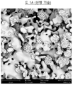
도면을 참조하면 도 1A 는 30 min 의 변환 시간을 갖는 프로토타입 (prototype) 1 (1-2 ㎜ 과립) 의 SEM (Scanning Electron Microscopy: 주사 전자 현미경법) 사진을 나타내며, 여기에서 매끄러운 영역은 SEM 에 의해 측정되는 총 외표면의 약 70 % 에 해당하고, 도 1B 는 40 min 의 변환 시간을 갖는 프로토타입 2 (1-2 ㎜ 과립) 의 SEM 사진을 나타내며, 여기에서 매끄러운 영역은 SEM 에 의해 측정되는 총 외표면의 약 50 % 에 해당한다.Referring to the drawings, FIG. 1A shows a scanning electron microscopy (SEM) photograph of prototype 1 (1-2 mm granules) having a conversion time of 30 min, where a smooth area is observed in the SEM. Corresponding to about 70% of the total outer surface measured by, FIG. 1B shows a SEM photograph of prototype 2 (1-2 mm granules) with a conversion time of 40 min, where the smooth area is measured by SEM About 50% of the total outer surface.
EP-B1-2445543 에 공개된 2상 칼슘 포스페이트/하이드록시아파타이트 (CAP/HAP) 골 대체 물질의 제조에서, 골 이식편 대체물의 제조의 단계 c) 에서 분리된 과립에 특정 세정 프로토콜을 적용함으로써, 평평한 결정 소판의 개별 클러스터 사이의 비-균질 외표면의 매끄러운 영역이 거친 영역으로 대체된다는 것이 이제 밝혀졌다. 특정 세정 프로토콜은 첫째로 순수한 물을 사용하는 정의된 세정 프로토콜, 및 바로 뒤에 짧은 사슬 지방족 알코올을 사용하는 정의된 세정 프로토콜을 포함한다. 평평한 결정 소판의 개별 클러스터 사이의 이들 거친 영역은 일반적으로 SEM 에 의해 측정되는 0.2 내지 5 ㎛ 의 개별 소판 크기를 갖는 소판의 서로 맞물린 네트워크를 형성하는 에피택시 성장한 하이드록시아파타이트 소판을 포함한다.In the preparation of the biphasic calcium phosphate/hydroxyapatite (CAP/HAP) bone replacement material disclosed in EP-B1-2445543, by applying a specific cleaning protocol to the granules isolated in step c) of the preparation of bone graft substitutes, It has now been found that the smooth regions of the non-homogeneous outer surface between individual clusters of crystalline platelets are replaced by rough regions. Certain cleaning protocols include a defined cleaning protocol using pure water first, and a defined cleaning protocol using short chain aliphatic alcohol immediately after. These rough areas between individual clusters of flat crystalline platelets include epitaxy grown hydroxyapatite platelets that form intermeshing networks of platelets with individual platelet sizes of 0.2 to 5 μm, usually measured by SEM.
평평한 결정 소판의 개별 클러스터 사이의 이들 거친 영역이 SEM 에 의해 확인되는 총 외표면의 적어도 20 % 에 해당할 때, 동일한 조건 하에 변환된 그러나 평평한 결정 소판의 개별 클러스터 사이의 매끄러운 영역을 초래하는 상이한 세정 프로토콜로 세정된 EP-B1-2445543 에 공개된 골 대체 물질과 비교하여 골자극 (골 대체 물질이 새로운 골 형성을 유도하는 능력) 이 유의하게 향상된다는 것이 추가로 밝혀졌다. 이는 특히 토끼 모델에서 이식 3 주 후에 대퇴과 (femoral condyle) 결함에서의 골 영역 밀도의 측정에 의해 보여진다.Different cleanings that resulted in smooth areas between individual clusters of flat crystalline platelets, but converted under the same conditions, when these rough areas between individual clusters of flat crystalline platelets correspond to at least 20% of the total outer surface identified by SEM. It has further been found that bone stimulation (the ability of bone substitutes to induce new bone formation) is significantly improved compared to the bone substitutes disclosed in EP-B1-2445543 cleaned with the protocol. This is shown by measurement of bone area density in femoral condyle defects, especially 3 weeks after transplantation in a rabbit model.
본 발명은 따라서 소결된 CAP 코어 및 소결된 CAP 코어의 외표면에 침적된 나노결정질 HAP 의 적어도 하나의 폐쇄된 에피택시 성장한 층을 포함하는 2상 칼슘 포스페이트/하이드록시아파타이트 (CAP/HAP) 골 대체 물질로서, 에피택시 성장한 나노결정은 인간 골 무기물과 동일한 크기 및 형태를 가지며, 소결된 CAP 코어의 외표면에 침적된 나노결정질 HAP 의 폐쇄된 에피택시 성장한 층은 (에피택시 성장한 HAP 나노결정의 응집물로 이루어지는) 평평한 결정 소판의 개별 클러스터 및 평평한 결정 소판의 개별 클러스터 사이의 거친 영역을 포함하는 비-균질 외표면을 가지며, 평평한 결정 소판의 개별 클러스터 사이의 거친 영역의 백분율은 SEM 에 의해 측정되는 총 외표면의 적어도 20% 인 2상 칼슘 포스페이트/하이드록시아파타이트 (CAP/HAP) 골 대체 물질에 관한 것이다.The present invention thus replaces a biphasic calcium phosphate/hydroxyapatite (CAP/HAP) bone comprising a sintered CAP core and at least one closed epitaxy grown layer of nanocrystalline HAP deposited on the outer surface of the sintered CAP core. As a material, epitaxially grown nanocrystals have the same size and shape as a human bone mineral, and a closed epitaxially grown layer of nanocrystalline HAP deposited on the outer surface of a sintered CAP core is a (agglomerate of epitaxially grown HAP nanocrystals The non-homogeneous outer surface comprising individual clusters of flat crystalline platelets and rough regions between individual clusters of flat crystalline platelets, and the percentage of rough regions between individual clusters of flat crystalline platelets is the total as measured by SEM. It relates to a biphasic calcium phosphate/hydroxyapatite (CAP/HAP) bone substitute that is at least 20% of the outer surface.
상기 2상 칼슘 포스페이트/하이드록시아파타이트 (CAP/HAP) 골 대체 물질은 증가된 골 형성 유도 능력을 보인다.The biphasic calcium phosphate/hydroxyapatite (CAP/HAP) bone substitute shows an increased ability to induce bone formation.
일반적으로, 개별 클러스터 사이의 거친 영역은 SEM 에 의해 확인되는 0.2 내지 5 ㎛ 의 개별 소판 크기를 갖는 HAP 나노결정의 에피택시 성장한 소판으로 이루어진다.In general, rough areas between individual clusters consist of epitaxy grown platelets of HAP nanocrystals with individual platelet sizes of 0.2 to 5 μm as identified by SEM.
바람직하게는 개별 결정 클러스터 사이의 거친 영역의 백분율은 SEM 에 의해 측정되는 총 표면의 적어도 30 %, 더욱 바람직하게는 총 외표면의 적어도 40 % 이다.Preferably the percentage of rough areas between individual crystal clusters is at least 30% of the total surface as measured by SEM, more preferably at least 40% of the total outer surface.
일반적으로, 상기 2상 칼슘 포스페이트/하이드록시아파타이트 (CAP/HAP) 골 대체 물질에서 XRD 에 의해 측정되는 HAP 의 백분율은 최대 10 % 이다. 실제로, 그 백분율이 10 % 를 초과할 때, 에피택시 성장한 HAP 나노결정의 평평한 결정 소판의 개별 클러스터는 일반적으로 외표면에서 너무 큰 공간을 차지하고, 따라서 SEM 에 의해 측정되는 개별 결정 클러스터 사이의 거친 영역의 백분율은 총 표면의 20% 미만이라는 것이 밝혀졌다.Generally, the percentage of HAP measured by XRD in the biphasic calcium phosphate/hydroxyapatite (CAP/HAP) bone substitute is up to 10%. Indeed, when the percentage exceeds 10%, individual clusters of flat crystalline platelets of epitaxy-grown HAP nanocrystals generally occupy too large space on the outer surface, thus rough areas between individual crystal clusters as measured by SEM. It was found that the percentage of is less than 20% of the total surface.
바람직하게는 XRD 에 의해 측정되는 HAP 의 백분율은 1 내지 5 %, 더욱 바람직하게는 1.5 내지 3.5 % 이다.Preferably, the percentage of HAP measured by XRD is 1 to 5%, more preferably 1.5 to 3.5%.
소결된 CAP 코어는 트리칼슘 포스페이트 (TCP), 특히 α-TCP (α-Ca3(PO4)2) 또는 β-TCP (β-Ca3(PO4)2), 및/또는 테트라칼슘 포스페이트 (TTCP) Ca4(PO4)2O 를 포함한다.The sintered CAP core is tricalcium phosphate (TCP), in particular α-TCP (α-Ca 3 (PO 4 ) 2 ) or β-TCP (β-Ca 3 (PO 4 ) 2 ), and/or tetracalcium phosphate ( TTCP) Ca 4 (PO 4 ) 2 O.
빈번히 사용되는 실시형태에 따르면, 소결된 CAP 코어는 TCP 로 본질적으로 이루어지며, α-TCP 가 바람직하다.According to the frequently used embodiment, the sintered CAP core consists essentially of TCP, with α-TCP being preferred.
나노결정질 HAP 의 에피택시 성장한 층은 천연 인간 골 무기물과 구조적으로 거의 동일하다.The epitaxially grown layer of nanocrystalline HAP is almost identical in structure to the natural human bone mineral.
CAP/HAP 골 대체 물질은 미립자 또는 그래뉼레이트일 수 있으며, 입자 또는 과립은 요망되는 크기 및 형상을 갖는다. 일반적으로, 입자 또는 과립은 250 내지 5000 ㎛, 바람직하게는 1000 내지 2000 ㎛ 의 크기를 갖는다.The CAP/HAP bone substitute material can be particulate or granulated, and the particles or granules have a desired size and shape. Generally, the particles or granules have a size of 250 to 5000 μm, preferably 1000 to 2000 μm.
CAP/HAP 골 대체 물질은 또한 성형체, 예를 들어, 나사, 못, 핀 또는 골성 신체 부분의 프로필을 갖는 구조 예컨대 특히 고관절, 쇄골, 늑골, 하악골 또는 두개골 부분일 수 있다. 이러한 나사, 못 또는 핀은 재건 정형외과 수술에서, 예를 들어, 무릎 또는 팔꿈치에서 인대를 뼈에 고정하기 위해 사용될 수 있다. 그러한 골성 신체 부분의 프로필을 갖는 구조는 정형외과 수술에서 결손 또는 결함 뼈 또는 뼈 부분을 대체하기 위한 인공기관으로서 사용될 수 있다.The CAP/HAP bone replacement material may also be a shaped body, eg, a screw, nail, pin, or structure having a profile of an osseous body part such as the hip, clavicle, rib, mandible or skull part. Such screws, nails or pins can be used to secure the ligament to the bone in reconstructive orthopedic surgery, for example in the knee or elbow. Structures having such a bone body profile can be used as an artificial organ to replace a missing or defective bone or bone part in orthopedic surgery.
본 발명은 또한 일반적으로 천연 또는 합성 중합체를 포함하는 적합한 매트릭스에 위에 정의된 CAP/HAP 골 대체물의 입자 또는 과립을 포함하는 퍼티에 관한 것이다. 일반적으로, 입자 또는 과립은 250 내지 5000 ㎛, 바람직하게는 1000 내지 2000 ㎛ 의 크기를 갖는다.The present invention also generally relates to a putty comprising particles or granules of the CAP/HAP bone substitute as defined above in a suitable matrix comprising natural or synthetic polymers. Generally, the particles or granules have a size of 250 to 5000 μm, preferably 1000 to 2000 μm.
본 발명은 또한 하기 단계를 포함하는 위에 정의된 CAP/HAP 골 대체 물질의 제조 방법에 관한 것이다:The present invention also relates to a method of making a CAP/HAP bone replacement material as defined above comprising the following steps:
a) 소결된 CAP 코어 물질을 제조하는 단계,a) preparing a sintered CAP core material,
b) 10℃ 내지 50℃ 의 온도에서 수성 완충 용액에 소결된 CAP 코어 물질을 침지시켜 CAP 에서 HAP 로의 변환 과정을 시작하여 소결된 CAP 코어 물질 표면 상에 나노결정질 하이드록시아파타이트의 균일하고 폐쇄된 에피택시 성장한 층을 형성하는 단계로서, 에피택시 성장한 나노결정은 인간 골 무기물과 동일한 크기 및 형태를 갖는 단계,b) A uniform and closed epi of nanocrystalline hydroxyapatite on the surface of the sintered CAP core material by immersing the sintered CAP core material in an aqueous buffer solution at a temperature of 10° C. to 50° C. to initiate CAP to HAP conversion. Forming a taxi-grown layer, the epitaxially grown nanocrystals have the same size and shape as a human bone mineral,
c) HAP 의 적어도 하나의 나노결정질 층의 균일하고 폐쇄된 코팅이 존재할 때 그러나 변환 과정이 완전히 끝나기 전에 한 번에 수성 완충 용액으로부터 고체 물질을 분리함으로써 변환을 정지시키고, 순수한 물 및 짧은 사슬 지방족 알코올 용액을 세정 용액으로서 포함하는 특정 세정 프로토콜을 적용하여 분리된 고체 물질을 세정하는 단계, 및c) Conversion is stopped by the separation of the solid material from the aqueous buffer solution at a time when a uniform and closed coating of at least one nanocrystalline layer of HAP is present, but before the conversion process is complete, pure water and short chain aliphatic alcohols Cleaning the separated solid material by applying a specific cleaning protocol comprising the solution as a cleaning solution, and
d) 임의로 단계 c) 에서 비롯되는 분리된 물질을 살균하는 단계.d) optionally sterilizing the separated material resulting from step c).
분리된 고체 물질의 단계 c) 의 특정 세정 프로토콜은 순수한 물을 사용하는 1 내지 10 회 세정 단계, 더욱 바람직하게는 3 내지 7 회 세정 단계, 및 바로 뒤에 지방족 알코올 용액을 사용하는 적어도 1 회의 세정 단계, 더욱 바람직하게는 적어도 2 회의 세정 단계를 포함한다.The specific cleaning protocol of step c) of the separated solid material includes 1 to 10 cleaning steps using pure water, more preferably 3 to 7 cleaning steps, and at least one cleaning step using an aliphatic alcohol solution immediately thereafter. , More preferably at least two cleaning steps.
적합한 짧은 사슬 지방족 알코올은 메탄올, 에탄올, 프로판올 및 부탄올로 이루어지는 군으로부터 선택될 수 있다.Suitable short chain aliphatic alcohols can be selected from the group consisting of methanol, ethanol, propanol and butanol.
바람직하게는 짧은 사슬 지방족 알코올은 에탄올이다.Preferably the short chain aliphatic alcohol is ethanol.
단계 b) 에서 사용되는 수성 완충 용액은, 단계 b) 의 침지 용액의 pH 값이 거의 중성이고, 바람직하게는 5.5 내지 9.0, 더욱 바람직하게는 7.0 내지 8.0 의 pH 범위 내에서 변환 과정 전체에 걸쳐 안정하게 유지되도록 선택된다.The aqueous buffer solution used in step b) has a substantially neutral pH value of the immersion solution of step b), and is preferably stable throughout the conversion process within a pH range of 5.5 to 9.0, more preferably 7.0 to 8.0. It is chosen to remain.
완충액은 상기 pH 범위에 있는 임의의 완충액일 수 있지만, 바람직하게는 칼슘, 마그네슘 및/또는 소듐을 포함하거나 포함하지 않는 포스페이트 완충액이다. 적합한 완충 용액은 예를 들어 pH 값이 7.45 ± 0.1 인 소듐 디하이드로젠 포스페이트 (NaH2PO4) 의 0.4 M 수성 용액이다.The buffer may be any buffer in the above pH range, but is preferably a phosphate buffer with or without calcium, magnesium and/or sodium. A suitable buffer solution is, for example, a 0.4 M aqueous solution of sodium dihydrogen phosphate (NaH 2 PO 4 ) with a pH value of 7.45±0.1.
단계 b) 에서 온도 범위는 일반적으로 10℃ 내지 50℃, 바람직하게는 25 ℃ 내지 45 ℃, 더욱 바람직하게는 35℃ 내지 40℃ 이다.The temperature range in step b) is generally 10 °C to 50 °C, preferably 25 °C to 45 °C, more preferably 35 °C to 40 °C.
바람직하게는 단계 b) 는 35 내지 40 ℃ 의 온도에서 pH 7.0 내지 8.0 의 포스페이트 완충 용액에서 수행된다.Preferably step b) is carried out in a phosphate buffer solution of pH 7.0 to 8.0 at a temperature of 35 to 40 °C.
소결된 CAP 코어 물질의 제조는 먼저 칼슘 하이드로젠 포스페이트 (CaHPO4), 칼슘 카르보네이트 및/또는 칼슘 하이드록사이드의 분말을 혼합한 후, 혼합물을 적절한 온도 범위 내에서 하소 (calcining) 및 소결 (sintering) 하여 벌크 소결된 CAP 코어 물질을 수득하는 것을 포함하는 당해 기술분야에 알려진 방법에 의해 수행될 수 있다 (예를 들어 Mathew M. et al., 1977, Acta. Cryst. B33: 1325; Dickens B. et al., 1974, J. Solid State Chemistry 10, 232; 및 Durucan C. et al., 2002, J. Mat. Sci., 37:963 참조).The preparation of the sintered CAP core material is first mixed with a powder of calcium hydrogen phosphate (CaHPO 4 ), calcium carbonate and/or calcium hydroxide, and then the mixture is calcined and sintered within an appropriate temperature range ( sintering) to obtain a bulk sintered CAP core material (e.g. Mathew M. et al., 1977, Acta. Cryst. B33: 1325; Dickens B) et al., 1974, J.
벌크 소결된 TCP 코어 물질은 따라서 칼슘 하이드로젠 포스페이트 (CaHPO4), 칼슘 카르보네이트 및/또는 칼슘 하이드록사이드의 분말을 화학량론적 비로 혼합하고, 혼합물을 1200~1450℃ 의 범위, 바람직하게는 약 1400℃ 의 온도에서 하소 및 소결하여 얻어질 수 있다.The bulk sintered TCP core material thus mixes a powder of calcium hydrogen phosphate (CaHPO 4 ), calcium carbonate and/or calcium hydroxide in a stoichiometric ratio, and the mixture is in the range of 1200-1450° C., preferably about It can be obtained by calcining and sintering at a temperature of 1400°C.
벌크 소결된 TTCP 코어 물질은 또한 위에 기술된 과정에 의해 얻어질 수 있다.Bulk sintered TTCP core material can also be obtained by the process described above.
이러한 방법에 의해 제조된 벌크 소결된 CAP 물질은 2 내지 80 부피% 의 공극율 및 공극의 넓은 분포를 갖는 다공성일 수 있다. 다공성 파라미터는 CAP/HAP 골 대체 물질의 의도되는 응용에 따라 선택될 것이다.The bulk sintered CAP material produced by this method can be porous with a porosity of 2 to 80% by volume and a wide distribution of pores. The porosity parameter will be selected depending on the intended application of the CAP/HAP bone substitute.
단계 b) 에서 사용되는 소결된 CAP 코어 물질은The sintered CAP core material used in step b) is
- 위에 기술된 바와 같이 제조된 벌크 소결된 TCP 코어 물질,-Bulk sintered TCP core material prepared as described above,
- 파쇄, 연마 및/또는 분쇄, 및 체거름과 같은 종래의 방법을 사용함으로써 위에 기술된 바와 같이 제조된 벌크 소결된 CAP 코어 물질로부터 얻어지는 소결된 CAP 코어 물질의 미립자 또는 그래뉼레이트, 또는-Particulates or granules of sintered CAP core material obtained from bulk sintered CAP core material prepared as described above by using conventional methods such as crushing, grinding and/or grinding, and sieving, or
- 요망되는 형상 및 크기를 갖는 소결된 CAP 코어 물질의 예비형태, 예를 들어, 나사, 못, 핀, 또는 골성 신체 부분의 프로필을 갖는 구조-A preform of a sintered CAP core material with the desired shape and size, for example a structure with a profile of a screw, nail, pin, or bone body part
일 수 있다.Can be
임의의 요망되는 형상 및 크기를 갖는 이러한 예비형태는 CNC 분쇄 또는 3D 프린팅과 같은 잘 알려진 포토타이핑을 사용함으로써 위에 기술된 바와 같이 제조된 벌크 소결된 코어 물질로부터 얻어질 수 있다 (예를 들어 Bartolo P. et al., 2008, Bio-Materials and Prototyping Applications in Medicine, Springer Science New York, ISBN 978-0-387-47682-7; Landers R. et al., 2002, Biomaterials 23(23), 4437; Yeong W.-Y. et al., 2004, Trends in Biotechnology, 22 (12), 643; 및 Seitz H. et al., 2005, Biomed. Mater. Res. 74B (2), 782 참조).These preforms with any desired shape and size can be obtained from bulk sintered core materials prepared as described above by using well known phototypes such as CNC grinding or 3D printing (eg Bartolo P et al., 2008, Bio-Materials and Prototyping Applications in Medicine, Springer Science New York, ISBN 978-0-387-47682-7; Landers R. et al., 2002, Biomaterials 23(23), 4437; Yeong W.-Y. et al., 2004, Trends in Biotechnology, 22 (12), 643; and Seitz H. et al., 2005, Biomed. Mater. Res. 74B (2), 782).
침지 단계 b) 는 제 1 단계에서 CAP 코어 물질의 일차 상 전이 및 그에 따른 HAP 나노결정 전구물질의 핵형성을 포함한다. 제 2 단계 동안, 제 1 단계로부터의 생성된 HAP 전구물질이 성장하고, 폐쇄된 (즉, 완전히 코팅된) 에피택시 나노결정질 복합 층을 확립할 것이다. 제 1 HAP 나노결정 층은 균일하고 폐쇄되어야 하고, 소결된 CAP 코어 물질에 에피택시 연결되어야 한다.The immersion step b) involves the primary phase transition of the CAP core material in the first step and thus nucleation of the HAP nanocrystalline precursor. During the second step, the resulting HAP precursor from the first step will grow and establish a closed (ie fully coated) epitaxy nanocrystalline composite layer. The first HAP nanocrystalline layer must be uniform and closed and epitaxy connected to the sintered CAP core material.
제 3 단계 동안, 새로 형성된 이중층 복합체 내에서 일차 상 전이가 진행되어, 소결된 CAP 코어 물질 (TCP 또는 TTCP) 을 나노결정질 HAP 로 추가로 변환시킬 수 있다. 상 전이의 제 3 단계 동안, 소결된 CAP 코어 물질의 일부가 나노결정질 HAP 로 변환될 때까지 느린 확산 제어 과정에 의해 제어가능한 시간 동안 칼슘 이온이 방출될 것이다. HAP 층의 두께 및 그에 따른 칼슘 방출 속도는 변환 시간의 변동에 의해 제어될 수 있다.During the third step, the primary phase transition proceeds in the newly formed bilayer composite to further convert the sintered CAP core material (TCP or TTCP) into nanocrystalline HAP. During the third phase of the phase transition, calcium ions will be released for a controllable time by a slow diffusion control process until a portion of the sintered CAP core material is converted to nanocrystalline HAP. The thickness of the HAP layer and hence the rate of calcium release can be controlled by the variation in conversion time.
적절한 두께의 에피택시 성장한 나노결정질 HAP 층은 시험관내에서 제조될 것이며, CAP 에서 HAP 로의 변환은 그것이 완료되기 전에 정지된다.An epitaxially grown nanocrystalline HAP layer of appropriate thickness will be prepared in vitro, and the CAP to HAP conversion is stopped before it is complete.
CAP/HAP 골 대체 물질이 생체내에서 고정되자마자, CAP 에서 HAP 로의 변환 과정은 체액과의 접촉에 의해 재활성화될 것이고, 골 대체 물질은 살아 있는 것 같은 시스템으로서 기능하여 인간 골 무기물과 크기 및 형태가 유사한 새로운 하이드록시아파타이트를 형성할 것이다.As soon as the CAP/HAP bone replacement material is immobilized in vivo, the process of converting CAP to HAP will be reactivated by contact with body fluids, and the bone replacement material functions as a living system, thereby providing human bone mineral and size and It will form a new hydroxyapatite of similar shape.
생체내 상 변환 과정 동안, 운반된 칼슘 이온은 국소 환경으로 방출되어, 골 재생 과정에 중요하고 유익한 국소 칼슘 평형을 지지할 것이다.During the in vivo phase transformation process, the transported calcium ions will be released into the local environment, which will support local calcium equilibrium that is important and beneficial to the bone regeneration process.
신체의 상이하게 로딩된 영역에서 골 결함의 상이한 재생 시간으로 인해, 칼슘 방출 속도가 제어될 수 있는 것이 중요하다. 이는 하이드록시아파타이트의 에피택시 성장한 층의 두께의 변동에 의해 달성될 수 있다.It is important that the rate of calcium release can be controlled, due to the different regeneration times of bone defects in differently loaded regions of the body. This can be achieved by variation in the thickness of the epitaxy grown layer of hydroxyapatite.
따라서, 단계 c) 는 매우 임계적인 단계이다. 단계 b) 의 수성 용액에서의 노출 시간은 요망되는 HAP 층의 두께에 기반한다. 에피택시 배향의 나노결정질 HAP 의 적어도 하나의 층이 필요하다. CAP 에서 HAP 로의 변환이 완료되지 않는 것이 필수적이다.Therefore, step c) is a very critical step. The exposure time in the aqueous solution of step b) is based on the thickness of the desired HAP layer. At least one layer of nanocrystalline HAP in epitaxy orientation is required. It is essential that the conversion from CAP to HAP is not complete.
요망되는 두께에 따른 적절한 노출 시간은 칼슘 포스페이트, 시멘트 및 콘크리트 화학 분야의 당업자에게 잘 알려진 여러 열역학적 미분 방정식을 사용하여 계산될 수 있다.The appropriate exposure time depending on the desired thickness can be calculated using several thermodynamic differential equations well known to those skilled in the art of calcium phosphate, cement and concrete chemistry.
예를 들어 다음을 참조한다: Pommersheim, J.C.; Clifton, J.R. (1979) Cem. Conc. Res.; 9:765; Pommersheim, J.C.; Clifton, J.R. (1982) Cem. Conc. Res.; 12:765; 및 Schluessler, K.H. Mcedlov-Petrosjan, O.P.; (1990): Der Baustoff Beton, VEB Verlag Bauwesen, Berlin.See, for example: Pommersheim, J.C.; Clifton, J.R. (1979) Cem. Conc. Res.; 9:765; Pommersheim, J.C.; Clifton, J.R. (1982) Cem. Conc. Res.; 12:765; And Schluessler, K.H. Mcedlov-Petrosjan, O.P.; (1990): Der Baustoff Beton, VEB Verlag Bauwesen, Berlin.
위에서 언급된 미분 방정식의 해법을 CAP/HAP 시스템에 적용함으로써 CAP 에서 HAP 로의 상 전이 및 HAP 의 에피택시 층이 안정하고 재현가능한 방식으로 제조될 수 있게 하는 층의 두께를 예측하는 것이 가능하다.By applying the solution of the differential equations mentioned above to a CAP/HAP system, it is possible to predict the phase transition from CAP to HAP and the thickness of the layer that allows the epitaxy layer of HAP to be prepared in a stable and reproducible manner.
수성 용액으로부터 고체 물질을 분리하는 것은 통상적으로 당해 기술분야에 잘 알려진 기술을 사용하여 여과 및 건조에 의해 수행된다.Separation of solid materials from aqueous solutions is typically accomplished by filtration and drying using techniques well known in the art.
임의적 살균 단계 d) 는 감마선-조사 또는 X-선 방사와 같은 당해 기술분야에 잘 알려진 기술에 의해 수행될 수 있다.The optional sterilization step d) can be performed by techniques well known in the art, such as gamma-irradiation or X-ray radiation.
본 발명은 또한 인간 또는 동물의 결함 부위에서 골 형성, 골 재생, 골 복원 및/또는 골 대체를 지지하기 위한 임플란트 또는 인공기관으로서의, 일반적으로 미립자 또는 성형체 형태의, 위에 정의된 CAP/HAP 골 대체 물질의 용도에 관한 것이다.The present invention also provides for the replacement of CAP/HAP bone as defined above, generally in the form of an implant or artificial organ, to support bone formation, bone regeneration, bone repair and/or bone replacement at defective sites in humans or animals. It relates to the use of the substance.
본 발명은 또한, 일반적으로 미립자 또는 성형체 형태의, 위에 정의된 CAP/HAP 골 대체 물질을 이식함으로써 인간 또는 동물의 결함 부위에서 골 형성, 골 재생 및/또는 골 복원을 촉진하는 방법에 관한 것이다.The present invention also relates to a method of promoting bone formation, bone regeneration and/or bone repair at defective sites in humans or animals by implanting a CAP/HAP bone replacement material as defined above, generally in the form of particulates or shaped bodies.
본 발명의 CAP/HAP 골 대체 물질의 이점Advantages of the CAP/HAP bone substitute of the present invention
토끼 모델에서 이식 3 주 후에 대퇴과 결함에서의 골 영역 밀도의 측정에 의해 보여지는 바와 같이, 에피택시 성장한 HAP 나노결정으로 이루어지는 평평한 결정 소판의 개별 (분리된) 클러스터 및 평평한 결정 소판의 개별 클러스터 사이의 거친 영역을 포함하는 비-균질 외표면을 갖는 본 발명의 2상 칼슘 포스페이트/하이드록시아파타이트 (CAP/HAP) 골 대체 물질은 평평한 결정 소판의 개별 클러스터 및 평평한 결정 소판의 개별 클러스터 사이의 매끄러운 영역을 포함하는 비-균질 외표면을 제시하는 EP-B1-2445543 에 공개된 골 대체 물질과 비교하여 증가된 골 형성 유도 능력을 보인다.Between individual (separated) clusters of flat crystalline platelets consisting of epitaxy-grown HAP nanocrystals and individual clusters of flat crystalline platelets, as shown by measurement of bone area density in the femur and defect 3 weeks after transplantation in a rabbit model. The biphasic calcium phosphate/hydroxyapatite (CAP/HAP) bone substitute of the present invention having a non-homogeneous outer surface comprising a rough region provides a smooth region between individual clusters of flat crystalline platelets and individual clusters of flat crystalline platelets. It shows an increased ability to induce bone formation compared to the bone substitute disclosed in EP-B1-2445543, which presents an inclusive non-homogeneous outer surface.
이는 R.A. Gittens et al. 에 의해 Biomaterials 2011 May, 32(13): 3395-3403 에서 공개된 결과와 일치하며, 상기 문헌은 마이크로-서브마이크로-스케일 조도와의 조합으로 나노스케일 구조의 도입이 골아세포 분화 및 국소 인자 생산을 개선한다는 것을 보여주며, 이는 결국 생체내에서 개선된 임플란트 골유착능에 대한 잠재성을 시사한다.This is R.A. Gittens et al. By Biomaterials 2011 May, 32(13): 3395-3403, which is a reference to the introduction of nanoscale structures in combination with micro-submicro-scale roughness, which leads to osteoblast differentiation and local factor production. It shows improvement, which in turn suggests the potential for improved implant bone adhesion in vivo.
본 발명은 이후에 본 발명의 바람직한 구현예의 예시적인 실시예 및 첨부되는 도면을 참조하여 추가로 상세히 기술되며, 도면에서:
도 1A 는 30 min 의 변환 시간으로 EP-B1-2445543 의 공개에 따른 실시예 1 에서 제조된 선행 기술 골 대체물의 프로토타입 1 의 SEM 사진을 나타내며, 여기에서 평평한 결정 소판의 개별 클러스터 사이의 매끄러운 영역은 SEM 에 의해 측정되는 총 외표면의 약 70 % 에 해당된다.
도 1B 는 40 min 의 변환 시간으로 EP-B1-2445543 의 공개에 따른 실시예 1 에서 제조된 선행 기술 골 대체물의 프로토타입 2 의 SEM 사진을 나타내며, 여기에서 평평한 결정 소판의 개별 클러스터 사이의 매끄러운 영역은 SEM 에 의해 측정되는 총 외표면의 약 50 % 에 해당된다.
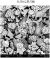
도 2A 는 30 min 의 변환 시간으로 실시예 2 에서 제조된 본 발명에 따른 골 대체 물질의 프로토타입 3 의 SEM 사진을 나타내며, 여기에서 평평한 결정 소판의 개별 클러스터 사이의 거친 영역은 SEM 에 의해 측정되는 총 외표면의 약 70 % 에 해당된다.
도 2B 는 40 min 의 변환 시간으로 실시예 2 에서 제조된 본 발명에 따른 골 대체 물질의 프로토타입 4 의 SEM 사진을 나타내며, 여기에서 평평한 결정 소판의 개별 클러스터 사이의 거친 영역은 SEM 에 의해 측정되는 총 외표면의 약 50 % 에 해당된다.
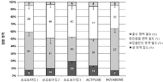
도 3 은 실시예 2 의 본 발명에 따른 골 대체 물질 (프로토타입 3), 실시예 1 의 EP-B1-2445543 에 따른 골 대체 물질 (프로토타입 1 및 2) 및 두 가지 잘 알려진 시판 골 대체 물질 ACTIFUSE® 및 NOVABONE® 에 관한 토끼 모델에서 이식 3 주 후에 대퇴과 결함에서의 골 밀도의 측정을 보여주는 도표를 나타낸다.The present invention is described in further detail below with reference to the accompanying drawings and exemplary embodiments of preferred embodiments of the present invention, in the drawings:
1A shows a SEM photograph of a prototype 1 of the prior art bone substitute prepared in Example 1 according to the disclosure of EP-B1-2445543 with a conversion time of 30 min, where smooth areas between individual clusters of flat crystalline platelets Is about 70% of the total outer surface measured by SEM.
1B shows a SEM photograph of a prototype 2 of the prior art bone substitute prepared in Example 1 according to the disclosure of EP-B1-2445543 with a conversion time of 40 min, where smooth areas between individual clusters of flat crystal platelets Silver corresponds to about 50% of the total outer surface measured by SEM.
2A shows an SEM photograph of a prototype 3 of a bone substitute according to the invention prepared in Example 2 with a conversion time of 30 min, where the rough areas between individual clusters of flat crystalline platelets are measured by SEM About 70% of the total outer surface.
2B shows a SEM photograph of a prototype 4 of a bone substitute according to the invention prepared in Example 2 with a conversion time of 40 min, where the rough areas between individual clusters of flat crystalline platelets are measured by SEM About 50% of the total outer surface.
3 is a bone replacement material according to the invention of Example 2 (prototype 3), a bone replacement material according to EP-B1-2445543 of Example 1 (prototypes 1 and 2) and two well-known commercial bone replacement materials. In a rabbit model for ACTIFUSE ® and NOVABONE ® , a chart is shown showing the measurement of bone density in femur and defect 3 weeks after transplantation.
하기 실시예는 본 발명을 설명하지만 본 발명의 범위를 제한하지 않는다.The following examples illustrate the invention but do not limit the scope of the invention.
실시예 1 EP-B1-2445543 에 따른 2상 칼슘 포스페이트/하이드록시아파타이트 (CAP/HAP) 골 대체 물질의 제조. Example 1 Preparation of a biphasic calcium phosphate/hydroxyapatite (CAP/HAP) bone substitute according to EP-B1-2445543.
알파-TCP 의 벌크 소결된 물질, 1.0-2.0 ㎜ 의 입자 크기를 갖는 그것의 다공성 과립 및 에피택시 성장한 HAP 코팅을 갖는 변환된 과립을 EP-B1-2445543 의 실시예 1, 2 및 4 와 유사하게 제조했다.The bulk sintered material of alpha-TCP, its porous granules having a particle size of 1.0-2.0 mm and the converted granules having an epitaxy grown HAP coating are similar to those of Examples 1, 2 and 4 of EP-B1-2445543. Manufactured.
364 g 디칼슘 포스페이트 무수 분말, 136 g 칼슘 카르보네이트 분말 및 220 ㎖ 탈염수를 실험실 교반기를 사용하여 700 rpm 에서 5 min 동안 혼합했다. 혼합 과정으로부터의 슬러리를 고온 안정한 백금 컵 내로 즉시 옮겼다. 충전된 백금 컵을 차가운 퍼네스 (furnace) 에 놓았다. 퍼네스를 100℃/시간의 가열 속도를 사용하여 1400℃ 로 가열했다. 이 온도를 12 시간 동안 유지하고, 그 후 퍼네스를 500℃/시간의 냉각 속도로 800℃ 로 냉각시키고, 그 후 125℃/시간의 냉각 속도로 300℃ 로 냉각시키고, 마지막으로 퍼네스를 전환하여 실온으로 냉각시켰다. 벌크 소결된 물질 (상 순수한 α-TCP, 즉 α-Ca3(PO4)2) 을 퍼네스 및 백금 컵으로부터 제거했다. 분말 X-선 회절 분석을 사용하여 상 순도의 제어를 수행했다.364 g dicalcium phosphate anhydrous powder, 136 g calcium carbonate powder and 220 ml demineralized water were mixed for 5 min at 700 rpm using a laboratory stirrer. The slurry from the mixing process was immediately transferred into a high temperature stable platinum cup. The filled platinum cup was placed in a cold furnace. The furnace was heated to 1400°C using a heating rate of 100°C/hour. This temperature is maintained for 12 hours, after which the furnace is cooled to 800° C. at a cooling rate of 500° C./hour, then cooled to 300° C. at a cooling rate of 125° C./hour, and finally the furnace is switched. And cooled to room temperature. The bulk sintered material (super pure α-TCP, ie α-Ca 3 (PO 4 ) 2 ) was removed from the furnace and platinum cup. Control of phase purity was performed using powder X-ray diffraction analysis.
벌크 산물을 조 크러셔 (조 거리는 10 내지 1 ㎜ 로 다르다) 를 사용하여 파쇄했다. 생성된 α-TCP 과립을 체거름 기계 (sieving machine) 및 2 ㎜ 및 1 ㎜ 의 메시 구멍이 있는 체 삽입물을 사용하여 체거름했다. 체거름 후에, 과립을 에탄올로 헹구어서 과립에 흡착된 미세 분말 잔류물을 분리했다. 다공성 과립을 캐비넷 건조기에서 1 h 동안 80℃ 에서 건조시켰다. 헹굼 후에 입자 표면의 청결을 주사 전자 현미경법 (SEM) 을 사용하여 표면 관찰에 의해 제어했다.The bulk product was crushed using a jaw crusher (jaw distance varies from 10 to 1 mm). The resulting α-TCP granules were sieved using a sieving machine and sieve inserts with 2 mm and 1 mm mesh holes. After sieving, the granules were rinsed with ethanol to separate the fine powder residue adsorbed on the granules. The porous granules were dried at 80° C. for 1 h in a cabinet dryer. The cleanliness of the particle surface after rinsing was controlled by surface observation using scanning electron microscopy (SEM).
0.4 mol/l 소듐 디하이드로젠 포스페이트 (NaH2PO4) 를 증류수에 용해하여 코팅 및 상 변환 과정에 적절한 완충 용액을 제조했다. 용액의 pH 를 소듐 하이드록사이드 (NaOH) 를 사용하여 실온에서 7.45 로 조정했다. 이전 단락에 따라 생성된 과립을 제조된 용액 내로 침지하고, 웰 템퍼드 (well-tempered) 수조 (40℃) 내에서 각각 30 min (프로토타입 1) 또는 40 min (프로토타입 2) 동안 저장했다. 침지 후에, 과립을 증류수로 3 회 헹구어서 상 변환 과정을 정지시키고, 완충 용액으로부터 잔류물을 제거했다. 다공성 과립을 2 시간 동안 캐비넷 건조기에서 100℃ 에서 건조시켰다.0.4 mol/l sodium dihydrogen phosphate (NaH 2 PO 4 ) was dissolved in distilled water to prepare a buffer solution suitable for coating and phase conversion. The pH of the solution was adjusted to 7.45 at room temperature using sodium hydroxide (NaOH). The granules produced according to the previous paragraph were immersed into the prepared solution and stored in a well-tempered water bath (40° C.) for 30 min (Prototype 1) or 40 min (Prototype 2), respectively. After immersion, the granules were rinsed three times with distilled water to stop the phase conversion process, and the residue was removed from the buffer solution. The porous granules were dried at 100° C. in a cabinet dryer for 2 hours.
프로토타입 1 및 2 의 코팅 및 상 변환 과정 후에 결정 클러스터의 표면 형태 및 표면 커버리지 (coverage) 를 주사 전자 현미경법 (SEM) 에 의해 관찰했다 (도 1A 및 도 1B 참조).After the coating and phase conversion process of prototypes 1 and 2, the surface morphology and surface coverage of the crystal clusters were observed by scanning electron microscopy (SEM) (see FIGS. 1A and 1B).
도 1A 및 1B 로부터 명백한 바와 같이, 과립의 외표면은 에피택시 성장한 HAP 나노결정으로 이루어지는 평평한 결정 소판의 개별 클러스터 및 클러스터 사이의 매끄러운 영역을 포함하며 비-균질이다.As apparent from Figures 1A and 1B, the outer surface of the granules is non-homogeneous and includes individual clusters of flat crystalline platelets made of epitaxially grown HAP nanocrystals and smooth regions between clusters.
프로토타입 1 및 프로토타입 2 각각에 관한 SEM 사진에서 개별 클러스터 및 그 사이의 매끄러운 영역이 차지하는 표면을 측정함으로써, 매끄러운 영역이 프로토타입 1 의 경우에 외표면의 약 70 % 에 해당하고, 프로토타입 2 의 경우에 외표면의 약 50 % 에 해당한다는 것을 확인했다.By measuring the surfaces occupied by the individual clusters and the smooth areas between them in the SEM photographs of each of the prototypes 1 and 2, the smooth areas correspond to about 70% of the outer surface in the case of prototype 1, and prototype 2 In the case of, it was confirmed that it corresponds to about 50% of the outer surface.
실시예 2 본 발명에 따른 2상 칼슘 포스페이트/하이드록시아파타이트 (CAP/HAP) 골 대체 물질의 제조. Example 2 Preparation of a biphasic calcium phosphate/hydroxyapatite (CAP/HAP) bone substitute according to the present invention.
상 순수한 α-TCP 의 1-2 ㎜ 크기의 다공성 과립을 상기 실시예 1 에 따라 생성했다.Porous granules of 1-2 mm size of phase pure α-TCP were produced according to Example 1 above.
상 변환 및 코팅 단계를 40℃ 로 설정된 수조에 배치된 유리 플라스크에서 수행했다. 변환 완충액은 pH 값이 7.45 ± 0.1 인 소듐 디하이드로젠 포스페이트 (NaH2PO4) 의 0.4M 수성 용액이었다.The phase conversion and coating steps were performed in a glass flask placed in a water bath set at 40°C. The conversion buffer was a 0.4M aqueous solution of sodium dihydrogen phosphate (NaH 2 PO 4 ) with a pH value of 7.45±0.1.
유리 플라스크에 변환 완충액을 채우고, 알파-TCP 과립을 1:40 (과립 대 변환 용액) 의 비로 첨가했다. 과립을 30 min (프로토타입 3) 또는 40 min (프로토타입 4) 동안 40℃ 에서 변환 용액에 침지시켰다. 침지 후에, 과립을 탈염수 (과립 대 물 비는 중량에 대해 1:10 이다) 로 5 회 및 에탄올 (99.9%, 과립 대 에탄올 비는 중량에 대해 1:10 이다) 로 2 회 헹구어서, 상 변환 과정을 정지시키고, 거친 영역의 형성을 유도하고, 완충 용액으로부터 잔류물을 제거했다. 다공성 과립을 2 시간 동안 캐비넷 건조기에서 100℃ 에서 건조시켰다.The glass flask was charged with conversion buffer and alpha-TCP granules were added at a ratio of 1:40 (granule to conversion solution). The granules were immersed in the conversion solution at 40° C. for 30 min (Prototype 3) or 40 min (Prototype 4). After immersion, the granules are rinsed 5 times with demineralized water (granulation to water ratio is 1:10 to weight) and 2 times with ethanol (99.9%, granulation to ethanol ratio is 1:10 to weight), and phase transformation The process was stopped, the formation of rough areas was induced, and the residue was removed from the buffer solution. The porous granules were dried at 100° C. in a cabinet dryer for 2 hours.
프로토타입 3 및 4 의 코팅 및 상 변환 과정 후에 결정 클러스터의 표면 형태 및 표면 커버리지를 주사 전자 현미경법 (SEM) 에 의해 관찰했다 (도 2A 및 도 2B 참조).After the coating and phase conversion process of prototypes 3 and 4, the surface morphology and surface coverage of the crystal clusters were observed by scanning electron microscopy (SEM) (see FIGS. 2A and 2B).
도 2A 및 2B 로부터 명백한 바와 같이, 과립의 외표면은 에피택시 성장한 HAP 나노결정으로 이루어지는 평평한 결정 소판의 개별 (분리된) 클러스터 및 클러스터 사이의 거친 영역을 포함하며 비-균질이다.As apparent from Figures 2A and 2B, the outer surface of the granules comprises individual (separated) clusters of flat crystalline platelets consisting of epitaxially grown HAP nanocrystals and rough regions between clusters and are non-homogeneous.
프로토타입 3 및 프로토타입 4 각각에 관한 SEM 사진에서 개별 클러스터 및 그 사이의 거친 영역이 차지하는 표면을 측정함으로써, 거친 영역이 프로토타입 3 의 경우에 외표면의 약 70 % 에 해당하고, 프로토타입 4 의 경우에 외표면의 약 50 % 에 해당한다는 것을 확인했다.By measuring the surface occupied by the individual clusters and the rough areas between them in the SEM photographs of each of the prototypes 3 and 4, the rough areas correspond to about 70% of the outer surface in the case of prototype 3, and prototype 4 In the case of, it was confirmed that it corresponds to about 50% of the outer surface.
실시예 3 토끼 연구. Example 3 Rabbit study.
새로 개발된 골 대체 물질의 생체내 성능을 평가하기 위해서, 토끼에서 대퇴과 모델을 선택했다. 대퇴과 결함 토끼 모델은 대체 생체적합물질을 시험하는데 가장 흔히 사용되는 동물 모델 중 하나이다 (Li Y. et al. Bone defect animal models for testing efficacy of bone substitute biomaterials, Journal of Orthopaedic Translation (2015) 3, 94-104).In order to evaluate the in vivo performance of the newly developed bone substitute, a femur and a model were selected from rabbits. Femoral and defective rabbit models are one of the most commonly used animal models for testing alternative biomaterials (Li Y. et al. Bone defect animal models for testing efficacy of bone substitute biomaterials, Journal of Orthopaedic Translation (2015) 3, 94- 104).
프로토타입 1, 2 및 3 뿐만 아니라 경쟁 물질 ACTIFUSE® 및 NOVABONE® 을 뉴질랜드 (New Zealand) 흰 토끼 (28 주) 에게 대퇴과의 임계적인 크기의 결함 (5 ㎜ x 10 ㎜) 에 이식했다. 이식 3 주 후에, 상이한 프로토타입에 관해 결함에서의 골 영역 밀도, 임플란트 영역 밀도, 섬유질 영역 밀도 및 골수 영역 밀도를 측정함으로써 상이한 생체적합물질의 성능을 분석했다. 정량적 분석을 수행하기 위해서, 샘플을 10% 중성 완충 포르말린 용액 (NBF) 에 고정하고, PMMA 에 포매시키고, EXACT 시스템을 사용하여 자르고, 변형 파라곤 (Paragon) 으로 염색했다.Prototypes 1, 2 and 3 as well as competing materials ACTIFUSE ® and NOVABONE ® were implanted into a New Zealand white rabbit (28 weeks) with a critical size defect (5 mm x 10 mm) with the femur. Three weeks after implantation, the performance of different biomaterials was analyzed by measuring bone area density, implant area density, fibrous area density and bone marrow area density in defects for different prototypes. To perform quantitative analysis, samples were fixed in 10% neutral buffered formalin solution (NBF), embedded in PMMA, cut using the EXACT system, and stained with modified Paragon.
도 3 에서 보여지는 바와 같이, 이식 3 주 후에 토끼 대퇴과 모델에서 새로 형성된 골의 양은 프로토타입 1 및 2 및 경쟁 물질 ACTIFUSE® 및 NOVABONE® 와 비교하여 프로토타입 3 의 경우가 유의하게 더 높았다.As shown in FIG. 3, the amount of newly formed bones in the rabbit femur model 3 weeks after transplantation was significantly higher in the case of prototype 3 compared to prototypes 1 and 2 and competitors ACTIFUSE ® and NOVABONE ® .
Claims (15)
에피택시 성장한 나노결정은 인간 골 무기물과 동일한 크기 및 형태를 가지며, 소결된 CAP 코어의 외표면에 침적된 나노결정질 HAP 의 폐쇄된 에피택시 성장한 층은 에피택시 성장한 HAP 나노결정으로 이루어지는 평평한 결정 소판의 개별 클러스터 및 개별 클러스터 사이의 거친 영역을 포함하는 비-균질 외표면을 가지며, SEM 에 의해 측정되는 개별 클러스터 사이의 거친 영역의 백분율은 총 표면의 적어도 20% 인,
2상 칼슘 포스페이트/하이드록시아파타이트 (CAP/HAP) 골 대체 물질.A biphasic calcium phosphate/hydroxyapatite (CAP/HAP) bone replacement material comprising a sintered CAP core and at least one closed epitaxy grown layer of nanocrystalline HAP deposited on the outer surface of the sintered CAP core,
The epitaxially grown nanocrystals have the same size and shape as the human bone mineral, and the closed epitaxially grown layer of nanocrystalline HAP deposited on the outer surface of the sintered CAP core is a flat crystal platelet made of epitaxially grown HAP nanocrystals. Has a non-homogeneous outer surface comprising individual clusters and rough regions between individual clusters, the percentage of rough regions between individual clusters measured by SEM being at least 20% of the total surface,
Biphasic calcium phosphate/hydroxyapatite (CAP/HAP) bone substitute.
a) 소결된 CAP 코어 물질을 제조하는 단계,
b) 10℃ 내지 50℃ 의 온도에서 수성 완충 용액에 소결된 CAP 코어 물질을 침지시켜 CAP 에서 HAP 로의 변환 과정을 시작하여 소결된 CAP 코어 물질 표면 상에 나노결정질 하이드록시아파타이트의 균일하고 폐쇄된 에피택시 성장한 층을 형성하는 단계로서, 에피택시 성장한 나노결정은 인간 골 무기물과 동일한 크기 및 형태를 갖는 단계,
c) HAP 의 적어도 하나의 나노결정질 층의 균일하고 폐쇄된 코팅이 존재할 때 그러나 변환 과정이 완전히 끝나기 전에 한 번에 수성 완충 용액으로부터 고체 물질을 분리함으로써 변환을 정지시키고, 순수한 물 및 짧은 사슬 지방족 알코올 용액을 세정 용액으로서 포함하는 세정 프로토콜을 적용하여 분리된 고체 물질을 세정하여 CAP/HAP 골 대체 물질을 형성하는 단계로서, CAP/HAP 골 대체 물질에서 소결된 CAP 코어의 외표면은 에피택시 성장한 HAP 나노결정으로 이루어지는 평평한 결정 소판의 개별 클러스터 및 개별 클러스터 사이의 거친 영역을 포함하는 비-균질 외표면을 가지며, SEM 에 의해 측정되는 개별 클러스터 사이의 거친 영역의 백분율은 총 표면의 적어도 20% 인 단계, 및
d) 임의로 단계 c) 에서 비롯되는 분리된 물질을 살균하는 단계.A method for producing a CAP/HAP bone substitute according to any one of claims 1 to 9 comprising the following steps:
a) preparing a sintered CAP core material,
b) uniform and closed epi of nanocrystalline hydroxyapatite on the surface of the sintered CAP core material by immersing the sintered CAP core material in an aqueous buffer solution at a temperature of 10° C. to 50° C. Forming a taxi-grown layer, the epitaxially grown nanocrystals have the same size and shape as a human bone mineral,
c) Conversion is stopped by the separation of the solid material from the aqueous buffer solution at a time when a uniform and closed coating of at least one nanocrystalline layer of HAP is present, but before the conversion process is complete, pure water and short chain aliphatic alcohols A step of forming a CAP/HAP bone replacement material by washing the separated solid material by applying a cleaning protocol including a solution as the cleaning solution, wherein the outer surface of the CAP core sintered in the CAP/HAP bone replacement material is epitaxially grown HAP Step having a non-homogeneous outer surface comprising individual clusters of flat crystal platelets made of nanocrystals and rough regions between individual clusters, wherein the percentage of rough regions between individual clusters as measured by SEM is at least 20% of the total surface , And
d) optionally sterilizing the separated material resulting from step c).
15. The process according to any one of claims 12 to 14, wherein step b) is carried out in a phosphate buffer solution of pH 7.0 to 8.0 at a temperature of 35 to 40 °C.
Applications Claiming Priority (3)
| Application Number | Priority Date | Filing Date | Title |
|---|---|---|---|
| EP17207233 | 2017-12-14 | ||
| EP17207233.2 | 2017-12-14 | ||
| PCT/EP2018/084776 WO2019115700A1 (en) | 2017-12-14 | 2018-12-13 | Bone substitute material |
Publications (2)
| Publication Number | Publication Date |
|---|---|
| KR20200076754A true KR20200076754A (en) | 2020-06-29 |
| KR102230625B1 KR102230625B1 (en) | 2021-03-22 |
Family
ID=60781526
Family Applications (1)
| Application Number | Title | Priority Date | Filing Date |
|---|---|---|---|
| KR1020207016845A KR102230625B1 (en) | 2017-12-14 | 2018-12-13 | Bone substitutes |
Country Status (7)
| Country | Link |
|---|---|
| US (1) | US10646619B2 (en) |
| EP (1) | EP3544642B1 (en) |
| JP (1) | JP6813716B1 (en) |
| KR (1) | KR102230625B1 (en) |
| CN (1) | CN111465419B (en) |
| TW (1) | TWI783093B (en) |
| WO (1) | WO2019115700A1 (en) |
Cited By (1)
| Publication number | Priority date | Publication date | Assignee | Title |
|---|---|---|---|---|
| WO2023182700A1 (en) * | 2022-03-24 | 2023-09-28 | 주식회사 엘지화학 | Porous inorganic particles, and composite filler and product using same |
Families Citing this family (3)
| Publication number | Priority date | Publication date | Assignee | Title |
|---|---|---|---|---|
| PL3544643T3 (en) | 2017-12-14 | 2020-12-28 | Geistlich Pharma Ag | Bone substitute material |
| US10960107B2 (en) | 2019-06-14 | 2021-03-30 | Geistlich Pharma Ag | Collagen matrix or granulate blend of bone substitute material |
| WO2020249714A1 (en) | 2019-06-14 | 2020-12-17 | Geistlich Pharma Ag | Collagen matrix or granulate blend of bone substitute material |
Citations (5)
| Publication number | Priority date | Publication date | Assignee | Title |
|---|---|---|---|---|
| US20030026770A1 (en) * | 2001-07-25 | 2003-02-06 | Szymaitis Dennis W. | Periodontal regeneration composition and method of using same |
| US20120130506A1 (en) * | 2009-06-23 | 2012-05-24 | Geistlich Pharma Ag | Bone substitute material |
| US20140127392A1 (en) * | 2006-10-24 | 2014-05-08 | Biomet 3I, Llc | Deposition of discrete nanoparticles on a nanostructured surface of an implant |
| US20150024023A1 (en) * | 2011-12-23 | 2015-01-22 | The University Court Of The University Of Aberdeen | Calcium phosphate material |
| US20160106674A1 (en) * | 2013-05-15 | 2016-04-21 | Euroresearch S.R.L. | Collagen powder, composition and use |
Family Cites Families (7)
| Publication number | Priority date | Publication date | Assignee | Title |
|---|---|---|---|---|
| DE3711426A1 (en) | 1987-04-04 | 1988-10-13 | Mtu Muenchen Gmbh | IMPLANT WITH BIOACTIVE COATING |
| US5763092A (en) * | 1993-09-15 | 1998-06-09 | Etex Corporation | Hydroxyapatite coatings and a method of their manufacture |
| IT1288038B1 (en) | 1996-04-30 | 1998-09-10 | Flametal S P A | PROCEDURE FOR THE PREPARATION OF HYDROXYAPATITE COATINGS |
| US6338752B1 (en) | 2000-04-20 | 2002-01-15 | Purzer Pharmaceutical Co., Ltd. | α-TCP/HAP biphasic cement and its preparing process |
| CN100384488C (en) * | 2004-05-28 | 2008-04-30 | 梁辉 | Preparation technique of absorbent hydroxyapatite artificial bone |
| CN102274547A (en) * | 2011-07-18 | 2011-12-14 | 湖南康胜生物材料科技有限公司 | Preparation process of coral artificial bone with surface replaced by hydroxyapatite |
| EP2826495A1 (en) * | 2013-07-19 | 2015-01-21 | Geistlich Pharma AG | Biomimetic collagen-hydroxyapatite composite material |
-
2018
- 2018-12-13 JP JP2020532763A patent/JP6813716B1/en active Active
- 2018-12-13 TW TW107145010A patent/TWI783093B/en active
- 2018-12-13 WO PCT/EP2018/084776 patent/WO2019115700A1/en unknown
- 2018-12-13 CN CN201880080307.3A patent/CN111465419B/en active Active
- 2018-12-13 EP EP18816062.6A patent/EP3544642B1/en active Active
- 2018-12-13 KR KR1020207016845A patent/KR102230625B1/en active IP Right Grant
- 2018-12-14 US US16/220,320 patent/US10646619B2/en active Active
Patent Citations (5)
| Publication number | Priority date | Publication date | Assignee | Title |
|---|---|---|---|---|
| US20030026770A1 (en) * | 2001-07-25 | 2003-02-06 | Szymaitis Dennis W. | Periodontal regeneration composition and method of using same |
| US20140127392A1 (en) * | 2006-10-24 | 2014-05-08 | Biomet 3I, Llc | Deposition of discrete nanoparticles on a nanostructured surface of an implant |
| US20120130506A1 (en) * | 2009-06-23 | 2012-05-24 | Geistlich Pharma Ag | Bone substitute material |
| US20150024023A1 (en) * | 2011-12-23 | 2015-01-22 | The University Court Of The University Of Aberdeen | Calcium phosphate material |
| US20160106674A1 (en) * | 2013-05-15 | 2016-04-21 | Euroresearch S.R.L. | Collagen powder, composition and use |
Non-Patent Citations (1)
| Title |
|---|
| Rolando A. Gittens et al., "Implant Osseointegration and the Role of Microroughness and Nanostructures: Lessons for Spine Implants", Acta Biomater.(2014). Vol. 10(8), pp.1-22* * |
Cited By (1)
| Publication number | Priority date | Publication date | Assignee | Title |
|---|---|---|---|---|
| WO2023182700A1 (en) * | 2022-03-24 | 2023-09-28 | 주식회사 엘지화학 | Porous inorganic particles, and composite filler and product using same |
Also Published As
| Publication number | Publication date |
|---|---|
| JP6813716B1 (en) | 2021-01-13 |
| EP3544642B1 (en) | 2020-05-13 |
| JP2021506403A (en) | 2021-02-22 |
| TWI783093B (en) | 2022-11-11 |
| CN111465419A (en) | 2020-07-28 |
| CN111465419B (en) | 2021-06-25 |
| KR102230625B1 (en) | 2021-03-22 |
| TW201927348A (en) | 2019-07-16 |
| WO2019115700A1 (en) | 2019-06-20 |
| US10646619B2 (en) | 2020-05-12 |
| EP3544642A1 (en) | 2019-10-02 |
| US20190209737A1 (en) | 2019-07-11 |
Similar Documents
| Publication | Publication Date | Title |
|---|---|---|
| US9066995B2 (en) | Bone substitute material | |
| KR102230625B1 (en) | Bone substitutes | |
| US8734524B2 (en) | Bone substitute material | |
| JP7249376B2 (en) | bone replacement material | |
| US11357891B2 (en) | Collagen matrix or granulate blend of bone substitute material | |
| KR102636183B1 (en) | Collagen matrix or granulated blend of bone substitute materials |
Legal Events
| Date | Code | Title | Description |
|---|---|---|---|
| A302 | Request for accelerated examination | ||
| E902 | Notification of reason for refusal | ||
| E701 | Decision to grant or registration of patent right | ||
| GRNT | Written decision to grant |