KR20200068493A - Diagnostic method of bppv using pupil and iris - Google Patents
Diagnostic method of bppv using pupil and iris Download PDFInfo
- Publication number
- KR20200068493A KR20200068493A KR1020180155540A KR20180155540A KR20200068493A KR 20200068493 A KR20200068493 A KR 20200068493A KR 1020180155540 A KR1020180155540 A KR 1020180155540A KR 20180155540 A KR20180155540 A KR 20180155540A KR 20200068493 A KR20200068493 A KR 20200068493A
- Authority
- KR
- South Korea
- Prior art keywords
- pupil
- image
- iris
- subject
- diagnosing
- Prior art date
Links
Images


Classifications
-
- A—HUMAN NECESSITIES
- A61—MEDICAL OR VETERINARY SCIENCE; HYGIENE
- A61B—DIAGNOSIS; SURGERY; IDENTIFICATION
- A61B5/00—Measuring for diagnostic purposes; Identification of persons
- A61B5/40—Detecting, measuring or recording for evaluating the nervous system
- A61B5/4005—Detecting, measuring or recording for evaluating the nervous system for evaluating the sensory system
- A61B5/4023—Evaluating sense of balance
-
- A—HUMAN NECESSITIES
- A61—MEDICAL OR VETERINARY SCIENCE; HYGIENE
- A61B—DIAGNOSIS; SURGERY; IDENTIFICATION
- A61B3/00—Apparatus for testing the eyes; Instruments for examining the eyes
- A61B3/10—Objective types, i.e. instruments for examining the eyes independent of the patients' perceptions or reactions
- A61B3/12—Objective types, i.e. instruments for examining the eyes independent of the patients' perceptions or reactions for looking at the eye fundus, e.g. ophthalmoscopes
- A61B3/1216—Objective types, i.e. instruments for examining the eyes independent of the patients' perceptions or reactions for looking at the eye fundus, e.g. ophthalmoscopes for diagnostics of the iris
-
- A—HUMAN NECESSITIES
- A61—MEDICAL OR VETERINARY SCIENCE; HYGIENE
- A61B—DIAGNOSIS; SURGERY; IDENTIFICATION
- A61B3/00—Apparatus for testing the eyes; Instruments for examining the eyes
- A61B3/10—Objective types, i.e. instruments for examining the eyes independent of the patients' perceptions or reactions
- A61B3/14—Arrangements specially adapted for eye photography
- A61B3/145—Arrangements specially adapted for eye photography by video means
Abstract
Description
본 발명은 어지럼증을 진단하고 치료하기 위한 방법에 관한 것으로, 보다 상세하게는 웨어러블 디바이스인 안진기와 단말기를 이용해 사용자의 어지럼증 여부 및 이석증을 진단하고 그 결과에 따라 사용자가 스스로 어지럼증을 치료할 수 있도록 하는 동공과 홍채를 이용한 이석증의 진단방법에 관한 것이다.The present invention relates to a method for diagnosing and treating dizziness, and more specifically, using a wearable device, Anjin and a terminal, to diagnose whether the user has dizziness and dizziness, and to allow the user to treat dizziness on his own according to the results. The present invention relates to a method for diagnosing dyslexia using a pupil and an iris.
이석이란 내이의 전정기관 속 난형낭에 있는 작은 칼슘덩어리로, 이석이 제자리에서 탈락되어 반고리관으로 이동하게 되면 중력에 영향을 받게 된 칼슘 덩어리에 의해 반고리관 내 림프액이 비정상적인 유동성을 가지게 되고, 이로 인해 어지럼증이 발생한다.This is a small chunk of calcium in the ovarian sac in the vestibular organ of the inner ear. When the stone is removed from its place and moves to the semicircular canal, lymphatic fluid in the semicircular canal has abnormal fluidity due to the mass of calcium affected by gravity, which causes dizziness. This happens.
이러한 증상을 이석증이라고 하는데, 대부분의 이석증 치료에는 체위에 변화를 주어 탈락됐던 이석을 원위치로 환원시키는 이석치환술이 이용된다. 이석치환술은 이석이 위치한 반고리관의 부위와 방향에 따라 치료법이 달라지므로 이석의 위치를 정확히 진단하여야만 적절하고 효과적인 이석치환술이 가능하다.These symptoms are called dyslithiasis, and most of the treatments for dyslithiasis use dichroic replacement, which reduces the displaced stones to their original position by changing the body position. Since the treatment method varies depending on the area and direction of the semicircular canal where the stone is located, proper and effective stone replacement is possible only when the location of the stone is accurately diagnosed.
이석의 위치를 정확히 진단하기 위해서는 두위 변화에 따라 유발되는 안진의 방향을 관찰함으로써 이석이 위치한 반고리관을 진단하여야 한다. 안진이란 이석증과 같이 말초전정계 이상뿐만 아니라 대뇌에 이르는 다양한 안구 운동계의 이상에 의하여 물체의 상이 망막의 중심으로부터 벗어나게 되면 이를 교정하기 위해 나타나는 교정성 안구운동을 말하며, 이석증은 이석의 위치에 따라 두위 변화 시 특징적인 안진이 나타나므로 이를 통해 이석의 위치를 진단할 수 있다.In order to accurately diagnose the location of the stone, it is necessary to diagnose the semicircular canal where the stone is located by observing the direction of the eyelid caused by changes in the head. Anjin refers to corrective eye movements that appear to correct when the image of an object deviates from the center of the retina by abnormalities in the peripheral vestibular system, such as dysplasia, as well as various ocular movement systems that reach the cerebrum. As a result, a characteristic nystagmus appears when the head changes, so it is possible to diagnose the location of the stone.
이석의 위치에 따른 안진의 특징으로는, 이석이 측수평반고리관에 위치한 경우 수평성분의 안진이 발생하고, 앙와위에서 좌·우 두위에 따라 방향이 변하는 특징을 가지며, 이석이 후반고리관에 위치한 경우에는 앉은 자세에서 고개를 병변쪽으로 45˚ 돌린 상태에서 현수위로 고개를 떨어뜨리는 딕스-홀파이크(Dix-Hallpike) 수기에 따라 향지성 회전성 안진(Geotropic Torsional Nystagmus)이 나타나지만 반대쪽을 주시하게 되면 상향안진이 발생한다.As a feature of Anjin according to the location of the stone, when the stone is located in the lateral horizontal semicircular canal, the horizontal component of the eczema occurs, and it has a characteristic that the direction changes depending on the left and right heads in the supine position. Geostatic Torsional Nystagmus appears due to the Dix-Hallpike technique, where the head is turned to the lesion in a sitting position and the head is dropped to the suspension level. do.
참고로 Dix-Hallpike는 병변쪽 귀가 아래로 향하는 특정한 체위로 변환시 나타나는 안진의 특성을 검사하는 방법을 의미한다.For reference, Dix-Hallpike refers to a method of examining the characteristics of the nystagmus that appears when the lesion-side ear is transformed to a specific position with the ear downward.
이석증에 의한 어지럼증은 대응하는 이석치환술을 통해 비교적 손쉽게 치료할 수 있지만 정확한 진단을 위해 직접 병원을 방문해야 하는 불편함이 있고, 적절한 이석치환술이 실시된 후에도 이석증이 재발할 가능성이 50% 정도까지 보고되고 있어 재발이 의심될 때마다 병원을 방문해야 하는 번거로움이 존재한다.Dizziness caused by dizziness can be treated relatively easily through the corresponding dichroic replacement, but there is a discomfort that requires direct visits to the hospital for accurate diagnosis. It is reported that there is a hassle of having to visit a hospital whenever a relapse is suspected.
따라서, 이석증이 의심되는 환자의 이석 위치를 정확하게 진단하고 그 위치에 따라 적절한 이석치환술을 스스로 실시할 수 있도록 하기 위한 방법이 필요한 실정이다.Accordingly, there is a need for a method for accurately diagnosing the location of a patient suspected of dyscalculia and allowing the patient to appropriately perform a proper displacement procedure according to the location.
또한, 어지럼을 호소하는 환자들의 경우 전정안구반사의 이상으로 특이적인 안구운동이 유발되기 때문에 임상에서는 두위변화에 따른 안구움직임의 이상 여부를 판단하여 진단에 이용한다.In addition, patients who complain of dizziness cause specific eye movements due to abnormal vestibular ocular reflexes, so clinicians use it for diagnosis by determining whether eye movements due to head changes are abnormal.
안구는 전정 반사 운동에 대해 수평 및 수직의 2축에 대한 움직임, 그리고 회전 움직임이라는 3차원의 운동 형태를 가진다. 현재까지 상용화된 안구 운동량의 측정은 검사자의 눈을 통한 검사, 프렌젤(Frenzel) 안경, 전기안진기나 비디오 안진기를 통하여 수행되었다. 그러나, 검사자가 직접 관찰하거나 프렌젤 안경을 사용하는 방법은 안진의 변화량이 클 때는 안구의 움직임이 관찰될 수 있으나, 그 반대의 경우에 측정이 어려운 문제가 있다.The eyeball has a three-dimensional form of motion, horizontal and vertical movements on the vestibular reflex motion, and rotational motion. Measurement of ocular momentum that has been commercialized to date has been carried out through examination through an examiner's eye, Frenzel glasses, an electro-osmulator or a video oscillator. However, in the method of directly inspecting the examiner or using the prezel glasses, the movement of the eye may be observed when the amount of change in the eyelid is large, but in the opposite case, measurement is difficult.
그럼에도 불구하고, 비디오 안진기는 현재까지 진단적 방법으로 널리 사용되고 있으며, 여기에는 사용자가 비디오를 통해 안구운동을 관찰하여 진단하는 방법과 비디오 안진기에 안진의 검사 장치를 달아 그래프를 통해 수평, 수직, 회전 운동에 대한 3축 측정을 표시하여 이를 진단에 사용하는 방법이 있다. 그러나 후자의 경우 객관적인 측정이 가능한 장점이 있으나 3축 분석을 위해서는 내부에 장착한 별도의 이미지 센서를 통해 획득한 영상을 처리하여 3축의 안구 운동량을 측정하게 된다. 따라서 내부에 장착된 이미지 센서의 해상도와 프레임율, 이미지 처리 정확도에 따라서 민감도가 결정되고, 또한 안구 운동의 각 축에 따라 측정된 각속도(deg/sec)를 별도로 측정해야 하는 기술적 문제 때문에 가격이 고가이어서 일반적으로 사용되기 어려운 문제가 있다. 반면 이러한 기능이 없는 단순 비디오 안진기는 상대적으로 저렴하여 널리 이용되고 있으나 이를 통한 진단에는 고도의 훈련이 필요하다.Nevertheless, video ophthalmographs have been widely used as a diagnostic method to date, and include a method for diagnosing and observing ocular movement through a video, and horizontal, vertical, and rotation through graphs by attaching an ophthalmic examination device to the video ophthalmograph. There is a way to display a 3-axis measurement of exercise and use it for diagnosis. However, in the latter case, there is an advantage that objective measurement is possible, but for 3-axis analysis, the image acquired through a separate image sensor mounted therein is processed to measure the 3-axis eye movement. Therefore, the sensitivity is determined according to the resolution, frame rate, and image processing accuracy of the internally mounted image sensor, and the price is high due to the technical problem of separately measuring the angular velocity (deg/sec) measured along each axis of eye movement. Then there is a problem that is difficult to be used in general. On the other hand, a simple video stabilizer without this function is relatively inexpensive and widely used, but it requires advanced training for diagnosis.
이러한 과제를 해결하기 위한 본 발명은 홍채와 동공을 이용하여 손쉽게 안진의 유무와 이석증을 진단할 수 있는 동공과 홍채를 이용한 이석증의 진단방법을 제공하는 것을 목적으로 한다.An object of the present invention to solve this problem is to provide a method for diagnosing dysentery using a pupil and an iris, which can easily diagnose the presence or absence and dizziness of anjin using the iris and pupil.
또한, 본 발명은 동공의 진동정보와 홍채의 비틀림 정보를 이용하여 안진의 유무와 이석증을 진단할 수 있는 동공과 홍채를 이용한 이석증의 진단방법을 제공하는 것을 다른 목적으로 한다.In addition, another object of the present invention is to provide a method of diagnosing dizziness using pupils and irises, which can diagnose presence or absence and dizziness of anjin using vibration information of pupils and torsion information of irises.
그리고 본 발명은 동공의 진동정보와 홍채의 비틀림 정보를 이용하여 자발안진, 주시안진, 두위회전후 안진 그리고 앞반 고리관 이석증, 수평반고리관 이석증 또는 후반고리관 이석증을 진단할 수 있는 동공과 홍채를 이용한 이석증의 진단방법을 제공하는 것을 또 다른 목적으로 한다. And the present invention uses the vibration information of the pupil and the twist information of the iris, the pupil and the iris that can diagnose spontaneous anesthesia, zhusianjin, anjin after head rotation, and anterior annular canalism, horizontal annular canalism or posterior annular canalism Another object of the present invention is to provide a method for diagnosing dyslexia.
이러한 과제를 해결하기 위해 본 발명의 피검자에게 착용되어 안진 검사를 수행하도록 하는 안진기를 이용한 이석증의 진단방법은, (a) 상기 안진기에서 수신된 영상을 처리하여 진동정보와 비틀림정보를 추출하는 단계와 (b) 상기 (a)단계에서 추출된 진동정보와 비틀림정보를 진단단말기로 전송하는 단계, 및 (c) 상기 (b)단계에서 전송된 진동정보와 비틀림정보를 이용하여 안진의 유무 및 이석증을 진단하는 단계를 포함하여 이루어지게 함으로써 달성될 수 있다.In order to solve this problem, a method of diagnosing dysentery using an exoskeleton worn by a subject of the present invention to perform an ocular examination, (a) extracting vibration information and torsion information by processing an image received from the exotropia And (b) transmitting the vibration information and the torsion information extracted in the step (a) to a diagnostic terminal, and (c) using the vibration information and the torsion information transmitted in the step (b) to determine whether or not Anjin is present and to be seated. It can be achieved by including the steps to diagnose the symptoms.
상기 (a)단계는 상기 진동정보를 추출하기 위하여 상기 안진기의 양안 카메라를 통하여 수신된 안구의 영상을 프레임별로 처리하여 각 프레임에서 동공의 중심점 좌표를 추출하고, 상기 비틀림정보를 추출하기 위하여 상기 안진기의 양안 카메라를 통하여 수신된 홍채 또는 안구의 영상을 프레임별로 처리하여 각 프레임에서 홍채 또는 안구의 특징점에 대한 패턴을 비교하여 각도를 추출하도록 구성한다.In step (a), to extract the vibration information, the image of the eyeball received through the binocular camera of the jinjingi is processed frame by frame to extract the coordinates of the center point of the pupil in each frame, and the jinjingi to extract the torsion information. It is configured to process the image of the iris or eyeball received through the binocular camera of each frame and compare the pattern for the feature points of the iris or eyeball in each frame to extract the angle.
또한, 상기 (c)단계는 수신된 (x,y)좌표를 기준으로 진동(Beating)의 정도를 측정하고, 수신된 각도로 비틀림(torsion)의 정도를 측정하여 측정된 값에 따라 PSCC, LSC, 또는 ASC 중 어느 하나의 어지럼증을 진단하도록 동작한다.In addition, in step (c), the degree of vibration is measured based on the received (x,y) coordinates, and the degree of torsion is measured at the received angle, depending on the measured value of PSCC, LSC , Or ASC.
또한, Dix-Hallpike 검사항목에 의하여 우측 Dix-hall을 진행한 경우에 진동(Beating)이 상방향이고 우측으로 비틀림(extorsion)이 관측된 경우 우측 후반고리관 이석증으로 진단하고, 좌측 Dix-hall을 진행한 경우에 진동(Beating)이 상방향이고 좌측 비틀림(extorsion)이 관측된 경우 좌측 후반고리관 이석증으로 진단한다.In addition, when the right dix-hall is advanced by the Dix-Hallpike test item, if the vibration is upward and the torsion is observed to the right, the right posterior annulus candidiasis is diagnosed, and the left dix-hall is diagnosed. In the case of progress, if the vibration is upward and the left extension is observed, it is diagnosed as the left posterior annulus dysplasia.
그리고 (c)단계는 자발안진, 주시안진, 두위회전후 안진 중 어느 하나의 검사항목에 따라 중추신경계 또는 말초신경계 어지럼증으로 진단한다. And step (c) is diagnosed as either central nervous system or peripheral nervous system dizziness according to one of the test items of spontaneous anesthesia, zhusianjin, and chin after rotation.
따라서, 본 발명의 동공과 홍채를 이용한 이석증의 진단방법에 의하면, 홍채와 동공을 이용하여 손쉽게 안진의 유무와 이석증을 진단할 수 있는 효과가 있다.Therefore, according to the method of diagnosing dysentery using the pupil and the iris of the present invention, there is an effect that can easily diagnose the presence and absence of anjin and dyslithiasis using the iris and the pupil.
또한, 본 발명의 동공과 홍채를 이용한 이석증의 진단방법에 의하면, 동공의 진동정보와 홍채의 비틀림 정보를 이용하여 안진의 유무와 이석증을 진단하기 때문에 주관적이지 않고 객관적인 데이터로 정확하게 진단할 수 있는 효과가 있다,In addition, according to the method of diagnosing dysplasia using the pupil and iris of the present invention, since the presence and absence of anjin and diagnosing disease are diagnosed using the vibration information of the pupil and the iris torsion information, it can be accurately diagnosed with objective data without being subjective. There is an effect,
그리고 본 발명의 동공과 홍채를 이용한 이석증의 진단방법에 의하면 동공의 진동정보와 홍채의 비틀림 정보를 이용하여 자발안진, 주시안진, 두위회전후 안진 그리고 앞반 고리관 이석증, 수평반고리관 이석증 또는 후반고리관 이석증을 손쉽게 진단할 수 있기 때문에 진단에 따라 적절한 치료법을 신속하게 처방할 수 있는 효과가 있다. And according to the method of diagnosing dizziness using the pupil and iris of the present invention, the spontaneous anesthesia, zhusianjin, anjin after head rotation, and anterior ring rotosis, horizontal ring loop diarrhea or Since it can easily diagnose the posterior ring duct, it has the effect of promptly prescribing an appropriate treatment method according to the diagnosis.
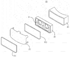
도 1은 본 발명의 실시예에 따른 이석증의 진단 장치 주요 구성도,
도 2는 휴대용 안진기 사시도,
도 3은 휴대용 안진기의 분해사시도,
도 4는 휴대용 안진기의 제어유닛의 상세 구성도,
도 5는 본 발명의 일실시예에 의한 동공과 홍채를 이용한 이석증의 진단방법을 설명하기 위한 흐름도,
도 6은 동공 중심점 좌표값을 산출하여 비교하는 참고도면,
그리고
도 7은 홍채 패턴을 비교하여 변화 각도를 산출하는 참고도면이다.1 is a main configuration diagram of a diagnosis device for dyscalculia according to an embodiment of the present invention,
2 is a perspective view of a portable jinjingi,
3 is an exploded perspective view of a portable jinjingi,
4 is a detailed configuration diagram of the control unit of the portable jinjingi,
Figure 5 is a flow chart for explaining a method of diagnosing dysplasia using a pupil and iris according to an embodiment of the present invention,
6 is a reference drawing for calculating and comparing the coordinate values of the pupil center point,
And
7 is a reference diagram for comparing a iris pattern and calculating a change angle.
본 명세서 및 청구범위에 사용된 용어나 단어는 통상적이거나 사전적인 의미로 한정 해석되지 아니하며, 발명자는 그 자신의 발명을 가장 최선의 방법으로 설명하기 위해 용어의 개념을 적절하게 정의할 수 있다는 원칙에 입각하여 본 발명의 기술적 사상에 부합하는 의미와 개념으로 해석되어야만 한다.The terms or words used in the specification and claims are not to be construed as being limited to ordinary or lexical meanings, and the inventor is based on the principle that the concept of terms can be properly defined in order to best describe his or her invention. It should be interpreted in a sense and concept consistent with the technical idea of the present invention.
명세서 전체에서, 어떤 부분이 어떤 구성요소를 "포함"한다고 할 때, 이는 특별히 반대되는 기재가 없는 한 다른 구성요소를 제외하는 것이 아니라 다른 구성요소를 더 포함할 수 있는 것을 의미한다. 또한, 명세서에 기재된 "…부", "…기", "모듈", "장치" 등의 용어는 적어도 하나의 기능이나 동작을 처리하는 단위를 의미하며, 이는 하드웨어 및/또는 소프트웨어의 결합으로 구현될 수 있다.Throughout the specification, when a part “includes” a certain component, this means that other components may be further included rather than excluding other components unless specifically stated to the contrary. In addition, terms such as “…unit”, “…group”, “module”, and “device” described in the specification mean a unit that processes at least one function or operation, which is implemented by a combination of hardware and/or software. Can be.
명세서 전체에서 "및/또는"의 용어는 하나 이상의 관련 항목으로부터 제시 가능한 모든 조합을 포함하는 것으로 이해되어야 한다. 예를 들어, "제1 항목, 제2 항목 및/또는 제3 항목"의 의미는 제1, 제2 또는 제3 항목뿐만 아니라 제1, 제2 또는 제3 항목들 중 2개 이상으로부터 제시될 수 있는 모든 항목의 조합을 의미한다.It should be understood that the term “and/or” throughout the specification includes all combinations that can be presented from one or more related items. For example, the meaning of “first item, second item, and/or third item” will be presented from the first, second, or third item, as well as two or more of the first, second, or third items It means a combination of all possible items.
명세서 전체에서 각 단계들에 있어 식별부호(예를 들어, a, b, c, ...)는 설명의 편의를 위하여 사용되는 것으로 식별부호는 각 단계들의 순서를 한정하는 것이 아니며, 각 단계들은 문맥상 명백하게 특정 순서를 기재하지 않은 이상 명기된 순서와 다르게 일어날 수 있다. 즉, 각 단계들은 명기된 순서와 동일하게 일어날 수도 있고 실질적으로 동시에 수행될 수도 있으며 반대의 순서대로 수행될 수도 있다.Throughout the specification, the identification numbers (for example, a, b, c, ...) in each step are used for convenience of description, and the identification numbers do not limit the order of each step. It may occur differently from the order specified unless a specific order has been explicitly stated in the context. That is, each step may occur in the same order as specified, may be performed substantially simultaneously, or may be performed in the reverse order.
이하, 본 발명의 일실시예에 의한 동공과 홍채를 이용한 이석증의 진단방법에 대하여 설명한다.Hereinafter, a method for diagnosing dyslexia using a pupil and an iris according to an embodiment of the present invention will be described.
먼저 도면을 참고하여 이석증을 진단하는 장치에 대하여 설명한다.First, a device for diagnosing dysentery will be described with reference to the drawings.
도 1은 본 발명의 실시예에 따른 이석증 진단 장치의 주요 구성도이고, 도 2는 휴대용 안진기 사시도, 그리고 도 3은 휴대용 안진기의 분해사시도이다.1 is a main configuration diagram of a diagnosing apparatus for diagnosing dysentery according to an embodiment of the present invention, FIG. 2 is a perspective view of a portable brace, and FIG. 3 is an exploded perspective view of the portable brace.
도시된 바와 같이 본 발명의 동공과 홍채를 이용한 이석증의 진단방법은 피검자의 머리에 착용하는 HMD형태의 휴대용 안진기(100)와 안진기(100)와 유무선 통신을 통하여 수신한 정보를 기준으로 안진을 진단하는 진단단말기(500)로 이루어진다.As shown in the figure, the diagnosis method of dyslithiasis using the pupil and iris of the present invention is based on information received through wired/wireless communication with the HMD type
먼저 휴대용 안진기(100)는 사용자, 즉 피검자의 머리 부분에 착용되어 장착되는 HMD(head mounted display) 형태의 기기일 수 있으며, 고글(goggle) 형태로 구성되어 피검자의 안면에 밀착되어 피검자의 시야를 외부로부터 차단시키는 본체(10) 및 본체(10)를 피검자의 머리에 고정시킬 수 있는 밴드(11)를 포함할 수 있다. First, the portable anti-vibrator 100 may be a user, that is, a device mounted in the form of a head mounted display (HMD) that is worn and mounted on a subject's head, and is configured in a goggle shape to closely adhere to the subject's face to enhance the field of view of the subject. It may include a
도 3을 참고하면, 본체(10)는 전면케이스(20), 제1디스플레이(110), 광학/촬영유닛(30), 제어유닛(200) 및 후면케이스(40)를 포함하며, 전면케이스(20)와 후면케이스(40)는 서로 결합되어 본체(10)를 구성하고, 그 내부에 제1디스플레이(110), 광학/촬영유닛(30) 및 제어유닛(200)이 배치될 수 있다. Referring to FIG. 3, the
전면케이스(20) 및 후면케이스(40)는 외부의 광을 차단하는 프레임 구조로, 후면케이스(40)는 피검자의 안면에 접촉되므로, 접촉부분에 실리콘, 에폭시, 폴리우레탄 등으로 이루어진 유연성 및 가용성 재질의 씰링(미도시)이 구비되어 후면케이스(40)가 피검자의 안면에 완전하게 밀착되도록 할 수 있다. The
제1디스플레이(110)는 액정 표시장치(LCD)나 유기발광 표시장치(OLED) 등으로 구성될 수 있으며 전면케이스(20)의 내측에 배치될 수 있다. 제1디스플레이(110)는 제어유닛(200)의 제어에 따라 광학/촬영유닛(30)을 통해 소정의 영상을 피검자에게 출력할 수 있다. The
광학/촬영유닛(30)은 피검자의 전방, 즉 후면케이스(40)와 제1디스플레이(110) 사이에 배치될 수 있다. 광학/촬영유닛(30)은 제1디스플레이(110)를 통하여 표시영상을 피검자에게 제공하고, 이를 시청하는 피검자의 양안 각각을 양안 카메라(31)를 통하여 촬영하여 하나 이상의 촬영영상을 출력할 수 있다. The optical/
광학/촬영유닛(30)은 후면케이스(40)의 내부에 삽입되는 형태로 구성되어 표시영상 제공의 정확성 및 피검자 촬영의 정확성을 높일 수 있도록 구성하여 피검자의 양안 각각을 촬영할 수 있다.The optical/
제어유닛(200)은 본체(10)의 내측에 배치되어 제1디스플레이(110) 및 광학/촬영유닛(30)의 동작을 제어하고, 수신된 영상을 처리하여 진동정보와 비틀림정보를 추출하도록 동작하고, 캘리브레이션을 수행할 수 있도록 동작한다. The
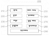
도 4의 휴대용 안진기의 제어유닛의 상세 구성도를 참고하면, 제어유닛(200)은 센서부(210), 통신부(220), 저장부(230), 음성출력부(240), 이미지처리부(250), 표시제어부(260), 촬영제어부(270), 캘리브레이션부(280) 그리고 안진기제어부(290)를 포함할 수 있다. 4, the
센서부(210)는 본체(10)의 내측 중앙부, 예컨대 피검자의 미간 중심에 대응되도록 배치될 수 있다. 센서부(210)는 피검자의 휴대용 안진기(100) 착용 상태, 예컨대 본체(10)의 기울어짐 등을 감지할 수 있다. 이러한 센서부(210)는 중력센서, 가속도센서, 자이로센서 등으로 구성될 수 있다. The
통신부(220)는 외부 단말기인 진단단말기(500)와 유/무선 방식으로 통신할 수 있다. 통신부(220)는 제어유닛(200)의 제어 결과, 예컨대 안구영상의 이미지 처리 결과를 외부기기로 전송할 수 있다. 또한, 통신부(220)는 외부기기로부터 소정의 제어신호를 제공받아 제어유닛(200)의 동작이 제어될 수 있도록 할 수 있다. The
또한, 통신부(220)를 통하여 안진기(100)와 진단단말기(500)가 무선통신을 통하여 정보를 주고받는 경우에는 특정 주파수 대역을 통하여 데이터의 송수신이 가능하도록 할 수 있으나, 통상의 네트워크 통신을 통하여 송수신하는 객체를 정확히 지정할 수 있도록 각 안진기와 단말기는 고유식별번호를 지정받아 통신할 수 있다.In addition, when information is transmitted and received through the wireless communication through the
이러한 고유식별번호는 IP일 수 있으며, 안진기의 저장부(230)와 진단단말기(500)의 IP저장부(560)에 저장되어 있는 IP로 상대방을 지정하여 통신할 수 있다.The unique identification number may be IP, and the counterpart may be designated and communicated with the IP stored in the
예를 들어 안진기(100)의 측정 결과를 다수의 진단단말기(500)로 지정하여 송신할 수도 있으며, 진단단말기(500)에서도 여러 피검자가 안진기를 이용하여 이석의 유무를 진단하는 경우에, 각각 서로 다른 IP로 상대 안진기를 호출하여 특정 안진기의 정보를 주고받을 수 있는 것이다.For example, it is also possible to designate and transmit the measurement results of the
저장부(230)는 제어유닛(200)의 동작을 위한 프로그램이 저장되거나 또는 제어유닛(200)의 제어 결과 또는 IP가 저장될 수 있다. The
또한, 저장부(230)는 본 발명의 안진 진단을 위한 어플리케이션이 저장될 수 있다.In addition, the
이러한 어플리케이션은 앱스토어 등을 통하여 관련 앱을 다운받아 저장하거나 또는 이동통신망을 통해 관리자의 웹서버에 접속하여 앱을 다운받아 설치되도록 구성할 수 있다.These applications can be configured to download and store related apps through the App Store, or to download and install apps by accessing the administrator's web server through a mobile communication network.
이러한 어플리케이션에는 진단을 위한 음성 안내 등이 포함될 수 있고, 무엇보다도 측정한 동공의 진동정보와 비틀림정보를 실행된 어플리케이션의 전송 포멧에 맞추어 진단단말기(500)로 전송하도록 한다.Such applications may include voice guidance for diagnosis, and above all, transmit the measured vibration information and torsion information to the
바람직하게는 앱(App)의 설치과정에서 인증과정을 거쳐 어플리케이션을 설치하도록 할 수 있으며, IOS 계열이건 안드로이드 계열이건 관계없이 각 OS에 맞는 앱을 앱 스토어에 올려서 배포하는 방식을 사용할 수도 있고, 또는 안진기 자체에 어플리케이션을 내장되어 이를 활용하거나 또는 단말기가 접속하여 해당 어플리케이션을 다운받게 할 수도 있다.Preferably, the application can be installed through an authentication process in the installation process of the app, and regardless of whether it is an IOS system or an Android system, a method for distributing an app for each OS in the app store can be used, or Anjinjingi itself has an application built into it, or a terminal can be accessed to download the application.
즉, 어플리케이션은 안진기, 단말기, 앱스토어 또는 관리자의 웹서버 등과 같이 어느 하나에 어플리케이션을 설치하고, 이를 다운받아 사용하게 할 수 있다.That is, the application can install the application on any one such as Anjingi, a terminal, an app store or a web server of an administrator, and download and use the application.
이러한 앱(App)의 다운 및 설치과정 그리고 인증단계 등은 일반적인 것이므로 그 상세한 설명은 생략한다.Since the downloading and installation process of the app and the authentication step are general, detailed description thereof will be omitted.
음성출력부(240)는 이석증을 진단하기 위한 음성명령이나 캘리브레이션을 위한 음성명령 등을 피검자에게 출력할 수 있다. The
예를 들어 자발안진 검사를 위한 음성과 영상안내, 주시안진 검사를 위한 음성과 영상안내, 앞반고리관 검사를 위한 음성과 영상 안내, 후반고리관 검사를 위한 음성과 영상안내, 수평반고리관 검사를 위한 음성과 영상안내 등을 저장부(230)에 저장하고 있다가 검사 항목에 따라 안진기제어부(290)의 제어에 따라 해당 음성을 출력하면 된다.For example, voice and video guidance for spontaneous ocular examination, voice and video guidance for primary cyanism examination, voice and video guidance for frontal annulus examination, voice and image guidance for posterior annulus examination, and voice examination for horizontal annulus examination After storing the image guide in the
예를 들어, 안진기를 착용하고 전원을 온하면 다음과 같은 음성이 출력될 수 있다.For example, the following voice may be output when the power is turned on while wearing the jinjingi.
인사말로 "안녕하세요. Smart dizzy 입니다"라고 출력하고, 검사 소개로 “지금부터 환자분의 평형 기능을 검사하겠습니다.”라는 멘트와 함께, 자세 확인을 위하여 "바로 앉은 자세에서 고글이 얼굴에 맞게 씌워 졌는지 확인해 주시기 바랍니다. 누울 수 있도록 주변에 위험한 물건이 없는지 확인해 주시길 바랍니다.“라는 음성을 출력할 수 있다.To say "Hello, it's Smart dizzy" as a greeting, and with the comment "I'll check the patient's equilibrium function from now on" with the introduction of the test, to confirm the posture, "Is the goggles covered in the right position in the sitting position?" Please make sure that there are no dangerous objects around you so that you can lie down.
이어, 자발안진의 경우에는 다음과 같은 순서로 음성을 출력할 수 있다.Subsequently, in the case of spontaneous anjin, the voice may be output in the following order.
1. 첫번째 검사로 자발 안진 검사를 시행하겠습니다. 1. The first test will be a voluntary eye examination.
2. 현재 전방 시야는 암흑 상태로, 앞이 보이지 않지만 현재 환자분의 눈을 비디오로 녹화 중입니다. 2. Currently, the anterior field of view is dark, and although the front is invisible, the patient's eyes are currently being recorded in video.
3. 가능한 눈을 깜박이지 말고, 앞에 하나의 점이 있다고 상상하신 뒤, 그 점을 주시하면서 눈을 움직이지 말아주세요. (눈을 움직이지 말고, 멍 하고 있으시길 바랍니다. )3. Do not blink your eyes as much as possible, imagine that there is a spot in front of you, and do not move your eyes while observing the spot. (Please don't move your eyes, but stay blank.)
4. 지금부터 촬영을 시작합니다. 소요시간은 약 20초 입니다. 삐~4. Start shooting now. The time required is about 20 seconds. Beep~
또한 수평반고리관 이석증 검사의 경우에는 Also, in the case of horizontal semicircular canal disease test
"뒤로 바로 누우세요. 오른쪽 귀가 아래로 향하도록 고개를 오른쪽으로 돌리세요. 왼쪽 귀가 아래로 향하도록 고개를 왼쪽으로 돌리세요. 다시 일어나 앉아 정면을 바라보세요." 등과 같이, 후반고리관 이석증 검사의 경우에는 "바로 앉은 자세에서 오른쪽으로 고개를 45˚ 돌린 상태에서 뒤로 누워 고개를 침대밑으로 떨어뜨리세요. 다시 일어나 앉으세요. 바로 앉은 자세에서 왼쪽으로 고개를 45˚ 돌린 상태에서 뒤로 누워 고개를 침대 밑으로 떨어뜨리세요. 다시 일어나 앉으세요."등과 같이 음성안내 또는 영상 안내로 제공할 수 있다."Let's lie back. Turn your head to the right with your right ear facing down. Turn your head to the left with your left ear facing down. Sit back and look straight ahead." For example, in the case of an examination of the posterior annular canal dysentery, "Let's lie back and drop your head under the bed while turning your head 45˚ to the right in a sitting position. Sit up again. Sit back. ˚ Lying on your back while turning, drop your head under the bed. Get up again and sit down.” It can be provided by voice guidance or video guidance.
또한, 제어유닛(200)의 안진기제어부(290)를 통하여 이러한 음성들은 멘트의 편집이 가능하게 하고 피검자의 상태에 따라 문장 재생 속도 등을 설정하게 할 수 있다.In addition, through the
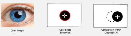
이미지처리부(250)는 광학/촬영유닛(30)에서 제공된 하나 이상의 촬영영상, 즉 피검자의 양안 각각에 대한 안구영상을 이미지 프로세싱할 수 있다. 이미지처리부(250)는 이미지 프로세싱의 결과물로 피검자의 안구 및 동공 각각에 대한 외곽라인과 동공의 중심점을 추출할 수 있다. 동공 중심점은 좌표로 추출되어 진동정보를 구성한다. The
또한, 이미지처리부(250)는 이미지 프로세싱의 결과물로 피검자의 홍채 또는 동공에 대한 특징점을 추출하고 각 프레임의 특징점에 대한 비틀림의 정도를 각도로 추출할 수 있다.Also, the
구체적으로 진동정보의 추출은 흑백 또는 이진 영상의 안구 이미지를 프레임별로 처리하여 각 프레임에서 동공의 중심점 좌표를 추출하고, 비틀림정보의 추출은 홍채 또는 동공의 영상을 프레임별로 처리하여 각 프레임에서 홍채 또는 동공의 특징점에 대한 패턴을 비교하여 각도를 추출하는 것이다.Specifically, for extracting vibration information, the eyeball image of a black and white or binary image is processed frame by frame to extract the coordinates of the center point of the pupil in each frame, and for the extraction of torsion information, the image of the iris or pupil is processed frame by frame to iris or The angle is extracted by comparing the patterns for the feature points of the pupil.
먼저, 동공의 중심점 좌표를 추출하기 위해서는 최초로 획득한 피검자의 흑백 또는 이진 영상의 안구 이미지로부터 최초 동공 중심점 좌표를 설정하고, 상기 최초 동공 중심점 좌표를 기준으로 연속적으로 입력되는 프레임의 안구영상에서 해당 프레임의 동공 중심점 좌표값을 산출하도록 한다.First, in order to extract the center point coordinates of the pupil, the first pupil center point coordinates are set from the eyeball image of the first acquired subject's black and white or binary image, and the corresponding frame in the eye image of the frame that is continuously input based on the first pupil center point coordinates. To calculate the coordinate value of the pupil center point.
도 6의 동공 중심점 좌표값을 산출하여 비교하는 참고도면을 좌측부터 순착적으로 보면, 양안 카메라(31)에서 촬영된 동공영상으로부터 최초 동공의 중심점 좌표를 추출하고(도면에서는 "![]()
![]()
이러한 표시는 입력되는 프레임별 센터표시를 누적하여 표시할 수도 있으나, 관찰을 위하여 직전 센터값과 현재 센터값을 기준으로 2개의 센터값만 비교하여 표시할 수도 있다.Such a display may be displayed by accumulating center marks for each input frame, but for observation, only two center values may be compared and displayed based on the previous center value and the current center value.
최초 동공의 중심점 좌표는 최초 프레임의 흑백 또는 이진 영상의 안구 이미지에서 피검자의 안구 외곽라인 및 동공 외곽라인을 각각 추출하고, 상기 동공 외곽라인에 기초하여 상기 최초 동공 중심점 좌표를 설정한 다음, 상기 안구 외곽라인에 기초하여 피검자의 안구 양측 수평 포인트를 설정하고, 상기 양측 수평포인트 각각에서 상기 최초 동공 중심점 좌표까지의 거리를 산출하도록 구성한다.The center point coordinates of the first pupil are respectively extracted from the subject's eye outline and the pupil outline line from the eye image of the black and white or binary image of the first frame, and the coordinates of the first pupil center point are set based on the pupil outline line, and then the eyeball The horizontal points on both sides of the subject's eye are set based on the outline line, and the distance from each of the horizontal points on both sides to the coordinates of the initial pupil center point is calculated.
또한, 상기 최초 프레임의 흑백 또는 이진 영상의 안구 이미지는 상기 안진기를 피검자의 양안 중 어느 하나의 영역으로부터 검진을 알리는 표시영상을 출력하고, 상기 표시영상에 대한 피검자의 시선을 유도한 다음, 상기 표시영상을 주시하는 피검자의 양안 각각을 촬영하여 상기 최초 안구영상을 획득하도록 할 수 있다.In addition, the black-and-white or binary image of the first frame of the eyeball image outputs a display image informing the examination from any one of the subject's binocular eyes, and induces the subject's gaze on the display image, and then displays The first eyeball image may be acquired by photographing each of both eyes of the subject who watches the image.
또한, 동공 중심점 좌표값의 산출은 피검자의 시선 이동에 따른 안구를 촬영하여 안구영상을 획득하고, 상기 안구영상에서 동공 외곽라인을 설정하고, 상기 동공 외곽라인에 기초하여 동공 중심점 좌표를 (x,y)형태로 추출하는 것이다.In addition, the calculation of the pupil center point coordinate value is obtained by photographing the eyeball according to the subject's gaze movement, obtaining an eyeball image, setting a pupil outline line in the eyeball image, and calculating the pupil center point coordinates based on the pupil outline line (x, y).
이러한 좌표의 추출은 초당 30프레임의 영상을 약 5~30sec 동안 분석한다.The extraction of these coordinates analyzes an image at 30 frames per second for about 5 to 30 seconds.
초당 30프레임으로 영상의 총 프레임을 계산하면 약 900 - 1200 프레임의 좌표를 추출하여 진동정보를 구성하는 것이다.When the total frame of the image is calculated at 30 frames per second, vibration information is configured by extracting coordinates of about 900-1200 frames.
그리고 산출된 동공 중심점 좌표에 따라 상기 최초 동공 중심점 좌표의 변화를 비교하여 동공의 센터값 이동거리는 x축 또는 y축으로 어느 하나 이상 1mm이상이고, 동공의 면적이 전체 면적의 50%이상일 때의 좌표만 추출하도록 한다.And by comparing the change in the coordinates of the initial pupil center point according to the calculated pupil center point coordinates, the center distance of the pupil is one or more of 1 mm or more in the x-axis or y-axis, and the pupil area is 50% or more of the total area. Only extract.
동공의 면적이 전체 면적의 50%이하인 경우에는 안구 외곽라인에 기초하여 분석 범위를 벗어난 영상은 배제하기 때문에 잔여 동공의 영상이 50%미만인 경우는 동공의 센터값을 추출할 수가 없는 경우이며, 1mm미만의 경우에는 안진과 관계없이 자연스런 통상의 눈의 움직임으로부터 발생할 수 있는 경우이기 때문에 배제하는 것이다.When the pupil area is less than 50% of the total area, the image outside the analysis range is excluded based on the outer line of the eyeball. If the residual pupil image is less than 50%, the center value of the pupil cannot be extracted, and 1mm The following cases are excluded because they may arise from natural normal eye movements regardless of the eye movement.
또한, 이미지처리부(250)는 비틀림의 정도를 비교할 수 있도록 각도를 추출한다.In addition, the
비틀림의 정도를 산출하기 위해서는 입력되는 흑백 또는 이진 영상의 안구이미지에서 동공 또는 홍채 영역을 추출하고, 동공 또는 홍채에서 특징점을 설정하고 최초 특징점 대비 연속으로 입력되는 프레임의 특징점을 상기 최초 특징점 대비 기울어진 각도를 산출하도록 동작한다. In order to calculate the degree of torsion, the pupil or iris region is extracted from the eye image of the input black-and-white or binary image, the feature points are set from the pupil or iris, and the feature points of the continuously input frame are inclined relative to the first feature points. It works to calculate the angle.
바람직하게는 LED와 같은 적외선 발사장치를 구비하고 반사된 동공 또는 홍채를 인식하는 렌즈를 사용할 수 있다. Preferably, an infrared launching device such as an LED and a lens for recognizing reflected pupils or irises can be used.
이하에서는 설명의 편의를 위하여 홍채의 이미지에서 특징점을 추출하고 각도를 산출하는 방법을 위주로 설명하기로 한다.Hereinafter, for convenience of description, a method of extracting feature points from an iris image and calculating an angle will be mainly described.
먼저 도 7의 홍채 패턴을 비교하여 변화 각도를 산출하는 참고도면을 보면, 입력되는 흑백 혹은 이진 영상의 안구 이미지에서 홍채 영역만 추출하고, 홍채에서 특징점을 설정하고 최초 특징점 대비 연속으로 입력되는 프레임의 특징점을 상기 최초 특징점 대비 기울어진 각도를 산출하도록 동작한다. First, looking at the reference drawing for comparing the iris pattern of FIG. 7 to calculate the angle of change, only the iris region is extracted from the input eye image of the black and white or binary image, the feature points are set in the iris, and the frame is continuously input compared to the initial feature point. The feature point is operated to calculate an angle inclined relative to the initial feature point.
이러한 홍채는 타 홍채와 구별될 수 있는 특징점으로 긴 띠 모양의 망(빗살 무늬의 인대), 붉은 색의 섬유질, 속눈썹 모양의 돌기, 꾸불꾸불한 혈관계, 링 모양의 원들, 동공을 둘러싸는 코로나 모양의 인대, 홍채 고유의 색, 얼룩점 등이 있으며 이러한 특징점들은 각 사람마다 다른 생물학적 패턴을 가진다. These irises can be distinguished from other irises by long strip-shaped nets (comb-shaped ligaments), red fibers, eyelash-shaped projections, serpentine vascular system, ring-shaped circles, and corona-shaped surrounding the pupil. There are ligaments, iris-specific colors, and spots, and these characteristic points have different biological patterns for each person.
따라서 본 발명에서는 상기 홍채의 특징점 중 어느 하나를 선정하여 비교할 수도 있으나, 홍채의 비틀림 각도를 손쉽게 측정하기 위해서는 긴 띠 모양의 망, 혈관계 등과 같이 비틀림 각도를 측정할 수 있는 특징점을 선택하여 비교하는 것이 바람직하나, 이에 한하지 않고 상기 특징점들을 포함하는 전체 패턴으로 비교할 수도 있음은 물론이다.Therefore, in the present invention, any one of the feature points of the iris may be selected and compared, but in order to easily measure the twist angle of the iris, it is preferable to select and compare feature points capable of measuring the twist angle, such as a long band-shaped network and a vascular system. It is preferable, but not limited to, that the entire pattern including the feature points may be compared.
또한, 이러한 특징점은 피검자마다 두드러지는 포인트가 있게 마련이므로, 필요한 경우 피검자마다 특징점을 새로 설정할 수 있도록 하는 것이 더욱 바람직하다.In addition, since these feature points are provided with prominent points for each subject, it is more preferable to set a new feature point for each subject if necessary.
상술한 바와 같이 특징점이 설정되면, 이미지처리부(250)는 비틀림의 각도를 산출하기 위하여 최초 안구영상으로부터 Binary영상처리기법을 이용하여 홍채나 동공의 특징점을 추출하고, 패턴 인식을 통하여 원본 영상의 특징점 대비 비틀린 정도를 각도로 추출하는 것이다.When the feature points are set as described above, the
도면을 참고하면, 좌측부터 순서대로, 좌측의 칼라 안구 영상으로부터 홍채의 특징점을 패턴 인식을 통하여 추출하고 다음 프레임에서 측정된 홍채의 특징점을 서로 비교함으로써, 변화의 정도를 각으로 표시하고, 이를 통하여 비틀림 정보를 추출할 수 있는 것이다.Referring to the drawings, from the left, in order, the feature points of the iris are extracted from the color eyeball image on the left side through pattern recognition and the feature points of the iris measured in the next frame are compared with each other to indicate the degree of change, and through this The torsional information can be extracted.
비틀림 정보도 진동정보와 같이 초당 30프레임의 영상을 약 5~30sec 동안 분석한다.Torsional information, like vibration information, analyzes 30 frames per second for about 5 to 30 seconds.
초당 30프레임으로 영상의 총 프레임을 계산하면 약 900 - 1200 프레임의 좌표를 추출하여 비틀림 정보를 구성하는 것이다.When the total frame of the image is calculated at 30 frames per second, torsion information is constructed by extracting coordinates of about 900-1200 frames.
표시제어부(260)는 제1디스플레이(110)를 제1영역과 제2영역, 예컨대 광학/촬영유닛(30)의 좌측영역과 우측영역을 분리하고, 분리된 각 영역에서 영상의 표시를 제어할 수 있다. The
표시제어부(260)는 제1디스플레이(110)의 좌측영역에 제1표시영상이 표시되도록 하고, 우측영역에 제2표시영상이 표시되도록 할 수 있다. 이때, 표시제어부(260)는 제1디스플레이(110)에서 하나의 영상만이 표시되도록 할 수 있다. 따라서, 제1디스플레이(110)는 표시제어부(260)에 의해 좌측영역의 제1표시영상 및 우측영역의 제2표시영상 중 하나를 표시할 수 있다. 이때, 제1디스플레이(110)의 각 영역 중 영상이 표시되지 않는 영역은 검은 화면, 즉 암흑 상태를 유지할 수 있다. The
또한, 표시제어부(260)는 제1디스플레이(110)의 분리된 각 영역에서 표시영상이 이동되면서 표시되도록 제어할 수 있다. In addition, the
예컨대, 표시제어부(260)는 제1디스플레이(110)의 좌측영역에 표시되는 제1표시영상이 제1방향 또는 제2방향으로 이동되면서 표시되도록 제어할 수 있다. 물론, 표시제어부(260)는 제1디스플레이(110)의 우측영역에 표시되는 제2표시영이 제1방향 또는 제2방향으로 이동되면서 표시되도록 제어할 수 있으며, 제1방향은 피검자를 기준으로 각 영역의 좌측 방향이고, 제2방향은 피검자를 기준으로 각 영역의 우측 방향일 수 있다. For example, the
또한, 표시제어부(260)는 표시영상의 조도를 제어하여 피검자와 영상 간의 거리를 조절할 수 있다. In addition, the
예컨대, 표시제어부(260)는 제1디스플레이(110)의 각 영역의 표시영상이 저조도로 표시되도록 함으로써, 피검자가 표시영상이 소정 거리로 이격되어 표시되고 있음을 인지하도록 할 수 있다. 통상, 안진 검사의 경우에 피검자와 영상 간 대략 1m의 이격 거리가 요구되므로, 표시제어부(260)는 피검자가 제1디스플레이(110)의 표시영상이 대략 1m의 거리에 표시되고 있음을 인지하도록 그 조도를 제어할 수 있다. For example, the
촬영제어부(270)는 광학/촬영유닛(30)의 카메라의 촬영 동작을 제어할 수 있다. 이에, 광학/촬영유닛(30)의 카메라는 촬영제어부(270)의 제어에 의해 피검자의 양안 각각을 적어도 한번 촬영할 수 있다. The photographing
또한, 촬영제어부(270)는 센서부(210)의 감지결과에 따라 본체(10)가 수평이 되도록 피검자의 자세를 교정할 수 있고, 광학/촬영유닛(30)의 각 접안렌즈의 움직임을 제어하여 피검자와 표시영상 간의 초점을 교정할 수 있다. 피검자의 자세교정 및 초점교정이 완료된 후, 촬영제어부(270)는 광학/촬영유닛(30)의 카메라의 촬영 동작을 제어할 수 있다. 자세교정 및 초점교정은 전술한 음성출력부(240)를 통해 음성명령을 피검자에게 제공하여 이루어질 수 있다.In addition, the photographing
캘리브레이션부(280)는 이미지처리부(250)의 처리 결과, 즉 피검자의 양안 각각에 대한 하나 이상의 안구영상에서 추출된 피검자의 안구와 동공 각각의 외곽라인 및 동공 중심점 좌표에 기초하여 피검자의 검진 초기값을 설정하는 캘리브레이션을 제어할 수 있다. The
캘리브레이션부(280)는 이미지처리부(250)에서 제공되는 하나 이상의 동공 중심점 좌표에 기초하여 최초 동공 중심점 좌표와 상기 최초 동공 중심점 좌표의 변화량을 산출할 수 있으며, 산출된 동공 중심점 값 변화량에 기초하여 피검자의 최대 동공 움직임 값을 설정하고, 이를 피검자의 검사 초기값으로 설정할 수 있다. The
한편, 휴대용 안진기(100)는 본체(10)의 외측, 예컨대 전면케이스(20)의 외면에 부착된 제2디스플레이(120)를 더 포함할 수 있다. Meanwhile, the
제2디스플레이(120)는 제어유닛(200)의 제어에 따라 피검자의 안구 이미지, 예컨대 광학/촬영유닛(30)에 의해 촬영된 안구영상 또는 이미지처리부(250)에 의해 처리된 이미지 중 적어도 하나를 외부에 표시할 수 있다. 이러한 제2디스플레이(120)는 제1디스플레이(110)와 마찬가지로 LCD 또는 OLED로 구성될 수 있으며, 외부에서 터치 등을 통해 제어할 수 있도록 터치형 디스플레이로 구성될 수 있다. The
또한, 본체(10)의 일측에는 인터페이스부(50)가 구비될 수 있으며, 인터페이스부(50)를 통해 다양한 외부기기와 연결될 수 있으며, 이를 통하여 유무선 통신을 할 수 있다.In addition, an
안진기제어부(290)는 제어유닛(200)의 각 구성요소들을 제어하여 피검자의 안구영상으로부터 진동정보와 비틀림정보를 추출하여 이를 진단단말기(500)로 전송하도록 동작할 수 있다.The
또한, 안진기제어부(290)는 피검자의 고유식별번호별로 측정한 안구 영상과 진동정보 그리고 비틀림정보를 검진한 날짜와 함께 DB의 형태로 저장부(230)에 저장되게 하고, 이후 진단단말기(500)로부터 진단결과가 수신되면 해당 데이터도 피검자의 고유식별번호별로 저장되게 할 수 있다.In addition, the
이로써, 단말기를 통하지 않고서도 안진기의 식별번호만으로로도 해당 안진기를 통하여 안진을 측정한 피검자들의 진단 결과를 직접 통신을 통하여 알 수 있는 것이다.In this way, the diagnosis result of the subjects who measured the eyelids through the corresponding eyepieces can be known through direct communication only by using the identification number of the eyepieces without going through the terminal.
상술한 바와 같이 휴대용 안진기(100)로부터 피검자의 머리에 착용되어 검진 항목에 따라 처리된 진동정보와 비틀림정보를 전송받은 진단단말기(500)는 해당 정보를 처리하여 안진의 종류를 진단하도록 한다.As described above, the
해당 안진기의 IP번호와 피검자의 고유식별번호, 그리고 측정데이터 등을 수신받아 안진의 유무를 판단하도록 한다.Receive the IP number of the relevant jinjingi, the subject's unique identification number, and measurement data to determine the presence or absence of jinjin.
본 발명에서는 피검자의 인적사항에 따라 고유식별번호를 부여할 수 있으며, 이러한 고유식별번호는 피검자를 상호 분리 관찰할 수 있는 정도의 정보이면 족하므로, 피검자의 핸드폰 번호, 주민 번호, 성명 등 어느 하나 이상으로 부여할 수 있음은 물론이다.In the present invention, a unique identification number may be assigned according to the personal information of the subject, and such a unique identification number is sufficient as long as the information is sufficient to observe the subjects separately, so any one of the subject's mobile phone number, resident number, and name, etc. Needless to say, the above can be given.
진단단말기(500)는 전문의와 같은 이석증 진단 관리자가 사용하는 단말기로서, 데스크탑 PC(desktop PC)폰만 아니라, 스마트폰, 태블릿 PC(tablet PC), 슬레이트 PC(slate PC), 노트북 컴퓨터(notebook computer), 키오스크와 같은 단말기 등이 해당될 수 있다. 물론, 본 발명이 적용 가능한 단말기는 상술한 종류에 한정되지 않고, 안진기 및 외부 단말기와 통신이 가능한 단말기를 모두 포함할 수 있음은 당연하다.The
진단단말기(500)는 안진기(100)가 피검자로부터 추출한 진동정보와 비틀림정보를 통신인터페이스(510)를 통하여 송수신하여 안진의 종류를 진단하는 어플리케이션을 실행하는 사용자앱(App)부(550)와, 키입력부(530)와, 단말기표시부(520)와, 어플리케이션을 구동하여 안진의 진단하고 진단한 결과를 단말기 표시부(520)에 표시하게 제어하는 단말기제어부(540)를 포함하여 구성한다.The
또한, 저장부(570)에는 진단 결과에 따라 안진을 진단할 수 있는 진단DB가 저장된다.In addition, the
통신인터페이스(510)는 다양한 통신 방식(와이파이와 같은 무선통신)을 통하여 하나 이상의 안진기(100) 및 진단단말기(500)와 통신하여 데이터 수신이 가능하도록 동작한다.The
즉, 통신인터페이스(510)는 이더넷(Ethernet), 범용 직렬 버스(Universal Serial Bus), IEEE 1394, 직렬통신(serial communication) 및 병렬 통신(parallel communication)과 같은 유선 통신 방식과 적외선 통신(Infrared Radiation), 블루투스(Bluetooth), RF(Radio Frequency), 무선 랜(Wireless LAN), 도는 인터넷 통신과 같은 네트워크 통신 등과 같은 무선 통신 방식을 이용할 수 있다..That is, the
사용자앱(App)부(550)는 앱스토어 등을 통하여 관련 앱을 다운받아 저장하거나 또는 이동통신망을 통해 관리자의 웹서버에 접속하여 어플리케이션을 다운받아 설치되도록 구성할 수 있다.The user app (App)
이러한 어플리케이션에는 안진을 진단하기 위한 자발안진 진단 기준, 주시안진 진단 기준, 후반고리관 이석증 진단기준, 가측반고리관 이석증의 진단기준 등이 포함될 수 있다.Such applications may include a diagnosis criteria for spontaneous nystagmus for diagnosing nystagmus, a criteria for diagnosis of primary cyanosis, a diagnosis criterion for posterior annular canopia, and a diagnosis criterion for lateral annular candidiasis.
이러한 앱의 설치와 기타 내용에 대해서는 안진기의 어플리케이션 설치와 동일하므로 상세한 설명은 생략하기로 한다.The installation and other contents of these apps are the same as the application installation of Ajinjin, so a detailed description will be omitted.
키입력부(530)는 터치 스크린이나 키패드로 해당 키에 대한 데이터를 입력할 수 있도록 구성하되, 통상의 통신에 필요한 키와 본 발명의 치매의 진단과 예방을 위한 운동 및 식이 처방에 필요한 서비스를 실행하고 표시하기 위한 특정버튼 등을 포함하여 구성된다.The
단말기표시부(520)는 통상의 통신인터페이스(510)를 통해 수신된 데이터를 표시하는 디스플레이장치로 동작되나, 본 발명에서는 앱을 구동하여 진단의 과정에 필요한 내용과 안진의 진단결과를 표시하도록 구성한다,The
좌표처리부(580)는 통신인터페이스부(510)를 통하여 수신되는 진동정보인 (x,y)좌표를 이용하여 진동의 정도와 방향을 판단하여 단말기제어부(540)로 전송한다.The coordinate
진동의 방향은 우측(외측)과 좌측(내측), 상향(up), 하향(down)으로 표현할 수 있다.The direction of vibration can be expressed as right (outside) and left (inside), up (up), down (down).
각도처리부(590)는 통신인터페이스부(510)를 통하여 수신되는 비틀림정보인 각도를 이용하여 비틀림의 방향을 추출하도록 한다.The
비틀림의 방향은 intorsion(내회전), extorsion(외회전)으로 표현한다.The direction of torsion is expressed as intorsion (external rotation) or extorsion (external rotation).
intorsion(내회전)은 수직안구경선의 상부가 얼굴의 중심을 향하여 내전하는 것으로 안구의 상극이 코 쪽으로 회전하는 것을 의미하고, extorsion(외회전)은 수직안구경선의 상부가 얼굴의 중안선에 대해 기울어져 있는 것으로 안구의 상극이 귀 쪽으로 회전하는 것을 의미한다.Intorsion (internal rotation) means that the upper part of the vertical spectacle line rotates toward the center of the face, and the upper pole of the eyeball rotates toward the nose, and extorsion (external rotation) the upper part of the vertical spectacle line is inclined with respect to the midline of the face It means that the upper pole of the eye rotates toward the ear.
단말기제어부(540)는 키입력부(530)의 특정버튼 입력에 따라 사용자앱(App)부(550)에 저장되어 있는 어플리케이션을 구동하여, 저장하고 있는 데이터를 표시하거나, 통신인터페이스(510)를 통하여 안진기(100)로부터 주고 받는 데이터를 단말기 표시부(520)에 표시하도록 제어한다.The
먼저 단말기제어부(540)는 안진기(100)에서 측정된 데이터를 수신하여 안진의 유무와 이석증을 판단하여 그 결과를 표시하도록 동작한다.First, the
이러한 검사항목에 따라 안진을 진단하는 방법에 대하여 간단히 설명하면 다음과 같다.The method for diagnosing ocular anxiety according to these test items is briefly described as follows.
통상 안진은 총 5가지로 검사를 진행할 수 있으며, 말초성과 중추성을 진단하는 것과 이석증을 진단하는 것으로 구별할 수 있다.In general, Anjin can be tested in five ways, and can be divided into diagnosing peripheral and central disease and diagnosing dysplasia.
말초성과 중추성을 진단하는 항목으로는 자발안진, 주시안진, 두위 회전후 안진 등이 있고, 이석증을 진단하는 방법으로는 체위변화(머리위치 변화)를 주면서(자극) 안진의 방향을 보고 후반고리관과 가측반 고리관 그리고 앞반고리관의 이석증으로 판단하는 방법 등이 있다.Items for diagnosing peripheral and centrality include spontaneous nystagmus, zhusianjin, and nystagmus after rotation of the head. As a method of diagnosing dysplasia, after reporting the direction of nystagmus by giving a change in position (stimulation of head position) (irritation) There is a method of judging by the dichotomy of the half-ring tube, the lateral half-ring tube, and the front half-ring tube.
먼저, 자발안진이란 피검자가 가만히 앉아있을 때, 안진기(100)를 통하여 가운데 보고 있으라고(암흑상태에서) 한 경우에 발생하는 안진으로 이를 기초로 말초신경계 안진과 중추신경계 안진으로 진단할 수 있다.First, spontaneous nystagmus is a nystagmus that occurs when a subject sits still and is looking at the center (in the dark state) through the
예를 들어 안진기의 이미지처리부(250)에서 수신한 진동정보로부터 진동의 방향이 우측으로 향하는 안진인 경우로 판단되면 우측 가측 반고리관(lateral semicircular canal)이 자극 받았거나 좌측 가측 반고리관(LSCC)이 억제 되었거나로 진단하고, 좌측으로 향하는 안진이 발생한 경우에는 좌측 가측 반고리관이 자극받았거나, 우측 가측 반고리관이 억제됨을 의미하는 것으로 진단할 수 있는 것이다.For example, from the vibration information received from the
즉, 자발안진은 반고리관이라는 말초기관의 이상으로 인한 수평안진이 발생한 경우 어지러움으로 진단할 수 있다.In other words, spontaneous nystagmus can be diagnosed as dizziness when horizontal nystagmus due to an abnormality in the peripheral organ called the Bangori duct.
또한, 수평 안진(좌측 혹은 우측)이 없이 상향 혹은 하향 안진이 발생하였을 때 중추신경계 어지러움이라고 진단할 수 있다. In addition, it can be diagnosed that the central nervous system is dizzy when an upward or downward nystagsm occurs without horizontal nystagmus (left or right).
또한, 1분간 지켜보았을 때 좌측으로 향하던 안진이 갑자기 우측으로 향하는 방향을 바꾸는 안진이 발생한 경우에도 중추신경계 안진으로 진단한다.In addition, even if the eyelid that turned left when observed for 1 minute suddenly changes the direction to the right, it is diagnosed as the central nervous system eyelid.
그리고 체위 변환(머리움직임)없이 외전(extorsion) 혹은 내전(intorsion)이 발생한 경우에도 중추신경계 안진으로 진단할 수 있다.In addition, the diagnosis of central nervous system nystagmus can be diagnosed even if an exorsion or intorsion occurs without changing the position (head movement).
또한, 주시안전이란, 앉은 자세에서 우측 30도와 좌측 30도로 보게 한 다음, 좌측을 보았을 때 수평안진(우측으로 향하는 안진)보다 우측을 보았을때 수평안진(우측으로 향하는 안진)이 더 강해질 때, 좌측 가측반고리관의 기능 저하(부전)으로 진단하고, 반대로 좌측으로 향하는 자발안진이 나타나는 상황에서 좌측을 보았을 때 좌측으로 향하는 안진이 더 커진다면 우측 가측 반고리관의 기능 부전으로 진단하고, 좌측을 보았을 때 좌측으로 향하는 수평안진이 발행하고 우측으로 보았을 때 우측으로 향하는 안진이 발생할 때는 중추 신경계 안진으로 진단하는 것이다.In addition, the safety of the gaze is 30 degrees on the right and 30 degrees on the left in a sitting position, and then when the left eye is stronger than the horizontal eye (left eye), when the left eye is stronger, the left eye is stronger. Diagnosed as deterioration (insufficiency) of the lateral annular duct and, conversely, when the left eye is larger in the situation where a spontaneous nymph directed to the left appears, it is diagnosed as dysfunction of the right lateral annular duct, and when viewed to the left, it is turned to the left. It is diagnosed as a central nervous system nystagmus when a horizontal nystagmus is directed and when it is viewed to the right, and an ocular gysm to the right occurs.
이석증을 검사하기 위해서는 체위 변화를 주면서 안진의 방향을 보고 진단하는 것으로, 딕스홀파이크(Dix-Hallpike) 검사와 롤링테스트(Rolling Test)를 통하여 진단할 수 있다.In order to test for dysentery, the patient is diagnosed by looking at the direction of the nystagmus while changing his position, and can be diagnosed through a Dix-Hallpike test and a rolling test.
Dix-Hallpike 검사는 우측 뒷반고리관을 검사하기 위해서 앉은상태에서 우측으로 45도 고개를 돌린후 머리를 뒤로 누우면서 침대보다 아래로 갈수 있게 하고, 좌측 뒷반고리관을 검사하기위해서는 좌측으로 45도 머리를 돌린후 뒤로 눕히면서 머리를 침대 모서리보다 아래로 내려가게 빠르게 한다.The Dix-Hallpike test allows you to go down from the bed while lying on your back with your head turned 45 degrees to the right to sit down to examine the right back half-ring tube, and turn your head 45 degrees to the left to check the left back half-ring tube. After lying on your back, make your head go faster than the edge of the bed.
우측 dix-hall을 했을 때 비틀림의 정도가 우측extorsion 방향이고 upbeating이 동시에 관찰되면 우측 후반고리관에 이석이 있으면서 우측 반고리관을 자극하고 있는 상태라고 진단하고, 좌측 dix-hall했을대 좌측 extorsion 방향이고 upbeating이 같이 나타나면 좌측 후반고리관에 이석이 있다고 진단하면 된다.When the right dix-hall is in the direction of the right extension and upbeating is observed at the same time, it is diagnosed that the right half ring tube is stimulating while the right half ring tube is stimulating. When the left dix-hall is left, it is in the left extorsion direction and upbeating If they appear together, you can diagnose that there are stones in the left posterior annulus.
그리고 rolling test를 통하여 측반고리관의 안진유무와 이석증을 진단할 수 있다.In addition, through rolling test, it is possible to diagnose the presence or absence and dizziness of the collateral tube.
rolling test는 피검자의 고개를 침대 위에 눕히고, 좌측 혹은 우측으로 고개를 45도 이상 돌리게 하여 안진을 검사하는 방법으로, 좌측으로 돌렸을◎ 좌측으로 향하는 수평안진이 발생하고, 우측으로 돌렸을 때 우측으로 향하는 수평안진이 발생(향지성,geotrophic)하고 그 안진 크기를 비교해서 우측이 더크면 우측 가측반고리관에 이석이 있음 진단하고, 좌측이 더크면 좌측 가측 반고리관에 이석이 있음을 진단하면 된다.Rolling test is a method of examining the eyelids by placing the subject's head on the bed and turning the head to the left or right by more than 45 degrees. Turned to the left ◎ Horizontal eyelid to the left occurs, and horizontal to the right when turned to the right By comparing the size of the nystagmus (eg, geotrophic) and comparing the size of the nystagmus, diagnose the presence of a stone in the right lateral semicircular canal if the right side is larger, and diagnose the presence of the stone in the left lateral semicircular canal when the left side is larger.
또한, 좌측으로 돌렸을 때 우측으로 향하는 안진이 발생하고, 우측으로 돌렸을 때 좌측으로 향하는 안진이 발행하면(원지성, ageotrophic), 그 안진 크기를 비교해서 우측이 더크면, 좌측 가측반고리관에 이석이 있음을 진단하고, 좌측이 더 크면 우측 가측반고리관에 이석이 있음을 진단하는 것이다.In addition, when the left eye turns to the right, and the left eye turns to the right when it is turned (ageotrophic), compared to the size of the eye, the right side is larger, and there are stones in the left lateral annulus. To diagnose, if the left side is larger, it is to diagnose the presence of a stone in the right side lateral semicircular canal.
상술한 바와 같이 단말기제어부(540)는 좌표처리부(580)와 각도처리부(590)의 처리 결과 수평안진(좌우로 진동)의 유무와 좌우측 외전, 상하진동 등 진단에 필요한 분석결과를 출력하도록 한다.As described above, the
또한, 단말기제어부(540)는 다수의 안진기에서 특정 안진기를 지정하여 데이터를 송수신하거나 또는 타 단말기와의 통신을 위하여 고유식별번호로 IP저장부(560)에 저장된 IP를 사용하여 통신하도록 한다.In addition, the
이하에서는 상술한 본 발명의 일실시예에 의한 안진기와 단말기를 이용하여 동공과 홍채를 이용한 이석증의 진단방법에 대하여 설명한다.Hereinafter, a method of diagnosing dyslithiasis using a pupil and an iris using an jinjingi and a terminal according to an embodiment of the present invention described above will be described.
도 5는 본 발명의 일실시예에 의한 동공과 홍채를 이용한 이석증의 진단방법을 설명하기 위한 흐름도로서, 도시된 바와 같이 본 발명의 이석증 진단 방법은 피검자에게 안진기(100)를 착용시키고 검사항목에 따라 전송되는 진동정보와 비틀림정보를 수신하여 분석함으로써 이석증을 진단하도록 동작한다.Figure 5 is a flow chart for explaining a method for diagnosing dysentery using a pupil and an iris according to an embodiment of the present invention, as shown in the present invention, the method for diagnosing dysentery is examined by wearing the
먼저 피검자의 안면에 착용된 안진기(100)의 양안카메라(31)에서 수신된 영상을 처리하여 진동정보와 비틀림정보를 추출한 다음(S100), S100 단계에서 추출된 진동정보와 비틀림정보를 진단단말기로 전송하고(S200), 진단단말기는 수신된 정보를 이용하여 이석증을 진단하게 된다(S300).First, by processing the image received from the
단계 S100에서 먼저 진동정보를 추출하기 위하여 안진기의 양안 카메라(31)를 통하여 피검자의 안구 영상을 촬영한다(S110).In step S100, in order to extract vibration information, an eyeball image of a subject is photographed through the
단계 S110에서의 안구 영상의 촬영은 설정된 진단 항목에 따라 음성출력부(240) 또는 제1디스플레이(110)를 통하여 음성 안내나 영상을 통하여 안구 영상을 획득할 수 있도록 안내를 진행한다.The photographing of the eyeball image in step S110 is guided to obtain the eyeball image through the voice guidance or the image through the
물론, 실제 진단항목을 설정하기 전에 진단의 정확도를 높일 수 있도록 캘리브레이션 과정을 수행할 수 있음은 물론이다.Of course, before setting the actual diagnosis items, it is needless to say that the calibration process can be performed to increase the accuracy of diagnosis.
이러한 안구 영상의 촬영은 촬영제어부(270)가 광학/촬영유닛(30)으르 제어하여 양안 카메라(31)를 통하여 안구 영상을 수신한다.The photographing
단계 S110에서 안구의 영상이 수신되면, 안진기제어부(290)는 이미지처리부(250)에서 수신된 안구의 영상을 프레임별로 처리하여(S111) 각 프레임에서 동공의 중심점 좌표를 추출한다(S112).When the image of the eyeball is received in step S110, the
단계 S112에서의 동공의 중심점 좌표는 상술한 이미지처리부(250)에서 개시된 동공의 중심점 좌표 추출 동작과 동일하므로 그 상세한 설명은 생략하기로 한다.Since the center point coordinates of the pupil in step S112 are the same as the operation of extracting the center point coordinates of the pupil disclosed in the above-described
간략하게 설명하면, 최초 프레임의 안구영상에서 피검자의 안구 외곽라인 및 동공 외곽라인을 각각 추출하고, 상기 동공 외곽라인에 기초하여 상기 최초 동공 중심점 좌표를 설정한 다음, 상기 안구 외곽라인에 기초하여 피검자의 안구 양측 수평 포인트를 설정하고, 상기 양측 수평포인트 각각에서 상기 최초 동공 중심점 좌표까지의 거리를 산출한다.Briefly, an eyeball outline line and a pupil outline line are respectively extracted from the eyeball image of the first frame, the coordinates of the initial pupil center point are set based on the pupil outline line, and then the subject is examined based on the eyeball outline line. Set the horizontal points on both sides of the eye, and calculate the distance from each of the horizontal points on both sides to the coordinates of the initial pupil center point.
이어 연속적으로 입력되는 안구의 영상 프레임에서 피검자의 시선 이동에 따른 안구를 촬영하여 안구영상을 획득하고, 상기 안구영상에서 동공 외곽라인을 설정하고, 상기 동공 외곽라인에 기초하여 동공 중심점 좌표를 (x,y)형태로 추출하는 것이다.Subsequently, in the image frame of the continuously input eyeball, the eyeball according to the subject's gaze movement is photographed to obtain an eyeball image, a pupil outline line is set in the eyeball image, and the pupil center point coordinates based on the pupil outline line (x ,y).
마찬가지로, 동공의 센터값 이동거리는 어느 하나 이상 1mm이상이고, 동공의 면적이 전체 면적의 50%이상일 때의 좌표만 추출하도록 한다.Similarly, the center distance of the pupil is at least one mm or more, and only the coordinates when the pupil area is 50% or more of the total area are extracted.
이렇게 추출된 대략 초당 30프레임 정도의 안구 영상으로부터 동공 중심점 좌표를 추출하여 진동정보를 구성하는 것이다. The vibration information is constructed by extracting the coordinates of the pupil center point from the eyeball image of about 30 frames per second.
즉, 동공의 중심점 좌표를 추출하기 위해서는 최초로 획득한 피검자의 프레임 안구영상으로부터 최초 동공 중심점 좌표를 설정하고, 상기 최초 동공 중심점 좌표를 기준으로 연속적으로 입력되는 프레임의 안구영상에서 해당 프레임의 동공 중심점 좌표값을 산출하여 (x,y)좌표값으로 진동정보를 구성하는 것이다.That is, in order to extract the center point coordinates of the pupil, the initial pupil center point coordinates are set from the frame eye image of the first acquired subject, and the pupil center point coordinates of the frame in the eyeball image of the frame continuously input based on the initial pupil center point coordinates. By calculating the value, the vibration information is composed of (x,y) coordinate values.
즉, 연속으로 입력되는 안구 영상을 이용하여 동공의 가장자리를 실시간으로 감지하고, 그 중심값을 추출하여 배열로 저장할 수 있도록 하는 것이다.That is, it is possible to detect the edge of the pupil in real time by using a continuously input eyeball image, and extract the central value and store it in an array.
한편, 비틀림정보를 추출하기 위하여 이미지처리부(250)는 안진기의 양안 카메라를 통하여 수신된 안구의 영상으로부터 홍채 또는 동공의 영상을 프레임별로 처리하여(S113) 각 프레임에서 홍채 또는 안구의 특징점에 대한 패턴을 비교하여 각도를 추출한다(S114).On the other hand, in order to extract the torsional information, the
단계 S113에서는 입력되는 안구의 칼라영상에서 Binary영상처리기법을 이용하여 홍채나 동공의 특징점을 추출하고, 패턴 인식을 통하여 원본 영상과의 비틀림을 계측한 다음, 단계 S114에서 비틀림의 정도를 각도로 추출한다.In step S113, the feature points of the iris or pupil are extracted from the color image of the input eye using a binary image processing technique, and the distortion with the original image is measured through pattern recognition, and then the degree of twist is extracted at an angle in step S114. do.
단계 S113에서의 비틀림정보도 상술한 이미지처리부(250)에서 개시된 비틀림의 정도를 각도로 추출하는 과정과 동일하므로 그 상세한 설명은 생략하기로 한다.The torsional information in step S113 is also the same as the process of extracting the degree of torsion initiated by the
또한, 홍채와 동공의 특징점을 각각 추출하거나 또는 동시에 추출하여 비틀림의 정보를 산출할 수 있으나, 본 발명에서는 홍채의 특징점을 추출하는 방법을 설명함으로써, 동공의 특징점을 추출하는 방법에 관한 설명에 갈음하고자 한다.In addition, although the feature points of the iris and the pupil may be respectively extracted or simultaneously extracted, torsional information may be calculated, but in the present invention, the method of extracting the feature points of the pupil is explained by describing a method of extracting the feature points of the iris. I want to.
간략하게 설명하면, 입력되는 칼라영상의 안구 이미지에서 홍채 영역만 추출하고, 홍채에서 특징점을 설정하고 최초 특징점 대비 연속으로 입력되는 프레임의 특징점을 상기 최초 특징점 대비 기울어진 각도를 산출하도록 동작한다. Briefly, only the iris region is extracted from the ocular image of the input color image, the feature points are set in the iris, and the feature points of the continuously input frame compared to the first feature points are operated to calculate an angle inclined relative to the first feature points.
이러한 홍채의 특징점으로 긴 띠 모양의 망(빗살 무늬의 인대), 붉은 색의 섬유질, 속눈썹 모양의 돌기, 꾸불꾸불한 혈관계, 링 모양의 원들, 동공을 둘러싸는 코로나 모양의 인대, 홍채 고유의 색, 얼룩점 등이 있으며 이러한 특징점들 중 어느 하나 이상의 특징점을 설정한다.The characteristic features of these irises are long strip-shaped nets (comb-shaped ligaments), red fibres, eyelash-shaped projections, serpentine vascular systems, ring-shaped circles, corona-shaped ligaments surrounding the pupil, and iris-specific colors. , There are spots, etc., and one or more of these feature points is set.
특징점이 설정되면, 최초 안구영상으로부터 Binary영상처리기법을 이용하여 홍채의 특징점을 추출하고, 패턴 인식을 통하여 원본 영상의 특징점과 비교하여 비틀린 각도를 측정하는 것이다.When the feature points are set, the feature points of the iris are extracted using the binary image processing technique from the first eyeball image, and the twist angle is measured by comparing with the feature points of the original image through pattern recognition.
단계 S200에서는 단계 S100을 수행하여 추출된 진동정보와 비틀림정보를 진단단말기(500)로 전송한다.In step S200, the extracted vibration information and torsion information are transmitted to the
즉, 안진기제어부(290)는 이미지처리부(250)에서 진동정보와 비틀림정보가 추출되면, 추출된 정보를 프레임별로 또는 둘 이상의 프레임에 대한 정보를 배열로 저장하고 있다가 진단단말기(500)로 전송할 수 있다.That is, when the vibration information and the torsional information are extracted from the
구체적으로 S110 내지 단계 S114를 수행하여 추출된 진동정보는 통신부(220)를 통하여 진단단말기(500)의 통신인터페이스부(510)로 송신되고(S210), 추출된 비틀림정보도 통신부(220)를 통하여 진단단말기(500)의 통신인터페이스부(510)로 송신된다(S220).Specifically, the vibration information extracted by performing steps S110 to S114 is transmitted to the
단계 S200에서 통신인터페이스부(510)를 통하여 진동정보와 비틀림정보를 수신하면, 진동단말기의 단말기제어부(540)는 수신한 정보를 기초로 안진의 유무와 딕스홀파이크(Dix-Hallpike)검사항목에 따라 검진이 이루어진 경우는 후반고리관의 이석증 유무를 판단하고, 롤링테스트(rolling test)를 통하여 측반고리관의 이석증 유무를 판단하는 과정을 수행한다(S300).When the vibration information and the torsion information are received through the
이를 위하여 진단단말기는 수신된 진동정보와 비틀림정보를 이용하여 측정한 피검자의 상태가 안으로 움직이는 내전, 밖으로 움직이는 외전, 위로 움직이는 상전(elevation 혹은 sursumduction), 아래로 움직이는 하전(depression 혹은 deorsumduction), 안구의 상극(upper pole)이 귀 쪽으로 움직이는 외회선(extorsion, excycloduction)과 코쪽으로 움직이는 내회선(intorsion, incycloduction) 중 어느 하나 이상을 판단하도록 한다,To this end, the diagnostic terminal uses the received vibration information and torsional information to measure the state of the subject moving inward, advancing outward, moving upward (elevation or sursumduction), moving downward (depression or deorsumduction), and the eyeball. Make sure that the upper pole judges one or more of the following: exorsion (excycloduction) moving toward the ear and intorsion (incycloduction) moving toward the nose,
상술한 바와 같이 진단단말기는 자발안진, 주시안진, 두위회전후 안진 중 어느 하나의 검사항목에 따라 중추신경계 또는 말초신경계 안진으로 진단할 수 있다.As described above, the diagnostic terminal may be diagnosed as a central nervous system or peripheral nervous system nystagmus according to any one of the test items of spontaneous nystagmus, primary cyanide, and occipital rotation.
또한, 우측 Dix-hall을 진행한 경우에 진동(Beating)이 상방향이고 우측으로 외회선(extorsion)이 관측된 경우 우측 후반고리관 이석증으로 진단하고, 좌측 Dix-hall을 진행한 경우에 진동(Beating)이 상방향이고 좌측 외회선(extorsion)이 관측된 경우 좌측 후반고리관 이석증으로 진단하면 된다.In addition, if the right dix-hall is progressed, vibration is observed in the upper direction, and when the external extremity is observed to the right, it is diagnosed with right posterior annulus dysplasia, and if left dix-hall is progressed, vibration ( If the Beating is upward and the left extorsion is observed, it can be diagnosed with left posterior annulus dysplasia.
또한, 좌측 또는 우측으로 고개를 45°이상 돌리는 검사인 롤링테스트를 진행한 경우에 피검자의 고개를 좌측으로 돌렸을 때 좌측으로 향하는 수평안진이 발생하고, 우측으로 돌렸을 때 수평안진이 향지성(geotrophc)이고, 그 안진 크기를 비교하였을 때 우측이 더 크면 우측가측반고리관에 이석이 있음을, 좌측이 더 크면 좌측 가측반고리관에 이석이 있음으로 진단한다.In addition, when the rolling test, which is an inspection in which the head is turned to the left or right by more than 45°, a horizontal angulation directed to the left occurs when the subject's head is turned to the left, and the horizontal angulation is geotrophc when turned to the right. , When comparing the size of the exogene, it is diagnosed that there is a stone in the right lateral semicircular canal if the right side is larger, and that there is a stone in the left lateral semicircular canal tube when the left side is larger.
또한, 좌측 또는 우측으로 고개를 45°이상 돌리는 검사인 롤링테스트를 진행한 경우에서 좌측으로 돌렸을 때 우측으로 향하는 안진이 발생하고, 우측으로 돌렸을 때 좌측으로 향하는 안진이 원지성(ageotrophc)이고, 그 안진 크기를 비교하였을 때 우측이 더 크면 좌측가측반고리관에 이석이 있음을, 좌측이 더 크면 우측 가측반고리관에 이석이 있음으로 진단할 수 있다.In addition, in the case where the rolling test, which is an inspection in which the head is turned to the left or right by more than 45°, has an ocular to the right when turned to the left, and an angular to the left when turned to the right is agiotrophc. When comparing the size of Anjin, it can be diagnosed that if the right side is larger, there are stones in the left lateral semicircular canal, and if the left side is larger, there are stones in the right lateral semicircular canal.
상술한 바와 같이 본 발명은 안진기와 단말기를 통하여 진동정보와 비틀림정보를 추출하고, 이를 근거로 말초성 신경계 안진과 중추성 신경계 안진, 수평반고리관 이석증 또는 후반고리관 이석증을 간단한 장치와 간단한 방법으로 진단할 수 있는 효과가 있다.As described above, the present invention extracts vibration information and torsion information through an angi and a terminal, and diagnoses peripheral nervous system and central nervous system nystagmus, horizontal and semicircular canal dysplasia or posterior canal dysplasia on the basis of this. There is an effect that can be done.
이상에서 본 발명은 기재된 구체예에 대하여 상세히 설명되었지만 본 발명의 기술사상 범위 내에서 다양한 변형 및 수정이 가능함은 당업자에게 있어서 명백한 것이며, 이러한 변형 및 수정이 첨부된 특허 청구범위에 속함은 당연한 것이다.In the above, the present invention has been described in detail with respect to the described embodiments, but it is apparent to those skilled in the art that various modifications and variations are possible within the scope of the technical idea of the present invention, and it is natural that such modifications and modifications belong to the appended claims.
100: 휴대용 안진기
10: 본체
20: 전면커버
30: 광학/촬영유닛
40: 후면케이스
110: 제1디스플레이
120: 제2디스플레이
200: 제어유닛
210: 센서부
220: 통신부
230: 저장부
240: 음성출력부
250: 이미지처리부
260: 촬영제어부
270: 캘리브레이션부
280 : 안진기제어부
500 : 진단단말기
510 : 통신인터페이스부
520 : 단말기표시부
530 : 키입력부
540 : 단말기제어부
550 : 사용자 앱부
570 : 저장부
580 : 좌표처리부
590 : 각도처리부100: portable anti-vibrator 10: main body
20: Front cover 30: Optical/shooting unit
40: rear case 110: first display
120: second display 200: control unit
210: sensor unit 220: communication unit
230: storage unit 240: audio output unit
250: image processing unit 260: shooting control unit
270: calibration unit 280: Anjingi control unit
500: diagnostic terminal 510: communication interface unit
520: terminal display unit 530: key input unit
540: terminal control unit 550: user app unit
570: storage unit 580: coordinate processing unit
590: angle processing unit
Claims (17)
(a) 상기 안진기의 양안카메라에서 수신된 흑백 또는 이진 영상의 안구이미지에서 영상을 처리하여 진동정보와 비틀림정보를 추출하는 단계;
(b) 상기 (a)단계에서 추출된 진동정보와 비틀림정보를 진단단말기로 전송하는 단계;및
(c) 상기 (b)단계에서 전송된 진동정보와 비틀림정보를 이용하여 안진의 유무 및 이석증을 진단하는 단계;
를 포함하는 동공과 홍채를 이용한 이석증의 진단방법.
In the method of diagnosing dysentery using an angiography machine worn on a subject and performing an angiography test,
(a) extracting vibration information and torsion information by processing an image from an eyeball image of a black and white or binary image received from the binocular camera of the jinjingi;
(b) transmitting vibration information and torsion information extracted in the step (a) to a diagnostic terminal; and
(c) diagnosing the presence and absence of anesthesia by using vibration information and torsion information transmitted in step (b);
Diagnosis method using a pupil and iris containing a.
상기 (a)단계는
(a-1) 상기 진동정보를 추출하기 위하여 상기 안진기의 양안 카메라를 통하여 수신된 흑백 또는 이진 영상의 안구 이미지를 프레임별로 처리하여 각 프레임에서 동공의 중심점 좌표를 추출하는 단계;
(a-2) 상기 비틀림정보를 추출하기 위하여 상기 안진기의 양안 카메라를 통하여 수신된 흑백 또는 이진 영상의 홍채 또는 안구 이미지를 프레임별로 처리하여 각 프레임에 홍채 또는 안구의 특징점에 대한 패턴을 비교하여 각도를 추출하는 단계;
를 포함하는 동공과 홍채를 이용한 이석증의 진단방법.
The method according to claim 1,
Step (a) is
(a-1) processing eyeball images of a black-and-white or binary image received through the binocular camera of the jinjingi machine for each frame to extract the vibration information, and extracting the coordinates of the center point of the pupil in each frame;
(a-2) In order to extract the torsional information, the iris or ocular image of the black and white or binary image received through the binocular camera of the jinjingi is processed frame by frame, and each frame is compared with the pattern of the iris or eye feature points to compare the angle. Extracting;
Diagnosis method using a pupil and iris containing a.
상기 (a-1)단계는
최초로 획득한 피검자의 프레임 안구영상으로부터 최초 동공 중심점 좌표를 설정하는 단계;및
상기 최초 동공 중심점 좌표를 기준으로 연속적으로 입력되는 프레임의 안구영상에서 해당 프레임의 동공 중심점 좌표값을 산출하는 단계;
를 포함하는 동공과 홍채를 이용한 이석증의 진단방법.
The method according to claim 2,
Step (a-1) is
Setting the initial pupil center point coordinates from the frame eyeball image of the first acquired subject; and
Calculating a pupil center point coordinate value of a corresponding frame from an eyeball image of a frame continuously input based on the initial pupil center point coordinates;
Diagnosis method using a pupil and iris containing a.
상기 좌표설정단계는
상기 최초 프레임의 안구영상에서 피검자의 안구 외곽라인 및 동공 외곽라인을 각각 추출하고, 상기 동공 외곽라인에 기초하여 상기 최초 동공 중심점 좌표를 설정한 다음, 상기 안구 외곽라인에 기초하여 피검자의 안구 양측 수평 포인트를 설정하고, 상기 양측 수평포인트 각각에서 상기 최초 동공 중심점 좌표까지의 거리를 산출하는 단계를 포함하는 동공과 홍채를 이용한 이석증의 진단방법.
The method according to claim 3,
The coordinate setting step
The eyeball outline line and the pupil outline line of the subject are respectively extracted from the eyeball image of the first frame, the coordinates of the initial pupil center point are set based on the pupil outline line, and then both sides of the eyeball of the subject are horizontal based on the eyeball outline line. And setting a point and calculating a distance from each of the two horizontal points to the coordinates of the initial pupil center point.
상기 최초 프레임의 안구 영상은,
상기 안진기를 피검자의 양안 중 어느 하나의 영역으로부터 상기 표시영상을 출력하고, 상기 표시영상에 대한 피검자의 시선을 유도한 다음, 상기 표시영상을 주시하는 피검자의 양안 각각을 촬영하여 상기 최초 안구영상을 획득하는 동공과 홍채를 이용한 이석증의 진단방법.
The method according to claim 4,
The eyeball image of the first frame,
The ophthalmoscopic device outputs the display image from any one of the subject's binocular eyes, induces the subject's gaze on the display image, and then photographs each of the subject's binocular eyes watching the display image to obtain the first eyeball image. Diagnosis method of dyslithiasis using the acquired pupil and iris.
상기 동공 중심점 좌표값을 산출하는 단계는
피검자의 시선 이동에 따른 안구를 촬영하여 안구영상을 획득하고, 상기 안구영상에서 동공 외곽라인을 설정하고, 상기 동공 외곽라인에 기초하여 동공 중심점 좌표를 산출한 다음, 산출된 상기 동공 중심점 좌표에 따라 상기 최초 동공 중심점 좌표의 변화에 따른 상기 동공 중심점 이동 좌표를 산출하는 동공과 홍채를 이용한 이석증의 진단방법.
The method according to claim 3,
The step of calculating the coordinate values of the pupil center point is
Obtain an eyeball image by photographing the eyeball according to the subject's gaze movement, set a pupil outline line in the eyeball image, calculate the pupil center point coordinates based on the pupil outline line, and then, according to the calculated pupil center point coordinates A method of diagnosing dysplasia using a pupil and an iris for calculating the movement coordinates of the pupil center point according to the change in the coordinates of the initial pupil center point.
상기 (a-2)단계는
입력되는 흑백 또는 이진 영상의 홍채 또는 안구 이미지를 Binary영상처리기법을 이용하여 홍채나 동공의 특징점을 추출하고, 패턴 인식을 통하여 원본 영상과의 비틀림을 계측한 다음, 비틀림의 정도를 각도로 추출하여 전송하는 동공과 홍채를 이용한 이석증의 진단방법.
The method according to claim 2,
Step (a-2) is
The iris or eye image of the input black and white or binary image is extracted using the binary image processing technique, the feature points of the iris or pupil are extracted, the distortion of the original image is measured through pattern recognition, and the degree of distortion is extracted at an angle. Diagnosis method of dyslithiasis using transmitted pupils and irises.
상기 (b)단계는
상기 (a)단계에서의 좌표를 (x,y)형태로 추출하여 상기 단말기로 전송하되, 상기 동공의 센터값 이동거리는 x축 또는 y축으로 어느 하나 이상 1mm이상이고, 동공의 면적이 전체 면적의 50%이상일 때의 좌표만 추출하여 전송하는 동공과 홍채를 이용한 이석증의 진단방법.
The method according to claim 2,
Step (b) is
The coordinates in step (a) are extracted in the form of (x,y) and transmitted to the terminal, but the center distance of the pupil is at least 1 mm in the x-axis or y-axis, and the pupil area is the total area. A method of diagnosing dysplasia using pupils and irises that extracts and transmits only the coordinates at 50% or more.
상기 (b)단계는
이석증의 검사항목을 순서에 따라 상기 안진기를 통하여 디스플레이 또는 음성으로 피검자에게 안내하는 동공과 홍채를 이용한 이석증의 진단방법.
The method according to claim 1,
Step (b) is
A method for diagnosing dyslithiasis using a pupil and an iris that guides the subject through the jinseongi in a sequence or through an examination of the test item of dyslithiasis.
상기 (c)단계는
상기 (b-1)단계에서 수신된 (x,y)좌표를 기준으로 진동(Beating)의 정도를 측정하고, 상기 (b-2)단계에서 수신된 각도로 비틀림(torsion)의 정도를 측정하여 측정된 값에 따라 안진의 유무와 이석증 유무를 진단하는 동공과 홍채를 이용한 이석증의 진단방법.
The method according to claim 2,
Step (c) is
Measure the degree of vibration based on the (x,y) coordinates received in step (b-1), and measure the degree of torsion at the angle received in step (b-2). A method of diagnosing dyslithiasis using pupils and irises to diagnose the presence or absence of dizziness and the presence or absence of dizziness according to the measured values.
상기 진동의 정도는 입력되는 (x,y)좌표들을 비교분석하여 진동의 방향으로 표시하며, 원본 영상을 이용하여 동공의 가장자리를 실시간으로 감지하고, 그 중심값을 추출하여 배열로 저장하여 비교하는 동공과 홍채를 이용한 이석증의 진단방법.
The method according to claim 10,
The degree of vibration is analyzed by comparing and analyzing the input (x,y) coordinates, detecting the edge of the pupil in real time using the original image, extracting the center value, storing it in an array for comparison Diagnosis method of dyslithiasis using pupils and irises.
상기 비틀림의 정도는
홍채와 동공의 특징점을 추출한 뒤 패턴 인식을 통하여 원본 영상과 비교하여 비틀림을 계측하고 계측된 값은 각도로 그 정도를 표기하는 동공과 홍채를 이용한 이석증의 진단방법.
The method according to claim 10,
The degree of torsion
A method of diagnosing dysplasia using pupils and irises by extracting the feature points of the iris and pupil and comparing the original image through pattern recognition to measure the torsion and indicate the degree of the measured value at an angle.
상기 (c)단계는
딕스홀파이크(Dix-Hallpike)검사항목에 따라 후반고리관의 이석유무를 판단하고, 롤링테스트(rolling test)를 통하여 측반고리관의 이석유무를 판단하는 동공과 홍채를 이용한 이석증의 진단방법.
The method according to claim 10,
Step (c) is
A method of diagnosing dysplasia using pupils and irises by determining the presence or absence of a second half-ring tube according to a Dix-Hallpike test item and determining the presence or absence of a lateral half-ring tube through a rolling test.
상기 Dix-Hallpike검사항목에 의하여 우측 Dix-hall을 진행한 경우에 진동(Beating)이 상방향이고 우측으로 비틀림(extorsion)이 관측된 경우 우측 후반고리관 이석증으로 진단하고, 좌측 Dix-hall을 진행한 경우에 진동(Beating)이 상방향이고 좌측 비틀림(extorsion)이 관측된 경우 좌측 후반고리관 이석증으로 진단하는 동공과 홍채를 이용한 이석증의 진단방법.
The method according to claim 13,
When the right dix-hall is progressed by the Dix-Hallpike test item, when the vibration is upward and the distortion is observed to the right, it is diagnosed with right posterior annulus dysplasia and proceeds with the left dix-hall. A method of diagnosing dysplasia using pupils and irises diagnosed with left posterior annulus dysplasia when the vibration is upward and the left torsion is observed in one case.
상기 좌측 또는 우측으로 고개를 45°이상 돌리는 검사인 롤링테스트에서 좌측으로 돌렸을 때 좌측으로 향하는 수평안진이 발생하고, 우측으로 돌렸을 때 수평안진이 향지성(geotrophc)이고, 그 안진 크기를 비교하였을 때 우측이 더 크면 우측가측반고리관에 이석이 있음을, 좌측이 더 크면 좌측 가측반고리관에 이석이 있음으로 진단하는 동공과 홍채를 이용한 이석증의 진단방법.
The method according to claim 13,
In the rolling test, which is a test in which the head is turned to the left or the right by more than 45°, horizontal eccentricity toward the left occurs when it is turned to the left, and horizontal eccentricity is geotrophc when turned to the right, and when the eccentric size is compared A method of diagnosing dysplasia using pupils and irises that diagnoses that there is a stone in the right lateral semicircular canal if it is larger, and that there is a stone in the left lateral semicircular canal if it is larger.
상기 좌측 또는 우측으로 고개를 45°이상 돌리는 검사인 롤링테스트에서 좌측으로 돌렸을 때 우측으로 향하는 안진이 발생하고, 우측으로 돌렸을 때 좌측으로 향하는 안진이 원지성(ageotrophc)이고, 그 안진 크기를 비교하였을 때 우측이 더 크면 좌측가측반고리관에 이석이 있음을, 좌측이 더 크면 우측 가측반고리관에 이석이 있음으로 진단하는 동공과 홍채를 이용한 이석증의 진단방법.
The method according to claim 13,
In the rolling test, which is a test in which the head is turned to the left or right by more than 45°, an ocular to the right occurs when it is turned to the left, and the ocular to the left when turned to the right is agiotrophc, and the ocular size is compared. When the right side is larger, the left side lateral annulus tube has a stone, and the left side is larger.
상기 (c)단계는
자발안진, 주시안진, 두위회전후 안진 중 어느 하나의 검사항목에 따라 중추신경계 또는 말초신경계 어지럼증으로 진단하는 동공과 홍채를 이용한 이석증의 진단방법. The method according to claim 10,
Step (c) is
A method of diagnosing dysplasia using pupils and irises that are diagnosed with central nervous system or peripheral nervous system dizziness according to any one of the following test items: spontaneous anesthesia, zhusianjin, and cranial rotation.
Priority Applications (1)
| Application Number | Priority Date | Filing Date | Title |
|---|---|---|---|
| KR1020180155540A KR102204112B1 (en) | 2018-12-05 | 2018-12-05 | Diagnostic method of bppv using pupil and iris |
Applications Claiming Priority (1)
| Application Number | Priority Date | Filing Date | Title |
|---|---|---|---|
| KR1020180155540A KR102204112B1 (en) | 2018-12-05 | 2018-12-05 | Diagnostic method of bppv using pupil and iris |
Publications (2)
| Publication Number | Publication Date |
|---|---|
| KR20200068493A true KR20200068493A (en) | 2020-06-15 |
| KR102204112B1 KR102204112B1 (en) | 2021-01-18 |
Family
ID=71081780
Family Applications (1)
| Application Number | Title | Priority Date | Filing Date |
|---|---|---|---|
| KR1020180155540A KR102204112B1 (en) | 2018-12-05 | 2018-12-05 | Diagnostic method of bppv using pupil and iris |
Country Status (1)
| Country | Link |
|---|---|
| KR (1) | KR102204112B1 (en) |
Citations (4)
| Publication number | Priority date | Publication date | Assignee | Title |
|---|---|---|---|---|
| KR20040070441A (en) * | 2003-02-03 | 2004-08-09 | 캐논 가부시끼가이샤 | Ophthalmologic apparatus |
| KR20060007537A (en) * | 2004-07-20 | 2006-01-26 | 재단법인서울대학교산학협력재단 | Method for torsional eye movement measurement compensation |
| KR101647455B1 (en) | 2014-06-17 | 2016-08-24 | 서울대학교병원 | Apparatus for diagnosing and treating vertigo |
| KR101898414B1 (en) * | 2018-04-24 | 2018-09-12 | 한림대학교 산학협력단 | A 3-dimensional measurement method for eye movement and fully automated deep-learning based system for vertigo diagnosis |
-
2018
- 2018-12-05 KR KR1020180155540A patent/KR102204112B1/en active IP Right Grant
Patent Citations (4)
| Publication number | Priority date | Publication date | Assignee | Title |
|---|---|---|---|---|
| KR20040070441A (en) * | 2003-02-03 | 2004-08-09 | 캐논 가부시끼가이샤 | Ophthalmologic apparatus |
| KR20060007537A (en) * | 2004-07-20 | 2006-01-26 | 재단법인서울대학교산학협력재단 | Method for torsional eye movement measurement compensation |
| KR101647455B1 (en) | 2014-06-17 | 2016-08-24 | 서울대학교병원 | Apparatus for diagnosing and treating vertigo |
| KR101898414B1 (en) * | 2018-04-24 | 2018-09-12 | 한림대학교 산학협력단 | A 3-dimensional measurement method for eye movement and fully automated deep-learning based system for vertigo diagnosis |
Also Published As
| Publication number | Publication date |
|---|---|
| KR102204112B1 (en) | 2021-01-18 |
Similar Documents
| Publication | Publication Date | Title |
|---|---|---|
| US11478142B2 (en) | Methods, apparatus, and systems for ophthalmic testing and measurement | |
| KR102520143B1 (en) | Light field processor system | |
| JP7106569B2 (en) | A system that evaluates the user's health | |
| US11612316B2 (en) | Medical system and method operable to control sensor-based wearable devices for examining eyes | |
| KR101704442B1 (en) | An Eyesight measurement system using a virtual reality device | |
| US11311188B2 (en) | Visual and mental testing using virtual reality hardware | |
| US10299674B2 (en) | Visual field measuring device and system | |
| KR20220024411A (en) | eye test | |
| US10709328B2 (en) | Main module, system and method for self-examination of a user's eye | |
| US20220071484A1 (en) | Virtual reality-based portable nystagmography device and diagnostic test method using same | |
| KR102295587B1 (en) | Method and system for virtual reality-based visual field inspection | |
| KR102304369B1 (en) | SYSTEM AND METHOD FOR EXAMINATING ophthalmic using VR | |
| KR102030266B1 (en) | Nystagmus measurement apparatus using a smartphone | |
| WO2020112757A1 (en) | Systems, methods, and apparatuses for eye imaging, screening, monitoring, and diagnosis | |
| US20230263388A1 (en) | Eye examination device, system and method | |
| KR102204112B1 (en) | Diagnostic method of bppv using pupil and iris | |
| KR102189783B1 (en) | Diagnosis name marking method of bppv | |
| US20230404388A1 (en) | Method and apparatus for measuring relative afferent pupillary defects | |
| KR20200063480A (en) | Portable inspecting device and calibration method thereof | |
| WO2021217218A1 (en) | Method, system and apparatus for investigating or assessing eye or pupil movement | |
| CN117320614A (en) | Eye examination device with camera and display screen |
Legal Events
| Date | Code | Title | Description |
|---|---|---|---|
| E902 | Notification of reason for refusal | ||
| AMND | Amendment | ||
| E601 | Decision to refuse application | ||
| X091 | Application refused [patent] | ||
| AMND | Amendment | ||
| X701 | Decision to grant (after re-examination) | ||
| GRNT | Written decision to grant |