KR20200053146A - Apparatus for measuring blood flow using ultrasound doppler and operating method thereof - Google Patents
Apparatus for measuring blood flow using ultrasound doppler and operating method thereof Download PDFInfo
- Publication number
- KR20200053146A KR20200053146A KR1020180136382A KR20180136382A KR20200053146A KR 20200053146 A KR20200053146 A KR 20200053146A KR 1020180136382 A KR1020180136382 A KR 1020180136382A KR 20180136382 A KR20180136382 A KR 20180136382A KR 20200053146 A KR20200053146 A KR 20200053146A
- Authority
- KR
- South Korea
- Prior art keywords
- doppler
- blood flow
- transducers
- signal
- signals
- Prior art date
Links
Images


Classifications
-
- G—PHYSICS
- G06—COMPUTING; CALCULATING OR COUNTING
- G06N—COMPUTING ARRANGEMENTS BASED ON SPECIFIC COMPUTATIONAL MODELS
- G06N20/00—Machine learning
- G06N20/20—Ensemble learning
-
- A—HUMAN NECESSITIES
- A61—MEDICAL OR VETERINARY SCIENCE; HYGIENE
- A61B—DIAGNOSIS; SURGERY; IDENTIFICATION
- A61B8/00—Diagnosis using ultrasonic, sonic or infrasonic waves
- A61B8/06—Measuring blood flow
-
- A—HUMAN NECESSITIES
- A61—MEDICAL OR VETERINARY SCIENCE; HYGIENE
- A61B—DIAGNOSIS; SURGERY; IDENTIFICATION
- A61B8/00—Diagnosis using ultrasonic, sonic or infrasonic waves
- A61B8/06—Measuring blood flow
- A61B8/065—Measuring blood flow to determine blood output from the heart
-
- A—HUMAN NECESSITIES
- A61—MEDICAL OR VETERINARY SCIENCE; HYGIENE
- A61B—DIAGNOSIS; SURGERY; IDENTIFICATION
- A61B8/00—Diagnosis using ultrasonic, sonic or infrasonic waves
- A61B8/44—Constructional features of the ultrasonic, sonic or infrasonic diagnostic device
- A61B8/4477—Constructional features of the ultrasonic, sonic or infrasonic diagnostic device using several separate ultrasound transducers or probes
-
- A—HUMAN NECESSITIES
- A61—MEDICAL OR VETERINARY SCIENCE; HYGIENE
- A61B—DIAGNOSIS; SURGERY; IDENTIFICATION
- A61B8/00—Diagnosis using ultrasonic, sonic or infrasonic waves
- A61B8/44—Constructional features of the ultrasonic, sonic or infrasonic diagnostic device
- A61B8/4483—Constructional features of the ultrasonic, sonic or infrasonic diagnostic device characterised by features of the ultrasound transducer
-
- A—HUMAN NECESSITIES
- A61—MEDICAL OR VETERINARY SCIENCE; HYGIENE
- A61B—DIAGNOSIS; SURGERY; IDENTIFICATION
- A61B8/00—Diagnosis using ultrasonic, sonic or infrasonic waves
- A61B8/48—Diagnostic techniques
- A61B8/488—Diagnostic techniques involving Doppler signals
-
- A—HUMAN NECESSITIES
- A61—MEDICAL OR VETERINARY SCIENCE; HYGIENE
- A61B—DIAGNOSIS; SURGERY; IDENTIFICATION
- A61B8/00—Diagnosis using ultrasonic, sonic or infrasonic waves
- A61B8/54—Control of the diagnostic device
-
- G—PHYSICS
- G06—COMPUTING; CALCULATING OR COUNTING
- G06N—COMPUTING ARRANGEMENTS BASED ON SPECIFIC COMPUTATIONAL MODELS
- G06N3/00—Computing arrangements based on biological models
- G06N3/02—Neural networks
- G06N3/04—Architecture, e.g. interconnection topology
- G06N3/045—Combinations of networks
-
- G—PHYSICS
- G06—COMPUTING; CALCULATING OR COUNTING
- G06N—COMPUTING ARRANGEMENTS BASED ON SPECIFIC COMPUTATIONAL MODELS
- G06N3/00—Computing arrangements based on biological models
- G06N3/02—Neural networks
- G06N3/08—Learning methods
-
- A—HUMAN NECESSITIES
- A61—MEDICAL OR VETERINARY SCIENCE; HYGIENE
- A61B—DIAGNOSIS; SURGERY; IDENTIFICATION
- A61B8/00—Diagnosis using ultrasonic, sonic or infrasonic waves
- A61B8/52—Devices using data or image processing specially adapted for diagnosis using ultrasonic, sonic or infrasonic waves
- A61B8/5207—Devices using data or image processing specially adapted for diagnosis using ultrasonic, sonic or infrasonic waves involving processing of raw data to produce diagnostic data, e.g. for generating an image
Abstract
Description
본 발명은 초음파 진단 기술에 관한 것으로, 초음파 도플러를 이용한 혈류 측정 장치 및 그 동작 방법에 관한 것이다.The present invention relates to ultrasonic diagnostic technology, and relates to an apparatus for measuring blood flow using an ultrasonic Doppler and an operation method thereof.
일반적으로 혈관 내 혈류의 속도 측정이 질병의 진단을 위해 널리 활용되고 있으며, 혈류 속도 측정에는 도플러 효과(doppler effect)를 이용한 초음파 진단 시스템이 널리 사용되고 있다.In general, measurement of blood flow velocity in blood vessels is widely used for diagnosis of diseases, and an ultrasound diagnostic system using a Doppler effect is widely used to measure blood flow velocity.
초음파의 도플러 효과를 이용한 혈류 측정 방법은 비침습적으로 실시간에 혈류 속도를 측정할 수 있다는 특징을 가지고 있는 바, 현대 의학의 진단에 널리 활용되고 있다.The method of measuring blood flow using the Doppler effect of ultrasound has a feature that it can measure blood flow velocity in real time, non-invasively, and is widely used for diagnosis of modern medicine.
도플러 효과를 이용한 초음파 진단 시스템에서는 초음파 신호를 적혈구와 같은 목표물로 송신하고 목표물에서 반사된 신호를 수신한 뒤 목표물의 이동에 의한 수신 신호의 주파수 편이를 검출하여 목표물의 속도를 결정한다.In the ultrasound diagnosis system using the Doppler effect, an ultrasound signal is transmitted to a target such as a red blood cell, a signal reflected from the target is received, and a frequency shift of the received signal due to movement of the target is detected to determine the target speed.
즉, 특정 주파수를 가지는 초음파를 인체에 입사시키고 혈관을 흐르는 적혈구에 의해 반사된 초음파를 검출하게 되는데, 검출되는 초음파의 주파수는 입사시킨 초음파의 주파수와 다른 주파수를 가지는 바, 이러한 주파수의 변화량을 검출하여 혈류 속도를 측정하게 되는 것이다.That is, ultrasonic waves having a specific frequency are incident on the human body and ultrasonic waves reflected by red blood cells flowing through blood vessels are detected. The detected ultrasonic frequency has a frequency different from that of the incident ultrasonic wave, and thus, the amount of change in the frequency is detected. This will measure the blood flow rate.
초음파 신호를 이용해 혈류 속도를 측정하는 원리를 간단히 설명하면, 초음파 프로브를 통해 초음파 신호를 목표물로 송신하고 목표물에서 반사되는 초음파 신호를 다시 초음파 프로브를 통해 획득한다.Briefly explaining the principle of measuring the blood flow velocity using an ultrasonic signal, the ultrasonic signal is transmitted to the target through the ultrasonic probe, and the ultrasonic signal reflected from the target is again acquired through the ultrasonic probe.
이때, 목표물이 이동한다면, 반사된 신호의 중심주파수가 송신된 신호의 중심주파수로부터 변화하는데, 반사된 신호의 중심주파수 변화량으로부터 목표물의 이동속도를 계산할 수 있다. 여기서 목표물의 이동속도는 목표물에서 반사되는 신호의 도플러 변이에 비례한다.At this time, if the target moves, the center frequency of the reflected signal changes from the center frequency of the transmitted signal, and the movement speed of the target can be calculated from the amount of change in the center frequency of the reflected signal. Here, the moving speed of the target is proportional to the Doppler shift of the signal reflected from the target.
중풍 등 심혈관계 질환 환자들의 경우 혈관의 혈류 속도를 측정해서 질병의 상태를 모니터링할 필요가 있는데, 이를 위해 초음파 도플러를 이용하여 혈류 속도를 측정한다. 예를 들어 경두개 도플러 초음파(Transcranial Doppler Ultrasound, TCD)는 2MHz의 낮은 주파수의 초음파를 이용하여, 두개강 내의 혈관의 혈류 속도 및 도플러 스펙트럼 파형을 측정하는 장치이다. TCD는 두개 내로 초음파를 발사하고, 혈관의 적혈구에 의해 반사되는 에코를 분석하여 도플러 변이를 속도로 변환하여 도플러 스펙트럼으로 표현한다.In the case of cardiovascular diseases such as paralysis, it is necessary to monitor the condition of the disease by measuring the blood flow velocity of the blood vessel, and for this purpose, the blood flow velocity is measured using an ultrasonic Doppler. Transcranial Doppler Ultrasound (TCD), for example, is a device that measures blood flow velocity and Doppler spectral waveforms of blood vessels in the cranial cavity using ultrasound at a low frequency of 2 MHz. The TCD emits ultrasound into the skull, analyzes echo reflected by red blood cells in blood vessels, converts Doppler mutations at a rate, and expresses the Doppler spectrum.
그런데 일반적으로 두개골은 초음파 신호가 통과하기 어렵기 때문에 TCD에서는 두개골 중 얇은 부분(예컨대 측두엽 관자놀이 부근)인 음향창(acoustic window)을 통해 혈류를 측정해야 한다. 음향창은 두개골 중 해부학적으로 비교적 얇아서 초음파 신호가 쉽게 통과할 수 있는 부위로, 측두창(Temporal window), 안와창(Orbital window), 후두하창(Suboccipital window), 악하창(Submandibula window), 유돌기후창(Retromandibular window) 등이 이용된다.However, in general, since the skull is difficult to pass through an ultrasound signal, blood flow must be measured through an acoustic window, which is a thin portion of the skull (for example, near the temporal lobe temple) in the TCD. The acoustic window is an area that is relatively anatomically thin in the skull, so that the ultrasound signal can easily pass through it.Temporal window, Orbital window, Suboccipital window, Submandibula window, Existence Retromandibular windows are used.
그러나 음향창은 그 영역이 좁은데다가 사람마다 해부학적인 위치가 조금씩 다르고 육안으로 확인되지 않기 때문에, 사용자의 경험에 의지하여 그 위치를 찾을 수밖에 없으며 통상적으로 시행착오를 동반한다. 또한 초음파 프로브가 음향창에 위치하더라도 사용자가 혈관이 있는 방향을 해부학적으로 잘 이해해서 초음파 프로브의 방향을 그 쪽으로 향하도록 해야 정확한 도플러 신호를 얻어낼 수 있다. However, since the acoustic window has a narrow area and the anatomical position of each person is slightly different and is not confirmed by the naked eye, it is inevitable to find the position depending on the user's experience and is usually accompanied by trial and error. In addition, even if the ultrasonic probe is located in the acoustic window, the user must understand the direction of the blood vessel anatomically and point the ultrasonic probe in the direction to obtain an accurate Doppler signal.
따라서 TCD 등의 혈류 측정은 숙련된 전문가만이 수행할 수 있으며 환자의 움직임이나 자세에 따라 실시간으로 보정이 요구되어 측정에 많은 시간과 노력이 필요하다.Therefore, only blood flow measurement such as TCD can be performed by an experienced expert, and correction is required in real time according to the movement or posture of the patient, which requires a lot of time and effort to measure.
본 발명이 이루고자 하는 기술적 과제는 초음파 도플러를 이용한 혈류 측정 시에 음향창과 혈관 방향을 찾는데 들어가는 시간과 노력을 현저히 줄일 수 있는 초음파 도플러를 이용한 혈류 측정 장치 및 그 동작 방법을 제공하는 데 있다. The technical problem to be achieved by the present invention is to provide a blood flow measuring apparatus using ultrasonic doppler and an operation method thereof, which can significantly reduce the time and effort required to find the acoustic window and the vascular direction when measuring blood flow using ultrasonic doppler.
본 발명의 해결하고자 하는 과제는 이상에서 언급한 과제로 제한되지 않으며, 언급되지 않은 또 다른 과제들은 아래의 기재로부터 당업자에게 명확하게 이해될 수 있을 것이다.The problems to be solved of the present invention are not limited to the problems mentioned above, and other problems not mentioned will be clearly understood by those skilled in the art from the following description.
상기 기술적 과제를 해결하기 위한 본 발명에 따른 초음파 도플러를 이용한 혈류 측정 장치는, 대상체와 초음파 신호를 송수신하기 위한 복수의 트랜스듀서들이 2차원으로 배열된 2차원 트랜스듀서 어레이; 상기 복수의 트랜스듀서들 중 일부의 트랜스듀서들을 구동하여 초음파 신호를 송수신하고, 상기 일부의 트랜스듀서들 각각에 대하여 도플러 신호들을 검출하고, 검출된 도플러 신호들 중 가장 세기가 큰 도플러 신호에 해당하는 트랜스듀서를 확인하는 음향창 탐색부; 상기 확인된 트랜스듀서를 포함하는 서로 인접한 복수 개의 트랜서듀서들로 빔 스티어링에 의해 복수의 스티어링 벡터들 각각에 대한 도플러 신호들을 검출하고, 검출된 도플러 신호들 중 가장 세기가 큰 도플러 신호에 해당하는 스티어링 벡터를 확인하는 혈류 탐색부; 및 상기 확인된 스티어링 벡터로 빔 스티어링을 수행하여 도플러 신호를 검출하고, 검출된 도플러 신호로부터 혈류 정보를 획득하는 도플러 처리부를 포함하는 것을 특징으로 한다.A blood flow measuring apparatus using an ultrasonic Doppler according to the present invention for solving the above technical problem includes: a two-dimensional transducer array in which a plurality of transducers for transmitting and receiving ultrasound signals to and from an object are arranged in two dimensions; A plurality of transducers are driven to transmit and receive ultrasonic signals, detect Doppler signals for each of the transducers, and correspond to the Doppler signal having the greatest intensity among the detected Doppler signals. An acoustic window search unit for confirming the transducer; Doppler signals for each of the plurality of steering vectors are detected by beam steering with a plurality of transducers adjacent to each other including the identified transducer, and steering corresponding to a Doppler signal having the greatest intensity among the detected Doppler signals A blood flow searcher that identifies a vector; And a Doppler processing unit that detects a Doppler signal by performing beam steering with the identified steering vector and obtains blood flow information from the detected Doppler signal.
상기 음향창 탐색부는 상기 일부의 트랜스듀서들을 동시에 구동할 수 있다.The acoustic window search unit may simultaneously drive some of the transducers.
상기 초음파 도플러를 이용한 혈류 측정 장치는 상기 일부의 트랜스듀서들이 동시에 구동될 수 있도록 상기 2차원 트랜스듀서 어레이와 연결되는 멀티플렉서를 더 포함할 수 있다.The blood flow measurement apparatus using the ultrasonic Doppler may further include a multiplexer connected to the two-dimensional transducer array so that some of the transducers can be simultaneously driven.
상기 일부의 트랜스듀서들은 상기 2차원 트랜스듀서 어레이에 산재하게 분포될 수 있다.Some of the transducers may be scattered across the two-dimensional transducer array.
상기 일부의 트랜스듀서들 각각으로부터 송신되는 초음파 신호는 구면파 신호일 수 있다.The ultrasonic signal transmitted from each of the some transducers may be a spherical wave signal.
상기 일부의 트랜스듀서들의 수는 가용한 채널의 수 이하일 수 있다.The number of some of the transducers may be less than or equal to the number of available channels.
상기 혈류 탐색부는, 상기 검출된 도플러 신호들 중 소정의 임계값보다 큰 도플러 신호에 해당하는 둘 이상의 스티어링 벡터를 확인할 수 있다.The blood flow searching unit may identify two or more steering vectors corresponding to a Doppler signal greater than a predetermined threshold among the detected Doppler signals.
상기 음향창 탐색부는, 기계학습을 통해 상기 가장 세기가 큰 도플러 신호에 해당하는 트랜스듀서를 확인할 수 있다.The acoustic window search unit may identify a transducer corresponding to the Doppler signal having the greatest intensity through machine learning.
상기 기술적 과제를 해결하기 위한 본 발명에 따른 초음파 도플러를 이용한 혈류 측정 장치의 동작 방법은, (a) 대상체와 초음파 신호를 송수신하기 위한 복수의 트랜스듀서들이 2차원으로 배열된 2차원 트랜스듀서 어레이의 일부의 트랜스듀서들을 구동하여 초음파 신호를 송수신하고, 상기 일부의 트랜스듀서들 각각에 대하여 도플러 신호들을 검출하는 단계; (b) 상기 검출된 도플러 신호들 중 가장 세기가 큰 도플러 신호에 해당하는 트랜스듀서를 확인하는 단계; (c) 상기 확인된 트랜스듀서를 포함하는 서로 인접한 복수 개의 트랜서듀서들로 빔 스티어링에 의해 복수의 스티어링 벡터들 각각에 대한 도플러 신호들을 검출하는 단계; (d) 상기 검출된 도플러 신호들 중 가장 세기가 큰 도플러 신호에 해당하는 스티어링 벡터를 확인하는 단계; 및 (e) 상기 확인된 스티어링 벡터로 빔 스티어링을 수행하여 도플러 신호를 검출하고, 검출된 도플러 신호로부터 혈류 정보를 획득하는 단계를 포함하는 것을 특징으로 한다.The operation method of the blood flow measuring apparatus using an ultrasonic Doppler according to the present invention for solving the above technical problem is: (a) a two-dimensional transducer array in which a plurality of transducers for transmitting and receiving ultrasound signals to and from an object are arranged in two dimensions; Driving some transducers to transmit and receive ultrasonic signals, and detecting Doppler signals for each of the transducers; (b) identifying a transducer corresponding to the Doppler signal having the greatest intensity among the detected Doppler signals; (c) detecting Doppler signals for each of the plurality of steering vectors by beam steering with a plurality of adjacent transducers including the identified transducer; (d) identifying a steering vector corresponding to a Doppler signal having the greatest intensity among the detected Doppler signals; And (e) detecting a Doppler signal by performing beam steering with the identified steering vector, and obtaining blood flow information from the detected Doppler signal.
상기 일부의 트랜스듀서들은 동시에 구동될 수 있다.Some of the transducers can be driven simultaneously.
상기 일부의 트랜스듀서들은 상기 2차원 트랜스듀서 어레이에 산재하게 분포될 수 있다.Some of the transducers may be scattered across the two-dimensional transducer array.
상기 일부의 트랜스듀서들 각각으로부터 송신되는 초음파 신호는 구면파 신호일 수 있다.The ultrasonic signal transmitted from each of the some transducers may be a spherical wave signal.
상기 일부의 트랜스듀서들의 수는 가용한 채널의 수 이하일 수 있다.The number of some of the transducers may be less than or equal to the number of available channels.
상기 (d) 단계는, 상기 검출된 도플러 신호들 중 소정의 임계값보다 큰 도플러 신호에 해당하는 둘 이상의 스티어링 벡터를 확인할 수 있다.In step (d), two or more steering vectors corresponding to a Doppler signal greater than a predetermined threshold among the detected Doppler signals may be identified.
상기 (b) 단계는, 기계학습을 통해 상기 가장 세기가 큰 도플러 신호에 해당하는 트랜스듀서를 확인할 수 있다.In step (b), a transducer corresponding to the Doppler signal having the greatest intensity may be identified through machine learning.
상기된 본 발명에 의하면, 초음파 도플러를 이용한 혈류 측정 시에 음향창과 혈관 방향을 찾는데 들어가는 시간과 노력을 현저히 줄일 수 있는 효과가 있다. According to the present invention described above, it is possible to significantly reduce the time and effort required to find the direction of the acoustic window and blood vessels when measuring blood flow using an ultrasonic Doppler.
본 발명의 효과는 이상에서 언급한 효과로 제한되지 않으며, 언급되지 않은 또 다른 효과들은 아래의 기재로부터 당업자에게 명확하게 이해될 수 있을 것이다.The effects of the present invention are not limited to the effects mentioned above, and other effects not mentioned will be clearly understood by those skilled in the art from the following description.
도 1은 본 발명의 일 실시예에 따른 초음파 도플러를 이용한 혈류 측정 장치의 구성을 나타낸다.
도 2는 2차원 트랜스듀서 어레이의 일 예를 나타낸다.
도 3은 구동되는 일부의 트랜스듀서들과 2차원 트랜스듀서 어레이가 배치된 대상체의 음향창을 보여준다.
도 4는 두개골의 음향창 부분의 단면을 개략적으로 보여준다.
도 5는 구동되는 일부의 트랜스듀서들 각각에 대하여 검출되는 도플러 신호의 예를 보여준다.
도 6은 음향창 위에서 빔 스티어링을 위해 구동할 트랜스듀서들이 선택된 예를 보여준다.
도 7은 빔 스티어링을 통해 얻어지는 몇 개의 스티어링 벡터와 그 중 대상체 내의 혈관의 혈류 지점을 향하는 스티어링 벡터의 예를 보여준다.
도 8은 본 발명의 일 실시예에 따른 초음파 도플러를 이용한 혈류 측정 장치의 동작 방법을 나타낸 흐름도이다.Figure 1 shows the configuration of a blood flow measuring apparatus using an ultrasonic Doppler according to an embodiment of the present invention.
2 shows an example of a two-dimensional transducer array.
3 shows an acoustic window of an object in which some of the driven transducers and the two-dimensional transducer array are arranged.
4 schematically shows a cross section of the acoustic window portion of the skull.
5 shows an example of a Doppler signal detected for each of some of the driven transducers.
6 shows an example in which transducers to be driven for beam steering are selected on the acoustic window.
FIG. 7 shows an example of several steering vectors obtained through beam steering, among which steering vectors are directed toward blood flow points of blood vessels in an object.
8 is a flowchart illustrating an operation method of an apparatus for measuring blood flow using an ultrasonic Doppler according to an embodiment of the present invention.
이하에서는 도면을 참조하여 본 발명의 바람직한 실시예들을 상세히 설명한다. 이하 설명 및 첨부된 도면들에서 실질적으로 동일한 구성요소들은 각각 동일한 부호들로 나타냄으로써 중복 설명을 생략하기로 한다. 또한 본 발명을 설명함에 있어 관련된 공지기능 혹은 구성에 대한 구체적인 설명이 본 발명의 요지를 불필요하게 흐릴 수 있다고 판단되는 경우 그에 대한 상세한 설명은 생략하기로 한다.Hereinafter, preferred embodiments of the present invention will be described in detail with reference to the drawings. In the following description and the accompanying drawings, elements that are substantially the same are denoted by the same reference numerals, and redundant description will be omitted. In addition, in the description of the present invention, when it is determined that a detailed description of a related known function or configuration may unnecessarily obscure the subject matter of the present invention, a detailed description thereof will be omitted.
도 1은 본 발명의 일 실시예에 따른 초음파 도플러를 이용한 혈류 측정 장치의 구성을 나타낸다.Figure 1 shows the configuration of a blood flow measuring apparatus using an ultrasonic Doppler according to an embodiment of the present invention.
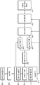
도 1을 참조하면, 본 실시예에 따른 초음파 도플러를 이용한 혈류 측정 장치는, 2차원 트랜스듀서 어레이(110), 멀티플렉서(120), 송수신부(130), 펄스 생성부(140), 신호처리부(150), 빔 스티어링부(160), 프로세서(170), 통신부(180), 디스플레이 장치(190)를 포함할 수 있다.Referring to FIG. 1, the blood flow measuring apparatus using the ultrasonic Doppler according to the present embodiment includes a two-
2차원 트랜스듀서 어레이(110)는 대상체와 초음파 신호를 송수신하기 위한 복수의 트랜스듀서들이 2차원으로 배열된 것으로, 2차원 트랜스듀서 어레이(110)에 포함된 복수의 트랜스듀서들 각각은 입력받은 전기 신호를 초음파 신호로 변환하고 변환된 초음파 신호를 대상체로 송신할 수 있으며, 대상체로부터 반사된 초음파 신호를 수신하고 수신된 초음파 신호를 전기 신호로 변환할 수 있다. In the
멀티플렉서(120)는 2차원 트랜스듀서 어레이(110)의 트랜스듀서들 중 장치가 지원하는 채널 수(혹은 그 이하)만큼의 트랜스듀서들을 구동하기 위한 것으로, 구동하고자 하는 트랜스듀서들을 선택하고 2차원 트랜스듀서 어레이(110)와 멀티플렉서(120) 뒷단과의 신호 라인 개수를 정합하는 역할을 한다. 즉, 멀티플렉서(120)는 초음파 신호 송신 및 에코 신호 수신 시에 2차원 트랜스듀서 어레이(110)의 일부 트랜스듀서들이 구동되도록 해당 트랜스듀서들과 송수신부(130)를 연결한다. The
송수신부(130)는, 프로세서(170)의 제어에 따라, 펄스 생성부(140)에서 생성된 고전압 펄스 신호를 멀티플렉서(120)를 통해 2차원 트랜스듀서 어레이(110)로 전달하거나, 2차원 트랜스듀서 어레이(110)로부터 멀티플렉서(120)를 통해 수신되는 아날로그 에코 신호를 신호처리부(150)로 전달한다. 구체적으로, 송수신부(130)는, 초음파 신호 송신 시에는 프로세서(150), 빔 스티어링부(160), 펄스 생성부(140)로 이루어지는 TX 회로와 2차원 트랜스듀서 어레이(110)를 연결하는 스위칭 동작을, 초음파 에코 신호 수신 시에는 2차원 트랜스듀서 어레이(110)와, 신호처리부(150), 빔 스티어링부(160), 프로세서(170)로 이루어지는 RX 회로를 연결하는 스위칭 동작을 수행한다. The transmitter /
펄스 생성부(140)는 초음파 신호를 발생시키기 위해 2차원 트랜스듀서 어레이(110)(정확하게는, 2차원 트랜스듀서 어레이의 일부 트랜스듀서들)에 인가될 고전압 펄스 신호를 생성한다. 펄스 신호는 주파수가 예컨대 2MHz이고, 소정의 펄스 반복 주파수(PRF, Pulse Repetition Frequency)를 가진다. 각 트랜스듀서에 인가될 각 채널의 펄스 신호에는 송신 지향성(transmission directionality)을 결정하기 위한 지연 시간(delay time)이 적용될 수 있다. The
신호처리부(150)는 대상체에서 반사되어 수신된 아날로그 에코 신호를 처리하여 초음파 데이터를 생성한다. 신호처리부(150)는 각 채널마다 에코 신호를 증폭하고, 노이즈를 제거하고, 아날로그-디지털 변환할 수 있다. 디지털 변환된 에코 신호에는 수신 지향성(reception directionality)을 결정하기 위한 지연 시간이 적용될 수 있다.The
빔 스티어링부(160)는 프로세서(170)의 제어에 따라 특정 스티어링 벡터(즉, 특정 거리와 특정 방향)의 관심 영역에 초음파 신호를 송신하고 에코 신호를 획득하기 위한 빔 스티어링을 수행한다. 빔 스티어링부(160)는 펄스 생성부(140)에 송신 지연 시간을 적용하고 신호처리부(150)에 수신 지연 시간을 적용함으로써 빔 스티어링을 수행할 수 있다.The
프로세서(170)는 장치를 구성하는 요소들, 즉 멀티플렉서(120), 송수신부(130), 펄스 생성부(140), 신호처리부(150), 빔 스티어링부(160), 통신부(180) 등의 동작을 제어하고, 초음파 데이터로부터 도플러 신호를 검출하고, 검출된 도플러 신호에 기초하여 혈류의 속도, 방향 등의 혈류 정보를 획득하고 이를 컬러 또는 파형으로 표현하는 도플러 영상을 생성할 수 있다. 도플러 영상은 혈액의 흐름을 나타내는 혈류 도플러 영상 (또는, 컬러 플로우 영상으로도 불림), 조직의 움직임을 나타내는 티슈 도플러 영상, 및 대상체의 이동 속도를 파형으로 표시하는 스펙트럴 도플러 영상 등을 포함할 수 있다.The
프로세서(170)는 음향창 탐색부(171), 혈류 탐색부(172), 도플러 처리부(173)를 포함할 수 있다. 이들의 구체적인 동작은 도 2 이하를 더욱 참조하여 뒤에서 설명하기로 한다. The
통신부(180)는 디스플레이 장치(190) 등 다른 장치와 데이터를 송수신하기 위한 것으로, 프로세서(170)의 제어에 따라 혈류 정보 또는 도플러 영상을 디스플레이 장치(190)로 전송할 수 있다. 통신부(180)는 데이터 전송을 위해 유선 또는 무선 통신 방식을 사용할 수 있다. 유선 통신 방식으로는, USB 케이블 등의 유선 케이블을 이용하여 데이터를 송수신할 수 있다. 무선 통신 방식으로는, 블루투스(Bluetooth), 무선 USB(Wireless USB), Wireless LAN, 와이파이(WiFi), 지그비(Zigbee), IrDA(Infrared Data Association) 등을 이용할 수 있다.The
디스플레이 장치(190)는 혈류 정보 또는 도플러 영상을 수신하고, 이를 화면에 표시한다. 디스플레이 장치(190)는 스마트폰(smartphone), 태블릿 PC(tablet personal computer), 이동 전화기(mobile phone), 화상 전화기, 전자북 리더기 (e-book reader), 데스크탑 PC(desktop personal computer), 랩탑 PC(laptop personal computer), 넷북 컴퓨터(netbook computer), 워크스테이션 (workstation), PDA(personal digital assistant), PMP(portable multimedia player) 등을 포함할 수 있다.The
또한, 통신부(180)는 유선 또는 무선으로 네트워크와 연결되어 외부 디바이스나 서버와 통신할 수도 있다. 통신부(180)는 의료 영상 정보 시스템(PACS, Picture Archiving and Communication System)을 통해 연결된 병원 서버나 병원 내의 다른 의료 장치와 데이터를 주고 받을 수 있다. 또한, 통신부(180)는 의료용 디지털 영상 및 통신(DICOM, Digital Imaging and Communications in Medicine) 표준에 따라 데이터 통신할 수 있다. 나아가, 통신부(270)는 병원 내의 서버나 의료 장치뿐만 아니라, 의사나 환자 또는 보호자의 휴대용 단말과 데이터 통신을 수행할 수도 있다.In addition, the
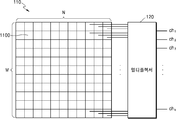
도 2는 2차원 트랜스듀서 어레이(110)의 일 예를 나타낸다. 예를 들어, 2차원 트랜스듀서 어레이(110)는 도시된 바와 같이 M개의 행과 N개의 열로 배열된 MㅧN개의 트랜스듀서(1100)들을 포함할 수 있다. 여기서 M, N은 동일한 수일 수도 있고 다른 수일 수도 있다.2 shows an example of a two-
장치가 K개의 채널을 지원한다고 하면, 멀티플렉서(120)는 송수신부(130)와 K개의 신호 라인을 통해 연결되고, 2차원 트랜스듀서 어레이(110)와 MㅧN개의 신호 라인을 통해 연결될 수 있다. MㅧN개의 신호 라인은 각각 2차원 트랜스듀서 어레이(110)에 포함된 각각의 트랜스듀서(1100)들에 대응한다. 멀티플렉서(120)는 프로세서(170)의 제어에 따라 MㅧN개의 신호 라인 중 구동할 트랜스듀서에 해당하는 신호 라인을 K개의 신호 라인과 연결하는 스위칭 동작을 수행함으로써 요구되는 K개(또는 그 이하)의 트랜스듀서들을 구동할 수 있다.If the device supports K channels, the
음향창 탐색부(171)는 2차원 트랜스듀서 어레이(110)의 트랜스듀서들 중 일부의 트랜스듀서들을 동시에 구동하여 초음파 신호를 송수신하고, 구동되는 일부의 트랜스듀서들 각각에 대하여 도플러 신호들을 검출한다. 이때 구동되는 각각의 트랜스듀서에 의해 송신되는 초음파 신호는 지향성이 없거나 적은 구면파 신호가 된다. 그리고 음향창 탐색부(171)는 검출된 도플러 신호들 중 가장 세기가 큰 도플러 신호에 해당하는 트랜스듀서를 확인하여, 이 트랜스듀서를 음향창에 위치한 트랜스듀서로 간주한다. The acoustic
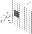
도 3은 음향창 탐색부(171)에 의해 구동되는 일부의 트랜스듀서들과 2차원 트랜스듀서 어레이(110)가 배치된 대상체의 음향창을 보여주고, 도 4는 두개골의 음향창 부분의 단면을 개략적으로 보여준다. FIG. 3 shows a sound window of an object in which some transducers driven by the sound
도 4를 참조하면, 초음파 신호는 두개골 대부분의 두꺼운 뼈 부분에서는 거의 반사되어 그 안쪽 깊이까지 도달하지 않으나, 뼈가 얇은 측두엽 관자놀이 부근처럼 주변과 비교해서 얇은 부분인 음향창(W) 영역은 초음파 신호가 통과하여 혈관(P)까지 도달할 수 있다. Referring to FIG. 4, the ultrasound signal is almost reflected from the thick bone part of the skull and does not reach the inner depth, but the acoustic window (W) region, which is a thin part compared to the surroundings, such as the vicinity of the temporal lobe temple, is an ultrasound signal. It can pass through to reach the blood vessel (P).
음향창 탐색부(171)는 2차원 트랜스듀서 어레이(110)의 트랜스듀서들 중 가용한 채널 수 내에서 비교적 균일하고 산재하게 분포되도록 구동할 트랜스듀서들을 선택할 수 있다. 도 3에서는 예컨대 9개의 채널에 대응하는 9개의 트랜스듀서들(1101, 1102, ..., 1109)이 선택된 경우를 보여준다. The acoustic
도 3의 예에서, 음향창(W)은 (실제로는 보이지 않음) 도시된 바와 같이 트랜스듀서(1104)와 트랜스듀서(1105)에 걸쳐 위치하고, 트랜스듀서(1104)가 음향창(W)의 더 많은 부분과 겹쳐진다. In the example of FIG. 3, the acoustic window W is located across the
트랜스듀서(1104, 1105)에 의해 송신되는 초음파 신호는 음향창(W)을 통과하므로 초음파 신호가 혈관의 혈류에 반사되면 도플러 신호가 검출될 수 있다. 여기서 송신되는 초음파 신호는 지향성이 없거나 적으므로 혈관이 음향창(W)의 바로 아래를 지나가지 않더라도 도플러 신호가 검출될 수 있다. 그러나 트랜스듀서(1101, 1102, 1103, 1106, 1107, 1108, 1109)에서 송신되는 초음파 신호는 음향창(W)을 통과하지 못하기 때문에 혈관의 혈류에까지 이르지 못하므로 도플러 신호가 검출되지 않는다. Since the ultrasonic signals transmitted by the
도 5는 9개의 채널, 즉 9개의 트랜스듀서들(1101, 1102, ..., 1109) 각각에 대하여 검출되는 도플러 신호를 보여준다. 도 5를 참조하면, 4번 채널에서 가장 큰 세기의 도플러 신호가 검출되며, 5번 채널에서는 도플러 신호가 검출되지만 4번 채널보다는 작은 세기의 도플러 신호가 검출된다. 이것은 4번 채널에 해당하는 트랜스듀서(1104)가 5번 채널에 해당하는 트랜스듀서(1105)보다 음향창(W)의 더 많은 부분과 겹쳐지기 때문이다. FIG. 5 shows the Doppler signal detected for each of the nine channels, ie, the nine
따라서 음향창 탐색부(171)는 가장 세기가 큰 도플러 신호에 해당하는 4번 채널의 트랜스듀서(1104)를 음향창(W)에 위치한 트랜스듀서로 간주한다. Therefore, the acoustic
음향창 탐색부(171)는 위와 같은 동작을 통해 한번에 음향창의 위치를 찾을 수도 있지만, 필요에 따라서는 구동할 트랜스듀서들을 변경하면서 복수 회에 걸쳐 최적의 음향창을 찾을 수도 있다. 이를테면, 도플러 신호 세기의 임계값을 정하고, 검출되는 도플러 신호들이 모두 임계값보다 작다면 트랜스듀서들을 변경(예컨대, 각각을 시프트하거나 이미 선택하였던 것을 제외하고 다시 선택)하여 다시 초음파 신호를 송수신함으로써 임계값보다 큰 도플러 신호가 검출되는 트랜스듀서를 찾을 수 있다.The acoustic
한편, 일반적으로 도플러 신호는 신호대잡음비가 좋지 않고 단일 트랜스듀서에 의한 초음파 신호는 신호 세기가 비교적 미약하므로, 최적의 음향창을 찾기 위해 예컨대 CNN(Convolutional Neural Network) 등과 같은 기계학습 알고리즘을 이용할 수도 있다. 이를테면, 음향창의 위치가 알려진 다수의 환자 샘플을 대상으로 도 5와 같은 도플러 신호의 패턴을 얻고 이 데이터로 기계학습을 통해 가장 세기가 큰 도플러 신호에 해당하는 트랜스듀서를 확인할 수 있다.On the other hand, in general, since the Doppler signal has a poor signal-to-noise ratio and the signal strength of the ultrasonic signal by a single transducer is relatively weak, a machine learning algorithm such as CNN (Convolutional Neural Network) may be used to find the optimal acoustic window. . For example, a pattern of a Doppler signal as shown in FIG. 5 may be obtained from a plurality of patient samples having a known acoustic window position, and the transducer corresponding to the Doppler signal having the greatest intensity may be identified through machine learning.
음향창 탐색부(171)에 의해 음향창의 위치(즉, 음향창 위의 트랜스듀서)가 확인되면, 혈류 탐색부(172)는 해당 트랜스듀서를 포함하는 서로 인접한 복수 개의 트랜스듀서들로 빔 스티어링에 의해 복수의 스티어링 벡터들 각각에 대한 도플러 신호들을 검출한다. 그리고 혈류 탐색부(172)는 검출된 도플러 신호들 중 가장 세기가 큰 도플러 신호에 해당하는 스티어링 벡터를 혈관의 혈류가 지나가는 지점의 스티어링 벡터로 확인한다.When the position of the sound window (ie, the transducer on the sound window) is confirmed by the sound
도 6은 도 3의 9개의 트랜스듀서들(1101, 1102, ..., 1109) 중 트랜스듀서(1104)가 음향창(W) 위에 위치하는 트랜스듀서로 확인됨에 따라, 트랜스듀서(1104)를 포함하는 서로 인접한 트랜스듀서들(1104, 1110, 1111, ..., 1117)이 빔 스티어링을 위해 구동할 트랜스듀서들로 선택된 경우를 보여준다. 도 6의 예는 트랜스듀서(1104)의 중심점이 빔 스티어링(즉, 스티어링 벡터)의 중심점(O)이 된 경우이나, 트랜스듀서의 형상(예컨대 삼각형, 육각형 등 다양한 형상이 될 수 있음)이나 배열에 따라서는 트랜스듀서 간의 특정 지점이 중심점이 될 수도 있다. FIG. 6 shows the
도 7은 도 6의 트랜스듀서들(1104, 1110, 1111, ..., 1117)로 빔 스티어링을 수행하여 얻을 수 있는 몇 개의 스티어링 벡터(V1, V2, V3, ...)와 대상체 내의 혈관(P)을 보여준다. 가령 TCD의 경우 혈관(P)은 중대뇌동맥(middle cerebral artery), 전대뇌동맥(anterior cerebral artery), 후대뇌동맥(posterior cerebral artery), 안동맥(ophthalmic artery), 척추동맥(vertebral artery), 기저동맥(basilar artery) 등이 될 수 있다. 도 7을 참조하면, 스티어링 벡터(V2)가 혈관(P) 내의 혈류가 지나가는 지점의 스티어링 벡터이므로, 스티어링 벡터(V2)에서 가장 큰 세기의 도플러 신호가 검출된다. FIG. 7 shows several steering vectors (V1, V2, V3, ...) and blood vessels in an object obtained by performing beam steering with the
따라서 혈류 탐색부(172)는 가장 큰 세기의 도플러 신호가 검출되는 스티어링 벡터(V2)를 혈관(P) 내의 혈류가 지나가는 지점의 스티어링 벡터로 확인한다. Therefore, the
혈류 탐색부(172)에 의해 혈관 내의 혈류가 지나가는 지점의 스티어링 벡터가 확인되면, 도플러 처리부(173)는 해당 스티어링 벡터로 빔 스티어링을 수행하여 해당 스티어링 벡터로 초음파 신호를 송수신함으로써 도플러 신호를 검출한다. 그리고 도플러 처리부(173)는 검출되는 도플러 신호로부터 혈류의 속도, 방향 등의 혈류 정보를 획득하고 이를 컬러 또는 파형으로 표현하는 도플러 영상을 생성할 수 있다.When the steering vector of the point where blood flow in the blood vessel passes is confirmed by the blood
혈류 탐색부(172)의 위와 같은 혈류를 탐색하는 동작은, 한번에 그치지 않고 반복적, 지속적으로 수행됨으로써 실시간으로 혈류를 추적할 수도 있다. 그에 따라 가장 세기가 큰 도플러 신호에 해당하는 스티어링 벡터, 즉 혈류가 지나가는 지점의 스티어링 벡터가 변경되면 도플러 처리부(173)는 변경된 스티어링 벡터로 빔 스티어링을 수행하여 도플러 신호를 검출할 수 있다. The operation of searching for blood flow as described above of the blood
또한, 혈류 탐색부(172)는 위와 같이 가장 큰 세기의 도플러 신호에 해당하는 하나의 스티어링 벡터를 찾을 수도 있지만, 혈관은 둘 이상일 수 있으므로, 도플러 신호 세기의 임계값을 정하고, 임계값보다 큰 도플러 신호에 해당하는 둘 이상의 스티어링 벡터를 찾음으로써 둘 이상의 혈관의 혈류를 탐색할 수도 있다. 이 경우 도플러 처리부(173)는 각각의 스티어링 벡터로 초음파 신호를 송수신함으로써 둘 이상의 혈관의 혈류 정보를 획득할 수도 있다. In addition, the blood
본 발명의 실시예에 따른 초음파 도플러를 이용한 혈류 측정 장치의 일부는 패치 형태로 제작되어 환자의 측정 부위에 부착될 수 있다. 이를테면, 환자의 측정 부위에 부착하기 위한 패치는 2차원 트랜스듀서 어레이(110), 멀티플렉서(120), 송수신부(130), 펄스 생성부(140), 신호처리부(150), 빔 스티어링부(160)를 구비하고, 패치와 유선 또는 무선으로 연결되는 별도의 셋톱박스가 프로세서(170), 통신부(180) 등을 구비할 수 있다. 디스플레이 장치(190)는 셋톱박스에 일체형으로 구비될 수도 있고, 스마트폰과 같은 외부 장치가 디스플레이 장치(190)로 사용될 수도 있다. A part of the blood flow measuring apparatus using the ultrasonic Doppler according to an embodiment of the present invention may be manufactured in a patch form and attached to a patient's measurement site. For example, a patch for attaching to a patient's measurement site includes a two-
도 8은 본 발명의 일 실시예에 따른 초음파 도플러를 이용한 혈류 측정 장치의 동작 방법을 나타낸 흐름도이다. 본 실시예에 따른 동작 방법은 전술한 초음파 도플러를 이용한 혈류 측정 장치에서 처리되는 단계들로 이루어지므로, 이하 생략된 내용이라 하더라도 초음파 도플러를 이용한 혈류 측정 장치에 관하여 이상에서 기술된 내용은 본 실시예에 따른 동작 방법에도 적용된다. 8 is a flowchart illustrating an operation method of an apparatus for measuring blood flow using an ultrasonic Doppler according to an embodiment of the present invention. Since the operation method according to the present embodiment consists of steps processed in the blood flow measuring apparatus using the ultrasonic doppler described above, the contents described above with respect to the blood flow measuring apparatus using the ultrasonic doppler are the present embodiment, even if omitted below. It also applies to the operation method according to.
710단계에서, 2차원 트랜스듀서 어레이(110)의 트랜스듀서들 중 일부의 트랜스듀서들을 동시에 구동하여 초음파 신호를 송수신하고, 구동되는 일부의 트랜스듀서들 각각에 대하여 도플러 신호들을 검출한다.In
720단계에서, 검출된 도플러 신호들 중 가장 세기가 큰 도플러 신호에 해당하는 트랜스듀서를 확인한다.In
730단계에서, 확인된 트랜스듀서를 포함하는 서로 인접한 복수 개의 트랜스듀서들로 빔 스티어링에 의해 복수의 스티어링 벡터들 각각에 대한 도플러 신호들을 검출한다.In
740단계에서, 검출된 도플러 신호들 중 가장 세기가 큰 도플러 신호에 해당하는 스티어링 벡터를 확인한다.In
750단계에서, 확인된 스티어링 벡터로 빔 스티어링을 수행하여 해당 스티어링 벡터로 초음파 신호를 송수신함으로써 도플러 신호를 검출한다.In
760단계에서, 검출되는 도플러 신호로부터 혈류의 속도, 방향 등의 혈류 정보를 획득한다. In
본 발명의 실시예들은 기능적인 블록 구성들 및 다양한 처리 단계들로 나타내어질 수 있다. 이러한 기능 블록들은 특정 기능들을 실행하는 다양한 개수의 하드웨어 또는/및 소프트웨어 구성들로 구현될 수 있다. 예를 들어, 실시예는 하나 이상의 마이크로프로세서들의 제어 또는 다른 제어 장치들에 의해서 다양한 기능들을 실행할 수 있는, 메모리, 프로세싱, 로직(logic), 룩 업 테이블(look-up table) 등과 같은 집적 회로 구성들을 채용할 수 있다. 본 발명에의 구성 요소들이 소프트웨어 프로그래밍 또는 소프트웨어 요소들로 실행될 수 있는 것과 유사하게, 실시예는 데이터 구조, 프로세스들, 루틴들 또는 다른 프로그래밍 구성들의 조합으로 구현되는 다양한 알고리즘을 포함하여, C, C++, 자바(Java), 어셈블러(assembler) 등과 같은 프로그래밍 또는 스크립팅 언어로 구현될 수 있다. 기능적인 측면들은 하나 이상의 프로세서들에서 실행되는 알고리즘으로 구현될 수 있다. 또한, 실시예는 전자적인 환경 설정, 신호 처리, 및/또는 데이터 처리 등을 위하여 종래 기술을 채용할 수 있다. "매커니즘", "요소", "수단", "구성"과 같은 용어는 넓게 사용될 수 있으며, 기계적이고 물리적인 구성들로서 한정되는 것은 아니다. 상기 용어는 프로세서 등과 연계하여 소프트웨어의 일련의 처리들(routines)의 의미를 포함할 수 있다.Embodiments of the present invention may be represented by functional block configurations and various processing steps. These functional blocks can be implemented with various numbers of hardware or / and software configurations that perform specific functions. For example, an embodiment may be implemented in an integrated circuit configuration such as memory, processing, logic, look-up table, etc., capable of executing various functions by control of one or more microprocessors or other control devices. You can hire them. Similar to the components of the present invention that can be executed with software programming or software components, embodiments include C, C ++, including various algorithms implemented with a combination of data structures, processes, routines or other programming components. , Can be implemented in programming or scripting languages such as Java, assembler, etc. Functional aspects can be implemented with algorithms running on one or more processors. In addition, embodiments may employ conventional techniques for electronic environment setting, signal processing, and / or data processing. Terms such as "mechanism", "element", "means", and "configuration" can be used broadly and are not limited to mechanical and physical configurations. The term may include the meaning of a series of routines of software in connection with a processor or the like.
실시예에서 설명하는 특정 실행들은 일 실시예들로서, 어떠한 방법으로도 실시 예의 범위를 한정하는 것은 아니다. 명세서의 간결함을 위하여, 종래 전자적인 구성들, 제어 시스템들, 소프트웨어, 상기 시스템들의 다른 기능적인 측면들의 기재는 생략될 수 있다. 또한, 도면에 도시된 구성 요소들 간의 선들의 연결 또는 연결 부재들은 기능적인 연결 및/또는 물리적 또는 회로적 연결들을 예시적으로 나타낸 것으로서, 실제 장치에서는 대체 가능하거나 추가의 다양한 기능적인 연결, 물리적인 연결, 또는 회로 연결들로서 나타내어질 수 있다. 또한, "필수적인", "중요하게" 등과 같이 구체적인 언급이 없다면 본 발명의 적용을 위하여 반드시 필요한 구성 요소가 아닐 수 있다.The specific implementations described in the embodiment are one embodiment, and do not limit the scope of the embodiment in any way. For brevity of the specification, descriptions of conventional electronic configurations, control systems, software, and other functional aspects of the systems may be omitted. In addition, the connection or connection members of the lines between the components shown in the drawings are illustrative examples of functional connections and / or physical or circuit connections. In the actual device, alternative or additional various functional connections, physical It can be represented as a connection, or circuit connections. In addition, unless specifically mentioned, such as "essential", "important", etc., it may not be a necessary component for the application of the present invention.
이제까지 본 발명에 대하여 그 바람직한 실시예들을 중심으로 살펴보았다. 본 발명이 속하는 기술 분야에서 통상의 지식을 가진 자는 본 발명이 본 발명의 본질적인 특성에서 벗어나지 않는 범위에서 변형된 형태로 구현될 수 있음을 이해할 수 있을 것이다. 그러므로 개시된 실시예들은 한정적인 관점이 아니라 설명적인 관점에서 고려되어야 한다. 본 발명의 범위는 전술한 설명이 아니라 특허청구범위에 나타나 있으며, 그와 동등한 범위 내에 있는 모든 차이점은 본 발명에 포함된 것으로 해석되어야 할 것이다.So far, the present invention has been focused on the preferred embodiments. Those skilled in the art to which the present invention pertains will appreciate that the present invention may be implemented in a modified form without departing from the essential characteristics of the present invention. Therefore, the disclosed embodiments should be considered in terms of explanation, not limitation. The scope of the present invention is shown in the claims rather than the foregoing description, and all differences within the equivalent range should be interpreted as being included in the present invention.
Claims (15)
상기 복수의 트랜스듀서들 중 일부의 트랜스듀서들을 구동하여 초음파 신호를 송수신하고, 상기 일부의 트랜스듀서들 각각에 대하여 도플러 신호들을 검출하고, 검출된 도플러 신호들 중 가장 세기가 큰 도플러 신호에 해당하는 트랜스듀서를 확인하는 음향창 탐색부;
상기 확인된 트랜스듀서를 포함하는 서로 인접한 복수 개의 트랜서듀서들로 빔 스티어링에 의해 복수의 스티어링 벡터들 각각에 대한 도플러 신호들을 검출하고, 검출된 도플러 신호들 중 가장 세기가 큰 도플러 신호에 해당하는 스티어링 벡터를 확인하는 혈류 탐색부; 및
상기 확인된 스티어링 벡터로 빔 스티어링을 수행하여 도플러 신호를 검출하고, 검출된 도플러 신호로부터 혈류 정보를 획득하는 도플러 처리부를 포함하는 것을 특징으로 하는, 초음파 도플러를 이용한 혈류 측정 장치.A two-dimensional transducer array in which a plurality of transducers for transmitting and receiving ultrasound signals to and from an object are arranged in two dimensions;
A plurality of transducers are driven to transmit and receive ultrasonic signals, detect Doppler signals for each of the transducers, and correspond to the Doppler signal having the greatest intensity among the detected Doppler signals. An acoustic window search unit for confirming the transducer;
Doppler signals for each of the plurality of steering vectors are detected by beam steering with a plurality of transducers adjacent to each other including the identified transducer, and steering corresponding to a Doppler signal having the greatest intensity among the detected Doppler signals A blood flow searcher that identifies a vector; And
And a Doppler processing unit for detecting a Doppler signal by performing beam steering with the identified steering vector and obtaining blood flow information from the detected Doppler signal.
상기 음향창 탐색부는 상기 일부의 트랜스듀서들을 동시에 구동하는 것을 특징으로 하는, 초음파 도플러를 이용한 혈류 측정 장치.According to claim 1,
The acoustic window search unit, characterized in that for driving the transducers at the same time, blood flow measuring apparatus using an ultrasonic Doppler.
상기 일부의 트랜스듀서들이 동시에 구동될 수 있도록 상기 2차원 트랜스듀서 어레이와 연결되는 멀티플렉서를 더 포함하는 것을 특징으로 하는, 초음파 도플러를 이용한 혈류 측정 장치.According to claim 2,
It characterized in that it further comprises a multiplexer connected to the two-dimensional transducer array so that some of the transducers can be driven at the same time, blood flow measuring apparatus using ultrasonic Doppler.
상기 일부의 트랜스듀서들은 상기 2차원 트랜스듀서 어레이에 산재하게 분포되는 것을 특징으로 하는, 초음파 도플러를 이용한 혈류 측정 장치.According to claim 2,
The some of the transducers are characterized in that scattered in the two-dimensional transducer array, blood flow measuring apparatus using an ultrasonic Doppler.
상기 일부의 트랜스듀서들 각각으로부터 송신되는 초음파 신호는 구면파 신호인 것을 특징으로 하는, 초음파 도플러를 이용한 혈류 측정 장치.According to claim 2,
The ultrasound signal transmitted from each of the transducers is a spherical wave signal, characterized in that, blood flow measuring apparatus using an ultrasonic Doppler.
상기 일부의 트랜스듀서들의 수는 가용한 채널의 수 이하인 것을 특징으로 하는, 초음파 도플러를 이용한 혈류 측정 장치.According to claim 2,
The number of transducers of the part, characterized in that less than the number of available channels, blood flow measuring apparatus using an ultrasonic Doppler.
상기 혈류 탐색부는, 상기 검출된 도플러 신호들 중 소정의 임계값보다 큰 도플러 신호에 해당하는 둘 이상의 스티어링 벡터를 확인하는 것을 특징으로 하는, 초음파 도플러를 이용한 혈류 측정 장치.According to claim 2,
The blood flow search unit, blood flow measurement apparatus using ultrasonic Doppler, characterized in that for checking the two or more steering vectors corresponding to the Doppler signal greater than a predetermined threshold value among the detected Doppler signals.
상기 음향창 탐색부는, 기계학습을 통해 상기 가장 세기가 큰 도플러 신호에 해당하는 트랜스듀서를 확인하는 것을 특징으로 하는, 초음파 도플러를 이용한 혈류 측정 장치.According to claim 2,
The acoustic window search unit, blood flow measuring apparatus using ultrasonic Doppler, characterized in that to identify the transducer corresponding to the Doppler signal having the greatest intensity through machine learning.
(b) 상기 검출된 도플러 신호들 중 가장 세기가 큰 도플러 신호에 해당하는 트랜스듀서를 확인하는 단계;
(c) 상기 확인된 트랜스듀서를 포함하는 서로 인접한 복수 개의 트랜서듀서들로 빔 스티어링에 의해 복수의 스티어링 벡터들 각각에 대한 도플러 신호들을 검출하는 단계;
(d) 상기 검출된 도플러 신호들 중 가장 세기가 큰 도플러 신호에 해당하는 스티어링 벡터를 확인하는 단계; 및
(e) 상기 확인된 스티어링 벡터로 빔 스티어링을 수행하여 도플러 신호를 검출하고, 검출된 도플러 신호로부터 혈류 정보를 획득하는 단계를 포함하는 것을 특징으로 하는, 초음파 도플러를 이용한 혈류 측정 장치의 동작 방법.(a) A plurality of transducers for transmitting and receiving ultrasound signals to and from an object is driven by driving some of the transducers of the two-dimensional transducer array arranged in two dimensions to transmit and receive ultrasound signals, and doppler for each of the partial transducers Detecting signals;
(b) identifying a transducer corresponding to the Doppler signal having the greatest intensity among the detected Doppler signals;
(c) detecting Doppler signals for each of the plurality of steering vectors by beam steering with a plurality of adjacent transducers including the identified transducer;
(d) identifying a steering vector corresponding to a Doppler signal having the greatest intensity among the detected Doppler signals; And
(e) detecting a Doppler signal by performing beam steering with the identified steering vector, and obtaining blood flow information from the detected Doppler signal, the method of operating the blood flow measuring apparatus using ultrasonic Doppler.
상기 일부의 트랜스듀서들은 동시에 구동되는 것을 특징으로 하는, 초음파 도플러를 이용한 혈류 측정 장치의 동작 방법.The method of claim 9,
A method of operating the blood flow measuring apparatus using ultrasonic Doppler, characterized in that some of the transducers are driven simultaneously.
상기 일부의 트랜스듀서들은 상기 2차원 트랜스듀서 어레이에 산재하게 분포되는 것을 특징으로 하는, 초음파 도플러를 이용한 혈류 측정 장치의 동작 방법.The method of claim 10,
A method of operating the blood flow measuring apparatus using ultrasonic Doppler, characterized in that some of the transducers are dispersedly distributed in the two-dimensional transducer array.
상기 일부의 트랜스듀서들 각각으로부터 송신되는 초음파 신호는 구면파 신호인 것을 특징으로 하는, 초음파 도플러를 이용한 혈류 측정 장치의 동작 방법.The method of claim 10,
The ultrasonic signal transmitted from each of the transducers is characterized in that the spherical wave signal, the operation method of the blood flow measurement apparatus using ultrasonic Doppler.
상기 일부의 트랜스듀서들의 수는 가용한 채널의 수 이하인 것을 특징으로 하는, 초음파 도플러를 이용한 혈류 측정 장치의 동작 방법.The method of claim 10,
The number of transducers of the part, characterized in that less than the number of available channels, the operation method of the blood flow measurement apparatus using ultrasonic Doppler.
상기 (d) 단계는, 상기 검출된 도플러 신호들 중 소정의 임계값보다 큰 도플러 신호에 해당하는 둘 이상의 스티어링 벡터를 확인하는 것을 특징으로 하는, 초음파 도플러를 이용한 혈류 측정 장치의 동작 방법.The method of claim 10,
In the step (d), two or more steering vectors corresponding to a Doppler signal greater than a predetermined threshold among the detected Doppler signals are identified, and the method of operating the blood flow measuring apparatus using ultrasonic Doppler is performed.
상기 (b) 단계는, 기계학습을 통해 상기 가장 세기가 큰 도플러 신호에 해당하는 트랜스듀서를 확인하는 것을 특징으로 하는, 초음파 도플러를 이용한 혈류 측정 장치의 동작 방법.The method of claim 10,
The step (b), characterized in that to identify the transducer corresponding to the Doppler signal having the greatest intensity through machine learning, the operation method of the blood flow measuring apparatus using ultrasonic Doppler.
Priority Applications (2)
| Application Number | Priority Date | Filing Date | Title |
|---|---|---|---|
| KR1020180136382A KR102117226B1 (en) | 2018-11-08 | 2018-11-08 | Apparatus for measuring blood flow using ultrasound doppler and operating method thereof |
| US16/670,410 US11544631B2 (en) | 2018-11-08 | 2019-10-31 | Blood flow measurement apparatus using doppler ultrasound and method of operating the same |
Applications Claiming Priority (1)
| Application Number | Priority Date | Filing Date | Title |
|---|---|---|---|
| KR1020180136382A KR102117226B1 (en) | 2018-11-08 | 2018-11-08 | Apparatus for measuring blood flow using ultrasound doppler and operating method thereof |
Publications (2)
| Publication Number | Publication Date |
|---|---|
| KR20200053146A true KR20200053146A (en) | 2020-05-18 |
| KR102117226B1 KR102117226B1 (en) | 2020-06-01 |
Family
ID=70550677
Family Applications (1)
| Application Number | Title | Priority Date | Filing Date |
|---|---|---|---|
| KR1020180136382A KR102117226B1 (en) | 2018-11-08 | 2018-11-08 | Apparatus for measuring blood flow using ultrasound doppler and operating method thereof |
Country Status (2)
| Country | Link |
|---|---|
| US (1) | US11544631B2 (en) |
| KR (1) | KR102117226B1 (en) |
Citations (6)
| Publication number | Priority date | Publication date | Assignee | Title |
|---|---|---|---|---|
| JP2005103193A (en) * | 2003-10-02 | 2005-04-21 | Hitachi Medical Corp | Ultrasonic transmitter, and ultrasonic apparatus using the same |
| WO2006102511A2 (en) * | 2005-03-23 | 2006-09-28 | New Health Sciences, Inc. | Systems and methods for using dynamic vascular assessment to distinguish among vascular states and for investigating intracranial pressure |
| US20070016050A1 (en) * | 2005-06-13 | 2007-01-18 | Moehring Mark A | Medical Doppler ultrasound system for locating and tracking blood flow |
| JP2011505898A (en) * | 2007-12-07 | 2011-03-03 | コーニンクレッカ フィリップス エレクトロニクス エヌ ヴィ | Method and system for angiographic imaging |
| JP2017500943A (en) * | 2013-12-18 | 2017-01-12 | コーニンクレッカ フィリップス エヌ ヴェKoninklijke Philips N.V. | System and method for registration of ultrasound and computed tomography images for ultrasonic thrombolysis procedures |
| JP2019503774A (en) * | 2016-01-05 | 2019-02-14 | ニューラル アナリティクス、インコーポレイテッド | System and method for detecting neurological diseases |
Family Cites Families (3)
| Publication number | Priority date | Publication date | Assignee | Title |
|---|---|---|---|---|
| US6582367B1 (en) * | 2000-09-15 | 2003-06-24 | Koninklijke Philips Electronics N.V. | 2D ultrasonic transducer array for two dimensional and three dimensional imaging |
| ATE549641T1 (en) * | 2007-09-04 | 2012-03-15 | Koninkl Philips Electronics Nv | DUAL MODE ULTRASONIC TRANSDUCER |
| CN108778140A (en) * | 2016-01-05 | 2018-11-09 | 神经系统分析公司 | System and method for determining clinical indication |
-
2018
- 2018-11-08 KR KR1020180136382A patent/KR102117226B1/en active IP Right Grant
-
2019
- 2019-10-31 US US16/670,410 patent/US11544631B2/en active Active
Patent Citations (6)
| Publication number | Priority date | Publication date | Assignee | Title |
|---|---|---|---|---|
| JP2005103193A (en) * | 2003-10-02 | 2005-04-21 | Hitachi Medical Corp | Ultrasonic transmitter, and ultrasonic apparatus using the same |
| WO2006102511A2 (en) * | 2005-03-23 | 2006-09-28 | New Health Sciences, Inc. | Systems and methods for using dynamic vascular assessment to distinguish among vascular states and for investigating intracranial pressure |
| US20070016050A1 (en) * | 2005-06-13 | 2007-01-18 | Moehring Mark A | Medical Doppler ultrasound system for locating and tracking blood flow |
| JP2011505898A (en) * | 2007-12-07 | 2011-03-03 | コーニンクレッカ フィリップス エレクトロニクス エヌ ヴィ | Method and system for angiographic imaging |
| JP2017500943A (en) * | 2013-12-18 | 2017-01-12 | コーニンクレッカ フィリップス エヌ ヴェKoninklijke Philips N.V. | System and method for registration of ultrasound and computed tomography images for ultrasonic thrombolysis procedures |
| JP2019503774A (en) * | 2016-01-05 | 2019-02-14 | ニューラル アナリティクス、インコーポレイテッド | System and method for detecting neurological diseases |
Also Published As
| Publication number | Publication date |
|---|---|
| US11544631B2 (en) | 2023-01-03 |
| US20200151612A1 (en) | 2020-05-14 |
| KR102117226B1 (en) | 2020-06-01 |
Similar Documents
| Publication | Publication Date | Title |
|---|---|---|
| US10324065B2 (en) | Ultrasound diagnostic apparatus, ultrasound image capturing method, and computer-readable recording medium | |
| KR102618500B1 (en) | Ultrasound diagnosis apparatus and mehtod thereof | |
| US10292682B2 (en) | Method and medical imaging apparatus for generating elastic image by using curved array probe | |
| EP3115000B1 (en) | Ultrasound diagnosis apparatus and operating method thereof | |
| KR102519423B1 (en) | Method of obtaining information from a contrast image, ultrasound apparatus thereof, and method of operation of the ultrasound apparatus | |
| US9986977B2 (en) | Ultrasonic diagnostic apparatus and method of operating the same | |
| KR102205507B1 (en) | Apparatus and method for displaying ultrasound image | |
| EP3017768B1 (en) | Apparatus and method of calculating arterial stiffness using ultrasound | |
| US10702243B2 (en) | Ultrasound diagnosis apparatus, wearable device, method of controlling ultrasound diagnosis apparatus, method of controlling wearable device, and recording medium having methods recorded thereon | |
| KR102577752B1 (en) | Method of outputting a velocity of object and ultrasound apparatus thereof | |
| US11504087B2 (en) | Ultrasonic diagnostic device and control method therefor | |
| EP3311752B1 (en) | Ultrasonic device and operation method therefor | |
| KR102524068B1 (en) | Ultrasound diagnosis apparatus, ultrasound probe and controlling method of the same | |
| US10390800B2 (en) | Ultrasound diagnosis method and ultrasound diagnosis apparatus | |
| KR102117226B1 (en) | Apparatus for measuring blood flow using ultrasound doppler and operating method thereof | |
| CN113576531A (en) | Blood flow measuring apparatus using doppler ultrasound and method of operating the same | |
| EP3175795B1 (en) | Method and apparatus for acquiring image using ultrasound | |
| JP6895697B1 (en) | Blood flow measuring device using ultrasonic Doppler and its operation method | |
| KR20150047416A (en) | Ultrasound apparatus and method for setting tgc thereof | |
| EP3907527A1 (en) | Blood flow measurement apparatus using doppler ultrasound and method of operating the same | |
| KR101563501B1 (en) | Apparatus and method for measuring vessel stress | |
| KR102605151B1 (en) | Method and beamformer for performing beamforming process | |
| KR20150102210A (en) | Method for Controlling Ultrasound Imaging Apparatus and Ultrasound Imaging Apparatus Thereof | |
| EP3197365B1 (en) | Ultrasound diagnostic apparatus and method of generating ultrasound image | |
| KR20160123210A (en) | Ultrasound System and Method for Diplaying Rigidness of Blood Vessel |
Legal Events
| Date | Code | Title | Description |
|---|---|---|---|
| E701 | Decision to grant or registration of patent right | ||
| GRNT | Written decision to grant |