KR20200047565A - Young pig-derived stem cells and method for manufacturing the same - Google Patents
Young pig-derived stem cells and method for manufacturing the same Download PDFInfo
- Publication number
- KR20200047565A KR20200047565A KR1020207006812A KR20207006812A KR20200047565A KR 20200047565 A KR20200047565 A KR 20200047565A KR 1020207006812 A KR1020207006812 A KR 1020207006812A KR 20207006812 A KR20207006812 A KR 20207006812A KR 20200047565 A KR20200047565 A KR 20200047565A
- Authority
- KR
- South Korea
- Prior art keywords
- cells
- stem cells
- cell
- msc
- young
- Prior art date
Links
- 210000000130 stem cell Anatomy 0.000 title claims abstract description 107
- 238000000034 method Methods 0.000 title claims abstract description 20
- 238000004519 manufacturing process Methods 0.000 title claims description 24
- 241000282887 Suidae Species 0.000 claims abstract description 26
- 210000004027 cell Anatomy 0.000 claims description 230
- 210000002901 mesenchymal stem cell Anatomy 0.000 claims description 84
- 210000001185 bone marrow Anatomy 0.000 claims description 64
- 238000007710 freezing Methods 0.000 claims description 30
- 230000008014 freezing Effects 0.000 claims description 30
- 210000001616 monocyte Anatomy 0.000 claims description 18
- 238000002054 transplantation Methods 0.000 claims description 15
- 238000009331 sowing Methods 0.000 claims description 12
- 230000003698 anagen phase Effects 0.000 claims description 5
- 238000002955 isolation Methods 0.000 claims description 5
- 230000001902 propagating effect Effects 0.000 claims description 2
- 230000002062 proliferating effect Effects 0.000 abstract description 4
- 239000002609 medium Substances 0.000 description 55
- 241000282898 Sus scrofa Species 0.000 description 53
- 230000004069 differentiation Effects 0.000 description 34
- 108010010803 Gelatin Proteins 0.000 description 31
- 229920000159 gelatin Polymers 0.000 description 31
- 239000008273 gelatin Substances 0.000 description 31
- 235000019322 gelatine Nutrition 0.000 description 31
- 235000011852 gelatine desserts Nutrition 0.000 description 31
- CIWBSHSKHKDKBQ-JLAZNSOCSA-N Ascorbic acid Chemical compound OC[C@H](O)[C@H]1OC(=O)C(O)=C1O CIWBSHSKHKDKBQ-JLAZNSOCSA-N 0.000 description 26
- IAZDPXIOMUYVGZ-UHFFFAOYSA-N Dimethylsulphoxide Chemical compound CS(C)=O IAZDPXIOMUYVGZ-UHFFFAOYSA-N 0.000 description 24
- 239000007640 basal medium Substances 0.000 description 21
- 238000002360 preparation method Methods 0.000 description 19
- 239000006285 cell suspension Substances 0.000 description 16
- 239000000243 solution Substances 0.000 description 15
- 230000035755 proliferation Effects 0.000 description 14
- 239000000872 buffer Substances 0.000 description 13
- 210000004153 islets of langerhan Anatomy 0.000 description 13
- 238000010186 staining Methods 0.000 description 12
- 108091003079 Bovine Serum Albumin Proteins 0.000 description 11
- 101000800116 Homo sapiens Thy-1 membrane glycoprotein Proteins 0.000 description 11
- 102100033523 Thy-1 membrane glycoprotein Human genes 0.000 description 11
- 238000012258 culturing Methods 0.000 description 11
- 238000010586 diagram Methods 0.000 description 11
- 239000012091 fetal bovine serum Substances 0.000 description 11
- 239000008188 pellet Substances 0.000 description 11
- 238000012360 testing method Methods 0.000 description 11
- 210000001789 adipocyte Anatomy 0.000 description 10
- 238000004458 analytical method Methods 0.000 description 10
- 210000002449 bone cell Anatomy 0.000 description 10
- 230000004083 survival effect Effects 0.000 description 10
- 102100032912 CD44 antigen Human genes 0.000 description 9
- ZZZCUOFIHGPKAK-UHFFFAOYSA-N D-erythro-ascorbic acid Natural products OCC1OC(=O)C(O)=C1O ZZZCUOFIHGPKAK-UHFFFAOYSA-N 0.000 description 9
- 101000868273 Homo sapiens CD44 antigen Proteins 0.000 description 9
- 229930003268 Vitamin C Natural products 0.000 description 9
- 210000001988 somatic stem cell Anatomy 0.000 description 9
- 235000019154 vitamin C Nutrition 0.000 description 9
- 239000011718 vitamin C Substances 0.000 description 9
- IJGRMHOSHXDMSA-UHFFFAOYSA-N Atomic nitrogen Chemical compound N#N IJGRMHOSHXDMSA-UHFFFAOYSA-N 0.000 description 8
- 238000005119 centrifugation Methods 0.000 description 7
- 238000010899 nucleation Methods 0.000 description 7
- 210000000496 pancreas Anatomy 0.000 description 7
- 241000894007 species Species 0.000 description 7
- 101000935043 Homo sapiens Integrin beta-1 Proteins 0.000 description 6
- 102100025304 Integrin beta-1 Human genes 0.000 description 6
- 210000001612 chondrocyte Anatomy 0.000 description 6
- 238000005138 cryopreservation Methods 0.000 description 6
- 239000007788 liquid Substances 0.000 description 6
- 210000001778 pluripotent stem cell Anatomy 0.000 description 6
- 101710160107 Outer membrane protein A Proteins 0.000 description 5
- 102000004142 Trypsin Human genes 0.000 description 5
- 108090000631 Trypsin Proteins 0.000 description 5
- 238000004113 cell culture Methods 0.000 description 5
- 239000003550 marker Substances 0.000 description 5
- 230000008569 process Effects 0.000 description 5
- 239000006228 supernatant Substances 0.000 description 5
- 210000001519 tissue Anatomy 0.000 description 5
- 239000012588 trypsin Substances 0.000 description 5
- 235000010323 ascorbic acid Nutrition 0.000 description 4
- 239000011668 ascorbic acid Substances 0.000 description 4
- 210000004748 cultured cell Anatomy 0.000 description 4
- 201000010099 disease Diseases 0.000 description 4
- 208000037265 diseases, disorders, signs and symptoms Diseases 0.000 description 4
- 230000006698 induction Effects 0.000 description 4
- JVTAAEKCZFNVCJ-UHFFFAOYSA-N lactic acid Chemical compound CC(O)C(O)=O JVTAAEKCZFNVCJ-UHFFFAOYSA-N 0.000 description 4
- 239000000203 mixture Substances 0.000 description 4
- 229910052757 nitrogen Inorganic materials 0.000 description 4
- 239000002777 nucleoside Substances 0.000 description 4
- 125000003835 nucleoside group Chemical group 0.000 description 4
- 229960005322 streptomycin Drugs 0.000 description 4
- 239000000725 suspension Substances 0.000 description 4
- 210000000689 upper leg Anatomy 0.000 description 4
- HTTJABKRGRZYRN-UHFFFAOYSA-N Heparin Chemical compound OC1C(NC(=O)C)C(O)OC(COS(O)(=O)=O)C1OC1C(OS(O)(=O)=O)C(O)C(OC2C(C(OS(O)(=O)=O)C(OC3C(C(O)C(O)C(O3)C(O)=O)OS(O)(=O)=O)C(CO)O2)NS(O)(=O)=O)C(C(O)=O)O1 HTTJABKRGRZYRN-UHFFFAOYSA-N 0.000 description 3
- OKKJLVBELUTLKV-UHFFFAOYSA-N Methanol Chemical compound OC OKKJLVBELUTLKV-UHFFFAOYSA-N 0.000 description 3
- 208000010378 Pulmonary Embolism Diseases 0.000 description 3
- 239000000427 antigen Substances 0.000 description 3
- 102000036639 antigens Human genes 0.000 description 3
- 108091007433 antigens Proteins 0.000 description 3
- 238000003556 assay Methods 0.000 description 3
- 210000000988 bone and bone Anatomy 0.000 description 3
- 210000002798 bone marrow cell Anatomy 0.000 description 3
- 230000004663 cell proliferation Effects 0.000 description 3
- 230000005757 colony formation Effects 0.000 description 3
- UREBDLICKHMUKA-CXSFZGCWSA-N dexamethasone Chemical compound C1CC2=CC(=O)C=C[C@]2(C)[C@]2(F)[C@@H]1[C@@H]1C[C@@H](C)[C@@](C(=O)CO)(O)[C@@]1(C)C[C@@H]2O UREBDLICKHMUKA-CXSFZGCWSA-N 0.000 description 3
- 229960003957 dexamethasone Drugs 0.000 description 3
- 239000012997 ficoll-paque Substances 0.000 description 3
- 229960002897 heparin Drugs 0.000 description 3
- 229920000669 heparin Polymers 0.000 description 3
- 238000004321 preservation Methods 0.000 description 3
- 230000001225 therapeutic effect Effects 0.000 description 3
- XLYOFNOQVPJJNP-UHFFFAOYSA-N water Substances O XLYOFNOQVPJJNP-UHFFFAOYSA-N 0.000 description 3
- HDTRYLNUVZCQOY-UHFFFAOYSA-N α-D-glucopyranosyl-α-D-glucopyranoside Natural products OC1C(O)C(O)C(CO)OC1OC1C(O)C(O)C(O)C(CO)O1 HDTRYLNUVZCQOY-UHFFFAOYSA-N 0.000 description 2
- 102000002260 Alkaline Phosphatase Human genes 0.000 description 2
- 108020004774 Alkaline Phosphatase Proteins 0.000 description 2
- OYPRJOBELJOOCE-UHFFFAOYSA-N Calcium Chemical compound [Ca] OYPRJOBELJOOCE-UHFFFAOYSA-N 0.000 description 2
- 229920002307 Dextran Polymers 0.000 description 2
- 241000588724 Escherichia coli Species 0.000 description 2
- 241000282412 Homo Species 0.000 description 2
- 206010061218 Inflammation Diseases 0.000 description 2
- FYYHWMGAXLPEAU-UHFFFAOYSA-N Magnesium Chemical compound [Mg] FYYHWMGAXLPEAU-UHFFFAOYSA-N 0.000 description 2
- HDTRYLNUVZCQOY-WSWWMNSNSA-N Trehalose Natural products O[C@@H]1[C@@H](O)[C@@H](O)[C@@H](CO)O[C@@H]1O[C@@H]1[C@H](O)[C@@H](O)[C@@H](O)[C@@H](CO)O1 HDTRYLNUVZCQOY-WSWWMNSNSA-N 0.000 description 2
- HDTRYLNUVZCQOY-LIZSDCNHSA-N alpha,alpha-trehalose Chemical compound O[C@@H]1[C@@H](O)[C@H](O)[C@@H](CO)O[C@@H]1O[C@@H]1[C@H](O)[C@@H](O)[C@H](O)[C@@H](CO)O1 HDTRYLNUVZCQOY-LIZSDCNHSA-N 0.000 description 2
- 229940072107 ascorbate Drugs 0.000 description 2
- 229960005070 ascorbic acid Drugs 0.000 description 2
- 230000015572 biosynthetic process Effects 0.000 description 2
- 230000002308 calcification Effects 0.000 description 2
- 239000011575 calcium Substances 0.000 description 2
- 229910052791 calcium Inorganic materials 0.000 description 2
- 239000003153 chemical reaction reagent Substances 0.000 description 2
- 230000007423 decrease Effects 0.000 description 2
- 210000002950 fibroblast Anatomy 0.000 description 2
- 238000000684 flow cytometry Methods 0.000 description 2
- 230000012010 growth Effects 0.000 description 2
- 239000001963 growth medium Substances 0.000 description 2
- 238000011534 incubation Methods 0.000 description 2
- CGIGDMFJXJATDK-UHFFFAOYSA-N indomethacin Chemical compound CC1=C(CC(O)=O)C2=CC(OC)=CC=C2N1C(=O)C1=CC=C(Cl)C=C1 CGIGDMFJXJATDK-UHFFFAOYSA-N 0.000 description 2
- 230000004054 inflammatory process Effects 0.000 description 2
- 230000000977 initiatory effect Effects 0.000 description 2
- NOESYZHRGYRDHS-UHFFFAOYSA-N insulin Chemical compound N1C(=O)C(NC(=O)C(CCC(N)=O)NC(=O)C(CCC(O)=O)NC(=O)C(C(C)C)NC(=O)C(NC(=O)CN)C(C)CC)CSSCC(C(NC(CO)C(=O)NC(CC(C)C)C(=O)NC(CC=2C=CC(O)=CC=2)C(=O)NC(CCC(N)=O)C(=O)NC(CC(C)C)C(=O)NC(CCC(O)=O)C(=O)NC(CC(N)=O)C(=O)NC(CC=2C=CC(O)=CC=2)C(=O)NC(CSSCC(NC(=O)C(C(C)C)NC(=O)C(CC(C)C)NC(=O)C(CC=2C=CC(O)=CC=2)NC(=O)C(CC(C)C)NC(=O)C(C)NC(=O)C(CCC(O)=O)NC(=O)C(C(C)C)NC(=O)C(CC(C)C)NC(=O)C(CC=2NC=NC=2)NC(=O)C(CO)NC(=O)CNC2=O)C(=O)NCC(=O)NC(CCC(O)=O)C(=O)NC(CCCNC(N)=N)C(=O)NCC(=O)NC(CC=3C=CC=CC=3)C(=O)NC(CC=3C=CC=CC=3)C(=O)NC(CC=3C=CC(O)=CC=3)C(=O)NC(C(C)O)C(=O)N3C(CCC3)C(=O)NC(CCCCN)C(=O)NC(C)C(O)=O)C(=O)NC(CC(N)=O)C(O)=O)=O)NC(=O)C(C(C)CC)NC(=O)C(CO)NC(=O)C(C(C)O)NC(=O)C1CSSCC2NC(=O)C(CC(C)C)NC(=O)C(NC(=O)C(CCC(N)=O)NC(=O)C(CC(N)=O)NC(=O)C(NC(=O)C(N)CC=1C=CC=CC=1)C(C)C)CC1=CN=CN1 NOESYZHRGYRDHS-UHFFFAOYSA-N 0.000 description 2
- 235000014655 lactic acid Nutrition 0.000 description 2
- 239000004310 lactic acid Substances 0.000 description 2
- 230000007774 longterm Effects 0.000 description 2
- 239000011777 magnesium Substances 0.000 description 2
- 229910052749 magnesium Inorganic materials 0.000 description 2
- 238000012423 maintenance Methods 0.000 description 2
- 210000005087 mononuclear cell Anatomy 0.000 description 2
- 210000000056 organ Anatomy 0.000 description 2
- 239000008363 phosphate buffer Substances 0.000 description 2
- 230000000644 propagated effect Effects 0.000 description 2
- 238000007634 remodeling Methods 0.000 description 2
- 238000011160 research Methods 0.000 description 2
- 230000028327 secretion Effects 0.000 description 2
- 210000002966 serum Anatomy 0.000 description 2
- DAEPDZWVDSPTHF-UHFFFAOYSA-M sodium pyruvate Chemical compound [Na+].CC(=O)C([O-])=O DAEPDZWVDSPTHF-UHFFFAOYSA-M 0.000 description 2
- 238000003756 stirring Methods 0.000 description 2
- 238000003860 storage Methods 0.000 description 2
- 238000010257 thawing Methods 0.000 description 2
- APIXJSLKIYYUKG-UHFFFAOYSA-N 3 Isobutyl 1 methylxanthine Chemical compound O=C1N(C)C(=O)N(CC(C)C)C2=C1N=CN2 APIXJSLKIYYUKG-UHFFFAOYSA-N 0.000 description 1
- 239000012114 Alexa Fluor 647 Substances 0.000 description 1
- 208000023275 Autoimmune disease Diseases 0.000 description 1
- 208000024172 Cardiovascular disease Diseases 0.000 description 1
- 102000019034 Chemokines Human genes 0.000 description 1
- 108010012236 Chemokines Proteins 0.000 description 1
- 102000004127 Cytokines Human genes 0.000 description 1
- 108090000695 Cytokines Proteins 0.000 description 1
- 102000004190 Enzymes Human genes 0.000 description 1
- 108090000790 Enzymes Proteins 0.000 description 1
- 206010016654 Fibrosis Diseases 0.000 description 1
- DHCLVCXQIBBOPH-UHFFFAOYSA-N Glycerol 2-phosphate Chemical compound OCC(CO)OP(O)(O)=O DHCLVCXQIBBOPH-UHFFFAOYSA-N 0.000 description 1
- 206010061216 Infarction Diseases 0.000 description 1
- 102100023915 Insulin Human genes 0.000 description 1
- 108090001061 Insulin Proteins 0.000 description 1
- 102000005755 Intercellular Signaling Peptides and Proteins Human genes 0.000 description 1
- 108010070716 Intercellular Signaling Peptides and Proteins Proteins 0.000 description 1
- ONIBWKKTOPOVIA-BYPYZUCNSA-N L-Proline Chemical compound OC(=O)[C@@H]1CCCN1 ONIBWKKTOPOVIA-BYPYZUCNSA-N 0.000 description 1
- -1 MCGS Chemical compound 0.000 description 1
- ONIBWKKTOPOVIA-UHFFFAOYSA-N Proline Natural products OC(=O)C1CCCN1 ONIBWKKTOPOVIA-UHFFFAOYSA-N 0.000 description 1
- 208000001647 Renal Insufficiency Diseases 0.000 description 1
- 102000056172 Transforming growth factor beta-3 Human genes 0.000 description 1
- 108090000097 Transforming growth factor beta-3 Proteins 0.000 description 1
- 241000700605 Viruses Species 0.000 description 1
- 239000002253 acid Substances 0.000 description 1
- 230000001464 adherent effect Effects 0.000 description 1
- 210000004102 animal cell Anatomy 0.000 description 1
- 239000003242 anti bacterial agent Substances 0.000 description 1
- 230000003510 anti-fibrotic effect Effects 0.000 description 1
- 230000003110 anti-inflammatory effect Effects 0.000 description 1
- 230000008901 benefit Effects 0.000 description 1
- 230000003115 biocidal effect Effects 0.000 description 1
- 210000000845 cartilage Anatomy 0.000 description 1
- 210000003321 cartilage cell Anatomy 0.000 description 1
- 230000010261 cell growth Effects 0.000 description 1
- 239000002771 cell marker Substances 0.000 description 1
- 206010008118 cerebral infarction Diseases 0.000 description 1
- 208000026106 cerebrovascular disease Diseases 0.000 description 1
- 238000006243 chemical reaction Methods 0.000 description 1
- 239000011248 coating agent Substances 0.000 description 1
- 238000000576 coating method Methods 0.000 description 1
- 230000000295 complement effect Effects 0.000 description 1
- 238000012790 confirmation Methods 0.000 description 1
- 230000002338 cryopreservative effect Effects 0.000 description 1
- 238000012136 culture method Methods 0.000 description 1
- 230000003247 decreasing effect Effects 0.000 description 1
- 230000003412 degenerative effect Effects 0.000 description 1
- 238000011161 development Methods 0.000 description 1
- 230000018109 developmental process Effects 0.000 description 1
- 238000010790 dilution Methods 0.000 description 1
- 239000012895 dilution Substances 0.000 description 1
- 238000004043 dyeing Methods 0.000 description 1
- 230000004064 dysfunction Effects 0.000 description 1
- 230000000694 effects Effects 0.000 description 1
- 230000002255 enzymatic effect Effects 0.000 description 1
- 238000011156 evaluation Methods 0.000 description 1
- 210000001808 exosome Anatomy 0.000 description 1
- 238000002474 experimental method Methods 0.000 description 1
- 210000004700 fetal blood Anatomy 0.000 description 1
- 210000003754 fetus Anatomy 0.000 description 1
- 230000004761 fibrosis Effects 0.000 description 1
- 239000012530 fluid Substances 0.000 description 1
- 230000005251 gamma ray Effects 0.000 description 1
- 239000003102 growth factor Substances 0.000 description 1
- 210000003958 hematopoietic stem cell Anatomy 0.000 description 1
- 238000007490 hematoxylin and eosin (H&E) staining Methods 0.000 description 1
- 230000002519 immonomodulatory effect Effects 0.000 description 1
- 210000002865 immune cell Anatomy 0.000 description 1
- 230000003832 immune regulation Effects 0.000 description 1
- 230000028993 immune response Effects 0.000 description 1
- 238000000338 in vitro Methods 0.000 description 1
- 229960000905 indomethacin Drugs 0.000 description 1
- 230000001939 inductive effect Effects 0.000 description 1
- 230000007574 infarction Effects 0.000 description 1
- 238000002347 injection Methods 0.000 description 1
- 239000007924 injection Substances 0.000 description 1
- 229940125396 insulin Drugs 0.000 description 1
- 230000005732 intercellular adhesion Effects 0.000 description 1
- 208000023589 ischemic disease Diseases 0.000 description 1
- 201000006370 kidney failure Diseases 0.000 description 1
- 208000019423 liver disease Diseases 0.000 description 1
- 210000004072 lung Anatomy 0.000 description 1
- 210000004698 lymphocyte Anatomy 0.000 description 1
- 238000005259 measurement Methods 0.000 description 1
- 244000309715 mini pig Species 0.000 description 1
- 239000011259 mixed solution Substances 0.000 description 1
- 238000012986 modification Methods 0.000 description 1
- 230000004048 modification Effects 0.000 description 1
- 210000002894 multi-fate stem cell Anatomy 0.000 description 1
- 210000001178 neural stem cell Anatomy 0.000 description 1
- 230000003472 neutralizing effect Effects 0.000 description 1
- 201000008482 osteoarthritis Diseases 0.000 description 1
- 230000002188 osteogenic effect Effects 0.000 description 1
- 244000052769 pathogen Species 0.000 description 1
- 230000001717 pathogenic effect Effects 0.000 description 1
- 238000001556 precipitation Methods 0.000 description 1
- 239000003755 preservative agent Substances 0.000 description 1
- 229960002429 proline Drugs 0.000 description 1
- 238000011084 recovery Methods 0.000 description 1
- 230000001172 regenerating effect Effects 0.000 description 1
- 208000023504 respiratory system disease Diseases 0.000 description 1
- 238000000926 separation method Methods 0.000 description 1
- 229940054269 sodium pyruvate Drugs 0.000 description 1
- 239000007787 solid Substances 0.000 description 1
- 208000020431 spinal cord injury Diseases 0.000 description 1
- 238000011476 stem cell transplantation Methods 0.000 description 1
- 210000001562 sternum Anatomy 0.000 description 1
- 239000013589 supplement Substances 0.000 description 1
- 208000024891 symptom Diseases 0.000 description 1
- 210000002444 unipotent stem cell Anatomy 0.000 description 1
- 230000002792 vascular Effects 0.000 description 1
- 230000035899 viability Effects 0.000 description 1
- 229930003231 vitamin Natural products 0.000 description 1
- 235000013343 vitamin Nutrition 0.000 description 1
- 239000011782 vitamin Substances 0.000 description 1
- 229940088594 vitamin Drugs 0.000 description 1
- 150000003722 vitamin derivatives Chemical class 0.000 description 1
Images


Classifications
-
- C—CHEMISTRY; METALLURGY
- C12—BIOCHEMISTRY; BEER; SPIRITS; WINE; VINEGAR; MICROBIOLOGY; ENZYMOLOGY; MUTATION OR GENETIC ENGINEERING
- C12N—MICROORGANISMS OR ENZYMES; COMPOSITIONS THEREOF; PROPAGATING, PRESERVING, OR MAINTAINING MICROORGANISMS; MUTATION OR GENETIC ENGINEERING; CULTURE MEDIA
- C12N5/00—Undifferentiated human, animal or plant cells, e.g. cell lines; Tissues; Cultivation or maintenance thereof; Culture media therefor
- C12N5/06—Animal cells or tissues; Human cells or tissues
- C12N5/0602—Vertebrate cells
- C12N5/0652—Cells of skeletal and connective tissues; Mesenchyme
- C12N5/0662—Stem cells
- C12N5/0663—Bone marrow mesenchymal stem cells (BM-MSC)
-
- C—CHEMISTRY; METALLURGY
- C12—BIOCHEMISTRY; BEER; SPIRITS; WINE; VINEGAR; MICROBIOLOGY; ENZYMOLOGY; MUTATION OR GENETIC ENGINEERING
- C12N—MICROORGANISMS OR ENZYMES; COMPOSITIONS THEREOF; PROPAGATING, PRESERVING, OR MAINTAINING MICROORGANISMS; MUTATION OR GENETIC ENGINEERING; CULTURE MEDIA
- C12N5/00—Undifferentiated human, animal or plant cells, e.g. cell lines; Tissues; Cultivation or maintenance thereof; Culture media therefor
- C12N5/06—Animal cells or tissues; Human cells or tissues
- C12N5/0602—Vertebrate cells
- C12N5/0634—Cells from the blood or the immune system
-
- C—CHEMISTRY; METALLURGY
- C12—BIOCHEMISTRY; BEER; SPIRITS; WINE; VINEGAR; MICROBIOLOGY; ENZYMOLOGY; MUTATION OR GENETIC ENGINEERING
- C12N—MICROORGANISMS OR ENZYMES; COMPOSITIONS THEREOF; PROPAGATING, PRESERVING, OR MAINTAINING MICROORGANISMS; MUTATION OR GENETIC ENGINEERING; CULTURE MEDIA
- C12N5/00—Undifferentiated human, animal or plant cells, e.g. cell lines; Tissues; Cultivation or maintenance thereof; Culture media therefor
- C12N5/06—Animal cells or tissues; Human cells or tissues
- C12N5/0602—Vertebrate cells
- C12N5/0676—Pancreatic cells
- C12N5/0678—Stem cells; Progenitor cells; Precursor cells
-
- C—CHEMISTRY; METALLURGY
- C12—BIOCHEMISTRY; BEER; SPIRITS; WINE; VINEGAR; MICROBIOLOGY; ENZYMOLOGY; MUTATION OR GENETIC ENGINEERING
- C12N—MICROORGANISMS OR ENZYMES; COMPOSITIONS THEREOF; PROPAGATING, PRESERVING, OR MAINTAINING MICROORGANISMS; MUTATION OR GENETIC ENGINEERING; CULTURE MEDIA
- C12N2533/00—Supports or coatings for cell culture, characterised by material
- C12N2533/50—Proteins
- C12N2533/54—Collagen; Gelatin
Abstract
본 발명은 뛰어난 증식능 및 분화능을 가진 줄기 세포를 제공하는 것을 목적으로 한다. 본 발명은 어린 돼지로부터 단리된 줄기 세포 및 그 제조 방법에 관한 것이다.An object of the present invention is to provide stem cells having excellent proliferative and differentiating capacity. The present invention relates to stem cells isolated from young pigs and methods of making the same.
Description
본 발명은 어린 돼지 유래 줄기 세포 및 그 제조 방법에 관한 것으로, 보다 상세하게는, 지방 세포, 골 세포, 연골 세포로 분화 가능한 어린 돼지 유래의 간엽계 줄기 세포 및 그 제조 방법에 관한 것이다.The present invention relates to a young pig-derived stem cell and a method for manufacturing the same, and more particularly, to a mesenchymal stem cell derived from a young pig capable of differentiating into adipocytes, bone cells, and chondrocytes, and a method for manufacturing the same.
최근의 간엽계 줄기 세포를 비롯한 체성 줄기 세포 연구의 진보에 따라 체성 줄기 세포의 임상 응용은 이미 기초적인 연구 단계에서 개발 단계로 이행하고 있다. 체성 줄기 세포는 크게 세 가지 기능(다분화능, 면역 조절능, 세포외 환경의 리모델링능)을 가져 난치성 질환의 치료용 세포로 기대되고 있다.With recent advances in somatic stem cell research, including mesenchymal stem cells, clinical application of somatic stem cells is already transitioning from a basic research stage to a development stage. Somatic stem cells have three functions (differentiation ability, immune regulation ability, and remodeling ability in the extracellular environment) and are expected to be cells for the treatment of intractable diseases.
첫 번째 다분화능에 대해서는 체성 줄기 세포가 직접 골, 연골 등으로 분화하는 능력이며 투여된 체성 줄기 세포가 상실된 세포를 보완하거나 기능이 부족한 세포에 치환되거나 함으로써 치료 효과를 발휘한다.For the first multipotential ability, somatic stem cells are capable of directly differentiating into bone, cartilage, etc., and the administered somatic stem cells complement the lost cells or replace the cells with insufficient functions to exert therapeutic effects.
두 번째 면역 조절능은 체성 줄기 세포로부터의 항염증성 사이토카인, 케모카인, 엑소좀 등의 분비를 통해, 또는 세포간 접착 인자 등을 통해 환자의 면역 담당 세포에 작용하여 염증이나 이식 편대 숙주병 등의 면역 반응을 억제함으로써 치료 효과를 발휘한다.The second immunomodulatory ability acts on the immune cells of the patient through secretion of anti-inflammatory cytokines, chemokines, exosomes, etc. from somatic stem cells, or through intercellular adhesion factors, such as inflammation or transplantation host disease. It exerts a therapeutic effect by suppressing the immune response.
세 번째 세포외 환경의 리모델링능에 대해서는 허혈성 질환에서의 경색 부위나 염증으로 인해 발생한 섬유화 부위 등에 대해 체성 줄기 세포로부터의 혈관 유도 인자, 성장 인자, 항섬유화 인자 등의 분비를 통해 치료 효과를 발휘하는 것이다.The third extracellular environment remodeling ability is to exert therapeutic effects through secretion of vascular inducing factors, growth factors, and antifibrotic factors from somatic stem cells to the infarct region or fibrosis region caused by inflammation in ischemic disease. .
간엽계 줄기 세포는 포유류의 골수, 지방, 췌도, 제대혈 등에 존재하며 중배엽성 조직(간엽)에서 유래한 체성 줄기 세포이며, 간엽계에 속하는 세포로의 분화능을 가진다. 최근 이식 편대 숙주병, 심혈관 장애, 자가 면역 질환, 변형성 관절증, 골형성 부전, 간 장애, 호흡기 질환, 척수 손상, 뇌경색, 신부전 등의 질환에 대해 임상 치험이 이루어지고 있다(비특허문헌 1).Mesenchymal stem cells are present in mammalian bone marrow, fat, islets, umbilical cord blood, etc. and are somatic stem cells derived from mesodermal tissue (mesenchymal), and have differentiation ability into cells belonging to the mesenchymal system. Recently, clinical trials have been conducted on diseases such as transplantation host disease, cardiovascular disorder, autoimmune disease, degenerative arthrosis, osteogenic dysfunction, liver disorder, respiratory disease, spinal cord injury, cerebral infarction, and kidney failure (Non-Patent Document 1).
간엽계 줄기 세포는 다양한 임상 응용이 기대되고 있지만 기증자 확보, 기증자에 대한 침습, 기증자별 바이러스 부정 검사 등의 안전성 보장 등의 과제가 있다. 또한 얻어지는 간엽계 줄기 세포의 효력은 기증자나 그 연령 등의 조건에 따라 크게 변동되어 치료용 세포의 안정적인 품질 확보도 큰 과제이다. 환자의 골수 등 유래 간엽계 줄기 세포를 체외에서 증식시켜 동일 환자에게 그 세포를 이용해 치료하는 기술은 기증자 부족으로 문제가 되고 있는 조직·장기 이식의 대체 치료법이 될 수 있다. 그러나 세포의 증식능, 분화능에는 개인차가 있어 모든 환자 유래 세포가 같은 거동을 보이지 않는다(비특허문헌 2). 이상과 같이 치료에 충분한 줄기 세포를 준비하려면 기증자 확보, 안전성 확인, 줄기 세포의 뛰어난 증식, 분화능이 요구된다.Although various clinical applications are expected for mesenchymal stem cells, there are challenges such as securing donors, invading donors, and ensuring safety such as virus testing by donor. In addition, the effect of the obtained mesenchymal stem cells varies greatly depending on conditions such as the donor and the age thereof, and securing stable quality of the cells for treatment is also a big challenge. The technique of multiplying a patient's bone marrow or other derived mesenchymal stem cells in vitro and using the cells to treat the same patient may be an alternative treatment method for tissue / organ transplantation, which is a problem due to lack of donors. However, there is an individual difference in the cell's proliferative and differentiating ability, and all patient-derived cells do not exhibit the same behavior (non-patent document 2). As described above, in order to prepare sufficient stem cells for treatment, it is required to secure donors, confirm safety, and excellent proliferation and differentiation of stem cells.
상기와 같이 재생 치료에 줄기 세포를 사용할 경우 기증자 확보, 안전성 확인, 줄기 세포의 뛰어난 증식, 분화능이 요구되지만 줄기 세포는 몇 계대를 거치면 그 증식능·분화능이 저하되기 때문에 치료에 충분한 줄기 세포를 만들기 위해서는 비용 및 시간이 필요하다는 문제가 있다. 따라서 본 발명은 안정 공급이나 병원체 관리가 가능한 어린 의료용 돼지를 도너 소스로 하여 뛰어난 증식능 및 분화능을 가진 줄기 세포를 제공하는 것을 목적으로 한다.When using stem cells for regenerative treatment as described above, donor acquisition, safety confirmation, and excellent proliferation and differentiation of stem cells are required.However, in order to make sufficient stem cells for treatment, stem cells decrease in their proliferative and differentiation ability after several passages. There is a problem of cost and time. Accordingly, an object of the present invention is to provide a stem cell having excellent proliferative and differentiating ability using a young medical pig capable of stable supply or pathogen management as a donor source.
본 발명자들은 어린 돼지 골수로부터 제조한 간엽계 줄기 세포는 기존의 간엽계 줄기 세포에 비해 현저히 증식 속도가 빠르고 증식능이 뛰어나며 세포 크기가 작다는 우수한 특성을 가지고 있다는 것을 발견하고 본 발명을 완성했다.The present inventors have completed the present invention by discovering that mesenchymal stem cells prepared from young pig bone marrow have excellent characteristics of significantly faster proliferation speed, superior proliferation capacity, and smaller cell size than conventional mesenchymal stem cells.
즉, 본 발명은 아래에 관한 것이다.That is, the present invention relates to the following.
1. 어린 돼지로부터 단리된 줄기 세포1. Stem cells isolated from young pigs
2. 평균 직경이 17μm 이하인 상기 1에 기재된 줄기 세포.2. The stem cell according to the above 1, wherein the average diameter is 17 μm or less.
3. 대수 증식기에서의 배가 시간이 36시간 이하인 상기 1 또는 2에 기재된 줄기 세포.3. The stem cell according to 1 or 2 above, wherein the doubling time in the logarithmic growth phase is 36 hours or less.
4. 간엽계 줄기 세포인 상기 1 내지 3 중 어느 하나에 기재된 줄기 세포.4. The stem cell according to any one of 1 to 3 above, which is a mesenchymal stem cell.
5. 어린 돼지가 인간에게 세포 이식할 수 있는 어린 돼지인 상기 1 내지 4 중 어느 하나에 기재된 줄기 세포.5. The stem cell according to any one of 1 to 4 above, wherein the young pig is a young pig capable of cell transplantation into a human.
6. 어린 돼지의 골수 또는 췌도로부터 단리된 상기 1 내지 5 중 어느 하나에 기재된 줄기 세포.6. The stem cell according to any one of 1 to 5 above, isolated from the bone marrow or the islets of young pigs.
7. 이식용 줄기 세포인 상기 1 내지 6 중 어느 하나에 기재된 줄기 세포.7. The stem cell according to any one of 1 to 6 above, which is a stem cell for transplantation.
8. 인간 이식용 줄기 세포인 상기 7에 기재된 줄기 세포.8. The stem cell according to 7 above, which is a stem cell for human transplantation.
9. 피더 세포용 줄기 세포인 상기 1 내지 6 중 어느 하나에 기재된 줄기 세포.9. The stem cell according to any one of 1 to 6 above, which is a stem cell for feeder cells.
10. 인간 이식용 줄기 세포를 증식하기 위한 피더 세포용 줄기 세포인 상기 9에 기재된 줄기 세포.10. The stem cell according to 9 above, which is a stem cell for feeder cells for propagating stem cells for human transplantation.
11. 어린 돼지로부터 세포를 단리하는 공정을 포함하는 줄기 세포의 제조 방법.11. A method for producing stem cells comprising the step of isolating cells from a young pig.
12. 줄기 세포를 파종 후 3 내지 12일 후에 계대하는 공정을 포함한 상기 11에 기재된 줄기 세포의 제조 방법.12. The method for producing the stem cells according to 11 above, including the step of passing the
13. 어린 돼지로부터 단리하는 세포가 단핵구 세포 분획의 세포인 상기 11 또는 12에 기재된 줄기 세포의 제조 방법.13. The method for producing stem cells according to 11 or 12 above, wherein the cells isolated from the young pig are cells of the monocyte cell fraction.
14. 어린 돼지로부터의 세포 단리가 어린 돼지의 골수 또는 췌도로부터의 세포 단리인 상기 11 내지 13 중 어느 하나에 기재된 줄기 세포의 제조 방법.14. The method for producing the stem cells according to any one of 11 to 13 above, wherein the cell isolation from the young pig is the cell isolation from the bone marrow or islets of the young pig.
15. 단리한 단핵구 세포 분획의 세포를 동결하는 공정을 포함한, 상기 13 또는 14에 기재된 줄기 세포의 제조 방법.15. A method for producing stem cells according to 13 or 14, including the step of freezing cells of an isolated monocyte cell fraction.
본 발명의 줄기 세포는 기존의 줄기 세포와 비교하여 증식 속도가 현저히 빠르고 증식능이 뛰어나며 세포 크기가 작다는 이점을 가진다. 본 발명에서의 줄기 세포는 증식 속도가 현저히 빠름으로써 단시간 또한 저렴하게 이식용·피더 세포용 등의 용도로 이용하는 줄기 세포를 대량으로 취득할 수 있다. 또한 줄기 세포 투여로 인해 해당 줄기 세포가 폐에 막혀 폐색전을 일으키는 경우가 있는데, 본 발명의 줄기 세포는 세포 크기가 작음으로써 이러한 폐색전의 형성을 방지할 수 있다.The stem cells of the present invention have the advantage of significantly faster proliferation rate, superior proliferation capacity, and smaller cell size compared to conventional stem cells. In the stem cell of the present invention, the proliferation rate is remarkably fast, so that a large amount of stem cells used for transplantation, feeder cell use, and the like can be obtained in a short time and at a low cost. In addition, due to the administration of stem cells, the stem cells may be blocked in the lungs and cause pulmonary embolism. The stem cells of the present invention can prevent the formation of such pulmonary embolism due to the small cell size.
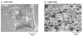
도 1의 도 1A는 본 발명의 줄기 세포를 배양했을 때의 특정 배양 기간(일)에서의 전체 세포량을 나타내는 도면이다. 도 1B는 본 발명의 줄기 세포를 배양했을 때의 특정 배양 기간(일)에서의 전체 세포 증식률을 나타내는 도면이다. 도 1A 및 도 1B에서 점선 및 검은색 동그라미는 어린 돼지 골수 유래 간엽계 줄기 세포(npBM-MSC)를, 실선 및 흰색 동그라미는 인간 골수 유래 간엽계 줄기 세포(hBM-MSC)를 나타낸다.
도 2의 도 2A 및 도 2B는 각각 인간 골수 유래 간엽계 줄기 세포(hBM-MSC) 및 어린 돼지 골수 유래 간엽계 줄기 세포(npBM-MSC)에 대한 지방 세포로의 분화를 나타내는 도면이다.
도 3의 도 3A 및 도 3B는 각각 인간 골수 유래 간엽계 줄기 세포(hBM-MSC) 및 어린 돼지 골수 유래 간엽계 줄기 세포(npBM-MSC)에 대한 골 세포로의 분화를 나타내는 도면이다.
도 4의 도 4A 및 도 4B는 각각 인간 골수 유래 간엽계 줄기 세포(hBM-MSC) 및 어린 돼지 골수 유래 간엽계 줄기 세포(npBM-MSC)에 대한 골 세포로의 분화를 나타내는 도면이다.
도 5의 도 5A 및 도 5B는 어린 돼지 골수 유래 간엽계 줄기 세포(npBM-MSC)에 대한 연골 세포로의 분화를 나타내는 도면이다.
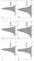
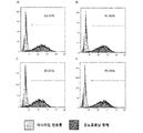
도 6은 간엽계 줄기 세포의 마커인 CD44를 이용하여 어린 돼지 골수 유래 간엽계 줄기 세포(npBM-MSC)의 세포 표면 항원 해석을 한 결과를 나타내는 도면이다.
도 7은 간엽계 줄기 세포의 마커인 CD90를 이용하여 어린 돼지 골수 유래 간엽계 줄기 세포(npBM-MSC)의 세포 표면 항원 해석을 한 결과를 나타내는 도면이다.
도 8의 도 8A 내지 도 8D는 간엽계 줄기 세포의 마커인 CD29를 이용하여 어린 돼지 췌도 유래 간엽계 줄기 세포(npISLET-MSC)의 세포 표면 항원 해석을 한 결과를 나타내는 도면이다. 도 8A는 샘플 11(췌도 제조 직후의 동결 없음), 도 8B는 샘플 12(췌도 제조 직후의 동결 있음), 도 8C는 샘플 13(췌도 제조 후 배양 3일째의 동결 없음), 도 8D는 샘플 14(췌도 제조 후 배양 3일째의 동결 있음)의 결과를 나타낸다.
도 9의 도 9A 내지 도 9D는 간엽계 줄기 세포의 마커인 CD44를 이용하여 어린 돼지 췌도 유래 간엽계 줄기 세포(npISLET-MSC)의 세포 표면 항원 해석을 한 결과를 나타내는 도면이다. 도 9A는 샘플 11(췌도 제조 직후의 동결 없음), 도 9B는 샘플 12(췌도 제조 직후의 동결 있음), 도 9C는 샘플 13(췌도 제조 후 배양 3일째 의 동결 없음), 도 9D는 샘플 14(췌도 제조 후 배양 3일째의 동결 있음)의 결과를 나타낸다.
도 10의 도 10A 내지 도 10D는 간엽계 줄기 세포의 마커인 CD90을 이용하여 어린 돼지 췌도 유래 간엽계 줄기 세포(npISLET-MSC)의 세포 표면 항원 해석을 한 결과를 나타내는 도면이다. 도 10A는 샘플 11(췌도 제조 직후의 동결 없음), 도 10B는 샘플 12(췌도 제조 직후의 동결 있음), 도 10C는 샘플 13(췌도 제조 후 배양 3일째 동결 없음), 도 10D는 샘플 14(췌도 제조 후 배양 3일째 동결 있음)의 결과를 나타낸다.1A of FIG. 1 is a diagram showing the total cell amount in a specific culture period (day) when the stem cells of the present invention are cultured. 1B is a diagram showing the overall cell proliferation rate in a specific culture period (day) when the stem cells of the present invention are cultured. In FIG. 1A and FIG. 1B, dotted and black circles represent young pig bone marrow-derived mesenchymal stem cells (npBM-MSC), and solid and white circles represent human bone marrow-derived mesenchymal stem cells (hBM-MSC).
2A and 2B of FIG. 2 are diagrams showing differentiation of human bone marrow-derived mesenchymal stem cells (hBM-MSC) and young porcine bone marrow-derived mesenchymal stem cells (npBM-MSC) into adipocytes.
3A and 3B of FIG. 3 are diagrams showing differentiation into human bone marrow-derived mesenchymal stem cells (hBM-MSC) and young porcine bone marrow-derived mesenchymal stem cells (npBM-MSC) into bone cells, respectively.
4A and 4B are diagrams showing differentiation of human bone marrow-derived mesenchymal stem cells (hBM-MSC) and young porcine bone marrow-derived mesenchymal stem cells (npBM-MSC) into bone cells, respectively.
5A and 5B of FIG. 5 are diagrams showing differentiation of mesenchymal stem cells (npBM-MSC) derived from young pig bone marrow into chondrocytes.
6 is a diagram showing the results of cell surface antigen analysis of mesenchymal stem cells derived from young pig bone marrow (npBM-MSC) using CD44, a marker of mesenchymal stem cells.
7 is a diagram showing the results of cell surface antigen analysis of mesenchymal stem cells derived from young pig bone marrow (npBM-MSC) using CD90, a marker of mesenchymal stem cells.
8A to 8D are diagrams showing the results of cell surface antigen analysis of mesenchymal stem cells derived from young pig islets (npISLET-MSC) using CD29, a marker of mesenchymal stem cells. Figure 8A is sample 11 (no freezing immediately after islet preparation), Figure 8B is sample 12 (with freezing immediately after islet preparation), Figure 8C is sample 13 (no freezing on
9A to 9D are diagrams showing the results of cell surface antigen analysis of young pig pancreatic islet-derived mesenchymal stem cells (npISLET-MSC) using CD44, a marker of mesenchymal stem cells. Figure 9A is sample 11 (no freezing immediately after islet preparation), Figure 9B is sample 12 (with freezing immediately after islet preparation), Figure 9C is sample 13 (no freezing on
10A to 10D of FIG. 10 are diagrams showing the results of cell surface antigen analysis of young pig islet-derived mesenchymal stem cells (npISLET-MSC) using CD90, a marker of mesenchymal stem cells. FIG. 10A shows sample 11 (no freezing immediately after islet preparation), FIG. 10B shows sample 12 (with freezing immediately after islet preparation), FIG. 10C shows sample 13 (no freezing on
본 발명의 줄기 세포는 어린 돼지로부터 단리된 줄기 세포이다. 또한 후술하는 실시예에 기재된 줄기 세포는 어린 돼지의 골수 또는 췌도로부터 분리되었는데 예를 들어 어린 돼지의 피부, 지방 등 유래의 줄기 세포도 본 발명에 포함된다.The stem cells of the present invention are stem cells isolated from young pigs. In addition, the stem cells described in Examples described below were isolated from bone marrow or islets of young pigs, for example, stem cells derived from skin, fat, etc. of young pigs are also included in the present invention.
본 발명에서 "어린 돼지"란 태아로부터 생후 1개월 미만, 또는 생후 25일 미만의 돼지를 가리킨다. 어린 돼지는 의료용인 것이 바람직하고, 인간에게 세포 이식을 할 수 있는 어린 돼지인 것이 보다 바람직하다. 돼지의 종류는 특별히 한정되지 않지만, 예를 들어 랜드레이스종(예를 들어 덴마크·랜드레이스종, 아메리칸·랜드레이스종, 브리티시·랜드레이스종, 네덜란드·랜드레이스종, 스웨디시·랜드레이스종), 대 요크셔종, 버크셔종, 듀록종, 햄프셔종, 중 요크셔종, 미니 돼지를 들 수 있으며, 그 중에서도 랜드레이스종이 바람직하다.In the present invention, "young pig" refers to a pig less than 1 month old or less than 25 days old. It is preferable that the young pig is for medical use, and more preferably, it is a young pig capable of cell transplantation to humans. The types of pigs are not particularly limited, but, for example, Landrace (e.g., Denmark / Landrace, American / Landrace, British / Landrace, Dutch / Landrace, Swedish / Landrace) , Large Yorkshire species, Berkshire species, Duroc species, Hampshire species, heavy Yorkshire species, mini pigs, and among them, Landrace species are preferred.
일반적으로 "줄기 세포"란 자기 복제능 및 분화·증식능을 가진 미숙한 세포를 뜻한다. 줄기 세포에는 분화 능력에 따라 다능성 줄기 세포(pluripotent stem cell), 복능성 줄기 세포(multipotent stem cell), 단능성 줄기 세포(unipotent stem cel) 등의 아집단이 포함된다.In general, "stem cell" refers to an immature cell having self-replicating ability and differentiation / proliferation ability. Stem cells include subpopulations such as pluripotent stem cells, multipotent stem cells, and unipotent stem cells, depending on their differentiation ability.
다능성 줄기 세포란 그 자체로는 개체가 될 수 없지만 생체를 구성하는 모든 조직이나 세포로 분화 가능한 능력을 가진 세포를 뜻한다. 복능성 줄기 세포란 모든 종류는 아니지만 여러 종류의 조직이나 세포로 분화 가능한 능력을 가진 세포를 뜻한다. 단능성 줄기 세포란 특정 조직이나 세포로 분화 가능한 능력을 가진 세포를 뜻한다.A pluripotent stem cell is a cell that cannot be an individual in itself, but has the ability to differentiate into any tissue or cell that makes up a living body. A pluripotent stem cell is a cell that has the ability to differentiate into a variety of tissues or cells, but not all types. Pluripotent stem cells are cells that have the ability to differentiate into specific tissues or cells.
본 발명의 줄기 세포로서는 복능성 줄기 세포가 바람직하다. 복능성 줄기 세포로서는 예를 들어 간엽계 줄기 세포, 조혈계 줄기 세포, 신경계 줄기 세포, 골수 줄기 세포, 생식 줄기 세포 등의 체성 줄기 세포 등을 들 수 있으며, 바람직하게는 간엽계 줄기 세포이다.As the stem cells of the present invention, pluripotent stem cells are preferred. Examples of the pluripotent stem cells include somatic stem cells such as mesenchymal stem cells, hematopoietic stem cells, neural stem cells, bone marrow stem cells, and germ stem cells, and are preferably mesenchymal stem cells.
본 발명의 줄기 세포는 어린 돼지로부터 단리된 줄기 세포라면 그 초대 배양 세포, 해당 초대 배양 세포를 계대 배양한 세포이며, 각종 분화 마커를 발현하는 각종 세포를 생성할 수 있는 줄기 세포도 본 발명의 줄기 세포에 포함된다. 또한, 본 발명에서의 줄기 세포가 간엽계 줄기 세포인 경우에는 세포 마커인 CD44 및 CD90이 모두 60% 이상 양성인 것이 바람직하며, 보다 바람직하게는 70% 이상, 더욱 바람직하게는 80% 이상 양성이다. 또 세포 마커인 CD29가 60% 이상 양성인 것이 바람직하고, 보다 바람직하게는 70% 이상, 더욱 바람직하게는 80% 이상 양성이다.If the stem cells of the present invention are stem cells isolated from young pigs, they are primary culture cells, cells that are passage-cultured of the primary culture cells, and stem cells capable of generating various cells expressing various differentiation markers are also the stem cells of the present invention. Cells. In addition, when the stem cells in the present invention are mesenchymal stem cells, both the cell markers CD44 and CD90 are preferably at least 60% positive, more preferably at least 70%, more preferably at least 80% positive. The cell marker CD29 is preferably 60% or more, more preferably 70% or more, and even more preferably 80% or more.
본 발명의 줄기 세포는 대수 증식기에서의 배가 시간이 36시간 이하인 것이 바람직하며, 보다 바람직하게는 32시간 이하, 더욱 바람직하게는 28시간 이하, 특히 바람직하게는 24시간 이하, 가장 바람직하게는 20시간 이하이다. 또 대수 증식기에서의 배가 시간은 14시간 이상인 것이 바람직하고, 16시간 이상인 것이 보다 바람직하다.The stem cells of the present invention preferably have an doubling time in the logarithmic growth phase of 36 hours or less, more preferably 32 hours or less, more preferably 28 hours or less, particularly preferably 24 hours or less, and most preferably 20 hours. Is below. Moreover, the doubling time in the logarithmic growth period is preferably 14 hours or more, and more preferably 16 hours or more.
본 발명의 줄기 세포의 대수 증식기에서의 배양은, 예를 들어 후술하는 비타민 C를 함유하는 배지(예를 들어 MSC 배지)에 본 발명의 줄기 세포를 파종하고 37℃에서 5% CO2 존재 하에서 배양용 인큐베이터에서 배양함으로써 행할 수 있다. 대수 증식기에서의 배가 시간이 짧을수록, 단시간 또한 저렴하게 대량의 줄기 세포를 제조하는 것이 가능해진다.Incubation of the stem cells of the present invention in a logarithmic growth phase, for example, seeding the stem cells of the present invention in a medium containing vitamin C (for example, MSC medium) described below and culturing at 37 ° C in the presence of 5% CO 2. It can be carried out by culturing in a dragon incubator. The shorter the doubling time in the logarithmic growth phase, the shorter and cheaper it becomes possible to produce large amounts of stem cells.
본 발명의 줄기 세포는 평균 직경이 17μm 이하인 것이 바람직하고, 보다 바람직하게는 16.5μm 이하이며, 더욱 바람직하게는 16μm 이하이며, 특히 바람직하게는 15.5μm 이하이며, 가장 바람직하게는 15μm 이하이다. 평균 직경은 10μm 이상인 것이 바람직하며 12μm 이상인 것이 보다 바람직하다. 평균 직경이 작을수록 줄기 세포 투여로 인한 폐색전의 형성을 방지할 수 있다. 평균 직경은, 예를 들어 Nucleo Counter NC-200(상표)을 이용해 계측할 수 있다. 여기서 평균이란 상가 평균을 의미한다.The stem cells of the present invention preferably have an average diameter of 17 μm or less, more preferably 16.5 μm or less, even more preferably 16 μm or less, particularly preferably 15.5 μm or less, and most preferably 15 μm or less. The average diameter is preferably 10 μm or more, and more preferably 12 μm or more. The smaller the average diameter, the more can prevent the formation of pulmonary embolism due to stem cell administration. The average diameter can be measured using Nucleo Counter NC-200 (trademark), for example. Here, the average means the mall average.
본 발명의 간엽계 줄기 세포에서 지방 세포로의 분화는 예를 들어 인슐린, MCGS(혈청 성분, Mesenchymal Cell Growth Supplement), 덱사메타존, 인도메타신, 이소부틸메틸크산틴 등의 존재 하에서 본 발명에서의 간엽계 줄기 세포를 배양함으로써 지방 세포로 분화 유도할 수 있다.Differentiation of mesenchymal stem cells from adipocytes to adipocytes in the present invention, for example, in the presence of insulin, MCGS (serum component, Mesenchymal Cell Growth Supplement), dexamethasone, indomethacin, isobutylmethylxanthine, etc. Differentiation into adipocytes can be induced by culturing the mesenchymal stem cells.
지방 세포로의 분화 및 유지를 위해서는 시판 중인 키트 또는 배지 등을 이용해도 되며, 예를 들어 Lonza Walkersville사제 hMSC differentiation Bullet Kit(상표)-adipogeni(PT-3004), Lonza Walkersville사제 hMS Cadipogenic induction medium(PT-3102B), Lonza Walkersville사제 hMS Cadipogenic maintenance medium(PT-302) 등을 들 수 있다. 간엽계 줄기 세포로부터 지방 세포로의 분화는 시판 중인 키트를 이용해 확인할 수 있으며, 예를 들어 Lonza사제 AdipoRed(상표) assay reagent를 들 수 있다.For the differentiation and maintenance into adipocytes, a commercially available kit or medium may be used, for example, hMSC differentiation Bullet Kit (trademark) -adipogeni (PT-3004) from Lonza Walkersville, hMS Cadipogenic induction medium (PT from Lonza Walkersville) -3102B), hMS Cadipogenic maintenance medium (PT-302) manufactured by Lonza Walkersville, and the like. Differentiation from mesenchymal stem cells to adipocytes can be confirmed using commercially available kits, for example, AdipoRed (trademark) assay reagent manufactured by Lonza.
본 발명의 간엽계 줄기 세포로부터 골 세포의 분화는 예를 들어 덱사메타존, 아스코르브산염, MCGS, β-글리세로인산 등의 존재 하에서 본 발명의 간엽계 줄기 세포를 배양함으로써 골 세포로 분화 유도할 수 있다. 또한, 시판 중인 키트를 이용해도 좋으며, 예를 들어 Lonza Walkersville사제 hMSC differentiation BulletKit(상표)-osteogenic, PT-3004 등을 들 수 있다. 간엽계 줄기 세포로부터 골 세포로의 분화는 시판 중인 알칼리포스파타아제 염색 키트(예를 들어 코스모·바이오사제 등), 시판 중인 석회화 염색 키트(예를 들어 코스모·바이오사제 등) 등에 의해 확인할 수 있다.Differentiation of bone cells from mesenchymal stem cells of the present invention can be induced to differentiate into bone cells by culturing the mesenchymal stem cells of the present invention in the presence of dexamethasone, ascorbate, MCGS, β-glycerophosphoric acid, etc. You can. In addition, a commercially available kit may be used, for example, hMSC differentiation BulletKit (trademark) -osteogenic from PT Lonza Walkersville, PT-3004. Differentiation from mesenchymal stem cells to bone cells can be confirmed by commercially available alkaline phosphatase staining kits (eg Cosmo Biosa), commercially available calcification staining kits (eg Cosmo Biosa), etc. .
본 발명의 간엽계 줄기 세포로부터 연골 세포로의 분화는, 예를 들어 TGF-β3, 덱사메타존, 인슐린-트랜스페린-아세렌산(ITS), 피루브산나트륨, 프롤린, 아스코르브산염, 의 존재 하에서 본 발명의 간엽계 줄기 세포를 배양함으로써 연골 세포로 분화 유도할 수 있다. 또한, 시판 중인 키트를 이용해도 좋고, 예를 들어 Lonza Walkersville사제 hMSC differentiation BulletKit(상표)-condrogenic, PT-3003등을 들 수 있다. 간엽계 줄기 세포로부터 연골 세포로의 분화는 알시안 블루(Alcian Blue) 염색 등을 통해 확인할 수 있다.Differentiation from mesenchymal stem cells to chondrocytes of the present invention, for example, in the presence of TGF-β3, dexamethasone, insulin-transferin-acerenic acid (ITS), sodium pyruvate, proline, ascorbate, Differentiation into chondrocytes can be induced by culturing the mesenchymal stem cells. Also, a commercially available kit may be used, for example, hMSC differentiation BulletKit (trademark) -condrogenic from PT Lonza Walkersville, PT-3003. Differentiation from mesenchymal stem cells to chondrocytes can be confirmed by Alcian Blue staining or the like.
줄기 세포의 이식은 줄기 세포의 부유액을 숙주체에 주입함으로써 쉽게 할 수 있다. 주입은 재생 치료하고자 하는 장기 또는 그 근방 또는 정맥 내 등에 대하여 실시할 수 있다. 또한 주입하는 줄기 세포의 수는 특별히 한정되지 않고 증상, 숙주의 체중 또는 투여 방법 등에 따라 적절히 선택할 수 있지만 보통 102 내지 1010개 정도로 한다.Stem cell transplantation can be easily accomplished by injecting stem cell suspension into the host body. The injection may be performed on an organ to be regenerated or in the vicinity or intravenously. In addition, the number of stem cells to be injected is not particularly limited, and may be appropriately selected according to symptoms, a host's weight, or a method of administration, but is usually about 10 2 to 10 10 cells.
본 명세서에서 "피더 세포(feeder cell)"란 증식이나 분화를 일으키려는 목적의 세포 배양조건을 갖추기 위해 이용하는 보조 역할을 하는 다른 세포종을 나타낸다.As used herein, "feeder cell" refers to another cell species that serves as an auxiliary to be used to prepare cell culture conditions for the purpose of causing proliferation or differentiation.
피더 세포로서 이용하는 경우에는 통상 증식하지 않도록 미리 감마선 조사나 항생 물질로 처리해 두는 것이 바람직하다. 줄기 세포의 피더 세포로서는 주로 마우스 태아 유래의 섬유 아세포가 이용되는데, 실험의 목적이나 세포에 따라 3T3이나 SNL 등의 섬유 아세포 등 여러 세포종이 피더 세포로서 이용되고 있다. 본 발명의 줄기 세포는 바람직하게는 인간에게 세포 이식을 할 수 있는 어린 돼지로부터 단리함으로써 인간 이식용 줄기 세포의 피더 세포로서 이용할 수 있다.When used as a feeder cell, it is preferable to treat with gamma-ray irradiation or an antibiotic in advance so as not to multiply normally. As stem cell feeder cells, fibroblasts derived from mouse fetus are mainly used, but various cell types such as fibroblasts such as 3T3 and SNL are used as feeder cells depending on the purpose of the experiment or the cells. The stem cells of the present invention can be preferably used as feeder cells for stem cells for human transplantation by isolating them from young pigs capable of cell transplantation to humans.
본 발명의 줄기 세포 제조 방법은 어린 돼지로부터 세포를 단리하는 공정을 포함하는 것을 특징으로 한다. 본 발명의 줄기 세포 제조 방법의 실시 양태로서는 예를 들어 다음과 같은 공정을 포함하는 방법을 들 수 있다.The method for producing stem cells of the present invention is characterized by including a step of isolating cells from young pigs. As an embodiment of the stem cell production method of the present invention, for example, a method including the following steps is exemplified.
(1) 어린 돼지로부터 세포를 채취하는 공정(1) Process of collecting cells from young pigs
(2) 공정 (1)에서 채취한 세포를 배양하여 어린 돼지 유래 줄기 세포를 제조하는 공정(2) Process for producing stem cells derived from young pigs by culturing the cells collected in step (1)
이하, 각 공정에 대해서 설명한다.Hereinafter, each process is demonstrated.
(1) 어린 돼지로부터 세포를 채취하는 공정(1) Process of collecting cells from young pigs
공정 (1)에서는 어린 돼지의 골수, 지방, 피부, 췌장 등으로부터 세포를 채취한다.In step (1), cells are collected from bone marrow, fat, skin, and pancreas of a young pig.
구체적으로는 예를 들어 어린 돼지 골수에서 세포를 채취할 경우 어린 돼지의 대퇴골, 장골릉 및 흉골 등에서 골수 세포를 채취할 수 있다. 예를 들어 어린 돼지로부터 대퇴골을 회수하고 양끝을 절단하여 바늘을 삽입하고 헤파린을 첨가한 생리적 완충액(예를 들어 인산 완충액, 이후 PBS라고도 함)으로 씻어낸 후 반대편 장소에서 유출액을 골수액으로서 회수한다. 유출액의 양이 감소하면 골을 거꾸로 해서 바늘을 반대편에 삽입하고 PBS로 다시 씻어내 세포 함유 용액인 골수액을 제조한다.Specifically, for example, when cells are collected from young pig bone marrow, bone marrow cells may be collected from the femur, iliac crest, and sternum of young pigs. For example, the femur is recovered from a young pig, the ends of which are cut, and the needle is inserted, washed with a physiological buffer (eg, phosphate buffer, hereinafter referred to as PBS) to which heparin is added, and then the effluent is recovered as the bone marrow solution from the opposite site. . When the amount of effluent decreases, the bone is turned upside down, the needle is inserted into the opposite side, and washed again with PBS to prepare a cell-containing solution, bone marrow solution.
또한, 상기에서 제조한 세포 함유 용액을 통상 원심 분리함으로써 어린 돼지 유래 단핵구 세포 분획을 단리해도 된다. 상기에서 제조한 세포 함유 용액을 PBS 등으로 희석하여 인간 림프구 분리용 매체(예를 들어 GE 헬스케어 라이프사이언스사제 Ficoll-Paque PLUS 등)를 넣은 튜브 내의 해당 매체층 위에 희석한 세포 함유 용액을 넣는다.In addition, the fraction containing monocytes derived from young pigs may be isolated by usually centrifuging the cell-containing solution prepared above. The cell-containing solution prepared above is diluted with PBS or the like, and the diluted cell-containing solution is added onto a corresponding medium layer in a tube containing human lymphocyte separation medium (eg, Ficoll-Paque PLUS manufactured by GE Healthcare Life Sciences).
상기 튜브를 원심 분리하여 분층시켜 어린 돼지 유래 단핵구 세포를 포함하는 층을 회수한다. 회수한 용액을 다시 원심 분리하여 상청을 제거한 후 PBS 등으로 희석하여 다시 원심 분리하고 단핵구 세포 분획을 단리한다. 이와 같이 하여 단리된 단핵구 세포 분획 세포는 배양 전에 동결 보존해도 된다. 단리된 어린 돼지 유래 단핵구 세포 분획의 세포를 동결함으로써 동결 융해의 영향을 받기 어려운 세포를 선택적으로 제조할 수 있다. 배양 전에 동결 보존할 경우, 온도는 -80℃ 이하인 것이 바람직하고, 보다 바람직하게는 -150℃ 이하이다.The tube is centrifuged and separated to collect a layer containing young pig-derived monocyte cells. The recovered solution is centrifuged again to remove the supernatant, diluted with PBS or the like, centrifuged again, and the monocyte cell fraction is isolated. The isolated monocyte cell fraction cells may be cryopreserved prior to culture. By freezing the cells of the isolated young pig-derived monocyte cell fraction, cells that are difficult to be affected by freeze-thawing can be selectively prepared. When cryopreserved before culture, the temperature is preferably -80 ° C or lower, more preferably -150 ° C or lower.
또한, 예를 들어 어린 돼지 췌장에서 세포를 채취할 경우, 어린 돼지로부터 췌도를 회수하고, 다시 경우에 따라서는 그 췌도를 부유 배양함으로써 줄기 세포를 제조할 목적으로 접착 배양에 사용하는 세포괴를 제조한다.In addition, for example, when cells are harvested from a young pig pancreas, a cell mass used for adhesion culture is prepared for the purpose of producing stem cells by recovering the pancreatic islets from a young pig and, if necessary, floating culture the pancreatic islets. .
또, 예를 들어 어린 돼지 지방으로부터 세포를 채취할 경우 어린 돼지에게서 지방을 채취해 가위로 잘게 썬 후 효소 처리를 한다. 셀 스트레이너(cell strainer)로 필터를 걸어 저속으로 원심 분리를 한다. 튜브 바닥에 침강된 세포를 배양에 이용한다. 또, 예를 들어 어린 돼지 피부(털 포함)로부터 세포를 채취하는 경우, 어린 돼지로부터 피부를 채취하여 효소 처리를 한다. 효소 처리 후 피부에서 털을 뽑아 Bulge 부분을 채취하여 배양에 이용한다. 배양을 실시할 때는 3T3 피더 세포를 이용한다.In addition, for example, when cells are collected from young pig fat, fat is collected from young pigs, chopped with scissors, and then enzymatically treated. Filter with a cell strainer and centrifuge at low speed. Cells settled at the bottom of the tube are used for culture. In addition, for example, when cells are collected from the skin of a young pig (including hair), the skin is collected from a young pig and subjected to enzyme treatment. After enzymatic treatment, hair is pulled from the skin, and the bulge part is collected and used for culture. When culturing, 3T3 feeder cells are used.
(2)공정 (1)에서 채취한 세포를 배양하여 어린 돼지 유래 줄기 세포를 제조하는 공정(2) Process of culturing the cells collected in step (1) to produce stem cells derived from young pigs
상기 공정 (1)에서 채취한 세포, 세포 분획 또는 세포괴에는 줄기 세포 이외의 목적 외의 세포가 많이 포함된다. 일반적으로 이들 목적외 세포의 생존에 필수적인 비타민 C를 포함하지 않는 기초 배지(예를 들어 후술하는 MSC 기초 배지)를 이용함으로써 이들 세포를 제거하는 배양 방법이 이용되고 있다.The cells, cell fractions or cell masses collected in the step (1) include many cells other than the target cells other than stem cells. In general, a culture method in which these cells are removed by using a basal medium not containing vitamin C (for example, MSC basal medium to be described later) essential for the survival of cells other than these objectives is used.
본 발명 공정 (2)에서는 상기 공정 (1)에서 채취한 세포, 세포 분획 또는 세포괴를 바람직하게는 35 내지 39℃, 보다 바람직하게는 36 내지 38℃, 가장 바람직하게는 37℃에서, 바람직하게는 4 내지 6%의, 보다 바람직하게는 4.5 내지 5.5%의, 가장 바람직하게는 5%의, CO2 존재 하에서 배양용 인큐베이터에서 배양함으로써 줄기 세포 이외의 목적 외의 세포를 제거함과 동시에 본 발명의 줄기 세포를 증식시킨다.In the step (2) of the present invention, the cells, cell fractions or cell mass collected in the step (1) are preferably 35 to 39 ° C, more preferably 36 to 38 ° C, most preferably 37 ° C, preferably Stem cells of the present invention are removed at the same time as cells other than the target cells are removed by culturing in a culture incubator in the presence of CO 2 of 4 to 6%, more preferably of 4.5 to 5.5%, most preferably of 5%. Multiply.
본 발명의 줄기 세포는 증식 속도가 현저히 빠르기 때문에 상기의 목적 외의 세포를 제거하는 배양을 위해 비타민 C를 포함하지 않는 기초 배지를 이용하지 않고 비타민 C를 포함한 배지(예를 들어 후술하는 MSC 배지)만을 이용해도 본 발명의 줄기 세포를 제조할 수 있다. 또한 상기 목적 외의 세포를 제거하기 위하여 비타민 C를 포함하지 않는 기초 배지를 이용하여 배양한 후 비타민 C를 포함한 배지로 교환하여 본 발명의 줄기 세포를 증식시킴으로써 본 발명의 줄기 세포를 제조할 수도 있다.Since the stem cells of the present invention have a proliferation rate that is remarkably fast, only a medium containing vitamin C (for example, MSC medium described later) is used without using a basal medium that does not contain vitamin C for culture to remove cells other than the above-mentioned purpose. The stem cells of the present invention can also be produced by use. In addition, the stem cells of the present invention can also be prepared by culturing using a basal medium that does not contain vitamin C, and then exchanging with a medium containing vitamin C to proliferate the stem cells of the present invention to remove cells other than the above-mentioned purpose.
본 발명의 줄기 세포는 구체적으로는 예를 들어 다음과 같은 방법으로 배양한다. 젤라틴으로 코팅한 배양용 용기(예를 들어 0.1% 젤라틴으로 코팅한 플레이트) 또는 젤라틴 코트 없는 배양용 용기(예를 들어 플레이트)를 이용하여 비타민 C를 포함하지 않는 기초 배지(예를 들어 후술한 MSC 배지)를 이용하고, 바람직하게는 5.0×10⁵개 내지 5.0×10⁷개의 세포/9.6cm2를 파종하고, 예를 들어 37℃에서 5% CO2, 90% 습도의 조건하에서 인큐베이팅하여 초대 배양 세포를 얻는다. 초대 배양 세포를 얻기 위한 배양 기간은 파종 후 바람직하게는 3 내지 12일, 보다 바람직하게는 3 내지 11일, 가장 바람직하게는 3 내지 10일이다. 초대 배양 세포는 계대해도 된다. 계대하여 얻은 줄기 세포를 계대 배양 세포라고도 한다. 초대 배양 세포 또는 계대 배양 세포의 계대는 줄기 세포를 파종 후, 바람직하게는 2 내지 6일 후, 보다 바람직하게는 2 내지 5일 후, 더욱 바람직하게는 2 내지 4일 후, 가장 바람직하게는 3일 후에, 줄기 세포가 30% 내지 100% 콘플루언트에, 바람직하게는 50% 내지 95% 콘플루언트에, 더욱 바람직하게는 60% 내지 90% 콘플루언트에, 가장 바람직하게는 70% 내지 85% 콘플루언트에 도달한 이후에 한다. 줄기 세포의 파종은 젤라틴으로 코팅한 배양용 용기(예를 들어 0.1% 젤라틴으로 코팅한 플레이트) 또는 젤라틴 코트 없는 배양용 용기(예를 들어 플레이트)를 이용하여 비타민 C를 포함한 배지(예를 들어 후술하는 MSC 배지)를 이용하여, 바람직하게는 5.0×10⁵개 내지 5.0×10⁷개의 세포/9.6cm2를 파종한다. 줄기 세포의 배양은, 예를 들어 37℃에서 5% CO2, 90% 습도의 조건하에서 배양한다. 줄기 세포의 배양 동안, 필요에 따라 배지 교환하여 본 발명의 줄기 세포를 증식시킨다.The stem cells of the present invention are specifically cultured, for example, by the following method. A basal medium not containing vitamin C (e.g. MSC described below) using a culture vessel coated with gelatin (e.g., a plate coated with 0.1% gelatin) or a culture vessel without a gelatin coat (e.g., a plate). Medium), preferably, 5.0 × 10 5 to 5.0 × 10 7 cells / 9.6 cm 2 are seeded, for example, incubated at 37 ° C. under conditions of 5% CO 2 and 90% humidity, and the initial culture is performed. Get cells The culture period for obtaining primary cultured cells is preferably 3 to 12 days after sowing, more preferably 3 to 11 days, and most preferably 3 to 10 days after sowing. The primary cultured cells may be passaged. Stem cells obtained by passage are also referred to as passage culture cells. The passage of primary cultured cells or passage cultured cells is after seeding the stem cells, preferably after 2 to 6 days, more preferably after 2 to 5 days, more preferably after 2 to 4 days, most preferably 3 After one day, the stem cells are 30% to 100% confluent, preferably 50% to 95% confluent, more preferably 60% to 90% confluent, most preferably 70% to After reaching 85% confluence. The seeding of the stem cells can be performed using a culture vessel (eg, 0.1% gelatin coated plate) coated with gelatin or a culture vessel (eg, plate) containing vitamin C (eg, described later) using a culture vessel without gelatin coat. MSC medium), preferably 5.0 × 10 5 to 5.0 × 10 7 cells / 9.6 cm 2 are seeded. The stem cells are cultured, for example, at 37 ° C under 5% CO 2 and 90% humidity. During the cultivation of stem cells, the medium of the present invention is propagated by medium exchange as necessary.
MSC 기초 배지 및 MSC 배지로서는 기존에 알려진 것을 이용할 수 있으며 시판되는 것을 이용해도 된다. MSC 기초 배지로서는 예를 들어 500mL의 Gibco사제 MEMα(Nucleosides, no Ascorbic acid)에 55mL의 Gibco사제 Fetal bovine serum(FBS) 및 5.5mL의 Sigma-Aldorich사제 Penicillin-Streptomycin을 첨가한 배지를 들 수 있다. 또한 MSC 배지로서는 예를 들어, 500mL의 Gibco사제 MEM(nucleosides)에 55mL의 Gibco사제 Fetal bovine serum(FBS), 5.5mL의 Sigma-Aldorich사제 Penicillin-Streptomycin 및 22.2μL의 Sigma-Aldorich사제 FGF-Basic, recombinant, expressed in E.coli, suitable for cell culture (final concentration : 1ng/mL)을 첨가한 배지를 들 수 있다.As the MSC basal medium and MSC medium, conventionally known ones can be used, and commercially available ones may be used. As the MSC basic medium, for example, a medium in which 500 mL of Gibco MEMα (Nucleosides, no Ascorbic acid) was added to 55 mL of Gibco's Fetal bovine serum (FBS) and 5.5 mL of Sigma-Aldorich's Penicillin-Streptomycin was added. In addition, as the MSC medium, for example, 500 mL of Gibco, MEM (nucleosides), 55 mL of Gibco, Fetal bovine serum (FBS), 5.5 mL of Sigma-Aldorich, Penicillin-Streptomycin and 22.2 μL of Sigma-Aldorich, FGF-Basic, and a medium to which recombinant, expressed in E.coli, suitable for cell culture (final concentration: 1 ng / mL) was added.
계대는 적어도 1회 이상 실시하는 것이 바람직하다. 계대 횟수는 본 발명에서의 줄기 세포를 얻을 수 있는 한 특별히 한정되지 않지만, 바람직하게는 1 내지 3회이며, 보다 바람직하게는 1 내지 20회이다.It is preferable to pass the passage at least once. The number of passages is not particularly limited as long as the stem cells in the present invention can be obtained, but is preferably 1 to 3 times, and more preferably 1 to 20 times.
본 발명의 줄기 세포는 동결 보존이 가능하다. 동결 보존 타이밍은 특별히 한정되지 않지만, 바람직하게는 계대 1 내지 20회 후이며, 보다 바람직하게는 계대 2 내지 10회 후이다. 동결 보존 및 해동 방법은 기존에 공지된 방법을 이용할 수 있다.The stem cells of the present invention can be cryopreserved. The cryopreservation timing is not particularly limited, but is preferably after 1 to 20 passages, more preferably after 2 to 10 passages. As the cryopreservation and thawing method, a conventionally known method can be used.
줄기 세포의 동결 보존 방법으로서는 구체적으로는 예를 들어 동결 보존액에 분산시키고 필요할 때까지 냉동고에서 -80℃ 이하 또는 액체 질소 안에서 동결 보존할 수 있다. 동결 보존액으로서는 예를 들어 OPF-301[3% 트레할로오스 및 5% 덱스트란을 함유하는 젖산 링거액(국제 공개 제 2014/208053호)]과 디메틸설폭사이드(DMSO)를 9:1의 비율로 혼합한 용액, 동물 세포의 동결 보존에 사용할 수 있는 혈청 함유 또는 무혈청 보존액, 또는 시판 중인 세포 동결 보존용 시약[바람직하게는 다카라바이오사 제조 CELLBANKER(등록 상표) 등의 셀뱅커]를 들 수 있다.As a method for cryopreservation of stem cells, for example, it can be dispersed in a cryopreservation solution and cryopreserved in liquid freezer at -80 ° C or lower in a freezer until necessary. As a cryopreservative, for example, OPF-301 [lactic acid Ringer's solution containing 3% trehalose and 5% dextran (International Publication No. 2014/208053)] and dimethyl sulfoxide (DMSO) in a ratio of 9: 1. And mixed solutions, serum-containing or serum-free preservatives that can be used for cryopreservation of animal cells, or commercially available cell cryopreservation reagents (preferably cell bankers such as CELLBANKER (registered trademark) manufactured by Takara Bio Corporation). .
[실시예][Example]
시험예 1Test Example 1
[어린 돼지 유래 골수 세포의 회수][Recovery of bone marrow cells derived from young pigs]
어린 돼지 대퇴골에서 골수를 채취했다. 어린 돼지(생후 23일의 의료용 랜드레이스종 돼지)로부터 대퇴골을 회수하고 양끝을 절단하여 12G 바늘을 삽입하고 50mL의 헤파린 처리한 PBS(3mL의 헤파린(1000U/mL), 47mL의 PBS)으로 씻어내고 반대편 장소에서 50mL의 골수 유출액(이하 골수액이라고도 한다)을 회수하였다. 유출액의 양이 감소하면 골을 거꾸로 해서 바늘을 반대편에 삽입한 뒤 PBS로 다시 씻어내고 골수액을 수집했다. 카운트용 15mL 코니컬 튜브로 1950μL의 PBS(40배 희석)에 50μL의 샘플을 채취하고 셀 카운터로 세포 수를 측정했다.Bone marrow was collected from the young pig's femur. The femur was recovered from a young pig (medical landrace pig of 23 days of age), cut at both ends, and inserted with a 12G needle, washed with 50 mL of heparin-treated PBS (3 mL of heparin (1000 U / mL), 47 mL of PBS). 50 mL of bone marrow effluent (also referred to as bone marrow fluid) was collected at the opposite site. When the amount of effluent decreased, the bone was turned upside down, the needle was inserted into the opposite side, washed again with PBS, and bone marrow was collected. A 50 mL sample was taken in 1950 μL PBS (40-fold dilution) with a 15 mL conical tube for counting, and the cell number was measured using a cell counter.
[어린 돼지 유래 단핵구 세포(npMNC) 분획의 단리][Isolation of monocyte-derived mononuclear cell (npMNC) fraction from young pigs]
상기 절차로 얻어진 골수액을 조용히 재현탁했다. 골수액 전체를 50mL 튜브 4개에 각 10mL씩 나누어 각각 PBS로 30mL로 희석하고 세포가 튜브에 부착되지 않은 것을 확인하여 잘 혼합하였다. 10mL의 Ficoll-Paque PLUS(GE 헬스케어 라이프 사이언스사제)를 4개의 새로운 50mL 튜브에 첨가하고 Ficoll-Paque PLUS층 위에 PBS와 혼합한 30mL의 골수액을 넣었다.The bone marrow solution obtained by the above procedure was quietly resuspended. The entire bone marrow solution was divided into 4 50mL tubes, 10mL each, diluted to 30mL with PBS, and mixed well by confirming that the cells were not attached to the tube. 10 mL of Ficoll-Paque PLUS (manufactured by GE Healthcare Life Sciences) was added to four new 50 mL tubes, and 30 mL of bone marrow solution mixed with PBS was placed on the Ficoll-Paque PLUS layer.
상기 튜브를 20℃에서 30분간 400×g으로 원심 분리하고 천천히 브레이크 없이 가속시켜(풀 스피드의 1/3), 3개의 다른 층을 형성시켰다. 단핵구 세포 분획은 부유 백색 링에 배치되어 있기 때문에 백색 링 전체를 25mL의 PBS를 포함한 50mL 튜브(×4)에 회수했다. 실온에서 400×g으로 7분간 원심 분리하여 상청을 제거했다. PBS를 40mL까지 추가하고 실온에서 400×g으로 7분간 다시 원심 분리했다. 상기와 마찬가지로 세포 수를 측정한 결과, 골수 세포 전체 중 25 내지 30%의 세포가 단핵구 세포 분획으로서 각각(20 내지 30)×106개 단리되었다.The tube was centrifuged at 400 × g for 30 minutes at 20 ° C. and slowly accelerated without brake (1/3 of full speed) to form 3 different layers. Since the monocyte fraction was placed on a floating white ring, the entire white ring was collected in a 50 mL tube (× 4) containing 25 mL PBS. The supernatant was removed by centrifugation at 400 x g for 7 minutes at room temperature. PBS was added to 40 mL and centrifuged again at room temperature at 400 × g for 7 minutes. As a result of measuring the number of cells as described above, 25 to 30% of the total number of bone marrow cells was isolated (20 to 30) × 10 6 cells as a monocyte fraction.
[어린 돼지 유래 단핵구 세포(npMNC) 분획 세포 동결 보존][Cryogenic preservation of fractional cells derived from young pig-derived monocytes (npMNC)]
단리된 단핵구 세포 분획의 세포를 107세포/mL의 DMSO를 혼합한 FBS(90% FBS와 10% DMSO)를 포함한 크라이오바이얼(cryovial)에 넣고 세포 현탁액의 전체 용량을 1ml로 만들었다[세포 수/10×106=DMSO를 혼합한 FBS의 용량(mL)으로 하였다]. 크라이오바이얼을 -20℃에서 1시간 동안 보존하고 이어 -80℃에서 24시간 보존한 후 최종적으로 장기 보존용 액체 질소 탱크에 옮겼다.Cells from the isolated monocyte cell fraction were placed in a cryovial containing FBS (90% FBS and 10% DMSO) mixed with 10 7 cells / mL DMSO and the total volume of the cell suspension was made 1 ml [number of cells] / 10 × 10 6 = capacity of FBS mixed with DMSO (mL)]. The cryovial was stored at -20 ° C for 1 hour, and then stored at -80 ° C for 24 hours, and finally transferred to a long-term storage liquid nitrogen tank.
[어린 돼지 유래 단핵구 세포(npMNC) 분획의 세포 배양 및 어린 돼지 골수 유래 간엽계 줄기 세포(npBM-MSC) 제조][Cell culture of young pig-derived monocyte cells (npMNC) fraction and mesenchymal stem cells derived from young pig bone marrow (npBM-MSC)]
37℃의 수욕에서 크라이오바이얼에 냉동 보존하고 있던 어린 돼지 유래 단핵구 세포(npMNCs) 분획의 세포를 포함하는 세포 현탁액을 빠르게 해동하고 마이크로 피펫을 이용하여 해동한 세포 현탁액을 30mL의 온도 평형(37℃)으로 조정한 MSC 기초 배지[500mL의 Gibco사제 MEMα(Nucleosides, no Ascorbic acid)에 55mL의 Gibco 사제 Fetal bovine serum (FBS) 및 5.5mL의 Sigma-Aldorich 사제 Penicillin-Streptomycin을 첨가한 배지, 이하 같음]에 조용히 첨가하였다. 실온에서 5분간, 500×g으로 원심 분리해, 펠렛을 4 mL의 온도에서 평형화한 MSC 기초 배지에 재현탁하고 상하로 부드럽게 피펫팅했다. 총세포 수 및 생세포 수를 계측한 결과, 총세포 수 4.18×10⁶개, 생세포 수 6.6×10⁵개, 생존율:15.8%였다.Quickly thaw the cell suspension containing cells of the young pig-derived mononuclear cell (npMNCs) fractions cryopreserved in a 37 ° C. water bath and use a micropipette to thaw the cell suspension at a 30 mL temperature equilibrium (37 ° C. MSC basal medium adjusted with (500 mL of Gibco, MEMα (Nucleosides, no Ascorbic acid), 55 mL of Gibco, Fetal bovine serum (FBS), and 5.5 mL of Sigma-Aldorich, Penicillin-Streptomycin), as follows: Was added quietly. Centrifuged at 500 xg for 5 minutes at room temperature, the pellet was resuspended in MSC basal medium equilibrated at a temperature of 4 mL and pipetted gently up and down. As a result of measuring the total number of cells and the number of viable cells, the total number of cells was 4.18 × 10 6 cells, the number of viable cells was 6.6 × 10 5 cells, and the survival rate was 15.8%.
0.1% 젤라틴으로 6웰 플레이트를 코팅하고 인큐베이터(37℃, 5% CO2) 속에 10 내지 15분간 정치 후 사용 전에 젤라틴을 제거하였다. 제조한 각 0.1% 젤라틴 피복 6-웰 플레이트에 세포 현탁액을 첨가하고 부드럽게 요동시켜 증식 표면(젤라틴 코트) 상에 세포 현탁액을 분산시켜 2mL의 MSC 기초 배지 중에 2.09×10⁶개의 세포/1웰을 파종했다. CO2 인큐베이터 안에서 37℃에서 5% CO2, 90% 습도의 조건 하에서 배양하고, 3일 후에 MSC 배지[500mL의 Gibco사제 MEMα(nucleosides)에 55mL의 Gibco사제 Fetal bovine serum(FBS), 5.5mL의 Sigma-Aldorich사제 Penicillin-Streptomycin및 22.2μL의 Sigma-Aldorich 사제 FGF-Basic, recombinant, expressed in E. coli, suitable for cell culture (final concentration : 1ng / mL)을 첨가한 배지, 이하 같음]로 교환하여 세포를 증식시키고, 그 후, 3 일 동안에 1 회, MSC 배지를 교환했다. 10일 후에 어린 돼지 골수 유래 간엽계 줄기 세포(npBM-MSC)가 콘플루언트가 되었다. 또한 젤라틴 코트가 없는 플레이트를 이용했을 경우에 대해서도 마찬가지로 10일 후에 어린 돼지 골수 유래 간엽계 줄기 세포(npBM-MSC)가 콘플루언트가 되었다.The 6-well plate was coated with 0.1% gelatin and left in an incubator (37 ° C, 5% CO 2 ) for 10 to 15 minutes before gelatin was removed. Cell suspension was added to each prepared 0.1% gelatin coated 6-well plate and gently shaken to disperse the cell suspension on the proliferation surface (gelatin coat), sowing 2.09 × 10 6 cells / well in 2 mL of MSC basal medium. did. Incubated in a CO 2 incubator at 37 ° C under conditions of 5% CO 2 and 90% humidity, and 3 days later, MSC medium [500 mL of Gibco manufactured by MEMα (nucleosides) 55 mL of Gibco manufactured by Fetal bovine serum (FBS), 5.5 mL] Sigma-Aldorich manufactured by Penicillin-Streptomycin and 22.2 μL of Sigma-Aldorich manufactured by FGF-Basic, recombinant, expressed in E. coli, suitable for cell culture (final concentration: 1ng / mL) added medium, as follows) Cells were proliferated, and then MSC medium was changed once every 3 days. Ten days later, young porcine bone marrow-derived mesenchymal stem cells (npBM-MSC) became confluent. In the case of using a plate without a gelatin coat, young pig bone marrow-derived mesenchymal stem cells (npBM-MSC) became confluent after 10 days.
[계대][Passage]
어린 돼지 골수 유래 간엽계 줄기 세포(npBM-MSC)가 거의 100% 콘플루언스에 도달한 후 2웰에서 세포를 회수하고 0.1% 젤라틴 코트 있음 또는 없음으로 T75 플라스크에 그것들을 재파종했다.After the young porcine bone marrow-derived mesenchymal stem cells (npBM-MSC) reached almost 100% confluence, cells were recovered in 2 wells and re-sowned into T75 flasks with or without 0.1% gelatin coat.
2mL의 PBS(칼슘 및 마그네슘 불포함)로 세포를 세정하고 1웰당 0.25% 트립신 320μL를 첨가하여 인큐베이터에 몇분간 정치했다가 세포가 벗겨지면 1680μL의 MSC 배지에서 중화했다. 1mL 피펫을 이용하여 세포 현탁액을 50mL 튜브에 채취하고, 16mL(8mL×2웰)의 MSC 배지를 첨가한 후 실온에서 5분간 500×g으로 원심 분리했다. 피펫을 이용하여 얻어진 펠렛을 온도 평형화한 MSC 배지(2 mL)에 온화하게 재현탁했다. 총세포 수 및 생세포 수를 계측한 결과, 총세포 수 2.05×10⁶개, 생세포 수 2.02×10⁶개, 생존율: 98.5%였다.Cells were washed with 2 mL PBS (no calcium and magnesium) and 320 μL of 0.25% trypsin per well was left in the incubator for a few minutes before neutralizing in 1680 μL of MSC medium when the cells were peeled. The cell suspension was collected in a 50 mL tube using a 1 mL pipette, 16 mL (8 mL x 2 wells) of MSC medium was added and centrifuged at 500 x g for 5 minutes at room temperature. The pellet obtained using a pipette was gently resuspended in temperature equilibrated MSC medium (2 mL). As a result of measuring the total number of cells and the number of viable cells, the total number of cells was 2.05 × 10 6 cells, the number of viable cells was 2.02 × 10 6 cells, and the survival rate was 98.5%.
MSC 배지를 0.1% 젤라틴 코트 있음 및 없음의 T75 플라스크에 첨가하고, 4.5×10⁵ 생세포/플라스크 T75 플라스크가 되도록 재파종하여 CO2 인큐베이터 안에서 37℃에서 5% CO2, 90% 습도의 조건 하에서 배양하였다. 이러한 세포를 제1계대로 했다. 제1계대를 파종하고 3일 후에 0.1% 젤라틴 코트의 유무에 관계없이 100% 콘플루언트에 도달했다.MSC medium was added to T75 flasks with and without 0.1% gelatin coat and re-sown to form 4.5 × 10 5 viable cells / flasks T75 flasks and cultured in a CO 2 incubator at 37 ° C. under 5% CO 2 and 90% humidity. Did. These cells were referred to as the first passage. Three days after sowing the first passage, 100% confluence was reached with or without 0.1% gelatin coat.
[어린 돼지 골수 유래 간엽계 줄기 세포(npBM-MSC) 제조][Preparation of mesenchymal stem cells derived from young pig bone marrow (npBM-MSC)]
어린 돼지 골수 유래 간엽계 줄기 세포(npBM-MSC)가 거의 100% 콘플루언스에 도달한 후 0.1% 젤라틴 코트를 포함하거나 포함하지 않는 T75 플라스크의 2개의 플라스크에서 세포를 회수했다. 8mL의 PBS(-)로 세포를 세정하고 1웰당 0.25%의 트립신 2.4mL를 첨가하여 인큐베이터에 몇 분간 정치했다가 세포가 벗겨지면 12.6mL의 MSC 배지로 중화했다. 세포 현탁액을 50mL 튜브에 모으고, 실온에서 5분간, 500×g으로 원심 분리했다.Cells were recovered from two flasks of T75 flasks with or without 0.1% gelatin coat after the young porcine bone marrow-derived mesenchymal stem cells (npBM-MSC) reached nearly 100% confluence. Cells were washed with 8 mL of PBS (-), 2.4 mL of trypsin at 0.25% per well was allowed to stand in the incubator for a few minutes, and when the cells were peeled, neutralized with 12.6 mL of MSC medium. The cell suspension was collected in a 50 mL tube and centrifuged at 500 xg for 5 minutes at room temperature.
얻어진 펠렛에 온도 평형화된 MSC 배지(10mL)를 첨가하고 피펫으로 상하로 조용히 재현탁하여 총세포 수 및 생세포 수를 계측한 결과는 다음과 같다.The result of measuring total cell number and viable cell number by adding temperature-equalized MSC medium (10 mL) to the obtained pellet and quietly resuspending it up and down with a pipette is as follows.
0.1% 젤라틴 코트된 플라스크(×2)로부터의 세포:총세포 수 1.62×10⁷개, 생세포 수 1.60×10⁷개, 생존율:98.8%Cells from 0.1% gelatin coated flask (× 2): total cell count 1.62 × 10 7 cells, viable cell number 1.60 × 10 7 cells, survival rate: 98.8%
젤라틴 코트 없음의 플라스크(×2)로부터의 세포:총세포 수 1.48×10⁷개, 생세포 수 1.46×10⁷개, 생존율: 98.6%Cells from flask (× 2) without gelatin coat: 1.48 × 10 7 total cells, 1.46 × 10 7 living cells, survival rate: 98.6%
[어린 돼지 골수 유래 간엽계 줄기 세포(npBM-MSC)의 동결 보존][Cryogenic preservation of mesenchymal stem cells (npBM-MSC) derived from young pig bone marrow]
상기의 배양과는 별도로, 조기 계대의 어린 돼지 골수 유래 간엽계 줄기 세포(npBM-MSC)를 동결하여 세포 스톡을 제작하였다. 원하는 농도의 CELLBANKER(등록 상표) 1 또는 OPF-301[3% 트레할로스 및 5% 덱스트란을 함유하는 젖산 링거액(국제 공개 제2014/208053호)]과 DMSO를 9:1의 비율로 혼합한 용액 중에서 트립신 처리한 npBM-MSC 펠렛을 재현탁하여 1.5×10⁶ 세포/1mL/바이얼로 만들었다. 바이얼을 바이셀에 넣어 -80℃에서 24시간 보존한 후 세포를 -80℃에서 액체 질소로 옮겨 장기 보존했다.Apart from the above culture, cell stocks were prepared by freezing young passage-derived mesenchymal stem cells (npBM-MSC) from early passage. In a mixture of CELLBANKER (registered trademark) 1 or OPF-301 (lactic acid Ringer's solution containing 3% trehalose and 5% dextran (International Publication No. 2014/208053)) and DMSO in a ratio of 9: 1 at a desired concentration. The trypsinized npBM-MSC pellet was resuspended to make 1.5 × 10 6 cells / 1mL / vial. The vial was placed in a vicell and stored at -80 ° C for 24 hours, and then the cells were transferred to liquid nitrogen at -80 ° C for long-term storage.
[CFU 어세이][CFU Assay]
어린 돼지 골수 유래 간엽계 줄기 세포(npBM-MSC)(P2)를 21cm2 배양 디쉬(젤라틴 코트 없음 또는 0.1% 젤라틴 코트)에 630세포를 30세포/cm2 밀도로 파종하여 MSC 배지 속에서 배양하였다. MSC 배지는 3일마다 교환했다. 6일간 배양 후 접착 세포를 4mL의 PBS로 2회 세정하고 4mL의 빙랭 메탄올로 4℃에서 15분간 고정하였다. 콜로니를 가시화하기 위해 인산 완충액으로 1:19로 희석한 4mL 김자로 30분간 세포를 염색한 후 실온(RT)에서 세정하고, H2O로 2회 세정하였다.Young pig bone marrow-derived mesenchymal stem cells (npBM-MSC) (P2) were seeded at a density of 30 cells / cm 2 in a 21 cm 2 culture dish (no gelatin coat or 0.1% gelatin coat) and cultured in MSC medium. . MSC medium was changed every 3 days. After incubation for 6 days, the adherent cells were washed twice with 4 mL of PBS and fixed with 4 mL of ice-cold methanol at 4 ° C for 15 minutes. To visualize the colonies, cells were stained for 30 minutes with 4 mL Kimza diluted 1:19 with phosphate buffer, washed at room temperature (RT), and washed twice with H 2 O.
이어 50개가 넘는 세포의 콜로니 수를 계측해 세포의 콜로니 형성 효율을 계산했다. 세포의 콜로니 형성 효율은 1디쉬당 콜로니 수를 1디쉬당 파종한 세포 수(630개)로 나눔으로써 계산했다. 결과를 표 1에 나타낸다. 또한, 표 1의 값은 평균값±SD(n=3)를 나타낸다.Subsequently, the number of colonies of over 50 cells was measured to calculate the cell colony formation efficiency. The efficiency of colony formation of cells was calculated by dividing the number of colonies per dish by the number of cells seeded per dish (630). Table 1 shows the results. In addition, the values in Table 1 represent mean values ± SD (n = 3).
표 1에 나타낸 바와 같이 CFU 어세이 결과, 얻어진 어린 돼지 골수 유래 간엽계 줄기 세포(npBM-MSC)는 젤라틴 코트의 유무에 관계없이 콜로니를 형성할 수 있다는 것을 알 수 있었다.As shown in Table 1, as a result of the CFU assay, it was found that the obtained young porcine bone marrow-derived mesenchymal stem cells (npBM-MSC) can form colonies with or without gelatin coat.
[세포의 평균 직경][Average diameter of cells]
인간 골수 유래 간엽계 줄기 세포(hBM-MSC, 계대 횟수 P4) 및 얻어진 어린 돼지 골수 유래 간엽계 줄기 세포(npBM-MSC)에 대하여 세포의 평균 직경을 계측한 결과를 표 2에 나타낸다. 세포의 평균 직경은 Nucleo Counter NC-200(상표)을 이용해 계측해, 평균값(n=3)을 산출했다.Table 2 shows the results of measuring the average diameter of cells for human bone marrow-derived mesenchymal stem cells (hBM-MSC, passage number P4) and obtained young porcine bone marrow-derived mesenchymal stem cells (npBM-MSC). The average diameter of the cells was measured using a Nucleo Counter NC-200 (trademark), and an average value (n = 3) was calculated.
표 2에 나타낸 바와 같이, 얻어진 어린 돼지 골수 유래 간엽계 줄기 세포(npBM-MSC)는 인간 골수 유래 간엽계 줄기 세포와 비교하여 평균 직경이 작다는 것을 알 수 있었다.As shown in Table 2, it was found that the obtained young porcine bone marrow-derived mesenchymal stem cells (npBM-MSC) had a smaller average diameter compared to the human bone marrow-derived mesenchymal stem cells.
[증식 속도의 평가][Evaluation of growth rate]
인간 골수 유래 간엽계 줄기 세포(hBM-MSC) 및 어린 돼지 골수 유래 간엽계 줄기 세포(npBM-MSC)에 대하여 세포를 T25 플라스크 안에서 5000세포/cm2(1.25×105 세포/플라스크)의 밀도로 파종하고 MSC 배지를 이용하여 배양하였다. MSC 배지는 3일마다 교환했다. 배양 개시로부터 1, 2, 4 및 8일 후에 생존 가능한 세포 및 죽은 세포의 총 수를 세었다. 결과를 표 3 및 표 4, 그리고 도 1A 및 도 1B에 나타낸다. 또한, 표 3 및 표 4의 값은 평균값± SD(n=4)이다.For human bone marrow-derived mesenchymal stem cells (hBM-MSC) and young porcine bone marrow-derived mesenchymal stem cells (npBM-MSC), cells were packed in a T25 flask at a density of 5000 cells / cm 2 (1.25 × 10 5 cells / flask). Planted and cultured using MSC medium. MSC medium was changed every 3 days. The total number of viable and dead cells was counted 1, 2, 4 and 8 days after initiation of culture. The results are shown in Table 3 and Table 4, and FIGS. 1A and 1B. In addition, the values in Tables 3 and 4 are mean values ± SD (n = 4).
표 3 및 표 4, 그리고 도 1A 및 도 1B에 나타내는 바와 같이, 얻어진 어린 돼지 골수 유래 간엽계 줄기 세포(npBM-MSC)는 인간 골수 유래 간엽계 줄기 세포와 비교하여 세포의 증식 속도가 현저히 빠르다는 것을 알 수 있었다.As shown in Tables 3 and 4, and FIGS. 1A and 1B, the obtained young porcine bone marrow-derived mesenchymal stem cells (npBM-MSC) have a significantly faster cell proliferation rate compared to human bone marrow-derived mesenchymal stem cells. I could see that.
[지방 세포로의 분화][Differentiation into fat cells]
인간 골수 유래 간엽계 줄기 세포(hBM-MSC) 및 어린 돼지 골수 유래 간엽계 줄기 세포(npBM-MSC)에 대해 hMSC differentiation BulletKit(상표)-adipogeni, PT-3004(Lonza Walkersville 사제)를 이용하여 프로토콜에 따라 지방 세포로의 분화를 유도하였다. 유도 개시 후 17일째에 Sigma-Aldorich 사제 Oil Red를 이용하여 염색한 결과를 각각 도 2A 및 도 2B에 나타냈다.For protocols using hMSC differentiation BulletKit (trademark) -adipogeni, PT-3004 (manufactured by Ronza Walkersville) for human bone marrow-derived mesenchymal stem cells (hBM-MSC) and young porcine bone marrow-derived mesenchymal stem cells (npBM-MSC). Thus, differentiation into adipocytes was induced. The results of dyeing using Oil Red manufactured by Sigma-Aldorich on the 17th day after the induction was started are shown in FIGS. 2A and 2B, respectively.
도 2A 및 도 2B에 나타내는 바와 같이, 얻어진 어린 돼지 골수 유래 간엽계 줄기 세포(npBM-MSC)는 인간 골수 유래 간엽계 줄기 세포와 마찬가지로 지방 세포로 분화할 수 있다는 것을 알 수 있었다.2A and 2B, it was found that the obtained young porcine bone marrow-derived mesenchymal stem cells (npBM-MSC) can differentiate into adipocytes like human bone marrow-derived mesenchymal stem cells.
[골 세포로의 분화][Differentiation into bone cells]
인간 골수 유래 간엽계 줄기 세포(hBM-MSC) 및 어린 돼지 골수 유래 간엽계 줄기 세포(npBM-MSC)에 대해 hMSC differentiation BulletKit(상표)-osteogenic, PT-3002(Lonza Walkersville 사제)를 이용하여 프로토콜에 따라 골 세포로의 분화를 유도하였다. 유도 개시 후 14일째에 코스모·바이오사제 알칼리포스파타아제 염색 키트를 이용하여 염색한 결과를 각각 도 3A 및 도 3B에, 코스모·바이오사제 석회화 염색 키트를 이용하여 염색한 결과를 각각 도 4A 및 도 4B에 나타낸다.For human bone marrow-derived mesenchymal stem cells (hBM-MSC) and young porcine bone marrow-derived mesenchymal stem cells (npBM-MSC), in the protocol using hMSC differentiation BulletKit (trademark) -osteogenic, PT-3002 (Lonza Walkersville). Thus, differentiation into bone cells was induced. On the 14th day after the initiation of induction, the results of staining using the alkaline phosphatase staining kit made by Cosmo-Bio Inc. are shown in FIGS. 3A and 3B, respectively, and the results obtained by staining with calcification staining kit made by Cosmo-Bio Inc. are shown in FIGS. 4A and 4, respectively. It is shown in 4B.
도 3A 및 도 3B 그리고 도 4A 및 도 4B에 나타내는 바와 같이, 얻어진 어린 돼지 골수 유래 간엽계 줄기 세포(npBM-MSC)는 인간 골수 유래 간엽계 줄기 세포와 마찬가지로 골 세포로 분화할 수 있다는 것을 알 수 있었다.As shown in FIGS. 3A and 3B and FIGS. 4A and 4B, it can be seen that the obtained young porcine bone marrow-derived mesenchymal stem cells (npBM-MSC) can differentiate into bone cells as well as human bone marrow-derived mesenchymal stem cells. there was.
[연골 세포로의 분화][Differentiation into cartilage cells]
어린 돼지 골수 유래 간엽계 줄기 세포(npBM-MSC)에 대해 hMSC differentiation Bullet Kit(상표)-chondrogenic, PT-3003(Lonza Walkersville 사제)을 이용하여 프로토콜에 따라 골 세포로의 분화를 유도하였다. 유도 개시 후 19일째에, HE 염색한 결과 및 알시안 블루 염색 결과를 각각 도 5A 및 도 5B에 나타낸다.Differentiation into bone cells was induced according to the protocol using hMSC differentiation Bullet Kit (trademark) -chondrogenic, PT-3003 (manufactured by Ronza Walkersville) for mesenchymal stem cells derived from young pig bone marrow (npBM-MSC). On the 19th day after the start of induction, the HE staining result and the Alsian blue staining result are shown in FIGS. 5A and 5B, respectively.
도 5A 및 도 5B에 나타내는 바와 같이, 얻어진 어린 돼지 골수 유래 간엽계 줄기 세포(npBM-MSC)는 연골 세포로 분화할 수 있다는 것을 알 수 있었다.5A and 5B, it was found that the obtained young pig bone marrow-derived mesenchymal stem cells (npBM-MSC) can differentiate into chondrocytes.
시험예 2Test Example 2
[어린 돼지 유래 단핵구 세포(npMNC) 분획의 세포 배양 및 어린 돼지 골수 유래 간엽계 줄기 세포(npBM-MSC) 제조][Cell culture of young pig-derived monocyte cells (npMNC) fraction and mesenchymal stem cells derived from young pig bone marrow (npBM-MSC)]
MSC 기초 배지 또는 MSC 배지는 사용 전에 인큐베이터(37℃, 5% CO2) 안에 10 내지 15분간 정치했다. 시험예 1과 마찬가지로 37℃의 수욕에서 크라이오바이얼에 냉동 보존하고 있던 어린 돼지 유래 단핵구 세포(npMNC) 분획의 세포를 포함하는 세포 현탁액을 재빨리 해동했다. 마이크로피펫을 이용하여, 해동한 세포 현탁액을 30 mL의 온도 평형(37℃) MSC 기초 배지에 조용히 첨가하고 50 mL의 튜브 2개에 15 mL씩 분주했다.MSC basal medium or MSC medium was placed in an incubator (37 ° C., 5% CO 2 ) for 10 to 15 minutes before use. As in Test Example 1, a cell suspension containing cells of a young pig-derived monocyte cell (npMNC) fraction that was cryopreserved in a 37 ° C water bath was rapidly thawed. Using a micropipette, the thawed cell suspension was quietly added to 30 mL of temperature equilibrium (37 ° C) MSC basal medium and dispensed 15 mL in two 50 mL tubes.
실온에서 5분간, 500×g로 원심 분리하고, 펠렛을 2 mL의 온도 평형 MSC 기초 배지 또는 MSC 배지에 재현탁해, 상하로 부드럽게 피펫팅 했다. 총세포 수 및 생세포 수를 계측한 결과를 하기에 나타낸다.The mixture was centrifuged at 500 xg for 5 minutes at room temperature, and the pellet was resuspended in 2 mL of a temperature equilibrated MSC basal medium or MSC medium, and pipetted gently up and down. The results of measuring the total cell number and viable cell number are shown below.
2mL의 MSC 기초 배지: 총세포 수 2.60×10⁶개, 생세포 수 4.8×10⁵개, 생존율 18.5%2 mL of MSC basal medium: 2.60 × 10 6 total cells, 4.8 × 10 5 living cells, 18.5% viability
2mL의 MSC 배지:총세포 수 2.55×10개, 생세포 수 4.5×10⁵개, 생존율 17.6%2mL of MSC medium: Total number of cells 2.55 × 10 pieces, the number of 4.5 × 10 5 viable cells dog, survival 17.6%
파종 세포 수가 하기와 같이 되도록 계산된 양의 세포 현탁액을 각 웰에 대해 아래와 같은 배지를 넣은 6웰 플레이트(젤라틴 코트 없음)에 첨가하여 부드럽게 흔들어 증식 표면 상에 세포 현탁액을 분산시켰다.A cell suspension in an amount calculated to have the number of seeded cells was added to a 6-well plate (without gelatin coat) containing the following medium for each well, gently shaken to disperse the cell suspension on the proliferation surface.
2mL의 MSC 기초 배지: 2.60×10⁶개/1웰의 세포를 파종2 mL of MSC basal medium: seed 2.60 × 10 6 cells / well
2mL의 MSC 배지: 2.55×10⁶개/1웰의 세포를 파종2 mL of MSC medium: seeding cells of 2.55 × 10 6 cells / well
CO2 인큐베이터에 넣고 37℃에서 5% CO2, 90% 습도의 조건 하에서 인큐베이팅했다. 파종하고 3일 후 및 6일 후에 MSC 배지에서 배지 교환하여 세포를 증식시키고, 파종 후 8일째에 계대하였다.It was placed in a CO 2 incubator and incubated at 37 ° C. under conditions of 5% CO 2 and 90% humidity. Cells were propagated by medium exchange in
[계대][Passage]
어린 돼지 골수 유래 간엽계 줄기 세포(npBM-MSC)가 거의 50 내지 60% 콘플루언트에 도달한 후 1웰에서 세포를 회수하고 젤라틴 코트 없이 T75 플라스크에 그것들을 재파종했다.After the young pig bone marrow-derived mesenchymal stem cells (npBM-MSC) reached almost 50-60% confluence, cells were recovered in 1 well and re-sowned into T75 flasks without gelatin coat.
2mL의 PBS(-)로 세포를 세정하고 1웰당 0.25% 트립신을 320μL 첨가하여 인큐베이터에 몇분간 정치했다가 세포가 벗겨지면 1680μL의 MSC 배지로 중화하였다. 세포 현탁액을 50 mL 튜브에 모아 8 mL의 MSC 배지를 첨가하여 실온에서 5분간, 500×g로 원심 분리했다.Cells were washed with 2 mL of PBS (-), and 320 μL of 0.25% trypsin per well was added and left in the incubator for a few minutes, and when the cells were peeled off, neutralized with 1680 μL of MSC medium. The cell suspension was collected in a 50 mL tube, and 8 mL of MSC medium was added, followed by centrifugation at 500 xg for 5 minutes at room temperature.
얻어진 펠렛에 온도 평형화된 MSC 배지(2mL)를 첨가하여 피펫으로 상하로 부드럽게 재현탁하여 총세포 수 및 생세포 수를 계측한 결과를 하기에 나타낸다.The results obtained by measuring the total cell number and viable cell number by adding and resuspending the pellet in a temperature-equilibrated MSC medium (2 mL) gently up and down with a pipette are shown below.
P0 파종 시에 MSC 기본 배지의 군: 총 세포 수 5.0×10⁵개, 생세포 수 5.0×10⁵개, 생존율: 100%P0 group of MSC basal medium at the time of planting: a total number of 5.0 × 10 5 cells, viable cells can be 5.0 × 10 5 stars, the survival rate: 100%
P0 파종 시에 MSC 배지의 군: 총 세포 수 3.3×10⁵개, 생세포 수 3.3×10⁵개, 생존율: 100%Group of the MSC medium at the time of sowing P0: The total number of cells 3.3 × 10 5 dog, be 3.3 × 10 5 viable cells gae,
15 mL의 MSC 배지를 T75 플라스크(젤라틴 코트 없음)에 첨가하고, 하기 세포 수가 되도록, 어린 돼지 골수 유래 간엽계 줄기 세포(npBM-MSC)를 재파종한, 인큐베이터에서 배양했다. 이러한 세포를 제1계대로 했다.15 mL of MSC medium was added to a T75 flask (without gelatin coat), and cultured in an incubator in which young porcine bone marrow-derived mesenchymal stem cells (npBM-MSC) were re-sown to obtain the following cell numbers. These cells were referred to as the first passage.
P0 파종 시에 MSC 기초 배지의 군: 생세포 수 5.0×10⁵개/플라스크Group of MSC basal medium at P0 seeding: number of live cells 5.0 × 10 5 cells / flask
P0 파종 시에 MSC 배지의 군: 생세포 수 3.3×10⁵개/플라스크Group of MSC medium at P0 seeding: number of live cells 3.3 × 10 5 cells / flask
[어린 돼지 골수 유래 간엽계 줄기 세포(npBM-MSC)의 제조][Preparation of mesenchymal stem cells derived from young bone marrow (npBM-MSC)]
상기 절차에 따라 재파종한 세포가 거의 80 내지 90%의 콘플루언스에 도달한 후 T75 플라스크(젤라틴 코트 없음)의 1 플라스크로부터 세포를 모았다. 8mL의 PBS(-)로 세포를 세정하고 0.25mL/1웰의 트립신 2.4mL를 첨가하여 인큐베이터에 몇분간 정치했다가 세포가 벗겨지면 12.6mL의 MSC 배지로 중화시켰다. 세포 현탁액을 50mL 튜브에 모으고, 실온에서 5분간, 500×g로 원심 분리했다.Cells were harvested from one flask of the T75 flask (without gelatin coat) after the re-sown cells reached nearly 80-90% confluence according to the above procedure. The cells were washed with 8 mL of PBS (-) and 2.4 mL of 0.25 mL / 1 well of trypsin was left for a few minutes in an incubator and neutralized with 12.6 mL of MSC medium when the cells were peeled off. The cell suspension was collected in a 50 mL tube, and centrifuged at 500 xg for 5 minutes at room temperature.
얻어진 펠렛에 온도 평형화한 MSC 배지(5mL)를 첨가하고 피펫으로 상하로 조용히 재현탁하여 총세포 수 및 생세포 수를 계측한 결과를 하기에 나타낸다.The result of measuring the total cell number and viable cell number by adding and resuspending the obtained pellet in a temperature-equilibrated MSC medium (5 mL) with a pipette gently up and down is shown below.
1개의 플라스크로부터의 세포(P0의 파종 후 3일간의 MSC 기초 배지):총세포 수 5.12×10⁶개, 생세포 수 5.09×10⁶개, 생존율:99.5%Cells from one flask (MSC basal medium for 3 days after sowing P0): Total cell number 5.12 × 10 6 cells, viable cell number 5.09 × 10 6 cells, survival rate: 99.5%
1개의 플라스크로부터의 세포(P0의 파종시부터 MSC 배지):총세포 수 4.76×10⁶개, 생세포 수 4.73×10⁶개, 생존율:99.4%Cells from one flask (MSC medium from the time of seeding of P0): Total cell number 4.76 × 10 6 cells, viable cell number 4.73 × 10 6 cells, survival rate: 99.9%
[어린 돼지 골수 유래 간엽계 줄기 세포(npBM-MSC)의 동결 보존][Cryogenic preservation of mesenchymal stem cells (npBM-MSC) derived from young pig bone marrow]
상기의 배양과는 별도로 시험예 1과 동일하게 하여 조기 계대의 세포를 동결하여 세포 스톡을 제작했다.Cell stocks were prepared by freezing cells in the early passage in the same manner as in Test Example 1, apart from the above culture.
시험예 3Test Example 3
시험예 1 및 시험예 2에서 제조한 어린 돼지 유래 단핵구 세포(npMNC)의 세포 표면 항원을 해석하였다. 해석에 이용한 각 샘플의 제조 방법에 대해 표 5에 나타낸다. 표 5에서 "Switch"란 초기 배양 시에는 MSC 기초 배지(비타민 C 프리)를 이용하고 증식 배양 시에는 증식 배지인 MSC 배지(비타민 C 함유)로 변경하여 배양한 것을 나타낸다.Cell surface antigens of young pig-derived monocytes (npMNC) prepared in Test Examples 1 and 2 were analyzed. Table 5 shows the production method of each sample used for analysis. In Table 5, "Switch" refers to an MSC basal medium (vitamin C-free) for initial culture and a culture medium that is cultured by changing to a growth medium MSC medium (containing vitamin C) for propagation culture.
[세포 표면 항원의 해석][Interpretation of cell surface antigens]
각 세포 샘플을 액체 질소 탱크에서 꺼내 뚜껑을 풀고 압을 뺀 뒤 다시 뚜껑을 닫고 37℃로 미리 가온해 둔 항온조에서 1 내지 2분간 가볍게 교반하면서 융해했다. Stain Buffer(BD사제) 5mL를 넣은 15mL 원침관에 융해시킨 각 세포를 옮겨, 4℃에서 500×g, 5분간 원심하여, 상청을 제거했다. 5mL의 Stain Buffer를 넣고, 4℃에서 500×g, 5분간 원심하여 2회 세정했다.Each cell sample was taken out of the liquid nitrogen tank, the lid was released, the pressure was released, the lid was closed again, and the mixture was thawed with gentle stirring for 1 to 2 minutes in a thermostat previously heated to 37 ° C. Each cell dissolved in a 15 mL circular needle tube containing 5 mL of Stain Buffer (manufactured by BD) was transferred, and centrifuged at 500 ° C. for 5 minutes at 4 ° C. to remove the supernatant. 5 mL of Stain Buffer was added, and washed twice by centrifugation at 500 ° C for 5 minutes at 4 ° C.
2mL의 Stain Buffer(BD사제)로 재현탁해, 생세포 수를 카운트했다. 재원심(500×g, 5분간, 4℃)을 실시하여 세포 수 1×10⁷개/mL가 되도록 Stain Buffer(BD사 제품)로 재현탁하고, 20μL(세포 수 2×10⁵개)씩 1.5mL 튜브로 분주하여 비염색 컨트롤, CD44, CD90, 이소타입 컨트롤(Isotype Control)의 총 4개씩 제조하였다. The cells were resuspended in 2 mL of Stain Buffer (manufactured by BD), and the number of viable cells was counted. Perform re-centrifugation (500 × g, 5 min, 4 ℃), resuspend in Stain Buffer (manufactured by BD) to obtain 1 × 10 7 cells / mL, and 20 μL (2 × 10 5 cells) each. Dispense into a 1.5 mL tube to prepare a total of four non-staining controls, CD44, CD90, and isotype control (Isotype Control).
4μL의 Anti-CD44, Mouse(MEM-263), PE(GeneTex사 제품), 1μL의 PE Mouse Anti-Human CD90(BD사 제품)(돼지와의 교차성 있음), 4μL의 PE Mouse IgG1, κ 이소타입 컨트롤(Isotype Control)(BD사 제품)을 각각의 튜브에 첨가하고 차광빙상에서 45분간 인큐베이팅했다. 비염색 컨트롤도 빙상에서 보관했다.4 μL of Anti-CD44, Mouse (MEM-263), PE (from GeneTex), 1 μL of PE Mouse Anti-Human CD90 (from BD) (crosslinked with pig), 4 μL of PE Mouse IgG1, κ iso Type control (manufactured by BD) was added to each tube and incubated on shading ice for 45 minutes. Non-staining controls were also stored on ice.
각 튜브에 Stain Buffer(BD사제)를 1 mL씩 넣고, 4℃에서 500×g로 5분간 원심하여 2회 세정했다. 세포 펠렛을 태핑해서 풀고, 500μL의 Stain Buffer(BD사 제품)로 재현탁해, 해석 직전에 필터를 통해 플로우사이트용의 테스트 튜브로 옮겼다. 해석까지의 기간 동안은 차광빙 상에서 보관하고, 플로우사이토메트리를 이용해 해석했다. CD44의 결과를 도 6에, CD90의 결과를 도 7에 나타낸다.1 mL of Stain Buffer (manufactured by BD) was added to each tube, and washed twice by centrifuging at 500 x g for 5 minutes at 4 ° C. The cell pellet was tapped and released, resuspended in 500 μL of Stain Buffer (manufactured by BD), and transferred to a test tube for flow sites through a filter just before analysis. During the period until analysis, it was stored on shading ice and analyzed using flow cytometry. Fig. 6 shows the results of CD44 and Fig. 7 shows the results of CD90.
도 6 및 도 7에 나타내는 바와 같이 모든 샘플에서 간엽계 줄기 세포의 마커인 CD44 및 CD90이 양성이었다. 또한 초기 배양 시에 젤라틴에 의한 코팅을 하지 않아도 목적으로 하는 간엽계 줄기 세포를 수립할 수 있었다고 생각된다. 또한, 어느 경우도 이소타입 컨트롤(Isotype Control)의 측정에서는 비특이적인 반응은 볼 수 없었다.As shown in FIGS. 6 and 7, CD44 and CD90, which are markers of mesenchymal stem cells, were positive in all samples. It is also believed that the target mesenchymal stem cells could be established without coating with gelatin during initial culture. Also, in any case, no specific reaction was observed in the measurement of isotype control.
시험예 4 Test Example 4
[어린 돼지 췌도 유래 간엽계 줄기 세포의 제조] [Preparation of Mesenchymal Stem Cells from Young Porcine Islets]
어린 돼지로부터 췌도를 회수하여 부유 배양함으로써 세포괴를 제조한 후 시험예 1과 동일하게 하여 냉동 보존했다. 37℃의 수욕에서 크라이오바이얼에 냉동 보존하고 있던 어린 돼지 췌도를 재빨리 해동했다.After recovering the pancreatic islets from the young pigs and culturing them, cell masses were prepared and cryopreserved in the same manner as in Test Example 1. The pancreatic islet was quickly thawed in a cryovial in a 37 ° C water bath.
마이크로피펫을 이용해, 해동한 췌도 현탁액을 30 mL의 온도 평형(37℃)으로 조정한 MSC 기초 배지에 조용히 첨가했다. 4℃에서 1분간 210×g으로 원심 분리했다. 또한, 췌도를 동결하지 않을 경우에는 실온에서 자연 낙하로 췌도가 침전 후 상청을 제거했다. 펠렛을 4mL 온도에서 평형화한 MSC 기초 배지에 재현탁해 상하로 부드럽게 피펫팅했다.Using a micropipette, the thawed pancreatic islet suspension was quietly added to the MSC basal medium adjusted to 30 mL of temperature equilibrium (37 ° C). It was centrifuged at 210 x g for 1 minute at 4 ° C. In addition, when the pancreas was not frozen, the supernatant was removed after precipitation of the pancreatic islets by natural dropping at room temperature. The pellet was resuspended in MSC basal medium equilibrated at 4 mL temperature and pipetted up and down gently.
6-웰 플레이트에 췌도 현탁액을 첨가하여 부드럽게 요동시켜 증식 표면(젤라틴 코트 없음) 상에 세포 현탁액을 분산시키고, 2mL의 MSC 기초 배지 중에 1650IEQ 내지 2125IEQ 범위의 췌도/1웰을 파종하였다.The pancreatic islet suspension was added to the 6-well plate and gently shaken to disperse the cell suspension on the proliferation surface (no gelatin coat) and seeded pancreatic islets / wells in the range of 1650IEQ to 2125IEQ in 2 mL of MSC basal medium.
CO2 인큐베이터 중 37℃에서 5% CO2, 90% 습도의 조건하에서 배양하고, 3일 후에 MSC 배지로 교환해 세포를 증식시키고, 그 후 3일간 1회 MSC 배지를 교환했다. 표 6에 샘플의 제조 조건을 나타낸다. 초기 동결 여부에 관계없이 파종한 지 6일 후에 100% 콘플루언트에 도달했다. Incubated under conditions of 5% CO 2 and 90% humidity at 37 ° C. in a CO 2 incubator, exchanged with MSC medium after 3 days to proliferate cells, and then exchanged with MSC medium once every 3 days. Table 6 shows the sample preparation conditions. 100% confluence was reached 6 days after sowing, with or without initial freezing.
No.Sample
No.
계대 횟수* Frozen
Number of passages *
(×10^6 total cells/mL)Cell concentration
(× 10 ^ 6 total cells / mL)
*괄호 안은 10% DMSO 용액의 종류* Types of 10% DMSO solution in parentheses
[계대][Passage]
어린 돼지 췌도 유래 간엽계 줄기 세포(npISLET-MSC)가 약 80% 내지 거의 95% 컴플루엔스에 도달한 후 2웰로부터 세포를 회수하고 젤라틴 코트 없이 T75 플라스크에 그것들을 재파종하였다.Mesenchymal stem cells (npISLET-MSC) derived from young porcine islets reached approximately 80% to nearly 95% of the confluence, and then cells were recovered from 2 wells and replanted into T75 flasks without gelatin coat.
2mL의 PBS(칼슘 및 마그네슘 불포함)로 세포를 세정하고 1웰당 0.25% 트립신 320μL를 첨가하여 인큐베이터 내에 몇분간 정치했다가 세포가 벗겨지면 1680μL의 MSC 배지에서 중화했다. 1mL 피펫을 이용하여 세포 현탁액을 50mL 튜브에 채취하고, 16mL(8mL×2웰)의 MSC 배지를 첨가한 후 실온에서 5분간 500×g으로 원심 분리했다. 피펫을 이용하고, 얻은 펠렛을 온도 평형화한 MSC 배지(2 mL)에 부드럽게 재현탁했다.Cells were washed with 2 mL of PBS (no calcium and magnesium) and 320 μL of 0.25% trypsin per well was allowed to stand in the incubator for a few minutes and then neutralized in 1680 μL of MSC medium when the cells were peeled off. The cell suspension was collected in a 50 mL tube using a 1 mL pipette, and 16 mL (8 mL x 2 wells) of MSC medium was added, followed by centrifugation at 500 x g for 5 minutes at room temperature. Using a pipette, the obtained pellet was gently resuspended in temperature equilibrated MSC medium (2 mL).
[세포 평균 직경][Cell average diameter]
20mL의 상기 MSC 배지를 젤라틴 코트 없는 T75 플라스크에 첨가하여 재파종하고 CO2 인큐베이터 안에서 37℃에서 5% CO2, 90% 습도의 조건 하에서 배양했다. 이러한 세포를 제1계대로 했다. 제1계대를 파종한 지 3일 후에 초기 동결 여부에 관계없이 100% 콘플루언트에 도달했다. 이로 인해 어린 돼지의 췌도로부터 제조한 간엽계 줄기 세포의 증식 속도는 어린 돼지의 골수로부터 제조한 간엽계 줄기 세포의 증식 속도와 같은 정도임을 알 수 있었다. 얻어진 어린 돼지 췌도 유래 간엽계 줄기 세포의 평균 직경을 표 7에 나타낸다.20 mL of the MSC medium was added to the T75 flask without gelatin coat, re-sown, and cultured in a CO 2 incubator at 37 ° C. under 5% CO 2 and 90% humidity. These cells were referred to as the first passage. Three days after sowing the first passage, it reached 100% confluence with or without an initial freezing. For this reason, it was found that the rate of proliferation of mesenchymal stem cells prepared from islets of young pigs is the same as that of mesenchymal stem cells prepared from bone marrow of young pigs. Table 7 shows the average diameter of the mesenchymal stem cells derived from the obtained young porcine islets.
No.Sample
No.
표 7에 나타낸 바와 같이 췌도의 제조에서의 동결 조건에 관계없이 어린 돼지 췌도 유래 간엽계 줄기 세포를 제조할 수 있으며, 동결 있음의 경우, 동결 없음 경우에 관계없이 평균 직경은 동일한 정도임을 알 수 있었다.As shown in Table 7, young pig islet-derived mesenchymal stem cells can be prepared irrespective of freezing conditions in the production of pancreatic islets. .
[세포 표면 항원의 해석][Interpretation of cell surface antigens]
각 세포 샘플을 액체 질소 탱크에서 꺼내 뚜껑을 풀고 압을 뺀 뒤 다시 뚜껑을 닫고 37℃로 미리 가온해 둔 항온조에서 1 내지 2분간 가볍게 교반하면서 융해했다. Stain Buffer(BD사제) 5mL를 넣은 15mL 원침관에 융해시킨 각 세포를 옮겨, 4℃에서 500×g, 5분간 원심하여, 상청을 제거했다. 5mL의 Stain Buffer를 넣고, 4℃에서 500×g, 5분간 원심하여 2회 세정했다.Each cell sample was taken out of the liquid nitrogen tank, the lid was released, the pressure was released, the lid was closed again, and the mixture was thawed with gentle stirring for 1 to 2 minutes in a thermostat previously heated to 37 ° C. Each cell dissolved in a 15 mL circular needle tube containing 5 mL of Stain Buffer (manufactured by BD) was transferred, and centrifuged at 500 ° C. for 5 minutes at 4 ° C. to remove the supernatant. 5 mL of Stain Buffer was added, and washed twice by centrifugation at 500 ° C for 5 minutes at 4 ° C.
2mL의 Stain Buffer(BD사제)로 재현탁해, 생세포 수를 카운트했다. 재원심(500×g, 5분간, 4℃)을 실시하여 세포 수 1×10⁷개/mL가 되도록 Stain Buffer(BD사 제품)로 재현탁하고, 20μL(세포 수 2×10⁵개)씩 1.5mL 튜브에 분주하여 비염색 컨트롤, CD29, CD44, CD90의 총 4개씩 제조하였다. The cells were resuspended in 2 mL of Stain Buffer (manufactured by BD), and the number of viable cells was counted. Perform re-centrifugation (500 × g, 5 min, 4 ℃), resuspend in Stain Buffer (manufactured by BD) to obtain 1 × 10 7 cells / mL, and 20 μL (2 × 10 5 cells) each. Dispense into 1.5 mL tubes to prepare a total of four non-staining controls, CD29, CD44, and CD90.
1μL의 Mouse Alexa Fluor 647 Mouse Anti-Pig CD29(BD사 제품), 4μL의 Anti-CD44, Mouse(MEM-263), PE(GeneTex사 제품), 1μL의 PE Mouse Anti-Human CD90(BD사 제품)(돼지와의 교차성 있음)을 각각의 튜브에 첨가하고 차광빙 상에서 45분간 인큐베이팅했다. 비염색 컨트롤도 빙상에서 보관했다.1μL Mouse Alexa Fluor 647 Mouse Anti-Pig CD29 (manufactured by BD), 4μL Anti-CD44, Mouse (MEM-263), PE (manufactured by GeneTex), 1μL PE Mouse Anti-Human CD90 (manufactured by BD) (With crossover with pig) was added to each tube and incubated for 45 minutes on shading ice. Non-staining controls were also stored on ice.
각 튜브에 Stain Buffer(BD사제)를 1 mL씩 넣고, 4℃에서 500×g으로 5분간 원심하여 2회 세정했다. 세포 펠렛을 태핑하여 풀고, 500μL의 Stain Buffer(BD사 제품)로 재현탁해, 해석 직전에 필터를 통해 플로우사이트용의 테스트 튜브에 옮겼다. 해석까지의 동안은 차광빙상에 보관하고, 플로우사이토메트리를 이용해 해석했다.1 mL of Stain Buffer (manufactured by BD) was added to each tube, and washed twice by centrifuging at 500 xg for 5 minutes at 4 ° C. The cell pellet was tapped and released, resuspended in 500 μL of Stain Buffer (manufactured by BD), and transferred to a test tube for flow sites through a filter just before analysis. Until analysis, it was stored on a shading ice sheet and analyzed using flow cytometry.
CD29의 결과를 도 8A 내지 도 8D에, CD44의 결과를 도 9A 내지 도 9D에, CD90의 결과를 도 10A 내지 도 10D에 나타낸다. 도 8A, 도 9A, 도 10A는 샘플 11(췌도 제조 직후의 동결 없음); 도 8B, 도 9B, 도 10B는 샘플 12(췌도 제조 직후의 동결 있음); 도 8C, 도 9C, 도 10C는 샘플 13(췌도 제조 후 배양 3일째의 동결 없음); 도 8D, 도 9D, 도 10D는 샘플 14(췌도 제조 후 배양 3일째의 동결 있음)의 결과를 나타낸다.The results of CD29 are shown in Figs. 8A to 8D, the results of CD44 are shown in Figs. 9A to 9D, and the results of CD90 are shown in Figs. 10A to 10D. 8A, 9A, and 10A show sample 11 (no freezing immediately after islet preparation); 8B, 9B, and 10B show sample 12 (with freezing immediately after islet preparation); 8C, 9C, and 10C show sample 13 (no freezing on
도 8A 내지 도 10D에 나타낸 바와 같이 모든 샘플에서 간엽계 줄기 세포의 마커인 CD29, CD44 및 CD90에 대해 높은 양성률이 관찰됐다. 또 초기 배양 시에서의 동결 유무에 관계없이 목적으로 하는 간엽계 줄기 세포를 수립할 수 있었다고 생각된다.8A to 10D, high positive rates were observed for CD29, CD44, and CD90 markers of mesenchymal stem cells in all samples. In addition, it is considered that the target mesenchymal stem cells can be established regardless of whether or not freezing occurs during initial culture.
본 발명을 특정 양태를 참조해 상세하게 설명했지만, 본 발명의 정신과 범위를 벗어나지 않고 다양한 변경 및 수정이 가능하다는 것은 당업자에게 있어서 명백하다. 또한 본 출원은 2017년 9월 8일자로 출원된 미국 가출원(US62/555,913)에 기초하고 있고, 그 전체가 인용에 의해 원용된다. 또, 여기에 인용되는 모든 참조는 전체적으로 받아들여진다.Although the present invention has been described in detail with reference to specific embodiments, it is apparent to those skilled in the art that various changes and modifications can be made without departing from the spirit and scope of the present invention. In addition, this application is based on the United States provisional application (US62 / 555,913) filed on September 8, 2017, the entire contents of which are incorporated by reference. Moreover, all the references referred here are taken in as a whole.
Claims (15)
Applications Claiming Priority (3)
| Application Number | Priority Date | Filing Date | Title |
|---|---|---|---|
| US201762555913P | 2017-09-08 | 2017-09-08 | |
| US62/555,913 | 2017-09-08 | ||
| PCT/JP2018/033110 WO2019049957A1 (en) | 2017-09-08 | 2018-09-06 | Stem cells derived from young pig and preparation method therefor |
Publications (2)
| Publication Number | Publication Date |
|---|---|
| KR20200047565A true KR20200047565A (en) | 2020-05-07 |
| KR102503086B1 KR102503086B1 (en) | 2023-02-23 |
Family
ID=65634872
Family Applications (1)
| Application Number | Title | Priority Date | Filing Date |
|---|---|---|---|
| KR1020207006812A KR102503086B1 (en) | 2017-09-08 | 2018-09-06 | Young pig-derived stem cells and method for producing the same |
Country Status (11)
| Country | Link |
|---|---|
| US (1) | US20200291358A1 (en) |
| EP (1) | EP3680324A4 (en) |
| JP (2) | JPWO2019049957A1 (en) |
| KR (1) | KR102503086B1 (en) |
| CN (1) | CN111094550A (en) |
| AU (1) | AU2018329882A1 (en) |
| BR (1) | BR112020004517A2 (en) |
| CA (1) | CA3074582A1 (en) |
| SG (1) | SG11202002047QA (en) |
| TW (1) | TW201920658A (en) |
| WO (1) | WO2019049957A1 (en) |
Families Citing this family (2)
| Publication number | Priority date | Publication date | Assignee | Title |
|---|---|---|---|---|
| CN110072992A (en) * | 2016-12-14 | 2019-07-30 | 株式会社大塚制药工场 | Mammalian cell freezen protective liquid |
| WO2020217652A1 (en) * | 2019-04-24 | 2020-10-29 | 学校法人福岡大学 | Pharmaceutical composition |
Family Cites Families (12)
| Publication number | Priority date | Publication date | Assignee | Title |
|---|---|---|---|---|
| WO2002008389A2 (en) * | 2000-07-26 | 2002-01-31 | Scimed Life Systems, Inc. | Therapeutic angiogenesis by bone marrow-derived cell transplantation in myocardial ischemic tissue and skeletal muscle ischemic tissue |
| FR2853663B1 (en) * | 2003-04-14 | 2007-08-31 | Aventis Pharma Sa | PROCESS FOR OBTAINING MASTOCYTE LINES FROM PORK TISSUES AND PROCESS FOR PRODUCING HEPARIN TYPE MOLECULES |
| US20090004661A1 (en) * | 2003-05-26 | 2009-01-01 | Reliance Life Sciences Pvt Ltd. | Method of growing mesenchymal stem cells from bone marrow |
| JP2006230316A (en) * | 2005-02-25 | 2006-09-07 | Japan Health Science Foundation | Cell having wound healing ability without scar and method for preparing the same |
| CA2637856A1 (en) * | 2006-02-08 | 2007-08-16 | Keio University | Feeder cell derived from tissue stem cell |
| WO2008071074A1 (en) * | 2006-12-13 | 2008-06-19 | Songling Wang | The use of mesenchymal stem cells and the separating and preserving method of stem cells from human tissues |
| SG10201710158QA (en) | 2013-06-28 | 2018-01-30 | Otsuka Pharma Factory Inc | Trehalose- and dextran-containing solution for transplanting mammalian cells |
| JP2016537414A (en) * | 2013-10-29 | 2016-12-01 | ベスティオン、インク. | Cardiac neural crest cells and methods of use thereof |
| CN105018430B (en) * | 2015-02-06 | 2018-08-31 | 华南农业大学 | A kind of method for building up of Chang-Bai piglet mesenchymal stem cell immortality system |
| CN108138130A (en) * | 2015-08-31 | 2018-06-08 | 爱平世股份有限公司 | The method of multipotential stem cell manufacture system and production induced multi-potent stem cell |
| JP6373241B2 (en) * | 2015-10-05 | 2018-08-15 | 有限会社ジーエヌコーポレーション | How to store cells |
| CN105441386A (en) * | 2015-12-25 | 2016-03-30 | 江苏省苏北人民医院 | Culture and identification method for very small porcine embryonic-like stem cells |
-
2018
- 2018-09-06 US US16/645,213 patent/US20200291358A1/en active Pending
- 2018-09-06 BR BR112020004517-9A patent/BR112020004517A2/en unknown
- 2018-09-06 AU AU2018329882A patent/AU2018329882A1/en active Pending
- 2018-09-06 JP JP2019541010A patent/JPWO2019049957A1/en active Pending
- 2018-09-06 SG SG11202002047QA patent/SG11202002047QA/en unknown
- 2018-09-06 WO PCT/JP2018/033110 patent/WO2019049957A1/en unknown
- 2018-09-06 EP EP18852821.0A patent/EP3680324A4/en active Pending
- 2018-09-06 KR KR1020207006812A patent/KR102503086B1/en active IP Right Grant
- 2018-09-06 CA CA3074582A patent/CA3074582A1/en active Pending
- 2018-09-06 CN CN201880058519.1A patent/CN111094550A/en active Pending
- 2018-09-07 TW TW107131628A patent/TW201920658A/en unknown
-
2023
- 2023-05-22 JP JP2023084176A patent/JP2023096192A/en active Pending
Non-Patent Citations (3)
| Title |
|---|
| Lemos NE, de Almeida Brondani L, Dieter C, Rheinheimer J, Boucas AP, Bauermann Leitao C, Crispim D, Bauer AC. Islets. 2017 Jul 5:e1335842. doi:10.1080/19382014.2017.1335842 |
| TISSUE ENGINEERING: Part A, vol.15(10), pp.3025~3036(2009)* * |
| 오오구시 하지메, 생화학, 제81권 제2호, 99 내지 104쪽, 2009년 2월 |
Also Published As
| Publication number | Publication date |
|---|---|
| US20200291358A1 (en) | 2020-09-17 |
| KR102503086B1 (en) | 2023-02-23 |
| CN111094550A (en) | 2020-05-01 |
| EP3680324A4 (en) | 2021-06-16 |
| WO2019049957A1 (en) | 2019-03-14 |
| CA3074582A1 (en) | 2019-03-14 |
| TW201920658A (en) | 2019-06-01 |
| SG11202002047QA (en) | 2020-04-29 |
| JPWO2019049957A1 (en) | 2020-10-15 |
| JP2023096192A (en) | 2023-07-06 |
| BR112020004517A2 (en) | 2020-09-08 |
| EP3680324A1 (en) | 2020-07-15 |
| AU2018329882A1 (en) | 2020-03-19 |
Similar Documents
| Publication | Publication Date | Title |
|---|---|---|
| Stute et al. | Autologous serum for isolation and expansion of human mesenchymal stem cells for clinical use | |
| EP2414511B1 (en) | Isolation of human umbilical cord blood-derived mesenchymal stem cells | |
| Haack-Sorensen et al. | The influence of freezing and storage on the characteristics and functions of human mesenchymal stromal cells isolated for clinical use | |
| CA2645142C (en) | Regulating stem cells | |
| Mohammadi et al. | Human platelet lysate as a xeno free alternative of fetal bovine serum for the in vitro expansion of human mesenchymal stromal cells | |
| EA025532B1 (en) | Method for isolation of precursor cells from human umbilical cord | |
| JPWO2005063967A1 (en) | Induction of cardiomyocytes using mammalian bone marrow cells or cord blood-derived cells and adipose tissue | |
| KR102155623B1 (en) | Adult cardiac stem cell population | |
| JP2023096192A (en) | Stem cells derived from juvenile pig and preparation method therefor | |
| KR20180063334A (en) | Fat-derived stem cell-based stem cell therapy | |
| Dulugiac et al. | Comparative studies of mesenchymal stem cells derived from different cord tissue compartments–The influence of cryopreservation and growth media | |
| Melero‐Martin et al. | An in vivo experimental model for postnatal vasculogenesis | |
| Bara et al. | Three-dimensional culture and characterization of mononuclear cells from human bone marrow | |
| Helmy et al. | A protocol for primary isolation and culture of adipose-derived stem cells and their phenotypic profile | |
| EP2557154A2 (en) | Method for increasing activity in human stem cell | |
| WO2011126264A2 (en) | Method for increasing activity in human stem cell | |
| WO2020217652A1 (en) | Pharmaceutical composition | |
| Baptista et al. | Processing of lipoaspirate samples for optimal mesenchymal stem cells isolation | |
| Dias et al. | Mesenchymal stem cells from sternum: the type of heart disease, ischemic or valvular, does not influence the cell culture establishment and growth kinetics | |
| KR101452877B1 (en) | Method for Preparing Cell Implant comprising Early-Endothelial Progenitor Cells and Islet Cells | |
| Spaas | Equine peripheral blood-and mammary gland-derived multipotent stem cells: an update on their characterization and potential | |
| IL193947A (en) | Cultured cells that stain as cd31bright, method for stimulating the same to differentiate into a progenitor/precursor cell population (pcp), use of said pcp in the preparation of medicaments and apparatus for implantation comprising the same |
Legal Events
| Date | Code | Title | Description |
|---|---|---|---|
| A201 | Request for examination | ||
| E902 | Notification of reason for refusal | ||
| AMND | Amendment | ||
| E601 | Decision to refuse application | ||
| AMND | Amendment | ||
| X701 | Decision to grant (after re-examination) | ||
| GRNT | Written decision to grant |