KR20200039707A - Compositions and methods for improving the maturation of healthy and diseased cardiomyocytes - Google Patents
Compositions and methods for improving the maturation of healthy and diseased cardiomyocytes Download PDFInfo
- Publication number
- KR20200039707A KR20200039707A KR1020207006161A KR20207006161A KR20200039707A KR 20200039707 A KR20200039707 A KR 20200039707A KR 1020207006161 A KR1020207006161 A KR 1020207006161A KR 20207006161 A KR20207006161 A KR 20207006161A KR 20200039707 A KR20200039707 A KR 20200039707A
- Authority
- KR
- South Korea
- Prior art keywords
- cardiomyocytes
- derived
- stem cell
- cell
- cells
- Prior art date
Links
- 210000004413 cardiac myocyte Anatomy 0.000 title claims abstract description 295
- 238000000034 method Methods 0.000 title claims abstract description 156
- 239000000203 mixture Substances 0.000 title claims abstract description 48
- 230000035800 maturation Effects 0.000 title description 43
- 210000000130 stem cell Anatomy 0.000 claims abstract description 230
- 208000037265 diseases, disorders, signs and symptoms Diseases 0.000 claims abstract description 83
- 201000010099 disease Diseases 0.000 claims abstract description 74
- 238000004519 manufacturing process Methods 0.000 claims abstract description 13
- 206010048610 Cardiotoxicity Diseases 0.000 claims abstract description 10
- 231100000259 cardiotoxicity Toxicity 0.000 claims abstract description 10
- 210000004027 cell Anatomy 0.000 claims description 339
- 108091042844 let-7i stem-loop Proteins 0.000 claims description 106
- 239000000758 substrate Substances 0.000 claims description 106
- AUYYCJSJGJYCDS-LBPRGKRZSA-N Thyrolar Chemical class IC1=CC(C[C@H](N)C(O)=O)=CC(I)=C1OC1=CC=C(O)C(I)=C1 AUYYCJSJGJYCDS-LBPRGKRZSA-N 0.000 claims description 101
- 206010013801 Duchenne Muscular Dystrophy Diseases 0.000 claims description 94
- 230000002107 myocardial effect Effects 0.000 claims description 75
- 239000002679 microRNA Substances 0.000 claims description 67
- 241000282414 Homo sapiens Species 0.000 claims description 66
- 239000005495 thyroid hormone Substances 0.000 claims description 66
- 229940036555 thyroid hormone Drugs 0.000 claims description 66
- 239000003795 chemical substances by application Substances 0.000 claims description 54
- 238000004458 analytical method Methods 0.000 claims description 50
- 208000029578 Muscle disease Diseases 0.000 claims description 49
- 108700011259 MicroRNAs Proteins 0.000 claims description 48
- 230000000747 cardiac effect Effects 0.000 claims description 40
- 210000001519 tissue Anatomy 0.000 claims description 39
- 230000014509 gene expression Effects 0.000 claims description 33
- 230000000694 effects Effects 0.000 claims description 32
- 238000000338 in vitro Methods 0.000 claims description 28
- 230000008859 change Effects 0.000 claims description 25
- 239000013598 vector Substances 0.000 claims description 25
- 208000021642 Muscular disease Diseases 0.000 claims description 23
- 238000005259 measurement Methods 0.000 claims description 23
- 230000000638 stimulation Effects 0.000 claims description 20
- 102000039446 nucleic acids Human genes 0.000 claims description 17
- 108020004707 nucleic acids Proteins 0.000 claims description 17
- 150000007523 nucleic acids Chemical class 0.000 claims description 16
- 210000001778 pluripotent stem cell Anatomy 0.000 claims description 13
- 210000004263 induced pluripotent stem cell Anatomy 0.000 claims description 12
- 108090000765 processed proteins & peptides Proteins 0.000 claims description 12
- 239000000463 material Substances 0.000 claims description 11
- 239000013603 viral vector Substances 0.000 claims description 11
- 210000001671 embryonic stem cell Anatomy 0.000 claims description 9
- 102000004196 processed proteins & peptides Human genes 0.000 claims description 9
- 230000003833 cell viability Effects 0.000 claims description 8
- 230000004064 dysfunction Effects 0.000 claims description 7
- 230000004898 mitochondrial function Effects 0.000 claims description 7
- 150000003384 small molecules Chemical class 0.000 claims description 7
- 239000006143 cell culture medium Substances 0.000 claims description 6
- 230000001939 inductive effect Effects 0.000 claims description 6
- 238000001514 detection method Methods 0.000 claims description 5
- 238000002360 preparation method Methods 0.000 claims description 5
- 230000005986 heart dysfunction Effects 0.000 claims description 3
- 238000004806 packaging method and process Methods 0.000 claims description 2
- 238000010362 genome editing Methods 0.000 claims 1
- 238000007877 drug screening Methods 0.000 abstract description 12
- 230000001605 fetal effect Effects 0.000 abstract description 12
- 238000012216 screening Methods 0.000 abstract description 11
- 206010027476 Metastases Diseases 0.000 abstract 1
- 230000009401 metastasis Effects 0.000 abstract 1
- 108090000623 proteins and genes Proteins 0.000 description 68
- 239000011575 calcium Substances 0.000 description 44
- 230000008602 contraction Effects 0.000 description 33
- 230000006870 function Effects 0.000 description 28
- 239000003814 drug Substances 0.000 description 26
- 238000011282 treatment Methods 0.000 description 26
- 238000011161 development Methods 0.000 description 25
- 108091032973 (ribonucleotides)n+m Proteins 0.000 description 24
- 208000031229 Cardiomyopathies Diseases 0.000 description 24
- 108010069091 Dystrophin Proteins 0.000 description 24
- 230000018109 developmental process Effects 0.000 description 24
- 229940079593 drug Drugs 0.000 description 24
- 108091070501 miRNA Proteins 0.000 description 24
- 230000004069 differentiation Effects 0.000 description 23
- 208000019622 heart disease Diseases 0.000 description 22
- 206010003119 arrhythmia Diseases 0.000 description 21
- 230000002503 metabolic effect Effects 0.000 description 21
- 108020004414 DNA Proteins 0.000 description 19
- 102000001039 Dystrophin Human genes 0.000 description 18
- 108091034117 Oligonucleotide Proteins 0.000 description 18
- 150000001875 compounds Chemical class 0.000 description 18
- BNRNXUUZRGQAQC-UHFFFAOYSA-N sildenafil Chemical compound CCCC1=NN(C)C(C(N2)=O)=C1N=C2C(C(=CC=1)OCC)=CC=1S(=O)(=O)N1CCN(C)CC1 BNRNXUUZRGQAQC-UHFFFAOYSA-N 0.000 description 18
- OYPRJOBELJOOCE-UHFFFAOYSA-N Calcium Chemical compound [Ca] OYPRJOBELJOOCE-UHFFFAOYSA-N 0.000 description 17
- 229910052791 calcium Inorganic materials 0.000 description 17
- 230000001965 increasing effect Effects 0.000 description 17
- 210000003205 muscle Anatomy 0.000 description 17
- 102000004169 proteins and genes Human genes 0.000 description 17
- 235000014113 dietary fatty acids Nutrition 0.000 description 16
- 229930195729 fatty acid Natural products 0.000 description 16
- 239000000194 fatty acid Substances 0.000 description 16
- 150000004665 fatty acids Chemical class 0.000 description 16
- 210000000663 muscle cell Anatomy 0.000 description 16
- -1 surface Substances 0.000 description 16
- PVHUJELLJLJGLN-INIZCTEOSA-N (S)-nitrendipine Chemical compound CCOC(=O)C1=C(C)NC(C)=C(C(=O)OC)[C@@H]1C1=CC=CC([N+]([O-])=O)=C1 PVHUJELLJLJGLN-INIZCTEOSA-N 0.000 description 15
- 230000006793 arrhythmia Effects 0.000 description 15
- 239000002609 medium Substances 0.000 description 15
- 229960005425 nitrendipine Drugs 0.000 description 15
- 102000003505 Myosin Human genes 0.000 description 14
- 108060008487 Myosin Proteins 0.000 description 14
- 210000005003 heart tissue Anatomy 0.000 description 14
- IPCSVZSSVZVIGE-UHFFFAOYSA-M hexadecanoate Chemical compound CCCCCCCCCCCCCCCC([O-])=O IPCSVZSSVZVIGE-UHFFFAOYSA-M 0.000 description 14
- 238000011002 quantification Methods 0.000 description 14
- 230000008672 reprogramming Effects 0.000 description 14
- 210000001082 somatic cell Anatomy 0.000 description 13
- IAZDPXIOMUYVGZ-UHFFFAOYSA-N Dimethylsulphoxide Chemical compound CS(C)=O IAZDPXIOMUYVGZ-UHFFFAOYSA-N 0.000 description 12
- 238000003556 assay Methods 0.000 description 12
- 230000011748 cell maturation Effects 0.000 description 12
- 230000004044 response Effects 0.000 description 12
- 238000012360 testing method Methods 0.000 description 12
- 230000008901 benefit Effects 0.000 description 11
- 230000024245 cell differentiation Effects 0.000 description 11
- 230000005684 electric field Effects 0.000 description 11
- 230000001976 improved effect Effects 0.000 description 11
- 210000004165 myocardium Anatomy 0.000 description 11
- 239000006146 Roswell Park Memorial Institute medium Substances 0.000 description 10
- 238000004113 cell culture Methods 0.000 description 10
- 230000003834 intracellular effect Effects 0.000 description 10
- 230000037361 pathway Effects 0.000 description 10
- 238000000059 patterning Methods 0.000 description 10
- 102000040430 polynucleotide Human genes 0.000 description 10
- 108091033319 polynucleotide Proteins 0.000 description 10
- 239000002243 precursor Substances 0.000 description 10
- 230000000241 respiratory effect Effects 0.000 description 10
- 239000000523 sample Substances 0.000 description 10
- 239000000126 substance Substances 0.000 description 10
- 102000007469 Actins Human genes 0.000 description 9
- 108010085238 Actins Proteins 0.000 description 9
- LFQSCWFLJHTTHZ-UHFFFAOYSA-N Ethanol Chemical compound CCO LFQSCWFLJHTTHZ-UHFFFAOYSA-N 0.000 description 9
- 102000010834 Extracellular Matrix Proteins Human genes 0.000 description 9
- 108010037362 Extracellular Matrix Proteins Proteins 0.000 description 9
- 230000003205 diastolic effect Effects 0.000 description 9
- 238000003384 imaging method Methods 0.000 description 9
- 230000033001 locomotion Effects 0.000 description 9
- 239000002157 polynucleotide Substances 0.000 description 9
- 230000008569 process Effects 0.000 description 9
- 230000001105 regulatory effect Effects 0.000 description 9
- 229960003310 sildenafil Drugs 0.000 description 9
- 201000006058 Arrhythmogenic right ventricular cardiomyopathy Diseases 0.000 description 8
- 108090000862 Ion Channels Proteins 0.000 description 8
- 102000004310 Ion Channels Human genes 0.000 description 8
- 238000003559 RNA-seq method Methods 0.000 description 8
- OPTASPLRGRRNAP-UHFFFAOYSA-N cytosine Chemical compound NC=1C=CNC(=O)N=1 OPTASPLRGRRNAP-UHFFFAOYSA-N 0.000 description 8
- 230000007246 mechanism Effects 0.000 description 8
- 230000004060 metabolic process Effects 0.000 description 8
- 230000004048 modification Effects 0.000 description 8
- 238000012986 modification Methods 0.000 description 8
- 230000004220 muscle function Effects 0.000 description 8
- 229920000642 polymer Polymers 0.000 description 8
- 239000002356 single layer Substances 0.000 description 8
- WQZGKKKJIJFFOK-GASJEMHNSA-N Glucose Natural products OC[C@H]1OC(O)[C@H](O)[C@@H](O)[C@@H]1O WQZGKKKJIJFFOK-GASJEMHNSA-N 0.000 description 7
- WQZGKKKJIJFFOK-VFUOTHLCSA-N beta-D-glucose Chemical compound OC[C@H]1O[C@@H](O)[C@H](O)[C@@H](O)[C@@H]1O WQZGKKKJIJFFOK-VFUOTHLCSA-N 0.000 description 7
- 230000015572 biosynthetic process Effects 0.000 description 7
- 238000012258 culturing Methods 0.000 description 7
- 230000001086 cytosolic effect Effects 0.000 description 7
- 208000035475 disorder Diseases 0.000 description 7
- 238000000684 flow cytometry Methods 0.000 description 7
- 210000002064 heart cell Anatomy 0.000 description 7
- 238000001727 in vivo Methods 0.000 description 7
- 230000002438 mitochondrial effect Effects 0.000 description 7
- 230000035772 mutation Effects 0.000 description 7
- 230000002829 reductive effect Effects 0.000 description 7
- 208000011580 syndromic disease Diseases 0.000 description 7
- 102000010825 Actinin Human genes 0.000 description 6
- 108010063503 Actinin Proteins 0.000 description 6
- 108091006146 Channels Proteins 0.000 description 6
- 241001559542 Hippocampus hippocampus Species 0.000 description 6
- JVTAAEKCZFNVCJ-UHFFFAOYSA-M Lactate Chemical compound CC(O)C([O-])=O JVTAAEKCZFNVCJ-UHFFFAOYSA-M 0.000 description 6
- 241001465754 Metazoa Species 0.000 description 6
- 241000699666 Mus <mouse, genus> Species 0.000 description 6
- 230000000259 anti-tumor effect Effects 0.000 description 6
- 238000003491 array Methods 0.000 description 6
- 230000009286 beneficial effect Effects 0.000 description 6
- 238000001406 capillary force lithography Methods 0.000 description 6
- 230000007831 electrophysiology Effects 0.000 description 6
- 238000002001 electrophysiology Methods 0.000 description 6
- 239000008103 glucose Substances 0.000 description 6
- UYTPUPDQBNUYGX-UHFFFAOYSA-N guanine Chemical compound O=C1NC(N)=NC2=C1N=CN2 UYTPUPDQBNUYGX-UHFFFAOYSA-N 0.000 description 6
- 230000004217 heart function Effects 0.000 description 6
- 229940088597 hormone Drugs 0.000 description 6
- 239000005556 hormone Substances 0.000 description 6
- 238000010166 immunofluorescence Methods 0.000 description 6
- 230000006698 induction Effects 0.000 description 6
- 210000003632 microfilament Anatomy 0.000 description 6
- 230000000877 morphologic effect Effects 0.000 description 6
- 239000002773 nucleotide Substances 0.000 description 6
- 125000003729 nucleotide group Chemical group 0.000 description 6
- 239000000047 product Substances 0.000 description 6
- 210000002027 skeletal muscle Anatomy 0.000 description 6
- 210000002700 urine Anatomy 0.000 description 6
- 108091003079 Bovine Serum Albumin Proteins 0.000 description 5
- BMZRVOVNUMQTIN-UHFFFAOYSA-N Carbonyl Cyanide para-Trifluoromethoxyphenylhydrazone Chemical compound FC(F)(F)OC1=CC=C(NN=C(C#N)C#N)C=C1 BMZRVOVNUMQTIN-UHFFFAOYSA-N 0.000 description 5
- 108010067306 Fibronectins Proteins 0.000 description 5
- 102000016359 Fibronectins Human genes 0.000 description 5
- 206010020880 Hypertrophy Diseases 0.000 description 5
- 241000700605 Viruses Species 0.000 description 5
- 230000036982 action potential Effects 0.000 description 5
- 238000013459 approach Methods 0.000 description 5
- 229940098773 bovine serum albumin Drugs 0.000 description 5
- 230000001413 cellular effect Effects 0.000 description 5
- 230000008828 contractile function Effects 0.000 description 5
- 238000005516 engineering process Methods 0.000 description 5
- 239000000017 hydrogel Substances 0.000 description 5
- 239000003112 inhibitor Substances 0.000 description 5
- 108091053410 let-7 family Proteins 0.000 description 5
- 238000013507 mapping Methods 0.000 description 5
- 239000003550 marker Substances 0.000 description 5
- 108010082117 matrigel Proteins 0.000 description 5
- 239000011159 matrix material Substances 0.000 description 5
- 230000004118 muscle contraction Effects 0.000 description 5
- 210000001087 myotubule Anatomy 0.000 description 5
- 150000002894 organic compounds Chemical class 0.000 description 5
- 230000002018 overexpression Effects 0.000 description 5
- 230000036284 oxygen consumption Effects 0.000 description 5
- 230000000541 pulsatile effect Effects 0.000 description 5
- RXWNCPJZOCPEPQ-NVWDDTSBSA-N puromycin Chemical compound C1=CC(OC)=CC=C1C[C@H](N)C(=O)N[C@H]1[C@@H](O)[C@H](N2C3=NC=NC(=C3N=C2)N(C)C)O[C@@H]1CO RXWNCPJZOCPEPQ-NVWDDTSBSA-N 0.000 description 5
- 210000002966 serum Anatomy 0.000 description 5
- 230000002269 spontaneous effect Effects 0.000 description 5
- XLYOFNOQVPJJNP-UHFFFAOYSA-N water Substances O XLYOFNOQVPJJNP-UHFFFAOYSA-N 0.000 description 5
- MNULEGDCPYONBU-WMBHJXFZSA-N (1r,4s,5e,5'r,6'r,7e,10s,11r,12s,14r,15s,16s,18r,19s,20r,21e,25s,26r,27s,29s)-4-ethyl-11,12,15,19-tetrahydroxy-6'-[(2s)-2-hydroxypropyl]-5',10,12,14,16,18,20,26,29-nonamethylspiro[24,28-dioxabicyclo[23.3.1]nonacosa-5,7,21-triene-27,2'-oxane]-13,17,23-trio Polymers O([C@@H]1CC[C@@H](/C=C/C=C/C[C@H](C)[C@@H](O)[C@](C)(O)C(=O)[C@H](C)[C@@H](O)[C@H](C)C(=O)[C@H](C)[C@@H](O)[C@H](C)/C=C/C(=O)O[C@H]([C@H]2C)[C@H]1C)CC)[C@]12CC[C@@H](C)[C@@H](C[C@H](C)O)O1 MNULEGDCPYONBU-WMBHJXFZSA-N 0.000 description 4
- MNULEGDCPYONBU-DJRUDOHVSA-N (1s,4r,5z,5'r,6'r,7e,10s,11r,12s,14r,15s,18r,19r,20s,21e,26r,27s)-4-ethyl-11,12,15,19-tetrahydroxy-6'-(2-hydroxypropyl)-5',10,12,14,16,18,20,26,29-nonamethylspiro[24,28-dioxabicyclo[23.3.1]nonacosa-5,7,21-triene-27,2'-oxane]-13,17,23-trione Polymers O([C@H]1CC[C@H](\C=C/C=C/C[C@H](C)[C@@H](O)[C@](C)(O)C(=O)[C@H](C)[C@@H](O)C(C)C(=O)[C@H](C)[C@H](O)[C@@H](C)/C=C/C(=O)OC([C@H]2C)C1C)CC)[C@]12CC[C@@H](C)[C@@H](CC(C)O)O1 MNULEGDCPYONBU-DJRUDOHVSA-N 0.000 description 4
- MNULEGDCPYONBU-YNZHUHFTSA-N (4Z,18Z,20Z)-22-ethyl-7,11,14,15-tetrahydroxy-6'-(2-hydroxypropyl)-5',6,8,10,12,14,16,28,29-nonamethylspiro[2,26-dioxabicyclo[23.3.1]nonacosa-4,18,20-triene-27,2'-oxane]-3,9,13-trione Polymers CC1C(C2C)OC(=O)\C=C/C(C)C(O)C(C)C(=O)C(C)C(O)C(C)C(=O)C(C)(O)C(O)C(C)C\C=C/C=C\C(CC)CCC2OC21CCC(C)C(CC(C)O)O2 MNULEGDCPYONBU-YNZHUHFTSA-N 0.000 description 4
- MNULEGDCPYONBU-VVXVDZGXSA-N (5e,5'r,7e,10s,11r,12s,14s,15r,16r,18r,19s,20r,21e,26r,29s)-4-ethyl-11,12,15,19-tetrahydroxy-6'-[(2s)-2-hydroxypropyl]-5',10,12,14,16,18,20,26,29-nonamethylspiro[24,28-dioxabicyclo[23.3.1]nonacosa-5,7,21-triene-27,2'-oxane]-13,17,23-trione Polymers C([C@H](C)[C@@H](O)[C@](C)(O)C(=O)[C@@H](C)[C@H](O)[C@@H](C)C(=O)[C@H](C)[C@@H](O)[C@H](C)/C=C/C(=O)OC([C@H]1C)[C@H]2C)\C=C\C=C\C(CC)CCC2OC21CC[C@@H](C)C(C[C@H](C)O)O2 MNULEGDCPYONBU-VVXVDZGXSA-N 0.000 description 4
- MNULEGDCPYONBU-UHFFFAOYSA-N 4-ethyl-11,12,15,19-tetrahydroxy-6'-(2-hydroxypropyl)-5',10,12,14,16,18,20,26,29-nonamethylspiro[24,28-dioxabicyclo[23.3.1]nonacosa-5,7,21-triene-27,2'-oxane]-13,17,23-trione Polymers CC1C(C2C)OC(=O)C=CC(C)C(O)C(C)C(=O)C(C)C(O)C(C)C(=O)C(C)(O)C(O)C(C)CC=CC=CC(CC)CCC2OC21CCC(C)C(CC(C)O)O2 MNULEGDCPYONBU-UHFFFAOYSA-N 0.000 description 4
- 201000006935 Becker muscular dystrophy Diseases 0.000 description 4
- 102100026925 Myosin regulatory light chain 2, ventricular/cardiac muscle isoform Human genes 0.000 description 4
- ISAKRJDGNUQOIC-UHFFFAOYSA-N Uracil Chemical compound O=C1C=CNC(=O)N1 ISAKRJDGNUQOIC-UHFFFAOYSA-N 0.000 description 4
- 230000002159 abnormal effect Effects 0.000 description 4
- 239000002253 acid Substances 0.000 description 4
- UCTWMZQNUQWSLP-UHFFFAOYSA-N adrenaline Chemical compound CNCC(O)C1=CC=C(O)C(O)=C1 UCTWMZQNUQWSLP-UHFFFAOYSA-N 0.000 description 4
- 239000000556 agonist Substances 0.000 description 4
- 230000000840 anti-viral effect Effects 0.000 description 4
- 230000002763 arrhythmic effect Effects 0.000 description 4
- 230000001174 ascending effect Effects 0.000 description 4
- 150000003943 catecholamines Chemical class 0.000 description 4
- 230000000295 complement effect Effects 0.000 description 4
- 229940104302 cytosine Drugs 0.000 description 4
- 230000007423 decrease Effects 0.000 description 4
- 238000012217 deletion Methods 0.000 description 4
- 230000037430 deletion Effects 0.000 description 4
- 238000013461 design Methods 0.000 description 4
- 230000003828 downregulation Effects 0.000 description 4
- 238000009472 formulation Methods 0.000 description 4
- YFHXZQPUBCBNIP-UHFFFAOYSA-N fura-2 Chemical compound CC1=CC=C(N(CC(O)=O)CC(O)=O)C(OCCOC=2C(=CC=3OC(=CC=3C=2)C=2OC(=CN=2)C(O)=O)N(CC(O)=O)CC(O)=O)=C1 YFHXZQPUBCBNIP-UHFFFAOYSA-N 0.000 description 4
- 238000012239 gene modification Methods 0.000 description 4
- 230000002068 genetic effect Effects 0.000 description 4
- 230000005017 genetic modification Effects 0.000 description 4
- 235000013617 genetically modified food Nutrition 0.000 description 4
- 230000003779 hair growth Effects 0.000 description 4
- 238000003018 immunoassay Methods 0.000 description 4
- NOESYZHRGYRDHS-UHFFFAOYSA-N insulin Chemical compound N1C(=O)C(NC(=O)C(CCC(N)=O)NC(=O)C(CCC(O)=O)NC(=O)C(C(C)C)NC(=O)C(NC(=O)CN)C(C)CC)CSSCC(C(NC(CO)C(=O)NC(CC(C)C)C(=O)NC(CC=2C=CC(O)=CC=2)C(=O)NC(CCC(N)=O)C(=O)NC(CC(C)C)C(=O)NC(CCC(O)=O)C(=O)NC(CC(N)=O)C(=O)NC(CC=2C=CC(O)=CC=2)C(=O)NC(CSSCC(NC(=O)C(C(C)C)NC(=O)C(CC(C)C)NC(=O)C(CC=2C=CC(O)=CC=2)NC(=O)C(CC(C)C)NC(=O)C(C)NC(=O)C(CCC(O)=O)NC(=O)C(C(C)C)NC(=O)C(CC(C)C)NC(=O)C(CC=2NC=NC=2)NC(=O)C(CO)NC(=O)CNC2=O)C(=O)NCC(=O)NC(CCC(O)=O)C(=O)NC(CCCNC(N)=N)C(=O)NCC(=O)NC(CC=3C=CC=CC=3)C(=O)NC(CC=3C=CC=CC=3)C(=O)NC(CC=3C=CC(O)=CC=3)C(=O)NC(C(C)O)C(=O)N3C(CCC3)C(=O)NC(CCCCN)C(=O)NC(C)C(O)=O)C(=O)NC(CC(N)=O)C(O)=O)=O)NC(=O)C(C(C)CC)NC(=O)C(CO)NC(=O)C(C(C)O)NC(=O)C1CSSCC2NC(=O)C(CC(C)C)NC(=O)C(NC(=O)C(CCC(N)=O)NC(=O)C(CC(N)=O)NC(=O)C(NC(=O)C(N)CC=1C=CC=CC=1)C(C)C)CC1=CN=CN1 NOESYZHRGYRDHS-UHFFFAOYSA-N 0.000 description 4
- 150000002500 ions Chemical class 0.000 description 4
- JVTAAEKCZFNVCJ-UHFFFAOYSA-N lactic acid Chemical compound CC(O)C(O)=O JVTAAEKCZFNVCJ-UHFFFAOYSA-N 0.000 description 4
- 239000010410 layer Substances 0.000 description 4
- 108091057746 let-7a-4 stem-loop Proteins 0.000 description 4
- 230000003278 mimic effect Effects 0.000 description 4
- 108010065781 myosin light chain 2 Proteins 0.000 description 4
- 229930014626 natural product Natural products 0.000 description 4
- 239000002547 new drug Substances 0.000 description 4
- 229930191479 oligomycin Natural products 0.000 description 4
- MNULEGDCPYONBU-AWJDAWNUSA-N oligomycin A Polymers O([C@H]1CC[C@H](/C=C/C=C/C[C@@H](C)[C@H](O)[C@@](C)(O)C(=O)[C@@H](C)[C@H](O)[C@@H](C)C(=O)[C@@H](C)[C@H](O)[C@@H](C)/C=C/C(=O)O[C@@H]([C@@H]2C)[C@@H]1C)CC)[C@@]12CC[C@H](C)[C@H](C[C@@H](C)O)O1 MNULEGDCPYONBU-AWJDAWNUSA-N 0.000 description 4
- 230000003287 optical effect Effects 0.000 description 4
- 229910052760 oxygen Inorganic materials 0.000 description 4
- 239000002245 particle Substances 0.000 description 4
- 238000000746 purification Methods 0.000 description 4
- 230000009467 reduction Effects 0.000 description 4
- 230000002441 reversible effect Effects 0.000 description 4
- DAEPDZWVDSPTHF-UHFFFAOYSA-M sodium pyruvate Chemical compound [Na+].CC(=O)C([O-])=O DAEPDZWVDSPTHF-UHFFFAOYSA-M 0.000 description 4
- 239000000243 solution Substances 0.000 description 4
- 230000004083 survival effect Effects 0.000 description 4
- 208000024891 symptom Diseases 0.000 description 4
- 230000001225 therapeutic effect Effects 0.000 description 4
- 108090000721 thyroid hormone receptors Proteins 0.000 description 4
- 102000004217 thyroid hormone receptors Human genes 0.000 description 4
- 238000001890 transfection Methods 0.000 description 4
- 230000003827 upregulation Effects 0.000 description 4
- WRIDQFICGBMAFQ-UHFFFAOYSA-N (E)-8-Octadecenoic acid Natural products CCCCCCCCCC=CCCCCCCC(O)=O WRIDQFICGBMAFQ-UHFFFAOYSA-N 0.000 description 3
- LQJBNNIYVWPHFW-UHFFFAOYSA-N 20:1omega9c fatty acid Natural products CCCCCCCCCCC=CCCCCCCCC(O)=O LQJBNNIYVWPHFW-UHFFFAOYSA-N 0.000 description 3
- QSBYPNXLFMSGKH-UHFFFAOYSA-N 9-Heptadecensaeure Natural products CCCCCCCC=CCCCCCCCC(O)=O QSBYPNXLFMSGKH-UHFFFAOYSA-N 0.000 description 3
- 229940127291 Calcium channel antagonist Drugs 0.000 description 3
- 208000024172 Cardiovascular disease Diseases 0.000 description 3
- 108010002947 Connectin Proteins 0.000 description 3
- 102000053602 DNA Human genes 0.000 description 3
- 208000018672 Dilatation Diseases 0.000 description 3
- 102000004190 Enzymes Human genes 0.000 description 3
- 108090000790 Enzymes Proteins 0.000 description 3
- 206010019280 Heart failures Diseases 0.000 description 3
- 108700021430 Kruppel-Like Factor 4 Proteins 0.000 description 3
- 108091007772 MIRLET7C Proteins 0.000 description 3
- 102100038895 Myc proto-oncogene protein Human genes 0.000 description 3
- 101710135898 Myc proto-oncogene protein Proteins 0.000 description 3
- 229920000557 Nafion® Polymers 0.000 description 3
- 102000007399 Nuclear hormone receptor Human genes 0.000 description 3
- 108020005497 Nuclear hormone receptor Proteins 0.000 description 3
- ZQPPMHVWECSIRJ-UHFFFAOYSA-N Oleic acid Natural products CCCCCCCCC=CCCCCCCCC(O)=O ZQPPMHVWECSIRJ-UHFFFAOYSA-N 0.000 description 3
- 239000005642 Oleic acid Substances 0.000 description 3
- 241000288906 Primates Species 0.000 description 3
- 108010029485 Protein Isoforms Proteins 0.000 description 3
- 102000001708 Protein Isoforms Human genes 0.000 description 3
- 101100247004 Rattus norvegicus Qsox1 gene Proteins 0.000 description 3
- 241000283984 Rodentia Species 0.000 description 3
- 102100026260 Titin Human genes 0.000 description 3
- 101710150448 Transcriptional regulator Myc Proteins 0.000 description 3
- 102000004987 Troponin T Human genes 0.000 description 3
- 108090001108 Troponin T Proteins 0.000 description 3
- JLCPHMBAVCMARE-UHFFFAOYSA-N [3-[[3-[[3-[[3-[[3-[[3-[[3-[[3-[[3-[[3-[[3-[[5-(2-amino-6-oxo-1H-purin-9-yl)-3-[[3-[[3-[[3-[[3-[[3-[[5-(2-amino-6-oxo-1H-purin-9-yl)-3-[[5-(2-amino-6-oxo-1H-purin-9-yl)-3-hydroxyoxolan-2-yl]methoxy-hydroxyphosphoryl]oxyoxolan-2-yl]methoxy-hydroxyphosphoryl]oxy-5-(5-methyl-2,4-dioxopyrimidin-1-yl)oxolan-2-yl]methoxy-hydroxyphosphoryl]oxy-5-(6-aminopurin-9-yl)oxolan-2-yl]methoxy-hydroxyphosphoryl]oxy-5-(6-aminopurin-9-yl)oxolan-2-yl]methoxy-hydroxyphosphoryl]oxy-5-(6-aminopurin-9-yl)oxolan-2-yl]methoxy-hydroxyphosphoryl]oxy-5-(6-aminopurin-9-yl)oxolan-2-yl]methoxy-hydroxyphosphoryl]oxyoxolan-2-yl]methoxy-hydroxyphosphoryl]oxy-5-(5-methyl-2,4-dioxopyrimidin-1-yl)oxolan-2-yl]methoxy-hydroxyphosphoryl]oxy-5-(4-amino-2-oxopyrimidin-1-yl)oxolan-2-yl]methoxy-hydroxyphosphoryl]oxy-5-(5-methyl-2,4-dioxopyrimidin-1-yl)oxolan-2-yl]methoxy-hydroxyphosphoryl]oxy-5-(5-methyl-2,4-dioxopyrimidin-1-yl)oxolan-2-yl]methoxy-hydroxyphosphoryl]oxy-5-(6-aminopurin-9-yl)oxolan-2-yl]methoxy-hydroxyphosphoryl]oxy-5-(6-aminopurin-9-yl)oxolan-2-yl]methoxy-hydroxyphosphoryl]oxy-5-(4-amino-2-oxopyrimidin-1-yl)oxolan-2-yl]methoxy-hydroxyphosphoryl]oxy-5-(4-amino-2-oxopyrimidin-1-yl)oxolan-2-yl]methoxy-hydroxyphosphoryl]oxy-5-(4-amino-2-oxopyrimidin-1-yl)oxolan-2-yl]methoxy-hydroxyphosphoryl]oxy-5-(6-aminopurin-9-yl)oxolan-2-yl]methoxy-hydroxyphosphoryl]oxy-5-(4-amino-2-oxopyrimidin-1-yl)oxolan-2-yl]methyl [5-(6-aminopurin-9-yl)-2-(hydroxymethyl)oxolan-3-yl] hydrogen phosphate Polymers Cc1cn(C2CC(OP(O)(=O)OCC3OC(CC3OP(O)(=O)OCC3OC(CC3O)n3cnc4c3nc(N)[nH]c4=O)n3cnc4c3nc(N)[nH]c4=O)C(COP(O)(=O)OC3CC(OC3COP(O)(=O)OC3CC(OC3COP(O)(=O)OC3CC(OC3COP(O)(=O)OC3CC(OC3COP(O)(=O)OC3CC(OC3COP(O)(=O)OC3CC(OC3COP(O)(=O)OC3CC(OC3COP(O)(=O)OC3CC(OC3COP(O)(=O)OC3CC(OC3COP(O)(=O)OC3CC(OC3COP(O)(=O)OC3CC(OC3COP(O)(=O)OC3CC(OC3COP(O)(=O)OC3CC(OC3COP(O)(=O)OC3CC(OC3COP(O)(=O)OC3CC(OC3COP(O)(=O)OC3CC(OC3COP(O)(=O)OC3CC(OC3CO)n3cnc4c(N)ncnc34)n3ccc(N)nc3=O)n3cnc4c(N)ncnc34)n3ccc(N)nc3=O)n3ccc(N)nc3=O)n3ccc(N)nc3=O)n3cnc4c(N)ncnc34)n3cnc4c(N)ncnc34)n3cc(C)c(=O)[nH]c3=O)n3cc(C)c(=O)[nH]c3=O)n3ccc(N)nc3=O)n3cc(C)c(=O)[nH]c3=O)n3cnc4c3nc(N)[nH]c4=O)n3cnc4c(N)ncnc34)n3cnc4c(N)ncnc34)n3cnc4c(N)ncnc34)n3cnc4c(N)ncnc34)O2)c(=O)[nH]c1=O JLCPHMBAVCMARE-UHFFFAOYSA-N 0.000 description 3
- 230000006518 acidic stress Effects 0.000 description 3
- 101150063416 add gene Proteins 0.000 description 3
- 230000003110 anti-inflammatory effect Effects 0.000 description 3
- 239000000739 antihistaminic agent Substances 0.000 description 3
- 238000010009 beating Methods 0.000 description 3
- 230000003592 biomimetic effect Effects 0.000 description 3
- 239000000480 calcium channel blocker Substances 0.000 description 3
- 230000001964 calcium overload Effects 0.000 description 3
- 238000004422 calculation algorithm Methods 0.000 description 3
- 229910002091 carbon monoxide Inorganic materials 0.000 description 3
- 210000001054 cardiac fibroblast Anatomy 0.000 description 3
- 230000030833 cell death Effects 0.000 description 3
- 230000011712 cell development Effects 0.000 description 3
- 230000003915 cell function Effects 0.000 description 3
- 230000019522 cellular metabolic process Effects 0.000 description 3
- 239000003153 chemical reaction reagent Substances 0.000 description 3
- 238000001723 curing Methods 0.000 description 3
- 230000006378 damage Effects 0.000 description 3
- 230000034994 death Effects 0.000 description 3
- 230000003247 decreasing effect Effects 0.000 description 3
- 235000013870 dimethyl polysiloxane Nutrition 0.000 description 3
- 238000009826 distribution Methods 0.000 description 3
- 231100000673 dose–response relationship Toxicity 0.000 description 3
- 239000000975 dye Substances 0.000 description 3
- 229940088598 enzyme Drugs 0.000 description 3
- 238000002474 experimental method Methods 0.000 description 3
- 210000002744 extracellular matrix Anatomy 0.000 description 3
- 230000004907 flux Effects 0.000 description 3
- 239000012634 fragment Substances 0.000 description 3
- 238000010230 functional analysis Methods 0.000 description 3
- 125000000524 functional group Chemical group 0.000 description 3
- 239000001963 growth medium Substances 0.000 description 3
- 210000003128 head Anatomy 0.000 description 3
- 210000005260 human cell Anatomy 0.000 description 3
- 206010020871 hypertrophic cardiomyopathy Diseases 0.000 description 3
- 150000002484 inorganic compounds Chemical class 0.000 description 3
- 229910010272 inorganic material Inorganic materials 0.000 description 3
- 230000003993 interaction Effects 0.000 description 3
- 230000001788 irregular Effects 0.000 description 3
- QXJSBBXBKPUZAA-UHFFFAOYSA-N isooleic acid Natural products CCCCCCCC=CCCCCCCCCC(O)=O QXJSBBXBKPUZAA-UHFFFAOYSA-N 0.000 description 3
- 108091023663 let-7 stem-loop Proteins 0.000 description 3
- 108091063478 let-7-1 stem-loop Proteins 0.000 description 3
- 108091049777 let-7-2 stem-loop Proteins 0.000 description 3
- 108091091807 let-7a stem-loop Proteins 0.000 description 3
- 108091028376 let-7a-5 stem-loop Proteins 0.000 description 3
- 108091024393 let-7a-6 stem-loop Proteins 0.000 description 3
- 108091091174 let-7a-7 stem-loop Proteins 0.000 description 3
- 108091007423 let-7b Proteins 0.000 description 3
- 108091007427 let-7g Proteins 0.000 description 3
- 150000002632 lipids Chemical class 0.000 description 3
- 239000012528 membrane Substances 0.000 description 3
- 108020004999 messenger RNA Proteins 0.000 description 3
- 238000010172 mouse model Methods 0.000 description 3
- 210000000651 myofibroblast Anatomy 0.000 description 3
- 238000010899 nucleation Methods 0.000 description 3
- ZQPPMHVWECSIRJ-KTKRTIGZSA-N oleic acid Chemical compound CCCCCCCC\C=C/CCCCCCCC(O)=O ZQPPMHVWECSIRJ-KTKRTIGZSA-N 0.000 description 3
- 230000036961 partial effect Effects 0.000 description 3
- 230000007170 pathology Effects 0.000 description 3
- 229920000435 poly(dimethylsiloxane) Polymers 0.000 description 3
- 229920001184 polypeptide Polymers 0.000 description 3
- 239000004814 polyurethane Substances 0.000 description 3
- 238000012545 processing Methods 0.000 description 3
- 230000008929 regeneration Effects 0.000 description 3
- 238000011069 regeneration method Methods 0.000 description 3
- 238000011160 research Methods 0.000 description 3
- 230000033764 rhythmic process Effects 0.000 description 3
- 238000000926 separation method Methods 0.000 description 3
- 238000012163 sequencing technique Methods 0.000 description 3
- 238000010561 standard procedure Methods 0.000 description 3
- 238000003786 synthesis reaction Methods 0.000 description 3
- 230000002103 transcriptional effect Effects 0.000 description 3
- 229940035893 uracil Drugs 0.000 description 3
- 229940124549 vasodilator Drugs 0.000 description 3
- 239000003071 vasodilator agent Substances 0.000 description 3
- 230000003612 virological effect Effects 0.000 description 3
- YBJHBAHKTGYVGT-ZKWXMUAHSA-N (+)-Biotin Chemical compound N1C(=O)N[C@@H]2[C@H](CCCCC(=O)O)SC[C@@H]21 YBJHBAHKTGYVGT-ZKWXMUAHSA-N 0.000 description 2
- FZWGECJQACGGTI-UHFFFAOYSA-N 2-amino-7-methyl-1,7-dihydro-6H-purin-6-one Chemical compound NC1=NC(O)=C2N(C)C=NC2=N1 FZWGECJQACGGTI-UHFFFAOYSA-N 0.000 description 2
- FWBHETKCLVMNFS-UHFFFAOYSA-N 4',6-Diamino-2-phenylindol Chemical group C1=CC(C(=N)N)=CC=C1C1=CC2=CC=C(C(N)=N)C=C2N1 FWBHETKCLVMNFS-UHFFFAOYSA-N 0.000 description 2
- OVONXEQGWXGFJD-UHFFFAOYSA-N 4-sulfanylidene-1h-pyrimidin-2-one Chemical compound SC=1C=CNC(=O)N=1 OVONXEQGWXGFJD-UHFFFAOYSA-N 0.000 description 2
- RYVNIFSIEDRLSJ-UHFFFAOYSA-N 5-(hydroxymethyl)cytosine Chemical compound NC=1NC(=O)N=CC=1CO RYVNIFSIEDRLSJ-UHFFFAOYSA-N 0.000 description 2
- HCGHYQLFMPXSDU-UHFFFAOYSA-N 7-methyladenine Chemical compound C1=NC(N)=C2N(C)C=NC2=N1 HCGHYQLFMPXSDU-UHFFFAOYSA-N 0.000 description 2
- LRFVTYWOQMYALW-UHFFFAOYSA-N 9H-xanthine Chemical compound O=C1NC(=O)NC2=C1NC=N2 LRFVTYWOQMYALW-UHFFFAOYSA-N 0.000 description 2
- 229930024421 Adenine Natural products 0.000 description 2
- 229930182536 Antimycin Natural products 0.000 description 2
- 108091023037 Aptamer Proteins 0.000 description 2
- CIWBSHSKHKDKBQ-JLAZNSOCSA-N Ascorbic acid Chemical compound OC[C@H](O)[C@H]1OC(=O)C(O)=C1O CIWBSHSKHKDKBQ-JLAZNSOCSA-N 0.000 description 2
- 241001057184 Axion Species 0.000 description 2
- 229920002799 BoPET Polymers 0.000 description 2
- 108091033409 CRISPR Proteins 0.000 description 2
- 238000010354 CRISPR gene editing Methods 0.000 description 2
- 238000010356 CRISPR-Cas9 genome editing Methods 0.000 description 2
- 241000282421 Canidae Species 0.000 description 2
- 241000282472 Canis lupus familiaris Species 0.000 description 2
- 241000283707 Capra Species 0.000 description 2
- 208000020446 Cardiac disease Diseases 0.000 description 2
- 241001227713 Chiron Species 0.000 description 2
- 108010035532 Collagen Proteins 0.000 description 2
- 102000008186 Collagen Human genes 0.000 description 2
- 206010056370 Congestive cardiomyopathy Diseases 0.000 description 2
- 102100028997 Cytochrome c oxidase subunit 6A2, mitochondrial Human genes 0.000 description 2
- 201000010046 Dilated cardiomyopathy Diseases 0.000 description 2
- AOJJSUZBOXZQNB-TZSSRYMLSA-N Doxorubicin Chemical compound O([C@H]1C[C@@](O)(CC=2C(O)=C3C(=O)C=4C=CC=C(C=4C(=O)C3=C(O)C=21)OC)C(=O)CO)[C@H]1C[C@H](N)[C@H](O)[C@H](C)O1 AOJJSUZBOXZQNB-TZSSRYMLSA-N 0.000 description 2
- KCXVZYZYPLLWCC-UHFFFAOYSA-N EDTA Chemical compound OC(=O)CN(CC(O)=O)CCN(CC(O)=O)CC(O)=O KCXVZYZYPLLWCC-UHFFFAOYSA-N 0.000 description 2
- 238000002965 ELISA Methods 0.000 description 2
- 241000196324 Embryophyta Species 0.000 description 2
- KRHYYFGTRYWZRS-UHFFFAOYSA-N Fluorane Chemical compound F KRHYYFGTRYWZRS-UHFFFAOYSA-N 0.000 description 2
- 108010010803 Gelatin Proteins 0.000 description 2
- 206010064571 Gene mutation Diseases 0.000 description 2
- 208000032007 Glycogen storage disease due to acid maltase deficiency Diseases 0.000 description 2
- 206010053185 Glycogen storage disease type II Diseases 0.000 description 2
- 102000003886 Glycoproteins Human genes 0.000 description 2
- 108090000288 Glycoproteins Proteins 0.000 description 2
- 101000915972 Homo sapiens Cytochrome c oxidase subunit 6A2, mitochondrial Proteins 0.000 description 2
- 101001027128 Homo sapiens Fibronectin Proteins 0.000 description 2
- 101001094700 Homo sapiens POU domain, class 5, transcription factor 1 Proteins 0.000 description 2
- 206010020850 Hyperthyroidism Diseases 0.000 description 2
- 206010021143 Hypoxia Diseases 0.000 description 2
- 102000004877 Insulin Human genes 0.000 description 2
- 108090001061 Insulin Proteins 0.000 description 2
- 241000713666 Lentivirus Species 0.000 description 2
- OYHQOLUKZRVURQ-HZJYTTRNSA-N Linoleic acid Chemical compound CCCCC\C=C/C\C=C/CCCCCCCC(O)=O OYHQOLUKZRVURQ-HZJYTTRNSA-N 0.000 description 2
- 102100033448 Lysosomal alpha-glucosidase Human genes 0.000 description 2
- 241000124008 Mammalia Species 0.000 description 2
- OCRSGGIJBDUXHU-WDSOQIARSA-N Met-Leu-Trp Chemical compound C1=CC=C2C(C[C@H](NC(=O)[C@H](CC(C)C)NC(=O)[C@@H](N)CCSC)C(O)=O)=CNC2=C1 OCRSGGIJBDUXHU-WDSOQIARSA-N 0.000 description 2
- 241000699670 Mus sp. Species 0.000 description 2
- 208000010428 Muscle Weakness Diseases 0.000 description 2
- 206010028372 Muscular weakness Diseases 0.000 description 2
- 102100035044 Myosin light chain kinase, smooth muscle Human genes 0.000 description 2
- 108010016731 PPAR gamma Proteins 0.000 description 2
- 229930040373 Paraformaldehyde Natural products 0.000 description 2
- 108091093037 Peptide nucleic acid Proteins 0.000 description 2
- 102100038825 Peroxisome proliferator-activated receptor gamma Human genes 0.000 description 2
- KPKZJLCSROULON-QKGLWVMZSA-N Phalloidin Chemical compound N1C(=O)[C@@H]([C@@H](O)C)NC(=O)[C@H](C)NC(=O)[C@H](C[C@@](C)(O)CO)NC(=O)[C@H](C2)NC(=O)[C@H](C)NC(=O)[C@@H]3C[C@H](O)CN3C(=O)[C@@H]1CSC1=C2C2=CC=CC=C2N1 KPKZJLCSROULON-QKGLWVMZSA-N 0.000 description 2
- 229920002873 Polyethylenimine Polymers 0.000 description 2
- CZPWVGJYEJSRLH-UHFFFAOYSA-N Pyrimidine Chemical compound C1=CN=CN=C1 CZPWVGJYEJSRLH-UHFFFAOYSA-N 0.000 description 2
- BUGBHKTXTAQXES-UHFFFAOYSA-N Selenium Chemical compound [Se] BUGBHKTXTAQXES-UHFFFAOYSA-N 0.000 description 2
- 238000003917 TEM image Methods 0.000 description 2
- GUGOEEXESWIERI-UHFFFAOYSA-N Terfenadine Chemical compound C1=CC(C(C)(C)C)=CC=C1C(O)CCCN1CCC(C(O)(C=2C=CC=CC=2)C=2C=CC=CC=2)CC1 GUGOEEXESWIERI-UHFFFAOYSA-N 0.000 description 2
- 102000005937 Tropomyosin Human genes 0.000 description 2
- 108010030743 Tropomyosin Proteins 0.000 description 2
- CZWIHKFGHICAJX-BPUTZDHNSA-N Trp-Glu-Glu Chemical compound C1=CC=C2C(C[C@H](N)C(=O)N[C@@H](CCC(O)=O)C(=O)N[C@@H](CCC(O)=O)C(O)=O)=CNC2=C1 CZWIHKFGHICAJX-BPUTZDHNSA-N 0.000 description 2
- 241000251539 Vertebrata <Metazoa> Species 0.000 description 2
- 230000009471 action Effects 0.000 description 2
- 230000004913 activation Effects 0.000 description 2
- 229960000643 adenine Drugs 0.000 description 2
- 210000004504 adult stem cell Anatomy 0.000 description 2
- 230000002411 adverse Effects 0.000 description 2
- 230000032683 aging Effects 0.000 description 2
- PYMYPHUHKUWMLA-LMVFSUKVSA-N aldehydo-D-ribose Chemical compound OC[C@@H](O)[C@@H](O)[C@@H](O)C=O PYMYPHUHKUWMLA-LMVFSUKVSA-N 0.000 description 2
- 125000000217 alkyl group Chemical group 0.000 description 2
- 230000000735 allogeneic effect Effects 0.000 description 2
- 230000000202 analgesic effect Effects 0.000 description 2
- 210000004102 animal cell Anatomy 0.000 description 2
- 238000010171 animal model Methods 0.000 description 2
- 230000000844 anti-bacterial effect Effects 0.000 description 2
- 230000001387 anti-histamine Effects 0.000 description 2
- 230000001754 anti-pyretic effect Effects 0.000 description 2
- CQIUKKVOEOPUDV-IYSWYEEDSA-N antimycin Chemical compound OC1=C(C(O)=O)C(=O)C(C)=C2[C@H](C)[C@@H](C)OC=C21 CQIUKKVOEOPUDV-IYSWYEEDSA-N 0.000 description 2
- 239000002221 antipyretic Substances 0.000 description 2
- 230000006907 apoptotic process Effects 0.000 description 2
- SCJNCDSAIRBRIA-DOFZRALJSA-N arachidonyl-2'-chloroethylamide Chemical compound CCCCC\C=C/C\C=C/C\C=C/C\C=C/CCCC(=O)NCCCl SCJNCDSAIRBRIA-DOFZRALJSA-N 0.000 description 2
- 125000003118 aryl group Chemical group 0.000 description 2
- QVGXLLKOCUKJST-UHFFFAOYSA-N atomic oxygen Chemical compound [O] QVGXLLKOCUKJST-UHFFFAOYSA-N 0.000 description 2
- 210000004369 blood Anatomy 0.000 description 2
- 239000008280 blood Substances 0.000 description 2
- 210000001185 bone marrow Anatomy 0.000 description 2
- 150000001669 calcium Chemical class 0.000 description 2
- 150000001720 carbohydrates Chemical class 0.000 description 2
- 210000000845 cartilage Anatomy 0.000 description 2
- 230000005779 cell damage Effects 0.000 description 2
- 210000000170 cell membrane Anatomy 0.000 description 2
- 230000004663 cell proliferation Effects 0.000 description 2
- 210000003169 central nervous system Anatomy 0.000 description 2
- 238000012512 characterization method Methods 0.000 description 2
- 239000013043 chemical agent Substances 0.000 description 2
- 238000006243 chemical reaction Methods 0.000 description 2
- 238000002512 chemotherapy Methods 0.000 description 2
- 230000001684 chronic effect Effects 0.000 description 2
- 229920001436 collagen Polymers 0.000 description 2
- 239000004020 conductor Substances 0.000 description 2
- 238000004132 cross linking Methods 0.000 description 2
- 230000007812 deficiency Effects 0.000 description 2
- 230000002939 deleterious effect Effects 0.000 description 2
- 230000001419 dependent effect Effects 0.000 description 2
- 238000007865 diluting Methods 0.000 description 2
- 239000004205 dimethyl polysiloxane Substances 0.000 description 2
- 238000010494 dissociation reaction Methods 0.000 description 2
- 230000005593 dissociations Effects 0.000 description 2
- 101150015424 dmd gene Proteins 0.000 description 2
- 229940000406 drug candidate Drugs 0.000 description 2
- 230000002526 effect on cardiovascular system Effects 0.000 description 2
- 238000001493 electron microscopy Methods 0.000 description 2
- 210000002308 embryonic cell Anatomy 0.000 description 2
- 230000003511 endothelial effect Effects 0.000 description 2
- 238000011156 evaluation Methods 0.000 description 2
- 230000005284 excitation Effects 0.000 description 2
- 210000000744 eyelid Anatomy 0.000 description 2
- 208000012955 familial cardiomyopathy Diseases 0.000 description 2
- 210000002458 fetal heart Anatomy 0.000 description 2
- 239000008273 gelatin Substances 0.000 description 2
- 229920000159 gelatin Polymers 0.000 description 2
- 235000019322 gelatine Nutrition 0.000 description 2
- 235000011852 gelatine desserts Nutrition 0.000 description 2
- BRZYSWJRSDMWLG-CAXSIQPQSA-N geneticin Chemical compound O1C[C@@](O)(C)[C@H](NC)[C@@H](O)[C@H]1O[C@@H]1[C@@H](O)[C@H](O[C@@H]2[C@@H]([C@@H](O)[C@H](O)[C@@H](C(C)O)O2)N)[C@@H](N)C[C@H]1N BRZYSWJRSDMWLG-CAXSIQPQSA-N 0.000 description 2
- 210000001654 germ layer Anatomy 0.000 description 2
- 239000011521 glass Substances 0.000 description 2
- 201000004502 glycogen storage disease II Diseases 0.000 description 2
- 239000003102 growth factor Substances 0.000 description 2
- 125000001475 halogen functional group Chemical group 0.000 description 2
- LNEPOXFFQSENCJ-UHFFFAOYSA-N haloperidol Chemical compound C1CC(O)(C=2C=CC(Cl)=CC=2)CCN1CCCC(=O)C1=CC=C(F)C=C1 LNEPOXFFQSENCJ-UHFFFAOYSA-N 0.000 description 2
- 210000003958 hematopoietic stem cell Anatomy 0.000 description 2
- 125000000623 heterocyclic group Chemical group 0.000 description 2
- 238000013537 high throughput screening Methods 0.000 description 2
- 239000003667 hormone antagonist Substances 0.000 description 2
- 208000015210 hypertensive heart disease Diseases 0.000 description 2
- 230000001969 hypertrophic effect Effects 0.000 description 2
- FDGQSTZJBFJUBT-UHFFFAOYSA-N hypoxanthine Chemical compound O=C1NC=NC2=C1NC=N2 FDGQSTZJBFJUBT-UHFFFAOYSA-N 0.000 description 2
- 230000001146 hypoxic effect Effects 0.000 description 2
- 238000010185 immunofluorescence analysis Methods 0.000 description 2
- 238000010569 immunofluorescence imaging Methods 0.000 description 2
- 238000003364 immunohistochemistry Methods 0.000 description 2
- 230000006872 improvement Effects 0.000 description 2
- 238000011534 incubation Methods 0.000 description 2
- 230000005764 inhibitory process Effects 0.000 description 2
- 229940125396 insulin Drugs 0.000 description 2
- 230000003907 kidney function Effects 0.000 description 2
- 235000014655 lactic acid Nutrition 0.000 description 2
- 239000004310 lactic acid Substances 0.000 description 2
- 201000010260 leiomyoma Diseases 0.000 description 2
- 108091033753 let-7d stem-loop Proteins 0.000 description 2
- 108091024449 let-7e stem-loop Proteins 0.000 description 2
- 108091044227 let-7e-1 stem-loop Proteins 0.000 description 2
- 108091071181 let-7e-2 stem-loop Proteins 0.000 description 2
- OYHQOLUKZRVURQ-IXWMQOLASA-N linoleic acid Natural products CCCCC\C=C/C\C=C\CCCCCCCC(O)=O OYHQOLUKZRVURQ-IXWMQOLASA-N 0.000 description 2
- 235000020778 linoleic acid Nutrition 0.000 description 2
- 239000002502 liposome Substances 0.000 description 2
- 239000007788 liquid Substances 0.000 description 2
- 210000004185 liver Anatomy 0.000 description 2
- 244000144972 livestock Species 0.000 description 2
- 208000004731 long QT syndrome Diseases 0.000 description 2
- 230000007774 longterm Effects 0.000 description 2
- 210000004072 lung Anatomy 0.000 description 2
- 210000003738 lymphoid progenitor cell Anatomy 0.000 description 2
- 238000012423 maintenance Methods 0.000 description 2
- 230000001404 mediated effect Effects 0.000 description 2
- DZNKOAWEHDKBEP-UHFFFAOYSA-N methyl 2-[6-[bis(2-methoxy-2-oxoethyl)amino]-5-[2-[2-[bis(2-methoxy-2-oxoethyl)amino]-5-methylphenoxy]ethoxy]-1-benzofuran-2-yl]-1,3-oxazole-5-carboxylate Chemical compound COC(=O)CN(CC(=O)OC)C1=CC=C(C)C=C1OCCOC(C(=C1)N(CC(=O)OC)CC(=O)OC)=CC2=C1OC(C=1OC(=CN=1)C(=O)OC)=C2 DZNKOAWEHDKBEP-UHFFFAOYSA-N 0.000 description 2
- 206010028320 muscle necrosis Diseases 0.000 description 2
- 210000001665 muscle stem cell Anatomy 0.000 description 2
- 239000013642 negative control Substances 0.000 description 2
- 210000001178 neural stem cell Anatomy 0.000 description 2
- 108020004017 nuclear receptors Proteins 0.000 description 2
- 210000000056 organ Anatomy 0.000 description 2
- 230000008520 organization Effects 0.000 description 2
- 239000001301 oxygen Substances 0.000 description 2
- 229920002866 paraformaldehyde Polymers 0.000 description 2
- 230000007310 pathophysiology Effects 0.000 description 2
- 210000001428 peripheral nervous system Anatomy 0.000 description 2
- 239000004033 plastic Substances 0.000 description 2
- 229920003023 plastic Polymers 0.000 description 2
- 229920000728 polyester Polymers 0.000 description 2
- 229920000307 polymer substrate Polymers 0.000 description 2
- 208000014321 polymorphic ventricular tachycardia Diseases 0.000 description 2
- 229920001296 polysiloxane Polymers 0.000 description 2
- 229920002635 polyurethane Polymers 0.000 description 2
- 230000000750 progressive effect Effects 0.000 description 2
- 230000002035 prolonged effect Effects 0.000 description 2
- 150000003212 purines Chemical class 0.000 description 2
- 229950010131 puromycin Drugs 0.000 description 2
- 239000003642 reactive oxygen metabolite Substances 0.000 description 2
- 230000002336 repolarization Effects 0.000 description 2
- 239000011347 resin Substances 0.000 description 2
- 229920005989 resin Polymers 0.000 description 2
- 229940080817 rotenone Drugs 0.000 description 2
- JUVIOZPCNVVQFO-UHFFFAOYSA-N rotenone Natural products O1C2=C3CC(C(C)=C)OC3=CC=C2C(=O)C2C1COC1=C2C=C(OC)C(OC)=C1 JUVIOZPCNVVQFO-UHFFFAOYSA-N 0.000 description 2
- 102220047090 rs6152 Human genes 0.000 description 2
- 238000007665 sagging Methods 0.000 description 2
- 150000003839 salts Chemical class 0.000 description 2
- 210000002235 sarcomere Anatomy 0.000 description 2
- 229910052711 selenium Inorganic materials 0.000 description 2
- 239000011669 selenium Substances 0.000 description 2
- 208000013363 skeletal muscle disease Diseases 0.000 description 2
- 210000002460 smooth muscle Anatomy 0.000 description 2
- 229940054269 sodium pyruvate Drugs 0.000 description 2
- 239000002904 solvent Substances 0.000 description 2
- 241000894007 species Species 0.000 description 2
- 230000007103 stamina Effects 0.000 description 2
- 238000007619 statistical method Methods 0.000 description 2
- 150000003431 steroids Chemical class 0.000 description 2
- 238000013517 stratification Methods 0.000 description 2
- 230000035882 stress Effects 0.000 description 2
- 238000004381 surface treatment Methods 0.000 description 2
- 239000012756 surface treatment agent Substances 0.000 description 2
- 230000008685 targeting Effects 0.000 description 2
- 239000010409 thin film Substances 0.000 description 2
- RWQNBRDOKXIBIV-UHFFFAOYSA-N thymine Chemical compound CC1=CNC(=O)NC1=O RWQNBRDOKXIBIV-UHFFFAOYSA-N 0.000 description 2
- 210000001685 thyroid gland Anatomy 0.000 description 2
- 238000012876 topography Methods 0.000 description 2
- 238000013518 transcription Methods 0.000 description 2
- 230000035897 transcription Effects 0.000 description 2
- 238000010361 transduction Methods 0.000 description 2
- 230000026683 transduction Effects 0.000 description 2
- 238000012546 transfer Methods 0.000 description 2
- 238000004627 transmission electron microscopy Methods 0.000 description 2
- 238000011269 treatment regimen Methods 0.000 description 2
- 108010045269 tryptophyltryptophan Proteins 0.000 description 2
- 238000009423 ventilation Methods 0.000 description 2
- 230000002861 ventricular Effects 0.000 description 2
- 206010047302 ventricular tachycardia Diseases 0.000 description 2
- 238000005406 washing Methods 0.000 description 2
- UIKROCXWUNQSPJ-VIFPVBQESA-N (-)-cotinine Chemical compound C1CC(=O)N(C)[C@@H]1C1=CC=CN=C1 UIKROCXWUNQSPJ-VIFPVBQESA-N 0.000 description 1
- OZFAFGSSMRRTDW-UHFFFAOYSA-N (2,4-dichlorophenyl) benzenesulfonate Chemical compound ClC1=CC(Cl)=CC=C1OS(=O)(=O)C1=CC=CC=C1 OZFAFGSSMRRTDW-UHFFFAOYSA-N 0.000 description 1
- UHEPSJJJMTWUCP-DHDYTCSHSA-N (2r,3r,4r,5r)-2-[(1s,2s,3r,4s,6r)-4,6-diamino-3-[(2s,3r,4r,5s,6r)-3-amino-4,5-dihydroxy-6-[(1r)-1-hydroxyethyl]oxan-2-yl]oxy-2-hydroxycyclohexyl]oxy-5-methyl-4-(methylamino)oxane-3,5-diol;sulfuric acid Chemical compound OS(O)(=O)=O.OS(O)(=O)=O.O1C[C@@](O)(C)[C@H](NC)[C@@H](O)[C@H]1O[C@@H]1[C@@H](O)[C@H](O[C@@H]2[C@@H]([C@@H](O)[C@H](O)[C@@H]([C@@H](C)O)O2)N)[C@@H](N)C[C@H]1N UHEPSJJJMTWUCP-DHDYTCSHSA-N 0.000 description 1
- OYHQOLUKZRVURQ-NTGFUMLPSA-N (9Z,12Z)-9,10,12,13-tetratritiooctadeca-9,12-dienoic acid Chemical compound C(CCCCCCC\C(=C(/C\C(=C(/CCCCC)\[3H])\[3H])\[3H])\[3H])(=O)O OYHQOLUKZRVURQ-NTGFUMLPSA-N 0.000 description 1
- FDKXTQMXEQVLRF-ZHACJKMWSA-N (E)-dacarbazine Chemical compound CN(C)\N=N\c1[nH]cnc1C(N)=O FDKXTQMXEQVLRF-ZHACJKMWSA-N 0.000 description 1
- PHIQHXFUZVPYII-ZCFIWIBFSA-N (R)-carnitine Chemical compound C[N+](C)(C)C[C@H](O)CC([O-])=O PHIQHXFUZVPYII-ZCFIWIBFSA-N 0.000 description 1
- YNGDWRXWKFWCJY-UHFFFAOYSA-N 1,4-Dihydropyridine Chemical compound C1C=CNC=C1 YNGDWRXWKFWCJY-UHFFFAOYSA-N 0.000 description 1
- PRDFBSVERLRRMY-UHFFFAOYSA-N 2'-(4-ethoxyphenyl)-5-(4-methylpiperazin-1-yl)-2,5'-bibenzimidazole Chemical compound C1=CC(OCC)=CC=C1C1=NC2=CC=C(C=3NC4=CC(=CC=C4N=3)N3CCN(C)CC3)C=C2N1 PRDFBSVERLRRMY-UHFFFAOYSA-N 0.000 description 1
- ASJSAQIRZKANQN-CRCLSJGQSA-N 2-deoxy-D-ribose Chemical compound OC[C@@H](O)[C@@H](O)CC=O ASJSAQIRZKANQN-CRCLSJGQSA-N 0.000 description 1
- PPJYSSNKSXAVDB-UHFFFAOYSA-N 3,3',5,5'-tetraiodothyroacetic acid Chemical compound IC1=CC(CC(=O)O)=CC(I)=C1OC1=CC(I)=C(O)C(I)=C1 PPJYSSNKSXAVDB-UHFFFAOYSA-N 0.000 description 1
- WONYMNWUJVKVII-UHFFFAOYSA-N 3,5-diiodothyropropionic acid Chemical compound IC1=CC(CCC(=O)O)=CC(I)=C1OC1=CC=C(O)C=C1 WONYMNWUJVKVII-UHFFFAOYSA-N 0.000 description 1
- FQRHOOHLUYHMGG-BTJKTKAUSA-N 3-(2-acetylphenothiazin-10-yl)propyl-dimethylazanium;(z)-4-hydroxy-4-oxobut-2-enoate Chemical compound OC(=O)\C=C/C(O)=O.C1=C(C(C)=O)C=C2N(CCCN(C)C)C3=CC=CC=C3SC2=C1 FQRHOOHLUYHMGG-BTJKTKAUSA-N 0.000 description 1
- UIAGMCDKSXEBJQ-IBGZPJMESA-N 3-o-(2-methoxyethyl) 5-o-propan-2-yl (4s)-2,6-dimethyl-4-(3-nitrophenyl)-1,4-dihydropyridine-3,5-dicarboxylate Chemical compound COCCOC(=O)C1=C(C)NC(C)=C(C(=O)OC(C)C)[C@H]1C1=CC=CC([N+]([O-])=O)=C1 UIAGMCDKSXEBJQ-IBGZPJMESA-N 0.000 description 1
- FWMNVWWHGCHHJJ-SKKKGAJSSA-N 4-amino-1-[(2r)-6-amino-2-[[(2r)-2-[[(2r)-2-[[(2r)-2-amino-3-phenylpropanoyl]amino]-3-phenylpropanoyl]amino]-4-methylpentanoyl]amino]hexanoyl]piperidine-4-carboxylic acid Chemical compound C([C@H](C(=O)N[C@H](CC(C)C)C(=O)N[C@H](CCCCN)C(=O)N1CCC(N)(CC1)C(O)=O)NC(=O)[C@H](N)CC=1C=CC=CC=1)C1=CC=CC=C1 FWMNVWWHGCHHJJ-SKKKGAJSSA-N 0.000 description 1
- ZLAQATDNGLKIEV-UHFFFAOYSA-N 5-methyl-2-sulfanylidene-1h-pyrimidin-4-one Chemical compound CC1=CNC(=S)NC1=O ZLAQATDNGLKIEV-UHFFFAOYSA-N 0.000 description 1
- LRSASMSXMSNRBT-UHFFFAOYSA-N 5-methylcytosine Chemical compound CC1=CNC(=O)N=C1N LRSASMSXMSNRBT-UHFFFAOYSA-N 0.000 description 1
- UJBCLAXPPIDQEE-UHFFFAOYSA-N 5-prop-1-ynyl-1h-pyrimidine-2,4-dione Chemical compound CC#CC1=CNC(=O)NC1=O UJBCLAXPPIDQEE-UHFFFAOYSA-N 0.000 description 1
- KXBCLNRMQPRVTP-UHFFFAOYSA-N 6-amino-1,5-dihydroimidazo[4,5-c]pyridin-4-one Chemical compound O=C1NC(N)=CC2=C1N=CN2 KXBCLNRMQPRVTP-UHFFFAOYSA-N 0.000 description 1
- DCPSTSVLRXOYGS-UHFFFAOYSA-N 6-amino-1h-pyrimidine-2-thione Chemical compound NC1=CC=NC(S)=N1 DCPSTSVLRXOYGS-UHFFFAOYSA-N 0.000 description 1
- LOSIULRWFAEMFL-UHFFFAOYSA-N 7-deazaguanine Chemical compound O=C1NC(N)=NC2=C1CC=N2 LOSIULRWFAEMFL-UHFFFAOYSA-N 0.000 description 1
- HRYKDUPGBWLLHO-UHFFFAOYSA-N 8-azaadenine Chemical compound NC1=NC=NC2=NNN=C12 HRYKDUPGBWLLHO-UHFFFAOYSA-N 0.000 description 1
- LPXQRXLUHJKZIE-UHFFFAOYSA-N 8-azaguanine Chemical compound NC1=NC(O)=C2NN=NC2=N1 LPXQRXLUHJKZIE-UHFFFAOYSA-N 0.000 description 1
- 229960005508 8-azaguanine Drugs 0.000 description 1
- MSSXOMSJDRHRMC-UHFFFAOYSA-N 9H-purine-2,6-diamine Chemical compound NC1=NC(N)=C2NC=NC2=N1 MSSXOMSJDRHRMC-UHFFFAOYSA-N 0.000 description 1
- 229940126565 ATP-synthase inhibitor Drugs 0.000 description 1
- 208000035657 Abasia Diseases 0.000 description 1
- NIXOWILDQLNWCW-UHFFFAOYSA-M Acrylate Chemical compound [O-]C(=O)C=C NIXOWILDQLNWCW-UHFFFAOYSA-M 0.000 description 1
- 101710196692 Actin A Proteins 0.000 description 1
- 241000251468 Actinopterygii Species 0.000 description 1
- GFFGJBXGBJISGV-UHFFFAOYSA-N Adenine Chemical compound NC1=NC=NC2=C1N=CN2 GFFGJBXGBJISGV-UHFFFAOYSA-N 0.000 description 1
- 108700028369 Alleles Proteins 0.000 description 1
- ITPDYQOUSLNIHG-UHFFFAOYSA-N Amiodarone hydrochloride Chemical compound [Cl-].CCCCC=1OC2=CC=CC=C2C=1C(=O)C1=CC(I)=C(OCC[NH+](CC)CC)C(I)=C1 ITPDYQOUSLNIHG-UHFFFAOYSA-N 0.000 description 1
- UIFFUZWRFRDZJC-UHFFFAOYSA-N Antimycin A1 Natural products CC1OC(=O)C(CCCCCC)C(OC(=O)CC(C)C)C(C)OC(=O)C1NC(=O)C1=CC=CC(NC=O)=C1O UIFFUZWRFRDZJC-UHFFFAOYSA-N 0.000 description 1
- NQWZLRAORXLWDN-UHFFFAOYSA-N Antimycin-A Natural products CCCCCCC(=O)OC1C(C)OC(=O)C(NC(=O)c2ccc(NC=O)cc2O)C(C)OC(=O)C1CCCC NQWZLRAORXLWDN-UHFFFAOYSA-N 0.000 description 1
- 102100021569 Apoptosis regulator Bcl-2 Human genes 0.000 description 1
- 208000002150 Arrhythmogenic Right Ventricular Dysplasia Diseases 0.000 description 1
- 206010003591 Ataxia Diseases 0.000 description 1
- 241000282672 Ateles sp. Species 0.000 description 1
- 206010003694 Atrophy Diseases 0.000 description 1
- 241000972773 Aulopiformes Species 0.000 description 1
- 238000012935 Averaging Methods 0.000 description 1
- 241000271566 Aves Species 0.000 description 1
- 101150017888 Bcl2 gene Proteins 0.000 description 1
- 241000157302 Bison bison athabascae Species 0.000 description 1
- 108010006654 Bleomycin Proteins 0.000 description 1
- 102100024505 Bone morphogenetic protein 4 Human genes 0.000 description 1
- 241000283690 Bos taurus Species 0.000 description 1
- CPELXLSAUQHCOX-UHFFFAOYSA-M Bromide Chemical compound [Br-] CPELXLSAUQHCOX-UHFFFAOYSA-M 0.000 description 1
- 206010059027 Brugada syndrome Diseases 0.000 description 1
- BHPQYMZQTOCNFJ-UHFFFAOYSA-N Calcium cation Chemical compound [Ca+2] BHPQYMZQTOCNFJ-UHFFFAOYSA-N 0.000 description 1
- 102000000584 Calmodulin Human genes 0.000 description 1
- 108010041952 Calmodulin Proteins 0.000 description 1
- 241000282465 Canis Species 0.000 description 1
- OKTJSMMVPCPJKN-UHFFFAOYSA-N Carbon Chemical group [C] OKTJSMMVPCPJKN-UHFFFAOYSA-N 0.000 description 1
- 101150100916 Casp3 gene Proteins 0.000 description 1
- 102000053642 Catalytic RNA Human genes 0.000 description 1
- 108090000994 Catalytic RNA Proteins 0.000 description 1
- 241000282994 Cervidae Species 0.000 description 1
- 108010077544 Chromatin Proteins 0.000 description 1
- 102000008169 Co-Repressor Proteins Human genes 0.000 description 1
- 108010060434 Co-Repressor Proteins Proteins 0.000 description 1
- 108020004705 Codon Proteins 0.000 description 1
- 108010022452 Collagen Type I Proteins 0.000 description 1
- 102000012422 Collagen Type I Human genes 0.000 description 1
- 108010042086 Collagen Type IV Proteins 0.000 description 1
- 102000004266 Collagen Type IV Human genes 0.000 description 1
- 102000029816 Collagenase Human genes 0.000 description 1
- 108060005980 Collagenase Proteins 0.000 description 1
- 208000002330 Congenital Heart Defects Diseases 0.000 description 1
- ITRJWOMZKQRYTA-RFZYENFJSA-N Cortisone acetate Chemical compound C1CC2=CC(=O)CC[C@]2(C)[C@@H]2[C@@H]1[C@@H]1CC[C@@](C(=O)COC(=O)C)(O)[C@@]1(C)CC2=O ITRJWOMZKQRYTA-RFZYENFJSA-N 0.000 description 1
- UIKROCXWUNQSPJ-UHFFFAOYSA-N Cotinine Natural products C1CC(=O)N(C)C1C1=CC=CN=C1 UIKROCXWUNQSPJ-UHFFFAOYSA-N 0.000 description 1
- 102000004420 Creatine Kinase Human genes 0.000 description 1
- 108010042126 Creatine kinase Proteins 0.000 description 1
- 241000699800 Cricetinae Species 0.000 description 1
- 108010060273 Cyclin A2 Proteins 0.000 description 1
- 102100025191 Cyclin-A2 Human genes 0.000 description 1
- 102000013717 Cyclin-Dependent Kinase 5 Human genes 0.000 description 1
- 108010025454 Cyclin-Dependent Kinase 5 Proteins 0.000 description 1
- 108010025468 Cyclin-Dependent Kinase 6 Proteins 0.000 description 1
- 102100026804 Cyclin-dependent kinase 6 Human genes 0.000 description 1
- 102100024456 Cyclin-dependent kinase 8 Human genes 0.000 description 1
- 108010052832 Cytochromes Proteins 0.000 description 1
- 102000018832 Cytochromes Human genes 0.000 description 1
- 108090000695 Cytokines Proteins 0.000 description 1
- 102000004127 Cytokines Human genes 0.000 description 1
- KDXKERNSBIXSRK-RXMQYKEDSA-N D-lysine Chemical compound NCCCC[C@@H](N)C(O)=O KDXKERNSBIXSRK-RXMQYKEDSA-N 0.000 description 1
- 230000004544 DNA amplification Effects 0.000 description 1
- 230000004543 DNA replication Effects 0.000 description 1
- 108091027757 Deoxyribozyme Proteins 0.000 description 1
- 206010052337 Diastolic dysfunction Diseases 0.000 description 1
- 241000271571 Dromaius novaehollandiae Species 0.000 description 1
- 208000003556 Dry Eye Syndromes Diseases 0.000 description 1
- 206010013774 Dry eye Diseases 0.000 description 1
- 239000012591 Dulbecco’s Phosphate Buffered Saline Substances 0.000 description 1
- 239000006144 Dulbecco’s modified Eagle's medium Substances 0.000 description 1
- 102100021238 Dynamin-2 Human genes 0.000 description 1
- 241000283086 Equidae Species 0.000 description 1
- 241000282324 Felis Species 0.000 description 1
- 241000282326 Felis catus Species 0.000 description 1
- 101710145505 Fiber protein Proteins 0.000 description 1
- OUVXYXNWSVIOSJ-UHFFFAOYSA-N Fluo-4 Chemical compound CC1=CC=C(N(CC(O)=O)CC(O)=O)C(OCCOC=2C(=CC=C(C=2)C2=C3C=C(F)C(=O)C=C3OC3=CC(O)=C(F)C=C32)N(CC(O)=O)CC(O)=O)=C1 OUVXYXNWSVIOSJ-UHFFFAOYSA-N 0.000 description 1
- 241000287828 Gallus gallus Species 0.000 description 1
- SXRSQZLOMIGNAQ-UHFFFAOYSA-N Glutaraldehyde Chemical compound O=CCCCC=O SXRSQZLOMIGNAQ-UHFFFAOYSA-N 0.000 description 1
- AEMRFAOFKBGASW-UHFFFAOYSA-N Glycolic acid Natural products OCC(O)=O AEMRFAOFKBGASW-UHFFFAOYSA-N 0.000 description 1
- NYHBQMYGNKIUIF-UUOKFMHZSA-N Guanosine Chemical compound C1=NC=2C(=O)NC(N)=NC=2N1[C@@H]1O[C@H](CO)[C@@H](O)[C@H]1O NYHBQMYGNKIUIF-UUOKFMHZSA-N 0.000 description 1
- 108700039143 HMGA2 Proteins 0.000 description 1
- 229920000209 Hexadimethrine bromide Polymers 0.000 description 1
- 102100028999 High mobility group protein HMGI-C Human genes 0.000 description 1
- 101150073387 Hmga2 gene Proteins 0.000 description 1
- 238000010867 Hoechst staining Methods 0.000 description 1
- 241000282412 Homo Species 0.000 description 1
- 101000762379 Homo sapiens Bone morphogenetic protein 4 Proteins 0.000 description 1
- 101000980937 Homo sapiens Cyclin-dependent kinase 8 Proteins 0.000 description 1
- 101000817607 Homo sapiens Dynamin-2 Proteins 0.000 description 1
- 101001053946 Homo sapiens Dystrophin Proteins 0.000 description 1
- 101000624625 Homo sapiens M-phase inducer phosphatase 1 Proteins 0.000 description 1
- 101000615488 Homo sapiens Methyl-CpG-binding domain protein 2 Proteins 0.000 description 1
- 101000589014 Homo sapiens Myomesin-3 Proteins 0.000 description 1
- 101000866336 Homo sapiens Transcription factor E2F5 Proteins 0.000 description 1
- 101000808784 Homo sapiens Ubiquitin-conjugating enzyme E2 R1 Proteins 0.000 description 1
- VEXZGXHMUGYJMC-UHFFFAOYSA-N Hydrochloric acid Chemical compound Cl VEXZGXHMUGYJMC-UHFFFAOYSA-N 0.000 description 1
- VSNHCAURESNICA-UHFFFAOYSA-N Hydroxyurea Chemical compound NC(=O)NO VSNHCAURESNICA-UHFFFAOYSA-N 0.000 description 1
- 208000031309 Hypertrophic Familial Cardiomyopathy Diseases 0.000 description 1
- 208000001953 Hypotension Diseases 0.000 description 1
- UGQMRVRMYYASKQ-UHFFFAOYSA-N Hypoxanthine nucleoside Natural products OC1C(O)C(CO)OC1N1C(NC=NC2=O)=C2N=C1 UGQMRVRMYYASKQ-UHFFFAOYSA-N 0.000 description 1
- DGAQECJNVWCQMB-PUAWFVPOSA-M Ilexoside XXIX Chemical compound C[C@@H]1CC[C@@]2(CC[C@@]3(C(=CC[C@H]4[C@]3(CC[C@@H]5[C@@]4(CC[C@@H](C5(C)C)OS(=O)(=O)[O-])C)C)[C@@H]2[C@]1(C)O)C)C(=O)O[C@H]6[C@@H]([C@H]([C@@H]([C@H](O6)CO)O)O)O.[Na+] DGAQECJNVWCQMB-PUAWFVPOSA-M 0.000 description 1
- 208000026350 Inborn Genetic disease Diseases 0.000 description 1
- 108010017123 Kruppel-Like Transcription Factors Proteins 0.000 description 1
- 102000004434 Kruppel-Like Transcription Factors Human genes 0.000 description 1
- FBOZXECLQNJBKD-ZDUSSCGKSA-N L-methotrexate Chemical compound C=1N=C2N=C(N)N=C(N)C2=NC=1CN(C)C1=CC=C(C(=O)N[C@@H](CCC(O)=O)C(O)=O)C=C1 FBOZXECLQNJBKD-ZDUSSCGKSA-N 0.000 description 1
- OUYCCCASQSFEME-QMMMGPOBSA-N L-tyrosine Chemical compound OC(=O)[C@@H](N)CC1=CC=C(O)C=C1 OUYCCCASQSFEME-QMMMGPOBSA-N 0.000 description 1
- 108010085895 Laminin Proteins 0.000 description 1
- 102000007547 Laminin Human genes 0.000 description 1
- 229930190887 Leptomycin Natural products 0.000 description 1
- 108090001030 Lipoproteins Proteins 0.000 description 1
- 102000004895 Lipoproteins Human genes 0.000 description 1
- 108090000362 Lymphotoxin-beta Proteins 0.000 description 1
- 239000004472 Lysine Substances 0.000 description 1
- KDXKERNSBIXSRK-UHFFFAOYSA-N Lysine Natural products NCCCCC(N)C(O)=O KDXKERNSBIXSRK-UHFFFAOYSA-N 0.000 description 1
- 102100023326 M-phase inducer phosphatase 1 Human genes 0.000 description 1
- 101150086759 MAP3K1 gene Proteins 0.000 description 1
- 108091007771 MIRLET7A1 Proteins 0.000 description 1
- 241000282553 Macaca Species 0.000 description 1
- 241000282567 Macaca fascicularis Species 0.000 description 1
- 241000282560 Macaca mulatta Species 0.000 description 1
- 241000283923 Marmota monax Species 0.000 description 1
- 208000001145 Metabolic Syndrome Diseases 0.000 description 1
- 102100021299 Methyl-CpG-binding domain protein 2 Human genes 0.000 description 1
- 102000029749 Microtubule Human genes 0.000 description 1
- 108091022875 Microtubule Proteins 0.000 description 1
- 208000034819 Mobility Limitation Diseases 0.000 description 1
- 241000699660 Mus musculus Species 0.000 description 1
- 101100013973 Mus musculus Gata4 gene Proteins 0.000 description 1
- 102000008934 Muscle Proteins Human genes 0.000 description 1
- 108010074084 Muscle Proteins Proteins 0.000 description 1
- 206010028289 Muscle atrophy Diseases 0.000 description 1
- 241000282339 Mustela Species 0.000 description 1
- 102100032969 Myomesin-3 Human genes 0.000 description 1
- 102000005604 Myosin Heavy Chains Human genes 0.000 description 1
- 108010084498 Myosin Heavy Chains Proteins 0.000 description 1
- 108010067385 Myosin Light Chains Proteins 0.000 description 1
- 102000016349 Myosin Light Chains Human genes 0.000 description 1
- 101710198035 Myosin light chain kinase, smooth muscle Proteins 0.000 description 1
- 102100026057 Myosin regulatory light chain 2, atrial isoform Human genes 0.000 description 1
- 101710098224 Myosin regulatory light chain 2, atrial isoform Proteins 0.000 description 1
- 108010074596 Myosin-Light-Chain Kinase Proteins 0.000 description 1
- 229930182474 N-glycoside Natural products 0.000 description 1
- 229930193140 Neomycin Natural products 0.000 description 1
- 108010038807 Oligopeptides Proteins 0.000 description 1
- 102000015636 Oligopeptides Human genes 0.000 description 1
- 241000283973 Oryctolagus cuniculus Species 0.000 description 1
- 108090000854 Oxidoreductases Proteins 0.000 description 1
- 102000004316 Oxidoreductases Human genes 0.000 description 1
- 206010033425 Pain in extremity Diseases 0.000 description 1
- 241000282579 Pan Species 0.000 description 1
- 108010009711 Phalloidine Proteins 0.000 description 1
- OAICVXFJPJFONN-UHFFFAOYSA-N Phosphorus Chemical compound [P] OAICVXFJPJFONN-UHFFFAOYSA-N 0.000 description 1
- 108091000080 Phosphotransferase Proteins 0.000 description 1
- 102000001253 Protein Kinase Human genes 0.000 description 1
- 102000052575 Proto-Oncogene Human genes 0.000 description 1
- 108700020978 Proto-Oncogene Proteins 0.000 description 1
- KDCGOANMDULRCW-UHFFFAOYSA-N Purine Natural products N1=CNC2=NC=NC2=C1 KDCGOANMDULRCW-UHFFFAOYSA-N 0.000 description 1
- 101150040459 RAS gene Proteins 0.000 description 1
- 101150076031 RAS1 gene Proteins 0.000 description 1
- 102000009572 RNA Polymerase II Human genes 0.000 description 1
- 108010009460 RNA Polymerase II Proteins 0.000 description 1
- 239000012980 RPMI-1640 medium Substances 0.000 description 1
- 241000700159 Rattus Species 0.000 description 1
- 102000007056 Recombinant Fusion Proteins Human genes 0.000 description 1
- 108010008281 Recombinant Fusion Proteins Proteins 0.000 description 1
- 208000004756 Respiratory Insufficiency Diseases 0.000 description 1
- 108091027981 Response element Proteins 0.000 description 1
- 102000003661 Ribonuclease III Human genes 0.000 description 1
- 108010057163 Ribonuclease III Proteins 0.000 description 1
- 241000277331 Salmonidae Species 0.000 description 1
- XUIMIQQOPSSXEZ-UHFFFAOYSA-N Silicon Chemical compound [Si] XUIMIQQOPSSXEZ-UHFFFAOYSA-N 0.000 description 1
- 108020004682 Single-Stranded DNA Proteins 0.000 description 1
- 108020004459 Small interfering RNA Proteins 0.000 description 1
- 206010041349 Somnolence Diseases 0.000 description 1
- 241000272534 Struthio camelus Species 0.000 description 1
- 206010049418 Sudden Cardiac Death Diseases 0.000 description 1
- 241000282887 Suidae Species 0.000 description 1
- QAOWNCQODCNURD-UHFFFAOYSA-L Sulfate Chemical compound [O-]S([O-])(=O)=O QAOWNCQODCNURD-UHFFFAOYSA-L 0.000 description 1
- PJSFRIWCGOHTNF-UHFFFAOYSA-N Sulphormetoxin Chemical compound COC1=NC=NC(NS(=O)(=O)C=2C=CC(N)=CC=2)=C1OC PJSFRIWCGOHTNF-UHFFFAOYSA-N 0.000 description 1
- 208000001871 Tachycardia Diseases 0.000 description 1
- 206010043276 Teratoma Diseases 0.000 description 1
- 201000008188 Timothy syndrome Diseases 0.000 description 1
- RTAQQCXQSZGOHL-UHFFFAOYSA-N Titanium Chemical compound [Ti] RTAQQCXQSZGOHL-UHFFFAOYSA-N 0.000 description 1
- 102000002689 Toll-like receptor Human genes 0.000 description 1
- 108020000411 Toll-like receptor Proteins 0.000 description 1
- 108091023040 Transcription factor Proteins 0.000 description 1
- 102000040945 Transcription factor Human genes 0.000 description 1
- 102100031632 Transcription factor E2F5 Human genes 0.000 description 1
- 108700019146 Transgenes Proteins 0.000 description 1
- 229920004890 Triton X-100 Polymers 0.000 description 1
- 239000013504 Triton X-100 Substances 0.000 description 1
- 102000013534 Troponin C Human genes 0.000 description 1
- 102000004142 Trypsin Human genes 0.000 description 1
- 108090000631 Trypsin Proteins 0.000 description 1
- 102000007537 Type II DNA Topoisomerases Human genes 0.000 description 1
- 108010046308 Type II DNA Topoisomerases Proteins 0.000 description 1
- 102100038466 Ubiquitin-conjugating enzyme E2 R1 Human genes 0.000 description 1
- 208000036029 Uterine contractions during pregnancy Diseases 0.000 description 1
- GBESYURLQOYWLU-LAEOZQHASA-N Val-Glu-Asp Chemical compound CC(C)[C@@H](C(=O)N[C@@H](CCC(=O)O)C(=O)N[C@@H](CC(=O)O)C(=O)O)N GBESYURLQOYWLU-LAEOZQHASA-N 0.000 description 1
- 206010047139 Vasoconstriction Diseases 0.000 description 1
- 206010047623 Vitamin C deficiency Diseases 0.000 description 1
- 240000006365 Vitis vinifera Species 0.000 description 1
- 108010031318 Vitronectin Proteins 0.000 description 1
- 102100035140 Vitronectin Human genes 0.000 description 1
- 208000027418 Wounds and injury Diseases 0.000 description 1
- 201000000690 abdominal obesity-metabolic syndrome Diseases 0.000 description 1
- 229960001946 acepromazine maleate Drugs 0.000 description 1
- MKUXAQIIEYXACX-UHFFFAOYSA-N aciclovir Chemical compound N1C(N)=NC(=O)C2=C1N(COCCO)C=N2 MKUXAQIIEYXACX-UHFFFAOYSA-N 0.000 description 1
- 229960004150 aciclovir Drugs 0.000 description 1
- 230000001154 acute effect Effects 0.000 description 1
- 230000037328 acute stress Effects 0.000 description 1
- 230000010933 acylation Effects 0.000 description 1
- 238000005917 acylation reaction Methods 0.000 description 1
- 239000000853 adhesive Substances 0.000 description 1
- 230000001070 adhesive effect Effects 0.000 description 1
- 210000000577 adipose tissue Anatomy 0.000 description 1
- 230000011759 adipose tissue development Effects 0.000 description 1
- 230000029936 alkylation Effects 0.000 description 1
- 238000005804 alkylation reaction Methods 0.000 description 1
- VREFGVBLTWBCJP-UHFFFAOYSA-N alprazolam Chemical compound C12=CC(Cl)=CC=C2N2C(C)=NN=C2CN=C1C1=CC=CC=C1 VREFGVBLTWBCJP-UHFFFAOYSA-N 0.000 description 1
- 229960000473 altretamine Drugs 0.000 description 1
- 230000009435 amidation Effects 0.000 description 1
- 238000007112 amidation reaction Methods 0.000 description 1
- 150000001412 amines Chemical class 0.000 description 1
- 150000001413 amino acids Chemical class 0.000 description 1
- 229960003234 amiodarone hydrochloride Drugs 0.000 description 1
- 229960000723 ampicillin Drugs 0.000 description 1
- AVKUERGKIZMTKX-NJBDSQKTSA-N ampicillin Chemical compound C1([C@@H](N)C(=O)N[C@H]2[C@H]3SC([C@@H](N3C2=O)C(O)=O)(C)C)=CC=CC=C1 AVKUERGKIZMTKX-NJBDSQKTSA-N 0.000 description 1
- 239000003242 anti bacterial agent Substances 0.000 description 1
- 230000000843 anti-fungal effect Effects 0.000 description 1
- 230000002924 anti-infective effect Effects 0.000 description 1
- 230000002927 anti-mitotic effect Effects 0.000 description 1
- 230000002141 anti-parasite Effects 0.000 description 1
- 230000000692 anti-sense effect Effects 0.000 description 1
- 229940088710 antibiotic agent Drugs 0.000 description 1
- 229940065524 anticholinergics inhalants for obstructive airway diseases Drugs 0.000 description 1
- 239000000935 antidepressant agent Substances 0.000 description 1
- 229940005513 antidepressants Drugs 0.000 description 1
- 229940121375 antifungal agent Drugs 0.000 description 1
- UIFFUZWRFRDZJC-SBOOETFBSA-N antimycin A Chemical compound C[C@H]1OC(=O)[C@H](CCCCCC)[C@@H](OC(=O)CC(C)C)[C@H](C)OC(=O)[C@H]1NC(=O)C1=CC=CC(NC=O)=C1O UIFFUZWRFRDZJC-SBOOETFBSA-N 0.000 description 1
- PVEVXUMVNWSNIG-UHFFFAOYSA-N antimycin A3 Natural products CC1OC(=O)C(CCCC)C(OC(=O)CC(C)C)C(C)OC(=O)C1NC(=O)C1=CC=CC(NC=O)=C1O PVEVXUMVNWSNIG-UHFFFAOYSA-N 0.000 description 1
- 239000002246 antineoplastic agent Substances 0.000 description 1
- 239000003963 antioxidant agent Substances 0.000 description 1
- 230000003078 antioxidant effect Effects 0.000 description 1
- 235000006708 antioxidants Nutrition 0.000 description 1
- 239000003096 antiparasitic agent Substances 0.000 description 1
- 229940127218 antiplatelet drug Drugs 0.000 description 1
- 239000003908 antipruritic agent Substances 0.000 description 1
- PYMYPHUHKUWMLA-UHFFFAOYSA-N arabinose Natural products OCC(O)C(O)C(O)C=O PYMYPHUHKUWMLA-UHFFFAOYSA-N 0.000 description 1
- 235000010323 ascorbic acid Nutrition 0.000 description 1
- 229960005070 ascorbic acid Drugs 0.000 description 1
- 239000011668 ascorbic acid Substances 0.000 description 1
- 239000012911 assay medium Substances 0.000 description 1
- 230000001746 atrial effect Effects 0.000 description 1
- 230000037444 atrophy Effects 0.000 description 1
- 210000003403 autonomic nervous system Anatomy 0.000 description 1
- 230000001580 bacterial effect Effects 0.000 description 1
- 239000007640 basal medium Substances 0.000 description 1
- 229960000686 benzalkonium chloride Drugs 0.000 description 1
- CADWTSSKOVRVJC-UHFFFAOYSA-N benzyl(dimethyl)azanium;chloride Chemical compound [Cl-].C[NH+](C)CC1=CC=CC=C1 CADWTSSKOVRVJC-UHFFFAOYSA-N 0.000 description 1
- 229960004156 bepridil hydrochloride Drugs 0.000 description 1
- JXBBWYGMTNAYNM-UHFFFAOYSA-N bepridil hydrochloride Chemical compound [H+].[Cl-].C1CCCN1C(COCC(C)C)CN(C=1C=CC=CC=1)CC1=CC=CC=C1 JXBBWYGMTNAYNM-UHFFFAOYSA-N 0.000 description 1
- 102000012740 beta Adrenergic Receptors Human genes 0.000 description 1
- 108010079452 beta Adrenergic Receptors Proteins 0.000 description 1
- SRBFZHDQGSBBOR-UHFFFAOYSA-N beta-D-Pyranose-Lyxose Natural products OC1COC(O)C(O)C1O SRBFZHDQGSBBOR-UHFFFAOYSA-N 0.000 description 1
- 230000027455 binding Effects 0.000 description 1
- 239000003124 biologic agent Substances 0.000 description 1
- 230000033228 biological regulation Effects 0.000 description 1
- 239000012472 biological sample Substances 0.000 description 1
- 230000005540 biological transmission Effects 0.000 description 1
- 229960002685 biotin Drugs 0.000 description 1
- 235000020958 biotin Nutrition 0.000 description 1
- 239000011616 biotin Substances 0.000 description 1
- HOQPTLCRWVZIQZ-UHFFFAOYSA-H bis[[2-(5-hydroxy-4,7-dioxo-1,3,2$l^{2}-dioxaplumbepan-5-yl)acetyl]oxy]lead Chemical compound [Pb+2].[Pb+2].[Pb+2].[O-]C(=O)CC(O)(CC([O-])=O)C([O-])=O.[O-]C(=O)CC(O)(CC([O-])=O)C([O-])=O HOQPTLCRWVZIQZ-UHFFFAOYSA-H 0.000 description 1
- 229960001561 bleomycin Drugs 0.000 description 1
- OYVAGSVQBOHSSS-UAPAGMARSA-O bleomycin A2 Chemical compound N([C@H](C(=O)N[C@H](C)[C@@H](O)[C@H](C)C(=O)N[C@@H]([C@H](O)C)C(=O)NCCC=1SC=C(N=1)C=1SC=C(N=1)C(=O)NCCC[S+](C)C)[C@@H](O[C@H]1[C@H]([C@@H](O)[C@H](O)[C@H](CO)O1)O[C@@H]1[C@H]([C@@H](OC(N)=O)[C@H](O)[C@@H](CO)O1)O)C=1N=CNC=1)C(=O)C1=NC([C@H](CC(N)=O)NC[C@H](N)C(N)=O)=NC(N)=C1C OYVAGSVQBOHSSS-UAPAGMARSA-O 0.000 description 1
- 230000036772 blood pressure Effects 0.000 description 1
- 230000036760 body temperature Effects 0.000 description 1
- 210000003123 bronchiole Anatomy 0.000 description 1
- 239000000872 buffer Substances 0.000 description 1
- 239000007978 cacodylate buffer Substances 0.000 description 1
- 229910001424 calcium ion Inorganic materials 0.000 description 1
- DHZBEENLJMYSHQ-XCVPVQRUSA-N cantharidin Chemical compound C([C@@H]1O2)C[C@@H]2[C@]2(C)[C@@]1(C)C(=O)OC2=O DHZBEENLJMYSHQ-XCVPVQRUSA-N 0.000 description 1
- 229940095758 cantharidin Drugs 0.000 description 1
- 229930008397 cantharidin Natural products 0.000 description 1
- DHZBEENLJMYSHQ-UHFFFAOYSA-N cantharidine Natural products O1C2CCC1C1(C)C2(C)C(=O)OC1=O DHZBEENLJMYSHQ-UHFFFAOYSA-N 0.000 description 1
- 235000014633 carbohydrates Nutrition 0.000 description 1
- 229910052799 carbon Inorganic materials 0.000 description 1
- 125000002915 carbonyl group Chemical group [*:2]C([*:1])=O 0.000 description 1
- 125000003178 carboxy group Chemical group [H]OC(*)=O 0.000 description 1
- 230000022900 cardiac muscle contraction Effects 0.000 description 1
- 230000001756 cardiomyopathic effect Effects 0.000 description 1
- 230000004706 cardiovascular dysfunction Effects 0.000 description 1
- 229960004203 carnitine Drugs 0.000 description 1
- 241001233037 catfish Species 0.000 description 1
- RTXOFQZKPXMALH-GHXIOONMSA-N cefdinir Chemical compound S1C(N)=NC(C(=N\O)\C(=O)N[C@@H]2C(N3C(=C(C=C)CS[C@@H]32)C(O)=O)=O)=C1 RTXOFQZKPXMALH-GHXIOONMSA-N 0.000 description 1
- 229960003719 cefdinir Drugs 0.000 description 1
- 230000021164 cell adhesion Effects 0.000 description 1
- 230000022131 cell cycle Effects 0.000 description 1
- 230000008235 cell cycle pathway Effects 0.000 description 1
- 230000032823 cell division Effects 0.000 description 1
- 230000010261 cell growth Effects 0.000 description 1
- 208000037887 cell injury Diseases 0.000 description 1
- 230000012292 cell migration Effects 0.000 description 1
- 239000013553 cell monolayer Substances 0.000 description 1
- 239000006285 cell suspension Substances 0.000 description 1
- 238000012054 celltiter-glo Methods 0.000 description 1
- 230000008614 cellular interaction Effects 0.000 description 1
- 230000008668 cellular reprogramming Effects 0.000 description 1
- 239000002575 chemical warfare agent Substances 0.000 description 1
- 235000013330 chicken meat Nutrition 0.000 description 1
- 239000000812 cholinergic antagonist Substances 0.000 description 1
- 210000003483 chromatin Anatomy 0.000 description 1
- 210000000349 chromosome Anatomy 0.000 description 1
- 239000011248 coating agent Substances 0.000 description 1
- 238000000576 coating method Methods 0.000 description 1
- 229940096422 collagen type i Drugs 0.000 description 1
- 229960002424 collagenase Drugs 0.000 description 1
- 238000011284 combination treatment Methods 0.000 description 1
- 230000002301 combined effect Effects 0.000 description 1
- 238000010205 computational analysis Methods 0.000 description 1
- 239000003636 conditioned culture medium Substances 0.000 description 1
- 208000028831 congenital heart disease Diseases 0.000 description 1
- 230000009989 contractile response Effects 0.000 description 1
- 239000003218 coronary vasodilator agent Substances 0.000 description 1
- 229960003290 cortisone acetate Drugs 0.000 description 1
- 229950006073 cotinine Drugs 0.000 description 1
- DNTGGZPQPQTDQF-XBXARRHUSA-N crotamiton Chemical compound C/C=C/C(=O)N(CC)C1=CC=CC=C1C DNTGGZPQPQTDQF-XBXARRHUSA-N 0.000 description 1
- 229960003338 crotamiton Drugs 0.000 description 1
- 210000004748 cultured cell Anatomy 0.000 description 1
- 125000004122 cyclic group Chemical group 0.000 description 1
- 210000000805 cytoplasm Anatomy 0.000 description 1
- 230000003436 cytoskeletal effect Effects 0.000 description 1
- 210000004292 cytoskeleton Anatomy 0.000 description 1
- 210000000172 cytosol Anatomy 0.000 description 1
- 229940127089 cytotoxic agent Drugs 0.000 description 1
- 238000013481 data capture Methods 0.000 description 1
- 238000013480 data collection Methods 0.000 description 1
- 230000009849 deactivation Effects 0.000 description 1
- 230000032459 dedifferentiation Effects 0.000 description 1
- 230000002950 deficient Effects 0.000 description 1
- 230000003412 degenerative effect Effects 0.000 description 1
- 238000002242 deionisation method Methods 0.000 description 1
- 239000005547 deoxyribonucleotide Substances 0.000 description 1
- 125000002637 deoxyribonucleotide group Chemical group 0.000 description 1
- 230000001687 destabilization Effects 0.000 description 1
- 230000009025 developmental regulation Effects 0.000 description 1
- 239000008121 dextrose Substances 0.000 description 1
- 238000003745 diagnosis Methods 0.000 description 1
- 210000000188 diaphragm Anatomy 0.000 description 1
- 230000009274 differential gene expression Effects 0.000 description 1
- 210000002249 digestive system Anatomy 0.000 description 1
- 108010085933 diguanylate cyclase Proteins 0.000 description 1
- ZJIPHXXDPROMEF-UHFFFAOYSA-N dihydroxyphosphanyl dihydrogen phosphite Chemical compound OP(O)OP(O)O ZJIPHXXDPROMEF-UHFFFAOYSA-N 0.000 description 1
- 201000011257 dilated cardiomyopathy 1B Diseases 0.000 description 1
- 208000037765 diseases and disorders Diseases 0.000 description 1
- 108010007093 dispase Proteins 0.000 description 1
- 238000006073 displacement reaction Methods 0.000 description 1
- 239000002934 diuretic Substances 0.000 description 1
- 230000001882 diuretic effect Effects 0.000 description 1
- 229960004679 doxorubicin Drugs 0.000 description 1
- 238000007876 drug discovery Methods 0.000 description 1
- 239000003596 drug target Substances 0.000 description 1
- 238000003255 drug test Methods 0.000 description 1
- 210000003981 ectoderm Anatomy 0.000 description 1
- 229920001971 elastomer Polymers 0.000 description 1
- 239000000806 elastomer Substances 0.000 description 1
- 239000003792 electrolyte Substances 0.000 description 1
- 230000027721 electron transport chain Effects 0.000 description 1
- 238000000609 electron-beam lithography Methods 0.000 description 1
- 238000001523 electrospinning Methods 0.000 description 1
- 210000001900 endoderm Anatomy 0.000 description 1
- 238000005265 energy consumption Methods 0.000 description 1
- 230000002708 enhancing effect Effects 0.000 description 1
- 238000010201 enrichment analysis Methods 0.000 description 1
- 230000007613 environmental effect Effects 0.000 description 1
- 230000002255 enzymatic effect Effects 0.000 description 1
- 230000008995 epigenetic change Effects 0.000 description 1
- 230000001973 epigenetic effect Effects 0.000 description 1
- 239000003822 epoxy resin Substances 0.000 description 1
- 210000002304 esc Anatomy 0.000 description 1
- 210000003238 esophagus Anatomy 0.000 description 1
- 230000032050 esterification Effects 0.000 description 1
- 238000005886 esterification reaction Methods 0.000 description 1
- 150000002148 esters Chemical class 0.000 description 1
- 229940011871 estrogen Drugs 0.000 description 1
- 239000000262 estrogen Substances 0.000 description 1
- 230000007717 exclusion Effects 0.000 description 1
- 230000001747 exhibiting effect Effects 0.000 description 1
- 239000003777 experimental drug Substances 0.000 description 1
- 238000010195 expression analysis Methods 0.000 description 1
- 239000013604 expression vector Substances 0.000 description 1
- 239000000284 extract Substances 0.000 description 1
- 208000004996 familial dilated cardiomyopathy Diseases 0.000 description 1
- 201000006692 familial hypertrophic cardiomyopathy Diseases 0.000 description 1
- 210000003195 fascia Anatomy 0.000 description 1
- 230000004129 fatty acid metabolism Effects 0.000 description 1
- 210000003754 fetus Anatomy 0.000 description 1
- 239000000835 fiber Substances 0.000 description 1
- 210000002950 fibroblast Anatomy 0.000 description 1
- 239000010408 film Substances 0.000 description 1
- 235000019688 fish Nutrition 0.000 description 1
- 238000002073 fluorescence micrograph Methods 0.000 description 1
- 238000001943 fluorescence-activated cell sorting Methods 0.000 description 1
- 210000004186 follicle cell Anatomy 0.000 description 1
- 235000013305 food Nutrition 0.000 description 1
- 230000037433 frameshift Effects 0.000 description 1
- 230000005714 functional activity Effects 0.000 description 1
- 238000002825 functional assay Methods 0.000 description 1
- 230000002538 fungal effect Effects 0.000 description 1
- 230000007661 gastrointestinal function Effects 0.000 description 1
- 210000001035 gastrointestinal tract Anatomy 0.000 description 1
- 230000030279 gene silencing Effects 0.000 description 1
- 208000016361 genetic disease Diseases 0.000 description 1
- 239000003862 glucocorticoid Substances 0.000 description 1
- ZDXPYRJPNDTMRX-UHFFFAOYSA-N glutamine Natural products OC(=O)C(N)CCC(N)=O ZDXPYRJPNDTMRX-UHFFFAOYSA-N 0.000 description 1
- 150000004676 glycans Chemical class 0.000 description 1
- 230000034659 glycolysis Effects 0.000 description 1
- 229930182470 glycoside Natural products 0.000 description 1
- 150000002338 glycosides Chemical class 0.000 description 1
- 230000013595 glycosylation Effects 0.000 description 1
- 238000006206 glycosylation reaction Methods 0.000 description 1
- PCHJSUWPFVWCPO-UHFFFAOYSA-N gold Chemical compound [Au] PCHJSUWPFVWCPO-UHFFFAOYSA-N 0.000 description 1
- 239000010931 gold Substances 0.000 description 1
- 229910052737 gold Inorganic materials 0.000 description 1
- 210000002149 gonad Anatomy 0.000 description 1
- 239000003673 groundwater Substances 0.000 description 1
- 229960003878 haloperidol Drugs 0.000 description 1
- 230000036541 health Effects 0.000 description 1
- 230000009067 heart development Effects 0.000 description 1
- 230000035199 heart looping Effects 0.000 description 1
- 238000009532 heart rate measurement Methods 0.000 description 1
- UUVWYPNAQBNQJQ-UHFFFAOYSA-N hexamethylmelamine Chemical compound CN(C)C1=NC(N(C)C)=NC(N(C)C)=N1 UUVWYPNAQBNQJQ-UHFFFAOYSA-N 0.000 description 1
- 230000000971 hippocampal effect Effects 0.000 description 1
- 238000009396 hybridization Methods 0.000 description 1
- 229910052739 hydrogen Inorganic materials 0.000 description 1
- 239000001257 hydrogen Substances 0.000 description 1
- 125000002887 hydroxy group Chemical group [H]O* 0.000 description 1
- 229960001330 hydroxycarbamide Drugs 0.000 description 1
- 238000010191 image analysis Methods 0.000 description 1
- 238000007654 immersion Methods 0.000 description 1
- 230000036039 immunity Effects 0.000 description 1
- 230000001771 impaired effect Effects 0.000 description 1
- 238000002847 impedance measurement Methods 0.000 description 1
- 238000010874 in vitro model Methods 0.000 description 1
- AMGQUBHHOARCQH-UHFFFAOYSA-N indium;oxotin Chemical compound [In].[Sn]=O AMGQUBHHOARCQH-UHFFFAOYSA-N 0.000 description 1
- 230000004941 influx Effects 0.000 description 1
- 239000004615 ingredient Substances 0.000 description 1
- 208000014674 injury Diseases 0.000 description 1
- 102000006495 integrins Human genes 0.000 description 1
- 108010044426 integrins Proteins 0.000 description 1
- 238000002955 isolation Methods 0.000 description 1
- 125000001449 isopropyl group Chemical group [H]C([H])([H])C([H])(*)C([H])([H])[H] 0.000 description 1
- 229960000318 kanamycin Drugs 0.000 description 1
- 229930027917 kanamycin Natural products 0.000 description 1
- SBUJHOSQTJFQJX-NOAMYHISSA-N kanamycin Chemical compound O[C@@H]1[C@@H](O)[C@H](O)[C@@H](CN)O[C@@H]1O[C@H]1[C@H](O)[C@@H](O[C@@H]2[C@@H]([C@@H](N)[C@H](O)[C@@H](CO)O2)O)[C@H](N)C[C@@H]1N SBUJHOSQTJFQJX-NOAMYHISSA-N 0.000 description 1
- 229930182823 kanamycin A Natural products 0.000 description 1
- 210000002429 large intestine Anatomy 0.000 description 1
- YACHGFWEQXFSBS-RJXCBBHPSA-N leptomycin Chemical compound OC(=O)/C=C(C)/C[C@H](C)[C@@H](O)[C@H](C)C(=O)[C@H](C)/C=C(\C)/C=C/C[C@@H](C)\C=C(/CC)\C=C\[C@@H]1OC(=O)C=C[C@@H]1C YACHGFWEQXFSBS-RJXCBBHPSA-N 0.000 description 1
- 108091047626 let-7a-2 stem-loop Proteins 0.000 description 1
- 108091047557 let-7a-3 stem-loop Proteins 0.000 description 1
- 108091063986 let-7f stem-loop Proteins 0.000 description 1
- 108091029710 let-7f-1 stem-loop Proteins 0.000 description 1
- 230000000670 limiting effect Effects 0.000 description 1
- 238000001459 lithography Methods 0.000 description 1
- 238000011068 loading method Methods 0.000 description 1
- 150000004668 long chain fatty acids Chemical class 0.000 description 1
- 238000004020 luminiscence type Methods 0.000 description 1
- 239000006166 lysate Substances 0.000 description 1
- 239000003120 macrolide antibiotic agent Substances 0.000 description 1
- 229940041033 macrolides Drugs 0.000 description 1
- 210000004962 mammalian cell Anatomy 0.000 description 1
- 230000008774 maternal effect Effects 0.000 description 1
- 230000010534 mechanism of action Effects 0.000 description 1
- 210000002901 mesenchymal stem cell Anatomy 0.000 description 1
- 210000003716 mesoderm Anatomy 0.000 description 1
- 230000037353 metabolic pathway Effects 0.000 description 1
- 230000037323 metabolic rate Effects 0.000 description 1
- 239000002207 metabolite Substances 0.000 description 1
- MYWUZJCMWCOHBA-VIFPVBQESA-N methamphetamine Chemical compound CN[C@@H](C)CC1=CC=CC=C1 MYWUZJCMWCOHBA-VIFPVBQESA-N 0.000 description 1
- 229960000485 methotrexate Drugs 0.000 description 1
- 230000011987 methylation Effects 0.000 description 1
- 238000007069 methylation reaction Methods 0.000 description 1
- 230000000813 microbial effect Effects 0.000 description 1
- 238000001000 micrograph Methods 0.000 description 1
- 238000000520 microinjection Methods 0.000 description 1
- 210000004688 microtubule Anatomy 0.000 description 1
- 230000027939 micturition Effects 0.000 description 1
- 238000010369 molecular cloning Methods 0.000 description 1
- 238000004264 monolayer culture Methods 0.000 description 1
- 125000004573 morpholin-4-yl group Chemical group N1(CCOCC1)* 0.000 description 1
- 230000004660 morphological change Effects 0.000 description 1
- 230000007336 muscle tissue development Effects 0.000 description 1
- 201000000585 muscular atrophy Diseases 0.000 description 1
- 208000031225 myocardial ischemia Diseases 0.000 description 1
- 239000002105 nanoparticle Substances 0.000 description 1
- PSZYNBSKGUBXEH-UHFFFAOYSA-M naphthalene-1-sulfonate Chemical compound C1=CC=C2C(S(=O)(=O)[O-])=CC=CC2=C1 PSZYNBSKGUBXEH-UHFFFAOYSA-M 0.000 description 1
- LTRANDSQVZFZDG-SNVBAGLBSA-N naproxol Chemical compound C1=C([C@H](C)CO)C=CC2=CC(OC)=CC=C21 LTRANDSQVZFZDG-SNVBAGLBSA-N 0.000 description 1
- 229950006890 naproxol Drugs 0.000 description 1
- 229960004927 neomycin Drugs 0.000 description 1
- 230000001613 neoplastic effect Effects 0.000 description 1
- 210000000653 nervous system Anatomy 0.000 description 1
- 210000002569 neuron Anatomy 0.000 description 1
- 229960000839 nicotinyl alcohol tartrate Drugs 0.000 description 1
- NPORIZAYKBQYLF-LREBCSMRSA-N nicotinyl alcohol tartrate Chemical compound OCC1=CC=CN=C1.OC(=O)[C@H](O)[C@@H](O)C(O)=O NPORIZAYKBQYLF-LREBCSMRSA-N 0.000 description 1
- 229960000715 nimodipine Drugs 0.000 description 1
- 239000012811 non-conductive material Substances 0.000 description 1
- 230000009871 nonspecific binding Effects 0.000 description 1
- 230000006911 nucleation Effects 0.000 description 1
- 210000004940 nucleus Anatomy 0.000 description 1
- 238000011580 nude mouse model Methods 0.000 description 1
- 235000015816 nutrient absorption Nutrition 0.000 description 1
- 238000001543 one-way ANOVA Methods 0.000 description 1
- 150000002902 organometallic compounds Chemical class 0.000 description 1
- 125000002524 organometallic group Chemical group 0.000 description 1
- 229910000489 osmium tetroxide Inorganic materials 0.000 description 1
- 230000003204 osmotic effect Effects 0.000 description 1
- 230000010627 oxidative phosphorylation Effects 0.000 description 1
- HFHZKZSRXITVMK-UHFFFAOYSA-N oxyphenbutazone Chemical compound O=C1C(CCCC)C(=O)N(C=2C=CC=CC=2)N1C1=CC=C(O)C=C1 HFHZKZSRXITVMK-UHFFFAOYSA-N 0.000 description 1
- 229960000649 oxyphenbutazone Drugs 0.000 description 1
- 239000005022 packaging material Substances 0.000 description 1
- 230000008506 pathogenesis Effects 0.000 description 1
- 210000003899 penis Anatomy 0.000 description 1
- 239000008194 pharmaceutical composition Substances 0.000 description 1
- 239000000825 pharmaceutical preparation Substances 0.000 description 1
- 229940127557 pharmaceutical product Drugs 0.000 description 1
- 239000002831 pharmacologic agent Substances 0.000 description 1
- 230000000144 pharmacologic effect Effects 0.000 description 1
- 229910052698 phosphorus Inorganic materials 0.000 description 1
- 239000011574 phosphorus Substances 0.000 description 1
- 230000026731 phosphorylation Effects 0.000 description 1
- 238000006366 phosphorylation reaction Methods 0.000 description 1
- 102000020233 phosphotransferase Human genes 0.000 description 1
- 239000000106 platelet aggregation inhibitor Substances 0.000 description 1
- 238000007747 plating Methods 0.000 description 1
- 229920000729 poly(L-lysine) polymer Polymers 0.000 description 1
- 229920002189 poly(glycerol 1-O-monomethacrylate) polymer Polymers 0.000 description 1
- 229920001606 poly(lactic acid-co-glycolic acid) Polymers 0.000 description 1
- 229920003229 poly(methyl methacrylate) Polymers 0.000 description 1
- 229920000647 polyepoxide Polymers 0.000 description 1
- 229920001223 polyethylene glycol Polymers 0.000 description 1
- 238000006116 polymerization reaction Methods 0.000 description 1
- 239000004926 polymethyl methacrylate Substances 0.000 description 1
- 229920001282 polysaccharide Polymers 0.000 description 1
- 239000005017 polysaccharide Substances 0.000 description 1
- 230000023603 positive regulation of transcription initiation, DNA-dependent Effects 0.000 description 1
- 238000001556 precipitation Methods 0.000 description 1
- 108091007428 primary miRNA Proteins 0.000 description 1
- 239000013615 primer Substances 0.000 description 1
- 239000002987 primer (paints) Substances 0.000 description 1
- 238000000513 principal component analysis Methods 0.000 description 1
- 230000001737 promoting effect Effects 0.000 description 1
- 210000002307 prostate Anatomy 0.000 description 1
- 102000034272 protein filaments Human genes 0.000 description 1
- 108091005974 protein filaments Proteins 0.000 description 1
- 108060006633 protein kinase Proteins 0.000 description 1
- IGFXRKMLLMBKSA-UHFFFAOYSA-N purine Chemical compound N1=C[N]C2=NC=NC2=C1 IGFXRKMLLMBKSA-UHFFFAOYSA-N 0.000 description 1
- MKSVFGKWZLUTTO-FZFAUISWSA-N puromycin dihydrochloride Chemical compound Cl.Cl.C1=CC(OC)=CC=C1C[C@H](N)C(=O)N[C@H]1[C@@H](O)[C@H](N2C3=NC=NC(=C3N=C2)N(C)C)O[C@@H]1CO MKSVFGKWZLUTTO-FZFAUISWSA-N 0.000 description 1
- 150000003230 pyrimidines Chemical class 0.000 description 1
- 238000000718 qrs complex Methods 0.000 description 1
- 238000004451 qualitative analysis Methods 0.000 description 1
- 238000003908 quality control method Methods 0.000 description 1
- 239000001397 quillaja saponaria molina bark Substances 0.000 description 1
- 238000003127 radioimmunoassay Methods 0.000 description 1
- 102000016914 ras Proteins Human genes 0.000 description 1
- 238000011552 rat model Methods 0.000 description 1
- 238000011536 re-plating Methods 0.000 description 1
- 102000005962 receptors Human genes 0.000 description 1
- 108020003175 receptors Proteins 0.000 description 1
- 230000006798 recombination Effects 0.000 description 1
- 238000005215 recombination Methods 0.000 description 1
- 238000011084 recovery Methods 0.000 description 1
- 239000013074 reference sample Substances 0.000 description 1
- 230000026267 regulation of growth Effects 0.000 description 1
- 230000018406 regulation of metabolic process Effects 0.000 description 1
- 230000003252 repetitive effect Effects 0.000 description 1
- 201000004193 respiratory failure Diseases 0.000 description 1
- 230000029058 respiratory gaseous exchange Effects 0.000 description 1
- 230000028160 response to osmotic stress Effects 0.000 description 1
- 230000036390 resting membrane potential Effects 0.000 description 1
- 230000000717 retained effect Effects 0.000 description 1
- 238000010839 reverse transcription Methods 0.000 description 1
- 238000003757 reverse transcription PCR Methods 0.000 description 1
- 239000002342 ribonucleoside Substances 0.000 description 1
- 108091092562 ribozyme Proteins 0.000 description 1
- 238000011808 rodent model Methods 0.000 description 1
- 235000019515 salmon Nutrition 0.000 description 1
- 229930182490 saponin Natural products 0.000 description 1
- 150000007949 saponins Chemical class 0.000 description 1
- 210000001908 sarcoplasmic reticulum Anatomy 0.000 description 1
- 238000001878 scanning electron micrograph Methods 0.000 description 1
- 206010039722 scoliosis Diseases 0.000 description 1
- 208000010233 scurvy Diseases 0.000 description 1
- 239000013535 sea water Substances 0.000 description 1
- AFJYYKSVHJGXSN-KAJWKRCWSA-N selamectin Chemical compound O1[C@@H](C)[C@H](O)[C@@H](OC)C[C@@H]1O[C@@H]1C(/C)=C/C[C@@H](O[C@]2(O[C@@H]([C@@H](C)CC2)C2CCCCC2)C2)C[C@@H]2OC(=O)[C@@H]([C@]23O)C=C(C)C(=N\O)/[C@H]3OC\C2=C/C=C/[C@@H]1C AFJYYKSVHJGXSN-KAJWKRCWSA-N 0.000 description 1
- 229960002245 selamectin Drugs 0.000 description 1
- 230000001568 sexual effect Effects 0.000 description 1
- DEIYFTQMQPDXOT-UHFFFAOYSA-N sildenafil citrate Chemical compound OC(=O)CC(O)(C(O)=O)CC(O)=O.CCCC1=NN(C)C(C(N2)=O)=C1N=C2C(C(=CC=1)OCC)=CC=1S(=O)(=O)N1CCN(C)CC1 DEIYFTQMQPDXOT-UHFFFAOYSA-N 0.000 description 1
- 229960002639 sildenafil citrate Drugs 0.000 description 1
- 229910052710 silicon Inorganic materials 0.000 description 1
- 239000010703 silicon Substances 0.000 description 1
- 210000002363 skeletal muscle cell Anatomy 0.000 description 1
- 238000007390 skin biopsy Methods 0.000 description 1
- 210000000813 small intestine Anatomy 0.000 description 1
- 210000000329 smooth muscle myocyte Anatomy 0.000 description 1
- 239000011734 sodium Substances 0.000 description 1
- 229910052708 sodium Inorganic materials 0.000 description 1
- IHQKEDIOMGYHEB-UHFFFAOYSA-M sodium dimethylarsinate Chemical compound [Na+].C[As](C)([O-])=O IHQKEDIOMGYHEB-UHFFFAOYSA-M 0.000 description 1
- 210000004872 soft tissue Anatomy 0.000 description 1
- 239000007787 solid Substances 0.000 description 1
- 238000000935 solvent evaporation Methods 0.000 description 1
- 230000000392 somatic effect Effects 0.000 description 1
- 238000010186 staining Methods 0.000 description 1
- 230000008080 stochastic effect Effects 0.000 description 1
- 210000002784 stomach Anatomy 0.000 description 1
- 238000009662 stress testing Methods 0.000 description 1
- 210000002536 stromal cell Anatomy 0.000 description 1
- 238000012916 structural analysis Methods 0.000 description 1
- 125000000547 substituted alkyl group Chemical group 0.000 description 1
- 229960004673 sulfadoxine Drugs 0.000 description 1
- 239000006228 supernatant Substances 0.000 description 1
- 230000003319 supportive effect Effects 0.000 description 1
- 238000004114 suspension culture Methods 0.000 description 1
- 230000002459 sustained effect Effects 0.000 description 1
- 230000008961 swelling Effects 0.000 description 1
- 229920002994 synthetic fiber Polymers 0.000 description 1
- 230000009897 systematic effect Effects 0.000 description 1
- 230000006794 tachycardia Effects 0.000 description 1
- 108091035539 telomere Proteins 0.000 description 1
- 102000055501 telomere Human genes 0.000 description 1
- 210000003411 telomere Anatomy 0.000 description 1
- 230000002123 temporal effect Effects 0.000 description 1
- 150000003505 terpenes Chemical class 0.000 description 1
- 229940124597 therapeutic agent Drugs 0.000 description 1
- 229940113082 thymine Drugs 0.000 description 1
- 239000003104 tissue culture media Substances 0.000 description 1
- 229910052719 titanium Inorganic materials 0.000 description 1
- 239000010936 titanium Substances 0.000 description 1
- 231100000419 toxicity Toxicity 0.000 description 1
- 230000001988 toxicity Effects 0.000 description 1
- 231100000820 toxicity test Toxicity 0.000 description 1
- 231100000027 toxicology Toxicity 0.000 description 1
- 239000003053 toxin Substances 0.000 description 1
- 231100000765 toxin Toxicity 0.000 description 1
- 108700012359 toxins Proteins 0.000 description 1
- 230000007704 transition Effects 0.000 description 1
- 238000013519 translation Methods 0.000 description 1
- 230000014621 translational initiation Effects 0.000 description 1
- 230000005945 translocation Effects 0.000 description 1
- 125000000876 trifluoromethoxy group Chemical group FC(F)(F)O* 0.000 description 1
- 229940035722 triiodothyronine Drugs 0.000 description 1
- 239000012588 trypsin Substances 0.000 description 1
- 238000007492 two-way ANOVA Methods 0.000 description 1
- OUYCCCASQSFEME-UHFFFAOYSA-N tyrosine Natural products OC(=O)C(N)CC1=CC=C(O)C=C1 OUYCCCASQSFEME-UHFFFAOYSA-N 0.000 description 1
- 210000000626 ureter Anatomy 0.000 description 1
- 210000003932 urinary bladder Anatomy 0.000 description 1
- 230000025033 vasoconstriction Effects 0.000 description 1
- 239000003981 vehicle Substances 0.000 description 1
- 238000000827 velocimetry Methods 0.000 description 1
- 238000012795 verification Methods 0.000 description 1
- 230000035899 viability Effects 0.000 description 1
- 238000012800 visualization Methods 0.000 description 1
- 239000011782 vitamin Substances 0.000 description 1
- 235000013343 vitamin Nutrition 0.000 description 1
- 229930003231 vitamin Natural products 0.000 description 1
- 229940088594 vitamin Drugs 0.000 description 1
- 239000011720 vitamin B Substances 0.000 description 1
- 235000019156 vitamin B Nutrition 0.000 description 1
- 229940046001 vitamin b complex Drugs 0.000 description 1
- 210000001260 vocal cord Anatomy 0.000 description 1
- 230000002747 voluntary effect Effects 0.000 description 1
- 230000029663 wound healing Effects 0.000 description 1
- 229940075420 xanthine Drugs 0.000 description 1
Images

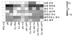


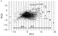
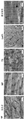

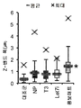
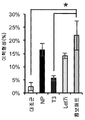


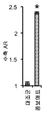


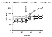
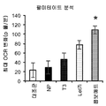
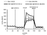


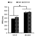

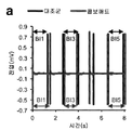

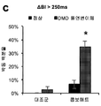

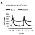

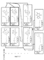
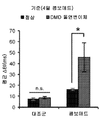
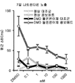


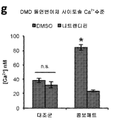
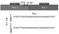

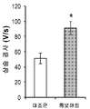


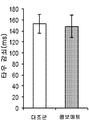
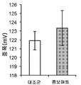
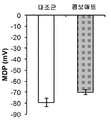

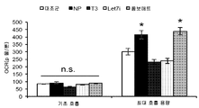
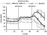

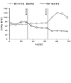
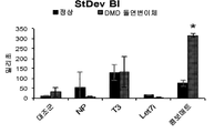

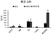
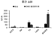
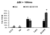

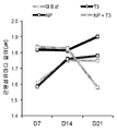
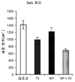
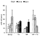
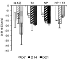
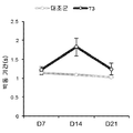
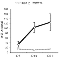
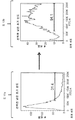


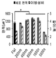

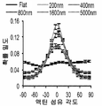


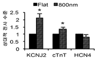
Classifications
-
- G—PHYSICS
- G01—MEASURING; TESTING
- G01N—INVESTIGATING OR ANALYSING MATERIALS BY DETERMINING THEIR CHEMICAL OR PHYSICAL PROPERTIES
- G01N33/00—Investigating or analysing materials by specific methods not covered by groups G01N1/00 - G01N31/00
- G01N33/48—Biological material, e.g. blood, urine; Haemocytometers
- G01N33/50—Chemical analysis of biological material, e.g. blood, urine; Testing involving biospecific ligand binding methods; Immunological testing
- G01N33/5005—Chemical analysis of biological material, e.g. blood, urine; Testing involving biospecific ligand binding methods; Immunological testing involving human or animal cells
- G01N33/5008—Chemical analysis of biological material, e.g. blood, urine; Testing involving biospecific ligand binding methods; Immunological testing involving human or animal cells for testing or evaluating the effect of chemical or biological compounds, e.g. drugs, cosmetics
- G01N33/5044—Chemical analysis of biological material, e.g. blood, urine; Testing involving biospecific ligand binding methods; Immunological testing involving human or animal cells for testing or evaluating the effect of chemical or biological compounds, e.g. drugs, cosmetics involving specific cell types
- G01N33/5061—Muscle cells
-
- C—CHEMISTRY; METALLURGY
- C12—BIOCHEMISTRY; BEER; SPIRITS; WINE; VINEGAR; MICROBIOLOGY; ENZYMOLOGY; MUTATION OR GENETIC ENGINEERING
- C12N—MICROORGANISMS OR ENZYMES; COMPOSITIONS THEREOF; PROPAGATING, PRESERVING, OR MAINTAINING MICROORGANISMS; MUTATION OR GENETIC ENGINEERING; CULTURE MEDIA
- C12N5/00—Undifferentiated human, animal or plant cells, e.g. cell lines; Tissues; Cultivation or maintenance thereof; Culture media therefor
- C12N5/06—Animal cells or tissues; Human cells or tissues
- C12N5/0602—Vertebrate cells
- C12N5/0652—Cells of skeletal and connective tissues; Mesenchyme
- C12N5/0657—Cardiomyocytes; Heart cells
-
- G—PHYSICS
- G01—MEASURING; TESTING
- G01N—INVESTIGATING OR ANALYSING MATERIALS BY DETERMINING THEIR CHEMICAL OR PHYSICAL PROPERTIES
- G01N33/00—Investigating or analysing materials by specific methods not covered by groups G01N1/00 - G01N31/00
- G01N33/48—Biological material, e.g. blood, urine; Haemocytometers
- G01N33/50—Chemical analysis of biological material, e.g. blood, urine; Testing involving biospecific ligand binding methods; Immunological testing
- G01N33/5005—Chemical analysis of biological material, e.g. blood, urine; Testing involving biospecific ligand binding methods; Immunological testing involving human or animal cells
- G01N33/5008—Chemical analysis of biological material, e.g. blood, urine; Testing involving biospecific ligand binding methods; Immunological testing involving human or animal cells for testing or evaluating the effect of chemical or biological compounds, e.g. drugs, cosmetics
- G01N33/5014—Chemical analysis of biological material, e.g. blood, urine; Testing involving biospecific ligand binding methods; Immunological testing involving human or animal cells for testing or evaluating the effect of chemical or biological compounds, e.g. drugs, cosmetics for testing toxicity
-
- G—PHYSICS
- G01—MEASURING; TESTING
- G01N—INVESTIGATING OR ANALYSING MATERIALS BY DETERMINING THEIR CHEMICAL OR PHYSICAL PROPERTIES
- G01N33/00—Investigating or analysing materials by specific methods not covered by groups G01N1/00 - G01N31/00
- G01N33/48—Biological material, e.g. blood, urine; Haemocytometers
- G01N33/50—Chemical analysis of biological material, e.g. blood, urine; Testing involving biospecific ligand binding methods; Immunological testing
- G01N33/5005—Chemical analysis of biological material, e.g. blood, urine; Testing involving biospecific ligand binding methods; Immunological testing involving human or animal cells
- G01N33/5008—Chemical analysis of biological material, e.g. blood, urine; Testing involving biospecific ligand binding methods; Immunological testing involving human or animal cells for testing or evaluating the effect of chemical or biological compounds, e.g. drugs, cosmetics
- G01N33/502—Chemical analysis of biological material, e.g. blood, urine; Testing involving biospecific ligand binding methods; Immunological testing involving human or animal cells for testing or evaluating the effect of chemical or biological compounds, e.g. drugs, cosmetics for testing non-proliferative effects
-
- G—PHYSICS
- G01—MEASURING; TESTING
- G01N—INVESTIGATING OR ANALYSING MATERIALS BY DETERMINING THEIR CHEMICAL OR PHYSICAL PROPERTIES
- G01N33/00—Investigating or analysing materials by specific methods not covered by groups G01N1/00 - G01N31/00
- G01N33/48—Biological material, e.g. blood, urine; Haemocytometers
- G01N33/50—Chemical analysis of biological material, e.g. blood, urine; Testing involving biospecific ligand binding methods; Immunological testing
- G01N33/5005—Chemical analysis of biological material, e.g. blood, urine; Testing involving biospecific ligand binding methods; Immunological testing involving human or animal cells
- G01N33/5008—Chemical analysis of biological material, e.g. blood, urine; Testing involving biospecific ligand binding methods; Immunological testing involving human or animal cells for testing or evaluating the effect of chemical or biological compounds, e.g. drugs, cosmetics
- G01N33/5044—Chemical analysis of biological material, e.g. blood, urine; Testing involving biospecific ligand binding methods; Immunological testing involving human or animal cells for testing or evaluating the effect of chemical or biological compounds, e.g. drugs, cosmetics involving specific cell types
- G01N33/5073—Stem cells
-
- A—HUMAN NECESSITIES
- A61—MEDICAL OR VETERINARY SCIENCE; HYGIENE
- A61K—PREPARATIONS FOR MEDICAL, DENTAL OR TOILETRY PURPOSES
- A61K35/00—Medicinal preparations containing materials or reaction products thereof with undetermined constitution
-
- C—CHEMISTRY; METALLURGY
- C12—BIOCHEMISTRY; BEER; SPIRITS; WINE; VINEGAR; MICROBIOLOGY; ENZYMOLOGY; MUTATION OR GENETIC ENGINEERING
- C12N—MICROORGANISMS OR ENZYMES; COMPOSITIONS THEREOF; PROPAGATING, PRESERVING, OR MAINTAINING MICROORGANISMS; MUTATION OR GENETIC ENGINEERING; CULTURE MEDIA
- C12N2501/00—Active agents used in cell culture processes, e.g. differentation
- C12N2501/30—Hormones
- C12N2501/375—Thyroid stimulating hormone [TSH]
-
- C—CHEMISTRY; METALLURGY
- C12—BIOCHEMISTRY; BEER; SPIRITS; WINE; VINEGAR; MICROBIOLOGY; ENZYMOLOGY; MUTATION OR GENETIC ENGINEERING
- C12N—MICROORGANISMS OR ENZYMES; COMPOSITIONS THEREOF; PROPAGATING, PRESERVING, OR MAINTAINING MICROORGANISMS; MUTATION OR GENETIC ENGINEERING; CULTURE MEDIA
- C12N2501/00—Active agents used in cell culture processes, e.g. differentation
- C12N2501/65—MicroRNA
-
- C—CHEMISTRY; METALLURGY
- C12—BIOCHEMISTRY; BEER; SPIRITS; WINE; VINEGAR; MICROBIOLOGY; ENZYMOLOGY; MUTATION OR GENETIC ENGINEERING
- C12N—MICROORGANISMS OR ENZYMES; COMPOSITIONS THEREOF; PROPAGATING, PRESERVING, OR MAINTAINING MICROORGANISMS; MUTATION OR GENETIC ENGINEERING; CULTURE MEDIA
- C12N2501/00—Active agents used in cell culture processes, e.g. differentation
- C12N2501/999—Small molecules not provided for elsewhere
-
- C—CHEMISTRY; METALLURGY
- C12—BIOCHEMISTRY; BEER; SPIRITS; WINE; VINEGAR; MICROBIOLOGY; ENZYMOLOGY; MUTATION OR GENETIC ENGINEERING
- C12N—MICROORGANISMS OR ENZYMES; COMPOSITIONS THEREOF; PROPAGATING, PRESERVING, OR MAINTAINING MICROORGANISMS; MUTATION OR GENETIC ENGINEERING; CULTURE MEDIA
- C12N2503/00—Use of cells in diagnostics
- C12N2503/02—Drug screening
-
- C—CHEMISTRY; METALLURGY
- C12—BIOCHEMISTRY; BEER; SPIRITS; WINE; VINEGAR; MICROBIOLOGY; ENZYMOLOGY; MUTATION OR GENETIC ENGINEERING
- C12N—MICROORGANISMS OR ENZYMES; COMPOSITIONS THEREOF; PROPAGATING, PRESERVING, OR MAINTAINING MICROORGANISMS; MUTATION OR GENETIC ENGINEERING; CULTURE MEDIA
- C12N2506/00—Differentiation of animal cells from one lineage to another; Differentiation of pluripotent cells
- C12N2506/02—Differentiation of animal cells from one lineage to another; Differentiation of pluripotent cells from embryonic cells
-
- C—CHEMISTRY; METALLURGY
- C12—BIOCHEMISTRY; BEER; SPIRITS; WINE; VINEGAR; MICROBIOLOGY; ENZYMOLOGY; MUTATION OR GENETIC ENGINEERING
- C12N—MICROORGANISMS OR ENZYMES; COMPOSITIONS THEREOF; PROPAGATING, PRESERVING, OR MAINTAINING MICROORGANISMS; MUTATION OR GENETIC ENGINEERING; CULTURE MEDIA
- C12N2506/00—Differentiation of animal cells from one lineage to another; Differentiation of pluripotent cells
- C12N2506/45—Differentiation of animal cells from one lineage to another; Differentiation of pluripotent cells from artificially induced pluripotent stem cells
-
- C—CHEMISTRY; METALLURGY
- C12—BIOCHEMISTRY; BEER; SPIRITS; WINE; VINEGAR; MICROBIOLOGY; ENZYMOLOGY; MUTATION OR GENETIC ENGINEERING
- C12N—MICROORGANISMS OR ENZYMES; COMPOSITIONS THEREOF; PROPAGATING, PRESERVING, OR MAINTAINING MICROORGANISMS; MUTATION OR GENETIC ENGINEERING; CULTURE MEDIA
- C12N2510/00—Genetically modified cells
-
- C—CHEMISTRY; METALLURGY
- C12—BIOCHEMISTRY; BEER; SPIRITS; WINE; VINEGAR; MICROBIOLOGY; ENZYMOLOGY; MUTATION OR GENETIC ENGINEERING
- C12N—MICROORGANISMS OR ENZYMES; COMPOSITIONS THEREOF; PROPAGATING, PRESERVING, OR MAINTAINING MICROORGANISMS; MUTATION OR GENETIC ENGINEERING; CULTURE MEDIA
- C12N2535/00—Supports or coatings for cell culture characterised by topography
- C12N2535/10—Patterned coating
-
- G—PHYSICS
- G01—MEASURING; TESTING
- G01N—INVESTIGATING OR ANALYSING MATERIALS BY DETERMINING THEIR CHEMICAL OR PHYSICAL PROPERTIES
- G01N2800/00—Detection or diagnosis of diseases
- G01N2800/32—Cardiovascular disorders
Abstract
본 명세서에서 개시되는 바와 같은 방법 및 조성물은 다른 용도 중에서도 질환 모델링, 심독성 스크리닝, 약물 스크리닝 및 동정과 같은 적용을 위한 성숙한 줄기세포-유래 심근세포의 제조를 기술한다. 본 발명의 방법은 줄기세포-유래 심근세포가 태아 표현형에서 성인 심근세포의 것과 매우 유사한 보다 성숙한 표현형으로의 전이를 촉진하는 물리적 및 생화학적 단서를 포함한다.The methods and compositions as disclosed herein describe the production of mature stem cell-derived cardiomyocytes for applications such as disease modeling, cardiotoxicity screening, drug screening and identification, among other uses. The methods of the present invention include physical and biochemical cues that promote the metastasis of stem cell-derived cardiomyocytes from a fetal phenotype to a more mature phenotype very similar to that of adult cardiomyocytes.

Description
관련 출원에 대한 상호 참조Cross reference to related applications
본 출원은, 2017년 8월 16일자 출원된 미국 가출원 제 62/546,438 호에 대한 35 U.S.C. § 119(e)에 따른 우선권을 주장하며, 그의 전문은 본 명세서에서 참조로 포함된다.The present application is 35 U.S.C. to U.S. Provisional Application No. 62 / 546,438, filed August 16, 2017. Priority is claimed pursuant to § 119 (e), the entirety of which is incorporated herein by reference.
본 발명은 심장 기능 및 질환에 대한 시험관내 모델에 관한 것이다.The present invention relates to an in vitro model for heart function and disease.
심혈관 질환은 전세계 남성과 여성 모두에게 사망의 주요 원인이 되고 있으며, 개발 도상국에 급속히 영향을 미치고 있다. 유전성 심근병증은 어린이와 젊은이, 그리고 건강한 성인을 포함한 모든 연령대에서 심장 질환의 주요 원인이다. 인간 만능 줄기세포-유래 심근세포(hPSC-CMs)는 심근병증 연구를 위한 유망한 도구로, 이는 그들이 인간 심장에서 발견되는 이온 채널, 단백질 이소형(protein isoform), 유전자 및 대사기구의 전체 어레이(full array)를 발현할 수 있는 인간 심근세포에 대한 접근성을 크게 증가시키기 때문이다.Cardiovascular disease is the leading cause of death for both men and women worldwide and is rapidly affecting developing countries. Hereditary cardiomyopathy is the leading cause of heart disease in all ages, including children, young adults, and healthy adults. Human pluripotent stem cell-derived cardiomyocytes (hPSC-CMs) are promising tools for the study of cardiomyopathy, a full array of ion channels, protein isoforms, genes and metabolic mechanisms they find in the human heart. array), which greatly increases the accessibility to human cardiomyocytes.
인간 만능 줄기세포(hPSCs)의 심근세포로의 지시된 분화는 전형적으로는 태아 심장에 존재하는 것들과 가장 유사한 구조적, 기능적 및 생화학적 특성을 가진 세포를 생성한다. 그러나, 새롭게 분화된 hPSCs와 비교하였을 때 향상된 근원섬유마디 발달(enhanced sarcomere development), 향상된 수축 기능, 감소된 심박동수, 향상된 미토콘드리아 용량, 및 보다 성숙한 전사체를 포함하는 성인 심근세포 표현형을 갖는 hPSC-CMs을 개발할 필요성이 있다. 현재의 심장 질환 모델링에 대한 노력의 주요 목표는 보다 효과적인 치료 전략을 설계하기 위한 첫 단계로서 심근병증의 발병 및 진행에 대한 통찰력을 얻는 것이다. 본원에 기술된 방법 이전에는, 뒤시엔느 근육 디스트로피(Duchenne muscular dystrophy)와 같은 성인-발병 근육 장애 및 심혈관 질환은 hPSC-CMs에 의해 정확하게 모델링되지 않았다. 이는 질병 모델링 및 약물 스크리닝 응용 분야에서 사용되는 줄기세포 분화 및 성숙 방법에 대한 개선의 필요성을 강조하는 것이다.Directed differentiation of human pluripotent stem cells (hPSCs) into cardiomyocytes typically produces cells with structural, functional and biochemical properties most similar to those present in the fetal heart. However, when compared to newly differentiated hPSCs, hPSC- with adult myocardial cell phenotypes including improved sarcomere development, improved contractile function, reduced heart rate, improved mitochondrial capacity, and more mature transcripts There is a need to develop CMs. The main goal of current cardiac disease modeling efforts is to gain insight into the development and progression of cardiomyopathy as a first step in designing more effective treatment strategies. Prior to the methods described herein, adult-onset muscle disorders and cardiovascular diseases such as Duchenne muscular dystrophy were not accurately modeled by hPSC-CMs. This highlights the need for improvements to stem cell differentiation and maturation methods used in disease modeling and drug screening applications.
본 명세서에서는 성숙한 줄기세포-유래 심근세포를 생성하기 위해 발달하고 있는 심장 니치 플랫폼(developmental cardiac niche platform)을 조작하는 방법이 기술된다. 이러한 방법은 줄기세포-유래 심근세포를 태아 표현형에서 성인 심근세포의 표현형과 보다 더 유사한 보다 성숙한 표현형으로의 전이를 촉진하는 물리적 및 생화학적 단서(cue)를 포함한다.Described herein is a method of manipulating a developing cardiac niche platform to generate mature stem cell-derived cardiomyocytes. These methods include physical and biochemical cues that promote the transfer of stem cell-derived cardiomyocytes from a fetal phenotype to a more mature phenotype more similar to that of adult cardiomyocytes.
본 명세서에서 기술되는 접근법은 줄기세포로부터 시험관내에서 분화된 보다 성숙한 심근세포를 제공함으로써, 다른 용도들 중에서도 심근세포 또는 심장 기능을 조절하는 새로운 약물을 동정하기 위해 심근 독성 및 스크리닝을 위한 약물을 평가하기 위한 플랫폼을 제공한다. 시험관내 분화 전에 줄기세포를 유전적으로 변형시키는 능력뿐만 아니라 정상 개체(subject) 및 질환이 있는 개체로부터 줄기세포, 예를 들면, 유도성 만능 줄기세포(induced pluripotent stem cells)(iPS 세포)를 생성하는 능력은 개선된 질병 모델링 및 약물 스크리닝 및 동정을 위한 플랫폼을 제공한다.The approach described herein evaluates drugs for myocardial toxicity and screening to identify new drugs that modulate cardiomyocytes or cardiac function, among other uses, by providing more mature cardiomyocytes differentiated in vitro from stem cells in vitro. It provides a platform to do. The ability to genetically modify stem cells prior to in vitro differentiation, as well as to generate stem cells, e.g. induced pluripotent stem cells (iPS cells) from normal subjects and diseased individuals. The ability provides a platform for improved disease modeling and drug screening and identification.
따라서, 하나의 양태에서, 본 명세서에서는 줄기세포-유래 심근세포를 제조하는 방법이 기술되며, 상기 방법은 줄기세포-유래 심근세포를 나노패턴화 기재; 갑상선 호르몬 T3; 및 Let7i microRNA와 접촉시키는 단계를 포함한다.Thus, in one embodiment, a method for producing stem cell-derived cardiomyocytes is described herein, wherein the method comprises nano-patterning stem cell-derived cardiomyocytes; Thyroid hormone T3; And contacting Let7i microRNA.
하나의 실시형태에서, 나노패턴화 기재는 그루브(groove) 및 릿지(ridge)의 실질적으로 평행한 어레이를 가진 나노패턴화된 표면을 포함한다.In one embodiment, the nanopatterned substrate comprises a nanopatterned surface with a substantially parallel array of grooves and ridges.
하나의 실시형태에서, 각각의 그루브 또는 릿지의 치수는 길이, 폭 또는 높이가 1000 나노미터 미만이다.In one embodiment, the dimensions of each groove or ridge are less than 1000 nanometers in length, width or height.
하나의 실시형태에서, 그루브 및 릿지의 폭은 약 800 nm이고, 그루브의 깊이는 약 600 nm이다.In one embodiment, the width of the groove and ridge is about 800 nm, and the depth of the groove is about 600 nm.
하나의 실시형태에서, Let7i microRNA는 줄기세포-유래 심근세포에 의해 발현된다.In one embodiment, Let7i microRNA is expressed by stem cell-derived cardiomyocytes.
하나의 실시형태에서, 심근세포를 Let7i microRNA와 접촉시키는 단계는 줄기세포-유래 심근세포를 바이러스 벡터와 접촉시키는 단계를 포함한다.In one embodiment, contacting the cardiomyocyte with Let7i microRNA comprises contacting the stem cell-derived cardiomyocyte with a viral vector.
하나의 실시형태에서, 줄기세포-유래 심근세포는 인간 심근세포이다.In one embodiment, the stem cell-derived cardiomyocyte is a human cardiomyocyte.
하나의 실시형태에서, 줄기세포-유래 심근세포는 유도성 만능 줄기세포(iPS 세포) 또는 배아 줄기세포로부터 분화된다.In one embodiment, stem cell-derived cardiomyocytes are differentiated from inducible pluripotent stem cells (iPS cells) or embryonic stem cells.
하나의 실시형태에서, 줄기세포-유래 심근세포는 근육 질환 또는 장애를 가진 개체로부터 유래된다.In one embodiment, stem cell-derived cardiomyocytes are derived from an individual with a muscle disease or disorder.
하나의 실시형태에서, 줄기세포-유래 심근세포는 유전자 변형된다.In one embodiment, stem cell-derived cardiomyocytes are genetically modified.
하나의 실시형태에서, 심근세포는 Let7i microRNA를 인코딩하는 벡터와 접촉한 후 나노패턴화 기재 및 갑상선 호르몬 T3과 접촉된다.In one embodiment, the cardiomyocytes are contacted with a vector encoding a Let7i microRNA, followed by a nanopatterning substrate and thyroid hormone T3.
하나의 실시형태에서, 생성된 줄기세포-유래 심근세포는 나노패턴화 기재, 갑상선 호르몬 T3 및 Let7i microRNA와 접촉하기 이전의 줄기세포-유래 심근세포와 비교하였을 때 더욱 성숙한 심근세포 표현형을 갖는다.In one embodiment, the resulting stem cell-derived cardiomyocytes have a more mature myocardial phenotype compared to stem cell-derived cardiomyocytes prior to contact with nanopatterned substrates, thyroid hormone T3 and Let7i microRNA.
하나의 실시형태에서, 나노패턴화 기재는 심근세포 전기 생리학적 특성의 전기적 자극 및/또는 측정을 허용하는 마이크로 전극 어레이를 포함한다.In one embodiment, the nanopatterned substrate comprises an array of microelectrodes that allow electrical stimulation and / or measurement of cardiomyocyte electrophysiological properties.
또 다른 양태에서, 본 명세서에서는 줄기세포-유래 심근세포를 성숙시키는 방법이 기술되며, 상기 방법은 줄기세포-유래 심근세포를 나노패턴화 기재; 갑상선 호르몬 T3; 및 Let7i microRNA와 접촉시키는 단계를 포함한다.In another aspect, a method of maturing stem cell-derived cardiomyocytes is described herein, the method comprising: nano-patterning stem cell-derived cardiomyocytes; Thyroid hormone T3; And contacting Let7i microRNA.
하나의 실시형태에서, 나노패턴화 기재는 그루브 및 릿지의 실질적으로 평행한 어레이를 가진 나노패턴화된 표면을 포함한다.In one embodiment, the nanopatterned substrate comprises a nanopatterned surface with a substantially parallel array of grooves and ridges.
하나의 실시형태에서, 각각의 그루브 또는 릿지의 치수는 길이, 폭 또는 높이가 1000 나노미터 미만이다.In one embodiment, the dimensions of each groove or ridge are less than 1000 nanometers in length, width or height.
하나의 실시형태에서, 그루브 및 릿지의 폭은 약 800 nm이고, 그루브의 깊이는 약 600 nm이다.In one embodiment, the width of the groove and ridge is about 800 nm, and the depth of the groove is about 600 nm.
하나의 실시형태에서, Let7i microRNA는 줄기세포-유래 심근세포에 의해 발현된다.In one embodiment, Let7i microRNA is expressed by stem cell-derived cardiomyocytes.
하나의 실시형태에서, 심근세포를 Let7i microRNA와 접촉시키는 단계는 줄기세포-유래 심근세포를 바이러스 벡터와 접촉시키는 단계를 포함한다.In one embodiment, contacting the cardiomyocyte with Let7i microRNA comprises contacting the stem cell-derived cardiomyocyte with a viral vector.
하나의 실시형태에서, 줄기세포-유래 심근세포는 인간 심근세포이다.In one embodiment, the stem cell-derived cardiomyocyte is a human cardiomyocyte.
하나의 실시형태에서, 줄기세포-유래 심근세포는 유도성 만능 줄기세포(iPS 세포) 또는 배아 줄기세포로부터 분화된다.In one embodiment, stem cell-derived cardiomyocytes are differentiated from inducible pluripotent stem cells (iPS cells) or embryonic stem cells.
하나의 실시형태에서, 줄기세포-유래 심근세포는 근육 질환 또는 장애를 가진 개체로부터 유래된다.In one embodiment, stem cell-derived cardiomyocytes are derived from an individual with a muscle disease or disorder.
하나의 실시형태에서, 줄기세포-유래 심근세포는 유전자 변형된다.In one embodiment, stem cell-derived cardiomyocytes are genetically modified.
하나의 실시형태에서, 심근세포는 Let7i microRNA를 인코딩하는 벡터와 접촉한 후 나노패턴화 기재 및 갑상선 호르몬 T3과 접촉된다.In one embodiment, the cardiomyocytes are contacted with a vector encoding a Let7i microRNA, followed by a nanopatterning substrate and thyroid hormone T3.
하나의 실시형태에서, 생성된 줄기세포-유래 심근세포는 나노패턴화 기재, 갑상선 호르몬 T3 및 Let7i microRNA와 접촉하기 이전의 줄기세포-유래 심근세포와 비교하였을 때 보다 성숙한 심근세포 표현형을 갖는다.In one embodiment, the resulting stem cell-derived cardiomyocytes have a more mature myocardial phenotype compared to stem cell-derived myocardial cells prior to contact with the nanopatterned substrate, thyroid hormone T3 and Let7i microRNA.
하나의 실시형태에서, 나노패턴화 기재는 심근세포 전기 생리학적 특성의 전기적 자극 및/또는 측정을 허용하는 마이크로 전극 어레이를 포함한다.In one embodiment, the nanopatterned substrate comprises an array of microelectrodes that allow electrical stimulation and / or measurement of cardiomyocyte electrophysiological properties.
또 다른 양태에서, 본 명세서에서는 작용제(agent)의 심독성을 평가하는 방법이 기술되며, 상기 방법은 본 명세서에서 기술되는 방법에 의해 제조되는 줄기세포-유래 심근세포를 작용제와 접촉시키는 단계를 포함한다.In another aspect, described herein is a method for evaluating cardiotoxicity of an agent, the method comprising contacting a stem cell-derived cardiomyocyte prepared by the method described herein with an agent. .
하나의 실시형태에서, 상기 방법은 심근세포의 적어도 하나의 표현형 특성을 검출하는 단계를 포함한다.In one embodiment, the method comprises detecting at least one phenotypic characteristic of the cardiomyocytes.
하나의 실시형태에서, 작용제는 소분자, 항체, 펩티드, 게놈 편집 시스템 및 핵산으로 이루어진 군 중에서 선택된다.In one embodiment, the agent is selected from the group consisting of small molecules, antibodies, peptides, genomic editing systems and nucleic acids.
하나의 실시형태에서, 작용제의 심독성은 세포 생존력, 세포 크기, 근원섬유마디 길이, 조직내 근원섬유마디의 조직, 줄기세포-유래 심근세포의 집단의 생체 전위 또는 전기적 특성, 미토콘드리아 기능, 유전자 발현, 심박동수, 박동 강도, 및 수축성 중의 하나 이상에 대한 작용제의 효과에 의해 지시된다.In one embodiment, the cardiotoxicity of the agent is cell viability, cell size, myofibrillar length, tissue of myofibroblasts in tissue, biopotential or electrical properties of a population of stem cell-derived myocardial cells, mitochondrial function, gene expression, It is indicated by the effect of the agonist on one or more of heart rate, heart rate, and contractility.
또 다른 양태에서, 본 명세서에서는 심근세포의 기능적 특성을 조절하는 작용제를 동정하기 위한 분석법이 기술되며, 상기 분석법은 본 명세서에서 기술되는 방법에 의해 제조되는 줄기세포-유래 심근세포의 집단을 후보 작용제와 접촉시키는 단계; 및 심근세포의 적어도 하나의 기능적 특성을 검출하는 단계를 포함하며, 여기서 심근세포의 적어도 하나의 기능적 특성에 있어서의 변화를 검출하는 것은 심근세포의 기능적 특성을 조절할 수 있는 것으로 작용제를 동정한다.In another aspect, an analytical method for identifying an agent that modulates functional properties of cardiomyocytes is described herein, wherein the analytical method is a candidate agent for a population of stem cell-derived cardiomyocytes prepared by the method described herein. Contacting with; And detecting at least one functional property of the myocardial cell, wherein detecting a change in at least one functional property of the myocardial cell identifies the agent as being capable of modulating the functional property of the myocardial cell.
하나의 실시형태에서, 검출 단계는 세포 생존력, 세포 크기, 근원섬유마디 길이, 조직내 근원섬유마디의 조직, 생체전위 또는 전기적 특성, 미토콘드리아 기능, 유전자 발현, 심박동수, 박동 강도, 및 수축성 중의 적어도 하나를 검출하는 것을 포함한다.In one embodiment, the detecting step comprises at least one of cell viability, cell size, myofibrillar length, tissue of myofibrillar tissue, biopotential or electrical properties, mitochondrial function, gene expression, heart rate, heartbeat intensity, and contractility. And detecting one.
또 다른 양태에서, 본 명세서에서는 본 명세서에서 기술되는 방법에 의해 제조되는 줄기세포-유래 심근세포를 포함하는 질환 모델이 기술되며, 여기서 상기 줄기세포는 근육 질환 또는 장애를 가진 개체로부터 유래되거나, 또는 줄기세포-유래 심근세포 또는 이들이 유래되는 줄기세포는 심근세포가 질환 표현형을 발현하도록 유전자 변형된다.In another aspect, described herein is a disease model comprising stem cell-derived cardiomyocytes prepared by a method described herein, wherein the stem cells are derived from an individual with a muscle disease or disorder, or Stem cell-derived cardiomyocytes or stem cells from which they are derived are genetically modified such that the cardiomyocytes express a disease phenotype.
하나의 실시형태에서, 근육 질환 또는 장애는 심장 기능장애를 가진 표현형을 갖는다.In one embodiment, the muscle disease or disorder has a phenotype with heart dysfunction.
하나의 실시형태에서, 근육 질환 또는 장애는 심장 질환 표현형의 성인 발병을 특징으로한다.In one embodiment, the muscle disease or disorder is characterized by an adult onset of the heart disease phenotype.
하나의 실시형태에서, 근육 질환 또는 장애는 뒤시엔느 근육 디스트로피가다.In one embodiment, the muscle disease or disorder is Duchenne muscular dystrophy.
또 다른 양태에서, 본 명세서에서는 나노패턴화 기재 상에 줄기세포-유래 심근세포를 포함하는 조성물이 기술되며, 상기 조성물은 갑상선 호르몬 T3 및 Let7i microRNA를 추가로 포함한다.In another aspect, a composition comprising stem cell-derived cardiomyocytes on a nanopatterned substrate is described herein, wherein the composition further comprises thyroid hormone T3 and Let7i microRNA.
하나의 실시형태에서, 줄기세포-유래 심근세포는 근육 질환 또는 장애를 가진 개체로부터 유래된다.In one embodiment, stem cell-derived cardiomyocytes are derived from an individual with a muscle disease or disorder.
하나의 실시형태에서, 심근세포는 인간 심근세포이다.In one embodiment, the cardiomyocyte is a human cardiomyocyte.
하나의 실시형태에서, 줄기세포-유래 심근세포 또는 이들이 유래되는 줄기세포는 줄기세포-유래 심근세포가 심장 기능장애 표현형을 나타내도록 유전자 변형된다.In one embodiment, the stem cell-derived cardiomyocytes or stem cells from which they are derived are genetically modified such that the stem cell-derived cardiomyocytes exhibit a cardiac dysfunction phenotype.
또 다른 양태에서, 본 명세서에서는 시험관내-분화된 심근세포를 나노패턴화 기재, 갑상선 호르몬 T3 및 Let7i microRNA와 접촉시킴으로써 제조된 심근세포를 포함하는 조성물이 기술되며, 여기서 상기 심근세포는 나노패턴화 기재, 갑상선 호르몬 T3 및 Let7i microRNA와 접촉하지 않은 시험관내-분화된 심근세포와 비교하였을 때 보다 성숙한 표현형을 갖는다.In another aspect, described herein is a composition comprising cardiomyocytes prepared by contacting in vitro-differentiated cardiomyocytes with a nanopatterning substrate, thyroid hormone T3 and Let7i microRNA, wherein the cardiomyocytes are nanopatterned It has a more mature phenotype compared to in vitro-differentiated cardiomyocytes that have not been contacted with the substrate, thyroid hormone T3 and Let7i microRNA.
하나의 실시형태에서, 심근세포는 근육 질환 또는 장애를 가진 개체로부터 유래된다.In one embodiment, the cardiomyocyte is derived from an individual with a muscle disease or disorder.
또 다른 양태에서, 본 명세서에서는 시험관내-분화된 심근세포 또는 그들이 분화되는 줄기세포가, 그들이 심장 기능장애 표현형을 나타내도록 유전적으로 변형된 조성물이 기술된다.In another aspect, described herein are compositions in which the in vitro-differentiated cardiomyocytes or stem cells to which they differentiate are genetically modified such that they exhibit a cardiac dysfunction phenotype.
또 다른 양태에서, 본 명세서에서는 줄기세포-유래 심근세포, 나노패턴화 기재, 갑상선 호르몬 T3, Let7i microRNA를 인코딩하는 벡터, 및 이들을 패키징하는 물질을 포함하는 키트가 기술된다.In another aspect, described herein is a kit comprising stem cell-derived cardiomyocytes, a nanopatterning substrate, a thyroid hormone T3, a vector encoding a Let7i microRNA, and a material packaging them.
하나의 실시형태에서, 상기 키트는 세포 배양 배지 및 줄기세포-유래 심근세포로부터 성숙한 시험관내 분화된 심근세포의 제조를 가능하게 하는 설명서를 포함한다.In one embodiment, the kit includes a cell culture medium and instructions that enable the production of mature in vitro differentiated cardiomyocytes from stem cell-derived cardiomyocytes.
하나의 실시형태에서, 나노패턴화 기재는 그루브 및 릿지의 실질적으로 평행한 어레이를 가진 나노패턴화된 표면을 포함한다.In one embodiment, the nanopatterned substrate comprises a nanopatterned surface with a substantially parallel array of grooves and ridges.
하나의 실시형태에서, 각각의 그루브 또는 릿지의 치수는 길이, 폭 또는 높이가 1000 나노미터 미만이다.In one embodiment, the dimensions of each groove or ridge are less than 1000 nanometers in length, width or height.
하나의 실시형태에서, 그루브 및 릿지의 폭은 약 800 nm이며, 그루브의 깊이는 약 600 nm이다.In one embodiment, the width of the grooves and ridges is about 800 nm, and the depth of the grooves is about 600 nm.
하나의 실시형태에서, 줄기세포-유래 심근세포는 인간 심근세포이다.In one embodiment, the stem cell-derived cardiomyocyte is a human cardiomyocyte.
하나의 실시형태에서, 줄기세포-유래 심근세포는 근육 질환 또는 장애를 가진 개체로부터 유래된다.In one embodiment, stem cell-derived cardiomyocytes are derived from an individual with a muscle disease or disorder.
하나의 실시형태에서, 심근세포는 나노패턴화 기재 상에 있다.In one embodiment, the cardiomyocyte is on a nanopatterned substrate.
하나의 실시형태에서, 줄기세포-유래 심근세포는 동결된다.In one embodiment, stem cell-derived cardiomyocytes are frozen.
하나의 실시형태에서, 키트는 실온 내지 4℃의 온도에서의 운송을 가능하게 한다.In one embodiment, the kit allows for transportation at temperatures between room temperature and 4 ° C.
또 다른 양태에서, 본 명세서에서는 줄기세포-유래 심근세포를 제조하는 방법이 기술되며, 상기 방법은 줄기세포-유래 심근세포를 그루브 및 릿지의 실질적으로 평행한 어레이(여기서 상기 그루브 및 릿지의 폭은 800 nm이며, 상기 그루브의 깊이는 600 nm이다); 갑상선 호르몬 T3; 및 Let7i microRNA를 포함하는 나노패턴화 기재과 접촉시키는 단계를 포함한다.In another aspect, described herein is a method of making stem cell-derived cardiomyocytes, the method comprising substantially parallel arrays of stem-derived cardiomyocytes with grooves and ridges, wherein the width of the grooves and ridges is 800 nm, the depth of the groove is 600 nm); Thyroid hormone T3; And contacting the nanopatterning substrate comprising Let7i microRNA.
또 다른 양태에서, 본 명세서에서는 나노패턴화 기재 상에 줄기세포-유래 심근세포를 포함하는 조성물이 기술되며, 상기 조성물은 그루브 및 릿지의 실질적으로 평행한 어레이(여기서 상기 그루브 및 릿지의 폭은 800 nm이며, 상기 그루브의 깊이는 600 nm이다); 갑상선 호르몬 T3; 및 Let7i microRNA를 포함하는 나노패턴화 기재를 추가로 포함한다.In another aspect, described herein is a composition comprising stem cell-derived cardiomyocytes on a nanopatterned substrate, wherein the composition is a substantially parallel array of grooves and ridges, wherein the width of the grooves and ridges is 800 nm, and the depth of the groove is 600 nm); Thyroid hormone T3; And a nanopatterning substrate comprising Let7i microRNA.
하나의 실시형태에서, 본 명세서에서는 나노패턴화 기재가 그 위에서 배양된 심근세포의 기계적 자극을 가능하게하는 엘라스토머성 기재를 포함하며, 상기 심근세포에 이러한 기계적 자극을 가하는 단계를 추가로 포함하는 방법이 기술된다.In one embodiment, the present specification includes a nano-patterned substrate comprising an elastomeric substrate that enables mechanical stimulation of cardiomyocytes cultured thereon, and further comprising applying such mechanical stimulation to the cardiomyocytes. It is described.
하나의 실시형태에서, 본 명세서에서 기술되는 조성물 또는 본 명세서에서 기술되는 키트에서, 나노패턴화 기재는 그 위에서 배양된 심근세포의 기계적 자극을 가능하게하는 엘라스토머성 기재를 포함한다.In one embodiment, in the compositions described herein or in the kits described herein, the nanopatterned substrate comprises an elastomeric substrate that enables mechanical stimulation of cardiomyocytes cultured thereon.
도 1a 내지 도 1f는 콤보매트 플랫폼(ComboMat plaform)이 보다 성숙한 전사 프로파일(transcriptional profile)을 촉진한다는 것을 나타낸다. 도 1a는 콤보매트 플랫폼의 개략적인 타임 라인(timeline)을 나타낸다. 심근세포 분화는 소분자 및 인간 재조합 단백질 기반 프로토콜을 사용하여 달성된다. 심근세포 생성 후, hPSC-CMs를 Let7i 렌티바이러스로 형질 감염시키고, 대사 시도를 통해 정제하고, 바이러스 형질 감염을 위해 선택한다. 이어서, 바이러스 벡터를 발현하는 고순도 hPSC-CMs를 NPs 상에 플레이팅한 다음 3주 동안 T3에 노출시킨다. 도 1b는 주요 성분 분석의 3 차원 시각화를 나타낸다. hPSC-CMs를 PC2를 따라 T3 클러스터에 노출시켰다. 콤보매트 그룹을 성숙 자극의 다른 조합으로부터 분리하고 구별한다. 도 1c는 상이한 처리의 결과로서 차별적으로 발현된 유전자의 심장 성숙 경로에서의 순 농축(net enrichment)을 나타낸다. 도 1d는 EV-Flat 대조군과 비교한 Let7i-NP-T3 조합 처리에서의 상향 조절된 유전자의 유전자 온톨로지(GO) 농축 결과를 나타낸다. 색상 변화율 및 막대 길이는 농축의 중요성을 나타낸다. 도 1e 및 도 1f는 EV-Flat을 Let7i-NP-T3(도 1e)와 비교하고 태아 심장 심근세포를 성인 심장 심근세포(도 1f)와 비교하는 RNAseq 샘플의 분리에 대한 유전자 기여를 보여주는 PCA 로딩의 버블 플롯을 도시한다. 도 1e 및 도 1f의 버블 플롯은 콤보매트-처리된 hPSC-CMs와 성인 CMs 사이의 특정 유전자의 유사한 상향/하향 조절 패턴을 강조한다.
도 2a 내지 도 2f는 hPSC-CMs의 구조적 성숙을 나타낸다. 도 2a는 콤보매트 프로토콜 또는 각각의 단서에 분리되어 노출된 hPSC-CMs의 면역형광 이미지를 나타낸다. 적색 = 알파-액티닌, 녹색 = F-액틴, 청색 = DAPI. 삽도(Inset)는 hPSC-CMs 내의 근원섬유마디 구조의 근접 사진을 나타낸다. 도 2b는 콤보매트 플랫폼의 표지된 단서에 노출된 hPSC-CMs로부터의 근원섬유마디의 TEM 이미지를 나타낸다. 대조군 조건에 노출된 hPSC-CMs는 단지 Z-소체(Z-body) 구조를 나타내는 반면, 콤보매트 플랫폼에 노출된 hPSC-CMs는 훨씬 더 한정된 Z-판(Z-disc) 구조를 갖는다. 도 2c 내지 도 2f는 콤보매트 프로토콜 또는 각각의 단서에 분리되어 노출된 hPSC-CMs에 대한 형태론적 변화의 정량화를 나타낸다. 도 2c는 주문형 2D 푸리에 변환 이미지 분석을 통해 측정된 근원섬유마디 길이를 나타낸다. 도 2d는 적색선으로 표시된 평균 Z-밴드 폭을 가진 TEM 이미지로부터 측정된 Z-밴드 폭의 상자-수염 그림(box-and-whisker plot)을 도시한다. 도 2e는 콤보매트 그룹이 생리적 이핵화 백분율에 근접하고 있음을 보여주는 이핵화된 세포의 막대 차트를 도시한다. 도 2f는 콤보매트 플랫폼에 대해 명확한 비대성 반응을 나타내는, 면역형광 이미지로부터 측정된 세포 면적을 나타낸다. 막대는 평균 ± S.E.M. *P<0.05를 나타낸다.
도 3a 내지 도 3g는 전기 기계적 성숙 및 대사성 성숙을 나타낸다. 도 3a는 주문형 CCQ 방법을 통해 측정하였을 때 대조군 또는 콤보매트 hPSC-CMs에 대해 전체 비디오 프레임에 걸쳐 평균화된 수축 속도의 흔적을 나타낸다. 피크 수축 속도 및 피크 이완 속도가 나타나 있다. 도 3b는 피크 수축 및 이완 속도의 정량화를 나타낸다. 도 3c는 대조군 또는 콤보매트 hPSC-CMs에 대한 수축각 분포 히스토그램을 나타낸다. 히스토그램 중앙에 있는 선은 평균 수축각을 나타내는 반면, 선의 길이는 분포가 평균과 정렬되는 정도를 나타낸다. 도 3d는 종향 및 횡방향에서의 수축의 크기의 수축 이방성비(anisotropic ratio)(AR)를 나타낸다. 도 3e는 대조군과 콤보매트 hPSC-CMs 사이의 FPD의 변화를 나타내는 대표적인 전계 전위 기록을 나타낸다. 도 3f는 FPD의 정량화를 나타내며, 도 3g는 콤보매트 플랫폼 또는 각각의 단서에 개별로 노출된 hPSC-CMs에 대한 분당 박동 수로 표현된 심박동수를 나타낸다. 도 3h는 팔미테이트를 사용한 지방산 스트레스 테스트에 대한 대표적인 OCR 트레이스를 나타낸다. 도 3i는 팔미테이트 첨가 후의 OCR의 최대 변화의 정량화를 나타낸다. 막대는 평균 ± S.E.M. *P<0.05를 나타낸다.
도 4a 내지 도 4e는 DMD 돌연변이체 hPSC-CMs가 정상 대조군과 유사하게 콤보매트 플랫폼에 반응한다는 것을 나타낸다. 도 4a는 정상 hPSC-CMs와 비교하여 대조군 또는 콤보매트 조건에 노출된 DMD 돌연변이체 hPSC-CMs의 해마 대사 프로파일을 나타낸다. 도 4b는 FCCP 첨가 후의 OCR의 최대 변화의 정량화를 나타낸다. 정상 및 DMD 돌연변이체 hPSC-CMs 둘 모두 콤보매트 플랫폼에 노출되었을 때 최대 호흡 능력에 있어서의 증가를 나타낸다. 도 4c 및 도 4d는 MEA 기반 전기 생리학적 측정값을 나타낸다. 도 4c는 정상 또는 DMD 돌연변이체 hPSC-CMs에 대한 분당 박동수로 표현된 심박동수를 나타낸다. 도 4d는 대조군 또는 콤보매트 조건에 노출된 정상 또는 DMD 돌연변이체 hPSC-CMs에 대한 평균 FPD를 나타낸다. 도 4e는 대조군(왼쪽) 및 콤보매트(오른쪽) 조건에 노출된 DMD 돌연변이체 hPSC-CMs의 면역형광 이미지를 나타낸다. 삽도는 각각의 조건에서의 근원섬유마디 구조의 근접 부분을 나타낸다. 막대는 평균 ± S.E.M. *P<0.05를 나타낸다.
도 5a 내지 도 5f는 내인성 DMD 질환 표현형이 노출된 것을 나타낸다. 도 5a는 하나 걸러서 박동 간격에 주석이 달린 대조군 또는 콤보매트 조건에 노출된 DMD 돌연변이체 hPSC-CMs로부터의 대표적인 전계 전위 기록을 나타낸다. 도 5b는 대조군 또는 콤보매트 조건에 노출된 정상 및 DMD 돌연변이체 hPSC-CMs에 대한 30회의 연속 박동에 대해 플롯팅된 박동 간격에 있어서의 변화(ΔBI)를 나타낸다. 성숙 DMD 돌연변이체 hPSC-CMs은 정상 hPSC-CMs에 비해 ΔBI에서 훨씬 더 큰 불안정성을 나타낸다. 도 5c는 대조군 또는 콤보매트 조건에 노출된 정상 또는 DMD 돌연변이체 hPSC-CMs에 대한 ΔBI > 250ms에서의 평균 박동 백분율을 나타낸다. 미성숙 정상 hPSC-CMs은 ΔBI > 250ms의 발생률을 전혀 나타내지 않았다. 도 5d는 총 30회 박동 기록에 대한 평균 ΔBI를 나타낸다. 미성숙 DMD 돌연변이체 hPSC-CMs는 MEAs 상에서 구별되는 질환 표현형을 전혀 나타내지 않는 반면, 콤보매트 플랫폼을 사용하여 성숙된 DMD 돌연변이체 hPSC-CMs는 정상 대조물과 유의하게 다르다. 도 5e는 대조군 또는 콤보매트 조건에 노출된 DMD 돌연변이체 hPSC-CMs의 대표적인 FurA-2 Ca2+ 이미징 트레이스를 나타낸다. 도 5f는 사이토졸(cytosol) 중에서의 이완기 Ca2+ 함량의 5F 정량화를 나타낸다. DMD 돌연변이체 hPSC-CMs은 콤보매트 플랫폼을 사용하여 성숙되었을 때 유의적으로 더 높은 사이토졸성 Ca2+ 함량을 나타낸다. 막대는 평균 ± S.E.M. *P<0.05를 나타낸다.
도 6a 내지 도 6f는 성숙한 hPSC-CMs을 사용한 DMD 심근병증 표현형 약물 스크린을 나타낸다. 도 6a는 삼투성 스트레스에 대한 반응에서 세포 사멸의 감소에 대해 Z-스코어로 등급을 매긴 약물 라이브러리를 나타낸다. 특정 화합물의 화학 구조가 또한 도시되어 있다. 도 6b는 대조군 또는 콤보매트 조건에 노출된 정상 및 DMD 돌연변이체 hPSC-CMs에 대한 기준선 평균 ΔBI의 정량화를 나타낸다. 도 6c 및 도 6d는 대조군 또는 콤보매트 조건에 노출된 정상 및 DMD 돌연변이체 hPSC-CMs에 대한 7일 후의 니트렌디핀 및 실데나필에 대한 반응에 있어서의 용량-반응 곡선을 나타낸다. 도 6e는 니트렌디핀 세척 48시간 후의 평균 ΔBI의 측정값을 나타낸다. 도 6f는 성숙 DMD 돌연변이체 hPSC-CMs에 대한 대표적인 Fura-2 Ca2+ 트레이스를 나타낸다. 도 6g는 니트렌디핀이 있을 때 및 없을 때의 이완기 Ca2+ 함량의 정량화를 나타낸다. 니트렌디핀은 성숙한 DMD 돌연변이체 hPSC-CMs에서 사이토졸성 Ca2+ 부하를 감소시키는데 도움이 된다. 막대는 평균 ± S.E.M. *P<0.05를 나타낸다.
도 7a 내지 도 7c는 디스트로핀 돌연변이체 분석을 나타낸다. 도 7a는 염기쌍 263에서 G 결실을 나타내는 정상(서열 번호: 18) 및 DMD 돌연변이체(서열 번호: 19) hPSC-CMs 둘 다에서 DMD 유전자 서열 분석을 위한 프라이머 디자인의 대표적인 표현을 나타낸다. 도 7b는 DMD 돌연변이체 hPSC-CMs의 bp263에서의 CRISPR 조작된 결실 주위의 DMD 유전자의 크로모그래프를 나타낸다. 도 7c는 엑손 1에서 프레임 시프트 결실의 결과로서 조기 정지 코돈(STOP codon)으로서 제안하는 정상(상단) 및 DMD 돌연변이체(하단) hiPSC-CM에서의 예측된 단백질 서열을 나타낸다.
도 8a 내지 도 8f는 패치 클램프 전기생리검사 결과를 나타낸다. 도 8a는 hPSC-CMs의 전류-클램프 전기생리학적 측정 결과를 나타낸다. 콤보매트 조건에 노출된 hPSC-CMs는 상당히 빠른 상행각 속도(upstroke velocity)를 나타내었지만, 모든 다른 파라미터들(도 8b 내지 도 8f)은 대조군과 콤보매트 hPSC-CMs 사이에서 통계적으로 다르지 않았다. 막대는 평균 ± S.E.M. *P<0.05를 나타낸다. 오르막 경사(Up Slope) = 활동 전위의 상행각 속도, RMP = 안정막 전위(resting membrane potential), APD90 = 90% 재분극의 활동 전위 지속시간, 타우 붕괴(Decay Tau) = 이완의 시간 상수, 진폭 = 활동 전위의 진폭, MDP = 최대 이완기 전위.
도 9a 내지 도 9e는 hPSC-CMs의 대사 프로파일링을 나타낸다. 도 9a는 미토스트리(MitoStree) 분석 도중의 ATP 합성효소 억제제 올리고마이신, 전자 수송 및 산화적 인산화(FCCP)의 언커플러, 및 전자 수송 사슬 차단제 로테논 및 안티마이신에 대한 반응에서의 대표적인 OCR 프로파일을 나타낸다. 도 9b는 기초 호흡 능력 및 최대 호흡 능력의 정량화를 나타낸다. 도 9c는 팔미테이트를 사용하는 지방산 스트레스 테스트에 대한 대표적인 OCR 트레이스를 나타낸다. 도 9d 및 도 9e는 정상 hiPSC-CMs에 대해 팔미테이트에 반응하는 OCR 변화의 정량화를 나타낸다. 도 9e는 콤보매트 hPSC-CMs를 사용하는 지방산 장애 및 BSA 대조군에 대한 대표적인 OCR 프로파일을 나타낸다. 세포들은 BSA 캐리어에 반응하지 않았다. 막대는 평균 ± S.E.M. *P<0.05를 나타낸다.
도 10a 내지 도 10f는 질환 상태 계층화에 전체 콤보매트가 필요하다는 것을 나타낸다. 길크리스트(Gilchrist) 등의 문헌25에 요약되어 있는 바와 같은 부정맥 메트릭스. 성숙 단서가 분리되어 사용되는 경우에는 정상 및 DMD 돌연변이 hPSC-CMs 사이에는 통계적 차이가 없지만, 콤보매트 프로토콜에 결합되는 경우에는 건강한 정상 및 질환 DMD 돌연변이 hPSC-CMs 사이에는 분리(separation)가 있다. 막대는 평균 ± S.E.M. *P<0.05를 나타낸다.
도 11a 내지 도 11f는 hPSC-CMs에서 장기 T3 노출의 효과를 나타낸다. 도 11a 및 도 11b는 면역 조직 화학을 통해 측정하였을 때 후처리후 7일, 14일 및 21일째에 측정된 hPSC-CMs의 근원섬유마디 길이 및 세포 면적에 대한 T3의 효과를 나타낸다. NPs와 T3를 단독으로 조합한 결과 근원섬유마디 구조 및 비대에 장기적으로 부정적인 영향을 미쳤다. 도 11c 및 도 11d는 후처리후 7일, 14일 및 21일째에 측정된 hPSC-CMs의 수축(도 11c) 및 이완 속도(도 11d)의 CCQ 측정값을 나타낸다. 다시 NPs와 T3를 단독으로 조합한 결과 수축 성능을 저해하였다. 도 11e 및 도 11f는 후처리후 7일, 14일 및 21일째에 대조군 또는 T3 조건에 노출되고 MEAs를 통해 측정된 hPSC-CMs의 전기 생리 학적 성능을 나타낸다. 도 11e는 T3 처리된 세포에서 D14일째에 절정에 도달한 박동 기간을 나타낸다. 도 11f는 T3에 단독으로 노출된 hPSC-CMs에서 실질적으로 증가된 심박동수 변동성을 나타낸다. 막대는 평균 ± S.E.M. *P<0.05를 나타낸다.
도 12a 내지 도 12e는 대사 유발을 통한 심근세포 정제를 나타낸다. 도 12a 및 도 12b는 저순도 심장 시작 집단에 대한 젖산 농축 이전(도 12a) 및 이후(도 12b)의 cTnT + 세포에 대한 유세포 분석 결과를 나타낸다. 도 12c는 IgG 아이소형 대조군 유세포 분석 결과가 cTnT 항체의 비특이적 결합을 전혀 나타내지 않음을 나타낸다. 도 12d 및 도 12e는 고순도 심장 시작 집단에 대한 젖산 농축 이전(도 12d) 및 이후(도 12e)의 cTnT + 세포에 대한 유세포 분석 결과를 나타낸다.
도 13a 내지 도 13g는 hPSC-CMs의 나노 지형 크기(nanotopographic size)-의존성 구조적 발달을 나타낸다. 도 13a는 다양한 치수를 가진 NP 표면의 주사 전자 현미경 사진(상단부), 다양한 치수의 NP 표면 상의 hPSC-CMs의 명시야 이미지(중간부) 및 다양한 치수의 NPs 상에서 배양된 hPSC-CMs의 공초점 면역 형광 이미지(하단부)를 나타낸다. 도 13b는 표지된 지형 치수 상에서 배양된 hPSC-CMs의 세포 면적 및 세포 이방성 비율을 나타낸다. 도 13c는 상이한 지형 치수 상에서의 hPSC-CMs의 초기 시딩 밀도에 대한 세포 단층 밀도를 나타낸다. 도 13d는 다양한 지형 상에서의 hPSC-CMs의 상대적 세포골격 섬유 정렬을 나타낸다. 도 13e 및 도 13f는 flat 및 800 nm NPs 사이의 hPSC-CMs의 Fluo-4 칼슘 이미징 및 시간-피크(time-to-peak) Ca2+ 측정 결과를 나타낸다. 도 13g는 flat과 비교한 800 nm NPs 상에서의 hPSC-CMs의 심장 성숙 마커의 RT-PCR 발현 수준을 나타낸다. 막대는 평균 ± S.E.M. *P<0.05를 나타낸다. 1A-1F show that the ComboMat plaform promotes a more mature transcriptional profile. 1A shows a schematic timeline of the combomat platform. Myocardial cell differentiation is achieved using small molecule and human recombinant protein based protocols. After cardiomyocyte production, hPSC-CMs are transfected with Let7i lentivirus, purified through metabolic attempts, and selected for viral transfection. Subsequently, high-purity hPSC-CMs expressing the viral vector are plated on NPs and then exposed to T3 for 3 weeks. 1B shows a three-dimensional visualization of the major component analysis. hPSC-CMs were exposed to the T3 cluster along PC2. The combomat groups are separated and differentiated from different combinations of maturation stimuli. 1C shows net enrichment in the cardiac maturation pathway of differentially expressed genes as a result of different treatments. Figure 1d shows the results of gene ontology (GO) enrichment of the up-regulated gene in the Let7i-NP-T3 combination treatment compared to the EV-Flat control. The rate of color change and bar length indicate the importance of concentration. 1E and 1F PCA loading showing gene contribution to the isolation of RNAseq samples comparing EV-Flat to Let7i-NP-T3 ( FIG. 1E ) and fetal cardiomyocytes to adult cardiac cardiomyocytes ( FIG. 1F ) . Shows the bubble plot. The bubble plots of FIGS. 1E and 1F highlight similar up / down regulation patterns of specific genes between combomat-treated hPSC-CMs and adult CMs.
2A - 2F show structural maturation of hPSC-CMs. 2A shows an immunofluorescence image of hPSC-CMs exposed separately to the combomat protocol or to each cue. Red = alpha-actinin, green = F-actin, blue = DAPI. Inset shows a close-up picture of the myofibrillar structure in hPSC-CMs. 2B shows TEM images of myofibrillar nodes from hPSC-CMs exposed to labeled cues of the combo mat platform. HPSC-CMs exposed to control conditions only exhibit a Z-body structure, whereas hPSC-CMs exposed to a combo mat platform have a much more limited Z-disc structure. 2C-2F show quantification of morphological changes for hPSC-CMs exposed separately to the combomat protocol or each cue. Figure 2c shows the myofibrillar length measured through on-demand 2D Fourier transform image analysis. FIG. 2D shows a box-and-whisker plot of Z-band width measured from a TEM image with an average Z-band width indicated by a red line. 2E shows a bar chart of binuclear cells showing that the combomat group is approaching the percentage of physiological binuclearization. 2F shows the cell area measured from immunofluorescence images, showing a clear hypertrophic response to the combomat platform. Bars represent mean ± SEM * P <0.05.
3A-3G show electromechanical maturation and metabolic maturation. FIG. 3A shows traces of shrinkage rate averaged over the entire video frame for control or combomat hPSC-CMs as measured via the on-demand CCQ method. Peak shrinkage and peak relaxation rates are shown. 3B shows quantification of peak contraction and relaxation rates. 3C shows histograms of shrinkage distribution for control or combomat hPSC-CMs. The line in the center of the histogram represents the average shrinkage angle, while the length of the line represents the degree to which the distribution is aligned with the average. 3D shows the contraction anisotropic ratio (AR) of the magnitude of contraction in the longitudinal and transverse directions. 3E shows a representative electric field potential record showing the change in FPD between control and combomat hPSC-CMs. Figure 3f shows the quantification of FPD, Figure 3g shows the heart rate expressed in beats per minute for the hPSC-CMs individually exposed to the combo mat platform or each cue. 3H shows a representative OCR trace for a fatty acid stress test using palmitate. Figure 3i shows the quantification of the maximum change in OCR after palmitate addition. Bars represent mean ± SEM * P <0.05.
4A-4E show that the DMD mutant hPSC-CMs respond to the combomat platform similar to the normal control. 4A shows the hippocampal metabolic profile of DMD mutant hPSC-CMs exposed to control or combomat conditions compared to normal hPSC-CMs. 4B shows quantification of the maximum change in OCR after FCCP addition. Both normal and DMD mutant hPSC-CMs show an increase in maximal respiratory capacity when exposed to the combomat platform. 4C and 4D show MEA based electrophysiological measurements. 4C shows heart rate expressed in beats per minute for normal or DMD mutant hPSC-CMs. 4D shows the mean FPD for normal or DMD mutant hPSC-CMs exposed to control or combomat conditions. 4E shows immunofluorescence images of DMD mutant hPSC-CMs exposed to control (left) and combomat (right) conditions. Inset shows the proximal portion of the myofibrillar structure under each condition. Bars represent mean ± SEM * P <0.05.
5A-5F show that the endogenous DMD disease phenotype is exposed. 5A shows representative electric field potential recordings from DMD mutant hPSC-CMs exposed to control or combomat conditions annotated every other beat interval. 5B shows the change (ΔBI) in the beat interval plotted for 30 consecutive beats for normal and DMD mutant hPSC-CMs exposed to control or combomat conditions. The mature DMD mutant hPSC-CMs show much greater instability in ΔBI compared to normal hPSC-CMs. 5C shows the average heartbeat percentage at ΔBI> 250 ms for normal or DMD mutant hPSC-CMs exposed to control or combomat conditions. Immature normal hPSC-CMs showed no incidence of ΔBI> 250 ms. 5D shows the average ΔBI for a total of 30 beat recordings. Immature DMD mutant hPSC-CMs show no distinct disease phenotype on MEAs, while DMD mutant hPSC-CMs matured using the combomat platform are significantly different from normal controls. 5E shows representative FurA-2 Ca 2+ imaging traces of DMD mutant hPSC-CMs exposed to control or combomat conditions. Figure 5f shows the 5F quantification of the diastolic Ca 2+ content in the cytosol. DMD mutant hPSC-CMs show significantly higher cytosolic Ca 2+ content when matured using the combomat platform. Bars represent mean ± SEM * P <0.05.
6A - 6F show a DMD cardiomyopathy phenotype drug screen using mature hPSC-CMs. 6A shows a drug library graded with Z-score for reduction of cell death in response to osmotic stress. The chemical structure of a particular compound is also shown. 6B shows quantification of baseline mean ΔBI for normal and DMD mutant hPSC-CMs exposed to control or combomat conditions. 6C and 6D show dose-response curves in response to nitrendipine and sildenafil after 7 days for normal and DMD mutant hPSC-CMs exposed to control or combomat conditions. 6E shows the average ΔBI measurement after 48 hours of nitrendipine washing. Figure 6f is representative Fura-2 Ca 2+ for mature DMD mutant hPSC-CMs Represents a trace. 6G shows quantification of the diastolic Ca 2+ content with and without nitrendipine. Nitrendipine helps reduce the cytosolic Ca 2+ load in mature DMD mutant hPSC-CMs. Bars represent mean ± SEM * P <0.05.
7A-7C show dystrophin mutant analysis. 7A shows a representative representation of a primer design for sequencing DMD genes in both normal (SEQ ID NO: 18) and DMD mutant (SEQ ID NO: 19) hPSC-CMs showing G deletion at base pair 263. 7B shows a chromograph of DMD gene around CRISPR engineered deletion at bp263 of DMD mutant hPSC-CMs. 7C shows the predicted protein sequence in normal (top) and DMD mutants (bottom) hiPSC-CM, proposed as STOP codons as a result of frame shift deletion in
8A to 8F show the results of a patch clamp electrophysiology test. 8A shows the current-clamp electrophysiological measurement results of hPSC-CMs. HPSC-CMs exposed to combomat conditions showed significantly faster upstroke velocity, but all other parameters ( FIGS. 8B-8F ) did not differ statistically between the control and combomat hPSC-CMs. Bars represent mean ± SEM * P <0.05. Up Slope = Upward angle velocity of action potential, RMP = resting membrane potential, APD90 = 90% repolarization action potential duration, Decay Tau = relaxation time constant, amplitude = Amplitude of action potential, MDP = maximum diastolic potential.
9A - 9E show metabolic profiling of hPSC-CMs. 9A shows a representative OCR profile in response to ATP synthase inhibitor oligomycin, uncoupler of electron transport and oxidative phosphorylation (FCCP), and electron transport chain blockers rotenone and antimycin during MitoStree analysis. Shows. 9B shows quantification of basal and maximum respiratory capacity. 9C shows a representative OCR trace for a fatty acid stress test using palmitate. 9D and 9E show quantification of OCR changes in response to palmitate for normal hiPSC-CMs. 9E shows a representative OCR profile for fatty acid disorder and BSA controls using combomat hPSC-CMs. Cells did not respond to the BSA carrier. Bars represent mean ± SEM * P <0.05.
10A-10F show that a full combo mat is required for stratification of disease states. Arrhythmia metrics as summarized in document 25 by Gilchrist et al. There is no statistical difference between normal and DMD mutant hPSC-CMs when maturation cues are used separately, but there is a separation between healthy normal and diseased DMD mutant hPSC-CMs when bound to the combomat protocol. Bars represent mean ± SEM * P <0.05.
11A - 11F show the effect of long-term T3 exposure in hPSC-CMs. 11A and 11B show the effect of T3 on myofibrillar length and cell area of hPSC-CMs measured on
12A to 12E show cardiomyocyte purification through metabolic induction. 12A and 12B show flow cytometry results for cTnT + cells before ( FIG. 12A ) and after ( FIG. 12B ) lactic acid enrichment for the low-purity cardiac starting population. 12c shows that the results of the IgG isotype control flow cytometry analysis did not show any non-specific binding of cTnT antibody. 12D and 12E show flow cytometry results for cTnT + cells before ( FIG. 12D ) and after ( FIG. 12E ) lactic acid enrichment for the high-purity heart-starting population.
13A - 13G show the nanotopographic size-dependent structural development of hPSC-CMs. 13A shows scanning electron micrographs of NP surfaces with various dimensions (top), bright field images of hPSC-CMs on NP surfaces of various dimensions (middle), and confocal immunity of hPSC-CMs cultured on NPs of various dimensions. Fluorescence image (lower part) is shown. 13B shows the cell area and cell anisotropy ratio of hPSC-CMs cultured on labeled topography dimensions. 13C shows the cell monolayer density for the initial seeding density of hPSC-CMs on different terrain dimensions. 13D shows the relative cytoskeletal fiber alignment of hPSC-CMs on various terrains. 13E and 13F show the results of Fluo-4 calcium imaging and time-to-peak Ca 2+ measurement of hPSC-CMs between flat and 800 nm NPs. 13G shows the RT-PCR expression level of cardiac maturation markers of hPSC-CMs on 800 nm NPs compared to flat. Bars represent mean ± SEM * P <0.05.
본 명세서에서 기술되는 조성물 및 방법은 부분적으로는, 세포를 나노패턴화 기재; 갑상선 호르몬(T3); 및 Let7i microRNA와 접촉시키는 단계를 포함하는 공정에 의해 보다 성숙한 줄기세포-유래 심근세포가 생성될 수 있다는 발견에 관한 것이다. 구조적 및 기능적 표현형을 가진 줄기세포-유래 심근세포는 표준 방법보다 성인 심근세포의 특징을 더 많이 가지고 있다. 또한, 본 명세서에서는 표준 방법보다 개선된 근원섬유마디 발달, 전기 생리학, 수축 기능, 미토콘드리아 용량 및 보다 성숙한 전사체를 가진 hPSC-유래 심근세포(hPSC-CMs)를 생성하기 위해 발달하고 있는 조작된 심장 니치 플랫폼을 확립하는 방법 및 조성물이 기술된다.The compositions and methods described herein are, in part, based on nanopatterning cells; Thyroid hormone (T3); And contacting Let7i microRNA to discover that more mature stem cell-derived cardiomyocytes can be produced by the process. Stem cell-derived cardiomyocytes with structural and functional phenotypes have more features of adult cardiomyocytes than standard methods. In addition, in the present specification, the engineered heart is developing to produce hPSC-derived cardiomyocytes (hPSC-CMs) with improved myofibrillar development, electrophysiology, contractile function, mitochondrial capacity, and more mature transcripts than standard methods. Methods and compositions for establishing a niche platform are described.
플랫폼의 능력은 본 명세서에서는 이를 조작된 디스트로핀 유전자 돌연변이를 가진 세포에 적용함으로써 입증된다. 이러한 발달하고 있는 심장 니치가 디스트로핀 돌연변이체 hPSC-CMs에 적용된 경우, 나노패턴화 기재, 갑상선 호르몬(T3) 또는 Let7i microRNA와 접촉하지 않은 미성숙된 질환이 있는 hPSC-CMs에서 이전에는 관찰되지 않았던 강건한 질환 표현형이 나타난다. 성숙된 디스트로핀 돌연변이체 hPSC-CMs는, 심박동수 변동성을 통해 측정하였을 때 부정맥에 대해 더 큰 성향을 나타내었으며, 추가의 실험에서는 그 효과가 더 안정한 사이토졸성 칼슘 함량에 기인하는 것으로 나타났다. 발달하고 있는 심장 니치 플랫폼 내에서의 hPSC-CM 기능을 스크리닝하기 위해 주문형 나노패턴화된 마이크로 전극 어레이 플랫폼을 사용하는 추가적인 능력이 본 명세서에서 본 명세서에서 입증되며, 여기서, 예를 들면, 칼슘 채널 차단제인 니트렌디핀은 부정맥유발성 거동을 경감시켰으며, 분석 결과 실데나필을 가양성(false positive)으로서 정확하게 동정하였다.The platform's ability is demonstrated herein by applying it to cells with engineered dystrophin gene mutations. When this developing heart niche is applied to the dystrophin mutant hPSC-CMs, a robust disease not previously observed in hPSC-CMs with immature disease that has not been contacted with nanopatterned substrates, thyroid hormone (T3) or Let7i microRNA Phenotype appears. The matured dystrophin mutant hPSC-CMs showed a greater propensity for arrhythmia as measured through heart rate variability, and further experiments showed that the effect was due to a more stable cytosolic calcium content. The additional ability to use a custom nanopatterned microelectrode array platform to screen for hPSC-CM function within a developing cardiac niche platform is demonstrated herein, where, for example, a calcium channel blocker Phosphorus nitrendipine reduced arrhythmia-induced behavior, and the analysis revealed that sildenafil was accurately identified as a false positive.
이를 종합하여 보면, 본 명세서에서 기술되는 방법은, 다른 많은 것들 중에서도 강건한 hPSC-CM 성숙을 가능하게 하여 질환 모델링, 심독성 평가, 및 예측 약물 스크리닝을 보다 정밀하게 할 수 있는 발달하고 있는 심장 니치 플랫폼을 제공한다.Taken together, the method described herein provides an emerging cardiac niche platform that enables robust hPSC-CM maturation, among other things, to more accurately model disease, assess cardiotoxicity, and predictive drug screening. to provide.
정의Justice
편의상, 명세서, 실시예 및 첨부된 특허청구범위에서 사용되는 특정의 용어들을 여기에 수록한다. 달리 언급되거나 문맥상 암시하지 않는 한, 다음의 용어 및 문구는 아래에 제공된 의미를 포함한다. 달리 명시적으로 언급되거나 문맥상 명백하지 않은 한, 아래의 용어 및 문구는 이들 용어 또는 문구가 관련 기술 분야에서 습득한 의미를 배제하지 않는다. 정의는 특정 실시형태를 설명하는데 도움을 주기 위해 제공되는 것으로, 특허청구된 발명을 제한하려는 것이 아니며, 이는 본 발명의 범위가 단지 특허청구범위에 의해서만 제한되기 때문이다. 달리 정의되지 않는 한, 본 명세서에서 사용되는 모든 기술 및 과학 용어는 본 발명이 속하는 기술 분야의 통상의 기술자에 의해 통상적으로 이해되는 의미와 동일한 의미를 갖는다.For convenience, specific terms used in the specification, examples, and appended claims are listed here. Unless stated otherwise or implied by context, the following terms and phrases include the meanings provided below. Unless expressly stated or contextually stated otherwise, the terms and phrases below do not exclude the meanings these terms or phrases have learned in the art. Definitions are provided to help explain certain embodiments and are not intended to limit the claimed invention, since the scope of the invention is limited only by the claims. Unless defined otherwise, all technical and scientific terms used herein have the same meaning as commonly understood by one of ordinary skill in the art to which this invention belongs.
본 명세서에서 사용되는 "접촉(contacting)"이란 용어는, 세포와 관련하여 사용되었을 경우, 세포가 작용제, 표면, 호르몬 등과의 물리적 접촉을 허용하는 방식으로 작용제, 표면, 호르몬 등을 세포에 도입하고, miRNA, 폴리펩티드, 또는 다른 발현 생성물과 같은 작용제를 발현할 수 있는 요소, 예를 들면 유전자 구성체 또는 벡터를 세포 내에 도입하는 것을 모두 포함한다. 작용제를 발현하도록 유전자 변형된 세포는 작용제를 발현하는 세포의 자손과 마찬가지로 작용제와 "접촉"된다는 것으로 이해되어야 한다.The term “contacting” as used herein, when used in connection with a cell, introduces the agent, surface, hormone, etc. into the cell in such a way that the cell allows physical contact with the agent, surface, hormone, etc. , elements that can express an agent such as a miRNA, polypeptide, or other expression product, such as a gene construct or vector. It should be understood that cells genetically modified to express the agent are “contacted” with the agent as well as the progeny of the cell expressing the agent.
본 명세서에서 사용되는 "줄기세포-유래 심근세포(stem cell-derived cardiomyocyte)"라는 용어는 배양시에, 즉 시험관 내에서 줄기세포로부터 분화되는 심근세포이다. 따라서, 생체내 심근세포는 궁극적으로는, 즉, 조직 또는 유기체의 발달 도중에 줄기세포로부터 유래되는 반면, 본 명세서에서 기술되는 바와 같은 줄기세포-유래 심근세포는 시험관내 분화에 의해 생성된다. 본 명세서에서 사용되는 바와 같이, 줄기세포, 예를 들면, 유도 만능 줄기(iPS) 세포 또는 배아 줄기세포("ES 세포" 또는 "ESC")로부터 시험관 내에서 분화된 세포는, 그것이 최소한 자발적인 박동 또는 수축, 및 심장 트로포닌 T(cTnT)의 발현을 갖는 경우에 "줄기세포-유래 심근세포"이다. 줄기세포를 시험관 내에서 심근세포로 분화시키는 방법은 당 업계에 공지되어 있으며 본 명세서의 다른 곳에 기술되어 있다.The term "stem cell-derived cardiomyocyte" as used herein is a cardiomyocyte that differentiates from stem cells in culture, ie in vitro. Thus, in vivo cardiomyocytes ultimately are derived from stem cells, ie, during the development of tissues or organisms, while stem cell-derived cardiomyocytes as described herein are produced by in vitro differentiation. As used herein, a cell differentiated in vitro from stem cells, eg, induced pluripotent stem (iPS) cells or embryonic stem cells (“ES cells” or “ESCs”), has at least a spontaneous beating or "Stem cell-derived cardiomyocytes" when having contraction and expression of cardiac troponin T (cTnT). Methods for differentiating stem cells into cardiomyocytes in vitro are known in the art and described elsewhere herein.
본 명세서에서 사용되는 "줄기세포(stem cell)" 또는 "미분화 세포(undifferentiated cell)"라는 용어는 자가-재생(self-renewal) 특성을 가지며, 발달 가능성(즉, 전능성, 만성, 다능성 등)과 관련한 특정의 암시없이 다수의 세포 유형으로 분화할 수 있는 발달 가능성을 갖는 미분화되거나 또는 부분적으로 분화된 상태의 세포를 지칭한다. 줄기세포는 발달 가능성을 유지하면서 이러한 줄기세포를 더 많이 증식하고 순치할 수 있다. 이론적으로, 자가-재생은 두 가지 주요 메커니즘 중의 어느 하나에 의해 발생할 수 있다. 줄기세포는 어미 줄기세포(parent stem cell)의 발달 가능성을 유지하는 하나의 딸 세포 및 어미 세포로부터 일부 구별되는 다른 특정 기능, 표현형 및/또는 발달 가능성을 발현하는 다른 하나의 딸 세포로 비대칭적으로 분열(이는 필연적 비대칭 분화(obligatory asymmetrical differentiation)로 알려져 있음)할 수 있다. 딸 세포 자체는 차후에 하나 이상의 성숙한 세포 유형으로 분화하는 동시에 또한 어미의 발달 가능성을 가진 하나 이상의 세포를 잠재적으로 보유하는 자손을 증식 및 생산하도록 유도될 수 있다. 분화된 세포는 그 자체가 다능성 세포 등으로부터 유래되는 다능성 세포로부터 유래될 수 있다. 이러한 다능성 세포 각각이 줄기세포로 간주될 수 있지만, 각각의 이러한 줄기세포가 야기할 수 있는 세포 유형의 범위, 즉 이들의 발달 가능성은 상당히 다양할 수 있다. 대안적으로, 하나의 집단의 줄기세포의 일부는 확률론적 분화로 알려진 2개의 줄기세포로 대칭적으로 분할될 수 있어서 집단 내의 일부 줄기세포를 전체로서 유지할 수 있는 반면, 집단 내의 다른 세포들은 단지 분화된 자손만을 야기할 수 있다. 따라서, "줄기세포"라는 용어는, 특정 상황 하에서 보다 전문화되거나 또는 분화된 표현형으로 분화하는 발달 가능성을 가지고 있으며, 특정 상황 하에서 실질적으로 분화하지 않고 증식하는 능력을 보유하고 있는 세포의 임의의 부분 집합을 지칭한다. 일부 실시형태에서, 줄기세포라는 용어는 일반적으로는, 그의 자손(자손 세포)이, 종종 상이한 방향으로, 분화에 의해, 예를 들면 배아 세포 및 조직의 점진적 다각화에서 발생하는 바와 같이 완전히 개별적인 특성을 획득함으로써 전문화되는 자연 발생성 어미 세포를 지칭한다. 일부 분화된 세포는 또한 발달 가능성이 더 큰 세포를 생성하는 능력을 갖는다. 이러한 능력은 자연적이거나 또는 다양한 인자로 처리할 때 인위적으로 유도될 수 있다. 줄기세포로서 시작하는 세포는 분화된 표현형으로 진행될 수 있지만, 줄기세포 표현형을 "역전시키고" 재발현하도록 유도될 수 있으며, 이러한 용어는 당해 기술 분야의 통상의 전문가에 의해 종종 "탈분화(dedifferentiation)" 또는 "리프로그래밍(reprogramming)" 또는 "역분화(retrodifferentiation)"로 지칭되고 본 명세서에서 사용된다.As used herein, the term “stem cell” or “undifferentiated cell” has a self-renewal property and is likely to develop (ie, omnipotent, chronic, pluripotent, etc.). Refers to cells in an undifferentiated or partially differentiated state with the potential for development to differentiate into multiple cell types without specific implications. Stem cells can proliferate and purify more of these stem cells while maintaining their potential for development. Theoretically, self-renewal can occur by one of two main mechanisms. Stem cells asymmetrically with one daughter cell that maintains the developmental potential of the parent stem cell and another daughter cell that expresses some other specific function, phenotype and / or developmental potential that is partly distinguished from the mother cell. Can divide (which is known as obligatory asymmetrical differentiation). The daughter cells themselves can later be induced to multiply and produce progeny that differentiate into one or more mature cell types while also potentially retaining one or more cells with the potential for development of the mother. Differentiated cells can be derived from pluripotent cells that are themselves derived from pluripotent cells or the like. Although each of these pluripotent cells can be considered a stem cell, the range of cell types that each such stem cell can cause, i.e. their development potential, can vary considerably. Alternatively, part of a population of stem cells can be symmetrically divided into two stem cells, known as stochastic differentiation, to keep some stem cells in the population as a whole, while other cells in the population only differentiate Only offspring can cause. Thus, the term “stem cell” has any developmental potential to differentiate into a more specialized or differentiated phenotype under certain circumstances, and any subset of cells that retains the ability to multiply substantially without differentiation under certain circumstances. Refers to. In some embodiments, the term stem cell generally refers to a completely individual characteristic of its progeny (children's cells), often in different directions, by differentiation, such as in gradual diversification of embryonic cells and tissues. Refers to a naturally occurring mother cell that is specialized by obtaining. Some differentiated cells also have the ability to produce cells that are more likely to develop. This ability can be natural or artificially induced when treated with various factors. Cells starting as stem cells can develop into a differentiated phenotype, but can be induced to “reverse” and re-express the stem cell phenotype, and the term is often “dedifferentiation” by one of ordinary skill in the art. Or “reprogramming” or “retrodifferentiation” and is used herein.
예시적인 줄기세포는 배아 줄기세포, 성체 줄기세포, 만능 줄기세포, 신경 줄기세포, 간 줄기세포, 근육 줄기세포, 근육 전구체 줄기세포, 내피 전구 세포, 골수 줄기세포, 연골 발생 줄기세포, 림프 줄기세포, 간엽 줄기세포, 조혈 줄기세포, 중추 신경계 줄기세포, 말초 신경계 줄기세포 등을 포함한다. 줄기세포의 분리 및 배양 방법을 포함한 줄기세포에 대한 설명은 다른 많은 것들 중에서도 문헌[참조: Embryonic Stem Cells, Methods and Protocols, Turksen, ed., Humana Press, 2002; Weisman et al., Annu. Rev. Cell. Dev. Biol. 17:387 403; Pittinger et al., Science, 284:143 47, 1999; Animal Cell Culture, Masters, ed., Oxford University Press, 2000; Jackson et al., PNAS 96(25):14482 86, 1999; Zuk et al., Tissue Engineering, 7:211 228, 2001 ("Zuk et al."); Atala et al., 특히 Chapters 33 41; 및 미국 특허 제 5,559,022 호, 제 5,672,346 호 및 제 5,827,735 호]에서 확인할 수 있다.Exemplary stem cells include embryonic stem cells, adult stem cells, pluripotent stem cells, neural stem cells, liver stem cells, muscle stem cells, muscle precursor stem cells, endothelial progenitor cells, bone marrow stem cells, cartilage generating stem cells, lymphoid stem cells , Mesenchymal stem cells, hematopoietic stem cells, central nervous system stem cells, peripheral nervous system stem cells, and the like. Stem cells, including methods for isolating and culturing stem cells, are described, among others, by Embryonic Stem Cells, Methods and Protocols, Turksen, ed., Humana Press, 2002; Weisman et al., Annu. Rev. Cell. Dev. Biol. 17: 387 403; Pittinger et al., Science, 284: 143 47, 1999; Animal Cell Culture, Masters, ed., Oxford University Press, 2000; Jackson et al., PNAS 96 (25): 14482 86, 1999; Zuk et al., Tissue Engineering, 7: 211 228, 2001 (“Zuk et al.”); Atala et al., In particular Chapters 33 41; And U.S. Patent Nos. 5,559,022, 5,672,346 and 5,827,735.
세포 개체 발생의 맥락에서, "분화하다(differentiate)" 또는 "분화하는(differentiating)"이란 용어는 "분화 세포(differentiated cell)"가 그의 전구체 세포보다 발달 경로 아래로 더 진행된 세포라는 것을 나타내는 상대적인 용어이다. 따라서, 일부 실시형태에서, 본 명세서에서 정의되는 용어인 줄기세포는 실제로는 경로 아래의 다른 유형의 전구체 세포(예를 들면, 심근세포 전구체와 같은 조직 특이적 전구체)로 더 분화될 수 있는 계통-제한 전구체 세포(lineage-restricted precursor cell)(예를 들면, 인간 심장 전구 세포 또는 중간-원시선(mid-primitive streak) 심장 발생 중배엽 전구 세포)로 분화되고, 이어서 특정 조직 유형에서 특징적인 역할을 하고 더 증식하는 능력을 유지할 수 있거나 또는 유지할 수 없는 말기-단계 분화 세포로 분화될 수 있다. 줄기세포에서 심근세포로의 시험관내 분화 방법은 관련 기술 분야에 공지되어 있고/있거나 본원에 기술되어 있다. 세포의 분화 상태는 일반적으로 하나 이상의 특징적인 유전자 또는 마커 발현 패턴, 대사 활동(들) 및 형태에 의해 결정된다.In the context of cell proliferation, the terms “differentiate” or “differentiating” are relative terms indicating that “differentiated cells” are cells that have progressed further down the developmental path than their precursor cells. to be. Thus, in some embodiments, the stem cell, as defined herein, is actually a lineage that can further differentiate into other types of precursor cells down the pathway (eg, tissue specific precursors such as cardiomyocyte precursors). Differentiate into lineage-restricted precursor cells (e.g., human cardiac progenitor cells or mid-primitive streak cardiac mesenchymal progenitor cells), which then play a characteristic role in certain tissue types and It can differentiate into end-stage differentiated cells that may or may not be able to maintain the ability to proliferate further. Methods for differentiating stem cells to myocardial cells in vitro are known in the art and / or described herein. The differentiation status of a cell is generally determined by one or more characteristic gene or marker expression patterns, metabolic activity (s) and morphology.
본 명세서에서 사용되는 "만능(pluripotent)"이란 용어는, 상이한 조건하에서, 3 가지의 모든 배아세포 층(내배엽, 중배엽 및 외배엽)의 특징적인 세포 유형으로 분화할 수 있는 능력을 가진 세포를 지칭한다. 만능 세포는 주로, 예를 들면, 누드 마우스 및 기형종 형성 분석(teratoma formation assay)을 이용하여 3 가지의 모든 배엽 층으로 분화하는 그들의 능력을 특징으로 한다. 만능성에 대한 바람직한 시험은 3 가지의 배엽 층 각각의 세포로 분화하는 능력을 실증하는 것이지만, 만능성은 배아 줄기(ES) 세포 마커의 발현에 의해 입증된다.The term “pluripotent” as used herein refers to cells with the ability to differentiate into characteristic cell types of all three embryonic cell layers (endoderm, mesoderm and ectoderm) under different conditions. . Pluripotent cells are characterized primarily by their ability to differentiate into all three germ layers, for example using nude mice and teratoma formation assays. The preferred test for pluripotency is to demonstrate the ability to differentiate into cells of each of the three germ layers, but pluripotency is demonstrated by the expression of embryonic stem (ES) cell markers.
당 업계에 공지된 방법에 의해 제조된 줄기세포-유래 심근세포는 일반적으로는 성인 조직으로부터의 심근세포 보다는 태아 심근세포와 더 유사한 표현형을 갖는다. 본원에 기술되는 바와 같이 나노패턴화 기재상에서 배양하고 갑상선 호르몬 T3 및 Let7i microRNA로 처리함으로써, 줄기세포-유래 심근세포는 보다 성숙한 표현형, 즉, 비패턴화된 기재상에서 배양되고 갑상선 호르몬 T3 또는 Let7i microRNA로 처리하지 않은 동일한 세포보다는 출생 후 조직 또는 성체 조직 내의 심근세포와 더 유사한 표현형으로 유도될 수 있다. 태아 또는 심지어 성인 조직으로부터의 일차 심근세포의 배양은 본 명세서에서 기술되는 조건으로부터 도움을 받아, 다른 조건들에서 보다도 더 성숙한 표현형을 나타내거나 심근세포 표현형을 더 오랫동안 유지할 수 있을 것으로 예상된다. 배양된 심근세포의 맥락에서 "보다 성숙한 표현형"이란 근원섬유마디 길이, Z-밴드 폭, 수축 속도, 전계 전위 지속 시간, 상행각 속도, 심근세포 직경, 심근세포 길이 및 미토콘드리아 능력 중의 하나 이상에서의 하나 이상의 표현형 특성이, 본 명세서에서 기술되는 바와 같은 갑상선 호르몬 T3 및 Let7i 처리없이 비패턴화된 표면상에서 배양된 동일한 심근세포에 비해, 본 명세서에서 기술되는 바와 같이 갑상선 호르몬 T3 및 Let7i 처리한 나노패턴화 기재상에서 배양된 줄기세포-유래 심근세포에서 (본 명세서에서 정의된 바와 같이) 능력이 증가된다는 것을 의미한다. 일부 실시형태에서, 근원섬유마디 길이, Z-밴드 폭, 수축 속도, 전계 전위 지속 시간, 상행각 속도, 심근세포 직경, 심근세포 길이 및 미토콘드리아 능력 각각은 본 명세서에서 기술되는 바와 같은 갑상선 호르몬 T3 및 Let7i 처리없이 비패턴화된 표면상에서 배양된 동일한 심근세포에 비해 본 명세서에서 기술되는 바와 같이 갑상선 호르몬 T3 및 Let7i 처리한 나노패턴화 기재상에서 배양된 줄기세포-유래 심근세포에서 증가된다. 일부 실시형태에서, 갑상선 호르몬 T3 및 Let7i miRNA 없이 나노패턴화 기재상에서 배양된 줄기세포-유래 심근세포에 비해, 또는 대안적으로는 갑상선 호르몬 T3 및 Let7i miRNA 둘 다가 아닌 어느 하나와 함께 나노패턴화된 표면상에서 배양된 줄기세포-유래 심근세포에 비해 근원섬유마디 길이, Z-밴드 폭, 수축 속도, 전계 전위 지속 시간, 상행각 속도, 심근세포 직경, 심근세포 길이 및 미토콘드리아 능력 중의 하나 이상에서 증가가 발생한다.Stem cell-derived cardiomyocytes prepared by methods known in the art generally have a phenotype more similar to fetal cardiomyocytes than cardiomyocytes from adult tissue. By culturing on a nanopatterned substrate and treating with thyroid hormones T3 and Let7i microRNA as described herein, stem cell-derived cardiomyocytes are cultured on a more mature phenotype, i.e., an unpatterned substrate and thyroid hormone T3 or Let7i microRNA It may be induced into a phenotype more similar to cardiomyocytes in postnatal tissue or adult tissue than the same cells not treated with. It is expected that the cultivation of primary cardiomyocytes from fetus or even adult tissue will benefit from the conditions described herein, and will be able to exhibit a more mature phenotype or maintain the cardiomyocyte phenotype longer than other conditions. In the context of cultured cardiomyocytes, "more mature phenotype" refers to one or more of the myofibrillar length, Z-band width, contraction rate, electric field duration, ascending angle, myocardial cell diameter, myocardial cell length and mitochondrial ability. Nanopatterns treated with thyroid hormone T3 and Let7i as described herein, compared to the same cardiomyocytes cultured on unpatterned surfaces without thyroid hormone T3 and Let7i treatment as described herein This means that in stem cell-derived cardiomyocytes cultured on an embryonic substrate, the ability is increased (as defined herein). In some embodiments, the myofibrillar length, Z-band width, contraction rate, field potential duration, ascending angle rate, myocardial cell diameter, myocardial cell length, and mitochondrial ability, respectively, are thyroid hormone T3 and as described herein. Increased in stem cell-derived cardiomyocytes cultured on thyroid hormone T3 and Let7i treated nanopatterned substrates as described herein compared to the same cardiomyocytes cultured on unpatterned surfaces without Let7i treatment. In some embodiments, compared to stem cell-derived cardiomyocytes cultured on nanopatterned substrates without thyroid hormone T3 and Let7i miRNA, or alternatively nanopatterned with either but not both thyroid hormone T3 and Let7i miRNA Increases in one or more of the myofibrillar length, Z-band width, contraction rate, electric field duration, ascending angle, myocardial cell diameter, myocardial cell length, and mitochondrial ability compared to surface-cultured stem cell-derived cardiomyocytes Occurs.
"보다 성숙한 표현형"의 증거는 또한, 예를 들면, Let7i miRNA 또는 갑상선 호르몬 T3 없이 편평 표면상에서 배양된 줄기세포-유래 심근세포에서의 발현에 비해 1.5배 이상, 또는 갑상선 호르몬 T3 및 Let7i miRNA 둘 다가 아닌 어느 하나와 함께 나노패턴화된 표면상에서 배양된 줄기세포-유래 심근세포에서의 발현에 비해 10% 이상 증가된, 모발 성장 관련 유전자 HR(라이신 데메틸라제 및 핵 수용체 코레프레서(corepressor)), 크루펠-유사 인자(Krupel-like factor) KLF9, 사이토크롬 c 옥시다제 서브유닛 6A2(COX6A2), 미오신 경쇄 2(MYL2) 및 MYOM3를 포함하는 하나 이상의 유전자의 상향 조절 또는 증가된 발현을 포함할 수도 있다.Evidence of the “more mature phenotype” also includes, for example, 1.5 times more than expression in stem cell-derived cardiomyocytes cultured on squamous surfaces without Let7i miRNA or thyroid hormone T3, or both thyroid hormones T3 and Let7i miRNA Hair growth-related gene HR (lysine demethylase and nuclear receptor corepressor) increased by 10% or more compared to expression in stem cell-derived cardiomyocytes cultured on a nanopatterned surface with any other , Which may include upregulated or increased expression of one or more genes, including Krupel-like factor KLF9, cytochrome c oxidase subunit 6A2 (COX6A2), myosin light chain 2 (MYL2) and MYOM3. It might be.
본 명세서에서 기술되는 "성숙 줄기세포-유래 심근세포" 또는 "성인 표현형을 가진 심근세포"는, 성인-발병 근육 질환을 가진 개체로부터 유래되었을 때 또는 성인-발병 표현형을 가진 근육 질환에 대한 질환 모델을 제공하도록 변형된 줄기세포로부터 유래되었을 때, 성인-발병 근육 질환의 질환 표현형을 나타낸다. 예를 들면, 뒤시엔느 근육 디스트로피를 가진 개체 또는 디스트로핀 유전자를 돌연변이 시키도록 변형된 줄기세포로부터 유래된 줄기세포-유래 심근세포는, 본 명세서에서 기술되는 성숙 촉진 조건(즉, 나노패턴화 기재, 갑상선 호르몬 T3 및 Let7i miRNA를 사용)하에 배양되었을 때, 갑상선 호르몬 T3 또는 Let7i miRNA 없이 편평 표면상에서 동일한 세포를 배양할 때 보이지 않았던 심박동수 변화에 의해 명백해지는 부정맥을 나타낸다.“Mature stem cell-derived cardiomyocytes” or “cardiomyocytes with adult phenotypes” as described herein are disease models for muscle diseases having adult-onset phenotypes or when derived from individuals with adult-onset muscle disease. When derived from stem cells modified to provide a disease phenotype of adult-onset muscle disease. For example, stem cell-derived cardiomyocytes derived from stem cells modified to mutate an individual with a Duchenne muscular dystrophy or a dystrophin gene are described in terms of promoting maturation described herein (i.e., based on nanopatterning) , When cultured under thyroid hormones T3 and Let7i miRNA), shows arrhythmia manifested by changes in heart rate that were not seen when culturing the same cells on a flat surface without thyroid hormone T3 or Let7i miRNA.
본 명세서에서 사용되는 "리프로그래밍(reprogramming)"이란 용어는 분화된 세포(예를 들면, 체세포)의 분화 상태를 변경 또는 역전시키는 과정을 지칭한다. 달리 말하면, 리프로그래밍은 세포의 분화를 더 미분화된 유형 또는 더 원시적인 유형의 세포로 반대 방향으로 구동하는 과정을 지칭한다. 리프로그래밍되는 세포는 리프로그래밍 전에 부분적으로 또는 완전하게 분화될 수 있다. 일부 실시형태에서, 리프로그래밍은 분화된 세포(예를 들면, 체세포)의 분화 상태에서 만능 상태로의 완전한 회귀를 포함한다. 일부 실시형태에서, 리프로그래밍은 또한 분화된 세포(예를 들면, 체세포)의 분화 상태에서 다능성 상태로의 부분적 회귀를 포함한다. 일부 실시형태에서, 리프로그래밍은 분화된 세포(예를 들면, 체세포)의 분화 상태에서 미분화 세포로의 완전한 또는 부분적 회귀를 포함한다. 리프로그래밍은 또한 추가로 조작될 때 체세포의 분화 상태에서 세포를 만능 상태로 완전히 리프로그래밍하기에 더 쉬운 상태로의 부분적인 회귀를 포함한다.The term "reprogramming" as used herein refers to a process of altering or reversing the differentiation state of differentiated cells (eg, somatic cells). In other words, reprogramming refers to the process of driving cell differentiation in the opposite direction to cells of a more undifferentiated or more primitive type. Cells that are reprogrammed can differentiate partially or completely before reprogramming. In some embodiments, reprogramming comprises complete regression of differentiated cells (eg, somatic cells) from a differentiated state to a pluripotent state. In some embodiments, reprogramming also includes partial regression of differentiated cells (eg, somatic cells) to a pluripotent state. In some embodiments, reprogramming comprises complete or partial regression from differentiated cells (eg, somatic cells) to undifferentiated cells. Reprogramming also involves partial regression from the differentiation of somatic cells to a more pluripotent state that is easier to fully reprogram when cells are further manipulated.
리프로그래밍은 접합체가 성인으로 발달함에 따라 세포 분화 중에 발생하는 핵산 변형(예를 들면, 메틸화), 크로마틴 농축, 후성적 변화, 게놈 각인 등의 유전적 패턴 중의 적어도 일부의 변형, 예를 들면, 역전을 포함한다.Reprogramming is a modification of at least some of the genetic patterns, such as nucleic acid modifications (eg, methylation), chromatin enrichment, epigenetic changes, genomic imprinting, etc. that occur during cell differentiation as the conjugate develops into an adult, for example Includes reversal.
본 명세서에서 사용되는 "hPSC 세포" 및 "인간 만능 줄기세포"라는 용어는 상호 교환적으로 사용되며, 분화된 체세포로부터 인위적으로 유래되는 만능 세포를 지칭한다. hPSC 세포는 다양한 유형의 성숙 세포뿐만 아니라 심장 계통의 세포를 포함하여 세포 사멸-수임 줄기세포로의 자가-재생 및 분화가 가능하다.The terms "hPSC cells" and "human pluripotent stem cells" as used herein are used interchangeably and refer to pluripotent cells artificially derived from differentiated somatic cells. hPSC cells are capable of self-renewal and differentiation into apoptosis-referring stem cells, including cells of the heart lineage as well as various types of mature cells.
줄기세포와 관련하여 사용되는 "~로부터 유래된(derived from)"이란 용어는 분화된 세포를 줄기세포 표현형으로 리프로그래밍함으로써 줄기세포가 생성되었다는 것을 의미한다. 분화된 세포와 관련하여 사용되는 "~로부터 유래된"이란 용어는 세포가 줄기세포의 분화, 예를 들면, 시험관내 분화의 결과라는 것을 의미한다.The term "derived from" as used in connection with stem cells means that stem cells have been produced by reprogramming differentiated cells into a stem cell phenotype. The term “derived from” as used in connection with differentiated cells means that the cells are the result of stem cell differentiation, eg, in vitro differentiation.
본 명세서에서 사용되는 "기재(substrate)"란 용어는 넓은 의미에서 세포가 배양될 수 있는 생체 적합성 매트릭스, 스캐폴드 등을 포함하는 조성물을 지칭한다. 단층 또는 3차원 배양(현탁 배양과 반대되는 유착 배양)에서 성장할 수 있는 세포는 일반적으로는 기재에 유착될 것이다. 본 명세서에서 기술되는 바와 같은 나노패턴화 기재는 생체 내에서 심근세포와 유사한 형태적 및 기능적 특성을 가정하도록 심근세포를 촉진시키는 구조적 단서를 제공한다. 특정 실시형태에서, 기재는 합성 또는 반합성 물질을 포함한다. 특정 실시형태에서, 기재는 중합체 스캐폴드와 같은 골격 또는 지지체를 포함한다.The term “substrate” as used herein refers to a composition comprising a biocompatible matrix, scaffold, and the like in which cells can be cultured in a broad sense. Cells that can grow in a monolayer or three-dimensional culture (adhesion culture as opposed to suspension culture) will generally adhere to the substrate. Nanopatterned substrates as described herein provide structural cues that promote cardiomyocytes to assume morphological and functional properties similar to cardiomyocytes in vivo. In certain embodiments, the substrate includes synthetic or semi-synthetic materials. In certain embodiments, the substrate includes a backbone or support, such as a polymer scaffold.
본 명세서에서 사용되는 "나노패턴화기재(nanopatterned substrate)"이란 용어는 1000 나노미터 미만의 폭 및 높이를 갖는 릿지 및 그루브의 평행한 어레이를 가진 패턴화 표면을 지칭한다. 깊이 또는 폭이 약 50 내지 100 나노미터 미만인 패턴화는 더 큰 나노 치수를 가진 패턴화보다도 보다 성숙한 심근세포 표현형을 촉진시킬 가능성이 더 적다. 따라서, 그루브의 깊이/높이는 100 나노미터 이상, 또는 150 나노미터 이상, 또는 200 나노미터 이상, 또는 250 나노미터 이상, 또는 300 나노미터 이상, 또는 350 나노미터 이상, 또는 400 나노미터 이상, 또는 450 나노미터 이상, 또는 500 나노미터 이상, 또는 550 나노미터 이상, 또는 600 나노미터 이상, 또는 650 나노미터 이상, 또는 700 나노미터 이상, 또는 750 나노미터 이상, 또는 800 나노미터 이상, 또는 850 나노미터 이상, 또는 900 나노미터 이상, 또는 950 나노미터 이상일 수 있지만, 1000 나노미터를 초과하지 않는다. 나노 패턴화된 기재 상의 나노 패턴의 그루브 또는 그루브들 사이의 릿지의 폭은 100 나노미터 이상, 또는 150 나노미터 이상, 또는 200 나노미터 이상, 또는 250 나노미터 이상, 또는 300 나노미터 이상, 또는 350 나노미터 이상, 또는 400 나노미터 이상, 또는 450 나노미터 이상, 또는 500 나노미터 이상, 또는 550 나노미터 이상, 또는 600 나노미터 이상, 또는 650 나노미터 이상, 또는 700 나노미터 이상, 또는 750 나노미터 이상, 또는 800 나노미터 이상, 또는 850 나노미터 이상, 또는 900 나노미터 이상, 또는 950 나노미터 이상일 수 있지만, 1000 나노미터를 초과하지 않는다. 하나의 실시형태에서, 그루브의 깊이, 그루브의 폭, 및 그루브들 사이의 릿지의 폭은 모두 동일한 치수, 예를 들면, 100 나노미터 이상, 150 나노미터 이상, 200 나노미터 이상, 250 나노미터 이상, 300 나노미터 이상, 350 나노미터 이상, 400 나노미터 이상, 450 나노미터 이상, 500 나노미터 이상, 550 나노미터 이상, 600 나노미터 이상, 650 나노미터 이상, 700 나노미터 이상, 750 나노미터 이상 800 나노미터 이상, 850 나노미터 이상, 900 나노미터 이상, 또는 950 나노미터 이상이지만, 1000 나노미터를 초과하지 않는다. 다른 실시형태에서, 그루브의 깊이, 그루브의 폭 및 그루브들 사이의 릿지의 폭은, 예를 들면, 100 나노미터의 그루브 깊이; 100, 150, 200, 250, 300, 350 400, 450, 500, 550, 600, 650, 700, 750, 800, 850, 900 또는 950 나노미터의 그루브 폭; 및 100, 150, 200, 250, 300, 350, 400, 450, 500, 550, 600, 650, 700, 750, 800, 850, 900 또는 950 나노미터의 릿지 폭을 포함한다. 다른 실시형태에서, 그루브 깊이는 200 나노미터이고; 그루브 폭은 100, 또는 150, 또는 200, 또는 250, 또는 300, 또는 350, 또는 400, 또는 450, 또는 500, 또는 550, 또는 600, 또는 650, 또는 700, 또는 750, 또는 800, 또는 850, 또는 900, 또는 950 나노미터이며; 릿지 폭은 100, 또는 150, 또는 200, 또는 250, 또는 300, 또는 350, 또는 400, 또는 450, 또는 500, 또는 550, 또는 600, 또는 650, 또는 700, 또는 750, 또는 800, 또는 850, 또는 900, 또는 950 나노미터이다. 다른 실시형태에서, 그루브 깊이는 300 나노미터이고; 그루브 폭은 100, 또는 150, 또는 200, 또는 250, 또는 300, 또는 350, 또는 400, 또는 450, 또는 500, 또는 550, 또는 600, 또는 650, 또는 700, 또는 750, 또는 800, 또는 850, 또는 900, 또는 950 나노미터이며; 릿지 폭은 100, 또는 150, 또는 200, 또는 250, 또는 300, 또는 350, 또는 400, 또는 450, 또는 500, 또는 550, 또는 600, 또는 650, 또는 700, 또는 750, 또는 800, 또는 850, 또는 900, 또는 950 나노미터이다. 다른 실시형태에서, 그루브 깊이는 400 나노미터이고; 그루브 폭은 100, 또는 150, 또는 200, 또는 250, 또는 300, 또는 350, 또는 400, 또는 450, 또는 500, 또는 550, 또는 600, 또는 650, 또는 700, 또는 750, 또는 800, 또는 850, 또는 900, 또는 950 나노미터이며; 릿지 폭은 100, 또는 150, 또는 200, 또는 250, 또는 300, 또는 350, 또는 400, 또는 450, 또는 500, 또는 550, 또는 600, 또는 650, 또는 700, 또는 750, 또는 800, 또는 850, 또는 900, 또는 950 나노미터이다. 다른 실시형태에서, 그루브 깊이는 500 나노미터이고; 그루브 폭은 100, 또는 150, 또는 200, 또는 250, 또는 300, 또는 350, 또는 400, 또는 450, 또는 500, 또는 550, 또는 600, 또는 650, 또는 700, 또는 750, 또는 800, 또는 850, 또는 900, 또는 950 나노미터이며; 릿지 폭은 100, 또는 150, 또는 200, 또는 250, 또는 300, 또는 350, 또는 400, 또는 450, 또는 500, 또는 550, 또는 600, 또는 650, 또는 700, 또는 750, 또는 800, 또는 850, 또는 900, 또는 950 나노미터이다. 다른 실시형태에서, 그루브 깊이는 600 나노미터이고; 그루브 폭은 100, 또는 150, 또는 200, 또는 250, 또는 300, 또는 350, 또는 400, 또는 450, 또는 500, 또는 550, 또는 600, 또는 650, 또는 700, 또는 750, 또는 800, 또는 850, 또는 900, 또는 950 나노미터이며; 릿지 폭은 100, 또는 150, 또는 200, 또는 250, 또는 300, 또는 350, 또는 400, 또는 450, 또는 500, 또는 550, 또는 600, 또는 650, 또는 700, 또는 750, 또는 800, 또는 850, 또는 900, 또는 950 나노미터이다. 다른 실시형태에서, 그루브 깊이는 700 나노미터이고; 그루브 폭은 100, 또는 150, 또는 200, 또는 250, 또는 300, 또는 350, 또는 400, 또는 450, 또는 500, 또는 550, 또는 600, 또는 650, 또는 700, 또는 750, 또는 800, 또는 850, 또는 900, 또는 950 나노미터이며; 릿지 폭은 100, 또는 150, 또는 200, 또는 250, 또는 300, 또는 350, 또는 400, 또는 450, 또는 500, 또는 550, 또는 600, 또는 650, 또는 700, 또는 750, 또는 800, 또는 850, 또는 900, 또는 950 나노미터이다. 다른 실시형태에서, 그루브 깊이는 800 나노미터이고; 그루브 폭은 100, 또는 150, 또는 200, 또는 250, 또는 300, 또는 350, 또는 400, 또는 450, 또는 500, 또는 550, 또는 600, 또는 650, 또는 700, 또는 750, 또는 800, 또는 850, 또는 900, 또는 950 나노미터이며; 릿지 폭은 100, 또는 150, 또는 200, 또는 250, 또는 300, 또는 350, 또는 400, 또는 450, 또는 500, 또는 550, 또는 600, 또는 650, 또는 700, 또는 750, 또는 800, 또는 850, 또는 900, 또는 950 나노미터이다. 다른 실시형태에서, 그루브 깊이는 900 나노미터이고; 그루브 폭은 100, 또는 150, 또는 200, 또는 250, 또는 300, 또는 350, 또는 400, 또는 450, 또는 500, 또는 550, 또는 600, 또는 650, 또는 700, 또는 750, 또는 800, 또는 850, 또는 900, 또는 950 나노미터이며; 릿지 폭은 100, 또는 150, 또는 200, 또는 250, 또는 300, 또는 350, 또는 400, 또는 450, 또는 500, 또는 550, 또는 600, 또는 650, 또는 700, 또는 750, 또는 800, 또는 850, 또는 900, 또는 950 나노미터이다. 본 명세서에서 예로서 제공되는 치수는 50 또는 100 nm의 짝수 배수이지만, 50 또는 100 nm의 짝수 배수가 아닌 다른 치수도 또한 이러한 나노패턴을 가진 기재 상에서 배양되는 줄기세포-유래 심근세포의 표현형에 대한 이점을 제공할 수 있는 것으로 고려된다.The term "nanopatterned substrate" as used herein refers to a patterned surface having a parallel array of ridges and grooves having a width and height of less than 1000 nanometers. Patterning with a depth or width of less than about 50 to 100 nanometers is less likely to promote a more mature myocardial cell phenotype than patterning with larger nano dimensions. Thus, the depth / height of the groove is 100 nanometers or more, or 150 nanometers or more, or 200 nanometers or more, or 250 nanometers or more, or 300 nanometers or more, or 350 nanometers or more, or 400 nanometers or more, or 450 At least nanometers, or at least 500 nanometers, or at least 550 nanometers, or at least 600 nanometers, or at least 650 nanometers, or at least 700 nanometers, or at least 750 nanometers, or at least 800 nanometers, or at 850 nanometers Or more, or 900 nanometers or more, or 950 nanometers or more, but does not exceed 1000 nanometers. The width of the ridges between the grooves or grooves of the nanopattern on the nanopatterned substrate may be 100 nanometers or more, or 150 nanometers or more, or 200 nanometers or more, or 250 nanometers or more, or 300 nanometers or more, or 350 At least nanometers, or at least 400 nanometers, or at least 450 nanometers, or at least 500 nanometers, or at least 550 nanometers, or at least 600 nanometers, or at least 650 nanometers, or at least 700 nanometers, or at 750 nanometers Or more, or 800 nanometers or more, or 850 nanometers or more, or 900 nanometers or more, or 950 nanometers or more, but does not exceed 1000 nanometers. In one embodiment, the depth of the groove, the width of the groove, and the width of the ridge between the grooves are all of the same dimensions, for example, 100 nanometers or more, 150 nanometers or more, 200 nanometers or more, 250 nanometers or more , Over 300 nanometers, over 350 nanometers, over 400 nanometers, over 450 nanometers, over 500 nanometers, over 550 nanometers, over 600 nanometers, over 650 nanometers, over 700 nanometers, over 750
나노패턴 치수에 있어서의 유의미한 차이는 본 명세서에서 기술되는 바와 같은 배양된 심근세포 표현형의 차이를 초래하는 것임을 이해해야 한다.It should be understood that a significant difference in nanopattern dimensions results in a difference in cultured cardiomyocyte phenotype as described herein.
나노패턴화 기재는 중합체, 하이드로겔, 폴리디메틸실록산, 플라스틱, 유리, 수지, 매트릭스, 또는 나노패턴을 제조할 수 있고 자연적으로 또는 표면 처리 또는 코팅한 다음 줄기세포-유래 심근세포를 유착시킬 수 있는 것으로 당 업계에 공지된 임의의 다른 물질로 구성될 수 있다. 심근세포 상호 작용을 촉진시키기를 원하는 경우, 나노패턴화 기재는 파이브로넥틴, 콜라겐, 라미닌, 또는 당 업계에 공지된 다른 세포외 매트릭스 물질과 같은 세포외 매트릭스 단백질로 코팅될 수있다.Nanopatterned substrates can be prepared from polymers, hydrogels, polydimethylsiloxanes, plastics, glass, resins, matrices, or nanopatterns and can naturally or surface-treated or coated and then adhere stem cell-derived cardiomyocytes. It can be made of any other material known in the art. If desired to promote myocardial cell interaction, the nanopatterned substrate can be coated with extracellular matrix proteins, such as fibronectin, collagen, laminin, or other extracellular matrix materials known in the art.
본 명세서에서 사용되는 "작용제(agent)"라는 용어는 소분자, 핵산, 폴리펩티드, 펩티드, 약물, 이온 등을 비롯한 임의의 화합물 또는 물질을 의미하지만, 이에 국한되지는 않는다. "작용제"는 합성 및 자연 발생 단백성 및 비단백성 물질을 포함한 임의의 화학 물질 또는 잔기일 수 있지만, 이에 국한되지는 않는다. 일부 실시형태에서, 작용제는 단백질, 올리고뉴클레오티드, 리보자임, DNA자임, 당단백, siRNA, 리포 단백질, 및 이들의 변형물 및 조합을 포함한 핵산, 핵산 유사체, 단백질, 항체, 펩티드, 앱타머, 뉴클레오티드의 올리고머, 아미노산 또는 탄수화물이지만, 이에 국한되지는 않는다. 특정 실시형태에서, 작용제는 비치환 또는 치환된 알킬, 방향족 또는 매크로라이드, 렙토마이신 및 관련 천연 생성물 또는 그의 유사체를 포함하는 헤테로사이클릴 잔기를 포함한 화학적 잔기를 포함하거나 이로 이루어진 소분자이다. 작용제는 목적하는 활성 및/또는 특성을 갖는 것으로 알려진 것일 수 있거나, 또는 다양한 화합물의 라이브러리로부터 선택될 수 있다.The term "agent" as used herein refers to, but is not limited to, any compound or substance, including small molecules, nucleic acids, polypeptides, peptides, drugs, ions, and the like. “Agonist” can be, but is not limited to, any chemical or moiety, including synthetic and naturally occurring proteinaceous and non-proteinaceous materials. In some embodiments, the agent is a nucleic acid, nucleic acid analog, protein, antibody, peptide, aptamer, nucleotide, including protein, oligonucleotide, ribozyme, DNAzyme, glycoprotein, siRNA, lipoprotein, and variants and combinations thereof. Oligomers, amino acids or carbohydrates, but is not limited thereto. In certain embodiments, the agent is a small molecule comprising or consisting of chemical moieties, including heterocyclyl moieties, including unsubstituted or substituted alkyl, aromatic or macrolides, leptomycin and related natural products or analogs thereof. The agent may be known to have the desired activity and / or properties, or it may be selected from a library of various compounds.
본 명세서에서 사용되는 "소분자"라는 용어는 몰당 약 5,000 그램 미만의 분자량을 갖는 유기 또는 무기 화합물(이종 유기 화합물 및 유기 금속 화합물을 포함함), 몰당 약 1,000 그램 미만의 분자량을 갖는 유기 또는 무기 화합물, 몰당 약 500 그램 미만의 분자량을 갖는 유기 또는 무기 화합물, 및 이러한 화합물의 염, 에스테르 및 다른 약학적으로 허용되는 형태를 포함할 수 있는 화학 작용제를 지칭한다.The term "small molecule" as used herein refers to organic or inorganic compounds having molecular weights less than about 5,000 grams per mole (including heterogeneous organic compounds and organometallic compounds), organic or inorganic compounds having molecular weights less than about 1,000 grams per mole. , Refers to a chemical agent that may include organic or inorganic compounds having a molecular weight of less than about 500 grams per mole, and salts, esters and other pharmaceutically acceptable forms of such compounds.
"심독성(cardiotoxicity)"이란 용어는, 이에 국한되는 것은 아니지만, 수축, 생체 전위 또는 전기 생리학적 특성 또는 그의 리듬(rhythm), 또는 적절한 심장 기능에 필요한 유전자 발현을 포함한 심근세포 생존력, 구조 또는 기능 중의 하나 이상을 억제하는 약물 또는 작용제의 특성을 지칭한다.The term “cardiotoxicity” is, but is not limited to, cardiomyocyte viability, structure, or function, including contraction, biopotential or electrophysiological properties or rhythm thereof, or gene expression required for proper cardiac function. Refers to the properties of a drug or agent that inhibits one or more.
본 명세서에서 사용되는 "핵산", "폴리뉴클레오티드", 및 "올리고뉴클레오티드"라는 용어는 일반적으로는 임의의 폴리리보뉴클레오티드 또는 폴리-데옥시리보뉴클레오티드를 지칭하며, 변형되지 않은 RNA, 변형되지 않은 DNA, 변형된 RNA, 및 변형된 DNA를 포함한다. 폴리뉴클레오티드는 단일 가닥 및 이중 가닥 DNA 및 RNA 폴리뉴클레오티드를 포함하지만, 이에 국한되지는 않는다. 본 명세서에서 사용되는 폴리뉴클레오티드라는 용어는 화학적, 효소적 또는 대사적으로 변형된 형태의 폴리뉴클레오티드뿐만 아니라 바이러스 및, 예를 들면, 단순(원핵) 세포 및 복합(진핵) 세포를 포함한 세포 내에서 발견되거나 또는 이들의 특징을 가진 DNA 및 RNA의 자연 발생성 화합물 형태를 포함한다. 본 명세서에서 기술되는 바와 같은 핵산, 폴리뉴클레오티드 또는 올리고뉴클레오티드는 그의 동족 상보적 가닥에 하이브리드화하는 능력을 보유한다.The terms "nucleic acid", "polynucleotide", and "oligonucleotide" as used herein generally refer to any polyribonucleotide or poly-deoxyribonucleotide, unmodified RNA, unmodified DNA , Modified RNA, and modified DNA. Polynucleotides include, but are not limited to, single-stranded and double-stranded DNA and RNA polynucleotides. The term polynucleotide as used herein is found in chemical, enzymatic, or metabolically modified forms of polynucleotides, as well as in cells including viruses and, for example, simple (prokaryotic) cells and complex (eukaryotic) cells. Or a naturally occurring compound form of DNA and RNA having these characteristics. Nucleic acids, polynucleotides or oligonucleotides as described herein retain the ability to hybridize to their cognate complementary strands.
따라서, "핵산", "폴리뉴클레오티드" 및 "올리고뉴클레오티드"는 또한 프라이머 및 프로브뿐만 아니라 올리고뉴클레오티드 단편을 포함하며, 폴리데옥시리보뉴클레오티드(2-데옥시-D-리보스 함유), 폴리리보뉴클레오티드(D-리보스 함유), 및 퓨린 또는 피리미딘 염기, 또는 변형된 퓨린 또는 피리미딘 염기(무염기성 부위를 포함하나 이에 제한되지 않음)의 N-글리코시드인 임의의 다른 유형의 폴리뉴클레오티드에 일반적이다. 용어 "핵산", "폴리뉴클레오티드" 및 "올리고뉴클레오티드" 사이의 길이에 있어서의 의도된 구별은 없으며, 이들 용어는 상호교환적으로 사용된다. 이들 용어는 분자의 기본 구조만을 지칭한다. 올리고뉴클레오티드는 반드시 임의의 기존 또는 자연 서열로부터 물리적으로 유래될 필요는 없지만, 화학적 합성, DNA 복제, DNA 증폭, 시험관내 전사, 역전사 또는 이들의 임의의 조합을 포함한 임의의 방식으로 생성될 수 있다.Thus, “nucleic acid”, “polynucleotide” and “oligonucleotide” also include oligonucleotide fragments as well as primers and probes, polydeoxyribonucleotides (containing 2-deoxy-D-ribose), polyribonucleotides ( D-ribose), and any other type of polynucleotide that is an N-glycoside of a purine or pyrimidine base, or a modified purine or pyrimidine base (including but not limited to an abasic site). There is no intended distinction in length between the terms "nucleic acid", "polynucleotide" and "oligonucleotide", and these terms are used interchangeably. These terms refer only to the basic structure of the molecule. Oligonucleotides are not necessarily physically derived from any existing or natural sequences, but can be produced in any way including chemical synthesis, DNA replication, DNA amplification, in vitro transcription, reverse transcription, or any combination thereof.
본 명세서에서 기술되는 바와 같이, "유전자 변형 세포"는 이종 유전 물질 또는 구성물을 운반하거나, 또는, 예를 들면, 부위 지향적 돌연변이를 포함하지만 이에 국한되지 않는 돌연변이에 의해 조작된 게놈을 포함하는 세포이다. 이종 유전 물질을 도입하면 일반적으로는 변형되지 않은 세포에 비해 유전자 또는 단백질 발현에서 변화를 초래한다. RNA를 도입하면 세포 내에서 통합되거나 복제되지 않는 벡터의 도입과 마찬가지로 외래 또는 이종 생성물의 발현을 일시적으로 촉진할 수 있다. 세포의 게놈에 통합되거나 세포의 핵산과 복제되는 구성물을 도입하면 연속적인 세포 분열을 통해 보다 안정화될 것이다. 하나의 실시형태에서, 유전자 변형은 체세포를 iPS 세포 표현형과 같은 줄기세포 표현형으로 리프로그래밍하는 구성물 또는 구성물들의 도입에 추가되거나 그로부터 분리된다. 다른 실시형태에서, 유전자 변형은 Let7i miRNA 또는 이를 인코딩하는 구성물의 도입에 추가되거나 그로부터 분리된다. 유전자 변형은 바이러스 벡터를 통한 유전자 물질의 도입 또는 부위 특이적 재조합을 위한 CRISPR/Cas 또는 유사 시스템을 이용한 변형을 포함할 수 있지만, 이에 국한되지는 않는다.As described herein, “genetically modified cell” is a cell that carries a heterogeneous genetic material or construct, or contains a genome engineered by, for example, but not limited to a site-directed mutation. . Introduction of heterogeneous genetic material generally results in changes in gene or protein expression compared to unmodified cells. The introduction of RNA can temporarily promote the expression of foreign or heterologous products, like the introduction of vectors that are not integrated or replicated in cells. Introducing constructs that integrate into the cell's genome or replicate with the cell's nucleic acid will make it more stable through continuous cell division. In one embodiment, the genetic modification is added to or isolated from the introduction of constructs or constructs that reprogram the somatic cells into a stem cell phenotype, such as the iPS cell phenotype. In other embodiments, the genetic modification is added to or isolated from the introduction of Let7i miRNA or constructs encoding it. Genetic modifications may include, but are not limited to, introduction of genetic material through viral vectors or modifications using CRISPR / Cas or similar systems for site-specific recombination.
본 명세서에서 사용되는 "기능 분석(functional assay)"이란 용어는 세포 활성을 측정함으로써, 다른 많은 것들 중에서도, 세포의 유전자 발현, 대사, 발달 상태 또는 성숙도와 같은 세포의 특성을 평가하는 시험을 지칭한다. 기능 분석은, 예를 들면, 세포 생존율 측정(예를 들면, 색소배제검사, 영양소 흡수 및/또는 전환, 대사 산물 생성 등에 의한 측정), 전위 또는 다른 전기 생리학적 특성 측정, 수축 강도, 속도 또는 리듬 측정, 미토콘드리아 기능 측정 등을 포함한다.As used herein, the term “functional assay” refers to a test that evaluates cell properties, such as gene expression, metabolism, developmental status or maturity, among other things, by measuring cell activity. . Functional analysis may include, for example, measuring cell viability (e.g., exclusion assay, measurement of nutrient absorption and / or conversion, metabolite production, etc.), measurement of dislocation or other electrophysiological properties, shrinkage strength, speed or rhythm Measurement, mitochondrial function measurement, and the like.
본 명세서에서 사용되는 "질환 모델(disease model)"이란 용어는 인간 질환의 하나 이상의 양태를 재현하는 동물 또는 세포 배양 시스템을 지칭한다. 질환의 동물 모델, 예를 들면, 마우스 또는 래트 모델은 종종 질환 관련 유전자에 대한 하나 이상의 돌연변이 또는 하나 이상의 질환 관련 생성물을 발현하는 이종 구성물의 도입으로 인해 인간 질환과 상당히 유사할 것이다. 인간 질환의 세포 배양 모델은 질환이 있는 인간 개체로부터의 세포, 또는 하나 이상의 질환 관련 유전자의 발현을 발현 또는 방해하도록 변형된 인간 세포 또는 다른 세포를 포함할 수 있다. 그러나, 일례로서, 뒤시엔느 근육 디스트로피가 있는 인간 또는 디스트로핀 유전자가 비활성화된 세포로부터 유래된 hPSC는, 심근세포로 분화되고 성숙을 촉진하기 위해 본 명세서에서 기술되는 바와 같이 처리되었을 때, 뚜렷한 부정맥에 의해 표지되는 DMD 심근병증의 세포 배양 모델을 제공할 수 있다.The term "disease model" as used herein refers to an animal or cell culture system that reproduces one or more aspects of human disease. Animal models of disease, such as mouse or rat models, will often be quite similar to human disease due to the introduction of one or more mutations to disease-related genes or heterologous constructs that express one or more disease-related products. A cell culture model of a human disease can include cells from a human subject with the disease, or human cells or other cells that have been modified to express or interfere with the expression of one or more disease-related genes. However, as an example, hPSCs derived from human or dystrophin gene inactivated cells with Duchenne muscle dystrophy are distinct arrhythmias when differentiated into cardiomyocytes and treated as described herein to promote maturation. It is possible to provide a cell culture model of DMD cardiomyopathy labeled by.
본 명세서에서 사용되는 "마커(marker)"는 소정 세포의 표현형에 기여하거나 그와 연관된 하나 이상의 특성을 지칭한다. 마커는 관심있는 특성을 포함하는 세포를 선택하는데 사용될 수 있다. 마커는 특이 세포에 따라 달라질 수 있고, 특히 세포 또는 조직 유형에 특이적이거나 또는 질병 상태 또는 표현형에 특이적인, 형태학적, 기능적 또는 생화학적인 특징일 수 있으며, 세포외 분자 및 세포내 분자, 예를 들면, 세포 또는 조직에 의해 발현되거나 나타난 단백질, RNA, 글리코실화 패턴 등을 모두 포함할 수 있다. 일부 실시형태에서, 이러한 마커는 당해 분야에서 이용 가능한 항체 또는 다른 결합 분자에 대한 에피토프를 가진 단백질을 포함한 단백질을 포함하지만, 이에 국한되지는 않는다. 다른 마커로는, 다른 많은 것들 중에서도, 펩타이드, 지질, 다당류, 핵산 및 스테로이드를 포함할 수 있다. 형태학적 특성 또는 형질의 예로는 형상, 크기 및 핵 대 세포질 비를 포함하지만, 이에 국한되지는 않는다. 기능적 특성 또는 형질의 예로는 특정 기재에 부착하는 능력, 특정 염료를 포함 또는 배제하는 능력, 특정 조건하에서 이행하는 능력, 특정 계통, 수축, 박동 등을 구별하는 능력 등을 포함하지만, 이에 국한되지는 않는다. 마커는 당업자에게 이용 가능한 임의의 적절한 방법에 의해 검출될 수 있다.As used herein, “marker” refers to one or more properties that contribute to or are associated with the phenotype of a given cell. Markers can be used to select cells that contain properties of interest. Markers may vary depending on the specific cell, and may be morphological, functional, or biochemical characteristics, specifically specific to a cell or tissue type, or specific to a disease state or phenotype, extracellular and intracellular molecules, such as For example, it may include proteins, RNA, glycosylation patterns, etc. expressed or expressed by cells or tissues. In some embodiments, such markers include, but are not limited to, proteins that include epitopes for antibodies or other binding molecules available in the art. Other markers may include, among other things, peptides, lipids, polysaccharides, nucleic acids and steroids. Examples of morphological properties or traits include, but are not limited to, shape, size and nucleus to cytoplasm ratio. Examples of functional properties or traits include, but are not limited to, the ability to attach to specific substrates, the ability to include or exclude specific dyes, the ability to perform under certain conditions, and the ability to distinguish specific strains, contractions, beats, etc. Does not. The marker can be detected by any suitable method available to those skilled in the art.
본 명세서에서 사용되는 "전기 생리학(electrophysiology)" 또는 "생체 전위(biopotential)"라는 용어는 세포의 전기적 특성을 지칭한다. 구체적으로, 이들 용어는 세포를 통한 이온 전류의 흐름 및, 예를 들면, 세포-세포 접촉에 의해 전송되는 전기 단서에 대한 이온 채널 거동의 특성을 설명한다. 생체 전위 또는 전기 생리학적 특성은 전 세포 패치 클램프 전기 생리학적 기록(whole cell patch clamp electrophysiological recording)(자동 또는 수동), 마이크로 전극 어레이, 칼슘 이미징, 광학 매핑, 또는 엑스셀리전스TM(xCelligenceTM) 실시간 세포 분석(아체아 바이오사이언시즈, 인코포레이티드(Acea Biosciences, Inc), 미국 캘리포니아주 샌디에이고 소재)을 포함한 많은 기술에 의해 측정될 수 있지만, 이에 국한되지는 않는다.The term "electrophysiology" or "biopotential" as used herein refers to the electrical properties of a cell. Specifically, these terms describe the properties of the flow of ionic current through cells and the behavior of ion channels for electrical cues transmitted, for example, by cell-cell contact. Biological potentials or electric physiological characteristics are whole cell patch clamp electro physiologic recorded (whole cell patch clamp electrophysiological recording) (automatic or manual), micro-electrode array, calcium imaging, optical mapping, or X Salle Intelligence TM (xCelligence TM) Real-Time Cell analysis (Acea Biosciences, Inc., San Diego, Calif.).
"전기 기계적 자극"은 세포 또는 세포의 집단과 접촉하는 기재에 의해 생성된 전위에서의 임의의 변화이고/이거나 세포에 대한 기계적 운동 또는 자극을 추가로 생성한다. 전기적 및 기계적 자극은 계측용 기재(Instrumented substrate)에 의해 동시에(동일 위상에서) 또는 다른 시간에(다른 위상에서) 생성될 수 있다. 기계적 자극의 비제한적인 예는 신장, 강성 변화, 위상 변화제, 압력, 진동, 또는 당 업계에 공지된 임의의 다른 유형의 기계적 자극을 포함한다.“Electromechanical stimulation” is any change in potential generated by a substrate in contact with a cell or population of cells and / or additionally creates mechanical movement or stimulation of the cell. Electrical and mechanical stimuli can be generated simultaneously (in the same phase) or at different times (in different phases) by the instrumented substrate. Non-limiting examples of mechanical stimulation include elongation, stiffness change, phase change agent, pressure, vibration, or any other type of mechanical stimulation known in the art.
본 명세서에서 사용되는 "계측용(Instrumented)"이란 용어는, 기재과 관련하여 사용되었을 때, 측정, 시험, 제어, 또는 자극과 같은 특정 기능을 수행하도록 개조되거나 설계된 기재 또는 장치를 지칭한다. 계측용 기재의 예는 나노패턴화된 마이크로 전극 어레이이다.The term "Instrumented" as used herein refers to a substrate or device that, when used in connection with a substrate, is adapted or designed to perform a specific function, such as measurement, test, control, or stimulation. An example of a measurement substrate is a nanopatterned microelectrode array.
"마이크로 전극 어레이(microelectrode array)", "다중 전극 어레이(multielectrode array)" 또는 "MEA"는, 전극 어레이와 접촉하는 세포의 생체 전위를 측정하고/하거나 상기 어레이와 접촉하는 세포에 전기적 자극을 제공할 수 있는 마이크로-크기 또는 나노-크기의 와이어 전극의 어레이를 가진 장치이다. MEA의 전극은 전형적으로는 인듐 주석 산화물, 티타늄 또는 금으로 구성되지만, 다른 유형의 전도성 재료 또는 전도성 재료 및 비전도성 재료를 포함하는 조성물로 구성될 수 있다. MEA는, 예를 들면, 조직 또는 조작된 조직(예를 들면, 나노패턴화 기재과 접촉하는 줄기세포-유래 심근세포)의 전계 전위를 결정하거나 조작하기 위한 시험관내 연구에 사용된다.A “microelectrode array”, “multielectrode array” or “MEA” measures the biopotential of cells in contact with the electrode array and / or provides electrical stimulation to cells in contact with the array It is a device with an array of micro-sized or nano-sized wire electrodes that can. The electrodes of the MEA are typically composed of indium tin oxide, titanium or gold, but can be composed of other types of conductive materials or compositions comprising conductive materials and non-conductive materials. MEA is used, for example, in vitro studies to determine or manipulate the electric field potential of tissue or engineered tissue (eg, stem cell-derived cardiomyocytes in contact with nanopatterned substrates).
본 명세서에서 사용되는 "감소하다(decrease)", "감소된(reduced)", "감소(reduction)", "감소하다(decrease)" 또는 "억제하다(inhibit)"라는 용어는 일반적으로는 통계적으로 유의미한 양만큼 감소하는 것을 지칭한다. 그러나, 의심의 여지를 피하기 위해, "감소된", "감소" 또는 "감소하다" 또는 "억제하다"는 기준과 비교하였을 때 적어도 10%의 감소, 예를 들면 기준과 비교하였을 때 적어도 약 20%, 또는 적어도 약 30%, 또는 적어도 약 40%, 또는 적어도 약 50%, 또는 적어도 약 60%, 또는 적어도 약 70%, 또는 적어도 약 80%, 또는 적어도 약 90% 또는 100%(예를 들면, 기준 샘플과 비교하였을 때 하나도 없는 수준) 이하의 감소, 또는 기준과 비교하였을 때 10%와 100% 사이의 임의의 감소를 의미한다.As used herein, the terms "decrease", "reduced", "reduction", "decrease" or "inhibit" are generally statistical. Refers to a decrease by a significant amount. However, for the avoidance of doubt, a reduction of at least 10% when compared to a criterion of “reduced”, “reduced” or “reduces” or “inhibits”, for example at least about 20 when compared to the reference %, Or at least about 30%, or at least about 40%, or at least about 50%, or at least about 60%, or at least about 70%, or at least about 80%, or at least about 90% or 100% (e.g. , Less than or equal to the reference sample), or any reduction between 10% and 100% when compared to the reference.
본 명세서에서 사용되는 "증가된(increased)", "증가하다(increase)", 또는 "향상시키다(enhance)", 또는 "활성화하다(activate)"라는 용어는 일반적으로는 정적으로 유의미한 양의 증가를 지칭한다. 그러나, 의심의 여지를 피하기 위해, "증가된", "증가하다", 또는 "향상시키다", 또는 "활성화하다"라는 용어는 기준과 비교하였을 때 적어도 10%의 증가, 예를 들면 기준과 비교하였을 때 적어도 약 20%, 또는 적어도 약 30%, 또는 적어도 약 40%, 또는 적어도 약 50%, 또는 적어도 약 60%, 또는 적어도 약 70%, 또는 적어도 약 80%, 또는 적어도 약 90% 또는 100% 이하의 증가, 또는 10%와 100% 사이의 임의의 증가, 또는 기준에 대해 적어도 약 2배, 또는 적어도 약 3배, 또는 적어도 약 4배, 또는 적어도 약 5배 또는 적어도 약 10배의 증가를 의미한다.As used herein, the terms "increased", "increase", or "enhance", or "activate" are generally statically significant amounts of increase. Refers to. However, to avoid doubt, the terms "increased", "increase", or "enhance", or "activate" increase by at least 10% when compared to a criterion, for example compared to a criterion At least about 20%, or at least about 30%, or at least about 40%, or at least about 50%, or at least about 60%, or at least about 70%, or at least about 80%, or at least about 90% or 100 % Or less, or any increase between 10% and 100%, or at least about 2 times, or at least about 3 times, or at least about 4 times, or at least about 5 times or at least about 10 times the reference Means
본 명세서에서 사용되는 "조절하다(modulates)"라는 용어는 본원에 정의되는 바와 같이 소정의 파라미터를 증가 또는 감소시키는 것을 포함하는 효과를 지칭한다.The term “modulates” as used herein refers to an effect that includes increasing or decreasing a given parameter as defined herein.
본 명세서에서 사용되는 "기능적 특성(functional property)"이란 용어는, 심근세포 또는 심근세포의 배양에 적용되었을 때, 심근세포 기능 또는 성숙한 심근세포 기능의 척도로서 본 명세서에서 기술되는 임의의 파라미터를 지칭한다. "기능적 특성의 변화"는 기능적 특성에서의 통계적으로 유의미한 증가 또는 감소로 표시된다.The term “functional property” as used herein refers to any parameter described herein as a measure of myocardial cell function or mature myocardial cell function when applied to the culture of cardiomyocytes or cardiomyocytes. do. "Change in functional properties" is expressed as a statistically significant increase or decrease in functional properties.
본 명세서에서 사용되는 바와 같이, "근육 질환 또는 장애"는 질환 또는 장애의 주요 효과로서 또는 근육 기능에 영향을 미치는 다른 시스템에 대한 질환 또는 장애의 효과의 결과로서 정상적인 근육 기능에 악영향을 미치는 것이다. 심장 질환 또는 장애는 심장 근육의 고유 기능에 필연적으로 영향을 미치며, 심장 부정맥, 심근병증(예를 들면, 비대 및 확장), QT 연장 증후군, 부정맥유발성 우심실 형성이상(ARVD), 카테콜아민성 다형성 심실성 빈맥(CPVT) 또는 바르트 증후군을 포함하지만, 이에 국한되지는 않는다. 뒤시엔느 근육 디스트로피는 말기의 심장 근육 기능에 영향을 미친다.As used herein, “muscle disease or disorder” is one that adversely affects normal muscle function as a major effect of the disease or disorder or as a result of the effect of the disease or disorder on other systems that affect muscle function. Heart disease or disorder inevitably affects the intrinsic function of the heart muscle, cardiac arrhythmia, cardiomyopathy (eg hypertrophy and dilatation), QT prolongation syndrome, arrhythmia-induced right ventricular dysplasia (ARVD), catecholamine polymorphic ventricles Sexual tachycardia (CPVT) or Bart's syndrome. Duchenne muscular dystrophy affects end-stage cardiac muscle function.
"통계적으로 유의미한(statistically significant)" 또는 "유의미하게"라는 용어는 통계적 유의성을 나타내며, 일반적으로는 기준에 대한 두 가지 표준 편차(2SD)의 변화를 의미한다. 이러한 용어는 차이가 있다는 통계적 증거를 지칭한다. 이는 귀무가설(null hypothesis)이 실제로 참일 때 귀무가설을 기각할 결정을 내릴 확률로서 정의된다.The terms "statistically significant" or "significantly" refer to statistical significance, and generally refers to the change in two standard deviations (2SD) of a reference. These terms refer to statistical evidence of differences. This is defined as the probability of making a decision to reject the null hypothesis when the null hypothesis is actually true.
본 명세서에서 사용되는 "수축성(contractility)"이란 용어는 심근세포를 포함하는 근육 세포의 거동을 말하며, 이에 의해 이들은 단독으로, 또는 대개는 그룹으로 수축한다. 수축성은 수축 또는 이완 속도 및, 예를 들면, 수축력으로 측정될 수 있다. 복수의 세포의 수축성은 힘 전달의 생체 물리학적 및 생체 역학적 특성에 의해 측정된다. 수축성은 심장 세포의 단층 또는 다층 위상차 현미경 및 전산 해석을 이용하여 측정하고, 이를 힘 변환기를 이용하여 직접적 힘 측정으로 검정한다.As used herein, the term "contractility" refers to the behavior of muscle cells, including cardiomyocytes, whereby they contract alone or usually in groups. Shrinkage can be measured by the rate of contraction or relaxation and, for example, contractile force. The contractility of multiple cells is measured by the biophysical and biomechanical properties of force transmission. The contractility is measured using a monolayer or multi-layer retardation microscope and computational analysis of cardiac cells, and this is assayed by direct force measurement using a force transducer.
"조직(tissue)"이라는 용어는 특정의 특수 기능을 함께 수행하는 유사하게 분화된 세포의 그룹 또는 층을 지칭한다. "조직-특이적(tissue-specific)"이라는 용어는 특이 조직으로부터의 세포의 공급원 또는 결정적인 특징을 지칭한다.The term "tissue" refers to a group or layer of similarly differentiated cells that perform certain specific functions together. The term "tissue-specific" refers to the source or decisive characteristic of a cell from a specific tissue.
본 명세서에서 사용되는 어구 "심혈관 상태, 질환 또는 장애"는 불충분하거나, 바람직하지 않거나 비정상적인 심장 근육 기능, 예를 들면, 부정맥, 허혈성 심장 질환, 고혈압성 심장 질환 및 폐 고혈압성 심장 질환, 선천성 심장 질환, 및 개체, 특히 인간 개체에서 심장 근조직의 기능 장애를 유발하는 임의의 상태를 특징으로 하는 모든 장애를 포함하는 것으로 의도된다. 불충분하거나 비정상적인 심장 근육 기능은 질환, 부상 및/또는 노화의 결과일 수 있다.The phrase “cardiovascular condition, disease or disorder” as used herein refers to insufficient, undesirable or abnormal cardiac muscle function, such as arrhythmia, ischemic heart disease, hypertensive heart disease and lung hypertensive heart disease, congenital heart disease , And all disorders characterized by any condition that causes dysfunction of cardiac muscle tissue in an individual, particularly a human subject. Insufficient or abnormal cardiac muscle function may be the result of disease, injury and / or aging.
단수 "a" 및 "an" 은 본 명세서에서는 물품의 문법적 대상 중의 하나 또는 하나 이상(예를 들면, 적어도 하나)을 지칭하기 위해 사용된다. 예를 들면, "요소"는 하나의 요소 또는 하나 이상의 요소를 의미한다.The singular “a” and “an” are used herein to refer to one or more (eg, at least one) of the grammatical object of the article. For example, “element” means one element or more than one element.
작동 예를 제외하거나 또는 달리 지시된 경우, 본 명세서에서 사용되는 치수, 성분의 양 또는 반응 조건을 나타내는 모든 숫자는 모든 경우에 "약"이라는 용어에 의해 수식되는 것으로 이해되어야 한다. "약"이라는 용어는 백분율과 관련하여 사용될 때 ± 1%를 의미할 수 있다. 나노패턴 치수와 관련하여 사용되는 경우, "약"이라는 용어는 소정 패턴에 대해 달성 가능한 해상도 내에서의 변화를 지칭한다. 예를 들면, 패턴 생성을 위해 달성될 수 있는 해상도가 ± 50 nm 이내에 있는 경우, 600 나노미터의 치수는 550 나노미터 내지 650 나노미터를 포함하는 것으로 이해되어야 한다. 당업자는 나노 패턴을 생성하는 상이한 방법이 상이한 해상도를 가질 것임을 이해하고, 소정 방법에 대한 해상도가 무엇인지를 이해할 것이다.It should be understood that, except for working examples or where otherwise indicated, all numbers indicating dimensions, amounts of ingredients or reaction conditions used herein are to be modified in all cases by the term “about”. The term “about” can mean ± 1% when used in relation to percentages. When used in reference to nanopattern dimensions, the term “about” refers to a change in achievable resolution for a given pattern. For example, if the resolution that can be achieved for pattern generation is within ± 50 nm, it should be understood that the dimensions of 600 nanometers include 550 nanometers to 650 nanometers. Those skilled in the art will understand that different methods of generating nanopatterns will have different resolutions, and what resolutions are for a given method.
본 명세서에서 사용되는 "포함하는(comprising)"이란 용어는 제시되는 정의된 요소 외에도 다른 요소가 존재할 수 있다는 것을 의미한다. "포함하는"은 제한이 아니라 포함을 나타내는 것으로 사용된다. 다른 말로 표현하면, "포함하는"이라는 용어는 "주로 포함하지만, 반드시 필요한 것은 아니다"를 의미한다. 또한, "~을 포함하다(comprise)" 및 "~들을 포함하다(comprises)"와 같은 "포함하는"이라는 단어의 변형은 대응하는 동일한 의미를 갖는다. 하나의 양태에서, 본 명세서에서 기술되는 기술은 본 발명에 필수적인 본 명세서에서 기술된 조성물, 방법 및 그의 개개의 성분(들)에 관련된 것이지만, 아직도 필수적이거나 필수적이지 않은("포함하는") 지정되지 않은 요소들을 포함하는 것에 대해 개방된다.As used herein, the term "comprising" means that other elements may be present in addition to the defined elements presented. "Including" is used to indicate inclusion, not limitation. In other words, the term "comprising" means "primarily, but not necessarily." Also, variations of the word "comprising" such as "comprises" and "comprises" have the same corresponding meaning. In one aspect, the techniques described herein relate to the compositions, methods, and individual component (s) described herein essential to the invention, but are still not required or required (“comprising”) It is open to containing elements that are not.
"필수적으로 이루어진(consisting essentially of)"이라는 용어는 "주로 적어도 하나를 포함하지만, 반드시 적어도 하나만이 필요한 것은 아니다"라는 것을 의미하며, 따라서 "하나 이상, 및 임의의 조합의 선택"을 의미하는 것으로 의도된다. 다른 방식으로 말하면, 다른 요소들이 ("필수적으로 이루어진") 본 발명의 기본적인 및 신규 특성(들)에 실질적으로 영향을 미치지 않는 것으로 국한되는 한, 다른 요소들이 조성물, 방법 또는 그의 개개의 성분의 설명에 포함될 수 있다. 이는 기술된 방법의 조성물 및 성분들뿐만 아니라 이러한 방법 내의 단계에도 동일하게 적용한다.The term "consisting essentially of" means "mainly at least one, but not necessarily at least one", and thus means "selection of one or more, and any combination." Is intended In other words, as long as the other elements are limited to not substantially affecting the basic and novel property (s) of the invention (“consisting essentially of”), the other elements describe the composition, method or individual components thereof Can be included in The same applies to the compositions and components of the described method, as well as steps within this method.
본 발명, 조성물, 방법 및 이들의 개개의 성분과 관련하여 본 명세서에서 사용되는 "~로 이루어진(consisting of)"이라는 용어는 성분, 조성 또는 방법에 필수적인 요소로 간주되지 않는 임의의 성분을 배제하도록 의도된다.The term "consisting of" as used herein in the context of the present invention, compositions, methods, and individual components thereof, is intended to exclude any component not considered essential to the component, composition, or method. Is intended
심장 근육 기능 및 근육 질환Cardiac muscle function and muscle disease
본 명세서에서 기술되는 방법 및 조성물을 사용하여 줄기세포로부터 유래될 수 있는 많은 유형의 인간 근육 세포가 존재한다.There are many types of human muscle cells that can be derived from stem cells using the methods and compositions described herein.
근육 세포는 다양한 세포, 조직 및 기관 수준의 기능을 수행하기 위해 근육의 수축을 촉진하는 액틴 및 미오신의 단백질 필라멘트를 함유하는 연조직 세포이다. 근육 세포는 팔다리 운동에 필요하지만, 또한 다른 것들 중에서도, 소화 시스템을 통한 음식물의 이동, 심장 박동, 혈압을 조절하는 혈관 수축 및 팽창, 출생 중의 자궁 수축, 정상 시력에 대한 홍채의 초점 조정, 및 기도의 수축도 포함한다.Muscle cells are soft tissue cells that contain protein filaments of actin and myosin that promote muscle contraction to perform various cell, tissue, and organ-level functions. Muscle cells are needed for limb exercise, but also among other things, food movement through the digestive system, heart rate, vasoconstriction and swelling to regulate blood pressure, uterine contraction during birth, iris focus on normal vision, and airways Also includes shrinkage.
근육에 의해 생성되는 힘은 수축하는 동안 액틴 필라멘트를 가로지르는 미오신 단백질 헤드의 움직임에 의해 생성된다. 일련의 단서전달 사건은 근육 세포 내에서 발생하여 일반적으로는: 1) 인근 근육 세포(nearby muscle cell) 및/또는 뉴런의 전기 충격의 자극; 2) 세포의 근소포체(sarcoplasmic reticulum)에서 칼슘의 방출을 유발하는 세포내 칼슘 이온의 유입; 3) 칼모듈린의 칼슘 활성화; 4) ATP의 존재하에 키나제에 의한 미오신의 인산화(예를 들면, 미오신 경쇄 키나제, MLCK); 및 5) 미오신 헤드와 액틴 필라멘트 사이의 "교차 결합" 형성을 포함하는 수축을 생성한다. 이러한 단서전달 사건은 근육 수축이 정상적으로 일어나는데 중요하다.The force generated by the muscle is created by the movement of the myosin protein head across the actin filaments during contraction. A series of cue delivery events occur within muscle cells and are generally: 1) stimulation of the electric shock of nearby muscle cells and / or neurons; 2) the influx of intracellular calcium ions causing the release of calcium from the cell's sarcoplasmic reticulum; 3) calcium activation of calmodulin; 4) phosphorylation of myosin by kinase in the presence of ATP (eg myosin light chain kinase, MLCK); And 5) the formation of a “cross-linking” between the myosin head and actin filaments. This cue delivery event is important for muscle contraction to occur normally.
뒤시엔느/베커 근육 디스트로피 및 팔다리 이음 근육 디스트로피와 같은 근육 디스트로피는 소아 환자에게 가장 흔한 골격근 질환이며, 예를 들면, 디스트로핀 및 관련 분자를 포함한 유전자에서의 유전자 돌연변이에 의해 발생된다. 이러한 퇴행성 근육 장애의 주요 증상에는 진행성 근육 소모 및 근육 약화, 균형 불량, 빈번한 낙상, 보행 곤란, 동요성 보행, 종아리 통증, 제한된 운동 범위, 근육 수축, 처진 눈꺼풀(눈꺼풀 처짐), 생식샘 위축, 척추 측만증(척추 만곡) 및 보행 불능을 포함한다. 종종 혈액 중에 존재하는 근육 단백질 크레아틴 키나아제의 양이 많은 근육 괴사가 있다.Muscle dystrophy, such as Duchenne / Becker muscle dystrophy and limb joint muscle dystrophy, is the most common skeletal muscle disease in pediatric patients and is caused by gene mutations in genes, including, for example, dystrophin and related molecules. The main symptoms of these degenerative muscle disorders include progressive muscle wasting and muscle weakness, poor balance, frequent falls, difficulty walking, agitated walking, calf pain, limited range of motion, muscle contraction, sagging eyelids (sagging of the eyelids), atrophy of the gonads, scoliosis (Spine curvature) and inability to walk. There are often muscle necrosis with high amounts of the muscle protein creatine kinase present in the blood.
뒤시엔느 근육 디스트로피(DMD)는 근육의 유전적(X-연관성) 진행성 퇴행으로 유전자 산물인 세포 골격 단백질 디스트로핀의 결핍으로 인해 발생되며 약 3,500명의 남아 출생자 중 대략 1명에게 영향을 미친다. 디스트로핀의 결핍은 이러한 근섬유 단백질을 인코딩하는 유전자의 돌연변이로 인해 발생된다. 디스트로핀은 디스트로핀 당단백 복합체(DGC)라 지칭되는 막-결합 단백질의 대형 복합체와 연관이 있다. 디스트로핀 및 연관 DGC의 손실은 근육 형질막의 구조적 통합성을 손상시켜 근육 괴사 및 재생의 손상 사이클을 생성한다. 근육에 대한 지속적인 손상 후, 근육 세포는 궁극적으로는 비수축성 섬유질 또는 지방 조직으로 대체된다. DMD 환자의 골격근은 서서히 점진적으로 손상되어 질환 증상을 유발한다.Duchenne muscular dystrophy (DMD) is a genetic (X-linked) progressive regression of muscles that is caused by a deficiency of the gene product cytoskeletal protein dystrophin, affecting approximately 1 in 3,500 male births. Dystrophin deficiency is caused by mutations in the gene encoding these muscle fiber proteins. Dystrophin is associated with a large complex of membrane-bound proteins called dystrophin glycoprotein complex (DGC). Loss of dystrophin and associated DGCs impair the structural integrity of the muscle plasma membrane, creating a cycle of damage to muscle necrosis and regeneration. After sustained damage to the muscles, the muscle cells are ultimately replaced with non-shrinking fibrous or adipose tissue. Skeletal muscles in DMD patients gradually become progressively damaged, causing disease symptoms.
골격근, 횡격막 및 심장 근육이 영향을 받을 수 있다. DMD는 심각한 심혈관 기능 장애를 초래하는 골격근 장애의 일례이다. DMD 환자는 종종 심장 부정맥이나 돌연사로 사망한다. 이는 골격근 기능과 병행하여 심장 근육 기능이 점진적으로 약화되기 때문이다. 그러나, DMD 환자에게서 심장 근육의 약화는 질환의 후반부에 발생하며, 심장 발병은 일반적으로는 성년기에 이르러서야 시작된다.Skeletal muscle, diaphragm, and heart muscle can be affected. DMD is an example of skeletal muscle disorder leading to severe cardiovascular dysfunction. People with DMD often die from cardiac arrhythmias or sudden death. This is because cardiac muscle function is gradually weakened in parallel with skeletal muscle function. However, in DMD patients cardiac muscle weakness occurs in the second half of the disease, and heart onset usually begins only in adulthood.
심장 전기 생리학적 기능 및 수축 기능은 엄격하게 제어되는 과정이다. 심장 세포 또는 조직에서 이온 채널 조절 또는 수축 기능이 중단되는 경우, 이는 때로는 치명적일 수 있는 심장 부정맥을 유발시킬 수 있다. 심장 부정맥, 심근병증(예를 들면, 비대 및 확장), QT 연장 증후군, 부정맥유발성 우심실 형성이상(ARVD), 카테콜아민성 다형성 심실성 빈맥(CPVT), 또는 바르트 증후군을 포함한 많은 질환들이 심장 근육에 영향을 미치지만, 이에 국한되지는 않는다.Cardiac electrophysiological and contractile functions are strictly controlled processes. When ion channel regulation or contractile function is interrupted in heart cells or tissues, this can sometimes lead to cardiac arrhythmias, which can be fatal. Many diseases, including cardiac arrhythmia, cardiomyopathy (eg hypertrophy and dilatation), QT prolongation syndrome, arrhythmia-induced right ventricular dysplasia (ARVD), catecholamine polymorphic ventricular tachycardia (CPVT), or Bart's syndrome It affects, but is not limited to.
줄기세포-유래 심근세포는 이러한 질환 및 질환 표현형을 모델링하는데 사용되어 왔다[참조 문헌: Itzhaki et al., Nature. 2011; Moretti et al., NEJM. 2010; Ma et al., European Heart Journal. 2012; Kim et al. Nature. 2013; Wang and McCain et al. Nature Medicine. 2014; Jung et al. EMBO Molecular Medicine. 2012]. 그러나, 기존의 모델에서 hPSC-CMs의 기능 성숙도는 일반적으로 결여되어 있으며, 정상 및 질환 hPSC-CMs에 걸쳐 잘 제어되지 않거나 일관성이 유지되지 않았다. hPSC-CMs의 성숙을 위한 품질 관리 표준 및 방법이 필요하다. 예를 들면, 질병 표현형이, 경우에 따라서는, 이러한 질환 및 장애를 효과적으로 치료하기 위한 치료제를 확인하기 위해 심장 질환 및 장애의 성인-발병을 나타내는 것을 보장하기 위해서는 신뢰할 만한 hPSC-CM 성숙 방법이 필요하다.Stem cell-derived cardiomyocytes have been used to model these diseases and disease phenotypes [Itzhaki et al., Nature. 2011; Moretti et al., NEJM . 2010; Ma et al., European Heart Journal . 2012; Kim et al. Nature. 2013; Wang and McCain et al. Nature Medicine . 2014; Jung et al. EMBO Molecular Medicine . 2012]. However, the functional maturity of hPSC-CMs in the existing model is generally lacking, and is not well controlled or consistent across normal and diseased hPSC-CMs. Quality control standards and methods for the maturation of hPSC-CMs are needed. For example, reliable hPSC-CM maturation methods are needed to ensure that disease phenotypes, in some cases, represent adult-onset of heart diseases and disorders to identify therapeutic agents for effectively treating these diseases and disorders Do.
질환 모델링과는 별개로, 일반적으로는 심장 기능과 관련이 없는 질환 또는 장애의 치료를 위한 약물 또는 작용제의 심독성이 조사 약물의 실패의 일반적인 원인이다. 심근세포의 기존의 세포 배양이 보다 더 태아적이거나 또는 미성숙한 표현형을 갖는 경향이 있는 경우, 이들은 심장 조직에 대한 약물의 예상되는 생체내 효과를 정확하게 반영하지 못한다. 본 명세서에서 기술되는 방법 및 세포 배양 플랫폼은 또한 이러한 맥락에서 심독성 평가시에 이점을 제공한다. 본 명세서에서 제공되는 방법 및 조성물은 DMD 심근병증과 같은 질환 표현형의 생성을 가능하게 하며, 표현형의 성인-발병을 갖는 다른 심장 질환 및 장애에 적용될 수 있다. 본 명세서에서 기술되는 방법 및 조성물은 본 명세서에서 기술되는 다수의 기능 분석에 기초하여 성인 표현형을 더 많이 가진 기능적으로 성숙한 hPSC-CMs를 생성한다.Apart from disease modeling, cardiotoxicity of a drug or agent for the treatment of a disease or disorder that is generally not related to cardiac function is a common cause of failure of the investigational drug. If existing cell cultures of cardiomyocytes tend to have a more prenatal or immature phenotype, they do not accurately reflect the expected in vivo effects of the drug on heart tissue. The methods and cell culture platforms described herein also provide advantages in assessing cardiotoxicity in this context. The methods and compositions provided herein enable the generation of disease phenotypes, such as DMD cardiomyopathy, and can be applied to other heart diseases and disorders with adult-onset phenotypes. The methods and compositions described herein produce functionally mature hPSC-CMs with more adult phenotypes based on a number of functional analyses described herein.
본 명세서에서 기술되는 방법은, Let7i microRNA 대신에 세포-특이적 microRNA를 사용하여 다른 유형의 근육 세포(예를 들면 골격근)에 적용하여 줄기세포를 태아-유사 상태에서 성인형 근육 세포 또는 조직으로 성숙시킬 수 있는 것으로 고려된다. 운동에 중요한 근육 조직의 한 예는 골격근이다. 골격근은 체세포 신경계의 자발적인 통제하에 있는 가로무늬근 조직이다. 본 명세서에서 기술되는 방법은 줄기세포-유래 골격근 세포에 의한 DMD와 같은 근육 질환의 질환 모델링에 적용될 수 있는 것으로 고려된다.The method described herein uses stem-specific microRNAs instead of Let7i microRNAs to apply to other types of muscle cells (e.g. skeletal muscle) to mature stem cells into fetal-like conditions into adult-type muscle cells or tissues. It is considered possible. One example of muscle tissue that is important for exercise is skeletal muscle. Skeletal muscle is a transverse muscle tissue under the voluntary control of the somatic nervous system. It is contemplated that the methods described herein can be applied to disease modeling of muscle diseases such as DMD by stem cell-derived skeletal muscle cells.
또한, 본 명세서에서 기술되는 방법은 평활근에 적용될 수 있음이 추가로 고려된다. 평활근의 비제한적 예는 눈의 홍채, 폐의 세기관지, 후두근(성대), 위의 근육층, 식도, 위장관의 소장 및 대장, 요관, 방광 배뇨관, 자궁 근막, 음경 또는 전립선에 있는 것을 포함한다. 본 명세서에서 기술되는 것과 유사한 줄기세포-유래 근육 세포 성숙 방법이 줄기세포-유래 평활근 세포의 성숙에 적용될 수 있는 것으로 고려된다.It is further contemplated that the methods described herein can be applied to smooth muscles. Non-limiting examples of smooth muscles include those in the iris of the eye, bronchioles of the lungs, occipital muscles (vocal cords), muscle layers of the stomach, esophagus, small and large intestine of the gastrointestinal tract, ureters, bladder urination, fascia, penis or prostate. It is contemplated that a stem cell-derived muscle cell maturation method similar to that described herein can be applied to the maturation of stem cell-derived smooth muscle cells.
줄기세포-유래 심근세포의 제조Preparation of stem cell-derived cardiomyocytes
따라서, 하나의 실시형태에서, 본 명세서에서 기술되는 조성물 및 방법에서 사용하기 위한 심근세포는 심장 조직, 즉 일차 심근세포로부터 얻을 수 있다. 첫 번째 문제로서, 본 개시는 줄기세포로부터 시험관내 분화된 심근세포의 사용에 초점을 맞추고 있지만, 갑상선 호르몬 T3 및 Let7i miRNA를 가진 나노패턴화 기재상에서 배양함으로써 제공되는 심근세포 표현형을 유지 또는 촉진하기 위한 단서가 일차 세포에 유리할 수 있다.Thus, in one embodiment, cardiomyocytes for use in the compositions and methods described herein can be obtained from cardiac tissue, ie primary cardiomyocytes. As a first issue, the present disclosure focuses on the use of in vitro differentiated cardiomyocytes from stem cells, but maintains or promotes the myocardial cell phenotype provided by culturing on nanopatterned substrates with thyroid hormone T3 and Let7i miRNA. The clues for this may be beneficial for primary cells.
또한, 개체, 환자 또는 기증자로부터 유래된 체세포로 시작하는 줄기세포-유래 심근세포의 생성 방법이 본 명세서에서 기술된다. 체세포는 유도성 만능 줄기세포 (iPS 세포, iPSCs)로 리프로그래밍되고, 이후 심근세포로 분화된다. 따라서, 체세포를 iPS 세포로 리프로그래밍하는 방법이 본 명세서에서 기술되고, 또한 iPS 세포를 줄기세포-유래 심근세포(예를 들면, 인간 만능 줄기세포-유래 심근세포, hPSC-CMs)로 분화시키는 방법이 본 명세서에서 기술된다. 배아 줄기세포를 심근세포로 분화시키는 방법은 당 업계에 공지되어 있으며, iPS 세포를 심근세포로 분화시키는 방법과 대체로 유사하다.Also described herein are methods for generating stem cell-derived cardiomyocytes starting with somatic cells derived from an individual, patient or donor. Somatic cells are reprogrammed into inducible pluripotent stem cells (iPS cells, iPSCs) and then differentiate into myocardial cells. Thus, methods for reprogramming somatic cells to iPS cells are described herein, and also to differentiate iPS cells into stem cell-derived cardiomyocytes (e.g., human pluripotent stem cell-derived cardiomyocytes, hPSC-CMs). This is described herein. Methods for differentiating embryonic stem cells into cardiomyocytes are well known in the art and are generally similar to methods for differentiating iPS cells into cardiomyocytes.
iPS 세포로 리프로그래밍하기 위한 체세포의 공급원 : Sources of somatic cells for reprogramming to iPS cells :
줄기세포-유래 심근세포는 만능 줄기세포 표현형으로 유도된 공여체 세포로부터 생성된 다음, 심근세포 계통을 따라 분화된다. 제한없이, iPS 세포는 인간 이외의 임의의 동물로부터 생산될 수 있다. 일부 실시형태에서, 세포 기증자는 포유동물이다. 일반적으로, 동물은 영장류, 설치류, 가축 또는 게임 동물과 같은 척추 동물이다. 영장류로는 침팬지, 사이노몰로거스 원숭이, 거미 원숭이 및 마카크 원숭이, 예를 들면 붉은털 원숭이를 포함한다. 설치류로는 마우스, 래트, 우드척, 흰담비, 토끼 및 햄스터를 포함한다. 가축 및 게임 동물은 소, 말, 돼지, 사슴, 들소, 버팔로, 고양이과 동물, 예를 들면, 집고양이, 개과 동물, 예를 들면, 개, 여우, 늑대, 조류, 예를 들면, 닭, 에뮤, 타조 및 물고기, 예를 들면, 송어, 메기 및 연어를 포함한다. 세포 기증자, 환자 또는 개체는 소정 용도에 적절한 상기의 서브 세트 중의 임의의 것을 포함할 수 있다. 본 명세서에서 기술되는 양태의 특정 실시형태에서, 개체는 포유동물, 예를 들면 영장류, 예를 들면 인간이다.Stem cell-derived cardiomyocytes are generated from donor cells induced by the pluripotent stem cell phenotype and then differentiated along the myocardial cell lineage. Without limitation, iPS cells can be produced from any animal other than humans. In some embodiments, the cell donor is a mammal. Generally, the animal is a vertebrate, such as a primate, rodent, livestock or game animal. Primates include chimpanzees, cynomolgus monkeys, spider monkeys, and macaque monkeys, such as rhesus monkeys. Rodents include mice, rats, woodchucks, ferrets, rabbits and hamsters. Livestock and game animals include cows, horses, pigs, deer, bison, buffalo, feline animals, for example house cats, canine animals, for example, dogs, foxes, wolves, birds, for example chickens, emu, Ostrich and fish, such as trout, catfish and salmon. The cell donor, patient or individual can include any of the above subsets suitable for a given use. In certain embodiments of the aspects described herein, the individual is a mammal, such as a primate, such as a human.
iPS 세포는 또한 기증자 줄기세포로부터 생성될 수도 있다. 예시적인 줄기세포는 성체 줄기세포, 신경 줄기세포, 간 줄기세포, 근육 줄기세포, 내피 전구세포, 골수 줄기세포, 연골 발생 줄기세포, 림프 줄기세포, 중간엽 줄기세포, 조혈 줄기세포, 중추 신경계 줄기세포, 말초 신경계 줄기세포 등을 포함한다iPS cells can also be produced from donor stem cells. Exemplary stem cells include adult stem cells, neural stem cells, liver stem cells, muscle stem cells, endothelial progenitor cells, bone marrow stem cells, cartilage-producing stem cells, lymphoid stem cells, mesenchymal stem cells, hematopoietic stem cells, and central nervous system stems. Cells, peripheral nervous system stem cells, etc.
줄기세포의 분리 및 배양 방법을 포함한 줄기세포에 대한 설명은, 다른 많은 것들 중에서도, 문헌[참조: Embryonic Stem Cells, Methods and Protocols, Turksen, ed., Humana Press, 2002; Weisman et al., Annu. Rev. Cell. Dev. Biol. 17:387 403; Pittinger et al., Science, 284:143 47, 1999; Animal Cell Culture, Masters, ed., Oxford University Press, 2000; Jackson et al., PNAS 96(25):14482 86, 1999; Zuk et al., Tissue Engineering, 7:211 228, 2001 ("Zuk et al."); Atala et al., 특히 Chapters 33-41; 및 미국 특허 제 5,559,022 호, 제 5,672,346 호 및 제 5,827,735 호]에서 확인할 수 있다.Descriptions of stem cells, including methods for isolating and culturing stem cells, include, among others, Embryonic Stem Cells, Methods and Protocols, Turksen, ed., Humana Press , 2002; Weisman et al., Annu. Rev. Cell. Dev. Biol . 17: 387 403; Pittinger et al., Science , 284: 143 47, 1999; Animal Cell Culture , Masters, ed., Oxford University Press , 2000; Jackson et al., PNAS 96 (25): 14482 86, 1999; Zuk et al., Tissue Engineering , 7: 211 228, 2001 (“Zuk et al.”); Atala et al., In particular Chapters 33-41; And U.S. Patent Nos. 5,559,022, 5,672,346 and 5,827,735.
대개, iPS 세포는 성인 유기체의 섬유 아세포, 기질 세포, 근육 세포 또는 임의의 많은 수의 조직 중의 임의 조직의 세포를 포함한 유핵 체세포를 포함하는 분화된 기증자 세포로부터 생성될 것이다. 실제로, 본 명세서에서 기술되는 실시예는 소변으로부터 분리하여 iPS 세포로 리프로그래밍된 인간 세포를 사용한다[참조: 실시예 및 예를 들면, Zhou et al., J. Urology, 2012, Guan et al., Stem Cell Research, 2014]. 기증자 세포는, 다른 많은 방법들 중에서도, 피부 생검, 소변 샘플, 또는 혈액 채취에 의해 개체로부터 얻을 수 있다.Typically, iPS cells will be produced from differentiated donor cells, including nucleated somatic cells, including fibroblasts, stromal cells, muscle cells of an adult organism, or cells of any tissue in any number of tissues. Indeed, the examples described herein use human cells isolated from urine and reprogrammed into iPS cells (see Examples and for example, Zhou et al., J. Urology, 2012, Guan et al. , Stem Cell Research , 2014]. Donor cells can be obtained from an individual, among other methods, by skin biopsy, urine sample, or blood collection.
일부 실시형태에서, iPS 세포는 심장 세포, 예를 들면, 심장 섬유아세포 또는 개체, 예를 들면, 인간 개체를 포함하는 포유동물 개체로부터 얻은 심실 심근세포로부터 리프로그래밍될 수 있다. 적합한 심장 조직 공급원으로부터의 세포의 혼합물은 당 업계에 공지된 방법에 의해 포유동물 기증자로부터 채취될 수 있다. 심장 조직은 해리되고 세포는 배양물에서 도금된다. 심장 섬유아세포는 배양 접시의 표면에 부착되고, 심장 섬유아세포를 수집하여 iPS 세포로 리프로그래밍할 것이다.In some embodiments, iPS cells can be reprogrammed from cardiac cells, such as cardiac fibroblasts or ventricular cardiomyocytes obtained from an individual, such as a mammalian individual, including a human subject. Mixtures of cells from suitable heart tissue sources can be harvested from mammalian donors by methods known in the art. Heart tissue is dissociated and cells are plated in culture. Cardiac fibroblasts will adhere to the surface of the culture dish, and cardiac fibroblasts will be collected and reprogrammed to iPS cells.
체세포의 iPS 세포로의 리프로그래밍 : Reprogramming of somatic cells to iPS cells :
분화된 세포를 iPS 세포로 리프로그래밍하는 방법은 당 업계에 널리 공지되어 있으며, 일반적으로는 세포에서 Oct ¾, Sox2, Klf4, 및 c-Myc의 강제 발현을 포함하지만, 당 업계에는 수많은 변형이 공지되어 있다. 예를 들면, 본원에 제공된 실시예에서, 인간 Oct ¾, Sox2, Klf4, 및 c-Myc를 인코딩하는 폴리시스트론성 렌티바이러스 벡터를 사용하여 클린-캐치 소변 샘플로부터 수집된 세포를 iPSCs로 리프로그래밍하였다. hiPSCs는 본 명세서에서 기술되는 방법 또는 미분화된 만능 표현형의 세포 생존률 및 유지에 유리한 다른 조건에 따라 배양, 확장 및 계대 배양된다. hiPSCs는 예를 들면 저산소 조건(예를 들면, 37℃, 5% CO2, 5% O2)에서 유지된다.Methods for reprogramming differentiated cells to iPS cells are well known in the art, and generally include forced expression of Oct ¾, Sox2, Klf4, and c-Myc in cells, but numerous modifications are known in the art. It is done. For example, in the examples provided herein, cells collected from clean-catch urine samples are reprogrammed into iPSCs using polycistronic lentiviral vectors encoding human Oct ¾, Sox2, Klf4, and c-Myc. Did. hiPSCs are cultured, expanded and passaged according to the methods described herein or other conditions that favor cell viability and maintenance of the undifferentiated pluripotent phenotype. hiPSCs are maintained, for example, in hypoxic conditions (eg, 37 ° C., 5% CO 2 , 5% O 2 ).
제한없이, iPS 세포는 또한 폴리시스트론성 벡터, mRNA 종, miRNAs 및 단백질의 사용을 포함하지만 비바이러스적 방법에 국한되지 않는 다른 방법, 예를 들면, 국제 특허출원 제WO2010/019569호, 제WO2009/149233호, 제WO2009/093022호, 제WO2010/022194호, 제WO2009/101084호, 제WO2008/038148호, 제WO2010/059806호, 제WO2010/057614호, 제WO2010/056831호, 제WO2010/050626호, 제WO2010/033906호, 제WO2009/126250호, 제WO2009/143421호, 제WO2009/140655호, 제WO2009/133971호, 제WO2009/101407호, 제WO2009/091659호, 제WO2009/086425호, 제WO2009/079007호, 제WO2009/058413호, 제WO2009/032456호, 제WO2009/032194호, 제WO2008/103462호, 일본 특허 제JP4411362호, 유럽 특허 제EP2128245호, 및 미국 특허 출원 제US2004/0072343호, 제US2009/0253203호, 제US2010/0112693호, 제US2010/07542호, 제US2009/0246875호, 제US2009/0203141호, 제US2010/00625343호, 제US2009/0269763호, 및 제US2010/059806호에 기술된 방법을 포함한 방법들을 이용하여 생성시킬 수도 있으며, 각각의 이들 문헌의 전문은 본 명세서에서 참조로 포함된다. 임의의 조직이 심근세포로 분화하기 위해 iPS 세포를 생성하기 위한 공급원 세포를 제공할 수 있지만, 심장 조직으로부터 유래된 세포는, 예를 들면, iPS 세포-유래 심근세포를 생성하기 위해 본 명세서에서 기술되는 방법에서 사용될 때 성숙한 심근세포의 생성을 향상시킬 수 있는 이점, 예를 들면 후성 기억을 가질 수 있는 것으로 생각된다.Without limitation, iPS cells also include other methods, including but not limited to non-viral methods, including the use of polycistronic vectors, mRNA species, miRNAs and proteins, such as International Patent Application WO2010 / 019569, WO2009 / 149233, WO2009 / 093022, WO2010 / 022194, WO2009 / 101084, WO2008 / 038148, WO2010 / 059806, WO2010 / 057614, WO2010 / 056831, WO2010 / 050626 No., WO2010 / 033906, WO2009 / 126250, WO2009 / 143421, WO2009 / 140655, WO2009 / 133971, WO2009 / 101407, WO2009 / 091659, WO2009 / 086425, WO2009 / 079007, WO2009 / 058413, WO2009 / 032456, WO2009 / 032194, WO2008 / 103462, Japanese patent JP4411362, European patent EP2128245, and US patent application US2004 / 0072343 US2009 / 0253203, US2010 / 0112693, US2010 / 07542, US2009 / 0246875, US2009 / 0203141, US2010 / 00625343, US2009 / 0269763, and US2010 / 059806 On flag May be produced using methods including a method, a text of each of these documents are incorporated herein by reference. Although any tissue can provide a source cell for generating iPS cells for differentiation into cardiomyocytes, cells derived from cardiac tissue are described herein, for example, to produce iPS cell-derived cardiomyocytes. It is thought to have the advantage of improving the production of mature myocardial cells when used in a method, such as epigenetic memory.
줄기세포를 줄기세포-유래 심근세포로 분화Differentiate stem cells into stem cell-derived cardiomyocytes
본 명세서에서 기술되는 바와 같이, iPSC는 세포 해리 시약(예를 들면, 트립신 또는 에틸렌디아민테트라아세트산)을 사용하여 단일 세포로 해리시키고, 심근세포로의 분화를 준비하기 위해 줄기세포 배양 배지(문헌: Current Protocols in Stem Cell Biology. 2:1C.2.1-1C2.I6, 2007 참조)에서 매트릭스-코팅된(예를 들면 MatrigelTM 코팅된) 플레이트 상에 플레이팅시킨다. 다른 세포외 매트릭스 단백질 표면 처리가 iPS 세포의 표준 단일층 배양을 위해 사용될 수 있다. 배양 접시의 표면 처리의 비제한적 예는 젤라틴, 콜라겐, 피브로넥틴 등과 같은 단백질 또는 이들의 혼합물을 포함한다.As described herein, iPSCs are dissociated into single cells using cell dissociation reagents (e.g. trypsin or ethylenediaminetetraacetic acid) and stem cell culture medium (prepared for myocardial cell differentiation): Plate on a matrix-coated (eg Matrigel ™ coated) plate in Current Protocols in Stem Cell Biology . 2: 1C.2.1-1C2.I6, 2007). Other extracellular matrix protein surface treatments can be used for standard monolayer culture of iPS cells. Non-limiting examples of surface treatment of a culture dish include proteins such as gelatin, collagen, fibronectin, or the like, or mixtures thereof.
심근세포로의 분화를 위한 iPS 세포를 제조하기 위해, iPS 세포는 고밀도 (250,000 세포/mm2 이상)로 시딩된다. 단일 층이 형성되면, 세포는 본 명세서의 실시예에 기술된 바와 같이, 또는, 예를 들면, 문헌[참조: Macadangdang J, et al., Cell. Mol. Bioeng. 8: 320-332 (2015)]에 의해 기술된 바와 같이 심근세포로 분화될 수 있다. 또한, 만능 줄기세포(iPS 또는 ES 세포)를 심근세포로 분화시키기 위한 당 업계에 공지된 다른 접근법이 사용될 수도 있다.To prepare iPS cells for differentiation into myocardial cells, iPS cells are seeded at high density (250,000 cells / mm 2 or more). When a single layer is formed, the cells are as described in the Examples herein, or, for example, Macadangdang J, et al., Cell. Mol. Bioeng. 8: 320-332 (2015)]. In addition, other approaches known in the art for differentiating pluripotent stem cells (iPS or ES cells) into cardiomyocytes may be used.
일부 실시형태에서, iPS 세포는 만능 상태 이후 또는 만능 상태가 유지되고 있는 동안 유전자 변형된다. 예를 들면, 질환 모델 세포주(예를 들면, DMD 돌연변이 인간 iPS 세포)는 CRISPR-Cas 기술을 사용하여 생성되어 정상 어미 세포주(예를 들면, UC3-4)로부터 동종 유전자 쌍을 생성할 수 있다. 또한, 바이러스 벡터, 리포좀-매개 형질 감염, 미량주사법 등의 도입을 포함한 iPS 또는 기증자 세포를 유전자 변형시키는 다른 접근법이 사용될 수도 있지만, 이에 국한되지는 않는다. 유전자 변형은 하나 이상의 인자의 발현을 증가시키는 변화, 또는 반대로, 하나 이상의 인자의 발현을 비활성화시키거나 달리는 억제하는 변화 둘 다를 포함할 수 있다.In some embodiments, iPS cells are genetically modified after the pluripotent state or while the pluripotent state is maintained. For example, disease model cell lines (eg, DMD mutant human iPS cells) can be generated using CRISPR-Cas technology to generate allogeneic gene pairs from normal mother cell lines (eg, UC3-4). In addition, other approaches to genetically modify iPS or donor cells may be used, including, but not limited to, viral vectors, liposome-mediated transfection, microinjection, and the like. Genetic modifications can include both changes that increase the expression of one or more factors, or, conversely, changes that inactivate or otherwise inhibit the expression of one or more factors.
CRISPR-Cas 접근법에서, 표적 디스트로핀 유전자의 근육-특이적 엑손을 표적으로 하는 가이드 서열이 유전자를 돌연변이시키기데 사용된다. 이어서, 당 업계에 공지된 시퀀싱 방법에 의해 돌연변이가 확인된다. 이어서, 질환 모델 세포주는 본 명세서에서 기술되는 바와 같은 정상 또는 야생형 세포주와 동일한 방식 및 조건으로 유지된다.In the CRISPR-Cas approach, guide sequences targeting the muscle-specific exon of the target dystrophin gene are used to mutate the gene. The mutation is then identified by sequencing methods known in the art. The disease model cell line is then maintained in the same manner and conditions as a normal or wild type cell line as described herein.
줄기세포-유래 심근세포 성숙을 위한 갑상선 호르몬 및 Let7i microRNA 처리Thyroid hormone and Let7i microRNA treatment for stem cell-derived myocardial cell maturation
본 명세서에서 기술되는 방법 및 조성물은 줄기세포-유래 심근세포의 성숙을 위한 Let7i microRNA 및 갑상선 호르몬 T3의 전달을 포함한다.The methods and compositions described herein include delivery of Let7i microRNA and thyroid hormone T3 for maturation of stem cell-derived cardiomyocytes.
갑상선 호르몬 및 Let7i miRNA의 전달 : Delivery of thyroid hormone and Let7i miRNA :
Let7i miRNALet7i miRNA
Let7i miRNA를 전달하는 맥락에서, "접촉하는(contacting)", "전달하는(delivering)" 또는 "전달(delivery)"이라는 용어는 세포 외측으로부터의 Let7i miRNA의 전달 및 세포 내부로부터의 전달 둘 다를 포함하는 것으로 의도된다. 예를 들면, miRNA는 세포 외측으로부터, 예를 들면, 지질 복합체(예를 들면, 리포좀) 내로 도입될 수 있다. 대안적으로, miRNA는 외인성 구성물, 예를 들면, 바이러스 또는 다른 발현 벡터로부터 세포 내로 발현에 의해 전달될 수 있다. 이러한 구성물은 에피솜과 유사할 수 있거나 또는 세포의 게놈 내에 안정하게 통합될 수 있다. 하나의 실시태양에서, 심근세포를 Let7i miRNA와 접촉시키는 단계는 구성물로부터 Let7i를 안정하게 발현하는 심근세포의 사용을 포함한다. 내인성 Let7i miRNA 유전자 서열의 발현이 Let7i miRNA 전달에 영향을 미치도록 상향 조절될 수 있다는 것도 또한 고려된다.In the context of delivering Let7i miRNA, the terms “contacting”, “delivering” or “delivery” include both delivery of Let7i miRNA from outside the cell and delivery from within the cell. It is intended to do. For example, miRNA can be introduced from outside the cell, for example, into a lipid complex (eg, liposome). Alternatively, miRNAs can be delivered by expression into cells from exogenous constructs, such as viruses or other expression vectors. Such constructs can be similar to episomes or can be stably integrated into the cell's genome. In one embodiment, contacting the myocardial cells with Let7i miRNA comprises the use of cardiomyocytes stably expressing Let7i from the construct. It is also contemplated that the expression of the endogenous Let7i miRNA gene sequence can be up-regulated to affect Let7i miRNA delivery.
나노 패턴화된 기재, 갑상선 호르몬 T3 및 Let7i miRNA의 조합은 줄기세포-유래 심근세포의 효율적인 성숙을 제공하며, 이러한 다양한 자극의 타이밍은 성숙에 영향을 미칠 수 있다. 하나의 실시형태에서, 시험관내-분화된 심근세포는 그들이 비-패턴화된 기재 상에 형성되는 최소한의 박동 단층을 갖고 심장 트로포닌 T를 발현하는 지점에서 Let7i miRNA- 인코딩 바이러스 벡터로 감염된다. 타이밍은 다양할 수 있지만, 하나의 실시형태에서, 이는 분화 유도후 약 15일이다. 이어서, 갑상선 호르몬 T3을 배지에 첨가한 다음, 세포를 나노패턴화 기재에 도입한다. 이론에 국한시키려는 것은 아니지만, Let7i miRNA의 하나의 기능은 갑상선 호르몬 T3의 효과를 적어도 부분적으로 억제하는 것으로 생각되는데, 이는 성숙에 유익한 영향을 미치는 것으로 밝혀졌지만 장기간 노출 후에는 해로운 영향을 포함하는 것으로 생각된다. 이러한 상황에서, Let7i miRNA가 갑상선 호르몬 이전보다는 이후에 투여되는 경우에 최상의 이점을 제공할 것으로 기대할 수 있다. 그러나, 바이러스 벡터로부터의 Let7i miRNA의 발현은 전이 유전자의 발현을 확립하는데 약간의 지연을 수반하고; miRNA를 더 신속하게 제공하는 다른 Let7i miRNA 전달 수단이 갑상선 호르몬과 동시에 또는 이후에 투여되는 경우에 효과적일 수 있다고 생각된다.The combination of nano-patterned substrate, thyroid hormone T3 and Let7i miRNA provides efficient maturation of stem cell-derived cardiomyocytes, and timing of these various stimuli can affect maturation. In one embodiment, in vitro-differentiated cardiomyocytes are infected with Let7i miRNA-encoding viral vectors at the point where they express cardiac troponin T with minimal pulsatile monolayers formed on non-patterned substrates. The timing can vary, but in one embodiment, it is about 15 days after induction of differentiation. Then, thyroid hormone T3 is added to the medium, and then cells are introduced into the nanopatterning substrate. Without being bound by theory, one function of Let7i miRNA is thought to at least partially inhibit the effect of thyroid hormone T3, which has been shown to have a beneficial effect on maturation, but is thought to include deleterious effects after prolonged exposure do. In this situation, it can be expected that Let7i miRNA will provide the best benefit when administered after the thyroid hormone, rather than before. However, expression of Let7i miRNA from the viral vector entails some delay in establishing expression of the transgene; It is contemplated that other Let7i miRNA delivery means, which provide miRNAs more rapidly, may be effective when administered concurrently with or after thyroid hormone.
갑상선 호르몬 T3 : Thyroid hormone T3 :
갑상선 호르몬 T3 또는 트리요오도티로닌(본 명세서에서는 또한 간단하게 "T3"이라 지칭됨)은 갑상선에 의해 생성되고 방출되는 티로신계 호르몬이다. T3는 주로 생체 내에서 대사, 성장 및 발달, 체온, 및 심박동수의 조절을 담당한다. T3는 그들의 표적 세포 내에서 핵 호르몬 수용체에 결합하며, 여기서 갑상선 호르몬 수용체가 결합하여 대사, 세포 성장 및 발달에 관여하는 유전자의 조절 영역에서 반응 요소를 통해 전사를 활성화시킨다. 대부분의 세포 유형의 경우, T3는 전형적으로는 기본 대사율의 증가를 초래하고, 이는 신체의 산소 및 에너지 소비를 전반적으로 증가시킬 수 있다.Thyroid hormone T3 or triiodothyronine (also referred to herein simply as "T3") is a tyrosine-based hormone produced and released by the thyroid gland. T3 is primarily responsible for the regulation of metabolism, growth and development, body temperature, and heart rate in vivo. T3 binds to nuclear hormone receptors in their target cells, where thyroid hormone receptors bind and activate transcription through response elements in regulatory regions of genes involved in metabolism, cell growth and development. For most cell types, T3 typically results in an increase in basic metabolic rate, which can increase the body's oxygen and energy consumption overall.
심장에서, 갑상선 호르몬 T3는 심근내의 미오신 및 β-아드레날린 수용체 수준을 증가시킴으로써 심박동수 및 수축력을 증가시켜 심박출량을 증가시킨다. 또한, T3의 작용은 (갑상선 기능 항진증이 있는 동물 모델에 의해 입증된) 심전도 상에서의 QRS 복합체 사이의 시간을 단축시킬 수 있다. T3는 또한 줄기세포-유래 심근세포의 칼슘 취급성을 개선할 수 있다. 출생 후, 갑상선 호르몬 T3의 혈청 수치는 즉시 급등한다. 이는 유아 심장을 추가적으로 성숙시키고 수축 기능을 개선시킬 수 있다.In the heart, thyroid hormone T3 increases myocardial and β-adrenergic receptor levels in the myocardium, thereby increasing heart rate and contractility, thereby increasing cardiac output. In addition, the action of T3 may shorten the time between QRS complexes on the electrocardiogram (proven by animal models with hyperthyroidism). T3 may also improve stem cell-derived myocardial cell calcium handling. After birth, the serum level of the thyroid hormone T3 rises immediately. This can further mature the infant's heart and improve its contractile function.
20 ng/ml 농도의 갑상선 호르몬 T3는 본 명세서에서 제공된 실시예에서 심근세포 성숙에 유리한 영향을 미치는 것으로 입증되었다. 그러나, 이러한 농도는, 예를 들면, 1 ng/ml와 50 ng/ml 사이에서, 또는 이들 사이의 임의의 농도로 변할 수 있다. 또한, 갑상선 호르몬 T3는 대사적으로 활성 형태의 호르몬인 반면, 세포 탈이온 효소에 의해 활성 T3 형태로 탈이온화되는 갑상선 호르몬 T4가 사용될 수 있지만, 단 세포가 T4 형태를 탈이온화하는 능력을 가지고 있어야 하는 것으로 생각된다. T4의 탈이온화에 관여하는 탈이온 효소는 셀레늄을 함유하므로, 셀레늄을 함유하는 배지가 이러한 활성을 촉진할 수 있다. 갑상선 호르몬 수용체 활성을 촉진하는 갑상선 호르몬 T3의 유사체도 또한 본 명세서에서 기술되는 심근세포 성숙 과정에서 사용하기 위해 고려된다. 이러한 유사체의 예는 3,3',5'-트리요오도티로아세트산(Triac), 3,3',5,5'-테트라요오도티로아세트산(Tetrac), 3,5-디요오도티로프로피온산(DIPTA) 및 덱스트로(D)-T4(콜록신)을 포함한다. 유사체, 및 그들의 활성에 대해 분석하는 방법이, 예를 들면, 슈메이커(Shoemaker) 등의 문헌[참조: Endocrin. Pract. 18: 954-964 (2012)], 및 그로네베그(Groeneweg) 등의 문헌[참조: Mol. Cell. Endocrinol. 458: 82-90 (2017)]에 기술되어 있다.Thyroid hormone T3 at a concentration of 20 ng / ml was demonstrated to have a beneficial effect on myocardial cell maturation in the examples provided herein. However, this concentration can be varied, for example, between 1 ng / ml and 50 ng / ml, or any concentration therebetween. In addition, thyroid hormone T3 is a metabolically active form of hormone, while thyroid hormone T4, which is deionized to the active T3 form by cell deionizing enzyme, can be used, provided that cells have the ability to deionize T4 form I think that. Since the deionizing enzyme involved in the deionization of T4 contains selenium, a medium containing selenium can promote this activity. Analogs of thyroid hormone T3 that promote thyroid hormone receptor activity are also contemplated for use in the myocardial cell maturation process described herein. Examples of such analogs are 3,3 ', 5'-triiodotyroacetic acid (Triac), 3,3', 5,5'-tetraiodotyroacetic acid (Tetrac), 3,5-diiodotyropropionic acid (DIPTA) and dextrose (D) -T4 (coloxine). Analogs, and methods for analyzing their activity, are described in, for example, Shoemaker et al., Endocrin. Pract. 18: 954-964 (2012), and Groneneweg et al., Mol. Cell. Endocrinol. 458: 82-90 (2017).
T3는, 조절된 배지를 사용하여 T3-생산 갑상선 난포 세포, 예를 들면, 세포주 PCCL3[팔메로(Palmero) 등의 문헌(Mol. Cell Endocrinol. 376: 12-22 (2013)) 참조]를 배양함으로써 제공될 수 있는 것으로 고려된다.T3 is cultured using conditioned medium, T3-producing thyroid follicle cells, e.g., cell line PCCL3 (see Palmo et al., Mol. Cell Endocrinol. 376: 12-22 (2013)). It is contemplated that it can be provided.
Let-7i miRNA:Let-7i miRNA:
miRNAs는 캡핑되고 폴리아데닐화된 1차 전사체(pri-miRNAs)의 일부로서 RNA 폴리머라제 II에 의해 전사된다. 1차 전사체는 드로샤 리보뉴클레아제 III 효소에 의해 분할되어 대략 70-nt 줄기-루프 전구체 miRNA(pre-miRNA)를 생성한다. pre-miRNA는 세포질 다이서 리보뉴클레아제(Dicer ribonuclease)에 의해 추가로 분할되어 성숙한 miRNA 및 안티센스 miRNA (때로는 miRNA 스타 또는 miRNA*로 지칭됨) 생성물을 생성한다. 성숙한 miRNA는 RNA 유도성 사일런싱 복합체(RISC)에 통합되는데, 이는 miRNA와의 불완전한 염기쌍을 통해 표적 mRNA에 결합하며, 가장 일반적으로는 표적 mRNA의 번역 억제 또는 불안정화를 초래한다.miRNAs are transcribed by RNA polymerase II as part of capped and polyadenylated primary transcripts (pri-miRNAs). The primary transcript is cleaved by the Drossa ribonuclease III enzyme to produce approximately 70-nt stem-loop precursor miRNA (pre-miRNA). The pre-miRNA is further cleaved by the cytoplasmic dicer ribonuclease to produce mature miRNA and antisense miRNA (sometimes referred to as miRNA star or miRNA * ) products. The mature miRNA is integrated into the RNA inducible silencing complex (RISC), which binds to the target mRNA through incomplete base pairing with miRNA, most commonly resulting in inhibition or destabilization of the target mRNA's translation.
척추 동물에서 microRNAs의 Let7 패밀리는 말단 분화를 촉진하기 위해 많은 발달 과정 동안 시간적 발현을 갖는 종에 걸쳐 보존된 서열이다. microRNAs의 Let7 패밀리는, 예를 들면, 다른 것들 중에서도, 원발암유전자(protooncogene), 세포주기 조절제, 세포 증식 조절제, 아포프토시스, 및 면역 물질, 예를 들면 RAS, HMGA2, 사이클린 A2, CDC34, 오로라 A 및 B 키나제, E2F5, CDK8, CDC25A, CDK6, Casp3, Bcl2, Map3k1, Cdk5, 사이토카인, 톨-유사 수용체를 포함하는 많은 표적을 갖는다.The Let7 family of microRNAs in vertebrates is a sequence conserved across species with temporal expression during many developmental processes to promote terminal differentiation. The Let7 family of microRNAs is, for example, among others, protooncogenes, cell cycle regulators, cell proliferation regulators, apoptosis, and immune agents such as RAS, HMGA2, cyclin A2, CDC34, Aurora A and It has many targets including B kinase, E2F5, CDK8, CDC25A, CDK6, Casp3, Bcl2, Map3k1, Cdk5, cytokines, toll-like receptors.
Let-7i는, 다른 유전자들 중에서도, 갑상선 호르몬 수용체를 포함한 많은 핵 수용체의 전사적 공동 억제제로서 작용하는 모발 성장 관련 유전자 HR의 발현을 조절한다.Let-7i, among other genes, regulates the expression of hair growth-related gene HR, which acts as a transcriptional co-inhibitor of many nuclear receptors, including thyroid hormone receptors.
Let-7 패밀리의 구성원은 16개의 뉴클레오티드 서열 UGAGGUAGUAGUUUGU에서 정확한 서열 동일성을 공유하는 Let-7a, Let-7b, Let-7c, Let-7g 및 Let-7i와 함께 고도로 보존된 18개의 뉴클레오티드 코어 서열을 특징으로한다. 인간 Let-7i는 염색체 12q14.1에서 인코딩된다. 인간 Let-7i 전구체 서열은 (5'-3')(서열 번호 5)이다:Members of the Let-7 family feature a highly conserved 18 nucleotide core sequence with Let-7a, Let-7b, Let-7c, Let-7g and Let-7i that share the exact sequence identity in the 16 nucleotide sequence UGAGGUAGUAGUUUGU Shall be Human Let-7i is encoded on chromosome 12q14.1. The human Let-7i precursor sequence is (5'-3 ') (SEQ ID NO: 5):
CUGGC UGAGGUAGUAGUUUGUGCU GUUGGUCGCUGGC UGAGGUAGUAGUUUGUGCU GUUGGUCG
GGUUGUGACAUUGCCCGCUGUGGAGAUAACUGGGUUGUGACAUUGCCCGCUGUGGAGAUAACUG
CGCAAGCUACUGCCUUGCUAGCGCAAGCUACUGCCUUGCUAG
인간 Let-7i 성숙형 분자는 서열(5'-3')을 갖는 듀플렉스(duplex)(서열 번호 6):Human Let-7i mature molecule is a duplex with the sequence (5'-3 ') (SEQ ID NO: 6):
UGAGGUAGUAGUUUGUGCUUGAGGUAGUAGUUUGUGCU
및 그의 보체(complement)(서열 번호 7)이다.And its complement (SEQ ID NO: 7).
하기에 나타낸 Let-7a 내지 Let-7i의 성숙 형태에 대한 서열은 패밀리의 다른 구성원에 비해 가장 유의미한 변이가 성숙 형태의 서열의 3' 말단에 있음을 나타낸다. 따라서, 3' 말단에서 또는 그 근처에서 Let-7i 분자에 대한 변화는 심근세포 성숙에서 Let-7i 분자의 특이성 또는 기능에 영향을 미칠 것으로 예상할 수 있다. 이와 대조적으로, 성숙 형태 서열의 5' 말단 또는 중심을 향한 변화는 심근세포 성숙에서 분자의 기능을 허용한다는 점에서 더 잘 견딜 가능성이 크다. Let-7 miRNAs는 표적 서열과의 불완전한 염기쌍 혼성화 상호 작용을 겪는 경향이 있으며, 이는 표적 RNA와의 100% 상보성이 필요하지 않다는 것을 의미한다. 따라서, 5개 이하, 예를 들면, 4개 이하, 3개 이하, 2개 이하 또는 단일의 뉴클레오티드(바람직하게는 서열의 5' 또는 그 이상의 중심 영역에서)의 서열 변이는 심근세포 성숙에서 기능을 유지하면서 견딜 수 있는 것으로 고려된다. 인간 성숙 형태 서열에 비해 하나 이상의 서열 차이가 있는 경우, 연속적인 차이가 나란히 위치하지 않는 것이 바람직할 수 있는데, 예를 들면 3, 4 또는 5개의 차이가 있는 경우, 이들은 인접한 뉴클레오티드에 있지 않는 것이 바람직할 수 있다. 결국, 본 명세서에서 기술되는 방법 및 조성물의 맥락에서 Let-7i miRNA에 가장 중요한 문제는 miRNA가 줄기세포-유래 심근세포에서 단독으로 또는 갑상선 호르몬 T3 및 나노패턴화 기재과의 조합으로 보다 성숙한 심근세포 표현형을 촉진한다는 것이다. 본 명세서에서 기술되는 방법을 사용하여 이러한 활성에 대해 소정의 서열 변이체를 분석하는 것은 간단한 문제이다.The sequences for the mature forms of Let-7a to Let-7i shown below indicate that the most significant variation is at the 3 'end of the sequence of the mature form compared to other members of the family. Therefore, it can be expected that changes to the Let-7i molecule at or near the 3 'end will affect the specificity or function of the Let-7i molecule in cardiomyocyte maturation. In contrast, changes toward the 5 'end or center of the mature conformation sequence are more likely to tolerate in that they allow the function of the molecule in cardiomyocyte maturation. Let-7 miRNAs tend to undergo incomplete base pair hybridization interactions with the target sequence, meaning that 100% complementarity with the target RNA is not required. Thus, sequence variations of 5 or less, such as 4 or less, 3 or less, 2 or less, or a single nucleotide (preferably in the 5 'or more central region of the sequence) function in cardiomyocyte maturation. It is considered to be able to withstand maintenance. When there is more than one sequence difference compared to a human mature form sequence, it may be desirable that consecutive differences are not located side by side, for example if there are 3, 4 or 5 differences, they are preferably not in adjacent nucleotides. can do. Consequently, the most important issue for Let-7i miRNAs in the context of the methods and compositions described herein is that miRNAs are more mature myocardial phenotypes in stem cell-derived cardiomyocytes alone or in combination with thyroid hormone T3 and nanopatterning substrates. It is to promote. Analyzing certain sequence variants for this activity using the methods described herein is a simple matter.
따라서, 본 명세서에서 사용되는 바와 같이, "Let-7i miRNA"는 UGAGGUAGUAGUUUGUGCU(서열 번호 6)의 5'-3'서열을 가진 듀플렉스 RNA 분자 및 그의 보체(서열 번호 7), 또는 본 명세서에서 정의된 바와 같은 보다 성숙한 심근세포 표현형을 촉진하는(즉, 심근세포 성숙을 촉진하는), 상술된 바와 같이 5개 이하의 뉴클레오티드 위치에서 상이한 분자이다. Let-7i miRNAs는, 예를 들면, 벡터로부터 pri-mRNA로서 또는 세포에서 성숙한 형태의 듀플렉스로 처리되는 pre-miRNA로서 발현될 수 있다. 대안적으로, 경우에 따라서는, 성숙한 형태의 듀플렉스가, 예를 들면, 지질 복합체 또는 다른 직접 전달 형태를 통해 세포에 도입될 수 있는 것으로 고려된다. 또한, 전구체 miRNA 또는 miRNA 서열을 갖는 헤어핀 분자의 직접 전달이 이용 될 수 있으며; 이러한 상황에서, 세포의 프로세싱 효소는 세포에서 성숙한 형태의 miRNA를 생성하는데 사용된다.Thus, as used herein, “Let-7i miRNA” is a duplex RNA molecule having the 5′-3 ′ sequence of UGAGGUAGUAGUUUGUGCU (SEQ ID NO: 6) and its complement (SEQ ID NO: 7), or as defined herein. Different molecules at up to 5 nucleotide positions, as described above, that promote a more mature myocardial cell phenotype (ie, promote myocardial cell maturation) as described above. Let-7i miRNAs can be expressed, for example, as a pri-mRNA from a vector or as a pre-miRNA treated with a mature form of duplex in cells. Alternatively, it is contemplated that, in some cases, a mature form of duplex may be introduced into a cell, eg, through a lipid complex or other direct delivery form. In addition, direct delivery of hairpin molecules with precursor miRNA or miRNA sequences can be used; In this situation, the cell's processing enzyme is used to generate the mature form of miRNA in the cell.
miRNAmiRNA
서열 번호Sequence number
let-7a-3; let-7a-4let-7a-1; let-7a-2;
let-7a-3; let-7a-4
let-7f-2-2let-7f-1; let-7f-2-1;
let-7f-2-2
Let-7i miRNA가 직접 전달되는 경우, 즉, 세포에서 발현되지 않는 경우, miRNA는, 예를 들면, 분자의 안정성 및/또는 표적 분자와의 그의 상호 작용을 향상시키는 변형을 포함할 수 있다. 변형은, 예를 들면, 핵염기 또는 골격에 대한 것일 수 있다. 변형된 핵염기의 비제한적인 예로는 5- 메틸사이토신(5-me-C), 5-하이드록시메틸 사이토신, 크산틴, 하이포크산틴, 2-아미노아데닌, 아데닌 및 구아닌의 6-메틸 및 다른 알킬 유도체, 아데닌 및 구아닌의 2-프로필 및 다른 알킬 유도체, 2-티오우라실, 2-티오티민 및 2-티오사이토신, 5-할로우라실 및 사이토신, 5-프로피닐 우라실 및 사이토신, 6-아조 우라실, 사이토신 및 티민, 5-우라실(의사 우라실), 4-티오우라실, 8-할로, 8-아미노, 8-티올, 8-티오 알킬, 8-하이드록실 및 다른 8-치환된 아데닌 및 구아닌, 5-할로, 특히 5-브로모, 5-트리플루오로메틸 및 다른 5-치환된 우라실 및 사이토신, 7-메틸구아닌 및 7-메틸아데닌, 8-아자구아닌 및 8-아자아데닌, 7-데아자구아닌 및 7-데아자아데닌, 및 3-데아자구아닌 및 3-데아자아데닌을 포함한다.When Let-7i miRNA is delivered directly, i.e., not expressed in a cell, the miRNA can include, for example, modifications that enhance the stability of the molecule and / or its interaction with the target molecule. Modifications may be, for example, to a nucleobase or a skeleton. Non-limiting examples of modified nucleobases are 5-methylcytosine (5-me-C), 5-hydroxymethyl cytosine, xanthine, hypoxanthine, 6-methyl of 2-aminoadenine, adenine and guanine and Other alkyl derivatives, 2-propyl and other alkyl derivatives of adenine and guanine, 2-thiouracil, 2-thiothymine and 2-thiocytosine, 5-halouracil and cytosine, 5-propynyl uracil and cytosine, 6 -Azo uracil, cytosine and thymine, 5-uracil (pseudo uracil), 4-thiouracil, 8-halo, 8-amino, 8-thiol, 8-thio alkyl, 8-hydroxyl and other 8-substituted adenine And guanine, 5-halo, especially 5-bromo, 5-trifluoromethyl and other 5-substituted uracil and cytosine, 7-methylguanine and 7-methyladenine, 8-azaguanine and 8-azadenine, 7-deazaguanine and 7-deazadenine, and 3-deazaguanine and 3-deazadenine.
microRNA 골격 변형은 데옥시리보핵산(DNA, 서열 번호 1-5), 펩타이드 핵산(PNA), 모르폴리노, 잠금 핵산(locked nucleic acid)(LNA), 글리콜 핵산(GNA), 트레오스 핵산(TNA), 또는 당 업계에 공지된 다른 제노 핵산(XNA) 형태를 포함할 수 있지만, 이에 국한되지는 않는다. 임의의 핵염기 또는 골격 변형의 경우, 본 명세서에서 기술되는 바와 같은 심근세포 성숙 또는 보다 성숙한 심근세포 표현형의 촉진이 본 명세서에서 기술되는 방법 및 조성물에서 변형된 miRNA를 사용하는데 있어서 필수적이라는 것을 이해해야 한다.MicroRNA backbone modifications include deoxyribonucleic acid (DNA, SEQ ID NOs: 1-5), peptide nucleic acid (PNA), morpholino, locked nucleic acid (LNA), glycolic acid nucleic acid (GNA), throse nucleic acid (TNA) ), Or other xeno nucleic acid (XNA) forms known in the art. It should be understood that for any nucleobase or skeletal modification, facilitation of myocardial cell maturation or a more mature myocardial cell phenotype as described herein is essential in using modified miRNAs in the methods and compositions described herein. .
Let7i miRNA 벡터는 또한 miRNA를 발현하는 줄기세포-유래 심근세포의 양성적 또는 음성적 선택을 허용하는 선택 가능한 마커를 함유할 수도 있다. 대부분의 경우, 선택 가능한 마커는 항생제와 같은 작용제에 세포의 내성을 부여한다. 선택 가능한 마커의 비제한적 예로는, 다른 것들 중에서도, 퓨로마이신, 암피실린, 카나마이신, 게네티신, 네오마이신, 블레오마이신, 메토트렉세이트, G418 설페이트에 대한 내성 유전자를 포함한다. 선택은 적절한 양의 선택 작용제를 세포 배양 배지에 첨가하여 Let7i miRNA를 발현하는 성공적으로 형질 도입된 줄기세포-유래 심근세포를 동정함으로써 일어날 수 있다.The Let7i miRNA vector may also contain selectable markers that allow positive or negative selection of stem cell-derived cardiomyocytes expressing miRNA. In most cases, selectable markers confer cell resistance to agents such as antibiotics. Non-limiting examples of selectable markers include, among other things, genes for resistance to puromycin, ampicillin, kanamycin, geneticin, neomycin, bleomycin, methotrexate, G418 sulfate. Selection can occur by adding a suitable amount of the selection agent to the cell culture medium to identify successfully transduced stem cell-derived cardiomyocytes expressing Let7i miRNA.
줄기세포-유래 근육 세포 성숙을 위한 생체 모방 나노패턴화 기재의 제조 및 사용Preparation and use of biomimetic nanopatterning substrates for stem cell-derived muscle cell maturation
줄기세포-유래 심근세포의 배양 및 성숙을 위해 생체 모방성 나노패턴화 기재를 사용하는 본 명세서에서 기술되는 바와 같은 조성물 및 방법의 경우, 나노패턴화 기재는 사용된 기재 물질에 적합한 당 업계에 공지된 임의의 방법에 의해 제조 될 수있다. 일부 실시형태에서, 나노패턴화는, 예를 들면, 킴(Kim) 등의 문헌[참조: Proc. Natl. Acad. Sci. U.S.A. 107: 565-570 (2010)], 마카당당(Macadangdang) 등의 문헌[참조: J. Vis. Exp. 88: 50039 (2014)], 또는 미국 특허 제 9,994,812 호에 기술되어 있는 바와 같이 기재에 도입될 수 있으며, 여기서 이들 참고 문헌의 전문은 본 명세서에서 참조로 포함된다. 나노패턴화된 어레이의 평행한 그루브 및 릿지는 모세관력 리소그래피, 나노 인덴테이션, e-빔 리소그래피, 전기 방사 또는 당업자에게 공지된 다른 방법을 포함하는 공정을 이용하여 제조할 수 있다. 일부 실시형태에서, 모세관력 리소그래피가 사용된다.For compositions and methods as described herein using biomimetic nanopatterned substrates for culturing and maturing stem cell-derived cardiomyocytes, nanopatterned substrates are known in the art suitable for the substrate material used. Can be prepared by any method. In some embodiments, nanopatterning is described, for example, in Kim et al., Proc. Natl. Acad. Sci. U.S.A. 107: 565-570 (2010), Macadangdang et al., J. Vis. Exp. 88: 50039 (2014)], or as described in US Pat. No. 9,994,812, the entirety of which reference is incorporated herein by reference. Parallel grooves and ridges of nanopatterned arrays can be prepared using processes including capillary force lithography, nanoindentation, e-beam lithography, electrospinning, or other methods known to those skilled in the art. In some embodiments, capillary force lithography is used.
일부 실시형태에서, 중합체 기재는 열 또는 UV-기반 경화 방법과 호환가능한 생체 적합성 하이드로겔 또는 모세관력 리소그래피를 사용할 수 있는 물질로 구성된다. 일부 실시형태에서, 중합체 기재는 PEG, PUA, PLGA, PMMA, PUA-PGMA 또는 이들의 화학적 변이체로 구성된다. 일부 실시형태에서, 중합체 기재는 UV 경화성 하이드로겔 중합체, 감열성 하이드로겔 중합체 또는 용매 증발에 의해 생성된 중합체를 포함한다. 일부 실시형태에서, 감열성 중합체는 PNIPAM이다.In some embodiments, the polymeric substrate is composed of a material that can use biocompatible hydrogels or capillary force lithography compatible with thermal or UV-based curing methods. In some embodiments, the polymer substrate consists of PEG, PUA, PLGA, PMMA, PUA-PGMA or chemical variants thereof. In some embodiments, the polymer substrate comprises a UV curable hydrogel polymer, a thermosensitive hydrogel polymer, or a polymer produced by solvent evaporation. In some embodiments, the thermosensitive polymer is PNIPAM.
예를 들면, UV-보조 모세관력 리소그래피를 사용하여 깊이가 600 나노미터이고 폭이800 나노미터인 릿지 및 그루브를 가진 이방성으로 나노제작된 기재를 제조할 수 있다 (Macadangdang J, et al., Cell Mol Bioeng. 2015, WO2013151755A1 참조). 다른 치수가 사용될 수 있다. 깊이 또는 폭이 약 50 내지 100 나노미터 미만인 패턴화는 더 큰 나노 치수를 가진 패턴화보다도 보다 성숙한 심근세포 표현형을 촉진시킬 가능성이 더 적다. 따라서, 그루브의 깊이/높이는 100 나노미터 이상, 또는 150 나노미터 이상, 또는 200 나노미터 이상, 또는 250 나노미터 이상, 또는 300 나노미터 이상, 또는 350 나노미터 이상, 또는 400 나노미터 이상, 또는 450 나노미터 이상, 또는 500 나노미터 이상, 또는 550 나노미터 이상, 또는 600 나노미터 이상, 또는 650 나노미터 이상, 또는 700 나노미터 이상, 또는 750 나노미터 이상, 또는 800 나노미터 이상, 또는 850 나노미터 이상, 또는 900 나노미터 이상, 또는 950 나노미터 이상일 수 있지만, 1000 나노미터를 초과하지 않는다. 나노 패턴화된 기재 상의 나노 패턴의 그루브 또는 그루브들 사이의 릿지의 폭은 100 나노미터 이상, 또는 150 나노미터 이상, 또는 200 나노미터 이상, 또는 250 나노미터 이상, 또는 300 나노미터 이상, 또는 350 나노미터 이상, 또는 400 나노미터 이상, 또는 450 나노미터 이상, 또는 500 나노미터 이상, 또는 550 나노미터 이상, 또는 600 나노미터 이상, 또는 650 나노미터 이상, 또는 700 나노미터 이상, 또는 750 나노미터 이상, 또는 800 나노미터 이상, 또는 850 나노미터 이상, 또는 900 나노미터 이상, 또는 950 나노미터 이상일 수 있지만, 1000 나노미터를 초과하지 않는다. 하나의 실시형태에서, 그루브의 깊이, 그루브의 폭, 및 그루브들 사이의 릿지의 폭은 모두 동일한 치수, 예를 들면, 100 나노미터 이상, 150 나노미터 이상, 200 나노미터 이상, 250 나노미터 이상, 300 나노미터 이상, 350 나노미터 이상, 400 나노미터 이상, 450 나노미터 이상, 500 나노미터 이상, 550 나노미터 이상, 600 나노미터 이상, 650 나노미터 이상, 700 나노미터 이상, 750 나노미터 이상 800 나노미터 이상, 850 나노미터 이상, 900 나노미터 이상, 또는 950 나노미터 이상이지만, 1000 나노미터를 초과하지 않는다. 다른 실시형태에서, 그루브의 깊이, 그루브의 폭 및 그루브들 사이의 릿지의 폭은, 예를 들면, 100 나노미터의 그루브 깊이; 100, 150, 200, 250, 300, 350 400, 450, 500, 550, 600, 650, 700, 750, 800, 850, 900 또는 950 나노미터의 그루브 폭; 및 100, 150, 200, 250, 300, 350, 400, 450, 500, 550, 600, 650, 700, 750, 800, 850, 900 또는 950 나노미터의 릿지 폭을 포함한다. 다른 실시형태에서, 그루브 깊이는 200 나노미터이고; 그루브 폭은 100, 또는 150, 또는 200, 또는 250, 또는 300, 또는 350, 또는 400, 또는 450, 또는 500, 또는 550, 또는 600, 또는 650, 또는 700, 또는 750, 또는 800, 또는 850, 또는 900, 또는 950 나노미터이며; 릿지 폭은 100, 또는 150, 또는 200, 또는 250, 또는 300, 또는 350, 또는 400, 또는 450, 또는 500, 또는 550, 또는 600, 또는 650, 또는 700, 또는 750, 또는 800, 또는 850, 또는 900, 또는 950 나노미터이다. 다른 실시형태에서, 그루브 깊이는 300 나노미터이고; 그루브 폭은 100, 또는 150, 또는 200, 또는 250, 또는 300, 또는 350, 또는 400, 또는 450, 또는 500, 또는 550, 또는 600, 또는 650, 또는 700, 또는 750, 또는 800, 또는 850, 또는 900, 또는 950 나노미터이며; 릿지 폭은 100, 또는 150, 또는 200, 또는 250, 또는 300, 또는 350, 또는 400, 또는 450, 또는 500, 또는 550, 또는 600, 또는 650, 또는 700, 또는 750, 또는 800, 또는 850, 또는 900, 또는 950 나노미터이다. 다른 실시형태에서, 그루브 깊이는 400 나노미터이고; 그루브 폭은 100, 또는 150, 또는 200, 또는 250, 또는 300, 또는 350, 또는 400, 또는 450, 또는 500, 또는 550, 또는 600, 또는 650, 또는 700, 또는 750, 또는 800, 또는 850, 또는 900, 또는 950 나노미터이며; 릿지 폭은 100, 또는 150, 또는 200, 또는 250, 또는 300, 또는 350, 또는 400, 또는 450, 또는 500, 또는 550, 또는 600, 또는 650, 또는 700, 또는 750, 또는 800, 또는 850, 또는 900, 또는 950 나노미터이다. 다른 실시형태에서, 그루브 깊이는 500 나노미터이고; 그루브 폭은 100, 또는 150, 또는 200, 또는 250, 또는 300, 또는 350, 또는 400, 또는 450, 또는 500, 또는 550, 또는 600, 또는 650, 또는 700, 또는 750, 또는 800, 또는 850, 또는 900, 또는 950 나노미터이며; 릿지 폭은 100, 또는 150, 또는 200, 또는 250, 또는 300, 또는 350, 또는 400, 또는 450, 또는 500, 또는 550, 또는 600, 또는 650, 또는 700, 또는 750, 또는 800, 또는 850, 또는 900, 또는 950 나노미터이다. 다른 실시형태에서, 그루브 깊이는 600 나노미터이고; 그루브 폭은 100, 또는 150, 또는 200, 또는 250, 또는 300, 또는 350, 또는 400, 또는 450, 또는 500, 또는 550, 또는 600, 또는 650, 또는 700, 또는 750, 또는 800, 또는 850, 또는 900, 또는 950 나노미터이며; 릿지 폭은 100, 또는 150, 또는 200, 또는 250, 또는 300, 또는 350, 또는 400, 또는 450, 또는 500, 또는 550, 또는 600, 또는 650, 또는 700, 또는 750, 또는 800, 또는 850, 또는 900, 또는 950 나노미터이다. 다른 실시형태에서, 그루브 깊이는 700 나노미터이고; 그루브 폭은 100, 또는 150, 또는 200, 또는 250, 또는 300, 또는 350, 또는 400, 또는 450, 또는 500, 또는 550, 또는 600, 또는 650, 또는 700, 또는 750, 또는 800, 또는 850, 또는 900, 또는 950 나노미터이며; 릿지 폭은 100, 또는 150, 또는 200, 또는 250, 또는 300, 또는 350, 또는 400, 또는 450, 또는 500, 또는 550, 또는 600, 또는 650, 또는 700, 또는 750, 또는 800, 또는 850, 또는 900, 또는 950 나노미터이다. 다른 실시형태에서, 그루브 깊이는 800 나노미터이고; 그루브 폭은 100, 또는 150, 또는 200, 또는 250, 또는 300, 또는 350, 또는 400, 또는 450, 또는 500, 또는 550, 또는 600, 또는 650, 또는 700, 또는 750, 또는 800, 또는 850, 또는 900, 또는 950 나노미터이며; 릿지 폭은 100, 또는 150, 또는 200, 또는 250, 또는 300, 또는 350, 또는 400, 또는 450, 또는 500, 또는 550, 또는 600, 또는 650, 또는 700, 또는 750, 또는 800, 또는 850, 또는 900, 또는 950 나노미터이다. 다른 실시형태에서, 그루브 깊이는 900 나노미터이고; 그루브 폭은 100, 또는 150, 또는 200, 또는 250, 또는 300, 또는 350, 또는 400, 또는 450, 또는 500, 또는 550, 또는 600, 또는 650, 또는 700, 또는 750, 또는 800, 또는 850, 또는 900, 또는 950 나노미터이며; 릿지 폭은 100, 또는 150, 또는 200, 또는 250, 또는 300, 또는 350, 또는 400, 또는 450, 또는 500, 또는 550, 또는 600, 또는 650, 또는 700, 또는 750, 또는 800, 또는 850, 또는 900, 또는 950 나노미터이다. 본 명세서에서 예로서 제공되는 치수는 50 또는 100 nm의 짝수 배수이지만, 50 또는 100 nm의 짝수 배수가 아닌 다른 치수도 또한 이러한 나노패턴을 가진 기재 상에서 배양되는 줄기세포-유래 심근세포의 표현형에 대한 이점을 제공할 수 있는 것으로 고려된다.For example, UV-assisted capillary force lithography can be used to prepare anisotropic nanofabricated substrates with ridges and
일부 실시형태에서, 나노패턴화 기재는 실질적으로 평면인 기재의 일면 또는 양면 상에 평행한 그루브 및 릿지의 나노텍스처 어레이를 포함한다. 일부 실시형태에서, 심근세포 또는 줄기세포는 나노패턴화 기재의 일면 또는 양면 상에 존재할 수 있다.In some embodiments, nanopatterned substrates include nanotexture arrays of grooves and ridges that are parallel on one or both sides of a substantially planar substrate. In some embodiments, cardiomyocytes or stem cells can be present on one or both sides of the nanopatterned substrate.
본 명세서에서 제공된 실시예에서 설명되고 입증된 그루브 및 릿지는, 예를 들면, 그루브의 측면이 그루브 사이의 릿지와 수직으로 교차하거나, 또는 이와 유사하게 그루브의 측면이 그루브의 바닥과 교차하는 곳에서 일반적으로 직각이다. 그러나, 생체내 세포외 매트릭스의 패턴화는 수직 또는 직각 전이하는 모양으로 구성되지 않는다. 따라서, 본 명세서에서는 또한 릿지와 그루브 사이의 전이는, 예를 들면, 정현파 모양일 수 있는 것으로 고려된다. 중요한 것은 패턴화가 본 명세서에서 기술되는 바와 같이 높이 및 폭의 나노 스케일 치수를 갖는 실질적으로 평행한 그루브 및 릿지를 갖는다는 것이다. 평행이라는 용어는 일반적인 의미, 즉, 인접한 그루브 또는 릿지가 배양 표면의 스팬에서 교차하지 않는다는 의미로 사용된다. 그러나, 텍스처의 이점을 희생시키지 않으면서 어느 정도의 불완전성 또는 심지어 어느 정도의 인접한 그루브의 설계 교차점이 용인될 수 있는 것으로 인식된다. 따라서, 무명수가 감내할 수 있는 정도의 평행하지 않거나 또는 교차하는 그루브 및 릿지를 완전히 캡쳐할 수는 없지만, 나노패턴화 기재의 사용시에 심근세포 구조 또는 기능이 측정될 배양 표면의 스팬 상에서, 2개의 인접한 그루브 또는 릿지가 교차하지 않거나, 또는 그루브 또는 릿지의 길이를 감안하였을 때 20% 이하의 시간 및 단지 20도 미만의 예각에서만 교차하는 경우에 "실질적으로 평행한(substantially parallel)" 것으로 이해되어야 한다. 전반적으로, 선택된 지형은 슬라이딩 액틴 및 미오신 필라멘트의 정렬된 Z-밴드 및 H-영역을 나타내도록 정상 또는 야생형 줄기세포-유래 심근세포의 근원섬유마디 형성을 허용해야만 한다. 근원섬유 번들링의 지표인 세포 내의 Z-밴드 폭은 비패턴화된 세포 단독과 비교하였을 때 성숙한 심근세포에서 상당히 더 크다. 근원섬유마디 형성은, 예를 들면, 줄기세포-유래 심근세포의 면역조직화학에 의해 검증될 수 있다.The grooves and ridges described and demonstrated in the examples provided herein are, for example, where the sides of the grooves perpendicularly intersect the ridges between the grooves, or similarly where the sides of the grooves intersect the bottom of the grooves. Normally right angle. However, the patterning of the extracellular matrix in vivo is not composed of vertical or orthogonal transitional shapes. Thus, it is also contemplated herein that the transition between the ridge and the groove may be, for example, sinusoidal. What is important is that the patterning has substantially parallel grooves and ridges with nanoscale dimensions of height and width as described herein. The term parallel is used in the general sense, that is, adjacent grooves or ridges do not intersect at the span of the culture surface. However, it is recognized that some design imperfections or even some adjacent groove design intersections can be tolerated without sacrificing the benefits of texture. Thus, although it is not possible to fully capture grooves and ridges that are non-parallel or intersecting to the extent that unknown water can tolerate, on the span of the culture surface where myocardial cell structure or function will be measured when using a nanopatterned substrate, two It should be understood as “substantially parallel” when adjacent grooves or ridges do not intersect, or, given the length of the grooves or ridges, cross at a time of less than 20% and only at an acute angle of less than 20 degrees. . Overall, the selected terrain should allow the formation of mesothelial nodes of normal or wild-type stem cell-derived myocardial cells to represent the aligned Z-band and H-regions of sliding actin and myosin filaments. The Z-band width in cells, an indicator of myofibrillar bundling, is significantly greater in mature cardiomyocytes when compared to unpatterned cells alone. Myofibrillar formation can be verified, for example, by immunohistochemistry of stem cell-derived cardiomyocytes.
나노패턴화 기재는, 예를 들면, 세포외 매트릭스 단백질 제제 또는 다른 생체 적합성 표면 처리제로 코팅 처리함으로써 세포 부착을 촉진시킬 수 있다. 일부 실시형태에서, 생체 적합성 표면 처리제는, 예를 들면, 폴리-L-라이신, 폴리-D-라이신, 폴리-오리니틴, 또는 비트로넥틴, 에리트로넥틴, 젤라틴, 콜라겐 I 형, 콜라겐 IV 형, 피브로넥틴, 피브로넥틴 도메인, 라미닌, 또는 조작된 세포외 매트릭스 단백질 또는 펩타이드와 같은 세포외 매트릭스 단백질을 포함할 수 있다. 조작된 단백질은, 예를 들면, CS1, RGD, 인테그린 수용체에 결합하는 세포외 매트릭스 단백질 내의 도메인, 및 당업자에게 일반적으로 알려진 다른 것들을 포함하는 펩티드 세그먼트일 수 있다.The nanopatterned substrate can promote cell adhesion by, for example, coating treatment with an extracellular matrix protein agent or other biocompatible surface treatment agent. In some embodiments, the biocompatible surface treatment agent is, for example, poly-L-lysine, poly-D-lysine, poly-orinitin, or vitronectin, erythronetin, gelatin, collagen type I, collagen type IV, fibronectin , Fibronectin domains, laminins, or engineered extracellular matrix proteins or extracellular matrix proteins such as peptides. The engineered protein can be, for example, a peptide segment comprising CS1, RGD, a domain in an extracellular matrix protein that binds an integrin receptor, and others generally known to those skilled in the art.
일부 실시형태에서, 나노패턴화 기재는 줄기세포-유래 심근세포의 전계 전위 기록을 위해 다중 전극 어레이 상에서 직접 제작된다. 일부 실시형태에서, 나노패턴화 기재는 수축성을 측정하기 위해 심근세포에 의해 포스트의 편향을 허용하는 나노 또는 마이크로 포스트로 제작된다. 일부 실시형태에서, 나노패턴화 기재는 심근세포가 수축하거나 전기적으로 자극받을 때 편향되는 층상 박막 상에서 제작된다. 일부 실시형태에서, 나노패턴화 기재는 심근세포가 수축하거나 전기적으로 자극받을 때 편향되는 층상 박막 상에서 제작된다. 일부 실시형태에서, 나노패턴화 기재는 심근세포(예를 들면 실리콘 또는 하이드로겔)의 연신이 가능한 장치 상에서 제작된다. 다른 물질들이 사용될 수 있지만, 심근세포의 기계적 자극 및 연신을 통해 그의 성숙도를 추가로 향상시킬 수 있는 엘라스토머 기재의 예는 폴리디메틸실록산(PDMS)이다. 나노패턴화 기재가 제작될 수 있는 플랫폼의 다른 예로는 듀(DU) 등의 문헌[참조: Nat Commun. 8: 400 (2017)], 미국 특허 제 9,994,812 호, 미국 특허출원 공개 제 20120027807 A1 호 및 제 20120004716 A1 호에 기술되어 있는 것들을 포함한다.In some embodiments, nanopatterned substrates are fabricated directly on multiple electrode arrays for electric field translocation of stem cell-derived cardiomyocytes. In some embodiments, the nanopatterned substrate is made of nano or micro posts that allow deflection of the post by cardiomyocytes to measure contractility. In some embodiments, nanopatterned substrates are fabricated on layered thin films that deflect when cardiomyocytes contract or are electrically stimulated. In some embodiments, nanopatterned substrates are fabricated on layered thin films that deflect when cardiomyocytes contract or are electrically stimulated. In some embodiments, the nanopatterned substrate is fabricated on a device capable of stretching myocardial cells (eg, silicone or hydrogel). Polydimethylsiloxane (PDMS) is an example of an elastomer based material that can further improve its maturity through mechanical stimulation and stretching of cardiomyocytes, although other materials can be used. Another example of a platform on which nanopatterned substrates can be fabricated is described in DU et al., Nat Commun . 8: 400 (2017), US Patent No. 9,994,812, US Patent Application Publication Nos. 20120027807 A1 and 20120004716 A1.
상술된 바와 같이, 예를 들면, 심근세포를 연신에 의해 기계적으로 자극하여, 예를 들면, 심근세포를 연신 및 이완의 반복 사이클로 처리하여 보다 성숙한 표현형을 촉진할 수 있다. 나노패턴화 기재, 갑상선 호르몬 T3 및 Let7i 처리의 조합이 우수한 심근세포 성숙을 제공하지만, 이러한 단서를 다른 자극 또는 조건과 조합함으로써 이러한 성숙이 더욱 촉진될 수 있을 것으로 예상된다. 비한적인 예로는, 상술된 바와 같이, 기계적 연신/이완 주기의 적용, 및 배양 배지에 지방산을 첨가하는 것을 포함한다. 따라서, 하나의 실시형태에서, 본 명세서에서 기술되는 나노패턴화 기재/갑상선 호르몬 T3/Let7i 처리된 심근세포는, 예를 들면, 연신/이완 주기의 적용을 위한 장치와 결합된 엘라스토머성 나노패턴화 기재를 사용하여 추가로 연신시킴으로써 본 명세서에서 기술되는 바와 같은 보다 성숙한 표현형을 추가로 촉진시킬 수 있다. 본 명세서에서 기술되는 바와 같은 분석 및 측정은 이러한 추가적인 자극에 의해 추가적으로 성숙된 표현형의 측정치를 제공할 수 있다.As described above, for example, cardiomyocytes may be mechanically stimulated by stretching, and, for example, cardiomyocytes may be treated in a repeated cycle of stretching and relaxation to promote a more mature phenotype. The combination of nanopatterned substrate, thyroid hormone T3 and Let7i treatment provides good cardiomyocyte maturation, but it is expected that this maturation can be further promoted by combining these cues with other stimuli or conditions. Non-limiting examples include the application of a mechanical stretching / relaxation cycle, as described above, and the addition of fatty acids to the culture medium. Thus, in one embodiment, the nanopatterning substrate / thyroid hormone T3 / Let7i treated cardiomyocytes described herein are elastomeric nanopatterning coupled with a device for application of elongation / relaxation cycles, for example. Further stretching using a substrate can further promote a more mature phenotype as described herein. Analysis and measurements as described herein can provide measurements of phenotypes that are additionally matured by these additional stimuli.
다른 실시형태에서, 팔미테이트, 올레산 및 리놀레산을 포함한 지방산의 존재하에 배양하여 본 명세서에서 기술되는 바와 같은 보다 성숙한 표현형을 추가로 촉진시킬 수 있지만, 이에 국한되지는 않는다. 비제한적인 예로서, 소 혈청 알부민(BSA)에 접합된 팔미테이트(예를 들면, 대략 50 □M), 올레산(예를 들면, 대략 15 □g/ml), 또는 리놀레산(예를 들면, 대략 10 □g/ml), 또는 이들의 조합을 기초 배지에 첨가하여 본 명세서에서 기술되는 바와 같은 보다 성숙한 표현형을 추가로 촉진시킬 수 있다.In other embodiments, culturing in the presence of fatty acids, including palmitate, oleic acid, and linoleic acid can further promote, but are not limited to, more mature phenotypes as described herein. As a non-limiting example, palmitate conjugated to bovine serum albumin (BSA) (e.g., approximately 50 □ M), oleic acid (e.g., approximately 15 □ g / ml), or linoleic acid (e.g., approximately 10 □ g / ml), or combinations thereof, can be added to the basal medium to further promote a more mature phenotype as described herein.
다른 실시형태에서, 나노패턴화 기재/갑상선 호르몬 T3/Let7i 처리를 기계적 자극 및 지방산 노출 둘 다와 조합하여 본 명세서에서 기술되는 바와 같은 보다 성숙한 표현형을 추가로 촉진시킬 수 있다.In other embodiments, nanopatterned substrate / thyroid hormone T3 / Let7i treatment can be combined with both mechanical stimulation and fatty acid exposure to further promote a more mature phenotype as described herein.
줄기세포-유래 심근세포의 기능적 특성화Functional characterization of stem cell-derived cardiomyocytes
본 명세서에서 기술되는 조건하에서 성숙된 줄기세포-유래 심근세포는 다양한 처리 또는 자극에 대한 성숙한 심근세포의 반응의 평가할 수 있다. 다양한 실시형태에서, 줄기세포-유래 심근세포의 정량 가능한 파라미터는 수축력, 수축성, 변경된 수축, 수축 빈도, 수축 지속 시간, 수축 스테미너, 심근세포 크기, 근원섬유마디 조직, 길이, 원주, 구조, 다핵 상태, 대사 호흡 능력, 산소 소비량, 전기 생리학적 및 생물 물리학적 파라미터를 포함할 수 있다. 일부 실시형태에서, 정량 가능한 파라미터는 줄기세포-유래 심근세포의 생존 및/또는 분열 또는 재생을 포함한다.Stem cell-derived cardiomyocytes matured under the conditions described herein can assess the response of mature cardiomyocytes to various treatments or stimuli. In various embodiments, the quantifiable parameters of stem cell-derived cardiomyocytes are: contractile force, contractility, altered contraction, contraction frequency, contraction duration, contraction stamina, myocardial cell size, myofibrillar tissue, length, columnar, structure, multinuclear status , Metabolic respiratory capacity, oxygen consumption, electrophysiological and biophysical parameters. In some embodiments, quantifiable parameters include survival and / or division or regeneration of stem cell-derived cardiomyocytes.
대부분의 파라미터는 정량적 판독 정보를 제공하지만, 일부 경우에는 반정량적 또는 정성적 결과가 허용된다. 판독 정보는 단독 결정값을 포함할 수 있거나, 또는 평균값, 중간값 또는 변량 등을 포함할 수 있다. 특징적으로, 파라미터 판독 정보의 값의 범위는 다수의 동일한 분석법으로부터 각각의 파라미터에 대해 수득될 것이다. 가변성이 예상되며, 일련의 시험 파라미터 각각에 대한 값의 범위는 유용한 값을 제공하기 위해 표준 통계 방법을 사용하여 얻을 수 있을 것이다.Most parameters provide quantitative readout, but in some cases semiquantitative or qualitative results are acceptable. The readout information may include a single decision value, or an average value, a median value, or a variance. Characteristically, a range of values of parameter readout information will be obtained for each parameter from multiple identical assays. Variability is expected, and a range of values for each of a series of test parameters may be obtained using standard statistical methods to provide useful values.
심근세포 상태 또는 기능을 평가하는데 유용한 다양한 파라미터의 측정 방법들이 아래에 기술되어 있다.Methods of measuring various parameters useful for evaluating myocardial cell status or function are described below.
면역 분석 : 당 업계에 공지되어 있고 구체적으로 기술되어 있지 않은 면역학 분야의 표준 방법은 일반적으로는 스티테스(Stites)(편집자) 등의 문헌[참조: Basic and Clinical Immunology, 8th Ed., Appleton & Lange, Norwalk, Conn. (1994)]; 및 미셸(Mishell) 및 샤이기(Shigi)(편집자)의 문헌[참조: Selected Methods in Cellular Immunology, W. H. Freeman and Co., New York (1980)]에 따른다. Immunoassay : Standard methods in the field of immunology, known in the art and not specifically described, are generally described by Stites et al., Basic and Clinical Immunology, 8th Ed., Appleton & Lange , Norwalk, Conn. (1994)]; And Michel and Shigi (editors) (Selected Methods in Cellular Immunology, WH Freeman and Co., New York (1980)).
일반적으로, 면역 분석은, 세포가 분석을 위해 어떻게 준비되는지에 따라 세포 표면 또는 세포내 마커에 대한 표본을 평가하기 위해 사용된다. 면역 세포 화학적 분석법은 당업자에게 잘 알려져 있다. 다클론성 및 단클론성 항체 모두가 이러한 분석에 사용될 수 있다. 경우에 따라, 효소 결합 면역 흡착 분석법(ELISA), 방사성 면역 분석법 및 유세포 분석법과 같은 다른 면역 분석법이 세포 유형 특이적 마커를 검출하는데 이용될 수 있다.In general, immunoassays are used to evaluate samples for cell surface or intracellular markers depending on how the cells are prepared for analysis. Immunocytochemical assays are well known to those skilled in the art. Both polyclonal and monoclonal antibodies can be used for this assay. In some cases, other immunoassays such as enzyme-linked immunosorbent assay (ELISA), radioimmunoassay and flow cytometry can be used to detect cell type specific markers.
이용 가능한 면역 분석법은 특허 및 과학 문헌에 광범위하게 기술되어 있다. 예를 들면, 미국 특허 제3,791,932호; 제3,839,153호; 제3,850,752호; 제850,578호; 제3,853,987호; 제3,867,517호; 제3,879,262호; 제3,901,654호; 제3,935,074호; 제3,984,533호; 제3,996,345호; 제4,034,074호; 제4,098,876호; 제4,879,219호; 제5,011,771호; 및 제5,281,521호; 및 샘브루크(Sambrook) 등의 문헌[참조: Molecular Cloning: A Laboratory Manual, Cold Springs Harbor, N.Y., 1989] 등등 참조.Available immunoassays are extensively described in patents and scientific literature. For example, US Patent No. 3,791,932; 3,839,153; 3,850,752; No. 850,578; 3,853,987; 3,867,517; 3,879,262; 3,901,654; 3,935,074; 3,984,533; 3,996,345; 4,034,074; 4,098,876; 4,879,219; No. 5,011,771; And 5,281,521; And Sambrook et al., Molecular Cloning: A Laboratory Manual, Cold Springs Harbor, N.Y., 1989.
면역-기반 방법을 이용하여 분석할 수 있는 심장-특이적 마커의 비제한적 예는 심장 트로포닌-T, 심장 트로포닌-C, 트로포미오신, 카베올린-3, GATA-4, 미오신 중쇄, 미오신 경쇄-2a, 미오신 경쇄-2v, 리아노딘 수용체, 심방 나트륨 이뇨인자 등을 포함한다.Non-limiting examples of cardiac-specific markers that can be analyzed using immuno-based methods are cardiac troponin-T, cardiac troponin-C, tropomyosin, carveoline-3, GATA-4, myosin heavy chain, myosin light chain -2a, myosin light chain -2v, lyanodine receptor, atrial sodium diuretic factor, and the like.
근원섬유마디 조직화:Organizing myofibrillar nodes:
본 명세서에서 기술되는 방법은 줄기세포-유래 심근세포 분화 및 성숙의 척도로서 근원섬유마디 형태학 및 액틴 및 미오신 필라멘트의 구조적 특성화의 이용을 포함한다. 근원섬유마디는 현미경 사진에서 일련의 어두운 선으로 나타나는 근육 세포의 2개의 인접한 Z-라인(또는 Z-디스크 또는 Z 바디) 사이의 세그먼트로 정의된다. Z-라인은 근육 수축을 위한 액틴 및 미오신 필라멘트의 고정점 역할을 한다. 심장 근원섬유마디는 (i) 미오신 헤드가 필라멘트에 거의 수직인, 두꺼운 미오신 단백질 필라멘트, (ii) 얇은 액틴 필라멘트, 및 (iii) 티틴 필라멘트를 포함한다. 각각의 Z-라인 사이에는 두꺼운 미오신 필라멘트에 의해 중첩되지 않는 얇은 필라멘트의 I 밴드가 있다. 심장 근육 세포 조직화의 다른 구조적(및 가변) 특징은 단일의 굵은 미오신 필라멘트의 전체 길이인 A 밴드를 포함한다. H 구역은 얇은 액틴 필라멘트에 의해 중첩되지 않는 두꺼운 필라멘트의 구역이다. M-라인은 세포질 세포 골격의 교차 연결 요소에 의해 형성된다.The methods described herein include the use of myofibrillar morphology and structural characterization of actin and myosin filaments as a measure of stem cell-derived myocardial cell differentiation and maturation. Myofibrillar nodes are defined as segments between two adjacent Z-lines (or Z-disks or Z bodies) of muscle cells that appear as a series of dark lines in the micrograph. The Z-line serves as a fixing point for actin and myosin filaments for muscle contraction. Cardiac myofibrillar nodes include (i) thick myosin protein filaments, where the myosin head is almost perpendicular to the filaments, (ii) thin actin filaments, and (iii) titin filaments. Between each Z-line is an I band of thin filaments that are not superimposed by thick myosin filaments. Other structural (and variable) features of cardiac muscle cell organization include the A band, the full length of a single coarse myosin filament. The H section is a section of thick filaments not overlapped by thin actin filaments. The M-line is formed by the cross-linking elements of the cytoplasmic cytoskeleton.
액틴 필라멘트 및 티틴 분자는 Z-라인 단백질 알파-액티닌을 통해 Z-디스크내에서 가교결합된다. 심장의 층상 조직 내에서, 알파-액티닌은 액틴 및 미오신 필라멘트에 수직이다. 심장 근원섬유마디의 구조는 잘 정돈되어 있기 때문에, 당업자는 성숙한 근육 세포 및 조직을 동정하기 위한 마커로서 배양된 세포의 조직 또는 집합에서의 이들 단백질(액틴, 미오신, 알파-액티닌, 티틴) 및 그들의 배열을 인지하고 있을 것이다. 발달하는 심장 세포는 세포 내에서 새로운 근원섬유마디 단위를 생산하는 "근원섬유마디 형성 발달(sarcomerogenesis)" 과정을 일으킨다. 근원섬유마디 조직화의 정도는 심근세포 성숙도의 척도를 제공한다.Actin filaments and Titin molecules are cross-linked in the Z-disk via the Z-line protein alpha-actinin. Within the layered tissue of the heart, alpha-actinin is perpendicular to the actin and myosin filaments. Since the structure of the cardiac myofibrillar is well-organized, those skilled in the art can use these proteins (actin, myosin, alpha-actinin, titin) in the tissues or aggregates of cultured cells as markers for identifying mature muscle cells and tissues, and You will be aware of their arrangement. Developing cardiac cells cause a "sarcomerogenesis" process that produces new myofibrillar units within the cell. The degree of myofiber node organization provides a measure of myocardial cell maturity.
특히, 미오신, 액틴, cTnT, 트로포미오신을 위한 면역 형광 분석 및 전자 현미경을 사용하여 근원섬유마디 구조를 동정하고 측정할 수 있다.In particular, it is possible to identify and measure myofibrillar structures using immunofluorescence analysis and electron microscopy for myosin, actin, cTnT, tropomyosin.
면역 형광 이미지는 근원섬유마디 정렬 상태, 패턴 강도 및 근원섬유마디 길이에 대해 정량화될 수 있다. 이는 스캐닝 구배 및 푸리에 변환 스크립트를 사용하여 단백질의 위치를 결정함으로써 달성될 수 있다. 이는 개별 분석을 위하여 각각의 이미지를 여러 개의 작은 세그먼트로 분리하여 수행한다. 방향도 함수를 이용하여, 각각의 세그먼트에 대한 이미지 구배를 계산하여 근원섬유마디의 국소 정렬을 결정할 수 있다. 구배의 방향으로 일차원 푸리에 변환의 최대 피크를 계산하여 패턴 강도를 결정할 수 있다. 근원섬유마디의 길이는 이와 동일한 구배 방향을 따라 근원섬유마디의 강도 프로파일을 측정함으로써 계산할 수 있다. 강도 프로파일이 그들의 평균을 교차하는 지점의 주파수는 각각의 이미지 세그먼트 내에서의 국소 근원섬유마디 길이를 정확하게 계산할 수 있다. 이러한 분석은 나노토포그래피에 의해 제공되는 정렬 단서가 없는 세포에서도 근원섬유마디 패턴화의 편향되지 않은 세포내 해상도 맵핑을 가능하게한다.Immunofluorescence images can be quantified for myofibrillar alignment status, pattern intensity and myofibrillar length. This can be achieved by determining the position of the protein using a scanning gradient and Fourier transform script. This is done by separating each image into several smaller segments for individual analysis. Using the orientation function, it is possible to determine the local alignment of the myofibrillar node by calculating the image gradient for each segment. The pattern intensity can be determined by calculating the maximum peak of the one-dimensional Fourier transform in the direction of the gradient. The length of the myofibrillar node can be calculated by measuring the strength profile of the myofibrillar node along this same gradient direction. The frequency at which the intensity profiles intersect their averages can accurately calculate the local fibroid length within each image segment. This analysis enables unbiased intracellular resolution mapping of myofibrillar patterning even in cells without alignment cues provided by nanotopography.
세포 형태학은 구조적으로 성숙한 줄기세포-유래 심근세포를 동정하는데 사용될 수 있다. 형태학적 및 구조적 파라미터의 비제한적 예로는 근원섬유마디 길이, Z-밴드 폭, 이핵화 백분율, 핵 이심률, 세포 면적 및 세포 종횡비가 포함하지만, 이에 국한되지는 않는다.Cell morphology can be used to identify structurally mature stem cell-derived cardiomyocytes. Non-limiting examples of morphological and structural parameters include, but are not limited to, myofibrillar length, Z-band width, percent nucleation, nuclear eccentricity, cell area, and cell aspect ratio.
대안적으로, 근원섬유마디 구조의 정성적 분석이 또한 사용될 수 있다. 당업자를 위하여, 성인 심근세포 표현형에 대한 표준은 알파-액틴이 액틴 필라멘트에 대해 약 90 도라는 것이다. 태아 심근세포는 알파-액티닌을 발현하지 않거나, 알파 액티닌 구조는 본 명세서에서 기술되는 근원섬유마디 구조로부터 무질서하다.Alternatively, qualitative analysis of myofibrillar structures can also be used. For those skilled in the art, the standard for adult myocardial cell phenotype is that alpha-actin is about 90 degrees for actin filaments. Fetal cardiomyocytes do not express alpha-actinin, or the alpha actinin structure is disordered from the myofibrillar structure described herein.
전기 생리학 : Electrophysiology :
성숙한 심근세포는 심근세포 사이에서 단서를 발생시키는 전위를 생성하는 기능성 이온 채널을 가지고 있어서 심장 근육 수축을 동기화시킬 수 있다. 줄기세포-유래 심근세포의 전기적 기능은 다양한 방법에 의해 측정될 수 있다. 이러한 방법의 비제한적인 예로는, 다른 것들 중에서도, 전세포 패치 클램프(수동 또는 자동), 다중 전극 어레이, 칼슘 이미징 및 광학 매핑을 포함한다. 줄기세포-유래 심근세포는 전세포 전류 클램프 또는 다중 전극 어레이 기록 중에 전기적으로 자극되어 전기적 반응 또는 수축 반응을 생성할 수 있다. 또한, 줄기세포-유래 심근세포는, 예를 들면, 세포의 광학적 자극이 가능한채널 로돕신을 발현하도록 유전자 변형될 수 있다.Mature cardiomyocytes have functional ion channels that generate potentials that lead to cues between cardiomyocytes, which can synchronize cardiac muscle contraction. The electrical function of stem cell-derived cardiomyocytes can be measured by a variety of methods. Non-limiting examples of such methods include, among others, whole-cell patch clamps (manual or automatic), multi-electrode arrays, calcium imaging and optical mapping. Stem cell-derived cardiomyocytes can be electrically stimulated during whole-cell current clamp or multi-electrode array recording to generate electrical or contractile responses. In addition, stem cell-derived cardiomyocytes can be genetically modified to express, for example, a channel capable of optical stimulation of the cell, rodoxin.
줄기세포-유래 심근세포의 전계 전위 및 생체 전위를 측정하여 분화 단계 및 세포 성숙도를 결정할 수 있다. 제한없이, 줄기세포-유래 심근세포의 전기 생리학적 기능을 결정하는데 다음의 파라미터들이 사용될 수 있다: FPD의 변화, FPD의 정량화, 진동 주파수, 분당 박동수, 상향 스트로크 속도, 안정막 전위, 활동 전위의 진폭, 최대 이완기 전위, 이완의 시간 상수, 90% 재분극의 활동 전위 지속 시간, 인터스파이크 간격, 박동 간격의 변화, 전류 밀도, 활성화 및 비활성화 동역학 등.The differentiation step and cell maturity can be determined by measuring the electric field potential and biopotential of stem cell-derived cardiomyocytes. Without limitation, the following parameters can be used to determine the electrophysiological function of stem cell-derived cardiomyocytes: changes in FPD, quantification of FPD, vibration frequency, beats per minute, upstroke rate, stable membrane potential, action potential Amplitude, maximum diastolic potential, time constant of relaxation, action potential duration of 90% repolarization, interspike interval, change in beat interval, current density, activation and deactivation kinetics, etc.
질환 상태 동안, 심근세포의 전기 생리학적 기능이 손상될 수 있으며, 이는 본 명세서에서 기술되는 바와 같이 성숙된 심근세포를 사용하는 질환 모델에 의해 요약될 수 있다. 예를 들면, QT 연장 증후군과 같은 심장 부정맥을 모델링하는데 사용되는 줄기세포-유래 심근세포는 정상적인 줄기세포-유래 심근세포와 비교하였을 때 연장 된 FPD 및 APD를 나타낼 수 있다.During a disease state, the electrophysiological function of cardiomyocytes may be impaired, which can be summarized by a disease model using mature cardiomyocytes as described herein. For example, stem cell-derived cardiomyocytes used for modeling cardiac arrhythmias such as QT prolongation syndrome may exhibit extended FPD and APD when compared to normal stem cell-derived cardiomyocytes.
대사 분석 : Metabolic analysis :
성인 심근세포는 증가된 미토콘드리아 기능 및 여분의 호흡 능력으로 표시되는 태아 심근세포와 비교하였을 때 향상된 세포 대사를 갖는 것으로 나타났다. 대사 분석법을 이용하여 본 명세서에서 기술되는 줄기세포-유래 심근세포의 분화 단계 및 세포 성숙도를 결정할 수 있다. 대사 분석의 비제한적 예로는 세포 생체에너지 분석(예를 들면, 씨호스 바이오사이언스(Seahorse Bioscience) XF 세포외 플럭스 분석기) 및 산소 소비량 테스트를 포함한다.Adult cardiomyocytes have been shown to have improved cellular metabolism when compared to fetal cardiomyocytes, which are marked by increased mitochondrial function and extra respiratory capacity. Metabolic assays can be used to determine the differentiation stage and cell maturity of stem cell-derived cardiomyocytes described herein. Non-limiting examples of metabolic analysis include cellular bioenergy analysis (eg, Seahorse Bioscience XF extracellular flux analyzer) and oxygen consumption testing.
보다 구체적으로, 세포 대사는, 다른 파라미터들 중에서도, 산소 소비율(OCR), 지방산 스트레스 테스트 중의 OCR 추적, OCR의 최대 변화율, FCCP 첨가 후의 OCR의 최대 변화율, 및 최대 호흡 능력에 의해 정량화될 수 있다.More specifically, cell metabolism can be quantified by, among other parameters, oxygen consumption rate (OCR), OCR tracking during fatty acid stress testing, maximum rate of change of OCR, maximum rate of change of OCR after FCCP addition, and maximum respiratory capacity.
또한, 대사 반응 검사 또는 락테이트 농축도 분석은 줄기세포-유래 심근세포 성숙도의 척도 또는 이러한 세포의 다양한 처리 효과의 척도를 제공할 수 있다. 포유류 세포는 일반적으로 주요 에너지원으로 글루코오스를 사용한다. 그러나, 심근세포는 락테이트 또는 지방산과 같은 다른 공급원으로부터 에너지를 생산할 수 있다. 일부 실시형태에서, 락테이트가 보충되고 글루코오스가 고갈된 배양 배지, 또는 에너지원으로서 락테이트 또는 지방산을 사용하는 세포의 능력은 성숙한 줄기세포-유래 심근세포 및 그의 기능의 변화를 확인하는데 유용하다.In addition, metabolic response tests or lactate concentration assays can provide a measure of stem cell-derived cardiomyocyte maturity or a measure of the various treatment effects of these cells. Mammalian cells generally use glucose as their primary energy source. However, cardiomyocytes can produce energy from other sources such as lactate or fatty acids. In some embodiments, the ability of cells to use lactate or fatty acids as an energy source or culture medium depleted of lactate and depleted of glucose is useful for identifying changes in mature stem cell-derived cardiomyocytes and their function.
심장 수축성을 측정하는 방법 : How to measure cardiac contractility :
수축성은 전형적으로는 비디오 추적 방법에 의해 측정된다. 수축 크기, 속도 및 각도와 같은 기능적 출력은 각각의 비디오 프레임에 대한 벡터 필드로서 출력되며, 공간적으로 또는 시간적으로 평균화될 수 있다. 비디오 추적 방법의 경우, 줄기세포-유래 심근세포의 수축(또는 수축기)은 세포 또는 심장 조직이 가장 짧은 길이에 있는 시간 및 공간의 시점으로 간주된다. 이완(이완기)은 세포 또는 심장 조직이 가장 긴 길이에 있는 시간 또는 공간의 시점으로 간주된다. 이들 파라미터는 비디오 내의 기준 프레임을 사용하고, 이러한 기준 프레임을 가이드로 사용하여 심근세포 또는 심장 조직의 움직임을 추적함으로써 결정된다.Shrinkage is typically measured by video tracking methods. Functional outputs such as shrinkage size, velocity and angle are output as vector fields for each video frame and can be spatially or temporally averaged. In the case of the video tracking method, the contraction (or systolic) of stem cell-derived cardiomyocytes is considered to be the point of time and space in which the cell or heart tissue is at the shortest length. Relaxation (relaxation) is considered to be the point of time or space in which the cell or heart tissue is the longest. These parameters are determined by using reference frames in the video and using these reference frames as guides to track the movement of myocardial cells or cardiac tissue.
비디오 추적 외에, 임피던스 측정을 수행할 수도 있다. 예를 들면, 본 명세서에서 기술되는 줄기세포-유래 심근세포는 xCelligenceTM 실시간 세포 분석(Acea Biosciences, Inc., San Diego, CA)에 의해 결정된 수축성 또는 심박동수 측정값을 가질 수 있다.In addition to video tracking, impedance measurements can also be performed. For example, the stem cell-derived cardiomyocytes described herein can have contractile or heart rate measurements determined by xCelligence ™ Real Time Cell Analysis (Acea Biosciences, Inc., San Diego, CA).
줄기세포-유래 심근세포 기능의 중요한 파라미터는 비트율이다. 수축 빈도, 심박동수, 박동 간격의 변화율(ΔBI) 또는 박동 주기는 줄기세포 분화 단계, 줄기세포-유래 심근세포 성숙도 및 이러한 비율에 대한 소정의 처리 효과를 결정하는데 사용될 수 있다. 심박동수는 일반적으로는 태아 심근세포에서는 증가되고 심근세포가 발달함에 따라 감소된다. 질병 상태에 있는 동안, 심박동수의 변화는 가변적일 수 있으며, 전기 생리 학적 또는 구조적 불안정성으로 인해 빈도가 일정하지 않을 수 있다. 제한없이, 수축성 파라미터는 또한 수축 속도, 이완 속도, 수축 각도 분포 또는 수축 이방성비를 포함할 수도 있다.An important parameter of stem cell-derived cardiomyocyte function is bit rate. The frequency of contraction, heart rate, rate of change in pulsatile interval (ΔBI), or pulsatile cycle can be used to determine the stem cell differentiation stage, stem cell-derived myocardial cell maturity, and the desired treatment effect on these rates. The heart rate is generally increased in fetal cardiomyocytes and decreased as cardiomyocytes develop. While in a diseased state, changes in heart rate may be variable, and frequency may not be constant due to electrophysiological or structural instability. Without limitation, shrinkable parameters may also include shrinkage rate, relaxation rate, shrinkage angle distribution, or shrinkage anisotropy ratio.
질환 모델Disease model
DMD는 본 명세서에서 기술되는 바와 같이 제조되고 성숙된 줄기세포-유래 심근세포가 모델링되도록 적응될 수 있는 심장 기능에 영향을 미치는 많은 질환들 중의 하나일 뿐이다. 다양한 심장 질환을 가진 개체로부터 유래되는, 심장 세포가 iPS 세포로부터 분화되는 많은 심장 질환 모델이 설명되어 왔다. 예를 들면, 에베르트(Ebert) 및 스벤센(Svendsen)의 문헌[참조: Nat. Rev. Drug Discov 9: 367-372 (2010)] 참조.DMD is only one of many diseases affecting cardiac function that can be adapted to be modeled and mature stem cell-derived cardiomyocytes prepared as described herein. Many heart disease models have been described in which heart cells differentiate from iPS cells, derived from individuals with various heart diseases. See, eg, Ebert and Svendsen, Nat. Rev. Drug Discov 9: 367-372 (2010).
본 명세서에서 기술되는 바와 같이 제조되고 성숙된 줄기세포-유래 심근세포를 사용하여 모델링할 수 있는 심장 질환은, 예를 들면, 스미스(Smith) 등의 문헌[참조: Biotechnol. Adv. 35: 77-94 (2017)]에서 논의된 것들을 포함한다. 보다 구체적으로, 이러한 질환은 QT 연장 증후군과 같은 이온 채널 병증을 포함하며, iPS 세포로부터 유래된 심근세포를 사용하는 모델이 모레티(Moretti) 등의 문헌[참조: New Engl. J. Med. 363: 1397-1409 (2010)]에 기술되어 있다. 다른 모델은, 에가시라(Egashira) 등의 문헌[참조: Cardiovasc. Res.95: 419-429 (2012)]에 기술되어 있는 LQT1 아형에 대한 모델; 이츠하키(Itzhaki) 등의 문헌[참조: Nature 471: 225-229 (2011)], 라흐티(Lahti) 등의 문헌[참조: Dis. Model Mech. 5: 220-230 (2012)], 및 마싸(Matsa) 등의 문헌[참조: Eur. Heart J. 32: 952-962 (2011)]에 기술되어 있는 LQT2 아형에 대한 모델; 및 마(Ma) 등의 문헌[참조: Int. J. Cardiol. 168: 5277-5286 (2013)] 및 테레노어(Terrenoire) 등의 문헌[참조: J. Gen. Physiol. 141: 61-72 (2013)]에 기술되어 있는 LQT3 아형에 대한 모델을 포함한다. LQT3 및 브루가다 증후군(Brugada Syndrome) 아형 둘 다를 일으키는 모델은 데이비스(Davis) 등의 문헌[참조: Circulation 125: 3079-3091 (2012)]에 기술되어 있으며, 티모시 신드롬(Timothy Syndrome) 환자로부터의 LTQ8에 대한 모델은 야자와(Yazawa) 등의 문헌[참조: J. Cardiovasc. Transl. Res. 6: 1-9 (2013)]에 기술되어 있다.Heart diseases that can be modeled using stem cell-derived cardiomyocytes prepared and matured as described herein include, for example, Smith et al., Biotechnol. Adv. 35: 77-94 (2017). More specifically, these diseases include ion channel conditions such as QT prolongation syndrome, and models using cardiomyocytes derived from iPS cells are described by Moretti et al., New Engl. J. Med. 363: 1397-1409 (2010). Other models are described in Egashira et al., Cardiovasc. Res. 95: 419-429 (2012)] for the LQT1 subtype; Itzhaki et al., Nature 471: 225-229 (2011), Lahti et al., Dis. Model Mech. 5: 220-230 (2012), and Matsa et al., Eur. Model for the LQT2 subtype described in Heart J. 32: 952-962 (2011); And Ma et al., Int. J. Cardiol. 168: 5277-5286 (2013) and Terrenoire et al., J. Gen. Physiol. 141: 61-72 (2013). Models that cause both LQT3 and Brugada Syndrome subtypes are described in Davis et al., Circulation 125: 3079-3091 (2012), LTQ8 from patients with Timothy Syndrome. Models for Yazawa et al., J. Cardiovasc. Transl. Res. 6: 1-9 (2013).
이러한 질환을 가진 환자로부터 분화된 심장 세포를 사용하여 모델링될 수 있는 추가의 심장 질환은 이온 채널 병증인 카테콜아민성 다형성 심실성 빈맥(CPVT)을 포함한다. 심근세포로 분화된 iPS 세포에 기초한 이러한 질환에 대한 모델은 이츠하키(Itzhaki) 등의 문헌[참조: J. Am. Coll. Cardiol. 60: 990-1000 (2012)]; 파티마(Fatima) 등의 문헌[참조: Cell Physiol. Biochem. 28: 579-592 (2011)]; 정(Jung) 등의 문헌[참조: EMBO Mol. Med. 4: 180-191 (2012)]; 디파스퀴알(DiPasquale) 등의 문헌[참조: Cell Death Dis. 4: e843 (2013)]; 및 파아볼라(Paavola) 등의 문헌[참조: Europace: European pacing, arrhythmias, and cardiac electrophysiology: journal of the working groups on cardiac pacing, arrhythmias, and cardiac cellular electrophysiology of the European Society of Cardiology (2015) for CPVT1]; 및 노바크(Novak) 등의 문헌[참조: Rambam Maimonides Med. J. 3: e0015 (2012) for CPVT2]에 기술되어 있는 것들을 포함한다.Additional cardiac diseases that can be modeled using differentiated cardiac cells from patients with these diseases include ion channel pathology catecholamine polymorphic ventricular tachycardia (CPVT). Models for these diseases based on iPS cells differentiated into cardiomyocytes are described in Itzhaki et al., J. Am. Coll. Cardiol. 60: 990-1000 (2012)]; Fatima et al., Cell Physiol. Biochem. 28: 579-592 (2011); Jung et al., EMBO Mol. Med. 4: 180-191 (2012)]; DiPasquale et al., Cell Death Dis. 4: e843 (2013)]; And Paavola et al., Europace: European pacing, arrhythmias, and cardiac electrophysiology: journal of the working groups on cardiac pacing, arrhythmias, and cardiac cellular electrophysiology of the European Society of Cardiology (2015) for CPVT1) ; And Novak et al., Rambam Maimonides Med. J. 3: e0015 (2012) for CPVT2.
질환을 가진 개체로부터의 iPS 세포 분화를 이용하여 모델링된 다른 장애는 비대성 심근병증(HCM)[참조: Carvajal-Vergara et al., Nature 2010 465: 808-812 (2010) 및 Lan et al., Cell Stem Cell 12: 101-113 (2013)]; 및 가족성 확장형 심장병증(DCM)[참조: Sun et al., Sci Transl. Med. 4: 130ra47 (2012), Siu et al., Aging 4: 803-822 (2012), Tse et al., Hum. Mol. Genet. 22: 1395-1403 (2013)]; 폼페병(Pompe disease)[참조: Huang et al., Hum. Mol. Genet. 20: 4851-4864 (2011)]; 프리드리히 운동 실조증(Friedrich's ataxia)(FRDA)[참조: Hick et al., Dis. Model. Mech. 6: 608-621 (2013)]; 바르트 증후군[참조: Wang et al.,Nat. Med. 20: 616-623 (2014)]; 부정맥유발성 우심실 형성이상/심근병증(ARVD/C)[참조: Calkins, Circulation Journal: official Journal of the Japanese Circulation Society 79: 901-913 (2015), Caspi et al., Circulation Cardiovascular Genetics 6: 557-568 (2013), Kim et al., Nature 494: 105-110 (2013), Malik and Rao, Meth. Mol. Biol. 997: 23-33 (2013)]을 포함한다.Other disorders modeled using iPS cell differentiation from diseased individuals are hypertrophic cardiomyopathy (HCM) (Carvajal-Vergara et al., Nature 2010 465: 808-812 (2010) and Lan et al., Cell Stem Cell 12: 101-113 (2013)]; And familial dilatation heart disease (DCM) (Sun et al., Sci Transl. Med. 4: 130ra47 (2012), Siu et al., Aging 4: 803-822 (2012), Tse et al., Hum. Mol. Genet. 22: 1395-1403 (2013)]; Pompe disease (Hum et al., Hum. Mol. Genet. 20: 4851-4864 (2011)]; Friedrich's ataxia (FRDA) (Hick et al., Dis. Model. Mech. 6: 608-621 (2013)]; Bart's syndrome [Wang et al., Nat. Med. 20: 616-623 (2014); Arrhythmia-induced right ventricular dysplasia / cardiomyopathy (ARVD / C) [See Calkins, Circulation Journal: official Journal of the Japanese Circulation Society 79: 901-913 (2015), Caspi et al., Circulation Cardiovascular Genetics 6: 557- 568 (2013), Kim et al., Nature 494: 105-110 (2013), Malik and Rao, Meth. Mol. Biol. 997: 23-33 (2013).
다양한 질환을 가진 개체로부터 유래된 iPS 세포로부터 분화된 심장 세포를 사용하는, 상술된 임의의 또는 모든 질환 모델은 본 명세서에서 기술되는 방법으로부터 줄기세포-유래 심근세포를 제조 및 성숙시키는 것이 유리할 수 있다.Any or all of the disease models described above, using cardiac cells differentiated from iPS cells derived from individuals with various diseases, may be advantageous to prepare and mature stem cell-derived cardiomyocytes from the methods described herein. .
줄기세포-유래 심근세포를 이용한 약물 스크리닝 플랫폼Drug screening platform using stem cell-derived cardiomyocytes
본 명세서에서 기술되는 바와 같이 제조된 성숙한 심근세포는 생체내 심근세포 또는 심장 조직에 대한 공지 또는 실험 약물의 유망한 효과의 연구 또는 평가를 위한 플랫폼을 제공한다. 이것은 가능한 심장 부작용 또는 심독성에 대한 비심장 적응증 치료에 사용될 약물을 평가하는데 사용될 수 있다. 대안적으로, 예를 들면, 잠재적 약물 또는 작용제의 라이브러리 또는 수집물을 스크리닝함으로써, 본 명세서에서 기술되는 바와 같이 제조되고 성숙된 심근세포는 또한 심근세포 또는 심장 기능에 유리한 효과를 갖는 새로운 약물을 확인하는데 사용될 수도 있다. 정상 기증자 세포에서 유래된 심근세포는 두 상황 모두에서 유용한 정보를 제공할 수 있으며, 심장 질환 또는 다른 질환을 가진 기증자로부터 유래되거나 또는 심장 질환 또는 장애를 모방하도록 가공된 세포로부터 유래된 심근세포는 이러한 질환들을 치료할 새로운 약물 또는 작용제를 동정하는데 매우 유용할 수 있다. 어느 경우에나, 본 명세서에서 기술되거나 당 업계에 공지된 기능적 또는 구조적 파라미터의 평가는 소정의 작용제의 효과와 관련하여 유익한 정보를 얻을 수 있다. 일반적으로, 이러한 분석은 본 명세서에서 기술되는 바와 같이 제조되고 성숙된 심근세포를 작용제와 접촉시키고 본 명세서에서 기술되는 심근세포의 하나 이상의 파라미터를 작용제의 효과(들)의 지표로서 측정하는 것을 포함한다. 효과가 관찰되는 경우, 또한 작용제의 농도 및/또는 접촉 기간을 변화시킴으로써 용량 반응을 평가할 수도 있다. 본 명세서에서 기술되는 바와 같은 성숙된 심근세포의 한 가지 이점은, 그들이 덜 성숙한 배양된 심근세포에 비해 배양(수주 이상, 최적 조건하에서)시에 연장된 기간 동안 그들의 성숙 표현형을 유지한다는 것이다. 이는 또한, 예를 들면, 또 다른 플랫폼에서 심근세포에 대해 불가능한 작용제에 대한 장시간 낮은 수준의 노출에 대한 데이터 수집만을 허용할 수 있다.Mature cardiomyocytes prepared as described herein provide a platform for the study or evaluation of promising effects of known or experimental drugs on cardiomyocytes or cardiac tissue in vivo. It can be used to evaluate drugs that will be used to treat non-cardiac indications for possible cardiac side effects or cardiotoxicity. Alternatively, for example, by screening a library or collection of potential drugs or agents, cardiomyocytes prepared and mature as described herein also identify new drugs that have beneficial effects on cardiomyocytes or cardiac function. It can also be used to Cardiomyocytes derived from normal donor cells can provide useful information in both situations, and cardiomyocytes derived from donors with heart disease or other diseases or from cells processed to mimic a heart disease or disorder are such It can be very useful in identifying new drugs or agents to treat diseases. In either case, evaluation of functional or structural parameters described herein or known in the art can yield informative information regarding the effectiveness of a given agent. In general, such assays include contacting an agent prepared and matured myocardial cells with an agent as described herein and measuring one or more parameters of the cardiomyocytes described herein as an indicator of the effect (s) of the agent. . When effects are observed, dose responses can also be assessed by varying the concentration of agent and / or duration of contact. One advantage of mature cardiomyocytes as described herein is that they retain their mature phenotype for extended periods in culture (over weeks, under optimal conditions) compared to less mature cultured cardiomyocytes. It may also only allow data collection for long time low levels of exposure to agents that are not possible for cardiomyocytes, for example on another platform.
따라서, 본 명세서에서 기술되는 바와 같이 제조되고 성숙된 줄기세포-유래 심근세포는 마커의 발현, 세포 생존율, 근원섬유마디 배열, 수축성, 전기 생리학적 반응, 심박동수, 또는 본 명세서에서 기술되거나 당 업계에 공지된 다른 파라미터와 같은 파라미터에 대한 효과에 대하여 작용제를 확인하거나 또는 작용제를 평가하는데 사용될 수 있다.Thus, stem cell-derived cardiomyocytes prepared and matured as described herein can be expressed in the marker, cell viability, myofibrillar arrangement, contractility, electrophysiological response, heart rate, or described herein or in the art. It can be used to identify agents or to evaluate agents for effects on parameters, such as other parameters known in the art.
일부 실시형태에서, 줄기세포-유래 심근세포는 분석에서 소분자, 핵산 또는 이의 유사체, 앱타머; 세포에 대해 유해하거나 유익한 효과를 나타내는 다른 작용제 중에서 단백질 또는 폴리펩티드 또는 이의 유사체 또는 단편으로부터 선택되는 작용제를 스크리닝하기 위한 분석시에 사용될 수 있다. 본 명세서에서 기술되는 바와 같이 제조되고 성숙된 심근세포가 심장 조직에 대한 약물의 효과를 재현하거나 모방할 수 있는 범위까지, 본 명세서에서 기술되는 바와 같이 제조되고 성숙된 심근세포는 비심장 적응증에 유용한 다른 약물의 심장 부작용에 대응하는 작용제를 스크리닝하는데 사용될 수 있는 것으로 고려된다.In some embodiments, the stem cell-derived cardiomyocytes are analyzed for analysis of small molecules, nucleic acids or analogs thereof, aptamers; It can be used in assays for screening for agents selected from proteins or polypeptides or analogs or fragments thereof, among other agents that exhibit deleterious or beneficial effects on cells. To the extent that cardiomyocytes prepared and matured as described herein can reproduce or mimic the effects of drugs on heart tissue, cardiomyocytes prepared and matured as described herein are useful for non-cardiac indications. It is contemplated that it can be used to screen for agents that respond to cardiac side effects of other drugs.
일부 실시형태에서, 작용제는 유기 금속 분자, 무기 분자, 유전자 서열 등을 포함할 수 있는 다수의 화합물 부류, 주로 유기 분자를 포함하는 공지된 화합물 및 미지의 화합물을 포함한 관심있는 작용제이다. 본 명세서에서 기술되는 바와 같은 줄기세포-유래 심근세포의 사용의 중요한 양태는 독성 시험 등을 포함한 후보 약물을 평가하는 것이다. 후보 물질은 또한 구조적 상호 작용에 필요한 작용기, 특히 수소 결합을 포함하는 유기 분자를 포함하며, 전형적으로는 하나 이상의 그러한 화학적 작용기인 아민, 카보닐, 하이드록실 또는 카복실기를 포함한다. 후보 물질은 종종 하나 이상의 상기 작용기로 치환된 사이클릭 탄소 또는 헤테로사이클릭 구조 및/또는 방향족 또는 폴리방향족 구조를 포함한다. 후보 물질은 또한 펩티드, 폴리뉴클레오티드, 사카라이드, 지방산, 스테로이드, 퓨린, 피리미딘, 유도체, 구조적 유사물 또는 이들의 조합을 포함하는 생체 분자 중에서 발견된다.In some embodiments, the agent is an agent of interest, including a number of compound classes that may include organometallic molecules, inorganic molecules, genetic sequences, and the like, primarily known compounds and unknown compounds comprising organic molecules. An important aspect of the use of stem cell-derived cardiomyocytes as described herein is to evaluate candidate drugs, including toxicity tests and the like. Candidate materials also include functional groups necessary for structural interaction, especially organic molecules comprising hydrogen bonds, and typically include one or more such chemical functional groups, amine, carbonyl, hydroxyl or carboxyl groups. Candidate materials often include cyclic carbon or heterocyclic structures substituted with one or more of these functional groups and / or aromatic or polyaromatic structures. Candidate substances are also found among biomolecules, including peptides, polynucleotides, saccharides, fatty acids, steroids, purines, pyrimidines, derivatives, structural analogs, or combinations thereof.
약리학적 활성 약물, 유전자 활성 분자 등이 또한 작용제로서 포함된다. 관심있는 화합물은, 예를 들면, 화학 요법제, 호르몬 또는 호르몬 길항제, 성장 인자 또는 재조합 성장 인자 및 이의 단편 및 변이체를 포함한다. 본 발명에 적합한 예시적인 약학 제제는 모두 본 명세서에서 참고로 포함된 문헌[참조: "The Pharmacological Basis of Therapeutics," Goodman and Gilman, McGraw-Hill, New York, N.Y., (1996), Ninth edition, under the sections: Water, Salts and Ions; Drugs Affecting Renal Function and Electrolyte Metabolism; Drugs Affecting Gastrointestinal Function; Chemotherapy of Microbial Diseases; Chemotherapy of Neoplastic Diseases; Drugs Acting on Blood-Forming organs; Hormones and Hormone Antagonists; Vitamins, Dermatology; and Toxicology]에 기술되어 있는 것들이다. 또한, 독소, 및 생물학적 및 화학적 작용제도 포함된다. 예를 들면, 소마니, 에스. 엠(Somani, S. M.)(편집자)의 문헌[참조: "Chemical Warfare Agents", Academic Press, New York, 1992] 참조.Pharmacologically active drugs, gene active molecules and the like are also included as agonists. Compounds of interest include, for example, chemotherapeutic agents, hormones or hormone antagonists, growth factors or recombinant growth factors, and fragments and variants thereof. Exemplary pharmaceutical formulations suitable for the present invention are all incorporated herein by reference, "The Pharmacological Basis of Therapeutics," Goodman and Gilman, McGraw-Hill, New York, NY, (1996), Ninth edition, under the sections: Water, Salts and Ions; Drugs Affecting Renal Function and Electrolyte Metabolism; Drugs Affecting Gastrointestinal Function; Chemotherapy of Microbial Diseases; Chemotherapy of Neoplastic Diseases; Drugs Acting on Blood-Forming organs; Hormones and Hormone Antagonists; Vitamins, Dermatology; and Toxicology. Also included are toxins, and biological and chemical agents. For example, Somani, S. See Somani, S. M. (editor), "Chemical Warfare Agents", Academic Press, New York, 1992.
후보 물질을 포함하는 화합물은 합성 또는 천연 화합물의 라이브러리를 포함하는 다양한 공급원으로부터 얻을 수 있다. 다양한 수단을 랜덤 올리고뉴클레오티드 및 올리고펩티드의 발현을 포함한, 생체 분자를 포함하는 광범위한 유기 화합물의 랜덤 합성 및 직접 합성에 이용할 수 있다. 대안적으로, 박테리아, 진균, 식물 및 동물 추출물 형태의 천연 화합물의 라이브러리가 이용 가능하거나 쉽게 생산된다. 또한, 천연 또는 합성 생산된 라이브러리 및 화합물이 통상적인 화학적, 물리적 및 생화학적 수단을 통해 쉽게 변형되고 조합 라이브러리를 생성하는데 사용될 수 있다. 공지된 약리학적 제제는 구조적 유사물을 생성하기 위해 아실화, 알킬화, 에스테르화, 아미드화 등과 같이 직접적으로 또는 랜덤하게 화학적으로 변형 처리될 수 있다.Compounds comprising candidate substances can be obtained from a variety of sources, including libraries of synthetic or natural compounds. Various means can be used for the random synthesis and direct synthesis of a wide range of organic compounds, including biomolecules, including the expression of random oligonucleotides and oligopeptides. Alternatively, libraries of natural compounds in the form of bacterial, fungal, plant and animal extracts are available or readily produced. In addition, natural or synthetically produced libraries and compounds can be readily modified through conventional chemical, physical and biochemical means and used to generate combinatorial libraries. Known pharmacological agents can be chemically modified, either directly or randomly, such as acylation, alkylation, esterification, amidation, etc. to produce structural analogs.
후보 물질은 상술된 모든 종류의 분자를 포함하며, 미지의 함량의 샘플을 추가로 포함할 수 있다. 식물과 같은 천연 공급원으로부터 유래된 천연 화합물의 복합 혼합물이 관심 대상이다. 많은 샘플이 용액 중에 화합물을 포함할 것이지만, 적합한 용매에 용해될 수있는 고체 샘플이 또한 분석될 수 있다. 관심있는 샘플로는 환경 샘플, 예를 들면 지하수, 해수 등; 생물학적 샘플, 예를 들면 작물로부터 제조된 용해물, 조직 샘플 등; 제조 샘플, 예를 들면 의약품 제조 중의 시간 경과; 뿐만 아니라 분석을 위해 준비된 화합물의 라이브러리; 등을 포함한다.Candidate materials include all kinds of molecules described above, and may further include samples of unknown content. Of interest are complex mixtures of natural compounds derived from natural sources such as plants. Many samples will contain the compound in solution, but solid samples that can be dissolved in a suitable solvent can also be analyzed. Samples of interest include environmental samples, such as groundwater, seawater, etc .; Biological samples, such as lysates prepared from crops, tissue samples, etc .; Time course during the manufacture of a manufacturing sample, eg a pharmaceutical product; As well as a library of compounds prepared for analysis; And the like.
일부 실시형태에서, 줄기세포-유래 심근세포의 정량 가능한 파라미터에 의해 결정되는 작용제의 효과는 수축력, 수축성, 변경된 수축, 수축 빈도, 수축 지속 시간, 수축 스테미너, 심근세포 크기, 근원섬유마디 조직, 길이, 구조, 대사성 호흡 능력, 산소 소비량, 및 전기 생리학적 및 생물 물리학적 파라미터를 포함할 수 있다. 일부 실시형태에서, 정량 가능한 파라미터는 줄기세포-유래 심근세포의 분화, 생존 및 재생을 포함한다.In some embodiments, the effect of an agent determined by a quantifiable parameter of stem cell-derived cardiomyocytes is: contractility, contractility, altered contraction, contraction frequency, contraction duration, contraction stamina, myocardial cell size, myofibroblast tissue, length , Structure, metabolic respiratory capacity, oxygen consumption, and electrophysiological and biophysical parameters. In some embodiments, quantifiable parameters include differentiation, survival and regeneration of stem cell-derived cardiomyocytes.
줄기세포-유래 심근세포를 포함하는 복수의 분석은 상이한 작용제 농도와 병렬로 진행되어 다양한 농도에 대한 차등 반응을 얻을 수 있다. 당 업계에 공지된 바와 같이, 작용제의 유효 농도를 결정하는 것은 전형적으로는 1:10 또는 다른 대수 범위로 희석된 농도를 생성하는 농도 범위를 사용한다. 경우에 따라, 농도는 2차로 희석시켜 추가로 정제할 수 있다. 전형적으로, 이들 농도 중의 하나는 음성 대조군, 즉 제로 농도 또는 작용제의 검출 수준 이하 또는 표현형에서 검출 가능한 변화를 제공하지 않는 작용제의 농도 이하로 제공한다.Multiple assays involving stem cell-derived cardiomyocytes can be performed in parallel with different agent concentrations to obtain differential responses to various concentrations. As is known in the art, determining the effective concentration of an agent typically uses a concentration range that results in a concentration diluted to 1:10 or other logarithmic range. In some cases, the concentration can be further purified by diluting it a second time. Typically, one of these concentrations is provided below the negative control, i.e., below the detection level of the concentration or agent, or below the concentration of the agent that does not provide a detectable change in phenotype.
선택적으로, 스크리닝에 사용되는 줄기세포-유래 심근세포는 원하는 유전자 산물을 발현하도록 조작될 수 있다.Optionally, stem cell-derived cardiomyocytes used for screening can be engineered to express the desired gene product.
키트Kit
일부 실시형태에서, 본 명세서에서 기술되는 조성물은 키트로 제조될 수 있다. 하나의 실시형태에서, 키트는 본 명세서에서 기술되는 바와 같은 나노패턴화 기재, 갑상선 호르몬 T3 또는 그의 유사체, Let7i microRNA를 인코딩하는 벡터 또는 Let7i miRNA를 포함하는 제제, 및 이를 위한 포장 재료를 포함할 수 있다. 다른 실시형태에서, 키트는 대사 활성 또는 동결될 수 있는 iPS 세포 또는 ES 세포 제제를 추가로 포함하며, 임의로 iPS 세포 또는 ES 세포 제제의 세포를 심근세포 표현형으로 분화시키기 위한 본 명세서에서 기술되는 바와 같은 시약을 포함할 수 있다. 다른 실시형태에서, 줄기세포-유래 심근세포는 나노패턴화된 기판 상에 예비 코팅되거나 부착된다.In some embodiments, compositions described herein can be prepared in kits. In one embodiment, the kit may include a nanopatterning substrate as described herein, a thyroid hormone T3 or analogue thereof, a vector encoding Let7i microRNA or a formulation comprising Let7i miRNA, and a packaging material therefor have. In another embodiment, the kit further comprises an iPS cell or ES cell preparation that can be metabolized or frozen, and optionally as described herein for differentiating cells of the iPS cell or ES cell preparation to a myocardial phenotype. Reagents. In another embodiment, stem cell-derived cardiomyocytes are pre-coated or attached to a nanopatterned substrate.
다른 실시형태에서, 키트는 세포 배양 배지 및 줄기세포-유래 심근세포로부터 성숙한 시험관내 분화된 심근세포를 제조할 수 있는 지침을 추가로 포함한다. 하나의 실시형태에서, 키트는 조직 배양 배지에 첨가하기에 적합한 지방산 제제를 포함한다. 지방산 제제는, 예를 들면, 팔미테이트, 올레산, 리놀레산 또는 이들의 조합을 포함하는 제제를 포함할 수 있다.In another embodiment, the kit further comprises instructions for making mature in vitro differentiated cardiomyocytes from cell culture medium and stem cell-derived cardiomyocytes. In one embodiment, the kit comprises a fatty acid formulation suitable for addition to tissue culture medium. Fatty acid formulations can include, for example, formulations comprising palmitate, oleic acid, linoleic acid, or combinations thereof.
다른 실시형태에서, 키트는 대사 활성 또는 동결될 수 있는 줄기세포-유래 심근세포를 포함한다. 다른 실시형태에서, 키트 및/또는 그의 임의의 구성 요소는 주위온도 또는 실온, 또는, 예를 들면, 4℃에서 선적 및/또는 저장될 수 있다.In another embodiment, the kit comprises stem cell-derived cardiomyocytes that can be metabolized or frozen. In other embodiments, the kit and / or any component thereof can be shipped and / or stored at ambient temperature or room temperature, or, for example, 4 ° C.
일부 실시형태에서, iPS 세포, ES 세포 또는 줄기세포-유래 심근세포는 인간 세포, 설치류 세포, 개 세포 등이다. 일부 실시형태에서, 줄기세포-유래 심근세포는 근육 질환 또는 장애를 갖는 개체로부터 유래되거나, 예를 들면, 심장 질환 또는 장애를 포함한 근육 질환 또는 장애를 모방하도록 유전자 변형된다.In some embodiments, iPS cells, ES cells or stem cell-derived cardiomyocytes are human cells, rodent cells, dog cells, and the like. In some embodiments, stem cell-derived cardiomyocytes are derived from an individual with a muscle disease or disorder, or genetically modified to mimic muscle diseases or disorders, including, for example, heart disease or disorder.
실시예Example
실시예 1: 조작된 발달하고 있는 니치는 인간 디스트로피성 심근병증에서 예측 표현형 스크리닝을 가능하게 한다Example 1: Engineered developing niches enable predictive phenotypic screening in human dystrophic cardiomyopathy
심혈관 질환은 전세계 남성과 여성 모두에게 사망의 주요 원인으로 남아 있으며, 개발 도상국에 급속히 영향을 미치고 있다1. 유전성 심근병증(inherited cardiomyopathy)은 어린이와 젊은이, 그리고 건강한 성인을 포함한 모든 연령대에서 심장 질환의 주요 원인이다2. 인간 만능 줄기세포-유래 심근세포(hPSC-CMs)는 심근병증 연구를 위한 유망한 도구로, 이론적으로는 그들이 인간 심장에서 발견되는 이온 채널, 단백질 이소형, 유전자 및 대사기구의 전체 어레이를 발현할 수 있는 인간 심근세포에 대한 접근성을 크게 증가시키기 때문이다3. 현재의 심장 질환 모델링에 대한 노력의 주요 목표는 보다 효과적인 치료 전략을 설계하기 위한 첫 단계로서 심근병증의 발병 및 진행에 대한 통찰력을 얻는 것이다. 그러나 유전성 심근병증이 있는 많은 환자는 청소년기 말기 또는 성인기 초기까지 심장병 증상을 나타내지 않는다2. 이는, 현재의 분화 프로토콜이 태아 심장의 세포와 가장 유사한 구조적, 기능적, 생화학적 특성을 가진 심근세포를 생성하기 때문에 hPSC-CM 질환 모델링의 가능성을 제한한다4.Cardiovascular disease remains the leading cause of death for both men and women worldwide and is rapidly affecting developing countries 1 . Inherited cardiomyopathy is the leading cause of heart disease in all ages, including children, young people, and healthy adults 2 . Human pluripotent stem cell-derived cardiomyocytes (hPSC-CMs) are promising tools for the study of cardiomyopathy, and theoretically they can express the entire array of ion channels, protein isotypes, genes and metabolic mechanisms found in the human heart. which it is due to significantly increase the accessibility of the human myocardial cells 3. The main goal of current cardiac disease modeling efforts is to gain insight into the development and progression of cardiomyopathy as a first step in designing more effective treatment strategies. However, many patients with hereditary cardiomyopathy do not show symptoms of the disease until late adolescence or early adulthood 2. This limits the possibility of hPSC-CM disease modeling because current differentiation protocols produce cardiomyocytes with structural, functional, and biochemical properties most similar to those of fetal heart cells 4 .
발달 성숙은 심장 질환 모델에서 hPSC-CMs에 대한 기술적 장애물을 나타낸다. 현재까지, 태아-유사 표현형을 나타내는 hPSC-CMs는 이온 채널 병증5-7, 대사 증후군8, 비대성 및 확장성 심근병증9,10, 뿐만 아니라 뒤시엔느 근육 디스트로피(DMD) 심근병증11,12과 같은 다른 지연-발병 심장 질환을 연구하는데 사용되어 왔다. 이러한 보고는 확실히 획기적이고 가치가 있는 반면13, 발견의 대부분은 불완전한 구조적 조직을 가진, 아직은 순수한 심근세포의 성능과 태아 및 성인 단백질 이소형 발현의 혼합에 기초하고 있다. hPSC-CMs의 발달을 가속화하는 방법을 개발하기 위하여 많은 노력을 기울여 왔다4,13. 이러한 전략이 hPSC-CM 개발의 특정 양태를 향상시키는데는 효과적인 것으로 입증되었지만, 단 하나의 방법이 발달하고 있는 심장 미세 환경 니치를 완전하게 재현할 가능성은 거의 없다. 시험관내에서 hPSC-CMs의 성숙을 가속화하기 위해 발달하고 있는 심장 니치를 보다 완전하게 모방하는 것을 목표로 하여 나노토포그래피 기재 단서, 갑상선 호르몬 T3 및 Let-7i miRNA 과발현을 포함하는 다면적이고 조합된 성숙(콤보매트(ComboMat)) 플랫폼이 만들어졌다. 완전히 발달된 심장 내에서의 적절한 구조-기능 관계와 hPSC-CMs에서의 형태학적 발달에 중요한 이방성 세포외 매트릭스 단서14도 또한 심장 발달 및 루핑 도중에 중요한 인자인 것으로 밝혀졌다15. 출생 직후 혈청 T3 수준의 스파이크를 시뮬레이션하는 갑상선 호르몬 처리는 hPSC-CMs에서의 힘 생산 및 칼슘 취급을 개선하는 것으로 나타난 반면16, Let-7 miRNAs의 상향 조절은 hPSC-CMs에서 대사 발달을 향상시킨다17.Developmental maturity represents a technical obstacle to hPSC-CMs in the heart disease model. To date, hPSC-CMs exhibiting fetal-like phenotypes have been identified as ion channel pathology 5-7 , metabolic syndrome 8 , hypertrophic and dilated cardiomyopathy 9,10 , as well as Duchenne muscular dystrophy (DMD) cardiomyopathy 11,12 It has been used to study other delayed-onset heart disease. While these reports are certainly groundbreaking and valuable, 13 , most of the findings are based on a mixture of fetal and adult protein isoform expression with the performance of pure cardiomyocytes, yet with incomplete structural tissue. Great efforts have been made to develop methods to accelerate the development of hPSC-CMs 4,13 . While this strategy has been proven to be effective in improving certain aspects of hPSC-CM development, it is unlikely that only one method will fully reproduce the developing cardiac microenvironmental niche. Multifaceted and combined maturation, including nanotopography-based cues, thyroid hormone T3 and Let-7i miRNA overexpression, aiming to more fully mimic the developing heart niches to accelerate the maturation of hPSC-CMs in vitro. The (ComboMat) platform was created. An anisotropic extracellular matrix cue 14 , which is important for proper structure-function relationships in fully developed heart and morphological development in hPSC-CMs, was also found to be an important factor during cardiac development and looping 15 . Thyroid hormone treatment simulating shortly after birth serum T3 levels spike causes, while shown to improve power production and calcium handling in hPSC-CMs 16, up-regulation of the Let-7 miRNAs improves metabolic development in hPSC-CMs 17 .
시험관내 심장 조직 공학 적용을 위한 성숙한 심장 표현형의 유용성을 강조하기 위한 수단으로서, 디스트로피성 심근병증의 보다 성숙한 모델이 개발되었다. 뒤시엔느 근육 디스트로피(DMD) 및 베커 근육 디스트로피(BMD)와 같은 디스트로피성 심근병증은 돌연변이된 디스트로핀 유전자로부터 야기되는 X-연관 유전자 장애이다. 치료의 표준으로서의 지지 환기(supportive ventilation)의 출현으로 인하여 DMD 환자의 주요 사망 원인인 호흡 부전이 심장 합병증으로 대체되었다18. 그러나, mdx 마우스와 같은 디스트로피성 설치류 모델에서의 심근병증은 클리닉에서의 치료적 처리에 대한 환자의 반응에 대한 나쁜 예측인자인 것으로 입증되었다19,20. 실제로, 실데나필에 대한 최근의 임상 시험은 mdx 마우스에서 심장 기능 장애를 완화시키는데 도움이 되었고21, DMD 환자의 심장 증상을 악화시켰으며, 따라서 조기에 종료시켜야만 했다20. 따라서, 디스트로피성 심근병증의 개선된 인간 모델에 대해 충족되지 않은 의학적 요구가 존재한다. 디스트로핀 돌연변이체 인간 심근세포가 건강한 대조군과 비교하였을 때 표현형 차이를 나타냈던 것으로 이전에 보고된 바 있지만, 이러한 차이는 경증이거나 저혈압 장애와 같은 외인성 급성 스트레스에 의해 유발되었다11,22. 린(Lin)12 등 및 영(Young) 등23에 의해 개발된 다른 디스트로핀 돌연변이체 모델은 DMD-특이적 질환 메커니즘 및 잠재적인 치료적 처리 옵션에 대한 귀중한 통찰을 제공하지만, 두 보고서 모두 태아-유사 hPSC-CMs에 기반한 것이었다. 특정 이론에 구속되지 않기 위해, 보다 성숙한 hPSC-CMs이 환자에게서 발견되는 디스트로피성 심근병증의 예측 모델을 개선할 것이라는 가설을 세웠다.A more mature model of dystrophic cardiomyopathy has been developed as a means to highlight the usefulness of a mature heart phenotype for in vitro cardiac tissue engineering applications. Dystrophic cardiomyopathy, such as Duchenne muscular dystrophy (DMD) and Becker muscular dystrophy (BMD), is an X-associated genetic disorder resulting from the mutated dystrophin gene. Due to the emergence of a standard ventilation support (supportive ventilation) as a treatment for respiratory failure leading cause of death in DMD patients it has been replaced with cardiac complications 18. However, cardiomyopathy in a dystrophic rodent model, such as an mdx mouse, has proven to be a poor predictor of the patient's response to therapeutic treatment in the clinic 19,20 . In fact, the stylized recent clinical trials for sildenafil was to help ease the cardiac dysfunction in mdx mice worsen the symptoms of heart 21, DMD patients, thus sikyeoyaman terminated early 20. Thus, there is an unmet medical need for an improved human model of dystrophic cardiomyopathy. It has been previously reported that dystrophin mutant human cardiomyocytes showed a phenotypic difference when compared to a healthy control group, but this difference was caused by exogenous acute stress, such as mild or hypotensive disorder 11,22 . Other dystrophin mutant models developed by Lin 12 et al and Young et al 23 provide valuable insights into DMD-specific disease mechanisms and potential therapeutic treatment options, but both reports are fetal-like It was based on hPSC-CMs. In order not to be bound by a particular theory, it was hypothesized that more mature hPSC-CMs would improve the predictive model of dystrophic cardiomyopathy found in patients.
이러한 가설을 시험하기 위해, 콤보매트 플랫폼을 CRISPR-생성된 디스트로핀 돌연변이체(DMD 263delG) hPSC-CMs에 적용하여 시험관내에서 성인-발병 심장 질환 표현형의 발현을 촉진시켰다. DMD 유전자에서 유사한 엑손 1 돌연변이를 가진 환자(도 7)는 절단된 디스트로핀 단백질을 발현하고 BMD 표현형과 함께 존재한다24. 조합을 사용하여 성숙되었을 때, DMD 돌연변이체 hPSC-CMs는 주문형 나노패턴화된 다중 전극 어레이(nanoMEA) 심장 스크리닝 플랫폼 상에서 훨씬 더 심한 부정맥 경향을 나타낸다. 발달하고 있는 심장 니치 단서 없이는, 건강한 동종 대조군 세포로부터 DMD 돌연변이체 hPSC-CMs의 기능적 프로파일을 구별할 수 없었다. 따라서, 콤보매트 플랫폼은 질환 모델링 및 약물 스크리닝 응용을 위해 보다 생리학적으로 관련된 hPSC-CMs을 생성하였다.To test this hypothesis, the combomat platform was applied to CRISPR-generated dystrophin mutant (DMD 263delG) hPSC-CMs to promote the expression of an adult-onset heart disease phenotype in vitro. Patients with
결과: 콤보매트-처리된 심근세포의 전사체 프로파일링Results: transcript profiling of combomat-treated cardiomyocytes
생체 모방 나노패턴화된 형태(NP)14,15, 갑상선 호르몬 T3(T3)16, 및 Let7i miRNA 과발현(Let7i)17을 포함하는 3 가지의 별개의 단서의 조합 효과를 조사하여 콤보매트 플랫폼(도 1a에 개략적으로 도시됨)이라 불리는 단일 절차에 통합하였다. 3 가지의 성숙 단서의 상이한 조합에 노출된 hPSC-CMs의 전체 전사체를 보기 위해 RNA-seq를 수행하였다. 주요 성분 분석(도 1b)은 콤보매트 그룹(모두 세 가지 요소가 함께 결합됨)이 다른 모든 조건과 분리되어 있음을 나타내었다. 이어서, 심근세포 성숙의 7 가지 특징 경로로 이루어진 농축 열지도가 생성되었다(도 1c). 더 많은 성숙 조건이 서로의 위에 겹쳐짐에 따라, 심근세포는 점진적으로 더 성숙해지는 것으로 나타났다. 콤보매트 그룹은 6 가지 경로에서 상향-조절 및 세포주기 경로에서 하향-조절과 함께 가장 강건한 성숙을 보였다. 도 1D는 콤보매트와 대조군 (편평한 표면상의 빈 벡터 세포; EV-Flat) 그룹 사이의 P-값에 기초한 8 개의 상향 조절된 유전자 온톨로지(GO) 기간을 나타낸다. 글루코오스 대사 과정, 장쇄 지방산 도입 및 해당 과정과 같은 대사와 관련된 경로는 상향 조절되었다. 근육 조직 발달, 근육 수축 및 가로무늬근 세포 분화를 포함한 근육 과정과 관련된 GO 기간도 상향 조절되었다.Combo mat platform (Fig. 4) by investigating the combined effect of three distinct cues, including biomimetic nanopatterned form (NP) 14,15 , thyroid hormone T3 (T3) 16 , and Let7i miRNA overexpression (Let7i) 17 1a). RNA-seq was performed to view the entire transcript of hPSC-CMs exposed to different combinations of three maturation cues. The major component analysis (FIG. 1B) showed that the combomat group (all three elements combined together) was isolated from all other conditions. Subsequently, a concentrated heat map consisting of seven characteristic pathways of myocardial cell maturation was generated (FIG. 1C). It has been shown that as more maturation conditions overlap on top of each other, cardiomyocytes gradually mature. The combomat group showed the strongest maturity with up-regulation in six pathways and down-regulation in cell cycle pathway. 1D shows eight up-regulated gene ontology (GO) periods based on P -values between the combo mat and control (empty vector cells on a flat surface; EV-Flat) group. Glucose metabolic processes, long-chain fatty acid introduction and metabolic pathways such as glycolysis have been upregulated. GO periods associated with muscle processes including muscle tissue development, muscle contraction and transverse muscle cell differentiation were also upregulated.
PC1을 따라 콤보매트 및 대조군의 유전자 발현을 비교하기 위해 버블 플롯을 생성하였다(도 1e). 성인 대 태아 및 콤보매트 대 대조군 모두에서 유의하게 더 높은 53 개의 유전자와 유의하게 더 낮은 13개의 유전자가 발견되었다. 콤보매트 그룹에서 PC1을 따라 가장 상향 조절된 유전자는 갑상선 호르몬 코레프레서인 모발 성장 관련 유전자(HR)이었다. 전사 인자인 크루펠-유사 인자(KLF9)도 또한 유의하게 상향 조절되었다. KLF9는 지방산 대사에서 중요한 유전자인 PPAR-감마(γ) 프로모터에 결합하는 것으로 보고되었다25. 심장 대사와 관련된 또 다른 유전자인 사이토크롬 c 옥시다제 서브 유닛 6A2(COX6A2)도 또한 콤보매트 그룹에서 상향 조절되었다. 또한, 심실 이소형 및 심근세포 성숙의 특징인 미오신 경쇄 2(MYL2)가 상향 조절되었다. 버블 플롯은 도 1e에서 강조된 동일한 유전자와 비교하였을 때 인간 태아 및 성인 심근세포 유전자 발현(도 1f)을 나타낸다. 콤보매트 그룹에서 고도로 상향 조절된 유전자는 또한 태아 대조군과 비교하였을 때 성인 심근세포에서 상향 조절된다.Bubble plots were generated to compare the gene expression of the combo mat and control along PC1 (FIG. 1E). Significantly higher 53 genes and significantly lower 13 genes were found in both adult versus fetal and combomat versus control. The most up-regulated gene along PC1 in the combo mat group was the hair growth-related gene (HR), a thyroid hormone chorepressor. The transcription factor krufel-like factor (KLF9) was also significantly upregulated. KLF9 has been reported to bind to the PPAR-gamma (γ) promoter, an important gene in fatty acid metabolism 25 . Another gene involved in cardiac metabolism, cytochrome c oxidase subunit 6A2 (COX6A2), was also upregulated in the combomat group. In addition, myosin light chain 2 (MYL2), a hallmark of ventricular isoform and myocardial cell maturation, was upregulated. The bubble plot shows human fetal and adult cardiomyocyte gene expression (Figure 1F) compared to the same gene highlighted in Figure 1E. Highly upregulated genes in the combo mat group are also upregulated in adult cardiomyocytes when compared to the fetal control.
콤보매트-처리된 심근세포의 구조적 성숙Structural maturation of combo mat-treated cardiomyocytes
이러한 연구 및 모든 후속 기능 분석을 위해, 전체 콤보매트 플랫폼(Let7i + NP + T3)을 바이러스 벡터 대조군 (EV + Flat + No T3) 및 각각의 성숙 단서와 분리하여 비교하였다: NP (EV + NP + No T3), T3 (EV + Flat + T3) 및 Let7i (Let7i + Flat + No T3). 면역 형광 이미징(도 2a)은, NPs상에서 배양된 hPSC-CMs이 나노토포그래피 방향으로 연장되었고 보다 이방성인 막대형 형태를 나타내었다는 것을 확인하였다. 근원섬유마디는 NPs 상에서 더 높은 수준의 방향성과 순서로 발달하여 더 많은 근원섬유마디가 서로 레지스터에서 생성되었다(도 2b). 대조군과 비교하였을 때, 단지 T3 또는 Let7i에 노출된 hPSC-CMs는 유의하게 더 컸지만(도 2f) 둥글거나 불규칙한 형태를 형성하였다. 콤보매트 플랫폼에서 3 개의 모든 단서에 노출된 경우, hPSC-CMs는 막대 모양이 되었고 각각의 단서를 분리한 것보다 더 컸다(도 2f, *p < 0.05). 콤보매트 플랫폼에 노출된 hPSC-CMs도 또한 대조군에서 확인된 원주 밴딩과는 대조적으로 세포의 길이를 따라 반복적인 근원섬유마디 밴딩 패턴을 보여주었으며, 약 1.8 마이크로미터(㎛) ± 0.012 마이크로미터(㎛)의 더 긴 안정한 근원섬유마디 길이를 가졌다(도 2c, *p < 0.05). 콤보매트 그룹의 hPSC-CMs도 또한 높은 이핵화 백분율을 나타내었다(도 2e, *p < 0.05).For this study, and all subsequent functional analysis, the entire combo mat platforms (Let7i + NP + T3) viral vector control (EV + Flat + No T3) and each was compared to mature leads and separation: NP (EV + NP + No T3), T3 (EV + Flat + T3) and Let7i (Let7i + Flat + No T3). Immunofluorescence imaging (FIG. 2A) confirmed that hPSC-CMs cultured on NPs extended in the nanotopography direction and exhibited more anisotropic rod-like morphology. Myofibrillar nodes developed at higher levels of directionality and order on the NPs, resulting in more myofibrillar nodes in each other's registers (FIG. 2B). Compared to the control group, hPSC-CMs exposed only to T3 or Let7i were significantly larger (FIG. 2F) but formed round or irregular morphology. When exposed to all three clues on the combo mat platform, hPSC-CMs became rod-shaped and larger than the separation of each clue (FIG. 2f, * p <0.05). HPSC-CMs exposed to the combo mat platform also showed a repetitive myofibrillar banding pattern along the length of the cell, in contrast to the columnar banding identified in the control group, about 1.8 micrometers (μm) ± 0.012 micrometers (μm) ) Had a longer stable myofibrillar length (FIG. 2C, * p <0.05). The hPSC-CMs of the combo mat group also showed a high percentage of binuclearization (Fig. 2e, * p <0.05).
hPSC-CMs의 초미세 구조는 투과 전자 현미경(TEM)을 통해 조사되었다. 대조군 조건에 노출된 hPSC-CMs는 저밀도의 무질서한 근섬유 및 단지 반점 형태의 Z-바디 형성을 나타낸 것으로 관찰되었다(도 2b). NPs, T3, 또는 Let7i의 적용은 개별적으로 보다 체계적이고 더 넓은 Z-밴드의 발달을 개선하였지만(도 2d), 전체 근원섬유마디는 다소 조직화되지 않고 여전히 밀도가 낮았다. 이와 대조적으로, 콤보매트 플랫폼으로 배양된 hPSC-CMs는 Z-밴드 및 H-영역의 출현과 함께 훨씬 더 질서가 있는 근원섬유마디를 발달시켰다(도 2b). 근섬유 번들링의 지표인 세포내의 Z-밴드 폭도 또한 대조군 또는 각각의 단서를 분리한 경우보다 콤보매트 그룹에서 유의하게 더 컸다.The ultrafine structure of hPSC-CMs was examined through transmission electron microscopy (TEM). It was observed that hPSC-CMs exposed to the control condition showed low density disordered muscle fibers and Z-body formation only in the form of spots (FIG. 2B). The application of NPs, T3, or Let7i individually improved the development of a more systematic and wider Z-band (FIG. 2D), but the overall myofibrillar nodes were rather less organized and still less dense. In contrast, hPSC-CMs cultured with the combo mat platform developed a much more ordered myofibrillar node with the appearance of the Z-band and H-regions (FIG. 2B). The intracellular Z-band width, which is an indicator of muscle fiber bundling, was also significantly greater in the combo mat group than in the control or individual cues.
콤보매트-처리된 심근세포의 전기 기계적 성숙 및 대사적 성숙Electromechanical and metabolic maturation of combomat-treated cardiomyocytes
콤보매트 플랫폼에 의해 부여된 유전적 및 구조적 변화가 hPSC-CMs의 향상된 기능적 성능으로 나타나는지를 시험하기 위해, 2 가지의 비침입성 측정법을 이용하여 전자 기계적 기능을 결정하였다. 상관-기반 수축 정량화 방법(CCQ)22을 사용하여 hPSC-CM 단층의 수축을 측정하고 1Hz에서 속도를 유지하고 초당 30 프레임으로 촬영하였다. 이들 수축 벡터를 공간적으로 함께 평균화함으로써, 대조군, NP, T3, Let7i 및 콤보매트 조건에 대해 최대 수축 및 이완 속도를 측정(도 3a에서 적색 및 청색 마크로 표시됨)하였고, 콤보매트 플랫폼에 노출된 세포는 대조군 또는 임의의 성숙 단서를 분리하여 비교하였을 때 유의하게 더 빠르게 수축되었음을 확인하였다(도 3b). 이의 설명을 돕기 위해, 수축하는 동안 변위 각도를 조사한 결과 NP 상에서 배양된 hPSC-CMs이 편평한 기재 상에서 배양된 세포보다 더 균일한 방향으로 수축한다는 것을 발견하였다(도 3c). 이러한 바람직한 방향성을 정량화하기 위해, 수축 이방성 비(AR)를 측정하였고, 콤보매트 플랫폼에서와 같은 NP 상의 세포가 수직 방향 보다는 하단의 나노토포그래피와 평행한 방향으로 2배 이상 큰 수축 크기를 갖는다는 것을 결정하였다. (콤보매트 AR = 2.39 ± 0.01) (도 3d). 이는 수축이 무작위로 배향되고 모든 방향에서 유사한 크기를 나타내는 전통적인 편평한 기질에서 성장한 세포와 대조적이었다(대조군 AR = 1.04 ± 0.02).In order to test whether the genetic and structural changes imparted by the combo mat platform appear as an improved functional performance of hPSC-CMs, electromechanical function was determined using two non-invasive metrics. The shrinkage of the hPSC-CM monolayer was measured using a correlation-based shrinkage quantification method (CCQ) 22 , maintained at 1 Hz and photographed at 30 frames per second. By spatially averaging these contraction vectors together, the maximum contraction and relaxation rates for control, NP, T3, Let7i and combomat conditions were measured (indicated by the red and blue marks in FIG. 3A), and cells exposed to the combomat platform were It was confirmed that the control group or any maturation cues were contracted significantly faster compared to each other (FIG. 3B). To help explain this, as a result of examining the displacement angle during contraction, it was found that hPSC-CMs cultured on NP contract in a more uniform direction than cells cultured on a flat substrate (FIG. 3C). To quantify this preferred directionality, the contraction anisotropy ratio (AR) was measured, and cells on the NP as in the combo mat platform have a contraction size that is more than 2 times larger in the direction parallel to the nanotopography at the bottom than in the vertical direction. Decided. (Combomat AR = 2.39 ± 0.01) (Figure 3d). This was in contrast to cells grown in a traditional flat matrix where the shrinkage was randomly oriented and showed similar size in all directions (control AR = 1.04 ± 0.02).
수축 역학에 더하여, 마이크로 전극 어레이(MEAs)를 사용하여 심근세포 전기 활성을 측정하였다. NP 및 콤보매트 조건에서 hPSC-CM 정렬을 용이하게 하는 동시에 하부 전극으로부터의 고해상도 전기 생리학적 데이터 캡처를 가능하게 하기 위해 이온 투과성 수지인 나피온(Nafion)을 사용하여 MEAs(nanoMEAs) 상에 나노토포그래피 표면을 생성시켰다. 도 3e는 대조군 및 콤보매트 배양물로부터 시간적으로 평균화된 전계 전위 기록을 나타낸다. 심장 단층의 자발적인 전기 활성을 측정한 결과, 콤보매트 플랫폼에 노출된 hPSC-CMs는 대조군, NP 및 T3 그룹보다 전계 전위 지속 시간이 유의하게 길다는 것을 발견하였다(도 3f). 또한, 콤보매트 플랫폼은 대조군 또는 각각의 성숙 단서와 분리하여 비교하였을 때 심근세포의 심박동수를 유의하게 늦추었다(도 3g). 콤보매트 플랫폼에 노출된 hPSC-CMs도 또한 패치 클램프를 통해 측정했을 때 더 빠른 상행각 속도를 나타내었다(도 8).In addition to shrinkage dynamics, myocardial cell electrical activity was measured using microelectrode arrays (MEAs). Nanotope on MEAs (nanoMEAs) using an ion permeable resin, Nafion, to facilitate hPSC-CM alignment under NP and combomat conditions while enabling high resolution electrophysiological data capture from the lower electrode A graffiti surface was created. 3E shows field potential recordings averaged over time from control and combomat cultures. As a result of measuring the spontaneous electrical activity of the heart monolayer, it was found that hPSC-CMs exposed to the combo mat platform had significantly longer electric field potential durations than the control, NP and T3 groups (FIG. 3F). In addition, the combo mat platform significantly slowed the heart rate of myocardial cells when compared with the control or each mature cue (Figure 3g). HPSC-CMs exposed to the combo mat platform also exhibited faster ascending speeds as measured by patch clamps (FIG. 8).
RNA-seq 분석은 콤보매트 플랫폼에 의해 부여된 심장 대사에 의미있는 변화를 제안하였다. 따라서, 외인성 지방산을 이용하는 심근세포의 능력은 해마 미토콘드리아 플럭스 분석기(Seahorse mitochondrial flux analyzer)에서 "팔미테이트 분석"을 통해 탐색되었다(도 3h). 콤보매트 플랫폼에 노출된 hPSC-CMs는 200μM 내지 400μM 사이의 팔미테이트 농도에서 측정된 산소 소비율(OCR)에서 유의하게 더 큰 최대 변화를 나타낸 것으로 관찰되었다(도 3i). 최대 호흡 능력도 또한 대조군에 비해 콤보매트 그룹에서 유의하게 더 컸다(도 9).RNA-seq analysis suggested a significant change in cardiac metabolism conferred by the combo mat platform. Thus, the ability of cardiomyocytes using exogenous fatty acids was explored through "palmitate analysis" in a Seahorse mitochondrial flux analyzer (Figure 3h). It was observed that hPSC-CMs exposed to the combo mat platform showed a significantly greater maximum change in the oxygen consumption rate (OCR) measured at a concentration of palmitate between 200 μM and 400 μM (FIG. 3i). The maximum respiratory capacity was also significantly greater in the combo mat group compared to the control group (FIG. 9).
콤보매트 처리된 디스트로핀 돌연변이체 hPSC-CMs는 질환 표현형을 나타낸다.Combomat treated dystrophin mutant hPSC-CMs exhibit disease phenotype.
먼저, DMD 돌연변이체 hPSC-CMs이 그들의 정상 (UC3-4) 동종 대응물로서 콤보매트 플랫폼과 유사한 방식으로 반응하였는지를 시험하였다. 해마 미토스트레스 분석(Seahorse MitoStress assay)을 이용하여(도 4a), DMD 돌연변이체 hiPSC-CMs가 대조군 조건과 비교하였을 때 콤보매트 플랫폼에서 배양하였을 때 최대 호흡 능력이 증가하였다는 것을 관찰하였다(도 4b). 그러나, 대조군 또는 콤보매트 조건 내에서, 정상 및 DMD 돌연변이체 그룹 사이에는 통계적 차이가 전혀 없었다. 주문형 nanoMEA 플랫폼을 사용하여 DMD 돌연변이체 세포의 기준 전기 생리학적 특성을 측정하였다. DMD 돌연변이체 hiPSC-CMs의 심박동수는 콤보매트 플랫폼에 반응하여 정상 hiPSC-CMs와 동일한 범위까지 감소하였다(도 4c). 또한, DMD 돌연변이체 hiPSC-CMs의 전계 전위 지속 시간(FPD)은 대조군과 비교하여 콤보매트에 노출되었을 때 정상 hiPSC-CMs로 이루어진 미러링 관찰에서 증가하였다(도 4d). 구조적인 관점에서, DMD 돌연변이체 hiPSC-CMs는 콤보매트 플랫폼에 노출되었을 때 레지스터내 근원섬유마디를 가진 특징적인 장방형 형태로 발달한 반면, 대조군 조건에 노출된 DMD 돌연변이체 hiPSC-CMs는 더 불규칙한 근원섬유마디를 가진 더 둥근 형태로 발달하였다(그림 4e).First, it was tested whether the DMD mutant hPSC-CMs responded in a manner similar to the combomat platform as their normal (UC3-4) homologous counterpart. Using the Seahorse MitoStress assay (FIG. 4A), it was observed that the maximum respiratory capacity increased when the DMD mutant hiPSC-CMs were cultured on a combo mat platform when compared to the control conditions (FIG. 4B). ). However, within control or combomat conditions, there were no statistical differences between the normal and DMD mutant groups. Reference electrophysiological properties of DMD mutant cells were measured using an on-demand nanoMEA platform. The heart rate of DMD mutant hiPSC-CMs decreased to the same extent as normal hiPSC-CMs in response to the combo mat platform (FIG. 4C). In addition, the electric field potential duration (FPD) of the DMD mutant hiPSC-CMs increased in the mirroring observations made of normal hiPSC-CMs when exposed to combo mats compared to the control group (FIG. 4D). From a structural point of view, DMD mutant hiPSC-CMs developed into a characteristic rectangular shape with myofibrillar fibrous nodes in the exposed when exposed to the combo mat platform, whereas DMD mutant hiPSC-CMs exposed to control conditions were more irregular It developed into a more rounded form with fibrous nodes (Figure 4e).
특정 이론에 구속되지 않고서, 비대 및 전 심근세포 성숙을 유도함으로써, 디스트로피성 hPSC-CMs이 더 큰 기계적 스트레스 및 세포 손상을 받게 되고, 불규칙한 기능적 활성을 초래하여 미성숙 세포에는 존재하지 않을 것이라는 가설을 세웠다. NanoMEA 플랫폼을 사용하여 대조군 또는 콤보매트 조건에 노출된 정상 및 DMD 돌연변이체 hPSC-CMs의 동질 유전자 쌍의 자발 박동성 단층(spontaneously beating monolayer)의 전기 활성을 기록하였다. 심박동수 변동성에 의존하는 부정맥 검출 방법을 사용하여26, 콤보매트 플랫폼에 노출된 DMD 돌연변이체 hPSC-CMs은 대조군 조건에서 덜 성숙한 DMD 돌연변이체 hPSC-CMs보다 현저하게 더 부정맥성인 것으로 관찰되었다. 도 5a는 개별 박동 간격이 주석 처리된 대조군 또는 콤보매트 조건에 노출 된 DMD 돌연변이체 hPSC-CMs의 대표적인 전계 전위 트레이스를 나타낸다. 하나의 박동에서 다음 박동까지의 박동 간격의 시간차(ΔBI)를 측정하고, 30개의 연속 박동에 대한 이러한 변화를 플로팅(도 5b)한 결과, 성숙된 DMD 돌연변이체 hPSC-CMs가 심박동수에 있어서 현저한 불안정성을 갖는다는 것을 발견하였다. ΔBI > 250 ms을 가진 박동의 백분율(도 5c) 및 30 회의 연속 박동에 대한 ΔBI의 산술 평균(도 5d)을 측정한 결과, 더 부정맥성인 상태를 나타내는 박동 간격의 비교적 큰 변화가 관찰되었다. 시험관내에서 자율 신경계 없이, hPSC-CMs는 박동마다 큰 변동없이 일정한 박동 패턴을 발달시켜야 한다. 이 일정한 거동은 정상 및 DMD 심근세포 둘 다에 대한 대조군 조건뿐만 아니라 건강한 성숙 정상 세포에서 명백하다(도 5b). 중요하게, 정상 및 DMD 돌연변이체 hPSC-CMs 사이의 기능적 차이를 관찰하기 위해서는 완전한 콤보매트 플랫폼이 필요한 것으로 관찰되었다. 성숙 단서가 분리되어 적용될 때, 건강한 정상 세포와 DMD 돌연변이체 hPSC-CMs 사이는 전혀 구분되지 않는다(도 10).Without being bound by a particular theory, it is hypothesized that by inducing hypertrophy and pre-myocardial cell maturation, dystrophic hPSC-CMs are subject to greater mechanical stress and cell damage, resulting in irregular functional activity and not present in immature cells. Erected. The NanoMEA platform was used to record the electrical activity of spontaneously beating monolayers of homogeneous gene pairs of normal and DMD mutant hPSC-CMs exposed to control or combomat conditions. Using an arrhythmia detection method that relies on heart rate variability 26 , DMD mutant hPSC-CMs exposed to the combo mat platform were observed to be significantly more arrhythmic than the less mature DMD mutant hPSC-CMs under control conditions. 5A shows a representative electric field potential trace of DMD mutant hPSC-CMs exposed to control or combomat conditions annotated with individual beat intervals. As a result of measuring the time difference (ΔBI) of the beat interval from one beat to the next, and plotting these changes for 30 consecutive beats (FIG. 5B), the mature DMD mutant hPSC-CMs was remarkable in heart rate. It was found to have instability. As a result of measuring the percentage of beats with ΔBI> 250 ms (FIG. 5C) and the arithmetic mean of ΔBI for 30 consecutive beats (FIG. 5D), a relatively large change in beat intervals indicating a more arrhythmic condition was observed. Without the autonomic nervous system in vitro, hPSC-CMs should develop a constant rhythm pattern without significant fluctuations from beat to beat. This constant behavior is evident in healthy mature normal cells as well as control conditions for both normal and DMD cardiomyocytes (FIG. 5B). Importantly, it was observed that a complete combomat platform was required to observe functional differences between normal and DMD mutant hPSC-CMs. When mature cues are applied separately, no distinction is made between healthy normal cells and DMD mutant hPSC-CMs (FIG. 10).
이러한 심박동수 변동에 대한 잠재적 메커니즘을 확인하기 위해, 세포내 Ca2+ 나노몰 농도를 계산하도록 보정된 IonOptix 이미징 셋업 장치 상에서 비율 계량용 Ca2+ 인디케이터 Fura-2를 사용하여 세포내 칼슘(Ca2+)을 측정하였다. 도 5e는 대조군 또는 콤보매트 조건에 노출된 DMD 돌연변이체 hPSC-CMs로부터의 대표적인 Ca2+ 트레이스를 도시한다. 성숙 DMD 돌연변이체 hPSC-CMs는 대조군에서 덜 성숙된 세포와 비교하였을 때 상승된 기준선 또는 이완기의 안정한 Ca2+ 농도를 갖는 것으로 밝혀졌다. 이러한 이완기 Ca2+ 농도를 정량화한 결과, 전술한 바와 같이 전기적 불안정성과 유사한 경향이 관찰되었다. 대조군의 정상 및 DMD 돌연변이체 hPSC-CMs는 서로 통계적으로 다르지 않은 사이토졸성 Ca2+ 농도를 가졌다(도 5f). 그러나, 콤보매트 플랫폼을 사용하여 성숙된 DMD 돌연변이체 hPSC-CMs는 정상 hPSC-CMs에 비해 증가된 사이토졸성 Ca2+ 농도를 갖는다.To identify potential mechanisms for these heart rate fluctuations, intracellular calcium (Ca 2) using Ca 2+ indicator Fura-2 for ratio metering on an IonOptix imaging setup device calibrated to calculate intracellular Ca 2+ nanomolar concentrations + ) Was measured. 5E depicts a representative Ca 2+ trace from DMD mutant hPSC-CMs exposed to control or combomat conditions. Mature DMD mutant hPSC-CMs were found to have a stable Ca 2+ concentration of elevated baseline or diastolic when compared to less mature cells in the control group. As a result of quantifying this diastolic Ca 2+ concentration, a similar trend was observed with electrical instability as described above. The control group's normal and DMD mutant hPSC-CMs had cytosolic Ca 2+ concentrations that were not statistically different from each other (FIG. 5F). However, DMD mutant hPSC-CMs matured using the combomat platform have increased cytosolic Ca 2+ concentrations compared to normal hPSC-CMs.
성숙한 hPSC-CMs를 사용한 표현형 약물 스크리닝Phenotypic drug screening using mature hPSC-CMs
보다 상세한 표현형 약물 스크리닝을 위한 잠재적인 약물 표적 또는 약물 부류를 동정하기 위해, 예비 배지 처리량을 가진 반자동 스크리닝을 다양한 작용 메커니즘을 가진 2,000개의 다양한 분자의 작은 라이브러리로 수행하였다. 세포내 ATP(고 처리량 스크리닝에서 널리 사용되는 세포 생존율의 분석)를 저긴장성 스트레스 후에 DMD 돌연변이체 hPSC-CMs에서 측정하였다. 분석의 Z-프라임 전체는 0.71이었다. 8,000개의 데이터 포인트(4 가지 농도에서 2,000 가지의 화합물)를 Z-스코어를 기준으로 순위를 정하고 플롯팅하였다(도 6a). Z-스코어 순위(>3), 반복 실험의 표준 편차 및 증가 용량-범위 반응을 고려하여, 2000 가지의 유입 화합물에서 39개의 히트(hit)(~ 2% 히트율(hit rate))가 확인되었다(표 2). 이들 중에서 9개(~ 23%)는 Ca2+ 채널 차단제로 분류되었다.In order to identify potential drug targets or drug classes for more detailed phenotypic drug screening, semi-automatic screening with preliminary media throughput was performed with small libraries of 2,000 different molecules with various mechanisms of action. Intracellular ATP (analysis of cell viability widely used in high throughput screening) was measured in DMD mutant hPSC-CMs after hypotonic stress. The overall Z-prime of the analysis was 0.71. 8,000 data points (2,000 compounds at 4 concentrations) were ranked and plotted based on Z-score (FIG. 6A). Considering the Z-score rank (> 3), the standard deviation of the repeat experiments and the increased dose-range response, 39 hits (~ 2% hit rate) were identified in 2000 influent compounds. (Table 2). Of these, 9 (~ 23%) were classified as Ca 2+ channel blockers.
이러한 예비 약물 스크리닝 데이터를 세포내 Ca2+ 함량 상승 결과와 통합하고(도 5f), nanoMEA 플랫폼을 사용하여 디하이드로피리딘 Ca2+ 채널 차단제인 니트렌디핀(도 6a에서 파란색 상자로 강조 표시)이 성숙한 DMD 돌연변이체 hiPSC-CMs의 부정맥성 거동을 감소시킬 수 있었는지의 여부를 테스트하였다. 이전에 DMD 환자20에게 도움이 되지 않는 것으로 입증되었던 실데나필에 대해서도 또한 시험하였다. 초기 시점(콤보매트 노출 4일째)에서 조차도 성숙한 정상 hPSC-CMs와 DMD 돌연변이체 hPSC-CMs 사이의 기준 심박동수 변동율에 유의한 차이가 있었던 반면, 미성숙 세포는 서로 구별할 수 없었다(도 6a).Incorporating these preliminary drug screening data with the results of intracellular Ca 2+ content elevation (FIG. 5F), using the nanoMEA platform, dihydropyridine Ca 2+ channel blocker nitrendipine (highlighted in blue box in FIG. 6A). It was tested whether it was possible to reduce the arrhythmic behavior of mature DMD mutant hiPSC-CMs. Sildenafil, which has previously been shown to be of no benefit to DMD patient 20, was also tested. Even at the initial time point (
세포를 7일 동안 다양한 농도의 니트렌디핀 또는 실데나필 시트레이트에 노출시키고, 그들의 심박동수 변동율을 측정하였다. 도 6c 및 도 6d는 니트렌디핀 및 실데나필 각각에 대한 용량-반응 곡선을 나타낸다. 니트렌디핀은 DMSO 운반체 대조군과 비교하였을 때 성숙한 DMD 돌연변이체 hPSC-CMs에서 평균 ΔBI를 유의하게 감소시켰다. 1 μM 이하의 니트렌디핀 용량으로 생리학적으로 유의미한 범위(<50ms) 내의 수준으로 심박동수 변동율을 회복시켰다. 그러나, 더 많은 용량은 자발적인 박동을 중지시켰다. 이와 대조적으로, 실데나필은 용량과 무관하게 심박동수 변동율에 영향을 미치지 않았다. 니트렌디핀 또는 실데나필은 대조군 조건에 노출된 세포 또는 콤보매트 플랫폼에 노출된 정상 hPSC-CMs에 대한 심박동수 변동율에 유의한 영향을 미치지 않았다. 니트렌디핀의 이점은 세포 배양 배지에서 제거된 경우 48 시간 이내에 사라졌다(도 6e). Ca2+ 이미징은 100 nM 니트렌디핀이 DMSO 대조군과 비교하였을 때 성숙한 DMD 돌연변이체 hPSC-CMs에서 이완기 Ca2+ 함량을 감소시켰음을 나타내었다(도 6f 및 도 6g). 니트렌디핀은 대조군 조건에 노출된 세포에서 이완기 Ca2+ 함량에 영향을 미치지 않았다(도 6g).Cells were exposed to various concentrations of nitrendipine or sildenafil citrate for 7 days, and their heart rate fluctuations were measured. 6C and 6D show dose-response curves for nitrendipine and sildenafil, respectively. Nitrendipine significantly reduced the mean ΔBI in mature DMD mutant hPSC-CMs compared to the DMSO vehicle control. The rate of heart rate variability was recovered to a level within a physiologically significant range (<50 ms) with a dose of 1 μM or less of nitrendipine. However, higher doses stopped spontaneous beating. In contrast, sildenafil did not affect the rate of heart rate fluctuations regardless of dose. Nitrendipine or sildenafil had no significant effect on heart rate variability for normal hPSC-CMs exposed to cells exposed to control conditions or combomat platform. The advantage of nitrendipine disappeared within 48 hours when removed from the cell culture medium (FIG. 6E). Ca 2+ imaging showed that 100 nM nitrendipine reduced diastolic Ca 2+ content in mature DMD mutant hPSC-CMs when compared to the DMSO control (FIGS. 6F and 6G). Nitrendipine did not affect the diastolic Ca 2+ content in cells exposed to control conditions (FIG. 6g).
논의Argument
콤보매트 플랫폼은 시험관 내에서 심근세포 발달을 조절하고, 세포 면적을 증가시키고, 더 긴 안정한 근원섬유마디 길이를 생성하고, 보다 생리학적으로 유의미한 이핵화 백분율을 달성하는 강력한 도구인 것으로 입증되었다. 콤보매트 플랫폼에서 관찰된 많은 기능적 향상은 RNAseq 분석으로 예측되었다. 콤보매트 그룹에서 근육 발달 및 수축에 대한 GO 기간이 상향 조정되어 CCQ 수축 매핑에 의해 측정된 바와 같이 수축 운동 속도가 더 빨라졌다. 지방산 도입과 같은 대사성 GO 기간도 또한 콤보매트 그룹에서 상향 조절되었으며, 그 후에 콤보매트 그룹에 노출된 세포는 더 큰 최대 호흡 능력 및 외인성 지방산을 이용할 수있는 능력을 갖는 것으로 관찰되었다. 따라서, 본 명세서에서 제공되는 방법의 검증을 제공하는 콤보매트 플랫폼에 의해 부여된 전사 및 기능적 변화 사이에 일치가 있었다.The combomat platform has been demonstrated to be a powerful tool to regulate myocardial cell development in vitro, increase cell area, create longer stable myofibroblast lengths, and achieve a more physiologically significant percentage of binuclearization. Many of the functional enhancements observed on the combomat platform were predicted by RNAseq analysis. In the combo mat group, the GO period for muscle development and contraction was adjusted upward, resulting in a faster rate of contraction movement as measured by CCQ contraction mapping. Metabolic GO periods, such as fatty acid introduction, were also upregulated in the combomat group, after which cells exposed to the combomat group were observed to have greater maximal respiratory capacity and the ability to utilize exogenous fatty acids. Thus, there was a consensus between the transcriptional and functional changes imparted by the combomat platform that provides verification of the methods provided herein.
본 명세서에서 기재되는 방법의 중요한 양태는 성공적인 표현형 약물 스크리닝 플랫폼의 개발이다. 콤보매트 플랫폼과 같은 방법은 nanoMEA 전기 생리학적 스크리닝 시스템과 함께 새로운 화합물의 고처리량 스크리닝을 위해 확장될 수 있다. 비침입성 측정은 세포 기능을 종방향으로 모니터링하기 위해 사용될 수 있으며, 질병 표현형의 강한 계층화는 약물 치료가 기능의 회복과 상관 관계가 있는 경우 건강한 대조군과 쉽게 비교할 수 있다. 중요한 Ca2+ 채널 차단제는 스크리닝에서 검증되었으며, mdx 마우스 모델 연구에서 잘못된 양성 약물 히트를 제외할 수 있었다20.An important aspect of the method described herein is the development of a successful phenotype drug screening platform. Methods such as the combomat platform can be extended for high-throughput screening of new compounds with nanoMEA electrophysiological screening systems. Non-invasive measurements can be used to monitor cellular function in the longitudinal direction, and strong stratification of disease phenotypes can be easily compared to healthy controls when drug treatment correlates with recovery of function. Significant Ca 2+ channel blockers have been validated at screening and can exclude false positive drug hits in mdx mouse model studies 20 .
디스트로피성 심근병증의 이전의 hPSC-CM 모델은 측정 가능한 질환 표현형을 이끌어 내기 위한 저긴장성 도전에 크게 의존하여 왔다11,23. 효과는 있지만, 저긴장성 도전은 생리학적으로 정확하지 않으며, 일시적인 조치일뿐이다. 따라서, 콤보매트 플랫폼으로 DMD 돌연변이체 hPSC-CMs을 성숙시키면, 세포는 부분적으로는 칼슘 과부하로 인한 심박동수 변동성을 제공한다. 반응성 산소 종(ROS) 및 미토콘드리아성 아폽토시스 단서 전달 경로를 통한 세포 손상의 매개에 관여하는 외에도, Ca2+ 과부하는 오랫동안 심장 부정맥을 매개하는 것으로 알려져 왔으며28, 시험관내 및 생체내 모두에서 DMD 병리생리학의 원동력이 되어 왔다29. 따라서, DMD 돌연변이체 hPSC-CMs에서 성숙-의존성 질환 표현형은 환자에게서 발견된 초기 질환 표현형을 재현하는 것으로 입증되었다. 이는 이전의 DMD 질환 모델과의 중요한 차이점이며 심장 질환 모델링 분야에서 타당한 진행임을 나타낸다. 웬(Wen) 등은 또한 질환 표현형을 유발하기 위해 부정맥유발성 우심실 형성이상증의 hPSC-CM 모델에서 성숙의 필요성을 보고 하였으며30, 티버시(Tiburcy) 등은 만성 카테콜아민 자극을 통한 성숙이 심부전을 모델링하는 그들의 능력을 향상시켰다는 것을 입증하였다31. 이러한 발견에 기초하여, 다른 심근병증은 본 명세서에서 기술되는 방법 및 조성물을 사용하여 표준 지향성 분화 방법의 전형적인 태아 단계를 넘어 성숙된 hPSC-CMs을 사용함으로써 정확하게 모델링될 수 있다.Previous hPSC-CM models of dystrophic cardiomyopathy have relied heavily on the hypotonic challenge to elicit a measurable disease phenotype 11,23 . Although effective, the hypotonic challenge is not physiologically accurate and is only a temporary measure. Thus, maturing the DMD mutant hPSC-CMs with the combo mat platform, the cells provide heart rate variability due in part to calcium overload. In addition to being involved in the mediation of cellular damage through reactive oxygen species (ROS) and mitochondrial apoptosis cue delivery pathways, Ca 2+ overload has long been known to mediate cardiac arrhythmias 28 , DMD pathophysiology both in vitro and in vivo It has been the driving force of 29 . Thus, the mature-dependent disease phenotype in DMD mutant hPSC-CMs has been demonstrated to reproduce the initial disease phenotype found in patients. This is an important difference from previous DMD disease models and indicates a reasonable progress in the field of heart disease modeling. Wen et al. Also reported the need for maturation in the hPSC-CM model of arrhythmia-induced right ventricular dysplasia to induce disease phenotype, 30 , and Tiburcy et al. Model heart failure through maturation of chronic catecholamines. They proved that they improved their abilities 31 . Based on these findings, other cardiomyopathy can be accurately modeled by using mature hPSC-CMs beyond typical fetal stages of standard directional differentiation methods using the methods and compositions described herein.
DMD 환자가 이완기 기능 장애 및 부정맥을 나타내는 것으로 알려져 있지만,이러한 질환 표현형의 병인은 완전히 이해되지 않았다18. Ca2+ 과부하는 디스트로피성 심근병증의 병태 생리학에서 중심적인 역할을 하는 것으로 오랫동안 추정되어 왔다28. 세포막 및 근소포체 상의 누출된 Ca2+ 채널, 비정상적인 NO 단서 전달 및 근섬유막의 인열은 모두 Ca2+를 세포로 유입되는 수단으로서 제안되어 왔지만 합의되지는 않았다. 증가된 Ca2+ 수준으로 인한 내인성 전기 생리학적 질환 표현형을 갖는 본 발명의 모델은 이러한 의문 중의 일부를 해결하기 위한 유용한 도구인 것으로 입증되었다. 종합하면, 본 발명의 데이터는 심장 성숙이 DMD 심근병증과 같은 성인 발병 질환을 정확하게 모델링하는데 중요한 요소이며, 콤보매트 플랫폼이 보다 성숙한 hPSC-CMs 생성에 필수적이라는 것을 나타낸다. 또한, 잠재적인 치료 목표를 효과적으로 스크리닝하기 위해 nanoMEA 플랫폼이 함께 사용될 수 있다.Although DMD patients are known to exhibit diastolic dysfunction and arrhythmia, the etiology of these disease phenotypes is not fully understood 18 . Ca 2+ overload has long been assumed to play a central role in the pathophysiology of dystrophic cardiomyopathy 28 . Leaked Ca 2+ channels on cell membranes and myofoblasts, abnormal NO cue delivery, and tearing of myofiber membranes have all been proposed as a means to introduce Ca 2+ into cells, but are not agreed. Models of the present invention with endogenous electrophysiological disease phenotypes due to increased Ca 2+ levels have proven to be useful tools to address some of these questions. Taken together, the data of the present invention indicate that cardiac maturation is an important factor in accurately modeling adult-onset diseases such as DMD cardiomyopathy, and that the combomat platform is essential for the generation of more mature hPSC-CMs. In addition, the nanoMEA platform can be used together to effectively screen for potential therapeutic targets.
실시예 2: 방법Example 2: Method
실험군Experimental group
RNAseq는 NPs, T3 및 Let7i의 3 가지 성숙 단서의 모든 상이한 조합에 노출 된 세포 상에서 수행되었다. 바이러스 노출을 위해 빈 벡터(empty vector)(EV) 음성 대조군을 포함시켰다. 다른 모든 분석법에 대해, 완전한 콤보매트 플랫폼(Let7i+NP+T3)을 바이러스 벡터 대조군(EV+Flat+No T3) 및 각각의 성숙 단서와 분리하여 비교하였다: NP (EV+NP+No T3), T3 (EV+Flat+T3), 및 Let7i (Let7i+Flat+No T3). 콤보매트 플랫폼은 배양 기간 동안 특정 시점에서 나노토포그래피 기재 단서, 갑상선 호르몬 T3 및 Let-7i microRNA(miR) 과발현을 이용하여 hPSC-CMs의 향상된 구조적 및 표현형 발달을 유도한다. 각각의 단서는 hPSC-CM 발달의 특정 측면에 영향을 주기 위해 선택되었다. 나노토포그래피 기재 단서는 일차적 및 hPSC-CMs의 구조적 성숙을 촉진하는 것으로 나타났다14,32. 그러나, 이러한 생물 물리학적 단서는 hPSC-CMs의 대사 발달에 최소한의 영향을 미친다. 따라서, 갑상선 호르몬 T3를 생화학적 단서로 도입하여 hPSC-CMs의 대사, 칼슘 취급, 및 수축 성능을 향상시켰다16. 그러나, 예비 결과는 장기간의 T3 노출이 갑상선 기능 항진증으로 인한 합병증을 연상시키는 근원섬유마디 발달 및 심박동수 변동성에 부정적인 영향을 미친다는 것을 발견하였다(도 11). 이러한 유해한 영향에 대응하기 위해, Let7 패밀리와 같은 miRs를 사용하여 전사체를 조작하였다. Let7i는 신진 대사 능력, 비대 및 유전자 프로필을 향상시키는 동시에17, 갑상선 호르몬 수용체를 포함한 많은 핵 수용체의 전사 보조 억제제로서 작용하는 모발 성장 관련 유전자의 발현을 조절하는데 도움을 준다.RNAseq was performed on cells exposed to all different combinations of three mature cues of NPs, T3 and Let7i. An empty vector (EV) negative control was included for virus exposure. For all other methods, a complete combo mat platforms (Let7i + NP + T3) with the viral vector control (EV + Flat + No T3) and were compared separately with each of the mature clue: NP (EV + NP + No T3), T3 (EV + Flat + T3), and Let7i (Let7i + Flat + No T3). The combomat platform induces improved structural and phenotypic development of hPSC-CMs using nanotopography-based cues, thyroid hormone T3 and Let-7i microRNA (miR) overexpression at specific time points during the culture period. Each clue was selected to influence certain aspects of hPSC-CM development. Nano topography substrate leads has been shown to promote maturation of the primary structural and hPSC-CMs 14,32. However, these biophysical cues have minimal impact on the metabolic development of hPSC-CMs. Thus, thyroid hormone T3 was introduced as a biochemical cue to improve metabolism, calcium handling, and contraction performance of hPSC-CMs 16 . However, preliminary results have found that prolonged T3 exposure negatively affects myofibrillar development and heart rate variability, reminiscent of complications due to hyperthyroidism (FIG. 11). To counter this adverse effect, transcripts were engineered using miRs, such as the Let7 family. Let7i helps to control the expression of a hair growth-related gene that functions as a secondary transfer inhibitors of many nuclear receptors, including the 17, thyroid hormone receptors at the same time to improve the metabolic capacity, hypertrophy and gene profile.
hiPS 세포주 생성 및 유지Create and maintain hiPS cell lines
정상 소변-유래 인간 유도성 만능 줄기세포(hiPSC)는 전술된 바와 같이 IRB-승인 프로토콜로부터 얻었다11. 간략하게, 인간 Oct3/4, Sox2, Klf4, and c-Myc33을 인코딩하는 폴리시스트론성 렌티바이러스 벡터를 사용하여 클린-캐치 소변 샘플로부터 수집된 세포를 iPSCs11로 리프로그래밍하였다. 유도체 hiPSC 세포주(UC3-4)는 핵형화되었고 정상 46,XY 핵형인 것으로 나타났다. hiPSC를 매트리겔(Matrigel)-코팅된 조직 배양 플라스틱상에서 배양하고, mTeSR 배양 배지(StemCell Technologies)를 공급한 다음, 1.9 U/mL 디스파제(ThermoFisher)를 사용하여 계대시켰다. 37℃, 5% CO2 및 5% O2에서 저산소 조건에서 세포를 배양 하였다. 정상 모계 세포주 UC3-4로부터 동질 쌍을 생성하기 위하여 CRISPR-Cas9 기술을 사용하여 DMD 돌연변이체 hiPSCs(UC1015-6)를 생성시켰다. 간략하게, 인간 디스트로핀의 제 1 근육 특이 엑손을 표적으로 하는 가이드 서열을 사용하여 디스트로핀 유전자의 263번 위치에서 단일 G 염기를 결실시켜 돌연변이 대립 유전자를 생성시켰다(도 7). 디스트로핀 유전자의 돌연변이는 시퀀싱을 통해 확인되었다. DMD 돌연변이체 hiPSC를 정상 UC3-4 모계와 동일한 방식으로 유지시켰다.Normal urine-derived human induced pluripotent stem cells (hiPSC) were obtained from the IRB-approved protocol as described above 11 . Briefly, cells collected from clean-catch urine samples were reprogrammed with iPSCs 11 using polycistronic lentiviral vectors encoding human Oct3 / 4, Sox2, Klf4, and c-Myc 33 . The derivative hiPSC cell line (UC3-4) was karyotyped and appeared to be a normal 46, XY karyotype. hiPSCs were cultured on Matrigel-coated tissue culture plastics, fed with mTeSR culture medium (StemCell Technologies), and then passaged using 1.9 U / mL dispase (ThermoFisher). Cells were cultured at 37 ° C., 5% CO 2 and 5% O 2 under hypoxic conditions. DMD mutant hiPSCs (UC1015-6) were generated using the CRISPR-Cas9 technique to generate homogeneous pairs from the normal maternal cell line UC3-4. Briefly, a mutant allele was generated by deleting a single G base at position 263 of the dystrophin gene using a guide sequence targeting the first muscle specific exon of human dystrophin (FIG. 7). The mutation of the dystrophin gene was confirmed by sequencing. The DMD mutant hiPSC was maintained in the same way as a normal UC3-4 mother.
심장 지향적 분화 및 세포 배양Heart-oriented differentiation and cell culture
단층-기반 지향적 분화 프로토콜은 전술한 바와 같이 사용되었다22. hiPSCs를 베르센(Versene)(ThermoFisher)을 사용하여 단일 세포로 해리시키고, mTeSR 중 고밀도(250,000 세포/cm2)에서 매트리겔-코팅된 플레이트 상에 플레이 팅하였다. 단층이 형성되었을 때, 세포를 B27 마이너스 인슐린(RPMI/B27/Ins-)이 보충된 RPMI 1640(ThermoFisher)에서 100nM 액틴-A(R&D Systems)로 유도될 때까지 mTeSR에서 1μM 치론(Chiron) 99021(Stemgent)로 처리한 다음, 0일 째에 노목식 상태(normoxic condition)(37℃, 5% CO2, 주변 O2)로 전환시켰다. 18시간 후, 세포에 RPMI/B27/Ins- 배지에서 5 ng/mL BMP4(R&D Systems) 및 1μM 치론 99021을 공급하였다. 유도 3일 후, 세포에 RPMI/B27/Ins- 배지에서 1μM Xav 939(Tocris)를 공급하였다. 유도 5일 후, 세포에 새로운 RPMI/B27/Ins- 배지를 공급하였다. 유도 7일 후, 세포를 인슐린을 함유하는 RPMI-B27 배지로 전환하고, 배지(RPMI/B27/Ins+)를 격일마다 다시 채웠다. T3 처리 군의 경우, 20 ng/ml T3을 RPMI/B27/Ins+에 새로 첨가하였다.The monolayer-based oriented differentiation protocol was used as described above 22 . hiPSCs were dissociated into single cells using Versene (ThermoFisher) and plated on Matrigel-coated plates at high density (250,000 cells / cm 2 ) in mTeSR. When a monolayer was formed, 1 μM Chiron 99021 in mTeSR until cells were induced with 100 nM actin-A (R & D Systems) in RPMI 1640 (ThermoFisher) supplemented with B27 minus insulin (RPMI / B27 / Ins-). Stemgent), and then converted to a nomoxic condition (37 ° C., 5% CO 2 , ambient O 2 ) on
Let7i miRNA의 렌티바이러스 형질 도입 및 심근세포의 정제Lentivirus transduction of Let7i miRNA and purification of cardiomyocytes
Let7i 과발현(OE) 구성물의 생성에 대한 자세한 설명은 커프사미(Kuppusamy) 등의 문헌17에서 확인할 수 있다. Let7i에 대한 대조군 벡터는 U6 프로모터 하에서 eGFP 발현 pLKO.1 TRC 벡터(Addgene)였다. eGFP 구성물을 pLKO.1 TRC 벡터의 AgeI 및 EcroRI 부위 사이에서 클로닝하였다. 렌티바이러스 입자를 생성하기 위해, 형질 감염 하루 전에 293FT 세포를 플레이팅하였다. 형질 감염 당일, 293FT 세포를 psPAX2(Addgene # 12260), pMD2.G(Addgene # 12259), 벡터 및 폴리에틸렌이민(PEI)으로 함께 형질 감염시켰다. 24시간 후에 배지를 교체한 다음, 24시간 및 48시간 후에 렌티바이러스 입자를 채취하였다. 렌티바이러스 입자를 PEG-it 바이러스 침전 용액(5x)(System Biosciences)을 사용하여 농축시켰다.A detailed description of the creation of Let7i overexpression (OE) constructs can be found in Document 17 of Kuppusamy et al. The control vector for Let7i was the eGFP expressing pLKO.1 TRC vector (Addgene) under the U6 promoter. The eGFP construct was cloned between the AgeI and EcroRI sites of the pLKO.1 TRC vector. To generate lentiviral particles, 293FT cells were plated one day prior to transfection. On the day of transfection, 293FT cells were transfected together with psPAX2 (Addgene # 12260), pMD2.G (Addgene # 12259), vector and polyethyleneimine (PEI). The medium was replaced after 24 hours, and then lentiviral particles were collected after 24 and 48 hours. The lentiviral particles were concentrated using PEG-it virus precipitation solution (5x) (System Biosciences).
유도 15일 후에, hiPSC-CMs의 박동 단층을 37℃에서 1시간 동안 칼슘 및 10 U/mL DNAse를 함유하는 DPBS에서 10 U/mL 콜라게나제 타입 I로 해리시켰다. 이어서, 세포를 수집하고, 회전시키고, 10 U/mL DNAse를 함유하는 TrypLE(ThermoFisher) 중에 5분 동안 재현탁시켰다. P1000 마이크로피펫을 사용하여 단일 세포가 얻어질 때까지 TrypLE 중에서 hiPSC-CMs의 덩어리를 분쇄하였다. 이어서, 세포를 100,000 세포/cm2에서 매트리겔-코팅된 상에 플레이팅하였다. 다음날, 세포의 바이러스 형질 도입을 밤새 RPMI/B27/Ins+ 배지 중에서 헥사디메트린 브로마이드(폴리브렌(Polybrene), 6㎍/ml)의 존재하에 바이러스를 희석하여 수행하였다. 다음날, 세포를 PBS로 세척한 다음, RPMI/B27/Ins+를 교체하였다.15 days after induction, the pulsating monolayer of hiPSC-CMs was dissociated with 10 U / mL collagenase type I in DPBS containing calcium and 10 U / mL DNAse for 1 hour at 37 ° C. Cells were then collected, rotated and resuspended in TrypLE (ThermoFisher) containing 10 U / mL DNAse for 5 minutes. A lump of hiPSC-CMs was crushed in TrypLE until a single cell was obtained using a P1000 micropipette. The cells were then plated on a Matrigel-coated phase at 100,000 cells / cm 2 . The next day, viral transduction of cells was performed overnight by diluting the virus in the presence of hexadimethrin bromide (Polybrene, 6 μg / ml) in RPMI / B27 / Ins + medium. The next day, cells were washed with PBS, and then RPMI / B27 / Ins + was replaced.
이어서, 심근세포를 글루코스, 나트륨 피루베이트, 또는 2% 말 혈청 및 4 mM 락테이트를 함유하는 글루타민(ThermoFisher)없이 세포 DMEM에 배지 교환없이 3일 동안 공급함으로써 대사 챌린지34를 통해 정제하였다. 고순도 샘플을 확인하기 위해 심장 순도에 대한 유세포 분석을 수행하였다(도 12). 그 후, hiPSC-CMs를 2 ㎍/mL 퓨로마이신 디하이드로클로라이드를 함유하는 RPMI/B27/Ins+ 배지로 3일 동안 다시 전환시켜 성공적으로 형질 도입된 hiPSC-CMs을 선택하였다.Myocardial cells were then purified via metabolic challenge 34 by feeding cells DMEM for 3 days without medium exchange without glutamine (ThermoFisher) containing glucose, sodium pyruvate, or 2% horse serum and 4 mM lactate. Flow cytometry analysis of heart purity was performed to identify high purity samples (FIG. 12). Thereafter, hiPSC-CMs were converted back to RPMI / B27 / Ins + medium containing 2 μg / mL puromycin dihydrochloride for 3 days to select successfully transfected hiPSC-CMs.
나노 패턴화된 기재 및 nanoMEA의 제작Fabrication of nano-patterned substrate and nanoMEA
나노패턴화된 기판은 전술한 바와 같이 모세관력 리소그래피를 통해 제작되었다35. 의도된 나노 지형학적 형상 (800nm 폭의 릿지 및 그루브, 600nm 깊이)을 가진 딥-이온 에칭 실리콘 마스터 상에 액체 PUA 예비중합체를 적하 분배한 다음, 그의 상부에 투명한 폴리에스테르(PET) 필름을 배치하여 폴리우레탄 아크릴레이트(PUA) 마스터 몰드를 제작하였다. 나노패턴 치수는 hPSC-CMs의 구조 및 기능을 향상시키도록 선택되었다(도 13). 고출력 UV 광원 하에서 경화시킨 후, PUA 및 PET 필름을 실리콘 마스터로부터 박리하여 PUA 마스터 몰드를 생성시켰다. 나노패턴화된 세포 배양 기재를 생성시키기 위해, 폴리우레탄계 UV-경화성 중합체(NOA76, Norland Optical Adhesive)를 18mm, # 1 커버슬립(Fisher Scientific)으로 처리된 유리 프라이머 상에 적하 분배한 다음, 그 위에 PUA 마스터 몰드를 위치시켰다. UV 광원으로 경화한 후, PUA 마스터 몰드를 벗겨 내어 나노패턴화된 표면(NP)을 남겨 놓았다. 동일한 공정을 사용하여 나노패터화된 PUA 마스터를 패턴화되지 않은 PET 필름으로 교체하여 대조용 편평 기재를 제조하였다.The nanopatterned substrate was fabricated through capillary force lithography as described above 35 . Liquid PUA prepolymer was dropped and distributed onto a deep-ion etched silicon master having the intended nano-topographical shape (800 nm wide ridge and groove, 600 nm deep), and then a transparent polyester (PET) film was placed on top of it. A polyurethane acrylate (PUA) master mold was prepared. The nanopattern dimensions were chosen to improve the structure and function of hPSC-CMs (Figure 13). After curing under a high power UV light source, the PUA and PET films were peeled from the silicone master to create a PUA master mold. To create a nanopatterned cell culture substrate, a polyurethane-based UV-curable polymer (NOA76, Norland Optical Adhesive) was dropped onto a glass primer treated with 18 mm, # 1 cover slip (Fisher Scientific), and then distributed thereon. The PUA master mold was placed. After curing with a UV light source, the PUA master mold was peeled off leaving a nanopatterned surface (NP). Using the same process, the nanopatterned PUA master was replaced with an unpatterned PET film to prepare a control flat substrate.
세포 배양을 위한 표면을 제조하기 위해, PU-기반 NPs 및 대조용 편평 기재를 70% 에탄올로 세척하고, 50W에서 5분 동안 산소 플라즈마로 처리하고, UV-C 광원하에 1시간 동안 놓아 두어 멸균한 다음, 5 ㎍/cm2 인간 피브로넥틴(Life Technologies)과 함께 37℃에서 24 시간 동안 배양하였다. hPSC-CMs을 150,000 세포/cm2 의 밀도로 플레이팅 하였다.To prepare the surface for cell culture, the PU-based NPs and control flat substrate were washed with 70% ethanol, treated with oxygen plasma for 5 minutes at 50W, and placed under UV-C light source for 1 hour to sterilize. Next, incubated with 5 μg / cm 2 human fibronectin (Life Technologies) at 37 ° C. for 24 hours. hPSC-CMs were plated at a density of 150,000 cells / cm 2 .
나노패턴화된 MEA(nanoMEA)는 미국 특허 제20160017268A1호에 기술되어 있는 바와 같이 제조하였다. 간단히, 이온 투과성 중합체 나피온(Sigma-Aldrich)을 용매-매개 모세관력 리소그래피를 통해 시판되고 있는 멀티-웰 MEA 플레이트(Axion Biosystems) 상에 패턴화시켰다. 세포 시딩 전에, 대조용 편평 또는 nanoMEA 나피온 표면을 UV-C 광으로 멸균 한 다음, 5 ㎍/cm2 인간 피브로넥틴(Life Technologies)과 함께 37℃에서 6시간 동안 배양하였다. hPSC-CMs을 MEA 플레이트의 웰당 25,000 세포에서 액적 방식으로 플레이팅하였다.Nanopatterned MEA (nanoMEA) was prepared as described in US Patent No. 20160017268A1. Briefly, ion permeable polymer Nafion (Sigma-Aldrich) was patterned onto commercially available multi-well MEA plates (Axion Biosystems) via solvent-mediated capillary lithography. Before cell seeding, the control flat or nanoMEA Nafion surface was sterilized with UV-C light, and then incubated with 5 μg / cm 2 human fibronectin (Life Technologies) at 37 ° C. for 6 hours. hPSC-CMs were plated in droplet fashion at 25,000 cells per well of MEA plates.
면역 형광 이미징 및 형태학적 분석Immunofluorescence imaging and morphological analysis
콤보매트 플랫폼에서 3주 후, 4% 파라포름알데히드(Affymetrix) 중에서 15분 동안 실온에서 고정시켜 면역 형광 분석을 위한 세포를 제조하였다. 이어서, 샘플에 PBS 중 0.1% 트리톤 X-100(Sigma-Aldrich)을 투과시키고 5% 염소 혈청으로 차단하였다. 이어서, 세포를 마우스 항-α-액티닌 모노클로날 항체(1:1000, Sigma-Aldrich)와 1% 염소 혈청 중 4℃에서 밤새 배양하였다. PBS로 세척한 후, 샘플을 알렉스(Alexa)-594 접합 염소-항-마우스 이차 항체 (1:200, Invitrogen)뿐만 아니라 알렉사플루오(Alexafluo) 488 표지된 팔로이딘(Phalloidin)(1:200, Invitrogen)으로 염색하였다. DAPI(Vector Labs)를 함유하는 벡타쉴드(Vectashield)로 핵 대조 염색을 수행하였다. Nikon CFI Plan Apo VC 60x 침수 대물렌즈가 있는 Nikon A1R 공초점 현미경을 사용하여 근원섬유마디 분석을 위한 상세한 면역 형광 이미지를 캡처하는 동시에, 20x 공기 대물렌즈를 사용하여 세포 이핵화 백분율을 측정하기 위한 저전력 이미지의 세포를 얻었다. 두 대물렌즈 전력으로부터의 세포를 사용하여 세포 크기를 측정하였다. 형태 및 근원섬유마디 분석을 위해, hPSC-CMs를 10,000 세포/cm2 에서 플레이팅 하였다. 각각의 조건에 대해, 적어도 3개의 생물학적 시편에 시편 당 적어도 7개의 세포를 플레이팅하여 이미지화하였다.After 3 weeks on the combo mat platform, cells were prepared for immunofluorescence analysis by fixing in 4% paraformaldehyde (Affymetrix) at room temperature for 15 minutes. Subsequently, the sample was permeated with 0.1% Triton X-100 (Sigma-Aldrich) in PBS and blocked with 5% goat serum. Cells were then incubated overnight at 4 ° C. in 1% goat serum with mouse anti-α-actinin monoclonal antibody (1: 1000, Sigma-Aldrich). After washing with PBS, the sample was Alexa-594 conjugated goat-anti-mouse secondary antibody (1: 200, Invitrogen) as well as Alexafluo 488 labeled Phalloidin (1: 200, Invitrogen). ). Nuclear control staining was performed with Vectashield containing DAPI (Vector Labs). A low-power to measure the percentage of cell heteronuclearization using a 20x air objective while simultaneously capturing detailed immunofluorescence images for myofibrillar analysis using a Nikon A1R confocal microscope with Nikon CFI Plan Apo VC 60x immersion objective The cells of the image were obtained. Cell size was measured using cells from both objective powers. For morphology and myofibrillar analysis, hPSC-CMs were plated at 10,000 cells / cm 2 . For each condition, at least 3 biological specimens were imaged by plating at least 7 cells per specimen.
정량적 근원섬유마디 분석Quantitative myofibrillar analysis
MATLAB(Mathworks)에서 스캐닝 구배/푸리에 변환 스크립트를 사용하여 근원섬유마디 정렬, 패턴 강도 및 근원섬유마디 길이에 대해 면역 형광 이미지를 정량화하였다. 각각의 이미지를 여러 개의 작은 세그먼트로 나누어 개별적으로 분석하였다. 방향도 함수를 사용하여, 각각의 세그먼트에 대한 이미지 구배를 계산하여 근원섬유마디의 국소 정렬을 결정하였다. 이어서, 구배 방향으로 1 차원 푸리에 변환의 최대 피크를 계산함으로써 패턴 강도를 결정하였다. 근원섬유마디의 길이는 이와 동일한 구배 방향을 따라 근원섬유마디의 강도 프로파일을 측정함으로써 계산하였다. 강도 프로파일이 평균을 교차하는 빈도로 각각의 이미지 세그먼트 내의 국소 근원섬유마디 길이를 정확하게 계산할 수 있었다. 완결되었을 때, 이러한 분석은 나노토포그래피에 의해 제공된 정렬 단서가 없는 세포에서 조차도 근원섬유마디 패턴화의 공평한 준세포 해상도 맵핑을 가능하게 하였다.Immunofluorescence images were quantified for myofibrillar alignment, pattern intensity and myofibrillar length using a scanning gradient / Fourier transform script from MATLAB (Mathworks). Each image was divided into several small segments and analyzed individually. Using the orientation function, the image gradient for each segment was calculated to determine the local alignment of the myofibrillar nodes. The pattern intensity was then determined by calculating the maximum peak of the one-dimensional Fourier transform in the gradient direction. The length of the myofibrillar node was calculated by measuring the strength profile of the myofibrillar node along this same gradient direction. The frequency of the intensity profile crossing the mean was able to accurately calculate the length of the local fibroid nodes within each image segment. When complete, this analysis enabled equitable quasi-cell resolution mapping of myofibrillar patterning, even in cells without alignment cues provided by nanotopography.
칼슘 이미지화Calcium imaging
세포내 칼슘 함량은 전술한 바와 같이 비율 계량용 인디케이터 염료 fura2-AM을 사용하여 측정하였다36. 요약하면, 세포를 37℃에서 20분 동안 티로드(Tyrode) 용액중 0.2μM fura2-AM 염료에서 배양한 다음 PBS로 세척 하였다. 이어서, Nikon 역형광 현미경에 연결된 이놉틱스 스테퍼 스위치 시스템으로 자발적 Ca2+ 과도 상태를 모니터링하였다. 40x 올림푸스 대물렌즈를 사용하여 형광 단서를 획득하고, 510 nm 필터에 통과시킨 다음, 광전자 증배관을 사용하여 단서를 정량화하였다. 이완기의 안정한 칼슘 농도는 다음 공식을 사용하여 이온옵틱스(IonOptix) 소프트웨어 이온위자드(IonWizard)에서 정량화하였다:The intracellular calcium content was measured using the indicator dye fura2-AM for ratio measurement as described above 36 . In summary, cells were incubated in 0.2 μM fura2-AM dye in a Tyrod solution for 20 min at 37 ° C. and then washed with PBS. Subsequently, spontaneous Ca 2+ transients were monitored with an Inoptics stepper switch system connected to a Nikon reverse fluorescence microscope. Fluorescent cues were obtained using a 40x Olympus objective lens, passed through a 510 nm filter, and quantified using a photomultiplier tube. Stable calcium concentrations in diastolic phase were quantified in IonOptix software IonWizard using the following formula:
![]()
![]()
상기 식에서,In the above formula,
Kd = fura2 칼슘 해리 상수 = 225nM 이고,K d = fura2 calcium dissociation constant = 225 nM,
Rmax 및 Rmin = 포화 칼슘 수준 및 칼슘 부재 하에서 각각 측정된 비율 값이며,R max and R min = percentage values measured respectively in the absence of saturated calcium level and calcium,
Sf2/Sb2 = 0 에서 배경을 뺀 파장 2 여기 형광과 포화 칼슘 용액 사이의 비이다.Sf2 / Sb2 = 0 is the ratio between the
전자 현미경 및 초미세 구조 분석Electron microscopy and ultra-fine structural analysis
콤보매트 플랫폼 하에서 3주 후, hiPSC-CMs를 투과 전자 현미경(TEM) 분석을 위해 나트륨 카코딜레이트 완충액 중 4% 글루타르알데히드 중에서 실온에서 2시간 동안 고정시켰다. 이어서, 샘플을 완충액으로 세척하고, 얼음상에서 30분 동안 완충된 1% 오스뮴 테트라옥사이드로 염색하였다. 이어서, 샘플을 물로 세척하고 일련의 등급화된 에탄올에서 탈수시켰다. 다음으로, 샘플을 1:1 에탄올:에폰-아랄다이트(Epon-Araldite) 에폭시 수지에 이어 순수한 에폰-아랄다이트의 두 가지 변화로 침윤시켜 처리하였다. 이 시점에서, 중합을 위해 두 번째 커버 슬립을 60℃ 오븐에서 샘플 상부에 놓았다. 다음날, 불화수소산을 사용하여 커버 슬립을 용해시키고, 샘플을 구획화하기 위해 블록 상에 장착하였다. 80 kV에서 JEOL JEM 1200EXII로 이미징하기 전에 단일 세포의 초박형(70-80 nm) 축 섹션을 시트르산납과 대조하였다. ImageJ(National Institutes of Health)는 근원섬유마디 구조 내에서 연속 전자 밀도 밴드의 폭으로 정의된 Z-밴드 폭을 측정하는데 사용되었다.After 3 weeks under the combomat platform, hiPSC-CMs were fixed for 2 hours at room temperature in 4% glutaraldehyde in sodium cacodylate buffer for transmission electron microscopy (TEM) analysis. The samples were then washed with buffer and stained with 1% osmium tetraoxide buffered on ice for 30 minutes. The sample was then washed with water and dehydrated in a series of graded ethanol. Next, the sample was treated by infiltrating with two changes of 1: 1 ethanol: epon-ardite epoxy resin followed by pure epon-ardite. At this point, a second cover slip was placed on top of the sample in a 60 ° C. oven for polymerization. The next day, the cover slip was dissolved using hydrofluoric acid and the samples were mounted on blocks to compartmentalize. Ultrathin (70-80 nm) axial sections of single cells were contrasted with lead citrate before imaging with JEOL JEM 1200EXII at 80 kV. The National Institutes of Health (ImageJ) was used to measure the Z-band width defined as the width of a continuous electron density band within the myofibrillar structure.
MEA 전기 생리학적 분석MEA electrophysiological analysis
자발 박동 심근세포의 전기 생리학적 분석은 AxIS 소프트웨어(Axion Biosystems)를 사용하여 2분 동안 수집하였다. 미가공 데이터를 수집한 후, 버터워스(Butterworth) 밴드-통과 필터 및 90 μV 스파이크 검출 임계값을 사용하여 단서를 필터링되었다. 전계 전위 지속 시간은 다항식 적합 T 파 검출 알고리즘을 사용하여 자동으로 결정되었다. 30 개의 연속적인 박동에 대한 박동 간격 분석은 이전에 발표된 바와 같이 수행하였다26(표 3 참조).Electrophysiological analysis of spontaneous pulsatile cardiomyocytes was collected for 2 minutes using AxIS software (Axion Biosystems). After the raw data was collected, clues were filtered using a Butterworth band-pass filter and 90 μV spike detection threshold. The electric field potential duration was automatically determined using a polynomial fit T wave detection algorithm. Beat interval analysis for 30 consecutive beats was performed as previously reported 26 (see Table 3).
미토콘드리아 기능 분석Mitochondrial function analysis
세포 대사 기능은 이전에 발표된 Seahorse XF96 세포외 플럭스 분석기를 사용하여 측정하였다17. 콤보매트 플랫폼으로 3주 동안 처리한 후, 세포를 트립신 처리하고, 웰당 30,000 세포의 밀도로 피브로넥틴 코팅된(5 μg/cm2) XF96 플레이트 상에 다시 플레이팅하였다. 미토스트레스 및 팔미테이트 분석은 XF96 플레이트 상에 다시 플레이팅한 후 3일 후에 수행하였다. 실험 1시간 전에, RPMI/B27/Ins+ 배지를, 나트륨 피루베이트(Gibco/Invitrogen, 1 mM) 및 미토스트레스 분석용의 25 mM 글루코오스 또는 팔미테이트 분석용의 0.5 mM 카르니틴을 함유한 25 mM 글루코오스로 보충된 Seahorse XF 분석 배지로 교체하였다. 미토스트레스 분석의 경우, 1 μM(FCCP; Seahorse Biosciences), 올리고 마이신(2.5μM), 안티마이신(2.5μM) 및 로테논(2.5μM)에서 4-(트리플루오로메톡시) 페닐하이드라존의 최종 농도를 달성하기 위해 측정 중에 기재 및 억제제를 주입하였다. 팔미 테이트 분석의 경우, 200 mM 팔미테이트 또는 33 μM BSA의 최종 농도를 달성하기 위해 측정 중에 기재 및 억제제를 주입하였다. 각 웰에 존재하는 세포의 수에 대해 OCR 값을 추가로 표준화한 다음, 355 nm 여기 및 460 nm 방출에서 형광을 사용하여 측정된 바와 같이 훽스트(Hoechst) 염색제(Hoechst 33342; Sigma-Aldrich)에 의해 정량화하였다. 미토스트레스 분석에서의 기저 호흡은 올리고마이신 첨가 전의 OCR로서 정의된 반면, 최대 호흡 능력은 올리고마이신 첨가 후 OCR로부터 FCCP에 대한 반응에서 OCR의 변화로 정의되었다. 외인성 지방산 이용은 팔미테이트 첨가 후 기준선으로부터의 OCR의 최대 변화로 정의되었다.Cell metabolism function was measured using the Seahorse XF96 extracellular flux analyzer previously published 17 . After 3 weeks treatment with the combomat platform, cells were trypsinized and plated again on fibronectin coated (5 μg / cm 2 ) XF96 plates at a density of 30,000 cells per well. Mitostress and palmitate analysis was performed 3 days after re-plating on XF96 plates. 1 hour prior to the experiment, RPMI / B27 / Ins + medium was supplemented with 25 mM glucose containing sodium pyruvate (Gibco / Invitrogen, 1 mM) and 25 mM glucose for mitostress analysis or 0.5 mM carnitine for palmitate analysis. Seahorse XF assay medium. For mitostress analysis, final of 4- (trifluoromethoxy) phenylhydrazone in 1 μM (FCCP; Seahorse Biosciences), Oligomycin (2.5 μM), Antimycin (2.5 μM) and Rotenone (2.5 μM) Substrates and inhibitors were injected during the measurement to achieve concentration. For palmitate analysis, substrates and inhibitors were injected during the measurement to achieve a final concentration of 200 mM palmitate or 33 μM BSA. OCR values were further normalized for the number of cells present in each well, followed by Hoechst stain (Hoechst 33342; Sigma-Aldrich) as measured using fluorescence at 355 nm excitation and 460 nm emission. It was quantified. Basal respiration in the mitostress analysis was defined as OCR before oligomycin addition, while maximal respiratory capacity was defined as the change in OCR in response to FCCP from OCR after oligomycin addition. Exogenous fatty acid utilization was defined as the maximum change in OCR from baseline after palmitate addition.
수축 분석Shrink analysis
상관-기반 수축 정량화 방법(CCQ)이라 불리우는 비디오 기반 수축 분석 알고리즘이 사용되었으며, 입자 이미지 속도 측정법과 디지털 이미지 상관 알고리즘37을 사용하여 이전에 발표된 명시야 비디오 기록22에서의 세포의 움직임을 추적하였다. 수축 크기, 속도 및 각도와 같은 기능적 출력은 각 비디오 프레임에 대한 벡터 필드로 출력되며 공간적으로 또는 시간적으로 평균화될 수 있다. 간단히 말해서, 참조 프레임은 설정된 크기의 윈도우의 그리드로 분할된다. 움직임을 추적하기 위해, 각각의 윈도우는 제 2 프레임을 비교하는 상관식을 통해 실행되어, 제 2 프레임에서 해당 윈도우의 위치를 제공한다. 사용된 상관 방정식은 하위 픽셀 정확도를 제공하는 확률적 특성을 가진 가우시안(Gaussian) 상관 피크를 제공하였다. 이러한 분석에 사용된 비디오는 니콘 카메라를 사용하여 20배 배율 및 초당 30 프레임으로 촬영하였다.A video-based contraction analysis algorithm, called a correlation-based contraction quantification method (CCQ), was used, and the motion of cells in the previously published brightfield video record 22 was tracked using particle image velocity measurement and digital image correlation algorithm 37 . . Functional outputs such as shrinkage size, velocity and angle are output as vector fields for each video frame and can be spatially or temporally averaged. Simply put, the reference frame is divided into a grid of windows of a set size. To track motion, each window is run through a correlation that compares the second frame, providing the location of that window in the second frame. The correlation equation used provided a Gaussian correlation peak with stochastic properties providing sub-pixel accuracy. The video used for this analysis was taken at 20x magnification and 30 frames per second using a Nikon camera.
RNA-seq 분석RNA-seq analysis
RUES2 hESC-CMs로부터 트리졸(Thermo Fisher Scientific)을 사용하여 RNA 전체를 추출하였다. RNA-seq 샘플을 토팟(Tophat)(PMID: 19289445, 버전 2.0.13)을 사용하여 hg19에 정렬시켰다. 앙상블(Ensembl) GRCh37 유전자 주석을 사용하는 htseq-count(PMID: 25260700)를 사용하여 유전자 수준 판독 카운트를 정량화하였다. RNA-seq 샘플에 걸쳐 합산된 3 RPKM 초과의 총 발현을 가진 유전자를 추가 분석을 위해 유지 하였다. R로부터의 프린콤 함수(princomp function)는 주성분 분석에 사용되었다. DESeq(PMID : 20979621)은 시차 유전자 발현 분석에 사용되였다. >1.5 초과의 배수 변화를 가진 유전자는 미분적으로 발현되는 것으로 간주되었다. 유전자 온톨로지 농축 분석에는 topGO R 패키지(PMID: 16606683)가 사용되었다.Total RNA was extracted from RUES2 hESC-CMs using Trizol (Thermo Fisher Scientific). RNA-seq samples were aligned to hg19 using Tophat (PMID: 19289445, version 2.0.13). Gene level read counts were quantified using htseq-count (PMID: 25260700) using the Ensembl GRCh37 gene annotation. Genes with total expression greater than 3 RPKM totaled across the RNA-seq sample were retained for further analysis. The princomp function from R was used for principal component analysis. DESeq (PMID: 20979621) was used for differential gene expression analysis. Genes with fold changes greater than 1.5 were considered differentially expressed. The topGO R package (PMID: 16606683) was used for gene ontology enrichment analysis.
경로 강화의 열 지도에 대해, 각각의 조건을 대조군(EV-Flat) 조건과 비교하고, 상향 조절된 유전자(> 1.5 배 변화) 및 하향 조절된 유전자(< -1.5 배 변화)를 동정하였다. 심장 성숙에 유익한 선별된 경로 세트에 대해 풍부하게 하기 위하여 상향 및 하향 조절된 유전자에 대해 초기하학적 테스트를 수행하였고, n 매트릭스에 의해 m을 생성하였는데, 여기서 m은 경로의 수(m = 7)이고 n은 조건의 수 (n = 10)이다. 상향-및 하향-조절된 유전자에 대한 농축 p-값 사이의 비율의 음의 log10은 처리의 전체 순"이익"을 나타내도록 계산되었다: 큰 양의 값(> 0)은 심장 성숙 경로에서 유전자의 상향 조절이 이들 유전자의 하향 조절에서 보다 처리 결과가 더 많은 것을 의미하며, 더 많은 음의 값은 심장 성숙 경로에서 유전자의 하향 조절의 처리 결과가 더 많은 것을 의미한다.For heat maps of pathway enhancement, each condition was compared to a control (EV-Flat) condition, and up-regulated genes (> 1.5-fold change) and down-regulated genes (<-1.5-fold change) were identified. Hypergeometric tests were performed on up- and down-regulated genes to enrich for a set of selected pathways that are beneficial for cardiac maturation, where m was generated by the n matrix, where m is the number of pathways (m = 7) n is the number of conditions (n = 10). A negative log10 of the ratio between enriched p-values for up- and down-regulated genes was calculated to represent the overall net “benefit” of the treatment: a large positive value (> 0) of the gene in the cardiac maturation pathway. Up-regulation means more processing results than down-regulation of these genes, and a more negative value means more processing results of down-regulation of genes in the cardiac maturation pathway.
유세포 분석Flow cytometry
락테이트 정제 및 퓨로마이신 선택 전후에 유세포 분석을 위해 세포를 해리및 제조하여 선택 효율을 결정하였다. 세포를 먼저 4% 파라포름알데히드에 15분 동안 고정시켰다. 이어서, 세포에 0.75% 사포닌을 침투시킨 다음, 1:100 마우스 항-cTnT 또는 이소 타입 대조군 마우스 IgG 항체로 5% FBS를 함유하는 PBS 중에서 염색하였다. 알렉사(Alexa)-488 접합 염소-항-마우스 이차 항체(1:200, Invitrogen)를 사용하여 시각화하였다. BD 사이언스 FACS 칸토(BD Sciences FACS Canto) 유세포 분석기 상에서 샘플의 분석을 실행하고 플로우조(FlowJo) 소프트웨어로 분석하였다.The selection efficiency was determined by dissociating and preparing the cells for flow cytometry analysis before and after lactate purification and puromycin selection. Cells were first fixed in 4% paraformaldehyde for 15 minutes. Cells were then infiltrated with 0.75% saponin and then stained in PBS containing 5% FBS with a 1: 100 mouse anti-cTnT or isotype control mouse IgG antibody. It was visualized using Alexa-488 conjugated goat-anti-mouse secondary antibody (1: 200, Invitrogen). Analysis of samples was run on a BD Sciences FACS Canto flow cytometer and analyzed with FlowJo software.
고 처리량 약물 스크리닝(High-throughput drug screening)High-throughput drug screening
분화 후 14 일째에 심근세포를 TrypLE(Life Technologies)에서 5분 배양하여 단일 세포 현탁액으로 해리시키고, 37℃에서 1 시간 동안 1 mg/mL 매트리겔(Matrigel)(Corning)로 사전 코팅된 불투명한 바닥 384-웰 플레이트(Nunc) 상에 웰당 10,000개 세포로 플레이팅하였다. 이어서, 세포를 72 시간마다 배지를 교환하면서 추가로 16일 동안 384-웰 플레이트 상에서 배양하였다. 15일 후, DMSO 중에 용해된 화합물을 CyBidnpf 바리오(CyBi Well Vario) 384/25 액체 처리기(Cybio, Germany)를 사용하여 플레이트에 분배하여 10-8, 10-7, 10-6, 10-5 M 의 농도를 2회 달성하였다. 배양 18시간 후, 배지를 흡인하고 바이오테크(BioTek) EL406 워셔 디스펜서(BioTek, Winooski, VT)를 사용하여 12.5% 규정 삼투압 농도에 도달할 때까지 순수한 물을 주입하였다. 배양 30분 후, 상청액을 제거한 다음, 제조사의 지침에 따라 셀타이터(CellTiter) Glo (Promega)를 첨가하여 플레이트를 분석하였다. 5분 후, 엔비전 멀티레이블 리사(EnVision Multilable Resaer)(PerkinElmer)를 사용하여 발광을 측정하였다. 모든 데이터는 팁코 스폿화이어(Tibco Spotfire)(Tibco Spotfire, Boston, MA)에 의해 처리하고 시각화하였다. 각각의 웰로부터의 단서를 DMSO 단독으로 처리된 대조군 웰의 평균(플레이트 당 32개의 대조군 웰)과 비교하여 생존율을 계산하였다.On the 14th day after differentiation, myocardial cells were incubated for 5 minutes in TrypLE (Life Technologies) to dissociate into a single cell suspension, and an opaque bottom pre-coated with 1 mg / mL Matrigel (Corning) for 1 hour at 37 ° C. Plated on a 384-well plate (Nunc) at 10,000 cells per well. Cells were then incubated on 384-well plates for an additional 16 days with medium exchange every 72 hours. After 15 days, the compound dissolved in DMSO was distributed to the plate using a CyBidnpf Vario Well Vario 384/25 liquid processor (Cybio, Germany) to 10 -8 , 10 -7 , 10 -6 , 10 -5 M The concentration of was achieved twice. After 18 hours of incubation, the medium was aspirated and pure water was injected using a BioTek EL406 washer dispenser (BioTek, Winooski, VT) until a 12.5% defined osmotic pressure was reached. After 30 minutes of incubation, the supernatant was removed, and then the plate was analyzed by adding CellTiter Glo (Promega) according to the manufacturer's instructions. After 5 minutes, luminescence was measured using EnVision Multilable Resaer (PerkinElmer). All data were processed and visualized by Tibco Spotfire (Tibco Spotfire, Boston, MA). Survival was calculated by comparing cues from each well to the mean of control wells treated with DMSO alone (32 control wells per plate).
통계 분석Statistical analysis
달리 언급되지 않는 한, 그룹들 사이의 통계적 유의성은 일괄처리식 쌍별 사후 비교분석 방식으로 일방향 또는 양방향 아노바(ANOVA)를 사용하여 시그마플롯(SigmaPlot) 소프트웨어로부터 결정되었다. 모든 통계적 분석에서, 0.05 미만의 p-값은 유의한 것으로 간주되었다. 오차 막대는 평균의 표준오차(SEM)를 나타낸다.Unless otherwise stated, statistical significance between groups was determined from SigmaPlot software using one-way or two-way ANOVA in a batch-wise pairwise post-comparison analysis. In all statistical analyzes, p-values less than 0.05 were considered significant. Error bars represent the standard error of the mean (SEM).
[참조 문헌][References]
1.
Celermajer, D. S., Chow, C. K., Marijon, E., Anstey, N. M. & Woo, K. S. Cardiovascular Disease in the Developing World. JAC 60, 1207-1216 (2012).1. Celermajer, DS, Chow, CK, Marijon, E., Anstey, NM & Woo, KS Cardiovascular Disease in the Developing World.
2. Watkins, H., Ashrafian, H. & Redwood, C. Inherited Cardiomyopathies. The New England Journal of Medicine 364, 1-14 (2011).2. Watkins, H., Ashrafian, H. & Redwood, C. Inherited Cardiomyopathies. The New England Journal of Medicine 364, 1-14 (2011).
3. Kim, C. iPSC technology-Powerful hand for disease modeling and therapeutic screen. BMB Reports 48, 256-265 (2015).3. Kim, C. iPSC technology-Powerful hand for disease modeling and therapeutic screen. BMB Reports 48, 256-265 (2015).
4. Yang, X., Pabon, L. & Murry, C. E. Engineering Adolescence: Maturation of Human Pluripotent Stem Cell-Derived Cardiomyocytes. Circulation Research 114, 511-523 (2014).4. Yang, X., Pabon, L. & Murry, CE Engineering Adolescence: Maturation of Human Pluripotent Stem Cell-Derived Cardiomyocytes. Circulation Research 114, 511-523 (2014).
5. Moretti, A. et al. Patient-Specific Induced Pluripotent Stem-Cell Models for Long-QT Syndrome. N Engl J Med 363, 1397-1409 (2010).5. Moretti, A. et al. Patient-Specific Induced Pluripotent Stem-Cell Models for Long-QT Syndrome. N Engl J Med 363, 1397-1409 (2010).
6. Itzhaki, I. et al. Modelling the long QT syndrome with induced pluripotent stem cells. Nature 471, 225-229 (2012).6. Itzhaki, I. et al. Modeling the long QT syndrome with induced pluripotent stem cells. Nature 471, 225-229 (2012).
7. Novak, A. et al. Cardiomyocytes generated from CPVT. J. Cell. Mol. Med. 16, 468-482 (2012).7. Novak, A. et al. Cardiomyocytes generated from CPVT. J. Cell. Mol. Med. 16, 468-482 (2012).
8.
Huang, H.-P. et al. Human Pompe disease-induced pluripotent stem cells for pathogenesis modeling, drug testing and disease marker identification. Human Molecular Genetics 20, 1-14 (2011).8. Huang, H.-P. et al. Human Pompe disease-induced pluripotent stem cells for pathogenesis modeling, drug testing and disease marker identification.
9. Lan, F. et al. Abnormal calcium handling properties underlie familial hypertrophic cardiomyopathy pathology in patient-specific induced pluripotent stem cells. Cell Stem Cell 12, 101-113 (2013).9. Lan, F. et al. Abnormal calcium handling properties underlie familial hypertrophic cardiomyopathy pathology in patient-specific induced pluripotent stem cells. Cell Stem Cell 12, 101-113 (2013).
10.
Sun, N. et al. Patient-specific induced pluripotent stem cells as a model for familial dilated cardiomyopathy. Science Translational Medicine 4, 130ra47-130ra47 (2012).10. Sun, N. et al. Patient-specific induced pluripotent stem cells as a model for familial dilated cardiomyopathy.
11. Guan, X. et al. Dystrophin-deficient cardiomyocytes derived from human urine: New biologic reagents for drug discovery. Stem Cell Research 12, 467-480 (2014).11. Guan, X. et al. Dystrophin-deficient cardiomyocytes derived from human urine: New biologic reagents for drug discovery. Stem Cell Research 12, 467-480 (2014).
12.
Lin, B. et al. Modeling and study of the mechanism of dilated cardiomyopathy using induced pluripotent stem cells derived from individuals with Duchenne muscular dystrophy. Disease Models & Mechanisms 8, 457-466 (2015).12. Lin, B. et al. Modeling and study of the mechanism of dilated cardiomyopathy using induced pluripotent stem cells derived from individuals with Duchenne muscular dystrophy. Disease Models &
13. Smith, A. S. T., Macadangdang, J., Leung, W., Laflamme, M. A. & Kim, D.-H. Human iPSC-derived cardiomyocytes and tissue engineering strategies for disease modeling and drug screening. Biotechnology Advances 35, 77-94 (2017).13. Smith, AST, Macadangdang, J., Leung, W., Laflamme, MA & Kim, D.-H. Human iPSC-derived cardiomyocytes and tissue engineering strategies for disease modeling and drug screening. Biotechnology Advances 35, 77-94 (2017).
14.
Carson, D. et al. Nanotopography-Induced Structural Anisotropy and Sarcomere Development in Human Cardiomyocytes Derived from Induced Pluripotent Stem Cells. ACS Appl. Mater. Interfaces 8, 21923-21932 (2016).14. Carson, D. et al. Nanotopography-Induced Structural Anisotropy and Sarcomere Development in Human Cardiomyocytes Derived from Induced Pluripotent Stem Cells. ACS Appl. Mater.
15. Shiraishi, I., Takamatsu, T. & Fujita, S. Three-dimensional observation with a confocal scanning laser microscope of fibronectin immunolabling during cardiac looping in the chick embryo. Anatomy and Embryology 191, 1-8 (1995).15.Shiraishi, I., Takamatsu, T. & Fujita, S. Three-dimensional observation with a confocal scanning laser microscope of fibronectin immunolabling during cardiac looping in the chick embryo. Anatomy and Embryology 191, 1-8 (1995).
16. Yang, X. et al. Tri-iodo-l-thyronine promotes the maturation of human cardiomyocytes-derived from induced pluripotent stem cells. Journal of Molecular and Cellular Cardiology 72, 296-304 (2014).16. Yang, X. et al. Tri-iodo-l-thyronine promotes the maturation of human cardiomyocytes-derived from induced pluripotent stem cells. Journal of Molecular and Cellular Cardiology 72, 296-304 (2014).
17. Kuppusamy, K. T. et al. Let-7 family of microRNA is required for maturation and adult-like metabolism in stem cell-derived cardiomyocytes. Proceedings of the National Academy of Sciences 112, E2785-E2794 (2015).17. Kuppusamy, KT et al. Let-7 family of microRNA is required for maturation and adult-like metabolism in stem cell-derived cardiomyocytes. Proceedings of the National Academy of Sciences 112, E2785-E2794 (2015).
18. Finsterer, J. & Cripe, L. Treatment of dystrophin cardiomyopathies. 11, 168-179 (2014).18. Finsterer, J. & Cripe, L. Treatment of dystrophin cardiomyopathies. 11, 168-179 (2014).
19. Mourkioti, F. et al. Role of telomere dysfunction in cardiac failure in Duchenne muscular dystrophy. 15, 895-904 (2013).19. Mourkioti, F. et al. Role of telomere dysfunction in cardiac failure in Duchenne muscular dystrophy. 15, 895-904 (2013).
20. Leung, D. G. et al. Sildenafil does not improve cardiomyopathy in Duchenne/Becker muscular dystrophy. Ann Neurol. 76, 541-549 (2014).20. Leung, DG et al. Sildenafil does not improve cardiomyopathy in Duchenne / Becker muscular dystrophy. Ann Neurol. 76, 541-549 (2014).
21. Adamo, C. M. et al. Sildenafil reverses cardiac dysfunction in the mdx mouse model of Duchenne muscular dystrophy. Proceedings of the National Academy of Sciences 107, 19079-19083 (2010).21. Adamo, CM et al. Sildenafil reverses cardiac dysfunction in the mdx mouse model of Duchenne muscular dystrophy. Proceedings of the National Academy of Sciences 107, 19079-19083 (2010).
22.
Macadangdang, J. et al. Nanopatterned Human iPSC-Based Model of a Dystrophin-Null Cardiomyopathic Phenotype. Cellular and Molecular Bioengineering 8, 320-332 (2015).22. Macadangdang, J. et al. Nanopatterned Human iPSC-Based Model of a Dystrophin-Null Cardiomyopathic Phenotype. Cellular and
23. Young, C. S. et al. A Single CRISPR-Cas9 Deletion Strategy that Targets the Majority of DMD Patients Restores Dystrophin Function in hiPSC-Derived Muscle Cells. 1-9 (2016). doi:10.1016/j.stem.2016.01.02123. Young, CS et al. A Single CRISPR-Cas9 Deletion Strategy that Targets the Majority of DMD Patients Restores Dystrophin Function in hiPSC-Derived Muscle Cells. 1-9 (2016). doi: 10.1016 / j.stem.2016.01.021
24.
Gurvich, O. L. et al. DMDexon 1 truncating point mutations: Amelioration of phenotype by alternative translation initiation in exon 6. Hum. Mutat. 30, 633-640 (2009).24. Gurvich, OL et al.
25.
Pei, H., Yao, Y., Yang, Y., Liao, K. & Wu, J.-R. Kruppel-like factor KLF9 regulates PPARgamma transactivation at the middle stage of adipogenesis. Cell Death and Differentiation 18, 315-327 (2010).25. Pei, H., Yao, Y., Yang, Y., Liao, K. & Wu, J.-R. Kruppel-like factor KLF9 regulates PPARgamma transactivation at the middle stage of adipogenesis. Cell Death and
26. Gilchrist, K. H., Lewis, G. F., Gay, E. A., Sellgren, K. L. & Grego, S. High-throughput cardiac safety evaluation and multi-parameter arrhythmia profiling of cardiomyocytes using microelectrode arrays. Toxicology and Applied Pharmacology 288, 249-257 (2015).26.Gilchrist, KH, Lewis, GF, Gay, EA, Sellgren, KL & Grego, S. High-throughput cardiac safety evaluation and multi-parameter arrhythmia profiling of cardiomyocytes using microelectrode arrays. Toxicology and Applied Pharmacology 288, 249-257 (2015).
27. Vassalle, M. & Lin, C.-I. Calcium Overload and Cardiac Function. J Biomed Sci 11, 542-565 (2004).27. Vassalle, M. & Lin, C.-I. Calcium Overload and Cardiac Function. J Biomed Sci 11, 542-565 (2004).
28. Thandroyen, F. T. et al. Intracellular calcium transients and arrhythmia in isolated heart cells. Circulation Research 69, 810-819 (1991).28. Thandroyen, FT et al. Intracellular calcium transients and arrhythmia in isolated heart cells. Circulation Research 69, 810-819 (1991).
29. van Westering, T., Betts, C. & Wood, M. Current Understanding of Molecular Pathology and Treatment of Cardiomyopathy in Duchenne Muscular Dystrophy. Molecules 2015, Vol. 20, Pages 8823-8855 20, 8823-8855 (2015).29. van Westering, T., Betts, C. & Wood, M. Current Understanding of Molecular Pathology and Treatment of Cardiomyopathy in Duchenne Muscular Dystrophy. Molecules 2015, Vol. 20, Pages 8823-8855 20, 8823-8855 (2015).
30. Wen, J.-Y. et al. Maturation-Based Model of Arrhythmogenic Right Ventricular Dysplasia Using Patient-Specific Induced Pluripotent Stem Cells. Circ J 1-7 (2015). doi:10.1253/circj.CJ-15-036330. Wen, J.-Y. et al. Maturation-Based Model of Arrhythmogenic Right Ventricular Dysplasia Using Patient-Specific Induced Pluripotent Stem Cells. Circ J 1-7 (2015). doi: 10.1253 / circj.CJ-15-0363
31. Tiburcy, M. et al. Defined Engineered Human Myocardium With Advanced Maturation for Applications in Heart Failure Modeling and RepairClinical Perspective. Circulation 135, 1832-1847 (2017).31. Tiburcy, M. et al. Defined Engineered Human Myocardium With Advanced Maturation for Applications in Heart Failure Modeling and RepairClinical Perspective. Circulation 135, 1832-1847 (2017).
32. Kim, D.-H. et al. Nanoscale cues regulate the structure and function of macroscopic cardiac tissue constructs. Proceedings of the National Academy of Sciences 107, 565-570 (2010).32. Kim, D.-H. et al. Nanoscale cues regulate the structure and function of macroscopic cardiac tissue constructs. Proceedings of the National Academy of Sciences 107, 565-570 (2010).
33.
Warlich, E. et al. Lentiviral Vector Design and Imaging Approaches to Visualize the Early Stages of Cellular Reprogramming. Mol Ther 19, 782-789 (2009).33. Warlich, E. et al. Lentiviral Vector Design and Imaging Approaches to Visualize the Early Stages of Cellular Reprogramming.
34. Tohyama, S. et al. Distinct Metabolic Flow EnablesLarge-Scale Purification of Mouse and Human Pluripotent Stem Cell-Derived Cardiomyocytes. Stem Cell 12, 127-137 (2013).34. Tohyama, S. et al. Distinct Metabolic Flow Enables Large-Scale Purification of Mouse and Human Pluripotent Stem Cell-Derived Cardiomyocytes. Stem Cell 12, 127-137 (2013).
35. Macadangdang, J. et al. Capillary Force Lithography for Cardiac Tissue Engineering. e50039 (2014).35. Macadangdang, J. et al. Capillary Force Lithography for Cardiac Tissue Engineering. e50039 (2014).
36. Lundy, S. D., Zhu, W.-Z., Regnier, M. & Laflamme, M. A. Structural and functional maturation of cardiomyocytes derived from human pluripotent stem cells. Stem Cells and Development 22, 1991-2002 (2013).36. Lundy, SD, Zhu, W.-Z., Regnier, M. & Laflamme, MA Structural and functional maturation of cardiomyocytes derived from human pluripotent stem cells. Stem Cells and Development 22, 1991-2002 (2013).
37. Milde, F. et al. Cell Image Velocimetry (CIV): boosting the automated quantification of cell migration in wound healing assays. Integr. Biol. 4, 1437 (2012).37. Milde, F. et al. Cell Image Velocimetry (CIV): boosting the automated quantification of cell migration in wound healing assays. Integr. Biol. 4, 1437 (2012).
[서열목록][Sequence list]
서열 번호 1SEQ ID NO: 1
CTGGCTGAGG TAGTAGTTTG TGCTGTTGGT CGGGTTGTGA CATTGCCCGC TGTGGAGATAACTGCGCAAG CTACTGCCTT GCTACTGGCTGAGG TAGTAGTTTG TGCTGTTGGT CGGGTTGTGA CATTGCCCGC TGTGGAGATAACTGCGCAAG CTACTGCCTT GCTA
서열 번호 2 SEQ ID NO: 2
CTGGCTGAGG TAGTAGTTTG TGCTGTTGGT CGGGTTGTGA CATTGCCCGC TGTGGAGATAACTGCGCAAG CTACTGCCTT GCTAGCTGGCTGAGG TAGTAGTTTG TGCTGTTGGT CGGGTTGTGA CATTGCCCGC TGTGGAGATAACTGCGCAAG CTACTGCCTT GCTAG
서열 번호 3 SEQ ID NO: 3
CTGGCTGAGG TAGTAGTTTG TGCTGTTGGT CGGGTTGTGA CATTGCCCGC TGTGGAGATAACTGCGCAAG CTACTGCCTT GCTCTGGCTGAGG TAGTAGTTTG TGCTGTTGGT CGGGTTGTGA CATTGCCCGC TGTGGAGATAACTGCGCAAG CTACTGCCTT GCT
서열 번호 4 SEQ ID NO: 4
GGGCCCTGGC TGAGGTAGTA GTTTGTGCTG TTGGTCGGGT TGTGACATTG CCCGCTGTGGAGATAACTGC GCAAGCTACT GCCTTGCTAG TGGGGCCCTGGC TGAGGTAGTA GTTTGTGCTG TTGGTCGGGT TGTGACATTG CCCGCTGTGGAGATAACTGC GCAAGCTACT GCCTTGCTAG TG
서열 번호 5 SEQ ID NO: 5
CUGGCUGAGGUAGUAGUUUGUGCUGUUGGUCGGGUUGUGACAUUGCCCGCUGUGGAGAUAACUGCGCAAGCUACUGCCUUGCUAGCUGGCUGAGGUAGUAGUUUGUGCUGUUGGUCGGGUUGUGACAUUGCCCGCUGUGGAGAUAACUGCGCAAGCUACUGCCUUGCUAG
서열 번호 6 SEQ ID NO: 6
UGAGGUAGUAGUUUGUGCUUGAGGUAGUAGUUUGUGCU
서열 번호 7SEQ ID NO: 7
UCTCCUTCUTCUUUCUCGUUCTCCUTCUTCUUUCUCGU
서열 번호 8 (서열 번호 6의 역 상보 서열) SEQ ID NO: 8 (reverse complementary sequence of SEQ ID NO: 6)
UGCUCUUUCTUCTUCCTCUUGCUCUUUCTUCTUCCTCU
서열 번호 9 (서열 번호 6의 역 서열)SEQ ID NO: 9 (reverse sequence of SEQ ID NO: 6)
UCGUGUUUGAUGAUGGAGUUCGUGUUUGAUGAUGGAGU
서열 번호 10SEQ ID NO: 10
UGAGGUAGUAGGUUGUAUAGUUUGAGGUAGUAGGUUGUAUAGUU
서열 번호 11SEQ ID NO: 11
UGAGGUAGUAGGUUGUGUGGUUUGAGGUAGUAGGUUGUGUGGUUU
서열 번호 12SEQ ID NO: 12
UGAGGUAGUAGGUUGUAUGGUUUGAGGUAGUAGGUUGUAUGGUU
서열 번호 13SEQ ID NO: 13
AGAGGUAGUAGGUUGCAUAGUAGAGGUAGUAGGUUGCAUAGU
서열 번호 14SEQ ID NO: 14
UGAGGUAGGAGGUUGUAUAGUUGAGGUAGGAGGUUGUAUAGU
서열 번호 15SEQ ID NO: 15
UGAGGUAGUAGAUUGUAUAGUUUGAGGUAGUAGAUUGUAUAGUU
서열 번호 16SEQ ID NO: 16
UGAGGUAGUAGUUUGUACAGUUGAGGUAGUAGUUUGUACAGU
서열 번호 17SEQ ID NO: 17
UGAGGUAGUAGUUUGUGCUUGAGGUAGUAGUUUGUGCU
서열 번호 18SEQ ID NO: 18
ATGCTTTGGTGGGAAGAAGTAGAGGACTGTT ATGCTTTGGTGGGAAGAAGTAGAGGACTGTT
서열 번호 19SEQ ID NO: 19
ATGCTTTGGTGGGAAGAATAGAGGACTGTTATGCTTTGGTGGGAAGAATAGAGGACTGTT
SEQUENCE LISTING <110> UNIVERSITY OF WASHINGTON <120> COMPOSITIONS AND METHODS FOR ENHANCING MATURATION STATES OF HEALTHY AND DISEASED CARDIOMYOCYTES <130> 034186-092680WOPT <140> PCT/IB2018/056169 <141> 2018-08-16 <150> 62/546,438 <151> 2017-08-16 <160> 24 <170> PatentIn version 3.5 <210> 1 <211> 84 <212> DNA <213> Artificial Sequence <220> <223> Description of Artificial Sequence: Synthetic oligonucleotide <400> 1 ctggctgagg tagtagtttg tgctgttggt cgggttgtga cattgcccgc tgtggagata 60 actgcgcaag ctactgcctt gcta 84 <210> 2 <211> 85 <212> DNA <213> Artificial Sequence <220> <223> Description of Artificial Sequence: Synthetic oligonucleotide <400> 2 ctggctgagg tagtagtttg tgctgttggt cgggttgtga cattgcccgc tgtggagata 60 actgcgcaag ctactgcctt gctag 85 <210> 3 <211> 83 <212> DNA <213> Artificial Sequence <220> <223> Description of Artificial Sequence: Synthetic oligonucleotide <400> 3 ctggctgagg tagtagtttg tgctgttggt cgggttgtga cattgcccgc tgtggagata 60 actgcgcaag ctactgcctt gct 83 <210> 4 <211> 92 <212> DNA <213> Artificial Sequence <220> <223> Description of Artificial Sequence: Synthetic oligonucleotide <400> 4 gggccctggc tgaggtagta gtttgtgctg ttggtcgggt tgtgacattg cccgctgtgg 60 agataactgc gcaagctact gccttgctag tg 92 <210> 5 <211> 85 <212> RNA <213> Homo sapiens <400> 5 cuggcugagg uaguaguuug ugcuguuggu cggguuguga cauugcccgc uguggagaua 60 acugcgcaag cuacugccuu gcuag 85 <210> 6 <211> 19 <212> RNA <213> Homo sapiens <400> 6 ugagguagua guuugugcu 19 <210> 7 <211> 19 <212> DNA <213> Artificial Sequence <220> <223> Description of Artificial Sequence: Synthetic oligonucleotide <220> <223> Description of Combined DNA/RNA Molecule: Synthetic oligonucleotide <400> 7 uctccutcut cuuucucgu 19 <210> 8 <211> 19 <212> DNA <213> Artificial Sequence <220> <223> Description of Artificial Sequence: Synthetic oligonucleotide <220> <223> Description of Combined DNA/RNA Molecule: Synthetic oligonucleotide <400> 8 ugcucuuuct uctucctcu 19 <210> 9 <211> 19 <212> RNA <213> Homo sapiens <400> 9 ucguguuuga ugauggagu 19 <210> 10 <211> 22 <212> RNA <213> Homo sapiens <400> 10 ugagguagua gguuguauag uu 22 <210> 11 <211> 22 <212> RNA <213> Homo sapiens <400> 11 ugagguagua gguugugugg uu 22 <210> 12 <211> 22 <212> RNA <213> Homo sapiens <400> 12 ugagguagua gguuguaugg uu 22 <210> 13 <211> 21 <212> RNA <213> Homo sapiens <400> 13 agagguagua gguugcauag u 21 <210> 14 <211> 21 <212> RNA <213> Homo sapiens <400> 14 ugagguagga gguuguauag u 21 <210> 15 <211> 22 <212> RNA <213> Homo sapiens <400> 15 ugagguagua gauuguauag uu 22 <210> 16 <211> 21 <212> RNA <213> Homo sapiens <400> 16 ugagguagua guuuguacag u 21 <210> 17 <211> 19 <212> RNA <213> Homo sapiens <400> 17 ugagguagua guuugugcu 19 <210> 18 <211> 31 <212> DNA <213> Homo sapiens <400> 18 atgctttggt gggaagaagt agaggactgt t 31 <210> 19 <211> 30 <212> DNA <213> Artificial Sequence <220> <223> Description of Artificial Sequence: Synthetic oligonucleotide <400> 19 atgctttggt gggaagaata gaggactgtt 30 <210> 20 <211> 16 <212> RNA <213> Homo sapiens <400> 20 ugagguagua guuugu 16 <210> 21 <211> 53 <212> DNA <213> Homo sapiens <400> 21 tttcaaaatg ctttggtggg aagaagtaga ggactgttat gaaagagaag atg 53 <210> 22 <211> 52 <212> DNA <213> Artificial Sequence <220> <223> Description of Artificial Sequence: Synthetic oligonucleotide <400> 22 tttcaaaatg ctttggtggg aagaatagag gactgttatg aaagagaaga tg 52 <210> 23 <211> 10 <212> PRT <213> Homo sapiens <400> 23 Met Leu Trp Trp Glu Glu Val Glu Asp Cys 1 5 10 <210> 24 <211> 6 <212> PRT <213> Artificial Sequence <220> <223> Description of Artificial Sequence: Synthetic peptide <400> 24 Met Leu Trp Trp Glu Glu 1 5 SEQUENCE LISTING <110> UNIVERSITY OF WASHINGTON <120> COMPOSITIONS AND METHODS FOR ENHANCING MATURATION STATES OF HEALTHY AND DISEASED CARDIOMYOCYTES <130> 034186-092680WOPT <140> PCT / IB2018 / 056169 <141> 2018-08-16 <150> 62 / 546,438 <151> 2017-08-16 <160> 24 <170> PatentIn version 3.5 <210> 1 <211> 84 <212> DNA <213> Artificial Sequence <220> <223> Description of Artificial Sequence: Synthetic oligonucleotide <400> 1 ctggctgagg tagtagtttg tgctgttggt cgggttgtga cattgcccgc tgtggagata 60 actgcgcaag ctactgcctt gcta 84 <210> 2 <211> 85 <212> DNA <213> Artificial Sequence <220> <223> Description of Artificial Sequence: Synthetic oligonucleotide <400> 2 ctggctgagg tagtagtttg tgctgttggt cgggttgtga cattgcccgc tgtggagata 60 actgcgcaag ctactgcctt gctag 85 <210> 3 <211> 83 <212> DNA <213> Artificial Sequence <220> <223> Description of Artificial Sequence: Synthetic oligonucleotide <400> 3 ctggctgagg tagtagtttg tgctgttggt cgggttgtga cattgcccgc tgtggagata 60 actgcgcaag ctactgcctt gct 83 <210> 4 <211> 92 <212> DNA <213> Artificial Sequence <220> <223> Description of Artificial Sequence: Synthetic oligonucleotide <400> 4 gggccctggc tgaggtagta gtttgtgctg ttggtcgggt tgtgacattg cccgctgtgg 60 agataactgc gcaagctact gccttgctag tg 92 <210> 5 <211> 85 <212> RNA <213> Homo sapiens <400> 5 cuggcugagg uaguaguuug ugcuguuggu cggguuguga cauugcccgc uguggagaua 60 acugcgcaag cuacugccuu gcuag 85 <210> 6 <211> 19 <212> RNA <213> Homo sapiens <400> 6 ugagguagua guuugugcu 19 <210> 7 <211> 19 <212> DNA <213> Artificial Sequence <220> <223> Description of Artificial Sequence: Synthetic oligonucleotide <220> <223> Description of Combined DNA / RNA Molecule: Synthetic oligonucleotide <400> 7 uctccutcut cuuucucgu 19 <210> 8 <211> 19 <212> DNA <213> Artificial Sequence <220> <223> Description of Artificial Sequence: Synthetic oligonucleotide <220> <223> Description of Combined DNA / RNA Molecule: Synthetic oligonucleotide <400> 8 ugcucuuuct uctucctcu 19 <210> 9 <211> 19 <212> RNA <213> Homo sapiens <400> 9 ucguguuuga ugauggagu 19 <210> 10 <211> 22 <212> RNA <213> Homo sapiens <400> 10 ugagguagua gguuguauag uu 22 <210> 11 <211> 22 <212> RNA <213> Homo sapiens <400> 11 ugagguagua gguugugugg uu 22 <210> 12 <211> 22 <212> RNA <213> Homo sapiens <400> 12 ugagguagua gguuguaugg uu 22 <210> 13 <211> 21 <212> RNA <213> Homo sapiens <400> 13 agagguagua gguugcauag u 21 <210> 14 <211> 21 <212> RNA <213> Homo sapiens <400> 14 ugagguagga gguuguauag u 21 <210> 15 <211> 22 <212> RNA <213> Homo sapiens <400> 15 ugagguagua gauuguauag uu 22 <210> 16 <211> 21 <212> RNA <213> Homo sapiens <400> 16 ugagguagua guuuguacag u 21 <210> 17 <211> 19 <212> RNA <213> Homo sapiens <400> 17 ugagguagua guuugugcu 19 <210> 18 <211> 31 <212> DNA <213> Homo sapiens <400> 18 atgctttggt gggaagaagt agaggactgt t 31 <210> 19 <211> 30 <212> DNA <213> Artificial Sequence <220> <223> Description of Artificial Sequence: Synthetic oligonucleotide <400> 19 atgctttggt gggaagaata gaggactgtt 30 <210> 20 <211> 16 <212> RNA <213> Homo sapiens <400> 20 ugagguagua guuugu 16 <210> 21 <211> 53 <212> DNA <213> Homo sapiens <400> 21 tttcaaaatg ctttggtggg aagaagtaga ggactgttat gaaagagaag atg 53 <210> 22 <211> 52 <212> DNA <213> Artificial Sequence <220> <223> Description of Artificial Sequence: Synthetic oligonucleotide <400> 22 tttcaaaatg ctttggtggg aagaatagag gactgttatg aaagagaaga tg 52 <210> 23 <211> 10 <212> PRT <213> Homo sapiens <400> 23 Met Leu Trp Trp Glu Glu Val Glu Asp Cys 1 5 10 <210> 24 <211> 6 <212> PRT <213> Artificial Sequence <220> <223> Description of Artificial Sequence: Synthetic peptide <400> 24 Met Leu Trp Trp Glu Glu 1 5
Claims (58)
상기 줄기세포-유래 심근세포를,
(a) 나노패턴화 기재;
(b) 갑상선 호르몬 T3; 및
(c) Let7i microRNA
와 접촉시키는 단계를 포함하는, 방법.A method for producing stem cell-derived cardiomyocytes,
The stem cell-derived cardiomyocyte,
(a) nanopatterning substrate;
(b) thyroid hormone T3; And
(c) Let7i microRNA
And contacting.
상기 나노패턴화 기재가 그루브(groove) 및 릿지(ridge)의 실질적으로 평행한 어레이를 가진 나노패턴화된 표면을 포함하는, 방법.According to claim 1,
Wherein the nanopatterned substrate comprises a nanopatterned surface with a substantially parallel array of grooves and ridges.
상기 각각의 그루브 또는 릿지의 치수가 길이, 폭 또는 높이에서 1000 나노미터 미만인, 방법.According to claim 2,
Wherein the dimensions of each groove or ridge are less than 1000 nanometers in length, width or height.
상기 그루브 및 릿지의 폭이 800 nm이고, 그루브의 깊이가 600 nm인, 방법.According to claim 2,
The method, wherein the width of the groove and ridge is 800 nm, and the depth of the groove is 600 nm.
상기 Let7i microRNA가 상기 줄기세포-유래 심근세포에 의해 발현되는, 방법.According to claim 1,
The Let7i microRNA is expressed by the stem cell-derived cardiomyocytes.
상기 심근세포를 상기 Let7i microRNA와 접촉시키는 단계가 상기 줄기세포-유래 심근세포를 바이러스 벡터와 접촉시키는 단계를 포함하는, 방법.According to claim 1,
Contacting the cardiomyocyte with the Let7i microRNA comprises contacting the stem cell-derived cardiomyocyte with a viral vector.
상기 줄기세포-유래 심근세포가 인간 심근세포인, 방법.According to claim 1,
The method, wherein the stem cell-derived cardiomyocytes are human cardiomyocytes.
상기 줄기세포-유래 심근세포가 유도성 만능 줄기세포(induced pluripotent stem cell)(iPS 세포) 또는 배아 줄기세포로부터 분화되는, 방법.According to claim 1,
The method, wherein the stem cell-derived cardiomyocytes are differentiated from induced pluripotent stem cells (iPS cells) or embryonic stem cells.
상기 줄기세포-유래 심근세포가 근육 질환 또는 장애를 가진 개체로부터 유래되는, 방법.According to claim 1,
The method, wherein the stem cell-derived cardiomyocyte is derived from an individual with a muscle disease or disorder.
상기 줄기세포-유래 심근세포가 유전자 변형되는, 방법.According to claim 1,
The method, wherein the stem cell-derived cardiomyocytes are genetically modified.
상기 심근세포가, Let7i microRNA를 인코딩하는 벡터와 접촉한 후 나노패턴화 기재 및 갑상선 호르몬 T3과 접촉되는, 방법.The method of claim 10,
The myocardial cell is in contact with a vector encoding a Let7i microRNA, and then in contact with a nanopatterning substrate and thyroid hormone T3.
생성되는 줄기세포-유래 심근세포가, 나노패턴화 기재, 갑상선 호르몬 T3 및 Let7i microRNA와 접촉하기 이전의 줄기세포-유래 심근세포와 비교하였을 때 보다 성숙한 심근세포 표현형을 갖는, 방법.According to claim 1,
The resulting stem cell-derived myocardial cell has a more mature myocardial phenotype when compared to a stem cell-derived myocardial cell prior to contact with a nanopatterned substrate, thyroid hormone T3 and Let7i microRNA.
상기 나노패턴화 기재가 심근세포 전기 생리학적 특성의 전기적 자극 및/또는 측정을 허용하는 마이크로 전극 어레이를 포함하는, 방법.According to claim 1,
Wherein the nanopatterned substrate comprises a microelectrode array that allows electrical stimulation and / or measurement of cardiomyocyte electrophysiological properties.
상기 줄기세포-유래 심근세포를
(a) 나노패턴화 기재;
(b) 갑상선 호르몬 T3; 및
(c) Let7i microRNA와 접촉시키는 단계를 포함하는, 방법.A method for maturing stem cell-derived cardiomyocytes, the method comprising:
The stem cell-derived cardiomyocytes
(a) nanopatterning substrate;
(b) thyroid hormone T3; And
(c) contacting a Let7i microRNA.
상기 나노패턴화 기재가 그루브 및 릿지의 실질적으로 평행한 어레이를 가진 나노패턴화된 표면을 포함하는, 방법.The method of claim 14,
Wherein the nanopatterned substrate comprises a nanopatterned surface with a substantially parallel array of grooves and ridges.
상기 각각의 그루브 또는 릿지의 치수가 길이, 폭 또는 높이에서 1000 나노미터 미만인, 방법.The method of claim 15,
Wherein the dimensions of each groove or ridge are less than 1000 nanometers in length, width or height.
상기 그루브 및 릿지의 폭이 800 nm이고, 그루브의 깊이가 600 nm인, 방법.The method of claim 15,
The method, wherein the width of the groove and ridge is 800 nm, and the depth of the groove is 600 nm.
상기 Let7i microRNA가 상기 줄기세포-유래 심근세포에 의해 발현되는, 방법.The method of claim 14,
The Let7i microRNA is expressed by the stem cell-derived cardiomyocytes.
상기 심근세포를 상기 Let7i microRNA와 접촉시키는 단계가 상기 줄기세포-유래 심근세포를 바이러스 벡터와 접촉시키는 단계를 포함하는, 방법.The method of claim 14,
Contacting the cardiomyocyte with the Let7i microRNA comprises contacting the stem cell-derived cardiomyocyte with a viral vector.
상기 줄기세포-유래 심근세포가 인간 심근세포인, 방법.The method of claim 14,
The method, wherein the stem cell-derived cardiomyocytes are human cardiomyocytes.
상기 줄기세포-유래 심근세포가 유도성 만능 줄기세포(iPS 세포) 또는 배아 줄기세포로부터 분화되는, 방법.The method of claim 14,
The method, wherein the stem cell-derived cardiomyocytes are differentiated from inducible pluripotent stem cells (iPS cells) or embryonic stem cells.
상기 줄기세포-유래 심근세포가 근육 질환 또는 장애를 가진 개체로부터 유래되는, 방법.The method of claim 14,
The method, wherein the stem cell-derived cardiomyocyte is derived from an individual with a muscle disease or disorder.
상기 줄기세포-유래 심근세포가 유전자 변형되는, 방법.The method of claim 14,
The method, wherein the stem cell-derived cardiomyocytes are genetically modified.
상기 심근세포가, Let7i microRNA를 인코딩하는 벡터와 접촉한 후 나노패턴화 기재 및 갑상선 호르몬 T3과 접촉되는, 방법.The method of claim 23,
The myocardial cell is in contact with a vector encoding a Let7i microRNA, and then in contact with a nanopatterning substrate and thyroid hormone T3.
생성되는 줄기세포-유래 심근세포가, 나노패턴화 기재, 갑상선 호르몬 T3 및 Let7i microRNA와 접촉하기 이전의 줄기세포-유래 심근세포와 비교하였을 때 보다 성숙한 심근세포 표현형을 갖는, 방법.The method of claim 14,
The resulting stem cell-derived myocardial cell has a more mature myocardial phenotype when compared to a stem cell-derived myocardial cell prior to contact with a nanopatterned substrate, thyroid hormone T3 and Let7i microRNA.
상기 나노패턴화 기재가 심근세포 전기 생리학적 특성의 전기적 자극 및/또는 측정을 허용하는 마이크로 전극 어레이를 포함하는, 방법.The method of claim 14,
Wherein the nanopatterned substrate comprises a microelectrode array that allows electrical stimulation and / or measurement of cardiomyocyte electrophysiological properties.
제 1 항 또는 제 14 항에 의해 제조되는 줄기세포-유래 심근세포를 상기 작용제와 접촉시키는 단계를 포함하는, 방법.As a method for evaluating the cardiotoxicity of an agent, the method,
A method comprising contacting the stem cell-derived cardiomyocytes produced by claim 1 or 14 with said agent.
상기 심근세포의 적어도 하나의 표현형 특성을 검출하는 단계를 추가로 포함하는, 방법.The method of claim 27,
And detecting at least one phenotypic characteristic of the cardiomyocytes.
상기 작용제가 소분자, 항체, 펩티드, 게놈 편집 시스템 및 핵산으로 이루어진 군 중에서 선택되는, 방법.The method of claim 27,
The method, wherein the agent is selected from the group consisting of small molecules, antibodies, peptides, genome editing systems and nucleic acids.
상기 작용제의 심독성이 세포 생존력, 세포 크기, 근원섬유마디 길이, 조직내 근원섬유마디의 조직, 줄기세포-유래 심근세포의 집단의 생체 전위 또는 전기적 특성, 미토콘드리아 기능, 유전자 발현, 심박동수, 박동 강도, 및 수축성 중의 하나 이상에 대한 작용제의 효과에 의해 나타나는, 방법.The method of claim 27,
The cardiotoxicity of these agents is cell viability, cell size, myofibrillar length, tissue of myofibrillar nodes, biopotential or electrical properties of a population of stem cell-derived myocardial cells, mitochondrial function, gene expression, heart rate, heart rate , And by the effect of an agent on one or more of the contractility.
(a) 제 1 항 또는 제 14 항의 방법에 의해 제조되는 줄기세포-유래 심근세포의 집단을 후보 작용제와 접촉시키는 단계; 및
(b) 상기 심근세포의 적어도 하나의 기능적 특성을 검출하는 단계
를 포함하며, 여기서 상기 심근세포의 적어도 하나의 기능적 특성에 있어서의 변화를 검출하는 단계가 상기 심근세포의 기능적 특성을 조절할 수 있는 것으로서 작용제를 동정하는 접촉 단계(a) 이후에 수행되는, 분석 방법.As an analytical method for identifying an agent that modulates the functional properties of cardiomyocytes, the analytical method comprises:
(a) contacting a population of stem cell-derived cardiomyocytes produced by the method of claim 1 or 14 with a candidate agent; And
(b) detecting at least one functional property of the cardiomyocytes
Including, wherein the step of detecting a change in at least one functional property of the myocardial cell is performed after the contact step (a) to identify the agent as being able to control the functional property of the myocardial cell, an analysis method .
상기 검출 단계(b)가, 세포 생존력, 세포 크기, 근원섬유마디 길이, 조직내 근원섬유마디의 조직, 생체전위 또는 전기적 특성, 미토콘드리아 기능, 유전자 발현, 심박동수, 박동 강도, 및 수축성 중의 적어도 하나를 검출하는 단계를 포함하는, 분석 방법.The method of claim 31,
The detection step (b), at least one of cell viability, cell size, myofibrillar length, tissue of myofibrillar tissue, biopotential or electrical properties, mitochondrial function, gene expression, heart rate, heartbeat intensity, and contractility The method comprising the step of detecting.
여기서 상기 줄기세포가 근육 질환 또는 장애를 가진 개체로부터 유래되거나, 또는 상기 줄기세포-유래 심근세포 또는 이들이 유래되는 줄기세포가, 상기 심근세포가 질환 표현형을 발현하도록 유전자 변형되는, 질환 모델.A disease model comprising stem cell-derived cardiomyocytes prepared by the method of claim 1 or 14,
Wherein the stem cell is derived from an individual with a muscle disease or disorder, or the stem cell-derived cardiomyocyte or the stem cell from which they are derived is genetically modified such that the cardiomyocyte expresses a disease phenotype.
상기 근육 질환 또는 장애가 심장 기능장애를 가진 표현형을 갖는, 질환 모델.The method of claim 33,
A disease model in which the muscle disease or disorder has a phenotype with heart dysfunction.
상기 근육 질환 또는 장애가 심장 표현형의 성인 발병을 특징으로 하는, 질환 모델.The method of claim 33,
A disease model in which the muscle disease or disorder is characterized by the adult onset of the heart phenotype.
상기 근육 질환 또는 장애가 뒤시엔느 근육 디스트로피(Duchenne Muscular Dystrophy)인, 질환 모델.The method of claim 33,
The disease model, wherein the muscle disease or disorder is Duchenne Muscular Dystrophy.
상기 줄기세포-유래 심근세포가 근육 질환 또는 장애를 가진 개체로부터 유래되는, 조성물.The method of claim 37,
The stem cell-derived cardiomyocyte is derived from an individual with a muscle disease or disorder, the composition.
상기 심근세포가 인간 심근세포인, 조성물.The method of claim 37,
The cardiomyocyte is a human cardiomyocyte, composition.
상기 줄기세포-유래 심근세포 또는 이들이 유래되는 줄기세포가, 상기 줄기세포-유래 심근세포가 심장 기능장애 표현형을 나타내도록 유전자 변형되는, 조성물.The method of claim 37,
The stem cell-derived cardiomyocytes or stem cells from which they are derived are genetically modified such that the stem cell-derived cardiomyocytes exhibit a cardiac dysfunction phenotype.
상기 심근세포가 나노패턴화 기재, 갑상선 호르몬 T3 및 Let7i microRNA와 접촉하지 않은 시험관내-분화된 심근세포와 비교하였을 때 보다 성숙한 표현형을 갖는, 조성물.A composition comprising cardiomyocytes prepared by contacting in vitro-differentiated cardiomyocytes with a nanopatterning substrate, thyroid hormone T3 and Let7i microRNA,
The myocardial cell has a more mature phenotype when compared to an in vitro-differentiated myocardial cell that does not contact the nanopatterned substrate, thyroid hormone T3 and Let7i microRNA.
상기 심근세포가 근육 질환 또는 장애를 가진 개체로부터 유래되는, 조성물.The method of claim 41,
A composition in which the cardiomyocytes are derived from an individual with a muscle disease or disorder.
상기 시험관내-분화된 심근세포 또는 그들이 분화되는 줄기세포가, 그들이 심장 기능장애 표현형을 나타내도록 유전자 변형되는, 조성물.The method of claim 41,
The in vitro-differentiated cardiomyocytes or stem cells from which they differentiate, are genetically modified such that they exhibit a heart dysfunction phenotype.
세포 배양 배지 및 줄기세포-유래 심근세포로부터 성숙한 시험관내 분화된 심근세포의 제조를 가능하게 하는 설명서를 추가로 포함하는, 키트.The method of claim 44,
A kit further comprising instructions for enabling the preparation of mature in vitro differentiated cardiomyocytes from cell culture medium and stem cell-derived cardiomyocytes.
상기 나노패턴화 기재가 그루브 및 릿지의 실질적으로 평행한 어레이를 가진 나노패턴화된 표면을 포함하는, 키트.The method of claim 44,
The kit, wherein the nanopatterned substrate comprises a nanopatterned surface with a substantially parallel array of grooves and ridges.
상기 각각의 그루브 또는 릿지의 치수가 길이, 폭 또는 높이에서 1000 나노미터 미만인, 키트.The method of claim 46,
The kit, wherein the dimensions of each groove or ridge are less than 1000 nanometers in length, width or height.
상기 그루브 및 릿지의 폭이 800 nm이고, 그루브의 깊이가 600 nm인, 키트.The method of claim 46,
The kit, wherein the width of the groove and ridge is 800 nm, and the depth of the groove is 600 nm.
상기 줄기세포-유래 심근세포가 인간 심근세포인, 키트.The method of claim 44,
The kit, wherein the stem cell-derived cardiomyocytes are human cardiomyocytes.
상기 줄기세포-유래 심근세포가 근육 질환 또는 장애를 가진 개체로부터 유래되는, 키트.The method of claim 44,
The kit, wherein the stem cell-derived cardiomyocytes are derived from an individual with a muscle disease or disorder.
상기 줄기세포-유래 심근세포가 동결되는, 키트.The method of claim 44,
The kit, wherein the stem cell-derived cardiomyocytes are frozen.
상기 줄기세포-유래 심근세포를,
(a) 그루브 및 릿지의 실질적으로 평행한 어레이(여기서, 상기 그루브 및 릿지의 폭은 800 nm이며, 상기 그루브의 깊이는 600 nm이다)를 포함하는 나노패턴화 기재;
(b) 갑상선 호르몬 T3; 및
(c) Let7i microRNA
와 접촉시키는 단계를 포함하는, 방법.As a method for producing stem cell-derived cardiomyocytes, the method comprises:
The stem cell-derived cardiomyocyte,
(a) a nanopatterned substrate comprising a substantially parallel array of grooves and ridges, wherein the width of the grooves and ridges is 800 nm, and the depth of the grooves is 600 nm;
(b) thyroid hormone T3; And
(c) Let7i microRNA
And contacting.
(a) 그루브 및 릿지의 실질적으로 평행한 어레이(여기서, 상기 그루브 및 릿지의 폭은 800 nm이며, 상기 그루브의 깊이는 600 nm이다)를 포함하는 나노패턴화 기재;
(b) 갑상선 호르몬 T3; 및
(c) Let7i microRNA
를 추가로 포함하는, 조성물.As a composition comprising stem cell-derived cardiomyocytes on a nanopatterned substrate, the composition,
(a) a nanopatterned substrate comprising a substantially parallel array of grooves and ridges, wherein the width of the grooves and ridges is 800 nm, and the depth of the grooves is 600 nm;
(b) thyroid hormone T3; And
(c) Let7i microRNA
The composition further comprising a.
상기 나노패턴화 기재가 그 위에서 배양된 심근세포의 기계적 자극을 가능하게 하는 엘라스토머성 기재를 포함하며, 상기 심근세포에 이러한 기계적 자극을 가하는 단계를 추가로 포함하는, 방법.The method according to any one of claims 1 to 32 and 53,
The nanopatterned substrate comprises an elastomeric substrate that enables mechanical stimulation of the myocardial cells cultured thereon, and further comprising applying the mechanical stimulation to the cardiomyocytes.
상기 심근세포가 상기 나노패턴화 기재 상에 있는, 키트.The method according to any one of claims 43 to 51,
The kit, wherein the cardiomyocytes are on the nanopatterned substrate.
실온 내지 4℃의 온도에서의 운송이 가능한, 키트.The method according to any one of claims 43 to 51 and 57,
Kit capable of transportation at room temperature to 4 ° C.
Applications Claiming Priority (3)
| Application Number | Priority Date | Filing Date | Title |
|---|---|---|---|
| US201762546438P | 2017-08-16 | 2017-08-16 | |
| US62/546,438 | 2017-08-16 | ||
| PCT/IB2018/056169 WO2019035032A2 (en) | 2017-08-16 | 2018-08-16 | Compositions and methods for enhancing maturation states of healthy and diseased cardiomyocytes |
Publications (1)
| Publication Number | Publication Date |
|---|---|
| KR20200039707A true KR20200039707A (en) | 2020-04-16 |
Family
ID=65362375
Family Applications (1)
| Application Number | Title | Priority Date | Filing Date |
|---|---|---|---|
| KR1020207006161A KR20200039707A (en) | 2017-08-16 | 2018-08-16 | Compositions and methods for improving the maturation of healthy and diseased cardiomyocytes |
Country Status (6)
| Country | Link |
|---|---|
| US (1) | US20200224168A1 (en) |
| EP (1) | EP3664821A4 (en) |
| JP (1) | JP2020535791A (en) |
| KR (1) | KR20200039707A (en) |
| CN (1) | CN111246863A (en) |
| WO (1) | WO2019035032A2 (en) |
Cited By (1)
| Publication number | Priority date | Publication date | Assignee | Title |
|---|---|---|---|---|
| WO2022010049A1 (en) * | 2020-07-09 | 2022-01-13 | 주식회사 티앤알바이오팹 | Composition for cardiomyocyte toxicity assay of candidate drug for sars-cov-2 virus, using human pluripotent stem cell-derived cardiomyocytes, and cardiomyocyte toxicity assay method using same |
Families Citing this family (3)
| Publication number | Priority date | Publication date | Assignee | Title |
|---|---|---|---|---|
| EP3720455A4 (en) * | 2017-12-08 | 2021-09-22 | The University of Washington | Methods and compositions for detecting and promoting cardiolipin remodeling and cardiomyocyte maturation and related methods of treating mitochondrial dysfunction |
| WO2020190939A1 (en) * | 2019-03-18 | 2020-09-24 | University Of Washington | Methods of promoting cellular maturation with ampk activators |
| WO2023183371A2 (en) * | 2022-03-22 | 2023-09-28 | The Trustees Of Columbia University In The City Of New York | Engineered heart tissue model of restrictive cardiomyopathy for drug discovery |
Family Cites Families (4)
| Publication number | Priority date | Publication date | Assignee | Title |
|---|---|---|---|---|
| JP2016518117A (en) * | 2013-03-15 | 2016-06-23 | ユニバーシティ オブ ワシントン スルー イッツ センター フォー コマーシャリゼーション | Devices and methods comprising microelectrode arrays for conductive cells |
| US9624471B2 (en) * | 2013-06-12 | 2017-04-18 | University Of Washington Through Its Center For Commercialization | Methods for maturing cardiomyocytes and uses thereof |
| WO2015187023A1 (en) * | 2014-06-06 | 2015-12-10 | Pluriomics B.V. | Cardiomyocyte maturation |
| CN106148284A (en) * | 2015-03-23 | 2016-11-23 | 苏州博瑞斯坦生物医药有限公司 | Multipotency mankind's stem cells technology, method and the product of small molecular orientation tissue and neomorph |
-
2018
- 2018-08-16 EP EP18845846.7A patent/EP3664821A4/en active Pending
- 2018-08-16 WO PCT/IB2018/056169 patent/WO2019035032A2/en unknown
- 2018-08-16 KR KR1020207006161A patent/KR20200039707A/en not_active Application Discontinuation
- 2018-08-16 CN CN201880065554.6A patent/CN111246863A/en active Pending
- 2018-08-16 JP JP2020509099A patent/JP2020535791A/en active Pending
- 2018-08-16 US US16/638,693 patent/US20200224168A1/en active Pending
Cited By (1)
| Publication number | Priority date | Publication date | Assignee | Title |
|---|---|---|---|---|
| WO2022010049A1 (en) * | 2020-07-09 | 2022-01-13 | 주식회사 티앤알바이오팹 | Composition for cardiomyocyte toxicity assay of candidate drug for sars-cov-2 virus, using human pluripotent stem cell-derived cardiomyocytes, and cardiomyocyte toxicity assay method using same |
Also Published As
| Publication number | Publication date |
|---|---|
| WO2019035032A3 (en) | 2019-04-25 |
| EP3664821A4 (en) | 2021-06-09 |
| CN111246863A (en) | 2020-06-05 |
| WO2019035032A2 (en) | 2019-02-21 |
| EP3664821A2 (en) | 2020-06-17 |
| US20200224168A1 (en) | 2020-07-16 |
| JP2020535791A (en) | 2020-12-10 |
Similar Documents
| Publication | Publication Date | Title |
|---|---|---|
| Feyen et al. | Metabolic maturation media improve physiological function of human iPSC-derived cardiomyocytes | |
| Karbassi et al. | Cardiomyocyte maturation: advances in knowledge and implications for regenerative medicine | |
| Veerman et al. | Immaturity of human stem-cell-derived cardiomyocytes in culture: fatal flaw or soluble problem? | |
| AU2017314870B2 (en) | Cardiomyocyte maturation | |
| Scharner et al. | The muscle satellite cell at 50: the formative years | |
| KR20200039707A (en) | Compositions and methods for improving the maturation of healthy and diseased cardiomyocytes | |
| Yang et al. | Engineering adolescence: maturation of human pluripotent stem cell–derived cardiomyocytes | |
| Blazeski et al. | Electrophysiological and contractile function of cardiomyocytes derived from human embryonic stem cells | |
| CN108699528A (en) | Pass through people's enteric nervous ridge pedigree feasible treatment and drug development based on cell derived from multipotential stem cell in congenital megacolon | |
| Kim et al. | Contractility of induced pluripotent stem cell-cardiomyocytes with an MYH6 head domain variant associated with hypoplastic left heart syndrome | |
| CN109641015A (en) | By removing tri-methylated increase human somatic cell nuclear transfer (SCNT) efficiency of histone H 3-lysine, and the method and composition of increase mankind NT-ESC derivative | |
| US8679833B2 (en) | Stem cells, nucleotide sequences and proteins therefrom | |
| Fang et al. | Skeletal muscle regeneration via the chemical induction and expansion of myogenic stem cells in situ or in vitro | |
| JP2020535791A5 (en) | ||
| KR20160099704A (en) | Method for producing sinoatrial node cells (pacemaker cells) from stem cells, and use of the produced sinoatrial node cells | |
| Macadangdang et al. | Engineered developmental niche enables predictive phenotypic screening in human dystrophic cardiomyopathy | |
| CN113710258A (en) | Knockdown of PRPF31 gene expression enhances survival of human cells differentiated in vitro | |
| JP7279953B2 (en) | Methods and compositions for detecting and promoting cardiolipin remodeling and cardiomyocyte maturation, and related methods for treating mitochondrial dysfunction | |
| Riuzzi et al. | S100B protein in skeletal muscle regeneration: regulation of myoblast and macrophage functions. | |
| Wang et al. | Cardiomyocyte Maturation–the Road is not Obstructed | |
| US20200225213A1 (en) | Compositions and methods for detecting cardiotoxicity | |
| Cibelli et al. | Generation and characterization of immortalized mouse cortical astrocytes from wildtype and connexin43 knockout mice | |
| Yang et al. | CircNDST1 regulates bovine myoblasts proliferation and differentiation via the miR-411a/Smad4 axis | |
| US20190185812A1 (en) | Method of differentiating neural stem cells or neural precursor cells into dopamine neurons | |
| US20220177846A1 (en) | Genome-edited induced pluripotent stem cells, and cells derived therefrom, and uses thereof |
Legal Events
| Date | Code | Title | Description |
|---|---|---|---|
| E902 | Notification of reason for refusal | ||
| WITB | Written withdrawal of application |