KR20200020904A - T-cell proliferation method and use - Google Patents
T-cell proliferation method and use Download PDFInfo
- Publication number
- KR20200020904A KR20200020904A KR1020207002323A KR20207002323A KR20200020904A KR 20200020904 A KR20200020904 A KR 20200020904A KR 1020207002323 A KR1020207002323 A KR 1020207002323A KR 20207002323 A KR20207002323 A KR 20207002323A KR 20200020904 A KR20200020904 A KR 20200020904A
- Authority
- KR
- South Korea
- Prior art keywords
- antigen
- cells
- particles
- cancer
- tumor
- Prior art date
Links
- 238000000034 method Methods 0.000 title claims abstract description 138
- 230000006052 T cell proliferation Effects 0.000 title description 7
- 239000002245 particle Substances 0.000 claims abstract description 362
- 239000000427 antigen Substances 0.000 claims abstract description 277
- 108091007433 antigens Proteins 0.000 claims abstract description 262
- 102000036639 antigens Human genes 0.000 claims abstract description 262
- 210000001744 T-lymphocyte Anatomy 0.000 claims abstract description 239
- 206010028980 Neoplasm Diseases 0.000 claims abstract description 214
- 150000001413 amino acids Chemical class 0.000 claims abstract description 179
- 230000001613 neoplastic effect Effects 0.000 claims abstract description 140
- 210000000612 antigen-presenting cell Anatomy 0.000 claims abstract description 109
- 201000011510 cancer Diseases 0.000 claims abstract description 103
- 230000000259 anti-tumor effect Effects 0.000 claims abstract description 63
- 230000000242 pagocytic effect Effects 0.000 claims abstract description 54
- 230000004913 activation Effects 0.000 claims abstract description 24
- 230000004044 response Effects 0.000 claims abstract description 14
- 238000000338 in vitro Methods 0.000 claims abstract description 13
- 206010057249 Phagocytosis Diseases 0.000 claims abstract description 11
- 230000008782 phagocytosis Effects 0.000 claims abstract description 11
- 125000003275 alpha amino acid group Chemical group 0.000 claims abstract 20
- 108090000765 processed proteins & peptides Proteins 0.000 claims description 161
- 210000004027 cell Anatomy 0.000 claims description 139
- 102000004196 processed proteins & peptides Human genes 0.000 claims description 115
- 229920001184 polypeptide Polymers 0.000 claims description 48
- 230000006044 T cell activation Effects 0.000 claims description 34
- 239000000203 mixture Substances 0.000 claims description 27
- 230000035772 mutation Effects 0.000 claims description 23
- 230000005298 paramagnetic effect Effects 0.000 claims description 21
- 231100000588 tumorigenic Toxicity 0.000 claims description 21
- 230000000381 tumorigenic effect Effects 0.000 claims description 21
- 125000006850 spacer group Chemical group 0.000 claims description 19
- 229910052751 metal Inorganic materials 0.000 claims description 17
- 239000002184 metal Substances 0.000 claims description 17
- 208000007097 Urinary Bladder Neoplasms Diseases 0.000 claims description 14
- 239000007787 solid Substances 0.000 claims description 14
- 201000005112 urinary bladder cancer Diseases 0.000 claims description 14
- 238000005406 washing Methods 0.000 claims description 14
- 239000003153 chemical reaction reagent Substances 0.000 claims description 13
- 230000000890 antigenic effect Effects 0.000 claims description 10
- 210000004369 blood Anatomy 0.000 claims description 10
- 239000008280 blood Substances 0.000 claims description 10
- 230000008878 coupling Effects 0.000 claims description 10
- 238000010168 coupling process Methods 0.000 claims description 10
- 238000005859 coupling reaction Methods 0.000 claims description 10
- 238000004925 denaturation Methods 0.000 claims description 10
- 230000036425 denaturation Effects 0.000 claims description 10
- 206010005003 Bladder cancer Diseases 0.000 claims description 9
- 210000004882 non-tumor cell Anatomy 0.000 claims description 8
- 206010058467 Lung neoplasm malignant Diseases 0.000 claims description 7
- 206010009944 Colon cancer Diseases 0.000 claims description 6
- XSQUKJJJFZCRTK-UHFFFAOYSA-N Urea Chemical compound NC(N)=O XSQUKJJJFZCRTK-UHFFFAOYSA-N 0.000 claims description 5
- 239000004202 carbamide Substances 0.000 claims description 5
- 208000029742 colonic neoplasm Diseases 0.000 claims description 5
- 210000004443 dendritic cell Anatomy 0.000 claims description 5
- PJJJBBJSCAKJQF-UHFFFAOYSA-N guanidinium chloride Chemical compound [Cl-].NC(N)=[NH2+] PJJJBBJSCAKJQF-UHFFFAOYSA-N 0.000 claims description 5
- 201000005202 lung cancer Diseases 0.000 claims description 5
- 208000020816 lung neoplasm Diseases 0.000 claims description 5
- 206010006187 Breast cancer Diseases 0.000 claims description 4
- 208000026310 Breast neoplasm Diseases 0.000 claims description 4
- 206010033128 Ovarian cancer Diseases 0.000 claims description 4
- 206010061535 Ovarian neoplasm Diseases 0.000 claims description 4
- 206010061902 Pancreatic neoplasm Diseases 0.000 claims description 4
- 239000004793 Polystyrene Substances 0.000 claims description 4
- 206010060862 Prostate cancer Diseases 0.000 claims description 4
- 208000000236 Prostatic Neoplasms Diseases 0.000 claims description 4
- 238000010367 cloning Methods 0.000 claims description 4
- 208000015486 malignant pancreatic neoplasm Diseases 0.000 claims description 4
- 201000002528 pancreatic cancer Diseases 0.000 claims description 4
- 208000008443 pancreatic carcinoma Diseases 0.000 claims description 4
- 210000001539 phagocyte Anatomy 0.000 claims description 4
- 229920002223 polystyrene Polymers 0.000 claims description 4
- 230000001902 propagating effect Effects 0.000 claims description 4
- 239000003398 denaturant Substances 0.000 claims description 3
- 229960000789 guanidine hydrochloride Drugs 0.000 claims description 3
- 201000007270 liver cancer Diseases 0.000 claims description 3
- 208000014018 liver neoplasm Diseases 0.000 claims description 3
- 230000005291 magnetic effect Effects 0.000 claims description 3
- 239000013598 vector Substances 0.000 claims description 3
- 241000124008 Mammalia Species 0.000 claims description 2
- 230000002491 angiogenic effect Effects 0.000 claims description 2
- 210000001365 lymphatic vessel Anatomy 0.000 claims description 2
- 208000026807 lung carcinoid tumor Diseases 0.000 claims 1
- 230000002352 nonmutagenic effect Effects 0.000 claims 1
- 231100000590 oncogenic Toxicity 0.000 claims 1
- 230000002246 oncogenic effect Effects 0.000 claims 1
- 229920000642 polymer Polymers 0.000 claims 1
- 230000002062 proliferating effect Effects 0.000 abstract description 6
- 235000001014 amino acid Nutrition 0.000 description 89
- 239000000523 sample Substances 0.000 description 55
- 108090000623 proteins and genes Proteins 0.000 description 51
- 235000018102 proteins Nutrition 0.000 description 45
- 102000004169 proteins and genes Human genes 0.000 description 45
- 230000035755 proliferation Effects 0.000 description 30
- 241000894007 species Species 0.000 description 30
- 210000003819 peripheral blood mononuclear cell Anatomy 0.000 description 28
- HEMHJVSKTPXQMS-UHFFFAOYSA-M Sodium hydroxide Chemical compound [OH-].[Na+] HEMHJVSKTPXQMS-UHFFFAOYSA-M 0.000 description 24
- 208000032839 leukemia Diseases 0.000 description 24
- 102000004127 Cytokines Human genes 0.000 description 14
- 108090000695 Cytokines Proteins 0.000 description 14
- 125000005647 linker group Chemical group 0.000 description 14
- 206010025323 Lymphomas Diseases 0.000 description 13
- 210000002443 helper t lymphocyte Anatomy 0.000 description 13
- 125000002843 carboxylic acid group Chemical group 0.000 description 12
- 239000002158 endotoxin Substances 0.000 description 12
- 210000001616 monocyte Anatomy 0.000 description 12
- IQFYYKKMVGJFEH-XLPZGREQSA-N Thymidine Chemical compound O=C1NC(=O)C(C)=CN1[C@@H]1O[C@H](CO)[C@@H](O)C1 IQFYYKKMVGJFEH-XLPZGREQSA-N 0.000 description 11
- 230000008569 process Effects 0.000 description 11
- 102000001398 Granzyme Human genes 0.000 description 10
- 108060005986 Granzyme Proteins 0.000 description 10
- 208000002250 Hematologic Neoplasms Diseases 0.000 description 10
- 108010058846 Ovalbumin Proteins 0.000 description 10
- 238000003556 assay Methods 0.000 description 10
- 210000000987 immune system Anatomy 0.000 description 10
- 229940092253 ovalbumin Drugs 0.000 description 10
- 230000001954 sterilising effect Effects 0.000 description 10
- 210000001519 tissue Anatomy 0.000 description 10
- 238000004458 analytical method Methods 0.000 description 9
- 210000003719 b-lymphocyte Anatomy 0.000 description 9
- 230000000638 stimulation Effects 0.000 description 9
- 210000004881 tumor cell Anatomy 0.000 description 9
- 108010002350 Interleukin-2 Proteins 0.000 description 8
- 108700018351 Major Histocompatibility Complex Proteins 0.000 description 8
- KHGNFPUMBJSZSM-UHFFFAOYSA-N Perforine Natural products COC1=C2CCC(O)C(CCC(C)(C)O)(OC)C2=NC2=C1C=CO2 KHGNFPUMBJSZSM-UHFFFAOYSA-N 0.000 description 8
- 206010042971 T-cell lymphoma Diseases 0.000 description 8
- 208000027585 T-cell non-Hodgkin lymphoma Diseases 0.000 description 8
- 230000001154 acute effect Effects 0.000 description 8
- 125000003277 amino group Chemical group 0.000 description 8
- 150000001732 carboxylic acid derivatives Chemical class 0.000 description 8
- 230000037361 pathway Effects 0.000 description 8
- 229930192851 perforin Natural products 0.000 description 8
- 230000020382 suppression by virus of host antigen processing and presentation of peptide antigen via MHC class I Effects 0.000 description 8
- 102100030703 Interleukin-22 Human genes 0.000 description 7
- 239000011324 bead Substances 0.000 description 7
- 101000998146 Homo sapiens Interleukin-17A Proteins 0.000 description 6
- VEXZGXHMUGYJMC-UHFFFAOYSA-N Hydrochloric acid Chemical compound Cl VEXZGXHMUGYJMC-UHFFFAOYSA-N 0.000 description 6
- 102100033461 Interleukin-17A Human genes 0.000 description 6
- 102000043129 MHC class I family Human genes 0.000 description 6
- 108091054437 MHC class I family Proteins 0.000 description 6
- 102000043131 MHC class II family Human genes 0.000 description 6
- 108091054438 MHC class II family Proteins 0.000 description 6
- 206010035226 Plasma cell myeloma Diseases 0.000 description 6
- 108091008874 T cell receptors Proteins 0.000 description 6
- 102000016266 T-Cell Antigen Receptors Human genes 0.000 description 6
- IQFYYKKMVGJFEH-UHFFFAOYSA-N beta-L-thymidine Natural products O=C1NC(=O)C(C)=CN1C1OC(CO)C(O)C1 IQFYYKKMVGJFEH-UHFFFAOYSA-N 0.000 description 6
- 238000001516 cell proliferation assay Methods 0.000 description 6
- 210000001151 cytotoxic T lymphocyte Anatomy 0.000 description 6
- 230000000694 effects Effects 0.000 description 6
- 230000028993 immune response Effects 0.000 description 6
- 238000011534 incubation Methods 0.000 description 6
- 229920006008 lipopolysaccharide Polymers 0.000 description 6
- 210000004698 lymphocyte Anatomy 0.000 description 6
- 238000004659 sterilization and disinfection Methods 0.000 description 6
- 208000003950 B-cell lymphoma Diseases 0.000 description 5
- 241000894006 Bacteria Species 0.000 description 5
- DWRXFEITVBNRMK-UHFFFAOYSA-N Beta-D-1-Arabinofuranosylthymine Natural products O=C1NC(=O)C(C)=CN1C1C(O)C(O)C(CO)O1 DWRXFEITVBNRMK-UHFFFAOYSA-N 0.000 description 5
- 101001010626 Homo sapiens Interleukin-22 Proteins 0.000 description 5
- WMFOQBRAJBCJND-UHFFFAOYSA-M Lithium hydroxide Chemical compound [Li+].[OH-] WMFOQBRAJBCJND-UHFFFAOYSA-M 0.000 description 5
- 241000699670 Mus sp. Species 0.000 description 5
- 239000002253 acid Substances 0.000 description 5
- 238000013459 approach Methods 0.000 description 5
- 239000000356 contaminant Substances 0.000 description 5
- 201000010099 disease Diseases 0.000 description 5
- 208000037265 diseases, disorders, signs and symptoms Diseases 0.000 description 5
- 238000010348 incorporation Methods 0.000 description 5
- 210000004185 liver Anatomy 0.000 description 5
- 244000005700 microbiome Species 0.000 description 5
- 210000000056 organ Anatomy 0.000 description 5
- 108020003175 receptors Proteins 0.000 description 5
- 102000005962 receptors Human genes 0.000 description 5
- 239000006228 supernatant Substances 0.000 description 5
- 229940104230 thymidine Drugs 0.000 description 5
- CVOFKRWYWCSDMA-UHFFFAOYSA-N 2-chloro-n-(2,6-diethylphenyl)-n-(methoxymethyl)acetamide;2,6-dinitro-n,n-dipropyl-4-(trifluoromethyl)aniline Chemical compound CCC1=CC=CC(CC)=C1N(COC)C(=O)CCl.CCCN(CCC)C1=C([N+]([O-])=O)C=C(C(F)(F)F)C=C1[N+]([O-])=O CVOFKRWYWCSDMA-UHFFFAOYSA-N 0.000 description 4
- 208000010839 B-cell chronic lymphocytic leukemia Diseases 0.000 description 4
- 208000032791 BCR-ABL1 positive chronic myelogenous leukemia Diseases 0.000 description 4
- 208000010833 Chronic myeloid leukaemia Diseases 0.000 description 4
- 108010002586 Interleukin-7 Proteins 0.000 description 4
- XUJNEKJLAYXESH-REOHCLBHSA-N L-Cysteine Chemical compound SC[C@H](N)C(O)=O XUJNEKJLAYXESH-REOHCLBHSA-N 0.000 description 4
- HNDVDQJCIGZPNO-YFKPBYRVSA-N L-histidine Chemical compound OC(=O)[C@@H](N)CC1=CN=CN1 HNDVDQJCIGZPNO-YFKPBYRVSA-N 0.000 description 4
- 208000031422 Lymphocytic Chronic B-Cell Leukemia Diseases 0.000 description 4
- 208000033761 Myelogenous Chronic BCR-ABL Positive Leukemia Diseases 0.000 description 4
- NQTADLQHYWFPDB-UHFFFAOYSA-N N-Hydroxysuccinimide Chemical compound ON1C(=O)CCC1=O NQTADLQHYWFPDB-UHFFFAOYSA-N 0.000 description 4
- KWYUFKZDYYNOTN-UHFFFAOYSA-M Potassium hydroxide Chemical compound [OH-].[K+] KWYUFKZDYYNOTN-UHFFFAOYSA-M 0.000 description 4
- 230000003044 adaptive effect Effects 0.000 description 4
- 210000000601 blood cell Anatomy 0.000 description 4
- 239000000872 buffer Substances 0.000 description 4
- 238000004364 calculation method Methods 0.000 description 4
- JJWKPURADFRFRB-UHFFFAOYSA-N carbonyl sulfide Chemical compound O=C=S JJWKPURADFRFRB-UHFFFAOYSA-N 0.000 description 4
- 239000013522 chelant Substances 0.000 description 4
- 230000001684 chronic effect Effects 0.000 description 4
- 208000032852 chronic lymphocytic leukemia Diseases 0.000 description 4
- 235000018417 cysteine Nutrition 0.000 description 4
- XUJNEKJLAYXESH-UHFFFAOYSA-N cysteine Natural products SCC(N)C(O)=O XUJNEKJLAYXESH-UHFFFAOYSA-N 0.000 description 4
- 230000009977 dual effect Effects 0.000 description 4
- 210000003743 erythrocyte Anatomy 0.000 description 4
- 239000012634 fragment Substances 0.000 description 4
- 230000006870 function Effects 0.000 description 4
- HNDVDQJCIGZPNO-UHFFFAOYSA-N histidine Natural products OC(=O)C(N)CC1=CN=CN1 HNDVDQJCIGZPNO-UHFFFAOYSA-N 0.000 description 4
- NBZBKCUXIYYUSX-UHFFFAOYSA-N iminodiacetic acid Chemical compound OC(=O)CNCC(O)=O NBZBKCUXIYYUSX-UHFFFAOYSA-N 0.000 description 4
- 238000009169 immunotherapy Methods 0.000 description 4
- 230000002757 inflammatory effect Effects 0.000 description 4
- 239000003446 ligand Substances 0.000 description 4
- 238000004519 manufacturing process Methods 0.000 description 4
- 210000003071 memory t lymphocyte Anatomy 0.000 description 4
- 229910021645 metal ion Inorganic materials 0.000 description 4
- 201000000050 myeloid neoplasm Diseases 0.000 description 4
- 102000054765 polymorphisms of proteins Human genes 0.000 description 4
- CPRMKOQKXYSDML-UHFFFAOYSA-M rubidium hydroxide Chemical compound [OH-].[Rb+] CPRMKOQKXYSDML-UHFFFAOYSA-M 0.000 description 4
- 230000028327 secretion Effects 0.000 description 4
- 238000012360 testing method Methods 0.000 description 4
- 206010073478 Anaplastic large-cell lymphoma Diseases 0.000 description 3
- 208000017604 Hodgkin disease Diseases 0.000 description 3
- 208000021519 Hodgkin lymphoma Diseases 0.000 description 3
- 208000010747 Hodgkins lymphoma Diseases 0.000 description 3
- 101000914514 Homo sapiens T-cell-specific surface glycoprotein CD28 Proteins 0.000 description 3
- 108010074328 Interferon-gamma Proteins 0.000 description 3
- 208000032004 Large-Cell Anaplastic Lymphoma Diseases 0.000 description 3
- 239000007987 MES buffer Substances 0.000 description 3
- 208000015914 Non-Hodgkin lymphomas Diseases 0.000 description 3
- 102000007056 Recombinant Fusion Proteins Human genes 0.000 description 3
- 108010008281 Recombinant Fusion Proteins Proteins 0.000 description 3
- QAOWNCQODCNURD-UHFFFAOYSA-N Sulfuric acid Chemical compound OS(O)(=O)=O QAOWNCQODCNURD-UHFFFAOYSA-N 0.000 description 3
- 230000024932 T cell mediated immunity Effects 0.000 description 3
- 102100027213 T-cell-specific surface glycoprotein CD28 Human genes 0.000 description 3
- 208000033559 Waldenström macroglobulinemia Diseases 0.000 description 3
- 230000002159 abnormal effect Effects 0.000 description 3
- 210000005006 adaptive immune system Anatomy 0.000 description 3
- 230000030741 antigen processing and presentation Effects 0.000 description 3
- HUCVOHYBFXVBRW-UHFFFAOYSA-M caesium hydroxide Inorganic materials [OH-].[Cs+] HUCVOHYBFXVBRW-UHFFFAOYSA-M 0.000 description 3
- 239000003795 chemical substances by application Substances 0.000 description 3
- 230000016396 cytokine production Effects 0.000 description 3
- 210000000172 cytosol Anatomy 0.000 description 3
- 229960004198 guanidine Drugs 0.000 description 3
- 201000005787 hematologic cancer Diseases 0.000 description 3
- 208000024200 hematopoietic and lymphoid system neoplasm Diseases 0.000 description 3
- 230000024949 interleukin-17 production Effects 0.000 description 3
- 230000005694 interleukin-22 production Effects 0.000 description 3
- 210000001165 lymph node Anatomy 0.000 description 3
- 210000002540 macrophage Anatomy 0.000 description 3
- 239000006249 magnetic particle Substances 0.000 description 3
- 239000003550 marker Substances 0.000 description 3
- FEKRFYZGYUTGRY-UHFFFAOYSA-N n'-ethylmethanediimine Chemical compound CCN=C=N FEKRFYZGYUTGRY-UHFFFAOYSA-N 0.000 description 3
- 230000000644 propagated effect Effects 0.000 description 3
- 239000013074 reference sample Substances 0.000 description 3
- 230000003248 secreting effect Effects 0.000 description 3
- 210000004988 splenocyte Anatomy 0.000 description 3
- BMYNFMYTOJXKLE-UHFFFAOYSA-N 3-azaniumyl-2-hydroxypropanoate Chemical compound NCC(O)C(O)=O BMYNFMYTOJXKLE-UHFFFAOYSA-N 0.000 description 2
- PQVHMOLNSYFXIJ-UHFFFAOYSA-N 4-[2-(2,3-dihydro-1H-inden-2-ylamino)pyrimidin-5-yl]-1-[2-oxo-2-(2,4,6,7-tetrahydrotriazolo[4,5-c]pyridin-5-yl)ethyl]pyrazole-3-carboxylic acid Chemical compound C1C(CC2=CC=CC=C12)NC1=NC=C(C=N1)C=1C(=NN(C=1)CC(N1CC2=C(CC1)NN=N2)=O)C(=O)O PQVHMOLNSYFXIJ-UHFFFAOYSA-N 0.000 description 2
- 206010000830 Acute leukaemia Diseases 0.000 description 2
- 208000024893 Acute lymphoblastic leukemia Diseases 0.000 description 2
- 208000014697 Acute lymphocytic leukaemia Diseases 0.000 description 2
- 206010061424 Anal cancer Diseases 0.000 description 2
- 208000007860 Anus Neoplasms Diseases 0.000 description 2
- 208000004736 B-Cell Leukemia Diseases 0.000 description 2
- 201000009030 Carcinoma Diseases 0.000 description 2
- 206010008342 Cervix carcinoma Diseases 0.000 description 2
- 208000033832 Eosinophilic Acute Leukemia Diseases 0.000 description 2
- 206010014958 Eosinophilic leukaemia Diseases 0.000 description 2
- 241000588724 Escherichia coli Species 0.000 description 2
- 206010016654 Fibrosis Diseases 0.000 description 2
- 102100021260 Galactosylgalactosylxylosylprotein 3-beta-glucuronosyltransferase 1 Human genes 0.000 description 2
- ZRALSGWEFCBTJO-UHFFFAOYSA-N Guanidine Chemical compound NC(N)=N ZRALSGWEFCBTJO-UHFFFAOYSA-N 0.000 description 2
- 101001054266 Homo sapiens Delta and Notch-like epidermal growth factor-related receptor Proteins 0.000 description 2
- 101000894906 Homo sapiens Galactosylgalactosylxylosylprotein 3-beta-glucuronosyltransferase 1 Proteins 0.000 description 2
- 101000713602 Homo sapiens T-box transcription factor TBX21 Proteins 0.000 description 2
- 102100037850 Interferon gamma Human genes 0.000 description 2
- 102000013691 Interleukin-17 Human genes 0.000 description 2
- 108050003558 Interleukin-17 Proteins 0.000 description 2
- 102000015696 Interleukins Human genes 0.000 description 2
- 108010063738 Interleukins Proteins 0.000 description 2
- UQSXHKLRYXJYBZ-UHFFFAOYSA-N Iron oxide Chemical compound [Fe]=O UQSXHKLRYXJYBZ-UHFFFAOYSA-N 0.000 description 2
- 208000006404 Large Granular Lymphocytic Leukemia Diseases 0.000 description 2
- 208000034578 Multiple myelomas Diseases 0.000 description 2
- 201000003793 Myelodysplastic syndrome Diseases 0.000 description 2
- GRYLNZFGIOXLOG-UHFFFAOYSA-N Nitric acid Chemical compound O[N+]([O-])=O GRYLNZFGIOXLOG-UHFFFAOYSA-N 0.000 description 2
- 108091028043 Nucleic acid sequence Proteins 0.000 description 2
- 208000008589 Obesity Diseases 0.000 description 2
- 208000002471 Penile Neoplasms Diseases 0.000 description 2
- 206010034299 Penile cancer Diseases 0.000 description 2
- 208000009565 Pharyngeal Neoplasms Diseases 0.000 description 2
- 206010034811 Pharyngeal cancer Diseases 0.000 description 2
- 208000007452 Plasmacytoma Diseases 0.000 description 2
- 208000006664 Precursor Cell Lymphoblastic Leukemia-Lymphoma Diseases 0.000 description 2
- 206010039491 Sarcoma Diseases 0.000 description 2
- 208000005718 Stomach Neoplasms Diseases 0.000 description 2
- 238000000692 Student's t-test Methods 0.000 description 2
- 102100036840 T-box transcription factor TBX21 Human genes 0.000 description 2
- 208000000389 T-cell leukemia Diseases 0.000 description 2
- 208000028530 T-cell lymphoblastic leukemia/lymphoma Diseases 0.000 description 2
- 208000024313 Testicular Neoplasms Diseases 0.000 description 2
- 206010057644 Testis cancer Diseases 0.000 description 2
- 208000006105 Uterine Cervical Neoplasms Diseases 0.000 description 2
- 206010047741 Vulval cancer Diseases 0.000 description 2
- 208000004354 Vulvar Neoplasms Diseases 0.000 description 2
- 208000026784 acute myeloblastic leukemia with maturation Diseases 0.000 description 2
- 208000015230 aggressive NK-cell leukemia Diseases 0.000 description 2
- 201000011165 anus cancer Diseases 0.000 description 2
- 230000008901 benefit Effects 0.000 description 2
- 210000001185 bone marrow Anatomy 0.000 description 2
- 230000015556 catabolic process Effects 0.000 description 2
- 238000004113 cell culture Methods 0.000 description 2
- 230000011712 cell development Effects 0.000 description 2
- 201000010881 cervical cancer Diseases 0.000 description 2
- 238000004624 confocal microscopy Methods 0.000 description 2
- 238000012258 culturing Methods 0.000 description 2
- 230000006378 damage Effects 0.000 description 2
- 238000006731 degradation reaction Methods 0.000 description 2
- 238000012217 deletion Methods 0.000 description 2
- 230000037430 deletion Effects 0.000 description 2
- 238000000432 density-gradient centrifugation Methods 0.000 description 2
- 206010012818 diffuse large B-cell lymphoma Diseases 0.000 description 2
- 239000012636 effector Substances 0.000 description 2
- 238000005516 engineering process Methods 0.000 description 2
- 238000002474 experimental method Methods 0.000 description 2
- 230000004761 fibrosis Effects 0.000 description 2
- 201000003444 follicular lymphoma Diseases 0.000 description 2
- 238000009472 formulation Methods 0.000 description 2
- 230000037433 frameshift Effects 0.000 description 2
- 206010017758 gastric cancer Diseases 0.000 description 2
- 210000003714 granulocyte Anatomy 0.000 description 2
- 210000002768 hair cell Anatomy 0.000 description 2
- 230000001401 hemangioblastic effect Effects 0.000 description 2
- 230000002489 hematologic effect Effects 0.000 description 2
- XMBWDFGMSWQBCA-UHFFFAOYSA-N hydrogen iodide Chemical compound I XMBWDFGMSWQBCA-UHFFFAOYSA-N 0.000 description 2
- 229940071870 hydroiodic acid Drugs 0.000 description 2
- 230000001976 improved effect Effects 0.000 description 2
- 230000001939 inductive effect Effects 0.000 description 2
- 230000002401 inhibitory effect Effects 0.000 description 2
- 238000002347 injection Methods 0.000 description 2
- 239000007924 injection Substances 0.000 description 2
- 238000003780 insertion Methods 0.000 description 2
- 230000037431 insertion Effects 0.000 description 2
- 230000003993 interaction Effects 0.000 description 2
- 108010074109 interleukin-22 Proteins 0.000 description 2
- 208000026876 intravascular large B-cell lymphoma Diseases 0.000 description 2
- 210000000265 leukocyte Anatomy 0.000 description 2
- 201000005296 lung carcinoma Diseases 0.000 description 2
- 210000004324 lymphatic system Anatomy 0.000 description 2
- 239000000463 material Substances 0.000 description 2
- 230000007246 mechanism Effects 0.000 description 2
- 201000001441 melanoma Diseases 0.000 description 2
- 208000037819 metastatic cancer Diseases 0.000 description 2
- 208000011575 metastatic malignant neoplasm Diseases 0.000 description 2
- 208000037843 metastatic solid tumor Diseases 0.000 description 2
- 239000004005 microsphere Substances 0.000 description 2
- 238000002552 multiple reaction monitoring Methods 0.000 description 2
- 239000013642 negative control Substances 0.000 description 2
- 210000000440 neutrophil Anatomy 0.000 description 2
- 229910017604 nitric acid Inorganic materials 0.000 description 2
- 239000002773 nucleotide Substances 0.000 description 2
- 125000003729 nucleotide group Chemical group 0.000 description 2
- 235000020824 obesity Nutrition 0.000 description 2
- 244000052769 pathogen Species 0.000 description 2
- 210000005259 peripheral blood Anatomy 0.000 description 2
- 239000011886 peripheral blood Substances 0.000 description 2
- 210000002381 plasma Anatomy 0.000 description 2
- 208000031223 plasma cell leukemia Diseases 0.000 description 2
- 210000002307 prostate Anatomy 0.000 description 2
- 210000003289 regulatory T cell Anatomy 0.000 description 2
- 238000000926 separation method Methods 0.000 description 2
- 229910052709 silver Inorganic materials 0.000 description 2
- 239000004332 silver Substances 0.000 description 2
- 230000004936 stimulating effect Effects 0.000 description 2
- 201000011549 stomach cancer Diseases 0.000 description 2
- 238000006467 substitution reaction Methods 0.000 description 2
- 230000008685 targeting Effects 0.000 description 2
- 201000003120 testicular cancer Diseases 0.000 description 2
- 239000003053 toxin Substances 0.000 description 2
- 231100000765 toxin Toxicity 0.000 description 2
- 210000003171 tumor-infiltrating lymphocyte Anatomy 0.000 description 2
- 230000004222 uncontrolled growth Effects 0.000 description 2
- 208000037965 uterine sarcoma Diseases 0.000 description 2
- 206010046885 vaginal cancer Diseases 0.000 description 2
- 208000013139 vaginal neoplasm Diseases 0.000 description 2
- 230000002792 vascular Effects 0.000 description 2
- 201000005102 vulva cancer Diseases 0.000 description 2
- 239000011534 wash buffer Substances 0.000 description 2
- XLYOFNOQVPJJNP-UHFFFAOYSA-N water Substances O XLYOFNOQVPJJNP-UHFFFAOYSA-N 0.000 description 2
- SXGZJKUKBWWHRA-UHFFFAOYSA-N 2-(N-morpholiniumyl)ethanesulfonate Chemical compound [O-]S(=O)(=O)CC[NH+]1CCOCC1 SXGZJKUKBWWHRA-UHFFFAOYSA-N 0.000 description 1
- MFGOFGRYDNHJTA-UHFFFAOYSA-N 2-amino-1-(2-fluorophenyl)ethanol Chemical compound NCC(O)C1=CC=CC=C1F MFGOFGRYDNHJTA-UHFFFAOYSA-N 0.000 description 1
- 238000010600 3H thymidine incorporation assay Methods 0.000 description 1
- 102000006306 Antigen Receptors Human genes 0.000 description 1
- 108010083359 Antigen Receptors Proteins 0.000 description 1
- 238000009020 BCA Protein Assay Kit Methods 0.000 description 1
- 238000000035 BCA protein assay Methods 0.000 description 1
- 108091003079 Bovine Serum Albumin Proteins 0.000 description 1
- 208000003174 Brain Neoplasms Diseases 0.000 description 1
- 235000011299 Brassica oleracea var botrytis Nutrition 0.000 description 1
- 240000003259 Brassica oleracea var. botrytis Species 0.000 description 1
- 238000012756 BrdU staining Methods 0.000 description 1
- 108090000613 Cathepsin S Proteins 0.000 description 1
- 102100035654 Cathepsin S Human genes 0.000 description 1
- 108010031896 Cell Cycle Proteins Proteins 0.000 description 1
- 102000005483 Cell Cycle Proteins Human genes 0.000 description 1
- 108010019670 Chimeric Antigen Receptors Proteins 0.000 description 1
- 102000008186 Collagen Human genes 0.000 description 1
- 108010035532 Collagen Proteins 0.000 description 1
- 208000001333 Colorectal Neoplasms Diseases 0.000 description 1
- 206010014733 Endometrial cancer Diseases 0.000 description 1
- 206010014759 Endometrial neoplasm Diseases 0.000 description 1
- 239000004593 Epoxy Substances 0.000 description 1
- 208000000461 Esophageal Neoplasms Diseases 0.000 description 1
- 208000006168 Ewing Sarcoma Diseases 0.000 description 1
- 102100027842 Fibroblast growth factor receptor 3 Human genes 0.000 description 1
- 101710182396 Fibroblast growth factor receptor 3 Proteins 0.000 description 1
- 208000022072 Gallbladder Neoplasms Diseases 0.000 description 1
- 101001066288 Gallus gallus GATA-binding factor 3 Proteins 0.000 description 1
- 201000003741 Gastrointestinal carcinoma Diseases 0.000 description 1
- 206010017993 Gastrointestinal neoplasms Diseases 0.000 description 1
- 206010051066 Gastrointestinal stromal tumour Diseases 0.000 description 1
- 102100036646 Glutamyl-tRNA(Gln) amidotransferase subunit A, mitochondrial Human genes 0.000 description 1
- 102000009465 Growth Factor Receptors Human genes 0.000 description 1
- 108010009202 Growth Factor Receptors Proteins 0.000 description 1
- 102000018713 Histocompatibility Antigens Class II Human genes 0.000 description 1
- 108010027412 Histocompatibility Antigens Class II Proteins 0.000 description 1
- 241000282412 Homo Species 0.000 description 1
- 101001072655 Homo sapiens Glutamyl-tRNA(Gln) amidotransferase subunit A, mitochondrial Proteins 0.000 description 1
- 101000605639 Homo sapiens Phosphatidylinositol 4,5-bisphosphate 3-kinase catalytic subunit alpha isoform Proteins 0.000 description 1
- 206010020751 Hypersensitivity Diseases 0.000 description 1
- 102000008070 Interferon-gamma Human genes 0.000 description 1
- 208000008839 Kidney Neoplasms Diseases 0.000 description 1
- 206010023825 Laryngeal cancer Diseases 0.000 description 1
- 208000032271 Malignant tumor of penis Diseases 0.000 description 1
- 206010027457 Metastases to liver Diseases 0.000 description 1
- 206010027458 Metastases to lung Diseases 0.000 description 1
- 208000003445 Mouth Neoplasms Diseases 0.000 description 1
- CHJJGSNFBQVOTG-UHFFFAOYSA-N N-methyl-guanidine Natural products CNC(N)=N CHJJGSNFBQVOTG-UHFFFAOYSA-N 0.000 description 1
- 208000001894 Nasopharyngeal Neoplasms Diseases 0.000 description 1
- 206010061306 Nasopharyngeal cancer Diseases 0.000 description 1
- 206010029260 Neuroblastoma Diseases 0.000 description 1
- 208000010505 Nose Neoplasms Diseases 0.000 description 1
- 206010030155 Oesophageal carcinoma Diseases 0.000 description 1
- 108010056995 Perforin Proteins 0.000 description 1
- 102100038332 Phosphatidylinositol 4,5-bisphosphate 3-kinase catalytic subunit alpha isoform Human genes 0.000 description 1
- 208000007913 Pituitary Neoplasms Diseases 0.000 description 1
- 102000001253 Protein Kinase Human genes 0.000 description 1
- 206010038111 Recurrent cancer Diseases 0.000 description 1
- 206010038389 Renal cancer Diseases 0.000 description 1
- 201000000582 Retinoblastoma Diseases 0.000 description 1
- 208000004337 Salivary Gland Neoplasms Diseases 0.000 description 1
- 206010061934 Salivary gland cancer Diseases 0.000 description 1
- 208000000453 Skin Neoplasms Diseases 0.000 description 1
- 108700005078 Synthetic Genes Proteins 0.000 description 1
- 230000005867 T cell response Effects 0.000 description 1
- 210000000068 Th17 cell Anatomy 0.000 description 1
- 208000000728 Thymus Neoplasms Diseases 0.000 description 1
- 208000024770 Thyroid neoplasm Diseases 0.000 description 1
- 108091023040 Transcription factor Proteins 0.000 description 1
- 102000040945 Transcription factor Human genes 0.000 description 1
- 239000007983 Tris buffer Substances 0.000 description 1
- 208000036142 Viral infection Diseases 0.000 description 1
- 208000008383 Wilms tumor Diseases 0.000 description 1
- 150000007513 acids Chemical class 0.000 description 1
- 230000009471 action Effects 0.000 description 1
- 230000003213 activating effect Effects 0.000 description 1
- 201000005188 adrenal gland cancer Diseases 0.000 description 1
- 208000024447 adrenal gland neoplasm Diseases 0.000 description 1
- 230000002411 adverse Effects 0.000 description 1
- 208000026935 allergic disease Diseases 0.000 description 1
- 230000007815 allergy Effects 0.000 description 1
- WNROFYMDJYEPJX-UHFFFAOYSA-K aluminium hydroxide Chemical compound [OH-].[OH-].[OH-].[Al+3] WNROFYMDJYEPJX-UHFFFAOYSA-K 0.000 description 1
- 150000001412 amines Chemical class 0.000 description 1
- 230000000844 anti-bacterial effect Effects 0.000 description 1
- 230000001093 anti-cancer Effects 0.000 description 1
- 230000006023 anti-tumor response Effects 0.000 description 1
- 230000005784 autoimmunity Effects 0.000 description 1
- RQPZNWPYLFFXCP-UHFFFAOYSA-L barium dihydroxide Chemical compound [OH-].[OH-].[Ba+2] RQPZNWPYLFFXCP-UHFFFAOYSA-L 0.000 description 1
- 229910001863 barium hydroxide Inorganic materials 0.000 description 1
- 229940098773 bovine serum albumin Drugs 0.000 description 1
- 210000004556 brain Anatomy 0.000 description 1
- AXCZMVOFGPJBDE-UHFFFAOYSA-L calcium dihydroxide Chemical compound [OH-].[OH-].[Ca+2] AXCZMVOFGPJBDE-UHFFFAOYSA-L 0.000 description 1
- 229910002091 carbon monoxide Inorganic materials 0.000 description 1
- 230000020411 cell activation Effects 0.000 description 1
- 230000005779 cell damage Effects 0.000 description 1
- 208000037887 cell injury Diseases 0.000 description 1
- 230000004663 cell proliferation Effects 0.000 description 1
- 210000002421 cell wall Anatomy 0.000 description 1
- 208000025997 central nervous system neoplasm Diseases 0.000 description 1
- 238000005119 centrifugation Methods 0.000 description 1
- 238000006243 chemical reaction Methods 0.000 description 1
- 238000002512 chemotherapy Methods 0.000 description 1
- 208000006990 cholangiocarcinoma Diseases 0.000 description 1
- 238000004587 chromatography analysis Methods 0.000 description 1
- 229920001436 collagen Polymers 0.000 description 1
- 238000000942 confocal micrograph Methods 0.000 description 1
- 238000011109 contamination Methods 0.000 description 1
- 230000001276 controlling effect Effects 0.000 description 1
- 239000011258 core-shell material Substances 0.000 description 1
- 230000002596 correlated effect Effects 0.000 description 1
- 230000000875 corresponding effect Effects 0.000 description 1
- 208000031513 cyst Diseases 0.000 description 1
- 108700010903 cytomegalovirus proteins Proteins 0.000 description 1
- 230000003247 decreasing effect Effects 0.000 description 1
- 238000013461 design Methods 0.000 description 1
- 238000001514 detection method Methods 0.000 description 1
- 230000004069 differentiation Effects 0.000 description 1
- 208000018554 digestive system carcinoma Diseases 0.000 description 1
- GYZLOYUZLJXAJU-UHFFFAOYSA-N diglycidyl ether Chemical compound C1OC1COCC1CO1 GYZLOYUZLJXAJU-UHFFFAOYSA-N 0.000 description 1
- SWSQBOPZIKWTGO-UHFFFAOYSA-N dimethylaminoamidine Natural products CN(C)C(N)=N SWSQBOPZIKWTGO-UHFFFAOYSA-N 0.000 description 1
- 231100000673 dose–response relationship Toxicity 0.000 description 1
- 230000003828 downregulation Effects 0.000 description 1
- 229940079593 drug Drugs 0.000 description 1
- 239000003814 drug Substances 0.000 description 1
- 210000003162 effector t lymphocyte Anatomy 0.000 description 1
- 210000002889 endothelial cell Anatomy 0.000 description 1
- 230000002708 enhancing effect Effects 0.000 description 1
- 201000004101 esophageal cancer Diseases 0.000 description 1
- 239000000284 extract Substances 0.000 description 1
- 210000000416 exudates and transudate Anatomy 0.000 description 1
- 208000024519 eye neoplasm Diseases 0.000 description 1
- 210000003195 fascia Anatomy 0.000 description 1
- 238000000684 flow cytometry Methods 0.000 description 1
- 235000013305 food Nutrition 0.000 description 1
- 201000010175 gallbladder cancer Diseases 0.000 description 1
- 201000011243 gastrointestinal stromal tumor Diseases 0.000 description 1
- 150000004676 glycans Chemical class 0.000 description 1
- 230000005484 gravity Effects 0.000 description 1
- 230000012010 growth Effects 0.000 description 1
- 230000036541 health Effects 0.000 description 1
- 238000010438 heat treatment Methods 0.000 description 1
- 230000028996 humoral immune response Effects 0.000 description 1
- 230000004727 humoral immunity Effects 0.000 description 1
- 230000005934 immune activation Effects 0.000 description 1
- 210000002865 immune cell Anatomy 0.000 description 1
- 230000001900 immune effect Effects 0.000 description 1
- 230000036039 immunity Effects 0.000 description 1
- 230000001506 immunosuppresive effect Effects 0.000 description 1
- 230000006872 improvement Effects 0.000 description 1
- 210000004969 inflammatory cell Anatomy 0.000 description 1
- 210000005007 innate immune system Anatomy 0.000 description 1
- 229960003130 interferon gamma Drugs 0.000 description 1
- 230000000968 intestinal effect Effects 0.000 description 1
- 230000003834 intracellular effect Effects 0.000 description 1
- 210000002977 intracellular fluid Anatomy 0.000 description 1
- 238000010212 intracellular staining Methods 0.000 description 1
- WTFXARWRTYJXII-UHFFFAOYSA-N iron(2+);iron(3+);oxygen(2-) Chemical compound [O-2].[O-2].[O-2].[O-2].[Fe+2].[Fe+3].[Fe+3] WTFXARWRTYJXII-UHFFFAOYSA-N 0.000 description 1
- SZVJSHCCFOBDDC-UHFFFAOYSA-N iron(II,III) oxide Inorganic materials O=[Fe]O[Fe]O[Fe]=O SZVJSHCCFOBDDC-UHFFFAOYSA-N 0.000 description 1
- 238000002955 isolation Methods 0.000 description 1
- 201000010982 kidney cancer Diseases 0.000 description 1
- 206010023841 laryngeal neoplasm Diseases 0.000 description 1
- 210000000867 larynx Anatomy 0.000 description 1
- 201000004962 larynx cancer Diseases 0.000 description 1
- 230000000670 limiting effect Effects 0.000 description 1
- 208000012987 lip and oral cavity carcinoma Diseases 0.000 description 1
- 150000002632 lipids Chemical class 0.000 description 1
- 239000007788 liquid Substances 0.000 description 1
- 230000005923 long-lasting effect Effects 0.000 description 1
- 210000004072 lung Anatomy 0.000 description 1
- 210000003712 lysosome Anatomy 0.000 description 1
- 230000001868 lysosomic effect Effects 0.000 description 1
- 239000011777 magnesium Substances 0.000 description 1
- VTHJTEIRLNZDEV-UHFFFAOYSA-L magnesium dihydroxide Chemical compound [OH-].[OH-].[Mg+2] VTHJTEIRLNZDEV-UHFFFAOYSA-L 0.000 description 1
- 239000000347 magnesium hydroxide Substances 0.000 description 1
- 229910001862 magnesium hydroxide Inorganic materials 0.000 description 1
- 239000000696 magnetic material Substances 0.000 description 1
- 238000007885 magnetic separation Methods 0.000 description 1
- 230000003211 malignant effect Effects 0.000 description 1
- 208000006178 malignant mesothelioma Diseases 0.000 description 1
- 238000004949 mass spectrometry Methods 0.000 description 1
- 238000005259 measurement Methods 0.000 description 1
- 230000001404 mediated effect Effects 0.000 description 1
- 239000012528 membrane Substances 0.000 description 1
- 210000000716 merkel cell Anatomy 0.000 description 1
- 230000001394 metastastic effect Effects 0.000 description 1
- 206010061289 metastatic neoplasm Diseases 0.000 description 1
- 238000005065 mining Methods 0.000 description 1
- 238000002156 mixing Methods 0.000 description 1
- 238000012544 monitoring process Methods 0.000 description 1
- 208000037830 nasal cancer Diseases 0.000 description 1
- 230000006911 nucleation Effects 0.000 description 1
- 238000010899 nucleation Methods 0.000 description 1
- 108020004707 nucleic acids Proteins 0.000 description 1
- 102000039446 nucleic acids Human genes 0.000 description 1
- 150000007523 nucleic acids Chemical class 0.000 description 1
- 201000008106 ocular cancer Diseases 0.000 description 1
- 201000008968 osteosarcoma Diseases 0.000 description 1
- 238000012856 packing Methods 0.000 description 1
- 230000008506 pathogenesis Effects 0.000 description 1
- 230000001717 pathogenic effect Effects 0.000 description 1
- VLTRZXGMWDSKGL-UHFFFAOYSA-N perchloric acid Chemical compound OCl(=O)(=O)=O VLTRZXGMWDSKGL-UHFFFAOYSA-N 0.000 description 1
- 230000002093 peripheral effect Effects 0.000 description 1
- 210000000680 phagosome Anatomy 0.000 description 1
- 208000010916 pituitary tumor Diseases 0.000 description 1
- 229920001282 polysaccharide Polymers 0.000 description 1
- 239000005017 polysaccharide Substances 0.000 description 1
- 230000001323 posttranslational effect Effects 0.000 description 1
- 238000002360 preparation method Methods 0.000 description 1
- 238000012545 processing Methods 0.000 description 1
- 230000000770 proinflammatory effect Effects 0.000 description 1
- 108060006633 protein kinase Proteins 0.000 description 1
- 239000002510 pyrogen Substances 0.000 description 1
- 201000009410 rhabdomyosarcoma Diseases 0.000 description 1
- 238000012216 screening Methods 0.000 description 1
- 238000012163 sequencing technique Methods 0.000 description 1
- 208000037968 sinus cancer Diseases 0.000 description 1
- 201000000849 skin cancer Diseases 0.000 description 1
- 201000002314 small intestine cancer Diseases 0.000 description 1
- 210000000952 spleen Anatomy 0.000 description 1
- 210000004989 spleen cell Anatomy 0.000 description 1
- 230000002269 spontaneous effect Effects 0.000 description 1
- 239000008223 sterile water Substances 0.000 description 1
- UUCCCPNEFXQJEL-UHFFFAOYSA-L strontium dihydroxide Chemical compound [OH-].[OH-].[Sr+2] UUCCCPNEFXQJEL-UHFFFAOYSA-L 0.000 description 1
- 229910001866 strontium hydroxide Inorganic materials 0.000 description 1
- 239000000126 substance Substances 0.000 description 1
- 235000011149 sulphuric acid Nutrition 0.000 description 1
- 238000001356 surgical procedure Methods 0.000 description 1
- 230000004083 survival effect Effects 0.000 description 1
- 239000000725 suspension Substances 0.000 description 1
- 201000002510 thyroid cancer Diseases 0.000 description 1
- 230000000451 tissue damage Effects 0.000 description 1
- 231100000827 tissue damage Toxicity 0.000 description 1
- 231100000331 toxic Toxicity 0.000 description 1
- 230000002588 toxic effect Effects 0.000 description 1
- 230000014616 translation Effects 0.000 description 1
- LENZDBCJOHFCAS-UHFFFAOYSA-N tris Chemical compound OCC(N)(CO)CO LENZDBCJOHFCAS-UHFFFAOYSA-N 0.000 description 1
- 210000002993 trophoblast Anatomy 0.000 description 1
- 230000009385 viral infection Effects 0.000 description 1
- 238000007482 whole exome sequencing Methods 0.000 description 1
- 238000012070 whole genome sequencing analysis Methods 0.000 description 1
Images


Classifications
-
- C—CHEMISTRY; METALLURGY
- C12—BIOCHEMISTRY; BEER; SPIRITS; WINE; VINEGAR; MICROBIOLOGY; ENZYMOLOGY; MUTATION OR GENETIC ENGINEERING
- C12N—MICROORGANISMS OR ENZYMES; COMPOSITIONS THEREOF; PROPAGATING, PRESERVING, OR MAINTAINING MICROORGANISMS; MUTATION OR GENETIC ENGINEERING; CULTURE MEDIA
- C12N5/00—Undifferentiated human, animal or plant cells, e.g. cell lines; Tissues; Cultivation or maintenance thereof; Culture media therefor
- C12N5/06—Animal cells or tissues; Human cells or tissues
- C12N5/0602—Vertebrate cells
- C12N5/0634—Cells from the blood or the immune system
- C12N5/0636—T lymphocytes
-
- A—HUMAN NECESSITIES
- A61—MEDICAL OR VETERINARY SCIENCE; HYGIENE
- A61K—PREPARATIONS FOR MEDICAL, DENTAL OR TOILETRY PURPOSES
- A61K35/00—Medicinal preparations containing materials or reaction products thereof with undetermined constitution
- A61K35/12—Materials from mammals; Compositions comprising non-specified tissues or cells; Compositions comprising non-embryonic stem cells; Genetically modified cells
- A61K35/14—Blood; Artificial blood
- A61K35/17—Lymphocytes; B-cells; T-cells; Natural killer cells; Interferon-activated or cytokine-activated lymphocytes
-
- A—HUMAN NECESSITIES
- A61—MEDICAL OR VETERINARY SCIENCE; HYGIENE
- A61K—PREPARATIONS FOR MEDICAL, DENTAL OR TOILETRY PURPOSES
- A61K39/00—Medicinal preparations containing antigens or antibodies
- A61K39/46—Cellular immunotherapy
- A61K39/461—Cellular immunotherapy characterised by the cell type used
- A61K39/4611—T-cells, e.g. tumor infiltrating lymphocytes [TIL], lymphokine-activated killer cells [LAK] or regulatory T cells [Treg]
-
- A—HUMAN NECESSITIES
- A61—MEDICAL OR VETERINARY SCIENCE; HYGIENE
- A61K—PREPARATIONS FOR MEDICAL, DENTAL OR TOILETRY PURPOSES
- A61K39/00—Medicinal preparations containing antigens or antibodies
- A61K39/46—Cellular immunotherapy
- A61K39/464—Cellular immunotherapy characterised by the antigen targeted or presented
- A61K39/4643—Vertebrate antigens
- A61K39/4644—Cancer antigens
- A61K39/464401—Neoantigens
-
- C—CHEMISTRY; METALLURGY
- C07—ORGANIC CHEMISTRY
- C07K—PEPTIDES
- C07K14/00—Peptides having more than 20 amino acids; Gastrins; Somatostatins; Melanotropins; Derivatives thereof
- C07K14/435—Peptides having more than 20 amino acids; Gastrins; Somatostatins; Melanotropins; Derivatives thereof from animals; from humans
- C07K14/46—Peptides having more than 20 amino acids; Gastrins; Somatostatins; Melanotropins; Derivatives thereof from animals; from humans from vertebrates
- C07K14/47—Peptides having more than 20 amino acids; Gastrins; Somatostatins; Melanotropins; Derivatives thereof from animals; from humans from vertebrates from mammals
- C07K14/4701—Peptides having more than 20 amino acids; Gastrins; Somatostatins; Melanotropins; Derivatives thereof from animals; from humans from vertebrates from mammals not used
- C07K14/4748—Tumour specific antigens; Tumour rejection antigen precursors [TRAP], e.g. MAGE
-
- C—CHEMISTRY; METALLURGY
- C07—ORGANIC CHEMISTRY
- C07K—PEPTIDES
- C07K4/00—Peptides having up to 20 amino acids in an undefined or only partially defined sequence; Derivatives thereof
- C07K4/12—Peptides having up to 20 amino acids in an undefined or only partially defined sequence; Derivatives thereof from animals; from humans
-
- C—CHEMISTRY; METALLURGY
- C12—BIOCHEMISTRY; BEER; SPIRITS; WINE; VINEGAR; MICROBIOLOGY; ENZYMOLOGY; MUTATION OR GENETIC ENGINEERING
- C12N—MICROORGANISMS OR ENZYMES; COMPOSITIONS THEREOF; PROPAGATING, PRESERVING, OR MAINTAINING MICROORGANISMS; MUTATION OR GENETIC ENGINEERING; CULTURE MEDIA
- C12N5/00—Undifferentiated human, animal or plant cells, e.g. cell lines; Tissues; Cultivation or maintenance thereof; Culture media therefor
- C12N5/06—Animal cells or tissues; Human cells or tissues
- C12N5/0602—Vertebrate cells
- C12N5/0634—Cells from the blood or the immune system
- C12N5/0636—T lymphocytes
- C12N5/0638—Cytotoxic T lymphocytes [CTL] or lymphokine activated killer cells [LAK]
-
- C—CHEMISTRY; METALLURGY
- C12—BIOCHEMISTRY; BEER; SPIRITS; WINE; VINEGAR; MICROBIOLOGY; ENZYMOLOGY; MUTATION OR GENETIC ENGINEERING
- C12N—MICROORGANISMS OR ENZYMES; COMPOSITIONS THEREOF; PROPAGATING, PRESERVING, OR MAINTAINING MICROORGANISMS; MUTATION OR GENETIC ENGINEERING; CULTURE MEDIA
- C12N5/00—Undifferentiated human, animal or plant cells, e.g. cell lines; Tissues; Cultivation or maintenance thereof; Culture media therefor
- C12N5/06—Animal cells or tissues; Human cells or tissues
- C12N5/0602—Vertebrate cells
- C12N5/0634—Cells from the blood or the immune system
- C12N5/0639—Dendritic cells, e.g. Langherhans cells in the epidermis
-
- C—CHEMISTRY; METALLURGY
- C12—BIOCHEMISTRY; BEER; SPIRITS; WINE; VINEGAR; MICROBIOLOGY; ENZYMOLOGY; MUTATION OR GENETIC ENGINEERING
- C12N—MICROORGANISMS OR ENZYMES; COMPOSITIONS THEREOF; PROPAGATING, PRESERVING, OR MAINTAINING MICROORGANISMS; MUTATION OR GENETIC ENGINEERING; CULTURE MEDIA
- C12N2501/00—Active agents used in cell culture processes, e.g. differentation
- C12N2501/998—Proteins not provided for elsewhere
-
- C—CHEMISTRY; METALLURGY
- C12—BIOCHEMISTRY; BEER; SPIRITS; WINE; VINEGAR; MICROBIOLOGY; ENZYMOLOGY; MUTATION OR GENETIC ENGINEERING
- C12N—MICROORGANISMS OR ENZYMES; COMPOSITIONS THEREOF; PROPAGATING, PRESERVING, OR MAINTAINING MICROORGANISMS; MUTATION OR GENETIC ENGINEERING; CULTURE MEDIA
- C12N2502/00—Coculture with; Conditioned medium produced by
- C12N2502/11—Coculture with; Conditioned medium produced by blood or immune system cells
- C12N2502/1121—Dendritic cells
-
- C—CHEMISTRY; METALLURGY
- C12—BIOCHEMISTRY; BEER; SPIRITS; WINE; VINEGAR; MICROBIOLOGY; ENZYMOLOGY; MUTATION OR GENETIC ENGINEERING
- C12N—MICROORGANISMS OR ENZYMES; COMPOSITIONS THEREOF; PROPAGATING, PRESERVING, OR MAINTAINING MICROORGANISMS; MUTATION OR GENETIC ENGINEERING; CULTURE MEDIA
- C12N2531/00—Microcarriers
Abstract
본 발명은하기 단계를 포함하는 항 종양 T- 세포의 증식 방법을 제공한다:
a) 밀접하게 연관된 하나 이상의 종양 신생 항원 컨스트럭트를 갖는 포식 작용성 입자를 제공하는 단계이되, 여기서, 종양 신생 항원 컨스트럭트는 대상체에서 암과 관련되는 것으로 알려진 또는 의심되는 적어도 하나의 돌연변이 아미노산을 포함하는 아미노산 서열, 또는 대상체 암에서 발현되는 것으로 알려지거나 의심되는 돌연변이 또는 비돌연 아미노산 서열을 포함하고;
b) 생존 가능한 항원-제시 세포를 제공하는 단계;
c) 항원-제시 세포에 의한 입자의 포식 작용을 허용하는 조건 하에서 입자를 항원-제시 세포와 시험관 내(invitro)에서 접촉시키는 단계;
d) 대상체로부터 생존 가능한 T- 세포를 포함하는 T- 세포 샘플을 제공하는 단계;
e) 항원 제시 세포에 의해 제시된 항원에 반응하여 항 종양 T 세포의 특이 적 활성화를 허용하는 조건 하에서 시험 관내에서 입자와 접촉 된 항원 제시 세포와 T 세포 샘플을 접촉시키는 단계.
본 발명은 또한 본 명세서에 정의된 종양 신생 항원 컨스트럭트를 제공한다.The present invention provides a method of proliferating anti-tumor T-cells comprising the following steps:
a) providing a phagocytic particle having one or more closely related tumor neoplastic antigen constructs, wherein the tumor neoplastic antigen construct comprises at least one mutant amino acid known or suspected to be associated with cancer in a subject Comprising an amino acid sequence comprising, or a mutant or non-mutant amino acid sequence known or suspected of being expressed in a subject cancer;
b) providing viable antigen-presenting cells;
c) contacting the particle in vitro with the antigen-presenting cell under conditions that permit the phagocytosis of the particle by the antigen-presenting cell;
d) providing a T-cell sample comprising T-cells viable from the subject;
e) contacting the T cell sample with the antigen presenting cells in contact with the particles in vitro under conditions that allow specific activation of anti-tumor T cells in response to the antigen presented by the antigen presenting cells.
The invention also provides a neoplastic antigen construct as defined herein.
Description
본 발명은 항 종양 T-세포 증식 방법, 및 특히 암 치료에 있어서 본 발명의 방법에 따라 생성 된 항 종양 T-세포의 조성물 및 용도에 관한 것이다.The present invention relates to methods of anti-tumor T-cell proliferation, and in particular to compositions and uses of anti-tumor T-cells produced according to the methods of the invention in the treatment of cancer.
적응 면역 시스템은보다 일반적인 방식으로 반응하는 선천 면역 시스템과는 달리, 유기체가 직면 한 다른 병원체 또는 세포 손상 문제에 특이적으로 적응하고 반응할 수 있는 면역 시스템의 한 부분을 구성한다. 적응 시스템은 체액 면역, 즉 B-세포에 의해 분비 된 항체 및 T-세포-매개 면역 둘 다를 포함한다. 적응 면역 시스템의 특이성은 B- 및 T-세포에서 발현 된 B- 및 T-세포 수용체에 있다. 유전자 분절의 복잡한 혼합 시스템을 통해 신체는 거의 무한한 다양한 B- 및 T-세포를 생성하며, 각각은 특정 단백질 또는 펩티드에 대한 특정 수용체를 발현한다.The adaptive immune system, unlike the innate immune system, which responds in a more general way, constitutes a part of the immune system that can specifically adapt and respond to other pathogen or cell damage problems that an organism faces. Adaptive systems include humoral immunity, ie both antibodies secreted by B-cells and T-cell-mediated immunity. The specificity of the adaptive immune system lies in B- and T-cell receptors expressed in B- and T-cells. Through a complex mixing system of gene segments, the body produces an almost infinite variety of B- and T-cells, each of which expresses specific receptors for specific proteins or peptides.
T-세포는 적응성 면역계의 주요 부분을 형성하는 림프구이며, 여기서 세포-매개 면역에서 중심적인 역할을 한다. 정의 된 T-세포 수용체(TCR)는 세포 표면에서 발현되고, 각각의 수용체는 MHC(주요 조직 적합성 복합체)분자와 관련하여 제시된 항원 유래 펩티드를 인식한다. 여러 유형의 T-세포가 존재하며, 각각은 세포 성 면역 반응에서 뚜렷한 기능을 갖는다.T-cells are lymphocytes that form a major part of the adaptive immune system, where they play a central role in cell-mediated immunity. Defined T-cell receptors (TCRs) are expressed on the cell surface, and each receptor recognizes antigen-derived peptides shown in relation to MHC (major histocompatibility complex) molecules. There are several types of T-cells, each of which has a distinct function in the cellular immune response.
기능이 다른 두 가지 주요 유형의 T-세포가 있다. CD8-양성 세포 독성 T-세포는 MHC 클래스 I 수용체(인간, 인간 백혈구 항원(HLA)클래스 I로 지칭됨)상에 제시된 펩티드를 세포에서 결합 할 것이다. 모든 핵 형성 세포는 HLA 클래스 I을 발현한다. 제시된 펩티드가 외부의 것(바이러스성 감염의 가장 빈번한 징후)으로 간주되는 경우, 세포 독성 T-세포는 단백질 그랜자임(Granzyme)B 또는 perforin(Perforin)에 의해 세포를 사멸시킬 것이다. 표면 마커 CD4를 발현하는 헬퍼 T-세포(Th)는 세포 사이의 전 염증성 및 일부 억제성 신호를 증강시키는 단백질인 사이토카인의 분비에 의해 면역 반응을 조정한다. T-헬퍼 세포의 또 다른 중요한 기능은 예를 들어 B-세포의 클래스 스위칭을 유도하는 것이다. IgM-분비 B-세포를 IgG-분비 세포로 전환시켜 항원에 대한 체액성 면역 반응을 증가시킨다.There are two main types of T-cells that differ in function. CD8-positive cytotoxic T-cells will bind peptides presented on MHC class I receptors (referred to as human, human leukocyte antigen (HLA) class I) in cells. All nucleation cells express HLA class I. If a given peptide is considered external (most frequent sign of viral infection), cytotoxic T-cells will kill cells by protein Granzyme B or perforin (Perforin). Helper T-cells (Th) expressing surface marker CD4 modulate the immune response by secretion of cytokines, proteins that enhance proinflammatory and some inhibitory signals between cells. Another important function of T-helper cells is to induce, for example, class switching of B-cells. IgM-secreting B-cells are converted to IgG-secreting cells to increase the humoral immune response to the antigen.
T-헬퍼 세포는 그의 TCR을 통해 소위 항원 제시 세포(APC)또는 내피 세포 상에 특이적으로 발현되는 수용체인 MHC-II(인간에서 HLA 클래스 II로 지칭됨)상에 제시된 상응하는 펩티드에 결합할 것이다. T-세포 활성화는 Th-APC 상호 작용이 일어나는 미세 환경 및 APC상에서 발현되는 소위 공동-자극 분자의 유형에 의존한다. T-헬퍼 세포는 상이한 Th-서브 세트로 분화될 수 있고, 예를들어 전 염증성 Th1, Th2, Th17 세포 또는 조절성 T-세포(Treg)라 불리는 억제성 T-헬퍼 세포 유형으로 분화될 수 있다. 제한되지 않은 면역 시스템은 유해하고 조직 손상 및 자가 면역을 유발할 수 있기 때문에 후자의 서브 세트는 면역 반응을 제어하는데 매우 중요하다.T-helper cells are capable of binding to the corresponding peptide presented on MHC-II (referred to as HLA class II in humans), a receptor specifically expressed on so-called antigen presenting cells (APCs) or endothelial cells via its TCR. will be. T-cell activation depends on the microenvironment in which Th-APC interaction occurs and the type of so-called co-stimulatory molecules expressed on APC. T-helper cells can be differentiated into different Th-subsets, for example into different inflammatory Th1, Th2, Th17 cells or inhibitory T-helper cell types called regulatory T-cells (Tregs). . The latter subset is very important for controlling the immune response because the unrestricted immune system is harmful and can cause tissue damage and autoimmunity.
암 치료를 위한 면역학적 접근법이 알려져 있다. 예를 들어 흑색종, 폐암, 방광암 및 위장암의 치료에서 면역 활성화의 체크 포인트를 표적으로 하는 단일 클론 항체 사용에서 많은 성공이 있었다. 상이한 단일 클론 항체는 상이한 작용 메카니즘을 갖지만, 이들은 모두 항종양(또는 "종양 반응성")T-세포의 활성화 및 증식을 초래한다.Immunological approaches for the treatment of cancer are known. For example, there has been much success in the use of monoclonal antibodies targeting checkpoints of immune activation in the treatment of melanoma, lung cancer, bladder cancer and gastrointestinal cancer. Different monoclonal antibodies have different mechanisms of action, but they all result in the activation and proliferation of anti-tumor (or “tumor responsive”) T-cells.
T-세포가 종종 면역-매개 암 치료의 최종 이펙터이기 때문에, 항-종양 T-세포를 직접 사용하는 전략이 개발되었다. 하나의 접근법은 적응성 세포 전달(ACT)이며, 여기서 T-세포는 신체 외부에서 증식되고 다수의 암을 갖는 대상체로 재주입된다. 이 접근법은 예를 들어 Klebanoff et al., Nature Medicine, 2016, 22, 26-36에서 논의된다.Since T-cells are often the final effector of immune-mediated cancer treatment, strategies have been developed to use anti-tumor T-cells directly. One approach is adaptive cell delivery (ACT), where T-cells proliferate outside the body and are reinjected into a subject with multiple cancers. This approach is discussed, for example, in Klebanoff et al., Nature Medicine, 2016, 22, 26-36.
이 분야에서 몇몇 성공이 있었다. 예를 들어, 진행성 대장암 환자를 입양 면역요법 프로토콜을 사용하여 치료한 연구가 Karlsson et al., Ann Surg Oncol., 2010, 17(7): 1747-57에 보고되었다. 치료는 종양을 자연적으로 배출시키는 제 1 림프절(센티넬 노드)로부터 수술로 단리된 자가 종양-반응성 림프구의 분리 및 인비트로 증식에 기초하였다. 센티넬 노드 획득 림프구를 수집하고, 활성화시키고, 자가 종양 추출물에 대해 증식시키고, 수혈로 돌려 보냈다. 독성 부작용이나 다른 부작용은 관찰되지 않았다. 간 및 폐 전이를 가진 4 명의 환자에서 이 질환의 완전한 또는 현저한 퇴행이 발생했으며, 12 명의 환자가 부분 퇴행 또는 안정된 질병 상태를 나타냈다. 용량 의존적 반응 상관 관계가 있었고 IV 기 환자는 대조군 환자 0.8 년에 비해 2.6 년으로 생존율이 유의하게 증가한 것으로 나타났다. 새로 단리 된 센티넬 노드 획득 림프구를 증식시키고 합병증 없이 환자에게 안전하게 다시 수혈할 수 있다 결론내렸다.There have been some successes in this field. For example, a study treating patients with advanced colorectal cancer using an adoptive immunotherapy protocol has been reported in Karlsson et al., Ann Surg Oncol., 2010, 17 (7): 1747-57. Treatment was based on in vitro proliferation and isolation of surgically isolated autologous tumor-reactive lymphocytes from a first lymph node (sentinel node) that naturally drains the tumor. Sentinel node acquired lymphocytes were collected, activated, propagated against autologous tumor extracts and returned to transfusion. No toxic or other adverse effects were observed. Four patients with liver and lung metastases developed a complete or significant regression of the disease and 12 patients showed a partially regressed or stable disease state. Dose-dependent response was correlated and survival in stage IV patients increased significantly to 2.6 years compared to 0.8 years for control patients. We concluded that newly isolated sentinel node-acquired lymphocytes can proliferate and be safely transfused back to patients without complications.
그러나, 현재의 방법을 사용하여 적응성 세포 전달에 사용하기에 충분한 항 종양 T-세포를 수득하는 것은 도전 과제이다: 증식될 종양-침윤 림프구를 얻기 위해서는 암의 외과적 제거가 필요하다. 이것은 침습적 절차이다. 또한, 수득 된 세포는 적고, 종양으로부터의 면역 억제 메카니즘으로 인해 빈번하게 비 반응성(면역성 결여)이다. 이로 인해 충분한 증식을 얻기 위해 오랜 시간(수 개월)이 필요할 수 있다.However, obtaining sufficient anti-tumor T-cells for use in adaptive cell delivery using current methods is a challenge: surgical removal of cancer is necessary to obtain tumor-infiltrating lymphocytes to proliferate. This is an invasive procedure. In addition, the cells obtained are small and are frequently non-reactive (lacking immunity) due to immunosuppressive mechanisms from tumors. This may require a long time (months) to get sufficient growth.
유전자 조작 된 T-세포는 종양-침윤 림프구를 사용하여 적응성 세포 전달의 사용을 제한하는 실용성을 극복하기 위해 개발되었다. 유전자 조작 된 T-세포는 항원 수용체의 도입에 의해 환자의 암으로 T-세포 특이성을 유전적으로 전환시키거나 "키메라 항원 수용체"라 불리는 합성 인식 구조를 T-세포에 도입함으로써 수득될 수 있다. 유전자 조작 된 T-세포가 혈액 암 치료에 있어서 성공을 거두었지만, 고형 암을 치료하기 위한 유전자 조작 된 T-세포의 안전성 및 선택성은 개선이 필요하다. WO 2016/053339(미국 보건 복지부 장관이 대표하는 USA의 국제출원)는 암 특이적 돌연변이에 의해 코딩된 돌연변이 아미노산 서열에 대해 항원 특이성을 갖는 T-세포를 분리하는 방법을 개시하고 있다. 상기 방법은 돌연변이 아미노산 서열을 코딩하는 암-특이적 돌연변이를 함유하는 환자의 암 세포의 핵산에서 하나 이상의 유전자를 확인하는 단계; 돌연변이 아미노산 서열을 제시하기 위해 환자의 자가 APC를 유도하는 단계; 돌연변이 아미노산 서열을 나타내는 자가 APC와 환자의 자가 T-세포를 공동 배양하는 단계; 및 자가 T-세포를 선택하는 단계를 포함한다. 선별 단계는 암-특이적 돌연변이에 의해 코딩된 돌연변이 아미노산 서열에 대해 항원 특이성을 갖는 단리 된 T-세포를 제오하기 위해(a)돌연변이 아미노산 서열을 나타내는 자가 APC와 공동 배양되고(b)환자에 의해 발현된 주요 조직 적합성(MHC)의 맥락에서 제시된 돌연변이 아미노산 서열에 대한 항원 특이성을 갖는 자가 T-세포를 선별하는 단계를 필요로 한다. 일단 이들 세포가 선택되면, 피더 PBMC(예를 들어, 조사된 알로제닉 PBMC), 인터루킨(IL)-2 및 OKT3 항체와 배양함으로써 세포가 증식되는 것으로 개시되어 있다.Genetically engineered T-cells have been developed to overcome the practicality of limiting the use of adaptive cell delivery using tumor-infiltrating lymphocytes. Genetically engineered T-cells can be obtained by genetically converting T-cell specificity into the cancer of a patient by the introduction of antigen receptors or by introducing synthetic recognition structures called "chimeric antigen receptors" into T-cells. Although genetically engineered T-cells have been successful in treating blood cancers, the safety and selectivity of genetically engineered T-cells for treating solid cancers is in need of improvement. WO 2016/053339 (International Application of USA, represented by the US Secretary of Health) discloses a method for isolating T-cells having antigen specificity for mutant amino acid sequences encoded by cancer specific mutations. The method comprises identifying at least one gene in the nucleic acid of a cancer cell of a patient containing a cancer-specific mutation encoding a mutant amino acid sequence; Inducing a patient's autologous APC to present a mutant amino acid sequence; Co-culturing autologous APC with the mutant amino acid sequence and autologous T-cells of the patient; And selecting autologous T-cells. The screening step is co-cultured with autologous APCs that exhibit mutant amino acid sequences (b) by subjects to isolate isolated T-cells with antigen specificity for the mutant amino acid sequences encoded by cancer-specific mutations (b) There is a need for selecting autologous T-cells with antigen specificity for the mutated amino acid sequences presented in the context of expressed major histocompatibility (MHC). Once these cells are selected, it is disclosed that the cells proliferate by culturing with feeder PBMCs (eg, irradiated allogenic PBMCs), interleukin (IL) -2 and OKT3 antibodies.
환자의 자가 APC가 돌연변이 아미노산 서열을 제시하도록 유도하기 위해, WO2016/053339는 프리 펩티드 또는 뉴클레오타이드 컨스트럭트로의 펄싱(pulsing)을 사용하여 신생 항원(또는 신생 항원 코딩 물질)을 세포 내로 도입하해는 것을 교시하고 있다. 유리 펩티드로 펄싱함으로써 신생 항원이 세포의 시토졸(세포내 액)로 도입된다.In order to induce a patient's autologous APC to present a mutant amino acid sequence, WO2016 / 053339 uses pulsing with pre-peptide or nucleotide constructs to introduce new antigens (or neo-antigen coding material) into cells. Teaching. By pulsing with the free peptide, the neoantigen is introduced into the cytosol (intracellular fluid) of the cell.
따라서, 암의 치료에 사용하기에 적합하고, 안전성 및 선택성이 양호하며, 임상 환경에서 실제로 사용될 수 있는 항 종양 T-세포를 제공하기 위한 개선된 방법이 여전히 요구되고 있다.Thus, there remains a need for improved methods for providing anti-tumor T-cells that are suitable for use in the treatment of cancer, have good safety and selectivity, and can be used in a clinical setting.
본 발명은 하기 단계를 포함하는 항 종양 T-세포의 증식 방법을 제공한다:The present invention provides a method of proliferating anti-tumor T-cells comprising the following steps:
a)포식 작용 가능한(phag℃ytosable)입자를 제공하는 단계이되, 이 입자는 이와 밀접하게 관련된 하나 이상의 종양 신생항원 컨스트럭트(construct)를 갖고, 상기 종양 신생항원 컨스트럭트는 대상체에서 암과 관련 있는 것으로 알려진 또는 의심되는 적어도 하나의 돌연변이 아미노산, 또는 대상체의 암 세포에서 발현되는 것으로 알려진 또는 의심되는 돌연변이 또는 비-돌연변이 아미노산 서열을 포함하는 아미노산 서열을 포함하는 것인, 단계;a) providing a phag ° C ytosable particle, the particle having one or more tumor neoantigen constructs closely associated therewith, the tumor neoantigen construct associated with cancer in a subject At least one mutant amino acid known or suspected to be present, or an amino acid sequence comprising a known or suspected mutant or non-mutant amino acid sequence known to be expressed in a cancer cell of a subject;
b)생존 가능한 항원-제시 세포를 제공하는 단계;b) providing viable antigen-presenting cells;
c)항원-제시 세포에 의한 입자의 포식 작용을 허용하는 조건 하에서 입자를 항원-제시 세포와 시험관 내(in vitro)(in vitro)에서 접촉시키는 단계;c) contacting the particle in vitro with the antigen-presenting cell under conditions that permit the phagocytosis of the particle by the antigen-presenting cell;
d)대상체로부터 생존 가능한 T-세포를 포함하는 T-세포 샘플을 제공하는 단계;d) providing a T-cell sample comprising T-cells viable from the subject;
e)항원-제시 세포에 의해 제시된 항원에 반응하여 항-종양 T-세포의 특이적 활성화를 허용하는 조건 하에서 시험관 내(in vitro)에서 입자와 접촉 된 항원-제시 세포와 T-세포 샘플을 접촉시키는 단계.e) contacting the T-cell sample with antigen-presenting cells in contact with the particles in vitro under conditions that allow specific activation of anti-tumor T-cells in response to the antigen presented by the antigen-presenting cells. Step.
본 발명자들은 놀랍게도, 밀접하게 관련이 있는 종양 신생 항원 컨스트럭트를 갖는 포식 작용성 입자가 항원-제시 세포에 의해 내재화될 수 있고, 이어서 종양 신생 항원 컨스트럭트가 이들 세포의 표면 상에 제시 될 수 있고, 입자/APC 혼합물의 존재 하에 배양되어 T-세포의 증식을 야기한다는 것을 규명하였다. 포식 작용성 입자의 사용은 종양 신생 항원 컨스트럭트의 놀랍게도 높은 흡수 및 처리를 초래한다.The inventors surprisingly found that phagocytic particles with closely related tumor neoplastic antigen constructs can be internalized by antigen-presenting cells, and then tumor neoplastic antigen constructs can be presented on the surface of these cells. It was found that it can be cultured in the presence of the particle / APC mixture to cause proliferation of T-cells. The use of phagocytic particles results in surprisingly high uptake and treatment of neoplastic antigen constructs.
밀접하게 관련된 하나 이상의 종양 신생 항원 컨스트럭트를 갖는, 포식 작용성 입자를 사용하고, 항원-제시 세포와 조합하여, 입자/항원-제시 세포를 암을 갖는 대상체로부터 T-세포 샘플에 노출시킴으로써, 항 종양 T-세포의 높은 수준의 증식이 달성될 수 있다. 일반적으로, 복수의 입자가 사용되고, 예를 들어 100 내지 1x10⁷개의 입자가 사용된다(아래에서 더 상세히 설명되는 바와 같이, 이들은 모두 모두 동일한 종이거나 상이 할 수 있음).By using phagocytic particles having one or more closely related neoplastic antigen constructs, in combination with antigen-presenting cells, exposing the particles / antigen-presenting cells to a T-cell sample from a subject with cancer, High levels of proliferation of anti-tumor T-cells can be achieved. In general, a plurality of particles are used, for example 100 to 1 × 10 7 particles (as described in more detail below, they may all be the same species or different).
상기 방법은 종양 신생 항원 컨스트럭트의 서열이 제어될 수 있고 또한 하나 이상의 종양 신생 항원 컨스트럭트가 입자 상에 포함될 수 있게 하므로 특히 효과적이다. 예를 들어, 하나 이상의 상이한 신생 항원 에피토프를 포함하는 상이한 종양 신생 항원 컨스트럭트가 입자에 부착될 수 있다. 입자의 이러한 다중화 능력은 다중 에피토프를 이용한 자극으로 T-세포 증식을 최대화 할 수 있다.The method is particularly effective because the sequence of the neoplastic antigen construct can be controlled and one or more neoplastic antigen constructs can be included on the particles. For example, different tumor neoplastic antigen constructs comprising one or more different neoplastic antigen epitopes can be attached to the particles. This multiplexing ability of the particles can maximize T-cell proliferation by stimulation with multiple epitopes.
포식 작용성 입자의 사용은 또한 다량의 종양 신생 항원 컨스트럭트가 세포로 들어가 세포 표면에 제공 될 수 있게 한다. 본 발명에 따른 단일 포식 작용성 입자는 전형적으로 이와 관련된 0.5 내지 백만개의 종양 신생 항원 컨스트럭트를 갖는다. 이는 포식 세포 입자를 내재화 한 APC에서 신생 항원의 높은 수준의 표면 발현으로 이어진다.The use of phagocytic particles also allows large amounts of neoplastic antigen constructs to enter the cell and be provided to the cell surface. Single phagocytic particles according to the invention typically have 0.5 to 1 million tumor neoplastic antigen constructs associated therewith. This leads to high levels of surface expression of neoplastic antigens in APCs internalizing phagocytic particles.
포식 작용성 입자의 사용은 놀랍게도 종양 신생 항원 컨스트럭트가 항원-제시 세포에 고도로 멸균되고 순수한 형태로 제시 될 수 있게 한다. 예를 들어, 포식 작용성 입자는 발열 물질(pyrogens)과 같은 오염 물질을 제거 할 수 있는 데 특히 효과적이다.The use of phagocytic particles surprisingly allows tumor neoplastic antigen constructs to be presented in highly sterile and pure form to antigen-presenting cells. For example, predatory particles are particularly effective at removing contaminants such as pyrogens.
임의의 특정 이론에 구속되기를 원치 않으면서, 밀접하게 관련된 종양 신생 항원 컨스트럭트를 갖는 포식 작용 가능한 입자는 포식 작용에 의해 항원-제시 세포에 의해 식포(phagosome)으로 내재화되기 때문에 특히 효과적이라고 여겨진다. 이어서, 종양 신생 항원 컨스트럭트는 식포에서 입자로부터 절단되고, 종양 신생 항원 컨스트럭트의 단편은 주요 조직 적합성(MHC)클래스 II 경로를 통해 항원-제시 세포의 표면에 제시되고 MHC 클래스 II 분자에 의해 세포 표면에 제시된다. 이는 항원이 APC 상에 제시되는 독점적인 과정이 아니며, 종양 신생 항원 컨스트럭트의 일부 단편은 또한 주요 조직 적합성(MHC)클래스 I 경로를 통해 항원-제시 세포의 표면 상에 제시 될 수 있고, 교차-제시로 알려진 과정에서, MHC 클래스 I 분자에 의해 세포 표면 상에 제시된다. 따라서, 종양 신생 항원 컨스트럭트의 단편이 주로 MHC 클래스 II 경로를 통해 APC 상에 제시 될 것으로 예상되지만, 일부는 MHC 클래스 I 경로를 통해 제시 될 것으로 예상되므로, 본 발명은 두 경로를 다양하게 활용한다.Without wishing to be bound by any particular theory, it is believed that phagocytable particles with closely related tumor neoplastic antigen constructs are particularly effective because they are internalized into phagosomes by antigen-presenting cells by phagocytosis. The neoplastic antigen construct is then cleaved from the particles at the follicle, and fragments of the neoplastic antigen construct are presented on the surface of the antigen-presenting cell via the major histocompatibility (MHC) class II pathway and by the MHC class II molecule. Presented on the cell surface. This is not a proprietary process in which antigens are presented on APCs, and some fragments of neoplastic antigen constructs can also be presented on the surface of antigen-presenting cells via major histocompatibility (MHC) class I pathways, and cross In a process known as presentation, it is presented on the cell surface by MHC class I molecules. Thus, while fragments of neoplastic antigen constructs are expected to be presented primarily on APC via the MHC class II pathway, some are expected to be presented via the MHC class I pathway, and therefore the present invention utilizes the two pathways in various ways. do.
항원이 MHC 클래스 II 분자에 의해 제시 될 때, 그들은 일반적으로 헬퍼 T- 세포(CD4+ T-세포라고도 함)를 활성화시키고, 이들은 다른 세포 유형을 직접 죽이지 않고, 대신 사이토카인의 분비에 의해 면역 반응을 조정하여 B-세포의 클래스 전환을 유도하여 B-세포가 항체를 만들고 다른 T-세포 유형의 증식을 자극하도록 돕는다. 이는 본 발명의 방법에 따라 활성화 된 T-세포 군이, 증식되고 환자에게 투여 될 때, 다음과 같은, 면역 반응이 느리게 시작하여(따라서 부작용이 거의 없음)오래 지속되는 효과가 있음을 의미하고,(예를 들어, 종양 세포만 직접 공격 할 수 있는 CD8-양성 세포독성 T-세포를 활성화시키기 보다는)전체 면역 체계를 이용하여 여러가지 방법으로 암을 표적할 수 있다. 이는 항원이 유리 펩티드 또는 펩티드를 발현하는 뉴클레오티드 컨스트럭트로서 제공될 때 발생할 것으로 예상되는 것과 대조적이다. 이러한 항원은 APC의 시토졸에 흡수 될 것으로 예상되며, 그 결과 신생 항원은 MHC 클래스 I 분자에 의해 MHC 클래스 I 경로를 통해서만 세포 표면에 제시된다. 이는 차례로 CD8-양성 세포 독성 T-세포의 활성화를 초래한다.When antigens are presented by MHC class II molecules, they typically activate helper T-cells (also known as CD4 + T-cells), which do not directly kill other cell types, but instead respond to immune responses by secretion of cytokines. By inducing class conversion of B-cells to help B-cells make antibodies and stimulate proliferation of other T-cell types. This means that when the T-cell population activated according to the method of the present invention is proliferated and administered to a patient, the immune response starts slowly (and therefore has few side effects), which has a long lasting effect, as follows. The entire immune system can be used to target cancer in a variety of ways (eg, rather than activating CD8-positive cytotoxic T-cells that can only directly attack tumor cells). This is in contrast to what is expected to occur when the antigen is provided as a free peptide or nucleotide construct that expresses the peptide. These antigens are expected to be absorbed by the cytosol of APC, with the result that neoantigens are presented to the cell surface only through the MHC class I pathway by MHC class I molecules. This in turn results in the activation of CD8-positive cytotoxic T-cells.
본 발명의 치료 방법에서, 환자 자신의 면역계가 활용되고 자극된다. 이는 T-세포를 투여하기 전에 내인성 T-세포의 화학-유도된 고갈이 필요하지 않음을 의미한다. 따라서이 방법은 특정 비교 가능한 대안 방법보다 부작용이 적다.In the treatment method of the present invention, the patient's own immune system is utilized and stimulated. This means that no chemo-induced depletion of endogenous T-cells is required before administration of T-cells. Therefore, this method has fewer side effects than certain comparable alternatives.
또한, 초기 표적화 후, T-헬퍼 세포로부터 유래 된 기억 T-세포는 순환 상태를 유지하고, 이들은 신생 항원을 발현하는 암 세포가 체내에 남아 있거나 동일한 암이 재발하는 한 신속하고 효과적인 2 차 면역 반응을 일으킬 수 있다.In addition, after initial targeting, memory T-cells derived from T-helper cells remain in a circulating state, and they are a rapid and effective secondary immune response as long as cancer cells expressing neoantigens remain in the body or the same cancer recurs. May cause
따라서, 본 발명의 방법에 따라 증식 된 T-세포 군은 증식되고 환자에게 투여 될 때, 투여된 원래의 T-세포가 사망 한 후에도, 원래의 활성화된 T-세포 유래 된 기억 T-세포로 인해 계속하여 작용할 것이다.Thus, when a group of T-cells proliferated in accordance with the method of the present invention proliferates and is administered to a patient, the original activated T-cell-derived memory T-cells, even after the original T-cells that have been administered, die, Will continue to function.
본 발명의 방법은 또한, 밀접하게 관련된 하나 이상의 종양 신생 항원 컨스트럭트를 갖는 포식 작용 가능한 입자가 멸균되고 순수한 형태로 신속하게 제조 될 수 있고, 이어서 증식이 빠르게 시작되고 완료될 수 있는 바, 증식 된 T- 세포가 매우 빠르게 생성될 수 있다.The methods of the present invention also provide that the phagocytable particles with one or more closely related neoplastic antigen constructs can be rapidly produced in sterile and pure form, and then proliferation can begin and complete quickly. T-cells can be produced very quickly.
도 1. T-세포 활성화에 대한 입자 크기의 영향. 오발부민 감작 마우스로부터의 비장세포에 의한 증식 분석(티미딘 혼입). 5.6μm, 1μm 및 0.2μm의 직경을 가진 다른 크기의 입자에 결합 된 오발부민의 비교. student T- 테스트를 사용하여 결정된 p-값 및 p <0.05가 발견 될 때 기록. 스테이플은 SD를 나타낸다.
도 2. 신생 항원 펩티드 NA1-9로의 자극에 의한 T-세포의 증식. 도 2A는 시간 경과에 따른 배양물에서 세포의 수를 보여준다. 도 2B는 % CD4 +/총 T-세포를 보여준다. 도 2C는 CD4에서의 T-bet 발현을 보여준다. 도 2D는 CD8 + T-세포에서 그랜자임 B 및 perforin의 발현을 보여준다.
도 3. 신생-항원 컨스트럭트로의 자극에 의한 T-세포의 증식. CD4 + T-세포의 T-세포(작은 사각형)중 퍼센트 및 총 수(큰 사각형), 및 증식하는 CD4 + 세포(원)를 보여준다.
도 4. 신-항원 컨스트럭트로의 자극에 의한 T-세포의 증식. 도 4A는 시간 경과에 따른 배양물에서 세포의 수를 나타낸다(적색선: 식별된 펩티드; 분홍색 및 주황색 선: 동일한 단백질을 가지나 예측된 돌연변이(NA1, 3, 4 및 5)를 갖는 예측된 펩티드 대조 배양물). 도 4B는 % CD4 +/총 T-세포를 보여준다. 도 4C는 CD4 T-세포에 대한 BH-SNE(Barnes-Hut St℃hastic Neighbor Embedding)알고리즘으로 수행 된 분석을 시각화하며, 여기서 샘플의 모든 세포는 선택된 마커 세트(CD28, CD57, T-bet, GATA-3, perforin, 그랜자임 B(GZB), Ki-67 및 PD-1)따른 발현 강도에서 유사도에 따라 2D 맵 상에 나타냈다.
도 5A. 항원 결합 입자로 37℃에서 18 시간 동안 배양한 후 3 가지 크기(4.5 μm, 2.8 μm 또는 1 μm)의 세포 내 포식된 항원 결합 입자로 PBMC의 공초점 현미경 이미지.
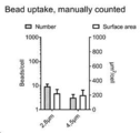
도 5B. 수동 계수에 의해 평가 된 바와 같이, 37 ℃에서 18 시간 동안 배양한 후 PBMC에서 2 가지 크기(4.5 ㎛ 또는 2.8 ㎛)의 항원 결합 입자의 흡수를 나타내는 그래프.
도 5C. 부피 계산에 의해 평가 된 바와 같이 37℃에서 18 시간 동안 배양 한 후 PBMC에서 3 가지 크기(4.5 μm, 2.8 μm 또는 1 μm)의 항원 결합 입자의 흡수를 보여주는 그래프(* p <0,05 ** p <0,01 *** p <0,001, 스튜던트 T- 테스트를 사용하여 계산).
도 6A. 실시예 3a(iv)의 FluoroSpot 분석에서 평가 된 바와 같이, 자극되지 않은 세포와 비교하여 3 가지 크기(4.5 μm, 2.8 μm 또는 1 μm)의 항원 결합 입자로 자극 된 CMV- 민감성 건강한 공여자(n = 2)로부터 PBMC에서 IFNγ 생산 수준의 상대적 증가를 보여주는 그래프.
도 6B. 실시예 3a(iv)의 FluoroSpot 분석에서 평가 된 바와 같이, 자극되지 않은 세포와 비교하여 3 가지 크기(4.5 μm, 2.8 μm 또는 1 μm)의 항원 결합 입자로 자극 된 CMV- 민감성 건강한 공여자(n = 1)로부터 PBMC에서의 IL22- 생산 수준의 상대적 증가를 보여주는 그래프.
도 6C. 실시예 3a(iv)의 FluoroSpot 분석에서 평가 된 바와 같이, 자극되지 않은 세포와 비교하여 3 가지 크기(4.5 μm, 2.8 μm 또는 1 μm)의 항원 결합 입자로 자극 된 CMV- 민감성 건강한 공여체(n = 1)로부터 PBMC에서 IL17- 생산 수준의 상대적 증가를 보여주는 그래프.
도 6D. 실시예 3a(iv)의 FluoroSpot 분석에서 평가 된 바와 같이, 자극되지 않은 세포와 비교하여 3 가지 크기(4.5 μm, 2.8 μm 또는 1 μm)의 항원 결합 입자로 자극 된 CMV- 민감성 건강한 공여자(n = 1)로부터 PBMC에서 IFNγ 및 IL17의 이중 사이토카인 생산의 상대적 증가를 보여주는 그래프.
도 6E. 실시예 3a(iv)의 FluoroSpot 분석에서 평가 된 바와 같이, 자극되지 않은 세포와 비교하여 3 가지 크기(4.5 μm, 2.8 μm 또는 1 μm)의 항원 결합 입자로 자극 된 CMV- 민감성 건강한 공여자(n = 1)로부터 PBMC에서 IL22 및 IL17의 이중 사이토카인 생산의 상대적 증가를 보여주는 그래프.Influence of particle size on T-cell activation. Proliferation Assay by Splenocytes from Ovalbumin Sensitized Mice (Thymidine Incorporation). Comparison of Ovalbumin bound to particles of different sizes with diameters of 5.6 μm, 1 μm and 0.2 μm. The p-value determined using the student T-test and recorded when p <0.05 was found. Staples represent SD.
Figure 2. Proliferation of T-cells by stimulation with neoantigenic peptide NA1-9. 2A shows the number of cells in culture over time. 2B shows% CD4 + / total T-cells. 2C shows T-bet expression in CD4. 2D shows expression of granzyme B and perforin in CD8 + T-cells.
3. Proliferation of T-cells by stimulation with neo-antigen constructs. The percentage and total number of T-cells (small squares) of CD4 + T-cells (large squares), and proliferating CD4 + cells (circles) are shown.
4. Proliferation of T-cells by stimulation with neo-antigen constructs. 4A shows the number of cells in culture over time (red line: peptide identified; pink and orange line: predicted peptide control culture with the same protein but with predicted mutations (NA1, 3, 4 and 5) water). 4B shows% CD4 + / total T-cells. 4C visualizes the analysis performed with the Barnes-Hut St ° hastic Neighbor Embedding (BH-SNE) algorithm for CD4 T-cells, where all cells in the sample were selected marker sets (CD28, CD57, T-bet, GATA). -3, perforin, Granzyme B (GZB), Ki-67 and PD-1) were shown on the 2D map according to similarity in expression intensity.
5A. Confocal microscopy image of PBMC with antigen bound particles of three sizes (4.5 μm, 2.8 μm or 1 μm) after incubation at 37 ° C. for 18 hours with antigen binding particles.
5B. As assessed by manual counting, a graph showing the uptake of antigen binding particles of two sizes (4.5 μm or 2.8 μm) in PBMC after incubation for 18 hours at 37 ° C.
Figure 5C. Graph showing uptake of three sizes (4.5 μm, 2.8 μm or 1 μm) of antigen binding particles in PBMC after incubation at 37 ° C. for 18 hours as assessed by volume calculation (* p <0,05 ** p <0,01 *** p <0,001, calculated using Student's T-test).
6A. As assessed in the FluoroSpot assay of Example 3a (iv), CMV-sensitive healthy donors stimulated with antigen binding particles of three sizes (4.5 μm, 2.8 μm or 1 μm) compared to unstimulated cells (n = Graph showing relative increase in IFNγ production levels in PBMC from 2).
6B. As assessed in the FluoroSpot assay of Example 3a (iv), CMV-sensitive healthy donors stimulated with antigen binding particles of three sizes (4.5 μm, 2.8 μm or 1 μm) compared to unstimulated cells (n = Graph showing relative increase in IL22- production levels in PBMC from 1).
Figure 6C. As assessed in the FluoroSpot assay of Example 3a (iv), CMV-sensitive healthy donors stimulated with antigen binding particles of three sizes (4.5 μm, 2.8 μm or 1 μm) compared to unstimulated cells (n = Graph showing relative increase in IL17- production levels in PBMC from 1).
6D. As assessed in the FluoroSpot assay of Example 3a (iv), CMV-sensitive healthy donors stimulated with antigen binding particles of three sizes (4.5 μm, 2.8 μm or 1 μm) compared to unstimulated cells (n = Graph showing relative increase in dual cytokine production of IFNγ and IL17 in PBMC from 1).
6E. As assessed in the FluoroSpot assay of Example 3a (iv), CMV-sensitive healthy donors stimulated with antigen binding particles of three sizes (4.5 μm, 2.8 μm or 1 μm) compared to unstimulated cells (n = Graph showing the relative increase in dual cytokine production of IL22 and IL17 in PBMC from 1).
정의Justice
엔도톡신, 예: 리포폴리사라카이드(LPS)는, 그램 음성 박테리아, 예컨대 에스케리키아 콜라이(Escherichia coli)의 외부 세포벽에서 발견되는 공유 결합 된 지질 및 폴리사카라이드 서브 유닛을 포함한다.Endotoxins, such as lipopolysaccharides (LPS), include covalently linked lipids and polysaccharide subunits found in the outer cell walls of Gram-negative bacteria such as Escherichia coli.
CD4 + T-세포(또는 T- 헬퍼 세포 또는 CD4 + 헬퍼 T-세포)는 사이토카인 분비를 통해 면역 반응을 조정하는 세포이다. 이들은 B 세포의 항체 클래스 전환, 세포 독성 T 세포의 증식을 자극하거나 또는 식세포를 강화시키는 것과 같은, 다른 면역 세포를 억제 또는 강화시킬 수 있다. 이들은 APC상의 MHC 클래스 II를 통한 항원 제시에 의해 활성화되고, 특정 항원 내에서 대략 15 개 아미노산(소위 T-세포 에피토프)의 스트레치에 특이적인 T-세포 수용체(TCR)를 발현한다.CD4 + T-cells (or T-helper cells or CD4 + helper T-cells) are cells that modulate an immune response through cytokine secretion. They can inhibit or enhance other immune cells, such as stimulating antibody class of B cells, proliferating cytotoxic T cells, or enhancing phagocytes. They are activated by antigen presentation via MHC class II on APC and express T-cell receptors (TCRs) specific for stretch of approximately 15 amino acids (so-called T-cell epitopes) within certain antigens.
CD8 + T-세포(또는 세포 독성 T-세포)는 종양 세포, 감염된 세포 또는 달리 손상된 세포를 사멸시키는 세포이다. CD4 + T-세포와 달리 활성화를 위해 APC가 필요하지 않다. 이들의 T-세포 수용체는 모든 핵 세포에서 발현 된 단백질 인 MHC 클래스 I에 의해 제시된 항원 유래 펩타이드(약 7-10, 예를 들어 8, 아미노산 길이)를 인식한다.CD8 + T-cells (or cytotoxic T-cells) are cells that kill tumor cells, infected cells or otherwise damaged cells. Unlike CD4 + T-cells, no APC is required for activation. Their T-cell receptor recognizes antigen-derived peptides (about 7-10, for example 8, amino acids long) presented by MHC class I, a protein expressed in all nuclear cells.
항원-특이적 T-세포 활성화는 TCR과 공동-자극된 MHC(HLA)분자 상에 제시된 특정 펩티드 사이의 상호 작용을 요구하는 공정이다.Antigen-specific T-cell activation is a process that requires interaction between TCR and specific peptides presented on co-stimulated MHC (HLA) molecules.
항원-제시 세포(APC)는 전형적으로 수지상 세포(DC), B-세포 또는 대식세포, 세포 외 유기체 또는 단백질, 즉 항원을 포식 또는 내재화하는 세포, 및 CD4 + T-세포에 대한 MHC 클래스 II에서 존재하는 항원-유래 펩티드를 가공한 후의 세포이다. 혈액에서 단핵구는 가장 풍부한 항원 제시 세포이다.Antigen-presenting cells (APCs) are typically found in dendritic cells (DCs), B-cells or macrophages, extracellular organisms or proteins, ie cells that phagocytize or internalize antigens, and MHC class II for CD4 + T-cells. Cells after processing the antigen-derived peptide present. Monocytes in the blood are the most abundant antigen presenting cells.
포식 작용 가능한 입자는 면역계의 세포, 특히 단핵구에 의해 포식 될 수 있는 입자로 정의된다.Phagocytic particles are defined as particles that can be fed by cells of the immune system, especially monocytes.
말초 혈액 단핵 세포(PBMC)는 전혈의 밀도 구배 원심 분리에 의해 제조 된 인간 혈액의 일부이다. PBMC 분획은 주로 림프구(70-90 %)와 단핵구(10-30 %)로 구성되며, 적혈구, 과립구 및 혈장은 제거되었다. 단핵구는 일부 경우에 PBMC 샘플에서 세포 수의 10 내지 20 %, 예를 들어 10 내지 15 %를 구성 할 수 있다.Peripheral blood mononuclear cells (PBMCs) are part of human blood prepared by density gradient centrifugation of whole blood. The PBMC fraction consisted mainly of lymphocytes (70-90%) and monocytes (10-30%), and red blood cells, granulocytes and plasma were removed. Monocytes may in some cases constitute 10-20%, for example 10-15%, of the cell number in PBMC samples.
종양 신생 항원은 면역계에 의해 외부 물질로 인식 될 수 있는 펩티드 또는 단백질의 아미노산 서열에서의 종양 유도 변화를 말한다.Tumor neoplastic antigens refer to tumor-induced changes in the amino acid sequence of a peptide or protein that can be recognized as foreign by the immune system.
돌연변이 아미노산을 포함하는 아미노산 서열은 대상체의 비종양 세포에서의 서열과 비교하여 변경되는 대상체의 종양 세포에서 펩티드 또는 단백질의 아미노산 서열이다. 돌연변이 아미노산은 점 돌연변이 일 수 있다 : 예를 들어 아미노산의 삽입 또는 결실, 또는 단백질 또는 펩티드의 서열에서 다른 아미노산으로 단일 아미노산의 치환. 일부 상황에서, 둘 또는 그 이상의 돌연변이 아미노산이 일렬로 존재할 수 있다. 돌연변이는 프레임-시프트 돌연변이 일 수 있다.An amino acid sequence comprising a mutant amino acid is an amino acid sequence of a peptide or protein in a tumor cell of a subject that is altered compared to the sequence in a subject's non-tumor cells. Mutant amino acids can be point mutations: for example insertion or deletion of amino acids, or substitution of a single amino acid with another amino acid in the sequence of a protein or peptide. In some situations, two or more mutant amino acids may be present in a line. The mutation may be a frame-shift mutation.
발명의 상세 설명Detailed description of the invention
포식 작용 가능한 입자는 면역계의 세포, 특히 단핵구에 의해 포식 될 수 있는 입자로 정의된다.Phagocytic particles are defined as particles that can be fed by cells of the immune system, especially monocytes.
말초 혈액 단핵 세포(PBMC)는 전혈의 밀도 구배 원심 분리에 의해 제조 된 인간 혈액의 일부이다. PBMC 분획은 주로 림프구(70-90 %)와 단핵구(10-30 %)로 구성되며, 적혈구, 과립구 및 혈장은 제거되었다. 단핵구는 일부 경우에 PBMC 샘플에서 세포 수의 10 내지 20 %, 예를 들어 10 내지 15 %를 구성 할 수 있다.Peripheral blood mononuclear cells (PBMCs) are part of human blood prepared by density gradient centrifugation of whole blood. The PBMC fraction consisted mainly of lymphocytes (70-90%) and monocytes (10-30%), and red blood cells, granulocytes and plasma were removed. Monocytes may in some cases constitute 10-20%, for example 10-15%, of the cell number in PBMC samples.
종양 신생 항원은 면역계에 의해 외부 물질로 인식 될 수 있는 펩티드 또는 단백질의 아미노산 서열에서의 종양 유도 된 변화이다.Tumor neoplastic antigens are tumor-induced changes in the amino acid sequence of a peptide or protein that can be recognized as foreign by the immune system.
돌연변이 아미노산을 포함하는 아미노산 서열은 대상체의 비종양 세포에서의 서열과 비교하여 변경되는 대상체의 종양 세포에서 펩티드 또는 단백질의 아미노산 서열이다. 돌연변이 아미노산은 점 돌연변이 일 수 있다: 예를 들어 아미노산의 삽입 또는 결실, 또는 단백질 또는 펩티드의 서열에서 상이한 아미노산에 대한 단일 아미노산의 치환. 일부 상황에서, 둘 이상의 돌연변이 아미노산이 일렬로 존재할 수 있다. 돌연변이는 프레임-시프트 돌연변이 일 수 있다.An amino acid sequence comprising a mutant amino acid is an amino acid sequence of a peptide or protein in a tumor cell of a subject that is altered compared to the sequence in a subject's non-tumor cells. Mutant amino acids can be point mutations: for example insertion or deletion of amino acids, or substitution of a single amino acid for a different amino acid in the sequence of a protein or peptide. In some situations, two or more mutant amino acids may be present in a line. The mutation may be a frame-shift mutation.
바람직하게는, 본 발명의 방법에 사용하기위한 입자는 최대 치수가 5.6 ㎛ 미만, 바람직하게는 4 ㎛ 미만, 더욱 바람직하게는 3 ㎛ 미만, 예를 들어 2.5 ㎛ 미만, 2 ㎛ 미만 또는 2 미만일 수 있다. 1.5 μm. 바람직하게는, 본 발명의 방법에 사용하기위한 입자는 최대 치수가 0.001 ㎛ 초과, 바람직하게는 0.005 ㎛ 초과, 바람직하게는 0.01 ㎛ 초과, 바람직하게는 0.05 ㎛ 초과, 바람직하게는 0.1 ㎛ 초과, 보다 바람직하게는 더 큰 치수를 가질 수 있다. 0.2 ㎛ 초과, 더욱 바람직하게는 0.5 ㎛ 초과이다. 바람직하게는 본 발명의 입자는 0.1 내지 5.6 ㎛, 바람직하게는 0.2 내지 5.6 ㎛, 바람직하게는 0.5 내지 5.6 ㎛, 바람직하게는 0.1 내지 4 ㎛, 바람직하게는 0.5 내지 4 ㎛,보다 바람직하게는 0.1 내지 3 ㎛ 간격에서 가장 큰 치수를 갖는다. 바람직하게는 0.5-3 ㎛, 더욱 더 바람직하게는 0.1-2.5 ㎛, 더욱 더 바람직하게는 0.5-2.5 ㎛, 더욱 더 바람직하게는 0.2-2 ㎛, 더욱 더 바람직하게는 0.5-2 ㎛,보다 바람직하게는 1-2 ㎛, 또는 예를 들어 약 1 μm, 약 1.5 μm 또는 약 2 μm(바람직하게는 약 1 μm). 입자는 실질적으로 구형 일 수 있으며, 이 경우 치수는 직경을 지칭 할 것이다. 바람직하게는 입자는 실질적으로 구형이다.Preferably, the particles for use in the process of the invention may have a maximum dimension of less than 5.6 μm, preferably less than 4 μm, more preferably less than 3 μm, for example less than 2.5 μm, less than 2 μm or less than 2. have. 1.5 μm. Preferably, the particles for use in the process of the invention have a maximum dimension of greater than 0.001 μm, preferably greater than 0.005 μm, preferably greater than 0.01 μm, preferably greater than 0.05 μm, preferably greater than 0.1 μm, more It may preferably have a larger dimension. More than 0.2 μm, more preferably more than 0.5 μm. Preferably the particles of the invention are 0.1 to 5.6 μm, preferably 0.2 to 5.6 μm, preferably 0.5 to 5.6 μm, preferably 0.1 to 4 μm, preferably 0.5 to 4 μm, more preferably 0.1 Have the largest dimension at intervals from 3 μm. Preferably 0.5-3 μm, even more preferably 0.1-2.5 μm, even more preferably 0.5-2.5 μm, even more preferably 0.2-2 μm, even more preferably 0.5-2 μm, more preferred Preferably 1-2 μm, or for example about 1 μm, about 1.5 μm or about 2 μm (preferably about 1 μm). The particles can be substantially spherical, in which case the dimension will refer to the diameter. Preferably the particles are substantially spherical.
본 발명에 따른 바람직한 입자 크기는 식균 작용에 의해 세포에 들어갈 수 있을만큼 크지만, 하나 이상의 입자가 식균 작용에 의해 동일한 세포에 들어갈 수 있을 정도로 작은 크기이다. 하나 이상의 포식 작용 된 입자를 갖는 것은 APC 세포가 상이한 포식 소체 내의 여러 입자로부터 펩타이드를 동시에 취할 수 있고, 따라서 MHC 클래스 II 경로를 통해 세포 표면에서 신생 항원의 발현을 최대화 할 것임을 의미한다.Preferred particle sizes according to the invention are large enough to enter cells by phagocytosis, but small enough to allow more than one particle to enter the same cells by phagocytosis. Having more than one phagocytic particle means that APC cells can simultaneously take peptides from several particles in different phagocytic bodies, thus maximizing expression of neoplastic antigens on the cell surface via the MHC class II pathway.
일반적으로 박테리아의 크기와 비슷한 크기는 APC에 의한 완전한 식균 작용을 촉진한다. 완전한 식균 작용은 APC에 의한 우수한 항원 분해 및이어서 MHC 클래스 II를 통한 T-세포에 대한 우수한 제시로 이어진다. 최적의 크기는 현재 발명자들에 의해 조사되었다(실시예 3a 및 3b, 및 도 1, 5A-C 및 6A-E 참조).Generally, a size similar to that of bacteria promotes full phagocytosis by APC. Full phagocytosis leads to good antigen degradation by APC followed by good presentation to T-cells through MHC class II. The optimal size was investigated by the present inventors (see Examples 3a and 3b, and FIGS. 1, 5A-C and 6A-E).
바람직한 특정 구체예에서, 입자는 상자성 특성, 또는 더욱 바람직하게는 초상자성 특성을 갖는다. 상자성 또는 초상자성 입자는 자석을 사용하여 T-세포로부터 완전히 분리 될 수 있다. 중요하게는, 내재화 된 입자를 함유하는 APC는 또한 자석을 사용하여 T-세포로부터 분리 될 수 있다. 이는 T-세포가 배양 혼합물로부터 신속하게 분리 될 수 있게 한다. T-세포는 모든 입자가 제거되었다는 지식으로 환자에게 투여 될 수 있다. 따라서 본 발명은 T-세포를 증식시키는 특히 안전한 방법을 제공한다.In certain preferred embodiments, the particles have paramagnetic properties, or more preferably superparamagnetic properties. Paramagnetic or superparamagnetic particles can be completely separated from T-cells using magnets. Importantly, APCs containing internalized particles can also be separated from T-cells using magnets. This allows T-cells to be quickly separated from the culture mixture. T-cells can be administered to patients with the knowledge that all particles have been removed. The present invention therefore provides a particularly safe method of proliferating T-cells.
상자성 입자는 또한 이와 관련된 종양 신생 항원 컨스트럭트로 입자의 멸균 및/또는 변성을 촉진시킨다. 세척은 아래에서 더 상세히 논의된다. 세척 동안, 입자는 자석에 의해 수집 및/또는 고정 될 수 있다. 또한, 상자 내에 입자를 상자성으로 고정 시키거나 또는 상자성으로 고정 시키거나, 중력 또는 원심 분리에 의해 입자를 침강시키는 것과 같은 다른 수단에 의해 세척을 수행 할 수도 있다.Paramagnetic particles also promote sterilization and / or denaturation of the particles with their associated neoplastic antigen constructs. Washes are discussed in more detail below. During washing, the particles can be collected and / or fixed by a magnet. In addition, the washing may be performed by other means such as paramagnetically or paramagnetically fixing the particles in the box, or by sedimenting the particles by gravity or centrifugation.
초상자성 입자의 예는 Dynabeads ™(Invitrogen)이다. Dynabeads M-270 Carboxylic acid, Dynabeads M-270 Amine 및 Dynabeads MyOne Carboxylic acid와 같은 다양한 기능적 형태로 제공된다. Dynabeads MyOne Carboxylic Acid는 균일 한 단일 크기의 초상자성 입자로, 균일하게 분포 된 자성 물질과 고도로 가교 된 폴리스티렌으로 구성된다. 입자는 글리시딜 에테르의 친수성 층으로 추가로 코팅되어, 그 안에 산화철을 내포한다. 이어서, 카르복실산 기가 입자의 표면에 도입된다.An example of a superparamagnetic particle is Dynabeads ™ (Invitrogen). Available in a variety of functional forms, such as Dynabeads M-270 Carboxylic acid, Dynabeads M-270 Amine, and Dynabeads MyOne Carboxylic acid. Dynabeads MyOne Carboxylic Acid is a uniform, single-sized superparamagnetic particle consisting of a uniformly distributed magnetic material and highly crosslinked polystyrene. The particles are further coated with a hydrophilic layer of glycidyl ether, containing iron oxide therein. The carboxylic acid group is then introduced to the surface of the particles.
초상자성 입자의 다른 예는 캡슐화 된 카르복실화 된 에스타 포르 ® 슈퍼 파라 마그네틱 마이크로 스피어(Merck Chimie S.A.S.)및 Sera-Mag SpeedBeads(친수성)카르복실 레이트-개질 된 자성 입자(GE Healthcare UK Limited)를 포함한다. 캡슐화 된 카르복실화 된 Estapor® SuperParamagnetic 마이크로 스피어는 코어-쉘 구조로 만들어졌다. 초상자성 산화철 재료(~ 40 %)는 폴리스티렌 필름으로 캡슐화되어 있으며 표면의 구성 요소를 방해하지 않다. Sera-Mag SpeedBeads(친수성)Carboxylate-Modified Magnetic Particles는 균일 한 크기의 자성 입자이며 자철광의 두 번째 층(~ 60 %를 구성하는 총 두 층)을 특징으로한다. 결과적으로 Sera-Mag SpeedBeads는 자기장에 매우 빠르게 반응하여 서스펜션과 신속하고 완벽하게 분리된다. 그들은 콜리 플라워와 같은 표면을 가지고 있어 입자의 전체 표면적과 결합력을 증가시킨다.Other examples of superparamagnetic particles include encapsulated carboxylated Estaphor ® super paramagnetic microspheres (Merck Chimie SAS) and Sera-Mag SpeedBeads (hydrophilic) carboxylate-modified magnetic particles (GE Healthcare UK Limited) do. Encapsulated carboxylated Estapor® SuperParamagnetic microspheres are made of a core-shell structure. Superparamagnetic iron oxide material (~ 40%) is encapsulated in polystyrene film and does not interfere with the components of the surface. Sera-Mag SpeedBeads (Hydrophilic) Carboxylate-Modified Magnetic Particles are magnetic particles of uniform size and feature a second layer of magnetite (total two layers, constituting ~ 60%). As a result, Sera-Mag SpeedBeads react very quickly to the magnetic field, quickly and completely separating the suspension. They have a surface like cauliflower, which increases the particle's total surface area and binding force.
항원 폴리펩티드와 입자의 연관Association of antigen polypeptides with particles
종양 신생 항원 컨스트럭트는 입자에서 종양 신생 항원 컨스트럭트를 분리하지 않으면서 하기 논의 된 바와 같이 살균 및 변성 세척을 수행 할 수 있는 방식으로 입자와 관련된다.Tumor neoplastic antigen constructs are associated with the particles in such a way that sterilization and denaturation washes can be performed as discussed below without isolating tumor neoplastic antigen constructs from the particles.
폴리펩티드를 입자에 회합시키는 한 가지 방법이 실시예 1에 도시되어 있다. 그러나, 정확한 회합 방식은 본 발명의 방법에 중요하지 않다. 바람직하게는, 폴리펩티드는 입자에 공유 결합된다(예를 들어, 폴리펩티드의 아민 기 또는 카르복실산 기와 입자 표면의 카르복실산 기 또는 아민 기 사이의 아미드 결합을 통해). 대안적으로, 후보 항원 폴리펩티드는 금속 킬레이트를 통해 입자에 연결될 수 있다. 예를 들어, 이미 노 디아 아세트산과 같은 금속 킬레이트 리간드와 연결된 입자는 Cu2 +, Zn2 +, Ca2 +, Co2 + 또는 Fe3 +와 같은 금속 이온에 결합 할 수 있다. 이들 금속 킬레이트는 예를 들어 히스티딘 또는 시스테인을 함유하는 단백질 및 펩티드를 큰 강도로 결합 할 수 있다. 따라서, 금속 킬레이트를 갖는 입자는 엄격한 세척이 결합 된 펩티드/단백질에서 LPS 및 다른 오염 성분의 양을 감소시킬 수 있는 방식으로 펩티드/단백질을 비공유적으로 흡착 할 수 있다.One method of associating a polypeptide to a particle is shown in Example 1. However, the exact association mode is not critical to the method of the present invention. Preferably, the polypeptide is covalently bound to the particle (eg, via an amide bond between the amine group or carboxylic acid group of the polypeptide and the carboxylic acid group or amine group on the particle surface). Alternatively, candidate antigen polypeptides can be linked to the particles via metal chelates. For example, particles linked with metal chelate ligands such as iminodiacetic acid can bind to metal ions such as Cu2 +, Zn2 +, Ca2 +, Co2 + or Fe3 +. These metal chelates can bind proteins and peptides containing histidine or cysteine with great strength, for example. Thus, particles with metal chelates can non-covalently adsorb the peptides / proteins in such a way that stringent washing can reduce the amount of LPS and other contaminants in the bound peptides / proteins.
종양 신생 항원 컨스트럭트는 입자에서 종양 신생 항원 컨스트럭트를 분리하지 않으면서하기 논의 된 바와 같이 살균 및 변성 세척을 수행 할 수 있는 방식으로 입자와 관련된다.Tumor neoplastic antigen constructs are associated with the particles in such a way that sterilization and denaturation washes can be performed as discussed below without isolating tumor neoplastic antigen constructs from the particles.
폴리펩티드를 입자에 회합시키는 한 가지 방법이 실시예 1에 도시되어있다. 그러나, 정확한 회합 방식은 본 발명의 방법에 중요하지 않다. 바람직하게는, 폴리펩티드는 입자에 공유 결합된다(예를 들어, 폴리펩티드의 아민 기 또는 카르복실산 기와 입자 표면의 카르복실산 기 또는 아민 기 사이의 아미드 결합을 통해). 대안적으로, 후보 항원 폴리펩티드는 금속 킬레이트를 통해 입자에 연결될 수 있다. 예를 들어, 이미 노 디아 아세트산과 같은 금속 킬레이트 리간드와 연결된 입자는 Cu2 +, Zn2 +, Ca2 +, Co2 + 또는 Fe3 +와 같은 금속 이온에 결합 할 수 있다. 이들 금속 킬레이트는 예를 들어 히스티딘 또는 시스테인을 함유하는 단백질 및 펩티드를 큰 강도로 결합 할 수 있다. 따라서, 금속 킬레이트를 갖는 입자는 엄격한 세척이 결합 된 펩티드/단백질에서 LPS 및 다른 오염 성분의 양을 감소시킬 수 있는 방식으로 펩티드/단백질을 비공유적으로 흡착 할 수 있다.One method of associating a polypeptide to a particle is shown in Example 1. However, the exact association mode is not critical to the method of the present invention. Preferably, the polypeptide is covalently bound to the particle (eg, via an amide bond between the amine group or carboxylic acid group of the polypeptide and the carboxylic acid group or amine group on the particle surface). Alternatively, candidate antigen polypeptides can be linked to the particles via metal chelates. For example, particles linked with metal chelate ligands such as iminodiacetic acid can bind to metal ions such as Cu2 +, Zn2 +, Ca2 +, Co2 + or Fe3 +. These metal chelates can bind proteins and peptides containing histidine or cysteine with great strength, for example. Thus, particles with metal chelates can non-covalently adsorb the peptides / proteins in such a way that stringent washing can reduce the amount of LPS and other contaminants in the bound peptides / proteins.
종양 신생 항원 컨스트럭트Tumor Angiogenic Constructs
종양 신생 항원 컨스트럭트는 대상체에서 암 세포에서 발현되는 것으로 공지되거나 의심되는 하나 이상의 종양 신생 항원 및/또는 하나 이상의 아미노산 서열을 포함하는 폴리펩티드이다. 바람직하게는, 종양 신생 항원 컨스트럭트는 대상체에서 암 세포에서 발현되는 것으로 알려져 있거나 의심되는 하나 이상의 종양 신생 항원 및/또는 돌연변이 아미노산 서열을 포함한다. 종양 신생 항원 컨스트럭트는 하나 이상의 공유결합으로 연결된 펩티드를 포함하며, 여기서 공유결합으로 연결된 펩티드 중 하나 이상은 바람직하게는 대상체에서 암과 관련이있는 것으로 알려져 있거나 의심되는 하나 이상의 돌연변이 아미노산을 포함하는 아미노산 서열을 포함한다. 특정 실시 양태에서, 하나 이상의 공유결합으로 연결된 펩티드는 대상체에서 암 세포에서 발현되는 것으로 공지되거나 의심되는 하나 이상의 아미노산 서열을 포함하는 아미노산 서열을 포함한다. 대상체에서 암 세포에서 발현되는 것으로 공지 또는 의심되는 아미노산 서열은 종양이 존재하는 조직 또는 기관에 의해 발현되는 것으로 공지 또는 의심되는 아미노산 서열 일 수 있다. 아미노산 서열은 돌연변이되거나 비 돌연변이 될 수 있다. 바람직하게는 돌연변이된다. 대상체에서 암과 관련이있는 것으로 알려진 또는 의심되는 하나 이상의 돌연변이 아미노산을 포함하는 아미노산 서열(또는 암에서 발현되거나 알려진 것으로 의심되는 하나 이상의 아미노산 서열을 포함하는 아미노산 서열을 포함하는 아미노산 서열)돌연변이가 있든 없든간에 대상체의 세포는 또한 '신생 항원 에피토프'로 지칭 될 수 있다.Tumor neoplastic antigen constructs are polypeptides comprising one or more tumor neoplastic antigens and / or one or more amino acid sequences known or suspected of being expressed in cancer cells in a subject. Preferably, the neoplastic antigen construct comprises one or more neoplastic antigens and / or mutant amino acid sequences known or suspected of being expressed in cancer cells in a subject. Tumor neoplastic antigen constructs comprise one or more covalently linked peptides, wherein at least one of the covalently linked peptides is preferably an amino acid comprising one or more mutant amino acids known or suspected of being associated with cancer in a subject Sequence. In certain embodiments, one or more covalently linked peptides comprise an amino acid sequence comprising one or more amino acid sequences known or suspected of being expressed in cancer cells in a subject. An amino acid sequence known or suspected to be expressed in cancer cells in a subject may be an amino acid sequence known or suspected to be expressed by the tissue or organ in which the tumor is present. The amino acid sequence can be mutated or non-mutated. Preferably mutated. With or without an amino acid sequence comprising one or more mutant amino acids known or suspected of being associated with cancer in a subject (or an amino acid sequence comprising an amino acid sequence comprising one or more amino acid sequences expressed or suspected of being known in cancer) The cells of the subject in the liver may also be referred to as 'new antigen epitopes'.
바람직하게는, 돌연변이 아미노산은 치환 된 아미노산이다(즉, 비종양 세포에서 단백질 또는 펩티드의 아미노산은 종양 세포에서 다른 아미노산으로 치환된다). 대안적으로, 돌연변이 아미노산은 서열로부터 결실 된 아미노산 일 수 있다(즉, 비종양 세포와 비교하여 종양 세포에서 단백질/펩티드에서 결실 됨); 또는 돌연변이 아미노산은 서열에 삽입 된 아미노산 일 수 있다(즉, 새로운 아미노산이 비종양 세포와 비교하여 종양 세포에서 단백질/펩티드에 첨가됨).Preferably, the mutant amino acids are substituted amino acids (ie, amino acids of proteins or peptides in non-tumor cells are replaced with other amino acids in tumor cells). Alternatively, the mutant amino acid may be an amino acid deleted from the sequence (ie, deleted in protein / peptide in tumor cells compared to non-tumor cells); Or the mutant amino acid may be an amino acid inserted into the sequence (ie, a new amino acid is added to the protein / peptide in tumor cells compared to non-tumor cells).
종양 신생 항원 컨스트럭트의 신생 항원 에피토프는 대상체에서 암과 관련이있는 것으로 알려져 있거나 의심되는 변이 된 단백질/펩티드(또는 변이 된 유전자)를 선택함으로써 설계 될 수 있으며; 단백질 또는 펩티드의 서열(또는 유전자의 발현에 기인 한 단백질/펩티드)에서 돌연변이 아미노산(들)을 위치시키는 단계; 및 돌연변이 아미노산(들), 및 돌연변이 아미노산(들)의 C- 말단 및 N- 말단에 다수의 측면 아미노산을 포함하는 단백질/펩타이드의 일부를 선택하는 단계를 포함한다. 특정 바람직한 구체예에서, 비-종양 세포의 단백질 또는 펩티드 서열과 비교하여 단백질 또는 펩티드의 서열에는 단일 돌연변이 아미노산이 존재한다. 더욱 바람직하게는, 비종양 세포에서의 단백질 또는 펩티드 서열과 비교하여 단백질 또는 펩티드의 서열에는 단일 치환 된 돌연변이 아미노산이 존재한다.Neoplastic epitopes of tumor neoplastic constructs can be designed by selecting a mutated protein / peptide (or mutated gene) that is known or suspected to be associated with cancer in a subject; Positioning the mutant amino acid (s) in the sequence of the protein or peptide (or protein / peptide due to expression of the gene); And selecting a portion of the protein / peptide comprising the mutant amino acid (s) and a plurality of flanking amino acids at the C- and N-terminus of the mutant amino acid (s). In certain preferred embodiments, there is a single mutant amino acid in the sequence of the protein or peptide as compared to the protein or peptide sequence of the non-tumor cell. More preferably, there is a single substituted mutant amino acid in the sequence of the protein or peptide as compared to the protein or peptide sequence in non-tumor cells.
바람직하게는 돌연변이 아미노산(들)의 C- 말단에 인접한 플 랭킹 아미노산의 수는 5 개 이상, 바람직하게는 8 개 이상, 예를 들어 8 개, 9 개, 10 개, 11 개 또는 12 개 아미노산이다. 바람직하게는 돌연변이 아미노산(들)의 N- 말단에 인접한 플 랭킹 아미노산의 수는 5 개 이상, 바람직하게는 8 개 이상, 예를 들어 8 개, 9 개, 10 개, 11 개 또는 12 개 아미노산이다. 변이 된 아미노산(들)의 플 랭킹 아미노산의 C- 말단 및 N- 말단에 대한 수는 동일 할 수 있고, 바람직하게는 동일하다.Preferably the number of flanking amino acids adjacent to the C-terminus of the mutant amino acid (s) is at least 5, preferably at least 8, for example 8, 9, 10, 11 or 12 amino acids. . Preferably the number of flanking amino acids adjacent to the N-terminus of the mutant amino acid (s) is at least 5, preferably at least 8, for example 8, 9, 10, 11 or 12 amino acids. . The numbers for the C-terminus and N-terminus of the flanking amino acid (s) of the mutated amino acid (s) may be the same and are preferably the same.
특정 실시 양태에서, 하나 이상의 공유결합으로 연결된 펩티드는 대상체에서 암 세포에서 발현되는 것으로 공지되거나 의심되는 하나 이상의 아미노산 서열을 포함하는 아미노산 서열을 포함한다. 대상체에서 암 세포에서 발현되는 것으로 공지 또는 의심되는 아미노산 서열은 종양이 존재하는 조직 또는 기관에 의해 발현되는 것으로 공지 또는 의심되는 아미노산 서열 일 수 있다. 아미노산 서열은 돌연변이되거나 비 돌연변이 될 수 있다. 바람직하게는 아미노산 서열이 돌연변이된다.In certain embodiments, one or more covalently linked peptides comprise an amino acid sequence comprising one or more amino acid sequences known or suspected of being expressed in cancer cells in a subject. An amino acid sequence known or suspected to be expressed in cancer cells in a subject may be an amino acid sequence known or suspected to be expressed by the tissue or organ in which the tumor is present. The amino acid sequence can be mutated or non-mutated. Preferably the amino acid sequence is mutated.
종양 신생 항원 컨스트럭트가 2 개 이상의 공유결합으로 연결된 펩티드를 포함하는 실시 양태에서, 바람직하게는 각각의 공유결합으로 연결된 펩티드는 대상체에서 암과 관련되거나 알려져있는 것으로 의심되거나 의심되는 하나 이상의 돌연변이 아미노산을 포함하는 아미노산 서열(즉, 신생 항원 에피토프)을 포함한다. 대안적으로, 종양 신생 항원 컨스트럭트가 2 개 이상의 공유결합으로 연결된 펩티드를 포함하는 구체예에서, 바람직하게는 종양 신생 항원 컨스트럭트는 암과 관련되거나 알려져있는 것으로 의심되거나 의심되는 적어도 하나의 돌연변이 아미노산을 포함하는 아미노산 서열을 포함하는 적어도 하나의 펩티드를 포함한다. 대상체(즉, 신생 항원 에피토프), 및 대상체의 암 세포에서 발현되는 것으로 알려진 또는 의심되는 아미노산 서열을 포함하는 하나 이상의 펩타이드를 포함한다.In embodiments in which the neoplastic antigen construct comprises two or more covalently linked peptides, preferably each covalently linked peptide is one or more mutant amino acids suspected or suspected of being associated with or known to be cancer in a subject. Amino acid sequence (ie, neoplastic epitope) comprising a. Alternatively, in embodiments wherein the neoplastic antigen construct comprises two or more covalently linked peptides, preferably the neoplastic antigen construct is at least one mutation suspected or suspected of being associated or known to be cancer At least one peptide comprising an amino acid sequence comprising an amino acid. One or more peptides comprising an amino acid sequence known or suspected to be expressed in a subject (ie, neoplastic epitope), and cancer cells of the subject.
종양 신생 항원 컨스트럭트가 2 개 이상의 공유결합으로 연결된 펩티드를 포함하는 구체예에서, 펩티드는 직접적으로 연결되거나 스페이서 부분을 통해 연결될 수 있다. 스페이서 부분은 짧은 서열의 아미노산, 예를 들어 1 내지 15 개 아미노산, 바람직하게는 1 내지 10 개 아미노산, 더욱 바람직하게는 1 내지 5 개 아미노산(예를 들어, 1, 2, 3, 4 또는 5 개 아미노산)일 수 있다. 바람직하게는, 스페이서 모이어티는 모티프 VVR을 포함한다. 한 구체예에서 스페이서 모이어티는 서열 VVR을 갖는다. 대안적인 구체예에서, 스페이서 모이어티는 모티프 VVR을 포함하지 않으며, 예를 들어 스페이서 모이어티는 서열 VVR을 갖지 않는다. 다른 실시 형태에서, 스페이서 모이어티는 모티프 GGS를 포함하고, 예를 들어 스페이서 모이어티는 서열 GGS를 갖는다. 다른 구체예에서, 펩티드는 직접 연결된다(즉, 스페이서 모이어티가없고, 예를 들어 아미노산 스페이서 모이어티가 없다).In embodiments in which the neoplastic antigen construct comprises peptides that are linked two or more covalently, the peptides may be linked directly or through spacer moieties. The spacer moiety is a short sequence of amino acids, for example 1 to 15 amino acids, preferably 1 to 10 amino acids, more preferably 1 to 5 amino acids (
바람직한 구체예에서, 종양 신생 항원 컨스트럭트는 2 개 이상의 공유결합으로 연결된 펩티드, 보다 바람직하게는 3 개 이상의 공유결합으로 연결된 펩티드, 예를 들어, 3, 4, 5, 6, 7, 8, 9 또는 10 개의 공유결합으로 연결된 펩티드, 바람직하게는 3, 4, 5 또는 6 개의 공유결합으로 연결된 펩티드, 보다 바람직하게는 3, 4 또는 5 개의 공유결합으로 연결된 펩티드를 포함한다. 바람직하게는, 연결된 펩티드는 각각 스페이서 부분을 통해 연결된다. 각각의 링커 모이어티는 동일하거나 상이 할 수 있다. 대상체에서 암과 관련이있는 것으로 알려져 있거나 의심되는 하나 이상의 돌연변이 아미노산을 포함하는 아미노산 서열을 각각 포함하는 3 개의 공유결합으로 연결된 펩티드가 특히 효과적인 것으로 밝혀졌다.In a preferred embodiment, the neoplastic antigen construct comprises two or more covalently linked peptides, more preferably three or more covalently linked peptides, eg, 3, 4, 5, 6, 7, 8, 9 Or 10 covalently linked peptides, preferably 3, 4, 5 or 6 covalently linked peptides, more preferably 3, 4 or 5 covalently linked peptides. Preferably, the linked peptides are each linked via a spacer moiety. Each linker moiety may be the same or different. Three covalently linked peptides each comprising an amino acid sequence comprising one or more mutant amino acids known or suspected of being associated with cancer in a subject have been found to be particularly effective.
2 개의 연결된 펩티드를 포함하는 종양 신생 항원 컨스트럭트의 구조는 다음과 같을 수 있다 :The structure of the neoplastic antigen construct comprising two linked peptides may be as follows:
- [에피토프 서열 1] - [링커 모이어티] - [에피토프 서열 2].[Epitope Sequence 1]-[Linker Moiety]-[Epitope Sequence 2].
3 개의 연결된 펩티드를 포함하는 종양 신생 항원 컨스트럭트의 구조는 다음과 같을 수 있다 :The structure of the neoplastic antigen construct comprising three linked peptides may be as follows:
- [에피토프 서열 1] - [링커 모이어티] - [에피토프 서열 2] - [링커 모이어티] - [에피토프 서열 3].[Epitope Sequence 1]-[Linker Moiety]-[Epitope Sequence 2]-[Linker Moiety]-[Epitope Sequence 3].
4 개의 연결된 펩티드를 포함하는 종양 신생 항원 컨스트럭트의 구조는 다음과 같을 수 있다 :The structure of the neoplastic antigen construct comprising four linked peptides may be as follows:
- [에피토프 서열 1] - [링커 모이어티] - [에피토프 서열 2] - [링커 모이어티] - [에피토프 서열 3] - [링커 모이어티] - [에피토프 서열 4].[Epitope Sequence 1]-[Linker Moiety]-[Epitope Sequence 2]-[Linker Moiety]-[Epitope Sequence 3]-[Linker Moiety]-[Epitope Sequence 4].
5 개의 연결된 펩티드를 포함하는 종양 신생 항원 컨스트럭트의 구조는 다음과 같을 수 있다 :The structure of a neoplastic antigen construct comprising five linked peptides may be as follows:
- [에피토프 서열 1] - [링커 모이어티] - [에피토프 서열 2] - [링커 모이어티] - [에피토프 서열 3] - [링커 모이어티] - [에피토프 서열 5].[Epitope Sequence 1]-[Linker Moiety]-[Epitope Sequence 2]-[Linker Moiety]-[Epitope Sequence 3]-[Linker Moiety]-[Epitope Sequence 5].
n 개의 연결된 펩티드를 포함하는 종양 신생 항원 컨스트럭트의 구조는 다음과 같을 수 있다 :The structure of a neoplastic antigen construct comprising n linked peptides may be as follows:
- [에피토프 서열 1] - [링커 모이어티] - [에피토프 서열 2] - [링커 모이어티] - [에피토프 서열 3]… .. - [링커 모이어티] - [에피토프 서열 n].-[Epitope Sequence 1]-[Linker Moiety]-[Epitope Sequence 2]-[Linker Moiety]-[Epitope Sequence 3]. ..-[linker moiety]-[epitope sequence n].
바람직한 구체예에서, 종양 신생 항원 컨스트럭트의 각 펩티드는 5 내지 50 개의 아미노산,보다 바람직하게는 5 내지 30 개의 아미노산,보다 바람직하게는 8 내지 30 개의 아미노산, 가장 바람직하게는 10 내지 25 개의 아미노산, 예를 들어 10, 11을 포함하고, 또는 12, 13, 14, 15, 16, 17, 18, 19, 20, 21, 22, 23, 24 또는 25 개 아미노산을 포함한다. 하나의 특히 바람직한 구체예에서, 종양 신생 항원 컨스트럭트의 각각의 펩티드는 17 내지 25 개의 아미노산을 포함하고, 예를 들어 돌연변이 아미노산 + C- 말단 및 N- 돌연변이 아미노산의 말단에 8 내지 12 개의 아미노산(바람직하게는 10 개의 아미노산)을 포함한다.In a preferred embodiment, each peptide of the neoplastic antigen construct has 5 to 50 amino acids, more preferably 5 to 30 amino acids, more preferably 8 to 30 amino acids, most preferably 10 to 25 amino acids , For example 10, 11, or 12, 13, 14, 15, 16, 17, 18, 19, 20, 21, 22, 23, 24 or 25 amino acids. In one particularly preferred embodiment each peptide of the neoplastic antigen construct comprises 17 to 25 amino acids, for example 8 to 12 amino acids at the ends of the mutant amino acids plus the C- and N- mutant amino acids. (Preferably 10 amino acids).
입자가 둘 이상의 종양 신생 항원 컨스트럭트와 관련되는 구체예에서, 각각의 종양 신생 항원 컨스트럭트는 동일한 폴리펩티드 서열을 갖거나 상이한 폴리펩티드 서열을 가질 수 있다. 예를 들어, 제 1 종양 신생 항원 컨스트럭트는 하나 이상의 공유결합으로 연결된 펩티드(예를 들어, 하나 이상의 신생 항원 에피토프)를 포함 할 수 있고, 제 2 종양 신생 항원 컨스트럭트는 하나 이상의 상이한 공유결합으로 연결된 펩티드(예를 들어, 하나 이상의 상이한 제 1 종양 신생 항원 컨스트럭트의 신생 항원 에피토프에 대한 신생 항원 에피토프)를 포함 할 수 있다.In embodiments in which the particles are associated with two or more tumorigenic antigen constructs, each tumorigenic antigen construct may have the same polypeptide sequence or different polypeptide sequences. For example, the first tumor neoantigen construct may comprise one or more covalently linked peptides (eg, one or more neoantigen epitopes), and the second tumor neoantigen construct may comprise one or more different covalent bonds. Linked peptides (eg, neoplastic epitopes for neoplastic epitopes of one or more different first tumor neoplastic antigen constructs).
종양 신생 항원 컨스트럭트는 20 개 이상의 아미노산,보다 바람직하게는 30 개 이상의 아미노산,보다 바람직하게는 40 개 이상의 아미노산,보다 바람직하게는 50 개 이상의 아미노산, 더욱 더 바람직하게는 60 개의 아미노산을 포함 할 수 있다. 종양 신생 항원 컨스트럭트는 150 개 미만의 아미노산, 바람직하게는 130 개 미만의 아미노산,보다 바람직하게는 100 개 미만의 아미노산, 더욱 더 바람직하게는 85 개 미만의 아미노산, 및 더욱 더 바람직하게는 75 개 미만의 아미노산을 포함할 수 있다.The neoplastic antigen construct may comprise at least 20 amino acids, more preferably at least 30 amino acids, more preferably at least 40 amino acids, more preferably at least 50 amino acids, even more preferably 60 amino acids have. Tumor neoplastic antigen constructs have less than 150 amino acids, preferably less than 130 amino acids, more preferably less than 100 amino acids, even more preferably less than 85 amino acids, and even more preferably 75 And may contain less than amino acids.
본 발명은 하나 이상의 종양 신생 항원 컨스트럭트가 입자와 관련 될 수 있게 한다. 종양 신생 항원 컨스트럭트는 하나 이상의 신생 항원 에피토프를 포함 할 수 있다. 입자에 여러 개의 신생 항원 에피토프를 사용하면 항암 T 세포가 증식 될 가능성이 높아집니다. APC에 의해 제시된 단편보다 더 큰 항원 폴리펩티드는 또한 각각의 항원의 다양한 에피토프가 제시 될 것을 보장한다.The present invention allows one or more tumorigenic antigen constructs to be associated with a particle. Tumor neoplastic antigen constructs may comprise one or more neoplastic antigen epitopes. The use of multiple neoantigen epitopes on the particles increases the likelihood of proliferation of anticancer T cells. Antigen polypeptides larger than the fragments presented by APC also ensure that various epitopes of each antigen are presented.
본 발명은 단일 종양 신생 항원 컨스트럭트가 주어진 입자와 관련되거나, 또는 하나 이상의 상이한 신생 항원 컨스트럭트가 주어진 입자와 관련 될 수 있게 한다. 동일한 종양 신생 항원 컨스트럭트 또는 컨스트럭트와 관련된 입자는 동일한 입자 종으로 간주된다. 상이한 종양 신생 항원 컨스트럭트 또는 컨스트럭트과 관련된 입자는 상이한 입자 종인 것으로 간주된다.The present invention allows a single tumor neoantigen construct to be associated with a given particle, or one or more different neoantigen constructs can be associated with a given particle. Particles associated with the same tumorigenic antigen construct or construct are considered to be the same particle species. Particles associated with different tumor neoantigenic constructs or constructs are considered to be different particle species.
일반적으로, 100 내지 1x10⁹, 예를 들어 100 내지 1x10⁸, 예를 들어 100 내지 1x10⁷ 입자가 소정의 증식 실행, 예를 들어 1000 내지 1x10⁷ 입자에 사용된다. 예를 들어, 입자 농도 대 APC 세포 농도의 비는 1000 : 1 내지 1:10의 범위이다. 입자의 크기에 따라 비율을 최적화 할 수 있다. 예를 들어, 1㎛ 단면의 입자의 경우, 비는 50 : 1 내지 2 : 1, 예를 들어 25 : 1 내지 5 : 1, 예를 들어 15 : 1 내지 7 : 1, 예를 들어 10 : 1 일 수 있다.Generally, 100 to 1 × 10 9 , for example 100 to 1 × 10 8 , for example 100 to 1 × 10 7 particles, are used for certain propagation runs, for example 1000 to 1 × 10 7 particles. For example, the ratio of particle concentration to APC cell concentration ranges from 1000: 1 to 1:10. The ratio can be optimized depending on the size of the particles. For example, for particles of 1 μm cross-section, the ratio is 50: 1 to 2: 1, for example 25: 1 to 5: 1, for example 15: 1 to 7: 1, for example 10: 1 Can be.
입자는 균질 한 집단 일 수 있으며, 즉 모든 입자는 동일한 입자 종이다. 대안적으로, 입자는 이종 집단 일 수 있으며, 즉 입자 집단은 하나 이상의 입자 종을 포함한다. 예를 들어, 단계 a)는 추가로, 밀접하게 연관된 하나 이상의 종양 신생 항원 컨스트럭트를 갖는 제 2 종의 포식 작용성 입자를 제공하는 단계를 포함 할 수 있으며, 여기서 종양 신생 항원 컨스트럭트는 공지되거나 의심되는 하나 이상의 돌연변이 아미노산을 포함하는 아미노산 서열을 포함한다 대상체에서 암, 또는 대상체에서 암 세포에서 발현되는 것으로 알려지거나 의심되는 돌연변이 또는 비 돌연변이 아미노산 서열과 관련되고, 종양 신생 항원 컨스트럭트의 서열은 제 2 입자는 제 1 입자와 관련된 종양 신생 항원 컨스트럭트(들)의 서열과 상이하다. 선택적으로 제 3, 및 선택적으로 제 4, 및 선택적으로 제 5, 및 선택적으로 제 6, 및 선택적으로 제 7, 및 선택적으로 제 8 및 선택적으로 제 8 및 선택적으로 n 번째(여기서, n은 50 이하의 임의의 정수일 수 있음)종의 포식 작용성 입자 또는 n 번째 입자 종과 관련된 종양 신생 항원 컨스트럭트(들)의 서열은 다른 종의 입자가 제공 될 수 있으며, 이들 각각은 상기 종양 신생 항원 컨스트럭트는 대상체에서 암과 관련이 알려져 있거나 의심되는 하나 이상의 돌연변이 아미노산, 또는 변이 또는 비 돌연변이 아미노산을 포함하는 아미노산 서열을 포함하는 하나 이상의 종양 신생 항원 컨스트럭트 대상체에서 암 세포에서 발현되는 것으로 알려지거나 의심되는 서열을 포함한다.Particles can be a homogeneous population, that is, all particles are the same particle species. Alternatively, the particles can be heterogeneous populations, ie, the particle population comprises one or more particle species. For example, step a) may further comprise providing a second species of predatory functional particles having one or more closely associated tumorigenic antigen constructs, wherein the tumorigenic antigen construct is known An amino acid sequence comprising one or more mutant amino acids that are suspected or suspected and are associated with a mutant or non mutant amino acid sequence known or suspected to be expressed in cancer in a subject or cancer cell in the subject, and the sequence of the tumor neoantigenic construct The silver second particle is different from the sequence of the neoplastic antigen construct (s) associated with the first particle. Optionally the third, and optionally the fourth, and optionally the fifth, and optionally the sixth, and optionally the seventh, and optionally the eighth, and optionally the eighth and optionally the nth, where n is 50 The sequence of tumor neoplastic antigen construct (s) associated with the phagocytic particles of the species or the n th particle species may be provided with particles of another species, each of which is a tumor neoplastic antigen. The construct is known to be expressed in cancer cells in one or more tumor neoantigenic construct subjects comprising an amino acid sequence comprising one or more mutant amino acids known or suspected of being associated with or suspected of having cancer in a subject, or variant or non-mutant amino acids. Include suspect sequences.
입자는 균질 한 집단 일 수 있으며, 즉 모든 입자는 동일한 입자 종이다. 대안적으로, 입자는 이종 집단 일 수 있으며, 즉 입자 집단은 하나 이상의 입자 종을 포함한다. 예를 들어, 단계 a)는 추가로, 밀접하게 연관된 하나 이상의 종양 신생 항원 컨스트럭트를 갖는 제 2 종의 포식 작용성 입자를 제공하는 단계를 포함 할 수 있으며, 여기서 종양 신생 항원 컨스트럭트는 공지되거나 의심되는 하나 이상의 돌연변이 아미노산을 포함하는 아미노산 서열을 포함한다 대상체에서 암, 또는 대상체에서 암 세포에서 발현되는 것으로 알려지거나 의심되는 돌연변이 또는 비 돌연변이 아미노산 서열과 관련되고, 종양 신생 항원 컨스트럭트의 서열은 제 2 입자는 제 1 입자와 관련된 종양 신생 항원 컨스트럭트(들)의 서열과 상이하다. 선택적으로 제 3, 및 선택적으로 제 4, 및 선택적으로 제 5, 및 선택적으로 제 6, 및 선택적으로 제 7, 및 선택적으로 제 8 및 선택적으로 제 8 및 선택적으로 n 번째(여기서, n은 50 이하의 임의의 정수일 수 있음)종의 포식 작용성 입자 또는 n 번째 입자 종과 관련된 종양 신생 항원 컨스트럭트(들)의 서열은 다른 종의 입자가 제공 될 수 있으며, 이들 각각은 상기 종양 신생 항원 컨스트럭트는 대상체에서 암과 관련이 알려져 있거나 의심되는 하나 이상의 돌연변이 아미노산, 또는 변이 또는 비 돌연변이 아미노산을 포함하는 아미노산 서열을 포함하는 하나 이상의 종양 신생 항원 컨스트럭트 대상체에서 암 세포에서 발현되는 것으로 알려지거나 의심되는 서열을 포함한다.Particles can be a homogeneous population, that is, all particles are the same particle species. Alternatively, the particles can be heterogeneous populations, ie, the particle population comprises one or more particle species. For example, step a) may further comprise providing a second species of predatory functional particles having one or more closely associated tumorigenic antigen constructs, wherein the tumorigenic antigen construct is known An amino acid sequence comprising one or more mutant amino acids that are suspected or suspected and are associated with a mutant or non mutant amino acid sequence known or suspected to be expressed in cancer in a subject or cancer cell in the subject, and the sequence of the tumor neoantigenic construct The silver second particle is different from the sequence of the neoplastic antigen construct (s) associated with the first particle. Optionally the third, and optionally the fourth, and optionally the fifth, and optionally the sixth, and optionally the seventh, and optionally the eighth, and optionally the eighth and optionally the nth, where n is 50 The sequence of tumor neoplastic antigen construct (s) associated with the phagocytic particles of the species or the n th particle species may be provided with particles of another species, each of which is a tumor neoplastic antigen. The construct is known to be expressed in cancer cells in one or more tumor neoantigenic construct subjects comprising an amino acid sequence comprising one or more mutant amino acids known or suspected of being associated with or suspected of having cancer in a subject, or variant or non-mutant amino acids. Include suspect sequences.
추가 입자 유형, 예를 들어 3 종 이상의 입자(예를 들어, 3, 4, 5, 6, 7, 8, 9, 10, 11, 12, 13, 14, 15, 16, 18, 19, 20, 25, 30, 40 또는 50)가 사용될 수도 있다. 바람직하게는, 2, 3, 4, 5, 6, 7, 8, 9, 10, 11 또는 12 개의 입자 종이 사용될 수 있고, 보다 바람직하게는 2, 3, 4, 5, 6, 7 또는 8 개의 입자 종이 사용될 수 있다 사용된다(예 : 4, 5 또는 6 입자 유형). 하나 이상의 입자 종이 사용되는 구체예에서, 각각의 입자 종은 다른 입자 종과 관련된 종양 신생 항원 컨스트럭트(들)과 상이한 서열을 갖는 그것과 관련된 종양 신생 항원 컨스트럭트(들)을 갖는다(예를 들어, 각각의 입자 종은 다른 입자 종과 관련된 종양 신생 항원 컨스트럭트(들)과 비교하여 하나 이상의 상이한 신생 항원 에피토프를 포함하는 종양 신생 항원 컨스트럭트(들); 바람직하게는 각각의 입자 종은 모든 상이한 신생 항원 에피토프를 갖는 이와 관련된 종양 신생 항원 컨스트럭트(들)(다른 입자 종과 관련된 종양 신생 항원 컨스트럭트(들)과 비교)을 갖는다. 하나 이상의 입자 종이 사용되는 구체예에서, 각각의 입자상의 종양 신생 항원 컨스트럭트는 상기 한 바와 같은 하나 이상의 신생 항원 에피토프를 포함하는 상기 한 바와 같은 컨스트럭트가다. 바람직하게는 각각의 입자 종과 관련된 종양 신생 항원 컨스트럭트는 2 개 이상의 공유결합으로 연결된 펩티드, 보다 바람직하게는 3 개 이상의 공유결합으로 연결된 펩티드(예를 들어, 3, 4, 5, 6, 7, 8, 9 또는 10 개의 공유결합으로 연결된 펩티드, 바람직하게는 3 개, 4 개, 5 개 또는 6 개의 공유결합으로 연결된 펩티드, 및 보다 바람직하게는 3 개, 4 개 또는 5 개의 공유결합으로 연결된 펩티드), 각각의 펩티드는 암과 관련이 있거나 알려진 것으로 의심되는 하나 이상의 돌연변이 아미노산을 포함하는 아미노산 서열을 포함 한다(보다 바람직하게는 각각 신생 항원 에피토프 임). 바람직하게는, 연결된 펩티드는 각각 스페이서 부분을 통해 연결된다. 각각의 링커 모이어티는 동일하거나 상이 할 수 있다.Additional particle types, for example three or more types of particles (e.g., 3, 4, 5, 6, 7, 8, 9, 10, 11, 12, 13, 14, 15, 16, 18, 19, 20, 25, 30, 40 or 50) may be used. Preferably, 2, 3, 4, 5, 6, 7, 8, 9, 10, 11 or 12 particle species may be used, more preferably 2, 3, 4, 5, 6, 7 or 8 Particle species can be used and used (
가장 바람직하게는, 각각의 입자 종과 관련된 종양 신생 항원 컨스트럭트는 3 개의 공유 적으로 연결된 펩티드를 가지며, 각각은 대상체에서 암과 관련이있는 것으로 알려져 있거나 의심되는 하나 이상의 돌연변이 아미노산을 포함하는 아미노산 서열을 포함한다(보다 바람직하게는 각각 신생 항원 에피토프 임). 이러한 구체예에서, 바람직하게는 2, 3, 4, 5, 6, 7, 8, 9, 10, 11 또는 12 개의 입자 종이 사용되며,보다 바람직하게는 4, 5 또는 6, 더욱더 바람직하게는 5이다.Most preferably, the neoplastic antigen construct associated with each particle species has three covalently linked peptides, each comprising an amino acid sequence comprising one or more mutant amino acids known or suspected of being associated with cancer in a subject (More preferably each is a new antigen epitope). In this embodiment, preferably 2, 3, 4, 5, 6, 7, 8, 9, 10, 11 or 12 particle species are used, more preferably 4, 5 or 6, even more preferably 5 to be.
APC에 의한 항원의 분해는 균일 한 과정이 아니다. 이는 각각의 항원의 다양한 에피토프가 제시되도록 보장한다. 따라서, 종양 신생 항원 컨스트럭트가 APC에 의해 분해 될 때, 이는 제시 될 다양한 에피토프를 추가로 증가시킨다. APC의 포식 작용 경로를 이용하고, 하나 이상의 신생 항원 에피토프를 갖는 각각의 종양 신생 항원 컨스트럭트의 가능성 및 입자 상에 하나 이상의 종양 신생 항원 컨스트럭트를 사용할 수 있는 가능성과 조합하여, T-세포의 개선 된 증식이 달성된다. 더 많은 수의 항 종양 T-세포에 대한 에피토프를 제공 할 가능성이 증가하기 때문이다.Degradation of antigen by APC is not a uniform process. This ensures that various epitopes of each antigen are presented. Thus, when the neoplastic antigen construct is degraded by APC, this further increases the various epitopes to be presented. T-cells using the phagocytic pathway of APC, in combination with the possibility of each tumor neoantigen construct with one or more neoantigen epitopes, and the possibility of using one or more neoplastic antigen constructs on the particles Improved proliferation is achieved. This is due to the increased likelihood of providing epitopes for a larger number of anti-tumor T-cells.
신생 항원 컨스트럭트의 대상체에서 암과 관련되거나 알려지거나 의심되는 적어도 하나의 돌연변이 아미노산을 포함하는 아미노산 서열을 포함하는 펩티드는 대상체의 종양에 존재하는 것으로 알려진 돌연변이 된 서열일 수 있다. 예를 들어, 돌연변이 데이터(전체 엑솜 및 핫스팟 분석 둘 다)또는 암 유형의 항원 프로파일이 알려져있을 수 있으며; 예를 들어 COSMIC 데이터베이스(http://cancer.sanger.ac.uk/cosmic에서 액세스 가능)와 같은 특정 암의 돌연변이 데이터 프로파일이 데이터베이스에 포함되어 있다. 항원 프로파일에서 돌연변이 된 항원이 표적화 될 수 있으며, 즉 종양 신생 항원 컨스트럭트에서 하나 이상의 펩티드는 항원 프로파일에서 하나 이상의 돌연변이 아미노산 서열을 포함하는 아미노산 서열을 갖도록 설계 될 수 있다.A peptide comprising an amino acid sequence comprising at least one mutant amino acid associated with, known or suspected of having cancer in a subject of a neoplastic construct may be a mutated sequence known to be present in a tumor of the subject. For example, mutation data (both total exome and hotspot analysis) or antigenic profiles of cancer types may be known; The database includes mutation data profiles for certain cancers, such as, for example, the COSMIC database (accessible at http://cancer.sanger.ac.uk/cosmic). Mutant antigens can be targeted in an antigen profile, i.e., one or more peptides in the neoplastic antigen construct can be designed to have an amino acid sequence comprising one or more mutant amino acid sequences in an antigen profile.
종양 신생 항원 컨스트럭트의 대상체에서 암과 관련되거나 알려지거나 의심되는 하나 이상의 돌연변이 아미노산을 포함하는 아미노산 서열을 포함하는 펩티드는 종양 또는 조직에 의해 발현되는 것으로 알려진 서열 일 수 있다. 종양이 존재한다. 이와 같이, 특정 암 또는 암이 위치한 특정 조직 또는 기관에 특이적인 항원이 선택 될 수 있다. 제한된 조직 또는 기관에서 발현되는 단백질/펩티드에 관한 정보를 제공하는 EBI 발현 아틀라스(https://www.ebi.ac.uk/gxa/home)와 같은 데이터베이스가 이용 가능하다. 특정 조직에서 발현 된 단백질에 고유 한 펩티드가 표적화 될 수 있다.A peptide comprising an amino acid sequence comprising one or more mutant amino acids associated with, known or suspected of having cancer in a subject of a neoplastic antigen construct may be a sequence known to be expressed by the tumor or tissue. There is a tumor. As such, an antigen specific to a particular cancer or a specific tissue or organ in which the cancer is located may be selected. Databases such as the EBI Expression Atlas (https://www.ebi.ac.uk/gxa/home) are available that provide information about proteins / peptides expressed in restricted tissues or organs. Peptides unique to proteins expressed in specific tissues can be targeted.
대안적으로(또는 추가로)암의 암 세포의 DNA 서열이 확립 될 수 있고, 따라서 암 세포의 돌연변이가 추론된다. 이어서, 돌연변이 된 서열이 표적화 될 수 있으며, 즉 종양 신생 항원 컨스트럭트에서 하나 이상의 펩티드는 암 세포에서 돌연변이 아미노산 서열을 포함하는 아미노산 서열을 갖도록 설계 될 수 있다.Alternatively (or in addition) the DNA sequence of cancer cells of cancer can be established, so that mutations of cancer cells are inferred. The mutated sequence can then be targeted, i.e., one or more peptides in the neoplastic antigen construct can be designed to have an amino acid sequence comprising the mutant amino acid sequence in cancer cells.
바람직한 특정 실시 양태에서, T-세포 샘플은 치료를받는 대상체로부터 유래하고, 각각의 종양 신생 항원 컨스트럭트의 하나 이상의 펩티드의 서열은 대상체로부터 유래되고(특히, 종양 항원 프로파일 및/또는 DNA 서열로부터 유래 됨)대상의 암 세포). 바람직하게는 APC는 또한 대상으로부터 온 것이다. 다른 실시 형태에서, T-세포 샘플은 대상체로부터의 것일 수 있고, 대상체로부터의 APC 일 수 있고, 각각의 종양 신생 항원 컨스트럭트의 각각의 펩티드의 서열은 암에 대해 동일한 종에 대해 돌연변이 된 서열의 라이브러리로부터 유래 될 수 있다.In certain preferred embodiments, the T-cell sample is from a subject to be treated and the sequence of one or more peptides of each tumor neoplastic antigen construct is derived from the subject (especially from a tumor antigen profile and / or a DNA sequence). Derived cancer cells). Preferably the APC is also from the subject. In another embodiment, the T-cell sample may be from a subject, may be APC from a subject, and the sequence of each peptide of each tumor neoplastic antigen construct is a mutated sequence for the same species for cancer Can be derived from the library.
항원 제시 세포(APC)및 T 세포 샘플Antigen Presenting Cell (APC) and T Cell Samples
본 발명의 맥락에서, APC는 단핵구/대 식세포 또는 수지상 세포와 같은 전문 항원 제시 세포이다. APC는 일차 세포 또는 불멸화 세포 일 수 있다. 바람직하게는 항원 제시 세포는 식세포이며, 더욱 바람직하게는 단핵구 또는 수지상 세포이다.In the context of the present invention, APCs are specialized antigen presenting cells such as monocytes / macrophages or dendritic cells. APCs can be primary cells or immortalized cells. Preferably the antigen presenting cells are phagocytes, more preferably monocytes or dendritic cells.
APC는 T-세포가 반응할 수 있는 항원-특이적 맥락(MHC 제한)으로 T-세포에 항원을 제시 할 수 있도록 T-세포 샘플의 T-세포와 호환 가능해야한다. APC 및 T-세포 샘플은 바람직하게는 동일한 종으로부터 수득되고 MHC 수용체와 관련하여 공여체 매칭된다. 그러나, 다른 종으로부터의 유전자 조작 된 APC의 사용이 또한 예상된다. 보다 바람직하게는 APC 및 T-세포 샘플은 동일한 대상으로부터 수득된다. 항원-제시 세포 및 T-세포 샘플이 동일한 대상체로부터 유래 된 경우, APC와 T-세포 사이의 불일치 가능성이 회피된다.APCs must be compatible with the T-cells of the T-cell sample to present antigens to the T-cells in an antigen-specific context (MHC restriction) to which the T-cells can respond. APC and T-cell samples are preferably obtained from the same species and donor matched with respect to the MHC receptor. However, the use of genetically engineered APCs from other species is also expected. More preferably APC and T-cell samples are obtained from the same subject. If antigen-presenting cells and T-cell samples are from the same subject, the possibility of inconsistency between APC and T-cells is avoided.
항원-제시 세포 및 T-세포 샘플은 동일한 혈액 샘플로부터 유래 될 수 있으며, 이는 실제적인 관점에서 유리하다. 항원-제시 세포 및 T-세포 샘플은 동일한 대상체로부터의 PBMC- 샘플로부터 유래 될 수 있다. 말초 혈액 샘플에서 PBMC를 얻는 것은 일상적인 프로토콜이며 APC와 T-세포 모두에 대해 동일한 대상에서 편리한 소스를 제공한다.Antigen-presenting cells and T-cell samples can be derived from the same blood sample, which is advantageous in practical terms. Antigen-presenting cells and T-cell samples may be derived from PBMC-samples from the same subject. Obtaining PBMCs from peripheral blood samples is a routine protocol and provides a convenient source on the same subject for both APC and T-cells.
PBMC 샘플은 새로 사용되거나 동결 될 수 있다. 냉동 된 세포를 사용할 가능성은 물류 관점에서 실질적인 이점이 있다.PBMC samples can be freshly used or frozen. The possibility of using frozen cells has a substantial advantage from a logistical point of view.
T-세포 샘플은 종양, 바람직하게는 종양의 림프관으로부터 유래 될 수 있다. 훨씬 더 바람직하게는 T-세포 샘플은 센티넬 노드(즉, 종양 배출 림프절)로부터 유래 될 수 있다.The T-cell sample may be derived from the lymphatic vessels of the tumor, preferably the tumor. Even more preferably, the T-cell sample may be derived from a sentinel node (ie, tumor draining lymph node).
T-세포 샘플은 CD4 + 및 CD8 + T-세포, 정제 된 T-세포 집단 또는(a)특정 T-세포 집단(들)이 고갈 된 PBMC를 포함하는 전체 PBMC를 포함 할 수 있다. 바람직하게는 T-세포 샘플은 CD4 + 및 CD8 + T-세포 둘 다를 포함한다.T-cell samples may comprise whole PBMCs, including CD4 + and CD8 + T-cells, purified T-cell populations, or (a) PBMCs depleted of specific T-cell population (s). Preferably the T-cell sample comprises both CD4 + and CD8 + T-cells.
바람직하게는, 본 발명의 방법에서 증식 된 항 종양 T-세포는 CD4 + 헬퍼 및/또는 CD8 + T-세포이다. 항 종양 CD4 + 헬퍼 T-세포는 항 종양 반응을 조정할 수 있다. CD8 + T-세포는 종양을 공격 할 수 있다. 따라서 혼합 된 T-세포 반응이 유리할 수 있다. 본원에 기재된 실시예에는 본 발명의 방법에서 증식 된 항-종양 T-세포의 대부분이 CD4 + 헬퍼 세포이다. Preferably, the anti-tumor T-cells propagated in the method of the invention are CD4 + helpers and / or CD8 + T-cells. Antitumor CD4 + helper T-cells can modulate antitumor response. CD8 + T-cells can attack tumors. Thus, mixed T-cell responses can be advantageous. In the examples described herein, most of the anti-tumor T-cells propagated in the methods of the invention are CD4 + helper cells.
방법 단계의 세부 사항Details of how steps
본 발명은 항 종양 T-세포의 증식 방법을 제공한다. 본 발명의 방법은 시험관 내(in vitro)에서 수행된다. 바람직하게는 대상은 포유 동물,보다 바람직하게는 인간이다. 단계(e)는 바람직하게는 1 일 내지 8 주, 바람직하게는 3 일 내지 8 주, 바람직하게는 1 주 내지 6 주, 더욱 바람직하게는 1 주 내지 4 주 지속된다. 본 발명의 방법은 단계(a)전에 단계(a '): 포식 작용 가능한 입자를 제공하고 하나 이상의 종양 신생 항원 컨스트럭트를 포식 작용 가능한 입자에 단단히 연관시키는 단계를 추가로 포함 할 수 있다.The present invention provides a method of proliferating anti-tumor T-cells. The method of the present invention is carried out in vitro. Preferably the subject is a mammal, more preferably a human. Step (e) preferably lasts from 1 day to 8 weeks, preferably 3 days to 8 weeks, preferably 1 week to 6 weeks, more preferably 1 week to 4 weeks. The method of the present invention may further comprise, before step (a), step (a '): providing a phagocytic particle and tightly associating one or more neoplastic antigenic constructs with the phagocytic particle.
본 발명의 방법의 한 실시 양태에서, 밀접하게 관련된 하나 이상의 종양 신생 항원 컨스트럭트를 갖는 입자, 항원 제시 세포 및 T-세포 샘플은 동일한 용기에서 동시에 접촉 될 수 있다. 다른 실시 형태에서, 하나 이상의 종양 신생 항원 컨스트럭트가 이와 밀접하게 연관되어 있고, 항원 제시 세포가 먼저 접촉 된 후 T-세포 샘플이 첨가된다. 다른 실시 형태에서, 항원 제시 세포 및 T-세포 샘플은 단일 샘플 내에 있고, 밀접하게 연관된 하나 이상의 종양 신생 항원 컨스트럭트를 갖는 입자가 T-세포/항원 제시 세포 샘플에 첨가된다.In one embodiment of the methods of the invention, the particles, antigen presenting cells and T-cell samples with one or more closely related tumor neoplastic antigen constructs can be contacted simultaneously in the same vessel. In another embodiment, one or more tumor neoplastic antigen constructs are closely associated with it, and the T-cell sample is added after antigen presenting cells are first contacted. In another embodiment, the antigen presenting cell and the T-cell sample are in a single sample, and particles with one or more closely related neoplastic antigen constructs are added to the T-cell / antigen presenting cell sample.
본 발명의 방법의 한 실시 양태에서, T-세포 샘플을 입자와 접촉 된 항원-제시 세포와 접촉시키는 단계(g)는, 예를 들어 더 큰 용량의 IL-2를 T-세포 샘플에 첨가하는 것을 추가로 포함한다. 1.25 U/ml 초과(예를 들어 1.25 U/ml, 2.5 U/ml, 5 U/ml 또는 50 U/ml), 바람직하게는 2.5 U/ml, 5 U/ml 또는 50 U/ml 초과. 항원 특이적 T-세포 증식은 IL-2가 동시에 존재할 때 항원-제시 세포의 존재에서 발생한다. IL-2는 T-세포의 효과기 T-세포 및 기억 T-세포로의 분화를 촉진한다. 항원 특이적 T-세포의 증식 후, 항원-제시 세포는 예를 들어 자기 분리에 의해 증식 된 T-세포 집단으로부터 제거 될 수 있다.In one embodiment of the method of the invention, contacting the T-cell sample with an antigen-presenting cell in contact with the particle (g), for example, adding a larger dose of IL-2 to the T-cell sample. It further includes. Greater than 1.25 U / ml (eg 1.25 U / ml, 2.5 U / ml, 5 U / ml or 50 U / ml), preferably greater than 2.5 U / ml, 5 U / ml or 50 U / ml. Antigen specific T-cell proliferation occurs in the presence of antigen-presenting cells when IL-2 is present simultaneously. IL-2 promotes the differentiation of T-cells into effector T-cells and memory T-cells. After propagation of antigen specific T-cells, antigen-presenting cells can be removed from the proliferated T-cell population, for example by magnetic separation.
본 발명의 방법의 다른 실시 양태에서, 입자와 접촉 된 항원-제시 세포와 T-세포 샘플을 접촉시키는 단계(g)는 IL-2 및/또는 IL-7 및/또는 IL- 도 15는 T-세포 샘플에 대해, 예를 들어 1.25 U/ml보다 큰(예를 들어 1.25 U/ml, 2.5 U/ml, 5 U/ml), T-세포 샘플에 저용량의 IL-2를, 또는 IL-7 및/또는 IL-15의 선택적인 첨가와 함께 50 U/ml), 바람직하게는 2.5 U/ml 초과, 5 U/ml 또는 50 U/ml의 IL-2이 첨가와 함께 수행될 수 있다.In another embodiment of the method of the present invention, the step (g) of contacting the T-cell sample with the antigen-presenting cell in contact with the particle comprises IL-2 and / or IL-7 and / or IL-15. For cell samples, eg, greater than 1.25 U / ml (eg 1.25 U / ml, 2.5 U / ml, 5 U / ml), low doses of IL-2 in T-cell samples, or IL-7 And / or 50 U / ml with selective addition of IL-15), preferably greater than 2.5 U / ml, 5 U / ml or 50 U / ml of IL-2 can be performed with the addition.
본 발명의 방법의 한 실시 양태에서, 각각의 단계(즉, 단계(a)내지(e)에서, 및 존재하는 경우 임의의 추가 단계에서)에는 항 -CD3 항체 및/또는 항 -CD28 항체가 없다 모든 단계에서 추가된다. 특히, 단계(d)또는(e)동안(또는 존재하는 경우 단계(e1),(f),(g)또는(h))동안 항 -CD3 항체 및/또는 항 -CD28 항체는 첨가되지 않는다. 본 발명의 방법의 한 실시 양태에서, 각각의 단계(즉, 단계(a)내지(e), 및 존재하는 경우 임의의 추가 단계)에서, 각 단계의 조건은 항 -CD3 항체 및/또는 항 -CD28 항체가 없다. 특히, 단계(d)및(e)에서, 존재하는 경우, 단계(e1),(f),(g)또는(h)에서 각각의 단계에서 T-세포 샘플은 항- CD3 항체 및/또는 항 -CD28 항체. 항 -CD3 항체 및 항 -CD28 항체는 T-세포를 무차별 적으로 자극하여 관심 항원에 특이적이 지 않은 T-세포 및 다른 T-세포의 증식을 유발할 수 있다.In one embodiment of the method of the invention, each step (ie, in steps (a) through (e) and, if present, in any further steps) is free of anti-CD3 antibody and / or anti-CD28 antibody. It is added at every stage. In particular, no anti-CD3 antibody and / or anti-CD28 antibody is added during step (d) or (e) (or if present, steps (e1), (f), (g) or (h)). In one embodiment of the method of the invention, in each step (ie, steps (a) to (e) and any additional steps, if present), the conditions of each step are anti-CD3 antibody and / or anti- There is no CD28 antibody. In particular, in steps (d) and (e), if present, the T-cell sample in each step in steps (e1), (f), (g) or (h) is an anti-CD3 antibody and / or anti -CD28 antibody. Anti-CD3 antibodies and anti-CD28 antibodies can indiscriminately stimulate T-cells to cause proliferation of T-cells and other T-cells that are not specific for the antigen of interest.
본 발명의 바람직한 구체예에서, 상기 방법은 단계(e)동안 또는 후에(또는 단계(e1))동안 항원 특이적 T-세포를 분리 또는 분리하는 단계를 포함하지 않는다. 보다 구체적으로, 활성화되지 않은 T-세포 샘플의 T-세포로부터 T-세포 샘플의 항원-제시 세포에 의해 제시된 항원에 반응하여 활성화 된 T-세포를 분리 또는 분리하는 단계가 없다. 이는 본 발명의 방법이 제시된 항원에 특이적인 필요한 T-세포의 증식으로 이어지기 때문이다. 본 발명의 방법의 단계(e)에서 T-세포 샘플에서 항원-제시 세포에 의해 제시된 항원에 반응하여 활성화되지 않은 T-세포는 공정 동안 죽을 것이다. 따라서 분리 또는 분리 단계가 필요하지 않다.In a preferred embodiment of the invention, the method does not comprise isolating or isolating antigen specific T-cells during or after step (e) (or step (e1)). More specifically, there is no step of isolating or isolating the activated T-cells in response to the antigens presented by the antigen-presenting cells of the T-cell sample from the T-cells of the unactivated T-cell sample. This is because the method of the present invention leads to the proliferation of the necessary T-cells specific for the antigen presented. In step (e) of the method of the present invention, unactivated T-cells will die during the process in response to the antigen presented by the antigen-presenting cells in the T-cell sample. Thus no separation or separation step is necessary.
세척wash
바람직하게는 T-세포 샘플을 관련된 펩티드를 갖는 입자와 접촉 된 항원-제시 세포와 접촉시키는 단계의 조건은 무균, 더욱 바람직하게는 무균이다. 입자의 사용은 관련된 펩티드를 갖는 입자의 멸균을 가능하게하고,보다 특히 거친 살균 조건의 사용을 허용하여, 입자와 관련된 신생 항원 컨스트럭트의 손실 또는 파괴없이 모든 병원체를 제거 할 수 있다.Preferably the conditions of contacting the T-cell sample with the antigen-presenting cell in contact with the particles having the associated peptide are sterile, more preferably sterile. The use of particles allows the sterilization of particles with associated peptides and allows for the use of more particularly harsh sterilization conditions, thereby eliminating all pathogens without loss or destruction of neoplastic constructs associated with the particles.
이와 같이, 특정 바람직한 실시 양태에서, 본 발명의 방법은 단계(a)전(및 해당되는 경우 단계(a ')후):(a' ')입자와 밀접하게 관련된 종양 신생 항원 컨스트럭트를 처리하는 단계를 포함 할 수 있다. 피험자에게 해로운 물질을 제거하는 살균 세척. 살균 세척은 변성 세척 일 수도 있다.As such, in certain preferred embodiments, the method of the present invention treats neoplastic antigen constructs that are closely associated with the particles before (a) (and after step (a '), if applicable): It may include the steps. Sterilizing washes to remove substances harmful to the subject. The sterile wash may be a denaturing wash.
특정 바람직한 구체예에서, 본 발명의 방법은 또한 단계(a)전(및 해당되는 경우 단계(a ')후):(a' ''): 입자와 밀접하게 관련된 종양 신생 항원 컨스트럭트를 변성 세척은 후속 단계(즉, 단계(a)내지(e)를 방해하지 않거나 존재하는 단계(a)내지(f);(a)내지(g)또는(a)를 방해하지 않을 정도로 낮은 내 독소 수준을 초래 함)(h))를 포함할 수 있다.In certain preferred embodiments, the methods of the present invention also denature the tumor neoantigenic constructs closely associated with the particles prior to step (a) (and if appropriate after step (a ')): (a' ''). The wash is endogenous toxin level low enough to not interfere with subsequent steps (i.e., steps (a) to (e) or to exist (a) to (f); (a) to (g) or (a) (H)).
단계(a '')및/또는 단계(a '' ')를 포함하는 이러한 구체예에서, 살균 세척 또는 변성 세척은 관련된 폴리펩티드 컨스트럭트(들)을 갖는 입자에 높은 pH, 예를 들어 적어도 pH를 적용하는 것을 포함 할 수 있다 12, 바람직하게는 pH 13 이상, 더욱 바람직하게는 pH 14 이상, 가장 바람직하게는 pH 14.3 이상이다. 대안적으로, 변성 세척은 관련된 폴리펩티드 컨스트럭트(들)을 갖는 입자를 낮은 pH, 예를 들어 pH 3 미만,보다 바람직하게는 pH 2 미만, 가장 바람직하게는 pH 1 미만으로 처리하는 것을 포함 할 수 있다.In such embodiments comprising step (a '') and / or step (a '' '), the sterile wash or denaturation wash is carried out at a high pH, e.g. at least pH, on the particles having the associated polypeptide construct (s). It may comprise applying 12, preferably pH 13 or more, more preferably
단계(a '')및/또는 단계(a '' ')를 포함하는 이러한 구체예에서, 멸균 세척 또는 변성 세척은 추가로 또는 대안적으로 관련 폴리펩티드 컨스트럭트을 갖는 입자에 90 이상 이상의 고온을 가하는 것을 포함 할 수 있다. ℃, 바람직하게는 92 ℃ 이상, 더욱 바람직하게는 95 ℃ 이상, 예를 들어 100 ℃ 이상 또는 110 ℃ 이상이다. 살균 세척 또는 변성 세척은 또한 부가 적으로 또는 대안적으로 관련 폴리펩티드 컨스트럭트을 갖는 입자를 변성제, 예컨대 우레아 또는 구아니딘 히드로 클로라이드에 충분한 농도, 예컨대 5M, 6M, 7M 또는 8M로 처리하는 것을 포함 할 수 있다.In such embodiments comprising step (a '') and / or step (a '' '), the sterile wash or denaturation wash additionally or alternatively applies at least 90 high temperatures to the particles having the relevant polypeptide constructs. It may include that. ° C, preferably at least 92 ° C, more preferably at least 95 ° C, for example at least 100 ° C or at least 110 ° C. Sterile washes or denaturation washes may additionally or alternatively also comprise treating the particles with the relevant polypeptide constructs at a concentration sufficient for denaturing agents such as urea or guanidine hydrochloride, such as 5M, 6M, 7M or 8M. .
살균 세척은 입자 및 관련 폴리펩티드 컨스트럭트을 알칼리, 바람직하게는 강 알칼리, 예를 들어 0.1M, 0.5M, 1M, 2M, 3M, 4M, 5M, 6M, 7M 또는 8M 알칼리에 노출시키는 것을 포함 할 수 있다. 특정 바람직한 구체예에서, 살균 세척은 입자 및 관련 폴리펩티드 컨스트럭트을 1M 이상의 수산화 나트륨(NaOH), 바람직하게는 2M 이상의 NaOH에 적용시키는 것을 포함 할 수 있다. 사용될 수 있는 다른 알칼리는 수산화 리튬(LiOH), 수산화 칼륨(KOH), 수산화 루비듐(RbOH), 수산화 세슘(CsOH), 수산화 마그네슘(Mg(OH)2), 칼슘 수산화물(Ca(OH)2), 수산화 스트론튬(Sr(OH)2)및 수산화 바륨(Ba(OH)2)을 포함하지만 이에 제한되지는 않는다.Bactericidal washing may comprise exposing the particles and related polypeptide constructs to alkalis, preferably strong alkalis, such as 0.1M, 0.5M, 1M, 2M, 3M, 4M, 5M, 6M, 7M or 8M alkalis. have. In certain preferred embodiments, the sterile wash may comprise applying the particles and associated polypeptide constructs to at least 1 M sodium hydroxide (NaOH), preferably at least 2 M NaOH. Other alkalis that may be used are lithium hydroxide (LiOH), potassium hydroxide (KOH), rubidium hydroxide (RbOH), cesium hydroxide (CsOH), magnesium hydroxide (Mg (OH) 2), calcium hydroxide (Ca (OH) 2), Strontium hydroxide (Sr (OH) 2) and barium hydroxide (Ba (OH) 2).
살균 세척은 입자 및 관련 폴리펩티드 컨스트럭트을 산, 바람직하게는 강산, 예를 들어 0.1M, 0.5M, 1M, 2M, 3M, 4M, 5M, 6M, 7M 또는 8M 산에 노출시키는 것을 포함 할 수 있다. 특정 바람직한 구체예에서, 살균 세척은 입자 및 관련 폴리펩티드 컨스트럭트을 1M 이상의 염산(HCl), 바람직하게는 2M 이상의 HCl에 노출시키는 것을 포함 할 수 있다. 사용될 수 있는 다른 산은 히드로 요오드 산(HI), 브롬화 수소산(HBr), 과염소산(HClO4), 질산(HNO3)및 황산(H2SO4)을 포함하지만 이에 제한되지는 않는다.A sterile wash may comprise exposing the particles and related polypeptide constructs to an acid, preferably a strong acid, such as 0.1M, 0.5M, 1M, 2M, 3M, 4M, 5M, 6M, 7M or 8M acid. . In certain preferred embodiments, the sterile wash may comprise exposing the particles and related polypeptide constructs to at least 1 M hydrochloric acid (HCl), preferably at least 2 M HCl. Other acids that may be used include, but are not limited to, hydroiodic acid (HI), hydrobromic acid (HBr), perchloric acid (HClO4), nitric acid (HNO3), and sulfuric acid (H2SO4).
바람직한 구체예에서, 살균 세척은 입자 및 관련 폴리펩티드 컨스트럭트이 무균 상태이고, 더욱 바람직하게는 입자 및 관련 폴리펩티드 컨스트럭트은 멸균 상태가된다. 본원에 정의 된 바와 같이 질병을 유발하는 박테리아 또는 다른 살아있는 미생물이없는 것으로 규정되어있다. 멸균은 본원에서 박테리아 또는 다른 살아있는 미생물이없는 것으로 정의된다.In a preferred embodiment, the sterile wash is wherein the particles and associated polypeptide constructs are sterile, and more preferably the particles and associated polypeptide constructs are sterile. As defined herein, it is defined that there are no bacteria or other living microorganisms that cause disease. Sterilization is defined herein as free of bacteria or other living microorganisms.
바람직한 구체예에서, 변성 세척은 내 독소의 최종 농도에서 100 pg/ml 미만, 바람직하게는 50 pg/ml 미만,보다 바람직하게는 25 pg/ml 미만, 가장 바람직하게는 10 pg/ml 미만이 되도록 내 독소 양을 초래한다.In a preferred embodiment, the denaturation wash is such that less than 100 pg / ml, preferably less than 50 pg / ml, more preferably less than 25 pg / ml and most preferably less than 10 pg / ml at the final concentration of endotoxin. Results in my toxin amount.
제제가 생존 미생물로 오염되면 세포 배양의 후속 단계가 손상 될 수 있다. 이와 같이, 본 발명의 바람직한 방법에서, 단계(a)의 관련 폴리펩티드 컨스트럭트을 갖는 입자는 멸균 세척을 거쳐 후속 단계를 방해하지 않을 정도로 미생물 및 내 독소 수준이 낮아진다.Contamination of the agent with viable microorganisms can damage subsequent steps of cell culture. As such, in a preferred method of the present invention, the particles having the relevant polypeptide constructs of step (a) are sterile washed and the level of microorganisms and endotoxins is low enough to not interfere with subsequent steps.
살균 세척의 특정 방식은 본 발명의 맥락에서 중요하지 않다. 예를 들어, 살균 세척은 입자 및 관련 폴리펩티드 컨스트럭트을 높은 pH, 낮은 pH, 높은 온도, 멸균/변성제 또는 이들의 조합에 적용시키는 것을 포함 할 수 있다. 바람직하게는, 살균 세척은 입자 및 관련 폴리펩티드 컨스트럭트를 높은 pH 또는 8M 우레아 또는 6M 구아니딘 -HCl과 같은 강한 입체화/변성제에 노출시키는 것을 포함한다. 가장 바람직하게는, 살균 세척은 입자 및 관련 폴리펩티드 컨스트럭트을 13.0 이상,보다 바람직하게는 14.0 이상, 가장 바람직하게는 14.3 이상의 높은 pH에 노출시키는 것을 포함한다. 바람직하게는, 변성은 입자 및 관련 폴리펩티드 컨스트럭트을 1-5M, 바람직하게는 1-3M,보다 바람직하게는 1.5-2.5M, 가장 바람직하게는 2M 강한 알칼리, 예컨대 NaOH 또는 KOH, 바람직하게는 NaOH로 세척하는 것을 포함 할 수 있다.The particular mode of sterile washing is not critical in the context of the present invention. For example, sterile washes may include applying the particles and related polypeptide constructs to high pH, low pH, high temperature, sterile / denaturant or combinations thereof. Preferably, the sterile wash comprises exposing the particles and related polypeptide constructs to high pH or strong stericizing / modifying agents such as 8M urea or 6M guanidine-HCl. Most preferably, the sterile wash comprises exposing the particles and associated polypeptide constructs to a high pH of at least 13.0, more preferably at least 14.0, most preferably at least 14.3. Preferably, the denaturation results in particles and related polypeptide constructs of 1-5M, preferably 1-3M, more preferably 1.5-2.5M, most preferably 2M strong alkalis such as NaOH or KOH, preferably NaOH It may include washing with.
살균 세척의 특별한 이점은 입자 및 관련 폴리펩티드 컨스트럭트을 갖는 제제가 단일 단계에서 멸균 및 변성되도록 조건이 선택 될 수 있다는 것이다. 특히, 높은 pH 세척(예 : pH> 14)은 제제를 편리하고 동시에 신속하게 살균하고 내 독소를 제거하는데 효과적인 변성 세척을 달성 할 수 있다.A particular advantage of sterile washes is that the conditions can be selected such that formulations with particles and associated polypeptide constructs are sterilized and denatured in a single step. In particular, high pH washes (eg pH> 14) can achieve a denaturing wash that is effective at sterilizing the formulations conveniently and simultaneously quickly and removing endotoxins.
살균 세척은 단일 세척 또는 여러 반복 세척, 예컨대 2, 3, 4 또는 5 세척을 포함 할 수 있다. 변성 세척은 단일 세척 또는 여러 반복 세척, 예컨대 2, 3, 4 또는 5 세척을 포함 할 수 있다.Sterile washes may comprise a single wash or several repeated washes, such as 2, 3, 4 or 5 washes. The denaturing wash may comprise a single wash or several repeated washes, such as 2, 3, 4 or 5 washes.
T-세포 활성화 정도 결정Determining the level of T-cell activation
단계(e)후에, 본 발명의 방법은 단계(f)를 추가로 포함 할 수 있다 : 예를 들어, 항-종양 T-세포 활성화 정도를 비교함으로써 T-세포 샘플에서 항-종양 T-세포 활성화 정도를 결정하는 단계 관련 기준을 참조하면, 기준과 비교하여 샘플에서 더 높은 정도의 항-종양 T-세포 활성화는 하나 이상의 종양 신생 항원 결정자가 샘플에서 항-종양 T-세포 활성화를 초래한다는 결론을 나타낸다. T-세포 샘플에서 항-종양 T-세포 활성화 정도를 결정하는 것은 샘플에서 활성화 된 T-세포의 분율을 결정하는 것을 포함 할 수 있다. T-세포 샘플에서 항-종양 T-세포 활성화의 정도를 결정하는 것은 ELISpot 또는 FluoroSpot 기술을 사용하여 수행 될 수 있다.After step (e), the method of the present invention may further comprise step (f): anti-tumor T-cell activation in T-cell samples, for example by comparing the degree of anti-tumor T-cell activation. Referring to the criteria associated with determining the extent, the higher degree of anti-tumor T-cell activation in the sample compared to the criterion concludes that one or more tumor neoantigen determinants results in anti-tumor T-cell activation in the sample. Indicates. Determining the extent of anti-tumor T-cell activation in a T-cell sample may include determining the fraction of activated T-cells in the sample. Determining the extent of anti-tumor T-cell activation in T-cell samples can be performed using ELISpot or FluoroSpot techniques.
상기 방법은 T-세포 활성화 정도를 관련 기준과 비교하는 단계를 포함 할 수 있으며, 이로써 기준과 비교하여 샘플에서 더 높은 정도의 T-세포 활성화는 후보 항원이 항원-특이적 T를 초래한다는 결론을 초래한다 샘플에서-세포 활성화. 기준은 "활성화"를 정의하는 데 중요하므로, 활성화 정도는 기준 샘플과 비교하여 바람직하게 정의된다. 기준 샘플은 예를 들어 종양 또는 종양 배출 림프절 샘플을 분석 할 때 정상 조직으로부터의 샘플 일 수 있다. 기준 샘플은 진단/예후 결론에 대한 임계 값을 설정하고 항원 특이적 양성, 즉 활성화 또는 음성, 즉 하향 조절을 결정하는데 사용된다.The method may include comparing the extent of T-cell activation with a relevant criterion, whereby a higher degree of T-cell activation in the sample compared to the criterion concludes that the candidate antigen results in antigen-specific T. Results in sample-cell activation. Since the criterion is important for defining "activation", the degree of activation is preferably defined compared to the reference sample. The reference sample can be, for example, a sample from normal tissue when analyzing a tumor or tumor draining lymph node sample. Reference samples are used to set thresholds for diagnostic / prognostic conclusions and to determine antigen specific positive, ie activation or negative, ie down regulation.
T-세포 샘플에서 T-세포 활성화의 정도를 결정하는 것은 샘플에서 활성화 된 T-세포의 분율, 즉 샘플 내의 총 T-세포에 대한 활성화 된 T-세포의 수를 결정하는 것을 포함 할 수 있다. 기준 샘플에서 활성화 된 세포의 분율과 관련된 활성화 된 세포의 분율은 T-세포 활성화의 크기의 척도를 제공 할 것이다. 바람직하게는, 각각의 분석에서, 개별 샘플에서의 자발적 활성화 수준( "백그라운드 활성화 수준")은 항원없이 배양 된 샘플, 즉 음성 대조군에서 결정된다. 백그라운드 활성화는 분석에서 보상 될 수 있다; 다시 말해서, 활성화 분율은 음성 대조군에 대한 활성화 분율을 뺀 순 값에 대해 계산 될 수 있다.Determining the extent of T-cell activation in a T-cell sample may include determining the fraction of activated T-cells in the sample, ie the number of activated T-cells relative to the total T-cells in the sample. The fraction of activated cells associated with the fraction of activated cells in the reference sample will provide a measure of the size of T-cell activation. Preferably, in each assay, the spontaneous activation level (“background activation level”) in an individual sample is determined in a sample cultured without antigen, ie a negative control. Background activation can be compensated for in the assay; In other words, the activation fraction can be calculated for the net value minus the activation fraction for the negative control.
ELISpot, Fluorospot, 유세포 분석법을 사용한 사이토카인의 세포 내 염색, FASCIA, 증식 분석(예 : 티미 딘 혼입, CFSE 또는 BrdU 염색), MHC-I에 의한 특이적 TCR- 검출 또는 II 사량 체, 및 분비 된 사이토카인의 ELISA- 또는 루미 넥스 분석을 포함하여 항원-특이적 T-세포 활성화를 결정하는데 적합한 몇몇 방법이 존재한다.ELISpot, Fluorospot, intracellular staining of cytokines using flow cytometry, FASCIA, proliferation assays (e.g., thymidine incorporation, CFSE or BrdU staining), specific TCR-detection or II tetramer by MHC-I, and secreted There are several methods suitable for determining antigen-specific T-cell activation, including ELISA- or Luminex analysis of cytokines.
ELISpot 기술을 사용하면, 자극에 대한 반응으로 세포에 의한 특정 사이토카인의 방출을 직접 검출한다. 세포를 막에 시딩하고 사이토카인 분비 세포의 수를 측정한다. 어떤 사이토카인이 측정되는지에 따라, 대 식세포 또는 T-세포 활성화와 같은 다른 변수가 측정 될 수 있다. FluoroSpot은 ELISpot 기술을 기반으로하며 여러 다른 분비 된 사이토카인을 동시에 읽을 수 있다. 이것은 세포 활성화의보다 정확하고 미묘한 추정을 가능하게한다. 이 두 가지 방법 모두 T 세포 활성화를 빠르고 쉽게 측정 할 수 있는 방법을 제공한다. 바람직하게는, T-세포 샘플에서 T-세포 활성화의 정도를 결정하는 것은 ELISpot/FluoroSpot 기술 또는 증식 분석(즉, 티미 딘 혼입)을 사용하여 수행 될 수 있다. T-세포 샘플에서 T-세포 활성화 정도를 결정하는 단계는 인터페론 감마(IFN-γ), 인터루킨 17(IL-17)의 분비에 의해 반응하는 항원-제시 세포와 접촉 된 T-세포의 분율을 결정하는 단계를 포함 할 수 있고, 인터루킨 22(IL-22)또는 이들의 조합, 예를 들어 ELISpot 또는 FluoroSpot 분석을 통해. 다른 관련 분석 물(사이토카인의 조합)을 사용하여 T-reg 사이토카인과 같은 특정 질병이나 상태를 분석 할 수 있다.Using ELISpot technology, the release of specific cytokines by cells is detected directly in response to stimulation. The cells are seeded on the membrane and the number of cytokine secreting cells is measured. Depending on which cytokine is measured, other variables can be measured, such as macrophage or T-cell activation. FluoroSpot is based on ELISpot technology and can read several different secreted cytokines simultaneously. This allows for a more accurate and subtle estimation of cell activation. Both of these methods provide a quick and easy way to measure T cell activation. Preferably, determining the extent of T-cell activation in T-cell samples can be performed using ELISpot / FluoroSpot techniques or proliferation assays (ie, thymidine incorporation). Determining the level of T-cell activation in the T-cell sample determines the fraction of T-cells in contact with the antigen-presenting cells that respond by secretion of interferon gamma (IFN-γ), interleukin 17 (IL-17). And interleukin 22 (IL-22) or a combination thereof, for example, via ELISpot or FluoroSpot analysis. Other related analytes (combinations of cytokines) can be used to analyze specific diseases or conditions, such as T-reg cytokines.
선택적 추가 활성화 단계Optional additional activation step
본 발명의 특정 실시 양태에서, 방법은 단계(e)후(예를 들어 단계(e)직후 또는 단계(f)직후 또는 단계(g)직후; 바람직하게는 단계(e)직후)를 추가로 포함한다. 또는 단계(f)직후):In certain embodiments of the invention, the method further comprises after step (e) (for example immediately after step (e) or immediately after step (f) or immediately after step (g); preferably immediately after step (e)). do. Or immediately after step (f):
e1)항원-제시 세포에 의해 제시된 항원에 반응하여 항 종양 T-세포의 특이적 활성화를 허용하는 조건 하에서 시험관 내(in vitro)에서 제 2 입자와 접촉 된 제 2 항원-제시 세포와 T-세포 샘플을 접촉시키는 단계.e1) second antigen-presenting cells and T-cells in contact with a second particle in vitro under conditions that allow specific activation of anti-tumor T-cells in response to antigens presented by antigen-presenting cells Contacting the sample.
단계(e1)은 유리하게는 T-세포의 제 2 라운드의 활성화 및 자극( "재 자극")으로서 작용한다. 따라서, 단계(e1)은 "재 자극"단계로 지칭 될 수 있다. 단계(e1)은 예를 들어 1 내지 4 주 지속될 수 있고,보다 바람직하게는 2 내지 3 주 지속될 수 있다.Step (e1) advantageously acts as activation and stimulation (“restimulation”) of the second round of T-cells. Thus, step e1 may be referred to as a "re-stimulation" step. Step (e1) may for example last for 1 to 4 weeks, more preferably for 2 to 3 weeks.
바람직하게는, 본 발명의 특정 구체예에서, 방법은 단계(e)후(예를 들어 단계(e)직후, 또는 단계(f)직후 또는 단계(g)직후); 바람직하게는 단계 직후(e)또는 단계(f)직후Preferably, in certain embodiments of the invention, the method comprises after step (e) (eg immediately after step (e), or immediately after step (f) or immediately after step (g)); Preferably immediately after step (e) or immediately after step (f)
a1)밀접하게 연관된 하나 이상의 종양 신생 항원 컨스트럭트를 갖는 제 2 포식 세포 성 입자를 제공하며, 여기서 종양 신생 항원 컨스트럭트는 대상체에서 암과 관련되거나 알려져있는 것으로 의심되는 적어도 하나의 돌연변이 아미노산을 포함하는 아미노산 서열을 포함하고, 또는 대상체에서 암 세포에서 발현되는 것으로 알려지거나 의심되는 돌연변이 또는 비 돌연변이 아미노산 서열;a1) providing a second phagocytic particle having one or more closely associated tumorigenic antigen constructs, wherein the tumorigenic antigen construct comprises at least one mutant amino acid suspected of being associated or known to cancer in a subject A mutant or non-mutant amino acid sequence known or suspected of being expressed in cancer cells in a subject;
b1)제 2 생존 가능한 항원-제시 세포를 제공하는 단계;b1) providing a second viable antigen-presenting cell;
c1)항원-제시 세포에 의한 입자의 포식 작용을 허용하는 조건 하에서 시험관 내(in vitro)에서 제 2 입자를 제 2 항원-제시 세포와 접촉시키는 단계; 및c1) contacting the second particle with the second antigen-presenting cell in vitro under conditions that permit the phagocytosis of the particle by the antigen-presenting cell; And
e1)항원-제시 세포에 의해 제시된 항원에 반응하여 항 종양 T-세포의 특이적 활성화를 허용하는 조건 하에서 시험관 내(in vitro)에서 제 2 입자와 접촉 된 제 2 항원-제시 세포와 T-세포 샘플을 접촉시키는 단계.e1) second antigen-presenting cells and T-cells in contact with a second particle in vitro under conditions that allow specific activation of anti-tumor T-cells in response to antigens presented by antigen-presenting cells Contacting the sample.
단계(e1)(또는 존재하는 경우 단계(a1)내지(e1))은 바람직하게는 T-세포 샘플의 증식 속도가 바뀔 때 단계(e)및/또는(f)및/또는(g)후에 수행된다. 바람직하게는 단계(e)는 2 내지 4 주 지속되며(단계(e)동안 T-세포 샘플의 증식 속도가 약해질 것으로 예상되는 경우), 및 단계(e1)(또는 존재하는 경우 단계(a1)내지(e1))은 단계(e)후에 수행된다.Step (e1) (or steps (a1) to (e1), if present) is preferably carried out after steps (e) and / or (f) and / or (g) when the rate of proliferation of the T-cell sample changes. do. Preferably step (e) lasts 2 to 4 weeks (if expected to slow the proliferation rate of T-cell sample during step (e)), and step (e1) (or step (a1) if present) To (e1)) are performed after step (e).
단계(e1)이 단계(f)이후 인 실시 양태에서, 방법은(f1): 예를 들어 T-세포 샘플에서 항-종양 T-세포 활성화 정도를 결정하는 단계, 예를 들어 항- 관련 기준에 대한 종양 T-세포 활성화, 여기서 기준과 비교하여 샘플에서 더 높은 정도의 항-종양 T-세포 활성화는 하나 이상의 종양 신생 항원 결정 기가 항-종양 T-세포를 초래한다는 결론을 나타낸다 샘플에서 활성화.In an embodiment where step (e1) is after step (f), the method comprises: (f1): determining the extent of anti-tumor T-cell activation in a T-cell sample, e.g. For tumor T-cell activation, where a higher degree of anti-tumor T-cell activation in the sample compared to the criterion indicates that one or more tumorigenic antigenic determinants result in anti-tumor T-cell activation in the sample.
단계(e1)이 단계(g)이후 인 실시 양태에서, 방법은(g1): 증식 된 T-세포 샘플로부터 입자 및 항원-제시 세포를 제거하는 단계를 추가로 포함 할 수 있다.In embodiments wherein step (e1) is after step (g), the method may further comprise (g1): removing particles and antigen-presenting cells from the proliferated T-cell sample.
이와 밀접하게 관련된 하나 이상의 종양 신생 항원 컨스트럭트를 갖는 제 2 식균 작용 입자는 단계(a)의 밀접하게 관련된 하나 이상의 종양 신생 항원 컨스트럭트를 갖는 식균 작용 입자와 동일 할 수 있거나, 상이 할 수 있다. 바람직하게는 동일하다.The second phagocytic particle having one or more tumorigenic antigen constructs closely related thereto may be the same as or different from the phagocytic particle having one or more closely related tumorigenic antigen constructs of step (a). have. Preferably the same.
제 2 생존 항원 제시 세포의 단계(b)의 생존 항원 제시 세포와 동일하거나 상이 할 수 있다. 바람직하게는 동일하다.It may be the same as or different from the surviving antigen presenting cell of step (b) of the second surviving antigen presenting cell. Preferably the same.
입자 제거Particle removal
단계(e)후(및 적용 가능한 경우, 단계(f)및/또는(e1)후), 본 발명의 방법은 단계(g)T-세포 샘플로부터 입자를 제거하는 단계; 및/또는(h)T-세포 샘플로부터 입자 및 항원-제시 세포를 제거하는 단계.After step (e) (and after step (f) and / or (e1), where applicable), the method of the present invention comprises the steps of (g) removing particles from the T-cell sample; And / or (h) removing the particles and antigen-presenting cells from the T-cell sample.
입자가 상자성 또는 초상자성 입자 인 본 발명의 실시 양태에서, 유리하게는, 내재화 입자를 갖는 입자 및 임의의 항원-제시 세포는, 예를 들어 증식 된 것을 확산시키기 전에 자석 또는 자기장을 사용하여 T-세포 샘플로부터 완전히 제거 될 수 있다 T-세포 샘플을 다시 대상으로. 이와 같이, 본 발명의 방법의 말미에있는 T-세포 샘플은 증식 된 T-세포 및 포집 입자를 갖지 않는 임의의 항원-제시 세포만을 포함한다. 따라서, 본 발명의 특정 바람직한 구체예에서, 입자는 상자성 또는 초상자성이고, 단계(g)는 자석 또는 자기장에 의해 T-세포 샘플로부터 입자를 제거하는 단계를 포함한다. 단계(g)에서 입자의 제거는 또한 내재화 입자를 갖는 항원-제시 세포를 제거한다. 전형적으로, 내재화 된 입자를 갖는 항원-제시 세포는 입자(및 내재화 된 입자를 갖는 항원-제시 세포)가 단계(g)또는(h)에서 제거 될 때 사멸 될 것이다.In embodiments of the present invention wherein the particles are paramagnetic or superparamagnetic particles, advantageously, the particles with internalizing particles and any antigen-presenting cells are, for example, T- Retarget the T-cell sample can be completely removed from the cell sample. As such, the T-cell sample at the end of the method of the present invention comprises only any antigen-presenting cells that do not have proliferated T-cells and collecting particles. Thus, in certain preferred embodiments of the invention, the particles are paramagnetic or superparamagnetic and step (g) comprises removing the particles from the T-cell sample by magnets or magnetic fields. Removal of particles in step (g) also removes antigen-presenting cells with internalizing particles. Typically, antigen-presenting cells with internalized particles will be killed when the particles (and antigen-presenting cells with internalized particles) are removed in step (g) or (h).
트리트먼트treatment
본 발명의 방법은 항 종양 T-세포를 제공한다. 항 종양 T-세포는 대상체에서 암의 치료에 유용하다. 본 발명은 본 발명의 방법을 사용하여 수득 된 T-세포가 대상체에게 투여되는 암의 치료 방법을 제공한다. T-세포는 단계(e)후(또는 해당되는 경우 단계(f)또는(g)후)수득 된 세포이다. 증식 된 T-세포는 정맥 내, 동맥 내, 척추 강내 또는 복강 내로 투여 될 수 있다.The method of the present invention provides anti-tumor T-cells. Anti-tumor T-cells are useful for the treatment of cancer in a subject. The present invention provides a method of treating cancer in which T-cells obtained using the method of the present invention are administered to a subject. T-cells are cells obtained after step (e) (or after step (f) or (g), if applicable). Proliferated T-cells can be administered intravenously, intraarterally, intrathecalally or intraperitoneally.
암은 임의의 형태의 고형암 일 수 있다. 본 발명에 따른 고형 암은 장기에서 기원하는 비정상적인 조직 덩어리이다. 고형암은 일반적으로 낭종이나 액체 부위를 포함하지 않다. 고형암은 악성 일 수 있다. 다른 유형의 고형암은 그것들을 형성하는 세포의 유형으로 명명된다. 고형암의 유형에는 육종, 암종 및 림프종이 포함된다.The cancer can be any form of solid cancer. Solid cancers according to the present invention are abnormal tissue masses originating from the organs. Solid cancers generally do not contain cysts or liquid areas. Solid cancer can be malignant. Other types of solid cancers are named for the type of cells that form them. Types of solid cancer include sarcomas, carcinomas and lymphomas.
본 발명은 본 발명의 방법에 의해 생성 된 항 종양 T-세포를 포함하는 조성물을 제공한다. 예를 들어, 조성물은 CD4 + 헬퍼 및/또는 CD8 + T-세포, 예를 들어 CD4 + 헬퍼 세포 및 CD8 + T-세포의 혼합물을 포함 할 수 있다. 예를 들어, 한 구체예에서, 혼합물은 주로 CD4 + 헬퍼 세포 및 또한 CD8 + T-세포를 포함 할 수 있다.The present invention provides a composition comprising anti-tumor T-cells produced by the method of the invention. For example, the composition may comprise a mixture of CD4 + helpers and / or CD8 + T-cells, such as CD4 + helper cells and CD8 + T-cells. For example, in one embodiment, the mixture may comprise primarily CD4 + helper cells and also CD8 + T-cells.
본 발명은 또한 대상체에서 암의 치료에 사용하기위한 본 발명의 방법에 의해 생성 된 항 종양 T-세포를 포함하는 조성물을 제공한다.The invention also provides a composition comprising anti-tumor T-cells produced by the methods of the invention for use in the treatment of cancer in a subject.
본 발명은 또한 본 발명의 방법에 의해 생성 된 항 종양 T-세포를 대상체에게 투여하는 것을 포함하는, 대상체에서의 암 치료 방법을 제공한다.The invention also provides a method of treating cancer in a subject comprising administering to the subject an anti-tumor T-cell produced by the method of the invention.
본 발명은 또한 환자의 암 치료를위한 본 발명의 방법에 의해 생성 된 항 종양 T-세포의 용도를 제공한다.The invention also provides the use of anti-tumor T-cells produced by the methods of the invention for the treatment of cancer in patients.
고형암의 예로는 부 신암, 항문 암, 역 형성 대 세포 림프종, 혈관 면역 모세포 T 세포 림프종, B-세포 림프종, 담관암, 요로 방광암, 뇌/CNS 종양, 유방암, 자궁 경부암, 결장암, 자궁 내막 암이 포함된다. 암, 식도암, 유잉 종양 군, 안 구암, 담낭암, 위장 암종 종양, 위장관 간질 종양(장), 임신성 영양막 질환, 간질 성 T 세포 림프종, 호 지킨 림프종, 혈관 내 큰 B 세포 림프종, 신장 암, 후두 및 인두 암, 간암, 폐암(비소 세포 및 소세포), 폐암 종 종양 림프종 육아 종증, 악성 중피종, 비강 및 부비동 암, 비인 두암, 신경 모세포종, 결절 주변부 B 세포 림프종, 비- 호 지킨 림프종, 구강 및 인두 암, 골육종, 난소 암, 췌장암, 음경 암, 뇌하수체 종양, 프리 마르 y 삼출성 림프종, 전립선 암, 망막 모세포종, 횡문근 육종, 타액선 암, 육종, 피부암(기저 및 편평 세포, 흑색 종 및 메르켈 세포), 소장 암, 위암, 고환암, 흉선 암, 갑상선암, 자궁 육종, 질암, 외음부 암, 발덴 스트롬 마크로 글로불린 혈증 및 윌 름스 종양. 본 발명의 T-세포는 특히 고형암의 치료에 효과적이다. 이와 같이, 본 발명의 대상은 고형 암을 가질 수 있다. 본 발명의 T-세포는 항문 암, 방광암, 유방암, 자궁 경부암, 결장암, 간암, 폐암(비소 세포 및 비암 세포)으로 이루어진 군으로부터 선택된 고형암의 치료에 특히 효과적이다. 소세포), 폐암 종, 난소 암, 췌장암, 음경 암, 전립선 암, 위암, 고환암, 자궁 육종, 질암, 외음부 암, 특히 유방암, 대장 암, 간 치료 용 암, 폐암(비소 세포 및 소세포), 폐 카르시노이드 종양, 췌장암, 전립선 암, 난소 암 및 방광암을 포함한다.Examples of solid cancers include adrenal cancer, anal cancer, anaplastic large cell lymphoma, vascular immune blast T cell lymphoma, B-cell lymphoma, cholangiocarcinoma, urinary bladder cancer, brain / CNS tumor, breast cancer, cervical cancer, colon cancer, endometrial cancer do. Cancer, esophageal cancer, Ewing's tumor group, eye cancer, gallbladder cancer, gastrointestinal carcinoma tumor, gastrointestinal stromal tumor (intestinal), gestational trophoblast disease, interstitial T-cell lymphoma, Hodgkin's lymphoma, vascular large B-cell lymphoma, kidney cancer, larynx and Pharyngeal cancer, liver cancer, lung cancer (non-small and small cell), lung carcinoma tumor lymphoma granulomatosis, malignant mesothelioma, nasal and sinus cancer, nasopharyngeal cancer, neuroblastoma, nodular peripheral B cell lymphoma, non-Hodgkin's lymphoma, oral and pharyngeal cancer , Osteosarcoma, ovarian cancer, pancreatic cancer, penile cancer, pituitary tumor, primar y exudate lymphoma, prostate cancer, retinoblastoma, rhabdomyosarcoma, salivary gland cancer, sarcoma, skin cancer (basal and squamous cells, melanoma and Merkel cells), small intestine cancer , Gastric cancer, testicular cancer, thymic cancer, thyroid cancer, uterine sarcoma, vaginal cancer, vulvar cancer, Waldenstrom's macroglobulinemia and Wilms' tumor. T-cells of the invention are particularly effective in the treatment of solid cancers. As such, the subject of the invention may have a solid cancer. T-cells of the present invention are particularly effective in the treatment of solid cancers selected from the group consisting of anal cancer, bladder cancer, breast cancer, cervical cancer, colon cancer, liver cancer, lung cancer (non-small cell and non-cancer cell). Small cell), lung carcinoma, ovarian cancer, pancreatic cancer, penis cancer, prostate cancer, gastric cancer, testicular cancer, uterine sarcoma, vaginal cancer, vulvar cancer, especially breast cancer, colon cancer, liver treatment cancer, lung cancer (non-small cell and small cell), lung karma Synoid tumors, pancreatic cancer, prostate cancer, ovarian cancer and bladder cancer.
본 발명의 T-세포는 또한 전이성 고형암의 치료에 특히 효과적이다. 전이성 암은 원발 부위에서 신체의 하나 이상의 다른 영역으로 퍼진 암이다.T-cells of the invention are also particularly effective in the treatment of metastatic solid cancer. Metastatic cancer is cancer that has spread from one primary site to one or more other areas of the body.
암은 대안적으로 임의의 형태의 혈액 악성 종양 일 수 있다. 본 발명에 따른 혈액 악성 종양은 골수 또는 림프계와 같은 혈액 형성 조직의 세포에서 시작되는 암의 형태이다. 많은 혈액 악성 종양에서, 정상적인 혈액 세포 발달 과정은 비정상적인 유형의 혈액 세포의 통제되지 않은 성장에 의해 중단된다. 혈액 암의 예는 백혈병, 림프종, 골수종 및 골수이 형성 증후군(림프종은 고형암 및 혈액 악성 종양으로 분류 될 수 있음)을 포함한다.The cancer may alternatively be any form of hematologic malignancy. Hematologic malignancies according to the invention are a form of cancer that begins in cells of blood forming tissues such as the bone marrow or lymphatic system. In many hematological malignancies, normal blood cell development processes are interrupted by uncontrolled growth of abnormal types of blood cells. Examples of hematological cancers include leukemia, lymphoma, myeloma and myelodysplastic syndromes (lymphomas can be classified as solid cancers and hematologic malignancies).
혈액 악성 종양의 예에는 급성 호 염기성 백혈병, 급성 호산구 백혈병, 급성 적혈구 백혈병, 급성 림프 모구 백혈병, 급성 거핵 모세포 백혈병, 급성 단핵구 백혈병, 성숙을 동반 한 급성 골수 아 구성 백혈병, 성인 성 급성 백혈병, 백혈병 성 백혈병, 백혈병 성 백혈병 T-세포 백혈병/림프종, 공격적인 NK- 세포 백혈병, 역 형성 큰 세포 림프종 및 형질 세포종, 혈관 면모 세포 성 T-세포 림프종, B-세포 만성 림프 구성 백혈병, B-세포 백혈병, B-세포 림프종, B-세포 전립선 백혈병, 만성 특발성 골수 섬유증, 만성 림프 구성 백혈병, 만성 골수성 백혈병, 만성 골수성 백혈병, 만성 호중구 백혈병, 골수 외, 모발 세포 백혈병, 간 비만 T-세포 림프종, 호 지킨 림프종, 혈관 내 큰 B-세포 림프종 림프종, K- 세포 림프종 백혈병, 다발성 골수종, 골수종, 결절 rginal zone B 세포 림프종, 비호 지킨 림프종, 혈장 세포 백혈병, 원발성 삼출 림프종 및 발덴스트롬 마크로 글로불린 혈증을 포함한다.Examples of hematologic malignancies include acute eosinophilic leukemia, acute eosinophil leukemia, acute erythrocyte leukemia, acute lymphocytic leukemia, acute megaloblastic leukemia, acute monocyte leukemia, acute myelogenous leukemia with maturation, adult acute leukemia, leukemia Leukemia, leukemia leukemia T-cell leukemia / lymphoma, aggressive NK-cell leukemia, anaplastic large cell lymphoma and plasmacytoma, hemangioblastic T-cell lymphoma, B-cell chronic lymphocytic leukemia, B-cell leukemia, B -Cell lymphoma, B-cell prostate leukemia, chronic idiopathic myeloid fibrosis, chronic lymphocytic leukemia, chronic myeloid leukemia, chronic myelogenous leukemia, chronic neutrophil leukemia, extramedullary, hair cell leukemia, liver obesity T-cell lymphoma, Hodgkin lymphoma, Intravascular large B-cell lymphoma lymphoma, K-cell lymphoma leukemia, multiple myeloma, myeloma, nodule rginal zone B Follicular lymphoma, non-Hodgkin's lymphoma, plasma cell leukemia, primary exudative lymphoma and Waldenstrom's macroglobulinemia.
본 발명은 또한 대상체에서 암의 치료에 사용하기 위해 본 발명의 방법에 의해 생성 된 항 종양 T-세포를 포함하는 조성물을 제공하며, 여기서 항 종양 T-세포를 포함하는 조성물을 대상체에게 투여 한 후, 대상체는 조성물 투여 후 X 일에 본 발명의 방법에 의해 생성 된 항 종양 T-세포로부터 유래 된 신생 항원 특이적 T-세포(예를 들어, 메모리 T-세포)의 존재에 대해 모니터링되며, 여기서 X는 약 7이다 , 14, 21, 28, 30, 35, 42, 49, 56, 60, 90, 120, 150, 180, 300 및/또는 365 일; 선택적으로, 대상체에게 본 발명의 방법에 의해 생성 된 추가의 항 종양 T-세포가 투여된다.The invention also provides a composition comprising anti-tumor T-cells produced by the methods of the invention for use in the treatment of cancer in a subject, wherein after administering to the subject a composition comprising the anti-tumor T-cells Subjects are monitored for the presence of neoantigen specific T-cells (eg, memory T-cells) derived from anti-tumor T-cells generated by the methods of the present invention on day X after administration of the composition, wherein X is about 7, 14, 21, 28, 30, 35, 42, 49, 56, 60, 90, 120, 150, 180, 300 and / or 365 days; Optionally, the subject is administered additional anti-tumor T-cells produced by the methods of the present invention.
본 발명은 또한 본 발명의 방법에 의해 생성 된 항 종양 T-세포를 대상체에게 투여하는 것을 포함하고, 신생 항원 특이적 T-세포의 존재에 대한 모니터링(예를 들어 기억)을 추가로 포함하는, 대상체에서의 암 치료 방법을 제공한다. T-세포): 조성물 투여 후 X 일에 본 발명의 방법에 의해 생성 된 항 종양 T-세포로부터 유도되며, 여기서 X는 약 7, 14, 21, 28, 30, 35, 42, 49, 56이다. , 60, 90, 120, 150, 180, 300 및/또는 365 일; 및 선택적으로 본 발명의 방법에 의해 생성 된 추가의 항 종양 T-세포를 투여하는 단계를 포함한다.The invention also includes administering to a subject an anti-tumor T-cell produced by the method of the invention, further comprising monitoring (eg, memory) for the presence of neoantigen specific T-cells, A method of treating cancer in a subject is provided. T-cells): derived from anti-tumor T-cells produced by the method of the present invention on day X after administration of the composition, where X is about 7, 14, 21, 28, 30, 35, 42, 49, 56 . , 60, 90, 120, 150, 180, 300 and / or 365 days; And optionally administering additional anti-tumor T-cells produced by the method of the invention.
본 발명은 또한 이전에 따라 제조 된 제 1 항 종양 T-세포 조성물로 이전에 치료 된 대상체에서 재발 암의 치료에 사용하기 위해 본 발명의 방법에 의해 생성 된 항 종양 T-세포를 포함하는 조성물을 제공한다 항-종양 T-세포를 포함하는 조성물은 본 발명의 방법에 의해 제조되며, 여기서 단계(a)에서 식 시토졸 입자와 밀접하게 관련된 종양 신생 항원 컨스트럭트는 본 발명에 따라 제조 된 제 1 항 종양 T-세포 조성물을 제조하는 방법에 사용되는 포식 작용성 입자. 예를 들어, 단계(a)에서 식세포 작용 가능 입자와 밀접하게 관련된 종양 신생 항원 컨스트럭트는 제 1 항 종양 T를 만드는 방법에서 사용 된 식세포 작용 입자와 밀접하게 연관된 종양 신생 항원 컨스트럭트와 비교하여 하나 이상의 상이한 신생 항원 에피토프를 포함한다 본 발명에 따라 제조 된 세포 조성; 바람직하게는 단계(a)에서 식세포 작용 가능 입자와 밀접하게 관련된 종양 신생 항원 컨스트럭트는 제 1 항 종양 T-세포 조성물을 제조하는 방법에서 사용 된 식세포 작용 입자와 밀접하게 연관된 종양 신생 항원 컨스트럭트와 비교하여 모든 상이한 신생 항원 에피토프를 포함한다 본 발명에 따라 제조된다.The invention also provides a composition comprising an anti-tumor T-cell produced by the method of the invention for use in the treatment of recurrent cancer in a subject previously treated with a previously prepared anti-tumor T-cell composition. A composition comprising an anti-tumor T-cell is prepared by the method of the present invention, wherein in step (a) the neoplastic antigen construct closely associated with the food cytosol particles is prepared according to the present invention. Phagocytic particles used in methods of making anti-tumor T-cell compositions. For example, the neoplastic antigen construct that is closely associated with the phagocytic particles in step (a) is compared with the neoplastic antigen construct that is closely associated with the phagocytic particles used in the method of making
본 발명은 또한 본 발명에 따라 제조 된 제 1 항 종양 T-세포 조성물로 이전에 치료 된 대상체에서 재발 암의 치료 방법을 제공하며, 상기 방법은 생성 된 항 종양 T-세포를 대상체에게 투여하는 단계를 포함한다. 단계(a)에서 식세포 성 입자와 밀접하게 관련된 종양 신생 항원 컨스트럭트는 제 1 항 종양 T를 제조하는 방법에서 사용 된 식세포 성 입자와 밀접하게 관련된 종양 신생 항원 컨스트럭트와 상이한 서열을 갖는 본 발명의 방법에 의해 본 발명에 따라 제조 된 세포 조성물. 예를 들어, 단계(a)에서 포식 세포 형성 입자와 밀접하게 관련된 종양 신생 항원 컨스트럭트는 제 1 항 종양 T를 제조하는 방법에서 사용 된 포식 세포 성 입자와 밀접하게 연관된 종양 신생 항원 컨스트럭트와 비교하여 하나 이상의 상이한 신생 항원 에피토프를 포함한다 본 발명에 따라 제조 된 세포 조성; 바람직하게는 단계(a)에서 식세포 작용 가능 입자와 밀접하게 관련된 종양 신생 항원 컨스트럭트는 제 1 항 종양 T-세포 조성물을 제조하는 방법에서 사용 된 식세포 작용 입자와 밀접하게 연관된 종양 신생 항원 컨스트럭트와 비교하여 모든 상이한 신생 항원 에피토프를 포함한다 본 발명에 따라 제조된다.The present invention also provides a method of treating relapsed cancer in a subject previously treated with a first anti-tumor T-cell composition prepared according to the present invention, the method comprising administering the resulting anti-tumor T-cell to the subject It includes. The neoplastic antigen construct closely associated with the phagocytic particles in step (a) has a sequence different from that of the neoplastic antigen construct closely associated with the phagocytic particles used in the method of preparing the tumor T. Cell composition prepared according to the invention by the method of. For example, the neoplastic antigen construct that is closely associated with the phagocytic particles in step (a) may be combined with the neoplastic antigen construct that is closely associated with the phagocytic particles used in the method of preparing the tumor T. Cell compositions prepared according to the present invention comprising at least one different neoplastic antigen epitope in comparison; Preferably the neoplastic antigen construct that is closely associated with the phagocytic particles in step (a) is a neoplastic antigen construct that is closely associated with the phagocytic particles used in the method of preparing the tumor tumor cell composition. All different neoplastic antigen epitopes in comparison with are prepared according to the present invention.
본 발명의 입자 :Particles of the Invention:
본 발명은 또한 밀접하게 관련된 하나 이상의 종양 신생 항원 컨스트럭트를 갖는 상자성 또는 초상자성 포식 세포 성 입자를 제공하며, 여기서 종양 신생 항원 컨스트럭트는 암과 관련되거나 알려져 있거나 의심되는 적어도 하나의 돌연변이 아미노산을 포함하는 아미노산 서열을 포함한다.The invention also provides paramagnetic or superparamagnetic phagocytic particles having one or more closely related tumor neoplastic antigen constructs, wherein the tumor neoplastic antigen construct comprises at least one mutant amino acid associated, known or suspected of being associated with cancer. It includes an amino acid sequence that includes.
바람직하게는, 본 발명의 입자는 5.6㎛ 미만, 바람직하게는 4㎛ 미만,보다 바람직하게는 3㎛ 미만, 예를 들어 2.5㎛ 미만, 2㎛ 미만 또는 1.5㎛ 미만의 최대 치수를 가질 수 있다. 바람직하게는, 본 발명의 방법에 사용하기위한 입자는 최대 치수가 0.001 ㎛ 초과, 바람직하게는 0.005 ㎛ 초과, 바람직하게는 0.01 ㎛ 초과, 바람직하게는 0.05 ㎛ 초과, 바람직하게는 0.1 ㎛ 초과,보다 바람직하게는 더 큰 치수를 가질 수 있다. 0.2 ㎛ 초과, 더욱 바람직하게는 0.5 ㎛ 초과이다. 바람직하게는, 바람직하게는 본 발명의 입자는 0.1 내지 5.6 ㎛, 바람직하게는 0.2 내지 5.6 ㎛, 바람직하게는 0.5 내지 5.6 ㎛, 바람직하게는 0.1 내지 4 ㎛, 바람직하게는 0.5 내지 4 ㎛,보다 바람직하게는 0.1 내지 3 ㎛의 간격에서 가장 큰 치수를 갖는다. 보다 바람직하게는 0.5 내지 3㎛, 더욱 바람직하게는 0.1 내지 2.5㎛, 더욱 더 바람직하게는 0.5 내지 2.5㎛, 더욱 더 바람직하게는 0.2 내지 2㎛, 더욱 더 바람직하게는 0.5 내지 2㎛, 더욱 바람직하게는 1-2㎛, 또는 예를 들어 약 1 μm, 약 1.5 μm 또는 약 2 μm(바람직하게는 약 1 μm). 바람직하게는, 입자는 실질적으로 구형이다.Preferably, the particles of the invention may have a maximum dimension of less than 5.6 μm, preferably less than 4 μm, more preferably less than 3 μm, for example less than 2.5 μm, less than 2 μm or less than 1.5 μm. Preferably, the particles for use in the process of the invention have a maximum dimension of greater than 0.001 μm, preferably greater than 0.005 μm, preferably greater than 0.01 μm, preferably greater than 0.05 μm, preferably greater than 0.1 μm, It may preferably have a larger dimension. More than 0.2 μm, more preferably more than 0.5 μm. Preferably, preferably the particles of the invention are 0.1 to 5.6 μm, preferably 0.2 to 5.6 μm, preferably 0.5 to 5.6 μm, preferably 0.1 to 4 μm, preferably 0.5 to 4 μm, Preferably it has the largest dimension at intervals of 0.1 to 3 μm. More preferably 0.5 to 3 µm, still more preferably 0.1 to 2.5 µm, even more preferably 0.5 to 2.5 µm, still more preferably 0.2 to 2 µm, even more preferably 0.5 to 2 µm, even more preferred Preferably 1-2 μm, or for example about 1 μm, about 1.5 μm or about 2 μm (preferably about 1 μm). Preferably, the particles are substantially spherical.
바람직하게는, 종양 신생 항원 컨스트럭트는 본 발명의 입자에 공유 적으로 부착된다. 예를 들어, 폴리펩티드는 폴리펩티드의 아민 기 또는 카르복실산 기와 입자 표면상의 카르복실산 기 또는 아민 기 사이의 아미드 결합에 의해 부착 될 수 있다.Preferably, the neoplastic antigen construct is covalently attached to the particles of the present invention. For example, a polypeptide can be attached by an amide bond between an amine group or carboxylic acid group of the polypeptide and a carboxylic acid group or amine group on the particle surface.
대안적으로, 후보 항원 폴리펩티드는 금속 킬레이트를 통해 입자에 연결될 수 있다.Alternatively, candidate antigen polypeptides can be linked to the particles via metal chelates.
예를 들어, 이미 노 디아 아세트산과 같은 금속 킬레이트 리간드와 연결된 입자는 Cu2 +, Zn2 +, Ca2 +, Co2 + 또는 Fe3 +와 같은 금속 이온에 결합 할 수 있다. 이들 금속 킬레이트는 예를 들어 히스티딘 또는 시스테인을 함유하는 단백질 및 펩티드를 큰 강도로 결합 할 수 있다. 따라서, 금속 킬레이트를 갖는 입자는 엄격한 세척이 결합 된 펩티드/단백질에서 LPS 및 다른 오염 성분의 양을 감소시킬 수 있는 방식으로 펩티드/단백질을 비공유적으로 흡착 할 수 있다.For example, particles linked with metal chelate ligands such as iminodiacetic acid can bind to metal ions such as Cu2 +, Zn2 +, Ca2 +, Co2 + or Fe3 +. These metal chelates can bind proteins and peptides containing histidine or cysteine with great strength, for example. Thus, particles with metal chelates can non-covalently adsorb the peptides / proteins in such a way that stringent washing can reduce the amount of LPS and other contaminants in the bound peptides / proteins.
본 발명의 키트Kit of the invention
본 발명은 본 발명의 상자성 또는 초상자성 포식 세포 화 가능 입자(즉, 밀접하게 연관된 하나 이상의 종양 신생 항원 성 구조를 갖는 상자성 또는 초상자성 파 식세포 화 가능 입자를 포함하는 키트를 제공하며, 여기서 종양 신생 항원 구조는 적어도 대상체에서 암과 관련이있는 것으로 알려지거나 의심되는 하나의 돌연변이 아미노산)및 T-세포 집단의 증식에 사용하기에 적합한 시약. 키트는 IL-2와 같은 T-세포 집단의 증식을 돕는 시약을 추가로 포함 할 수 있다. 그것은 또한 선택적으로 IL-7 및/또는 IL-15를 포함 할 수 있다. 키트는 본 발명의 입자를 멸균하기위한 시약을 추가로 포함 할 수 있다.The present invention provides a kit comprising the paramagnetic or superparamagnetic phagocytable particles of the invention (ie, paramagnetic or superparamagnetic phagocytable particles having one or more closely related tumorigenic antigenic structures associated therewith, wherein tumorigenicity is present). The antigenic structure is at least one mutant amino acid known or suspected of being associated with cancer in a subject) and reagents suitable for use in the proliferation of T-cell populations. The kit may further comprise a reagent that aids in the proliferation of a T-cell population such as IL-2. It may also optionally include IL-7 and / or IL-15. The kit may further comprise a reagent for sterilizing the particles of the present invention.
바람직하게는, 키트는 항 -CD3 항체 및/또는 항 -CD28 항체를 포함하지 않는다.Preferably, the kit does not comprise anti-CD3 antibodies and / or anti-CD28 antibodies.
본 발명은 상자성 또는 초상자성 식세포 성 입자, 펩티드를 식세포 성 입자에 커플 링하기위한 커플 링 시약, 및 T-세포 집단을 증식 시키는데 사용하기에 적합한 시약을 포함하는 키트를 추가로 제공한다.The invention further provides kits comprising paramagnetic or superparamagnetic phagocytic particles, coupling reagents for coupling peptides to phagocytic particles, and reagents suitable for use in propagating T-cell populations.
키트는 종양 신생 항원 컨스트럭트를 설계하기위한 설명서를 추가로 포함 할 수 있다. 키트는 종양 신생 항원 컨스트럭트, 예를 들어 상이한 클로닝 및 발현 조건에 대해 조정 된 즉시 사용 가능한 벡터를 생성하기위한 시약을 추가로 포함 할 수 있다. 키트는 또한 또는 대안적으로 하나 이상의 종양 신생 항원 컨스트럭트를 포함 할 수 있고, 예를 들어, 하나 이상의 특이적 암에 대한 종양 신생 항원 컨스트럭트의 라이브러리를 포함 할 수 있다. 키트는 하나 이상의 신생 항원 에피토프를 포함 할 수 있다. 예를 들어, 하나 이상의 특정 암에 대한 신생 항원 에피토프의 라이브러리를 포함 할 수 있다. 키트는 신생 항원 에피토프(및 선택적으로 하나 이상의 스페이서 모이어티)로부터 종양 신생 항원 컨스트럭트를 생성하기위한 설명서 및/또는 시약을 포함 할 수 있다.The kit may further comprise instructions for designing a neoplastic antigen construct. The kit may further comprise a reagent for generating a tumor neoantigen construct, eg, a ready-to-use vector adjusted for different cloning and expression conditions. The kit may also or alternatively comprise one or more tumorigenic antigen constructs, eg, a library of tumorigenic antigen constructs for one or more specific cancers. The kit may comprise one or more neoplastic antigen epitopes. For example, it may comprise a library of neoplastic antigen epitopes for one or more specific cancers. The kit may include instructions and / or reagents for generating neoplastic antigen constructs from neoplastic antigen epitopes (and optionally one or more spacer moieties).
키트는 IL-2와 같은 T-세포 집단의 증식을 돕는 시약을 추가로 포함 할 수 있다. 그것은 또한 선택적으로 IL-7 및/또는 IL-15를 포함 할 수 있다. 키트는 본 발명의 입자를 멸균하기위한 시약을 추가로 포함 할 수 있다.The kit may further comprise a reagent that aids in the proliferation of a T-cell population such as IL-2. It may also optionally include IL-7 and / or IL-15. The kit may further comprise a reagent for sterilizing the particles of the present invention.
바람직하게는, 키트는 항 -CD3 항체 및/또는 항 -CD28 항체를 포함하지 않는다. 바람직하게는, 키트는 키트가 사용될 때, 예를 들어 본 발명의 방법에 사용될 때 항 -CD3 항체 및/또는 항 -CD28 항체를 첨가하지 않는 설명서를 포함한다. 키트는 또한 T-세포를 증식시키기 전에 T-세포 샘플에서 활성화 된 T-세포를 분리하지 않도록 지시를 포함 할 수 있다.Preferably, the kit does not comprise anti-CD3 antibodies and / or anti-CD28 antibodies. Preferably, the kit includes instructions for not adding an anti-CD3 antibody and / or anti-CD28 antibody when the kit is used, for example when used in the methods of the present invention. The kit may also include instructions not to isolate the activated T-cells from the T-cell sample prior to propagating the T-cells.
실시예Example
실시예 1 : 입자에 대한 폴리펩티드 커플 링Example 1 Polypeptide Coupling for Particles
폴리펩티드를 유리 카르복실산기를 함유하는 상자성 입자에 공유 결합시켰다. Dynabeads® MyOne ™ 카르복시산(ThermoFischer Scientific)을 입자로 사용했다(직경 1 μm 구체). 커플 링 절차는 제조자 프로토콜(NHS(N- 히드 록시 숙신이 미드)및 EDC(에틸 카르 보디이 미드)를 사용한 2 단계 절차)에 따라 수행되었다 :The polypeptide was covalently bound to paramagnetic particles containing free carboxylic acid groups. Dynabeads MyOne ™ carboxylic acid (ThermoFischer Scientific) was used as particles (1 μm spheres in diameter). The coupling procedure was performed according to the manufacturer's protocol (two-step procedure using NHS (N-hydroxy succinimide) and EDC (ethyl carbodiimide)):
입자를 MES- 버퍼(25mM MES(2-(N- 모르 폴리 노)에탄 설 폰산), pH 6)로 2 회 세척 하였다. 이어서, 카르복실산기를 MES- 완충제 중의 50 mg/ml NHS(N- 히드 록시 숙신이 미드)및 50 mg/ml EDC(N-(3- 디메틸 아미노 프로필)-N- 에틸 카르 보디이 미드)를 입자에 첨가하여 활성화시키고, 30 분 동안 배양 하였다. 실온. 입자를 자석으로 수집하고 상청액을 제거하고 입자를 MES- 버퍼로 2 회 세척 하였다. 단백질을 MES- 완충액에서 1 mg/ml, 총 100 ug의 농도로 희석하고 입자에 첨가하고 실온에서 1 시간 동안 배양 하였다. 입자를 자석으로 수집하고 상청액을 제거하고 단백질 농도 측정을 위해 저장 하였다. 미 반응 활성화 된 카르복실산기를 15 분 동안 50mM Tris pH 7,4로 켄칭 하였다. 이어서, 입자를 PBS pH 7.4로 세척 한 다음 -80℃에 저장 하였다.The particles were washed twice with MES-buffer (25 mM MES (2- (N-morpholino) ethanesulfonic acid), pH 6). Subsequently, the carboxylic acid group is particles of 50 mg / ml NHS (N-hydroxy succinimide) and 50 mg / ml EDC (N- (3-dimethylaminopropyl) -N-ethylcarbodiimide) in MES- buffer. Activated by addition and incubated for 30 min. Room temperature. The particles were collected by magnets, the supernatant was removed and the particles washed twice with MES-buffer. The protein was diluted to a concentration of 1 mg / ml, 100 ug total in MES- buffer, added to the particles and incubated for 1 hour at room temperature. The particles were collected by magnet and the supernatant was removed and stored for protein concentration measurement. The unreacted activated carboxylic acid group was quenched with 50
입자에 결합 된 단백질의 양을 측정하기 위해, BCA 단백질 분석 키트(Pierce BCA Protein Assay Kit, ThermoFisher Scientific)를 사용하여 커플 링 전 및 커플 링 후 상청액에서 단백질의 단백질 농도를 측정 하였다. BCA 분석은 제조업체의 프로토콜에 따라 사용되었다.To determine the amount of protein bound to the particles, protein concentration of the protein in the supernatant before and after coupling was measured using a BCA Protein Assay Kit (ThermoFisher Scientific). BCA analysis was used according to the manufacturer's protocol.
비친 코니 닉산, BCA 단백질 분석을 사용하여 성공적인 커플 링을 보장하기 위해 커플 링 전후의 단백질 농도를 측정 하였다. 여러 폴리펩티드를 시험하고 1 mg 입자 당 평균 48.7 ㎍(평균 : 48.7, SD : 20.5, N = 10)을 커플 링 하였다. 제조업체의 지침에 따르면, 1 mg 입자 당 50 μg 폴리펩티드가 결합 될 수 있으며, 이는이 예에서 수행 된 결합 효율이 높음을 나타냅니다.Protein concentrations before and after coupling were measured to ensure successful coupling using a non-coniconic acid, BCA protein assay. Several polypeptides were tested and coupled with an average of 48.7 μg (mean: 48.7, SD: 20.5, N = 10) per 1 mg particle. According to the manufacturer's instructions, 50 μg polypeptide can be bound per 1 mg particle, which indicates that the binding efficiency performed in this example is high.
실시예 2: 세척Example 2: Wash
대장균에서 생산 된 재조합 단백질과 입자를 결합시켰다. 입자를 RT의 멸균 수에서 3 개의 상이한 세척 완충제, 2M NaOH pH 14.3, 8M 우레아 또는 6M 구아니딘(구아니딘 -HCl)중 하나로 세척하거나, 95 ℃에서 PBS에서 배양 하였다. 입자를 완충액에 현탁시키고 4 분 동안 진탕시키고, 자석으로 수집하고 상청액을 제거 하였다. 이것을 3 회 반복했다. 열처리 된 입자를 PBS pH 7.4에 넣고 95 ℃에서 5 분 동안 가열 블록에 넣은 후 자석으로 수집하고 상청액을 제거 하였다. 이것을 3 회 반복했다. 이어서, 입자를 멸균 PBS로 3 회 세척하여 남아있는 세척 완충제를 제거 하였다.The particles were combined with recombinant proteins produced in E. coli. Particles were washed with three different wash buffers, 2M NaOH pH 14.3, 8M urea or 6M guanidine (guanidine-HCl) in sterile water at RT, or incubated in PBS at 95 ° C. The particles were suspended in buffer and shaken for 4 minutes, collected by magnet and the supernatant was removed. I repeated this three times. The heat treated particles were placed in PBS pH 7.4 and placed in a heating block at 95 ° C. for 5 minutes, then collected by magnet and the supernatant was removed. I repeated this three times. The particles were then washed three times with sterile PBS to remove any remaining wash buffer.
(a)고 pH(2M NaOH pH 14.3),(b)열(95 ° C)및 멸균/변성제((c)8M 우레아 및(d)6M 구아니딘 히드로 클로라이드)의 4 가지 상이한 세척 조건을 시험 하였다. 세척 후, 입자와 관련된 폴리펩티드는 입자와 관련된 상태를 유지 하였다.Four different washing conditions were tested: (a) high pH (2M NaOH pH 14.3), (b) heat (95 ° C.) and sterile / denaturant ((c) 8M urea and (d) 6M guanidine hydrochloride). After washing, the polypeptide associated with the particle remained in the particle related state.
실시예 3a: 식 균성 입자에 적합한 입자 크기의 확인Example 3a: Identification of Particle Size Suitable for Phagocytic Particles
티미 딘 혼입에 의한 증식 분석을 사용하여 항원-특이적 T-세포 활성화에 대한 입자 크기의 효과를 시험 하였다. 오발 부민(OVA)면역화 된 마우스로부터의 비장 세포를 상이한 크기의 OVA 결합 입자로 자극하여 항원 특이적 증식을 측정 하였다.Proliferation assays with thymidine incorporation were used to test the effect of particle size on antigen-specific T-cell activation. Splenocytes from Oval Bumin (OVA) immunized mice were stimulated with OVA binding particles of different sizes to measure antigen specific proliferation.
표면에 카르복실산을 갖는 직경 5.6㎛, 1㎛ 및 0.2㎛의 상자성 입자를 실시예 1의 프로토콜에 따라 오발 부민 또는 소 혈청 알부민과 커플 링시켰다.Paramagnetic particles of diameter 5.6 μm, 1 μm and 0.2 μm with carboxylic acid on the surface were coupled with Ovalbumin or bovine serum albumin according to the protocol of Example 1.
항원 특이적 T-세포 활성화를 자극하는 입자의 효과를 시험하기 위해, 증식 분석(3H 티미 딘 혼입 포함)을 사용 하였다. 세포 농도와 관련한 입자 농도는 5.6μm 입자의 경우 1 : 1, 1μm 입자의 경우 10 : 1, 0.2μm 입자의 경우 500 : 1이다. 세포와의 배양 동안의 총 단백질 농도는 각각 5.6μm, 1μm 및 0.2μm에 대해 125ng/ml, 160ng/ml 및 160ng/ml로 계산되었다. 증식 분석은 다음과 같이 수행되었다 :To test the effect of particles stimulating antigen specific T-cell activation, proliferation assays (including 3H thymidine incorporation) were used. Particle concentrations in relation to cell concentration are 1: 1 for 5.6 μm particles, 10: 1 for 1 μm particles and 500: 1 for 0.2 μm particles. Total protein concentration during incubation with cells was calculated to be 125 ng / ml, 160 ng / ml and 160 ng / ml for 5.6 μm, 1 μm and 0.2 μm, respectively. Proliferation analysis was performed as follows:
오발 부민 감작 마우스로부터 비장 세포와 티미 딘 혼입에 의한 증식 분석. 자극으로서, 비드(Dynabeads® MyOne ™ 카르복시산)에 결합 된 오발 부민(SigmaAldrich)및 BSA(SigmaAldrich)를 사용 하였다. 수산화 알루미늄에 흡착 된 100 μg의 오발 부민(Sigma)의 월간 주사를 통해 마우스를 오발 부민에 면역시켰다. 첫 주사 3 개월 후 마우스를 죽이고 비장을 수확했다. 비장 세포는 Thunberg et al. 2009, 알레르기 64 : 919.Proliferation assay by spleen cells and thymidine incorporation from Ovalbumin sensitized mice. As a stimulus, Ovalbumin (SigmaAldrich) and BSA (SigmaAldrich) bound to beads (Dynabeads MyOne ™ carboxylic acid) were used. Mice were immunized with Ovalbumin via monthly injection of 100 μg Ovalbumin (Sigma) adsorbed on aluminum hydroxide. Three months after the first injection the mice were killed and the spleen harvested. Splenocytes are described in Thunberg et al. 2009, Allergy 64: 919.
세포를 5 일 동안 오발 부민 결합 비드 또는 BSA 결합 비드(세포 당 10 비드)와 함께 cRPMI에서 배양 하였다. 모든 세포를 37 ℃에서 6 % CO2의 가습 분위기에서 6 일 동안 배양 하였다. 하나의 3Ci/웰 [3H] 티미 딘을 최종 18 시간의 배양 동안 세포 배양에 첨가 하였다. 자극 된 3 중으로부터 수득 된 분당 평균 카운트(cpm)를 자극되지 않은 세포로부터의 평균 cpm 값으로 나누고 자극 지수(SI)로 표현 하였다. SI 값이 2.0 이상이면 일반적으로 양수로 간주된다.Cells were incubated in cRPMI with ovalbumin binding beads or BSA binding beads (10 beads per cell) for 5 days. All cells were incubated for 6 days in a humidified atmosphere of 6% CO2 at 37 ° C. One 3Ci / well [3H] thymidine was added to the cell culture for the final 18 hours of incubation. The average counts per minute (cpm) obtained from the triples stimulated were divided by the average cpm value from unstimulated cells and expressed as stimulation index (SI). An SI value of 2.0 or greater is generally considered positive.
도 1에 도시 된 바와 같이, 직경 0.2㎛의 OVA- 입자와 함께 배양 된 세포는 평균 SI가 4.1(95 % CI 2.4-5.8, P = 0.007)인 경우 증식이 증가한 것으로 나타났다. 직경이 1㎛ 인 OVA- 입자와 함께 배양 된 세포는 평균 SI가 8.4(95 % CI 6.1-10.6, P <0.005)인 경우 증식의 증가를 나타냈다. 직경이 5.6 μm 인 OVA- 입자와 함께 배양 된 세포는 증식을 자극하는데 실패했으며, 평균 SI 1.1(95 % CI 0.4-2.7, P = 0.876).As shown in FIG. 1, the cells cultured with OVA-particles having a diameter of 0.2 μm showed an increase in proliferation when the mean SI was 4.1 (95% CI 2.4-5.8, P = 0.007). Cells incubated with OVA-particles with a diameter of 1 μm showed an increase in proliferation with an average SI of 8.4 (95% CI 6.1-10.6, P <0.005). Cells incubated with OVA-particles with a diameter of 5.6 μm failed to stimulate proliferation, with an average SI 1.1 (95% CI 0.4-2.7, P = 0.876).
이러한 결과는 상이한 크기의 입자에 결합 된 항원이 세포 증식을 자극 할 수 있음을 보여준다. 직경이 약 1 μm 인 입자는 세포 자극과 관련하여 가장 효율적인 것으로 보이지만 0.2 μm 크기의 입자는 여전히 작동한다. 직경이 5.6 μm에 가까워 질 때 입자가 세포를 완전히 자극하지는 않지만 1 μm보다 큰 크기의 입자도 작동 할 것으로 예상하는 것이 합리적이다. 박테리아의 크기와 비슷하기 때문에 1 μm이 최적 크기라고 가정하는 것이 합리적이다. 우리의 면역 체계는 식균 작용으로 진화 하여이 크기의 미생물/입자에 반응한다. 정상적인 항원 제시 세포는 10-15 ㎛ 범위의 크기를 갖는다.These results show that antigens bound to particles of different sizes can stimulate cell proliferation. Particles about 1 μm in diameter appear to be most efficient with regard to cell stimulation, but particles of 0.2 μm size still work. It is reasonable to expect that even when the diameter approaches 5.6 μm, the particles do not completely stimulate the cells, but even particles of size larger than 1 μm will work. Since it is similar in size to bacteria, it is reasonable to assume that 1 μm is the optimal size. Our immune system evolves into phagocytosis, responding to microorganisms / particles of this size. Normal antigen presenting cells have a size in the range of 10-15 μm.
실시예 3b : 상이한 입자 크기의 항원 결합 된 입자의 비교 및 T-세포의 활성화 및 증식에있어서의 효과Example 3b: Comparison of Antigen-Bound Particles of Different Particle Sizes and Effects on Activation and Proliferation of T-Cells
(i)항원 결합 식세포 화 가능한 입자의 제조 :(i) Preparation of Antigen Binding Phagocyteizable Particles:
크기가 다른 세 종류의 상자성 폴리스티렌 포식 세포가 사용되었다.Three kinds of paramagnetic polystyrene phagocytes of different sizes were used.
-직경 1μm(Dynabeads MyOne Carboxylic Acid, ThermoFisher),1 μm in diameter (Dynabeads MyOne Carboxylic Acid, ThermoFisher),
-직경 2.8μm(Dynabeads M-270 카르복실산, ThermoFisher)및2.8 μm in diameter (Dynabeads M-270 carboxylic acid, ThermoFisher) and
-직경 4.5μm(Dynabeads M-450 에폭시, ThermoFisher).4.5 μm in diameter (Dynabeads M-450 epoxy, ThermoFisher).
포식 작용성 입자를 제조자의 지시에 따라 항원(사이토 메갈로 바이러스 단백질 PP65)과 커플 링시켰다. 내 독소를 제거하기 위해, 모든 포식 작용 가능한 입자를 0,75M 수산화 나트륨 완충액으로 5 회 세척 한 다음 멸균 PBS에 재현 탁시켰다.The phagocytic particles were coupled with the antigen (cytomegalovirus protein PP65) according to the manufacturer's instructions. To remove endotoxins, all predatory particles were washed 5 times with 0,75M sodium hydroxide buffer and then resuspended in sterile PBS.
(iii)섭취 평가 :(iii) intake assessment:
배양 후, 공초점 현미경을 사용하여 포식 작용 된 항원 커플 링 된 입자의 수를 수동으로 계수 하였다. 평균 및 표준 편차 값을 얻기 위해 8 개의 세포를 계수 하였다. 도 5A에서, 세포 내 포식 작용 된 항원 결합 된 입자를 갖는 대표적인 세포의 공초점 현미경으로부터의 이미지가 도시되어있다. 검은 색 점선은 셀의 윤곽을 나타냅니다. 흰색 선은 세포 내 항원 결합 입자의 총 크기를 나타냅니다.After incubation, the number of phagocytic antigen-coupled particles was manually counted using confocal microscopy. Eight cells were counted to obtain mean and standard deviation values. In FIG. 5A, images from confocal microscopy of representative cells with intracellular phagocytic antigen bound particles are shown. Black dotted lines outline the cells. The white line shows the total size of the antigen-binding particles in the cell.
이 방법은 1μm 항원 결합 입자가 너무 작아 정확하게 계산할 수 없으므로 적용 할 수 없다. 1μm 항원 결합 입자의 양을 평가하기 위해, 모든 포식 작용 항원 결합 입자의 총 부피를 측정하고 60 %의 패킹 밀도를 가정하여 총 부피를 기준으로 개별 항원 결합 입자의 양을 역 계산 하였다. 이 방법은 2.8μm 항원 결합 입자(수동으로 계산 된 9.1 항원 결합 입자/세포 대 추정 11.9 항원 결합 입자/세포)및 4.5μm 항원 결합 입자(수동으로 계산 3.1 항원 결합 입자/세포 대 추정 2.4 항원 결합)에 대해 상당히 정확한 것으로 입증되었다. 입자/세포)와 마찬가지로 1μm 항원 결합 입자의 양을 정확하게 추정하는 것으로 가정 할 수 있다.This method is not applicable because 1μm antigen binding particles are so small that they cannot be accurately calculated. To assess the amount of 1 μm antigen binding particles, the total volume of all phagocytic antigen binding particles was measured and the amount of individual antigen binding particles was inversely calculated based on the total volume, assuming a packing density of 60%. This method consists of 2.8 μm antigen binding particles (manually calculated 9.1 antigen binding particles / cell vs. estimated 11.9 antigen binding particles / cells) and 4.5 μm antigen binding particles (manually calculated 3.1 antigen binding particles / cells vs. estimated 2.4 antigen binding). It has proven to be quite accurate for. Particles / cells) can be assumed to accurately estimate the amount of 1 μm antigen binding particles.
도 5B 및 5C에 도시 된 항원 결합 입자의 흡수. 도 5B는 수동 계수(비드-타입 당 8 개의 세포 수)에 의해 평가 될 때 각 세포에 의해 흡수 된 항원 결합 입자의 수를 나타낸다. 수동 카운팅 방법을 사용하여, 4.5μm 항원 결합 입자에 대한 세포 당 포식 작용 항원 결합 입자의 수는 3.1(± 1.1)인 것으로 밝혀졌다. 2.8μm 항원 결합 입자의 경우 9.1(± 2.2)이었다. 이 방법으로는 1μm 항원 결합 입자의 수를 세는 것이 불가능했다.Uptake of the antigen binding particles shown in Figures 5B and 5C. 5B shows the number of antigen binding particles taken up by each cell as assessed by manual count (8 cells per bead-type). Using a manual counting method, the number of phagocytic antigen binding particles per cell for 4.5 μm antigen binding particles was found to be 3.1 (± 1.1). 9.1 (± 2.2) for the 2.8 μm antigen binding particles. With this method it was not possible to count the number of 1 μm antigen binding particles.
도 5C는 부피 계산(비드-타입 당 3 개의 세포 측정)(* p <0.05 ** p <0.01 *** p <0.001, student T를 사용하여 계산 됨)에 의해 평가 된 바와 같이 각 세포에 의해 흡수 된 항원 결합 입자의 수를 나타낸다 -테스트). 부피 계산법을 사용하여, 4.5μm 항원 결합 입자에 대한 세포 당 포식 작용 항원 결합 입자의 수는 2.4(± 1.1)인 것으로 밝혀졌다. 2.8μm 항원 결합 입자의 경우 11.9(± 3.2)였다. 1μm 항원 결합 입자의 경우 203.7(± 21.9)이었다.5C is calculated by each cell as assessed by volume calculation (
부피 계산 방법에 의해 평가 된 바와 같이 각 세포에 의해 흡수 된 항원 결합 입자의 수에 기초하여, 총 포식 화 된 표면적 및 연장에 의해 총 항원의 양을 계산 하였다. 취한 표면적은 1μm 항원 결합 입자의 경우 639.6(± 68.9)μm2, 2.8μm 항원 결합 입자의 경우 293.1(± 79.3)μm2, 4.5μm 항원 결합 입자의 경우 150.7(± 67.0)μm2로 계산되었다. 상기 데이터는 아래 표 2에 나와 있다.Based on the number of antigen binding particles taken up by each cell, as assessed by the volume calculation method, the total amount of antigen was calculated by total preformed surface area and extension. The surface area taken was calculated to be 639.6 (± 68.9) μm 2 for 1 μm antigen binding particles, 293.1 (± 79.3) μm 2 for 2.8 μm antigen binding particles and 150.7 (± 67.0) μm 2 for 4.5 μm antigen binding particles. The data is shown in Table 2 below.
(iv)T-세포 자극의 평가(iv) Assessment of T-Cell Stimulation
항원 결합 된 입자의 T-세포를 자극하고 이에 따라 그들의 증식을 촉진하는 능력은 FluoroSpot 분석법(Mabtech, Sweden)을 사용하여 PBMC로부터 IFNγ, IL22 및 IL17A의 방출을 측정함으로써 평가되었다. CMV- 민감성 건강한 공여자(n = 2)로부터의 PBMC(250,000/웰)를 3 회 항원 결합 입자로 자극 하였다. 항원 결합 된 입자의 농도는 총 표면적 : 10 1 μm 항원 결합 된 입자/세포, 1.4 2.8 μm 항원 결합 된 입자/세포 및 0.5 4.5 μm 항원 결합 된 입자/세포를 기준으로 전술 한 바와 같다.The ability to stimulate T-cells and thus promote their proliferation of antigen bound particles was assessed by measuring the release of IFNγ, IL22 and IL17A from PBMCs using FluoroSpot assay (Mabtech, Sweden). PBMCs from CMV-sensitive healthy donors (n = 2) (250,000 / well) were stimulated with three antigen binding particles. The concentration of antigen bound particles is as described above based on the total surface area: 10 1 μm antigen bound particles / cells, 1.4 2.8 μm antigen bound particles / cells and 0.5 4.5 μm antigen bound particles / cells.
세포를 37 ℃, 5 % CO2에서 44 시간 동안 배양 하였다. 플레이트는 제조업체의 지침에 따라 개발되었으며 자동화 된 FluoroSpot 리더에서 읽다. FluoroSpot에 대해보고 된 데이터는 세포가 항원 결합 입자로 자극되지 않을 때 스폿 수 이상의 항원 결합 입자로 자극 될 때 스폿 수이다.Cells were incubated at 37 ° C., 5
FluoroSpot 분석에서 평가 된 IFNγ 생산 수준은 그림 6A에 표시되어 있다. 항원 입자들 사이에는 거의 차이가 없음을 알 수 있다.The IFNγ production levels assessed in the FluoroSpot assay are shown in Figure 6A. It can be seen that there is little difference between the antigen particles.
FluoroSpot 분석에서 평가 된 IL22 및 IL17 생산 수준은 그림 6B와 6C에 나와 있다. 1μm 항원 결합 된 입자는 더 큰 항원 결합 된 입자보다 하나의 개체에서 현저히 더 높은 IL22 및 IL17 생성을 일으킨 것으로 나타 났으며, 다른 개체에게는 IL22와 유사한 경향이 관찰되었다.The IL22 and IL17 production levels evaluated in the FluoroSpot assay are shown in Figures 6B and 6C. 1 μm antigen-bound particles appeared to cause significantly higher IL22 and IL17 production in one individual than larger antigen-bound particles, and similar trends were observed in other individuals.
FluoroSpot 분석에서 평가 된 이중 사이토카인 생산 수준은 그림 6D와 6E에 나와 있다. 1μm 항원 결합 입자는 큰 항원 결합 입자보다 1μm 항원 결합 입자로 자극 될 때 하나의 건강한 공여자에 대해 이중 사이토카인 방출(IFNγ + IL17 및 IL22γ + IL17)이 상당히 더 높은 것으로 나타났다.Dual cytokine production levels assessed in FluoroSpot analysis are shown in Figures 6D and 6E. 1 μm antigen binding particles were found to have significantly higher double cytokine release (IFNγ + IL17 and IL22γ + IL17) for one healthy donor when stimulated with 1 μm antigen binding particles than large antigen binding particles.
이들 실험에서 사이토카인 방출은 T-세포 증식을위한 프록시로서 작용한다. 일반적으로, IFNγ는 CD4 + T-세포(Th1 서브 클래스)및 CD8 + T-세포에 의해 생성된다. IL17 및 IL22는 주로 전 염증성 Th17 CD4 + T-세포에 의해 생성된다. 이러한 염증성 세포는 종양 제거를 돕는 것으로 밝혀졌다. 데이터는 1μm 비드가 다른 비드와 동일한 정도로 Th1 CD4 + T-세포 및 CD8 + T-세포의 활성화 및 증식을 야기하며, 추가의 전 염증성 Th17 CD4 + T-세포 및 덜 뚜렷하지만 여전히 전 염증성 이중 사이토카인 생산 T-세포.In these experiments, cytokine release acts as a proxy for T-cell proliferation. In general, IFNγ is produced by CD4 + T-cells (Th1 subclass) and CD8 + T-cells. IL17 and IL22 are produced mainly by pre-inflammatory Th17 CD4 + T-cells. These inflammatory cells have been found to help tumor removal. The data indicate that 1 μm beads cause activation and proliferation of Th1 CD4 + T-cells and CD8 + T-cells to the same extent as other beads, with additional pre-inflammatory Th17 CD4 + T-cells and less pronounced but still pre-inflammatory dual cytokines Producing T-cells.
실시예 4: 파일럿 연구Example 4: Pilot Study
본 발명의 방법을 이용하는 파일럿 연구가 수행되었다 :Pilot studies using the method of the present invention were performed:
(i)방광암에서 신생 항원 표적의 확인(i) Identification of neoantigen targets in bladder cancer
방광암은 높은 비율의 돌연변이를 나타내므로 면역계에 의해 비-자기 적으로 인식 될 수 있는 다수의 신-항원을 발현한다. 이와 같이, 본 발명자들은 면역 요법을 위해 T-세포를 증식 시키는데 사용될 수 있는 잠재적 인 신생 항원의 큰 저장소를 함유하는 돌연변이 데이터베이스를 마이닝함으로써 신항 원 및 T-세포 표적으로서 적합한 종양 다형성을 조사 하였다.Bladder cancer exhibits a high percentage of mutations and therefore expresses a number of neo-antigens that can be recognized non-magnetically by the immune system. As such, we investigated tumor polymorphisms suitable as neoantigens and T-cell targets by mining a mutation database containing large reservoirs of potential neoplastic antigens that could be used to propagate T-cells for immunotherapy.
COSMIC 데이터베이스 [27899578]에는 전체 엑솜 시퀀싱 및 핫스팟 분석 둘 다인 4754 개의 전이 세포 암종에 대한 돌연변이 데이터가 들어 있다. 본 발명자들은 요로 방광암(UBC)에 초점을 맞추고 가장 흔한 돌연변이 중 15 개를 선택하여 단일 아미노산 변화를 일으켜 신생 항원으로 인정 받았다. 본 발명자들은 또한 키나제, 성장 인자 수용체 및 세포주기 단백질과 같은 종양 병인과 관련된 것으로 공지 된 유전자에서의 다형성에 초점을 맞추었다. 선택된 15 개의 돌연변이만으로도 COSMIC에서 발견 된 방광암 돌연변이의 73 %를 커버한다. 네오 펩티드는 돌연변이 아미노산의 돌연변이 아미노산의 C- 말단 및 N- 말단에 10 개의 플 랭킹 아미노산 + 10 개의 플 랭킹 아미노산으로 설계되어, 21aa 펩티드가 생성되었다. 신생-항원 컨스트럭트을 설계하기 위해, 번역 후 인간 백혈구 항원 제시의 단계로서 VVR 모티프가 리소좀에서 카 텝신 S에 의해 절단되기 때문에 3 개의 신생-항원 서열이 2 개의 VVR- 스페이서로 라이닝되었다.The COSMIC database [27899578] contains mutation data for 4754 metastatic cell carcinomas, both for full exome sequencing and hotspot analysis. We focused on urinary bladder cancer (UBC) and selected 15 of the most common mutations, resulting in single amino acid changes, which were recognized as neoplastic antigens. We also focused on polymorphisms in genes known to be involved in tumor pathogenesis such as kinases, growth factor receptors and cell cycle proteins. Only 15 mutations selected cover 73% of bladder cancer mutations found in COSMIC. Neo peptides were designed with 10 flanking amino acids plus 10 flanking amino acids at the C- and N-terminus of the mutant amino acids of the mutant amino acids, resulting in a 21aa peptide. To design the neo-antigen construct, three neo-antigen sequences were lined with two VVR-spacers as the VVR motif was cleaved by cathepsin S in the lysosome as a step for post-translational human leukocyte antigen presentation.
대안으로, 추가 다형성 및 면역 요법에 대한 새로운 표적을 확인하기 위해 UBC 환자에서 종양의 전체 게놈 시퀀싱이 수행된다. RNA에서, 다형성을 보유하는 전 사체의 존재를 확인하기 위해 종양의 서열 분석이 수행 될 수 있다. MRM(Multiple reaction monitoring)질량 분석 전이를 사용하여 단백질 수준에서 가장 일반적인 신생 항원의 발현을 환자에게 신속하게 스캔하여 개별 면역 요법에 사용되는 신생 항원을 조정할 수 있다. Alternatively, whole genome sequencing of tumors is performed in UBC patients to identify new targets for further polymorphism and immunotherapy. In RNA, sequencing of tumors can be performed to confirm the presence of transcripts carrying polymorphisms. Multiple reaction monitoring (MRM) mass spectrometry can be used to quickly scan the patient for expression of the most common neoantigen at the protein level to adjust the neoantigen used for individual immunotherapy.
(ii)재조합 신생 항원 생산(ii) production of recombinant neoantigens
재조합 단백질 생산을위한 매우 효율적인 프로토콜이 개발되었다. 합성 유전자는 상이한 클로닝 및 발현 조건에 따라 조정 된 즉시 사용 가능한 벡터로 설계, 구매 및 서브 클로닝된다. 네오-항원은이. 콜라 이로 발현되고, Ni-IMAC 크로마토 그래피에 의해 정제되고, 순도 및 내 독소 수준에 대한 시험 후 다이나 비드에 커플 링함으로써 효율적인 항원 제시를 위해 준비된다. 확립 된 방법 론적 플랫폼으로, 재조합 단백질의 클로닝 및 발현은 몇 주 내에 실행될 수 있다. Highly efficient protocols for recombinant protein production have been developed. Synthetic genes are designed, purchased and subcloned into ready-to-use vectors adjusted for different cloning and expression conditions. Neo-antigen is this. It is expressed in collagen, purified by Ni-IMAC chromatography, and prepared for efficient antigen presentation by coupling to dynabe beads after testing for purity and endotoxin levels. With an established methodological platform, cloning and expression of recombinant proteins can be performed within a few weeks.
(iii)신생 항원에 의한 T-세포 활성화 및 증식(iii) T-cell activation and proliferation by neoplastic antigens
9 개의 신생-항원은 상기 한 바와 같이 생물 정보 학적으로 확인되었다. 설계된 신생-항원은 FGFR3 및 p53과 같은 방광암 관련 돌연변이를보고 한 유전자를 기초로한다. 3 개의 신생-항원을 포함하는 컨스트럭트를 사용한 파일럿 실험에서, 본 발명자들은 FluoroSpot에 의해 요로 방광암 환자의 혈액으로부터 IFN-γ 생산 T-세포를 동정하여 신생-항원 접근법의 타당성을 입증 할 수 있었다.Nine neo-antigens were identified bioinformatically as described above. Designed neo-antigens are based on genes that report bladder cancer-related mutations such as FGFR3 and p53. In a pilot experiment using a construct comprising three neo-antigens, we were able to identify IFN-γ producing T-cells from the blood of urinary bladder cancer patients by FluoroSpot to demonstrate the feasibility of the neo-antigen approach. .
방광암에 걸린 동일한 환자의 경우, 예측 된 신생 항원 펩티드 NA1-9를 사용하여 T-세포의 활성화를 수행 하였다. NA 1, 3, 5, 7 및 8에 대한 반응으로 증식이 관찰되었다.도 2A는 시간 경과에 따른 배양에서 세포의 수를 나타낸다(PB = 말초 혈액). 도 2A의 화살표는 재 자극 시간(즉, T-세포 샘플이 항 종양 T-세포의 특이적 활성화를 허용하는 조건 하에서 포식 세포 성 입자와 접촉 된 항원-제시 세포의 제 2 배치와 접촉 된 시간을 나타낸다. 항원-제시 세포에 의해 제시된 항원에 대한 반응). 도 2B는 % CD4 +/총 T-세포를 보여준다. 도 2C는 CD4에서의 T- bet 발현을 나타내고,도 2D에서는 CD8 + T-세포에서의 그랜자임 B 및 perforin의 발현을 나타낸다.For the same patient with bladder cancer, T-cell activation was performed using the predicted neoantigenic peptide NA1-9. Proliferation was observed in response to
증식 된 T-세포는 전사 인자 Tbet 및 높은 수준의 이펙터 분자 perforin 및 그랜자임 B(GZB)를 발현한다.Proliferated T-cells express the transcription factor Tbet and high levels of effector molecules perforin and granzyme B (GZB).
본 발명의 방법은 또한 서열화 된 종양 데이터가 이용 가능한 파종 된 결장암 환자의 세포에서 사용되었다. 환자는 p53에서 두 가지 다형성, 즉 하나의 알려진 소설과 하나의 소설을 보여 주었다. 환자는 또한 PIK3CA에서 돌연변이를 나타냈다. 종양 신생 항원 컨스트럭트는 환자에 대한 종양 데이터의 특정 돌연변이에 기초하여 설계, 발현 및 정제되었다. 신생-항원 컨스트럭트는 세포를 증식시키기 위해 사용되어 신생-항원 특이적 반응을 일으켰다. 말초 혈액 단핵 세포(PBMC)를 배양에 사용 하였다. 도 3은 증식 전 및 증식 동안 T- 샘플에서 CD4 + 세포(원)뿐만 아니라 CD4 + T-세포의 T-세포(작은 제곱)및 총 수(큰 제곱)중 백분율을 나타낸다. 도 4A는 시간 경과에 따른 배양 물에서의 세포 수를 나타낸다(빨간색 선은 개인화 된 펩티드를 나타내고, 분홍색 및 주황색 선은 대조군 단백질은 동일하지만 돌연변이는 예측 된 펩티드(NA1, 3, 4 및 5)를 나타냄). 도 4B는 % CD4 +/총 T-세포를 보여준다. 도 4A 및 4B의 화살표는 재 자극 시간(즉, T-세포 샘플이 항-종양 T-의 특이적 활성화를 허용하는 조건 하에서 phaogcytosable 입자와 접촉 된 항원-제시 세포의 제 2 배치와 접촉 된 시간을 나타낸다. 항원-제시 세포에 의해 제시된 항원에 반응하는 세포). 도 4C는 CD4 T-세포에 대한 BH-SNE(Barnes-Hut St℃hastic Neighbor Embedding)알고리즘으로 수행 된 분석을 시각화하며, 여기서 샘플의 모든 세포는 2 차원지도에 따라 선택된 마커 세트, 여기서 CD28, CD57, T- bet, GATA-3, perforin, 그랜자임 B(GZB), Ki-67 및 PD-1.The method of the present invention was also used in cells of seeded colon cancer patients where sequenced tumor data is available. The patient showed two polymorphisms, one known novel and one novel at p53. The patient also showed a mutation in PIK3CA. Tumor neoplastic constructs were designed, expressed and purified based on specific mutations in tumor data for the patient. Neo-antigen constructs have been used to propagate cells, resulting in neo-antigen specific responses. Peripheral blood mononuclear cells (PBMC) were used for the culture. 3 shows the percentage of T-cells (small square) and total number (large square) of CD4 + T-cells as well as CD4 + cells (circle) in T-samples before and during proliferation. 4A shows the number of cells in culture over time (red lines represent personalized peptides, pink and orange lines represent the same control protein but mutations predicted peptides (NA1, 3, 4 and 5) ). 4B shows% CD4 + / total T-cells. The arrows in FIGS. 4A and 4B show the restimulation time (ie, the time the T-cell sample was in contact with the second batch of antigen-presenting cells in contact with the phaogcytosable particles under conditions that allowed specific activation of anti-tumor T-). Cells that respond to the antigen presented by the antigen-presenting cell). 4C visualizes the analysis performed with the Barnes-Hut St ° hastic Neighbor Embedding (BH-SNE) algorithm for CD4 T-cells, where all cells in the sample were selected from a set of markers selected according to a two-dimensional map, where CD28, CD57 , T- bet, GATA-3, perforin, granzyme B (GZB), Ki-67 and PD-1.
증식 마커 Ki67의 발현 및 T-세포의 수는 증식에서 증가하였고, 항-종양 활성에 대한 중요한 마커, 예컨대 Tbet, perforin 및 그랜자임 B의 발현은 14 개 이상의 CD4 + 및 CD8 + 세포에서 증가 하였다 문화 일. CD8 + T-세포의 백분율은 CD4 + T-세포의 약 10 %로 감소하였으나, CD8 + 세포의 총 수는 증가 하였다.Expression of the proliferation marker Ki67 and the number of T-cells increased in proliferation, and expression of important markers for anti-tumor activity, such as Tbet, perforin and granzyme B, increased in more than 14 CD4 + and CD8 + cells. Work. The percentage of CD8 + T-cells decreased to about 10% of CD4 + T-cells, but the total number of CD8 + cells increased.
시퀀스 데이터 수신에서 증식 된 세포 분석에 이르는 전체 프로세스는 4-5 주 내에 완료 될 수 있다.The entire process from receiving sequence data to analyzing proliferated cells can be completed in 4-5 weeks.
이러한 결과는 맞춤형 Th1 신-항원 반응이 있음을 보여준다. 따라서, 본 발명자들은 예측되고 서열 확인 된 신생-항원이 T-세포 활성화 및 증식을 위해 설계되고 사용될 수 있음을 입증 하였다.These results show that there is a custom Th1 neo-antigen response. Thus, we demonstrated that predicted and sequenced neo-antigens can be designed and used for T-cell activation and proliferation.
Claims (50)
a) 밀접하게 관련된 하나 이상의 종양 신생 항원 컨스트럭트를 갖는, 포식 세포성 입자를 제공하는 단계이되, 여기서, 종양 신생 항원 컨스트럭트는 대상체에서 암과 관련되거나 알려진 것으로 의심되는 적어도 하나의 돌연변이 아미노산, 또는 대상체에서 암 세포에서 발현되는 것으로 알려지거나 의심되는 돌연변이 또는 비돌연변이 아미노산 서열을 포함하는 아미노산 서열을 포함하고;
b) 생존 가능한 항원-제시 세포를 제공하는 단계;
c) 항원-제시 세포에 의한 입자의 포식 작용을 허용하는 조건 하에서 입자를 항원-제시 세포와 시험관 내(in vitro)에서 접촉시키는 단계;
d) 대상체로부터 생존 가능한 T-세포를 포함하는 T-세포 샘플을 제공하는 단계;
e) 항원-제시 세포에 의해 제시된 항원에 반응하여 항 종양 T-세포의 특이적 활성화를 허용하는 조건 하에서 시험관 내(in vitro)에서 입자와 접촉 된 항원-제시 세포와 T-세포 샘플을 접촉시키는 단계.
A method of propagating anti-tumor T-cells, comprising the following steps:
a) providing a phagocytic particle having one or more closely related tumor neoplastic antigen constructs, wherein the tumor neoplastic antigen construct comprises at least one mutant amino acid suspected of being associated or known to cancer in a subject, Or an amino acid sequence comprising a mutant or nonmutant amino acid sequence known or suspected of being expressed in cancer cells in a subject;
b) providing viable antigen-presenting cells;
c) contacting the particle in vitro with the antigen-presenting cell under conditions that permit the phagocytosis of the particle by the antigen-presenting cell;
d) providing a T-cell sample comprising viable T-cells from the subject;
e) contacting the T-cell sample with antigen-presenting cells in contact with the particles in vitro under conditions that allow specific activation of anti-tumor T-cells in response to the antigens presented by the antigen-presenting cells. step.
2. A particle according to claim 1, wherein the particles having the relevant polypeptide constructs of step (a) are sterilized washes to inactivate particles having the related polypeptide constructs, preferably sterilized with the related polypeptides. To produce particles.
(g) T-세포 샘플로부터 입자를 제거하는 단계; 및/또는
(h) T-세포 샘플로부터 입자 및 항원-제시 세포를 제거하는 단계.
The method of claim 1, wherein the method further comprises:
(g) removing the particles from the T-cell sample; And / or
(h) removing the particles and antigen-presenting cells from the T-cell sample.
The method according to any one of claims 1 to 3, wherein the particles have paramagnetic properties, preferably superparamagnetic properties.
(g) 자석 또는 자기장에 의해 T-세포 샘플로부터 입자를 제거하는 단계.
The method of claim 4, wherein the method further comprises:
(g) removing particles from the T-cell sample by magnet or magnetic field.
The method of claim 1, wherein the antigen-presenting cell is from a subject.
The method of claim 1, wherein the neoplastic antigen construct comprises one or more covalently linked peptides, wherein the one or more covalently linked peptides are known to be associated with cancer in a subject or And an amino acid sequence comprising the suspected one or more mutant amino acids.
8. The method of claim 7, wherein the neoplastic angiogenic construct comprises at least one covalently linked peptide, wherein the at least one covalently linked peptide corresponds to a mutant amino acid sequence known or suspected to be expressed in the subject cancer. Comprising an amino acid sequence.
The method of claim 7 or 8, wherein the neoplastic antigen construct comprises three or more covalently linked peptides (eg, 3, 4 or 5 covalently linked peptides).
10. The method of any one of claims 7-9, wherein each peptide of the neoplastic antigen construct comprises 10-25 amino acids.
The method of claim 1, wherein two or more tumor neoplastic antigen constructs are closely associated with the particles, each tumor neoplastic antigen construct having the same polypeptide sequence or a different polypeptide sequence. How can it be.
The method according to claim 1, wherein the subject is a mammal, in particular a human.
The method of claim 1, wherein the anti-tumor T-cells proliferated in the method are CD4 + helpers and / or CD8 + T-cells.
The method of claim 1, wherein the T-cell sample comprises CD4 + helpers and / or CD8 + T-cells.
The method according to claim 1, wherein the T-cell sample is derived from a tumor, preferably from the lymphatic vessels of the tumor.
The method of claim 1, wherein the antigen-presenting cells are phagocytes, eg dendritic cells.
The method according to any one of claims 1 to 16, wherein the cancer is solid cancer, such as breast cancer, colon cancer, liver cancer, lung cancer (non-small cell and small cell), lung carcinoid tumor, pancreatic cancer, prostate cancer, ovarian cancer and bladder cancer The cancer selected from.
18. The particle according to claim 1, wherein the particles are less than 5.6 μm, preferably less than 4 μm, more preferably less than 3 μm, even more preferably 0.5 to 2 μm, or most preferably. Method with a maximum dimension of about 1 μm.
The method of claim 18, wherein the particles are substantially spherical.
The method according to any one of claims 2 to 19, wherein the sterile (or sterile) wash or the denaturing wash results in the particles having the polypeptides involved having a high pH, such as at least pH 13, more preferably at least pH 14, most preferably. Treating at a minimum pH of 14.3; Or the sterile wash comprises treating the particles with the relevant polypeptides to a low pH, for example below pH 2, preferably below pH 1.
The method of claim 2, wherein the sterile washing or denaturing washing is carried out at high temperatures, such as at least 90 ° C., more preferably at least 92 ° C., most preferably at least 95 ° C. Exposing step.
22. The method according to any one of claims 2 to 21, wherein the sterile wash or denaturation wash treats particles having an associated polypeptide with a sterile / denaturant such as urea or guanidine hydrochloride at a sufficient concentration of at least 5M, 6M, 7M or 8M. Method comprising the same.
23. The method according to any one of claims 2 to 22, wherein the particles are inert, more preferably the particles are sterile by sterile washing.
24. The method according to any one of claims 3 to 23, wherein the particles are polymer particles, preferably polystyrene particles.
25. The method of any one of claims 1 to 24, wherein the one or more tumorigenic antigen constructs are covalently linked to the particles.
26. The method of any one of claims 1-25, wherein the one or more oncogenic antigen constructs are linked to the particles via metal chelates.
27. The method of any one of claims 1 to 26, wherein the antigen-presenting cell and the T-cell sample are from the same subject and more preferably from the same blood sample.
28. The method of any one of claims 1 to 27, wherein the antigen-presenting cell and T-cell sample are from a PBMC-sample from the same subject.
29. The method of any one of claims 1 to 28, wherein the washed particles, antigen presenting cells and T-cell samples are contacted simultaneously.
하기 단계를 더 포함하는 것인, 방법:
(f) 예를 들어, 항 종양 T-세포 활성화 정도를 관련 기준과 비교함으로써, T-세포 샘플에서 항 종양 T-세포 활성화 정도를 결정하는 단계이되, 기준과 비교하여 샘플에서의 더 높은 정도의 항 종양 T-세포 활성화가 하나 이상의 종양 신생 항원 결정자가 샘플에서 항 종양 T-세포 활성화를 초래한다는 결론을 나타낸다.
The method according to any one of claims 1 to 29,
The method further comprises the following steps:
(f) determining the extent of anti-tumor T-cell activation in the T-cell sample, for example, by comparing the degree of anti-tumor T-cell activation with the relevant criteria, with a higher degree of in the sample compared to the criteria. It concludes that anti-tumor T-cell activation results in one or more tumor neoantigen determinants resulting in anti-tumor T-cell activation in the sample.
31. The method of claim 30, wherein determining the extent of anti-tumor T-cell activation in the T-cell sample comprises determining the fraction of activated T-cells in the sample.
32. The method of claim 30 or 31, wherein determining the extent of anti-tumor T-cell activation in the T-cell sample is performed using ELISpot or FluoroSpot- techniques.
33. A composition comprising anti-tumor T-cells produced by the method of any one of claims 1-32.
The composition of claim 33, wherein the anti-tumor T-cells comprise CD4 + helpers and / or CD8 + T-cells.
35. The composition of claim 33 or 34 for use in the treatment of cancer in a subject.
33. A method of treating cancer in a subject comprising administering to the subject an anti-tumor T-cell produced by the method of any one of claims 1-32.
Use for the treatment of cancer in a patient of anti-tumor T-cells produced by the method of any one of claims 1-32.
여기서 바람직하게는 종양 신생 항원 컨스트럭트의 암과 관련되는 것으로 알려져있거나 또는 의심되는 적어도 하나의 돌연변이 아미노산을 포함하는 각각의 아미노산 서열은 5 내지 50 개의 아미노산, 바람직하게는 10 내지 25 개의 아미노산을 포함하고; 바람직하게는 종양 신생 항원 컨스트럭트의 대상체에서 암 세포에서 발현되는 것으로 알려지거나 의심되는 각각의 선택적으로 존재하는 돌연변이 또는 비돌연변이 아미노산 서열은 5 내지 50 개의 아미노산, 바람직하게는 10 내지 25 개의 아미노산을 포함한다.
Tumor neoantigenic consensus comprising at least one mutant amino acid known or suspected of being associated with cancer in a subject, and optionally an amino acid sequence comprising a mutant or nonmutant amino acid sequence known or suspected to be expressed in a subject's cancer Truck:
Wherein preferably each amino acid sequence comprising at least one mutant amino acid known or suspected of being associated with cancer of the neoplastic antigen construct comprises 5 to 50 amino acids, preferably 10 to 25 amino acids and; Preferably each selectively present mutant or nonmutagenic amino acid sequence known or suspected of being expressed in cancer cells in a subject of the neoplastic antigen construct may comprise from 5 to 50 amino acids, preferably from 10 to 25 amino acids. Include.
The method of claim 38, wherein the neoplastic antigen construct comprises two or more covalently linked peptides (eg, 3, 4, 5 or 6 covalently linked peptides) and each peptide in the subject One or more mutant amino acids known or suspected to be associated with cancer, or mutated or nonmutant amino acid sequences known or suspected to be expressed in a cancer cell of a subject (preferably each peptide is associated with cancer in the subject) An amino acid sequence comprising one or more mutant amino acids known or suspected to be), wherein the peptides are linked directly or through a spacer moiety.
The peptide of claim 39, wherein the peptide is linked via a spacer moiety, wherein the spacer moiety is a short sequence of 1 to 15 amino acids (preferably 1 to 5 amino acids), preferably the spacer moiety comprises the motif VVR. Or (eg, the spacer moiety has the sequence VVR), or the spacer moiety comprises the motif GGS (eg, the spacer moiety has the sequence GGS), or a neoplastic antigen construct.
41. The one or more mutations according to claim 39 or 40 wherein the tumorigenic antigen construct comprises three covalently linked peptides, preferably each peptide is known or suspected of being associated with a subject's cancer. An neoplastic antigen construct comprising an amino acid sequence comprising an amino acid.
42. The amino acid sequence of any one of claims 38-41, wherein each of the amino acid sequences comprising at least one mutant amino acid known or suspected of being associated with cancer in a subject of a neoplastic antigen construct is a sequence in a non-tumor cell. Has a single substituted mutant amino acid in the sequence as compared to the number of flanking amino acids to the C-terminus of the mutant amino acid is 8, 9, 10, 11 or 12 amino acids, and the N-terminus of the mutant amino acid The neoplastic antigen construct of claim 9, wherein the number of flanking amino acids for is 8, 9, 10, 11 or 12 amino acids.
상기 종양 신생 항원 컨스트럭트는 대상체에서 암과 관련이있는 것으로 알려지거나 또는 의심되는 적어도 하나의 돌연변이 아미노산을 포함하는 아미노산 서열, 또는 대상체의 암에서 발현되는 것으로 알려지거나 또는 의심되는 돌연변이 또는 비돌연변이 아미노산 서열을 포함하고; 또는
상기 종양 신생 항원 컨스트럭트는 제 38 항 내지 제 42 항 중 어느 한 항에 정의 된 종양 신생 항원 컨스트럭트인 것인, 상자성 또는 초상자성 포식 작용성 입자.
Paramagnetic or superparasitic phagocytic particles closely related to one or more neoplastic antigen constructs:
Said neoplastic antigen construct is an amino acid sequence comprising at least one mutant amino acid known or suspected of being associated with cancer in a subject, or a mutant or nonmutant amino acid sequence known or suspected of being expressed in a cancer of a subject It includes; or
42. The paramagnetic or superparasitic phagocytic particle of said tumorigenic antigen construct is a tumorigenic antigen construct as defined in any of claims 38-42.
44. Paramagnetic or superparamagnetic phagocytic particles according to claim 43, wherein the maximum dimension of the particles is less than 5.6 μm, preferably less than 4 μm, more preferably less than 3 μm, even more preferably from 0.5 to 2 μm. Preferably about 1 μm; And / or the paramagnetic or superparamagnetic phagocytic particles being substantially spherical.
45. The paramagnetic or superparasitic phagocytic particle of claim 43 or 44, wherein the tumorigenic antigen determinant is covalently attached to the particle.
46. A kit comprising paramagnetic or superparasitic phagocytic particles according to any of claims 43 to 45 and reagents suitable for use in propagation of T-cells.
A kit comprising a paramagnetic or superparamagnetic phagocytic particle, a coupling reagent for coupling the peptide to the phagocytic particle, and a reagent suitable for use in propagating T-cells.
48. The kit of claim 47, further comprising: instructions for designing a neoplastic antigen construct; And / or one or more neoplastic antigen constructs; And / or one or more neoplastic antigen epitopes; And optionally reagents for generating neoplastic antigen constructs, eg, ready-to-use vectors adapted for different cloning and expression conditions.
a1) 밀접하게 연관된 하나 이상의 종양 신생 항원 컨스트럭트를 갖는 제 2 포식 작용성 입자를 제공하며, 여기서 종양 신생 항원 컨스트럭트는 대상체에서 암과 관련되거나 알려져있는 것으로 의심되는 적어도 하나의 돌연변이 아미노산을 포함하는 아미노산 서열, 또는 대상체에서 암 세포에서 발현되는 것으로 알려지거나 의심되는 돌연변이 또는 비 돌연변이 아미노산 서열을 포함하고;
b1) 제 2 생존 가능한 항원-제시 세포를 제공하는 단계;
c1) 항원-제시 세포에 의한 입자의 포식 작용을 허용하는 조건 하에서 시험관 내(in vitro)에서 제 2 입자를 제 2 항원-제시 세포와 접촉시키는 단계; 및
e1) 항원-제시 세포에 의해 제시된 항원에 반응하여 항 종양 T-세포의 특이적 활성화를 허용하는 조건 하에서 시험관 내(in vitro)에서 제 2 입자와 접촉 된 제 2 항원-제시 세포와 T-세포 샘플을 접촉시키는 단계.
33. The method of any one of claims 1 to 32, further comprising after step (e) (preferably immediately after step (e) or immediately after step (f), if present):
a1) providing a second phagocytic particle having one or more closely related neoplastic antigenic constructs, wherein the neoplastic antigenic construct comprises at least one mutant amino acid suspected of being associated or known to cancer in a subject An amino acid sequence or a mutant or non-mutant amino acid sequence known or suspected of being expressed in cancer cells in a subject;
b1) providing a second viable antigen-presenting cell;
c1) contacting the second particle with the second antigen-presenting cell in vitro under conditions that permit the phagocytosis of the particle by the antigen-presenting cell; And
e1) second antigen-presenting cells and T-cells contacted with a second particle in vitro under conditions that allow specific activation of anti-tumor T-cells in response to antigens presented by antigen-presenting cells. Contacting the sample.
50. The method of claim 49, wherein the second phagocytic particle having at least one neoplastic antigenic construct of step (a1) is the same as the phagocytic particle having at least one neoplastic antigenic construct of step (a). ; And / or the second viable antigen-presenting cell of step (b1) is the same as the viable antigen-presenting cell of step (b).
Applications Claiming Priority (5)
| Application Number | Priority Date | Filing Date | Title |
|---|---|---|---|
| GBGB1710023.1A GB201710023D0 (en) | 2017-06-22 | 2017-06-22 | T-cell expansion method and uses |
| GB1710023.1 | 2017-06-22 | ||
| GB1716978.0 | 2017-10-16 | ||
| GBGB1716978.0A GB201716978D0 (en) | 2017-10-16 | 2017-10-16 | T-cell expansion method and uses |
| PCT/EP2018/066690 WO2018234516A2 (en) | 2017-06-22 | 2018-06-21 | T-cell expansion method and uses |
Publications (1)
| Publication Number | Publication Date |
|---|---|
| KR20200020904A true KR20200020904A (en) | 2020-02-26 |
Family
ID=62784126
Family Applications (1)
| Application Number | Title | Priority Date | Filing Date |
|---|---|---|---|
| KR1020207002323A KR20200020904A (en) | 2017-06-22 | 2018-06-21 | T-cell proliferation method and use |
Country Status (11)
| Country | Link |
|---|---|
| US (1) | US20200140813A1 (en) |
| EP (2) | EP3642331B1 (en) |
| JP (1) | JP7218309B2 (en) |
| KR (1) | KR20200020904A (en) |
| CN (1) | CN110785487A (en) |
| AU (1) | AU2018287169A1 (en) |
| CA (1) | CA3068103A1 (en) |
| DK (1) | DK3642331T3 (en) |
| ES (1) | ES2950653T3 (en) |
| FI (1) | FI3642331T3 (en) |
| WO (1) | WO2018234516A2 (en) |
Families Citing this family (4)
| Publication number | Priority date | Publication date | Assignee | Title |
|---|---|---|---|---|
| CN111638329B (en) * | 2020-06-09 | 2021-06-01 | 南方医科大学 | ELISPOT detection kit for detecting brucellosis and application thereof |
| JP2023551811A (en) | 2020-12-08 | 2023-12-13 | グイジョウ シノルダ バイオテクノロジー カンパニー,リミテッド | Engineered T cells for adoptive immunotherapy |
| ES2955819T3 (en) | 2021-01-21 | 2023-12-07 | Guizhou Sinorda Biotechnology Co Ltd | Modified T lymphocytes for adoptive immunotherapy |
| CA3233047A1 (en) * | 2021-09-20 | 2023-03-23 | Droplet Biosciences, Inc. | Methods for disease assessment using drain fluid |
Family Cites Families (14)
| Publication number | Priority date | Publication date | Assignee | Title |
|---|---|---|---|---|
| WO2004104185A1 (en) * | 2003-05-08 | 2004-12-02 | Xcyte Therapies, Inc. | Generation and isolation of antigen-specific t cells |
| WO2008019366A2 (en) * | 2006-08-07 | 2008-02-14 | Ludwig Institute For Cancer Research | Methods and compositions for increased priming of t-cells through cross-presentation of exogenous antigens |
| US9107858B2 (en) * | 2007-12-05 | 2015-08-18 | Wisconsin Alumni Research Foundation | Dendritic cell targeting compositions and uses thereof |
| CA2779601A1 (en) * | 2009-11-02 | 2011-05-05 | Orbis Health Solutions Llc | Vaccine compositions and methods of use |
| WO2012159643A1 (en) * | 2011-05-24 | 2012-11-29 | Biontech Ag | Individualized vaccines for cancer |
| JP6484558B2 (en) | 2012-11-28 | 2019-03-13 | バイオエヌテック エールエヌアー ファーマシューティカルズ ゲーエムベーハーBiontech Rna Pharmaceuticals Gmbh | Combination of cancer vaccine |
| KR102341899B1 (en) * | 2013-04-07 | 2021-12-21 | 더 브로드 인스티튜트, 인코퍼레이티드 | Compositions and methods for personalized neoplasia vaccines |
| CA2961179A1 (en) * | 2014-09-14 | 2016-03-17 | Washington University | Personalized cancer vaccines and methods therefor |
| EP3848456A1 (en) * | 2014-10-02 | 2021-07-14 | The United States of America, as represented by The Secretary, Department of Health and Human Services | Methods of isolating t cells having antigenic specificity for a cancer-specific mutation |
| WO2016077215A2 (en) * | 2014-11-10 | 2016-05-19 | Yale University | Modular nanodevices for smart adaptable vaccines |
| CN108025048B (en) * | 2015-05-20 | 2022-10-25 | 博德研究所 | Shared novel antigens |
| WO2017087692A1 (en) * | 2015-11-18 | 2017-05-26 | The University Of North Carolina At Chapel Hill | Antigen capturing nanoparticles for use in cancer immunotherapy |
| US20190169262A1 (en) | 2015-12-04 | 2019-06-06 | Board Of Regents, The University Of Texas System | Slc45a2 peptides for immunotherapy |
| EP3182125A1 (en) * | 2015-12-16 | 2017-06-21 | Ab Tcer | T-cell reactivity platform |
-
2018
- 2018-06-21 EP EP18735229.9A patent/EP3642331B1/en active Active
- 2018-06-21 CA CA3068103A patent/CA3068103A1/en active Pending
- 2018-06-21 CN CN201880041920.4A patent/CN110785487A/en active Pending
- 2018-06-21 WO PCT/EP2018/066690 patent/WO2018234516A2/en unknown
- 2018-06-21 ES ES18735229T patent/ES2950653T3/en active Active
- 2018-06-21 FI FIEP18735229.9T patent/FI3642331T3/en active
- 2018-06-21 US US16/624,097 patent/US20200140813A1/en active Pending
- 2018-06-21 AU AU2018287169A patent/AU2018287169A1/en active Pending
- 2018-06-21 EP EP23174945.8A patent/EP4248980A3/en active Pending
- 2018-06-21 DK DK18735229.9T patent/DK3642331T3/en active
- 2018-06-21 KR KR1020207002323A patent/KR20200020904A/en not_active Application Discontinuation
- 2018-06-21 JP JP2019570965A patent/JP7218309B2/en active Active
Also Published As
| Publication number | Publication date |
|---|---|
| FI3642331T3 (en) | 2023-07-24 |
| CN110785487A (en) | 2020-02-11 |
| CA3068103A1 (en) | 2018-12-27 |
| ES2950653T3 (en) | 2023-10-11 |
| JP2020524514A (en) | 2020-08-20 |
| AU2018287169A1 (en) | 2020-01-16 |
| US20200140813A1 (en) | 2020-05-07 |
| JP7218309B2 (en) | 2023-02-06 |
| DK3642331T3 (en) | 2023-07-31 |
| WO2018234516A2 (en) | 2018-12-27 |
| EP4248980A3 (en) | 2023-12-06 |
| WO2018234516A3 (en) | 2019-02-14 |
| EP3642331B1 (en) | 2023-05-24 |
| EP4248980A2 (en) | 2023-09-27 |
| EP3642331A2 (en) | 2020-04-29 |
Similar Documents
| Publication | Publication Date | Title |
|---|---|---|
| JP6207783B2 (en) | Method for the proliferation of antigen-specific T cells | |
| JP7054418B2 (en) | Compositions and Methods for Producing T Cells | |
| JP5816627B2 (en) | Method for the proliferation of antigen-specific T cells | |
| JP5225262B2 (en) | Method for activating helper T cell and composition therefor | |
| JP7218309B2 (en) | Method and use of T cell expansion | |
| EP3052085B1 (en) | Nanoparticle based artificial antigen presenting cell mediated activation of nkt cells | |
| CN107002038A (en) | For recognize, be enriched with and/or expansion of antigen specific T-cells reagent and method | |
| RU2009122204A (en) | MICROORGANISMS AND THEIR FRACTIONS INDUCING CELL-SPECIFIC CELLULAR IMMUNITY | |
| Barberá et al. | APL1, an altered peptide ligand derived from human heat-shock protein 60, increases the frequency of Tregs and its suppressive capacity against antigen responding effector CD4+ T cells from rheumatoid arthritis patients | |
| JP2004512030A (en) | Compositions and methods for inducing a specific cytolytic T cell response | |
| Xiong et al. | CD200 checkpoint reversal: a novel approach to immunotherapy | |
| AU2009214980B2 (en) | Methods of obtaining antigen-specific T cell populations | |
| JP2022503505A (en) | Production and selection of tumor-related reactive immune cells (TURIC) | |
| Oka et al. | WT1 peptide vaccine as a paradigm for “cancer antigen-derived peptide”-based immunotherapy for malignancies: successful induction of anti-cancer effect by vaccination with a single kind of WT1 peptide | |
| WO2018057447A1 (en) | Methods of preparing an isolated population of dendritic cells and methods of treating cancer using same | |
| US11976299B2 (en) | Methods of preparing an isolated population of dendritic cells and methods of treating cancer using same | |
| EP3055322B1 (en) | Ubiquitinylated proteins | |
| JP7277255B2 (en) | Method for evaluating specificity of regulatory T cells in vitro | |
| JP2023532320A (en) | immunotherapy | |
| McMonigle | Delineation of B Cell Follicle Associated CD 4+ T Cell Subsets | |
| Piapi | Characterisation of CD3-enhanced gene-modified CD4+ T cells for cancer immunotherapy | |
| EP4045064A1 (en) | Treatment of autoimmunity and transplant rejection through establishment and/or promotion of tolerogenic processes by fibroblast-mediated reprogramming of antigen presenting cells |
Legal Events
| Date | Code | Title | Description |
|---|---|---|---|
| A201 | Request for examination | ||
| E902 | Notification of reason for refusal |