KR20180098984A - 의료영상의 영역 분리 방법 및 그 장치 - Google Patents
의료영상의 영역 분리 방법 및 그 장치 Download PDFInfo
- Publication number
- KR20180098984A KR20180098984A KR1020170025866A KR20170025866A KR20180098984A KR 20180098984 A KR20180098984 A KR 20180098984A KR 1020170025866 A KR1020170025866 A KR 1020170025866A KR 20170025866 A KR20170025866 A KR 20170025866A KR 20180098984 A KR20180098984 A KR 20180098984A
- Authority
- KR
- South Korea
- Prior art keywords
- voxel
- node
- seed group
- weight
- link
- Prior art date
- Legal status (The legal status is an assumption and is not a legal conclusion. Google has not performed a legal analysis and makes no representation as to the accuracy of the status listed.)
- Granted
Links
Images


Classifications
-
- G—PHYSICS
- G06—COMPUTING OR CALCULATING; COUNTING
- G06T—IMAGE DATA PROCESSING OR GENERATION, IN GENERAL
- G06T7/00—Image analysis
- G06T7/10—Segmentation; Edge detection
- G06T7/11—Region-based segmentation
-
- A—HUMAN NECESSITIES
- A61—MEDICAL OR VETERINARY SCIENCE; HYGIENE
- A61B—DIAGNOSIS; SURGERY; IDENTIFICATION
- A61B5/00—Measuring for diagnostic purposes; Identification of persons
- A61B5/05—Detecting, measuring or recording for diagnosis by means of electric currents or magnetic fields; Measuring using microwaves or radio waves
- A61B5/055—Detecting, measuring or recording for diagnosis by means of electric currents or magnetic fields; Measuring using microwaves or radio waves involving electronic [EMR] or nuclear [NMR] magnetic resonance, e.g. magnetic resonance imaging
-
- A—HUMAN NECESSITIES
- A61—MEDICAL OR VETERINARY SCIENCE; HYGIENE
- A61B—DIAGNOSIS; SURGERY; IDENTIFICATION
- A61B6/00—Apparatus or devices for radiation diagnosis; Apparatus or devices for radiation diagnosis combined with radiation therapy equipment
- A61B6/02—Arrangements for diagnosis sequentially in different planes; Stereoscopic radiation diagnosis
- A61B6/03—Computed tomography [CT]
- A61B6/032—Transmission computed tomography [CT]
-
- G—PHYSICS
- G06—COMPUTING OR CALCULATING; COUNTING
- G06T—IMAGE DATA PROCESSING OR GENERATION, IN GENERAL
- G06T7/00—Image analysis
- G06T7/10—Segmentation; Edge detection
- G06T7/162—Segmentation; Edge detection involving graph-based methods
-
- G—PHYSICS
- G06—COMPUTING OR CALCULATING; COUNTING
- G06T—IMAGE DATA PROCESSING OR GENERATION, IN GENERAL
- G06T7/00—Image analysis
- G06T7/10—Segmentation; Edge detection
- G06T7/187—Segmentation; Edge detection involving region growing; involving region merging; involving connected component labelling
-
- G—PHYSICS
- G06—COMPUTING OR CALCULATING; COUNTING
- G06T—IMAGE DATA PROCESSING OR GENERATION, IN GENERAL
- G06T2207/00—Indexing scheme for image analysis or image enhancement
- G06T2207/10—Image acquisition modality
- G06T2207/10072—Tomographic images
- G06T2207/10081—Computed x-ray tomography [CT]
-
- G—PHYSICS
- G06—COMPUTING OR CALCULATING; COUNTING
- G06T—IMAGE DATA PROCESSING OR GENERATION, IN GENERAL
- G06T2207/00—Indexing scheme for image analysis or image enhancement
- G06T2207/10—Image acquisition modality
- G06T2207/10072—Tomographic images
- G06T2207/10088—Magnetic resonance imaging [MRI]
-
- G—PHYSICS
- G06—COMPUTING OR CALCULATING; COUNTING
- G06T—IMAGE DATA PROCESSING OR GENERATION, IN GENERAL
- G06T2207/00—Indexing scheme for image analysis or image enhancement
- G06T2207/30—Subject of image; Context of image processing
- G06T2207/30004—Biomedical image processing
- G06T2207/30061—Lung
-
- G—PHYSICS
- G06—COMPUTING OR CALCULATING; COUNTING
- G06T—IMAGE DATA PROCESSING OR GENERATION, IN GENERAL
- G06T2207/00—Indexing scheme for image analysis or image enhancement
- G06T2207/30—Subject of image; Context of image processing
- G06T2207/30004—Biomedical image processing
- G06T2207/30096—Tumor; Lesion
-
- G—PHYSICS
- G06—COMPUTING OR CALCULATING; COUNTING
- G06T—IMAGE DATA PROCESSING OR GENERATION, IN GENERAL
- G06T2210/00—Indexing scheme for image generation or computer graphics
- G06T2210/41—Medical
Landscapes
- Engineering & Computer Science (AREA)
- Health & Medical Sciences (AREA)
- Physics & Mathematics (AREA)
- Theoretical Computer Science (AREA)
- Life Sciences & Earth Sciences (AREA)
- General Physics & Mathematics (AREA)
- Computer Vision & Pattern Recognition (AREA)
- Medical Informatics (AREA)
- Nuclear Medicine, Radiotherapy & Molecular Imaging (AREA)
- Radiology & Medical Imaging (AREA)
- General Health & Medical Sciences (AREA)
- Biophysics (AREA)
- Biomedical Technology (AREA)
- Surgery (AREA)
- Animal Behavior & Ethology (AREA)
- Heart & Thoracic Surgery (AREA)
- Public Health (AREA)
- Veterinary Medicine (AREA)
- Molecular Biology (AREA)
- Pathology (AREA)
- High Energy & Nuclear Physics (AREA)
- Pulmonology (AREA)
- Optics & Photonics (AREA)
- Apparatus For Radiation Diagnosis (AREA)
- Magnetic Resonance Imaging Apparatus (AREA)
- Quality & Reliability (AREA)
Abstract
Description
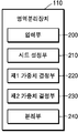
도 2는 본 발명에 따른 영역분리장치의 일 실시예의 구성을 도시한 도면,
도 3 내지 도 5는 본 발명에 따른 의료영상의 영역분리를 위해 사용자로부터 시드군을 입력받는 방법의 일 예를 도시한 도면,
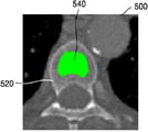
도 6은 본 발명에 따른 의료영상의 영역분리를 위해 영역성장법을 통한 시드군의 설정 방법의 일 예를 도시한 도면,
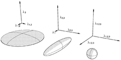
도 7은 본 발명에 따른 의료영상의 영역분리를 위해 형태학적 특징을 이용한 시드군의 설정 방법의 일 예를 도시한 도면,
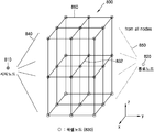
도 8은 본 발명에 따른 의료영상의 영역분리를 위한 노드 그래프의 일 예를 도시한 도면,
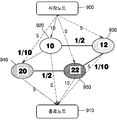
도 9는 본 발명에 따른 의료영상 영역분리 방법의 일 예를 간략히 도시한 도면,
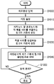
도 10은 본 발명에 따른 의료영상 분리방법의 일 실시 예의 흐름을 도시한 도면, 그리고
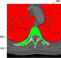
도 11 및 도 12는 본 발명의 일 실시 예에 따라 의료영상에서 영역을 분리한 결과를 도시한 도면이다.
| 구조 특징 | 고유값 조건 |
| 면(sheet) |
|
| 선(line) |
|
| 덩어리(blob) |
|
Claims (9)
- 의료영상을 입력받는 단계;
상기 의료영상의 복셀들 중 분할 대상 영역과 그 나머지 영역에 각각 속한 복셀로 이루어진 제1 시드군 및 제2 시드군을 설정하는 단계;
상기 의료영상의 각 복셀을 나타내는 복셀노드와 시작노드 및 종료노드를 각각 연결하는 링크에 가중치를 부여하되, 복셀노드와 연결되는 노드의 종류 및 복셀노드가 속한 시드군의 종류를 기준으로 상기 가중치를 결정하는 단계;
상기 의료영상의 복셀 사이의 신호강도 차이 및 거리 중 적어도 하나를 기초로 복셀노드 사이를 연결하는 링크의 가중치를 결정하는 단계; 및
상기 의료영상의 복셀노드들이 절단링크에 의해 두 영역으로 분리될 때까지, 상기 시작노드와 상기 종료노드를 연결하는 경로의 가중치 합이 최소가 되는 최단경로에서 가중치가 최소인 링크를 절단링크로 파악하는 과정을 반복수행하는 단계;를 포함하는 것을 특징으로 하는 의료영상의 영역 분할 방법. - 제 1항에 있어서, 상기 제1 시드군 및 제2 시드군을 설정하는 단계는,
상기 의료영상을 화면에 표시하는 단계;
상기 화면을 통해 사용자로부터 점, 선 또는 면을 선택받는 단계;
상기 점, 선 또는 면에 해당하는 복셀을 상기 제1 시드군 또는 상기 제2 시드군으로 설정하는 단계;를 포함하는 것을 특징으로 하는 의료영상의 영역 분할 방법. - 제 1항에 있어서, 상기 제1 시드군 및 제2 시드군을 설정하는 단계는,
상기 의료영상에서 기 설정된 복셀의 신호강도의 상한값을 기준으로 종자점으로부터 확장한 영역에 속한 복셀을 상기 제1 시드군 또는 상기 제2 시드군으로 설정하는 단계;를 포함하는 것을 특징으로 하는 의료영상의 영역 분할 방법. - 제 1항에 있어서, 상기 제1 시드군 및 제2 시드군을 설정하는 단계는,
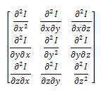
상기 의료영상에서 복셀들의 신호강도의 각 방향의 변화에 대한 방향성을 기초로 파악된 면, 선 또는 덩어리 영역에 속한 복셀을 상기 제1 시드군 또는 상기 제2 시드군으로 설정하는 단계;를 포함하는 것을 특징으로 하는 의료영상의 영역 분할 방법. - 제 1항에 있어서,
상기 시작노드와 상기 제1 시드군의 복셀노드를 연결하는 링크의 제1 가중치, 상기 시작노드와 상기 제2 시드군의 복셀노드를 연결하는 링크의 제2 가중치, 상기 시작노드와 상기 제1 및 제2 시드군에 모두 속하지 않는 복셀노드를 연결하는 링크의 제3 가중치에 있어서, 상기 제1 가중치 > 상기 제3 가중치 > 상기 제2 가중치의 관계를 만족하는 것을 특징으로 하는 의료영상의 영역 분할 방법. - 제 1항에 있어서,
상기 종료노드와 상기 제1 시드군의 복셀노드를 연결하는 링크의 제1 가중치, 상기 종료노드와 상기 제2 시드군의 복셀노드를 연결하는 링크의 제2 가중치, 상기 종료노드와 상기 제1 및 제2 시드군에 모두 속하지 않는 복셀노드를 연결하는 링크의 제3 가중치에 있어서, 상기 제1 가중치 < 상기 제3 가중치 < 상기 제2 가중치의 관계를 만족하는 것을 특징으로 하는 의료영상의 영역 분할 방법. - 제 1항에 있어서, 상기 절단링크로 파악하는 과정을 반복수행하는 단계는,
상기 시작노드와 상기 제2 시드군의 복셀노드를 연결하는 링크 및 상기 종료노드와 상기 제1 시드군의 복셀노드를 연결하는 링크와 절단링크로 파악된 링크를 배제한 상태에서 상기 시작노드와 상기 종료노드 사이를 연결하는 최단경로를 파악하는 단계를 포함하는 것을 특징으로 하는 의료영상의 영역 분할 방법. - 의료영상을 입력받는 입력부;
상기 의료영상의 복셀들 중 분리 대상 영역 및 그 나머지 영역에 각각 속한 복셀로 이루어진 제1 시드군 및 제2 시드군을 설정하는 군설정부;
상기 의료영상의 각 복셀을 나타내는 복셀노드와 시작노드 및 종료노드를 각각 연결하는 링크에 가중치를 부여하되, 복셀노드와 연결되는 노드의 종류 및 복셀노드가 속한 시드군의 종류를 기준으로 상기 가중치를 결정하는 제1 가중치결정부;
상기 의료영상의 복셀들 사이의 신호강도 차이 및 거리를 기초로 복셀노드 사이를 연결하는 링크의 가중치를 결정하는 제2 가중치결정부; 및
상기 의료영상의 복셀노들이 절단링크에 의해 두 영역으로 분리될 때까지, 상기 시작노드와 상기 종료노드를 연결하는 경로의 가중치 합이 최소가 되는 최단경로에서 가중치가 최소인 링크를 절단링크로 파악하는 과정을 반복수행하는 분리부;를 포함하는 것을 특징으로 하는 의료영상의 영역분할장치. - 제 1항 내지 제 7항 중 어느 한 항에 기재된 방법을 수행하기 위한 프로그램을 기록한 컴퓨터로 읽을 수 있는 기록매체.
Priority Applications (2)
| Application Number | Priority Date | Filing Date | Title |
|---|---|---|---|
| KR1020170025866A KR101930905B1 (ko) | 2017-02-28 | 2017-02-28 | 의료영상의 영역 분리 방법 및 그 장치 |
| US15/471,996 US10402975B2 (en) | 2017-02-28 | 2017-03-28 | Method and apparatus for segmenting medical images |
Applications Claiming Priority (1)
| Application Number | Priority Date | Filing Date | Title |
|---|---|---|---|
| KR1020170025866A KR101930905B1 (ko) | 2017-02-28 | 2017-02-28 | 의료영상의 영역 분리 방법 및 그 장치 |
Publications (2)
| Publication Number | Publication Date |
|---|---|
| KR20180098984A true KR20180098984A (ko) | 2018-09-05 |
| KR101930905B1 KR101930905B1 (ko) | 2018-12-19 |
Family
ID=63246846
Family Applications (1)
| Application Number | Title | Priority Date | Filing Date |
|---|---|---|---|
| KR1020170025866A Active KR101930905B1 (ko) | 2017-02-28 | 2017-02-28 | 의료영상의 영역 분리 방법 및 그 장치 |
Country Status (2)
| Country | Link |
|---|---|
| US (1) | US10402975B2 (ko) |
| KR (1) | KR101930905B1 (ko) |
Cited By (3)
| Publication number | Priority date | Publication date | Assignee | Title |
|---|---|---|---|---|
| EP3667609A1 (en) | 2018-12-11 | 2020-06-17 | Medicalip Co., Ltd. | Method and apparatus for reconstructing medical image |
| WO2022010075A1 (ko) | 2020-07-06 | 2022-01-13 | 메디컬아이피 주식회사 | 의료영상을 기초로 인체 조직을 분석하는 방법 및 그 장치 |
| EP4102461A2 (en) | 2021-06-10 | 2022-12-14 | Medicalip Co., Ltd. | Medical image registration method and apparatus |
Families Citing this family (8)
| Publication number | Priority date | Publication date | Assignee | Title |
|---|---|---|---|---|
| US11039883B1 (en) | 2011-12-19 | 2021-06-22 | American Medical Technologies, Llc | Methods and system for atrial fibrillation ablation using balloon based catheters and utilizing medical images (CT or MRI in segments) based cardiac mapping with optional esophageal temperature monitoring |
| US11206984B1 (en) | 2011-12-19 | 2021-12-28 | American Medical Technologies, Llc | Methods and system for cardiac mapping for atrial fibrillation using balloon based catheters utilizing medical images (CT or MRI in segments) and left ventricular lead placement for cardiac re-synchronization therapy (CRT) |
| US10885630B2 (en) | 2018-03-01 | 2021-01-05 | Intuitive Surgical Operations, Inc | Systems and methods for segmentation of anatomical structures for image-guided surgery |
| CN110519338B (zh) * | 2019-08-06 | 2022-02-15 | 中交信息技术国家工程实验室有限公司 | 一种基于协同通信的数据传输方法 |
| US11436724B2 (en) | 2020-10-30 | 2022-09-06 | International Business Machines Corporation | Lesion detection artificial intelligence pipeline computing system |
| US11688063B2 (en) | 2020-10-30 | 2023-06-27 | Guerbet | Ensemble machine learning model architecture for lesion detection |
| US11688517B2 (en) | 2020-10-30 | 2023-06-27 | Guerbet | Multiple operating point false positive removal for lesion identification |
| US11749401B2 (en) * | 2020-10-30 | 2023-09-05 | Guerbet | Seed relabeling for seed-based segmentation of a medical image |
Citations (1)
| Publication number | Priority date | Publication date | Assignee | Title |
|---|---|---|---|---|
| KR20110018573A (ko) | 2009-08-18 | 2011-02-24 | 서울여자대학교 산학협력단 | 호기 및 흡기 컴퓨터 단층촬영 영상의 비강체 폐 영상 정합 장치 및 방법 |
Family Cites Families (11)
| Publication number | Priority date | Publication date | Assignee | Title |
|---|---|---|---|---|
| US6331116B1 (en) | 1996-09-16 | 2001-12-18 | The Research Foundation Of State University Of New York | System and method for performing a three-dimensional virtual segmentation and examination |
| US7760941B2 (en) * | 2005-09-23 | 2010-07-20 | Mevis Research Gmbh | Method and apparatus of segmenting an object in a data set and of determination of the volume of segmented object |
| US8126232B2 (en) * | 2008-03-05 | 2012-02-28 | Siemens Aktiengesellschaft | System and method for 3D vessel segmentation with minimal cuts |
| KR101178398B1 (ko) | 2011-01-06 | 2012-08-30 | 건국대학교 산학협력단 | 의료 영상 분할 처리 장치와 방법 및 컴퓨터 프로그램이 기록된 기록매체 |
| KR101849373B1 (ko) | 2012-01-31 | 2018-04-17 | 한국전자통신연구원 | 인체의 관절구조를 추정하기 위한 장치 및 방법 |
| KR101423835B1 (ko) | 2012-10-15 | 2014-07-29 | 가천대학교 산학협력단 | 의료영상에서의 간 영역 검출방법 |
| KR101482247B1 (ko) | 2013-08-01 | 2015-01-14 | 서울대학교산학협력단 | 기도 추출 방법 및 그 장치 |
| US9996919B2 (en) | 2013-08-01 | 2018-06-12 | Seoul National University R&Db Foundation | Method for extracting airways and pulmonary lobes and apparatus therefor |
| EP2863363A1 (en) * | 2013-09-30 | 2015-04-22 | Samsung Medison Co., Ltd. | Method and apparatus for generating three-dimensional image of target object |
| WO2017030276A1 (ko) | 2015-08-17 | 2017-02-23 | 삼성전자(주) | 의료영상 표시장치 및 의료영상 처리방법 |
| US9786058B2 (en) * | 2016-02-08 | 2017-10-10 | Sony Corporation | Method and system for segmentation of vascular structure in a volumetric image dataset |
-
2017
- 2017-02-28 KR KR1020170025866A patent/KR101930905B1/ko active Active
- 2017-03-28 US US15/471,996 patent/US10402975B2/en active Active
Patent Citations (1)
| Publication number | Priority date | Publication date | Assignee | Title |
|---|---|---|---|---|
| KR20110018573A (ko) | 2009-08-18 | 2011-02-24 | 서울여자대학교 산학협력단 | 호기 및 흡기 컴퓨터 단층촬영 영상의 비강체 폐 영상 정합 장치 및 방법 |
Cited By (4)
| Publication number | Priority date | Publication date | Assignee | Title |
|---|---|---|---|---|
| EP3667609A1 (en) | 2018-12-11 | 2020-06-17 | Medicalip Co., Ltd. | Method and apparatus for reconstructing medical image |
| WO2022010075A1 (ko) | 2020-07-06 | 2022-01-13 | 메디컬아이피 주식회사 | 의료영상을 기초로 인체 조직을 분석하는 방법 및 그 장치 |
| US12322105B2 (en) | 2020-07-06 | 2025-06-03 | Medicalip Co., Ltd. | Method for analyzing human tissue on basis of medical image and device thereof |
| EP4102461A2 (en) | 2021-06-10 | 2022-12-14 | Medicalip Co., Ltd. | Medical image registration method and apparatus |
Also Published As
| Publication number | Publication date |
|---|---|
| KR101930905B1 (ko) | 2018-12-19 |
| US10402975B2 (en) | 2019-09-03 |
| US20180247407A1 (en) | 2018-08-30 |
Similar Documents
| Publication | Publication Date | Title |
|---|---|---|
| KR101930905B1 (ko) | 의료영상의 영역 분리 방법 및 그 장치 | |
| US12292926B2 (en) | Surgical video retrieval based on preoperative images | |
| CN110232383B (zh) | 一种基于深度学习模型的病灶图像识别方法及病灶图像识别系统 | |
| KR101875468B1 (ko) | 질환 모델 기반의 의료 정보 서비스 제공 방법 및 장치 | |
| US20220245821A1 (en) | Automated lung cancer detection from pet-ct scans with hierarchical image representation | |
| US10111713B2 (en) | Surgery assistance apparatus, surgery assistance method and non-transitory computer-readable recording medium having stored therein surgery assistance program | |
| JP5138431B2 (ja) | 画像解析装置および方法並びにプログラム | |
| US10748662B2 (en) | Content-based medical image retrieval method and retrieval system | |
| CN104036484B (zh) | 图像分割装置、图像分割方法和医学图像设备 | |
| KR101482247B1 (ko) | 기도 추출 방법 및 그 장치 | |
| US20100082692A1 (en) | Method and apparatus for classification of coronary artery image data | |
| CN113239755A (zh) | 一种基于空谱融合深度学习的医学高光谱图像分类方法 | |
| EP1996959A2 (en) | System and method of automatic prioritization and analysis of medical images | |
| US11854190B2 (en) | Similarity determination apparatus, similarity determination method, and similarity determination program | |
| KR102349515B1 (ko) | 의료 영상에서 딥러닝에 기반한 종양 자동분할 방법 | |
| Chen et al. | Tree-branch-searching multiresolution approach to skeletonization for virtual endoscopy | |
| CN110738652A (zh) | 一种肺部动静脉分离方法及装置 | |
| WO2009058315A1 (en) | Structure segmentation via mar-cut | |
| CN111986205A (zh) | 血管树生成及病变识别方法、装置、设备及可读存储介质 | |
| JP2006506163A (ja) | 肺結節のコンピュータ支援検出 | |
| CN112712540A (zh) | 一种基于ct影像的肺部支气管提取方法 | |
| US8050470B2 (en) | Branch extension method for airway segmentation | |
| CN111986137B (zh) | 生物器官病变检测方法、装置、设备及可读存储介质 | |
| CN110352448B (zh) | 分离医学图像的区域的方法及设备 | |
| CN101313333A (zh) | 创建结构模型的方法 |
Legal Events
| Date | Code | Title | Description |
|---|---|---|---|
| A201 | Request for examination | ||
| PA0109 | Patent application |
Patent event code: PA01091R01D Comment text: Patent Application Patent event date: 20170228 |
|
| PA0201 | Request for examination | ||
| E902 | Notification of reason for refusal | ||
| PE0902 | Notice of grounds for rejection |
Comment text: Notification of reason for refusal Patent event date: 20180309 Patent event code: PE09021S01D |
|
| PG1501 | Laying open of application | ||
| E701 | Decision to grant or registration of patent right | ||
| PE0701 | Decision of registration |
Patent event code: PE07011S01D Comment text: Decision to Grant Registration Patent event date: 20180917 |
|
| GRNT | Written decision to grant | ||
| PR0701 | Registration of establishment |
Comment text: Registration of Establishment Patent event date: 20181213 Patent event code: PR07011E01D |
|
| PR1002 | Payment of registration fee |
Payment date: 20181214 End annual number: 3 Start annual number: 1 |
|
| PG1601 | Publication of registration | ||
| FPAY | Annual fee payment |
Payment date: 20211203 Year of fee payment: 6 |
|
| PR1001 | Payment of annual fee |
Payment date: 20211203 Start annual number: 4 End annual number: 6 |
|
| PR1001 | Payment of annual fee |
Payment date: 20241211 Start annual number: 7 End annual number: 7 |