KR20180098617A - During the multiple access procedure, - Google Patents
During the multiple access procedure, Download PDFInfo
- Publication number
- KR20180098617A KR20180098617A KR1020187021515A KR20187021515A KR20180098617A KR 20180098617 A KR20180098617 A KR 20180098617A KR 1020187021515 A KR1020187021515 A KR 1020187021515A KR 20187021515 A KR20187021515 A KR 20187021515A KR 20180098617 A KR20180098617 A KR 20180098617A
- Authority
- KR
- South Korea
- Prior art keywords
- filter body
- catheter
- downstream
- port
- open end
- Prior art date
- Legal status (The legal status is an assumption and is not a legal conclusion. Google has not performed a legal analysis and makes no representation as to the accuracy of the status listed.)
- Granted
Links
Images


Classifications
-
- A—HUMAN NECESSITIES
- A61—MEDICAL OR VETERINARY SCIENCE; HYGIENE
- A61F—FILTERS IMPLANTABLE INTO BLOOD VESSELS; PROSTHESES; DEVICES PROVIDING PATENCY TO, OR PREVENTING COLLAPSING OF, TUBULAR STRUCTURES OF THE BODY, e.g. STENTS; ORTHOPAEDIC, NURSING OR CONTRACEPTIVE DEVICES; FOMENTATION; TREATMENT OR PROTECTION OF EYES OR EARS; BANDAGES, DRESSINGS OR ABSORBENT PADS; FIRST-AID KITS
- A61F2/00—Filters implantable into blood vessels; Prostheses, i.e. artificial substitutes or replacements for parts of the body; Appliances for connecting them with the body; Devices providing patency to, or preventing collapsing of, tubular structures of the body, e.g. stents
- A61F2/01—Filters implantable into blood vessels
- A61F2/013—Distal protection devices, i.e. devices placed distally in combination with another endovascular procedure, e.g. angioplasty or stenting
-
- A—HUMAN NECESSITIES
- A61—MEDICAL OR VETERINARY SCIENCE; HYGIENE
- A61B—DIAGNOSIS; SURGERY; IDENTIFICATION
- A61B17/00—Surgical instruments, devices or methods
- A61B17/22—Implements for squeezing-off ulcers or the like on inner organs of the body; Implements for scraping-out cavities of body organs, e.g. bones; for invasive removal or destruction of calculus using mechanical vibrations; for removing obstructions in blood vessels, not otherwise provided for
- A61B17/221—Gripping devices in the form of loops or baskets for gripping calculi or similar types of obstructions
-
- A—HUMAN NECESSITIES
- A61—MEDICAL OR VETERINARY SCIENCE; HYGIENE
- A61B—DIAGNOSIS; SURGERY; IDENTIFICATION
- A61B17/00—Surgical instruments, devices or methods
- A61B17/34—Trocars; Puncturing needles
- A61B17/3468—Trocars; Puncturing needles for implanting or removing devices, e.g. prostheses, implants, seeds, wires
-
- A—HUMAN NECESSITIES
- A61—MEDICAL OR VETERINARY SCIENCE; HYGIENE
- A61F—FILTERS IMPLANTABLE INTO BLOOD VESSELS; PROSTHESES; DEVICES PROVIDING PATENCY TO, OR PREVENTING COLLAPSING OF, TUBULAR STRUCTURES OF THE BODY, e.g. STENTS; ORTHOPAEDIC, NURSING OR CONTRACEPTIVE DEVICES; FOMENTATION; TREATMENT OR PROTECTION OF EYES OR EARS; BANDAGES, DRESSINGS OR ABSORBENT PADS; FIRST-AID KITS
- A61F2/00—Filters implantable into blood vessels; Prostheses, i.e. artificial substitutes or replacements for parts of the body; Appliances for connecting them with the body; Devices providing patency to, or preventing collapsing of, tubular structures of the body, e.g. stents
- A61F2/01—Filters implantable into blood vessels
- A61F2/0103—With centering means
-
- A—HUMAN NECESSITIES
- A61—MEDICAL OR VETERINARY SCIENCE; HYGIENE
- A61F—FILTERS IMPLANTABLE INTO BLOOD VESSELS; PROSTHESES; DEVICES PROVIDING PATENCY TO, OR PREVENTING COLLAPSING OF, TUBULAR STRUCTURES OF THE BODY, e.g. STENTS; ORTHOPAEDIC, NURSING OR CONTRACEPTIVE DEVICES; FOMENTATION; TREATMENT OR PROTECTION OF EYES OR EARS; BANDAGES, DRESSINGS OR ABSORBENT PADS; FIRST-AID KITS
- A61F2/00—Filters implantable into blood vessels; Prostheses, i.e. artificial substitutes or replacements for parts of the body; Appliances for connecting them with the body; Devices providing patency to, or preventing collapsing of, tubular structures of the body, e.g. stents
- A61F2/01—Filters implantable into blood vessels
- A61F2/0105—Open ended, i.e. legs gathered only at one side
-
- A—HUMAN NECESSITIES
- A61—MEDICAL OR VETERINARY SCIENCE; HYGIENE
- A61F—FILTERS IMPLANTABLE INTO BLOOD VESSELS; PROSTHESES; DEVICES PROVIDING PATENCY TO, OR PREVENTING COLLAPSING OF, TUBULAR STRUCTURES OF THE BODY, e.g. STENTS; ORTHOPAEDIC, NURSING OR CONTRACEPTIVE DEVICES; FOMENTATION; TREATMENT OR PROTECTION OF EYES OR EARS; BANDAGES, DRESSINGS OR ABSORBENT PADS; FIRST-AID KITS
- A61F2/00—Filters implantable into blood vessels; Prostheses, i.e. artificial substitutes or replacements for parts of the body; Appliances for connecting them with the body; Devices providing patency to, or preventing collapsing of, tubular structures of the body, e.g. stents
- A61F2/01—Filters implantable into blood vessels
- A61F2/013—Distal protection devices, i.e. devices placed distally in combination with another endovascular procedure, e.g. angioplasty or stenting
- A61F2/014—Retrograde blood flow filters, i.e. device inserted against the blood flow direction
-
- A—HUMAN NECESSITIES
- A61—MEDICAL OR VETERINARY SCIENCE; HYGIENE
- A61B—DIAGNOSIS; SURGERY; IDENTIFICATION
- A61B17/00—Surgical instruments, devices or methods
- A61B17/22—Implements for squeezing-off ulcers or the like on inner organs of the body; Implements for scraping-out cavities of body organs, e.g. bones; for invasive removal or destruction of calculus using mechanical vibrations; for removing obstructions in blood vessels, not otherwise provided for
- A61B2017/22081—Treatment of vulnerable plaque
-
- A—HUMAN NECESSITIES
- A61—MEDICAL OR VETERINARY SCIENCE; HYGIENE
- A61F—FILTERS IMPLANTABLE INTO BLOOD VESSELS; PROSTHESES; DEVICES PROVIDING PATENCY TO, OR PREVENTING COLLAPSING OF, TUBULAR STRUCTURES OF THE BODY, e.g. STENTS; ORTHOPAEDIC, NURSING OR CONTRACEPTIVE DEVICES; FOMENTATION; TREATMENT OR PROTECTION OF EYES OR EARS; BANDAGES, DRESSINGS OR ABSORBENT PADS; FIRST-AID KITS
- A61F2/00—Filters implantable into blood vessels; Prostheses, i.e. artificial substitutes or replacements for parts of the body; Appliances for connecting them with the body; Devices providing patency to, or preventing collapsing of, tubular structures of the body, e.g. stents
- A61F2/01—Filters implantable into blood vessels
- A61F2/011—Instruments for their placement or removal
-
- A—HUMAN NECESSITIES
- A61—MEDICAL OR VETERINARY SCIENCE; HYGIENE
- A61F—FILTERS IMPLANTABLE INTO BLOOD VESSELS; PROSTHESES; DEVICES PROVIDING PATENCY TO, OR PREVENTING COLLAPSING OF, TUBULAR STRUCTURES OF THE BODY, e.g. STENTS; ORTHOPAEDIC, NURSING OR CONTRACEPTIVE DEVICES; FOMENTATION; TREATMENT OR PROTECTION OF EYES OR EARS; BANDAGES, DRESSINGS OR ABSORBENT PADS; FIRST-AID KITS
- A61F2/00—Filters implantable into blood vessels; Prostheses, i.e. artificial substitutes or replacements for parts of the body; Appliances for connecting them with the body; Devices providing patency to, or preventing collapsing of, tubular structures of the body, e.g. stents
- A61F2/02—Prostheses implantable into the body
- A61F2/24—Heart valves ; Vascular valves, e.g. venous valves; Heart implants, e.g. passive devices for improving the function of the native valve or the heart muscle; Transmyocardial revascularisation [TMR] devices; Valves implantable in the body
- A61F2/2427—Devices for manipulating or deploying heart valves during implantation
-
- A61F2002/011—
-
- A—HUMAN NECESSITIES
- A61—MEDICAL OR VETERINARY SCIENCE; HYGIENE
- A61F—FILTERS IMPLANTABLE INTO BLOOD VESSELS; PROSTHESES; DEVICES PROVIDING PATENCY TO, OR PREVENTING COLLAPSING OF, TUBULAR STRUCTURES OF THE BODY, e.g. STENTS; ORTHOPAEDIC, NURSING OR CONTRACEPTIVE DEVICES; FOMENTATION; TREATMENT OR PROTECTION OF EYES OR EARS; BANDAGES, DRESSINGS OR ABSORBENT PADS; FIRST-AID KITS
- A61F2/00—Filters implantable into blood vessels; Prostheses, i.e. artificial substitutes or replacements for parts of the body; Appliances for connecting them with the body; Devices providing patency to, or preventing collapsing of, tubular structures of the body, e.g. stents
- A61F2/01—Filters implantable into blood vessels
- A61F2002/016—Filters implantable into blood vessels made from wire-like elements
-
- A—HUMAN NECESSITIES
- A61—MEDICAL OR VETERINARY SCIENCE; HYGIENE
- A61F—FILTERS IMPLANTABLE INTO BLOOD VESSELS; PROSTHESES; DEVICES PROVIDING PATENCY TO, OR PREVENTING COLLAPSING OF, TUBULAR STRUCTURES OF THE BODY, e.g. STENTS; ORTHOPAEDIC, NURSING OR CONTRACEPTIVE DEVICES; FOMENTATION; TREATMENT OR PROTECTION OF EYES OR EARS; BANDAGES, DRESSINGS OR ABSORBENT PADS; FIRST-AID KITS
- A61F2230/00—Geometry of prostheses classified in groups A61F2/00 - A61F2/26 or A61F2/82 or A61F9/00 or A61F11/00 or subgroups thereof
- A61F2230/0063—Three-dimensional shapes
- A61F2230/0069—Three-dimensional shapes cylindrical
-
- A—HUMAN NECESSITIES
- A61—MEDICAL OR VETERINARY SCIENCE; HYGIENE
- A61F—FILTERS IMPLANTABLE INTO BLOOD VESSELS; PROSTHESES; DEVICES PROVIDING PATENCY TO, OR PREVENTING COLLAPSING OF, TUBULAR STRUCTURES OF THE BODY, e.g. STENTS; ORTHOPAEDIC, NURSING OR CONTRACEPTIVE DEVICES; FOMENTATION; TREATMENT OR PROTECTION OF EYES OR EARS; BANDAGES, DRESSINGS OR ABSORBENT PADS; FIRST-AID KITS
- A61F2250/00—Special features of prostheses classified in groups A61F2/00 - A61F2/26 or A61F2/82 or A61F9/00 or A61F11/00 or subgroups thereof
- A61F2250/0058—Additional features; Implant or prostheses properties not otherwise provided for
- A61F2250/0069—Sealing means
Landscapes
- Health & Medical Sciences (AREA)
- Life Sciences & Earth Sciences (AREA)
- General Health & Medical Sciences (AREA)
- Animal Behavior & Ethology (AREA)
- Engineering & Computer Science (AREA)
- Biomedical Technology (AREA)
- Heart & Thoracic Surgery (AREA)
- Veterinary Medicine (AREA)
- Public Health (AREA)
- Vascular Medicine (AREA)
- Cardiology (AREA)
- Oral & Maxillofacial Surgery (AREA)
- Transplantation (AREA)
- Surgery (AREA)
- Nuclear Medicine, Radiotherapy & Molecular Imaging (AREA)
- Medical Informatics (AREA)
- Molecular Biology (AREA)
- Orthopedic Medicine & Surgery (AREA)
- Pathology (AREA)
- Surgical Instruments (AREA)
- Prostheses (AREA)
Abstract
색전 방지 디바이스는 외장에 부착된 관형 필터 바디를 포함한다. 관형 필터 바디는 색전을 포획하기 위한 상류측 개방 단부 및 대체로 폐쇄되는 하류측 단부를 갖는다. 관형 필터 바디의 색전 포획 단부를 통한 자가 개방 통로는 다수의 카테터가 외장으로부터 또는 그렇지 않으면 필터 바디 내로 동시에 또는 순차적으로 전진되게 한다. 외장은 필터 바디의 색전 포획 단부 부근의 주연 지지 구조체에 부착되어 속박 전달 카테터를 통한 필터 바디의 전개 및 회수를 용이하게 한다.The embolization device includes a tubular filter body attached to the enclosure. The tubular filter body has an upstream open end for capturing the embolization and a generally closed downstream end. The self-opening passageway through the embolization capture end of the tubular filter body allows multiple catheters to advance simultaneously or sequentially from the enclosure or into the filter body. The sheath is attached to the peripheral support structure near the embolization capture end of the filter body to facilitate deployment and retrieval of the filter body through the captive delivery catheter.

Description
관련 출원의 상호 참조Cross reference of related application
본 출원은 2015년 12월 29일 출원된 미국 가출원 제62/272,643호(대리인 문서 번호 41959-711.101); 2016년 2월 11일 출원된 미국 가출원 제62/294,018호(대리인 문서 번호 41959-711.102); 2016년 2월 18일 출원된 미국 가출원 제62/297,053호(대리인 문서 번호 41959-712.101); 2016년 4월 25일 출원된 미국 특허 출원 제15/137,924호(대리인 문서 번호 41959-712.201)의 이익을 청구하고, 이들 미국 출원의 전체 개시내용은 본 명세서에 참조로서 합체되어 있다.This application claims the benefit of U.S. Provisional Application No. 62 / 272,643, filed December 29, 2015 (Attorney Docket No. 41959-711.101); U.S. Provisional Application No. 62 / 294,018, filed February 11, 2016 (Attorney Docket No. 41959-711.102); U.S. Provisional Application No. 62 / 297,053, filed February 18, 2016 (Attorney Docket No. 41959-712.101); Filed on April 25, 2016 (Attorney Docket No. 41959-712.201), the entire disclosures of which are incorporated herein by reference.
본 출원의 개시내용은 또한 공동 소유로서 계류중이며 2014년 11월 10일 출원된 미국 특허 출원 제14/537,814호(대리인 문서 번호 41959- 707.201) 및 2013년 1월 7일 출원된 미국 특허 출원 제13/735,864호(대리인 문서 번호 41959-705.201)에 관련되며, 이들 미국 출원의 전체 개시내용은 본 명세서에 참조로서 합체되어 있다. 이 단락에서 열거되어 있는 출원들로부터의 우선권은 주장하지 않는다.The disclosure of the present application is also incorporated herein by reference in its entirety as U.S. Patent Application Serial No. 14 / 537,814, filed November 10, 2014 (Attorney Docket Nos. 41959-707.201) and U.S. Patent Application No. 13 / 735,864 (Attorney Docket No. 41959-705.201), the entire disclosures of which are incorporated herein by reference. It does not claim priority from the applications listed in this paragraph.
발명의 분야Field of invention
본 발명은 일반적으로 의료 디바이스 및 방법에 관한 것으로서, 더 구체적으로는 심장 수술(cardiac surgery) 및 중재적 심장 시술(interventional cardiology procedure) 중에 환자의 대동맥궁 혈관에서의 색전 방지(embolic protection)를 제공하기 위한 장치 및 방법에 관한 것이다.The present invention relates generally to medical devices and methods, and more particularly to a method and apparatus for providing embolic protection in aortic arch vessels of a patient during cardiac surgery and interventional cardiology procedures. And more particularly,
대뇌 색전증은 심장 수술, 심폐 우회술, 및 카테터 기반 중재적 심장 시술 및 전기생리학 시술의 알려진 합병증이다. 혈전, 죽종, 및 지질을 포함하는 색전 입자는 수술 또는 카테터 조작에 의해 제거되고, 혈류에 진입하고, 뇌 또는 하류측의 다른 생체 기관에 "색전을 유발한다". 대뇌 색전증은 신경심리학적 결핍, 뇌졸중 및 심지어 사망을 유도할 수 있다. 색전 방출부(embolic release)의 하류측의 다른 기관이 또한 손상될 수 있어, 기능 저하 또는 기관의 기능 상실을 야기한다.Cerebral embolism is a known complication of cardiac surgery, cardiopulmonary bypass, and catheter-based interventional cardiac and electrophysiology procedures. Thrombosis, atheroma, and lipid-containing embolization particles are removed by surgery or catheter manipulation, enter the bloodstream, and "embolize" the brain or other living organs on the downstream side. Cerebral embolism can lead to neuropsychological deprivation, stroke, and even death. Other organs on the downstream side of the embolic release may also be damaged, resulting in impaired function or loss of function of the organs.
본 발명에서 특히 관심을 가져, 환자의 대동맥궁 위로 전진되는 카테터를 사용하여 다수의 시술이 대동맥 판막에 대해 수행된다. 판막성형 시술은 수년 동안 수행되어 왔고, 대동맥 판막 상의 석회화를 파열시키기 위해 대동맥궁 위로 전진되는 고압 벌룬을 사용한다. 이러한 시술은 대뇌 동맥으로의 색전 방출의 상당한 위험을 야기한다. 더 최근에, 경도관 대동맥 판막 이식(transcatheter aortic valve implantation: TAVI) 시술 또는 경도관 대동맥 판막 치환(transcatheter aortic valve replacement: TAVR) 시술로서 또한 알려져 있는 경피적 대동맥 판막 치환(percutaneous aortic valve replacement: PAVR) 시술이 승인되었고, 이들의 사용이 보급되고 있다. 다수의 환자 이익을 제공하지만, 이들 시술은 특히 카테터가 대동맥궁 위로 도입된 상태에서 혈관경유식으로 수행될 때, 색전 방출의 상당한 위험을 또한 야기한다.Of particular interest in the present invention, a number of procedures are performed on the aortic valve using catheters that advance over the aortic arch of the patient. Valve casting has been performed for many years and uses high pressure balloons that are advanced over the aortic arch to rupture the calcifications on the aortic valve. This procedure causes a considerable risk of embolization of the cerebral artery. More recently, percutaneous aortic valve replacement (PAVR) procedures, also known as transcatheter aortic valve implantation (TAVI) or transcatheter aortic valve replacement (TAVR) Have been approved, and their use has become widespread. While providing a number of patient benefits, these procedures also pose a significant risk of embolization, especially when the catheter is performed via the vascular route with the introduced over the aortic arch.
이들 시술 및 다른 시술에서의 색전증의 방지가 환자에게 유리할 것이고 다수의 외과 시술의 성과를 향상시킬 것이다. 하나가 넘는 접속 부위 및 하나가 넘는 시술 디바이스를 수반하는 카테터 기반 시술 중에 잠재적인 색전이 종종 제거되면, 다수의 카테터로 진단 및 중재적 시술을 수행하기 위해 방지 디바이스를 통한 또는 방지 디바이스를 지나는 다중 접근 경로를 제공하는 색전 방지 시스템을 전개하는 것이 유리할 것이다. 혈관조영술 진단 카테터, 경도관 판막 전달 시스템, 및 전기생리학적 카테터와 함께 사용되는 것과 같이, 시술을 수행하는 데 사용되는 외장(sheath) 상에 색전 방지 시스템을 일체화하는 것이 또한 유리할 것이다.Prevention of embolism in these procedures and other procedures will be beneficial to the patient and will improve the outcome of many surgical procedures. If potential embolizations are often removed during catheter-based procedures involving more than one access site and more than one surgical device, multiple accesses through the prevention device or across the prevention device to perform diagnostic and interventional procedures with multiple catheters It would be advantageous to deploy an embolic protection system that provides pathways. It would also be advantageous to integrate an embolization system on the sheath used to perform the procedure, such as used with an angiography diagnostic catheter, a hardness tube valve delivery system, and an electrophysiological catheter.
본원과 함께 공동으로 양수된 미국 특허 출원 공개 제2015/0066075호는 종래의 색전 방지용 외장 접근 디바이스의 단점의 일부를 해소하는, 특히 판막성형술 및 TAVR 시술에 사용되도록 의도된 도입기 외장(introducer sheath)을 설명하고 있다. '075 출원의 외장은 색전 방지 요소를 포함하고, 외장을 통해 콘트라스트 카테터(contrast catheter) 또는 다른 소형 카테터 및 필터 내에 형성된 포트를 통해 제2 카테터를 전진시키기에 적합하다. 이러한 특징을 갖는 이전의 색전 방지식 접근 외장에 비해 상당한 개량이 있지만, '075 출원의 접근법의 특정 디자인은 전개 및 회수가 어려울 수 있고, 소량의 색전을 손실할 수 있고, 전개 중에 비교적 큰 프로파일을 가질 수 있다.U.S. Patent Application Publication No. 2015/0066075, which is commonly assigned herewith, discloses an introducer sheath which is intended to be used in valvuloplasty and TAVR procedures, which solves some of the disadvantages of conventional embolization external access devices . The enclosure of the '075 application includes an embolization element and is suitable for advancing the second catheter through a port formed in the contrast catheter or other small catheter and filter through an enclosure. While there is considerable improvement over previous embolization approaches with these features, certain designs of the '075 application approach may be difficult to deploy and recover, may lose a small amount of embolization, Lt; / RTI >
따라서, 대동맥궁 위에서 수행된 심장 시술 및 다른 시술 중에 색전증을 방지하기 위한 개량된 디바이스, 시스템, 및 방법을 제공하는 것이 바람직할 것이다. 이러한 디바이스, 시스템, 및 방법은 덜 복잡한 전개 프로토콜을 제공해야 하고, 전개될 때 비교적 낮은 프로파일을 가져야 하고, 시술 중에 항상 신뢰성 있고 효율적인 색전 억제를 제공해야 한다. 이들 목적의 적어도 일부는 본 명세서에 설명된 발명에 의해 부합될 것이다.Accordingly, it would be desirable to provide an improved device, system, and method for preventing embolism during heart procedures and other procedures performed on the aortic arch. These devices, systems, and methods must provide a less complex deployment protocol, have a relatively low profile when deployed, and always provide reliable and efficient embolic repression during the procedure. At least some of these objectives will be met by the invention described herein.
미국 특허 출원 공개 제2015/0066075호는 전술되었다. 대뇌 색전증을 방지하기 위한 다른 필터 및 디바이스가 미국 특허 출원 공개 제2013/0178891호; 제2010/0312268호; 제2006/0287668호; 제2005/0010246호; 제2005/0283186호; 제2004/0215167호; 제2003/0100940호; PCT 공보 WO/2004/019817호; 미국 특허 제8,114,114호; 제7,232,453호; 제6,712,834호; 제6,537,297호; 제6,499,487호; 제6,371,935호; 제6,361,545호; 제6,258,120호; 제6,254,563호; 제6,245,012호; 제6,139,517호; 제5,769,819호에 설명되어 있다.U.S. Patent Application Publication No. 2015/0066075 has been described above. Other filters and devices for preventing cerebral embolism are disclosed in U.S. Patent Application Publication Nos. 2013/0178891; 2010/0312268; 2006/0287668; 2005/0010246; 2005/0283186; 2004/0215167; 2003/0100940; PCT Publication WO / 2004/019817; U.S. Patent No. 8,114,114; 7,232, 453; 6,712,834; 6,537,297; 6,499,487; 6,371, 935; 6,361,545; 6,258,120; 6,254,563; 6,245,012; 6,139,517; 5,769, 819. < / RTI >
본 발명은 색전을 수집하기 위한 방법, 시스템, 및 디바이스, 특히 팔머리동맥, 좌경동맥, 및 좌쇄골하동맥을 포함하여, 대동맥측 혈관 내로 색전이 방출되는 위험이 있는 대동맥 판막 치환술, 대동맥 판막 판막성형술 등을 포함하여, 환자의 대동맥 내의 중재적 시술의 수행 중에 대뇌 혈관구조 내로의 색전의 방출을 방지하기 위한 방법, 시스템, 및 디바이스를 제공한다. 본 발명은 색전 방지 디바이스, 관형 필터 바디, 및 통상적으로 보통 한쪽 또는 양쪽의 대퇴동맥 접근법에 의해 하행 대동맥으로부터 유도되는 1개, 2개, 3개 이상의 중재적 카테터 및/또는 진단 카테터에 의해 대동맥 판막으로의 동시 접근을 허용하면서 대동맥측 분기 혈관 내로의 색전 방출을 저지하기 위해 하행 대동맥을 통해 그리고 대동맥궁 위로 디바이스 및 필터를 배치하기 위한 시스템 및 방법을 제공한다.The present invention relates to a method, system and device for collecting an embolus, particularly aortic valve replacement at risk of embolization into the aortic vessel, including arthroscopic, left, and subclavian arteries, System, and device for preventing the release of embolization into the cerebral vasculature during the performance of an interventional procedure in the aorta of a patient, including, The present invention relates to an embolization device, a tubular filter body, and an aortic valve by means of one, two, three or more interventional catheters and / or diagnostic catheters usually derived from the descending aorta by one or both femoral artery approaches. A system and method for placing devices and filters over the descending aorta and over the aortic arch to block embolization into the aortic branch branch vessel while allowing simultaneous access to the aortic branch vessel.
색전 방지 디바이스는 필터 바디 및 필터 바디에 연결된 전개 카테터 바디를 포함한다. 필터 바디는 통상적으로 관형 다공성 메시 재료를 포함하고, 혈류의 진입을 허용하기 위한 상류측 개방 단부 그리고 적어도 하나의 작동 카테터 및 일반적으로 2개 이상의 작동 카테터의 동시 진입을 허용하기 위한 하류측 개방 단부를 갖는다. 전개 카테터 바디는 필터 바디의 하류측 개방 단부에 직접 결합되거나 또는 간접 결합되고, 여기서 상류측 및 하류측은 혈류의 방향을 칭하는데, 예를 들어 하류측은 하행 대동맥을 향하고 심장 및 대동맥궁으로부터 이격된다. 적어도 하나의 자가 밀봉 포트 또는 통로가 필터 바디의 내부 내에 제공되고, 전개 카테터 바디는 통상적으로 자가 밀봉 포트를 통해 진단 카테터, 중재적 카테터, 또는 다른 작동 카테터를 유도하기 위해 관형 필터 바디의 내부에 적어도 하나의 접근 경로를 제공하기 위한 적어도 하나의 루멘(lumen)을 갖는다. 바람직하게는, 하나 이상의 부가의 작동 카테터는 외장을 통해 유도된 제1 카테터와 동시에 또는 순차적으로 동일한 자가 밀봉 통로를 통해 유도될 수도 있다. 부가의 자가 밀봉 접근 포트 또는 다른 카테터 접근 포트가 필터 바디를 통한 다른 평행한 접근 경로를 제공하도록 포함될 수 있지만, 자가 밀봉 통로는 통상적으로, 어떠한 카테터도 존재하지 않을 때 색전 방출을 차단하도록 폐쇄되는 것이 가능하면서 2개 이상의 카테터의 동시 통과를 허용하도록 충분히 팽창 가능한 직경을 가질 것이기 때문에 일반적으로 불필요하다. 축방향으로 정렬된 다른 자가 밀봉 카테터 접근 포트가 또한 필터 바디 내에 부가의 색전 포획 챔버를 제공하도록 포함될 수 있다.An embolization device includes a filter body and a deployment catheter body connected to the filter body. The filter body typically includes a tubular porous mesh material and includes an upstream open end for allowing entry of blood flow and a downstream open end for allowing simultaneous entry of at least one working catheter and generally two or more operating catheters . The deploying catheter body is directly or indirectly coupled to the downstream open end of the filter body, wherein the upstream and downstream sides refer to the direction of the blood flow, e.g., the downstream side is directed to the descending aorta and is spaced from the heart and the aortic arch. At least one self-sealing port or passageway is provided within the interior of the filter body, and the deployment catheter body is typically provided with at least one self-sealing port or passageway within the interior of the tubular filter body for guiding the diagnostic catheter, interventional catheter, And at least one lumen for providing one access path. Advantageously, the at least one additional working catheter may be guided through the same self-sealing passageway, either simultaneously or sequentially with the first catheter introduced through the enclosure. Although a self-sealing access port or other catheter access port may be included to provide another parallel access path through the filter body, the self-sealing passageway is typically closed to block embolization when no catheter is present Which is generally unnecessary since it will have a diameter that is sufficiently expandable to allow simultaneous passage of two or more catheters. Other axially aligned self-sealing catheter access ports may also be included to provide additional embolic capture chambers within the filter body.
본 발명의 제1 특정 양태에서, 색전 방지 디바이스는 관형 다공성 메시 재료로부터 형성되고 상류측 개방 단부 및 하류측 개방 단부를 갖는 필터 바디를 포함한다. 자가 밀봉 포트는 각각의 단부로부터 내향으로 이격되어 있고, 자가 밀봉 포트는 그를 통과하는 적어도 하나의 작동 카테터에 순응하도록 구성된 팽창형 개구를 포함한다. 반경방향 절첩형 지지부는 필터 바디의 하류측 단부의 주연부에 결합되고, 원위 단부를 갖는 카테터 바디는 반경방향 절첩형 지지부에 결합되고, "원위측"은 조작자로부터 이격되어 있는, 즉 바디의 외부에 있는 디바이스의 부분으로부터 가장 멀리 이격되어 있는 디바이스의 방향을 칭한다. 유사하게, 용어 "근위측"은 조작자에 더 근접한, 즉 바디의 외부에 있는 디바이스의 부분에 더 근접한 디바이스의 방향을 칭한다. 전달 외장은 필터 바디를 수용하여 반경방향으로 속박하여, 카테터 바디가 전달 외장에 대해 원위측으로 전진되어 필터 바디를 속박으로부터 해제시키고 지지부가 필터 바디의 하류측 단부에 외접하는 상태로 필터 바디가 반경방향으로 팽창하게 할 수도 있도록 구성된 루멘을 갖는다. 이 방식으로, 카테터 바디는 전달 외장에 대해 원위측으로 전진되고 근위측으로 후퇴되어 전달 외장의 루멘 내외로 지지부와 필터 바디의 조립체를 이동시킬 수도 있다. 특히, 전달 외장 외부로 전진될 때, 지지부는 필터 바디의 하류측 단부의 전개를 보조하도록 개방될 것이고, 전달 외장 내로 재차 후퇴될 때, 지지부는 필터 바디가 루멘 내로 당겨지기 전에 필터 바디의 하류측 단부가 접히게 하도록 폐쇄될 것이다.In a first particular aspect of the invention, the embolization prevention device comprises a filter body formed from a tubular porous mesh material and having an upstream open end and a downstream open end. The self-sealing port is spaced inwardly from each end, and the self-sealing port includes an inflatable opening configured to conform to at least one working catheter passing therethrough. The radial foldable support is coupled to the periphery of the downstream end of the filter body and the catheter body having the distal end is coupled to the radial foldable support and the "distal" is spaced from the operator, Refers to the direction of the device that is farthest away from that portion of the device. Similarly, the term "proximal side " refers to the orientation of the device closer to the operator, i.e. closer to the portion of the device that is external to the body. The delivery sheath receives and radially constrains the filter body such that the catheter body is advanced distally with respect to the delivery sheath so that the filter body is released from the bondage and the support is circumscribed at the downstream end of the filter body, Lt; RTI ID = 0.0 > lumen < / RTI > In this manner, the catheter body may be advanced distally to the delivery sheath and retracted proximally to move the assembly of the support and filter body into and out of the lumen of the delivery sheath. In particular, when advancing out of the delivery enclosure, the support will be open to assist in the deployment of the downstream end of the filter body, and when retracted back into the delivery enclosure, the support is positioned on the downstream side of the filter body before the filter body is pulled into the lumen The ends will be closed to fold.
특정 실시예에서, 필터 바디는 포트의 하류측 단부와 필터 바디의 하류측 단부 사이에 배치된 개방 원통형 챔버를 갖는다. 포트는 관형 다공성 메시 재료까지 벽 부분을 포함할 수도 있고, 여기서 벽 부분은 전달 외장으로부터 반경방향 속박으로부터 해제될 때 다른 벽 부분이 팽창함에 따라 반경방향 내향으로 절첩되고, 반전되거나 다른 방식으로 편향된다. 또 다른 특정 실시예에서, 벽 부분은 하류측에서 원추형 개구 또는 기부를 갖는 포트를 형성하도록 반전된다. 예를 들어, 관형 다공성 메시 재료의 반전된 벽 부분은 포트의 팽창형 개구를 형성하는 원추형 개구의 정점 또는 기부로부터 상류측 방향으로 연장되는 탄성적으로 폐쇄된 슬리브 부분을 가질 수도 있다.In certain embodiments, the filter body has an open cylindrical chamber disposed between the downstream end of the port and the downstream end of the filter body. The port may include a wall portion up to the tubular porous mesh material wherein the wall portion is folded radially inwardly as the other wall portion expands and is reversed or otherwise deflected when released from the radial constraint . In another specific embodiment, the wall portion is inverted to form a port having a conical opening or base at the downstream side. For example, the inverted wall portion of the tubular porous mesh material may have an elastically closed sleeve portion extending upstream from the apex or base of the conical opening forming the inflatable opening of the port.
또 다른 특정 실시예에서, 반경방향 절첩형 지지부는 필터 바디의 하류측 단부의 주연부 주위에 고정된 루프를 포함할 수도 있다. 루프는, 카테터 바디 내의 전개 루멘을 통과하는 테더에 연결될 수도 있다. 루프는, 전달 외장의 루멘 내로 필터 바디를 당기기 전에, 폐쇄된 필터 바디의 개방 단부가 테더에 의해 당겨지는 것을 허용하기 위한 올가미(lasso)로서 구성될 수도 있다. 대안으로, 반경방향 절첩형 지지부는 필터 바디의 하류측 단부의 주연부에 결합된 개방 단부 및 카테터 바디의 원위 단부에 결합된 속박 단부를 갖는 골격(scaffold)을 포함할 수도 있다.In another specific embodiment, the radial foldable support may comprise a loop secured about the periphery of the downstream end of the filter body. The loop may be connected to a tether that passes through the deployment lumen in the catheter body. The loop may be configured as a lasso to allow the open end of the closed filter body to be pulled by the tether, before pulling the filter body into the lumen of the transmission enclosure. Alternatively, the radial foldable support may include a scaffold having an open end coupled to the periphery of the downstream end of the filter body and a constrained end coupled to the distal end of the catheter body.
본 발명의 또 다른 특정 실시예에서, 카테터 바디는 작동 카테터가 루멘을 통해 필터 바디의 하류측 개방 단부 내로 그리고 이어서 포트를 통해 전진될 수도 있도록 적어도 하나의 작동 카테터를 수용하기 위한 루멘을 가질 것이다. 카테터 바디는 반경방향 절첩형 지지부에 부착된 테더를 수용하기 위한 적어도 하나의 부가의 루멘을 더 포함할 수도 있다. 부가의 루멘은 또한 다른 목적으로 제공될 수도 있다.In another specific embodiment of the present invention, the catheter body will have a lumen for receiving the at least one operating catheter such that the working catheter can be advanced through the lumen into the downstream open end of the filter body and subsequently through the port. The catheter body may further comprise at least one additional lumen for receiving a tether attached to the radial foldable support. Additional lumens may also be provided for other purposes.
본 발명의 제2 특정 양태에서, 관강내 색전 포획 디바이스는, 관형 다공성 메시 재료로부터 형성되고 상류측 개방 단부, 하류측 개방 단부, 및 적어도 각각의 단부로부터 내향으로 이격되어 있는 제1 포트를 갖는 필터 바디를 포함한다. 포트는 그를 통과하는 적어도 하나의 작동 카테터를 형성하도록 구성된 팽창형 개구를 포함하고, 필터 바디는 그 하류측 단부에 적어도 원통형 개방 챔버 그리고 그 상류측 단부에 원통형 개방 챔버를 가질 것이고, 여기서 포트는 그 사이에 배치된다. 색전 포획 디바이스는 필터 바디의 하류측 단부에 결합된 원위 단부를 갖는 카테터 바디를 더 포함할 수도 있다.In a second particular aspect of the present invention, an intratracheal embolization capture device comprises a filter having a first port formed from a tubular porous mesh material and spaced inwardly from an upstream open end, a downstream open end, Includes body. The port comprising an inflatable opening configured to form at least one working catheter therethrough, the filter body having at least a cylindrical opening chamber at its downstream end and a cylindrical opening chamber at its upstream end, Respectively. The embolization capture device may further comprise a catheter body having a distal end coupled to a downstream end of the filter body.
특정 실시예에서, 다공성 메시 재료는 사전 결정된 크기를 초과하는 색전이 통과하는 것을 방지하도록 선택된 기공 크기를 갖는 편직 섬유, 편조 섬유, 직조 섬유, 또는 부직 섬유, 필라멘트, 또는 와이어의 직물을 포함한다. 다수의 실시예에서, 직물은 관형 메시의 적어도 일부에 대해 이중벽일 것이고, 다공성 메시 재료는 탄성 재료, 폴리머 재료, 가단성 재료, 소성 변형성 재료, 형상 기억 재료, 또는 이들의 조합으로 제조될 수도 있다. 다른 특정 경우에, 다공성 메시 재료는 그 표면 상에 혈전 생성 방지 코팅을 가질 수도 있고, 기공 크기는 통상적으로 약 1 mm 내지 약 0.1 mm의 범위일 것이다. 예시적인 다공성 메시 재료는 276개의 Nitinol®(니켈-티타늄 합금) 와이어와 12개의 탄탈 와이어의 조합을 포함하여, 288개의 개별 와이어로부터 형성된 이중층 편조부(braid)를 포함하고, 각각의 와이어는 20 mm 내지 40 mm의 최종 이중층 메시 직경으로 형성된 0.002 인치 직경이다.In certain embodiments, the porous mesh material comprises a fabric of knit fibers, braided fibers, woven fibers, or nonwoven fibers, filaments, or wires having a pore size selected to prevent the embolization exceeding a predetermined size from passing therethrough. In many embodiments, the fabric will be a double wall for at least a portion of the tubular mesh, and the porous mesh material may be made of an elastic material, a polymer material, a malleable material, a plastic deformation material, a shape memory material, or a combination thereof. In other specific cases, the porous mesh material may have a thrombogenic coating on its surface, and the pore size will typically range from about 1 mm to about 0.1 mm. An exemplary porous mesh material comprises a double layer braid formed from 288 individual wires, including a combination of 276 Nitinol (R) (nickel-titanium alloy) wires and 12 tantalum wires, Lt; RTI ID = 0.0 > mm, < / RTI >
다른 특정 실시예에서, 적어도 제1 포트는 관형 다공성 메시 재료의 벽 부분으로부터 형성되거나 포함한다. 벽 부분은 속박으로부터 해제될 때 다른 벽 부분이 팽창함에 따라 포트가 반경방향 내향으로 절첩되거나 폐쇄되도록 형성 또는 성형되는데, 예를 들어 열성형되고 경화된다. 벽 부분은 통상적으로 하류측에 원추형 개구, 그리고 통상적으로 포트의 팽창형 개구를 형성하는 원추형 개구의 정점으로부터 상류측 방향으로 연장되는 폐쇄된 슬리브 부분을 갖는 포트를 형성하기 위해 반전되도록 사전성형될 것이다. 대안적인 실시예에서, 포트는 반경방향 내향으로 속박되고, 압착되거나, 또는 다른 방식으로 폐쇄되어 있지만 그를 통한 작동 카테터(들)의 통과에 응답하여 개방될 부분인 관형 다공성 메시의 벽 부분에 의해 형성될 수도 있다. 특정 실시예에서, 상류측 챔버 및 하류측 챔버에 추가하여, 필터 바디는 제1 포트 또는 다른 포트의 하류측 단부와 제2 포트 또는 다른 포트의 상류측 단부 사이에 하나 이상의 "중앙" 원통형 개방 챔버를 가질 수도 있다.In another particular embodiment, at least the first port is formed or comprises a wall portion of the tubular porous mesh material. The wall portion is formed or shaped such that when the other wall portion expands, the port is folded or closed radially inwardly as it is released from the constraint, e.g., thermoformed and cured. The wall portion will be preformed to be inverted to form a port that typically has a conical opening on the downstream side and a closed sleeve portion extending in the upstream direction from the apex of the conical opening that typically forms the inflatable opening of the port . In an alternative embodiment, the port is formed by a wall portion of a tubular porous mesh that is constrained radially inwardly, compressed, or otherwise closed, which portion is to be opened in response to passage of the operating catheter (s) therethrough . In certain embodiments, in addition to the upstream chamber and the downstream chamber, the filter body includes one or more "central" cylindrical open chambers between the downstream end of the first port or other port and the upstream end of the second port or other port. .
본 발명의 제3 특정 양태에서, 응괴 회수 시스템은 응괴 포획 원위 단부를 갖는 응괴 회수 작동 카테터와 조합하여 방금 설명된 색전 방지 디바이스를 포함하고, 여기서 응괴 회수 작동 카테터는 필터 바디 상의 상류측 개방 단부를 통해 중앙 챔버 내로 하류측 방향으로 회수된 응괴를 당기도록 구성된다.In a third particular aspect of the present invention, the coagulum recovery system includes an embolism recovery device just described in combination with a coagulum recovery operating catheter having a coagulated capture distal end, wherein the coagulum recovery working catheter has an upstream open end on the filter body To withdraw the recovered clusters in the downstream direction into the central chamber.
제4 특정 양태에서, 본 발명은 환자의 대동맥궁 내로 그리고/또는 위로 작동 카테터를 전진시키기 위한 방법을 제공한다. 다공성 메시로부터 적어도 부분적으로 형성된 원통형 필터 바디가 제공된다. 원통형 필터 바디는 색전을 위한 수집 챔버를 형성하고, 상류측 개방 단부, 하류측 개방 단부, 각각의 단부들로부터 내향으로 이격되어 있는 자가 밀봉 포트, 필터 바디의 하류측 단부의 주연부에 결합된 반경방향 절첩형 지지부를 갖는다. 원통형 필터 바디를 지지하고 속박하는 전개 카테터는 대동맥궁의 하류측으로 전진되고, 반면에 필터 바디는 통상적으로 이전에 배치된 전달 외장으로 그 반경방향으로 속박된 구성으로 유지된다. 원통형 필터 바디는 반경방향으로 팽창되어, 다공성 메시의 벽이 환자의 대동맥측 또는 분기 혈관을 덮게 되고 필터 바디의 상류측 개방 단부가 환자의 심장에 대면하게 된다. 혈액은 상류측 개방 단부를 통해 필터 바디의 내부 내로 유동하고, 색전은 수집 챔버 내에 수집된다. 필터 바디가 전개된 후에, 지지부가 필터 바디의 하류측 단부를 개방 유지하기 위해 반경방향으로 팽창되고, 필터 바디의 다공성 메시를 통해 그리고 대동맥측 혈관 내로 유동하는 혈액은 실질적으로 색전이 없다. 필터 바디가 전개된 후에, 제1 작동 카테터가 필터 바디의 하류측 개방 단부를 통해 그리고 자가 밀봉 포트를 통해 심장을 향해 전진될 수 있다. 선택적으로, 제2 작동 카테터가 필터 바디의 하류측 개방 단부를 통해 그리고 자가 밀봉 포트를 통해 심장을 향해 제1 작동 카테터의 배치와 동시에 또는 순차적으로 전진될 수도 있다.In a fourth particular aspect, the present invention provides a method for advancing a working catheter into and / or out of a patient's aortic arch. A cylindrical filter body at least partially formed from a porous mesh is provided. The cylindrical filter body forms a collection chamber for embolization and has an upstream open end, a downstream open end, a self-sealing port spaced inwardly from the respective ends, a radial direction coupled to the periphery of the downstream end of the filter body And has a foldable support portion. The deploying catheter that supports and binds the cylindrical filter body is advanced to the downstream side of the aortic arch, while the filter body is typically maintained in its radially constrained configuration with the previously deployed delivery sheath. The cylindrical filter body is radially expanded such that the wall of the porous mesh covers the patient's aortic or branch vessel and the upstream open end of the filter body faces the patient's heart. The blood flows through the upstream open end into the interior of the filter body and the embolus is collected in the collection chamber. After the filter body is deployed, the support is radially expanded to keep the downstream end of the filter body open, and the blood flowing through the porous mesh of the filter body and into the aortic side vessel is substantially embarrassed. After the filter body is deployed, the first working catheter can be advanced through the downstream open end of the filter body and through the self-sealing port towards the heart. Optionally, the second operating catheter may be advanced simultaneously or sequentially with the deployment of the first operating catheter through the downstream open end of the filter body and through the self-sealing port towards the heart.
특정 실시예에서, 제1 진단 시술 또는 중재적 시술이 제1 작동 카테터로 수행될 수도 있고, 제2 진단 시술 또는 중재적 시술이 제2 카테터로 수행될 수도 있다. 제3 카테터, 제4 카테터, 및 부가의 작동 카테터가 또한 유도되고 다른 작동 카테터와 동시에 또는 순차적으로 전진될 수도 있다는 것이 이해될 수 있을 것이다.In certain embodiments, a first diagnostic procedure or interventional procedure may be performed with the first operating catheter, and a second diagnostic procedure or interventional procedure with the second catheter. It will be appreciated that the third catheter, the fourth catheter, and additional operating catheter may also be derived and may be advanced simultaneously or sequentially with other operating catheters.
제1 작동 카테터는 통상적으로 전개 카테터 내의 루멘을 통해 유도되고, 제2 작동 카테터는 전개 카테터에 평행하게 유도될 수도 있다. 이 방식으로, 전개 카테터의 전달 프로파일은 최소화될 수 있다. 일 예에서, 제1 작동 카테터는 중재적 부위에 대조 매체를 유도하고, 반면에 제2 작동 카테터는 그 부위에서 중재적 시술을 수행할 것이다. 더 구체적으로, 중재적 시술은 인공 대동맥 판막의 전달, 판막성형술의 수행 등을 포함할 수도 있다.The first working catheter is typically introduced through the lumen in the deployment catheter and the second working catheter may be guided in parallel with the deployment catheter. In this way, the delivery profile of the deployment catheter can be minimized. In one example, the first operating catheter will guide the contrast medium to the intervening site, while the second operating catheter will perform the interventional procedure at that site. More specifically, interventional procedures may include delivery of an artificial aortic valve, performance of valve prosthesis, and the like.
또 다른 특정 실시예에서, 전개 카테터는 원통형 필터 바디를 반경방향으로 속박하는 전달 외장 내에 존재하는 동안 전진된다. 원통형 필터 바디를 반경방향으로 팽창시키는 것은, 전개 카테터에 대해 전달 외장을 근위측으로 후퇴시키는 것을 포함할 수도 있다. 통상적으로, 반경방향으로 팽창된 필터 바디는, 필터의 하류측 개방 단부를 폐쇄하고 필터 바디의 폐쇄된 하류측 단부를 전달 외장 내로 당기기 위해 반경방향 절첩형 지지부가 접히도록 전개 카테터를 후퇴시킴으로써 회수된다. 더 구체적으로, 반경방향 절첩형 지지부를 접기 위해 전개 카테터를 후퇴시키는 것은, 필터 바디의 하류측 단부를 폐쇄하도록 반경방향 절첩형 지지부를 먼저 접히게 하기 위해 전개 카테터의 루멘 내에 존재하는 테더를 후퇴시키는 것 그리고 이어서 필터 바디의 폐쇄된 하류측 단부를 전달 외장 내로 당기기 위해 전개 카테터를 후퇴시키는 것을 포함할 수도 있다.In another specific embodiment, the deployment catheter is advanced while it is in a delivery enclosure that confines the cylindrical filter body radially. Radially expanding the cylindrical filter body may include retracting the delivery sheath proximally relative to the deployment catheter. Typically, the radially expanded filter body is withdrawn by retracting the deployment catheter to close the downstream open end of the filter and to fold the radially collapsible support to pull the closed downstream end of the filter body into the delivery sheath . More specifically, retracting the deployment catheter to fold the radial foldable support retracts the tether present in the lumen of the deployment catheter to first collapse the radial foldable support to close the downstream end of the filter body And then retracting the deployment catheter to pull the closed downstream end of the filter body into the delivery sheath.
또 다른 실시예에서, 필터는 시술 중에 필터의 압축 또는 이동을 방지하기 위해 디바이스에 종방향 강성을 제공하는 하나 이상의 지지 구조체 또는 와이어를 포함할 수도 있다. 이러한 와이어 또는 구조체는 디바이스의 전체 길이로 또는 단지 그 길이의 부분에 대해서만 연장될 수도 있고, 이러한 와이어 또는 구조체는 접근 외장에 고정적으로 또는 활주 가능하게 부착될 것이다.In another embodiment, the filter may include one or more support structures or wires that provide longitudinal rigidity to the device to prevent compression or movement of the filter during the procedure. Such a wire or structure may extend only to the full length of the device or only to a portion of that length, and such wire or structure may be fixedly or slidably attached to the access sheath.
도 1은 본 발명의 원리에 따라 구성된 색전 방지 디바이스의 제1 실시예의 부분 분해도이다.
도 2는 도 1의 색전 방지 디바이스의 필터 바디와 전개 카테터 바디 사이의 연결에 관한 상세도이다.
도 3은 도 1의 색전 방지 디바이스의 카테터 바디의 상류측 단부 또는 원위 단부에 필터 바디의 하류측 단부를 연결하는 테더 구조체의 부분 섹션에서 도시되어 있는 상세도이다.
도 4 및 도 5는 본 발명의 방법의 원리에 따른 대동맥궁으로의 전진을 위해 전달 외장의 포트 내로 도 1의 색전 방지 디바이스의 필터 바디를 유도하기 위한 분리제거형(peel-away) 카테터의 사용을 도시하고 있다.
도 6은 본 발명의 색전 방지 디바이스를 사용한 인공 심장 판막의 배치를 위해 사용될 것인 개별 경도관 대동맥 판막 치환(TAVR) 카테터 및 색전 방지 디바이스의 배치를 위한 전달 외장의 반대쪽 위치설정을 도시하고 있다.
도 7a 내지 도 7zz는 본 발명의 방법에 따른 색전 방지 디바이스를 통한 인공 대동맥 판막의 전달 및 환자의 대동맥궁 위의 본 발명의 색전 방지 디바이스의 위치설정을 도시하고 있다.
도 8a 내지 도 8e는 본 발명의 실시예에서 유용한 필터 바디를 형성하기 위한, 관형 다공성 메시 재료에 관한 다수의 상이한 절첩 패턴을 도시하고 있다.
도 9는 본 발명의 원리에 따라 구성된 색전 방지 디바이스의 제2 실시예를 도시하고 있다.
도 10은 본 발명의 원리에 따라 구성된 색전 방지 디바이스의 제3 실시예를 도시하고 있다.
도 11은 본 발명의 원리에 따라 구성된 색전 방지 디바이스의 제4 실시예를 도시하고 있다.
도 12는 도 11의 색전 방지 디바이스의 자가 밀봉 포트를 통해 제2 작동 카테터를 유도하기 위한 도 11의 디바이스의 사용을 도시하고 있다.
도 13a 내지 도 13c는 본 발명의 원리에 따라 구성된 색전 방지 디바이스의 제5 실시예를 도시하고 있고, 디바이스의 필터 바디 내의 자가 밀봉 밸브의 단계식 형성을 또한 도시하고 있다.
도 14는 환자의 대동맥궁 위에서의, 본 발명의 원리에 따라 구성된 색전 방지 디바이스의 제6 실시예의 배치를 도시하고 있다.
도 15a 내지 도 15c는 본 발명의 필터 바디 내에 자가 밀봉 포트를 제공하는 데 사용될 수 있는 상이한 절첩 패턴을 도시하고 있다.
도 16a 내지 도 16e는 본 발명의 제2 예시적인 방법에서 응괴(clot)를 포획하기 위한 도 15에 도시되어 있는 것과 유사한 필터 바디의 사용을 도시하고 있다.Figure 1 is a partially exploded view of a first embodiment of an embolism prevention device constructed in accordance with the principles of the present invention.
Figure 2 is a detail view of the connection between the filter body of the embolization device of Figure 1 and the deployment catheter body.
Figure 3 is a detail view shown in a partial section of the tether structure connecting the downstream end of the filter body to the upstream or distal end of the catheter body of the embolization device of Figure 1;
Figures 4 and 5 illustrate the use of a peel-away catheter for guiding the filter body of the embolization device of Figure 1 into the port of the delivery enclosure for advancement into the aortic arch in accordance with the principles of the method of the present invention. Respectively.
Figure 6 shows the positioning of the opposite hardness tubular aortic valve replacement (TAVR) catheter and the delivery sheath for placement of an embolization device, which will be used for placement of artificial heart valves using the embolization prevention device of the present invention.
Figures 7A-7ZL illustrate the delivery of an artificial aortic valve through an embolism device according to the method of the present invention and the positioning of an embolism prevention device of the present invention on a patient's aorta.
8A-8E illustrate a number of different fold patterns for a tubular porous mesh material to form a useful filter body in an embodiment of the present invention.
Figure 9 shows a second embodiment of an embolism prevention device constructed in accordance with the principles of the present invention.
Figure 10 shows a third embodiment of an embolism prevention device constructed in accordance with the principles of the present invention.
Fig. 11 shows a fourth embodiment of an embolism prevention device constructed in accordance with the principles of the present invention.
Fig. 12 illustrates the use of the device of Fig. 11 for guiding a second operating catheter through the self-sealing port of the embolization prevention device of Fig.
Figures 13a-13c illustrate a fifth embodiment of an embolization prevention device constructed in accordance with the principles of the present invention and also illustrate the stepped formation of a self-sealing valve in the filter body of the device.
Figure 14 shows an arrangement of a sixth embodiment of an embolism prevention device constructed according to the principles of the present invention on a patient's aortic arch.
15A-15C illustrate different folding patterns that may be used to provide a self-sealing port within the filter body of the present invention.
Figs. 16A-16E illustrate the use of a filter body similar to that shown in Fig. 15 for capturing clots in a second exemplary method of the present invention.
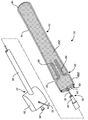
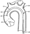
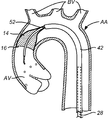
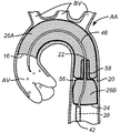
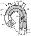
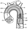
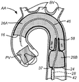
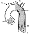
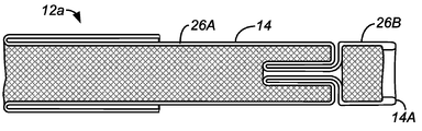
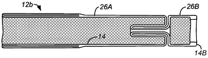
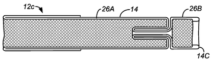
도 1 내지 도 3에 도시되어 있는 바와 같이, 본 발명의 원리에 따라 구성된 색전 방지 디바이스(10)는 상류측 개방 단부(16) 및 하류측 개방 단부(18)를 갖는 필터 바디(12)를 포함한다. 필터 바디(12)는 상류측 개방 단부(16)와 하류측 개방 단부(18) 사이에, 도시되어 있는 바와 같이 하류측 개방 단부에 더 근접하게 위치된 팽창형 개구(22)를 갖는 자가 밀봉 포트(20)를 갖도록 예비성형된 다공성 메시 재료, 더 통상적으로는 관형 다공성 메시 재료로 형성된다. 필터 바디(12)의 특정 절첩 패턴이 도 8a 내지 도 8e를 참조하여 이하에 설명되고, 다수의 예시적인 대안적인 절첩 패턴이 도 15a 내지 도 15c와 관련하여 이하에 설명된다.An
반경방향 팽창형/절첩형 지지부(24)가 도 2에 가장 양호하게 도시되어 있는 바와 같이, 필터 바디(12)의 하류측 개방 단부(18)에 고정된다. 반경방향 절첩형 지지부(24)는 그 원위 단부에 형성된 루프(37)를 갖는 견인 와이어(36)를 갖춘 튜브(34)(도 3)를 포함할 수도 있다. 루프(37)는 필터 바디(12)의 하류측 개방 단부(18)의 주연부 둘레에 고정되어, 하류측 개방 단부(18)를 개방 및 폐쇄하기 위한 "올가미 또는 "주머니끈(purse-string)"으로서 작용할 수도 있게 된다. 특히, 튜브(34) 내에서 견인 와이어(36)를 근위측으로 (도 3의 우측으로) 후퇴시킴으로써, 루프(37)는 폐쇄될 수도 있다. 역으로, 튜브(34)에 대해 견인 와이어(36)를 원위측으로 전진시킴으로써, 루프(37)는 개방될 수도 있다. 이하에 더 상세히 설명되는 바와 같이, 테더 구조체(32)를 축방향으로 전진 및 후퇴시킴으로써, 필터 바디(12)는 전개 카테터 바디(28)에 대해 위치설정될 수도 있다.A radially expandable /
필터 바디(12)의 자가 밀봉 포트(20)는 필터 바디를 상류측 원통형 챔버(26A)와 하류측 원통형 챔버(26B)로 분할한다. 각각의 챔버(26A, 26B)는 일반적으로 내부 구조체가 없을 것이고, 자가 밀봉 포트(20)는 2개의 챔버를 분할하도록, 특히 하류측 챔버(26B) 내로 또는 하류측 챔버를 지나 상류측 챔버(26A)에 진입할 수도 있는 색전의 통과를 방지하도록 작용할 것이다. 하류측 원통형 챔버(26B)는 필터 바디가 대동맥 또는 다른 혈관 내에서 전개될 때 필터 바디(12)의 상류측에서 중재적 시술을 수행하기 위해, 자가 밀봉 포트(20) 내로의 그리고 자가 밀봉 포트를 통한 작동 카테터의 유도를 수용하여 용이하게 하도록 작용한다.The self-sealing port (20) of the filter body (12) divides the filter body into an upstream cylindrical chamber (26A) and a downstream cylindrical chamber (26B). Each of the
전개 카테터 바디(28)는 원위 단부(30)와, 테더 구조체(32)를 지지하기 위한 적어도 제1 루멘(38)과, 이하에 더 상세히 설명되는 바와 같이 인공 대동맥 판막을 전개하기 위한 TAVR 카테터와 같은, 중재적 카테터 또는 작동 카테터를 통과시켜 유도하기 위한 작동 루멘으로서 역할을 하는 제2 루멘(40)을 갖는다.The deploying
근위 허브 또는 제어 허브(29)가 전개 카테터 바디(28)의 근위 단부(31)에 결합된다. 테더 구조체(32)의 근위 단부(33)가 제어 허브(29)로부터 연장되고, 사용자가 테더 구조체의 축방향 후퇴 및 전진 모두뿐만 아니라 루프(37)의 개방 및 폐쇄를 포함하여, 테더 구조체를 조작하는 것을 허용한다. 제어 허브(29)는 또한 가이드 와이어, 작동 카테터 등의 통과를 허용하기 위해 카테터 바디(28) 내의 제2 루멘(40)으로 개방되어 있는 포트(35)를 더 구비한다.A proximal hub or
필터 바디(12)는 통상적으로 자가 팽창형일 것이다. "자가 팽창형"이라는 것은, 필터 바디가 탄성일 것이고 반경방향 속박 및/또는 축방향 속박이 없을 때 통상 개방된 또는 팽창된 구성을 갖는 것을 의미한다. 필터 바디를 축방향으로 수축시키거나 축방향으로 신장시킴으로써, 필터 바디의 직경 또는 프로파일이 감소될 것이며, 이에 따라 필터 바디는 통상적으로 대동맥궁 위로, 그러나 선택적으로 마찬가지로 다른 위치에서, 환자의 혈관구조 내의 작업 부위로 혈관 내에서 유도될 수 있게 된다. 부가적으로, 필터 바디를 반경방향으로 접고 그리고/또는 축방향으로 신장시킴으로써, 필터 바디 내의 자가 밀봉 포트가 펼쳐지고 그리고 축방향으로 신장될 것이다.The
자가 밀봉 포트(20)는 자가 형성식(self-forming)일 것이고, 통상적으로 도 1에 도시되어 있는 바와 같이, 원추형 기부(56) 및 연장 슬리브(58)를 갖는다. 자가 밀봉 포트(20)는, 필터 바디의 반경이 증가하고 필터 바디의 길이가 축방향으로 단축됨에 따라 필터 바디의 일반적으로 관형 구조체를 절첩하고 반전시킴으로써 형성된 구조를 가질 것이다. 필수 절첩 라인이 통상적으로 열처리에 의해 필터 바디 내로 예비 성형될 것이다. 예시적인 실시예에서, 필터 바디는 이하에서 도 8a 내지 도 8e를 참조하여 더 상세히 설명되는 절첩 라인을 갖도록 형성될 것인 Nitinol®(니켈-티타늄 합금)로서 형성될 것이다. 예비 성형된 이들 절첩 라인은 필터 바디가 축방향으로 연신되고 반경방향으로 접혀서, 전달 중에, 통상적으로 12 Fr(French) 미만, 종종 10 Fr 미만의 전달 직경을 갖는 낮은 프로파일을 갖게 할 것이다. 역으로, 필터 바디는 통상적으로 5 mm 초과, 종종 15 mm 초과, 더 종종 25 mm 초과, 통상적으로 25 mm 내지 40 mm의 범위의 미속박된 폭 또는 직경으로 개방될 것이다. 이하에 더 상세히 설명될 것인 바와 같이, 필터 바디(12)는 통상적으로 대동맥궁 위의 대퇴동맥을 통해, 환자의 동맥 내에 사전 배치되어 있는 전달 외장(42)을 통해 그 낮은 프로파일 구성으로 유도될 것이다. 그러나, 전달 외장(42) 내로 필터 바디를 전진시키기 위해, 자가 팽창형 필터 바디(12)를 일시적으로 속박할 필요가 있다. 이는, 도 4 및 도 5에 도시되어 있는 바와 같이, 분리제거형 외장(48)을 사용하여 성취될 수도 있다. 필터 바디(12)는 축방향으로 연신되고 반경방향으로 접혀지고 도 4에 도시되어 있는 바와 같이, 분리제거형 외장(48)의 루멘 내로 당겨진다. 분리제거형 외장은 카테터 바디(28)가 분리제거형 외장(48)의 근위 단부로부터 신장되는 상태로 필터 바디(12)를 덮는다. 분리제거형 외장과 카테터 바디(28)의 일시적 조립체가, 도 4에 도시되어 있는 바와 같이, 전달 외장(42)의 원위 포트(44)를 통해 사전 배치되어 있는 가이드와이어 구조체(46) 위로 유도될 수 있고, 외장은 이어서 도 5에 도시되어 있는 바와 같이, 원위 포트(44)를 통해 전진된다. 일단 필터 바디(12)의 원위 단부가 포트(44)를 통해 전달 외장(42)의 근위 단부 내로 유도되었으면, 필터 바디가 전달 외장(42) 내로 계속 유도됨에 따라 분리제거형 외장은 제거될 수도 있다. 전달을 용이하게 하기 위해, 전달 외장(42) 및 분리제거형 외장(48)은 각각 이들의 루멘 내로의 유체의 유도를 허용하기 위한 포트를 가질 수도 있다.The self-sealing
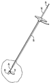
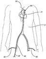
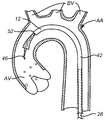
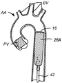
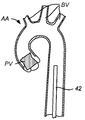
도 6에 도시되어 있는 바와 같이, 전달 외장(42)은 대퇴동맥 내로 그리고 대동맥궁 상에 그리고 대동맥궁 위로 환자의 서혜부를 통해 통상의 방식으로 유도될 것이다. 통상적으로 TAVR 또는 다른 중재적 카테터 또는 작동 카테터를 유도하기 위한 제2 외장(50)이 전달 외장(42)에 평행하게 대동맥 상에 그리고 대동맥궁(AA) 위로 작동 카테터를 유도하기 위해 반대쪽 대퇴동맥 내에 위치설정될 것이다. 이러한 위치설정은 환자(P)의 심장(H) 상에서의 인공 판막 배치 또는 다른 중재적 시술을 위해 의도될 것이다.As shown in FIG. 6, the
이제 도 7a 내지 도 7zz를 참조하여, 인공 판막(PV)을 환자의 자연 대동맥 판막 내로 유도하기 위한 특정 프로토콜이 설명될 것이다. 도 7a에 도시되어 있는 바와 같이, 전달 외장(42)은 도 6을 참조하여 방금 설명된 바와 같이, 초기에 가이드와이어 구조체(46) 위에 배치된다. 카테터 바디(28)는 이어서 전달 외장(42)의 내부 루멘을 통해 전진되어, 반경방향으로 속박된 필터 바디(12)가 전달 외장의 개방 원위 단부(52)에 접근하게 된다.Referring now to Figures 7A-7ZZ, a specific protocol for directing a prosthetic valve (PV) into a patient's native aortic valve will be described. As shown in FIG. 7A, the
이어서 카테터 바디(28)를 상대적으로 정지 상태 또는 고정 상태로 유지하고 전달 외장(42)을 근위 방향으로, 즉 환자의 대동맥 판막(AV)으로부터 이격하여 후퇴시킴으로써, 도 7b에 도시되어 있는 바와 같이, 필터 바디(12)의 원위 단부는 속박으로부터 해제되어 관형 다공성 메시(14)가 반경방향으로 팽창을 시작하게 될 것이다. 전달 외장(42)은 도 7c에 도시되어 있는 바와 같이 근위측으로 계속 후퇴되며, 이에 따라 관형 다공성 메시(14)가 팽창하여 대동맥 판막(AV) 바로 위의 상행대동맥의 내부벽에 결합하게 된다. 도 7d에 도시되어 있는 바와 같이, 전달 외장(42)은 근위측으로 계속 후퇴되며, 이에 따라 도 7e에 도시되어 있는 필터 바디(12)의 상류측 원통형 챔버(26A)의 상대적인 완전한 전개에 의해, 관형 다공성 메시(14)가 계속 팽창하고 분기 혈관(BV)을 덮기 시작하는 것을 허용한다. 도 7e에 또한 도시되어 있는 바와 같이, 자가 밀봉 포트 구조체(20)는 형성되는 그 바로 초기 스테이지에 있고, 추가의 형성은 도 7f에 도시되어 있다.7B, by maintaining the
도 7g에 도시되어 있는 바와 같이, 자가 밀봉 포트(20)가 될 부분 중 원추형 기부(56)가 거의 형성되고, 하류측 원통형 챔버(26B)는 도 7h에 도시되어 있는 바와 같이 형성되기 시작한다. 하류측 원통형 챔버(26B)는 도 7i에 도시되어 있는 바와 같이 거의 형성되고, 반면에 자가 밀봉 포트(20)가 막 형성되기 시작한다. 이어서 대동맥 판막(AV)을 향한 방향에서 카테터 바디(28)를 전진함으로써, 도 7j에 도시되어 있는 바와 같이, 관형 다공성 메시(14)의 협소한 세그먼트는 자가 밀봉 포트(20)의 슬리브 구조체(58)를 형성하도록 반전되기 시작할 것이다. 도 7i 및 도 7j에서 또한 명백한 바와 같이, 루프(37) 형태의 반경방향으로 접힘 가능한 지지부(24)는 하류측 원통형 챔버(26)의 개방 원위 단부를 개방하여 지지하도록 개방된다. 카테터 바디(28)가 필터 바디(12)의 하류측 부분을 조작하는 것을 허용하여 하류측 원통형 챔버(26B)가 상류측 원통형 챔버(26A)에 대해 대동맥 판막(AV)을 향해 또는 원위측으로 전진될 수 있게 하는 것은 바로 이 지지 구조체(24)이다. 반경방향 팽창형/절첩형 지지부(24A)는 이하에 더 상세히 설명되는 바와 같이, 시술의 종료 시에 필터 바디(12)를 수축시킬 때 또한 유용할 것이다.As shown in Fig. 7G, the
완전 전개된 자가 밀봉 포트(20)가 도 7k에 도시되어 있고, 슬리브(58)는 이하에 더 상세히 나타내는 바와 같이, 하류측 단부로부터 카테터의 유도를 용이하게 하는 팽창형 개구(22) 및 원추형 기부(58)를 형성한다.The fully deployed self-sealing
특정예에서, 가이드와이어 구조체(46)는 도 7l에 도시되어 있는 바와 같이, 가이드와이어를 적소에 후퇴 및 퇴피될 수도 있는 외부 지지 튜브를 포함할 수도 있다. 이어서 도 7m에 도시되어 있는 바와 같이, 통상적으로 혈관조영술을 위해 사용되는 가이드와이어(46) 위로 진단 카테터(60)가 전진될 수도 있다. 상기 자가 밀봉 포트(20)는 임의의 색전이 포트를 통과하는 것을 방지하기 위해 카테터 주위를 밀봉하면서 진단 카테터(60)의 직경을 수용하도록 팽창될 것이다.In a particular example, the
진단 카테터(60)를 퇴피시킨 후에, 다른 가이드와이어(62)가, 도 7n에 도시되어 있는 바와 같이, TAVR 전달 카테터(64)를 전진시키도록 유도될 수도 있다. 제1 카테터 구조체(46)는 통상적으로 적소에 남아 있을 것이지만, 도 7n에는 가시화되어 있지 않다. TAVR 전달 카테터(64)는 이어서, 도 7p에 도시되어 있는 바와 같이 자연 대동맥 판막(AV)을 통과할 때까지, 도 7o에 도시되어 있는 바와 같이, 환자의 대동맥궁(AA) 위로 전진된다. 이어서 도 7q에 도시되어 있는 바와 같이, 인공 판막(PV)이 TAVR 카테터(64)로부터 해제될 것이다. 대동맥궁(AA) 위의 TAVR 카테터(64)의 전진 중에, 특히 인공 판막(PV)의 해제 중에, 대동맥궁 및 대동맥 판막(AV)이 상당히 석회화될 수도 있기 때문에, 색전이 방출될 상당한 위험이 존재한다는 것이 이해되어야 한다. 이러한 색전이 존재하면, 색전은 대동맥궁 위로 그리고 필터 바디(12)의 상류측 개방 단부(16)를 통해 운반될 것이어서, 상부 원통형 챔버(26A)에 진입하여 상부 원통형 챔버 내에 수납되게 된다. 특히, 관형 다공성 메시(14)는 이들 혈관 내로의 혈류를 허용하면서 임의의 상당한 크기의 색전이 임의의 분기 혈관(BV)에 진입하는 것을 방지할 것이다. 자가 밀봉 포트(20)의 슬리브(58)는 TAVR 전달 카테터(64)의 외부에 합치하고 그 주위를 밀봉할 것이며, 따라서 카테터 통과를 허용하도록 팽창되는 동안 포트를 통한 색전의 우발적인 통과를 저지하거나 방지한다.After evacuating the
도 7q에 도시되어 있는 바와 같이, 인공 판막(PV)이 릴리스된 후에, TAVR 전달 카테터(64)는 도 7r에 도시되어 있는 바와 같이, 가이드와이어(62)에 걸쳐 근위측으로 후퇴될 것이다. 제1 가이드와이어(46)가 또한 도 7r에 도시되어 있다. 도 7s 및 도 7t에 도시되어 있는 바와 같이, TAVR 전달 카테터(64)는 계속 퇴피되고 자가 밀봉 포트(20)를 통해 나오고, 상기 자가 밀봉 포트는 이어서 가이드와이어(46)를 폐쇄한다. 이어서 도 7t에 도시되어 있는 바와 같이, TAVR 가이드와이어(62)는 대동맥을 통해 뒤로 견인된다.As shown in Figure 7q, after the prosthetic valve PV is released, the
TAVR 카테터(64) 및 가이드와이어(62)가 퇴피된 후에, 인공 판막(PV)은 적소에 있고 대동맥궁(AA)으로부터 필터 바디(12)를 퇴피시킬 필요가 있다. 도 7u 및 도 7v에 도시되어 있는 바와 같이, 테더 구조체(32)는 반경방향으로 접힘 가능한 지지부(24)의 루프(37)를 폐쇄하도록 조작된다. 루프(37)를 폐쇄하는 것에 추가하여, 도 7w에 도시되어 있는 바와 같이, 필터 구조체(12)의 근위 단부는 카테터 바디(28)의 원위 단부로 당겨지고, 카테터 바디(28)는 필터 바디를 당기도록 전달 외장(42) 내로 후퇴된다.After the
카테터 바디(28)는 근위측으로 계속 퇴피되며, 이에 따라 도 7x에 도시되어 있는 바와 같이, 하류측 원통형 챔버(26)를 전달 외장(42) 내로 견인하게 되고, 도 7y, 도 7z 및 도 7zz에 도시되어 있는 바와 같이, 전체 필터 바디(12)가 전달 외장(42) 내로 당겨질 때까지 계속 근위측으로 퇴피되게 된다. 필터 바디 및 그 내부에 수납된 모든 색전은 이어서 전달 외장(42) 내에 안전하게 포획되고, 전달 외장(42)은 환자로부터 퇴피될 수도 있고, 시술은 통상의 방식으로 완료될 수도 있다.The
다공성 필터 메시 재료는 다양한 편직 섬유, 직조 섬유 또는 부직 섬유, 필라멘트 또는 와이어를 포함할 수도 있고, 혈액이 통과하는 것을 허용하면서도 특정 크기를 초과하는 색전이 통과하는 것을 방지하도록 선택된 기공 크기를 가질 것이다. 적합한 재료는 형상 기억 합금 및 열 기억 합금, 폴리머, 및 이들의 조합과 같은 탄성 금속을 포함하고, 재료는 선택적으로 이들의 표면 상에 (헤파린과 같은) 혈전 방지 코팅을 가질 수도 있다. 필터 메시는 필터 바디의 방사선 비투과성을 향상시키기 위한 재료 및 구조를 더 구비할 수도 있다. 예시적인 재료는 금, 백금, 팔라듐, 또는 탄탈, 및 탄성 금속보다 더 큰 방사선 비투과성을 갖는 다른 금속뿐만 아니라 방사선 비투과 코팅 또는 방사선 비투과 충전물을 포함한다. 다른 경우에, 탄성 금속 필라멘트 또는 와이어는 더 얇고, 더 방사선 비투과성인 와이어 또는 필라멘트로 마련될 수도 있다.The porous filter mesh material may comprise a variety of knitted fibers, woven fibers or nonwoven fibers, filaments or wires, and will have a pore size selected to prevent passage of blood beyond that of a particular size while allowing passage therethrough. Suitable materials include elastomeric metals such as shape memory alloys and thermally memory alloys, polymers, and combinations thereof, and the materials may optionally have anti-friability coatings (such as heparin) on their surfaces. The filter mesh may further comprise materials and structures for enhancing the radiation impermeability of the filter body. Exemplary materials include gold, platinum, palladium, or tantalum, and other metals having greater radiopacity than the resilient metal, as well as radiation non-transmissive coatings or radiation non-transmissive charges. In other cases, the resilient metal filaments or wires may be provided with thinner, more radiopaque wires or filaments.
필터 바디는 함께 부착된 이산 섹션에 구성될 수도 있지만, 더 통상적으로는 보통 단일의 이러한 절첩된 관형 메시 구조체로 이루어지는 특정 디자인 특징부를 형성하기 위해 섹션에서 협소화되거나 절첩된 연속적인 원통형 메시 구조체로부터 형성될 것이다. 하나의 연속적인 원통형 메시로부터 디바이스를 형성하는 것은 전개 및/또는 회수를 위해 축방향으로 필터 바디가 신장되게 하여, 이에 의해 필터의 프로파일을 감소시킨다. 단일의 연속적인 얇고 편평한 메시 재료로부터 형성된 필터의 다른 장점은 이것이 단지 평활하고 라운딩된 에지를 포함할 것이라는 것이다. 이러한 에지는 카테터 및 시술 도구가 필터를 통해 유도되는 상태에서 마찰 및 처짐을 최소화시킨다.The filter body may be constructed in a discrete section attached together, but is typically formed from a continuous cylindrical mesh structure that is narrowed or folded in a section to form a particular design feature, usually consisting of a single such folded tubular mesh structure will be. Forming the device from one continuous cylindrical mesh causes the filter body to extend in the axial direction for deployment and / or retrieval, thereby reducing the profile of the filter. Another advantage of a filter formed from a single continuous thin flat mesh material is that it will only include smooth and rounded edges. Such an edge minimizes friction and deflection with the catheter and instrument being guided through the filter.
자가 밀봉 포트는, 통상적으로 전술된 바와 같이 슬리브에 의해 형성된 그 협소 단부에 접근 포트를 갖는 원추형 구조체로서 구성될 수도 있다. 다른 실시예에서, 이하에 예시되는 바와 같이, 자가 밀봉 포트는 원통형 구조체의 간단한 협소부, 예를 들어 이를 통해 유도되는 카테터 및 다른 도구 주위를 밀봉하는 자가 폐쇄형 네크(neck) 영역일 수도 있다. 어떠한 특정 기하학적 구조이건간에, 자가 밀봉 포트는 열처리 또는 냉간 성형을 거쳐 감소된 직경으로 더 대형의 관형 메시 또는 원통형 메시를 형상 세팅함으로써 형성될 수 있다. 게다가, 자가 밀봉 포트의 다른 실시예는 직선형일 수 있고, 비틀림부를 포함할 수 있고, 파형일 수 있고, 평탄화된 섹션을 가질 수 있고, 또는 카테터가 적소에 있을 때 색전이 통과하는 것을 저지하거나 방지하기 위해 충분히 시술 디바이스 주위를 폐쇄하는 그 능력을 보조하는 다른 특징부를 가질 수 있다. 또 다른 실시예에서, 필터 바디는 2개 이상의 이러한 자가 팽창형 포트 구조체를 포함할 수도 있다. 자가 밀봉 포트(20)는 단일의 디바이스(예컨대 가이드와이어, 카테터, 판막 전달 시스템, 조율 리드 등), 2개의 디바이스 또는 2개 초과의 디바이스를 동시에 수용할 수도 있고, 필요에 따라 다수의 디바이스 주위에 충분한 밀봉을 유지하도록 팽창 및 수축할 수 있다. 또한, 이러한 디바이스는 카테터 바디(28)의 작동 루멘(40)을 경유하여 하류측 원통형 챔버(26B)를 통해 그리고 자가 밀봉 포트(20) 내로 또는 대안적인 접근 부위에서 제2 외장(50)을 경유하여 직접, 또는 이들의 몇몇 조합으로 유도될 수 있다.The self-sealing port may also be configured as a conical structure having an access port at its narrow end, which is typically formed by a sleeve as described above. In another embodiment, as illustrated below, the self-sealing port may be a self-closing neck region that seals a simple narrowed portion of the cylindrical structure, for example, around a catheter and other tool that is guided therethrough. Any self-sealing port, whether any particular geometry, can be formed by heat-setting or cold-forming to shape the larger tubular mesh or cylindrical mesh with a reduced diameter. In addition, other embodiments of the self-sealing port may be straight, may include a torture, may be corrugated, have a flattened section, or may prevent or prevent the embolization from passing when the catheter is in place It may have other features that assist in its ability to close the surgical device sufficiently to allow the operator to close the surgical device. In yet another embodiment, the filter body may include two or more such self-expanding port structures. The self-sealing
이제, 도 8a 내지 도 8e를 참조하면, 상류측 원통형 챔버(26A)와 하류측 원통형 챔버(26B) 사이에 자가 밀봉 포트(20)를 갖는 필터 바디(12) 내로 본 발명의 관형 다공성 메시 재료(14)를 형성하기 위한 다수의 상이한 패턴이 도시되어 있다. 도 8a 및 도 8b의 구조체는 각각 방금 설명된 바와 같이 메시 재료의 단일층 튜브로 시작한다는 것이 이해될 수 있을 것이다. 도 8a의 구성에서, 관형 구조체는 먼저 그 중간에 절첩부(14A)를 갖는 2층 구조체로 절첩된다. 2층 구조체는 이어서, 다음에 반경방향으로 완전 팽창된 구성으로 열경화되는 구조체를 갖는 예시된 필터 바디(12a)를 형성하기 위해 자체로 재차 절첩되고 반전된다.8A to 8E, a tubular porous mesh material (not shown) of the present invention is inserted into the
도 8b의 필터 바디(12b)는 유사하게 일 단부에 단일의 절첩부(14B)를 갖는 2층 관형 메시로서 시작한다. 2층 구조체는 이어서, 원통형 챔버(26A)의 개방 단부가 도 8a에 도시되어 있는 바와 같이 간단한 절첩 패턴으로보다는 내향으로 반전된 패턴으로 절첩되는 것을 제외하고는, 도 8b의 패턴에 유사하게 절첩된다.The
도 8c에 도시되어 있는 필터 바디(12c)는, 상류측 원통형 챔버(26A)의 개방 단부가 원위측 커프를 형성하도록 외부층 위로 절첩된 내부층을 갖고, 내부층은 절첩된 외부층 내에서 종료되는 것을 제외하고는, 도 8a의 필터(12a)의 절첩 패턴에 대부분의 관점에서 역시 유사하다.The
도 8d에 도시되어 있는 필터 바디(12d)는 다수의 방식에서 도 8a의 필터 바디(12a)와 반대이다. 원래 단일층 관형 실린더 내의 단일의 절첩부(14D)는 상류측 원통형 챔버(26A)의 개방 단부에 위치된다. 하류측 챔버(26B)의 하류측 개방 단부는 자체로 절첩되어 커프 구조체를 형성한다.The
도 8e에 도시되어 있는 바와 같은 필터 바디(12e)는, 하류측 원통형 챔버(26B)의 절첩이 도 8a 내지 도 8c의 것과 유사하지만, 상류측 원통형 챔버(26A)의 개방 단부는 내부층 및 외부층이 개방된 상태로 종료되고 전혀 절첩되지 않는 가장 간단한 구조체이다.The
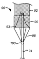
도 9는 본 발명의 원리에 따라 구성된 색전 방지 디바이스(70)의 제1 대안 실시예를 도시하고 있다. 필터 바디(72)는 상류측 개방 단부(74) 및 하류측 폐쇄 단부(76)를 갖는다. 자가 밀봉 포트(78)가 하류측 폐쇄 단부(76)에 형성되고, 지지 구조체(82)가 필터 바디의 하류측 단부에 부착된다. 지지 구조체(82)는 한 쌍의 지주(strut)를 포함하고, 전달을 위해 압축되고 속박 외장(86)의 해제에 의해 제자리로 팽창될 수 있는 재료(형상 기억 합금)로 제조될 수 있다. 지지부는 칼라(84)를 거쳐 전개 카테터 바디(80)에 고정적으로 또는 이동 가능하게 부착된다. 카테터 바디(80)의 원위 단부 또는 상류측 단부는 자가 밀봉 포트(78)에 인접하여 필터 바디(72)의 폐쇄 단부(76)를 통과한다.Figure 9 shows a first alternative embodiment of an
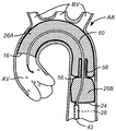
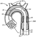
도 10은 본 발명의 원리에 따라 구성된 색전 방지 디바이스(90)의 제2 대안 실시예를 도시하고 있다. 하류측 폐쇄 단부(93)를 갖는 필터 바디(92)가 완전한 원주방향 지지 구조체(96)에 의해 전개 카테터 바디에 부착된다. 지지 구조체(96)는, 지지 구조체가 중첩되고 필터(92)의 메시 재료에 부착되는 영역 위에 "스텐트형" 다이아몬드 요소를 포함한다. 지지 구조체(96)는 칼라(100) 및 복수의 지주(98)를 거쳐 전개 카테터 바디(94)에 고정적으로 또는 이동 가능하게 부착된다. 카테터 바디(94)의 원위 단부 또는 상류측 단부는 자가 밀봉 포트에 인접하여 필터 바디(92)의 폐쇄 단부(93)를 통과한다.Figure 10 shows a second alternative embodiment of an
도 11 및 도 12는 필터 바디(104)의 폐쇄 단부(106) 내에 원추형 메시 자가 밀봉 포트(108)를 갖는 색전 방지 디바이스(102)의 제3 대안 실시예를 도시하고 있다. 전개 카테터 바디(114)가 칼라(116)에 부착된다. 필터 바디(104)의 전달 및 견인을 위해 전달 외장(118)이 제공된다. 도 11에서, 카테터 바디(114)의 원위 단부 또는 상류측 단부가 자가 밀봉 포트(108)를 통해 배치되고, 자가 밀봉 포트(108)에 인접한 포트를 통해 유도 루멘 또는 다른 경로를 제공한다. 도 12에서, TAVR 전달 카테터 또는 다른 작동 카테터는 카테터 바디(114)에 평행하게 자가 밀봉 포트를 통해 유도된다. 자가 밀봉 포트(108)의 주연부는 양 카테터에 합치하여 동시에 밀봉하기 위해 충분히 유연할(탄성일) 것이다.Figures 11 and 12 illustrate a third alternative embodiment of an
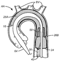
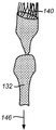
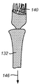
도 13a 내지 도 13c는 본 발명의 원리에 따라 구성된 색전 방지 디바이스(130)의 제4 대안 실시예를 도시하고 있다. 필터 바디(132)는 필터의 전체 대부분에 걸쳐 이중층의 메시를 포함하고, 디바이스의 이 부분에서 필터의 고정 강도를 증가시키기 위해 하류측 단부에 (총 3개의 층을 위한) 부가의 층 또는 커프(150)를 갖는다. 도 13a는 그 이완된 구성에서 색전 방지 디바이스(130)를 도시하고 있고, 반면에 도 13b 및 도 13c는 전달 구성 또는 회수 구성으로 화살표(146)의 방향으로 축방향으로 신장될 때의 색전 방지 디바이스(130)를 도시하고 있다. 자가 밀봉 포트(134) 및 다른 디바이스 특징부는 연속적인 원통형 메시 구조체 내에 일체로 또는 단일체로 형성되기 때문에, 이들 특징부는 필터 바디(132)가 축방향으로 완전히 신장될 때 효과적으로 사라진다. 이러한 내부 구조체를 신장시켜 제거하는 능력은 디바이스 프로파일을 최소화시킨다. 연속적인 하나의 원통형 표면으로부터의 필터의 구성은 또한 제조 복잡성을 피하고, 디바이스 전체에 걸쳐 평활한 접촉면을 유지하여 필터를 통과하는 시술 도구의 마찰을 감소시킨다. 필터 바디는 필터 바디(132)의 하류측 원통형 챔버(139)를 중첩하거나 위에 놓이는 스텐트형 주연 지지 구조체(140)에 의해 전개 카테터 바디(144)에 부착된다. 자가 밀봉 포트(134)는 전술한 바와 같이 원추형 기부(136) 및 슬리브(138)를 포함할 수도 있다.13A-13C illustrate a fourth alternative embodiment of an
도 14는, 중재적 카테터가 대동맥 판막(AV)에서, 판막성형술 또는 TAVR과 같은 시술을 수행하기 위해 자가 밀봉 포트(134)를 통해 상류측 방향으로 전달될 때 분기 혈관을 보호하기 위해 환자의 대동맥궁 위에서 전개되는 색전 방지 디바이스(130)를 도시하고 있다.Figure 14 is a cross-sectional view of an aortic valve (AV) in which an interventional catheter is inserted into the aorta of a patient to protect the divergent vessel, when delivered in the aorta direction through the self-sealing
도 15a 내지 도 15c는 본 발명의 필터 바디의 대안적인 구성을 도시하고 있다. 도 15a는 원통형 메시 재료 내에 형성된 간단한 협소부 또는 네크(166)에 의해 분리되어 있는 상류측 원통형 챔버(162) 및 하류측 원통형 챔버(164)를 포함하는 필터 바디(160)를 도시하고 있다. 원통형 메시 재료는 단일벽 또는 이중벽일 수도 있고, 벽 영역의 일부 또는 모두 위에 2개 초과의 층을 가질 수도 있거나 또는 이들의 조합일 수도 있고, 이 필터 바디 구성은 전술한 본 발명의 색전 방지 디바이스의 실시예의 대부분 또는 모두에서 조합될 수 있다.15A-15C illustrate alternative configurations of the filter body of the present invention. 15A shows a
도 15b는 네크(180, 182)에 의해 각각 분리된 상류측 원통형 챔버(172), 중앙 원통형 챔버(174), 및 하류측 원통형 챔버(178)를 포함하는 필터 바디(170)를 도시하고 있다. 이러한 다수의 원통형 챔버는 전술한 자가 폐쇄 포트 구조체 중 임의의 하나에 의해 분리될 수 있다는 것이 이해될 수 있을 것이다. 하류측 원통형 챔버(178)의 하류측 단부는 모아지거나 폐쇄될 수도 있고, 도시되어 있는 바와 같이, 폐쇄된 하류측 단부를 갖는 필터 바디(170)는 이하의 도 16a 내지 도 16e에 설명되어 있는 응괴 포획 구성에서 특정 용도를 발견할 수도 있다. 이전의 실시예와 같이, 필터 바디(170)의 원통형 메시 재료는 단일벽 또는 이중벽일 수도 있고, 2개 초과의 층을 가질 수도 있거나 또는 이들의 조합일 수도 있고, 다중 챔버 필터 바디 구성은 전술한 본 발명의 색전 방지 디바이스의 실시예의 대부분에서 또는 모두에서 조합될 수 있지만, 하류측 챔버의 하류측 단부는 개방되어야 할 것이다.15B shows a
도 15c는 네크(208) 및 자가 밀봉 포트(210)에 의해 각각 분리된 상류측 원통형 챔버(202), 중앙 원통형 챔버(204), 및 하류측 원통형 챔버(206)를 포함하는 필터 바디(200)를 도시하고 있다. 하류측 원통형 챔버(206)의 하류측 단부는 개방되고, 다중 챔버 필터 바디(200)는 전술한 본 발명의 색전 방지 디바이스의 실시예의 대부분에서 또는 모두에서 조합될 수 있다. 필터 바디(200)의 원통형 메시 재료는 단일벽 또는 이중벽일 수도 있고, 2개 초과의 층을 가질 수도 있고, 또한 이들의 조합일 수도 있다.15C shows a
도 16a 내지 도 16e는 심장, 말초, 또는 대뇌 혈관(BV) 내의 응괴 및/또는 혈전을 포획하기 위해 도 15b의 필터 바디(170)를 사용하는 특정 시술을 도시하고 있다. 이러한 디바이스 및 프로토콜은 급성 허혈성 뇌졸중을 치료하기 위해 두개 혈관 내의 응괴/혈전을 회수하는 데 특히 유용할 것이다. 도 16a에 도시되어 있는 바와 같이, 필터 바디(170)의 모아진 단부(179)는 전개 카테터(210)의 원위 단부에 부착되고, 전개 카테터(210)는 마이크로카테터(230)(도 16e)를 통해 전진된다. 그 원위 단부에 나선형 팁과 같은 응괴 포획 요소(222)를 갖는 Merci® 회수기와 같은 응괴 포획 카테터(220)가 도 16b에 도시되어 있는 바와 같이, 카테터(220), 필터 바디(170)를 통해, 응괴/혈전(THR)을 통해 전진된다. 응괴 포획 카테터(220)는 이어서 도 16b 내지 도 16d에 도시되어 있는 바와 같이, 네크(180)를 통해 중앙 챔버(174) 내로 응괴/혈전(THR)을 당기기 위해 근위측으로 견인된다. 응괴/혈전(THR)이 중앙 챔버(174) 내에 위치한 후에는, 응괴/혈전이 필터로부터 소실될 가능성은 현저히 감소되고, 네크(180, 182)를 통해 새어나올 수도 있는 임의의 색전은 가능하게는 상류측 원통형 챔버(178) 및 하류측 원통형 챔버(172) 내에 각각 포획될 가능성이 있을 것이다. 상류측 네크(180)는 응괴 회수 디바이스 및 걸린 혈전이 통과하게 하도록 신장 개방되고, 응괴 및/또는 혈전이 중앙 원통형 챔버(174) 내에 완전히 봉입된 후에 폐쇄되도록 구성될 것이다. 응괴 및 혈전으로부터 떨어져 나올 수도 있는 임의의 부스러기가 완전 폐쇄된 네크(180, 182)(도 16d)에 의해 수납되고, 응괴 회수기(220), 필터 바디(170), 및 카테터 바디(220)의 조립체는 마이크로카테터(230)를 통해 안전하게 퇴피될 수도 있다.Figures 16A-16E illustrate specific procedures using the
본 발명을 수행하기 위한 전술한 조립체 및 방법의 수정, 실시 가능한 것과 같은 상이한 변형 사이의 조합, 및 당 기술 분야의 숙련자들에게 명백한 본 발명의 양태의 변형은, 본 개시내용의 범주 내에 있는 것으로 의도된다.Modifications of the above-described assemblies and methods for carrying out the invention, combinations between different variations such as are feasible, and variations of the embodiments of the invention that are apparent to those skilled in the art are within the scope of this disclosure, do.
10: 색전 방지 디바이스
12: 필터 바디
16: 상류측 개방 단부
18: 하류측 개방 단부
20: 자가 밀봉 포트
22: 팽창형 개구
24: 지지부
28: 전개 카테터 바디
30: 원위 단부
32: 테더 구조체
34: 튜브
36: 견인 와이어
37: 튜브
42: 전달 외장
56: 원추형 기부
58: 연장 슬리브10: Embolism prevention device 12: Filter body
16: upstream side open end 18: downstream side open end
20: self-sealing port 22: inflatable opening
24: support 28: deployment catheter body
30: distal end 32: tether structure
34: tube 36: pull wire
37: tube 42: transfer sheath
56: conical base 58: extension sleeve
Claims (34)
상기 필터 바디의 하류측 개방 단부의 주연부에 결합된 반경방향 절첩형 지지부;
상기 반경방향 절첩형 지지부에 결합된 원위 단부를 갖는 카테터 바디;
상기 필터 바디를 수용하여 반경방향으로 속박하도록 구성된 루멘(lumen)을 갖는 전달 외장
을 포함하는 색전 방지 디바이스로서,
상기 카테터 바디는, 상기 필터 바디를 속박으로부터 해제하도록 그리고 상기 지지부가 상기 필터 바디의 하류측 개방 단부에 외접하는 상태에서 상기 필터 바디가 반경방향으로 팽창하게 허용하도록 상기 전달 외장에 대해 원위측으로 전진될 수도 있고, 상기 카테터 바디는 상기 지지부 및 상기 필터 바디를 상기 전달 외장의 루멘 내로 뒤로 견인하도록 상기 전달 외장에 대해 원위측으로 후퇴될 수도 있어, 상기 필터 바디가 상기 루멘 내로 당겨지기 전에 상기 지지부가 상기 필터 바디의 하류측 개방 단부를 반경방향으로 접히게 하는 것인 색전 방지 디바이스.A tubular porous mesh material having an upstream open end and a downstream open end, and a self-sealing port spaced inwardly from each of said ends, said port adapted to conform to at least one working catheter passing therethrough the filter body comprising an inflatable opening configured to conformation of the filter body;
A radially collapsible support coupled to a periphery of the downstream open end of the filter body;
A catheter body having a distal end coupled to the radial foldable support;
A transmission enclosure having a lumen configured to receive and radially tether the filter body,
An embolization device comprising:
The catheter body is advanced distally with respect to the delivery sheath so as to allow the filter body to expand radially and to release the filter body from the bond and with the support section circumscribing the downstream open end of the filter body And wherein the catheter body may be retracted distally with respect to the delivery sheath to retract the support and the filter body back into the lumen of the delivery sheath so that the support portion may be retracted into the lumen before the filter body is pulled into the lumen. Thereby causing the downstream open end of the body to be folded radially.
상기 필터 바디의 하류측 개방 단부에 결합된 원위 단부를 갖는 카테터 바디
를 포함하는 관강내 색전 포획 디바이스.A tubular porous mesh material having an upstream open end and a downstream open end and at least a first port inwardly spaced from each of said ends, said port adapted to conform to at least one working catheter passing therethrough Wherein the filter body includes a downstream cylindrical open chamber between a downstream end of the first port and a downstream open end of the filter body and an upstream end of the port and an upstream end of the port, A filter body having an upstream cylindrical open chamber between upstream open ends of the filter body;
A catheter body having a distal end coupled to a downstream open end of the filter body
Gt; embryo < / RTI >
응괴 포획 원위 단부를 갖는 응괴 회수 작동 카테터로서, 상기 카테터는 상기 필터 바디 상의 상류측 개방 단부를 통해 상기 중앙 원통형 개방 챔버 내로 하류측 방향으로 회수된 응괴를 당기도록 구성되는 것인 응괴 회수 작동 카테터
를 포함하는 응괴 회수 시스템.26. An embolization device according to claim 24;
A coagulum recovery operating catheter having a coagulant capture distal end, wherein the catheter is configured to draw a coagulum recovered in a downstream direction into the central cylindrical opening chamber through an upstream open end on the filter body.
Wherein the coagulant recovery system comprises:
색전에 관한 수집 챔버를 형성하는 다공성 메시로부터 적어도 부분적으로 형성되는 원통형 필터 바디를 제공하는 단계로서, 상기 필터 바디는, 상류측 개방 단부; 하류측 개방 단부; 상기 단부들 각각으로부터 내향으로 이격되어 있는 자가 밀봉 포트; 필터 바디의 하류측 단부의 주연부에 결합된 반경방향 절첩형 지지부; 반경방향으로 속박된 전달 구성; 반경방향으로 팽창된 전개 구성을 갖는 것인 단계;
대동맥궁 내로 전개 카테터를 전진시키는 단계로서, 상기 전개 카테터의 원위 단부는, 상기 원통형 필터 바디가 대동맥궁 위에 상기 필터 바디를 위치설정하기 위해 반경방향으로 속박된 구성으로 유지되는 동안 상기 원통형 필터 바디 상의 지지부에 부착되는 것인 단계;
상기 다공성 메시가 환자의 대동맥측 혈관을 덮도록 하기 위해, 상기 상류측 개방 단부를 통해 혈류를 안내하고 수집 챔버 내로 색전을 안내하도록 상기 필터 바디의 상류측 단부가 환자의 심장에 대면하게 되도록 하기 위해, 그리고 상기 지지부가 상기 필터 바디의 하류측 단부를 개방 상태로 유지하도록 반경방향으로 팽창하게 하기 위해, 상기 원통형 필터 바디를 반경방향으로 팽창시키는 단계로서, 색전이 없는 혈액은 상기 다공성 메시를 통해 상기 대동맥측 혈관 내로 유동하는 것인 단계;
상기 필터 바디의 하류측 개방 단부를 통해 그리고 상기 자가 밀봉 포트를 통해 심장을 향해 제1 작동 카테터를 전진시키는 단계
를 포함하는 방법.CLAIMS 1. A method for advancing a working catheter over a patient's aortic arch,
Providing a cylindrical filter body at least partially formed from a porous mesh forming a collection chamber for embolization, the filter body comprising: an upstream open end; A downstream-side open end; A self-sealing port spaced inwardly from each of the ends; A radial foldable support coupled to a periphery of the downstream end of the filter body; Radially bound transmission configuration; The radially expanded configuration being expanded;
Advancing a deployment catheter into the aortic arch, wherein a distal end of the deployment catheter is positioned within the aortic arch, while the distal end of the deployment catheter is positioned within the aortic arch, while the cylindrical filter body is maintained in a radially constrained configuration to position the filter body over the aortic arch. Is attached to the support;
To allow the upstream end of the filter body to face the patient's heart so as to guide the blood flow through the upstream open end and guide the embolization into the collection chamber so that the porous mesh covers the aorta side blood vessel of the patient And radially expanding the cylindrical filter body to cause the support to radially expand to maintain the downstream end of the filter body in an open state, wherein the embolization- Into the aorta-side blood vessel;
Advancing the first operating catheter through the downstream open end of the filter body and through the self-sealing port towards the heart
≪ / RTI >
상기 필터 바디의 하류측 개방 단부를 통해 그리고 상기 자가 밀봉 포트를 통해 심장을 향해 제2 작동부를 전진시키는 단계
를 더 포함하는 방법.27. The method of claim 26,
Advancing the second actuating part through the downstream open end of the filter body and through the self-sealing port towards the heart
≪ / RTI >
상기 필터 바디의 하류측 개방 단부를 폐쇄하고 상기 필터 바디의 폐쇄된 하류측 단부를 상기 전달 외장 내로 당기기 위해 반경방향 절첩형 지지부가 접히게 하도록 상기 전개 카테터를 후퇴시킴으로써 반경방향으로 팽창된 필터 바디를 회수하는 단계
를 더 포함하는 방법.33. The method of claim 32,
Expanding the radially expanded filter body by retracting the deployment catheter to close the downstream open end of the filter body and to fold the radially collapsing support to pull the closed downstream end of the filter body into the delivery enclosure Recovery step
≪ / RTI >
Applications Claiming Priority (9)
| Application Number | Priority Date | Filing Date | Title |
|---|---|---|---|
| US201562272643P | 2015-12-29 | 2015-12-29 | |
| US62/272,643 | 2015-12-29 | ||
| US201662294018P | 2016-02-11 | 2016-02-11 | |
| US62/294,018 | 2016-02-11 | ||
| US201662297053P | 2016-02-18 | 2016-02-18 | |
| US62/297,053 | 2016-02-18 | ||
| US15/137,924 US10617509B2 (en) | 2015-12-29 | 2016-04-25 | Multi-access intraprocedural embolic protection device |
| US15/137,924 | 2016-04-25 | ||
| PCT/US2016/067686 WO2017116828A1 (en) | 2015-12-29 | 2016-12-20 | Multi-access intraprocedural embolic protection device |
Publications (2)
| Publication Number | Publication Date |
|---|---|
| KR20180098617A true KR20180098617A (en) | 2018-09-04 |
| KR102732643B1 KR102732643B1 (en) | 2024-11-20 |
Family
ID=59087545
Family Applications (1)
| Application Number | Title | Priority Date | Filing Date |
|---|---|---|---|
| KR1020187021515A Active KR102732643B1 (en) | 2015-12-29 | 2016-12-20 | Anti-embolism device during multi-access procedures |
Country Status (7)
| Country | Link |
|---|---|
| US (6) | US10617509B2 (en) |
| EP (3) | EP4566573A3 (en) |
| JP (1) | JP6854821B2 (en) |
| KR (1) | KR102732643B1 (en) |
| CN (2) | CN108738303B (en) |
| DE (1) | DE202016009224U1 (en) |
| WO (1) | WO2017116828A1 (en) |
Families Citing this family (67)
| Publication number | Priority date | Publication date | Assignee | Title |
|---|---|---|---|---|
| JP6272781B2 (en) | 2012-01-06 | 2018-01-31 | エンボライン, インコーポレイテッド | Integrated embolic protection device |
| WO2017083437A1 (en) | 2015-11-09 | 2017-05-18 | Radiaction Ltd. | Radiation shielding apparatuses and applications thereof |
| US10500046B2 (en) * | 2015-12-14 | 2019-12-10 | Medtronic, Inc. | Delivery system having retractable wires as a coupling mechanism and a deployment mechanism for a self-expanding prosthesis |
| US10617509B2 (en) * | 2015-12-29 | 2020-04-14 | Emboline, Inc. | Multi-access intraprocedural embolic protection device |
| CN113368367B (en) | 2016-02-24 | 2024-03-29 | 禾木(中国)生物工程有限公司 | Flexible reinforced neurovascular catheter |
| WO2018013515A1 (en) * | 2016-07-12 | 2018-01-18 | Tendyne Holdings, Inc. | Apparatus and methods for trans-septal retrieval of prosthetic heart valves |
| CN110381855B (en) | 2017-01-06 | 2023-07-04 | 因赛普特有限责任公司 | Antithrombotic coating for aneurysm treatment devices |
| WO2019089821A1 (en) * | 2017-10-31 | 2019-05-09 | Miami Medtech Llc | Embolic protection devices and methods of embolic protection |
| DE102018105671A1 (en) * | 2018-03-12 | 2019-09-12 | Phenox Gmbh | thrombectomy |
| WO2019191281A1 (en) * | 2018-03-27 | 2019-10-03 | Maduro Discovery, Llc | Accessory device to provide neuroprotection during interventional procedures |
| WO2019195860A2 (en) | 2018-04-04 | 2019-10-10 | Vdyne, Llc | Devices and methods for anchoring transcatheter heart valve |
| JP2021522885A (en) | 2018-05-01 | 2021-09-02 | インセプト・リミテッド・ライアビリティ・カンパニーIncept,Llc | Devices and methods for removing obstructive substances from intravascular sites |
| US11395665B2 (en) | 2018-05-01 | 2022-07-26 | Incept, Llc | Devices and methods for removing obstructive material, from an intravascular site |
| CN112584799A (en) * | 2018-06-29 | 2021-03-30 | 阿万泰血管公司 | Systems and methods for implants and deployment devices |
| WO2020010310A1 (en) | 2018-07-06 | 2020-01-09 | Imperative Care, Inc. | Sealed neurovascular extendable catheter |
| US11471582B2 (en) | 2018-07-06 | 2022-10-18 | Incept, Llc | Vacuum transfer tool for extendable catheter |
| US10595994B1 (en) | 2018-09-20 | 2020-03-24 | Vdyne, Llc | Side-delivered transcatheter heart valve replacement |
| US12186187B2 (en) | 2018-09-20 | 2025-01-07 | Vdyne, Inc. | Transcatheter deliverable prosthetic heart valves and methods of delivery |
| US10321995B1 (en) | 2018-09-20 | 2019-06-18 | Vdyne, Llc | Orthogonally delivered transcatheter heart valve replacement |
| US11344413B2 (en) | 2018-09-20 | 2022-05-31 | Vdyne, Inc. | Transcatheter deliverable prosthetic heart valves and methods of delivery |
| US11278437B2 (en) | 2018-12-08 | 2022-03-22 | Vdyne, Inc. | Compression capable annular frames for side delivery of transcatheter heart valve replacement |
| US11071627B2 (en) | 2018-10-18 | 2021-07-27 | Vdyne, Inc. | Orthogonally delivered transcatheter heart valve frame for valve in valve prosthesis |
| US11109969B2 (en) | 2018-10-22 | 2021-09-07 | Vdyne, Inc. | Guidewire delivery of transcatheter heart valve |
| US20220000601A1 (en) * | 2018-11-15 | 2022-01-06 | Baleen Medical Llc | Methods, systems, and devices for embolic protection |
| US11253359B2 (en) | 2018-12-20 | 2022-02-22 | Vdyne, Inc. | Proximal tab for side-delivered transcatheter heart valves and methods of delivery |
| EP3905959B1 (en) | 2019-01-02 | 2025-12-24 | Radiaction Ltd. | Radiation protection apparatus and materials therefor |
| EP3905958B1 (en) | 2019-01-02 | 2025-05-21 | Radiaction Ltd. | Patient head protection device |
| WO2020146842A1 (en) | 2019-01-10 | 2020-07-16 | Vdyne, Llc | Anchor hook for side-delivery transcatheter heart valve prosthesis |
| US11273032B2 (en) | 2019-01-26 | 2022-03-15 | Vdyne, Inc. | Collapsible inner flow control component for side-deliverable transcatheter heart valve prosthesis |
| US11185409B2 (en) | 2019-01-26 | 2021-11-30 | Vdyne, Inc. | Collapsible inner flow control component for side-delivered transcatheter heart valve prosthesis |
| JP7557475B2 (en) | 2019-02-13 | 2024-09-27 | エンボライン, インコーポレイテッド | CATHETER WITH INTEGRATED EMBOLIC PROTECTION DEVICE - Patent application |
| CN113543750B (en) | 2019-03-05 | 2025-10-10 | 维迪内股份有限公司 | Tricuspid regurgitation control device for orthogonal transcatheter heart valve prosthesis |
| US11173027B2 (en) | 2019-03-14 | 2021-11-16 | Vdyne, Inc. | Side-deliverable transcatheter prosthetic valves and methods for delivering and anchoring the same |
| US11076956B2 (en) | 2019-03-14 | 2021-08-03 | Vdyne, Inc. | Proximal, distal, and anterior anchoring tabs for side-delivered transcatheter mitral valve prosthesis |
| US11766539B2 (en) | 2019-03-29 | 2023-09-26 | Incept, Llc | Enhanced flexibility neurovascular catheter |
| EP3718505A1 (en) * | 2019-04-05 | 2020-10-07 | Aorticlab Sarl | Transcatheter anti embolic filter for arterial and venous vessels |
| CN109998749B (en) * | 2019-04-12 | 2021-07-30 | 武汉唯柯医疗科技有限公司 | Intracranial vascular stent with distal protection |
| AU2020267390B2 (en) | 2019-05-04 | 2025-12-04 | Vdyne, Inc. | Cinch device and method for deployment of a side-delivered prosthetic heart valve in a native annulus |
| BR112021025297A2 (en) | 2019-06-15 | 2022-02-15 | Maduro Discovery Llc | Catheter tube and catheter construction |
| EP3993705B1 (en) | 2019-07-02 | 2024-07-24 | Radiaction Ltd. | Deployable radiation shield cover |
| US11707351B2 (en) | 2019-08-19 | 2023-07-25 | Encompass Technologies, Inc. | Embolic protection and access system |
| JP7584500B2 (en) | 2019-08-20 | 2024-11-15 | ブイダイン,インコーポレイテッド | Devices and methods for delivery and retrieval of laterally deliverable transcatheter prosthetic valves |
| CN114630665B (en) | 2019-08-26 | 2025-06-17 | 维迪内股份有限公司 | Laterally deliverable transcatheter prosthetic valve and method of delivering and anchoring the same |
| US11134859B2 (en) | 2019-10-15 | 2021-10-05 | Imperative Care, Inc. | Systems and methods for multivariate stroke detection |
| US12201506B2 (en) | 2019-12-18 | 2025-01-21 | Imperative Care, Inc. | Rotatable thrombus engagement tool |
| US11439799B2 (en) | 2019-12-18 | 2022-09-13 | Imperative Care, Inc. | Split dilator aspiration system |
| JP2023507553A (en) * | 2019-12-18 | 2023-02-24 | インパラティブ、ケア、インク. | Methods and systems for treating venous thromboembolism |
| US11638637B2 (en) | 2019-12-18 | 2023-05-02 | Imperative Care, Inc. | Method of removing embolic material with thrombus engagement tool |
| US11234813B2 (en) | 2020-01-17 | 2022-02-01 | Vdyne, Inc. | Ventricular stability elements for side-deliverable prosthetic heart valves and methods of delivery |
| EP4117762A4 (en) | 2020-03-10 | 2024-05-08 | Imperative Care, Inc. | NEUROVASCULAR CATHETER WITH ENHANCED FLEXIBILITY |
| CN111658226B (en) * | 2020-07-21 | 2024-08-09 | 石家庄帝中医疗器械科技有限公司 | Brain protection system and method of use |
| CN111759531A (en) * | 2020-08-11 | 2020-10-13 | 石家庄帝中医疗器械科技有限公司 | Brain protection device and surgical instrument for aortic valve replacement |
| US11207497B1 (en) | 2020-08-11 | 2021-12-28 | Imperative Care, Inc. | Catheter with enhanced tensile strength |
| CN112022425A (en) * | 2020-09-11 | 2020-12-04 | 复旦大学附属中山医院 | Thrombus filter device |
| US11786354B2 (en) * | 2020-10-01 | 2023-10-17 | Gil Vardi | Embolic filter device |
| WO2022177618A1 (en) | 2021-02-19 | 2022-08-25 | Emboline, Inc. | Methods and systems for placing embolic filters in an aortic arch |
| EP4112004A1 (en) * | 2021-07-01 | 2023-01-04 | Medtronic Inc. | An embolic filter |
| US20230048388A1 (en) | 2021-08-12 | 2023-02-16 | Imperative Care, Inc. | Robotically driven interventional device |
| CN113729860A (en) * | 2021-08-16 | 2021-12-03 | 万漉医疗科技(江苏)有限公司 | Thrombus removal device and method |
| USD1077996S1 (en) | 2021-10-18 | 2025-06-03 | Imperative Care, Inc. | Inline fluid filter |
| CN113974712B (en) * | 2021-10-29 | 2024-07-26 | 上海御瓣医疗科技有限公司 | Device for protecting cerebral vessels in cardiovascular surgery |
| CN114767352A (en) * | 2022-05-27 | 2022-07-22 | 首都医科大学附属北京安贞医院 | A support placer for among cardiac surgery art |
| CN114788746B (en) * | 2022-05-27 | 2026-04-24 | 首都医科大学附属北京安贞医院 | A distal filter brain protection device for use in cardiac surgery |
| EP4601585A1 (en) | 2022-10-14 | 2025-08-20 | Vdyne, Inc. | Devices and methods for delivering a prosthetic heart valve using supra-annular support |
| US20240189087A1 (en) * | 2022-12-13 | 2024-06-13 | Transaortic Medical, Inc. | Low profile embolic protection device and system |
| US12171917B1 (en) | 2024-01-08 | 2024-12-24 | Imperative Care, Inc. | Devices for blood capture and reintroduction during aspiration procedure |
| JP7819395B1 (en) * | 2025-06-26 | 2026-02-24 | 三菱重工環境・化学エンジニアリング株式会社 | Filter cartridges, pipe fittings with filter cartridges |
Citations (4)
| Publication number | Priority date | Publication date | Assignee | Title |
|---|---|---|---|---|
| US20060287668A1 (en) * | 2005-06-16 | 2006-12-21 | Fawzi Natalie V | Apparatus and methods for intravascular embolic protection |
| US20140249568A1 (en) * | 2013-03-01 | 2014-09-04 | Aga Medical Corporation | Embolic protection pass through tube |
| US20150032120A1 (en) * | 2013-07-29 | 2015-01-29 | Insera Therapeutics, Inc. | Aspiration systems |
| US20150066075A1 (en) * | 2012-01-06 | 2015-03-05 | Emboline, Inc. | Introducer sheath with embolic protection |
Family Cites Families (157)
| Publication number | Priority date | Publication date | Assignee | Title |
|---|---|---|---|---|
| US4790809A (en) | 1985-08-29 | 1988-12-13 | Medical Engineering Corporation | Ureteral stent |
| US4723549A (en) | 1986-09-18 | 1988-02-09 | Wholey Mark H | Method and apparatus for dilating blood vessels |
| US5108419A (en) | 1990-08-16 | 1992-04-28 | Evi Corporation | Endovascular filter and method for use thereof |
| US5197485A (en) | 1991-10-15 | 1993-03-30 | Pilling Co. | Method and apparatus for sampling aortic plaque |
| US6808520B1 (en) | 1991-12-13 | 2004-10-26 | Endovascular Technologies, Inc. | Dual valve, flexible expandable sheath and method |
| IT1269443B (en) | 1994-01-19 | 1997-04-01 | Stefano Nazari | VASCULAR PROSTHESIS FOR THE REPLACEMENT OR INTERNAL COATING OF MEDIUM AND LARGE DIAMETER BLOOD VESSELS AND DEVICE FOR ITS APPLICATION WITHOUT INTERRUPTION OF BLOOD FLOW |
| EP1221307B1 (en) * | 1994-07-08 | 2010-02-17 | ev3 Inc. | System for performing an intravascular procedure |
| US5643227A (en) | 1995-01-19 | 1997-07-01 | Stevens; Robert C. | Hemostasis cannula valve apparatus and method of using same |
| WO1996030073A1 (en) | 1995-03-30 | 1996-10-03 | Heartport, Inc. | Endovascular cardiac venting catheter and method |
| DE69638142D1 (en) | 1995-09-29 | 2010-04-15 | Univ Siena | Regulated genes and their uses |
| US5989281A (en) | 1995-11-07 | 1999-11-23 | Embol-X, Inc. | Cannula with associated filter and methods of use during cardiac surgery |
| US5769816A (en) | 1995-11-07 | 1998-06-23 | Embol-X, Inc. | Cannula with associated filter |
| US5807327A (en) | 1995-12-08 | 1998-09-15 | Ethicon, Inc. | Catheter assembly |
| US5895398A (en) | 1996-02-02 | 1999-04-20 | The Regents Of The University Of California | Method of using a clot capture coil |
| US5662671A (en) | 1996-07-17 | 1997-09-02 | Embol-X, Inc. | Atherectomy device having trapping and excising means for removal of plaque from the aorta and other arteries |
| CA2575865C (en) * | 1996-07-17 | 2009-09-22 | C.R. Bard, Inc. | Removable embolus blood clot filter |
| US5797880A (en) | 1996-09-05 | 1998-08-25 | Becton And Dickinson And Company | Catheter and placement needle assembly with retractable needle |
| US6391044B1 (en) | 1997-02-03 | 2002-05-21 | Angioguard, Inc. | Vascular filter system |
| US6254633B1 (en) | 1997-02-12 | 2001-07-03 | Corvita Corporation | Delivery device for a medical device having a constricted region |
| US5814064A (en) | 1997-03-06 | 1998-09-29 | Scimed Life Systems, Inc. | Distal protection device |
| US5769819A (en) | 1997-04-24 | 1998-06-23 | Medtronic, Inc. | Catheter distal tip component |
| US5911734A (en) | 1997-05-08 | 1999-06-15 | Embol-X, Inc. | Percutaneous catheter and guidewire having filter and medical device deployment capabilities |
| US6258120B1 (en) | 1997-12-23 | 2001-07-10 | Embol-X, Inc. | Implantable cerebral protection device and methods of use |
| US5800525A (en) | 1997-06-04 | 1998-09-01 | Vascular Science, Inc. | Blood filter |
| US6340356B1 (en) | 1997-09-23 | 2002-01-22 | NAVIA JOSé ANTONIO | Intraluminal catheter with expandable tubular open-walled element |
| US6371935B1 (en) | 1999-01-22 | 2002-04-16 | Cardeon Corporation | Aortic catheter with flow divider and methods for preventing cerebral embolization |
| US6361545B1 (en) | 1997-09-26 | 2002-03-26 | Cardeon Corporation | Perfusion filter catheter |
| US6120534A (en) | 1997-10-29 | 2000-09-19 | Ruiz; Carlos E. | Endoluminal prosthesis having adjustable constriction |
| US6461370B1 (en) | 1998-11-03 | 2002-10-08 | C. R. Bard, Inc. | Temporary vascular filter guide wire |
| JP4328888B2 (en) * | 1997-11-07 | 2009-09-09 | サルヴィアック・リミテッド | Embolic protection device |
| US6254563B1 (en) | 1997-12-15 | 2001-07-03 | Cardeon Corporation | Perfusion shunt apparatus and method |
| CA2314228A1 (en) | 1997-12-15 | 1999-06-24 | Cardeon Corporation | Perfusion shunt apparatus and method |
| US6695864B2 (en) | 1997-12-15 | 2004-02-24 | Cardeon Corporation | Method and apparatus for cerebral embolic protection |
| IL124958A0 (en) | 1998-06-16 | 1999-01-26 | Yodfat Ofer | Implantable blood filtering device |
| US5928261A (en) | 1998-06-29 | 1999-07-27 | Ruiz; Carlos E. | Removable vascular filter, catheter system and methods of use |
| US6547760B1 (en) | 1998-08-06 | 2003-04-15 | Cardeon Corporation | Aortic catheter with porous aortic arch balloon and methods for selective aortic perfusion |
| US6013051A (en) | 1998-10-22 | 2000-01-11 | Medtronic, Inc. | Filtered access port with filter bypass for accessing body fluid samples |
| US6152144A (en) | 1998-11-06 | 2000-11-28 | Appriva Medical, Inc. | Method and device for left atrial appendage occlusion |
| US6083239A (en) | 1998-11-24 | 2000-07-04 | Embol-X, Inc. | Compliant framework and methods of use |
| US7018401B1 (en) | 1999-02-01 | 2006-03-28 | Board Of Regents, The University Of Texas System | Woven intravascular devices and methods for making the same and apparatus for delivery of the same |
| US6355051B1 (en) | 1999-03-04 | 2002-03-12 | Bioguide Consulting, Inc. | Guidewire filter device |
| IL128938A0 (en) | 1999-03-11 | 2000-02-17 | Mind Guard Ltd | Implantable stroke treating device |
| US6245012B1 (en) | 1999-03-19 | 2001-06-12 | Nmt Medical, Inc. | Free standing filter |
| US6267776B1 (en) | 1999-05-03 | 2001-07-31 | O'connell Paul T. | Vena cava filter and method for treating pulmonary embolism |
| JP2002543877A (en) | 1999-05-07 | 2002-12-24 | サルヴィアック・リミテッド | Embolism prevention device |
| US7229463B2 (en) | 1999-07-30 | 2007-06-12 | Angioguard, Inc. | Vascular filter system for cardiopulmonary bypass |
| ES2311670T3 (en) | 1999-08-27 | 2009-02-16 | Ev3 Inc. | SLIDING VASCULAR FILTER. |
| US6692513B2 (en) | 2000-06-30 | 2004-02-17 | Viacor, Inc. | Intravascular filter with debris entrapment mechanism |
| GB2369575A (en) | 2000-04-20 | 2002-06-05 | Salviac Ltd | An embolic protection system |
| US6520978B1 (en) | 2000-05-15 | 2003-02-18 | Intratherapeutics, Inc. | Emboli filter |
| US6602271B2 (en) | 2000-05-24 | 2003-08-05 | Medtronic Ave, Inc. | Collapsible blood filter with optimal braid geometry |
| US6485501B1 (en) | 2000-08-11 | 2002-11-26 | Cordis Corporation | Vascular filter system with guidewire and capture mechanism |
| US6511496B1 (en) * | 2000-09-12 | 2003-01-28 | Advanced Cardiovascular Systems, Inc. | Embolic protection device for use in interventional procedures |
| EP1337200A2 (en) | 2000-11-17 | 2003-08-27 | Evysio Medical Devices Ulc | Endovascular prosthesis |
| US20020128680A1 (en) | 2001-01-25 | 2002-09-12 | Pavlovic Jennifer L. | Distal protection device with electrospun polymer fiber matrix |
| US7044958B2 (en) | 2001-04-03 | 2006-05-16 | Medtronic Vascular, Inc. | Temporary device for capturing embolic material |
| US6746469B2 (en) | 2001-04-30 | 2004-06-08 | Advanced Cardiovascular Systems, Inc. | Balloon actuated apparatus having multiple embolic filters, and method of use |
| CN100409818C (en) * | 2001-07-06 | 2008-08-13 | 周星 | Retrievable thrombus temporary filter |
| US20040138692A1 (en) | 2003-01-13 | 2004-07-15 | Scimed Life Systems, Inc. | Embolus extractor |
| IL162087A0 (en) | 2001-11-23 | 2005-11-20 | Mindguard Ltd | Implantable intraluminal protector device and method of using same forstabilizing atheromas |
| WO2003047648A2 (en) | 2001-12-05 | 2003-06-12 | Sagax Inc. | Endovascular device for entrapment of particulate matter and method for use |
| US20030144686A1 (en) * | 2002-01-30 | 2003-07-31 | Embol-X, Inc. | Distal filtration devices and methods of use during aortic procedures |
| DE60317474T2 (en) | 2002-03-05 | 2008-10-02 | Salviac Ltd. | SYSTEM OF EMBOLIC FILTER AND REAR PULLER |
| US20030176884A1 (en) | 2002-03-12 | 2003-09-18 | Marwane Berrada | Everted filter device |
| AU2003228528A1 (en) | 2002-04-16 | 2003-11-03 | Viacor, Inc. | Method and apparatus for resecting and replacing an aortic valve |
| US8070769B2 (en) | 2002-05-06 | 2011-12-06 | Boston Scientific Scimed, Inc. | Inverted embolic protection filter |
| US8114114B2 (en) | 2002-08-27 | 2012-02-14 | Emboline, Inc. | Embolic protection device |
| AU2003268379A1 (en) | 2002-09-03 | 2004-03-29 | John R. Fagan | Arterial embolic filter deployed from catheter |
| DE10258708A1 (en) | 2002-12-12 | 2004-07-08 | Simag GmbH Systeme und Instrumente für die Magnetresonanztomographie | vessel filter |
| US7625389B2 (en) | 2002-12-30 | 2009-12-01 | Boston Scientific Scimed, Inc. | Embolic protection device |
| US7235060B2 (en) | 2003-03-27 | 2007-06-26 | Codman & Shurtleff, Inc. | Hydrocephalus shunt system with endoscopic placement features |
| DE602004023350D1 (en) | 2003-04-30 | 2009-11-12 | Medtronic Vascular Inc | Percutaneous inserted provisional valve |
| US7537600B2 (en) | 2003-06-12 | 2009-05-26 | Boston Scientific Scimed, Inc. | Valved embolic protection filter |
| US20050004594A1 (en) | 2003-07-02 | 2005-01-06 | Jeffrey Nool | Devices and methods for aspirating from filters |
| US8337519B2 (en) | 2003-07-10 | 2012-12-25 | Boston Scientific Scimed, Inc. | Embolic protection filtering device |
| JP4712707B2 (en) | 2003-09-12 | 2011-06-29 | ルビコン メディカル インコーポレイテッド | Methods, systems, and instruments for embolic prevention and removal of embolic material. |
| US20050060017A1 (en) | 2003-09-15 | 2005-03-17 | Fischell Robert E. | Means and method for the treatment of cerebral aneurysms |
| US20050075728A1 (en) | 2003-10-06 | 2005-04-07 | Nguyen Tuoc Tan | Minimally invasive valve replacement system |
| US7604650B2 (en) | 2003-10-06 | 2009-10-20 | 3F Therapeutics, Inc. | Method and assembly for distal embolic protection |
| US8840663B2 (en) | 2003-12-23 | 2014-09-23 | Sadra Medical, Inc. | Repositionable heart valve method |
| US7780725B2 (en) | 2004-06-16 | 2010-08-24 | Sadra Medical, Inc. | Everting heart valve |
| US20050137696A1 (en) | 2003-12-23 | 2005-06-23 | Sadra Medical | Apparatus and methods for protecting against embolization during endovascular heart valve replacement |
| US20050159773A1 (en) | 2004-01-20 | 2005-07-21 | Scimed Life Systems, Inc. | Expandable retrieval device with dilator tip |
| US7706179B2 (en) * | 2004-08-04 | 2010-04-27 | The Florida International University Board Of Trustees | Three dimensional magnetic memory and/or recording device |
| US20060100658A1 (en) | 2004-11-09 | 2006-05-11 | Hiroyuki Obana | Interventional guiding sheath system and method of use |
| US20060129181A1 (en) | 2004-12-13 | 2006-06-15 | Callol Joseph R | Retrieval device with retractable dilator tip |
| US7481795B2 (en) | 2004-12-17 | 2009-01-27 | Ethicon Endo-Surgery, Inc. | Circumferential trocar seal assembly |
| US20060173490A1 (en) | 2005-02-01 | 2006-08-03 | Boston Scientific Scimed, Inc. | Filter system and method |
| US7943697B2 (en) | 2005-07-11 | 2011-05-17 | Saint-Gobain Performance Plastics Corporation | Radiation resistant silicone formulations and medical devices formed of same |
| CA2609800A1 (en) | 2005-07-19 | 2007-01-25 | Stout Medical Group L.P. | Embolic filtering method and apparatus |
| US8790396B2 (en) | 2005-07-27 | 2014-07-29 | Medtronic 3F Therapeutics, Inc. | Methods and systems for cardiac valve delivery |
| US8021351B2 (en) | 2005-08-18 | 2011-09-20 | Medtronic Vascular, Inc. | Tracking aspiration catheter |
| US20070073332A1 (en) | 2005-09-26 | 2007-03-29 | Medtronic Vascular, Inc. | Intraluminal filter having a cover sleeve |
| US20080234722A1 (en) | 2006-06-14 | 2008-09-25 | Possis Medical, Inc. | Inferior vena cava filter on guidewire |
| KR101377117B1 (en) | 2006-07-19 | 2014-03-21 | 노베이트 메디칼 리미티드 | Vascular filter |
| US8460335B2 (en) | 2006-09-11 | 2013-06-11 | Embrella Cardiovascular, Inc. | Method of deflecting emboli from the cerebral circulation |
| US9339367B2 (en) | 2006-09-11 | 2016-05-17 | Edwards Lifesciences Ag | Embolic deflection device |
| WO2008066881A1 (en) | 2006-11-29 | 2008-06-05 | Amir Belson | Embolic protection device |
| EP2117468A1 (en) | 2007-02-05 | 2009-11-18 | Boston Scientific Limited | Systems and methods for valve delivery |
| ES2475144T3 (en) | 2007-06-26 | 2014-07-10 | St. Jude Medical, Inc. | Apparatus for implanting prosthetic heart valves folding / expandable |
| US9034007B2 (en) | 2007-09-21 | 2015-05-19 | Insera Therapeutics, Inc. | Distal embolic protection devices with a variable thickness microguidewire and methods for their use |
| DE102007056946A1 (en) | 2007-11-27 | 2009-05-28 | Gunnar Pah | Device for filtering blood |
| AU2009205739B2 (en) | 2008-01-16 | 2014-09-25 | St. Jude Medical, Inc. | Delivery and retrieval systems for collapsible/expandable prosthetic heart valves |
| JP5391407B2 (en) | 2008-04-03 | 2014-01-15 | クック・メディカル・テクノロジーズ・リミテッド・ライアビリティ・カンパニー | Self-cleaning device, system and method of use thereof |
| WO2010008451A2 (en) | 2008-06-23 | 2010-01-21 | Lumen Biomedical, Inc. | Embolic protection during percutaneous heart valve replacement and similar procedures |
| US20100010535A1 (en) | 2008-07-14 | 2010-01-14 | Boston Scientific Scimed, Inc. | Embolic protection device |
| US8052717B2 (en) | 2008-07-14 | 2011-11-08 | Boston Scientific Scimed, Inc. | Embolic protection device |
| DE202009018998U1 (en) | 2008-09-04 | 2015-04-29 | Swat Medical Ab | Foldable temporary embolic protection device with elongated blood permeable unit |
| AU2009294864B2 (en) * | 2008-09-22 | 2015-09-10 | Arrow International, Inc. | Flow restoration systems and methods for use |
| US9211202B2 (en) | 2008-10-24 | 2015-12-15 | Wisconsin Alumni Research Foundation | Apparatus and method for treating an aneurysm |
| CA2748504C (en) | 2008-12-30 | 2014-04-01 | Cook Medical Technologies Llc | Balloon-tipped endoscopic system |
| US8740930B2 (en) | 2009-02-25 | 2014-06-03 | Medtronic Vascular, Inc. | Embolic filter device independent of treatment device |
| US20100262219A1 (en) | 2009-04-14 | 2010-10-14 | Frimerman Aharon | Device for angioplasty with an embolization protection component and method therefor |
| US8956385B2 (en) | 2009-04-14 | 2015-02-17 | Aharon FRIMERMAN | Integrated distal embolization protection apparatus for endo-luminal devices such as balloon, stent or tavi apparatus |
| US20100274277A1 (en) * | 2009-04-27 | 2010-10-28 | Cook Incorporated | Embolic protection device with maximized flow-through |
| US8298258B2 (en) | 2009-10-05 | 2012-10-30 | Boston Scientific Scimed, Inc | Embolic protection device |
| US8696698B2 (en) * | 2009-12-02 | 2014-04-15 | Surefire Medical, Inc. | Microvalve protection device and method of use for protection against embolization agent reflux |
| WO2011079285A1 (en) * | 2009-12-23 | 2011-06-30 | Pavilion Medical Innovations | Reversible vascular filter devices and methods for using same |
| EP2539012B1 (en) | 2010-02-23 | 2018-01-24 | Covidien LP | Devices for vascular recanalization |
| US8420902B2 (en) | 2010-07-30 | 2013-04-16 | Monsanto Technology Llc | Soybean variety A1024640 |
| CN201798779U (en) * | 2010-08-25 | 2011-04-20 | 成正辉 | Thrombus remover |
| EP2629684B1 (en) | 2010-10-22 | 2018-07-25 | Neuravi Limited | Clot engagement and removal system |
| GB2485338B (en) | 2010-11-02 | 2012-12-05 | Cook Medical Technologies Llc | Introducer assembly and dilator tip therefor |
| CA2822291A1 (en) | 2010-12-21 | 2012-06-28 | BiO2 Medical, Inc. | Configuration and method for fixation of a filter to a catheter |
| US9345565B2 (en) | 2010-12-30 | 2016-05-24 | Claret Medical, Inc. | Steerable dual filter cerebral protection system |
| US20120271341A1 (en) | 2011-04-25 | 2012-10-25 | Hill Alexander J | Method and Apparatus for Treating a Mitral Valve Prolapse and Providing Embolic Protection |
| US20120271340A1 (en) | 2011-04-25 | 2012-10-25 | Medtronic, Inc. | Method and Apparatus for Embolic Protection During Heart Procedure |
| DE102011101522A1 (en) * | 2011-05-13 | 2012-11-15 | Phenox Gmbh | thrombectomy |
| WO2012166804A1 (en) | 2011-06-03 | 2012-12-06 | Reverse Medical Corporation | Embolic implant and method of use |
| US8617200B2 (en) * | 2011-08-17 | 2013-12-31 | Cook Medical Technologies Llc | Multi-layer filtration device |
| US20130096606A1 (en) | 2011-10-17 | 2013-04-18 | William C. Bruchman | Embolic protection devices and related systems and methods |
| EP2770951A1 (en) | 2011-10-25 | 2014-09-03 | Boston Scientific Scimed, Inc. | Embolic debris deflector |
| US8968354B2 (en) | 2011-10-26 | 2015-03-03 | Boston Scientific Scimed, Inc. | Extended protection embolic filter |
| WO2013116613A1 (en) | 2012-02-03 | 2013-08-08 | The Cleveland Clinic Foundation | Embolic filter device, systems, and methods for capturing emboli during medical procedures |
| US20140058372A1 (en) | 2012-08-22 | 2014-02-27 | Amir Belson | Treatment for renal failure |
| CN102973332B (en) | 2012-11-23 | 2015-01-21 | 杭州启明医疗器械有限公司 | Thrombus filter and using method thereof |
| US20140214069A1 (en) | 2013-01-30 | 2014-07-31 | Edwards Lifesciences Corporation | Inflatable Embolic Deflector |
| WO2014177935A2 (en) | 2013-03-14 | 2014-11-06 | Valve Medical Ltd. | Temporary valve and valve-filter |
| CA2901443A1 (en) | 2013-03-15 | 2014-09-25 | Insera Therapeutics, Inc. | Vascular treatment devices and methods |
| US10231751B2 (en) * | 2013-05-29 | 2019-03-19 | Thomas A. Sos | Thrombus removal and intravascular distal embolic protection device |
| JP6466419B2 (en) | 2013-05-29 | 2019-02-06 | トーマス エー. ソス, | Thrombectomy and intravascular distal embolism protection device |
| US10003637B2 (en) * | 2013-06-27 | 2018-06-19 | Tencent Technology (Shenzhen) Company Limited | Method, mobile terminal and system for displaying picture based on wireless network, and storage medium |
| US20160193045A1 (en) | 2013-08-29 | 2016-07-07 | Mayo Foundation For Medical Education And Research | Self-assembling percutaneously implantable heart valve |
| EP3073961B1 (en) | 2013-11-28 | 2023-09-06 | Innoventions Ltd. | Filtration and entrapment apparatus |
| CN107072770B (en) | 2014-05-21 | 2020-06-16 | Swat医疗有限公司 | Improved anti-embolism protection device and method |
| WO2015184450A1 (en) | 2014-05-30 | 2015-12-03 | Cardiac Valve Solutions Llc | Temporary valve and filter on guide catheter |
| FR3021863A1 (en) | 2014-06-05 | 2015-12-11 | Bernard Pain | TRANSCATHETER INTRODUCTION DEVICE IN THE AORTIC ROOT AT THE TUBULAR SINO JUNCTION |
| EP4491151A3 (en) | 2014-09-14 | 2025-04-23 | Emboline, Inc. | Introducer sheath with embolic protection |
| CN104434263A (en) * | 2014-12-01 | 2015-03-25 | 中国人民解放军第二军医大学 | Regulating intracranial intravascular thrombus extraction device |
| DE102015003643B3 (en) | 2015-03-19 | 2016-06-16 | Audi Ag | motor vehicle |
| WO2017074530A1 (en) | 2015-10-26 | 2017-05-04 | Sos Thomas A | Thrombus removal and intravascular distal embolic protection device |
| US10617509B2 (en) | 2015-12-29 | 2020-04-14 | Emboline, Inc. | Multi-access intraprocedural embolic protection device |
| WO2018026904A1 (en) * | 2016-08-03 | 2018-02-08 | Spence Paul A | Devices, systems and methods to improve placement and prevent heart block with percutaneous aortic valve replacement |
| US20180206970A1 (en) | 2016-11-22 | 2018-07-26 | Boston Scientific Scimed, Inc. | Embolic protection device |
| US11207131B2 (en) | 2017-07-17 | 2021-12-28 | Medtronic, Inc. | Reverse irrigation for embolic mitigation |
| JP7557475B2 (en) | 2019-02-13 | 2024-09-27 | エンボライン, インコーポレイテッド | CATHETER WITH INTEGRATED EMBOLIC PROTECTION DEVICE - Patent application |
| WO2022177618A1 (en) | 2021-02-19 | 2022-08-25 | Emboline, Inc. | Methods and systems for placing embolic filters in an aortic arch |
-
2016
- 2016-04-25 US US15/137,924 patent/US10617509B2/en active Active
- 2016-12-20 DE DE202016009224.4U patent/DE202016009224U1/en active Active
- 2016-12-20 EP EP25157249.1A patent/EP4566573A3/en active Pending
- 2016-12-20 EP EP24209719.4A patent/EP4491157A3/en active Pending
- 2016-12-20 JP JP2018534075A patent/JP6854821B2/en active Active
- 2016-12-20 EP EP16882367.2A patent/EP3397178B8/en active Active
- 2016-12-20 WO PCT/US2016/067686 patent/WO2017116828A1/en not_active Ceased
- 2016-12-20 KR KR1020187021515A patent/KR102732643B1/en active Active
- 2016-12-20 CN CN201680082796.7A patent/CN108738303B/en active Active
- 2016-12-20 CN CN202110915128.0A patent/CN113662703B/en active Active
-
2020
- 2020-03-04 US US16/808,859 patent/US11399927B2/en active Active
-
2022
- 2022-07-06 US US17/858,637 patent/US12295827B2/en active Active
-
2023
- 2023-03-13 US US18/183,067 patent/US12263076B2/en active Active
- 2023-05-30 US US18/325,163 patent/US20230293282A1/en active Pending
-
2025
- 2025-02-26 US US19/064,597 patent/US20260013977A1/en active Pending
Patent Citations (4)
| Publication number | Priority date | Publication date | Assignee | Title |
|---|---|---|---|---|
| US20060287668A1 (en) * | 2005-06-16 | 2006-12-21 | Fawzi Natalie V | Apparatus and methods for intravascular embolic protection |
| US20150066075A1 (en) * | 2012-01-06 | 2015-03-05 | Emboline, Inc. | Introducer sheath with embolic protection |
| US20140249568A1 (en) * | 2013-03-01 | 2014-09-04 | Aga Medical Corporation | Embolic protection pass through tube |
| US20150032120A1 (en) * | 2013-07-29 | 2015-01-29 | Insera Therapeutics, Inc. | Aspiration systems |
Also Published As
| Publication number | Publication date |
|---|---|
| EP3397178A4 (en) | 2019-09-04 |
| US12263076B2 (en) | 2025-04-01 |
| EP3397178C0 (en) | 2025-02-12 |
| EP4566573A2 (en) | 2025-06-11 |
| CN108738303A (en) | 2018-11-02 |
| CN113662703A (en) | 2021-11-19 |
| CN108738303B (en) | 2021-08-20 |
| EP3397178A1 (en) | 2018-11-07 |
| JP2019501712A (en) | 2019-01-24 |
| US20230210650A1 (en) | 2023-07-06 |
| EP4491157A2 (en) | 2025-01-15 |
| US11399927B2 (en) | 2022-08-02 |
| CN113662703B (en) | 2024-10-01 |
| US20230091397A1 (en) | 2023-03-23 |
| WO2017116828A1 (en) | 2017-07-06 |
| EP3397178B8 (en) | 2025-03-26 |
| EP4566573A3 (en) | 2025-09-10 |
| US20260013977A1 (en) | 2026-01-15 |
| US20170181835A1 (en) | 2017-06-29 |
| US10617509B2 (en) | 2020-04-14 |
| KR102732643B1 (en) | 2024-11-20 |
| DE202016009224U1 (en) | 2024-12-06 |
| US20230293282A1 (en) | 2023-09-21 |
| EP3397178B1 (en) | 2025-02-12 |
| EP4491157A3 (en) | 2025-03-26 |
| JP6854821B2 (en) | 2021-04-07 |
| US12295827B2 (en) | 2025-05-13 |
| US20200197151A1 (en) | 2020-06-25 |
Similar Documents
| Publication | Publication Date | Title |
|---|---|---|
| US11399927B2 (en) | Multi-access intraprocedural embolic protection device | |
| US12156671B2 (en) | Axial lengthening thrombus capture system | |
| US12161353B2 (en) | Axial lengthening thrombus capture system | |
| US6994718B2 (en) | Distal protection device for filtering and occlusion | |
| JP7557475B2 (en) | CATHETER WITH INTEGRATED EMBOLIC PROTECTION DEVICE - Patent application | |
| EP2950747B1 (en) | Aortic great vessel protection |
Legal Events
| Date | Code | Title | Description |
|---|---|---|---|
| PA0105 | International application |
Patent event date: 20180725 Patent event code: PA01051R01D Comment text: International Patent Application |
|
| PG1501 | Laying open of application | ||
| A201 | Request for examination | ||
| PA0201 | Request for examination |
Patent event code: PA02012R01D Patent event date: 20211214 Comment text: Request for Examination of Application |
|
| E902 | Notification of reason for refusal | ||
| PE0902 | Notice of grounds for rejection |
Comment text: Notification of reason for refusal Patent event date: 20230915 Patent event code: PE09021S01D |
|
| E701 | Decision to grant or registration of patent right | ||
| PE0701 | Decision of registration |
Patent event code: PE07011S01D Comment text: Decision to Grant Registration Patent event date: 20240827 |
|
| GRNT | Written decision to grant | ||
| PR0701 | Registration of establishment |
Comment text: Registration of Establishment Patent event date: 20241118 Patent event code: PR07011E01D |
|
| PR1002 | Payment of registration fee |
Payment date: 20241118 End annual number: 3 Start annual number: 1 |
|
| PG1601 | Publication of registration |