KR20180098495A - Conformal interface for medical diagnostic ultrasound imaging - Google Patents
Conformal interface for medical diagnostic ultrasound imaging Download PDFInfo
- Publication number
- KR20180098495A KR20180098495A KR1020180098028A KR20180098028A KR20180098495A KR 20180098495 A KR20180098495 A KR 20180098495A KR 1020180098028 A KR1020180098028 A KR 1020180098028A KR 20180098028 A KR20180098028 A KR 20180098028A KR 20180098495 A KR20180098495 A KR 20180098495A
- Authority
- KR
- South Korea
- Prior art keywords
- breast
- transducer
- scanning
- volume
- bag
- Prior art date
- Legal status (The legal status is an assumption and is not a legal conclusion. Google has not performed a legal analysis and makes no representation as to the accuracy of the status listed.)
- Withdrawn
Links
Images


Classifications
-
- A—HUMAN NECESSITIES
- A61—MEDICAL OR VETERINARY SCIENCE; HYGIENE
- A61B—DIAGNOSIS; SURGERY; IDENTIFICATION
- A61B8/00—Diagnosis using ultrasonic, sonic or infrasonic waves
- A61B8/44—Constructional features of the ultrasonic, sonic or infrasonic diagnostic device
- A61B8/4444—Constructional features of the ultrasonic, sonic or infrasonic diagnostic device related to the probe
-
- A—HUMAN NECESSITIES
- A61—MEDICAL OR VETERINARY SCIENCE; HYGIENE
- A61B—DIAGNOSIS; SURGERY; IDENTIFICATION
- A61B8/00—Diagnosis using ultrasonic, sonic or infrasonic waves
- A61B8/42—Details of probe positioning or probe attachment to the patient
- A61B8/4272—Details of probe positioning or probe attachment to the patient involving the acoustic interface between the transducer and the tissue
- A61B8/4281—Details of probe positioning or probe attachment to the patient involving the acoustic interface between the transducer and the tissue characterised by sound-transmitting media or devices for coupling the transducer to the tissue
-
- A—HUMAN NECESSITIES
- A61—MEDICAL OR VETERINARY SCIENCE; HYGIENE
- A61B—DIAGNOSIS; SURGERY; IDENTIFICATION
- A61B34/00—Computer-aided surgery; Manipulators or robots specially adapted for use in surgery
- A61B34/30—Surgical robots
-
- A—HUMAN NECESSITIES
- A61—MEDICAL OR VETERINARY SCIENCE; HYGIENE
- A61B—DIAGNOSIS; SURGERY; IDENTIFICATION
- A61B8/00—Diagnosis using ultrasonic, sonic or infrasonic waves
- A61B8/40—Positioning of patients, e.g. means for holding or immobilising parts of the patient's body
- A61B8/403—Positioning of patients, e.g. means for holding or immobilising parts of the patient's body using compression means
-
- A—HUMAN NECESSITIES
- A61—MEDICAL OR VETERINARY SCIENCE; HYGIENE
- A61B—DIAGNOSIS; SURGERY; IDENTIFICATION
- A61B8/00—Diagnosis using ultrasonic, sonic or infrasonic waves
- A61B8/08—Clinical applications
- A61B8/0825—Clinical applications for diagnosis of the breast, e.g. mammography
-
- A—HUMAN NECESSITIES
- A61—MEDICAL OR VETERINARY SCIENCE; HYGIENE
- A61B—DIAGNOSIS; SURGERY; IDENTIFICATION
- A61B8/00—Diagnosis using ultrasonic, sonic or infrasonic waves
- A61B8/42—Details of probe positioning or probe attachment to the patient
- A61B8/4272—Details of probe positioning or probe attachment to the patient involving the acoustic interface between the transducer and the tissue
- A61B8/429—Details of probe positioning or probe attachment to the patient involving the acoustic interface between the transducer and the tissue characterised by determining or monitoring the contact between the transducer and the tissue
-
- A—HUMAN NECESSITIES
- A61—MEDICAL OR VETERINARY SCIENCE; HYGIENE
- A61B—DIAGNOSIS; SURGERY; IDENTIFICATION
- A61B8/00—Diagnosis using ultrasonic, sonic or infrasonic waves
- A61B8/44—Constructional features of the ultrasonic, sonic or infrasonic diagnostic device
- A61B8/4483—Constructional features of the ultrasonic, sonic or infrasonic diagnostic device characterised by features of the ultrasound transducer
-
- A—HUMAN NECESSITIES
- A61—MEDICAL OR VETERINARY SCIENCE; HYGIENE
- A61B—DIAGNOSIS; SURGERY; IDENTIFICATION
- A61B8/00—Diagnosis using ultrasonic, sonic or infrasonic waves
- A61B8/48—Diagnostic techniques
- A61B8/483—Diagnostic techniques involving the acquisition of a 3D volume of data
-
- A—HUMAN NECESSITIES
- A61—MEDICAL OR VETERINARY SCIENCE; HYGIENE
- A61B—DIAGNOSIS; SURGERY; IDENTIFICATION
- A61B8/00—Diagnosis using ultrasonic, sonic or infrasonic waves
- A61B8/52—Devices using data or image processing specially adapted for diagnosis using ultrasonic, sonic or infrasonic waves
- A61B8/5207—Devices using data or image processing specially adapted for diagnosis using ultrasonic, sonic or infrasonic waves involving processing of raw data to produce diagnostic data, e.g. for generating an image
Landscapes
- Health & Medical Sciences (AREA)
- Life Sciences & Earth Sciences (AREA)
- Engineering & Computer Science (AREA)
- Physics & Mathematics (AREA)
- Surgery (AREA)
- Heart & Thoracic Surgery (AREA)
- Animal Behavior & Ethology (AREA)
- Veterinary Medicine (AREA)
- Nuclear Medicine, Radiotherapy & Molecular Imaging (AREA)
- Biomedical Technology (AREA)
- Public Health (AREA)
- Medical Informatics (AREA)
- Molecular Biology (AREA)
- General Health & Medical Sciences (AREA)
- Biophysics (AREA)
- Pathology (AREA)
- Radiology & Medical Imaging (AREA)
- Acoustics & Sound (AREA)
- Robotics (AREA)
- Ultra Sonic Daignosis Equipment (AREA)
- Computer Vision & Pattern Recognition (AREA)
- Gynecology & Obstetrics (AREA)
Abstract
초음파 이미징 스탠드오프(28)는 볼륨 트랜스듀서(30)와 접촉하는 스티퍼부(32), 및 환자에 컨포밍하여 더 적은 갭을 야기하거나 또는 어떠한 갭도 야기하지 않기 위해 점성 유체(36)로 필링된 플렉서블부(34)를 갖는다. 결합 조직이 편평해지게 하여 쉐도잉 아티팩트를 더 적게 야기하기 위해 스탠드오프(28)를 통해 압력이 가해진다(46).The ultrasound imaging standoff 28 includes a stiffer portion 32 that contacts the volume transducer 30 and a stiffening portion 32 that is configured to conform to the patient to create less gaps or to be filled with the viscous fluid 36 And has a flexible portion 34. Pressure is applied 46 through the standoff 28 to cause the connective tissue to flatten and cause less shadowing artifacts.
Description
볼류메트릭 초음파(volumetric ultrasound)는 환자를 이미징(imaging)하는데 사용되는데, 예컨대, 치밀 유방들을 갖는 여성을 이미징하는데 사용된다. 치밀 유방들을 볼륨 이미징(volume imaging)하는 것에 대해 다수의 단점들이 존재한다. 볼륨 스캐닝 트랜스듀서(volume scanning transducer)는 유방의 큰 부분(large fraction)과 접촉하지 않을 수 있거나, 또는 더 작은 유방의 경우, 유방의 전체 표면과 접촉하지 않을 수 있다. 이는 트랜스듀서(transducer)의 부분들과 환자 사이에 에어(air) 또는 다른 갭(gap)들을 야기하고, 이는 주어진 포지션(position)으로부터 스캐닝(scanning)될 수 있는 볼륨(volume)을 제한한다. 이후, 소노그래퍼(sonographer)는 다른 포지션들로부터 볼륨 스캔(volume scan)들을 반복하고, 이는 별개로 취득되는 볼륨들의 정합을 요구한다. 볼륨 초음파(volume ultrasound)에서의 다른 쟁점은 음영(shadowing)이다. 유두 및 다른 결합 유방 조직은 음향 음영들을 유발한다. 음향 음영들은 이미지 아티팩트(image artifact)들을 유발한다.Volumetric ultrasound is used to image a patient, for example, to image a woman with dense breasts. There are a number of disadvantages to volume imaging of dense breasts. A volume scanning transducer may not be in contact with a large fraction of the breast or, in the case of smaller breasts, may not be in contact with the entire surface of the breast. This causes air or other gaps between the parts of the transducer and the patient, which limits the volume that can be scanned from a given position. Thereafter, the sonographer repeats volume scans from other positions, requiring registration of the volumes acquired separately. Another issue in volume ultrasound is shadowing. Nipples and other connective breast tissue cause acoustic shadows. Acoustic shadows cause image artifacts.
도입부로서, 하기에서 설명되는 바람직한 실시예들은 초음파 이미징(ultrasound imaging)을 위한 방법들, 스탠드오프(standoff)들, 컨포멀한 인터페이스(conformal interface)들, 트랜스듀서 어레이(transducer array)들, 및 시스템(system)들을 포함한다. 스탠드오프는 볼륨 트랜스듀서(volume transducer)와 접촉되는 강성(stiff) 부분, 및 환자에 컨포밍(conforming)하여 더 적은 에어 갭(air gap)들을 야기하거나 또는 어떠한 에어 갭들도 야기하지 않게, 점성 유체로 충전된 연성(flexible) 부분을 갖는다. 결합 조직이 편평해지게 하여 음영 아티팩트(shadowing artifact)를 더 적게 야기하기 위해, 스탠드오프를 통해 압력이 가해진다.As an introduction, preferred embodiments described below include methods for ultrasound imaging, standoffs, conformal interfaces, transducer arrays, and systems (systems). The standoff includes a stiff portion that is in contact with a volume transducer and a stiff portion that conforms to the patient to cause less air gaps or cause no air gaps, And has a filled flexible portion. Pressure is applied through the standoff to cause the connective tissue to flatten and cause less shadowing artifacts.
제1 양상에서는, 초음파 이미징을 위한 컨포멀한 인터페이스가 제공된다. 밀봉된 용기는, 대향하는 제2 표면보다 더 강성의 제1 표면을 갖는다. 제2 표면은 환자의 외부 표면에 컨포멀(conformal)하고, 제1 표면은 초음파 트랜스듀서 어레이(ultrasound transducer array)와 음향 접촉(acoustic contact)하도록 구성된다. 밀봉된 용기에서 유체는 물보다 더욱 점성이 있다.In a first aspect, a conformal interface for ultrasound imaging is provided. The sealed container has a first surface that is stiffer than the opposing second surface. The second surface conforms to the external surface of the patient and the first surface is configured to make acoustic contact with an ultrasound transducer array. Fluids in a sealed container are more viscous than water.
제2 양상에서는, 의료 진단 초음파 볼륨 이미징(medical diagnostic ultrasound volume imaging)에서 유방을 스캐닝하기 위한 방법이 제공된다. 압력 하에서 점성 유체로 충전된 백(bag)이 환자의 유방에 맞닿게 배치된다. 볼륨 스캐닝 트랜스듀서(volume scanning transducer)가 백에 맞닿게 포지셔닝(positioning)된다. 볼륨 스캐닝 트랜스듀서를 이용하여 백에 압력이 가해진다. 압력은 중력에 의해 유발되는 것보다 더 크다. 유방은 볼륨 스캐닝 트랜스듀서를 갖는 백을 통해 볼륨 스캐닝(volume scanning)된다.In a second aspect, a method is provided for scanning a breast in a medical diagnostic ultrasound volume imaging. A bag filled with viscous fluid under pressure is placed against the patient's breast. A volume scanning transducer is positioned against the bag. Pressure is applied to the bag using a volume scanning transducer. Pressure is greater than that caused by gravity. The breast is volume scanned through a bag with a volume scanning transducer.
제3 양상에서는, 초음파 이미징을 위한 스탠드오프가 제공된다. 하우징(housing)은 점성 유체를 포함한다. 하우징은, 제1 부분에서는 연성의 탄성 물질이고, 제2 부분에서는 비교적 더 강성의 물질이다. 점성 유체 및 하우징은 음향적으로 전도성이다.In a third aspect, a standoff for ultrasound imaging is provided. The housing includes a viscous fluid. The housing is a flexible elastic material in the first part and a relatively stiff material in the second part. The viscous fluid and the housing are acoustically conductive.
본 실시예들은 하기의 청구항들에 의해 정의되고, 본 섹션(section)의 어느 것도 그러한 청구항들에 대한 제한으로서 취해지지 않아야 한다. 위에서 논의된 양상들 중 임의의 하나의 양상, 또는 임의의 2 이상의 양상들의 결합들이 사용될 수 있다. 본 발명의 추가적인 양상들 및 장점들이 바람직한 실시예들과 함께 하기에서 논의된다.These embodiments are defined by the following claims, and nothing in this section shall be taken as a limitation on such claims. Any one of the aspects discussed above, or any combination of two or more aspects, can be used. Additional aspects and advantages of the invention are discussed below in conjunction with the preferred embodiments.
컴포넌트(component)들 및 도면들이 반드시 축척에 맞는 것은 아니며, 대신에, 본 발명의 원리들을 예시할 때 강조가 이루어진다. 또한, 도면들에서는, 동일한 참조 번호들이 상이한 도면들을 통틀어 대응하는 부분들을 표기한다.
도 1은 초음파 스캐닝을 위한 컨포멀한 인터페이스의 일 실시예를 예시한다;
도 2는 스탠드오프를 이용하는 초음파 이미징을 위한 방법의 일 실시예의 흐름도 다이어그램(flow chart diagram)이다;
도 3은 유방과 도 1의 컨포멀한 인터페이스의 예시적 상호작용을 예시한다; 그리고
도 4는 볼륨 스캐닝을 위한 초음파 시스템(ultrasound system)의 일 실시예의 블록도(block diagram)이다. The components and drawings are not necessarily to scale, emphasis instead being placed upon illustrating the principles of the invention. Also, in the drawings, like reference numerals designate corresponding parts throughout the different views.
Figure 1 illustrates one embodiment of a conformal interface for ultrasonic scanning;
2 is a flow chart diagram of one embodiment of a method for ultrasonic imaging using standoff;
Figure 3 illustrates an exemplary interaction of the breast with the conformal interface of Figure 1; And
Figure 4 is a block diagram of an embodiment of an ultrasound system for volume scanning.
초음파 볼륨 또는 다른 스캐닝(ultrasound volume or other scanning)을 위해, 컨포멀한 음영 감소 인터페이스(conformal, shadow-reducing interface)가 사용된다. 컨포멀한 인터페이스는, 점성이 있는 음향적으로 전도성인 유체로 충전된 밀봉된 백이다. 백은 트랜스듀서와 환자의 유방 사이에 컨포멀한 인터페이스를 생성한다. 모든 각각의 유방은 상이한 형상 및 구조를 갖는다. 백은 흉부 방향으로 압박되는 트랜스듀서와 반듯이 누운 환자의 유방 사이에 고압의 인터페이스(interface)를 생성할 수 있다. 컨포멀한 설계(conformal design)는 음향 접촉을 증가시키고, 환자를 편안하게 하며, 음영 감소를 야기한다. For ultrasound volume or other scanning (ultrasound volume or other scanning), a conformal, shadow-reducing interface is used. The conformal interface is a sealed bag filled with a viscous, acoustically conductive fluid. The bag creates a conformal interface between the transducer and the patient's breast. Every individual breast has a different shape and structure. The bag can create a high pressure interface between the transducer being pressed in the direction of the chest and the breast of the patient lying down. Conformal design increases acoustic contact, makes the patient comfortable, and causes shadow reduction.
볼류메트릭 초음파 시스템(volumetric ultrasound system)은, 통상적인 핸드-헬드 초음파(hand-held ultrasound)와 유사한 음영으로, 하나의 볼륨 취득(volume acquisition)으로, 더 작은 치밀 유방 전체를 캡쳐링(capturing)할 수 있다. 컨포멀한 인터페이스는, 유방의 더 큰 부분에 대해 갭들 없이, 음향 경로들을 제공할 수 있다.A volumetric ultrasound system captures a smaller dense whole breast with one volume acquisition, in a similar shade as a conventional hand-held ultrasound . The conformal interface can provide acoustic paths without gaps for a larger portion of the breast.
본원의 예들에서는, 유방 이미징(breast imaging)이 사용된다. 다른 실시예들에서는, 환자의 다른 부위들을 스캐닝하기 위해 컨포멀한 인터페이스가 사용된다. 복부의 작은 기관과 말초 혈관이 몇몇 다른 예들이다. In the examples herein, breast imaging is used. In other embodiments, a conformal interface is used to scan other parts of the patient. The small organs of the abdomen and peripheral blood vessels are some other examples.
도 1은 초음파 이미징을 위한 컨포멀한 인터페이스의 일 실시예를 도시한다. 컨포멀한 인터페이스는 초음파 트랜스듀서(ultrasound transducer)(30)와 환자 사이에 포지셔닝되는 스탠드오프(28)이다. 스탠드오프(28)는, 유체(36)로 충전되는 용기를 형성하는 환자 컨포밍 표면(patient conforming surface)(34)과 연결되는 트랜스듀서 접촉 표면(transducer contact surface)(32)을 포함한다. 또한, 용기를 충전하기 위한 포트(port)(38), 및 힘 센서(force sensor)(40)가 도시된다. 예컨대, 포트(38) 및/또는 힘 센서(40)를 갖지 않는 부가적인, 상이한, 또는 더 적은 수의 컴포넌트(component)들이 제공될 수도 있다.Figure 1 illustrates one embodiment of a conformal interface for ultrasound imaging. The conformal interface is a
트랜스듀서 접촉 표면(32)은 탄성중합체, 예컨대 PEBAX이다. 다른 물질들이 사용될 수 있다. 물질은 살균 가능하고, 이에 따라 세정 및 재사용을 위해 열과 스팀(steam)에 영향을 받을 수 있다. 또한, 물질은 음향적으로 전도성이고, 예컨대 물 및 조직과 유사한 음향 임피던스(acoustic impedance)를 갖는 유체(36) 및 트랜스듀서(30)에 매칭(matching)되는 음향 임피던스를 제공한다.The
트랜스듀서 접촉 표면(32)은 초음파 트랜스듀서(30)와 음향 접촉하도록 구성된다. 초음파 트랜스듀서(30)와 음향 접촉하는 트랜스듀서 접촉 표면(32)을 구성하기 위해, 임의의 하나의 어레인지먼트(arrangement) 또는 다양한 어레인지먼트들의 결합이 사용될 수 있다. 아티팩트들을 회피하기 위해, 음향 매칭(acoustic matching)이 제공된다. 임피던스(impedance)에서의 음향 미스매치(acoustic mismatch)를 최소화함으로써, 음향 에너지(acoustic energy)가 인터페이스(interface) 또는 경계를 통과해 이동할 가능성이 더 크다. 조직을 스캐닝하기 위해, 트랜스듀서(30)의 방출 면과 피부 사이에 에어가 없는 접촉을 제공하는데 젤(gel)이 사용된다. 피부, 젤, 및 환자는 유사한 음향 임피던스를 갖지만, 에어는 그렇지 않다. The
일 실시예에서, 트랜스듀서 접촉 표면(32)은 강성이 되거나 또는 환자 컨포밍 표면(patient conforming surface)(34)보다 더 강성이 됨으로써 접촉을 위해 구성된다. 예컨대, 트랜스듀서 접촉 표면(32)은 환자 컨포밍 표면(34)에 대해 더 두꺼운데, 예컨대, 환자 컨포밍 표면(34)의 두께의 3-4배(예컨대, 10-15 mils)이다. 트랜스듀서 접촉 표면(32)을 강성이 되게 하는 구조, 예컨대 리지(ridge)들이 사용될 수 있다. 예컨대, 트랜스듀서 접촉 표면(32)의 둘레의 일부 또는 전부의 주위에 립(lip)이 형성된다. 립 또는 리지들은 휨을 제한한다.In one embodiment, the
트랜스듀서 접촉 표면(32)은 실질적으로 평편하고 경성(rigid)이다. 실질적으로, 적어도 젤을 통해 그리고 에어 포켓들(pockets of air) 없이, 트랜스듀서 접촉 표면(32)과 트랜스듀서(30)의 경성 방출 면 사이의 접촉이 유지되기에 충분한, 평편함으로부터의 약간의 편차가 설명된다. 예컨대, 초음파 이미징을 위해 트랜스듀서 접촉 표면(32)에 대하여 트랜스듀서(30)에 의해 압력이 가해지는 동안, 대향하는 에지(edge)들이 평면으로부터 0.5 cm 보다 크게 벗어나지 않도록 트랜스듀서 접촉 표면(32)은 충분히 강성이다. 더 많거나 또는 더 적은 편차 또는 양들의 휨이 제공될 수 있다. 강성은, 환자의 고주파수(예컨대, 2-10 MHz) 스캐닝을 위해 트랜스듀서(30)가 트랜스듀서 접촉 표면(32) 상에서 사용되도록 허용한다.The
다른 실시예에서, 트랜스듀서 접촉 표면(32)은 형상을 통해 음향 접촉하도록 구성된다. 트랜스듀서(30)의 방출 면은 평평하거나 또는 만곡되고, 임의의 형상의 둘레를 갖는다. 트랜스듀서 접촉 표면(32)은 결합 형상 및 만곡부를 갖는다. 예컨대, 트랜스듀서(30)는 15 cm x 15 cm이다. 마찬가지로, 트랜스듀서 접촉 표면(32)은 15 cm x 15 cm이거나 또는 약간(예컨대, 3 cm 내) 더 크다. 다른 예로서, 트랜스듀서(30)는 임의의 방향으로 적어도 10 cm의 지름을 갖는다. 트랜스듀서 접촉 표면(32)은 그 방향들로 매칭되거나 또는 약간 더 큰 지름을 갖는다.In another embodiment, the
또 다른 실시예에서, 트랜스듀서 접촉 표면(32)은 결합 구조들, 예컨대 정렬 홀들 또는 로드들(alignment holes or rods)을 통해 음향 접촉하도록 구성된다. 일 접근에서는, 립이 트랜스듀서 접촉 표면(32)의 둘레 주위에서 연장된다. 립은 트랜스듀서 접촉 표면(32) 및 환자로부터 멀어지게 연장되는데, 트랜스듀서(30)를 향해 연장된다. 트랜스듀서(30)는 립과 트랜스듀서 접촉 표면(32) 사이에서 그리고 트랜스듀서 접촉 표면(32)에 대하여 들어맞는다. 예컨대, 트랜스듀서(30)의 정사각형 또는 직사각형 방출 면은 립 내에서 들어맞거나 또는 립에 의해 둘러싸이는데, 립은 결합 타원체, 원형, 정사각형 또는 직사각형 인덴션(indention)을 정의한다. 트랜스듀서(30)와 트랜스듀서 접촉 표면(32) 사이의 음향 접촉을 형성하기 위해 인덴션 내에는 물, 젤, 또는 다른 음향적으로 매칭되는 결합 물질이 포지셔닝될 수 있다.In yet another embodiment, the
트랜스듀서 접촉 표면(32)은 완전히 평편할 수 있거나 또는 요철(bump)들 또는 다른 구조를 가질 수 있다. 요철들 또는 다른 구조는 마찰을 감소시킬 수 있거나, 또는 마찰을 증가시키도록 설계될 수 있다. 일 실시예에서는, 사용 동안, 젤이 회피된다. 대신에, 에칭(etching)된 트랜스듀서 접촉 표면(32) 상에 접착성 윤활제가 코팅(coating)되거나, 증착(depositing)되거나, 임베딩(imbedding)되거나, 또는 형성된다. 접착성 윤활제는 트랜스듀서 접촉 표면(32)에 달라붙고, 트랜스듀서(30)와의 음향 접촉을 허용한다. 예컨대, 제조 시, 접착성 윤활제를 미리-적용함으로써, 영구적 또는 반-영구적(예컨대, 3번 이상의 사용들)인 음향 매칭 물질(acoustic matching material)이 제공되어, 트랜스듀서(30) 또는 트랜스듀서(30)의 일부가 스캐닝 동안 슬라이딩(sliding)할 수 있다.The
환자 접촉 표면(patient contact surface)(34)은 탄성중합체, 예컨대 PEBAX의 시트(sheet)이다. 다른 물질들이 사용될 수 있다. 환자 접촉 표면(34)은, 생체에 적합하고, 살균 가능하며, 얇고, 우수한 터치-느낌(touch-feel)의 물질로 만들어진다. 피부와 접촉하는 것으로 인해, 환자 편안함이 제공된다. 어떤 유연성을 유지하면서, 임의의 두께가 사용될 수 있다. 환자 접촉 표면(34)은 주름 발생(wrinkling)을 회피하기에 충분할 만큼 두껍다. 일 예에서, 환자 접촉 표면(34)은 1-5 mils만큼의 두꺼운 물질로 형성된다. 더 두껍거나 또는 더 얇은 두께가 사용될 수 있다.The
환자 접촉 표면(34)은 트랜스듀서 접촉 표면(32)과 동일한 또는 상이한 물질로 이루어진다. 일 실시예에서, 동일한 물질의 상이한 타입(type)들이 사용된다. 예컨대, 환자 접촉 표면(34)은 PEBAX SA MED로 만들어지고, 트랜스듀서 접촉 표면(32)은 더 낮은 마찰 계수의 PEBAX 7033로 만들어진다.The
환자 접촉 표면(34)은 환자에 컨포멀하다. 환자는 일반적으로 평편한 또는 만곡된 표면을 갖는다. 예컨대, 유방은 일반적으로, 유두에 의해 형성되는 돌출부로 만곡된다. 환자 접촉 표면(34)은, 환자에 대하여 압박될 때 환자의 대부분의 표면 또는 전체 표면에 컨포밍하는 물질의 시트이다. 일 실시예에서, 환자 접촉 표면은 탄성이 있고, 압력으로 인해 늘어난다. 연성의 그리고 탄성이 있는 환자 접촉 표면(34)은 표면(34)의 적어도 일부 상에서 환자의 형상을 띤다.The
환자 접촉 표면(34)은 움직이지 않는 임의의 형상을 갖거나, 또는 유체(36)로부터의 압력 하에 있지만 환자에 대하여 압박되지는 않는 동안 임의의 형상을 갖는다. 도 1의 예에서, 환자 접촉 표면(34)은 트랜스듀서 접촉 표면(32)에 연결되는 물질의 시트로 형성된다. 시트는 유체(36)에 의해 유발되는 것과는 다르게 평편할 수 있거나, 또는 도 1에서 표현된 바와 같이 볼 형상(bowl shape) 또는 만곡부를 가질 수 있다. 예컨대, 중심부에서 2-3 인치(inch)의 임의의 깊이가 제공될 수 있다. 다른 실시예들에서, 환자 접촉 표면(34)은 다른 형상들을 갖는데, 예컨대, 반듯이 누운 환자의 유방과 결합되게 미리-배치되는 컵(cup)을 형성하는 인버스 볼 형상(inverse bowl shape)을 갖는다. 다른 실시예들에서, 트랜스듀서 접촉 표면(32)으로부터 측벽들이 형성되어, 환자 접촉 표면(34)은 고막 타입 표면(drum membrane type surface)을 형성한다.The
환자 접촉 표면(34)은 평편하거나 또는 평활하다. 텍스처(texture)가 제공될 수 있다. 일 실시예에서, 유두와 결합하도록 미리-형성된 또는 영구적인 만입부(indentation)가 제공된다.The
환자 접촉 표면(34)은 트랜스듀서 접촉 표면(32)에 직접적으로 연결된다. 접착제, 음파 용접, 열 용접, 또는 다른 연결부가 사용될 수 있다. 대안적 실시예들에서, 환자 접촉 표면(34)과 트랜스듀서 접촉 표면(32) 사이에는 측벽들 또는 다른 중간 구조가 제공된다.The
연결부는 밀봉된 용기를 형성한다. 유체(36)는 밀봉된 용기에서 누설되지 않고 유지된다. 밀봉부는 고정되거나, 또는 방출 가능하다. 일 실시예에서, 포트(38)가 제공된다. 포트(38)는 단방향 밸브(one way valve) 또는 양방향 밸브(two-way valve)이다. 포트(38)는 밀봉된 용기로부터 유체(36)의 부가 및/또는 제거를 허용한다. 유체(36)의 유압식 부가 또는 제거를 위해 실린더(cylinder) 또는 펌프(pump)가 제공될 수 있다. 일 실시예에서, 주사기 하우징 유체(syringe housing fluid)(36)의 일부를 주입시키거나 또는 제거시키기 위해, 포트(38)는 배관을 통해 유체(36)에 연결된다.The connection forms a sealed container.
유체(36)는 점성이 있다. 예컨대, 유체(36)는 물보다 더욱 점성이 있다. 임의의 유체, 예컨대 오일(oil)(예컨대, 조리용 오일(cooking oil))이 사용될 수 있다. 유체(36)가 물 및 조직과 유사한 음향 임피던스를 가져, 환자 및 밀봉된 용기(예컨대, 환자 접촉 표면(34)과 트랜스듀서 접촉 표면(32) 사이에 형성되는 하우징(housing))와의 실질적인 미스매치(mismatch)가 회피된다.
유체(36)는 압력 하에서 밀봉된 용기에 하우징(housing)된다. 예컨대, 압력은 1 기압을 초과한다. 제조 동안 주입시키는데 사용되는 포트(38) 또는 다른 구조는 유체(36)를 원하는 압력으로 유지시킨다. 포트(38)를 이용하여, 압력은 요구될 때 증가 또는 감소될 수 있다. 압력은 주입 또는 제거에 의해 설정 가능하다.
압력이 충만함으로써, 심지어 오퍼레이터(operator)에 의한 상당한 흉부 방향 압박 하에서도, 유체가 환자와 트랜스듀서(30) 사이에서 남겨 질 가능성이 더 크다. 유체(36)는 전체 유방을 가로지르는 균일한 압력장을 보장한다. 균일한 압력은 전체 유방에 걸쳐 결합 조직으로부터의 음영의 감소를 제공할 수 있다. 걸쭉한 점성 유체는, 전체 접촉 표면에 걸쳐 분산된 압력으로, 환자 상에 유체로 충전된 백의 포지셔닝을 가능하게 한다. 점성이 더 적은 유체들, 예컨대 물은, 상당한 압력이 가해지도록 허용하지 않을 수 있다. 물은, 중간 유체 없이 트랜스듀서 접촉 표면(32)과 환자 접촉 표면(34) 사이의 접촉을 허용함으로써, 덜 균등한 압력 분산을 야기할 수 있다. 점성 유체는, 오퍼레이터 흉부 방향 압력 하에서, 변위 및 흐름을 늦출 수 있다.The pressure is full and the fluid is more likely to be left between the patient and the
스탠드오프(28)는 하우징에 포함된 점성 유체를 제공한다. 하우징은, 제1 부분에서는 연성의 탄성 물질이고, 제2 부분에서는 비교적 더 강성의 물질이다. 트랜스듀서에 의해 압력이 가해질 때, 강성 부분은 비-변형이거나 또는 거의 변형되지 않으며, 이는 음향 접촉을 유지시킨다. 연성의 탄성 부분은, 더 작은 구역에서의 압력의 피크(peak)로 인한 환자 불편함 없이, 유방의 대부분 또는 전부를 가로질러 분산된 압력을 가능하게 한다. 점성 유체는 유방을 가로질러 더욱 균일한 압력을 보장하고, 스캐닝을 위한 포지셔닝을 허용한다. 음향적으로 전도성이 됨으로써, 스탠드오프(28)는 초음파 스캐닝을 위해 사용될 수 있다. 살균 가능함으로써, 스탠드오프(28)는 다수 번(예컨대, 3번 이상) 사용될 수 있다. 점성 유체와 부드러운 터치 물질(touch material)의 결합은 상당한 흉부 방향 압력이 전체 유방에 가해지도록 허용하고, 이는 더 적은 환자 불편함으로 음영을 감소시킨다. 스탠드오프(28)는 경제적이고 반-영구적인 커플링 디바이스(coupling device)이다.The
일 실시예에서, 하나 이상의 힘 센서(force sensor)들(40)이 스탠드오프(28) 상에 또는 그 안에 제공된다. 임의의 힘 센서(40), 예컨대 스트레인 게이지(strain gauge), 거리 측정을 위한 초음파, 또는 압력 센서가 사용될 수 있다. 예컨대, 압력 센서가 스탠드오프(28)의 내부에 있다. 밀봉된 용기의 압력 센서는 유체 압력을 측정한다. 유체 압력은 스탠드오프(28) 내의 유체(36)의 압력, 및 트랜스듀서(30)에 의해 스탠드오프(28)에 대하여 가해지는 임의의 흉부 방향 압력에 응답한다. 균일한 압력장을 측정하는 것은 복잡한 탄성 계산들을 도울 수 있는데, 그 이유는 유방을 가로질러 가해지는 힘이 측정될 수 있기 때문이다.In one embodiment, one or
도 2는 의료 진단 초음파 볼륨 이미징(medical diagnostic ultrasound volume imaging)에서 유방을 스캐닝하기 위한 방법의 일 실시예의 흐름도 다이어그램이다. 유방을 스캐닝하기 하기 위한 초음파 이미징 시스템(ultrasound imaging system)과 함께 도 1의 스탠드오프(28) 또는 상이한 스탠드오프가 사용된다. 스캐닝(scanning)은, 환자의 유방에 대해 스탠드오프(28)를 움직이지 않고 그리고/또는 트랜스듀서 하우징(transducer housing)을 리포지셔닝(repositioning)하지 않고, 전체 유방 또는 유방의 부분에 대해 이루어진다. 환자의 상이한 부위들을 스캐닝하기 위해 리포지셔닝 또는 움직임이 제공될 수 있다.Figure 2 is a flow diagram of one embodiment of a method for scanning a breast in medical diagnostic ultrasound volume imaging. The stand-
방법은 도시된 순서로 또는 다른 순서로 수행된다. 예컨대, 동작(46)이 수행되면서, 동작(48) 및/또는 동작(50)이 또한 수행된다. 부가적인, 상이한, 또는 더 적은 수의 동작들이 제공될 수 있다. 예컨대, 동작들(50, 52, 및/또는 54)은 수행되지 않는다. 다른 예로서, 탄성 이미징(elasticity imaging) 또는 스캐닝 구성(scanning configuration)을 위한 동작들이 수행된다.The methods are performed in the order shown or in a different order. For example, as
동작(42)에서는, 압력 하에서 점성 유체로 충전된 백이 환자의 유방에 맞닿게 배치된다. 예컨대, 내부 유체 압력이 1 기압을 초과하고 점성 유체가 오일을 포함하거나 또는 물보다 더욱 점성이 있는 다른 유체를 포함하는 밀봉된 백이 배치된다. 도 1의 스탠드오프가 적용될 수 있다. 배치 전에, 젤 또는 다른 음향 커플링(acoustic coupling)이 백 또는 유방에 적용될 수 있다.In action (42), a bag filled with viscous fluid under pressure is placed against the patient ' s breast. For example, a sealed bag is disposed that includes another fluid whose internal fluid pressure is above one atmosphere and where the viscous fluid comprises oil or is more viscous than water. The stand-off of Fig. 1 can be applied. Prior to placement, a gel or other acoustic coupling may be applied to the bag or breast.
동작(44)에서는, 볼륨 스캐닝 트랜스듀서(volume scanning transducer)가 백에 맞닿게 포지셔닝된다. 트랜스듀서는 유방과 대향하는 백의 면에 맞닿게 포지셔닝된다. 포지셔닝 전에, 젤 또는 다른 음향 커플링이 트랜스듀서 또는 백에 적용될 수 있다. 일 실시예에서, 음향 커플링을 위해 그리고/또는 백을 따라서 더 적은 마찰로 트랜스듀서의 어레이(array)를 움직일 수 있기 위해, 반-영구적 윤활제가 백에 인퓨징(infusing)되거나, 또는 이 반-영구적 윤활제로 백이 프리-코팅(pre-coating)된다.In
트랜스듀서 하우징이 고정적인 반면에, 볼륨 스캐닝 트랜스듀서는 초음파로 하나보다 많은 평면을 스캐닝하기 위한 이차원 어레이(two-dimensional array), 워블러(wobbler) 또는 다른 트랜스듀서이다. 일 실시예에서, 트랜스듀서는 트랙(track)에 장착되는 일차원 어레이(one-dimensional array)이다. 기어(gear)들 또는 풀리(pulley)가 볼륨 스캐닝(volume scanning)을 위해 일차원 어레이를 기계적으로 병진시킨다. 백이 트랜스듀서 하우징을 수용하기 위한 프레임(frame) 또는 다른 구조를 갖는 경우에는, 윈도우(window) 또는 다른 커버(cover)가 방출 면 위에 제공되지 않을 수 있다. 대안적 실시예들에서, 커버가 제공되고, 이 커버는 백과 음향 접촉된다.While the transducer housing is stationary, volume scanning transducers are two-dimensional arrays, wobblers or other transducers for scanning more than one plane with ultrasound. In one embodiment, the transducer is a one-dimensional array mounted on a track. Gears or pulleys mechanically translate the one-dimensional array for volume scanning. In the case where the bag has a frame or other structure for receiving the transducer housing, a window or other cover may not be provided on the emitting surface. In alternative embodiments, a cover is provided, which is acoustically in contact with the bag.
동작(46)에서는, 압력이 유방에 가해진다. 트랜스듀서는 백에 대하여 압박된다. 이 압력은 백 및 트랜스듀서에 대한 중력에 기인한 것보다 크다. 소노그래퍼의 손 또는 로봇 팔(robotic arm)이 압력을 가한다. 임의의 양의 압력이 가해질 수 있다. 백에 가해지는 압력이 또한 환자에 가해진다.In action (46), pressure is applied to the breast. The transducer is urged against the bag. This pressure is greater than due to gravity on the bag and transducer. The sonographer's hand or robotic arm pressurizes. Any amount of pressure can be applied. The pressure exerted on the bag is also applied to the patient.
압력의 결과로서, 연성의, 백의 유체로 충전된 부분이 환자의 유방에 컨포밍한다. 유방은 더 평편해지도록 푸쉬(push)된다. 백은, 유방의 형상에 컨포밍하도록 굽혀지고 그리고/또는 팽창된다. 이러한 어레인지먼트에서, 백의 더욱 경성의 또는 평편한 부분이 트랜스듀서에 음향적으로 접촉되고, 연성 부분이 유방에 컨포밍한다. 연성 물질 및 가압된 점성 유체가 유방에 컨포밍하여, 대략 ½ 이상의 파장에서의 에어 갭(air gap)들이 방지되거나 또는 존재하지 않게 된다. 유사하게, 두께, 탄성, 및/또는 압력은 백의 연성 물질이 주름 발생되지 않을 가능성을 더 크게 한다.As a result of the pressure, the soft, bag-filled portion of the bag conforms to the patient ' s breast. The breast is pushed to become more flat. The bag is bent and / or inflated to conform to the shape of the breast. In such an arrangement, a more rigid or flat portion of the bag is acoustically contacted to the transducer and the soft portion is concomitant to the breast. The malleable material and the pressurized viscous fluid are concomitantly confined to the breast such that air gaps at or above approximately ½ wavelength are prevented or absent. Similarly, thickness, elasticity, and / or pressure make the likelihood that the soft material of the bag is not wrinkled.
도 3은 예를 도시한다. 환자 접촉 표면(34)이 양측으로 압박되지만, 일부 유체(36)가 전체 환자 접촉 표면(34)을 따라서 유지되고, 이는 대부분의 또는 전체 유방(37)을 따라서 일반적으로 동일한 압력이 가해지게 한다. 피크가 있거나 또는 포물선인 위치의 함수로서 압력의 프로파일(profile)을 갖는 것이 아니라(중심에 더 많은 압력, 및 양측에 더 적은 압력), 프로파일은 더욱 평편하다(더 큰 공간 범위를 따라서 더욱 균등한 압력).Figure 3 shows an example. The
도 2의 동작(48)에서는, 유방이 볼륨 스캐닝된다. 스캐닝은 백을 통해 일어난다. 볼륨 스캐닝 트랜스듀서 어레이(volume scanning transducer array)는 유방 내로 포커싱(focusing)된 음향 에너지(acoustic energy)를 전송한다. 음향 에너지는 백을 통과해 환자에 전달된다. 음향 에너지의 적어도 70%, 80%, 90% 이상은, 백의 경계들에 의해 반사되는 것이 아니라, 환자에 전달된다. 전송된 음향 에너지에 응답하는 음향 에코(acoustic echo)들이 백을 통과해 이동하고 트랜스듀서에 도달한다.In
볼륨 스캐닝은 볼륨(예컨대, 유방) 전체에 걸쳐 분산된 스캔 라인(scan line)들을 따라서 전송하고 수신한다. 예컨대, 유방의 다수의 평면들이 스캐닝된다. 전자식 스티어링(electronic steering)을 이용하여 볼륨 스캔(volume scan)이 수행된다. 대안적으로, 예컨대 워블러 어레이(wobbler array)를 이용하여, 기계식 스티어링(mechanical steering)이 하나의 방향을 따라서 제공되고 전자식 스티어링이 다른 방향을 따라서 제공된다. 일 실시예에서, 상이한 평면들을 스캐닝하기 위해 백의 표면을 따라서 일차원 어레이가 병진되고, 이는 볼륨 스캔을 형성한다.Volume scanning transmits and receives scattered scan lines across a volume (e.g., a breast). For example, multiple planes of the breast are scanned. A volume scan is performed using electronic steering. Alternatively, for example, using a wobbler array, mechanical steering is provided along one direction and electronic steering is provided along the other direction. In one embodiment, a one-dimensional array is translated along the surface of the bag to scan different planes, which form a volume scan.
트랜스듀서 어레이에 의해 백을 따라서 이루어지는 음향 접촉으로 인해, 유방 볼륨(breast volume)의 많은 부분 또는 전부가 하나의 포지션의 트랜스듀서 하우징 및 백으로 스캐닝될 수 있다. 작은 유방 내지 중간 유방의 경우, 유방에 대한 볼륨 어레이(volume array)의 상이한 배치와 연관된 2 이상(예컨대, 통상적 4-5개)의 볼륨들을 별개로 스캐닝하는 것이 아니라, 단일 배치 및 포지셔닝이 사용된다. 대안적 실시예들에서 또는 더 큰 유방들의 경우, 유방 볼륨(breast volume)의 상이한 부위들을 별개로 볼륨 스캐닝(volume scanning)하기 위해 유방 상의 다른 위치로 백이 재배치될 수 있다.Due to the acoustic contact made by the transducer array along the bag, much or all of the breast volume can be scanned into the transducer housing and the bag in one position. In the case of small to medium breasts, rather than separately scanning two or more (e.g., typically 4-5) volumes associated with different arrangements of volume arrays for the breasts, a single placement and positioning is used . In alternate embodiments or for larger breasts, the bag may be relocated to another location on the breast to separately volume scan different parts of the breast volume.
동작(50)에서는, 스캐닝으로부터 이미지(image)가 생성된다. 스캔(scan)은 전기 신호들을 생성하고, 이 전기 신호들은 빔포밍(beamforming)된다. 빔포밍된 샘플(sample)들은 유방 내에서 삼차원 그리드(grid) 또는 샘플링 패턴(sampling pattern)으로 상이한 복셀(voxel)들 또는 위치들을 나타낸다. 초음파 시스템(ultrasound system)을 사용하여, 빔포밍된 샘플들이 검출되는데, 예컨대, B-모드 이미징(B-mode imaging)의 경우 세기들이 검출되고 또는 색 흐름 이미징(color flow imaging)의 경우 속도, 전력, 또는 분산(variance) 추정치들이 검출된다. 필터링(filtering), 스캔 변환(scan conversion), 삼차원 데카르트 좌표 그리드(Cartesian coordinate grid)에 대한 보간, 또는 다른 프로세싱(processing)이 수행된다.In
검출된 데이터(data)로부터 이미지가 생성된다. 삼차원 렌더링(rendering)이 생성될 수 있는데, 예컨대, 삼차원으로 분산된 복셀들로부터 디스플레이(display)를 위해 이차원 이미지(two-dimensional image)가 렌더링된다. 프로젝션(projection) 또는 표면 렌더링(surface rendering)이 사용될 수 있다. 대안적으로 또는 부가하여, 평면의 이미지가 생성된다. 볼륨을 통하는 평면이 정의되고, 평면을 따라서 있는 복셀 데이터(voxel data)는, 유방을 통하는 그 평면의 이차원 뷰(two-dimensional view)를 생성하는데 사용된다. 다-평면 재구성이 제공될 수 있다.An image is generated from the detected data (data). Three-dimensional rendering may be generated, for example, a two-dimensional image is rendered for display from three dimensionally distributed voxels. Projection or surface rendering may be used. Alternatively or additionally, an image of the plane is generated. A plane through the volume is defined, and voxel data along the plane is used to create a two-dimensional view of the plane through the breast. Multi-plane reconstruction can be provided.
트랜스듀서에 의해 백에 가해지는 압력으로 인해, 이미지에서 음영들이 있을 가능성은 더 적다. 결합 조직이 더 치밀할수록, 이에 따라 음향 에너지가 더욱 크게 감쇠한다. 그 결과, 트랜스듀서에 대해 결합 조직을 너머로부터의 리턴(return)들 또는 에코(echo)들은 더 약해지고, 이는 음영을 유발한다. 결합 조직의 깊이 범위를 더 크게 하는 것은 더 많은 음영을 유발한다. 더욱 균등한 압력을 가함으로써, 결합 조직(39)(선들이 결합 조직을 나타내는 도 3 참조)은 더욱 평편하게 또는 수평 분산이 되게 강제된다. 이는 더 적은 음영을 야기한다.Due to the pressure exerted on the bag by the transducer, the likelihood of shading in the image is less. The more dense the connective tissue, the more attenuated the acoustic energy. As a result, the returns or echoes from beyond the connective tissue to the transducer become weaker, which causes shading. Larger depth ranges of connective tissue cause more shadowing. By applying a more even pressure, the connective tissue 39 (see FIG. 3 where the lines represent connective tissue) is forced to be more evenly or horizontally dispersed. This causes less shading.
탄성 또는 스트레인 이미징(elasticity or strain imaging)에서는, 힘이 조직에 가해진다. 예컨대, 트랜스듀서로부터 압력이 가해진다. 이후, 조직의 스트레인(strain), 유연성 또는 탄성이 측정된다. 얼마나 많은 힘이 가해지는지를 알지 못해도, 탄성 또는 스트레인이 측정될 수 있다. 조직의 특징들, 예컨대 영률(Young's modulus)을 결정하기 위해, 가해진 힘의 양이 사용된다. 동작(52)에서는, 백 안에서부터 힘이 감지된다. 압력 또는 다른 힘 센서가 조직 또는 피부에 가해진 힘의 양을 결정한다. 유방 내에 가해지는 힘들은 백의 압력 또는 피부에 가해지는 힘으로부터 추론될 수 있다. 동작(54)에서는, 스캐닝으로부터의 데이터(예컨대, 탄성 또는 스트레인 데이터(elasticity or strain data)) 및 힘을 사용하여 조직 특징이 계산된다. 영률 또는 다른 조직 특징들이 계산된다.In elasticity or strain imaging, force is applied to the tissue. For example, pressure is applied from the transducer. The strain, flexibility or elasticity of the tissue is then measured. Even if you do not know how much force is applied, elasticity or strain can be measured. To determine the characteristics of the tissue, such as Young's modulus, the amount of force applied is used. In action (52), the force is sensed from within the bag. A pressure or other force sensor determines the amount of force applied to the tissue or skin. The forces exerted in the breast can be deduced from the pressure of the bag or the force exerted on the skin. In
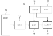
도 4는 의료 진단 초음파 이미징(medical diagnostic ultrasound imaging)을 위한 시스템(system)(10)을 도시한다. 시스템(10)은 환자를 스캐닝하기 위해, 예컨대 유방을 볼륨 스캐닝하기 위해, 스탠드오프, 예컨대 도 1의 스탠드오프(28)와 함께 사용될 수 있다. 시스템(10)은 트랜스듀서 프로브(transducer probe)(12), 빔포머(beamformer)(14), 프로세서(processor)(16), 검출기(18), 메모리(memory)(22), 및 디스플레이(24)를 포함한다. 부가적인, 상이한, 또는 더 적은 수의 컴포넌트들이 제공될 수 있다. 예컨대, 시스템(10)은 사용자 인터페이스(user interface)를 포함한다. 일 실시예에서, 시스템(10)은 의료 진단 초음파 이미징 시스템(medical diagnostic ultrasound imaging system)이다. 다른 실시예들에서, 프로세서(16) 및/또는 메모리(22)는 초음파 이미징 시스템(ultrasound imaging system)과는 상이한 또는 별개의 워크스테이션(workstation) 또는 컴퓨터(computer)의 일부이다. 워크스테이션은 초음파 이미징 시스템에 인접하게 또는 그로부터 원격으로 있다. 몇몇 실시예들에서, 트랜스듀서 프로브(12)는 다른 컴포넌트들 없이 제공된다. Figure 4 shows a
일 실시예에서, 시스템은 자동화된 유방 볼륨 스캐너(breast volume scanner)를 나타낸다. 유방을 스캐닝하기 위해 트랜스듀서 프로브(12)가 제공된다. 트랜스듀서 프로브(12)는 핸드헬드(handheld)되거나, 또는 자동화된 스캐닝 시스템(scanning system)의 일부일 수 있다. 예컨대, 트랜스듀서 프로브(12)는 로봇 팔 또는 지지 팔에 의해 지지된다. 중력, 서보(servo)들, 모터(motor)들, 스프링(spring)들, 유압식 또는 다른 메커니즘(mechanism)이 환자의 유방에 맞닿은 위치에서 트랜스듀서 프로브(12)를 홀딩(holding)시킨다. 유방 이미징(breast imaging) 이외의 다른 애플리케이션(application)들이 제공될 수 있다.In one embodiment, the system represents an automated breast volume scanner. A
트랜스듀서 프로브(12)는 의료 진단 초음파 이미징을 위한 트랜스듀서 어레이(transducer array)이다. 트랜스듀서 프로브(12)는 프로브 하우징(probe housing) 및 트랜스듀서 어레이를 포함한다. 부가적인, 상이한, 또는 더 적은 수의 컴포넌트들이 제공될 수 있는데, 케이블(cable) 및/또는 전자장치(electronics)가 제공될 수 있다.The
트랜스듀서 프로브(12)는 평면 어레이(planar array), 만곡형 어레이(curved array), 이차원 어레이, 방사형 어레이(radial array), 환형 어레이(annular array), 또는 트랜스듀서 엘리먼트(transducer element)들의 다른 다차원 어레이(multidimensional array)를 포함한다. 예컨대, 트랜스듀서 프로브(12)는 다차원 또는 이차원 어레이를 포함한다. 이차원 어레이는 다수의 방향들로 이격된 엘리먼트(element)들을 갖지만(예컨대, NxM, 여기서 N 및 M 둘 다 1보다 큼), 반드시 각각의 방향으로 동일한 범위를 갖는 것은 아니다. 다차원 어레이들은, 선이 아니라 영역(area)에 걸쳐, 엘리먼트들의 1.25D, 1.5D, 1.75D, 환형, 방사형, 또는 다른 어레인지먼트들을 포함한다.The transducer probes 12 may be formed of a planar array, a curved array, a two dimensional array, a radial array, an annular array, or other multi-dimensional array of transducer elements. And includes a multidimensional array. For example, the
대안적 실시예에서, 트랜스듀서 프로브(12)는 가이드(guide)와 연결되는 일차원 어레이를 갖는다. 가이드는 레일(rail), 풀리, 유압식 시스템(hydraulic system), 스크류 드라이브(screw drive), 기계식 링키지(mechanical linkage), 볼 베어링(ball bearing)들, 랙(rack) 및 피니언(pinion), 또는 트랜스듀서 어레이를 회전 움직임 또는 측방향 움직임으로 가이딩(guiding)하기 위한 다른 메커니즘이다. 예컨대, 가이드는 두 개의 그루브(groove)들을 포함하는데, 트랜스듀서 어레이는 이 그루브들에 놓이고 풀리 또는 체인(chain)에 연결된다. 그루브들은, 일반적으로 직각으로, 예컨대 고도 방향으로 움직이도록 어레이를 지지한다. 모터는 예컨대 풀리 또는 기어들을 통해 어레이와 연결된다. 모터는 트랜스듀서 어레이를 움직이기 위해 힘을 가한다. 트랜스듀서 어레이를 병진시키거나 또는 움직이기 위해 임의의 스피드(speed)의 모션(motion)이 제공될 수 있다. 스캔 헤드(scan head)는 단축(short axis)에 병렬 방향으로 기계적으로 병진되고, 이는 전송 평면이 전체 볼륨을 가로질러 스위핑(sweeping)하게 한다. 제어기가 모터를 원하는 시간들에서 그리고/또는 스피드로 작동시킨다. 임의의 타입의 모터, 예컨대, 스텝퍼 모터(stepper motor), 전기 모터(electric motor), 또는 펌프가 사용될 수 있다.In an alternative embodiment, the
트랜스듀서 프로브(12)는 프로브 하우징(probe housing)을 포함한다. 유방 이미저(breast imager)의 경우, 프로브 하우징은 플라스틱(plastic), 유리섬유, 금속, 및/또는 다른 물질의 포드(pod) 또는 외부 쉘(shell)이다. 트랜스듀서 어레이와 패드(pad) 사이에는, 음향 윈도우(acoustic window), 예컨대, 젤 또는 다른 초음파 투과성 물질을 갖거나 또는 갖지 않는 연성 백이 제공된다. 예컨대, 패드는 압박된 유방의 형상에 컨포밍한다. 패드와 트랜스듀서 어레이 사이의 젤은 적응을 허용하고, 트랜스듀서 어레이로부터 유방으로의 음향 경로를 제공한다. 대안적으로, 프로브 하우징은 맘모그램 시스템(mammogram system) 또는 임의의 다른 유방 압박 또는 스캐닝 시스템(breast compression or scanning system)의 일부이다.The
유방을 스캐닝하는 용도를 위한 또는 다른 용도들을 위한 대안적 실시예들에서, 프로브 하우징은 핸드헬드 용도(handheld use)를 위한 것이다. 프로브 하우징의 형상 및 표면 텍스처(surface texture)는 프로브 하우징의 수동 움직임을 위한 그립(grip) 또는 핸들(handle)을 포함한다. 음향 윈도우, 예컨대 플라스틱 또는 렌즈(lens)가 제공될 수 있다. In alternative embodiments for use for scanning the breasts or for other uses, the probe housing is for handheld use. The shape and surface texture of the probe housing includes a grip or handle for manual movement of the probe housing. Acoustic windows, such as plastic or lenses, may be provided.
프로브 하우징은 트랜스듀서 어레이를 감싸거나, 트랜스듀서 어레이의 대부분을 둘러싸거나, 또는 트랜스듀서 어레이 둘레의 보호성 프레임 워크(protective frame work)이다. 프로브 하우징은 핸들들, 그립들, 래치(latch)들, 연결부들, 트랜스듀서 케이블(transducer cable), 또는 다른 컴포넌트들을 포함할 수 있다. 전자장치가 프로브 하우징 내에 제공될 수 있지만, 프로브 하우징에는 액티브(active)(예컨대, 트랜지스터(transistor)들, 스위치(switch)들, 또는 전치증폭기들) 전자장치가 없을 수 있다.The probe housing surrounds the transducer array, surrounds most of the transducer array, or is a protective frame work around the transducer array. The probe housing may include handles, grips, latches, connections, a transducer cable, or other components. Although electronic devices may be provided within the probe housing, the probe housing may not have active (e.g., transistors, switches, or preamplifiers) electronics.
트랜스듀서 프로브(12)의 음향 엘리먼트(acoustic element)들은, 티탄산 지르콘산 연(lead zirconate titanate)(PZT) 압전 트랜스덕션 물질(piezoelectric transduction material), 강유전성 릴렉서(ferroelectric relaxor) 또는 PVDF 물질들, 용량성 막 초음파 트랜스듀서(cMUT:capacitive membrane ultrasonic transducer) 물질들, 미세-기계화된 막들 또는 빔(beam)들, 미세 전자 기계 디바이스(microelectromechanical device)들, 다른 압전 물질, 또는 음향-대-전기 및/또는 전기-대-음향 트랜스덕션(acoustic-to-electric and/or electric-to-acoustic transduction)을 위한 다른 수단이다. 예컨대, 음향 엘리먼트들은 cMUT 또는 미세기계화된 구조들, 예컨대, 갭 상에 부유되는 적어도 하나의 연성 막이며, 갭의 각 측 상에는, 음향 에너지(acoustic energy)와 전기 에너지(electrical energy) 사이를 트랜스듀싱(transducing)하기 위한 전극들이 있다. 각각의 음향 엘리먼트는 하나 이상, 예컨대 4개-8개, 열 개 또는 다른 개수들의 막들 및 갭들(즉, "드럼(drum)들" 또는 cMUT 셀(cell)들)로 형성된다. 주어진 엘리먼트에 대한 막들 및 갭들 각각의 전극들이 공동으로 연결되어, 단일 음향 엘리먼트가 형성된다. The acoustic elements of the
음향 엘리먼트들 전부가 동일한 타입의 물질을 포함하지만, 상이한 음향 엘리먼트들에 대해 다수의 타입들의 음향 트랜스듀서 물질(acoustic transducer material)들이 사용될 수 있다. 음향 엘리먼트들은 다양한 가능한 형상들, 예컨대, 삼각형, 직사각형, 정사각형, 다각형, 육각형, 원형, 불규칙, 또는 음향 엘리먼트(즉, 스캐닝될 볼륨에 인접하게 배치된 엘리먼트의 부분)의 면 상의 형상들의 임의의 결합 중 하나를 갖는다.Although all of the acoustic elements comprise the same type of material, multiple types of acoustic transducer materials can be used for different acoustic elements. Acoustic elements can have any combination of shapes on the surface of various possible shapes, such as triangular, rectangular, square, polygonal, hexagonal, circular, irregular, or acoustical elements (i.e., / RTI >
트랜스듀서 프로브(12)는 환자의 몸의 구역을 스캐닝하기 위해 음향 에너지와 전기 신호들 사이를 변환한다. 스캐닝되는 몸의 구역은, 트랜스듀서 어레이의 타입, 및 환자에 대한 트랜스듀서 프로브(12)의 포지션의 함수이다. 선형 어퍼처(linear aperture)가 몸의 직사각형 또는 정사각형의 평면 구역을 스캐닝할 수 있다. 다른 예로서, 만곡된 선형 어퍼처가 몸의 파이(pie) 형상의 구역을 스캐닝할 수 있다. 몸 내의 다른 기하학적 구역들 또는 형상들에 컨포밍하는 스캔들, 예컨대 Vector™ 스캔들이 사용될 수 있다. 스캔들은 이차원 평면으로 이루어지는데, 예컨대, 어퍼처(aperture)에 대해 상이한 방위각들로 스캐닝이 이루어진다. 트랜스듀서 어레이를 움직임으로써, 상이한 평면들 또는 평면의 상이한 세그먼트(segment)들이 스캐닝될 수 있다. 유방 볼륨을 스캐닝하기 위해, 트랜스듀서 어레이는, 또한 또는 대신에, 상이한 고도로 이격된 평면들을 스캐닝하기 위해 기계적으로 움직여진다. The
빔포머(14)는 하드웨어(hardware) 및/또는 소프트웨어(software)에 의해 구성된다. 예컨대, 음향 빔(acoustic beam)들을 스티어링(steering)하기 위해 지연들 또는 위상들을 결정하는데 포커스 테이블(focus table)들이 사용된다. 소프트웨어 제어(software control)에 따라, 전송 동작을 위한 원하는 파형들이 생성되고, 원하는 수신 프로세스(receive process)가 구현된다.The beam former 14 is constituted by hardware and / or software. For example, focus tables are used to determine delays or phases to steer acoustic beams. Depending on the software control, desired waveforms for the transmission operation are generated and a desired receive process is implemented.
일 실시예에서, 빔포머(14)는 전송 어퍼처(transmit aperture)의 각각의 엘리먼트에 대한 전기 파형들을 생성하기 위한 파형 생성기들 또는 전송기들을 포함한다. 파형들은 위상 및 진폭과 연관된다. 주어진 전송 이벤트(transmit event)에 대한 파형들은 동일한 또는 상이한 페이징(phasing)을 가질 수 있다. 전기 파형들은, 원하는 위상 및 진폭 특징을 갖는 음향 빔을 형성하기 위해 비교적(relatively) 가중 및 지연된다. 예컨대, 전송 빔포머(transmit beamformer)는 다른 음향 빔들과 관련하여 원하는 위상 및 진폭을 갖는 순차적인 스티어링(steering)된 펄스(pulse)들을 생성하기 위해 증폭기들, 위상 로테이터(phase rotator)들, 및/또는 제어기들을 포함한다. 수렴, 발산 또는 평면 빔들이 사용될 수 있다.In one embodiment, the
빔포머(14)는, 동적 포커싱(dynamic focusing)으로 하나 이상의 수신 빔(receive beam)들을 형성하기 위해 수신 빔포머(receive beamformer)들, 예컨대, 지연들, 위상 로테이터들, 증폭기들, 및/또는 수신 신호들을 비교적 지연시키고 합산하기 위한 가산기들을 포함할 수 있다. 예컨대, 공유 프로세싱(shared processing), 별개의 프로세싱, 또는 이들의 결합들을 사용하여, 복수(예컨대, 수십 개 또는 수백 개)의 병렬 수신 빔포머들이 제공되어, 주어진 전송 빔(transmit beam)에 응답하는 개개의 복수의 수신 빔들이 형성된다. 대안적으로, 빔포머(14)는, 스캐닝되는 구역의 상이한 공간 위치들을 나타내는 샘플들을 생성하기 위해 수신 신호들의 푸리에(Fourier) 또는 다른 분석을 위한 프로세서(processor)를 포함한다. 다른 실시예들에서, 각각의 전송 빔에 대해 단 한 개 또는 몇 개(예컨대, 아홉 개 또는 그 미만)의 수신 빔들이 생성된다.The
수신 빔포머는 전치증폭, 임의의 신호 컨디셔닝(signal conditioning)(예컨대, 필터링(filtering)) 및 아날로그-대-디지털 변환(analog-to-digital conversion) 뒤에 트랜스듀서 어레이의 수신 엘리먼트(receive element)들과 연결된다. 수신 빔포머는 엘리먼트들을 갖는 온-칩(on-chip)일 수 있다. The receive beamformer may comprise receive elements of the transducer array after preamplification, any signal conditioning (e.g., filtering) and analog-to-digital conversion Lt; / RTI > The receive beamformer may be on-chip with elements.
트랜스듀서 프로브(12)와 빔포머(14)가 서로 연결되는데, 예컨대, 전송 빔포머 채널(channel)들이 동축 케이블(coaxial cable)들을 통해 트랜스듀서 프로브(12)에 연결된다. 트랜스듀서 프로브(12) 및 빔포머(14)는 평면 구역 또는 평면 구역의 세그먼트를 스캐닝하도록 구성된다. 빔포머(14)는 스캔을 수행하도록 제어 또는 프로그래밍(programming)된다. 빔포머 파라미터(beamformer parameter)들, 예컨대, 포커스(focus)를 위한 상대 지연들 및/또는 페이징, 아포다이제이션(apodization), 빔 진폭(beam amplitude), 빔 위상(beam phase), 주파수, 또는 다른 것들이 설정된다. 트랜스듀서 프로브(12) 상에서 전송을 위한 어퍼처 및 수신을 위한 어퍼처가 설정된다. 빔포머(14) 및 트랜스듀서 프로브(12)는, 어퍼처에 대한 파형들을 생성하고 이 파형들을, 빔을 전송하기 위한 음향 에너지로 변환하는데 사용된다. 빔포머(14) 및 트랜스듀서 프로브(12)는, 수신 어퍼처에서 음향 에너지를 수신하고, 음향 에너지를 전기 에너지로 변환하며, 수신된 전기 신호들을 빔포밍하는데 사용된다. Transducer probes 12 and beam former 14 are interconnected, for example, transmission beamformer channels are connected to
평면을 스캐닝하는데 전기 스티어링(electric steering)이 사용될 수 있다. 트랜스듀서 어레이의 기계식 움직임 또는 추가적인 전기 스티어링을 사용하여 볼륨 스캔이 수행될 수 있다. 스캔 라인(scan line)들 및/또는 어퍼처들의 임의의 패턴(pattern) 또는 분산이 사용될 수 있다. 음향 에너지는, 데이터(data)를 취득하기 위해, 각각의 스캔 평면(scan plane)을 따라서, 현재 알려져 있는 또는 이후에 개발되는 다양한 스캔 패턴(scan pattern)들 중 임의의 패턴으로 전송된다. 이후, 트랜스듀서 어레이를 움직임으로써, 볼륨의 다른 위치로 스캔 평면이 변경된다. 가이드를 따라서 트랜스듀서 어레이를 움직임으로써, 볼륨이 스캐닝될 수 있다. 볼륨은 복수의 평면들에 대한 데이터에 의해 표현된다.An electric steering may be used to scan the plane. A volume scan can be performed using mechanical movement of the transducer array or additional electrical steering. Any pattern or dispersion of scan lines and / or apertures may be used. Acoustic energy is transmitted along any scan plane, in any of various scan patterns that are currently known or later developed, in order to acquire data. Then, by moving the transducer array, the scan plane changes to another position in the volume. By moving the transducer array along the guide, the volume can be scanned. The volume is represented by data for a plurality of planes.
각각의 평면 포지션(plane position)에 대해, 빔포머는 평면을 한 번 스캐닝하도록 구성된다. 대안적으로, 평면은 다수 번 스캐닝되지만, 공간적으로 합성하기 위해 상이한 스캔 라인 방위 각도들(scan line angles in azimuth)로 스캐닝된다. 주어진 위치를 상이한 각도들로부터 스캐닝하기 위해 상이한 어퍼처 위치(aperture location)들이 사용될 수 있다. For each plane position, the beamformer is configured to scan the plane once. Alternatively, the planes are scanned a number of times, but with different scan line angles in azimuth to spatially synthesize. Different aperture locations may be used to scan a given position from different angles.
주어진 볼륨에 대해, 스캔들은 반복될 수 있다. 스캔들을 반복함으로써, 복셀 데이터(voxel data)의 프레임(frame)들의 시퀀스(sequence)가 획득된다. 각각의 프레임은 삼차원으로 스캐닝된 전체 볼륨을 나타내지만, 볼륨 내의 더 작은 구역들, 예컨대 평면만을 나타낼 수 있다. 스캐닝을 반복함으로써, 볼륨 및/또는 평면을 나타내는 빔포밍된 데이터의 복수의 프레임들이 취득된다. 스캔 라인, 프레임의 일부, 프레임, 또는 인터리빙되는 프레임의 그룹(group of frame interleaving) 중 임의의 것이 사용될 수 있다.For a given volume, the scans may be repeated. By repeating the scans, a sequence of frames of voxel data is obtained. Each frame represents the entire volume scanned three-dimensionally, but may represent only smaller zones, e.g., planes, in the volume. By repeating the scanning, a plurality of frames of beamformed data representing volume and / or plane are acquired. Any of a scan line, a portion of a frame, a frame, or a group of frame interleaving may be used.
검출기(detector)(18)는, 빔포머(14)에 의해 출력되는 데이터를 검출하고 트랜스듀서 어레이에 응답하도록 구성된다. 검출기(18)는 초음파 검출기이다. 검출기는, 빔포밍된 그리고/또는 보간된 데이터로부터의 검출을 위해, 하드웨어 및/또는 소프트웨어에 의해 구성된다. 임의의 검출, 예컨대, B 모드(B mode), 도플러 또는 색 흐름 모드(Doppler or color flow mode), 고조파 모드(harmonic mode), 또는 현재 알려져 있는 또는 이후에 개발되는 다른 모드들이 사용될 수 있다. B 모드 및 몇몇 고조파 모드들은 검출을 위해 단일 펄스 스캔 기술(single pulse scan technique)들을 사용한다. 관심대상의 주파수 대역에서 수신된 신호들의 세기가 계산된다. 다수의 펄스 기술들, 예컨대, 속도 또는 에너지의 흐름 모드 추정(flow mode estimation)이 사용될 수 있다.A
검출기(18)는 볼륨의 스캔을 위해 전송 빔들에 대한 응답을 검출한다. 검출되는 데이터의 공간 및/또는 시간 레졸루션(resolution)은 빔포밍 또는 스캐닝 레졸루션(beamforming or scanning resolution)에 기초한다. 볼륨을 나타내는, 검출된 데이터가 제공된다.The
프로세서(16)는 하드웨어 및/또는 소프트웨어에 의해 구성되는 렌더링 프로세서(rendering processor)이다. 프로세서(16)는 일반 프로세서(general processor), 제어 프로세서(control processor), 애플리케이션-특정 집적 회로(application-specific integrated circuit), 필드-프로그램어블 게이트 어레이(field-programmable gate array), 그래픽스 프로세싱 유닛(graphics processing unit), 디지털 회로(digital circuit), 아날로그 회로(analog circuit), 디지털 신호 프로세서(digital signal processor), 이들의 결합들, 또는 상이한 평면들로 스캐닝되는 볼륨의 삼차원 렌더링(three-dimensional rendering)을 생성하기 위한, 현재 알려져 있는 또는 이후에 개발되는 다른 디바이스(device)이다. 프로세서(16)는 단일 디바이스 또는 디바이스들의 그룹(group)이다. 예컨대, 프로세서(16)는 병렬로 또는 시퀀스(sequence)로 동작하는 별개의 프로세서들을 포함한다. 다른 예로서, 프로세서(16)는 병렬로 또는 시퀀스로 이루어지는 분산 프로세싱(distributed processing)을 위한 디바이스들의 네트워크(network)를 포함한다. 일 실시예에서, 프로세서(16)는 삼차원 이미지 렌더링(three-dimensional image rendering)을 위한 특정한 디바이스, 예컨대, 그래픽스 프로세싱 유닛, 그래픽스 카드(graphics card), 또는 렌더링을 위한 다른 디바이스이다.The
프로세서(16)는 표면 렌더링(surface rendering), 프로젝션 렌더링(projection rendering), 알파 블렌딩(alpha blending), 텍스처링(texturing), 또는 현재 알려져 있는 또는 이후에 개발되는 다른 렌더링을 사용한다. 데이터는 규칙적인 복셀 그리드(voxel grid)로 리샘플링(resampling)될 수 있다. 대안적으로, 렌더링은 데이터로부터 예컨대 실제 스캔 라인들 및/또는 보간된 스캔 라인들과 연관된 스캔 포맷(scan format)으로 수행된다. 또 다른 실시예들에서, 프로세서(16)는 제공되지 않거나, 또는 스캐닝된 평면을 나타내거나 또는 스캐닝된 볼륨으로부터의 평면의 재구성을 나타내는 이차원 이미지를 생성하기 위한 스캔 컨버터(scan converter)이다.The
프로세서(16), 검출기(18), 또는 별개의 프로세서는 볼륨 스캔 및/또는 평면 스캔 또는 검출기(18)로부터 출력되는 다른 데이터로부터 이미지들을 생성한다. 예컨대, 그레이스케일 및/또는 색 코딩(grayscale and/or color coding)이 B 모드, 도플러 모드(Doppler mode), 또는 B 모드 도플러 모드 결합(B mode Doppler mode combination)을 생성하는데 사용된다. 임의의 이미지, 예컨대, 삼차원 렌더링이 디스플레이(24)에 출력된다.The
디스플레이(24)는 CRT, LCD, 플라즈마(plasma), 프로젝터(projector), 프린터(printer), 또는 현재 알려져 있는 또는 이후의 다른 디스플레이 디바이스(display device)이다. 디스플레이(24)는 프로세서(16) 또는 다른 컴포넌트로부터 이미지 데이터(image data)를 수신하고, 이미지를 생성한다. 삼차원 렌더링, 이차원 이미지, 또는 다른 이미지가 디스플레이(display)된다.
메모리(22)는 유형(비-일시적) 컴퓨터 판독가능 스토리지 매체(computer readable storage medium), 예컨대, 캐시(cache), 버퍼(buffer), 레지스터(register), 램(RAM), 탈착가능 미디어(removable media), 하드 드라이브(hard drive), 광학 스토리지 디바이스(optical storage device), 또는 다른 컴퓨터 판독가능 스토리지 미디어(computer readable storage media)이다. 메모리(22)는, 신호가 아니라 디바이스이기 때문에 유형이다. 컴퓨터 판독가능 스토리지 미디어(computer readable storage media)는 다양한 타입들의 휘발성 및 비휘발성 스토리지 미디어(volatile and nonvolatile storage media)를 포함한다. 메모리(22)는 프로세서(16)에 의해 액세스 가능(accessible)하다.The
메모리(22)는, 프로그래밍된 프로세서(programmed processor)(16), 빔포머(14)의 프로세서, 및/또는 초음파로 스캐닝하고 그리고/또는 트랜스듀서 프로브(12)의 모터를 제어하기 위한 프로세서에 의해 실행 가능한 명령들을 나타내는 데이터를 저장한다. 본원에서 논의된 프로세스(process)들, 방법들 및/또는 기술들을 구현하기 위한 명령들이 컴퓨터-판독가능 스토리지 미디어 또는 메모리들 상에 제공된다. 도면들에 예시된 또는 본원에 설명된 기능들, 동작들 또는 작업들은 컴퓨터 판독가능 스토리지 미디어에 또는 그 상에 저장된 명령들의 하나 이상의 세트(set)들에 응답하여 실행된다. 기능들, 동작들 또는 작업들은 특정 타입의 명령 세트(instructions set), 스토리지 미디어(storage media), 프로세서 또는 프로세싱 전략(processor or processing strategy)에 독립적이고, 소프트웨어, 하드웨어, 집적 회로(integrated circuit)들, 펌웨어(firmware), 마이크로 코드(micro code) 등에 의해 수행될 수 있으며, 이들은 단독으로 또는 결합하여 동작한다. 마찬가지로, 프로세싱 전략(processing strategy)들은 멀티프로세싱(multiprocessing), 멀티태스킹(multitasking), 병렬 프로세싱(parallel processing) 등을 포함할 수 있다. 일 실시예에서, 명령들은 로컬 또는 원격 시스템(local or remote system)들에 의한 판독을 위해 탈착가능 미디어 디바이스(removable media device) 상에 저장된다. 다른 실시예들에서, 명령들은 컴퓨터 네트워크(computer network)를 통한 또는 전화선들을 통한 전송을 위해 원격 위치에 저장된다. 또 다른 실시예들에서, 명령들은 주어진 컴퓨터, CPU, GPU 또는 시스템 내에 저장된다.The
본 발명이 다양한 실시예들을 참조하여 위에서 설명되었지만, 본 발명의 범위로부터 벗어남 없이 많은 변경들 및 수정들이 이루어질 수 있음이 이해되어야 한다. 위의 실시예들은 예들이다. 그러므로, 앞의 상세한 설명은 본 발명의 현재 바람직한 실시예들의 예시로서 이해되고, 본 발명의 정의로서 이해되지 않음이 의도된다. 모든 등가물들을 포함하는 하기의 청구항들만이, 본 발명의 범위를 정의하도록 의도된다.Although the present invention has been described above with reference to various embodiments, it should be understood that many changes and modifications can be made without departing from the scope of the present invention. The above embodiments are examples. It is therefore intended that the foregoing detailed description be understood as illustrative of the presently preferred embodiments of the invention, and not as a definition of the invention. Only the following claims, including all equivalents, are intended to define the scope of the invention.
Claims (20)
대향하는 제2 표면(34)보다 더 강성(stiff)의 제1 표면(32)을 갖는 밀봉된 용기(28) ― 상기 제2 표면(34)은 환자의 외부 표면에 컨포멀(conformal)하고, 상기 제1 표면(32)은 초음파 트랜스듀서 어레이(ultrasound transducer array)(30)와 음향적 접촉(acoustic contact)하도록 구성됨 ―; 및
상기 밀봉된 용기(28)에 있는, 물보다 더욱 점성이 있는 유체(36)
를 포함하는,
초음파 이미징을 위한 컨포멀한 인터페이스.As a conformal interface for ultrasound imaging,
A sealed container 28 having a first surface 32 that is stiffer than an opposing second surface 34, the second surface 34 conforming to the exterior surface of the patient, The first surface (32) is configured to make acoustic contact with an ultrasound transducer array (30); And
The fluid 36 in the sealed container 28, which is more viscous than water,
/ RTI >
Conformal interface for ultrasound imaging.
상기 제1 표면(32)은 정사각형 또는 직사각형 프레임(frame)을 포함하고, 상기 프레임은, 상기 프레임의 둘레 주위에 있고 상기 제2 표면(34)으로부터 멀어지게 연장되는 립(lip)을 갖는,
초음파 이미징을 위한 컨포멀한 인터페이스.The method according to claim 1,
The first surface 32 comprises a square or rectangular frame and the frame has a lip which is around the periphery of the frame and extends away from the second surface 34,
Conformal interface for ultrasound imaging.
상기 제1 표면(32)은 상기 제2 표면(34)보다 더 두꺼운,
초음파 이미징을 위한 컨포멀한 인터페이스.The method according to claim 1,
The first surface (32) is thicker than the second surface (34)
Conformal interface for ultrasound imaging.
상기 제2 표면(34)은 탄성이 있는,
초음파 이미징을 위한 컨포멀한 인터페이스.The method according to claim 1,
The second surface 34 is resilient,
Conformal interface for ultrasound imaging.
상기 제2 표면(34)은 PEBAX이고, 상기 제1 표면(32)은 PEBAX인,
초음파 이미징을 위한 컨포멀한 인터페이스.The method according to claim 1,
Wherein the second surface (34) is PEBAX and the first surface (32) is PEBAX.
Conformal interface for ultrasound imaging.
상기 제1 표면(32) 및 상기 제2 표면(34)은 상이한 타입(type)들의 PEBAX인,
초음파 이미징을 위한 컨포멀한 인터페이스.6. The method of claim 5,
The first surface 32 and the second surface 34 may be PEBAX of different types,
Conformal interface for ultrasound imaging.
상기 제1 표면(32)은 임의의 방향으로 적어도 10 cm의 지름을 갖고, 그리고상기 제1 표면(32)은, 초음파 이미징을 위해 손으로 가해지는 압력 하에 있는 동안 대향하는 에지(edge)들이 평면으로부터 0.5 cm 이하(no more than)만큼 벗어날 정도로 강성인,
초음파 이미징을 위한 컨포멀한 인터페이스.The method according to claim 1,
The first surface 32 has a diameter of at least 10 cm in any direction and the first surface 32 is positioned such that the opposing edges are in a planar state while under pressure applied by hand for ultrasound imaging. Which is not more than 0.5 cm (no more than)
Conformal interface for ultrasound imaging.
상기 제1 표면(32)은 실질적으로 평편한,
초음파 이미징을 위한 컨포멀한 인터페이스.The method according to claim 1,
The first surface 32 is substantially planar,
Conformal interface for ultrasound imaging.
상기 제1 표면(32)은 접착성 윤활제를 포함하는,
초음파 이미징을 위한 컨포멀한 인터페이스.The method according to claim 1,
The first surface (32) comprises an adhesive lubricant.
Conformal interface for ultrasound imaging.
상기 유체(36)는 오일(oil)을 포함하는,
초음파 이미징을 위한 컨포멀한 인터페이스.The method according to claim 1,
The fluid (36) comprises an oil.
Conformal interface for ultrasound imaging.
상기 유체(36)는 1 기압을 초과하는 압력 하에서 상기 밀봉된 용기(28) 내에 있는,
초음파 이미징을 위한 컨포멀한 인터페이스.The method according to claim 1,
The fluid 36 is pressurized in the sealed vessel 28 under a pressure exceeding one atmosphere,
Conformal interface for ultrasound imaging.
상기 밀봉된 용기(28)는, 상기 유체(36)의 주입을 위해 구성되고 상기 유체(36)를 1 기압을 초과하는 압력으로 홀딩(holding)시키는 포트(port)를 포함하고, 상기 압력은 상기 주입에 의해 설정 가능한,
초음파 이미징을 위한 컨포멀한 인터페이스.The method according to claim 1,
The sealed vessel 28 comprises a port configured for injection of the fluid 36 and holding the fluid 36 at a pressure in excess of one atmosphere, Settable by injection,
Conformal interface for ultrasound imaging.
상기 밀봉된 용기(28)에서 힘 센서(force sensor)(40)
를 더 포함하는,
초음파 이미징을 위한 컨포멀한 인터페이스.The method according to claim 1,
In the sealed container 28, a force sensor 40,
≪ / RTI >
Conformal interface for ultrasound imaging.
압력 하에서 점성 유체(36)로 충전된 백(bag)(28)을 환자의 상기 유방에 맞닿게 배치하는 단계(42);
볼륨 스캐닝(48) 트랜스듀서(volume scanning transducer)를 상기 백(28)에 맞닿게 포지셔닝(positioning)하는 단계(44);
상기 볼륨 스캐닝(48) 트랜스듀서를 이용하여 상기 백(28)에 압력을 가하는 단계(46) ―상기 압력은 중력에 의해 유발되는 것보다 더 큼―; 및
상기 유방을 상기 볼륨 스캐닝(48) 트랜스듀서를 갖는 상기 백(28)을 통해 볼륨 스캐닝(volume scanning)(48)하는 단계
를 포함하는,
의료 진단 초음파 볼륨 이미징에서 유방을 스캐닝(48)하기 위한 방법.CLAIMS What is claimed is: 1. A method for scanning (48) a breast in a medical diagnostic ultrasound volume imaging,
Placing (42) a bag (28) filled with viscous fluid (36) under pressure against the breast of the patient;
Positioning a volume scanning transducer (44) against the bag (28) (44);
Applying (46) pressure to the bag (28) using the volume scanning (48) transducer, the pressure being greater than that caused by gravity; And
Volume scanning (48) the breast through the bag (28) with the volume scanning (48) transducer
/ RTI >
A method for scanning (48) a breast in a medical diagnostic ultrasound volume imaging.
상기 백(28)은 실질적으로 경성(rigid)의 트랜스듀서 표면(transducer surface) 및 연성(flexible)의 유방 접촉 표면을 포함하고, 상기 압력을 가하는 단계(46)는 상기 유방이 상기 압력을 받는 동안 상기 연성의 유방 접촉 표면을 상기 유방에 컨포밍(conforming)시키는 단계를 포함하는,
의료 진단 초음파 볼륨 이미징에서 유방을 스캐닝(48)하기 위한 방법.15. The method of claim 14,
The bag (28) comprises a substantially rigid transducer surface and a flexible breast contact surface, the step of applying pressure (46) being performed while the breast is under the pressure Contacting said soft breast contact surface with said breast,
A method for scanning (48) a breast in a medical diagnostic ultrasound volume imaging.
배치하는 단계(42)는, 1 기압을 초과하는 압력 및 오일인 상기 점성 유체(36)를 갖는 밀봉된 백(28)인 백(28)을 배치하는 단계(42)를 포함하는,
의료 진단 초음파 볼륨 이미징에서 유방을 스캐닝(48)하기 위한 방법.15. The method of claim 14,
The placing step 42 includes placing a bag 28 that is a sealed bag 28 having the viscous fluid 36 at a pressure and oil that is greater than one atmosphere.
A method for scanning (48) a breast in a medical diagnostic ultrasound volume imaging.
상기 볼륨 스캐닝(48)하는 단계는 상기 유방을 통하는 복수의 평면들을 스캐닝(48)하는 단계를 포함하고,
상기 방법은,
상기 스캐닝(48)으로부터 이미지(image)를 생성하는 단계
를 더 포함하는,
의료 진단 초음파 볼륨 이미징에서 유방을 스캐닝(48)하기 위한 방법.15. The method of claim 14,
The step of volume scanning (48) includes scanning (48) a plurality of planes through the breast,
The method comprises:
Generating an image from the scanning 48,
≪ / RTI >
A method for scanning (48) a breast in a medical diagnostic ultrasound volume imaging.
상기 압력을 가하는 단계(46)는 소노그래퍼(sonographer)의 손 또는 로봇 팔(robotic arm)로 압박하는 단계를 포함하고,
상기 볼륨 스캐닝(48) 트랜스듀서와 상기 유방 사이의 상기 백(28)에는 에어 갭(air gap)들 및 주름(wrinkle)들이 존재하지 않도록, 상기 백(28)은 상기 볼륨 스캐닝(48) 트랜스듀서의 방출 면에 컨포밍하고 상기 유방에 컨포밍하는,
의료 진단 초음파 볼륨 이미징에서 유방을 스캐닝(48)하기 위한 방법.15. The method of claim 14,
The step of applying pressure 46 comprises the step of pressing with a hand or a robotic arm of a sonographer,
The bag 28 is positioned between the volume scanning 48 transducer and the breast so that there is no air gaps and wrinkles in the bag 28 between the volume scanning 48 transducer and the breast. And conformation to the breast,
A method for scanning (48) a breast in a medical diagnostic ultrasound volume imaging.
상기 백(28) 내의 압력을 감지하는 단계(52); 및
상기 스캐닝(48)에 응답하여 상기 힘과 탄성 또는 스트레인 데이터(strain data)의 함수로서 조직 특징을 계산하는 단계(54)
를 더 포함하는,
의료 진단 초음파 볼륨 이미징에서 유방을 스캐닝(48)하기 위한 방법.15. The method of claim 14,
Sensing (52) pressure in the bag (28); And
Computing (54) tissue features as a function of said force and elasticity or strain data in response to said scanning (48)
≪ / RTI >
A method for scanning (48) a breast in a medical diagnostic ultrasound volume imaging.
하우징(housing)(28)에 포함된 점성 유체(36)를 포함하고, 상기 하우징(28)은 제1 부분에서는 연성의 탄성 물질(34)이고, 제2 부분에서는 비교적 더 강성의 물질(32)이며, 상기 점성 유체(36) 및 상기 하우징(28)은 음향적으로 전도성인,
초음파 이미징을 위한 스탠드오프.As a standoff for ultrasound imaging,
The housing 28 comprises a viscous fluid 36 contained in a housing 28 which is a flexible elastic material 34 in a first portion and a relatively more rigid material 32 in a second portion, , Said viscous fluid (36) and said housing (28) being acoustically conductive,
Standoff for ultrasound imaging.
Applications Claiming Priority (2)
| Application Number | Priority Date | Filing Date | Title |
|---|---|---|---|
| US14/500,833 US20160089110A1 (en) | 2014-09-29 | 2014-09-29 | Conformal interface for medical diagnostic ultrasound volume imaging |
| US14/500,833 | 2014-09-29 |
Related Parent Applications (1)
| Application Number | Title | Priority Date | Filing Date |
|---|---|---|---|
| KR1020150136124A Division KR102065331B1 (en) | 2014-09-29 | 2015-09-25 | Conformal interface for medical diagnostic ultrasound imaging |
Publications (1)
| Publication Number | Publication Date |
|---|---|
| KR20180098495A true KR20180098495A (en) | 2018-09-04 |
Family
ID=55485997
Family Applications (2)
| Application Number | Title | Priority Date | Filing Date |
|---|---|---|---|
| KR1020150136124A Expired - Fee Related KR102065331B1 (en) | 2014-09-29 | 2015-09-25 | Conformal interface for medical diagnostic ultrasound imaging |
| KR1020180098028A Withdrawn KR20180098495A (en) | 2014-09-29 | 2018-08-22 | Conformal interface for medical diagnostic ultrasound imaging |
Family Applications Before (1)
| Application Number | Title | Priority Date | Filing Date |
|---|---|---|---|
| KR1020150136124A Expired - Fee Related KR102065331B1 (en) | 2014-09-29 | 2015-09-25 | Conformal interface for medical diagnostic ultrasound imaging |
Country Status (5)
| Country | Link |
|---|---|
| US (1) | US20160089110A1 (en) |
| JP (1) | JP2016067925A (en) |
| KR (2) | KR102065331B1 (en) |
| CN (1) | CN105455850A (en) |
| DE (1) | DE102015116383A1 (en) |
Cited By (1)
| Publication number | Priority date | Publication date | Assignee | Title |
|---|---|---|---|---|
| WO2023095973A1 (en) * | 2021-11-29 | 2023-06-01 | 경상국립대학교병원 | Method for obtaining ultrasound image of individual having subcutaneous emphysema |
Families Citing this family (15)
| Publication number | Priority date | Publication date | Assignee | Title |
|---|---|---|---|---|
| US20170135673A1 (en) * | 2015-11-16 | 2017-05-18 | General Electric Company | An ultrasound transducer probe having a curved imaging face |
| CN105748106B (en) * | 2016-04-22 | 2018-07-31 | 毛军卫 | Ultrasonic probe and ultrasonic detection equipment with the ultrasonic probe |
| JP2020512132A (en) * | 2017-03-31 | 2020-04-23 | コーニンクレッカ フィリップス エヌ ヴェKoninklijke Philips N.V. | Acoustic lens of ultrasonic transducer probe manufactured by textured surface |
| CN107242883A (en) * | 2017-07-31 | 2017-10-13 | 无锡祥生医学影像有限责任公司 | Breast ultrasound device and breast ultrasound scan component |
| DE102017221330A1 (en) * | 2017-11-28 | 2019-05-29 | Ulrich A. Baumann | Pressure measuring device for pressure measurement and / or elasticity measurement of a vein or an organ and for combination with an ultrasonic measuring unit as well as system and method for pressure measurement and / or elasticity measurement of a vein or an organ |
| CN113453617A (en) * | 2018-11-03 | 2021-09-28 | 兹布拉护理有限公司 | System and method for impedance tomography of a body part of a patient |
| US11517251B2 (en) * | 2019-02-20 | 2022-12-06 | Parham A. Ganchi | Breast-measuring device |
| WO2021041295A1 (en) * | 2019-08-23 | 2021-03-04 | Thomas Jefferson University | System and method for breast cancer detection using co-localized ultrasound-mammography |
| US20210219953A1 (en) * | 2020-01-20 | 2021-07-22 | GE Precision Healthcare LLC | Systems for an ultrasound scan tray |
| WO2021154730A1 (en) * | 2020-01-27 | 2021-08-05 | Cordance Medical Inc. | Ultrasound transducer assembly |
| CN113520464B (en) * | 2020-04-22 | 2024-03-22 | 苏州佳世达电通有限公司 | Universal ultrasonic wave conduction interface device |
| WO2022097138A1 (en) * | 2020-11-03 | 2022-05-12 | Nina Medical Ltd. | Pelvic floor diagnostic-therapeutic treatment chair |
| US11994496B2 (en) | 2021-05-12 | 2024-05-28 | Ford Global Technologies, Llc | Non-destructive evaluation system for inspection of weld and braze joints |
| JP2023156129A (en) * | 2022-04-12 | 2023-10-24 | 吉則 早川 | A simple auxiliary device used in breast cancer ultrasound examinations and breast cancer magnetic resonance imaging examinations. |
| TWI901284B (en) * | 2024-05-24 | 2025-10-11 | 互貴興業股份有限公司 | Ultrasonic sampling auxiliary device and ultrasonic probe including the same |
Family Cites Families (19)
| Publication number | Priority date | Publication date | Assignee | Title |
|---|---|---|---|---|
| JPS5316481A (en) * | 1976-07-30 | 1978-02-15 | Tokyo Shibaura Electric Co | Ultrasonic diagnostic device |
| JPS5770309U (en) * | 1980-10-16 | 1982-04-28 | ||
| US5265614A (en) * | 1988-08-30 | 1993-11-30 | Fujitsu Limited | Acoustic coupler |
| US6117080A (en) * | 1997-06-04 | 2000-09-12 | Atl Ultrasound | Ultrasonic imaging apparatus and method for breast cancer diagnosis with the use of volume rendering |
| US6846291B2 (en) * | 2002-11-20 | 2005-01-25 | Sonotech, Inc. | Production of lubricious coating on adhesive hydrogels |
| JP3690525B2 (en) * | 2004-01-09 | 2005-08-31 | 株式会社石川製作所 | Water bag device for ultrasonic probe in ultrasonic diagnostic equipment |
| WO2007116957A1 (en) * | 2006-04-07 | 2007-10-18 | Hitachi Medical Corporation | Ultrasonic probe and ultrasonograph |
| JP2007282960A (en) * | 2006-04-19 | 2007-11-01 | Toshiba Corp | Ultrasonic breast examination equipment |
| WO2008016022A1 (en) * | 2006-07-31 | 2008-02-07 | Hitachi Medical Corporation | Pressing device, and ultrasonic probe and ultrasonic diagnosis device using the pressing device |
| JP5788141B2 (en) * | 2006-10-25 | 2015-09-30 | スーパー ソニック イマジンSuper Sonic Imagine | Method of operating an assembly for generating and imaging mechanical waves in a viscoelastic medium |
| JP2008154679A (en) * | 2006-12-21 | 2008-07-10 | Aloka Co Ltd | Holding device of ultrasonic probe, water bag for ultrasonic probe and ultrasonic diagnostic device |
| JP5019887B2 (en) * | 2007-01-12 | 2012-09-05 | 株式会社日立メディコ | COMPRESSION DEVICE AND ULTRASONIC DIAGNOSTIC DEVICE USING THE COMPRESSION DEVICE |
| JP2009039149A (en) * | 2007-08-06 | 2009-02-26 | Panasonic Corp | Ultrasonic diagnostic equipment |
| JP2010082337A (en) * | 2008-10-01 | 2010-04-15 | Hitachi Medical Corp | Ultrasonic diagnostic apparatus |
| JP5634077B2 (en) * | 2010-02-02 | 2014-12-03 | キヤノン株式会社 | Acoustic wave receiver |
| JP2012029718A (en) * | 2010-07-28 | 2012-02-16 | Hitachi Aloka Medical Ltd | Ultrasonic probe adapter, ultrasonic diagnostic system, and ultrasonograph |
| JP5858378B2 (en) * | 2011-10-13 | 2016-02-10 | 国立大学法人 東京大学 | Acoustic matching media unit |
| CN103845083A (en) * | 2012-12-04 | 2014-06-11 | 通用电气公司 | Scanning assembly |
| CA2896510A1 (en) * | 2012-12-28 | 2014-07-03 | Volcano Corporation | Intravascular ultrasound catheter for minimizing image distortion |
-
2014
- 2014-09-29 US US14/500,833 patent/US20160089110A1/en not_active Abandoned
-
2015
- 2015-09-24 JP JP2015186739A patent/JP2016067925A/en active Pending
- 2015-09-25 KR KR1020150136124A patent/KR102065331B1/en not_active Expired - Fee Related
- 2015-09-28 DE DE102015116383.7A patent/DE102015116383A1/en not_active Withdrawn
- 2015-09-29 CN CN201510630086.0A patent/CN105455850A/en active Pending
-
2018
- 2018-08-22 KR KR1020180098028A patent/KR20180098495A/en not_active Withdrawn
Cited By (1)
| Publication number | Priority date | Publication date | Assignee | Title |
|---|---|---|---|---|
| WO2023095973A1 (en) * | 2021-11-29 | 2023-06-01 | 경상국립대학교병원 | Method for obtaining ultrasound image of individual having subcutaneous emphysema |
Also Published As
| Publication number | Publication date |
|---|---|
| KR102065331B1 (en) | 2020-01-13 |
| DE102015116383A1 (en) | 2016-03-31 |
| US20160089110A1 (en) | 2016-03-31 |
| JP2016067925A (en) | 2016-05-09 |
| CN105455850A (en) | 2016-04-06 |
| KR20160037790A (en) | 2016-04-06 |
Similar Documents
| Publication | Publication Date | Title |
|---|---|---|
| KR102065331B1 (en) | Conformal interface for medical diagnostic ultrasound imaging | |
| US20250176948A1 (en) | Calibration of multiple aperture ultrasound probes | |
| CN108778530B (en) | Ultrasound imaging with sparse array detectors | |
| US10617384B2 (en) | M-mode ultrasound imaging of arbitrary paths | |
| US8647279B2 (en) | Volume mechanical transducer for medical diagnostic ultrasound | |
| US7850613B2 (en) | Apparatus and method for three dimensional ultrasound breast imaging | |
| EP4060379A1 (en) | Ultrasound imaging using apparent point-source transmit transducer | |
| US20120029358A1 (en) | Three -Dimensional Ultrasound Systems, Methods, and Apparatuses | |
| KR20190103048A (en) | Region of interest placement for quantitative ultrasound imaging | |
| US20160310110A1 (en) | Acquisition control for mixed mode ultrasound imaging | |
| KR20110058723A (en) | Synchronization for Multi-Directional Ultrasonic Scanning | |
| JP7449406B2 (en) | Medical detection system and deployment method | |
| JP6767575B2 (en) | Ultrasonic Transducer / Tile Alignment | |
| US12329580B2 (en) | Contextual multiplanar reconstruction of three-dimensional ultrasound imaging data and associated devices, systems, and methods | |
| US11311270B2 (en) | Intervolume lesion detection and image preparation | |
| KR20140137037A (en) | ultrasonic image processing apparatus and method | |
| EP4114272A1 (en) | Contextual multiplanar reconstruction of three-dimensional ultrasound imaging data and associated devices, systems, and methods | |
| EP4545012A1 (en) | Ultrasound system and method | |
| HK40082645A (en) | Ultrasound imaging using apparent point-source transmit transducer | |
| CN120837118A (en) | Three-dimensional ultrasonic imaging system and method based on flexible ultrasonic transducer array |
Legal Events
| Date | Code | Title | Description |
|---|---|---|---|
| A107 | Divisional application of patent | ||
| PA0107 | Divisional application |
Comment text: Divisional Application of Patent Patent event date: 20180822 Patent event code: PA01071R01D Filing date: 20150925 Application number text: 1020150136124 |
|
| PG1501 | Laying open of application | ||
| PC1203 | Withdrawal of no request for examination |