KR20180090308A - Deriving tooth status information for populating digital dental charts - Google Patents
Deriving tooth status information for populating digital dental charts Download PDFInfo
- Publication number
- KR20180090308A KR20180090308A KR1020187018521A KR20187018521A KR20180090308A KR 20180090308 A KR20180090308 A KR 20180090308A KR 1020187018521 A KR1020187018521 A KR 1020187018521A KR 20187018521 A KR20187018521 A KR 20187018521A KR 20180090308 A KR20180090308 A KR 20180090308A
- Authority
- KR
- South Korea
- Prior art keywords
- digital
- tooth
- teeth
- representation
- data
- Prior art date
Links
- 238000000034 method Methods 0.000 claims abstract description 86
- 238000001506 fluorescence spectroscopy Methods 0.000 claims description 22
- 208000002925 dental caries Diseases 0.000 claims description 14
- 241000894006 Bacteria Species 0.000 claims description 6
- 230000006378 damage Effects 0.000 claims description 6
- 208000004188 Tooth Wear Diseases 0.000 claims description 3
- 239000002253 acid Substances 0.000 claims description 3
- 230000003628 erosive effect Effects 0.000 claims description 3
- 210000004195 gingiva Anatomy 0.000 claims description 3
- 238000012014 optical coherence tomography Methods 0.000 claims description 3
- 206010061274 Malocclusion Diseases 0.000 claims description 2
- 239000005557 antagonist Substances 0.000 claims description 2
- 206010006514 bruxism Diseases 0.000 claims description 2
- 230000001013 cariogenic effect Effects 0.000 claims 1
- 238000004590 computer program Methods 0.000 description 38
- 230000000875 corresponding effect Effects 0.000 description 20
- 238000012545 processing Methods 0.000 description 16
- 230000011218 segmentation Effects 0.000 description 15
- 230000008901 benefit Effects 0.000 description 9
- 238000007408 cone-beam computed tomography Methods 0.000 description 9
- 230000002596 correlated effect Effects 0.000 description 8
- 238000004458 analytical method Methods 0.000 description 7
- 230000000007 visual effect Effects 0.000 description 7
- -1 porphyrin compounds Chemical class 0.000 description 6
- 238000012876 topography Methods 0.000 description 6
- 238000012800 visualization Methods 0.000 description 6
- 238000010586 diagram Methods 0.000 description 5
- 238000011282 treatment Methods 0.000 description 5
- 210000003298 dental enamel Anatomy 0.000 description 4
- 210000004283 incisor Anatomy 0.000 description 4
- 230000003993 interaction Effects 0.000 description 4
- 238000013507 mapping Methods 0.000 description 4
- 239000000523 sample Substances 0.000 description 4
- 230000004044 response Effects 0.000 description 3
- 230000003213 activating effect Effects 0.000 description 2
- 230000006835 compression Effects 0.000 description 2
- 238000007906 compression Methods 0.000 description 2
- 238000009795 derivation Methods 0.000 description 2
- 230000005284 excitation Effects 0.000 description 2
- 239000000945 filler Substances 0.000 description 2
- 230000006870 function Effects 0.000 description 2
- 230000002452 interceptive effect Effects 0.000 description 2
- 239000000463 material Substances 0.000 description 2
- 238000005259 measurement Methods 0.000 description 2
- 238000000638 solvent extraction Methods 0.000 description 2
- 238000002834 transmittance Methods 0.000 description 2
- 230000017105 transposition Effects 0.000 description 2
- 241000282465 Canis Species 0.000 description 1
- 235000010799 Cucumis sativus var sativus Nutrition 0.000 description 1
- 244000299906 Cucumis sativus var. sativus Species 0.000 description 1
- 238000004566 IR spectroscopy Methods 0.000 description 1
- 206010061218 Inflammation Diseases 0.000 description 1
- 230000008859 change Effects 0.000 description 1
- 239000003086 colorant Substances 0.000 description 1
- 239000002131 composite material Substances 0.000 description 1
- 230000003247 decreasing effect Effects 0.000 description 1
- 230000006735 deficit Effects 0.000 description 1
- 230000001419 dependent effect Effects 0.000 description 1
- 238000001514 detection method Methods 0.000 description 1
- 238000002845 discoloration Methods 0.000 description 1
- 238000005286 illumination Methods 0.000 description 1
- 208000015181 infectious disease Diseases 0.000 description 1
- 230000004054 inflammatory process Effects 0.000 description 1
- 238000012986 modification Methods 0.000 description 1
- 230000004048 modification Effects 0.000 description 1
- 238000012544 monitoring process Methods 0.000 description 1
- 230000002093 peripheral effect Effects 0.000 description 1
- 230000009467 reduction Effects 0.000 description 1
- 230000032554 response to blue light Effects 0.000 description 1
- 239000007787 solid Substances 0.000 description 1
- 230000009466 transformation Effects 0.000 description 1
- 230000007704 transition Effects 0.000 description 1
- 238000002604 ultrasonography Methods 0.000 description 1
Images


Classifications
-
- A—HUMAN NECESSITIES
- A61—MEDICAL OR VETERINARY SCIENCE; HYGIENE
- A61B—DIAGNOSIS; SURGERY; IDENTIFICATION
- A61B5/00—Measuring for diagnostic purposes; Identification of persons
- A61B5/0059—Measuring for diagnostic purposes; Identification of persons using light, e.g. diagnosis by transillumination, diascopy, fluorescence
- A61B5/0062—Arrangements for scanning
-
- G—PHYSICS
- G16—INFORMATION AND COMMUNICATION TECHNOLOGY [ICT] SPECIALLY ADAPTED FOR SPECIFIC APPLICATION FIELDS
- G16H—HEALTHCARE INFORMATICS, i.e. INFORMATION AND COMMUNICATION TECHNOLOGY [ICT] SPECIALLY ADAPTED FOR THE HANDLING OR PROCESSING OF MEDICAL OR HEALTHCARE DATA
- G16H10/00—ICT specially adapted for the handling or processing of patient-related medical or healthcare data
- G16H10/60—ICT specially adapted for the handling or processing of patient-related medical or healthcare data for patient-specific data, e.g. for electronic patient records
-
- A—HUMAN NECESSITIES
- A61—MEDICAL OR VETERINARY SCIENCE; HYGIENE
- A61B—DIAGNOSIS; SURGERY; IDENTIFICATION
- A61B5/00—Measuring for diagnostic purposes; Identification of persons
- A61B5/0059—Measuring for diagnostic purposes; Identification of persons using light, e.g. diagnosis by transillumination, diascopy, fluorescence
- A61B5/0082—Measuring for diagnostic purposes; Identification of persons using light, e.g. diagnosis by transillumination, diascopy, fluorescence adapted for particular medical purposes
- A61B5/0088—Measuring for diagnostic purposes; Identification of persons using light, e.g. diagnosis by transillumination, diascopy, fluorescence adapted for particular medical purposes for oral or dental tissue
-
- A—HUMAN NECESSITIES
- A61—MEDICAL OR VETERINARY SCIENCE; HYGIENE
- A61B—DIAGNOSIS; SURGERY; IDENTIFICATION
- A61B5/00—Measuring for diagnostic purposes; Identification of persons
- A61B5/103—Detecting, measuring or recording devices for testing the shape, pattern, colour, size or movement of the body or parts thereof, for diagnostic purposes
- A61B5/1032—Determining colour for diagnostic purposes
-
- A—HUMAN NECESSITIES
- A61—MEDICAL OR VETERINARY SCIENCE; HYGIENE
- A61B—DIAGNOSIS; SURGERY; IDENTIFICATION
- A61B5/00—Measuring for diagnostic purposes; Identification of persons
- A61B5/45—For evaluating or diagnosing the musculoskeletal system or teeth
- A61B5/4538—Evaluating a particular part of the muscoloskeletal system or a particular medical condition
- A61B5/4542—Evaluating the mouth, e.g. the jaw
- A61B5/4547—Evaluating teeth
-
- A61B6/512—
-
- A—HUMAN NECESSITIES
- A61—MEDICAL OR VETERINARY SCIENCE; HYGIENE
- A61C—DENTISTRY; APPARATUS OR METHODS FOR ORAL OR DENTAL HYGIENE
- A61C9/00—Impression cups, i.e. impression trays; Impression methods
- A61C9/004—Means or methods for taking digitized impressions
- A61C9/0046—Data acquisition means or methods
-
- G—PHYSICS
- G06—COMPUTING; CALCULATING OR COUNTING
- G06T—IMAGE DATA PROCESSING OR GENERATION, IN GENERAL
- G06T7/00—Image analysis
- G06T7/0002—Inspection of images, e.g. flaw detection
- G06T7/0012—Biomedical image inspection
- G06T7/0014—Biomedical image inspection using an image reference approach
-
- G—PHYSICS
- G06—COMPUTING; CALCULATING OR COUNTING
- G06T—IMAGE DATA PROCESSING OR GENERATION, IN GENERAL
- G06T7/00—Image analysis
- G06T7/10—Segmentation; Edge detection
- G06T7/12—Edge-based segmentation
-
- G—PHYSICS
- G16—INFORMATION AND COMMUNICATION TECHNOLOGY [ICT] SPECIALLY ADAPTED FOR SPECIFIC APPLICATION FIELDS
- G16H—HEALTHCARE INFORMATICS, i.e. INFORMATION AND COMMUNICATION TECHNOLOGY [ICT] SPECIALLY ADAPTED FOR THE HANDLING OR PROCESSING OF MEDICAL OR HEALTHCARE DATA
- G16H30/00—ICT specially adapted for the handling or processing of medical images
- G16H30/40—ICT specially adapted for the handling or processing of medical images for processing medical images, e.g. editing
-
- G—PHYSICS
- G16—INFORMATION AND COMMUNICATION TECHNOLOGY [ICT] SPECIALLY ADAPTED FOR SPECIFIC APPLICATION FIELDS
- G16H—HEALTHCARE INFORMATICS, i.e. INFORMATION AND COMMUNICATION TECHNOLOGY [ICT] SPECIALLY ADAPTED FOR THE HANDLING OR PROCESSING OF MEDICAL OR HEALTHCARE DATA
- G16H50/00—ICT specially adapted for medical diagnosis, medical simulation or medical data mining; ICT specially adapted for detecting, monitoring or modelling epidemics or pandemics
- G16H50/20—ICT specially adapted for medical diagnosis, medical simulation or medical data mining; ICT specially adapted for detecting, monitoring or modelling epidemics or pandemics for computer-aided diagnosis, e.g. based on medical expert systems
-
- A—HUMAN NECESSITIES
- A61—MEDICAL OR VETERINARY SCIENCE; HYGIENE
- A61B—DIAGNOSIS; SURGERY; IDENTIFICATION
- A61B5/00—Measuring for diagnostic purposes; Identification of persons
- A61B5/0059—Measuring for diagnostic purposes; Identification of persons using light, e.g. diagnosis by transillumination, diascopy, fluorescence
- A61B5/0062—Arrangements for scanning
- A61B5/0066—Optical coherence imaging
-
- A—HUMAN NECESSITIES
- A61—MEDICAL OR VETERINARY SCIENCE; HYGIENE
- A61B—DIAGNOSIS; SURGERY; IDENTIFICATION
- A61B5/00—Measuring for diagnostic purposes; Identification of persons
- A61B5/0059—Measuring for diagnostic purposes; Identification of persons using light, e.g. diagnosis by transillumination, diascopy, fluorescence
- A61B5/0071—Measuring for diagnostic purposes; Identification of persons using light, e.g. diagnosis by transillumination, diascopy, fluorescence by measuring fluorescence emission
-
- A—HUMAN NECESSITIES
- A61—MEDICAL OR VETERINARY SCIENCE; HYGIENE
- A61B—DIAGNOSIS; SURGERY; IDENTIFICATION
- A61B5/00—Measuring for diagnostic purposes; Identification of persons
- A61B5/74—Details of notification to user or communication with user or patient ; user input means
- A61B5/7475—User input or interface means, e.g. keyboard, pointing device, joystick
-
- A—HUMAN NECESSITIES
- A61—MEDICAL OR VETERINARY SCIENCE; HYGIENE
- A61C—DENTISTRY; APPARATUS OR METHODS FOR ORAL OR DENTAL HYGIENE
- A61C13/00—Dental prostheses; Making same
- A61C13/0003—Making bridge-work, inlays, implants or the like
- A61C13/0004—Computer-assisted sizing or machining of dental prostheses
-
- G—PHYSICS
- G06—COMPUTING; CALCULATING OR COUNTING
- G06T—IMAGE DATA PROCESSING OR GENERATION, IN GENERAL
- G06T2207/00—Indexing scheme for image analysis or image enhancement
- G06T2207/10—Image acquisition modality
- G06T2207/10028—Range image; Depth image; 3D point clouds
-
- G—PHYSICS
- G06—COMPUTING; CALCULATING OR COUNTING
- G06T—IMAGE DATA PROCESSING OR GENERATION, IN GENERAL
- G06T2207/00—Indexing scheme for image analysis or image enhancement
- G06T2207/30—Subject of image; Context of image processing
- G06T2207/30004—Biomedical image processing
- G06T2207/30036—Dental; Teeth
-
- G06T3/14—
-
- G—PHYSICS
- G06—COMPUTING; CALCULATING OR COUNTING
- G06T—IMAGE DATA PROCESSING OR GENERATION, IN GENERAL
- G06T7/00—Image analysis
- G06T7/30—Determination of transform parameters for the alignment of images, i.e. image registration
- G06T7/32—Determination of transform parameters for the alignment of images, i.e. image registration using correlation-based methods
-
- G—PHYSICS
- G06—COMPUTING; CALCULATING OR COUNTING
- G06T—IMAGE DATA PROCESSING OR GENERATION, IN GENERAL
- G06T7/00—Image analysis
- G06T7/70—Determining position or orientation of objects or cameras
-
- G—PHYSICS
- G16—INFORMATION AND COMMUNICATION TECHNOLOGY [ICT] SPECIALLY ADAPTED FOR SPECIFIC APPLICATION FIELDS
- G16H—HEALTHCARE INFORMATICS, i.e. INFORMATION AND COMMUNICATION TECHNOLOGY [ICT] SPECIALLY ADAPTED FOR THE HANDLING OR PROCESSING OF MEDICAL OR HEALTHCARE DATA
- G16H30/00—ICT specially adapted for the handling or processing of medical images
- G16H30/20—ICT specially adapted for the handling or processing of medical images for handling medical images, e.g. DICOM, HL7 or PACS
Abstract
환자의 치아에 대한 치아 상태 정보를 도출하고, 도출된 치아 상태 정보로 디지털 치과 차트를 파퓰레이팅하고, 그리고 그러한 정보를 포함하는 전자 데이터 레코드를 생성하기 위한 방법들 및 디지털 툴들이 개시된다.Disclosed are methods and digital tools for deriving tooth condition information for a patient ' s teeth, populating a digital dental chart with derived tooth condition information, and generating an electronic data record containing such information.
Description
본 발명은 일반적으로 환자의 치아에 대한 치아 상태 정보를 도출하고 치아 상태 정보로 치과 차트들을 파퓰레이팅하기 위한 방법들, 시스템들, 컴퓨터 프로그램 제품들 및 디지털 환경들에 관한 것이다. 더 상세하게는, 본 발명은, 개별 치아가 환자의 치아의 디지털 3D 표현으로부터 식별되고 도출된 치아 상태 정보가 개별 치아와 상관되는 방법들, 시스템들, 컴퓨터 프로그램 제품들 및 디지털 환경들에 관한 것이다. The present invention generally relates to methods, systems, computer program products and digital environments for deriving tooth condition information for a patient ' s teeth and for populating dental charts with tooth condition information. More particularly, the present invention relates to methods, systems, computer program products and digital environments in which individual teeth are identified and derived from a digital 3D representation of a patient's teeth and correlated with the individual teeth .
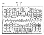
치과 진료 관리 시스템은 종종 환자의 치과 상황에 관한 정보를 저장하기 위해 치과 차트들을 사용한다. 이러한 치과 차트들은 환자의 치아의 상태에 관한 정보를 시각화하는 효율적인 툴로서 알려져 있다. Dental care management systems often use dental charts to store information about a patient's dental situation. These dental charts are known as efficient tools for visualizing information about the condition of a patient's teeth.
표준화된 치과 차트들은 종종 환자의 입 안에서 정상적으로 발견되는 개별 치아의 표면들을 나타내는 영역들을 갖는다. 이러한 차트들에서, 보통 각각의 특정 치아 표면에 대해 하나의 특정 영역이 존재한다. 치과 차트 상에서, 예컨대, 충치, 치과 수복 작업, 치근(root) 문제들 등을 시각화하기 위해 다른 컬러들, 기하학적 도형들 또는 다른 시각 표현들이 사용된다. Standardized dental charts often have areas that represent the surfaces of individual teeth normally found in the patient ' s mouth. In these charts, there is usually one specific area for each specific tooth surface. On the dental chart, other colors, geometric figures or other visual representations are used to visualize, for example, tooth decay, dental restoration, root problems, and the like.
디지털 치과 차트들은, 예컨대, 제 US8,416,984호로부터 또한 알려져 있고, 여기서 환자의 치아의 스캔으로부터 디지털 치과 차트를 생성하고, 생성된 디지털 치과 차트를 치아 상태 정보로 파퓰레이팅하기 위한 방법이 개시된다.Digital dental charts are also known, for example, from US Pat. No. 8,416,984, wherein a method for generating a digital dental chart from a scan of a patient's teeth and populating the generated digital dental chart with tooth status information is disclosed.
종래 기술 시스템들에서, 도출된 치아 상태 정보는 치과 진료 관리 시스템들에서 사용되는 환자의 치아의 표준화된/개략적인 표현으로 수동으로 주석으로 첨부/맵핑/전송된다. 이것은 시간을 소비할 뿐만 아니라, 예컨대, 정보가 치아의 잘못된 부분 또는 심지어 잘못된 치아에 주석으로 첨부되는 인간의 에러 위험에 취약하다.In prior art systems, the derived tooth condition information is manually annotated / annotated / transmitted as a standardized / schematic representation of the patient's teeth used in dental care management systems. This is not only time consuming, but also vulnerable to human error risks, for example, where information is annotated in the wrong part of the tooth or even in the wrong tooth.
환자의 치아에 대한 치아 상태 정보를 도출하고, 그러한 정보로 디지털 치과 차트를 파퓰레이팅하기 위한 방법 및 디지털 툴들이 여전히 제공되어야 하고, 상기 방법 및 디지털 툴들은 더 적은 수동 단계들을 갖고 따라서 인간의 에러들에 덜 취약하다. 디지털 툴들은, 예컨대, 컴퓨터 시스템, 컴퓨터 프로그램 제품 또는 디지털 환경일 수 있다.Methods and digital tools for deriving tooth condition information for a patient ' s teeth and for populating the digital dental chart with such information should still be provided, and the method and digital tools should have fewer manual steps, Is less vulnerable to. The digital tools may be, for example, a computer system, a computer program product, or a digital environment.
환자의 치아에 대한 치아 상태 정보를 도출하는 방법이 개시되고, 방법은:A method of deriving tooth condition information for a patient's teeth is disclosed, the method comprising:
- 환자의 치아의 디지털 3D 표현을 획득하는 단계;Obtaining a digital 3D representation of the patient ' s teeth;
- 디지털 3D 표현에서 개별 치아를 식별하는 단계;- identifying individual teeth in a digital 3D representation;
- 디지털 3D 표현으로부터 개별 치아를 분할하는 단계;Dividing the individual teeth from the digital 3D representation;
- 치아 중 하나 이상에 대한 진단 데이터를 획득하는 단계; Obtaining diagnostic data for at least one of the teeth;
- 진단 데이터로부터 치아 상태 정보를 도출하는 단계; 및Deriving tooth condition information from the diagnostic data; And
- 도출된 치아 상태 정보와 개별 치아를 상관시키는 단계를 포함한다.And correlating the derived tooth condition information with the individual teeth.
디지털 3D 표현에서 치아의 분할 및 식별은, 디지털 3D 표현의 어느 부분들이 환자의 개별 치아에 관련되는지를 알려주는 것을 제공한다. 따라서, 디지털 3D 표현에서 개별 치아의 배열이 또한 알려진다. The segmentation and identification of a tooth in a digital 3D representation provides that which portions of the digital 3D representation are associated with individual teeth of a patient. Thus, an array of individual teeth in a digital 3D representation is also known.
진단 데이터와 디지털 3D 표현 간의 공간적 상관관계가 또한 알려질 때, 디지털 3D 표현에서 치아의 분할 및 식별은, 진단 데이터가 개별 치아와 상관될 수 있음을 제공한다. 즉, 어떠한 치아에 대한 진단 데이터가 레코딩되는지가 결정될 수 있다. 진단 데이터와 디지털 3D 표현 간의 공간적 상관관계는, 예컨대, 진단 데이터가 디지털 3D 표현에 포함될 때 또는 공간적 상관관계가 진단 데이터의 부분들과 디지털 3D 표현의 대응하는 부분들을 정렬시킴으로써 결정될 때, 알려진다. 진단 데이터가 디지털 3D 표현과 공간적으로 상관될 때, 그러면 디지털 3D 표현의 개별 치아와 진단 데이터로부터 도출된 치아 상태 정보 간의 공간적 상관관계가 또한 종종 알려질 것이다. When the spatial correlation between the diagnostic data and the digital 3D representation is also known, the segmentation and identification of the teeth in the digital 3D representation provides that the diagnostic data can be correlated with individual teeth. That is, it can be determined which diagnostic data is recorded for which tooth. The spatial correlation between the diagnostic data and the digital 3D representation is known, for example, when the diagnostic data is included in the digital 3D representation or when the spatial correlation is determined by aligning corresponding portions of the digital 3D representation with portions of the diagnostic data. When the diagnostic data is spatially correlated with the digital 3D representation, then the spatial correlation between the individual teeth of the digital 3D representation and the tooth state information derived from the diagnostic data will also often be known.
따라서, 치아의 분할 및 식별은, 도출된 치아 정보가 환자의 개별 치아에 링크될 수 있고, 즉, 주어진 치아 상태 정보가 어떤 치아에 대해 도출되는지가 결정될 수 있음을 제공한다.Thus, segmentation and identification of teeth provides that the derived tooth information can be linked to individual teeth of a patient, i. E., To which teeth the given tooth state information is derived.
일부 실시예들에서, 도출된 치아 정보와 식별되어 분할된 치아를 상관시키는 것은 치아 상의 치아 상태의 위치를 결정하는 것을 포함한다. 이어서, 치아 상태 정보는 치과 차트의 영역 상의 정확한 위치로 맵핑될 수 있다.In some embodiments, correlating the extracted tooth information with the derived tooth information comprises determining the position of the tooth condition on the tooth. The tooth condition information can then be mapped to the correct position on the area of the dental chart.
위에서 설명된 바와 같이, 치아 상태 정보는 디지털 치과 차트에서 시각화될 수 있다. 따라서, 치아 상태 정보로 디지털 치과 차트를 파퓰레이팅하기 위한 방법이 또한 본원에 개시되고, 방법은:As described above, tooth condition information can be visualized in a digital dental chart. Accordingly, a method for populating a digital dental chart with tooth condition information is also disclosed herein, the method comprising:
- 실시예들 중 임의의 것에 따른 방법, 시스템, 컴퓨터 프로그램 제품 및/또는 디지털 환경을 사용함으로써 치아 중 하나 이상에 대한 치아 상태 정보를 도출하는 단계; Deriving tooth condition information for one or more of the teeth by using a method, system, computer program product and / or digital environment according to any of the embodiments;
- 환자의 치아의 표면들을 나타내는 영역들을 포함하는 디지털 치과 차트를 획득하는 단계; - obtaining a digital dental chart comprising areas representing the surfaces of the patient's teeth;
- 개별 치아와 디지털 치과 차트의 대응하는 영역들을 상관시키는 단계; 및Correlating the corresponding areas of the individual teeth with the digital dental chart; And
- 도출된 치아 상태 정보의 표현을 디지털 치과 차트의 대응하는 영역 또는 영역들에 추가하는 단계를 포함한다.And adding a representation of the derived tooth state information to a corresponding region or regions of the digital dental chart.
즉, 방법, 컴퓨터 프로그램 제품, 디지털 환경 및 시스템은 환자의 개별 치아에 대한 치아 상태 정보로 디지털 치과 차트를 파퓰레이팅하도록 구성될 수 있다.That is, a method, a computer program product, a digital environment, and a system may be configured to populate a digital dental chart with tooth condition information for individual teeth of a patient.
표준화된 디지털 치과 차트는 종종 환자의 입 안에서 정상적으로 발견되는 개별 치아의 표면들을 나타내는 영역들을 갖는다. 이러한 차트들에서, 보통 각각의 특정 치아 표면에 대해 하나의 특정 영역이 존재한다. 디지털 3D 표현에서 개별 치아의 분할 및 식별은, 디지털 3D 표현의 치아 부분들이 디지털 치과 차트의 대응하는 영역들에 링크될 수 있음을 제공한다. Standardized digital dental charts often have regions that represent the surfaces of individual teeth normally found in the patient ' s mouth. In these charts, there is usually one specific area for each specific tooth surface. The segmentation and identification of individual teeth in the digital 3D representation provides that the tooth portions of the digital 3D representation can be linked to the corresponding regions of the digital dental chart.
디지털 3D 표현과 진단 데이터 간의 공간적 상관관계에 대한 지식은, 도출된 치아 조건 정보가 어느 치아에 관련되는지를 알려준다는 것을 제공한다. 따라서, 도출된 치아 상태 정보의 표현은, 치아 상태 정보가 도출된 치아 표면을 나타내는 치아 차트의 정확한 영역에 추가될 수 있다. Knowledge of the spatial correlation between the digital 3D representation and the diagnostic data provides that it informs which teeth the derived tooth condition information relates to. Thus, the derived representation of the tooth condition information can be added to the correct area of the tooth chart representing the tooth surface from which the tooth condition information is derived.
도출된 치아 조건 정보가 디지털 치과 차트에 추가될 때, 디지털 치과 차트는 적어도 하나의 치아 상태에 관련하여 적어도 하나의 치아의 현재 상태, 이를테면, 충치, 치아 표면의 균열들 또는 치과 수복들(dental restorations)의 존재를 나타낸다. When the derived tooth condition information is added to the digital dental chart, the digital dental chart can include at least one tooth status associated with at least one tooth condition, such as cavity, tooth surface cracks, or dental restorations ).
일부 경우들에서, 디지털 치과 차트는 치과 진료 관리 시스템에서 파퓰레이팅되고 시각화된다. 치과 진료 관리 시스템에서 사용하도록 구성된 전자 데이터 레코드를 생성하기 위한 방법이 또한 본원에 개시되고, 여기서 전자 데이터 레코드는 환자의 개별 치아에 대한 치아 상태 정보를 포함하고, 방법은:In some cases, a digital dental chart is populated and visualized in a dental care management system. A method for generating an electronic data record configured for use in a dental care management system is also disclosed herein, wherein the electronic data record includes tooth condition information for individual teeth of a patient, the method comprising:
- 실시예들 중 임의의 것에 따른 방법, 시스템, 컴퓨터 프로그램 제품 및/또는 디지털 환경을 사용함으로써 치아 중 하나 이상에 대한 치아 상태 정보를 도출하는 단계; 및Deriving tooth condition information for one or more of the teeth by using a method, system, computer program product and / or digital environment according to any of the embodiments; And
- 식별된 치아에 대한 치아 상태 정보를 전자 데이터 레코드에 저장하는 단계를 포함한다.And storing tooth condition information for the identified tooth in an electronic data record.
방법이 개시되고, 방법은: A method is disclosed, the method comprising:
- 환자의 치아의 디지털 3D 표현을 획득하는 단계; Obtaining a digital 3D representation of the patient ' s teeth;
- 치아 중 하나 이상에 대한 진단 데이터를 획득하는 단계;Obtaining diagnostic data for at least one of the teeth;
- 디지털 3D 표현의 개별 치아를 식별하는 단계;Identifying individual teeth of a digital 3D representation;
- 진단 데이터로부터 치아 상태 정보를 도출하는 단계; 및Deriving tooth condition information from the diagnostic data; And
- 도출된 치아 정보와 식별된 개별 치아를 상관시키는 단계를 포함한다. And correlating the derived tooth information with the identified individual teeth.
수술자가 개시된 방법의 단계들을 수행하는 것을 돕도록 구성된 디지털 환경이 개시된다.A digital environment configured to assist an operator to perform the steps of the disclosed method is disclosed.
전자 데이터 프로세싱 디바이스에 의해 실행될 때, 개시된 방법의 단계들을 수행하기 위한 디지털 환경을 제공하는 컴퓨터 판독 가능 명령들을 포함하는 컴퓨터 프로그램 제품이 개시된다.A computer program product comprising computer readable instructions that, when executed by an electronic data processing device, provides a digital environment for performing the steps of the disclosed method.
시스템이 개시되고, 시스템은:A system is disclosed, the system comprising:
- 전자 데이터 프로세싱 디바이스; 및 An electronic data processing device; And
- 개시된 컴퓨터 프로그램 제품으로 인코딩된 비일시적인 컴퓨터 판독 가능 매체를 포함한다.Non-transitory computer readable medium encoded with the disclosed computer program product.
일부 실시예들에서, 방법, 컴퓨터 프로그램 제품, 디지털 환경 또는 시스템은 환자의 개별 치아에 대한 치아 상태 정보를 도출하기 위한 것이다.In some embodiments, a method, a computer program product, a digital environment, or a system is for deriving tooth condition information for individual teeth of a patient.
일부 실시예들에서, 방법, 컴퓨터 프로그램 제품, 디지털 환경 또는 시스템은 환자의 개별 치아에 대한 치아 상태 정보로 디지털 치과 차트를 파퓰레이팅하기 위한 것이다. 이어서, 방법은 도출된 치아 상태 정보로 디지털 치과 차트를 파퓰레이팅하는 단계를 포함한다.In some embodiments, a method, computer program product, digital environment or system is for populating a digital dental chart with tooth condition information for individual teeth of a patient. The method then includes populating the digital dental chart with the derived tooth condition information.
일부 실시예들에서, 방법, 컴퓨터 프로그램 제품, 디지털 환경 또는 시스템은 치과 진료 관리 시스템에서 사용하도록 구성된 전자 데이터 레코드를 생성하기 위한 것이고, 여기서 전자 데이터 레코드는 환자의 개별 치아에 대한 치아 상태 정보를 포함한다. 이어서, 방법은 식별된 치아에 대한 도출된 치아 상태 정보를 전자 데이터 레코드에 저장하는 단계를 포함한다.In some embodiments, a method, a computer program product, a digital environment, or a system is for generating an electronic data record configured for use in a dental care management system, wherein the electronic data record includes tooth condition information for individual teeth of a patient do. The method then includes storing the derived tooth condition information for the identified tooth in an electronic data record.
일부 실시예들에서, 방법은 디지털 3D 표현으로부터 개별 치아를 분할하는 단계를 포함한다.In some embodiments, the method includes dividing individual teeth from a digital 3D representation.
디지털 3D 표현에서 개별 치아의 분할 및 식별은, 디지털 3D 표현의 치아 부분들이 디지털 치과 차트의 대응하는 영역들에 링크될 수 있음을 제공한다. 이는 인간의 에러들에 덜 취약한 더 효율적이고 더 빠른 절차를 제공한다.The segmentation and identification of individual teeth in the digital 3D representation provides that the tooth portions of the digital 3D representation can be linked to the corresponding regions of the digital dental chart. This provides a more efficient and faster procedure that is less susceptible to human errors.
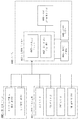
디지털 환경이 개시되고, 디지털 환경은: A digital environment is disclosed, the digital environment comprising:
- 환자의 치아의 디지털 3D 표현 및 치아 중 하나 이상에 대한 진단 데이터를 디지털 환경에 로딩하기 위한 디지털 로딩 툴;A digital loading tool for loading the digital environment with diagnostic data for at least one of a digital 3D representation of the patient ' s teeth and teeth;
- 디지털 3D 표현 및/또는 진단 데이터를 시각화하도록 구성된 디지털 작업 공간;- a digital workspace configured to visualize digital 3D representation and / or diagnostic data;
- 디지털 3D 표현의 개별 치아를 식별하기 위한 디지털 식별 툴;A digital identification tool for identifying individual teeth of a digital 3D representation;
- 진단 데이터로부터 치아 상태 정보를 도출하기 위한 디지털 도출 툴; 및A digital derivation tool for deriving tooth condition information from the diagnostic data; And
- 도출된 치아 정보와 식별된 개별 치아를 상관시키기 위한 디지털 상관 툴을 포함한다. And a digital correlation tool for correlating the derived tooth information with the identified individual teeth.
일부 실시예들에서, 디지털 환경은 디지털 3D 표현으로부터 개별 치아를 분할하기 위한 디지털 분할 툴을 포함한다.In some embodiments, the digital environment includes a digital segmentation tool for segmenting individual teeth from a digital 3D representation.
일부 실시예들에서, 디지털 환경은 도출된 치아 상태 정보로 디지털 치과 차트를 파퓰레이팅하기 위한 디지털 파퓰레이팅 툴을 포함한다. In some embodiments, the digital environment includes a digital populating tool for populating a digital dental chart with derived tooth state information.
디지털 작업 공간을 포함하는 디지털 환경이 개시되고, 여기서 디지털 환경은: A digital environment is disclosed that includes a digital workspace, wherein the digital environment includes:
- 환자의 치아의 디지털 3D 표현을 획득하고, 디지털 3D 표현을 디지털 작업 공간에 디스플레이하고; Acquiring a digital 3D representation of the patient ' s teeth, displaying the digital 3D representation in a digital workspace;
- 치아 중 하나 이상에 대한 진단 데이터를 획득하고; 그리고Obtaining diagnostic data for one or more of the teeth; And
- 수술자가 방법의 단계들을 수행하는 것을 돕도록 구성되고, 방법은:- the surgeon is configured to assist in performing the steps of the method, the method comprising:
디지털 3D 표현에서 개별 치아를 식별하는 단계; Identifying individual teeth in a digital 3D representation;
디지털 3D 표현으로부터 개별 치아를 분할하는 단계; Dividing individual teeth from a digital 3D representation;
진단 데이터로부터 치아 상태 정보를 도출하는 단계; 및 Deriving tooth condition information from the diagnostic data; And
도출된 치아 정보와 개별 치아를 상관시키는 단계를 포함한다. And correlating the derived tooth information with the individual teeth.
환자의 치아에 대한 치아 상태 정보를 도출하도록 구성된 사용자 인터페이스가 개시되고, 사용자 인터페이스는:A user interface is disclosed that is configured to derive tooth condition information for a patient ' s teeth, the user interface comprising:
- 환자의 치아의 디지털 3D 표현을 획득하고;Obtaining a digital 3D representation of the patient ' s teeth;
- 디지털 3D 표현에서 개별 치아를 식별하고;- identify individual teeth in a digital 3D representation;
- 디지털 3D 표현으로부터 개별 치아를 분할하고;Dividing individual teeth from a digital 3D representation;
- 치아 중 하나 이상에 대한 진단 데이터를 획득하고; Obtaining diagnostic data for one or more of the teeth;
- 진단 데이터로부터 치아 상태 정보를 도출하고; 그리고Deriving tooth condition information from the diagnostic data; And
- 도출된 치아 정보와 개별 치아를 상관시키도록 구성된다.And to correlate the derived tooth information with the individual teeth.
환자의 치아의 치아 상태 정보를 도출하기 위한 시스템이 개시되고, 시스템은 환자의 치아의 디지털 3D 표현 및 치아의 하나 이상에 대한 진단 데이터를 획득하도록 구성되고, 여기서 시스템은 컴퓨터 판독 가능 명령들을 포함하는 컴퓨터 프로그램 제품으로 인코딩된 비일시적인 컴퓨터 판독 가능 매체를 포함하고, 컴퓨터 판독 가능 명령들은: A system is disclosed for deriving tooth condition information of a patient's teeth, the system being configured to obtain a digital 3D representation of a patient's teeth and diagnostic data for one or more of the teeth, wherein the system comprises computer readable instructions A non-transitory computer readable medium encoded with a computer program product, the computer readable instructions comprising:
- 디지털 3D 표현에서 개별 치아를 식별하고;- identify individual teeth in a digital 3D representation;
- 디지털 3D 표현으로부터 개별 치아를 분할하고;Dividing individual teeth from a digital 3D representation;
- 진단 데이터로부터 치아 상태 정보를 도출하고; 그리고Deriving tooth condition information from the diagnostic data; And
- 도출된 치아 정보와 개별 치아를 상관시키기 위한 것이다.- to correlate the derived tooth information with individual teeth.
환자의 치아에 대한 치아 상태 정보를 도출하기 위한 시스템이 개시되며, 시스템은:A system is disclosed for deriving tooth condition information for a patient's teeth, the system comprising:
- 전자 데이터 프로세싱 디바이스; 및 An electronic data processing device; And
- 전자 데이터 프로세싱 디바이스에 의해 실행될 때, 방법에 의해 치아에 대한 치아 상태 정보를 도출하기 위한 디지털 환경을 제공하는 컴퓨터 판독 가능 명령들을 포함하는 컴퓨터 프로그램 제품으로 인코딩된 비일시적인 컴퓨터 판독 가능 매체를 포함하고, 방법은:- a non-transitory computer readable medium encoded with a computer program product comprising computer readable instructions, when executed by an electronic data processing device, for providing a digital environment for deriving tooth condition information for a tooth by a method, , Way:
i. 환자의 치아의 디지털 3D 표현을 획득하는 단계; i. Obtaining a digital 3D representation of the patient ' s teeth;
ii. 치아 중 하나 이상에 대한 진단 데이터를 획득하는 단계; ii. Obtaining diagnostic data for one or more of the teeth;
iii. 디지털 3D 표현에서 개별 치아를 식별하는 단계; iii. Identifying individual teeth in a digital 3D representation;
iv. 디지털 3D 표현으로부터 개별 치아를 분할하는 단계; iv. Dividing individual teeth from a digital 3D representation;
v. 진단 데이터로부터 치아 상태 정보를 도출하는 단계; 및 v. Deriving tooth condition information from the diagnostic data; And
vi. 도출된 치아 정보와 개별 치아를 상관시키는 단계를 포함한다. vi. And correlating the derived tooth information with the individual teeth.
환자의 치아에 대한 치아 상태 정보로 디지털 치과 차트를 파퓰레이팅하도록 구성된 사용자 인터페이스가 개시되고, 사용자 인터페이스는:A user interface is disclosed that is configured to populate a digital dental chart with tooth condition information for a patient ' s teeth, the user interface comprising:
- 환자의 치아의 디지털 3D 표현을 획득하고;Obtaining a digital 3D representation of the patient ' s teeth;
- 디지털 3D 표현에서 개별 치아를 식별하고;- identify individual teeth in a digital 3D representation;
- 디지털 3D 표현으로부터 개별 치아를 분할하고;Dividing individual teeth from a digital 3D representation;
- 치아 중 하나 이상에 대한 진단 데이터를 획득하고;Obtaining diagnostic data for one or more of the teeth;
- 진단 데이터로부터 치아 상태 정보를 도출하고;Deriving tooth condition information from the diagnostic data;
- 도출된 치아 정보와 개별 치아를 상관시키고;Correlating the derived tooth information with individual teeth;
- 치아 표면들을 나타내는 영역들을 포함하는 디지털 치과 차트를 획득하고; Obtaining a digital dental chart comprising regions representing tooth surfaces;
- 개별 치아와 디지털 치과 차트의 대응하는 영역들을 상관시키고; 그리고Correlating the corresponding areas of the individual teeth with the digital dental chart; And
- 도출된 치아 상태 정보의 표현을 디지털 치과 차트의 대응하는 영역들에 추가하도록 구성된다.And to add a representation of the derived tooth state information to corresponding areas of the digital dental chart.
디지털 3D 표현은 환자의 치아 중 하나 이상의 지형(topography)을 나타내는 형상 데이터를 포함한다. 치아의 분할 및 식별은 바람직하게는 이들 형상 데이터에 적어도 부분적으로 기반한다. 일부 경우들에서, 디지털 3D 표현은 또한 환자의 잇몸(gingiva)에 대한 형상 데이터를 포함한다. 이어서, 잇몸 상태 정보는 독립적으로 또는 치아 상태 정보의 부분으로서 도출될 수 있다.The digital 3D representation includes shape data representing the topography of at least one of the patient's teeth. The segmentation and identification of teeth is preferably based at least in part on these shape data. In some cases, the digital 3D representation also includes shape data for the patient ' s gingiva. The gingival state information can then be derived independently or as part of the tooth condition information.
디지털 3D 표현은 구강 내 스캐닝(intra-oral scanning)에 의해, 환자의 치아 인상(impression) 또는 그러한 인상을 사용하여 형성된 치아의 물리적 모델을 스캐닝함으로써 획득될 수 있다. The digital 3D representation can be obtained by intra-oral scanning, by scanning the physical model of the tooth formed using the patient's impression or such impression.
일부 스캐닝 기술들은 치아 표면의 형상뿐만 아니라 치아 내부의 내부 구조 둘 모두를 측정할 수 있다. 이것은, 예컨대, X-레이 기반 스캐너들, 이를테면, 콘 빔 전산화 단층촬영 스캐너들(Cone Beam Computed Tomography scanners) 및 광 간섭 단층촬영 스캐너들(optical coherence tomography scanners)에 대한 경우이다. 그러한 스캐너들은 치아의 지형에 대한 형상 데이터뿐만 아니라 치아의 내부 구조 및 서브-표면 손상들 및 충치 및 치과 수복들의 존재에 관련된 데이터 둘 모두를 포함하는 디지털 3D 표현을 제공할 수 있다.Some scanning techniques can measure both the shape of the tooth surface as well as the internal structure of the tooth interior. This is the case, for example, for X-ray based scanners, such as Cone Beam Computed Tomography scanners and optical coherence tomography scanners. Such scanners may provide digital 3D representations that include both the geometry data for the topography of the tooth, as well as the internal structure of the tooth and both the sub-surface impairments and the presence of cavities and dental restorations.
일부 실시예들에서, 진단 데이터 중 적어도 일부는 디지털 3D 표현에 포함된다. 이러한 실시예의 하나의 이점은, 디지털 3D 표현으로부터 분할된 치아 부분들 및 진단 데이터가 이미 상관된다는 것이다. 진단 데이터가 디지털 3D 표현에 포함될 때, 진단 데이터는 종종 분할된 치아의 지형을 나타내는 형상 데이터와 정렬된다. 예컨대, 치아 음영(shade) 데이터는, 획득된 디지털 3D 표현에서 치아에 대한 음영 데이터 및 형상 데이터가 정확하게 상관되도록, 형상 데이터와 동시에 레코딩될 수 있다. 따라서, 진단 데이터와 디지털 3D 표현의 형상 데이터 간의 공간적 상관관계를 제공하거나 결정하기 위해 이 둘을 정렬시키는 추가적인 단계가 필요하지 않다. 이러한 경우에, 디지털 3D 표현을 획득하고, 진단 데이터를 획득하는 것은 하나의 단계로서 수행되고, 즉, 디지털 3D 표현 및 진단 데이터가 방법의 동일한 단계에서 획득된다.In some embodiments, at least some of the diagnostic data is included in a digital 3D representation. One advantage of this embodiment is that the divided tooth parts and diagnostic data from the digital 3D representation are already correlated. When the diagnostic data is included in the digital 3D representation, the diagnostic data is often aligned with shape data representing the topography of the divided teeth. For example, the tooth shade data may be recorded concurrently with the shape data so that the shade data and the shape data for the teeth in the obtained digital 3D representation are accurately correlated. Thus, there is no need for an additional step of aligning the two to provide or determine spatial correlation between the diagnostic data and the shape data of the digital 3D representation. In this case, obtaining the digital 3D representation and acquiring the diagnostic data is performed as one step, that is, digital 3D representation and diagnostic data are obtained at the same step of the method.
진단 데이터는 또한, 이를테면, 치아 상태가, 예컨대, 치아 마모 또는 잇몸 압배(gingiva retraction)에 관련될 때, 디지털 3D 표현의 형상 데이터일 수 있고, 여기서 상태는 형상 데이터의 분석으로부터 결정될 수 있다.The diagnostic data may also be shape data of a digital 3D representation, such as when the tooth condition is associated with, for example, tooth wear or gingiva retraction, wherein the condition may be determined from analysis of the shape data.
일부 실시예들에서, 디지털 3D 표현은 형상 데이터, 컬러 또는 음영 데이터 및 형광 데이터를 포함한다. 이러한 디지털 3D 표현은 (x, y, z, 컬러 데이터, 형광 강도 데이터) 형태의 데이터를 포함할 수 있다. 여기서 (x, y, z)는 치아 표면 상의 포인트에 대한 공간 좌표들을 나타내고, 컬러 데이터 및 형광 강도 데이터는 치아 표면의 (x, y, z) 포인트로부터 측정된 치아 컬러 및 형광에 관한 정보를 각각 제공한다. 컬러 데이터는, 예컨대, (RGB) 좌표들로 제공될 수 있다. 이어서, 디지털 3D 표현은 (x, y, z, R, G, B, 형광 강도) 형태의 데이터를 포함한다.In some embodiments, the digital 3D representation includes shape data, color or shading data, and fluorescence data. Such a digital 3D representation may include data in the form of (x, y, z, color data, fluorescence intensity data). (X, y, z) represents spatial coordinates for a point on the tooth surface, and the color data and fluorescence intensity data represents information about tooth color and fluorescence measured from the (x, y, z) to provide. The color data may be provided, for example, in (RGB) coordinates. The digital 3D representation then includes data in the form of (x, y, z, R, G, B, fluorescence intensity).
일부 실시예들에서, 환자의 치아의 진단 데이터 및 디지털 3D 표현은, 진단 데이터가 초기에 디지털 3D 표현의 부분이 아니도록, 상이한 디바이스들을 사용하여 레코딩된다. 이것은, 예컨대, 진단 데이터가 X-레이 데이터, 이를테면, CBCT 스캐너를 사용하여 레코딩된 데이터와 같은 X 선 데이터인 반면에, 치아의 표면 스캔을 제공하는 구강 내 스캐너를 사용하여 디지털 3D 표현이 레코딩되는 경우일 수 있다. In some embodiments, the diagnostic data and the digital 3D representation of the patient's teeth are recorded using different devices so that the diagnostic data is not initially part of the digital 3D representation. This is achieved, for example, by the use of an intraoral scanner that provides surface scans of teeth while diagnostic data is X-ray data, such as data recorded using a CBCT scanner, for example, .
일부 실시예들에서, 진단 데이터 중 적어도 일부는 환자의 치아의 디지털 3D 표현 이외에 획득된 진단 데이터 세트에 포함된다. 이어서, 치아 상태 정보 중 적어도 일부는 이러한 진단 데이터 세트로부터 도출될 수 있다. 그러한 진단 데이터 세트는, 예컨대, 진단 데이터가 치아 표면에 대해 충분히 정확하거나 상세한 형상 데이터를 레코딩하지 않은 스캐너들로부터 추출될 때, 또는 진단 데이터가 2D 이미지들 형태로 제공될 때, 사용될 수 있다. 진단 데이터 세트로부터 획득될 수 있는 진단 데이터의 다른 예들은 CBCT 데이터, 별개로 획득된 형광 데이터 및 IR 데이터이다.In some embodiments, at least some of the diagnostic data is included in the acquired diagnostic data set in addition to the digital 3D representation of the patient ' s teeth. At least some of the tooth condition information can then be derived from this diagnostic data set. Such a diagnostic data set can be used, for example, when the diagnostic data is extracted from scanners that have not recorded sufficiently accurate or detailed shape data for the tooth surface, or when the diagnostic data is provided in the form of 2D images. Other examples of diagnostic data that may be obtained from the diagnostic data set are CBCT data, separately obtained fluorescence data and IR data.
그러한 경우들에서, 디지털 3D 표현들과 진단 데이터 간의 공간적 상관관계, 즉, 디지털 3D 표현과 진단 데이터 세트의 진단 데이터 간의 공간적 상관을 결정하는 것이 유리할 수 있다. 상관관계는, 예컨대, 디지털 3D 표현 및 진단 데이터를 정확한 상대적인 배열을 갖는 공통 좌표 시스템으로 도출하는 변환으로서 표현될 수 있다. 이러한 상관관계를 결정하는 것은, 디지털 3D 표현의 진단 데이터 및 형상 데이터로부터 도출된 치아 및/또는 잇몸 상태 정보 사이의 공간적 관계가 또한 결정되는 것을 제공할 수 있다. 이는, 예컨대, 도출된 치아 및/또는 잇몸 상태 정보를 디지털 치과 차트로 맵핑하기 위한 컴퓨터 프로그램 제품을 사용할 때, 유리하다.In such cases, it may be advantageous to determine the spatial correlation between the digital 3D representations and the diagnostic data, i. E., The spatial correlation between the digital 3D representation and the diagnostic data of the diagnostic data set. The correlation can be represented, for example, as a transformation that derives the digital 3D representation and the diagnostic data into a common coordinate system with an accurate relative arrangement. Determining this correlation can also provide that the spatial relationship between the diagnostic data of the digital 3D representation and the tooth and / or gum status information derived from the shape data is also determined. This is advantageous, for example, when using a computer program product for mapping derived tooth and / or gingival state information to a digital dental chart.
공간적 상관관계는, 예컨대, 동일한 표면에 관련된 디지털 3D 표현 및 진단 데이터의 부분들의 정렬(aligning)에 기반하거나, 기준 마커들, 랜드마크 식별 또는, 예컨대, ICP(Iterative Closest Point) 알고리즘을 사용하는 표면들의 정렬에 기반하여 결정될 수 있다. The spatial correlation may be based, for example, on the alignment of portions of the digital 3D representation and diagnostic data associated with the same surface, or on the surface using reference markers, landmark identification or, for example, ICP (Iterative Closest Point) As shown in FIG.
일부 실시예들에서, 디지털 3D 표현은 치아에 대한 형상 데이터를 포함하는 제1 디지털 3D 표현 및 진단 데이터를 포함하는 제2 디지털 3D 표현으로부터 형성된다. 제1 및 제2 디지털 3D 표현들 간의 공간적 상관관계가 결정될 때, 제1 디지털 3D 표현의 형상 데이터 및 제2 디지털 3D 표현의 진단 데이터 둘 모두를 포함하는 디지털 3D 표현이 형성될 수 있다.In some embodiments, the digital 3D representation is formed from a first digital 3D representation comprising shape data for a tooth and a second digital 3D representation comprising diagnostic data. When a spatial correlation between the first and second digital 3D representations is determined, a digital 3D representation can be formed that includes both the shape data of the first digital 3D representation and the diagnostic data of the second digital 3D representation.
도출된 치아 상태 정보와 디지털 3D 표현의 식별된 개별 치아 사이의 상관관계는 디지털 3D 표현과 진단 데이터 세트 사이의 결정된 공간적 관계에 기반할 수 있다.The correlation between the derived tooth condition information and the identified individual teeth of the digital 3D representation may be based on the determined spatial relationship between the digital 3D representation and the diagnostic data set.
디지털 3D 표현으로부터의 치아의 분할은 개별 치아에 대응하는 디지털 3D 표현의 부분들을 디지털 3D 표현의 남아있는 부분으로부터 분리시키는 것이 바람직하다. 즉, 개별 치아에 관련된 디지털 3D 표현의 데이터는 서로로부터 및 환자의 잇몸과 관련된 부분들과 같은 디지털 3D 표현의 다른 부분들로부터 분리된다. The division of the teeth from the digital 3D representation preferably separates portions of the digital 3D representation corresponding to the individual teeth from the remaining portion of the digital 3D representation. That is, the data of the digital 3D representation associated with the individual tooth is separated from each other and from other portions of the digital 3D representation, such as portions of the patient's gums.
분할 단계는, 치아가 식별되기 전에 또는 후에 수행될 수 있다. 이전에 수행되면, 식별은 분할된 치아에 기반할 수 있다. 식별 후에 수행되면, 분할은 디지털 3D 표현의 주어진 부분이 어떤 치아엔 관련되는지에 대한 지식에 기반할 수 있다. The segmentation step may be performed before or after the tooth is identified. If performed previously, the identification may be based on the segmented tooth. If performed after identification, the segmentation may be based on knowledge of which tooth a given portion of the digital 3D representation is related to.
분할은 사용자 상호작용을 통해서 뿐만 아니라 사용자 상호작용 없이 실현될 수 있다. 디지털 3D 표현이 디스플레이, 이를테면, 컴퓨터 스크린에서 시각화되면, 수술자는 디지털 3D 표현 상에 개별 치아의 경계들을 마킹하기 위한 포인팅 툴, 이를테면, 컴퓨터 마우스를 사용할 수 있다. 이어서, 디지털 3D 표현으로부터 치아의 분할은 마킹된 경계들에 기반할 수 있다. 컴퓨터 기반 알고리즘들은 또한, 사용자 상호작용 없이 또는 제한된 사용자 상호작용을 통해 분할이 이루어질 수 있도록, 예컨대, 치아 표면으로부터 잇몸으로의 전이(transition) 및 이웃 치아와의 접촉 시에 치아의 경계를 식별하도록 적용될 수 있다. Partitioning can be realized through user interaction as well as without user interaction. Once the digital 3D representation is visualized on a display, such as a computer screen, the operator can use a pointing tool, such as a computer mouse, to mark the boundaries of individual teeth on the digital 3D representation. The segmentation of the teeth from the digital 3D representation may then be based on the marked boundaries. Computer-based algorithms may also be applied to identify the boundary of a tooth, such as a transition from a tooth surface to a gum and a contact with a neighboring tooth, such that a segmentation can be made without user interaction or with limited user interaction .
진단 데이터가 텍스처 데이터, 이를테면, 컬러 데이터를 포함할 때, 분할은 또한 진단 데이터에 적어도 부분적으로 기반할 수 있다. 예컨대, 잇몸의 치아의 경계는 치아 및 잇몸의 컬러 차이로부터 검출될 수 있다.When the diagnostic data includes texture data, such as color data, the segmentation may also be based, at least in part, on the diagnostic data. For example, the boundaries of the teeth of the gum can be detected from the color differences of the teeth and gums.
일부 실시예들에서, 치아에 대한 치아 상태 정보는 디지털 3D 표현의 분할된 치아 부분에 걸쳐 진단 데이터의 변동들로부터 도출된다.In some embodiments, tooth condition information for a tooth is derived from variations in diagnostic data over a segmented tooth portion of a digital 3D representation.
진단 데이터가 디지털 3D 표현에 포함될 때, 진단 데이터는 본질적으로 치아의 형상 데이터에 링크된다. 일부 경우들, 이를테면, 치아 상태가 그 치아로부터 방출되는 형광의 로컬 변동으로부터 도출된 치아의 주어진 부분에 충치가 존재하는 것일 때, 도출된 치아 상태 정보는 디지털 3D 표현의 치아 부분과 이미 상관된다.When the diagnostic data is included in the digital 3D representation, the diagnostic data is essentially linked to the shape data of the teeth. In some cases, for example, when tooth cavities are present in a given portion of a tooth derived from local variations in fluorescence emitted from the tooth, the derived tooth condition information is already correlated with the tooth portion of the digital 3D representation.
일부 실시예들에서, 진단 데이터는 치아로부터 레코딩된 신호의 강도를 나타내고, 치아 표면들에 걸친 강도의 변동들은, 예컨대, 충치 또는 기존의 치과 충전물(dental filling)의 존재를 나타낸다. 치아 상태의 존재는 그 상태의 위치에서 신호 강도의 증가 또는 감소 둘 모두에 의해 표시될 수 있다. In some embodiments, the diagnostic data represents the intensity of the signal recorded from the tooth, and variations in intensity across the tooth surfaces indicate, for example, the presence of cavities or conventional dental filling. The presence of a tooth condition can be indicated by both increasing or decreasing signal strength at that location.
정상적인 치아(sound teeth)에 약 405nm의 파장을 갖는 광이 조명될 때, 치아는 통상적으로 자연적인 에나멜(natural enamel)에서 500nm의 광범위한 방출로 형광을 방출한다. When normal tooth light is illuminated with light having a wavelength of about 405 nm, teeth typically emit fluorescence with a broad emission of 500 nm in a natural enamel.
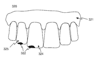
치아의 충치 감염 영역들에서, 구강 세균들의 포르피린 화합물들(porphyrin compounds)로부터의 방출로 인해, 635nm 및 680nm에서 추가적인 피크들이 종종 보여진다. 즉, 충치의 존재는 치아의 특정 영역으로부터 레코딩된 635nm 및 680nm에서의 형광 신호가 치아의 다른 (건강한) 부분들로부터의 것보다 더 강한지를 측정함으로써 검출될 수 있다. 이어서, 형광 신호의 증가된 강도는 치아 내의 충치의 직접적인 표현을 제공한다.In tooth cavity infections, additional peaks are often seen at 635 nm and 680 nm due to the release of oral bacteria from porphyrin compounds. That is, the presence of cavities can be detected by measuring whether the fluorescence signal at 635 nm and 680 nm recorded from a particular area of the tooth is stronger than from the other (healthy) parts of the tooth. The increased intensity of the fluorescence signal then provides a direct representation of the cavity in the tooth.
예컨대, 405nm의 파장의 광에 의해 조명된 치아로부터 500nm으로 방출된 자연적인 형광의 감소는 충치를 갖는 영역에서의 산란에 의해 발생할 수 있다. 따라서, 더 낮은 형광 신호(약 500nm)를 검출하는 것은 치아의 그 영역에서 충치의 존재를 표시할 수 있다.For example, a reduction in natural fluorescence emitted from a tooth illuminated by light at a wavelength of 405 nm to 500 nm may be caused by scattering in the area with cavity. Thus, detecting a lower fluorescence signal (about 500 nm) can indicate the presence of cavities in that area of the tooth.
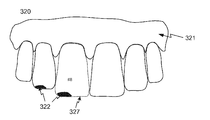
진단 데이터는 치아 상태의 공간 분포를 나타낼 수 있다. 이는, 진단 데이터가, 치아의 특정 영역에서, 예컨대, 교합 표면(occlusal surface) 상의 특정 영역에서 치아가 충치에 의해 공격받는 것을 나타내는 형광 데이터인 경우일 수 있다. The diagnostic data may represent a spatial distribution of the tooth condition. This may be the case where the diagnostic data is fluorescence data indicating that a tooth is attacked by a tooth cavity in a certain area of the tooth, for example, a specific area on an occlusal surface.
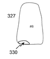
일부 실시예들에서, 치아 상태 정보는 치아 상의 상태의 위치를 포함한다. 따라서, 도출된 정보는 상태 및 치아의 어느 부분이 상태에 의해 영향을 받는지 둘 모두를 설명한다. 이어서, 충치의 경우에, 치아 상태 정보는 충치가 치아에 존재한다는 것, 및 예컨대, 충치가 치아의 교합 표면에 위치된다는 것을 설명하는 정보를 포함할 수 있다.In some embodiments, the tooth condition information includes the position of the condition on the tooth. Thus, the derived information describes both the state and which part of the tooth is affected by the condition. Then, in the case of a tooth cavity, the tooth condition information may include information that the tooth cavity is present in the tooth and that, for example, the tooth cavity is located on the occlusal surface of the tooth.
디지털 3D 표현 및/또는 진단 데이터가 환자의 잇몸에 대한 데이터를 포함할 때, 방법은 잇몸의 상태, 이를테면, 그의 형상, 컬러, 치아에 대한 공간 관계 또는 잇몸 주머니들의 깊이에 관한 정보를 도출하는 단계를 포함할 수 있다. 잇몸 형상 및 시간에 걸쳐 치아에 대한 그의 공간적 관계를 모니터링하는 것은 잇몸 압배 및 잇몸 주머니들의 심화(deepening)를 검출하기 위한 수단을 제공한다. When the digital 3D representation and / or diagnostic data includes data for the patient's gums, the method includes deriving information about the state of the gums, such as its shape, color, spatial relationship to the teeth, or depth of the gum pockets . ≪ / RTI > Monitoring his spatial relationship to the tooth over the shape and time of the gums provides a means for detecting gingival compression and deepening of gum pockets.
일부 실시예들에서, 개별 치아를 식별하는 것은 치아 데이터베이스의 디지털 템플릿 치아와 분할된 치아를 비교하는 것을 포함한다. 이는, 상이한 치아가 상이한 형상들을 갖기 때문에 가능하다(예컨대, 전치(anterior tooth)가 후치(posterior tooth)와 동일한 형상을 가지지 않음). 주어진 어금니가 환자의 입의 좌측에 속하는지 또는 우측에 속하는지는, 예컨대, 환자의 치아의 디지털 3D 표현에서 대응하는 데이터의 위치로부터 결정될 수 있다. In some embodiments, identifying the individual teeth includes comparing the digital teeth of the teeth database with the divided teeth. This is possible because different teeth have different shapes (e.g., the anterior tooth does not have the same shape as the posterior tooth). It can be determined from the position of the corresponding data in the digital 3D representation of the patient's teeth whether the given molar belongs to the left side or the right side of the patient's mouth, for example.
치아가 식별되었을 때, 식별의 결과는 사용자 인터페이스에서 시각화되어, 수술자가 식별이 정확한지를 확인하도록 허용한다.When a tooth is identified, the result of the identification is visualized in the user interface, allowing the operator to verify that the identification is correct.
일부 실시예들에서, 치아는 예컨대, 사용자 인터페이스에서의 치아의 세트의 시각화 시에 포인팅 툴을 사용하여 수술자에 의해 수동으로 식별된다. 치과 의사 또는 수술자는 절차 동안에 치아의 시각화를 종종 보고, 개별 치아는 디스플레이 상의 치아의 시각화와 관련하여 포인팅 툴을 사용하여 수동으로 식별될 수 있다.In some embodiments, the teeth are manually identified, for example, by the operator using a pointing tool at the time of visualization of the set of teeth at the user interface. The dentist or operator often views the visualization of the teeth during the procedure, and individual teeth can be manually identified using a pointing tool in connection with visualization of the teeth on the display.
일부 실시예들에서, 획득된 디지털 3D 표현에서 치아의 식별은 환자의 치아의 이전에 획득된 디지털 3D 표현에 대해 이루어진 식별에 기반한다. 이러한 이전에 획득된 디지털 3D 표현은, 이전의 치과 방문 동안에서의 방법을 적용하면서, 분석될 수 있다. 이전의 분석은, 현재의 방문 동안에 획득된 디지털 3D 표현의 치아 부분들이 실제 모양과 비교될 수 있도록, 이러한 특정 환자의 치아의 실제 형상에 대한 지식을 제공한다. 이러한 비교는 치아 데이터베이스의 템플릿 치아의 비교보다 잠재적으로 더 쉽고 더 정확하다.In some embodiments, the identification of a tooth in the obtained digital 3D representation is based on an identification made against a previously obtained digital 3D representation of the patient's teeth. This previously acquired digital 3D representation can be analyzed while applying the method during previous dental visits. The previous analysis provides knowledge of the actual shape of teeth of this particular patient so that the tooth parts of the digital 3D representation obtained during the current visit can be compared to the actual shape. This comparison is potentially easier and more accurate than a comparison of template teeth in a tooth database.
일부 실시예들에서, 식별된 치아의 형상들은 전자 데이터 레코드에 저장된다. 이것은, 방법의 후속 사용 동안에, 이를테면, 후속 치과 방문 동안에 치아의 식별이 환자의 치아 세트의 실제 형상에 기반할 수 있다는 이점을 제공한다. 이러한 식별은, 예컨대, 치아가 표준 치아 형상들과의 비교에 기반하여 식별될 때와 비교하여, 잠재적으로 훨씬 더 빠르고 더 적은 계산력을 요구한다.In some embodiments, the shapes of the identified teeth are stored in an electronic data record. This provides the advantage that during the subsequent use of the method, such as the identification of the teeth during subsequent dental visits, can be based on the actual shape of the patient's tooth set. This identification is potentially much faster and requires less computational power compared to, for example, when a tooth is identified based on a comparison with standard tooth shapes.
일부 실시예들에서, 진단 데이터는 텍스처 데이터, 이를테면, 치아 컬러 데이터 또는 치아 음영 데이터, 형광 데이터, 적외선 데이터, X-레이 데이터, 광 간섭 단층촬영 데이터, 초음파 데이터, 레이저 스페클(laser speckle) 이미지들, 또는 대교치(antagonist teeth) 간의 교합 접촉들을 나타내는 데이터로 구성된 그룹으로부터 선택된 데이터를 포함한다. 원칙적으로, 진단 목적으로 그리고 치아의 상태를 나타내기에 적합한 임의의 종류의 데이터가 사용될 수 있다.In some embodiments, the diagnostic data may include texture data, such as tooth color data or tooth shadow data, fluorescence data, infrared data, X-ray data, optical coherence tomography data, ultrasound data, laser speckle images Or data representative of occlusal contacts between the antagonist teeth. In principle, any kind of data suitable for diagnostic purposes and to indicate the condition of the teeth may be used.
본 개시내용의 문맥에서, "형광 데이터"라는 어구는, 치아의 형광 물질 또는 치아에서 방출될 수 있는 파장들의 광을 포함하는 프로브 빔에 의한 조명에 대한 응답으로 치아로부터 방출되는 형광 신호를 검출하는 형광 측정에 의해 포착된 데이터를 지칭한다. 여기(excitation)는, 예컨대, 어느 물질이 여기되는지에 의존하여 청색 또는 녹색 광을 사용할 수 있다. In the context of this disclosure, the phrase "fluorescence data" is used to refer to the detection of a fluorescent signal emitted from a tooth in response to illumination by a probe beam comprising light of wavelengths Quot; refers to data captured by fluorescence measurement. The excitation may use blue or green light depending on, for example, which material is excited.
본 개시내용의 문맥에서, "적외선 데이터"라는 어구는 적외선 측정, 예컨대, 적외선 스캐너에 의해 포착된 데이터를 지칭하고, 여기서 분석된 치아 또는 치아들을 통한 적외선 파장들에서의 광 투과율 또는 투과율의 변동들이 검출된다. 치아를 통해 투과되는 적외선 광의 강도의 변동들은, 예컨대, 치과 충전물, 치아 표면의 균열들 또는 충치로 인한 것일 수 있다. In the context of this disclosure, the phrase "infrared data" refers to data acquired by an infrared measurement, such as an infrared scanner, wherein variations in light transmittance or transmittance at infrared wavelengths through the analyzed teeth or teeth . Fluctuations in the intensity of infrared light transmitted through the teeth may be due to, for example, dental filling, cracks on the tooth surface, or tooth decay.
대교치와의 교합 접촉들은, 예컨대, 물기(bite) 동안 치아의 상대적인 움직임을 모방하는 가상 교합기(virtual articulator)를 사용하여 결정될 수 있다. 교합 접촉들은 또한 치아 상에 컬러 마킹들을 남기는 교합지(articulating paper)를 사용하여 레코딩될 수 있다. 치아의 디지털 3D 표현이 구강 내 스캐닝 레코딩을 사용하여 포착되면, 치아 형상 및 접촉점들의 컬러 둘 모두는 디지털 3D 표현으로부터 도출될 수 있다. 이어서, 도출된 교합 접촉을 디지털 치과 차트로 맵핑될 수 있다. Occlusal contacts with the bridge can be determined, for example, using a virtual articulator that mimics the relative motion of the teeth during the bite. Occlusal contacts can also be recorded using articulating paper that leaves color markings on the teeth. When a digital 3D representation of a tooth is captured using an intraoral scanning recording, both the tooth shape and the color of the contact points can be derived from the digital 3D representation. The derived occlusal contact can then be mapped to a digital dental chart.
X-레이 데이터는, 예컨대, 콘-빔 전산화 단층촬영(cone-beam computed tomography) 이미지 또는 2D X-레이 이미지들의 형태일 수 있다. The X-ray data may be in the form of, for example, a cone-beam computed tomography image or 2D X-ray images.
일부 실시예들에서, 도출된 치아 상태 정보는 치아 음영, 치아 마모, 충치, 충치원성 세균의 존재, 이전의 치과 작업으로부터의 충전물들의 존재, 산 침식 손상들(acid erosion damages), 이갈이 유발 손상들(bruxism induced damages), 치아 배열, 부정 교합 또는 잇몸 압배로 구성된 그룹으로부터 선택된 정보와 관련된다.In some embodiments, the derived tooth condition information includes at least one of the following: tooth shadow, tooth wear, caries, presence of caries bacteria, presence of fillers from previous dental work, acid erosion damages, bruxism induced damages, tooth alignment, malocclusion, or gingival compression.
치아 배열은, 환자의 치아 배열의 변화들이 모니터링되는 교정 치료의 부분으로서 도출될 수 있다. 이어서, 치료 동안에 환자의 치아에 대해 도출된 치아 상태 정보, 즉 치아 배열을 저장하는 것은 치열 교정 의사(orthodontist)가 진행을 시각화하고 치료가 계획대로 진행되는지를 확인하기 위한 강력한 툴을 제공한다. The tooth arrangement can be derived as part of an orthodontic treatment in which changes in the patient's tooth arrangement are monitored. Then, storing tooth state information, i.e. tooth alignment, derived for the patient's teeth during treatment provides a powerful tool for the orthodontist to visualize the progress and to see if treatment is proceeding as planned.
산성 침식 또는 이갈이에 의해 발생되는 손상들은 에나멜의 미세 구조에 대한 정보를 제공하는 레이저 스페클 이미지로부터 검출될 수 있다. 그러한 손상들은 또한 시간에 걸쳐 환자의 치아 형상의 변화를 모니터링하기 위해 검출될 수 있다.Damage caused by acid erosion or discoloration can be detected from laser speckle images providing information on the microstructure of the enamel. Such injuries may also be detected to monitor changes in the patient ' s tooth shape over time.
충치원성 세균의 존재 또는 미세구조 스케일의 골절은 치아에서 성장하는 충치의 표시자들이 될 수 있다. The presence of a bacterium or a fracture of the microstructure scale can be indicative of tooth decay in teeth.
몇몇의 치아에 대해 그리고 몇몇의 상이한 치과 상태들에 관련하여 정보가 도출될 수 있다. Information can be derived for some teeth and for some different dental conditions.
일부 실시예들에서, 수술자는 도출된 치아 상태 정보를 디지털 치과 차트 상에 수동으로 주석으로 첨부할 것이다.In some embodiments, the operator will manually annotate the derived tooth condition information on a digital dental chart.
일부 실시예들에서, 방법은 환자의 치아의 표면들을 나타내는 영역들을 포함하는 디지털 치과 차트를 획득하는 단계, 및 도출된 치아 상태 정보의 표현을, 치과 상태 정보가 도출되는 치아 또는 치아들에 대응하는, 디지털 치과 차트의 영역 또는 영역(들)에 추가하는 단계를 포함한다. In some embodiments, the method includes obtaining a digital dental chart that includes areas representing surfaces of a patient ' s teeth, and providing a representation of the derived tooth condition information to the teeth or teeth from which the dental condition information is derived , To the area or area (s) of the digital dental chart.
일부 실시예들에서, 디지털 치과 차트는 치아 표면들을 나타내는 영역들을 포함하고, 즉, 환자의 치아 세트에서 정상적으로 발견되는 치아 각각에 대해, 디지털 치과 차트는 치아의 표면들을 나타내는 하나 이상의 영역들을 갖는다. 전치 각각에 대해, 디지털 치과 차트는 각각의 치아의 입술측(labial) 표면을 나타내는 영역 및 설측(lingual) 표면을 나타내는 영역을 포함할 수 있다. 후치 각각에 대해, 디지털 치과 차트는 볼측 표면(buccal surface)을 나타내는 영역, 설측 표면을 나타내는 영역 및 교합 표면을 나타내는 영역을 포함할 수 있다. 예컨대, 충치가 송곳니들 중 하나에서 검출되면, 충치가 그 치아에서 존재한다는 것을 치과 의사에 보여주는 표현이 이러한 치아를 나타내는 치과 차트의 영역들에 추가된다.In some embodiments, the digital dental chart includes areas representing tooth surfaces, that is, for each tooth normally found in a patient's tooth set, the digital dental chart has one or more areas that represent the surfaces of the tooth. For each of the transpositions, the digital dental chart may include a region representing a labial surface of each tooth and an area representing a lingual surface. For each posterior, the digital dental chart may include a region representing a buccal surface, a region representing a lingual surface, and an area representing an occlusal surface. For example, if a tooth cavity is detected in one of the canines, an expression indicating to the dentist that the tooth cavity is present in the tooth is added to the areas of the dental chart representing such a tooth.
일부 실시예들에서, 디지털 치과 차트는 치아의 상이한 표면들을 나타내는 영역들을 갖는 2D 치과 차트를 포함한다. 이어서, 분할된 치아에 대한 진단 데이터 또는 도출된 치아 상태 정보는 디지털 치과 차트의 대응하는 영역에 투영될 수 있다. 영역은, 예컨대, 디지털 치과 차트에서 치아의 볼측/입술측, 교합 또는 설측 표면을 나타낼 수 있다.In some embodiments, the digital dental chart includes a 2D dental chart having regions that represent different surfaces of the tooth. The diagnostic data or the derived tooth condition information for the divided teeth may then be projected onto a corresponding area of the digital dental chart. The area may represent, for example, the ball side / lips side, occlusal or lingual surface of the teeth in a digital dental chart.
디지털 치과 차트는, 상이한 치아를 나타내는 영역들이 일반화된 표준 치과 차트의 형태일 수 있다.The digital dental chart may be in the form of a standardized dental chart, in which regions representing different teeth are generalized.
일부 실시예들에서, 영역들은, 디지털 치과 차트는 개인 치과 차트가 되도록 치아의 대응하는 표면들에 기반하여 형성되고, 여기서 치과 차트의 영역들은 환자의 입의 실제 상황에 따라 형상화 및/또는 채색 및/또는 배열된다. In some embodiments, the areas are formed based on corresponding surfaces of the teeth such that the digital dental chart is a personal dental chart, wherein the areas of the dental chart are shaped and / or colored according to the actual situation of the patient ' / ≪ / RTI >
일부 실시예들에서, 도출된 치아 상태 정보를 디지털 치과 차트에 추가하는 것은 도출된 정보의 표현을 디지털 치과 차트로 맵핑하는 것을 포함한다. 이어서, 치아 상태 정보는 디지털 치과 차트의 정확한 영역에서 시각화된다. In some embodiments, adding the derived tooth state information to a digital dental chart includes mapping a representation of the derived information to a digital dental chart. The tooth condition information is then visualized in the correct area of the digital dental chart.
일부 실시예들에서, 진단 데이터는 디지털 치과 차트로 맵핑된다. 예컨대, 음영 데이터 또는 형광 데이터가 디지털 치과 차트의 영역들에 맵핑되어, 디지털 치과 차트는 치아 표면 표현들과 진단 데이터가 그 영역들에 투영되는 것을 보여줄 수 있다. In some embodiments, the diagnostic data is mapped to a digital dental chart. For example, shaded data or fluorescence data may be mapped to regions of a digital dental chart such that the digital dental chart may show that tooth surface representations and diagnostic data are projected onto those areas.
일부 실시예들에서, 진단 데이터는 텍스처 데이터를 포함하고, 텍스처 데이터의 표현은 치아의 표면을 나타내는 디지털 치과 차트의 영역들 상에 투영된다.In some embodiments, the diagnostic data includes texture data, and the representation of the texture data is projected onto regions of the digital dental chart that represent the surface of the tooth.
환자의 잇몸 상태에 관한 도출된 정보가 또한 디지털 치과 차트에 추가될 수 있다. 잇몸 상태 정보가 디지털 치과 차트에 맵핑될 수 있다.Derived information about the patient's gum status can also be added to the digital dental chart. Gum status information can be mapped to a digital dental chart.
이를테면, 별개의 이미지 X-레이 이미지들로부터 수동으로 도출된 치아 상태 정보는 디지털 치과 차트에 수동으로 주석으로 첨부될 수 있다. For example, tooth condition information derived manually from separate image X-ray images can be manually annotated on a digital dental chart.
일부 실시예들에서, 치아 상태 정보는 컬러 코드, 기하학적 또는 텍스트 심볼들, 벡터 맵 또는 하나 이상의 화살표들을 사용하여 디지털 치과 차트 상에 표현된다. 이러한 화살표들은, 예컨대, 디지털 치과 차트가 교정 치료를 모니터하는 데 사용될 때, 차트를 최종 업데이트 이후의 움직임을 표시하는 데 사용될 수 있다.In some embodiments, the tooth status information is represented on a digital dental chart using color codes, geometric or text symbols, vector maps, or one or more arrows. These arrows can be used to display a chart after the last update, for example, when a digital dental chart is used to monitor orthodontic treatment.
따라서, 디지털 치과 차트를 파퓰레이팅하는 것은 정보에 대한 심볼을 디지털 치과 차트의 대응하는 영역에 투영하는 것을 포함할 수 있다.Thus, populating a digital dental chart may include projecting a symbol for information onto a corresponding area of the digital dental chart.
일부 실시예들에서, 디지털 치과 차트는 디지털 3D 표현으로부터 적어도 부분적으로 형성된다.In some embodiments, the digital dental chart is formed at least in part from a digital 3D representation.
이는, 디지털 치과 차트를 형성할 때 설정되거나 사용된 디지털 3D 표현과 디지털 치과 차트 간의 상관관계가 또한 디지털 치과 차트로의 도출된 치아 상태 정보 또는 진단 데이터의 맵핑을 위해 사용될 수 있기 때문에, 진단 데이터가 치아의 디지털 3D 표현에 포함될 때 이점을 제공한다.This is because the correlation between the digital 3D representation and the digital dental chart set or used when forming the digital dental chart can also be used for mapping tooth state information or diagnostic data derived to a digital dental chart, And provides advantages when included in a digital 3D representation of a tooth.
일부 실시예들에서, 디지털 치과 차트는, 개시된 방법들의 단계들을 제공하는 컴퓨터 프로그램 제품의 명령들을 실행하도록 구성된 컴퓨터 시스템으로 치과 차트 템플릿(template)을 로딩함으로써 획득된다.In some embodiments, a digital dental chart is obtained by loading a dental chart template with a computer system configured to execute instructions of a computer program product that provides the steps of the disclosed methods.
환자의 치아에 대한 치아 상태 정보를 도출하는 방법이 개시되고, 방법은:A method of deriving tooth condition information for a patient's teeth is disclosed, the method comprising:
- 환자의 치아의 디지털 3D 표현 및 치아 중 하나 이상에 대한 진단 데이터를 전자 데이터 프로세싱 디바이스에 로딩하는 단계, 및 Loading a digital 3D representation of the patient ' s teeth and diagnostic data for one or more of the teeth on the electronic data processing device, and
- 상기 전자 데이터 프로세싱 디바이스를 사용하여 컴퓨터 프로그램 제품을 실행하는 단계를 포함하고, 컴퓨터 프로그램 제품은:- executing the computer program product using the electronic data processing device, the computer program product comprising:
- 디지털 3D 표현에서 개별 치아를 식별하기 위한 컴퓨터 판독 가능 명령들;Computer readable instructions for identifying an individual tooth in a digital 3D representation;
- 디지털 3D 표현으로부터 개별 치아를 분할하기 위한 컴퓨터 판독 가능 명령들;Computer readable instructions for segmenting individual teeth from a digital 3D representation;
- 진단 데이터로부터 치아 상태 정보를 도출하기 위한 컴퓨터 판독 가능 명령들; 및Computer readable instructions for deriving tooth condition information from the diagnostic data; And
- 도출된 치아 정보와 식별된 개별 치아를 상관시키기 위한 컴퓨터 판독 가능 명령들을 포함한다.And computer readable instructions for correlating the derived tooth information with the identified individual teeth.
환자의 치아에 대한 치아 상태 정보를 도출하는 방법이 개시되고, 방법은:A method of deriving tooth condition information for a patient's teeth is disclosed, the method comprising:
- 환자의 치아의 디지털 3D 표현 및 치아 중 하나 이상에 대한 진단 데이터를 디지털 환경에 로딩하는 단계; 및Loading a digital environment with diagnostic data for at least one of a digital 3D representation of the patient ' s teeth and teeth; And
- 디지털 환경에서,In a digital environment,
- 디지털 3D 표현에서 개별 치아를 식별하는 단계; - identifying individual teeth in a digital 3D representation;
- 디지털 3D 표현으로부터 개별 치아를 분할하는 단계; Dividing the individual teeth from the digital 3D representation;
- 진단 데이터로부터 치아 상태 정보를 도출하는 단계; 및 Deriving tooth condition information from the diagnostic data; And
- 도출된 치아 정보와 식별된 개별 치아를 상관시키는 단계를 수행하는 단계를 포함한다. - correlating the derived tooth information with the identified individual teeth.
컴퓨터 판독 가능 명령들을 포함하는 컴퓨터 프로그램 제품이 개시되고, 컴퓨터 판독 가능 명령들은:What is claimed is: 1. A computer program product comprising computer-readable instructions, the computer-readable instructions comprising:
- 환자의 치아의 디지털 3D 표현을 획득하고; Obtaining a digital 3D representation of the patient ' s teeth;
- 치아 중 하나 이상에 대한 진단 데이터를 획득하고; 그리고Obtaining diagnostic data for one or more of the teeth; And
- 절차에 의해 환자의 치아에 대한 치아 상태 정보를 도출하기 위한 것이고, 절차는: - to derive tooth condition information for the patient's teeth by a procedure, the procedure being:
- 디지털 3D 표현에서 개별 치아를 식별하는 단계; - identifying individual teeth in a digital 3D representation;
- 디지털 3D 표현으로부터 개별 치아를 분할하는 단계; Dividing the individual teeth from the digital 3D representation;
- 진단 데이터로부터 치아 상태 정보를 도출하는 단계; 및 Deriving tooth condition information from the diagnostic data; And
- 도출된 치아 정보와 식별된 개별 치아를 상관시키는 단계를 포함한다. And correlating the derived tooth information with the identified individual teeth.
전자 데이터 프로세싱 디바이스에 의해 실행될 때, 방법에 의해 환자의 치아에 대한 치아 상태 정보를 도출하기 위한 디지털 환경을 제공하는 컴퓨터 판독 가능 명령들을 포함하는 컴퓨터 프로그램 제품이 개시되고, 방법은:A computer program product is disclosed that includes computer readable instructions that, when executed by an electronic data processing device, provide a digital environment for deriving tooth condition information for a patient's teeth by a method, the method comprising:
- 환자의 치아의 디지털 3D 표현을 디지털 환경에 로딩하는 단계;Loading a digital 3D representation of the patient ' s teeth into the digital environment;
- 디지털 3D 표현에서 개별 치아를 식별하는 단계;- identifying individual teeth in a digital 3D representation;
- 디지털 3D 표현으로부터 개별 치아를 분할하는 단계;Dividing the individual teeth from the digital 3D representation;
- 치아 중 하나 이상에 대한 진단 데이터를 디지털 환경에 로딩하는 단계; 및Loading diagnostic data for one or more of the teeth into the digital environment; And
- 진단 데이터로부터 치아 상태 정보를 도출하는 단계; 및Deriving tooth condition information from the diagnostic data; And
- 도출된 치아 정보와 식별된 개별 치아를 상관시키는 단계를 포함한다.And correlating the derived tooth information with the identified individual teeth.
일부 실시예들에서, 컴퓨터 판독 가능 명령들은, 실행될 때, 방법의 단계들 중 하나 이상의 단계들, 이를테면, 식별, 분할, 도출 및 상관시키는 단계들을 수행한다.In some embodiments, the computer-readable instructions, when executed, perform one or more of the steps of the method, such as identifying, partitioning, deriving and correlating.
사용자 인터페이스를 포함하는 가상 환경을 제공하기 위한 컴퓨터 판독 가능 명령들을 포함하는 컴퓨터 프로그램 제품이 개시되며, 가상 환경은: There is disclosed a computer program product comprising computer readable instructions for providing a virtual environment comprising a user interface, the virtual environment comprising:
환자의 치아의 디지털 3D 표현을 획득하고, 디지털 3D 표현을 사용자 인터페이스에 디스플레이하고; Acquiring a digital 3D representation of the patient ' s teeth, and displaying the digital 3D representation in a user interface;
- 치아 중 하나 이상에 대한 진단 데이터를 획득하고; 그리고Obtaining diagnostic data for one or more of the teeth; And
- 사용자가 절차에 의해 환자의 치아에 대한 치아 상태 정보를 도출하는 것을 돕도록 구성되고, 절차는:- the user is configured to assist in deriving tooth condition information for the patient's teeth by a procedure, the procedure being:
i. 디지털 3D 표현에서 개별 치아를 식별하는 단계;i. Identifying individual teeth in a digital 3D representation;
ii. 디지털 3D 표현으로부터 개별 치아를 분할하는 단계;ii. Dividing individual teeth from a digital 3D representation;
iii. 진단 데이터로부터 치아 상태 정보를 도출하는 단계; 및iii. Deriving tooth condition information from the diagnostic data; And
iv. 도출된 치아 정보와 개별 식별된 치아를 상관시키는 단계;iv. Correlating the derived tooth information with the individually identified teeth;
일부 실시예들에서, 가상 환경은 진단 데이터 또는 진단 데이터를 포함하는 디지털 진단 데이터 파일을 사용자 인터페이스에 디스플레이하도록 구성된다.In some embodiments, the virtual environment is configured to display in the user interface a digital diagnostic data file that includes diagnostic data or diagnostic data.
개시된 컴퓨터 프로그램 제품으로 인코딩된 비일시적인 컴퓨터 판독 가능 매체가 개시된다.A non-transitory computer readable medium encoded with the disclosed computer program product is disclosed.
일부 실시예들에서, 전자 데이터 레코드는 치과 진료 관리 시스템에 로딩되도록 구성된다.In some embodiments, the electronic data record is configured to be loaded into a dental care management system.
이어서, 치과 진료 관리 시스템은 바람직하게는, 치과 진료 관리 시스템은, 예컨대, 도출된 치아 상태 정보를 디지털 치과 차트에서 시각화할 수 있도록, 전자 데이터 레코드로부터 치아 상태 관리 정보를 판독할 수 있다. 치과 진료 관리 시스템은 또한 로딩된 전자 데이터 레코드의 치아 상태 정보 및/또는 잇몸 상태 정보와 초기에 획득된 진단 데이터로부터 도출된 정보를 비교하도록 구성될 수 있다. 디지털 치과 차트는, 예컨대, 치과 진료 관리 시스템의 사용자 인터페이스에서 시각화될 수 있다. The dental care management system is then preferably capable of reading the tooth condition management information from the electronic data record so that the dental care management system can, for example, visualize the derived tooth condition information in a digital dental chart. The dental care management system may also be configured to compare tooth condition information of the loaded electronic data record and / or information derived from the gum status information initially obtained from the diagnostic data. The digital dental chart can be visualized, for example, at the user interface of the dental care management system.
일부 실시예들에서, 사용자 인터페이스는 디지털 치과 차트를 디스플레이하는 것과 치아의 디지털 3D 표현을 디스플레이하는 것 사이를 토글링(toggling)하도록 구성된다. 디지털 치과 차트는, 예컨대, 환자의 치아의 가시적 표면들을 시각화하는 영역들을 갖는 2D 치과 차트를 포함할 수 있고, 여기서 도출된 정보는 이들 영역들로 맵핑되는 반면에, 디지털 3D 표현은 형상 및, 예컨대, 음영 둘 모두 또는 치아로부터 획득된 형광 데이터를 도시한다.In some embodiments, the user interface is configured to toggle between displaying a digital dental chart and displaying a digital 3D representation of the teeth. The digital dental chart may include, for example, a 2D dental chart with areas that visualize the visible surfaces of the patient ' s teeth, wherein the derived information is mapped to these areas, Lt; / RTI > fluorescence data obtained from both, the shade, or the teeth.
이는, 치과 의사와 같은 수술자가 상이한 뷰들 사이에서 용이하게 변경하고, 이로써 상이한 뷰들에서 제공되는 상이한 지식에 용이하게 액세스할 수 있다는 이점을 제공한다. This provides the advantage that an operator, such as a dentist, can easily change between different views, thereby providing easy access to different knowledge provided in different views.
상이한 뷰들 간의 토글링은, 예컨대, 가상 푸시 버튼이 눌려지거나 임의의 다른 적절한 방법으로 활성화될 때, 제공될 수 있다.Toggling between different views may be provided, for example, when a virtual push button is pressed or activated in any other suitable way.
일부 실시예들에서, 사용자 인터페이스는 도출된 치아 정보가 대응하는 영역들에서 시각화되는 디지털 치과 차트를 디스플레이하는 것과 도출된 정보가 은닉되는 디지털 치과 차트를 디스플레이하는 것 사이를 토글링하도록 구성된다.In some embodiments, the user interface is configured to toggle between displaying a digital dental chart where the derived tooth information is visualized in corresponding areas and displaying the derived digital dental chart.
이는, 컬러 또는 치아 음영과 같은 텍스처 데이터가 디지털 치과 차트에서 시각화될 때 유리할 수 있다. 이어서, 토글링은, 치아 상태 정보 및 치아 상태 정보를 제공하는 다른 뷰를 간섭하지 않고서, 하나의 뷰가 디지털 3D 표현, 예컨대, 음영 데이터와 함께 명확하게 보일 수 있도록 허용한다. This can be advantageous when texture data, such as color or tooth shadow, is visualized in a digital dental chart. The toggling then allows one view to be clearly visible with a digital 3D representation, e.g., shading data, without interfering with other views providing tooth state information and tooth state information.
일부 실시예들에서, 치과 차트의 하나의 뷰는 형상 데이터만 그리고 치아 상태 정보가 디지털 3D 표현의 영역들로 맵핑된 디지털 3D 표현을 포함한다. In some embodiments, one view of a dental chart includes a digital 3D representation in which only shape data and tooth state information are mapped into regions of a digital 3D representation.
이는, 치아 상태 정보가 치아의 디지털 3D 시각화 상에서 보이지만 임의의 텍스처 데이터가 치아 상태 정보를 간섭하지 않는다는 이점을 제공한다.This provides the advantage that the tooth condition information is visible on the digital 3D visualization of the tooth, but that no texture data interferes with the tooth condition information.
일부 실시예들에서, 교익(bite wing)이 환자의 치아에 배열되는 동안에 디지털 3D 표현이 레코딩되고, 방법은 환자의 치아에 대해 교익의 정보를 도출하기 위해 교익의 디지털 모델을 디지털 3D 표현에 등록하는 단계를 포함한다.In some embodiments, a digital 3D representation is recorded while the bite wing is arranged on the patient's teeth, and the method includes registering the digital model of the bite to the digital 3D representation to derive the information of the bite for the patient's teeth .
개시된 실시예는 치아 및 잇몸 상태 정보를 클린 디지털 치과 차트에 추가하거나 환자에 대한 기존의 디지털 치과 차트를 업데이트하기 위해 사용될 수 있다. 즉, 획득된 디지털 치과 차트는 클린 템플릿 디지털 치과 차트, 또는 디지털 치과 차트를 생성하는 것이 환자의 치아에 대한 업데이트된 디지털 치과 차트를 제공하도록 환자의 치아에 대한 진단 데이터 또는 치아 상태 정보를 이미 포함하는 이전에 파퓰레이팅된 디지털 치과 차트일 수 있다.The disclosed embodiments can be used to add tooth and gum status information to a clean digital dental chart or to update an existing digital dental chart for a patient. That is, the acquired digital dental charts may include data that already includes diagnostic data or tooth status information for a patient's teeth to provide a clean digital dental chart, or digital dental chart, to provide an updated digital dental chart for the patient's teeth It can be a previously populated digital dental chart.
환자의 치아에 대한 치아 상태 정보를 도출하는 방법이 개시되고, 방법은:A method of deriving tooth condition information for a patient's teeth is disclosed, the method comprising:
- 환자의 치아의 디지털 표현을 획득하는 단계;Obtaining a digital representation of the patient ' s teeth;
- 디지털 표현에서 개별 치아를 식별하는 단계;Identifying individual teeth in a digital representation;
- 디지털 표현으로부터 개별 치아를 분할하는 단계;Dividing the individual teeth from the digital representation;
- 치아 중 하나 이상에 대한 진단 데이터를 획득하는 단계; Obtaining diagnostic data for at least one of the teeth;
- 진단 데이터로부터 치아 상태 정보를 도출하는 단계; 및Deriving tooth condition information from the diagnostic data; And
- 도출된 치아 정보와 개별 치아를 상관시키는 단계를 포함한다.And correlating the derived tooth information with the individual teeth.
일부 실시예들에서, 치아의 디지털 표현은 치아의 디지털 2D 표현 또는 디지털 3D 표현을 포함한다. In some embodiments, the digital representation of the tooth includes a digital 2D representation or a digital 3D representation of the tooth.
또한, 본 개시내용은 컴퓨터 판독 가능 명령들이 데이터 프로세싱 시스템 상에서 실행될 때, 데이터 프로세싱 시스템으로 하여금 실시예들 중 임의의 것에 따른 방법을 수행하게 하는 상기 컴퓨터 판독 가능 명령들을 포함하는 컴퓨터 프로그램 제품, 및 컴퓨터 판독 가능 명령들이 저장된 컴퓨터-판독 가능 매체를 포함하는 컴퓨터 프로그램 제품에 관한 것이다.The present disclosure also provides a computer program product including the computer readable instructions that when executed on a data processing system causes the data processing system to perform a method according to any of the embodiments, To a computer program product comprising a computer-readable medium having readable instructions stored thereon.
컴퓨터 프로그램을 저장하는 비일시적인 컴퓨터 판독 가능 매체가 개시되고, 상기 컴퓨터 프로그램은 환자의 치아에 대한 치아 상태 정보를 컴퓨터-보조식으로 도출하게 하도록 구성되고, 도출하는 것은 환자의 개별 치아를 식별하고, 환자의 치아의 획득된 디지털 3D 표현으로부터 개별 치아를 분할하고, 치아 중 하나 이상에 대해 획득된 진단 데이터로부터 개별 치아에 대한 치아 상태 정보를 도출하는 것을 포함한다. There is disclosed a non-transitory computer readable medium storing a computer program, wherein the computer program is configured to cause a computer-assisted derivation of tooth condition information for a patient ' s tooth, wherein deriving identifies individual teeth of the patient, Dividing the individual teeth from the obtained digital 3D representation of the patient's teeth and deriving tooth condition information for the individual teeth from the diagnostic data obtained for one or more of the teeth.
본 발명은 전술되었거나 후술되는 방법, 컴퓨터 프로그램 제품들, 시스템들, 디지털 환경들 및 사용자 인터페이스를 포함하는 상이한 양상들 및 대응하는 방법들, 컴퓨터 프로그램 제품들, 시스템들 및/또는 사용자 인터페이스들에 관한 것이고, 이들 각각은 언급된 제1 양상과 관련하여 설명된 이득들 및 이점들 중 하나 이상을 산출하며, 각각은 언급된 제1 양상과 관련하여 설명되고 그리고/또는 첨부된 청구항들에 개시된 실시예들에 대응하는 하나 이상의 실시예들을 갖는다.The present invention is not limited to the above described or described methods, computer program products, systems, digital environments, and different aspects and corresponding methods, including user interfaces, computer program products, systems and / Each of which yields one or more of the benefits and advantages described in connection with the first aspect mentioned, each of which is described in connection with the first aspect mentioned and / or in the embodiments disclosed in the appended claims Lt; RTI ID = 0.0 > and / or < / RTI >
실시예들Examples
1. 치아 상태 정보로 디지털 치과 차트를 파퓰레이팅하기 위한 방법으로서, 방법은:1. A method for populating a digital dental chart with tooth condition information, the method comprising:
- 개시된 실시예들 중 어느 하나에 따른 방법을 사용함으로써 상기 치아 중 하나 이상에 대한 치아 상태 정보를 도출하는 단계; Deriving tooth condition information for at least one of the teeth by using a method according to any of the disclosed embodiments;
- 치아 표면들을 나타내는 영역들을 포함하는 디지털 치과 차트를 획득하는 단계; Obtaining a digital dental chart comprising areas representing tooth surfaces;
- 식별되고 분할된 개별 치아와 디지털 치과 차트의 대응하는 영역들을 상관시키는 단계; 및Correlating the identified and divided individual teeth with corresponding areas of a digital dental chart; And
- 도출된 치아 상태 정보의 표현을 디지털 치과 차트의 대응하는 영역 또는 영역들에 추가하는 단계를 포함한다.And adding a representation of the derived tooth state information to a corresponding region or regions of the digital dental chart.
2. 제1 실시예에 따른 방법에 있어서, 도출된 치아 상태 정보를 디지털 치과 차트에 추가하는 단계는 도출된 정보의 표현을 디지털 치과 차트로 맵핑하는 단계를 포함한다. 2. The method according to the first embodiment, wherein adding the derived tooth state information to a digital dental chart comprises mapping the representation of the derived information to a digital dental chart.
3. 제1 또는 제2 실시예에 따른 방법에 있어서, 치아 상태 정보는 컬러 코드, 기하학적 또는 텍스트 심볼들, 벡터 맵 또는 하나 이상의 화살표들을 사용하여 디지털 치과 차트 상에 표현된다.3. In the method according to the first or second embodiment, the tooth state information is represented on a digital dental chart using color codes, geometric or text symbols, vector maps or one or more arrows.
4. 제1 내지 제3 실시예들 중 어느 한 실시예에 따른 방법에 있어서, 디지털 치과 차트는 디지털 3D 표현으로부터 적어도 부분적으로 형성된다.4. A method according to any one of the first through third embodiments, wherein the digital dental chart is formed at least in part from a digital 3D representation.
5. 제1 내지 제4 실시예들 중 어느 한 실시예에 따른 방법에 있어서, 진단 데이터는 텍스처 데이터를 포함하고, 텍스처 데이터의 표현은 치아의 표면을 나타내는 디지털 치과 차트의 영역들 상에 투영된다.5. The method according to any one of the first to fourth embodiments, wherein the diagnostic data comprises texture data and the representation of the texture data is projected onto areas of the digital dental chart representing the surface of the tooth .
6. 제5 실시예에 따른 방법에 있어서, 디지털 치과 차트는 치아의 상이한 표면들을 나타내는 영역들을 갖는 2D 치과 차트를 포함하고, 분할된 치아에 대한 진단 데이터 또는 도출된 치아 상태 정보는 디지털 치과 차트의 대응하는 영역으로 투영된다.6. The method according to claim 5, wherein the digital dental chart comprises a 2D dental chart having areas representing different surfaces of the tooth, wherein the diagnostic data or derived tooth condition information for the divided teeth is obtained from a digital dental chart And projected onto the corresponding area.
7. 치과 진료 관리 시스템에서 사용하도록 구성된 전자 데이터 레코드를 생성하기 위한 방법으로서, 전자 데이터 레코드는 환자의 개별 치아에 대한 치아 상태 정보를 포함하고, 방법은:7. A method for generating an electronic data record configured for use in a dental care management system, the electronic data record comprising tooth condition information for individual teeth of a patient, the method comprising:
- 개시된 실시예들 중 어느 하나의 방법을 사용함으로써 치아 중 하나 이상에 대한 치아 상태 정보를 도출하는 단계; 및Deriving tooth condition information for at least one of the teeth by using any one of the disclosed embodiments; And
- 식별된 치아에 대한 치아 상태 정보를 전자 데이터 레코드에 저장하는 단계를 포함한다.And storing tooth condition information for the identified tooth in an electronic data record.
8. 제7 실시예에 따른 방법에 있어서, 분할되고 식별된 치아에 대한 형상 데이터는 전자 데이터 레코드에 저장된다.8. The method according to the seventh embodiment, wherein the shape data for the divided and identified teeth is stored in the electronic data record.
9. 제7 또는 제8 실시예에 따른 방법에 있어서, 전자 데이터 레코드는 치과 진료 관리 시스템에 로딩되도록 구성된다.9. The method according to the seventh or eighth embodiment, wherein the electronic data record is configured to be loaded into a dental care management system.
10. 환자의 치아에 대한 치아 상태 정보를 도출하도록 구성된 가상 환경으로서, 가상 환경은:10. A virtual environment configured to derive tooth condition information for a patient's teeth, the virtual environment comprising:
- 환자의 치아의 디지털 3D 표현을 획득하고;Obtaining a digital 3D representation of the patient ' s teeth;
- 디지털 3D 표현에서 개별 치아를 식별하고;- identify individual teeth in a digital 3D representation;
- 디지털 3D 표현으로부터 개별 치아를 분할하고;Dividing individual teeth from a digital 3D representation;
- 치아 중 하나 이상에 대한 진단 데이터를 획득하고; Obtaining diagnostic data for one or more of the teeth;
- 진단 데이터로부터 치아 상태 정보를 도출하고; 그리고Deriving tooth condition information from the diagnostic data; And
- 도출된 치아 정보와 개별 치아를 상관시키도록 구성된다.And to correlate the derived tooth information with the individual teeth.
11. 제10 실시예에 따른 가상 환경에 있어서, 치아 상태 정보는 컬러 코드, 기하학적 또는 텍스트 심볼들, 벡터 맵 또는 하나 이상의 화살표들을 사용하여 디지털 치과 차트 상에서 시각화된다.11. In a virtual environment according to the tenth embodiment, the tooth state information is visualized on a digital dental chart using color codes, geometric or text symbols, vector maps or one or more arrows.
12. 제11 실시예에 따른 가상 환경에 있어서, 가상 환경은 디지털 치과 차트를 디스플레이하는 것과 치아의 디지털 3D 표현을 디스플레이하는 것 사이를 토글링(toggling)하도록 구성된다.12. In a virtual environment according to the eleventh embodiment, the virtual environment is configured to toggle between displaying a digital dental chart and displaying a digital 3D representation of the teeth.
13. 제11 또는 제12 실시예에 따른 가상 환경에 있어서, 가상 환경은 도출된 치아 정보가 대응하는 영역들에서 시각화되는 디지털 치과 차트를 디스플레이하는 것과 도출된 정보가 은닉되는 디지털 치과 차트를 디스플레이하는 것 사이를 토글링하도록 구성된다.13. The virtual environment according to the eleventh or twelfth embodiment, wherein the virtual environment displays a digital dental chart in which the derived tooth information is visualized in corresponding areas, and displays a digital dental chart in which the derived information is hidden To toggle between them.
본 발명의 위의 및/또는 추가적인 목적들, 특징들 및 장점들은, 첨부 도면들을 참조하여, 본 발명의 실시예들의 하기의 예시적이고 비제한적인 상세한 설명에 의해 더 확실해질 것이다.
도 1은 치아 상태 정보를 레코딩하는 데 사용되는 치과 차트를 도시한다.
도 2는 흐름도의 개략도를 도시한다.
도 3은 실시예의 단계들을 예시한다.
도 4는 실시예에 따른 시스템을 도시한다.
도 5는 실시예에 따른 사용자 인터페이스 시스템을 도시한다.The foregoing and / or additional objects, features and advantages of the present invention will become more apparent from the following illustrative and non-limiting detailed description of embodiments of the invention, with reference to the accompanying drawings.
Figure 1 shows a dental chart used to record tooth condition information.
Figure 2 shows a schematic diagram of the flow chart.
Figure 3 illustrates the steps of an embodiment.
4 shows a system according to an embodiment.
5 shows a user interface system according to an embodiment.
하기의 설명에서, 본 발명이 어떻게 실시될 수 있는지를 예시로서 예시하는 첨부된 도면에 대해 참조가 이루어진다.In the following description, reference is made to the accompanying drawings which illustrate, by way of example, how the invention may be practiced.
도 1은 치아 상태 정보를 레코딩하는 데 사용되는 치과 차트를 도시한다.Figure 1 shows a dental chart used to record tooth condition information.
이러한 치과 차트(100)는 각각의 치아의 표면들 및 치근을 나타내는 표준화된 영역들을 갖는다. 예컨대, 치아 차트(100)는 치아(#32)의 설측, 교합 및 볼측 표면들을 각각 나타내는 영역들(101, 102 및 103)을 갖는 반면에, 영역(104)은 그 치아의 치근들을 나타낸다.This
치아 표면들을 나타내는 영역들은 도 1에서 보이는 것보다 훨씬 더 치아와 유사하거나 더 개략적이도록 형상화될 수 있다. 대부분의 치과 차트들은, 도 1에 예시된 차트에서 또한 보여지는 바와 같이, 일반적으로 인간의 입에서 발견되는 모든 치아에 대한 영역들을 갖는다.The areas representing the tooth surfaces can be shaped to be similar or more approximate to the teeth than is seen in FIG. Most dental charts have areas for all teeth commonly found in the human mouth, as also shown in the chart illustrated in Fig.
환자의 치아에 대해 도출된 치아 상태 정보를 시각화하기 위해 상이한 심볼들이 사용될 수 있다. 도 1의 치아 차트에서, 다른 것들 중에서도, 도트들(105)로 채워진 링으로 상징되는 치아(11)에 복합 충전물(composite filling)이 존재한다.Different symbols may be used to visualize the tooth state information derived for the patient's teeth. In the tooth chart of FIG. 1, among other things, there is a composite filling in teeth 11, which is represented by a ring filled with
이러한 치과 차트는 수십 년 동안 종이 형태로 알려져 왔고, 또한 디지털 치과 차트가 사용되는 많은 디지털 치과 진료 관리 시스템의 부분이다.These dental charts have been known for decades as paper and are part of many digital dental care management systems where digital dental charts are used.
도 2는 실시예에 대한 흐름도(210)의 개략도를 도시한다.Figure 2 shows a schematic diagram of a
단계(211)에서, 치아의 지형을 설명하는 형상 데이터를 갖는, 환자 치아의 디지털 3D 표현이 획득된다. 디지털 3D 표현은 구강 내 스캐너, 이를테면, 3shape A/S에 의해 생산된 TRIOS 구강 내 스캐너를 사용하여 또는 치아의 인상 또는 치아가 그 인상으로부터 제조되는 경우에 물리적 모델을 스캐닝함으로써 레코딩될 수 있다. In
디지털 3D 표현은, 디지털 3D 표현의 남아있는 부분으로부터 개별 치아를 식별하고 분할하기 위한 컴퓨터 판독 가능 명령들을 갖는 컴퓨터 프로그램 제품으로 인코딩된 비일시적인 컴퓨터 판독 가능 매체를 갖는 데이터 프로세싱 시스템에 로딩된다(단계(212)). 이러한 동작들은, 개별 치아의 디지털 모델들이 획득되고 대응하는 치아 번호가 주어지는 것을 제공한다. The digital 3D representation is loaded into a data processing system having a non-transitory computer readable medium encoded with a computer program product having computer readable instructions for identifying and segmenting individual teeth from a remaining portion of a digital 3D representation 212). These operations provide that the digital models of the individual teeth are obtained and the corresponding tooth numbers are given.
치아 식별은, 예컨대, 개별 치아의 디지털 모델이 사람의 입 안에서 정상적으로 발견되는 상이한 타입들 치아에 대한 표준화된 치아 CAD 모델들과 비교되는, 데이터 프로세싱 시스템에 의해 실행되는 치아 인식 알고리즘에 의해 처리될 수 있다. 식별은 또한, 치아 번호에 대한 참조를 제공하는 환자의 정중면(medial plane)에 걸친 대칭에 기반할 수 있다. The tooth identification can be handled, for example, by a tooth recognition algorithm that is executed by a data processing system, in which a digital model of an individual tooth is compared with standardized tooth CAD models for different types of teeth normally found in a human & have. The identification may also be based on symmetry across the medial plane of the patient providing a reference to the tooth number.
단계(213)에서 치아에 대한 진단 데이터가 획득된다. 진단 데이터는, 예컨대, 컬러 데이터, 음영 데이터, 형광 데이터, 적외선 데이터, CBCT(Cone beam computed tomography) 데이터 및 교합 접촉 데이터일 수 있다. At
도 2는 별개의 단계들로서 디지털 3D 표현(211) 및 진단 데이터(213)를 획득하는 것을 예시한다. 그러나, 일부 진단 데이터가 디지털 3D 표현의 일부로서 획득될 수 있고, 즉, 단계(211 및 213)의 동작들이 하나의 단일 단계에서 수행될 수 있기 때문에, 이는 반드시 그 경우는 아니다. 예컨대, 구강 내 스캐너, 이를테면, TRIOS 3 구강 내 스캐너가 컬러를 레코딩하도록 구성되면, 컬러 또는 음영 데이터 형태의 진단 데이터는 디지털 3D 표현의 형상 데이터와 동시에 레코딩될 수 있다. 이어서, 획득된 디지털 3D 표현은 치아에 대한 형상 데이터 및 진단 데이터 둘 모두를 포함한다.Figure 2 illustrates obtaining
진단 데이터는 또한 데이터 프로세싱 시스템에 로딩되고, 단계(214)에서 치아 상태 정보는 획득된 진단 데이터로부터 도출된다. 치아 상태 정보를 도출하기 위한 분석은 진단 데이터의 특징 및 도출되는 정보에 의존한다. Diagnostic data is also loaded into the data processing system, and at
진단 데이터가 형광 데이터인 경우, 도출된 정보는, 예컨대, 환자의 치아의 부분에서 충치 또는 충치원성 세균의 존재 또는 충전물들 또는 치과 수복물들의 존재에 관한 것일 수 있다. 충치원성 세균은, 405nm의 파장에서 프로브 광에 의한 여기에 대한 응답으로 600nm 초과의 파장들에서 형광 신호를 방출하는 포르피린 화합물들(porphyrin compounds)을 생성한다. 포르피린 화합물들이 치아 표면의 부분 상에 존재하면, 치아 표면의 그 부분으로부터 더 강한 형광 신호가 존재할 것이고, 형광의 강도에서의 로컬 증가로부터 충치원성 세균이 검출된다. If the diagnostic data is fluorescence data, the derived information may relate, for example, to the presence of cavities or cavities in the part of the patient's teeth or the presence of fillers or dental restorations. The cavities produce porphyrin compounds that emit fluorescence signals at wavelengths greater than 600 nm in response to excitation by probe light at a wavelength of 405 nm. If porphyrin compounds are present on a portion of the tooth surface, there will be a stronger fluorescence signal from that portion of the tooth surface, and cucumber bacteria are detected from local increases in intensity of fluorescence.
형광 데이터는, 치아 표면들로부터 반사된 프로브 광에 기반하여 형상 데이터를 검출하고 동시에 더 긴 파장의 형광 신호를 레코딩하는 스캐너를 사용하여 디지털 3D 표현의 부분으로서 레코딩될 수 있다. 이는, 프로브 광이 청색 LED 또는 405nm 파장으로 광을 방출하는 레이저에 의해 제공되고 스캐너의 검출기가 반사된 광과 형광 신호를 구별하기 위해 베이어 필터(Bayer filter)를 적용하면, 실현될 수 있다. 그 경우에, 형광 데이터는 반사된 광과 동시에 레코딩될 수 있고, 레코딩된 디지털 3D 표현은 형상 데이터 및 형광 데이터 둘 모두를 포함한다.Fluorescence data can be recorded as part of a digital 3D representation using a scanner that detects shape data based on probe light reflected from tooth surfaces and simultaneously records fluorescence signals of longer wavelengths. This can be realized if the probe light is provided by a blue LED or a laser emitting light at a wavelength of 405 nm and the detector of the scanner applies a Bayer filter to distinguish the reflected light from the fluorescence signal. In that case, the fluorescence data can be recorded simultaneously with the reflected light, and the recorded digital 3D representation includes both shape data and fluorescence data.
진단 데이터의 분석은, 예컨대, 수술자가 방법의 단계들을 수행하는 것을 돕도록 구성된 사용자 인터페이스에서 진단 데이터의 시각화에 기반하여 수술자에 의해 이루어질 수 있다. 예컨대, 환자의 치아에 대한 적외선 데이터 형태의 진단 데이터가 사용자 인터페이스에 제시될 수 있고, 수술자는 적외선 광을 산란시키는 치아 섹션들을, 예컨대, 치아의 에나멜 내의 충치 또는 골절들로서 식별할 수 있다.Analysis of the diagnostic data may be performed by the operator based on, for example, visualization of the diagnostic data in a user interface configured to help the operator perform the steps of the method. For example, diagnostic data in the form of infrared data on the patient's teeth can be presented in the user interface and the operator can identify the tooth sections that scatter the infrared light, for example, as cavities or fractures in the enamel of the tooth.
분석은 또한, 예컨대, 치아에 걸쳐 진단 데이터의 강도에서의 변동들을 검출하기 위한 명령들을 갖는 컴퓨터 프로그램 제품에 의해 수행될 수 있다. 예컨대, 에나멜 내의 골절에 의한 적외선의 산란은 로컬적으로 더 낮은 강도의 투과된 적외선 광을 발생시킬 것이다. 이러한 로컬 강도 최소치의 존재 및 위치는 컴퓨터 프로그램 제품에 의해 도출될 수 있고, 이로써 치아 상태 정보가 분석된 진단 데이터로부터 도출된다. The analysis may also be performed, for example, by a computer program product having instructions for detecting variations in the intensity of diagnostic data across a tooth. For example, infrared scattering due to fractures in enamel will locally produce lower intensity transmitted infrared light. The presence and location of these local intensity minima can be derived by a computer program product, whereby tooth condition information is derived from the analyzed diagnostic data.
단계(215)에서, 디지털 치과 차트가 획득된다. 이는 치과 진료 관리 시스템의 데이터베이스로부터 획득되고, 환자가 진료소에 처음 방문 시에 치아 상태 정보를 레코딩하기 위한 클린 템플릿일 수 있거나, 이는 하나 이상의 이전 진료소 방문 시에 그러한 정보로 이미 파퓰레이팅된 디지털 치과 차트일 수 있다. 디지털 치과 차트는, 도 1에서 예시된 것과 같은, 환자의 치아의 표준화된 표현들을 가질 수 있다.At
단계(216)에서, 획득된 디지털 치과 차트는 도출된 치아 상태 정보로 파퓰레이팅된다. 정보가 디지털 3D 표현과 공간적으로 상관되는 진단 데이터로부터 도출되고, 즉, 진단 데이터와 디지털 3D 표현의 형상 데이터의 공간적 상관관계가 알려질 때, 도출된 정보는 디지털 치과 차트의 대응하는 영역들에 즉시 투영될 수 있다. 이러한 공간적 상관관계가 설정되지 않는다면, 수술자가, 예컨대, 치아 상태 정보가 치과 차트의 어느 치아 영역 상에 추가되어야 하는지를 표시하기 위해 컴퓨터 마우스를 사용하여, 도출된 정보를 수동으로 디지털 치과 차트 상에 주석으로 첨부하는 것이 또한 가능하다. 잇몸 상태 정보, 이를테면, 염증의 존재 또는 주머니 깊이가 진단 데이터로부터 도출되었다면, 이러한 정보가 또한 디지털 치과 차트에 추가될 수 있다.At
단계들(211 내지 214)은 단독으로 치아 상태 정보를 도출하기 위한 방법에 관한 것인 반면에, 단계들(211 내지 216)은 치아 상태 정보를 도출하고 도출된 정보로 디지털 치과 차트를 파퓰레이팅하기 위한 방법에 관한 것이다.Steps 211-214 refer to a method for deriving tooth condition information alone, while steps 211-216 refer to deriving tooth condition information and populating a digital dental chart with derived information ≪ / RTI >
단계들은 환자의 치아의 디지털 3D 표현, 치아 중 하나 이상에 대한 진단 데이터, 및 디지털 치과 차트를 수신 및 저장할 수 있는 비일시적인 컴퓨터 판독 가능 매체를 갖는 시스템에 의해 수행될 수 있다. 컴퓨터 프로그램 제품은 또한 매체 상에 저장되고, 여기서 컴퓨터 프로그램 제품은 치아 상태 정보를 도출하고 도출된 치아 상태 정보로 디지털 치과 차트를 파퓰레이팅하기 위한 명령들을 갖는다. 파퓰레이팅된 디지털 치과 차트의 그래픽 표현은 시스템의 디스플레이 유닛 상에 디스플레이될 수 있다. 그러한 시스템은 도 4에 관련하여 설명된다.The steps may be performed by a system having a digital 3D representation of a patient's teeth, diagnostic data for one or more of the teeth, and a non-volatile computer readable medium capable of receiving and storing a digital dental chart. The computer program product is also stored on a medium, wherein the computer program product has instructions for deriving tooth condition information and populating the digital dental chart with the derived tooth condition information. A graphical representation of the populated digital dental chart may be displayed on a display unit of the system. Such a system is described with reference to FIG.
도 3은 치아 상태 정보를 도출하고 도출된 정보로 디지털 치과 차트를 파퓰레이팅하기 위한 단계들을 예시한다. Figure 3 illustrates steps for deriving tooth condition information and populating a digital dental chart with the derived information.
도 3a는 획득된 디지털 3D 표현(320)의 개략도를 도시한다. 디지털 3D 표현은 컴퓨터 스크린과 같은 디스플레이 상으로 수술자에게 제시된 디지털 작업 공간에서 시각화될 수 있다. 디지털 3D 표현(320)은 환자의 상부 턱의 잇몸(321)의 부분의 표면들 및 6 개의 전치, 즉, 일반적인 치아 넘버링 시스템에서 치아(#6 내지 #11)에 대한 형상 데이터를 갖는다. 치아의 지형을 나타내는 형상 데이터 이외에, 디지털 3D 표현(320)은 또한 형광 데이터 형태의 진단 데이터를 제공한다. 형광 데이터는 환자의 상악 중절치(maxillary central incisor)(324) 및 상악 측절치(maxillary lateral incisor)(325) 상의 2 개의 섹션들(322)에서 상당히 더 강한 강도를 갖는다. 형광 데이터는, 예컨대, 포르피린 화합물들이 405nm의 광에 의해 여기될 때, 포르피린 화합물들로부터 600nm 초과의 파장들에서 방출되는 형광으로부터 발생할 수 있다. 위에 설명된 바와 같이, 포르피린 화합물들은 충치원성 세균이 존재한다는 것을 표시한다. Figure 3A shows a schematic diagram of the obtained digital 3D representation (320). The digital 3D representation can be visualized in a digital workspace presented to the operator on a display such as a computer screen. The
디지털 3D 표현은 환자의 치아를 조명하기 위해 청색 LED를 사용하여 구강 내 스캐너에 의해 획득될 수 있다. 치아의 지형은 치아 표면으로부터 반사된 청색광으로부터 도출될 수 있는 반면에, 치아 상태 정보는 청색광에 대한 응답으로 감염된 영역들(322) 내의 형광 물질들에 의해 방출된 적색광으로부터 도출된다. 이는, 형광 데이터, 즉, 진단 데이터가 형상 데이터와 동시에 획득되고, 형광 데이터가 디지털 3D 표현의 부분이고, 따라서 치아에 대한 형상 데이터와 공간적으로 상관됨을 제공한다.The digital 3D representation may be obtained by an intraoral scanner using a blue LED to illuminate a patient ' s teeth. The tooth topography can be derived from the blue light reflected from the tooth surface, while the tooth condition information is derived from the red light emitted by the fluorescent materials in the
도 3b는 상악 중절치가 디지털 3D 표현으로부터 분할된, 획득된 디지털 3D 표현(320)을 도시한다. 분할된 부분은 도면에서 점선으로 도시된 상악 중절치(치아 #8)의 디지털 모델(327)을 형성한다. 디지털 3D 표현으로부터 치아의 분할은 각각의 치아에 대한 표면들의 경계들을 검출하는 것을 수반한다. 잇몸에서의 경계는 디지털 3D 표현의 형상 데이터 또는 디지털 3D 표현의 컬러 데이터에 기반하여 검출될 수 있다. 분할은, 디지털 3D 표현에서 경계들을 검출하도록 구성된 명령들 갖는 컴퓨터 프로그램 제품에 의해 또는 수술자가 디지털 3D 표현 상의 경계들을 마킹함으로써 수행될 수 있다. 컴퓨터 알고리즘에 의해 검출된 경계들은 또한, 수술자가 검출된 경계들이 정확하다는 것을 확인할 수 있도록, 디지털 작업 공간에서 시각화될 수 있다.FIG. 3B shows the obtained
개별 치아는 디지털 3D 표현으로부터 식별하도록 구성된 컴퓨터 프로그램 제품을 사용하여 식별된다. 이는 분할된 치아의 형상 분석에 기반하여 그리고/또는 치아의 표준 형상들 및 상대적 크기들을 설명하는 템플릿들 치아와의 비교에 의해 실현될 수 있다. 디지털 3D 표현이 중절치들에 대한 형상 데이터를 갖는다면, 이들은 자신들의 대칭에 기반하여 검출되고, 남아있는 치아는 중절치들에 대한 자신들의 자연적인 위치에 기반하여 식별된다. 도 3b에서, 분할된 치아는 일반적인 치아 넘버링 시스템을 사용하여 치아(#8)로서 식별된다. 컴퓨터 프로그램 제품을 사용하는 대신에, 수술자는, 예컨대, 디지털 작업 공간과 관련하여 포인팅 툴을 사용하여 각각의 치아를 수동으로 식별할 수 있다.Individual teeth are identified using a computer program product configured to identify from a digital 3D representation. This can be realized based on the shape analysis of the divided teeth and / or by comparison of the teeth shapes to the standard shapes and relative sizes of the teeth. If the digital 3D representation has shape data for the midpoints, they are detected based on their symmetry and the remaining teeth are identified based on their natural location for midpoints. In Figure 3B, the divided teeth are identified as
형광 데이터가 디지털 3D 표현의 부분으로서 획득될 때, 형광 데이터와 형상 데이터 사이의 공간적 상관관계가 알려진다. 상악 중절치(324)와 상악 측절치(325)의 절단연들(incisal edges)에서의 섹션들(322)과 같은, 치아의 일부 섹션들로부터 레코딩된 형광 신호가 치아의 다른 부분들로부터의 형광 신호보다 상당히 더 강하다고 형광 데이터의 분석이 결론을 내린다면, 충치원성 세균이 이러한 섹션들에 존재한다는 위험이 있다고 결론이 지어진다. 즉, 형광 데이터에 기반하여, 시스템 또는 수술자는, 상악 중절치(324) 및 상악 측절치(325) 상의 섹션들(322)에 충치가 아마도 존재하거나 성장하고 있다는 치아 상태 정보를 도출한다. 도출된 정보는, 도 3c에 예시된 바와 같이, 분할된 치아(327)의 디지털 모델 상의 심볼(330)을 사용하여 시각화된다.When the fluorescence data is acquired as part of the digital 3D representation, the spatial correlation between the fluorescence data and the shape data is known. The fluorescent signal recorded from some sections of the tooth, such as
도 3d는 도 1에서 설명된 것과 같은 디지털 치과 차트(332)의 대응하는 영역들에 투영되는 상악 중절치 및 측절치에 대해 도출된 치아 상태에 대한 심볼들(330, 331)을 도시한다. 이로써 파퓰레이팅된 디지털 치과 차트는, 예컨대, 마지막 방문 이후 무엇이 변하였는지를 결정하기 위해 치과 진료소에서의 다음 방문 시에 저장 및 검사될 수 있다.FIG. 3D shows
도 4는 실시예에 따른 시스템의 개략도를 도시한다. 시스템(440)은, 컴퓨터 판독 가능 매체(442) 및 마이크로프로세서(443) 형태의 전자 데이터 프로세싱 디바이스를 포함하는 컴퓨터 디바이스(441)를 포함한다. 시스템은 시각 디스플레이 유닛(444), 및 수술자가 컴퓨터 시스템의 기능을 활용하도록 허용하는 적어도 하나의 액세스 디바이스 및/또는 인터페이스를 더 포함한다. 액세스 디바이스 및/또는 인터페이스는 키보드, 마우스, 터치 스크린, 스타일러스, 조이스틱, 라이트 펜, 트랙볼, 음성 대화형 기능, 3차원 글러브, 솔리드 3차원 마우스 볼, 그래픽 사용자 인터페이스(GUI), 디스플레이 스크린, 프린터 및 다른 공지된 입력 또는 출력 디바이스들 및 인터페이스들을 포함할 수 있지만, 이에 제한되지 않는다. 도 4에서, 액세스 디바이스들은, 데이터를 입력하고 시각 디스플레이 유닛(444) 상에서 시각화된 사용자 인터페이스의 가상 버튼들을 활성화시키기 위한 컴퓨터 키보드(445) 및 컴퓨터 마우스(446)이다. 시각 디스플레이 유닛(444)은, 예컨대, 컴퓨터 스크린일 수 있다. 컴퓨터 디바이스(441)는 환자의 치아의 디지털 3D 표현 및 진단 데이터를 획득할 수 있고, 디지털 3D 표현 및 진단 데이터 둘 모두는 컴퓨터 판독 가능 매체(442)에 저장되고 프로세싱을 위해 마이크로프로세서(443)에 로딩될 수 있다. 디지털 3D 표현은 3D 컬러 스캐너(450), 이를테면, 치아의 형상 및 컬러 둘 모두를 레코딩할 수 있는, 3Shape TRIOS A/S에 의해 제조된 TRIOS 3 구강 내 스캐너로부터 획득될 수 있다. Figure 4 shows a schematic diagram of a system according to an embodiment. The system 440 includes a computer device 441 that includes a computer readable medium 442 and an electronic data processing device in the form of a microprocessor 443. The system further includes a visual display unit 444 and at least one access device and / or interface that allows the surgeon to utilize the functionality of the computer system. The access device and / or interface may be a keyboard, a mouse, a touch screen, a stylus, a joystick, a light pen, a trackball, a voice interactive function, a 3D glove, a solid 3D mouse ball, a graphical user interface (GUI) But is not limited to, other known input or output devices and interfaces. 4, the access devices are a computer keyboard 445 and a computer mouse 446 for inputting data and activating virtual buttons of the user interface visualized on the visual display unit 444. The visual display unit 444 may be, for example, a computer screen. The computer device 441 may obtain a digital 3D representation of the patient's teeth and diagnostic data and both the digital 3D representation and the diagnostic data may be stored in the computer readable medium 442 and stored in the microprocessor 443 for processing Lt; / RTI > The digital 3D representation can be obtained from a 3D color scanner 450, such as a TRIOS 3 intraoral scanner manufactured by 3Shape TRIOS A / S, which can record both the shape and color of teeth.
컴퓨터 시스템은, 포착된 디지털 3D 표현이 자동적으로 또는 수술자 커맨드들에 대한 응답으로 조작될 수 있는 방법 단계들의 실행을 제공한다. 컴퓨터는 매우 다양한 상이한 소프트웨어 애플리케이션들 또는 특정 기능들로 제한된 특수 디바이스를 실행할 수 있는 범용 컴퓨터일 수 있다. 일부 실시예들에서, 컴퓨터는 네트워크 또는 다른 구성의 컴퓨팅 디바이스들이다. 컴퓨터는 임의의 타입, 개수, 형태 또는 구성의 프로세서들, 시스템 메모리, 컴퓨터-판독 가능 매체들, 주변 디바이스들 및 운영 시스템들을 포함할 수 있다. 일 실시예에서, 컴퓨터는 데스크탑, 랩탑, 태블릿 개인용 컴퓨터(PC) 형태일 수 있는 PC 또는 다른 공지된 형태들의 개인용 컴퓨터들을 포함한다.The computer system provides for the execution of method steps in which the captured digital 3D representation can be manipulated automatically or in response to operator commands. The computer can be a wide variety of different software applications or a general purpose computer capable of executing specialized devices limited to certain functions. In some embodiments, the computer is a network or other configuration of computing devices. A computer can include any type, number, form or configuration of processors, system memory, computer-readable media, peripheral devices, and operating systems. In one embodiment, the computer includes a personal computer, such as a PC, which may be in the form of a desktop, laptop, tablet personal computer (PC) or other known types of personal computers.
진단 데이터는 상이한 타입들의 진단 디바이스들(451), 이를테면, 적외선 데이터 및 CBCT 데이터를 각각 레코딩하기 위한 적외선 스캐너 및 CBCT 스캐너를 사용하여 레코딩될 수 있다. 레코딩된 데이터는 컴퓨터 판독 가능 매체(442)에 로딩되고 마이크로프로세서(443)를 사용하여 분석되어, 환자의 치아에 대한 치아 상태 정보를 도출한다.The diagnostic data may be recorded using different types of diagnostic devices 451, such as an infrared scanner and a CBCT scanner, for respectively recording infrared data and CBCT data. The recorded data is loaded into the computer readable medium 442 and analyzed using the microprocessor 443 to derive tooth condition information for the patient's teeth.
환자에 대해 이전에 레코딩된 데이터를 포함하는 디지털 치과 차트는 컴퓨터 판독 가능 매체(442)에 저장되며, 디지털 치과 차트는 컴퓨터 판독 가능 매체(442)로부터 마이크로프로세서(443)에 로딩되고, 시각 디스플레이 유닛(444) 상에 시각화될 수 있어서, 치과 의사가 환자의 치아 이력을 불러올 수 있다. A digital dental chart containing data previously recorded for the patient is stored in the computer readable medium 442 and the digital dental chart is loaded from the computer readable medium 442 into the microprocessor 443, Can be visualized on the patient's teeth 444 so that the dentist can recall the patient's tooth history.
시스템(441)은 조작자가 해부학적으로 정확한 배열을 최상으로 반영하는 공간 배열에 따라 디지털 3D 표현 및 진단 데이터를 배열할 수 있도록 구성된다. 이는, 디지털 3D 표현과 진단 데이터 간의 공간적 상관관계가 필요하지만 알려지지 않을 때에 관련된다. 이는, 예컨대, 진단 데이터가 디지털 3D 표현과 독립적으로 레코딩된 CBCT 데이터인 경우일 수 있다. 디지털 3D 표현 및 진단 데이터는, 시각 디스플레이 유닛(444) 상에서 디지털 3D 표현 및 진단 데이터의 시각화물들을 드래그하거나 회전시키기 위해, 예컨대, 컴퓨터 마우스를 사용하여 3차원으로 서로에 대해 이동될 수 있다. 수술자가 상대적인 배열에 만족할 때, 그는 사용자 인터페이스 내의 가상 푸시 버튼을 활성화시키고, 공간적 관계가 컴퓨터 판독 가능 매체(442)에 저장된다.The system 441 is configured to allow the operator to arrange the digital 3D representation and diagnostic data according to a spatial arrangement that best reflects the anatomically correct alignment. This is relevant when a spatial correlation between the digital 3D representation and the diagnostic data is required but not known. This may be the case, for example, where the diagnostic data is CBCT data recorded independently of the digital 3D representation. The digital 3D representation and diagnostic data may be moved relative to each other in three dimensions using, for example, a computer mouse, to drag or rotate the visualization of the digital 3D representation and diagnostic data on the visual display unit 444. When the operator is satisfied with the relative arrangement, he activates the virtual push button in the user interface and the spatial relationship is stored in the computer readable medium 442.
환자의 치아에 대한 치아 상태 정보를 도출하기 위해 진단 데이터를 분석하기 위한 명령들을 갖는 컴퓨터 프로그램 제품이 또한 컴퓨터 판독 가능 매체(442)에 저장된다. A computer program product having instructions for analyzing diagnostic data to derive tooth condition information for a patient ' s teeth is also stored in the computer readable medium 442. [
컴퓨터 판독 가능 매체(442)는 또한 디지털 3D 표현으로부터 치아의 분할 및 개별 치아의 식별을 위한 컴퓨터 프로그램 제품을 저장한다. 디지털 3D 표현에 적용될 때, 결과는 해당 치아 번호들이 알려진 개별 치아의 디지털 모델들이다. 개별 치아의 이들 디지털 모델들은 환자의 전자 저널의 디지털 치과 차트와 함께 컴퓨터 판독 가능 매체(442)에 저장되고, 다음 방문시에 레코딩되는 디지털 3D 표현에서 개별 치아의 식별을 위해 다음 방문에서 재사용될 수 있다. Computer readable medium 442 also stores a computer program product for segmentation of teeth and identification of individual teeth from a digital 3D representation. When applied to a digital 3D representation, the results are digital models of individual teeth whose tooth numbers are known. These digital models of the individual teeth are stored in the computer readable medium 442 along with the digital dental chart of the patient's electronic journal and can be reused at the next visit for identification of the individual teeth in the digital 3D representation recorded at the next visit have.
디지털 3D 표현과 진단 데이터 사이의 공간적 상관관계가 알려질 때, 주어진 치아 조건 정보가 어떤 치아 또는 치아들에 대해 도출되는지가 또한 알려진다. 따라서, 일단 치아 상태 정보가 도출되면, 이는 시각 디스플레이 유닛(444)에서 시각화되는 디지털 치과 차트로 투영될 수 있다. 이로써, 치과 의사는 환자의 치아 상태를 평가하고 임의의 문제들을 해결하기 위해 어떠한 치료들이 적용될 수 있는지를 결정하는 데 유용한 툴을 가질 수 있다.When a spatial correlation between the digital 3D representation and the diagnostic data is known, it is also known to which tooth or teeth the given tooth condition information is derived. Thus, once the tooth condition information is derived, it can be projected onto a digital dental chart that is visualized in the visual display unit 444. Thereby, the dentist can have tools useful in assessing the patient ' s tooth condition and determining which treatments can be applied to solve any problems.
도 5는 실시예에 따른 디지털 환경의 디지털 작업 공간의 개략도를 도시한다. Figure 5 shows a schematic diagram of a digital work space of a digital environment according to an embodiment.
디지털 작업 공간(555)의 제1 부분(557)에서, 디지털 3D 표현으로부터 분할된 치아(527)가 예시된다. 치아 상태 정보(530)는 획득된 진단 데이터로부터 도출되고, 분할된 치아 상에 시각화된다. 디지털 치과 차트(560)는 또한 디지털 작업 공간(555)의 제1 부분(557)에 도시된다. 수술자가 도출된 치아 상태 정보를 확인하였을 때, 그 정보에 대한 심볼은 가상 푸시 버튼(561)을 활성화시킴으로써 디지털 치과 차트(560)에 투영될 수 있다. 가상 푸시 버튼은, 예컨대, 컴퓨터 마우스 버튼을 사용하여 활성화될 수 있다. 동일한 마우스 버튼은 또한, 수술자가 원하는 경우, 치아를 나타내는 디지털 치과 차트의 영역 상의 심볼의 위치를 조정하기 위해 사용될 수 있다.In a first portion 557 of the digital workspace 555, a
디지털 작업 공간의 제2 부분(558)은, 예컨대, 환자의 치아 상황에 관련된 치과 의사의 코멘트들을 입력하고, 어떤 진단 데이터를 분석할지를 선택하고, 치아 상태 정보가 레코딩될 디지털 치과 차트를 선택하기 위한 데이터 입력 섹션들(562, 563)을 포함한다.The second portion 558 of the digital workspace may include, for example, inputting dentist comments related to the patient's tooth condition, selecting which diagnostic data to analyze, selecting a digital dental chart to which tooth condition information is to be recorded And
디지털 작업 공간은 개시된 방법을 구현하도록 구성된 시스템의 부분인 컴퓨터 스크린과 같은 시각 디스플레이 유닛 상에 시각화될 수 있다. The digital workspace can be visualized on a visual display unit, such as a computer screen, which is part of a system configured to implement the disclosed method.
도 5에 예시된 디지털 환경 및 작업 공간은 디지털 작업 공간에 디스플레이될 수 있는 하나 이상의 디지털 툴들을 포함한다. 이러한 디지털 툴들은 수술자가, 예컨대, 데이터를 입력함으로써 디지털 환경과 상호작용하고, 식별, 분할, 도출 및 상관시키는 단계들 중 적어도 하나에 참여하도록 허용한다. 도 5에서, 이들 툴들 중 하나는 가상 푸시 버튼(561)으로서 구현된다. 활성화될 때, 가상 푸시 버튼은 도출된 치아 상태 정보로 디지털 치과 차트를 파퓰레이팅하기 위한 명령들의 실행을 발생시킨다. The digital environment and workspace illustrated in Figure 5 include one or more digital tools that can be displayed in a digital workspace. These digital tools allow the operator to participate in at least one of the steps of interacting with, identifying, segmenting, deriving and correlating the digital environment by, for example, entering data. In FIG. 5, one of these tools is implemented as a
디지털 3D 표현으로부터 개별 치아를 분할하고 식별하기 위한 디지털 툴들은 치아의 자동 분할 및 식별을 허용하는 컴퓨터 프로그램 제품의 명령들에 의해 구현될 수 있다.Digital tools for segmenting and identifying individual teeth from a digital 3D representation may be implemented by instructions in a computer program product that allow automatic segmentation and identification of teeth.
몇몇 실시예들이 상세히 설명 및 도시되어 있지만, 본 발명은 실시예들로 제한되지 않으며, 또한 하기의 청구항들에 규정된 요지의 범위 내에서 다른 방식들로 구현될 수 있다. 특히, 본 발명의 범위로부터 일탈함이 없이 다른 실시예들이 활용될 수 있으며 구조적 및 기능적 수정들이 이루어질 수 있음이 이해되어야 한다.Although several embodiments are described and shown in detail, the present invention is not limited to the embodiments, but may be implemented in other ways within the scope of the subject matter defined in the following claims. In particular, it should be understood that other embodiments may be utilized and structural and functional modifications may be made without departing from the scope of the present invention.
몇몇의 수단을 열거하는 디바이스 청구항들에서, 이들 수단 중 몇몇은 하나에 의해 그리고 동일한 아이템의 하드웨어에 의해 구현될 수 있다. 특정 방안들이 상호적으로 상이한 종속 청구항들에서 인용되거나 상이한 실시예들에서 설명된다는 단순한 사실은, 이들 방안들의 조합이 유리하게 사용될 수 없음을 나타내는 것은 아니다.In the device claims enumerating some means, some of these means may be implemented by one and by the same item of hardware. The mere fact that certain measures are recited in mutually different dependent claims or described in different embodiments does not indicate that a combination of these measures can not be used to advantage.
청구항은 선행하는 청구항들 중 어느 항을 인용할 수 있으며, "어느(any)"는 선행하는 청구항들 중 "어느 하나 이상(any one or more)"을 의미하는 것으로 이해된다.The claims may refer to any of the preceding claims and "any" is understood to mean "any one or more" of the preceding claims.
본 명세서에서 사용된 "포함한다/포함하는(comprises/comprising)"이라는 용어는 언급된 특징들, 정수들, 단계들 또는 컴포넌트들의 존재를 명시하기 위해 취해지지만, 하나 이상의 다른 특징들, 정수들, 단계들, 컴포넌트들 또는 이들의 그룹들의 존재 또는 추가를 제외하는 것은 아니라는 점이 강조되어야 한다.The term " comprises / comprising ", as used herein, is taken to specify the presence of stated features, integers, steps or components, but is not limited to, But does not preclude the presence or addition of steps, components, or groups thereof.
상기 및 하기에 설명된 방법의 특징들은 소프트웨어로 구현될 수 있으며, 컴퓨터-실행 가능 명령들의 실행에 의해 야기된 데이터 프로세싱 시스템 또는 다른 프로세싱 수단 상에서 수행될 수 있다. 명령들은 저장 매체로부터 또는 컴퓨터 네트워크를 통해 다른 컴퓨터로부터 RAM과 같은 메모리에 로딩되는 프로그램 코드 수단일 수 있다. 대안적으로, 설명된 특징들은 소프트웨어 대신에 또는 소프트웨어와 결합하여 하드와이어드 회로에 의해 구현될 수 있다.The features of the method described above and below may be implemented in software and performed on a data processing system or other processing means caused by the execution of computer-executable instructions. The instructions may be program code means loaded into a memory, such as RAM, from a storage medium or from another computer via a computer network. Alternatively, the described features may be implemented by hardwired circuitry instead of or in combination with software.
Claims (13)
상기 환자의 치아의 디지털 3D 표현을 획득하는 단계;
상기 디지털 3D 표현에서 개별 치아를 식별하는 단계;
상기 디지털 3D 표현으로부터 개별 치아를 분할하는 단계;
상기 치아 중 하나 이상에 대한 진단 데이터를 획득하는 단계;
상기 진단 데이터로부터 치아 상태 정보를 도출하는 단계; 및
도출된 치아 정보와 상기 개별 치아를 상관시키는 단계
를 포함하는,
방법.A method for deriving tooth condition information for a patient's tooth,
Obtaining a digital 3D representation of the patient ' s teeth;
Identifying individual teeth in the digital 3D representation;
Dividing individual teeth from the digital 3D representation;
Obtaining diagnostic data for at least one of the teeth;
Deriving tooth condition information from the diagnostic data; And
Correlating the derived tooth information with the individual teeth
/ RTI >
Way.
상기 진단 데이터 중 적어도 일부는 상기 디지털 3D 표현에 포함되는,
방법.The method according to claim 1,
At least some of the diagnostic data being included in the digital 3D representation,
Way.
치아에 대한 상기 치아 상태 정보는 상기 디지털 3D 표현의 분할된 치아 부분에 걸쳐 상기 진단 데이터의 변동들로부터 도출되는,
방법.3. The method of claim 2,
Wherein the tooth condition information for a tooth is derived from variations in the diagnostic data over a segmented tooth portion of the digital 3D representation,
Way.
상기 진단 데이터 중 적어도 일부는 상기 환자의 치아의 상기 디지털 3D 표현 이외에 획득되는 진단 데이터 세트에 포함되는,
방법.4. The method according to any one of claims 1 to 3,
Wherein at least some of the diagnostic data are included in a diagnostic data set obtained in addition to the digital 3D representation of the patient '
Way.
상기 방법은 상기 디지털 3D 표현과 상기 진단 데이터 세트의 진단 데이터 간의 공간적 상관관계(spatial correlation)를 결정하는 단계를 포함하는,
방법.5. The method of claim 4,
The method includes determining a spatial correlation between the digital 3D representation and the diagnostic data of the diagnostic data set.
Way.
상기 디지털 3D 표현과 상기 진단 데이터 간의 공간적 상관관계는 상기 디지털 3D 표현 및 상기 진단 데이터 세트의 대응 부분들을 정렬함으로써 결정되는,
방법.6. The method of claim 5,
Wherein the spatial correlation between the digital 3D representation and the diagnostic data is determined by aligning the digital 3D representation and corresponding portions of the diagnostic data set,
Way.
상기 치아 상태 정보는 식별된 치아 상의 상태의 위치를 포함하는,
방법.7. The method according to any one of claims 1 to 6,
Wherein the tooth condition information includes a position of a state on the identified tooth,
Way.
상기 도출된 치아 정보와 식별된 치아를 상관시키는 단계는 상기 치아 상의 치아 상태의 위치를 결정하는 단계를 포함하는,
방법.8. The method according to any one of claims 4 to 7,
Wherein correlating the derived tooth information with the identified tooth comprises determining a position of the tooth condition on the tooth.
Way.
상기 개별 치아를 식별하는 단계는 치아 데이터베이스의 디지털 템플릿 치아(digital template teeth)와 분할된 치아를 비교하는 단계를 포함하는,
방법.9. The method according to any one of claims 1 to 8,
Wherein identifying the individual teeth comprises comparing the divided teeth with digital template teeth of the tooth database.
Way.
획득된 디지털 3D 표현에서 치아의 식별은 상기 환자의 치아의 이전에 획득된 디지털 3D 표현에 대해 이루어진 식별에 기반하는,
방법.10. The method according to any one of claims 1 to 9,
The identification of the teeth in the obtained digital 3D representation is based on an identification made on the previously obtained digital 3D representation of the patient '
Way.
상기 진단 데이터는 텍스처 데이터, 이를테면, 치아 컬러 데이터 또는 치아 음영(shade) 데이터, 형광(fluorescence) 데이터, 적외선 데이터, X-레이 데이터, 광 간섭 단층촬영 데이터(optical coherence tomography data), 초음파 데이터, 레이저 스페클(laser speckle) 이미지들, 또는 대교치(antagonist teeth) 간의 교합 접촉(occlusal contact)을 나타내는 데이터로 구성된 그룹으로부터 선택된 데이터를 포함하는,
방법.11. The method according to any one of claims 1 to 10,
The diagnostic data may include texture data, such as tooth color data or tooth shade data, fluorescence data, infrared data, X-ray data, optical coherence tomography data, Data selected from the group consisting of laser speckle images, or data indicative of occlusal contact between antagonist teeth.
Way.
도출된 치아 상태 정보는 치아 음영, 치아 마모, 충치(caries), 충치원성 세균(cariogenic bacteria)의 존재, 이전의 치과 작업으로부터의 충전물들(fillings)의 존재, 산 침식 손상들(acid erosion damages), 이갈이 유발 손상들(bruxism induced damages), 치아 배열, 부정 교합(malocclusion) 또는 잇몸 압배(gingiva retraction)로 구성된 그룹으로부터 선택된 정보에 관련되는,
방법.12. The method according to any one of claims 1 to 11,
The derived tooth status information includes the presence of tooth shadows, tooth wear, caries, presence of cariogenic bacteria, presence of fillings from previous dental work, acid erosion damages, , Bruxism induced damages, tooth alignment, malocclusion, or gingiva retraction. ≪ RTI ID = 0.0 >
Way.
제1항 내지 제12항 중 어느 한 항에 따른 방법을 사용함으로써 치아 중 하나 이상에 대한 치아 상태 정보를 도출하는 단계;
환자의 치아의 표면들을 나타내는 영역들을 포함하는 디지털 치과 차트를 획득하는 단계;
개별 치아와 상기 디지털 치과 차트의 대응하는 영역들을 상관시키는 단계; 및
도출된 치아 상태 정보의 표현을 상기 디지털 치과 차트의 대응하는 영역 또는 영역들에 추가하는 단계
를 포함하는,
방법.CLAIMS 1. A method for populating a digital dental chart with tooth condition information,
Using the method according to any one of claims 1 to 12 to derive tooth condition information for at least one of the teeth;
Obtaining a digital dental chart comprising areas representing surfaces of a patient ' s teeth;
Correlating individual teeth with corresponding areas of the digital dental chart; And
Adding a representation of derived tooth state information to a corresponding region or regions of the digital dental chart
/ RTI >
Way.
Applications Claiming Priority (3)
| Application Number | Priority Date | Filing Date | Title |
|---|---|---|---|
| DKPA201570803 | 2015-12-04 | ||
| DKPA201570803 | 2015-12-04 | ||
| PCT/EP2016/079749 WO2017093563A1 (en) | 2015-12-04 | 2016-12-05 | Deriving tooth condition information for populating digital dental charts |
Publications (1)
| Publication Number | Publication Date |
|---|---|
| KR20180090308A true KR20180090308A (en) | 2018-08-10 |
Family
ID=58796374
Family Applications (1)
| Application Number | Title | Priority Date | Filing Date |
|---|---|---|---|
| KR1020187018521A KR20180090308A (en) | 2015-12-04 | 2016-12-05 | Deriving tooth status information for populating digital dental charts |
Country Status (7)
| Country | Link |
|---|---|
| US (4) | US10902595B2 (en) |
| EP (1) | EP3383311A1 (en) |
| JP (2) | JP2019500095A (en) |
| KR (1) | KR20180090308A (en) |
| CN (1) | CN108601636B (en) |
| BR (1) | BR112018011227B1 (en) |
| WO (1) | WO2017093563A1 (en) |
Cited By (13)
| Publication number | Priority date | Publication date | Assignee | Title |
|---|---|---|---|---|
| KR102056480B1 (en) * | 2018-12-10 | 2020-01-22 | 이치중 | Method and system for managing digital dental care data |
| WO2020040588A1 (en) * | 2018-08-23 | 2020-02-27 | 주식회사 쓰리디산업영상 | System and method for separating teeth |
| KR20200056007A (en) * | 2018-11-14 | 2020-05-22 | 오스템임플란트 주식회사 | Method, Apparatus and Recording For Providing Of Tooth Image |
| KR20200072164A (en) * | 2018-12-12 | 2020-06-22 | 주식회사 아이오바이오 | Dental imaging apparatus and method for imaging teeth using the same |
| KR20200129414A (en) * | 2019-05-08 | 2020-11-18 | 경북대학교 산학협력단 | Measurement System and Method of Remained Adhesive Quantity on The Tooth Surface |
| KR20210006244A (en) * | 2019-07-08 | 2021-01-18 | 서울대학교치과병원 | Method and apparatus for recording and displaying dental care data on a digital dental image |
| WO2021235764A1 (en) * | 2020-05-22 | 2021-11-25 | 오스템임플란트 주식회사 | Method for displaying dental formula for orthodontics and device for displaying dental formula for orthodontics |
| KR20210144267A (en) * | 2020-05-22 | 2021-11-30 | 오스템임플란트 주식회사 | Method and Apparatus Of Displaying To Dental Formula Symbol, and Recording Medium |
| KR20210144552A (en) * | 2020-05-22 | 2021-11-30 | 오스템임플란트 주식회사 | Method and Apparatus For Displaying To Orthodontic Dental Formula |
| KR20220014895A (en) * | 2020-05-15 | 2022-02-07 | 오스템임플란트 주식회사 | Patient Diagnostic Method And Apparatus For Orthodontic Patients |
| WO2022085966A1 (en) * | 2020-10-23 | 2022-04-28 | 주식회사 메디트 | Oral image processing device and oral image processing method |
| KR20220072341A (en) * | 2020-11-25 | 2022-06-02 | 오스템임플란트 주식회사 | Panoramic Image Display Apparatus And Method For Improving Tooth Analysis Screen |
| WO2022197098A1 (en) * | 2021-03-16 | 2022-09-22 | 주식회사 메디트 | Data processing method |
Families Citing this family (27)
| Publication number | Priority date | Publication date | Assignee | Title |
|---|---|---|---|---|
| CN106999020A (en) | 2014-12-17 | 2017-08-01 | 卡尔斯特里姆保健公司 | 3D fluorescence imagings in oral cavity |
| BR112018011227B1 (en) | 2015-12-04 | 2021-07-20 | 3Shape A/S | METHOD FOR COMPLETING A DIGITAL DENTAL CHART WITH DENTAL CONDITION INFORMATION FOR A PATIENT'S TEETH |
| US10129448B2 (en) * | 2016-07-20 | 2018-11-13 | Dental Imaging Technologies Corporation | Optical coherence tomography imaging system |
| US20190313963A1 (en) * | 2018-04-17 | 2019-10-17 | VideaHealth, Inc. | Dental Image Feature Detection |
| WO2019243202A1 (en) * | 2018-06-21 | 2019-12-26 | 3Shape A/S | Method, device and system for correlating at least one additional 2d-image to a 3d-representation of at least a part of tooth |
| US11389131B2 (en) | 2018-06-27 | 2022-07-19 | Denti.Ai Technology Inc. | Systems and methods for processing of dental images |
| KR20200008749A (en) * | 2018-07-17 | 2020-01-29 | 주식회사 아이원바이오 | Oral scanner and 3d overlay image display method using the same |
| KR102309052B1 (en) * | 2018-08-01 | 2021-10-05 | 연세대학교 산학협력단 | Method for predicting dental caries area and device for predicting dental caries area using the same |
| CN109166625B (en) * | 2018-10-10 | 2022-06-07 | 欧阳聪星 | Tooth virtual editing method and system |
| US10991091B2 (en) * | 2018-10-30 | 2021-04-27 | Diagnocat Inc. | System and method for an automated parsing pipeline for anatomical localization and condition classification |
| US11464467B2 (en) * | 2018-10-30 | 2022-10-11 | Dgnct Llc | Automated tooth localization, enumeration, and diagnostic system and method |
| CN109584986A (en) * | 2018-12-14 | 2019-04-05 | 深圳市倍康美医疗电子商务有限公司 | Tooth-planting visualizes physical examination report-generating method, terminal and storage medium |
| CN109730646A (en) * | 2019-02-01 | 2019-05-10 | 温州大学 | 3-D scanning imaging method in a kind of mouth |
| WO2020161301A1 (en) * | 2019-02-07 | 2020-08-13 | Trophy Sas | Method and apparatus for training automatic tooth charting systems |
| US11348237B2 (en) * | 2019-05-16 | 2022-05-31 | Retrace Labs | Artificial intelligence architecture for identification of periodontal features |
| US11311247B2 (en) * | 2019-06-27 | 2022-04-26 | Retrace Labs | System and methods for restorative dentistry treatment planning using adversarial learning |
| US11366985B2 (en) * | 2020-05-15 | 2022-06-21 | Retrace Labs | Dental image quality prediction platform using domain specific artificial intelligence |
| US11357604B2 (en) | 2020-05-15 | 2022-06-14 | Retrace Labs | Artificial intelligence platform for determining dental readiness |
| US11276151B2 (en) * | 2019-06-27 | 2022-03-15 | Retrace Labs | Inpainting dental images with missing anatomy |
| US11398013B2 (en) * | 2019-10-18 | 2022-07-26 | Retrace Labs | Generative adversarial network for dental image super-resolution, image sharpening, and denoising |
| US11367188B2 (en) * | 2019-10-18 | 2022-06-21 | Retrace Labs | Dental image synthesis using generative adversarial networks with semantic activation blocks |
| US20200411167A1 (en) * | 2019-06-27 | 2020-12-31 | Retrace Labs | Automated Dental Patient Identification And Duplicate Content Extraction Using Adversarial Learning |
| US20220240868A1 (en) * | 2019-06-13 | 2022-08-04 | Osstemimplant Co., Ltd. | Treatment information display device and method for displaying treatment history on image of teeth in accumulated manner |
| EP4034030A1 (en) * | 2019-09-27 | 2022-08-03 | 3Shape A/S | A method for digitally designing a denture using an existing denture |
| JP6806871B1 (en) * | 2019-10-18 | 2021-01-06 | 亀田 浩司 | Dental treatment chart image creation method and dental treatment chart image |
| WO2021113501A1 (en) * | 2019-12-04 | 2021-06-10 | Carestream Dental Llc | Intraoral 3d scanning with automatic charting |
| CN220089438U (en) | 2020-02-26 | 2023-11-28 | 盖特-格林公司 | intraoral adapter |
Family Cites Families (25)
| Publication number | Priority date | Publication date | Assignee | Title |
|---|---|---|---|---|
| US5356294A (en) | 1993-07-30 | 1994-10-18 | Wataru Odomo | Dental diagnostic and instructional apparatus |
| DE19709500C1 (en) * | 1997-03-07 | 1998-07-23 | Kaltenbach & Voigt | Method and device for determining caries on teeth |
| US7068825B2 (en) * | 1999-03-08 | 2006-06-27 | Orametrix, Inc. | Scanning system and calibration method for capturing precise three-dimensional information of objects |
| US6582225B1 (en) | 2000-10-11 | 2003-06-24 | Earl O. Bergersen | Dental diagnosis and dispensing apparatus and a system and a method for providing same |
| US7717708B2 (en) | 2001-04-13 | 2010-05-18 | Orametrix, Inc. | Method and system for integrated orthodontic treatment planning using unified workstation |
| US7343305B2 (en) * | 2001-05-03 | 2008-03-11 | University Of Florida Research Foundation, Inc. | Method and system for recording carious lesions |
| US8013853B1 (en) | 2002-03-06 | 2011-09-06 | Regents Of The University Of Minnesota | Virtual dental patient |
| US7341450B2 (en) * | 2003-10-03 | 2008-03-11 | Shade Analyzing Technologies, Inc. | Tooth shade scan system and method |
| US8126726B2 (en) * | 2004-02-27 | 2012-02-28 | Align Technology, Inc. | System and method for facilitating automated dental measurements and diagnostics |
| US20060069591A1 (en) * | 2004-09-29 | 2006-03-30 | Razzano Michael R | Dental image charting system and method |
| US7596253B2 (en) * | 2005-10-31 | 2009-09-29 | Carestream Health, Inc. | Method and apparatus for detection of caries |
| US7577284B2 (en) * | 2006-04-21 | 2009-08-18 | Carestream Health, Inc. | Optical detection of dental caries |
| EP2039288A1 (en) * | 2007-09-18 | 2009-03-25 | Olympus Corporation | Dental observation apparatus |
| CN101579263B (en) | 2008-05-12 | 2013-05-08 | 徐步光 | Digital management system for dental clinic |
| US9710603B2 (en) * | 2008-11-12 | 2017-07-18 | Softech, Inc. | Dental charting system |
| US8896592B2 (en) | 2009-08-21 | 2014-11-25 | Align Technology, Inc. | Digital dental modeling |
| US8416984B2 (en) | 2011-01-20 | 2013-04-09 | Carestream Health, Inc. | Automatic tooth charting using digital images |
| WO2012129160A2 (en) | 2011-03-21 | 2012-09-27 | Carestream Health, Inc. | A method for tooth surface classification |
| US8923581B2 (en) * | 2012-03-16 | 2014-12-30 | Carestream Health, Inc. | Interactive 3-D examination of root fractures |
| US10617489B2 (en) * | 2012-12-19 | 2020-04-14 | Align Technology, Inc. | Creating a digital dental model of a patient's teeth using interproximal information |
| US20140227655A1 (en) * | 2013-02-12 | 2014-08-14 | Ormco Corporation | Integration of model data, surface data, and volumetric data |
| US9158889B2 (en) | 2013-04-26 | 2015-10-13 | Oral4D Systems Ltd. | Electronic dental charting |
| US20150294066A1 (en) | 2014-04-09 | 2015-10-15 | Douglas A. Golay | Combination dental imaging system and dental practice management and charting system with a bi-directional communication interface |
| US9510757B2 (en) * | 2014-05-07 | 2016-12-06 | Align Technology, Inc. | Identification of areas of interest during intraoral scans |
| BR112018011227B1 (en) | 2015-12-04 | 2021-07-20 | 3Shape A/S | METHOD FOR COMPLETING A DIGITAL DENTAL CHART WITH DENTAL CONDITION INFORMATION FOR A PATIENT'S TEETH |
-
2016
- 2016-12-05 BR BR112018011227-5A patent/BR112018011227B1/en active IP Right Grant
- 2016-12-05 WO PCT/EP2016/079749 patent/WO2017093563A1/en active Application Filing
- 2016-12-05 EP EP16805469.0A patent/EP3383311A1/en active Pending
- 2016-12-05 JP JP2018528286A patent/JP2019500095A/en active Pending
- 2016-12-05 US US15/780,519 patent/US10902595B2/en active Active
- 2016-12-05 KR KR1020187018521A patent/KR20180090308A/en unknown
- 2016-12-05 CN CN201680080004.2A patent/CN108601636B/en active Active
-
2020
- 2020-12-21 US US17/128,273 patent/US11612324B2/en active Active
-
2022
- 2022-04-28 JP JP2022074185A patent/JP2022122867A/en active Pending
-
2023
- 2023-03-02 US US18/177,321 patent/US11931128B2/en active Active
- 2023-12-18 US US18/542,951 patent/US20240115137A1/en active Pending
Cited By (15)
| Publication number | Priority date | Publication date | Assignee | Title |
|---|---|---|---|---|
| WO2020040588A1 (en) * | 2018-08-23 | 2020-02-27 | 주식회사 쓰리디산업영상 | System and method for separating teeth |
| US11883249B2 (en) | 2018-08-23 | 2024-01-30 | 3D Industrial Imaging Co., Ltd. | Tooth separation systems and methods |
| KR20200056007A (en) * | 2018-11-14 | 2020-05-22 | 오스템임플란트 주식회사 | Method, Apparatus and Recording For Providing Of Tooth Image |
| KR102056480B1 (en) * | 2018-12-10 | 2020-01-22 | 이치중 | Method and system for managing digital dental care data |
| KR20200072164A (en) * | 2018-12-12 | 2020-06-22 | 주식회사 아이오바이오 | Dental imaging apparatus and method for imaging teeth using the same |
| KR20200129414A (en) * | 2019-05-08 | 2020-11-18 | 경북대학교 산학협력단 | Measurement System and Method of Remained Adhesive Quantity on The Tooth Surface |
| KR20210006244A (en) * | 2019-07-08 | 2021-01-18 | 서울대학교치과병원 | Method and apparatus for recording and displaying dental care data on a digital dental image |
| KR20220014895A (en) * | 2020-05-15 | 2022-02-07 | 오스템임플란트 주식회사 | Patient Diagnostic Method And Apparatus For Orthodontic Patients |
| KR20210144552A (en) * | 2020-05-22 | 2021-11-30 | 오스템임플란트 주식회사 | Method and Apparatus For Displaying To Orthodontic Dental Formula |
| KR20210144267A (en) * | 2020-05-22 | 2021-11-30 | 오스템임플란트 주식회사 | Method and Apparatus Of Displaying To Dental Formula Symbol, and Recording Medium |
| WO2021235764A1 (en) * | 2020-05-22 | 2021-11-25 | 오스템임플란트 주식회사 | Method for displaying dental formula for orthodontics and device for displaying dental formula for orthodontics |
| WO2022085966A1 (en) * | 2020-10-23 | 2022-04-28 | 주식회사 메디트 | Oral image processing device and oral image processing method |
| KR20220054095A (en) * | 2020-10-23 | 2022-05-02 | 주식회사 메디트 | An intraoral image processing apparatus and an intraoral image processing method |
| KR20220072341A (en) * | 2020-11-25 | 2022-06-02 | 오스템임플란트 주식회사 | Panoramic Image Display Apparatus And Method For Improving Tooth Analysis Screen |
| WO2022197098A1 (en) * | 2021-03-16 | 2022-09-22 | 주식회사 메디트 | Data processing method |
Also Published As
| Publication number | Publication date |
|---|---|
| US20210106229A1 (en) | 2021-04-15 |
| EP3383311A1 (en) | 2018-10-10 |
| BR112018011227B1 (en) | 2021-07-20 |
| JP2019500095A (en) | 2019-01-10 |
| CN108601636B (en) | 2021-04-27 |
| CN108601636A (en) | 2018-09-28 |
| US11931128B2 (en) | 2024-03-19 |
| US10902595B2 (en) | 2021-01-26 |
| US20180357766A1 (en) | 2018-12-13 |
| US20240115137A1 (en) | 2024-04-11 |
| BR112018011227A2 (en) | 2018-11-21 |
| US11612324B2 (en) | 2023-03-28 |
| WO2017093563A1 (en) | 2017-06-08 |
| US20230200655A1 (en) | 2023-06-29 |
| JP2022122867A (en) | 2022-08-23 |
Similar Documents
| Publication | Publication Date | Title |
|---|---|---|
| US11612324B2 (en) | Deriving tooth condition information for populating digital dental charts | |
| US11911240B2 (en) | Intraoral scanning systems | |
| US20220287811A1 (en) | Detecting and monitoring development of a dental condition | |
| DK3050534T3 (en) | TRACKING AND PREDICTING DENTAL CHANGES | |
| US8706672B2 (en) | Computer-assisted creation of a custom tooth set-up using facial analysis | |
| US20220148263A1 (en) | System and method for generating digital three-dimensional dental models | |
| JP2013503662A (en) | Method for digitizing maxillofacial objects |