KR20170137149A - Media additives and media compositions and methods of culturing cells or tissues using them - Google Patents
Media additives and media compositions and methods of culturing cells or tissues using them Download PDFInfo
- Publication number
- KR20170137149A KR20170137149A KR1020177032376A KR20177032376A KR20170137149A KR 20170137149 A KR20170137149 A KR 20170137149A KR 1020177032376 A KR1020177032376 A KR 1020177032376A KR 20177032376 A KR20177032376 A KR 20177032376A KR 20170137149 A KR20170137149 A KR 20170137149A
- Authority
- KR
- South Korea
- Prior art keywords
- cells
- cell
- agar
- medium
- culture
- Prior art date
Links
- 239000000203 mixture Substances 0.000 title claims abstract description 81
- 238000012258 culturing Methods 0.000 title claims abstract description 66
- 239000000654 additive Substances 0.000 title claims abstract description 26
- 238000000034 method Methods 0.000 title claims description 92
- 229920001817 Agar Polymers 0.000 claims abstract description 234
- 239000008272 agar Substances 0.000 claims abstract description 231
- 239000013028 medium composition Substances 0.000 claims abstract description 130
- 239000002609 medium Substances 0.000 claims abstract description 113
- 239000001963 growth medium Substances 0.000 claims abstract description 92
- 230000035755 proliferation Effects 0.000 claims abstract description 27
- 230000000996 additive effect Effects 0.000 claims abstract description 23
- 238000007667 floating Methods 0.000 claims abstract description 19
- 238000010191 image analysis Methods 0.000 claims abstract description 18
- 210000004027 cell Anatomy 0.000 claims description 520
- 239000000126 substance Substances 0.000 claims description 68
- 206010028980 Neoplasm Diseases 0.000 claims description 58
- 201000011510 cancer Diseases 0.000 claims description 56
- 238000012360 testing method Methods 0.000 claims description 51
- 239000000853 adhesive Substances 0.000 claims description 39
- 230000001070 adhesive effect Effects 0.000 claims description 39
- 210000003494 hepatocyte Anatomy 0.000 claims description 26
- 239000002246 antineoplastic agent Substances 0.000 claims description 19
- 238000012216 screening Methods 0.000 claims description 19
- 230000002401 inhibitory effect Effects 0.000 claims description 14
- 239000007788 liquid Substances 0.000 claims description 12
- 230000001464 adherent effect Effects 0.000 claims description 11
- 230000008859 change Effects 0.000 claims description 11
- 229940000406 drug candidate Drugs 0.000 claims description 11
- 230000035790 physiological processes and functions Effects 0.000 claims description 9
- 239000006143 cell culture medium Substances 0.000 claims description 8
- 238000004519 manufacturing process Methods 0.000 claims description 6
- 239000012913 medium supplement Substances 0.000 claims description 4
- 230000009702 cancer cell proliferation Effects 0.000 claims 1
- 238000012136 culture method Methods 0.000 abstract description 19
- 230000000694 effects Effects 0.000 abstract description 17
- 230000001737 promoting effect Effects 0.000 abstract description 9
- 235000010419 agar Nutrition 0.000 description 220
- 210000001519 tissue Anatomy 0.000 description 94
- 239000006144 Dulbecco’s modified Eagle's medium Substances 0.000 description 46
- 238000004020 luminiscence type Methods 0.000 description 37
- 239000013642 negative control Substances 0.000 description 30
- 239000012091 fetal bovine serum Substances 0.000 description 27
- 239000011324 bead Substances 0.000 description 25
- 210000000130 stem cell Anatomy 0.000 description 23
- 229920000936 Agarose Polymers 0.000 description 22
- 239000007864 aqueous solution Substances 0.000 description 22
- 230000001954 sterilising effect Effects 0.000 description 22
- 238000004659 sterilization and disinfection Methods 0.000 description 22
- 238000004458 analytical method Methods 0.000 description 20
- 241000196324 Embryophyta Species 0.000 description 19
- 239000004793 Polystyrene Substances 0.000 description 19
- 239000000499 gel Substances 0.000 description 19
- 229920002223 polystyrene Polymers 0.000 description 19
- 229920002148 Gellan gum Polymers 0.000 description 17
- 235000010492 gellan gum Nutrition 0.000 description 17
- 239000000216 gellan gum Substances 0.000 description 17
- 239000000243 solution Substances 0.000 description 16
- 238000001914 filtration Methods 0.000 description 15
- XLYOFNOQVPJJNP-UHFFFAOYSA-N water Chemical compound O XLYOFNOQVPJJNP-UHFFFAOYSA-N 0.000 description 15
- 238000003384 imaging method Methods 0.000 description 14
- 239000006185 dispersion Substances 0.000 description 13
- 241001465754 Metazoa Species 0.000 description 12
- 239000008186 active pharmaceutical agent Substances 0.000 description 12
- 238000002360 preparation method Methods 0.000 description 12
- 238000003556 assay Methods 0.000 description 11
- 230000015572 biosynthetic process Effects 0.000 description 11
- 238000011282 treatment Methods 0.000 description 11
- 230000002776 aggregation Effects 0.000 description 10
- 238000004220 aggregation Methods 0.000 description 10
- 229940041181 antineoplastic drug Drugs 0.000 description 10
- 238000005119 centrifugation Methods 0.000 description 10
- 230000000052 comparative effect Effects 0.000 description 10
- 239000000470 constituent Substances 0.000 description 10
- 238000010586 diagram Methods 0.000 description 10
- 239000011148 porous material Substances 0.000 description 10
- 238000003734 CellTiter-Glo Luminescent Cell Viability Assay Methods 0.000 description 9
- 230000004663 cell proliferation Effects 0.000 description 9
- 239000003814 drug Substances 0.000 description 9
- 238000010438 heat treatment Methods 0.000 description 9
- 229920000609 methyl cellulose Polymers 0.000 description 9
- 235000010981 methylcellulose Nutrition 0.000 description 9
- 239000001923 methylcellulose Substances 0.000 description 9
- ZKHQWZAMYRWXGA-KQYNXXCUSA-J ATP(4-) Chemical compound C1=NC=2C(N)=NC=NC=2N1[C@@H]1O[C@H](COP([O-])(=O)OP([O-])(=O)OP([O-])([O-])=O)[C@@H](O)[C@H]1O ZKHQWZAMYRWXGA-KQYNXXCUSA-J 0.000 description 8
- ZKHQWZAMYRWXGA-UHFFFAOYSA-N Adenosine triphosphate Natural products C1=NC=2C(N)=NC=NC=2N1C1OC(COP(O)(=O)OP(O)(=O)OP(O)(O)=O)C(O)C1O ZKHQWZAMYRWXGA-UHFFFAOYSA-N 0.000 description 8
- 102000004190 Enzymes Human genes 0.000 description 8
- 108090000790 Enzymes Proteins 0.000 description 8
- 239000000306 component Substances 0.000 description 8
- 229940088598 enzyme Drugs 0.000 description 8
- -1 gellan gum Chemical class 0.000 description 8
- 239000003102 growth factor Substances 0.000 description 8
- 239000000047 product Substances 0.000 description 8
- 229930012538 Paclitaxel Natural products 0.000 description 7
- 230000021164 cell adhesion Effects 0.000 description 7
- 238000004113 cell culture Methods 0.000 description 7
- 229940079593 drug Drugs 0.000 description 7
- 229960002074 flutamide Drugs 0.000 description 7
- MKXKFYHWDHIYRV-UHFFFAOYSA-N flutamide Chemical compound CC(C)C(=O)NC1=CC=C([N+]([O-])=O)C(C(F)(F)F)=C1 MKXKFYHWDHIYRV-UHFFFAOYSA-N 0.000 description 7
- SEOVTRFCIGRIMH-UHFFFAOYSA-N indole-3-acetic acid Chemical compound C1=CC=C2C(CC(=O)O)=CNC2=C1 SEOVTRFCIGRIMH-UHFFFAOYSA-N 0.000 description 7
- 238000005259 measurement Methods 0.000 description 7
- 210000000056 organ Anatomy 0.000 description 7
- 229960001592 paclitaxel Drugs 0.000 description 7
- 239000008213 purified water Substances 0.000 description 7
- 210000001082 somatic cell Anatomy 0.000 description 7
- 239000000725 suspension Substances 0.000 description 7
- RCINICONZNJXQF-MZXODVADSA-N taxol Chemical compound O([C@@H]1[C@@]2(C[C@@H](C(C)=C(C2(C)C)[C@H](C([C@]2(C)[C@@H](O)C[C@H]3OC[C@]3([C@H]21)OC(C)=O)=O)OC(=O)C)OC(=O)[C@H](O)[C@@H](NC(=O)C=1C=CC=CC=1)C=1C=CC=CC=1)O)C(=O)C1=CC=CC=C1 RCINICONZNJXQF-MZXODVADSA-N 0.000 description 7
- IAZDPXIOMUYVGZ-UHFFFAOYSA-N Dimethylsulphoxide Chemical compound CS(C)=O IAZDPXIOMUYVGZ-UHFFFAOYSA-N 0.000 description 6
- 206010020649 Hyperkeratosis Diseases 0.000 description 6
- BELBBZDIHDAJOR-UHFFFAOYSA-N Phenolsulfonephthalein Chemical compound C1=CC(O)=CC=C1C1(C=2C=CC(O)=CC=2)C2=CC=CC=C2S(=O)(=O)O1 BELBBZDIHDAJOR-UHFFFAOYSA-N 0.000 description 6
- DNIAPMSPPWPWGF-UHFFFAOYSA-N Propylene glycol Chemical compound CC(O)CO DNIAPMSPPWPWGF-UHFFFAOYSA-N 0.000 description 6
- 208000009956 adenocarcinoma Diseases 0.000 description 6
- 210000004102 animal cell Anatomy 0.000 description 6
- 230000010261 cell growth Effects 0.000 description 6
- 238000002073 fluorescence micrograph Methods 0.000 description 6
- 230000006870 function Effects 0.000 description 6
- 230000003834 intracellular effect Effects 0.000 description 6
- 239000000463 material Substances 0.000 description 6
- 229960003531 phenolsulfonphthalein Drugs 0.000 description 6
- XJMOSONTPMZWPB-UHFFFAOYSA-M propidium iodide Chemical compound [I-].[I-].C12=CC(N)=CC=C2C2=CC=C(N)C=C2[N+](CCC[N+](C)(CC)CC)=C1C1=CC=CC=C1 XJMOSONTPMZWPB-UHFFFAOYSA-M 0.000 description 6
- 238000011160 research Methods 0.000 description 6
- 235000002639 sodium chloride Nutrition 0.000 description 6
- 239000006285 cell suspension Substances 0.000 description 5
- 210000004748 cultured cell Anatomy 0.000 description 5
- 238000005516 engineering process Methods 0.000 description 5
- 230000012010 growth Effects 0.000 description 5
- 238000000338 in vitro Methods 0.000 description 5
- 230000005764 inhibitory process Effects 0.000 description 5
- 210000004185 liver Anatomy 0.000 description 5
- 238000002844 melting Methods 0.000 description 5
- 230000008018 melting Effects 0.000 description 5
- 238000002156 mixing Methods 0.000 description 5
- 229920000642 polymer Polymers 0.000 description 5
- 229920001282 polysaccharide Polymers 0.000 description 5
- 239000005017 polysaccharide Substances 0.000 description 5
- 150000004804 polysaccharides Chemical class 0.000 description 5
- 239000002904 solvent Substances 0.000 description 5
- 108010035532 Collagen Proteins 0.000 description 4
- 102000008186 Collagen Human genes 0.000 description 4
- 206010009944 Colon cancer Diseases 0.000 description 4
- LFQSCWFLJHTTHZ-UHFFFAOYSA-N Ethanol Chemical compound CCO LFQSCWFLJHTTHZ-UHFFFAOYSA-N 0.000 description 4
- IAYPIBMASNFSPL-UHFFFAOYSA-N Ethylene oxide Chemical compound C1CO1 IAYPIBMASNFSPL-UHFFFAOYSA-N 0.000 description 4
- 108010067306 Fibronectins Proteins 0.000 description 4
- 102000016359 Fibronectins Human genes 0.000 description 4
- 108010010803 Gelatin Proteins 0.000 description 4
- ZDXPYRJPNDTMRX-VKHMYHEASA-N L-glutamine Chemical compound OC(=O)[C@@H](N)CCC(N)=O ZDXPYRJPNDTMRX-VKHMYHEASA-N 0.000 description 4
- SEQKRHFRPICQDD-UHFFFAOYSA-N N-tris(hydroxymethyl)methylglycine Chemical compound OCC(CO)(CO)[NH2+]CC([O-])=O SEQKRHFRPICQDD-UHFFFAOYSA-N 0.000 description 4
- FAPWRFPIFSIZLT-UHFFFAOYSA-M Sodium chloride Chemical compound [Na+].[Cl-] FAPWRFPIFSIZLT-UHFFFAOYSA-M 0.000 description 4
- 108010009583 Transforming Growth Factors Proteins 0.000 description 4
- 102000009618 Transforming Growth Factors Human genes 0.000 description 4
- 235000010443 alginic acid Nutrition 0.000 description 4
- 229920000615 alginic acid Polymers 0.000 description 4
- 239000003795 chemical substances by application Substances 0.000 description 4
- 229920001436 collagen Polymers 0.000 description 4
- 208000029742 colonic neoplasm Diseases 0.000 description 4
- 238000009826 distribution Methods 0.000 description 4
- 238000001125 extrusion Methods 0.000 description 4
- 239000008273 gelatin Substances 0.000 description 4
- 229920000159 gelatin Polymers 0.000 description 4
- 235000019322 gelatine Nutrition 0.000 description 4
- 235000011852 gelatine desserts Nutrition 0.000 description 4
- 230000002440 hepatic effect Effects 0.000 description 4
- 238000004128 high performance liquid chromatography Methods 0.000 description 4
- 230000001976 improved effect Effects 0.000 description 4
- 238000011534 incubation Methods 0.000 description 4
- 201000007270 liver cancer Diseases 0.000 description 4
- 208000014018 liver neoplasm Diseases 0.000 description 4
- 238000012423 maintenance Methods 0.000 description 4
- 239000012533 medium component Substances 0.000 description 4
- 210000001672 ovary Anatomy 0.000 description 4
- 210000000496 pancreas Anatomy 0.000 description 4
- 239000004626 polylactic acid Substances 0.000 description 4
- 230000002062 proliferating effect Effects 0.000 description 4
- 229920005989 resin Polymers 0.000 description 4
- 239000011347 resin Substances 0.000 description 4
- 239000007787 solid Substances 0.000 description 4
- GVJHHUAWPYXKBD-UHFFFAOYSA-N (±)-α-Tocopherol Chemical compound OC1=C(C)C(C)=C2OC(CCCC(C)CCCC(C)CCCC(C)C)(C)CCC2=C1C GVJHHUAWPYXKBD-UHFFFAOYSA-N 0.000 description 3
- PRDFBSVERLRRMY-UHFFFAOYSA-N 2'-(4-ethoxyphenyl)-5-(4-methylpiperazin-1-yl)-2,5'-bibenzimidazole Chemical compound C1=CC(OCC)=CC=C1C1=NC2=CC=C(C=3NC4=CC(=CC=C4N=3)N3CCN(C)CC3)C=C2N1 PRDFBSVERLRRMY-UHFFFAOYSA-N 0.000 description 3
- FHVDTGUDJYJELY-UHFFFAOYSA-N 6-{[2-carboxy-4,5-dihydroxy-6-(phosphanyloxy)oxan-3-yl]oxy}-4,5-dihydroxy-3-phosphanyloxane-2-carboxylic acid Chemical compound O1C(C(O)=O)C(P)C(O)C(O)C1OC1C(C(O)=O)OC(OP)C(O)C1O FHVDTGUDJYJELY-UHFFFAOYSA-N 0.000 description 3
- 229930192334 Auxin Natural products 0.000 description 3
- 241000283690 Bos taurus Species 0.000 description 3
- 206010008342 Cervix carcinoma Diseases 0.000 description 3
- 102000002004 Cytochrome P-450 Enzyme System Human genes 0.000 description 3
- 108010015742 Cytochrome P-450 Enzyme System Proteins 0.000 description 3
- 102000004127 Cytokines Human genes 0.000 description 3
- 108090000695 Cytokines Proteins 0.000 description 3
- 102000053602 DNA Human genes 0.000 description 3
- 108020004414 DNA Proteins 0.000 description 3
- IAJILQKETJEXLJ-UHFFFAOYSA-N Galacturonsaeure Natural products O=CC(O)C(O)C(O)C(O)C(O)=O IAJILQKETJEXLJ-UHFFFAOYSA-N 0.000 description 3
- 239000007760 Iscove's Modified Dulbecco's Medium Substances 0.000 description 3
- 229930182816 L-glutamine Natural products 0.000 description 3
- 108010085895 Laminin Proteins 0.000 description 3
- OKKJLVBELUTLKV-UHFFFAOYSA-N Methanol Chemical compound OC OKKJLVBELUTLKV-UHFFFAOYSA-N 0.000 description 3
- 241000699666 Mus <mouse, genus> Species 0.000 description 3
- 206010061902 Pancreatic neoplasm Diseases 0.000 description 3
- 239000004372 Polyvinyl alcohol Substances 0.000 description 3
- XDSSPSLGNGIIHP-VKHMYHEASA-N Se-methyl-L-selenocysteine Chemical compound C[Se]C[C@H]([NH3+])C([O-])=O XDSSPSLGNGIIHP-VKHMYHEASA-N 0.000 description 3
- 208000006105 Uterine Cervical Neoplasms Diseases 0.000 description 3
- 239000002253 acid Substances 0.000 description 3
- 229940072056 alginate Drugs 0.000 description 3
- 229940125648 antineoplastic drug candidate Drugs 0.000 description 3
- 239000002363 auxin Substances 0.000 description 3
- 210000004369 blood Anatomy 0.000 description 3
- 239000008280 blood Substances 0.000 description 3
- 229920002678 cellulose Chemical class 0.000 description 3
- 239000001913 cellulose Chemical class 0.000 description 3
- 235000010980 cellulose Nutrition 0.000 description 3
- 201000010881 cervical cancer Diseases 0.000 description 3
- 239000002738 chelating agent Substances 0.000 description 3
- 239000011248 coating agent Substances 0.000 description 3
- 238000000576 coating method Methods 0.000 description 3
- 150000001875 compounds Chemical class 0.000 description 3
- 238000001816 cooling Methods 0.000 description 3
- 229920001577 copolymer Polymers 0.000 description 3
- 238000011156 evaluation Methods 0.000 description 3
- 239000000706 filtrate Substances 0.000 description 3
- 239000010419 fine particle Substances 0.000 description 3
- 239000007789 gas Substances 0.000 description 3
- 210000003958 hematopoietic stem cell Anatomy 0.000 description 3
- 239000000017 hydrogel Substances 0.000 description 3
- 230000001965 increasing effect Effects 0.000 description 3
- 208000015486 malignant pancreatic neoplasm Diseases 0.000 description 3
- 201000008968 osteosarcoma Diseases 0.000 description 3
- 201000002528 pancreatic cancer Diseases 0.000 description 3
- 208000008443 pancreatic carcinoma Diseases 0.000 description 3
- 230000000704 physical effect Effects 0.000 description 3
- 229920000729 poly(L-lysine) polymer Polymers 0.000 description 3
- 229920000747 poly(lactic acid) Polymers 0.000 description 3
- 229920002451 polyvinyl alcohol Polymers 0.000 description 3
- 230000005855 radiation Effects 0.000 description 3
- 201000009410 rhabdomyosarcoma Diseases 0.000 description 3
- 150000003839 salts Chemical class 0.000 description 3
- 235000013555 soy sauce Nutrition 0.000 description 3
- 231100000419 toxicity Toxicity 0.000 description 3
- 230000001988 toxicity Effects 0.000 description 3
- YBJHBAHKTGYVGT-ZKWXMUAHSA-N (+)-Biotin Chemical compound N1C(=O)N[C@@H]2[C@H](CCCCC(=O)O)SC[C@@H]21 YBJHBAHKTGYVGT-ZKWXMUAHSA-N 0.000 description 2
- GHOKWGTUZJEAQD-ZETCQYMHSA-N (D)-(+)-Pantothenic acid Chemical compound OCC(C)(C)[C@@H](O)C(=O)NCCC(O)=O GHOKWGTUZJEAQD-ZETCQYMHSA-N 0.000 description 2
- 239000005971 1-naphthylacetic acid Substances 0.000 description 2
- 125000000954 2-hydroxyethyl group Chemical group [H]C([*])([H])C([H])([H])O[H] 0.000 description 2
- IIDAJRNSZSFFCB-UHFFFAOYSA-N 4-amino-5-methoxy-2-methylbenzenesulfonamide Chemical compound COC1=CC(S(N)(=O)=O)=C(C)C=C1N IIDAJRNSZSFFCB-UHFFFAOYSA-N 0.000 description 2
- 108010088751 Albumins Proteins 0.000 description 2
- 102000009027 Albumins Human genes 0.000 description 2
- PAYRUJLWNCNPSJ-UHFFFAOYSA-N Aniline Chemical compound NC1=CC=CC=C1 PAYRUJLWNCNPSJ-UHFFFAOYSA-N 0.000 description 2
- CIWBSHSKHKDKBQ-JLAZNSOCSA-N Ascorbic acid Chemical compound OC[C@H](O)[C@H]1OC(=O)C(O)=C1O CIWBSHSKHKDKBQ-JLAZNSOCSA-N 0.000 description 2
- BPYKTIZUTYGOLE-IFADSCNNSA-N Bilirubin Chemical compound N1C(=O)C(C)=C(C=C)\C1=C\C1=C(C)C(CCC(O)=O)=C(CC2=C(C(C)=C(\C=C/3C(=C(C=C)C(=O)N\3)C)N2)CCC(O)=O)N1 BPYKTIZUTYGOLE-IFADSCNNSA-N 0.000 description 2
- 241001474374 Blennius Species 0.000 description 2
- SOGAXMICEFXMKE-UHFFFAOYSA-N Butylmethacrylate Chemical compound CCCCOC(=O)C(C)=C SOGAXMICEFXMKE-UHFFFAOYSA-N 0.000 description 2
- 201000009030 Carcinoma Diseases 0.000 description 2
- 102000012422 Collagen Type I Human genes 0.000 description 2
- 108010022452 Collagen Type I Proteins 0.000 description 2
- 102000029816 Collagenase Human genes 0.000 description 2
- 108060005980 Collagenase Proteins 0.000 description 2
- KDXKERNSBIXSRK-RXMQYKEDSA-N D-lysine Chemical compound NCCCC[C@@H](N)C(O)=O KDXKERNSBIXSRK-RXMQYKEDSA-N 0.000 description 2
- 229920002307 Dextran Polymers 0.000 description 2
- 238000002965 ELISA Methods 0.000 description 2
- 239000006145 Eagle's minimal essential medium Substances 0.000 description 2
- 241000206672 Gelidium Species 0.000 description 2
- 102000051325 Glucagon Human genes 0.000 description 2
- 108060003199 Glucagon Proteins 0.000 description 2
- PEDCQBHIVMGVHV-UHFFFAOYSA-N Glycerine Chemical compound OCC(O)CO PEDCQBHIVMGVHV-UHFFFAOYSA-N 0.000 description 2
- HTTJABKRGRZYRN-UHFFFAOYSA-N Heparin Chemical compound OC1C(NC(=O)C)C(O)OC(COS(O)(=O)=O)C1OC1C(OS(O)(=O)=O)C(O)C(OC2C(C(OS(O)(=O)=O)C(OC3C(C(O)C(O)C(O3)C(O)=O)OS(O)(=O)=O)C(CO)O2)NS(O)(=O)=O)C(C(O)=O)O1 HTTJABKRGRZYRN-UHFFFAOYSA-N 0.000 description 2
- CSNNHWWHGAXBCP-UHFFFAOYSA-L Magnesium sulfate Chemical compound [Mg+2].[O-][S+2]([O-])([O-])[O-] CSNNHWWHGAXBCP-UHFFFAOYSA-L 0.000 description 2
- 239000012901 Milli-Q water Substances 0.000 description 2
- NWIBSHFKIJFRCO-WUDYKRTCSA-N Mytomycin Chemical compound C1N2C(C(C(C)=C(N)C3=O)=O)=C3[C@@H](COC(N)=O)[C@@]2(OC)[C@@H]2[C@H]1N2 NWIBSHFKIJFRCO-WUDYKRTCSA-N 0.000 description 2
- LRHPLDYGYMQRHN-UHFFFAOYSA-N N-Butanol Chemical compound CCCCO LRHPLDYGYMQRHN-UHFFFAOYSA-N 0.000 description 2
- NWBJYWHLCVSVIJ-UHFFFAOYSA-N N-benzyladenine Chemical compound N=1C=NC=2NC=NC=2C=1NCC1=CC=CC=C1 NWBJYWHLCVSVIJ-UHFFFAOYSA-N 0.000 description 2
- DFPAKSUCGFBDDF-UHFFFAOYSA-N Nicotinamide Chemical compound NC(=O)C1=CC=CN=C1 DFPAKSUCGFBDDF-UHFFFAOYSA-N 0.000 description 2
- 206010033128 Ovarian cancer Diseases 0.000 description 2
- 206010061535 Ovarian neoplasm Diseases 0.000 description 2
- 239000002033 PVDF binder Substances 0.000 description 2
- 239000004695 Polyether sulfone Substances 0.000 description 2
- 229920000954 Polyglycolide Polymers 0.000 description 2
- WCUXLLCKKVVCTQ-UHFFFAOYSA-M Potassium chloride Chemical compound [Cl-].[K+] WCUXLLCKKVVCTQ-UHFFFAOYSA-M 0.000 description 2
- RJKFOVLPORLFTN-LEKSSAKUSA-N Progesterone Chemical compound C1CC2=CC(=O)CC[C@]2(C)[C@@H]2[C@@H]1[C@@H]1CC[C@H](C(=O)C)[C@@]1(C)CC2 RJKFOVLPORLFTN-LEKSSAKUSA-N 0.000 description 2
- 108010067787 Proteoglycans Proteins 0.000 description 2
- 102000016611 Proteoglycans Human genes 0.000 description 2
- 241000700159 Rattus Species 0.000 description 2
- AUNGANRZJHBGPY-SCRDCRAPSA-N Riboflavin Chemical compound OC[C@@H](O)[C@@H](O)[C@@H](O)CN1C=2C=C(C)C(C)=CC=2N=C2C1=NC(=O)NC2=O AUNGANRZJHBGPY-SCRDCRAPSA-N 0.000 description 2
- 206010039491 Sarcoma Diseases 0.000 description 2
- 208000005718 Stomach Neoplasms Diseases 0.000 description 2
- QAOWNCQODCNURD-UHFFFAOYSA-N Sulfuric acid Chemical compound OS(O)(=O)=O QAOWNCQODCNURD-UHFFFAOYSA-N 0.000 description 2
- 241001385887 Tachys Species 0.000 description 2
- IQFYYKKMVGJFEH-XLPZGREQSA-N Thymidine Chemical compound O=C1NC(=O)C(C)=CN1[C@@H]1O[C@H](CO)[C@@H](O)C1 IQFYYKKMVGJFEH-XLPZGREQSA-N 0.000 description 2
- 102000004338 Transferrin Human genes 0.000 description 2
- 108090000901 Transferrin Proteins 0.000 description 2
- XSQUKJJJFZCRTK-UHFFFAOYSA-N Urea Chemical compound NC(N)=O XSQUKJJJFZCRTK-UHFFFAOYSA-N 0.000 description 2
- 108010073929 Vascular Endothelial Growth Factor A Proteins 0.000 description 2
- 102000005789 Vascular Endothelial Growth Factors Human genes 0.000 description 2
- 108010019530 Vascular Endothelial Growth Factors Proteins 0.000 description 2
- 238000004115 adherent culture Methods 0.000 description 2
- 125000000129 anionic group Chemical group 0.000 description 2
- 230000001093 anti-cancer Effects 0.000 description 2
- YZXBAPSDXZZRGB-DOFZRALJSA-N arachidonic acid Chemical compound CCCCC\C=C/C\C=C/C\C=C/C\C=C/CCCC(O)=O YZXBAPSDXZZRGB-DOFZRALJSA-N 0.000 description 2
- 210000001185 bone marrow Anatomy 0.000 description 2
- 210000002798 bone marrow cell Anatomy 0.000 description 2
- 150000001720 carbohydrates Chemical class 0.000 description 2
- 235000014633 carbohydrates Nutrition 0.000 description 2
- 239000000969 carrier Substances 0.000 description 2
- 108010015046 cell aggregation factors Proteins 0.000 description 2
- 230000030833 cell death Effects 0.000 description 2
- 230000001413 cellular effect Effects 0.000 description 2
- HVYWMOMLDIMFJA-DPAQBDIFSA-N cholesterol Chemical compound C1C=C2C[C@@H](O)CC[C@]2(C)[C@@H]2[C@@H]1[C@@H]1CC[C@H]([C@H](C)CCCC(C)C)[C@@]1(C)CC2 HVYWMOMLDIMFJA-DPAQBDIFSA-N 0.000 description 2
- 239000000512 collagen gel Substances 0.000 description 2
- 229960002424 collagenase Drugs 0.000 description 2
- 239000012141 concentrate Substances 0.000 description 2
- RMRCNWBMXRMIRW-BYFNXCQMSA-M cyanocobalamin Chemical compound N#C[Co+]N([C@]1([H])[C@H](CC(N)=O)[C@]\2(CCC(=O)NC[C@H](C)OP(O)(=O)OC3[C@H]([C@H](O[C@@H]3CO)N3C4=CC(C)=C(C)C=C4N=C3)O)C)C/2=C(C)\C([C@H](C/2(C)C)CCC(N)=O)=N\C\2=C\C([C@H]([C@@]/2(CC(N)=O)C)CCC(N)=O)=N\C\2=C(C)/C2=N[C@]1(C)[C@@](C)(CC(N)=O)[C@@H]2CCC(N)=O RMRCNWBMXRMIRW-BYFNXCQMSA-M 0.000 description 2
- 230000004069 differentiation Effects 0.000 description 2
- 238000007865 diluting Methods 0.000 description 2
- RVIXKDRPFPUUOO-UHFFFAOYSA-N dimethylselenide Chemical compound C[Se]C RVIXKDRPFPUUOO-UHFFFAOYSA-N 0.000 description 2
- 238000004090 dissolution Methods 0.000 description 2
- 210000002919 epithelial cell Anatomy 0.000 description 2
- 150000002148 esters Chemical class 0.000 description 2
- 210000001508 eye Anatomy 0.000 description 2
- 230000001605 fetal effect Effects 0.000 description 2
- 238000005188 flotation Methods 0.000 description 2
- 238000000684 flow cytometry Methods 0.000 description 2
- OVBPIULPVIDEAO-LBPRGKRZSA-N folic acid Chemical compound C=1N=C2NC(N)=NC(=O)C2=NC=1CNC1=CC=C(C(=O)N[C@@H](CCC(O)=O)C(O)=O)C=C1 OVBPIULPVIDEAO-LBPRGKRZSA-N 0.000 description 2
- 230000003325 follicular Effects 0.000 description 2
- 125000000524 functional group Chemical group 0.000 description 2
- 206010017758 gastric cancer Diseases 0.000 description 2
- 210000004602 germ cell Anatomy 0.000 description 2
- 239000011521 glass Substances 0.000 description 2
- MASNOZXLGMXCHN-ZLPAWPGGSA-N glucagon Chemical compound C([C@@H](C(=O)N[C@H](C(=O)N[C@@H](CCC(N)=O)C(=O)N[C@@H](CC=1C2=CC=CC=C2NC=1)C(=O)N[C@@H](CC(C)C)C(=O)N[C@@H](CCSC)C(=O)N[C@@H](CC(N)=O)C(=O)N[C@@H]([C@@H](C)O)C(O)=O)C(C)C)NC(=O)[C@H](CC(O)=O)NC(=O)[C@H](CCC(N)=O)NC(=O)[C@H](C)NC(=O)[C@H](CCCNC(N)=N)NC(=O)[C@H](CCCNC(N)=N)NC(=O)[C@H](CO)NC(=O)[C@H](CC(O)=O)NC(=O)[C@H](CC(C)C)NC(=O)[C@H](CC=1C=CC(O)=CC=1)NC(=O)[C@H](CCCCN)NC(=O)[C@H](CO)NC(=O)[C@H](CC=1C=CC(O)=CC=1)NC(=O)[C@H](CC(O)=O)NC(=O)[C@H](CO)NC(=O)[C@@H](NC(=O)[C@H](CC=1C=CC=CC=1)NC(=O)[C@@H](NC(=O)CNC(=O)[C@H](CCC(N)=O)NC(=O)[C@H](CO)NC(=O)[C@@H](N)CC=1NC=NC=1)[C@@H](C)O)[C@@H](C)O)C1=CC=CC=C1 MASNOZXLGMXCHN-ZLPAWPGGSA-N 0.000 description 2
- 229960004666 glucagon Drugs 0.000 description 2
- UYTPUPDQBNUYGX-UHFFFAOYSA-N guanine Chemical compound O=C1NC(N)=NC2=C1N=CN2 UYTPUPDQBNUYGX-UHFFFAOYSA-N 0.000 description 2
- 229920000669 heparin Polymers 0.000 description 2
- 229960002897 heparin Drugs 0.000 description 2
- JYGXADMDTFJGBT-VWUMJDOOSA-N hydrocortisone Chemical compound O=C1CC[C@]2(C)[C@H]3[C@@H](O)C[C@](C)([C@@](CC4)(O)C(=O)CO)[C@@H]4[C@@H]3CCC2=C1 JYGXADMDTFJGBT-VWUMJDOOSA-N 0.000 description 2
- JTEDVYBZBROSJT-UHFFFAOYSA-N indole-3-butyric acid Chemical compound C1=CC=C2C(CCCC(=O)O)=CNC2=C1 JTEDVYBZBROSJT-UHFFFAOYSA-N 0.000 description 2
- 230000006698 induction Effects 0.000 description 2
- 239000004615 ingredient Substances 0.000 description 2
- NOESYZHRGYRDHS-UHFFFAOYSA-N insulin Chemical compound N1C(=O)C(NC(=O)C(CCC(N)=O)NC(=O)C(CCC(O)=O)NC(=O)C(C(C)C)NC(=O)C(NC(=O)CN)C(C)CC)CSSCC(C(NC(CO)C(=O)NC(CC(C)C)C(=O)NC(CC=2C=CC(O)=CC=2)C(=O)NC(CCC(N)=O)C(=O)NC(CC(C)C)C(=O)NC(CCC(O)=O)C(=O)NC(CC(N)=O)C(=O)NC(CC=2C=CC(O)=CC=2)C(=O)NC(CSSCC(NC(=O)C(C(C)C)NC(=O)C(CC(C)C)NC(=O)C(CC=2C=CC(O)=CC=2)NC(=O)C(CC(C)C)NC(=O)C(C)NC(=O)C(CCC(O)=O)NC(=O)C(C(C)C)NC(=O)C(CC(C)C)NC(=O)C(CC=2NC=NC=2)NC(=O)C(CO)NC(=O)CNC2=O)C(=O)NCC(=O)NC(CCC(O)=O)C(=O)NC(CCCNC(N)=N)C(=O)NCC(=O)NC(CC=3C=CC=CC=3)C(=O)NC(CC=3C=CC=CC=3)C(=O)NC(CC=3C=CC(O)=CC=3)C(=O)NC(C(C)O)C(=O)N3C(CCC3)C(=O)NC(CCCCN)C(=O)NC(C)C(O)=O)C(=O)NC(CC(N)=O)C(O)=O)=O)NC(=O)C(C(C)CC)NC(=O)C(CO)NC(=O)C(C(C)O)NC(=O)C1CSSCC2NC(=O)C(CC(C)C)NC(=O)C(NC(=O)C(CCC(N)=O)NC(=O)C(CC(N)=O)NC(=O)C(NC(=O)C(N)CC=1C=CC=CC=1)C(C)C)CC1=CN=CN1 NOESYZHRGYRDHS-UHFFFAOYSA-N 0.000 description 2
- 210000002510 keratinocyte Anatomy 0.000 description 2
- 210000003734 kidney Anatomy 0.000 description 2
- JVTAAEKCZFNVCJ-UHFFFAOYSA-N lactic acid Chemical compound CC(O)C(O)=O JVTAAEKCZFNVCJ-UHFFFAOYSA-N 0.000 description 2
- 238000009630 liquid culture Methods 0.000 description 2
- 210000005228 liver tissue Anatomy 0.000 description 2
- 210000002540 macrophage Anatomy 0.000 description 2
- 238000002826 magnetic-activated cell sorting Methods 0.000 description 2
- 108010082117 matrigel Proteins 0.000 description 2
- 239000011159 matrix material Substances 0.000 description 2
- 238000000691 measurement method Methods 0.000 description 2
- 201000001441 melanoma Diseases 0.000 description 2
- 210000002901 mesenchymal stem cell Anatomy 0.000 description 2
- 239000011325 microbead Substances 0.000 description 2
- 238000001053 micromoulding Methods 0.000 description 2
- 239000004005 microsphere Substances 0.000 description 2
- 239000007758 minimum essential medium Substances 0.000 description 2
- QNILTEGFHQSKFF-UHFFFAOYSA-N n-propan-2-ylprop-2-enamide Chemical compound CC(C)NC(=O)C=C QNILTEGFHQSKFF-UHFFFAOYSA-N 0.000 description 2
- 210000001178 neural stem cell Anatomy 0.000 description 2
- 210000002569 neuron Anatomy 0.000 description 2
- 210000000440 neutrophil Anatomy 0.000 description 2
- 230000010412 perfusion Effects 0.000 description 2
- 239000005648 plant growth regulator Substances 0.000 description 2
- 238000007747 plating Methods 0.000 description 2
- 229920003229 poly(methyl methacrylate) Polymers 0.000 description 2
- 229920006393 polyether sulfone Polymers 0.000 description 2
- 239000004633 polyglycolic acid Substances 0.000 description 2
- 229920002338 polyhydroxyethylmethacrylate Polymers 0.000 description 2
- 239000004926 polymethyl methacrylate Substances 0.000 description 2
- 229920002981 polyvinylidene fluoride Polymers 0.000 description 2
- 239000001267 polyvinylpyrrolidone Substances 0.000 description 2
- 229920000036 polyvinylpyrrolidone Polymers 0.000 description 2
- 235000013855 polyvinylpyrrolidone Nutrition 0.000 description 2
- 239000002243 precursor Substances 0.000 description 2
- 108090000623 proteins and genes Proteins 0.000 description 2
- LXNHXLLTXMVWPM-UHFFFAOYSA-N pyridoxine Chemical compound CC1=NC=C(CO)C(CO)=C1O LXNHXLLTXMVWPM-UHFFFAOYSA-N 0.000 description 2
- 230000001850 reproductive effect Effects 0.000 description 2
- 229920002477 rna polymer Polymers 0.000 description 2
- 210000000813 small intestine Anatomy 0.000 description 2
- 239000011780 sodium chloride Substances 0.000 description 2
- VWDWKYIASSYTQR-UHFFFAOYSA-N sodium nitrate Chemical compound [Na+].[O-][N+]([O-])=O VWDWKYIASSYTQR-UHFFFAOYSA-N 0.000 description 2
- JUJBNYBVVQSIOU-UHFFFAOYSA-M sodium;4-[2-(4-iodophenyl)-3-(4-nitrophenyl)tetrazol-2-ium-5-yl]benzene-1,3-disulfonate Chemical compound [Na+].C1=CC([N+](=O)[O-])=CC=C1N1[N+](C=2C=CC(I)=CC=2)=NC(C=2C(=CC(=CC=2)S([O-])(=O)=O)S([O-])(=O)=O)=N1 JUJBNYBVVQSIOU-UHFFFAOYSA-M 0.000 description 2
- 241000894007 species Species 0.000 description 2
- 206010041823 squamous cell carcinoma Diseases 0.000 description 2
- 238000007447 staining method Methods 0.000 description 2
- 238000003756 stirring Methods 0.000 description 2
- 201000011549 stomach cancer Diseases 0.000 description 2
- UCSJYZPVAKXKNQ-HZYVHMACSA-N streptomycin Chemical compound CN[C@H]1[C@H](O)[C@@H](O)[C@H](CO)O[C@H]1O[C@@H]1[C@](C=O)(O)[C@H](C)O[C@H]1O[C@@H]1[C@@H](NC(N)=N)[C@H](O)[C@@H](NC(N)=N)[C@H](O)[C@H]1O UCSJYZPVAKXKNQ-HZYVHMACSA-N 0.000 description 2
- KDYFGRWQOYBRFD-UHFFFAOYSA-N succinic acid Chemical compound OC(=O)CCC(O)=O KDYFGRWQOYBRFD-UHFFFAOYSA-N 0.000 description 2
- ZFXYFBGIUFBOJW-UHFFFAOYSA-N theophylline Chemical compound O=C1N(C)C(=O)N(C)C2=C1NC=N2 ZFXYFBGIUFBOJW-UHFFFAOYSA-N 0.000 description 2
- 239000012581 transferrin Substances 0.000 description 2
- 230000002792 vascular Effects 0.000 description 2
- 210000003556 vascular endothelial cell Anatomy 0.000 description 2
- 229940088594 vitamin Drugs 0.000 description 2
- 229930003231 vitamin Natural products 0.000 description 2
- 235000013343 vitamin Nutrition 0.000 description 2
- 239000011782 vitamin Substances 0.000 description 2
- 229910000859 α-Fe Inorganic materials 0.000 description 2
- PUPZLCDOIYMWBV-UHFFFAOYSA-N (+/-)-1,3-Butanediol Chemical compound CC(O)CCO PUPZLCDOIYMWBV-UHFFFAOYSA-N 0.000 description 1
- GGJFWMOVUFBSIN-AIRLBKTGSA-N (2S)-2-amino-4-[[(2S,3S,4R,5R)-5-(6-aminopurin-9-yl)-3,4-dihydroxyoxolan-2-yl]methyl-methylselaniumyl]butanoate Chemical compound C[Se+](CC[C@H](N)C([O-])=O)C[C@H]1O[C@H]([C@H](O)[C@@H]1O)n1cnc2c(N)ncnc12 GGJFWMOVUFBSIN-AIRLBKTGSA-N 0.000 description 1
- KIUKXJAPPMFGSW-DNGZLQJQSA-N (2S,3S,4S,5R,6R)-6-[(2S,3R,4R,5S,6R)-3-Acetamido-2-[(2S,3S,4R,5R,6R)-6-[(2R,3R,4R,5S,6R)-3-acetamido-2,5-dihydroxy-6-(hydroxymethyl)oxan-4-yl]oxy-2-carboxy-4,5-dihydroxyoxan-3-yl]oxy-5-hydroxy-6-(hydroxymethyl)oxan-4-yl]oxy-3,4,5-trihydroxyoxane-2-carboxylic acid Chemical compound CC(=O)N[C@H]1[C@H](O)O[C@H](CO)[C@@H](O)[C@@H]1O[C@H]1[C@H](O)[C@@H](O)[C@H](O[C@H]2[C@@H]([C@@H](O[C@H]3[C@@H]([C@@H](O)[C@H](O)[C@H](O3)C(O)=O)O)[C@H](O)[C@@H](CO)O2)NC(C)=O)[C@@H](C(O)=O)O1 KIUKXJAPPMFGSW-DNGZLQJQSA-N 0.000 description 1
- FDKWRPBBCBCIGA-REOHCLBHSA-N (2r)-2-azaniumyl-3-$l^{1}-selanylpropanoate Chemical compound [Se]C[C@H](N)C(O)=O FDKWRPBBCBCIGA-REOHCLBHSA-N 0.000 description 1
- IAJILQKETJEXLJ-KLVWXMOXSA-N (2s,3r,4r,5r)-2,3,4,5-tetrahydroxy-6-oxohexanoic acid Chemical compound O=C[C@H](O)[C@H](O)[C@@H](O)[C@H](O)C(O)=O IAJILQKETJEXLJ-KLVWXMOXSA-N 0.000 description 1
- WRIDQFICGBMAFQ-UHFFFAOYSA-N (E)-8-Octadecenoic acid Natural products CCCCCCCCCC=CCCCCCCC(O)=O WRIDQFICGBMAFQ-UHFFFAOYSA-N 0.000 description 1
- VUDQSRFCCHQIIU-UHFFFAOYSA-N 1-(3,5-dichloro-2,6-dihydroxy-4-methoxyphenyl)hexan-1-one Chemical compound CCCCCC(=O)C1=C(O)C(Cl)=C(OC)C(Cl)=C1O VUDQSRFCCHQIIU-UHFFFAOYSA-N 0.000 description 1
- LEVVMZZBTOYKSC-UHFFFAOYSA-N 1-amino-4-hydroxybutane-2-sulfonic acid Chemical compound NCC(S(O)(=O)=O)CCO LEVVMZZBTOYKSC-UHFFFAOYSA-N 0.000 description 1
- OWEGMIWEEQEYGQ-UHFFFAOYSA-N 100676-05-9 Natural products OC1C(O)C(O)C(CO)OC1OCC1C(O)C(O)C(O)C(OC2C(OC(O)C(O)C2O)CO)O1 OWEGMIWEEQEYGQ-UHFFFAOYSA-N 0.000 description 1
- FPIPGXGPPPQFEQ-UHFFFAOYSA-N 13-cis retinol Natural products OCC=C(C)C=CC=C(C)C=CC1=C(C)CCCC1(C)C FPIPGXGPPPQFEQ-UHFFFAOYSA-N 0.000 description 1
- VOXZDWNPVJITMN-ZBRFXRBCSA-N 17β-estradiol Chemical compound OC1=CC=C2[C@H]3CC[C@](C)([C@H](CC4)O)[C@@H]4[C@@H]3CCC2=C1 VOXZDWNPVJITMN-ZBRFXRBCSA-N 0.000 description 1
- SMZOUWXMTYCWNB-UHFFFAOYSA-N 2-(2-methoxy-5-methylphenyl)ethanamine Chemical compound COC1=CC=C(C)C=C1CCN SMZOUWXMTYCWNB-UHFFFAOYSA-N 0.000 description 1
- ZSZRUEAFVQITHH-UHFFFAOYSA-N 2-(2-methylprop-2-enoyloxy)ethyl 2-(trimethylazaniumyl)ethyl phosphate Chemical compound CC(=C)C(=O)OCCOP([O-])(=O)OCC[N+](C)(C)C ZSZRUEAFVQITHH-UHFFFAOYSA-N 0.000 description 1
- NOHCGJVKOIEMCT-UHFFFAOYSA-N 2-(methoxymethyl)prop-2-enoic acid Chemical compound COCC(=C)C(O)=O NOHCGJVKOIEMCT-UHFFFAOYSA-N 0.000 description 1
- NIXOWILDQLNWCW-UHFFFAOYSA-N 2-Propenoic acid Natural products OC(=O)C=C NIXOWILDQLNWCW-UHFFFAOYSA-N 0.000 description 1
- KTTCLOUATPWTNB-UHFFFAOYSA-N 2-[2-[4-(6,7-dimethoxy-3,4-dihydro-1h-isoquinolin-2-yl)butylcarbamoyl]-4-methylphenoxy]ethyl methanesulfonate Chemical compound C1C=2C=C(OC)C(OC)=CC=2CCN1CCCCNC(=O)C1=CC(C)=CC=C1OCCOS(C)(=O)=O KTTCLOUATPWTNB-UHFFFAOYSA-N 0.000 description 1
- LQJBNNIYVWPHFW-UHFFFAOYSA-N 20:1omega9c fatty acid Natural products CCCCCCCCCCC=CCCCCCCCC(O)=O LQJBNNIYVWPHFW-UHFFFAOYSA-N 0.000 description 1
- FTZIQBGFCYJWKA-UHFFFAOYSA-N 3-(4,5-dimethylthiazol-2-yl)-2,5-diphenyltetrazolium Chemical compound S1C(C)=C(C)N=C1[N+]1=NC(C=2C=CC=CC=2)=NN1C1=CC=CC=C1 FTZIQBGFCYJWKA-UHFFFAOYSA-N 0.000 description 1
- PMYDPQQPEAYXKD-UHFFFAOYSA-N 3-hydroxy-n-naphthalen-2-ylnaphthalene-2-carboxamide Chemical compound C1=CC=CC2=CC(NC(=O)C3=CC4=CC=CC=C4C=C3O)=CC=C21 PMYDPQQPEAYXKD-UHFFFAOYSA-N 0.000 description 1
- 125000002124 5'-adenosyl group Chemical group N1=CN=C2N(C=NC2=C1N)[C@H]1[C@H](O)[C@H](O)[C@H](O1)C* 0.000 description 1
- SQDAZGGFXASXDW-UHFFFAOYSA-N 5-bromo-2-(trifluoromethoxy)pyridine Chemical compound FC(F)(F)OC1=CC=C(Br)C=N1 SQDAZGGFXASXDW-UHFFFAOYSA-N 0.000 description 1
- QSBYPNXLFMSGKH-UHFFFAOYSA-N 9-Heptadecensaeure Natural products CCCCCCCC=CCCCCCCCC(O)=O QSBYPNXLFMSGKH-UHFFFAOYSA-N 0.000 description 1
- 230000035495 ADMET Effects 0.000 description 1
- 244000215068 Acacia senegal Species 0.000 description 1
- 239000004925 Acrylic resin Substances 0.000 description 1
- 229920000178 Acrylic resin Polymers 0.000 description 1
- APKFDSVGJQXUKY-KKGHZKTASA-N Amphotericin-B Natural products O[C@H]1[C@@H](N)[C@H](O)[C@@H](C)O[C@H]1O[C@H]1C=CC=CC=CC=CC=CC=CC=C[C@H](C)[C@@H](O)[C@@H](C)[C@H](C)OC(=O)C[C@H](O)C[C@H](O)CC[C@@H](O)[C@H](O)C[C@H](O)C[C@](O)(C[C@H](O)[C@H]2C(O)=O)O[C@H]2C1 APKFDSVGJQXUKY-KKGHZKTASA-N 0.000 description 1
- 241000271309 Aquilaria crassna Species 0.000 description 1
- 108060006004 Ascorbate peroxidase Proteins 0.000 description 1
- DCXYFEDJOCDNAF-UHFFFAOYSA-N Asparagine Natural products OC(=O)C(N)CC(N)=O DCXYFEDJOCDNAF-UHFFFAOYSA-N 0.000 description 1
- 229920001342 Bakelite® Polymers 0.000 description 1
- 206010004146 Basal cell carcinoma Diseases 0.000 description 1
- DWRXFEITVBNRMK-UHFFFAOYSA-N Beta-D-1-Arabinofuranosylthymine Natural products O=C1NC(=O)C(C)=CN1C1C(O)C(O)C(CO)O1 DWRXFEITVBNRMK-UHFFFAOYSA-N 0.000 description 1
- 206010005003 Bladder cancer Diseases 0.000 description 1
- 206010065553 Bone marrow failure Diseases 0.000 description 1
- 108091003079 Bovine Serum Albumin Proteins 0.000 description 1
- 206010006187 Breast cancer Diseases 0.000 description 1
- 208000026310 Breast neoplasm Diseases 0.000 description 1
- UXVMQQNJUSDDNG-UHFFFAOYSA-L Calcium chloride Chemical compound [Cl-].[Cl-].[Ca+2] UXVMQQNJUSDDNG-UHFFFAOYSA-L 0.000 description 1
- 241000282472 Canis lupus familiaris Species 0.000 description 1
- 229920002134 Carboxymethyl cellulose Polymers 0.000 description 1
- 102000016938 Catalase Human genes 0.000 description 1
- 108010053835 Catalase Proteins 0.000 description 1
- 241000700198 Cavia Species 0.000 description 1
- 241000282693 Cercopithecidae Species 0.000 description 1
- 102100023321 Ceruloplasmin Human genes 0.000 description 1
- 108010075016 Ceruloplasmin Proteins 0.000 description 1
- GHOKWGTUZJEAQD-UHFFFAOYSA-N Chick antidermatitis factor Natural products OCC(C)(C)C(O)C(=O)NCCC(O)=O GHOKWGTUZJEAQD-UHFFFAOYSA-N 0.000 description 1
- 229920002101 Chitin Polymers 0.000 description 1
- 229920001661 Chitosan Polymers 0.000 description 1
- 241000282552 Chlorocebus aethiops Species 0.000 description 1
- 229920001287 Chondroitin sulfate Polymers 0.000 description 1
- 101100007328 Cocos nucifera COS-1 gene Proteins 0.000 description 1
- 102000000503 Collagen Type II Human genes 0.000 description 1
- 108010041390 Collagen Type II Proteins 0.000 description 1
- 241000699800 Cricetinae Species 0.000 description 1
- 241000699802 Cricetulus griseus Species 0.000 description 1
- AUNGANRZJHBGPY-UHFFFAOYSA-N D-Lyxoflavin Natural products OCC(O)C(O)C(O)CN1C=2C=C(C)C(C)=CC=2N=C2C1=NC(=O)NC2=O AUNGANRZJHBGPY-UHFFFAOYSA-N 0.000 description 1
- FDKWRPBBCBCIGA-UWTATZPHSA-N D-Selenocysteine Natural products [Se]C[C@@H](N)C(O)=O FDKWRPBBCBCIGA-UWTATZPHSA-N 0.000 description 1
- ZZZCUOFIHGPKAK-UHFFFAOYSA-N D-erythro-ascorbic acid Natural products OCC1OC(=O)C(O)=C1O ZZZCUOFIHGPKAK-UHFFFAOYSA-N 0.000 description 1
- 101710088194 Dehydrogenase Proteins 0.000 description 1
- 241000255581 Drosophila <fruit fly, genus> Species 0.000 description 1
- 108010014258 Elastin Proteins 0.000 description 1
- 102000016942 Elastin Human genes 0.000 description 1
- 206010014733 Endometrial cancer Diseases 0.000 description 1
- 206010014759 Endometrial neoplasm Diseases 0.000 description 1
- 241000283086 Equidae Species 0.000 description 1
- 208000000461 Esophageal Neoplasms Diseases 0.000 description 1
- 102000010834 Extracellular Matrix Proteins Human genes 0.000 description 1
- 108010037362 Extracellular Matrix Proteins Proteins 0.000 description 1
- 241000282326 Felis catus Species 0.000 description 1
- 108010080379 Fibrin Tissue Adhesive Proteins 0.000 description 1
- 108010049003 Fibrinogen Proteins 0.000 description 1
- 102000008946 Fibrinogen Human genes 0.000 description 1
- 201000008808 Fibrosarcoma Diseases 0.000 description 1
- 229930091371 Fructose Natural products 0.000 description 1
- 239000005715 Fructose Substances 0.000 description 1
- RFSUNEUAIZKAJO-ARQDHWQXSA-N Fructose Chemical compound OC[C@H]1O[C@](O)(CO)[C@@H](O)[C@@H]1O RFSUNEUAIZKAJO-ARQDHWQXSA-N 0.000 description 1
- 229920000855 Fucoidan Polymers 0.000 description 1
- 229930182566 Gentamicin Natural products 0.000 description 1
- CEAZRRDELHUEMR-URQXQFDESA-N Gentamicin Chemical compound O1[C@H](C(C)NC)CC[C@@H](N)[C@H]1O[C@H]1[C@H](O)[C@@H](O[C@@H]2[C@@H]([C@@H](NC)[C@@](C)(O)CO2)O)[C@H](N)C[C@@H]1N CEAZRRDELHUEMR-URQXQFDESA-N 0.000 description 1
- 208000032612 Glial tumor Diseases 0.000 description 1
- 206010018338 Glioma Diseases 0.000 description 1
- WQZGKKKJIJFFOK-GASJEMHNSA-N Glucose Natural products OC[C@H]1OC(O)[C@H](O)[C@@H](O)[C@@H]1O WQZGKKKJIJFFOK-GASJEMHNSA-N 0.000 description 1
- WHUUTDBJXJRKMK-UHFFFAOYSA-N Glutamic acid Natural products OC(=O)C(N)CCC(O)=O WHUUTDBJXJRKMK-UHFFFAOYSA-N 0.000 description 1
- 229920002683 Glycosaminoglycan Polymers 0.000 description 1
- 229920002907 Guar gum Polymers 0.000 description 1
- 229920000084 Gum arabic Polymers 0.000 description 1
- 229920002971 Heparan sulfate Polymers 0.000 description 1
- 102000003745 Hepatocyte Growth Factor Human genes 0.000 description 1
- 108090000100 Hepatocyte Growth Factor Proteins 0.000 description 1
- 206010019851 Hepatotoxicity Diseases 0.000 description 1
- 241000282412 Homo Species 0.000 description 1
- 101000942967 Homo sapiens Leukemia inhibitory factor Proteins 0.000 description 1
- 239000004354 Hydroxyethyl cellulose Substances 0.000 description 1
- 229920000663 Hydroxyethyl cellulose Polymers 0.000 description 1
- 229920002153 Hydroxypropyl cellulose Polymers 0.000 description 1
- DGAQECJNVWCQMB-PUAWFVPOSA-M Ilexoside XXIX Chemical compound C[C@@H]1CC[C@@]2(CC[C@@]3(C(=CC[C@H]4[C@]3(CC[C@@H]5[C@@]4(CC[C@@H](C5(C)C)OS(=O)(=O)[O-])C)C)[C@@H]2[C@]1(C)O)C)C(=O)O[C@H]6[C@@H]([C@H]([C@@H]([C@H](O6)CO)O)O)O.[Na+] DGAQECJNVWCQMB-PUAWFVPOSA-M 0.000 description 1
- 102000004877 Insulin Human genes 0.000 description 1
- 108090001061 Insulin Proteins 0.000 description 1
- 102000015696 Interleukins Human genes 0.000 description 1
- 108010063738 Interleukins Proteins 0.000 description 1
- FAIXYKHYOGVFKA-UHFFFAOYSA-N Kinetin Natural products N=1C=NC=2N=CNC=2C=1N(C)C1=CC=CO1 FAIXYKHYOGVFKA-UHFFFAOYSA-N 0.000 description 1
- ZNWYDQPOUQRDLY-UHFFFAOYSA-N L-Selenocystathionine Natural products OC(=O)C(N)CC[Se]CC(N)C(O)=O ZNWYDQPOUQRDLY-UHFFFAOYSA-N 0.000 description 1
- KIENGQUGHPTFGC-JLAZNSOCSA-N L-ascorbic acid 6-phosphate Chemical compound OP(=O)(O)OC[C@H](O)[C@H]1OC(=O)C(O)=C1O KIENGQUGHPTFGC-JLAZNSOCSA-N 0.000 description 1
- DCXYFEDJOCDNAF-REOHCLBHSA-N L-asparagine Chemical compound OC(=O)[C@@H](N)CC(N)=O DCXYFEDJOCDNAF-REOHCLBHSA-N 0.000 description 1
- CKLJMWTZIZZHCS-REOHCLBHSA-N L-aspartic acid Chemical compound OC(=O)[C@@H](N)CC(O)=O CKLJMWTZIZZHCS-REOHCLBHSA-N 0.000 description 1
- WHUUTDBJXJRKMK-VKHMYHEASA-N L-glutamic acid Chemical compound OC(=O)[C@@H](N)CCC(O)=O WHUUTDBJXJRKMK-VKHMYHEASA-N 0.000 description 1
- ZNWYDQPOUQRDLY-WHFBIAKZSA-N L-selenocystathionine Chemical compound [O-]C(=O)[C@@H]([NH3+])CC[Se]C[C@H]([NH3+])C([O-])=O ZNWYDQPOUQRDLY-WHFBIAKZSA-N 0.000 description 1
- 206010023825 Laryngeal cancer Diseases 0.000 description 1
- 102000004856 Lectins Human genes 0.000 description 1
- 108090001090 Lectins Proteins 0.000 description 1
- 102100032352 Leukemia inhibitory factor Human genes 0.000 description 1
- OYHQOLUKZRVURQ-HZJYTTRNSA-N Linoleic acid Chemical compound CCCCC\C=C/C\C=C/CCCCCCCC(O)=O OYHQOLUKZRVURQ-HZJYTTRNSA-N 0.000 description 1
- 229920000161 Locust bean gum Polymers 0.000 description 1
- 206010058467 Lung neoplasm malignant Diseases 0.000 description 1
- 206010025323 Lymphomas Diseases 0.000 description 1
- 239000004472 Lysine Substances 0.000 description 1
- KDXKERNSBIXSRK-UHFFFAOYSA-N Lysine Natural products NCCCCC(N)C(O)=O KDXKERNSBIXSRK-UHFFFAOYSA-N 0.000 description 1
- GUBGYTABKSRVRQ-PICCSMPSSA-N Maltose Natural products O[C@@H]1[C@@H](O)[C@H](O)[C@@H](CO)O[C@@H]1O[C@@H]1[C@@H](CO)OC(O)[C@H](O)[C@H]1O GUBGYTABKSRVRQ-PICCSMPSSA-N 0.000 description 1
- 229920000877 Melamine resin Polymers 0.000 description 1
- 239000004640 Melamine resin Substances 0.000 description 1
- 241000699673 Mesocricetus auratus Species 0.000 description 1
- CERQOIWHTDAKMF-UHFFFAOYSA-M Methacrylate Chemical compound CC(=C)C([O-])=O CERQOIWHTDAKMF-UHFFFAOYSA-M 0.000 description 1
- 240000002769 Morchella esculenta Species 0.000 description 1
- 235000002779 Morchella esculenta Nutrition 0.000 description 1
- 241000699670 Mus sp. Species 0.000 description 1
- OVBPIULPVIDEAO-UHFFFAOYSA-N N-Pteroyl-L-glutaminsaeure Natural products C=1N=C2NC(N)=NC(=O)C2=NC=1CNC1=CC=C(C(=O)NC(CCC(O)=O)C(O)=O)C=C1 OVBPIULPVIDEAO-UHFFFAOYSA-N 0.000 description 1
- VZUNGTLZRAYYDE-UHFFFAOYSA-N N-methyl-N'-nitro-N-nitrosoguanidine Chemical compound O=NN(C)C(=N)N[N+]([O-])=O VZUNGTLZRAYYDE-UHFFFAOYSA-N 0.000 description 1
- 206010029260 Neuroblastoma Diseases 0.000 description 1
- 239000000020 Nitrocellulose Substances 0.000 description 1
- 238000000636 Northern blotting Methods 0.000 description 1
- 239000004677 Nylon Substances 0.000 description 1
- 206010030155 Oesophageal carcinoma Diseases 0.000 description 1
- 239000005642 Oleic acid Substances 0.000 description 1
- ZQPPMHVWECSIRJ-UHFFFAOYSA-N Oleic acid Natural products CCCCCCCCC=CCCCCCCCC(O)=O ZQPPMHVWECSIRJ-UHFFFAOYSA-N 0.000 description 1
- 241000283973 Oryctolagus cuniculus Species 0.000 description 1
- 102000004264 Osteopontin Human genes 0.000 description 1
- 108010081689 Osteopontin Proteins 0.000 description 1
- 229910019142 PO4 Inorganic materials 0.000 description 1
- 229920002230 Pectic acid Polymers 0.000 description 1
- 108091000080 Phosphotransferase Proteins 0.000 description 1
- 206010035226 Plasma cell myeloma Diseases 0.000 description 1
- 229920003171 Poly (ethylene oxide) Polymers 0.000 description 1
- 239000004952 Polyamide Substances 0.000 description 1
- 239000004642 Polyimide Substances 0.000 description 1
- GOOHAUXETOMSMM-UHFFFAOYSA-N Propylene oxide Chemical compound CC1CO1 GOOHAUXETOMSMM-UHFFFAOYSA-N 0.000 description 1
- 206010060862 Prostate cancer Diseases 0.000 description 1
- 208000000236 Prostatic Neoplasms Diseases 0.000 description 1
- 239000004373 Pullulan Substances 0.000 description 1
- 229920001218 Pullulan Polymers 0.000 description 1
- LCTONWCANYUPML-UHFFFAOYSA-N Pyruvic acid Chemical group CC(=O)C(O)=O LCTONWCANYUPML-UHFFFAOYSA-N 0.000 description 1
- 208000015634 Rectal Neoplasms Diseases 0.000 description 1
- 208000006265 Renal cell carcinoma Diseases 0.000 description 1
- 201000000582 Retinoblastoma Diseases 0.000 description 1
- XDSSPSLGNGIIHP-UHFFFAOYSA-N Se-methyl-L-selenocysteine Natural products C[Se]CC(N)C(O)=O XDSSPSLGNGIIHP-UHFFFAOYSA-N 0.000 description 1
- RJFAYQIBOAGBLC-BYPYZUCNSA-N Selenium-L-methionine Chemical compound C[Se]CC[C@H](N)C(O)=O RJFAYQIBOAGBLC-BYPYZUCNSA-N 0.000 description 1
- RJFAYQIBOAGBLC-UHFFFAOYSA-N Selenomethionine Natural products C[Se]CCC(N)C(O)=O RJFAYQIBOAGBLC-UHFFFAOYSA-N 0.000 description 1
- 208000000453 Skin Neoplasms Diseases 0.000 description 1
- 206010041067 Small cell lung cancer Diseases 0.000 description 1
- 229920002385 Sodium hyaluronate Polymers 0.000 description 1
- 239000005708 Sodium hypochlorite Substances 0.000 description 1
- 229920002125 Sokalan® Polymers 0.000 description 1
- 238000002105 Southern blotting Methods 0.000 description 1
- CZMRCDWAGMRECN-UGDNZRGBSA-N Sucrose Chemical compound O[C@H]1[C@H](O)[C@@H](CO)O[C@@]1(CO)O[C@@H]1[C@H](O)[C@@H](O)[C@H](O)[C@@H](CO)O1 CZMRCDWAGMRECN-UGDNZRGBSA-N 0.000 description 1
- 229930006000 Sucrose Natural products 0.000 description 1
- 241000282887 Suidae Species 0.000 description 1
- QAOWNCQODCNURD-UHFFFAOYSA-L Sulfate Chemical class [O-]S([O-])(=O)=O QAOWNCQODCNURD-UHFFFAOYSA-L 0.000 description 1
- 102000019197 Superoxide Dismutase Human genes 0.000 description 1
- 108010012715 Superoxide dismutase Proteins 0.000 description 1
- UZMAPBJVXOGOFT-UHFFFAOYSA-N Syringetin Natural products COC1=C(O)C(OC)=CC(C2=C(C(=O)C3=C(O)C=C(O)C=C3O2)O)=C1 UZMAPBJVXOGOFT-UHFFFAOYSA-N 0.000 description 1
- 210000001744 T-lymphocyte Anatomy 0.000 description 1
- 240000004584 Tamarindus indica Species 0.000 description 1
- 235000004298 Tamarindus indica Nutrition 0.000 description 1
- JZRWCGZRTZMZEH-UHFFFAOYSA-N Thiamine Natural products CC1=C(CCO)SC=[N+]1CC1=CN=C(C)N=C1N JZRWCGZRTZMZEH-UHFFFAOYSA-N 0.000 description 1
- 206010062129 Tongue neoplasm Diseases 0.000 description 1
- 239000007997 Tricine buffer Substances 0.000 description 1
- GLNADSQYFUSGOU-GPTZEZBUSA-J Trypan blue Chemical compound [Na+].[Na+].[Na+].[Na+].C1=C(S([O-])(=O)=O)C=C2C=C(S([O-])(=O)=O)C(/N=N/C3=CC=C(C=C3C)C=3C=C(C(=CC=3)\N=N\C=3C(=CC4=CC(=CC(N)=C4C=3O)S([O-])(=O)=O)S([O-])(=O)=O)C)=C(O)C2=C1N GLNADSQYFUSGOU-GPTZEZBUSA-J 0.000 description 1
- 208000023915 Ureteral Neoplasms Diseases 0.000 description 1
- 206010046392 Ureteric cancer Diseases 0.000 description 1
- LEHOTFFKMJEONL-UHFFFAOYSA-N Uric Acid Chemical compound N1C(=O)NC(=O)C2=C1NC(=O)N2 LEHOTFFKMJEONL-UHFFFAOYSA-N 0.000 description 1
- TVWHNULVHGKJHS-UHFFFAOYSA-N Uric acid Natural products N1C(=O)NC(=O)C2NC(=O)NC21 TVWHNULVHGKJHS-UHFFFAOYSA-N 0.000 description 1
- 208000007097 Urinary Bladder Neoplasms Diseases 0.000 description 1
- FPIPGXGPPPQFEQ-BOOMUCAASA-N Vitamin A Natural products OC/C=C(/C)\C=C\C=C(\C)/C=C/C1=C(C)CCCC1(C)C FPIPGXGPPPQFEQ-BOOMUCAASA-N 0.000 description 1
- 229930003270 Vitamin B Natural products 0.000 description 1
- 229930003268 Vitamin C Natural products 0.000 description 1
- 229930003427 Vitamin E Natural products 0.000 description 1
- 208000008383 Wilms tumor Diseases 0.000 description 1
- 240000008042 Zea mays Species 0.000 description 1
- 235000005824 Zea mays ssp. parviglumis Nutrition 0.000 description 1
- 235000002017 Zea mays subsp mays Nutrition 0.000 description 1
- 238000010521 absorption reaction Methods 0.000 description 1
- 235000010489 acacia gum Nutrition 0.000 description 1
- 239000000205 acacia gum Substances 0.000 description 1
- 230000001133 acceleration Effects 0.000 description 1
- 150000001242 acetic acid derivatives Chemical class 0.000 description 1
- 238000010306 acid treatment Methods 0.000 description 1
- 239000004480 active ingredient Substances 0.000 description 1
- 239000013543 active substance Substances 0.000 description 1
- 238000010535 acyclic diene metathesis reaction Methods 0.000 description 1
- 210000004100 adrenal gland Anatomy 0.000 description 1
- 210000004504 adult stem cell Anatomy 0.000 description 1
- 238000005054 agglomeration Methods 0.000 description 1
- 238000013019 agitation Methods 0.000 description 1
- 150000001298 alcohols Chemical class 0.000 description 1
- IAJILQKETJEXLJ-RSJOWCBRSA-N aldehydo-D-galacturonic acid Chemical compound O=C[C@H](O)[C@@H](O)[C@@H](O)[C@H](O)C(O)=O IAJILQKETJEXLJ-RSJOWCBRSA-N 0.000 description 1
- IAJILQKETJEXLJ-QTBDOELSSA-N aldehydo-D-glucuronic acid Chemical compound O=C[C@H](O)[C@@H](O)[C@H](O)[C@H](O)C(O)=O IAJILQKETJEXLJ-QTBDOELSSA-N 0.000 description 1
- 239000000783 alginic acid Substances 0.000 description 1
- 229960001126 alginic acid Drugs 0.000 description 1
- 150000004781 alginic acids Chemical class 0.000 description 1
- FPIPGXGPPPQFEQ-OVSJKPMPSA-N all-trans-retinol Chemical compound OC\C=C(/C)\C=C\C=C(/C)\C=C\C1=C(C)CCCC1(C)C FPIPGXGPPPQFEQ-OVSJKPMPSA-N 0.000 description 1
- 235000001014 amino acid Nutrition 0.000 description 1
- 229940024606 amino acid Drugs 0.000 description 1
- 150000001413 amino acids Chemical class 0.000 description 1
- XKMRRTOUMJRJIA-UHFFFAOYSA-N ammonia nh3 Chemical compound N.N XKMRRTOUMJRJIA-UHFFFAOYSA-N 0.000 description 1
- APKFDSVGJQXUKY-INPOYWNPSA-N amphotericin B Chemical compound O[C@H]1[C@@H](N)[C@H](O)[C@@H](C)O[C@H]1O[C@H]1/C=C/C=C/C=C/C=C/C=C/C=C/C=C/[C@H](C)[C@@H](O)[C@@H](C)[C@H](C)OC(=O)C[C@H](O)C[C@H](O)CC[C@@H](O)[C@H](O)C[C@H](O)C[C@](O)(C[C@H](O)[C@H]2C(O)=O)O[C@H]2C1 APKFDSVGJQXUKY-INPOYWNPSA-N 0.000 description 1
- 229960003942 amphotericin b Drugs 0.000 description 1
- 230000006907 apoptotic process Effects 0.000 description 1
- 239000007900 aqueous suspension Substances 0.000 description 1
- 235000021342 arachidonic acid Nutrition 0.000 description 1
- 229940114079 arachidonic acid Drugs 0.000 description 1
- 235000009582 asparagine Nutrition 0.000 description 1
- 229960001230 asparagine Drugs 0.000 description 1
- 235000003704 aspartic acid Nutrition 0.000 description 1
- 239000012298 atmosphere Substances 0.000 description 1
- 210000003719 b-lymphocyte Anatomy 0.000 description 1
- 210000000227 basophil cell of anterior lobe of hypophysis Anatomy 0.000 description 1
- WQZGKKKJIJFFOK-VFUOTHLCSA-N beta-D-glucose Chemical compound OC[C@H]1O[C@@H](O)[C@H](O)[C@@H](O)[C@@H]1O WQZGKKKJIJFFOK-VFUOTHLCSA-N 0.000 description 1
- IQFYYKKMVGJFEH-UHFFFAOYSA-N beta-L-thymidine Natural products O=C1NC(=O)C(C)=CN1C1OC(CO)C(O)C1 IQFYYKKMVGJFEH-UHFFFAOYSA-N 0.000 description 1
- OQFSQFPPLPISGP-UHFFFAOYSA-N beta-carboxyaspartic acid Natural products OC(=O)C(N)C(C(O)=O)C(O)=O OQFSQFPPLPISGP-UHFFFAOYSA-N 0.000 description 1
- GUBGYTABKSRVRQ-QUYVBRFLSA-N beta-maltose Chemical compound OC[C@H]1O[C@H](O[C@H]2[C@H](O)[C@@H](O)[C@H](O)O[C@@H]2CO)[C@H](O)[C@@H](O)[C@@H]1O GUBGYTABKSRVRQ-QUYVBRFLSA-N 0.000 description 1
- 239000011230 binding agent Substances 0.000 description 1
- 229960002685 biotin Drugs 0.000 description 1
- 235000020958 biotin Nutrition 0.000 description 1
- 239000011616 biotin Substances 0.000 description 1
- 210000001772 blood platelet Anatomy 0.000 description 1
- 210000004204 blood vessel Anatomy 0.000 description 1
- 210000002449 bone cell Anatomy 0.000 description 1
- 210000004556 brain Anatomy 0.000 description 1
- 238000009395 breeding Methods 0.000 description 1
- 230000001488 breeding effect Effects 0.000 description 1
- CJGYSWNGNKCJSB-YVLZZHOMSA-N bucladesine Chemical compound C([C@H]1O2)OP(O)(=O)O[C@H]1[C@@H](OC(=O)CCC)[C@@H]2N1C(N=CN=C2NC(=O)CCC)=C2N=C1 CJGYSWNGNKCJSB-YVLZZHOMSA-N 0.000 description 1
- 229960005263 bucladesine Drugs 0.000 description 1
- 239000001110 calcium chloride Substances 0.000 description 1
- 229910001628 calcium chloride Inorganic materials 0.000 description 1
- 235000011148 calcium chloride Nutrition 0.000 description 1
- 239000004202 carbamide Substances 0.000 description 1
- 125000003178 carboxy group Chemical group [H]OC(*)=O 0.000 description 1
- 235000010948 carboxy methyl cellulose Nutrition 0.000 description 1
- 239000001768 carboxy methyl cellulose Substances 0.000 description 1
- 239000008112 carboxymethyl-cellulose Substances 0.000 description 1
- 229920001525 carrageenan Polymers 0.000 description 1
- 210000000845 cartilage Anatomy 0.000 description 1
- 229940105657 catalase Drugs 0.000 description 1
- 230000003915 cell function Effects 0.000 description 1
- 210000000170 cell membrane Anatomy 0.000 description 1
- 210000002421 cell wall Anatomy 0.000 description 1
- 229920002301 cellulose acetate Polymers 0.000 description 1
- 229920001727 cellulose butyrate Polymers 0.000 description 1
- 201000007455 central nervous system cancer Diseases 0.000 description 1
- 239000000919 ceramic Substances 0.000 description 1
- 208000006990 cholangiocarcinoma Diseases 0.000 description 1
- 235000012000 cholesterol Nutrition 0.000 description 1
- 210000001612 chondrocyte Anatomy 0.000 description 1
- KXKPYJOVDUMHGS-OSRGNVMNSA-N chondroitin sulfate Chemical compound CC(=O)N[C@H]1[C@H](O)O[C@H](OS(O)(=O)=O)[C@H](O)[C@@H]1O[C@H]1[C@H](O)[C@@H](O)[C@H](O)[C@@H](C(O)=O)O1 KXKPYJOVDUMHGS-OSRGNVMNSA-N 0.000 description 1
- 229940059329 chondroitin sulfate Drugs 0.000 description 1
- 238000005345 coagulation Methods 0.000 description 1
- 230000015271 coagulation Effects 0.000 description 1
- 229940096422 collagen type i Drugs 0.000 description 1
- 210000001072 colon Anatomy 0.000 description 1
- 230000005757 colony formation Effects 0.000 description 1
- 238000004040 coloring Methods 0.000 description 1
- 210000002808 connective tissue Anatomy 0.000 description 1
- 238000007796 conventional method Methods 0.000 description 1
- 235000005822 corn Nutrition 0.000 description 1
- 239000013078 crystal Substances 0.000 description 1
- 210000001771 cumulus cell Anatomy 0.000 description 1
- 229960002104 cyanocobalamin Drugs 0.000 description 1
- 235000000639 cyanocobalamin Nutrition 0.000 description 1
- 239000011666 cyanocobalamin Substances 0.000 description 1
- UQHKFADEQIVWID-UHFFFAOYSA-N cytokinin Natural products C1=NC=2C(NCC=C(CO)C)=NC=NC=2N1C1CC(O)C(CO)O1 UQHKFADEQIVWID-UHFFFAOYSA-N 0.000 description 1
- 239000004062 cytokinin Substances 0.000 description 1
- 210000000805 cytoplasm Anatomy 0.000 description 1
- 230000034994 death Effects 0.000 description 1
- 238000000354 decomposition reaction Methods 0.000 description 1
- 238000001514 detection method Methods 0.000 description 1
- UREBDLICKHMUKA-CXSFZGCWSA-N dexamethasone Chemical compound C1CC2=CC(=O)C=C[C@]2(C)[C@]2(F)[C@@H]1[C@@H]1C[C@@H](C)[C@@](C(=O)CO)(O)[C@@]1(C)C[C@@H]2O UREBDLICKHMUKA-CXSFZGCWSA-N 0.000 description 1
- 229960003957 dexamethasone Drugs 0.000 description 1
- 235000014113 dietary fatty acids Nutrition 0.000 description 1
- KCFYHBSOLOXZIF-UHFFFAOYSA-N dihydrochrysin Natural products COC1=C(O)C(OC)=CC(C2OC3=CC(O)=CC(O)=C3C(=O)C2)=C1 KCFYHBSOLOXZIF-UHFFFAOYSA-N 0.000 description 1
- XBDQKXXYIPTUBI-UHFFFAOYSA-N dimethylselenoniopropionate Natural products CCC(O)=O XBDQKXXYIPTUBI-UHFFFAOYSA-N 0.000 description 1
- MWEQTWJABOLLOS-UHFFFAOYSA-L disodium;[[[5-(6-aminopurin-9-yl)-3,4-dihydroxyoxolan-2-yl]methoxy-hydroxyphosphoryl]oxy-oxidophosphoryl] hydrogen phosphate;trihydrate Chemical compound O.O.O.[Na+].[Na+].C1=NC=2C(N)=NC=NC=2N1C1OC(COP(O)(=O)OP([O-])(=O)OP(O)([O-])=O)C(O)C1O MWEQTWJABOLLOS-UHFFFAOYSA-L 0.000 description 1
- 229940088679 drug related substance Drugs 0.000 description 1
- 229920002549 elastin Polymers 0.000 description 1
- 230000005684 electric field Effects 0.000 description 1
- 210000002308 embryonic cell Anatomy 0.000 description 1
- 210000001671 embryonic stem cell Anatomy 0.000 description 1
- 210000002257 embryonic structure Anatomy 0.000 description 1
- 210000002889 endothelial cell Anatomy 0.000 description 1
- 230000003511 endothelial effect Effects 0.000 description 1
- 230000002708 enhancing effect Effects 0.000 description 1
- 210000003979 eosinophil Anatomy 0.000 description 1
- 230000008556 epithelial cell proliferation Effects 0.000 description 1
- 210000000981 epithelium Anatomy 0.000 description 1
- 239000003822 epoxy resin Substances 0.000 description 1
- 210000003743 erythrocyte Anatomy 0.000 description 1
- 201000004101 esophageal cancer Diseases 0.000 description 1
- 229960005309 estradiol Drugs 0.000 description 1
- 229930182833 estradiol Natural products 0.000 description 1
- LYCAIKOWRPUZTN-UHFFFAOYSA-N ethylene glycol Natural products OCCO LYCAIKOWRPUZTN-UHFFFAOYSA-N 0.000 description 1
- 210000002744 extracellular matrix Anatomy 0.000 description 1
- NGOGFTYYXHNFQH-UHFFFAOYSA-N fasudil Chemical compound C=1C=CC2=CN=CC=C2C=1S(=O)(=O)N1CCCNCC1 NGOGFTYYXHNFQH-UHFFFAOYSA-N 0.000 description 1
- 239000000194 fatty acid Substances 0.000 description 1
- 229930195729 fatty acid Natural products 0.000 description 1
- 150000004665 fatty acids Chemical class 0.000 description 1
- 230000002349 favourable effect Effects 0.000 description 1
- ZNOLGFHPUIJIMJ-UHFFFAOYSA-N fenitrothion Chemical compound COP(=S)(OC)OC1=CC=C([N+]([O-])=O)C(C)=C1 ZNOLGFHPUIJIMJ-UHFFFAOYSA-N 0.000 description 1
- 210000004700 fetal blood Anatomy 0.000 description 1
- 229940012952 fibrinogen Drugs 0.000 description 1
- 210000002950 fibroblast Anatomy 0.000 description 1
- 229930003935 flavonoid Natural products 0.000 description 1
- 150000002215 flavonoids Chemical class 0.000 description 1
- 235000017173 flavonoids Nutrition 0.000 description 1
- 229960000304 folic acid Drugs 0.000 description 1
- 235000019152 folic acid Nutrition 0.000 description 1
- 239000011724 folic acid Substances 0.000 description 1
- 235000013305 food Nutrition 0.000 description 1
- 238000009472 formulation Methods 0.000 description 1
- WIGCFUFOHFEKBI-UHFFFAOYSA-N gamma-tocopherol Natural products CC(C)CCCC(C)CCCC(C)CCCC1CCC2C(C)C(O)C(C)C(C)C2O1 WIGCFUFOHFEKBI-UHFFFAOYSA-N 0.000 description 1
- 238000005227 gel permeation chromatography Methods 0.000 description 1
- 238000001879 gelation Methods 0.000 description 1
- 244000037671 genetically modified crops Species 0.000 description 1
- 229960002518 gentamicin Drugs 0.000 description 1
- 239000003365 glass fiber Substances 0.000 description 1
- 239000008103 glucose Substances 0.000 description 1
- 229940097043 glucuronic acid Drugs 0.000 description 1
- 235000013922 glutamic acid Nutrition 0.000 description 1
- 239000004220 glutamic acid Substances 0.000 description 1
- ZDXPYRJPNDTMRX-UHFFFAOYSA-N glutamine Natural products OC(=O)C(N)CCC(N)=O ZDXPYRJPNDTMRX-UHFFFAOYSA-N 0.000 description 1
- 235000004554 glutamine Nutrition 0.000 description 1
- 125000005456 glyceride group Chemical group 0.000 description 1
- 235000011187 glycerol Nutrition 0.000 description 1
- 230000005484 gravity Effects 0.000 description 1
- 235000010417 guar gum Nutrition 0.000 description 1
- 239000000665 guar gum Substances 0.000 description 1
- 229960002154 guar gum Drugs 0.000 description 1
- 238000012835 hanging drop method Methods 0.000 description 1
- 210000002216 heart Anatomy 0.000 description 1
- 231100000334 hepatotoxic Toxicity 0.000 description 1
- 230000003082 hepatotoxic effect Effects 0.000 description 1
- 231100000304 hepatotoxicity Toxicity 0.000 description 1
- 230000007686 hepatotoxicity Effects 0.000 description 1
- 229940088597 hormone Drugs 0.000 description 1
- 239000005556 hormone Substances 0.000 description 1
- 229920002674 hyaluronan Polymers 0.000 description 1
- 229960003160 hyaluronic acid Drugs 0.000 description 1
- 229960000890 hydrocortisone Drugs 0.000 description 1
- 229920001477 hydrophilic polymer Polymers 0.000 description 1
- WGCNASOHLSPBMP-UHFFFAOYSA-N hydroxyacetaldehyde Natural products OCC=O WGCNASOHLSPBMP-UHFFFAOYSA-N 0.000 description 1
- 235000019447 hydroxyethyl cellulose Nutrition 0.000 description 1
- 229920013819 hydroxyethyl ethylcellulose Polymers 0.000 description 1
- 235000010977 hydroxypropyl cellulose Nutrition 0.000 description 1
- 239000001863 hydroxypropyl cellulose Substances 0.000 description 1
- 238000005286 illumination Methods 0.000 description 1
- 238000002952 image-based readout Methods 0.000 description 1
- 238000001727 in vivo Methods 0.000 description 1
- 239000003112 inhibitor Substances 0.000 description 1
- 230000000977 initiatory effect Effects 0.000 description 1
- 238000002347 injection Methods 0.000 description 1
- 239000007924 injection Substances 0.000 description 1
- 229940125396 insulin Drugs 0.000 description 1
- 230000008611 intercellular interaction Effects 0.000 description 1
- 210000004966 intestinal stem cell Anatomy 0.000 description 1
- 229920000554 ionomer Polymers 0.000 description 1
- 230000001788 irregular Effects 0.000 description 1
- QXJSBBXBKPUZAA-UHFFFAOYSA-N isooleic acid Natural products CCCCCCCC=CCCCCCCCCC(O)=O QXJSBBXBKPUZAA-UHFFFAOYSA-N 0.000 description 1
- 229930027917 kanamycin Natural products 0.000 description 1
- 229960000318 kanamycin Drugs 0.000 description 1
- SBUJHOSQTJFQJX-NOAMYHISSA-N kanamycin Chemical compound O[C@@H]1[C@@H](O)[C@H](O)[C@@H](CN)O[C@@H]1O[C@H]1[C@H](O)[C@@H](O[C@@H]2[C@@H]([C@@H](N)[C@H](O)[C@@H](CO)O2)O)[C@H](N)C[C@@H]1N SBUJHOSQTJFQJX-NOAMYHISSA-N 0.000 description 1
- 229930182823 kanamycin A Natural products 0.000 description 1
- 210000003292 kidney cell Anatomy 0.000 description 1
- 210000001985 kidney epithelial cell Anatomy 0.000 description 1
- QANMHLXAZMSUEX-UHFFFAOYSA-N kinetin Chemical compound N=1C=NC=2N=CNC=2C=1NCC1=CC=CO1 QANMHLXAZMSUEX-UHFFFAOYSA-N 0.000 description 1
- 229960001669 kinetin Drugs 0.000 description 1
- 235000014655 lactic acid Nutrition 0.000 description 1
- 239000004310 lactic acid Substances 0.000 description 1
- 210000002429 large intestine Anatomy 0.000 description 1
- 206010023841 laryngeal neoplasm Diseases 0.000 description 1
- 239000002523 lectin Substances 0.000 description 1
- 208000032839 leukemia Diseases 0.000 description 1
- 235000020778 linoleic acid Nutrition 0.000 description 1
- OYHQOLUKZRVURQ-IXWMQOLASA-N linoleic acid Natural products CCCCC\C=C/C\C=C\CCCCCCCC(O)=O OYHQOLUKZRVURQ-IXWMQOLASA-N 0.000 description 1
- 150000002632 lipids Chemical class 0.000 description 1
- 206010024627 liposarcoma Diseases 0.000 description 1
- 210000005229 liver cell Anatomy 0.000 description 1
- 235000010420 locust bean gum Nutrition 0.000 description 1
- 239000000711 locust bean gum Substances 0.000 description 1
- 210000004072 lung Anatomy 0.000 description 1
- 201000005202 lung cancer Diseases 0.000 description 1
- 208000020816 lung neoplasm Diseases 0.000 description 1
- 210000004698 lymphocyte Anatomy 0.000 description 1
- 229910052943 magnesium sulfate Inorganic materials 0.000 description 1
- 235000019341 magnesium sulphate Nutrition 0.000 description 1
- 239000000696 magnetic material Substances 0.000 description 1
- 230000005389 magnetism Effects 0.000 description 1
- 230000014759 maintenance of location Effects 0.000 description 1
- 210000002752 melanocyte Anatomy 0.000 description 1
- 206010027191 meningioma Diseases 0.000 description 1
- 210000005033 mesothelial cell Anatomy 0.000 description 1
- 125000000956 methoxy group Chemical group [H]C([H])([H])O* 0.000 description 1
- XJRBAMWJDBPFIM-UHFFFAOYSA-N methyl vinyl ether Chemical compound COC=C XJRBAMWJDBPFIM-UHFFFAOYSA-N 0.000 description 1
- 244000005700 microbiome Species 0.000 description 1
- 229960004857 mitomycin Drugs 0.000 description 1
- 239000011259 mixed solution Substances 0.000 description 1
- 210000001616 monocyte Anatomy 0.000 description 1
- 239000000178 monomer Substances 0.000 description 1
- 210000005087 mononuclear cell Anatomy 0.000 description 1
- 235000019799 monosodium phosphate Nutrition 0.000 description 1
- 229910000403 monosodium phosphate Inorganic materials 0.000 description 1
- 210000003205 muscle Anatomy 0.000 description 1
- 210000001665 muscle stem cell Anatomy 0.000 description 1
- 201000000050 myeloid neoplasm Diseases 0.000 description 1
- DUWWHGPELOTTOE-UHFFFAOYSA-N n-(5-chloro-2,4-dimethoxyphenyl)-3-oxobutanamide Chemical compound COC1=CC(OC)=C(NC(=O)CC(C)=O)C=C1Cl DUWWHGPELOTTOE-UHFFFAOYSA-N 0.000 description 1
- 229960003966 nicotinamide Drugs 0.000 description 1
- 235000005152 nicotinamide Nutrition 0.000 description 1
- 239000011570 nicotinamide Substances 0.000 description 1
- 229920001220 nitrocellulos Polymers 0.000 description 1
- 208000002154 non-small cell lung carcinoma Diseases 0.000 description 1
- 235000015097 nutrients Nutrition 0.000 description 1
- 229920001778 nylon Polymers 0.000 description 1
- ZQPPMHVWECSIRJ-KTKRTIGZSA-N oleic acid Chemical compound CCCCCCCC\C=C/CCCCCCCC(O)=O ZQPPMHVWECSIRJ-KTKRTIGZSA-N 0.000 description 1
- 235000021313 oleic acid Nutrition 0.000 description 1
- 210000000287 oocyte Anatomy 0.000 description 1
- 210000003463 organelle Anatomy 0.000 description 1
- 229940055726 pantothenic acid Drugs 0.000 description 1
- 235000019161 pantothenic acid Nutrition 0.000 description 1
- 239000011713 pantothenic acid Substances 0.000 description 1
- 210000004738 parenchymal cell Anatomy 0.000 description 1
- 239000002245 particle Substances 0.000 description 1
- LCLHHZYHLXDRQG-ZNKJPWOQSA-N pectic acid Chemical compound O[C@@H]1[C@@H](O)[C@@H](O)O[C@H](C(O)=O)[C@@H]1OC1[C@H](O)[C@@H](O)[C@@H](OC2[C@@H]([C@@H](O)[C@@H](O)[C@H](O2)C(O)=O)O)[C@@H](C(O)=O)O1 LCLHHZYHLXDRQG-ZNKJPWOQSA-N 0.000 description 1
- 229920001277 pectin Polymers 0.000 description 1
- 235000010987 pectin Nutrition 0.000 description 1
- 239000001814 pectin Substances 0.000 description 1
- 210000005259 peripheral blood Anatomy 0.000 description 1
- 239000011886 peripheral blood Substances 0.000 description 1
- 239000008194 pharmaceutical composition Substances 0.000 description 1
- 239000000546 pharmaceutical excipient Substances 0.000 description 1
- 239000005011 phenolic resin Substances 0.000 description 1
- 208000028591 pheochromocytoma Diseases 0.000 description 1
- NBIIXXVUZAFLBC-UHFFFAOYSA-K phosphate Chemical compound [O-]P([O-])([O-])=O NBIIXXVUZAFLBC-UHFFFAOYSA-K 0.000 description 1
- 239000010452 phosphate Substances 0.000 description 1
- 102000020233 phosphotransferase Human genes 0.000 description 1
- 239000002504 physiological saline solution Substances 0.000 description 1
- 239000003375 plant hormone Substances 0.000 description 1
- 210000001778 pluripotent stem cell Anatomy 0.000 description 1
- 239000002798 polar solvent Substances 0.000 description 1
- 229920001606 poly(lactic acid-co-glycolic acid) Polymers 0.000 description 1
- 229920002647 polyamide Polymers 0.000 description 1
- 229920001610 polycaprolactone Polymers 0.000 description 1
- 239000004632 polycaprolactone Substances 0.000 description 1
- 229920000515 polycarbonate Polymers 0.000 description 1
- 239000004417 polycarbonate Substances 0.000 description 1
- 229920000647 polyepoxide Polymers 0.000 description 1
- 229920000728 polyester Polymers 0.000 description 1
- 239000010318 polygalacturonic acid Substances 0.000 description 1
- 229920001721 polyimide Polymers 0.000 description 1
- 229920005606 polypropylene copolymer Polymers 0.000 description 1
- 229920001343 polytetrafluoroethylene Polymers 0.000 description 1
- 239000004810 polytetrafluoroethylene Substances 0.000 description 1
- 235000019422 polyvinyl alcohol Nutrition 0.000 description 1
- 239000013641 positive control Substances 0.000 description 1
- 239000001103 potassium chloride Substances 0.000 description 1
- 235000011164 potassium chloride Nutrition 0.000 description 1
- 239000000843 powder Substances 0.000 description 1
- 239000002244 precipitate Substances 0.000 description 1
- 102000004196 processed proteins & peptides Human genes 0.000 description 1
- 108090000765 processed proteins & peptides Proteins 0.000 description 1
- 229960003387 progesterone Drugs 0.000 description 1
- 239000000186 progesterone Substances 0.000 description 1
- BDERNNFJNOPAEC-UHFFFAOYSA-N propan-1-ol Chemical compound CCCO BDERNNFJNOPAEC-UHFFFAOYSA-N 0.000 description 1
- 235000019260 propionic acid Nutrition 0.000 description 1
- 210000002307 prostate Anatomy 0.000 description 1
- 208000023958 prostate neoplasm Diseases 0.000 description 1
- 235000018102 proteins Nutrition 0.000 description 1
- 102000004169 proteins and genes Human genes 0.000 description 1
- 210000001938 protoplast Anatomy 0.000 description 1
- 235000019423 pullulan Nutrition 0.000 description 1
- 235000008160 pyridoxine Nutrition 0.000 description 1
- 239000011677 pyridoxine Substances 0.000 description 1
- 230000002285 radioactive effect Effects 0.000 description 1
- 239000002994 raw material Substances 0.000 description 1
- 238000011084 recovery Methods 0.000 description 1
- 206010038038 rectal cancer Diseases 0.000 description 1
- 201000001275 rectum cancer Diseases 0.000 description 1
- 230000001172 regenerating effect Effects 0.000 description 1
- 238000003757 reverse transcription PCR Methods 0.000 description 1
- 229960002477 riboflavin Drugs 0.000 description 1
- 235000019192 riboflavin Nutrition 0.000 description 1
- 239000002151 riboflavin Substances 0.000 description 1
- 238000010079 rubber tapping Methods 0.000 description 1
- SPVXKVOXSXTJOY-UHFFFAOYSA-N selane Chemical compound [SeH2] SPVXKVOXSXTJOY-UHFFFAOYSA-N 0.000 description 1
- 229910000058 selane Inorganic materials 0.000 description 1
- 229940065287 selenium compound Drugs 0.000 description 1
- 150000003343 selenium compounds Chemical class 0.000 description 1
- 229940055619 selenocysteine Drugs 0.000 description 1
- ZKZBPNGNEQAJSX-UHFFFAOYSA-N selenocysteine Natural products [SeH]CC(N)C(O)=O ZKZBPNGNEQAJSX-UHFFFAOYSA-N 0.000 description 1
- 235000016491 selenocysteine Nutrition 0.000 description 1
- 229960002718 selenomethionine Drugs 0.000 description 1
- 238000000926 separation method Methods 0.000 description 1
- 229920002050 silicone resin Polymers 0.000 description 1
- 210000002363 skeletal muscle cell Anatomy 0.000 description 1
- 210000003491 skin Anatomy 0.000 description 1
- 201000000849 skin cancer Diseases 0.000 description 1
- 208000000587 small cell lung carcinoma Diseases 0.000 description 1
- 210000001057 smooth muscle myoblast Anatomy 0.000 description 1
- 210000000329 smooth muscle myocyte Anatomy 0.000 description 1
- 239000011734 sodium Substances 0.000 description 1
- 229910052708 sodium Inorganic materials 0.000 description 1
- AJPJDKMHJJGVTQ-UHFFFAOYSA-M sodium dihydrogen phosphate Chemical compound [Na+].OP(O)([O-])=O AJPJDKMHJJGVTQ-UHFFFAOYSA-M 0.000 description 1
- 229940010747 sodium hyaluronate Drugs 0.000 description 1
- SUKJFIGYRHOWBL-UHFFFAOYSA-N sodium hypochlorite Chemical compound [Na+].Cl[O-] SUKJFIGYRHOWBL-UHFFFAOYSA-N 0.000 description 1
- 235000010344 sodium nitrate Nutrition 0.000 description 1
- 239000004317 sodium nitrate Substances 0.000 description 1
- 239000011655 sodium selenate Substances 0.000 description 1
- 235000018716 sodium selenate Nutrition 0.000 description 1
- 229960001881 sodium selenate Drugs 0.000 description 1
- YWIVKILSMZOHHF-QJZPQSOGSA-N sodium;(2s,3s,4s,5r,6r)-6-[(2s,3r,4r,5s,6r)-3-acetamido-2-[(2s,3s,4r,5r,6r)-6-[(2r,3r,4r,5s,6r)-3-acetamido-2,5-dihydroxy-6-(hydroxymethyl)oxan-4-yl]oxy-2-carboxy-4,5-dihydroxyoxan-3-yl]oxy-5-hydroxy-6-(hydroxymethyl)oxan-4-yl]oxy-3,4,5-trihydroxyoxane-2- Chemical compound [Na+].CC(=O)N[C@H]1[C@H](O)O[C@H](CO)[C@@H](O)[C@@H]1O[C@H]1[C@H](O)[C@@H](O)[C@H](O[C@H]2[C@@H]([C@@H](O[C@H]3[C@@H]([C@@H](O)[C@H](O)[C@H](O3)C(O)=O)O)[C@H](O)[C@@H](CO)O2)NC(C)=O)[C@@H](C(O)=O)O1 YWIVKILSMZOHHF-QJZPQSOGSA-N 0.000 description 1
- 210000004872 soft tissue Anatomy 0.000 description 1
- 238000009331 sowing Methods 0.000 description 1
- 210000000952 spleen Anatomy 0.000 description 1
- 238000010186 staining Methods 0.000 description 1
- 210000002784 stomach Anatomy 0.000 description 1
- 229960005322 streptomycin Drugs 0.000 description 1
- 239000000758 substrate Substances 0.000 description 1
- 239000001384 succinic acid Substances 0.000 description 1
- 239000005720 sucrose Substances 0.000 description 1
- 239000006228 supernatant Substances 0.000 description 1
- 238000004381 surface treatment Methods 0.000 description 1
- 235000010491 tara gum Nutrition 0.000 description 1
- 239000000213 tara gum Substances 0.000 description 1
- 210000001550 testis Anatomy 0.000 description 1
- 229960000278 theophylline Drugs 0.000 description 1
- 229920002803 thermoplastic polyurethane Polymers 0.000 description 1
- 229960003495 thiamine Drugs 0.000 description 1
- 235000019157 thiamine Nutrition 0.000 description 1
- KYMBYSLLVAOCFI-UHFFFAOYSA-N thiamine Chemical compound CC1=C(CCO)SCN1CC1=CN=C(C)N=C1N KYMBYSLLVAOCFI-UHFFFAOYSA-N 0.000 description 1
- 239000011721 thiamine Substances 0.000 description 1
- 239000002562 thickening agent Substances 0.000 description 1
- 229940104230 thymidine Drugs 0.000 description 1
- 210000001541 thymus gland Anatomy 0.000 description 1
- 229960001295 tocopherol Drugs 0.000 description 1
- 229930003799 tocopherol Natural products 0.000 description 1
- 235000010384 tocopherol Nutrition 0.000 description 1
- 239000011732 tocopherol Substances 0.000 description 1
- 201000006134 tongue cancer Diseases 0.000 description 1
- LIRYPHYGHXZJBZ-UHFFFAOYSA-N trametinib Chemical compound CC(=O)NC1=CC=CC(N2C(N(C3CC3)C(=O)C3=C(NC=4C(=CC(I)=CC=4)F)N(C)C(=O)C(C)=C32)=O)=C1 LIRYPHYGHXZJBZ-UHFFFAOYSA-N 0.000 description 1
- 229960004066 trametinib Drugs 0.000 description 1
- UZKQTCBAMSWPJD-UQCOIBPSSA-N trans-Zeatin Natural products OCC(/C)=C\CNC1=NC=NC2=C1N=CN2 UZKQTCBAMSWPJD-UQCOIBPSSA-N 0.000 description 1
- UZKQTCBAMSWPJD-FARCUNLSSA-N trans-zeatin Chemical compound OCC(/C)=C/CNC1=NC=NC2=C1N=CN2 UZKQTCBAMSWPJD-FARCUNLSSA-N 0.000 description 1
- 210000004881 tumor cell Anatomy 0.000 description 1
- 230000005740 tumor formation Effects 0.000 description 1
- 208000029729 tumor suppressor gene on chromosome 11 Diseases 0.000 description 1
- 229910021642 ultra pure water Inorganic materials 0.000 description 1
- 239000012498 ultrapure water Substances 0.000 description 1
- 238000009827 uniform distribution Methods 0.000 description 1
- 235000021122 unsaturated fatty acids Nutrition 0.000 description 1
- 150000004670 unsaturated fatty acids Chemical class 0.000 description 1
- 201000011294 ureter cancer Diseases 0.000 description 1
- 229940116269 uric acid Drugs 0.000 description 1
- 210000003932 urinary bladder Anatomy 0.000 description 1
- 201000005112 urinary bladder cancer Diseases 0.000 description 1
- 210000004291 uterus Anatomy 0.000 description 1
- 125000000391 vinyl group Chemical group [H]C([*])=C([H])[H] 0.000 description 1
- 229920002554 vinyl polymer Polymers 0.000 description 1
- 239000011719 vitamin A Substances 0.000 description 1
- 235000019155 vitamin A Nutrition 0.000 description 1
- 235000019156 vitamin B Nutrition 0.000 description 1
- 239000011720 vitamin B Substances 0.000 description 1
- 235000019154 vitamin C Nutrition 0.000 description 1
- 239000011718 vitamin C Substances 0.000 description 1
- 235000019165 vitamin E Nutrition 0.000 description 1
- 239000011709 vitamin E Substances 0.000 description 1
- 229940046009 vitamin E Drugs 0.000 description 1
- 229940045997 vitamin a Drugs 0.000 description 1
- 229940011671 vitamin b6 Drugs 0.000 description 1
- 108010047303 von Willebrand Factor Proteins 0.000 description 1
- 102100036537 von Willebrand factor Human genes 0.000 description 1
- 229960001134 von willebrand factor Drugs 0.000 description 1
- 229920001285 xanthan gum Polymers 0.000 description 1
- 235000010493 xanthan gum Nutrition 0.000 description 1
- 239000000230 xanthan gum Substances 0.000 description 1
- 229940082509 xanthan gum Drugs 0.000 description 1
- 229940023877 zeatin Drugs 0.000 description 1
- GVJHHUAWPYXKBD-IEOSBIPESA-N α-tocopherol Chemical compound OC1=C(C)C(C)=C2O[C@@](CCC[C@H](C)CCC[C@H](C)CCCC(C)C)(C)CCC2=C1C GVJHHUAWPYXKBD-IEOSBIPESA-N 0.000 description 1
Images
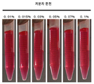


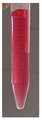


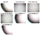

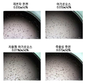


Classifications
-
- C—CHEMISTRY; METALLURGY
- C08—ORGANIC MACROMOLECULAR COMPOUNDS; THEIR PREPARATION OR CHEMICAL WORKING-UP; COMPOSITIONS BASED THEREON
- C08L—COMPOSITIONS OF MACROMOLECULAR COMPOUNDS
- C08L5/00—Compositions of polysaccharides or of their derivatives not provided for in groups C08L1/00 or C08L3/00
- C08L5/12—Agar or agar-agar, i.e. mixture of agarose and agaropectin; Derivatives thereof
-
- C—CHEMISTRY; METALLURGY
- C12—BIOCHEMISTRY; BEER; SPIRITS; WINE; VINEGAR; MICROBIOLOGY; ENZYMOLOGY; MUTATION OR GENETIC ENGINEERING
- C12N—MICROORGANISMS OR ENZYMES; COMPOSITIONS THEREOF; PROPAGATING, PRESERVING, OR MAINTAINING MICROORGANISMS; MUTATION OR GENETIC ENGINEERING; CULTURE MEDIA
- C12N5/00—Undifferentiated human, animal or plant cells, e.g. cell lines; Tissues; Cultivation or maintenance thereof; Culture media therefor
- C12N5/06—Animal cells or tissues; Human cells or tissues
- C12N5/0602—Vertebrate cells
- C12N5/067—Hepatocytes
-
- G—PHYSICS
- G01—MEASURING; TESTING
- G01N—INVESTIGATING OR ANALYSING MATERIALS BY DETERMINING THEIR CHEMICAL OR PHYSICAL PROPERTIES
- G01N33/00—Investigating or analysing materials by specific methods not covered by groups G01N1/00 - G01N31/00
- G01N33/48—Biological material, e.g. blood, urine; Haemocytometers
- G01N33/50—Chemical analysis of biological material, e.g. blood, urine; Testing involving biospecific ligand binding methods; Immunological testing
- G01N33/5005—Chemical analysis of biological material, e.g. blood, urine; Testing involving biospecific ligand binding methods; Immunological testing involving human or animal cells
- G01N33/5008—Chemical analysis of biological material, e.g. blood, urine; Testing involving biospecific ligand binding methods; Immunological testing involving human or animal cells for testing or evaluating the effect of chemical or biological compounds, e.g. drugs, cosmetics
- G01N33/5011—Chemical analysis of biological material, e.g. blood, urine; Testing involving biospecific ligand binding methods; Immunological testing involving human or animal cells for testing or evaluating the effect of chemical or biological compounds, e.g. drugs, cosmetics for testing antineoplastic activity
-
- C—CHEMISTRY; METALLURGY
- C08—ORGANIC MACROMOLECULAR COMPOUNDS; THEIR PREPARATION OR CHEMICAL WORKING-UP; COMPOSITIONS BASED THEREON
- C08B—POLYSACCHARIDES; DERIVATIVES THEREOF
- C08B37/00—Preparation of polysaccharides not provided for in groups C08B1/00 - C08B35/00; Derivatives thereof
- C08B37/12—Agar-agar; Derivatives thereof
-
- C—CHEMISTRY; METALLURGY
- C08—ORGANIC MACROMOLECULAR COMPOUNDS; THEIR PREPARATION OR CHEMICAL WORKING-UP; COMPOSITIONS BASED THEREON
- C08L—COMPOSITIONS OF MACROMOLECULAR COMPOUNDS
- C08L89/00—Compositions of proteins; Compositions of derivatives thereof
-
- C—CHEMISTRY; METALLURGY
- C12—BIOCHEMISTRY; BEER; SPIRITS; WINE; VINEGAR; MICROBIOLOGY; ENZYMOLOGY; MUTATION OR GENETIC ENGINEERING
- C12N—MICROORGANISMS OR ENZYMES; COMPOSITIONS THEREOF; PROPAGATING, PRESERVING, OR MAINTAINING MICROORGANISMS; MUTATION OR GENETIC ENGINEERING; CULTURE MEDIA
- C12N5/00—Undifferentiated human, animal or plant cells, e.g. cell lines; Tissues; Cultivation or maintenance thereof; Culture media therefor
- C12N5/0018—Culture media for cell or tissue culture
-
- C—CHEMISTRY; METALLURGY
- C12—BIOCHEMISTRY; BEER; SPIRITS; WINE; VINEGAR; MICROBIOLOGY; ENZYMOLOGY; MUTATION OR GENETIC ENGINEERING
- C12N—MICROORGANISMS OR ENZYMES; COMPOSITIONS THEREOF; PROPAGATING, PRESERVING, OR MAINTAINING MICROORGANISMS; MUTATION OR GENETIC ENGINEERING; CULTURE MEDIA
- C12N5/00—Undifferentiated human, animal or plant cells, e.g. cell lines; Tissues; Cultivation or maintenance thereof; Culture media therefor
- C12N5/06—Animal cells or tissues; Human cells or tissues
- C12N5/0602—Vertebrate cells
- C12N5/0693—Tumour cells; Cancer cells
-
- C—CHEMISTRY; METALLURGY
- C12—BIOCHEMISTRY; BEER; SPIRITS; WINE; VINEGAR; MICROBIOLOGY; ENZYMOLOGY; MUTATION OR GENETIC ENGINEERING
- C12Q—MEASURING OR TESTING PROCESSES INVOLVING ENZYMES, NUCLEIC ACIDS OR MICROORGANISMS; COMPOSITIONS OR TEST PAPERS THEREFOR; PROCESSES OF PREPARING SUCH COMPOSITIONS; CONDITION-RESPONSIVE CONTROL IN MICROBIOLOGICAL OR ENZYMOLOGICAL PROCESSES
- C12Q1/00—Measuring or testing processes involving enzymes, nucleic acids or microorganisms; Compositions therefor; Processes of preparing such compositions
- C12Q1/02—Measuring or testing processes involving enzymes, nucleic acids or microorganisms; Compositions therefor; Processes of preparing such compositions involving viable microorganisms
-
- G—PHYSICS
- G01—MEASURING; TESTING
- G01N—INVESTIGATING OR ANALYSING MATERIALS BY DETERMINING THEIR CHEMICAL OR PHYSICAL PROPERTIES
- G01N33/00—Investigating or analysing materials by specific methods not covered by groups G01N1/00 - G01N31/00
- G01N33/48—Biological material, e.g. blood, urine; Haemocytometers
- G01N33/50—Chemical analysis of biological material, e.g. blood, urine; Testing involving biospecific ligand binding methods; Immunological testing
- G01N33/5005—Chemical analysis of biological material, e.g. blood, urine; Testing involving biospecific ligand binding methods; Immunological testing involving human or animal cells
- G01N33/5008—Chemical analysis of biological material, e.g. blood, urine; Testing involving biospecific ligand binding methods; Immunological testing involving human or animal cells for testing or evaluating the effect of chemical or biological compounds, e.g. drugs, cosmetics
- G01N33/5044—Chemical analysis of biological material, e.g. blood, urine; Testing involving biospecific ligand binding methods; Immunological testing involving human or animal cells for testing or evaluating the effect of chemical or biological compounds, e.g. drugs, cosmetics involving specific cell types
- G01N33/5067—Liver cells
-
- C—CHEMISTRY; METALLURGY
- C12—BIOCHEMISTRY; BEER; SPIRITS; WINE; VINEGAR; MICROBIOLOGY; ENZYMOLOGY; MUTATION OR GENETIC ENGINEERING
- C12N—MICROORGANISMS OR ENZYMES; COMPOSITIONS THEREOF; PROPAGATING, PRESERVING, OR MAINTAINING MICROORGANISMS; MUTATION OR GENETIC ENGINEERING; CULTURE MEDIA
- C12N2500/00—Specific components of cell culture medium
- C12N2500/30—Organic components
-
- C—CHEMISTRY; METALLURGY
- C12—BIOCHEMISTRY; BEER; SPIRITS; WINE; VINEGAR; MICROBIOLOGY; ENZYMOLOGY; MUTATION OR GENETIC ENGINEERING
- C12N—MICROORGANISMS OR ENZYMES; COMPOSITIONS THEREOF; PROPAGATING, PRESERVING, OR MAINTAINING MICROORGANISMS; MUTATION OR GENETIC ENGINEERING; CULTURE MEDIA
- C12N2533/00—Supports or coatings for cell culture, characterised by material
- C12N2533/70—Polysaccharides
- C12N2533/76—Agarose, agar-agar
Landscapes
- Health & Medical Sciences (AREA)
- Life Sciences & Earth Sciences (AREA)
- Engineering & Computer Science (AREA)
- Chemical & Material Sciences (AREA)
- Biomedical Technology (AREA)
- Immunology (AREA)
- Biotechnology (AREA)
- Organic Chemistry (AREA)
- Bioinformatics & Cheminformatics (AREA)
- Molecular Biology (AREA)
- Cell Biology (AREA)
- General Health & Medical Sciences (AREA)
- Biochemistry (AREA)
- Hematology (AREA)
- Urology & Nephrology (AREA)
- Microbiology (AREA)
- Medicinal Chemistry (AREA)
- Zoology (AREA)
- Wood Science & Technology (AREA)
- Genetics & Genomics (AREA)
- Physics & Mathematics (AREA)
- Analytical Chemistry (AREA)
- General Physics & Mathematics (AREA)
- Pathology (AREA)
- Food Science & Technology (AREA)
- Tropical Medicine & Parasitology (AREA)
- Toxicology (AREA)
- General Engineering & Computer Science (AREA)
- Chemical Kinetics & Catalysis (AREA)
- Polymers & Plastics (AREA)
- Gastroenterology & Hepatology (AREA)
- Proteomics, Peptides & Aminoacids (AREA)
- Oncology (AREA)
- Materials Engineering (AREA)
- Biophysics (AREA)
- Micro-Organisms Or Cultivation Processes Thereof (AREA)
- Measuring Or Testing Involving Enzymes Or Micro-Organisms (AREA)
- Immobilizing And Processing Of Enzymes And Microorganisms (AREA)
Abstract
본 발명은, 세포 또는 조직을 양호하게 분산시킨 상태에서 효율적으로 배양할 수 있고, 또한 세포 또는 조직의 세포 이미지 해석이 가능해지는 배지 첨가물 및 배지 조성물, 그리고 배양 방법 등을 제공한다. 본 발명의 배지 첨가물 또는 배지 조성물은 한천을 함유한다. 한천으로는, 중량 평균 분자량이 10,000∼60,000인 저분자량의 한천을 사용하는 것이 바람직하다. 이것을 사용하면, 세포 또는 조직이 배지 중에 양호하게 분산된 상태에서 배양할 수 있고, 세포 또는 조직의 증식 촉진 효과를 얻을 수도 있다. 또, 상기 한천의 농도를 조정함으로써, 부유 상태 및 침전된 상태 중 어느 상태에서도 배양할 수 있다.The present invention provides a culture medium additive and a culture medium composition capable of efficiently culturing a cell or a tissue in a well dispersed state and enabling cell image analysis of the cell or tissue, a culture method, and the like. The media additive or media composition of the present invention contains agar. As the agar, it is preferable to use a low molecular weight agar having a weight average molecular weight of 10,000 to 60,000. By using this, the cells or tissues can be cultured in a well-dispersed state in the medium, and the effect of promoting the proliferation of cells or tissues can be obtained. In addition, by adjusting the concentration of the agar, the culture can be carried out in either a floating state or a precipitated state.
Description
본 발명은, 세포 또는 조직을 부유 혹은 침강시킨 상태에서 양호하게 분산시켜 배양하는 것을 가능하게 하는 배지 첨가물 및 배지 조성물, 그리고 당해 배지 첨가물 또는 배지 조성물을 사용하는 것을 특징으로 하는, 세포 또는 조직의 배양 방법에 관한 것이다.The present invention relates to a method for culturing a cell or tissue, which comprises using a culture medium additive and a culture medium composition capable of well dispersing and culturing cells or tissues in a floating or settled state and using the culture medium additive or culture medium composition ≪ / RTI >
최근, 동물이나 식물체 내에서 상이한 역할을 하고 있는 여러 가지 기관, 조직, 및 세포를 생체 외에서 증식 혹은 유지시키기 위한 기술이 발전해 오고 있다. 이들 기관, 조직을 생체 외에서 증식 혹은 유지하는 것은, 각각 기관 배양, 조직 배양이라고 불리고 있으며, 기관, 조직으로부터 분리된 세포를 생체 외에서 증식, 분화 혹은 유지하는 것은 세포 배양이라고 불리고 있다. 세포 배양은, 분리한 세포를 배지 중에서, 즉 생체 외에서 증식 또는 분화시키거나, 혹은 유지하는 기술이고, 생체 내의 각종 기관, 조직, 세포의 기능 및 구조를 상세하게 해석하기 위해서 불가결한 것으로 되어 있다. 또, 당해 기술에 의해 배양된 세포 또는 조직은, 화학 물질, 의약품 등의 약효 및 독성 평가나, 효소, 세포 증식 인자, 항체 등의 유용 물질의 대량 생산, 질환이나 결손에 의해 없어진 기관, 조직, 세포를 보충하기 위한 재생 의료, 식물의 품종 개량, 유전자 재조합 작물의 제조 등, 여러 가지 분야에서 이용되고 있다.Recently, techniques for growing or maintaining various organs, tissues, and cells that play different roles in animals or plants in vitro have been developed. The propagation or maintenance of these organs and tissues in vitro is referred to as an organ culture or tissue culture, respectively, and the propagation, differentiation or maintenance of cells isolated from organs and tissues in vitro is called cell culture. Cell culture is a technique for proliferating or differentiating or maintaining separated cells in a medium, that is, in vitro, and it is indispensable for detailed analysis of the functions and structures of various organs, tissues, and cells in the living body. In addition, the cells or tissue cultured by the present technique can be used for evaluation of drug efficacy and toxicity of chemical substances and medicines, mass production of useful substances such as enzymes, cell growth factors, and antibodies, organs, tissues, Regenerative medicine for replenishing cells, breeding of plants, production of genetically modified crops, and the like.
동물 유래의 세포는, 그 성상으로부터 부유성 세포와 접착성 세포로 크게 이분된다. 부유성 세포는, 생육·증식에 족장(足場)을 필요로 하지 않는 세포이고, 접착성 세포는, 생육·증식에 족장을 필요로 하는 세포이지만, 생체를 구성하는 대부분의 세포는 후자의 접착성 세포이다. 접착성 세포의 배양 방법으로는, 단층 배양, 분산 배양, 포매 배양, 마이크로 캐리어 배양, 및 스피어 배양 등이 알려져 있다.Animal-derived cells are largely divided into floating cells and adhesive cells. The floating cell is a cell that does not require a chock (foot field) for growth and proliferation. The adhesive cell is a cell which needs a patriarch for growth and proliferation. However, most of the cells constituting the living body are adhesive It is a cell. As a method for culturing adherent cells, there are known single culture, dispersion culture, forced culture, microcarrier culture, and sphere culture.
탈아실화 젤란검 등의 아니온성 관능기를 갖는 고분자 화합물을 함유하는 구조체를 액체 배지 중에 혼합함으로써, 그 액체 배지의 점도를 실질적으로 높이지 않고, 동식물 세포 및/또는 조직을 정치(靜置)시킨 상태에서 균일하게 분산시켜, 부유 배양할 수 있는 것, 이 배지 조성물을 사용하여 배양함으로써, 세포의 증식 활성이 항진되는 것이 보고되어 있다(특허문헌 1). 또, 메틸셀룰로오스 등의 증점제를 사용하여 접착성 세포를 분산시켜, 스페로이드(세포끼리가 집합·응집화된 세포 집합체를 말한다. 본 명세서에 있어서는 「스피어」라고 칭하는 경우가 있다)의 형성을 촉진시켜, 세포 증식을 항진시키는 예도 보고되어 있다(특허문헌 2, 비특허문헌 1).A structure containing a polymer compound having an anionic functional group such as deacylated gellan gum is mixed into a liquid medium so that the animal and plant cells and / or tissues are allowed to stand still without substantially increasing the viscosity of the liquid medium , And that the cell growth activity is enhanced by culturing the cells using the culture medium (Patent Document 1). In addition, the adhesive cells are dispersed using a thickening agent such as methyl cellulose to promote the formation of spheroids (sometimes referred to as " sphere " in this specification) (Patent Document 2, Non-Patent Document 1).
상기 서술한 바와 같이, 특허문헌 1에는, 탈아실화 젤란검 등의 아니온성 관능기를 갖는 고분자 화합물을 함유하는 구조체를 액체 배지 중에 혼합함으로써, 그 액체 배지의 점도를 실질적으로 높이지 않고, 세포 또는 조직을 정치시킨 상태에서 양호하게 분산시켜, 부유 배양할 수 있는 것, 이 배지 조성물을 사용하여 배양함으로써, 세포의 증식 활성이 항진되는 것이 보고되어 있다. 그러나, 본 발명자들은, 이 배지 조성물을 사용하여 세포 또는 조직을 배양한 경우에는, 세포 또는 조직이 부유 상태이기 때문에, 세포 또는 조직을 세포 이미징 장치로 해석할 때에 렌즈의 초점이 맞지 않아, 배양물을 그대로 세포 이미지 해석에 제공할 수 없다는 새로운 과제를 알아내었다.As described above, in Patent Document 1, a structure containing a polymer compound having an anionic functional group such as deacylated gellan gum is mixed in a liquid medium, and the viscosity of the liquid medium is not substantially increased, Has been reported to be proliferated in a state in which the cells are allowed to stand in a state of being allowed to float and to be cultured by using the culture medium. However, when cells or tissues are cultured using this medium composition, the inventors of the present invention have found that since the cells or tissues are floating, when the cells or tissues are analyzed by a cell imaging device, the focus of the lens is not focused, Can not be directly applied to cell image interpretation.
본 발명은, 세포 또는 조직을 양호하게 분산시킨 상태로 효율적으로 배양할 수 있고, 또한 세포 또는 조직의 세포 이미지 해석이 가능해지는 배지 조성물 및 배양 방법 등을 제공하는 것을 목적으로 한다.An object of the present invention is to provide a culture medium, a culture method, and the like which can efficiently culture a cell or a tissue in a well-dispersed state, and can perform cell image analysis of a cell or a tissue.
본 발명자들은 상기 과제를 해결하기 위하여 예의 검토한 결과, 한천을 배지 첨가물로서 사용함으로써, 세포 또는 조직을 양호하게 분산시킬 수 있고, 또한 한천의 농도에 따라서는, 부유 상태에서도 침강된 상태에서도, 양호한 효율로 배양이 가능해지는 것을 알아내었다. 또한 그 배지 첨가물을 사용한 조성물에서 배양함으로써, 세포 또는 조직의 세포 이미징 장치에서의 해석을 신속하고 또한 간편하게 실시하는 것이 가능해지는 것을 알아내어, 본 발명을 완성시키기에 이르렀다.DISCLOSURE OF THE INVENTION As a result of diligent studies for solving the above problems, the present inventors have found that, by using agar as a medium addition agent, cells or tissues can be well dispersed and, depending on the concentration of agar, Thereby enabling the cultivation to be performed with efficiency. Further, the present invention has been accomplished on the basis of the findings that it is possible to quickly and easily carry out an analysis in a cell imaging apparatus for a cell or a tissue by culturing it in a composition using the medium additive.
즉, 본 발명은 하기와 같다:That is, the present invention is as follows:
[1] 한천을 함유하는, 배지 첨가물.[1] Agar-containing medium additives.
[2] 한천의 중량 평균 분자량이 10,000∼60,000인, [1]에 기재된 배지 첨가물.[2] The medium addition product according to [1], wherein the agar has a weight average molecular weight of 10,000 to 60,000.
[3] 액상인, [1] 또는 [2]에 기재된 배지 첨가물.[3] A media additive according to [1] or [2] which is a liquid.
[4] 한천의 함유량이, 배지 첨가물의 전체량에 대하여 0.001(w/v)%∼5(w/v)%인, [3]에 기재된 배지 첨가물.[4] The medium addition product according to [3], wherein the agar content is 0.001 (w / v) to 5% (w / v) relative to the total amount of the medium supplement.
[5] 한천을 함유하는, 배지 조성물.[5] A medium composition comprising agar.
[6] 한천의 중량 평균 분자량이 10,000∼60,000인, [5]에 기재된 배지 조성물.[6] The medium composition according to [5], wherein the agar has a weight average molecular weight of 10,000 to 60,000.
[7] [1]∼[4] 중 어느 하나에 기재된 배지 첨가물을 함유하는, 배지 조성물.[7] A medium composition comprising the medium addition agent according to any one of [1] to [4].
[8] 한천의 함유량이, 배지 조성물의 전체량에 대하여 0.005(w/v)% 이상이고 2(w/v)% 미만인, [5]∼[7] 중 어느 하나에 기재된 배지 조성물.[8] The medium composition according to any one of [5] to [7], wherein the content of agar is 0.005 (w / v) or more and less than 2 (w / v)% with respect to the total amount of the medium composition.
[9] 한천의 함유량이 0.1(w/v)%인 경우의 37℃에 있어서의 점도가 2.5mPa·s 이하인, [5]∼[8] 중 어느 하나에 기재된 배지 조성물.[9] The medium composition according to any one of [5] to [8], wherein the viscosity at 37 ° C. is 2.5 mPa · s or less when the agar content is 0.1 (w / v)%.
[10] 세포 배양용인, [5]∼[9] 중 어느 하나에 기재된 배지 조성물.[10] The culture medium according to any one of [5] to [9], which is for cell culture.
[11] 세포가 접착성 세포 또는 부유성 세포인, [10]에 기재된 배지 조성물.[11] The medium composition according to [10], wherein the cell is an adhesive cell or a floating cell.
[12] 접착성 세포가 담체 표면에 접착되고 또는 담체 내부에 포매되어 있는, [11]에 기재된 배지 조성물.[12] The medium composition according to [11], wherein the adhesive cells are adhered to the surface of the carrier or embedded in the carrier.
[13] 접착성 세포가 마이크로 캐리어에 접착되어 있는, [11]에 기재된 배지 조성물.[13] The medium composition according to [11], wherein the adhesive cells are adhered to the microcarriers.
[14] 접착성 세포가 스피어를 형성하고 있는, [11]에 기재된 배지 조성물.[14] The medium composition according to [11], wherein the adhesive cell forms a sphere.
[15] 접착성 세포가, 암세포, 간세포 및 암세포주로 이루어지는 군에서 선택되는, [11]∼[14] 중 어느 하나에 기재된 배지 조성물.[15] The medium composition according to any one of [11] to [14], wherein the adhesive cell is selected from the group consisting of cancer cells, hepatocytes and cancer cells.
[16] [5]∼[15] 중 어느 하나에 기재된 배지 조성물과, 세포 또는 조직을 함유하는, 세포 또는 조직 배양물.[16] A cell or tissue culture comprising the culture medium according to any one of [5] to [15] and a cell or tissue.
[17] [5]∼[15] 중 어느 하나에 기재된 배지 조성물 중에, 세포 또는 조직을 분산시킨 상태로 배양하는, 세포 또는 조직의 배양 방법.[17] A method for culturing a cell or tissue, wherein the cell or tissue is cultured in the medium composition according to any one of [5] to [15].
[18] 세포가 접착성 세포 또는 부유성 세포인, [17]에 기재된 배양 방법.[18] The method according to [17], wherein the cell is an adhesive cell or a floating cell.
[19] 접착성 세포가 담체 표면에 접착되고 또는 담체 내부에 포매되어 있는, [18]에 기재된 배양 방법.[19] The culture method according to [18], wherein the adhesive cells are adhered to the surface of the carrier or embedded in the carrier.
[20] 접착성 세포가 마이크로 캐리어에 접착되어 있는, [18]에 기재된 배양 방법.[20] The culture method according to [18], wherein the adhesive cells are adhered to the microcarriers.
[21] 접착성 세포가 스피어를 형성하고 있는, [18]에 기재된 배양 방법.[21] The culture method according to [18], wherein the adhesive cell forms a sphere.
[22] 접착성 세포가, 암세포, 간세포 및 암세포주로 이루어지는 군에서 선택되는, [18]∼[21] 중 어느 하나에 기재된 배양 방법.[22] The culture method according to any one of [18] to [21], wherein the adhesive cell is selected from the group consisting of cancer cells, hepatocytes and cancer cells.
[23] (a) 피험물질의 존재하 및 비존재하에, [5]∼[15] 중 어느 하나에 기재된 배지 조성물 중에서 세포를 배양하는 공정, 및 (b) 세포의 생리학적 기능의 변화를 해석하는 공정을 포함하는, 의약품 후보 물질의 스크리닝 방법.[23] A method of culturing a cell in a culture medium according to any one of [5] to [15], which comprises (a) culturing a cell in the presence and absence of a test substance, and (b) The method comprising the steps of:
[24] 추가로, (c) 피험물질의 비존재하의 경우와 비교하여, 세포의 생리학적 기능을 억제 또는 증가시키는 물질을 의약품 후보 물질로서 선택하는 공정을 포함하는, [23]에 기재된 스크리닝 방법.[24] The screening method according to [23], further comprising a step of selecting as a drug candidate substance a substance which inhibits or increases the physiological function of the cell, as compared with the case where the test substance is not present. .
[25] (b) 세포의 생리학적 기능의 변화를 해석하는 공정이 세포 이미지 해석에 의해 실시되는, [23] 또는 [24]에 기재된 스크리닝 방법.[25] The screening method according to [23] or [24], wherein the step of (b) analyzing the change in physiological function of the cell is carried out by cell image analysis.
[26] 배지 조성물에 있어서의 한천의 함유량이, 배지 조성물의 전체량에 대하여 0.005(w/v)% 이상 0.07(w/v)% 미만인, [23]∼[25] 중 어느 하나에 기재된 스크리닝 방법.[26] The screening method according to any one of [23] to [25], wherein the content of the agar in the culture medium composition is 0.005 (w / v)% or more to less than 0.07 Way.
[27] (a) 피험물질의 존재하 및 비존재하에, [5]∼[15] 중 어느 하나에 기재된 배지 조성물 중에서 암세포 또는 암세포주를 배양하는 공정, 및 (b) 암세포 또는 암세포주의 증식의 변화를 해석하는 공정을 포함하는, 항암제 후보 물질의 스크리닝 방법.[27] A method of culturing a cancer cell or a cancer cell line in a culture medium according to any one of [5] to [15], which comprises (a) culturing a cancer cell or a cancer cell line in the presence or absence of a test substance, and (b) A method for screening an anticancer candidate substance, which comprises a step of analyzing a change.
[28] 추가로, (c) 피험물질의 비존재하의 경우와 비교하여, 암세포 또는 암세포주의 증식을 억제하는 물질을 항암제 후보 물질로서 선택하는 공정을 포함하는, [27]에 기재된 스크리닝 방법.[28] The screening method according to [27], further comprising a step of selecting, as an anticancer agent candidate substance, a substance which inhibits the proliferation of cancer cells or cancer cells, as compared with (c) the case where the test substance is not present.
[29] (b) 암세포 또는 암세포주의 증식의 변화를 해석하는 공정이 세포 이미지 해석에 의해 실시되는, [27] 또는 [28]에 기재된 스크리닝 방법.[29] (b) The screening method according to [27] or [28], wherein the step of analyzing the change in proliferation of cancer cells or cancer cells is carried out by cell image analysis.
[30] 배지 조성물에 있어서의 한천의 함유량이, 배지 조성물의 전체량에 대하여 0.005(w/v)% 이상 0.07(w/v)% 미만인, [27]∼[29] 중 어느 하나에 기재된 스크리닝 방법.[30] The screening composition according to any one of [27] to [29], wherein the content of agar in the culture medium composition is 0.005 (w / v)% or more to less than 0.07 Way.
[31] [5]∼[10] 중 어느 하나에 기재된 배지 조성물 중에서 접착성 세포를 배양하는, 스피어의 제조 방법.[31] A method for producing a sphere, comprising culturing adherent cells in the medium composition according to any one of [5] to [10].
본 발명의 배지 첨가물 또는 배지 조성물을 사용함으로써, 세포 또는 조직이 양호하게 분산되어 부유한 상태, 침강한 상태 중 어느 상태에서도 효율적으로 배양할 수 있다.By using the culture medium additive or the culture medium composition of the present invention, the cells or tissues are well dispersed and can be efficiently cultured in any of the floated state and the sedimented state.
특히, 본 발명의 배지 첨가물 또는 배지 조성물을 사용함으로써, 담체 표면에 접착되거나 혹은 담체 내부에 포매된 접착성 세포, 또는 스피어를 형성한 접착성 세포를, 과잉인 응집을 일으키지 않고, 양호하게 분산된 상태로 배양할 수 있다.Particularly, by using the medium additive or the medium composition of the present invention, it is possible to prevent the adhesive cells adhered to the surface of the carrier or embedded in the carrier, or the adhesive cells forming the sphere, Lt; / RTI >
또한 본 발명의 배지 첨가물 또는 배지 조성물을 사용하여 배양함으로써, 세포 이미지 해석에 의한 세포의 성상, 기능의 해석이 가능해져, 항암제 등, 의약품의 후보 물질의 스크리닝을 바람직하게 실시할 수 있다.Further, by culturing using the medium additive of the present invention or the medium composition of the present invention, it is possible to analyze the properties and functions of the cells by analyzing the cell image, and screening of candidate substances for medicines such as anticancer drugs can be preferably carried out.
도 1은, 분석예 1에 있어서, 저분자 한천을 함유하는 배지 조성물 중에 있어서의 폴리스티렌 비즈의 상태를 나타내는 도면이다.
도 2는, 분석예 1에 있어서, 일반용 한천을 함유하는 배지 조성물 중에 있어서의 폴리스티렌 비즈의 분산 상태를 나타내는 도면이다.
도 3은, 분석예 2에 있어서, 저분자 한천을 함유하는 배지 조성물에서 HepG2 세포를 7일간 배양한 후의 상태를 나타내는 도면이다.
도 4는, 분석예 3에 있어서, 저분자 한천을 함유하는 배지 조성물 중에 있어서의 폴리스티렌 비즈의 상태를 나타내는 도면이다.
도 5는, 시험예 1에 있어서, 7일간 배양 후의 A549 세포의 스피어의 현미경 관찰 결과를 나타내는 도면이다.
도 6은, 시험예 2에 있어서, 7일간 배양 후의 HepG2 세포의 스피어의 현미경 관찰 결과를 나타내는 도면이다.
도 7은, 시험예 3에 있어서, 7일간 배양 후의 A549 세포의 스피어의 현미경 관찰 결과를 나타내는 도면이다.
도 8은, 시험예 5에 있어서, A549 세포의 세포 이미징 장치에 의한 관찰 이미지를 나타내는 도면이다.
도 9는, 시험예 6에 있어서, 7일간 배양 후의 A549 세포의 스피어의 현미경 관찰 결과를 나타내는 도면이다.
도 10은, 시험예 7에 있어서, 7일간 배양 후의 A549 세포의 스피어의 현미경 관찰 결과를 나타내는 도면이다.
도 11은, 시험예 11에 있어서, 21일간 배양 후의 HeLa 세포의 스피어의 현미경 관찰 결과를 나타내는 도면이다. 도면 중, 실선 화살표는, 단단하게 구상으로 응집된 스피어를 나타내고, 점선 화살표는, 무르게 응집된 스피어를 나타낸다.1 is a diagram showing the state of polystyrene beads in a medium composition containing a low molecular agar in analysis example 1;
2 is a diagram showing the dispersion state of polystyrene beads in a medium composition containing general agar in analysis example 1. Fig.
Fig. 3 is a diagram showing the state after the HepG2 cells were cultured for 7 days in the medium composition containing the low molecular agar in the analysis example 2; Fig.
4 is a diagram showing the state of polystyrene beads in a medium composition containing a low molecular agar in Analysis Example 3. Fig.
5 is a diagram showing the result of microscope observation of sphere of A549 cells after 7 days of culture in Test Example 1. Fig.
6 is a diagram showing the result of microscope observation of sphere of HepG2 cells after 7 days of culture in Test Example 2. Fig.
Fig. 7 is a diagram showing a microscopic observation result of sphere of A549 cells after 7 days of culture in Test Example 3. Fig.
Fig. 8 is a view showing an observation image of A549 cells by a cell imaging apparatus in Test Example 5. Fig.
Fig. 9 is a diagram showing the result of microscopic observation of sphere of A549 cells after 7 days of culture in Test Example 6. Fig.
10 is a diagram showing the result of microscope observation of sphere of A549 cells after 7 days of culture in Test Example 7. Fig.
11 is a diagram showing the result of microscope observation of sphere of HeLa cells after 21 days of culture in Test Example 11. Fig. In the figure, a solid line arrow indicates a spherically agglomerated sphere, and a dashed arrow indicates a spherically agglomerated sphere.
본 발명은, 한천을 함유하는 배지 첨가물을 제공한다.The present invention provides a culture medium additive containing agar.
또, 본 발명은, 한천을 함유하는 배지 조성물을 제공한다.Further, the present invention provides a medium composition containing agar.
한천은, 아가로오스와, 아가로오스가 부분적으로 황산에스테르화되거나, 또는 메톡시기, 피루브산기, 카르복실기에 의해 치환된 아가로펙틴에 의해 구성되지만, 이들 구성 성분의 비율에 관해서 제한은 없고, 아가로오스만으로 구성되어도 된다. 또, 아가로오스가 하이드록시에틸화된 저융점 아가로오스나, 원료 해조의 선택에 의해, 또는 하이드록시에틸화를 실시하는 등에 의해 조제된 저융점 한천, 또한 온탕(溫湯)에 대해서도 높은 용해성을 나타내는 즉용성 한천 등도, 본 발명에 있어서의 「한천」에 포함된다.Agar is made up of agarose and agarose partially or partly sulfuric acid esterified or agaropectin substituted by a methoxy group, a pyruvic acid group or a carboxyl group. There is no limitation on the ratio of these constituent components, It may also be composed of agarose. In addition, a low-melting-point agar prepared by selecting a low-melting-point agarose in which agarose is hydroxyethylated, a raw material seaweed, or subjected to hydroxyethylation, and also has a high solubility Is also included in " agar " in the present invention.
본 발명에서는, 공업적으로 제조되는 분말상, 플레이크상 또는 고형상의 한천이 고순도이고 품질도 균일하기 때문에, 바람직하게 사용된다. 또, 의약품, 식품 등의 분야에서, 일반적으로 사용되는 여러 가지 성상, 물성을 갖는 것을 사용할 수 있고, 중량 평균 분자량이 10,000∼60,000인 저분자량의 한천, 중량 평균 분자량이 60,000을 초과하고 100,000 이하 정도이고, 겔 강도가 낮은 저강도 한천, 중량 평균 분자량이 290,000 정도인 고분자량의 한천 등을 사용할 수 있다. 이러한 한천으로는, 시판되고 있는 제품을 이용할 수 있고, 예를 들어, 이나 식품 공업 주식회사로부터 판매되고 있는 「울트라 한천 이나」, 「울트라 한천 AX-30」, 「울트라 한천 AX-100」, 「S-6」, 「S-7」등을 사용할 수 있다.In the present invention, an industrially produced powdery, flaky or solid agar is preferably used since it has high purity and uniform quality. It is also possible to use those having various properties and physical properties commonly used in the fields of pharmaceuticals and foods and having a weight average molecular weight of 10,000 to 60,000 and a weight average molecular weight of more than 60,000 and not more than 100,000 , A low strength agar having a low gel strength, and a high molecular weight agar having a weight average molecular weight of about 290,000. As such agar, commercially available products can be used. For example, "Ultra Agar A", "Ultra Agar AX-30", "Ultra Agar AX-100", "S" -6 ", " S-7 ", and the like.
본 발명에 있어서는, 중량 평균 분자량이 10,000∼60,000이고, 일반적인 한천보다 저분자량의 한천(이하, 본 명세서에 있어서 「저분자 한천」이라고 칭하는 경우가 있다)을 사용하는 것이 바람직하다.In the present invention, agar having a weight average molecular weight of 10,000 to 60,000 and a lower molecular weight than general agar (hereinafter sometimes referred to as " low molecular agar in the present specification ") is preferably used.
본 발명에서 바람직하게 사용되는 저분자 한천의 중량 평균 분자량은, 상기와 같이 10,000∼60,000이고, 20,000∼60,000인 것이 보다 바람직하고, 30,000∼60,000인 것이 더욱 바람직하고, 40,000∼60,000인 것이 더욱 더 바람직하고, 43,000∼60,000인 것이 보다 더 바람직하고, 43,000∼50,000인 것이 특히 바람직하다.The weight average molecular weight of the low molecular weight agar preferably used in the present invention is 10,000 to 60,000, more preferably 20,000 to 60,000, still more preferably 30,000 to 60,000, still more preferably 40,000 to 60,000 , Still more preferably from 43,000 to 60,000, and particularly preferably from 43,000 to 50,000.
중량 평균 분자량이 10,000 미만인 한천을 사용한 경우에는, 세포를 분산시키는 효과를 얻는 것이 곤란한 경우가 있다. 한편, 중량 평균 분자량이 60,000을 초과하는 한천을 사용한 경우에는, 세포 또는 조직의 배지 중에 있어서의 분산이 불균일해져, 충분한 증식 촉진 효과를 얻을 수 없는 경우가 있다.When an agar having a weight average molecular weight of less than 10,000 is used, it may be difficult to obtain an effect of dispersing cells. On the other hand, when agar having a weight average molecular weight of more than 60,000 is used, dispersion of cells or tissues in a medium becomes uneven and a sufficient promoting effect may not be obtained.
또한, 본 발명에서 사용하는 저분자 한천으로는, 분자량 분포가 좁은 것임이 바람직하고, 한천의 중량 평균 분자량(Mw)을 수평균 분자량(Mn)으로 나눈 값으로서 얻어지는 분자량 분포(Mw/Mn)는, 1.1∼8.0인 것이 바람직하고, 1.5∼7.0인 것이 보다 바람직하고, 2.0∼6.0인 것이 더욱 바람직하고, 2.5∼5.5인 것이 보다 더 바람직하고, 3.5∼5.0인 것이 특히 바람직하다.The low molecular weight agar used in the present invention preferably has a narrow molecular weight distribution and the molecular weight distribution (Mw / Mn) obtained by dividing the weight average molecular weight (Mw) of the agar by the number average molecular weight (Mn) More preferably from 1.5 to 7.0, even more preferably from 2.0 to 6.0, even more preferably from 2.5 to 5.5, and particularly preferably from 3.5 to 5.0.
또한, 한천의 상기 중량 평균 분자량 및 수평균 분자량은, 고속 액체 크로마토그래피(HPLC)에 의한 겔 침투 크로마토그래피법에 의해 측정할 수 있다.The weight average molecular weight and number average molecular weight of agar can be measured by gel permeation chromatography using high performance liquid chromatography (HPLC).
구체적으로는, 예를 들어 다음과 같은 측정 기기, 조건 등에 의해 측정할 수 있다.Specifically, it can be measured by, for example, the following measuring instrument, conditions, and the like.
(1)측정용 시료:한천을 예를 들어 0.15(w/v)% (「(w/v)%」는 「중량/체적%」를 나타낸다, 이하 동일) 정도의 농도가 되도록, 정제수에 95℃∼97℃에서 용해시킨 후, 50℃까지 냉각시켜, 시료로 한다.(1) Sample for measurement: Agar was added to purified water to a concentration of about 0.15 (w / v)% ("w / v%" represents "weight / Deg.] C to 97 deg. C, and then cooled to 50 deg. C to obtain a sample.
(2) 측정 기기:주식회사 시마즈 제작소 제조 액체 크로마토그래프 LC-10AT VP, RID-10A 등(2) Measuring instrument: LC-10AT VP, RID-10A manufactured by Shimadzu Corporation
(3) 칼럼:토소 주식회사 제조 TOSOH TSK-GEL for HPLC, TSK-GEL GMPWXL 등(3) Column: TOSOH TSK-GEL for HPLC, TSK-GEL GMPWXL manufactured by Tosoh Corporation
(4) 용매: 0.1M 질산나트륨 수용액 등(4) Solvent: 0.1 M aqueous sodium nitrate solution
(5) 검출기:시차 굴절계(5) Detector: differential refractometer
(6) 측정 온도:50℃(6) Measuring temperature: 50 DEG C
(7) 표준 물질: 분자량이 이미 알려진 풀루란(Shodex STANDARD P-82 등)(7) Standard substance: Pullulan (Shodex STANDARD P-82, etc.)
또, 본 발명에서 사용하는 저분자 한천은, 1.5(w/v)%의 겔에 대하여, 20℃에서 측정한 겔 강도가 25g/㎠ 이하인 것이 바람직하고, 15g/㎠ 이하인 것이 보다 바람직하고, 12g/㎠ 이하인 것이 더욱 바람직하다.The low molecular weight agar used in the present invention preferably has a gel strength of 25 g / cm 2 or less, more preferably 15 g / cm 2 or less, and more preferably 12 g / cm 2 or less as measured at 20 캜 for 1.5 (w / v) Cm < 2 > or less.
상기 「겔 강도」란, 한천의 1.5(w/v)% 수용액을 20℃에서 15시간 정치시키고, 응고된 겔에 대해, 그 표면 1㎠당 20초간 견딜 수 있는 최대 하중을 말하며, 예를 들어 일한수식(日寒水式) 측정 장치를 사용하여, 일본 공업 규격(JIS) K 8263:1994의 규정에 따라 측정된다.The "gel strength" refers to the maximum load capable of withstanding a 1.5 (w / v)% aqueous solution of agar at 20 ° C. for 15 hours and allowing the gel to stand for 20 seconds per 1 cm 2 of its surface. For example, (JIS) K 8263: 1994, using a Janshin formula.
또한, 본 발명에서 사용하는 저분자 한천은, 1.5(w/v)% 겔 압출 하중이 10g∼1,400g인 것이 바람직하고, 10g∼1,000g인 것이 보다 바람직하고, 10g∼500g인 것이 더욱 바람직하고, 100g∼300g인 것이 보다 더 바람직하다.The low molecular weight agar used in the present invention preferably has a gel extrusion load of 1.5 (w / v)% of from 10 g to 1,400 g, more preferably from 10 g to 1,000 g, still more preferably from 10 g to 500 g, More preferably 100 g to 300 g.
상기 압출 하중은, 예를 들어, 텍스처 애널라이저(에이코 정기 주식회사 제조)에 부수하는 원주 용기(내경 50㎜, 높이 110㎜, 아크릴 수지제)의 저부 중앙부의 구멍(직경 3㎜)을 테이프로 막고, 한천의 1.5(w/v)% 수용액 100g을 충전하고, 20℃에서 18시간 정치시켜 겔화시킨 후, 저부의 테이프를 떼어내고, 직경 49㎜의 플런저를 사용하여 겔 상부로부터 가압하여(20℃, 진입 속도 20㎜/분), 겔이 붕괴되어 하부의 구멍으로부터 유출되었을 때의 하중을 측정함으로써 구해진다.The extrusion load was measured by, for example, closing a hole (diameter: 3 mm) of the bottom center of a cylindrical container (inner diameter 50 mm, height 110 mm, made of acrylic resin) attached to a texture analyzer (manufactured by Eiko Kikai Co., 100 g of agar (1.5% (w / v)) aqueous solution was charged and allowed to stand at 20 占 폚 for 18 hours to gelify. Then, the tape at the bottom was removed and pressed from the top of the gel using a plunger At an entrance speed of 20 mm / min) and measuring the load when the gel collapses and flows out from the lower hole.
상기한 저분자 한천은, 통상적인 한천의 산처리에 의한 저분자화, 혹은 우뭇가사리, 강리, 개우무 등의 해조로부터의 추출 공정시에, 또는 상기 추출 공정 혹은 여과 공정 중 어느 것을 거친 한천에 대해 산처리를 실시하는 등, 공지된 방법에 의해 제조할 수 있지만, 저분자 한천으로서 시판되고 있는 제품, 예를 들어 상기한 「울트라 한천 이나」(이나 식품 공업 주식회사 제조) 등을 사용할 수도 있다.The above-mentioned low molecular agar can be produced by a conventional method such as low molecular weight by acid treatment of agar, or in the step of extracting from seaweed such as Agarwood, , And a product commercially available as a low molecular weight agar such as the above-mentioned " Ultra Agar or the like " (manufactured by Inagaki Kogyo Co., Ltd.) may be used.
본 발명에 있어서 사용하는 저분자 한천 등의 한천은, 필요에 따라 멸균 처리를 실시해도 된다. 멸균 방법에는 특별히 제한은 없고, 예를 들어, 방사선 멸균, 에틸렌옥사이드 가스 멸균, 오토클레이브 멸균 등을 들 수 있다. 이들 멸균 처리는, 한천이 고형이어도 용액의 상태이어도 실시할 수 있다.Agar such as low molecular weight agar used in the present invention may be sterilized as needed. The sterilization method is not particularly limited, and examples thereof include radiation sterilization, ethylene oxide gas sterilization, and autoclave sterilization. These sterilization treatments can be carried out even if the agar is solid or in a solution state.
본 발명에 있어서는, 상기한 한천을 그대로 배지 첨가물로 해도 되고, 또, 물 등의 용매에 용해시키거나, 혹은 부형제, 결합제 등의 일반적으로 제제화에 사용되는 성분과 혼합하여, 분말상, 과립상 등의 고형상의 배지 첨가물, 또는 수용액 등의 액상의 배지 첨가물로 해도 된다.In the present invention, the above-described agar may be used as a culture medium additive, dissolved in a solvent such as water, or mixed with a component generally used for formulation such as excipient, binder and the like, A solid culture medium additive, or a liquid culture medium additive such as an aqueous solution may be used.
또, 상기한 한천을, 탄수화물, 무기염 등의 이하에 서술하는 배지 성분의 일부와 혼합하여, 배지 첨가물로서 조제해도 된다.The agar described above may be mixed with a part of the medium components described below such as carbohydrates, inorganic salts and the like to prepare a medium supplement.
세포 또는 조직의 배양에 사용되는 배지에 간편하게 첨가할 수 있고, 또, 특히 액체의 배지와의 혼화성 등의 관점에서, 본 발명의 배지 첨가물은, 바람직하게는 액상의 형태로 제공된다.Can be easily added to a medium used for culturing cells or tissues. In particular, from the viewpoint of compatibility with a liquid medium, the medium additive of the present invention is preferably provided in a liquid form.
액상의 배지 첨가물은, 한천을 적당한 용매에 용해시켜, 용액으로서 조제된다. 본 발명에 있어서 사용할 수 있는 용매로는, 물;디메틸술폭사이드(DMSO);메탄올, 에탄올, 프로판올, 부탄올 등의 저급 알코올;프로필렌글리콜, 부틸렌글리콜, 글리세린 등의 다가 알코올 등의 극성 용매를 들 수 있지만, 이들에 한정되지 않는다. 특히 바람직한 용매는 물이고, 본 발명의 배지 첨가물은, 특히 바람직하게는, 수용액으로서 제공된다.The agar medium is prepared as a solution by dissolving the agar in an appropriate solvent. Examples of the solvent that can be used in the present invention include water, dimethylsulfoxide (DMSO), polar solvents such as methanol, ethanol, propanol, butanol and other lower alcohols, propylene glycol, butylene glycol and glycerin, But are not limited to these. A particularly preferable solvent is water, and the medium addition product of the present invention is particularly preferably provided as an aqueous solution.
본 발명의 배지 첨가물에 있어서의 한천의 함유량은, 배지에 첨가했을 때의 배지 조성물 중에 있어서의 한천의 함유량이, 후술하는 소정의 함유량이 되도록 설정된다.The content of the agar in the medium additive of the present invention is set so that the content of the agar in the medium composition when added to the medium is a predetermined content to be described later.
수용액 등, 액상의 배지 첨가물에 있어서의 한천의 함유량은, 0.001(w/v)%∼5(w/v)%인 것이 바람직하고, 0.01(w/v)%∼2(w/v)%인 것이 보다 바람직하고, 0.1(w/v)%∼1(w/v)%인 것이 더욱 바람직하다.The content of agar in a liquid medium additive such as an aqueous solution is preferably 0.001 (w / v)% to 5 (w / v)%, more preferably 0.01 (w / v) , More preferably 0.1 (w / v)% to 1 (w / v)%.
본 발명의 배지 첨가물에는, 한천의 효과를 높이고, 그 사용량을 저감시킬 수 있는 다른 성분을 첨가할 수도 있다. 이러한 성분으로는, 헥수론산(글루쿠론산, 갈락투론산 등) 등의 우론산;구아검, 타마린드검, 알긴산, 알긴산프로필렌글리콜에스테르, 로커스트빈검, 아라비아검, 타라검, 젤란검, 탈아실화 젤란검, 람산검, 디우탄검, 크산탄검, 카리기난, 키틴, 후코이단, 펙틴, 펙트산, 펙틴산, 람난황산 등의 다당류 및 그 유도체;히알루론산, 헤파란황산, 헤파린, 케라토황산, 콘드로이틴황산, 데르마탄황산 등의 무코 다당류;메틸셀룰로오스, 하이드록시에틸셀룰로오스, 하이드록시프로필셀룰로오스, 카르복시메틸셀룰로오스 등의 셀룰로오스 유도체;카르복시비닐 폴리머, 아크릴산·메타크릴산알킬 공중합체 등의 친수성 고분자;그리고 이들의 염 등을 들 수 있고, 상기 성분은 1종 또는 2종 이상을 선택하여 사용할 수 있다. 본 발명의 배지 첨가물에 있어서의 상기 성분의 함유량은, 0.001(w/v)%∼5(w/v)%인 것이 바람직하고, 0.01(w/v)%∼2(w/v)%인 것이 보다 바람직하고, 0.1(w/v)%∼1(w/v)%인 것이 더욱 바람직하다.In the medium supplement of the present invention, other ingredients capable of enhancing the agar effect and reducing the amount of agar may be added. Examples of such components include uronic acid such as hexuronic acid (glucuronic acid, galacturonic acid, etc.), guar gum, tamarind gum, alginic acid, propylene glycol ester alginate, locust bean gum, gum arabic, tara gum, gellan gum, Polysaccharides and derivatives thereof such as hyaluronic acid, heparan sulfate, heparin, keratosulfuric acid, etc .; polysaccharides such as gellan gum, lambda gum, dianthan gum, xanthan gum, cariguanan, chitin, fucoidan, pectin, pectic acid, , Mucopolysaccharides such as chondroitin sulfate and desmatan sulfate, cellulose derivatives such as methyl cellulose, hydroxyethyl cellulose, hydroxypropyl cellulose and carboxymethyl cellulose; hydrophilic polymers such as carboxyvinyl polymer and acrylic acid / methacrylic acid alkyl copolymer; And salts thereof. One or more of the above-mentioned components can be selected and used. The content of the above components in the culture medium additive of the present invention is preferably 0.001 (w / v)% to 5 (w / v)%, more preferably 0.01 (w / v) , More preferably 0.1 (w / v)% to 1 (w / v)%.
수용액의 형태로 제공되는 본 발명의 배지 첨가물은, 한천, 및 필요에 따라 다른 성분을 물에 첨가하고, 90℃∼97℃로 가열하여 용해시키고, 바람직하게는 멸균 처리를 실시하여 조제된다.The culture medium additives of the present invention provided in the form of an aqueous solution are prepared by adding agar and other ingredients as needed to water and heating to 90 to 97 캜 for dissolution and preferably sterilization treatment.
멸균 처리의 방법은 특별히 제한되지 않고, 예를 들어, 121℃에서 20분간의 오토클레이브 멸균, 방사선 멸균, 에틸렌옥사이드 가스 멸균, 필터 여과 멸균 등을 들 수 있다.The method of the sterilization treatment is not particularly limited, and examples thereof include autoclave sterilization at 121 占 폚 for 20 minutes, radiation sterilization, ethylene oxide gas sterilization, filter filtration sterilization and the like.
필터 여과 멸균(이하, 「여과 멸균」이라고 하는 경우도 있다)을 실시할 때의 필터 부분의 재질은 특별히 제한되지 않지만, 예를 들어, 유리 파이버, 나일론, PES(폴리에테르술폰), 친수성 PVDF(폴리불화비닐리덴), 셀룰로오스 혼합 에스테르, 셀룰로오스아세테이트, 폴리테트라플루오로에틸렌 등을 들 수 있다. 필터의 세공의 크기는, 본 발명의 배지 첨가물이 통과하고, 미생물이 통과하지 않는 크기이면 특별히 제한되지 않지만, 바람직하게는 0.1㎛∼10㎛, 보다 바람직하게는 0.1㎛∼1㎛, 가장 바람직하게는, 0.1㎛∼0.5㎛이다. 필터 여과 멸균을 할 때의 배지 첨가물의 온도는, 30℃∼80℃인 것이 바람직하고, 40℃∼70℃인 것이 보다 바람직하고, 50℃∼60℃인 것이 더욱 바람직하다.The material of the filter portion when performing filter filtration sterilization (hereinafter also referred to as "filtration sterilization" in some cases) is not particularly limited, and examples thereof include glass fiber, nylon, PES (polyethersulfone), hydrophilic PVDF Polyvinylidene fluoride), cellulose mixed ester, cellulose acetate, polytetrafluoroethylene, and the like. The pore size of the filter is not particularly limited as long as it passes through the medium additive of the present invention and does not allow passage of the microorganism, but is preferably 0.1 占 퐉 to 10 占 퐉, more preferably 0.1 占 퐉 to 1 占 퐉, Is from 0.1 mu m to 0.5 mu m. The temperature of the medium additive at the time of filter filtration sterilization is preferably 30 to 80 캜, more preferably 40 to 70 캜, and still more preferably 50 to 60 캜.
본 발명의 배지 조성물은, 상기한 한천을, 세포 또는 조직 배양에 통상 사용하는 배지 성분과 함께 함유한다.The medium composition of the present invention contains the agar described above together with a medium component which is usually used for cell or tissue culture.
상기한 한천은, 통상 사용되는 배지 성분에, 상기의 본 발명의 배지 첨가물로서, 첨가되는 것이어도 된다.The above-described agar may be added to the medium component which is usually used as the medium addition product of the present invention.
세포 또는 조직 배양에 통상 사용하는 배지 성분으로는, 글루코오스, 프룩토오스, 자당, 말토오스 등의 탄수화물;아스파라긴, 아스파르트산, 글루타민, 글루타민산 등의 아미노산;알부민, 트랜스페린 등의 단백질 또는 펩티드;혈청;비타민 A, 비타민 B군(티아민, 리보플라빈, 피리독신, 시아노코발라민, 비오틴, 엽산, 판토텐산, 니코틴아미드 등), 비타민 C, 비타민 E 등의 비타민;올레산, 아라키돈산, 리놀레산, 콜레스테롤 등의 지방산 또는 지질;염화칼륨, 염화칼슘, 황산마그네슘, 염화나트륨, 인산이수소나트륨 등의 무기염;아연, 구리, 셀렌 등의 미량 원소;N,N-bis(2-hydroxyethyl)-2-aminoethanesulfonic acid(BES), 4-(2-hydroxyethyl)-1-piperazineethanesulfonic acid(HEPES), N-[tris(hydroxymethyl)methyl]glycine(Tricine) 등의 완충 시약;암포테리신 B, 카나마이신, 겐타마이신, 스트렙토마이신, 페니실린 등의 항생 물질;Type I 콜라겐, Type II 콜라겐, 콘드로이틴황산나트륨, 피브로넥틴, 라미닌, 폴리-L-리신, 폴리-D-리신 등의 세포 접착 인자 또는 세포간 매트릭스;인터류킨, 간세포 증식 인자(HGF), 트랜스포밍 증식 인자(TGF)-α, 트랜스포밍 증식 인자(TGF)-β, 혈관 내피 증식 인자(VEGF) 등의 시토카인 또는 증식 인자;덱사메타손, 하이드로코르티손, 에스트라디올, 프로게스테론, 글루카곤, 인슐린 등의 호르몬 등을 들 수 있고, 배양하는 세포 또는 조직에 따라 적절한 성분을 선택하고, 공지된 조성에 따라 배지 조성물을 조제하여 사용할 수 있다.Examples of the culture medium used for cell or tissue culture include carbohydrates such as glucose, fructose, sucrose and maltose, amino acids such as asparagine, aspartic acid, glutamine and glutamic acid, proteins or peptides such as albumin and transferrin, Vitamins such as vitamin A, vitamin B group (thiamine, riboflavin, pyridoxine, cyanocobalamin, biotin, folic acid, pantothenic acid and nicotinamide), vitamin C and vitamin E; fatty acids or lipids such as oleic acid, arachidonic acid, linoleic acid and cholesterol; (2-hydroxyethyl) -2-aminoethanesulfonic acid (BES), 4- (2-hydroxyethyl) -2-aminoethanesulfonic acid (BES), and the like; inorganic salts such as sodium chloride, potassium chloride, calcium chloride, magnesium sulfate, sodium chloride, sodium dihydrogenphosphate; 2-hydroxyethyl) -1-piperazineethanesulfonic acid (HEPES), N- [tris (hydroxymethyl) methyl] glycine (Tricine), amphotericin B, kanamycin, gentamicin, streptomycin, Cell adhesion matrix or intercellular matrix such as Type I collagen, Type II collagen, sodium chondroitin sulfate, fibronectin, laminin, poly-L-lysine, poly-D- lysine, interleukin, hepatocyte growth factor ), A cytokine or growth factor such as transforming growth factor (TGF) -α, transforming growth factor (TGF) -β, and vascular endothelial growth factor (VEGF), dexamethasone, hydrocortisone, estradiol, progesterone, glucagon, insulin And a suitable composition may be selected according to the cell or tissue to be cultured, and a culture medium composition may be prepared according to a known composition.
또, 본 발명에 있어서는, 세포 또는 조직 배양용으로서 범용되는 배지를 사용할 수도 있고, 이러한 배지로는, 간세포를 비롯한 동물 세포나, 동물 유래 조직의 배양, 암세포의 배양에 사용되는 배지, 및 식물 세포 또는 식물 유래 조직의 배양에 사용되는 배지를 들 수 있다.In the present invention, a medium generally used for culturing cells or tissues may be used. Such a medium may be a medium used for culturing animal cells including hepatocytes, an animal-derived tissue, a culture medium for cancer cells, Or a medium used for culturing a plant-derived tissue.
동물 세포 또는 동물 유래 조직의 배양용 배지로는, 둘베코 개변 이글 배지(Dulbecco's Modified Eagle's Medium;DMEM), 함 F12 배지(Ham's Nutrient Mixture F12), DMEM/F12 배지, 맥코이 5A 배지(McCoy's 5A medium), 이글 MEM 배지(Eagle's Minimum Essential Medium;EMEM), αMEM 배지(alpha Modified Eagle's Minimum Essential Medium;αMEM), MEM 배지(Minimum Essential Medium), RPMI(Roswell Park Memorial Institute) 1640배지, 이스코브 개변 둘베코 배지(Iscove's Modified Dulbecco's Medium;IMDM), MCDB131 배지, 윌리엄 배지 E, IPL41 배지, Fischer's 배지, StemPro34(인비트로젠 주식회사 제조), X-VIVO 10(켐브렉스사 제조), X-VIVO 15(켐브렉스사 제조), HPGM(켐브렉스사 제조), StemSpan H3000(스템셀 테크놀로지사 제조), StemSpanSFEM(스템셀 테크놀로지사 제조), StemLine II(시그마 알드리치사 제조), QBSF-60(퀄리티 바이오로지컬사 제조), StemProhESCSFM(인비트로젠 주식회사 제조), Essential8(등록상표) 배지(Gibco 사 제조), mTeSR1 혹은 2배지(스템셀 테크놀로지사 제조), 리프로 FF 혹은 리프로 FF2(주식회사 리프로셀 제조), PSGro hESC/iPSC 배지(시스템 바이오사이언스사 제조), NutriStem(등록상표) 배지(바이오로지컬 인더스트리즈사 제조), CSTI-7배지(주식회사 세포 과학 연구소 제조), MesenPRO RS 배지(Gibco사 제조), MF-Medium(등록상표) 간엽계 줄기 세포 증식 배지(토요보 주식회사 제조), Sf-900II(인비트로젠 주식회사 제조), Opti-Pro(인비트로젠 주식회사 제조) 등을 들 수 있다.Dulbecco's Modified Eagle's Medium (DMEM), Ham's Nutrient Mixture F12, DMEM / F12 medium, McCoy's 5A medium, and the like are used as culture media for animal cells or animal- EMEM, alpha MEM, MEM Essential Medium, RPMI (Roswell Park Memorial Institute) 1640 medium, Iscove's modified Dulbecco's medium (Dulbecco's modified Eagle's Minimum Essential Medium (Manufactured by Invitrogen Corporation), X-VIVO 10 (manufactured by Chembrax), X-VIVO 15 (manufactured by Invitrogen Corporation), Iscove's Modified Dulbecco's Medium (IMDM), MCDB131 medium, William medium E, IPL41 medium, Fischer's medium StemSpan H3000 (manufactured by Stem Cell Technology), StemSpanSFEM (manufactured by Stem Cell Technology), StemLine II (manufactured by Sigma Aldrich), QBSF-60 (manufactured by Quality Biological Logic), HPGM (manufactured by Chembrix) StemProhESCSFM (Invitrogen Corporation) (Manufactured by Gibco), mTeSR1 or 2 medium (manufactured by Stem Cell Technology), ReFlo FF or ReFlow FF2 (manufactured by Lefrocell Co., Ltd.), PSGro hESC / iPSC medium MesenPRO RS medium (manufactured by Gibco), MF-Medium (registered trademark) mesenchymal stem (manufactured by Mitsubishi Chemical Corporation), NutriStem (registered trademark) medium (manufactured by Bio Logical Industries), CSTI-7 medium Cell growth medium (manufactured by Toyobo Co., Ltd.), Sf-900II (manufactured by Invitrogen Corporation), Opti-Pro (manufactured by Invitrogen Corporation) and the like.
암세포의 배양에 사용되는 배지로는, 상기 동물 세포 또는 동물 유래 조직의 배양용 배지에 세포 접착 인자를 함유하는 것을 사용할 수 있고, 세포 접착 인자로는, 마트리겔, 콜라겐겔, 젤라틴, 폴리-L-리신, 폴리-D-리신, 라미닌, 피브로넥틴 등을 들 수 있다. 이들 세포 접착 인자는, 1종을 단독으로, 또는 2종류 이상을 조합하여 첨가할 수 있다.As the medium used for culturing cancer cells, those containing a cell adhesion factor in the culture medium for the animal cell or animal-derived tissue can be used. As the cell adhesion factor, Matrigel, collagen gel, gelatin, poly-L Lysine, poly-D-lysine, laminin, fibronectin, and the like. These cell adhesion factors may be added singly or in combination of two or more.
간세포의 배양에 사용되는 배지로는, 상기 동물 세포 또는 동물 유래 조직의 배양용 배지에 더하여, HepatoZYME-SFM(라이프 테크놀로지즈사 제조), HCM(등록상표)-간세포 배양 배지 BulletKit(등록상표, 론자사 제조), HBM(등록상표)-간세포 기본 배지(론자사 제조), HMM(등록상표)-간세포 메인터넌스 배지(론자사 제조), 변법 란포드 배지(닛스이 제약 주식회사 제조), ISOM's 배지, 간세포 증식 배지(타카라 바이오 주식회사 제조), 간세포 유지 배지(타카라 바이오 주식회사 제조), 간세포 기본 배지(타카라 바이오 주식회사 제조), 활성 유지 슈퍼 배지(인비트로 ADMET 래버러토리즈사 제조) 등을 들 수 있다. 이들 배지에는, 마트리겔, 콜라겐겔, 젤라틴, 폴리-L-리신, 폴리-D-리신, 라미닌, 피브로넥틴 등의 세포 접착 인자를 첨가하는 것이 가능하고, 상기 세포 접착 인자는, 1종 또는 2종 이상을 선택하여 첨가할 수 있다.As the medium used for culturing the hepatocytes, in addition to the culture medium for the animal cells or animal-derived tissues, HepatZYME-SFM (manufactured by Life Technologies), HCM (registered trademark) -Lipocyte culture medium BulletKit (registered trademark, (Manufactured by Nissui Pharmaceutical Co., Ltd.), ISOM's medium, hepatocyte growth medium (manufactured by Nissui Pharmaceutical Co., Ltd.), HBM (registered trademark) , Hepatic cell maintenance medium (manufactured by Takara Bio), hepatocyte basic medium (manufactured by Takara Bio Co., Ltd.), and active retention super medium (manufactured by Invitro ADMET Laboratories Co., Ltd.). These cells can be supplemented with cell adhesion factors such as matrigel, collagen gel, gelatin, poly-L-lysine, poly-D-lysine, laminin and fibronectin, Or more can be selected and added.
식물 세포 또는 식물 유래 조직의 배양용 배지로는, 무라시게·스쿠그(MS) 배지, 린즈마이어·스쿠그(LS) 배지, 화이트 배지, 감보그 B5 배지, 니체 배지, 헬라 배지, 모렐 배지 등의 기본 배지, 혹은 이들 배지 성분을 지적 농도로 수정한 수정 배지(예를 들어, 암모니아태 질소 농도를 절반으로 하는 등)에, 옥신류 및 필요에 따라 시토키닌류 등의 식물 생장 조절 물질(식물 호르몬)을 적당한 농도로 첨가한 배지를 들 수 있다. 이들 배지에는, 필요에 따라, 카세인 분해 효소, 옥수수 침지액, 비타민류 등을 추가로 보충할 수 있다. 옥신류로는, 예를 들어, 3-인돌아세트산(IAA), 3-인돌부티르산(IBA), 1-나프탈렌아세트산(NAA), 2,4-디클로로페녹시 아세트산(2,4-D) 등을 들 수 있지만, 이들에 한정되지 않는다. 옥신류는, 예를 들어, 약0.1ppm∼약10ppm의 농도로 배지에 첨가될 수 있다. 시토키닌류로는, 예를 들어, 키네틴, 벤질아데닌(BA), 제아틴 등을 들 수 있지만, 이들에 한정되지 않는다. 시토키닌류는, 예를 들어, 약0.1ppm∼약10ppm의 농도로 배지에 첨가될 수 있다.Examples of the medium for culturing the plant cell or the plant-derived tissue include a medium such as Murashige and Skug (MS) medium, Lindsey Meyer · LSK medium, White medium, Sambog B5 medium, Nichie medium, Hela medium, Morel medium, etc. (For example, the concentration of ammonia nitrogen is halved), a plant growth regulator such as auxin and, if necessary, a plant growth regulator such as a plant hormone ) At an appropriate concentration. These media may be further supplemented with casein-decomposing enzyme, corn steep liquor, vitamins and the like, if necessary. Examples of the auxin include 3-indoleacetic acid (IAA), 3-indolebutyric acid (IBA), 1-naphthaleneacetic acid (NAA), 2,4- But are not limited to these. The auxin may be added to the medium, for example, at a concentration of about 0.1 ppm to about 10 ppm. Examples of the cytokinins include, but are not limited to, kinetin, benzyladenine (BA), zeatin, and the like. The cytokines may be added to the medium, for example, at a concentration of about 0.1 ppm to about 10 ppm.
본 발명에 있어서는, 배양하는 세포 또는 조직의 종류, 배양 목적 등에 따라, 적절한 배지를 선택하여 사용할 수 있다. 상기한 배지는, 그들 조성에 기초하여 조제하여 사용해도 되지만, 각 사로부터 제공되고 있는 시판되는 제품을 사용할 수도 있다.In the present invention, a suitable medium may be selected depending on the type of cells or tissues to be cultured, the purpose of culture, and the like. The above-mentioned medium may be prepared on the basis of their composition, but a commercially available product provided from each company may be used.
본 발명의 배지 조성물에 있어서의 한천의 함유량은, 배지 조성물의 전체량에 대하여, 0.005(w/v)% 이상이고 2(w/v)% 미만인 것이 바람직하고, 0.03(w/v)% 이상이고 2(w/v)% 미만인 것이 보다 바람직하고, 0.03(w/v)%∼1(w/v)%인 것이 더욱 바람직하고, 0.03(w/v)%∼0.1(w/v)%인 것이 더욱 더 바람직하다.The content of the agar in the culture medium composition of the present invention is preferably 0.005 (w / v) or more and less than 2 (w / v)%, more preferably 0.03 (w / v) , More preferably less than 2 (w / v)%, more preferably 0.03 (w / v)% to 1 (w / v)%, more preferably 0.03 (w / v) Is even more preferable.
배지 조성물 중의 한천의 함유량이 0.005(w/v)% 이상이면, 세포 또는 하나의 세포로 형성된 스피어, 혹은 조직은 과잉인 크기의 응집 덩어리를 형성하지 않고 증식되어, 저접착 조건에 있어서의 세포의 증식 촉진 효과를 볼 수 있다. 또, 배지 조성물 중의 한천의 함유량이 0.03(w/v)% 이상이면, 세포 또는 조직의 균일한 분산이 얻어지기 때문에, 보다 바람직하다. 한편, 배지 조성물 중의 한천의 함유량이 2(w/v)% 이상이면, 실온에서 겔화되어 버리는 경우가 있기 때문에, 취급이 곤란해지는 경우가 있다.If the content of the agar in the culture medium composition is 0.005 (w / v)% or more, the sphere or tissue formed of the cells or one cell is proliferated without forming aggregates of excessive size, The effect of promoting proliferation can be seen. In addition, when the content of agar in the culture medium composition is 0.03 (w / v)% or more, uniform distribution of cells or tissues can be obtained. On the other hand, if the content of the agar in the culture medium composition is 2 (w / v)% or more, gelation may occur at room temperature, which may make handling difficult.
또, 본 발명의 배지 조성물에 있어서의 한천의 함유량은, 후술하는 바와 같이, 배양하는 세포 또는 조직의 종류나 배양 방법, 배양의 목적 등에 따라, 적절한 농도가 되도록 선택할 수 있다.The content of the agar in the medium composition of the present invention can be selected so as to be an appropriate concentration depending on the type of cells or tissues to be cultured, the culturing method, the purpose of culturing, and the like, as described later.
본 발명에 있어서는, 상기한, 일반적인 한천에 비해 저분자량의 한천을 저농도로 함유하는 배지 조성물이, 마이크로 캐리어 배양이나 스피어 배양에 사용할 수 있는 배지 조성물이면서, 저점도이고, 취급하기 쉬운 배지 조성물이기 때문에 바람직하다.In the present invention, since the above-mentioned medium composition containing low-molecular-weight agar as compared with general agar in low concentration is a medium composition which can be used for microcarrier culture or spear culture, desirable.
본 발명에 있어서는, 한천의 함유량이 0.1(w/v)%인 배지 조성물의 점도는, E형 점도계로 37℃에서 후술하는 조건으로 측정했을 경우, 3mPa·S 이하인 것이 바람직하고, 2.5mPa·S 이하인 것이 보다 바람직하고, 2.1mPa·S 이하인 것이 더욱 바람직하다. 이러한 저점도의 배지 조성물은, 상기한 저분자 한천을 사용함으로써, 용이하게 조제할 수 있다.In the present invention, the viscosity of the culture composition having an agar content of 0.1 (w / v)% is preferably 3 mPa · S or less, preferably 2.5 mPa · S or less when measured by an E-type viscometer at 37 ° C., Or less, more preferably 2.1 mPa · S or less. Such a medium composition having a low viscosity can be easily prepared by using the above-described low molecular weight agar.
본 발명의 배지 조성물은, 공지된 방법에 준하여 조제할 수 있고, 예를 들어, 배지 성분 및 한천을 소정 농도가 되도록 정제수에 첨가하고, 90℃∼97℃로 가열하여 용해시키고, 121℃에서 20분간 오토클레이브 멸균하여 조제할 수 있다.The medium composition of the present invention can be prepared according to a known method. For example, the medium component and the agar are added to purified water so as to have a predetermined concentration, heated to dissolve at 90 to 97 占 폚, It can be sterilized by autoclave for a minute.
또, 한천을 0.6(w/v)%∼2(w/v)%의 농도가 되도록 정제수에 첨가하고 90℃∼97℃로 가열, 용해시키고, 121℃에서 20분간 오토클레이브 멸균하여 조제한 한천 수용액과, 임의의 배지를 소정량 혼합하여 조제할 수도 있다.The agar was added to purified water so as to have a concentration of 0.6 (w / v)% to 2 (w / v)% of agar, heated to dissolve at 90 to 97 ° C, and sterilized by autoclaving at 121 ° C for 20 minutes. And an arbitrary medium may be mixed in a predetermined amount.
게다가 또한, 0.01(w/v)%∼0.2(w/v)%의 농도가 되도록 정제수에 첨가하고 90℃∼97℃로 가열, 용해시키고, 121℃에서 20분간 오토클레이브 멸균하여 조제한 한천 수용액과, 2배 이상으로 성분을 농축한 배지를 소정량 혼합하여 조제할 수도 있다.In addition, it was further added to purified water to have a concentration of 0.01 (w / v)% to 0.2 (w / v)% and heated to dissolve at 90 to 97 ° C and dissolved in an autoclave at 121 ° C for 20 minutes. , And a medium in which the components are concentrated to at least two times may be mixed by a predetermined amount.
한천 수용액과 임의의 배지를 혼합할 때의 그 배지의 온도는, 25℃∼80℃인 것이 바람직하고, 보다 바람직하게는 30℃∼50℃, 더욱 바람직하게는 32℃∼37℃이다. 또, 그 때의 한천 수용액의 온도는, 30℃∼80℃인 것이 바람직하고, 보다 바람직하게는 40℃∼70℃, 더욱 바람직하게는 50℃∼60℃이다.When the agar solution and the optional medium are mixed, the temperature of the medium is preferably 25 占 폚 to 80 占 폚, more preferably 30 占 폚 to 50 占 폚, and still more preferably 32 占 폚 to 37 占 폚. The temperature of the aqueous agar solution at that time is preferably 30 占 폚 to 80 占 폚, more preferably 40 占 폚 to 70 占 폚, and still more preferably 50 占 폚 to 60 占 폚.
혹은 또, 상기 본 발명의 배지 첨가물과 배지 성분을, 각각 소정의 농도가 되도록 정제수에 첨가하고, 상기와 같이 가열 용해시키고, 오토클레이브 멸균하여 조제할 수 있고, 또는 멸균 처리된 본 발명의 배지 첨가물을, 소정의 한천 농도가 되도록 배지에 첨가하여 조제할 수도 있다.Alternatively, the medium additive of the present invention and the medium of the medium may be added to purified water so as to have a predetermined concentration, and the mixture may be heated and dissolved as described above and sterilized by autoclaving. Alternatively, May be added to the medium so as to have a predetermined agar concentration.
본 발명의 배지 조성물의 조제에 사용하는 한천 수용액은, 상기한 수용액의 형태의 배지 첨가물과 동일하게 조제할 수 있고, 또, 동일하게 멸균 처리를 실시할 수 있다.The aqueous agar solution used in the preparation of the culture medium composition of the present invention can be prepared in the same manner as the medium addition agent in the form of the aqueous solution described above and sterilized in the same manner.
본 발명의 배지 조성물은, 세포 또는 조직의 배양에 바람직하게 사용할 수 있다.The medium composition of the present invention can be suitably used for culturing cells or tissues.
여기서, 「세포」란, 동물 혹은 식물을 구성하는 가장 기본적인 단위이고, 그 요소로서 세포막의 내부에 세포질과 각종 세포 소기관을 갖는 것이다. 이 때, DNA를 내포하는 핵은, 세포 내부에 함유되어도 되고 함유되지 않아도 된다.Here, the term " cell " is the most basic unit constituting an animal or a plant, and has a cytoplasm and various organ organelles inside the cell membrane. At this time, the nucleus containing the DNA may or may not be contained in the cell.
동물 유래의 세포에는, 정자, 난자 등의 생식 세포, 생체를 구성하는 체세포, 줄기 세포, 전구 세포, 암세포, 생체로부터 분리되어 불사화능(不死化能)을 획득하여 체외에서 안정적으로 유지되는 세포(세포주), 생체로부터 분리되어 인위적으로 유전자 개변이 이루어진 세포, 생체로부터 분리되어 인위적으로 핵이 교환된 세포 등이 포함된다.Animal-derived cells include cells that are separated from somatic cells, stem cells, progenitor cells, cancer cells, and living organisms that constitute a living body, germ cells such as sperm, oocytes, etc. and obtain immortalization ability Cell lines), cells that have been artificially genetically modified from a living body, cells that have been artificially nuclear-exchanged, separated from the living body, and the like.
생체를 구성하는 체세포의 예로는, 이하에 한정되는 것은 아니지만, 선유아세포, 골수 세포, B림프구, T림프구, 호중구, 적혈구, 혈소판, 매크로파지, 단구, 골세포, 골수 세포, 주피 세포, 수상 세포, 지방 세포, 간엽 세포, 상피 세포, 표피 세포(예를 들어, 각화 세포(케라티노사이트), 각질 세포 등), 내피 세포, 혈관 내피 세포, 간실질 세포, 연골 세포, 난구 세포, 신경 세포, 글리어 세포, 올리고덴드로사이트(희돌기교 세포), 마이크로 글리어(소교 세포), 아스트로사이트(성상교 세포), 심장 세포, 식도 세포, 근육 세포(예를 들어, 평활근 세포, 골격근 세포), 췌장 베타 세포, 멜라닌 세포 및 단핵 세포 등을 들 수 있다.Examples of somatic cells constituting the living body include, but are not limited to, fibroblasts, bone marrow cells, B lymphocytes, T lymphocytes, neutrophils, red blood cells, platelets, macrophages, monocytes, bone cells, bone marrow cells, (Keratinocytes), keratinocytes, etc.), endothelial cells, vascular endothelial cells, hepatic parenchymal cells, chondrocytes, cumulus cells, nerve cells, mesothelial cells, (For example, smooth muscle cells, skeletal muscle cells), pancreas cells (for example, pancreas cells), pancreas cells Beta cells, melanocytes, and mononuclear cells.
당해 체세포에는, 예를 들어 피부, 신장, 비장, 부신, 간장, 폐, 난소, 췌장, 자궁, 위, 결장, 소장, 대장, 방광, 전립선, 정소, 흉선, 근육, 결합 조직, 뼈, 연골, 혈관 조직, 혈액(제대혈을 포함한다), 골수, 심장, 눈, 뇌 또는 신경 조직 등의 임의의 조직으로부터 채취되는 세포가 포함된다.The somatic cells may be administered to a subject in need thereof such as, for example, skin, kidney, spleen, adrenal gland, lung, ovary, pancreas, uterus, stomach, colon, small intestine, large intestine, bladder, prostate, testis, thymus, Blood vessels, blood (including cord blood), bone marrow, heart, eyes, brain or nervous tissue.
줄기 세포란, 자기 자신을 복제하는 능력과 다른 복수 계통의 세포로 분화되는 능력을 겸비한 세포이고, 그 예로서, 이하에 한정되는 것은 아니지만, 신경 줄기 세포, 조혈 줄기 세포, 간엽계 줄기 세포, 간 줄기 세포, 췌장 줄기 세포, 근육 줄기 세포, 생식 줄기 세포, 장 줄기 세포, 모낭 줄기 세포 등의 성체 줄기 세포, 배성 줄기 세포(ES 세포), 배성 종양 세포, 배성 생식 줄기 세포, 인공 다능성 줄기 세포(iPS 세포) 등의 다능성 줄기 세포, 암 줄기 세포 등을 들 수 있다.Stem cells are cells that have the ability to replicate themselves and to differentiate into pluripotent cells. Examples include, but are not limited to, neural stem cells, hematopoietic stem cells, mesenchymal stem cells, liver Adult stem cells such as stem cells, pancreatic stem cells, muscle stem cells, reproductive stem cells, intestinal stem cells and follicular stem cells, embryonic stem cells (ES cells), prostate tumor cells, embryonic reproductive stem cells, (iPS cells), and cancer stem cells.
전구 세포란, 상기 줄기 세포로부터 특정한 체세포나 생식 세포로 분화되는 도중의 단계에 있는 세포이고, 위성 세포, 췌장 전구 세포, 혈관 전구 세포, 혈관 내피 전구 세포, 조혈 전구 세포(제대혈 유래의 CD34 양성 세포 등)를 들 수 있다.A progenitor cell is a cell in the middle stage that is differentiated from the stem cell into a specific somatic cell or germ cell and is a cell such as a satellite cell, a pancreatic precursor cell, a vascular progenitor cell, a vascular endothelial progenitor cell, a hematopoietic progenitor cell (CD34- Etc.).
암세포란, 체세포로부터 파생되어 무한 증식능을 획득한 세포이고, 위암, 식도암, 대장암, 결장암, 직장암, 췌장암, 유방암, 난소암, 전립선암, 편평 상피 세포암, 기저 세포암, 선암, 골수암, 신장 세포암, 요관암, 간암, 담관암, 자궁 경부암, 자궁 내막암, 정소암, 소세포 폐암, 비소세포 폐암, 방광암, 상피암, 두개 인두암, 후두암, 설암, 섬유 육종, 점막 육종, 지방 육종, 연골 육종, 골원성 육종, 척색종, 혈관 육종, 림프관 육종, 림프관 내피 육종, 활막종, 중피종, 유윙 종양, 평활근 육종, 횡문근 육종, 정상피종, 윌름스 종양, 신경 교종, 성장 세포종, 골수아종, 수막종, 흑색종, 신경아세포종, 수아종, 망막아세포종, 악성 림프종, 암환자 유래의 혈액 등의 암조직의 세포를 들 수 있다.The cancer cell is a cell derived from somatic cells and obtained infinite proliferative capacity and is a cell which has obtained infinite proliferative ability and is a cell which is obtained from gastric cancer, esophageal cancer, colon cancer, colon cancer, rectal cancer, pancreatic cancer, breast cancer, ovarian cancer, prostate cancer, squamous cell carcinoma, basal cell carcinoma, Cell cancer, ureter cancer, liver cancer, cholangiocarcinoma, cervical cancer, endometrial cancer, colon cancer, small cell lung cancer, non-small cell lung cancer, bladder cancer, epithelial cancer, celiac cancer, laryngeal cancer, tongue cancer, fibrosarcoma, mucosal sarcoma, liposarcoma, , Mesenchymal sarcoma, rhabdomyosarcoma, rhabdomyosarcoma, squamous cell carcinoma, Wilms' tumor, glioma, growth cell tumor, myelosuppression, meningioma, osteosarcoma, osteogenic sarcoma, rhabdomyosarcoma, Melanoma, neuroblastoma, subspecies, retinoblastoma, malignant lymphoma, and blood derived from cancer patients.
암세포주로는, 인간 유방암 세포주로서 HBC-4, BSY-1, BSY-2, MCF-7, MCF-7/ADR RES, HS578T, MDA-MB-231, MDA-MB-435, MDA-N, BT-549, T47D, 인간 자궁 경부암 세포주로서 HeLa, C-33A, 인간 폐암 세포주로서 A549, EKVX, HOP-62, HOP-92, NCI-H23, NCI-H226, NCI-H322M, NCI-H460, NCI-H522, DMS273, DMS114, 인간 대장암 세포주로서 Caco-2, COLO-205, HCC-2998, HCT-15, HCT-116, HT-29, KM-12, SW-620, WiDr, 인간 전립선암 세포주로서 DU-145, PC-3, LNCaP, 인간 중추 신경계암 세포주로서 U251, SF-295, SF-539, SF-268, SNB-75, SNB-78, SNB-19, 인간 난소암 세포주로서 OVCAR-3, OVCAR-4, OVCAR-5, OVCAR-8, SK-OV-3, IGROV-1, 인간 신장암 세포주로서 RXF-631L, ACHN, UO-31, SN-12C, A498, CAKI-1, RXF-393L, 786-0, TK-10, 인간 위암 세포주로서 MKN45, MKN28, St-4, MKN-1, MKN-7, MKN-74, 피부암 세포주로서 LOX-IMVI, LOX, MALME-3M, SK-MEL-2, SK-MEL-5, SK-MEL-28, UACC-62, UACC-257, M14, 백혈병 세포주로서 CCRF-CRM, K562, MOLT-4, HL-60TB, RPMI8226, SR, UT7/TPO, Jurkat, 인간 상피형암 세포주로서 A431, 인간 멜라노마 세포주로서 A375, 인간 골육종 세포주로서 MNNG/HOS, 인간 췌장암 세포주로서 MIAPaCa-2, 마우스 골수종 세포주로서 Ns0, Ns1, 래트 갈색 세포종 유래의 세포주로서 PC12 등을 들 수 있다.As cancer cell lines, HBC-4, BSY-1, BSY-2, MCF-7, MCF-7 / ADR RES, HS578T, MDA-MB-231, MDA- NCI-H426, NCI-H460, NCI-H460, and HCI-H49 were used as the human lung cancer cell lines, HeLa and C-33A as the human cervical cancer cell lines, A549, EKVX, HOP-62, HOP-92, NCI- H522, DMS273, DMS114, Caco-2, COLO-205, HCC-2998, HCT-15, HCT-116, HT-29, KM-12, SW-620 and WiDr as human colon cancer cell lines UB1, SF-295, SF-539, SF-268, SNB-75, SNB-78 and SNB-19 as a human central nervous system cancer cell line, OVCAR-3 U-31, SN-12C, A498, CAKI-1 and RXF-1 as human renal cell carcinoma cell lines, MKN45, MKN28, St-4, MKN-1, MKN-7 and MKN-74 as human gastric cancer cell lines; LOX-IMVI, LOX, MALME-3M and SK-MEL as skin cancer cell lines; CCRF-CRM as a leukemia cell line, K562, MOLT-2, SK-MEL-5, SK-MEL-28, UACC-62, UACC- A375 as a human melanoma cell line, MNNG / HOS as a human osteosarcoma cell line, MIAPaCa-2 as a human pancreatic cancer cell line, a mouse myeloma cell line as a human pancreatic cancer cell line, Ns0, Ns1, and PC12 as a cell line derived from rat pheochromocytoma.
정상 세포 유래의 세포주로는, CHOK1 세포(ATCC CCL-61(상표)), CHO-S 세포, CHO-DG44 세포(차이니즈 햄스터 난소 유래), HEK293(인간 태아 신장 세포 유래), MDCK(개 신장 요세관 상피 세포 유래), MDBK(소 신장 유래), BHK(시리안 햄스터 신장 유래), AE-1(마우스 비장 세포 유래), NIH3T3(마우스 태자(胎仔) 선유아 세포 유래), S2(초파리 배아 유래), Sf9(밤나방 난소 세포 유래), Sf21(밤나방 난소 세포 유래), High Five(등록상표, 은무늬밤나방 난세포 유래), Vero(아프리카 녹색 원숭이 신장 상피 세포 유래) 등을 들 수 있다.Cell lines derived from normal cells include CHOK1 cells (ATCC CCL-61), CHO-S cells, CHO-DG44 cells (derived from Chinese hamster ovary), HEK293 (derived from human fetal kidney cells), MDCK (Derived from mouse embryonic cells), NIH3T3 (derived from mouse fetal cells), S2 (derived from Drosophila embryos), MDBK (derived from small intestine), BHK (derived from Syrian hamster kidney), AE ), Sf9 (derived from bovine ovary cells), Sf21 (derived from bovine ovary cells), High Five (registered trademark), and Vero (derived from African green monkey kidney epithelial cells).
간세포로는, 간장 조직으로부터 채취된 초대 간세포 외에, 생체 외에서의 배양에 최적화된 조건으로 계대 배양되어 확립된 간세포주, 및 간장 이외의 조직 유래의 세포, iPS 세포나 ES 세포 등의 다능성 줄기 세포, 간엽계 줄기 세포, 말초혈 유래 줄기 세포, 골수 줄기 세포, 지방 줄기 세포, 간 줄기 세포, 간 전구 세포 등으로부터 생체 외에서 분화 유도된 간세포 등이 사용된다.As the hepatocyte, in addition to the hepatocyte collected from liver tissue, hepatocyte stem cells cultured under conditions optimized for in vitro culture, cells derived from tissues other than liver, pluripotent stem cells such as iPS cells and ES cells , Mesenchymal stem cells, peripheral blood stem cells, bone marrow stem cells, adipose stem cells, liver stem cells, liver precursor cells and the like.
간장 조직은, 인간, 래트, 마우스, 모르모트, 햄스터, 토끼, 돼지, 소, 말, 개, 고양이, 혹은 원숭이 등으로부터 채취된 간장이고, 정상적인 간장뿐만 아니라 암화된 간장이어도 된다.Soy tissues are soy sauce collected from humans, rats, mice, guinea pigs, hamsters, rabbits, pigs, cows, horses, dogs, cats or monkeys, and may be normal soy sauce as well as dark soy sauce.
초대 간세포는, 상기 간장 조직으로부터 콜라게나아제를 사용한 관류법에 의해 분리하여 채취할 수 있지만, 주식회사 프라이머리셀, 닛폰 백톤·딕킨슨 주식회사, 타카라 바이오 주식회사, 홋카이도 시스템·사이언스 주식회사, 론자 재팬 주식회사, 주식회사 베리타스, 라이프 테크놀로지즈 재팬 주식회사 등의 시약 회사로부터 구입한 것이어도 된다. 구입하는 간세포는, 동결된 상태 혹은 콜라겐 등의 담체에 접착된 상태일 수 있다.The hepatic hepatocytes can be separated and collected from the hepatic tissues by a perfusion method using collagenase. However, the hepatic hepatocytes can be collected from the liver tissue by the perfusion method using collagenase. Veritas, Life Technologies Japan Co., Ltd., and the like. The hepatocytes to be purchased may be frozen or adhered to a carrier such as collagen.
간세포주의 예로는, 이하에 한정되는 것은 아니지만, HepG2, Hep3B, HepaRG(등록상표), JHH7, HLF, HLE, PLC/PRF/5, WRL68, HB611, SK-HEP-1, HuH-4, HuH-7 등을 들 수 있다.Examples of hepatocytes include but are not limited to HepG2, Hep3B, HepaRG (registered trademark), JHH7, HLF, HLE, PLC / PRF / 5, WRL68, HB611, SK- 7, and the like.
식물 유래의 세포에는, 식물체의 각 조직으로부터 분리된 세포가 포함되고, 당해 세포로부터 세포벽을 인위적으로 제거한 프로토플라스트도 포함된다.Plant-derived cells include protoplasts in which cells isolated from each tissue of a plant are contained, and cell walls are artificially removed from the cells.
본 발명에 있어서, 「조직」이란, 몇 종류인가의 상이한 성질이나 기능을 갖는 세포가 일정한 양식으로 집합한 구조의 단위이고, 동물 조직의 예로는, 상피 조직, 결합 조직, 근육 조직, 신경 조직 등을 들 수 있다. 식물 조직의 예로는, 분열 조직, 표피 조직, 동화 조직, 엽육 조직, 통도 조직, 기계 조직, 유조직, 탈분화된 세포 덩어리(캘러스) 등을 들 수 있다.In the present invention, " tissue " is a unit of a structure in which several kinds of cells having different properties and functions are collected in a certain form. Examples of animal tissues include epithelial tissues, connective tissues, muscle tissues, . Examples of plant tissues include fissured tissues, epidermal tissues, assimilated tissues, follicular tissues, ductal tissues, mechanical tissues, soft tissues, demineralized cell masses (callus), and the like.
본 발명의 배지 조성물은, 세포의 배양에 바람직하게 사용되고, 상기한 바와 같은 동물 유래의 세포의 배양에 의해 바람직하게 사용된다.The culture medium composition of the present invention is preferably used for culturing cells, and is preferably used by culturing cells derived from an animal as described above.
동물 유래의 세포는, 상기와 같이, 생육·증식시의 성상에 의해 부유성 세포와 접착성 세포로 이분된다. 부유성 세포로는, 호중구, 호산구, 림프구, 매크로파지 등의 혈액 내에 존재하는 세포 등을 들 수 있고, 접착성 세포로는, 상피 세포, 내피 세포, 신경 세포, 선유아 세포 등을 들 수 있다. 본 발명의 배지 조성물은, 부유성 세포, 접착성 세포 중 어느 것에도 바람직하게 사용할 수 있지만, 생체를 구성하는 체세포나, 체세포 또는 암세포 유래의 세포주에는 접착성 세포가 많으므로, 본 발명의 배지 조성물은, 접착성 세포의 배양에 의해 바람직하게 사용할 수 있다. 본 발명의 배지 조성물은, 담체 표면에 접착되거나 혹은 담체 내부에 포매된 상태에서, 또는 스피어(세포 덩어리)를 형성한 상태에서 접착성 세포를 배양하는 데에 특히 바람직하게 사용할 수 있다.As described above, an animal-derived cell is divided into a floating cell and an adhesive cell depending on the characteristics during growth and proliferation. Examples of the floating cells include cells existing in the blood such as neutrophils, eosinophils, lymphocytes, and macrophages. Examples of the adherent cells include epithelial cells, endothelial cells, nerve cells, and pre-infant cells. The culture medium composition of the present invention can be preferably used for both floating cells and adhesive cells. However, since somatic cells constituting living bodies, cell lines derived from somatic cells or cancer cells have many adhesive cells, the culture medium composition of the present invention Can be preferably used by culturing adherent cells. The culture medium composition of the present invention can be particularly preferably used for culturing adherent cells in the state of being adhered to the surface of the carrier or embedded in the carrier or in the state of forming a sphere (cell mass).
또, 본 발명의 배지 조성물은, 암세포, 간세포 및 암세포주의 배양에 특히 바람직하게 사용된다.Further, the culture medium composition of the present invention is particularly preferably used for culture of cancer cells, hepatocytes and cancer cells.
접착성 세포를 표면에 접착시킬 수 있는 담체로는, 비닐 수지, 우레탄 수지, 에폭시 수지, 폴리스티렌, 폴리메틸메타크릴레이트, 폴리에스테르, 폴리아미드, 폴리이미드, 실리콘 수지, 페놀 수지, 멜라민 수지, 우레아 수지, 아닐린 수지, 아이오노머 수지, 폴리카보네이트, 콜라겐, 덱스트란, 젤라틴, 셀룰로오스, 알긴산염 및 이들의 혼합물 등에 의해 구성된 마이크로 캐리어, 유리 비즈, 세라믹스 비즈, 폴리스티렌 비즈, 덱스트란 비즈 등을 들 수 있다.Examples of the carrier capable of adhering the adhesive cells to the surface include vinyl resin, urethane resin, epoxy resin, polystyrene, polymethyl methacrylate, polyester, polyamide, polyimide, silicone resin, phenol resin, melamine resin, urea Glass beads, ceramics beads, polystyrene beads, and dextran beads composed of a resin, an aniline resin, an ionomer resin, polycarbonate, collagen, dextran, gelatin, cellulose, alginate and mixtures thereof .
이들 담체는, 세포의 접착성을 높이거나, 혹은 세포로부터의 물질의 방출을 높이는 코팅재로 피복되어 있어도 된다. 이러한 코팅재의 예로는, 폴리(모노스테아로일글리세리드숙신산), 폴리-D,L-락티드-코-글리콜라이드, 히알루론산나트륨, n-이소프로필아크릴아미드, I형∼XIX형 콜라겐, 피브로넥틴, 비트로넥틴, 라미닌-1∼12, 테나신, 트롬보스폰딘, 폰 빌브란드(von Willebrand) 인자, 오스테오폰틴, 피브리노겐, 각종 엘라스틴, 각종 프로테오글리칸, 각종 카드헤린, 데스모콜린, 데스모글레인, 각종 인테그린, E-셀렉틴, P-셀렉틴, L-셀렉틴, 면역 글로블린 슈퍼패밀리, 마트리겔, 폴리-D-리신, 폴리-L-리신, 키틴, 키토산, 세파로스, 알긴산겔, 각종 하이드로겔 등을 들 수 있고, 이들 코팅재는, 1종을 단독으로, 또는 2종 이상을 조합하여 사용해도 된다.These carriers may be coated with a coating material which enhances the adhesion of the cells or enhances the release of substances from the cells. Examples of such coating materials include poly (monostearoyl glyceride succinic acid), poly-D, L-lactide-co-glycolide, sodium hyaluronate, n-isopropylacrylamide, collagen type I to XIX, fibronectin, Von Willebrand factor, osteopontin, fibrinogen, various elastin, various proteoglycans, various cardinalin, desmocholine, desmoglene, various kinds of drugs, A variety of hydrogels and the like may be added to the pharmaceutical composition of the present invention in the form of an aqueous solution or suspension of the active ingredient, These coating materials may be used singly or in combination of two or more kinds.
또, 당해 담체는, 자성 재료, 예를 들어 페라이트를 함유하고 있어도 된다.The carrier may contain a magnetic material, for example, ferrite.
당해 담체의 직경은 수 10㎛∼수 100㎛, 보다 바람직하게는 100㎛∼200㎛이고, 그 비중은, 1에 가까운 것이 바람직하고, 보다 바람직하게는 0.9∼1.2, 특히 바람직하게는 약 1.0이다.The specific gravity of the carrier is preferably close to 1, more preferably 0.9 to 1.2, and particularly preferably about 1.0 .
당해 담체의 예로는, 이것에 한정되는 것은 아니지만, Cytodex1(등록상표), Cytodex3(등록상표), Cytoline1(등록상표), Cytoline2(등록상표), Cytopore1(등록상표), Cytopore2(등록상표)(이상 GE 헬스케어사 제조), Biosilon(등록상표)(NUNC사 제조), Cultispher-G(등록상표), Cultispher-S(등록상표)(이상 서모 사이언티픽 사 제조), HILLEXCT(등록상표), ProNectinF-COATED(등록상표), 및 HILLEXII(등록상표)(솔로힐 엔지니어링사 제조), GEM(등록상표)(글로벌 유카리오틱 마이크로케리어사 제조) 등을 들 수 있다Examples of such carriers include, but are not limited to, Cytodex1 (registered trademark), Cytodex3 (registered trademark), Cytoline1 (registered trademark), Cytoline2 (registered trademark), Cytopore1 (registered trademark), Cytopore2 (registered trademark) (Manufactured by GE Healthcare), Biosilon (registered trademark) (manufactured by NUNC), Cultispher-G (registered trademark), Cultispher-S (registered trademark) (manufactured by Thermo Scientific Corporation), HILLEXCT (registered trademark), ProNectin F- COATED (registered trademark), HILLEXII (registered trademark) (manufactured by Solo Hill Engineering Co., Ltd.) and GEM (registered trademark) (manufactured by Global Eucalyptic Microcarrier Co., Ltd.)
당해 담체는, 필요에 따라 멸균 처리를 실시해도 된다. 멸균 방법은 특별히 제한되지 않고, 예를 들어, 방사선 멸균, 에틸렌옥사이드 가스 멸균, 오토클레이브 멸균 및 건열 멸균 등을 들 수 있다.The carrier may be sterilized as needed. The sterilization method is not particularly limited, and examples thereof include radiation sterilization, ethylene oxide gas sterilization, autoclave sterilization, dry heat sterilization, and the like.
당해 담체를 사용하여 접착성 세포를 배양함으로써, 당해 담체에 상기 세포를 접착시킬 수 있지만, 그 배양 방법으로는 특별히 제한은 없고, 통상적인 유동층형 배양조 또는 충전층형 배양조를 사용하는 배양 방법 등을 사용할 수 있다.The cell can be adhered to the carrier by culturing the adherent cell using the carrier. However, the culture method is not particularly limited, and a culture method using a conventional fluidized bed culture type or packed bed type culture tank, etc. Can be used.
접착성 세포를 내부에 포매시킬 수 있는 담체로는, 콜라겐, 젤라틴, 알긴산염, 키토산, 아가로오스, 폴리글리콜산, 폴리락트산, 피브린 접착제, 폴리락트산·폴리글리콜산 공중합체, 프로테오글리칸, 글리코사미노글리칸, 폴리우레탄폼 등의 스펀지, 온도 감수성 고분자(예를 들어, DseA-3D(등록상표), 폴리N-치환 아크릴아미드 유도체, 폴리N-치환 메타아크릴아미드 유도체 및 이들의 공중합체, 폴리비닐메틸에테르, 프로필렌옥사이드와 에틸렌옥사이드의 공중합체, 폴리비닐알코올 부분 아세트화물 등), 폴리아크릴아미드, 폴리비닐알코올, 메틸셀룰로오스, 니트로셀룰로오스, 셀룰로오스부틸레이트, 폴리에틸렌옥사이드, 폴리(2-하이드록시에틸메타크릴레이트)/폴리카프로락톤 등의 하이드로겔 등에서 선택한 1종 또는 2종 이상의 고분자 재료에 의해 제조된 담체를 사용할 수 있다.Examples of the carrier capable of embedding the adhesive cells therein include collagen, gelatin, alginate, chitosan, agarose, polyglycolic acid, polylactic acid, fibrin adhesive, polylactic acid / polyglycolic acid copolymer, proteoglycan, Poly (N-substituted acrylamide derivatives, poly N-substituted methacrylamide derivatives and copolymers thereof, poly (methyl methacrylate) copolymers, and the like), thermosensitive polymers Polyvinyl alcohol, methylcellulose, nitrocellulose, cellulose butyrate, polyethylene oxide, poly (2-hydroxyethyl) ethylcellulose, polyvinyl alcohol, polyvinyl pyrrolidone, polyvinyl pyrrolidone, vinyl methyl ether, copolymers of propylene oxide and ethylene oxide, polyvinyl alcohol partial acetates, Methacrylate) / hydrogels such as polycaprolactone, and the like. The carrier may be used.
당해 담체에는, 추가로 세포 증식 인자, 분화 유도 인자, 세포 접착 인자, 항체, 효소, 시토카인, 호르몬, 렉틴 또는 세포 외 매트릭스 등의 생리 활성 물질을 함유시킬 수도 있다.The carrier may further contain a physiologically active substance such as a cell growth factor, a differentiation inducing factor, a cell adhesion factor, an antibody, an enzyme, a cytokine, a hormone, a lectin or an extracellular matrix.
상기 담체에 접착성 세포를 포매시키는 방법은 특별히 제한되지 않지만, 예를 들어, 상기 세포와 담체 형성 재료인 고분자의 혼액을 시린지에 흡인하고, 25G∼19G 정도의 주사 바늘을 통해서 배지 중에 적하하거나, 혹은 마이크로 피펫을 사용하여 배지 중에 적하하는 등의 방법을 사용할 수 있다. 이러한 방법으로 형성되는 비즈상 담체의 크기는, 상기 세포와 고분자 혼합액을 적하할 때에 사용하는 기구 선단의 형상에 의해 결정되고, 바람직하게는 수 10㎛∼수 1,000㎛, 보다 바람직하게는 100㎛∼2,000㎛이다. 비즈상 담체에 포매하여 배양할 수 있는 세포수는 특별히 제한되지 않고, 비즈상 담체의 크기에 맞춰 자유롭게 선택하면 된다. 예를 들어, 직경 약 2,000㎛의 비즈상 담체의 경우, 500만개까지의 세포를 담체 중에 포매할 수 있다. 또, 포매된 세포는, 담체 내에서 1개씩 분산되어 있어도 되고, 복수개의 세포가 집합한 스피어를 형성하고 있어도 된다.There is no particular limitation on the method for embedding adhesive cells in the carrier. For example, a method in which a mixed solution of the cells and a polymer as a carrier-forming material is aspirated into a syringe and dropped into a culture medium through injection needles of about 25G to 19G, Or by dropping it in a medium using a micropipette. The size of the bead-shaped carrier formed by this method is determined by the shape of the distal end of the device used when dropping the mixture of the cells and the polymer, and is preferably from several ten micrometers to several thousand micrometers, more preferably, Lt; / RTI > The number of cells that can be embedded in a bead carrier and cultured is not particularly limited and may be freely selected according to the size of the bead carrier. For example, in the case of a bead-like carrier having a diameter of about 2,000 mu m, up to 5 million cells can be embedded in the carrier. In addition, the embedded cells may be dispersed one by one in the carrier or may form a sphere in which a plurality of cells are aggregated.
접착성 세포의 스피어를 형성시키는 방법에는 특별히 제한은 없고, 범용되는 방법으로부터 당업자가 적절히 선택할 수 있다. 그 예로는, 세포 비접착 표면을 갖는 용기를 사용한 방법, 행잉 드롭법, 선회 배양법, Micromolding법, 3차원 스캐폴드법, 원심법, 전기장이나 자장에 의한 응집을 사용한 방법 등을 들 수 있다.The method of forming the sphere of the adhesive cell is not particularly limited and may be suitably selected by those skilled in the art from a general-purpose method. Examples thereof include a method using a container having a cell non-adhesive surface, a hanging drop method, a swirling culture method, a micromolding method, a three-dimensional scaffolding method, a centrifugation method, and a method using coagulation by an electric field or a magnetic field.
예를 들어, 세포 비접착 표면을 갖는 용기를 사용하는 방법에서는, 목적으로 하는 세포를, 세포 접착을 저해하는 표면 처리를 실시한 배양 용기 중에서 배양하여, 스피어를 형성시킬 수 있다. 이 세포 비접착성 배양 용기를 사용하는 경우에는, 먼저, 목적으로 하는 세포를 채취하고, 이어서 그 세포의 부유액을 조제하고, 당해 배양 용기 중에 파종하여 배양을 실시한다. 일주일정도 배양을 계속하면, 세포는 자발적으로 스피어를 형성한다. 이 때 사용하는 세포 비접착성 표면으로는, 일반적으로 사용되는 샬레 등의 배양 용기의 표면에, 세포 접착을 저해하는 물질을 코트한 것 등을 사용할 수 있다. 세포 접착을 저해하는 물질로는, 아가로오스, 폴리-HEMA(폴리-(2-하이드록시에틸메타크릴레이트)), 2-메타크릴로일옥시에틸포스포릴콜린과 다른 모노머(예를 들어 부틸메타크릴레이트 등)의 공중합체, 폴리(2-메톡시메틸아크릴레이트), 폴리-N-이소프로필아크릴아미드, 메비올젤(등록상표) 등을 들 수 있다.For example, in a method using a container having a cell non-adherent surface, a target cell can be cultured in a culture container subjected to a surface treatment inhibiting cell adhesion to form a sphere. In the case of using the cell non-adherent culture container, first, a target cell is taken, then a suspension of the cell is prepared, and the culture is carried out by sowing in the culture container. If culturing is continued for about a week, the cells spontaneously form a spear. The cell non-adhesive surface to be used at this time may be a surface of a culturing container such as a chalet generally used and coated with a substance inhibiting cell adhesion. Examples of the substance inhibiting cell adhesion include agarose, poly-HEMA (poly- (2-hydroxyethyl methacrylate)), 2-methacryloyloxyethylphosphoryl choline and other monomers (for example, butyl Methacrylate, etc.), poly (2-methoxymethylacrylate), poly-N-isopropylacrylamide, and Mebel Gel (registered trademark).
스피어 형성을 위한 배양시, 스피어의 형성을 빠르게 하거나, 혹은 그 유지를 촉진시키는 성분을 배지 중에 함유시킬 수도 있다. 이러한 효과를 갖는 성분의 예로는, 디메틸술폭시드, 슈퍼옥사이드디스무타아제, 세룰로플라스민, 카탈라아제, 퍼옥시다아제, L-아스코르브산, L-아스코르브산인산에스테르, 토코페롤, 플라보노이드, 요산, 빌리루빈, 함셀렌 화합물, 트랜스페린, 불포화 지방산, 알부민, 테오필린, 포스콜린, 글루카곤, 디부티릴 cAMP 등을 들 수 있다. 함셀렌 화합물로는, 아셀렌산나트륨, 셀렌산나트륨, 디메틸셀레나이드, 셀렌화수소, 셀레노메티오닌, Se-메틸셀레노시스테인(rac-(R*)-2-아미노-3-(메틸셀레노)프로판산), 셀레노시스타티오닌, 셀레노시스테인, 셀레노호모시스테인, 아데노신-5'-포스포셀렌산, Se-아데노실셀레노메티오닌(4-[5'-아데노실(메틸)셀레노니오]-2-아미노부티르산), Y27632, Fasudil(HA1077), H-1152, Wf-536 등의 ROCK(Rho-associated coiled-coil-forming kinase) 저해제를 들 수 있다.When culturing for sphere formation, a component that accelerates the formation of sphere or promotes its maintenance may be contained in the medium. Examples of the component having such an effect include dimethylsulfoxide, superoxide dismutase, ceruloplasmin, catalase, peroxidase, L-ascorbic acid, L-ascorbic acid phosphate ester, tocopherol, flavonoid, uric acid, bilirubin, Selenium compounds, transferrin, unsaturated fatty acids, albumin, theophylline, phoscholine, glucagon, dibutyryl cAMP, and the like. Examples of the hamselene compound include sodium ascelenate, sodium selenate, dimethyl selenide, hydrogen selenide, selenomethionine, Se-methyl selenocysteine (rac- (R *) - 2-amino- ) Propanoic acid), selenocystathionine, selenocysteine, selenohomocysteine, adenosine-5'-phosphoselenic acid, Se-adenosylselenomethionine (4- [5'-adenosyl (Rho-associated coiled-coil-forming kinase) inhibitors such as Y27632, Fasudil (HA1077), H-1152 and Wf-536.
또, 목적으로 하는 사이즈의 균일한 스피어를 얻기 위해서는, 사용하는 세포 비접착성 배양 용기 상에, 목적으로 하는 스피어와 동일 직경의 복수의 오목부를 도입할 수도 있다. 이들 오목부가 서로 접하고 있거나, 혹은 목적으로 하는 스피어의 직경의 범위 내이면, 세포를 파종했을 때, 파종한 세포는 오목부와 오목부 사이에서 스피어를 형성하지 않고, 확실하게 오목부 중에서 그 용적에 따른 크기의 스피어를 형성하여, 균일 사이즈의 스피어 집단을 얻을 수 있다. 이 때의 오목부의 형상으로는 반구 또는 원추상이 바람직하다. 이러한 오목부의 제조는, 미리 설계한 주형(템플릿)을 이용하는 Micromolding법에 의해 바람직하게 실시할 수 있다.In addition, in order to obtain a uniform sphere of a target size, a plurality of recesses having the same diameter as the target sphere may be introduced on the cell non-adherent culture container to be used. When these concave portions are in contact with each other or within the range of the diameter of the target sphere, when the cells are sown, the sown cells do not form a sphere between the concave portion and the concave portion, So that a uniformly sized sphere group can be obtained. The shape of the concave portion at this time is preferably hemispherical or circular. The production of such concave portions can be preferably carried out by a micromolding method using a pre-designed template (template).
혹은, 세포 접착성을 갖는 지지체를 기초로 스피어를 형성시킬 수도 있다. 이 같은 지지체의 예로는, 콜라겐, 폴리로탁산, 폴리락트산(PLA), 폴리락트산글리콜산 공중합체(PLGA), 하이드로겔 등을 들 수 있다.Alternatively, a sphere may be formed based on a support having cell adhesiveness. Examples of such a support include collagen, polyrotaxane, polylactic acid (PLA), polylactic acid glycol copolymer (PLGA), hydrogel and the like.
또, 피더 세포와 공배양함으로써, 스피어를 형성시킬 수도 있다. 스피어 형성을 촉진시키기 위한 피더 세포로는, 어떠한 접착성 세포에서도 사용하는 것이 가능하지만, 바람직하게는 각종 세포에 따른 피더 세포가 바람직하다. 예를 들어 간장이나 연골 유래의 세포의 스피어를 형성시키는 경우, 그 피더 세포의 예로는 COS-1 세포나 혈관 내피 세포를 바람직한 세포종으로서 들 수 있다.It is also possible to form a sphere by co-culturing with a feeder cell. As a feeder cell for promoting spear formation, any adherent cell can be used, but a feeder cell according to various cells is preferable. For example, in the case of forming a sphere of cells derived from liver or cartilage, examples of the feeder cell include COS-1 cells and vascular endothelial cells as preferable cell types.
스피어의 형성에 사용하는 용기로는, 일반적으로 동물 세포의 배양이 가능한 것이면 특별히 한정되지 않지만, 예를 들어, 플라스크, 디쉬, 페트리디쉬, 조직 배양용 디쉬, 멀티 디쉬, 마이크로 플레이트, 마이크로웰 플레이트, 멀티 플레이트, 멀티웰 플레이트, 챔버 슬라이드, 세포 배양 플라스크, 스피너 플라스크, 튜브, 트레이, 배양 백, 롤러 보틀, EZ SPHERE(아사히 글라스 주식회사 제조), 스밀론 셀 타이트 플레이트(스미토모 베이클라이트 주식회사 제조) 등을 들 수 있다.The container used for forming the sphere is not particularly limited as long as it is capable of culturing animal cells in general. Examples of such a container include a flask, a dish, a petri dish, a dish for tissue culture, a multidish, a microplate, (Manufactured by Asahi Glass Co., Ltd.), Smiloncellite plate (manufactured by Sumitomo Bakelite Co., Ltd.), and the like were placed in a glass plate, a multi-plate, a multiwell plate, a chamber slide, a cell culture flask, a spinner flask, .
본 발명에서 사용하는 스피어의 크기(직경)는, 세포종 및 배양 기간에 따라 상이하여 특별히 한정되지 않지만, 스피어가 구 형상 혹은 타원구 형상인 경우에는 20㎛∼1,000㎛, 바람직하게는 40㎛∼500㎛, 보다 바람직하게는 50㎛∼300㎛, 가장 바람직하게는 80㎛∼200㎛이다.The size (diameter) of the spear used in the present invention varies depending on the cell type and incubation period, and is not particularly limited. When the sphere is spherical or ellipsoidal, the size is 20 to 1,000 mu m, preferably 40 to 500 mu m More preferably from 50 mu m to 300 mu m, and most preferably from 80 mu m to 200 mu m.
접착성 세포가 스피어를 형성한 상태는, 생체 내 환경에 가까운 세포-세포간 상호 작용 및 세포 구조체가 재구축되어 있고, 세포 기능이 장기적으로 유지되는 점, 그리고 세포의 회수가 비교적 용이한 점에서, 스피어를 형성한 상태의 배양 세포는, 세포의 생리적 기능의 해석이나, 의약품 후보 물질 등의 스크리닝 등에 가장 바람직하게 사용할 수 있다. 본 발명의 배지 조성물은, 이러한 스피어를 형성한 상태에서의 세포의 배양에 가장 바람직하게 사용할 수 있다.The state in which the adhesive cells form a sphere is in that the cell-cell interactions and the cell structure close to the in-vivo environment are reconstructed, the cell function is maintained for a long time, and the cells are relatively easily recovered , Cultured cells in a state in which a sphere is formed can be most preferably used for analysis of physiological functions of cells and screening for drug candidates and the like. The culture medium composition of the present invention can be most preferably used for culturing cells in a state in which such sphere is formed.
본 발명에 있어서는, 1종의 세포가 복수개 집합하여 스피어를 형성한 것, 및 2종 이상의 세포가 집합하여 스피어를 형성한 것 중 어느 것도 바람직하게 사용된다.In the present invention, any one of a plurality of one kind of cells aggregated to form a sphere, and a combination of two or more kinds of cells to form a sphere is preferably used.
또한 본 발명의 배지 조성물을 사용하여 단세포로부터 스피어를 형성시킬 수도 있다. 그 때, 배지 조성물 중의 한천의 농도는, 당해 배지 조성물의 점도를 실질적으로 높이지 않고 세포나 스피어의 분산을 향상시켜, 스피어끼리의 회합을 방지할 수 있는 농도이고, 예를 들어 0.005(w/v)% 이상이고 2(w/v)% 미만인 것이 바람직하고, 0.03(w/v)% 이상이고 2(w/v)% 미만인 것이 보다 바람직하고, 0.03(w/v)%∼1(w/v)%인 것이 더욱 바람직하고, 0.03(w/v)%∼0.1(w/v)%인 것이 더욱 더 바람직하다. 스피어는, 본 발명의 배지 조성물 중에 목적으로 하는 세포를 분산시키고, 3일간∼12일간 정치시켜 배양함으로써 형성된다. 여기서 얻어진 스피어에 대해서는, 현미경이나 세포 이미징 장치를 사용함으로써, 그 크기, 수, 형태, 구성 세포수 등을 해석할 수 있다. 이와 같은 해석은, 스피어 어세이(Sphere assay), 스페로이드 콜로니 어세이(Spheroid colony assay), 스피어 형성 어세이(Sphere formation assay), 종양 형성 어세이(Tumor formation assay) 등이라고 불리고 있으며, 암 줄기 세포, 신경 줄기 세포, 조혈 전구/줄기 세포 등의 분류나 정량 평가에 바람직하게 사용할 수 있다.The medium composition of the present invention can also be used to form a sphere from a single cell. At this time, the concentration of the agar in the culture medium composition is such that the dispersion of cells and spores is improved without substantially increasing the viscosity of the culture medium composition, (w / v)% or more and less than 2 (w / v)%, more preferably 0.03 (w / v)% or more and less than 2 / v), and still more preferably 0.03 (w / v)% to 0.1 (w / v)%. The spear is formed by dispersing the desired cells in the medium composition of the present invention and incubating at a temperature of 3 to 12 days. The size, number, shape, number of constituent cells, etc. of the sphere obtained can be analyzed by using a microscope or a cell imaging device. Such an analysis is called a sphere assay, a spheroid colony assay, a sphere formation assay, a tumor formation assay, etc., Cells, neural stem cells, hematopoietic stem / progenitor cells, and the like.
후술하는 바와 같이, 본 발명의 배지 조성물을 사용함으로써, 배양 중 진탕, 교반 등의 조작을 실시하지 않아도, 세포 또는 조직이 양호하게 분산된 배양물을 얻을 수 있다.As will be described later, by using the culture medium composition of the present invention, it is possible to obtain a culture in which cells or tissues are well dispersed, even when the culture or shaking is not performed during the culture.
따라서, 본 발명에 의해, 목적으로 하는 세포 또는 조직의 기능이 저해되지 않고 정상적으로 유지된 배양물을 얻을 수 있다.Therefore, according to the present invention, it is possible to obtain a culture in which the objective cell or tissue function is not inhibited and is maintained normally.
또, 본 발명의 배지 조성물을 사용함으로써, 세포 또는 조직의 증식을 양호하게 촉진시킬 수 있다.By using the culture medium composition of the present invention, the proliferation of cells or tissues can be favorably promoted.
특히, 한천으로서, 상기한 저분자 한천을 사용한 경우, 저점도의 배지 조성물이 얻어지고, 세포 또는 조직의 분산성이 우수하고, 세포 또는 조직의 증식 촉진 효과도 우수하기 때문에 바람직하다.Particularly, when the above low-molecular agar is used as an agar, a medium composition having a low viscosity is obtained, the dispersibility of cells or tissues is excellent, and the effect of promoting the proliferation of cells or tissues is also excellent.
본 발명은 또, 상기의 본 발명의 배지 조성물 중에, 상기한 세포 또는 조직을 분산시킨 상태에서 배양하는 방법을 제공한다.The present invention also provides a method for culturing the aforementioned cells or tissues in a dispersed state in the above-mentioned culture medium composition of the present invention.
본 발명의 배양 방법에 있어서는, 별도도 조제한 세포 또는 조직을 본 발명의 배지 조성물에 첨가하고, 양호하게 분산되도록 혼합한다. 그 때의 혼합 방법에는 특별히 제한은 없고, 예를 들어 피펫팅 등의 수동으로의 혼합, 스터러, 볼텍스 믹서, 마이크로 플레이트 믹서, 진탕기 등의 기기를 사용한 혼합을 들 수 있다. 혼합 후에는 배양액을 정치 상태로 해도 되고, 필요에 따라 배양액을 회전, 진탕 혹은 교반해도 된다. 그 회전수와 빈도 또는 진탕 빈도는, 배양하는 세포 또는 조직의 종류나 배양의 목적에 맞춰 적절히 설정할 수 있다. 또한, 세포 또는 조직의 기능 등에 미치는 손상을 회피하기 위해서는, 정치 상태로 배양하는 것이 바람직하다.In the culture method of the present invention, separately prepared cells or tissues are added to the medium composition of the present invention and mixed so as to be well dispersed. The mixing method at that time is not particularly limited, and examples thereof include mixing by manual mixing such as pipetting, mixing using a stirrer, a vortex mixer, a microplate mixer, and a shaker. After the mixing, the culture medium may be allowed to stand, and the culture medium may be rotated, shaken, or stirred as necessary. The number of revolutions, the frequency or the frequency of agitation can be appropriately set in accordance with the type of cells or tissues to be cultured or the purpose of culturing. In addition, in order to avoid damage to the functions of cells or tissues, it is preferable to culture the cells in a stationary state.
또, 정치 배양의 기간에 있어서 배지 조성물의 교환이 필요할 때에는, 원심분리나 여과 처리를 실시함으로써 세포 또는 조직과 배지 조성물을 분리한 후, 새로운 배지 조성물을 세포 또는 조직에 첨가하면 된다. 혹은, 원심분리나 여과 처리를 실시함으로써 세포 또는 조직을 적절히 농축한 후, 새로운 배지 조성물을 이 농축액에 첨가하면 된다.When it is necessary to replace the culture medium in the period of the constant culture, centrifugation or filtration may be performed to separate the cells or tissue and the culture medium composition, and then a new culture medium composition may be added to the cells or tissues. Alternatively, centrifugation or filtration treatment may be performed to appropriately concentrate the cells or tissues, and then a new medium composition may be added to the concentrate.
상기 원심분리시의 중력 가속도(G)는, 예를 들어 50G∼1,000G, 보다 바람직하게는, 100G∼500G이고, 여과 처리에 사용하는 필터의 세공의 크기는, 예를 들어 10㎛∼100㎛이지만, 세포 또는 조직과 배지 조성물을 분리할 수 있는 한, 이들에 한정되지 않는다.The gravitational acceleration G at the time of centrifugal separation is, for example, 50 G to 1,000 G, and more preferably 100 G to 500 G. The size of the pores of the filter used for the filtration treatment is, for example, But is not limited thereto as long as it can separate the cells or tissues from the culture medium composition.
또, 목적으로 하는 세포에 특이적으로 결합하는 항체를 표면 상에 코팅한 자성 미립자를 사용하여, 자력에 의해 배양한 세포 또는 조직을 분리할 수 있다. 이와 같은 자성 미립자의 예로는, 다이나비즈(주식회사 베리타스 제조), MACS 마이크로 비즈(밀테니 바이오테크 주식회사 제조), BioMag(테크노케미컬 주식회사 제조), 자성 마이크로스피어(폴리사이언스사 제조) 등을 들 수 있다.In addition, cells or tissues cultured by magnetic force can be separated using magnetic fine particles coated with an antibody that specifically binds to a target cell. Examples of such magnetic fine particles include Dynabead (manufactured by VERITAS CORPORATION), MACS Micro Beads (manufactured by MITTEN BIO TECH CO., LTD.), BioMag (manufactured by Techno Chemical Co., Ltd.), magnetic microspheres .
세포 또는 조직을 배양할 때의 온도는, 동물 세포이면 통상 25℃∼39℃, 바람직하게는 33℃∼39℃이다. CO2 농도는, 통상, 배양의 분위기 중, 4(v/v)% (「(v/v)%」는 「체적/체적%」를 나타낸다, 이하 동일)∼10(v/v)% 이고, 4(v/v)%∼6(v/v)% 가 바람직하다. 배양 기간은 통상 3일간∼35일간이지만, 배양의 목적에 맞춰 적절히 설정할 수 있다.The temperature at which the cells or tissues are cultured is usually 25 ° C to 39 ° C, preferably 33 ° C to 39 ° C, in the case of animal cells. The CO 2 concentration is usually in the range of 4 (v / v)% ("v / v%" represents "volume / volume%", hereinafter the same) to 10 , And 4 (v / v)% to 6 (v / v)%. The incubation period is usually from 3 days to 35 days, but it can be appropriately set in accordance with the purpose of culturing.
식물 세포의 배양 온도는, 통상 20℃∼30℃이고, 광이 필요하면 조도 2,000룩스∼8,000룩스의 조건하에서 배양하면 된다. 배양 기간은 통상 3일간∼70일간이지만, 배양의 목적에 맞춰 적절히 설정할 수 있다.The cultivation temperature of the plant cell is usually 20 to 30 DEG C, and if light is required, it may be cultured under the condition of illumination of 2,000 lux to 8,000 lux. The incubation period is usually from 3 days to 70 days, but can be appropriately set in accordance with the purpose of the culture.
본 발명의 배양 방법에 의해 배양된 세포 또는 조직은, 상기와 동일하게 원심분리 또는 필터를 사용한 여과에 의해 회수할 수 있다.Cells or tissues cultured by the culture method of the present invention can be recovered by centrifugation or filtration using a filter as described above.
세포가 담체에 접착된 상태인 경우에는, 그대로의 상태에서, 50G∼1,000G, 바람직하게는 100G∼500G로 원심분리하거나, 또는 10㎛∼100㎛ 정도의 세공을 갖는 필터를 사용하여 여과하여 회수할 수 있다. 또, 담체 중에 페라이트 등의 자성을 갖는 재료를 내포시켜 두면, 자력에 의해 배양한 담체를 회수할 수 있다.When the cells are adhered to the carrier, they are centrifuged at 50 G to 1,000 G, preferably 100 G to 500 G, or filtered using a filter having pores of about 10 m to 100 m, can do. In addition, if a material having magnetism such as ferrite is contained in the carrier, the carrier cultured by the magnetic force can be recovered.
이어서, 회수된 담체로부터, 각종 킬레이트제에 의한 처리, 열처리, 효소 처리 등에 의해, 배양된 세포를 박리하여 회수할 수 있다.Then, the cultured cells can be separated and recovered from the recovered carrier by treatment with various chelating agents, heat treatment, enzyme treatment and the like.
세포가 담체에 포매된 상태인 경우도, 그대로의 상태에서, 50G∼1,000G, 바람직하게는 100G∼500G로 원심분리하거나, 또는 10㎛∼100㎛ 정도의 세공을 갖는 필터를 사용하여 여과하여 회수할 수 있다. 그 때, 사용한 배지 조성물에 함유되는 액체 배지를 첨가한 후에, 원심분리나 여과를 실시해도 된다.When the cells are embedded in a carrier, they are centrifuged at 50 G to 1,000 G, preferably 100 G to 500 G, or filtered using a filter having pores of about 10 m to 100 m, can do. At this time, after the liquid medium contained in the used medium composition is added, centrifugation or filtration may be performed.
배양된 세포는, 각종 킬레이트제에 의한 처리, 열처리, 효소 처리 등에 의해 담체를 분해하여 분산시켜, 회수할 수 있다.The cultured cells can be recovered by disintegrating and dispersing the carrier by treatment with various chelating agents, heat treatment, enzyme treatment or the like.
세포가 스피어를 형성하고 있는 경우, 본 발명의 방법에 의해 배양된 스피어는, 50G∼1,000G, 바람직하게는 100G∼500G로 원심분리하거나, 또는 10㎛∼100㎛ 정도의 세공을 갖는 필터를 사용하여 여과하여 회수할 수 있다. 그 때, 사용한 배지 조성물에 함유되는 액체 배지를 첨가한 후에, 원심분리나 여과를 실시해도 된다.When the cell forms a sphere, the sphere cultured by the method of the present invention is centrifuged at 50G to 1,000G, preferably 100G to 500G, or a filter having pores of about 10 mu m to 100 mu m is used And then recovered by filtration. At this time, after the liquid medium contained in the used medium composition is added, centrifugation or filtration may be performed.
또, 목적으로 하는 세포에 특이적으로 결합하는 항체를 표면 상에 코팅한 자성 미립자, 예를 들어, 상기한 다이나비즈(주식회사 베리타스 제조), MACS 마이크로 비즈(밀테니 바이오테크 주식회사 제조), BioMag(테크노케미컬 주식회사 제조), 자성 마이크로스피어(폴리사이언스(Polysciences Inc) 사 제조) 등을 사용하여, 자력에 의해 배양한 스피어를 회수할 수 있다.In addition, magnetic fine particles coated on the surface with an antibody specifically binding to a target cell, such as the above-mentioned Dynabead (manufactured by Veritas Co., Ltd.), MACS microbeads (manufactured by Milteney Biotech Co., Ltd.), BioMag (Manufactured by Techno Chemical Co., Ltd.) and magnetic microspheres (manufactured by Polysciences Inc) can be used to recover spores cultured by magnetic force.
회수된 스피어는, 각종 킬레이트제에 의한 처리, 열처리, 효소 처리 등에 의해 분해됨으로써, 단일 세포로서 분산시킬 수 있다.The recovered sphere can be dispersed as a single cell by decomposition by treatment with various chelating agents, heat treatment, enzyme treatment and the like.
상기의 세포의 회수나 배지 조성물의 교환은, 기계적인 제어하 및 폐쇄 환경하에서 실행이 가능한 바이오리엑터나, 자동 배양 장치를 사용하여 실시할 수도 있다.Recovery of the above cells or replacement of the medium composition may be carried out using a bioreactor capable of being performed under a mechanical control and a closed environment or an automatic culture apparatus.
본 발명의 배양 방법에 의해, 식물 유래의 세포 또는 조직을 정치 배양할 수 있다. 그 때, 분화되어 있지 않은 식물 세포 덩어리인 캘러스를 배양할 수 있다. 캘러스의 유도는, 사용하는 식물종에 대해 각각 공지된 방법에 의해 실시할 수 있고, 예를 들어, 분화된 식물체의 일부의 조직(예를 들어, 뿌리, 줄기, 잎의 절편, 종자, 생장점, 배, 화분 등) 표면을, 필요에 따라 70(v/v)% 알코올이나 1(w/v)% 차아염소산나트륨 수용액 등에 의해 멸균한 후, 필요에 따라 메스 등을 사용하여 적당한 크기의 조직편(예를 들어, 가로세로 약1㎜∼가로세로 약5㎜의 뿌리 절편)을 잘라내고, 클린 벤치 등을 사용한 무균 조작에 의해, 당해 조직편을 미리 멸균한 캘러스 유도 배지에 파종하여 적당한 조건하에서 무균 배양한다. 여기서 유도된 캘러스는, 곧바로 대량 증식을 위해서 액체 배양에 부여되어도 되고, 혹은 계대용 배지에서 계대 배양함으로써 종주(種株)로서 유지할 수도 있다. 계대 배양은, 액체 배지 및 고형 배지 중 어느 것을 사용하여 실시해도 된다.The plant-derived cells or tissues can be cultured by the culture method of the present invention. At that time, callus which is an undifferentiated plant cell mass can be cultured. The induction of the callus can be carried out by a method known per se for the plant species to be used, and for example, a part of the differentiated plant (for example, roots, stems, leaf slices, seeds, (V / v)% alcohol or 1% (w / v)% sodium hypochlorite aqueous solution or the like, if necessary, the tissue surface of an appropriate size For example, root slices of about 1 mm in length and about 5 mm in length and about 5 mm in length) are cut out and seeded on a callus induction medium previously sterilized by aseptic operation using a clean bench or the like, do. The callus induced here may be directly imparted to the liquid culture for mass proliferation, or may be maintained as a seed strain by subculturing in an alternate medium. The subculture may be carried out using either a liquid medium or a solid medium.
본 발명의 배지 조성물을 사용하여 정치 배양을 개시할 때에 접종되는 식물 세포 덩어리의 양은, 목적으로 하는 세포의 증식 속도, 배양 양식(회분 배양, 유가 배양, 연속 배양 등), 배양 기간 등에 따라 변동되지만, 예를 들어, 캘러스 등의 식물 세포 덩어리를 배양하는 경우, 본 발명의 배지 조성물에 대한 세포 덩어리의 습중량이 4(w/v)%∼8(w/v)%, 바람직하게는 5(w/v)%∼7(w/v)% 가 되도록 접종된다. 배양시의 식물 세포 덩어리의 입경은 1㎜∼40㎜, 바람직하게는 3㎜∼20㎜, 보다 바람직하게는 5㎜∼15㎜이다. 여기서 「입경」이란, 예를 들어 식물 세포 덩어리가 구형인 경우에는 그 직경을 의미하고, 타원구형인 경우에는 그 장경을 의미하고, 그 밖의 형상에 있어서도, 동일하게 취할 수 있는 최대 길이를 의미한다.The amount of the plant cell mass inoculated at the time of initiating the stationary culture using the medium composition of the present invention varies depending on the growth rate of the intended cells, the culture mode (batch culture, oil culture, continuous culture, etc.) (W / v)% to 8 (w / v)%, preferably 5 (w / v)%, of the cell mass to the culture medium composition of the present invention when culturing a plant cell mass such as callus w / v) to 7% (w / v)%. The particle size of the plant cell mass at the time of culturing is 1 mm to 40 mm, preferably 3 mm to 20 mm, more preferably 5 mm to 15 mm. Here, the term " grain size " means, for example, a diameter of a plant cell mass when it is spherical, a diameter of the ellipse when it is an elliptical spherical shape, and a maximum length that can be taken in other shapes .
본 발명의 배양 방법은, 동물 유래의 세포의 배양에 바람직하게 사용되고, 접착성 세포의 배양에 의해 바람직하게 사용된다. 접착성 세포는, 담체 표면에 접착된 상태 혹은 담체 내부에 포매된 상태, 또는 스피어를 형성한 상태에서 배양하는 것이 더욱 바람직하고, 암세포, 간세포 및 암세포주의 배양에 특히 바람직하게 사용된다.The culturing method of the present invention is preferably used for culturing cells derived from animals, and is preferably used by culturing adherent cells. It is more preferable that the adhesive cells are cultured in the state of being adhered to the surface of the carrier, the state of being embedded in the carrier, or the state of forming a sphere, and they are particularly preferably used for the cultivation of cancer cells, hepatocytes and cancer cells.
본 발명의 배양 방법에 의해, 진탕, 교반 등의 조작을 실시하지 않아도, 세포 또는 조직이 양호하게 분산된 상태에서 배양할 수 있다.With the culturing method of the present invention, the cells or tissues can be cultured in a well-dispersed state without performing operations such as shaking and stirring.
따라서, 본 발명에 의해, 목적으로 하는 세포 또는 조직을, 그 기능을 저해시키지 않고 정상적으로 유지한 상태에서 배양할 수 있다.Therefore, according to the present invention, a desired cell or tissue can be cultured in a state in which it is normally maintained without inhibiting its function.
또, 본 발명의 배양 방법에 의해, 세포 또는 조직의 증식을 촉진시킬 수 있으므로, 세포 또는 조직을 효율적으로 배양할 수 있다.In addition, the culture method of the present invention can promote the proliferation of cells or tissues, so that cells or tissues can be efficiently cultured.
또한 본 발명의 배양 방법에 있어서는, 배지 조성물 중의 한천의 농도를 조정함으로써, 세포 또는 조직을 부유한 상태 또는 침전시킨 상태에서 배양할 수 있고, 배양하는 세포 또는 조직의 종류, 배양의 목적 등에 따라, 배양 상태를 선택할 수 있다.Further, in the culture method of the present invention, the concentration of the agar in the culture medium composition can be adjusted so that the cells or tissues can be cultured in a floating state or in a precipitated state. Depending on the type of cells or tissues to be cultured, The culture condition can be selected.
세포 또는 조직을 배지 조성물 중에서 부유시키기 위해서 필요한 한천의 농도로는, 배양하는 세포 또는 조직의 종류나 상태, 예를 들어 담체에 접착되어 있는 상태 또는 스피어를 형성하고 있는 상태인지의 여부 등에 따라 상이하지만, 배지 조성물의 전체량에 대하여 0.07(w/v)% 이상인 것이 바람직하고, 0.1(w/v)% 이상인 것이 보다 바람직하다.The concentration of the agar required to suspend the cells or tissues in the culture medium varies depending on the kind or condition of the cells or tissues to be cultured, for example, whether the cells or tissues are adhered to a carrier or whether they are in a state of forming a sphere Is preferably 0.07 (w / v) or more, more preferably 0.1 (w / v) or more, based on the total amount of the culture medium composition.
또한, 배지 조성물의 점도를 저점도로 할 수 있고, 우수한 세포 또는 조직의 증식 촉진 효과를 갖고, 또한 세포 또는 조직의 양호한 분산이 얻어지므로, 한천으로서, 상기한 저분자 한천을 사용하는 것이 바람직하다.Further, it is preferable to use the above-described low molecular weight agar as agar because the viscosity of the culture medium composition can be set at a low point, the effect of promoting the proliferation of excellent cells or tissues, and the good dispersion of cells or tissues can be obtained.
본 발명은, 추가로, (a) 피험물질의 존재하 및 비존재하에, 상기한 본 발명의 배지 조성물 중에서 세포를 배양하는 공정, 및 (b) 세포의 생리학적 기능의 변화를 해석하는 공정을 포함하는 의약품 후보 물질의 스크리닝 방법을 제공한다.The present invention further provides a method for producing a cell culture comprising the steps of (a) culturing a cell in the medium composition of the present invention in the presence and absence of a test substance, and (b) analyzing a change in a physiological function of the cell A method for screening a drug candidate containing the drug.
본 발명의 상기 스크리닝 방법은, 추가로 (c) 피험물질의 비존재하와 비교하여, 세포의 생리학적 기능을 억제 또는 증가하는 물질을 의약품 후보 물질로서 선택하는 공정을 포함할 수 있다.The screening method of the present invention may further include (c) a step of selecting, as a drug candidate substance, a substance that inhibits or increases the physiological function of the cell as compared with the absence of the test substance.
본 발명의 배지 조성물 및 그 조성물을 사용한 본 발명의 배양 방법은, 암세포, 간세포 및 암세포주의 배양에 특히 바람직하게 사용할 수 있기 때문에, 본 발명의 상기 스크리닝 방법은, 이들 세포를 사용한 의약품 후보 물질의 스크리닝 방법에 특히 바람직하게 적용할 수 있고, 각종 암에 대한 항암제 후보 물질의 스크리닝 방법, 또는 간세포에 대한 의약품 후보 물질의 약효 또는 독성의 평가 방법으로서, 바람직하게 사용할 수 있다.Since the culture medium of the present invention and the culture method of the present invention using the composition of the present invention can be particularly preferably used for culturing cancer cells, hepatocytes and cancer cells, the screening method of the present invention can be used for screening drug candidates using these cells Method, and can be preferably used as a screening method for anticancer drug candidates for various cancers, or a method for evaluating the drug efficacy or toxicity of a drug candidate substance to hepatocytes.
본 발명의 항암제 후보 물질의 스크리닝 방법은, (a) 피험물질의 존재하 및 비존재하에, 상기한 본 발명의 배지 조성물 중에서 암세포 또는 암세포주를 배양하는 공정, 및 (b) 암세포 또는 암세포주의 증식의 변화를 해석하는 공정을 포함한다. 또, 추가로, (c) 피험물질의 비존재하의 경우와 비교하여, 암세포 또는 암세포주의 증식을 억제하는 물질을 항암제 후보 물질로서 선택하는 공정을 포함할 수 있다.The method for screening an anticancer drug candidate of the present invention comprises the steps of: (a) culturing a cancer cell or a cancer cell line in the above-described medium composition of the present invention in the presence or absence of a test substance; and (b) And a step of analyzing the change of the temperature. In addition, (c) a step of selecting, as a candidate anticancer agent, a substance that inhibits the proliferation of cancer cells or cancer cells, as compared with the case where the test substance is not present.
(a)의 공정에 있어서의 암세포 또는 암세포주의 배양은, 상기한 본 발명의 배양 방법에 따라 실시할 수 있다.The culturing of the cancer cells or the cancer cells in the step (a) can be carried out according to the above-described culturing method of the present invention.
(b)의 공정에 있어서의 암세포 또는 암세포주의 증식의 변화의 해석은, 암세포 등의 세포수의 측정, 세포에 대한 장해성의 평가 등에 의해 실시할 수 있다.The analysis of the change in the proliferation of the cancer cells or the cancer cells in the step (b) can be carried out by measuring the number of cells such as cancer cells and evaluating the damage to the cells.
세포수의 측정은, 콜로니 형성법, 크리스탈 바이올렛법, 티미딘 흡수법, 트리판블루 염색법, 아데노신3인산(ATP) 측정법, 3-(4,5-dimethylthiazol-2-yl)-2,5-diphenyltetrazolium bromide(MTT) 염색법, WST-1(2-(4-Iodophenyl)-3-(4-nitrophenyl)-5-(2,4-disulfophenyl)-2H-tetrazolium, monosodium salt) 염색법, WST-8(2-(2-methoxy-4-nitrophenyl)-3-(4-nitrophenyl)-5-(2,4-disulfophenyl)-2H-tetrazolium, monosodium salt) 염색법, 플로우 사이토메트리법, 세포수 자동 측정 장치를 사용한 방법, 세포 내의 형광 시그널을 검출, 수치화하는 세포 이미지 해석 등을 사용할 수 있다. 이들 중에서는, 세포 이미지 해석이 가장 바람직하게 사용된다.The number of cells can be measured by a method such as colony formation, crystal violet method, thymidine absorption method, trypan blue staining method, adenosine triphosphate (ATP) measurement method, 3- (4,5-dimethylthiazol-2-yl) -2,5-diphenyltetrazolium (MTT) staining, WST-1 (2- (4-Iodophenyl) -3- (4-nitrophenyl) -5- (2,4-disulfophenyl) -2H-tetrazolium, monosodium salt) - 2-methoxy-4-nitrophenyl) -3- (4-nitrophenyl) -5- (2,4-disulfophenyl) -2H-tetrazolium, monosodium salt) staining method, flow cytometry method, Methods used, and cell image analysis for detecting and quantifying fluorescence signals in cells. Of these, cell image analysis is most preferably used.
세포에 대한 장해성을 평가하는 방법으로는, 락트산 탈수소 효소(LDH) 활성 측정법, CytoTox-ONE(등록상표)법 등을 사용할 수 있다. 혹은, 배양한 세포에 대해 특이적 항체를 사용하여 염색한 후, 세포 표면 분화 마커를 Enzyme-Linked ImmunoSorbent Assay(ELISA)나 플로우 사이토메트리에 의해 검출하고, 항암제 후보 물질이, 암세포의 증식이나 아포토시스에 미치는 영향을 관찰할 수 있다. 또한, 항암제 후보 물질에 의해 발현이 변화한 유전자는, 배양한 세포로부터 DNA(디옥시리보핵산) 혹은 RNA(리보핵산)를 추출하고, 서던 블로팅법, 노던 블로팅법, RT-PCR법 등에 의해 검출할 수 있다.As a method for evaluating the damage to the cells, lactic acid dehydrogenase (LDH) activity measurement method, CytoTox-ONE (registered trademark) method and the like can be used. Alternatively, the cultured cells are stained with a specific antibody, and then cell surface differentiation markers are detected by Enzyme-Linked ImmunoSorbent Assay (ELISA) or flow cytometry, and anticancer candidate substances are used for proliferation or apoptosis of cancer cells The effect can be observed. The gene whose expression is changed by an anticancer drug candidate can be detected by Southern blotting, northern blotting, RT-PCR or the like by extracting DNA (deoxyribonucleic acid) or RNA (ribonucleic acid) from the cultured cells have.
본 발명의 간세포에 대한 의약품 후보 물질의 약효 또는 독성의 평가 방법에 있어서, (b)의 공정에 있어서의 간세포의 생리학적 기능의 변화로는, 간세포의 증식 또는 사멸, 시토크롬 P450 활성의 증가 또는 감소 등을 들 수 있다.In the method for evaluating the drug efficacy or toxicity of a drug candidate substance for hepatocytes according to the present invention, the change in the physiological function of the hepatocytes in the step (b) may include a proliferation or death of hepatocytes, an increase or decrease in cytochrome P450 activity And the like.
간세포수의 측정은, 상기한 암세포의 경우와 동일한 방법에 의해 실시할 수 있다. 또, 시토크롬 P450의 효소 활성은, 당해 효소에 의한 기질의 구조 변환 활성을 방사성 동위체법, 고속 액체 크로마토그래피법, 발광법, 발색법 등을 사용함으로써 검출하여, 측정할 수 있다.The hepatic cell count can be measured by the same method as in the case of the cancer cells described above. In addition, the enzyme activity of cytochrome P450 can be measured by detecting the structure-converting activity of the substrate by the enzyme using a radioactive isotope method, a high-performance liquid chromatography method, a light emitting method, a coloring method or the like.
상기 서술한 바와 같이, 본 발명의 배지 조성물 및 배양 방법을 사용하여 배양함으로써, 세포 또는 조직의 분산성이 향상된 상태의 배양물이 얻어지고, 세포가 담체 표면에 접착된 상태 혹은 담체 내부에 포매된 상태, 또는 스피어를 형성하고 있는 상태이어도, 그들의 상태에서 양호하게 분산된 배양물을 얻을 수 있다. 또한 배지 조성물 중의 한천의 농도를 조정함으로써, 세포 또는 조직이 배지 중에 부유한 상태 또는 배양 용기 저부에 침전된 상태로 배양할 수 있다.As described above, by culturing using the culture medium of the present invention and the culture method of the present invention, it is possible to obtain a culture in a state where the dispersibility of cells or tissues is improved, and a state in which the cells are adhered to the surface of the carrier, State, or a state in which a sphere is formed, it is possible to obtain a culture that is well dispersed in their state. Further, by adjusting the concentration of agar in the culture medium composition, the cells or tissues can be cultured in a floating state in the medium or in a state of being settled at the bottom of the culture medium.
본 발명의 상기 스크리닝 방법에 있어서, 암세포 또는 암세포주의 세포수의 측정, 세포에 대한 장해성 등의 평가 등에 의한 암세포 등의 증식의 변화의 해석이나, 간세포수의 측정, 시토크롬 P450 활성의 측정 등에 의한 간세포의 생리학적 기능의 변화의 평가를 실시하기 위해서는, 암세포 또는 암세포주, 간세포 등이 배양 조성물 중에 부유되지 않고 배양 용기 저면에 침전되어 있지만, 양호하게 분산된 상태의 배양물을 얻는 것이 바람직하다.In the screening method of the present invention, it is possible to analyze the changes in the proliferation of cancer cells by measuring the number of cancer cells or cancer cell lines, the evaluation of the damage to the cells, etc., the liver cells by measuring the hepatic cell count, the cytochrome P450 activity, The cancer cells, cancer cells, hepatocytes, etc. are deposited on the bottom of the culture container without floating in the culture composition, it is preferable to obtain a culture in a well dispersed state.
이러한 상태의 배양물을 얻기 위해서는, 본 발명의 배지 조성물 중에 있어서의 한천의 농도는, 배양하는 세포 또는 조직의 종류나 상태, 예를 들어 담체에 접착되어 있는 상태 또는 스피어를 형성하고 있는 상태인지의 여부 등에 따라 상이하지만, 0.005(w/v)% 이상 0.07(w/v)% 미만인 것이 바람직하고, 0.03(w/v)% 이상 0.07(w/v)% 미만인 것이 보다 바람직하고, 0.03(w/v)%∼0.05(w/v)%인 것이 더욱 바람직하다.In order to obtain a culture in this state, the concentration of the agar in the medium composition of the present invention may be appropriately determined depending on the type and state of the cells or tissues to be cultured, for example, a state in which the agent is adhered to a carrier or a state in which a sphere is formed (W / v)% to 0.07 (w / v)%, more preferably less than 0.07 (w / v)%, /v)%-0.05(w/v)%.
본 발명의 배지 중에 있어서의 한천의 농도가 0.03(w/v)% 이상인 경우, 세포 또는 조직의 분산성이 보다 향상되어, 세포 또는 조직이 균일하게 분산된 배양물을 얻을 수 있다. 세포 또는 조직이 배양 조성물 중에 부유되지 않고 배양 용기 저면에 침전되어 있지만, 균일하게 분산된 상태의 배양물은, 원심분리 등, 세포 또는 조직을 침강시키는 조작을 반복하여 실시하지 않고, 또 배양물을 희석시키지 않고, 그대로 세포 이미징 장치에 의해 해석할 수 있기 때문에, 본 발명의 배양 방법 및 배양물은, 세포 이미지 해석에 의한 의약품 후보 물질의 하이콘텐트 스크리닝 또는 하이콘텐트 아날리시스에 바람직하게 사용할 수 있다.When the concentration of the agar in the culture medium of the present invention is 0.03 (w / v)% or more, the dispersibility of cells or tissues is further improved, and a culture in which cells or tissues are uniformly dispersed can be obtained. The cells or tissues do not float in the culture composition but precipitate on the bottom of the culture container. However, the culture in a uniformly dispersed state can be obtained by repeating centrifugation or the like to precipitate cells or tissues, The culture method and culture of the present invention can be suitably used for high content screening of a drug candidate substance by cell image analysis or for high content analysis because it can be directly analyzed by a cell imaging device without being diluted .
또한, 세포 이미징 장치에 의해 해석하기 전에, 세포 또는 조직의 배양물을 원심분리 등에 의해 침강시키는 조작을 실시해도 되지만, 이러한 조작은 1회 정도이면 충분하다.In addition, it is possible to perform an operation of sedimenting a cell or a culture of a tissue by centrifugation or the like before analysis by the cell imaging device, but such an operation is sufficient once.
세포 이미징 장치의 예로는, 「OperaPhenix」(등록상표)(퍼킨 엘머사 제조), 「Operetta」(등록상표)(퍼킨 엘머사 제조), 「Cytel Cell Imaging System」(GE 헬스케어사 제조), 「IN Cell Analyzer 2000 혹은 6000」(GE 헬스케어사 제조), 「셀보이저(CellVoyager)(등록상표) CV7000(요코가와 전기 주식회사 제조), 「ArrayScan(등록상표) VTI HCS Reader」(서모 피셔 사이언티픽사 제조), 「ArrayScan(등록상표) XTI HCA Reader」(서모 피셔 사이언티픽사 제조), 「CellInsight(등록상표)」(서모 피셔 사이언티픽사 제조), 「ImageXpress Micro」(몰레큘러 디바이스사 제조) 등을 들 수 있다. 그러나, 세포 이미징 장치는, 이들에 한정되지 않고, 형광 또는 명시야 화상 데이터를 이용하여, 측정 대상으로 하는 세포를 개별 또한 복수의 파라미터에 대해 시간 경과적으로 또한 상세하게 조사할 수 있으면 된다.Examples of the cell imaging device include "OperaPhenix" (manufactured by Perkin Elmer), "Operetta" (manufactured by Perkin Elmer), "Cytel Cell Imaging System" (manufactured by GE Healthcare) IN Cell Analyzer 2000 or 6000 (manufactured by GE Healthcare), CellVoyager (registered trademark) CV7000 (manufactured by Yokogawa Electric Co., Ltd.), ArrayScan (registered trademark) VTI HCS Reader ImageXpress Micro " (manufactured by Molecular Devices, Inc.), " CellInsight (registered trademark) " (manufactured by Thermo Fisher Scientific) And the like. However, the cell imaging device is not limited to these, and it suffices to be able to irradiate cells to be measured individually and in a plurality of parameters over time and in detail using fluorescence or bright field image data.
세포 또는 조직이 배양 조성물 중에 부유되지 않고 배양 용기 저부에 침전되어 있지만, 균일하게 분산된 상태의 배양물을 얻기 위해서는, 배지 조성물 중에 있어서의 한천의 농도는, 배양하는 세포 또는 조직의 종류나 상태, 예를 들어 담체에 접착되어 있는 상태 또는 스피어를 형성하고 있는 상태인지의 여부 등에 따라 상이하지만, 0.03(w/v)% 이상 0.07(w/v)% 미만인 것이 바람직하고, 0.03(w/v)%∼0.05(w/v)%인 것이 보다 바람직하고, 0.03(w/v)%인 것이 특히 바람직하다.In order to obtain a culture in a uniformly dispersed state, the concentration of the agar in the culture medium composition is not particularly limited so long as the concentration of the agar in the culture medium varies depending on the kind and condition of the cell or tissue to be cultured, But it is preferably 0.03 (w / v)% or more and less than 0.07 (w / v)%, more preferably 0.03 (w / v)% or less, (W / v)%, more preferably 0.03 (w / v)%.
따라서, 세포 이미지 해석에 의한 의약품 후보 물질의 스크리닝을 목적으로 하여 세포 또는 조직의 배양을 실시하는 경우, 본 발명의 배지 조성물에 있어서의 한천의 농도를 0.03(w/v)% 이상 0.07(w/v)% 미만으로 하는 것이 바람직하고, 0.03(w/v)%∼0.05(w/v)%로 하는 것이 보다 바람직하고, 0.03(w/v)%로 하는 것이 특히 바람직하다.Therefore, when cells or tissues are cultured for the purpose of screening drug candidates by cell image analysis, the concentration of agar in the medium composition of the present invention is 0.03 (w / v)% or more and 0.07 (w / v)%, more preferably 0.03 (w / v)% to 0.05% (w / v)%, and particularly preferably 0.03 (w / v)%.
또한, 본 발명에 있어서는, 한천으로서 상기한 저분자 한천을 사용하는 것이, 세포 또는 조직의 보다 균일한 분산과, 보다 양호한 증식 촉진이 얻어지기 때문에 바람직하다.Further, in the present invention, it is preferable to use the above-described low molecular agar as an agar because more uniform dispersion of cells or tissues and better proliferation promotion are obtained.
실시예Example
이하에 본 발명의 배지 조성물의 분석예, 제조예, 시험예를 실시예로서 구체적으로 서술함으로써, 본 발명을 더욱 상세하게 설명하지만, 본 발명은 이들에 의해 한정되는 것은 아니다.Hereinafter, the present invention will be described in more detail by way of examples, but the invention is not limited thereto, by way of example, as follows.
이하의 실시예에 있어서, CO2 인큐베이터에 있어서의 CO2의 농도(%)는, 분위기 중의 CO2의 (v/v)%로 나타냈다. 또, 「PBS」는 인산 완충 생리 식염수(시그마 알드리치 재팬사 제조)를 의미하고, 「FBS」는 소 태아 혈청(바이오로지컬 인더스트리즈사 제조)을 의미한다.In the following examples, the concentration (%) of CO 2 in the CO 2 incubator was expressed as (v / v)% of CO 2 in the atmosphere. "PBS" means phosphate buffered physiological saline (Sigma Aldrich Japan), and "FBS" means fetal bovine serum (manufactured by Bio Logical Industries).
[분석예 1] 한천을 함유하는 배지 조성물에서의 폴리스티렌 비즈의 부유 시험[Analysis Example 1] Flotation test of polystyrene beads in a medium composition containing agar
저분자 한천 함유 배지 조성물, 일반용 한천 함유 배지 조성물의 조제Preparation of low molecular weight agar-containing medium composition, medium agar-containing medium composition
저분자 한천(「울트라 한천 이나」, 이나 식품 공업 주식회사 제조)을 2.0(w/v)%가 되도록 순수에 현탁시킨 후, 90℃에서 가열 교반하여 용해시켰다. 본 수용액을 교반하고, 또한 121℃에서 20분 오토클레이브 멸균하여 완전히 용해시켰다. 실온까지 방랭한 후, 겔화된 저분자 한천 수용액을 전자 레인지로 가열하여 재용해시켰다. 본 수용액 150㎕를 15㎖ 원심관(애즈원 주식회사 제조)에 넣고, 37℃로 가온한 DMEM(둘베코 개변 이글 배지)(와코우 순약 공업 주식회사 제조) 9.85㎖를 첨가하고, 신속하게 교반함으로써, 저분자 한천의 종농도가 0.03(w/v)%인 배지 조성물을 조제하였다. 동일하게, 저분자 한천의 종농도가 0.07(w/v)%, 0.10(w/v)%가 되도록, 상기 저분자 한천 수용액을 첨가하여, 배지 조성물을 조제하였다. 일반용 한천(「S-6」, 이나 식품 공업 주식회사 제조)을 함유하는 배지 조성물에 대해서도 동일하게 조제하였다.Suspended in pure water so as to have a concentration of 2.0 (w / v)% of a low molecular weight agar (manufactured by Iina Kogyo Co., Ltd.) and dissolved therein by heating at 90 캜. This aqueous solution was stirred and autoclaved at 121 占 폚 for 20 minutes to completely dissolve. After cooling to room temperature, the gelled low molecular weight agar solution was redissolved by heating in a microwave oven. 150 μl of this aqueous solution was added to a 15 ml centrifuge tube (manufactured by Asuzon Co., Ltd.), 9.85 ml of DMEM (Dulbecco's modified Eagle medium) heated at 37 ° C (manufactured by Wako Pure Chemical Industries, Ltd.) And the agar concentration was 0.03 (w / v)%. Similarly, the aqueous low molecular weight agar solution was added so that the species concentration of the low molecular agar was 0.07 (w / v)% and 0.10 (w / v)%. , And a medium agar ("S-6", manufactured by Inagaki Kogyo Co., Ltd.) for general use.
상기 저분자 한천 및 일반용 한천의 물성은 이하와 같다.The physical properties of the low-molecular agar and the general-purpose agar are as follows.
(1) 중량 평균 분자량 및 분자량 분포(Mw/Mn)(1) Weight average molecular weight and molecular weight distribution (Mw / Mn)
HPLC에 의한 겔 침투 크로마토그래피-시차 굴절계법에 의해, 0.15(w/v)% 수용액을 시료로서 측정한 저분자 한천의 중량 평균 분자량 및 분자량 분포는, 43,000 및 4.9이다.The weight average molecular weight and the molecular weight distribution of the low molecular weight agar, measured as 0.15 (w / v)% aqueous solution as a sample by gel permeation chromatography-differential refractometry by HPLC, were 43,000 and 4.9.
한편, 일반용 한천의 중량 평균 분자량은 약 290,000이다.On the other hand, the weight average molecular weight of the general agar is about 290,000.
(2) 1.5(w/v)% 겔의 강도(2) Strength of 1.5 (w / v)% gel
일한수식 측정 장치를 사용하여, JIS K 8263:1994의 규정에 따라 20℃에서 측정한 1.5(w/v)% 겔의 겔 강도는, 저분자 한천에 대해 10g/㎠, 일반용 한천에 대해 630g/㎠ 초과이다.The gel strength of the 1.5 (w / v)% gel measured at 20 ° C in accordance with JIS K 8263: 1994 was 10 g / cm 2 for the low molecular weight agar and 630 g / cm 2 for the general agar, .
(3) 1.5(w/v)% 겔 압출 하중(3) 1.5 (w / v)% gel extrusion load
텍스처 애널라이저(에이코 정기 주식회사 제조)로, 상기에 기재한 방법으로 측정(20℃, 플런저의 직경=49㎜, 진입 속도=20㎜/분)한 1.5(w/v)% 겔 압출 하중은, 저분자 한천에 대해 170g, 일반용 한천에 대해 2,000g 이상이다.The 1.5 (w / v)% gel extrusion load, measured by the method described above (20 占 폚, plunger diameter = 49 mm, entry speed = 20 mm / min) with a Texture Analyzer (Eiko Kogyo Co., 170g for agar and 2,000g for general-purpose agar.
저분자 한천 함유 배지 조성물 및 일반용 한천 함유 배지 조성물에 있어서의 폴리스티렌 비즈의 부유 시험Suspension test of polystyrene beads in a medium agar-containing medium composition and a medium agar-containing medium composition
상기에서 조제한 각 배지 조성물 10㎖에, 폴리스티렌 비즈(폴리사이언스사 제조, 비즈 직경=200㎛∼300㎛, 600㎛)를 현탁하고, 37℃에서 24시간 인큐베이트한 후, 폴리스티렌 비즈의 분산 상태를 육안으로 관찰하였다. 그 결과를 표 1 및 표 2에 나타냈다.Polystyrene beads (manufactured by Polyscience, bead diameter = 200 to 300 占 퐉, 600 占 퐉) were suspended in 10 ml of each of the medium compositions prepared above, incubated at 37 占 폚 for 24 hours and dispersed in polystyrene beads And observed with naked eyes. The results are shown in Table 1 and Table 2.
또, 상기와 동일하게 하여, 저분자 한천 및 일반용 한천의 각각을 0.01(w/v)%, 0.015(w/v)%, 0.05(w/v)%의 농도로 함유하는 배지 조성물을 조제하고, 이들 배지 조성물을 포함하여, 폴리스티렌 비즈를 분산시켰을 경우의 폴리스티렌 비즈의 상태를 도 1 및 도 2에 나타냈다.In the same manner as described above, a medium composition was prepared containing each of low-molecular agar and general-purpose agar at a concentration of 0.01 (w / v)%, 0.015 (w / v)% and 0.05 (w / v) The state of polystyrene beads in the case of dispersing the polystyrene beads, including these culture medium compositions, is shown in Fig. 1 and Fig.


표 1, 2 및 도 1, 2에 나타내는 바와 같이, 저분자 한천, 일반용 한천 모두 0.07(w/v)% 이상의 농도로 함유하는 배지 조성물에 있어서, 폴리스티렌 비즈가 부유하는 것이 분명해졌다.As shown in Tables 1 and 2 and Figs. 1 and 2, it was found that the polystyrene beads were floated in a medium composition containing both low-molecular agar and general agar at a concentration of 0.07 (w / v)% or more.
그러나, 일반용 한천을 함유하는 배지 조성물에서는, 폴리스티렌 비즈의 분산은 불균일했지만, 저분자 한천을 함유하는 배지 조성물에서는 균일한 분산이 관찰되었다.However, in the culture medium containing general agar, the dispersion of polystyrene beads was uneven, but a uniform dispersion was observed in a medium composition containing a low molecular agar.
[분석예 2] 한천을 함유하는 배지 조성물의 점도 측정 및 세포 부유 시험[Analysis Example 2] Viscosity measurement and cell floating test of a medium composition containing agar
저분자 한천 함유 배지 조성물의 조제 및 점도 측정Preparation and viscosity measurement of low-molecular agar-containing medium composition
분석예 1과 동일한 방법을 사용하여, 0.03(w/v)%, 0.05(w/v)% 및 0.10(w/v)%의 저분자 한천을 DMEM(와코우 순약 공업 주식회사 제조) 중에 함유하는 배지 조성물을 조제하고, 점도 측정을 실시하였다. 본 배지 조성물의 점도는, 37℃ 조건하에서 E형 점도계(토키 산업 주식회사 제조, Viscometer TVE-22L, 표준 로터 1°34'×R24)를 사용하여, 회전수 100rpm으로 5분간 측정하였다.A low molecular weight agar containing 0.03 (w / v)%, 0.05 (w / v)% and 0.10 (w / v)% of agar was added to DMEM (Wako Pure Chemical Industries, Ltd.) The composition was prepared and viscosity measurement was carried out. The viscosity of the present medium composition was measured at a rotation speed of 100 rpm for 5 minutes using an E-type viscometer (Viscometer TVE-22L, standard rotor 1 ° 34 '× R24, manufactured by TOKI INDUSTRIAL CO., LTD.) At 37 ° C.
그 결과를 표 3에 나타냈다.The results are shown in Table 3.
저분자 한천 함유 배지 조성물에 있어서의 세포 부유 시험Cell Suspension Test in Low Medium Agar Containing Medium Composition
인간 간장암 세포주 HepG2(DS 파마 바이오메디칼 주식회사 제조)를, 10(v/v)% FBS를 함유하는 DMEM(와코우 순약 공업 주식회사 제조)에 50,000개/㎖가 되도록 현탁하고, 본 현탁액 10㎖를 EZ SPHERE(아사히 글라스 주식회사 제조)에 파종한 후, CO2 인큐베이터(5% CO2) 내에서 2일간 배양하였다. 여기서 얻어진 스피어(직경 100㎛∼200㎛)의 현탁액 10㎖를 원심분리(200G, 3분간)하여 스피어를 침강시키고, 상청을 제거함으로써 스피어 현탁액 1.0㎖를 조제하였다. 계속해서, 상기에서 조제한 저분자 한천 함유 배지 조성물을 15㎖ 원심관(애즈원 주식회사 제조)에 10㎖씩 넣고, 또한 HepG2 세포 현탁액 50㎕를 첨가하였다. 태핑에 의해 세포 덩어리를 분산시키고, 37℃에서 인큐베이트하고, 7일 후의 세포의 분산 상태를 육안으로 관찰하였다. 그 결과를 표 3에 아울러 나타냈다. 또 관찰시의 세포의 상태를 도 3에 나타냈다.The human liver cancer cell line HepG2 (manufactured by DS Pharma Biomedical Co., Ltd.) was suspended in DMEM (manufactured by Wako Pure Chemical Industries, Ltd.) containing 10 (v / v)% FBS to have a concentration of 50,000 cells / EZ SPHERE (manufactured by Asahi Glass Co., Ltd.) and then cultured in a CO 2 incubator (5% CO 2 ) for 2 days. 10 ml of a suspension of the obtained spear (diameter 100 to 200 탆) was centrifuged (200 G for 3 minutes) to precipitate the spores, and the supernatant was removed to prepare 1.0 ml of a spear suspension. Subsequently, 10 ml of the low-molecular agar-containing medium composition prepared above was added to a 15 ml centrifuge tube (manufactured by Asuzon Co., Ltd.), and 50 Hep of HepG2 cell suspension was added. The cell mass was dispersed by tapping, incubated at 37 占 폚, and the state of dispersion of the cells after 7 days was visually observed. The results are also shown in Table 3. The state of the cells at the time of observation is shown in Fig.

표 3 및 도 3에 나타내는 결과로부터, 0.1(w/v)% 가 되도록 저분자 한천을 배지 조성물에 첨가함으로써, HepG2 세포의 스피어를 부유시키는 것이 가능하고, 그 때의 배지 조성물의 점도는 2.002mPa·s라는 낮은 값인 것을 알 수 있었다.From the results shown in Table 3 and FIG. 3, it is possible to float the sperm of HepG2 cells by adding a low molecular agar to the medium composition so as to be 0.1 (w / v)%. The viscosity of the medium composition at that time is 2.002 mPa · s. < / RTI >
[분석예 3] 한천을 함유하는 배지 조성물에 있어서의 폴리스티렌 비즈의 부유 시험[Analysis Example 3] Flotation test of polystyrene beads in a medium composition containing agar
저분자 한천 함유 배지 조성물의 조제Preparation of low molecular agar-containing medium composition
저분자 한천(「울트라 한천 이나」, 이나 식품 공업 주식회사 제조)을 0.2(w/v)% 가 되도록 순수에 현탁시킨 후, 90℃에서 가열 교반하여 용해시켰다. 본 수용액을 교반하고, 또한 121℃에서 20분 오토클레이브 멸균하여 완전히 용해시켰다. DMEM 분체 배지(시그마 알드리치사 제조)를 사용하여 2배 농축 DMEM을 조제하였다. 2배 농축 DMEM은 0.22㎛ 필터(코닝사 제조)에 통과시켜 여과 멸균을 실시하였다. 오토클레이브 멸균 후의 용해된 상태의 0.2(w/v)%의 저분자 한천 수용액과, 37℃로 가온한 상기의 2배 농축 DMEM 배지를 등량씩 혼합하여 현탁하여, 0.1(w/v)%의 저분자 한천을 함유하는 DMEM을 조제하였다.Low molecular weight agar (" Ultra Agar, ", manufactured by Inagaki Kogyo Co., Ltd.) was suspended in purified water so as to have a concentration of 0.2 (w / v)% and dissolved with heating at 90 캜. This aqueous solution was stirred and autoclaved at 121 占 폚 for 20 minutes to completely dissolve. 2-fold concentrated DMEM was prepared using DMEM powder medium (Sigma Aldrich). 2-fold concentrated DMEM was passed through a 0.22 mu m filter (manufactured by Corning) and sterilized by filtration. (W / v)% of a low molecular weight agar solution in a dissolved state after sterilization of an autoclave and the above-mentioned 2-fold concentrated DMEM medium heated to 37 캜 were mixed in an equal amount, DMEM containing agar was prepared.
저분자 한천 함유 배지 조성물에 있어서의 폴리스티렌 비즈의 부유 시험Suspension test of polystyrene beads in a low molecular agar-containing medium composition
상기 배지 조성물 10㎖에 폴리스티렌 비즈(폴리사이언스사 제조, 비즈 직경 200㎛∼300㎛, 600㎛)를 현탁하고, 37℃에서 24시간 인큐베이트 후, 폴리스티렌 비즈의 분산 상태를 육안으로 관찰하였다. 그 결과를 표 4에 나타냈다. 또, 관찰시의 폴리스티렌 비즈의 상태를 도 4에 나타냈다.Polystyrene beads (manufactured by Polyscience Inc., bead diameter 200 탆 to 300 탆, 600 탆) were suspended in 10 ml of the above-mentioned culture medium, incubated at 37 캜 for 24 hours, and the dispersion state of the polystyrene beads was visually observed. The results are shown in Table 4. The state of the polystyrene beads at the time of observation is shown in Fig.

표 4 및 도 4에 나타내는 바와 같이, 상기의 조제법에 의해 0.1(w/v)%의 저분자 한천을 함유하는 배지 조성물을 조제해도, 폴리스티렌 비즈가 균일하게 부유하는 것이 분명해졌다.As shown in Table 4 and FIG. 4, it was apparent that polystyrene beads float evenly even when a medium composition containing 0.1 (w / v)% low molecular agar was prepared by the above preparation method.
[제조예 1] 저분자 한천을 함유하는 배지 조성물의 제조[Preparation Example 1] Preparation of a medium composition containing a low molecular agar
저분자 한천(「울트라 한천 이나」, 이나 식품 공업 주식회사 제조)을 0.06(w/v)%가 되도록 순수에 현탁시킨 후, 90℃에서 가열 교반하여 용해시켰다. 본 수용액을 교반하여 수용액 온도가 42℃가 될 때까지 방랭한 후, 0.22㎛ 직경의 필터(코닝사 제조)에 의해 여과 멸균을 실시하였다. 또 분석예 3의 방법과 동일하게 하여, 2배 농축 DMEM을 조제하였다. 여과 멸균 직후의 0.06(w/v)% 저분자 한천 수용액에 대해, 37℃로 가온한 등량의 2배 농축 DMEM을 첨가함으로써, 0.03(w/v)%의 저분자 한천을 함유하는 배지 조성물을 조제하였다.Low molecular weight agar ("Ultra Agar," manufactured by Inagaki Kogyo Co., Ltd.) was suspended in pure water to 0.06 (w / v)%, and the mixture was heated and stirred at 90 ° C for dissolution. The aqueous solution was stirred until the temperature of the aqueous solution became 42 ° C, and then filtration sterilization was carried out with a 0.22 μm diameter filter (manufactured by Corning). In the same manner as in the analysis example 3, double-concentrated DMEM was prepared. A medium composition containing 0.03 (w / v)% of low molecular weight agar was prepared by adding an equal volume of double-concentrated DMEM heated to 37 占 폚 to 0.06 (w / v)% low molecular weight agar immediately after filtration sterilization .
[시험예][Test Example]
다음으로, 본 발명의 배지 조성물의 세포 배양에 있어서의 유용성에 대해, 이하의 시험예에 있어서 구체적으로 설명하지만, 본 발명은 이들에만 한정되는 것은 아니다.Next, the usefulness of the culture medium composition of the present invention in cell culture will be specifically described in the following test examples, but the present invention is not limited thereto.
[시험예 1] A549 세포를 분산시켰을 때의 세포 증식 시험[Test Example 1] Cell proliferation test when A549 cells were dispersed
분석예 1의 배지 조성물의 조제 방법과 동일한 방법을 사용하여, 10(v/v)% FBS와, 0.005(w/v)%, 0.03(w/v)%, 0.07(w/v)%, 0.10(w/v)%의 저분자 한천 또는 일반용 한천을 DMEM(와코우 순약 공업 주식회사 제조) 중에 함유하는 배지 조성물을 조제하였다.(W / v)%, 0.03 (w / v)%, 0.07 (w / v)%, A medium composition containing 0.10 (w / v)% low molecular agar or general agar in DMEM (manufactured by Wako Pure Chemical Industries, Ltd.) was prepared.
이어서, 인간 폐포 기저 상피선암 세포주 A549(DS 파마 바이오메디칼 주식회사 제조)를, 20,000개/㎖가 되도록 상기의 저분자 한천 또는 일반용 한천을 함유하는 각 배지 조성물에 파종한 후, 96웰 평저(平底) 초저접착 표면 마이크로 플레이트(코닝사 제조, #3474)의 웰에, 1웰당 100㎕가 되도록 분주(分注)하였다. 또한, 음성 대조로서, 저분자 한천, 일반용 한천 모두 함유하지 않는 10(v/v)% FBS 함유 DMEM에 A549 세포를 현탁한 것을 분주하였다.Subsequently, the human alveolar basal epithelial adenocarcinoma cell line A549 (manufactured by DS Pharma Biomedical Co., Ltd.) was inoculated into each medium composition containing 20,000 cells / ml of the above low-molecular agar or general agar, and then cultured in 96-well ultra- And dispensed (dispensed) to a well of a bonded surface microplate (# 3474, manufactured by Corning Incorporated) so as to make 100 μl per well. As a negative control, A549 cells were suspended in DMEM containing 10 (v / v)% FBS containing neither low-molecular agar nor general-purpose agar.
계속해서, 각 마이크로 플레이트를 CO2 인큐베이터(37℃, 5% CO2) 내에서 7일간 정치 상태로 배양하였다. 2일, 5일, 7일간 배양 후의 각 세포 배양액에 대해, CellTiter-Glo Luminescent Cell Viability Assay(프로메가 주식회사 제조)를 100㎕ 첨가하고 10분간 실온에서 정치시키고, 마이크로 플레이트 리더(「FlexStation3」, 몰레큘러 디바이스사 제조)로, 프로메가 주식회사의 추천 프로토콜에 따라 발광량(Relative Light Unit;RLU)을 측정하고, 각각 배지 조성물만인 경우의 발광량을 뺌으로써 생세포의 수를 측정하였다.Subsequently, each microplate was incubated in a CO 2 incubator (37 ° C, 5% CO 2 ) for 7 days in a stationary state. 100 占 퐇 of CellTiter-Glo Luminescent Cell Viability Assay (manufactured by Promega Co., Ltd.) was added to each cell culture medium after culturing for 2 days, 5 days and 7 days, and the mixture was allowed to stand at room temperature for 10 minutes. (Relative Light Unit; RLU) was measured according to a recommended protocol of Promega Co., Ltd., and the number of viable cells was measured by subtracting the amount of light emitted in the case of only the culture composition.
저분자 한천 또는 일반용 한천을 함유하는 각 배지 조성물에서, 7일간 배양한 후의 A549 세포의 스피어의 현미경 관찰(사용 기기:「도립형 리서치 현미경 IX73」(올림푸스 주식회사 제조), 배율:40배)의 결과를 도 5에 나타냈다. 또, 7일간 배양 후의 A549 세포의 웰 내에서의 상태 및 분산성에 대해 표 5에 나타냈다. 또한 저분자 한천 또는 일반용 한천을 함유하는 각 배지 조성물에서, 2일, 5일 및 7일간 정치 배양 후의 발광량(A549 세포의 세포수에 상당한다)으로부터, 음성 대조의 발광량을 1로 했을 때의 상대적 세포수를 구하고, 표 6에 나타냈다.Microscope observation of the sphere of A549 cells after culturing for 7 days in each medium composition containing a low-molecular agar or a general agar (apparatus: "Inverted Research Microscope IX73" (manufactured by Olympus Co., Ltd., magnification: 40 times) 5. Table 5 shows the state and dispersibility of A549 cells in the wells after 7 days of culture. In addition, when the amount of luminescence of the negative control was 1 from the luminescence amount (corresponding to the cell number of A549 cells) after the stationary culture for 2 days, 5 days and 7 days in each medium composition containing a low molecular agar or a general agar, And the number is shown in Table 6.


도 5로부터, 음성 대조에서는, A549 세포의 스피어는 배지 중에 분산되지 않고, 웰의 벽면 부근에서 큰 응집 덩어리를 형성하는 것이 관찰되었다. 이에 대해, 저분자 한천 또는 일반용 한천을 함유하는 배지 조성물에서는, A549 세포는 하나의 세포로부터 스피어를 형성하고, 일반용 한천 농도가 0.005(w/v)%인 배지 조성물에서 응집이 관찰된 것 외에는, 스피어끼리의 회합이 일어나지 않고, 과잉인 크기의 응집 덩어리를 형성하지 않고 증식되는 것이 관찰되었다.From Fig. 5, it was observed that in the negative control, the sphere of A549 cells did not disperse in the medium, but formed a large agglomerated mass in the vicinity of the wall surface of the well. On the other hand, in a medium composition containing a low-molecular agar or a general-purpose agar, A549 cells formed spears from one cell, and aggregation was observed in a medium composition having a general-purpose agar concentration of 0.005 (w / v) It was observed that aggregation did not occur and proliferation was not formed in the aggregation mass of excessive size.
또, 표 5로부터, 0.03(w/v)% 이상의 저분자 한천 또는 일반용 한천을 함유하는 배지 조성물에 있어서, A549 세포의 스피어의 분산성은 보다 향상되어, 균일하게 분산된 상태로 배양되는 것이 나타났다. 0.005(w/v)% 또는 0.03(w/v)%의 저분자 한천 또는 일반용 한천을 함유하는 배지 조성물에서는, A549 세포의 스피어는, 웰 저부에 침전된 상태로 증식되는 것이 관찰되었다. 저분자 한천 또는 일반용 한천의 농도가 0.03(w/v)%인 배지 조성물에서는, 배지 중에 부유하지 않고 균일하게 분산된 상태에서의 배양이 가능하였다.It can be seen from Table 5 that the dispersibility of the A549 cells in the medium composition containing the low molecular weight agar or the general agar of 0.03 (w / v)% or more was further improved and the cells were cultured in a uniformly dispersed state. It was observed that in a medium composition containing 0.005 (w / v)% or 0.03 (w / v)% low molecular agar or general agar, the sphere of A549 cells proliferated in a state of being precipitated at the bottom of the well. In a medium composition in which the concentration of a low-molecular agar or a general-purpose agar was 0.03 (w / v)%, it was possible to culture in a state of being uniformly dispersed in a medium without floating.
또한, 도 5 및 표 5에 나타내는 바와 같이, 일반용 한천을 함유하는 배지 조성물에 비해, 저분자 한천을 함유하는 배지 조성물에 있어서, A549 세포의 스피어는, 보다 양호하게 분산된 상태로 배양되는 것이 관찰되었다.Further, as shown in Fig. 5 and Table 5, it was observed that the sphere of A549 cells was cultured in a more favorably dispersed state in the medium composition containing the low molecular agar, as compared with the medium composition containing agar agar .
또한 표 6으로부터, 음성 대조인 일반용 한천, 저분자 한천 모두 무첨가의 배지에 비해, 일반용 한천 및 저분자 한천을 함유하는 배지 조성물에서는, 세포 증식의 촉진이 관찰되었다. 이 때, 저분자 한천을 함유하는 배지 조성물에서는, 동 농도의 일반용 한천을 함유하는 배지 조성물을 사용한 경우에 비해, 세포 증식을 보다 촉진시키는 것이 관찰되었다.In addition, as shown in Table 6, promotion of cell proliferation was observed in a medium composition containing general agar and low molecular agar, compared with a medium without addition of both negative agar and low agar. At this time, in the medium composition containing the low molecular agar, it was observed that the cell growth was further promoted as compared with the case of using the medium composition containing the general agar at the same concentration.
[시험예 2] HepG2 세포를 분산시켰을 때의 세포 증식 시험[Test Example 2] Cell proliferation test when HepG2 cells were dispersed
분석예 1의 배지 조성물의 조제 방법과 동일한 방법을 사용하여, 10(v/v)% FBS와, 0.03(w/v)%의 저분자 한천을 DMEM(와코우 순약 공업 주식회사 제조) 중에 함유하는 배지 조성물을 조제하였다.Using a method similar to that for preparing the medium composition of Example 1, 10 (v / v)% FBS and 0.03 (w / v)% low molecular weight agar were suspended in DMEM (manufactured by Wako Pure Chemical Industries, A composition was prepared.
이어서, 인간 간장암 세포주 HepG2(DS 파마 바이오메디칼 주식회사 제조)를, 20,000개/㎖가 되도록 상기의 저분자 한천을 함유하는 배지 조성물에 파종한 후, 96웰 평저 초저접착 표면 마이크로 플레이트(코닝사 제조, #3474)의 웰에, 1웰당 100㎕가 되도록 분주하였다. 또한, 음성 대조로서, 10(v/v)% FBS를 함유하는 DMEM에 HepG2 세포를 현탁한 것을 분주하였다.Subsequently, the human liver cancer cell line HepG2 (manufactured by DS Pharma Biomedical Co., Ltd.) was inoculated into a medium composition containing the above low molecular weight agar to have a concentration of 20,000 cells / ml, and then cultured in a 96 well flat bottom ultra low adhesion surface microplate 3474) so as to be 100 쨉 l per well. In addition, as a negative control, the HepG2 cells were suspended in DMEM containing 10 (v / v)% FBS.
계속해서, 본 플레이트를 CO2 인큐베이터(37℃, 5% CO2) 내에서 7일간 정치 상태로 배양하였다. 2일, 5일, 7일간 배양 후의 세포 배양액에 대해, CellTiter-Glo Luminescent Cell Viability Assay(프로메가 주식회사 제조)를 100㎕ 첨가하고 10분간 실온에서 정치시키고, 마이크로 플레이트 리더(「SPECTRA MAX 190」, 몰레큘러 디바이스사 제조)로, 프로메가 주식회사의 추천 프로토콜에 따라 발광량을 측정하고, 배지만인 경우의 발광량을 뺌으로써 생세포의 수를 측정하였다.Subsequently, this plate was incubated in a CO 2 incubator (37 ° C, 5% CO 2 ) for 7 days in a stationary state. 100 占 퐇 of CellTiter-Glo Luminescent Cell Viability Assay (manufactured by Promega Co., Ltd.) was added to the cell culture medium after culturing for 2 days, 5 days and 7 days, and the mixture was allowed to stand at room temperature for 10 minutes. Manufactured by Molecular Devices Co., Ltd.), the amount of luminescence was measured according to a recommended protocol of Promega Co., Ltd., and the number of living cells was measured by subtracting the amount of luminescence in the case of luminescence.
7일간 배양 후의 HepG2 세포의 스피어를 현미경 관찰(사용 기기:도립형 리서치 현미경 IX73(올림푸스사 제조), 배율:40배)한 결과를 도 6에 나타냈다. 또, 7일간 배양 후의 HepG2 세포의 웰 내에 있어서의 상태 및 분산성을 표 7에 나타냈다. 또한 2일, 5일 및 7일간 정치 배양한 후의 발광량(HepG2 세포의 세포수에 상당한다)으로부터, 음성 대조에 있어서의 발광량을 1로 했을 경우의 상대적 세포수를 구하고, 표 8에 나타냈다.FIG. 6 shows the results of microscopic observation of the sphere of HepG2 cells after 7 days of culture (apparatus used: inverted research microscope IX73 (manufactured by Olympus), magnification: 40 times). Table 7 shows the state and dispersibility of the HepG2 cells in the wells after 7 days of culture. The relative number of cells when the amount of luminescence in the negative control was 1 was determined from the amount of luminescence (corresponding to the number of cells in HepG2 cells) after 2 days, 5 days and 7 days of stationary culture, and is shown in Table 8.


도 6에 나타내는 바와 같이, 저분자 한천을 함유하는 본 발명의 배지 조성물에서는, HepG2 세포는 하나의 세포로부터 스피어를 형성하고, 그 스피어끼리의 회합이 일어나지 않기 때문에, 스피어가 과잉으로 크게 응집되지 않고, 스피어가 균일하게 분산된 상태로 배양되었다. 한편, 음성 대조에서는, 웰의 벽면 부근에 있어서, HepG2 세포의 스피어의 응집이 관찰되었다.As shown in Fig. 6, in the medium composition of the present invention containing a low molecular agar, HepG2 cells form a sphere from one cell and the aggregation of the sphere does not occur, so that the sphere does not excessively aggregate excessively, The sphere was incubated in a uniformly dispersed state. On the other hand, in the negative control, aggregation of sphera of HepG2 cells was observed in the vicinity of the wall surface of the well.
또, 표 7에 나타내는 바와 같이, 저분자 한천을 함유하는 본 발명의 배지 조성물에서는, HepG2 세포의 스피어는 웰 저면에 침전되지만, 균일하게 분산된 상태로 배양되었다. 또한, 표 8로부터, 음성 대조인 저분자 한천 무첨가의 배지에 비해, 저분자 한천을 함유하는 본 발명의 배지 조성물에서는, HepG2 세포의 증식의 촉진이 관찰되었다.In addition, as shown in Table 7, in the medium composition of the present invention containing a low molecular agar, the sphere of HepG2 cells was precipitated on the bottom of the well but cultured in a uniformly dispersed state. In addition, as shown in Table 8, promotion of the proliferation of HepG2 cells was observed in the medium composition of the present invention containing a low molecular agar, as compared with the medium with no added low-molecular agar as a negative control.
[시험예 3] 메틸셀룰로오스 또는 탈아실화 젤란검 함유 배지 조성물과의 세포 증식 비교 시험[Test Example 3] Comparative test of cell proliferation with a medium composition containing methylcellulose or deacylated gellan gum
저분자 한천 함유 배지 조성물 및 탈아실화 젤란검 함유 배지 조성물의 조제Preparation of low molecular weight agar-containing medium composition and deacylated gellan gum containing medium composition
분석예 1의 배지 조성물의 조제 방법과 동일한 방법을 사용하여, 10(v/v)% FBS와, 0.005(w/v)%, 0.03(w/v)%, 0.05(w/v)%, 0.10(w/v)%의 저분자 한천을 DMEM(와코우 순약 공업 주식회사 제조) 중에 함유하는 배지 조성물을 조제하였다. 또, 저분자 한천 수용액과 동일하게 0.3(w/v)%의 탈아실화 젤란검(KELCOGEL CG-LA, 산쇼 주식회사 제조)을 함유하는 수용액을 조제하고, 본 수용액을 사용하여, 10(v/v)% FBS와 0.015(w/v)%의 탈아실화 젤란검을 DMEM(와코우 순약 공업 주식회사 제조) 중에 함유하는 배지 조성물을 조제하였다.(W / v)%, 0.03 (w / v)%, 0.05% (w / v)%, A medium composition containing 0.10 (w / v)% low molecular weight agar in DMEM (Wako Pure Chemical Industries, Ltd.) was prepared. An aqueous solution containing 0.3 (w / v)% of deacylated gellan gum (KELCOGEL CG-LA, manufactured by Sansho Co., Ltd.) was prepared in the same manner as the aqueous solution of low molecular weight agar. % Of FBS and 0.015 (w / v)% of deacylated gellan gum in DMEM (manufactured by Wako Pure Chemical Industries, Ltd.).
메틸셀룰로오스 함유 배지 조성물의 조제Preparation of methylcellulose-containing medium composition
메틸셀룰로오스(M0387, 시그마 알드리치사 제조)를 2.6(w/v)%가 되도록 순수에 현탁시킨 후, 121℃에서 20분 오토클레이브 멸균하였다. 실온까지 방랭한 후, 4℃에서 하룻밤 정치시키고, 메틸셀룰로오스를 균일화시켰다. 2.6(w/v)% 메틸셀룰로오스 수용액에 20(v/v)% FBS를 함유하는 2배 농도의 DMEM(와코우 순약 공업 주식회사 제조)을 등량 첨가하고, 1.3(w/v)%의 메틸셀룰로오스를 함유하는 배지 조성물을 조제하였다. 본 배지를 10(v/v)% FBS를 함유하는 DMEM으로 희석시키고, 0.1(w/v)%, 0.3(w/v)%, 0.6(w/v)%의 메틸셀룰로오스를 함유하는 배지 조성물을 조제하였다.(M0387, manufactured by Sigma-Aldrich) was suspended in purified water so as to be 2.6% (w / v), and autoclaved at 121 占 폚 for 20 minutes. After cooling to room temperature, the mixture was allowed to stand overnight at 4 DEG C, and methylcellulose was homogenized. To a methylcellulose aqueous solution of 2.6 (w / v)%, DMEM (manufactured by Wako Pure Chemical Industries, Ltd.) containing 20 (v / v)% FBS was added in an equal volume and 1.3 (w / v)% methylcellulose Was prepared. ≪ tb > < TABLE > The medium was diluted with DMEM containing 10 (v / v)% FBS, and the medium composition containing 0.1 (w / v)%, 0.3 (w / v)% and 0.6 Was prepared.
A549 세포를 분산시켰을 때의 세포 증식 시험Cell proliferation test when A549 cells were dispersed
인간 폐포 기저 상피선암 세포주 A549(DS 파마 바이오메디칼 주식회사 제조)를, 20,000개/㎖가 되도록, 상기의 각 배지 조성물에 파종한 후, 96웰 평저 초저접착 표면 마이크로 플레이트(코닝사 제조, #3474)의 웰에, 1웰당 100㎕가 되도록 분주하였다. 또한, 음성 대조로서, 10(v/v)% FBS를 함유하는 DMEM에 A549 세포를 현탁한 것을 분주하였다.The human alveolar basal epithelial adenocarcinoma cell line A549 (manufactured by DS Pharma Biomedical Co., Ltd.) was inoculated into each of the above-mentioned medium compositions to have a concentration of 20,000 cells / ml, and then seeded in a 96 well flat bottom ultra-stick surface microplate (# 3474, Well, to give 100 mu l per well. As a negative control, A549 cells were suspended in DMEM containing 10 (v / v)% FBS.
계속해서, 본 플레이트를 CO2 인큐베이터(37℃, 5% CO2) 내에서 7일간 정치 상태로 배양하였다. 2일, 5일 및 7일간 배양 후의 세포 배양액에 대해, CellTiter-Glo Luminescent Cell Viability Assay(프로메가 주식회사 제조)를 100㎕ 첨가하고 10분간 실온에서 정치시키고, 마이크로 플레이트 리더(「FlexStation3」, 몰레큘러 디바이스사 제조)로, 프로메가 주식회사의 추천 프로토콜에 따라 발광량을 측정하고, 각각 배지만인 경우의 발광량을 뺌으로써 생세포의 수를 측정하였다.Subsequently, this plate was incubated in a CO 2 incubator (37 ° C, 5% CO 2 ) for 7 days in a stationary state. 100 占 퐇 of CellTiter-Glo Luminescent Cell Viability Assay (manufactured by Promega Co., Ltd.) was added to the cell culture medium after culturing for 2 days, 5 days and 7 days, and the mixture was allowed to stand at room temperature for 10 minutes. Device Co., Ltd.), the amount of luminescence was measured according to the recommendation protocol of Promega Co., Ltd., and the number of live cells was measured by subtracting the luminescence amount in the case of each luminescence.
7일간 배양 후의 A549 세포의 스피어의 현미경 관찰(사용 기기:도립형 리서치 현미경 IX73(올림푸스 주식회사 제조), 배율:40배) 결과를 도 7에 나타냈다. 또, 7일간 배양 후의 A549 세포의 웰 내에서의 상태 및 분산성에 대해 표 9에 나타냈다. 또한 2일, 5일 및 7일간 정치 배양한 후의 발광량(A549 세포의 세포수에 상당한다)으로부터, 음성 대조에 있어서의 발광량을 1로 했을 때의 상대적 세포수를 구하고, 표 10에 나타냈다.The microscope observation of the sphere of A549 cells after 7 days of culture (instrument: Inverted Research Microscope IX73 (manufactured by Olympus Corporation), magnification: 40 times) The results are shown in Fig. Table 9 shows the state and dispersibility of A549 cells in the wells after 7 days of cultivation. The relative number of cells when the amount of luminescence in the negative control was 1 was determined from the luminescence amount (corresponding to the cell number of A549 cells) after 2 days, 5 days, and 7 days of stationary culture, and is shown in Table 10.


도 7 및 표 9, 10에 나타내는 바와 같이, 저분자 한천을 함유하는 본 발명의 배지 조성물, 및 탈아실화 젤란검을 함유하는 배지 조성물에서는, A549 세포의 스피어는 균일하게 분산된 상태에서 양호하게 증식되었지만, 메틸셀룰로오스를 함유하는 배지 조성물에서는, A549 세포의 스피어의 분산은 불균일하여, 응집이 관찰되고, 증식 촉진 효과도 관찰되지 않았다.As shown in Fig. 7 and Tables 9 and 10, in the culture medium containing the low molecular agar of the present invention and the culture medium containing the deacylated gellan gum, the sphere of the A549 cell was satisfactorily grown in a state of uniformly dispersed, In the medium composition containing methylcellulose, the dispersion of spheres of A549 cells was uneven, and aggregation was observed, and no effect of promoting proliferation was observed.
[시험예 4] SKOV3 세포를 분산시켰을 때의 세포 증식 시험[Test Example 4] Cell proliferation test when SKOV3 cells were dispersed
분석예 1의 배지 조성물의 조제 방법과 동일한 방법을 사용하여, 10(v/v)% FBS와, 0.03(w/v)%의 저분자 한천을 DMEM(와코우 순약 공업 주식회사 제조) 중에 함유하는 배지 조성물을 조제하였다.Using a method similar to that for preparing the medium composition of Example 1, 10 (v / v)% FBS and 0.03 (w / v)% low molecular weight agar were suspended in DMEM (manufactured by Wako Pure Chemical Industries, A composition was prepared.
인간 난소암 세포주 SKOV3(DS 파마 바이오메디칼 주식회사 제조)를 37,000개/㎖가 되도록, 상기의 저분자 한천을 함유하는 배지 조성물에 현탁한 후, 96웰 평면 초저접착 표면 마이크로 플레이트(코닝사 제조, #3474)의 웰에, 1웰당 135㎕가 되도록 분주하였다. 본 플레이트를 CO2 인큐베이터(37℃, 5% CO2) 내에서 정치시키고, 다음날 증식 인자를 첨가하였다. 또, 저분자 한천을 함유하지 않는 10(v/v)% FBS 함유 DMEM에서도 동일한 조작을 실시하였다. 증식 인자로서, 인간 헤파린 결합성 상피 세포 증식 인자(hHB-EGF)(페프로텍사 제조)를 30ng/㎖ 및 100ng/㎖, 인간 상피 세포 증식 인자(hEGF)(페프로텍사 제조) 및 인간 트랜스포밍 증식 인자α(hTGFα)(페프로텍사 제조)를 1ng/㎖, 3ng/㎖ 및 10ng/㎖의 종농도가 되도록, 각각 1웰당 15㎕ 첨가하였다.(# 3474, manufactured by Corning Incorporated) after being suspended in a medium composition containing the above low molecular weight agar to give a human ovarian cancer cell line SKOV3 (manufactured by DS Pharma Biomedical Co., Ltd.) at 37,000 cells / Was dispensed into the wells at a concentration of 135 占 퐇 per well. The plate was allowed to stand in a CO 2 incubator (37 ° C, 5% CO 2 ), and the following day, the growth factor was added. In addition, the same operation was also performed in DMEM containing 10 (v / v)% FBS containing no low molecular agar. As the growth factor, human heparin-binding epithelial cell proliferation factor (hHB-EGF) (manufactured by Pepronic Co., Ltd.) was added at a concentration of 30ng / ml and 100ng / ml, human epithelial cell growth factor (hEGF) 15 占 퐇 / well of the growth factor? (HTGF?) (Manufactured by Peprotech Co., Ltd.) was added so as to have a final concentration of 1 ng / ml, 3 ng / ml and 10 ng / ml.
계속해서, 본 플레이트를 CO2 인큐베이터(37℃, 5% CO2) 내에서 10일간 정치 상태에서 배양하였다. 또한, 음성 대조로서, 증식 인자와 등량의 DMEM을 첨가하였다. 10일간 배양 후의 세포 배양액에 대해, CellTiter-Glo Luminescent Cell Viability Assay(프로메가 주식회사 제조)를 100㎕ 첨가하고 10분간 실온에서 정치시키고, 마이크로 플레이트 리더(「FlexStation3」, 몰레큘러 디바이스사 제조)로 프로메가 주식회사의 추천 프로토콜에 따라 발광량을 측정하고, 배지 조성물만인 발광량을 뺌으로써 생세포의 수를 측정하였다.Subsequently, this plate was incubated in a CO 2 incubator (37 ° C, 5% CO 2 ) for 10 days in a stationary state. As a negative control, DMEM equivalent to the growth factor was added. 100 占 퐇 of CellTiter-Glo Luminescent Cell Viability Assay (manufactured by Promega Co., Ltd.) was added to the cell culture medium after 10 days of culture, and the mixture was allowed to stand at room temperature for 10 minutes. Then, a microplate reader (" FlexStation 3 ", manufactured by Molecular Devices, The amount of luminescence was measured according to a recommended protocol of Mega Co., Ltd., and the number of viable cells was measured by subtracting the luminescence amount of only the culture medium composition.
증식 인자의 첨가 후 10일간 배양한 후의 발광량(SKOV3 세포의 세포수에 상당한다)으로부터, 음성 대조에서의 발광량을 1로 했을 때의 상대적 세포수를 구하고, 표 11에 나타냈다.The relative number of cells when the amount of luminescence in the negative control was 1 was determined from the amount of luminescence (corresponding to the number of cells of SKOV3 cells) after 10 days of the addition of the proliferative factor, and the results are shown in Table 11.

표 11에 나타내는 바와 같이, 저분자 한천을 함유하지 않는 배지 조성물과 비교하여, 저분자 한천을 0.03(w/v)% 함유하는 본 발명의 배지 조성물에서는, 증식 인자의 농도 의존적인 SKOV3 세포의 증식 촉진이 관찰되었다.As shown in Table 11, in the medium composition of the present invention containing 0.03 (w / v)% of low molecular weight agar as compared with the medium composition containing no low molecular agar, the proliferation promotion of SKOV3 cells Respectively.
[시험예 5] 저분자 한천을 함유하는 배지 조성물을 사용한 항암제의 하이콘텐트 아날리시스[Experimental Example 5] Tests were carried out using a medium composition containing a low molecular agar,
분석예 1의 배지 조성물의 조제 방법과 동일한 방법을 사용하여, 10(v/v)% FBS와, 0.03(w/v)%의 저분자 한천을 DMEM(와코우 순약 공업 주식회사 제조) 중에 함유하는 배지 조성물을 조제하고, 또한 파클리탁셀(Paclitaxel)(와코우 순약 공업 주식회사 제조)을, 종농도가 0.001μM, 0.01μM 및 0.1μM가 되도록 첨가하였다. 계속해서, 인간 폐포 기저 상피선암 세포주 A549(DS 파마 바이오메디칼 주식회사 제조)를, 5,000개/㎖가 되도록 상기 저분자 한천을 함유하는 배지 조성물에 현탁한 후, 96웰 평저 초저접착 표면 마이크로 플레이트(코닝사 제조, #3474)의 웰에, 1웰당 100㎕가 되도록 분주하였다. 계속해서, 본 플레이트를 CO2 인큐베이터(37℃, 5% CO2) 내에서 10일간 정치 상태로 배양하였다.Using a method similar to that for preparing the medium composition of Example 1, 10 (v / v)% FBS and 0.03 (w / v)% low molecular weight agar were suspended in DMEM (manufactured by Wako Pure Chemical Industries, And Paclitaxel (manufactured by Wako Pure Chemical Industries, Ltd.) was added so as to have the species concentrations of 0.001 μM, 0.01 μM and 0.1 μM. Subsequently, the human alveolar basal epithelial adenocarcinoma cell line A549 (manufactured by DS Pharma Biomedical Co., Ltd.) was suspended in a medium composition containing the low molecular weight agar at 5,000 cells / ml, , # 3474) were dispensed in a volume of 100 μl per well. Subsequently, this plate was incubated in a CO 2 incubator (37 ° C, 5% CO 2 ) for 10 days in a stationary state.
이어서, 200㎎/㎖의 호크스트(Hoechst) 33342(인비트로젠사 제조)의 DMEM(페놀 레드, L-글루타민 불함유)(와코우 순약 공업 주식회사 제조) 용액을 조제하고, 10일간 정치 배양한 상기 배양물에, 1웰당 10㎕ 첨가하고, CO2 인큐베이터(37℃, 5% CO2) 내에서 45분간 정치시켰다. 계속해서, 20μg/㎖의 프로피디움아이오다이드 (Propidium Iodide)(바이오몰사 제조)의 DMEM(페놀 레드, L-글루타민 불함유)(와코우 순약 공업 주식회사 제조) 용액을 조제하고, 1웰당 10㎕ 첨가하였다. 본 플레이트를 1,500rpm으로 10분간 원심분리하고, 세포 이미징 장치(「ArrayScan VTI HCS Reader」, 서모 피셔 사이언티픽 주식회사 제조)를 사용하여, 세포 이미지 해석에 의한 하이콘텐트 아날리시스를 실시하였다. 그 때, 4배의 대물 렌즈를 사용하여, 1웰당 10시야에서 관찰을 실시하고, 호크스트 33342 형광 이미지로부터 세포의 외형과 세포핵, 프로피디움아이오다이드 형광 이미지로부터 사세포의 검출을 실시하였다. 또 항암제를 최고 농도로 첨가했을 때의 구성 세포수를 스피어의 형성 저해율 100% 로 하여, 하이콘텐트 아날리시스에 의한 파클리탁셀의 50% 저해 농도(nM)를 산출하였다.Subsequently, a solution of DMEM (phenol red, L-glutamine-free) (manufactured by Wako Pure Chemical Industries, Ltd.) of 200 mg / ml Hoechst 33342 (manufactured by Invitrogen) was prepared and allowed to stand for 10 days 10 mu l per well was added to the culture, and the culture was allowed to stand in a CO 2 incubator (37 DEG C, 5% CO 2 ) for 45 minutes. Subsequently, a solution of DMEM (phenol red, without L-glutamine) (manufactured by Wako Pure Chemical Industries, Ltd.) of 20 袖 g / ml Propidium Iodide (manufactured by BioMol) was prepared, . This plate was centrifuged at 1,500 rpm for 10 minutes, and high content analysis was performed by cell image analysis using a cell imaging device ("ArrayScan VTI HCS Reader", manufactured by Thermo Scientific Co., Ltd.). At that time, observation was carried out at 10 fields per well using a 4-fold objective lens, and cell detection was performed from Hawkst 33342 fluorescence image from the cell outline, nucleus and propidium iodide fluorescence image. In addition, when the anticancer drug was added at the highest concentration, the number of constituent cells was determined as 100% inhibition of spear formation, and 50% inhibitory concentration (nM) of paclitaxel by high content assays was calculated.
음성 대조로서, 10(v/v)% FBS만을 DMEM(와코우 순약 공업 주식회사 제조) 중에 함유하는 배지 조성물을 조제하였다. 또 비교 대조로서, 시험예 3과 동일하게, 10(v/v)% FBS와 0.015(w/v)%의 탈아실화 젤란검을 DMEM(와코우 순약 공업 주식회사 제조) 중에 함유하는 배지 조성물을 조제하였다. 음성 대조와 비교 대조의 배지 조성물에 대해서도 상기 농도로 파클리탁셀을 첨가하고, A549 세포를 현탁하여 분주하였다. 계속해서, 본 플레이트를 CO2 인큐베이터(37℃, 5% CO2) 내에서 10일간 정치 상태로 배양하였다.As a negative control, a medium composition containing only 10 (v / v)% FBS in DMEM (manufactured by Wako Pure Chemical Industries, Ltd.) was prepared. In the same manner as in Test Example 3, a culture composition containing 10 (v / v)% FBS and 0.015 (w / v)% of deacylated gellan gum in DMEM (manufactured by Wako Pure Chemical Industries, Ltd.) . For the medium composition of the negative control and the comparative control, paclitaxel was added at the above concentrations, and A549 cells were suspended and dispensed. Subsequently, this plate was incubated in a CO 2 incubator (37 ° C, 5% CO 2 ) for 10 days in a stationary state.
음성 대조 및 비교 대조의 각 세포 배양액에 대해, CellTiter-Glo Luminescent Cell Viability Assay(프로메가 주식회사 제조)를 100㎕ 첨가하고 10분간 실온에서 정치시키고, 마이크로 플레이트 리더(「FlexStation3」, 몰레큘러 디바이스사 제조)로 프로메가 주식회사의 추천 프로토콜에 따라 발광량을 측정하고, 배지만인 경우의 발광량을 뺌으로써 생세포의 수를 측정하였다. 또 파클리탁셀 비첨가의 경우의 생세포수를 저해율 0% 로 하여, 발광량에 의한 파클리탁셀의 50% 저해 농도(nM)를 산출하였다.100 [micro] l of CellTiter-Glo Luminescent Cell Viability Assay (manufactured by Promega Co., Ltd.) was added to each cell culture medium of the negative control and the comparative control, and the mixture was allowed to stand at room temperature for 10 minutes, and a microplate reader ("FlexStation 3" ), And the number of viable cells was measured by subtracting the amount of luminescence in the case of plating. The number of viable cells in the case of no addition of paclitaxel was determined as the inhibition rate of 0%, and the 50% inhibitory concentration (nM) of paclitaxel by the amount of luminescence was calculated.
저분자 한천을 함유하는 본 발명의 배지 조성물, 음성 대조 및 비교 대조(탈아실화 젤란검을 함유하는 배지 조성물)로 10일간 배양한 A549 세포의 세포 이미징 장치에 의한 관찰 이미지(1시야당 면적:1㎟)를 도 8에 나타냈다. 또, 세포 이미지 해석으로부터 얻어진 A549 세포 스피어의 구성 세포수(스피어당 평균 세포수), 스피어수(10㎟당 평균 스피어수), 스피어의 투영 면적(크기)(μ㎡)을 표 12에 나타냈다. 또한, 음성 대조와 비교 대조에 대해서는, 세포 내 ATP를 지표로 한 생존 세포수 측정 결과로부터, 항암제 비첨가의 경우의 발광량을 1로 했을 때의 상대적 세포수를 구하고, 표 13에 나타냈다. 또한 세포 내 ATP에 기초하는 발광량과, 하이콘텐트 아날리시스 각각으로부터 산출한 파클리탁셀의 50% 저해 농도(nM)를 표 14에 나타냈다.Observation images (area per sight field: 1 mm 2) of A549 cells cultured for 10 days with the medium composition of the present invention containing the low molecular agar, the negative control and the comparative control (culture medium containing deacylated gellan gum) Is shown in Fig. Table 12 shows the number of constituent cells (average number of cells per spore), the number of spears (the average number of spores per 10 mm 2), and the projected area (size) of spear from the cell image analysis. With respect to the negative control and the comparison, the relative number of cells when the amount of luminescence in the case of no anticancer drug was 1 was determined from the results of measurement of viable cell count using intracellular ATP as an index, and is shown in Table 13. Table 14 shows the amount of luminescence based on intracellular ATP and the 50% inhibitory concentration (nM) of paclitaxel calculated from each of the high content anolysis.


도 8 및 표 12, 14로부터, 본 발명의 배지 조성물을 사용한 하이콘텐트 아날리시스에 의해, 세포로 형성된 스피어의 구성 세포수, 스피어수, 스피어의 투영 면적(크기)을 측정할 수 있는 것이 확인되었다. 또한 본 발명의 배지 조성물을 사용한 하이콘텐트 아날리시스에 의해, 항암제를 효율적으로 평가할 수 있는 것이 확인되었다. 즉, 저분자 한천을 0.03(w/v)% 함유하는 본 발명의 배지 조성물로 배양했을 경우, 세포가 부유하지 않고 균일하게 분산된 상태로 배양되기 때문에, 한 번의 원심분리 조작을 실시함으로써, 세포 배양물을 희석시키지 않아도 세포 이미지 해석이 가능하여, 항암제의 효과를 신속하고 또한 정확하게 평가할 수 있었다.From FIG. 8 and Tables 12 and 14, it was confirmed that the number of constituent cells, the number of spheres, and the projected area (size) of sphere formed into cells can be measured by high content anolysis using the medium composition of the present invention . It was also confirmed that the anticancer agent can be efficiently evaluated by the high content assays using the medium composition of the present invention. That is, when cultured in the medium composition of the present invention containing 0.03 (w / v)% of low molecular weight agar, the cells are cultured in a uniformly dispersed state without floating, Cellular image analysis was possible without diluting water, and the effect of anticancer drugs could be evaluated quickly and accurately.
한편, 음성 대조에서는, 웰의 벽면 부근에서 세포가 과잉으로 응집되어 있기 때문에, 세포 이미징 장치의 해석 가능한 영역 내에 세포가 들어가지 않았다. 또 탈아실화 젤란검을 0.015(w/v)% 함유하는 비교 대조의 배지 조성물로 배양한 경우에는, 탈아실화 젤란검의 세포 부유능에 의해, 한 번의 원심분리 조작에서는 세포가 웰 저면에 낙하하지 않았기 때문에 초점이 맞지 않아, 세포 이미지 해석을 할 수 없었다.On the other hand, in the negative control, since the cells were excessively aggregated in the vicinity of the wall surface of the well, cells did not enter the analytical region of the cell imaging device. In addition, in the case of culturing with a comparative control medium composition containing 0.015 (w / v)% of deacylated gellan gum, the cells were not allowed to fall on the bottom of the well in one centrifugation operation due to the cell ability of the deacylated gellan gum Because it was out of focus, cell image interpretation could not be done.
[시험예 6] 아가로오스 함유 배지와의 세포 증식 비교 시험[Test Example 6] Comparative test of cell proliferation with agarose-containing medium
저분자 한천(「울트라 한천 이나」, 이나 식품 공업 주식회사 제조)을 2.0(w/v)%가 되도록 초순수(Milli-Q수)에 현탁한 후, 90℃에서 가열하여 교반하여 용해시키고, 본 수용액을 121℃에서 20분간 오토클레이브 멸균하였다. 본 수용액을 사용하여, 10(v/v)% FBS와, 종농도 0.03(w/v)%의 저분자 한천을 DMEM(와코우 순약 공업 주식회사 제조) 중에 함유하는 배지 조성물을 조제하였다. 동일하게, 아가로오스(「Agarose S」, 주식회사 닛폰 진 제조)를 0.03(w/v)%, 저융점 아가로오스(「Agarose, Low Gelling Temperture」, 시그마 알드리치사 제조)를 0.1(w/v)%, 즉용성 한천(「맥스」, 이나 식품 공업 주식회사 제조)을 0.07(w/v)% 함유하는 배지 조성물을 조제하였다.Suspended in ultrapure water (Milli-Q water) so as to have a concentration of 2.0 (w / v)% of a low molecular weight agar (Ultra Agar or a product of Inagaki Kogyo K.K.), dissolved with stirring at 90 ° C, And autoclaved at 121 占 폚 for 20 minutes. Using this aqueous solution, a medium composition containing 10 (v / v)% FBS and 0.03 (w / v)% low molecular weight agar in DMEM (manufactured by Wako Pure Chemical Industries, Ltd.) was prepared. In the same manner, 0.03 (w / v)% of agarose (Agarose S, manufactured by Nippon Gosei Co., Ltd.) and 0.1 (w / v) of low melting point agarose (Agarose, Low Gelling Temperature, Sigma Aldrich) v)%, that is, 0.09 (w / v)% of Yongseong agar ("Max", manufactured by Isofushi Kogyo Co., Ltd.) was prepared.
본 시험예에서 사용한 아가로오스, 저융점 아가로오스 및 즉용성 한천의 물성은, 이하와 같다.The physical properties of the agarose, low melting point agarose and soluble agar used in this test example are as follows.
(1) 중량 평균 분자량(1) Weight average molecular weight
아가로오스:약 220,000Agarose: about 220,000
(2) 겔 강도(2) Gel strength
(i) 아가로오스:1.5(w/v)% 겔로 1,200g/㎠ 이상 (i) agarose: not less than 1,200 g / cm2 in 1.5 (w / v)% gel
(ii) 저융점 아가로오스:1.0(w/v)% 겔로 200g/㎠ 이상 (ii) Low melting point agarose: 1.0 (w / v)% gel at 200 g / cm 2 or more
(iii) 즉용성 한천:1.5(w/v)% 겔로 450±50g/㎠ (iii) 450 to 50 g / cm < 2 > with a soluble agar: 1.5 (w / v)
(3) 융점(3) Melting point
(i) 아가로오스:1.5(w/v)% 수용액의 경우, 88℃∼90℃ (i) agarose: 1.5 (w / v)% aqueous solution, 88 ° C to 90 ° C
(ii) 저융점 아가로오스:65℃ 이하 (ii) Low melting point agarose: 65 占 폚 or less
인간 폐포 기저 상피선암 세포주 A549(DS 파마 바이오메디칼 주식회사 제조)를, 20,000개/㎖가 되도록, 상기 각 배지 조성물에 파종한 후, 96웰 평저 초저접착 표면 마이크로 플레이트(코닝사 제조, #3474)의 웰에, 1웰당 100㎕가 되도록 분주하였다. 또한, 음성 대조로서, 10(v/v)% FBS를 함유하는 DMEM에 A549 세포를 현탁한 것을 분주하였다. 계속해서, 본 플레이트를 CO2 인큐베이터(37℃, 5% CO2) 내에서 7일간 정치 상태로 배양하였다. 2일, 5일, 7일간 배양 후의 세포 배양액에 대해, CellTiter-Glo Luminescent Cell Viability Assay(프로메가 주식회사 제조)를 100㎕ 첨가하고 10분간 실온에서 정치시키고, 마이크로 플레이트 리더(「FlexStation3」, 몰레큘러 디바이스사 제조)로, 프로메가 주식회사의 추천 프로토콜에 따라 발광량을 측정하고, 각각 배지만인 경우의 발광량을 뺌으로써 생세포의 수를 측정하였다.The human alveolar basal epithelial adenocarcinoma cell line A549 (manufactured by DS Pharma Biomedical Co., Ltd.) was inoculated into each of the above culture media to have a concentration of 20,000 cells / ml, To 100 占 퐇 / well. As a negative control, A549 cells were suspended in DMEM containing 10 (v / v)% FBS. Subsequently, this plate was incubated in a CO 2 incubator (37 ° C, 5% CO 2 ) for 7 days in a stationary state. 100 μl of CellTiter-Glo Luminescent Cell Viability Assay (manufactured by Promega Co., Ltd.) was added to the cell culture medium after culturing for 2 days, 5 days, and 7 days, and the mixture was allowed to stand at room temperature for 10 minutes. Device Co., Ltd.), the amount of luminescence was measured according to the recommendation protocol of Promega Co., Ltd., and the number of live cells was measured by subtracting the luminescence amount in the case of each luminescence.
7일간 배양 후의 A549 세포의 스피어의 현미경 관찰(사용 기기:도립형 리서치 현미경 IX73(올림푸스 주식회사 제조), 배율:40배) 결과를 도 9에 나타냈다. 또, 2일, 5일 및 7일간 정치 배양한 후의 발광량(A549 세포의 세포수에 상당한다)으로부터, 음성 대조에 있어서의 발광량을 1로 했을 때의 상대적 세포수를 구하고, 표 15에 나타냈다.The microscope observation of the sphere of A549 cells after 7 days of culture (instrument: Inverted Research Microscope IX73 (manufactured by Olympus Corporation), magnification: 40 times) The results are shown in Fig. Further, the relative number of cells when the amount of luminescence in the negative control was 1 was determined from the luminescence amount (corresponding to the cell number of A549 cells) after 2 days, 5 days and 7 days of stationary culture, and is shown in Table 15.

도 9에 나타내는 바와 같이, 상기 저분자 혹은 즉용성 한천, 또는 상기 각 아가로오스를 함유하는 배지 조성물을 사용함으로써, A549 세포는 양호하게 분산된 상태로 증식되는 것이 관찰되었다. 또, 표 15에 나타내는 바와 같이, 어느 배지 조성물을 사용한 경우에도, 음성 대조와 비교하여 양호한 증식 촉진 효과가 관찰되었지만, 저분자 한천을 함유하는 배지 조성물에 있어서, 가장 양호한 증식 촉진을 볼 수 있었다.As shown in FIG. 9, it was observed that the A549 cells were proliferated in a well dispersed state by using the medium containing the above low molecular weight or the agar agar, or the above agarose. In addition, as shown in Table 15, even when any of the medium compositions was used, a good proliferation promoting effect was observed as compared with the negative control, but in the medium composition containing the low molecular agar, the most favorable proliferation promotion was observed.
[시험예 7] 각종 다당류의 혼합제를 사용한 스피어의 세포 증식 시험[Test Example 7] Cell proliferation test of sphere using a mixture of various polysaccharides
시험예 3과 동일한 방법을 사용하여, 0.015(w/v)%의 저분자 한천(「울트라 한천 이나」, 이나 식품 공업 주식회사 제조)과 0.05(w/v)%의 크산탄검(「KELTROL CG」, 산쇼 주식회사 제조), 0.03(w/v)%의 상기 저분자 한천과 0.05(w/v)%의 κ-카리기난(「GENUGEL WR-80-J」, 산쇼 주식회사 제조), 및 0.03(w/v)%의 상기 저분자 한천과 0.005(w/v)%의 탈아실화 젤란검(「KELCOGEL CG-LA」, 산쇼 주식회사 제조)을, 각각 10(v/v)% FBS를 첨가한 DMEM 중에 함유하는 배지 조성물을 조제하였다.("KELTROL CG") and 0.05% (w / v)% of low molecular weight agar ("Ultra Agar" or " , 0.03 (w / v)% of the low molecular weight agar, 0.05 (w / v)% of 虜 -carri guanine (GENUGEL WR-80-J, manufactured by Sansho Co., v)% FBS in DMEM containing 10% (v / v)% of the above low molecular weight agar and 0.005 (w / v)% of deacylated gellan gum ("KELCOGEL CG- LA" To prepare a medium composition.
인간 폐포 기저 상피선암 세포주 A549(DS 파마 바이오메디칼 주식회사 제조)를, 20,000개/㎖가 되도록, 상기의 각 배지 조성물에 파종한 후, 96웰 평저 초저접착 표면 마이크로 플레이트(코닝사 제조, #3474)의 웰에, 1웰당 100㎕가 되도록 분주하였다. 또한, 음성 대조로서, 10(v/v)% FBS를 첨가한 DMEM에 A549 세포를 현탁한 것을 분주하였다.The human alveolar basal epithelial adenocarcinoma cell line A549 (manufactured by DS Pharma Biomedical Co., Ltd.) was inoculated into each of the above-mentioned medium compositions to have a concentration of 20,000 cells / ml, and then seeded in a 96 well flat bottom ultra-stick surface microplate (# 3474, Well, to give 100 mu l per well. As a negative control, A549 cells were suspended in DMEM supplemented with 10 (v / v)% FBS.
계속해서, 본 플레이트를 CO2 인큐베이터(37℃, 5% CO2) 내에서 7일간 정치 상태로 배양하였다. 2일, 5일, 7일간 배양 후의 세포 배양액에 대해, CellTiter-Glo Luminescent Cell Viability Assay(프로메가 주식회사 제조)를 100㎕ 첨가하고 10분간 실온에서 정치시키고, 마이크로 플레이트 리더(「FlexStation3」, 몰레큘러 디바이스사 제조)로, 프로메가 주식회사의 추천 프로토콜에 따라 발광량을 측정하고, 각각 배지만인 경우의 발광량을 뺌으로써 생세포의 수를 측정하였다.Subsequently, this plate was incubated in a CO 2 incubator (37 ° C, 5% CO 2 ) for 7 days in a stationary state. 100 μl of CellTiter-Glo Luminescent Cell Viability Assay (manufactured by Promega Co., Ltd.) was added to the cell culture medium after culturing for 2 days, 5 days, and 7 days, and the mixture was allowed to stand at room temperature for 10 minutes. Device Co., Ltd.), the amount of luminescence was measured according to the recommendation protocol of Promega Co., Ltd., and the number of live cells was measured by subtracting the luminescence amount in the case of each luminescence.
7일간 배양 후의 A549 세포의 스피어의 현미경 관찰(사용 기기:도립형 리서치 현미경 IX73(올림푸스 주식회사 제조), 배율:40배) 결과를 도 10에 나타냈다. 또, 2일, 5일 및 7일간 정치 배양한 후의 발광량(A549 세포의 세포수에 상당한다)으로부터, 음성 대조에 있어서의 발광량을 1로 했을 때의 상대적 세포수를 구하고, 표 16에 나타냈다.The microscope observation of the sphere of A549 cells after 7 days of culture (instrument: On-site Research Microscope IX73 (manufactured by Olympus Corporation), magnification: 40 times) The results are shown in Fig. Further, the relative number of cells when the amount of luminescence in the negative control was 1 was determined from the luminescence amount (corresponding to the cell number of A549 cells) after stationary culture for 2 days, 5 days, and 7 days.

도 10에 나타내는 바와 같이, 저분자 한천과 각종 다당류를 함유하는 배지 조성물 중에서, A549 세포는 양호하게 분산되어 증식되는 것이 나타났다.As shown in Fig. 10, A549 cells were well dispersed and proliferated in a medium composition containing a low molecular agar and various polysaccharides.
또, 표 16에 나타내는 바와 같이, 저분자 한천과 각종 다당류를 함유하는 배지 조성물을 사용하여 배양함으로써, 양호한 세포 증식 촉진 효과가 관찰되었다.As shown in Table 16, good cell proliferation promoting effect was observed by culturing using a medium composition containing a low molecular agar and various polysaccharides.
[시험예 8] 저분자 한천 함유 배지를 사용한 하이콘텐트 아날리시스[Test Example 8] High content assays using a low molecular agar-containing medium
시험예 1의 배지 조성물의 조제 방법과 동일한 방법을 사용하여 10(v/v)% FBS와, 0.03(w/v)%의 저분자 한천(「울트라 한천 이나」, 이나 식품 공업 주식회사 제조)을 DMEM(와코우 순약 공업 주식회사 제조) 중에 함유하는 배지 조성물을 조제하였다.10% (v / v)% FBS and 0.03% (w / v) of low molecular weight agar ("Ultra Agar", manufactured by Nippon Kagaku Kogyo Co., Ltd.) were dissolved in DMEM (Manufactured by Wako Pure Chemical Industries, Ltd.) was prepared.
인간 폐포 기저 상피선암 세포주 A549(DS 파마 바이오메디칼 주식회사 제조)를 11,000개/㎖가 되도록, 상기 저분자 한천을 함유하는 배지 조성물에 현탁하였다. 96웰 평저 초저접착 표면 마이크로 플레이트(코닝사 제조, #3474)의 웰에, 상기 세포 현탁액을 1웰당 90㎕가 되도록 분주하고, 본 플레이트를 CO2 인큐베이터(37℃, 5% CO2) 내에서 하룻밤 정치시켰다. 다음날, 추가로 파클리탁셀(와코우 순약 공업 주식회사 제조) 및 트라메티닙(산타크루즈사 제조)을, 각각 종농도가 0.001μM, 0.01μM 및 0.1μM, 마이토마이신 C(와코우 순약 공업 주식회사 제조)를 종농도가 0.005μM, 0.05μM 및 0.5μM, MK2206(산타크루즈사 제조)을 종농도가 0.001μM, 0.01μM, 0.1μM 및 1μM가 되도록, 1웰당 10㎕씩 첨가하였다. 계속해서, 본 플레이트를 CO2 인큐베이터(37℃, 5% CO2) 내에서 추가로 7일간 정치 상태에서 배양하였다.The human alveolar basal epithelial adenocarcinoma cell line A549 (manufactured by DS Pharma Biomedical Co., Ltd.) was suspended in a medium composition containing the low molecular weight agar at a concentration of 11,000 cells / ml. The cell suspension was dispensed in a well of a 96-well flat bottom ultra-low adhesion surface microplate (# 3474, Corning) to 90 μl per well, and the plate was incubated overnight in a CO 2 incubator (37 ° C., 5% CO 2 ) It was politicized. On the next day, paclitaxel (manufactured by Wako Pure Chemical Industries, Ltd.) and Trametinib (manufactured by Santa Cruz) were added at concentrations of 0.001 μM, 0.01 μM and 0.1 μM, respectively, to mitomycin C (manufactured by Wako Pure Chemical Industries, Were added at a final concentration of 0.005 μM, 0.05 μM and 0.5 μM, and MK2206 (manufactured by Santa Cruz) at a concentration of 0.001 μM, 0.01 μM, 0.1 μM and 1 μM, respectively. Subsequently, this plate was further incubated in a CO 2 incubator (37 ° C, 5% CO 2 ) for 7 days in a stationary state.
음성 대조로서, 10(v/v)% FBS만을 DMEM(와코우 순약 공업 주식회사 제조) 중에 함유하는 배지 조성물을 조제하였다. 또, 비교 대상으로서, 시험예 3과 동일하게, 10(v/v)% FBS와 0.015(w/v)%의 탈아실화 젤란검을 DMEM(와코우 순약 공업 주식회사 제조) 중에 함유하는 배지 조성물을 조제하였다. 음성 대조 및 비교 대조의 각 배지 조성물에 대해서도, 각각에 A549 세포를 현탁하고, 음성 대조에 대해서는 96웰 평저 접착 표면 마이크로 플레이트(코닝사 제조, #3585)에 분주하고, 비교 대조에 대해서는 96웰 평저 초저접착 표면 마이크로 플레이트(코닝사 제조, #3474)에 분주하였다. 다음날, 상기한 농도로 각 항암제를 첨가하고, 계속해서, 본 플레이트를 CO2 인큐베이터(37℃, 5% CO2) 내에서 추가로 7일간 정치 상태로 배양하였다.As a negative control, a medium composition containing only 10 (v / v)% FBS in DMEM (manufactured by Wako Pure Chemical Industries, Ltd.) was prepared. In the same manner as in Test Example 3, a culture composition containing 10 (v / v)% FBS and 0.015 (w / v)% of deacylated gellan gum in DMEM (manufactured by Wako Pure Chemical Industries, Ltd.) Respectively. A549 cells were each suspended in each of the media compositions for negative control and comparison control, and the negative control was divided into 96-well flat-adhered surface microplates (# 3585 manufactured by Corning Incorporated) And then dispensed onto an adhesive surface microplate (# 3474, manufactured by Corning Incorporated). On the next day, each anticancer drug was added at the above-mentioned concentration, and then the plate was further incubated in a CO 2 incubator (37 ° C, 5% CO 2 ) for 7 days.
이어서, 200㎎/㎖의 호크스트(Hoechst) 33342(인비트로젠사 제조)의 DMEM(페놀 레드, L-글루타민 불함유)(와코우 순약 공업 주식회사 제조) 용액을 조제하고, 8일간 정치 배양한 상기 저분자 한천 함유 배지에서의 배양물에, 1웰당 10㎕ 첨가하고, CO2 인큐베이터(37℃, 5% CO2) 내에서 45분간 정치시켰다. 계속해서, 20μg/㎖의 프로피디움아이오다이드 (Propidium Iodide)(바이오몰사 제조)의 DMEM(페놀 레드, L-글루타민 불함유)(와코우 순약 공업 주식회사 제조) 용액을 조제하고, 1웰당 10㎕ 첨가하고 배양액을 현탁하였다. 본 플레이트를 1,500rpm으로 1분간 원심분리하고, 세포 이미징 장치(「ArrayScan VTI HCS Reader」, 서모 피셔 사이언티픽 주식회사 제조)를 사용하여, 세포 이미지 해석에 의한 하이콘텐트 아날리시스를 실시하였다. 그 때, 10배의 대물 렌즈를 사용하여, 1웰당 20시야에서 관찰을 실시하고, 호크스트 33342 형광 이미지로부터 세포의 외형과 세포핵, 프로피디움아이오다이드 형광 이미지로부터 사세포의 검출을 실시하였다. 또 구성 세포수가 5이상인 스피어수로부터 하이콘텐트 아날리시스에 의한 각 항암제의 50% 저해 농도(nM)를 산출하였다. 그 때, 항암제 비첨가의 경우의 값을 스피어의 형성 저해율 0% 로 하였다.Then, a solution of DMEM (phenol red, L-glutamine-free) (manufactured by Wako Pure Chemical Industries, Ltd.) of 200 mg / ml Hoechst 33342 (manufactured by Invitrogen) was prepared and allowed to stand for 8 days 10 μl per well was added to the culture in the low molecular agar-containing medium and allowed to stand in a CO 2 incubator (37 ° C., 5% CO 2 ) for 45 minutes. Subsequently, a solution of DMEM (phenol red, without L-glutamine) (manufactured by Wako Pure Chemical Industries, Ltd.) of 20 袖 g / ml Propidium Iodide (manufactured by BioMol) was prepared, And the culture was suspended. The plate was centrifuged at 1,500 rpm for 1 minute, and high content analysis was performed by cell image analysis using a cell imaging device ("ArrayScan VTI HCS Reader", manufactured by Thermo Scientific Co., Ltd.). At that time, observations were made at 20 fields per well using a 10-times objective lens, and cell death was carried out from the Hawkst 33342 fluorescence image from the cell's outer shape, nucleus and propidium iodide fluorescence image. In addition, the 50% inhibitory concentration (nM) of each anticancer drug was calculated from the number of spheres having constituent cell numbers of 5 or more by high content assays. At that time, the inhibition rate of spear formation was set to 0% in the case of no anticancer agent addition.
또, 저분자 한천 함유 배양액 그리고 음성 대조 및 비교 대조의 각 세포 배양액에 대해, CellTiter-Glo Luminescent Cell Viability Assay(프로메가 주식회사 제조)를 100㎕ 첨가하고 10분간 실온에서 정치시키고, 마이크로 플레이트 리더(「FlexStation3」, 몰레큘러 디바이스사 제조)로 프로메가 주식회사의 추천 프로토콜에 따라 발광량을 측정하고, 배지만인 경우의 발광량을 뺌으로써 생세포의 수를 측정하였다. 또 항암제 비첨가의 경우의 생세포수를 저해율 0% 로 하여, 발광량에 의한 항암제의 50% 저해 농도(nM)를 산출하였다.100 μl of CellTiter-Glo Luminescent Cell Viability Assay (manufactured by Promega Co., Ltd.) was added to each culture medium containing the low molecular weight agar and each of the negative control and the comparative control, and the mixture was allowed to stand at room temperature for 10 minutes. Quot; manufactured by Molecular Devices Co., Ltd.), and the number of live cells was measured by subtracting the amount of luminescence in the case of the culture medium. The number of viable cells in the case of no addition of the anticancer agent was determined as the inhibition rate of 0%, and the 50% inhibitory concentration (nM) of the anticancer drug by the amount of light emission was calculated.
세포 이미지 해석으로부터 얻어진 A549 세포 스피어의 구성 세포수(스피어당 평균 세포수), 스피어수(20㎟당 평균 스피어수), 스피어의 투영 면적(크기), 스피어 내의 평균 사세포수를 표 17∼20에 나타냈다. 또한 세포 내 ATP를 지표로 한 생존 세포수 측정 결과로부터, 항암제 비첨가의 경우의 발광량을 100으로 했을 때의 상대적 세포수를 구하고, 표 21∼24에 나타냈다. 또, 세포 내 ATP에 기초하는 발광량과, 하이콘텐트 아날리시스 각각으로부터 산출한 각 항암제의 50% 저해 농도(nM)는 표 25에 나타냈다.(Average cell number per spore), the number of spores (the average number of spores per 20 mm 2), the projected area (size) of the spear and the average number of cells in the spear obtained from the analysis of cell images are shown in Tables 17 to 20 . From the results of measurement of viable cell counts using intracellular ATP as an index, the relative cell numbers when the luminescence amount in the case of no anticancer drug was 100 were determined and shown in Tables 21 to 24. In addition, the amount of light emission based on intracellular ATP and the 50% inhibitory concentration (nM) of each anticancer agent, which is calculated from each of high content antigenicity, are shown in Table 25.


표 17∼25로부터, 저분자 한천을 함유하는 본 발명의 배지 조성물을 사용한 하이콘텐트 아날리시스에 의해, 세포로 형성된 스피어의 구성 세포수, 스피어수, 스피어의 투영 면적(크기), 스피어 내의 사세포수를 측정할 수 있는 것이 확인되었다.It can be seen from Tables 17 to 25 that the number of constituent cells of the sphere formed in the cell, the number of spores, the projected area (size) of the sphere, the population of sphere in the sphere Was measured.
또한 저분자 한천을 함유하는 본 발명의 배지 조성물을 사용한 하이콘텐트 아날리시스에 의해, 항암제를 효율적으로 평가할 수 있는 것이 확인되었다. 즉, 저분자 한천을 0.03(w/v)% 함유하는 본 발명의 배지 조성물에서 배양했을 경우, 세포가 부유하지 않고 균일하게 분산된 상태에서 배양되기 때문에, 한 번의 원심분리 조작을 실시함으로써, 세포 배양물을 희석시키지 않아도 세포 이미지 해석이 가능하여, 항암제의 효과를 신속하고 또한 정확하게 평가할 수 있었다.It was also confirmed that the anticancer agent can be efficiently evaluated by the high content anaalysis using the medium composition of the present invention containing the low molecular agar. That is, when cultured in the medium composition of the present invention containing 0.03 (w / v)% of low-molecular agar, the cells are cultured in a uniformly dispersed state without floating, Cellular image analysis was possible without diluting water, and the effect of anticancer drugs could be evaluated quickly and accurately.
한편, 음성 대조에서는, 웰의 벽면 부근에서 세포가 과잉으로 응집되어 있기 때문에, 세포 이미징 장치의 해석 가능한 영역 내에 세포가 들어가지 않았다. 또 탈아실화 젤란검을 0.015(w/v)% 함유하는 비교 대조의 배지 조성물에서 배양한 경우에는, 탈아실화 젤란검의 세포 부유능에 의해, 한 번의 원심분리 조작에서는 세포가 웰 저면에 낙하하지 않았기 때문에 초점이 맞지 않아, 세포 이미지 해석을 할 수 없었다.On the other hand, in the negative control, since the cells were excessively aggregated in the vicinity of the wall surface of the well, cells did not enter the analytical region of the cell imaging device. In addition, in the case of culturing in a comparative control medium composition containing 0.015 (w / v)% of deacylated gellan gum, it was confirmed that cells were not dropped on the bottom of the well in one centrifugation operation due to the cell ability of the deacylated gellan gum Because it was out of focus, cell image interpretation could not be done.
[시험예 9] 저분자 한천 함유 배지를 사용한 간독성 물질의 평가[Test Example 9] Evaluation of hepatotoxic substances using low molecular weight agar-containing medium
시험예 1의 배지 조성물의 조제 방법과 동일한 방법을 사용하여, 10(v/v)% FBS와, 0.03(w/v)%의 저분자 한천(「울트라 한천 이나」, 이나 식품 공업 주식회사 제조)을 DMEM(와코우 순약 공업 주식회사 제조) 중에 함유하는 배지 조성물을 조제하였다. 인간 간장암 세포주 HepG2(DS 파마 바이오메디칼 주식회사 제조)를 11,000개/㎖가 되도록, 상기 저분자 한천을 함유하는 배지 조성물에 현탁하였다. 96웰 평저 초저접착 표면 마이크로 플레이트(코닝사 제조, #3474)의 웰에, 상기 세포 현탁액을 1웰당 90㎕가 되도록 분주하고, 본 플레이트를 CO2 인큐베이터(37℃, 5% CO2) 내에서 3일간 배양하였다. 배양 3일째에, 플루타미드(시그마 알드리치사 제조)를 1웰당 10㎕씩 첨가하였다. 계속해서, 본 플레이트를 CO2 인큐베이터(37℃, 5% CO2) 내에서 추가로 2일간 정치 상태에서 배양하였다. 비교 대조로서, 10(v/v)% FBS를 함유하는 DMEM에 HepG2 세포를 현탁하고, 96웰 평저 접착 표면 마이크로 플레이트(코닝사 제조, #3585)에 분주하고, 동일한 조작을 실시하였다.10% (v / v)% FBS and 0.03% (w / v) of low molecular weight agar ("Ultra-agarose", manufactured by Nippon Kagaku Kogyo Co., Ltd.) were mixed in the same manner as in the preparation method of the culture medium of Test Example 1 DMEM (manufactured by Wako Pure Chemical Industries, Ltd.) was prepared. The human liver cancer cell line HepG2 (manufactured by DS Pharma Biomedical Co., Ltd.) was suspended in a medium composition containing the low molecular weight agar at a concentration of 11,000 cells / ml. 96-well microplates in the pyeongjeo ultra low adhesion surface (manufactured by Corning, # 3474) to the wells, the cell suspension was dispensed per well 1 such that 90㎕, and this plate CO 2 incubator (37 ℃, 5% CO 2 ) for three Lt; / RTI > On the third day of incubation, 10 μl of flutamide (Sigma Aldrich) was added per well. Subsequently, this plate was further incubated in a CO 2 incubator (37 ° C, 5% CO 2 ) for 2 days in a stationary state. As a comparative control, HepG2 cells were suspended in DMEM containing 10 (v / v)% FBS and dispensed into a 96-well flat-bonded surface microplate (# 3585, manufactured by Corning), and the same operation was carried out.
이어서, 200㎎/㎖의 호크스트(Hoechst) 33342(인비트로젠사 제조)의 DMEM(페놀 레드, L-글루타민 불함유)(와코우 순약 공업 주식회사 제조) 용액을 조제하고, 상기 저분자 한천 함유 배지 및 비교 대조를 사용하여 5일간 정치 배양한 각 배양물에, 1웰당 10㎕ 첨가하고, CO2 인큐베이터(37℃, 5% CO2) 내에서 45분간 정치시켰다. 계속해서, 20μg/㎖의 프로피디움아이오다이드 (Propidium Iodide)(바이오몰사 제조)의 DMEM(페놀 레드, L-글루타민 불함유)(와코우 순약 공업 주식회사 제조) 용액을 조제하고, 1웰당 10㎕ 첨가하고 배양액을 현탁하였다. 본 플레이트를 1,500rpm으로 1분간 원심하고, 세포 이미징 장치(「ArrayScan VTI HCS Reader」, 서모 피셔 사이언티픽 주식회사 제조)를 사용하여, 세포 이미지 해석에 의한 하이콘텐트 아날리시스를 실시하였다. 그 때, 10배의 대물 렌즈를 사용하여, 1웰당 20시야에서 관찰을 실시하고, 호크스트 33342 형광 이미지로부터 세포의 외형과 세포핵, 프로피디움아이오다이드 형광 이미지로부터 사세포의 검출을 실시하였다. 또 플루타미드 비첨가의 경우의 값을 스피어의 형성 저해율 0% 로 하고, 스피어의 구성 세포수의 값으로부터, 하이콘텐트 아날리시스에 의한 플루타미드의 50% 저해 농도(μM)를 산출하였다.Subsequently, a solution of DMEM (phenol red, L-glutamine-free) (manufactured by Wako Pure Chemical Industries, Ltd.) of 200 mg / ml Hoechst 33342 (manufactured by Invitrogen) was prepared, And 10 [mu] l per well were added to each of the cultures which had been allowed to stand for 5 days using the control, and allowed to stand in a CO 2 incubator (37 ° C, 5% CO 2 ) for 45 minutes. Subsequently, a solution of DMEM (phenol red, without L-glutamine) (manufactured by Wako Pure Chemical Industries, Ltd.) of 20 袖 g / ml Propidium Iodide (manufactured by BioMol) was prepared, And the culture was suspended. The plate was centrifuged at 1,500 rpm for 1 minute, and high content analysis was performed by cell image analysis using a cell imaging device ("ArrayScan VTI HCS Reader", manufactured by Thermo Scientific Co., Ltd.). At that time, observations were made at 20 fields per well using a 10-times objective lens, and cell death was carried out from the Hawkst 33342 fluorescence image from the cell's outer shape, nucleus and propidium iodide fluorescence image. The 50% inhibitory concentration (μM) of flutamide by high content arnalisis was calculated from the value of the constituent cell number of spores, with the value of the formation rate of spear being 0% for the case of the addition of flutamide .
또, 저분자 한천 함유 배양액 및 비교 대조에 의한 각 세포 배양액에 대해, CellTiter-Glo Luminescent Cell Viability Assay(프로메가 주식회사 제조)를 100㎕ 첨가하고 10분간 실온에서 정치시키고, 마이크로 플레이트 리더(「FlexStation3」, 몰레큘러 디바이스사 제조)로 프로메가 주식회사의 추천 프로토콜에 따라 발광량을 측정하고, 배지만인 경우의 발광량을 뺌으로써 생세포의 수를 측정하였다. 또 플루타미드 비첨가의 경우의 생세포수를 저해율 0%로 하고, 발광량에 의한 플루타미드의 50% 저해 농도(μM))를 산출하였다.100 μl of CellTiter-Glo Luminescent Cell Viability Assay (manufactured by Promega Co., Ltd.) was added to each culture medium containing the low molecular agar and the comparative control, and the mixture was allowed to stand at room temperature for 10 minutes. Manufactured by Molecular Devices Co., Ltd.), and the number of viable cells was measured by subtracting the amount of luminescence in the case of plating. The number of live cells in the case of the addition of the flutamide was determined as the inhibition rate of 0%, and the 50% inhibitory concentration (μM) of the flutamide by the amount of the luminescence was calculated.
세포 이미지 해석으로부터 얻어진 HepG2 세포 스피어의 구성 세포수, 스피어수, 스피어의 투영 면적(크기), 스피어 내의 사세포수를 표 26에 나타냈다. 또한 세포 내 ATP를 지표로 한 발광에 의한 생존 세포수 측정 결과로부터, 플루타미드 비첨가의 경우의 발광량을 100으로 했을 때의 상대적 세포수를 구하고, 표 27에 나타냈다. 또, 세포 내 ATP에 기초하는 발광량과, 하이콘텐트 아날리시스로부터 얻어진 구성 세포수의 각각으로부터 산출한 플루타미드의 50% 저해 농도(μM)는 표 28에 나타냈다.Table 26 shows the number of constituent cells, the number of spores, the projected area (size) of the sphere, and the number of cells in the sphere obtained from the analysis of cell images. From the results of measurement of viable cell count by luminescence using intracellular ATP as an indicator, the relative cell number when the amount of luminescence in the case of adding no flutamide was 100 was determined and shown in Table 27. [ Table 28 shows the 50% inhibitory concentration (μM) of plutamide calculated from the amount of luminescence based on intracellular ATP and the number of constituent cells obtained from high content assays.


표 26∼28에 나타내는 결과로부터, 저분자 한천을 함유하는 본 발명의 배지 조성물을 사용하여 세포 배양을 실시함으로써, 세포 이미지 해석에 의한 하이콘텐트 아날리시스를 사용하여 플루타미드의 간독성을 평가할 수 있는 것이 확인되었다.From the results shown in Tables 26 to 28, it was confirmed that the cell culture was carried out using the medium composition of the present invention containing a low molecular agar to evaluate the hepatotoxicity of flutamide using high content of ananalyses by cell image analysis .
[시험예 10] 필터 여과한 배지 조성물을 사용한 세포 분산성 시험[Test Example 10] Cell dispersibility test using filter medium filtered composition
저분자 한천 함유 배지 조성물의 조제Preparation of low molecular agar-containing medium composition
저분자 한천(「울트라 한천 이나」, 이나 식품 공업 주식회사 제조)을 1.0(w/v)%가 되도록 초순수(Milli-Q수)에 현탁시킨 후, 90℃에서 가열 교반하여 용해시켰다. 본 수용액을 교반하고, 또한 121℃에서 20분간 오토클레이브 멸균하여 완전히 용해시켰다. 실온까지 방랭한 후, 겔화된 저분자 한천 수용액을 전자 레인지로 가열하여 재용해시켰다. 본 수용액 150㎕를 15㎖ 원심관(애즈원 주식회사 제조)에 넣고, 37℃로 가온한 DMEM(와코우 순약 공업 주식회사 제조) 9.85㎖를 첨가하고, 신속하게 교반함으로써, 저분자 한천의 종농도가 0.03(w/v)%인 배지 조성물을 조제하였다. 본 배지 조성물을, 표 29에 나타내는 4조건하에 두고, 구멍 직경 70㎛, 40㎛의 필터(BD 팔콘사 제조), 30㎛, 20㎛의 필터(애즈원 주식회사 제조), 10㎛의 필터(Partec 사 제조), 5㎛, 1.2㎛, 0.45㎛, 0.2㎛의 필터(사토리우스·스테딤·재팬사 제조)를 사용하여 각각 여과하였다. 그 때, 여과액은 최저 2㎖가 되도록 하였다.(Milli-Q water) so as to have a concentration of 1.0 (w / v)% and then dissolved with heating at 90 占 폚 to dissolve. This aqueous solution was stirred and autoclaved at 121 占 폚 for 20 minutes to dissolve completely. After cooling to room temperature, the gelled low molecular weight agar solution was redissolved by heating in a microwave oven. 150 μl of this aqueous solution was added to a 15 ml centrifuge tube (manufactured by Asuzon Co., Ltd.), 9.85 ml of DMEM (manufactured by Wako Pure Chemical Industries, Ltd.) warmed to 37 ° C was added and rapidly stirred to obtain a low molecular weight agar concentration of 0.03 w / v)%. The medium composition was placed under the conditions shown in Table 29 under the conditions shown in Table 29, and a filter having a pore size of 70 mu m and 40 mu m (BD Falcon), 30 mu m and 20 mu m filter (manufactured by Asuzon Co., Ltd.) (Manufactured by Satoyu Sutdim, Japan) having a thickness of 5 mu m, a thickness of 1.2 mu m, a thickness of 0.45 mu m, and a thickness of 0.2 mu m. At that time, the filtrate was made to be at least 2 ml.

상기의 여과액에 대해, FBS 무첨가의 여과액에 대해서는 종농도가 10(v/v)% 가 되도록 FBS를 첨가하고, 인간 폐포 기저 상피선암 세포주 A549(DS 파마 바이오메디칼 주식회사 제조)를, 10,000개/㎖가 되도록 파종한 후, 96웰 평저 초저접착 표면 마이크로 플레이트(코닝사 제조, #3474)의 웰에, 1웰당 100㎕가 되도록 분주하였다. 또한, 음성 대조로서, 저분자 한천을 함유하지 않는 전술 배지, 양성 대조로서 저분자 한천을 함유하고, 필터 여과되어 있지 않은 전술 배지에, 각각 A549 세포를 현탁한 것을 분주하였다. 계속해서, 본 플레이트를 CO2 인큐베이터(37℃, 5% CO2) 내에서 7일간 정치 상태로 배양하였다. 7일 후의 세포의 분산 상태를 육안으로 관찰하였다. 분산 상태를, 「0;양호하게 분산되어 있다」, 「△;일부에 응집이 관찰된다」, 「×;현저하게 응집이 관찰된다」로서 평가하고, 결과를 표 30에 나타냈다.With respect to the above filtrate, FBS was added to the filtrate to which the FBS was not added at a final concentration of 10 (v / v)%, and the human alveolar basal epithelial cancer cell line A549 (manufactured by DS Pharma Biomedical Co., Ltd.) / Ml, and the mixture was dispensed into wells of a 96-well flat bottom ultra-stick surface microplate (# 3474, manufactured by Corning Incorporated) so as to have a volume of 100 μl per well. As a negative control, a tachy medium containing no low molecular agar, a tachy medium containing a low molecular agar as a positive control and not being filtered by filtration, was dispensed to each suspension of A549 cells. Subsequently, this plate was incubated in a CO 2 incubator (37 ° C, 5% CO 2 ) for 7 days in a stationary state. After 7 days, the dispersion state of the cells was visually observed. The dispersion state was evaluated as "0; well dispersed ","DELTA; aggregation was observed in a part ", and "x; remarkable aggregation was observed "

표 30에 나타내는 바와 같이, 저분자 한천을 첨가 후 실온에서 1시간 보존하고, 구멍 직경 0.2㎛ 이상의 필터로 여과한 배지 조성물에 있어서는, A549 세포 스피어가 양호하게 분산된 상태로 유지되었다. 힌편, 저분자 한천을 첨가한 후, 배지 조성물을 4℃에서 보존하고, 4℃와 37℃의 각 조건에서 각각 필터 여과한 배지 조성물에 있어서는, 필터의 구멍 직경이 5㎛ 이상이면, A549 세포 스피어는 양호하게 분산된 상태로 유지되지만, 필터의 구멍 직경이 1.2㎛인 경우, A549 세포 스피어의 일부 응집이 관찰되고, 필터의 구멍 직경이 0.45㎛ 이하인 경우에는, A549 세포 스피어는 응집되는 것을 확인하였다.As shown in Table 30, the low-molecular agar was added and the mixture was stored at room temperature for 1 hour. In the medium composition filtered through a filter having a pore diameter of 0.2 mu m or more, the A549 cell sphere was maintained in a well dispersed state. In the case of the media composition in which the hinpis and low molecular agar were added and the medium composition was stored at 4 캜 and filtered at 4 ° C and 37 ° C under the conditions respectively, if the pore diameter of the filter was 5 μm or more, the A549 cell sphere When the pore diameter of the filter was 1.2 mu m, some agglomeration of the A549 cell sphere was observed. When the pore diameter of the filter was 0.45 mu m or less, it was confirmed that the A549 cell sphere was aggregated.
이상의 결과로부터, 본 배지 조성물 중에 함유되는 저분자 한천의 응집 구조체의 크기는, 0.45㎛ 내지 5㎛ 정도인 것이 시사되었다.From the above results, it was suggested that the size of the agglomerate structure of the low molecular agar contained in the medium composition was about 0.45 탆 to 5 탆.
[시험예 11] 스피어 형성 어세이(Sphere formation assay)[Test Example 11] Sphere formation assay [
시험예 1의 배지 조성물의 조제 방법과 동일한 방법을 사용하여, 10(v/v)% FBS와, 0.03(w/v)%의 저분자 한천(「울트라 한천 이나」, 이나 식품 공업 주식회사 제조)을 DMEM(와코우 순약 공업 주식회사 제조) 중에 함유하는 배지 조성물을 조제하였다. 인간 자궁 경부암 세포주 HeLa(DS 파마 바이오메디칼 주식회사 제조)를 5,000개/㎖가 되도록, 상기 저분자 한천을 함유하는 배지 조성물에 현탁하였다. 96웰 평저 초저접착 표면 마이크로 플레이트(코닝사 제조, #3474)의 웰에, 상기 세포 현탁액을 1웰당 100㎕가 되도록 분주하고, 본 플레이트를 CO2 인큐베이터(37℃, 5% CO2) 내에서 최장 21일간 배양하였다. 음성 대조로서, 저분자 한천을 함유하지 않는 전술 배지에 HeLa 세포를 현탁한 것을 분주하였다. 배양 21일째의 HeLa 세포의 상태를 현미경 관찰(사용 기기:도립형 리서치 현미경 IX73(올림푸스 주식회사 제조), 배율:40배) 한 결과를 도 11에 나타낸다.10% (v / v)% FBS and 0.03% (w / v) of low molecular weight agar ("Ultra-agarose", manufactured by Nippon Kagaku Kogyo Co., Ltd.) were mixed in the same manner as in the preparation method of the culture medium of Test Example 1 DMEM (manufactured by Wako Pure Chemical Industries, Ltd.) was prepared. Was suspended in a medium composition containing the low molecular weight agar such that the number of human cervical cancer cell lines HeLa (manufactured by DS Pharma Biomedical Co., Ltd.) was 5,000 cells / ml. The cell suspension was dispensed into wells of a 96-well, ultra-low-adhesion surface microplate (# 3474, manufactured by Corning Incorporated) in an amount of 100 μl per well, and the plate was immersed in a CO 2 incubator (37 ° C., 5% CO 2 ) And cultured for 21 days. As a negative control, a suspension of HeLa cells was dispensed in a tactical medium containing no low molecular agar. Fig. 11 shows the results of microscopic observation of the state of HeLa cells on the 21st day of culturing (apparatus used: inverted research microscope IX73 (manufactured by Olympus Corporation), magnification: 40 times).
도 11에 나타내는 바와 같이, 저분자 한천을 함유하지 않는 배지로 배양한 음성 대조에서는, 파종한 HeLa 세포가 웰의 가장자리에 모여들어, 과응집되어 있는 모습이 관찰되어, 각 세포의 스피어 형성능을 평가할 수 없었다. 한편, 0.03(w/v)%의 저분자 한천을 함유하는 배지에서 배양한 경우에는, HeLa 세포의 과응집이 억제되어 있었다. 또 도 11 중, 실선 화살표로 나타내는 바와 같이, 단단하게 구상으로 응집된 스피어뿐만 아니라, 점선 화살표로 나타내는 바와 같이, 무르게 응집된 스피어의 형성도 관찰되었다.As shown in Fig. 11, in the negative control in which culture was performed with a medium containing no low molecular agar, sowed HeLa cells were gathered at the edges of the wells and observed as aggregation, and the sphere forming ability of each cell was evaluated There was no. On the other hand, when cultured in a medium containing 0.03 (w / v)% low molecular agar, over-aggregation of HeLa cells was inhibited. In addition, as shown by the solid line arrows in FIG. 11, formation of spheres agglomerated in an irregular manner was observed, as indicated by dashed arrows, as well as sphere spherically aggregated.
이상으로부터, 본 발명의 배지 조성물로 세포를 배양함으로써, 각 세포의 증식성이나 줄기 세포성을 평가하는 스피어 형성 어세이가 가능하다는 것이 확인되었다.From the above, it was confirmed that by culturing the cells with the medium composition of the present invention, a sphere formation assay for evaluating the proliferation and stem cell characteristics of each cell is possible.
산업상 이용가능성Industrial availability
이상 상세히 서술한 바와 같이, 본 발명에 의해, 세포 또는 조직을 부유한 상태, 침전된 상태 중 어느 상태에 있어서도, 양호하게 분산된 상태에서 배양할 수 있는 배지 첨가물 및 배지 조성물 그리고 배양 방법을 제공할 수 있다.INDUSTRIAL APPLICABILITY As described in detail above, the present invention provides a culture medium adhering substance and a culture medium, which can be cultured in a well-dispersed state in any state of cells or tissues in a floated state or a precipitated state, and a culture method .
본 발명의 배지 첨가물 및 배지 조성물 그리고 배양 방법은, 담체 표면에 접착되거나 혹은 담체 내부에 포매된 접착성 세포, 또는 스피어를 형성한 접착성 세포의 배양에 바람직하게 사용할 수 있다.The medium additive and the medium composition and the culture method of the present invention can be suitably used for culturing adhesive cells adhered to the surface of the carrier or embedded in the carrier, or adherent cells formed with spheres.
또, 본 발명의 배지 첨가물 또는 배지 조성물을 사용함으로써, 배양한 배양물을 신속하게 세포 이미지 해석에 의해 해석할 수 있기 때문에, 본 발명은, 항암제 등의 의약품 후보 물질의 스크리닝에 바람직하게 사용할 수 있다.Further, since the cultured culture can be quickly analyzed by cell image analysis by using the medium addition composition or the medium composition of the present invention, the present invention can be suitably used for screening candidate drug substances such as anticancer drugs .
본 출원은, 일본에서 출원된 일본 특허출원 2015-84590 및 일본 특허출원 2015-229974를 기초로 하고 있으며, 그들 내용은 본 명세서에 모두 포함되는 것이다.This application is based on Japanese Patent Application No. 2015-84590 and Japanese Patent Application No. 2015-229974 filed in Japan, the contents of which are incorporated herein by reference in their entirety.
Claims (31)
한천의 중량 평균 분자량이 10,000∼60,000인, 배지 첨가물.The method according to claim 1,
Agar medium having a weight average molecular weight of 10,000 to 60,000.
액상인, 배지 첨가물.The method according to claim 1 or 2,
Liquid form, medium additive.
한천의 함유량이, 배지 첨가물의 전체량에 대하여 0.001(w/v)%∼5(w/v)%인, 배지 첨가물.The method of claim 3,
Wherein the agar content is 0.001 (w / v) to 5% (w / v)% based on the total amount of the culture medium additive.
한천의 중량 평균 분자량이 10,000∼60,000인, 배지 조성물.The method of claim 5,
Wherein the agar has a weight average molecular weight of 10,000 to 60,000.
한천의 함유량이, 배지 조성물의 전체량에 대하여 0.005(w/v)% 이상 2(w/v)% 미만인, 배지 조성물.The method according to any one of claims 5 to 7,
Wherein the content of agar is less than 2 (w / v)%, more preferably 0.005 (w / v)% or more, based on the total amount of the culture medium.
한천의 함유량이 0.1(w/v)%인 경우의 37℃에 있어서의 점도가 2.5mPa·s 이하인, 배지 조성물.The method according to any one of claims 5 to 8,
Wherein the viscosity at 37 占 폚 is 2.5 mPa 占 퐏 or less when the agar content is 0.1 (w / v)%.
세포 배양용인, 배지 조성물.The method according to any one of claims 5 to 9,
Wherein the cell culture medium is a culture medium.
세포가 접착성 세포 또는 부유성 세포인, 배지 조성물.The method of claim 10,
Wherein the cell is an adhesive cell or a floating cell.
접착성 세포가 담체 표면에 접착되거나 또는 담체 내부에 포매되어 있는, 배지 조성물.The method of claim 11,
Wherein the adhesive cells are adhered to the surface of the carrier or embedded in the carrier.
접착성 세포가 마이크로 캐리어에 접착되어 있는, 배지 조성물.The method of claim 11,
Wherein the adhesive cells are adhered to the microcarriers.
접착성 세포가 스피어를 형성하고 있는, 배지 조성물.The method of claim 11,
Wherein the adhesive cells form a sphere.
접착성 세포가, 암세포, 간세포 및 암세포주로 이루어지는 군에서 선택되는, 배지 조성물.The method according to any one of claims 11 to 14,
Wherein the adhesive cells are selected from the group consisting of cancer cells, hepatocytes and cancer cells.
세포가 접착성 세포 또는 부유성 세포인, 배양 방법.18. The method of claim 17,
Wherein the cell is an adhesive cell or a floating cell.
접착성 세포가 담체 표면에 접착되거나 또는 담체 내부에 포매되어 있는, 배양 방법.19. The method of claim 18,
Wherein the adhesive cells are adhered to the surface of the carrier or embedded in the carrier.
접착성 세포가 마이크로 캐리어에 접착되어 있는, 배양 방법.19. The method of claim 18,
Wherein the adhesive cells are adhered to the microcarriers.
접착성 세포가 스피어를 형성하고 있는, 배양 방법.19. The method of claim 18,
Wherein the adherent cells form a sphere.
접착성 세포가, 암세포, 간세포 및 암세포주로 이루어지는 군에서 선택되는, 배양 방법.The method according to any one of claims 18 to 21,
Wherein the adhesive cell is selected from the group consisting of cancer cells, hepatocytes and cancer cells.
추가로, (c) 피험물질의 비존재하의 경우와 비교하여, 세포의 생리학적 기능을 억제 또는 증가시키는 물질을 의약품 후보 물질로서 선택하는 공정을 포함하는, 스크리닝 방법.24. The method of claim 23,
(C) a step of selecting as a drug candidate substance a substance that inhibits or increases the physiological function of the cell, as compared to the case without the test substance.
(b) 세포의 생리학적 기능의 변화를 해석하는 공정이 세포 이미지 해석에 의해 실시되는, 스크리닝 방법.The method of claim 23 or 24,
(b) a step of analyzing a change in a physiological function of a cell is carried out by a cell image analysis.
배지 조성물에 있어서의 한천의 함유량이, 배지 조성물의 전체량에 대하여 0.005(w/v)% 이상 0.07(w/v)% 미만인, 스크리닝 방법.26. The method according to any one of claims 23 to 25,
Wherein the content of the agar in the culture medium composition is 0.005 (w / v)% or more and less than 0.07 (w / v)% with respect to the total amount of the culture medium composition.
추가로, (c) 피험물질의 비존재하의 경우와 비교하여, 암세포 또는 암세포주의 증식을 억제하는 물질을 항암제 후보 물질로서 선택하는 공정을 포함하는, 스크리닝 방법.28. The method of claim 27,
Further, (c) a step of selecting a substance inhibiting the proliferation of cancer cells or cancer cells as an anticancer agent candidate substance, as compared with the case where the test substance is not present.
(b) 암세포 또는 암세포주의 증식의 변화를 해석하는 공정이 세포 이미지 해석에 의해 실시되는, 스크리닝 방법.29. The method of claim 27 or 28,
(b) a step of analyzing a change in the proliferation of a cancer cell or a cancer cell line is carried out by cell image analysis.
배지 조성물에 있어서의 한천의 함유량이, 배지 조성물의 전체량에 대하여 0.005(w/v)% 이상 0.07(w/v)% 미만인, 스크리닝 방법.28. The method of any one of claims 27 to 29,
Wherein the content of the agar in the culture medium composition is 0.005 (w / v)% or more and less than 0.07 (w / v)% with respect to the total amount of the culture medium composition.
Applications Claiming Priority (5)
| Application Number | Priority Date | Filing Date | Title |
|---|---|---|---|
| JP2015084590 | 2015-04-16 | ||
| JPJP-P-2015-084590 | 2015-04-16 | ||
| JP2015229974 | 2015-11-25 | ||
| JPJP-P-2015-229974 | 2015-11-25 | ||
| PCT/JP2016/062689 WO2016167373A1 (en) | 2015-04-16 | 2016-04-15 | Culture medium additive, culture medium composition, and method for culturing cells or tissue using same |
Publications (1)
| Publication Number | Publication Date |
|---|---|
| KR20170137149A true KR20170137149A (en) | 2017-12-12 |
Family
ID=57126592
Family Applications (1)
| Application Number | Title | Priority Date | Filing Date |
|---|---|---|---|
| KR1020177032376A KR20170137149A (en) | 2015-04-16 | 2016-04-15 | Media additives and media compositions and methods of culturing cells or tissues using them |
Country Status (8)
| Country | Link |
|---|---|
| US (1) | US10914725B2 (en) |
| EP (1) | EP3284816B1 (en) |
| JP (2) | JPWO2016167373A1 (en) |
| KR (1) | KR20170137149A (en) |
| CN (1) | CN107532143A (en) |
| DK (1) | DK3284816T3 (en) |
| TW (1) | TWI713516B (en) |
| WO (1) | WO2016167373A1 (en) |
Families Citing this family (16)
| Publication number | Priority date | Publication date | Assignee | Title |
|---|---|---|---|---|
| EP3409761A1 (en) * | 2012-07-24 | 2018-12-05 | Nissan Chemical Corporation | Culture medium composition, and method for culturing cell or tissue using said composition |
| CN105769909A (en) * | 2016-05-13 | 2016-07-20 | 云南舜喜再生医学工程有限公司 | Hemostix and method for directly obtaining cytokine-containing serum |
| JP7049164B2 (en) * | 2017-04-25 | 2022-04-06 | 栄研化学株式会社 | Agar medium for microbial culture |
| WO2019009425A1 (en) * | 2017-07-07 | 2019-01-10 | 日産化学株式会社 | Method for assessing tumorigenicity |
| TWI814738B (en) | 2017-09-08 | 2023-09-11 | 日商日產化學股份有限公司 | Cells preserving material |
| CN108998441A (en) * | 2018-08-02 | 2018-12-14 | 南方医科大学深圳医院 | A kind of three-dimensional nodule ball culture medium additive, culture medium and three-dimensional nodule ball cultural method |
| US20210388313A1 (en) * | 2018-11-13 | 2021-12-16 | Korea Research Institute Of Chemical Technology | Organoid produced using carrier for cell culture, and methold for evaluating drug toxicity using same |
| JP7508048B2 (en) | 2019-04-04 | 2024-07-01 | 日産化学株式会社 | Composition for promoting secretion of extracellular vesicles |
| JP7344016B2 (en) * | 2019-06-17 | 2023-09-13 | 国立大学法人 東京大学 | Culture method |
| EP4177339A4 (en) | 2020-07-03 | 2024-01-03 | National University Corporation Okayama University | Method for producing cartilage tissue |
| JP7426316B2 (en) * | 2020-09-16 | 2024-02-01 | 日清食品ホールディングス株式会社 | Methylcellulose slurry and method for producing the same, processed meat-like protein food containing the methylcellulose slurry, and method for producing the same |
| CN112980689A (en) * | 2021-02-08 | 2021-06-18 | 湖南美柏生物医药有限公司 | Adherent cell culture device, 2.5D beehive type culture system and method |
| CN114561341B (en) * | 2022-04-02 | 2024-04-26 | 东南大学 | Three-dimensional islet organoid based on iPSC and construction method thereof |
| CN115161256B (en) * | 2022-06-20 | 2024-03-12 | 广州远想生物科技股份有限公司 | Polygonum cuspidatum suspension cells, exosomes thereof, and preparation method and application thereof |
| CN116731219A (en) * | 2023-06-13 | 2023-09-12 | 嘉保生物科技(保山)有限公司 | Modified low-acyl transparent gellan gum, preparation method and application thereof in microbial culture medium |
| CN117126799B (en) * | 2023-10-26 | 2024-01-26 | 中国人民解放军军事科学院军事医学研究院 | Three-dimensional lung epithelial cell aggregate and application thereof as pneumonia model |
Family Cites Families (11)
| Publication number | Priority date | Publication date | Assignee | Title |
|---|---|---|---|---|
| JPH0779772A (en) | 1993-09-13 | 1995-03-28 | W R Grace & Co | Cell culture solution and preparation of spheroid using said cell culture solution |
| GB0026473D0 (en) * | 2000-10-30 | 2000-12-13 | Unilever Plc | Shear gel compositions |
| JP4220756B2 (en) | 2002-10-28 | 2009-02-04 | 極東製薬工業株式会社 | Incubator, incubator production method and culture method |
| JP4263513B2 (en) * | 2003-03-20 | 2009-05-13 | 三栄源エフ・エフ・アイ株式会社 | Mastication / swallowing foods and chewing / swallowing assist compositions |
| JP2009229339A (en) | 2008-03-25 | 2009-10-08 | National Institute Of Information & Communication Technology | Sample preparation method for microscopic observation, and kit used for the same |
| CN102892399B (en) | 2010-03-12 | 2016-11-09 | 日清奥利友集团株式会社 | Composition for external application, cosmetics and abluent |
| US20140170693A1 (en) | 2011-03-24 | 2014-06-19 | Brigham And Women's Hospital, Inc. | Compositions and methods for culturing cells from normal human tubo-ovarian epithelium and human tubo-ovarian tumors |
| US20140248655A1 (en) | 2011-10-03 | 2014-09-04 | Nippon Medical & Chemical Instruments Co., Ltd. | Quantitative soft agar colony formation assay using tetrazolium that generates water-soluble formazan |
| US9664671B2 (en) * | 2012-07-24 | 2017-05-30 | Nissan Chemical Industries, Ltd. | Culture medium composition and method of culturing cell or tissue using thereof |
| EP3409761A1 (en) * | 2012-07-24 | 2018-12-05 | Nissan Chemical Corporation | Culture medium composition, and method for culturing cell or tissue using said composition |
| JP6074799B2 (en) | 2012-10-10 | 2017-02-08 | 伊那食品工業株式会社 | Lactic acid bacteria growth promoter and yogurt containing the same |
-
2016
- 2016-04-15 CN CN201680022851.3A patent/CN107532143A/en active Pending
- 2016-04-15 JP JP2017512612A patent/JPWO2016167373A1/en active Pending
- 2016-04-15 TW TW105111791A patent/TWI713516B/en active
- 2016-04-15 DK DK16780164.6T patent/DK3284816T3/en active
- 2016-04-15 EP EP16780164.6A patent/EP3284816B1/en active Active
- 2016-04-15 KR KR1020177032376A patent/KR20170137149A/en active IP Right Grant
- 2016-04-15 US US15/566,981 patent/US10914725B2/en active Active
- 2016-04-15 WO PCT/JP2016/062689 patent/WO2016167373A1/en active Application Filing
-
2021
- 2021-11-05 JP JP2021181502A patent/JP7452519B2/en active Active
Also Published As
| Publication number | Publication date |
|---|---|
| US10914725B2 (en) | 2021-02-09 |
| JP7452519B2 (en) | 2024-03-19 |
| TW201643245A (en) | 2016-12-16 |
| JPWO2016167373A1 (en) | 2018-02-08 |
| EP3284816B1 (en) | 2022-03-16 |
| WO2016167373A1 (en) | 2016-10-20 |
| TWI713516B (en) | 2020-12-21 |
| EP3284816A1 (en) | 2018-02-21 |
| US20180136196A1 (en) | 2018-05-17 |
| JP2022025134A (en) | 2022-02-09 |
| EP3284816A4 (en) | 2018-11-14 |
| DK3284816T3 (en) | 2022-06-13 |
| CN107532143A (en) | 2018-01-02 |
Similar Documents
| Publication | Publication Date | Title |
|---|---|---|
| JP7452519B2 (en) | Medium additives, medium compositions, and cell or tissue culture methods using them | |
| JP7319638B2 (en) | Medium composition and method for culturing cells or tissues using the composition | |
| US11371013B2 (en) | Culture medium composition and method of culturing cell or tissue using thereof | |
| JP2024086669A (en) | Agent for inhibiting over-agglomeration of cell clusters |
Legal Events
| Date | Code | Title | Description |
|---|---|---|---|
| E902 | Notification of reason for refusal | ||
| E90F | Notification of reason for final refusal | ||
| E701 | Decision to grant or registration of patent right |