KR20170096497A - Phantoms for Quality Assurance of Magnetic Resonance Image Guided Radiation Therapy Machine - Google Patents
Phantoms for Quality Assurance of Magnetic Resonance Image Guided Radiation Therapy Machine Download PDFInfo
- Publication number
- KR20170096497A KR20170096497A KR1020160017945A KR20160017945A KR20170096497A KR 20170096497 A KR20170096497 A KR 20170096497A KR 1020160017945 A KR1020160017945 A KR 1020160017945A KR 20160017945 A KR20160017945 A KR 20160017945A KR 20170096497 A KR20170096497 A KR 20170096497A
- Authority
- KR
- South Korea
- Prior art keywords
- magnetic resonance
- quality control
- main body
- phantom device
- radiation therapy
- Prior art date
Links
Images


Classifications
-
- A—HUMAN NECESSITIES
- A61—MEDICAL OR VETERINARY SCIENCE; HYGIENE
- A61N—ELECTROTHERAPY; MAGNETOTHERAPY; RADIATION THERAPY; ULTRASOUND THERAPY
- A61N5/00—Radiation therapy
- A61N5/10—X-ray therapy; Gamma-ray therapy; Particle-irradiation therapy
- A61N5/1048—Monitoring, verifying, controlling systems and methods
- A61N5/1075—Monitoring, verifying, controlling systems and methods for testing, calibrating, or quality assurance of the radiation treatment apparatus
-
- A—HUMAN NECESSITIES
- A61—MEDICAL OR VETERINARY SCIENCE; HYGIENE
- A61N—ELECTROTHERAPY; MAGNETOTHERAPY; RADIATION THERAPY; ULTRASOUND THERAPY
- A61N5/00—Radiation therapy
- A61N5/10—X-ray therapy; Gamma-ray therapy; Particle-irradiation therapy
- A61N5/1048—Monitoring, verifying, controlling systems and methods
-
- G—PHYSICS
- G01—MEASURING; TESTING
- G01T—MEASUREMENT OF NUCLEAR OR X-RADIATION
- G01T1/00—Measuring X-radiation, gamma radiation, corpuscular radiation, or cosmic radiation
- G01T1/16—Measuring radiation intensity
- G01T1/161—Applications in the field of nuclear medicine, e.g. in vivo counting
-
- A—HUMAN NECESSITIES
- A61—MEDICAL OR VETERINARY SCIENCE; HYGIENE
- A61N—ELECTROTHERAPY; MAGNETOTHERAPY; RADIATION THERAPY; ULTRASOUND THERAPY
- A61N5/00—Radiation therapy
- A61N5/10—X-ray therapy; Gamma-ray therapy; Particle-irradiation therapy
- A61N5/1048—Monitoring, verifying, controlling systems and methods
- A61N5/1075—Monitoring, verifying, controlling systems and methods for testing, calibrating, or quality assurance of the radiation treatment apparatus
- A61N2005/1076—Monitoring, verifying, controlling systems and methods for testing, calibrating, or quality assurance of the radiation treatment apparatus using a dummy object placed in the radiation field, e.g. phantom
Landscapes
- Health & Medical Sciences (AREA)
- Engineering & Computer Science (AREA)
- Biomedical Technology (AREA)
- General Health & Medical Sciences (AREA)
- Nuclear Medicine, Radiotherapy & Molecular Imaging (AREA)
- Life Sciences & Earth Sciences (AREA)
- Pathology (AREA)
- Physics & Mathematics (AREA)
- Veterinary Medicine (AREA)
- Public Health (AREA)
- Animal Behavior & Ethology (AREA)
- Radiology & Medical Imaging (AREA)
- Medical Informatics (AREA)
- Spectroscopy & Molecular Physics (AREA)
- Molecular Biology (AREA)
- High Energy & Nuclear Physics (AREA)
- General Physics & Mathematics (AREA)
- Optics & Photonics (AREA)
- Magnetic Resonance Imaging Apparatus (AREA)
- Radiation-Therapy Devices (AREA)
Abstract
Description
The present invention relates to an apparatus for verifying the performance of a radiation therapy apparatus, and more particularly, to a phantom apparatus for managing the quality of a magnetic resonance imaging radiation therapy apparatus.
Radiation therapy is one of the three major cancer therapies, along with surgery and chemotherapy, as a method of clinical medicine to treat patients with very short wavelength and high energy radiation. It treats malignant tumors, mainly cancer, but also benign tumors and some benign diseases. Radiation therapy is performed by expensive medical equipment called a linear accelerator or a Co-60 Radiotherapy machine, which can output x-rays and electron beams of high dose rates, as well as fine-tune the output energy. Currently, it is being used as a standard equipment for radiation therapy.
Particularly, when performing radiation therapy through the above-described radiation therapy apparatus, it is most important to confirm whether the planned radiation dose is accurately delivered to the patient. It is necessary to irradiate the radiotherapy apparatus with appropriate energy and to irradiate the optimal energy radiation corresponding to the condition, size or depth of the tumor. It is very important that the radiation of the energy be output and that the patient's exact spatial information be located.
On the other hand, interest in the introduction of magnetic resonance imaging (MRI) in the field of radiation therapy is increasing. Compared to computed tomography (CT) images, MRI provides more precise and accurate radiation therapy because of its excellent ability to differentiate soft tissues, and MRI is safe because it does not have additional radiation exposure Provides excellent anatomical image information. As a result, there have been active researches on image-guided radiotherapy devices that have been combined with MRI, and as a result, the ViewRay® system equipment has recently been introduced. The equipment is actively installed all over the world, and a new magnetic resonance image-guided radiotherapy device is expected to be introduced and clinically introduced.
When performing the radiation therapy using the above equipment, it is necessary to confirm whether the planned radiation dose is accurately delivered to the patient, accurately grasp the position of the lesion, correct the radiation dose, output the radiation of necessary energy, It is necessary to check in advance and accuracy. This is called Quality Assurance and actually conducts quality control periodically or non-periodically in hospitals. Currently, a variety of phantoms have been developed (Korean Patent No. 10-1300780) for the quality control of radiotherapy devices, but they are different from the phantom used for managing the quality of radiation therapy devices based on conventional computerized tomography (CT) The use of phantoms and measuring instruments that can be used under a magnetic field environment is extremely limited.
Disclosure of Invention Technical Problem [8] The present invention has been conceived to solve the above-mentioned problems, and an inventor of the present invention has developed a phantom device for performing quality control of a magnetic resonance imaging radiation therapy apparatus for performing radiation therapy in a magnetic field environment.
SUMMARY OF THE INVENTION Accordingly,
A
Protrusions (200) protruding from the inner wall of the main body (100) at regular intervals;
An
And an ion chamber accommodating part (400) formed on a side wall of the main body part (100) and providing a space in which the ion chamber can be inserted and inserted. The phantom device for quality control of a MRI image radiotherapy apparatus will be.
Another object of the present invention is
A
And a radiation sensitive film (800) disposed between the first and second body parts (600, 700) and capable of measuring a radiation dose distribution, the apparatus comprising: a phantom device .
However, the technical problem to be solved by the present invention is not limited to the above-mentioned problems, and other matters not mentioned can be clearly understood by those skilled in the art from the following description.
In order to accomplish the object of the present invention as described above,
According to the present invention, there is provided a display device comprising: a main body part (100) having a hollow hexahedron shape;
Protrusions (200) protruding from the inner wall of the main body (100) at regular intervals;
An
And an ion chamber accommodating part (400) formed on a side wall of the main body part (100) and providing a space in which the ion chamber can be inserted, can be provided by providing a phantom device for quality control of a MRI image radiotherapy apparatus .
The phantom device may further include a
Preferably, the
The
Preferably, the phantom device is a radiation therapy device derived from any one selected from the group consisting of magnetic resonance imaging (MRI), computed tomography (CT), single photon imaging (SPECT), and positron emission tomography Can be used for quality control.
The present invention may also be applied to a display device including a first
And a radiation sensitive film (800) disposed between the first and second body parts (600, 700) and capable of measuring a radiation dose distribution, the apparatus comprising: a phantom device to provide.
Preferably, the phantom device further includes a
Preferably, the phantom device further comprises a
Preferably, the phantom device further includes
The
The
Preferably, the phantom device is a radiation therapy device derived from any one selected from the group consisting of magnetic resonance imaging (MRI), computed tomography (CT), single photon imaging (SPECT), and positron emission tomography Can be used for quality control.
The phantom for the quality control of the radiotherapeutic apparatus proposed in the present invention not only verifies the image and radiation quality for periodic quality control of the magnetic resonance image guided radiotherapy apparatus at the same time, By performing various quality control items of various kinds of radiotherapy devices in addition to treatment devices, it is expected that it will enable precise and accurate radiotherapy in clinical practice and enhance the therapeutic effect.
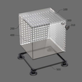
1 is a perspective view of a phantom device for managing the quality of a MRI image-guided radiotherapy apparatus according to an embodiment of the present invention.
FIG. 2 is an exploded perspective view of a phantom device for quality control of a MRI image-guided radiotherapy apparatus according to an embodiment of the present invention.
FIG. 3 is a cross-sectional view showing the inside of the
FIG. 4 is a view illustrating a state in which a phantom device for level management of a MRI image-guided radiotherapy apparatus according to an embodiment of the present invention is filled with water.
FIG. 5 is a side view of a phantom device for quality control of a MRI image-guided radiotherapy apparatus according to an embodiment of the present invention.
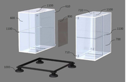
6 is a perspective view of a phantom device for quality control of a MRI image-guided radiotherapy apparatus according to another embodiment of the present invention.
FIG. 7 is an exploded perspective view of a phantom device for quality control of a MRI image-guided radiotherapy apparatus according to another embodiment of the present invention.
FIG. 8 is a top view of a phantom device for quality control of a MRI image-guided radiotherapy apparatus according to another embodiment of the present invention.
Hereinafter, preferred embodiments of the present invention will be described in detail with reference to the accompanying drawings, in order that those skilled in the art can easily carry out the present invention. The shapes, sizes, ratios, angles, numbers, and the like disclosed in the drawings for describing the embodiments of the present invention are illustrative, and thus the present invention is not limited thereto. In the following detailed description of the preferred embodiments of the present invention, a detailed description of known functions and configurations incorporated herein will be omitted when it may make the subject matter of the present invention rather unclear. In the drawings, like reference numerals are used throughout the drawings.
In addition, in the entire specification, when a part is referred to as being 'connected' to another part, it may be referred to as 'indirectly connected' not only with 'directly connected' . Also, to "include" an element means that it may include other elements, rather than excluding other elements, unless specifically stated otherwise.
FIGS. 1 to 5 show a phantom device for managing the quality of a MRI image-guided radiotherapy apparatus according to an embodiment of the present invention. FIGS. 6 and 7 are views showing a magnetic resonance imaging And a phantom device for quality control of an inductive radiotherapy apparatus.
By adopting such a configuration, it is possible to simultaneously verify image quality and radiation quality for periodic quality control of a magnetic resonance image-guided radiotherapy apparatus that performs radiation therapy in a magnetic field environment.
In the present invention, "magnetic resonance imaging (MRI)" is an examination method for diagnosing a disease by imaging electromagnetic waves reflected from a human body in a strong magnetic field by measuring electromagnetic waves reflected therefrom. In a large magnet cylinder generating a magnetic field, And then generating a high frequency to resonate the hydrogen nuclei in the body region, measuring the difference in signal from each tissue, reconstructing it through a computer, and imaging it.
Hereinafter, each component constituting the phantom device for the quality control of the MRI image-guided radiotherapy apparatus according to the present invention will be described in detail.
First, the phantom device for managing the quality of a MRI image-guided radiotherapy apparatus according to an embodiment of the present invention confirms the image spatial integrity of the most important spatial information in a radiotherapy image, FIG. 1 is a perspective view of a phantom device for quality control of a MRI image-guided radiotherapy apparatus according to an embodiment of the present invention, and FIG. 2 is a perspective view of a phantom device according to an embodiment of the present invention. FIG. 2 is an exploded perspective view of a phantom device for quality control of an inductive radiotherapy apparatus.
1 and 2, the phantom device for managing the quality of a MRI image-guided radiotherapy apparatus according to an embodiment of the present invention includes a
The
As shown in FIG. 3, the
The
5, the ion
As shown in FIG. 2, the
According to another aspect of the present invention, a phantom device for managing the quality of a MRI image-guided radiotherapy apparatus according to another embodiment of the present invention includes a virtual isocenter of a room laser alignment system, FIG. 6 is a perspective view of a phantom device for managing the quality of a MRI image-guiding radiotherapy apparatus according to another embodiment of the present invention, and FIG. FIG. 7 is an exploded perspective view of a phantom device for quality control of a MRI image-guided radiotherapy apparatus according to another embodiment of the present invention.
6 and 7, the phantom device for managing the quality of a MRI image-guided radiotherapy apparatus according to another embodiment of the present invention includes a
The
The
As shown in FIGS. 6 and 7, the radiation-
The
6 and 7, the
As shown in FIGS. 6 and 7, the first and second
Meanwhile, the phantom device for the quality control of the MRI image-guiding radiotherapy apparatus according to the present invention may perform various quality management items of various kinds of radiotherapy apparatuses in addition to the MRI image-guided radiotherapy apparatus. Herein, the radiation therapy apparatus may be, but not limited to, a radiation therapy apparatus that is directed to computed tomography (CT), single photon imaging (SPECT) or positron emission tomography (PET).
In the present invention, "computed tomography (CT)" is an inspection method for acquiring an image on the cross section of a human body using X-rays and using it for diagnosis.
In the present invention, "single photon imaging (SPECT)" is a method of obtaining a tomographic image of a living body after administering a tracer capable of observing biochemical and functional states such as brain perfusion, cardiac perfusion and bone metabolism, And the gamma ray emitted from the radionuclide located at the region of interest to be diagnosed is used as an expression mechanism to obtain an image. Therefore, it is possible to acquire an image by injecting a solution containing a radioisotope into a phantom device for the quality control of the radiotherapy apparatus according to the present invention.
In the present invention, "positron emission tomography (PET)" is a nuclear medicine imaging method in which a physiochemical and functional image of a human body is obtained in three dimensions using a radiopharmaceutical that emits positron. This is a device that can be used to check on the screen that the part consumes more glucose than the other parts. The PET is obtained by injecting a liquid isotope into a blood vessel and obtaining an image using the radiation emitted from the radioisotope. It shows the degree of metabolic activity of sugar, oxygen and protein that cause body changes, so that abnormal signs can be detected early I will. Therefore, it is possible to acquire an image by injecting a solution containing a radioisotope into a phantom device for the quality control of the radiotherapy apparatus according to the present invention.
It will be understood by those skilled in the art that the foregoing description of the present invention is for illustrative purposes only and that those of ordinary skill in the art can readily understand that various changes and modifications may be made without departing from the spirit or essential characteristics of the present invention. will be. It is therefore to be understood that the above-described embodiments are illustrative in all aspects and not restrictive.
100: main body 200: protrusion
300: injection part 310:
400: ion chamber 500: pedestal
600: first main body part 610: fastening part
700: second main body part 710: fastening groove
800: radiation sensitive film 900: supply part
1000: Support 1100: Grid groove
Claims (12)
A main body 100 having a hollow hexahedral shape;
A protrusion 200 protruding from the inner wall of the main body 100 at a predetermined interval;
An injection unit 300 formed on the side wall of the main body 100 and capable of supplying liquid into the main body 100; And
And an ion chamber accommodating part (400) formed on a side wall of the main body part (100) for providing a space through which the ion chamber can be inserted. A phantom for quality control of a magnetic resonance imaging radiotherapy apparatus Device.
Further comprising a pedestal (500) for separating the main body part (100) from the ground and adjusting the level of the main body part (100) Device.
Characterized in that the apparatus is made of acrylic material.
Further comprising a stopper (310) for sealing the opening of the injection unit (300).
Is characterized in that it is used for the quality control of a radiotherapeutic apparatus derived from any one selected from the group consisting of magnetic resonance imaging (MRI), computed tomography (CT), single photon imaging (SPECT) and positron emission tomography (PET) A phantom device for quality control of magnetic resonance imaging radiation therapy devices.
A first body part 600 and a second body part 700 having an inner hollow body shape;
And a radiation sensitive film (800) disposed between the first and second body parts (600, 700) and capable of measuring a radiation dose distribution. A phantom device for.
Further comprising a supply part (900) formed through the side walls of the first and second body parts (600, 700) and capable of supplying liquid into the first and second body parts (100) A phantom device for quality control of resonance image guided radiotherapy apparatus.
Further comprising a support 1000 for separating the first and second body portions 600 and 700 from the ground and adjusting the level of the first and second body portions 600 and 700 , Phantom device for the quality control of magnetic resonance image guided radiotherapy device.
The apparatus according to claim 1, further comprising lattice grooves (1100) formed at the center of the upper and lower surfaces and the three outer surfaces of the first and second body parts (600, 700) Phantom device.
Is characterized in that it is used for the quality control of a radiotherapeutic apparatus derived from any one selected from the group consisting of magnetic resonance imaging (MRI), computed tomography (CT), single photon imaging (SPECT) and positron emission tomography (PET) A phantom device for quality control of magnetic resonance imaging radiation therapy devices.
Priority Applications (1)
| Application Number | Priority Date | Filing Date | Title |
|---|---|---|---|
| KR1020160017945A KR101777499B1 (en) | 2016-02-16 | 2016-02-16 | Phantoms for Quality Assurance of Magnetic Resonance Image Guided Radiation Therapy Machine |
Applications Claiming Priority (1)
| Application Number | Priority Date | Filing Date | Title |
|---|---|---|---|
| KR1020160017945A KR101777499B1 (en) | 2016-02-16 | 2016-02-16 | Phantoms for Quality Assurance of Magnetic Resonance Image Guided Radiation Therapy Machine |
Publications (2)
| Publication Number | Publication Date |
|---|---|
| KR20170096497A true KR20170096497A (en) | 2017-08-24 |
| KR101777499B1 KR101777499B1 (en) | 2017-09-11 |
Family
ID=59758391
Family Applications (1)
| Application Number | Title | Priority Date | Filing Date |
|---|---|---|---|
| KR1020160017945A KR101777499B1 (en) | 2016-02-16 | 2016-02-16 | Phantoms for Quality Assurance of Magnetic Resonance Image Guided Radiation Therapy Machine |
Country Status (1)
| Country | Link |
|---|---|
| KR (1) | KR101777499B1 (en) |
Cited By (1)
| Publication number | Priority date | Publication date | Assignee | Title |
|---|---|---|---|---|
| US12070623B2 (en) | 2018-10-12 | 2024-08-27 | Elekta Ltd. | Quality assurance for MR-Linac |
Family Cites Families (3)
| Publication number | Priority date | Publication date | Assignee | Title |
|---|---|---|---|---|
| KR200347702Y1 (en) * | 2004-01-13 | 2004-04-13 | 학교법인 가톨릭학원 | Phantom for verification of accuracy of HDR brachytherapy planning and Phantom device having the phantom |
| KR200427116Y1 (en) * | 2006-07-11 | 2006-09-20 | 가톨릭대학교 산학협력단 | Holder device for analysis dosimeter |
| KR101501408B1 (en) * | 2014-02-13 | 2015-03-12 | 가톨릭대학교 산학협력단 | Voxel-based block phantom for multi functional radiation detection |
-
2016
- 2016-02-16 KR KR1020160017945A patent/KR101777499B1/en active IP Right Grant
Cited By (1)
| Publication number | Priority date | Publication date | Assignee | Title |
|---|---|---|---|---|
| US12070623B2 (en) | 2018-10-12 | 2024-08-27 | Elekta Ltd. | Quality assurance for MR-Linac |
Also Published As
| Publication number | Publication date |
|---|---|
| KR101777499B1 (en) | 2017-09-11 |
Similar Documents
| Publication | Publication Date | Title |
|---|---|---|
| JP6774481B2 (en) | Systems and methods for image guidance during medical procedures | |
| US20150208994A1 (en) | Ct/mri integrated system for the diagnosis of acute strokes and methods thereof | |
| RU2587077C2 (en) | System for planning radiation therapy and subsequent observation with wide-channel radionuclide and magnetic resonance imaging or wide-channel computer tomography and magnetic resonance imaging | |
| Lecchi et al. | Current concepts on imaging in radiotherapy | |
| US20120150017A1 (en) | Open pet/mri hybrid machine | |
| Bostel et al. | MR-guidance–a clinical study to evaluate a shuttle-based MR-linac connection to provide MR-guided radiotherapy | |
| US20070153969A1 (en) | Radiotherapeutic device | |
| Zou et al. | Current state of image guidance in radiation oncology: implications for PTV margin expansion and adaptive therapy | |
| KR101777499B1 (en) | Phantoms for Quality Assurance of Magnetic Resonance Image Guided Radiation Therapy Machine | |
| Abolaban | On board cone beam CT for treatment planning in image guided radiotherapy | |
| EP0409920A1 (en) | Device for interfacing mri with other imaging modalities | |
| Ng et al. | Medical physics contributes to the advancement in medicine | |
| KR101653095B1 (en) | The phantom for measuring artifact generated by magnetic susceptibility difference in magnetic resonance image | |
| Ogino et al. | Calcium phosphate cement paste injection as a fiducial marker of cervical cancer | |
| AU2017204730B2 (en) | System and method for image guidance during medical procedures | |
| Jain et al. | Current Status of Radiological Multimodality Imaging | |
| Tae-Suk | Imaging in radiation therapy | |
| Rowe et al. | Relative skeletal distribution of proliferating marrow in the adult dog determined using 3′‐deoxy‐3′‐[18F] fluorothymidine | |
| Latif et al. | technology and medical imaging | |
| de Waard-Schalkx et al. | Recent developments in medical techniques involving ionising or non-ionising radiation: update 2014 | |
| lu Yang et al. | 3D CT simulation and treatment planning system for radiotherapy | |
| Antolini et al. | Physics & Medicine: Toward a Future of Integration: Trento, November 6th–8th, 2014 | |
| Amestoy et al. | Imaging Modalities | |
| Nuyts | Use of Imaging Data in Radiotherapy Planning of Head and Neck Cancer: Improved Tumour Characterization, Delineation and Treatment Verification | |
| Wiant et al. | TECHNICAL ADVANCE Gamma KnifeTM Radiosurgery Treatment Planning for Small Animals using High-Resolution 7T Micro-magnetic Resonance Imaging |
Legal Events
| Date | Code | Title | Description |
|---|---|---|---|
| E701 | Decision to grant or registration of patent right | ||
| GRNT | Written decision to grant |