KR20170054445A - Methods and compositions for nuclease-mediated genome engineering and correction in hematopoietic stem cells - Google Patents
Methods and compositions for nuclease-mediated genome engineering and correction in hematopoietic stem cells Download PDFInfo
- Publication number
- KR20170054445A KR20170054445A KR1020177009275A KR20177009275A KR20170054445A KR 20170054445 A KR20170054445 A KR 20170054445A KR 1020177009275 A KR1020177009275 A KR 1020177009275A KR 20177009275 A KR20177009275 A KR 20177009275A KR 20170054445 A KR20170054445 A KR 20170054445A
- Authority
- KR
- South Korea
- Prior art keywords
- cell
- cells
- gene
- dna
- zfn
- Prior art date
Links
- 238000000034 method Methods 0.000 title claims abstract description 79
- 239000000203 mixture Substances 0.000 title claims abstract description 27
- 210000003958 hematopoietic stem cell Anatomy 0.000 title claims abstract description 17
- 238000010362 genome editing Methods 0.000 title claims abstract description 12
- 101710163270 Nuclease Proteins 0.000 title claims description 118
- 238000012937 correction Methods 0.000 title description 31
- 230000001404 mediated effect Effects 0.000 title description 25
- 108090000623 proteins and genes Proteins 0.000 claims abstract description 237
- 102000004169 proteins and genes Human genes 0.000 claims abstract description 87
- 230000014509 gene expression Effects 0.000 claims abstract description 52
- 210000004027 cell Anatomy 0.000 claims description 374
- 108010017070 Zinc Finger Nucleases Proteins 0.000 claims description 192
- 210000003743 erythrocyte Anatomy 0.000 claims description 49
- 241000282414 Homo sapiens Species 0.000 claims description 45
- 210000000130 stem cell Anatomy 0.000 claims description 44
- 108091034117 Oligonucleotide Proteins 0.000 claims description 39
- 230000035772 mutation Effects 0.000 claims description 38
- 239000002157 polynucleotide Substances 0.000 claims description 37
- 102000040430 polynucleotide Human genes 0.000 claims description 37
- 108091033319 polynucleotide Proteins 0.000 claims description 37
- 230000010354 integration Effects 0.000 claims description 35
- 108020004999 messenger RNA Proteins 0.000 claims description 33
- 238000003780 insertion Methods 0.000 claims description 27
- 230000037431 insertion Effects 0.000 claims description 27
- 108700019146 Transgenes Proteins 0.000 claims description 25
- HCHKCACWOHOZIP-UHFFFAOYSA-N Zinc Chemical compound [Zn] HCHKCACWOHOZIP-UHFFFAOYSA-N 0.000 claims description 25
- 239000011701 zinc Substances 0.000 claims description 25
- 229910052725 zinc Inorganic materials 0.000 claims description 25
- 108020001507 fusion proteins Proteins 0.000 claims description 23
- 102000037865 fusion proteins Human genes 0.000 claims description 23
- 101710185494 Zinc finger protein Proteins 0.000 claims description 21
- 102100023597 Zinc finger protein 816 Human genes 0.000 claims description 21
- 238000012239 gene modification Methods 0.000 claims description 20
- 239000002243 precursor Substances 0.000 claims description 17
- 230000005017 genetic modification Effects 0.000 claims description 15
- 235000013617 genetically modified food Nutrition 0.000 claims description 15
- 230000001965 increasing effect Effects 0.000 claims description 15
- 238000012217 deletion Methods 0.000 claims description 14
- 230000037430 deletion Effects 0.000 claims description 14
- 101150013707 HBB gene Proteins 0.000 claims description 9
- 239000008194 pharmaceutical composition Substances 0.000 claims description 6
- 102000004127 Cytokines Human genes 0.000 claims description 4
- 108090000695 Cytokines Proteins 0.000 claims description 4
- 238000012545 processing Methods 0.000 claims description 4
- 108020005067 RNA Splice Sites Proteins 0.000 claims description 2
- 102100031573 Hematopoietic progenitor cell antigen CD34 Human genes 0.000 claims 1
- 101000777663 Homo sapiens Hematopoietic progenitor cell antigen CD34 Proteins 0.000 claims 1
- 230000002349 favourable effect Effects 0.000 claims 1
- 230000004936 stimulating effect Effects 0.000 claims 1
- 230000001131 transforming effect Effects 0.000 claims 1
- 208000007056 sickle cell anemia Diseases 0.000 abstract description 29
- 238000001415 gene therapy Methods 0.000 abstract description 18
- 208000037265 diseases, disorders, signs and symptoms Diseases 0.000 abstract description 12
- 230000002950 deficient Effects 0.000 abstract description 9
- 201000010099 disease Diseases 0.000 abstract description 9
- 230000009466 transformation Effects 0.000 abstract description 9
- 208000026350 Inborn Genetic disease Diseases 0.000 abstract description 3
- 208000016361 genetic disease Diseases 0.000 abstract description 3
- 108020004414 DNA Proteins 0.000 description 122
- 238000003776 cleavage reaction Methods 0.000 description 121
- 230000007017 scission Effects 0.000 description 118
- 150000007523 nucleic acids Chemical class 0.000 description 87
- 235000018102 proteins Nutrition 0.000 description 79
- 102000039446 nucleic acids Human genes 0.000 description 68
- 108020004707 nucleic acids Proteins 0.000 description 68
- 230000027455 binding Effects 0.000 description 56
- 108090000765 processed proteins & peptides Proteins 0.000 description 55
- 239000013598 vector Substances 0.000 description 52
- 102100021519 Hemoglobin subunit beta Human genes 0.000 description 47
- 108091005904 Hemoglobin subunit beta Proteins 0.000 description 45
- 238000004458 analytical method Methods 0.000 description 41
- 230000004568 DNA-binding Effects 0.000 description 39
- 102000004196 processed proteins & peptides Human genes 0.000 description 37
- 241000699670 Mus sp. Species 0.000 description 35
- 210000001185 bone marrow Anatomy 0.000 description 35
- 238000002054 transplantation Methods 0.000 description 35
- 125000003729 nucleotide group Chemical group 0.000 description 34
- 239000002773 nucleotide Substances 0.000 description 33
- 229920001184 polypeptide Polymers 0.000 description 33
- 108091033409 CRISPR Proteins 0.000 description 32
- 102000001554 Hemoglobins Human genes 0.000 description 31
- 108010054147 Hemoglobins Proteins 0.000 description 31
- 239000005090 green fluorescent protein Substances 0.000 description 30
- 238000000338 in vitro Methods 0.000 description 29
- 238000010354 CRISPR gene editing Methods 0.000 description 28
- 230000005782 double-strand break Effects 0.000 description 23
- 230000006780 non-homologous end joining Effects 0.000 description 23
- 101001105486 Homo sapiens Proteasome subunit alpha type-7 Proteins 0.000 description 21
- 102100021201 Proteasome subunit alpha type-7 Human genes 0.000 description 21
- 108091027544 Subgenomic mRNA Proteins 0.000 description 21
- 238000004520 electroporation Methods 0.000 description 21
- 108060003196 globin Proteins 0.000 description 21
- 239000010432 diamond Substances 0.000 description 20
- 230000000694 effects Effects 0.000 description 20
- 239000000047 product Substances 0.000 description 20
- 108700028369 Alleles Proteins 0.000 description 19
- 101000899111 Homo sapiens Hemoglobin subunit beta Proteins 0.000 description 19
- 210000003483 chromatin Anatomy 0.000 description 19
- 238000002474 experimental method Methods 0.000 description 19
- 230000004927 fusion Effects 0.000 description 18
- 238000007894 restriction fragment length polymorphism technique Methods 0.000 description 18
- 241000700605 Viruses Species 0.000 description 17
- 150000001413 amino acids Chemical class 0.000 description 16
- 210000003995 blood forming stem cell Anatomy 0.000 description 16
- 239000012634 fragment Substances 0.000 description 16
- 230000006870 function Effects 0.000 description 16
- 239000000523 sample Substances 0.000 description 16
- 108010042407 Endonucleases Proteins 0.000 description 15
- 210000004369 blood Anatomy 0.000 description 15
- 239000008280 blood Substances 0.000 description 15
- 230000004048 modification Effects 0.000 description 15
- 238000012986 modification Methods 0.000 description 15
- 210000001519 tissue Anatomy 0.000 description 15
- 102000007513 Hemoglobin A Human genes 0.000 description 14
- 108010085682 Hemoglobin A Proteins 0.000 description 14
- 230000004069 differentiation Effects 0.000 description 14
- 235000001014 amino acid Nutrition 0.000 description 13
- 210000004700 fetal blood Anatomy 0.000 description 13
- 238000001727 in vivo Methods 0.000 description 13
- 210000005259 peripheral blood Anatomy 0.000 description 13
- 239000011886 peripheral blood Substances 0.000 description 13
- 230000009261 transgenic effect Effects 0.000 description 13
- 239000013603 viral vector Substances 0.000 description 13
- 230000003612 virological effect Effects 0.000 description 13
- 102000004190 Enzymes Human genes 0.000 description 12
- 108090000790 Enzymes Proteins 0.000 description 12
- 238000004128 high performance liquid chromatography Methods 0.000 description 12
- 239000013612 plasmid Substances 0.000 description 12
- 108010077544 Chromatin Proteins 0.000 description 11
- 108091026890 Coding region Proteins 0.000 description 11
- 102100031780 Endonuclease Human genes 0.000 description 11
- 102100038617 Hemoglobin subunit gamma-2 Human genes 0.000 description 11
- JLCPHMBAVCMARE-UHFFFAOYSA-N [3-[[3-[[3-[[3-[[3-[[3-[[3-[[3-[[3-[[3-[[3-[[5-(2-amino-6-oxo-1H-purin-9-yl)-3-[[3-[[3-[[3-[[3-[[3-[[5-(2-amino-6-oxo-1H-purin-9-yl)-3-[[5-(2-amino-6-oxo-1H-purin-9-yl)-3-hydroxyoxolan-2-yl]methoxy-hydroxyphosphoryl]oxyoxolan-2-yl]methoxy-hydroxyphosphoryl]oxy-5-(5-methyl-2,4-dioxopyrimidin-1-yl)oxolan-2-yl]methoxy-hydroxyphosphoryl]oxy-5-(6-aminopurin-9-yl)oxolan-2-yl]methoxy-hydroxyphosphoryl]oxy-5-(6-aminopurin-9-yl)oxolan-2-yl]methoxy-hydroxyphosphoryl]oxy-5-(6-aminopurin-9-yl)oxolan-2-yl]methoxy-hydroxyphosphoryl]oxy-5-(6-aminopurin-9-yl)oxolan-2-yl]methoxy-hydroxyphosphoryl]oxyoxolan-2-yl]methoxy-hydroxyphosphoryl]oxy-5-(5-methyl-2,4-dioxopyrimidin-1-yl)oxolan-2-yl]methoxy-hydroxyphosphoryl]oxy-5-(4-amino-2-oxopyrimidin-1-yl)oxolan-2-yl]methoxy-hydroxyphosphoryl]oxy-5-(5-methyl-2,4-dioxopyrimidin-1-yl)oxolan-2-yl]methoxy-hydroxyphosphoryl]oxy-5-(5-methyl-2,4-dioxopyrimidin-1-yl)oxolan-2-yl]methoxy-hydroxyphosphoryl]oxy-5-(6-aminopurin-9-yl)oxolan-2-yl]methoxy-hydroxyphosphoryl]oxy-5-(6-aminopurin-9-yl)oxolan-2-yl]methoxy-hydroxyphosphoryl]oxy-5-(4-amino-2-oxopyrimidin-1-yl)oxolan-2-yl]methoxy-hydroxyphosphoryl]oxy-5-(4-amino-2-oxopyrimidin-1-yl)oxolan-2-yl]methoxy-hydroxyphosphoryl]oxy-5-(4-amino-2-oxopyrimidin-1-yl)oxolan-2-yl]methoxy-hydroxyphosphoryl]oxy-5-(6-aminopurin-9-yl)oxolan-2-yl]methoxy-hydroxyphosphoryl]oxy-5-(4-amino-2-oxopyrimidin-1-yl)oxolan-2-yl]methyl [5-(6-aminopurin-9-yl)-2-(hydroxymethyl)oxolan-3-yl] hydrogen phosphate Polymers Cc1cn(C2CC(OP(O)(=O)OCC3OC(CC3OP(O)(=O)OCC3OC(CC3O)n3cnc4c3nc(N)[nH]c4=O)n3cnc4c3nc(N)[nH]c4=O)C(COP(O)(=O)OC3CC(OC3COP(O)(=O)OC3CC(OC3COP(O)(=O)OC3CC(OC3COP(O)(=O)OC3CC(OC3COP(O)(=O)OC3CC(OC3COP(O)(=O)OC3CC(OC3COP(O)(=O)OC3CC(OC3COP(O)(=O)OC3CC(OC3COP(O)(=O)OC3CC(OC3COP(O)(=O)OC3CC(OC3COP(O)(=O)OC3CC(OC3COP(O)(=O)OC3CC(OC3COP(O)(=O)OC3CC(OC3COP(O)(=O)OC3CC(OC3COP(O)(=O)OC3CC(OC3COP(O)(=O)OC3CC(OC3COP(O)(=O)OC3CC(OC3CO)n3cnc4c(N)ncnc34)n3ccc(N)nc3=O)n3cnc4c(N)ncnc34)n3ccc(N)nc3=O)n3ccc(N)nc3=O)n3ccc(N)nc3=O)n3cnc4c(N)ncnc34)n3cnc4c(N)ncnc34)n3cc(C)c(=O)[nH]c3=O)n3cc(C)c(=O)[nH]c3=O)n3ccc(N)nc3=O)n3cc(C)c(=O)[nH]c3=O)n3cnc4c3nc(N)[nH]c4=O)n3cnc4c(N)ncnc34)n3cnc4c(N)ncnc34)n3cnc4c(N)ncnc34)n3cnc4c(N)ncnc34)O2)c(=O)[nH]c1=O JLCPHMBAVCMARE-UHFFFAOYSA-N 0.000 description 11
- 210000000349 chromosome Anatomy 0.000 description 11
- PZNPLUBHRSSFHT-RRHRGVEJSA-N 1-hexadecanoyl-2-octadecanoyl-sn-glycero-3-phosphocholine Chemical compound CCCCCCCCCCCCCCCCCC(=O)O[C@@H](COP([O-])(=O)OCC[N+](C)(C)C)COC(=O)CCCCCCCCCCCCCCC PZNPLUBHRSSFHT-RRHRGVEJSA-N 0.000 description 10
- 238000005516 engineering process Methods 0.000 description 10
- 102000018146 globin Human genes 0.000 description 10
- 238000004806 packaging method and process Methods 0.000 description 10
- 108091008146 restriction endonucleases Proteins 0.000 description 10
- 230000037432 silent mutation Effects 0.000 description 10
- 238000013518 transcription Methods 0.000 description 10
- 230000035897 transcription Effects 0.000 description 10
- 238000010361 transduction Methods 0.000 description 10
- 230000026683 transduction Effects 0.000 description 10
- 241000701161 unidentified adenovirus Species 0.000 description 10
- 102100039894 Hemoglobin subunit delta Human genes 0.000 description 9
- 241001465754 Metazoa Species 0.000 description 9
- 241000699666 Mus <mouse, genus> Species 0.000 description 9
- 238000013461 design Methods 0.000 description 9
- 238000001476 gene delivery Methods 0.000 description 9
- 230000008439 repair process Effects 0.000 description 9
- 210000000952 spleen Anatomy 0.000 description 9
- 241000196324 Embryophyta Species 0.000 description 8
- 108010044495 Fetal Hemoglobin Proteins 0.000 description 8
- 108020005004 Guide RNA Proteins 0.000 description 8
- 108091028043 Nucleic acid sequence Proteins 0.000 description 8
- 238000003556 assay Methods 0.000 description 8
- 230000010261 cell growth Effects 0.000 description 8
- 230000003394 haemopoietic effect Effects 0.000 description 8
- 230000006801 homologous recombination Effects 0.000 description 8
- 238000002744 homologous recombination Methods 0.000 description 8
- 210000005260 human cell Anatomy 0.000 description 8
- 238000004519 manufacturing process Methods 0.000 description 8
- 230000007246 mechanism Effects 0.000 description 8
- 230000037361 pathway Effects 0.000 description 8
- UCSJYZPVAKXKNQ-HZYVHMACSA-N streptomycin Chemical compound CN[C@H]1[C@H](O)[C@@H](O)[C@H](CO)O[C@H]1O[C@@H]1[C@](C=O)(O)[C@H](C)O[C@H]1O[C@@H]1[C@@H](NC(N)=N)[C@H](O)[C@@H](NC(N)=N)[C@H](O)[C@H]1O UCSJYZPVAKXKNQ-HZYVHMACSA-N 0.000 description 8
- 230000007018 DNA scission Effects 0.000 description 7
- 102000052510 DNA-Binding Proteins Human genes 0.000 description 7
- 102100038614 Hemoglobin subunit gamma-1 Human genes 0.000 description 7
- 101001031977 Homo sapiens Hemoglobin subunit gamma-1 Proteins 0.000 description 7
- 101000738771 Homo sapiens Receptor-type tyrosine-protein phosphatase C Proteins 0.000 description 7
- 241000713666 Lentivirus Species 0.000 description 7
- 102100037422 Receptor-type tyrosine-protein phosphatase C Human genes 0.000 description 7
- 210000001744 T-lymphocyte Anatomy 0.000 description 7
- 108091028113 Trans-activating crRNA Proteins 0.000 description 7
- 108010073062 Transcription Activator-Like Effectors Proteins 0.000 description 7
- 230000004913 activation Effects 0.000 description 7
- 238000013459 approach Methods 0.000 description 7
- 230000008901 benefit Effects 0.000 description 7
- 230000001413 cellular effect Effects 0.000 description 7
- 230000002068 genetic effect Effects 0.000 description 7
- 230000003993 interaction Effects 0.000 description 7
- 238000012177 large-scale sequencing Methods 0.000 description 7
- 150000002632 lipids Chemical class 0.000 description 7
- 239000002609 medium Substances 0.000 description 7
- 238000005457 optimization Methods 0.000 description 7
- 230000008569 process Effects 0.000 description 7
- 230000001105 regulatory effect Effects 0.000 description 7
- 238000012163 sequencing technique Methods 0.000 description 7
- 125000006850 spacer group Chemical group 0.000 description 7
- -1 valine amino acid Chemical class 0.000 description 7
- 108700020911 DNA-Binding Proteins Proteins 0.000 description 6
- WHUUTDBJXJRKMK-UHFFFAOYSA-N Glutamic acid Natural products OC(=O)C(N)CCC(O)=O WHUUTDBJXJRKMK-UHFFFAOYSA-N 0.000 description 6
- 108091005886 Hemoglobin subunit gamma Proteins 0.000 description 6
- 101001031961 Homo sapiens Hemoglobin subunit gamma-2 Proteins 0.000 description 6
- 241000589634 Xanthomonas Species 0.000 description 6
- 210000003719 b-lymphocyte Anatomy 0.000 description 6
- 239000003153 chemical reaction reagent Substances 0.000 description 6
- 239000003795 chemical substances by application Substances 0.000 description 6
- 230000001332 colony forming effect Effects 0.000 description 6
- 230000000875 corresponding effect Effects 0.000 description 6
- 229910003460 diamond Inorganic materials 0.000 description 6
- 239000000499 gel Substances 0.000 description 6
- 239000001963 growth medium Substances 0.000 description 6
- 230000000670 limiting effect Effects 0.000 description 6
- 239000003550 marker Substances 0.000 description 6
- 230000036961 partial effect Effects 0.000 description 6
- 230000001177 retroviral effect Effects 0.000 description 6
- 238000006467 substitution reaction Methods 0.000 description 6
- 230000002103 transcriptional effect Effects 0.000 description 6
- 238000001712 DNA sequencing Methods 0.000 description 5
- 108091005903 Hemoglobin subunit delta Proteins 0.000 description 5
- 101000934338 Homo sapiens Myeloid cell surface antigen CD33 Proteins 0.000 description 5
- 101000581981 Homo sapiens Neural cell adhesion molecule 1 Proteins 0.000 description 5
- 102100025243 Myeloid cell surface antigen CD33 Human genes 0.000 description 5
- 102100027347 Neural cell adhesion molecule 1 Human genes 0.000 description 5
- 230000015556 catabolic process Effects 0.000 description 5
- 230000008859 change Effects 0.000 description 5
- 238000006243 chemical reaction Methods 0.000 description 5
- 230000029087 digestion Effects 0.000 description 5
- 239000003623 enhancer Substances 0.000 description 5
- 210000003714 granulocyte Anatomy 0.000 description 5
- 238000002347 injection Methods 0.000 description 5
- 239000007924 injection Substances 0.000 description 5
- 239000002502 liposome Substances 0.000 description 5
- 230000007774 longterm Effects 0.000 description 5
- 210000005087 mononuclear cell Anatomy 0.000 description 5
- 210000000822 natural killer cell Anatomy 0.000 description 5
- 239000000126 substance Substances 0.000 description 5
- 241001430294 unidentified retrovirus Species 0.000 description 5
- 108700026220 vif Genes Proteins 0.000 description 5
- CURLTUGMZLYLDI-UHFFFAOYSA-N Carbon dioxide Chemical compound O=C=O CURLTUGMZLYLDI-UHFFFAOYSA-N 0.000 description 4
- 102000053602 DNA Human genes 0.000 description 4
- 102000004533 Endonucleases Human genes 0.000 description 4
- 206010018901 Haemoglobinaemia Diseases 0.000 description 4
- 102100030826 Hemoglobin subunit epsilon Human genes 0.000 description 4
- 101001083591 Homo sapiens Hemoglobin subunit epsilon Proteins 0.000 description 4
- 102000004372 Insulin-like growth factor binding protein 2 Human genes 0.000 description 4
- 108090000964 Insulin-like growth factor binding protein 2 Proteins 0.000 description 4
- ZDXPYRJPNDTMRX-VKHMYHEASA-N L-glutamine Chemical compound OC(=O)[C@@H](N)CCC(N)=O ZDXPYRJPNDTMRX-VKHMYHEASA-N 0.000 description 4
- 108010047956 Nucleosomes Proteins 0.000 description 4
- 229930182555 Penicillin Natural products 0.000 description 4
- JGSARLDLIJGVTE-MBNYWOFBSA-N Penicillin G Chemical compound N([C@H]1[C@H]2SC([C@@H](N2C1=O)C(O)=O)(C)C)C(=O)CC1=CC=CC=C1 JGSARLDLIJGVTE-MBNYWOFBSA-N 0.000 description 4
- 108010016797 Sickle Hemoglobin Proteins 0.000 description 4
- 241000589499 Thermus thermophilus Species 0.000 description 4
- 238000010521 absorption reaction Methods 0.000 description 4
- 230000015572 biosynthetic process Effects 0.000 description 4
- 238000010276 construction Methods 0.000 description 4
- 230000001419 dependent effect Effects 0.000 description 4
- 238000009826 distribution Methods 0.000 description 4
- 239000012636 effector Substances 0.000 description 4
- 238000001943 fluorescence-activated cell sorting Methods 0.000 description 4
- 230000002779 inactivation Effects 0.000 description 4
- 238000011534 incubation Methods 0.000 description 4
- 238000001638 lipofection Methods 0.000 description 4
- 210000002540 macrophage Anatomy 0.000 description 4
- 229920000609 methyl cellulose Polymers 0.000 description 4
- 239000001923 methylcellulose Substances 0.000 description 4
- 210000001623 nucleosome Anatomy 0.000 description 4
- 229940049954 penicillin Drugs 0.000 description 4
- 229920000642 polymer Polymers 0.000 description 4
- 102000005962 receptors Human genes 0.000 description 4
- 108020003175 receptors Proteins 0.000 description 4
- 230000006798 recombination Effects 0.000 description 4
- 238000005215 recombination Methods 0.000 description 4
- 230000010076 replication Effects 0.000 description 4
- 241000894007 species Species 0.000 description 4
- 229960005322 streptomycin Drugs 0.000 description 4
- 230000008685 targeting Effects 0.000 description 4
- 238000002560 therapeutic procedure Methods 0.000 description 4
- 238000013519 translation Methods 0.000 description 4
- 239000013607 AAV vector Substances 0.000 description 3
- 102000014914 Carrier Proteins Human genes 0.000 description 3
- 230000033616 DNA repair Effects 0.000 description 3
- 241000702421 Dependoparvovirus Species 0.000 description 3
- LFQSCWFLJHTTHZ-UHFFFAOYSA-N Ethanol Chemical compound CCO LFQSCWFLJHTTHZ-UHFFFAOYSA-N 0.000 description 3
- PEDCQBHIVMGVHV-UHFFFAOYSA-N Glycerine Chemical compound OCC(O)CO PEDCQBHIVMGVHV-UHFFFAOYSA-N 0.000 description 3
- 102100027685 Hemoglobin subunit alpha Human genes 0.000 description 3
- 108091005902 Hemoglobin subunit alpha Proteins 0.000 description 3
- 101001000998 Homo sapiens Protein phosphatase 1 regulatory subunit 12C Proteins 0.000 description 3
- 102100034343 Integrase Human genes 0.000 description 3
- 108010061833 Integrases Proteins 0.000 description 3
- 108091061960 Naked DNA Proteins 0.000 description 3
- 102100035620 Protein phosphatase 1 regulatory subunit 12C Human genes 0.000 description 3
- 241000700159 Rattus Species 0.000 description 3
- 108700008625 Reporter Genes Proteins 0.000 description 3
- 240000004808 Saccharomyces cerevisiae Species 0.000 description 3
- 108091023040 Transcription factor Proteins 0.000 description 3
- 102000040945 Transcription factor Human genes 0.000 description 3
- KZSNJWFQEVHDMF-UHFFFAOYSA-N Valine Natural products CC(C)C(N)C(O)=O KZSNJWFQEVHDMF-UHFFFAOYSA-N 0.000 description 3
- 230000004075 alteration Effects 0.000 description 3
- 239000000427 antigen Substances 0.000 description 3
- 108091007433 antigens Proteins 0.000 description 3
- 102000036639 antigens Human genes 0.000 description 3
- QVGXLLKOCUKJST-UHFFFAOYSA-N atomic oxygen Chemical compound [O] QVGXLLKOCUKJST-UHFFFAOYSA-N 0.000 description 3
- 108091008324 binding proteins Proteins 0.000 description 3
- 230000004071 biological effect Effects 0.000 description 3
- 239000000872 buffer Substances 0.000 description 3
- 230000024245 cell differentiation Effects 0.000 description 3
- 238000012512 characterization method Methods 0.000 description 3
- 239000002299 complementary DNA Substances 0.000 description 3
- OPTASPLRGRRNAP-UHFFFAOYSA-N cytosine Chemical compound NC=1C=CNC(=O)N=1 OPTASPLRGRRNAP-UHFFFAOYSA-N 0.000 description 3
- 238000006471 dimerization reaction Methods 0.000 description 3
- 208000035475 disorder Diseases 0.000 description 3
- 230000034431 double-strand break repair via homologous recombination Effects 0.000 description 3
- 210000000267 erythroid cell Anatomy 0.000 description 3
- 230000000925 erythroid effect Effects 0.000 description 3
- 238000011156 evaluation Methods 0.000 description 3
- 239000012091 fetal bovine serum Substances 0.000 description 3
- 238000000684 flow cytometry Methods 0.000 description 3
- 108091008053 gene clusters Proteins 0.000 description 3
- 230000030279 gene silencing Effects 0.000 description 3
- 238000011331 genomic analysis Methods 0.000 description 3
- ZDXPYRJPNDTMRX-UHFFFAOYSA-N glutamine Natural products OC(=O)C(N)CCC(N)=O ZDXPYRJPNDTMRX-UHFFFAOYSA-N 0.000 description 3
- 229960002743 glutamine Drugs 0.000 description 3
- 239000000833 heterodimer Substances 0.000 description 3
- 230000001939 inductive effect Effects 0.000 description 3
- 208000015181 infectious disease Diseases 0.000 description 3
- 239000012212 insulator Substances 0.000 description 3
- 238000010255 intramuscular injection Methods 0.000 description 3
- 239000007927 intramuscular injection Substances 0.000 description 3
- 238000010253 intravenous injection Methods 0.000 description 3
- 208000032839 leukemia Diseases 0.000 description 3
- 239000003446 ligand Substances 0.000 description 3
- 239000007788 liquid Substances 0.000 description 3
- 210000004962 mammalian cell Anatomy 0.000 description 3
- 230000001394 metastastic effect Effects 0.000 description 3
- 206010061289 metastatic neoplasm Diseases 0.000 description 3
- 230000011987 methylation Effects 0.000 description 3
- 238000007069 methylation reaction Methods 0.000 description 3
- 229910052760 oxygen Inorganic materials 0.000 description 3
- 239000001301 oxygen Substances 0.000 description 3
- 239000002245 particle Substances 0.000 description 3
- 125000002467 phosphate group Chemical group [H]OP(=O)(O[H])O[*] 0.000 description 3
- 230000008488 polyadenylation Effects 0.000 description 3
- 230000004044 response Effects 0.000 description 3
- 230000000717 retained effect Effects 0.000 description 3
- 238000010561 standard procedure Methods 0.000 description 3
- 238000003786 synthesis reaction Methods 0.000 description 3
- 230000001225 therapeutic effect Effects 0.000 description 3
- 238000001890 transfection Methods 0.000 description 3
- 230000032258 transport Effects 0.000 description 3
- 239000004474 valine Substances 0.000 description 3
- 239000003981 vehicle Substances 0.000 description 3
- XLYOFNOQVPJJNP-UHFFFAOYSA-N water Substances O XLYOFNOQVPJJNP-UHFFFAOYSA-N 0.000 description 3
- 108091032973 (ribonucleotides)n+m Proteins 0.000 description 2
- 102000040650 (ribonucleotides)n+m Human genes 0.000 description 2
- QAOBBBBDJSWHMU-WMBBNPMCSA-N 16,16-dimethylprostaglandin E2 Chemical compound CCCCC(C)(C)[C@H](O)\C=C\[C@H]1[C@H](O)CC(=O)[C@@H]1C\C=C/CCCC(O)=O QAOBBBBDJSWHMU-WMBBNPMCSA-N 0.000 description 2
- AZXXGVPWWKWGAE-UHFFFAOYSA-N 4-n-[2-benzyl-7-(2-methyltetrazol-5-yl)-9h-pyrimido[4,5-b]indol-4-yl]cyclohexane-1,4-diamine Chemical compound CN1N=NC(C=2C=C3C(C4=C(NC5CCC(N)CC5)N=C(CC=5C=CC=CC=5)N=C4N3)=CC=2)=N1 AZXXGVPWWKWGAE-UHFFFAOYSA-N 0.000 description 2
- 241001655883 Adeno-associated virus - 1 Species 0.000 description 2
- 241000702423 Adeno-associated virus - 2 Species 0.000 description 2
- 241000202702 Adeno-associated virus - 3 Species 0.000 description 2
- 241000580270 Adeno-associated virus - 4 Species 0.000 description 2
- 241001634120 Adeno-associated virus - 5 Species 0.000 description 2
- 241000972680 Adeno-associated virus - 6 Species 0.000 description 2
- 241001164825 Adeno-associated virus - 8 Species 0.000 description 2
- 241000589158 Agrobacterium Species 0.000 description 2
- 108010088751 Albumins Proteins 0.000 description 2
- 108091093088 Amplicon Proteins 0.000 description 2
- 108020005544 Antisense RNA Proteins 0.000 description 2
- 102000008682 Argonaute Proteins Human genes 0.000 description 2
- 108010088141 Argonaute Proteins Proteins 0.000 description 2
- NLRJGXZWTKXRHP-DCAQKATOSA-N Asn-Leu-Arg Chemical compound [H]N[C@@H](CC(N)=O)C(=O)N[C@@H](CC(C)C)C(=O)N[C@@H](CCCNC(N)=N)C(O)=O NLRJGXZWTKXRHP-DCAQKATOSA-N 0.000 description 2
- FAEIQWHBRBWUBN-FXQIFTODSA-N Asp-Arg-Ser Chemical compound C(C[C@@H](C(=O)N[C@@H](CO)C(=O)O)NC(=O)[C@H](CC(=O)O)N)CN=C(N)N FAEIQWHBRBWUBN-FXQIFTODSA-N 0.000 description 2
- IJGRMHOSHXDMSA-UHFFFAOYSA-N Atomic nitrogen Chemical compound N#N IJGRMHOSHXDMSA-UHFFFAOYSA-N 0.000 description 2
- 102100022976 B-cell lymphoma/leukemia 11A Human genes 0.000 description 2
- 101710145992 B-cell lymphoma/leukemia 11A Proteins 0.000 description 2
- 108091003079 Bovine Serum Albumin Proteins 0.000 description 2
- 102100035875 C-C chemokine receptor type 5 Human genes 0.000 description 2
- 101710149870 C-C chemokine receptor type 5 Proteins 0.000 description 2
- 206010068051 Chimerism Diseases 0.000 description 2
- HEDRZPFGACZZDS-UHFFFAOYSA-N Chloroform Chemical compound ClC(Cl)Cl HEDRZPFGACZZDS-UHFFFAOYSA-N 0.000 description 2
- 108020004705 Codon Proteins 0.000 description 2
- 230000006820 DNA synthesis Effects 0.000 description 2
- 229920002307 Dextran Polymers 0.000 description 2
- IAZDPXIOMUYVGZ-UHFFFAOYSA-N Dimethylsulphoxide Chemical compound CS(C)=O IAZDPXIOMUYVGZ-UHFFFAOYSA-N 0.000 description 2
- 108010017080 Granulocyte Colony-Stimulating Factor Proteins 0.000 description 2
- 102100039619 Granulocyte colony-stimulating factor Human genes 0.000 description 2
- 108010043121 Green Fluorescent Proteins Proteins 0.000 description 2
- 102000004144 Green Fluorescent Proteins Human genes 0.000 description 2
- 108010033040 Histones Proteins 0.000 description 2
- 102000006947 Histones Human genes 0.000 description 2
- 102100028404 Homeobox protein Hox-B4 Human genes 0.000 description 2
- 241000282412 Homo Species 0.000 description 2
- 101100220044 Homo sapiens CD34 gene Proteins 0.000 description 2
- 101000839788 Homo sapiens Homeobox protein Hox-B4 Proteins 0.000 description 2
- 101000941879 Homo sapiens Leucine-rich repeat serine/threonine-protein kinase 2 Proteins 0.000 description 2
- 101000687346 Homo sapiens PR domain zinc finger protein 2 Proteins 0.000 description 2
- 101000702718 Homo sapiens Phosphatidylcholine:ceramide cholinephosphotransferase 1 Proteins 0.000 description 2
- 108090000144 Human Proteins Proteins 0.000 description 2
- 102000003839 Human Proteins Human genes 0.000 description 2
- 241000725303 Human immunodeficiency virus Species 0.000 description 2
- 108010091358 Hypoxanthine Phosphoribosyltransferase Proteins 0.000 description 2
- 102100029098 Hypoxanthine-guanine phosphoribosyltransferase Human genes 0.000 description 2
- 108020004684 Internal Ribosome Entry Sites Proteins 0.000 description 2
- FJUKMPUELVROGK-IHRRRGAJSA-N Leu-Arg-His Chemical compound CC(C)C[C@@H](C(=O)N[C@@H](CCCN=C(N)N)C(=O)N[C@@H](CC1=CN=CN1)C(=O)O)N FJUKMPUELVROGK-IHRRRGAJSA-N 0.000 description 2
- 102100032693 Leucine-rich repeat serine/threonine-protein kinase 2 Human genes 0.000 description 2
- 108091036060 Linker DNA Proteins 0.000 description 2
- 108700011259 MicroRNAs Proteins 0.000 description 2
- 101100001703 Mus musculus Angptl2 gene Proteins 0.000 description 2
- 101100001705 Mus musculus Angptl3 gene Proteins 0.000 description 2
- 101100335081 Mus musculus Flt3 gene Proteins 0.000 description 2
- DFPAKSUCGFBDDF-UHFFFAOYSA-N Nicotinamide Chemical compound NC(=O)C1=CC=CN=C1 DFPAKSUCGFBDDF-UHFFFAOYSA-N 0.000 description 2
- 102000007999 Nuclear Proteins Human genes 0.000 description 2
- 108010089610 Nuclear Proteins Proteins 0.000 description 2
- 102100024885 PR domain zinc finger protein 2 Human genes 0.000 description 2
- ISWSIDIOOBJBQZ-UHFFFAOYSA-N Phenol Chemical compound OC1=CC=CC=C1 ISWSIDIOOBJBQZ-UHFFFAOYSA-N 0.000 description 2
- 102100030919 Phosphatidylcholine:ceramide cholinephosphotransferase 1 Human genes 0.000 description 2
- 101100087805 Ralstonia solanacearum rip19 gene Proteins 0.000 description 2
- 241000191025 Rhodobacter Species 0.000 description 2
- 241001007347 Rhodope Species 0.000 description 2
- 108020004682 Single-Stranded DNA Proteins 0.000 description 2
- 108091008874 T cell receptors Proteins 0.000 description 2
- 102000016266 T-Cell Antigen Receptors Human genes 0.000 description 2
- NIJJYAXOARWZEE-UHFFFAOYSA-N Valproic acid Chemical compound CCCC(C(O)=O)CCC NIJJYAXOARWZEE-UHFFFAOYSA-N 0.000 description 2
- 108700005077 Viral Genes Proteins 0.000 description 2
- 239000012190 activator Substances 0.000 description 2
- 239000004480 active ingredient Substances 0.000 description 2
- 239000011543 agarose gel Substances 0.000 description 2
- 239000000556 agonist Substances 0.000 description 2
- 125000000539 amino acid group Chemical group 0.000 description 2
- 230000001580 bacterial effect Effects 0.000 description 2
- 210000002960 bfu-e Anatomy 0.000 description 2
- 230000003115 biocidal effect Effects 0.000 description 2
- 210000000601 blood cell Anatomy 0.000 description 2
- 238000010322 bone marrow transplantation Methods 0.000 description 2
- 239000001506 calcium phosphate Substances 0.000 description 2
- 229910000389 calcium phosphate Inorganic materials 0.000 description 2
- 235000011010 calcium phosphates Nutrition 0.000 description 2
- 229910002092 carbon dioxide Inorganic materials 0.000 description 2
- 238000004113 cell culture Methods 0.000 description 2
- 239000013592 cell lysate Substances 0.000 description 2
- 238000002659 cell therapy Methods 0.000 description 2
- 230000003833 cell viability Effects 0.000 description 2
- 238000005119 centrifugation Methods 0.000 description 2
- 210000003763 chloroplast Anatomy 0.000 description 2
- HVYWMOMLDIMFJA-DPAQBDIFSA-N cholesterol Chemical compound C1C=C2C[C@@H](O)CC[C@]2(C)[C@@H]2[C@@H]1[C@@H]1CC[C@H]([C@H](C)CCCC(C)C)[C@@]1(C)CC2 HVYWMOMLDIMFJA-DPAQBDIFSA-N 0.000 description 2
- 230000002759 chromosomal effect Effects 0.000 description 2
- 238000000975 co-precipitation Methods 0.000 description 2
- 150000001875 compounds Chemical class 0.000 description 2
- 238000005520 cutting process Methods 0.000 description 2
- 230000007423 decrease Effects 0.000 description 2
- 238000006731 degradation reaction Methods 0.000 description 2
- 238000011161 development Methods 0.000 description 2
- 230000018109 developmental process Effects 0.000 description 2
- 238000010586 diagram Methods 0.000 description 2
- 239000000539 dimer Substances 0.000 description 2
- 239000003814 drug Substances 0.000 description 2
- 238000009510 drug design Methods 0.000 description 2
- 210000001671 embryonic stem cell Anatomy 0.000 description 2
- 230000007613 environmental effect Effects 0.000 description 2
- 210000003754 fetus Anatomy 0.000 description 2
- 238000009472 formulation Methods 0.000 description 2
- 230000009368 gene silencing by RNA Effects 0.000 description 2
- 238000012226 gene silencing method Methods 0.000 description 2
- 238000012637 gene transfection Methods 0.000 description 2
- 238000011773 genetically engineered mouse model Methods 0.000 description 2
- 235000013922 glutamic acid Nutrition 0.000 description 2
- 239000004220 glutamic acid Substances 0.000 description 2
- 239000005556 hormone Substances 0.000 description 2
- 229940088597 hormone Drugs 0.000 description 2
- 230000002209 hydrophobic effect Effects 0.000 description 2
- FDGQSTZJBFJUBT-UHFFFAOYSA-N hypoxanthine Chemical compound O=C1NC=NC2=C1NC=N2 FDGQSTZJBFJUBT-UHFFFAOYSA-N 0.000 description 2
- 210000004263 induced pluripotent stem cell Anatomy 0.000 description 2
- 230000006698 induction Effects 0.000 description 2
- 230000002458 infectious effect Effects 0.000 description 2
- 238000001802 infusion Methods 0.000 description 2
- 230000014759 maintenance of location Effects 0.000 description 2
- 210000001161 mammalian embryo Anatomy 0.000 description 2
- 238000007726 management method Methods 0.000 description 2
- 239000000463 material Substances 0.000 description 2
- 238000005259 measurement Methods 0.000 description 2
- 210000003470 mitochondria Anatomy 0.000 description 2
- 239000000178 monomer Substances 0.000 description 2
- 210000003205 muscle Anatomy 0.000 description 2
- 238000002703 mutagenesis Methods 0.000 description 2
- 231100000350 mutagenesis Toxicity 0.000 description 2
- 239000002105 nanoparticle Substances 0.000 description 2
- 230000007935 neutral effect Effects 0.000 description 2
- 108091027963 non-coding RNA Proteins 0.000 description 2
- 102000042567 non-coding RNA Human genes 0.000 description 2
- 210000003463 organelle Anatomy 0.000 description 2
- 238000002823 phage display Methods 0.000 description 2
- 239000000546 pharmaceutical excipient Substances 0.000 description 2
- 150000003180 prostaglandins Chemical class 0.000 description 2
- 102000021127 protein binding proteins Human genes 0.000 description 2
- 108091011138 protein binding proteins Proteins 0.000 description 2
- RXWNCPJZOCPEPQ-NVWDDTSBSA-N puromycin Chemical compound C1=CC(OC)=CC=C1C[C@H](N)C(=O)N[C@H]1[C@@H](O)[C@H](N2C3=NC=NC(=C3N=C2)N(C)C)O[C@@H]1CO RXWNCPJZOCPEPQ-NVWDDTSBSA-N 0.000 description 2
- 238000003753 real-time PCR Methods 0.000 description 2
- 230000022532 regulation of transcription, DNA-dependent Effects 0.000 description 2
- 238000012552 review Methods 0.000 description 2
- 230000035945 sensitivity Effects 0.000 description 2
- 230000005783 single-strand break Effects 0.000 description 2
- 150000003384 small molecules Chemical class 0.000 description 2
- 230000009870 specific binding Effects 0.000 description 2
- 239000000758 substrate Substances 0.000 description 2
- 239000006228 supernatant Substances 0.000 description 2
- 230000001629 suppression Effects 0.000 description 2
- 208000024891 symptom Diseases 0.000 description 2
- 238000012360 testing method Methods 0.000 description 2
- 238000010257 thawing Methods 0.000 description 2
- RYYWUUFWQRZTIU-UHFFFAOYSA-K thiophosphate Chemical group [O-]P([O-])([O-])=S RYYWUUFWQRZTIU-UHFFFAOYSA-K 0.000 description 2
- 238000004448 titration Methods 0.000 description 2
- 230000000699 topical effect Effects 0.000 description 2
- QORWJWZARLRLPR-UHFFFAOYSA-H tricalcium bis(phosphate) Chemical compound [Ca+2].[Ca+2].[Ca+2].[O-]P([O-])([O-])=O.[O-]P([O-])([O-])=O QORWJWZARLRLPR-UHFFFAOYSA-H 0.000 description 2
- 210000003954 umbilical cord Anatomy 0.000 description 2
- 238000011144 upstream manufacturing Methods 0.000 description 2
- 230000035899 viability Effects 0.000 description 2
- ALNDFFUAQIVVPG-NGJCXOISSA-N (2r,3r,4r)-3,4,5-trihydroxy-2-methoxypentanal Chemical compound CO[C@@H](C=O)[C@H](O)[C@H](O)CO ALNDFFUAQIVVPG-NGJCXOISSA-N 0.000 description 1
- OPCHFPHZPIURNA-MFERNQICSA-N (2s)-2,5-bis(3-aminopropylamino)-n-[2-(dioctadecylamino)acetyl]pentanamide Chemical compound CCCCCCCCCCCCCCCCCCN(CC(=O)NC(=O)[C@H](CCCNCCCN)NCCCN)CCCCCCCCCCCCCCCCCC OPCHFPHZPIURNA-MFERNQICSA-N 0.000 description 1
- VSNHCAURESNICA-NJFSPNSNSA-N 1-oxidanylurea Chemical compound N[14C](=O)NO VSNHCAURESNICA-NJFSPNSNSA-N 0.000 description 1
- ASJSAQIRZKANQN-CRCLSJGQSA-N 2-deoxy-D-ribose Chemical group OC[C@@H](O)[C@@H](O)CC=O ASJSAQIRZKANQN-CRCLSJGQSA-N 0.000 description 1
- UZOVYGYOLBIAJR-UHFFFAOYSA-N 4-isocyanato-4'-methyldiphenylmethane Chemical compound C1=CC(C)=CC=C1CC1=CC=C(N=C=O)C=C1 UZOVYGYOLBIAJR-UHFFFAOYSA-N 0.000 description 1
- 230000005730 ADP ribosylation Effects 0.000 description 1
- 102100031585 ADP-ribosyl cyclase/cyclic ADP-ribose hydrolase 1 Human genes 0.000 description 1
- KIUYPHAMDKDICO-WHFBIAKZSA-N Ala-Asp-Gly Chemical compound C[C@H](N)C(=O)N[C@@H](CC(O)=O)C(=O)NCC(O)=O KIUYPHAMDKDICO-WHFBIAKZSA-N 0.000 description 1
- 102100027211 Albumin Human genes 0.000 description 1
- 108010019425 Angiopoietin-like Proteins Proteins 0.000 description 1
- 102000006501 Angiopoietin-like Proteins Human genes 0.000 description 1
- 102100034567 Angiopoietin-related protein 5 Human genes 0.000 description 1
- 208000002267 Anti-neutrophil cytoplasmic antibody-associated vasculitis Diseases 0.000 description 1
- 241000207208 Aquifex Species 0.000 description 1
- 241000180579 Arca Species 0.000 description 1
- DCGLNNVKIZXQOJ-FXQIFTODSA-N Arg-Asn-Ala Chemical compound C[C@@H](C(=O)O)NC(=O)[C@H](CC(=O)N)NC(=O)[C@H](CCCN=C(N)N)N DCGLNNVKIZXQOJ-FXQIFTODSA-N 0.000 description 1
- RWWPBOUMKFBHAL-FXQIFTODSA-N Arg-Asn-Cys Chemical compound NC(N)=NCCC[C@H](N)C(=O)N[C@@H](CC(N)=O)C(=O)N[C@@H](CS)C(O)=O RWWPBOUMKFBHAL-FXQIFTODSA-N 0.000 description 1
- CPSHGRGUPZBMOK-CIUDSAMLSA-N Arg-Asn-Gln Chemical compound [H]N[C@@H](CCCNC(N)=N)C(=O)N[C@@H](CC(N)=O)C(=O)N[C@@H](CCC(N)=O)C(O)=O CPSHGRGUPZBMOK-CIUDSAMLSA-N 0.000 description 1
- LVMUGODRNHFGRA-AVGNSLFASA-N Arg-Leu-Arg Chemical compound NC(N)=NCCC[C@H](N)C(=O)N[C@@H](CC(C)C)C(=O)N[C@@H](CCCN=C(N)N)C(O)=O LVMUGODRNHFGRA-AVGNSLFASA-N 0.000 description 1
- ADPACBMPYWJJCE-FXQIFTODSA-N Arg-Ser-Asp Chemical compound [H]N[C@@H](CCCNC(N)=N)C(=O)N[C@@H](CO)C(=O)N[C@@H](CC(O)=O)C(O)=O ADPACBMPYWJJCE-FXQIFTODSA-N 0.000 description 1
- VRTWYUYCJGNFES-CIUDSAMLSA-N Arg-Ser-Gln Chemical compound [H]N[C@@H](CCCNC(N)=N)C(=O)N[C@@H](CO)C(=O)N[C@@H](CCC(N)=O)C(O)=O VRTWYUYCJGNFES-CIUDSAMLSA-N 0.000 description 1
- 229940123517 Aryl hydrocarbon receptor antagonist Drugs 0.000 description 1
- PNHQRQTVBRDIEF-CIUDSAMLSA-N Asn-Leu-Ala Chemical compound C[C@@H](C(=O)O)NC(=O)[C@H](CC(C)C)NC(=O)[C@H](CC(=O)N)N PNHQRQTVBRDIEF-CIUDSAMLSA-N 0.000 description 1
- BXUHCIXDSWRSBS-CIUDSAMLSA-N Asn-Leu-Asp Chemical compound [H]N[C@@H](CC(N)=O)C(=O)N[C@@H](CC(C)C)C(=O)N[C@@H](CC(O)=O)C(O)=O BXUHCIXDSWRSBS-CIUDSAMLSA-N 0.000 description 1
- DJIMLSXHXKWADV-CIUDSAMLSA-N Asn-Leu-Ser Chemical compound OC[C@@H](C(O)=O)NC(=O)[C@H](CC(C)C)NC(=O)[C@@H](N)CC(N)=O DJIMLSXHXKWADV-CIUDSAMLSA-N 0.000 description 1
- DXQOQMCLWWADMU-ACZMJKKPSA-N Asp-Gln-Ser Chemical compound OC(=O)C[C@H](N)C(=O)N[C@@H](CCC(N)=O)C(=O)N[C@@H](CO)C(O)=O DXQOQMCLWWADMU-ACZMJKKPSA-N 0.000 description 1
- UMHUHHJMEXNSIV-CIUDSAMLSA-N Asp-Leu-Ser Chemical compound OC[C@@H](C(O)=O)NC(=O)[C@H](CC(C)C)NC(=O)[C@@H](N)CC(O)=O UMHUHHJMEXNSIV-CIUDSAMLSA-N 0.000 description 1
- 102100027935 Attractin-like protein 1 Human genes 0.000 description 1
- 101150076489 B gene Proteins 0.000 description 1
- 102100022754 BTB/POZ domain-containing protein KCTD16 Human genes 0.000 description 1
- 241000894006 Bacteria Species 0.000 description 1
- 108091032955 Bacterial small RNA Proteins 0.000 description 1
- 102000006734 Beta-Globulins Human genes 0.000 description 1
- 108010087504 Beta-Globulins Proteins 0.000 description 1
- 206010006187 Breast cancer Diseases 0.000 description 1
- 208000026310 Breast neoplasm Diseases 0.000 description 1
- 101150111062 C gene Proteins 0.000 description 1
- 101150017501 CCR5 gene Proteins 0.000 description 1
- 101150066398 CXCR4 gene Proteins 0.000 description 1
- 101100290380 Caenorhabditis elegans cel-1 gene Proteins 0.000 description 1
- 241000282472 Canis lupus familiaris Species 0.000 description 1
- 108090000565 Capsid Proteins Proteins 0.000 description 1
- 101710132601 Capsid protein Proteins 0.000 description 1
- 108090000994 Catalytic RNA Proteins 0.000 description 1
- 102000053642 Catalytic RNA Human genes 0.000 description 1
- 102000000844 Cell Surface Receptors Human genes 0.000 description 1
- 108010001857 Cell Surface Receptors Proteins 0.000 description 1
- 108010051109 Cell-Penetrating Peptides Proteins 0.000 description 1
- 102000020313 Cell-Penetrating Peptides Human genes 0.000 description 1
- 241000282693 Cercopithecidae Species 0.000 description 1
- 101710094648 Coat protein Proteins 0.000 description 1
- 108020004635 Complementary DNA Proteins 0.000 description 1
- RYGMFSIKBFXOCR-UHFFFAOYSA-N Copper Chemical compound [Cu] RYGMFSIKBFXOCR-UHFFFAOYSA-N 0.000 description 1
- 241000699800 Cricetinae Species 0.000 description 1
- 206010011732 Cyst Diseases 0.000 description 1
- 201000003883 Cystic fibrosis Diseases 0.000 description 1
- 230000004544 DNA amplification Effects 0.000 description 1
- 241000450599 DNA viruses Species 0.000 description 1
- 101710096438 DNA-binding protein Proteins 0.000 description 1
- 101100457345 Danio rerio mapk14a gene Proteins 0.000 description 1
- 101100457347 Danio rerio mapk14b gene Proteins 0.000 description 1
- 108010008532 Deoxyribonuclease I Proteins 0.000 description 1
- 102000007260 Deoxyribonuclease I Human genes 0.000 description 1
- 102100030214 Diacylglycerol kinase iota Human genes 0.000 description 1
- 101100291385 Drosophila melanogaster p38a gene Proteins 0.000 description 1
- 108010069091 Dystrophin Proteins 0.000 description 1
- 102100024108 Dystrophin Human genes 0.000 description 1
- 102100029059 EF-hand domain-containing family member B Human genes 0.000 description 1
- 101150111720 EPSPS gene Proteins 0.000 description 1
- 101000889905 Enterobacteria phage RB3 Intron-associated endonuclease 3 Proteins 0.000 description 1
- 101000889904 Enterobacteria phage T4 Defective intron-associated endonuclease 3 Proteins 0.000 description 1
- 101000889900 Enterobacteria phage T4 Intron-associated endonuclease 1 Proteins 0.000 description 1
- 101000889899 Enterobacteria phage T4 Intron-associated endonuclease 2 Proteins 0.000 description 1
- 101800001467 Envelope glycoprotein E2 Proteins 0.000 description 1
- 241000206602 Eukaryota Species 0.000 description 1
- 108700024394 Exon Proteins 0.000 description 1
- 108060002716 Exonuclease Proteins 0.000 description 1
- 101150058769 FAD2 gene Proteins 0.000 description 1
- 101150115493 FAD3 gene Proteins 0.000 description 1
- 108010076282 Factor IX Proteins 0.000 description 1
- 108010054218 Factor VIII Proteins 0.000 description 1
- 241000282326 Felis catus Species 0.000 description 1
- 229920001917 Ficoll Polymers 0.000 description 1
- 206010064571 Gene mutation Diseases 0.000 description 1
- 108700028146 Genetic Enhancer Elements Proteins 0.000 description 1
- OSCLNNWLKKIQJM-WDSKDSINSA-N Gln-Ser-Gly Chemical compound [H]N[C@@H](CCC(N)=O)C(=O)N[C@@H](CO)C(=O)NCC(O)=O OSCLNNWLKKIQJM-WDSKDSINSA-N 0.000 description 1
- JILRMFFFCHUUTJ-ACZMJKKPSA-N Gln-Ser-Ser Chemical compound [H]N[C@@H](CCC(N)=O)C(=O)N[C@@H](CO)C(=O)N[C@@H](CO)C(O)=O JILRMFFFCHUUTJ-ACZMJKKPSA-N 0.000 description 1
- PJBVXVBTTFZPHJ-GUBZILKMSA-N Glu-Leu-Asp Chemical compound CC(C)C[C@@H](C(=O)N[C@@H](CC(=O)O)C(=O)O)NC(=O)[C@H](CCC(=O)O)N PJBVXVBTTFZPHJ-GUBZILKMSA-N 0.000 description 1
- 108090000079 Glucocorticoid Receptors Proteins 0.000 description 1
- WQZGKKKJIJFFOK-GASJEMHNSA-N Glucose Natural products OC[C@H]1OC(O)[C@H](O)[C@@H](O)[C@@H]1O WQZGKKKJIJFFOK-GASJEMHNSA-N 0.000 description 1
- 108090000288 Glycoproteins Proteins 0.000 description 1
- 102000003886 Glycoproteins Human genes 0.000 description 1
- 102100021181 Golgi phosphoprotein 3 Human genes 0.000 description 1
- 102000009465 Growth Factor Receptors Human genes 0.000 description 1
- 108010009202 Growth Factor Receptors Proteins 0.000 description 1
- 108010048896 HLA-D Antigens Proteins 0.000 description 1
- 108010062347 HLA-DQ Antigens Proteins 0.000 description 1
- 101150035620 HLA-DRA gene Proteins 0.000 description 1
- 208000007514 Herpes zoster Diseases 0.000 description 1
- 108091027305 Heteroduplex Proteins 0.000 description 1
- MWAJSVTZZOUOBU-IHRRRGAJSA-N His-Arg-Lys Chemical compound NCCCC[C@@H](C(O)=O)NC(=O)[C@H](CCCN=C(N)N)NC(=O)[C@@H](N)CC1=CN=CN1 MWAJSVTZZOUOBU-IHRRRGAJSA-N 0.000 description 1
- STOOMQFEJUVAKR-KKUMJFAQSA-N His-His-His Chemical compound C([C@H](N)C(=O)N[C@@H](CC=1N=CNC=1)C(=O)N[C@@H](CC=1N=CNC=1)C(O)=O)C1=CNC=N1 STOOMQFEJUVAKR-KKUMJFAQSA-N 0.000 description 1
- LVXFNTIIGOQBMD-SRVKXCTJSA-N His-Leu-Ser Chemical compound [H]N[C@@H](CC1=CNC=N1)C(=O)N[C@@H](CC(C)C)C(=O)N[C@@H](CO)C(O)=O LVXFNTIIGOQBMD-SRVKXCTJSA-N 0.000 description 1
- LVWIJITYHRZHBO-IXOXFDKPSA-N His-Leu-Thr Chemical compound [H]N[C@@H](CC1=CNC=N1)C(=O)N[C@@H](CC(C)C)C(=O)N[C@@H]([C@@H](C)O)C(O)=O LVWIJITYHRZHBO-IXOXFDKPSA-N 0.000 description 1
- 102100039869 Histone H2B type F-S Human genes 0.000 description 1
- 102000003964 Histone deacetylase Human genes 0.000 description 1
- 108090000353 Histone deacetylase Proteins 0.000 description 1
- 241001272567 Hominoidea Species 0.000 description 1
- 101000777636 Homo sapiens ADP-ribosyl cyclase/cyclic ADP-ribose hydrolase 1 Proteins 0.000 description 1
- 101000924346 Homo sapiens Angiopoietin-related protein 5 Proteins 0.000 description 1
- 101000697938 Homo sapiens Attractin-like protein 1 Proteins 0.000 description 1
- 101000974729 Homo sapiens BTB/POZ domain-containing protein KCTD16 Proteins 0.000 description 1
- 101000864600 Homo sapiens Diacylglycerol kinase iota Proteins 0.000 description 1
- 101000840941 Homo sapiens EF-hand domain-containing family member B Proteins 0.000 description 1
- 101100230565 Homo sapiens HBB gene Proteins 0.000 description 1
- 101001035372 Homo sapiens Histone H2B type F-S Proteins 0.000 description 1
- 101000601581 Homo sapiens NADH dehydrogenase [ubiquinone] iron-sulfur protein 4, mitochondrial Proteins 0.000 description 1
- 101000632159 Homo sapiens Ninjurin-2 Proteins 0.000 description 1
- 101001109682 Homo sapiens Nuclear receptor subfamily 6 group A member 1 Proteins 0.000 description 1
- 101001094700 Homo sapiens POU domain, class 5, transcription factor 1 Proteins 0.000 description 1
- 101000825940 Homo sapiens Phosphatidylcholine:ceramide cholinephosphotransferase 2 Proteins 0.000 description 1
- 101001136986 Homo sapiens Proteasome subunit beta type-8 Proteins 0.000 description 1
- 101001086862 Homo sapiens Pulmonary surfactant-associated protein B Proteins 0.000 description 1
- 101000757673 Homo sapiens Putative ankyrin repeat domain-containing protein 26-like protein Proteins 0.000 description 1
- 101100194594 Homo sapiens RFX5 gene Proteins 0.000 description 1
- 101001096074 Homo sapiens Regenerating islet-derived protein 4 Proteins 0.000 description 1
- 101000713275 Homo sapiens Solute carrier family 22 member 3 Proteins 0.000 description 1
- 101000649068 Homo sapiens Tapasin Proteins 0.000 description 1
- 101000598049 Homo sapiens Transmembrane protein 116 Proteins 0.000 description 1
- 101000626703 Homo sapiens YEATS domain-containing protein 2 Proteins 0.000 description 1
- 101000915603 Homo sapiens Zinc finger protein 773 Proteins 0.000 description 1
- VSNHCAURESNICA-UHFFFAOYSA-N Hydroxyurea Chemical compound NC(=O)NO VSNHCAURESNICA-UHFFFAOYSA-N 0.000 description 1
- 241000282620 Hylobates sp. Species 0.000 description 1
- UGQMRVRMYYASKQ-UHFFFAOYSA-N Hypoxanthine nucleoside Natural products OC1C(O)C(CO)OC1N1C(NC=NC2=O)=C2N=C1 UGQMRVRMYYASKQ-UHFFFAOYSA-N 0.000 description 1
- 206010021143 Hypoxia Diseases 0.000 description 1
- 101150047851 IL2RG gene Proteins 0.000 description 1
- 206010061598 Immunodeficiency Diseases 0.000 description 1
- 208000029462 Immunodeficiency disease Diseases 0.000 description 1
- 108060003951 Immunoglobulin Proteins 0.000 description 1
- 102000008394 Immunoglobulin Fragments Human genes 0.000 description 1
- 108010021625 Immunoglobulin Fragments Proteins 0.000 description 1
- 239000007760 Iscove's Modified Dulbecco's Medium Substances 0.000 description 1
- SITWEMZOJNKJCH-UHFFFAOYSA-N L-alanine-L-arginine Natural products CC(N)C(=O)NC(C(O)=O)CCCNC(N)=N SITWEMZOJNKJCH-UHFFFAOYSA-N 0.000 description 1
- 229930182816 L-glutamine Natural products 0.000 description 1
- ROHFNLRQFUQHCH-YFKPBYRVSA-N L-leucine Chemical compound CC(C)C[C@H](N)C(O)=O ROHFNLRQFUQHCH-YFKPBYRVSA-N 0.000 description 1
- KZSNJWFQEVHDMF-BYPYZUCNSA-N L-valine Chemical compound CC(C)[C@H](N)C(O)=O KZSNJWFQEVHDMF-BYPYZUCNSA-N 0.000 description 1
- LJHGALIOHLRRQN-DCAQKATOSA-N Leu-Ala-Arg Chemical compound CC(C)C[C@H](N)C(=O)N[C@@H](C)C(=O)N[C@H](C(O)=O)CCCN=C(N)N LJHGALIOHLRRQN-DCAQKATOSA-N 0.000 description 1
- WNGVUZWBXZKQES-YUMQZZPRSA-N Leu-Ala-Gly Chemical compound CC(C)C[C@H](N)C(=O)N[C@@H](C)C(=O)NCC(O)=O WNGVUZWBXZKQES-YUMQZZPRSA-N 0.000 description 1
- KOSWSHVQIVTVQF-ZPFDUUQYSA-N Leu-Ile-Asp Chemical compound [H]N[C@@H](CC(C)C)C(=O)N[C@@H]([C@@H](C)CC)C(=O)N[C@@H](CC(O)=O)C(O)=O KOSWSHVQIVTVQF-ZPFDUUQYSA-N 0.000 description 1
- ZGUMORRUBUCXEH-AVGNSLFASA-N Leu-Lys-Gln Chemical compound [H]N[C@@H](CC(C)C)C(=O)N[C@@H](CCCCN)C(=O)N[C@@H](CCC(N)=O)C(O)=O ZGUMORRUBUCXEH-AVGNSLFASA-N 0.000 description 1
- 102000003960 Ligases Human genes 0.000 description 1
- 108090000364 Ligases Proteins 0.000 description 1
- 108090001030 Lipoproteins Proteins 0.000 description 1
- 102000004895 Lipoproteins Human genes 0.000 description 1
- 108060001084 Luciferase Proteins 0.000 description 1
- 239000005089 Luciferase Substances 0.000 description 1
- 102000006830 Luminescent Proteins Human genes 0.000 description 1
- 108010047357 Luminescent Proteins Proteins 0.000 description 1
- 206010025323 Lymphomas Diseases 0.000 description 1
- 108700012928 MAPK14 Proteins 0.000 description 1
- 101150058595 MDH gene Proteins 0.000 description 1
- 108700018351 Major Histocompatibility Complex Proteins 0.000 description 1
- 101710125418 Major capsid protein Proteins 0.000 description 1
- 241000124008 Mammalia Species 0.000 description 1
- 101150003941 Mapk14 gene Proteins 0.000 description 1
- 108060004795 Methyltransferase Proteins 0.000 description 1
- 102000054819 Mitogen-activated protein kinase 14 Human genes 0.000 description 1
- 241000713869 Moloney murine leukemia virus Species 0.000 description 1
- 108010086093 Mung Bean Nuclease Proteins 0.000 description 1
- 241001529936 Murinae Species 0.000 description 1
- 102100037519 NADH dehydrogenase [ubiquinone] iron-sulfur protein 4, mitochondrial Human genes 0.000 description 1
- 229930193140 Neomycin Natural products 0.000 description 1
- 206010028980 Neoplasm Diseases 0.000 description 1
- 108700019961 Neoplasm Genes Proteins 0.000 description 1
- 102000048850 Neoplasm Genes Human genes 0.000 description 1
- 102100027889 Ninjurin-2 Human genes 0.000 description 1
- 108010008964 Non-Histone Chromosomal Proteins Proteins 0.000 description 1
- 102000006570 Non-Histone Chromosomal Proteins Human genes 0.000 description 1
- 108091092724 Noncoding DNA Proteins 0.000 description 1
- 102100022670 Nuclear receptor subfamily 6 group A member 1 Human genes 0.000 description 1
- 101800004910 Nuclease A Proteins 0.000 description 1
- 108091007494 Nucleic acid- binding domains Proteins 0.000 description 1
- 101710141454 Nucleoprotein Proteins 0.000 description 1
- 102000011931 Nucleoproteins Human genes 0.000 description 1
- 108010061100 Nucleoproteins Proteins 0.000 description 1
- 108700026244 Open Reading Frames Proteins 0.000 description 1
- 241000283973 Oryctolagus cuniculus Species 0.000 description 1
- 238000012408 PCR amplification Methods 0.000 description 1
- 102100035423 POU domain, class 5, transcription factor 1 Human genes 0.000 description 1
- 240000005373 Panax quinquefolius Species 0.000 description 1
- 235000003140 Panax quinquefolius Nutrition 0.000 description 1
- CXOFVDLJLONNDW-UHFFFAOYSA-N Phenytoin Chemical compound N1C(=O)NC(=O)C1(C=1C=CC=CC=1)C1=CC=CC=C1 CXOFVDLJLONNDW-UHFFFAOYSA-N 0.000 description 1
- 102100022771 Phosphatidylcholine:ceramide cholinephosphotransferase 2 Human genes 0.000 description 1
- 108700019535 Phosphoprotein Phosphatases Proteins 0.000 description 1
- 102000045595 Phosphoprotein Phosphatases Human genes 0.000 description 1
- 206010035226 Plasma cell myeloma Diseases 0.000 description 1
- 101710083689 Probable capsid protein Proteins 0.000 description 1
- 102000001253 Protein Kinase Human genes 0.000 description 1
- 241000125945 Protoparvovirus Species 0.000 description 1
- 102100032617 Pulmonary surfactant-associated protein B Human genes 0.000 description 1
- 102100023959 Putative ankyrin repeat domain-containing protein 26-like protein Human genes 0.000 description 1
- 101150074379 RFX5 gene Proteins 0.000 description 1
- 102000044126 RNA-Binding Proteins Human genes 0.000 description 1
- 108700020471 RNA-Binding Proteins Proteins 0.000 description 1
- 230000004570 RNA-binding Effects 0.000 description 1
- 108091030071 RNAI Proteins 0.000 description 1
- 239000012980 RPMI-1640 medium Substances 0.000 description 1
- 241000232299 Ralstonia Species 0.000 description 1
- 101100272715 Ralstonia solanacearum (strain GMI1000) brg11 gene Proteins 0.000 description 1
- 108020004511 Recombinant DNA Proteins 0.000 description 1
- 102100037889 Regenerating islet-derived protein 4 Human genes 0.000 description 1
- 108091081062 Repeated sequence (DNA) Proteins 0.000 description 1
- 108020005091 Replication Origin Proteins 0.000 description 1
- 108091027981 Response element Proteins 0.000 description 1
- 108091028664 Ribonucleotide Proteins 0.000 description 1
- 238000010818 SYBR green PCR Master Mix Methods 0.000 description 1
- 101001025539 Saccharomyces cerevisiae (strain ATCC 204508 / S288c) Homothallic switching endonuclease Proteins 0.000 description 1
- 238000012300 Sequence Analysis Methods 0.000 description 1
- OYEDZGNMSBZCIM-XGEHTFHBSA-N Ser-Arg-Thr Chemical compound [H]N[C@@H](CO)C(=O)N[C@@H](CCCNC(N)=N)C(=O)N[C@@H]([C@@H](C)O)C(O)=O OYEDZGNMSBZCIM-XGEHTFHBSA-N 0.000 description 1
- UBRMZSHOOIVJPW-SRVKXCTJSA-N Ser-Leu-Lys Chemical compound OC[C@H](N)C(=O)N[C@@H](CC(C)C)C(=O)N[C@@H](CCCCN)C(O)=O UBRMZSHOOIVJPW-SRVKXCTJSA-N 0.000 description 1
- YUJLIIRMIAGMCQ-CIUDSAMLSA-N Ser-Leu-Ser Chemical compound [H]N[C@@H](CO)C(=O)N[C@@H](CC(C)C)C(=O)N[C@@H](CO)C(O)=O YUJLIIRMIAGMCQ-CIUDSAMLSA-N 0.000 description 1
- 108091027967 Small hairpin RNA Proteins 0.000 description 1
- 108020004459 Small interfering RNA Proteins 0.000 description 1
- FAPWRFPIFSIZLT-UHFFFAOYSA-M Sodium chloride Chemical compound [Na+].[Cl-] FAPWRFPIFSIZLT-UHFFFAOYSA-M 0.000 description 1
- 108091081024 Start codon Proteins 0.000 description 1
- 101800001271 Surface protein Proteins 0.000 description 1
- 238000010459 TALEN Methods 0.000 description 1
- 101150011263 Tap2 gene Proteins 0.000 description 1
- 102100028082 Tapasin Human genes 0.000 description 1
- VFEHSAJCWWHDBH-RHYQMDGZSA-N Thr-Arg-Leu Chemical compound [H]N[C@@H]([C@@H](C)O)C(=O)N[C@@H](CCCNC(N)=N)C(=O)N[C@@H](CC(C)C)C(O)=O VFEHSAJCWWHDBH-RHYQMDGZSA-N 0.000 description 1
- OJRNZRROAIAHDL-LKXGYXEUSA-N Thr-Asn-Ser Chemical compound [H]N[C@@H]([C@@H](C)O)C(=O)N[C@@H](CC(N)=O)C(=O)N[C@@H](CO)C(O)=O OJRNZRROAIAHDL-LKXGYXEUSA-N 0.000 description 1
- SGAOHNPSEPVAFP-ZDLURKLDSA-N Thr-Ser-Gly Chemical compound [H]N[C@@H]([C@@H](C)O)C(=O)N[C@@H](CO)C(=O)NCC(O)=O SGAOHNPSEPVAFP-ZDLURKLDSA-N 0.000 description 1
- 108010022394 Threonine synthase Proteins 0.000 description 1
- 101710183280 Topoisomerase Proteins 0.000 description 1
- 108010043645 Transcription Activator-Like Effector Nucleases Proteins 0.000 description 1
- 102100037027 Transmembrane protein 116 Human genes 0.000 description 1
- 108091023045 Untranslated Region Proteins 0.000 description 1
- 206010046865 Vaccinia virus infection Diseases 0.000 description 1
- 108020005202 Viral DNA Proteins 0.000 description 1
- 102100024781 YEATS domain-containing protein 2 Human genes 0.000 description 1
- PTFCDOFLOPIGGS-UHFFFAOYSA-N Zinc dication Chemical compound [Zn+2] PTFCDOFLOPIGGS-UHFFFAOYSA-N 0.000 description 1
- 102100028585 Zinc finger protein 773 Human genes 0.000 description 1
- HMNZFMSWFCAGGW-XPWSMXQVSA-N [3-[hydroxy(2-hydroxyethoxy)phosphoryl]oxy-2-[(e)-octadec-9-enoyl]oxypropyl] (e)-octadec-9-enoate Chemical compound CCCCCCCC\C=C\CCCCCCCC(=O)OCC(COP(O)(=O)OCCO)OC(=O)CCCCCCC\C=C\CCCCCCCC HMNZFMSWFCAGGW-XPWSMXQVSA-N 0.000 description 1
- 230000002159 abnormal effect Effects 0.000 description 1
- 239000012345 acetylating agent Substances 0.000 description 1
- 230000021736 acetylation Effects 0.000 description 1
- 238000006640 acetylation reaction Methods 0.000 description 1
- 239000002253 acid Substances 0.000 description 1
- 230000009471 action Effects 0.000 description 1
- 230000001154 acute effect Effects 0.000 description 1
- 230000006978 adaptation Effects 0.000 description 1
- 239000002671 adjuvant Substances 0.000 description 1
- 230000002776 aggregation Effects 0.000 description 1
- 238000004220 aggregation Methods 0.000 description 1
- 230000000735 allogeneic effect Effects 0.000 description 1
- 150000001412 amines Chemical class 0.000 description 1
- 125000003277 amino group Chemical group 0.000 description 1
- AVKUERGKIZMTKX-NJBDSQKTSA-N ampicillin Chemical compound C1([C@@H](N)C(=O)N[C@H]2[C@H]3SC([C@@H](N3C2=O)C(O)=O)(C)C)=CC=CC=C1 AVKUERGKIZMTKX-NJBDSQKTSA-N 0.000 description 1
- 229960000723 ampicillin Drugs 0.000 description 1
- 230000003321 amplification Effects 0.000 description 1
- 208000007502 anemia Diseases 0.000 description 1
- 210000004102 animal cell Anatomy 0.000 description 1
- 238000010171 animal model Methods 0.000 description 1
- 238000000137 annealing Methods 0.000 description 1
- 239000005557 antagonist Substances 0.000 description 1
- 230000000259 anti-tumor effect Effects 0.000 description 1
- 108010018691 arginyl-threonyl-arginine Proteins 0.000 description 1
- 238000003491 array Methods 0.000 description 1
- 210000004436 artificial bacterial chromosome Anatomy 0.000 description 1
- 210000001106 artificial yeast chromosome Anatomy 0.000 description 1
- 230000003190 augmentative effect Effects 0.000 description 1
- 244000052616 bacterial pathogen Species 0.000 description 1
- 208000005980 beta thalassemia Diseases 0.000 description 1
- WQZGKKKJIJFFOK-VFUOTHLCSA-N beta-D-glucose Chemical compound OC[C@H]1O[C@@H](O)[C@H](O)[C@@H](O)[C@@H]1O WQZGKKKJIJFFOK-VFUOTHLCSA-N 0.000 description 1
- 208000022806 beta-thalassemia major Diseases 0.000 description 1
- 239000011230 binding agent Substances 0.000 description 1
- 230000008033 biological extinction Effects 0.000 description 1
- 230000033228 biological regulation Effects 0.000 description 1
- 230000005540 biological transmission Effects 0.000 description 1
- 238000001574 biopsy Methods 0.000 description 1
- 230000000903 blocking effect Effects 0.000 description 1
- 210000000988 bone and bone Anatomy 0.000 description 1
- 210000002798 bone marrow cell Anatomy 0.000 description 1
- 238000010804 cDNA synthesis Methods 0.000 description 1
- 239000011575 calcium Substances 0.000 description 1
- 201000011510 cancer Diseases 0.000 description 1
- 210000000234 capsid Anatomy 0.000 description 1
- 150000001720 carbohydrates Chemical class 0.000 description 1
- 235000014633 carbohydrates Nutrition 0.000 description 1
- 239000001569 carbon dioxide Substances 0.000 description 1
- 239000003054 catalyst Substances 0.000 description 1
- 238000005341 cation exchange Methods 0.000 description 1
- 125000002091 cationic group Chemical group 0.000 description 1
- 150000001768 cations Chemical class 0.000 description 1
- 230000022131 cell cycle Effects 0.000 description 1
- 230000032823 cell division Effects 0.000 description 1
- 230000012292 cell migration Effects 0.000 description 1
- 210000003855 cell nucleus Anatomy 0.000 description 1
- 108091092356 cellular DNA Proteins 0.000 description 1
- 239000002738 chelating agent Substances 0.000 description 1
- 230000007073 chemical hydrolysis Effects 0.000 description 1
- 238000001311 chemical methods and process Methods 0.000 description 1
- 235000012000 cholesterol Nutrition 0.000 description 1
- 230000001684 chronic effect Effects 0.000 description 1
- 238000010367 cloning Methods 0.000 description 1
- 238000007621 cluster analysis Methods 0.000 description 1
- 238000003501 co-culture Methods 0.000 description 1
- 238000000749 co-immunoprecipitation Methods 0.000 description 1
- 239000003184 complementary RNA Substances 0.000 description 1
- 239000008139 complexing agent Substances 0.000 description 1
- 238000007906 compression Methods 0.000 description 1
- 230000006835 compression Effects 0.000 description 1
- 230000003750 conditioning effect Effects 0.000 description 1
- 238000011109 contamination Methods 0.000 description 1
- 238000007796 conventional method Methods 0.000 description 1
- 229910052802 copper Inorganic materials 0.000 description 1
- 239000010949 copper Substances 0.000 description 1
- 230000002596 correlated effect Effects 0.000 description 1
- 239000013078 crystal Substances 0.000 description 1
- 208000031513 cyst Diseases 0.000 description 1
- 229940104302 cytosine Drugs 0.000 description 1
- 231100000135 cytotoxicity Toxicity 0.000 description 1
- 230000003013 cytotoxicity Effects 0.000 description 1
- 230000006378 damage Effects 0.000 description 1
- 230000000850 deacetylating effect Effects 0.000 description 1
- 230000009849 deactivation Effects 0.000 description 1
- 238000012350 deep sequencing Methods 0.000 description 1
- 230000007547 defect Effects 0.000 description 1
- 230000007812 deficiency Effects 0.000 description 1
- 230000007850 degeneration Effects 0.000 description 1
- 238000002716 delivery method Methods 0.000 description 1
- 239000012649 demethylating agent Substances 0.000 description 1
- 238000004925 denaturation Methods 0.000 description 1
- 230000036425 denaturation Effects 0.000 description 1
- 210000004443 dendritic cell Anatomy 0.000 description 1
- 238000001739 density measurement Methods 0.000 description 1
- 239000005547 deoxyribonucleotide Substances 0.000 description 1
- 125000002637 deoxyribonucleotide group Chemical group 0.000 description 1
- 239000008121 dextrose Substances 0.000 description 1
- 239000005546 dideoxynucleotide Substances 0.000 description 1
- 102000004419 dihydrofolate reductase Human genes 0.000 description 1
- 230000003292 diminished effect Effects 0.000 description 1
- 238000010494 dissociation reaction Methods 0.000 description 1
- 230000005593 dissociations Effects 0.000 description 1
- 238000004090 dissolution Methods 0.000 description 1
- 230000003828 downregulation Effects 0.000 description 1
- 229940079593 drug Drugs 0.000 description 1
- 239000003937 drug carrier Substances 0.000 description 1
- 241001493065 dsRNA viruses Species 0.000 description 1
- 235000013399 edible fruits Nutrition 0.000 description 1
- 230000005611 electricity Effects 0.000 description 1
- 238000010828 elution Methods 0.000 description 1
- 230000013020 embryo development Effects 0.000 description 1
- 239000003995 emulsifying agent Substances 0.000 description 1
- 238000012407 engineering method Methods 0.000 description 1
- 230000002255 enzymatic effect Effects 0.000 description 1
- 230000007071 enzymatic hydrolysis Effects 0.000 description 1
- 238000006047 enzymatic hydrolysis reaction Methods 0.000 description 1
- 230000002327 eosinophilic effect Effects 0.000 description 1
- 230000008472 epithelial growth Effects 0.000 description 1
- 230000010437 erythropoiesis Effects 0.000 description 1
- 230000000913 erythropoietic effect Effects 0.000 description 1
- 102000013165 exonuclease Human genes 0.000 description 1
- 238000004880 explosion Methods 0.000 description 1
- 239000013604 expression vector Substances 0.000 description 1
- 238000013213 extrapolation Methods 0.000 description 1
- 230000001605 fetal effect Effects 0.000 description 1
- 108700014844 flt3 ligand Proteins 0.000 description 1
- MHMNJMPURVTYEJ-UHFFFAOYSA-N fluorescein-5-isothiocyanate Chemical compound O1C(=O)C2=CC(N=C=S)=CC=C2C21C1=CC=C(O)C=C1OC1=CC(O)=CC=C21 MHMNJMPURVTYEJ-UHFFFAOYSA-N 0.000 description 1
- 108091006047 fluorescent proteins Proteins 0.000 description 1
- 102000034287 fluorescent proteins Human genes 0.000 description 1
- 239000012595 freezing medium Substances 0.000 description 1
- 230000002538 fungal effect Effects 0.000 description 1
- 238000001502 gel electrophoresis Methods 0.000 description 1
- TWYVVGMYFLAQMU-UHFFFAOYSA-N gelgreen Chemical compound [I-].[I-].C1=C(N(C)C)C=C2[N+](CCCCCC(=O)NCCCOCCOCCOCCCNC(=O)CCCCC[N+]3=C4C=C(C=CC4=CC4=CC=C(C=C43)N(C)C)N(C)C)=C(C=C(C=C3)N(C)C)C3=CC2=C1 TWYVVGMYFLAQMU-UHFFFAOYSA-N 0.000 description 1
- 238000012246 gene addition Methods 0.000 description 1
- 238000012224 gene deletion Methods 0.000 description 1
- 238000003209 gene knockout Methods 0.000 description 1
- 230000004077 genetic alteration Effects 0.000 description 1
- 231100000118 genetic alteration Toxicity 0.000 description 1
- 230000037442 genomic alteration Effects 0.000 description 1
- 238000003205 genotyping method Methods 0.000 description 1
- 150000004676 glycans Chemical class 0.000 description 1
- 230000013595 glycosylation Effects 0.000 description 1
- 238000006206 glycosylation reaction Methods 0.000 description 1
- 108010020688 glycylhistidine Proteins 0.000 description 1
- 230000012010 growth Effects 0.000 description 1
- 239000003102 growth factor Substances 0.000 description 1
- 150000003278 haem Chemical group 0.000 description 1
- 238000010438 heat treatment Methods 0.000 description 1
- 210000002443 helper t lymphocyte Anatomy 0.000 description 1
- 238000011134 hematopoietic stem cell transplantation Methods 0.000 description 1
- 238000012165 high-throughput sequencing Methods 0.000 description 1
- 108010028295 histidylhistidine Proteins 0.000 description 1
- 108010018006 histidylserine Proteins 0.000 description 1
- 239000000710 homodimer Substances 0.000 description 1
- 210000004276 hyalin Anatomy 0.000 description 1
- 238000006460 hydrolysis reaction Methods 0.000 description 1
- 229960001330 hydroxycarbamide Drugs 0.000 description 1
- 230000001146 hypoxic effect Effects 0.000 description 1
- 238000002649 immunization Methods 0.000 description 1
- 230000003053 immunization Effects 0.000 description 1
- 230000007813 immunodeficiency Effects 0.000 description 1
- 102000018358 immunoglobulin Human genes 0.000 description 1
- 238000001114 immunoprecipitation Methods 0.000 description 1
- 230000006872 improvement Effects 0.000 description 1
- 238000010348 incorporation Methods 0.000 description 1
- 239000003112 inhibitor Substances 0.000 description 1
- 230000002401 inhibitory effect Effects 0.000 description 1
- 238000013383 initial experiment Methods 0.000 description 1
- 230000003834 intracellular effect Effects 0.000 description 1
- 238000007917 intracranial administration Methods 0.000 description 1
- 238000007918 intramuscular administration Methods 0.000 description 1
- 238000007912 intraperitoneal administration Methods 0.000 description 1
- 239000007928 intraperitoneal injection Substances 0.000 description 1
- 238000001990 intravenous administration Methods 0.000 description 1
- 238000011835 investigation Methods 0.000 description 1
- 238000002955 isolation Methods 0.000 description 1
- 210000003734 kidney Anatomy 0.000 description 1
- 210000000265 leukocyte Anatomy 0.000 description 1
- 210000004185 liver Anatomy 0.000 description 1
- 230000005923 long-lasting effect Effects 0.000 description 1
- 210000004072 lung Anatomy 0.000 description 1
- 210000004698 lymphocyte Anatomy 0.000 description 1
- 229920002521 macromolecule Polymers 0.000 description 1
- 238000012423 maintenance Methods 0.000 description 1
- 230000008774 maternal effect Effects 0.000 description 1
- 239000002906 medical waste Substances 0.000 description 1
- MIKKOBKEXMRYFQ-WZTVWXICSA-N meglumine amidotrizoate Chemical compound C[NH2+]C[C@H](O)[C@@H](O)[C@H](O)[C@H](O)CO.CC(=O)NC1=C(I)C(NC(C)=O)=C(I)C(C([O-])=O)=C1I MIKKOBKEXMRYFQ-WZTVWXICSA-N 0.000 description 1
- 229910052751 metal Inorganic materials 0.000 description 1
- 239000002184 metal Substances 0.000 description 1
- MYWUZJCMWCOHBA-VIFPVBQESA-N methamphetamine Chemical compound CN[C@@H](C)CC1=CC=CC=C1 MYWUZJCMWCOHBA-VIFPVBQESA-N 0.000 description 1
- SIBCJMZHJZDUNK-UHFFFAOYSA-N methyl 4-(3-piperidin-1-ylpropylamino)-9h-pyrimido[4,5-b]indole-7-carboxylate Chemical compound C=1C(C(=O)OC)=CC=C(C=23)C=1NC3=NC=NC=2NCCCN1CCCCC1 SIBCJMZHJZDUNK-UHFFFAOYSA-N 0.000 description 1
- 230000001035 methylating effect Effects 0.000 description 1
- 239000011325 microbead Substances 0.000 description 1
- 230000000813 microbial effect Effects 0.000 description 1
- 238000000520 microinjection Methods 0.000 description 1
- 238000013460 mixed model approach Methods 0.000 description 1
- 102000035118 modified proteins Human genes 0.000 description 1
- 108091005573 modified proteins Proteins 0.000 description 1
- 238000010369 molecular cloning Methods 0.000 description 1
- 210000001616 monocyte Anatomy 0.000 description 1
- 230000000877 morphologic effect Effects 0.000 description 1
- 210000000663 muscle cell Anatomy 0.000 description 1
- 210000000066 myeloid cell Anatomy 0.000 description 1
- 201000000050 myeloid neoplasm Diseases 0.000 description 1
- 229960004927 neomycin Drugs 0.000 description 1
- 210000000440 neutrophil Anatomy 0.000 description 1
- 229960003966 nicotinamide Drugs 0.000 description 1
- 235000005152 nicotinamide Nutrition 0.000 description 1
- 239000011570 nicotinamide Substances 0.000 description 1
- 229910052757 nitrogen Inorganic materials 0.000 description 1
- 238000003199 nucleic acid amplification method Methods 0.000 description 1
- 210000004940 nucleus Anatomy 0.000 description 1
- 230000030648 nucleus localization Effects 0.000 description 1
- 238000002515 oligonucleotide synthesis Methods 0.000 description 1
- 238000001543 one-way ANOVA Methods 0.000 description 1
- 210000000056 organ Anatomy 0.000 description 1
- 229940094443 oxytocics prostaglandins Drugs 0.000 description 1
- 239000006179 pH buffering agent Substances 0.000 description 1
- 230000001717 pathogenic effect Effects 0.000 description 1
- 230000000737 periodic effect Effects 0.000 description 1
- 210000004976 peripheral blood cell Anatomy 0.000 description 1
- 230000002688 persistence Effects 0.000 description 1
- 230000002085 persistent effect Effects 0.000 description 1
- 150000004713 phosphodiesters Chemical class 0.000 description 1
- PTMHPRAIXMAOOB-UHFFFAOYSA-L phosphoramidate Chemical compound NP([O-])([O-])=O PTMHPRAIXMAOOB-UHFFFAOYSA-L 0.000 description 1
- 230000026731 phosphorylation Effects 0.000 description 1
- 238000006366 phosphorylation reaction Methods 0.000 description 1
- 230000003032 phytopathogenic effect Effects 0.000 description 1
- 239000013600 plasmid vector Substances 0.000 description 1
- 210000001778 pluripotent stem cell Anatomy 0.000 description 1
- 229920001983 poloxamer Polymers 0.000 description 1
- 238000003752 polymerase chain reaction Methods 0.000 description 1
- 229920001282 polysaccharide Polymers 0.000 description 1
- 239000005017 polysaccharide Substances 0.000 description 1
- 230000001323 posttranslational effect Effects 0.000 description 1
- 239000002244 precipitate Substances 0.000 description 1
- 238000002360 preparation method Methods 0.000 description 1
- 230000002265 prevention Effects 0.000 description 1
- 230000004853 protein function Effects 0.000 description 1
- 108060006633 protein kinase Proteins 0.000 description 1
- 229950010131 puromycin Drugs 0.000 description 1
- ZAHRKKWIAAJSAO-UHFFFAOYSA-N rapamycin Natural products COCC(O)C(=C/C(C)C(=O)CC(OC(=O)C1CCCCN1C(=O)C(=O)C2(O)OC(CC(OC)C(=CC=CC=CC(C)CC(C)C(=O)C)C)CCC2C)C(C)CC3CCC(O)C(C3)OC)C ZAHRKKWIAAJSAO-UHFFFAOYSA-N 0.000 description 1
- 230000009257 reactivity Effects 0.000 description 1
- 210000003370 receptor cell Anatomy 0.000 description 1
- 238000011084 recovery Methods 0.000 description 1
- 108010054624 red fluorescent protein Proteins 0.000 description 1
- 230000009467 reduction Effects 0.000 description 1
- 230000002829 reductive effect Effects 0.000 description 1
- 239000012925 reference material Substances 0.000 description 1
- 230000008929 regeneration Effects 0.000 description 1
- 238000011069 regeneration method Methods 0.000 description 1
- 102000037983 regulatory factors Human genes 0.000 description 1
- 108091008025 regulatory factors Proteins 0.000 description 1
- 238000007634 remodeling Methods 0.000 description 1
- 230000008263 repair mechanism Effects 0.000 description 1
- 230000001850 reproductive effect Effects 0.000 description 1
- 230000002441 reversible effect Effects 0.000 description 1
- 239000002336 ribonucleotide Substances 0.000 description 1
- 125000002652 ribonucleotide group Chemical group 0.000 description 1
- 210000003705 ribosome Anatomy 0.000 description 1
- 108091092562 ribozyme Proteins 0.000 description 1
- 238000012216 screening Methods 0.000 description 1
- 230000028327 secretion Effects 0.000 description 1
- 238000010187 selection method Methods 0.000 description 1
- 238000000926 separation method Methods 0.000 description 1
- 230000035939 shock Effects 0.000 description 1
- 230000003584 silencer Effects 0.000 description 1
- HBMJWWWQQXIZIP-UHFFFAOYSA-N silicon carbide Chemical compound [Si+]#[C-] HBMJWWWQQXIZIP-UHFFFAOYSA-N 0.000 description 1
- 229910010271 silicon carbide Inorganic materials 0.000 description 1
- 229960002930 sirolimus Drugs 0.000 description 1
- QFJCIRLUMZQUOT-HPLJOQBZSA-N sirolimus Chemical compound C1C[C@@H](O)[C@H](OC)C[C@@H]1C[C@@H](C)[C@H]1OC(=O)[C@@H]2CCCCN2C(=O)C(=O)[C@](O)(O2)[C@H](C)CC[C@H]2C[C@H](OC)/C(C)=C/C=C/C=C/[C@@H](C)C[C@@H](C)C(=O)[C@H](OC)[C@H](O)/C(C)=C/[C@@H](C)C(=O)C1 QFJCIRLUMZQUOT-HPLJOQBZSA-N 0.000 description 1
- 101150044104 soc gene Proteins 0.000 description 1
- 239000011780 sodium chloride Substances 0.000 description 1
- 239000007787 solid Substances 0.000 description 1
- 239000000243 solution Substances 0.000 description 1
- 238000001228 spectrum Methods 0.000 description 1
- 230000006641 stabilisation Effects 0.000 description 1
- 238000011105 stabilization Methods 0.000 description 1
- 239000003381 stabilizer Substances 0.000 description 1
- 238000003860 storage Methods 0.000 description 1
- 210000002536 stromal cell Anatomy 0.000 description 1
- 238000007920 subcutaneous administration Methods 0.000 description 1
- 235000000346 sugar Nutrition 0.000 description 1
- 150000008163 sugars Chemical class 0.000 description 1
- 230000020382 suppression by virus of host antigen processing and presentation of peptide antigen via MHC class I Effects 0.000 description 1
- 208000037369 susceptibility to malaria Diseases 0.000 description 1
- 239000000725 suspension Substances 0.000 description 1
- 230000002459 sustained effect Effects 0.000 description 1
- 238000007910 systemic administration Methods 0.000 description 1
- 231100000331 toxic Toxicity 0.000 description 1
- 230000002588 toxic effect Effects 0.000 description 1
- 238000012546 transfer Methods 0.000 description 1
- 238000000844 transformation Methods 0.000 description 1
- 238000012301 transgenic model Methods 0.000 description 1
- 230000001052 transient effect Effects 0.000 description 1
- 230000010474 transient expression Effects 0.000 description 1
- 230000005945 translocation Effects 0.000 description 1
- 238000010396 two-hybrid screening Methods 0.000 description 1
- 230000034512 ubiquitination Effects 0.000 description 1
- 238000010798 ubiquitination Methods 0.000 description 1
- 241001529453 unidentified herpesvirus Species 0.000 description 1
- 238000011870 unpaired t-test Methods 0.000 description 1
- 230000003827 upregulation Effects 0.000 description 1
- 208000007089 vaccinia Diseases 0.000 description 1
- 229960000604 valproic acid Drugs 0.000 description 1
- 210000002845 virion Anatomy 0.000 description 1
- 239000000277 virosome Substances 0.000 description 1
- 230000001018 virulence Effects 0.000 description 1
- 238000005406 washing Methods 0.000 description 1
- 238000012784 weak cation exchange Methods 0.000 description 1
- 230000036266 weeks of gestation Effects 0.000 description 1
- 238000009736 wetting Methods 0.000 description 1
- 239000000080 wetting agent Substances 0.000 description 1
Images
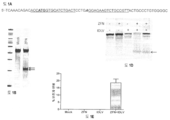

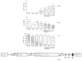
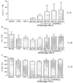
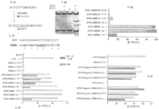
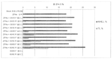
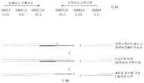


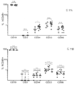

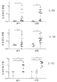


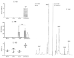
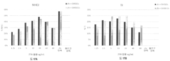
Classifications
-
- C—CHEMISTRY; METALLURGY
- C12—BIOCHEMISTRY; BEER; SPIRITS; WINE; VINEGAR; MICROBIOLOGY; ENZYMOLOGY; MUTATION OR GENETIC ENGINEERING
- C12N—MICROORGANISMS OR ENZYMES; COMPOSITIONS THEREOF; PROPAGATING, PRESERVING, OR MAINTAINING MICROORGANISMS; MUTATION OR GENETIC ENGINEERING; CULTURE MEDIA
- C12N9/00—Enzymes; Proenzymes; Compositions thereof; Processes for preparing, activating, inhibiting, separating or purifying enzymes
- C12N9/14—Hydrolases (3)
- C12N9/16—Hydrolases (3) acting on ester bonds (3.1)
- C12N9/22—Ribonucleases RNAses, DNAses
-
- A—HUMAN NECESSITIES
- A61—MEDICAL OR VETERINARY SCIENCE; HYGIENE
- A61K—PREPARATIONS FOR MEDICAL, DENTAL OR TOILETRY PURPOSES
- A61K35/00—Medicinal preparations containing materials or reaction products thereof with undetermined constitution
- A61K35/12—Materials from mammals; Compositions comprising non-specified tissues or cells; Compositions comprising non-embryonic stem cells; Genetically modified cells
- A61K35/28—Bone marrow; Haematopoietic stem cells; Mesenchymal stem cells of any origin, e.g. adipose-derived stem cells
-
- A—HUMAN NECESSITIES
- A61—MEDICAL OR VETERINARY SCIENCE; HYGIENE
- A61K—PREPARATIONS FOR MEDICAL, DENTAL OR TOILETRY PURPOSES
- A61K39/00—Medicinal preparations containing antigens or antibodies
- A61K39/46—Cellular immunotherapy
- A61K39/461—Cellular immunotherapy characterised by the cell type used
-
- A—HUMAN NECESSITIES
- A61—MEDICAL OR VETERINARY SCIENCE; HYGIENE
- A61K—PREPARATIONS FOR MEDICAL, DENTAL OR TOILETRY PURPOSES
- A61K39/00—Medicinal preparations containing antigens or antibodies
- A61K39/46—Cellular immunotherapy
- A61K39/464—Cellular immunotherapy characterised by the antigen targeted or presented
-
- A—HUMAN NECESSITIES
- A61—MEDICAL OR VETERINARY SCIENCE; HYGIENE
- A61K—PREPARATIONS FOR MEDICAL, DENTAL OR TOILETRY PURPOSES
- A61K48/00—Medicinal preparations containing genetic material which is inserted into cells of the living body to treat genetic diseases; Gene therapy
- A61K48/005—Medicinal preparations containing genetic material which is inserted into cells of the living body to treat genetic diseases; Gene therapy characterised by an aspect of the 'active' part of the composition delivered, i.e. the nucleic acid delivered
-
- A—HUMAN NECESSITIES
- A61—MEDICAL OR VETERINARY SCIENCE; HYGIENE
- A61P—SPECIFIC THERAPEUTIC ACTIVITY OF CHEMICAL COMPOUNDS OR MEDICINAL PREPARATIONS
- A61P7/00—Drugs for disorders of the blood or the extracellular fluid
- A61P7/06—Antianaemics
-
- C—CHEMISTRY; METALLURGY
- C12—BIOCHEMISTRY; BEER; SPIRITS; WINE; VINEGAR; MICROBIOLOGY; ENZYMOLOGY; MUTATION OR GENETIC ENGINEERING
- C12N—MICROORGANISMS OR ENZYMES; COMPOSITIONS THEREOF; PROPAGATING, PRESERVING, OR MAINTAINING MICROORGANISMS; MUTATION OR GENETIC ENGINEERING; CULTURE MEDIA
- C12N15/00—Mutation or genetic engineering; DNA or RNA concerning genetic engineering, vectors, e.g. plasmids, or their isolation, preparation or purification; Use of hosts therefor
- C12N15/09—Recombinant DNA-technology
- C12N15/11—DNA or RNA fragments; Modified forms thereof; Non-coding nucleic acids having a biological activity
- C12N15/111—General methods applicable to biologically active non-coding nucleic acids
-
- C—CHEMISTRY; METALLURGY
- C12—BIOCHEMISTRY; BEER; SPIRITS; WINE; VINEGAR; MICROBIOLOGY; ENZYMOLOGY; MUTATION OR GENETIC ENGINEERING
- C12N—MICROORGANISMS OR ENZYMES; COMPOSITIONS THEREOF; PROPAGATING, PRESERVING, OR MAINTAINING MICROORGANISMS; MUTATION OR GENETIC ENGINEERING; CULTURE MEDIA
- C12N15/00—Mutation or genetic engineering; DNA or RNA concerning genetic engineering, vectors, e.g. plasmids, or their isolation, preparation or purification; Use of hosts therefor
- C12N15/09—Recombinant DNA-technology
- C12N15/87—Introduction of foreign genetic material using processes not otherwise provided for, e.g. co-transformation
- C12N15/90—Stable introduction of foreign DNA into chromosome
- C12N15/902—Stable introduction of foreign DNA into chromosome using homologous recombination
- C12N15/907—Stable introduction of foreign DNA into chromosome using homologous recombination in mammalian cells
-
- C—CHEMISTRY; METALLURGY
- C12—BIOCHEMISTRY; BEER; SPIRITS; WINE; VINEGAR; MICROBIOLOGY; ENZYMOLOGY; MUTATION OR GENETIC ENGINEERING
- C12N—MICROORGANISMS OR ENZYMES; COMPOSITIONS THEREOF; PROPAGATING, PRESERVING, OR MAINTAINING MICROORGANISMS; MUTATION OR GENETIC ENGINEERING; CULTURE MEDIA
- C12N5/00—Undifferentiated human, animal or plant cells, e.g. cell lines; Tissues; Cultivation or maintenance thereof; Culture media therefor
- C12N5/06—Animal cells or tissues; Human cells or tissues
- C12N5/0602—Vertebrate cells
- C12N5/0634—Cells from the blood or the immune system
- C12N5/0645—Macrophages, e.g. Kuepfer cells in the liver; Monocytes
-
- C—CHEMISTRY; METALLURGY
- C07—ORGANIC CHEMISTRY
- C07K—PEPTIDES
- C07K14/00—Peptides having more than 20 amino acids; Gastrins; Somatostatins; Melanotropins; Derivatives thereof
- C07K14/795—Porphyrin- or corrin-ring-containing peptides
- C07K14/805—Haemoglobins; Myoglobins
-
- C—CHEMISTRY; METALLURGY
- C12—BIOCHEMISTRY; BEER; SPIRITS; WINE; VINEGAR; MICROBIOLOGY; ENZYMOLOGY; MUTATION OR GENETIC ENGINEERING
- C12N—MICROORGANISMS OR ENZYMES; COMPOSITIONS THEREOF; PROPAGATING, PRESERVING, OR MAINTAINING MICROORGANISMS; MUTATION OR GENETIC ENGINEERING; CULTURE MEDIA
- C12N2310/00—Structure or type of the nucleic acid
- C12N2310/10—Type of nucleic acid
- C12N2310/20—Type of nucleic acid involving clustered regularly interspaced short palindromic repeats [CRISPRs]
-
- C—CHEMISTRY; METALLURGY
- C12—BIOCHEMISTRY; BEER; SPIRITS; WINE; VINEGAR; MICROBIOLOGY; ENZYMOLOGY; MUTATION OR GENETIC ENGINEERING
- C12N—MICROORGANISMS OR ENZYMES; COMPOSITIONS THEREOF; PROPAGATING, PRESERVING, OR MAINTAINING MICROORGANISMS; MUTATION OR GENETIC ENGINEERING; CULTURE MEDIA
- C12N2510/00—Genetically modified cells
-
- C—CHEMISTRY; METALLURGY
- C12—BIOCHEMISTRY; BEER; SPIRITS; WINE; VINEGAR; MICROBIOLOGY; ENZYMOLOGY; MUTATION OR GENETIC ENGINEERING
- C12N—MICROORGANISMS OR ENZYMES; COMPOSITIONS THEREOF; PROPAGATING, PRESERVING, OR MAINTAINING MICROORGANISMS; MUTATION OR GENETIC ENGINEERING; CULTURE MEDIA
- C12N2800/00—Nucleic acids vectors
- C12N2800/80—Vectors containing sites for inducing double-stranded breaks, e.g. meganuclease restriction sites
Abstract
본 발명은 게놈 엔지니어링 분야, 특히 조혈 줄기세포의 게놈의 표적화된 변형의 분야에 있다. 본원에는 발현을 변경시키기 위한 또는 겸상 적혈구 질환과 같은 유전자 질환에 포함된 단백질 (예컨대 질환의 생성 단백질 부족, 결핍 또는 일탈 및/또는 이들 단백질을 조절하는 단백질)을 코드화하는 하나 이상의 유전자를 교정하기 위한 방법 및 조성물이 개시된다. 본 발명은 유전자 요법 및 유전자 엔지니어링에 사용하기 위한 조성물 및 방법을 기술한다.The present invention is in the field of genome engineering, particularly targeted transformation of the genome of hematopoietic stem cells. Herein, reference is made herein to a method for correcting one or more genes encoding altered expression or a protein contained in a genetic disorder such as sickle cell disease (e.g., a protein lacking, deficient or deviating from a disease producing protein, and / Methods and compositions are disclosed. The present invention describes compositions and methods for use in gene therapy and gene engineering.

Description
관련 출원에 대한 상호-참조Cross-reference to related application
본 출원은 2014년 9월 16일자로 출원된 미국 임시 출원 번호 62/051,159의 유익을 주장하며, 그것의 내용은 전체적으로 참조로 본원에 포함된다.This application claims the benefit of U.S. Provisional Application No. 62 / 051,159, filed September 16, 2014, the content of which is incorporated herein by reference in its entirety.
기술분야Technical field
본 발명은 게놈 엔지니어링 분야, 특히 조혈 세포의 게놈의 표적화된 변형에 있다.The present invention is in the field of genome engineering, in particular targeted transformations of the genome of hematopoietic cells.
대다수의 질환에 대한 유전자 요법에서 가장 전도유망한 접근법 중 한 가지는 줄기세포의 시험관 내 유전자 변형과 이어서 변형된 세포의 환자에서의 이식 및 접목의 사용을 포함한다. 특히 유망한 것은 도입된 줄기세포가 장기간 지속성 및 다중-혈통 세포분화를 나타낼 때이다. 가장 통상적으로 CD34 세포 표면 마커의 발현을 기반으로 한 풍부해진 세포 형태의 조혈 줄기세포는, 그것들이 쉽게 얻어질 수 있고 장기간 조혈 줄기세포 (LT-HSC)를 함유할 수 있고, 그것은 이식 후 완전한 조혈 혈통을 복원할 수 있기 때문에 특히 유용한 세포 집단이다.One of the most promising approaches to gene therapy for the majority of diseases involves the in vitro transgenic transformation of stem cells followed by transplantation and grafting of transformed cells in patients. Particularly promising is when the introduced stem cells exhibit long-term persistence and multi-lineage cell differentiation. Most commonly hematopoietic stem cells in the form of enriched cells based on the expression of CD34 cell surface markers can be easily obtained and contain long-term hematopoietic stem cells (LT-HSC) It is a particularly useful group of cells because it can restore bloodlines.
게놈 DNA의 표적화된 절단을 위한 다양한 방법 및 조성물들이 기술되어 있다. 그런 표적화된 절단 사건은, 예를 들면 표적화된 돌연변이생성을 유도하고, 세포 DNA 서열의 표적화된 결실을 유도하며, 임의의 유기체로부터의 세포에서 예정된 염색체 유전자좌의 표적화된 재조합을 촉진시키기 위해 사용될 수 있다. 예컨대 미국 특허 번호 9,045,763; 9,005,973; 8,956,828; 8,945,868; 8,586,526; 6,534,261; 6,599,692; 6,503,717; 6,689,558; 7,067,317; 7,262,054; 7,888,121; 7,972,854; 7,914,796; 7,951,925; 8,110,379; 8,409,861; 미국 특허 공개공보 20030232410; 20050208489; 20050026157; 20050064474; 20060063231; 20080159996; 201000218264; 20120017290; 20110265198; 20130137104; 20130122591; 20130177983 및 20130177960 및 20150056705 및 미국 출원 번호 14/706,747를 참조하는데, 모든 문서의 내용은 모든 목적에 대해 전체 내용이 참조로 포함된다. 이들 방법은 표적 DNA 서열에 이중 가닥 브레이크 (DSB) 또는 닉 (nick)을 유도하여서 비-상동성 단부 결합 (NHEJ)과 같은 오류 발생 과정에 의한 브레이크의 수복 또는 수복 주형을 사용한 수복 (상동성 지시된 수복 또는 HDR)이 유전자의 녹아웃 또는 관심 서열의 삽입 (표적화된 통합)을 초래할 수 있는 엔지니어링된 절단 시스템의 사용을 포함한다. 뒤따르는 수복 경로 (NHEJ 대 HDR 또는 둘 다)는 전형적으로 수복 주형의 존재 및 여러 경합하는 수복 경로의 활성에 좌우된다.Various methods and compositions for targeted cleavage of genomic DNA have been described. Such targeted cleavage events can be used, for example, to induce targeted mutagenesis, induce targeted deletion of cellular DNA sequences, and promote targeted recombination of predetermined chromosomal loci in cells from any organism . For example, U.S. Patent Nos. 9,045,763; 9,005,973; 8,956,828; 8,945,868; 8,586,526; 6,534,261; 6,599,692; 6,503,717; 6,689,558; 7,067,317; 7,262,054; 7,888,121; 7,972,854; 7,914,796; 7,951,925; 8,110,379; 8,409,861; U.S. Patent Publication No. 20030232410; 20050208489; 20050026157; 20050064474; 20060063231; 20080159996; 201000218264; 20120017290; 20110265198; 20130137104; 20130122591; 20130177983 and 20130177960 and 20150056705 and U.S. Serial No. 14 / 706,747, the entire contents of which are incorporated by reference in their entirety for all purposes. These methods include double-strand breaks (DSBs) or nicks induced in the target DNA sequence to repair the brakes by an error event such as non-homologous end joining (NHEJ) or to repair using a repair template Restoration or HDR) can result in knockout of the gene or insertion of the sequence of interest (targeted integration). The following restoration pathways (NHEJ vs. HDR or both) typically depend on the presence of restoration templates and the activity of several competing restoration pathways.
외부에서 공급되는 수복 주형 (예컨대 도너 (donor))의 부재시 이중 가닥 브레이크의 도입은 통상적으로 세포 NHEJ 경로에 의해 도입된 돌연변이를 통한 표적화된 유전자의 비활성화를 위해 사용된다. NHEJ 경로는 미세상동성-매개된 단부 결합을 기반으로 표준 Ku-의존성 경로 및 대체 Ku-무관 경로로 나누어질 수 있는데, 절단 부위 가까이에 직접 반복부의 짧은 트랙의 장점을 취한다. 이들 두 가지 NHEJ 경로를 통한 유전자 편집에 이어지는 삽입 및/또는 결실 ('indel')의 패턴은 용도에 따라 상이하고, 돌연변이의 기능적 결과의 차이를 초래할 수 있다.The introduction of double-stranded brakes in the absence of an externally supplied repair mold (e. G. A donor) is typically used for inactivation of the targeted gene through mutations introduced by the cellular NHEJ pathway. The NHEJ path can be divided into a standard Ku-dependent path and an alternate Ku-independent path based on a US world-mediated end junction, taking advantage of the short track of the repetition directly near the truncation site. The pattern of insertions and / or deletions ('indel') following genetic editing through these two NHEJ pathways may differ between uses and may result in differences in the functional outcome of the mutations.
이중 가닥 브레이크의 양옆의 서열들에 상동하는 외부에서 공급된 도너 운반 신장부의 존재하에, 도너 분자를 사용하는 상동성 지시된 유전자 수복 (HDR)은 단일 염기 또는 DNA의 작은 신장부의 서열을 변화시키기 위해 ('유전자 교정'), 또는 다른 한편으로, 예정된 게놈 위치 안으로의 전체 발현 카세트의 표적화된 삽입 ('유전자 첨가')을 위해 사용될 수 있다.In the presence of an exogenously supplied donor transport stretch that is identical to sequences on either side of the double-stranded brakes, homologous directed gene repair (HDR) using donor molecules can be used to alter the sequence of a single base or a small stretch of DNA ('Gene correction'), or, on the other hand, targeted insertion of a full expression cassette into a predetermined genomic location ('gene addition').
절단은 엔지니어링된 징크 핑거 뉴클레아제 (ZFN), 전사-활성화제 유사 이펙터 뉴클레아제 (TALEN)과 같은 특이적인 뉴클레아제의 사용을 통해, 또는 특이적인 절단을 안내하기 위해 엔지니어링된 crRNA/tracr RNA ('단일 가이드 RNA')를 가지는 CRISPR/Cas 시스템을 사용하여 일어날 수 있다. 예컨대 미국 특허 번호 8,697,359 및 8,932,814 및 미국 특허 공보 번호 20150056705를 참조한다. 나아가, 표적화된 뉴클레아제는 Argonaute 시스템을 기반으로 발달되었고 (예컨대 T.thermophilus로부터 유래하고, 'TtAgo'로서 알려짐, Swarts et al (2014) Nature 507(7491):258-261 참조), 게놈 편집 및 유전자 요법에 사용하기 위한 가능성을 가질 수 있다.The cleavage can be achieved either through the use of engineered zinc finger nuclease (ZFN), specific nuclease such as transcription-activating agent-like effector nuclease (TALEN), or engineered crRNA / tracr Can occur using the CRISPR / Cas system with RNA ('single guide RNA'). See, for example, U.S. Patent Nos. 8,697,359 and 8,932,814 and U.S. Patent Publication No. 20150056705. Furthermore, targeted nuclease has been developed based on the Argonaute system (eg, derived from T. thermophilus and known as 'TtAgo', see Swarts et al (2014) Nature 507 (7491): 258-261) And may have potential for use in gene therapy.
상기 언급된 뉴클레아제 시스템 중 하나를 사용하는 표적화된 절단은 HDR 또는 NHEJ-매개된 과정 중 어느 하나를 사용하여 특이적인 표적 위치 안으로 핵산을 삽입시키기 위해 사용될 수 있다. 그러나, 뉴클레아제 시스템과 도너 둘 다를 세포에 전달하는 것은 문제의 소지가 될 수 있다. 예를 들어, 플라스미드의 세포 안으로의 형질 도입을 통한 도너 또는 뉴클레아제의 전달은 수용체 세포, 특히 일차 세포인 세포에 독성이 될 수 있고 그러므로 세포주로부터의 세포와 같이 왕성하진 않다.Targeted cleavage using one of the above-mentioned nuclease systems can be used to insert a nucleic acid into a specific target site using either HDR or NHEJ-mediated procedures. However, transferring both nuclease systems and donors to cells can be problematic. For example, the delivery of a donor or nuclease through the transduction of a plasmid into a cell may be toxic to a receptor cell, particularly a primary cell, and is therefore not as vigorous as a cell from a cell line.
CD34+ 줄기 또는 선구 세포는 림프종 혈통의 세포 (예컨대 T 세포, B 세포, NK 세포) 및 골수종 혈통의 세포 (예컨대 단핵세포, 적혈구, 호산성 세포, 호염기성 세포 및 호중구)로 갱신 및/또는 분화하는 능력을 특징으로 하는 세포의 이종성 세트이다. 그것들의 이종성 성질은 CD34+ 줄기세포 집단 내에, 자주 특이적인 그룹의 다능성 (혈통이 어디에 전용되든간에)을 반영하는 다수의 하위그룹이 있다는 사실로부터 발생한다. 예를 들어, CD38-인 CD34+ 세포는 보다 원시적이고, 미성숙한 CD34+ 선구 세포 (또한 장기간 조혈 선구체로도 언급됨)인 한편, CD34+CD38+인 것들 (단기간 조혈 선구체)은 혈통 전용이다 (Stella et al (1995) Hematologica 80:367-387 참조). 다음에 이 집단이 추가로 분화 경로를 다운 진행시킬 때, CD34 마커는 손실된다. CD34+ 줄기세포는 임상 세포 요법에서 많은 가능성을 나타낸다.CD34 + stem or progenitor cells may be renewed and / or differentiated into cells of a lymphoma lineage (eg, T cells, B cells, NK cells) and cells of myeloma lineage (eg, mononuclear cells, erythrocytes, eosinophilic cells, basophilic cells and neutrophils) It is a heterogeneous set of cells characterized by ability. Their heterogeneous nature arises from the fact that within the CD34 + stem cell population there are a number of subgroups that often reflect the specificity of a specific group of pluripotency (where the lineage is dedicated). For example, CD38- the CD34 + cells are more primitive and, immature CD34 + precursor cells (also long-term hematopoietic precursor body also referred search) in the other hand, CD34 + CD38 + ones (short-term hematopoietic precursor) are lineage-only (Stella et al. (1995) Hematologica 80: 367-387). The CD34 marker is then lost when this population is further advanced down the differentiation pathway. CD34 + stem cells represent many possibilities in clinical cell therapy.
적색 혈액 세포 (RBC), 또는 적혈구는 혈액의 주요 세포 성분이다. 실제로, RBC는 인간에서 세포의 1/4을 차지한다. 성숙한 RBC는 인간에서 핵 및 많은 다른 소기관이 결핍되어 있고, 조직 밖으로 이산화탄소를 운반하고 제거를 위해 폐로 되돌리는 것뿐만 아니라 조직에 산소를 운반하는 기능을 하는, RBC에서 발견되는 금속단백질인 헤모글로빈으로 가득하다. 그 단백질은 RBC의 건조 중량의 대략 97%를 구성하고 혈액의 산소 운반 능력을 약 70배 증가시킨다. 헤모글로빈은 2개의 α-형 글로빈 사슬과 2개의 β-형 글로빈 사슬 및 4개의 헴 그룹을 포함하는 이종사량체이다. 성인에서 α2β2 사량체는 헤모글로빈 A (HbA) 또는 성인 헤모글로빈으로 언급된다. 전형적으로, 알파 및 베타 글로빈 사슬은 대략 1:1 비율로 합성되고 이 비율은 헤모글로빈 및 RBC 안정화의 관점에서 결정적인 것으로 보인다. 실제로, 한 유형의 글로빈 유전자가 부적절하게 발현된 일부 경우에 (하기 참조), 다른 유형의 글로빈의 감소하는 발현 (예컨대 특이적 siRNA를 사용함)은, 이 1:1 비율을 되돌리면서 돌연변이체 세포 표현형의 일부 측면을 완화시킨다 (Voon et al (2008) Haematologica 93(8):1288 참조). 발생기 태아에서, 헤모글로빈 A보다 산소에 대한 더 큰 결합 친화성을 가져서 산소가 산모의 혈류를 통해 아기의 시스템으로 전달될 수 있는, 헤모글로빈의 상이한 형태인 태아 헤모글로빈 (HbF)이 생성된다. 태아 헤모글로빈은 또한 2개의 α 글로빈 사슬을 함유하지만, 성인 β-글로빈 사슬 대신 2개의 태아 γ-글로빈 사슬을 함유한다 (즉 태아 헤모글로빈은 α2γ2이다). 대략 임신 30주에는, 태아에서의 γ 글로빈의 합성이 떨어지기 시작하는 한편 β 글로빈의 생성이 증가한다. 대략 10개월이 되면, 일부 (총 헤모글로빈의 대략 1 내지 3%) HbF가 성인기로 계속되긴 하지만 신생아의 헤모글로빈은 거의 전부 α2β2가 된다. γ의 β로의 생성으로부터의 스위치의 조절은 꽤 복잡하고, 주로 γ 글로빈의 발현상의 하향-조절과 동시에 β 글로빈 발현의 상향-조절을 포함한다.Red blood cells (RBCs), or red blood cells, are a major cellular component of blood. In fact, RBC accounts for one-quarter of the cells in humans. Mature RBCs are full of hemoglobin, a metal protein found in RBCs, which lacks nuclear and many other organelles in humans, transports carbon dioxide out of tissues and returns them to the lungs for removal, Do. The protein constitutes approximately 97% of the dry weight of the RBC and increases the oxygen transport capacity of the blood by approximately 70-fold. Hemoglobin is a heterodimer containing two α-globin chains, two β-globin chains and four heme groups. In adults, α2β2 tetramers are referred to as hemoglobin A (HbA) or adult hemoglobin. Typically, the alpha and beta globin chains are synthesized at approximately a 1: 1 ratio and this ratio appears to be crucial in terms of hemoglobin and RBC stabilization. Indeed, in some cases where one type of globin gene is inappropriately expressed (see below), a diminished expression of other types of globin (e.g., using a specific siRNA) may result in a mutant cell phenotype (See Voon et al (2008) Haematologica 93 (8): 1288). In the embryonic fetus, fetal hemoglobin (HbF), a different form of hemoglobin, is generated that has a greater binding affinity for oxygen than that of hemoglobin A, allowing oxygen to be delivered to the baby's system through the maternal blood stream. Fetal hemoglobin also contains two alpha globin chains, but contains two fetal gamma-globin chains instead of an adult beta-globin chain (i.e., fetal hemoglobin is
헤모글로빈 사슬을 코드화하는 서열의 유전자 결핍은 겸상 적혈구 빈혈 및 지중해 빈혈을 포함하여, 헤모글로빈혈증으로 알려져 있는 많은 질환에 기여할 수 있다. 헤모글로빈혈증을 가진 환자들의 대부분에서, γ 글로빈을 코드화하는 유전자가 여전히 존재하지만, 발현은 상기 기술된 것과 같이 출산 즈음에 발생하는 정상 유전자 억압으로 인해 상대적으로 낮다.Gene deficiency in the sequence encoding the hemoglobin chain can contribute to many diseases known as hemoglobinemia, including sickle cell anemia and Mediterranean anemia. In most of the patients with hemoglobinemia, the gene coding for gamma globin is still present, but the expression is relatively low due to normal gene suppression occurring during birth as described above.
미국에서 5000명 중 1명이 겸상 적혈구 질환 (SCD)을 가지고 있는 것으로 추산되는데, 그들 대부분이 사하라 사막 이남의 아프리카 태생이다. 말라리아에 대한 보호를 위한 겸상 적혈구 이형접합성의 장점인 것으로 나타나고, 그로써 이런 특성은 시간이 경과함에 따라 선택될 수 있어서, 사하라 사막 이남의 아프리카에서는, 인구의 1/3이 겸상 적혈구 특성을 가는 것으로 추정된다. 겸상 적혈구 질환은 발린이 아미노산 #6에서 글루탐산에 대해 치환되어 있는 (DNA 수준에서는 GTG에 대해 GAG) β-글로빈 유전자에서의 돌연변이에 의해 유발되고, 그 결과의 헤모글로빈은 "헤모글로빈 S" 또는 "HbS"로 언급된다. 저산소 조건하에서, HbS의 데옥시 형태에서 형태상의 시프트는 E와 F 나선 사이의 단백질 상의 소수성 패치를 노출시킨다. 헤모글로빈의 베타 사슬의 위치 6에서 발린의 소수성 잔기는 소수성 패치와 회합할 수 있어서 HbS 분자의 응집 및 섬유상 침전의 형성을 유발한다. 이들 응집체는 계속해서 RBC의 비정상 또는 '겸상 세포화"를 유발하여 세포의 가요성의 손실을 초래한다. 겸상 세포화된 RBC는 더 이상 모세혈관계로 들어갈 수 없고 겸상 적혈구 환자에게서 혈관-폐색 위기를 초래할 수 있다. 또한, 겸상 적혈구 RBC는 정상 RBC보다 더 깨지기 쉽고, 용혈되는 경향이 있으며, 궁극적으로 환자에서 빈혈을 유발한다.One in 5,000 people in the United States are estimated to have sickle cell disease (SCD), most of them born in sub-Saharan Africa. It appears to be an advantage of sickle cell heterozygosity for protection against malaria, and this property can be selected over time, so that in sub-Saharan Africa, one third of the population is presumed to have sickle cell characteristics do. Sickle cell disease is caused by a mutation in the β-globin gene in which valine is substituted for glutamic acid at amino acid # 6 (GAG for GTG at the DNA level), and the resulting hemoglobin is called "hemoglobin S" or "HbS" . Under hypoxic conditions, a morphological shift in the deoxy form of HbS exposes a hydrophobic patch on the protein between the E and F helices. At position 6 of the beta chain of hemoglobin, valine hydrophobic residues can associate with hydrophobic patches, leading to aggregation of HbS molecules and formation of fibrous precipitates. These aggregates continue to cause abnormal or " sickle celling "of RBCs, resulting in loss of cellular flexibility. Sickle cellized RBCs no longer enter the capillary system and result in a vessel-occlusion crisis in sickle cell patients In addition, sickle cell RBCs are more fragile, tend to hemolyze than normal RBCs, and ultimately cause anemia in patients.
겸상 적혈구 환자의 치료 및 관리는 항생물질 치료, 통증 관리 및 급성 에피소드 중의 주입을 포함하는 평생 과제이다. 한 가지 접근법은 γ 글로빈의 생성을 증가시킴으로써 효과를 부분적으로 나타내는 하이드록시우레아의의 사용이다. 만성 하이드록시우레아 치료법의 장기 부작용은 여전히 알려져 있지 않지만, 치료는 원치 않은 부작용을 유발하고 환자마다 가변적인 효능을 나타낼 수 있다. 겸상 적혈구 치료의 효능의 증가에도 불구하고, 환자들의 기대수명은 여전히 50대 중반부터 후반에 머물러 있고 질환의 관련된 이병률은 환자의 삶의 질에 지대한 영향을 미친다.Treatment and management of sickle cell patients is a lifelong task involving antibiotic therapy, pain management and infusion during acute episodes. One approach is the use of hydroxyurea, which in part represents an effect by increasing the production of gamma globin. The long-term side effects of chronic hydroxyurea therapy are still unknown, but treatment can lead to unwanted side effects and varying efficacy from patient to patient. Despite the increased efficacy of sickle cell therapy, patients' life expectancy is still in their mid 50s and late, and the associated morbidity of the disease has a significant impact on the patient's quality of life.
그러므로, 게놈 편집을 위해, 일탈 유전자를 교정하거나 다른 유전자들의 발현을 변경하기 위해, 예를 들면 겸상 적혈구 질환과 같은 헤모글로빈혈증을 치료하기 위해 사용될 수 있는 추가의 방법 및 조성물에 대한 요구가 여전히 존재한다.Therefore, there is still a need for additional methods and compositions that can be used for genomic editing, for correcting deviating genes, altering the expression of other genes, for example, for treating hemoglobinemia such as sickle cell disease .
본원에는 겸상 적혈구 질환과 같은 유전자 질환 (예컨대 질환에서 부족하거나, 결핍되거나 또는 일탈된 단백질 및/또는 그런 단백질을 조절하는 단백질을 생성하는)에 포함된 단백질을 코드화하는 하나 이상의 유전자를 교정하거나 유전자의 발현을 변경하기 위한 방법 및 조성물이 개시된다. 본 발명은 유전자 요법 및 게놈 엔지니어링에 사용하기 위한 조성물 및 방법을 기술한다.The present application discloses a method of correcting one or more genes encoding a protein involved in a genetic disorder such as sickle cell disease (e.g., producing deficient, deficient or deviated proteins and / or proteins that regulate such proteins) Methods and compositions for altering expression are disclosed. The present invention describes compositions and methods for use in gene therapy and genome engineering.
그런 단백질의 변경은 이들 유전자 질환의 치료를 초래할 수 있다. 특히, 게놈 편집은 일탈 유전자를 교정하거나, 야생형 유전자를 삽입하거나, 또는 내인성 유전자의 발현을 변화시키기 위해 사용된다. 한 가지 접근법은 잘못된 내인성 β글로빈 유전자가 표적화되고 돌연변이 서열이 대체되는 유전자 교정의 사용을 포함한다. 한 가지 접근법은 추가로 줄기세포 (예컨대 조혈 줄기세포 또는 RBC 전구체)의 변형의 사용을 포함하는데, 그 줄기세포는 다음에 환자로의 이식을 위해, 헤모글로빈혈증의 치료를 위해 사용될 수 있다.Modification of such proteins can result in the treatment of these genetic disorders. In particular, genome editing is used to correct deviation genes, insert wild-type genes, or alter the expression of endogenous genes. One approach involves the use of gene correction in which the false endogenous beta globin gene is targeted and the mutation sequence is replaced. One approach further involves the use of a modification of stem cells (such as hematopoietic stem cells or RBC precursors) which can then be used for the treatment of hemoglobinemia for transplantation into patients.
한 측면으로, 본원에는 예를 들면 세포 또는 세포주의 동일 유형의 야생형 게놈 서열과 비교하여 유전자 변형된 세포 또는 세포주가 기술된다. 특정 구체예에서, 세포는 유전자 변형된 RBC 전구체 ("HSC"로 알려져 있는 조혈 줄기세포)를 포함한다. 세포 또는 세포주는 변형에 대해 이형접합성이거나 동형접합성일 수 있다. 변형은 삽입, 결실 및/또는 그것들의 조합을 포함할 수 있다. 특정 구체예에서, HSC는 엔지니어링된 뉴클레아제 및 도너 핵산으로 변형되어서 야생형 유전자 (예컨대 글로빈 유전자)가 삽입되고 발현되며 및/또는 내인성 일탈 유전자가 교정된다. 특정 구체예에서, 변형 (예컨대 삽입)은, 제한은 아니지만, 절단 및/결합 부위의 부위(들)의 상류 또는 하류의 1 내지 300 (또는 그 사이의 임의의 수의 염기쌍) 염기쌍 내에 있는 서열에 대한 변형; 결합 및/또는 절단 부위(들)의 어느 한 부위의 1 내지 100 염기쌍 (또는 그 사이의 임의의 수의 염기쌍) 내에 있는 변형; 결합 및/또는 절단 부위(들)의 어느 한 측에 있는 1 내지 50 염기쌍 (또는 그 사이의 임의의 수의 염기쌍) 내에 있는 변형; 및/또는 뉴클레아제 결합 및/또는 절단 부위의 하나 이상의 염기쌍에 대한 변형을 포함하여, 뉴클레아제(들) 결합 및/또는 절단 부위(들)에 또는 가까이에 있다. 특정 구체예에서, 변형은 SEQ ID NO:23 또는 24에서 또는 가까이에서 (예컨대 1 내지 300 염기쌍 또는 그 사이에 있는 임의의 염기쌍 수) 일어난다. 다른 구체예에서, 변형은 SEQ ID NO:23 또는 24의 1 내지 100 염기쌍에서 (또는 그 사이에 있는 임의의 염기쌍 수) 일어난다. 특정 구체예에서, 변형은 SEQ ID NO:23 및/또는 SEQ ID NO:24 내에서 일어나고, 예를 들면 SEQ ID NO:23 또는 24중 어느 하나에서 하나 이상의 염기쌍이 변형된다. 일부 경우에, 삽입을 위해 야생형 유전자 서열이 야생형 β 글로빈을 코드화한다. 다른 경우에, 내인성 일탈 유전자는 β 글로빈 유전자, 예를 들면 내인성 일탈 인간 베타-헤모글로빈 (Hbb) 유전자의 적어도 하나의 돌연변이를 교정하는 하나 이상의 게놈 변형이다. 일부 측면으로, 베타 글로빈 유전자의 변형은 전사된 RNA의 적절한 스플라이싱을 허용한다. 다른 측면으로, 베타 글로빈 유전자의 변형은 겸상 대립형질의 돌연변이를 교정하여서 폴리펩티드 서열의 6번째 위치에 있는 부정확한 발린 아미노산을 코딩하는 코돈 GTG가 야생형 폴리펩티드에서 발견되는 것과 같은 글루탐산을 코드화하는 GAG로 바뀐다. 본원에서 기술되는 것과 같은 유전자 변형된 줄기세포로부터 파생된 부분적으로 또는 전체적으로 분화된 세포들 또한 제공된다 (예컨대 RBS 또는 RBC 전구체 세포). 본원에서 기술되는 유전자 변형된 세포를 포함하는 약학 조성물과 같은 조성물 또한 제공된다.In one aspect, a genetically modified cell or cell line is described herein, for example, in comparison to a wild type genomic sequence of the same type of cell or cell line. In certain embodiments, the cell comprises a genetically modified RBC precursor (a hematopoietic stem cell known as "HSC"). The cell or cell line may be heterozygous or homozygous for transformation. Variations may include insertions, deletions, and / or combinations thereof. In certain embodiments, HSCs are modified with engineered nucleases and donor nucleic acids to insert and express wild-type genes (e.g., globin genes) and / or endogenous deletion genes are calibrated. In certain embodiments, the variant (e. G., Insertions) includes, but is not limited to, sequences located in base pairs between 1 and 300 (or any number of base pairs in between) upstream or downstream of site (s) of the cleavage and / Transformation to; Deformation within 1 to 100 base pairs (or any number of base pairs therebetween) of either site of the binding and / or cleavage site (s); Deformation within 1 to 50 base pairs (or any number of base pairs in between) on either side of the binding and / or cleavage site (s); And / or cleavage site (s), including modifications to one or more base pairs of nucleotide binding and / or cleavage site (s). In certain embodiments, the modification occurs at or near SEQ ID NO: 23 or 24 (e. G., Any base number between 1 and 300 base pairs or in between). In another embodiment, the modification occurs at a base pair of 1 to 100 (or any base pair number therebetween) of SEQ ID NO: 23 or 24. In certain embodiments, the modification occurs within SEQ ID NO: 23 and / or SEQ ID NO: 24, wherein one or more base pairs are modified, for example, in any one of SEQ ID NO: 23 or 24. In some cases, the wild-type gene sequence codes for the wild-type beta globin for insertion. In other cases, the endogenous deviating gene is one or more genomic variants that correct for at least one mutation in the beta globin gene, e. G., The endogenous human beta-hemoglobin (Hbb) gene. In some respects, modification of the beta globin gene allows for proper splicing of the transcribed RNA. In another aspect, modification of the beta globin gene corrects mutations in sickle allelic variants, such that the codon GTG encoding the incorrect valine amino acid at the 6 < th > position of the polypeptide sequence is converted to a GAG encoding glutamic acid as found in the wild-type polypeptide . Partially or totally differentiated cells derived from genetically modified stem cells such as those described herein are also provided (e.g., RBS or RBC precursor cells). Compositions such as pharmaceutical compositions comprising genetically modified cells as described herein are also provided.
다른 측면으로, 본원에는 게놈의 관심의 영역 (예컨대 β 글로빈 유전자)에서 표적 부위에 결합하는 징크-핑거 단백질 (ZEP)이 기술되는데, ZEP는 하나 이상의 엔지니어링된 징크-핑거 결합 도메인을 포함한다. 한 구체예에서, ZEP는 관심의 표적 게놈 영역을 절단하는 징크-핑거 뉴클레아제 (ZFN)이고, ZFN은 하나 이상의 엔지니어링된 징크-핑거 결합 도메인 및 뉴클레아제 절단 도메인 또는 절단 하프-도메인을 포함한다. 절단 도메인 및 절단 하프 도메인은 예를 들면, 다양한 제한 엔도뉴클레아제 및/또는 호밍 (homing) 엔도뉴클레아제로부터 얻어질 수 있다. 한 구체예에서, 절단 하프-도메인은 타입 IIS 제한 엔도뉴클레아제 (예컨대 Fok I)로부터 유도된다. 특정 구체예에서, 징크 핑거 도메인은 글로빈 또는 안전한 은닉 (harbor) 유전자에서 표적 부위를 인식한다. 특정 구체예에서, 징크 핑거 도메인은 5 또는 6 징크 핑거 도메인을 포함하고 글로빈 유전자에서 표적 부위를 인식한다 (예컨대 표 1에 나타낸 인식 나선 영역을 가지는 5 또는 6 핑거를 가지는 징크 핑거 단백질).In another aspect, a zinc-finger protein (ZEP) is described herein that binds to a target site in a region of interest of the genome (e.g., a beta globin gene), wherein the ZEP comprises at least one engineered zinc-finger binding domain. In one embodiment, the ZEP is a zinc-finger nuclease (ZFN) that cleaves the target genomic region of interest, and the ZFN comprises one or more engineered zinc-finger binding domains and a nuclease cleavage domain or cleavage half-domain do. The cleavage domain and cleavage half domain can be obtained, for example, from a variety of restriction endonuclease and / or homing endonuclease. In one embodiment, the cleavage half-domain is a type IIS restriction endonuclease (e.g., Fok I). In certain embodiments, a zinc finger domain recognizes a target site in a globin or a safe harbor gene. In certain embodiments, a zinc finger domain comprises 5 or 6 zinc finger domains and recognizes a target site in the globin gene (e.g., a zinc finger protein with 5 or 6 fingers with a recognition helical region shown in Table 1).
다른 측면으로, 본원에는 게놈의 관심 영역 (예컨대 β 글로빈 유전자)에서 표적 부위에 결합하는 TALE 단백질이 기술되는데, TALE은 하나 이상의 엔지니어링된 TALE 결합 도메인을 포함한다. 한 구체예에서, TALE은 관심의 표적 게놈 영역을 절단하는 뉴클레아제 (TALEN)이고, TALEN은 하나 이상의 엔지니어링된 TALE DNA 결합 도메인 및 뉴클레아제 절단 도메인 또는 절단 하프-도메인을 포함한다. 절단 도메인 및 절단 하프 도메인은, 예를 들면 다양한 제한 엔도뉴클레아제 및/또는 호밍 엔도뉴클레아제로부터 얻어질 수 있다. 한 구체예에서, 절단 하프-도메인은 타입 IIS 제한 엔도뉴클레아제 (예컨대 Fok I)로부터 유도된다.In another aspect, herein, beta globin gene), the TALE protein comprising at least one engineered TALE binding domain. In one embodiment, TALE is a nuclease (TALEN) that cleaves the target genomic region of interest, and TALEN comprises one or more engineered TALE DNA binding and nuclease cleavage domains or cleavage half-domains. The cleavage domain and cleavage half domain can be obtained, for example, from a variety of limiting endonuclease and / or homing endonuclease. In one embodiment, the cleavage half-domain is a type IIS restriction endonuclease (e.g., Fok I).
또 다른 측면으로, 본원에는 게놈의 관심 영역 (예컨대 질환 관련 유전자)의 표적 부위에 결합하는 CRISPR/Cas 또는 tAgo 시스템이 기술되며, CRISPR/Cas 시스템은 CRIPSR/Cas 뉴클레아제 및 엔지니어링된 crRNA/tracrRNA (또는 단일 가이드 RNA)를 포함한다. 특정 구체예에서, CRISPR/Cas 또는 tAgo 시스템은 글로빈 유전자에서 표적을 인식한다. In another aspect, a CRISPR / Cas or tAgo system is described herein that binds to a target region of a genomic region of interest (e.g., a disease-associated gene), wherein the CRISPR / Cas system comprises a CRIPSR / Cas nuclease and an engineered crRNA / tracrRNA (Or a single guide RNA). In certain embodiments, the CRISPR / Cas or tAgo system recognizes a target in the globin gene.
일부 구체예에서, 세포의 게놈 엔지니어링을 촉진하는 뉴클레아제는 펩티드로서 전달되는 한편, 다른 구체예에서 그것은 뉴클레아제를 코드화하는 핵산으로서 전달된다. 일부 구체예에서, 하나 이상의 뉴클레아제가 사용되고 핵산 형태, 단백질 형태 또는 그것들의 조합으로 전달될 수 있다. 일부 바람직한 구체예에서, 뉴클레아제를 코드화하는 핵산(들)은 mRNA이고, 일부 경우에, mRNA는 보호된다. 추가의 바람직한 구체예에서, mRNA는 ARCA cap을 포함할 수 있고 및/또는 변형 및 미변형 뉴클레오티드의 혼합물을 포함할 수 있다. 뉴클레아제는 징크 핑거 뉴클레아제 (ZFN), TALE-뉴클레아제 (TALEN), TtAgo 또는 CRISPR/Cas 뉴클레아제 시스템 또는 그것들의 조합을 포함할 수 있다. 바람직한 구체예에서, 뉴클레아제(들)을 코드화하는 핵산은 전기천공법을 통해 전달된다. 다른 측면으로, 본원에는 세포에서 뉴클레아제-매개된 절단 후에 (예컨대 HDR을 통해) 표적화된 통합을 증가시키는 방법이 기술된다. 특정 구체예에서, 방법은 (i) 하나 이상의 뉴클레아제 (및/또는 mRNA 또는 그 뉴클레아제(들) 및 필요하다면 하나 이상의 단일 가이드 RNA를 발현하는 발현 구성물)을 하나 이상의 도너 분자와 함께 숙주 세포 안에 도입하는 단계 및 (ii) 줄기성의 손실 또는 세포 및/또는 그 세포를 함유하는 배양 배지 안으로의 분화 없이 줄기세포 팽창에 영향을 미치고 및/또는 증가시키는 하나 이상의 인자를 뉴클레아제(들)의 도입 전에, 중에 및/또는 후에 도입하는 단계를 포함한다. 도너는 뉴클레아제(들)을 코드화하는 핵산보다 앞서, 후에 또는 함께 전달될 수 있다.In some embodiments, the nuclease that promotes genomic engineering of the cell is delivered as a peptide while in other embodiments it is delivered as a nucleic acid encoding a nuclease. In some embodiments, one or more nucleases are used and may be delivered in nucleic acid form, in protein form, or a combination thereof. In some preferred embodiments, the nucleic acid (s) encoding the nuclease is mRNA and, in some cases, the mRNA is protected. In a further preferred embodiment, the mRNA may comprise an ARCA cap and / or may comprise a mixture of modified and unmodified nucleotides. Nucleases may include zinc finger nuclease (ZFN), TALE-nuclease (TALEN), TtAgo or CRISPR / Cas nuclease systems or combinations thereof. In a preferred embodiment, the nucleic acid encoding the nuclease (s) is delivered via electroporation. In another aspect, the disclosure provides a method of inhibiting A method of increasing targeted integration through HDR is described. In certain embodiments, the method comprises the steps of (i) contacting one or more nuclease (s) (and / or mRNA or nuclease (s) thereof, and optionally one or more expression constructs expressing a single guide RNA) (S) into the cell and (ii) one or more factors that affect and / or increase stem cell expansion without loss of stem cells or differentiation into a culture medium containing cells and / or cells thereof, Before, during and / or after the introduction of the catalyst. The donor can be delivered before, after, or together with the nucleic acid encoding the nuclease (s).
도너는 바이러스 및/또는 비-바이러스 유전자 전달 방법에 의해 전달될 수 있다. 다른 구체예에서, 도너는 아데노 관련 바이러스 (AAV)를 통해 세포에 전달된다. 일부 경우에, AAV는 캡시드 혈청형과 비교하여 이종성 혈청형의 것인 LTR을 포함한다. 다른 구체예에서, 도너는 렌티바이러스를 통해 세포에 전달된다. 일부 경우에, 렌티바이러스는 인테그라제 결핍 렌티바이러스 (IDLV)이다.The donor can be delivered by viral and / or non-viral gene delivery methods. In another embodiment, the donor is delivered to the cell via an adeno-associated virus (AAV). In some cases, AAV includes an LTR that is of heterologous serotype compared to the capsid serotype. In another embodiment, the donor is delivered to the cell via lentivirus. In some cases, the lentivirus is an integrase deficient lentivirus (IDLV).
특정 구체예에서, 도너 핵산은 비-상동성 의존 방법 (예컨대 NHEJ)을 통해 통합된다. 상기에서 주지된 것과 같이, 생체 내 절단시 도너 서열은 표적화된 방식으로 DSB의 위치에서 세포의 게놈 안으로 통합될 수 있다. 도너 서열은 DSB를 생성하기 위해 사용된 하나 이상의 뉴클레아제에 대해 하나 이상의 동일한 표적 부위를 포함할 수 있다. 그러므로, 도너 서열은 그 안으로의 통합이 바람직한 내인성 유전자를 절단하기 위해 사용된 하나 이상의 동일한 뉴클레아제에 의해 절단될 수 있다. 특정 구체예에서, 도너 서열은 DSB를 유도하기 위해 사용된 뉴클레아제로부터의 상이한 뉴클레아제 표적 부위들을 포함한다. 표적 세포의 게놈의 DSB는 임의의 메커니즘에 의해 생성될 수 있다. 특정 구체예에서, DSB는 관심의 영역 내의 서열에 결합하도록 엔지니어링된 징크 핑거 결합 도메인 및 절단 도메인 또는 절단 하프-도메인을 포함하는 융합 단백질인 하나 이상의 징크-핑거 뉴클레아제 (ZFNs)에 의해 생성된다. 다른 구체예에서, DSB는 뉴클레아제 도메인 (TALEN)에 융합된 하나 이상의 TALE DNA-결합 도메인 (자연적으로 발생하는 또는 자연적으로 발생하지 않는)에 의해 생성된다. 또 추가의 구체예에서, DSB는 엔지니어링된 단일 가이드 RNA 또는 그것이 기능적 동등물이 뉴클레아제를 게놈의 표적화된 부위로 안내하기 위해 사용되는 CRISPR/Cas 뉴클레아제 시스템을 사용하여 생성된다. 또 다른 추가의 구체예에서, DSB는 TtAgo 시스템을 사용하여 생성된다.In certain embodiments, the donor nucleic acid is a non-homologous-dependent method (e. NHEJ). As noted above, in vivo cleavage, the donor sequence can be integrated into the genome of the cell at the location of the DSB in a targeted manner. The donor sequence may comprise one or more identical target sites for one or more nucleases used to generate the DSB. Thus, the donor sequence may be cleaved by one or more of the same nucleases used to cleave the endogenous gene for integration into which it is desired. In certain embodiments, the donor sequence comprises different nuclease target sites from the nucleases used to derive the DSB. The DSB of the genome of the target cell can be generated by any mechanism. In certain embodiments, the DSB is generated by one or more zinc finger-nuclease (ZFNs) that are fusion proteins comprising a zinc finger binding domain engineered to bind to a sequence within the region of interest and a truncation domain or truncation half-domain . In another embodiment, the DSB is generated by one or more TALE DNA-binding domains fused to the nuclease domain (TALEN) (naturally occurring or non-naturally occurring). In yet another embodiment, the DSB is generated using a single engineered guideline RNA or a CRISPR / Cas nuclease system in which the functional equivalents are used to direct the nuclease to the targeted site of the genome. In yet another additional embodiment, the DSB is generated using the TtAgo system.
또 다른 측면으로, 발명은 세포 게놈의 뉴클레아제-매개된 절단 후에 유전자 파괴 및/또는 표적화된 통합 (예컨대 유전자 교정)을 증가시키기에 유용한 키트를 제공한다 (예컨대 ZFNs, TAL-이펙터 도메인 뉴클레아제 융합 단백질, TtAgo 시스템, 또는 CRISPR/Cas 시스템으로 엔지니어링된 호밍 엔도뉴클레아제 또는 엔지니어링된 가이드 RNA). 키트는 전형적으로 표적 부위에 결합하는 하나 이상의 뉴클레아제, 줄기세포 팽창 및/또는 분화에 영향을 미치는 하나 이상의 인자 및 뉴클레아제 및 줄기세포-영향 인자들을 세포에 도입시키기 위한 지시를 포함하여서 뉴클레아제-매개된 유전자 파괴 및/또는 표적화된 통합이 증대된다. 선택적으로, 뉴클레아제의 표적 부위(들)을 함유하는 세포 또한 본원에 기술된 키트에 포함될 수 있다. 특정 구체예에서, 키트는 표적 유전자 및 표적 유전자 내에서 절단할 수 있는 공지의 뉴클레아제를 가지는 적어도 하나의 구성물을 포함한다. 그런 키트는 다양한 달라지는 숙주 세포 유형에서 절단 조건의 최적화에 유용하다. 발명에 의해 고려된 다른 키트는 게놈 내의 공지의 표적 유전자좌 내에서 절단할 수 있는 공지의 뉴클레아제를 포함할 수 있고, 추가로 도너 핵산을 포함할 수 있다. 일부 측면으로, 도너 DNA는 폴리펩티드, 조절 영역 또는 구조적 핵산을 코드화할 수 있다. 일부 구체예에서, 폴리펩티드는 리포터 유전자 (예컨대 GFP)이다. 그런 키트는 도너 통합을 위한 또는 유전자 파괴 또는 표적화된 삽입을 함유하고 있는 특이적으로 변형된 세포, 세포주 및 동물의 구성을 위한 최적화에 유용하다.In another aspect, the invention provides kits useful for increasing gene disruption and / or targeted integration (e.g., gene correction) after nuclease-mediated cleavage of cellular genomes (e.g., ZFNs, TAL-effector domain nuclease A fusion protein, a TtAgo system, or a homing endonuclease or an engineered guide RNA engineered with a CRISPR / Cas system). The kit typically comprises one or more nucleases that bind to the target site, one or more factors that affect stem cell expansion and / or differentiation, and instructions for introducing nuclease and stem cell- Cleavage-mediated gene disruption and / or targeted integration is increased. Alternatively, cells containing the target site (s) of nuclease may also be included in the kits described herein. In certain embodiments, the kit comprises at least one construct having a target gene and a known nuclease that can be cleaved within the target gene. Such kits are useful for optimization of cleavage conditions in a variety of different host cell types. Other kits contemplated by the invention may include known nucleases that are capable of cleaving within known target loci within the genome, and may further include donor nucleic acids. In some aspects, the donor DNA can encode a polypeptide, regulatory region or structural nucleic acid. In some embodiments, the polypeptide is a reporter gene (e.g., GFP). Such kits are useful for optimization for the construction of specifically modified cells, cell lines and animals for donor integration or containing gene-disrupted or targeted insertions.
다른 측면으로, 본원에서 기술되는 유전자 변형된 줄기세포를 대상에게 투여하는 방법이 기술된다. 유전자 변형된 혈구 전구체 ("HSC/PC")는 전형적으로 골수 이식으로 얻어지고 HSC/PC는 생체 내에서 분화하고 성숙한다. 일부 구체예에서, HSC/PC는 G-CSF-유도된 고정에 이어서 분리되고, 다른 구체예에서는 세포는 인간 골수 또는 탯줄로부터 분리된다. 일부 측면으로, HSC/PC는 특이적 유전자 또는 조절 서열을 녹아웃하도록 디자인된 뉴클레아제로의 처리에 의해 편집된다. 다른 측면으로, HSC/PC는 엔지니어링된 뉴클레아제 및 도너 핵산으로 변형되어서 야생형 유전자 또는 관심의 다른 유전자가 삽입되고 발현되며 및/또는 내인성 일탈 유전자가 교정된다. 일부 구체예에서, 변형된 HSC/PC는 약한 골수소멸성 사전-조건화에 이어서 환자에게 투여된다. 다른 측면으로, HSC/PC는 전체 골수소멸 후에 투여되어서 이식 후에, 100%의 조혈세포가 변형된 HSC/PC로부터 유도된다. 나아가, 세포는 세포 주기의 G2 단계에서 정지될 수 있다.In another aspect, a method of administering a genetically modified stem cell described herein to a subject is described. Genetically modified blood cell precursors ("HSC / PC") are typically obtained by bone marrow transplantation and HSC / PC differentiate and mature in vivo. In some embodiments, the HSC / PC is separated following G-CSF-induced fixation, and in other embodiments the cell is isolated from a human bone marrow or umbilical cord. In some aspects, HSC / PC is edited by treatment with a nuclease designed to knock out specific genes or regulatory sequences. In another aspect, HSC / PC is modified with engineered nucleases and donor nucleic acids to insert and express wild-type genes or other genes of interest and / or endogenous deletion genes are calibrated. In some embodiments, the modified HSC / PC is administered to the patient following a mild myelogenous pre-conditioning. In another aspect, HSC / PC is administered after total bone marrow extinction and after transplantation, 100% hematopoietic cells are derived from transformed HSC / PC. Furthermore, the cells can be arrested at the G2 stage of the cell cycle.
일부 구체예에서, 유전자 도입 HSC/PC 세포 및/또는 동물은 인간 유전자를 코드화하는 도입 유전자를 포함한다. 일부 경우에, 유전자 도입 동물은 외인성 도입 유전자에 상응하는 내인성 유전자좌에서 녹아웃을 포함하고, 그로써 인간 단백질이 분리상태로 연구될 수 있는 생체 내 시스템의 개발이 허용된다. 그런 유전자 도입 모델은 관심의 인간 단백질과 상호작용하거나 그것을 변형시킬 수 있는 작은 분자 또는 큰 생체분자 또는 다른 실체를 확인하기 위한 스크리닝 목적에 대해 사용될 수 있다. 일부 측면으로, 도입 유전자는 선택된 유전자좌 안으로 (예컨대 안전한-은닉), 줄기세포 (예컨대 배아 줄기 세포, 유도된 전능 줄기세포, 조혈 줄기세포 등) 안으로 또는 본원에 기술된 방법들 중 임의의 방법에 의해 얻어진 동물 배아 안으로 통합된 후, 배아는 이식되어서 살아있는 동물이 태어난다. 그런 다음 동물은 성적으로 성숙해지고 적어도 자손의 일부가 편집된 내인성 유전자 서열 또는 통합된 도입 유전자를 포함하는 자손을 생산하도록 허용된다.In some embodiments, the transgenic HSC / PC cells and / or animal comprises an inducible gene encoding a human gene. In some cases, transgenic animals contain knockouts at endogenous loci corresponding to exogenous transgene, thereby allowing the development of in vivo systems in which human proteins can be studied in isolation. Such transgenic models can be used for screening purposes to identify small molecules or large biomolecules or other entities that can interact with or transform human proteins of interest. In some aspects, the transgene can be introduced into a selected locus (e.g., a safe-hiding), into a stem cell (e.g., embryonic stem cell, induced omnipotent stem cell, hematopoietic stem cell, etc.) or by any of the methods described herein After integration into the resulting animal embryo, the embryo is transplanted and a living animal is born. The animal is then allowed to produce offspring that are sexually mature and at least part of their offspring have an edited endogenous gene sequence or an integrated transgene.
이들 및 다른 측면들은 전체 발명을 고려하여 숙련자들에게 쉽게 드러날 것이다.These and other aspects will be readily apparent to those skilled in the art in view of the entire invention.
도 1A 내지 1E는 IDLV 도너를 사용하는 CD34+ 세포의 베타-글로빈 유전자좌에서의 절단 및 교정을 도시한다. 도 1A (SEQ ID NO:3)는 출발 코돈 (진한 색) 및 겸상 돌연변이 (진한 색, 이탤릭체) 상부의 ZFN 표적 부위 (밑줄)를 보여주는 베타-글로빈의 엑손 I DNA 서열의 일부를 도시한다. 도 1B는 인간 제대혈 CD34+ 세포의 베타-글로빈 유전자좌에서의 표적화된 절단을 보여주는 겔이다. 세포들은 ZFN을 코드화하는 시험관 내 전사된 mRNA를 사용한 전기천공 후 3일 후 분석되었다. 'Mock'은 미처리 CD34+ 세포를 나타낸다. 화살표는 PCR 증폭 및 Surveyor? 뉴클레아제 (Transgenomic?)로의 소화 후의 절단 밴드를 나타낸다. 도 1C는 엑손 1 및 2와 관련하여 베타 글로빈 유전자의 겸상 돌연변이에서의 부위-특이적 유전자 교정의 개략도이다. 또한 도너 구성물 및 ZFN에 의한 절단 및 HDR에 의한 수복시 그 결과의 게놈 DNA가 상세하게 예시된다. 겸상 적혈구 돌연변이 및 HhaI RFLP의 위치 (별표)가 표시된다. Surveyor 뉴클레아제에 대한 프라이머 위치 및 RFLP 분석이 아래에 도시된다. 일정 비율의 엑손: 비스듬한 해시 마크를 일정 비율이 아닌 구역을 나타낸다. 도 1D는 ZFN 및 도너의 존재하에 베타-글로빈의 표적화된 유전자 변형에 대한 RELP 겔이다. 제대혈 (CB) CD34+ 세포는 겔의 상부에 표시된 바와 같이 시험관 내 전사된 ZFN mRNA로 전기천공되었고 및/또는 IDLV를 포함하는 도너로 형질도입되었다. 세포들은 처리 후 4일 후에 수확되었고, 도너 영역 외부로부터 PCR 증폭되었으며, HhaI 효소로 소화되었고, 아가로오스 겔 상에서 분석되었다. 화살표는 절단된 생성물을 보여주고, 표적 부위에서 게놈 안으로의 RFLP의 통합을 나타낸다. 도 1E는 표시된 조건하에서 CD34+ 세포에서의 유전자 변형 백분율을 나타내는 그래프이다. CB CD34+ 세포는 시험관 내 전사된 ZFN mRNA로 전기천공되었고 도너 IDLV로 형질도입되었다. 세포들은 처리 후 3일 후에 수확되었고, 도너 영역 외부로부터 PCR 증폭되었으며 qPCR이 HhaI RFLP를 생성하는 사일런트 염기 변화의 통합을 특이적으로 검출하기 위해 디자인된 프라이머들로 완료되었고 암플리콘의 베타-글로빈 유전자좌의 엑손 2에서 결합하는 프라이머들에 대해 표준화되었다, (모든 조건에 대해 n=4). 유전자 변형은 ZFN 및 도너 둘 다로 처리된 세포의 대략 18%에서 발견되었다.
도 2A 내지 2D는 형질도입에 대해 사용된 ZFN 및 IDLV 용량의 최적화의 결과를 도시한다. 도 2A, B 및 C는 ZFN mRNA의 적정 후 결과를 보여주는 그래프이다. CD34+ 세포가 증가하는 양의 시험관 내 전사된 ZFN mRNA로 전기천공되었고 일정한 양의 도너 IDLV로 형질도입되었다. Mock는 미처리 세포를 나타내며, ZFN 단독 샘플은 30 30ug/mL의 각각의 ZFN으로 전기천공되었고, IDLV 단독 샘플은 펄스되고 2E+07 TU/ml의 도너 IDLV로 형질도입되었다. 도 2A는 RFLP에 대해 qPCR에 의해 측정되는 바 전기천공 후 3일째의 유전자 변형률을 보여준다. 도 2B는 전기천공 후 1일째의 팽창 배수를 보여주고 도 2C는 트립판 축출 염료에 의해 측정되는 바 전기천공 후 1일째의 세포 생존율을 보여준다, (모든 조건에 대해 n=8). 도 2D는 도너 인테그라제-결핍 렌티바이러스 벡터 (IDLV)의 개략도를 보여준다: 1.1 kb 영역의 베타-글로빈이 긴 말단 반복부 (PTR)의 프로모터 요소들에 대하여 역으로, 3' LTR이 프로바이러스 DNA의 5' LTR로 역전사되는 동안 전달되는 자기-비활성화 (SIN) 결실을 함유하고 있는 렌티바이러스 골격에 클론되었다. UTR: 미번역 영역, RRE: 역-반응 요소, Int: 인트론.
도 3A 내지 3C는 도너 IDLV의 적정을 보여주는 그래프이다. CD34+ 세포들은 일정량의 시험관 내 전사된 ZFN mRNA로 전기천공되었고 증가하는 양의 도너 IDLV로 형질도입되었다. Mock는 미처리 세포, 10 ug/mL의 각각의 ZFN으로 전기천공된 ZFN만의 샘플, 펄스되고 (pulsed) 6E+07 TU/ml의 도너 IDLV로 형질도입된 IDLV만의 샘플을 나타낸다. 도 3A는 RFLP에 대한 qPCR에 의해 측정되는 바 전기천공 후 3일째의 유전자 변형률을 보여준다. 도 3B는 전기천공 후 1일째의 팽창 배수를 보여주고 도 3C는 트립판 축출 염료에 의해 측정되는 바 전기천공 후 1일째의 세포 생존률을 보여준다, (모든 조건에 대해 n=6).
도 4A 내지 4F는 올리고뉴클레오티드 도너를 사용하여 CD34+ 세포에서 베타-글로빈 유전자좌에서 ZFN 쌍 33488/33501을 사용한 교정을 도시한다. 도 4A는 올리고뉴클레오티드-지시된 유전자 변형을 개략적으로 보여준다 (SEQ ID NO:4 및 5). 도 4B는 베타-글로빈 유전자좌의 AvrII-소화된 PCR 암플리콘의 겔이다. 단편은 천연 AvrII 부위를 함유하고, 그것의 절단은 AvrII 소화에 대한 내부 대조표준으로서 작용한다 (겔 상의 하부 밴드). 화살표는 AvrII 절단 생성물을 나타낸다. 올리고 도너 및 ZFN의 존재하에, 유전자 교정률은 15%이다. 도 4C는 SBS#33501 ZFN 결합 부위에서 사일런트 돌연변이의 가능한 6개의 부위를 나타낸다 (WT에 대해 SEQ ID NO:53 및 SMS에 대해 SEQ ID NO:54). 겸상 돌연변이는 이탤릭체로, 가능한 사일런트 돌연변이 부위 (SMS)는 진하게 나타낸다. 도 4D는 사일런트 돌연변이가 베타-글로빈에서 유전자 교정을 증가시켰음을 보여준다. mPB CD34+ 세포가 ZFN 및 표시된 도너 올리고뉴클레오티드로 트랜스펙션되었다. 관련 사일런트 돌연변이의 도입은 대용량 서열화를 통해 평가되었다. 흰색 막대는 삽입 및 결실 (indel)을 나타내고; 회색 막대는 유전자 교정을 나타낸다. 도 4E는 사일런트 돌연변이가 ZFN 재-절단을 차단하는 것을 보여준다. Indel이 있는 대립유전자가 상동성-매개된 변형의 증거에 대해 조사되었다. NHEJ-유도된 indel의 증거를 또한 가지는 유전자 변형을 가진 대립유전자의 백분율이 도시된다. 도 4F는 ZFN 농도의 최적화 및 도너 유형을 보여준다. NHEJ-유도 indel (흰색 막대) 및 유전자 교정 (회색 막대)은 대용량 서열화에 의해 평가되었다. 대용량 DNA 서열화의 깊이가 제공되면, 측정 오류는 매우 낮을 것으로 예상된다. WT: 야생형; SMS: 사일런트 돌연변이 부위; 올리고: 올리고뉴클레오티드.
도 5는 도너 길이 및 가닥의 최적화를 도시한다. mPB CD34+ 세포가 ZFN과 3개의 염기쌍 서열을 베타-글로빈 유전자좌에 도입하도록 디자인된 올리고뉴클레오티드의 조합으로 트랜스펙션되었다. 세포가 수확되고 NHEJ-유도된 indel의 수준 및 이 짧은 서열의 표적화된 통합 ("TI")이 대용량 DNA 서열화 (HTS)에 의해 분석되었다. 샘플당 대략 4 내지 9,000 서열 판독을 얻었다.
도 6A 및 6B는 부분적인 HDR 사건을 도시하고 합성이 올리고-매개된 유전자 변형 중에 좌측에서 우측으로 진행되는 것을 나타낸다. 도 6A는 부분적인 HDR 생성물 (SMS1, SMS12, SMS24, 및 SMS4 대립유전자)의 증거에 대해 분석된, ZFN과 도 3F로부터의 SMS124 도너로부터의 대용량 DNA 서열화 데이터를 보여준다. 이 실험에서 ~41%의 유전자 변형 중에 6%의 변형은 SMS12 사건으로부터 유래한 반면 SMS24 대립유전자는 거의 완전하게 부재하였다 (0.03%). 이 비대칭성은 DNA 합성이 좌측에서 우측으로 진행되는 것을 시사한다. 도 6B는 3개의 모든 ZFN 농도로부터의 데이터의 평균을 사용한 것이 도시되는데, 30 및 60 μg/mL의 샘플로부터의 사건이 파열 (Burst) 15 μg/mL의 샘플에서 볼 수 있는 ~41% 유전자 변형을 매칭하기 위해 상향 표준화되었다.
도 7A 내지 7D는 베타-글로빈 유전자 클러스터에서의 온-표적 절단 분석을 도시한다. 도 7A는 다음과 같이 GFP 포획을 예시한다: K562 적혈구성 백혈병 세포가 시험관 내 전사된 mRNA로 전기천공되고 GFP를 발현하는 IDLV 벡터로 형질도입되었다. 세포는 60일 동안 배양되어 모든 미-트랩핑된 GFP가 희석되었고, 그런 후 GFP 포지티브 세포가 형광-활성화 세포 분류 (FACS)를 사용하여 분류되었다. 샘플에 대해 nrLAM-PCR이 수행되었고 벡터 통합 부위 분석이 완료되었다. 클러스터 분석을 사용하여 측정된 베타-글로빈 및 델타-글로빈에서의 대부분의 통상적인 통합 부위들이 도시된다. HBB: 야생형 인간 헤모글로빈 베타; HBD: 인간 헤모글로빈 델타. 도 7B는 ZFN 절단 부위 주변의 베타-글로빈 유전자 클러스터에서 염색체 11 상의 부분적인 글로빈 유전자 서열의 일렬배열을 보여준다 (SEQ ID NO:6 내지11). ZFN 결합 부위는 밑줄이 있고, 겸상 돌연변이는 굵은 이탤릭체이다. HBB: 야생형 인간 헤모글로빈 베타; HBD: 인간 헤모글로빈 델타; HBE1: 인간 헤모글로빈 엡실론; HBBP1: 인간 헤모글로빈 베타 위유전자 1; HBG1: 인간 헤모글로빈 감마 A; HBG2: 인간 헤모글로빈 감마 G. 도 7C는 ZFN 시험관 내 전사된 mRNA로 처리된 또는 미처리된 (Mock) CD34+ 세포의 Surveyor? 뉴클레아제 분석을 보여주는 겔이다. 표시된 베타 글로빈 클러스터 유전자들의 각각은 표적 부위와 가장 높은 상동성의 영역 주변에서 증폭되었고 Surveyor? 뉴클레아제로 절단되었다. 밴드들은 밀도계에 의해 정량되었다. 도 7D는 도 7B에서와 같이 SCD 환자 골수 샘플의 Surveyor? 뉴클레아제 분석을 보여주는 겔이다. 감마-글로빈 유전자의 단일 뉴클레오티드 다형태에 대한 이형접합체는 ZFN에 노출되지 않은 샘플에서도 배경 절단을 유발하였다 (가는 화살표).
도 8은 ZFN 및 올리고뉴클레오티드 변형된 HSPC의 콜로니-형성 가능성을 보여주는 그래프이다. mPB CD34+ 세포는 mock 트랜스펙션되거나, 10ug/mL ZFN으로 트랜스펙션되거나, 또는 ZFN 및 3 μM의 올리고뉴클레오티드 도너로 트랜스펙션되었다. 배양 후 5일 후, 세포는 메틸셀룰로오스 및 사이토카인-함유 배지에 플레이팅되었고 분화되었다. 15일 동안 성장된 후 콜로니-형성 유닛 (CFU)이 카운팅되었고 형태학적으로 적혈구 (E), 파열-형성 유닛 - 적혈구 (BFU-E), 과립구/마크로파지 (GM) 또는 과립구/적혈구/마크로파지/거대핵세포 (GEMM) 중 어느 하나로 분류되었다.
도 9A 및 9B는 적혈구 분화 중에 변형된 세포의 안정성을 도시한다. 도 9A는 mPB CD34+ 세포가 0, 3.75, 7.5, 15 또는 30 μg/mL의 ZFN 및 3 μM의 올리고뉴클레오티드 도너로 트랜스펙션되거나 또는 올리고뉴클레오티드 도너 단독으로 트랜스펙션되었음을 보여준다. 세포는 적혈구로 분화하도록 유도되었고 0, 6, 12, 15, 및 18일 후에 부분표본이 제거되었다. 게놈 DNA가 세포로부터 제조되었고 유전자-변형된 세포의 빈도가 대용량 서열화를 통해 분석되었다. 대략 10 내지 50,000 서열 판독이 샘플당 시점당 얻어졌다. 도 9B는 엔지니어링된 세포에서 글로빈 단백질의 HPLC 분석을 보여준다.
도 10A 내지 10E는 ZFN 및 도너-처리된 세포의 NSG 마우스 안으로의 이식을 도시한다. 도 10A는 전기천공 후 7일째에 RFLP에 대한 qPCR에 의해 측정되는 바 ZFN+IDLV로 처리되고 시험관 내에서 배양된 벌크 이식된 세포의 유전자 변형률을 보여준다. Mock 세포는 미처리된다 (n=3 독립적인 실험). 도 10B는 ZFN+IDLV 변형된 CD34+ 세포에 대한 대용량 서열화에 의해 평가된 겸상 염기에서의 변형을 보여주는 그래프이다. 베타-글로빈 유전자좌의 서열화의 결과는 절단 부위에서의 삽입 및 결실 (indel)뿐 아니라 겸상 염기 (T)로 변화된 야생형을 함유하는 배열된 총 판독의 백분율을 보여준다. 샘플은 도 10A에서와 같다. 변화된 염기, 백색; indel, 회색. 도 10C는 이식 후 5주 및 8주째에 이식된 마우스의 말초혈에서의 이식을 보여주는 그래프이다. 인간 이식은 mock- 또는 ZFN+IDLV-처리된 세포 중 어느 하나를 받은 마우스로부터의 세포의 유동 세포분석에 의해 총 hCD45+ 및 mCD45+ 세포 중 hCD45+ 세포의 백분율로서 측정되었다. Mock, 흰색 다이아몬드; ZFN+IDLV, 검은색 다이아몬드. (n=3 독립적인 실험; mock n=5, ZFN+IDLV n=12). 도 10D는 CD34+ 세포가 올리고, ZFN 또는 ZFN+올리고로 전기천공되고 시험관 내 배양된 후 NSG 마우스로 이식된 것을 보여주는 그래프이다. 겸상 염기에서의 변형률 (n=1) 및 indel이 도 10C에서와 같이 도시된다. 도 10E는 도 10C에서와 같이 말초혈에서, 올리고-, ZFN- 또는 ZFN+올리고-처리된 세포 중 어느 하나를 받은 마우스로부터의 세포의 이식을 보여주는 그래프이다. 올리고, 원형; ZFN, 삼각형; ZFN+올리고, 다이아몬드. (올리고 n=8, ZFN n=7, ZFN+올리고 n=9), n.s.: 유의미하지 않음, * p<0.05, **p<0.01.
도 11A 및 11B는 8주째에 이식된 NSG 마우스의 혈통 분석을 도시한다. mock 및 ZFN 및 도너 처리된 세포로 이식된 NSG 마우스의 말초혈의 이식후 8주째의 면역표현형 분석. B-세포 (CD19), T-세포 (CD3), 조혈 선구체 (CD34), 골수 선구체 (CD33), 및 천연 킬러 세포 (CD56)의 마커에 대해 포지티브인 인간 CD45+ 세포의 백분율이 유동 세포분석을 사용하여 나열되었다. 도 11A는 mock- 또는 ZFN+IDLV-처리된 세포로 이식된 마우스로부터의 세포를 보여주는 그래프이다, (n=3 독립적 실험; mock n=5, ZFN+IDLV n=12). 도 11B는 올리고-, ZFN- 또는 ZFN+올리고-처리된 세포로 이식된 마우스로부터의 세포를 보여주는 그래프이다, (올리고 n=8, ZFN n=7, ZFN+올리고 n=9). n.s.: 유의미하지 않음; 별표는 유의미성을 나타낸다, * p<0.05, **p<0.01.
도 12A 내지 12D는 최종 말초혈 이식 및 혈통 분석을 도시한다. 도 12A는 이식후 16주째에 이식된 마우스의 말초혈에서의 이식을 보여주는 그래프이다. 인간 이식은 mock- 또는 ZFN+IDLV-처리된 세포 중 어느 하나를 받은 마우스로부터의 세포의 유동 세포분석에 의해 총 hCD45+ 및 mCD45+ 세포 중 hCD45+ 세포의 백분율로서 측정되었다. Mock, 흰색 다이아몬드; ZFN+IDLV, 검은색 다이아몬드. 도 12B는 이식후 16주째에 이식된 NSG 마우스의 말초혈의 면역표현형 분석을 보여주는 그래프이다. B-세포 (CD19), T-세포 (CD3), 조혈 선구체 (CD34), 골수 선구체 (CD33), 및 천연 킬러 세포 (CD56)의 마커에 대해 포지티브인 hCD45+ 세포의 백분율이 mock- 또는 ZFN+IDLV-처리된 세포 중 어느 하나를 받은 마우스로부터의 세포의 유동 세포분석을 사용하여 나열되었다. Mock, 흰색 다이아몬드; ZFN+IDLV, 검은색 다이아몬드; (n=3 독립적인 실험; mock n=5, ZFN+IDLV n=12). 도 12C는 도 12A에서와 같이 올리고-, ZFN- 또는 ZFN+올리고-처리된 세포 중 어느 하나를 받은 마우스로부터의 세포의 말초혈에서의 이식을 보여주는 그래프이다. 올리고, 원형; ZFN, 삼각형; ZFN+올리고, 다이아몬드. 도 12D는 도 12B에서와 같이 올리고-, ZFN- 또는 ZFN+올리고-처리된 세포 중 어느 하나를 받은 마우스로부터의 세포의 말초혈의 혈통 분석을 보여주는 그래프이다. 올리고, 원형; ZFN, 삼각형; ZFN+올리고, 다이아몬드; (올리고 n=8, ZFN n=7, ZFN+올리고 n=9). n.s.: 유의미하지 않음, * p<0.05.
도 13A 내지 13C는 유전자 변형된 세포가 NSG 마우스에서 지속적인 것을 보여준다. 도 13A는 mock- 또는 ZFN+IDLV-처리된 세포 중 어느 하나를 받은 마우스로부터의 세포에서 16주째에 이식된 마우스의 골수 및 비장에서의 유전자 변형률을 보여주는 그래프이다. 조직이 수확되었고, 게놈 DNA가 추출되고 증폭되었으며, 통합된 RFLP에 대해 qPCR로 분석되었다. Mock, 흰색 다이아몬드; ZFN+IDLV, 검은색 다이아몬드. 도 13B는 도 13A에서 기술된 샘플의 대용량 서열화에 의해 평가된 겸상 돌연변이에서의 교정을 보여주는 그래프이다. 베타-글로빈 유전자좌의 서열화의 결과는 겸상 돌연변이에서의 변형된 염기를 함유하는 배열된 총 판독의 백분율을 보여준다. Mock, 흰색 다이아몬드; ZFN+IDLV, 검은색 다이아몬드. (n=3 독립적인 실험; mock n=5, ZFN+IDLV n=12). 도 13C는 도 13B에서 기술된 것과 같이 올리고-, ZFN- 또는 ZFN+올리고-처리된 세포 중 어느 하나를 받은 마우스로부터의 세포에서 겸상 돌연변이에서의 교정을 보여주는 그래프이다. 올리고, 원형; ZFN, 삼각형; ZFN+올리고, 다이아몬드. (올리고 n=8, ZFN n=7, ZFN+올리고 n=9). n.s.: 유의미하지 않음; 별표는 유의미성을 나타낸다, * p<0.05, **p<0.01.
도 14A 내지 14D는 최종 골수 이식 및 혈통 분석을 도시한다. 도 14A는 이식후 16주째에 이식된 마우스의 골수에서의 이식을 보여주는 그래프이다. 인간 이식은 mock- 또는 ZFN+IDLV-처리된 세포 중 어느 하나를 받은 마우스로부터의 세포의 유동 세포분석에 의해 총 hCD45+ 및 mCD45+ 세포 중 hCD45+ 세포의 백분율로서 측정되었다. Mock, 흰색 다이아몬드; ZFN+IDLV, 검은색 다이아몬드. 도 14B는 이식후 16주째에 이식된 NSG 마우스의 말초혈의 면역표현형 분석을 보여주는 그래프이다. B-세포 (CD19), T-세포 (CD3), 조혈 선구체 (CD34), 골수 선구체 (CD33), 및 천연 킬러 세포 (CD56)의 마커에 대해 포지티브인 hCD45+ 세포의 백분율이 mock- 또는 ZFN+IDLV-처리된 세포 중 어느 하나를 받은 마우스로부터의 세포의 유동 세포분석을 사용하여 나열되었다. Mock, 흰색 다이아몬드; ZFN+IDLV, 검은색 다이아몬드; (n=3 독립적인 실험; mock n=5, ZFN+IDLV n=12). 도 14C는 도 14A에서와 같이 올리고-, ZFN- 또는 ZFN+올리고-처리된 세포 중 어느 하나를 받은 마우스로부터의 세포의 골수에서의 이식을 보여주는 그래프이다. 올리고, 원형; ZFN, 삼각형; ZFN+올리고, 다이아몬드. 도 14D는 도 14B에서와 같이 올리고-, ZFN- 또는 ZFN+올리고-처리된 세포 중 어느 하나를 받은 마우스로부터의 세포의 골수의 혈통 분석을 보여주는 그래프이다. 올리고, 원형; ZFN, 삼각형; ZFN+올리고, 다이아몬드; (올리고 n=8, ZFN n=7, ZFN+올리고 n=9). n.s.: 유의미하지 않음, * p<0.05.
도 15A 내지 15C는 겸상 골수 CD34+ 세포의 분화를 도시한다. 도 15A는 시험관 내 전사된 ZFN mRNA로 전기천공되고 겸상 위치에서 WT 염기를 운반하는 도너 IDLV로 형질도입되고 적혈구 조건하에서 성장한 SCD 환자 골수 CD334+ 세포의 배수 팽창을 보여주는 그래프이다. 도 15B는 플레이팅된 200 세포당 확인된 조혈 콜로니의 각 유형에 대한 총 콜로니 형성 유닛이 나타난 그래프이다. 도 15C는 플레이팅된 총 수에 관련하여 형성된 콜로니의 백분율을 보여주는 그래프이다. CFU: 콜로니-형성 유닛; BFU: 발아-형성 유닛; GEMM: 과립구, 적혈구, 단핵세포 및 마크로파지; E: 적혈구계, GM: 과립구 및 마크로파지; G: 과립구; M: 단핵세포, 별표는 유의미성을 나타낸다, * p<0.05, **p<0.01, (n=2 독립적인 실험; Mock n=3; ZFN 단독 n=4; IDLV 단독 n=3, ZFN+IDLV n=6).
도 16A 내지 16D는 겸상 골수 CD34+ 세포의 기능적 교정을 도시한다. 겸상 적혈구 질환 환자 골수 CD34+ 세포가 시험과 내 전사된 ZFN mRNA로 전기천공되었고 겸상 위치에서 WT 염기를 운반하는 도너 IDLV로 형질도입되고 적혈구 조건하에서 성장되었다. 도 16A는 전기천공 후 12일째에 통합된 RFLP에 대해 qPCR에 의해 분석된 유전자 변형률을 보여주는 그래프이다. 도 16B는 대용량 서열화에 의해 평가된 겸상 돌연변이에서의 교정을 보여주는 그래프이다. 베타-글로빈 유전자좌의 서열화의 결과는 절단 부위에서의 삽입 및 결실 (indel)뿐 아니라 겸상 돌연변이에서의 교정된 WT 염기 (A)를 함유하는 배열된 총 판독의 백분율을 보여준다. 교정된 염기, 백색; indel, 회색. 도 16C는 배양의 종결시 분화된 적혈구계 세포의 HPLC 분석을 보여준다. 세포는 펠릿화되고, 용해되었고, 상층액이 고성능 액체 크로마토그래피 (HPLC)에 의해 분석되었다. 좌측 패널은 SCD mock 샘플을 보여주고 우측 패널은 SCD ZFN+IDLV 샘플을 보여준다. 쉐이딩은 HbA를 나타낸다: WT 성인 헤모글로빈 피크. 도 16D는 주요 피크에 의해 표시된 곡선 아래의 총 면적 중 HbA의 퍼센트의 정량 그래프이다. HbA: WT 성인 헤모글로빈, HbF: 태아 헤모글로빈, HbS: 겸상 헤모글로빈. n.s.: 유의미하지 않음, (n=2 독립적인 실험; Mock n=3; ZFN 단독 n=4; IDLV 단독 n=3, ZFN+IDLV n=6).
도 17A 및 17B는 올리고뉴클레오티드 도너 및 달라지는 농도의 징크-핑거 뉴클레아제 쌍 47773/47817을 사용한 CD34+ 세포의 베타-글로빈 유전자좌에서의 교정을 보여준다. 도 17A는 NHEJ DNA 수복의 빈도를 보여준다. 도 17B는 상동성-의존성 DNA 수복의 빈도를 보여준다. 겸상-적혈구 돌연변이에 더불어, 올리고뉴클레오티드 도너는 도 4C에서 나타낸 SMS12 돌연변이 (검은색 막대) 또는 SNS012 돌연변이 (회색 막대) 중 어느 하나를 포함한다. Figures 1A-1E illustrate cleavage and correction at the beta-globin locus of CD34 + cells using an IDLV donor. Figure 1A (SEQ ID NO: 3) shows a portion of the exon I DNA sequence of the beta-globin showing the ZFN target site (underlined) on the start codon (dark color) and the sickle-mutant (dark color, italic). Figure IB is a gel showing targeted cleavage at the beta-globin locus of human cord blood CD34 + cells. Cells were analyzed 3 days after electroporation with in vitro transcribed mRNA encoding ZFN. 'Mock' represents untreated CD34 + cells. Arrows PCR amplification and Surveyor? It shows the digestion cleavage band to nuclease (Transgenomic?). Figure 1C is a schematic diagram of site-specific genetic correction in sickle-cell mutation of the beta globin gene with respect to
Figures 2A-2D show the results of optimization of the ZFN and IDLV capacities used for transduction. Figures 2A, B and C are graphs showing the results after titration of ZFN mRNA. CD34 + cells were electroporated into increasing amounts of in vitro transcribed ZFN mRNA and transduced with a constant amount of donor IDLV. Mock represents untreated cells, ZFN alone samples were electroporated with 30 30 ug / mL of each ZFN and IDLV single samples were pulsed and transduced with 2E + 07 TU / ml donor IDLV. Figure 2A shows gene degeneration on
Figures 3A-3C are graphs showing the titration of the donor IDLV. CD34 + cells were electroporated into a dose of in vitro transcribed ZFN mRNA and transduced with increasing amounts of donor IDLV. Mock represents a sample of untreated cells, ZFN alone, electroporated with 10 ug / mL of each ZFN, and IDLV alone, pulsed with donor IDLV of pulsed 6E + 07 TU / ml. Figure 3A shows the gene strain at
Figures 4A-4F illustrate a calibration using ZFN pair 33488/33501 at the beta-globin locus in CD34 + cells using an oligonucleotide donor. Figure 4A schematically shows oligonucleotide-directed gene modification (SEQ ID NOs: 4 and 5). Figure 4B is a gel of Avr II-digested PCR Amplicon of the beta-globin locus. The fragment contains the native Avr II site and its cleavage serves as an internal control standard for Avr II digestion (gel-like subband). The arrows represent Avr II truncated products. In the presence of oligodonar and ZFN, the gene correction rate is 15%. Figure 4C shows six possible sites of silent mutation at the SBS # 33501 ZFN binding site (SEQ ID NO: 53 for WT and SEQ ID NO: 54 for SMS). The sickle mutation is italicized, and the possible silent mutation site (SMS) is dense. Figure 4D shows that silent mutation increased gene correction in beta-globin. mPB CD34 + cells were transfected with ZFN and indicated donor oligonucleotides. The introduction of related silent mutations was assessed by large-scale sequencing. The white bars represent insertion and indel; The gray bars indicate gene correction. Figure 4E shows that the silent mutation blocks ZFN re-cleavage. Alleles with Indel were examined for evidence of homology-mediated transformation. The percentage of alleles with genetic modification also showing evidence of NHEJ-induced indel is shown. Figure 4F shows optimization of the ZFN concentration and donor type. NHEJ-induced indel (white bars) and genetic corrections (gray bars) were evaluated by large-scale sequencing. Given the depth of large DNA sequencing, measurement errors are expected to be very low. WT: wild type; SMS: silent mutation site; Oligo: oligonucleotide.
Figure 5 shows optimization of donor length and strand. mPB CD34 + cells were transfected with a combination of oligonucleotides designed to introduce ZFN and three base pair sequences into the beta-globin locus. Cells were harvested and the level of NHEJ-induced indel and the targeted integration ("TI") of this short sequence were analyzed by high-capacity DNA sequencing (HTS). Approximately 4 to 9,000 sequence reads per sample were obtained.
Figures 6A and 6B show partial HDR events and indicate that the synthesis progresses from left to right during oligo- mediated gene modification. Figure 6A shows high-capacity DNA sequencing data from ZFN and SMS124 donors from Figure 3F, analyzed for evidence of partial HDR products (SMS1, SMS12, SMS24, and SMS4 alleles). In this experiment, 6% of the variants in ~ 41% of the transgenic strains were derived from the SMS12 event while the SMS24 allele was almost completely absent (0.03%). This asymmetry suggests that DNA synthesis proceeds from the left to the right. Figure 6B shows the use of an average of data from all three ZFN concentrations, with events from samples at 30 and 60 [mu] g / mL showing ~ 41% genotypic variation seen in samples at 15 [mu] g / To match.
Figures 7A-7D illustrate on-target cleavage analysis in a beta-globin gene cluster. Figure 7A illustrates GFP capture as follows: K562 erythropoietic leukemia cells were electroporated into in vitro transcribed mRNA and transduced with an IDLV vector expressing GFP. Cells were cultured for 60 days to dilute all mis-trapped GFPs and then GFP positive cells were sorted using fluorescence activated cell sorting (FACS). NrLAM-PCR was performed on the samples and vector integration site analysis was completed. Most conventional integration sites in beta-globin and delta-globin as measured using cluster analysis are shown. HBB: wild-type human hemoglobin beta; HBD: human hemoglobin delta. FIG. 7B shows a series of partial globin gene sequences on chromosome 11 in the beta-globin gene cluster around the ZFN cleavage site (SEQ ID NOs: 6-11). The ZFN binding site is underlined, and the sickle mutation is in bold italic. HBB: wild-type human hemoglobin beta; HBD: human hemoglobin delta; HBE1: human hemoglobin epsilon; HBBP1: human hemoglobin beta-
Figure 8 is a graph showing the possibility of colonization of ZFN and oligonucleotide modified HSPCs. mPB CD34 + cells were mock transfected, transfected with 10 ug / mL ZFN, or transfected with ZFN and 3 μM oligonucleotide donors. Five days after incubation, the cells were plated and differentiated into methylcellulose and cytokine-containing media. After 15 days of growth, the colony-forming units (CFU) were counted and morphologically analyzed for erythrocytes (E), rupture-forming units-red blood cells (BFU-E), granulocytes / macrophages (GM) or granulocytes / red blood cells / macrophages / And nuclear cells (GEMM).
Figures 9A and 9B show the stability of cells modified during erythrocyte differentiation. FIG. 9A shows that mPB CD34 + cells were transfected with 0, 3.75, 7.5, 15 or 30 μg / mL ZFN and 3 μM oligonucleotide donor or transcribed with an oligonucleotide donor alone. Cells were induced to differentiate into red blood cells and partial samples were removed after 0, 6, 12, 15, and 18 days. Genomic DNA was prepared from cells and the frequency of gene-modified cells was analyzed by high-throughput sequencing. Approximately 10 to 50,000 sequence readings were obtained per time point per sample. Figure 9B shows HPLC analysis of globin protein in engineered cells.
Figures 10A-10E illustrate transplantation of ZFN and donor-treated cells into NSG mice. Figure 10A shows the gene strain of bulk transplanted cells treated with ZFN + IDLV and incubated in vitro as measured by qPCR for RFLP on day 7 after electroporation. Mock cells are untreated (n = 3 independent experiments). Figure 10B is a graph showing deformation in sphericity bases as assessed by large-scale sequencing of ZFN + IDLV-modified CD34 + cells. The results of sequencing of the beta-globin locus show the percentage of ordered total readings containing wild type transformed into sickle-forming base (T) as well as insertions and deletions (indel) at the cleavage site. The sample is the same as in Fig. 10A. Changed base, white; indel, gray. Figure 10C is a graph showing transplantation in peripheral blood of mice transplanted at 5 and 8 weeks after transplantation. Human transplantation was measured as a percentage of hCD45 + cells in total hCD45 + and mCD45 + cells by flow cell analysis of cells from mice receiving either mock- or ZFN + IDLV-treated cells. Mock, white diamond; ZFN + IDLV, black diamond. (n = 3 independent experiments; mock n = 5, ZFN + IDLV n = 12). 10D is a graph showing that CD34 + cells were transplanted with oligo, ZFN or ZFN + oligo, cultured in vitro, and then transplanted with NSG mice. The strain (n = 1) and indel in the sickle-base are shown in Fig. 10C. FIG. 10E is a graph showing the transplantation of cells from a mouse that received either oligo-, ZFN- or ZFN + oligo-treated cells in peripheral blood as in FIG. 10C. Raised, round; ZFN, triangle; ZFN + oligo, diamond. (Oligo n = 8, ZFN n = 7, ZFN + oligo n = 9), ns: not significant, * p <0.05, ** p <0.01.
11A and 11B show lineage analysis of NSG mice transplanted at 8 weeks. Immunophenotypic analysis at 8 weeks after transplantation of peripheral blood of NSK mice transplanted with mock and ZFN and donor treated cells. The percentage of human CD45 + cells positive for B-cell (CD19), T-cell (CD3), hematopoietic precursor (CD34), bone marrow precursor (CD33), and markers of natural killer cells (CD56) . ≪ / RTI > 11A is a graph showing cells from mice transplanted with mock- or ZFN + IDLV-treated cells (n = 3 independent experiments; mock n = 5, ZFN + IDLV n = 12). Figure 11B is a graph showing cells from mice transplanted with oligo-, ZFN- or ZFN + oligo-treated cells (oligo n = 8, ZFN n = 7, ZFN + oligo n = 9). ns: not significant; The asterisk indicates significance, * p <0.05, ** p <0.01.
Figures 12A-12D illustrate final peripheral blood graft and lineage analysis. 12A is a graph showing transplantation in peripheral blood of mice transplanted at 16 weeks after transplantation. Human transplantation was measured as a percentage of hCD45 + cells in total hCD45 + and mCD45 + cells by flow cell analysis of cells from mice receiving either mock- or ZFN + IDLV-treated cells. Mock, white diamond; ZFN + IDLV, black diamond. 12B is a graph showing immunophenotype analysis of peripheral blood of NSG mice transplanted at 16 weeks after transplantation. The percentage of hCD45 + cells positive for markers of B-cells (CD19), T-cells (CD3), hematopoietic precursors (CD34), bone marrow precursor (CD33), and natural killer cells (CD56) + ≪ / RTI > IDLV-treated cells. Mock, white diamond; ZFN + IDLV, black diamond; (n = 3 independent experiments; mock n = 5, ZFN + IDLV n = 12). 12C is a graph showing transplantation of peripheral blood cells from a mouse that received either oligo-, ZFN- or ZFN + oligo-treated cells as in FIG. 12A. Raised, round; ZFN, triangle; ZFN + oligo, diamond. FIG. 12D is a graph showing blood lineage analysis of peripheral blood of a mouse from a mouse that received either oligo-, ZFN- or ZFN + oligo-treated cells as in FIG. 12B. Raised, round; ZFN, triangle; ZFN + oligo, diamond; (Oligo n = 8, ZFN n = 7, ZFN + oligo n = 9). ns: Not significant, * p <0.05.
Figures 13A-13C show that genetically modified cells are persistent in NSG mice. FIG. 13A is a graph showing gene transduction in the bone marrow and spleen of mice transplanted at 16 weeks in cells from mice receiving either mock- or ZFN + IDLV-treated cells. Tissues were harvested, genomic DNA extracted and amplified, and qPCR analyzed for integrated RFLP. Mock, white diamond; ZFN + IDLV, black diamond. Figure 13B is a graph showing the calibration in sluice mutants evaluated by large-scale sequencing of the sample described in Figure 13A. The result of sequencing of the beta-globin locus shows the percentage of ordered total reads containing modified bases in sickle mutations. Mock, white diamond; ZFN + IDLV, black diamond. (n = 3 independent experiments; mock n = 5, ZFN + IDLV n = 12). Figure 13C is a graph showing the correction in sickle cell mutations in cells from mice receiving either oligo-, ZFN- or ZFN + oligo-treated cells as described in Figure 13B. Raised, round; ZFN, triangle; ZFN + oligo, diamond. (Oligo n = 8, ZFN n = 7, ZFN + oligo n = 9). ns: not significant; The asterisk indicates significance, * p <0.05, ** p <0.01.
Figures 14A-14D illustrate final bone marrow transplantation and lineage analysis. 14A is a graph showing transplantation in the bone marrow of mice transplanted at 16 weeks after transplantation. Human transplantation was measured as a percentage of hCD45 + cells in total hCD45 + and mCD45 + cells by flow cell analysis of cells from mice receiving either mock- or ZFN + IDLV-treated cells. Mock, white diamond; ZFN + IDLV, black diamond. 14B is a graph showing immunophenotypic analysis of peripheral blood of NSG mice transplanted at 16 weeks after transplantation. The percentage of hCD45 + cells positive for markers of B-cells (CD19), T-cells (CD3), hematopoietic precursors (CD34), bone marrow precursor (CD33), and natural killer cells (CD56) + ≪ / RTI > IDLV-treated cells. Mock, white diamond; ZFN + IDLV, black diamond; (n = 3 independent experiments; mock n = 5, ZFN + IDLV n = 12). Figure 14C is a graph showing transplantation of cells from the bone marrow from mice receiving either oligo-, ZFN- or ZFN + oligo-treated cells as in Figure 14A. Raised, round; ZFN, triangle; ZFN + oligo, diamond. 14D is a graph showing the lineage analysis of bone marrow cells from a mouse that received either oligo-, ZFN- or ZFN + oligo-treated cells as in FIG. 14B. Raised, round; ZFN, triangle; ZFN + oligo, diamond; (Oligo n = 8, ZFN n = 7, ZFN + oligo n = 9). ns: Not significant, * p <0.05.
Figures 15A-15C show differentiation of pancreatic bone marrow CD34 + cells. 15A is a graph showing draining expansion of SCD patient bone marrow CD334 + cells transduced with in vitro transduced ZFN mRNA and transduced with donor IDLV carrying WT base at sickle position and under erythrocyte condition. Figure 15B is a graph showing total colony forming units for each type of hematopoietic colony identified per 200 plated cells. 15C is a graph showing the percentage of colonies formed in relation to the total number plated. CFU: colony-forming unit; BFU: Germination-forming unit; GEMM: granulocytes, red blood cells, mononuclear cells and macrophages; E: red blood cell system, GM: granulocyte and macrophage; G: granulocyte; M: monocyte, asterisk indicates significance * p <0.05, ** p <0.01, (n = 2 independent experiments; Mock n = 3; ZFN alone n = 4; IDLV alone n = 3, ZFN + IDLV n = 6).
Figures 16A-16D illustrate the functional calibration of hyaline bone marrow CD34 + cells. Sickle cell disease patient bone marrow CD34 + cells were electroporated into test and endogenous ZFN mRNA and were transduced with donor IDLV carrying WT base at sickle spot and grown under erythrocyte conditions. 16A is a graph showing the gene strain analyzed by qPCR for integrated RFLP on
Figures 17A and 17B show calibration at the beta-globin locus of CD34 + cells using an oligonucleotide donor and varying concentrations of zinc-finger nuclease pair 47773/47817. Figure 17A shows the frequency of NHEJ DNA repair. Figure 17B shows the frequency of homology-dependent DNA repair. In addition to sickle-red cell mutations, the oligonucleotide donor includes either the SMS12 mutation (black bar) or the SNS012 mutation (gray bar) shown in Figure 4C.
본원에는 유전자 요법 또는 게놈 엔지니어링에 사용하기 위한 세포의 형질도입을 위한 조성물 및 방법이 개시된다. 특히, 표적화된 절단에 의한 외인성 서열 또는 게놈 변경의 뉴클레아제-매개된 (즉 ZFN, TALEN, TtAgo 또는 CRISPR/Cas 시스템) 표적화된 통합은 세포에서 효과적으로 이루어진다. HSC/PC의 형질도입 및 엔지니어링에 대해 특히 유용한 방법 및 조성물은 또한 글로빈 유전자에 삽입 및/또는 결실을 포함하고 있는 유전자 변형된 세포를 제공하기 위해 다른 세포 유형에 대해서도 사용될 수 있다. 구체적으로, 발명의 방법 및 조성물은 겸상 적혈구 질환의 치료 및 방지를 위해 질환 관련 대립유전자를 교정함으로써 글로빈 유전자의 편집에 유용하다.Disclosed herein are compositions and methods for transduction of cells for use in gene therapy or genomic engineering. In particular, targeted integration of exogenous sequences or genomic alterations by targeted cleavage is effected naturally in cells, such as nuclease-mediated (i.e., ZFN, TALEN, TtAgo or CRISPR / Cas systems). Methods and compositions particularly useful for the transduction and engineering of HSC / PC can also be used for other cell types to provide genetically modified cells containing insertions and / or deletions in globin genes. Specifically, the methods and compositions of the invention are useful for the editing of globin genes by correcting disease related alleles for the treatment and prevention of sickle cell disease.
일반적 사항General
방법의 실행, 뿐만 아니라 본원에 개시된 조성물의 제조 및 사용은, 다르게 표시되지 않는 한, 기술분야의 숙련도 내에 있는 분자 생물학, 생화학, 크로마틴 구조 및 분석, 컴퓨터 화학, 세포 배양, 재조합 DNA 및 관련 분야에서의 종래 기법들을 사용한다. 이들 기법은 문헌에 전체적으로 설명되어 있다. 예를 들면, Sambrook et al. MOLECULAR CLONING: A LABORATORY MANUAL, Second edition, Cold Spring Harbor Laboratory Press, 1989 및 Third edition, 2001; Ausubel et al., CURRENT PROTOCOLS IN MOLECULAR BIOLOGY, John Wiley & Sons, New York, 1987 및 periodic updates; the series METHODS IN ENZYMOLOGY, Academic Press, San Diego; Wolffe, CHROMATIN STRUCTURE 및 FUNCTION, Third edition, Academic Press, San Diego, 1998; METHODS IN ENZYMOLOGY, Vol. 304, "Chromatin" (P.M. Wassarman 및 A. P. Wolffe, eds.), Academic Press, San Diego, 1999; 및 METHODS IN MOLECULAR BIOLOGY, Vol. 119, "Chromatin Protocols" (P.B. Becker, ed.) Humana Press, Totowa, 1999를 참조한다.The practice of the methods as well as the manufacture and use of the compositions disclosed herein will employ, unless otherwise indicated, conventional techniques of molecular biology, biochemistry, chromatin structure and analysis, computer chemistry, cell culture, recombinant DNA and related fields Lt; / RTI > These techniques are described fully in the literature. See, for example, Sambrook et al . MOLECULAR CLONING: A LABORATORY MANUAL, Second edition, Cold Spring Harbor Laboratory Press, 1989 and Third edition, 2001; Ausubel et al ., CURRENT PROTOCOLS IN MOLECULAR BIOLOGY, John Wiley & Sons, New York, 1987 and periodic updates; the series METHODS IN ENZYMOLOGY, Academic Press, San Diego; Wolffe, CHROMATIN STRUCTURE and FUNCTION, Third edition, Academic Press, San Diego, 1998; METHODS IN ENZYMOLOGY, Vol. 304, "Chromatin" (PM Wassarman and AP Wolffe, eds.), Academic Press, San Diego, 1999; And METHODS IN MOLECULAR BIOLOGY, Vol. 119, "Chromatin Protocols" (PB Becker, ed.) Humana Press, Totowa, 1999.
정의Justice
용어 "핵산", "폴리펩티드" 및 "올리고뉴클레오티드"는 상호교환적으로 사용되며 선형 또는 원형 형태의, 및 단일- 또는 이중-가닥 중 어느 한 형태의 데옥시리보뉴클레오티드 또는 리보뉴클레오티드 중합체를 나타낸다. 본 발명의 목적에 대해, 이들 용어는 중합체의 길이와 관련하여 제한하는 것으로 해석되지 않아야 한다. 이 용어는 천연 뉴클레오티드의 공지의 유사체뿐 아니라, 염기, 당 및/또는 포스페이트 모이어티에서 변형된 뉴클레오티드 (예컨대 포스포로티오에이트 골격)를 포함할 수 있다. 일반적으로, 특정 뉴클레오티드의 유사체는 동일한 염기-쌍 형성 특이성을 가진다; 즉 A의 유사체는 T와 염기-쌍을 이룰 것이다.The terms "nucleic acid "," polypeptide ", and "oligonucleotide" are used interchangeably and refer to a deoxyribonucleotide or ribonucleotide polymer in either linear or circular form and either single- or double-stranded form. For the purposes of the present invention, these terms should not be construed as limiting with respect to the length of the polymer. The term may include not only known analogs of natural nucleotides, but also nucleotides modified in bases, sugars and / or phosphate moieties (e.g., phosphorothioate backbone). In general, analogs of particular nucleotides have the same base-pairing specificity; That is, analogs of A will pair with T base-pairs.
용어 "폴리펩티드", "펩티드" 및 "단백질"은 아미노산 잔기의 중합체를 나타내기 위하여 상호교환적으로 사용된다. 이 용어는 또한 하나 이상의 아미노산이 상응하는 자연적으로-발생하는 아미노산의 화학적 유사체 또는 변형된 유도체인 아미노산 중합체에도 적용된다.The terms "polypeptide "," peptide ", and "protein" are used interchangeably to refer to a polymer of amino acid residues. The term also applies to amino acid polymers in which one or more amino acids are chemical analogs or modified derivatives of the corresponding naturally-occurring amino acids.
"결합"은 거대분자들 사이의 (예컨대 단백질과 핵산 사이의) 서열-특이적, 비-공유 상호작용을 나타낸다. 결합 상호작용의 모든 성분은, 전체로서의 상호작용이 서열-특이적인 한, 서열-특이적일 필요는 없다 (예컨대 DNA 골격의 포스페이트 잔기와 접촉함). 그런 상호작용은 일반적으로 10-6 M-1 또는 그 아래의 해리 상수 (Kd)에 의해 특성화된다. "친화성"은 결합 강도를 나타낸다: 증가된 결합 친화성은 더 낮은 Kd와 상관된다."Binding" refers to sequence-specific, non-shared interactions between macromolecules (e.g., between proteins and nucleic acids). All components of the binding interaction do not have to be sequence-specific (for example, in contact with the phosphate moiety of the DNA backbone) as long as the overall interaction is sequence-specific. Such interactions are typically characterized by a dissociation constant (K d ) of 10 -6 M -1 or below. "Affinity" indicates bond strength: increased bond affinity is correlated with lower K d .
"결합 단백질"은 다른 분자에 결합할 수 있는 단백질이다. 결합 단백질은 예를 들면, DNA 분자 (DNA-결합 단백질), RNA 분자 (RNA-결합 단백질) 및/또는 단백질 분자 (단백질-결합 단백질)에 결합할 수 있다. 단백질-결합 단백질의 경우에, 그것은 자체에 결합할 수 있고 (동종이량체, 동종삼량체 등을 형성함) 및/또는 상이한 단백질 또는 단백질들의 하나 이상의 분자에 결합할 수 이TEK. 결합 단백질은 한 가지 이상의 유형의 결합 활성을 가질 수 있다. 예를 들어, 징크 핑거 단백질은 DNA-결합, RNA-결합 및 단백질-결합 활성을 가진다."Binding protein" is a protein that can bind to other molecules. Binding proteins can bind to, for example, DNA molecules (DNA-binding proteins), RNA molecules (RNA-binding proteins) and / or protein molecules (protein-binding proteins). In the case of a protein-binding protein, it can bind to itself (form a homodimer, homotrimer, etc.) and / or bind to one or more molecules of different proteins or proteins. Binding proteins can have more than one type of binding activity. For example, zinc finger proteins have DNA-binding, RNA-binding and protein-binding activities.
"징크 핑거 DNA 결합 단백질" (또는 결합 도메인)은 하나 이상의 징크 핑거를 통해 서열-특이적 방식으로 DNA에 결합하고, 그 구조가 아연 이온의 조정을 통해 안정화되는 결합 도메인 내의 아미노산 서열의 영역인 더 큰 단백질 내에 있는 단백질 또는 도메인이다. 용어 징크 핑거 DNA 결합 단백질은 자주 징크 핑거 단백질 또는 ZFP로 약칭된다.A "zinc finger DNA binding protein" (or binding domain) binds to DNA in a sequence-specific manner through one or more zinc fingers, and the structure is further stabilized through the coordination of zinc ions, It is a protein or domain within a large protein. The term zinc finger DNA binding protein is often abbreviated as zinc finger protein or ZFP.
"TALE DNA 결합 도메인" 또는 "TALE"은 하나 이상의 TALE 반복 도메인/유닛을 포함하는 폴리펩티드이다. 반복 도메인은 TALE의 그것의 동족 표적 DNA 서열에의 결합에 포함된다. 단일 "반복 유닛" (또한 "반복부"로서 언급됨)은 전형적으로 33 내지 35 아미노산 길이이고 자연적으로 발생하는 TALE 단백질 내에서 다른 TALE 반복부와 적어도 약간의 서열 상동성을 나타낸다.A "TALE DNA binding domain" or "TALE" is a polypeptide comprising at least one TALE repeat domain / unit. The repeat domain is included in the binding of TALE to its cognate target DNA sequence. A single "repeat unit" (also referred to as a "repeat") is typically 33-35 amino acids long and exhibits at least some sequence homology with other TALE repeats in the naturally occurring TALE protein.
징크 핑거 및 TALE 결합 도메인은 예정된 뉴클레오티드 서열에, 예를 들면 자연적으로 발생하는 징크 핑거 또는 TALE 단백질의 인식 나선 영역의 엔지니어링 (하나 이상의 아미노산의 변경)을 통해 결합하기 위해 "엔지니어링"될 수 있다. 그러므로, 엔지니어링된 DNA 결합 단백질 (징크 핑거 또는 TALEs)은 자연적으로 발생하지 않는 단백질이다. DNA-결합 단백질을 엔지니어링하는 방법의 비-제한적인 실례는 디자인 및 선택이다. 디자인된 DNA 결합 단백질은 그것의 디자인/조성이 원칙적으로 합리적 기준으로부터 유래되는 자연에서 발생하지 않는 단백질이다. 디자인을 위한 합리적 기준은 치환 규칙 및 기존의 ZFP 및/또는 TALE 디자인 및 결합 데이터의 데이터베이스 저장 정보에서의 프로세싱 정보에 대한 컴퓨터화된 알고리즘의 적용을 포함한다. 예를 들면 미국 특허 6,140,081; 6,453,242; 6,534,261 및 8,586,526; 또한 WO 98/53058; WO 98/53059; WO 98/53060; WO 02/016536 및 WO 03/016496을 참조한다.The zinc finger and TALE binding domains can be "engineered" to bind to a predetermined nucleotide sequence, e. G. Through engineering naturally occurring zinc finger or recognition helical regions of TALE proteins (one or more amino acid changes). Therefore, engineered DNA binding proteins (zinc finger or TALEs) are proteins that do not naturally occur. Non-limiting examples of methods for engineering DNA-binding proteins are design and selection. Designed DNA binding proteins are proteins that do not occur in nature, whose design / composition is in principle derived from rational criteria. Rational criteria for design include the application of computerized algorithms to processing rules in the database storage information of replacement rules and existing ZFP and / or TALE designs and associated data. See, for example, U.S. Patent 6,140,081; 6,453,242; 6,534, 261 and 8,586, 526; Also in WO 98/53058; WO 98/53059; WO 98/53060; See WO 02/016536 and WO 03/016496.
"선택된" 징크 핑거 단백질 또는 TALE은 그것의 구조가 원칙적으로 파지 디스플레이, 상호작용 트랩 또는 하이브리드 선택과 같은 실험 과정으로부터 유래하는, 자연에서 발견되지 않는 단백질이다. 예컨대 미국 특허 번호 5,789,538; 5,925,523; 6,007,988; 6,013,453; 6,200,759; 8,586,526; WO 95/19431; WO 96/06166; WO 98/53057; WO 98/54311; WO 00/27878; WO 01/60970 WO 01/88197, WO 02/099084를 참조한다.A "selected" zinc finger protein or TALE is a protein that is not found in nature, whose structure is derived principally from experimental procedures such as phage display, interaction trap or hybrid selection. U. S. Patent Nos. 5,789, 538; 5,925,523; 6,007,988; 6,013,453; 6,200,759; 8,586,526; WO 95/19431; WO 96/06166; WO 98/53057; WO 98/54311; WO 00/27878; WO 01/60970 WO 01/88197, WO 02/099084.
"TtAgo"는 유전자 침묵에 포함되는 것으로 여겨지는 원핵 아르고노트 (Argonaute) 단백질이다. TtAgo는 박테리아 써머스 써모필러스 (Thermus thermophilus)로부터 유도된다. 예컨대 Swarts et al, ibid, G. Sheng et al., (2013) Proc . Natl . Acad . Sci . U.S.A . 111, 652를 참조한다. "TtAgo 시스템"은 TtAgo 효소에 의한 절단을 위해 필요한, 예컨대 가이드 DNA를 포함하는 모든 성분이다."TtAgo" is a prokaryotic Argonaute protein that is considered to be involved in gene silencing. TtAgo is derived from the bacteria Thermus thermophilus . See, for example, Swarts et al., Ibid , G. Sheng et al ., (2013) Proc . Natl . Acad . Sci . USA . 111,652. The "TtAgo system" is all the components necessary for cleavage by the TtAgo enzyme, including the guide DNA.
"재조합"은 제한은 아니지만, 비-상동성 단부 결합 (NHEJ) 및 상동성 재조합에 의한 도너 포획을 포함하여, 두 개의 폴리뉴클레오티드 사이의 유전자 정보의 교환 과정을 나타낸다. 본 발명의 목적에 대하여, "상동성 재조합 (HR)"은 예를 들면 상동성-지시된 수복 메커니즘을 통해 세포에서 이중-가닥 파괴를 수복하는 중에 일어나는 그런 교환의 특수한 형태를 나타낸다. 이 과정은 뉴클레오티드 서열 상동성을 필요로 하고, "표적" 분자 (즉 이중-가닥 파괴를 경험한 분자)의 주형 수복을 위해 "도너" 분자를 사용하며, 그것이 도너로부터 표적으로의 유전자 정보의 전달을 유도하기 때문에 "비-교차형 유전자 전환" 또는 "짧은 트랙 유전자 전환"으로 알려진다. 임의의 특정 이론에 의해 구속되는 것을 바라지 않으면서, 그런 전달은 파괴된 표적과 도너 사이에 형성되는 이종듀플렉스 DNA의 미스매치 교정, 및/또는 도너가 표적의 일부가 될 유전자 정보를 재합성하기 위해 사용되는 "합성-의존성 가닥 아닐링" 및/또는 관련된 과정들을 포함할 수 있다. 그런 특수화된 HR은 자주 표적 분자의 서열의 변경을 초래하여서 도너 폴리뉴클레오티드의 서열의 일부 또는 전부가 표적 폴리뉴클레오티드 안에 통합된다."Recombination" refers to the process of exchanging gene information between two polynucleotides, including, but not limited to, non-homologous end binding (NHEJ) and donor capture by homologous recombination. For purposes of the present invention, "homologous recombination (HR) " refers to a particular form of such exchange that occurs during restoration of double-strand breaks in a cell, for example, via homology-directed repair mechanisms. This process requires nucleotide sequence homology and uses a "donor" molecule to repair the template of a "target" molecule (ie, a molecule that experiences double-strand breaks) Quot; non-cross-over gene switching "or" short track gene switch " Without wishing to be bound by any particular theory, such transmissions can be achieved by mismatch correction of the heteroduplex DNA formed between the destroyed target and the donor, and / or the recombination of the donor with the donor, May include "synthesis-dependent strand annealing" and / or related processes as used herein. Such specialized HR often results in alteration of the sequence of the target molecule such that some or all of the sequence of the donor polynucleotide is integrated into the target polynucleotide.
발명의 방법에서, 본원에서 기술되는 하나 이상의 표적화된 뉴클레아제는 예정된 부위에서 표적 서열에 (예컨대 세포 크로마틴) 이중-가닥 파괴 (DSB)를 생성한다. DSB는 상동성-지시된 수복에 의해 또는 비-상동성-지시된 수복 메커니즘에 의해 결실 및/또는 삽입을 초래할 수 있다. 결실은 임의의 수의 염기쌍을 포함할 수 있다. 유사하게, 삽입은 예를 들면, 파괴 영역에서 뉴클레오티드 서열에 대한 상동성을 선택적으로 가지는 "도너" 폴리뉴클레오티드의 통합을 포함하여, 임의의 수의 염기쌍을 포함할 수 있다. 도너 서열은 물리적으로 통합될 수 있고, 또는, 다르게는, 도너 폴리뉴클레오티드는 상동성 재조합을 통해 파괴의 수복을 위한 주형으로서 사용되어, 세포 크로마틴 안으로의 도너에서와 같이 뉴클레오티드 서열의 전부 또는 일부의 통합을 초래한다. 그러므로, 세포 크로마틴의 제1 서열은 변경될 수 있고, 특정 구체예에서는, 도너 폴리뉴클레오티드에 존재하는 서열로 전환될 수 있다. 그러므로, 용어 "대체하다" 또는 "대체"의 사용은 한 뉴클레오티드 서열의 다른 것에 의한 대체 (즉 정보 차원에서 서열의 대체)를 나타내는 것으로 인지될 수 있고, 한 폴리뉴클레오티드의 다른 것에 의한 물리적 또는 화학적 대체를 반드시 필요로 하지는 않는다.In the methods of the invention, one or more of the targeted nuclease agents described herein produces a double-strand break (DSB) in the target sequence (e.g., cell chromatin) at a predetermined site. DSBs can result in deletion and / or insertion by homology-directed repair or by non-homology-directed repair mechanisms. Deletions may include any number of base pairs. Similarly, the insertions can include any number of base pairs, including, for example, the integration of "donor" polynucleotides optionally having homology to the nucleotide sequence in the breakdown region. The donor sequence may be physically integrated or alternatively the donor polynucleotide may be used as a template for repair of disruption through homologous recombination so that all or a portion of the nucleotide sequence as in donor into cell chromatin Integration. Thus, the first sequence of cellular chromatin can be altered and, in certain embodiments, can be converted to a sequence present in the donor polynucleotide. Thus, the use of the term " substitute "or" substitution "can be perceived as representing substitution by one of the nucleotide sequences (i.e., substitution of sequences at the informational level), and physical or chemical substitution of one polynucleotide by another Is not necessarily required.
본원에 기술된 방법들 중 임의의 방법에서, 징크-핑거 단백질, TALEN, TtAgo 및/또는 CRIPSR/Cas 시스템의 추가의 쌍이 세포 내에서 추가의 표적 부위의 추가의 이중-가닥 절단을 위해 사용될 수 있다.In any of the methods described herein, additional pairs of zinc-finger proteins, TALEN, TtAgo, and / or CRIPSR / Cas systems can be used for additional double-strand cleavage of additional target sites within the cell .
본원에 기술된 방법들 중 어떠한 것이든지 관심의 유전자(들)의 발현을 파괴하는 도너 서열의 표적화된 통합에 의해 세포에서 임의의 크기의 도너의 삽입 및/또는 하나 이상의 표적 서열의 부분적인 또는 완전한 비활성화에 대해 사용될 수 있다. 부분적으로 또는 완전하게 비활성화된 유전자들을 가진 세포주 또한 제공된다.Any of the methods described herein may be used to insert any size donor in a cell and / or to partially or fully target one or more target sequences by targeted integration of donor sequences that destroy the expression of the gene (s) of interest. Can be used for deactivation. Cell lines with partially or completely inactivated genes are also provided.
본원에 기술된 방법들 중 임의의 방법에서, 외인성 뉴클레오티드 서열 ("도너 서열" 또는 "전이유전자")은 관심 영역의 게놈 서열들에 상동하지만 동일하지는 않은 서열들을 함유할 수 있고, 따라서 관심 영역의 비-동일 서열을 삽입하기 위한 상동성 재조합을 자극할 수 있다. 그러므로, 특정 구체예에서,관심 영역의 서열들에 상동하는 도너 서열의 부분들은 대체되는 게놈 서열에 대해 약 80 내지 99% (또는 그 사이의 임의의 정수)의 서열 동일성을 나타낸다. 다른 구체예에서, 예를 들어 만약 도너와 100 연속 염기쌍이 넘는 게놈 서열 사이에서 단지 한 뉴클레오티드만이 다르다면 도너와 게놈 서열 사이의 상동성은 99%보다 높다. 특정 경우에, 도너 서열의 비-상동성 부분은 관심 영역에 존재하지 않은 서열들을 함유할 수 있어서, 새로운 서열이 관심 영역에 도입된다. 이런 경우에, 비-상동성 서열은 일반적으로 관심 영역의 서열들에 상동하거나 동일한 50 내지 1,000 염기쌍 (또는 그 사이의 임의의 정수 값)의 서열 또는 1,000보다 큰 임의의 수의 염기쌍이 양 옆에 있다. 다른 구체예에서, 도너 서열은 제1 서열에 비-상동성이고, 비-상동성 재조합 메커니즘에 의해 게놈 안으로 삽입된다.In any of the methods described herein, an exogenous nucleotide sequence ("donor" or "transgene") may contain sequences that are homologous but not identical to genomic sequences of the region of interest, Can be used to stimulate homologous recombination to insert non-identical sequences. Thus, in certain embodiments, portions of the donor sequence that are homologous to the sequences of the region of interest exhibit sequence identity of about 80 to 99% (or any integer therebetween) relative to the genomic sequence being replaced. In other embodiments, for example, if only one nucleotide is different between a donor and a genomic sequence over 100 consecutive base pairs, the homology between the donor and the genomic sequence is higher than 99%. In certain instances, the non-homologous portion of the donor sequence may contain sequences that are not present in the region of interest, so that a new sequence is introduced into the region of interest. In this case, the non-homologous sequence will generally be identical to the sequences of the region of interest or may be identical to or homologous to a sequence of 50-1,000 base pairs (or any integer value therebetween) of the same or an arbitrary number of base pairs greater than 1,000 have. In other embodiments, the donor sequence is non-homologous to the first sequence and is inserted into the genome by a non-homologous recombination mechanism.
"절단"은 DNA 분자의 공유 골격의 파괴를 나타낸다. 절단은 제한은 아니지만, 포스포다이에스테르 결합의 효소적 또는 화학적 가수분해를 포함하는 다양한 방법들에 의해 개시될 수 있다. 단일-가닥 절단 및 이중-가닥 절단 둘 다 가능하며, 이중-가닥 절단은 두 개의 구별되는 단일-가닥 절단 사건의 결과로서 일어날 수 있다. DNA 절단은 평활 (blunt) 단부 또는 점착 (staggered) 단부 중 어느 하나의 생성을 초래할 수 있다. 특정 구체예에서, 융합 폴리펩티드가 표적화된 이중-가닥 절단에 사용된다."Cleavage " refers to the destruction of the shared backbone of the DNA molecule. The cleavage can be initiated by a variety of methods including, but not limited to, enzymatic or chemical hydrolysis of phosphodiester linkages. Both single-strand breaks and double-strand breaks are possible, and double-strand breaks can occur as a result of two distinct single-strand break events. DNA cleavage can result in the production of either a blunt end or a staggered end. In certain embodiments, fusion polypeptides are used for targeted double-strand cleavage.
"절단 하프-도메인"은 제2 폴리펩티드 (동일하거나 상이함)와 함께 절단 활성 (바람직하게는 이중-가닥 절단 활성)을 가지는 복합체를 형성한다. 용어 "제1 및 제2 절단 하프-도메인", "+ 및 - 절단 하프-도메인" 및 "우측 및 좌측 절단 하프-도메인"은 이량체화하는 절단 하프-도메인의 쌍을 나타내기 위해 상호교환적으로 사용된다.A "cleavage half-domain" forms a complex with a cleavage activity (preferably double-strand cleavage activity) with a second polypeptide (same or different). The term "first and second truncated half-domain "," + and-cutting half-domain ", and "right and left truncated half- domain" are used interchangeably to denote a pair of truncated half- Is used.
"엔지니어링된 절단 하프-도메인"은 다른 절단 하프-도메인 (예컨대 또 다른 엔지니어링된 절단 하프-도메인)과 부득이한 이종이량체를 형성하기 위해 변형되어 있는 절단 하프-도메인이다. 미국 특허 번호 7,888,121; 7,914,796; 8,034,598 및 8,823,618 참조 (이것들은 전체 내용이 참조로 본원에 포함된다).An "engineered truncated half-domain" is a truncated half-domain that has been modified to form an inevitable heterodimer with another truncated half-domain (such as another engineered truncated half-domain). U.S. Patent No. 7,888,121; 7,914,796; 8,034,598 and 8,823,618, which are incorporated herein by reference in their entirety.
용어 "서열"은 DNA 또는 RNA일 수 있고; 선형, 원형 또는 분지형일 수 있으며 단일-가닥 또는 이중 가닥 중 어느 하나일 수 있는, 임의의 길이의 뉴클레오티드 서열을 나타낸다. 용어 "도너 서열"은 게놈 안으로 삽입되는 뉴클레오티드 서열을 나타낸다. 도너 서열은 임의의 길이, 예를 들면 2 내지 100,000,000 뉴클레오티드의 길이 (또는 그 사이의 또는 그것을 초과하는 임의의 정수 값), 바람직하게는 약 100 내지 100,000 뉴클레오티드 길이 (또는 그 사이의 임의의 정수), 보다 바람직하게는 약 2000 내지 20,000 뉴클레오티드 길이 (또는 그 사이의 임의의 값), 보다 더 바람직하게는 약 5 내지 15 kb (또는 그 사이의 임의의 값)의 것일 수 있다.The term "sequence" may be DNA or RNA; Linear, circular or branched, and represents a nucleotide sequence of any length, which can be either single-stranded or double-stranded. The term "donor sequence" refers to the nucleotide sequence inserted into the genome. The donor sequence may be of any length, for example from 2 to 100,000,000 nucleotides in length (or any integer number in between, or beyond), preferably about 100 to 100,000 nucleotides in length (or any integer therebetween) More preferably about 2000 to 20,000 nucleotides in length (or any value therebetween), even more preferably about 5 to 15 kb (or any value therebetween).
"크로마틴"은 세포 게놈을 포함하는 핵단백질 구조이다. 세포 크로마틴은 핵산, 주로 DNA 및, 히스톤 및 비-히스톤 염색체 단백질을 포함하여 단백질을 포함한다. 진핵 세포 크로마틴의 대부분은 뉴클레오좀의 형태로 존재하는데, 뉴클레오좀 코어는 히스톤 H2A, H2B, H3 및 H4 중 각각을 두 개 포함하는 8량체와 회합된 DNA의 대략 150 염기쌍; 및 뉴클레오좀 코어 사이에 뻗어 있는 링커 DNA (유기체에 따라 다양한 길이를 가짐)를 포함한다. 히스톤 H1의 분자는 일반적으로 링커 DNA와 회합된다. 본 발명의 목적에 대해, 용어 "크로마틴"은 원핵 및 진핵 둘 다의 세포 핵단백질의 모든 유형을 포함하는 것을 의미한다. 세포 크로마틴은 염색체 및 에피솜 크로마틴 둘 다를 포함한다."Chromatin" is a nuclear protein structure that includes the cellular genome. Cell chromatin includes proteins, including nucleic acids, primarily DNA, and histone and non-histone chromosomal proteins. Most of the eukaryotic chromatin is present in the form of a nucleosome, the nucleosome core is approximately 150 base pairs of DNA associated with octomers containing two of each of the histones H2A, H2B, H3 and H4; And linker DNA (having varying lengths depending on the organism) extending between the nucleosomes. The molecules of histone H1 are generally associated with linker DNA. For purposes of the present invention, the term "chromatin" means to include all types of cellular nucleoproteins, both prokaryotic and eukaryotic. Cell chromatin includes both chromosomes and episomal chromatin.
"염색체"는 세포의 게놈의 전부 또는 일부를 포함하는 크로마틴 복합체이다. 세포의 게놈은 자주, 세포의 게놈을 포함하는 모든 염색체의 집합인, 그것의 핵형에 의해 특성화된다. 세포의 게놈은 하나 이상의 염색체를 포함할 수 있다.A "chromosome" is a chromatin complex comprising all or a portion of the genome of a cell. The genome of a cell is often characterized by its karyotype, which is a collection of all chromosomes containing the genome of the cell. The genome of a cell may contain one or more chromosomes.
"에피솜"은 복제 핵산, 핵단백질 복합체 또는 세포의 염색체 핵형의 일부가 아닌 핵산을 포함하는 다른 구조이다. 에피솜의 실례로는 플라스미드 및 특정 바이러스 게놈이 있다."Episomal" is another structure comprising a nucleic acid that is not a part of a cloned nucleic acid, a nuclear protein complex, or a chromosomal karyotype of the cell. Examples of episomes include plasmids and specific viral genomes.
"접근 가능한 영역"은 핵산에 존재하는 표적 부위가 그 표적 부위를 인식하는 외인성 분자에 의해 결합될 수 있는 세포 크로마틴에 있는 부위이다. 임의의 특정 이론에 의해 구속되는 것을 원하지 않으면서, 접근 가능한 영역은 뉴클레오솜 구조 안으로 패키징되지 않는 것이라 여겨진다. 접근 가능한 영역의 뚜렷한 구조는 자주 그것의 화학적 및 효소적 프로브, 예를 들면 뉴클레아제에 대한 민감성에 의해 검출될 수 있다.An "accessible region" is a site in the cell chromatin where the target site present in the nucleic acid can be bound by an exogenous molecule that recognizes the target site. Without wishing to be bound by any particular theory, it is believed that accessible regions are not packaged into the nucleosome structure. The distinct structure of accessible regions can often be detected by its sensitivity to chemical and enzymatic probes, such as nuclease.
"표적 부위" 또는 "표적 서열"은 결합에 대한 충분한 조건이 존재한다면, 담결합 분자가 결합하게 될 핵산의 부분을 규정하는 핵산 서열이다.A "target site" or "target sequence" is a nucleic acid sequence that defines a portion of a nucleic acid to which a binding molecule will bind, provided sufficient conditions for binding are present.
"외인성" 분자는 세포에 정상적으로 존재하지는 않지만, 하나 이상의 유전자적, 생화학적 또는 다른 방법에 의해 세포 안으로 도입될 수 있는 분자이다. "세포에서의 정상적인 존재"는 세포의 특정 발달 단계 및 환경 조건과 관련하여 측정된다. 그러므로, 예를 들어, 근육의 배아 발달 중에만 존재하는 분자는 성인 근육 세포와 관련하여 외인성 분자이다. 유사하게, 열 충격에 의해 유도된 분자는 비-열-충격 세포와 관련하여 외인성 분자이다. 외인성 분자는, 예를 들면 기능부전 내인성 분자의 기능화 버전 또는 정상적으로-기능하는 내인성 분자의 기능부전 버전을 포함할 수 있다.An "exogenous" molecule is a molecule that is not normally present in a cell, but can be introduced into the cell by one or more genetic, biochemical, or other methods. "Normal presence in a cell" is measured in relation to the specific developmental stage of the cell and environmental conditions. Thus, for example, molecules that are present only during embryonic development of a muscle are exogenous molecules associated with adult muscle cells. Similarly, molecules induced by heat shock are exogenous molecules in the context of non-heat-shock cells. Exogenous molecules may include, for example, a functionalized version of a dysfunctional endogenous molecule or a dysfunctional version of a normally-functioning endogenous molecule.
외인성 분자는, 다른 것들 중에서도, 조합적인 화학 과정에 의해 생성된 것과 같은 작은 분자, 또는 단백질, 핵산, 탄수화물, 지질, 당단백질, 리포단백질, 다당류, 상기 분자들의 임의의 변형된 유도체, 또는 상기 분자들의 하나 이상을 포함하는 임의의 복합체와 같은 거대분자일 수 있다. 핵산은 DNA와 RNA를 포함하며, 단일- 또는 이중-가닥일 수 있고; 선형, 분지형 또는 원형일 수 있으며; 임의의 길이의 것일 수 있다. 핵산은 듀플렉스를 형성할 수 있는 것들뿐 아니라 트리플렉스-형성 핵산을 포함한다. 예를 들면 미국 특허 번호 5,176,996 및 5,422,251 참조. 단백질은, 제한은 아니지만, DNA-결합 단백질, 전사 인자, 크로마틴 리모델링 인자, 메틸화된 DNA-결합 단백질, 중합효소, 메틸화제, 탈메틸화제, 아세틸화제, 탈아세틸화제, 키나아제, 포스파타제, 인테그라제, 재조합효소, 리가아제, 토포아이소메라제, 기라아제 및 헬리카아제를 포함한다.Exogenous molecules may be, among other things, small molecules, such as those produced by a combined chemical process, or proteins, nucleic acids, carbohydrates, lipids, glycoproteins, lipoproteins, polysaccharides, any modified derivatives of such molecules, , ≪ / RTI > or any complex comprising at least one of the foregoing. The nucleic acid includes DNA and RNA, and may be single- or double-stranded; Linear, branched or circular; But may be of any length. Nucleic acids include triplex-forming nucleic acids as well as those capable of forming duplexes. See, for example, U.S. Patent Nos. 5,176,996 and 5,422,251. Proteins include, but are not limited to, DNA-binding proteins, transcription factors, chromatin remodeling factors, methylated DNA-binding proteins, polymerases, methylating agents, demethylating agents, acetylating agents, deacetylating agents, kinases, phosphatases, , Recombinant enzyme, ligase, topoisomerase, galase, and helicase.
외인성 분자는 내인성 분자와 동일한 유형의 분자, 예컨대 외인성 단백질 또는 핵산일 수 있다. 예를 들어, 외인성 핵산은 감염성 바이러스 게놈, 세포 안으로 도입된 플라스미드 또는 에피솜, 또는 세포에 정상적으로 존재하지 않는 염색체를 포함할 수 있다. 외인성 분자를 세포 안으로 도입시키기 위한 방법들은 기술분야의 숙련자들에게 알려져 있고, 제한은 아니지만, 지질-매개된 전달 (즉 중성 및 양이온성 지질을 포함하여 리포솜), 전기천공, 직접 주입, 세포 융합, 입자 폭발, 칼슘 포스페이트 공-침전, DEAE-덱스트란-매개 전달 및 바이러스 벡터-매개 전달을 포함한다. 외인성 분자는 또한 내인성 분자와 동일한 유형의 분자일 수 있지만 그 세포가 유도되는 것과 상이한 종으로부터 유도된다. 예를 들어, 인간 핵산 서열은 마우스 또는 햄스터로부터 원래 유도된 세포주 안에 도입될 수 있다. 외인성 분자의 식물 세포 안으로의 도입 방법은 기술분야의 숙련자들에게 알려져 있으며, 제한은 아니지만 원형질 형질전환, 탄화규소 (예컨대 WHISKERSTM), 아그로박테리움 (Agrobacterium)-매개된 형질전환, 지질-매개된 전달 (즉 중성 및 양이온성 지질을 포함하여 리포솜), 전기천공, 직접 주입, 세포 융합, 입자 폭발 (예컨대 "유전자 총"을 사용함), 칼슘 포스페이트 공-침전, DEAE-덱스트란-매개 전달 및 바이러스 벡터-매개 전달을 포함한다.The exogenous molecule may be a molecule of the same type as the endogenous molecule, such as an exogenous protein or nucleic acid. For example, the exogenous nucleic acid may comprise an infectious viral genome, a plasmid or episome introduced into the cell, or a chromosome that is not normally present in the cell. Methods for introducing exogenous molecules into cells are known to those skilled in the art and include, but are not limited to, lipid-mediated delivery (ie liposomes including neutral and cationic lipids), electroporation, direct injection, Particle explosion, co-precipitation of calcium phosphate, DEAE-dextran-mediated delivery and viral vector-mediated delivery. An exogenous molecule may also be a molecule of the same type as an endogenous molecule, but is derived from a species different from that from which the cell is derived. For example, human nucleic acid sequences can be introduced into cell lines originally derived from mice or hamsters. Introducing a into the plant of an exogenous molecule cells are known to those skilled in the art, the limited, but plasma transfection, silicon carbide (e.g. WHISKERS TM), Agrobacterium (Agrobacterium)-mediated transfection, lipid-mediated (E. G., Using a "gene gun"), calcium phosphate co-precipitation, DEAE-dextran-mediated delivery and viruses Vector-mediated delivery.
대조적으로, "내인성" 분자는 특정 세포에 특정 발달 단계에서 특정 환경 조건 하에 정상적으로 존재하는 분자이다. 예를 들어, 내인성 핵산은 염색체, 미토콘드리아의 게놈, 엽록체 또는 다른 소기관, 또는 자연적으로-발생하는 에피솜성 핵산을 포함할 수 있다. 추가의 내인성 분자로는 단백질, 예를 들면 전사 인자 및 효소를 들 수 있다.In contrast, an "endogenous" molecule is a molecule that normally exists in a particular cell under certain environmental conditions at a particular developmental stage. For example, the endogenous nucleic acid may comprise a chromosome, a genome of mitochondria, a chloroplast or other organelles, or a naturally occurring episomal nucleic acid. Additional endogenous molecules include proteins, such as transcription factors and enzymes.
본원에서 사용되는 용어 "외인성 핵산의 생성물"은 폴리뉴클레오티드 및 폴리펩티드 생성물 둘 다, 예를 들면 전사 생성물 (RNA와 같은 폴리뉴클레오티드) 및 번역 생성물 (폴리펩티드)을 포함한다.The term "product of an exogenous nucleic acid" as used herein includes both polynucleotides and polypeptide products, for example, transcription products (polynucleotides such as RNA) and translation products (polypeptides).
"융합" 분자는 둘 이상의 하위유닛 분자가, 바람직하게는 공유적으로 연결되어 있는 분자이다. 하위유닛 분자들은 동일한 화학적 유형의 분자일 수 있거나, 또는 상이한 화학적 유형의 분자일 수 있다. 융합 분자의 제1 유형의 실례로는, 제한은 아니지만, 융합 단백질 (예를 들면 ZFP 또는 TALE DNA-결합 도메인과 하나 이상의 활성화 도메인 사이의 융합) 및 융합 핵산 (예를 들면 상기 기술된 융합 단백질을 코드화하는 핵산)을 포함한다. 융합 분자의 제2 유형의 실례로는, 제한은 아니지만, 트리플렉스-형성 핵산과 폴리펩티드 사이의 융합, 및 마이너 그루브 결합제 (minor groove binder)와 핵산 사이의 융합을 포함한다.A "fusion" molecule is a molecule in which two or more lower unit molecules are preferably covalently linked. The subunit molecules may be molecules of the same chemical type, or may be molecules of a different chemical type. Examples of a first type of fusion molecule include, but are not limited to, a fusion protein (e.g., fusion between a ZFP or a TALE DNA-binding domain and one or more activation domains) and a fusion nucleic acid (e.g., Encoding nucleic acid). Examples of a second type of fusion molecule include, but are not limited to, fusion between a triplex-forming nucleic acid and a polypeptide, and fusion between a minor groove binder and a nucleic acid.
융합 단백질의 세포에서의 발현은 그 융합 단백질의 세포에의 전달로부터 또는 그 융합 단백질을 코드화하는 폴리뉴클레오티드의 세포에의 전달에 의해 유발될 수 있고, 이때 폴리뉴클레오티드는 전사되고, 전사물은 번역되어 융합 단백질이 생성된다. 트랜스-스플라이싱, 폴리펩티드 절단 및 폴리펩티드 결찰이 또한 단백질의 세포에서의 발현에 포함될 수 있다. 폴리뉴클레오티드 및 폴리펩티드의 세포에의 전달 방법은 본 발명의 다른 곳에서 제시된다.Expression in a cell of a fusion protein can be induced by delivery of the fusion protein to a cell or delivery of a polynucleotide encoding the fusion protein to a cell wherein the polynucleotide is transcribed and the transcript is translated A fusion protein is generated. Trans-splicing, polypeptide cleavage and polypeptide ligation may also be involved in expression of the protein in cells. Methods of delivery of polynucleotides and polypeptides to cells are presented elsewhere herein.
본 발명의 목적에 대해 "유전자"는 유전자 생성물 (이하 참조)을 코드화하는 DNA 영역뿐 아니라, 그 유전자 생성물의 생성을 조절하는 모든 DNA 영역을, 이때 그런 조절 서열이 코딩 및/또는 전사된 서열에 인접하든 아니든 포함한다. 따라서, 유전자는, 반드시 그것들에 제한될 필요는 않지만, 프로모터 서열, 터미네이터, 리보솜 결합 부위 및 내부 리보솜 유입 부위와 같은 번역 조절 서열, 인핸서, 사일런서, 인슐레이터, 경계 요소, 복제 기원, 매트릭스 부착 부위 및 유전자좌 제어 영역을 포함한다.For purposes of the present invention, a "gene" refers not only to a DNA region encoding a gene product (see below), but also to all DNA regions that regulate the production of the gene product, such that such regulatory sequences are encoded and / Whether adjacent or not. Thus, the gene may be, but need not be limited to, a translation control sequence such as a promoter sequence, a terminator, a ribosome binding site and an internal ribosome entry site, an enhancer, a silencer, an insulator, a boundary element, a replication origin, And a control area.
"유전자 발현"은 유전자에 함유된 정보의 유전자 생성물로의 전환을 나타낸다. 유전자 생성물은 유전자의 직접적인 전사 생성물 (예컨대 mRNA, tRNA, rRNA, 안티센스 RNA, 리보자임, 구조 RNA 또는 임의의 다른 유형의 RNA) 또는 mRNA의 번역에 의해 생성된 단백질일 수 있다. 유전자 생성물은 또한 캡핑, 폴리아데닐화, 메틸화, 및 편집과 같은 과정에 의해 변형된 RNA, 및 예를 들면 메틸화, 아세틸화, 인산화, 유비퀴틴화, ADP-리보실화, 미리스틸화 및 글리코실화에 의해 변형된 단백질을 포함한다."Gene expression" refers to the conversion of information contained in a gene into a gene product. The gene product may be a direct transcription product of the gene (e.g., mRNA, tRNA, rRNA, antisense RNA, ribozyme, structural RNA, or any other type of RNA) or protein produced by translation of mRNA. The gene product may also be modified by RNA modified by procedures such as capping, polyadenylation, methylation, and compilation, and by means of, for example, methylation, acetylation, phosphorylation, ubiquitination, ADP-ribosylation, myristylation, and glycosylation And includes modified proteins.
유전자 발현의 "조정"은 유전자의 활성의 변화를 나타낸다. 발현의 조정은, 제한은 아니지만 유전자 활성화 및 유전자 억압을 포함할 수 있다. 게놈 편집 (예컨대 절단, 변경, 비활성화, 무작위 돌연변이)이 발현을 조정하기 위해 사용될 수 있다. 유전자 비활성화는 본원에 기술된 ZFP, TALE, TtAgo 또는 CRISPR/Cas 시스템을 포함하지 않는 세포와 비교하여 유전자 발현에서의 임의의 감소를 나타낸다. 그러므로, 유전자 비활성화는 부분적이거나 완전할 수 있다."Modulation" of gene expression refers to a change in the activity of a gene. Adjustment of expression may include, but is not limited to, gene activation and gene suppression. Genome editing (e.g., truncation, alteration, inactivation, random mutation) can be used to regulate expression. Gene inactivation represents any reduction in gene expression as compared to cells not containing the ZFP, TALE, TtAgo or CRISPR / Cas systems described herein. Therefore, gene inactivation can be partial or complete.
"관심 영역"은 내인성 분자에 결합하는 것이 바람직한, 세포 크로마틴의 임의의 영역, 예컨대, 예를 들면 유전자 또는 유전자 내부의 또는 인접한 비-코딩 서열이다. 결합은 표적화된 DNA 절단 및/또는 표적화된 재조합의 목적에 대한 것일 수 있다. 관심 영역은 염색체, 에피솜, 소기관 게놈 (예컨대 미토콘드리아, 엽록체), 또는 예를 들면 감염성 바이러스 게놈에 존재할 수 있다. 관심 영역은 유전자의 코딩 영역 내부에, 전사된 비-코딩 영역 내부에, 예컨대, 예를 들면 리더 서열, 트레일러 서열 또는 인트론에, 또는 코딩 영역의 상류 또는 하류에 있는 비-전사 영역 내부에 존재할 수 있다. 관심 영역은 단일 뉴클레오티드 쌍으로서 또는 최대 2,000 뉴클레오티드 쌍의 길이, 또는 임의의 정수 값의 뉴클레오티드 쌍의 길이 정도로 작을 수 있다.An "region of interest" is any region of the cell chromatin that is desired to bind to an endogenous molecule, for example, a non-coding sequence within or adjacent to a gene or gene. Binding may be for purposes of targeted DNA cleavage and / or targeted recombination. The region of interest may be present on chromosomes, episomes, small organ genomes (e.g. mitochondria, chloroplasts) or, for example, infectious viral genomes. The region of interest can be within the coding region of the gene, within the transcribed non-coding region, for example in a leader sequence, trailer sequence or intron, or within a non-transcription region upstream or downstream of the coding region have. The region of interest may be as small as a single nucleotide pair or as long as the length of a pair of up to 2,000 nucleotides, or any integer number of nucleotide pairs.
"진핵" 세포는, 제한은 아니지만 줄기 세포 (다능 및 다분화능)를 포함하여, 진균 세포 (예컨대 효모), 식물 세포, 동물 세포, 포유류 세포 및 인간 세포 (예컨대 T-세포)를 포함한다."Eukaryotic" cells include, but are not limited to, fungal cells (eg, yeast), plant cells, animal cells, mammalian cells, and human cells (eg, T-cells), including stem cells (versatile and versatile).
용어 "작동 가능한 연쇄" 및 "작동 가능하게 연결된" (또는 "작동하도록 연결된")은 둘 이상의 성분 (예컨대 서열 요소들)의 병치와 관련하여 상호교환적으로 사용되고, 이때 성분들은 두 성분이 정상적으로 기능하고 성분들 중 적어도 하나가 다른 성분들 중 적어도 하나에 대해 발휘된 기능을 매개할 수 있는 가능성을 허용하도록 배열된다. 예시에 의하면, 프로모터와 같은 전사 조절 서열은 전사 조절 서열이 하나 이상의 전사 조절 인자들의 존재 또는 부재에 반응하여 코딩 서열의 전사 수준을 제어한다면 코딩 서열에 작동 가능하게 연결된다. 전사 조절 서열은 일반적으로 코딩 서열과 시스 (cis)로 작동 가능하게 연결되지만, 그것에 바로 인접할 필요는 없다. 예를 들어, 인핸서는 연속적으로 위치하진 않을지라도 코딩 서열에 작동 가능하게 연결되어 있는 전사 조절 서열이다.The terms "operable chain" and "operably linked" (or "operably linked") are used interchangeably in connection with the juxtaposition of two or more components (such as sequence elements) And allow for the possibility that at least one of the components may mediate a function exerted on at least one of the other components. By way of example, a transcription control sequence, such as a promoter, is operably linked to a coding sequence if the transcription control sequence controls the transcription level of the coding sequence in response to the presence or absence of one or more transcription regulatory factors. Transcriptional regulatory sequences are generally operatively linked to the coding sequence and the cis, but need not be immediately adjacent thereto. For example, an enhancer is a transcriptional regulatory sequence that is operably linked to a coding sequence, even if it is not located consecutively.
융합 폴리펩티드와 관련하여, 용어 "작동 가능하게 연결된"은 각 성분이 그것이 그렇게 연결되지 않는다면 일어날 수 있는 것과 같이 다른 성분에 대한 연쇄에서 동일한 기능을 수행한다는 사실을 나타낼 수 있다. 예를 들어, ZFP, TALE, TtAgo 또는 Cas DNA-결합 도메인이 활성화 도메인에 융합되어 있는 융합 폴리펩티드와 관련하여, ZFP, TALE, TtAgo 또는 Cas DNA-결합 도메인 및 활성화 도메인은 만약 융합 폴리펩티드에서 ZFP, TALE, TtAgo 또는 Cas DNA-결합 도메인 부분이 그것의 표적 부위 및/또는 그것의 결합 부위에 결합할 수 있는 한편, 활성화 도메인은 유전자 발현을 상향 조절할 수 있다면, 작동 가능한 연쇄에 있는 것이다. ZFP, TALE, TtAgo 또는 Cas DNA-결합 도메인이 절단 도메인에 융합되어 있는 융합 폴리펩티드라면, ZFP, TALE, TtAgo 또는 CasDNA-결합 도메인 및 절단 도메인은, 만약 융합 폴리펩티드에서, ZFP, TALE, TtAgo 또는 Cas DNA-결합 도메인 부분이 그것의 표적 부위 및/또는 그것의 결합 부위에 결합할 수 있는 한편, 절단 도메인은 표적 부위 가까이에서 DNA를 절단할 수 있다면, 작동 가능한 연쇄에 있는 것이다.In connection with fusion polypeptides, the term "operably linked" may indicate the fact that each component performs the same function in the chain for other components, such as may occur if it is not so connected. For example, with respect to a fusion polypeptide in which the ZFP, TALE, TtAgo or Cas DNA-binding domain is fused to the activation domain, the ZFP, TALE, TtAgo or Cas DNA- , The TtAgo or Cas DNA-binding domain portion may bind to its target site and / or its binding site while the activation domain is in a operable sequence if it can upregulate gene expression. ZFP, TALE, TtAgo or Cas DNA-binding domain is a fusion polypeptide fused to the cleavage domain, then the ZFP, TALE, TtAgo or CasDNA-binding domain and cleavage domain can be replaced with ZFP, TALE, TtAgo or Cas DNA -Binding domain moiety is capable of binding to its target site and / or its binding site while the cleavage domain is in an operable sequence if DNA can be cleaved near the target site.
단백질, 폴리펩티드 또는 핵산의 "기능성 단편"은 그것의 서열이 전-길이 단백질, 폴리펩티드 또는 핵산에 동일하지 않지만, 전-길이 단백질, 폴리펩티드 또는 핵산과 동일한 기능을 보유하는 단백질, 폴리펩티드 또는 핵산이다. 기능성 단편은 상응하는 천연 분자보다 많거나, 적거나 또는 그것과 동일한 수의 잔기를 가질 수 있고, 및/또는 하나 이상의 아미노산 또는 뉴클레오티드 치환을 함유할 수 있다. 핵산의 기능 (예컨대 코딩 기능, 다른 핵산에 하이브리드를 형성하는 능력)을 측정하는 방법은 기술 분야에 잘 알려져 있다. 유사하게, 단백질 기능을 측정하는 방법도 잘 알려져 있다. 예를 들어, 폴리펩티드의 DNA-결합 기능은 예를 들면 필터-결합, 전기영동적 이동성-시프트 또는 면역침전 분석에 의해 측정될 수 있다. DNA 절단은 겔 전기영동에 의해 분석될 수 있다. 상기 Ausubel et al. 참조. 단백질의 다른 단백질과 상호작용하는 능력은 예를 들어 공-면역침전, 2-하이브리드 분석 또는 상보성에 의해 유전자적으로 및 생화학적으로 측정될 수 있다. 예를 들면 Fields et al. (1989) Nature 340:245-246; 미국 특허 번호 5,585,245 및 PCT WO 98/44350을 참조한다.A "functional fragment" of a protein, polypeptide, or nucleic acid is a protein, polypeptide, or nucleic acid that has the same function as a full-length protein, polypeptide, or nucleic acid, although its sequence is not identical to a full-length protein, polypeptide or nucleic acid. The functional fragment may have more, fewer, or the same number of residues as the corresponding natural molecule, and / or may contain one or more amino acid or nucleotide substitutions. Methods for measuring the function of nucleic acids (e.g., coding function, ability to form hybrids on other nucleic acids) are well known in the art. Similarly, methods for measuring protein function are well known. For example, the DNA-binding function of a polypeptide can be measured by, for example, filter-binding, electrophoretic mobility-shift or immunoprecipitation analysis. DNA cleavage can be analyzed by gel electrophoresis. Ausubel et al . Reference. The ability of a protein to interact with other proteins can be measured both genetically and biochemically, for example, by co-immunoprecipitation, 2-hybrid analysis or complementarity. For example, Fields et al . (1989) Nature 340 : 245-246; See U.S. Patent No. 5,585,245 and PCT WO 98/44350.
"벡터"는 표적 세포에 유전자 서열을 전달할 수 있다. 전형적으로, "벡터 구성물", "발현 벡터" 및 "유전자 전달 벡터"는 관심의 유전자의 발현을 지시할 수 있고 유전자 서열을 표적 세포에 전달할 수 있는 모든 핵산 구성물을 의미한다. 그러므로, 이 용어는 클로닝, 및 발현 비히클뿐 아니라, 통합 벡터를 포함한다.A "vector" can deliver a gene sequence to a target cell. Typically, "vector constructs "," expression vectors ", and "gene delivery vectors" refer to all nucleic acid constructs capable of directing the expression of a gene of interest and delivering the gene sequence to a target cell. Thus, the term includes integrated vectors as well as cloning and expression vehicles.
용어 "대상" 및 "환자"는 상호교환적으로 사용되고, 인간 환자 및 비-인간 환자, 뿐만 아니라 토끼, 개, 고양이, 래트, 마우스 및 다른 동물과 같은 실험 동물과 같은 포유류를 나타낸다. 따라서, 본원에서 사용되는 용어 "대상" 또는 "환자"는 발명의 뉴클레아제, 도너 및/또는 유전자 변형된 세포가 투여될 수 있는 임의의 포유류 환자 또는 대상을 의미한다. 본 발명의 대상은 장애가 있는 대상을 포함한다.The terms "subject" and "patient" are used interchangeably and refer to mammals such as human and non-human patients as well as laboratory animals such as rabbits, dogs, cats, rats, mice and other animals. Thus, the term " subject "or" patient "as used herein refers to any mammalian patient or subject to which the nuclease, donor and / or genetically modified cell of the invention can be administered. Objects of the present invention include subjects with a disorder.
"줄기성"은 임의의 세포의 줄기세포-유사 방식으로 작용하는 상대적인 능력, 즉 임의의 특정 줄기세포가 가질 수 있는 전체적인-, 다수의- 또는 소량-정도의 능력 및 팽창된 또는 무기한의 자기-재생성을 나타낸다."Stem-tolerance" refers to the relative ability of any cell to function in a stem-cell-like manner, that is, the ability of any particular stem cell to have a total-, multi- or small- Reproducibility.
줄기세포 팽창을 증대시키는 인자들Factors that increase stem cell expansion
줄기세포 팽창 및/또는 분화를 증대시키는 임의의 인자 또는 인자들은 본 발명의 실시에 사용될 수 있다. 인자들은 직접 세포 안으로 도입되거나 (예를 들면, 인자(들)을 코드화하는 유전자로서) 및/또는 세포에 영향을 미치기 위해 주변의 배양 배지 (배양보조세포층 및 다른 고체 기질을 포함함) 안으로 도입될 수 있다. 뉴클레아제-매개된 변형이 유도되기 전에, 중에 또는 후에, 예를 들면 배양 조건에 그런 인자들을 사용하는 것은 줄기세포의 뉴클레아제-매개된 변형 속도를 증가시킨다.Any factor or factors that enhance stem cell expansion and / or differentiation can be used in the practice of the present invention. The factors may be introduced directly into the cell (e. G., As a gene encoding the factor (s)) and / or into the surrounding culture medium (including the culture assisting cell layer and other solid substrate) . The use of such factors before, during or after the induction of nuclease-mediated transformation, e.g., in culture conditions, increases the rate of nuclease-mediated deformation of stem cells.
사용될 수 있는 인자들의 비-제한적인 실례로는, SR1, 아릴 탄화수소 수용체 길항체, dmPGE2, 프로스타글란딘, UM171 및 UM729, 라이브러리 스크린에서 확인된 화합물들 (Pabst et al (2014) Nat Meth 11:436-442 참조), 라파마이신 (Wang et al (2014) Blood. pii: blood-2013-12-546218 참조), 안지오포이에틴-유사 단백질 ("Angptls", 예컨대 Notch/delta/ANGPTL5 (Zhang et al (2008) Blood. 111(7):3415-3423 참조), Angptl2, Angptl3, Angptl5, Angptl7, 및 Mfap4), 구리 킬레이터 테트라에틸에테펜타민 (TEPA, de Lima et al (2008) Bone Mar Trans 41:771-778 참조), 히스톤 탈아세틸화제 (HPAC) 억제제, 예컨대 발프로산 (Chaurasia et al (2014) J Clin Invest 124:2378-2395 참조), IGF-결합 단백질 2 (IGFBP2), 니코틴아미드 (Horwitz et al (2014) J. Clin . Invest 124:3121-3128 참조), Tat-myc (WO2010025421) 및 tat-Bcl2 (WO2014015312) 융합 단백질, MAPK14/p38a Ly2228820 (Baudet et al (2012) Blood 119 (26):6255-6258 참조), HOXB4, OCT3/4 제대혈 및/또는 MSC 유도된 배양보조세포층 또는 E4+EC로 명명된 생체 외 혈관 틈새 공-배양 시스템 (Butler et al (2012) Blood. 120(6):1344-1347 참조)과 같은 자기-재생 유전자들의 생성물, 사이토카인, (비-제한적 실례 StemspanTM CC110, CC100, 및/또는 H3000 (StemcellTM 기술), Flt-3 리간드, SCF, TPO)를 포함한다.Examples of non-limiting examples of factors that may be used include SR1, an aryl hydrocarbon receptor antagonist, dmPGE2, prostaglandin, UM171 and UM729, compounds identified in the library screen (Pabst et al (2014) Nat Meth 11: 436-442 reference), rapamycin (Wang et al (2014) blood pii:. see blood-2013-12-546218), J Va'a-o The tin-like proteins ( "Angptls", for example, Notch / delta / ANGPTL5 (Zhang et al (2008 ) Bone Mar Trans 41: TEPA, de Lima et al. (2008) Blood . 111 (7): 3415-3423), Angptl2, Angptl3, Angptl5, Angptl7 and Mfap4), copper chelator tetraethyletepentamine reference 771-778), histone deacetylase agent (HPAC) inhibitors such as valproic acid (Chaurasia et al (2014) J Clin Invest 124: 2378-2395 reference), IGF- binding protein 2 (IGFBP2), nicotinamide (Horwitz et al (2014) J. Clin Invest 124:. reference 3121-3128), tat-myc (WO2010025421 ) and tat-Bcl2 (WO2014015312) fusion protein, MAPK14 / p38a Ly2228820 (Baudet et al (2012) Blood 119 (26): 6255-6258), HOXB4, OCT3 / 4 umbilical cord blood and / or MSC-derived culture assisting cell layers or E4 + EC (Butler et al (2012) Blood . (Non-limiting examples: Stemspan ™ CC110, CC100, and / or H3000 (Stemcell ™ technology), Flt-3 ligand, SCF, TPO).
일부 구체예에서, 인자들은 StemRegenin (SR1, 예컨대 미국 특허 번호 8,741,640; Boitano et al, (2010) Science 329(5997):1345-1348), 본원에서 기술된 방법 및 조성물에 생체 외적으로 사용된 CD34+ 세포의 팽창을 촉진하는 아릴 탄화수소 수용체 (AhR) 길항체를 포함한다. 다른 구체예에서, 인자들은 줄기세포 재생의 아고니스트인 UM171 (Fares et al (2013) Blood:122 (21))을 포함한다. 또 다른 측면으로, 인자는 하나 이상의 프로스타글란딘, 예를 들면 dmPGE2를 포함한다. 예컨대 미국 특허 번호 8,1 68,428; North et al (2007) Nature 447(7147):1007-1011 참조. 일부 측면으로, 인자는 안지오포이에틴-유사 단백질과 같은 하나 이상의 호르몬 ("Angptls", 예컨대 Angptl2, Angptl3, Angptl5, Angptl7 및 Mfap4)을 포함하고, IGF-결합 단백질 2 (IGFBP2)가 사용된다. 예컨대 미국 특허 번호 7,807,464; Zhang et al (2008) 111(7):3415-3423) 참조. 다른 측면으로 인자들은 사용된 HOXB4 또는 OCT와 같은 자기-재생 유전자들의 하나 이상의 단백질 단백질을 포함한다. 예컨대 미국 특허 번호 8,735,153; Watts et al (2012) Exp Hematol. 40(3):187-196 참조. 다르게는 이들 유전자들은 배양 배지에서 및/또는 줄기세포에서 일시적으로 발현될 수 있다.In some embodiments, the factors are selected from the group consisting of StemRegenin (SR1, e.g., U.S. Patent No. 8,741,640; Boitano et al , (2010) Science 329 (5997): 1345-1348), CD34 + cells used ex vivo Lt; RTI ID = 0.0 > (AhR) < / RTI > antagonist. In another embodiment, the factors include UM171 (Fares et al (2013) Blood : 122 (21)), an agonist of stem cell regeneration. In another aspect, the factor comprises one or more prostaglandins, such as dmPGE2. U.S. Patent Nos. 8,1 68,428; North et al (2007) Nature 447 (7147): 1007-1011. In some aspects, the factor comprises one or more hormones (Angptl2, Angptl3, Angptl5, Angptl7 and Mfap4) such as angiopoietin-like protein and IGF-binding protein 2 (IGFBP2) is used. For example, U.S. Patent Nos. 7,807,464; Zhang et al (2008) 111 (7): 3415-3423). In another aspect, the factors include one or more protein proteins of self-regenerating genes such as HOXB4 or OCT used. U.S. Patent Nos. 8,735,153; Watts et al (2012) Exp Hematol . 40 (3): 187-196. Alternatively, these genes may be transiently expressed in culture medium and / or in stem cells.
줄기세포 팽창에 영향을 미치는 인자들은 또한, 제한되는 것은 아니지만, 간질 세포 및/또는 MSC 유도된 세포로부터 유도된 배양보조세포층을 포함하여 세포 지지 방법을 포함할 수 있다. 예컨대 Breems et al (1998) Blood 91(1):111-117 및 Magin et al., (2009) Stem Cells Dev. 2009 Jan-Feb;18(1):173-86을 참조한다.Factors affecting stem cell expansion may also include, but are not limited to, cell-supporting methods, including culture assisted cell layers derived from stromal cells and / or MSC-derived cells. For example, Breems et al (1998) Blood 91 (1): 111-117 and Magin et al . (2009) Stem Cells Dev . 2009 Jan-Feb; 18 (1): 173-86.
줄기세포 팽창을 증대시키는 하나 이상의 인자들은 그것이 뉴클레아제 활성 및 뉴클레아제-매개된 게놈의 변형을 증가시키기에 효과적인 한 임의의 적합한 양으로 사용될 수 있다. 사용된 특정 농도는 기술분야의 숙련자에 의해 쉽게 결정될 수 있다. 특정 구체예에서, 0.1 nM 내지 100 μM이 사용되며, 예를 들면 0.5 μM 내지 25 μM 농도가 사용되며, 그 사이의 임의의 양도 포함된다 ((예컨대 1 μM 내지 20 μM, 3 μM 내지 10 μM 등).One or more factors that enhance stem cell expansion may be used in any suitable amount as long as it is effective in increasing nuclease activity and de novo variation of the nuclease mediated genome. The particular concentration employed may be readily determined by one skilled in the art. In certain embodiments, 0.1 nM to 100 [mu] M is used, for example, 0.5 [mu] M to 25 [mu] M concentration is used and any amount therebetween is included 1 μM to 20 μM, 3 μM to 10 μM, and the like).
융합 분자Fusion molecule
본원에는 세포, 특히 줄기세포에서 선택된 표적 유전자의 절단에 유용한 조성물, 예를 들면 뉴클레아제가 기술된다. 특정 구체예에서, 융합 분자 (예컨대 뉴클레아제)의 하나 이상의 성분은 자연적으로 발생한다. 다른 구체예에서, 융합 분자 (예컨대 뉴클레아제)의 하나 이상의 성분은 자연적으로 발생하지 않는다, 즉 DNA-결합 도메인(들) 및/또는 절단 도메인(들)에서 엔지니어링된다. 예를 들어, 자연적으로-발생하는 뉴클레아제의 DNA-결합 도메인은 선택된 표적 부위 (예컨대 동족 결합 부위와는 상이한 부위에 결합하도록 엔지니어링되어 있는 메가뉴클레아제)에 결합하도록 변경될 수 있다. 다른 구체예에서, 뉴클레아제는 이종성 DNA-결합 및 절단 도메인 (예컨대 징크 핑거 뉴클레아제; TAL-이펙터 도메인 DNA 결합 단백질; 이종성 절단 도메인을 가지는 메가뉴클레아제 DNA-결합 도메인)을 포함한다.Herein, a composition useful for cleaving a target gene selected in a cell, particularly a stem cell, for example, a nuclease is described. In certain embodiments, one or more components of the fusion molecule (e.g., a nuclease) occurs naturally. In other embodiments, one or more components of the fusion molecule (e.g., nuclease) are not naturally occurring, i.e., engineered in the DNA-binding domain (s) and / or cleavage domain (s). For example, the DNA-binding domain of a naturally-occurring nuclease can be altered to bind to a selected target site, such as a meganuclease that is engineered to bind to a site that is different from the homologous binding site. In other embodiments, the nuclease comprises a heterologous DNA-binding and cleavage domain (such as a zinc finger nuclease, a TAL-effector domain DNA binding protein, a meganuclease DNA-binding domain having a heterologous cleavage domain).
A. DNA-결합 분자A. DNA-binding molecules
특정 구체예에서, 본원에 기술된 조성물 및 방법은 도너 분자에 대한 결합 및/또는 세포의 게놈에서 관심 영역에 대한 결합을 위해 메가뉴클레아제 (호밍 엔도뉴클레아제) DNA-결합 분자 (또한 DNA-결합 도메인으로도 언급됨)를 사용한다. 자연적으로-발생하는 메가뉴클레아제는 15 내지 40 염기쌍 절단 부위를 인식하고 통상적으로 다음의 4개의 패밀리로 구분된다: LAGLIDADG 패밀리, GIY-YIG 패밀리, His-Cyst 박스 패밀리 및 HNH 패밀리. 호밍 엔도뉴클레아제의 예를 들면 I-SceI, I-CeuI, PI-PspI, PI-Sce, I-SceIV, I-CsmI, I-PanI, I-SceII, I-PpoI, I-SceIII, I-CreI, I-TevI, I-TevII 및 I-TevIII을 포함한다. 또한 미국 특허 번호 5,420,032; 미국 특허 번호 6,833,252; Belfort et al.(1997) Nucleic AcidsRes.25:3379-3388; Dujon et al. (1989) Gene 82:115-118; Perler et al.(1994) Nucleic Acids Res. 22, 1125-1127; Jasin (1996) Trends Genet.12:224-228; Gimble et al. (1996) J. Mol. Biol.263:163-180; Argast et al. (1998) J. Mol. Biol.280:345-353 및 New England Biolabs 카탈로그를 참조한다. 또한, 호밍 엔도뉴클레아제 및 메가뉴클레아제의 DNA-결합 특이성은 비-천연 표적 부위에 결합하기 위해 엔지니어링될 수 있다. 예를 들면 Chevalier et al. (2002) Molec. Cell10:895-905; Epinatet al.(2003) Nucleic Acids Res.31:2952-2962; Ashworth et al. (2006) Nature441:656-659; Paques et al. (2007) Current Gene Therapy7:49-66; 미국 특허 공개공보 번호 20070117128을 참조한다. 호밍 엔도뉴클레아제 및 메가뉴클레아제의 DNA-결합 도메인은 전체로서 뉴클레아제의 맥락에서 변경될 수 있고 (즉 뉴클레아제가 동족의 절단 도메인을 포함하도록) 또는 이종성 절단 도메인에 융합될 수 있다.In certain embodiments, the compositions and methods described herein may be combined with a meganuclease (homing endonuclease) DNA-binding molecule (also referred to as DNA (DNA)) for binding to the donor molecule and / or for binding to regions of interest in the genome of the cell - also referred to as a binding domain). Naturally-occurring meganuclease recognizes 15 to 40 base pair cleavage sites and is typically divided into four families: the LAGLIDADG family, the GIY-YIG family, the His-Cyst box family, and the HNH family. Examples of homing endonuclease include I-SceI, I-CeuI, PI-PspI, PI-Sce, I-SceIV, I-CsmI, I-PanI, I- SceII, I-PpoI, I-SceIII, I -CreI, I-TevI, I-TevII, and I-TevIII. U.S. Patent Nos. 5,420,032; U.S. Patent No. 6,833,252; Belfort et al. (1997) Nucleic Acids Res. 25: 3379-3388; Dujon et al. (1989) Gene 82: 115-118; Perler et al. (1994) Nucleic Acids Res. 22,1125-1127; Jasin (1996) Trends Genet.12: 224-228; Gimble et al. (1996) J. Mol. 263: 163-180; Argast et al. (1998) J. Mol. Biol. 280: 345-353 and New England Biolabs catalog. In addition, the DNA-binding specificity of homing endonucleases and meganuclease can be engineered to bind to non-native target sites. See, for example, Chevalier et al. (2002) Molec. Cell 10: 895-905; Epinat et al (2003) Nucleic Acids Res. 31: 2952-2962; Ashworth et al. (2006) Nature 441: 656-659; Paques et al. (2007) Current Gene Therapy 7: 49-66; See United States Patent Application Publication No. 20070117128. The DNA-binding domain of homing endonucleases and meganuclease can be altered in the context of nuclease as a whole (i. E., To include the cleavage domain of the homologue) or to the heterologous cleavage domain .
다른 구체예에서, 본원에 기술된 방법 및 조성물에서 사용된 하나 이상의 뉴클레아제의 DNA-결합 도메인은 자연적으로 발생하는 또는 엔지니어링된 (자연적으로 발생하지 않는) TAL 이펙터 DNA 결합 도메인을 포함한다. 예컨대 본원에 전체 내용이 참조로 포함되는 미국 특허 번호 8,586,526 참조. 크산토모나스 속 (genus Xanthomonas)의 식물 병원성 박테리아는 중요한 농작물에서 많은 질환을 유발하는 것으로 알려진다. 크산토모나스의 병원성은 25 이상의 상이한 이펙터 단백질을 식물 세포 안으로 주입하는 보존된 유형 III 분비 (T3S) 시스템에 좌우된다. 이들 주입된 단백질 중에 식물 전사 활성화제를 모방하고 식물 전사체를 조작하는 전사 활성화제-유사 (TAL) 이펙터가 있다 (Kay et al (2007) Science 318:648-651 참조). 이들 단백질은 DNA 결합 도메인 및 전사 활성화 도메인을 함유한다. 특성화가 가장 잘 이루어져 있는 것들 중 한 가지는 크산토모나스 캄페스트그리스 피브이. 베시카토리아 (Xanthomonas campestgris pv. Vesicatoria)로부터의 AvrBs3이다 (Bonas et al (1989) Mol Gen Genet 218:127-136 및 WO2010079430 참조). TAL-이펙터는 이들 단백질의 DNA 결합 특이성에 대한 핵심인 탠덤 반복부의 집중된 도메인을 함유하고, 각 반복부는 대략 34 아미노산을 함유하고 있다. 또한, 그것들은 핵 정위 서열 및 산성 전사 활성화 도메인을 함유한다 (개괄적으로 Schornack S, et al (2006) J Plant Physiol 163(3):256-272 참조). 또한, 병원성 박테리아 랄스토니아 솔라나세아룸 (Ralstonia solanacearum)에서, brg11 및 hpx17로 표시된 2개의 유전자가 R. 솔라나세아룸 바이오바르 1 스트레인 GMI1000 및 바이오바르 4 스트레인 RS1000에서 크산토모나스의 AvrBs3 패밀리에 상동하는 것으로 밝혀졌다 (Heuer et al (2007) Appl및 Envir Micro 73(13):4379-4384 참조). 이들 유전자는 서로에 대해 뉴클레오티드 서열에서 98.9% 동일하지만, hpx17의 반복 도메인에서 1,575 염기쌍의 손실만큼 상이하다. 그러나, 두 유전자 생성물은 크산토모나스의 AvrBs3 패밀리 단백질과 40% 미만의 서열 동일성을 가진다. 예컨대 전체 내용이 본원에 참조로 포함되는 미국 특허 번호 8,586,526 참조.In other embodiments, the DNA-binding domain of one or more nucleases used in the methods and compositions described herein comprises a naturally occurring or engineered (naturally occurring) TAL effector DNA binding domain. See, for example, U.S. Patent No. 8,586,526, which is incorporated herein by reference in its entirety. Phytopathogenic bacteria of the genus Xanthomonas are known to cause many diseases in important crops. The virulence of Xanthomonas depends on a conserved type III secretion (T3S) system that injects more than 25 different effector proteins into plant cells. Among these injected proteins there is a transcriptional activator-like (TAL) effector which mimics a plant transcription activator and manipulates a plant transcript (Kay et al (2007) Science 318: 648-651). These proteins contain a DNA binding domain and a transcription activation domain. One of the best features of characterization is the Xanthomonas Campfest greek PV. Xanthomonas campestgris pv. Vesicatoria (Bonas et al (1989) Mol Gen Genet 218: 127-136 and WO2010079430). The TAL-effector contains the centralized domain of the tandem repeats, the core of which is the DNA binding specificity of these proteins, each repeat containing approximately 34 amino acids. They also contain nuclear localization sequences and acid transcription activation domains (see Schornack S, et al (2006) J Plant Physiol 163 (3): 256-272). In addition, the pathogenic bacterium Ralstonia < RTI ID = 0.0 > solanacearum ), two genes, designated brg11 and hpx17, were found to be homologous to the AvrBs3 family of Xanthomonas in the R.
이들 TAL 이펙터의 특이성은 탠덤 반복부에서 발견되는 서열들에 좌우된다. 반복된 서열은 대략 102 염기쌍을 포함하고 반복부는 전형적으로 서로와 91 내지 100% 상동한다 (Bonas et al, 상기 동일). 반복부의 다형태는 보통 위치 12 및 13에 위치하고 위치 12 및 13에서의 초가변 2잔기 (RVD)와 TAL-이펙터의 표적 서열의 연속적인 뉴클레오티드의 동일성과는 일-대-일 대응인 것으로 나타난다 (Moscou 및 Bogdanove, (2009) Science 326:1501 및 Boch et al (2009) Science 326:1509-1512 참조). 실험적으로, 이들 TAL-이펙터의 DAN 인지에 대한 천연 코드는 위치 12 및 13에서의 HD 서열이 사이토신 (C)에 대한 결합을 유도하고, NG는 T에 결합하며, NI는 A, C, G 또는 T에 결합하고, NN은 A 또는 G에 결합하며, ING는 T에 결합하도록 결정되었다. 이들 DNA 결합 반복부는 반복부의 새로운 조합 및 수로 단백질로 조립되어 새로운 서열들과 상호작용하고 비-내인성 리포터 유전자의 식물 세포에서의 발현을 활성화할 수 있는 인공 전사 인자들이 만들어진다 (Boch et al, 상기 동일). 엔지니어링된 TAL 단백질은 FokI 절단 하프 도메인에 연결되어 TAL 이펙터 도메인 뉴클레아제 융합 (TALEN)이 제조된다. 예컨대 미국 특허 번호 8,586,526; Christian et al ((2010) <Genetics epub 10.1534/genetics.110.120717)을 참조한다. 특정 구체예에서, TALE 도메인은 미국 특허 번호 8,586,526에 기술된 것과 같이 N-캡 및/또는 C-캡을 포함한다.The specificity of these TAL effectors depends on the sequences found in the tandem repeats. Repeated sequences contain approximately 102 base pairs and repeats are typically 91 to 100% identical to each other (Bonas et al, supra). The polymorphic form of the repeats is usually located at
특정 구체예에서, 세포의 게놈의 생체 내 절단 및/또는 표적화된 절단에 사용된 하나 이상의 뉴클레아제의 DNA 결합 도메인은 징크 핑거 단백질을 포함한다. 바람직하게, 징크 핑거 단백질은 그것이 선택된 표적 부위에 결합하도록 엔지니어링된다는 점에서 자연적으로 발생하는 것이 아니다. 예를 들어 Beerli et al. (2002) Nature Biotechnol. 20:135-141; Pabo et al. (2001) Ann. Rev. Biochem. 70:313-340; Isalan et al. (2001) Nature Biotechnol. 19:656-660; Segal et al. (2001) Curr . Opin . Biotechnol. 12:632-637; Choo et al. (2000) Curr . Opin . Struct. Biol. 10:411-416; 미국 특허 번호 6,453,242; 6,534,261; 6,599,692; 6,503,717; 6,689,558; 7,030,215; 6,794,136; 7,067,317; 7,262,054; 7,070,934; 7,361,635; 7,253,273; 및 미국 특허 공개공보 번호 2005/0064474; 2007/0218528; 2005/0267061 (이것들은 모두 전체 내용이 참조로 본원에 포함됨)을 참조한다.In certain embodiments, the DNA binding domain of one or more nucleases used in vivo cleavage and / or targeted cleavage of the genome of the cell comprises a zinc finger protein. Preferably, the zinc finger protein is not naturally occurring in that it is engineered to bind to the selected target site. For example, Beerli et al . (2002) Nature Biotechnol . 20 : 135-141; Pabo et al . (2001) Ann. Rev. Biochem . 70 : 313-340; Bake it al . (2001) Nature Biotechnol . 19 : 656-660; Segal et al . (2001) Curr . Opin . Biotechnol . 12 : 632-637; Choo et al . (2000) Curr . Opin . Struct. Biol . 10 : 411-416; U.S. Patent No. 6,453,242; 6,534,261; 6,599,692; 6,503,717; 6,689,558; 7,030,215; 6,794,136; 7,067,317; 7,262,054; 7,070,934; 7,361,635; 7,253,273; And U.S. Patent Publication No. 2005/0064474; 2007/0218528; 2005/0267061, all of which are incorporated herein by reference in their entirety.
엔지니어링된 징크 핑거 결합 도메인은 자연적으로-발생하는 징크 핑거 단백질과 비교하여 신규한 결합 특이성을 가질 수 있다. 엔지니어링 방법으로는, 제한하는 것은 아니지만, 합리적 디자인 및 다양한 유형의 선택을 포함한다. 합리적 디자인은, 예를 들면 삼중 (또는 사중) 뉴클레오티드 서열 및 개별적인 징크 핑거 아미노산 서열을 포함하는 데이터베이스를 사용하는 것을 포함하는데, 여기서 각각의 삼중 또는 사중 뉴클레오티드 서열은 특정 삼중 또는 사중 서열에 결합하는 징크 핑거의 하나 이상의 아미노산 서열과 회합된다. 예를 들면 공동-소유의 미국 특허 6,453,242 및 6,534,261 (전체 내용이 본원에 참조로 포함됨)을 참조한다.Engineered zinc finger binding domains can have novel binding specificities compared to naturally-occurring zinc finger proteins. Engineering methods include, but are not limited to, rational design and various types of selection. Rational designs include, for example, using databases containing triplicate (or quadruple) nucleotide sequences and individual zinc finger amino acid sequences, wherein each triplet or quadruplet nucleotide sequence comprises a zinc finger RTI ID = 0.0 > of < / RTI > See, for example, co-owned U.S. Patent Nos. 6,453,242 and 6,534,261, the entire contents of which are incorporated herein by reference.
파지 디스플레이 및 2-하이브리드 시스템을 포함하여, 예시적인 선택 방법은 미국 특허 5,789,538; 5,925,523; 6,007,988; 6,013,453; 6,410,248; 6,140,466; 6,200,759; 및 6,242,568; 또한 WO 98/37186; WO 98/53057; WO 00/27878; WO 01/88197 및 GB 2,338,237에서 개시된다. 또한, 징크 핑거 결합 도메인에 대한 결합 특이성의 증대는, 예를 들면 공동-소유의 WO 02/077227에 기술되어 있다.Exemplary selection methods, including phage display and two-hybrid systems, are described in U. S. Patent Nos. 5,789, 538; 5,925,523; 6,007,988; 6,013,453; 6,410,248; 6,140,466; 6,200,759; And 6,242,568; Also WO 98/37186; WO 98/53057; WO 00/27878; WO 01/88197 and GB 2,338,237. In addition, an increase in binding specificity for the zinc finger binding domain is described, for example, in co-owned WO 02/077227.
또한, 이들 및 다른 참조문헌에서 개시된 것과 같이, 징크 핑거 도메인 및/또는 다중-핑거의 징크 핑거 단백질은 어떠한 적합한 링커 서열, 이를테면 예를 들어 5 이상의 아미노산 길이의 링커를 사용하여 함께 연결될 수 있다. 또한 6 이상의 아미노산 길이의 예시적인 링커 서열에 대해서는 미국 특허 번호 6,479,626; 6,903,185; 및 7,153,949를 참조한다. 본원에 기술된 단백질은 단백질의 개별적인 징크 핑거 사이의 적합한 링커들의 임의의 조합을 포함할 수 있다.Also, as disclosed in these and other references, zinc finger domains and / or multi-finger zinc finger proteins can be linked together using any suitable linker sequence, such as a linker of, for example, an amino acid length of 5 or more. Also for example linker sequences of amino acid length 6 or more are described in U.S. Patent Nos. 6,479,626; 6,903,185; And 7,153,949. The proteins described herein may comprise any combination of suitable linkers between the individual zinc fingers of the protein.
융합 단백질 (및 그것을 코드화하는 폴리뉴클레오티드)의 디자인 및 구성을 위한 표적 부위 및 방법의 선택은 기술분야의 숙련자들에게 알려져 있고 미국 특허 번호 6,140,081; 5,789,538; 6,453,242; 6,534,261; 5,925,523; 6,007,988; 6,013,453; 6,200,759; WO 95/19431; WO 96/06166; WO 98/53057; WO 98/54311; WO 00/27878; WO 01/60970; WO 01/88197; WO 02/099084; WO 98/53058; WO 98/53059; WO 98/53060; WO 02/016536 및 WO 03/016496에서 상세하게 설명된다.The selection of target sites and methods for the design and construction of fusion proteins (and polynucleotides encoding them) is known to those skilled in the art and is described in U.S. Patent Nos. 6,140,081; 5,789,538; 6,453,242; 6,534,261; 5,925,523; 6,007,988; 6,013,453; 6,200,759; WO 95/19431; WO 96/06166; WO 98/53057; WO 98/54311; WO 00/27878; WO 01/60970; WO 01/88197; WO 02/099084; WO 98/53058; WO 98/53059; WO 98/53060; WO 02/016536 and WO 03/016496.
또한, 이들 및 다른 참조문헌에서 개시된 것과 같이, 징크 핑거 도메인 및/또는 다중-핑거의 징크 핑거 단백질은 어떠한 적합한 링커 서열, 이를테면 예를 들어 5 이상의 아미노산 길이의 링커를 사용하여 함께 연결될 수 있다. 또한 6 이상의 아미노산 길이의 예시적인 링커 서열에 대해서는 미국 특허 번호 6,479,626; 6,903,185; 및 7,153,949를 참조한다. 본원에 기술된 단백질은 단백질의 개별적인 징크 핑거 사이의 적합한 링커들의 임의의 조합을 포함할 수 있다.Also, as disclosed in these and other references, zinc finger domains and / or multi-finger zinc finger proteins can be linked together using any suitable linker sequence, such as a linker of, for example, an amino acid length of 5 or more. Also for example linker sequences of amino acid length 6 or more are described in U.S. Patent Nos. 6,479,626; 6,903,185; And 7,153,949. The proteins described herein may comprise any combination of suitable linkers between the individual zinc fingers of the protein.
특정 구체예에서, DNA-결합 도메인은, 예를 들면 단일 가이드 RNA (sgRNA)를 포함하는, CRISPR/Cas 뉴클레아제 시스템의 일부이다. 예컨대 미국 특허 번호 8,697,359 및 미국 특허 공개공보 번호 20150056705를 참조한다. 시스템의 RNA 성분을 코드화하는 CRISPR (주기적으로 간격을 띠고 분포하는 짧은 회문 구조 반복서 열) 유전자좌, 및 단백질을 코드화하는 cas (CRISPR-회합됨) 유전자좌 (Jansen et al., 2002. Mol . Microbiol. 43:1565-1575; Makarovaet al., 2002. Nucleic Acids Res. 30:482-496; Makarovaet al., 2006. Biol . Direct 1:7; Haft et al., 2005. PLoSComput. Biol. 1:e60)는 CRISPR/Cas 뉴클레아제 시스템의 유전자 서열을 구성한다. 미생물 숙주에서의 CRISPR 유전자좌는 CRISPR-매개된 핵산 절단의 특이성을 프로그래밍할 수 있는 비-코딩 RNA 요소뿐 아니라 CRISPR-회합된 (Cas) 유전자의 조합을 함유한다.In certain embodiments, the DNA-binding domain is part of a CRISPR / Cas nuclease system, including, for example, a single guide RNA (sgRNA). See, for example, U.S. Patent No. 8,697,359 and U.S. Patent Publication No. 20150056705. (CRISPR-associated) locus encoding CRISPR (cyclically spaced short interstitial repeating sequence) locus encoding the RNA component of the system, and a protein (Jansen et al ., 2002. Mol . Microbiol . 1: 7, Haft et al ., 2005. PLoSComput Biol . 1: 7: 167-166; Makarova et al ., 2002. Nucleic Acids Res . 30: 482-496; Makarova et al ., 2006. Biol . e60) constitute the gene sequence of the CRISPR / Cas nuclease system. The CRISPR locus in the microbial host contains a combination of CRISPR-associated (Cas) genes as well as non-coding RNA elements capable of programming the specificity of CRISPR-mediated nucleic acid cleavage.
타입 II CRISPR은 가장 잘 특성화된 시스템 중 하나이고 표적화된 DNA 이중-가닥 파괴를 4개의 순차적 단계로 수행한다. 첫째, 2개의 비-코딩 RNA, 프리-crRNA 어레이 및 tracrRNA가 CRISPR 유전자좌로부터 전사된다. 둘째, tracrRNA는 프리-crRNA의 반복 영역에 하이브리드를 형성하고 개별적인 스페이서 서열을 함유하는 성숙한 crRNA로의 프리-crRNA의 프로세싱을 매개한다. 셋째, 성숙한 crRNA:tracrRNA 복합체는 crRNA 상의 스페이서와 표적 인식을 위한 추가의 요건인 프로토스페이서 인접 모티프 (PAM) 다음에 있는 표적 DNA 상의 프로토스페이서 사이의 왓슨-크릭 염기쌍 형성을 통해 표적 DNA에 대해 Cas9를 지시한다. 마지막으로, Cas9는 프로토스페이서 내에 이중-가닥 파괴를 생성하기 위해 표적 DNA의 절단을 매개한다. CRISPR/Cas 시스템의 활성은 3가지 단계를 포함한다: (i) '적응'으로 불리는 과정에서, 미래의 공격을 방지하기 위해 외부 DNA 서열의 CRISPR 어레이로의 삽입, (ii) 관련된 단백질의 발현, 뿐만 아니라 어레이의 발현 및 프로세싱, 이어서 (iii) 외부 핵산으로의 RNA-매개된 간섭. 그러므로, 박테리아 세포에서, 여러 개의 소위 'Cas' 단백질은 CRISPR/Cas 시스템의 천연 기능과 포함되고 외부 DNA 등의 삽입과 같은 기능에서 역할을 한다.Type II CRISPR is one of the best characterized systems and performs targeted DNA double-strand breakdown in four sequential steps. First, two non-coding RNAs, pre-crRNA arrays and tracrRNAs are transcribed from the CRISPR locus. Second, tracrRNA mediates the processing of pre-crRNAs into mature crRNAs that form hybrids in repeating regions of pre-crRNA and contain individual spacer sequences. Third, the mature crRNA: tracrRNA complexes have Cas9 for the target DNA through Watson-Crick base pairing between the spacer on the crRNA and the protospers on the target DNA next to the proto-spacer adjacent motif (PAM), which is an additional requirement for target recognition Indicate. Finally, Cas9 mediates cleavage of the target DNA to produce double-stranded break in the prototype spacer. The activity of the CRISPR / Cas system involves three steps: (i) insertion in the CRISPR array of external DNA sequences to prevent future attacks, in the so-called 'adaptation', (ii) As well as the expression and processing of the array, followed by (iii) RNA-mediated interference with external nucleic acids. Therefore, in bacterial cells, several so-called 'Cas' proteins are involved in the natural functions of the CRISPR / Cas system and play a role in functions such as the insertion of external DNA and the like.
특정 구체예에서, Cas 단백질은 자연적으로 발생하는 Cas 단백질의 "기능적 유도체"이다. 천연 서열 폴리펩티드의 "기능적 유도체"는 천연 서열 폴리펩티드와 공통의 정성적 생물학적 특성을 가지는 화합물이다. "기능적 유도체"는, 제한은 아니지만, 천연 서열의 단편 및 천연 서열 폴리펩티드 및 그것의 단편의 유도체를 포함하며, 단 그것들은 상응하는 천연 서열 폴리펩티드와 공통의 생물학적 특성을 가진다. 본원에서 고려되는 생물학적 활성이란 DNA 기질을 단편으로 가수분해하는 기능적 유도체의 능력이다. 용어 "유도체"는 폴리펩티드, 공유 변형의 아미노산 서열 변이체, 및 그것들의 융합 둘 다를 포함한다. Cas 폴리펩티드 또는 그것의 단편의 적합한 유도체는, 제한하는 것은 아니지만, Cas 단백질 또는 그것의 단편의 돌연변이체, 융합물, 공유 변형을 포함한다. Cas 단백질 또는 그것의 단편, 뿐만 아니라 Cas 단백질 또는 그것의 단편의 유도체를 포함하는 Cas 단백질은 세포로부터 얻을 수 있거나 화학적으로 합성될 수 있고 또는 이 두 가지 과정의 조합에 의해 얻을 수 있다. 세포는 자연적으로 Cas 단백질을 생성하는 세포, 또는 Cas 단백질을 자연적으로 생성하고 더 높은 발현 수준으로 내인성 Cas 단백질을 생성하기 위해 또는 외부적으로 도입된 핵산으로부터 Cas 단백질을 생성하기 위해 유전학적으로 엔지니어링되는 세포일 수 있고, 이때 핵산은 내인성 Ca와 동일하거나 상이한 Cas를 코드화한다. 일부 경우에, 세포는 Cas 단백질을 자연적으로 생성하지 않고 Cas 단백질을 생성하기 위해 유전학적으로 엔지니어링된다.In certain embodiments, the Cas protein is a "functional derivative" of a Cas protein that occurs naturally. A "functional derivative " of a natural sequence polypeptide is a compound having common qualitative biological properties with a natural sequence polypeptide. "Functional derivatives" include, but are not limited to, fragments of native sequences and derivatives of native sequence polypeptides and fragments thereof, which have common biological properties with the corresponding native sequence polypeptides. The biological activity contemplated herein is the ability of a functional derivative to hydrolyze a DNA substrate to a fragment. The term "derivative" encompasses both polypeptides, amino acid sequence variants of covalent modifications, and fusions thereof. Suitable derivatives of Cas polypeptides or fragments thereof include, but are not limited to mutants, fusions, covalent modifications of the Cas protein or fragment thereof. The Cas protein, or a fragment thereof, as well as a Cas protein comprising a Cas protein or a derivative thereof, can be obtained from a cell or chemically synthesized or can be obtained by a combination of these two processes. The cell naturally produces a Cas protein, or a cell that naturally produces a Cas protein and is genetically engineered to produce an endogenous Cas protein at a higher expression level or to produce a Cas protein from an exogenously introduced nucleic acid Cell, wherein the nucleic acid encodes a Cas that is the same or different than the endogenous Ca. In some cases, the cell is genetically engineered to produce the Cas protein without naturally producing the Cas protein.
일부 구체예에서, DNA 결합 도메인은 TtAgo 시스템의 일부이다 (Swarts et al, 상기 동일; Sheng et al, 상기 동일, 참조). 진핵생물에서, 유전자 침묵은 단백질의 아르고노트 (Ago) 패밀리에 의해 매개된다. 이 패러다임에서, Ago는 작은 (19 내지 31 nt) RNA에 결합된다. 이 단백질-RNA 침묵 복합체는 작은 RNA와 표적 사이의 왓슨-크릭 염기쌍을 통해 표적 RNA를 인식하고 표적 RNA를 핵산 내에서 가수분해적으로 절단한다 (Vogel (2014) Science 344:972-973). 대조적으로, 원핵 Ago 단백질은 작은 단일-가닥 DNA 단편에 결합하고 외래 (자주 바이러스) DNA를 감지하고 제거하는 기능을 하는 것으로 보인다 (Yuan et al., (2005) Mol . Cell 19, 405; Olovnikov, et al. (2013) Mol . Cell 51, 594; Swarts et al., 상기 동일). 예시적인 원핵 Ago 단백질로는 아퀴펙스 아에롤리커스 (Aquifex aeolicus), 로도박터 스파에로이데스 (Rhodobacter sphaeroides) 및 써머스 써모필러스 (Thermus thermophilus)로부터의 것들이 있다.In some embodiments, the DNA binding domain is part of the TtAgo system (Swarts et al , supra; Sheng et al , supra). In eukaryotes, gene silencing is mediated by the agonist (Ago) family of proteins. In this paradigm, Ago binds to small (19 to 31 nt) RNA. This protein-RNA silencing complex recognizes the target RNA through a Watson-Crick base pair between the small RNA and the target and hydrolyzes the target RNA hydrolytically in the nucleic acid (Vogel (2014) Science 344: 972-973). In contrast, prokaryotic Ago proteins appear to bind to small single-stranded DNA fragments and function to detect and eliminate foreign (frequently viral) DNA (Yuan et al ., (2005) Mol . Cell 19, 405; Olovnikov, . et al (2013) Mol Cell 51, 594;.. Swarts et al, the same). Exemplary prokaryotic Ago proteins include Aquifex < RTI ID = 0.0 > aeolicus , Rhodobacter, sphaeroides and Thermus thermophilus .
특성화가 가장 잘 이루어져 있는 원핵 Ago 단백질 중 한 가지는 T. 써모필러스로부터의 단백질이다 (TtAgo; Swarts et al. 상기 동일). TtAgo는 5' 포스페이트 기를 가지는 15 nt 또는 13 내지 25 nt의 단일-가닥 DNA 단편과 회합한다. TtAgo에 의해 결합된 "가이드 DNA"는 단백질-DNA 복합체가 왓슨-크릭 상보성 DNA 서열에 DNA의 제3자 분자로 결합하도록 지시하는 작용을 한다. 일단 이들 가이드 DNA의 서열 정보가 표적 DNA의 확인을 허용하고 나면, TtAgo-가이드 DNA 복합체가 표적 DNA를 절단한다. 그런 메커니즘은 또한 표적 DNA에 결합되어 있는 동안 TtAgo-가이드 DNA 복합체의 구조에 의해 지지된다 (G. Sheng et al., 상기 동일). 로도박터 스파에로이데스로부터의 Ago (RsAgo)가 유사한 특성을 가진다 (Olivnikov et al, 상기 동일).One of the best-characterized prokaryotic Ago proteins is the protein from T. thermophilus (TtAgo; Swarts et al ., Supra). TtAgo associates with 15 nt or 13-25 nt single-stranded DNA fragments with a 5 ' phosphate group. The "guide DNA " coupled by TtAgo serves to direct the protein-DNA complex to bind as a third-party molecule of DNA to the Watson-Crick complementary DNA sequence. Once the sequence information of these guide DNAs allows identification of the target DNA, the TtAgo-guide DNA complex cleaves the target DNA. Such a mechanism is also supported by the structure of the TtAgo-guide DNA complex while being bound to the target DNA (G. Sheng et al. , Supra). Ago (RsAgo) from Royodes at Rhodobacter spA has similar properties (Olivnikov et al , supra).
임의적인 DNA 서열의 외인성 가이드 DNA가 TtAgo 단백질 상에 로딩될 수 있다 (Swarts et al., 상기 동일). TtAgo 절단의 특이성이 가이드 DNA에 의해 지시되기 때문에, 따라서 외인성, 조사자-명시된 가이드 DNA로 형성된 TtAgo-DNA 복합체는 상보하는 조사자-명시된 표적 DNA에 대한 직접적인 TtAgo 표적 DNA 절단을 지시할 것이다. 이런 방법으로, 당업자는 DNA에 표적화된 이중-가닥 파괴를 생성할 수 있다. TtAgo-가이드 DNA 시스템 (또는 다른 유기체로부터의 이종상동성 Ago-가이드 DNA 시스템)은 세포 내에서 게놈 DNA의 표적화된 절단을 허용한다. 그런 절단은 단일- 또는 이중-가닥 중 어느 하나일 수 있다. 포유류 게놈 DNA의 절단에 대해, 포유류 세포에서의 발현에 대해 최적화된 TtAgo 코돈의 버전을 사용하는 것이 바람직할 것이다. 나아가, 시험관 내 형성된 TtAgo-DNA 복합체로 세포를 처리하는 것이 바람직할 것이고, 이때 TtAgo 단백질이 세포-침투 펩티드에 융합된다. 나아가, 돌연변이생성을 통해 37℃에서 개선된 활성을 가지도록 변경되어 있는 TtAgo 단백질의 버전을 사용하는 것이 바람직할 것이다. Ago-RNA-매개된 DNA 절단은 DNA 파괴의 외삽을 위해 기술분야의 표준 기법을 사용하는 유전자 녹-아웃, 표적화된 유전자 첨가, 유전자 교정, 표적화된 유전자 결실을 포함하는 결과의 모음에 영향을 주기 위해 사용될 수 있을 것이다.An exogenous guide DNA of an arbitrary DNA sequence may be loaded onto the TtAgo protein (Swarts et al. , Supra). Since the specificity of the TtAgo cleavage is indicated by the guide DNA, the TtAgo-DNA complex formed from the exogenous, investigator-specified guide DNA will direct direct TtAgo target DNA cleavage to the target investigator-specific target DNA. In this way, one skilled in the art can generate double-stranded breaks targeted to DNA. The TtAgo-guide DNA system (or heterologous Ago-guided DNA system from other organisms) allows targeted cleavage of genomic DNA within the cell. Such a cleavage can be either a single- or double-stranded. For cleavage of mammalian genomic DNA, it may be desirable to use a version of the TtAgo codon optimized for expression in mammalian cells. Further, it may be desirable to treat the cells with an in vitro formed TtAgo-DNA complex, wherein the TtAgo protein is fused to the cell-penetrating peptide. Further, it would be desirable to use a version of the TtAgo protein that has been modified to have improved activity at 37 DEG C through mutagenesis. Ago-RNA-mediated DNA truncation affects the collection of results, including gene knock-out, targeted gene insertion, gene correction, and targeted gene deletion using standard techniques in the art for extrapolation of DNA breaks .
그러므로, 뉴클레아제는 도너 (전이유전자)를 삽입하는 것이 바람직한 임의의 유전자에서 표적 부위에 특이적으로 결합하는 DNA-결합 도메인을 포함한다.Thus, the nuclease includes a DNA-binding domain that specifically binds to a target site in any gene in which it is desired to insert a donor (metastatic gene).
B. 절단 도메인B. Cutting Domain
임의의 적합한 절단 도메인은 DNA-결합 도메인에 작동 가능하게 연결되어 뉴클레아제를 형성한다. 예를 들어, ZFP DNA-결합 도메인은 뉴클레아제 도메인에 융합되어 ZFN - 엔지니어링된 (ZFP) DNA 결합 도메인을 통해 의도된 핵산 표적을 인식할 수 있고 다양한 유기체에서 게놈 변형에 사용하기 위한 것을 포함하여, 뉴클레아제 활성을 통해 ZFP 결합 부위 가까이에서 DNA가 절단되는 것을 유발할 수 있는 기능적 실체임 -을 형성한다. 예를 들어, 미국 특허 공보 20030232410; 20050208489; 20050026157; 20050064474; 20060188987; 20060063231; 및 국제 공보 WO 07/014275를 참조한다. 마찬가지로, TALE DNA-결합 도메인은 뉴클레아제 도메인에 융합되어 TALEN이 형성된다. 예컨대 미국 특허 번호 8.586,526을 참조한다. DNA에 결합하고 절단 도메인 (예컨대 Cas 도메인)과 회합되어 표적화된 절단을 유도하는 단일 가이드 RNA (sgRNAs)를 포함하는 CRISPR/Cas 뉴클레아제 시스템이 또한 기술되어 있다. 예컨대 미국 특허 번호 8,697,359 및 8,932,814 및 미국 특허 공보 번호 20150056705를 참조한다.Any suitable truncation domain is operably linked to a DNA-binding domain to form a nuclease. For example, the ZFP DNA-binding domain can be fused to a nucleotide domain to recognize an intended nucleic acid target through a ZFN-engineered (ZFP) DNA binding domain and for use in genomic modification in various organisms , A functional entity that can trigger DNA cleavage near the ZFP binding site through nuclease activity. For example, U.S. Patent Publication 20030232410; 20050208489; 20050026157; 20050064474; 20060188987; 20060063231; And International Publication No. WO 07/014275. Likewise, the TALE DNA-binding domain is fused to the nuclease domain to form TALEN. See, for example, U.S. Patent No. 8,586,526. DNA and cleavage domains (e. G. Cas domain) and a single guide RNA (sgRNAs) that leads to targeted cleavage. See, for example, U.S. Patent Nos. 8,697,359 and 8,932,814 and U.S. Patent Publication No. 20150056705.
상기에서 주지한 바, 절단 도메인은 DNA-결합 도메인, 예를 들면: 뉴클레아제로부터의 징크 핑거 DNA-결합 도메인 및 절단 도메인; 뉴클레아제로부터의 TALEN DNA-결합 도메인 및 절단 도메인; 뉴클레아제 (CRISPR/Cas)로부터의 sgRNA DNA-결합 도메인 및 절단 도메인; 및/또는 상이한 뉴클레아제로부터의 메가뉴클레아제 DNA-결합 도메인 및 절단 도메인에 대해 이종성일 수 있다. 이종성 절단 도메인은 임의의 엔도뉴클레아제 또는 엑소뉴클레아제로부터 얻어질 수 있다. 절단 도메인이 유도될 수 있는 예시적인 엔도뉴클레아제는, 제한하는 것은 아니지만, 제한 엔도뉴클레아제 및 호밍 엔도뉴클레아제를 포함한다. DNA를 절단하는 추가의 효소들이 알려져 있다 (예컨대 S1 뉴클레아제; 녹두 뉴클레아제; 췌장 DNase I; 미구균 뉴클레아제; 효모 HO 엔도뉴클레아제. 이들 효소 중 하나 이상 (또는 그것의 기능적 단편)이 절단 도메인 및 절단 하프-도메인의 공급원으로서 사용될 수 있다.As noted above, the cleavage domain can be a DNA-binding domain, such as a zinc finger DNA-binding domain from a nuclease and a cleavage domain; The TALEN DNA-binding domain and cleavage domain from nuclease; SgRNA DNA-binding domain and cleavage domain from nucleases (CRISPR / Cas); And / or heterologous to the meganuclease DNA-binding domain and cleavage domain from different nucleases. The heterologous cleavage domain can be obtained from any endonuclease or exonuclease. Exemplary endonuclease agents from which cleavage domains can be derived include, but are not limited to, limiting endonucleases and homing endonucleases. Additional enzymes that cleave DNA are known (e.g., S1 nuclease, mung bean nuclease, pancreatic DNase I, microglucose nuclease, yeast HO endonuclease). One or more of these enzymes (or a functional fragment thereof ) Can be used as a source of cleavage domains and cleavage half-domains.
유사하게, 상기에서 설명된 것과 같이, 절단 활성을 위해 이량체화가 필요한 절단 하프-도메인은 임의의 뉴클레아제 또는 그것의 부분으로부터 유도될 수 있다. 일반적으로, 융합 단백질이 절단 하프-도메인을 포함한다면 두 개의 융합 단백질이 절단에 필요하다. 다르게는, 두 개의 절단 하프-도메인을 포함하는 단일 단백질이 사용될 수 있다. 두 개의 절단 하프-도메인은 동일한 엔도뉴클레아제 (또는 그것의 기능적 단편)으로부터 유도되거나, 또는 각각의 절단 하프-도메인이 상이한 엔도뉴클레아제 (또는 그것의 기능적 단편)으로부터 유도될 수 있다. 또한, 두 개의 융합 단백질에 대한 표적 부위는 바람직하게는 서로에 대해, 두 개의 융합 단백질의 그것들의 각각의 표적 부위에 대한 결합이 절단 하프-도메인이 예컨대 이량체화에 의해 기능적 절단 도메인을 형성하는 것을 허용하는, 서로에 대해 공간적 배향으로 절단 하프-도메인을 위치시키도록 배치된다. 그러므로, 특정 구체예에서, 표적 부위의 모서리 근처는 5 내지 8 뉴클레오티드 또는 15 내지 18 뉴클레오티드만큼 분리된다. 그러나 임의의 정수 수의 뉴클레오티드 또는 뉴클레오티드 쌍 (예컨대 2 내지 50 뉴클레오티드 또는 그 이상의 쌍)이 두 개의 표적 부위 사이에 개재할 수 있다. 일반적으로, 절단 부위는 표적 부위들 사이에 있다.Similarly, as described above, cleavage half-domains that require dimerization for cleavage activity can be derived from any nuclease or portion thereof. Generally, two fusion proteins are required for cleavage if the fusion protein comprises a cleavage half-domain. Alternatively, a single protein comprising two truncated half-domains may be used. The two truncated half-domains can be derived from the same endonuclease (or functional fragment thereof), or each truncated half-domain can be derived from a different endonuclease (or a functional fragment thereof). In addition, the target sites for the two fusion proteins are preferably linked to each other such that, for each other, binding of the two fusion proteins to their respective target sites results in the cleavage half-domain forming a functional cleavage domain, Domains in a spatial orientation with respect to one another, which allows for the cut half-domain to be aligned with respect to one another. Thus, in certain embodiments, the vicinity of the edge of the target site is separated by 5 to 8 nucleotides or 15 to 18 nucleotides. However, any integer number of nucleotides or pairs of nucleotides (e.g., 2 to 50 nucleotides or more) can be interposed between the two target sites. Generally, the cleavage site is between the target sites.
제한 엔도뉴클레아제 (제한 효소)는 많은 종들에 존재하고 DNA에 서열-특이적으로 결합할 수 있고 (인식 부위에), 그리고 결합 부위에서 또는 가까이에서 DNA를 절단할 수 있다. 특정 제한 효소 (예컨대 Type IIS)는 인식 부위로부터 제거된 부위들에서 DNA를 절단하고 분리 가능한 결합 및 절단 도메인을 가진다. 예를 들어, 타입 IIS 효소 Fok I는 한 가닥 상에서 그것의 인식 부위로부터 9 뉴클레오티드에서 및 다른 가닥 상에서 그것의 인식 부위로부터 13 뉴클레오티드에서, DNA의 이중-가닥의 절단을 촉매한다. 예를 들면 미국 특허 5,356,802; 5,436,150 및 5,487,994; 뿐만 아니라 Li et al.(1992) Proc. Natl. Acad. Sci. USA 89:4275-4279; Li et al. (1993) Proc. Natl. Acad. Sci. USA 90:2764-2768; Kim et al. (1994a) Proc. Natl. Acad. Sci. USA 91:883-887; Kim et al. (1994b) J. Biol. Chem. 269:31,978-31,982를 참조한다. 그러므로, 한 구체예에서, 융합 단백질은 적어도 하나의 타입 IIS 제한 효소 및 하나 이상의 징크 핑거 결합 도메인으로부터의, 엔지니어링되거나 또는 엔지니어링되지 않을 수 있는 절단 도메인 (또는 절단 하프-도메인)을 포함한다.Restriction endonucleases (restriction enzymes) are present in many species, can bind to DNA in a sequence-specific manner (at the recognition site), and can cleave DNA at or near the binding site. Certain restriction enzymes (such as Type IIS) have DNA cleavage and cleavable binding and cleavage domains at sites removed from the recognition site. For example, type IIS enzyme Fok I catalyzes the cleavage of double-stranded DNA at 9 nucleotides from its recognition site on one strand and 13 nucleotides from its recognition site on the other strand. See, for example, U.S. Patent Nos. 5,356,802; 5,436,150 and 5,487,994; Li et al. (1992) Proc. Natl. Acad. Sci. USA 89 : 4275-4279; Li et al. (1993) Proc. Natl. Acad. Sci. USA 90 : 2764-2768; Kim et al. (1994a) Proc. Natl. Acad. Sci. USA 91 : 883-887; Kim et al. (1994b) J. Biol. Chem. 269 : 31, 978-31,982. Thus, in one embodiment, the fusion protein comprises a cleavage domain (or cleavage half-domain) that may be engineered or not engineered from at least one type IIS restriction enzyme and one or more zinc finger binding domains.
절단 도메인이 결합 도메인으로부터 분리 가능한, 예시적인 타입 IIS 제한 효소는 Fok I이다. 이 특정 효소는 이량체로서 활성이다. Bitinaiteet al. (1998) Proc. Natl. Acad. Sci. USA95:10,570-10,575. 따라서, 본 발명의 목적에 대해, 개시된 융합 단백질에서 사용된 Fok I 효소의 부분은 절단 하프-도메인인 것으로 여겨진다. 그러므로, 징크 핑거-Fok I 융합을 사용하는 세포 서열의 표적화된 이중-가닥 절단 및/또는 표적화된 대체에 대해, 각각이 FokI 절단 하프-도메인을 포함하는 2개의 융합 단백질이 촉매적으로 활성 절단 도메인을 복원시키기 위해 사용될 수 있다. 다르게는, 징크 핑거 결합 도메인 및 2개의 Fok I 절단 하프-도메인을 함유하는 단일 폴리펩티드 분자가 또한 사용될 수 있다. 징크 핑거-Fok I 융합을 사용하는 표적화된 절단 및 표적화된 서열 변경에 대한 변수들이 본 발명의 다른 곳에서 제시된다.An exemplary type IIS restriction enzyme from which the cleavage domain is separable from the binding domain is Fok I. This particular enzyme is active as a dimer. Buy Bitinaiteet. (1998) Proc. Natl. Acad. Sci. USA95: 10,570-10,575. Thus, for the purposes of the present invention, the portion of the Fok I enzyme used in the disclosed fusion proteins is considered to be a cleavage half-domain. Therefore, the zinc finger- fok I fused, the two fusion proteins, each containing a Fok I cleavage half-domain, can be used to catalytically restore the active cleavage domain for targeted double-strand cleavage and / or targeted substitution . Alternatively, a single polypeptide molecule containing a zinc finger binding domain and two Fok I cleavage half-domains may also be used. Zinc Finger - Fok ≪ RTI ID = 0.0 > I < / RTI > fusion are targeted elsewhere in the present invention.
절단 도메인 또는 절단 하프-도메인은 절단 활성을 보유하는, 또는 기능적 절단 도메인을 형성하기 위해 다량체화 (예컨대 이량체화)되는 능력을 보유하는 단백질의 임의의 부분일 수 있다.The truncation domain or truncated half-domain may be any part of the protein that retains cleavage activity, or that retains the ability to be multimerized (e. G., Dimerized) to form a functional cleavage domain.
예시적인 Type IIS 제한 효소는 전체 내용이 본원에 포함되어 있는, 국제 공보 WO 07/014275에서 기술된다. 추가의 제한 효소는 또한 분리 가능한 결합 및 절단 도메인을 함유하고, 이것들은 본 발명에 의해 고려된다. 예를 들면 Roberts et al. (2003) Nucleic Acids Res. 31:418-420을 참조한다.Exemplary Type IIS restriction enzymes are described in International Publication WO 07/014275, the entire contents of which are incorporated herein. Additional restriction enzymes also contain separable binding and cleavage domains, which are contemplated by the present invention. See, for example, Roberts et al. (2003) Nucleic Acids Res. 31 : 418-420.
특정 구체예에서, 절단 도메인은 예를 들면 미국 특허 공보 번호 20050064474; 20060188987; 20070305346 및 20080131962 (전체 내용이 본원에 참조로 포함됨)에 기술되어 있는 것과 같이, 동종이량체화를 최소화하거나 방지하는 하나 이상의 엔지니어링된 절단 하프-도메인 (또한 이량체화 도메인 돌연변이체로서 언급됨)을 포함한다. FokI의 위치 446, 447, 479, 483, 484, 486, 487, 490, 491, 496, 498, 499, 500, 531, 534, 537, 및 538에 있는 아미노산 잔기들이 FokI 절단 하프-도메인의 이량체화에 영향을 주기 위한 모든 표적이다.In certain embodiments, the cleavage domain is, for example, as disclosed in U.S. Patent Publication No. 20050064474; 20060188987; One or more engineered cleavage half-domains (also referred to as dimerization domain mutants) that minimize or prevent homodimerization, as described in US Patent Application No. 20070305346 and 20080131962, the entire contents of which are incorporated herein by reference. do. The amino acid residues at positions 446, 447, 479, 483, 484, 486, 487, 490, 491, 496, 498, 499, 500, 531, 534, 537, and 538 of Fok I, All targets to influence dimerization.
하나 이상의 돌연변이, 예를 들면 "E490K:I538K"로 표시된 엔지니어링된 절단 하프-도메인을 생성하기 위한 한 절단 하프-도메인에서 위치 490 (EK) 및 538 (I→K)에서의 돌연변이 및 "Q486E:I499L"로 표시된 엔지니어링된 절단 하프-도메인을 생성하기 위한 다른 절단 하프-도메인에서 위치 486 (QE) 및 499 (IL)의 돌연변이에 의한 돌연변이; 위치 486에서 야생형 Gln (Q) 잔기를 Glu (E) 잔기로, 위치 499에서 야생형 Iso (I)를 Leu (L) 잔기로 및 위치 496에서 야생형 Asn (N) 잔기를 Asp (D) 또는 Glu (E) 잔기 (또한 각각 "ELD" 및 "ELE" 도메인으로 언급됨)로 대체하는 돌연변이; 위치 490, 538 및 537 (야생형 FokI에 대한 숫자)에서의 돌연변이를 포함하는 엔지니어링된 절단 하프-도메인, 예를 들면 위치 490에서 야생형 Glu (E) 잔기를 Lys (K) 잔기로, 위치 538에서 야생형 Iso (I) 잔기를 Lys (K) 잔기로, 및 위치 537에서 야생형 His (H) 잔기를 Lys (K) 잔기 또는 Arg (R) 잔기 (또한 각각 "KKK" 및 "KKR" 도메인으로 언급됨)로 대체하는 돌연변이를 가지는 절단 도메인이 사용될 수 있고; 및/또는 엔지니어링된 절단 하프-도메인은 위치 490 및 537 (야생형 FokI에 대한 숫자)에서의 돌연변이, 예를 들면 위치 490에서 야생형 Glu (E) 잔기를 Lys (K) 잔기로 및 위치 537에서 야생형 His (H) 잔기를 Lys (K) 잔기 또는 Arg (R) 잔기 (또한 각각 "KIK" 및 "KIR" 도메인으로 언급됨)로 대체하는 돌연변이를 포함한다. 예컨대 미국 특허 번호 7,914,796; 8,034,598 및 8,623,618을 참조하고, 이것들은 모든 목적에 대해 전체 내용이 참조로 포함된다. 다른 구체예에서, 엔지니어링된 절단 하프 도메인은 "Sharkey" 및/또는 "Sharkey" 돌연변이를 포함한다 (Guoet al, (2010) J. Mol . Biol. 400(1):96-107 참조). Mutations at positions 490 (EK) and 538 (I- > K) in a truncated half-domain to produce an engineered truncated half-domain represented by one or more mutations, for example, "E490K: I538K" Mutations in mutations at positions 486 (QE) and 499 (IL) in other truncation half-domains to produce the engineered truncation half-domain labeled " (I) to the Leu (L) residue at position 499 and the wild-type Asn (N) residue at position 496 to Asp (D) or Glu (E) E) residues (also referred to as "ELD" and "ELE" domains, respectively); An engineered cleavage half-domain comprising mutations at positions 490, 538 and 537 (numbers for wild-type Fok I), such as a wild-type Glu (E) residue at position 490 to a Lys (K) residue, at position 538 The wild type Iso (I) residue is referred to as the Lys (K) residue, and the wild-type His (H) residue at position 537 is referred to as the Lys (K) or Arg (R) residues (also referred to as the "KKK" and "KKR" ) ≪ / RTI > may be used; And / or engineered cleavage half-domains can be obtained by mutating at positions 490 and 537 (numbers for wild-type Fok I), such as at position 490 with the wild-type Glu (E) Mutations that replace His (H) residues with Lys (K) residues or Arg (R) residues (also referred to as "KIK" and "KIR" domains, respectively). See, for example, U.S. Patent Nos. 7,914,796; 8,034,598 and 8,623,618, which are incorporated by reference in their entirety for all purposes. In other embodiments, engineered truncated half domains include "Sharkey" and / or "Sharkey" mutations (see Guo et al , (2010) J. Mol . Biol . 400 (1): 96-107).
다르게는, 뉴클레아제는 생체 내에서 소위 "스플릿-효소" 기술을 사용하여 핵산 표적 부위에서 조립될 수 있다 (예컨대 미국 특허 공보 번호 20090068164 참조). 그런 스플릿 효소의 성분들은 별도의 발현 구성물에서 발현되거나, 또는 개별적인 성분들이 예를 들어 자기-절단 2A 펩티드 또는 IRES 서열에 의해 분리되는 하나의 오픈 리딩 프레임에서 연결될 수 있다. 성분들은 개별적인 징크 핑거 결합 도메인 또는 메가뉴클레아제 핵산 결합 도메인의 도메인일 수 있다.Alternatively, the nuclease can be assembled at the nucleic acid target site in vivo using the so-called "split-enzyme" technique (see, for example, U.S. Patent Publication No. 20090068164). The components of such a split enzyme may be expressed in separate expression constructs, or individual components may be joined in one open reading frame, e. G., By self-truncating 2A peptides or IRES sequences. The components may be individual zinc finger binding domains or domains of a meganuclease nucleic acid binding domain.
뉴클레아제는 예를 들면 미국 특허 번호 8,563,314에서 기술된 것과 같이 효모-기반 염색체 시스템에서 사용 전에 활성에 대해 스크리닝될 수 있다.Nucleases may be screened for activity prior to use in a yeast-based chromosome system, for example as described in U.S. Patent No. 8,563,314.
Cas9 관련 CRISPR/Cas 시스템은 두 개의 RNA 비-코딩 성분: tracrRNA 및 동일한 직접 반복부 (DR)에 의해 사이에 배치된 뉴클레아제 가이드 서열 (스페이서)을 함유하는 프리-crRNA 배열을 포함한다. 게놈 엔지니어링을 이루기 위하여 CRISPR/Cas 시스템을 사용하기 위하여, 이들 RNA의 두 기능이 존재해야 한다 (Cong et al, (2013) Sciencexpress 1/10.1126/science 1231143 참조). 일부 구체예에서, tracrRNA 및 pre-crRNA는 별도의 발현 구성물을 통해 또는 별도의 RNA로서 공급된다. 다른 구체예에서, 엔지니어링된 성숙한 crRNA (표적 특이성을 부여함)가 tracrRNA (Cas9와의 상호작용을 공급함)에 융합되어 키메라 cr-RNA-tracrRNA 하이브리드 (또한 단일 가이드 RNA로 언급됨)가 생성되는 키메라 RNA가 구성된다. (Jinek, 상기 동일 및 Cong, 상기 동일, 참조).The Cas9-related CRISPR / Cas system comprises a pre-crRNA sequence containing two RNA non-coding components: a tracrRNA and a nuclease guide sequence (spacer) placed between the same direct repeats (DR). In order to use the CRISPR / Cas system for genomic engineering, two functions of these RNAs must be present (see Cong et al , (2013)
표적 부위Target site
상기에서 상세하게 기술된 것과 같이, DNA-결합 도메인은 선택된 임의의 서열을 결합시키기 위해 엔지니어링될 수 있다. 엔지니어링된 DNA-결합 도메인은 자연적으로-발생하는 DNA-결합 도메인에 비교하여 신규한 결합 특이성을 가질 수 있다.As described in detail above, the DNA-binding domain can be engineered to bind any selected sequence. Engineered DNA-binding domains can have novel binding specificities compared to naturally-occurring DNA-binding domains.
적합한 표적 유전자의 비-제한적인 실례는 베타 (β) 글로빈 유전자 (HBB), 감마 (γ) 글로빈 유전자 (HBG1), B-세포 림프종/백혈병 11A (BCL11A) 유전자, Kruppel-유사 인자 1 (KLF1) 유전자, CCR5 유전자, CXCR4 유전자, PPP1R12C (AAVS1) 유전자, 하이포크산틴 포스포리보실트란스페라제 (HPRT) 유전자, 알부민 유전자, 인자 VIII 유전자, 인자 IX 유전자, 류신-풍부 반복 키나아제 2 (LRRK2) 유전자, 헌팅틴 (Htt) 유전자, 로돕신 (RHO) 유전자, 낭성 섬유증 경막 전도성 조절제 (CFTR) 유전자, 계면활성제 단백질 B 유전자 (SFTPB), T-세포 수용체 알파 (TRAC) 유전자, T-세포 수용체 베타 (TRBC) 유전자, 프로그램된 세포 사망 1 (PD1) 유전자, 세포독성 T-림프구 항원 4 (CTLA-4) 유전자, 인간 백혈구 항원 (HLA) A 유전자, HLA B 유전자, HLA C 유전자, HLA-DPA 유전자, HLA-DQ 유전자, HLA-DRA 유전자, LMP7 유전자, 항원 프로세싱과 관련된 수송체 (TAP) 1 유전자, TAP2 유전자, 타파신 유전자 (TAPBP), 부류 II 주요 조직적합성 복합체 전이활성제 (CIITA) 유전자, 디스트로핀 유전자 (DMD), 글루코코르티코이드 수용체 유전자 (GR), IL2RG 유전자, Rag-1 유전자, RFX5 유전자, FAD2 유전자, FAD3 유전자, ZP15 유전자, KASII 유전자, MDH 유전자, 및/또는 EPSPS 유전자이다.Non-limiting examples of suitable target genes include beta (beta) globin gene (HBB), gamma (gamma) globin gene (HBG1), B-cell lymphoma / leukemia 11A (BCL11A) gene, Kruppel- Gene, the CCR5 gene, the CXCR4 gene, the PPP1R12C (AAVS1) gene, the hypoxanthine phospholiposyltransferase (HPRT) gene, the albumin gene, the factor VIII gene, the factor IX gene, the leucine-rich repeat kinase 2 (LRRK2) (TRTC) gene, a T-cell receptor beta (TRBC) gene, and the like, as well as a gene encoding a T-cell receptor (TTC) gene, a rhodope gene, a rhodope gene, a cystic fibrosis transmembrane conduction modulator (CFTR) gene, a surfactant protein B gene (SFTPB) (HLA) gene, HLA-D gene, HLA-DQ gene, human leukocyte antigen (HLA) A gene, HLA B gene, HLA C gene, Gene, an HLA-DRA gene, an LMP7 gene, an antigen (TAP) gene, TAP2 gene, TAPBP, Class II major histocompatibility complex transit activator (CIITA) gene, dystrophin gene (DMD), glucocorticoid receptor gene (GR), IL2RG gene , The Rag-1 gene, the RFX5 gene, the FAD2 gene, the FAD3 gene, the ZP15 gene, the KASII gene, the MDH gene, and / or the EPSPS gene.
특정 구체예에서, 뉴클레아제는 인간 세포에서 AAVS1, HPRT, 알부민 및 CCR5, 및 쥐과 세포에서 Rosa26과 같은 유전자좌 (예컨대 미국 특허 번호 7,888,121; 7,972,854; 7,914,796; 7,951,925; 8,110,379; 8,409,861; 8,586,526; 미국 특허 공보 20030232410; 20050208489; 20050026157; 20060063231; 20080159996; 201000218264; 20120017290; 20110265198; 20130137104; 20130122591; 20130177983 및 20130177960 참조) 및 식물에서 Zp15 유전자좌 (미국 특허 U.S. 8,329,986 참조)와 같은 "안전한 은닉" 유전자좌를 표적으로 한다.In certain embodiments, the nuclease is selected from the group consisting of AAVS1, HPRT, albumin and CCR5 in human cells, and locus such as Rosa26 in rat cells (see, for example, U.S. Patent Nos. 7,827,124; 7,972,854; 7,914,796; 7,951,925; 8,110,379; 8,409,861; 8,586,526; Safe confinement "locus such as the Zp15 locus (see U.S. Patent No. 8,329,986) in plants, as well as in plants. [0004] The term " safe secret"
도너donor
특정 구체예에서, 본 발명은 줄기세포의 게놈의 뉴클레아제-매개된 변형에 관련된다. 상기에서 주지된 것과 같이, 예를 들어 명시된 영역의 결실 및/또는 돌연변이 유전자의 결실 또는 야생형 유전자의 증가된 발현을 위한 외인성 서열 (또한 "도너 서열" 또는 "도너" 또는 "전이유전자"로도 불림)의 삽입. 도너 서열은 전형적으로 그것이 놓이는 게놈 서열에 동일하지 않은 것이 쉽게 드러날 것이다. 도너 서열은 관심의 위치에서 효과적인 HDR을 허용하기 위해 두 개의 상동성 영역이 양 옆에 있는 비-상동성 서열을 함유하거나 또는 비-상동성 지시된 수복 메커니즘을 통해 통합될 수 있다. 추가로, 도너 서열은 세포 크로마틴에서 관심의 영역에 상동하지 않는 서열을 함유하는 벡터 분자를 포함할 수 있다. 도너 분자는 세포 크로마틴에 대한 여러, 불연속적인 상동성 영역을 함유할 수 있다. 나아가, 관심 영역에 정상적으로 존재하지 않는 서열의 표적화된 삽입에 대해, 상기 서열은 도너 핵산 분자에 존재하고 관심 영역의 서열에 대한 상동성 영역이 양 옆에 있다.In certain embodiments, the invention relates to nuclease mediated transformation of the genome of stem cells. (Also referred to as "donor" or "donor" or "transgene") for deletion and / or deletion of a mutated gene or increased expression of a wild-type gene, Insertion of. It will be readily apparent that the donor sequence is typically not identical to the genomic sequence in which it is placed. The donor sequence may contain non-homologous sequences with two homologous regions on either side or may be integrated via non-homologous directed repair mechanisms to allow effective HDR at the site of interest. In addition, donor sequences may include vector molecules containing sequences that are not homologous to the region of interest in the cell chromatin. The donor molecule may contain multiple, discontinuous homology regions for the cell chromatin. Furthermore, for targeted insertions of sequences that do not normally exist in the region of interest, the sequences are present in the donor nucleic acid molecule and have homology regions on both sides of the sequence of the region of interest.
뉴클레아제와 같이, 도너는 임의의 형태로 도입될 수 있다. 특정 구체예에서, 도너는 변형된 세포에서 잔류 바이러스를 제거하기 위해 mRNA 형태로 도입된다. 다른 구체예에서, 도너는 기술분야에 공지된 방법들에 의해 DNA 및/또는 바이러스 벡터를 사용하여 도입될 수 있다. 예컨대 미국 특허 공보 번호 20100047805 및 20110207221을 참조한다. 도너는 세포 안으로 원형 또는 선형 형태로 도입될 수 있다. 만약 선형 형태로 도입된다면, 도너 서열의 단부들은 기술분야의 숙련자들에 공지된 방법에 의하여 (예컨대 핵산 외적으로 가부순해적 분해로부터) 보호될 수 있다. 예를 들어, 하나 이상의 디데옥시뉴클레오티드 잔기가 선형 분자의 3' 말단에 첨가되고 및/또는 자기-상보적인 올리고뉴클레오티드가 하나 또는 둘 다의 단부에 결찰된다. 예를 들면 Chang et al. (1987) Proc . Natl . Acad . Sci . USA84:4959-4963; Nehls et al. (1996) Science 272:886-889를 참조한다. 외인성 폴리뉴클레오티드를 분해로부터 보호하는 추가의 방법으로는, 제한하는 것은 아니지만, 말단 아미노기(들)의 첨가 및 변형된 뉴클레오티드내 연쇄, 예를 들면 포스포로티오에이트, 포스포라미데이트 및 O-메틸 리보오스 또는 데옥시리보오스 잔기의 사용을 포함한다.Like nuclease, the donor can be introduced in any form. In certain embodiments, the donor is introduced in the form of mRNA to remove residual virus in the transformed cells. In other embodiments, the donor can be introduced using DNA and / or viral vectors by methods known in the art. See, for example, U.S. Patent Publication Nos. 20100047805 and 20110207221. The donor can be introduced into the cell in circular or linear form. If introduced in linear form, the ends of the donor sequence may be protected by methods known to those skilled in the art (e. G. From non-nucleic acid degradation outside the nucleic acid). For example, one or more dideoxynucleotide residues are added to the 3 'end of the linear molecule and / or the self-complementary oligonucleotide is ligated to one or both ends. For example, Chang et al. (1987) Proc . Natl . Acad . Sci . USA 84: 4959-4963; Nehls et al. (1996) Science 272: 886-889. Additional methods of protecting the exogenous polynucleotide from degradation include, but are not limited to, the addition of a terminal amino group (s) and the cleavage of modified internucleotide chains such as phosphorothioate, phosphoramidate, and O-methylribose Or the use of deoxyribose residues.
특정 구체예에서, 도너는 길이가 1 kb보다 큰, 예를 들면 2 내지 200 kb, 2 내지 10 kb (또는 그 사이의 임의의 값)의 서열 (예컨대 코딩 서열, 또한 전이유전자로서 언급됨)을 포함한다. 도너는 또한 적어도 하나의 뉴클레아제 표적 부위를 포함할 수 있다. 특정 구체예에서, 도너는 예를 들어 ZFN, TALEN, TtAgo 또는 CRISPR/Cas 뉴클레아제의 쌍에 대해, 적어도 2개의 표적 부위를 포함한다. 전형적으로, 뉴클레아제 표적 부위는 전이유전자의 절단에 대해, 전이유전자 서열 외부에, 예를 들면 전이유전자 서열에 대해 5' 및/또는 3'에 있다. 뉴클레아제 절단 부위(들)은 임의의 뉴클레아제(들)에 대한 것일 수 있다. 특정 구체예에서, 이중-가닥 도너에 함유된 뉴클레아제 절단 부위(들)은 절단된 기증가가 상동성-무관한 방법을 통해 통합되는 내인성 표적을 절단하기 위해 사용된 동일한 뉴클레아제(들)에 대한 것이다.In certain embodiments, the donor may comprise a sequence of length greater than 1 kb, such as 2 to 200 kb, 2 to 10 kb (or any value therebetween), such as a coding sequence and also referred to as a transgene . The donor may also comprise at least one nuclease targeting moiety. In certain embodiments, the donor comprises at least two target sites, for example a pair of ZFN, TALEN, TtAgo or CRISPR / Cas nuclease. Typically, the nuclease targeting site is at a 5 'and / or 3' position, outside the transgene sequence, for example, for a transgene sequence, for cleavage of the transgene. The nuclease cleavage site (s) may be for any nuclease (s). In certain embodiments, the nuclease cleavage site (s) contained in the double-stranded donor is the cleavage site (s) of the same nuclease (s) used to cleave the endogenous target incorporated by the homologous- Lt; / RTI >
도너는 그것의 발현이 통합 부위에서 내인성 프로모터, 즉 기증가자 삽입되는 내인성 유전자의 발현을 구동하는 프로모터에 의해 구동되도록 삽입될 수 있다. 그러나, 도너가 프로모터 및/또는 인핸서, 예를 들면 구성성 프로모터 또는 유도성 또는 조직 특이적 프로모터를 포함할 수 있다는 것이 드러날 것이다. 도너 분자는 모든, 일부의 내인성 유전자가 발현되거나 전혀 발현되지 않도록 내인성 유전자 안으로 삽입될 수 있다. 나아가, 비록 발현에는 필요하지 않더라도, 내인성 서열은 또한 전사 또는 번역 조절 서열, 예를 들면 프로모터, 인핸서, 인슐레이터, 말단 리보솜 유입 부위, 2A 펩티드를 코드화하는 서열 및/또는 폴리아데닐화 신호를 포함한다.The donor can be inserted so that its expression is driven by an endogenous promoter at the integration site, i. E., A promoter driving the expression of the endogenous gene into which the donor gene is inserted. It will be evident, however, that the donor may comprise a promoter and / or enhancer, such as a constitutive promoter or an inducible or tissue-specific promoter. The donor molecule can be inserted into the endogenous gene such that all, some endogenous genes are expressed or not expressed at all. Furthermore, although not necessary for expression, the endogenous sequence also includes transcriptional or translational control sequences, such as promoters, enhancers, insulators, terminal ribosomal entry sites, sequences encoding the 2A peptides and / or polyadenylation signals.
본원에 기술된 도너 서열상에 운반된 전이유전자는 플라스미드, 세포 또는 다른 공급원으로부터 PCR과 같이 기술분야에 공지된 표준 기법들을 사용하여 분리될 수 있다. 사용하기 위한 도너는 수퍼코일형 원형, 풀어진 원형, 선형 등을 포함하여, 달라지는 유형의 위상을 포함할 수 있다. 다르게는, 그것은 표준 올리고뉴클레오티드 합성 기법을 사용하여 화학적으로 합성될 수 있다. 또한, 도너는 메틸화되거나 메틸화가 결핍될 수 있다. 도너는 박테리아 또는 효모 인공 염색체 (BAC 또는 YAC)의 형태일 수 있다.The transferred genes carried on the donor sequences described herein can be isolated from plasmids, cells or other sources using standard techniques known in the art, such as PCR. The donor for use may include different types of phases, including supercoiled circular, loose circular, linear, and the like. Alternatively, it can be chemically synthesized using standard oligonucleotide synthesis techniques. In addition, the donor may be methylated or lack of methylation. The donor may be in the form of a bacterial or yeast artificial chromosome (BAC or YAC).
본원에 기술된 도너 폴리뉴클레오티드는 하나 이상의 비-천연 염기 및/또는 골격을 포함할 수 있다. 특히, 메틸화된 사이토신을 가진 도너 분자의 삽입은 관심 영역에서 전사 정지 상태를 이루기 위하여 본원에 기술된 방법들을 사용하여 수행될 수 있다.The donor polynucleotides described herein may comprise one or more non-natural bases and / or frameworks. In particular, insertion of a donor molecule with methylated cytosine can be performed using the methods described herein to achieve transcriptional quiescence in the region of interest.
외인성 (도너) 폴리뉴클레오티드는 임의의 관심의 서열 (외인성 서열)을 포함할 수 있다. 예시적인 외인성 서열로는, 제한하는 것은 아니지만, 임의의 폴리펩티드 코딩 서열 (예컨대 cDNA), 인핸서 서열, 에피토프 태그, 마커 유전자, 절단 효소 인식 부위 및 다양한 유형의 발현 구성물을 포함한다. 마커 유전자는, 제한하는 것은 아니지만, 항생물질 내성 (예컨대 암피실린 내성, 네오마이신 내성, G418 내성, 퓨로마이신 내성)을 매개하는 단백질을 코드화하는 서열, 착색된 또는 형광 또는 발광 단백질 (예컨대 녹색 형광 단백질, 증강된 녹색 형광 단백질, 적색 형광 단백질, 루시페라제), 및 증강된 세포 성장 및/또는 유전자 증폭을 매개하는 단백질 (예컨대 다이하이드로폴레이트 환원효소)을 코드화하는 서열을 포함한다. 에피토프 태그는, 예를 들면 FLAG의 하나 이상의 복사물, His, myc, Tap, HA 또는 임의의 검출 가능한 아미노산 서열을 포함한다.Exogenous (donor) polynucleotides may comprise any sequence of interest (exogenous sequence). Exemplary exogenous sequences include, but are not limited to, any polypeptide coding sequence (e. cDNA), enhancer sequences, epitope tags, marker genes, cleavage enzyme recognition sites, and various types of expression constructs. Marker genes include, but are not limited to, sequences encoding proteins that mediate antibiotic resistance (e.g., ampicillin resistance, neomycin resistance, G418 resistance, puromycin resistance), stained or fluorescent or luminescent proteins (such as green fluorescent protein, Augmented green fluorescent protein, red fluorescent protein, luciferase), and a protein that mediates enhanced cell growth and / or gene amplification (e.g., dihydrofolate reductase). The epitope tag includes, for example, one or more copies of FLAG, His, myc, Tap, HA or any detectable amino acid sequence.
일부 구체예에서, 도너는 추가로 그것의 세포에서의 발현이 바람직한 임의의 폴리펩티드, 이를테면 제한하는 것은 아니지만, 항원, 효소, 수용체 (세포 표면 또는 핵의), 호르몬, 림포카인, 사이토카인, 리포터 폴리펩티드, 성장 인자 및 상기 중 임의의 기능적 단편을 코드화하는 폴리뉴클레오티드를 포함한다. 코딩 서열은 예를 들면 cDNA일 수 있다.In some embodiments, the donor further comprises any polypeptide for which expression in its cell is desirable, such as, but not limited to, an antigen, an enzyme, a receptor (cell surface or nucleus), a hormone, a lymphocaine, a cytokine, Polypeptides, growth factors, and polynucleotides encoding any functional fragments thereof. The coding sequence may be, for example, cDNA.
특정 구체예에서, 외인성 서열은 표적화된 통합을 수행하는 세포의 선택을 허용하는 마커 유전자 (상기 기술됨), 및 추가의 기능성을 코드화하는 연결된 서열을 포함할 수 있다. 마커 유전자의 비-제한적인 실례는 GFP, 약물 선택 마커(들) 등을 포함한다.In certain embodiments, the exogenous sequence may comprise a marker gene (described above) that allows selection of cells to perform targeted integration, and linked sequences encoding additional functionality. Non-limiting examples of marker genes include GFP, drug selection marker (s), and the like.
특정 구체예에서, 전이유전자는, 예를 들면 돌연변이된 내인성 서열을 대체하기 위한 야생형 유전자를 포함할 수 있다. 예를 들어, 야생형 (또는 다른 기능적) 유전자 서열은 유전자의 내인성 복사물이 돌연변이되는 줄기세포의 게놈 안으로 삽입될 수 있다. 전이유전자는 내인성 유전자좌에서 삽입되거나, 또는 다르게는 안전한 은닉 유전자좌에 표적화될 수 있다.In certain embodiments, the transgene can include, for example, a wild-type gene to replace the mutated endogenous sequence. For example, wild-type (or other functional) gene sequences can be inserted into the genome of a stem cell where an endogenous copy of the gene is mutated. The transgene can be inserted at the endogenous locus, or otherwise targeted to a safe secret locus.
본 명세서의 교시에 따르는, 그런 발현 카세트의 구성은 분자 생물학의 기술분야에 잘 알려져 있는 방법론들을 이용한다 (예를 들면 Ausubel 또는 Maniatis 참조). 형질도입 동물을 생성하기 위해 발현 카세트를 사용하기 전에, 발현 카세트의 선택된 제어 요소와 관련된 스트레스-유도제에 대한 반응성은 발현 카세트를 적합한 세포주 (예컨대 일차 세포, 형질전환된 세포 또는 불멸화된 세포주) 안에 도입시킴으로써 시험될 수 있다.The construction of such expression cassettes in accordance with the teachings herein utilizes methodologies well known in the art of molecular biology (see, e.g., Ausubel or Maniatis). Prior to using the expression cassette to generate the transgenic animal, the reactivity to the stress-inducing agent associated with the selected control element of the expression cassette is determined by introducing the expression cassette into a suitable cell line (e.g., a primary cell, transformed cell, or immortalized cell line) Can be tested.
나아가, 비록 발현에는 필요하지 않지만, 외인성 서열은 또한 전사 또는 번역 조절 서열, 예를 들면 프로모터, 인핸서, 인슐레이터, 내부 리보솜 유입 부위, 24 펩티드를 코드화하는 서열 및/또는 폴리아데닐화 신호일 수 있다. 나아가, 관심의 유전자의 제어 요소들은 키메라 유전자 (예컨대 리포터 발현 카세트)를 생성하기 위하여 리포터 유전자에 작동 가능하게 연결될 수 있다. 예시적인 스플라이스 수용체 부위 서열은 기술분야의 숙련자들에게 알려져 있고, 단지 예를 들면 CTGACCTCTTCTCTTCCTCCCACAG, (SEQ ID NO:1)(인간 HBB 유전자로부터 유래함) 및 TTTCTCTCCACAG (SEQ ID NO:2) (인간 면역글로불린-감마 유전자로부터 유래함)를 포함한다.Furthermore, although not required for expression, the exogenous sequence may also be a transcriptional or translational regulatory sequence, such as a promoter, enhancer, insulator, internal ribosome entry site, sequence coding for 24 peptides and / or polyadenylation signal. Furthermore, the control elements of the gene of interest may be operably linked to a reporter gene to produce a chimeric gene (e.g., a reporter expression cassette). Exemplary splice acceptor site sequences are known to those of skill in the art and can be found, for example, only in CTGACCTCTTCTCTTCCTCCCACAG, (SEQ ID NO: 1) (derived from the human HBB gene) and TTTCTCTCCACAG Derived from the globulin-gamma gene).
비-코딩 핵산 서열의 표적화된 삽입이 또한 이루어질 수 있다. 안티센스 RNAs, RNAi, shRNAs 및 마이크로 RNA (miRNA)를 코드화하는 서열이 또한 표적화된 삽입을 위해 사용될 수 있다.Targeted insertion of non-coding nucleic acid sequences can also be achieved. Sequences encoding antisense RNAs, RNAi, shRNAs, and microRNAs (miRNAs) may also be used for targeted insertion.
추가의 구체예에서, 도너 핵산은 추가의 뉴클레아제 디자인에 특이적인 표적 서열인 비-코딩 서열을 포함할 수 있다. 계속해서, 추가의 뉴클레아제는 원래의 도너 분자가 절단되고 관심의 다른 도너 분자의 삽입에 의해 변형되도록 세포에서 발현될 수 있다. 이런 방법으로, 도너 분자의 반복되는 통합은 관심의 특정 유전자좌에서 또는 안전한 은닉 유전자좌에서 형질 결합을 허용하면서 생성될 수 있다.In a further embodiment, the donor nucleic acid may comprise a non-coding sequence that is a target sequence specific for an additional nuclease design. Subsequently, the additional nuclease can be expressed in the cell such that the original donor molecule is cleaved and modified by the insertion of other donor molecules of interest. In this way, iterative integration of the donor molecule can be generated allowing specific binding at specific loci of interest or in secure secreted loci.
세포cell
그러므로, 본원에는 본원에 기술된 방법에 의해 생성된 세포를 포함하여, 유전학적으로 변형된 세포, 예를 들면 비활성화된 유전자 및/또는 전이유전자를 포함하는 줄기세포가 제공된다. 전이유전자는 표적화된 방식으로 하나 이상의 뉴클레아제를 사용하여 세포의 게놈 안에 통합된다. 무작위 통합과 달리, 표적화된 통합은 전이유전자가 명시된 유전자 안에 통합되는 것을 보장한다. 전이유전자는 표적 유전자에 어느 곳에서든 통합될 수 있다. 특정 구체예에서, 전이유전자는 뉴클레아제 결합 및/또는 절단 부위에 또는 가까이에, 예를 들면 절단 및/또는 결합 부위의 부위의 상류 또는 하류의 1 내지 300 (또는 그 사이의 임의의 염기쌍 수) 염기쌍 내에서, 보다 바람직하게 절단 및/또는 결합 부위의 어느 한 쪽의 1 내지 100 염기쌍 (또는 그 사이의 임의의 염기쌍 수) 염기쌍 내에서, 보다 더 바람직하게는 절단 및/또는 결합 부위의 어느 한 쪽의 1 내지 50 염기쌍 (또는 그 사이의 임의의 염기쌍 수) 염기쌍 내에서 통합된다. 특정 구체예에서, 통합된 서열은 임의의 벡터 서열 (예컨대 바이러스 벡터 서열)을 포함하지 않는다. 특정 구체예에서, 세포는 본원에 기술된 것과 같은 뉴클레아제에 의해 만들어진 변형 (예컨대 삽입 및/또는 결실)을 그 변형이 베타-글로빈 유전자의 엑손 내에, 예를 들면 엑손 1, 2 또는 3 내에 있도록 포함한다. 특정 구체예에서, 변형은 베타 글로빈 유전자의 엑손 1의 겸상-적혈구 돌연변이를 교정한다. 특정 구체예에서, 변형은 SEQ ID NO:23 또는 24에서 또는 가까이 (예컨대 1 내지 300 염기쌍 또는 그 사이의 임의의 수의 염기쌍)에서 일어난다. 다른 구체예에서, 변형은 SEQ ID NO:23 또는 24의 1 내지 100 (또는 그 사이의 임의의 수의 염기쌍)에서 일어난다. 특정 구체예에서, 변형은 SEQ ID NO:23 및/또는 SEQ ID NO:24의 내부에서 일어나는데, 예를 들면 하나 이상의 염기쌍의 변형은 SEQ ID NO:23 또는 24 중 어느 하나에서 일어난다.Thus, provided herein are stem cells comprising genetically modified cells, such as deactivated genes and / or metastatic genes, including cells produced by the methods described herein. The transgene is integrated into the genome of the cell using one or more nucleases in a targeted fashion. Unlike random integration, targeted integration ensures that the transgene is integrated into the specified gene. The transgene can be integrated anywhere in the target gene. In certain embodiments, the transgene is present at or near the nuclease binding and / or cleavage site, for example, between 1 and 300 (or any base pair number between or adjacent to the cleavage and / ) Base pair, more preferably within a base pair of 1 to 100 base pairs (or any base pair number) between either of the cleavage and / or binding sites, and more preferably within a base pair Lt; / RTI > are incorporated within base pairs of 1 to 50 base pairs (or any base pair number in between) on either side. In certain embodiments, the integrated sequences do not contain any vector sequences (e. G., Viral vector sequences). In certain embodiments, the cell is transformed (e. G., Inserted and / or deleted) by a nuclease such as those described herein, within the exon of a beta-globin gene, e. G., Within
제한하는 것은 아니지만 세포 및 세포주를 포함하여, 어떠한 세포 유형이든지 본원에 기술된 것과 같이 유전학적으로 변형되어 전이유전자를 포함하게 된다. 본원에 기술된 것과 같은 유전학적으로 변형된 세포의 다른 비-제한적인 실례는 T-세포 (예컨대 CD4+, CD3+, CD8+, 등); 수지상 세포; B-세포; 자가조직 (예컨대 부모-유도된) 세포를 포함한다. 특정 구체예에서, 세포는 줄기세포, 이를테면 이종성 다능성, 전능성 또는 다중 전능성 줄기세포 (예컨대 CD34+ 세포, 유도된 다능 줄기세포 (iPSC), 배아 줄기세포 등)이다. 특정 구체예에서, 본원에서 기술되는 세포는 환자로부터 유도된 줄기세포이다.Any cell type, including but not limited to cells and cell lines, will be genetically modified to include the transgene as described herein. Other non-limiting examples of genetically modified cells such as those described herein include T-cells (e. CD4 +, CD3 +, CD8 +, etc.); Dendritic cells; B-cells; (E. G., Parent-induced) cells. In certain embodiments, the cell is a stem cell, such as a heterologous pluripotent, omnipotent or pluripotent stem cell (e.g., CD34 + cell, induced pluripotent stem cell (iPSC), embryonic stem cell, etc.). In certain embodiments, the cell described herein is a stem cell derived from a patient.
본원에 기술된 세포는 장애를 가진 대상에서 장애를, 예를 들면 생체 외 치료법에 의해 치료 및/또는 방지하는 데 유용하다. 뉴클레아제-변형된 세포는 표준 기법을 사용하여 팽창된 후 환자에제 재도입될 수 있다. 예컨대 Tebas et al (2014) New Eng J Med 370(10):901을 참조한다. 줄기세포의 경우에, 대상에게 주입된 후, 이들 전구체의 기능적 단백질을 (삽입된 도너로부터) 발현하는 세포로의 생체 내 분화가 또한 일어난다. 본원에 기술된 세포를 포함하는 약학 조성물 또한 제공된다. 또한, 세포는 환자에게 투여되기 전에 저온보존될 수 있다.Cells described herein are useful for treating and / or preventing a disorder in a subject with a disorder, e. G., By in vitro therapy. Nuclease-modified cells can be reintroduced into patients after they are expanded using standard techniques. See, for example, Tebas et al (2014) New Eng J Med 370 (10): 901. In the case of stem cells, in vivo differentiation into cells expressing the functional proteins of these precursors (from an inserted donor) after injection into the subject also occurs. Pharmaceutical compositions comprising the cells described herein are also provided. In addition, the cells may be preserved at low temperature prior to administration to the patient.
전달relay
뉴클레아제, 이들 뉴클레아제를 코드화하는 폴리뉴클레오티드, 도너 폴리뉴클레오티드 및 본원에 기술된 단백질 및/또는 폴리뉴클레오티드를 포함하는 조성물은 임의의 적합한 수단에 의해 전달될 수 있다. 특정 구체예에서, 뉴클레아제 및/또는 도너는 생체 내로 전달된다. 다른 구체예에서, 뉴클레아제 및/또는 도너는 환자에 대한 생체 외 전달에 유용한 변형된 세포를 제공하기 위해 분리된 세포 (예컨대 자가조직 또는 이종성 줄기세포)에 전달된다.Nucleases, polynucleotides encoding these nucleases, donor polynucleotides, and compositions comprising the proteins and / or polynucleotides described herein can be delivered by any suitable means. In certain embodiments, the nuclease and / or donor is delivered in vivo. In other embodiments, the nuclease and / or donor is delivered to a separate cell (e. G., Autologous or heterologous stem cell) to provide a modified cell useful for in vitro delivery to the patient.
본원에 기술된 뉴클레아제를 전달하는 방법은, 예를 들면 미국 특허 번호 6,453,242; 6,503,717; 6,534,261; 6,599,692; 6,607,882; 6,689,558; 6,824,978; 6,933,113; 6,979,539; 7,013,219; 및 7,163,824에 기술되어 있고, 이것들은 전체적으로 본원에 참조로 포함된다.Methods of delivering nuclease described herein are described, for example, in U.S. Patent Nos. 6,453,242; 6,503,717; 6,534,261; 6,599,692; 6,607,882; 6,689,558; 6,824,978; 6,933,113; 6,979,539; 7,013,219; And 7,163,824, which are incorporated herein by reference in their entirety.
네이키드 DNA 및/또는 RNA (예컨대 mRNA) 및 하나 이상의 성분을 코드화하는 서열을 함유하는 벡터를 포함하여, 본원에 기술된 뉴클레아제 및/또는 도너 구성물은 임의의 핵산 전달 메커니즘을 사용하여 전달될 수 있다. 제한하는 것은 아니지만, 플라스미드 벡터, DNA 미니써클, 레트로바이러스 벡터, 렌티바이러스 벡터, 아데노바이러스 벡터, 폭스바이러스 벡터; 단순포진 벡터 및 아데노-관련 바이러스 벡터 등, 및 그것들의 조합을 포함하여 임의의 벡터 시스템이 사용될 수 있다. 또한 미국 특허 번호 6,534,261; 6,607,882; 6,824,978; 6,933,113; 6,979,539; 7,013,219; 및 7,163,824, 및 미국 특허 출원 번호 14/271,008 (이것들은 전체 내용이 본원에 참조로 포함됨)을 참조한다. 나아가, 이들 시스템 중 임의의 것이 치료에 필요한 하나 이상의 서열을 포함할 수 있다는 것이 드러날 것이다. 그러므로, 하나 이상의 뉴클레아제 및 도너 구성물이 세포 안에 도입될 때, 뉴클레아제 및/또는 도너 폴리뉴클레오티드는 동일한 전달 시스템 상에서 또는 상이한 전달 메커니즘 상에서 수행될 수 있다. 다중 시스템이 사용될 때, 각각의 전달 메커니즘은 하나 또는 다중 뉴클레아제 및/또는 도너 구성물을 코드화하는 서열 (하나 이상의 뉴클레아제를 코드화하는 mRNA 및/또는 하나 이상의 도너 구성물을 운반하는 mRNA 또는 AAV)을 포함할 수 있다.Naked DNA and / or RNA (e. mRNA) and a vector containing sequences encoding one or more components, the nuclease and / or donor constructs described herein may be delivered using any nucleic acid delivery mechanism. But are not limited to, plasmid vectors, DNA minicircle, retrovirus vectors, lentivirus vectors, adenovirus vectors, poxvirus vectors; Any vector system can be used, including simple herpesvirus and adeno-associated viral vectors, and the like, and combinations thereof. See also U.S. Patent Nos. 6,534,261; 6,607,882; 6,824,978; 6,933,113; 6,979,539; 7,013,219; And 7,163,824, and U.S. Patent Application Serial No. 14 / 271,008, which are incorporated herein by reference in their entirety. Further, it will be evident that any of these systems may contain more than one sequence required for treatment. Thus, when one or more nucleases and donor constructs are introduced into a cell, the nucleases and / or donor polynucleotides may be carried on the same delivery system or on different delivery mechanisms. When multiple systems are used, each delivery mechanism may comprise a sequence encoding one or multiple nuclease and / or donor constructs (mRNA encoding one or more nucleases and / or mRNA or AAV carrying one or more donor constructs) . ≪ / RTI >
종래의 바이러스 및 비-바이러스 기반 유전자 전달 방법은 뉴클레아제를 코드화하는 핵산 및 도너 구성물을 세포 (예컨대 포유류 세포) 및 표적 조직에 도입하기 위해 사용될 수 있다. 비-바이러스 벡터 전달 시스템은 DNA 플라스미드, DNA 미니써클, 네이키드 핵산 및 리포좀, 지질 나노입자, 폴리-락테이트-글리콜산 나노입자, 폴리-아민 복합체화제 또는 폴록사머와 같은 전달 비히클과 복합체를 형성한 핵산을 포함한다. 바이러스 벡터 전달 시스템은 세포에 전달된 후 에피솜성 또는 통합된 게놈 중 어느 하나를 가지는 DNA 및 RNA 바이러스를 포함한다. 유전자 요법 과정을 개관하기 위해서는, Anderson, Science 256:808-813 (1992); Nabel & Felgner, TIBTECH 11:211-217 (1993); Mitani & Caskey, TIBTECH 11:162-166 (1993); Dillon, TIBTECH 11:167-175 (1993); Miller, Nature 357:455-460 (1992); Van Brunt, Biotechnology 6(10):1149-1154 (1988); Vigne, Restorative Neurology 및 Neuroscience 8:35-36 (1995); Kremer & Perricaudet, British Medical Bulletin 51(1):31-44 (1995); Haddada et al., in Current Topics in Microbiology and Immunology Doerfler 및 Bohm (eds.) (1995); 및 Yu et al., Gene Therapy 1:13-26 (1994)를 참조한다.Conventional viral and non-viral based gene delivery methods can be used to introduce nucleic acid and donor constructs encoding nuclease into cells (e. G., Mammalian cells) and target tissues. Non-viral vector delivery systems form complexes with DNA plasmids, DNA minicircles, naked nucleic acids and delivery vehicles such as liposomes, lipid nanoparticles, poly-lactate-glycolic acid nanoparticles, poly-amine complexing agents or poloxamers A nucleic acid. The viral vector delivery system includes DNA and RNA viruses that have either episomal or integrated genomes after delivery to the cell. To review the gene therapy process, see Anderson, Science 256: 808-813 (1992); Nabel & Felgner, TIBTECH 11: 211-217 (1993); Mitani & Caskey, TIBTECH 11: 162-166 (1993); Dillon, TIBTECH 11: 167-175 (1993); Miller, Nature 357: 455-460 (1992); Van Brunt, Biotechnology 6 (10): 1149-1154 (1988); Vigne, Restorative Neurology and Neuroscience 8: 35-36 (1995); Kremer & Perricaudet, British Medical Bulletin 51 (1): 31-44 (1995); Haddada et al ., In Current Topics in Microbiology and Immunology Doerfler and Bohm (eds.) (1995); And Yu et al., Gene Therapy 1: 13-26 (1994).
핵산의 비-바이러스 전달 방법은 전기천공, 리포펙션, 마이크로주입, 유전자총 (biolistics), 비로좀, 리포좀, 면역리포좀, 다가양이온 또는 지질:핵산 컨쥬게이트, 네이키드 DNA, 네이키드 RNA, 캡핑된 RNA, 인공 비리온 및 DNA의 제제-증강된 흡수를 포함한다. 예컨대 Sonitron 2000 시스템 (Rich-Mar)을 사용하는 초음파 천공법 또한 핵산의 전달을 위해 사용될 수 있다.Methods for non-viral delivery of nucleic acids include but are not limited to electroporation, lipofection, microinjection, biolistics, virosomes, liposomes, immunoliposomes, polyvalent cations or lipid: nucleic acid conjugates, naked DNA, naked RNA, RNA, artificial virions and DNA-enhanced absorption of DNA. For example, an ultrasonic puncture using the Sonitron 2000 system (Rich-Mar) can also be used for delivery of the nucleic acid.
추가의 예시적인 핵산 전달 시스템은 Amaxa Biosystems (Cologne, Germany), Maxcyte, Inc. (Rockville, Maryl및), BTX Molecular Delivery systems (Holliston, MA) 및 Copernicus Therapeutics Inc.에 의해 제공된 것들을 포함한다 (예를 들면 미국 특허 번호 6,008,336). 리포펙션은 예컨대 미국 특허 번호 5,049,386; 4,946,787; 및 4,897,355에 기술되고, 리포펙션 시약은 상업적으로 시판된다 (예컨대 TransfectamTM 및 LipofectinTM). 폴리뉴클레오티드의 효과적인 수용체-인식 리포펙션에 적합한 양이온성 및 중성 지질은 Felgner, WO 91/17424, WO 91/16024의 것들을 포함한다.Additional exemplary nucleic acid delivery systems are available from Amaxa Biosystems (Cologne, Germany), Maxcyte, Inc. (Rockville, Maryl and), BTX Molecular Delivery systems (Holliston, MA), and Copernicus Therapeutics Inc. (e.g., U.S. Patent No. 6,008,336). Lipofection is described, for example, in U.S. Patent Nos. 5,049,386; 4,946,787; And 4,897,355, and lipofection reagents are commercially available (e.g., Transfectam ™ and Lipofectin ™ ). Suitable cationic and neutral lipids for effective receptor-recognition lipofection of polynucleotides include those of Felgner, WO 91/17424, WO 91/16024.
면역지질 복합체와 같은 표적화된 리포좀을 포함하여 지질:핵산 복합체의 제조는 기술분야의 숙련자에게 잘 알려져 있다 (예컨대 Crystal, Science 270:404-410 (1995); Blaese et al., Cancer Gene Ther . 2:291-297 (1995); Behr et al., Bioconjugate Chem. 5:382-389 (1994); Remy et al., Bioconjugate Chem . 5:647-654 (1994); Gao et al., Gene Therapy 2:710-722 (1995); Ahmad et al., Cancer Res. 52:4817-4820 (1992); 미국 특허 번호 4,186,183, 4,217,344, 4,235,871, 4,261,975, 4,485,054, 4,501,728, 4,774,085, 4,837,028, 및 4,946,787 참조).The preparation of lipid: nucleic acid complexes including targeted liposomes such as immunoglobulin complexes is well known to those skilled in the art (see, for example, Crystal, Science 270: 404-410 (1995); Blaese et al., Cancer Gene Ther . 2 : 291-297 (1995); Behr et al., Bioconjugate Chem . 5: 382-389 (1994); Remy et al., Bioconjugate Chem . 5: 647-654 (1994); Gao et al., Gene Therapy 2: 710-722 (1995); Ahmad et al., Cancer Res. 52: 4817-4820 (1992); U.S. Patent Nos. 4,186,183, 4,217,344, 4,235,871, 4,261,975, 4,485,054, 4,501,728, 4,774,085, 4,837,028, and 4,946,787).
추가의 전달 방법은 EnGeneIC 전달 비히클 (EDV) 안으로 전달된 핵산의 패키징의 사용을 포함한다. 이들 EDV는 항체의 한 아암이 표적 조직에 대한 특이성을 가지고 다른 아암은 EDV에 대한 특이성을 가지고 있는 이중특이적 항체를 사용하여 표적 조직에 특이적으로 전달된다. 항체는 EDV를 표적 세포 표면으로 가져간 후 EDV가 세포 안으로 세포내 이물흡수에 의해 들어간다. 일단 세포 안에서는 내용물이 방출된다 (MacDiarmid et al (2009) Nature Biotechnology 27(7):643 참조).Additional delivery methods include the use of packaging of the nucleic acid delivered into the EnGeneIC Delivery Vehicle (EDV). These EDVs are specifically transferred to target tissues using bispecific antibodies in which one arm of the antibody has specificity for the target tissue and the other arm has specificity for EDV. The antibody, after bringing EDV to the target cell surface, enters EDV by intracellular foreign body absorption into the cell. Once inside, the contents are released (see MacDiarmid et al (2009) Nature Biotechnology 27 (7): 643).
엔지니어링된 CRISPR/Cas 시스템을 코드화하는 핵산의 전달을 위한 RNA 또는 DNA 바이러스 기반 시스템의 사용은 신체에서 특수 세포에 바이러스를 표적화하고 바이러스 페이로드를 핵으로 건네주기 위한 고도로 진화된 과정들의 장점을 가진다. 바이러스 벡터는 대상에게 직접 투여될 수 있고 (생체 내) 또는 시험관 내에서 세포를 치료하기 위해 사용될 수 있고 변형된 세포는 대상에게 투여된다 (생체 외). CRISPR/Cas 시스템의 전달을 위한 종래의 바이러스 기반 시스템은, 제한하는 것은 아니지만, 유전자 전달을 위한 레트로바이러스, 렌티바이러스, 아데노바이러스, 아데노-관련, 백시니아 및 단순 포진 바이러스 벡터를 포함한다. 숙주 게놈에서의 통합은 레트로바이러스, 렌티바이러스 및 아데노-관련 바이러스 유전자 전달 방법으로 가능하며, 자주 삽입된 전이유전자의 장기간 발현을 초래한다. 추가로, 높은 형질도입 효율이 많은 상이한 세포 유형 및 표적 조직에서 관찰되었다.The use of RNA or DNA virus-based systems for delivery of nucleic acids encoding engineered CRISPR / Cas systems has the advantage of highly evolved processes for targeting viruses from the body to specific cells and delivering virus payloads to the nucleus. The viral vector can be administered directly to the subject (in vivo) or can be used to treat the cells in vitro and the modified cells are administered to the subject (in vitro). Conventional virus-based systems for delivery of CRISPR / Cas systems include, but are not limited to, retroviruses, lentiviruses, adenoviruses, adeno-associated, vaccinia and herpes zoster virus vectors for gene delivery. Integration in the host genome is possible with retroviral, lentiviral, and adeno-associated viral gene delivery methods, resulting in long-term expression of frequently inserted metastatic genes. In addition, high transduction efficiencies have been observed in many different cell types and target tissues.
레트로바이러스의 향성은 외래 외피 단백질의 통합, 표적 세포의 잠재적인 표적 집단의 팽창에 의해 변경될 수 있다. 렌티바이러스 벡터는 비-분할 세포를 형질도입시키거나 감염시키고 전형적으로 고 바이러스 역가를 유발할 수 있는 레트로바이러스 벡터이다. 레트로바이러스 유전자 전달 시스템의 선택은 표적 조직에 좌우된다. 레트로바이러스 벡터는 6 내지 10 kb 까지의 외래 서열에 대한 패키징 용량을 가지는 시스-작용 긴 말단 반복부로 구성된다. 최소 시스-작용 LTR은 벡터의 복제 및 패키징에 충분하며, 그것은 다음에 영구적인 전이유전자 발현을 제공하기 위해 표적 세포 안으로 치료제를 통합시키기 위해 사용된다. 광범위하게 사용된 레트로바이러스 벡터는 쥐과 백혈병 바이러스 (MuLV), 긴팔 원숭이 백혈병 바이러스 (GaLV), 유인원 면역결핍 바이러스 (SIV), 인간 면역결핍 바이러스 (HIV) 및 그것들의 조합을 기반으로 한 것들을 포함한다 (예컨대 Buchscher et al., J. Virol. 66:2731-2739 (1992); Johann et al., J. Virol . 66:1635-1640 (1992); Sommerfelt et al., Virol . 176:58-59 (1990); Wilson et al., J. Virol .63:2374-2378 (1989); Miller et al., J. Virol . 65:2220-2224 (1991); PCT/US94/05700 참조).The orientation of the retrovirus may be altered by the integration of the exogenous coat protein, the expansion of a potential target population of target cells. Lentiviral vectors are retroviral vectors that can transduce or infect non-dividing cells and typically cause viral titer. The choice of retroviral gene delivery system depends on the target tissue. The retroviral vector consists of a cis-acting long terminal repeat having a packaging capacity for foreign sequences of 6 to 10 kb. The minimal cis-acting LTR is sufficient for vector replication and packaging, which is then used to integrate the therapeutic into the target cell to provide permanent transgene expression. Widely used retroviral vectors include those based on murine and leukemia virus (MuLV), gibbon monkey leukemia virus (GaLV), apes immunodeficiency virus (SIV), human immunodeficiency virus (HIV), and combinations thereof For example, Buchscher et al. , J. Virol . 66: 2731-2739 (1992); Johann et al., J. Virol . 66: 1635-1640 (1992); Sommerfelt et al. , Virol . 176: 58-59 1990); Wilson et al, J. Virol 63: see PCT / US94 / 05700);: ; 2220-2224 (1991) 2374-2378 (1989) Miller et al, J. Virol 65.....
일시적인 발현이 바람직한 용도에서는, 아데노바이러스 기반 시스템이 사용될 수 있다. 아데노바이러스 기반 벡터는 많은 세포 유형에서 매우 높은 형질도입 효율을 가질 수 있고 세포 분할을 필요로 하지 않는다. 그런 벡터를 사용하면, 고역가 및 고수준의 발현이 얻어졌다. 이 벡터는 상대적으로 단순한 시스템으로 다량으로 제조될 수 있다. 아데노-관련 바이러스 ("AAV") 벡터는 또한, 예컨대 핵산 및 펩티드의 시험관 내 제조에, 및 생체 내 및 생체 외 유전자 치료 과정을 위해, 표적 핵산으로 세포를 형질도입하기 위해 사용된다 (예컨대 West et al., Virology 160:38-47 (1987); 미국 특허 번호 4,797,368; WO 93/24641; Kotin, Human Gene Therapy 5:793-801 (1994); Muzyczka, J. Clin . Invest. 94:1351 (1994) 참조). 재조합 AAV 벡터의 구성은 많은 간행물, 이를테면 미국 특허 번호 5,173,414; Tratschin et al., Mol . Cell. Biol . 5:3251-3260 (1985); Tratschin, et al., Mol . Cell. Biol . 4:2072-2081 (1984); Hermonat & Muzyczka, PNAS 81:6466-6470 (1984); 및 Samulski et al., J. Virol . 63:03822-3828 (1989)에 기술된다. AAV1, AAV3, AAV4, AAV5, AAV6 및 AAV8, AAV 8.2, AAV9, 및 AAV rh10 및 위타입의 AAV, 예컨대 AV2/8, AAV2/5 및 AAV2/6을 포함하여, 임의의 AAV 혈청형이 사용될 수 있다.In applications where transient expression is desired, adenovirus-based systems may be used. Adenovirus-based vectors can have very high transduction efficiencies in many cell types and do not require cell division. Using such a vector, high-frequency and high-level expression was obtained. This vector can be made in large quantities with a relatively simple system. Adeno-associated virus ( "AAV") vectors are also, for example, in the production in vitro of a nucleic acids and peptides, and for in vivo and ex vivo gene therapy procedure, is used to transfected the cells with target nucleic acids (e.g. West et al, Virology 160:. 38-47 ( 1987); U.S. Pat. No. 4,797,368; WO 93/24641; Kotin, Human Gene Therapy 5:.. 793-801 (1994); Muzyczka, J. Clin Invest 94: 1351 (1994 ) Reference). The construction of recombinant AAV vectors is described in many publications, such as U.S. Patent Nos. 5,173,414; Tratschin et al. , Mol . Cell. Biol . 5: 3251-3260 (1985); Tratschin, et al. , Mol . Cell. Biol . 4: 2072-2081 (1984); Hermonat & Muzyczka, PNAS 81: 6466-6470 (1984); And Samulski et al. , J. Virol . 63: 03822-3828 (1989). Any of the AAV serotypes, including AAV1, AAV3, AAV4, AAV5, AAV6 and AAV8, AAV 8.2, AAV9, and AAV rh10 and AAVs of the above type, such as AV2 / 8, AAV2 / 5 and AAV2 / have.
임상 실험에서 유전자 전달을 위해 적어도 6가지의 바이러스 벡터 접근법이 현재 활용되는데, 그것은 형질도입제를 생성하기 위하여 헬퍼 세포주 안으로 삽입된 유전자에 의해 결함 벡터를 보충하는 것을 포함하는 접근법을 활용한다.In clinical trials, at least six viral vector approaches are currently utilized for gene delivery, which utilizes an approach that involves replenishing the defect vector with a gene inserted into the helper cell line to generate the transducer.
pLASN 및 MFG-S는 임상 실험에 사용된 레트로바이러스 벡터이다 (Dunbar et al., Blood 85:3048-305 (1995); Kohn et al., Nat. Med. 1:1017-102 (1995); Malech et al., PNAS 94:22 12133-12138 (1997)). PA317/pLASN은 유전자 요법 실험에 사용된 첫 번째 치료 벡터였다 (Blaese et al., Science 270:475-480 (1995)). 50% 이상의 형질도입 효율은 MFG-S 패키징된 벡터에 대해 관찰되었다 (Ellem et al., Immunol Immunother. 44(1):10-20 (1997); Dranoff et al., Hum. Gene Ther . 1:111-2 (1997).pLASN and MFG-S are retroviral vectors used in clinical trials (Dunbar et al., Blood 85: 3048-305 (1995); Kohn et al., Nat. Med . 1: 1017-102 et al., PNAS 94: 2212133-12138 (1997)). PA317 / pLASN was the first therapeutic vector used in gene therapy experiments (Blaese et al., Science 270: 475-480 (1995)). Transfection efficiencies of greater than 50% were observed for MFG-S packaged vectors (Ellem et al., Immunol Immunother . 44 (1): 10-20 (1997); Dranoff et al., Hum. Gene Ther . 1: 111-2 (1997).
재조합 아데노-관련 바이러스 벡터 (rAAV)는 결핍 및 비병원성 파르보바이러스 아데노-관련 유형 2 바이러스를 기반으로 한 유망한 대체 유전자 전달 시스템이다. 모든 벡터는 전이유전자 발현 카세트 양 옆의 AAV 145 염기쌍 (bp) 반전 말단 반복부만을 보유한 플라스미드로부터 유도된다. 형질도입된 세포의 게놈 안으로의 통합으로 인한 효율적인 유전자 전달 및 안정한 전이유전자 전달은 이 벡터 시스템에 대한 핵신 특징이다 (Wagner et al., Lancet 351:9117 1702-3 (1998), Kearns et al., Gene Ther . 9:748-55 (1996)). AAV1, AAV3, AAV4, AAV5, AAV6,AAV8, AAV9 및 AAVrh10, 및 그것들의 모든 변이체를 포함하여, 다른 AAV 혈청형 또한 본 발명에 따라 사용될 수 있다.Recombinant adeno-associated viral vectors (rAAV) are promising alternative gene delivery systems based on deficient and non-pathogenic parvovirus adeno-associated
복제-결핍 재조합 아데노바이러스 벡터 (Ad)는 고역가로 제조될 수 있고 많은 상이한 세포 유형을 쉽게 감염시킨다. 대부분의 아데노바이러스 벡터는 엔지니어링되어서 전이유전자가 Ad E1a, E1b, 및/또는 E3 유전자를 대체한다; 계속해서 복제 결핍 벡터는 트란스로 결실된 유전자 기능을 공급하는 인간 293 세포에서 증식된다. Ad 벡터는 분할하지 않는, 분화된 세포, 예컨대 간, 신장 및 근육에서 발견된 것들을 포함하여, 생체 내에서 다중 유형의 조직을 형질도입할 수 있다. 종래의 Ad 벡터는 큰 운반 용량을 가진다. 임상 실험에서 Ad 벡터를 사용하는 실례는 근육 내 주사를 포함한 항-종양 면역화를 위한 폴리뉴클레오티드 치료법을 포함하였다 (Sterman et al., Hum. Gene Ther . 7:1083-9 (1998)). 임상 실험에서 유전자 전달을 위해 아데노바이러스 벡터를 사용하는 추가의 실례는 다음을 포함한다: Rosenecker et al., Infection 24:1 5-10 (1996); Sterman et al., Hum. Gene Ther. 9:7 1083-1089 (1998); Welsh et al., Hum. Gene Ther . 2:205-18 (1995); Alvarez et al., Hum. Gene Ther. 5:597-613 (1997); Topf et al., Gene Ther. 5:507-513 (1998); Sterman et al., Hum. Gene Ther . 7:1083-1089 (1998).Replication-deficient recombinant adenovirus vector (Ad) can be produced in high frequency and easily infects many different cell types. Most adenoviral vectors are engineered such that the transgene replaces the Ad E1a, E1b, and / or E3 genes; Subsequently, the replication deficient vector is proliferated in human 293 cells that supply gene function deleted by trans. Ad vectors can transduce multiple types of tissues in vivo, including those found in differentiated cells such as liver, kidney and muscle, which do not divide. The conventional Ad vector has a large carrying capacity. An example of using Ad vectors in clinical trials included polynucleotide therapy for anti-tumor immunization, including intramuscular injection (Sterman et al., Hum. Gene Ther . 7: 1083-9 (1998)). Additional examples of using adenoviral vectors for gene delivery in clinical trials include: Rosenecker et al., Infection 24: 1 5-10 (1996); Sterman et al., Hum. Gene Ther. 9: 7 1083-1089 (1998); Welsh et al., Hum. Gene Ther . 2: 205-18 (1995); Alvarez et al., Hum. Gene Ther . 5: 597-613 (1997); Topf et al., Gene Ther . 5: 507-513 (1998); Sterman et al., Hum. Gene Ther . 7: 1083-1089 (1998).
세포 패키징은 숙주 세포를 감염시킬 수 있는 바이러스 입자를 형성하기 위해 사용된다. 그런 세포는 아데노바이러스를 패키징하는 293 세포, 및 레트로바이러스를 패키징하는 ψ 세포 또는 PA317 세포를 포함한다. 유전자 요법에 사용된 바이러스 벡터는 통상적으로 핵산 벡터를 바이러스 입자 안으로 패키징하는 생산자 세포주에 의해 생성된다. 벡터는 전형적으로 패키징 및 숙주 안으로의 후속 통합 (적용 가능하다면)에 필요한 최소 바이러스 서열을 함유하고, 다른 바이러스 서열은 발현될 단백질을 코드화하는 발현 카세트에 의해 대체된다. 바이러스 기능을 잃는 것은 미싱 바이러스 기능 (missing viral function)은 패키징 세포주에 의해 트란스로 공급된다. 예를 들어, 유전자 요법에 사용된 AAV 벡터는 전형적으로 패키징 및 숙주 게놈 안으로의 통합에 필요한 AAV 게놈으로부터의 반전된 말단 반복부 (ITR) 서열만을 가진다. 바이러스 DNA는 다른 AAV 유전자, 즉 rep 및 cap을 코드화하는 헬퍼 플라스미들 함유하지만, ITR 서열은 없는 세포주에 패키징된다. 세포주는 또한 헬퍼로서 아데노바이러스로 감염된다. 헬퍼 바이러스는 AAV 벡터의 복제 및 헬퍼 플라스미드로부터의 AAV 유전자의 발현을 촉진한다. 헬퍼 플라스미드는 ITR 서열의 결핍으로 인해 유의미한 양으로 패키징되지 않는다. 아데노바이러스로의 오염은, 예컨대 아데노바이러스가 AAV보다 더 민감한 열 처리에 의해 감소될 수 있다.Cell packaging is used to form virus particles that can infect host cells. Such cells include 293 cells packaging the adenovirus, and ψ cells or PA317 cells packaging the retrovirus. Viral vectors used in gene therapy are typically produced by producer cell lines that package nucleic acid vectors into viral particles. The vector typically contains the minimum viral sequence required for packaging and subsequent integration into the host (if applicable), and the other viral sequence is replaced by an expression cassette encoding the protein to be expressed. Loss of virus function The missing viral function is fed into the transplant by the packaging cell line. For example, AAV vectors used in gene therapy typically have only an inverted terminal repeat (ITR) sequence from the AAV genome that is required for packaging and integration into the host genome. The viral DNA is packaged in cell lines that contain helper plasmids encoding other AAV genes, rep and cap , but without the ITR sequence. The cell line is also infected with adenovirus as a helper. The helper virus promotes the replication of AAV vectors and the expression of AAV genes from helper plasmids. The helper plasmid is not packaged in a significant amount due to the lack of the ITR sequence. Contamination with adenoviruses, for example, adenoviruses, can be reduced by heat treatment that is more sensitive than AAV.
많은 유전자 요법 용도에서, 유전자 요법 벡터는 특정 조직 유형으로 고도의 특이성으로 전달되는 것이 바람직하다. 따라서, 바이러스 벡터는 바이러스의 외면에 바이러스 코트 단백질을 가지는 융합 단백질로서 리간드를 발현함으로써 주어진 세포 유형에 대한 특이성을 가지도록 변형될 수 있다. 리간드는 관심의 세포 유형에 존재하는 것으로 알려진 수용체에 대한 친화성을 가지도록 선택된다. 예를 들어, Han et al.은 Proc . Natl . Acad . Sci . USA 92:9747-9751 (1995)에서 Moloney 쥐과 백혈병 바이러스가 gp70에 융합된 인간 헤레굴린 (heregulin)을 발현하도록 변형될 수 있고, 재조합 바이러스는 인간 상피 성장 인자 수용체를 발현하는 특정 인간 유방암 세포를 감염시킨다고 보고하였다. 이 원리는 다른 바이러스-표적 세포쌍에 대해 연장될 수 있고, 여기서 표적 세포는 수용체를 발현하고 바이러스는 세포-표면 수용체에 대한 리간드를 포함하는 융합 단백질을 발현한다. 예를 들어, 필라멘트상 파지는 실제로 임의의 선택된 세포 수용체에 대한 특이적인 결합 친화성을 가지는 항체 단편 (예컨대 FAB 또는 Fv)을 나타내도록 엔지니어링될 수 있다. 비록 상기 설명이 주로 바이러스 벡터에 적용되긴 하지만, 동일한 원리가 비바이러스 벡터에도 적용될 수 있다. 그런 벡터는 특이적인 표적 세포에 의한 흡수를 선호하는 특이적인 흡수 서열을 함유하도록 엔지니어링될 수 있다.For many gene therapy applications, it is desirable that the gene therapy vector be delivered with a high degree of specificity to a particular tissue type. Thus, a viral vector can be modified to have specificity for a given cell type by expressing the ligand as a fusion protein having a viral coat protein on the outer surface of the virus. The ligand is selected to have affinity for the receptor known to be present in the cell type of interest. For example, Han et al. Proc . Natl . Acad . Sci . USA 92: 9747-9751 (1995), the Moloney murine leukemia virus can be modified to express human hereglulin fused to gp70, and the recombinant virus can be used to infect certain human breast cancer cells expressing the human epithelial growth factor receptor Respectively. This principle can be extended to other virus-target cell pairs, wherein the target cell expresses the receptor and the virus expresses a fusion protein comprising a ligand for the cell-surface receptor. For example, a filament-based phage can be engineered to represent an antibody fragment (such as FAB or Fv) that has a specific binding affinity for virtually any selected cell receptor. Although the above description applies mainly to viral vectors, the same principles can be applied to non-viral vectors as well. Such vectors can be engineered to contain specific absorption sequences that favor absorption by specific target cells.
유전자 요법 벡터는 개별적인 대상에 투여함으로써, 전형적으로 하기 기술되는 것과 같이 전신적 투여 (예컨대 정맥 내, 복강 내, 근육 내, 피하 또는 두개 내 주입)에 의해 또는 국소 적용에 의해 생체 내에서 전달될 수 있다. 다르게는, 벡터는 세포에, 예컨대 개별적인 환자로부터 외식된 세포 (예컨대 림프구, 골수 흡입, 조직 생검) 또는 보편적 도너 조혈 줄기세포에 전달되고, 이어서, 통상적으로 벡터를 통합한 세포에 대한 선택 후에 환자 안으로 세포가 재이식된다.Gene therapy vectors can be delivered in vivo by administration to an individual subject, typically by systemic administration as described below (e.g., by intravenous, intraperitoneal, intramuscular, subcutaneous or intracranial injection) or by topical application . Alternatively, the vector may be delivered to the cell, e. G., From an individual patient, to a cell (e. G., Lymphocyte, bone marrow aspirate, tissue biopsy) or to a universal donor hematopoietic stem cell, Cells are re-implanted.
뉴클레아제 및/또는 도너 구성물을 함유하는 벡터 (예컨대 레트로바이러스, 아데노바이러스, 리포좀 등)는 또한 생체 내에서 세포의 형질도입을 위한 유기체에 직접 투여될 수 있다. 다르게는, 네이키드 DNA가 투여될 수 있다. 투여는 궁극적으로 혈액 또는 조직 세포와 접촉하는 분자를 도입하기 위해 정상적으로 사용된 임의의 경로, 예를 들면 제한하는 것은 아니지만, 주사, 주입, 국소 적용 및 전기천공에 의해 이루어진다. 그런 핵산을 투여하는 적합한 방법은 활용 가능하고 기술분야의 숙련자들에게 잘 알려져 있지만, 특정 조성물을 투여하기 위해 하나 이상의 경로가 사용될 수 있고, 특정 경로는 자주 다른 경로보다 더 즉각적이고 더 효과적인 반응을 제공할 수 있다.Vectors containing nuclease and / or donor constructs (such as retroviruses, adenoviruses, liposomes, etc.) can also be administered directly into an organism for transduction of cells in vivo. Alternatively, naked DNA can be administered. Administration is by any route normally used to introduce molecules that ultimately contact blood or tissue cells, for example, but not limited to, by injection, infusion, topical application and electroporation. While suitable methods of administering such nucleic acids are well known to those skilled in the art and are well known to those of skill in the art, one or more routes may be used to administer a particular composition, and a particular route often provides a more immediate and more effective response than another route can do.
본원에 기술된 폴리뉴클레오티드의 도입에 적합한 벡터는 비-통합 렌티바이러스 벡터 (IDLV)를 포함한다. 예를 들면 Ory et al. (1996) Proc . Natl . Acad . Sci. USA 93:11382-11388; Dull et al. (1998) J. Virol . 72:8463-8471; Zuffery et al. (1998) J. Virol . 72:9873-9880; Follenzi et al. (2000) Nature Genetics 25:217-222; 미국 특허 공보 번호 2009/054985를 참조한다.Suitable vectors for introduction of the polynucleotides described herein include non-integrated lentiviral vectors (IDLVs). For example, Ory et al. (1996) Proc . Natl . Acad . Sci. USA 93: 11382-11388; Dull et al. (1998) J. Virol . 72: 8463-8471; Zuffery et al. (1998) J. Virol . 72: 9873-9880; Follenzi et al. (2000) Nature Genetics 25: 217-222; See U.S. Patent Publication No. 2009/054985.
약학적으로 허용되는 담체는 부분적으로 투여되는 특정 조성물에 의해, 뿐만 아니라 조성물을 투여하기 위해 사용된 특정 방법에 의해 결정된다. 따라서, 하기에서와 같이, 활용 가능한 약학 조성물의 적합한 제형이 광범위하게 있다 (예컨대 Remington's Pharmaceutical Sciences, 17th ed., 1989 참조).The pharmaceutically acceptable carrier is determined by the particular composition to be administered, as well as by the particular method used to administer the composition. Thus, as described below, suitable formulations of the available pharmaceutical compositions are widely available (see, e.g., Remington's Pharmaceutical Sciences , 17th ed., 1989).
뉴클레아제-코드화 서열 및 도너 구성물이 동일한 또는 상이한 시스템을 사용하여 전달될 수 있다는 것이 드러날 것이다. 예를 들어, 도너 폴리뉴클레오티드는 또는 AAV에 의해 운반될 수 있는 한편, 하나 이상의 뉴클레아제가 mRNA에 의해 운반될 수 있다. 나아가, 상이한 시스템은 동일한 또는 상이한 경로 (근육 내 주사, 꼬리 정맥 주사, 다른 정맥 내 주사, 복강 내 투여 및/또는 근육 내 주사)에 의해 투여될 수 있다. 벡터는 동시에 또는 임의의 순차적인 순서로 전달될 수 있다.It will be appreciated that the nuclease-encoded sequences and donor constructs can be delivered using the same or different systems. For example, the donor polynucleotide may be delivered by an AAV, while one or more nucleases may be carried by an mRNA. Furthermore, different systems may be administered by the same or different routes (intramuscular injection, intravenous injection, intravenous injection, intraperitoneal injection and / or intramuscular injection). The vectors may be delivered simultaneously or in any sequential order.
생체 외 및 생체 내 투여 둘 다에 대한 제형은 액체 또는 유화된 액체 상태의 현탁액을 포함한다. 활성 성분은 자주 약학적으로 허용되고 활성 성분과 부합하는 부형제들과 혼합된다. 적합한 부형제로는, 예를 들면 물, 식염수, 덱스트로오스, 글리세롤, 에탄올 등, 및 그것들의 조합을 포함한다. 또한, 조성물은 미량의 보조제 물질, 예컨대 습윤제 또는 유화제, pH 완충제, 안정화제 또는 약학 조성물의 유효성을 증강시키는 다른 시약들을 함유할 수 있다.Formulations for both in vitro and in vivo administration include suspensions in liquid or emulsified liquid form. The active ingredient is often mixed with excipients which are pharmaceutically acceptable and compatible with the active ingredient. Suitable excipients include, for example, water, saline, dextrose, glycerol, ethanol, and the like, and combinations thereof. In addition, the compositions may contain minor amounts of adjuvant materials such as wetting or emulsifying agents, pH buffering agents, stabilizers or other reagents which enhance the effectiveness of the pharmaceutical compositions.
키트Kit
또한 상기 방법들 중 임의의 방법을 수행하기 위한 키트가 제공된다. 키트는 전형적으로 본원에 기술된 것과 같은 하나 이상의 뉴클레아제를 코드화하는 폴리뉴클레오티드, 줄기세포 팽창에 영향을 미치는 하나 이상의 인자 및/또는 도너 폴리뉴클레오티드뿐만 아니라 뉴클레아제 및/또는 도너 폴리뉴클레오티드가 도입되는 세포 (또는 주변 배지) 안으로의 줄기세포에 영향을 미치는 인자들을 투여하기 위한 지침을 함유한다. 키트는 또한 세포, 세포의 형질전환을 위한 완충제, 세포를 위한 배양 배지, 및/또는 분석을 수행하기 위한 완충제를 함유할 수 있다. 전형적으로, 키티는 또한 지침, 키트의 다른 성분에 부착되거나 그렇지 않으면 수반하는 패키징 또는 광고 리플릿과 같은 임의의 물질을 포함하는 라벨을 함유한다.Also provided is a kit for performing any of the above methods. The kit typically comprises a polynucleotide encoding one or more nucleases such as those described herein, one or more factors affecting stem cell expansion and / or donor polynucleotides as well as nuclease and / or donor polynucleotides (Or surrounding medium) of the stem cells of the present invention. The kit may also contain a cell, a buffer for transformation of the cell, a culture medium for the cells, and / or a buffer for carrying out the assay. Typically, the kitty also contains a label comprising any material, such as instructions, packaging or advertising leaflets attached to or otherwise associated with other components of the kit.
다음의 실시예는 뉴클레아제가 하나 이상의 ZFN 또는 하나 이상의 TALEN을 포함하는 본 발명의 예시적인 구체예들에 관련된다. 이것은 예시만의 목적에 대한 것이고 다른 뉴클레아제, 예를 들면 엔지니어링된 DNA-결합 도메인 및/또는 엔지니어링된 호밍 엔도뉴클레아제 (메가뉴클레아제) DNA-결합 도메인 및 이종성 절단 도메인의 자연적으로 발생하는 것의 융합을 가지는 호밍 엔도뉴클레아제 (메가뉴클레아제), 메가 TAL, 압축 TALEN 및 뉴클레아제 시스템, 예컨대 엔지니어링된 단일 가이드 RNA를 사용하는 TtAgo 및 CRISPR/Cas가 사용될 수 있는 것으로 드러날 것이다.The following examples relate to exemplary embodiments of the present invention wherein the nuclease comprises one or more ZFNs or one or more TALENs. This is for illustrative purposes only and is not intended to be limited to the naturally occurring (e. G., Polynucleotides) of other nuclease, e. G., Engineered DNA-binding domain and / or engineered homing endonuclease (meganuclease) DNA- (Meganuclease), Mega TAL, Compression TALEN and a nuclease system such as TtAgo and CRISPR / Cas using an engineered single guide RNA may be used.
실시예Example
실시예Example 1: One: 징크Zinc 핑거Finger 단백질 protein 뉴클레아제sNuclease ( ( ZFNZFN )의 디자인, 구성 및 일반적 특성화Design, composition and general characterization
징크 핑거 단백질을 디자인하고 본질적으로 Urnov et al. (2005) Nature 435(7042):646-651, Perez et al (2008) Nature Biotechnology 26(7):808-816에 기술된 것과 같이, 및 미국 특허 번호 6,534,261에 기술된 것과 같이 결합에 대해 시험한 플라스미드, AAV 또는 아데노바이러스 벡터에 통합하였다. 인간 베타 글로빈 유전자좌에 특이적인 ZFN 및 TALEN에 대해, 공동-소유의 미국 특허 번호 7,888,121 및 미국 특허 공개공보 번호 20130137104 및 20130122591을 참조한다.The zinc finger protein was designed and essentially purified by Urnov et al . (2005) Nature 435 (7042): 646-651, Perez et al (2008) Nature Biotechnology 26 (7): 808-816, and US Patent No. 6,534,261 Plasmid, AAV or adenoviral vector. For ZFN and TALEN specific for the human beta globin locus, see co-owned U.S. Patent No. 7,888,121 and U.S. Patent Publication Nos. 20130137104 and 20130122591.
실시예Example 2: 글로빈-특이적 2: Globin-Specific ZFN의ZFN's 활성 activation
인간 글로빈 유전자좌를 표적화하는 ZFN 쌍을 사용하여 이들 ZFN이 특이적 표적 부위에서 DSB를 유도하는 능력을 시험하였다. 표시된 ZFN의 각 핑거의 인식 나선 영역의 아미노산 서열을 아래의 표 1에 나타낸다. 표적 부위 (대문자로 표시된 DNA 표적 부위; 소문자로 표시된 비-접촉 뉴클레오티드)를 표 2에 나타낸다.Using ZFN pairs targeting the human globin locus, these ZFNs were tested for their ability to induce DSB at a specific target site. The amino acid sequence of the recognition helical region of each finger of the indicated ZFN is shown in Table 1 below. The target site (DNA target site in upper case; non-contact nucleotide in lower case) is shown in Table 2.
SBS #
SBS #
LRHHLTR (SEQ ID NO: 12)
TSGSLSR (SEQ ID NO: 18)
(SEQ ID NO:55)LKQNLDA
(SEQ ID NO: 55)
(SEQ ID NO:58)HHHSLKR
(SEQ ID NO: 58)
인간 CD34+ 세포를 다음과 같이 얻었고 처리하였다. 모든 제대혈 (CD) 시편을 캘리포니아 주립대학교에 의해 승인된 지침에 따라 얻었고, IRB 리뷰로부터 미지의 의료 폐기물로 간주하엿다. 세포를 수집 후 48시간 이내에 처리하였다. SCD를 가진 자원 도너로부터의 골수 (BM) 흡입물을 UCLA IRB 프로토콜 #10-001399 하에 고지에 의한 동의로 얻었다. 단핵 세포 (MNC)를 Ficoll Hypaque (Stem Cell Technologies) 밀도 원심분리를 사용하여 BM 및 CB로부터 분리하였다. 그런 다음 면역자성 칼럼 분리를 사용하여 MNC를 항-CD34 마이크로비드 (Miltenyi Biotec Inc.)와 함께 4℃에서 30분 동안 인큐베이션함으로써 CD34+ 세포에 대해 풍부하게 하였다. 그런 다음 세포를 자성 칼럼을 통해 보냈고 CD34+ 세포를 수집하여 냉동 배지 (10% 다이메틸 설폭사이드 (Sigma Aldrich), 90% FBS)가 들어있는 저온 바이알에 넣고 액체 질소 중에 저온보존하였다. 이동된 CD34+ 세포 (mPB)를 Allcells로부터 구매하였다.Human CD34 + cells were obtained and treated as follows. All cord blood (CD) specimens were obtained according to guidelines approved by the State University of California and considered as medical waste from the IRB review. Cells were treated within 48 hours after collection. Bone marrow (BM) aspirate from the resource donor with SCD was obtained under informed consent under UCLA IRB protocol # 10-001399. Mononuclear cells (MNC) were isolated from BM and CB using Ficoll Hypaque (Stem Cell Technologies) density centrifugation. The MNC was then enriched for CD34 + cells by incubation with anti-CD34 microbeads (Miltenyi Biotec Inc.) at 4 ° C for 30 minutes using immunomagnetic column separation. The cells were then sent through magnetic columns and CD34 + cells were collected and placed in cold vials containing freezing medium (10% dimethyl sulfoxide (Sigma Aldrich, 90% FBS)) and stored cold in liquid nitrogen. The transferred CD34 + cells (mPB) were purchased from Allcells.
전기천공을 위해 mRNA를 제조하기 위하여, 표 1에 나타낸 ZFN을 코드화하는 플라스미드를 SpeI (New England Biolabs)로 선형화하고 시험관 내 전사를 위한 주형으로서 사용 전에 페놀:클로로포름에 의해 정제하였다. mMessage mMachine? T7 ULTRA 전사 키트 (Life Technologies)를 제조자의 프로토콜을 따라 사용하여 시험관 내 전사된 mRNA를 제조하였고 RNeasy? MinElute? Cleanup 키트 (Qiagen)로 세정하였다. 전기천공을 위하여, 다음의 프로토콜을 따랐다: CB CD34+ 세포를 37℃에서 해동하고, 20% 우태아 혈청 (Gemini Bio-products) 및 (1x 글루타민, 페니실린 및 스트렙토마이신)이 첨가된 이스코브 변형 둘베코 배지 (IMDM; Life Technologies)로 세척하고 48시간 동안 글루타민, 페니실린, 스트렙토마이신, 50 ng/ml SCF, 50 ng/ml Flt-3 및 50 ng/ml TPO (Peprotech)를 함유한 X-VIVO15 배지 (Lonza)에서 예비-자극하였다. 전기천공을 위해, 반응당 200,000 세포를 90g에서 15분 동안 회전시키고, 100 μl의 BTXpress 완충액 (Harvard Apparatus)에 재현탁한 후, 표시된 양의 ZFN mRNA 및/또는 해당되는 경우 올리고뉴클레오티드와 혼합하고, 250V에서 5 밀리초 동안 BTX ECM 830 Square Wave 전기천공기 (Harvard Apparatus)에서 펄스하였다.To prepare mRNA for electroporation, the plasmids encoding ZFN shown in Table 1 were linearized with SpeI (New England Biolabs) and purified with phenol: chloroform before use as a template for in vitro transcription. mMessage mMachine? T7 ULTRA transcription kit (Life Technologies) was used according to the manufacturer's protocol to produce in vitro transcribed mRNA and RNeasy? MinElute? Cleanup kit (Qiagen). CB CD34 + cells were thawed at 37 ° C and cultured in Iscove's modified Dulbecco's medium supplemented with 20% fetal bovine serum (Gemini Bio-products) and (1x glutamine, penicillin and streptomycin) (IMDM; Life Technologies) and cultured for 48 hours in X-VIVO15 medium containing glutamine, penicillin, streptomycin, 50 ng / ml SCF, 50 ng / ml Flt-3 and 50 ng / ml TPO (Peprotech) Lonza). For electroporation, 200,000 cells per reaction were spun at 90 g for 15 minutes, resuspended in 100 [mu] l of BTXpress buffer (Harvard Apparatus), mixed with the indicated amount of ZFN mRNA and / or oligonucleotide if applicable, For 5 milliseconds on a BTX ECM 830 Square Wave electric perforator (Harvard Apparatus).
전기천공에 이어서, 세포를 10분 동안 실온에서 쉬게 한 후 배양 배지를 첨가하고 총 500 ul로 플레이트에 옮겼다. 도너 IDLV가 적절한 샘플에 대해 기술된 농도에서 최종 배양 배지에 존재하였다.Following electroporation, the cells were allowed to rest for 10 minutes at room temperature, then the culture medium was added and transferred to the plate with a total of 500 ul. Donor IDLV was present in the final culture medium at the concentrations described for the appropriate samples.
유전자 교정 및 유전자 파괴를 다음과 같이 분석하였다: Surveyor? 뉴클레아제 분석 (Cel-1)을 사용하여 ZFN 유도된 부위-특이적 대립유전자 파괴를 측정하였다. ZFN 결합 부위를 둘러싸고 있는 410bp 영역을 200 ng의 게놈 DNA로부터 Cel1Fwd (5'-gacaggtacggctgtcatca-3' SEQ ID NO:25) 및 Cel1Rev (5'-cagcctaagggtgggaaaat-3' SEQ ID NO:26)를 사용하여 Accuprime Taq Hi-Fi (Life Technologies)를 사용하여 PCR 증폭시켰다. 변성, 재아닐링, 소화 및 전기영동적 및 밀도측정 분석을 기술된 대로 완료하였다 (예컨대 Joglekat et al (2013) Mol . Ther: J of the Amer Soc Gene Ther 21:1705-17). 부위-특이적 유전자 변형을 제한 단편 길이 다형태 (RFLP)에 의해 검출하였다.Genetic correction and gene disruption were analyzed as follows: Surveyor? Nuclease assay (Cel-1) was used to measure ZFN-induced site-specific allele disruption. The 410 bp region surrounding the ZFN binding site was amplified from 200 ng of genomic DNA using AccelTrime (5'-gacaggtacggctgtcatca-3 'SEQ ID NO: 25) and CellRev (5'-cagcctaagggtgggaaaat- 3' SEQ ID NO: 26) And amplified by PCR using Taq Hi-Fi (Life Technologies). The denaturation, reannealing, digestion and electrophoretic and density measurement assays were completed as described (e.g., Joglekat et al (2013) Mol . Ther: J of the Amer Soc Gene Ther 21: 1705-17). Site-specific gene modifications were detected by restriction fragment length polymorphism (RFLP).
ZFN 결합 부위를 둘러싸고 있는 1.1 kb 영역을 프라이머 BgloOuterFwd (5'-atgcttagaaccgaggtagagttt-3', SEQ ID NO:27) 및 BgloOuterRev (5'-cctgagacttccacactgatg-3', SEQ ID NO:28) 및 Accuprime Taq Hi-Fi (Life Technologies)를 사용하여 PCR 증폭시켰다. PCR 생성물을 PCR 세정 키트 (Life Technologies)를 사용하여 정제하였고 10 유닛의 HhaI (New England Biolabs)를 사용하여 3.5시간 동안 37℃에서 소화시켰다. 소화 생성물을 GelGreen (Biotium)으로 사전-염색한 1.0% TBE-아가로오스 겔 상에서 분리하고 Typhoon FLA 9000 Biomolecular Imager (GE Healthcare) 상에서 영상화하였다.The 1.1 kb region surrounding the ZFN binding site was amplified using primers BgloOuterFwd (5'-atgcttagaaccgaggtagagttt-3 ', SEQ ID NO: 27) and BgloOuterRev (5'-cctgagacttccacactgatg-3', SEQ ID NO: 28) and Accuprime Taq Hi- (Life Technologies). The PCR product was purified using a PCR washing kit (Life Technologies) and digested at 37 [deg.] C for 3.5 hours using 10 units of HhaI (New England Biolabs). The digestion products were separated on 1.0% TBE-agarose gel pre-stained with GelGreen (Biotium) and imaged on a Typhoon FLA 9000 Biomolecular Imager (GE Healthcare).
유전자 변형을 정량하기 위하여, 정량 PCR 기반 분석을 사용하였다. 두 개의 PCR 반응의 세트를 상기에서 기술한 1.1 kb PCR 생성물을 주형으로서 사용하여 수행하였다. 정제하지 않은 PCR 주형을 1:5,000으로 희석하고 그 중 1 ul를 다음의 25 ul 반응 각각에 사용하였다. 제1 PCR을 수행하여 변형된 게놈을 증폭시켰는데, 프라이머 HhaIFwd (5'-gaagtctgccgttactgcg-3', SEQ ID NO:29) 및 HhaIRev (5'-cccagtttctattggtctcc-3', SEQ ID NO:30)를 사용하였다. 제2 PCR을 프라이머 ExonIIFwd (5'-ctcggtgcctttagtgatgg-3', SEQ ID NO:31) 및 ExonIIRev (5'-gactcaccctgaagttctc-3', SEQ ID NO:32)를 사용하여 수행하여 인풋 주형을 표준화하였다. 이들 PCR은 둘 다 Power SYBR Green PCR Master Mix (Life Technologies)를 사용하여 정량적으로 제조하였고 ViiA7 (Life Technologies) 상에 획득하였다. 유전자 변형의 빈도를 2개의 반응과 플라스미드 표준 곡선 사이의 Ct (임계값까지의 주기) 차이를 사용하여 측정하였따. 모든 ZFN은 활성인 것으로 나타났다.Quantitative PCR-based assays were used to quantify genetic modifications. A set of two PCR reactions was performed using the 1.1 kb PCR product described above as a template. Unpublished PCR template was diluted 1: 5,000 and 1 ul of which was used in each of the following 25 ul reactions. The first PCR was performed to amplify the modified genome using primers HhaIFwd (5'-gaagtctgccgttactgcg-3 ', SEQ ID NO: 29) and HhaIRev (5'-cccagtttctattggtctcc-3', SEQ ID NO: 30) Respectively. A second PCR was performed using the primers ExonlIFwd (5'-ctcggtgcctttagtgatgg-3 ', SEQ ID NO: 31) and ExonlIRev (5'-gactcaccctgaagttctc-3', SEQ ID NO: 32) to standardize the input template. Both of these PCRs were quantitatively prepared using Power SYBR Green PCR Master Mix (Life Technologies) and obtained on ViiA7 (Life Technologies). The frequency of genetic modification was measured using the Ct (period to threshold) difference between the two reactions and the plasmid standard curve. All ZFNs appeared to be active.
글로빈 파라로그를 PCR-증폭하였고 Illumina MiSeq 기계 상에서 심츨-서열화하여 표적 변형을 분석해내었다. PCR에 사용한 프라이머는 다음과 같다: HBB: 5'-acacgacgctcttccgatctnnnngggctgggcataaaagtcag-3' (SEQ ID NO:33) 및 5'- gacgtgtgctcttccgatcttccacatgcccagtttctatt-3' (SEQ ID NO:34); HBD: 5'-acacgacgctcttccgatctnnnntaaaaggcagggcagagtcga-3' (SEQ ID NO:35) 및 5'- gacgtgtgctcttccgatctacatgcccagtttccatttgc-3' (SEQ ID NO:36); HBE1: 5'-acacgacgctc ttccgatctnnnnctgcttccgacacagctgcaa-3' (SEQ ID NO:37) 및 5'-gacgtgtgctcttccgatcttcacccttcattcccatgcat-3' (SEQ ID NO:38); HBG1 및 HBG2: 5'- acacgacgctcttccgatctnnnnggaacgtctgaggttatcaat-3' (SEQ ID NO:39) 및 5'-gacgtgtgctcttcc gatcttccttccctcccttgtcc-3' (SEQ ID NO:40). HBG1 및 HBG2를 공동-증폭시켰고 서열 판독을 암플리콘 내의 유전자좌-특이적 SNP를 사용하여 HBG1 또는 HBG2에 배정하였다. 전방 프라이머 내의 혼합된 염기는 서열화 중에 클러스터 디콘볼루션을 허용한다.The globin para-log was PCR-amplified and the target deformation was analyzed by priming-sequencing on an Illumina MiSeq machine. The primers used for PCR were as follows: HBB: 5'-acacgacgctcttccgatctnnnngggctgggcataaaagtcag-3 '(SEQ ID NO: 33) and 5'-gacgtgtgctcttccgatcttccacatgcccagtttctatt-3' (SEQ ID NO: 34); HBD: 5'-acacgacgctcttccgatctnnnntaaaaggcagggcagagtcga-3 '(SEQ ID NO: 35) and 5'-gacgtgtgctcttccgatctacatgcccagtttccatttgc-3' (SEQ ID NO: 36); HBE1: 5'-acacgacgctc ttccgatctnnnnctgcttccgacacagctgcaa-3 '(SEQ ID NO: 37) and 5'-gacgtgtgctcttccgatcttcacccttcattcccatgcat-3' (SEQ ID NO: 38); HBG1 and HBG2: 5'-acacgacgctcttccgatctnnnnggaacgtctgaggttatcaat-3 '(SEQ ID NO: 39) and 5'-gacgtgtgctcttcc gatcttccttccctcccttgtcc-3' (SEQ ID NO: 40). HBG1 and HBG2 were co-amplified and sequence reads were assigned to HBG1 or HBG2 using locus-specific SNPs in the amplicon. Mixed bases in the forward primer allow cluster deconvolution during sequencing.
ZFN은 세포 유형에 따라 베타-글로빈 유전자좌에서 35 내지 65%의 대립유전자 파괴 (indels)를 유도한 한편, 베타 글로빈 파라로그에서 검출된 변형은 아래의 표 3에서 나타난 것과 같이 낮았다.ZFN induced allele gene indices of 35-65% in the beta-globin locus, depending on the cell type, while the deformation detected in the beta globin para-log was low as shown in Table 3 below.
이동화된 말초혈 (mPB)뿐만 아니라 인간 제대혈 (CB) 둘 다로부터 분리된 CD34+에 ZFN을 코드화하는 시험관 내 전사된 mRNA를 전기천공하는 것은 Surveyor 뉴클레아제 분석에 의해 측정되는 바, 표적 유전자좌의 효과적인 절단을 초래하였다 (도 1B).Electroporation of in vitro transcribed mRNA encoding ZFN into CD34 + isolated from both mobilized peripheral blood (mPB) as well as human umbilical cord (CB), as measured by Surveyor nuclease analysis, Resulting in effective cleavage (Fig. 1B).
그 결과는 ZDN이 베타-글로빈 유전자좌에서 고수준의 유전자 변형을 유도했음을 보여주었다.The results showed that ZDN induced a high level of genetic modification in the beta-globin locus.
실시예Example 4: 겸상 염기에서 4: In the sickle base ZFNZFN 유도된 교정 Induced correction
표적화된 베타-글로빈 유전자좌에서 성공적인 절단에 이어서, 본 발명자들은 이 부위에서의 겸상 염기의 교정이 상동성 도너 주형을 사용하여 가능한지를 측정하고자 하였다. 이 목적을 위해서, 두 가지 유형의 유전자 교정 주형: 짧은 DNA 올리고 및 IDLV를 디자인하였고 나란히 시험하였다. IDLV에 클론된 1.1 kb 인간 베타-글로빈 유전자 단편 도너 주형을 겸상 돌연변이에서 교정적인 변화, 뿐만 아니라 사일런트 제한 단편 길이 다형태 (RFLP)를 포함하도록 디자인하여 상동성 재조합의 대리 (surrogate) 분석을 위한 HhaI 제한 부위를 생성하였다 (도 1C). 도너 주형은 IDLV에 의해 전달되었고 (도 2D 참조), 그것으로 최소 세포독성으로 CD34+ HSPC의 효율적인 형질도입이 허용되었고, 최소 게놈 통합으로 높은 주형 복사수가 일시적으로 생성되었다.Following successful cleavage at the targeted beta-globin locus, the inventors sought to determine whether the correction of sickle-base at this site was possible using a homologous donor template. For this purpose, two types of genotyping templates: short DNA oligos and IDLVs were designed and tested side by side. 1.1 kb human beta-globin gene fragment cloned in IDLV was designed to include a calibrating change in sickle mutation, as well as a silent restriction fragment length polymorphism (RFLP), for the surrogate analysis of homologous recombination. (Fig. 1C). The donor template was delivered by IDLV (see FIG. 2D), which allowed efficient transduction of CD34 + HSPC with minimal cytotoxicity, and high template copy number was transiently generated with minimal genomic integration.
ZFN (쌍 33488/33501) + IDLV 도너로 처리된 CD34+ 세포에서 유전자 변형 수준을 HhaI RFLP 소화 (도 1D) 및 정량적 PCR-기반 (qPCR) 분석에 의해 평균 18.0±2.2%의 대립유전자에서 (도 1E) 처음으로 측정하였다. ZFN mRNA 및 IDLV 도너 농도의 최적화를 수행하였고 (도 2 및 도 3) 세포 수 및 생존율을 유지하면서 고수준의 변형을 이루기 위하여 관심의 세포 유형에서 ZFN 시약을 적정하는 중요성을 증명하였다.Genotypic levels in CD34 + cells treated with ZFN (paired 33488/33501) + IDLV donors were analyzed by HhaI RFLP digestion (FIG. 1D) and quantitative PCR-based (qPCR) ) Were measured for the first time. Optimization of ZFN mRNA and IDLV donor concentration was performed (FIGS. 2 and 3) and demonstrated the importance of titrating ZFN reagents in cell types of interest to achieve high levels of deformation while maintaining cell numbers and viability.
유전자 변형/교정 주형으로서 올리고뉴클레오티드의 사용은 실험의 속도, 비용, 재생성 및 용이성의 장점들을 가질 수 있을 것이다. 베타-글로빈 유전자 (HBB)에 AvrII RFLP를 생성하기 위하여 디자인된 3 염기쌍 서열이 도입된 mPB로부터 HSPC에서 올리고뉴클레오티드 도너 주형을 사용한 초기 실험은 15%의 유전자 변형을 산툴하였다 (도 4 참조).The use of oligonucleotides as genetic modification / correction templates may have the advantages of speed, cost, reproducibility and ease of experimentation. Initial experiments with oligonucleotide donor templates in HSPC from a mPB incorporating a 3 base pair sequence designed to generate AvrII RFLP in the beta-globin gene (HBB) resulted in a 15% loss of transgene (see FIG. 4).
올리고 도너의 사용을 개선하기 위하여, 한 가닥 또는 다른 가닥에 상응하고 ZFN 절단 부위를 중심으로 대칭적으로 증가하는 길이의 올리고뉴클레오티드의 패널을 시험하였고 (표 4 참조), 여기서 진한 글씨는 야생형 서열과 관련하여 돌연변이된 염기를 나타낸다. 's'로 표지된 올리고뉴클레오티드는 야생형 유전자 안으로 겸상 돌연변이를 도입하기 위해 디자인된 서열을 포함한다. 표에서 나타난 것과 같이, 'S'로 표지된 열에서, 야생형 서열은 이 위치에 A를 포함하는 한편 겸상 돌연변이는 T를 포함한다.To improve the use of the oligonucleotide, a panel of oligonucleotides of length corresponding to one strand or another strand and increasing symmetrically about the ZFN cleavage site was tested (see Table 4), wherein the dark lettering is the wild type sequence Gt; mutated < / RTI > Oligonucleotides labeled with 's' include sequences designed to introduce sickle-cell mutations into wild-type genes. As shown in the table, in the column labeled 'S', the wild-type sequence contains A at this position while the sibling mutant contains T.
더 긴, 반전-가닥의 올리고뉴클레오티드의 사용은 최고 수준의 유전자 변형을 유발하였다 (도 5 참조). SCD 돌연변이 부위에서의 유전자 변형 또는 교정이 ZFN 결합 부위의 서열 또는 스페이싱을 변화시키지 않을 것이기 때문에, 사일런트 돌연변이를 도너 주형에 디자인하였고 염색체 안으로 공동-도입하여 변형된 대립유전자의 ZFN 결합 및 재-절단을 방지하였다. 반전-가닥 도너의 사용은 3' ZFN 결합 부위에서 사일런트 돌연변이의 도입과 부합한다 (도 4C).The use of longer, inverted-strand oligonucleotides resulted in the highest level of transgenesis (see FIG. 5). Because silent mutation at the SCD mutation site will not alter the sequence or spacing of the ZFN binding site, the silent mutation was designed into the donor template and co-introduced into the chromosome to provide ZFN binding and re-cleavage of the altered allele Respectively. The use of an inverted-stranded donor is consistent with the introduction of a silent mutation at the 3 ' ZFN binding site (Figure 4C).
사일런트 돌연변이들의 다양한 조합을 도너 올리고에 도입하고, 유전자 변형의 빈도를 분석하였다. 사일러트 돌연변이 부위 (SMS) 하나 및 둘 (SMS12)의 조합은 최고 수준의 HBB 변형을 유발하였고 모든 후속 실험에서 사용하였다 (도 4D). 유전자-변형된 HBB 대립유전자와 변형-후-재절단 대립유전자 사이의 역관게는 SMS 도너가 실제로 ZFN 재절단을 차단하는 역할을 한다는 것을 시사한다 (도 4E). HSPC 푸울 및 파열-형성 유닛에서 HBB 전사물, 적혈구 콜로니 (BFU-E)의 조사는 임의의 SMS 대립유전자로부터의 mRNA의 스플라이싱의 변경에 대한 증가를 발견하지 못하였다.Various combinations of silent mutations were introduced into donor oligos and the frequency of gene mutations was analyzed. The combination of one and two mutations (SMS) of the mutation (SMS12) caused the highest level of HBB strain and was used in all subsequent experiments (FIG. 4D). The reversibility between the gene-modified HBB allele and the post-translational cleavage allele suggests that the SMS donor actually plays a role in blocking ZFN re-cleavage (Figure 4E). Investigation of HBB transcript, erythrocyte colonies (BFU-E) in HSPC pools and burst-forming units did not find an increase in the alteration of splicing of mRNA from any SMS allele.
높은 ZFN mRNA (쌍 47773/47817) 농도에서 SMS012를 함유하는 도너 올리고뉴클레오티드의 사용은 SMS12 도너의 사용보다 많은 HBB-변형 대립유전자를 유발하였다 (도 17B).The use of donor oligonucleotides containing SMS012 at high ZFN mRNA (paired 47773/47817) concentrations resulted in more HBB-modified alleles than the use of SMS12 donors (Fig. 17B).
ZFN 및 SMS124 도너로의 처리 후에 HBB에서의 분자적 결과의 포괄적인 분석은 상동성-지시된 및 NHEJ-기반 유전자 변형의 조합 (1.5%) 또는 올리고뉴클레오티드의 NHEJ-기반 포획 (0.4%)으로부터의 것들과 같이 단지 미량 수준의 예상치 못한 DNA 수복 사건을 나타냈다 (아래의 표 5 참조).A comprehensive analysis of the molecular results in HBB after treatment with ZFN and SMS124 donors was performed using a combination of homologous-directed and NHEJ-based gene modification (1.5%) or NHEJ-based capture of oligonucleotides (0.4%) (See Table 5, below).
마지막으로, 부분적인 상동성-지시된 유전자 변형 사건 (SMS12 대 SMS24)의과잉은 올리고뉴클레오티드-주형의 유전자 변형 중에 새로운 DNA 합성이 절제된 DAB의 좌측 3' 단일-가닥 단부를 사용하여 일어난다는 개념을 강력하게 지지한다 (도 6 참조). ZFN, 올리고뉴클레오티드 및 HSPC 도너의 최적의 조합은 대립유전자의 30 내지 40%의 유전자 변형을 허용하였다 (도 4F 참조).Finally, the excess of partial homology-directed transgenic events (SMS12 versus SMS24) suggests that during gene modification of the oligonucleotide-template, new DNA synthesis occurs using the
그러므로, ZFN은 상동성 도너 주형과 나란히 전달될 때 일차 HSPC에서 고수준의 부위-특이적 유전자 변형뿐 아니라 고수준의 표적화된 DSB 둘 다를 유도할 수 있다.Therefore, ZFNs can induce both high-level targeted DSBs as well as high-level site-specific transgenes in primary HSPCs when delivered alongside homologous donor templates.
실시예Example 5: 5: ZFNZFN 특이성 Specificity
베타-글로빈이 다른 글로빈 유전자 (델타-, 엡실론-, A 감마-, G 감마-, 및 위-베타-글로빈)에 대해 높은 상동성을 가지기 때문에, ZFN을 이들 영역에서의 절단을 피하도록 디자인하였다 (도 6A). 33488/33501 쌍을 사용한 상동성 글로빈 유전자에서의 절단률을 Surveyor 뉴클레아제 분석을 사용하여 및 대용량 DNA 서열화에 의해 분석하였다. 이들 영역의 각각의 분석은 mPB, CB, 및 SCD BM CD34+ 세포에서 단지 고도로 상동성인 델타-글로빈 유전자에서만 표적-외 변형을 나타냈다 (도 6B, 표 3). 본 발명자들은 표적-외 부위를 확인하기 위하여 보다 포괄적이고, 편향되지 않은 게놈-전체 접근법으로 표적-외 ZFN 절단의 이런 직접 절단을 고려하였다. 이 분석은 비-상동성 IDLV가 이중-가닥 파괴 부위에서 포획될 성향을 기반으로 하고 IDLV 포획에 대한 허용성에 대해 선택된 K562 세포에서 통합의 하류 응집된 통합 부위 분석 (CLIS)을 사용한다. 사용한 과정을 아래에 간략하게 기술한다:ZFN was designed to avoid cleavage in these regions because beta-globin has high homology to other globin genes (delta-, epsilon-, A gamma-, G gamma-, and up- beta-globin) (Fig. 6A). The cleavage rate in the homologous globin gene using the 33488/33501 pair was analyzed using Surveyor nuclease analysis and by high-capacity DNA sequencing. Each analysis of these regions showed only an extra-target deformation in the highly homologous delta-globin gene in mPB, CB, and SCD BM CD34 + cells (Figure 6B, Table 3). The present inventors have considered this direct cleavage of the non-target ZFN cleavage with a more inclusive, non-biased genome-wide approach to identify the target-outside regions. This assay is based on the propensity of non-homologous IDLV to be captured at the double-strand break site and uses downstream aggregated integrated site analysis (CLIS) of integration in K562 cells selected for tolerance to IDLV capture. The process used is briefly described below:
베타-글로빈 유전자 클러스터 부위를 다음의 프라이머들: HBD: 5'-ggttcatttttcattctcaca-3'(SEQ ID NO:41) 및 5'- gtaatctgagggtaggaaaac-3' (SEQ ID NO:42); HBBP1: 5'-cacccttgaccaatagattc-3' (SEQ ID NO:52) 및 5'- gagactgtgggatagtcata-3'(SEQ ID NO:43); HBE1: 5'-cattatcacaaacttagtgtcc-3' (SEQ ID NO:44) 및 5'-agtctatgaaatgacaccatat-3' (SEQ ID NO:45); HBG1: 5'- gcaaaggctataaaaaaaattagc-3' (SEQ ID NO:46) 및 5'-gagatcatccaggtgctttg-3' (SEQ ID NO:47); HBG2: 5'-ggcaaaggctataaaaaaaattaagca-3' (SEQ ID NO:48) 및 5'-gagatcatccaggtgcttta-3' (SEQ ID NO:49)을 사용하여 PCR-증폭시켰고 상기 기술한 것과 같이 Surveyor? 뉴클레아제에 의해 분석하였다.The beta-globin gene cluster region was amplified using the following primers: HBD: 5'-ggttcatttttcattctcaca-3 '(SEQ ID NO: 41) and 5'-gtaatctgagggtaggaaaac-3' (SEQ ID NO: 42); HBBP1: 5'-cacccttgaccaatagattc-3 '(SEQ ID NO: 52) and 5'-gagactgtgggatagtcata-3' (SEQ ID NO: 43); HBE1: 5'-cattatcacaaacttagtgtcc-3 '(SEQ ID NO: 44) and 5'-agtctatgaaatgacaccatat-3' (SEQ ID NO: 45); HBG1: 5'-gcaaaggctataaaaaaaattagc-3 '(SEQ ID NO: 46) and 5'-gagatcatccaggtgctttg-3' (SEQ ID NO: 47); Amplified using HBG2: 5'-ggcaaaggctataaaaaaaattaagca-3 '(SEQ ID NO: 48) and 5'-gagatcatccaggtgcttta-3' (SEQ ID NO: 49) And analyzed by nuclease.
K562 세포를 15 μg/mL의 ZFN mRNA로 전기천공하고 2E+08 T U/mL (MOI=250)의, MND 프로모터 (Challita, P. M. et al. (1995) J of Virol 69, 748-55)에 의해 구동되고 베타-글로빈 유전자좌에 대해 임의의 상동성이 없는 GFP 전이유전자를 함유하는 IDLV로 형질도입하여 이중 가닥 파괴에서의 통합이 NHEJ-매개된 포획으로 인한 것임을 확실하게 하였다 (도 7A 참조). ZFN+IDLV의 4개의 개별적인 생물학적 복제물을,잠재적인 게놈-전체 표적-외 부위에 대한 높은 민감성을 유지하도록 완료하는 한편 두 개의 대조표준은 바탕 통합의 수준을 측정하는 것을 완료하였다. 대조 세포들에는 GFP IDLV를 주었지만, ZFN의 작용 없이 IDLV를 포획하는 자연적으로 발생하는 DSB의 부위를 검출하는 ZFN mRNA는 없었다. K562 세포를 10% 유태아 혈청 (Gemini Bio-products) 및 1% 페니실린/스트렙토마이신/L-글루타민 (Gemini Bio-products)이 첨가된 RPMI1640 (Cellgro)에서 60일 동안 배양하였다. 이 기간 동안에, GFP 양성에 대한 유동 세포분석계에 의한 분석을 정기적으로 수행하여 임의의 비-통합 IDLV가 샘플에서 희석된 것을 확실하게 하였다. 60일째에, 샘플을 형광-활성화 세포 분류 (FACS)에 의해 분류하여 GFP+ 세포를 분리하였고, 그것을 5일 동안 팽창시키고, 게놈 DNA를 추출한 후, 샘플을 비제한적 선형 증폭 Mediated-PCR35에 의해 벡터 통합 부위의 서열화에 대해 준비하였다. 응집된 통합 부위 (CLIS) 분석을 앞서 개괄한 바와 같이 (Gabriel, R., et al(2011)Nat Biotech.29(9):816-23) 500 염기쌍의 CLIS 윈도우로 수행하여 ZFN 쌍 서열에 대한 각 부위의 상동성 수준을 측정하였다. 상동성 백분율을 20 뉴클레오티드까지의 스페이서 길이를 가지는 ZFN 결합 (동종이량체 및 이종이량체)의 모든 통합에 대한 부위에서 매칭 염기쌍의 수로서 정의하였다. 각 CLIS에 대한 가장 높은 백분율 상동성을 아래의 표 6에 열거한다. 이 표에 대해, 확인한 모든 CLIS를 그것들의 통합 부위의 200 염기쌍 내에서 ZFN 표적 부위에 대한 최고 백분율의 상동성과 함께 열거한다. ZFN 결합 (동종이량체 및 이종이량체)의 모든 가능한 상호작용을 평가하였고 20 nt까지의 스페이서 길이를 분석하였다. 절단의 두 ZFN 부위를 진하게 강조한다. CLIS에 의해 확인한 다른 모든 파괴되기 쉬운 부위들은 이탤릭체로 표시한다. Chr, 염색체; CLIS, 응집된 통합 부위; L, '좌측' ZFN 단량체 (SBS#33488) ; R, '우측' ZFN 단량체 (SBS#33501); 한편 ZFN 이량체의 수는 ZFN 표적 부위에 대한 상동성의 가장 큰 백분율을 제공하는 스페이서 크기에 상응한다. 20,000개의 무작위 게놈 부위를 이 방법을 사용하여 분석하여 게놈을 가로지르는 ZFN 상동성의 바탄 수준을 측정하였다.K562 cells were electroporated with 15 μg / ml ZFN mRNA and cultured with MND promoter (Challita, PM et al. (1995) J of Virol 69 , 748-55) and transduced with IDLV containing a GFP transgene that lacked any homology to the beta-globin locus to ensure that integration in double strand breaks was due to NHEJ-mediated capture (See FIG. 7A). Two separate control standards completed the measurement of the level of background integration, while the four individual biological replicates of ZFN + IDLV were completed to maintain high sensitivity to potential genomic-total target-outside regions. There was no ZFN mRNA that gave a GFP IDLV to the control cells, but detected a naturally occurring region of DSB that captured IDLV without the action of ZFN. K562 cells were cultured for 60 days in RPMI1640 (Cellgro) supplemented with 10% fetal bovine serum (Gemini Bio-products) and 1% penicillin / streptomycin / L-glutamine (Gemini Bio-products). During this period, analysis by flow cytometry for GFP positivity was performed periodically to ensure that any non-integrated IDLV was diluted in the sample. On
(500 kb 이내)(Within 500 kb)
예측한 바대로, CLIS 분석은 베타-글로빈 및 델타-글로빈에서의 트래핑을 나타냈다 (도 7, 표 6). ZFN 결합 부위에 유의미한 상동성을 가지는 다른 잠재적인 표적-외 부위를 CLIS 분석에서 발견하지 못하였다.As predicted, CLIS analysis showed trapping in beta-globin and delta-globin (Figure 7, Table 6). No other potential target-outside regions with significant homology to the ZFN binding site were found in the CLIS assay.
이들 결과는 게놈-전체 규모에서 의도된 표적 부위에 대한 ZFN의 이런 쌍의 고수준의 특이성을 증명한다.These results demonstrate the high level of specificity of this pair of ZFNs against the intended target site at the genome-wide scale.
실시예Example 6: 변형된 6: Modified HSPC의HSPC's 시험관 내 분화 In vitro differentiation
유전자-변형된 HSPC가 정상적인, 광역 스펙트럼의 적혈구성 및 골수성 분화를 할 수 있는지를 확실히 하기 위해, 처리된 세포들을 단일 세포로서 및 벌크 배양에서 모무 분석하였다. 단일-세포를 HSPC 분화를 촉진하는 사이토카인을 함유하는 메틸셀룰로오스 배지에서 콜로니-형성 가능성에 대해 모니터링하였다. 간단하게 설명하면, 전기천공 후 1일 후에, 35 mm 세포 배양 접시당 100 및 300의 생존 세포를 MethoCultTM 최적 메틸셀룰로오스-기반 배지 (Stem Cell Technologies)에 이중으로 플레이팅하였다. 올리고뉴클레오티드 도너를 사용하는 실험을 위해, 세포를 전기천공후 5일 후에 플레이팅하였다. 5% CO2, 37℃ 및 습기 분위기에 2주 동안 배양한 후, 상이한 유형의 콜로니들을 특성화하고 형태를 기반으로 계수하였다. ZFN 단독으로 (쌍 33488/33501) 또는 ZFN과 올리고뉴클레오티드 도너로 처리된 HSPC는 유사한 수 및 패턴의 적혈구계 및 골수성 클론을 생성하였다.To ensure that genetically-modified HSPCs are capable of normal, broad spectrum red cell and myeloid differentiation, treated cells were subjected to fuzzy analysis as single cells and in bulk culture. Single-cells were monitored for colony-forming potential in methylcellulose medium containing cytokines that promote HSPC differentiation. Briefly,
SCD BM 샘플에 대한 시험관 내 적혈구성 분화 기법을 Giarratana et al. ((2005) Nat Biotech 23:69-74)로부터 채택하였고 Romero and Urbinati et al ((2013) J Clin Invest123(8):3317-3330)에 기술되어 있다. 전기천공 후 1일 후에, 세포를 적혈구 배양에 넣었다. 하류 분석을 위해 대다수의 세포를 얻기 위하여, 세포를 8일과는 반대로 프로토콜의 12일에 공동-배양에 넣어 증가된 적혈구 팽창을 허용하였다. 세포를 실험 22일째에 수확하고, 펠릿화한 후, HPLC 분석을 수행하였다 (하기 참조). HBB 스플라이싱의 분석을 또한 RT-PCR에 의해 5'-acatttgcttctgacacaac-3' (엑손 1, SEQ ID NO:50) 및 5'-gaaattggacagcaagaaagc-3' (엑손 3, SEQ ID NO:51)을 사용하여 수행하였고 엑손 건너뜀 또는 인트론 보유는 관찰하지 못하였다.In vitro erythropoiesis techniques for SCD BM samples were performed by Giarratana et al. (2005) Nat Biotech 23: 69-74) and Romero and Urbinati et al ((2013) J Clin Invest 123 (8): 3317-3330). One day after electroporation, cells were placed in erythrocyte cultures. To obtain the majority of cells for downstream analysis, the cells were put into co-culture on the 12th day of the protocol as opposed to 8 days, allowing increased erythrocyte expansion. Cells were harvested on
개별적인 적혈구계 콜로니들의 유전형 분석은 예상된 빈도로 의도된 편집의 존재를 확인하였다. 또한, HSPC를 벌크 배양에서 적혈구로 분화하도록 유도하였다 (Giarratana, M. et al. (2011) Blood118, 5071-9). 세포를 적혈구 분화 전과정을 통해 샘플링하였고 편집된 세포의 빈도를 대용량 DNA 서열화에 의해 측정하였다. 메틸셀룰로오스에서 성장한 적혈구 콜로니에 대한 본 발명자들의 단일-세포 분석과 일관되게, 변형된 세포의 빈도의 변화는 푸울의 유전자 변형의 출발 수준과 관계없이 벌크 배양에서 적혈구로 분화하는 동안 본질적으로 발견되지 않았다 (도 9A).The genomic analysis of individual erythroid colonies confirmed the presence of intended editions at the expected frequency. In addition, HSPCs were induced to differentiate into erythrocytes in bulk culture (Giarratana, M. et al. (2011) Blood 118, 5071-9). Cells were sampled throughout the erythrocyte differentiation process and the frequency of edited cells was determined by high-capacity DNA sequencing. Consistent with our single-cell analysis of erythrocyte colonies grown on methylcellulose, changes in the frequency of modified cells were essentially not found during erythrocyte differentiation in bulk cultures, regardless of the starting level of gene transfection (Fig. 9A).
글로빈 사슬의 분석을 위해, HPLC를 수행하였다 (도 9B). SCD BM 샘플에 대해, 적혈수 세포의 말기 단계에 의해 생성된 헤모글로빈 (Hb) 종의 HPLC를 Wilber, A. et al. ((2011) Blood 117:2817-26)에 기술된 것을 약간 변형시켜 수행하였다. 적혈구계 세포 (1-2E+07)를 적혈구 배양이 끝날 때 (22일)에 수확하고, 펠릿화한 후, -80℃에서 용해할 때까지 냉동 보관하였다. 해동시, 세포를 25 uL의 용혈물 시약 (Helena Laboratories)에 재-현탁하고 밤새 냉장보관한 후 20,800 x g에서 10분 동안 4℃에서 원심분리하여 세포 파편을 제거하였다. 깨끗해진 상층액을 양이온-교환 칼럼 (Ultra2 Variant Resolution Analyzer; Primus Diagnostics) 및 인간 헤모글로빈에 대해 보정된 샘플을 사용한 HPLC에 의한 Hb 생성의 특성화에 사용하였다. Hb 종에 상응하는 피크를 HPLC 기기에 수반된 소프트웨어로 보유 시간을 기준으로 확인하였다. 각 샘플에 대해 생성된 HbA의 상대 백분율을 아세틸화된 태아 헤모글로빈을 포함한 일차 헤모글로빈 피크의 각각에 대한 곡선 아래의 면적의 총 합계를 기준으로 계산하였다, HbFAc; 태아 헤모글로빈, HbF; 야생형 헤모글로빈, HbA; 및 겸상 헤모글로빈, HbS.For analysis of the globin chain, HPLC was performed (Figure 9B). For the SCD BM sample, HPLC of the hemoglobin (Hb) species produced by the late stage of red blood cell was performed by Wilber, A. et al. ((2011) Blood 117: 2817-26). Erythroid cells (1-2E + 07) were harvested at the end of erythrocyte culture (22 days), pelleted, and stored frozen at -80 ° C until dissolution. Upon thawing, the cells were re-suspended in 25 μL of hemolyzed water reagent (Helena Laboratories), refrigerated overnight, and centrifuged at 20,800 xg for 10 min at 4 ° C to remove cell debris. The cleaned supernatant was used for characterization of Hb production by HPLC using a cation-exchange column (Ultra2 Variant Resolution Analyzer; Primus Diagnostics) and a sample corrected for human hemoglobin. The peak corresponding to the Hb species was confirmed on the basis of the retention time with the software accompanied by the HPLC apparatus. The relative percentage of HbA produced for each sample was calculated based on the sum of the areas under the curve for each of the primary hemoglobin peaks including acetylated fetal hemoglobin, HbFAc; Fetal hemoglobin, HbF; Wild-type hemoglobin, HbA; And sickle hemoglobin, HbS.
mPB 샘플에 대하여, 적혈구계 세포를 적혈구 배양 18일째에 수확하였다. 세포를 펠릿화하고 물에서 10분 동안 RT에서 용해시켰다. 20,000 g에서 5분 동안 원심분리하여 세포 파편을 제거한 후에, 세포 용해물을 -80℃에서 냉동 보관하였다. 해동시, 세포 용해물을 이동상 A에 1:10으로 희석하고 약한 양이온-교환 칼럼 (PolyCAT ATM, PolyLCINC.)을 사용하여 HPLC (Infinity 1260, Agilent)에 의해 특성화하였다. FASC 참조 물질 (Trinity Biotech)을 사용하여 통상적인 헤모글로빈 (HbF, HbA, HbS, HbC)의 용출 시간을 규정하였다. 분석 및 피크 통합을 OpenLAB CDS Chemstation 소프트웨어를 사용하여 수행하였다. 각 샘플에 대해 생성된 HbA의 상대 백분율을 아세틸화된 태아 헤모글로빈을 포함한 헤모글로빈 피크의 각각에 대한 곡선 아래의 면적의 총 합계를 기준으로 계산하였다, HbFAc; 태아 헤모글로빈, HbF; 야생형 헤모글로빈, HbA; 및 겸상 헤모글로빈, HbS.For mPB samples, erythrocyte cells were harvested on day 18 of erythrocyte culture. Cells were pelleted and lysed in water for 10 min at RT. Cell debris was removed by centrifugation at 20,000 g for 5 minutes and the cell lysate was stored frozen at -80 ° C. Upon thawing, cell lysates were diluted 1:10 in mobile phase A and characterized by HPLC (Infinity 1260, Agilent) using a weak cation-exchange column (PolyCAT ATM, PolyLCINC.). The elution time of conventional hemoglobin (HbF, HbA, HbS, HbC) was defined using FASC reference material (Trinity Biotech). Analysis and peak integration were performed using OpenLAB CDS Chemstation software. The relative percentage of HbA generated for each sample was calculated based on the total sum of areas under the curve for each of the hemoglobin peaks including acetylated fetal hemoglobin, HbFAc; Fetal hemoglobin, HbF; Wild-type hemoglobin, HbA; And sickle hemoglobin, HbS.
병렬 실험으로, 본 발명자들은 14%의 HBB 대립유전자를 겸상 형태로 전환시키고, HPLC에 의해 분석하여 적혈구 분화 후 18일 후 존재하는 글로빈 사슬을 보였다. 이들 세포에 의해 생성된 헤모글로빈의 대략 18%가 겸상 헤모글로빈이었고, 이것은 단백질 수준에서의 유전자 변형 사건의 영향을 증명한다 (도 9B).In a parallel experiment, we converted 14% of the HBB allele to the sickle form and analyzed by HPLC to show the globin chain present after 18 days after erythrocyte differentiation. Approximately 18% of the hemoglobin produced by these cells was shed hemoglobin, demonstrating the effect of the transgenic event at the protein level (Fig. 9B).
실시예Example 7: 변형된 세포의 마우스로의 이식 7: Transplantation of transformed cells into mice
ZFN-변형된 세포가 그것의 조혈 재증식 능력을 유지하는 지의 여부를 측정하기 위하여, ZFN- 및 도너-처리된 또는 mock-처리된 CB-유도된 HSPC를 면역-결핍 NSG 마우스에 이종이식하였다.ZFN- and donor-treated or mock-treated CB-induced HSPCs were xenotransplanted into immunodeficient NSG mice to determine whether ZFN-modified cells retained their hematopoietic reproductive capacity.
간단하게 설명하면, 다수의 건강한 개체로부터의 신선한 CB CD34+ 세포를 2일 동안 예비-자극한 후에 ZFN mRNA 로 전기천공하고 도너 IDLV (2E+07TU/mL; MOI=50)로 형질도입시켰다. 글루타민, 페니실린, 스트렙토마이신, 50 ng/ml SCF, 50 ng/ml Flt-3 및 50 ng/ml TPO (Peprotech)를 함유한 X-VIVO15 배지 (Lonza)를 포함하는 예비-자극 배지를 사용하였다. 회복 후 1일 후에, 0.1% BSA (Sigma)가 첨가된 PBS (Corning) 중의 1E+06 생존 세포를 6- 내지 8-주령의 NSG 마우스 (The Jackson Laboratory)에 250 cGy 총 신체 방사선 조사 후에 꼬리-정맥 주사에 의해 이식하였다. 나란히 배양하였지만 mRNA, 전기천공 또는 IDLV (mock-처리된)에 노충되지 않은 CD34+ 세포의 대조 샘플을 사용하였다. 다중 분석을 위해 작은 부분표본을 시험관 내에서 배양하였다. mPB 샘플을 전기천공 후 1일에 냉동시켰고, 나중에 해동하였으며 상기와 같이 이식전 1일에 회복시켰다.Briefly, fresh CB CD34 + cells from a number of healthy individuals were pre-stimulated for 2 days before electroporation with ZFN mRNA and transduced with donor IDLV (2E + 07 TU / mL; MOI = 50). Pre-stimulation medium containing X-VIVO15 medium (Lonza) containing glutamine, penicillin, streptomycin, 50 ng / ml SCF, 50 ng / ml Flt-3 and 50 ng / ml TPO (Peprotech) was used. One day after the recovery, 1E + 06 surviving cells in PBS (Corning) supplemented with 0.1% BSA (Sigma) were added to 6-8-week-old NSG mice (The Jackson Laboratory) And transplanted by intravenous injection. Control samples of CD34 + cells that were cultured side by side but not mRNA, electroporated or IDLV (mock-treated) were used. Small aliquots were incubated in vitro for multiple assays. The mPB samples were frozen on
인간 세포의 이식을 유동 세포분석법에 의해 이식 후 5주 후에 V450-컨쥬게이트된 항-인간 CD45 대 APC-컨쥬게이트된 항-쥐과 CD45를 사용하여 평가하였다. 항체들과 인큐베이션한 후에, 적혈구를 BD FACS-용해 용액 (BD Biosciences)으로 용해시켰다. 이식의 백분율을 %huCD45+/(%huCD45+ + %muCD45+)로 규정하였다. 말초혈 및 BM에서의 이식 및 혈통 분포를 이식 후 8주 및 16주 후에 V450-컨쥬게이트된 항-인간 CD45, V500-컨쥬게이트된 항-쥐과 CD45, FITC-컨쥬게이트된 항-인간 CD33, PerCP-컨쥬게이트된 항-인간 CD3, PE-컨쥬게이트된 항-인간 CD56, PE-Cy7-컨쥬게이트된 항-인간 CD19, 및 APC-컨쥬게이트된 항-인간 CD34 (모든 항체는 BD Biosciences)를 사용하여 유동 세포분석에 의해 평가하였다.Human cell transplantation was assessed by flow cytometry using V450-conjugated anti-human CD45 versus APC-conjugated anti-mouse and
결과 변수들에 대한 평균 및 표준 편차를 포함하는 기술 통계학을 기록하고 그래프로 나타냈다. 유전자 변형 수준, 이식 및 혈통과 관련된 정량적 결과에 대해, 쌍-방식의 비교를 쌍을 이루지 않은 t-시험에 의해 또는 2개 이상의 그룹이 있다면 일원변량분석의 틀 안에서 수행하였다. 용량-반응 분석을 수행하여 ZFN 및 도너 비율 최적화를 평가하였다. 구체적으로, 본 발명자들은 선형 혼합 모델 접근법을 사용했지만 무작위 효과와 같은 연속적이고 고정된 효과 및 실험으로 용량을 처리하였다. 시간 경과에 따른 SCD 환자 BM 세포의 배수 팽창을 반복 변량분석에 의해 평가하였다. 본 발명자들의 통계학적 연구에서, 가설 시험은 2변이었고, p-값에 대해 중요 한계값 0.5를 사용하였다. 모든 통계학적 분석을 SAS 버전 9.3 (SAS Institute Inc. 2012)을 사용하여 수행하였다. Technological statistics including mean and standard deviation for outcome variables were recorded and plotted. For quantitative results related to transgenic levels, transplants and pedigrees, pairwise comparisons were performed by unpaired t-tests, or within the framework of one-way ANOVAs if there were two or more groups. Capacity-response analyzes were performed to evaluate ZFN and donor ratio optimization. Specifically, we used a linear mixed model approach, but treated the volume with continuous, fixed effects and experiments such as random effects. Drainage expansion of BM cells in patients with SCD over time was evaluated by repeated variable analysis. In a statistical study of the present inventors, the hypothesis test was two-sided and an important limit value of 0.5 was used for the p-value. All statistical analyzes were performed using SAS version 9.3 (SAS Institute Inc. 2012).
시험관 내에서의 세포의 병렬 배양은 유전자 변형률이 qPCR RFLP 분석에 의해 5% 내지 20%의 범위이고, 평균 값이 14.5±6.4%인 것을 보였다 (도 10). 대용량 서열화로는 10.5±4.0%의 대립유전자가 겸상 위치에서 32.0±9.9%에서 볼 수 있는 삽입 또는 결실 (indel)로 교환된 염기를 함유한 것으로 나타났다. 또한, 이들 세포의 조혈 잠재력을 시험관 내 콜로니-형성 분석으로 평가하였다; 세포는 모든 분석 가능한 혈통에서 미처리 mock 샘플과 관련하여 광역 콜로니-형성 능력을 유지하였다.Parallel culture of cells in vitro showed that the gene strain ranged from 5% to 20% and an average value of 14.5 ± 6.4% by qPCR RFLP analysis (FIG. 10). Large-scale sequencing showed that 10.5 ± 4.0% of the alleles contained bases exchanged with insertion or deletion (indel) seen at 32.0 ± 9.9% at sickle-cell location. In addition, the hematopoietic potential of these cells was assessed by in vitro colony-forming assay; The cells retained their ability to form broad-spectrum colonies in association with untreated mock samples in all analytical lines.
주사 후 5 및 8주 후에, 이식된 마우스들을 인간 및 쥐과 둘 다의 총 CD45+ 세포 중 인간 CD45+ 세포의 백분율로서 측정된, 인간 HSPC의 이식에 대해 평가하였다. ZFN 및 IDLV-처리된 HSPC의 이식 수준은 미처리 대조표준의 그것과 비교할만하였는데, 5주째에 평균 14.8±11.4%이었고 8주째에 45%로 증가하였다 (도 10C). 마우스의 말초혈의 혈통 분석 (T 세포에 대해 CD3, 골수성 세포에 대해 CD33, HSPC에 대해 CD34, B 세포에 대해 CD19 및 NK 세포에 대해 CD56)은 이 모델에서 예상한 대로였다 (도 11). ZFN 및 올리고뉴클레오티드 도너 둘 다로 처리된 세포를 받은 마우스에서 인간 세포의 분석은 ZFN 및 IDLV-처리된 HSPC의 그것과 유사한 HSPC 분화 스펙트럼을 나타냈다 (도 10E).At 5 and 8 weeks after the injection, the transplanted mice were evaluated for transplantation of human HSPCs as a percentage of human CD45 + cells in total CD45 + cells in both human and rat. The transplantation levels of ZFN and IDLV-treated HSPCs were comparable to those of the untreated control standard, which increased to 14.8 ± 11.4% at 5 weeks and to 45% at 8 weeks (Figure 10C). Lineage analysis of mouse peripheral blood (CD3 for T cells, CD33 for myeloid cells, CD34 for HSPC, CD19 for B cells and CD56 for NK cells) were as expected in this model (Fig. 11). Analysis of human cells in mice receiving cells treated with both ZFN and oligonucleotide donors showed a HSPC differentiation spectrum similar to that of ZFN and IDLV-treated HSPCs (Fig. 10E).
마우스들을 이식 후 16주에 안락사시켜서 인간 세포 이식 및 유전자 변형 수준의 평가를 허용하였다. ZFN 및 IDLV-처리된 세포를 받은 마우스에 대해, 말초혈의 평가는 고수준의 이식 및 예상한 혈통 분포를 나타냈다. 골수 (BM) 구획의 분석은 유사하였다 (도 12). 마찬가지로, ZFN 및 올리고뉴클레오티드 둘 다로 처리된 세포를 받은 마우스에서, 이식 수준 및 혈통 분포는 말초혈 및 BM에서 ZFN 및 IDLV-처리된 세포를 받은 마우스의 집단에서 볼 수 있는 것과 유사하였다.Mice were euthanized at 16 weeks post-transplantation to allow assessment of human cell transplantation and transgenic levels. For mice receiving ZFN and IDLV-treated cells, evaluation of peripheral blood showed high-level transplantation and expected bloodline distribution. Analysis of the bone marrow (BM) compartment was similar (Figure 12). Similarly, in mice that received cells treated with both ZFN and oligonucleotides, the transplantation level and pedigree distribution were similar to those seen in the group of mice that received ZFN and IDLV-treated cells in peripheral blood and BM.
마우스에 존재하는 인간 세포가 실제로 ZFN 시약에 의해 변형된 것인지를 측정하기 위하여, 게놈 DNA를 각 마우스의 BM 및 비장 조직으로부터 분리하고 인간 베타-글로빈 유전자를 탐색하였다. ZFN 및 IDLV-처리된 세포를 받은 마우스의 조직에서 qPCR에 의한 HhaI RFLP의 분석은 이들 마우스에서 유전자 변형이 일어났음을 나타냈지만, 인풋 세포에 대해 관찰된 10.5% 유전자 변형 수준보다 적은 빈도였다 (BM 및 비장에 대해 각각 0.14±0.32% 및 0.16±0.18%) (도 13). 서열 분석은 BM 및 비장에서 인간 세포의 겸상 돌연변이에서의 유전자 변형을 확인해주었는데, 각각 평균 0.21±0.39% 및 0.27±0.31%으로 비교할만하였다 (도 13B). 절단 부위에서 NHEJ에 의해 유발된 삽입 및 결실 (indel)의 분석은 HDR에 의한 것보다 높은 수준의 변화를 나타냈는데, indel을 함유한 BM에서 모든 서열 판독의 4.8±7.8%였고 비장에서는 3.8±3.7%였다 (도 14).Genomic DNA was isolated from the BM and spleen tissues of each mouse and the human beta-globin gene was searched to determine whether human cells present in the mouse were actually modified by ZFN reagents. Analysis of HhaI RFLP by qPCR in tissues of mice that received ZFN and IDLV-treated cells indicated that genetic modification took place in these mice but was less frequent than the 10.5% genotypic level observed for input cells (BM And 0.16 + 0.18% for the spleen, respectively (Figure 13). Sequence analysis confirmed genetic alterations in sickle-cell mutations of human cells in BM and spleen, which were comparable to 0.21 + 0.39% and 0.27 + 0.31%, respectively (Fig. 13B). Analysis of insertions and indels caused by NHEJ at the cleavage site showed a higher level of change than HDR, with 4.8 ± 7.8% of all sequence readings in the indel containing BM and 3.8 ± 3.7 in the spleen % (Fig. 14).
ZFN 및 올리고뉴클레오티드-처리된 세포를 받은, 투여 전에 벌크 집단에서 겸상 염기에서 17.3%의 유전자 변형 및 19.8% indel을 가진 마우스의 BM 및 비장의 게놈 분석 또한 유전자 변형의 유지를 증명하였다. 이들 조직으로부터의 DNA에 대용량 서열화를 수행하였고, 그 결과 BM 및 비장에서 각각 0.85±0.81% 및 2.11±1.19%의 표적화된 유전자 변형을 나타냈다 (도 13C). 이들 조직에서 indel의 평가는 BM에서 3.34±2.65% 및 비장에서 5.86±2.30%의 수준을 나타냈다 (도 14).Genomic analysis of BM and spleen of mice with 17.3% genetic modification and 19.8% indel in sickle-base in the bulk population, receiving ZFN and oligonucleotide-treated cells, also demonstrated maintenance of genetic modification prior to administration. Large-scale sequencing was performed on DNA from these tissues, resulting in targeted genetic modifications of 0.85 + 0.81% and 2.11 + 1.19% in BM and spleen, respectively (Fig. 13C). The evaluation of indel in these tissues was 3.34 ± 2.65% in BM and 5.86 ± 2.30% in spleen (FIG. 14).
이들 결과는 ZFN에 의한 부위-특이적 DNA 절단 및 상동성-지시된 유전자 변형이 진행된 CD34+ 세포가 이식할 수 있고 다중-혈통 분화를 진행할 수 있음을 증명한다.These results demonstrate that site-specific DNA cleavage by ZFN and homologous-directed gene transfection can allow transplanted CD34 + cells to undergo multi-lineage differentiation.
실시예Example 8: 겸상 골수에서 유전자 교정 8: Genetic correction in sickle-marrow
겸상-적혈구 질환 환자들은 G-CSF를 사용한 줄기세포 이동에 대한 후보가 아니기 때문에, 본 발명자들은 이들 환자의 BM 흡입물로부터 CD34+ HSPC를 얻었다. 부위-특이적 유전자 교정을 ZFN mRNA 및 IDLV 도너를 사용하여 수행하였다. 세포를 적혈구 팽창 배지에 넣고 계속해서 수립된 방법을 사용하여 분화시켰다 (Romero et al, 상기 동일, Giarratana et al, 상기 동일). 또한, 세포의 일부를 콜로니-형성 가능성에 대해 평가하였다. ZFN+IDLV (쌍 33488/33501)로 처리된 세포는 mock, 비-전기천공된 대조 샘플에 비교하여 적당하게 낮은 (35%) 콜로니-형성 능력을 보였다 (도 15).Since patients with sickle-cell disease were not candidates for stem cell migration using G-CSF, we obtained CD34 + HSPCs from BM inhaled in these patients. Site-specific gene correction was performed using ZFN mRNA and IDLV donor. Cells were placed in erythrocyte expansion medium and differentiated using an established method (Romero et al, supra, Giarratana et al, supra). In addition, some of the cells were evaluated for colonization potential. Cells treated with ZFN + IDLV (pair 33488/33501) showed moderately low (35%) colony-forming ability compared to mock, non-electroporated control samples (FIG. 15).
초기 적혈구 팽창에 이어서 (그러나 적출 전에) 세포를 게놈 분석을 위해 수확하였다. 이들 샘플의 qPCR에 의한 RFLP 분석은 평균 20.1±8.8%의 유전자 변형 수준을 나타냈다 (도 16A). 이들 결과는 심층 서열화에 의해 확인되었는데, 판독의 18.4±6.7%에서 SCD 돌연변이의 교정을 보여준다 (도 16B). 추가로, 서열화는 ㄷSCD 돌연변이의 교정을 함유한 대부분의 대립유전자가 또한 HhaI 위치에서의 염기 변화를 함유하였음을 확인해주었고, 대부분의 HDR-유도된 사건이 그런 두 개의 염기 사이에 적어도 22 bp의 거리를 포함하는 것을 나타낸다. 적혈구 배양이 끝난 후에, 샘플을 고압 액체 크로마토그래피 (HPLC)에 의한 글로빈 사량체의 분석을 위해 수집하였다 (도 16C).Following initial erythrocyte expansion (but before removal), cells were harvested for genomic analysis. RFLP analysis by qPCR of these samples showed an average of 20.1 ± 8.8% transgenic levels (FIG. 16A). These results were confirmed by deep sequencing, showing the correction of SCD mutations at 18.4 +/- 6.7% of the readings (Figure 16B). In addition, sequencing confirmed that most alleles containing the correction of the d SCD mutation also contained base changes at the HhaI site, and that most HDR-induced events had at least 22 bp Indicating that it includes distance. After erythrocyte culture, samples were collected for analysis of globin tetramers by high pressure liquid chromatography (HPLC) (Fig. 16C).
ZFN 및 IDLV-처리된 샘플로부터 유도된 적혈구계 세포에서 HbA 피크의 존재는 유전자 교정이 베타S 대립유전자의 베타A 대립유전자로의 기능적 전환을 유도하였음을 증명한다. 그런 피크는 mock-처리된 세포로부터 유도된 적혈구에서는 볼 수 없었다. HbF 피크는 ZFN 처리된 샘플에서 HbS 피크의 감소로 인한 상대적인 증가를 보여주었다. ZFN 및 IDLV-처리된 샘플에서 HbA의 상대적 유도는 평균 5.3±0.02%이었고, 단백질 교정 수준은 10.0%까지였다 (도 16D).The presence of HbA peaks in erythroid cells derived from ZFN and IDLV-treated samples demonstrates that gene correction induced functional translocation of the beta S allele to the beta A allele. Such peaks could not be seen in erythrocytes derived from mock-treated cells. The HbF peak showed a relative increase due to the decrease of the HbS peak in the ZFN treated sample. The relative induction of HbA in the ZFN and IDLV-treated samples was 5.3 ± 0.02% on average and the protein calibration level was up to 10.0% (Figure 16D).
이들 결과는 겸상-적혈구 질환 환자들의 골수로부터 유래된 HSPC의 표현형을 교정하는 IDLV 도너와 조합된 ZFN의 능력을 증명한다.These results demonstrate the ability of ZFN in combination with an IDLV donor to correct the phenotype of HSPCs derived from the bone marrow of patients with sickle-cell disease.
여기서 보여준 결과들은 인간 조혈 줄기/선구 세포에서 시험관 내 징크-핑거 뉴클레아제를 사용하여 겸상-적혈구 질환 돌연변이의 고수준의 유전자 교정을 증명한다. 본 연구에서, 본 발명자들은 ZFN 쌍이 베타-글로빈 유전자좌를 절단하도록 디자인하였다. 상동성 도너 주형 (올리고뉴클레오티드로서 또는 인테그라제-결핍 렌티바이러스 벡터를 통해 전달됨)과 조합하여, 이들 ZFN은 선구 세포에서 고수준으로 상동성-지시된 수복을 유도할 수 있다.The results presented here demonstrate high-level genetic correction of sickle-cell disease mutations using in vitro zinc finger-nuclease in human hematopoietic stem / progenitor cells. In this study, the inventors designed the ZFN pair to cleave the beta-globin locus. In combination with a homologous donor template (delivered as an oligonucleotide or through an integrase-deficient lentiviral vector), these ZFNs can induce high level homologous-directed repair in the precursor cells.
ZFN 절단 부위의 분석은 대부분의 뉴클레아제 활성이 베타-글로빈 유전자좌 표적 부위에서 일어나고, 이때 상동성 델타-글로빈 유전자에서는 절단이 더 작은 부분으로 일어나는 것을 나타냈다. 성인 적혈구 세포에서 저수준의 델타-글로빈 발현 (모든 글로빈의 <3.5%)으로 인해, 세포의 하위세트에서 이 유전자에서의 ZFN 활성은 해로운 것으로 보이지 않는다.IDLV 단부-포획을 사용하여 K562 세포에서 ZFN 표적-외 절단 부위의 편향되지 않은, 게놈-전체 평가는 ZFN의 높은 특이성을 증명하였고, 이때 자연적으로 파괴되기 쉬운 부위로의 바탕 통합만을 볼 수 있었다.Analysis of the ZFN cleavage site revealed that most of the nuclease activity occurs at the beta-globin locus locus, where cleavage occurs in a smaller fraction in the homologous delta-globin gene. Because of the low level of delta-globin expression (<3.5% of all globin) in adult red blood cells, the ZFN activity in this gene in a subset of cells does not appear to be harmful. The IDLV end- - Unbiased, genome-wide evaluation of the external cleavage site proved the high specificity of ZFN, and only the background integration to the naturally prone site could be seen.
ZFN 및 도너-처리된 세포를 마우스에 이식했을 때, 세포의 이식 및 혈통 분포는 mock-처리된 및 ZFN과 도너-처리된 샘플 사이에 동등하였다. 그러나, 이식 전의 시험관 내 샘플에서 평균 10 내지 20%의 유전자 변형 수준에도 불구하고, 이식 후 16주 후의 마우스의 비장 및 BM에서 인간 세포에서의 유전자 교정 수준은 현저하게 더 낮았다. 이들 발견은 기술분야에서 최근에 발표된 작업과 일관되며 (Genovese et al (2014) Nature 510:235-240) 더 많은 성숙한 선구 세포가 효과적으로 교정되는 한편으로, 지속적인 유익 (예컨대 임상적 유익)이 선행의, 보다 원시적인 조혈 줄기세포의 변형에 의해 제공되는 것을 함축할 수 있다.When ZFN and donor-treated cells were transplanted into mice, cell transplantation and pedigree distribution were equivalent between mock-treated and ZFN and donor-treated samples. However, despite the average level of genetic modification of 10-20% in the in vitro samples before transplantation, the level of gene correction in human cells in the spleen and BM of the mice 16 weeks after transplantation was significantly lower. These findings are consistent with the work recently published in the art (Genovese et al (2014) Nature 510: 235-240), while more mature progenitor cells are effectively corrected, while sustained benefits (such as clinical benefit) Of the hematopoietic stem cells of the present invention.
부위-특이적 유전자 파괴의 효율 (CCR5 파괴와 관련하여 Holt et al (2010)Nat biotech :839-47 참조)과 비교하여 보다 원시적인 HSPC에서의 상동성-지시된 유전자 교정의 효율은 교정적인 염기를 함유하는 도너 주형을 사용하는 데에서의 차이로 인한 것일 수 있고, 그것은 공-전달되며 세포의 DNA 손상 수복 경로는 NHEJ 보다는 HDR을 사용하여 DSB를 해결해야 한다. NHEJ가 조용한, 원시적인 HSC에서 선호되기 때문에 (Mohrin et al (2010) Cell stem cell 7:174-85 참조), 수복 경로 선택에서의 편향은 원시적인 HSC에서 유전자 교정을 제한해야 할 것이다. 그러므로, 한 이론에 의해 구속되지 않으면서, ZFN이 성숙한 및 원시적인 세포 집단에서 유사한 효율로 작용하는 것이 가능하고, 그러나 각 세포 유형에서 활성 수복 경로는 달라서 HDR은 보다 성숙한 세포에서 보다 활성이고 원시적인 HSC에서 그렇게 적어진다. 특히, 마우스에 이식된 세포는 유전자 변형에 대한 것보다 더 큰 정도로 indel의 인풋 수준을 유지하였다 (IDLV 및 올리고 실험에 대한 유전자 변형에서 각각 43.9 및 11.7배 변화와 비교하여 indel의 7.4 및 4.3배 변화).The efficiency of homologous-directed gene correction in more primitive HSPCs compared with the efficiency of site-specific gene disruption (see Holt et al (2010) Nat biotech : 839-47 for CCR5 breakdown) , Which is co-propagated and the DNA repair pathway of the cells should resolve DSB using HDR rather than NHEJ. Since NHEJ is preferred in silent, primitive HSCs (see Mohrin et al (2010) Cell stem cell 7: 174-85), biases in the selection of restoration pathways should limit gene correction in primitive HSCs. Thus, without being constrained by one theory, it is possible for ZFN to function at similar efficiencies in mature and primitive cell populations, but the active restoration pathway in each cell type is different so that HDR is more active and primitive than in mature cells Less in HSC. In particular, mouse transplanted cells retained the input level of indel to a greater extent than those for genetic modification (7.4 and 4.3 folds of indel compared to 43.9 and 11.7 fold changes in genotypes for IDLV and oligo experiments, respectively ).
겸상-적혈구 질환 환자들로부터의 BM CD34+ 세포에서의 실험은 임상적 번역에 대한 전망을 제공한다. 표본적인 겸상 돌연변이 (적어도 적혈구계 선구 세포들에서)의 교정 수준은 이들 실험에 대해 평균 18%였고, 분화시, 이들 세포는 교정된, 야생형 헤모글로빈 (HbA)을 생성하였다. SCD에 대한 동종이계의 조혈 줄기세포 이식으로부터의 데이터를 기반으로, 10 내지 30%의 도너 키메라 현상은 정상적인, 도너-유도된 적혈구의 선택적 장점의 결과로서 유의미한 임상적 개선을 초래할 수 있다 (Andreani et al (2011) Chimerism 2(1):21-22 및 Walters et al (2001) Am Soc Blood and Marrow Transpl 7:665-73 참조). 또한, SCD 돌연변이에 대한 이형접합체는 정상적으로 질환의 증상들을 경험하지 않는다. 그러므로, 각 HSC에서 단지 하나의 대립유전자를 교정하는 것이 SCD와 관련된 증상들 중 적어도 일부를 완화시키는 것을 충분히 증명할 수 있다.Experiments in BM CD34 + cells from sickle-cell disease patients provide a prospect for clinical translation. Correction levels of the sample sickle mutants (at least in erythroid precursors) were 18% on average for these experiments, and upon differentiation, these cells produced calibrated, wild-type hemoglobin (HbA). Based on data from allogeneic hematopoietic stem cell transplantation for SCD, a donor chimerism of 10-30% can result in significant clinical improvement as a result of the selective, donor-induced red blood cell selection (Andreani et < RTI ID = 0.0 > al (2011) Chimerism 2 (1): 21-22 and Walters et al (2001) Am Soc Blood and Marrow Transpl 7: 665-73). In addition, heterozygotes for SCD mutations do not normally experience the symptoms of the disease. Therefore, it can be fully demonstrated that correcting only one allele in each HSC mitigates at least some of the symptoms associated with SCD.
헤모글로빈혈증, 특히 SCD에 대한 렌티바이러스 기반 유전자 요법에서의 최근의 진보 (Romero et al, 상기 동일, 및 Chandrakasan and Malik (2014) Hematol/oncol Clin of North Amer 28:199-216 참조)에도 불구하고, 치료용 전이유전자의 장기-지속성 및 적절하게 조절된 발현에 대한 요구로 인해 잠재적인 문제가 남아있다. HSC에서 표본적인 A 내지 T 질환-유발 겸상 형질도입의 표적화된 뉴클레아제를 사용하는 부위-특이적 교정은 그것의 내인성 프로모터 및 유전자좌 제어 영역 하에 베타-글로빈의 발현을 유지하는 독특한 능력을 제공한다. 나아가, 게놈 교정 시약은 영구적인 교정을 초래하기 위해 세포의 1회, 일시적인, 생체 외 처리만을 필요로 한다.Recent advances in hemoglobin, cholesterol, especially lentivirus-based gene therapies for SCD (Romero et al, the same, and Chandrakasan and Malik (2014) Hematol / oncol Clin of North Amer 28: 199-216), there remains a potential problem due to the need for long-lasting and adequately regulated expression of the therapeutic transgene. Site-specific correction using targeted nuclease of exemplary A to T disease-induced sperm transduction in HSC provides a unique ability to maintain beta-globin expression under its endogenous promoter and locus control regions . Further, genomic calibration reagents require only a single, transient, in vitro treatment of the cells to effect permanent correction.
이들 데이터를 함께 취하면 SCD에 대한 잠재적인 치료로서 HSPC에서의 게놈 편집의 지속적인 발달을 지지한다.Taking these data together supports the continued development of genome editing in HSPC as a potential treatment for SCD.
본원에서 언급된 모든 특허, 특허 출원 및 공보는 그것들의 전체 내용이 참조로 본원에 포함된다.All patents, patent applications, and publications mentioned in the present specification are herein incorporated by reference in their entirety.
발명이 비록 이해를 분명히 할 목적으로 예시 및 실시예에 의하여 일부 상세하게 제공되었지만, 기술분야의 숙련자들에게는 다양한 변화 및 변형이 발명의 사상 또는 범주로부터 벗어나지 않으면서 실시될 수 있다는 것이 명백해질 것이다. 따라서, 전술한 설명 및 실시예들은 제한하는 것으로 해석되지 않아야 한다.Although the invention has been described in some detail by way of illustration and example for purposes of clarity of understanding, it will be apparent to those skilled in the art that various changes and modifications may be made therein without departing from the spirit or scope of the invention. Accordingly, the foregoing description and examples should not be construed as limiting.
SEQUENCE LISTING <110> SANGAMO BIOSCIENCES, INC. <120> METHODS AND COMPOSITIONS FOR NUCLEASE-MEDIATED GENOME ENGINEERING AND CORRECTION IN HEMATOPOIETIC STEM CELLS <130> 8328-0127.40 <140> PCT/US2015/050411 <141> 2015-09-16 <150> 62/051,159 <151> 2014-09-16 <160> 71 <170> PatentIn version 3.5 <210> 1 <211> 25 <212> DNA <213> Homo sapiens <400> 1 ctgacctctt ctcttcctcc cacag 25 <210> 2 <211> 13 <212> DNA <213> Homo sapiens <400> 2 tttctctcca cag 13 <210> 3 <211> 64 <212> DNA <213> Artificial Sequence <220> <223> Description of Artificial Sequence: Synthetic oligonucleotide <400> 3 tcaaacagac accatggtgc atctgactcc tgaggagaag tctgccgtta ctgccctgtg 60 gggc 64 <210> 4 <211> 12 <212> DNA <213> Artificial Sequence <220> <223> Description of Artificial Sequence: Synthetic oligonucleotide <400> 4 actcctgagg ag 12 <210> 5 <211> 15 <212> DNA <213> Artificial Sequence <220> <223> Description of Artificial Sequence: Synthetic oligonucleotide <400> 5 actcctaggg aggag 15 <210> 6 <211> 41 <212> DNA <213> Homo sapiens <400> 6 caccatggtg catctgactc ctgaggagaa gtctgccgtt a 41 <210> 7 <211> 41 <212> DNA <213> Homo sapiens <400> 7 caccatggtg catctgactc ctgaggagaa gactgctgtc a 41 <210> 8 <211> 41 <212> DNA <213> Homo sapiens <400> 8 catcatggtg cattttactg ctgaggagaa ggctgccgtc a 41 <210> 9 <211> 41 <212> DNA <213> Homo sapiens <400> 9 cactgtagtg catttcactg ctgacaagaa ggctgctgcc a 41 <210> 10 <211> 41 <212> DNA <213> Homo sapiens <400> 10 cgccatgggt catttcacag aggaggacaa ggctactatc a 41 <210> 11 <211> 41 <212> DNA <213> Homo sapiens <400> 11 cgccatgggt catttcacag aggaggacaa ggctactatc a 41 <210> 12 <211> 7 <212> PRT <213> Artificial Sequence <220> <223> Description of Artificial Sequence: Synthetic peptide <400> 12 Leu Arg His His Leu Thr Arg 1 5 <210> 13 <211> 7 <212> PRT <213> Artificial Sequence <220> <223> Description of Artificial Sequence: Synthetic peptide <400> 13 Leu Arg His Asn Leu Arg Ala 1 5 <210> 14 <211> 7 <212> PRT <213> Artificial Sequence <220> <223> Description of Artificial Sequence: Synthetic peptide <400> 14 Asp Gln Ser Asn Leu Arg Ala 1 5 <210> 15 <211> 7 <212> PRT <213> Artificial Sequence <220> <223> Description of Artificial Sequence: Synthetic peptide <400> 15 Arg Asn Ala Ser Arg Thr Arg 1 5 <210> 16 <211> 7 <212> PRT <213> Artificial Sequence <220> <223> Description of Artificial Sequence: Synthetic peptide <400> 16 Arg Ser Asp Asn Leu Ser Glu 1 5 <210> 17 <211> 7 <212> PRT <213> Artificial Sequence <220> <223> Description of Artificial Sequence: Synthetic peptide <400> 17 Arg Ser Gln His Arg Lys Thr 1 5 <210> 18 <211> 7 <212> PRT <213> Artificial Sequence <220> <223> Description of Artificial Sequence: Synthetic peptide <400> 18 Thr Ser Gly Ser Leu Ser Arg 1 5 <210> 19 <211> 7 <212> PRT <213> Artificial Sequence <220> <223> Description of Artificial Sequence: Synthetic peptide <400> 19 Asp Arg Ser Asp Leu Ser Arg 1 5 <210> 20 <211> 7 <212> PRT <213> Artificial Sequence <220> <223> Description of Artificial Sequence: Synthetic peptide <400> 20 Asp Arg Ser Ala Leu Ala Arg 1 5 <210> 21 <211> 7 <212> PRT <213> Artificial Sequence <220> <223> Description of Artificial Sequence: Synthetic peptide <400> 21 Gln Ser Ser Asn Leu Ala Arg 1 5 <210> 22 <211> 7 <212> PRT <213> Artificial Sequence <220> <223> Description of Artificial Sequence: Synthetic peptide <400> 22 Gln Ser Gly His Leu Ser Arg 1 5 <210> 23 <211> 28 <212> DNA <213> Homo sapiens <400> 23 ggagtcaggt gcaccatggt gtctgttt 28 <210> 24 <211> 28 <212> DNA <213> Homo sapiens <400> 24 gtggagaagt ctgccgttac tgccctgt 28 <210> 25 <211> 20 <212> DNA <213> Artificial Sequence <220> <223> Description of Artificial Sequence: Synthetic primer <400> 25 gacaggtacg gctgtcatca 20 <210> 26 <211> 20 <212> DNA <213> Artificial Sequence <220> <223> Description of Artificial Sequence: Synthetic primer <400> 26 cagcctaagg gtgggaaaat 20 <210> 27 <211> 24 <212> DNA <213> Artificial Sequence <220> <223> Description of Artificial Sequence: Synthetic primer <400> 27 atgcttagaa ccgaggtaga gttt 24 <210> 28 <211> 21 <212> DNA <213> Artificial Sequence <220> <223> Description of Artificial Sequence: Synthetic primer <400> 28 cctgagactt ccacactgat g 21 <210> 29 <211> 19 <212> DNA <213> Artificial Sequence <220> <223> Description of Artificial Sequence: Synthetic primer <400> 29 gaagtctgcc gttactgcg 19 <210> 30 <211> 20 <212> DNA <213> Artificial Sequence <220> <223> Description of Artificial Sequence: Synthetic primer <400> 30 cccagtttct attggtctcc 20 <210> 31 <211> 20 <212> DNA <213> Artificial Sequence <220> <223> Description of Artificial Sequence: Synthetic primer <400> 31 ctcggtgcct ttagtgatgg 20 <210> 32 <211> 19 <212> DNA <213> Artificial Sequence <220> <223> Description of Artificial Sequence: Synthetic primer <400> 32 gactcaccct gaagttctc 19 <210> 33 <211> 44 <212> DNA <213> Artificial Sequence <220> <223> Description of Artificial Sequence: Synthetic primer <220> <221> modified_base <222> (21)..(24) <223> a, c, t, g, unknown or other <400> 33 acacgacgct cttccgatct nnnngggctg ggcataaaag tcag 44 <210> 34 <211> 41 <212> DNA <213> Artificial Sequence <220> <223> Description of Artificial Sequence: Synthetic primer <400> 34 gacgtgtgct cttccgatct tccacatgcc cagtttctat t 41 <210> 35 <211> 45 <212> DNA <213> Artificial Sequence <220> <223> Description of Artificial Sequence: Synthetic primer <220> <221> modified_base <222> (21)..(24) <223> a, c, t, g, unknown or other <400> 35 acacgacgct cttccgatct nnnntaaaag gcagggcaga gtcga 45 <210> 36 <211> 41 <212> DNA <213> Artificial Sequence <220> <223> Description of Artificial Sequence: Synthetic primer <400> 36 gacgtgtgct cttccgatct acatgcccag tttccatttg c 41 <210> 37 <211> 45 <212> DNA <213> Artificial Sequence <220> <223> Description of Artificial Sequence: Synthetic primer <220> <221> modified_base <222> (21)..(24) <223> a, c, t, g, unknown or other <400> 37 acacgacgct cttccgatct nnnnctgctt ccgacacagc tgcaa 45 <210> 38 <211> 41 <212> DNA <213> Artificial Sequence <220> <223> Description of Artificial Sequence: Synthetic primer <400> 38 gacgtgtgct cttccgatct tcacccttca ttcccatgca t 41 <210> 39 <211> 45 <212> DNA <213> Artificial Sequence <220> <223> Description of Artificial Sequence: Synthetic primer <220> <221> modified_base <222> (21)..(24) <223> a, c, t, g, unknown or other <400> 39 acacgacgct cttccgatct nnnnggaacg tctgaggtta tcaat 45 <210> 40 <211> 38 <212> DNA <213> Artificial Sequence <220> <223> Description of Artificial Sequence: Synthetic primer <400> 40 gacgtgtgct cttccgatct tccttccctc ccttgtcc 38 <210> 41 <211> 21 <212> DNA <213> Artificial Sequence <220> <223> Description of Artificial Sequence: Synthetic primer <400> 41 ggttcatttt tcattctcac a 21 <210> 42 <211> 21 <212> DNA <213> Artificial Sequence <220> <223> Description of Artificial Sequence: Synthetic primer <400> 42 gtaatctgag ggtaggaaaa c 21 <210> 43 <211> 20 <212> DNA <213> Artificial Sequence <220> <223> Description of Artificial Sequence: Synthetic primer <400> 43 gagactgtgg gatagtcata 20 <210> 44 <211> 22 <212> DNA <213> Artificial Sequence <220> <223> Description of Artificial Sequence: Synthetic primer <400> 44 cattatcaca aacttagtgt cc 22 <210> 45 <211> 22 <212> DNA <213> Artificial Sequence <220> <223> Description of Artificial Sequence: Synthetic primer <400> 45 agtctatgaa atgacaccat at 22 <210> 46 <211> 24 <212> DNA <213> Artificial Sequence <220> <223> Description of Artificial Sequence: Synthetic primer <400> 46 gcaaaggcta taaaaaaaat tagc 24 <210> 47 <211> 20 <212> DNA <213> Artificial Sequence <220> <223> Description of Artificial Sequence: Synthetic primer <400> 47 gagatcatcc aggtgctttg 20 <210> 48 <211> 27 <212> DNA <213> Artificial Sequence <220> <223> Description of Artificial Sequence: Synthetic primer <400> 48 ggcaaaggct ataaaaaaaa ttaagca 27 <210> 49 <211> 20 <212> DNA <213> Artificial Sequence <220> <223> Description of Artificial Sequence: Synthetic primer <400> 49 gagatcatcc aggtgcttta 20 <210> 50 <211> 20 <212> DNA <213> Artificial Sequence <220> <223> Description of Artificial Sequence: Synthetic oligonucleotide <400> 50 acatttgctt ctgacacaac 20 <210> 51 <211> 21 <212> DNA <213> Artificial Sequence <220> <223> Description of Artificial Sequence: Synthetic oligonucleotide <400> 51 gaaattggac agcaagaaag c 21 <210> 52 <211> 20 <212> DNA <213> Artificial Sequence <220> <223> Description of Artificial Sequence: Synthetic primer <400> 52 cacccttgac caatagattc 20 <210> 53 <211> 18 <212> DNA <213> Homo sapiens <400> 53 gaggagaagt ctgccgtt 18 <210> 54 <211> 18 <212> DNA <213> Artificial Sequence <220> <223> Description of Artificial Sequence: Synthetic oligonucleotide <400> 54 gaagaaaaaa gcgctgtg 18 <210> 55 <211> 7 <212> PRT <213> Artificial Sequence <220> <223> Description of Artificial Sequence: Synthetic peptide <400> 55 Leu Lys Gln Asn Leu Asp Ala 1 5 <210> 56 <211> 7 <212> PRT <213> Artificial Sequence <220> <223> Description of Artificial Sequence: Synthetic peptide <400> 56 Arg Asn Cys Ala Arg Leu Arg 1 5 <210> 57 <211> 7 <212> PRT <213> Artificial Sequence <220> <223> Description of Artificial Sequence: Synthetic peptide <400> 57 Arg Asn Gln Thr Arg Leu Asn 1 5 <210> 58 <211> 7 <212> PRT <213> Artificial Sequence <220> <223> Description of Artificial Sequence: Synthetic peptide <400> 58 His His His Ser Leu Lys Arg 1 5 <210> 59 <211> 7 <212> PRT <213> Artificial Sequence <220> <223> Description of Artificial Sequence: Synthetic peptide <400> 59 Thr Asn Ser Glu Leu Asp Arg 1 5 <210> 60 <211> 18 <212> DNA <213> Homo sapiens <400> 60 gaggagaagt ctgccgtt 18 <210> 61 <211> 18 <212> DNA <213> Artificial Sequence <220> <223> Description of Artificial Sequence: Synthetic oligonucleotide <400> 61 gaggaaaagt ctgccgtt 18 <210> 62 <211> 18 <212> DNA <213> Artificial Sequence <220> <223> Description of Artificial Sequence: Synthetic oligonucleotide <400> 62 gaggagaaat ctgccgtt 18 <210> 63 <211> 18 <212> DNA <213> Artificial Sequence <220> <223> Description of Artificial Sequence: Synthetic oligonucleotide <400> 63 gaggagaagt ctgctgtt 18 <210> 64 <211> 18 <212> DNA <213> Artificial Sequence <220> <223> Description of Artificial Sequence: Synthetic oligonucleotide <400> 64 gaggaaaaat ctgccgtt 18 <210> 65 <211> 18 <212> DNA <213> Artificial Sequence <220> <223> Description of Artificial Sequence: Synthetic oligonucleotide <400> 65 gaggaaaagt ctgctgtt 18 <210> 66 <211> 18 <212> DNA <213> Artificial Sequence <220> <223> Description of Artificial Sequence: Synthetic oligonucleotide <400> 66 gaggagaaat ctgctgtt 18 <210> 67 <211> 18 <212> DNA <213> Artificial Sequence <220> <223> Description of Artificial Sequence: Synthetic oligonucleotide <400> 67 gaggaaaaat ctgctgtt 18 <210> 68 <211> 18 <212> DNA <213> Artificial Sequence <220> <223> Description of Artificial Sequence: Synthetic oligonucleotide <400> 68 gaagaaaaat ctgccgtt 18 <210> 69 <211> 18 <212> DNA <213> Artificial Sequence <220> <223> Description of Artificial Sequence: Synthetic oligonucleotide <400> 69 gtggaaaaat ctgccgtt 18 <210> 70 <211> 18 <212> DNA <213> Artificial Sequence <220> <223> Description of Artificial Sequence: Synthetic oligonucleotide <400> 70 gtagaaaaat ctgccgtt 18 <210> 71 <211> 9 <212> PRT <213> Unknown <220> <223> Description of Unknown: 'LAGLIDADG' family meganuclease motif peptide <400> 71 Leu Ala Gly Leu Ile Asp Ala Asp Gly 1 5 SEQUENCE LISTING <110> SANGAMO BIOSCIENCES, INC. <120> METHODS AND COMPOSITIONS FOR NUCLEASE-MEDIATED GENOME ENGINEERING AND CORRECTION IN HEMATOPOIETIC STEM CELLS <130> 8328-0127.40 <140> PCT / US2015 / 050411 <141> 2015-09-16 <150> 62 / 051,159 <151> 2014-09-16 <160> 71 <170> PatentIn version 3.5 <210> 1 <211> 25 <212> DNA <213> Homo sapiens <400> 1 ctgacctctt ctcttcctcc cacag 25 <210> 2 <211> 13 <212> DNA <213> Homo sapiens <400> 2 tttctctcca cag 13 <210> 3 <211> 64 <212> DNA <213> Artificial Sequence <220> <223> Description of Artificial Sequence: Synthetic 자이클립 <400> 3 tcaaacagac accatggtgc atctgactcc tgaggagaag tctgccgtta ctgccctgtg 60 gggc 64 <210> 4 <211> 12 <212> DNA <213> Artificial Sequence <220> <223> Description of Artificial Sequence: Synthetic 자이클립 <400> 4 actcctgagg ag 12 <210> 5 <211> 15 <212> DNA <213> Artificial Sequence <220> <223> Description of Artificial Sequence: Synthetic 자이클립 <400> 5 actcctaggg aggag 15 <210> 6 <211> 41 <212> DNA <213> Homo sapiens <400> 6 caccatggtg catctgactc ctgaggagaa gtctgccgtt a 41 <210> 7 <211> 41 <212> DNA <213> Homo sapiens <400> 7 caccatggtg catctgactc ctgaggagaa gactgctgtc a 41 <210> 8 <211> 41 <212> DNA <213> Homo sapiens <400> 8 catcatggtg cattttactg ctgaggagaa ggctgccgtc a 41 <210> 9 <211> 41 <212> DNA <213> Homo sapiens <400> 9 cactgtagtg catttcactg ctgacaagaa ggctgctgcc a 41 <210> 10 <211> 41 <212> DNA <213> Homo sapiens <400> 10 cgccatgggt catttcacag aggaggacaa ggctactatc a 41 <210> 11 <211> 41 <212> DNA <213> Homo sapiens <400> 11 cgccatgggt catttcacag aggaggacaa ggctactatc a 41 <210> 12 <211> 7 <212> PRT <213> Artificial Sequence <220> <223> Description of Artificial Sequence: Synthetic peptide <400> 12 Leu Arg His His Leu Thr Arg 1 5 <210> 13 <211> 7 <212> PRT <213> Artificial Sequence <220> <223> Description of Artificial Sequence: Synthetic peptide <400> 13 Leu Arg His A Asn Leu Arg Ala 1 5 <210> 14 <211> 7 <212> PRT <213> Artificial Sequence <220> <223> Description of Artificial Sequence: Synthetic peptide <400> 14 Asp Gln Ser Asn Leu Arg Ala 1 5 <210> 15 <211> 7 <212> PRT <213> Artificial Sequence <220> <223> Description of Artificial Sequence: Synthetic peptide <400> 15 Arg Asn Ala Ser Arg Thr Arg 1 5 <210> 16 <211> 7 <212> PRT <213> Artificial Sequence <220> <223> Description of Artificial Sequence: Synthetic peptide <400> 16 Arg Ser Asp Asn Leu Ser Glu 1 5 <210> 17 <211> 7 <212> PRT <213> Artificial Sequence <220> <223> Description of Artificial Sequence: Synthetic peptide <400> 17 Arg Ser Gln His Arg Lys Thr 1 5 <210> 18 <211> 7 <212> PRT <213> Artificial Sequence <220> <223> Description of Artificial Sequence: Synthetic peptide <400> 18 Thr Ser Gly Ser Leu Ser Arg 1 5 <210> 19 <211> 7 <212> PRT <213> Artificial Sequence <220> <223> Description of Artificial Sequence: Synthetic peptide <400> 19 Asp Arg Ser Asp Leu Ser Arg 1 5 <210> 20 <211> 7 <212> PRT <213> Artificial Sequence <220> <223> Description of Artificial Sequence: Synthetic peptide <400> 20 Asp Arg Ser Ala Leu Ala Arg 1 5 <210> 21 <211> 7 <212> PRT <213> Artificial Sequence <220> <223> Description of Artificial Sequence: Synthetic peptide <400> 21 Gln Ser Ser Asn Leu Ala Arg 1 5 <210> 22 <211> 7 <212> PRT <213> Artificial Sequence <220> <223> Description of Artificial Sequence: Synthetic peptide <400> 22 Gln Ser Gly His Leu Ser Arg 1 5 <210> 23 <211> 28 <212> DNA <213> Homo sapiens <400> 23 ggagtcaggt gcaccatggt gtctgttt 28 <210> 24 <211> 28 <212> DNA <213> Homo sapiens <400> 24 gtggagaagt ctgccgttac tgccctgt 28 <210> 25 <211> 20 <212> DNA <213> Artificial Sequence <220> <223> Description of Artificial Sequence: Synthetic primer <400> 25 gacaggtacg gctgtcatca 20 <210> 26 <211> 20 <212> DNA <213> Artificial Sequence <220> <223> Description of Artificial Sequence: Synthetic primer <400> 26 cagcctaagg gtgggaaaat 20 <210> 27 <211> 24 <212> DNA <213> Artificial Sequence <220> <223> Description of Artificial Sequence: Synthetic primer <400> 27 atgcttagaa ccgaggtaga gttt 24 <210> 28 <211> 21 <212> DNA <213> Artificial Sequence <220> <223> Description of Artificial Sequence: Synthetic primer <400> 28 cctgagactt ccacactgat g 21 <210> 29 <211> 19 <212> DNA <213> Artificial Sequence <220> <223> Description of Artificial Sequence: Synthetic primer <400> 29 gaagtctgcc gttactgcg 19 <210> 30 <211> 20 <212> DNA <213> Artificial Sequence <220> <223> Description of Artificial Sequence: Synthetic primer <400> 30 cccagtttct attggtctcc 20 <210> 31 <211> 20 <212> DNA <213> Artificial Sequence <220> <223> Description of Artificial Sequence: Synthetic primer <400> 31 ctcggtgcct ttagtgatgg 20 <210> 32 <211> 19 <212> DNA <213> Artificial Sequence <220> <223> Description of Artificial Sequence: Synthetic primer <400> 32 gactcaccct gaagttctc 19 <210> 33 <211> 44 <212> DNA <213> Artificial Sequence <220> <223> Description of Artificial Sequence: Synthetic primer <220> <221> modified_base ≪ 222 > (21) <223> a, c, t, g, unknown or other <400> 33 acacgacgct cttccgatct nnnngggctg ggcataaaag tcag 44 <210> 34 <211> 41 <212> DNA <213> Artificial Sequence <220> <223> Description of Artificial Sequence: Synthetic primer <400> 34 gacgtgtgct cttccgatct tccacatgcc cagtttctat t 41 <210> 35 <211> 45 <212> DNA <213> Artificial Sequence <220> <223> Description of Artificial Sequence: Synthetic primer <220> <221> modified_base ≪ 222 > (21) <223> a, c, t, g, unknown or other <400> 35 acacgacgct cttccgatct nnnntaaaag gcagggcaga gtcga 45 <210> 36 <211> 41 <212> DNA <213> Artificial Sequence <220> <223> Description of Artificial Sequence: Synthetic primer <400> 36 gacgtgtgct cttccgatct acatgcccag tttccatttg c 41 <210> 37 <211> 45 <212> DNA <213> Artificial Sequence <220> <223> Description of Artificial Sequence: Synthetic primer <220> <221> modified_base ≪ 222 > (21) <223> a, c, t, g, unknown or other <400> 37 acacgacgct cttccgatct nnnnctgctt ccgacacagc tgcaa 45 <210> 38 <211> 41 <212> DNA <213> Artificial Sequence <220> <223> Description of Artificial Sequence: Synthetic primer <400> 38 gacgtgtgct cttccgatct tcacccttca ttcccatgca t 41 <210> 39 <211> 45 <212> DNA <213> Artificial Sequence <220> <223> Description of Artificial Sequence: Synthetic primer <220> <221> modified_base ≪ 222 > (21) <223> a, c, t, g, unknown or other <400> 39 acacgacgct cttccgatct nnnnggaacg tctgaggtta tcaat 45 <210> 40 <211> 38 <212> DNA <213> Artificial Sequence <220> <223> Description of Artificial Sequence: Synthetic primer <400> 40 gacgtgtgct cttccgatct tccttccctc ccttgtcc 38 <210> 41 <211> 21 <212> DNA <213> Artificial Sequence <220> <223> Description of Artificial Sequence: Synthetic primer <400> 41 ggttcatttt tcattctcac a 21 <210> 42 <211> 21 <212> DNA <213> Artificial Sequence <220> <223> Description of Artificial Sequence: Synthetic primer <400> 42 gtaatctgag ggtaggaaaa c 21 <210> 43 <211> 20 <212> DNA <213> Artificial Sequence <220> <223> Description of Artificial Sequence: Synthetic primer <400> 43 gagactgtgg gatagtcata 20 <210> 44 <211> 22 <212> DNA <213> Artificial Sequence <220> <223> Description of Artificial Sequence: Synthetic primer <400> 44 cattatcaca aacttagtgt cc 22 <210> 45 <211> 22 <212> DNA <213> Artificial Sequence <220> <223> Description of Artificial Sequence: Synthetic primer <400> 45 agtctatgaa atgacaccat at 22 <210> 46 <211> 24 <212> DNA <213> Artificial Sequence <220> <223> Description of Artificial Sequence: Synthetic primer <400> 46 gcaaaggcta taaaaaaaaat tagc 24 <210> 47 <211> 20 <212> DNA <213> Artificial Sequence <220> <223> Description of Artificial Sequence: Synthetic primer <400> 47 gagatcatcc aggtgctttg 20 <210> 48 <211> 27 <212> DNA <213> Artificial Sequence <220> <223> Description of Artificial Sequence: Synthetic primer <400> 48 ggcaaaggct ataaaaaaaa ttaagca 27 <210> 49 <211> 20 <212> DNA <213> Artificial Sequence <220> <223> Description of Artificial Sequence: Synthetic primer <400> 49 gagatcatcc aggtgcttta 20 <210> 50 <211> 20 <212> DNA <213> Artificial Sequence <220> <223> Description of Artificial Sequence: Synthetic 자이클립 <400> 50 acatttgctt ctgacacaac 20 <210> 51 <211> 21 <212> DNA <213> Artificial Sequence <220> <223> Description of Artificial Sequence: Synthetic 자이클립 <400> 51 gaaattggac agcaagaaag c 21 <210> 52 <211> 20 <212> DNA <213> Artificial Sequence <220> <223> Description of Artificial Sequence: Synthetic primer <400> 52 cacccttgac caatagattc 20 <210> 53 <211> 18 <212> DNA <213> Homo sapiens <400> 53 gaggagaagt ctgccgtt 18 <210> 54 <211> 18 <212> DNA <213> Artificial Sequence <220> <223> Description of Artificial Sequence: Synthetic 자이클립 <400> 54 gaagaaaaaa gcgctgtg 18 <210> 55 <211> 7 <212> PRT <213> Artificial Sequence <220> <223> Description of Artificial Sequence: Synthetic peptide <400> 55 Leu Lys Gln Asn Leu Asp Ala 1 5 <210> 56 <211> 7 <212> PRT <213> Artificial Sequence <220> <223> Description of Artificial Sequence: Synthetic peptide <400> 56 Arg Asn Cys Ala Arg Leu Arg 1 5 <210> 57 <211> 7 <212> PRT <213> Artificial Sequence <220> <223> Description of Artificial Sequence: Synthetic peptide <400> 57 Arg Asn Gln Thr Arg Leu Asn 1 5 <210> 58 <211> 7 <212> PRT <213> Artificial Sequence <220> <223> Description of Artificial Sequence: Synthetic peptide <400> 58 His His His Ser Leu Lys Arg 1 5 <210> 59 <211> 7 <212> PRT <213> Artificial Sequence <220> <223> Description of Artificial Sequence: Synthetic peptide <400> 59 Thr Asn Ser Glu Leu Asp Arg 1 5 <210> 60 <211> 18 <212> DNA <213> Homo sapiens <400> 60 gaggagaagt ctgccgtt 18 <210> 61 <211> 18 <212> DNA <213> Artificial Sequence <220> <223> Description of Artificial Sequence: Synthetic 자이클립 <400> 61 gaggaaaagt ctgccgtt 18 <210> 62 <211> 18 <212> DNA <213> Artificial Sequence <220> <223> Description of Artificial Sequence: Synthetic 자이클립 <400> 62 gaggagaaat ctgccgtt 18 <210> 63 <211> 18 <212> DNA <213> Artificial Sequence <220> <223> Description of Artificial Sequence: Synthetic 자이클립 <400> 63 gaggagaagt ctgctgtt 18 <210> 64 <211> 18 <212> DNA <213> Artificial Sequence <220> <223> Description of Artificial Sequence: Synthetic 자이클립 <400> 64 gaggaaaaat ctgccgtt 18 <210> 65 <211> 18 <212> DNA <213> Artificial Sequence <220> <223> Description of Artificial Sequence: Synthetic 자이클립 <400> 65 gaggaaaagt ctgctgtt 18 <210> 66 <211> 18 <212> DNA <213> Artificial Sequence <220> <223> Description of Artificial Sequence: Synthetic 자이클립 <400> 66 gaggagaaat ctgctgtt 18 <210> 67 <211> 18 <212> DNA <213> Artificial Sequence <220> <223> Description of Artificial Sequence: Synthetic 자이클립 <400> 67 gaggaaaaat ctgctgtt 18 <210> 68 <211> 18 <212> DNA <213> Artificial Sequence <220> <223> Description of Artificial Sequence: Synthetic 자이클립 <400> 68 gaagaaaaat ctgccgtt 18 <210> 69 <211> 18 <212> DNA <213> Artificial Sequence <220> <223> Description of Artificial Sequence: Synthetic 자이클립 <400> 69 gtggaaaaat ctgccgtt 18 <210> 70 <211> 18 <212> DNA <213> Artificial Sequence <220> <223> Description of Artificial Sequence: Synthetic 자이클립 <400> 70 gtagaaaaat ctgccgtt 18 <210> 71 <211> 9 <212> PRT <213> Unknown <220> <223> Description of Unknown: 'LAGLIDADG' family 전이제 <400> 71 Leu Ala Gly Leu Ile Asp Ala Asp Gly 1 5
Claims (26)
세포에 제15 항에 따르는 하나 이상의 폴리뉴클레오티드를, 하나 이상의 단백질이 발현되고 Hbb 유전자의 발현이 변경되는 조건하에서 도입하는 단계를 포함하는 방법.As a method for changing Hbb in a cell,
15. A method comprising introducing into a cell at least one polynucleotide according to claim 15 under conditions that express at least one protein and alter the expression of the Hbb gene.
a) 세포를 제14 항에 따르는 융합 단백질을 코드화하는 폴리뉴클레오티드와 접촉시키는 단계,
b) 세포를 폴리뉴클레오티드로부터 융합 단백질을 발현시키기에 좋은 조건하에 두는 단계; 및
c) 내인성 Hbb 유전자를 유전자 변형된 세포를 생성하기에 충분한 발현된 융합 단백질로 변형시키는 단계
를 포함하는 방법.A method of producing a genetically modified cell comprising a genomic modification in an endogenous Hbb gene,
a) contacting the cell with a polynucleotide encoding a fusion protein according to claim 14,
b) placing the cell under conditions favorable for expressing the fusion protein from the polynucleotide; And
c) transforming the endogenous Hbb gene into an expressed fusion protein sufficient to produce genetically modified cells
≪ / RTI >
제12 항에 따르는 약학 조성물을 환자에서 Hbb 유전자 발현을 증가시키기에 충분한 양으로 환자에게 투여하는 단계를 포함하는 방법.A method of treating a patient suffering from < RTI ID = 0.0 >
12. A method comprising administering to a patient an amount of a pharmaceutical composition according to claim 12 in an amount sufficient to increase Hbb gene expression in the patient.
Applications Claiming Priority (3)
| Application Number | Priority Date | Filing Date | Title |
|---|---|---|---|
| US201462051159P | 2014-09-16 | 2014-09-16 | |
| US62/051,159 | 2014-09-16 | ||
| PCT/US2015/050411 WO2016044416A1 (en) | 2014-09-16 | 2015-09-16 | Methods and compositions for nuclease-mediated genome engineering and correction in hematopoietic stem cells |
Publications (1)
| Publication Number | Publication Date |
|---|---|
| KR20170054445A true KR20170054445A (en) | 2017-05-17 |
Family
ID=55533790
Family Applications (1)
| Application Number | Title | Priority Date | Filing Date |
|---|---|---|---|
| KR1020177009275A KR20170054445A (en) | 2014-09-16 | 2015-09-16 | Methods and compositions for nuclease-mediated genome engineering and correction in hematopoietic stem cells |
Country Status (18)
| Country | Link |
|---|---|
| US (1) | US10435677B2 (en) |
| EP (2) | EP3878948A1 (en) |
| JP (3) | JP6722176B2 (en) |
| KR (1) | KR20170054445A (en) |
| CN (2) | CN106795488B (en) |
| AU (2) | AU2015317793B2 (en) |
| CA (1) | CA2960769A1 (en) |
| DK (1) | DK3194570T3 (en) |
| ES (1) | ES2886012T3 (en) |
| HR (1) | HRP20211468T1 (en) |
| HU (1) | HUE055583T2 (en) |
| LT (1) | LT3194570T (en) |
| PL (1) | PL3194570T3 (en) |
| PT (1) | PT3194570T (en) |
| RS (1) | RS62334B1 (en) |
| SG (2) | SG10202011572XA (en) |
| SI (1) | SI3194570T1 (en) |
| WO (1) | WO2016044416A1 (en) |
Families Citing this family (48)
| Publication number | Priority date | Publication date | Assignee | Title |
|---|---|---|---|---|
| US10323236B2 (en) | 2011-07-22 | 2019-06-18 | President And Fellows Of Harvard College | Evaluation and improvement of nuclease cleavage specificity |
| US9163284B2 (en) | 2013-08-09 | 2015-10-20 | President And Fellows Of Harvard College | Methods for identifying a target site of a Cas9 nuclease |
| US9359599B2 (en) | 2013-08-22 | 2016-06-07 | President And Fellows Of Harvard College | Engineered transcription activator-like effector (TALE) domains and uses thereof |
| US9340799B2 (en) | 2013-09-06 | 2016-05-17 | President And Fellows Of Harvard College | MRNA-sensing switchable gRNAs |
| US9737604B2 (en) | 2013-09-06 | 2017-08-22 | President And Fellows Of Harvard College | Use of cationic lipids to deliver CAS9 |
| US9322037B2 (en) | 2013-09-06 | 2016-04-26 | President And Fellows Of Harvard College | Cas9-FokI fusion proteins and uses thereof |
| US11053481B2 (en) | 2013-12-12 | 2021-07-06 | President And Fellows Of Harvard College | Fusions of Cas9 domains and nucleic acid-editing domains |
| EP3177718B1 (en) | 2014-07-30 | 2022-03-16 | President and Fellows of Harvard College | Cas9 proteins including ligand-dependent inteins |
| CA2986310A1 (en) * | 2015-05-11 | 2016-11-17 | Editas Medicine, Inc. | Optimized crispr/cas9 systems and methods for gene editing in stem cells |
| US11911415B2 (en) | 2015-06-09 | 2024-02-27 | Editas Medicine, Inc. | CRISPR/Cas-related methods and compositions for improving transplantation |
| EP3328399B1 (en) | 2015-07-31 | 2023-12-27 | Regents of the University of Minnesota | Modified cells and methods of therapy |
| WO2017053729A1 (en) | 2015-09-25 | 2017-03-30 | The Board Of Trustees Of The Leland Stanford Junior University | Nuclease-mediated genome editing of primary cells and enrichment thereof |
| CA3002827A1 (en) | 2015-10-23 | 2017-04-27 | President And Fellows Of Harvard College | Nucleobase editors and uses thereof |
| US11214789B2 (en) | 2016-05-03 | 2022-01-04 | Flodesign Sonics, Inc. | Concentration and washing of particles with acoustics |
| WO2017214569A1 (en) * | 2016-06-09 | 2017-12-14 | Regents Of The University Of Minnesota | Genome-edited nk cell and methods of making and using |
| IL308426A (en) | 2016-08-03 | 2024-01-01 | Harvard College | Adenosine nucleobase editors and uses thereof |
| CN109804066A (en) | 2016-08-09 | 2019-05-24 | 哈佛大学的校长及成员们 | Programmable CAS9- recombination enzyme fusion proteins and application thereof |
| WO2018039438A1 (en) | 2016-08-24 | 2018-03-01 | President And Fellows Of Harvard College | Incorporation of unnatural amino acids into proteins using base editing |
| WO2018071868A1 (en) | 2016-10-14 | 2018-04-19 | President And Fellows Of Harvard College | Aav delivery of nucleobase editors |
| WO2018118942A1 (en) * | 2016-12-19 | 2018-06-28 | The Uab Research Foundation | Mass spectrometric standards for hemoglobin beta and hemoglobin beta sickle and uses thereof |
| US10745677B2 (en) | 2016-12-23 | 2020-08-18 | President And Fellows Of Harvard College | Editing of CCR5 receptor gene to protect against HIV infection |
| JP2020505044A (en) * | 2017-01-26 | 2020-02-20 | サンガモ セラピューティクス, インコーポレイテッド | B cell manipulation |
| AU2018212854A1 (en) | 2017-01-27 | 2019-08-01 | Children's Hospital Medical Center | Methods of enhancing engraftment activity of hematopoietic stem cells |
| TW201839136A (en) | 2017-02-06 | 2018-11-01 | 瑞士商諾華公司 | Compositions and methods for the treatment of hemoglobinopathies |
| US11898179B2 (en) | 2017-03-09 | 2024-02-13 | President And Fellows Of Harvard College | Suppression of pain by gene editing |
| EP3592777A1 (en) | 2017-03-10 | 2020-01-15 | President and Fellows of Harvard College | Cytosine to guanine base editor |
| CA3057192A1 (en) | 2017-03-23 | 2018-09-27 | President And Fellows Of Harvard College | Nucleobase editors comprising nucleic acid programmable dna binding proteins |
| CA3059793A1 (en) * | 2017-05-03 | 2018-11-08 | Sangamo Therapeutics, Inc. | Methods and compositions for modification of a cystic fibrosis transmembrane conductance regulator (cftr) gene |
| US11560566B2 (en) | 2017-05-12 | 2023-01-24 | President And Fellows Of Harvard College | Aptazyme-embedded guide RNAs for use with CRISPR-Cas9 in genome editing and transcriptional activation |
| EP3652312A1 (en) | 2017-07-14 | 2020-05-20 | Editas Medicine, Inc. | Systems and methods for targeted integration and genome editing and detection thereof using integrated priming sites |
| CN111801345A (en) | 2017-07-28 | 2020-10-20 | 哈佛大学的校长及成员们 | Methods and compositions using an evolved base editor for Phage Assisted Continuous Evolution (PACE) |
| EP3676376A2 (en) | 2017-08-30 | 2020-07-08 | President and Fellows of Harvard College | High efficiency base editors comprising gam |
| KR20200121782A (en) | 2017-10-16 | 2020-10-26 | 더 브로드 인스티튜트, 인코퍼레이티드 | Uses of adenosine base editor |
| CN109722415B (en) * | 2017-10-27 | 2021-01-26 | 博雅辑因(北京)生物科技有限公司 | Hematopoietic stem cell culture composition, culture medium and hematopoietic stem cell culture method |
| EP3704232A1 (en) * | 2017-10-31 | 2020-09-09 | Magenta Therapeutics, Inc. | Compositions and methods for the expansion of hematopoietic stem and progenitor cells |
| EP3511412A1 (en) * | 2018-01-12 | 2019-07-17 | Genethon | Genetically engineered hematopoietic stem cell as a platform for systemic protein expression |
| EP3575402A1 (en) * | 2018-06-01 | 2019-12-04 | Algentech SAS | Gene targeting |
| WO2020046951A1 (en) * | 2018-08-27 | 2020-03-05 | The Regents Of The University Of California | Methods to control viral infection in mammalian cells |
| EP3844187A1 (en) | 2018-08-28 | 2021-07-07 | Vor Biopharma, Inc. | Genetically engineered hematopoietic stem cells and uses thereof |
| GB201815670D0 (en) * | 2018-09-26 | 2018-11-07 | Univ Oxford Innovation Ltd | Protein editing |
| JPWO2020179931A1 (en) * | 2019-03-07 | 2020-09-10 | ||
| MX2021011325A (en) | 2019-03-19 | 2022-01-06 | Broad Inst Inc | Methods and compositions for editing nucleotide sequences. |
| US20200316116A1 (en) | 2019-04-02 | 2020-10-08 | Sangamo Therapeutics, Inc. | Method for the treatment of beta-thalassemia |
| JP2022530100A (en) * | 2019-04-25 | 2022-06-27 | アクティニウム ファーマシューティカルズ インコーポレイテッド | Compositions and Methods of Immune Depletion for the Treatment of Malignant and Non-Malignant Blood Diseases |
| EP3770264A1 (en) * | 2019-07-22 | 2021-01-27 | Genethon | Precise integration using nuclease targeted idlv |
| WO2021076965A1 (en) * | 2019-10-17 | 2021-04-22 | Minicircle, Inc. | Nucleic acids encoding hiv neutralizing antibodies and uses thereof |
| JP2023525304A (en) | 2020-05-08 | 2023-06-15 | ザ ブロード インスティテュート,インコーポレーテッド | Methods and compositions for simultaneous editing of both strands of a target double-stranded nucleotide sequence |
| WO2022094363A1 (en) * | 2020-11-01 | 2022-05-05 | University of South Alabama Foundation for Research and Commercialization | Barcoded cells engineered with heterozygous genetic diversity |
Family Cites Families (108)
| Publication number | Priority date | Publication date | Assignee | Title |
|---|---|---|---|---|
| US4217344A (en) | 1976-06-23 | 1980-08-12 | L'oreal | Compositions containing aqueous dispersions of lipid spheres |
| US4235871A (en) | 1978-02-24 | 1980-11-25 | Papahadjopoulos Demetrios P | Method of encapsulating biologically active materials in lipid vesicles |
| US4186183A (en) | 1978-03-29 | 1980-01-29 | The United States Of America As Represented By The Secretary Of The Army | Liposome carriers in chemotherapy of leishmaniasis |
| US4261975A (en) | 1979-09-19 | 1981-04-14 | Merck & Co., Inc. | Viral liposome particle |
| US4485054A (en) | 1982-10-04 | 1984-11-27 | Lipoderm Pharmaceuticals Limited | Method of encapsulating biologically active materials in multilamellar lipid vesicles (MLV) |
| US4501728A (en) | 1983-01-06 | 1985-02-26 | Technology Unlimited, Inc. | Masking of liposomes from RES recognition |
| US4897355A (en) | 1985-01-07 | 1990-01-30 | Syntex (U.S.A.) Inc. | N[ω,(ω-1)-dialkyloxy]- and N-[ω,(ω-1)-dialkenyloxy]-alk-1-yl-N,N,N-tetrasubstituted ammonium lipids and uses therefor |
| US5049386A (en) | 1985-01-07 | 1991-09-17 | Syntex (U.S.A.) Inc. | N-ω,(ω-1)-dialkyloxy)- and N-(ω,(ω-1)-dialkenyloxy)Alk-1-YL-N,N,N-tetrasubstituted ammonium lipids and uses therefor |
| US4946787A (en) | 1985-01-07 | 1990-08-07 | Syntex (U.S.A.) Inc. | N-(ω,(ω-1)-dialkyloxy)- and N-(ω,(ω-1)-dialkenyloxy)-alk-1-yl-N,N,N-tetrasubstituted ammonium lipids and uses therefor |
| US4797368A (en) | 1985-03-15 | 1989-01-10 | The United States Of America As Represented By The Department Of Health And Human Services | Adeno-associated virus as eukaryotic expression vector |
| US4774085A (en) | 1985-07-09 | 1988-09-27 | 501 Board of Regents, Univ. of Texas | Pharmaceutical administration systems containing a mixture of immunomodulators |
| US5422251A (en) | 1986-11-26 | 1995-06-06 | Princeton University | Triple-stranded nucleic acids |
| US4837028A (en) | 1986-12-24 | 1989-06-06 | Liposome Technology, Inc. | Liposomes with enhanced circulation time |
| US5176996A (en) | 1988-12-20 | 1993-01-05 | Baylor College Of Medicine | Method for making synthetic oligonucleotides which bind specifically to target sites on duplex DNA molecules, by forming a colinear triplex, the synthetic oligonucleotides and methods of use |
| US5264618A (en) | 1990-04-19 | 1993-11-23 | Vical, Inc. | Cationic lipids for intracellular delivery of biologically active molecules |
| AU7979491A (en) | 1990-05-03 | 1991-11-27 | Vical, Inc. | Intracellular delivery of biologically active substances by means of self-assembling lipid complexes |
| US5173414A (en) | 1990-10-30 | 1992-12-22 | Applied Immune Sciences, Inc. | Production of recombinant adeno-associated virus vectors |
| US5420032A (en) | 1991-12-23 | 1995-05-30 | Universitge Laval | Homing endonuclease which originates from chlamydomonas eugametos and recognizes and cleaves a 15, 17 or 19 degenerate double stranded nucleotide sequence |
| US5436150A (en) | 1992-04-03 | 1995-07-25 | The Johns Hopkins University | Functional domains in flavobacterium okeanokoities (foki) restriction endonuclease |
| US5487994A (en) | 1992-04-03 | 1996-01-30 | The Johns Hopkins University | Insertion and deletion mutants of FokI restriction endonuclease |
| US5356802A (en) | 1992-04-03 | 1994-10-18 | The Johns Hopkins University | Functional domains in flavobacterium okeanokoites (FokI) restriction endonuclease |
| US5792632A (en) | 1992-05-05 | 1998-08-11 | Institut Pasteur | Nucleotide sequence encoding the enzyme I-SceI and the uses thereof |
| US5587308A (en) | 1992-06-02 | 1996-12-24 | The United States Of America As Represented By The Department Of Health & Human Services | Modified adeno-associated virus vector capable of expression from a novel promoter |
| ATE310812T1 (en) | 1994-01-18 | 2005-12-15 | Scripps Research Inst | DERIVATIVES OF ZINC FINGER PROTEINS AND METHODS |
| US6140466A (en) | 1994-01-18 | 2000-10-31 | The Scripps Research Institute | Zinc finger protein derivatives and methods therefor |
| US6242568B1 (en) | 1994-01-18 | 2001-06-05 | The Scripps Research Institute | Zinc finger protein derivatives and methods therefor |
| DE69535853D1 (en) | 1994-03-23 | 2008-11-20 | Univ Case Western Reserve | COMPACT NUCLEIC ACID AND THEIR ADMINISTRATION IN CELLS |
| US5585245A (en) | 1994-04-22 | 1996-12-17 | California Institute Of Technology | Ubiquitin-based split protein sensor |
| USRE45721E1 (en) | 1994-08-20 | 2015-10-06 | Gendaq, Ltd. | Relating to binding proteins for recognition of DNA |
| GB9824544D0 (en) | 1998-11-09 | 1999-01-06 | Medical Res Council | Screening system |
| US5789538A (en) | 1995-02-03 | 1998-08-04 | Massachusetts Institute Of Technology | Zinc finger proteins with high affinity new DNA binding specificities |
| ES2243393T3 (en) * | 1996-06-04 | 2005-12-01 | University Of Utah Research Foundation | MONITORING OF HYBRIDIZATION DURING PCR. |
| US5925523A (en) | 1996-08-23 | 1999-07-20 | President & Fellows Of Harvard College | Intraction trap assay, reagents and uses thereof |
| GB9703369D0 (en) | 1997-02-18 | 1997-04-09 | Lindqvist Bjorn H | Process |
| GB2338237B (en) | 1997-02-18 | 2001-02-28 | Actinova Ltd | In vitro peptide or protein expression library |
| US6342345B1 (en) | 1997-04-02 | 2002-01-29 | The Board Of Trustees Of The Leland Stanford Junior University | Detection of molecular interactions by reporter subunit complementation |
| GB9710807D0 (en) | 1997-05-23 | 1997-07-23 | Medical Res Council | Nucleic acid binding proteins |
| GB9710809D0 (en) | 1997-05-23 | 1997-07-23 | Medical Res Council | Nucleic acid binding proteins |
| US6410248B1 (en) | 1998-01-30 | 2002-06-25 | Massachusetts Institute Of Technology | General strategy for selecting high-affinity zinc finger proteins for diverse DNA target sites |
| JP4309051B2 (en) | 1998-03-02 | 2009-08-05 | マサチューセッツ インスティテュート オブ テクノロジー | Polyzinc finger protein with improved linker |
| US6140081A (en) | 1998-10-16 | 2000-10-31 | The Scripps Research Institute | Zinc finger binding domains for GNN |
| US6534261B1 (en) | 1999-01-12 | 2003-03-18 | Sangamo Biosciences, Inc. | Regulation of endogenous gene expression in cells using zinc finger proteins |
| US7070934B2 (en) | 1999-01-12 | 2006-07-04 | Sangamo Biosciences, Inc. | Ligand-controlled regulation of endogenous gene expression |
| US7013219B2 (en) | 1999-01-12 | 2006-03-14 | Sangamo Biosciences, Inc. | Regulation of endogenous gene expression in cells using zinc finger proteins |
| US6503717B2 (en) | 1999-12-06 | 2003-01-07 | Sangamo Biosciences, Inc. | Methods of using randomized libraries of zinc finger proteins for the identification of gene function |
| US6599692B1 (en) | 1999-09-14 | 2003-07-29 | Sangamo Bioscience, Inc. | Functional genomics using zinc finger proteins |
| US6453242B1 (en) | 1999-01-12 | 2002-09-17 | Sangamo Biosciences, Inc. | Selection of sites for targeting by zinc finger proteins and methods of designing zinc finger proteins to bind to preselected sites |
| US6794136B1 (en) | 2000-11-20 | 2004-09-21 | Sangamo Biosciences, Inc. | Iterative optimization in the design of binding proteins |
| US7030215B2 (en) | 1999-03-24 | 2006-04-18 | Sangamo Biosciences, Inc. | Position dependent recognition of GNN nucleotide triplets by zinc fingers |
| WO2001059450A2 (en) | 2000-02-08 | 2001-08-16 | Sangamo Biosciences, Inc. | Cells expressing zinc finger protein for drug discovery |
| US20020061512A1 (en) | 2000-02-18 | 2002-05-23 | Kim Jin-Soo | Zinc finger domains and methods of identifying same |
| US20030044787A1 (en) | 2000-05-16 | 2003-03-06 | Joung J. Keith | Methods and compositions for interaction trap assays |
| JP2002060786A (en) | 2000-08-23 | 2002-02-26 | Kao Corp | Germicidal stainproofing agent for hard surface |
| US7067317B2 (en) | 2000-12-07 | 2006-06-27 | Sangamo Biosciences, Inc. | Regulation of angiogenesis with zinc finger proteins |
| GB0108491D0 (en) | 2001-04-04 | 2001-05-23 | Gendaq Ltd | Engineering zinc fingers |
| KR100750929B1 (en) | 2001-07-10 | 2007-08-22 | 삼성전자주식회사 | Liquid crystal display with a function of color correction, and apparatus and method for driving thereof |
| WO2003016496A2 (en) | 2001-08-20 | 2003-02-27 | The Scripps Research Institute | Zinc finger binding domains for cnn |
| CA2461290C (en) | 2001-09-24 | 2014-11-25 | Sangamo Biosciences, Inc. | Modulation of stem cells using zinc finger proteins |
| US7262054B2 (en) | 2002-01-22 | 2007-08-28 | Sangamo Biosciences, Inc. | Zinc finger proteins for DNA binding and gene regulation in plants |
| AU2003251286B2 (en) | 2002-01-23 | 2007-08-16 | The University Of Utah Research Foundation | Targeted chromosomal mutagenesis using zinc finger nucleases |
| CA2479858A1 (en) | 2002-03-21 | 2003-10-02 | Monika Liljedahl | Methods and compositions for using zinc finger endonucleases to enhance homologous recombination |
| US7361635B2 (en) | 2002-08-29 | 2008-04-22 | Sangamo Biosciences, Inc. | Simultaneous modulation of multiple genes |
| AU2003298574B2 (en) | 2002-09-05 | 2008-04-24 | California Institute Of Technology | Use of chimeric nucleases to stimulate gene targeting |
| AU2003261970A1 (en) * | 2002-09-06 | 2004-03-29 | Wakunaga Pharmaceutical Co., Ltd. | Method of identifying nucleic acid having polymorphism sequence site |
| US8409861B2 (en) | 2003-08-08 | 2013-04-02 | Sangamo Biosciences, Inc. | Targeted deletion of cellular DNA sequences |
| US7888121B2 (en) | 2003-08-08 | 2011-02-15 | Sangamo Biosciences, Inc. | Methods and compositions for targeted cleavage and recombination |
| US7972854B2 (en) | 2004-02-05 | 2011-07-05 | Sangamo Biosciences, Inc. | Methods and compositions for targeted cleavage and recombination |
| EP1737498A2 (en) | 2004-04-08 | 2007-01-03 | Sangamo Biosciences Inc. | Zinc finger proteins for treatment of neuropathic pain |
| ES2315859T3 (en) | 2004-04-08 | 2009-04-01 | Sangamo Biosciences, Inc. | METHODS AND COMPOSITIONS TO TREAT NEUROPATIC AND NEURODEGENERATIVE AFFECTIONS. |
| WO2006033859A2 (en) | 2004-09-16 | 2006-03-30 | Sangamo Biosciences, Inc. | Compositions and methods for protein production |
| WO2006121866A2 (en) | 2005-05-05 | 2006-11-16 | The Arizona Board Of Regents On Behalf Of The University Of Arizona | Sequence enabled reassembly (seer) - a novel method for visualizing specific dna sequences |
| JP5139271B2 (en) | 2005-05-24 | 2013-02-06 | ホワイトヘッド インスティチュート フォー バイオメディカル リサーチ | Method for expanding and analyzing cultured hematopoietic stem cells |
| CA2615532C (en) | 2005-07-26 | 2016-06-28 | Sangamo Biosciences, Inc. | Targeted integration and expression of exogenous nucleic acid sequences |
| DK2662442T3 (en) | 2005-10-18 | 2015-07-06 | Prec Biosciences | Rationally designed mechanuclease with altered dimer formation affinity |
| CN102626517B (en) | 2006-03-24 | 2015-07-29 | 儿童医疗中心有限公司 | Regulate the method for hematopoietic stem cell growth |
| JP5266210B2 (en) | 2006-05-25 | 2013-08-21 | サンガモ バイオサイエンシズ インコーポレイテッド | Modified cleavage half-domain |
| CA2651499C (en) | 2006-05-25 | 2015-06-30 | Sangamo Biosciences, Inc. | Methods and compositions for ccr-5 gene inactivation |
| AU2008244473B2 (en) | 2007-04-26 | 2013-06-20 | Sangamo Therapeutics, Inc. | Targeted integration into the PPP1R12C locus |
| US8790345B2 (en) | 2007-08-21 | 2014-07-29 | Zimmer, Inc. | Titanium alloy with oxidized zirconium for a prosthetic implant |
| US8563314B2 (en) | 2007-09-27 | 2013-10-22 | Sangamo Biosciences, Inc. | Methods and compositions for modulating PD1 |
| AU2009238629C1 (en) | 2008-04-14 | 2015-04-30 | Sangamo Therapeutics, Inc. | Linear donor constructs for targeted integration |
| AU2009283194B2 (en) | 2008-08-22 | 2014-10-16 | Sangamo Therapeutics, Inc. | Methods and compositions for targeted single-stranded cleavage and targeted integration |
| CA2735522C (en) | 2008-08-28 | 2017-04-18 | Taiga Biotechnologies, Inc. | Modulators of myc, methods of using the same, and methods of identifying agents that modulate myc |
| CA2745031C (en) | 2008-12-04 | 2018-08-14 | Sangamo Biosciences, Inc. | Genome editing in rats using zinc-finger nucleases |
| AR074783A1 (en) | 2008-12-17 | 2011-02-09 | Dow Agrosciences Llc | METHODS AND COMPOSITIONS TO EXPRESS ONE OR MORE PRODUCTS OF A NUCLEIC ACID INTEGRATED TO LOCUS ZP15 OF A VEGETABLE CELL |
| EP2206723A1 (en) | 2009-01-12 | 2010-07-14 | Bonas, Ulla | Modular DNA-binding domains |
| US8956828B2 (en) | 2009-11-10 | 2015-02-17 | Sangamo Biosciences, Inc. | Targeted disruption of T cell receptor genes using engineered zinc finger protein nucleases |
| NZ601247A (en) * | 2010-01-22 | 2014-10-31 | Dow Agrosciences Llc | Targeted genomic alteration |
| WO2011097036A1 (en) | 2010-02-08 | 2011-08-11 | Sangamo Biosciences, Inc. | Engineered cleavage half-domains |
| EP2534163B1 (en) | 2010-02-09 | 2015-11-04 | Sangamo BioSciences, Inc. | Targeted genomic modification with partially single-stranded donor molecules |
| WO2011139335A1 (en) | 2010-04-26 | 2011-11-10 | Sangamo Biosciences, Inc. | Genome editing of a rosa locus using zinc-finger nucleases |
| CN103025344B (en) | 2010-05-17 | 2016-06-29 | 桑格摩生物科学股份有限公司 | Novel DNA-associated proteins and application thereof |
| WO2012012667A2 (en) | 2010-07-21 | 2012-01-26 | Sangamo Biosciences, Inc. | Methods and compositions for modification of a hla locus |
| US9405700B2 (en) | 2010-11-04 | 2016-08-02 | Sonics, Inc. | Methods and apparatus for virtualization in an integrated circuit |
| EP2643454A4 (en) | 2010-11-24 | 2014-05-21 | Hong Gao | Expanding hematopoietic stem cells |
| WO2013019745A1 (en) * | 2011-07-29 | 2013-02-07 | Georgia Health Sciences University | Methods and compositions for genetically modifiying cells |
| EP2758529B1 (en) | 2011-09-21 | 2019-03-20 | Sangamo Therapeutics, Inc. | Methods and compositions for regulation of transgene expression |
| CA3099582A1 (en) | 2011-10-27 | 2013-05-02 | Sangamo Biosciences, Inc. | Methods and compositions for modification of the hprt locus |
| US20150166969A1 (en) | 2012-02-24 | 2015-06-18 | Fred Hutchinson Cancer Research Center | Compositions and methods for the treatment of hemoglobinopathies |
| RU2650819C2 (en) * | 2012-05-07 | 2018-04-17 | Сангамо Терапьютикс, Инк. | Methods and compositions for nuclease-mediated targeting of transgenes |
| WO2013169398A2 (en) * | 2012-05-09 | 2013-11-14 | Georgia Tech Research Corporation | Systems and methods for improving nuclease specificity and activity |
| CA2878037C (en) * | 2012-07-11 | 2021-08-31 | Sangamo Biosciences, Inc. | Methods and compositions for delivery of biologics |
| ES2856179T3 (en) | 2012-07-20 | 2021-09-27 | Taiga Biotechnologies Inc | Empowered Reconstitution and Self-Reconstitution of the Hematopoietic Compartment |
| SG10201701601WA (en) * | 2012-08-29 | 2017-04-27 | Sangamo Biosciences Inc | Methods and compositions for treatment of a genetic condition |
| US20140310830A1 (en) | 2012-12-12 | 2014-10-16 | Feng Zhang | CRISPR-Cas Nickase Systems, Methods And Compositions For Sequence Manipulation in Eukaryotes |
| US8697359B1 (en) | 2012-12-12 | 2014-04-15 | The Broad Institute, Inc. | CRISPR-Cas systems and methods for altering expression of gene products |
| EP2997146A4 (en) | 2013-05-15 | 2017-04-26 | Sangamo BioSciences, Inc. | Methods and compositions for treatment of a genetic condition |
| CN106413760B (en) | 2014-05-08 | 2020-01-14 | 桑格摩生物科学股份有限公司 | Methods and compositions for treating huntington's disease |
-
2015
- 2015-09-16 KR KR1020177009275A patent/KR20170054445A/en not_active Application Discontinuation
- 2015-09-16 EP EP21161850.9A patent/EP3878948A1/en not_active Withdrawn
- 2015-09-16 RS RS20211135A patent/RS62334B1/en unknown
- 2015-09-16 CN CN201580049971.8A patent/CN106795488B/en active Active
- 2015-09-16 CN CN202110258721.2A patent/CN113699113A/en active Pending
- 2015-09-16 LT LTEPPCT/US2015/050411T patent/LT3194570T/en unknown
- 2015-09-16 EP EP15841477.1A patent/EP3194570B1/en active Active
- 2015-09-16 ES ES15841477T patent/ES2886012T3/en active Active
- 2015-09-16 PT PT158414771T patent/PT3194570T/en unknown
- 2015-09-16 HU HUE15841477A patent/HUE055583T2/en unknown
- 2015-09-16 SG SG10202011572XA patent/SG10202011572XA/en unknown
- 2015-09-16 CA CA2960769A patent/CA2960769A1/en active Pending
- 2015-09-16 WO PCT/US2015/050411 patent/WO2016044416A1/en active Application Filing
- 2015-09-16 HR HRP20211468TT patent/HRP20211468T1/en unknown
- 2015-09-16 US US15/511,858 patent/US10435677B2/en active Active
- 2015-09-16 SG SG11201702134RA patent/SG11201702134RA/en unknown
- 2015-09-16 SI SI201531709T patent/SI3194570T1/en unknown
- 2015-09-16 AU AU2015317793A patent/AU2015317793B2/en not_active Ceased
- 2015-09-16 PL PL15841477T patent/PL3194570T3/en unknown
- 2015-09-16 DK DK15841477.1T patent/DK3194570T3/en active
- 2015-09-16 JP JP2017514521A patent/JP6722176B2/en not_active Expired - Fee Related
-
2019
- 2019-08-20 JP JP2019150566A patent/JP6929585B2/en active Active
-
2021
- 2021-08-04 JP JP2021128240A patent/JP2021182931A/en active Pending
-
2022
- 2022-02-09 AU AU2022200835A patent/AU2022200835A1/en not_active Abandoned
Also Published As
| Publication number | Publication date |
|---|---|
| ES2886012T3 (en) | 2021-12-16 |
| SG10202011572XA (en) | 2021-01-28 |
| JP2017532955A (en) | 2017-11-09 |
| CN106795488A (en) | 2017-05-31 |
| HUE055583T2 (en) | 2021-12-28 |
| AU2015317793A1 (en) | 2017-03-30 |
| DK3194570T3 (en) | 2021-09-13 |
| EP3194570A1 (en) | 2017-07-26 |
| AU2015317793B2 (en) | 2021-11-11 |
| EP3194570B1 (en) | 2021-06-30 |
| US10435677B2 (en) | 2019-10-08 |
| EP3878948A1 (en) | 2021-09-15 |
| JP6929585B2 (en) | 2021-09-01 |
| SG11201702134RA (en) | 2017-04-27 |
| JP6722176B2 (en) | 2020-07-15 |
| US20180187173A1 (en) | 2018-07-05 |
| JP2021182931A (en) | 2021-12-02 |
| CA2960769A1 (en) | 2016-03-24 |
| PT3194570T (en) | 2021-08-06 |
| WO2016044416A1 (en) | 2016-03-24 |
| JP2019193679A (en) | 2019-11-07 |
| LT3194570T (en) | 2021-09-27 |
| PL3194570T3 (en) | 2021-12-20 |
| CN106795488B (en) | 2021-03-30 |
| EP3194570A4 (en) | 2018-04-04 |
| HRP20211468T1 (en) | 2021-12-24 |
| AU2022200835A1 (en) | 2022-03-03 |
| SI3194570T1 (en) | 2022-01-31 |
| CN113699113A (en) | 2021-11-26 |
| RS62334B1 (en) | 2021-10-29 |
Similar Documents
| Publication | Publication Date | Title |
|---|---|---|
| JP6929585B2 (en) | Methods and Compositions for nuclease-mediated genomic engineering and correction in hematopoietic stem cells | |
| US9757420B2 (en) | Gene editing for HIV gene therapy | |
| EP3057432B1 (en) | Delivery methods and compositions for nuclease-mediated genome engineering in hematopoietic stem cells | |
| US9816074B2 (en) | Methods and compositions for modulating nuclease-mediated genome engineering in hematopoietic stem cells | |
| EP3102673B1 (en) | Methods and compositions for treatment of a beta thalessemia | |
| EP3492593B1 (en) | Nuclease-mediated regulation of gene expression | |
| KR102474010B1 (en) | Methods and compositions for treatment of a genetic condition | |
| US20160030477A1 (en) | Gene correction of scid-related genes in hematopoietic stem and progenitor cells | |
| AU2017347928A1 (en) | Gene correction of scid-related genes in hematopoietic stem and progenitor cells |
Legal Events
| Date | Code | Title | Description |
|---|---|---|---|
| E601 | Decision to refuse application |