KR20170041192A - Identification of cancer stem cell markers and use of same for diagnosis and treatment - Google Patents
Identification of cancer stem cell markers and use of same for diagnosis and treatment Download PDFInfo
- Publication number
- KR20170041192A KR20170041192A KR1020177001924A KR20177001924A KR20170041192A KR 20170041192 A KR20170041192 A KR 20170041192A KR 1020177001924 A KR1020177001924 A KR 1020177001924A KR 20177001924 A KR20177001924 A KR 20177001924A KR 20170041192 A KR20170041192 A KR 20170041192A
- Authority
- KR
- South Korea
- Prior art keywords
- cells
- tumor
- population
- cancer
- antigen
- Prior art date
Links
Images


Classifications
-
- G—PHYSICS
- G01—MEASURING; TESTING
- G01N—INVESTIGATING OR ANALYSING MATERIALS BY DETERMINING THEIR CHEMICAL OR PHYSICAL PROPERTIES
- G01N33/00—Investigating or analysing materials by specific methods not covered by groups G01N1/00 - G01N31/00
- G01N33/48—Biological material, e.g. blood, urine; Haemocytometers
- G01N33/50—Chemical analysis of biological material, e.g. blood, urine; Testing involving biospecific ligand binding methods; Immunological testing
- G01N33/53—Immunoassay; Biospecific binding assay; Materials therefor
- G01N33/574—Immunoassay; Biospecific binding assay; Materials therefor for cancer
- G01N33/57407—Specifically defined cancers
-
- A—HUMAN NECESSITIES
- A61—MEDICAL OR VETERINARY SCIENCE; HYGIENE
- A61K—PREPARATIONS FOR MEDICAL, DENTAL OR TOILETRY PURPOSES
- A61K31/00—Medicinal preparations containing organic active ingredients
- A61K31/275—Nitriles; Isonitriles
-
- A—HUMAN NECESSITIES
- A61—MEDICAL OR VETERINARY SCIENCE; HYGIENE
- A61K—PREPARATIONS FOR MEDICAL, DENTAL OR TOILETRY PURPOSES
- A61K45/00—Medicinal preparations containing active ingredients not provided for in groups A61K31/00 - A61K41/00
-
- A—HUMAN NECESSITIES
- A61—MEDICAL OR VETERINARY SCIENCE; HYGIENE
- A61P—SPECIFIC THERAPEUTIC ACTIVITY OF CHEMICAL COMPOUNDS OR MEDICINAL PREPARATIONS
- A61P35/00—Antineoplastic agents
-
- C—CHEMISTRY; METALLURGY
- C12—BIOCHEMISTRY; BEER; SPIRITS; WINE; VINEGAR; MICROBIOLOGY; ENZYMOLOGY; MUTATION OR GENETIC ENGINEERING
- C12Q—MEASURING OR TESTING PROCESSES INVOLVING ENZYMES, NUCLEIC ACIDS OR MICROORGANISMS; COMPOSITIONS OR TEST PAPERS THEREFOR; PROCESSES OF PREPARING SUCH COMPOSITIONS; CONDITION-RESPONSIVE CONTROL IN MICROBIOLOGICAL OR ENZYMOLOGICAL PROCESSES
- C12Q1/00—Measuring or testing processes involving enzymes, nucleic acids or microorganisms; Compositions therefor; Processes of preparing such compositions
- C12Q1/68—Measuring or testing processes involving enzymes, nucleic acids or microorganisms; Compositions therefor; Processes of preparing such compositions involving nucleic acids
- C12Q1/6876—Nucleic acid products used in the analysis of nucleic acids, e.g. primers or probes
- C12Q1/6883—Nucleic acid products used in the analysis of nucleic acids, e.g. primers or probes for diseases caused by alterations of genetic material
- C12Q1/6886—Nucleic acid products used in the analysis of nucleic acids, e.g. primers or probes for diseases caused by alterations of genetic material for cancer
-
- G—PHYSICS
- G01—MEASURING; TESTING
- G01N—INVESTIGATING OR ANALYSING MATERIALS BY DETERMINING THEIR CHEMICAL OR PHYSICAL PROPERTIES
- G01N33/00—Investigating or analysing materials by specific methods not covered by groups G01N1/00 - G01N31/00
- G01N33/48—Biological material, e.g. blood, urine; Haemocytometers
- G01N33/50—Chemical analysis of biological material, e.g. blood, urine; Testing involving biospecific ligand binding methods; Immunological testing
- G01N33/5005—Chemical analysis of biological material, e.g. blood, urine; Testing involving biospecific ligand binding methods; Immunological testing involving human or animal cells
- G01N33/5008—Chemical analysis of biological material, e.g. blood, urine; Testing involving biospecific ligand binding methods; Immunological testing involving human or animal cells for testing or evaluating the effect of chemical or biological compounds, e.g. drugs, cosmetics
- G01N33/5044—Chemical analysis of biological material, e.g. blood, urine; Testing involving biospecific ligand binding methods; Immunological testing involving human or animal cells for testing or evaluating the effect of chemical or biological compounds, e.g. drugs, cosmetics involving specific cell types
- G01N33/5073—Stem cells
-
- G—PHYSICS
- G01—MEASURING; TESTING
- G01N—INVESTIGATING OR ANALYSING MATERIALS BY DETERMINING THEIR CHEMICAL OR PHYSICAL PROPERTIES
- G01N33/00—Investigating or analysing materials by specific methods not covered by groups G01N1/00 - G01N31/00
- G01N33/48—Biological material, e.g. blood, urine; Haemocytometers
- G01N33/50—Chemical analysis of biological material, e.g. blood, urine; Testing involving biospecific ligand binding methods; Immunological testing
- G01N33/5005—Chemical analysis of biological material, e.g. blood, urine; Testing involving biospecific ligand binding methods; Immunological testing involving human or animal cells
- G01N33/5008—Chemical analysis of biological material, e.g. blood, urine; Testing involving biospecific ligand binding methods; Immunological testing involving human or animal cells for testing or evaluating the effect of chemical or biological compounds, e.g. drugs, cosmetics
- G01N33/5082—Supracellular entities, e.g. tissue, organisms
- G01N33/5088—Supracellular entities, e.g. tissue, organisms of vertebrates
-
- G—PHYSICS
- G01—MEASURING; TESTING
- G01N—INVESTIGATING OR ANALYSING MATERIALS BY DETERMINING THEIR CHEMICAL OR PHYSICAL PROPERTIES
- G01N33/00—Investigating or analysing materials by specific methods not covered by groups G01N1/00 - G01N31/00
- G01N33/48—Biological material, e.g. blood, urine; Haemocytometers
- G01N33/50—Chemical analysis of biological material, e.g. blood, urine; Testing involving biospecific ligand binding methods; Immunological testing
- G01N33/53—Immunoassay; Biospecific binding assay; Materials therefor
- G01N33/574—Immunoassay; Biospecific binding assay; Materials therefor for cancer
- G01N33/57484—Immunoassay; Biospecific binding assay; Materials therefor for cancer involving compounds serving as markers for tumor, cancer, neoplasia, e.g. cellular determinants, receptors, heat shock/stress proteins, A-protein, oligosaccharides, metabolites
-
- C—CHEMISTRY; METALLURGY
- C12—BIOCHEMISTRY; BEER; SPIRITS; WINE; VINEGAR; MICROBIOLOGY; ENZYMOLOGY; MUTATION OR GENETIC ENGINEERING
- C12Q—MEASURING OR TESTING PROCESSES INVOLVING ENZYMES, NUCLEIC ACIDS OR MICROORGANISMS; COMPOSITIONS OR TEST PAPERS THEREFOR; PROCESSES OF PREPARING SUCH COMPOSITIONS; CONDITION-RESPONSIVE CONTROL IN MICROBIOLOGICAL OR ENZYMOLOGICAL PROCESSES
- C12Q2600/00—Oligonucleotides characterized by their use
- C12Q2600/158—Expression markers
-
- G—PHYSICS
- G01—MEASURING; TESTING
- G01N—INVESTIGATING OR ANALYSING MATERIALS BY DETERMINING THEIR CHEMICAL OR PHYSICAL PROPERTIES
- G01N2333/00—Assays involving biological materials from specific organisms or of a specific nature
- G01N2333/90—Enzymes; Proenzymes
- G01N2333/902—Oxidoreductases (1.)
- G01N2333/90203—Oxidoreductases (1.) acting on the aldehyde or oxo group of donors (1.2)
Abstract
본 발명에서는 인간 일차 종양에서 암 줄기세포 마커를 동정하는 방법이 제공된다.
상기 방법은 다음의 단계들을 포함한다:
(a) 일차 종양을 생체내에서 계대배양하는 단계; 및
(b) 계대배양된 일차 종양의 종양 세포들의 제1 개체군에서의 적어도 하나의 항원 수준을 상기 일차 종양의 종양 세포들의 제2 개체군과 비교하는 단계,
여기서 상기 종양 세포들의 제2 개체군에서의 상기 항원의 양과 비교하여 상기 종양 세포들의 제1 개체군에서의 상기 항원의 양의 증가는 인간 일차 종양에서의 암 줄기세포 마커들의 지표가 된다.The present invention provides a method for identifying cancer stem cell markers in human primary tumors.
The method includes the steps of:
(a) subculturing primary tumor in vivo; And
(b) comparing at least one antigen level in a first population of subcultured primary tumor tumor cells with a second population of tumor cells of said primary tumor,
Wherein an increase in the amount of the antigen in the first population of tumor cells as compared to the amount of the antigen in the second population of tumor cells is indicative of cancer stem cell markers in human primary tumors.
Description
본 발명은, 일부 구체예에서, 1차 종양에서 암 줄기세포들을 농축(enriching)및 동정(identifying)하고, 선택적으로 그로부터 분리하는 방법에 관한 것이다. 암 줄기세포를 1차 종양에 존재하는 다른 암 세포들로부터 구별하는 마커들(markers)은 암의 진단을 돕기 위해 동정될 수 있다. 또한, 상기 마커들은 암의 치료를 위해 표적화(targeting)될 수 있다.The present invention, in some embodiments, relates to a method for enriching and identifying and optionally separating cancer stem cells from a primary tumor. Markers that differentiate cancer stem cells from other cancer cells present in primary tumors can be identified to aid in the diagnosis of cancer. In addition, the markers may be targeted for treatment of cancer.
최근, 수년동안 고형 종양에서 암-개시/암 줄기세포들(CSCs)의 놀라운 발견들이 이루어져 왔다. 줄기세포 연구로부터 확립된 원리들을 적용함으로써, 인간 CSCs는 기능적으로 이종 이식(xenograft; Xn) 마우스 모델을 사용하여 암을 재생시키는 능력이 강화된 것으로 규정된다. 정상 줄기세포와 유사하게, CSCs는 자기-재생(self-renewal) 과정을 통해 그들 스스로를 재생할 수 있는데, 이는 일련의 이식(transplantation) 분석을 통해 연구될 수 있다. 또한, 정제된 CSDs로부터 유래된 암은 그들이 유래된 부모 암의 이종성 표현형을 재현하고, 이는 CSCs의 분화 능력을 반영한다. CSCs의 가능한 분리는 표면 마커 발현에 기초하여 수행되어 왔고, 이는 세포 배양에 앞서 유동 세포 분석법(flow cytometry)에 의해 줄기 세포의 고도로 정제된 개체군의 반복된 분리를 가능하게 한다. Recently, surprising discoveries have been made of cancer-onset / cancer stem cells (CSCs) in solid tumors for many years. By applying established principles from stem cell research, human CSCs are defined as having an enhanced ability to functionally regenerate cancer using a xenograft (Xn) mouse model. Similar to normal stem cells, CSCs can regenerate themselves through a self-renewal process, which can be studied through a series of transplantation assays. Cancers derived from purified CSDs also reproduce the heterologous phenotype of the parental cancer from which they are derived, reflecting the differentiation potential of CSCs. Possible separation of CSCs has been performed on the basis of surface marker expression, which allows repeated separation of highly purified populations of stem cells by flow cytometry prior to cell culture.
악성 간상소체 종양(malignant rhabdoid tumors)은 특히 어린이들에게서 가장 공격적이고 매우 악성인 배아성 종양이다. 이 종양은 신장(간상소체 종양), 신외 조직(extra-renal tissues) 또는 중추신경계(ATRT)에서 발생할 수 있다. 이 종양은 SMARCB1 손실(loss of SMARCB1)의 거의 완전한 침투, 염색제 22의 장완(long arm) 부분(11.2)(22q11.2)에 위치한 SWI/SNF 염색질 리모델링 컴플렉스의 코어 성분에 의해 특징지워진다. 돌연변이가 보고되어 있고, 이는 진단시 매우 어린 나이, 다양한 위치들에서의 동시성(synchronous) 및 비동시성(metachronous) 종양들 및 최악의 예후와 연관되어 있다. SWI/SNF 기능의 손실에 기인한 비정상적인 유전자 발현은 시토카인 신호전달, 분화, 전분화능(pluripotency) 및 자가 재생에 있어서의 염색질 리모델링의 중요한 역할과 함께, 발암 작용에 주된 역할을 하는 것으로 여겨진다. 수술, 화학요법 및 방사선 치료를 포함하는 최적화된 적용가능한 의학적 치료 관리에도 불구하고, 육종양(rhabdoid tumor)은 매우 낮은 진단율을 유지하고 있고, 전체적인 생존율이 대략 25%에 불과하다. 치료율을 향상시키기 위하여, 그리고 단기 및 장기 사망률을 감소시키기 위하여, 종양의 기원에 관한 이해도의 계속적인 확장, 종양 암에 관한 의문 해소를 위한 모델의 개발, 및 더욱 견디기 쉬운 생물학적으로 타켓팅된 신규의 치료법의 개발이 요구되고 있다. Malignant rhabdoid tumors are the most aggressive and very malignant embryonal tumors, especially in children. These tumors may occur in the kidney (extra-renal tissues), or in the central nervous system (ATRT). This tumor is characterized by a nearly complete penetration of SMARCB1 loss (loss of SMARCB1), core component of the SWI / SNF chromatin remodeling complex located in the long arm portion (11.2) (22q11.2) of
관련 선행문헌들에는, Pode-Shakked et al., EMBO Molecular Medicine, Volume 5, Issue 1, pages 18-37, 2013; Metildi et al, Cancer Res 2014;74(19 Suppl):Abstract nr 4960. doi:10.1158/1538-7445.AM2014-4960; Shukrun et al., Pediatric Nephrology, Vol. 29, No. 5 1 May 2014; US Patent Application No. 2011/111434, Metsuyanim et al., Stem Cells, Aphamed Press, Dayton OH, US, Vol. 26., No. 7 2008, pages 1808-1817; Ghani et al Biochemical and Biophyscial Research Communications, Academic Press Inc., Orlando, Florida, Vol. 404, No. 2 14 January 2011, pages 735-742; and Ginn et al, Frontiers in Oncology, Vol. 2, 1 January 2012가 포함된다. Related prior art documents include Pode-Shakked et al., EMBO Molecular Medicine,
본 발명의 일부 구체예들의 일 측면에 따르면, 다음의 단계들을 포함하는, 인간 일차 종양에서 암 줄기세포 마커들을 동정하는 방법이 제공된다:According to one aspect of some embodiments of the present invention, there is provided a method of identifying cancer stem cell markers in a human primary tumor comprising the steps of:
(a) 일차 종양을 생체내에서 계대배양하는 단계; 및(a) subculturing primary tumor in vivo; And
(b) 계대배양된 일차 종양의 종양 세포들의 제1 개체군에서의 적어도 하나의 항원의 수준을 상기 일차 종양의 종양 세포들의 제2 개체군과 비교하는 단계, (b) comparing the level of at least one antigen in a first population of tumor cells of subcultured primary tumor with a second population of tumor cells of said primary tumor,
여기서, 상기 종양 세포들의 제2 개체군은 다음의 (ⅰ) 또는 (ⅱ)이다:Wherein the second population of tumor cells is (i) or (ii)
(ⅰ) 상기 인간 일차 종양의 계대배양되지 않은(non-passaged) 세포들; 또는(I) non-passaged cells of said human primary tumor; or
(ⅱ) 상기 인간 일차 종양의 생체내 계대배양된 세포들, 여기서 상기 종양 세포들의 제2 개체군은 계대배양된 종양 세포들의 제1 개체군 보다 적어도 1회 더 더 적은 계대수로 생체내 계대배양된 것이고,(Ii) in vivo subcultured cells of said human primary tumor, wherein said second population of said tumor cells has been in-vivo subcultured at least one less than the first population of subcultured tumor cells ,
상기 종양 세포들의 제2 개체군에서의 상기 항원의 양(amount)과 비교하여 상기 종양 세포들의 제1 개체군에서의 상기 항원의 양의 증가는 상기 인간 일차 종양에서의 암 줄기세포 마커들의 지표가 된다. An increase in the amount of the antigen in the first population of tumor cells as compared to an amount of the antigen in the second population of tumor cells is indicative of cancer stem cell markers in the human primary tumor.
본 발명의 일부 구체예들의 일 측면에 따르면, 항원의 발현을 하향 조절 및/또는 항원의 활성을 억제하는 제제(agent)의 치료적 유효량을 암의 치료를 필요로 하는 대상(subject)에게 투여하는 것을 포함하는, 암의 치료가 필요한 대상의 암을 치료하는 방법이 제공되고, 여기서, 종양의 생체내 계대배양된 세포들에서의 상기 항원의 양:종양의 일차 세포들에서의 상기 항원의 양의 비율은 미리 결정된 수준 이상이다. 본 발명의 일부 구체예들의 일 측면에 따르면, 진단 대상의 종양의 항원의 수준을 분석하는 것을 포함하는, 상기 대상에 있어서의 암을 진단하는 방법이 제공되고, 여기서 상기 종양의 생체내 세포들에서의 상기 항원의 양:상기 종양의 일차 세포들에서의 상기 항원의 양의 비율은 미리 결정된 수준 이상이다. According to one aspect of some embodiments of the invention, a therapeutically effective amount of an agent that down-regulates expression of an antigen and / or inhibits the activity of an antigen is administered to a subject in need of treatment of cancer Wherein the amount of the antigen in the subcultured cells of the tumor in vivo is greater than the amount of the antigen in the primary cells of the tumor. The ratio is above a predetermined level. According to an aspect of some embodiments of the present invention, there is provided a method of diagnosing cancer in a subject, the method comprising analyzing the level of an antigen of a tumor to be diagnosed, wherein the in vivo cells of the tumor Of the antigen in the primary cells of the tumor is greater than or equal to a predetermined level.
본 발명의 일부 구체예들의 일 측면에 따르면, 암 치료 대상에게 암 치료의 적용 후에 상기 대상의 일차 종양에서의 항원의 수준을 분석하는 것을 포함하는, 상기 대상에 대한 암 치료를 모니터링하는 방법이 제공되고, 여기서 암 치료의 적용 전의 상기 항원의 수준과 비교되는 상기 항원의 감소는 치료적 처치의 지표가 되고, 상기 대상의 종양의 생체내 계대배양된 세포들에서의 상기 항원의 양:상기 대상의 종양의 일차 세포들에서의 상기 항원의 양의 비율은 미리 결정된 수준 이상이다.According to one aspect of some embodiments of the present invention there is provided a method of monitoring cancer treatment for a subject, the method comprising: analyzing the level of an antigen in a subject's primary tumor following application of cancer therapy to a cancer treatment subject Wherein the reduction of the antigen compared to the level of the antigen before application of the cancer treatment is indicative of a therapeutic treatment and the amount of the antigen in the subcultured cells of the subject's tumor in vivo: The ratio of the amount of said antigen in the primary cells of the tumor is above a predetermined level.
본 발명의 일부 구체예들의 일 측면에 따르면, 다음의 단계들을 포함하는, 암 치료 대상의 암 치료를 위한 제제를 선택하는 방법이 제공된다: According to one aspect of some embodiments of the present invention, there is provided a method of selecting an agent for treating cancer in a cancer treatment, comprising the steps of:
(a) 본 발명의 방법에 따라 상기 대상의 일차 종양에서 적어도 하나의 암 줄기세포 마커를 동정하는 단계; 및 (a) identifying at least one cancer stem cell marker in a primary tumor of said subject according to the method of the invention; And
(b) 상기 암 줄기세포 마커들의 발현을 하향 조절 및/또는 활성을 억제하는 제제를 선택함으로써, 암 치료를 위한 제제를 선택하는 단계.(b) selecting an agent for treating cancer by selecting an agent that down-regulates and / or inhibits the expression of the cancer stem cell markers.
본 발명의 일부 구체예들의 일 측면에 따르면, 비정형 유기형/간상 종양(atypical teratoid rhabdoid tumor; ATRT)의 치료가 필요한 대상에 있어서 비정형 유기형/간상 종양을 치료하는 방법이 제공되고, 상기 방법은 세마포린3C(SEMA3C), 리실옥시다아제(LOX), 글리코프로테인 M6A(GPM6A), 헤파토사이트 성장인자(HGF/SF) 및 알데하이드 디하이드로게나아제1(ALDH1)로 이루어지는 군으로부터 선택되는 폴리펩타이드와 결합 및/또는 그의 발현을 하향 조절하는 제제의 치료적 유효량을 상기 대상에게 투여하는 것을 포함함으로써 ATRT를 치료한다. According to an aspect of some embodiments of the present invention there is provided a method of treating an atypical organic / hepatic tumor in a subject in need of treatment of an atypical teratoid rhabdoid tumor (ATRT), said method comprising administering semaphorin (ALDH1), and / or a polypeptide selected from the group consisting of SEMA3C (SEMA3C), Riciloxidase (LOX), glycoprotein M6A (GPM6A), hepatocyte growth factor (HGF / SF) and
본 발명의 일부 구체예들의 일 측면에 따르면, 다음을 포함하는, 암 치료 대상에게서 암 치료를 모니터링하는 방법이 제공된다:According to one aspect of some embodiments of the present invention, there is provided a method of monitoring cancer treatment in a cancer treatment subject, comprising:
(a) 본 발명의 방법에 따라, 상기 대상의 일차 종양에 있어서 적어도 하나의 암 줄기세포 마커를 동정하는 단계;(a) identifying at least one cancer stem cell marker in the subject's primary tumor according to the method of the present invention;
(b) 암 치료 후에 상기 대상의 종양에서의 상기 암 줄기세포 마커의 양을 분석하는 단계, 여기서, 상기 치료 이전의 상기 암 줄기세포 마커의 양과 비교하여 상기 종양에서의 상기 암 줄기세포 마커의 양의 감소는 효과적인 암 치료의 지표가 된다. (b) analyzing the amount of the cancer stem cell marker in the tumor of the subject after cancer treatment, wherein the amount of the cancer stem cell marker in the tumor as compared to the amount of the cancer stem cell marker prior to the treatment Is an indicator of effective cancer treatment.
본 발명의 일부 구체예들의 일 측면에 따르면, 암 진단 대상의 암 진단 방법이 제공되고, 상기 방법은 본 발명의 방법에 따라 상기 대상의 생물학적 샘플에서 적어도 하나의 암 줄기세포 마커를 동정하는 것을 포함하고, 상기 암 줄기세포 마커의 존재는 암의 지표가 된다.According to one aspect of some embodiments of the present invention, there is provided a method of diagnosing cancer in a subject to be diagnosed, the method comprising identifying at least one cancer stem cell marker in the biological sample of the subject according to the method of the present invention And the presence of the cancer stem cell marker is an indicator of cancer.
본 발명의 일부 구체예들에 따르면, 계대배양된 세포들의 제1 개체군의 계대수(계대수)는, 적어도 2 계대수의 연속적인 계대배양 동안 유의적으로 증가하지 않은 명백한 공격성 종양 표현형이 나타나도록 선택된다. According to some embodiments of the invention, the number of passages (pass number) of the first population of subcultured cells is such that there is a clear aggressive tumor phenotype that is not significantly increased during successive passages of at least two passages Is selected.
본 발명의 일부 구체예들에 따르면, 상기 제1 개체군의 계대수는, 계대배양된 세포들의 제1 개체군의 5000개 세포들을 사용하여 다음 세대 이종이식(later generation xenograft)을 개시시키는 빈도가 인간 일차 종양의 계대배양되지 않은 세포들과 비교하여 적어도 5배 증가하도록 선택된다.According to some embodiments of the present invention, the number of passages of said first population is determined by the frequency of initiation of the next generation xenograft using 5000 cells of the first population of subcultured cells, The tumor is selected to increase at least 5-fold compared to non-cultured cells.
본 발명의 일부 구체예들에 따르면, 계대배양된 세포들의 제1 개체군의 계대수는, 계대배양된 세포들의 제1 개체군에서의 암 줄기세포:비-암 줄기세포의 비율이 1:1000 보다 크도록 선택된다.According to some embodiments of the present invention, the number of passages of the first population of subcultured cells is such that the ratio of cancer stem cells: non-cancer stem cells in the first population of subcultured cells is greater than 1: 1000 .
본 발명의 일부 구체예들에 따르면, 상기 항원은 폴리펩타이드이다.According to some embodiments of the invention, the antigen is a polypeptide.
본 발명의 일부 구체예들에 따르면, 상기 폴리펩타이드의 양의 증가는 세포 당 상기 폴리펩타이드의 발현 수준의 증가이다.According to some embodiments of the invention, the increase in the amount of the polypeptide is an increase in the level of expression of the polypeptide per cell.
본 발명의 일부 구체예들에 따르면, 상기 폴리펩타이드의 양의 증가는 상기 폴리펩타이드를 발현하는 세포들의 양의 증가이다.According to some embodiments of the invention, an increase in the amount of the polypeptide is an increase in the amount of cells expressing the polypeptide.
본 발명의 일부 구체예들에 따르면, 상기 폴리펩타이드의 양의 증가는 세포 당 상기 폴리펩타이드의 발현 수준의 증가이고, 상기 폴리펩타이드를 발현하는 세포들의 양의 증가이다. According to some embodiments of the present invention, the increase in the amount of the polypeptide is an increase in the expression level of the polypeptide per cell, and the increase in the amount of cells expressing the polypeptide.
본 발명의 일부 구체예들에 따르면, 종양 세포들의 제1 개체군에서의 암 줄기세포 빈도:종양 세포들의 제2 개체군에서의 암 줄기세포 빈도의 비율은 5:1 보다 크다. According to some embodiments of the present invention, the ratio of cancer stem cell frequency in the first population of tumor cells: cancer stem cell frequency in the second population of tumor cells is greater than 5: 1.
본 발명의 일부 구체예들에 따르면, 상기 일차 종양은 소아 종양(pediatric tumor)이다.According to some embodiments of the present invention, the primary tumor is a pediatric tumor.
본 발명의 일부 구체예들에 따르면, 상기 일차 종양은 비정형 유기형/간상 종양(ATRT), 어윙 육종(Ewings's sarcoma), 혈관근육지방종(Angiomyolipoma), 흉막과 폐의 모세포종( Pleuropulmonary Blastoma), 및 윌름스 종양(Wilms' tumor)으로 이루어지는 군으로부터 선택된다.According to some embodiments of the present invention, the primary tumor is selected from the group consisting of ATRT, Ewings's sarcoma, Angiomyolipoma, pleuropulmonary blastoma, and Wilms' Tumor (Wilms' tumor).
본 발명의 일부 구체예들에 따르면, 계대배양된 종양 세포들의 제1 개체군은 최소 2계대수로 계대배양된다.According to some embodiments of the invention, the first population of subcultured tumor cells is subcultured to a minimum of two passages.
본 발명의 일부 구체예들에 따르면, 종양 세포들의 제2 개체군은 5 이하의 계대수로 연속적으로 생체내 계대배양된다.According to some embodiments of the present invention, the second population of tumor cells is subcultured in vivo continuously in a subculture of 5 or less.
본 발명의 일부 구체예들에 따르면, 계대배양된 종양 세포들의 제1 개체군은 적어도 5 계대수로 생체내 계대배양된다. According to some embodiments of the invention, the first population of subcultured tumor cells is passaged in vivo with at least 5 passages.
본 발명의 일부 구체예들에 따르면, 상기 계대배양은 마우스에서 실현된다.According to some embodiments of the present invention, said subculture is realized in a mouse.
본 발명의 일부 구체예들에 따르면, 상기 계대배양은 상기 일차 종양의 이종이식의 조직 샘플의 비-해리 동물세포들(animal non-dissociated cells) 내로의 이식에 의해 실현된다.According to some embodiments of the present invention, the subculture is realized by implantation of tissue samples of xenotransplantation of the primary tumor into non-dissociated cells of the animal.
본 발명의 일부 구체예들에 따르면, 계대배양은 일차 종양의 이종이식의 단일 세포 현탁액 내로의 이식에 의해 실현된다. According to some embodiments of the invention, subculture is realized by implantation of a xenotransplantation of primary tumor into a single cell suspension.
본 발명의 일부 구체예들에 따르면, 적어도 하나의 폴리펩타이드의 발현은 mRNA 수준에 의해 결정된다.According to some embodiments of the invention, the expression of at least one polypeptide is determined by the mRNA level.
본 발명의 일부 구체예들에 따르면, 적어도 하나의 폴리펩타이드의 발현은 단백질 수준에 의해 결정된다. According to some embodiments of the invention, the expression of at least one polypeptide is determined by the protein level.
본 발명의 일부 구체예들에 따르면, 암 세포의 공격성 표현형( aggressiveness phenotype)은 암 세포 마커, 유전자 발현 프로파일, 이종이식의 소요 시간, 이종이식 발생에 요구되는 세포수, 세포이동 능력, 비-암성(non-cancerous) 조직에 대한 침투 능력 및 증식 수준으로 이루어지는 군으로부터 선택된다. According to some embodiments of the present invention, the aggressiveness phenotype of a cancer cell is a cancer cell marker, a gene expression profile, the time required for xenotransplantation, the number of cells required for xenotransplantation, cell migration capacity, penetration ability and proliferation level for non-cancerous tissues.
본 발명의 일부 구체예들에 따르면, 종양 세포들의 제1 개체군에 대해서는 면역세포분리(immunoisolation)가 수행되지 않는다. According to some embodiments of the invention, immunocolation is not performed on the first population of tumor cells.
본 발명의 일부 구체예들에 따르면, 항원은 본 발명의 방법에 따라 동정된 암 줄기세포 마커이다.According to some embodiments of the invention, the antigen is a cancer stem cell marker identified according to the methods of the present invention.
본 발명의 일부 구체예들에 따르면, 상기 항원은 폴리펩타이드이다.According to some embodiments of the invention, the antigen is a polypeptide.
본 발명의 일부 구체예들에 따르면, 일차 종양에서의 폴리펩타이드의 발현:생체내 계대배양된 세포들에서의 폴리펩타이드의 발현의 비율은 적어도 1:5이다.According to some embodiments of the present invention, the expression of the polypeptide in the primary tumor: the ratio of the expression of the polypeptide in the in vivo subcultured cells is at least 1: 5.
본 발명의 일부 구체예들에 따르면, the in-vivo passaged cells comprise cells which have undergone 적어도 3 in-vivo passages.According to some embodiments of the present invention, the in vivo passaged cells comprise at least 3 in-vivo passages.
본 발명의 일부 구체예들에 따르면, 상기 폴리펩타이드는 리실옥시다아제 (LOX)이다.According to some embodiments of the present invention, the polypeptide is a ribonuclease (LOX).
본 발명의 일부 구체예들에 따르면, 상기 제제(agent)는 β-아미노프로피오니트릴(BAPN)이다.According to some embodiments of the present invention, the agent is? -Aminopropionitrile (BAPN).
본 발명의 일부 구체예들에 따르면, 상기 암 줄기세포 마커가 세마포린3C(SEMA3C), 리실옥시다아제(LOX), 글리코프로테인 M6A(GPM6A), 헤파토사이트 성장인자(HGF/SF) 및 알데하이드 디하이드로게나아제1(ALDH1)로 이루어지는 군으로부터 선택되는 경우, 상기 암은 ATRT이다. According to some embodiments of the present invention, the cancer stem cell marker is selected from the group consisting of Semaphorin 3C (SEMA3C), Riciloxidase (LOX), Glycoprotein M6A (GPM6A), Hepatoside Growth Factor (HGF / SF) (ALDH1), the cancer is ATRT.
달리 정의되지 않는 한, 본 명세서에서 사용된 모든 기술적 및/또는 과학적 용어들은 본 발명이 속하는 분야의 통상의 기술자에 의해 통상적으로 이해되는 것과 동일한 의미를 가진다. 본 명세서에 개시된 것과 유사 내지는 동등한 방법들과 재료들이 본 발명의 구체예들의 실시에 사용될 수 있는데, 이하에서는 예시적인 방법들 및/또는 재료들이 기재되어 있다. 분쟁이 있는 경우, 용어들의 정의를 포함하는 본 특허명세서에 의해 조정될 것이다 또한, 물질들, 방법들, 및 실시예들은 단지 예시적인 것으로, 이들에 반드시 제한되는 의도는 아니다.Unless otherwise defined, all technical and / or scientific terms used herein have the same meaning as commonly understood by one of ordinary skill in the art to which this invention belongs. Similar or equivalent methods and materials to those disclosed herein can be used in the practice of embodiments of the present invention, wherein exemplary methods and / or materials are described below. In the event of dispute, it shall be adjusted by the present patent specification, including definitions of terms. Furthermore, the materials, methods, and embodiments are illustrative only and not intended to be limiting.
본 발명의 일부 구체예들이 첨부된 도면들을 참조하여 단지 실시예로서 기재된다. 도면들을 특별히 참조하여 상세하게 설명하나, 도시된 구체 사항들은 본 발명의 구체예들을 실시예를 통해 예시적으로 설명하기 위한 것이다. 이와 관련하여, 도면을 참조하여 이루어진 설명들에 의해, 본 발명의 구체예들이 어떻게 실시될 수 있는지가 당 분야의 기술자들에게 명백해질 것이다.
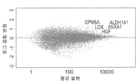
도 2a 내지 도 2e - 증대된 종양 개시 활성과 관련된 글로벌 유전자 표지(global gene signature)는 추정 CSC 바이오마커들 및 새로운 치료 타겟을 나타낸다. (도 2a) 여러 다양한 샘플들을 비교하는 마이크로어레이 유전자 발현 분석: 1. 일차 MRT, 2. 초기 MRT Xn (P2), 3. 중기 MRT Xn (P7), 4. 후기 MRT Xn (P17), 5. 인간 배아줄기세포 (hESCs), 6. 태아 신장 (FK), 7. 성인 신장 (AK), 8. 태아 뇌 (FB), 9. 성인 뇌 (AB). 비감독 계층 클러스터링(un-supervised hierarchical clustering)은 MRT 후기 계대배양 및 hESCs 사이의 큰 유사성을 나타내었고, 이는 그들의 비분화 성질을 강조하는 것이다; (도 2b) MRT 조직들의 비교는, 두가지의 구별되는 유전자 발현 패턴들을 나타내었다; 계대배양 동안 상향 조절된 유전자들(빨간색) 및 하향 조절된 유전자들(녹색); (도 2c) 세마포린3C(SEMA3C), 리실옥시다아제(LOX), 글리코프로테인 M6A(GPM6A), 헤파토사이트 성장인자(HGF/SF) 및 알데하이드 디하이드로게나아제1(ALDH1)를 포함하는 MRT에서 증가된 CSC 기능과 관련된 가장 상향 조절된 10개의 유전자들; (도 2d) 다양한 MRT 샘플들 간의 여러 증식 마커들의 발현 패턴들을 비교하는 유전자 히트 맵(gene heat map) (예를 들어, KI67, CDC20, CDK1 및 CCNA2)은 고도의 계대배양에 의해 고도의 증식이 이루어짐을 나타낸다; (E) ingenuityⓒ 기능 분석은, 일차 종양 및 낮은 계대수의 계대배양 Xn과 비교하여, 높은 계대수의 계대배양 Xn에서 감소된 괴사, 세포사멸 및 세포 분화를 나타냄을 보여주었다.
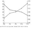
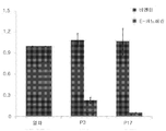
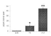
도 3a 내지 도 3e - ALDH1은 MRT CSC의 추정 마커이다: (도 3a) NOD/SCID 마우스에서 인간 MRT Xn의 연속적 증식에 의해, ALDH1을 발현하는 세포들의 증대가 초래된다, 그림 참조; (도 3b) 다양한 MRT Xn 계대배양들에 대한 대표적인 FACS 분석. 종양들이 진행됨에 따라, ALDHA1 발현 세포들의 비율은 현저히 증가하였다: P4 세포들로부터 유래된 세포들의 경우 4% (왼쪽), 및 P10 세포들로부터 유래된 세포들의 경우 25% (오른쪽), 이는 Xn 연속 증식을 통해 ALDH1+ 를 발현하는 세포들이 증대되었음을 나타내는 것이다; (도 3c) ALDH1에 대한 일차 MRT의 IHC, 중기 계대배양 Xn (P7) 및 후기 계대배양 Xn (P13)은 Xn 연속 증식에 따른 발현 증가를 나타낸다. 스케일 바, 200㎛; (도 3d) ALDH1에 대한 일차 MRT의 확대 IHC는 높은 수준의 ALDH1 발현 세포들은 주로 큰 육종양 세포들임을 보여준다. 스케일 바, 200㎛; (도 3e) qRT-PCR을 통해, 후기 계대배양들에서의 높은 ALDH1 발현이 확인되었다, 일차 종양에 비해 약 40배 더 높음. qRT-PCR 분석을 위하여, 일차 종양 세포들에 대한 값들이 정규화(=1)를 위하여 사용되었고, 따라서 모든 다른 값들이 계산되었다. 결과들은 3회의 별개 실험들의 평균값±S.E.M으로 나타내어진다. * p<0.05; ** p<0.01.
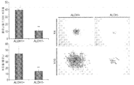
도 4a 내지 도 4d - MRT CSC 바이오마커로서의 ALDH1의 기능 확인: (도 4a) 콜로니 형성능력은 ALDH1+ MRT 세포들과 ALDH1- MRT 세포들 간에 비교되었다. ALDH1+ 세포들에 의해 형성된 콜로니들의 수는 ALDH1- 세포들에 비교하여 매우 높았다 (왼쪽 상부 막대그래프; p=0.0083). 세포/콜로니의 수는 ALDH1- 세포들에 비하여 ALDH1+ 세포들에서 매우 높았다 (왼쪽 하부 막대그래프; p=0.0024). ALDH1+ 세포들 및 ALDH1- 세포들로부터 형성된 콜로니들의 대표적인 이미지들은 오른쪽에 나타내었다. 실험은 3회씩 수행되었다; (도 4b) ALDH1+ 세포들로부터 야기된 종양들은 추가적으로 2차 수용체들(recipients) 내로 연속적으로 이식되었고, 이는 생체내 자가-재생 능력을 보여주는 것이고, 이 개체군에서의 종양 개시 능력의 존재와 일치된다; (도 4c) RNA 서열화 실험은, ALDH- 세포들에 비하여 ALDH+ 세포들에서 가장 상향 조절된 유전자들은, 일차 종양에 비하여 후기 Xn 계대배양들에서 매우 과발현된 유전자들이었음을 보여주었다(예를 들어, ANXA1 , GPM6A , HGF 및 LOX). (도 4d) qRT-PCR를 통한 확인은, ALDH- 세포들에 비하여, 분류된 ALDH+ 세포들에서 높은 LOX 발현이 있음을 나타내었다. qRT-PCR 분석을 위하여, ALDH- 세포들에 대한 값들이 정규화(=1)를 위하여 사용되었고, 따라서 모든 다른 값들이 계산되었다. 결과들은 3회의 별개 실험들의 평균값±S.E.M으로 나타내어진다; * p<0.05.
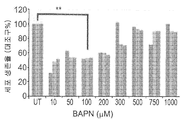
도 5a 내지 도 5d - 잠재적 MRT 치료 타겟으로서의 LOX 억제제의 기능 확인: (도 5a) LOX에 대한 일차 종양, 초기 (P2), 중기 (P7) 및 후기 (P14) MRT 계대배양물의 IHC 염색. 상기 염색은 Xn 연속 증식에 따른 광범위한 발현 증대를 나타내었다. 스케일 바, 10㎛; (도 5b) 윌름스 종양(WT) Xn 세포들에 대한 BAPN의 효과, 마우스에서 연속 증식된 신장의 공통 소아 고형 종양. 결과들은 WT 세포 증식에 대해 아무런 영향이 없음을 나타내었고, 이는 MRT 세포들에 대한 LOX 억제의 특이적 효과를 나타내는 것이다. ** p<0.01; (도 5c) MRT 세포들에 대한 BAPN 처리에 의해 유도된, 핵 농축(nuclear condensation) 및 세포 팽윤을 포함하는 형태학적 변화. 스케일 바, 100㎛ (상부) 및 50㎛ (하부); (도 5d) 세포이동 분석(migration assay)은 48시간 동안의 100μM BAPN 처리에 의해, MRT 세포들의 이동 능력이 비처리 세포들에 비하여 현저히 억제되었음을 보여준다. 스케일 바, 1000㎛.
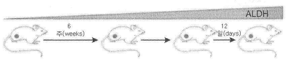
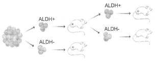
도 6 - ATRT Xn 모델의 확립, 그림 참조. 일차 종양 이식편들은 2~5mm의 종양 조각들을 면역 결핍된 마우스에게 피하 이식하여 형성하였다. 모든 마우스에서 종양 증식을 가능하게 하는 종양 취득(tumor take)이 관찰되었다. NOD/SCID 마우스에서 ATRT Xn의 연속 증식(serial propagation)은 1x10⁶ 세포의 고정된 세포수를 이용하여, 조직 샘플 이식(transplantation) 또는 단일 세포 현탁액 이식(grafting)에 의해 수행하였다. 연속 증식에 의해 낮은 계대수의 계대배양(<P5), 중간 계대수의 계대배양(P5-P10) 및 높은 계대수의 계대배양(P10-P15)의 ATRT Xn 계대배양들의 확립이 가능하게 되었다. 또한, 조직 단편들이 IHC 염색, RNA, DNA 및 단백질 분리를 위하여 사용되었다. 부착 세포들 또한 종양 세포들의 시험관내 연구를 위하여 사용되었다.
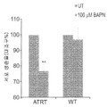
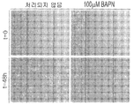
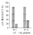
도 7a 내지 도 7b - ATRT CIC/CSC 치료제로서의 LOX 억제제의 기능 확인: (도 7a) 처리 후 세포 생존력 분석(Cell viability assay). 세포 생존력을 시험하는 MTS 분석은 다양한 농도의 BAPN(10-1000f μM)으로 48시간 동안 성장시킨 P2 Xn 세포들에 대해 수행되었다. 100μM BAPN으로 처리한 후, 결과적으로 증식이 상당히 감소되었다(UT 세포들에 비하여 47%). **, p<0.01. (도 7b) BAPN 처리에 의해, ATRT 세포들에서 형태학적 변화들이 야기되었다. 처리 후에 관찰된 변화들 중에는 핵 농축 및 세포 팽윤이 포함된다. 스케일 바, 100㎛ (상부) 및 50㎛ (하부). (도 7c) BAPN 처리는, 세포 이동을 억제한다. 세포이동 분석은, 48시간 동안의 100μM BAPN 처리는 비처리 세포들에 비하여 ATRT 세포의 이동 능력을 상당히 억제하였음을 보여준다. 스케일 바, 1000㎛. (도 7d) BAPN은 ATRT 세포들에서 LOX 활성을 효과적으로 억제한다. LOX 활성 정량 키트는 P3 및 P10 세포들에 대한 100μM BAPN 처리 후에 상당한 억제가 이루어짐을 보여준다(각각 81% 및 63%).
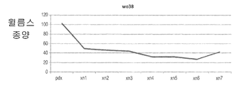
도 8a 및 도 8b - 종양이 나타나기 까지의 평균 일수를 이종이식 계대배양의 함수로서 나타낸 그래프들. 유잉 육종(도 8a) 및 윌름스 종양(도 8b)에서 종양 접종 시간의 단축과 관련되는 연속 Xn 증식, 이는 더욱 공격성인 표현형으로 나타나는 종양 거동에서의 변화를 보여주는 것이다.
도 9 - 고도로 증식되는 종양 이종이식의 후기 계대배양들을 나타내는 유전자 히트 맵으로서, 전이성 거동을 예상하게 하는 침투성 유전자 표지에 따른, 자가-재생 유전자들의 상향 조절(up-regulation)을 나타낸다.
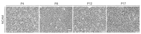
도 10a - NCAM1에 대한 여러 Xn 계대배양(P4, P8, P12 및 P17)의 면역화학염색(immunohistochemistry; IHC)은 Xn 연속 증식에 따른 증가된 발현을 나타낸다. 스케일 바, 200㎛;
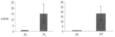
도 10b - qRT-PCR 분석은, 성인 폐 대조군(오른쪽)에 비하여 일차 종양에서높은 NCAM1 발현을 나타내고, 성인 폐(왼쪽)에 비하여 태아 폐에서 높은 NCAM1 발현을 나타낸다. 결과들은 3회의 별개 실험들의 평균값±S.E.M으로 나타내어진다; *, p<0.05.
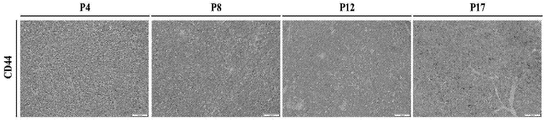
도 11a - CD44에 대한 여러 Xn 계대배양(P4, P8, P12 및 P17)의 IHC은 Xn 연속 증식에 따른 증가된 발현을 나타낸다. 스케일 바, 200㎛;
도 11b - CD44에 대한 일차 PPB의 확대 IHC는 높은 CD44 발현 세포들이 종양을 따라 분산되어 있는 것을 보여준다. 스케일 바, 200㎛. Some embodiments of the present invention are described by way of example only with reference to the accompanying drawings. The drawings are described in detail with particular reference thereto, but the illustrated embodiments are intended to illustrate examples of embodiments of the present invention through examples. In this regard, it will be apparent to those skilled in the art how the embodiments of the present invention can be practiced by the description made with reference to the drawings.
Figures 2a-2e - global genetic markers associated with the start of the increase in tumor activity (global gene signature) represents the estimated CSC biomarkers and new therapeutic targets. (Fig. 2a) Microarray gene expression analysis comparing various samples: 1. Primary MRT, 2. Initial MRT Xn (P2), 3. Midterm MRT Xn (P7), 4. Later MRT Xn (P17) Human embryonic stem cells (hESCs), 6. fetal kidney (FK), 7. adult kidney (AK), 8. fetal brain (FB), 9. adult brain (AB). Un-supervised hierarchical clustering showed large similarities between late MRT passage cultures and hESCs, highlighting their non-differentiating nature; (Figure 2b) Comparison of MRT tissues revealed two distinct gene expression patterns; Upregulated genes (red) and downregulated genes (green) during subculture; (Fig. 2c) Increase in MRT, including semaphorin 3C (SEMA3C), ricyloxidase (LOX), glycoprotein M6A (GPM6A), hepatocyte growth factor (HGF / SF) and aldehyde dehydrogenase 1 (ALDH1) 10 most up-regulated genes associated with CSC function; (Figure 2d) Gene heat maps (e.g., KI67, CDC20, CDK1 and CCNA2 ) that compare the expression patterns of various growth markers between various MRT samples are highly proliferated by advanced subculture ; (E) ingenuity © functional analysis showed reduced necrosis, apoptosis and cell differentiation in high passaged subculture Xn compared to subculture Xn in primary tumors and low passage counts.
Figures 3a-3e - ALDH1 is a marker of the estimated MRT CSC: (Fig. 3a) NOD / SCID mice by in the continuous growth of human MRT Xn, is caused an increase of cells which express ALDH1, see figure; (Figure 3b) Representative FACS analysis for various MRT Xn passages. As the tumors progressed, the proportion of ALDHA1 expressing cells significantly increased: 4% (left) for cells derived from P4 cells and 25% (right) for cells derived from P10 cells, Indicating that cells expressing ALDH1 + have been augmented through proliferation; (FIG. 3c) IHC, middle passage sub-culture Xn (P7) and late sub-passage culture Xn (P13) of primary MRT for ALDH1 show increased expression by Xn continuous proliferation. Scale bar, 200 m; (Figure 3d) Expansion of Primary MRT to ALDH1 IHC shows that high levels of ALDH1 expressing cells are mainly large sarcoma cells. Scale bar, 200 m; (Fig. 3e) qRT-PCR showed high ALDH1 expression in late passages, approximately 40-fold higher than primary tumors. For qRT-PCR analysis, values for primary tumor cells were used for normalization (= 1), and therefore all other values were calculated. Results are expressed as mean ± SEM of three separate experiments. * p <0.05; ** p < 0.01.
Fig. 4a-4d - check function of ALDH1 MRT CSC as biomarkers: (Fig. 4A) Colony forming ability was compared between ALDH1 + MRT cells and ALDH1-MRT cells. The number of colonies formed by ALDH1 + cells was very high compared to ALDH1- cells (top left bar graph; p = 0.0083). The number of cells / colonies was significantly higher in ALDH1 + cells than in ALDH1- cells (bottom left bar graph; p = 0.0024). Representative images of the colonies formed from ALDHl + cells and ALDHl- cells are shown on the right. The experiment was carried out three times; (Fig. 4b) Tumors arising from ALDHl + cells were additionally consecutively transplanted into secondary recipients, which demonstrate in vivo self-renewal ability and are consistent with the presence of tumor-initiating ability in this population; (Fig. 4c) RNA sequencing experiments showed that the most up-regulated genes in ALDH + cells compared to ALDH-cells were overexpressed genes in late Xn-line cultures compared to primary tumors (for example, ANXA1 , GPM6A , HGF and LOX ). (Fig. 4d). Confirmation by qRT-PCR showed higher LOX expression in sorted ALDH + cells compared to ALDH-cells. For qRT-PCR analysis, values for ALDH-cells were used for normalization (= 1) and therefore all other values were calculated. Results are expressed as mean ± SEM of three separate experiments; * p < 0.05.
Figure 5a to 5d - checking of the potential therapeutic target MRT as LOX inhibitor (Fig. 5a) the primary tumor for LOX, initial (P2), medium term (P7) and reviews (P14) MRT subculture water IHC staining. The staining showed extensive expression increases following Xn continuous proliferation. Scale bar, 10 탆; (Fig. 5b) Effect of BAPN on Wilms tumor (WT) Xn cells, a common pediatric solid tumor of the kidney continuously growing in the mouse. The results showed no effect on WT cell proliferation, indicating a specific effect of LOX inhibition on MRT cells. ** p <0.01; (Fig. 5c) Morphological changes, including nuclear condensation and cell swelling, induced by BAPN treatment on MRT cells. Scale bar, 100 mu m (top) and 50 mu m (bottom); (Fig. 5d) The migration assay showed that the migration ability of MRT cells was significantly inhibited by 100 μM BAPN treatment for 48 hours compared to untreated cells. Scale bar, 1000 탆.
Figure 6 - Establishment of ATRT Xn model, see figure. Primary tumor implants were formed by subcutaneous implantation of immunocompetent mice with 2-5 mm tumor fragments. Tumor take was observed in all mice to enable tumor growth. Serial propagation of ATRT Xn in NOD / SCID mice was performed by tissue sample transplantation or single cell suspension grafting using a fixed number of cells of 1 x 10 cells. Continuous proliferation enabled the establishment of ATRT Xn subcultures of subculture (<P5), subculture subculture (P5-P10) and high subculture subculture (P10-P15). Tissue fragments were also used for IHC staining, RNA, DNA and protein isolation. Adherent cells were also used for in vitro studies of tumor cells.
7a to 7b - Functional confirmation of LOX inhibitor as a therapeutic agent for ATRT CIC / CSC: (Fig. 7a) Cell viability assay after treatment. MTS assays to test cell viability were performed on P2 Xn cells grown at various concentrations of BAPN (10-1000fM) for 48 hours. After treatment with 100 μM BAPN, the result was a significant reduction in proliferation (47% relative to UT cells). **, p < 0.01. (Fig. 7B) BAPN treatment resulted in morphological changes in ATRT cells. Among the changes observed after treatment are nuclear enrichment and cell swelling. Scale bar, 100 탆 (upper) and 50 탆 (lower). (Fig. 7C) The BAPN treatment inhibits cell migration. Cell migration analysis shows that treatment with 100 [mu] M BAPN for 48 hours significantly suppressed the ability of ATRT cells to migrate relative to untreated cells. Scale bar, 1000 탆. (Fig. 7d) BAPN effectively inhibits LOX activity in ATRT cells. The LOX active quantitation kit showed significant inhibition after treatment with 100 [mu] M BAPN for P3 and P10 cells (81% and 63%, respectively).
Figures 8a and 8b - Graphs showing the mean days to tumor appearance as a function of xenograft subculture. Continuous Xn proliferation associated with the shortening of tumor inoculation time in Ewing's sarcoma (Fig. 8a) and Wilms tumor (Fig. 8b), showing a change in tumor behavior as a more aggressive phenotype.
Figure 9 - Genetic heatmap representing late passaged cultures of highly proliferating tumor xenografts showing up-regulation of self-regenerating genes, according to permeable genetic markers that predicted metastatic behavior.
Figure 10a - Immunohistochemistry (IHC) of several Xn passages (P4, P8, P12 and P17) to NCAM1 shows increased expression following Xn continuous proliferation. Scale bar, 200 m;
Figure 10b - qRT-PCR analysis shows high NCAM1 expression in primary tumors and high NCAM1 expression in fetal lung compared to adult lung (left) compared to adult lung control (right). Results are expressed as mean ± SEM of three separate experiments; *, p < 0.05.
Figure 11a -IHC of several Xn passages (P4, P8, P12 and P17) for CD44 show increased expression following Xn continuous proliferation. Scale bar, 200 m;
Figure 11b - Expansion of primary PPB to CD44 IHC shows that high CD44 expressing cells are distributed along the tumor. Scale bar, 200 탆.
본 발명의 일부 구체예는 일차 종양에서 암 줄기세포의 농축(enriching) 및 동정(identifying), 및 선택적으로 그들을 분리하는 방법에 관한 것이다. 암 줄기세포를 일차 종양에 존재하는 다른 암 세포들과 차별화하는 마커들은 암의 진단에 도움을 주기 위하여 동정될 수 있다. 또한, 상기 마커들은 암의 치료를 위한 타켓이 될 수 있다.Some embodiments of the present invention are directed to enriching and identifying cancer stem cells in primary tumors and, optionally, isolating them. Markers that differentiate cancer stem cells from other cancer cells in primary tumors can be identified to aid in the diagnosis of cancer. The markers may also be targets for the treatment of cancer.
본 발명의 적어도 하나의 구체예를 상세히 설명하기 전에, 본 발명은 그의 적용이 하기 설명 또는 예시된 실시예들에 제시된 상세 사항들에 반드시 한정되는 것은 아님이 이해되어야 할 것이다. 본 발명은 다른 구체예들로 구현될 수 있고, 또는 여러 다양한 방법들로 실시 또는 수행될 수 있다. Before explaining at least one embodiment of the invention in detail, it is to be understood that the invention is not necessarily limited to the details given in the following description or illustrated embodiments. The invention may be embodied in other specific forms, or may be practiced or carried out in various ways.
암 줄기세포(CSC)는 계층적으로 조직화된 종양들에서 악성 세포 서브세트들( malignant cell subsets)을 지칭하며, 이는 선택적으로 종양 개시 및 자가-재생이 가능하고, 분화를 통해 비-종양형성성(non-tumorigenic) 암세포 자손의 집단을 생성시킬 수 있다. Cancer stem cells (CSCs) refer to malignant cell subsets in hierarchically organized tumors, which are selectively capable of tumor initiation and self-renewal, and are capable of non-tumorigenic lt; RTI ID = 0.0 > non-tumorigenic < / RTI > cancer cell progeny.
생리학적 줄기세포들의 약물-내성 표현형에 대한 관찰의 필연적 결과로서, CSC는 또한 화학 요법 및 방사선 요법에 대한 증가된 내성으로 특징지워지는 암 내의 소집단(subpopulation)을 지칭할 수 있는 것으로 가정되어 왔다. 따라서, 특이적 마커들을 통해 CSC를 표적화하는 암 치료법은 재발 및 전반(dissemination)의 위험성을 감소시킴으로써, 기존의 치료법들의 효율을 잠재적으로 증가시킬 수 있을 것이다. 암 줄기세포의 동정은 특히 그들이 전형적으로 매우 적은 수의 종양 세포들에서 발현되는 점에서 복잡한 작업이다. As a consequence of observing the drug-resistant phenotype of physiological stem cells, CSC has also been postulated to be able to refer to subpopulations in cancer characterized by increased resistance to chemotherapy and radiotherapy. Thus, cancer therapy targeting CSCs through specific markers may potentially increase the efficiency of existing therapies by reducing the risk of recurrence and dissemination. The identification of cancer stem cells is a complex task in that they are typically expressed in a very small number of tumor cells.
소아 고형 종양에서는, 다수의 신선한 종양 시료에 대한 제한된 접근이 상기 문제를 복잡하게 만들어서, 내성 CSC 분석 및 효과적인 새로운 치료 전략의 개발을 방해한다. In pediatric solid tumors, limited access to a large number of fresh tumor samples complicates the problem, hindering the development of resistant CSC analysis and effective new therapeutic strategies.
본 발명자들은 CSC 표현형의 발견을 위하여, 면역결핍 마우스에서, 매우 공격성이고 치명적인 소아 신생물인 인간 비정형 유기형/간상 종양(ATRT)의 장기간 생체내 증식법을 한계 희석 이종이식법(limiting dilution xenotransplantation)과 조합하였다. 이러한 방법을 사용하여, 본 발명자들은 낮은 계대수의 계대배양(low-passage) 이종이식에 비하여 40배 미만에 해당되는 50개의 세포를 사용하여 ATRT를 생성시킬 수 있었다. For the discovery of the CSC phenotype, the present inventors have developed a long-term in vivo propagation method of human atypical organic / hepatic tumor (ATRT), a very aggressive and deadly pediatric neoplasm, in immune deficient mice by limiting dilution xenotransplantation Respectively. Using this method, we were able to generate ATRT using 50 cells that were less than 40-fold less than low-passage, low-passage xenotransplantation.
본 발명자들은 이러한 실험들로부터, 이종이식의 연속 사이클들은 추가의 CSC 표현형들을 선택하기 위해 사용될 수 있고, 이 방법은 암의 진단 및 치료 모두를 위한 가치있는 수단이 될 수 있다고 추론하였다. From these experiments, the present inventors have deduced that continuous cycles of xenotransplantation can be used to select additional CSC phenotypes and that this method can be a valuable tool for both the diagnosis and treatment of cancer.
ATRT와 유사하게, 본 발명자들은 유잉육종, 윌름스 종양 및 흉막과 폐의 모세포증(PPB)의 연속 증식은, 계대배양에 따라 종양의 공격성이 촉진되었음을 보여주는 종양 접종에 대한 시간 단축 및 종양 성장 촉진과 관련된 이종이식들(Xns)을 생성시켰음을 발견하였다. Similar to ATRT, we found that continuous proliferation of Ewing's sarcoma, Wilms' tumor and pleural and pulmonary blast proliferation (PPB) reduced time to tumor inoculation and accelerated tumor growth, indicating that the aggressiveness of the tumor was enhanced by subculture (Xns) associated with the immune system.
특정 종양에 대한 CSCs에 대한 지식은 새로운 치료법의 개발에 도움을 준다. 이는 개인적 필요 수준(즉, 특정 환자의 종양에서 CSCs 관찰 및 그에 따른 치료) 또는 보다 일반적인 수준(특정 암들에 대한 치료법의 개발)에 영향을 미칠 수 있다. Knowledge of CSCs for specific tumors aids in the development of new therapies. This may affect the level of personal needs (ie, observation of CSCs in a particular patient's tumor and treatment thereof) or at a more general level (development of treatment for certain cancers).
본 발명의 일 측면에 따르면, 다음의 단계들을 포함하는, 인간 일차 종양에서 암 줄기세포 마커들을 동정하는 방법이 제공된다:According to one aspect of the present invention there is provided a method of identifying cancer stem cell markers in a human primary tumor comprising the steps of:
(a) 계대배양된 종양 세포들의 제1 개체군을 생성시키기 위한 계대수로 상기 일차 종양을 생체내에서 계대배양하는 단계; 및(a) subculturing said primary tumor in vivo with a passage count to produce a first population of subcultured tumor cells; And
(b) 계대배양된 일차 종양의 종양 세포들의 제1 개체군에서의 적어도 하나의 항원의 수준을 상기 일차 종양의 종양 세포들의 제2 개체군과 비교하는 단계, (b) comparing the level of at least one antigen in a first population of tumor cells of subcultured primary tumor with a second population of tumor cells of said primary tumor,
여기서, 상기 종양 세포들의 제2 개체군은 다음의 (ⅰ) 또는 (ⅱ)이다:Wherein the second population of tumor cells is (i) or (ii)
(ⅰ) 상기 인간 일차 종양의 계대배양되지 않은(non-passaged) 세포들; 또는(I) non-passaged cells of said human primary tumor; or
(ⅱ) 상기 인간 일차 종양의 생체내 계대배양된 세포들, 여기서 상기 종양 세포들의 제2 개체군은 계대배양된 종양 세포들의 제1 개체군 보다 적어도 1회 더 적은 계대수로 생체내 계대배양된 것이고,(Ii) in vivo subcultured cells of said human primary tumor, wherein said second population of said tumor cells is subcultured in vivo at least one less than the first population of subcultured tumor cells,
상기 종양 세포들의 제2 개체군에서의 상기 항원의 양(amount)과 비교하여 상기 종양 세포들의 제1 개체군에서의 상기 항원의 양의 증가는 상기 인간 일차 종양에서의 암 줄기세포 마커들의 지표가 된다. An increase in the amount of the antigen in the first population of tumor cells as compared to an amount of the antigen in the second population of tumor cells is indicative of cancer stem cell markers in the human primary tumor.
본 명세서에서 사용된 용어 "암 줄기세포", ("CSC"로 언급되기도 함)는 이종이식(Xn) 마우스 모델을 사용하여 암을 생성시키는 능력을 갖는 세포를 의미한다. CSCs는 자가-재생 과정을 통해 자신들을 재생할 수 있고, 이는 연속 이식 분석법으로 연구될 수 있다. 또한, 정제된 CSCs로부터 유래된 암들은 그들이 유래된 부모 암의 이종성 표현형들을 재현하고, 이는 CSCs의 분화능력을 반영한다. As used herein, the term "cancer stem cell ", also referred to as" CSC ", refers to a cell that has the ability to produce cancer using a xenograft (Xn) mouse model. CSCs can reproduce themselves through the self-regeneration process, which can be studied by continuous grafting analysis. Also, cancers derived from purified CSCs reproduce heterologous phenotypes of the parental cancer from which they are derived, reflecting the differentiation potential of CSCs.
본 명세서에서 사용된 용어 "종양(tumor)"은 조절되지 않은(misregulated) 세포 증식에 의해 형성된 세포들 또는 조직의 그룹을 의미한다. 종양은 구조적 조직화(structural organization) 및 정상조직과의 기능적 협력의 부분적 또는 완전한 결핍을 나타내고, 일반적으로 분리된 조직 덩어리를 형성하며, 이는 양성 또는 악성일 수 있다. 하나의 구체예에서, 용어 "종양"은 악성 종양을 의미한다. 하나의 구체예에 따르면, 용어 "종양 세포들"은 또한 백혈병 세포들과 같은 비-고형 암 세포들을 포함한다. 다른 구체예에 따르면, 각각의 비-고형 암 세포들은 상기 용어 "종양 세포들"의 범위에 포함되지 않는다. The term "tumor ", as used herein, refers to a group of cells or tissues formed by unregregulated cell proliferation. Tumors represent a partial or complete lack of structural organization and functional cooperation with normal tissue, and generally form a separate tissue mass, which may be benign or malignant. In one embodiment, the term "tumor" refers to a malignant tumor. According to one embodiment, the term "tumor cells" also encompass non-solid cancer cells such as leukemia cells. According to another embodiment, each non-solid cancer cell is not included in the scope of the term " tumor cells ".
특정 구체예에 따르면, 종양은 배아 줄기세포 기원(origin)을 갖는 고형 종양(예를 들어, 소아 종양)이다. According to a particular embodiment, the tumor is a solid tumor (e. G., A pediatric tumor) having an embryonic stem cell origin.
그러한 고형 종양들의 예로는, 비제한적인 예로서 다음과 같은 육종암(sarcomas) 및 암종(carcinomas)을 포함하나, 이들에 제한되는 것은 아니다: 섬유육종(fibrosarcoma), 점액육종(myxosarcoma), 흉막과 폐의 모세포증(pleulonary blastoma), 지방육종(liposarcoma), 연골육종(chondrosarcoma), 골원성 육종(teogenic sarcoma), 척삭종(chordoma), 혈관육종(angiosarcoma), 내피 육종(endotheliosarcoma), 림프관 육종(lymphangiosarcoma), 림프관 내피육종(lymphangioendotheliosarcoma), 활막종(synovioma), 중피종(mesothelioma), 유잉 종양(Ewing's tumor), 평활근육종(leiomyosarcoma), 횡문근육종(rhabdomyosarcoma), 결장암(colon carcinoma), 췌장암(pancreatic cancer), 유방암(breast cancer), 난소암(ovarian cancer), 전립선암(prostate cancer), 편평상피암(squamous cell carcinoma), 기저세포암(basal cell carcinoma), 선암(adenocarcinoma), 한선암종(sweat gland carcinoma), 피지선암종(sebaceous gland carcinoma), 유두상암종(papillary carcinoma), 유두상선암(papillary adenocarcinomas), 낭종암(cystadenocarcinoma), 수양암(medullary carcinoma), 기관지원성암종(bronchogenic carcinoma), 신장암(renal cell carcinoma), 간암(hepatoma), 담관암(bile duct carcinoma), 융모암(choriocarcinoma), 정상피종(seminoma), 태생기암(embryonal carcinoma), 윌름스 종양(Wilms' tumor), 자궁경부암(cervical cancer), 고환암(testicular tumor), 폐암종(lung carcinoma), 소세포폐암(small cell lung carcinoma), 방광암종(bladder carcinoma), 원주상피암(epithelial carcinoma), 신경교종(glioma), 성상세포종(astrocytoma), 수모세포종(medulloblastoma), 두개인두종(craniopharyngioma), 상의세포종(ependymoma), 송과체종(pinealoma), 혈관아세포종(hemangioblastoma), 청신경종(acoustic neuroma), 핍지교종(oligodendroglioma), 뇌수막종(meningioma), 악성흑색종(melanoma), 신경아세포종(neuroblastoma), 및 망막아세포종(retinoblastoma). 소아 고형 종양의 예로는, 다음을 포함하나, 이들에 제한되는 것은 아니다: 윌름스 종양/신아세포종(Wilms' tumor/Nephroblastoma), 횡문근육종(rhabdomyosarcoma), 유잉계 종양/원시신경외배엽종양(Ewing's family of tumors/primitive neuroectodermal tumor), 골육종(Osteosarcoma), 말초원시신경외배엽종양(peripheral neuroectodermal tumors), 소아 생식세포종(Childhood Germ Cell Tumor), 성선외생식세포종(Extragonadal Germ Cell Tumor), 신장암(Kidney Cancer), 간암(Liver Cancer), 신경아세포종(Neuroblastoma), 난소암(Ovarian Cancer), 망막아세포종(Retinoblastoma), 육종(Sarcoma), 더욱 구체적으로는, ㄱ고골육종(Osteosarcoma), 횡문근육종(Rhabdomyosarcoma), 결합조직성 소원형세포종양(Desmoplastic small round-cell tumor), 간모세포종(Hepatoblastoma), 배세포종양(Germ cell tumors), 신경아세포종(neuroblastoma) 및 수모세포종(Medulloblastoma).Examples of such solid tumors include, but are not limited to, fibrosarcoma, myxosarcoma, pleura, and sarcomas, including, but not limited to, sarcomas and carcinomas, The present invention relates to the use of a compound of formula (I) or a pharmaceutically acceptable salt thereof in the manufacture of a medicament for the treatment and / or prophylaxis of a disease selected from the group consisting of pleulonary blastoma, liposarcoma, chondrosarcoma, teogenic sarcoma, chordoma, angiosarcoma, endotheliosarcoma, lymphangiosarcoma, lymphangioendotheliosarcoma, synovioma, mesothelioma, Ewing's tumor, leiomyosarcoma, rhabdomyosarcoma, colon carcinoma, pancreatic cancer, Breast cancer, ovarian cancer, prostate cancer, squamous cell carcinoma, basal cell carcinoma, adenocarcinoma, sweat gland carcinoma, ), Sebaceous adenocarcinoma papillary carcinoma, papillary adenocarcinomas, cystadenocarcinoma, medullary carcinoma, bronchogenic carcinoma, renal cell carcinoma, pancreatic carcinoma, pancreatic carcinoma, Hepatoma, bile duct carcinoma, choriocarcinoma, seminoma, embryonal carcinoma, Wilms' tumor, cervical cancer, testicular cancer, tumor, lung carcinoma, small cell lung carcinoma, bladder carcinoma, epithelial carcinoma, glioma, astrocytoma, medulloblastoma, Neuroblastoma, acne neuroma, oligodendroglioma, meningioma, malignant melanoma, nerve plexus, neuroblastoma, neuroblastoma, neuroblastoma, craniopharyngioma, ependymoma, pinealoma, Neuroblastoma, Retinoblastoma (retinoblastoma). Examples of pediatric solid tumors include, but are not limited to, Wilms' tumor / Nephroblastoma, rhabdomyosarcoma, Ewing's tumor / origin of the optic nerve, Ewing's family (Tumor), Tumor, Tumor, Tumor, Tumors / primitive neuroectodermal tumor, Osteosarcoma, Peripheral neuroectodermal tumors, Childhood Germ Cell Tumor, Extragonadal Germ Cell Tumor, Kidney Cancer ), Liver cancer, neuroblastoma, ovarian cancer, retinoblastoma, sarcoma, and more specifically, It is well known that osteosarcoma, rhabdomyosarcoma, desmoplastic small round-cell tumors, hepatoblastomas, germ cell tumors, neuroblastoma, Neoplasm (Medulloblastoma).
본 명세서에서 사용된 용어 "일차 종양(primary tumor)"은, 종양 진행이 시작되어 암 덩어리를 형성하게 되는 해부학적 위치로부터 얻어지는 세포들을 의미한다. The term "primary tumor ", as used herein, refers to cells obtained from an anatomical location where tumor progression is initiated to form a cancer mass.
상기 항원은, 이종이식(Xn) 마우스 모델을 이용하여 암을 생성시키는 능력을 갖지 않고, 자가-재생 과정을 통해 스스로를 재생시킬 수 없는 동종의 종양의 암세포들과 비교할 때, CSC에서 차별화되는 양으로 존재하는 임의의 분자일 수 있다. The antigen does not have the ability to produce cancer using a xenograft model and can not differentiate from CSC when compared to cancer cells of the same tumor that can not regenerate itself through self- Lt; / RTI >
따라서, 예를 들어, CSC 마커는 폴리펩타이드, 탄수화물, 펩타이드, 또는 RNA 분자일 수 있다.Thus, for example, the CSC marker can be a polypeptide, a carbohydrate, a peptide, or an RNA molecule.
상기 항원은 세포의 표면에 위치할 수 있고, 또는 세포내 분자일 수 있다. The antigen may be on the surface of the cell, or it may be an intracellular molecule.
특정 구체예에 따르면, CSC 마커는 폴리펩타이드이다.According to a particular embodiment, the CSC marker is a polypeptide.
상기 폴리펩타이드는 세포 표면 단백질(즉, 막 단백질)일 수 있다. 특정 구체예에 따르면, 상기 세포 표면 단백질은 탄수화물 결합 분자(예를 들어, 레시틴)이다.The polypeptide may be a cell surface protein (i.e., a membrane protein). According to a particular embodiment, the cell surface protein is a carbohydrate binding molecule (e. G., Lecithin).
다른 구체예에 따르면, 상기 단백질은 세포외 단백질(예를 들어, 가용성 단백질)이다. According to another embodiment, the protein is an extracellular protein (e. G., A soluble protein).
상기 폴리펩타이드는, 예를 들어 전사 인자, 경로 관련 마커 등의 기능을 수행할 수 있다The polypeptide can perform functions such as, for example, transcription factors, pathway-related markers, and the like
본 명세서에서 사용된 용어 "생체내 계대배양(in vivo passaging)"은 일차 종양을 동물(예를 들어, 마우스) 내로 초기 이식(initial implantation)하고, 2차 종양으로 진행될 때까지 일정 기간 기다린 다음, 종양 세포들을 수확하여 2차 동물 내로 상기 종양 세포들을 이식(예를 들어, 연속 이식(serial transplantations))하는 것을 포함하는 과정을 의미한다. 그런 다음, 연속적인 일련의 계대배양이 수행될 수 있다. 본 발명은 적어도 1 계대수의 계대배양, 적어도 2 계대수의 계대배양, 적어도 3 계대수의 계대배양, 적어도 4 계대수의 계대배양, 적어도 5 계대수의 계대배양, 적어도 6 계대수의 계대배양, 적어도 7 계대수의 계대배양, 적어도 8 계대수의 계대배양, 적어도 9 계대수의 계대배양, 적어도 10 계대수의 계대배양, 적어도 11 계대수의 계대배양, 적어도 12 계대수의 계대배양, 적어도 13 계대수의 계대배양, 적어도 14 계대수의 계대배양, 적어도 15 계대수의 계대배양, 적어도 16 계대수의 계대배양, 적어도 17 계대수의 계대배양, 적어도 18 계대수의 계대배양, 적어도 19 계대수의 계대배양, 적어도 20 계대수의 계대배양으로 계대배양을 수행하는 것을 포함한다. The term " in vivo passaging ", as used herein, refers to an initial implantation of a primary tumor into an animal (e.g., a mouse), a period of waiting until progressing to a secondary tumor, Refers to a process involving harvesting tumor cells and transplanting (e. G., Serial transplantations) the tumor cells into a secondary animal. A continuous series of subcultures can then be performed. The present invention relates to a method for cultivating at least one subculture, subculturing at least two subcultures, subculturing at least three subcultures, subculturing at least four subcultures, subculturing at least five subcultures, subculturing at least six subcultures At least 7 passages, at least 8 passages, at least 9 passages, at least 10 passages, at least 11 passages, at least 12 passages, at least 12 passages, At least 14 passages, at least 15 passages, at least 16 passages, at least 17 passages, at least 18 passages, at least 19 passages ≪ / RTI > passaged subculture, subculture with at least 20 passages of subculture.
특정 구체예에 따르면, 상기 계대배양은 5 계대수 이상, 예를 들어 5~30, 또는 5~20 계대수의 계대배양이다. According to a particular embodiment, the subculture is a subculture of 5 or more, for example 5 to 30, or 5 to 20 passages.
특정 구체예에 따르면, 상기 계대배양은 10 계대수 이상, 예를 들어 10~30, 또는 10~20 계대수의 계대배양이다. According to a particular embodiment, the subculture is a subculture of 10 or more passages, for example 10 to 30, or 10 to 20 passages.
특정 구체예에 따르면, 상기 종양 세포들(종양 세포들의 제1 개체군 및/또는 종양 세포들의 제2 개체군)은 시험관내 계대배양을 하지 않은 것이다. According to a particular embodiment, the tumor cells (the first population of tumor cells and / or the second population of tumor cells) have not undergone in vitro passaging.
다른 특정 구체예에 따르면, 상기 종양 세포들은 일 라운드(one round) 초과의 시험관내 계대배양을 하지 않은 것이다. According to another particular embodiment, the tumor cells have not undergone more than one round of in vitro passaging.
생체내 계대배양에 사용되는 동물들의 예로는, 선충(nematodes), 초파리(fruit flies), 제브라피쉬(zebrafish)를 포함하고; 바람직하게는 마우스 (누드 마우스, SCID 마우스, NOD/SCID 마우스, 베이지(Beige)/SCID 마우스), 쥐(rat), 토끼, 또는 영장류(예를 들어, 인간)와 같은 실험실용 포유류가 사용된다. 하나의 구체예에 따르면, 면역결핍 동물들이 사용된다. 다른 구체예에 따르면, 생체내 계대배양을 위해 인간이 사용된다. Examples of animals used for in vivo passaging include nematodes, fruit flies, zebrafish; Preferably, laboratory mammals such as mice (nude mice, SCID mice, NOD / SCID mice, Beige / SCID mice), rats, rabbits, or primates (e.g., humans) are used. According to one embodiment, immunodeficient animals are used. According to another embodiment, a human is used for in vivo passaging.
전형적으로, 동일한 종의 동물이 각각의 생체내 계대배양에 사용된다. 따라서, 예를 들어, 만약 면역결핍 마우스가 초기 이식에 사용되면, 다음 계대배양을 위해서 면역결핍 마우스가 사용된다. Typically, animals of the same species are used in each in vivo subculture. Thus, for example, if an immunodeficient mouse is used for early transplantation, an immunodeficient mouse is used for subsequent passage.
일차 종양이 제2 동물 내에서 2차 종양의 진행을 개시시키는 한, 일차 종양은 어떤 양으로도 사용될 수 있다. 특정 구체예에 따르면, 일차 종양은 조각들(예를 들어, 1~10 mm 조각들)로 절단된다. 바람직하게는, 일차 종양의 세포들은 효소를 사용하여 해리되지 않은 것이다. 또한, 일차 종양의 세포들은 바람직하게는 저미거나(minced) 또는 갈은(ground) 것이 아니다.As long as the primary tumor initiates progression of the secondary tumor in the second animal, the primary tumor may be used in any amount. According to a particular embodiment, the primary tumor is cut into pieces (e.g., 1 to 10 mm pieces). Preferably, the cells of the primary tumor are not dissociated using the enzyme. In addition, the cells of the primary tumor are preferably not minced or ground.
특정 구체예에 따르면, 일차 종양의 세포들은 이식 전에 정제 과정(예를 들어, NCAM에 특이적으로 결합하는 항체를 사용하는 유동세포 계수법(flow cytometry)과 같은 면역-분리법(immune-isolation))을 거치지 않은 것이다. 또한, 이어지는 생체내 계대배양에서 이종이식 세포들 역시 바람직하게는 정제 과정(예를 들어, NCAM에 특이적으로 결합하는 항체를 사용하는 유동세포 계수법(flow cytometry)과 같은 면역-분리법(immune-isolation))을 거치지 않은 것이다. According to a particular embodiment, the cells of the primary tumor are subjected to a purification process prior to transplantation (e. G., Immune-isolation, such as flow cytometry using antibodies specifically binding to NCAM) It has not passed. In subsequent in vivo subculture, the xenograft cells are also preferably subjected to a purification procedure (e. G., Immune-isolation, such as flow cytometry using antibodies specifically binding to NCAM) )).
상기 종양은 그것이 종양 진행을 개시시키는 한, 동물 내에 어떤 방식으로도이식될 수 있다. 하나의 구체예에 따르면, 상기 종양은 피하 이식된다. The tumor can be implanted in any way in an animal so long as it initiates tumor progression. According to one embodiment, the tumor is subcutaneously transplanted.
전형적으로, 종양들은 이식 후 약 1~3개월 후 또는 그들의 크기가 1~3cm, 예를 들어 직경 1.5cm에 도달했을 때 수확된다. Typically, tumors are harvested approximately one to three months after transplantation or when their size reaches 1 to 3 cm, for example 1.5 cm in diameter.
이어지는 이종이식 증식은 이종이식 단일 세포 현탁액(single cells suspensions) 또는 이종이식 조직 단편들을 이식함으로써 실현될 수 있다. Subsequent xenotransplantation proliferation can be accomplished by implanting xenotransplantation single cell suspensions or xenograft tissue fragments.
단일 세포 현탁액의 경우, 해리된 세포들의 수는 생체내 계대배양에 사용된 동물 및 종양의 공격성(aggressiveness)에 따라 달라질 것이다. 전형적으로는, 새로이 회수된(freshly retrieved) Xn 조직으로부터 유래된 약 0.5~3x10⁶ 개의 해리 세포들이 사용된다. In the case of a single cell suspension, the number of dissociated cells will depend on the aggressiveness of the animal and tumor used for in vivo passaging. Typically, about 0.5-3
이종이식 조직 단편들은 전형적으로 1~10 mm, 더욱 바람직하게는 2~5 mm이다.The xenograft tissue fragments are typically 1 to 10 mm, more preferably 2 to 5 mm.
단일 세포 현탁액은 적절한 매질(바람직하게는 항체들도 포함함) 내에서 저미기(mincing), 갈기(grinding) 또는 분산을 수행하고, 이어서 콜라게나아제와 같은 단백질 분해효소로 처리함으로써 얻어질 수 있다. 그런 다음, 효소 처리된 조직은 적절한 매질 내에서 연마(triturated)될 수 있다.Single cell suspensions may be obtained by performing mincing, grinding or dispersion in a suitable medium (preferably including antibodies) followed by treatment with proteolytic enzymes such as collagenase . The enzyme treated tissue can then be triturated in a suitable medium.
특정 구체예에 따르면, 종양 세포들의 제2 개체군은 계대배양되지 않은 인간 일차 종양의 세포들이다. 다른 구체예에 따르면, 종양 세포들의 제2 개체군은 1 계대수로 계대배양된 것이다. 다른 구체예에 따르면, 종양 세포들의 제2 개체군은 2 계대수로 계대배양된 것이다. 다른 구체예에 따르면, 종양 세포들의 제2 개체군은 3 계대수로 계대배양된 것이다. 다른 구체예에 따르면, 종양 세포들의 제2 개체군은 4 계대수로 계대배양된 것이다. 다른 구체예에 따르면, 종양 세포들의 제2 개체군은 5 초과의 계대수로 계대배양된 것은 아니다. According to a particular embodiment, the second population of tumor cells are cells of a human primary tumor that have not been subcultured. According to another embodiment, the second population of tumor cells is subcultured to a passage number. According to another embodiment, the second population of tumor cells is subcultured in two passages. According to another embodiment, the second population of tumor cells is subcultured into three passages. According to another embodiment, the second population of tumor cells is subcultured in 4 passages. According to another embodiment, the second population of tumor cells is not subcultured to a passage number greater than 5.
종양 세포들의 제1 개체군은 적어도 1회(one round)의 생체내 계대배양을 한 것이다. 다른 구체예에 따르면, 종양 세포들의 제1 개체군은 적어도 2회의 생체내 계대배양을 한 것이다. 다른 구체예에 따르면, 종양 세포들의 제1 개체군은 적어도 3회의 생체내 계대배양을 한 것이다. 다른 구체예에 따르면, 종양 세포들의 제1 개체군은 적어도 4회의 생체내 계대배양을 한 것이다. 다른 구체예에 따르면, 종양 세포들의 제1 개체군은 적어도 5회의 생체내 계대배양을 한 것이다. 다른 구체예에 따르면, 종양 세포들의 제1 개체군은 적어도 6회의 생체내 계대배양을 한 것이다. 다른 구체예에 따르면, 종양 세포들의 제1 개체군은 적어도 7회의 생체내 계대배양을 한 것이다. 다른 구체예에 따르면, 종양 세포들의 제1 개체군은 적어도 8회의 생체내 계대배양을 한 것이다. 다른 구체예에 따르면, 종양 세포들의 제1 개체군은 적어도 9회의 생체내 계대배양을 한 것이다. 다른 구체예에 따르면, 종양 세포들의 제1 개체군은 적어도 10회의 생체내 계대배양을 한 것이다. 다른 구체예에 따르면, 종양 세포들의 제1 개체군은 적어도 15회의 생체내 계대배양을 한 것이다. 다른 구체예에 따르면, 종양 세포들의 제1 개체군은 적어도 20회의 생체내 계대배양을 한 것이다. The first population of tumor cells is at least one round of in vivo subculture. According to another embodiment, the first population of tumor cells is at least two in vivo subculture cultures. According to another embodiment, the first population of tumor cells has undergone at least three in vivo passages. According to another embodiment, the first population of tumor cells is at least four in vivo subculture cultures. According to another embodiment, the first population of tumor cells has undergone at least five in vivo subculture cultures. According to another embodiment, the first population of tumor cells has undergone at least six in vivo subculture cultures. According to another embodiment, the first population of tumor cells has undergone at least seven in vivo subculture cultures. According to another embodiment, the first population of tumor cells has undergone at least 8 in vivo subculture. According to another embodiment, the first population of tumor cells has undergone at least nine in vivo subculture cultures. According to another embodiment, the first population of tumor cells has undergone at least 10 in vivo subculture. According to another embodiment, the first population of tumor cells has undergone at least 15 in vivo subculture cultures. According to another embodiment, the first population of tumor cells has undergone at least 20 in vivo subculture cultures.
전형적으로, 종양 세포들의 제1 개체군은, 상기 계대배양된 세포들의 제1 개체군의 5000개 세포들을 사용하여 후손 세대 이종이식(later generation xenograft)을 개시시키는 빈도(frequency)가 인간 일차 종양의 계대배양되지 않은 세포들과 비교하여 적어도 5배까지 증가되도록 생체내 계대배양을 한 것이다. Typically, the first population of tumor cells is selected from the group consisting of 5,000 cells of the first population of subcultured cells, and the frequency of initiation of the later generation xenograft is greater than that of the subculture of human primary tumors Lt; RTI ID = 0.0 > 5-fold < / RTI >
본 명세서에서 사용된 용어 "이종이식(xenograft)"은 하나의 종(species), 속(genus) 또는 과(family)의 개체로부터 유래된 조직 또는 기관(organs)을 다른 종, 속, 또는 과의 개체 내로 외과적으로 이식(transplant 또는 graft)하는 것을 의미한다. The term " xenograft, " as used herein, refers to a tissue or organ originating from a species, genus, or family entity, Means transplanting (grafting) into an individual.
바람직하게는, 후손 세대 이종이식을 개시시키는 상기 빈도는 적어도 통계학적으로 유의한 수가 얻어지도록 적어도 2마리, 예를 들어 3마리, 5마리 또는 더욱 바람직하게는 10마리까지의 동물들에서 결정된다. Preferably, the frequency of initiating offspring generation xenotransplantation is determined in animals of at least 2, for example 3, 5 or more preferably 10, so as to obtain at least a statistically significant number.
따라서, 예로서, 만약 종양 세포들의 제2 개체군이 테스트된 10마리의 마우스 중 한마리에서 후손 세대 이종이식을 개시시킨다면, 동일한 수의 종양 세포들을 사용하여, 종양 세포들의 제1 개체군이 10마리의 마우스 중 적어도 5마리에서 후손 세대 이종이식을 개시시키도록 충분히 생체내 계대배양되어야 한다. 전형적으로, 후손 세대 이종이식을 개시시키기 위해 사용되는 상기 종양 세포들은 단일 세포 현탁액 내에 존재한다. Thus, for example, if the second population of tumor cells initiates a descendant generation xenotransplantation in one of the 10 tested mice, the same number of tumor cells is used, so that the first population of tumor cells is divided into 10 mice Lt; RTI ID = 0.0 > 5 < / RTI > Typically, the tumor cells used to initiate descendant generation xenotransplantation are present in a single cell suspension.
후손 세대 이종이식이 개시되었는지의 여부를 테스트하는 것은, 당 분야 기술자의 전문 지식의 범주에 속한다. 이종이식이 개시되었는지의 여부를 결정하는데 사용될 수 있는 예시적인 방법들에는, 육안적 종양 촉진(gross tumor palpation), 캘리퍼스(calipers), 초음파 유도하 이미징화(ultrasound-guided imaging) 등이 포함된다. It is within the expertise of the skilled artisan to test whether or not a descendant generation xenotransplantation has been initiated. Exemplary methods that can be used to determine whether xenotransplantation has been initiated include gross tumor palpation, calipers, ultrasound-guided imaging, and the like.
하나의 구체예에 따르면, 종양 세포들의 제1 개체군은 상기 계대배양된 세포들의 제1 개체군의 5000개 세포들을 사용하여 후손 세대 이종이식을 개시시키는 빈도가 인간 일차 종양의 계대배양되지 않은 세포들과 비교하여 적어도 10배까지 증가되도록 충분히 생체내 계대배양된 것이다. According to one embodiment, the first population of tumor cells is selected from the group consisting of non-subcultured cells of human primary tumors and the number of cells expressing the frequency of initiation of descendant generation xenografts using 5000 cells of the first population of subcultured cells And are sufficiently subcultured in vivo to be increased by at least 10 times.
하나의 구체예에 따르면, 종양 세포들의 제1 개체군은 상기 계대배양된 세포들의 제1 개체군의 5000개 세포들을 사용하여 후손 세대 이종이식을 개시시키는 빈도가 인간 일차 종양의 계대배양되지 않은 세포들과 비교하여 적어도 20배까지 증가되도록 충분히 생체내 계대배양된 것이다. According to one embodiment, the first population of tumor cells is selected from the group consisting of non-subcultured cells of human primary tumors and the number of cells expressing the frequency of initiation of descendant generation xenografts using 5000 cells of the first population of subcultured cells Which is at least 20 times higher than that of the control.
하나의 구체예에 따르면, 종양 세포들의 제1 개체군은 상기 계대배양된 세포들의 제1 개체군의 1000개 세포들을 사용하여 후손 세대 이종이식을 개시시키는 빈도가 인간 일차 종양의 계대배양되지 않은 세포들과 비교하여 적어도 5배까지 증가되도록 충분히 생체내 계대배양된 것이다. According to one embodiment, the first population of tumor cells is selected from the group consisting of cells that have not been subcultured to human primary tumors and whose frequency of initiation of descendant generation xenotransplantation using 1000 cells of the first population of subcultured cells Lt; RTI ID = 0.0 > 5-fold. ≪ / RTI >
하나의 구체예에 따르면, 종양 세포들의 제1 개체군은 상기 계대배양된 세포들의 제1 개체군의 1000개 세포들을 사용하여 후손 세대 이종이식을 개시시키는 빈도가 인간 일차 종양의 계대배양되지 않은 세포들과 비교하여 적어도 10배까지 증가되도록 충분히 생체내 계대배양된 것이다. According to one embodiment, the first population of tumor cells is selected from the group consisting of cells that have not been subcultured to human primary tumors and whose frequency of initiation of descendant generation xenotransplantation using 1000 cells of the first population of subcultured cells And are sufficiently subcultured in vivo to be increased by at least 10 times.
하나의 구체예에 따르면, 종양 세포들의 제1 개체군은 상기 계대배양된 세포들의 제1 개체군의 1000개 세포들을 사용하여 후손 세대 이종이식을 개시시키는 빈도가 인간 일차 종양의 계대배양되지 않은 세포들과 비교하여 적어도 20배까지 증가되도록 충분히 생체내 계대배양된 것이다. According to one embodiment, the first population of tumor cells is selected from the group consisting of cells that have not been subcultured to human primary tumors and whose frequency of initiation of descendant generation xenotransplantation using 1000 cells of the first population of subcultured cells Which is at least 20 times higher than that of the control.
하나의 구체예에 따르면, 종양 세포들의 제1 개체군은 상기 계대배양된 세포들의 제1 개체군의 500개 세포들을 사용하여 후손 세대 이종이식을 개시시키는 빈도가 인간 일차 종양의 계대배양되지 않은 세포들과 비교하여 적어도 5배까지 증가되도록 충분히 생체내 계대배양된 것이다. According to one embodiment, the first population of tumor cells is selected from the group consisting of non-subcultured cells of human primary tumors and the number of lymphocyte-derived xenografts using the 500 cells of the first population of subcultured cells Lt; RTI ID = 0.0 > 5-fold. ≪ / RTI >
하나의 구체예에 따르면, 종양 세포들의 제1 개체군은 상기 계대배양된 세포들의 제1 개체군의 500개 세포들을 사용하여 후손 세대 이종이식을 개시시키는 빈도가 인간 일차 종양의 계대배양되지 않은 세포들과 비교하여 적어도 10배까지 증가되도록 충분히 생체내 계대배양된 것이다. According to one embodiment, the first population of tumor cells is selected from the group consisting of non-subcultured cells of human primary tumors and the number of lymphocyte-derived xenografts using the 500 cells of the first population of subcultured cells And are sufficiently subcultured in vivo to be increased by at least 10 times.
하나의 구체예에 따르면, 종양 세포들의 제1 개체군은 상기 계대배양된 세포들의 제1 개체군의 500개 세포들을 사용하여 후손 세대 이종이식을 개시시키는 빈도가 인간 일차 종양의 계대배양되지 않은 세포들과 비교하여 적어도 20배까지 증가되도록 충분히 생체내 계대배양된 것이다. According to one embodiment, the first population of tumor cells is selected from the group consisting of non-subcultured cells of human primary tumors and the number of lymphocyte-derived xenografts using the 500 cells of the first population of subcultured cells Which is at least 20 times higher than that of the control.
하나의 구체예에 따르면, 종양 세포들의 제1 개체군은 상기 계대배양된 세포들의 제1 개체군의 100개 세포들을 사용하여 후손 세대 이종이식을 개시시키는 빈도가 인간 일차 종양의 계대배양되지 않은 세포들과 비교하여 적어도 5배까지 증가되도록 충분히 생체내 계대배양된 것이다. According to one embodiment, the first population of tumor cells is selected from the group consisting of 100 cells of the first population of subcultured cells and the frequency of initiation of descendant generation xenotransplantation Lt; RTI ID = 0.0 > 5-fold. ≪ / RTI >
하나의 구체예에 따르면, 종양 세포들의 제1 개체군은 상기 계대배양된 세포들의 제1 개체군의 100개 세포들을 사용하여 후손 세대 이종이식을 개시시키는 빈도가 인간 일차 종양의 계대배양되지 않은 세포들과 비교하여 적어도 10배까지 증가되도록 충분히 생체내 계대배양된 것이다. According to one embodiment, the first population of tumor cells is selected from the group consisting of 100 cells of the first population of subcultured cells and the frequency of initiation of descendant generation xenotransplantation And are sufficiently subcultured in vivo to be increased by at least 10 times.
하나의 구체예에 따르면, 종양 세포들의 제1 개체군은 상기 계대배양된 세포들의 제1 개체군의 100개 세포들을 사용하여 후손 세대 이종이식을 개시시키는 빈도가 인간 일차 종양의 계대배양되지 않은 세포들과 비교하여 적어도 20배까지 증가되도록 충분히 생체내 계대배양된 것이다. According to one embodiment, the first population of tumor cells is selected from the group consisting of 100 cells of the first population of subcultured cells and the frequency of initiation of descendant generation xenotransplantation Which is at least 20 times higher than that of the control.
하나의 구체예에 따르면, 종양 세포들의 제1 개체군은 상기 계대배양된 세포들의 제1 개체군의 50개 세포들을 사용하여 후손 세대 이종이식을 개시시키는 빈도가 인간 일차 종양의 계대배양되지 않은 세포들과 비교하여 적어도 5배까지 증가되도록 충분히 생체내 계대배양된 것이다. According to one embodiment, the first population of tumor cells is selected from the group consisting of non-subcultured cells of human primary tumors and the number of cells expressing the frequency of initiation of offspring generation xenografts using 50 cells of the first population of subcultured cells Lt; RTI ID = 0.0 > 5-fold. ≪ / RTI >
하나의 구체예에 따르면, 종양 세포들의 제1 개체군은 상기 계대배양된 세포들의 제1 개체군의 50개 세포들을 사용하여 후손 세대 이종이식을 개시시키는 빈도가 인간 일차 종양의 계대배양되지 않은 세포들과 비교하여 적어도 10배까지 증가되도록 충분히 생체내 계대배양된 것이다. According to one embodiment, the first population of tumor cells is selected from the group consisting of non-subcultured cells of human primary tumors and the number of cells expressing the frequency of initiation of offspring generation xenografts using 50 cells of the first population of subcultured cells And are sufficiently subcultured in vivo to be increased by at least 10 times.
하나의 구체예에 따르면, 종양 세포들의 제1 개체군은 상기 계대배양된 세포들의 제1 개체군의 50개 세포들을 사용하여 후손 세대 이종이식을 개시시키는 빈도가 인간 일차 종양의 계대배양되지 않은 세포들과 비교하여 적어도 20배까지 증가되도록 충분히 생체내 계대배양된 것이다. According to one embodiment, the first population of tumor cells is selected from the group consisting of non-subcultured cells of human primary tumors and the number of cells expressing the frequency of initiation of offspring generation xenografts using 50 cells of the first population of subcultured cells Which is at least 20 times higher than that of the control.
전형적으로, 종양 세포들의 제1 개체군은 종양 세포들의 제2 개체군보다 1회(one round) 더 계대배양된다. 다른 구체예에 따르면, 종양 세포들의 제1 개체군은 종양 세포들의 제2 개체군보다 2회 더 계대배양된다. 다른 구체예에 따르면, 종양 세포들의 제1 개체군은 종양 세포들의 제2 개체군보다 3회 더 계대배양된다. 다른 구체예에 따르면, 종양 세포들의 제1 개체군은 종양 세포들의 제2 개체군보다 4회 더 계대배양된다. 다른 구체예에 따르면, 종양 세포들의 제1 개체군은 종양 세포들의 제2 개체군보다 5회 더 계대배양된다. 다른 구체예에 따르면, 종양 세포들의 제1 개체군은 종양 세포들의 제2 개체군보다 6회 더 계대배양된다. 다른 구체예에 따르면, 종양 세포들의 제1 개체군은 종양 세포들의 제2 개체군보다 7회 더 계대배양된다. 다른 구체예에 따르면, 종양 세포들의 제1 개체군은 종양 세포들의 제2 개체군보다 8회 더 계대배양된다. 다른 구체예에 따르면, 종양 세포들의 제1 개체군은 종양 세포들의 제2 개체군보다 9회 더 계대배양된다. 다른 구체예에 따르면, 종양 세포들의 제1 개체군은 종양 세포들의 제2 개체군보다 10회 더 계대배양된다. 다른 구체예에 따르면, 종양 세포들의 제1 개체군은 종양 세포들의 제2 개체군보다 10회 이상 더 계대배양된다. Typically, the first population of tumor cells is subcultured one round further than the second population of tumor cells. According to another embodiment, the first population of tumor cells is subcultured two more times than the second population of tumor cells. According to another embodiment, the first population of tumor cells is subcultured three more times than the second population of tumor cells. According to another embodiment, the first population of tumor cells is subcultured four more times than the second population of tumor cells. According to another embodiment, the first population of tumor cells is subcultured five more times than the second population of tumor cells. According to another embodiment, the first population of tumor cells is subcultured six more times than the second population of tumor cells. According to another embodiment, the first population of tumor cells is subcultured seven more times than the second population of tumor cells. According to another embodiment, the first population of tumor cells is subcultured eight more times than the second population of tumor cells. According to another embodiment, the first population of tumor cells is subcultured nine more times than the second population of tumor cells. According to another embodiment, the first population of tumor cells is subcultured 10 more times than the second population of tumor cells. According to another embodiment, the first population of tumor cells is subcultured more than 10 times than the second population of tumor cells.
하나의 구체예에서, 종양 세포들의 제1 개체군은, 적어도 2회의 연속적 계대배양 동안 유의하게 증가되지 않은 명백한 종양 공격성 표현형(tumor aggressiveness phenotype)이 나타나도록 계대배양된다. In one embodiment, the first population of tumor cells is subcultured to exhibit an apparent tumor aggressiveness phenotype that is not significantly increased during at least two consecutive subcultures.
초기 계대배양 동안, 종양 공격성 표현형의 증가 비율이 높다는 것이 인식될 것이다. 이어지는 계대배양 동안, 특정 계대배양에 이르기까지 종양 공격성 표현형의 증가 비율은 낮아져서 정체기(plateau)에 도달한다. 이 시점에서, 종양 공격성 표현형의 수준은 일정하게 유지되거나, 또는 이어지는 계대배양에서 심지어 감소될 수 있다.During early passaging, it will be appreciated that the rate of increase in tumor aggressive phenotype is high. During the subsequent subculture, the rate of increase in the tumor aggressive phenotype, down to a particular subculture, is lowered to reach a plateau. At this point, the level of tumor aggressive phenotype may remain constant or even be reduced in subsequent passages.
본 발명자들은 종양 공격성 표현형이 정체기에 도달하도록 하는 계대수까지 종양을 계대배양하는 것을 고려하였다. 상기 정체기에서의 종양 공격성 표현형의 변화는 바람직하게는 10% 이하, 9% 이하, 8% 이하, 7% 이하, 6% 이하, 5% 이하, 4% 이하, 3% 이하, 2% 이하, 또는 심지어 1% 이하이다.The present inventors considered subculturing the tumors to the number of passages allowing the tumor aggressive phenotype to reach a congestion period. The change in tumor aggressiveness phenotype in the static phase is preferably 10% or less, 9% or less, 8% or less, 7% or less, 6% or less, 5% or less, 4% or less, 3% or less or 2% Even less than 1%.
본 발명의 상기 측면에 따라 측정될 수 있는 종양 공격성 표현형들의 예로는, 제한되지는 않지만, 특히 다음을 포함한다: 암 세포 마커의 수준, 유전자 발현 프로파일, 침투(invasiveness) 수준, 전이 능력, 전이 수준, 이종이식에 걸리는 시간(즉, 동물 모델에서 이종이식이 종양을 특정 크기로 생성시키기 위한 시간), 동물 모델에서 특정 크기의 이종이식을 발생시키는데 필요한 세포 수. 측정가능한 추가의 종양 공격성 표현형들은 염색체 안정성, 키나아제(kinase) 활성, 세포 부착, 세포 자멸(apoptosis), 암 세포 성장, 시클린(cyclin) 생성, 세포 증식, 증식성 세포(clonogenicity) 측정(연질 한천 분석법), 비부착 증식(anchorage-independent growth) 측정, 세포 주기 조절(cell cycle regulation) 측정, 암 세포 운동성(motility) 측정, 혈관신생(angiogenesis) 측정, 및 세포 사멸 측정.Examples of tumor aggressive phenotypes that can be measured in accordance with this aspect of the invention include, but are not limited to, levels of cancer cell markers, gene expression profiles, invasiveness levels, metastatic potential, metastatic levels , The time required for xenotransplantation (ie, the time for xenotransplantation to produce a tumor-specific size in an animal model), and the number of cells required to generate a specific size of xenotransplantation in an animal model. Additional measurable tumor aggressive phenotypes include chromosome stability, kinase activity, cell adhesion, apoptosis, cancer cell growth, cyclin production, cell proliferation, clonogenicity measurement (soft agar Anchorage-independent growth measurement, cell cycle regulation measurement, cancer cell motility measurement, angiogenesis measurement, and cell death measurement.
상기 측정들은 생체내(즉, 동물 모델에서) 또는 시험관내(예를 들어, 세포 배양물에서)에서 실현될 수 있다. The measurements can be realized in vivo (i.e., in an animal model) or in vitro (e.g., in a cell culture).
세포 증식 분석은, 제한되지는 않지만, MTT 분석(예를 들어, VybrantRTM MTT 세포 증식 분석 키트(Invitrogen)); BrdU 표지(incorporation) 분석(예를 들어, Absolute-S SBIP 분석(Invitrogen)); 세포내 ATP 수준 측정(이 분석법의 상업적 버젼은 ATPLiteTM-M, 1,000 분석 키트(PerkinElmer) 및 ATP 세포 생존능 분석 키트 (BioVision)); DiOc18 분석, 막 침투성 염료(Invitrogen); 글루코오스-6-포스페이트 탈수소화 효소 활성 분석(예를 들어, Vibrant 세포독성(cytotoxicity) 분석 (Invitrogen)); 세포 LDH 활성 측정; 및 3H-티미딘 통합 분석 및 Cell Titer Glo 분석(Promega)을 포함한다.Cell proliferation assays include, but are not limited to, MTT assay (e.g., Vybrant RTM MTT cell proliferation assay kit (Invitrogen)); BrdU incorporation assay (e.g., Absolute-S SBIP assay (Invitrogen)); Intracellular ATP level measurements (commercial versions of this assay include ATPLite TM -M, 1,000 assay kit (PerkinElmer) and ATP cell viability assay kit (BioVision)); DiOc18 assay, membrane permeable dye (Invitrogen); Glucose-6-phosphate dehydrogenase activity assay (e.g., Vibrant cytotoxicity assay (Invitrogen)); Measurement of cellular LDH activity; And 3 H-thymidine incorporation assay and Cell Titer Glo assay (Promega).
세포주기 진행(Cell-cycle progression)은 브로모데옥시우리딘(BRDU) 표지법으로 분석될 수 있다. 그러한 분석법은 새로이 합성된 DNA 내로 BRDU를 통합시켜서 DNA 합성을 하는 세포 개체군을 동정한다. 그런 다음, 새로이 합성된 DNA는 항-BRDU 항체 (Hoshino et al., 1986, int. J. Cancer 38, 369; Campana et al., 1988, J. Immunol. Meth. 107, 79), 또는 다른 수단들을 사용하여 검출될 수 있다. 세포 증식은 또한 포스포-히스톤 H3 염색법으로 분석될 수 있는데, 이는 유사분열하는 세포 개체군을 히스톤 H3의 인산화 반응에 의해 동정한다. 세린 10(serine 10)에서의 히스톤 H3의 인산화 반응은 히스톤 H3의 세린 10 잔기의 인산화 형태에 특이적인 항체를 사용하여 검출된다(Chadlee, D. N. 1995, J. Biol. Chem. 270:20098-105). 또한, 세포 증식은 3H-티미딘 표지(thymidine incorporation)를 사용하여서도 검사될 수 있다(Chen, J., 1996, Oncogene 13:1395-403; Jeoung, J., 1995, J. Biol. Chem. 270:18367-73). 이 분석법은 S-파아제(phase) DNA 합성의 정량적 분석을 가능하게 하는 분석법이다. 이 분석법에서는, DNA를 합성하는 세포들이 새로이 합성된 DNA 내로 3H-티미딘을 도입시킬 것이다. 그런 다음, 상기 도입(incorporation)은 신틸레이션 카운터(scintillation counter; 예로서, Beckman L S 3800 Liquid Scintillation Counter)에서 방사성 동위원소를 카운팅하는 것과 같은 표준 기술에 의해 측정될 수 있다. 다른 증식 분석법은, 살아있는 세포들 내에서 환원될 경우 형광을 나타내어 세포수의 간접적 측정을 제공하는 염료 Alamar Blue(Biosource International로부터 입수 가능)를 사용한다(Voytik-Harbin S L at al., 1998, In Vitro Cell Dev Biol Anim 34:239-46). 또 다른 증식 분석법인 MTS 분석은 산업적 화학물질들의 시험관내 세포독성(cytotoxicity) 분석에 기초하고, 테트라졸륨 염인 MTS를 사용한다. MTS 분석은 상업적으로 이용가능하고, Promega CellTiter 96.RTM. 수성 비-방사성 동위원소 세포 증식 분석법(Cat# G5421)을 포함한다. 또한, 세포 증식은 연질 한천에서의 콜로니 형성에 의해서도 분석될 수 있다 (Sambrook at al., Molecular Cloning, Cold Spring Harbor (1989)). 또한, 세포 증식은 대사 활성 세포들의 표지자로서의 ATP 수준을 측정함으로써도 분석될 수 있다. 그러한 분석법들은 상업적으로 이용가능하고, Cell Titer-Glo.TM.(Promega)를 포함한다. 세포 주기 증식은 또한 유동세포 계수법(flow cytometry)에 의해서도 분석될 수 있다(Gray J W at al. (1986) Int J Radiat Biol Relat Stud Phys Chem Med 49:237-55). 세포들은 프로피디움 요오드화물(propidium iodide)로 염색되어, 세포 주기의 각 다른 단계들에서 세포들의 축적을 측정하기 위하여 유동세포 계수기로 평가될 수 있다. Cell-cycle progression can be analyzed by bromodeoxyuridine (BRDU) labeling. Such assays integrate BRDU into newly synthesized DNA to identify cell populations that undergo DNA synthesis. The newly synthesized DNA is then ligated with an anti-BRDU antibody (Hoshino et al., 1986, int.
측정될 수 있는 종양 공격성 표현형들의 다른 예는, 장벽(barrier)을 통과하는 능력이다. 바람직하게는, 종양 세포들은 장벽을 통과하지 못하도록 방해받는다. 상기 종양 세포들에 대한 장벽은 시험관내의 인공 장벽 또는 생체내의 천연 장벽일 수 있다. 시험관내 장벽들은, 제한되지는 않지만, Matrigel과 같은 세포외 매트릭스 코팅 멤브레인을 포함한다. 따라서, 종양 세포들은, Parish, C. R., et al., "A Basement-Membrane Permeability Assay which Correlates with the Metastatic Potential of Tumour Cells, " Int. J. Cancer (1992) 52:378-383에 상세히 기재된 바와 같은 Matrigel 침투 분석 시스템에서, 그들의 종양 세포 침투 억제 능력에 대해 테스트된다. Matrigel은, 타입 IV 콜라겐, 라미닌(laminin), bFGF에 결합하여 bFGF를 국소화하는 퍼레칸(perlecan)과 같은 헤파린 설페이트 프로테오글리칸류, 비트로넥틴 뿐 아니라, 종양증식인자-.베타(TGF-.beta), 유로키나아제-타입 플라스미노겐 활성화인자(uPA), 조직 플라스미노겐 활성화인자(tPA), 및 플라스미노겐 활성화인자 억제제 타입 1(PAI-1)로 알려진 세르핀(serpin)을 포함하는 재구성된 기저막이다. 전이(metastasis)를 위한 다른 시험관내 분석 및 생체내 분석은 선행 문헌, 예로서 1999년 8월 10일에 등록된 미국특허등록 번호 제5,935,850호에 기재되어 있고, 상기 특허는 본 명세서에 참고문헌으로 통합되다. 생체내 장벽은 대상의 신체 내에 존재하는 세포성 장벽을 의미한다. Another example of tumor aggressive phenotypes that can be measured is the ability to cross the barrier. Preferably, the tumor cells are prevented from passing through the barrier. The barrier to the tumor cells may be an artificial barrier in vitro or a natural barrier in vivo. In vitro barriers include, but are not limited to, extracellular matrix-coated membranes such as Matrigel. Thus, tumor cells have been described in Parish, C. R., et al., "A Basement-Membrane Permeability Assay which Correlates with the Metastatic Potential of Tumor Cells, Int. J. Cancer (1992) 52: 378-383, for their ability to inhibit tumor cell penetration. Matrigel has been associated with type IV collagen, laminin, heparin sulfate proteoglycans such as perlecan that bind to bFGF and localize bFGF, tumor growth factor-beta (TGF-.beta.), A reconstituted basement membrane comprising serpin known as europykinase-type plasminogen activator (uPA), tissue plasminogen activator (tPA), and plasminogen activator inhibitor type 1 (PAI-1) to be. Other in vitro assays and in vivo assays for metastasis are described in the prior art, for example, U.S. Patent No. 5,935,850, issued August 10, 1999, which is incorporated herein by reference Integrated. The in vivo barrier means a cellular barrier present in the body of the subject.
종양의 증식 특징들 또한 종양 공격성 표현형들(예를 들어, 개체군 배가능(population doubling capability), 배가 시간(doubling time), 세포노화까지의 계대배양(passages to senescence))로서의 역할을 할 수 있다. 또한, 유전자 발현 프로파일링(예를 들어, 유전자 칩 어레이; 중합효소 연쇄반응(예를 들어, 역전사효소 PCR, 실시간 PCR, 및 일반 PCR))이 종양 공격성 표현형들을 분석하기 위해 사용될 수 있다. Tumor growth characteristics can also serve as tumor aggressive phenotypes (eg, population doubling capability, doubling time, passages to senescence). In addition, gene expression profiling (e.g., a gene chip array; polymerase chain reaction (e.g., reverse transcriptase PCR, real time PCR, and general PCR) can be used to analyze tumor aggressive phenotypes.
본 발명에 의해 고려될 수 있는 추가의 분석법들은 Pouliot N, Pearson HB, Burrows A. Investigating Metastasis Using In Vitro Platforms. In: Madame Curie Bioscience Database [Internet]. Austin (TX): Landes Bioscience; 2000에 요약되어 있고, 이의 내용은 본 명세서에 참고문헌으로서 통합된다. Additional assays that may be considered by the present invention are described in Pouliot N, Pearson HB, Burrows A. Investigating Metastasis Using In Vitro Platforms. In: Madame Curie Bioscience Database [Internet]. Austin (TX): Landes Bioscience; 2000, the contents of which are incorporated herein by reference.
종양 공격성과 연관되어 증가되는 암 세포 마커들의 예로는, 증식 마커들(예로서, KI67, E2F2 , 및 CDK1), 자가 재생 폴리콤(polycomb) 유전자들(예로서, BMI1 , TOP2A, 및 EZH2), 및 전이 표지 유전자들(metastasis signature genes; 예로서, SPARC, CXCR4 및 LTBP1)이 포함된다.Examples of cancer cell markers that are increased in association with tumor aggressiveness include growth markers (e.g., KI67, E2F2 , and CDK1 ), self-renewing polycomb genes (e.g., BMIl , TOP2A, and EZH2 ) Metastasis signature genes (e.g., SPARC, CXCR4 and LTBP1 ).
본 발명의 상기 측면에 따른 방법을 수행함으로써 암 세포들의 제2 개체군에 대하여 암 세포들의 제1 개체군에서 암 줄기세포들을 농축하는 것이 가능해 진다는 것이 인식될 것이다. 본 발명은 암 줄기세포들의 농축 수준을 2배 이상, 3배 이상, 4배 이상, 5배 이상, 6배 이상, 7배 이상, 8배 이상, 9배 이상, 10배 이상, 15배 이상, 20배 이상, 30배 이상, 40배 이상, 50배 이상 또는 심지어 100배 이상까지로 하는 것을 포함한다.It will be appreciated that by performing the method according to this aspect of the invention it is possible to concentrate cancer stem cells in a first population of cancer cells against a second population of cancer cells. The present invention relates to a method for producing a cancer stem cell comprising the steps of: (1) culturing a cancer stem cell in a culture medium containing a cancer stem cell; (2) At least 20 times, at least 30 times, at least 40 times, at least 50 times, or even at least 100 times.
종양 세포들의 제2 개체군에서의 암 줄기세포 수는 1000개의 종양 세포들에서 1 미만, 2000개의 종양 세포들에서 1 미만, 3000개의 종양 세포들에서 1 미만, 4000개의 종양 세포들에서 1 미만, 5000개의 종양 세포들에서 1 미만, 6000개의 종양 세포들에서 1 미만, 7000개의 종양 세포들에서 1 미만, 8000개의 종양 세포들에서 1 미만, 9000개의 종양 세포들에서 1 미만, 10000개의 종양 세포들에서 1 미만일 수 있다.The number of cancer stem cells in the second population of tumor cells is less than 1 in 1000 tumor cells, less than 1 in 2000 tumor cells, less than 1 in 3000 tumor cells, less than 1 in 4000 tumor cells, 5000 Less than 1 in 6000 tumor cells, less than 1 in 6000 tumor cells, less than 1 in 7000 tumor cells, less than 1 in 8000 tumor cells, less than 1 in 9000 tumor cells, 1 < / RTI >
종양 세포들의 제1 개체군에서의 암 줄기세포의 수는 전형적으로 1000개의 종양 세포들에서 1 초과, 900개의 종양 세포들에서 1 초과, 800개의 종양 세포들에서 1 초과, 700개의 종양 세포들에서 1 초과, 600개의 종양 세포들에서 1 초과, 500개의 종양 세포들에서 1 초과, 400개의 종양 세포들에서 1 초과, 300개의 종양 세포들에서 1 초과, 200개의 종양 세포들에서 1 초과, 100개의 종양 세포들에서 1 초과 또는 심지어 10개의 종양 세포들에서 1 초과이다. The number of cancer stem cells in the first population of tumor cells is typically greater than 1 in 1000 tumor cells, greater than 1 in 900 tumor cells, greater than 1 in 800 tumor cells, 1 in 700 tumor cells Over 1 in 600 tumor cells, more than 1 in 500 tumor cells, more than 1 in 400 tumor cells, more than 1 in 300 tumor cells, more than 1 in 200 tumor cells, more than 1 in 100 tumor cells Lt; RTI ID = 0.0 > 1 < / RTI > or even 10 tumor cells.
바람직하게는, 종양 세포들의 제1 개체군에서의 암 줄기세포:비-암 줄기세포의 비율(즉, 암 줄기세포의 빈도수)은 허용가능한 표지:노이즈의 비율로써 유전자 발현 분석에 의해 마커들을 검출하는 것을 가능하게 하는 정도이다. 항원의 양의 증가는 세포 당 항원 양(예로서, 세포 당 폴리펩타이드의 발현수준)의 증가에 기인할 수 있고, 그리고/또는 항원을 포함하는 세포의 빈도(예로서, 상기 폴리펩타이드를 발현하는 세포들의 수)의 증가에 기인할 수 있다는 것이 인식될 것이다. Preferably, the ratio of cancer stem cells: non-cancer stem cells in the first population of tumor cells (i.e., the frequency of cancer stem cells) is determined by detecting the markers by gene expression analysis as a percentage of acceptable label: It is the degree that makes it possible. The increase in the amount of antigen can be due to an increase in the amount of antigen per cell (e.g., the expression level of the polypeptide per cell) and / or the frequency of cells comprising the antigen (e.g., Lt; RTI ID = 0.0 > number of cells). ≪ / RTI >
종양 세포들의 제2 개체군에서의 항원의 양에 비교하여, 종양 세포들의 제1 개체군에서의 항원의 양의 전체적인 증가는, 전형적으로 적어도 2배, 적어도 3배, 적어도 4배 또는 심지어 적어도 5배이다.The overall increase in the amount of antigen in the first population of tumor cells relative to the amount of antigen in the second population of tumor cells is typically at least 2-fold, at least 3-fold, at least 4-fold, or even at least 5-fold .
상기 항원을 포함하는 세포의 빈도수의 증가는 2배 증가, 3배 증가, 4배 증가, 5배 증가, 6배 증가, 7배 증가, 8배 증가, 9배 증가, 10배 증가, 20배 증가, 30배 증가, 40배 증가, 50배 증가, 60배 증가, 70배 증가, 80배 증가, 90배 증가, 100배 증가 또는 그 이상일 수 있다. The increase in the frequency of the cells containing the antigen is 2 fold increase, 3 fold increase, 4 fold increase, 5 fold increase, 6 fold increase, 7 fold increase, 8 fold increase, 9 fold increase, 10 fold increase, 20 fold increase , 30-fold increase, 40-fold increase, 50-fold increase, 60-fold increase, 70-fold increase, 80-fold increase, 90-fold increase, 100-fold increase, or more.
단백질의 발현을 분석하는 방법들이 이하에 제공된다. 그 방법은 RNA 수준 또는 단백질 수준에 대해 수행될 수 있다는 것이 인식될 것이다. Methods for analyzing protein expression are provided below. It will be appreciated that the method can be performed on an RNA level or a protein level.
RNA의 발현 수준을 검출하는 방법Methods for detecting expression levels of RNA
본 발명의 일부 구체예들에서, 세포들에서의 RNA의 발현 수준은 당 분야에 공지된 방법들을 사용하여 결정할 수 있다. In some embodiments of the invention, the level of expression of the RNA in the cells may be determined using methods known in the art.
노던 블롯 분석 (Northern Blot analysis): 본 방법은 RNA 혼합물에서 특정 RNA를 검출하는 것을 포함하는 방법이다. RNA 샘플은, 염기쌍 간에 수소결합을 방지하는 시약(예로서, 포름알데하이드)으로 변성 처리되어, 모든 RNA 분자들이 접히지 않은(unfolded) 선형 구조를 갖도록 한다. 그런 다음, 개개의 RNA 분자들은 겔 전기영동에 의해 크기별로 분리되어, 니트로셀룰로오스 또는 나일론계 멤브레인으로 옮겨져서 거기에 변성된 RNA가 부착된다. 그 후, 상기 멤브레인은 표지된 DNA 프로브들에 노출된다. 프로브들은 방사성 동위원소들 또는 효소 연결 뉴클레오타이드들을 사용하여 표지될 수 있다. 검출은 방사선자동사진법(autoradiography), 발색 반응(colorimetric reaction) 또는 화학발광(chemiluminescence)을 사용하여 수행될 수 있다. 이 방법은, 전기영동 동안 겔에서의 이동 거리의 지표인 상대적 위치에 의해, 특정 RNA 분자들의 정량 및 그것의 동정을 모두 가능하게 한다. Northern blot analysis: This method involves detecting specific RNA in an RNA mixture. An RNA sample is denatured with a reagent (e.g., formaldehyde) that prevents hydrogen bonding between base pairs so that all RNA molecules have an unfolded linear structure. Individual RNA molecules are then separated by size by gel electrophoresis and transferred to a nitrocellulose or nylon membrane, where the denatured RNA is attached. The membrane is then exposed to labeled DNA probes. Probes can be labeled using radioactive isotopes or enzyme linked nucleotides. Detection can be carried out using autoradiography, colorimetric reaction or chemiluminescence. This method allows both the quantification of specific RNA molecules and their identification by their relative location, which is an indicator of the distance traveled in the gel during electrophoresis.
RT- PCR 분석: 이 방법은 상대적으로 수가 적은 RNA 분자들의 증폭에 사용된다. 우선, RNA 분자들은 세포들로부터 정제되어, 역전사효소(예로서, MMLV-RT) 및 올리고 dT, 랜덤 헥사머들 또는 유전자 특이 프라이머들과 같은 프라이머들을 사용하여 상보적 DNA(cDNA)로 전환된다. 그런 다음, 유전자 특이 프라이머들 및 Taq DNA 폴리머라아제를 적용함으로써, PCR 기기에서 PCR 증폭 반응이 수행된다. 당분야의 기술자들은, 특이적 RNA 분자들의 검출에 적합한 유전자 특이 프라이머들의 길이와 서열 및 PCR 조건들(즉, 어닐링 온도, 사이클수 등)을 선택할 수 있다. 반정량적 RT-PCR 반응은 PCR 사이클수를 조절하고, 공지의 대조군들과 증폭산물을 비교함으로써 수행될 수 있다는 것이 인식될 것이다. RT- PCR analysis: This method is used to amplify a relatively small number of RNA molecules. First, RNA molecules are purified from cells and transformed into complementary DNA (cDNA) using primers such as reverse transcriptase (e.g., MMLV-RT) and oligo dT, random hexamers or gene specific primers. Then, by applying gene specific primers and Taq DNA polymerase, the PCR amplification reaction is performed in the PCR instrument. The skilled artisan will be able to select the length and sequence of the gene specific primers suitable for the detection of specific RNA molecules and the PCR conditions (i.e., annealing temperature, number of cycles, etc.). It will be appreciated that semi-quantitative RT-PCR reactions can be performed by adjusting the number of PCR cycles and comparing amplification products with known controls.
RNA 인시튜 혼성염색법(in situ hybridization stain): 이 방법에서, DNA 또는 RNA 프로브들이 세포들 내에 존재하는 RNA 분자들에 부착된다. 일반적으로, 상기 세포들은 우선 세포 구조를 보존하고, RNA 분자들이 분해되는 것을 방지하기 위하여 현미경 슬라이드에 고정되고, 그런 다음 표지 프로브를 포함하는 혼성화 버퍼(hybridization buffer)에 노출된다. 상기 혼성화 버퍼는, 포름아마이드, 및 프로브의 비특이적 결합을 방지하면서, 타겟이 되는 mRNA 분자들을 인시튜로 DNA 또는 RNA 프로브들과 특이적으로 혼성화할 수 있는 염들(예로서, 소듐클로라이드 및 소듐시트레이트)과 같은 시약들을 포함한다. 당분야의 기술자들은 특이적 프로브들 및 세포들의 타입에 따라 혼성화 조건들(즉, 온도, 염들 및 포름아마이드의 농도 등)을 조절할 수 있다. 혼성화 후에, 임의의 비결합 프로브는 세척하여 제거되고, 결합 프로브는 공지의 방법들을 사용하여 검출된다. 예를 들어, 방사성 동위원소로 표지된 프로브가 사용된 경우에는, 상기 슬라이드는 방사성 동위원소로 표지된 프로브들을 사용하여 생성된 시그널들을 나타내어주는 인화성(photographic) 에멀젼으로 처리된다; 상기 프로브가 효소로 표지된 경우에는, 발색 반응의 형성을 위하여 효소 특이적 기질이 첨가된다; 상기 프로브가 형광성 표지로 표지된 경우에는, 결합된 프로브는 형광 현미경을 사용하여 나타내어진다; 상기 프로브가 태그(예로서, 디곡시게닌(digoxigenin), 비오틴 등)를 사용하여 표지된 경우에는, 결합된 프로브는 공지의 방법으로 검출될 수 있는 태그-특이적 항체와의 상호반응으로 검출될 수 있다. RNA in situ staining mixed (in situ hybridization stain): In this method, DNA or RNA probes is attached to the RNA molecules present in the cell. Generally, the cells are first fixed on a microscope slide to preserve the cell structure and to prevent degradation of the RNA molecules, and then exposed to a hybridization buffer containing a labeled probe. The hybridization buffer may include salts that can specifically hybridize target mRNA molecules with in vitro probes or RNA probes while preventing nonspecific binding of formamide and probe such as sodium chloride and sodium citrate ). ≪ / RTI > The skilled artisan will be able to control hybridization conditions (i. E., Temperature, concentrations of salts and formamide, etc.) depending on the type of specific probes and cells. After hybridization, any non-binding probe is washed away and the binding probe is detected using known methods. For example, if a radioisotope labeled probe is used, the slide is treated with a photographic emulsion that represents signals generated using radioisotope labeled probes; When the probe is labeled with an enzyme, an enzyme-specific substrate is added for the formation of a chromogenic reaction; If the probe is labeled with a fluorescent label, the bound probe is indicated using a fluorescence microscope; When the probe is labeled using a tag (e.g., digoxigenin, biotin, etc.), the bound probe is detected as an interaction with a tag-specific antibody that can be detected in a known manner .
인시튜 (in situ) RT- PCR 염색: 이 방법은 Nuovo GJ, et al. [Intracellular localization of polymerase chain reaction (PCR)-amplified hepatitis C cDNA. Am J Surg Pathol. 1993, 17: 683-90] 및 Komminoth P, et al. [Evaluation of methods for hepatitis C virus detection in archival liver biopsies. Comparison of histology, immunohistochemistry, in situ hybridization, reverse transcriptase polymerase chain reaction (RT-PCR) and in situ RT-PCR. Pathol Res Pract. 1994, 190: 1017-25]에 기술되어 있다. 요약하면, RT-PCR 반응은 표지된 뉴클레오타이드들을 PCR 반응에 도입함으로써 고정된 세포들(fixed cells) 상에서 수행된다. 상기 반응은, Arcturus Engineering사(Mountainview, CA)의 레이저-캡쳐 현미해부 PixCell I LCM 시스템과 같은 인시튜 RT-PCR 장치를 사용하여 수행된다. In situ RT- PCR staining: This method is described in Nuovo GJ, et al. [Intracellular localization of polymerase chain reaction (PCR) -amplified hepatitis C cDNA. Am J Surg Pathol. 1993, 17: 683-90 and Komminoth P, et al. [Evaluation of methods for hepatitis C virus detection in archival liver biopsies. Comparison of histology, immunohistochemistry, in situ hybridization, reverse transcriptase polymerase chain reaction (RT-PCR) and in situ RT-PCR. Pathol Res Pract. 1994, 190: 1017-25. In summary, RT-PCR reactions are performed on fixed cells by introducing labeled nucleotides into the PCR reaction. The reaction is carried out using an in situ RT-PCR device such as the Laser-Capsized Rectification PixCell I LCM system from Arcturus Engineering (Mountainview, Calif.).
DNA 마이크로어레이 ( microarrays )/DNA 칩(chips): 수천개의 유전자들의 발현은 DNA 마이크로어레이들을 사용하여 동시에 분석될 수 있고, 이에 의해 특이적 진행 과정 또는 생리학적 반응 동안에 유기체의 완전한 전사 프로그램의 분석이 가능해진다. DNA 마이크로어레이들은 유리제 현미경 슬라이드와 같은 지지체의 표면 상에 밀접하게 패킹된 면적에 부착된 수천개의 개개의 유전자 서열들로 구성된다. 다양한 방법들이 DNA 마이크로어레이를 제조하기 위해 개발되어왔다. 하나의 방법에 있어서, 분석을 위한 각 유전자의 코딩 영역의 약 1 킬로베이스(kilobase) 단편(segment)이 개별적으로 PCR 증폭된다. 각각의 증폭된 DNA 샘플을 유리제 현미경 슬라이드의 표면 상의 밀접하게 간격을 둔 구역들(closely spaced zones)에 적용하기 위하여, 로봇식 장치가 사용되고, 이어서 상기 지지체의 상기 표면에 DNA 서열들을 결합시켜 그들을 변성시키기 위하여 열화학적 처리를 수행한다. 전형적으로, 그러한 어레이들은 약 2 x 2 cm 크기이고, 약 6000 스폿(spots)의 개별 핵산들을 포함한다. 상기 기술의 하나의 변형예에서는, 지지체의 표면에 공유결합된 초기 뉴클레오타이드로부터 통상 20 뉴클레오타이드 길이의 다중 DNA 올리고뉴클레오타이드가 합성되어, 수만개의 동등한 올리고뉴클레오타이드가 상기 지지체의 표면 상의 작은 사각형 구역 내에서 합성된다. 단일 유전자로부터 유래된 다중 올리고뉴클레오타이드 서열들이 상기 유전자의 발현을 분석하기 위한 슬라이드의 인접 영역들에서 합성된다. 따라서, 수천개의 유전자들이 하나의 유리 슬라이드 상에서 나타날 수 있다. 합성 올리고뉴클레오타이드들의 그러한 어레이들은 상기한 [Lodish et al. (eds.). Chapter 7.8: DNA Microarrays: Analyzing Genome-Wide Expression. In: Molecular Cell Biology, 4th ed., W. H. Freeman, New York. (2000)]에 기재된 바와 같이, 당분야에서 "DNA 마이크로어레이들"과는 다르게 "DNA 칩들"로 언급될 수 있다. DNA microarrays (microarrays) / DNA chip (chips): the expression of thousands of genes can be analyzed using a DNA microarray at the same time, this specific process or physiological response during the analysis of the complete transfer program of the organism by the It becomes possible. DNA microarrays consist of thousands of individual gene sequences attached to tightly packed areas on the surface of a support such as a glass microscope slide. Various methods have been developed to produce DNA microarrays. In one method, approximately 1 kilobase segments of the coding region of each gene for analysis are individually PCR amplified. A robotic device is used to apply each amplified DNA sample to closely spaced zones on the surface of a glass microscope slide and then to bind the DNA sequences to the surface of the support to denature them The thermochemical treatment is carried out. Typically, such arrays are about 2 x 2 cm in size and contain about 6000 individual spots of nucleic acids. In one variation of this technique, multiple DNA oligonucleotides, usually 20 nucleotides in length, are synthesized from the initial nucleotides covalently bonded to the surface of the support such that tens of thousands of equivalent oligonucleotides are synthesized within a small rectangular region on the surface of the support . Multiple oligonucleotide sequences derived from a single gene are synthesized in adjacent regions of the slide for analysis of expression of the gene. Thus, thousands of genes can appear on one glass slide. Such arrays of synthetic oligonucleotides are described in Lodish et al. (eds.). Chapter 7.8: DNA Microarrays: Analyzing Genome-Wide Expression. In: Molecular Cell Biology, 4th ed., WH Freeman, New York. Quot; DNA chips ", as opposed to "DNA microarrays"
올리고뉴클레오타이드 마이크로어레이 (Oligonucleotide microarray ) - 이 방법에서는, 본 발명의 일부 구체예들의 폴리뉴클레오타이드들과 특이적으로 혼성화할 수 있는 올리고뉴클레오타이드 프로브들이 고체 표면(예를 들어, 유리 웨이퍼)에 부착된다. 각각의 올리고뉴클레타이드 프로브는 약 20~25 핵산 길이이다. 특정 세포 샘플(예를 들어, 혈액 세포들)에서 본 발명의 일부 구체예들의 폴리뉴클레오타이드들의 발현 패턴을 검출하기 위하여, 당분야에서 공지된 방법들(예를 들어, TRIZOL 용액, Gibco BRL, USA)을 사용하여, 상기 세포 샘플로부터 RNA가 추출된다. 혼성화(Hybridization)는 표지된 올리고뉴클레오타이드 프로브들(예로서, 5'-비오티닐화 프로브들), 또는 상보 DNA(cDNA) 또는 상보 RNA(cRNA)의 표지된 단편들을 사용하여 수행될 수 있다. 요약하면, 제조사(Invitrogen Life Technologies, Frederick, MD, USA)의 지시사항에 따라, 이중가닥 cDNA는 역전사효소(RT)(예를 들어, Superscript II RT), DNA 리가아제 및 DNA 폴리머라아제 Ⅰ로부터 제조된다. 표지된 cRNA를 제조하기 위하여, 상기 이중가닥 cDNA는 비오티닐화 뉴클레오타이드의 존재하에, 예를 들어 BioArray High Yield RNA Transcript Labeling Kit (Enzo, Diagnostics, Affymetix Santa Clara CA)를 사용하여 시험관내 전사 반응에 적용된다. 효율적인 혼성화를 위하여, 표지된 cRNA는, 40 mM 트리스 아세테이트(pH 8.1), 100 mM 포타슘 아세테이트 및 30 mM 마그네슘 아세테이트 중에서 35분간 94℃에서 상기 RNA를 인큐베이팅함으로써 단편화될 수 있다. 혼성화 후에, 마이크로어레이는 세척되고, 혼성화 시그널은 상기 프로브 어레이들에 결합된 표지된 cRNA에 의해 방출된 형광 강도를 측정하는 공초점형 레이저 형광스캐너를 사용하여 스캔된다. Oligonucleotide Microarray (Oligonucleotide microarray) - In this method, oligonucleotide probes that can hybridize specifically with the polynucleotide of some embodiments of the present inventive are immobilized on a solid surface (e.g., a glass wafer). Each oligonucleotide probe is about 20-25 nucleotides in length. Methods known in the art (e.g., TRIZOL solution, Gibco BRL, USA) can be used to detect the expression pattern of the polynucleotides of some embodiments of the invention in a particular cell sample (e. G., Blood cells) , RNA is extracted from the cell sample. Hybridization can be performed using labeled oligonucleotide probes (e.g., 5'-biotinylated probes) or labeled fragments of complementary DNA (cDNA) or complementary RNA (cRNA). Briefly, double-stranded cDNA was prepared from reverse transcriptase (RT) (e.g., Superscript II RT), DNA ligase and DNA polymerase I, according to the manufacturer's instructions (Invitrogen Life Technologies, Frederick, MD, USA) . To produce labeled cRNA, the double-stranded cDNA was applied to in vitro transcription reactions using, for example, the BioArray High Yield RNA Transcript Labeling Kit (Enzo, Diagnostics, Affymetix Santa Clara CA) in the presence of biotinylated nucleotides do. For efficient hybridization, labeled cRNA can be fragmented by incubating the RNA at 94 占 폚 for 35 minutes in 40 mM Tris acetate (pH 8.1), 100 mM potassium acetate and 30 mM magnesium acetate. After hybridization, the microarray is washed and the hybridization signal is scanned using a coplanar spot laser fluorescence scanner that measures the fluorescence intensity emitted by the labeled cRNA bound to the probe arrays.
예를 들어, Affymetrix 마이크로어레이(Affymetrix®, Santa Clara, CA)에서, 어레이 상의 각 유전자는 일련의 다른 올리고뉴클레오타이드 프로브들에 의해 나타내어지고, 각 프로브 쌍은 완전일치형(perfect match) 올리고뉴클레오타이드 및 불일치형(mismatch) 올리고뉴클레오타이드로 이루어진다. 완전-매치형 프로브는 특정 유전자에 정확히 상보적인 서열을 가짐으로써 상기 특정 유전자의 발현 수준의 측정을 가능하게 하는 반면, 불일치형 프로브는 중앙 염기 위치에서 하나의 염기 치환에 의해 완전일치형 프로브와 구별된다. 상기 혼성화 시그널은 Agilent 스캐너를 사용하여 스캔되고, Microarray Suite software는 완전일치형 프로브로부터의 시그널로부터 불일치형 프로브로부터의 비-특이적 시그널을 제외시킨다. For example, in an Affymetrix microarray (Affymetrix®, Santa Clara, Calif.), Each gene on the array is represented by a series of different oligonucleotide probes, each pair of probes is a perfect match oligonucleotide, Mismatch oligonucleotides. A completely-matched probe allows a measurement of the level of expression of the particular gene by having a sequence that is exactly complementary to a particular gene, while a mismatch probe distinguishes from a perfectly matched probe by a single base substitution at a central base position do. The hybridization signal is scanned using an Agilent scanner and the Microarray Suite software removes non-specific signals from the mismatched probe from the signal from the fully matched probe.
단백질 발현 및/또는 활성의 검출방법Methods for detecting protein expression and / or activity
본 발명의 일부 구체예들의 배양물 중의 세포들에서 발현된 단백질들의 발현및/또는 활성 수준은 당분야에서 공지된 방법들을 사용하여 결정될 수 있다. Expression and / or activity levels of the proteins expressed in the cells of the cultures of some embodiments of the invention may be determined using methods known in the art.
효소 표지 면역분석법(ELISA): 이 방법은 단백질 기질을 포함하는 샘플(예로서, 고정 세포들 또는 단백질 용액)을 미세적정판(microtiter plate)의 웰(well)과 같은 표면에 고정화(fixation)하는 것을 포함한다. 효소에 결합된 기질 특이적 항체가 상기 기질에 적용되어 기질에 결합된다. 그런 다음, 상기 항체의 존재가 검출되고, 상기 항체에 결합된 효소를 적용하는 발색 반응에 의해 정량화된다. 이 방법에 통상 사용되는 효소들에는 겨자무과산화효소(horseradish peroxidase) 및 알칼린 포스파타아제(alkaline phosphatase)가 포함된다. 잘 보정되고, 반응의 선형 범위 내에 있다면, 상기 샘플 중에 존재하는 기질의 양은 발색된 색의 양에 비례한다. 정량적 정확도를 향상시키기 위하여, 일반적으로 기질 표준이 사용된다. Enzyme labeled immunoassay (ELISA): This method involves immobilizing a sample (e.g., fixed cells or protein solution) containing a protein substrate onto a surface, such as a well of a microtiter plate . A substrate specific antibody bound to the enzyme is applied to the substrate and bound to the substrate. The presence of the antibody is then detected and quantified by a chromogenic reaction applying an enzyme bound to the antibody. Enzymes commonly used in this method include horseradish peroxidase and alkaline phosphatase. And is within the linear range of the reaction, the amount of substrate present in the sample is proportional to the amount of color developed. In order to improve the quantitative accuracy, a substrate standard is generally used.
웨스턴 블롯 (Western blot): 이 방법은 아크릴아마이드 겔을 이용하여 다른 단백질로부터 기질을 분리한 후, 상기 기질을 멤브레인(예로서, 나일론 또는 PVDF)으로 이동시키는 것을 포함한다. 그런 다음, 상기 기질의 존재는 상기 기질에 특이적인 항체들에 의해 검출되고, 상기 항체들은 항체 결합 시약들(antibody binding reagents)에 의해 검출된다. 항체 결합 시약들은, 예를 들어 단백질 A, 또는 다른 항체들일 수 있다. 항체 결합 시약들은 상기에 기재한 바와 같이 방사성동위원소로 표지되거나 효소로 표지될 수 있다. 검출은 방사선자동사진법, 발색 반응 또는 화학발광에 의해 수행될 수 있다. 이 방법은 전기영동 동안 아크릴아마이드 겔에서의 이동 거리의 지표인, 멤브레인 상에서의 상대적 위치에 의해, 기질의 양의 정량화 및 그것의 동정을 모두 가능하게 한다. Western Blot (Western blot): this method involves using an acrylamide gel, go to after separating the substrate from other proteins, the substrate membrane (e. G., Nylon or PVDF). The presence of the substrate is then detected by antibodies specific to the substrate, and the antibodies are detected by antibody binding reagents. The antibody binding reagents can be, for example, protein A, or other antibodies. Antibody binding reagents may be labeled with a radioactive isotope as described above or may be labeled with an enzyme. Detection can be carried out by radiation auto-photographing, chromogenic reaction or chemiluminescence. This method allows both the quantification of the amount of substrate and its identification by its relative position on the membrane, which is an index of the travel distance in acrylamide gel during electrophoresis.
방사성동위원소 표지 면역검정법(Radio-immunoassay, RIA ): 일 형태에서, 이 방법은 특이적 항체 및 아가로스 비드와 같은 침전성 담체(carrier)상에 고정된 방사성동위원소 표지 항체 결합 단백질(예로서, I125로 표지된 단백질 A)을 이용하여 원하는 단백질(예를 들어, 기질)을 침전시키는 것을 포함한다. 침전된 펠렛의 수는 기질의 양에 비례한다. Radioisotope labeled radioimmunoassay ( RIA ): In one form, the method comprises contacting a radioisotope labeled antibody binding protein (e. G., A radioisotope labeled antibody binding antibody) immobilized on a precipitating carrier such as a specific antibody and agarose beads , ≪ / RTI > I 125 labeled protein A) to precipitate the desired protein (e. G., Substrate). The number of precipitated pellets is proportional to the amount of substrate.
RIA의 다른 형태에서는, 표지된 기질 및 표지되지 않은 항체 결합 단백질이 사용된다. 미지의 양의 기질을 포함하는 샘플이 다양한 양으로 첨가된다. 표지된 기질로부터 침전된 수의 감소는 첨가된 샘플 중의 기질의 양에 비례한다. In other forms of RIA, labeled substrates and unlabeled antibody binding proteins are used. Samples containing unknown amounts of substrate are added in varying amounts. The decrease in the precipitated water from the labeled substrate is proportional to the amount of substrate in the added sample.
형광표지세포 분류 분석법(Fluorescence activated cell sorting, FACS ): 이 방법은 기질 특이적 항체들에 의해 인시튜로 기질을 검출하는 것을 포함한다. 상기 기질 특이적 항체들은 형광물질(fluorophores)로 표지된다. 검출은 광 빔(light beam)을 통과하는 각 세포로부터 방출된 광의 파장을 판독하는 세포 분류기를 이용하여 수행된다. 이 방법은 둘 이상의 항체들을 동시에 사용할 수 있다. Fluorescently labeled cell sorting assay (Fluorescence activated cell sorting, FACS) : This method involves detection of a substrate in situ by substrate specific antibodies. The substrate specific antibodies are labeled with fluorophores. Detection is performed using a cell sorter that reads the wavelength of light emitted from each cell passing through the light beam. This method can use two or more antibodies simultaneously.
면역조직화학 분석법( Immunohistochemical analysis): 이 방법은 기질 특이적 항체들에 의해 고정 세포들에서 인시튜로 기질을 검출하는 것을 포함한다. 상기 기질 특이적 항체들은 효소 표지 또는 형광 표지될 수 있다. 검출은 현미경 관찰 및 주관적(subjective) 평가 또는 자동 평가에 의해 수행된다. 효소 표지 항체들이 사용되는 경우, 발색 반응이 필요할 수 있다. 흔히, 면역조직화학 분석에 이어서, 예를 들어 헤마톡실린(Hematoxyline) 또는 김자액(Giemsa) 염색을 이용한 세포핵의 대비염색(counterstaining)이 수행된다. Immunohistochemical analysis (Immunohistochemical analysis): This method involves detection of a substrate in situ in fixed cells by substrate specific antibodies. The substrate specific antibodies may be enzyme labeled or fluorescently labeled. Detection is carried out by microscopic observation and subjective evaluation or automatic evaluation. When enzyme-labeled antibodies are used, a color reaction may be required. Often, immunohistochemical analysis is followed by counterstaining of the nucleus with, for example, hematoxyline or Giemsa staining.
인시튜 활성 분석법(In situ activity assay): 이 방법에 따르면, 발색성 기질이 활성 효소를 포함하는 세포들에 적용되고, 상기 효소는 반응을 촉매하여 상기 기질울 분해시켜 광(light) 또는 형광현미경에 의해 볼 수 있는 발색성 산물을 생성한다. Situ activation method (In situ activity assay): According to this method, the chromogenic substrate is applied to the cells containing active enzyme, the enzyme in an optical (light) or a fluorescence microscope by the catalyst the substrate wool was digested To produce a chromogenic product.
시험관내 활성 분석법(In vitro activity assays): 이 방법에서는, 세포들로부터 추출된 단백질 혼합물에서 특정 효소의 활성이 측정된다. 상기 활성은 발색 방법들을 사용하여 분광광도계로 측정될 수 있거나 또는 비-변성 아크릴아마이드 겔(즉, 활성 겔)에서 측정될 수 있다. 전기영동에 이어서, 겔을 기질 및 발색 시약들을 포함하는 용액에 침지한다. 결과의 염색된 밴드는 목적하는 단백질의 효소 활성에 대응된다. 잘 보정되고, 반응의 선형 범위 내에 있다면, 상기 샘플 중에 존재하는 효소의 양은 발색된 색의 양에 비례한다. 정량적 정확도를 향상시키기 위하여, 일반적으로 효소 표준이 사용된다. In vitro activity assays (In vitro activity assays): In this method, is measured, the activity of a particular enzyme in the protein mixture was extracted from the cells. The activity can be measured in a spectrophotometer using colorimetric methods or can be measured in a non-modified acrylamide gel (i.e., an activated gel). Following electrophoresis, the gel is immersed in a solution containing the substrate and chromogenic reagents. The resulting dyed band corresponds to the enzyme activity of the desired protein. And is within the linear range of the reaction, the amount of enzyme present in the sample is proportional to the amount of color developed. In order to improve quantitative accuracy, enzyme standards are generally used.
일단 암 줄기세포 마커들이 동정되면, 본 발명의 발명자들은 그러한 마커들이 암 줄기세포들의 분리를 위해 사용될 수 있다는 것을 고려한다. 고려된 방법들은 원심분리에 기초한 방법들, 세정법(elutriation), 밀도 구배 분리법, 분리반출법(apheresis), 친화도 선별법, 비중 선별법(panning), 형광 활성화 세포 분류법(FACS)과 같은 면역학적 시스템; 면역친화 교환법(immunoaffinity exchange); 원하는 세포들을 분리하기 위하여 특정 세포 타입에 결합하는 항체-코팅 자성 미립자들을 이용하는 자성 세포분류법을 포함하는 비광학적(non-optical) 세포 분류법을 포함한다. Once cancer stem cell markers are identified, the inventors of the present invention contemplate that such markers can be used for the isolation of cancer stem cells. The methods considered include immunological systems such as centrifugation based methods, elutriation, density gradient separation, apheresis, affinity selection, panning, fluorescence activated cell sorting (FACS); Immunoaffinity exchange; And non-optical cell sorting, including magnetic cell sorting using antibody-coated magnetic microparticles that bind to a particular cell type to separate the desired cells.
추가적으로, 본 발명은 처리를 목적으로 CSC 마커들을 표적화하는 것을 포함한다. Additionally, the present invention includes targeting CSC markers for processing purposes.
또한, 본 발명자들은 진단을 목적으로 상기 마커들의 존재 및/또는 수준을 분석하고, 최적의 처리를 결정하고, 처리 효율을 모니터링하고, 그리고/또는 스테이징(staging)하는 것을 제시한다. The present inventors also propose to analyze the presence and / or level of the markers for diagnostic purposes, determine the optimal treatment, monitor the treatment efficiency and / or staging.
따라서, 본 발명의 다른 측면에 따르면, 대상 환자의 종양에서 항원의 존재를 분석하는 것을 포함하는, 대상 환자에게서 암을 진단하는 방법이 제공되고, 상기 항원의 존재는 암의 표지자가 되고, 상기 종양의 생체내 계대배양된 세포들에서의 상기 항원의 양:상기 종양의 일차 세포들의 비율은 미리 결정된 수준 이상이다. Accordingly, in accordance with another aspect of the present invention, there is provided a method of diagnosing cancer in a subject, comprising analyzing the presence of the antigen in the tumor of the subject, wherein the presence of the antigen is a marker of cancer, The amount of said antigen in the in vivo subcultured cells: the proportion of primary cells of said tumor is above a predetermined level.
본 명세서에서 사용된 용어 "진단(diagnosing)"은 암을 갖고 있는 대상으로분류(categorizing)하는 것을 의미하고, 이는 암을 분류하고, 암의 심각도(등급 또는 단계)를 결정하고, 암의 진행을 모니터링하고, 암의 결과를 예상하고, 그리고/또는 회복을 전망하는 것을 포함한다. The term "diagnosing ", as used herein, refers to categorizing a subject with a cancer, classifying the cancer, determining the severity (grade or stage) of the cancer, Monitoring, anticipating the outcome of cancer, and / or anticipating recovery.
본 발명의 방법들은 겉으로는 건강한 개인들의 초기 암을 검출하는데 특히 유용하다. The methods of the present invention are particularly useful for detecting early cancers of seemingly healthy individuals.
상기 대상은 건강한 동물 또는 규칙적으로 건강 검사를 받는 인간일 수 있다. 또는, 상기 대상은 암에 걸릴 위험(예를 들어, 유전적 경향이 있는 대상, 암 병력 및/또는 가족 암병력이 있는 대상, 발암물질, 직업적 위험, 환경적 위험에 노출되어온 대상, 및/또는 암의 임상적 의심 증상을 나타내는 대상[예를 들어, 혈변 또는 흑색변, 원인불명 통증, 발한, 원인불명 발열, 식욕감소에 이르는 원인불명 체중감소, 배변 습관의 변화(변비 및/또는 설사), 이급후중(tenesmus)(직장암에 특이적 증상인 불완전한 배변감), 빈혈 및/또는 전반적인 허약함)에 놓여있을 수 있다. The subject may be a healthy animal or a human being regularly undergoing a health examination. Alternatively, the subject may be at risk of developing cancer (e. G., A subject with a genetic predisposition, a cancer history and / or a family history of cancer, a carcinogen, a professional risk, a subject exposed to environmental risk, and / Unspecified weight loss, change in bowel habits (constipation and / or diarrhea) leading to clinical manifestations of cancer (e.g., stool or black stools, unexplained pain, sweating, May be placed on the tenesmus (incomplete defecation, a specific symptom of rectal cancer), anemia and / or general weakness.
CSC의 양은 암의 종류 및 단계에 따라 달라질 수 있다. 일부 구체예들에서, CSC 마커의 수준은 암의 표지자이다. 다른 구체예들에서, CSC 마커의 존재(그 수준에 관계없이)는 암의 표지자이다. The amount of CSC may vary depending on the type and stage of cancer. In some embodiments, the level of the CSC marker is a marker of cancer. In other embodiments, the presence of the CSC marker (regardless of its level) is a marker of cancer.
종양의 생체내 계대배양된 세포들에서의 항원:종양의 일차세포들의 발현 비율은 적어도 2:1, 3:1, 4:1, 5:1, 6:1, 7:1, 8:1, 9:1, 10:1, 20:1, 50:1 또는 심지어 적어도 100:1 보다 크다.The expression ratio of antigen: primary cells in tumor subcultured cells in tumor is at least 2: 1, 3: 1, 4: 1, 5: 1, 6: 1, 7: 1, 8: 9: 1, 10: 1, 20: 1, 50: 1 or even at least 100: 1.
상기 종양의 생체내 계대배양된 세포들은, 바람직하게는 적어도 1회, 적어도 2회, 적어도 3회, 적어도 4회, 적어도 5회, 적어도 6회, 적어도 7회, 적어도 8회, 적어도 9회, 적어도 10회, 적어도 11회, 적어도 12회, 적어도 13회, 적어도 14회, 적어도 15회, 적어도 16회, 적어도 17회, 적어도 18회, 적어도 19회, 적어도 20회 계대배양된 것이다. At least 3 times, at least 4 times, at least 5 times, at least 6 times, at least 7 times, at least 8 times, at least 9 times, at least 2 times, at least 5 times, At least 10 times, at least 11 times, at least 12 times, at least 13 times, at least 14 times, at least 15 times, at least 16 times, at least 17 times, at least 18 times, at least 19 times, at least 20 times.
CSC 마커의 발현을 분석하는 것은 상기에서 언급한 방법들을 사용하여 수행될 수 있다. Analysis of the expression of CSC markers can be performed using the methods mentioned above.
특정 구체예에 따르면, 상기 분석은 CSC 마커를 인식하는 항체를 사용하여 실현된다. According to a particular embodiment, the assay is realized using an antibody that recognizes a CSC marker.
다양한 타입의 검출가능한 성분들(moieties) 또는 리포터 성분들이 상기 항체에 컨쥬게이션될 수 있다. 이들은, 제한되지는 않지만, 방사성 동위원소(예로서, [125]요오드), 인광성 화학물질, 화학발광성 화학물질, 형광성 화학물질(발형광단; fluorophore), 효소, 형광성 폴리펩타이드, 친화성 태그(affinity tag), 및 양전자 방출 단층촬영(Positron Emission Tomagraphy; PET) 또는 자기공명영상(MRI)에 의해 검출될 수 있는 분자들(조영제들)을 포함한다. Various types of detectable moieties or reporter components can be conjugated to the antibody. These include, but are not limited to, radioisotopes (such as [125] iodine), phosphorescent chemicals, chemiluminescent chemicals, fluorescent chemicals (fluorophore), enzymes, fluorescent polypeptides, affinity tags affinity tag, and molecules (contrast agents) that can be detected by positron emission tomography (PET) or magnetic resonance imaging (MRI).
적합한 발형광단의 예로는, 제한되지는 않지만, 피코에트린(phycoerythrin; PE), 플루오레세인 이소티오시아네이트(fluorescein isothiocyanate; FITC), Cy-크롬(Cy-chrome), 로다민(rhodamine), 그린 형광성 단백질(green fluorescent protein; GFP), 블루 형광성 단백질(blue fluorescent protein; BFP), 텍사스 레드(Texas red), PE-Cy5 등이 포함된다. 발형광단의 선택, 발형광단을 다양한 타입의 분자들에 연결하는 방법들에 관한 추가적인 안내는 Richard P. Haugland, "Molecular Probes: Handbook of Fluorescent Probes and Research Chemicals 1992-1994", 5th ed., Molecular Probes, Inc. (1994); U.S. Pat. No. 6,037,137 to Oncoimmunin Inc.; Hermanson, "Bioconjugate Techniques", Academic Press New York, N.Y. (1995); Kay M. et al., 1995. Biochemistry 34:293; Stubbs et al., 1996. Biochemistry 35:937; Gakamsky D. et al., "Evaluating Receptor Stoichiometry by Fluorescence Resonance Energy Transfer," in "Receptors: A Practical Approach," 2nd ed., Stanford C. and Horton R. (eds.), Oxford University Press, UK. (2001); U.S. Pat. No. 6,350,466 to Targesome, Inc.]에 기재되어 있다. 형광 검출가능 성분에 컨쥬게이트된 항체를 검출하기 위해 사용될 수 있는 형광 검출 방법들은, 예를 들어 형광 활성 유세포분석기(fluorescence activated flow cytometry; FACS), 면역형광법 공초점 현미경(immunofluorescence confocal microscopy), 인시튜 형광혼성화(fluorescence in-situ hybridization; FISH) 및 형광 공명에너지 전이(fluorescence resonance energy transfer; FRET)를 포함한다. Examples of suitable foot fluorophore include, but are not limited to, phycoerythrin (PE), fluorescein isothiocyanate (FITC), Cy-chrome, rhodamine, , Green fluorescent protein (GFP), blue fluorescent protein (BFP), Texas red, PE-Cy5, and the like. Selection of foot fluorophore, additional guidance on methods for linking phophophores to various types of molecules can be found in Richard P. Haugland, "Molecular Probes: Handbook of Fluorescent Probes and Research Chemicals 1992-1994 ", 5th ed. Molecular Probes, Inc. (1994); US Pat. No. 6,037,137 to Oncoimmunin Inc .; Hermanson, "Bioconjugate Techniques ", Academic Press New York, NY (1995); Kay M. et al. , 1995. Biochemistry 34: 293; Stubbs et al. , 1996. Biochemistry 35: 937; Gakamsky D. et al. , &Quot; Evaluating Receptor Stoichiometry by Fluorescence Resonance Energy Transfer, "in" Receptors: A Practical Approach, "2nd Ed., Stanford C. and Horton R. (eds.), Oxford University Press, UK. (2001); US Pat. No. 6,350,466 to Targesome, Inc.]. Fluorescence detection methods that can be used to detect antibodies conjugated to a fluorescent detectable moiety include, for example, fluorescence activated flow cytometry (FACS), immunofluorescence confocal microscopy, Fluorescence in situ hybridization (FISH) and fluorescence resonance energy transfer (FRET).
다양한 타입의 효소들[예를 들어, 겨자무 과산화효소(horseradish peroxidase; HPR), 베타-갈라토시다아제(beta-galactosidase), 및 알칼린 포스파타아제(alkaline phosphatase; AP)]이 항체에 부착될 수 있고, 효소-컨쥬케이트된 항체들의 검출은 ELISA(예를 들어, 용액 중에서), 효소 표지 면역조직학적 분석(예를 들어, 고정된 조직에서), 효소 표지 화학발광 분석(예를 들어, 전기영동으로 분리된 단백질 혼합물에서) 또는 당분야에서 공지된 다른 방법들[예를 들어, Khatkhatay MI. and Desai M., 1999. J Immunoassay 20:151-83; Wisdom GB., 1994. Methods Mol Biol. 32:433-40; Ishikawa E. et al., 1983. J Immunoassay 4:209-327; Oellerich M., 1980. J Clin Chem Clin Biochem. 18:197-208; Schuurs AH. and van Weemen BK., 1980. J Immunoassay 1:229-49 참조)을 사용하여 수행될 수 있다. Various types of enzymes (e. G., Horseradish peroxidase (HPR), beta-galactosidase, and alkaline phosphatase (AP) And the detection of the enzyme-conjugated antibodies can be detected by ELISA (e. G., In solution), enzyme labeled immunohistological analysis (e. G., In fixed tissue), enzyme labeled chemiluminescence assay In a protein mixture separated by electrophoresis) or by other methods known in the art (see, for example, Khatkhatay MI. and Desai M., 1999. J Immunoassay 20: 151-83; Wisdom GB., 1994. Methods Mol Biol. 32: 433-40; Ishikawa E. et al. , 1983. J Immunoassay 4: 209-327; Oellerich M., 1980. J Clin Chem Clin Biochem. 18: 197-208; Schuurs AH. and van Weemen BK., 1980. J Immunoassay 1: 229-49).
동정될 수 있는(identifiable) 성분들(moieties)의 예로는, 제한되지는 않지만, 그린 형광성 단백질, 알칼린 포스파타아제, 퍼옥시다아제, 히스티딘 태그, 비오틴, 오렌지 형광성 단백질 및 스트레파비딘(strepavidin)을 포함한다. Examples of identifiable moieties include, but are not limited to, green fluorescent protein, alkaline phosphatase, peroxidase, histidine tag, biotin, orange fluorescent protein, and strepavidin, .
CSCs의 동정은, 개인별로 최적화된 암 치료, 치료 대상에 대한 항암 치료의 모니터링, 치료 대상을 위한 항암 치료의 결정, 및 암을 치료할 수 있는 치료 제제의 결정에 관한 다양한 적용법을 갖는다는 것이 인식될 것이다. It is recognized that the identification of CSCs has various applications for individualized cancer therapy, monitoring of chemotherapeutic treatment for the subject, determination of chemotherapeutic treatment for the subject, and determination of therapeutic agents capable of treating cancer will be.
따라서, 본 발명의 다른 측면에 따르면, 암 치료의 적용에 이어서 치료 대상의 일차 종양에서의 항원의 양을 분석하는 것을 포함하는 치료 대상에 대한 암 치료의 모니터링 방법이 제공되고, 이 방법에서 암 치료 적용 전의 상기 항원의 수준과 비교하여 상기 항원의 수준의 감소는 치료적 효과의 지표가 되고, 상기 종양의 생체내 계대배양된 세포들에서의 상기 항원의 양:상기 종양의 일차 세포들에서의 상기 항원의 양의 비율은 미리 결정된 수준 이상이다. Accordingly, in accordance with another aspect of the present invention there is provided a method of monitoring cancer treatment for a subject to treat, comprising the application of cancer therapy followed by analyzing the amount of antigen in the primary tumor to be treated, Wherein the reduction in the level of the antigen compared to the level of the antigen prior to application is indicative of a therapeutic effect and the amount of the antigen in the subcultured cells of the tumor in vivo: The proportion of the amount of antigen is above a predetermined level.
본 발명의 상기 측면에 따라 분석되어질 암 치료는, 후술되는 바와 같이, 치료 제제 또는 치료 조건일 수 있다. The cancer treatment to be analyzed according to this aspect of the invention may be a therapeutic agent or therapeutic condition, as described below.
본 명세서에서 사용된 용어 "제제(agent)"는 생물학적 제제 또는 화학적 제제를 포함하는 시험 조성물을 의미한다. The term "agent" as used herein refers to a test composition comprising a biological or chemical agent.
본 발명의 방법에 따라서 잠재적 항암제로서 시험될 수 있는 생물학적 제제의 예로는, 제한되지는 않지만, 핵산, 예를 들어 폴리뉴클레오타이드, 리보자임, siRNA 및 안티센스 분자들(제한없이, RNA, DNA, RNA/DNA 혼성체(hybrids), 펩타이드 핵산, 및 변경된 주쇄 및/또는 염기 구조 또는 다른 화학적 변경을 갖는 폴리뉴클레오타이드 유사체를 포함함); 단백질, 폴리펩타이드(예를 들어, 펩타이드들), 탄화수소물, 지질 및 "소분자(small molecule)" 의약 후보들(drug candidates)을 포함한다. "소분자"는, 예를 들어 천연적으로 발생하는 화합물들(예를 들어, 식물 추출물, 미생물 브로스(broth)등으로부터 유래된 화합물들), 또는 약 10,000 달톤 미만, 바람직하게는 약 5,000 달톤 미만, 및 가장 바람직하게는 약 1,500 달톤 미만의 분자량을 갖는 합성 유기화합물들이나 합성 유기금속화합물들일 수 있다. Examples of biological agents that can be tested as potential anticancer agents according to the methods of the present invention include, but are not limited to, nucleic acids such as polynucleotides, ribozymes, siRNA and antisense molecules (including without limitation, RNA, DNA, RNA / DNA hybrids, peptide nucleic acids, and polynucleotide analogs having altered backbone and / or base structures or other chemical modifications); Proteins, polypeptides (e. G., Peptides), hydrocarbons, lipids and "small molecule" drug candidates. "Small molecule" refers to any compound that is capable of binding to, for example, naturally occurring compounds (e.g., compounds derived from plant extracts, microbial broths, etc.), or less than about 10,000 daltons, preferably less than about 5,000 daltons, And most preferably less than about 1,500 daltons, or synthetic organometallic compounds.
본 발명의 방법에 따라서 잠재적 항암제들에 대한 시험 조건들의 예로는, 제한되지는 않지만, 방사선 노출(예를 들어, 감마선 조사, UV 조사, X선 조사)이 포함된다. Examples of test conditions for potential anticancer agents according to the methods of the present invention include, but are not limited to, radiation exposure (e.g., gamma irradiation, UV irradiation, X-ray irradiation).
본 발명의 상기 측면에 따르면, "CSC 마커"는 또한 암 치료 이전 및 이후에 비교가 수행될 수 있도록, 암 치료의 적용 전에 분석된다. According to this aspect of the invention, the "CSC markers" are also analyzed prior to the application of cancer therapy so that comparisons can be performed before and after cancer treatment.
본 발명의 상기 측면의 다른 구체예에 따르면, 상기 제제는 항암 효과를 나타내기에 충분히 긴 기간동안 암 세포들에 적용된다. According to another embodiment of this aspect of the invention, the agent is applied to cancer cells for a period of time long enough to exhibit an anti-cancer effect.
상기한 바와 같이, 암 줄기세포 마커들의 동정은 또한 그러한 마커들을 발현하는 암의 치료를 가능하게 한다. As noted above, identification of cancer stem cell markers also enables treatment of cancer expressing such markers.
본 명세서에서 사용된 용어 "치료(treating)"는 상태의 진행을 멈춤(abrogating), 실질적으로 억제, 지연(slowing) 또는 역전(reversing)시키는 것, 상태의 임상적 또는 심미적(aesthetical) 증상들을 실질적으로 개선시키는 것, 또는 상태의 임상적 또는 심미적 증상들의 출현을 실질적으로 방지하는 것을 포함한다. The term "treating ", as used herein, refers to abrogating, substantially inhibiting, slowing, or reversing the progression of a condition, causing clinical or aesthetical symptoms of a condition to be substantially , Or substantially preventing the appearance of clinical or aesthetic symptoms of the condition.
암 줄기세포 마커들이 세포 표면 항원인 경우, 본 발명의 발명자들은 상기 마커들에 결합할 수 있는 분자들(예를 들어, 항체들)을 사용하여, 치료 성분들(therapeutic moieties)을 해당 세포들에 표적화하는 것을 포함한다는 것을 인식할 것이다. 상기 치료 성분은, 예를 들어 세포독성 성분, 독성 성분, 시토카인 성분 및 본 발명의 항체들에 대해 다른 특이성을 포함하는 제2 항체 성분일 수 있다. When cancer stem cell markers are cell surface antigens, the inventors of the present invention use molecules (e.g., antibodies) capable of binding to the markers to convert therapeutic moieties into corresponding cells And < RTI ID = 0.0 > targeting. ≪ / RTI > The therapeutic component may be, for example, a cytotoxic component, a toxic component, a cytokine component, and a second antibody component comprising other specificities for the antibodies of the invention.
바람직하게는, 상기 항체는 CSC 마커의 적어도 하나의 에피토프(epitope)에 특이적으로 결합한다. 본 명세서에서 사용된 용어 "에피토프(epitope)"는 항체의 파라토프(paratope)가 결합하는 항원에 대한 임의의 항원결정기(antigenic determinant)를 의미한다. Preferably, the antibody specifically binds to at least one epitope of the CSC marker. The term "epitope " as used herein refers to any antigenic determinant to the antigen to which the paratope of the antibody binds.
에피토프 결정기들(epitopic determinants)은 일반적으로 아미노산 또는 탄화수소 측쇄와 같은 분자들의 화학적 활성 표면 그룹들로 구성되고, 일반적으로 비전하(specific charge) 특성뿐 아니라, 특이적인 3차원 구조적 특성들을 갖는다.Epitopic determinants generally consist of chemically active surface groups of molecules such as amino acids or hydrocarbon side chains and generally have specific three dimensional structural properties as well as specific charge characteristics.
본 발명에서 사용되는 용어 "항체"는 완전한 분자뿐 아니라, 그들의 기능적 단편들, 예를 들어 매크로파지에 결합할 수 있는 Fab, F(ab')2, 및 Fv를 포함한다. 이들 기능적 항체 단편들은 다음과 같이 정의된다: (1) Fab, 항체 분자의 일가( monovalent) 항원-결합 단편을 포함하는 단편으로서, 완전한 경쇄 및 하나의 중쇄의 일부를 수득하기 위해 효소 파파인으로 전체 항체를 소화시킴으로써 생성될 수 있다; (2) Fab', 완전한 경쇄 및 중쇄의 일부를 수득하기 위하여, 전체 항체를 펩신으로 처리한 다음 환원시켜 얻어질 수 있는 항체 분자의 단편; 항체 분자 당 2개의 Fab' 단편들이 얻어진다; (3) (Fab')2, 전체 항체를 효소 펩신으로 처리하고, 추가의 환원처리 없이 얻어질 수 있는 항체의 단편; F(ab')2는 2개의 디설파이드 결합에 의해 함께 결합된 2개의 Fab' 단편들의 이량체이다; (4) Fv, 경쇄의 가변영역 및 2개의 사슬로 발현되는 중쇄의 가변영역을 포함하는 유전자 조작된 단편으로서 정의된다; 그리고, (5) 단일쇄 항체("SCA"), 적절한 폴리펩타이드 링커에 의해 연결되어 유전적으로 융합된 단일쇄를 이루는, 경쇄의 가변영역 및 중쇄의 가변영역을 포함하는 유전자 조작된 분자.The term "antibody" as used herein includes not only complete molecules, but also functional fragments thereof, such as Fab, F (ab ') 2, and Fv capable of binding to macrophages. These functional antibody fragments are defined as follows: (1) Fab, a fragment containing a monovalent antigen-binding fragment of an antibody molecule, as an enzyme papain to obtain a portion of the complete light chain and one heavy chain, ≪ / RTI > (2) a fragment of an antibody molecule, which can be obtained by treating the whole antibody with pepsin and then reducing it to obtain a part of Fab ', a complete light chain and a heavy chain; Two Fab 'fragments are obtained per antibody molecule; (3) (Fab ') 2, a fragment of an antibody that can be obtained without the further reduction treatment by treating the whole antibody with enzyme pepsin; F (ab ') 2 is a dimer of two Fab' fragments joined together by two disulfide bonds; (4) a genetically engineered fragment comprising an Fv, a variable region of a light chain and a variable region of a heavy chain expressed in two chains; And (5) a single chain antibody ("SCA"), a genetically engineered molecule comprising a variable region of a light chain and a variable region of a heavy chain, joined by a suitable polypeptide linker to form a single chain fused genetically.
본 발명의 항체에 컨쥬게이트될 수 있는 치료 성분의 비제한적 예들이 하기 표 1에 제시되어 있다. Non-limiting examples of therapeutic ingredients that can be conjugated to an antibody of the invention are set forth in Table 1 below.
[표 1][Table 1]

치료 성분은 또한, 펩타이드 결합(기능 성분이 폴리펩타이드인 경우)을 통해, 또는 링커 펩타이드 또는 유기 폴리머와 같은 다른 화학 성분과 같은 개재성 링커 요소에의 공유결합을 통해, 직간접의 임의의 적절한 화학 연결을 사용하는 것과 같은 당분야에서 널리 실시되는 표준 화학 합성 기술들을 사용하여, 본 발명의 항체에 부착될 수 있다[예를 들어,hypertexttransferprotocol://worldwideweb (dot) chemistry (dot) org/portal/Chemistry) 참조], 키메릭 펩타이드는 펩타이드의 카르복실 말단(C) 또는 아미노 말단(N)에서의 결합을 통해, 또는 직쇄형, 분지형 또는 고리형 측쇄, 내부 탄소원자 또는 질소원자 등과 같은 내부 화학기들에서의 결합을 통해 연결될 수 있다. 항체들의 형광 표지에 관한 설명은 미국특허 제 3,940,475호, 제4,289,747호 및 제4,376,110호에 상세히 기술되어 있다.The therapeutic component may also be administered via covalent attachment to a peptidic linkage (where the functional component is a polypeptide), or to an optional linker element such as a linker peptide or an organic polymer, such as an organic polymer, Can be attached to an antibody of the invention using standard chemical synthesis techniques that are widely practiced in the art, such as, for example, using a chemiluminescent substrate (e. G. ), Chimeric peptides may be attached to the peptide at the carboxyl terminal (C) or amino terminal (N), or through internal chemical groups such as linear, branched or cyclic side chains, internal carbon atoms or nitrogen atoms Lt; / RTI > A description of fluorescent labels of antibodies is described in detail in U.S. Patent Nos. 3,940,475, 4,289,747 and 4,376,110.
CSC 마커들을 인식하는 항체들은 세포독성 제제를 포함하는 입자들에 부착될 수 있다. Antibodies that recognize CSC markers can be attached to particles containing a cytotoxic agent.
본 명세서에서 사용된 용어 "캡슐화 입자(encapsulating particle)"는 하나 이상의 내부 기공을 형성하는 지질들 및/또는 지방산들로 조성된 하나 이상의 벽들(walls) 또는 멤브레인들의 존재로 특징되는 개체(entity)를 의미한다. 상기 벽들 또는 멤브레인들은 동심원 형태 또는 다른 형태일 수 있다. 소낭(vesicles)의 벽들 또는 멤브레인들은 실질적으로 고형(균질)일 수 있고, 또는 예를 들어 리포좀, 지방구(lipospheres), 나노리포좀(nanoliposomes), 입자, 교질입자(micelles), 기포, 미세기포, 미세구(microspheres), 나노구(nanospheres), 나노구조체(nanostructures), 마이크로벌룬(microballoons), 마이크로캡슐, 에어로겔, 클라트레이트(clathrate) 결합 비히클, 헥사고날/큐빅/헥사고날 II상 구조체 등으로 언급될 수 있다. The term "encapsulating particle " as used herein refers to an entity that is characterized by the presence of one or more walls or membranes composed of lipids and / or fatty acids that form one or more internal pores. it means. The walls or membranes may be concentric or other shapes. The walls or membranes of the vesicles can be substantially solid (homogeneous) or can be, for example, liposomes, lipospheres, nanoliposomes, particles, micelles, bubbles, microbubbles, Microspheres, nanospheres, nanostructures, microballoons, microcapsules, aerogels, clathrate coupled vehicles, hexagonal / cubic / hexagonal II phase structures, and the like. Can be mentioned.
상기 캡슐화 입자들에 포함되는 지질 성분은, 공지의 방법들에 따라서, 표적화 분자로 피복된(derivatized) 지질, 또는 미리 성형된 입자들 내에서 표적화 분자로 피복될 수 있는 극성-헤드(polar-head) 화학기를 갖는 지질을 포함할 수 있다. The lipid component contained in the encapsulated particles may be derivatized with a targeting molecule or a polar-head capable of being coated with a targeting molecule in preformed particles, according to known methods, ) ≪ / RTI > chemical groups.
표적화 성분을 캡슐화 입자들에 공유 부착시키는 다른 방법들은, 아미드 결합, 에스테르 결합, 또는 에테르 결합, 스트렙트아비딘 및 비오틴의 사용(예를 들어, 미국특허 제5,171,578호 참조), 및 폴리펩타이드를 카르보이미드로 활성화시킨 후, 활성화된 카르복실기에 커플링시키는 것(미국특허 제5,204,096호)을 포함한다. 지질에 폴리펩타이드를 공유결합시키는데 사용될 수 있는 다른 방법들의 예들은 미국특허 제 5,258,499호에 기재되어 있다.Other methods of covalently attaching the targeting moiety to the encapsulating particles include the use of amide bonds, ester bonds, or ether bonds, streptavidin and biotin (see, for example, U.S. Patent 5,171,578) , Followed by coupling to an activated carboxyl group (U.S. Patent No. 5,204,096). Examples of other methods that can be used to covalently attach polypeptides to lipids are described in U.S. Patent No. 5,258,499.
또한, 본 발명은 암 치료에 사용될 수 있는 제제를 사용하여 CSC 마커의 발현을 하향조절(down-regulating)하는 것을 포함한다. The present invention also includes down-regulating the expression of CSC markers using agents that can be used in the treatment of cancer.
상기 하향조절은 단백질 수준(예로서, 중화 항체를 사용) 또는 RNA 수준(예로서, siRNA, DNAzyme, 리보자임, miRNA 등)에 대해 실현될 수 있다.Such down-regulation may be realized for protein levels (e.g., using neutralizing antibodies) or RNA levels (e.g., siRNA, DNAzymes, ribozymes, miRNAs, etc.).
본 발명의 특정 측면에 따르면, 상기 암은 비정형 유기형/간상 종양(ATRT)이다. 이 암의 치료를 위해 사용될 수 있는 제제의 예로는, 다음의 폴리펩타이드들 중 적어도 하나를 표적화 (및 하향조절)함으로써 ATRT를 치료하는 제제들이 포함된다: 세마포린 3C (SEMA3C), 리실옥시다아제(LOX), 글리코프로테인 M6A(GPM6A), 헤파토사이트 성장인자(HGF/SF) 및 알데하이드 디하이드로게나아제1(ALDH1).According to a particular aspect of the invention, the cancer is an atypical organic / hepatic tumor (ATRT). Examples of agents that can be used for the treatment of this cancer include agents that treat ATRT by targeting (and downregulating) at least one of the following polypeptides: Semaphorin 3C (SEMA3C), Riciloxidase (LOX ), Glycoprotein M6A (GPM6A), hepatocyte growth factor (HGF / SF) and aldehyde dehydrogenase 1 (ALDH1).
따라서, ATRT의 치료에 사용될 수 있는 예시적 제제는 리실옥시다아제(LOX) - 예로서, β-아미노프로피오니트릴(BAPN)를 표적화 (및 하향조절)하는 제제이다. Thus, an exemplary agent that can be used in the treatment of ATRT is a formulation that targets (and down-regulates) β-aminopropionitrile (BAPN), such as a ricyloxidase (LOX).
상기 제제는 그 자체로서, 또는 약학적 조성물의 일부로서 투여될 수 있다. The agent may be administered as such, or as part of a pharmaceutical composition.
본 명세서에서 사용된 "약학적 조성물(pharmaceutical composition)"은 본 명세서에서 기재된 하나 이상의 활성 성분들과 함께 생리학적으로 적합한 담체 및 부형제들과 같은 다른 화학 성분들의 조제물을 의미한다. 약학적 조성물의 목적은 임의의 화합물을 유기체에 투여하는 것을 용이하게 하는 것이다. As used herein, "pharmaceutical composition" means a preparation of other chemical ingredients, such as carriers and excipients, which are physiologically compatible with one or more of the active ingredients described herein. The purpose of pharmaceutical compositions is to facilitate administration of any compound to an organism.
본 명세서에 기재된 용어 "활성 성분(active ingredient)"은 생물학적 효과를 나타내는 성분을 의미한다. The term "active ingredient" as used herein refers to a component that exhibits a biological effect.
이하에서, 서로 호환적으로 사용될 수 있는 용어들인 "생리학적으로 허용가능한 담체" 및 "약학적으로 허용가능한 담체"는 유기체에 심각한 자극(irritation)을 일으키지 않고, 투여된 화합물의 생물학적 활성 및 성능을 방해하지 않는 담체 또는 희석제를 의미한다. 보조제(adjuvant)도 상기 용어들의 범주에 포함된다. Hereinafter, the terms "physiologically acceptable carrier" and "pharmaceutically acceptable carrier ", which may be used interchangeably, are intended to encompass the biological activity and performance of an administered compound without causing serious irritation to the organism Means a carrier or diluent which does not interfere. Adjuvants are also included within the scope of the terms.
본 명세서에 기재된 용어 "부형제(excipient)"는 활성 성분의 투여를 더욱 촉진하기 위하여 약학적 조성물에 첨가되는 비활성 물질을 의미한다. 부형제의 예로서, 제한되지는 않지만, 칼슘카보네이트, 칼슘포스페이트, 여러종류의 당류(sugars) 및 여러종류의 전분, 셀룰로오스 유도체, 젤라팅, 식물유 및 폴리에틸렌글리콜류가 포함된다. The term "excipient " as used herein means an inert substance added to a pharmaceutical composition to further facilitate administration of the active ingredient. Examples of excipients include, but are not limited to, calcium carbonate, calcium phosphate, various sugars and various starches, cellulosic derivatives, gelatins, vegetable oils and polyethylene glycols.
약물을 제제화하고 투여하기 위한 기술들은 "Remington's Pharmaceutical Sciences," Mack Publishing Co., Easton, PA, latest edition에 기재되어 있고, 이는 본 명세서에 참고문헌으로서 통합된다. Techniques for formulating and administering drugs are described in "Remington's Pharmaceutical Sciences," Mack Publishing Co., Easton, Pa., Latest edition, which is incorporated herein by reference.
적합한 투여경로는, 예를 들어, 경구, 직장내, 점막흡수(transmucosal), 특히 경비(transnasal), 장내(intestinal) 전달, 또는 근육내, 피하, 골수내 주사뿐 아니라 척추강내, 직접적으로 심실내, 심장내, 예를 들어 우심실강 또는 좌심실강 내, 심장동맥내, 일반 관상동맥내, 정맥내, 복강내, 비강내, 또는 안구내 주사를 포함하는 비경구(parenteral) 전달을 포함할 수 있다. Suitable routes of administration may be, for example, oral, rectal, transmucosal, in particular transnasal, intestinal delivery, or intramuscular, subcutaneous, intramedullary injection as well as intrathecal, , Parenteral delivery including intraarterially, intravenously, intraperitoneally, intranasally, or intraocularly in the heart, for example in the right ventricle or left ventricle, .
중추신경계(CNS)로 약물을 전달하기 위한 통상적인 접근법은 다음을 포함한다: 신경외과적 방법(예로서, 대뇌내 주사 또는 뇌혈관내 주입); BBB의 내생적 이송 경로들 중의 하나를 이용하기 위한, 제제의 분자 조작(molecular manipulation)(예로서, 그 자체로서 BBB를 통과할 수 있는 제제와 함께, 내피세포 표면 분자에 대한 친화성을 갖는 수송(transport) 펩타이드를 포함하는 키메릭 융합 단백질의 생성); 제제의 지용성을 증가시키기 위하여 설계된 약물학적 방법들(예로서, 지질 또는 콜레스테롤 담체에 수용성 제제를 컨쥬게이션함); 및 (목동맥 내로의 만니톨 용액의 주입 또는 안지오텐신 펩타이드와 같은 생물학적 활성 제제의 사용으로 인해 초래되는) 고삼투압성 분리에 의한 BBB의 완전성의 일시적 붕괴. 그러나, 상기 방법들의 각각은, 외과적 수술 과정과 관련된 내재적 위험성, 내생적 이송 시스템들에 내재된 한계에 의한 크기의 제한, CNA 외에서 활성적일 수 있는 담체 모티프를 포함하는 키메릭 분자의 전신 투여와 관련된 잠재적인 바람직하지 않은 생물학적 부작용, 및 BBB가 붕괴된 뇌의 영역 내에서의 뇌 손상의 잠재적 위험성과 같은 한계를 가짐으로 인하여, 그러한 방법은 차선적인 전달 방법이다. Common approaches for delivering drugs to the central nervous system (CNS) include: neurosurgical methods (e.g., intracerebral injection or intracerebral injection); In order to utilize one of the endogenous transport pathways of the BBB, molecular manipulation of the preparation (e.g., transport with affinity for endothelial cell surface molecules, with agents capable of crossing the BBB as such the production of a chimeric fusion protein comprising a transport peptide); Pharmacological methods designed to increase the fat-solubility of the formulation (e.g., conjugating a water-soluble agent to a lipid or cholesterol carrier); And transient disruption of the integrity of the BBB by hyperosmotic separation (resulting from the injection of mannitol solution into the carotid artery or the use of biologically active agents such as angiotensin peptides). However, each of the above methods may be combined with systemic administration of a chimeric molecule comprising an endogenous risk associated with surgical surgical procedures, a size limitation due to limitations inherent in endogenous delivery systems, a carrier motif that may be active outside the CNA, Such a method is a suboptimal delivery method because of its potential limitations such as potential undesirable biological side effects, and the potential risk of brain damage in the area of the brain where the BBB is collapsed.
대안으로서, 약학적 조성물은 전신 투여 대신에, 예를 들어 약학적 조성물을 환자의 조직 영역 내로 직접 주사하는 것을 통하여 국부적으로 투여될 수 있다. Alternatively, the pharmaceutical composition may be administered locally, for example, by direct injection of the pharmaceutical composition into the tissue area of the patient instead of systemic administration.
본 발명의 약학적 조성물은 당분야에 잘 알려진 공정들, 예를 들어 통상적인 혼합, 용해, 과립화, 당의정화, 가루화(levigating), 에멀젼화, 캡슐화, 트랩핑화( entrapping), 또는 동결건조 공정들에 의해 제조될 수 있다. The pharmaceutical compositions of the present invention can be prepared by conventional processes well known in the art such as conventional mixing, dissolving, granulating, sugar purification, levigating, emulsifying, encapsulating, entrapping, Drying processes.
따라서, 본 발명에 따라 사용되기 위한 약학적 조성물들은, 활성 성분들을 제제화하는 것을 용이하게 하는 약학적으로 사용될 수 있는 부형제들 및 보조제들을 포함하는 생리학적으로 허용가능한 담체들을 사용하여, 통상의 방법으로 제제화될 수 있다. 적절한 제제화는 선택된 투여 경로에 따라 달라질 수 있다. Accordingly, the pharmaceutical compositions for use in accordance with the present invention may be formulated in conventional manner, using physiologically acceptable carriers, including pharmaceutically acceptable excipients and adjuvants to facilitate formulation of the active ingredients Can be formulated. Appropriate formulation may vary depending on the chosen route of administration.
주사 투여를 위해서는,약학적 조성물의 활성 성분들은 수용액, 바람직하게는 행크 용액(Hank's solution), 링거 용액(Ringer's solution), 또는 생리학적 염 완충액과 같은 생리학적으로 혼화가능한 완충액 내로 제제화될 수 있다. 점막흡수 투여를 위해서는, 침투되어야 할 장벽에 적합한 침투제들이 제제화에 사용된다. 그러한 침투제들은 당 분야에 일반적으로 알려져 있다. For injection administration, the active ingredients of the pharmaceutical composition may be formulated in a physiologically compatible buffer such as an aqueous solution, preferably a Hank's solution, Ringer's solution, or physiological saline buffer. For mucosal absorption administration, penetrants appropriate to the barrier to be infiltrated are used in the formulation. Such penetrants are generally known in the art.
경구 투여를 위해서는, 약학적 조성물은 당분야에 잘 알려진 약학적으로 허용가능한 담체들과 활성 화합물들을 조합함으로써 쉽게 제제화될 수 있다. 그러한 담체들은, 환자에 의한 경구 섭취를 위하여, 약학적 조성물을 정제, 환, 당의정(dragee), 캡슐, 액체, 겔, 시럽, 슬러리, 현탁액 등으로 제제화하는 것을 가능하게 한다. 경구용 약물학적 제제는, 정제 또는 당의정 코어를 얻기 위하여, 필요하다면 적절한 보조제들을 첨가한 후에, 고체 부형제를 사용하여, 선택적으로 결과의 혼합물을 그라인딩하고, 과립 혼합물을 가공하여 제조될 수 있다. 적합한 부형제들은, 특히 락토오즈, 슈크로오즈, 만니톨, 또는 솔비톨을 포함하는 당류와 같은 필러들; 예를 들어, 옥수수 전분, 밀 전분, 쌀 전분, 감자 전분, 젤라틴, 트라가칸트 검, 메틸셀룰로오스, 히드록시프로필메틸셀룰로오스, 소듐 카르보메틸셀룰로오스와 같은 셀룰로오스 조제물; 및/또는 폴리비닐피롤리돈(PVP)과 같은 생리학적으로 허용가능한 폴리머들이다. 바람직하다면, 가교 폴리비닐피롤리돈, 한천, 또는 알긴산 또는 알긴산나트륨과 같은 그의 염과 같은 붕해제들(disintegrating agents)이 첨가될 수 있다. For oral administration, the pharmaceutical compositions may be readily formulated by combining the active compounds with pharmaceutically acceptable carriers well known in the art. Such carriers make it possible to formulate pharmaceutical compositions into tablets, pills, dragees, capsules, liquids, gels, syrups, slurries, suspensions and the like, for oral ingestion by a patient. Oral pharmaceutical preparations may be prepared by adding suitable auxiliaries, if necessary, to obtain tablets or dragee cores, then using a solid excipient, optionally grinding the resulting mixture, and processing the granular mixture. Suitable excipients are, in particular, fillers such as sugars, including lactose, sucrose, mannitol, or sorbitol; Cellulose preparations such as corn starch, wheat starch, rice starch, potato starch, gelatin, tragacanth gum, methylcellulose, hydroxypropylmethylcellulose, sodium carbomethylcellulose; And / or polyvinylpyrrolidone (PVP). If desired, disintegrating agents such as crosslinked polyvinylpyrrolidone, agar, or salts thereof such as alginic acid or sodium alginate may be added.
당의정 코어는 적절히 코팅되어 제공될 수 있다. 이를 위하여, 농축 당용액이 사용될 수 있고, 이는 선택적으로 아라비안 검, 탈크, 폴리비닐피롤리돈, 카보폴 겔, 폴리에틸렌글리콜, 이산화티타늄, 락커 용액 및 적합한 유기 용매들 또는 용매 혼합물들을 함유할 수 있다. 활성 화합물 투여물들의 서로 다른 조합물들을 확인 및 구별하기 위하여, 색소 또는 염료가 정제 또는 당의정 코팅에 첨가될 수 있다. Dragee cores may be suitably coated and provided. To this end, concentrated sugar solutions may be used, which may optionally contain gum arabic, talc, polyvinylpyrrolidone, carbo-gel gel, polyethylene glycol, titanium dioxide, locker solutions and suitable organic solvents or solvent mixtures . In order to identify and differentiate the different combinations of active compound doses, pigments or dyes may be added to the tablets or dragee coatings.
경구 투여로 사용될 수 있는 약학적 조성물들은, 젤라틴 및 글리세롤 또는 솔비톨과 같은 가소제로 제조된 연질의 밀봉 캡슐뿐 아니라 젤라틴으로 제조된 끼워맞춤형(push-fit) 캡슐을 포함한다. 끼워맞춤형 캡슐은 락토오즈와 같은 필러, 전분과 같은 바인더, 탈크 또는 마그네슘스테아레이트와 같은 윤활제, 및 선택적으로 안정화제와 활성 성분들의 혼합물을 포함할 수 있다. 연질 캡슐에서, 활성 성분들은 지방산 오일, 액체 파라핀, 또는 액체 폴리에틸렌글리콜과 같은 적합한 액체내에 용해 또는 현탁될 수 있다. 또한, 안정화제가 첨가될 수 있다. 경구투여용 모든 제제들은 선택된 투여 경로에 적합한 투여량으로 제제화될 수 있다. Pharmaceutical compositions that can be used for oral administration include soft, sealed capsules made of gelatin and plasticizers such as glycerol or sorbitol, as well as push-fit capsules made of gelatin. Fitted capsules can include fillers such as lactose, binders such as starch, lubricants such as talc or magnesium stearate, and optionally a mixture of stabilizing agent and active ingredients. In soft capsules, the active ingredients may be dissolved or suspended in suitable liquids such as fatty acid oils, liquid paraffin, or liquid polyethylene glycols. In addition, stabilizers may be added. All formulations for oral administration may be formulated at dosages suitable for the chosen route of administration.
구강 투여를 위하여, 조성물은 통상의 방법으로 제제화된 정제 또는 로젠지(lozenges) 정제의 형태일 수 있다. For oral administration, the compositions may be in the form of tablets or lozenges tablets formulated in conventional manner.
비강내 흡입에 의한 투여를 위하여, 본 발명에 따라 사용되는 활성 성분들은, 편리하게는, 적합한 추진제, 예를 들어 디클로로디플루오로메탄, 트리클로로플루오로메탄, 디클로로테트라플루오로에탄 또는 이산화탄소를 사용하는 압력 팩 또는 분무기로부터 에어로졸 스프레이의 형태로 전달된다. 압력 에에로졸의 경우, 투여 단위는 계량된 양으로 투여하기 위한 밸브를 제공함으로써 결정될 수 있다. 디스펜서(dispenser)에 사용하기 위한, 젤라틴 캡슐 및 젤라틴 카트리지는 활성 화합물 및 락토오즈 또는 전분과 같은 적절한 분말 베이스의 분말 믹스를 포함하여 제제화될 수 있다. For administration by intranasal inhalation, the active ingredients used in accordance with the present invention may conveniently be formulated with a suitable propellant such as dichlorodifluoromethane, trichlorofluoromethane, dichlorotetrafluoroethane, or carbon dioxide In the form of an aerosol spray from a pressure pack or sprayer. In the case of pressure aerosols, the dosage unit may be determined by providing a valve for administration in a metered amount. For use in dispensers, gelatin capsules and gelatin cartridges may be formulated containing a powder mix of the active compound and a suitable powder base such as lactose or starch.
본 발명의 약학적 조성물은, 예를 들어 볼러스(bolus) 주사 또는 연속 주입에 의한 비경구 투여를 위해 제제화될 수 있다. 주사용 제제는, 예를 들어, 선택적으로 첨가된 보존제를 포함하는 앰플 또는 다중투여 용기의 단위 투여 형태로 제공될 수 있다. 본 발명의 조성물은 오일 비히클 또는 수성 비히클 중의 현탁액, 용액, 또는 에멀젼일 수 있고, 현탁제, 안정화제 및/또는 분산제와 같은 제제화 첨가제들을 포함할 수 있다. The pharmaceutical compositions of the present invention may be formulated for parenteral administration, for example, by bolus injection or continuous infusion. The injectable preparation may be presented in unit dosage form, for example, as an ampoule or multi-dose container containing an optionally added preservative. The compositions of the present invention may be a suspension, solution, or emulsion in an oil vehicle or an aqueous vehicle, and may contain formulatory additives such as suspending, stabilizing and / or dispersing agents.
비경구 투여용 약학적 조성물은 수용성 형태의 활성 조제물의 수성 용액을 포함한다. 추가적으로, 활성 성분들의 현탁액이 오일계 또는 수성계 주사 현탁액으로서 제조될 수 있다. 적합한 친유성 용매들 또는 비히클들은 참기름, 또는 에틸올레이트, 트리글리세라이드 또는 리포좀과 같은 합성 지방산 에스테르류와 같은 지방산 오일을 포함한다. 수성 주사 현탁액은 소듐 카르복시메틸셀룰로오스, 솔비톨 또는 덱스트린과 같은, 현탁액의 점도를 증가시키는 물질들을 포함할 수 있다. 선택적으로, 현탁액은 또한 고농축 용액의 제조가 가능하도록, 활성 성분들의 가용성을 증가시키는 적합한 안정화제 또는 첨가제들을 포함할 수 있다. Pharmaceutical compositions for parenteral administration include aqueous solutions of the active ingredient in water-soluble form. In addition, suspensions of the active ingredients may be prepared as oil-based or aqueous system injection suspensions. Suitable lipophilic solvents or vehicles include sesame oil, or fatty acid oils, such as ethyl oleate, triglycerides or synthetic fatty acid esters, such as liposomes. Aqueous injection suspensions may contain substances that increase the viscosity of the suspension, such as sodium carboxymethylcellulose, sorbitol or dextrin. Optionally, the suspension may also contain suitable stabilizers or additives to increase the solubility of the active ingredients, so as to enable the production of highly concentrated solutions.
선택적으로, 활성 성분들은 사용 전에는, 적합한 비히클, 예를 들어 발열물질(pyrogen)이 없는 살균된 수계 용액과 조합되어질 분말 형태일 수 있다. Alternatively, the active ingredients may be in powder form to be combined with a suitable vehicle, e. G., A pyrogen-free, sterile aqueous solution, prior to use.
또한, 본 발명의 약학적 조성물은, 예를 들어 코코아 버터 또는 다른 글리세라이드류와 같은 통상의 좌약 베이스를 사용하여, 좌약 또는 정체 관장제(retention enemas)와 같은 직장내 투여용 조성물로 제제화될 수 있다. In addition, the pharmaceutical compositions of the present invention may be formulated into rectal compositions such as suppositories or retention enemas, using conventional suppository bases such as, for example, cocoa butter or other glycerides .
본 발명에서의 사용에 적합한 약학적 조성물들은 활성 성분들이 의도한 목적을 달성하기 위한 유효량으로 포함되어 있는 조성물들을 포함한다. 더욱 구체적으로는, 치료적 유효량이라 함은 장애 증상을 예방, 완화 또는 개선하거나, 치료 대상 환자의 생존 기간을 연장하기에 효과적인 활성 성분들의 양을 의미한다. Pharmaceutical compositions suitable for use in the present invention include compositions wherein the active ingredients are contained in an effective amount to achieve the intended purpose. More specifically, the therapeutically effective amount refers to the amount of active ingredient effective to prevent, alleviate or ameliorate the symptoms of the disorder or to prolong the survival of the subject being treated.
치료적 유효량의 결정은, 특히 본 명세서에 개시된 상세 내용들에 근거하여, 당분야의 기술자들의 능력 범위 내에서 잘 이루어질 수 있다. The determination of a therapeutically effective amount can be well within the skill of those skilled in the art, especially based on the details disclosed herein.
본 발명의 방법에 사용된 제제의 치료적 유효량 또는 투여량은 우선 시험관내 분석 및 세포 배양물 분석으로부터 결정될 수 있다. 예를 들어, 투여량은 원하는 농도 또는 역가(titer)를 달성하기 위하여 동물 모델에서 제제화될 수 있다. 그러한 정보는 인간에 대한 투여량을 더욱 정확하게 결정하는데에 사용될 수 있다. The therapeutically effective amount or dosage of the agent used in the method of the present invention can be determined first from in vitro assays and from cell culture assays. For example, the dosage can be formulated in an animal model to achieve the desired concentration or titer. Such information can be used to more accurately determine doses for humans.
본 명세서에서 기재된 활성 성분들의 독성 및 치료 효율은 시험관내, 세포 배양물 내, 또는 실험 동물들에게서 표준 약학적 과정들에 의해 결정될 수 있다. 이러한 시험관내 분석, 세포 배양물 분석, 또는 동물 연구들로부터 얻어진 데이터는 인간에 적용하기 위한 투여량 범위를 제제화하는데 사용될 수 있다. 투여량은 사용될 투여 형태 및 적용될 투여 경로에 따라 달라질 수 있다. 정확한 제제화, 투여 경로 및 투여량은 환자의 상태의 관점에서 개별 의사에 의해 선택될 수 있다. (예를 들어, Fingl, et al., 1975, in "The Pharmacological Basis of Therapeutics", Ch. 1 p.1 참조). The toxicity and therapeutic efficacy of the active ingredients described herein can be determined by standard pharmaceutical procedures in vitro, in cell cultures, or in experimental animals. Data obtained from such in vitro assays, cell culture assays, or animal studies can be used to formulate dosage ranges for application to humans. The dosage may vary depending on the dosage form to be used and the route of administration to be applied. The exact formulation, route of administration, and dosage can be selected by a physician in view of the condition of the patient. (See, for example, Fingl, et al., 1975, in "The Pharmacological Basis of Therapeutics ", Ch. 1 p.
투여량 및 투여 간격은 생물학적 효과를 유도 또는 억제하기에 충분한 활성 성분들의 수준(최소 유효 농도, MEC)을 제공하기 위하여 개별적으로 조절될 수 있다. MEC는 각 제제에 따라 다를 수 있으나, 시험관내 데이터로부터 결정될 수 있다. MEC를 달성하기 위해 필요한 투여량은 개인별 특성들 및 투여 경로에 따라 달라질 수 있다. 혈장 농도를 결정하기 위하여 검출 분석이 사용될 수 있다. The dosage and dosage interval can be adjusted individually to provide a level of active ingredients (minimum effective concentration, MEC) sufficient to induce or inhibit the biological effect. The MEC may vary from each formulation, but can be determined from in vitro data. The dosage required to achieve the MEC may vary depending on the individual characteristics and route of administration. Detection assays can be used to determine plasma concentrations.
치료되어야 할 상태의 심각도 및 민감성에 따라서, 투여는 단일 투여 또는 복수 투여일 수 있고, 이는 수일 내지 수주 동안 또는 치료가 효과를 나타내고 질병 상태의 완화가 달성될 때까지 지속되는 치료 과정으로 이루어질 수 있다. Depending on the severity and sensitivity of the condition to be treated, the administration may be a single dose or multiple administrations, which may consist of a course of treatment that lasts from a few days to several weeks, or until the treatment is effective and the palliation of the disease state is achieved .
투여되어질 조성물의 양은, 물로, 치료 대상, 고통의 심각도, 투여 방식, 처방 의사의 판단 등에 따를 것이다. The amount of composition to be administered will depend on the water, the subject being treated, the severity of the affliction, the manner of administration, the judgment of the prescribing physician, and the like.
본 발명의 조성물들은, 필요한 경우, FDA에 의해 승인된 키트와 같은 팩 또는 분주기(dispenser device) 내에 제공될 수 있고, 이는 활성 성분들을 포함하는 하나 또는 그 이상의 단위 투여 형태를 포함할 수 있다. 팩은, 예를 들어 블리스터 팩과 같이 금속 호일 또는 플라스틱 호일을 포함할 수 있. 팩 또는 분주기는 투여를 위한 지시사항과 함께 제공될 수 있다. 팩 또는 분주기는 또한 의약의 제조, 사용 또는 판매를 관리하는 정부기관에 의해 규정된 형태의 용기와 관련된 안내사항(notice)을 포함할 수 있고, 상기 안내사항은 해당 조성물의 형태, 인간 또는 가축에의 투여에 대한 정부 기관의 승인을 반영하는 것이다. 그러한 안내사항은, 예를 들어 처방 의약에 대한 미국 식품의약국에 의해 승인된 라벨 또는 승인된 제품 삽입물일 수 있다. 또한, 적합한 약학적 담체와 함께 제제화된 본 발명의 제제를 포함하는 조성물들은 적합한 용기로 제조되어 그 용기 내에 포함될 수 있고, 하기에 서술하는 바와 같이, 지시된 상태의 치료를 위해 라벨링될 수 있다. The compositions of the present invention may, if desired, be provided in a pack or dispenser device, such as a kit approved by the FDA, which may include one or more unit dosage forms containing the active ingredients. The pack may, for example, comprise a metal foil or a plastic foil, such as a blister pack. The pack or dispenser may be provided with instructions for administration. The pack or dispenser may also include a notice associated with a container of the type prescribed by a government agency that administers the manufacture, use or sale of the medicament, This is to reflect the approval of the government agency for administration to Such guidance may, for example, be a label or approved product insert approved by the US Food and Drug Administration for prescription medicines. In addition, compositions comprising the agents of the invention formulated with suitable pharmaceutical carriers may be prepared into suitable containers and included in the containers and labeled for treatment of the indicated conditions, as described below.
용어들 "포함하다", "포함하는", "함유하다", "함유하는", "갖는" 및 이들 용어들의 활용형들은 "포함하나, 이에 제한되지는 않는"을 의미한다. 이 용어는 "구성되는" 및 "필수적으로 구성되는"과 같은 용어들을 포함한다. The terms "including," " including, "including," containing ", "having ", and utilitarian forms of these terms mean" including but not limited to. The term includes terms such as "consisting" and "essentially consisting ".
용어 "필수적으로 구성되는"은, 추가 성분들 및/또는 추가 단계들이 청구된 조성물 또는 방법의 기본 특징들 및 신규 특징들을 실질적으로 변경하지 않는다면,상기 조성물 또는 방법은 추가 성분들 및/또는 추가 단계들을 포함할 수 있다는 것을 의미한다. The term "essentially consisting" means that, unless the additional components and / or additional steps substantially alter the basic features and novel characteristics of the claimed composition or method, ≪ / RTI >
본 명세서에서 사용된 단수형 "a", "an" 및 "상기"는 본 명세서에서 달리 명시하지 않는 한, 복수형을 포함한다. 예를 들어, 용어 "화합물" 또는 "적어도 하나의 화합물"은 복수의 화합물 및 그들의 혼합물들을 포함할 수 있다. The singular forms "a "," an "and" above ", as used herein, unless otherwise indicated herein, include the plural. For example, the term "compound" or "at least one compound" may comprise a plurality of compounds and mixtures thereof.
본 명세서의 전반에서, 본 발명의 다양한 구체예들이 범위 형식으로 제시될 수 있다. 범위 형식의 기재는 단순히 편리성 및 간편성을 위한 것일 뿐이고, 본 발명의 범위에 대한 융통성없는 한정으로 해석되어서는 안된다는 것이 이해되어야만 한다. 따라서, 범위에 관한 기재는 해당 범위 내의 각각의 수치들뿐 아니라 가능한 부분범위들(subranges) 모두를 특정적으로 기재한 것으로 간주되어야 한다. 예를 들어, 1 내지 6과 같은 범위의 기재는, 그 범위 내의 각각의 수치들, 예를 들어 1, 2, 3, 4, 5 및 6뿐 아니라, 1 내지 3, 1 내지 4, 1 내지 5, 2 내지 4, 2 내지 6, 3 내지 6 등과 같은 부분범위들을 특정적으로 기재한 것으로 해석되어야 한다. 이는 범위의 폭과 무관하게 적용된다. Throughout this specification various embodiments of the invention may be presented in scoped form. It should be understood that the description of a range format is merely for convenience and simplicity and should not be construed as an inflexible limitation to the scope of the invention. Thus, the description of a range should be regarded as describing specifically all of the possible subranges as well as each numerical value within that range. For example, a description in the range of 1 to 6 may be used to indicate that each value within the range, such as 1, 2, 3, 4, 5 and 6, as well as 1 to 3, 1 to 4, 1 to 5 , 2 to 4, 2 to 6, 3 to 6, and the like. This applies irrespective of the width of the range.
본 명세서에서 수치 범위가 제시되는 경우마다, 그것은 제시된 범위 내의 임의의 인용수(분수 또는 적분)를 포함하는 것을 의미한다. 본 명세서에서, 첫번째 수치 "내지" 두번째 수치, 및 첫번째 수치와 두번째 수치 "사이의 범위" 라는 용어와, 첫번째 수치"로부터" 두번째 수치"까지"라는 용어는 호환적으로 사용되고, 제시된 첫번째 수치와 두번째 수치 자체를 포함하고, 그들 사이의 모든 분수 및 정수를 포함하는 것을 의미한다. Wherever a numerical range is set forth herein, it is meant to include any number of citations (fractions or integers) within the stated range. In this specification, the terms "first numerical value" through "second numerical value, and the term" range between the first numerical value and the second numerical value "and from the first numerical value to the" second numerical value "are used interchangeably, Quot; includes the numerals themselves, and includes all fractions and integers between them.
본 명세서에서 사용된 용어 "방법"은 주어진 과제를 완수하기 위한 방식들(manners), 수단들(means), 기술들(techniques) 및 과정들(procedures)을 의미하고, 이는 화학, 약물학, 생물학, 생화학 및 의약 분야의 기술자들에게 알려진 방식들, 수단들, 기술들 및 과정들, 또는 이들 알려진 방식들, 수단들, 기술들 및 과정들로부터 상기 기술자들이 쉽게 개발할 수 있는 방식들, 수단들, 기술들 및 과정들을 포함하나, 이들에 제한되지는 않는다. The term "method " as used herein refers to manners, means, techniques and procedures for accomplishing a given task, which may include chemistry, pharmacology, biology, Means, techniques, and processes known to those skilled in the art of biochemistry and medicine, or that may be readily developed by those skilled in the art from these known methods, means, techniques, and processes But are not limited to, processes and processes.
본 명세서에서 사용된 용어 "예시(examplary)"는 "예(example), 사례(instance) 또는 실례(illustration)로서 나타내는"을 의미한다. "예시"로서 기재된 임의의 구체예는 다른 구체예들 보다 바람직하거나 유리한 것으로 반드시 해석되는 것은 아니고, 그리고/또는 다른 구체예들의 특징들을 통합하는 것을 배제하는 것으로 해석되는 것은 아니다. As used herein, the term "examplary " means" serving as an example, instance, or illustration. &Quot; Any embodiment described as "exemplary" is not necessarily to be construed as preferred or advantageous over other embodiments and is not to be construed as an admission that incorporation of the features of other embodiments.
본 명세서에서 사용된 용어 "선택적으로(optionally)"는 "일부 구체예들에서는 제공되지만, 다른 구체예들에서는 제공되지 않는"의 의미로 사용된다. 본 발명의 어느 특정 구체예는, 복수의 특징들이 서로 대립되지 않는 한, 그러한 복수의 특징들을 포함할 수 있다. As used herein, the term " optionally "is used in the sense of" provided in some embodiments, but not in other embodiments. &Quot; Any particular embodiment of the present invention may include such a plurality of features, as long as the plurality of features do not conflict with each other.
명확한 기재를 위하여 본 발명의 별개의 구체예들에 각각 기재한 본 발명의 임의의 특징들은 또한 하나의 구체예에서 조합되어 제공될 수도 있음이 이해될 것이다. 역으로, 간략한 기재를 위하여 본 발명의 하나의 구체예에 기재한 다양한 특징들은 각각 개별적으로 또는 임의의 적합한 부분조합으로 또는 본 발명의 임의의 다른 구체예에서 적합하게 제공될 수도 있다. 본 발명의 다양한 구체예들에서 기재된 임의의 특징들은, 그러한 특징들 없이는 그 구체예가 실시되지 못하는 경우가 아닌 한, 그들 구체예들의 필수적인 특징들로 간주되어서는 안된다. It is to be understood that for the purposes of clarity, any of the features of the present invention, each of which is separately described herein, may also be provided in combination in one embodiment. Conversely, various features described in one embodiment of the present invention may be provided individually or in any suitable combination of parts, or in any other embodiment of the present invention, for a brief description. Any features described in the various embodiments of the present invention should not be considered essential features of their embodiments unless such embodiments are not practicable without such features.
상기에서 상세하게 기술되고, 하기의 청구범위에서 청구된 본 발명의 다양한 구체예들 및 다양한 측면들은 하기의 실시예들에서 실험적으로 뒷받침된다.Various embodiments and various aspects of the invention as described in detail above and as claimed in the following claims are experimentally supported in the following examples.
실시예들Examples
본 발명의 일부 구체예들을 설명하는 상기 상세한 설명과 함께 비제한적인 방식으로 다음의 실시예들이 기술된다.The following examples are described in a non-limiting manner in conjunction with the above detailed description that illustrates certain embodiments of the invention.
일반적으로, 본 명세서에서 사용된 명명법 및 본 발명에서 사용된 실험 공정들은 분자학, 생물학, 미생물학 및 재조합 DNA의 기술들을 포함한다. 이러한 기술들은 다음 문헌들에서 상세하게 설명된다. 예를 들어, "Molecular Cloning: A laboratory Manual" Sambrook et al., (1989); "Current Protocols in Molecular Biology" Volumes I-III Ausubel, R. M., ed. (1994); Ausubel et al., "Current Protocols in Molecular Biology", John Wiley and Sons, Baltimore, Maryland (1989); Perbal, "A Practical Guide to Molecular Cloning", John Wiley & Sons, New York (1988); Watson et al., "Recombinant DNA", Scientific American Books, New York; Birren et al. (eds) "Genome Analysis: A Laboratory Manual Series", Vols. 1-4, Cold Spring Harbor Laboratory Press, New York (1998); methodologies as set forth in U.S. Pat. Nos. 4,666,828; 4,683,202; 4,801,531; 5,192,659 and 5,272,057; "Cell Biology: A Laboratory Handbook", Volumes I-III Cellis, J. E., ed. (1994); "Culture of Animal Cells - A Manual of Basic Technique" by Freshney, Wiley-Liss, N. Y. (1994), Third Edition; "Current Protocols in Immunology" Volumes I-III Coligan J. E., ed. (1994); Stites et al. (eds), "Basic and Clinical Immunology" (8th Edition), Appleton & Lange, Norwalk, CT (1994); Mishell and Shiigi (eds), "Selected Methods in Cellular Immunology", W. H. Freeman and Co., New York (1980) 참조; 이용가능한 면역측정법은 특허 및 과학 문헌 내에서 광범위하게 기재되어 있으며, 예를 들어 U.S. Pat. Nos. 3,791,932; 3,839,153; 3,850,752; 3,850,578; 3,853,987; 3,867,517; 3,879,262; 3,901,654; 3,935,074; 3,984,533; 3,996,345; 4,034,074; 4,098,876; 4,879,219; 5,011,771 and 5,281,521; "Oligonucleotide Synthesis" Gait, M. J., ed. (1984); "Nucleic Acid Hybridization" Hames, B. D., and Higgins S. J., eds. (1985); "Transcription and Translation" Hames, B. D., and Higgins S. J., eds. (1984); "Animal Cell Culture" Freshney, R. I., ed. (1986); "Immobilized Cells and Enzymes" IRL Press, (1986); "A Practical Guide to Molecular Cloning" Perbal, B., (1984) and "Methods in Enzymology" Vol. 1-317, Academic Press; "PCR Protocols: A Guide To Methods And Applications", Academic Press, San Diego, CA (1990); Marshak et al., "Strategies for Protein Purification and Characterization - A Laboratory Course Manual" CSHL Press (1996) 참조; 이들 문헌들 모두의 전체 내용은 본 명세서에 참고문헌으로 통합된다. 다른 일반 참고문헌들은 본 명세서 전체에 걸쳐 제공된다. 상기 참고문헌들에 기재된 공정들은 당분야의 기술자들에게 잘 알려진 것으로, 독자의 편의를 위해 제공된다. 상기 참고문헌들에 포함된 모든 정보들은 본 명세서에 참고문헌으로 통합된다. In general, the nomenclature used herein and the experimental processes used in the present invention include techniques of molecular biology, microbiology, and recombinant DNA. These techniques are described in detail in the following references. For example, "Molecular Cloning: A laboratory Manual" Sambrook et al., (1989); "Current Protocols in Molecular Biology" Volumes I-III Ausubel, R. M., ed. (1994); Ausubel et al., "Current Protocols in Molecular Biology ", John Wiley and Sons, Baltimore, Maryland (1989); Perbal, "A Practical Guide to Molecular Cloning ", John Wiley & Sons, New York (1988); Watson et al., "Recombinant DNA ", Scientific American Books, New York; Buy Birren et. (eds) "Genome Analysis: A Laboratory Manual Series ", Vols. 1-4, Cold Spring Harbor Laboratory Press, New York (1998); methodologies as set forth in U.S. Pat. Nos. 4,666,828; 4,683,202; 4,801,531; 5,192,659 and 5,272,057; &Quot; Cell Biology: A Laboratory Handbook ", Volumes I-III Cellis, J. E., ed. (1994); &Quot; Culture of Animal Cells - A Manual of Basic Technique "by Freshney, Wiley-Liss, N. Y. (1994), Third Edition; "Current Protocols in Immunology" Volumes I-III Coligan J. E., ed. (1994); Stites et al. (eds), "Basic and Clinical Immunology" (8th Edition), Appleton & Lange, Norwalk, CT (1994); Mishell and Shiigi (eds), "Selected Methods in Cellular Immunology ", W. H. Freeman and Co., New York (1980); Available immunoassays are described extensively in the patent and scientific literature, for example U.S. Pat. Pat. Nos. 3,791,932; 3,839,153; 3,850,752; 3,850,578; 3,853,987; 3,867,517; 3,879,262; 3,901,654; 3,935,074; 3,984,533; 3,996,345; 4,034,074; 4,098,876; 4,879,219; 5,011,771 and 5,281,521; "Oligonucleotide Synthesis" Gait, M. J., ed. (1984); "Nucleic Acid Hybridization" Hames, B. D., and Higgins S. J., eds. (1985); "Transcription and Translation" Hames, B. D., and Higgins S. J., eds. (1984); "Animal Cell Culture" Freshney, R. I., ed. (1986); "Immobilized Cells and Enzymes" IRL Press, (1986); "A Practical Guide to Molecular Cloning", Perbal, B., (1984) and "Methods in Enzymology" Vol. 1-317, Academic Press; &Quot; PCR Protocols: A Guide To Methods And Applications ", Academic Press, San Diego, Calif. (1990); See Marshak et al., "Strategies for Protein Purification and Characterization - A Laboratory Course Manual" CSHL Press (1996); The entire contents of all of these documents are incorporated herein by reference. Other general references are provided throughout this specification. The processes described in these references are well known to those skilled in the art and are provided for the convenience of the reader. All of the information contained in the above references is incorporated herein by reference.
실시예Example 1 One
암 줄기세포를 동정, 분리 및 Identify, isolate, and 표적화하기Targeting 위한 플랫폼으로서의 인간 비정형 유기형/간상 종양의 이종이식 모델의 장기간 증식 Long-term proliferation of xenotransplantation models of human atypical organic /
물질들 및 방법들Materials and methods
일차 비정형 유기형/ 간상 종양 샘플: 일차 ATRT 샘플은 수술 후 1시간 이내에 환자로부터 얻었다. 헬싱키 선언에 따라 관련된 환자의 법적 보호자에게 사전 동의를 받았다. Primary atypical yugihyeong / rods tumor samples: ATRT primary sample obtained from a patient within one hour after surgery. The Helsinki Declaration has given prior consent to the legal guardians of the patients involved.
생체내 이종이식 형성: 동물 실험은 쉐바 메디컬 센터의 동물 실험을 위한 실험 지침에 따라 수행하였다. 5~8주령, 암컷, 자연발증 당뇨병 면역결핍(NOD/SCID) 마우스에 대한 초기 ATRT 이종이식은 상기에 기재된 Dekel et al., 2006a에 따라 수행하였다. 간단히 설명하면, 일차 ATRT 조직을 2~5mm의 조각들로 컷팅하였고, 마우스의 등쪽에 피하 이식하였다. 마우스는 병원체가 없는 환경에서 유지되었으며, 종양 성장에 대하여 매주 모니터링하였다. 매주 마다 이차 종양들이 촉진(palpation)에 의해 검출되었다. 종양들은 이식 후 대략 1~3개월에, 또는 그것들이 1.5cm의 직경의 크기가 되었을 때 채취하였다. 각각 접종된 Xn에 대하여 접종까지의 시간, 절제까지의 시간, 중량 및 부피를 기록하였다. Xn 조직은 즉시 작은 조각들로 컷팅하였고, 다음과 같은 추가 실험을 진행하였다: (i) 추출된 분석물의 후속 분자 특성화를 위한 급속 동결; (ii) 이후 면역조직화학(IHC) 연구를 위한 포르말린 고정 및 파라핀 삽입; (iii) NOD/SCID 마우스의 옆구리에 피하 조직 삽입, 및 (iv) 후속하는 Xn 증식 및 시험관내 실험을 위해 하기에 설명한 바와 같은 단일 세포 현탁액의 제조(시험관내 연구, 한계 희석 분석 및 FACS 분류). In vivo xenograft formation: Animal experiments were performed according to the experimental guidelines for animal experiments at Shebarma Medical Center. Early ATRT xenotransplantation for 5-8 week old, female, spontaneously diabetic immune deficient (NOD / SCID) mice was performed according to Dekel et al., 2006a, described above. Briefly, primary ATRT tissue was cut into 2-5 mm pieces and subcutaneously implanted in the dorsal side of the mice. Mice were maintained in a pathogen free environment and monitored weekly for tumor growth. Secondary tumors were detected weekly by palpation. Tumors were harvested approximately one to three months after transplantation, or when they reached a diameter of 1.5 cm in diameter. Time to inoculation, time to ablation, weight and volume were recorded for each inoculated Xn. Xn tissue was immediately cut into small pieces and further experiments were performed as follows: (i) rapid freezing for subsequent molecular characterization of the extracted analyte; (ii) formalin fixation and paraffin insertion for subsequent immunohistochemistry (IHC) studies; (iii) subcutaneous tissue insertion in the lateral side of NOD / SCID mice, and (iv) preparation of single cell suspensions as described below (in vitro studies, limiting dilution analysis and FACS classification) for subsequent Xn- .
항생제(페니실린 및 스트렙토마이신)가 함유된 IMDM(Iscove's modification of Dulbecco's medium) 내에서 샘플을 분쇄하여 단일 세포 현탁액을 얻은 다음, 37℃에서 2시간 동안 콜라게나아제 Ⅳ로 처리하였다. 효소로 처리된 조직을 IMDM을 사용하여 2배 부피의 콜라게나아제 용액으로 연마하고, 현탁액을 여과하여(100㎛ 세포 스트레이너), 항생제가 함유된 IMDM으로 2회 세척하였다. 적혈구는 ACK RBS 용해용 버퍼(lysis buffer)로 제거하였다.The sample was milled in Iscove's modification of Dulbecco's medium (IMDM) containing antibiotics (penicillin and streptomycin) to obtain a single cell suspension, which was then treated with collagenase IV at 37 ° C for 2 hours. The enzyme-treated tissue was polished with a double volume collagenase solution using IMDM, and the suspension was filtered (100 mu m cell strainer) and washed twice with IMDM containing antibiotics. Erythrocytes were removed with ACK RBS lysis buffer.
Xn 연속적 계대배양은 두가지 방법을 사용하여 수행하였다: 1. 신선하게 회수된 ATRT Xn으로부터의 약 1x10⁶개의 해리된 세포들의 연속적 주입; 세포들을 무혈청 배지/마트리겔 1:1(BD Biosciences, San Jose, CA) 100㎕ 내에 주입하였다. 2. ATRT Xn 조직을 2~5mm 조각들로 컷팅하였고, 마우스의 등쪽에 피하 이식하였다. Xn serial subculture was performed using two methods: 1. Continuous infusion of about 1x10 6 dissociated cells from freshly recovered ATRT Xn; Cells were injected into 100 μl of serum-free medium / Matrigel 1: 1 (BD Biosciences, San Jose, Calif.). 2. ATRT Xn tissue was cut into 2-5 mm pieces and subcutaneously implanted in the dorsal side of the mouse.
종양 증식 세포들의 상대 빈도의 평가: 낮은 계대수의 계대배양 및 높은 계대수의 계대배양으로부터 Xnx 세포의 종양 형성 활성을 평가하고 비교하기 위해, 100㎕의 PBS와 100㎕의 마트리겔 내에 현탁된 세포의 연속 희석액(1x10⁶ - 50세포들)을 NOD/SCID 마우스의 옆구리에 피하 주사하였다. 암 증식 세포들의 상대적 빈도의 평가는 ELDA 소프트웨어 온라인을 사용하여 온라인 계산하였다(bioinfdotwehidotedudotau/software/elda/). Evaluation of the relative frequency of tumor proliferating cells: To evaluate and compare the tumorigenic activity of Xnx cells from subculture of low passage count and high passage counts, 100 μl of PBS and 100 μl of the cells suspended in Matrigel (1x10 < 6 > - 50 cells) were subcutaneously injected into the side of NOD / SCID mice. Assessment of the relative frequency of cancer-proliferating cells was performed online using ELDA software online (bioinfdotwehidotedudotau / software / elda /).
고ALDH 세포들에서 종양 증식 세포들의 상대적 빈도의 평가: 저ALDH 세포들에 대한 고ALDH 세포들의 종양 증식 세포의 빈도를 결정하기 위해, 상기한 바와 같이, 한계 희석 분석을 사용하였다. 간단하게, Xn으로부터 수득된 세포에서 ALDHA1의 높은 발현에 따른 세포 분류 후에, [ALDH1hi, ALDH1low 및 분류되지 않은 세포들(US)]로 분류된 개체군들을 멸균 DMEM으로 수집하였다. 상기 세포들은 100㎕의 PBS 및 100㎕ 마트리겔 내에 재현탁하였고, 연속 희석하여 NOD/SCID 마우스의 옆구리에 피하 주사하였다. Evaluation of the relative frequency of tumor proliferating cells in high ALDH cells: To determine the frequency of tumor proliferating cells of high ALDH cells on low ALDH cells, a limiting dilution assay was used as described above. Briefly, after cell sorting with high expression of ALDHA1 in cells obtained from Xn, populations classified as [ALDH1 hi , ALDH1 low and unclassified cells (US)] were collected with sterile DMEM. The cells were resuspended in 100 μl of PBS and 100 μl of Matrigel, serially diluted and subcutaneously injected into the side of NOD / SCID mice.
형광-활성화 세포 분류( FACS ) 분석: 일차 ATRT 세포들의 FACS 분석 및 이후의 새로운 Xn 유래 세포들의 FACS 분석을, 상기에 기재된 Dekel et al., 2006a에 따라 수행하였다. 작은 종양 조각들은 단일 세포들로 해리되었고, 4℃에서 2분 동안, 1ml/5ml의 세포 현탁액 비율에서 RBCs 용해 버퍼(이중 증류수 H2O 내에 8.3g의 NH4Cl, 1.0g의 KHCO3, 1.8ml의 5% EDTA를 포함)로 세척하였다. 그런 다음, 최종 원심분리 전에, 세포들을 30㎛ 나일론 메쉬를 통해 여과하였다. 모든 세포들을 0.5% 소혈청 알부민(BSA; Sigma-Aldrich, St. Louis)과 PBS 내의 0.02% 소듐 아자이드로 이루어진 FACS 버퍼 내에 재현탁시켰다. 표면 마커 항원들[CD24 (eBiosience, 120247-42), CD34 (Miltenyi, 3008100), CD56 (eBiosience, 1205942), CD90 (Beckman Coulter, IM3600U)]은 항체들의 내재화를 방지하기 위하여, 4℃에서 30분 동안 어두운 곳에서 106 세포들 당 5μg 항체의 농도에서 형광 컨쥬게이트된 항체와 함께 배양함으로써 표지화하였다. 또한, 7-아미노-악티노마이신-D(7AAD; eBioscience, San Diego, CA)를 생존가능한 세포 게이팅(gating)을 위해 사용하였다. 모든 세척 단계는 FACS 버퍼로 수행하였다. 모든 정량 측정은 IgG 동종 항체(eBioscience, San Diego, CA)와 비교하여 이루어졌다. 높은 ALDH1 효소 활성을 갖는 세포의 검출은 상기에 기재된 Christophe Ginestier et al., 2007; Dylla et al., 2008에 따라 알데플루어 키트(Aldefluor kit)(StemCell Technologies, Durham, NC, USA)를 사용하여 수행하였다. 세포들은 전하를 띠지 않는 ALDH1 기질인 BODIPY-아미노아세트알데하이드(BAAA)가 들어있는 알데플루어 분석 완충액에 현탁시킨 후, 어둠 속 37℃에서 30~45분간 배양하였다. BAAA는 수동적인 확산을 통해 오직 살아있는 세포에 의해서만 흡수된 다음, ALDH1을 음으로 하전된 반응 생성물인 BODIPY-아미노아세테이트로 세포 내 전환시켰고, 이는 높은 수준의 ALDH1를 발현하는 세포 내에 유지되어 이들 세포가 밝은 형광색을 갖게 한다. 이들 ALDH1 발현 세포들(ALDH1+)의 형광색은 FACSAria(BD Biosciences, San Jose, CA)의 녹색 형광 채널(520~540nm)에 의해 검출될 수 있다. 음성 대조군으로서, 각 세포들의 샘플에 대하여 동일한 조건에서 처리된 분취액을 특정 ALDH1 억제제인 디에틸아미노벤즈알데하이드(DEAB)와 추가로 배양하였다. DEAB의 첨가없이 BAAA를 갖는 세포들의 배양에 의해 ALDH1+ 개체군을 정의하는 BAAA 형광의 이동이 나타난다. 손상되지 않은 세포막을 갖는 세포들만이 알데플루어 반응 생성물을 보유할 수 있기 때문에, 생존 가능한 ALDH1+ 세포만이 동정되었다. Fluorescence-activated cell sorting ( FACS ) analysis: FACS analysis of primary ATRT cells and subsequent FACS analysis of new Xn-derived cells was performed according to Dekel et al., 2006a described above. Small tumor fragments were dissociated into monolayers and incubated for 2 min at 4 ° C in RBCs lysis buffer (8.3 g NH 4 Cl, 1.0 g KHCO 3 , 1.8 g / ml in double distilled H 2 O at a cell suspension ratio of 1 ml / ml of 5% EDTA). Then, before the final centrifugation, the cells were filtered through a 30 탆 nylon mesh. All cells were resuspended in FACS buffer consisting of 0.5% bovine serum albumin (BSA; Sigma-Aldrich, St. Louis) and 0.02% sodium azide in PBS. To prevent internalization of antibodies, surface marker antigens (CD24 (eBiosience, 120247-42), CD34 (Miltenyi, 3008100), CD56 (eBiosience, 1205942), CD90 (Beckman Coulter, IM3600U) And incubated with the fluorescent conjugated antibody at a concentration of 5 [mu] g antibody per 10 < 6 > cells in the dark. In addition, 7-amino-actinomycin-D (7AAD; eBioscience, San Diego, CA) was used for viable cell gating. All washing steps were performed with FACS buffer. All quantitative measurements were made in comparison with IgG allogeneic antibody (eBioscience, San Diego, CA). Detection of cells with high ALDH1 enzyme activity was performed as described in Christophe Ginestier et al., 2007; Was performed using Aldefluor kit (StemCell Technologies, Durham, NC, USA) according to Dylla et al., 2008. The cells were suspended in an aldehyde buffer solution containing an uncharged ALDH1 substrate, BODIPY-aminoacetaldehyde (BAAA), and incubated in the dark at 37 ° C for 30 to 45 minutes. BAAA was only absorbed by living cells through passive diffusion and then ALDH1 was intracellularly converted to a negatively charged reaction product, BODIPY-aminoacetate, which was retained in cells expressing high levels of ALDH1, Have a bright fluorescent color. Fluorescence of these ALDH1-expressing cells (ALDH1 +) can be detected by the green fluorescent channel (520-540 nm) of FACSAria (BD Biosciences, San Jose, Calif.). As a negative control, aliquots treated for the samples of each cell under the same conditions were further incubated with a specific ALDHl inhibitor, diethylaminobenzaldehyde (DEAB). Culture of BAAA cells without addition of DEAB resulted in the migration of BAAA fluorescence which defines the ALDH1 + population. Only viable ALDH1 + cells were identified, since only cells with undamaged cell membranes can retain aldefluore reaction products.
FACS 분류 : 상기한 바와 같이 세포들을 채취하고, 최종 원심 분리 전에 30㎛ 나일론 메쉬를 통해 여과한 다음, FACS 버퍼 또는 알데플루어 버퍼 중 어느 하나에 재현탁시켰다. FACS 아리아(Aria)는 표면 마커들 및 높은 ALDH1 활성을 발현하는 세포들을 농축(enrich)하기 위해 사용되었다. 100-㎛ 노즐(BD Biosciences, San Jose, CA), 제곱 인치당 20~25파운드의 외장 압력(PSI) 및 초당 1,000~3,000회의 획득 속도가 ATRT 세포 분류에 최적화된 조건으로서 사용되었다. 단일 생존 세포들은 7AAD에 기반을 두고, 그런 다음, 모든 후속 실험을 위해 수집 튜브 내로 물리적으로 분류되었다. FlowJo 소프트웨어를 사용하여 데이터를 추가적으로 분석하고 제시하였다. FACS classification : Cells were harvested as described above, filtered through a 30 탆 nylon mesh prior to final centrifugation, and resuspended in either FACS buffer or aldefluore buffer. The FACS aria was used to enrich surface markers and cells expressing high ALDHl activity. External pressure (PSI) of 20 to 25 pounds per square inch (PSI) and 1,000 to 3,000 acquisition velocities per second were used as conditions optimized for ATRT cell sorting, with 100-μm nozzles (BD Biosciences, San Jose, Calif.). Single survival cells were based on 7AAD and then physically sorted into collection tubes for all subsequent experiments. Data was further analyzed and presented using FlowJo software.
칩 어레이: 칩 어레이 데이터는 공개 라이브러리(GEO)에 저장된다. 모든 실험들을 Affymetrix HU GENE1.0st 올리고뉴클레오티드 어레이(Pode-Shakked et al., 2009)를 사용하여 수행하였다. 각 샘플로부터의 전체 RNA는 제조사의 지침에 따라 비오틴화(biotinylated)된 표적 DNA를 제조하기 위해 사용되었다. 각 시료로부터 생성된 표적 cDNA는 Affymetrix Gene Chip Instrument System을 사용하여 제조사의 지침에 따라 처리하였다. 품질 관리 규정에 대한 세부 사항은 온라인에서 확인할 수 있다. 유의적으로 변화된 유전자를 여과하였고, 적어도 2배(p값: 0.05)까지 변화하였다. Chip array: Chip array data is stored in the public library (GEO). All experiments were performed using an Affymetrix HU GENE 1.0st oligonucleotide array (Pode-Shakked et al., 2009). Total RNA from each sample was used to prepare biotinylated target DNA according to the manufacturer's instructions. Target cDNAs generated from each sample were processed according to the manufacturer's instructions using Affymetrix Gene Chip Instrument System. Details of the quality control regulations can be found online. Significantly changed genes were filtered and changed at least twice (p value: 0.05).
정량적 실시간 역전사 PCR 분석 - 유전자 발현 분석 : 정량적 역전사 PCR (qRT-PCR)은 유전자의 선택적 발현의 접힘 변화를 결정하기 위해 수행하였다. 제조사의 지시에 따라, RNeasy Micro Kit (Qiagen GmbH, Hilden, Germany)를 사용하여 세포들로부터 전체 RNA를 분리하였다. 전체 RNA에 대한 고용량 cDNA 역전사 키트(Applied Biosystems, California USA)를 사용하여 cDNA를 합성하였다. TaqMan Gene Expression Master Mix(Applied Biosystems, California, USA)의 존재하에서, ABI7900HT 서열 검출 시스템(Perkin-Elmer/Applied Biosystems, California, USA)을 사용하여 실시간 PCR을 수행하였다. 유전자 특이적 TaqMan Gene Expression Assay-Pre-Made 키트(Applied Biosystems, California, USA)를 사용하여 PCR 증폭을 수행하였다. 각각의 분석 반응은 3회 수행되었다. HPRT1 또는 GAPDH는 실험 분석을 통해 내인성 대조군으로 사용되었다. PCR 결과는 SDS RQ Manager 1.2 소프트웨어를 사용하여 분석되었다. 통계학적 분석은 넌페어 양측 검정(non-paired 2-tails T-test)을 사용하여 수행하였다. 통계학적 유의성은 P<0.05로 고려되었다. Quantitative real-time RT PCR analysis - Gene expression analysis : Quantitative reverse transcription PCR (qRT-PCR) was performed to determine the fold change in selective expression of the gene. Total RNA was isolated from the cells using the RNeasy Micro Kit (Qiagen GmbH, Hilden, Germany) according to the manufacturer's instructions. CDNA was synthesized using a high capacity cDNA reverse kit for total RNA (Applied Biosystems, California USA). Real-time PCR was performed using ABI7900HT sequence detection system (Perkin-Elmer / Applied Biosystems, California, USA) in the presence of TaqMan Gene Expression Master Mix (Applied Biosystems, California, USA). PCR amplification was performed using a gene-specific TaqMan Gene Expression Assay-Pre-Made kit (Applied Biosystems, California, USA). Each assay reaction was performed three times. HPRT1 or GAPDH was used as an endogenous control through experimental analysis. PCR results were analyzed using SDS RQ Manager 1.2 software. Statistical analysis was performed using a non-paired 2-tails T-test. Statistical significance was considered as P <0.05.
H&E 염색: 파라핀에 포매된 조직의 H&E 염색: 파라핀 포매 조직의 5㎛ 절편을 super frost / plus glass 위에 올려놓고 60℃에서 40분간 배양하였다. 탈파라핀화 후, 슬라이드를 Mayer의 헤마톡실린 용액(Sigma-Aldrich)에서 배양하고, 70% 에탄올 내의 1% HCl로 1분 동안 배양하였다. 그런 다음, 슬라이드를 에오신(Sigma-Aldrich)에서 10초 동안 배양하였다. 이미지를 올림푸스 BX51TF를 사용하여 생성하였다. H & E staining: H & E staining of paraffin embedded tissues: 5 mu m sections of paraffin embedded tissues were placed on super frost / plus glass and incubated at 60 ° C for 40 minutes. After deparaffinization, slides were cultured in Mayer's hematoxylin solution (Sigma-Aldrich) and incubated with 1% HCl in 70% ethanol for 1 min. Slides were then incubated in eosin (Sigma-Aldrich) for 10 seconds. Images were generated using Olympus BX51TF.
일차 ATRT와 ATRT Xn의 면역조직화학 염색: 4-㎛ 두께의 절편들을 면역조직화학 검사를 위해 일차 ATRT 및 ATRT Xn으로부터 절단하였다. 면역 염색은 상기에 기술한 바와 같이 Dekel et al., 2006b에 따라 수행하였다. 간단하게, 상기 절편들은 항원의 산화를 피하기 위해 가공되었다. 면역 염색 전에, 항원 회수를 위해 절편들을 97℃에서 10분 동안 PH 6.0의 10mM의 구연산 버퍼로 처리한 다음, 10분 동안 3%의 H2O2로 처리하였다. 이어서, 히스토스테인 플러스 키트(Zymed, San Francisco, CA, USA)를 사용하여 표지된 스트렙타비딘-비오틴(LAB-SA) 방법을 사용하여 상기 슬라이드를 염색하였다. 상기 면역 반응은 HRP-계 chromogen/substrate 시스템(액체 DAB 기질 키트 - Zymed, San Francisco, CA, USA)에 의해 시각화되었다. 항-인간 ALDH1 항체(BD Biosciences, #611195)를 1:100으로 희석하여 사용하였다. 상기 면역 반응은 HRP계 chromogen/substrate 시스템(액체 DAB 기질 키트 - Zymed, San Francisco, CA, USA)에 의해 시각화되었다. 모든 절편들은 또한 종래의 면역조직화학 공정을 사용하여 비멘틴, 상피막 항원(EMA), 평활근 액틴(SMA), AE1/AE3, NFP 및 INI1에 대하여 염색하였다. 모든 항체 희석은 염색 항체의 제조사의 지침대로 수행되었다. Primary ATRT and ATRT Immunohistochemical staining of Xn : 4-μm thick sections were cut from primary ATRT and ATRT Xn for immunohistochemistry. Immunostaining was performed according to Dekel et al., 2006b as described above. Briefly, the sections were processed to avoid oxidation of the antigen. Prior to immunostaining, the sections were treated with 10 mM citrate buffer of pH 6.0 for 10 min at 97 ° C for antigen retrieval and then treated with 3% H 2 O 2 for 10 min. The slides were then stained using the streptavidin-biotin (LAB-SA) method labeled with a histostane plus kit (Zymed, San Francisco, CA, USA). The immune response was visualized by HRP-based chromogen / substrate system (liquid DAB substrate kit - Zymed, San Francisco, Calif., USA). Anti-human ALDH1 antibody (BD Biosciences, # 611195) was diluted 1: 100 and used. The immune response was visualized by HRP chromogenic / substrate system (liquid DAB substrate kit - Zymed, San Francisco, Calif., USA). All sections were also stained for vimentin, epithelial membrane antigen (EMA), smooth muscle actin (SMA), AE1 / AE3, NFP and INI1 using conventional immunohistochemical processes. All antibody dilutions were performed as directed by the manufacturer of the staining antibodies.
LOX 활성 분석들: 상기 LOX 효소 활성은 상기 15843371에 기술된 바와 같이, 앰플렉스 레드 형광 분석(AAA Bioquest, 15255)을 사용하여 측정하였다. 상기 분석 반응 혼합물은 50mM 소듐 보레이트 (pH 8.2), 1.2M 요소, 50_M 앰플렉스 레드, 0.1unit/ml 겨자무과산화효소, 및 10mM의 1,5-디아미노펜탄(cadaverine) 기질로 이루어졌다. 상기 단백질 샘플들은 100μM의 BAPN 존재 또는 부재하에서 반응 믹스에 첨가하였고, 그런 다음 30분 동안 37℃에서 배양하였다. 상기 형광 생성물은 흡광도 마이크로 플레이트 리더를 사용하여 590nm에서 측정하였다. LOX activity assays: The LOX enzyme activity was measured using Amphrex red fluorescence assay (AAA Bioquest, 15255), as described in 15843371, supra. The assay reaction mixture consisted of 50 mM sodium borate (pH 8.2), 1.2 M urea, 50_M Ampoulex red, 0.1 unit / ml mustard radish peroxidase, and 10
MTS - 세포 생존 분석: 5x10³ 세포들을 3중으로 플레이팅하고, 하룻밤 동안 96-웰 플레이트에서 성장시켰다. 다음날 배지를 바꿔 다양한 농도의 BAPN으로 보충한 후, 세포들을 48시간 동안 더 배양하였다. CellTiter 96 Aqueous One Solution Cell Proliferation Assay(Promega)를 사용하여 제조사의 지침에 따라 세포 증식을 측정하였다. 간단히 설명하자면, 상기 세포들을 37℃에서 3시간 동안 MTS 용액으로 배양하였고, 마이크로플레이트 판독기를 사용하여 492nm에서의 흡광도를 측정하였다. 세가지의 독립적인 실험을 수행하였다. MTS-cell survival assay: 5x10 3 cells were plated in triplicate and grown in 96-well plates overnight. The following day, the medium was changed and supplemented with various concentrations of BAPN, and the cells were further cultured for 48 hours. Cell proliferation was measured using the CellTiter 96 Aqueous One Solution Cell Proliferation Assay (Promega) according to the manufacturer's instructions. Briefly, the cells were incubated in MTS solution for 3 hours at 37 ° C and the absorbance at 492 nm was measured using a microplate reader. Three independent experiments were performed.
세포 이동 분석 : 배지를 바꾸고 100μM의 BAPN으로 보충한 후, 세포를 하룻밤 동안 성장시켰다. 48시간 후, 플라스틱 1ml 피펫 팁으로 융합 단층을 통해 긁힌 자국을 만들었다. 이어서, 처리된 세포 및 대조군 세포를 동일한 시점(24 및 48시간)에서 촬영하였다. Cell migration assay : After changing the medium and supplementing with 100 μM BAPN, the cells were grown overnight. After 48 hours, a 1ml pipette tip made of plastic scratches through the fused monolayer. The treated cells and the control cells were then photographed at the same time point (24 and 48 hours).
통계 분석 : 달리 명시하지 않는 한, 결과들은 평균 ±S.E.M으로 표시한다. ATRT 세포 개체군들 사이의 유전자 발현형의 통계적 차이는 스튜던트 T-테스트를 사용하여 평가하였다. 생체내 실험의 통계적 차이는 ANOVE / Chi-square test를 사용하여 계산되었다. 모든 통계 분석에서, 유의한 수준은 달리 명시하지 않는 한 p <0.05로 설정되었다. Statistical analysis : Unless otherwise indicated, results are expressed as mean ± SEM. Statistical differences in gene expression types among ATRT cell populations were assessed using the Student T-test. Statistical differences in in vivo experiments were calculated using the ANOVE / Chi-square test. In all statistical analyzes, significant levels were set at p < 0.05 unless otherwise specified.
결과들Results
MRTMRT 이종이식 모델 및 세포 배양 특성화의 확립: Establishment of xenotransplantation model and cell culture characterization:
인간의 악성 간상소체 종양 샘플을 신생아의 왼쪽 자궁 경부에서 수득하였다. 일차 종양의 H&E 염색은 원형 핵, 미세 염색질 및 거대 호산성 핵(eosinophilic nucleoli)이 있는 중형 내지 대형의 다각형(polygonal) 종양 세포를 나타냈다. 면역조직화학적 프로파일이 결합된 종양 세포들의 형태학적 특징은 악성 간상소체 종양의 진단을 확인시켰다. 실제로, 대부분의 종양 세포들은 INI-1 단백질의 발현을 나타내지 않았다. 일차 종양 세포에서 유동 세포 분석법(Flow cytometry)을 실시하여 크기가 다른 2개의 세포 개체군을 확인하였다. 시험관내 특성화를 위해, 상기 세포들을 두가지 상이한 조건에서 성장시켰다: 10% FBS 및 무혈정 배지를 갖는 DMEM을 성장 요소에 따라 보충하였다. 두개의 형태학적으로 구별되는 유형의 세포가 관찰되었다: 부착 섬유아세포와 같은 세포들 및 부착 세포들에 느슨하게 부착된 원형세포들은 상기에서 기술된 바와 같은 배양 내에서 MRT 세포들의 형태와 일치한다. 이들 세포들이 10% FBS가 함유된 DMEM 내에서 배양될 때, 유의한 세포 성장이 관찰되었다. 일차 종양 세포들의 배양물에서의 형태는 약 8 계대수의 계대배양 동안 유지되었다. 2~5mm의 종양 조각들을 4마리의 면역 결핍성 마우스에 피하 이식하여 일차종양 이식편들을 형성하였다(도 6). 종양 증식이 더욱 가능한 4/4 마우스에서 종양 접종이 관찰되었다. NOD/SCID 마우스 내에서 MRT Xn의 연속적 증식은 1x10⁶ 세포들의 고정된 수를 이용하여 조직 샘플 이식 또는 단일 세포 현탁액 접종에 의해 수행되었다(표 2 및 도 6). 연속 증식에 의해, 본 발명자들이 MRT CSC 표현형(phenotype) 특성화 및 MRT-개시능(initiating capacity)과 관련된 병원의 경로(pathogenic pathways)를 설명하기 위해 연구한, 낮은 계대수(<P5), 중간 계대수(P5-P10) 및 높은-계대수(P10-P16)의 MRT Xn 계대배양을 확립할 수 있었다.A human malignant hepatocellular tumor sample was obtained in the left cervix of the newborn. H & E staining of primary tumors revealed medium to large polygonal tumor cells with round nucleus, fine chromatin, and eosinophilic nucleoli. The morphological features of tumor cells with immunohistochemical profiles confirmed the diagnosis of malignant hepatocellular carcinoma. Indeed, most tumor cells did not express INI-1 protein expression. Flow cytometry was performed on primary tumor cells to identify two cell populations of different sizes. For in vitro characterization, the cells were grown under two different conditions: DMEM with 10% FBS and avirulent conditioned medium was supplemented according to growth factors. Two morphologically distinct types of cells were observed: cells such as adherent fibroblasts and circular cells loosely adherent to adherent cells are consistent with the morphology of MRT cells in culture as described above. When these cells were cultured in DMEM containing 10% FBS, significant cell growth was observed. The morphology of the primary tumor cells in culture was maintained during passage of about 8 passages. Tumor pieces of 2-5 mm were subcutaneously transplanted into four immunodeficient mice to form primary tumor implants (Figure 6). Tumor inoculation was observed in 4/4 mice where tumor proliferation is more likely. Continuous proliferation of MRT Xn in NOD / SCID mice was performed by tissue sample transplantation or single cell suspension inoculation using a fixed number of 1 x 10 cells (Table 2 and Figure 6). By continuous proliferation, the present inventors investigated the pathway pathways associated with MRT CSC phenotype characterization and MRT-initiating capacity, including low passage number (< P5), intermediate passage (P5-P10) and high-passages (P10-P16).
[표 2][Table 2]
NOD/NOD / SCID 마우스에서From the SCID mouse 인간 human ATRTATRT XnXn 모델의 생성 및 확립 Creating and Establishing a Model

연속적 1x10⁶* 세포 주입 및 조직 이식으로부터 일차 인간 ATRT Xn의 생성. 일차 종양 조직은 신생아의 ATRT에서 얻었다.Generation of primary human ATRT Xn from continuous 1x10 6 * cell injection and tissue grafting. Primary tumor tissues were obtained from ATRT of neonates.
MRT의Of MRT 장기간 증식은 Long-term proliferation 증가된Increased CSCCSC 빈도와 관련된다. It is related to frequency.
1x10⁶ 세포들의 고정된 세포 수를 갖는 MRT Xn의 연속적 증식은 종양 접종까지의 보다 짧은 시간 및 가속화된 종양 성장과 관련이 있고(도 1a 및 표 3), 이는 계배배양에 따른 암 공격성의 촉진을 보여주는 것이다.Successive proliferation of MRT Xn with a fixed number of cells of 1x10 6 cells is associated with shorter time to tumor inoculation and accelerated tumor growth (Figs. Ia and 3), which promotes cancer aggressiveness by germination It shows.
[표 3][Table 3]
증식 동안의 ATRT Xn 빈도 및 특성들ATRT Xn frequency and characteristics during proliferation
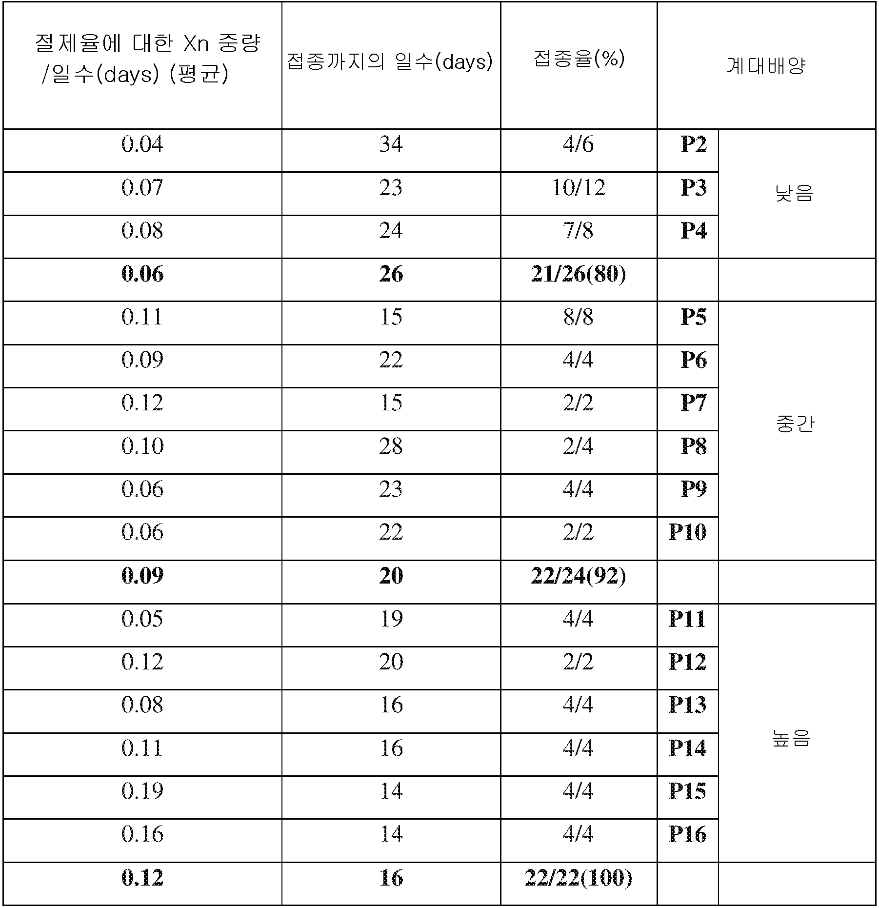
ATRT Xn에서 얻어지고 더욱 증식된 1x10⁶ 세포들로부터 이차 종양 형성의 빈도와 특성을 요약한 표. ATRT Xn의 연속 증식 동안에 종양 접종까지의 보다 짧은 시간 및 가속화된 종양 성장을 나타냈다. 낮은 계대수의 Xn 계대배양과 중간 계대수의 계대배양의 비교에서 *p<0.01이다.A table summarizing the frequency and characteristics of secondary tumor formation from 1x10 6 cells obtained from ATRT Xn and further propagated. Showed a shorter time to tumor inoculation and accelerated tumor growth during the continuous proliferation of ATRT Xn. * P < 0.01 in comparison of Xn subculture of low passages and subculture of medium passages.
본 발명자들은 MRT 증식으로 인해 CSC 능력이 기능적으로 증대되는지를 다음 의문으로 제기하였다. 이에 낮은 계대수, 중간 계대수 및 높은 계대수의 계대배양 Xn에서 유래된 MRT 세포를 이용하여 한계 희석 이종이식 실험을 수행하였다. 상기 분석은 높은 계대수의 계대배양 Xn에서 CSC 빈도에 대한 유의적인 양성 선별(positive selection)을 나타낸다(표 4).The present inventors have raised the following question as to whether the CSC capacity is functionally increased due to MRT proliferation. Therefore, marginal dilution xenotransplantation experiments were carried out using MRT cells derived from subculture of low passage number, middle passage number and high passage number. This analysis represents a significant positive selection for CSC frequency in high passaged subculture Xn (Table 4).
[표 4][Table 4]
증식 동안, 종양 CSC 빈도를 나타내는 한계 희석 이종이식의 요약Summary of marginal dilution xenotransplantation showing tumor CSC frequency during proliferation

높은 계대수의 계배배양 MRT Xn이 보다 많은 수의 CSC를 농축한다는 사실이 관찰되었고, 계대배양에 따른 CSC 표현형의 획득을 수반하는 조직학적 및 면역조직화학적 변화가 분석되었다(도 1b~c). H&E 염색은 Xn 유래된 종양이 기본적인 육종양같은 세포 형태를 유지한다는 것을 보여 주었다. 그럼에도 불구하고, 방추세포와 같은 세포 형태의 획득, 괴사의 광대한 영역, 적은 세포 사멸체 및 더 많은 유사분열을 포함하는 높은 세대 Xn(high generation Xn)에서 일부 형태학적 차이가 관찰되었다(도 1b). 면역조직화학(IHC) 염색은 일차 종양 및 Xn 세포들 모두가 간엽(mesenchymal) 마커인 비멘틴을 강하게 발현한다는 것을 밝혀냈다(도 1c). 그러나, 일차 종양과 반대로, 높은 세대 Xn은 상피막 항원(EMA), 시토케라틴 AE1/AE3 및 신경미세섬유 단백질(NFP)을 포함하는 차별화되는 마커들의 손실을 나타냈다. IHC 결과들을 검증하기 위해, 초기 및 후기 Xn에서 일차 종양에 대하여 qRT-PCR을 수행하였다. 상기 유전자 발현 분석은 상피 분화 마커(epithelial differentiation marker)인 E-카드헤린의 손실과 함께 비멘틴의 유지를 증명하였다. 따라서, 증대된 CSC 빈도는 유사한 병리조직학적 육종양의 형태를 유지하고 간엽 표현형으로 탈분화를 나타내는 MRT Xn과 관련이 있다.It has been observed that MRC Xn enriches a greater number of CSCs, and histological and immunohistochemical changes accompanying the acquisition of the CSC phenotype by subculture were analyzed (FIGS. 1b-c). H & E staining showed that Xn-derived tumors retained cell morphology like basic sarcoma. Nevertheless, some morphological differences were observed in the high generation Xn, including the acquisition of cell morphology such as spindle cells, vast areas of necrosis, less apoptosis and more mitosis (Fig. 1B ). Immunohistochemistry (IHC) staining revealed that both primary tumors and Xn cells strongly express the mesenchymal marker, visentin (Fig. 1c). However, as opposed to primary tumors, high-generation Xn showed loss of differentiating markers including epithelial antigen (EMA), cytokeratin AE1 / AE3 and neurofilament protein (NFP). To verify IHC results, qRT-PCR was performed on primary tumors in early and late Xn. The gene expression analysis demonstrated the retention of visentin with loss of E-carderin, an epithelial differentiation marker. Thus, the increased frequency of CSCs is associated with MRT Xn, which maintains a similar histopathological sarcoma pattern and exhibits decentifi- cation into the mesenchymal phenotype.
증대된 종양 개시 활성과 관련된 글로벌 유전자 표지는 추정 Global gene markers associated with increased tumorigenic activity are estimated CSCCSC 바이오 Bio 마커들을Markers 나타낸다 Show
본 발명자들은 CSC 표현형에 대한 선택을 수반하는 연속적 MRT Xn의 글로벌 분자 프로파일을 특성화하고자 하였다. 이러한 목적을 위하여, 여러 가지 다른 샘플을 비교하는 마이크로어레이 유전자 발현 분석을 수행했다: 1. 일차 MRT, 2. 초기 MRT Xn (P2), 3. 중기 MRT Xn (P7), 4. 후기 MRT Xn (P16), 5. 인간 배아줄기세포(hESCs), 6. 태아 신장 (FK), 7. 성인 신장 (AK), 8. 태아 뇌 (FB), 9. 성인 뇌 (AB). 글로벌 발현 분석 결과, 증식된 MRT Xn 세포들은 계층형 클러스터링에 의해 설명된 바와 같이, 원래의 그들의 일차 종양보다 오히려 hESC와 더 밀접하게 밀집된 것으로 나타났다(도 2a). 또한, Xn 조직과 인간 태아 조직의 유사성이 더욱 컸다. 전적으로, 이러한 결과들은 종양이 증식되는 동안의 줄기세포 표현형에 대한 선택성을 확인시켜준다. 일차 MRT와 연속적 MRT Xn의 더욱 면밀한 조사(낮은, 중간 및 높은 계대수의 계대배양)는 계대배양을 통해 상향 조절 및 하향 조절되는 두 개의 주요 유전자 클러스터를 포함하는 다양한 유전자 발현 패턴을 동정했다(도 2b). 상향 조절된 유전자들의 집단은 세마포린3C(SEMA3C), 리실옥시다아제(LOX), 글리코프로테인 M6A(GPM6A), 헤파토사이트 성장인자(HGF / SF) 및 알데하이드 디하이드로게나아제1(ALDH1)과 같은 관찰된 CSC 표현형의 획득에 수반되는 분자들을 포함하였다(도 2c). 상기 유전자 발현 프로파일은 또한 고도의 증식과 같은 후기 계대배양을 나타내는 유전자 히트 맵(Gene heat map)에 의해 나타나는 것과 같이 계대배양에 따라 종양 생물학을 검증할 수 있었다(예를 들면. KI -67, CDK1 및 AURKA) (Figure 2D). 또한, ngenuityⓒ 기능 분석은 감소된 괴사, 세포사멸 및 세포 분화를 입증하였고(도 2e), 이들 결과들의 두가지 근거는 일차 종양과 낮은 계대수의 계대배양 Xn과 비교하여 높은 계대수의 계대배양 Xn에서의 감소된 세포자멸, 증가된 유사분열 및 분화 마커의 손실을 증명하는 H&E 및 IHC 염색으로부터 얻었다. 따라서, 연속적 Xn 증식 모델은 MRT에서 지속되는 종양 개시 및 종양 진행 및 CSC 표현형의 증가에 관여하는 추정 바이오 마커 분자를 생성하였다.We sought to characterize the global molecular profile of the consecutive MRT Xn with selection for the CSC phenotype. For this purpose, we performed microarray gene expression analysis comparing several different samples: 1. Primary MRT, 2. Initial MRT Xn (P2), 3. Midterm MRT Xn (P7), 4. Later MRT Xn P16), 5. human embryonic stem cells (hESCs), 6. fetal kidney (FK), 7. adult kidney (AK), 8. fetal brain (FB), 9. adult brain (AB). Global expression analysis showed that the proliferated MRT Xn cells were more closely packed with hESCs than their original primary tumors (Fig. 2a), as described by hierarchical clustering. In addition, the similarity between the Xn tissue and the human fetal tissue was greater. Altogether, these results confirm selectivity for stem cell phenotype during tumor proliferation. A closer scrutiny (low, medium and high passage subculture) of primary MRT and continuous MRT Xn identified a variety of gene expression patterns including two major gene clusters that were upregulated and downregulated through subculture 2b). Observing a group of up-regulated genes, such as semaphores, Lin 3C (SEMA3C), florisil oxidase (LOX), glycoprotein M6A (GPM6A), Hepa soil site growth factor (HGF / SF) and aldehyde dehydrogenase 1 (ALDH1) Lt; / RTI > (Figure 2c). The gene expression profile could also be verified for tumor biology according to subculture as indicated by the Gene heat map representing late passaging such as high proliferation (e.g., KI -67, CDK1 And AURKA ) (Figure 2D). In addition, the ngenuity 기능 function analysis demonstrated reduced necrosis, apoptosis and cell differentiation (Fig. 2e), and the two rationale of these results was that high-passaged subculture Xn And H & E and IHC staining demonstrating reduced apoptosis, increased mitosis, and loss of differentiation markers in the cells. Thus, the continuous Xn proliferation model produced a putative biomarker molecule that is involved in sustained tumor initiation and tumor progression and an increase in CSC phenotype in MRT.
MRTMRT CSCCSC 바이오 Bio 마커로서의As a marker ALDH1의Of ALDH1 기능 검증 Function verification
본 명세서에서의 분자 스크린이 MRT에서의 CSC 표현형 수득의 마커로서 ALDH1을 독립적으로 정확하게 보여준다는 것이 관찰되었으므로, 본 발명자들은 그것의 발현 및 기능적 역할을 추가로 조사하였다. 중요하게도 ALDH1이 CSC 마커로 제시되었고, 따라서 상기 접근법을 검증할 수 있었다. 발현 분석은 ALDH1의 비율이 종양 증식에 따라 현저하게 증가한다는 것을 입증하였다(도 3a). Xn 증식의 다른 단계들에서 ALDH1 개체군의 비율을 정량화하기 위한 FACS 분석은, 후기 계대수의 계대배양(계대배양 10)에서 25%의 세포가 ALDH1+인 것과 비교하여 낮은 계대수의 계대배양 Xn 세포들(계대배양 4)에서 단지 4%의 세포들만이 ALDH1+임을 밝혀내었다(도 3b). 또한, ALDH1 IHC 염색법은 계대배양에 따라 ALDH1의 증가된 발현을 증명하였다(도 3c). 더욱이, ALDH1high 발현 세포들은 주로 거대한 간상 세포들이었다(도 3d). 유전자 발현 수준에서, qRT-PCR 분석은 일차 종양에 비해 후기 계대배양에서 약 40배 더 높은 ALDH1 발현을 나타내었다(도 3e). 이어서, 본 발명자들은 그것들이 CSC로서 자격이 있는지를 결정하기 위하여 ALDH1+ 분획을 분류하였다. 암 줄기세포를 정의하는 특성은 시험관내에서 클론을 형성하는 그들의 기능적 능력이었다. 콜로니 형성 분석은 CSC 표현형에 따른 xALDH1- MRT 세포들과 비교하여 ALDH1+ 내에서 현저하게 높은 클론의 개수 및 보다 큰 콜로니들을 나타내었다(도 4a). 또한, CSCs는 면역 결핍 마우스 내로 이식 시에 종양을 개질하는 그들의 능력에 의해 정의된다. 생체내에서 ALDH1+ 세포들의 종양 형성성(tumorigenicity)을 조사하기 위해, 한계 희석 분석(18)을 사용하여 NOD/SCID 마우스의 옆구리에 FACS 정제된 ALDH1+ 및 ALDH1- 세포들을 점점 감소되는 양으로 주입하였다(18). 분류되지 않은 세포들을 대조구로서 주입하였다(표 2). 낮은 계대수의 계대배양(P3)에서, 2000개 이하의 ALDH1+ 세포들이 주입된 6마리의 마우스 중 4마리는 종양을 형성하였으나, ALDH1- 세포를 주사한 마우스는 종양을 형성하지 않았다(p <0.05). 높은 계대수의 계대배양(P12)에서, 1000개 이하의 ALDH1+ 세포들을 주사한 모든 마우스들은 이차 종양이 형성된 반면에, ALDH1- 세포들이 주사된 11마리의 마우스 중 2마리만이 종양으로 발전하였는데, 이는 상기 개체군은 소량의 암줄기세포가 상대적으로 대폭 감소하였음을 나타내는 것이다(표 2). 중요한 것은, ALDH1+ 세포들에서 발생한 종양은 상기 개체군에서의 종양 개시 능력의 존재와 일치되게 이차 수용체 내로 연속적으로 이식될 수 있었고, 이는 ALDH+ 세포들의 생체내 자가-재생 능력을 증명하는 것이다(도 4b). 전체적으로, 이들 데이터는 ALDH1이 CSC 표현형 수득의 링크를 검증하는 MRT CSC를 위한 마커임을 나타내므로, 본 발명자들은 ALDH1과 같은 특정 분자들에 대한 장기간 증식으로 정의하였다. 분자 수준에서 분류된 ALDH+ CSC를 더욱 특성화하고, 그들이 장기간 증식으로 예측된 CSC 바이오 마커를 농축하였는지를 결정하기 위해, MRT Xn에서 얻어 분류된 ALDH+ 및 ALDH- 세포들에 대한 RNA 시퀀싱 실험을 수행하였다(도 4c). 흥미롭게도, ALDH- 세포들과 비교하여 ALDH+ 세포들에서 대부분 상향 조절된 유전자들 중에는 일차 종양(예를 들면. ANXA1 , GPM6A , HGF 및 LOX)과 비교하여 후기 Xn 계대배양에서 현저하게 과발현된 유전자가 있다는 것이 밝혀졌다. qRT-PCR 분석을 수행하여 ALDH- 세포들과 비교하여 ALDH+ 세포들에서 높은 LOX 발현을 확인하였다(도 4e). 이러한 결과들은 종양의 공격성 및 CSC 빈도와 관련된 분자들 및 경로를 동정하기 위한 모델로서 본 발명자들의 Xn 증식 기술의 유효성을 강조하고 있다. 진행된 MRT Xn 및 분류된 ALDH+ CSC 분획에서 LOX의 높은 발현은 우리가 LOX를 치료 표적으로서 사용할 수 있는지를 조사하게 하였다.As it has been observed that the molecular screen herein correctly and independently shows ALDH1 as a marker of obtaining the CSC phenotype in MRT, we further investigated its expression and functional role. Significantly, ALDHl was presented as a CSC marker and thus the above approach could be verified. Expression analysis demonstrated that the proportion of ALDH1 was markedly increased with tumor growth (Fig. 3a). FACS analysis to quantify the proportion of ALDHl populations at different stages of Xn proliferation showed that 25% of the cells in the late passage subculture (subculture 10) had a lower passage number of subcultured Xn cells (Subculture 4) revealed that only 4% of the cells were ALDH1 + (Fig. 3B). In addition, ALDHl IHC staining demonstrated increased expression of ALDHl by subculture (Fig. 3c). Moreover, ALDH1 high expressing cells were mainly large stem cells (Fig. 3d). At the gene expression level, qRT-PCR analysis showed approximately 40-fold higher expression of ALDH1 in late passaging than primary tumors (Figure 3e). We then sorted the ALDHl + fraction to determine if they were eligible as a CSC. The characteristics defining cancer stem cells were their functional ability to form clones in vitro. Colony formation assays showed significantly higher numbers of clones and larger colonies in ALDHl + as compared to xALDH1-MRT cells according to the CSC phenotype (Fig. 4A). In addition, CSCs are defined by their ability to modify tumors at the time of transplantation into immunodeficient mice. In order to investigate the tumorigenicity of ALDHl + cells in vivo, FACS purified ALDHl + and ALDHl- cells were injected into the flank of NOD / SCID mice in a decreasing amount using a limiting dilution assay (18) 18). Untreated cells were injected as control (Table 2). In the subculture of low passage number (P3), four out of six mice injected with less than 2000 ALDH1 + cells formed tumors, whereas mice injected with ALDH1- cells did not form tumors (p <0.05 ). In high passaged subculture (P12), all mice injected with less than 1000 ALDH1 + cells formed secondary tumors, whereas only 2 out of 11 injected mice with ALDH1- cells developed into tumors, This indicates that the population has a relatively large decrease in the amount of cancer stem cells (Table 2). Importantly, tumors developed in ALDHl + cells could be continuously implanted into secondary receptors consistent with the presence of tumor-initiating capacity in the population, demonstrating in vivo self-renewal capability of ALDH + cells (Figure 4b) . Overall, these data indicate that ALDH1 is a marker for the MRT CSC that verifies the link to obtain the CSC phenotype, so we have defined long-term proliferation for certain molecules such as ALDH1. To further characterize the ALDH + CSCs classified at the molecular level and determine whether they enriched the CSC biomarkers predicted by prolonged proliferation, RNA sequencing experiments were performed on the ALDH + and ALDH- cells obtained from MRT Xn 4c). Interestingly, among the most up-regulated genes in ALDH + cells compared to ALDH-cells, genes significantly overexpressed in late Xn passages compared to primary tumors (eg, ANXA1 , GPM6A , HGF and LOX ) . qRT-PCR analysis was performed to confirm high LOX expression in ALDH + cells compared to ALDH- cells (Fig. 4E). These results highlight the efficacy of our Xn propagation technique as a model for identifying molecules and pathways involved in tumor aggressiveness and CSC frequency. High expression of LOX in the advanced MRT Xn and fractionated ALDH + CSC fractions led us to investigate whether we could use LOX as a therapeutic target.
MRT CSC 치료제로서 LOX 억제제의 기능적 검증Functional Validation of LOX Inhibitor as MRT CSC Therapeutic
세포 외 매트릭스-리모델링 효소인 LOX는 초기 세포 분화 및 조직 발달(22) 뿐만 아니라 염색질 밀집(chromatin compaction)(19), 유전자 전사(20, 21)와 같은 경우들을 조절하므로, 따라서 유사한 활성을 조절하는 INI-1 손실에 의해 특징지워지는 MRT와 연관이 있다. 최근 연구에 따르면, LOX는 유방암, 대장암 및 췌장암들을 비롯한 여러 악성 종양에서 과발현되며(23, 24), 종양 진행 및 전이와 관련이 있다(25). 일차 MRT, 초기 계대수(P2), 중기 계대수(P7) 및 후기 계대수(P14)의 Xn 계대배양에서 LOX의 발현을 분석한 IHC 염색법은 간상 세포내에서 유의하게 발현한 계대배양을 통하여 발현형이 광범위하게 증가함을 확인하였다(도 5a). 이전에는 LOX 억제제인 BAPN이 다양한 암세포들에 긍정적인 영향을 미치는 것으로 나타났다(23). 상기 LOX 유전자는 분류된 MRT CSCs내에서 뿐만 아니라 증식 및 CSC 표현형을 선발하는 동안에 Xn 내에서 상향 조절되었기 때문에, 본 발명자들은 LOX 억제가 MRT 세포에 영향을 미치는지를 조사하고자 하였다. 우선, Xn 세포들을 48시간 동안 상이한 농도의 BAPN에서 성장시켰다. 세포 생존력을 조사한 MTS 분석에서는, 3가지 다른 실험에서 100㎛ BAPN 처리 후, 증식이 현저하게 감소함을 나타내었다(데이터는 표시하지 않음). 다음으로, 마우스에서 연속적으로 증식된 소아 신장의 일반 고형 종양인 WT Xn 세포에 대한 BAPN의 효과를 조사했다. 상기 결과들은 WT 세포 증식에 영향을 미치지 않음을 나타내며, 이는 MRT 세포들에 대한 LOX 억제의 특정한 효과가 있음을 증명하였다(도 5b). 본 발명자들은 다음으로 BAPN 처리의 결과로서 MRT 세포들의 형태를 조사하였고, 상기 처리가 핵 응결(nuclear condensation) 및 세포 연하(cell swallowing)를 포함하는 형태학적 변화를 유도한다는 것을 나타내었다(도 5c). 중요하게도, 이동 분석(migration assay)은 48시간 동안 BAPN 처리된 세포들을, BAPN 처리되지 않은 세포들과 비교할 때, MRT 세포 이동 능력을 현저하게 억제한다는 것을 밝혀냈다(도 5d). 모든 시험관내 결과들은 중기 Xn 계대배양(P10)을 사용하여 얻어졌기 때문에, 본 발명자들은 BAPN이 일차 종양과 매우 유사한 세포들에서도 비슷한 효과를 갖는지를 측정하도록 정하였다. 유사한 실험들이 P2 세포들에서 수행되었고, 동일한 결과들이 나타났다(도 7a~c). LOX 활성 측정은, 100μM BAPN이 실제로 LOX 활성을 현저하게 억제한다는 것을 증명하였다(도 7d). 이들 결과들은 새로운 MRT CSC 바이오 마커를 표적화하는 것은, 종양의 성장을 막고, 가능한 MRT 치료 표적으로서의 LOX를 보증할 수 있음을 제시한다.LOX, an extracellular matrix-remodeling enzyme, modulates events such as chromatin compaction 19 and
실시예 1에 대한 참고문헌들References to Example 1
1. Pode-Shakked N, Metsuyanim S, Rom-Gross E, Mor Y, Fridman E, Goldstein I, et al. Developmental tumourigenesis: NCAM as a putative marker for the malignant renal stem/progenitor cell population. Journal of cellular and molecular medicine. 2009 Aug;13(8B):1792-808.One. Pode-Shakked N, Metsuyanim S, Rom-Gross E, Mor Y, Fridman E, Goldstein I, et al. Developmental tumourigenesis: NCAM as a putative marker for the malignant renal stem / progenitor cell population. Journal of cellular and molecular medicine. 2009 Aug; 13 (8B): 1792-808.
2.
Dekel B, Metsuyanim S, Schmidt-Ott KM, Fridman E, Jacob-Hirsch J, Simon A, et al. Multiple imprinted and stemness genes provide a link between normal and tumor progenitor cells of the developing human kidney. Cancer research. 2006 Jun 15;66(12):6040-9.2.
Dekel B, Metsuyanim S, Schmidt-Ott KM, Fridman E, Jacob-Hirsch J, Simona, et al. Multiple imprinted and stem-like genes provide a link between normal and tumor progenitor cells of the developing human kidney. Cancer research. 2006
3. Metsuyanim S, Pode-Shakked N, Schmidt-Ott KM, Keshet G, Rechavi G, Blumental D, et al. Accumulation of malignant renal stem cells is associated with epigenetic changes in normal renal progenitor genes. Stem cells (Dayton, Ohio). 2008 Jul;26(7):1808-17.3. Metsuyanim S, Pode-Shakked N, Schmidt-Ott KM, Keshet G, Rechavi G, Blumental D, et al. Accumulation of malignant renal stem cells is associated with epigenetic changes in normal renal progenitor genes. Stem cells (Dayton, Ohio). 2008 Jul; 26 (7): 1808-17.
4. Pode-Shakked N, Dekel B. Wilms tumor--a renal stem cell malignancyx Pediatric nephrology (Berlin, Germany). 2011 Sep;26(9):1535-43.4. Pode-Shakked N, Dekel B. Wilms tumor-a renal stem cell malignancyx Pediatric nephrology (Berlin, Germany). 2011 Sep; 26 (9): 1535-43.
5. Pode-Shakked N, Shukrun R, Mark-Danieli M, Tsvetkov P, Bahar S, Pri-Chen S, et al. The isolation and characterization of renal cancer initiating cells from human Wilms' tumour xenografts unveils new therapeutic targets. EMBO molecular medicine. 2013 Jan;5(1):18-37.5. Pode-Shakked N, Shukrun R, Mark-Daniel M, Tsvetkov P, Bahar S, Pri-Chen S, et al. The isolation and characterization of renal cancer initiating cells from human Wilms' tumor xenografts unveils new therapeutic targets. EMBO molecular medicine. 2013 Jan; 5 (1): 18-37.
6. Shukrun R, Dekel B, Pode-Shakked N, Pleniceanu O, Omer D, Vax E, et al. Wilms' Tumor Blastemal Stem Cells Dedifferentiate to Propagate the Tumor Bulk. Stem Cell Reports. 2014.6. Shukrun R, Dekel B, Pode-Shakked N, Plenicean O, Omer D, Vax E, et al. Wilms' Tumor Blastemal Stem Cells Dedifferentiate to Propagate the Tumor Bulk. Stem Cell Reports. 2014.
7. Parham DM, Weeks DA, Beckwith JB. The clinicopathologic spectrum of putative extrarenal rhabdoid tumors. An analysis of 42 cases studied with immunohistochemistry or electron microscopy. The American journal of surgical pathology. 1994 Oct;18(10):1010-29.7. Parham DM, Weeks, Beckwith JB. The clinicopathologic spectrum of putative extrarenal rhabdoid tumors. An analysis of 42 cases studied with immunohistochemistry or electron microscopy. The American journal of surgical pathology. 1994 Oct; 18 (10): 1010-29.
8. Wick MR, Ritter JH, Dehner LP. Malignant rhabdoid tumors: a clinicopathologic review and conceptual discussion. Seminars in diagnostic pathology. 1995 Aug;12(3):233-48.8. Wick MR, Ritter JH, Dehner LP. Malignant rhabdoid tumors: a clinicopathologic review and conceptual discussion. Seminars in diagnostic pathology. 1995 Aug; 12 (3): 233-48.
9. Versteege I, Sevenet N, Lange J, Rousseau-Merck MF, Ambros P, Handgretinger R, et al. Truncating mutations of hSNF5/INI1 in aggressive paediatric cancer. Nature. 1998 Jul 9;394(6689):203-6.9. Versteege I, Sevenet N, Lange J, Rousseau-Merck MF, Ambros P, Handgretinger R, et al. Truncating mutations of hSNF5 / INI1 in aggressive paediatric cancer. Nature. 1998 Jul 9; 394 (6689): 203-6.
10. Olson TA, Bayar E, Kosnik E, Hamoudi AB, Klopfenstein KJ, Pieters RS, et al. Successful treatment of disseminated central nervous system malignant rhabdoid tumor. Journal of pediatric hematology/oncology. 1995 Feb;17(1):71-5.10. Olson TA, Bayar E, Kosnik E, Hamoudi AB, Klopfenstein KJ, Pieters RS, et al. Successful treatment of disseminated central nervous system malignant rhabdoid tumor. Journal of pediatric hematology / oncology. 1995 Feb; 17 (1): 71-5.
11. Douglass EC, Valentine M, Rowe ST, Parham DM, Wilimas JA, Sanders JM, et al. Malignant rhabdoid tumor: a highly malignant childhood tumor with minimal karyotypic changes. Genes, chromosomes & cancer. 1990 Sep;2(3):210-6.11. Douglass EC, Valentine M, Rowe ST, Parham DM, Wilimas JA, Sanders JM, et al. Malignant rhabdoid tumor: a highly malignant childhood tumor with minimal karyotypic changes. Genes, chromosomes & cancer. 1990 Sep; 2 (3): 210-6.
12. Rorke LB, Packer RJ, Biegel JA. Central nervous system atypical teratoid/rhabdoid tumors of infancy and childhood: definition of an entity. Journal of neurosurgery. 1996 Jul;85(1):56-65.12. Rorke LB, Packer RJ, Biegel JA. Central nervous system atypical teratoid / rhabdoid tumors of infancy and childhood: definition of an entity. Journal of neurosurgery. 1996 Jul; 85 (1): 56-65.
13. Biegel JA, Tan L, Zhang F, Wainwright L, Russo P, Rorke LB. Alterations of the hSNF5/INI1 gene in central nervous system atypical teratoid/rhabdoid tumors and renal and extrarenal rhabdoid tumors. Clin Cancer Res. 2002 Nov;8(11):3461-7.13. Biegel JA, Tan L, Zhang F, Wainwright L, Russo P, Rorke LB. Alterations of the hSNF5 / INI1 gene in central nervous system atypical teratoid / rhabdoid tumors and renal and extrarenal rhabdoid tumors. Clin Cancer Res. 2002 Nov; 8 (11): 3461-7.
14.
Biegel JA, Zhou JY, Rorke LB, Stenstrom C, Wainwright LM, Fogelgren B. Germ-line and acquired mutations of INI1 in atypical teratoid and rhabdoid tumors. Cancer research. 1999 Jan 1;59(1):74-9.14.
Biegel JA, Zhou JY, Rorke LB, Stenstrom C, Wainwright LM, Fogelgren B. Germ-line and acquired mutations of
15. Euskirchen G, Auerbach RK, Snyder M. SWI/SNF chromatin-remodeling factors: multiscale analyses and diverse functions. The Journal of biological chemistry. 2012 Sep 7;287(37):30897-905.15. Euskirchen G, Auerbach RK, and Snyder M. SWI / SNF chromatin-remodeling factors: multiscale analyses and diverse functions. The Journal of biological chemistry. 2012 Sep 7; 287 (37): 30897-905.
16.
Tomlinson GE, Breslow NE, Dome J, Guthrie KA, Norkool P, Li S, et al. Rhabdoid tumor of the kidney in the National Wilms' Tumor Study: age at diagnosis as a prognostic factor. J Clin Oncol. 2005 Oct 20;23(30):7641-5.16.
Tomlinson GE, Breslow NE, Dome J, Guthrie KA, Norkool P, Li S, et al. Rhabdoid tumor of the kidney in the National Wilms' Tumor Study: Age at diagnosis as a prognostic factor. J Clin Oncol. 2005
17. Ginestier C, Hur MH, Charafe-Jauffret E, Monville F, Dutcher J, Brown M, et al. ALDH1 is a marker of normal and malignant human mammary stem cells and a predictor of poor clinical outcome. Cell stem cell. 2007 Nov;1(5):555-67.17. Ginestier C, Hur MH, Charafe-Jauffret E, Monville F, Dutcher J, Brown M, et al. ALDH1 is a marker of normal and malignant human mammary stem cells and a predictor of poor clinical outcome. Cell stem cell. 2007 Nov; 1 (5): 555-67.
18.
Hu Y, Smyth GK. ELDA: extreme limiting dilution analysis for comparing depleted and enriched populations in stem cell and other assays. Journal of immunological methods. 2009 Aug 15;347(1-2):70-8.18.
Hu Y, Smyth GK. ELDA: extreme limiting dilution analysis for comparison depleted and enriched populations in stem cell and other assays. Journal of immunological methods. 2009
19. Mello ML, Contente S, Vidal BC, Planding W, Schenck U. Modulation of ras transformation affecting chromatin supraorganization as assessed by image analysis. Experimental cell research. 1995 Oct;220(2):374-82.19. Mello ML, Contente S, Vidal BC, Planding W, Schenck U. Modulation of ras transformation affecting chromatin supraorganization as assessed by image analysis. Experimental cell research. 1995 Oct; 220 (2): 374-82.
20. Di Donato A, Lacal JC, Di Duca M, Giampuzzi M, Ghiggeri G, Gusmano R. Micro-injection of recombinant lysyl oxidase blocks oncogenic p21-Ha-Ras and progesterone effects on Xenopus laevis oocyte maturation. FEBS letters. 1997 Dec 8;419(1):63-8.20. Di Donatoa, Lacal JC, Di Duca M, Giampuzzi M, Ghiggeri G, Gusmano R. Microinjection of recombinant lysyl oxidase blocks oncogenic p21-Ha-Ras and progesterone effects on Xenopus laevis oocyte maturation. FEBS letters. 1997 Dec 8; 419 (1): 63-8.
21. Giampuzzi M, Botti G, Di Duca M, Arata L, Ghiggeri G, Gusmano R, et al. Lysyl oxidase activates the transcription activity of human collagene III promoter. Possible involvement of Ku antigen. The Journal of biological chemistry. 2000 Nov 17;275(46):36341-9.21. Giampuzzi M, Botti G, Di Duca M, Arata L, Ghiggeri G, Gusmano R, et al. Lysyl oxidase activates the transcriptional activity of human collagene III promoter. Possible involvement of Ku antigen. The Journal of biological chemistry. 2000 Nov 17; 275 (46): 36341-9.
22.
Maki JM, Rasanen J, Tikkanen H, Sormunen R, Makikallio K, Kivirikko KI, et al. Inactivation of the lysyl oxidase gene Lox leads to aortic aneurysms, cardiovascular dysfunction, and perinatal death in mice. Circulation. 2002 Nov 5;106(19):2503-9.22.
Maki JM, Rasanen J, Tikkanen H, Sormunen R, Makikallio K, Kivirikko KI, et al. Inactivation of the lysyl oxidase gene Lox leads to aortic aneurysms, cardiovascular dysfunction, and perinatal death in mice. Circulation. 2002
23. Bondareva A, Downey CM, Ayres F, Liu W, Boyd SK, Hallgrimsson B, et al. The lysyl oxidase inhibitor, beta-aminopropionitrile, diminishes the metastatic colonization potential of circulating breast cancer cells. PloS one. 2009;4(5):e5620.23. Bondarevaya, Downey CM, Ayres F, Liu W, Boyd SK, Hallgrimsson B, et al. The lysyl oxidase inhibitor, beta-aminopropionitrile, diminishes the metastatic colonization potential of circulating breast cancer cells. PloS one. 2009; 4 (5): e5620.
24.
Melstrom LG, Bentrem DJ, Salabat MR, Kennedy TJ, Ding XZ, Strouch M, et al. Overexpression of 5-lipoxygenase in colon polyps and cancer and the effect of 5-LOX inhibitors in vitro and in a murine model. Clin Cancer Res. 2008 Oct 15;14(20):6525-30.24.
Melstrom LG, Bentrem DJ, Salabat MR, Kennedy TJ, Ding XZ, Strouch M, et al. Overexpression of 5-lipoxygenase in colon polyps and cancer and the effect of 5-LOX inhibitors in vitro and in a murine model. Clin Cancer Res. 2008
25. Barker HE, Cox TR, Erler JT. The rationale for targeting the LOX family in cancer. Nature reviews. 2012 Aug;12(8):540-52.25. Barker HE, Cox TR, Erler JT. The rationale for targeting the LOX family in cancer. Nature reviews. 2012 Aug; 12 (8): 540-52.
26. Findlay VJ, Wang C, Watson DK, Camp ER. Epithelial-to-mesenchymal transition and the cancer stem cell phenotype: insights from cancer biology with therapeutic implications for colorectal cancer. Cancer gene therapy. 2014 May 2.26. Findlay VJ, Wang C, Watson DK, Camp ER. Epithelial-to-mesenchymal transition and the cancer stem cell phenotype: insights from cancer biology with therapeutic implications for colorectal cancer. Cancer gene therapy. May 2014.
27.
Marjanovic ND, Weinberg RA, Chaffer CL. Poised with purpose: cell plasticity enhances tumorigenicity. Cell cycle (Georgetown, Tex. 2013 Sep 1;12(17):2713-4.27.
Marjanovic ND, Weinberg RA, Chaffer CL. Poised with purpose: cell plasticity enhances tumorigenicity. Cell cycle (Georgetown, Tex. 2013
28. Lander AD. The 'stem cell' concept: is it holding us backx Journal of biology. 2009;8(8):70.28. Lander AD. The 'stem cell' concept: is it holding us backx Journal of biology. 2009; 8 (8): 70.
29. Bertucci F, Bouvier-Labit C, Finetti P, Metellus P, Adelaide J, Mokhtari K, et al. Gene expression profiling of solitary fibrous tumors. PloS one. 2013;8(5):e64497.29. Bertucci F, Bouvier-Labit C, Finetti P, Metellus P, Adelaide J, Mokhtari K, et al. Gene expression profiling of solitary fibrous tumors. PloS one. 2013; 8 (5): e64497.
30. Ma LJ, Li YG, Huang L, Han M, Ma BJ, Sun BJ, et al. [Expression of LOX and MMP-2 in gastric cancer tissue and the effects of LOX and MMP-2 on tumor invasion and metastasis]. Zhonghua zhong liu za zhi [Chinese journal of oncology]. 2011 Jan;33(1):37-41.30. Ma LJ, Li YG, Huang L, Han M, Ma BJ, Sun BJ, et al. [Expression of LOX and MMP-2 in gastric cancer tissue and the effects of LOX and MMP-2 on tumor invasion and metastasis]. Zhonghua zhong liu za zhi [Chinese journal of oncology]. 2011 Jan; 33 (1): 37-41.
31. Thomas C, Karnoub AE. Lysyl oxidase at the crossroads of mesenchymal stem cells and epithelial-mesenchymal transition. Oncotarget. 2013 Mar;4(3):376-7.31. Thomas C, Karnoub AE. Lysyl oxidase at the crossroads of mesenchymal stem cells and epithelial-mesenchymal transition. Oncotarget. 2013 Mar; 4 (3): 376-7.
32. Kordes U, Gesk S, Fruhwald MC, Graf N, Leuschner I, Hasselblatt M, et al. Clinical and molecular features in patients with atypical teratoid rhabdoid tumor or malignant rhabdoid tumor. Genes, chromosomes & cancer. 2003 Feb;49(2):176-81.32. Kordesu, Gesk S, Fruhwald MC, Graf N, Leuschner I, Hasselblatt M, et al. Clinical and molecular features in patients with atypical teratoid rhabdoid tumor or malignant rhabdoid tumor. Genes, chromosomes & cancer. 2003 Feb; 49 (2): 176-81.
실시예Example 2 2
인간 윌름스 종양 및 유잉 육종 이종증식의 장기간 증식 Long-term proliferation of human Wilms tumor and Ewing's sarcoma
물질들 및 방법들Materials and methods
생체내 이종이식 형성 및 이식까지의 시간의 계산: 동물 실험은 쉐바 메디컬 센터의 동물 실험을 위한 실험 지침에 따라 수행하였다. 5~8주령, 암컷, 자연발증 당뇨병 면역결핍(NOD/SCID) 마우스에 대한 초기 ATRT 이종이식은 상기에 기재된 바에 따라 수행하였다. 간단히 설명하면, 일차 ATRT 조직을 2~5mm의 조각들로 컷팅하였고, 마우스의 등쪽에 피하 이식하였다. 마우스는 병원체가 없는 환경에서 유지되었으며, 종양 성장에 대하여 매주 모니터링하였다. 매주 마다 이차 종양들이 촉진에 의해 검출되었다. 종양들은 1.5cm의 직경의 크기가 되었을 때 채취하였다. 각각 이식된 Xn에 대하여 이식까지의 시간, 절제까지의 시간, 중량 및 부피를 기록하였다. Xn 조직은 즉시 작은 조각들로 컷팅하였고, NOD/SCID 마우스의 옆구리에 피하 조직 삽입을 진행하였으며, 후속하는 Xn 증식을 위해 하기에 설명한 바와 같은 단일 세포 현탁액의 제조를 진행하였다. 항생제(페니실린 및 스트렙토마이신)가 함유된 IMDM(Iscove's modification of Dulbecco's medium) 내에서 샘플을 분쇄하여 단일 세포 현탁액을 얻은 다음, 37℃에서 2시간 동안 콜라게나아제 Ⅳ로 처리하였다. 효소로 처리된 조직을 IMDM을 사용하여 2배 부피의 콜라게나아제 용액으로 연마하고, 현탁액을 여과하여(100㎛ 세포 스트레이너), 항생제가 함유된 IMDM으로 2회 세척하였다. 적혈구는 ACK RBS 용해용 버퍼(lysis buffer)로 제거하였다. Calculation of time to in vivo xenotransplantation and transplantation: Animal experiments were performed according to the experimental guidelines for animal experiments in Shebarma Medical Center. Early ATRT xenografts for 5-8 week old, female, spontaneously proliferating diabetic immunodeficiency (NOD / SCID) mice were performed as described above. Briefly, primary ATRT tissue was cut into 2-5 mm pieces and subcutaneously implanted in the dorsal side of the mice. Mice were maintained in a pathogen free environment and monitored weekly for tumor growth. Each week, secondary tumors were detected by stimulation. Tumors were harvested when they reached a diameter of 1.5 cm. For each transplanted Xn, time to transplantation, time to resection, weight and volume were recorded. Xn tissue was immediately cut into small pieces, subcutaneous tissue insertion was performed in the side of NOD / SCID mice, and a single cell suspension was prepared as described below for subsequent Xn proliferation. The sample was milled in Iscove's modification of Dulbecco's medium (IMDM) containing antibiotics (penicillin and streptomycin) to obtain a single cell suspension, which was then treated with collagenase IV at 37 ° C for 2 hours. The enzyme-treated tissue was polished with a double volume collagenase solution using IMDM, and the suspension was filtered (100 mu m cell strainer) and washed twice with IMDM containing antibiotics. Erythrocytes were removed with ACK RBS lysis buffer.
Xn 연속 계대배양은 두가지 방법을 사용하여 수행하였다: 1. 신선하게 회수된 ATRT Xn으로부터의 약 1x10⁶개의 해리된 세포들의 연속적 주입; 세포들을 무혈청 배지/마트리겔 1:1(BD Biosciences, San Jose, CA) 100㎕ 내에 주입하였다. 2. Xn 조직을 2~5mm 조각들로 컷팅하였고, 마우스의 등쪽에 피하 이식하였다. Xn serial passages were performed using two methods: 1. Continuous infusion of about 1x10 6 dissociated cells from freshly recovered ATRT Xn; Cells were injected into 100 μl of serum-free medium / Matrigel 1: 1 (BD Biosciences, San Jose, Calif.). 2. Xn tissue was cut into 2-5 mm pieces and subcutaneously implanted in the dorsal side of the mouse.
결과들Results
유잉 육종(도 8a)과 윌름스 종양(도 8b) Xn의 연속적 증식은 종양 접종까지의 보다 짧은 시간과 연관이 있는데, 이는 보다 공격적인 표현형으로의 종양 거동의 변화를 나타낸다.Successive proliferation of Ewing sarcoma (Fig. 8a) and Wilms tumor (Fig. 8b) Xn is associated with shorter time to tumor inoculation, indicating a change in tumor behavior to a more aggressive phenotype.
실시예Example 3 3
인간 막과 폐의 모세포종(PPB) 이종이식의 장기간 증식Long-term proliferation of human membranes and lung blastomas (PPB) xenografts
생체내 이종이식 형성 및 이식까지의 시간의 계산: 동물 실험은 쉐바 메디컬 센터의 동물 실험을 위한 실험 지침에 따라 수행하였다. 5~8주령, 암컷, 자연발증 당뇨병 면역결핍(NOD/SCID) 마우스에 대한 초기 ATRT 이종이식은 상기에 기재된 바에 따라 수행하였다. 간단히 설명하면, 일차 ATRT 조직을 2~5mm의 조각들로 컷팅하였고, 마우스의 등쪽에 피하 이식하였다. 마우스는 병원체가 없는 환경에서 유지되었으며, 종양 성장에 대하여 매주 모니터링하였다. 매주 마다 이차 종양들이 촉진에 의해 검출되었다. 종양들은 1.5cm의 직경의 크기가 되었을 때 채취하였다. 각각 이식된 Xn에 대하여 이식까지의 시간, 절제까지의 시간, 중량 및 부피를 기록하였다. Xn 조직은 즉시 작은 조각들로 컷팅하였고, NOD/SCID 마우스의 옆구리에 피하 조직 삽입을 진행하였으며, 후속하는 Xn 증식을 위해 하기에 설명한 바와 같은 단일 세포 현탁액의 제조를 진행하였다. 항생제(페니실린 및 스트렙토마이신)가 함유된 IMDM(Iscove's modification of Dulbecco's medium) 내에서 샘플을 분쇄하여 단일 세포 현탁액을 얻은 다음, 37℃에서 2시간 동안 콜라게나아제 Ⅳ로 처리하였다. 효소로 처리된 조직을 IMDM을 사용하여 2배 부피의 콜라게나아제 용액으로 연마하고, 현탁액을 여과하여(100㎛ 세포 스트레이너), 항생제가 함유된 IMDM으로 2회 세척하였다. 적혈구는 ACK RBS 용해용 버퍼(lysis buffer)로 제거하였다. Calculation of time to in vivo xenotransplantation and transplantation: Animal experiments were performed according to the experimental guidelines for animal experiments in Shebarma Medical Center. Early ATRT xenografts for 5-8 week old, female, spontaneously proliferating diabetic immunodeficiency (NOD / SCID) mice were performed as described above. Briefly, primary ATRT tissue was cut into 2-5 mm pieces and subcutaneously implanted in the dorsal side of the mice. Mice were maintained in a pathogen free environment and monitored weekly for tumor growth. Each week, secondary tumors were detected by stimulation. Tumors were harvested when they reached a diameter of 1.5 cm. For each transplanted Xn, time to transplantation, time to resection, weight and volume were recorded. Xn tissue was immediately cut into small pieces, subcutaneous tissue insertion was performed in the side of NOD / SCID mice, and a single cell suspension was prepared as described below for subsequent Xn proliferation. The sample was milled in Iscove's modification of Dulbecco's medium (IMDM) containing antibiotics (penicillin and streptomycin) to obtain a single cell suspension, which was then treated with collagenase IV at 37 ° C for 2 hours. The enzyme-treated tissue was polished with a double volume collagenase solution using IMDM, and the suspension was filtered (100 mu m cell strainer) and washed twice with IMDM containing antibiotics. Erythrocytes were removed with ACK RBS lysis buffer.
Xn 연속적 계대배양은 두가지 방법을 사용하여 형성하였다: 1. 신선하게 회수된 ATRT Xn으로부터의 약 1x10⁶개의 해리된 세포들의 연속적 주입; 세포들을 무혈청 배지/마트리겔 1:1(BD Biosciences, San Jose, CA) 100㎕ 내에 주입하였다. 2. Xn 조직을 2~5mm 조각들로 컷팅하였고, 마우스의 등쪽에 피하 이식하였다. Xn serial passages were formed using two methods: 1. Continuous infusion of about 1x10 6 dissociated cells from freshly recovered ATRT Xn; Cells were injected into 100 μl of serum-free medium / Matrigel 1: 1 (BD Biosciences, San Jose, Calif.). 2. Xn tissue was cut into 2-5 mm pieces and subcutaneously implanted in the dorsal side of the mouse.
종양 증식 세포들의 상대적 빈도의 평가: 낮은 계대수의 계대배양 및 높은 계대수의 계대배양으로부터의 Xn 세포들의 종양 형성 활성을 평가하고 비교하기 위해, 100㎕의 PBS와 100㎕의 마트리겔 내에 현탁된 세포의 연속 희석액(1x10⁶ - 50세포들)을 NOD/SCID 마우스의 옆구리에 피하 주사하였다. 암 증식 세포들의 상대적 빈도의 평가는 ELDA 소프트웨어 온라인을 사용하여 온라인 계산하였다(bioinfdotwehidotedudotau/software/elda/). Evaluation of the relative frequency of tumor proliferating cells: To evaluate and compare the tumorigenic activity of Xn cells from subculture of low passage counts and subculture of high passage counts, 100 μl of PBS and 100 μl of a suspension Serial dilutions of cells (1 x 10 6 - 50 cells) were subcutaneously injected into the lateral side of NOD / SCID mice. Assessment of the relative frequency of cancer-proliferating cells was performed online using ELDA software online (bioinfdotwehidotedudotau / software / elda /).
칩 어레이: 모든 실험들을 Affymetrix HU GENE1.0st 올리고뉴클레오티드 어레이를 사용하여 수행하였다. 각 샘플로부터의 전체 RNA는 제조사의 지침에 따라 비오틴화(biotinylated)된 표적 DNA를 제조하기 위해 사용되었다. 각 시료로부터 생성된 표적 cDNA는 Affymetrix Gene Chip Instrument System을 사용하여 제조사의 지침에 따라 처리하였다. 품질 관리 규정에 대한 세부 사항은 온라인에서 확인할 수 있다. 유의적으로 변화된 유전자를 여과하였고, 적어도 2배(p값: 0.05)까지 변화하였다. Chip array: All experiments were performed using an Affymetrix HU GENE 1.0st oligonucleotide array. Total RNA from each sample was used to prepare biotinylated target DNA according to the manufacturer's instructions. Target cDNAs generated from each sample were processed according to the manufacturer's instructions using Affymetrix Gene Chip Instrument System. Details of the quality control regulations can be found online. Significantly changed genes were filtered and changed at least twice (p value: 0.05).
PPB Xn의 면역조직화학 염색: 4-㎛ 두께의 절편들을 면역조직화학 검사를 위해 일차 PPB 및 PPB Xn으로부터 절단하였다. 면역 염색은 상기에 기술한 바와 같이 수행하였다. 간단하게, 상기 절편들은 항원의 산화를 피하기 위해 가공되었다. 면역 염색 전에, 항원 회수를 위해 절편들을 97℃에서 10분 동안 PH 6.0의 10mM의 구연산 버퍼로 처리한 다음, 10분 동안 3%의 H2O2로 처리하였다. 이어서, 히스토스테인 플러스 키트(Zymed, San Francisco, CA, USA)를 사용하여 표지된 스트렙타비딘-비오틴(LAB-SA) 방법을 사용하여 상기 슬라이드를 염색하였다. 상기 면역 반응은 HRP-계 chromogen/substrate 시스템(액체 DAB 기질 키트 - Zymed, San Francisco, CA, USA)에 의해 시각화되었다. 항-인간 NCAM 항체(BD Biosciences, #611195)를 1:1200으로 희석하여 사용하였다. 상기 면역 반응은 HRP계 chromogen/substrate 시스템(액체 DAB 기질 키트 - Zymed, San Francisco, CA, USA)에 의해 시각화되었다. PPB Immunohistochemical staining of Xn : 4-μm thick sections were cut from primary PPB and PPB Xn for immunohistochemistry. Immunostaining was performed as described above. Briefly, the sections were processed to avoid oxidation of the antigen. Prior to immunostaining, the sections were treated with 10 mM citrate buffer of pH 6.0 for 10 min at 97 ° C for antigen retrieval and then treated with 3% H 2 O 2 for 10 min. The slides were then stained using the streptavidin-biotin (LAB-SA) method labeled with a histostane plus kit (Zymed, San Francisco, CA, USA). The immune response was visualized by HRP-based chromogen / substrate system (liquid DAB substrate kit - Zymed, San Francisco, Calif., USA). Anti-human NCAM antibody (BD Biosciences, # 611195) was diluted 1: 1200 and used. The immune response was visualized by HRP chromogenic / substrate system (liquid DAB substrate kit - Zymed, San Francisco, Calif., USA).
정량적 실시간 역전사 PCR 분석 - 유전자 발현 분석 : 정량적 역전사 PCR (qRT-PCR)은 NCAM의 발현의 접힘 변화를 결정하기 위해 수행하였다. 제조사의 지시에 따라, RNeasy Micro Kit (Qiagen GmbH, Hilden, Germany)를 사용하여 세포들로부터 전체 RNA를 분리하였다. 전체 RNA에 대한 고용량 cDNA 역전사 키트(Applied Biosystems, California USA)를 사용하여 cDNA를 합성하였다. TaqMan Gene Expression Master Mix(Applied Biosystems, California, USA)의 존재하에서, ABI7900HT 서열 검출 시스템(Perkin-Elmer/Applied Biosystems, California, USA)을 사용하여 실시간 PCR을 수행하였다. 유전자 특이적 TaqMan Gene Expression Assay-Pre-Made 키트(Applied Biosystems, California, USA)를 사용하여 PCR 증폭을 수행하였다. 각각의 분석 반응은 3회 수행되었다. HPRT1 또는 GAPDH는 실험 분석을 통해 내인성 대조군으로 사용되었다. PCR 결과는 SDS RQ Manager 1.2 소프트웨어를 사용하여 분석되었다. 통계적 분석은 넌페어 양측 검정(non-paired 2-tails T-test)을 사용하여 수행하였다. 통계학적 유의성은 P<0.05로 고려되었다. Quantitative real-time RT PCR analysis - Gene expression analysis : Quantitative reverse transcription PCR (qRT-PCR) was performed to determine the fold change in the expression of NCAM. Total RNA was isolated from the cells using the RNeasy Micro Kit (Qiagen GmbH, Hilden, Germany) according to the manufacturer's instructions. CDNA was synthesized using a high capacity cDNA reverse kit for total RNA (Applied Biosystems, California USA). Real-time PCR was performed using ABI7900HT sequence detection system (Perkin-Elmer / Applied Biosystems, California, USA) in the presence of TaqMan Gene Expression Master Mix (Applied Biosystems, California, USA). PCR amplification was performed using a gene-specific TaqMan Gene Expression Assay-Pre-Made kit (Applied Biosystems, California, USA). Each assay reaction was performed three times. HPRT1 or GAPDH was used as an endogenous control through experimental analysis. PCR results were analyzed using SDS RQ Manager 1.2 software. Statistical analysis was performed using a non-paired 2-tails T-test. Statistical significance was considered as P <0.05.
결과들Results
PPB Xn의 연속적 증식은 종양 접종까지의 보다 짧은 시간 및 가속화된 종양 성장과 관련이 있고, 이는 계대배양에 따른 종양 공격성의 촉진을 나타낸다. 초기 (계대배양 1~4)와 후기 (계대배양 7~12) Xn을 비교하면, 종양 접종에 대한 계대배양 시간은 하기 표 6에 나타낸 바와 같이 58일에서 33일로 감소하였다 (p <0.02).Continuous proliferation of PPB Xn is associated with shorter time to tumor inoculation and accelerated tumor growth, indicating the promotion of tumor aggressiveness by subculture. Comparing the initial (
[표 6][Table 6]

한계 희석 이종이식 실험들은 초기 및 후기-계대배양 Xn으로부터 유래된 PPB 세포들로 수행하였다. 상기 분석은 높은 계대수의 계대배양 Xn에서 CSC 빈도에 대한 현저한 양성 선별을 나타낸다. 하기 표 7에 나타낸 바와 같이, 초기 및 후기 Xn 계대배양 CSC 빈도를 비교한 바, 1/1581에서 1/304 세포들(p=0.009)로 상승하였다.Limit dilution xenograft experiments were performed with PPB cells derived from early and late-pass culture Xn. The analysis shows a significant positive selection for CSC frequency in high passaged subculture Xn. As shown in Table 7 below, the frequency of early and late Xn passage culture CSCs were compared and it was increased from 1/1581 to 1/304 cells (p = 0.009).
[표 7][Table 7]
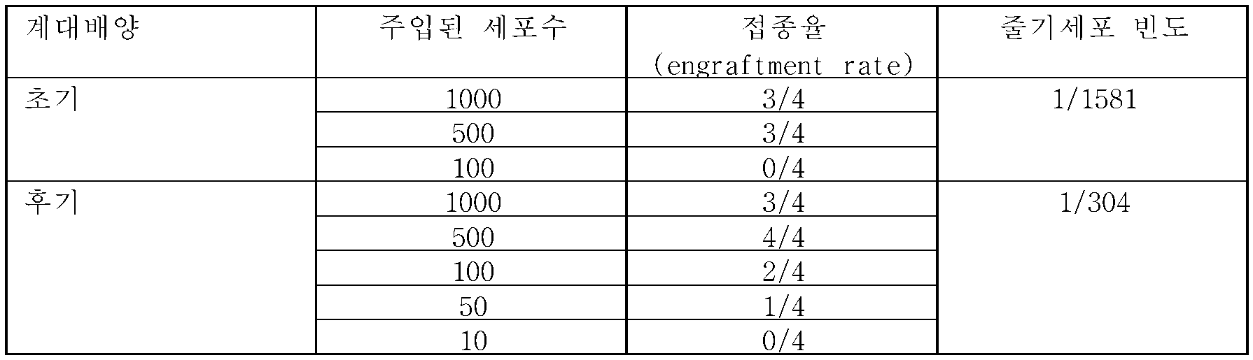
유전자 히트 맵을 도 9에 나타내었고, 상이한 PPB 샘플들, 성인 폐(AL) 및 태아 폐(FL) 사이의 몇몇 증식 마커들(예를 들면, KI67, E2F2 , 및 CDK1), 자가 재생 폴리콤 유전자(예를 들면, BMI1, TOP2A, and EZH2), 전이 표지(metastasis signature) 유전자들의 발현 패턴을 비교하면, 높은 계대수의 계대배양물은 고도로 증식되고 침투성 유전자 표지를 갖는 자가 재생 유전자의 상향 조절을 나타내고, 이는 전이 거동을 예상케한다는 것을 나타내었다.The gene heat map is shown in Figure 9 and includes several proliferation markers (e.g., KI67, E2F2 , and CDK1 ) between different PPB samples, adult lung (AL) and fetal lung (FL) Comparing the expression patterns of BMI1, TOP2A, and EZH2 ) and metastasis signature genes, high-passaged subculture shows upregulation of self-regenerating genes with highly proliferating and permeable gene markers , Indicating that it predicts the transition behavior.
NCAM1에 대한 몇개의 Xn 계대배양(P4, P8, P12 및 P17)의 면역조직화학 (IHC)은 도 10a에 도시한 바와 같이 Xn 연속 증식과 함께 증가된 발현을 나타낸다. qRT-PCR 분석 결과, 성인 폐 대조구(오른쪽)와 비교하여 일차 종양 내에서 고도의 NCAM1 발현을 나타내고, 성인 폐(왼쪽)와 비교하여 태아 폐에서의 고도의 NCAM1 발현을 나타내었고, 이는 NCAM1이 발암(carcinogenesis) 및 암 발전 과정에서 중요한 역할을 한다는 사실을 강조한다(도 10b).Immunohistochemistry (IHC) of several Xn passages (P4, P8, P12 and P17) for NCAM1 shows increased expression with Xn continuous proliferation as shown in FIG. The qRT-PCR analysis showed a high NCAM1 expression in the primary tumor compared to the adult lung control (right) and a high NCAM1 expression in the fetal lung compared to the adult lung (left) (carcinogenesis) and cancer development (Fig. 10b).
CD44에 대한 몇개의 Xn 계대배양(P4, P8, P12 및 P17)의 IHC는 도 11a~b에 도시된 바와 같이, Xn 연속 증식으로 발현이 증가됨을 나타낸다.The IHC of several Xn passages (P4, P8, P12 and P17) for CD44 shows that the expression is increased by Xn continuous proliferation, as shown in Figs. 11a-b.
비록 본 발명이 이들의 특정한 구체예들과 관련되어 기술되었지만, 많은 대안들, 변경들 및 변형들이 당업자에게 명백하다는 것은 자명하다. 따라서, 본 발명은 첨부된 청구항들의 기술사상 및 넓은 범위 내에 있는 모든 대안들, 변경들 및 변형들을 포함한다.Although the present invention has been described in connection with specific embodiments thereof, it is evident that many alternatives, modifications and variations will be apparent to those skilled in the art. Accordingly, the invention includes all alternatives, modifications and variations that fall within the spirit and broad scope of the appended claims.
구체적 및 개별적으로 나타낸 각각의 출원들, 특허 또는 특허 출원들은 참고문헌으로 본 명세서에 통합된 것처럼, 본 명세서에서 언급된 모든 간행물들, 특허 및 특허 출원들은 동일한 범위 내에서 참고문헌으로서 전체 내용이 본 명세서에 통합된다. 또한, 본 출원에서 임의의 참고 문헌의 인용 또는 확인은 그러한 참고가 본 발명의 선행 기술로서 이용가능하다는 것을 인정하는 것으로 해석되어서는 안된다. 부분 명칭들이 사용되었으나, 그들은 반드시 제한적인 것으로 해석되어서는 안된다.Each of the applications, patents, or patent applications, both specifically and individually indicated, as incorporated herein by reference, all publications, patents, and patent applications mentioned in this specification are herein incorporated by reference to the same extent ≪ / RTI > In addition, citation or identification of any reference in this application should not be construed as an admission that such reference is available as prior art to the present invention. Partial names have been used, but they are not necessarily to be construed as limiting.
Claims (38)
(a) 일차 종양을 생체내에서 계대배양하는 단계; 및
(b) 계대배양된 일차 종양의 종양 세포들의 제1 개체군에서의 적어도 하나의 항원 수준을 상기 일차 종양의 종양 세포들의 제2 개체군과 비교하는 단계, 여기서 상기 종양 세포들의 제2 개체군은 다음의 (i) 또는 (ii)이다:
(i) 상기 인간 일차 종양의 계대배양되지 않은 세포들; 또는
(ii) 상기 인간 일차 종양의 생체내 계대배양된 세포들, 여기서 상기 종양 세포들의 제2 개체군은 계대배양된 종양 세포들의 제1 개체군보다 적어도 1회 더 적은 계대수로 생체내 계대배양된 것이고,
상기 종양 세포들의 제2 개체군에서의 상기 항원의 양과 비교하여 상기 종양 세포들의 제1 개체군에서의 상기 항원의 양의 증가는 인간 일차 종양에서의 암 줄기세포 마커들의 지표가 된다.A method for identifying cancer stem cell markers in human primary tumors, comprising the steps of:
(a) subculturing primary tumor in vivo; And
(b) comparing at least one antigen level in a first population of tumor cells of subcultured primary tumor with a second population of tumor cells of said primary tumor, wherein said second population of tumor cells is i) or (ii):
(i) non-subcultured cells of said human primary tumor; or
(ii) in vivo subcultured cells of said human primary tumor, wherein said second population of said tumor cells is in vivo subcultured with a passage number at least one less than the first population of subcultured tumor cells,
An increase in the amount of the antigen in the first population of tumor cells as compared to the amount of the antigen in the second population of tumor cells is indicative of cancer stem cell markers in human primary tumors.
계대배양된 세포들의 상기 제1 개체군의 계대수는 적어도 2 계대수의 연속적인 계대배양 동안 유의적으로 증가하지 않는 명백한 공격성 종양 표현형이 나타나도록 선택되는 것인 방법.The method according to claim 1,
Wherein the number of passages of said first population of subcultured cells is selected to exhibit a distinct aggressive tumor phenotype that does not significantly increase during subsequent subculture of at least two passages.
상기 제1 개체군의 계대수는 계대배양된 세포의 제1 개체군의 5000개 세포들을 사용하여 다음 세대 이종이식을 개시시키는 빈도가 인간 일차 종양의 계대배양되지 않은 세포들과 비교하여 적어도 5배 증가하도록 선택되는 것인 방법.The method according to claim 1,
The number of passages of the first population is determined by using at least 5,000 cells of the first population of subcultured cells to increase the frequency of initiating the next generation xenotransplantation by at least 5-fold compared to non- The method being selected.
상기 계대배양된 세포들의 제1 개체군의 계대수는 상기 계대배양된 세포들의 제1의 개체군에 있어서 암 줄기세포:비-암 줄기세포의 비율이 1:1000보다 크도록 선택되는 것인 방법.The method according to claim 1,
Wherein the number of passages of the first population of subcultured cells is selected such that the ratio of cancer stem cells to non-cancer stem cells in the first population of subcultured cells is greater than 1: 1000.
상기 항원은 폴리펩타이드인 방법.5. The method according to any one of claims 1 to 4,
Wherein said antigen is a polypeptide.
상기 폴리펩타이드의 양의 증가는 세포 당 상기 폴리펩타이드의 발현 수준의 증가인 방법.6. The method of claim 5,
Wherein the increase in the amount of said polypeptide is an increase in the level of expression of said polypeptide per cell.
상기 폴리펩타이드의 양의 증가는 상기 폴리펩타이드를 발현하는 세포들의 양의 증가인 방법.6. The method of claim 5,
Wherein an increase in the amount of said polypeptide is an increase in the amount of cells expressing said polypeptide.
상기 폴리펩타이드의 양의 증가는 세포 당 상기 폴리펩타이드의 발현 수준의 증가 및 상기 폴리펩타이드를 발현하는 세포들의 양의 증가인 방법.6. The method of claim 5,
Wherein an increase in the amount of said polypeptide is an increase in the level of expression of said polypeptide per cell and an increase in the amount of cells expressing said polypeptide.
상기 종양 세포들의 제1 개체군에서의 암 줄기세포 빈도:상기 종양 세포들의 제2 개체군에서의 암 줄기세포 빈도가 5:1보다 큰 것인 방법.9. The method according to any one of claims 1 to 8,
The cancer stem cell frequency in the first population of tumor cells: the cancer stem cell frequency in the second population of tumor cells is greater than 5: 1.
상기 일차 종양은 소아 종양인 방법.10. The method according to any one of claims 1 to 9,
Wherein said primary tumor is a pediatric tumor.
상기 일차 종양은 비정형 유기형/간상 종양(ATRT), 유윙 육종, 혈관근육지방종, 흉막과 폐의 모세포종 및 월름스 종양으로 이루어지는 군으로부터 선택되는 것인 방법.10. The method according to any one of claims 1 to 9,
Wherein said primary tumor is selected from the group consisting of atypical organic / hepatic tumors (ATRT), wing sarcoma, angio-muscular lipoma, pleural and pulmonary blastoma, and horizontal tumors.
상기 계대배양된 종양 세포들의 제1 개체군은 최소 2 계대수로 계대배양된 것인 방법.12. The method according to any one of claims 1 to 11,
Wherein the first population of subcultured tumor cells is subcultured to a minimum of two passages.
상기 종양 세포들의 제2 개체군은 5 이하의 계대수로 연속적으로 생체내 계대배양되는 방법.13. The method according to any one of claims 1 to 12,
Wherein the second population of tumor cells is subcultured in vivo in successive passages of 5 or less.
상기 계대배양된 종양 세포들의 제1 개체군은 적어도 5 계대수로 생체내 계대배양되는 방법.13. The method according to any one of claims 1 to 12,
Wherein the first population of subcultured tumor cells is subcultured in vivo in at least 5 passages.
상기 계대배양은 마우스에서 수행되는 방법.15. The method according to any one of claims 1 to 14,
Wherein said subculture is performed in a mouse.
상기 계대배양은 상기 일차 종양의 이종이식의 조직 샘플의 비-해리된 동물세포들 내로의 이식에 의해 수행되는 방법.16. The method according to any one of claims 1 to 15,
Wherein the subculture is performed by implantation of tissue samples of xenotransplantation of the primary tumor into non-dissociated animal cells.
상기 계대배양은 일차 종양의 이종이식의 동물 단일 세포 현택액 내로의 이식에 의해 수행되는 방법.16. The method according to any one of claims 1 to 15,
Wherein the subculture is performed by implantation of a primary tumor xenotransplantation into an animal monocytic suspension.
상기 폴리펩타이드의 발현은 mRNA 수준에 의해 결정되는 방법.18. The method according to any one of claims 5 to 17,
Wherein the expression of said polypeptide is determined by mRNA levels.
상기 폴리펩타이드의 발현은 단백질 수준에 의해 결정되는 방법.18. The method according to any one of claims 5 to 17,
Wherein the expression of said polypeptide is determined by a protein level.
상기 암 세포의 공격성 표현형은 암 세포 마커, 유전자 발현 프로파일, 이종이식의 소요 시간, 이종이식의 발생에 요구되는 세포수, 세포이동 능력, 비-암성 조직에 대한 침투 능력 및 증식 수준으로 이루어지는 군으로부터 선택되는 방법.3. The method of claim 2,
Aggressive phenotypes of the cancer cells can be determined from the group consisting of cancer cell markers, gene expression profiles, time required for xenotransplantation, number of cells required for the development of xenotransplantation, cell migration ability, penetration ability to non-cancerous tissue, How to choose.
종양 세포들의 제1 개체군은 면역세포분리가 수행되지 않은 것인 방법.20. The method according to any one of claims 1 to 19,
Wherein the first population of tumor cells is not subjected to immunocyte separation.
상기 항원은 제1항 내지 제21항 중 어느 한 항의 방법에 따라 동정된 암 줄기세포 마커인 암 치료 방법.23. The method of claim 22,
Wherein said antigen is a cancer stem cell marker identified according to the method of any one of claims 1 to 21.
상기 항원은 폴리펩타이드인 방법.26. The method according to any one of claims 22 to 25,
Wherein said antigen is a polypeptide.
상기 일차 종양에서의 상기 폴리펩타이드 발현: 상기 생체내 계대배양된 세포들에서의 폴리펩타이드 발현의 비율은 적어도 1:5인 방법.27. The method of claim 26,
Said polypeptide expression in said primary tumor: the ratio of polypeptide expression in said in vivo subcultured cells is at least 1: 5.
상기 생체내 계대배양된 세포들은 적어도 3 계대수로 생체내 계대배양된 세포들을 포함하는 방법.26. The method according to any one of claims 22 to 25,
Wherein said in vivo subcultured cells comprise cells passaged in vivo in at least 3 passages.
(a) 제1항 내지 제21항 중 어느 한 항의 방법에 따라 상기 대상의 일차 종양에서의 적어도 하나의 암 줄기세포 마커를 동정하는 단계; 및
(b) 상기 암 줄기세포 마커들의 발현을 하향 조정 및/또는 그들의 활성을 억제하는 제제를 선택함으로써, 암 치료를 위한 제제를 선택하는 단계.A method for selecting an agent for cancer treatment for cancer treatment, comprising the steps of:
(a) identifying at least one cancer stem cell marker in a primary tumor of the subject according to the method of any one of claims 1 to 21; And
(b) selecting an agent for cancer treatment by selecting an agent that down-regulates expression of the cancer stem cell markers and / or inhibits their activity.
상기 폴리펩타이드는 리실옥시다아제(LOX)인 방법.31. The method of claim 30,
Wherein said polypeptide is a ribonuclease (LOX).
상기 제제는 β-아미노프로피오니트릴(BAPN)인 방법.31. The method of claim 30,
Wherein the preparation is? -Aminopropionitrile (BAPN).
(a) 제1항 내지 제21항 중 어느 한 항의 방법에 따라, 상기 대상의 일차 종양에서의 적어도 하나의 암 줄기세포 마커를 동정되는 단계; 및
(b) 암 치료 후에 상기 대상의 종양에서의 상기 암 줄기세포 마커의 양을 분석하는 단계; 여기서 상기 치료 이전의 상기 암 줄기세포 마커의 양과 비교하여 상기 종양에서의 상기 암 줄기세포 마커의 양의 감소는 효과적인 암 치료의 지표이다.A method of monitoring cancer treatment in a subject, comprising the steps of:
(a) identifying at least one cancer stem cell marker in the subject's primary tumor according to the method of any one of claims 1 to 21; And
(b) analyzing the amount of the cancer stem cell marker in the tumor of the subject after cancer treatment; Wherein a decrease in the amount of the cancer stem cell marker in the tumor relative to the amount of the cancer stem cell marker prior to the treatment is an indicator of effective cancer therapy.
상기 암 줄기세포 마커는 세마포린3C(SEMA3C), 리실옥시다아제(LOX), 글리코프로테인 M6A(GPM6A), 헤파토사이트 성장인자(HGF/SF) 및 알데하이드 디하이드로게나아제1(ALDH1)로 이루어지는 군으로부터 선택되고, 상기 암은 비정형 유기형/간상 종양(ATRT)인 방법.35. The method of claim 34,
The cancer stem cell markers are selected from the group consisting of semaphorin 3C (SEMA3C), ricyloxidase (LOX), glycoprotein M6A (GPM6A), hepatocyte growth factor (HGF / SF) and aldehyde dehydrogenase 1 Wherein said cancer is an atypical organic / hepatic tumor (ATRT).
상기 항원은 제1항 내지 제21항 중 어느 한 항의 방법에 따라 동정된 암 줄기세포 마커인 제제.37. The method of claim 36,
Wherein said antigen is a cancer stem cell marker identified according to the method of any one of claims 1 to 21.
Applications Claiming Priority (3)
| Application Number | Priority Date | Filing Date | Title |
|---|---|---|---|
| US201462016773P | 2014-06-25 | 2014-06-25 | |
| US62/016,773 | 2014-06-25 | ||
| PCT/IL2015/050663 WO2015198334A2 (en) | 2014-06-25 | 2015-06-25 | Identification of cancer stem cells and use of same for diagnosis and treatment |
Publications (1)
| Publication Number | Publication Date |
|---|---|
| KR20170041192A true KR20170041192A (en) | 2017-04-14 |
Family
ID=53674236
Family Applications (1)
| Application Number | Title | Priority Date | Filing Date |
|---|---|---|---|
| KR1020177001924A KR20170041192A (en) | 2014-06-25 | 2015-06-25 | Identification of cancer stem cell markers and use of same for diagnosis and treatment |
Country Status (11)
| Country | Link |
|---|---|
| US (1) | US10883994B2 (en) |
| EP (1) | EP3161480B1 (en) |
| JP (1) | JP6587288B2 (en) |
| KR (1) | KR20170041192A (en) |
| CN (1) | CN106537148B (en) |
| AU (1) | AU2015278659A1 (en) |
| CA (1) | CA2953663A1 (en) |
| ES (1) | ES2791199T3 (en) |
| IL (1) | IL249741B (en) |
| SG (1) | SG11201610630RA (en) |
| WO (1) | WO2015198334A2 (en) |
Cited By (2)
| Publication number | Priority date | Publication date | Assignee | Title |
|---|---|---|---|---|
| WO2020184782A1 (en) * | 2019-03-13 | 2020-09-17 | 울산대학교 산학협력단 | Multi-cancer classification method using common gene extraction |
| WO2022025625A1 (en) * | 2020-07-28 | 2022-02-03 | 부산대학교 산학협력단 | Biomarker composition for detecting cancer stem cells |
Families Citing this family (4)
| Publication number | Priority date | Publication date | Assignee | Title |
|---|---|---|---|---|
| EP3161480B1 (en) | 2014-06-25 | 2020-02-12 | Tel HaShomer Medical Research Infrastructure and Services Ltd. | Identification of cancer stem cell markers and use of same for diagnosis and treatment |
| US11573241B2 (en) | 2016-03-09 | 2023-02-07 | Institute National De La Sante Et De La Recherche Medicale (Inserm) | Total cellular iron as a marker of cancer stem cells and uses thereof |
| CN108967358A (en) * | 2018-05-31 | 2018-12-11 | 上海交通大学医学院附属瑞金医院 | A kind of construction method of zebra fish FL tumor model and its application |
| CN114624304B (en) * | 2021-12-09 | 2023-09-19 | 重庆医科大学国际体外诊断研究院 | miRNA electrochemical sensor based on cerium-silver nanoflower and branch DNAzyme walker |
Family Cites Families (43)
| Publication number | Priority date | Publication date | Assignee | Title |
|---|---|---|---|---|
| NL154600B (en) | 1971-02-10 | 1977-09-15 | Organon Nv | METHOD FOR THE DETERMINATION AND DETERMINATION OF SPECIFIC BINDING PROTEINS AND THEIR CORRESPONDING BINDABLE SUBSTANCES. |
| US3940475A (en) | 1970-06-11 | 1976-02-24 | Biological Developments, Inc. | Radioimmune method of assaying quantitatively for a hapten |
| NL154598B (en) | 1970-11-10 | 1977-09-15 | Organon Nv | PROCEDURE FOR DETERMINING AND DETERMINING LOW MOLECULAR COMPOUNDS AND PROTEINS THAT CAN SPECIFICALLY BIND THESE COMPOUNDS AND TEST PACKAGING. |
| NL154599B (en) | 1970-12-28 | 1977-09-15 | Organon Nv | PROCEDURE FOR DETERMINING AND DETERMINING SPECIFIC BINDING PROTEINS AND THEIR CORRESPONDING BINDABLE SUBSTANCES, AND TEST PACKAGING. |
| US3901654A (en) | 1971-06-21 | 1975-08-26 | Biological Developments | Receptor assays of biologically active compounds employing biologically specific receptors |
| US3853987A (en) | 1971-09-01 | 1974-12-10 | W Dreyer | Immunological reagent and radioimmuno assay |
| US3867517A (en) | 1971-12-21 | 1975-02-18 | Abbott Lab | Direct radioimmunoassay for antigens and their antibodies |
| NL171930C (en) | 1972-05-11 | 1983-06-01 | Akzo Nv | METHOD FOR DETERMINING AND DETERMINING BITES AND TEST PACKAGING. |
| US3850578A (en) | 1973-03-12 | 1974-11-26 | H Mcconnell | Process for assaying for biologically active molecules |
| US3935074A (en) | 1973-12-17 | 1976-01-27 | Syva Company | Antibody steric hindrance immunoassay with two antibodies |
| US3996345A (en) | 1974-08-12 | 1976-12-07 | Syva Company | Fluorescence quenching with immunological pairs in immunoassays |
| US4034074A (en) | 1974-09-19 | 1977-07-05 | The Board Of Trustees Of Leland Stanford Junior University | Universal reagent 2-site immunoradiometric assay using labelled anti (IgG) |
| US3984533A (en) | 1975-11-13 | 1976-10-05 | General Electric Company | Electrophoretic method of detecting antigen-antibody reaction |
| US4098876A (en) | 1976-10-26 | 1978-07-04 | Corning Glass Works | Reverse sandwich immunoassay |
| US4289747A (en) | 1978-12-26 | 1981-09-15 | E-Y Laboratories, Inc. | Immunological determination using lectin |
| US4376110A (en) | 1980-08-04 | 1983-03-08 | Hybritech, Incorporated | Immunometric assays using monoclonal antibodies |
| US4879219A (en) | 1980-09-19 | 1989-11-07 | General Hospital Corporation | Immunoassay utilizing monoclonal high affinity IgM antibodies |
| US5204096A (en) | 1984-03-07 | 1993-04-20 | New York Blood Center, Inc. | Pre-S gene coded peptide hepatitis B immunogens, vaccines, diagnostics, and synthetic lipid vesicle carriers |
| US5011771A (en) | 1984-04-12 | 1991-04-30 | The General Hospital Corporation | Multiepitopic immunometric assay |
| US4666828A (en) | 1984-08-15 | 1987-05-19 | The General Hospital Corporation | Test for Huntington's disease |
| US4683202A (en) | 1985-03-28 | 1987-07-28 | Cetus Corporation | Process for amplifying nucleic acid sequences |
| US4801531A (en) | 1985-04-17 | 1989-01-31 | Biotechnology Research Partners, Ltd. | Apo AI/CIII genomic polymorphisms predictive of atherosclerosis |
| US5171578A (en) | 1985-06-26 | 1992-12-15 | The Liposome Company, Inc. | Composition for targeting, storing and loading of liposomes |
| US5258499A (en) | 1988-05-16 | 1993-11-02 | Vestar, Inc. | Liposome targeting using receptor specific ligands |
| US5272057A (en) | 1988-10-14 | 1993-12-21 | Georgetown University | Method of detecting a predisposition to cancer by the use of restriction fragment length polymorphism of the gene for human poly (ADP-ribose) polymerase |
| US5192659A (en) | 1989-08-25 | 1993-03-09 | Genetype Ag | Intron sequence analysis method for detection of adjacent and remote locus alleles as haplotypes |
| US5281521A (en) | 1992-07-20 | 1994-01-25 | The Trustees Of The University Of Pennsylvania | Modified avidin-biotin technique |
| US6132764A (en) | 1994-08-05 | 2000-10-17 | Targesome, Inc. | Targeted polymerized liposome diagnostic and treatment agents |
| US5935850A (en) | 1996-09-30 | 1999-08-10 | The Research Foundation Of State University Of New York | Model for cell migration and use thereof |
| US6037137A (en) | 1997-02-20 | 2000-03-14 | Oncoimmunin, Inc. | Fluorogenic peptides for the detection of protease activity |
| US6984522B2 (en) | 2000-08-03 | 2006-01-10 | Regents Of The University Of Michigan | Isolation and use of solid tumor stem cells |
| TW200422749A (en) | 2003-04-22 | 2004-11-01 | Adv Lcd Tech Dev Ct Co Ltd | Crystallization apparatus, crystallization method, phase modulation element, device and display apparatus |
| US20070021365A1 (en) * | 2005-06-21 | 2007-01-25 | The Board Of Trustees Of The Leland Stanford Junior University | Inhibition of Lysyl oxidase for treating tumor growth and diagnostics relating thereto |
| WO2008091908A2 (en) * | 2007-01-22 | 2008-07-31 | Raven Biotechnologies | Human cancer stem cells |
| EP2022848A1 (en) * | 2007-08-10 | 2009-02-11 | Hubrecht Institut | A method for identifying, expanding, and removing adult stem cells and cancer stem cells |
| EP2268604B1 (en) * | 2008-03-14 | 2017-02-15 | Retrotope, Inc. | Therapies for cancer using isotopically substituted lysine |
| EP2263072A1 (en) | 2008-03-26 | 2010-12-22 | Veritide Limited | An analysis system |
| US20110111434A1 (en) * | 2008-07-15 | 2011-05-12 | Huang Emina H | Colon stem cells associated with colitisand colorectal cancer and methods of use |
| EP2401362B1 (en) | 2009-02-26 | 2017-04-12 | Tel HaShomer Medical Research Infrastructure and Services Ltd. | Isolated populations of renal stem cells and methods of isolating and using same |
| EP2412825B8 (en) * | 2009-03-24 | 2018-01-10 | Riken | Leukemia stem cell markers |
| EP2611463A2 (en) | 2010-09-03 | 2013-07-10 | Stem Centrx, Inc. | Identification and enrichment of cell subpopulations |
| EP3603671A3 (en) * | 2011-10-28 | 2020-07-29 | Chugai Seiyaku Kabushiki Kaisha | Cancer stem cell-specific molecule |
| EP3161480B1 (en) | 2014-06-25 | 2020-02-12 | Tel HaShomer Medical Research Infrastructure and Services Ltd. | Identification of cancer stem cell markers and use of same for diagnosis and treatment |
-
2015
- 2015-06-25 EP EP15739030.3A patent/EP3161480B1/en active Active
- 2015-06-25 JP JP2016574232A patent/JP6587288B2/en not_active Expired - Fee Related
- 2015-06-25 WO PCT/IL2015/050663 patent/WO2015198334A2/en active Application Filing
- 2015-06-25 AU AU2015278659A patent/AU2015278659A1/en not_active Abandoned
- 2015-06-25 ES ES15739030T patent/ES2791199T3/en active Active
- 2015-06-25 SG SG11201610630RA patent/SG11201610630RA/en unknown
- 2015-06-25 US US15/320,847 patent/US10883994B2/en active Active
- 2015-06-25 CA CA2953663A patent/CA2953663A1/en not_active Abandoned
- 2015-06-25 KR KR1020177001924A patent/KR20170041192A/en unknown
- 2015-06-25 CN CN201580037960.8A patent/CN106537148B/en active Active
-
2016
- 2016-12-22 IL IL249741A patent/IL249741B/en active IP Right Grant
Cited By (2)
| Publication number | Priority date | Publication date | Assignee | Title |
|---|---|---|---|---|
| WO2020184782A1 (en) * | 2019-03-13 | 2020-09-17 | 울산대학교 산학협력단 | Multi-cancer classification method using common gene extraction |
| WO2022025625A1 (en) * | 2020-07-28 | 2022-02-03 | 부산대학교 산학협력단 | Biomarker composition for detecting cancer stem cells |
Also Published As
| Publication number | Publication date |
|---|---|
| CN106537148B (en) | 2019-10-11 |
| CA2953663A1 (en) | 2015-12-30 |
| US10883994B2 (en) | 2021-01-05 |
| IL249741B (en) | 2019-12-31 |
| US20170199195A1 (en) | 2017-07-13 |
| WO2015198334A3 (en) | 2016-02-25 |
| JP2017528114A (en) | 2017-09-28 |
| WO2015198334A2 (en) | 2015-12-30 |
| IL249741A0 (en) | 2017-02-28 |
| AU2015278659A1 (en) | 2017-02-02 |
| EP3161480A2 (en) | 2017-05-03 |
| SG11201610630RA (en) | 2017-01-27 |
| CN106537148A (en) | 2017-03-22 |
| ES2791199T3 (en) | 2020-11-03 |
| EP3161480B1 (en) | 2020-02-12 |
| JP6587288B2 (en) | 2019-10-09 |
Similar Documents
| Publication | Publication Date | Title |
|---|---|---|
| JP6912538B2 (en) | Methods and Assays for Blood Circulating Tumor Cells | |
| US10883994B2 (en) | Identification of cancer stem cells markers and use of same for diagnosis and treatment | |
| US20180246080A1 (en) | Methods using axl as a biomarker of epithelial-to-mesenchymal transition | |
| US20070292414A1 (en) | Compositions enriched in neoplastic stem cells and methods comprising same | |
| EP2529223B1 (en) | Biomarkers for circulating tumor cells | |
| JP2016525883A (en) | Prognostic classification and treatment of adenocarcinoma | |
| Lee et al. | Prognostic Value of Alternative Lengthening of Telomeres–Associated Biomarkers in Uterine Sarcoma and Uterine Carcinosarcoma | |
| Thapar et al. | Clinical-pathological correlations of pituitary tumours | |
| US9057055B2 (en) | Method of obtaining circulating cancer cell populations | |
| EP1726208A1 (en) | Murine stem cells and applications thereof | |
| JP2014516551A (en) | Metakaryotic stem cell dsRNA / DNA hybrid genome replication intermediates | |
| KR101916564B1 (en) | Biomarkers for identifying high-grade glioma subjects responsive to anti-angiogenic drug | |
| Li | Targeting CXCR2+ Neuroendocrine-like Cells for the Treatment of Castration-resistant Prostate Cancer | |
| CN117795340A (en) | Biomarkers and uses thereof | |
| Jimenez | Generation of DNA aptamers for lung cancer studies |