KR20160130754A - Systems and methods for diagnosing a fluidics system and determining data processing settings for a flow cytometer - Google Patents
Systems and methods for diagnosing a fluidics system and determining data processing settings for a flow cytometer Download PDFInfo
- Publication number
- KR20160130754A KR20160130754A KR1020167022906A KR20167022906A KR20160130754A KR 20160130754 A KR20160130754 A KR 20160130754A KR 1020167022906 A KR1020167022906 A KR 1020167022906A KR 20167022906 A KR20167022906 A KR 20167022906A KR 20160130754 A KR20160130754 A KR 20160130754A
- Authority
- KR
- South Korea
- Prior art keywords
- data
- channel
- time window
- data collection
- collection time
- Prior art date
Links
- 238000000034 method Methods 0.000 title claims abstract description 108
- 238000012545 processing Methods 0.000 title claims abstract description 16
- 239000012530 fluid Substances 0.000 claims abstract description 49
- 238000013480 data collection Methods 0.000 claims description 89
- 239000002245 particle Substances 0.000 claims description 82
- 238000012546 transfer Methods 0.000 claims description 20
- 239000011324 bead Substances 0.000 claims description 14
- 238000013500 data storage Methods 0.000 claims description 10
- 230000005684 electric field Effects 0.000 claims description 9
- 238000005259 measurement Methods 0.000 abstract description 6
- 210000004027 cell Anatomy 0.000 description 45
- 238000005070 sampling Methods 0.000 description 13
- 230000008569 process Effects 0.000 description 12
- 230000003287 optical effect Effects 0.000 description 11
- 238000004458 analytical method Methods 0.000 description 9
- 238000002474 experimental method Methods 0.000 description 7
- 239000000523 sample Substances 0.000 description 6
- 238000001514 detection method Methods 0.000 description 5
- 238000000684 flow cytometry Methods 0.000 description 5
- 238000000926 separation method Methods 0.000 description 5
- 241000894007 species Species 0.000 description 5
- 230000036541 health Effects 0.000 description 4
- 239000000463 material Substances 0.000 description 4
- 238000012360 testing method Methods 0.000 description 4
- 241000894006 Bacteria Species 0.000 description 3
- 239000004005 microsphere Substances 0.000 description 3
- VYPSYNLAJGMNEJ-UHFFFAOYSA-N Silicium dioxide Chemical compound O=[Si]=O VYPSYNLAJGMNEJ-UHFFFAOYSA-N 0.000 description 2
- 238000007405 data analysis Methods 0.000 description 2
- 230000001419 dependent effect Effects 0.000 description 2
- 238000003745 diagnosis Methods 0.000 description 2
- 238000002405 diagnostic procedure Methods 0.000 description 2
- 239000011521 glass Substances 0.000 description 2
- 230000010354 integration Effects 0.000 description 2
- 238000012986 modification Methods 0.000 description 2
- 230000004048 modification Effects 0.000 description 2
- 238000011056 performance test Methods 0.000 description 2
- 230000002285 radioactive effect Effects 0.000 description 2
- QLUXVUVEVXYICG-UHFFFAOYSA-N 1,1-dichloroethene;prop-2-enenitrile Chemical compound C=CC#N.ClC(Cl)=C QLUXVUVEVXYICG-UHFFFAOYSA-N 0.000 description 1
- NLHHRLWOUZZQLW-UHFFFAOYSA-N Acrylonitrile Chemical compound C=CC#N NLHHRLWOUZZQLW-UHFFFAOYSA-N 0.000 description 1
- 241000224489 Amoeba Species 0.000 description 1
- 241000203069 Archaea Species 0.000 description 1
- 241000206602 Eukaryota Species 0.000 description 1
- 241000233866 Fungi Species 0.000 description 1
- 239000004793 Polystyrene Substances 0.000 description 1
- 240000004808 Saccharomyces cerevisiae Species 0.000 description 1
- 241000700605 Viruses Species 0.000 description 1
- 230000009471 action Effects 0.000 description 1
- 230000003321 amplification Effects 0.000 description 1
- 239000012491 analyte Substances 0.000 description 1
- 210000004102 animal cell Anatomy 0.000 description 1
- 238000004364 calculation method Methods 0.000 description 1
- 230000015556 catabolic process Effects 0.000 description 1
- 239000003153 chemical reaction reagent Substances 0.000 description 1
- 238000010586 diagram Methods 0.000 description 1
- 238000009792 diffusion process Methods 0.000 description 1
- 239000006185 dispersion Substances 0.000 description 1
- 230000005284 excitation Effects 0.000 description 1
- 230000004907 flux Effects 0.000 description 1
- 230000006870 function Effects 0.000 description 1
- 230000001678 irradiating effect Effects 0.000 description 1
- 238000002955 isolation Methods 0.000 description 1
- 229920000126 latex Polymers 0.000 description 1
- 239000004816 latex Substances 0.000 description 1
- 229920002521 macromolecule Polymers 0.000 description 1
- 230000007257 malfunction Effects 0.000 description 1
- 238000012544 monitoring process Methods 0.000 description 1
- 238000003199 nucleic acid amplification method Methods 0.000 description 1
- 210000000056 organ Anatomy 0.000 description 1
- 230000002085 persistent effect Effects 0.000 description 1
- 229920000747 poly(lactic acid) Polymers 0.000 description 1
- 229920003229 poly(methyl methacrylate) Polymers 0.000 description 1
- 229920002239 polyacrylonitrile Polymers 0.000 description 1
- 229920000642 polymer Polymers 0.000 description 1
- 239000004926 polymethyl methacrylate Substances 0.000 description 1
- -1 polymethylene melamine Polymers 0.000 description 1
- 229920002223 polystyrene Polymers 0.000 description 1
- 229920000131 polyvinylidene Polymers 0.000 description 1
- 239000012857 radioactive material Substances 0.000 description 1
- 230000008439 repair process Effects 0.000 description 1
- 238000011160 research Methods 0.000 description 1
- 230000011664 signaling Effects 0.000 description 1
- 239000000377 silicon dioxide Substances 0.000 description 1
- 229920002379 silicone rubber Polymers 0.000 description 1
- 239000004945 silicone rubber Substances 0.000 description 1
- 230000007480 spreading Effects 0.000 description 1
- 239000000126 substance Substances 0.000 description 1
- 230000001052 transient effect Effects 0.000 description 1
- XLYOFNOQVPJJNP-UHFFFAOYSA-N water Substances O XLYOFNOQVPJJNP-UHFFFAOYSA-N 0.000 description 1
Images
Classifications
-
- G—PHYSICS
- G01—MEASURING; TESTING
- G01N—INVESTIGATING OR ANALYSING MATERIALS BY DETERMINING THEIR CHEMICAL OR PHYSICAL PROPERTIES
- G01N15/00—Investigating characteristics of particles; Investigating permeability, pore-volume or surface-area of porous materials
- G01N15/10—Investigating individual particles
- G01N15/1012—Calibrating particle analysers; References therefor
-
- G—PHYSICS
- G01—MEASURING; TESTING
- G01N—INVESTIGATING OR ANALYSING MATERIALS BY DETERMINING THEIR CHEMICAL OR PHYSICAL PROPERTIES
- G01N15/00—Investigating characteristics of particles; Investigating permeability, pore-volume or surface-area of porous materials
- G01N15/10—Investigating individual particles
- G01N15/14—Optical investigation techniques, e.g. flow cytometry
- G01N15/1404—Handling flow, e.g. hydrodynamic focusing
-
- G—PHYSICS
- G01—MEASURING; TESTING
- G01N—INVESTIGATING OR ANALYSING MATERIALS BY DETERMINING THEIR CHEMICAL OR PHYSICAL PROPERTIES
- G01N15/00—Investigating characteristics of particles; Investigating permeability, pore-volume or surface-area of porous materials
- G01N15/10—Investigating individual particles
- G01N15/14—Optical investigation techniques, e.g. flow cytometry
- G01N15/1429—Signal processing
-
- G—PHYSICS
- G01—MEASURING; TESTING
- G01N—INVESTIGATING OR ANALYSING MATERIALS BY DETERMINING THEIR CHEMICAL OR PHYSICAL PROPERTIES
- G01N15/00—Investigating characteristics of particles; Investigating permeability, pore-volume or surface-area of porous materials
- G01N15/10—Investigating individual particles
- G01N15/14—Optical investigation techniques, e.g. flow cytometry
- G01N15/1434—Optical arrangements
-
- G—PHYSICS
- G01—MEASURING; TESTING
- G01N—INVESTIGATING OR ANALYSING MATERIALS BY DETERMINING THEIR CHEMICAL OR PHYSICAL PROPERTIES
- G01N15/00—Investigating characteristics of particles; Investigating permeability, pore-volume or surface-area of porous materials
- G01N15/10—Investigating individual particles
- G01N15/14—Optical investigation techniques, e.g. flow cytometry
- G01N15/1456—Optical investigation techniques, e.g. flow cytometry without spatial resolution of the texture or inner structure of the particle, e.g. processing of pulse signals
- G01N15/1459—Optical investigation techniques, e.g. flow cytometry without spatial resolution of the texture or inner structure of the particle, e.g. processing of pulse signals the analysis being performed on a sample stream
-
- G—PHYSICS
- G01—MEASURING; TESTING
- G01N—INVESTIGATING OR ANALYSING MATERIALS BY DETERMINING THEIR CHEMICAL OR PHYSICAL PROPERTIES
- G01N15/00—Investigating characteristics of particles; Investigating permeability, pore-volume or surface-area of porous materials
- G01N15/10—Investigating individual particles
- G01N15/1012—Calibrating particle analysers; References therefor
- G01N2015/1014—Constitution of reference particles
-
- G01N2015/1018—
-
- G—PHYSICS
- G01—MEASURING; TESTING
- G01N—INVESTIGATING OR ANALYSING MATERIALS BY DETERMINING THEIR CHEMICAL OR PHYSICAL PROPERTIES
- G01N15/00—Investigating characteristics of particles; Investigating permeability, pore-volume or surface-area of porous materials
- G01N15/10—Investigating individual particles
- G01N2015/1026—Recognising analyser failures, e.g. bubbles; Quality control for particle analysers
-
- G—PHYSICS
- G01—MEASURING; TESTING
- G01N—INVESTIGATING OR ANALYSING MATERIALS BY DETERMINING THEIR CHEMICAL OR PHYSICAL PROPERTIES
- G01N15/00—Investigating characteristics of particles; Investigating permeability, pore-volume or surface-area of porous materials
- G01N15/10—Investigating individual particles
- G01N2015/1027—Determining speed or velocity of a particle
-
- G01N2015/1068—
-
- G01N2015/1075—
-
- G—PHYSICS
- G01—MEASURING; TESTING
- G01N—INVESTIGATING OR ANALYSING MATERIALS BY DETERMINING THEIR CHEMICAL OR PHYSICAL PROPERTIES
- G01N15/00—Investigating characteristics of particles; Investigating permeability, pore-volume or surface-area of porous materials
- G01N15/10—Investigating individual particles
- G01N15/14—Optical investigation techniques, e.g. flow cytometry
- G01N15/1434—Optical arrangements
- G01N2015/1438—Using two lasers in succession
Landscapes
- Chemical & Material Sciences (AREA)
- Dispersion Chemistry (AREA)
- Physics & Mathematics (AREA)
- Health & Medical Sciences (AREA)
- Life Sciences & Earth Sciences (AREA)
- Analytical Chemistry (AREA)
- Biochemistry (AREA)
- General Health & Medical Sciences (AREA)
- General Physics & Mathematics (AREA)
- Immunology (AREA)
- Pathology (AREA)
- Engineering & Computer Science (AREA)
- Signal Processing (AREA)
- Investigating, Analyzing Materials By Fluorescence Or Luminescence (AREA)
- Investigating Or Analysing Biological Materials (AREA)
Abstract
본 실시형태의 세트는 유체 시스템을 진단하고 유동 세포분석기를 위한 데이터 처리 설정을 결정하는 시스템 및 방법에 관한 것이다. 유체 시스템을 진단하는 시스템 및 방법은 유체 전달 시스템을 정확히 측정하고 그 내부 변동의 해석을 요구한다. 데이터 처리 설정을 결정하는 시스템 및 방법은 여러 채널에 대해 피크 시간을 정확히 측정할 것을 요구하여 시간 지연 설정을 조절할 수 있고, 여기서 피크 시간은 데이터 수집 시간 윈도우의 시작으로부터 윈도우 내 최고 피크까지 경과된 시간을 측정한 값이다.The set of embodiments relates to a system and method for diagnosing fluid systems and determining data processing settings for a flow cell analyzer. Systems and methods for diagnosing a fluid system accurately measure the fluid delivery system and require interpretation of its internal variation. The system and method for determining the data processing settings may adjust the time delay setting by requiring accurate measurement of the peak time for multiple channels, wherein the peak time is the elapsed time from the beginning of the data acquisition time window to the highest peak in the window .
Description
본 발명은 일반적으로 유동 세포분석 분야의 유체 시스템에 관한 것으로, 보다 상세하게는 유체소자의 장애를 진단하고 데이터 취득 및 분석 설정을 설정하는 시스템 및 방법에 관한 것이다.FIELD OF THE INVENTION The present invention relates generally to fluid systems in the field of flow cell analysis, and more particularly to a system and method for diagnosing faults in fluid devices and setting up data acquisition and analysis settings.
관련 출원에 대한 상호 참조Cross-reference to related application
본 출원은, 전체 내용이 본 명세서에 병합된, 2014년 3월 6일자로 출원된 미국 출원 번호 61/948,547, 및 2014년 9월 29일자로 출원된 미국 출원 번호 62/056,646에 대한 우선권을 주장한다.This application claims priority to U.S. Serial No. 61 / 948,547, filed March 6, 2014, and U.S. Serial No. 62 / 056,646, filed September 29, 2014, the entire contents of which are incorporated herein by reference. do.
유동 세포분석은 기본적으로 생명과학 조사 및 의료 분야에서 다수의 응용에서 입자와 세포를 분석하는데 사용되는 강력한 도구이다. 이 기술의 분석 강도는 광원, 일반적으로 레이저 또는 레이저들로부터 포커싱(focused)된 스팟(spot)을 통해 초당 최대 수 만개의 입자의 속도로 연속적으로 빠르게 (세포, 박테리아 및 바이러스와 같은 생체입자를 포함하는) 단일 입자를 퍼레이드하는 능력에 있다. 이 초점 스팟에서 높은 광자 플럭스(photon flux)는 입자에서 광을 산란시키거나 및 또는 입자 또는 입자에 부착된 라벨로부터 광을 방출시키는데 이 광은 수집되어 분석될 수 있다. 이것은 개별적인 입자에 관한 많은 정보를 사용자에 제공하여 입자 또는 세포의 집단(population)에 관한 통계적 정보를 신속히 파악할 수 있게 한다.Flow cell analysis is basically a powerful tool used to analyze particles and cells in many applications in life science research and medical applications. The analytical intensity of this technique is rapidly and rapidly (including biological particles such as cells, bacteria, and viruses) at a rate of up to tens of thousands of particles per second through a focused spot from a light source, typically a laser or lasers ) To parade a single particle. The high photon flux at this focal spot can scatter and collect light from the particle and / or emit light from the label attached to the particle or particle, which can be collected and analyzed. This provides the user with a lot of information about individual particles, allowing them to quickly grasp statistical information about the population of particles or cells.
전통적인 유동 세포분석에서, 입자는 포커싱된 검사점을 통해 유동되고, 이 검사점에서 레이저가 레이저 빔을 채널 내 코어 직경을 포함하는 포커싱된 점으로 지향한다. 입자를 포함하는 샘플 유체는 샘플의 체적비(volumetric rate)의 100-1000 배 정도의 매우 높은 체적비로 샘플 스트림 주위로 시스 유체(sheath fluid)를 흐르게 하는 것에 의해 약 5-50 마이크론의 매우 작은 코어 직경으로 포커싱된다. 이것은 초당 수 미터 정도로 포커싱된 입자에 매우 고속의 선형 속도를 야기한다. 이것은 각 입자가 여기 스팟(excitation spot)에서 매우 제한된 시간, 종종 1-10 마이크로초만을 소비하는 것을 의미한다.In conventional flow cytometry, the particles flow through the focused checkpoint, at which point the laser directs the laser beam to the focused point containing the core diameter in the channel. The sample fluid containing the particles may have a very small core diameter of about 5-50 microns by flowing a sheath fluid around the sample stream at a very high volume ratio of about 100-1000 times the volumetric rate of the sample . This results in very high linear velocities in the focused particles at a few meters per second. This means that each particle consumes a very limited time, often 1-10 microseconds, at the excitation spot.
종래의 유동 세포분석기에서 전체 시스템과 서브시스템의 성능을 추적하는데 필요한 분석 도구 및/또는 방법이 있다. 유동 세포분석기에서 장애가 일어날 수 있는 서브시스템들은 독립적으로 또는 집합적으로 광학기기, 전자부품, 및 유체소자를 포함할 수 있다. 전통적으로, 유동 세포분석 데이터 취득 및/또는 진단 소프트웨어는 순간 시스템 성능을 측정하고 이 성능을 이전의 날(들)의 성능과 비교하는 모드를 갖는다. 이 성능 테스트는 종종 알려진 형광 특성(fluorescent characteristic)을 갖는 비드 칵테일(cocktail of bead)을 사용한다. 이 성능 테스트는 이 비드를 사용하여 '밝은' 형광 비드의 집단의 변동 계수(coefficient of variation), 광학적 배경, 및 검출 채널의 양자(quantum) 효율을 포함하는 일련의 측정을 한다. 이 값과 이 값이 변하는 방식을 모니터링하는 것에 의해, 기기가 사양 내에서 더 이상 기능하지 않아서 수리되어야 할 때를 결정할 수 있다. 기기를 수리하는 사람은 광학기기, 전자부품, 및 유체소자에 테스트를 실행할 수 있고; 이후 장애 모드는 변수 제거 공정 또는 변수 분리(isolation) 공정을 통해 결정된다. There are analytical tools and / or methods needed to track the performance of the overall system and subsystems in conventional flow cell analyzers. The subsystems in which the breakdown in the flow cell analyzer can occur independently or collectively may include optical instruments, electronic components, and fluidic devices. Traditionally, flow cytometry data acquisition and / or diagnostic software has a mode to measure instantaneous system performance and compare this performance to the performance of the previous day (s). This performance test often uses a cocktail of bead with a known fluorescent characteristic. This performance test uses this bead to make a series of measurements including the coefficient of variation of the population of 'bright' fluorescent beads, the optical background, and the quantum efficiency of the detection channel. By monitoring this value and how it changes, you can determine when the device is no longer functional within the specification and needs to be repaired. The person repairing the device can perform tests on optical devices, electronic components, and fluid devices; The failure mode is then determined through a variable removal process or a variable isolation process.
불운하게도, 유동 세포분석기를 수리하는 가장 큰 곤란성 중 하나는 대부분의 측정된 파라미터들이 광학기기, 전자부품, 및 유체 시스템의 복잡한 입력(convoluted input)으로부터 유도된다는 것이다. 많은 광학 부품과 전자 부품을 분리하는 기술이 존재한다. 유체 시스템의 마이크로유체 특성으로 인해, 극히 적은 개수의 센서와 테스트를 이용하여 건강 상태(health)를 분리하고 결정하거나 및/또는 유체 전달 시스템의 흐름 프로파일을 정확히 측정할 수 있다. 이 때문에, 광학기기와 전자부품이 테스트되고, 만약 문제가 해결되지 않는 경우에만, 유체 시스템이 테스트된다. 정상 상태의 압력을 측정하는 것 또는 누설을 검사하는 것을 넘어, 유체소자를 테스트하는 것은 통상 해법을 찾는 희망으로 여러 부품에 장착하고 여러 부품으로부터 빼내는 것을 포함한다. 다수의 레이저 빔을 갖는 유동 세포분석기는 유체 전달 시스템 내 압력 변동에 특히 민감하고, 여기서 총 동작 압력의 1% 미만의 변동이라도 광학적 데이터에서 변동 계수를 확장(broadening)시킬 수 있다. 이런 상황에서 사람을 호출하여 광학적 데이터에서 확장하는 변동 계수를 고정시킬 수 있고, 여기서 테스트는 광학 인터페이스와 전자 인터페이스에서 시작한다.Unfortunately, one of the major difficulties in repairing a flow cell analyzer is that most of the measured parameters are derived from the convoluted input of optical instruments, electronic components, and fluid systems. Techniques exist to separate many optical and electronic components. Due to the microfluidic nature of the fluidic system, a very small number of sensors and tests can be used to separate and determine the health and / or to accurately measure the flow profile of the fluid delivery system. For this reason, optical instruments and electronic components are tested, and the fluid system is tested only if the problem is not solved. Beyond measuring steady-state pressure or checking for leaks, testing fluid devices involves mounting and unplugging several components in the hope of finding a solution. Flow cell analyzers with multiple laser beams are particularly sensitive to pressure fluctuations in the fluid delivery system, where fluctuations of less than 1% of the total operating pressure can broaden the coefficient of variation in optical data. In this situation, you can call a person to fix the coefficient of variation that expands in the optical data, where testing starts with an optical interface and an electronic interface.
그리하여, 유동 세포분석기를 위한 유체 시스템에서 정상 상태와 동적인 불규칙성 또는 장애를 분리하여 검출하고, 장애 있는 실험을 실행함이 없이 광학 서브시스템과 전자 서브시스템으로부터 유체소자를 분리하고 나서, 심지어 유체 시스템이 고려되기 전에 여러 서브시스템을 고정 수리할 수 있는 것이 요구된다. 이러한 검출 시스템은 작업 유체 시스템을 조절하여 의도된 사양을 충족시키는 것을 도와줄 뿐만 아니라 파괴된 유체 시스템을 수리하는데에 사용될 수 있다. It is thus possible to isolate and detect steady state and dynamic irregularities or obstacles in a fluid system for a flow cytometer and to separate the fluid element from the optical subsystem and the electronic subsystem without performing a failed experiment, It is required that several subsystems can be fixedly repaired before being considered. This detection system can be used to regulate the working fluid system to help meet the intended specifications as well as to repair the destroyed fluid system.
일 측면에서, 유동 세포분석기를 위한 데이터 처리 설정을 결정하는 방법이 개시된다. 상기 방법은 교정 입자(calibration particle)의 세트가 유동 셀(flow cell)을 통과하는 단계를 포함할 수 있다. 상기 방법은 상기 유동 셀을 통과하는 상기 교정 입자의 세트의 각 입자를 적어도 2개의 광 빔으로 조명하는 단계를 포함할 수 있고 여기서 각 광 빔은 채널과 연관된다. 상기 방법은 각 채널과 연관된 검출기를 사용하여 상기 교정 입자의 세트의 각 입자로부터 방출된 광을 수집하는 단계를 포함할 수 있다. 상기 방법은 각 검출기로부터 데이터를 레코드하는 단계를 포함할 수 있다. 상기 방법은 트리거 채널(trigger channel)을 위한 데이터 신호 임계값이 초과될 때 상기 트리거 채널과 연관된 제1 데이터 수집 시간 윈도우로부터 데이터의 전달을 개시하도록 상기 트리거 채널을 설정하는 단계를 포함할 수 있다. 상기 방법은 상기 트리거 채널을 위한 상기 데이터 신호 임계값이 초과될 때 제2 채널과 연관된 제2 데이터 수집 시간 윈도우로부터 데이터를 전달하도록 상기 제2 채널을 설정하는 단계를 포함할 수 있고, 여기서 상기 제2 데이터 수집 시간 윈도우의 시작은 상기 트리거 채널과 상기 제2 채널 사이의 공간적 경로에 기초한다. 상기 방법은 상기 데이터 신호 임계값이 초과될 때마다 상기 제1 데이터 수집 시간 윈도우로부터의 데이터를 데이터 저장소에 레코드하는 단계를 포함할 수 있다. 상기 방법은 상기 트리거 채널을 위한 상기 데이터 신호 임계값이 초과될 때마다 상기 제2 데이터 수집 시간 윈도우로부터의 데이터를 상기 데이터 저장소에 레코드하는 단계를 포함할 수 있다. 상기 방법은 상기 제2 데이터 수집 시간 윈도우 내 데이터 세기 피크 시간(data intensity peak time)의 분포를 분석하는 단계를 포함할 수 있다. 상기 방법은 상기 제2 데이터 수집 시간 윈도우 내 데이터 세기 피크 시간의 분포에 기초하여 상기 제2 데이터 수집 시간 윈도우 내 상기 제2 채널에 데이터 신호를 위치시키는 시간 지연을 계산하는 단계를 포함할 수 있다. 상기 방법은 상기 방출된 광이 형광 광인 것을 포함할 수 있다. 상기 방법은 상기 방출된 광이 산란된 광인 것을 포함할 수 있다. 상기 방법은 상기 제2 데이터 수집 시간 윈도우의 시작이 유동률(flow rate)에 기초하는 것을 포함할 수 있다. 상기 방법은 상기 제2 데이터 수집 시간 윈도우의 시작이 시스 유체의 유동률에 기초하는 것을 포함할 수 있다. 상기 방법은 상기 공간적 경로가 약 80 마이크로미터 내지 250 마이크로미터인 것을 포함할 수 있다. 상기 방법은 상기 공간적 경로가 약 150 마이크로미터인 것을 포함할 수 있다. 상기 방법은 상기 데이터 수집 시간 윈도우가 약 80개의 내지 약 120개의 ADC 점의 폭인 것을 포함할 수 있다. 상기 방법은 상기 데이터 수집 시간 윈도우가 약 320개 내지 약 360개의 ADC 점의 폭인 것을 포함할 수 있다. In one aspect, a method for determining data processing settings for a flow cytometer is disclosed. The method may comprise the step of passing a set of calibration particles through a flow cell. The method may include illuminating each particle of the set of calibration particles through the flow cell with at least two light beams, wherein each light beam is associated with a channel. The method may include collecting light emitted from each particle of the set of calibration particles using a detector associated with each channel. The method may include recording data from each detector. The method may include setting the trigger channel to initiate transfer of data from a first data acquisition time window associated with the trigger channel when a data signal threshold for the trigger channel is exceeded. The method may include setting the second channel to transfer data from a second data acquisition time window associated with a second channel when the data signal threshold for the trigger channel is exceeded, 2 data acquisition time window is based on a spatial path between the trigger channel and the second channel. The method may include recording data from the first data collection time window into the data store each time the data signal threshold is exceeded. The method may include recording data from the second data collection time window into the data store each time the data signal threshold for the trigger channel is exceeded. The method may include analyzing a distribution of data intensity peak times within the second data collection time window. The method may include calculating a time delay to position a data signal on the second channel within the second data collection time window based on a distribution of data intensity peak times within the second data collection time window. The method may include that the emitted light is fluorescent light. The method may include that the emitted light is scattered light. The method may include that the beginning of the second data collection time window is based on a flow rate. The method may include that the beginning of the second data collection time window is based on a flow rate of the cis fluid. The method may include that the spatial path is from about 80 micrometers to about 250 micrometers. The method may include that the spatial path is about 150 micrometers. The method may include the data acquisition time window being about 80 to about 120 ADC points wide. The method may include the data collection time window being from about 320 to about 360 ADC points in width.
일 측면에서, 유동 세포분석기를 위한 데이터 처리 설정을 결정하는 시스템이 개시된다. 상기 시스템은 교정 입자를 유동시키도록 구성된 유동 셀을 포함할 수 있다. 상기 시스템은 광 빔을 방출하도록 각각 구성된 적어도 2개의 광원을 포함할 수 있고, 여기서 각 광 빔은 채널과 연관되고, 상기 광 빔은 상기 유동 셀을 통과한다. 상기 시스템은 각 채널과 연관된 검출기를 포함할 수 있고 여기서 각 검출기는 교정 비드의 세트의 각 비드로부터 방출된 광을 수집하도록 구성될 수 있다. 상기 시스템은 각 상기 검출기로부터 오는 데이터를 레코드하도록 구성된 메모리 버퍼를 포함할 수 있다. 상기 시스템은 트리거 채널을 위한 데이터 신호 임계값이 초과될 때 상기 트리거 채널과 연관된 제1 데이터 수집 시간 윈도우로부터 데이터의 전달을 개시하도록 구성된 상기 트리거 채널을 포함할 수 있다. 상기 시스템은 상기 트리거 채널을 위한 상기 데이터 신호 임계값이 초과될 때 제2 채널과 연관된 제2 데이터 수집 시간 윈도우로부터 데이터를 전달하도록 구성된 상기 제2 채널을 포함할 수 있고 여기서 상기 제2 데이터 수집 시간 윈도우의 시작은 상기 트리거 채널과 상기 제2 채널 사이의 공간적 경로에 기초한다. 상기 시스템은 상기 데이터 신호 세기 임계값이 초과될 때마다 상기 제1 데이터 수집 시간 윈도우로부터 오는 상기 데이터를 데이터 저장매체로 전달하고, 상기 데이터 신호 세기 임계값이 초과될 때마다 상기 제2 데이터 수집 시간 윈도우로부터 오는 상기 데이터를 상기 데이터 저장매체에 전달하도록 구성된 트리거 프로세서를 포함할 수 있다. 상기 시스템은 상기 제2 데이터 수집 시간 윈도우 내 데이터 세기 피크 시간의 분포를 분석하고, 상기 제2 데이터 수집 시간 윈도우 내 상기 데이터 세기 피크 시간의 분포에 기초하여 상기 제2 데이터 수집 시간 윈도우 내 상기 제2 채널에 데이터 신호를 위치시키는 시간 지연을 계산하도록 구성된 컴퓨터 프로세서를 포함할 수 있다. 상기 시스템은 전계 프로그래밍가능한 게이트 어레이를 포함할 수 있고 여기서 상기 메모리 버퍼와 상기 트리거 프로세서는 전계 프로그래밍가능한 게이트 어레이의 서브성분이다. 상기 시스템은 상기 방출된 광이 형광 광인 것을 포함할 수 있다. 상기 시스템은 상기 방출된 광이 산란된 광인 것을 포함할 수 있다. 상기 시스템은 상기 제2 데이터 수집 시간 윈도우의 시작이 유동률에 기초하는 것을 포함할 수 있다. 상기 시스템은 상기 제2 데이터 수집 시간 윈도우의 시작이 시스 유체의 유동률에 기초하는 것을 포함할 수 있다. 상기 시스템은 상기 공간적 경로가 약 80 마이크로미터 내지 250 마이크로미터인 것을 포함할 수 있다. 상기 시스템은 상기 공간적 경로가 약 150 마이크로미터인 것을 포함할 수 있다. 상기 시스템은 상기 데이터 수집 시간 윈도우가 약 80개 내지 약 120개의 ADC 점의 폭인 것을 포함할 수 있다. 상기 시스템은 상기 데이터 수집 시간 윈도우가 약 320개 내지 약 360개의 ADC 점의 폭인 것을 포함할 수 있다. 일 측면에서, 유동 세포분석기를 위한 유체소자 진단 방법이 개시된다. 상기 방법은 교정 입자의 세트가 유동 셀을 통과하는 단계를 포함할 수 있다. 상기 방법은 상기 유동 셀을 통과하는 상기 교정 입자의 세트의 각 입자를 적어도 2개의 광 빔으로 조명하는 단계를 포함할 수 있고 여기서 각 광 빔은 채널과 연관된다. 상기 방법은 각 채널과 연관된 검출기를 사용하여 상기 교정 입자의 세트의 각 입자로부터 방출된 광을 수집하는 단계를 포함할 수 있다. 상기 방법은 각 상기 검출기로부터 데이터를 레코드하는 단계를 포함할 수 있다. 상기 방법은 트리거 채널을 위한 데이터 신호 임계값이 초과될 때 상기 트리거 채널과 연관된 제1 데이터 수집 시간 윈도우로부터 데이터의 전달을 개시하도록 상기 트리거 채널을 설정하는 단계를 포함할 수 있다. 상기 방법은 상기 트리거 채널을 위한 상기 데이터 신호 임계값이 초과될 때 제2 채널과 연관된 제2 데이터 수집 시간 윈도우로부터 데이터를 전달하도록 상기 제2 채널을 설정하는 단계를 포함할 수 있다. 상기 방법은 상기 데이터 신호 임계값이 초과될 때마다 상기 제1 데이터 수집 시간 윈도우로부터의 데이터를 데이터 저장소에 레코드하는 단계를 포함할 수 있다. 상기 방법은 상기 트리거 채널을 위한 상기 데이터 신호 임계값이 초과될 때마다 상기 제2 데이터 수집 시간 윈도우로부터의 데이터를 상기 데이터 저장소에 레코드하는 단계를 포함할 수 있다. 상기 방법은 상기 제2 데이터 수집 시간 윈도우 내 데이터 세기 피크 시간의 분포를 분석하고, 상기 분포를 시스템 사양과 비교하여 유체 시스템의 건강 상태를 결정하는 단계를 포함할 수 있다. 상기 방법은 상기 시스템 사양이 1 표준 편차(standard deviation)인 것을 포함할 수 있다. 상기 방법은 상기 시스템 사양이 2 표준 편차인 것을 포함할 수 있다. 상기 방법은 상기 시스템 사양이 3 표준 편차인 것을 포함할 수 있다. 상기 방법은 상기 시스템 사양이 4 표준 편차인 것을 포함할 수 있다. 상기 방법은 상기 시스템 사양이 가우시안 분포(Gaussian distribution)인 것을 포함할 수 있다. 상기 방법은 상기 시스템 사양이 포아송 분포(Poisson distribution)인 것을 포함할 수 있다. 상기 방법은 상기 시스템 사양이 임의의 통계적 분포인 것을 포함할 수 있다. 상기 방법은 상기 방출된 광이 형광 광인 것을 포함할 수 있다. 상기 방법은 상기 방출된 광이 산란된 광인 것을 포함할 수 있다. 상기 방법은 상기 데이터 수집 시간 윈도우가 약 80개 내지 약 120개의 ADC 점의 폭인 것을 포함할 수 있다. 상기 방법은 상기 데이터 수집 시간 윈도우가 약 320개 내지 약 360개의 ADC 점의 폭인 것을 포함할 수 있다. In one aspect, a system for determining data processing settings for a flow cytometer is disclosed. The system may include a flow cell configured to flow calibration particles. The system may include at least two light sources each configured to emit a light beam, wherein each light beam is associated with a channel and the light beam passes through the flow cell. The system can include a detector associated with each channel, wherein each detector can be configured to collect light emitted from each bead of the set of calibration beads. The system may include a memory buffer configured to record data from each of the detectors. The system may include the trigger channel configured to initiate transfer of data from a first data acquisition time window associated with the trigger channel when a data signal threshold for the trigger channel is exceeded. The system may include the second channel configured to transfer data from a second data acquisition time window associated with a second channel when the data signal threshold for the trigger channel is exceeded, The beginning of the window is based on the spatial path between the trigger channel and the second channel. Wherein the system transfers the data from the first data collection time window to the data storage medium each time the data signal strength threshold is exceeded and the second data collection time And a trigger processor configured to transmit the data from the window to the data storage medium. Wherein the system analyzes the distribution of the data intensity peak time in the second data collection time window and determines the second data collection time window within the second data collection time window based on the distribution of the data intensity peak time in the second data collection time window. And a computer processor configured to calculate a time delay to position the data signal in the channel. The system may include an electric field programmable gate array wherein the memory buffer and the trigger processor are subcomponents of an electric field programmable gate array. The system may include the emitted light being a fluorescent light. The system may include the emitted light being scattered light. The system may include that the beginning of the second data collection time window is based on a flow rate. The system may include that the beginning of the second data acquisition time window is based on a flow rate of the cis fluid. The system may include those wherein the spatial path is from about 80 micrometers to about 250 micrometers. The system may include the spatial path is about 150 micrometers. The system may include the data collection time window being from about 80 to about 120 ADC points wide. The system may include the data collection time window being from about 320 to about 360 ADC points in width. In one aspect, a method of diagnosing a fluidic device for a flow cytometer is disclosed. The method may include the step of passing a set of calibration particles through the flow cell. The method may include illuminating each particle of the set of calibration particles through the flow cell with at least two light beams, wherein each light beam is associated with a channel. The method may include collecting light emitted from each particle of the set of calibration particles using a detector associated with each channel. The method may include recording data from each of the detectors. The method may include setting the trigger channel to initiate transfer of data from a first data acquisition time window associated with the trigger channel when a data signal threshold for the trigger channel is exceeded. The method may include setting the second channel to transfer data from a second data acquisition time window associated with the second channel when the data signal threshold for the trigger channel is exceeded. The method may include recording data from the first data collection time window into the data store each time the data signal threshold is exceeded. The method may include recording data from the second data collection time window into the data store each time the data signal threshold for the trigger channel is exceeded. The method may include analyzing a distribution of data intensity peak times within the second data collection time window and comparing the distribution to a system specification to determine a health state of the fluid system. The method may include that the system specification is one standard deviation. The method may include that the system specification is two standard deviations. The method may include the system specification being three standard deviations. The method may include the system specification being 4 standard deviations. The method may include that the system specification is a Gaussian distribution. The method may comprise that the system specification is a Poisson distribution. The method may include that the system specification is of any statistical distribution. The method may include that the emitted light is fluorescent light. The method may include that the emitted light is scattered light. The method may include the data collection time window being from about 80 to about 120 ADC points in width. The method may include the data collection time window being from about 320 to about 360 ADC points in width.
일 측면에서 유동 세포분석기를 위한 유체소자 진단 시스템이 개시된다. 상기 시스템은 교정 입자를 유동시키도록 구성된 유동 셀을 포함할 수 있다. 상기 시스템은 광 빔을 방출하도록 각각 구성된 적어도 2개의 광원을 포함할 수 있고, 여기서 각 광 빔은 채널과 연관되고, 상기 광 빔은 상기 유동 셀을 통과한다. 상기 시스템은 각 채널과 연관된 검출기를 포함할 수 있고 여기서 각 검출기는 교정 비드의 세트의 각 비드로부터 방출된 광을 수집하도록 구성된다. 상기 시스템은 각 상기 검출기로부터 데이터를 레코드하도록 구성된 메모리 버퍼를 포함할 수 있다. 상기 시스템은 트리거 채널을 위한 데이터 신호 임계값이 초과될 때 상기 트리거 채널과 연관된 제1 데이터 수집 시간 윈도우로부터 데이터의 전달을 개시하도록 구성된 상기 트리거 채널을 포함할 수 있다. 상기 시스템은 상기 트리거 채널을 위한 상기 데이터 신호 임계값이 초과될 때 제2 채널과 연관된 제2 데이터 수집 시간 윈도우로부터 데이터를 전달하도록 구성된 상기 제2 채널을 포함할 수 있다. 상기 시스템은 상기 데이터 신호 세기 임계값이 초과될 때마다 상기 제1 데이터 수집 시간 윈도우로부터 오는 상기 데이터를 데이터 저장매체에 전달하고, 상기 데이터 신호 세기 임계값이 초과될 때마다 상기 제2 데이터 수집 시간 윈도우로부터 오는 상기 데이터를 상기 데이터 저장매체에 전달하도록 구성된 트리거 프로세서를 포함할 수 있다. 상기 시스템은 상기 제2 데이터 수집 시간 윈도우 내 데이터 세기 피크 시간의 분포를 시스템 사양과 비교하여 유체 시스템의 건강 상태를 결정하도록 구성된 컴퓨터 프로세서를 포함할 수 있다. 상기 시스템은 전계 프로그래밍가능한 게이트 어레이를 포함할 수 있고 여기서 상기 메모리 버퍼와 상기 트리거 프로세서는 상기 전계 프로그래밍가능한 게이트 어레이의 서브성분일 수 있다. 상기 시스템은 상기 시스템 사양이 1 표준 편차인 것을 포함할 수 있다. 상기 시스템은 상기 시스템 사양이 2 표준 편차인 것을 포함할 수 있다. 상기 시스템은 상기 시스템 사양이 3 표준 편차인 것을 포함할 수 있다. 상기 시스템은 상기 시스템 사양이 4 표준 편차인 것을 포함할 수 있다. 상기 시스템은 상기 시스템 사양이 가우시안 분포인 것을 포함할 수 있다. 상기 시스템은 상기 시스템 사양이 포아송 분포인 것을 포함할 수 있다. 상기 시스템은 상기 시스템 사양이 임의의 통계적 분포인 것을 포함할 수 있다. 상기 시스템은 상기 방출된 광이 형광 광인 것을 포함할 수 있다. 상기 시스템은 상기 방출된 광이 산란된 광인 것을 포함할 수 있다. 상기 시스템은 상기 데이터 수집 시간 윈도우가 약 80개 내지 약 120개의 ADC 점의 폭인 것을 포함할 수 있다. 상기 시스템은 상기 데이터 수집 시간 윈도우가 약 320개 내지 약 360개의 ADC 점의 폭인 것을 포함할 수 있다. In one aspect, a fluidic device diagnostic system for a flow cytometer is disclosed. The system may include a flow cell configured to flow calibration particles. The system may include at least two light sources each configured to emit a light beam, wherein each light beam is associated with a channel and the light beam passes through the flow cell. The system may include a detector associated with each channel, wherein each detector is configured to collect light emitted from each bead of the set of calibration beads. The system may include a memory buffer configured to record data from each of the detectors. The system may include the trigger channel configured to initiate transfer of data from a first data acquisition time window associated with the trigger channel when a data signal threshold for the trigger channel is exceeded. The system may include the second channel configured to transfer data from a second data acquisition time window associated with a second channel when the data signal threshold for the trigger channel is exceeded. Wherein the system transmits the data from the first data collection time window to the data storage medium each time the data signal strength threshold is exceeded and the second data collection time And a trigger processor configured to transmit the data from the window to the data storage medium. The system may include a computer processor configured to compare a distribution of data intensity peak times within the second data collection time window with a system specification to determine a health state of the fluid system. The system may include an electric field programmable gate array wherein the memory buffer and the trigger processor may be subcomponents of the electric field programmable gate array. The system may include the system specification being one standard deviation. The system may include the system specification being 2 standard deviations. The system may include the system specification being three standard deviations. The system may include the system specification being 4 standard deviations. The system may include the Gaussian distribution of the system specification. The system may include that the system specification is a Poisson distribution. The system may include that the system specification is any statistical distribution. The system may include the emitted light being a fluorescent light. The system may include the emitted light being scattered light. The system may include the data collection time window being from about 80 to about 120 ADC points wide. The system may include the data collection time window being from about 320 to about 360 ADC points in width.
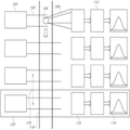
도 1은 기본 유동 세포분석기의 여러 실시형태 중 하나를 도시하는 도면.
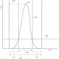
도 2는 유동 세포분석기에 의해 수집될 수 있는 데이터의 유형의 일부의 일 실시예를 도시하는 도면.
도 3은 유동 세포분석기에 의해 수집될 수 있는 데이터의 유형의 일부의 일 실시예를 도시하는 도면.
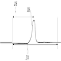
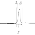
도 4a 및 도 4b는 넓은 데이터 시간 수집 윈도우와 좁은 데이터 시간 수집 윈도우를 도시하는 도면.
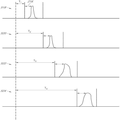
도 5는 유동 세포분석기에서 4개의 채널로부터 오는 데이터의 일 실시예를 도시하는 도면.
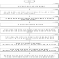
도 6은 유동 세포분석기를 위한 데이터 처리 설정을 결정하는 방법의 예시적인 실시형태를 도시하는 도면.
도 7은, 피크 시간 확산(peak time spread)이 트리거 채널로부터 거리의 함수로서 증가하는, 유동 세포분석기로부터 데이터의 일 실시예를 도시하는 도면.
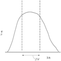
도 8a는 시스템 사양의 밖에 있는 피크 시간을 도시하는 도면.
도 8b는 시스템 사양의 내에 있는 피크 시간을 도시하는 도면.
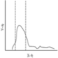
도 9a 내지 도 9e는 유체소자에 장애가 있을 때 유동 세포분석 데이터가 보일 수 있을 것 같은 것을 도시하는 도면.
도 10은 유동 세포분석기를 위한 유체 시스템을 진단하는 방법의 예시적인 실시형태를 도시하는 도면.1 shows one of several embodiments of a basic flow cell analyzer;
Figure 2 illustrates one embodiment of a portion of a type of data that can be collected by a flow cytometer.
3 depicts one embodiment of a portion of a type of data that can be collected by a flow cytometer.
Figures 4A and 4B show a wide data time collection window and a narrow data time collection window.
Figure 5 illustrates one embodiment of data from four channels in a flow cytometer.
Figure 6 depicts an exemplary embodiment of a method for determining data processing settings for a flow cytometer.
Figure 7 illustrates one embodiment of data from a flow cell analyzer, wherein the peak time spread increases as a function of distance from the trigger channel.
8A is a diagram showing peak times outside the system specification;
Figure 8b shows peak times within the system specification.
Figures 9a-9e illustrate that flow cell analysis data may be visible when a fluidic element is obstructed.
Figure 10 illustrates an exemplary embodiment of a method for diagnosing a fluidic system for a flow cytometer.
유체 시스템을 진단하고 유동 세포분석기에 대한 데이터 수집 및 분석 설정을 결정하는 시스템 및 방법의 실시형태가 첨부된 상세한 설명 및 도면에 설명된다. 도면에서, 다수의 특정 상세가 특정 실시형태를 상세히 이해하기 위해 제시된다. 이 기술 분야에 통상의 지식을 가진 자라면 본 명세서에서 설명된 시스템 및 방법이 유동 세포분석기를 포함하지만 이것으로 제한되지 않는 유체 시스템을 사용하는 여러 기기에 사용될 수 있다는 것을 이해할 수 있을 것이다. 추가적으로, 이 기술 분야에 통상의 지식을 가진 자라면 특정 실시형태가 이 특정 상세 없이 실시될 수 있다는 것을 이해할 수 있을 것이다. 나아가, 이 기술 분야에 통상의 지식을 가진 자라면 방법이 제시되고 수행되는 특정 순서는 예시를 위한 것일 뿐, 순서는 변할 수 있고 특정 실시형태의 사상과 범위 내에 여전히 있을 수 있는 것으로 고려된다는 것을 용이하게 이해할 수 있을 것이다.Embodiments of systems and methods for diagnosing fluid systems and determining data collection and analysis settings for a flow cytometer are described in the accompanying detailed description and drawings. In the drawings, numerous specific details are set forth in order to provide a thorough understanding of certain embodiments. One of ordinary skill in the art will appreciate that the systems and methods described herein can be used with a variety of instruments using fluidic systems including, but not limited to, flow cell analyzers. In addition, it will be understood by those skilled in the art that certain embodiments may be practiced without these specific details. Further, those skilled in the art will readily appreciate that the specific sequence in which a method is suggested and performed is for illustration purposes only and that the order may be changed and still be considered to be within the spirit and scope of the specific embodiment I can understand it.
본 개시 내용은 여러 실시형태와 함께 설명되었지만, 본 개시 내용은 이러한 실시형태로 제한되는 것으로 의도된 것이 아니다. 오히려, 본 개시 내용은, 이 기술 분야에 통상의 지식을 가진 자라면 이해할 수 있는 여러 대안, 변형, 및 균등물을 포함한다.While this disclosure has been described in conjunction with several embodiments, it is not intended that the disclosure be limited to such embodiments. Rather, the disclosure includes various alternatives, modifications, and equivalents that may be understood by those of ordinary skill in the art.
나아가, 여러 실시형태를 설명할 때, 본 명세서는 특정 단계 순서로 방법 및/또는 공정을 제시하였을 수 있다. 그러나, 본 방법 또는 공정이 본 명세서에 제시된 특정 단계 순서에 의존하지 않는 정도까지, 본 방법 또는 공정은 설명된 특정 단계 순서로 제한되어서는 안 된다. 이 기술 분야에 통상의 지식을 가진 자라면, 다른 단계 순서도 가능할 수 있다는 것을 이해할 수 있을 것이다. 그리하여, 본 명세서에 제시된 특정 단계 순서는 청구범위에 한정된 사항으로 해석되어서는 안 된다. 나아가, 본 방법 및/또는 공정에 관한 청구범위는 기록된 순서로 그 단계를 수행하는 것으로 제한되어서는 안되고, 이 기술 분야에 통상의 지식을 가진 자라면 그 순서는 변할 수 있고 여러 실시형태의 사상과 범위 내에 여전히 있을 수 있다는 것을 용이하게 이해할 수 있을 것이다.Further, when describing various embodiments, the present specification may suggest methods and / or processes in a specific sequence of steps. However, to the extent that the present method or process is not dependent upon the particular sequence of steps presented herein, the method or process should not be limited to the particular sequence of steps set forth. Those of ordinary skill in the art will recognize that other step sequences may be possible. Thus, the particular order of steps presented herein should not be construed as limited to the claims. Furthermore, the claims of the present method and / or process should not be limited to performing the steps in the order in which they are written, and those of ordinary skill in the art will recognize that the order may vary, Lt; / RTI > and < RTI ID = 0.0 > range. ≪ / RTI >
본 발명을 보다 용이하게 이해하기 위하여, 특정 용어들이 제일 먼저 정의된다. 추가적인 정의는 상세한 설명에 제시된다.For a better understanding of the present invention, certain terms are first defined. Additional definitions are provided in the detailed description.
본 명세서에 사용된 바와 같이 "ADC 점"이란 아날로그-디지털 컨버터의 샘플링 점(sampling point)들 사이의 시간 간격이다. 본 명세서에서, 1 ADC 점은 500 나노초 또는 1 마이크로초일 수 있다.As used herein, the term "ADC point" is the time interval between sampling points of the analog-to-digital converter. In this specification, one ADC point may be 500 nanoseconds or 1 microsecond.
본 명세서에 사용된 바와 같이 "분석물"이란 분석될 물질종 또는 물질을 의미한다.As used herein, "analyte" means a species or material to be analyzed.
본 명세서에 사용된 바와 같이 "채널"이란 데이터 수집이 일어나는 유동 셀을 통한 경로를 의미한다.As used herein, "channel" means a path through a flow cell where data collection occurs.
본 명세서에 사용된 바와 같이 "진단 파라미터"란 층상 유동 안정성(laminar flow stability)과 관련된 양 또는 측정, 펌프 또는 기어 펌프에서 발생하는 기계적인 교란, 입자 도달 사이의 시간(입자 도달 시간), 유체 압력, 높은 유체 압력, 낮은 유체 압력, 유체 압력 변동, 누설, 및/또는 유체 시스템의 양에 관해 이 기술 분야에서 알려진 임의의 것을 의미한다.As used herein, "diagnostic parameter" refers to an amount or measurement associated with laminar flow stability, mechanical disturbances occurring in a pump or gear pump, time between particle arrival (particle arrival time), fluid pressure , High fluid pressure, low fluid pressure, fluid pressure fluctuations, leakage, and / or the amount of fluid system.
본 명세서에 사용된 바와 같이 "유동 셀"란 직사각형, 정사각형, 타원형, 장방형, 원형, 8각형, 7각형, 6각형, 5각형, 및 3각형으로부터 선택된 내부 형상을 구비하는 유동 채널, 챔버 또는 모세관(capillary)을 의미한다.As used herein, a "flow cell" is a flow channel having an interior shape selected from a rectangular, square, oval, rectangular, circular, octagonal, hexagonal, hexagonal, pentagonal, (capillary).
본 명세서에 사용된 바와 같이 "라벨"이란 생물학적 시스템과 같은 시스템에 도입되어 유동 셀 또는 채널의 코스를 통해 추종하며, 유동 셀 또는 채널에서 입자 또는 타깃에 대한 정보를 제공하는 염료(dye) 또는 방사성 동위원소(radioactive isotope)와 같은 식별가능한 물질종을 의미한다.As used herein, the term "label" refers to a dye or radioactive material that is introduced into a system such as a biological system to follow through the course of a flow cell or channel and provides information about the particle or target in the flow cell or channel Means an identifiable species of substance, such as a radioactive isotope.
본 명세서에 사용된 바와 같이 "마이크로구체(microsphere)" 또는 "비드(bead)"란 구(sphere)와 같이 대칭이거나, 대칭성을 갖지 않는 덤벨 형상(dumbbell shape) 또는 매크로분자와 같이 비대칭일 수 있는 입자를 의미한다. 마이크로구체 또는 비드의 실시예는 실리카, 유리 및 중공 유리, 라텍스, 실리콘 고무, 폴리스티렌, 폴리메틸메타크릴레이트, 폴리메틸렌멜라민, 폴리아크릴로니트릴, 폴리메틸아크릴로니트릴, 폴리(비닐리덴 염화물-코-아크릴로니트릴), 및 폴리락타이드와 같은 폴리머를 포함하나 이들로 제한되지 않는다. As used herein, "microsphere" or "bead" refers to a molecule that is symmetric, such as a sphere, or which may be asymmetric, such as a dumbbell shape or macromolecule, Particles. Examples of microspheres or beads are silica, glass and hollow glass, latex, silicone rubber, polystyrene, polymethylmethacrylate, polymethylene melamine, polyacrylonitrile, polymethyl acrylonitrile, poly (vinylidene chloride- - acrylonitrile), and polymers such as polylactide.
본 명세서에 사용된 바와 같이 "입자"란 진핵생물과 원핵생물 세포, 고세균, 박테리아, 곰팡이, 식물 세포, 효모, 원생동물, 아메바, 원생생물, 동물 세포와 같은 생물체 세포; 세포 세포소기관; 유기/무기 소자 또는 분자; 마이크로구체; 및 물 내 오일 같이 혼합되지 않는 유체의 액적을 포함하지만 이들로 제한되지 않는 작은 물질 단위를 의미한다. As used herein, the term "particle" refers to an organism cell such as a eukaryote and a prokaryote cell, an archaea, a bacterium, a fungus, a plant cell, a yeast, a protozoan, an amoeba, a protist organism, an animal cell; Cell organ organ; Organic / inorganic elements or molecules; Microsphere; And droplets of fluid that are not mixed such as oil in water.
본 명세서에 사용된 바와 같이 "피크"란 신호 진폭에서 높은 점에 관한 것이다. 일부 경우에, 신호는 형광 광으로부터 유래될 수 있다. As used herein, "peak" refers to the high point in the signal amplitude. In some cases, the signal may be derived from fluorescent light.
본 명세서에 사용된 바와 같이 "피크 시간"은 데이터 수집 시간 윈도우의 시작으로부터 윈도우 내 최고 피크까지 경과된 시간의 측정값이다. As used herein, "peak time" is a measure of the elapsed time from the beginning of the data acquisition time window to the highest peak in the window.
본 명세서에 사용된 바와 같이 "탐침"이란 유체 또는 샘플에서 다른 물질을 검출하거나 식별하도록 라벨링되거나 다른 방식으로 표시되고 사용되는 물질종을 의미한다.As used herein, "probe" means a species of material that is labeled or otherwise displayed and used to detect or identify other materials in a fluid or sample.
본 명세서에 사용된 바와 같이 "시약(reagent)"이란 특정 방식으로 반응하는 것으로 알려진 물질종이다.As used herein, "reagent" is a species known to react in a particular manner.
본 명세서에 사용된 바와 같이 "신호 분자(signaling molecule)"는 생물학적 시스템과 같은 시스템에 도입되어 입자에 대한 신호로 사용될 수 있는 염료 또는 방사성 동위원소와 같은 식별가능한 물질종을 의미한다. As used herein, "signaling molecule" means an identifiable species of material, such as a dye or radioactive isotope, that can be introduced into a system, such as a biological system, and used as a signal to a particle.
본 명세서에 사용된 바와 같이 "공간적 분리" 또는 "채널들 사이의 공간적 분리"란 하나의 광 빔의 중심으로부터 인접한 광 빔의 중심까지의 거리를 의미한다. As used herein, "spatial separation" or "spatial separation between channels" means the distance from the center of one light beam to the center of an adjacent light beam.
본 명세서에 사용된 바와 같이 "사양"이란 개별 실험의 요구를 충족하는 데이터 품질 요구조건을 충족하는 유동 세포분석기의 성능을 의미한다. As used herein, "specification" means the performance of a flow cell analyzer that meets data quality requirements to meet the needs of an individual experiment.
본 명세서에 사용된 바와 같이 "타깃"이란 탐침의 결합 부분을 의미한다.As used herein, "target" refers to the coupled portion of the probe.
본 명세서에 사용된 바와 같이 "과도 상태(transient)"란 종국적으로 안정화되는 시스템에서 일시적으로 불안정한 상태를 의미한다. 예를 들어, 팽창하고 수축하는 유체 시스템에서 공기 버블은 과도 상태를 야기할 수 있다. As used herein, "transient" means a temporarily unstable condition in a system that eventually stabilizes. For example, air bubbles in a fluid system that expands and contracts can cause transients.
본 명세서에 사용된 바와 같이 "트리거 임계값"은 신호로부터 오는 세기 값이 검출된 이벤트를 처리하기 위하여 처리 전자부품을 활성화할 만큼 충분히 높은 점을 의미한다. As used herein, a "trigger threshold" means that the intensity value coming from the signal is high enough to activate the detected electronic component to process the detected event.
본 명세서에 사용된 바와 같이 "트리거" 또는 "트리거링"이란 신호로부터 오는 세기 값이 트리거 임계값을 초과할 때 처리 전자부품을 활성화시키는 것이다. As used herein, "trigger" or "triggering" is to activate a processing electronic component when the intensity value coming from the signal exceeds the trigger threshold.
본 명세서에 사용된 바와 같이 "트리거 레이저" 또는 "트리거 채널"이란 트리거 임계값을 센싱하고, 시스템에서 모든 레이저 또는 채널로부터 오는 모든 취득된 데이터가 저장 및 분석될 것을 요구하는 것을 나타내는 일을 담당하는 하드웨어 세트이다. As used herein, a "trigger laser" or "trigger channel" is used to sense trigger thresholds and to indicate that all acquired data from all lasers or channels in the system requires storage and analysis Hardware set.
본 명세서에 사용된 바와 같이 "윈도우," "수집 윈도우," "데이터 수집 윈도우," "데이터 수집 시간 윈도우," "데이터 분석 윈도우"는 높이, 폭, 및 면적에 대해 디지털 샘플링 전자부품에 의해 초기에 분석되고 나서 차후에 디지털 샘플링 전자부품으로부터 추가적인 분석을 위해 영구 저장매체 장소로 전달될 수 있는 데이터이다. As used herein, the terms "window," "acquisition window," "data acquisition window," "data acquisition time window," and "data analysis window" And then transferred from the digital sampling electronics to a permanent storage location for further analysis.
여러 실시형태에서, 본 출원서에 개시된 시스템, 방법, 및 장치는 유동 세포분석에 관한 여러 장치, 시스템, 및 방법과 함께 사용될 수 있다. 전체 내용이 본 명세서에 병합된, 미국 특허 출원 번호 12/239,390 및 12/209,084 참조. 또한 전체 내용이 본 명세서에 병합된 Practical Flow Cytometry, 4th Edition, Howard M. Shapiro 참조. In various embodiments, the systems, methods, and apparatus described herein may be used in conjunction with various devices, systems, and methods for flow cell analysis. See U.S. Patent Application Nos. 12 / 239,390 and 12 / 209,084, the entire contents of which are incorporated herein. See Practical Flow Cytometry , 4 th Edition, Howard M. Shapiro, the entire contents of which are incorporated herein.
도 1은 유동 세포분석기, 및 데이터를 수집할 수 있는 방식을 개략적으로 도시한다. 여러 실시형태는 적어도 하나의 광원(102)을 포함할 수 있다. 각 광원(102)은 광 빔(104)을 생성할 수 있고, 이 광 빔은 입자가 유동 셀(116)을 통과할 때 입자(106)를 조명할 수 있다. 이 조명에 의해 입자로부터 광(108)이 나올 수 있다. 광(108)의 형태는 형광 광 또는 산란된 광인 것을 포함할 수 있다. 광(108)은 검출기(110)에 의해 검출될 수 있고, 데이터(114)는 디지털 샘플링 전자부품(112)으로 전달될 수 있다. 여러 실시형태에서, 디지털 샘플링 전자부품(112)은 아날로그 또는 디지털 형태일 수 있는 일부 종류의 메모리를 포함할 수 있다. 메모리는 랜덤 액세스 메모리이거나 또는 랜덤 액세스 메모리를 사용하는 원형 버퍼(circular)일 수 있다. 디지털 샘플링 전자부품(112)에서 데이터(114)는 높이, 폭, 및 피크 시간 정보를 포함할 수 있는 신호로부터 생성될 수 있다. 데이터(114)는 전압 형태일 수 있고, 유체소자를 진단하고 시간 지연 설정을 교정하는데 사용될 수 있다. 여러 실시형태에서, 아날로그-디지털 컨버터는 전압을 디지털 데이터로 변환하는데 사용될 수 있다. 여러 실시형태에서, 이미지는 전압 대신에 사용될 수 있고, 세기는 외삽될 수 있다. 추가적으로, 공간적 분리(118)(또는 공간적 경로)는 인접한 광 빔(104)들 사이의 거리에 의해 측정될 수 있다. 채널(120)들 사이에 공간적 분리(118)가 있을 수 있고 이 공간적 분리는 여러 실시형태에서 약 150 마이크로미터이거나 또는 다른 여러 실시형태에서 약 80 마이크로미터 내지 약 250 마이크로미터일 수 있다. 본 명세서에 사용된 바와 같이, 채널(120)은 데이터 수집 경로일 수 있다. 도 1에는 4개의 데이터 수집 경로 또는 채널(120)이 도시되어 있지만, 여러 실시형태는 임의의 개수의 채널(120)을 사용할 수 있다. Figure 1 schematically shows a flow cell analyzer and the manner in which data can be collected. Various embodiments may include at least one
여러 실시형태에서 디지털 샘플링 전자부품(112)은 아날로그 샘플링 전자부품 또는 간단한 샘플링 전자부품일 수 있다. 여러 실시형태에서, 디지털 샘플링 전자부품(112)은 전계 프로그래밍가능한 게이트 어레이를 포함할 수 있고 여기서 전계 프로그래밍가능한 게이트 어레이는 메모리 버퍼, 트리거 프로세서, 및 계산 블록을 포함할 수 있다. 메모리 버퍼는 모든 데이터(114)를 저장할 수 있고, 데이터 신호 세기 임계값(트리거 임계값)이 초과될 때 데이터(114)는 계산 블록에 의해 처리되어 컴퓨터로 송신될 수 있다. 컴퓨터는 메모리, 프로세서, 및 이 기술 분야에서 알려진 임의의 다른 부품을 포함할 수 있다. In various embodiments,
도 2는 결과적인 데이터가 x 및 y 좌표 시스템으로 도시되고 신호 곡선(206)으로 표시될 때 결과적인 데이터(114)를 개략적으로 도시하는 실시예이다. x-축은 시간을 나타내고, y-축은 신호 세기를 나타낼 수 있다. 여러 실시형태에서, 신호 세기는 형광 세기로부터 유래될 수 있다. 여러 실시형태에서, 세기는 광전자증배관 또는 이와 유사한 디바이스에서 증폭되고 나서 전압으로 측정될 수 있다. 여러 실시형태에서, 신호 세기가 트리거 임계값(208)에 도달할 때, 디지털 샘플링 전자부품(112)은 입자(또는 이벤트)가 검출된 것을 레지스터할 수 있고, 일부 디지털 처리를 수행하거나 또는 데이터를 영구 데이터 저장매체에 전달하거나, 또는 이들 둘 모두를 수행할 수 있다. 영구 데이터 저장매체는 컴퓨터에 위치될 수 있다. 도 2에서, 펄스 폭은 트리거 임계값(208)에서 신호 곡선(206)의 폭을 포함하는 것을 볼 수 있다. (데이터 세기 피크 또는 펄스 높이라고도 언급되는) 최고 피크(204)는 신호 곡선이 y-축에 대해 최고인 곳일 수 있다. 일반적으로, 데이터 수집 시간 윈도우(214)를 설정할 때 관련 데이터(114)를 가능한 한 많이 수집하는 것이 유리하다. 그리하여, 데이터 수집 시간 윈도우(214)는 펄스 폭(202)뿐만 아니라 전방 연장부(210)와 후방 연장부(212)를 포함할 수 있다. 데이터 수집 시간 윈도우(214)는 신호 곡선(206)의 위치가 일부 분산(variance)을 가질 수 있는 것을 고려하여 실험 전에 설정될 수 있다. FIG. 2 is an embodiment that schematically illustrates the resulting
데이터 수집 시간 윈도우(214)는 입자마다 실험 동안 동적으로 설정될 수 있다. 마지막 데이터 시간 수집 윈도우(214)의 사이즈를 판정할 때 여러 고려 사항이 관련된다. 데이터 시간 수집 윈도우(214)는 너무 클 수 없고 또는 동시발생(coincidence)의 위험이 있고, 데이터 시간 수집 윈도우(214)는 너무 작을 수 없고 또는 입자(106)로부터 데이터는 데이터 시간 수집 윈도우(214)의 경계의 밖에 있을 수 있다. The data
도 3을 참조하면, 단일 곡선으로 컴파일된 여러 신호 곡선(206)을 포함하는 히스토그램을 볼 수 있다. 각 신호 곡선(206)은 단일 이벤트로 카운트되거나, 또는 유동 셀(116)을 통해 전달되고 채널(120)들 중 하나의 채널로부터 신호를 생성하는 입자(106)를 나타낼 수 있다. y-축은 카운트 또는 이벤트의 개수를 나타내고, x-축은 피크 시간(216) 또는 데이터 세기 피크 시간을 나타낸다. 피크의 정점은 대부분의 이벤트가 일어나는 시점이다. 도 3에서 이벤트는 정규 분포된다. 도 3에서, 서로 더해지는 피크 시간(216)은 컴파일된 피크 시간(302)이라고 언급될 수 있다. 유동 세포분석에서, 입자 도달 시간에 항상 일부 분산 또는 지터(jitter)가 있어서, 이에 의해 이벤트는 상이한 피크 시간(216)에 일어나게 된다. 상기 사양에서, 정확한 피크 시간(216)을 검출하는 능력은 매우 중요하다. 통계적 중요성에 도달하기 위하여 종종 500개 이상의 이벤트를 평균낼 필요가 있다. 일부 상황에서, 1000개 이상의 이벤트가 통계적 중요성에 도달하고 컴파일된 피크 시간(302)을 적절히 평가하는데 요구된다. 평균, 중앙값, 적분, 또는 경사 미분(slope derivative)과 관련된 임의의 통계적 수단이 사용될 수 있다. 이 기술 분야에 통상의 지식을 가진 자라면 분산을 분석하는 많은 다른 방식이 있다는 것을 이해할 수 있을 것이다. Referring to FIG. 3, a histogram including several signal curves 206 compiled into a single curve can be seen. Each
도 4a 및 도 4b를 참조하면, 2개의 신호 곡선(206)이 도시된다. 도 4a는 넓은 데이터 수집 시간 윈도우 곡선(206)을 도시하고 도 4b는 좁은 데이터 수집 시간 윈도우 곡선(206)을 도시한다. 이들 도면은 유동 세포분석기를 위한 데이터 처리 설정을 결정하는 시스템 또는 방법에서 중요한 단계를 나타낸다. 데이터 수집 시간 윈도우(214) 내에서 피크 시간(216)을 초기에 측정할 때 최고 피크(204)가 일어나는 곳은 불확정적이다. 각 채널(120)에 대해 데이터 수집 시간 윈도우(214)를 확장(widening)시키면 피크(204)를 발견할 가능성을 크게 개선시킬 수 있다. 전술한 바와 같이, 컴파일된 피크 시간(302)의 정확한 표현을 보장하기 위해 여러 데이터 점을 수집할 필요가 있다. 컴파일된 피크 시간(302)이 정확히 측정되면 데이터 수집 시간 윈도우(214)가 도 4b에 도시된 바와 같이 좁아져서, 동시발생의 가능성을 감소시키고 신호 대 잡음 비율을 증가시킬 수 있다. 이러한 데이터 수집 시간 윈도우(214)의 사이즈의 감소는 각 채널(120)에서 일어날 수 있다. 데이터 시간 수집 시간 윈도우(214)는 또한 시간적으로 시프트될 수 있다. Referring to Figures 4A and 4B, two
도 5를 참조하면, 4개의 신호 곡선(206)이 도시된다. 도 5에서 각 신호 곡선(206)은 시스템에서 일반적인 피크 시간(216)을 갖는 일반적인 신호 곡선(206)을 나타낸다. 여기서, y-축은 세기(전압, 형광, 휘도 등)일 수 있고, x-축은 시간일 수 있다(이것은 종종 이 기술 분야에서, 아날로그-디지털 컨버터와 관련해서 ADC 점이라고 언급된다). 신호 곡선(216)은 동일한 입자가 4개의 공간적으로 분리된 채널(518, 520, 522, 및 524)을 통과하는 것을 나타낸다. 이 실시예에서, 제1 채널(518)은 트리거 채널(518)을 나타낸다. 트리거 임계값(208)이 트리거 채널(518)에서 충족되면 디지털 샘플링 전자부품(112)은 모든 채널(518, 520, 522, 및 524)에 대한 신호 처리를 시작한다.Referring to Figure 5, four
피크 시간(216) 또는 컴파일된 피크 시간(302)을 사용하여 시간 지연 결정을 하기 전에 시스템 설정에 기초하여 근사값을 사용할 수 있다. 이 설정은 유동 셀을 통한 유동률 또는 시스 유체의 유동률을 포함할 수 있다. 추가적으로, 유동 셀(116)을 통과하는 인접한 광 빔(104)들 사이의 거리와 같은 하드웨어 파라미터를 포함할 수 있다.The
데이터 수집 설정을 결정할 때 데이터 수집 시간 윈도우(214)는 4개의 공간적으로 분리된 채널(518, 520, 522, 및 524)들 각각에 대해 도 4a에 도시된 바와 같이 넓게 유지된다. 피크 시간(216)으로부터 유도된 시간 지연은 4개의 공간적으로 분리된 채널(518, 520, 522, 및 524)들 각각에 대해 설정될 수 있고 여기서 시간 지연은 데이터 수집 시간 윈도우 위치(Ti)와 피크 시간(216) 사이의 다음 관계에 의해 주어진다.The data
여기서 i는 i번째 레이저 위치에 대응하고, i = 1은 트리거 채널(216)이다. T1 = 0을 설정하는 것이 일반적이다. 트리거 채널은 임의의 채널일 수 있고, 시간 지연은 양이거나 또는 음일 수 있는 것으로 이해된다.Where i corresponds to the i-th laser position, and i = 1 is the
공간에서 트리거 채널(518)로부터 더 멀리 있는 채널은 가장 긴 시간 지연을 가질 수 있다. 모든 채널(518, 520, 522, 및 524)에 대한 피크 시간(216)의 정확한 측정이 디지털 샘플링 전자부품(112)에 의해 측정되었다면 시간 지연은 각 채널(518, 520, 522, 및 524)에 대해 조절될 수 있고, 시간 수집 데이터 윈도우(214)가 좁아져서, 신호 대 잡음 비율을 최적화하고 동시발생을 감소시킬 수 있다. 일반적으로, 최고 피크(204) 평균은 각 채널(518, 520, 522, 및 524)에 대한 시간 수집 데이터 윈도우(214) 내에 센터링될 수 있다. 그러나, 센터링이 요구되는 것은 아니고, 일부 상황에서는 최적이 아닐 수 있다. 이러한 절차는 2개의 이상 채널에 대해 사용될 수 있고, 도 5는 여러 실시형태 중 단지 하나의 실시예일 뿐인 것으로 이해된다.A channel further away from the
여러 실시형태에서, 초기 넓은 시간 수집 데이터 윈도우(214)는 약 320개 내지 360개의 ADC 점의 범위일 수 있다. 그리고 좁은 시간 수집 데이터 윈도우(214)는 약 80개 내지 약 120개의 ADC 점의 범위일 수 있다. 여러 실시형태에서, 연장부는 좁은 윈도우에 대해 약 27개의 ADC 점일 수 있다. 여러 실시형태에서, 연장부는 약 17개 내지 약 37개의 ADC 점의 범위일 수 있다.In various embodiments, the initial wide time
도 6은, 교정 입자의 세트가 유동 셀을 통과하는 것을 포함하는, 유동 세포분석기를 위한 데이터 처리 설정을 결정하는 방법의 여러 실시형태 중 일 실시예를 도시한다(602). 여러 실시형태는 유동 셀을 통과하는 교정 입자의 세트의 각 입자를 적어도 2개의 광 빔으로 조명하는 단계를 포함할 수 있고, 여기서 각 광 빔은 채널과 연관된다(604). 여러 실시형태는 각 채널과 연관된 검출기를 사용하여 교정 입자의 세트의 각 입자로부터 방출된 광을 수집하는 단계를 포함할 수 있다(606). 여러 실시형태는 각 검출기로부터 데이터를 레코드하는 단계를 포함할 수 있다(608). 여러 실시형태는 트리거 채널을 위한 데이터 신호 임계값이 초과될 때 트리거 채널과 연관된 제1 데이터 수집 시간 윈도우로부터 데이터의 전달을 개시하도록 트리거 채널을 설정하는 단계를 포함할 수 있다(610). 여러 실시형태는 트리거 채널을 위한 데이터 신호 임계값이 초과될 때 제2 채널과 연관된 제2 데이터 수집 시간 윈도우로부터 데이터를 전달하도록 제2 채널을 설정하는 단계를 포함할 수 있고(612), 여기서 제2 데이터 수집 시간 윈도우의 시작은 트리거 채널과 제2 채널 사이에 공간적 경로에 기초한다. 여러 실시형태는 데이터 신호 임계값이 초과될 때마다 제1 데이터 수집 시간 윈도우로부터의 데이터를 데이터 저장소에 레코드하는 단계를 포함할 수 있다(614). 여러 실시형태는 트리거 채널을 위한 데이터 신호 임계값이 초과될 때마다 제2 데이터 수집 시간 윈도우로부터의 데이터를 데이터 저장소에 레코드하는 단계를 포함할 수 있다(616). 여러 실시형태는 제2 데이터 수집 시간 윈도우 내 데이터 세기 피크 시간의 분포를 분석하는 단계를 포함할 수 있다(618). 여러 실시형태는 제2 데이터 수집 시간 윈도우 내 데이터 세기 피크 시간의 분포에 기초하여 제2 데이터 수집 시간 윈도우 내 제2 채널에 데이터 신호를 위치시키는 시간 지연을 계산하는 단계를 포함할 수 있다(620).Figure 6 illustrates one embodiment of various embodiments of a method for determining data processing settings for a flow cytometer, including passing a set of calibration particles through a flow cell (602). Various embodiments may include illuminating each particle of a set of calibration particles through a flow cell with at least two light beams, wherein each light beam is associated with a channel (604). Various embodiments may include collecting light emitted from each particle of the set of calibration particles using a detector associated with each channel (606). Various embodiments may include recording data from each detector (608). Various embodiments may include setting a trigger channel to initiate transfer of data from a first data acquisition time window associated with the trigger channel when the data signal threshold for the trigger channel is exceeded (610). Various embodiments may include setting (612) a second channel to transfer data from a second data acquisition time window associated with a second channel when a data signal threshold for the trigger channel is exceeded, 2 The start of the data acquisition time window is based on a spatial path between the trigger channel and the second channel. Various embodiments may include recording (614) data from a first data collection time window to a data store whenever a data signal threshold is exceeded. Various embodiments may include recording (616) data from a second data collection time window to a data store whenever a data signal threshold for the trigger channel is exceeded. Various embodiments may include analyzing the distribution of the data intensity peak times within the second data collection time window (618). Various embodiments may include calculating a time delay to position the data signal on a second channel in a second data collection time window based on a distribution of data intensity peak times within a second data collection time window (620) .
도 7을 참조하면, 3개의 데이터 수집 곡선(702, 704, 및 706)은 첨부된 컴파일된 데이터 곡선(또는 히스토그램)(708, 710, 및 712)을 각각 구비하는 것으로 도시된다. 데이터 수집 곡선(702, 704, 및 706)에 대해, x-축은 시간을 나타내고, y-축은 신호 세기를 나타낼 수 있다. 제1 데이터 수집 곡선(702)은 피크 시간(216)의 변동이 전혀 없거나 거의 없는 트리거 채널(702)을 나타낸다. 인접한 데이터 수집 곡선(704)은 3개의 곡선을 도시한다. 점선 곡선은 입자 도달 시간의 변동을 나타낸다. 트리거 채널(702)로부터 가장 먼 채널(120) 또는 데이터 수집 곡선(706)은 변동이 더 긴 거리에 걸쳐 합성(compound)될 수 있으므로 가장 많은 피크 시간(216)을 가진다. 유체소자의 장애 피크 시간(216)이 있는지 여부를 진단할 때 도달이 중요한 증거를 제공할 수 있다. 예를 들어, 트리거 채널로부터 가장 먼 채널에 대한 데이터 수집 곡선(706)은 최적화된 데이터 수집 시간 윈도우(214)의 밖에 있는 곡선을 가진다. 이러한 데이터(114)는 서브-최적으로 처리되거나 또는 간단히 처리되지 않을 수 있고, 데이터 누락과 열등한 결과를 초래할 수 있다. 특정 상황에서 데이터 수집 시간 윈도우(214)는 더 많은 데이터(114)를 수집하도록 개방될 수 있으나, 동시발생이 잡음과 함께 증가할 수 있다.Referring to FIG. 7, three data collection curves 702, 704, and 706 are shown having attached compiled data curves (or histograms) 708, 710, and 712, respectively. For data collection curves 702, 704, and 706, the x-axis represents time and the y-axis may represent signal strength. The first
컴파일된 데이터 곡선(708, 710, 및 712)은, y-축에 이벤트 또는 카운트를 나타내고 x-축에 피크 시간(216)을 나타내고, 시스템에서 피크 시간(216)을 많은 입자로부터 측정한 것으로부터 취해진 히스토그램을 도시한다. 트리거 채널에 대한 컴파일된 데이터(708)는 채널을 통과하는 입자 또는 이벤트의 대부분이 작은 시간 범위 내에서 일어나는 타이트한 데이터 분포를 도시한다. 다른 컴파일된 데이터 곡선(710 및 712)에서 볼 수 있는 바와 같이 입자가 더 긴 거리를 이동할 때 확산이 더 넓어질 수 있는 것으로 예상된다.The compiled data curves 708, 710, and 712 illustrate events or counts on the y-axis and
도 8a를 참조하면 도 8a는 시스템에서 유체소자의 변동의 결과로 데이터(114)의 대부분이 데이터 수집 시간 윈도우(214) 내에 있지 않은 저품질로 컴파일된 데이터를 도시하는 피크 시간 값(216)의 히스토그램의 일 실시예를 도시한다. 데이터는 대시 라인에 의해 도시된 미리 결정된 시스템 사양의 밖에 있다. 도 8b는 유체 시스템 동작 사양 내에 있는 결과 모든 데이터(114)가 데이터 수집 시간 윈도우(214) 내에 있는 고품질로 컴파일된 데이터 곡선을 도시하는 피크 시간 값(216)의 히스토그램의 일 실시예이다. 도 8a에서 발견되는 것과 같은 데이터(114)는 유체소자의 불안정성 문제를 나타낼 수 있다. 이 도면은 축척에 맞지 않고, 이 실시예의 의도는, 도 8a에 있는 이벤트가 도 8b에 있는 이벤트보다 훨씬 더 멀리까지 확산된다는 것을 의미하는, 데이터 수집 시간 윈도우(214)들이 동일한 폭이라는 것이다.8A, FIG. 8A is a histogram of the
도 9a 내지 도 9e는 유체 시스템의 장애를 나타낼 수 있는 본 명세서에 개시된 실시형태로부터 생성된 데이터(114b)의 유형의 모든 실시예이다.9A-9E are all examples of types of data 114b generated from the embodiments disclosed herein that may indicate a failure of the fluid system.
도 9a를 참조하면, y-축은 이벤트의 개수를 나타내고, x-축은 피크 시간(216)의 히스토그램을 나타낸다(여기서 최고 피크(204)는 데이터 수집 시간 윈도우(214)에 관해 일어난다). 도 9a에서 대시 라인들 사이의 영역은 주어진 실험에서 시스템 사양에 허용가능한 것을 나타낸다. 이 실시예에서, 피크 시간(216)의 분포는 높은 점과, 우측으로 꼬리를 구비한다. 이 꼬리는 이 실행 동안 많은 유체소자의 변동 및/또는 펄스 동작이 발생한 것을 나타낸다. 도 9b는 도 9a와 동일한 실행이고, y-축에 형광을 나타내고 x-축에 시간을 나타낸다. 시간이 진행됨에 따라 형광 또는 신호 세기가 증가한다. 함께 취해진 도 9a 및 도 9b는 유체 시스템에서 실험 또는 실행 초기에 문제가 일어났고 자체 교정된 것을 나타낸다. 시스템에서 포획된 공기 버블은 이러한 결과를 야기할 수 있다. 일반적으로, 정상이 아닌 임의의 분포는 일정 종류의 유체소자의 문제 또는 장애를 나타낸다. 9A, the y-axis represents the number of events and the x-axis represents the histogram of peak times 216 (where
도 9c는 y-축에 이벤트 카운트를 도시하고 x-축에 형광 세기를 도시하고, 너무 넓어 사양을 충족시킬 수 없는 분포를 갖는다. 이러한 데이터(114)는 광원(102)의 오정렬, 수집 광학기기의 문제, 수집 전자부품에 대한 과도한 잡음, 또는 여러 합성 문제의 결과일 수 있다. 보다 상세한 설명에 대한 상기 참조 문헌 참조. 그러나, 도 9d (y-축에 이벤트 카운트, x-축에 피크 시간)에서와 같이 피크 시간(216) 정보가 수집되고, 도 9c와 함께 분석될 때 유체소자에 장애가 있을 수 있다는 결정이 이루어질 수 있다. 피크 시간(216)의 확산이 허용가능한 한계를 넘는지 여부를 결정할 때, 데이터 수집 시간 윈도우(214)의 밖에 있는 데이터(114)의 양을 모니터링하는 것이 중요할 수 있다. 여러 실시형태에서, 이 양은 10 퍼센트 이상일 수 있다. 도 9c 및 도 9d에서 대시 라인들 사이의 영역은 주어진 실험에 대한 시스템 사양에 허용가능하다는 것을 나타낸다. FIG. 9C shows the event count on the y-axis and fluorescence intensity on the x-axis, and has a distribution that is too wide to meet the specification.
도 9e는 y-축에 실행을 나타내고 x-축에 시간을 나타내는 후속 입자의 피크 시간(216)을 도시한다. 이 실시예로부터, 입자(106)의 도달은 시간에 따라 일정치 않고, 피크 시간(216)의 변동이 너무 커서 시스템 사양에 맞지 않는 경우 심각한 유체소자의 장애를 나타낼 수 있다는 것이 추론될 수 있다. 이러한 장애의 원인은 종종 시스 전달 시스템의 펄스 동작에서 온다. 현재 실시형태의 세트 전에, 이러한 문제에 대한 진단은 많은 시간이 낭비되는 것을 수반하는 우회 공정을 수반하였을 수 있다. 최고 피크(204)의 위치가 데이터 수집 시간 윈도우(214) 내에서 알려진 피크 시간(216) 데이터를 사용하면 이러한 진단은 훨씬 더 간단해진다. FIG. 9E shows the
도 6은, 교정 입자의 세트가 유동 셀을 통과하는 것을 포함하는, 유동 세포분석기를 위한 유체소자 진단 방법의 여러 실시형태의 일 실시예를 도시한다(1002). 여러 실시형태는 유동 셀을 통과하는 교정 입자의 세트의 각 입자를 적어도 2개의 광 빔으로 조명하는 단계를 포함할 수 있고, 여기서 각 광 빔은 채널과 연관된다(1004). 여러 실시형태는 각 채널과 연관된 검출기를 사용하여 교정 입자의 세트의 각 입자로부터 방출된 광을 수집하는 단계를 포함할 수 있다(1006). 여러 실시형태는 각 검출기로부터 데이터를 레코드하는 단계를 포함할 수 있다(1008). 여러 실시형태는 트리거 채널을 위한 데이터 신호 임계값이 초과될 때 트리거 채널과 연관된 제1 데이터 수집 시간 윈도우로부터의 데이터의 전달을 개시하도록 트리거 채널을 설정하는 단계를 포함할 수 있다(1010). 여러 실시형태는 트리거 채널을 위한 데이터 신호 임계값이 초과될 때 제2 채널과 연관된 제2 데이터 수집 시간 윈도우로부터의 데이터를 전달하도록 제2 채널을 설정하는 단계를 포함할 수 있다(1012). 여러 실시형태는 데이터 신호 임계값이 초과될 때마다 제1 데이터 수집 시간 윈도우로부터의 데이터를 데이터 저장소에 레코드하는 단계를 포함할 수 있다(1014). 여러 실시형태는 트리거 채널을 위한 데이터 신호 임계값이 초과될 때마다 제2 데이터 수집 시간 윈도우로부터의 데이터를 데이터 저장소에 레코드하는 단계를 포함할 수 있다. 여러 실시형태는 제2 데이터 수집 시간 윈도우 내 데이터 세기 피크 시간의 분포를 분석하고, 이 분포를 시스템 사양과 비교하여 유체 시스템의 건강 상태를 결정하는 단계를 포함할 수 있다(1016). 여러 실시형태는 시스템 사양이 1 표준 편차인 것을 포함할 수 있다. 여러 실시형태는 시스템 사양이 2 표준 편차를 포함할 수 있다. 여러 실시형태는 시스템 사양이 3 표준 편차인 것을 포함할 수 있다. 여러 실시형태는 시스템 사양이 4 표준 편차인 것을 포함할 수 있다. 여러 실시형태는 시스템 사양이 가우시안 분포인 것을 포함할 수 있다. 여러 실시형태는 시스템 사양이 포아송 분포인 것을 포함할 수 있다. 여러 실시형태는 시스템 사양이 임의의 통계적 분포인 것을 포함할 수 있다. 여러 실시형태는 방출된 광이 형광 광인 것을 포함할 수 있다. 여러 실시형태는 방출된 광이 산란된 광인 것을 포함할 수 있다. 여러 실시형태는 데이터 수집 시간 윈도우가 약 80개 내지 약 120개의 ADC 점의 폭인 것을 포함할 수 있다. 여러 실시형태는 데이터 수집 시간 윈도우가 약 320개 내지 약 360개의 ADC 점의 폭인 것을 포함할 수 있다.FIG. 6 illustrates one embodiment of various embodiments of a fluidic device diagnostic method for a flow cytometer, including a set of calibration particles passing through a flow cell (1002). Various embodiments may include illuminating each particle of a set of calibration particles through a flow cell with at least two light beams, wherein each light beam is associated with a channel (1004). Various embodiments may include collecting light emitted from each particle of the set of calibration particles using a detector associated with each channel (1006). Various embodiments may include recording data from each detector (1008). Various embodiments may include setting a trigger channel to initiate transfer of data from a first data acquisition time window associated with the trigger channel when the data signal threshold for the trigger channel is exceeded (1010). Various embodiments may include setting a second channel to pass data from a second data acquisition time window associated with a second channel when the data signal threshold for the trigger channel is exceeded (1012). Various embodiments may include recording data from a first data collection time window into a data store each time a data signal threshold is exceeded (1014). Various embodiments may include recording data from a second data collection time window into the data store each time a data signal threshold for the trigger channel is exceeded. Various embodiments may include determining a health state of the fluid system by analyzing a distribution of data intensity peak times within a second data collection time window and comparing the distribution to system specifications (1016). Various embodiments may include those in which the system specification is one standard deviation. Various embodiments may include two standard deviations of the system specification. Various embodiments may include that the system specification is three standard deviations. Various embodiments may include that the system specification is 4 standard deviations. Various embodiments may include that the system specification is a Gaussian distribution. Various embodiments may include that the system specification is a Poisson distribution. Various embodiments may include that the system specification is any statistical distribution. Various embodiments may include that the emitted light is fluorescent light. Various embodiments may include that the emitted light is scattered light. Various embodiments may include that the data collection time window is from about 80 to about 120 ADC points wide. Various embodiments may include that the data collection time window is between about 320 and about 360 ADC points wide.
여러 실시형태에서, 본 방법은 각 공간적으로 분리된 채널(120)로부터 데이터를 생성하도록 광 빔(104)을 입자(106)에 조사하는 단계를 포함할 수 있다. In various embodiments, the method may include irradiating the
여러 실시형태에서, 본 방법은 검출기(110)를 사용하여 이 입자(106)로부터 오는 신호를 검출하는 단계를 포함할 수 있다. In various embodiments, the method may include detecting a signal from the
여러 실시형태에서, 이 데이터(114)는 피크 시간(216)을 포함할 수 있다. 여러 실시형태에서, 이 데이터(114)는 높이, 폭, 및 면적 데이터를 포함할 수 있다. In various embodiments, this
여러 실시형태에서, 이 피크 시간(216) 데이터는 이 데이터를 평가하는 단계에서 사용될 수 있다. In various embodiments, this
여러 실시형태에서, 이 데이터(114)를 평가하는 단계는 이 피크 시간(216) 데이터의 10 퍼센트를 초과하는 양이 데이터 수집 시간 윈도우(214)의 밖에 있는지 여부를 결정하는 단계를 포함할 수 있다. In various embodiments, the step of evaluating the
여러 실시형태에서, 이 데이터(114)를 평가하는 단계는 디지털 샘플링 전자부품(112)을 사용하여 일어날 수 있다. In various embodiments, the step of evaluating the
여러 실시형태에서, 이 데이터 수집 시간 윈도우(214)는 약 3개의 점에서 5 마이크로초일 수 있다. In various embodiments, this data
유체소자 진단 방법 및 유동 세포분석기를 위한 데이터 처리 설정을 결정하는 방법의 여러 실시형태에서, 광 빔(104)은 각 입자(106)보다 더 큰 직경을 구비할 수 있다. 이러한 구성은 적분을 요구함이 없이 신호 세기를 계산할 수 있다. 다시 말해, 문헌에서 어디엔가에 설명된 높이와 면적은 비례할 수 있다. 그러나, 대안적으로 적분을 사용할 수 있고 이는 입자(106)의 직경이 광 빔(104)의 직경을 초과할 때 특히 유용하다. In various embodiments of the method for determining the data processing settings for a fluidic device diagnostic method and a flow cell analyzer, the
실시예 1Example 1
고품질 형광 및 피크 시간 데이터High-quality fluorescence and peak time data
형광 및 피크 시간(216) 데이터(114)는 유동 세포분석기에서 4개의 채널(120)에 대해 수집된다. 좌측으로부터 우측으로 이동하는 열은 적색, 청색, 보라색, 및 황색 광원(102)을 나타낸다. 상위 2개의 행은 y-축에 카운트 또는 이벤트를 나타내고 x-축에 형광 세기를 나타낸다. 채널 이름에서 "A"로 도시된 곡선은 신호 면적을 측정한 히스토그램이고, 측정에서 "H"로 도시된 곡선은 신호 높이를 측정한 히스토그램이다. 이 실시예에서, 2개의 상이한 형광 세기를 갖는 비드가 사용되었으며 이것이 2개의 피크가 있는 이유이다. 마지막 행은 y-축에 카운트 또는 이벤트를 나타내고 x-축에 (ADC 점으로 측정된) 피크 시간을 나타낸다. Fluorescence and
처음 2개의 행에서, 강한 신호 세기를 나타내는 2개의 뾰족한 피크(crisp peak)가 각 곡선에서 볼 수 있다. In the first two rows, two sharp peaks representing strong signal strength are visible on each curve.
마지막 행에서, 이전의 2개의 데이터 행을 생성한 입자 데이터로부터 피크 시간(216)의 히스토그램이 제시된다. 최좌측 곡선은 이것이 트리거 채널이기 때문에 가장 타이트한 피크를 도시한다. 우측으로 이동하는 각 채널(120)은 트리거 채널로부터 더 멀고 이것이 곡선이 확장하는 이유이다. 그러나, 이 확산은 여전히 사양 내에 있고, 120개의 ADC 점의 폭인 데이터 수집 시간 윈도우(214) 내에 맞춰진다. In the last row, a histogram of
전체적으로, 이 실시예는 건강한 유체 시스템을 입증한다.Overall, this embodiment demonstrates a healthy fluid system.
실시예 2Example 2
저품질 형광 및 피크 시간 데이터Low-quality fluorescence and peak time data
형광 및 피크 시간(216) 데이터(114)는 유동 세포분석기에서 4개의 채널에 대해 수집되었다. 좌측으로부터 우측으로 이동하는 열은 적색, 청색, 보라색, 및 황색 광원(102)을 나타낸다. 상위 2개의 행은 y-축에 카운트 또는 이벤트를 나타내고 x-축에 형광 세기를 나타낸다. 이 실시예에서, 2개의 형광단이 사용되었으며 이것이 2개의 피크가 있는 이유이다. 마지막 행은 y-축에 카운트 또는 이벤트를 나타내고 x-축에 (ADC 점으로 측정된) 피크 시간을 나타낸다. Fluorescence and
처음 2개의 행에서, 대부분의 곡선에서 2개의 피크를 볼 수 있지만, x-축을 따라 많은 가시 잡음이 있다. 이러한 결과는 유동 세포분석기가 일부 방식으로 오기능하는 것을 나타낼 수 있으나, 오기능의 유형은 명백하지 않을 수 있다. In the first two rows, you can see two peaks in most curves, but there is a lot of visible noise along the x-axis. These results may indicate that the flow cell analyzer is malfunctioning in some way, but the type of malfunction may not be obvious.
마지막 행에서, 최좌측 곡선은 타이트한 피크를 나타내지만, 데이터 수집 시간 윈도우(214)가 350개의 ADC 점까지 확장됨에도 불구하고 최우측 곡선이 x-축을 따라 완전히 잡음이 될 때까지 다른 곡선은 신속히 저하된다. 이 데이터는, 입자가 시간적으로 상당히 근접하게 도달되어야 할 때 마지막 채널에서 거의 랜덤하게 (편평한 분포로) 도달하기 때문에 시스템 내에 극한 유체소자의 변동이 있는 것을 나타낸다. 일정 종류의 유체소자의 장애와 가능하게는 시스 유체 펌프의 장애가 있다. In the last row, the leftmost curve represents a tight peak, but the other curve rapidly degrades until the rightmost curve is completely noise along the x-axis, even though the data
유동 세포분석기를 위한 유체 시스템을 진단하는 현재 시스템 및 방법은 초당 최대 35,000개의 입자(106)의 속도로 흐르는 입자(106)를 수용할 수 있고, 이는 종래의 진단 수단보다 10 배 더 고속일 수 있다. 이 속도는 더 고속인 ADC, 더 고속인 디지털 프로세서, 및 더 높은 유체 속도를 사용하여 더 높아질 수 있다.Current systems and methods for diagnosing fluid systems for flow cytometry can accommodate flowing
본 개시 내용은 여러 실시형태와 함께 설명되었으나, 본 개시 내용은 이러한 실시형태로 제한되지 않는 것으로 의도된다. 오히려, 본 개시 내용은 이 기술 분야에 통상의 지식을 가진 자라면 이해할 수 있는 여러 대안, 변형, 및 균등물을 포함한다. 본 명세서에 설명된 검출 양식은 전술한 입자 검출 플랫폼으로 유동 세포분석을 언급한다. 이것은 또한 광학적 분석 방법 및/또는 유동 세포분석의 구성을 넘어 유체 및/또는 공기 스트림 입자 검출에 적용될 수 있고, 임의의 일반적인 입자 스트림에 입자 스트림 변동 측정 방법으로 사용될 수 있다. 나아가, 여러 실시형태를 설명할 때, 본 명세서는 특정 단계 순서로 방법 및/또는 공정을 제시하였을 수 있다. 그러나, 본 방법 또는 공정이 본 명세서에 제시된 특정 단계 순서에 의존하지 않는 정도까지, 본 방법 또는 공정은 설명된 특정 단계 순서로 제한되어서는 안 된다. 이 기술 분야에 통상의 지식을 가진 자라면 이해하는 바와 같이, 다른 단계 순서도 가능할 수 있다. 그리하여, 본 명세서에 제시된 특정 단계 순서는 청구범위에 한정된 사항으로 해석되어서는 안 된다. 나아가, 본 방법 및/또는 공정에 관한 청구범위는 기록된 순서로 그 단계를 수행하는 것으로 제한되어서는 안되고, 이 기술 분야에 통상의 지식을 가진 자라면 그 순서가 변할 수 있고 여러 실시형태의 사상과 범위 내에 여전히 있을 수 있다는 것을 용이하게 이해할 수 있을 것이다.While this disclosure has been described in conjunction with several embodiments, it is not intended that the disclosure be limited to such embodiments. Rather, the disclosure includes various alternatives, modifications, and equivalents that may be apparent to those of ordinary skill in the art. The detection format described herein refers to flow cell analysis as the particle detection platform described above. It can also be applied to the detection of fluid and / or air stream particles beyond the configuration of optical and / or flow cell analysis, and can be used as a method for measuring particle stream variation in any common particle stream. Further, when describing various embodiments, the present specification may suggest methods and / or processes in a specific sequence of steps. However, to the extent that the present method or process is not dependent upon the particular sequence of steps presented herein, the method or process should not be limited to the particular sequence of steps set forth. As will be understood by one of ordinary skill in the art, other step sequences may be possible. Thus, the particular order of steps presented herein should not be construed as limited to the claims. Furthermore, the claims of the present methods and / or processes should not be limited to performing the steps in the order in which they are written, and those of ordinary skill in the art will recognize that the order may vary, Lt; / RTI > and < RTI ID = 0.0 > range. ≪ / RTI >
Claims (52)
교정 입자의 세트가 유동 셀을 통과하는 단계;
상기 유동 셀을 통과하는 상기 교정 입자의 세트의 각 입자를 적어도 2개의 광 빔으로 조명하는 단계로서, 각 광 빔은 채널과 연관된, 상기 조명하는 단계;
각 채널과 연관된 검출기를 사용하여 상기 교정 입자의 세트의 각 입자로부터 방출된 광을 수집하는 단계;
각 검출기로부터의 데이터를 레코드하는 단계;
트리거 채널을 위한 데이터 신호 임계값이 초과될 때 상기 트리거 채널과 연관된 제1 데이터 수집 시간 윈도우로부터 데이터의 전달을 개시하도록 상기 트리거 채널을 설정하는 단계;
상기 트리거 채널을 위한 상기 데이터 신호 임계값이 초과될 때 제2 채널과 연관된 제2 데이터 수집 시간 윈도우로부터 데이터를 전달하도록 상기 제2 채널을 설정하는 단계로서, 상기 제2 데이터 수집 시간 윈도우의 시작은 상기 트리거 채널과 상기 제2 채널 사이의 공간적 경로에 기초하는, 상기 제2 채널을 설정하는 단계;
상기 데이터 신호 임계값이 초과될 때마다 상기 제1 데이터 수집 시간 윈도우로부터의 데이터를 데이터 저장소에 레코드하는 단계;
상기 트리거 채널을 위한 상기 데이터 신호 임계값이 초과될 때마다 상기 제2 데이터 수집 시간 윈도우로부터의 데이터를 상기 데이터 저장소에 레코드하는 단계;
상기 제2 데이터 수집 시간 윈도우 내 데이터 세기 피크 시간의 분포를 분석하는 단계; 및
상기 제2 데이터 수집 시간 윈도우 내 데이터 세기 피크 시간의 분포에 기초하여 상기 제2 데이터 수집 시간 윈도우 내 상기 제2 채널에 데이터 신호를 위치시키는 시간 지연을 계산하는 단계를 포함하는, 유동 세포분석기를 위한 데이터 처리 설정을 결정하는 방법.CLAIMS 1. A method for determining a data processing setting for a flow cell analyzer,
The set of calibration particles passing through the flow cell;
Illuminating each particle of the set of calibration particles passing through the flow cell with at least two light beams, wherein each light beam is associated with a channel;
Collecting light emitted from each particle of the set of calibration particles using a detector associated with each channel;
Recording data from each detector;
Establishing the trigger channel to initiate transfer of data from a first data acquisition time window associated with the trigger channel when a data signal threshold for the trigger channel is exceeded;
Setting the second channel to transfer data from a second data acquisition time window associated with a second channel when the data signal threshold for the trigger channel is exceeded, Setting the second channel based on a spatial path between the trigger channel and the second channel;
Recording data from the first data collection time window into the data store each time the data signal threshold is exceeded;
Recording data from the second data collection time window into the data store each time the data signal threshold for the trigger channel is exceeded;
Analyzing a distribution of a data intensity peak time in the second data collection time window; And
Calculating a time delay to position the data signal on the second channel in the second data collection time window based on a distribution of the data intensity peak time in the second data collection time window. How to determine data processing settings.
교정 입자를 유동시키도록 구성된 유동 셀;
광 빔을 방출하도록 각각 구성된 적어도 2개의 광원으로서, 각 광 빔은 채널과 연관되고 상기 광 빔은 상기 유동 셀을 통과하는, 상기 적어도 2개의 광원;
각 채널과 연관된 검출기로서, 각각 교정 비드(calibration bead)의 세트의 각 비드로부터 방출된 광을 수집하도록 구성된, 상기 검출기;
각 상기 검출기로부터의 데이터를 레코드하도록 구성된 메모리 버퍼;
트리거 채널을 위한 데이터 신호 임계값이 초과될 때 상기 트리거 채널과 연관된 제1 데이터 수집 시간 윈도우로부터 데이터의 전달을 개시하도록 구성된 상기 트리거 채널;
상기 트리거 채널을 위한 상기 데이터 신호 임계값이 초과될 때 제2 채널과 연관된 제2 데이터 수집 시간 윈도우로부터 데이터를 전달하도록 구성된 상기 제2 채널로서, 상기 제2 데이터 수집 시간 윈도우의 시작은 상기 트리거 채널과 상기 제2 채널 사이의 공간적 경로에 기초하는, 상기 제2 채널;
트리거 프로세서로서,
상기 데이터 신호 세기 임계값이 초과될 때마다 상기 제1 데이터 수집 시간 윈도우로부터의 상기 데이터를 데이터 저장매체로 전달하고; 그리고
상기 데이터 신호 세기 임계값이 초과될 때마다 상기 제2 데이터 수집 시간 윈도우로부터의 상기 데이터를 상기 데이터 저장매체로 전달하도록 구성된, 상기 트리거 프로세서;
상기 제2 데이터 수집 시간 윈도우 내 데이터 세기 피크 시간의 분포를 분석하고, 상기 제2 데이터 수집 시간 윈도우 내 데이터 세기 피크 시간의 분포에 기초하여 상기 제2 데이터 수집 시간 윈도우 내 상기 제2 채널에 데이터 신호를 위치시키는 시간 지연을 계산하도록 구성된 컴퓨터 프로세서를 포함하는, 유동 세포분석기를 위한 데이터 처리 설정을 결정하는 시스템.A system for determining data processing settings for a flow cell analyzer,
A flow cell configured to flow calibration particles;
At least two light sources each configured to emit a light beam, each light beam being associated with a channel and the light beam passing through the flow cell;
A detector associated with each channel, the detector configured to collect light emitted from each bead of a respective set of calibration beads;
A memory buffer configured to record data from each of the detectors;
The trigger channel configured to initiate transfer of data from a first data acquisition time window associated with the trigger channel when a data signal threshold for the trigger channel is exceeded;
The second channel configured to communicate data from a second data acquisition time window associated with a second channel when the data signal threshold for the trigger channel is exceeded, The second channel being based on a spatial path between the second channel and the second channel;
As a trigger processor,
Transfer the data from the first data collection time window to a data storage medium whenever the data signal strength threshold is exceeded; And
And to forward the data from the second data collection time window to the data storage medium whenever the data signal strength threshold is exceeded;
Analyzing the distribution of the data intensity peak times in the second data collection time window and analyzing the data intensity peak times in the second data collection time window based on the distribution of the data intensity peak times in the second data collection time window; And a computer processor configured to calculate a time delay to determine a data processing setting for the flow cell analyzer.
교정 입자의 세트가 유동 셀을 통과하는 단계;
상기 유동 셀을 통과하는 상기 교정 입자의 세트의 각 입자를 적어도 2개의 광 빔으로 조명하는 단계로서, 각 광 빔은 채널과 연관된, 상기 조명하는 단계;
각 채널과 연관된 검출기를 사용하여 상기 교정 입자의 세트의 각 입자로부터 방출된 광을 수집하는 단계;
각 상기 검출기로부터의 데이터를 레코드하는 단계;
트리거 채널을 위한 데이터 신호 임계값이 초과될 때 상기 트리거 채널과 연관된 제1 데이터 수집 시간 윈도우로부터 데이터의 전달을 개시하도록 상기 트리거 채널을 설정하는 단계;
상기 트리거 채널을 위한 상기 데이터 신호 임계값이 초과될 때 제2 채널과 연관된 제2 데이터 수집 시간 윈도우로부터 데이터를 전달하도록 상기 제2 채널을 설정하는 단계;
상기 데이터 신호 임계값이 초과될 때마다 상기 제1 데이터 수집 시간 윈도우로부터의 데이터를 데이터 저장소에 레코드하는 단계;
상기 트리거 채널을 위한 상기 데이터 신호 임계값이 초과될 때마다 상기 제2 데이터 수집 시간 윈도우로부터의 데이터를 상기 데이터 저장소에 레코드하는 단계; 및
상기 제2 데이터 수집 시간 윈도우 내 데이터 세기 피크 시간의 분포를 분석하고, 상기 분포를 시스템 사양과 비교하여 진단 파라미터를 결정하는 단계를 포함하는, 유동 세포분석기를 위한 유체소자 진단 방법.A method of diagnosing a fluidic device for a flow cytometer,
The set of calibration particles passing through the flow cell;
Illuminating each particle of the set of calibration particles passing through the flow cell with at least two light beams, wherein each light beam is associated with a channel;
Collecting light emitted from each particle of the set of calibration particles using a detector associated with each channel;
Recording data from each of the detectors;
Establishing the trigger channel to initiate transfer of data from a first data acquisition time window associated with the trigger channel when a data signal threshold for the trigger channel is exceeded;
Setting the second channel to transfer data from a second data acquisition time window associated with the second channel when the data signal threshold for the trigger channel is exceeded;
Recording data from the first data collection time window into the data store each time the data signal threshold is exceeded;
Recording data from the second data collection time window into the data store each time the data signal threshold for the trigger channel is exceeded; And
Analyzing a distribution of data intensity peak times within the second data collection time window and comparing the distribution to system specifications to determine diagnostic parameters.
교정 입자를 유동시키도록 구성된 유동 셀;
광 빔을 방출하도록 각각 구성된 적어도 2개의 광원으로서, 각 광 빔은 채널과 연관되고, 상기 광 빔은 상기 유동 셀을 통과하는, 상기 적어도 2개의 광원;
각 채널과 연관된 검출기로서, 각 검출기는 교정 비드의 세트의 각 비드로부터 방출된 광을 수집하도록 구성된, 상기 검출기;
각 상기 검출기로부터의 데이터를 레코드하도록 구성된 메모리 버퍼;
트리거 채널을 위한 데이터 신호 임계값이 초과될 때 상기 트리거 채널과 연관된 제1 데이터 수집 시간 윈도우로부터 데이터의 전달을 개시하도록 구성된 상기 트리거 채널;
상기 트리거 채널을 위한 상기 데이터 신호 임계값이 초과될 때 제2 채널과 연관된 제2 데이터 수집 시간 윈도우로부터 데이터를 전달하도록 구성된 상기 제2 채널;
트리거 프로세서로서,
상기 데이터 신호 세기 임계값이 초과될 때마다 상기 제1 데이터 수집 시간 윈도우로부터의 상기 데이터를 데이터 저장매체로 전달하고; 그리고
상기 데이터 신호 세기 임계값이 초과될 때마다 상기 제2 데이터 수집 시간 윈도우로부터의 상기 데이터를 상기 데이터 저장매체로 전달하도록 구성된, 상기 트리거 프로세서;
상기 제2 데이터 수집 시간 윈도우 내 데이터 세기 피크 시간의 분포를 시스템 사양과 비교하여 진단 파라미터를 결정하도록 구성된 컴퓨터 프로세서를 포함하는, 유동 세포분석기를 위한 유체소자 진단 시스템.A fluid device diagnostic system for a flow cytometer,
A flow cell configured to flow calibration particles;
At least two light sources each configured to emit a light beam, each light beam being associated with a channel, the light beam passing through the flow cell;
A detector associated with each channel, each detector being configured to collect light emitted from each bead of a set of calibration beads;
A memory buffer configured to record data from each of the detectors;
The trigger channel configured to initiate transfer of data from a first data acquisition time window associated with the trigger channel when a data signal threshold for the trigger channel is exceeded;
The second channel configured to communicate data from a second data acquisition time window associated with a second channel when the data signal threshold for the trigger channel is exceeded;
As a trigger processor,
Transfer the data from the first data collection time window to a data storage medium whenever the data signal strength threshold is exceeded; And
And to forward the data from the second data collection time window to the data storage medium whenever the data signal strength threshold is exceeded;
And a computer processor configured to determine a diagnostic parameter by comparing a distribution of a data intensity peak time in the second data collection time window with a system specification.
Applications Claiming Priority (5)
| Application Number | Priority Date | Filing Date | Title |
|---|---|---|---|
| US201461948547P | 2014-03-06 | 2014-03-06 | |
| US61/948,547 | 2014-03-06 | ||
| US201462056646P | 2014-09-29 | 2014-09-29 | |
| US62/056,646 | 2014-09-29 | ||
| PCT/US2015/014210 WO2015134139A1 (en) | 2014-03-06 | 2015-02-03 | Systems and methods for diagnosing a fluidics system and determining data processing settings for a flow cytometer |
Publications (1)
| Publication Number | Publication Date |
|---|---|
| KR20160130754A true KR20160130754A (en) | 2016-11-14 |
Family
ID=52464627
Family Applications (1)
| Application Number | Title | Priority Date | Filing Date |
|---|---|---|---|
| KR1020167022906A KR20160130754A (en) | 2014-03-06 | 2015-02-03 | Systems and methods for diagnosing a fluidics system and determining data processing settings for a flow cytometer |
Country Status (10)
| Country | Link |
|---|---|
| US (2) | US10001434B2 (en) |
| EP (1) | EP3114458B1 (en) |
| JP (1) | JP2017511884A (en) |
| KR (1) | KR20160130754A (en) |
| CN (1) | CN106164645B (en) |
| AU (1) | AU2015225730A1 (en) |
| MX (1) | MX2016011482A (en) |
| RU (1) | RU2016132999A (en) |
| SG (1) | SG11201606189UA (en) |
| WO (1) | WO2015134139A1 (en) |
Cited By (1)
| Publication number | Priority date | Publication date | Assignee | Title |
|---|---|---|---|---|
| KR102103080B1 (en) * | 2018-10-17 | 2020-04-22 | 빌리브마이크론(주) | Particle measurement system and measurement method therefor |
Families Citing this family (8)
| Publication number | Priority date | Publication date | Assignee | Title |
|---|---|---|---|---|
| US9857284B1 (en) * | 2013-07-15 | 2018-01-02 | Stratedigm, Inc. | Method and apparatus for detection and measurement of particles with a wide dynamic range of measurement |
| SG11201606189UA (en) | 2014-03-06 | 2016-09-29 | Life Technologies Corp | Systems and methods for diagnosing a fluidics system and determining data processing settings for a flow cytometer |
| US9863864B2 (en) * | 2015-09-15 | 2018-01-09 | Bio-Rad Technologies, Inc. | Threshold selector for flow cytometer |
| CN115931687A (en) | 2017-03-31 | 2023-04-07 | 生命技术公司 | Apparatus, system and method for imaging flow cytometry |
| DE102018119081A1 (en) * | 2018-08-06 | 2020-02-06 | Foshan Sensicfusion Technology Co., Ltd. | A method and an apparatus for counting air particles |
| CN110715892A (en) * | 2019-09-19 | 2020-01-21 | 中国科学院苏州生物医学工程技术研究所 | Data processing method and system based on multi-laser flow cytometer and flow cytometer control system |
| WO2023145551A1 (en) * | 2022-01-31 | 2023-08-03 | ソニーグループ株式会社 | Biological sample analysis system, method for setting optical data acquisition interval in biological sample analysis system, and information processing device |
| WO2023163162A1 (en) * | 2022-02-25 | 2023-08-31 | 株式会社オンチップ・バイオテクノロジーズ | In-droplet particle detection method, method for fractioning/dispensing droplet including particles, method for removing particles to outside from droplet after dispensing, and device therefor |
Family Cites Families (10)
| Publication number | Priority date | Publication date | Assignee | Title |
|---|---|---|---|---|
| US5199576A (en) * | 1991-04-05 | 1993-04-06 | University Of Rochester | System for flexibly sorting particles |
| US5682038A (en) * | 1995-04-06 | 1997-10-28 | Becton Dickinson And Company | Fluorescent-particle analyzer with timing alignment for analog pulse subtraction of fluorescent pulses arising from different excitation locations |
| AU2003902319A0 (en) * | 2003-05-14 | 2003-05-29 | Garrett Thermal Systems Limited | Laser video detector |
| KR101166180B1 (en) * | 2003-08-13 | 2012-07-18 | 루미넥스 코포레이션 | Methods for controlling one or more parameters of a flow cytometer type measurement system |
| KR20120103659A (en) * | 2010-01-15 | 2012-09-19 | 미쯔이 죠센 가부시키가이샤 | Fluorescence measurement device and fluorescence measurement method |
| KR101805941B1 (en) | 2010-03-10 | 2017-12-06 | 베크만 컬터, 인코포레이티드 | Generating pulse parameters in a particle analyzer |
| EP2569069B1 (en) * | 2010-05-10 | 2019-04-10 | Thermo Fisher Scientific Inc. | Particulate matter monitor |
| US8705031B2 (en) * | 2011-02-04 | 2014-04-22 | Cytonome/St, Llc | Particle sorting apparatus and method |
| CN103180858B (en) * | 2011-08-25 | 2016-01-20 | 索尼公司 | The compensation of the motion-related error in mobile micro-entity stream |
| SG11201606189UA (en) | 2014-03-06 | 2016-09-29 | Life Technologies Corp | Systems and methods for diagnosing a fluidics system and determining data processing settings for a flow cytometer |
-
2015
- 2015-02-03 SG SG11201606189UA patent/SG11201606189UA/en unknown
- 2015-02-03 AU AU2015225730A patent/AU2015225730A1/en not_active Abandoned
- 2015-02-03 CN CN201580011249.5A patent/CN106164645B/en active Active
- 2015-02-03 EP EP15703722.7A patent/EP3114458B1/en active Active
- 2015-02-03 RU RU2016132999A patent/RU2016132999A/en not_active Application Discontinuation
- 2015-02-03 KR KR1020167022906A patent/KR20160130754A/en active IP Right Grant
- 2015-02-03 WO PCT/US2015/014210 patent/WO2015134139A1/en active Application Filing
- 2015-02-03 US US14/612,820 patent/US10001434B2/en active Active
- 2015-02-03 MX MX2016011482A patent/MX2016011482A/en unknown
- 2015-02-03 JP JP2016553525A patent/JP2017511884A/en active Pending
-
2018
- 2018-05-21 US US15/984,695 patent/US10520418B2/en active Active
Cited By (1)
| Publication number | Priority date | Publication date | Assignee | Title |
|---|---|---|---|---|
| KR102103080B1 (en) * | 2018-10-17 | 2020-04-22 | 빌리브마이크론(주) | Particle measurement system and measurement method therefor |
Also Published As
| Publication number | Publication date |
|---|---|
| EP3114458B1 (en) | 2024-08-21 |
| SG11201606189UA (en) | 2016-09-29 |
| US10520418B2 (en) | 2019-12-31 |
| EP3114458A1 (en) | 2017-01-11 |
| US20180335375A1 (en) | 2018-11-22 |
| RU2016132999A (en) | 2018-04-09 |
| MX2016011482A (en) | 2016-12-20 |
| CN106164645A (en) | 2016-11-23 |
| RU2016132999A3 (en) | 2018-09-19 |
| AU2015225730A1 (en) | 2016-10-27 |
| CN106164645B (en) | 2019-10-18 |
| JP2017511884A (en) | 2017-04-27 |
| WO2015134139A1 (en) | 2015-09-11 |
| US10001434B2 (en) | 2018-06-19 |
| US20150253235A1 (en) | 2015-09-10 |
Similar Documents
| Publication | Publication Date | Title |
|---|---|---|
| US10520418B2 (en) | Systems and methods for diagnosing a fluidics system and determining data processing settings for a flow cytometer | |
| US10222320B2 (en) | Identifying and enumerating early granulated cells (EGCs) | |
| JP4607111B2 (en) | Method for controlling one or more parameters of a flow cytometer type measurement system | |
| US8094299B2 (en) | Transducer module | |
| EP0515099B1 (en) | Apparatus for analyzing cells in urine | |
| US7390662B2 (en) | Method and apparatus for performing platelet measurement | |
| US11726031B2 (en) | Fluorescent spectrum correcting method and fluorescent spectrum measuring device | |
| US7130046B2 (en) | Data frame selection for cytometer analysis | |
| JP2012505406A5 (en) | ||
| JP2020079772A5 (en) | ||
| CN104755905A (en) | Systems and methods for platelet count with clump adjustment | |
| Gray et al. | A new method for cell volume measurement based on volume exclusion of a fluorescent dye | |
| CN114222907B (en) | Detection and reporting of neutrophil subpopulations | |
| JP2001091466A (en) | Chemiluminescence analyzer | |
| US20190195772A1 (en) | Microparticle measuring device and microparticle measuring method |
Legal Events
| Date | Code | Title | Description |
|---|---|---|---|
| A201 | Request for examination | ||
| E902 | Notification of reason for refusal | ||
| E701 | Decision to grant or registration of patent right |
