KR20160049385A - Method and ultrasound apparatus for inputting informaion - Google Patents
Method and ultrasound apparatus for inputting informaion Download PDFInfo
- Publication number
- KR20160049385A KR20160049385A KR1020140146425A KR20140146425A KR20160049385A KR 20160049385 A KR20160049385 A KR 20160049385A KR 1020140146425 A KR1020140146425 A KR 1020140146425A KR 20140146425 A KR20140146425 A KR 20140146425A KR 20160049385 A KR20160049385 A KR 20160049385A
- Authority
- KR
- South Korea
- Prior art keywords
- annotation
- recommendation
- annotations
- ultrasound
- image
- Prior art date
Links
Images


Classifications
-
- A—HUMAN NECESSITIES
- A61—MEDICAL OR VETERINARY SCIENCE; HYGIENE
- A61B—DIAGNOSIS; SURGERY; IDENTIFICATION
- A61B8/00—Diagnosis using ultrasonic, sonic or infrasonic waves
- A61B8/46—Ultrasonic, sonic or infrasonic diagnostic devices with special arrangements for interfacing with the operator or the patient
- A61B8/467—Ultrasonic, sonic or infrasonic diagnostic devices with special arrangements for interfacing with the operator or the patient characterised by special input means
- A61B8/468—Ultrasonic, sonic or infrasonic diagnostic devices with special arrangements for interfacing with the operator or the patient characterised by special input means allowing annotation or message recording
-
- A—HUMAN NECESSITIES
- A61—MEDICAL OR VETERINARY SCIENCE; HYGIENE
- A61B—DIAGNOSIS; SURGERY; IDENTIFICATION
- A61B8/00—Diagnosis using ultrasonic, sonic or infrasonic waves
- A61B8/44—Constructional features of the ultrasonic, sonic or infrasonic diagnostic device
- A61B8/4444—Constructional features of the ultrasonic, sonic or infrasonic diagnostic device related to the probe
-
- A—HUMAN NECESSITIES
- A61—MEDICAL OR VETERINARY SCIENCE; HYGIENE
- A61B—DIAGNOSIS; SURGERY; IDENTIFICATION
- A61B8/00—Diagnosis using ultrasonic, sonic or infrasonic waves
- A61B8/46—Ultrasonic, sonic or infrasonic diagnostic devices with special arrangements for interfacing with the operator or the patient
- A61B8/461—Displaying means of special interest
- A61B8/463—Displaying means of special interest characterised by displaying multiple images or images and diagnostic data on one display
-
- A—HUMAN NECESSITIES
- A61—MEDICAL OR VETERINARY SCIENCE; HYGIENE
- A61B—DIAGNOSIS; SURGERY; IDENTIFICATION
- A61B8/00—Diagnosis using ultrasonic, sonic or infrasonic waves
- A61B8/46—Ultrasonic, sonic or infrasonic diagnostic devices with special arrangements for interfacing with the operator or the patient
- A61B8/461—Displaying means of special interest
- A61B8/465—Displaying means of special interest adapted to display user selection data, e.g. icons or menus
-
- A—HUMAN NECESSITIES
- A61—MEDICAL OR VETERINARY SCIENCE; HYGIENE
- A61B—DIAGNOSIS; SURGERY; IDENTIFICATION
- A61B8/00—Diagnosis using ultrasonic, sonic or infrasonic waves
- A61B8/54—Control of the diagnostic device
-
- A—HUMAN NECESSITIES
- A61—MEDICAL OR VETERINARY SCIENCE; HYGIENE
- A61B—DIAGNOSIS; SURGERY; IDENTIFICATION
- A61B8/00—Diagnosis using ultrasonic, sonic or infrasonic waves
- A61B8/56—Details of data transmission or power supply
- A61B8/565—Details of data transmission or power supply involving data transmission via a network
Abstract
A user input section for receiving a user input for selecting a menu for setting an annotation on a first ultrasound image displayed on the screen of the ultrasound device, a control section for determining an image capturing region of the object in the first ultrasound image, And a display unit for displaying a setting window including at least one recommendation annotation among the plurality of annotations corresponding to the selected recommendation annotation, wherein the user input unit receives a user input for selecting one of the at least one recommendation annotation, Wherein at least one recommended annotation is an annotation generated in the ultrasound device by the user in response to a second ultrasound image including the imaging site, wherein the at least one recommended annotation is an annotation An ultrasonic device is disclosed.
Description
The present invention relates to an information input method for providing recommendation data based on a user's context for an ultrasonic apparatus, and an ultrasonic apparatus therefor.
The ultrasound imaging apparatus examines an ultrasound signal generated from a transducer of a probe as a target object, receives information of an echo signal reflected from the target object, and obtains an image of a site inside the target object. In particular, ultrasound imaging devices are used for medical purposes such as observation of objects inside, detection of foreign objects, and measurement of injuries. Such an ultrasonic imaging apparatus is more stable than the diagnostic apparatus using X-ray, is capable of displaying images in real time, and is safe because there is no radiation exposure, so that it is widely used with other image diagnosis apparatuses.
On the other hand, the user may be required to input a comment on the object displayed on the ultrasound image on the ultrasound image. For example, the user may need to display the name, shape, or area of interest of the organ displayed in the ultrasound image. In addition, there may be annotations frequently used by most users for the same shooting region. Also, depending on the user or the hospital, only a few annotations may be frequently used for the same imaging site.
Accordingly, a user interface is required to provide the user with the necessary annotations according to the diagnostic situation, without having to input annotations one character at a time each time the user inputs annotations.
There are provided various embodiments for providing an ultrasonic image processing method for providing recommendation data based on the user's context of the ultrasonic apparatus and an ultrasonic apparatus therefor.
As a technical means for achieving the above-mentioned technical object, a first aspect of the present disclosure is a user input for receiving a user input for selecting a menu for setting an annotation on a first ultrasound image displayed on a screen of the ultrasonic apparatus, And a display unit for displaying a setting window including at least one recommendation annotation among a plurality of annotations corresponding to a photographed portion of the determined object, wherein the user input unit includes at least one And the display unit displays an image representing the selected recommendation annotation on the first ultrasound image, and the at least one recommendation annotation corresponds to the second ultrasound image including the capture region And inputted to the ultrasonic apparatus by the user Generated may provide tin in the ultrasonic device.
In addition, the at least one recommendation annotation may include annotations of different kinds.
Further, the at least one recommendation annotation may be a preset annotation in the ultrasonic apparatus by the user corresponding to the imaging site.
The at least one recommendation annotation may also include at least one of a phrase, a body marker, and an arrow.
The ultrasonic apparatus may further include a storage unit for storing the order of the plurality of annotations on the basis of the set number of times in the ultrasonic apparatus, and the at least one recommendation annotation may be an annotation in a predetermined order among the plurality of annotations.
Further, the ultrasonic apparatus further includes a storage unit for storing the order of the plurality of annotations on the basis of the order generated in the ultrasonic apparatus, and the at least one recommendation annotation may be an annotation in the ordered order among the plurality of annotations.
Further, the setting window may include a screen keyboard including a plurality of keys.
In addition, the setting window includes a movement display button for switching the setting window to the screen keyboard including a plurality of keys, and the display unit changes the setting window to the screen keyboard and displays it by receiving the user input for selecting the movement display button .
The user input unit receives a user input for moving the setting window to the right or left, and the control unit can change the setting window to a screen keyboard including a plurality of keys as the user inputs input for moving the setting window to the right or left .
Also, the first ultrasound image may be two different ultrasound images, and the setting window may be two setting windows corresponding to two different ultrasound images.
Further, the control unit obtains at least one recommendation annotation previously stored corresponding to the photographed region, based on at least one of the degree of identification of the user and the object of the ultrasonic apparatus, and the display unit displays the at least one recommendation annotation Setting window can be displayed.
The second aspect of the present disclosure also includes a method of receiving a user input for selecting a menu for setting an annotation on a first ultrasound image displayed on a screen of an ultrasound device, Displaying a setting window including at least one recommendation annotation among a plurality of annotations corresponding to a photographed portion of the determined object, receiving a user input for selecting one of the at least one recommendation annotation, Wherein the at least one recommendation annotation is an annotation generated in the ultrasonic apparatus by the user in correspondence with the ultrasonic image including the imaging site and displaying the image representing the ultrasonic image on the first ultrasonic image, Can be provided.
Further, the setting window includes a movement display button for switching the setting window to a screen keyboard including a plurality of keys. The information input method includes changing a setting window to a screen keyboard by receiving a user input for selecting the movement display button The method comprising the steps of:
The step of displaying the setting window including at least one recommendation annotation previously stored corresponding to the determined region of the object to be imaged may further include the step of displaying the setting window including at least one of the user and the object of the ultrasonic apparatus, Obtaining at least one recommendation annotation, and displaying a setup window including the obtained at least one recommendation annotation.
1 is a block diagram of an ultrasound system according to an embodiment of the present invention.
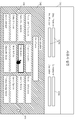
2 is a diagram illustrating a method of providing a screen keyboard for inputting annotations on an ultrasound image, according to an embodiment of the present invention.
3A is a flowchart for explaining a method of setting an annotation on an ultrasonic image by an ultrasonic apparatus according to an embodiment of the present invention.
3B is a diagram illustrating a database in which recommendation annotations are stored, according to an embodiment of the present invention.
4 is a view for explaining a method of providing a recommendation annotation on the basis of an imaging site by an ultrasound device, according to an embodiment of the present invention.
5A and 5B are views for explaining a method of providing a recommendation annotation based on a radiographic site, according to another embodiment of the present invention.
Figure 6 is a diagram illustrating a method by which an ultrasound device provides a screen keyboard, in accordance with one embodiment of the present invention.
7 is a diagram illustrating a method by which an ultrasound device provides a recommendation annotation image, in accordance with an embodiment of the present invention.
8 is a diagram illustrating a method by which an ultrasound device provides an interface for generating an annotation image, in accordance with an embodiment of the present invention.
9 is a view for explaining a method of providing a setting window corresponding to a plurality of ultrasound images, according to an embodiment of the present invention.
10 is a diagram illustrating a method by which an ultrasound device provides recommendation data corresponding to an input field, according to an embodiment of the present invention.
11 is a block diagram of an ultrasonic apparatus according to another embodiment of the present invention.
The terms used in this specification will be briefly described and the present invention will be described in detail.
While the present invention has been described in connection with what is presently considered to be the most practical and preferred embodiment, it is to be understood that the invention is not limited to the disclosed embodiments. Also, in certain cases, there may be a term selected arbitrarily by the applicant, in which case the meaning thereof will be described in detail in the description of the corresponding invention. Therefore, the term used in the present invention should be defined based on the meaning of the term, not on the name of a simple term, but on the entire contents of the present invention.
When an element is referred to as "including" an element throughout the specification, it is to be understood that the element may include other elements as well, without departing from the spirit or scope of the present invention. Also, the terms "part," " module, "and the like described in the specification mean units for processing at least one function or operation, which may be implemented in hardware or software or a combination of hardware and software .
Hereinafter, embodiments of the present invention will be described in detail with reference to the accompanying drawings so that those skilled in the art can easily carry out the present invention. The present invention may, however, be embodied in many different forms and should not be construed as limited to the embodiments set forth herein. In order to clearly illustrate the present invention, parts not related to the description are omitted, and similar parts are denoted by like reference characters throughout the specification.
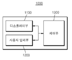
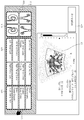
1 is a block diagram of an
Referring to FIG. 1, the
The
For example, the
In addition, the
The
For example, the
Also, the
The
In addition, the
At least one recommendation annotation previously stored corresponding to the radiography site may be a annotation generated by the user in the
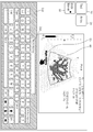
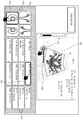
2 is a diagram illustrating a method of providing a
Referring to FIG. 2, the
The
Upon receiving the user input for selecting the
Upon receiving a user input that selects at least one key within the screen keyboard, the
In addition, as the user selects textual information displayed on the
Further, when receiving a user input for selecting the
In addition, upon receiving a user input that sets the annotation on the
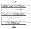
3A is a flowchart illustrating a method of setting an annotation on an ultrasonic image by the
In step S310, the
The
The
The annotation may refer to a phrase or image set on the first ultrasound image by the user. The annotation may be information describing an object in the first ultrasound image. For example, the annotation may be information describing the organ in the first ultrasound image. In addition, the annotation may be information describing the user, the object itself, or the
In step S320, the
The imaging region of the object may be set in the
Further, according to the embodiment, the identification information of the organ represented by the first ultrasound image in the form of metadata may be stored in the first ultrasound image file. Accordingly, the
In step S330, the
In the
Upon receiving a user input for selecting a menu for setting annotations on the first ultrasound image, the
At least one recommendation annotation may be an annotation generated in the
In addition, at least one recommendation annotation may include a frequently set annotation. For example, the at least one recommendation annotation may be a comment positioned within a predetermined order based on the number of times set in the
In addition, at least one recommendation annotation may include annotations with a high order of creation. For example, the at least one recommendation annotation may be an annotation that is located within a predetermined order, based on the order generated in the
Also, the at least one recommendation annotation may include a text annotation, an arrow image in addition to the body marker, and the like. Also, the at least one recommendation annotation may include annotations of different kinds. For example, the
The
3B, the
The default annotation may be an annotation stored in the
The user-created annotation may mean an annotation that is not the same as the default annotation among the annotations set by the user in the ultrasound image. For example, when the user manually sets the annotation on the ultrasound image without selecting the recommendation annotation, the
In addition, when the set annotation is the same as the default annotation or the user-created annotation, the
The
Further, the
Accordingly, the
Further, the
FIG. 3B shows only the recommendation annotations corresponding to the photographed regions. However, the recommended annotations corresponding to the photographed region and the user may be stored, and the recommended annotations corresponding to the photographed region and the target object may be stored.
In step S340, the
In step S350, the
Also, as the recommended annotation displayed on the ultrasound image is selected and the user input to be moved is received, the
Further, the
In addition, according to the embodiment, the
For example, even the same shooting region may have different annotations that are mainly used for each user. In addition, the same patient is more likely to take an ultrasound image due to the same disease. Accordingly, the
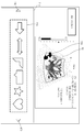
FIG. 4 is a view for explaining a method of providing a recommendation annotation based on a radiographic site, according to an embodiment of the present invention.
4, upon receiving a user input for selecting a
The
The recommendation text annotation may include the
In addition, the recommendation text annotation may include
In addition, the recommendation text annotation may include the
Upon receiving a user input that selects one of the suggested text annotations, the
Upon receiving a user input for selecting a predetermined key (e.g., an enter key) in a state where an annotation is displayed on the
In addition, the setting window may include a
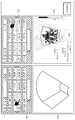
FIGS. 5A and 5B are diagrams for explaining a method for providing a recommendation annotation based on a radiographic site, according to another embodiment of the present invention.
Referring to FIG. 5A, upon receiving a user input for selecting a
Suggested annotations can contain frequently used annotations. The recommendation annotation corresponding to the imaging site may include annotations frequently set on the ultrasound image including the imaging site. For example, the recommendation annotation may include a high
The
Upon acquiring the recommendation annotations corresponding to the radiographic sites, the
Referring to FIG. 5B, the
For example, upon receiving a user input for selecting a
The
Accordingly, by receiving the annotations corresponding to the diagnostic situation, the user can set the annotations on the ultrasound image with one selection even if the user does not input the annotations manually. In addition, by displaying different kinds of annotations on one setting window, the user can select different kinds of annotations without changing the setting window.
Figure 6 is a diagram illustrating a method by which
Referring to FIG. 6, the
The
In addition, the
Figure 7 is a diagram illustrating a method by which
Referring to FIG. 7,
The
The
The
In addition, the setting
Figure 8 is a diagram illustrating a method by which
Referring to FIG. 8, the
The
Upon receiving a user input that draws an image in the
In addition, the setting
9 is a view for explaining a method of providing a setting window corresponding to a plurality of ultrasound images, according to an embodiment of the present invention.
Referring to FIG. 9, the
For example, the
In addition, the
In this case, the
In addition, the
For example, upon receiving a user input for selecting a setting
10 is a view for explaining a method by which the
Referring to FIG. 10, upon receiving a user input for selecting an input field, the
For example, the
Upon receiving the user input for selecting the
The recommendation identification information previously stored corresponding to the identification information item of the sonographer may be the identification information of the sonographer generated by the sonographer by inputting into the
Also, the recommendation identification information previously stored corresponding to the identification information item of the sonographer may be the identification information of the sonographer having a large number of times stored for a predetermined period among the identification information of the plurality of sonographers previously stored. For example, when receiving a user input that stores the identification information of the sonographer corresponding to the identification information item of the sonographer, the
Upon receiving a user input for selecting one of the plurality of recommendation identification information displayed in the
In addition, upon receiving a user input for selecting the
Accordingly, even if the data is not input manually every time data is input, the user can easily select and input the data that the user has input.
11 is a block diagram of an
11, the
The
The
The
The receiving
The
The B mode processing unit 212 extracts and processes the B mode component from the ultrasonic data. The
Similarly, the
The
The
The
The
The
The short
The
The
The
The
The
Some or all of the
The method according to an embodiment of the present invention can be implemented in the form of a program command which can be executed through various computer means and recorded in a computer-readable medium. The computer-readable medium may include program instructions, data files, data structures, and the like, alone or in combination. The program instructions recorded on the medium may be those specially designed and constructed for the present invention or may be available to those skilled in the art of computer software. Examples of computer-readable media include magnetic media such as hard disks, floppy disks and magnetic tape; optical media such as CD-ROMs and DVDs; magnetic media such as floppy disks; Magneto-optical media, and hardware devices specifically configured to store and execute program instructions such as ROM, RAM, flash memory, and the like. Examples of program instructions include machine language code such as those produced by a compiler, as well as high-level language code that can be executed by a computer using an interpreter or the like.
While the present invention has been particularly shown and described with reference to exemplary embodiments thereof, it is to be understood that the invention is not limited to the disclosed exemplary embodiments, It belongs to the scope of right.
Claims (20)
A control unit for determining a region to be imaged of a target object in the first ultrasound image; And
And a display unit configured to display a setting window including at least one recommendation annotation among a plurality of annotations corresponding to the determined imaging region of the object,
Wherein the user input section receives a user input for selecting one of the at least one recommendation annotation and the display section displays an image representing the selected recommendation annotation on the first ultrasound image,
Wherein the at least one recommendation annotation is an annotation generated in the ultrasonic apparatus by the user in correspondence with a second ultrasonic image including the imaging region.
Wherein the at least one recommendation annotation includes annotations of different kinds.
Wherein the at least one recommendation annotation comprises:
Wherein the ultrasonic wave is a tin previously set on the ultrasonic device by the user in correspondence with the photographing part.
Wherein the at least one recommendation annotation comprises at least one of a phrase, a body marker, and an arrow.
The ultrasonic device includes:
Further comprising a storage unit for storing the order of the plurality of annotations on the basis of the number of times set in the ultrasonic apparatus,
Wherein the at least one recommendation annotation comprises:
Wherein the order among the plurality of annotations is annotations within a predetermined rank.
The ultrasonic device includes:
Further comprising a storage unit for storing the order of the plurality of annotations based on the order generated in the ultrasonic apparatus,
Wherein the at least one recommendation annotation comprises:
Wherein the sequence of the plurality of annotations is a annotation within a predetermined rank.
Wherein the setting window includes a screen keyboard including a plurality of keys.
Wherein the setting window includes a movement display button for switching the setting window to a screen keyboard including a plurality of keys,
The display unit includes:
Wherein the display control unit changes the setting window to the screen keyboard upon receiving the user input for selecting the movement display button.
Wherein the first ultrasound image is two different ultrasound images,
Wherein the setting window is two setting windows corresponding to the two different ultrasound images.
Wherein,
Acquiring at least one recommendation annotation previously stored corresponding to the photographing site based on at least one of the user of the ultrasonic apparatus and the identification information of the object,
The display unit includes:
And displays the setting window including the obtained at least one recommendation annotation.
Determining an imaging region of a target object in the first ultrasound image;
Displaying a setting window including at least one recommended annotation among a plurality of annotations corresponding to the determined photographed portion of the object;
Receiving a user input selecting one of the at least one recommendation annotation; And
And displaying an image representing the selected recommendation annotation on the first ultrasound image,
Wherein the at least one recommendation annotation is an annotation generated in the ultrasonic apparatus by being input by the user corresponding to a second ultrasonic image including the imaging region.
Wherein the at least one recommendation annotation includes annotations of different kinds.
Wherein the at least one recommendation annotation comprises:
Wherein the annotation is preset in the ultrasonic apparatus by the user in correspondence to the imaging site.
Wherein the at least one recommendation annotation comprises at least one of a phrase, a body marker, and an arrow.
Wherein the order of the plurality of annotations is determined based on the number of times set in the ultrasonic apparatus,
Wherein the at least one recommendation annotation comprises:
Wherein the order among the plurality of annotations is an annotation within a predetermined rank.
The order of the plurality of annotations is determined based on the order generated in the ultrasonic apparatus,
Wherein the at least one recommendation annotation comprises:
Wherein the order among the plurality of annotations is an annotation within a predetermined rank.
Wherein the setting window includes a screen keyboard including a plurality of keys.
Wherein the setting window includes a movement display button for switching the setting window to a screen keyboard including a plurality of keys,
The information input method includes:
Further comprising changing the setting window to the screen keyboard and displaying the setting window upon receiving a user input for selecting the movement display button.
Wherein the first ultrasound image is two different ultrasound images,
Wherein the setting window is two setting windows corresponding to the two different ultrasound images.
Wherein the step of displaying the setting window including at least one recommendation annotation previously stored corresponding to the determined imaging region of the object comprises:
Obtaining at least one recommendation annotation previously stored corresponding to the photographed region based on at least one of the user of the ultrasonic apparatus and the degree of identification of the target object; And
And displaying a setting window including the obtained at least one recommendation annotation.
Priority Applications (3)
| Application Number | Priority Date | Filing Date | Title |
|---|---|---|---|
| KR1020140146425A KR20160049385A (en) | 2014-10-27 | 2014-10-27 | Method and ultrasound apparatus for inputting informaion |
| EP15163471.4A EP3015071A1 (en) | 2014-10-27 | 2015-04-14 | Ultrasound apparatus and information input method thereof |
| US14/706,102 US20160113627A1 (en) | 2014-10-27 | 2015-05-07 | Ultrasound apparatus and information input method thereof |
Applications Claiming Priority (1)
| Application Number | Priority Date | Filing Date | Title |
|---|---|---|---|
| KR1020140146425A KR20160049385A (en) | 2014-10-27 | 2014-10-27 | Method and ultrasound apparatus for inputting informaion |
Publications (1)
| Publication Number | Publication Date |
|---|---|
| KR20160049385A true KR20160049385A (en) | 2016-05-09 |
Family
ID=52997228
Family Applications (1)
| Application Number | Title | Priority Date | Filing Date |
|---|---|---|---|
| KR1020140146425A KR20160049385A (en) | 2014-10-27 | 2014-10-27 | Method and ultrasound apparatus for inputting informaion |
Country Status (3)
| Country | Link |
|---|---|
| US (1) | US20160113627A1 (en) |
| EP (1) | EP3015071A1 (en) |
| KR (1) | KR20160049385A (en) |
Cited By (2)
| Publication number | Priority date | Publication date | Assignee | Title |
|---|---|---|---|---|
| KR20190019365A (en) * | 2017-08-17 | 2019-02-27 | 삼성전자주식회사 | Method and ultrasound apparatus for providing annotation related information |
| KR20220147498A (en) * | 2021-04-27 | 2022-11-03 | 이수안 | device that recommends a hospital for pets |
Families Citing this family (5)
| Publication number | Priority date | Publication date | Assignee | Title |
|---|---|---|---|---|
| KR102250094B1 (en) * | 2013-12-23 | 2021-05-10 | 삼성전자주식회사 | Method for performing function and mobile apparatus using the method |
| US20190015080A1 (en) * | 2017-07-14 | 2019-01-17 | Imorgon Medical LLC | Medical Diagnostic Ultrasound Imaging System and Method for Receiving Information from a Server During An Examination of a Patient to Improve Workflow |
| JP7399621B2 (en) * | 2018-03-16 | 2023-12-18 | キヤノンメディカルシステムズ株式会社 | Ultrasonic diagnostic equipment, information processing equipment, and information processing programs |
| US10646206B1 (en) | 2019-01-10 | 2020-05-12 | Imorgon Medical LLC | Medical diagnostic ultrasound imaging system and method for communicating with a server during an examination of a patient using two communication channels |
| US11583244B2 (en) * | 2019-10-04 | 2023-02-21 | GE Precision Healthcare LLC | System and methods for tracking anatomical features in ultrasound images |
Family Cites Families (4)
| Publication number | Priority date | Publication date | Assignee | Title |
|---|---|---|---|---|
| US6638223B2 (en) * | 2000-12-28 | 2003-10-28 | Ge Medical Systems Global Technology Company, Llc | Operator interface for a medical diagnostic imaging device |
| JP2004208858A (en) * | 2002-12-27 | 2004-07-29 | Toshiba Corp | Ultrasonograph and ultrasonic image processing apparatus |
| US6988990B2 (en) * | 2003-05-29 | 2006-01-24 | General Electric Company | Automatic annotation filler system and method for use in ultrasound imaging |
| WO2015002409A1 (en) * | 2013-07-01 | 2015-01-08 | Samsung Electronics Co., Ltd. | Method of sharing information in ultrasound imaging |
-
2014
- 2014-10-27 KR KR1020140146425A patent/KR20160049385A/en not_active Application Discontinuation
-
2015
- 2015-04-14 EP EP15163471.4A patent/EP3015071A1/en not_active Withdrawn
- 2015-05-07 US US14/706,102 patent/US20160113627A1/en not_active Abandoned
Cited By (3)
| Publication number | Priority date | Publication date | Assignee | Title |
|---|---|---|---|---|
| KR20190019365A (en) * | 2017-08-17 | 2019-02-27 | 삼성전자주식회사 | Method and ultrasound apparatus for providing annotation related information |
| KR20220147498A (en) * | 2021-04-27 | 2022-11-03 | 이수안 | device that recommends a hospital for pets |
| KR20220147499A (en) * | 2021-04-27 | 2022-11-03 | 이수안 | method and system for recommending a hospital |
Also Published As
| Publication number | Publication date |
|---|---|
| US20160113627A1 (en) | 2016-04-28 |
| EP3015071A1 (en) | 2016-05-04 |
Similar Documents
| Publication | Publication Date | Title |
|---|---|---|
| JP6683677B2 (en) | Ultrasonic diagnostic device for self-diagnosis and remote diagnosis, and operating method of ultrasonic diagnostic device | |
| KR20160049385A (en) | Method and ultrasound apparatus for inputting informaion | |
| KR102618500B1 (en) | Ultrasound diagnosis apparatus and mehtod thereof | |
| KR102366316B1 (en) | Ultrasonic imaging apparatus and ultrasonic image processing method thereof | |
| KR101660369B1 (en) | Apparatus and method for generating ultrasound image | |
| KR102310976B1 (en) | Untrasound dianognosis apparatus, method and computer-readable storage medium | |
| CN106562803B (en) | Method and apparatus for displaying image showing object | |
| KR102656542B1 (en) | Method and apparatus for displaying ultrasound images | |
| KR102273831B1 (en) | The Method and Apparatus for Displaying Medical Image | |
| KR102519424B1 (en) | Method of displaying a ultrasound image and apparatus thereof | |
| KR20160087221A (en) | Ultrasonic diagnostic apparatus and operating method for the same | |
| KR20160051161A (en) | Medical image apparatus and displaying medical image thereof | |
| KR20150102589A (en) | Apparatus and method for medical image, and computer-readable recording medium | |
| KR20170060853A (en) | Medical imaging apparatus and operating method for the same | |
| KR102418975B1 (en) | Ultrasound apparatus and method for providing information | |
| JP6200589B2 (en) | Ultrasonic diagnostic apparatus and operation method thereof | |
| EP3025650B1 (en) | Volume rendering apparatus and volume rendering method | |
| KR102367194B1 (en) | Ultrasonic diagnostic apparatus and operating method for the same | |
| KR101868019B1 (en) | Ultrasound Imaging Apparatus and Method for processing a ultrasound image thereof | |
| KR102364490B1 (en) | Untrasound dianognosis apparatus, method and computer-readable storage medium | |
| KR102416511B1 (en) | Method and apparatus for generating a body marker | |
| KR102270718B1 (en) | Untrasound dianognosis apparatus, operating method thereof and computer-readable storage medium | |
| KR20160026608A (en) | Ultrasonic diagnostic apparatus and operating method for the same |
Legal Events
| Date | Code | Title | Description |
|---|---|---|---|
| WITN | Withdrawal due to no request for examination |