KR20150107742A - Delphinidin for combating melanoma cells - Google Patents
Delphinidin for combating melanoma cells Download PDFInfo
- Publication number
- KR20150107742A KR20150107742A KR1020157018732A KR20157018732A KR20150107742A KR 20150107742 A KR20150107742 A KR 20150107742A KR 1020157018732 A KR1020157018732 A KR 1020157018732A KR 20157018732 A KR20157018732 A KR 20157018732A KR 20150107742 A KR20150107742 A KR 20150107742A
- Authority
- KR
- South Korea
- Prior art keywords
- delphinidin
- cells
- cell
- sbe
- complex
- Prior art date
Links
Images
Classifications
-
- A—HUMAN NECESSITIES
- A61—MEDICAL OR VETERINARY SCIENCE; HYGIENE
- A61K—PREPARATIONS FOR MEDICAL, DENTAL OR TOILETRY PURPOSES
- A61K31/00—Medicinal preparations containing organic active ingredients
- A61K31/33—Heterocyclic compounds
- A61K31/335—Heterocyclic compounds having oxygen as the only ring hetero atom, e.g. fungichromin
- A61K31/35—Heterocyclic compounds having oxygen as the only ring hetero atom, e.g. fungichromin having six-membered rings with one oxygen as the only ring hetero atom
- A61K31/352—Heterocyclic compounds having oxygen as the only ring hetero atom, e.g. fungichromin having six-membered rings with one oxygen as the only ring hetero atom condensed with carbocyclic rings, e.g. methantheline
-
- A—HUMAN NECESSITIES
- A61—MEDICAL OR VETERINARY SCIENCE; HYGIENE
- A61K—PREPARATIONS FOR MEDICAL, DENTAL OR TOILETRY PURPOSES
- A61K31/00—Medicinal preparations containing organic active ingredients
- A61K31/70—Carbohydrates; Sugars; Derivatives thereof
- A61K31/7042—Compounds having saccharide radicals and heterocyclic rings
-
- A—HUMAN NECESSITIES
- A61—MEDICAL OR VETERINARY SCIENCE; HYGIENE
- A61K—PREPARATIONS FOR MEDICAL, DENTAL OR TOILETRY PURPOSES
- A61K38/00—Medicinal preparations containing peptides
- A61K38/16—Peptides having more than 20 amino acids; Gastrins; Somatostatins; Melanotropins; Derivatives thereof
- A61K38/17—Peptides having more than 20 amino acids; Gastrins; Somatostatins; Melanotropins; Derivatives thereof from animals; from humans
- A61K38/177—Receptors; Cell surface antigens; Cell surface determinants
- A61K38/1793—Receptors; Cell surface antigens; Cell surface determinants for cytokines; for lymphokines; for interferons
-
- A—HUMAN NECESSITIES
- A61—MEDICAL OR VETERINARY SCIENCE; HYGIENE
- A61K—PREPARATIONS FOR MEDICAL, DENTAL OR TOILETRY PURPOSES
- A61K38/00—Medicinal preparations containing peptides
- A61K38/16—Peptides having more than 20 amino acids; Gastrins; Somatostatins; Melanotropins; Derivatives thereof
- A61K38/17—Peptides having more than 20 amino acids; Gastrins; Somatostatins; Melanotropins; Derivatives thereof from animals; from humans
- A61K38/19—Cytokines; Lymphokines; Interferons
- A61K38/191—Tumor necrosis factors [TNF], e.g. lymphotoxin [LT], i.e. TNF-beta
-
- A—HUMAN NECESSITIES
- A61—MEDICAL OR VETERINARY SCIENCE; HYGIENE
- A61K—PREPARATIONS FOR MEDICAL, DENTAL OR TOILETRY PURPOSES
- A61K45/00—Medicinal preparations containing active ingredients not provided for in groups A61K31/00 - A61K41/00
- A61K45/06—Mixtures of active ingredients without chemical characterisation, e.g. antiphlogistics and cardiaca
-
- A—HUMAN NECESSITIES
- A61—MEDICAL OR VETERINARY SCIENCE; HYGIENE
- A61K—PREPARATIONS FOR MEDICAL, DENTAL OR TOILETRY PURPOSES
- A61K47/00—Medicinal preparations characterised by the non-active ingredients used, e.g. carriers or inert additives; Targeting or modifying agents chemically bound to the active ingredient
- A61K47/30—Macromolecular organic or inorganic compounds, e.g. inorganic polyphosphates
- A61K47/36—Polysaccharides; Derivatives thereof, e.g. gums, starch, alginate, dextrin, hyaluronic acid, chitosan, inulin, agar or pectin
- A61K47/40—Cyclodextrins; Derivatives thereof
-
- A61K47/48969—
-
- A—HUMAN NECESSITIES
- A61—MEDICAL OR VETERINARY SCIENCE; HYGIENE
- A61K—PREPARATIONS FOR MEDICAL, DENTAL OR TOILETRY PURPOSES
- A61K47/00—Medicinal preparations characterised by the non-active ingredients used, e.g. carriers or inert additives; Targeting or modifying agents chemically bound to the active ingredient
- A61K47/50—Medicinal preparations characterised by the non-active ingredients used, e.g. carriers or inert additives; Targeting or modifying agents chemically bound to the active ingredient the non-active ingredient being chemically bound to the active ingredient, e.g. polymer-drug conjugates
- A61K47/69—Medicinal preparations characterised by the non-active ingredients used, e.g. carriers or inert additives; Targeting or modifying agents chemically bound to the active ingredient the non-active ingredient being chemically bound to the active ingredient, e.g. polymer-drug conjugates the conjugate being characterised by physical or galenical forms, e.g. emulsion, particle, inclusion complex, stent or kit
- A61K47/6949—Medicinal preparations characterised by the non-active ingredients used, e.g. carriers or inert additives; Targeting or modifying agents chemically bound to the active ingredient the non-active ingredient being chemically bound to the active ingredient, e.g. polymer-drug conjugates the conjugate being characterised by physical or galenical forms, e.g. emulsion, particle, inclusion complex, stent or kit inclusion complexes, e.g. clathrates, cavitates or fullerenes
- A61K47/6951—Medicinal preparations characterised by the non-active ingredients used, e.g. carriers or inert additives; Targeting or modifying agents chemically bound to the active ingredient the non-active ingredient being chemically bound to the active ingredient, e.g. polymer-drug conjugates the conjugate being characterised by physical or galenical forms, e.g. emulsion, particle, inclusion complex, stent or kit inclusion complexes, e.g. clathrates, cavitates or fullerenes using cyclodextrin
-
- A—HUMAN NECESSITIES
- A61—MEDICAL OR VETERINARY SCIENCE; HYGIENE
- A61P—SPECIFIC THERAPEUTIC ACTIVITY OF CHEMICAL COMPOUNDS OR MEDICINAL PREPARATIONS
- A61P35/00—Antineoplastic agents
-
- B—PERFORMING OPERATIONS; TRANSPORTING
- B82—NANOTECHNOLOGY
- B82Y—SPECIFIC USES OR APPLICATIONS OF NANOSTRUCTURES; MEASUREMENT OR ANALYSIS OF NANOSTRUCTURES; MANUFACTURE OR TREATMENT OF NANOSTRUCTURES
- B82Y5/00—Nanobiotechnology or nanomedicine, e.g. protein engineering or drug delivery
Landscapes
- Health & Medical Sciences (AREA)
- Life Sciences & Earth Sciences (AREA)
- Chemical & Material Sciences (AREA)
- Medicinal Chemistry (AREA)
- Pharmacology & Pharmacy (AREA)
- General Health & Medical Sciences (AREA)
- Animal Behavior & Ethology (AREA)
- Public Health (AREA)
- Veterinary Medicine (AREA)
- Epidemiology (AREA)
- Engineering & Computer Science (AREA)
- Bioinformatics & Cheminformatics (AREA)
- Proteomics, Peptides & Aminoacids (AREA)
- Zoology (AREA)
- Gastroenterology & Hepatology (AREA)
- Immunology (AREA)
- Molecular Biology (AREA)
- Cell Biology (AREA)
- Chemical Kinetics & Catalysis (AREA)
- General Chemical & Material Sciences (AREA)
- Nuclear Medicine, Radiotherapy & Molecular Imaging (AREA)
- Organic Chemistry (AREA)
- Pharmaceuticals Containing Other Organic And Inorganic Compounds (AREA)
- Medicinal Preparation (AREA)
- Nanotechnology (AREA)
- Medicines That Contain Protein Lipid Enzymes And Other Medicines (AREA)
- Medicines Containing Antibodies Or Antigens For Use As Internal Diagnostic Agents (AREA)
- Inorganic Chemistry (AREA)
- Biophysics (AREA)
- Biotechnology (AREA)
- General Engineering & Computer Science (AREA)
- Medical Informatics (AREA)
- Crystallography & Structural Chemistry (AREA)
Abstract
제1성분으로서, 델피니딘과 설포알킬 에테르 β-사이클로덱스트린의 복합체 및/또는 델피니딘 또는 그 염 및 제 2성분으로서, 악성 흑색종의 치료에 사용되는 종양괴사인자관련 세포자살유도 리간드 (TRAIL)를 포함하는 악성 흑색종의 치료에 사용되는 조성물이 제공된다.As the first component, a complex of delphinidin and sulfoalkyl ether? -Cyclodextrin and / or a complex of delphinidin or a salt thereof and As a second component, there is provided a composition for use in the treatment of malignant melanoma comprising tumor necrosis factor related cell suicide inducing ligand (TRAIL) used in the treatment of malignant melanoma.
Description
본 발명은 제1성분으로서, 델피니딘과 설포알킬 에테르 β-사이클로덱스트린의 복합체 및/또는 델피니딘 또는 그 염; 및 추가적인 성분으로서, 바람직하게 종양괴사인자관련 아포토시스(세포자살)유도 리간드(Tumor necrosis factor-Related Apoptosis-Inducing Ligand, TRAIL)를 포함하는 조성물로서, 악성 흑색종(악성 멜라노마)(malignant melanoma)의 치료에 사용되는 조성물에 관한 것이다.The present invention provides, as a first component, a complex of delphinidin and sulfoalkyl ether? -Cyclodextrin and / or delphinidin or a salt thereof; As a further component, a composition comprising a tumor necrosis factor-related apoptosis-related apoptosis-inducing ligand (TRAIL), preferably a malignant melanoma (malignant melanoma) To a composition for use in therapy.
용어 "흑색 피부암"으로 알려진 악성 흑색종(멜라노마)(malignant melanoma)은 색소세포, 소위 멜라노사이트(멜라닌 세포)(melanocytes)의 악성 변성이다. 암은 초기단계에서도 순환계 및 림프계를 통하여 전이를 확산시키는 경향을 가지고 있다. 악성 흑색종은 적어도 초기에 진단 및 처치하면 치료가능하다.Malignant melanoma, also known as the term "black skin cancer", is malignant transformation of pigment cells, so-called melanocytes (melanocytes). Cancer also tends to spread the metastasis through the circulatory and lymphatic systems at an early stage. Malignant melanoma can be treated at least initially by diagnosis and treatment.
비록 인테페론 단독요법이 악성 흑색종의 치료에 사용되지만, 가장 중요한 치료방법은 방사선요법 이외에 종양제거수술이다. 백신요법, 즉, 특정의 암세포 퇴치를 위해 신체의 고유저항을 자극하는 종양백신을 이용한 종양접종에 의한 활성면역요법이 역시 알려져 있다. 이 경우, 종양(예를 들어, 종양세포 또는 종양세포의 세포단편에 의해 생성된 단백질 분자)의 기능이 투여된 백신을 통하여 면역세포에 제시되어, 상기 면역세포가 이러한 기능을 이물로서 인식하고 이 기능에 견디는 신체고유의 세포를 공격하도록 한다. 백신요법 이외에, 화학요법, 즉, 종양세포를 손상 또는 억제하는 화학물질(cytostatics)을 이용한 치료법이 실제적으로 관련된다.Although the use of interferon monotherapy is used for the treatment of malignant melanoma, the most important treatment is tumor resection surgery in addition to radiation therapy. Vaccine therapy, that is, active immunotherapy by tumor inoculation using a tumor vaccine to stimulate the body's intrinsic resistance to certain cancer cells, is also known. In this case, the function of a tumor (for example, a protein molecule produced by a tumor cell or a cell fragment of a tumor cell) is presented to the immune cell through the administered vaccine such that the immune cell recognizes this function as a foreign body, Attack body-specific cells that resist function. In addition to vaccine therapy, chemotherapy, i.e., treatment with cytostatics to damage or inhibit tumor cells, is practically involved.
본 발명의 목적은 종래 기술에서 알려진 악성 흑색종에 대한 치료선택지에 대한 대안 또는 보완으로서의 효과적인 치료법을 제공하는 것이다.It is an object of the present invention to provide an effective treatment as an alternative or supplement to treatment options for malignant melanoma known in the prior art.
상기 목적은 독립 청구항에서 인용된 조성물 및 용도에 의해 달성되는 반면, 발명의 유리한 실시형태는 종속 청구항에 개시된다. 상기 목적의 사실상의 달성은 실시예 6 내지 12에서의 흑색종 세포, 비암성 세포 및 세포활력의 본 발명에 따른 조성물의 효과에 대한 시험관 내 실험의 결과와, 특히, 실시예 9 내지 11에서의 TRAIL이 추가 성분으로 사용되는 시험관내 실험의 결과에 의해 입증된다.While this object is achieved by the compositions and uses cited in the independent claims, advantageous embodiments of the invention are disclosed in the dependent claims. The actual achievement of this object is achieved by the results of in vitro experiments on the effects of melanoma cells, noncancerous cells and cell viability of the composition according to the invention in Examples 6 to 12 and in particular of the results obtained in Examples 9 to 11 The results of in vitro experiments in which TRAIL is used as an additional ingredient are demonstrated.
본 발명에 따른 복합체 및 TRAIL을 포함하는 조성물은 악성 흑색종에 대해 개선된 치료법을 제공할 수 있다.Compositions according to the present invention and compositions comprising TRAIL can provide improved therapies for malignant melanoma.
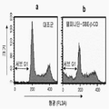
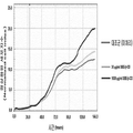
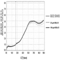
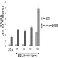
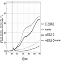
도 1은 델피니딘-SBE-β-CD로 처리된 인간 흑색종 세포주(A-375)의 세포의 히스토그램(b); 및 처리되지 않은 인간 흑색종 세포주(A-375)의 세포의 히스토그램 (a)이다.
도 2a는 낮은 세포 밀집도에서 대조군인 SBE-β-CD, DMSO 및 미처리 A-375 세포와 비교되는 델피니딘-SBE-β-CD 및 델피니딘 Cl(델피니딘 클로라이드)에 의한 세포자살의 용량의존적 유도를 보여준다. 이 용량의존적 유도는 서브-G1 피크 방법에 의해 결정된다.
도 2b는 높은 세포 밀집도에서 델피니딘 Cl과 대조군인 SBE-β-CD, DMSO 및 미처리 A-375 세포와 비교되는 델피니딘-SBE-β-CD에 의한 세포자살의 용량의존적 유도를 보여준다. 이 용량의존적 유도는 서브-G1 피크 방법에 의해 결정된다.
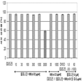
도 3a는 낮은 세포 밀집도에서 대조군인 SBE-β-CD, DMSO 및 미처리 A-375 세포와 비교되는 델피니딘-SBE-β-CD 및 델피니딘 Cl의 세포생존률에 대한 용량의존적 효과를 보여준다. 이 용량의존적 효과는 WST-1 분석에 의해 결정된다.
도 3b는 높은 세포 밀집도에서 델피니딘 Cl과 대조군인 SBE-β-CD, DMSO 및 미처리 A-375 세포와 비교되는 델피니딘-SBE-β-CD의 세포생존률에 대한 용량의존적 효과를 보여준다. 이 용량의존적 효과는 WST-1 분석에 의해 결정된다.
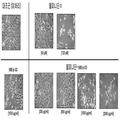
도 4는 미처리 A-375 세포와 델피니딘-SBE-β-CD, 델피니딘 Cl 및 SBE-β-CD로 처리된 A-375 세포의 현미경 사진을 보여준다.
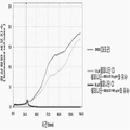
도 5는 미처리 세포와 대비되는 델피니딘-SBE-β-CD 로 처리된 A-375 세포의 높은 세포 밀집도에서의 세포수 및 세포증식(실시간 세포 모니터링)을 보여준다. 이는 xCELLigence 시스템을 이용하여 실시된다.
도 6은 미처리 세포와 대비되는 델피니딘-SBE-β-CD 로 처리된 A-375 세포의 낮은 세포 밀집도에서의 세포수 및 세포증식(실시간 세포 모니터링)을 보여준다. 이는 xCELLigence 시스템을 이용하여 실시된다.
도 7은 미처리 세포와 대비되는 델피니딘 Cl 로 처리된 A-375 세포의 높은 세포 밀집도에서의 세포수 및 세포증식(실시간 세포 모니터링)을 보여준다. 이는 xCELLigence 시스템을 이용하여 실시된다.
도 8은 미처리 세포와 대비되는 델피니딘 Cl 로 처리된 A-375 세포의 낮은 세포 밀집도에서의 세포수 및 세포증식(실시간 세포 모니터링)을 보여준다. 이는 xCELLigence 시스템을 이용하여 실시된다.
도 9는 미처리 세포와 대비되는 SBE-β-CD 로 처리된 A-375 세포의 높은 세포 밀집도에서의 세포수 및 세포증식(실시간 세포 모니터링)을 보여준다. 이는 xCELLigence 시스템을 이용하여 실시된다.
도 10은 미처리 세포와 대비되는 SBE-β-CD 로 처리된 A-375 세포의 낮은 세포 밀집도에서의 세포수 및 세포증식(실시간 세포 모니터링)을 보여준다. 이는 xCELLigence 시스템을 이용하여 실시된다.
도 11a는 낮은 세포 밀집도에서 대조군(미처리세포 또는 TRAIL만으로 처리된 세포) 과 비교되는 TRAIL을 가지는 델피니딘-SBE-β-CD 및 TRAIL을 가지지 않는 델피니딘-SBE-β-CD 에 의한 세포자살의 유도를 보여준다. 이 유도는 서브-G1 피크 방법에 의해 결정된다.
도 11b는 높은 세포 밀집도에서 대조군(미처리세포 또는 TRAIL만으로 처리된 세포) 과 비교되는 TRAIL을 가지는 델피니딘-SBE-β-CD 및 TRAIL을 가지지 않는 델피니딘-SBE-β-CD에 의한 세포자살의 유도를 보여준다. 이 유도는 서브-G1 피크 방법에 의해 결정된다.
도 12a는 낮은 세포 밀집도에서 델피니딘-SBE-β-CD 단독 및 대조군(미처리세포 또는 TRAIL만으로 처리된 세포) 과 비교되는 TRAIL을 가지는 델피니딘-SBE-β-CD의 세포생존률에 대한 효과를 보여준다. 이 효과는 WST-1 분석에 의해 결정된다.
도 12b는 높은 세포 밀집도에서 델피니딘-SBE-β-CD 단독 및 대조군(미처리세포 또는 TRAIL만으로 처리된 세포, SBE-β-CD로 처리된 세포)과 비교되는 TRAIL을 가지는 델피니딘-SBE-β-CD의 세포생존률에 대한 효과를 보여준다. 이 효과는 WST-1 분석에 의해 결정된다.
도 13은 미처리 A-375 세포, TRAIL만으로 처리된 A-375 세포, 델피니딘-SBE-β-CD만으로 처리된 A-375 세포, 및 델피니딘-SBE-β-CD과 TRAIL로 처리된 A-375 세포의 현미경 사진을 보여준다.
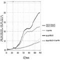
도 14는 미처리 세포와 대비되는 TRAIL, 델피니딘-SBE-β-CD, 및 TRAIL 과 델피니딘-SBE-β-CD 로 처리된 A-375 세포의 높은 세포 밀집도에서의 세포수 및 세포증식(실시간 세포 모니터링)을 보여준다. 이는 xCELLigence 시스템을 이용하여 실시된다.
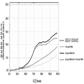
도 15는 미처리 세포와 대비되는 TRAIL, 델피니딘 Cl, 및 TRAIL 과 델피니딘 C로 처리된 A-375 세포의 높은 세포 밀집도에서의 세포수 및 세포증식(실시간 세포 모니터링)을 보여준다. 이는 xCELLigence 시스템을 이용하여 실시된다.
도 16은 미처리 세포와 대비되는 TRAIL(0.02 ㎍/ml), SBE-β-CD(30 ㎍/ml), 및 TRAIL(0.02 ㎍/ml)과 SBE-β-CD(30 ㎍/ml)로 처리된 A-375 세포의 높은 세포 밀집도에서의 세포수 및 세포증식(실시간 세포 모니터링)을 보여준다. 이는 xCELLigence 시스템을 이용하여 실시된다.
도 17은 미처리 세포와 대비되는 TRAIL(0.02 ㎍/ml), SBE-β-CD(1000 ㎍/ml), 및 TRAIL(0.02 ㎍/ml)과 SBE-β-CD(1000 ㎍/ml)로 처리된 A-375 세포의 높은 세포 밀집도에서의 세포수 및 세포증식(실시간 세포 모니터링)을 보여준다. 이는 xCELLigence 시스템을 이용하여 실시된다.
도 18은 무활성성분 제어매질과 대비되는 델피니딘 Cl과 델피니딘-SBE-β-CD 복합체의 농도 0.1 μM, 3.2 μM 및 100 μM 에서 내피세포의 활력에 대한 효과를 보여준다. 이는 ATP 발광 분석을 이용하여 실시한다.
도 19는 무활성성분 제어매질과 대비되는 델피니딘 Cl과 델피니딘-SBE-β-CD 복합체의 농도 0.1 μM, 3.2 μM 및 100 μM 에서 인간 섬유아세포의 활력에 대한 효과를 보여준다. 이는 ATP 발광 분석을 이용하여 실시한다.Figure 1 is a histogram (b) of cells of a human melanoma cell line (A-375) treated with delphinidin-SBE-? -CD; And a histogram (a) of cells of untreated human melanoma cell line (A-375).
Figure 2a shows dose-dependent induction of apoptosis by delphinidin-SBE- [beta] -CD and delphinidinCl (delphinidin chloride) compared to control SBE- [beta] -CD, DMSO and untreated A-375 cells at low cell density. This dose-dependent induction is determined by the sub-Gl peak method.
Figure 2b shows the dose-dependent induction of cell suicide by delphinidin-Cl compared to Delphinidin Cl and the control SBE-β-CD, DMSO and untreated A-375 cells at high cell density. This dose-dependent induction is determined by the sub-Gl peak method.
FIG. 3A shows the dose-dependent effect on cell viability of delphinidin-SBE-β-CD and delphinidin Cl compared to control SBE-β-CD, DMSO and untreated A-375 cells at low cell density. This dose-dependent effect is determined by WST-1 analysis.
Figure 3b shows the dose-dependent effect of delphinidin Cl on cell viability of delphinidin-SBE-β-CD compared to control SBE-β-CD, DMSO and untreated A-375 cells at high cell density. This dose-dependent effect is determined by WST-1 analysis.
Figure 4 shows a micrograph of A-375 cells treated with untreated A-375 cells and delphinidin-SBE- [beta] -CD, delphinidin Cl and SBE- [beta] -CD.
Figure 5 shows cell number and cell proliferation (real-time cell monitoring) at high cell density of A-375 cells treated with delphinidin-SBE-p-CD compared to untreated cells. This is done using the xCELLigence system.
Figure 6 shows cell number and cell proliferation (real-time cell monitoring) at low cell density of A-375 cells treated with delphinidin-SBE-p-CD compared to untreated cells. This is done using the xCELLigence system.
Figure 7 shows cell number and cell proliferation (real-time cell monitoring) at high cell density of A-375 cells treated with Delphinidin Cl compared to untreated cells. This is done using the xCELLigence system.
Figure 8 shows cell number and cell proliferation (real-time cell monitoring) at low cell density of A-375 cells treated with Delphinidin Cl compared to untreated cells. This is done using the xCELLigence system.
FIG. 9 shows cell numbers and cell proliferation (real-time cell monitoring) at high cell density of A-375 cells treated with SBE-β-CD compared to untreated cells. This is done using the xCELLigence system.
Figure 10 shows cell number and cell proliferation (real-time cell monitoring) at low cell density of A-375 cells treated with SBE-p-CD compared to untreated cells. This is done using the xCELLigence system.
Figure 11a shows the induction of apoptosis by delphinidin-SBE- [beta] -CD with TRAIL and delphinidin-SBE- [beta] -CD without TRAIL compared to the control (cells treated with untreated cells or TRAIL alone) at low cell density Show. This induction is determined by the sub-G1 peak method.
Figure 11b shows the induction of apoptosis by delphinidin-SBE- [beta] -CD with TRAIL and delphinidin-SBE- [beta] -CD without TRAIL compared to the control (cells treated with untreated cells or TRAIL alone) at high cell density Show. This induction is determined by the sub-G1 peak method.
Figure 12a shows the effect on cell viability of delphinidin-SBE-β-CD with TRAIL compared to delphinidin-SBE-β-CD alone and control (cells treated with untreated cells or TRAIL only) at low cell density. This effect is determined by WST-1 analysis.
12B is a graph showing the results of a comparison of delphinidin-SBE-ss-CD-CD with TRAIL compared to delphinidin-SBE-ss-CD alone at high cell density and control (cells treated with untreated cells or TRAIL alone, SBE- Lt; RTI ID = 0.0 > cell viability. ≪ / RTI > This effect is determined by WST-1 analysis.
Fig. 13 is a graph showing the results of A-375 cells treated with TRAIL alone, A-375 cells treated with only delphinidin-SBE-? -CD, and A-375 cells treated with delphinidin-SBE- In the microscope.
Figure 14 shows cell numbers and cell proliferation at high cell density of TRAIL, delphinidin-SBE-β-CD, and A-375 cells treated with TRAIL and delphinidin-SBE-β-CD compared to untreated cells ). This is done using the xCELLigence system.
Figure 15 shows cell number and cell proliferation (real-time cell monitoring) at high cell density of TRAIL, delphinidin Cl, and A-375 cells treated with TRAIL and delphinidin C compared to untreated cells. This is done using the xCELLigence system.
Figure 16 shows that treatment with TRAIL (0.02 占 퐂 / ml), SBE-? -CD (30 占 퐂 / ml) and TRAIL (0.02 占 퐂 / ml) and SBE-? And cell proliferation (real-time cell monitoring) at high cell density of A-375 cells. This is done using the xCELLigence system.
Figure 17 shows the results of treatment with TRAIL (0.02 占 퐂 / ml), SBE-? -CD (1000 占 퐂 / ml) and TRAIL (0.02 占 퐂 / ml) and SBE-? And cell proliferation (real-time cell monitoring) at high cell density of A-375 cells. This is done using the xCELLigence system.
Figure 18 shows the effect of endothelial cell viability at concentrations of 0.1 μM, 3.2 μM and 100 μM of the delphinidin-C and the delphinidin-SBE-β-CD complex compared to the inactive component control medium. This is done using ATP emission analysis.
FIG. 19 shows the effect of human fibroblasts on the vital capacity at concentrations of 0.1 μM, 3.2 μM and 100 μM of the delphinidin-C and the delphinidin-SBE-β-CD complex as compared to the inactive component control medium. This is done using ATP emission analysis.
우선, 본 발명의 명세서에서 사용되는 일부 용어들을 설명한다.First, some terms used in the specification of the present invention will be described.
본 발명에 따른 조성물은 악성 흑색종으로 고통 받는 대상 이나 개인을 치료하는 데 사용된다. 상기 용어 "대상"은 살아있는 동물 및 인간을 포함한다. 이 치료의 목적은 종양세포의 적어도 부분적인 사멸 또는 중화이다. 본 발명에서, "중화" 또는 "사멸"은 종양세포의 적어도 부분적인 파괴, 붕괴, 비활성화 또는 증식을 의미한다.The compositions according to the present invention are used to treat subjects or individuals suffering from malignant melanoma. The term "subject" includes living animals and humans. The goal of this treatment is at least partial killing or neutralization of tumor cells. In the present invention, "neutralization" or "death" means at least partial destruction, disruption, inactivation or proliferation of tumor cells.
본 발명은 또한 악성 흑색종으로 고통 받는 대상을 치료하는 방법을 말하며, 이 방법에서는 치료학적 활성량의 본 발명의 조성물을 대상에게 투여한다. 여기에서 특히 흥미로운 점은, 한편으로는 흑색종 세포를 활성성분으로 퇴치하면서, 다른 한편으로는 비암성세포의 활력 및 증식에 가능한 한 영향을 미치지 않도록 하는 것이다.The present invention also relates to a method of treating a subject suffering from a malignant melanoma in which a therapeutically active amount of a composition of the invention is administered to a subject. Of particular interest here is, on the one hand, the elimination of melanoma cells by the active component, on the one hand, and on the other hand, as little as possible of the vitality and proliferation of the non-cancerous cells.
본 발명에서, 상기 용어 "치료"는 다음의 특정 결과의 전체 또는 일부의 달성을 의미하며, 이들 특정 결과는: 완전히 또는 부분적으로 임상상(臨床像)을 줄이는 것; 질병과 관련된 임상 증상 또는 지표들 중 적어도 하나를 개선하는 것; 완전히 또는 부분적으로 질병의 시작 또는 성장을 지연 또는 억제하거나 질병의 시작 또는 성장으로부터 대상을 보호하는 것; 또는 질병의 진행을 지연 또는 억제하거나 질병의 진행으로부터 대상을 보호하는 것을 포함한다. 치료할 대상은 인간 또는 동물, 바람직하게는 포유동물이다. 수의학적 치료는, 가축이나 야생동물(예를 들어, 양, 고양이, 말, 소, 돼지) 의 치료 이외에 실험동물(예를 들어, 래트, 마우스, 기니아 피그, 원숭이)의 치료를 포함한다.In the present invention, the term " treatment "means achieving all or part of the following particular outcomes, which include: reducing the clinical symptoms in whole or in part; Ameliorating at least one of the clinical symptoms or indicators associated with the disease; To delay or inhibit the onset or growth of the disease, in whole or in part, or to protect the subject from the onset or growth of the disease; Or delaying or inhibiting the progression of the disease or protecting the subject from progression of the disease. The subject to be treated is a human or an animal, preferably a mammal. Veterinary treatment includes the treatment of experimental animals (e.g., rats, mice, guinea pigs, monkeys) in addition to treatment of livestock or wild animals (e.g., sheep, cats, horses, cows, pigs).
상기 용어 "델피니딘과 설포알킬 에테르 β-사이클로덱스트린의 복합체 및/또는 델피니딘 또는 그 염을 포함하는 조성물"은 단일 조제품(monopreparation)으로서의 조성물, 즉, 치료학적 활성성분이 없는 조성물을 포함한다. 그 대안으로서, 상기 조성물은 적어도 하나의 치료학적 활성물질을 더 포함할 수 있다. 본 발명에 따른 조성물은 단독으로 또는 악성 흑색종의 하나 이상의 증상을 줄이기 위한 적어도 하나의 다른 치료물질과 조합하여 투여될 수 있다. 본 발명에 따른 조성물은 동일 조성물의 구성성분이 될 수 있거나 다른 조성물에 제공되는 다른 치료물질과 동시에 투여될 수 있다. 그 대안으로서, 본 발명에 따른 조성물은 다른 치료물질의 투여 전 또는 후에 투여될 수 있다. 본 발명에 따른 조성물은 다른 치료물질에 대한 투여경로와 동일하거나 다른 투여경로를 통하여 투여될 수 있다.The term "a composition comprising a complex of delphinidin and a sulfoalkyl ether beta -cyclodextrin and / or a delphinidin or a salt thereof" includes a composition as a monopreparation, that is, a composition without a therapeutically active ingredient. As an alternative, the composition may further comprise at least one therapeutically active substance. The compositions according to the present invention may be administered alone or in combination with at least one other therapeutic agent to reduce one or more symptoms of malignant melanoma. The composition according to the invention may be a constituent of the same composition or may be administered simultaneously with other therapeutic substances provided in other compositions. Alternatively, the composition according to the invention may be administered before or after administration of the other therapeutic substance. The composition according to the present invention may be administered via the same or different routes of administration for other therapeutic agents.
상기 추가의 치료학적 활성물질은 바람직하게 종양괴사인자관련 아포토시스(세포자멸)유도 리간드 (Tumor necrosis factor-Related Apoptosis-Inducing Ligand, TRAIL)이다. 또 다른 바람직한 실시형태에 따르면, 상기 추가의 치료학적 활성물질은 세포증식 억제제, 인터페론, 바람직하게 알파- 및/또는 베타-인터페론, 보다 바람직하게는 인터페론 알파-2a 및 베타-2b 및 종양백신으로 이루어진 군에서 선택된다. 후자는 또한 TRAIL과 조합하여 적용될 수 있다.The additional therapeutically active agent is preferably a tumor necrosis factor-related apoptosis (Tumor necrosis factor-Related Apoptosis-Inducing Ligand, TRAIL). According to another preferred embodiment, said further therapeutically active substance comprises a cell proliferation inhibitor, interferon, preferably alpha- and / or beta-interferon, more preferably interferon alpha-2a and beta-2b and a tumor vaccine It is selected from the group. The latter can also be applied in combination with TRAIL.
특히, 전이가 이미 다른 기관에 형성된 경우, 회귀 분석(regressions)을 달성하기 위하여 화학요법과 면역요법의 조합 및 개별전이의 방사선 치료가 적절할 수 있다.In particular, if metastasis is already established in other organs, a combination of chemotherapy and immunotherapy and radiotherapy of individual metastasis may be appropriate to achieve regressions.
인터페론, 특히, 악성세포뿐만 아니라 정상세포의 성장을 저해하고 면역시스템을 자극하는, 백혈구(leucocytes)에 형성된 알파-인터페론(IFN-alpha)과 결합조직세포(섬유아세포(fibroblasts))에 형성된 베타-인터페론(IFN-beta)은 일반적으로 화학요법에 사용된다. 유전자공학에 의해 얻어진 순수한 인터페론, 예를 들어, 독일에서 암치료를 위해 승인된 인터페론 알파-2a(Roferon®) 및 인터페론 알파-2b(IntronA®)는 보통 암치료에 사용된다.Interferons, particularly beta-cells formed in alpha-interferon (IFN-alpha) and connective tissue cells (fibroblasts) formed in leukocytes, which inhibit the growth of normal cells as well as stimulate the immune system, Interferon (IFN-beta) is commonly used in chemotherapy. Pure interferons obtained by genetic engineering, such as Interferon alpha-2a (Roferon) and Interferon alpha-2b (IntronA), approved for cancer treatment in Germany, are commonly used in cancer therapy.
본래 개념의 면역요법은 점점 거의 사용되지 않으며, 이 면역요법에서는, 신체 고유의 종양세포는 방사선 치료에 의해 분할이 불가능하며, 면역시스템의 자극을 증가시키도록 피부에 주입, 바람직하게 바이러스와 혼합하여, 면역세포를 유인하고 이 종양세포를 선택적으로 표적으로 삼는다. 전체 종양세포 또는 종양세포의 세포단편을 사용하는 대신에, 시험관내 혈액 전구체 세포에 충진된 종양세포에 의해 생성된 단백질 분자를 사용하면, 상기 단백질 분자는 현재 정기적으로 적용되어 맞춤형으로 되고, 환자에게 반환되어 퇴치되어야 할 어떤 것처럼 면역세포에 제공된다. 대안적으로, 면역 세포의 활성화 물질이나 유전자 유인 물질을 종양세포 내로 주입한다면 같은 결과를 달성할 수 있다.Immunotherapy of the original concept is increasingly rarely used, and in this immunotherapy, bodily tumor cells are unable to divide by radiation therapy, injected into the skin to increase the stimulation of the immune system, preferably mixed with virus , Attracting immune cells and selectively targeting these tumor cells. Instead of using cell fragments of whole tumor cells or tumor cells, the use of protein molecules produced by tumor cells packed into in vitro blood precursor cells makes the protein molecules now customarily applied and tailored to the patient It is provided to the immune cells as if it were something that should be returned and repelled. Alternatively, the same results can be achieved by injecting an immune cell activator or gene inducer into tumor cells.
또한, 실제적인 관계상, 암세포에 대한 전술한 화학요법에서는 다양한 약제가 사용되고, 다른 방법으로 종양의 성장을 억제할 수 있다. 이러한 화학요법제(chemotherapeutics)를 일반적으로 "세포증식 억제제(cytostatics)"라고 한다. 증식 억제제는 인공적으로 합성되거나 종양세포의 프로그램된 세포사망(세포자살)을 유발시키는 자연발생의 세포독소(cytotoxin)로부터 유도된다. 본 발명에 사용되는 화학요법제의 예는 보르테조미브(Velcade®, 밀레니엄), 멜팔란, 프레드니손, 빈크리스틴, 카르무스틴, 사이클로포스파미드, 덱사메타손, 탈리도마이드, 독소루비신, 시스플라틴, 에토포시드 및 시타라빈을 포함한다.Also, in practical terms, the above-described chemotherapy for cancer cells uses various drugs and can inhibit tumor growth in other ways. Such chemotherapeutics are generally referred to as "cytostatics. &Quot; Proliferation inhibitors are derived from naturally occurring cytotoxins that are either artificially synthesized or cause programmed cell death (apoptosis) of tumor cells. Examples of chemotherapeutic agents for use in the present invention include, but are not limited to, bortezomib (Velcade®, millennium), melphalan, prednisone, vincristine, carmustine, cyclophosphamide, dexamethasone, thalidomide, doxorubicin, cisplatin, And cytarabine.
본 발명에 따른 약물투여 이외에, 방사선 치료를 추가적으로 사용할 수 있다. 방사성 약제, 소위, 방사성 의약품(radiopharmaceutical)의 이용과는 별도로, 방사선 치료는 바람직하게 국부적으로 제한적인 방법으로 사용된다. 이는 전자기 방사 및 입자빔이 조사영역에 제한된 경우에는 국부적으로 작용하고, 자유 라디칼의 생성과 이온화에 의해 종양세포, 특히, 세포핵 내의 DNA를 손상시키며, 종양세포의 DNA를 파괴한다는 것을 의미한다. 종양을 둘러싸는 건강한 조직을 보호하기 위해, 스크린이 사용될 수 있다.In addition to the administration of the medicament according to the present invention, radiation therapy may additionally be used. Apart from the use of radiopharmaceuticals, so-called radiopharmaceuticals, radiation therapy is preferably used in a locally restricted manner. This means that when the electromagnetic radiation and the particle beam are localized to the irradiation area, they act locally and damage the DNA in the tumor cells, especially the nucleus, by the formation of free radicals and ionization, and destroy the DNA of the tumor cells. To protect healthy tissue surrounding the tumor, a screen can be used.
단독요법을 통하거나 TRAIL 또는, 예를 들어, 세포증식 억제제, 인터페론 및 종양백신으로 이루어진 군으로부터 선택된 다른 또는 추가적인 치료 활성성분의 조합에 의한 본 발명의 활성성분의 투여는 소위 국소치료, 예를 들어, 체내영역 또는 체강 내에서의 또는 종양부위의 혈관 또는 종양이 위치한 기관의 혈관 내로의 표적주입(예를 들어, 카테터(catheter)에 의한)를 통하여 실시할 수 있다. 영향 받은 부위 또는 기관의 침투는 국소관류에 의해 가능하며, 이 국소관류에서는 잔여 순환의 완료 시에 약제가 상기 부위(예를 들어, 팔 또는 다리) 또는 상기 기관을 통해 흘러, 몸체의 나머지 부분에 도달하지 않고 직접 다시 통과한다.Administration of the active ingredient of the present invention by monotherapy or by combination of TRAIL or other or additional therapeutically active ingredients selected from the group consisting of, for example, cell proliferation inhibitors, interferon and tumor vaccines may be used for the so-called topical treatment, , By injection (e. G., By catheter) of a target into the blood vessel of a tumor or an organ in which the tumor or tumor is located, in the body region or body cavity or at the tumor site. Infiltration of affected areas or organs is possible by local perfusion in which the drug flows through the site (e.g., an arm or a leg) or the organs upon completion of the residual circulation, It passes directly again without reaching.
본 발명에 따른 조성물은 바람직하게 약학적 조성물로서 제공 및 투여된다. 상기 용어 "약학적 조성물"은 하나 이상의 활성성분 및 상기 활성성분(들)에 대한 담체로서 작용하는 하나 이상의 불활성성분을 포함한다. 상기 약학적 조성물은 본 발명의 복합체 또는 본 발명의 조성물이 경구투여; 직장투여; 또는 복강내 투여, 경피투여, 피하투여, 근육내 투여, 정맥내 투여, 눈내 투여, 폐내 투여 또는 비강내 투여를 포함하는 비경구 투여의 방식으로 투여될 수 있도록 한다. 비경구 투여 형태는, 예들 들어, 정제, 캡슐, 용액, 현탁액 또는 분산액일 수 있다. 눈내 투여, 폐내 투여 또는 비강내 투여의 형태는, 예를 들어, 에어로졸, 용액, 크림, 페이스트, 로션, 젤, 고약, 현탁액 또는 분산액일 수 있다. 제형 및 투여에 대한 기술은 대응 종래 기술 (참고, 예들 들어, "레밍턴의 약학", 맥 출판사)에 공지되어 있다. 예를 들어, 본 발명에 따른 조성물은 약학적으로 허용가능한 담체(예를들어, 생리식염수)를 이용하여 정맥을 통하여 목적물에 투여될 수 있다. 주사의 경우, 하나의 옵션은 수용액내의 제형, 바람직하게는 생리학적으로 허용가능한 버퍼(예를 들면, 행크스 용액, 링거 용액 또는 생리학적 완충 식염수) 내의 제형이다. 정맥내 투여, 피하투여, 근육내 투여 및 복강내 투여를 포함하는 비경구 투여의 경우에, 수성 또는 유성 용액 또는 고체 제형은 마찬가지로 가능하다. 약학적 조성물 중의 유효한 활성성분의 비율은 변할 수 있고, 일반적으로 투여 단위의 2 내지 60 중량% 이다. 활성성분의 비율은 유효량이 달성되도록 적절하게 선택된다. 본 발명의 바람직한 실시형태에 있어서, 델피니딘 또는 이의 염, 및/또는 델피니딘 및 설포알킬 에테르 β-사이크로덱스트린의 복합체는 델피니딘의 제어 및/또는 지연된 방출을 위한 갈레누스 제제(galenic preparation)(추출 제제)에 사용된다.The composition according to the invention is preferably provided and administered as a pharmaceutical composition. The term "pharmaceutical composition" includes one or more active ingredients and one or more inactive ingredients that act as a carrier for the active ingredient (s). The pharmaceutical composition may be administered orally or parenterally, e.g., Rectal administration; Or parenteral administration, including intraperitoneal, transdermal, subcutaneous, intramuscular, intravenous, intraocular, intrapulmonary or intranasal administration. A parenteral dosage form can be, for example, a tablet, capsule, solution, suspension or dispersion. Formulations for intra-ocular, pulmonary or intranasal administration may be, for example, aerosols, solutions, creams, pastes, lotions, gels, lozenges, suspensions or dispersions. Techniques for formulation and administration are known in the corresponding prior art (see, e.g., "Remington's Pharmacy", Mac Publishing Company). For example, a composition according to the present invention may be administered to a subject via a vein using a pharmaceutically acceptable carrier (e. G., Physiological saline). In the case of injection, one option is a formulation in aqueous solution, preferably in a physiologically acceptable buffer (e.g., Hanks solution, Ringer's solution or physiological buffered saline). In the case of parenteral administration, including intravenous, subcutaneous, intramuscular and intraperitoneal administration, aqueous or oily solutions or solid formulations are likewise possible. The proportion of active active ingredient in the pharmaceutical composition may vary and is generally from 2 to 60% by weight of the dosage unit. The proportion of active ingredient is suitably selected so that an effective amount is achieved. In a preferred embodiment of the invention, the complex of delphinidin or a salt thereof, and / or a complex of delphinidin and sulfoalkyl ether beta -cyclodextrin is administered in combination with a galenic preparation for controlled and / or delayed release of delphinidin ).
상기 "염" 또는 "약학적으로 허용가능한 염" 은 본 발명의 화합물의 어느 약학적으로 허용 가능한 염을 의미한다. 여기서, 상기 염은 투여 후 약학적으로 유효한 활성성분 또는 그 활성 대사물을 방출할 수 있다. 본 발명의 조성물 및 복합체의 염은 무기 또는 유기산 및 염기로부터 유도될 수 있다."Salts" or "pharmaceutically acceptable salts" refer to any pharmaceutically acceptable salts of the compounds of the present invention. Here, the salt may release the pharmaceutically effective active ingredient or its active metabolite after administration. Salts of the compositions and complexes of the present invention may be derived from inorganic or organic acids and bases.
안토시아니딘 델피니딘(anthocyanidin delphinidin)은 원하지 않는 성분이 제거 된 것을 의미하는 "순수한 형태" 또는 "정제된 형태"로 사용될 수있다. Anthocyanidin delphinidin can be used in "pure form" or "refined form", meaning that unwanted components have been removed.
상기 "안토시아니딘(anthocyanidins)"은 하기 화학식 1으로 나타낸 기본구조를 가지고 있다.The above "anthocyanidins" has a basic structure represented by the following formula (1).
상기 화학식 1에서, 치환기는 수소, 수산기, 및 메톡시기로 이루어진 군으로부터 선택된다.In the above formula (1), the substituent is selected from the group consisting of hydrogen, a hydroxyl group, and a methoxy group.
본 발명에 따른 안토시아니딘 델피니딘과 복합체를 형성할 수 있는 사이크로덱스트린은 α-1,4-글루코시드 결합에 의해 연결된 글루코스 분자로 이루어진 환상 올리고당(cyclic oligosaccharides)이다. β-사이클로덱스트린은 일곱개의 글루코스 단위을 가지고 있다. 설포알킬 에테르 β-사이클로덱스트린의 경우에, 글루코스 단위의 수산기는 설포알킬 알코올내에서 에테르화 된다. 본 발명에 따르면, 일반적으로, β-사이클로덱스트린의 21개의 수산기 중 일부만의 수산기들이 에테르화 된다. 상기 설포 알킬 에테르 사이클로 덱스트린의 제조는 당업자에게 친숙하고, 예를들어, US 5,134,127 or WO 2009/134347 A2 에 기재되어 있다.The cyclodextrin capable of forming a complex with anthocyanidin delphinidin according to the present invention is a cyclic oligosaccharide composed of glucose molecules linked by? -1,4-glucosidic bonds. β-cyclodextrin has seven glucose units. In the case of sulfoalkyl ether [beta] -cyclodextrin, the hydroxyl group of the glucose unit is etherified in the sulfoalkyl alcohol. According to the present invention, generally only some of the hydroxyl groups of the 21 hydroxyl groups of the? -Cyclodextrin are etherified. The preparation of the sulfoalkyl ether cyclodextrins is familiar to those skilled in the art and is described, for example, in US 5,134,127 or WO 2009/134347 A2.
설포알킬 에테르기는 종래의 사이클로덱스트린의 경우에 친수성 또는 물에 대한 용해도를 증가시키는 데 사용된다. 설포알킬 에테르기는 안토시아니딘과 적절하게 치환된 β-사이클로덱스트린으로 이루어진 복합체의 안정성을 증가시키는 데 기여하여, 특히 산화에 민감한 안토시아니딘의 보존안정성 및 제형성을 실질적으로 향상시킨다. 본 발명에 따른 복합체는, 이하에 더욱 상세히 도시하는 바와 같이, 보존안정성 있는 수용액 또는 고체로서 조제될 수 있다.The sulfoalkyl ether groups are used to increase the hydrophilicity or solubility in water in the case of conventional cyclodextrins. The sulfoalkyl ether group contributes to increasing the stability of a complex consisting of an anthocyanidin and a suitably substituted? -Cyclodextrin, thereby substantially improving the storage stability and formulation of an anthocyanidin particularly sensitive to oxidation. The complexes according to the present invention can be formulated as storage-stable aqueous solutions or solids, as shown in more detail below.
본 발명에 따르면, 활성성분인 델피니딘과 설포알킬 에테르 β-사이클로덱스트린의 복합체가 형성되어, 바람직하게는 설포부틸 에테르 β-사이클로덱스트린(SBE-β-CD) 또는 설포에틸 에테르 β-사이클로덱스트린의 복합체가 형성되어, 놀랍게도, 상기 활성성분의 용해도 및 안정성이 증가한다. 이러한 현상은, 보호범위를 제한하지 않고, 음의 전하를 띤 설포부틸 또는 설포에틸 단위가 정전기적으로 양의 전하를 띤 안토시아니닌 델피니딘과 상호작용하고, 알킬기중 부틸기 또는 에틸기가 최적의 길이를 가져 입체적인 관점에서 적절한 상호작용을 허용한다는 시도에서 비롯된다. 이런 점에서, 어느 원하는 활성성분, 예를들어, 델피니딘이 설포알킬 에테르 β-사이클로덱스트린과 복합체를 형성함으로써 용해도 및 안정성의 향상을 유도한다는 일반적으로 유효한 설명을 증명하는 것이 불가능하다는 것을 유의해야한다. 예로서, 하기 표4의 이런 점에서, 참고문헌 "몇몇의 약품용 가용화제/안정제로서의 설포부틸 에테르 β-사이클로덱스트린의 평가" (우에다 등 저, 약제개발 및 산업약국, 24(9), 863 - 867 (1998))를 들 수 있고, 여기서, 설포부틸 에테르 β-사이클로덱스트린을 함유하는 복합체 및 β-사이클로덱스트린을 함유하는 복합체에서의 다양한 활성성분의 용해도들이 대조된다. 여기에 나타낸 용해도(SBE7-β-CD 대 β-CD)로부터, 조사된 설포부틸 에테르 β-사이클로덱스트린과 복합화된 1/3의 활성성분이 이 경우 정확히 반대의 결과임을 알 수 있다. 즉, 활성성분과 설포부틸 에테르 β-사이클로덱스트린으로 이루어진 복합체는 β-사이클로덱스트린만을 함유하는 복합체에 비해 상당히 낮은 용해도를 가지고 있다.According to the present invention, a complex of the active ingredient delphinidin and sulfoalkyl ether? -Cyclodextrin is formed, preferably a complex of sulfobutyl ether? -Cyclodextrin (SBE-? -CD) or sulfoethyl ether? -Cyclodextrin , Which surprisingly increases the solubility and stability of the active ingredient. This phenomenon is not limited by the scope of protection, and it is believed that the negative charge of sulfobutyl or sulfoethyl units interacts with electrostatically positively charged anthocyanins delphinidin and the butyl or ethyl group in the alkyl group Lengths, resulting in an attempt to allow proper interaction from a stereoscopic point of view. In this respect it should be noted that it is impossible to prove a generally valid explanation that any desired active ingredient, for example, delphinidin, leads to an increase in solubility and stability by complexing with sulfoalkyl ether? -Cyclodextrin. As an example, in this regard in Table 4 below, the reference "Evaluation of Sulfobutyl Ether β-Cyclodextrin as Solubilizing Agent / Stabilizer for Some Drugs" (Ueda et al., Pharmaceutical Development and Industry Pharmacy, 24 (9), 863 - 867 (1998)), wherein the solubilities of the various active ingredients in complexes containing sulfobutyl ether? -Cyclodextrin and? -Cyclodextrin are compared. From the solubility shown here (SBE7-? -CD versus? -CD) it can be seen that the
설포알킬 에테르기를 가진 사이클로덱스트린의 치환도는 3 내지 8, 보다 바람직하게 4 내지 8, 더 바람직하게 5 내지 8, 더더욱 바람직하게 6 내지 7 이다. 예를 들어, 6 내지 7의 중간의 치환도를 가진 적절한 사이클로덱스트린은 전술한 특허문헌 WO 2009/134347 A2에 기재되어 있고, 등록상표명 캡티솔(Captisol®)로 시판되고 있다. 마찬가지로, 4 내지 5, 예를들어, 4.2의 치환도를 가진 적절한 사이클로덱스트린도 유용하다.The substitution degree of the cyclodextrin having a sulfoalkyl ether group is 3 to 8, more preferably 4 to 8, further preferably 5 to 8, still more preferably 6 to 7. For example, suitable cyclodextrins with an intermediate degree of substitution of between 6 and 7 are described in the above-cited patent document WO 2009/134347 A2 and are marketed under the registered trademark Captisol (R). Likewise, suitable cyclodextrins with a degree of substitution of from 4 to 5, for example 4.2, are also useful.
순수 형태, 염 형태 또는 복합체 형태로 사용되는 본 발명에 따른 안토시아니딘이 델피니딘이다. 델피니딘의 화학구조는 다음과 같은 표 1의 치환패턴을 가진 상기 화학식 1에 해당한다.Anthocyanidin according to the present invention used in pure form, in salt form or in complex form is delphinidin. The chemical structure of delphinidin corresponds to the above formula (1) having the substitution pattern shown in Table 1 below.
본 발명은 악성 흑색종의 치료를 위한 약제로서 사용되는 본 발명의 조성물의 수용액을 제공한다.The present invention provides an aqueous solution of the composition of the present invention used as a medicament for the treatment of malignant melanoma.
본 발명의 복합체 및 적절한 수용액의 제조공정은 다음의 단계를 포함한다.The process for preparing the complex of the invention and a suitable aqueous solution comprises the following steps.
a) 설포알킬 에테르 β-사이클로덱스트린의 수용액의 제조, 및a) preparation of an aqueous solution of sulfoalkyl ether? -cyclodextrin, and
b) 안토시아니딘 델피니딘의 첨가 및 혼합에 의한 복합체의 제조.b) Preparation of the complex by addition and mixing of anthocyanidins delphinidin.
상기 a) 단계에서, 5 내지 10 중량%의 사이클로덱스트린을 함유하는 수용액을 제조한다. 이 경우, 특히, 수용액의 pH를, 델피니딘의 첨가 전이 바람직하지만, 델피니딘의 첨가 동안 또는 델피니딘의 첨가 후에 7 이하, 바람직하게 6 이하, 보다 바람직하게 5 이하, 더욱 바람직하게 4 내지 5 로 조절한다. 상기 pH로 인해 수용액중의 복합체의 농도가 비교적 높게 설정될 수 있다는 것을 알았다.In step a), an aqueous solution containing 5 to 10% by weight of cyclodextrin is prepared. In this case, in particular, the pH of the aqueous solution is adjusted preferably to 7 or less, preferably to 6 or less, more preferably to 5 or less, more preferably to 4 to 5, during the addition of delphinidin or after the addition of delphinidin, It has been found that the concentration of the complex in the aqueous solution can be set to a relatively high level due to the above-mentioned pH.
클로라이드(chloride)로서 산출되는 델피니딘의 농도는, 바람직하게는 적어도 0.5 mg/ml, 보다 바람직하게는 적어도 1.0 mg/ml, 더욱 바람직하게는 적어도 1.5 mg/ml, 더 더욱 바람직하게는 적어도 2.0 mg/ml 이다. 본 발명의 바람직한 실시 형태에서, 4 내지 5의 pH 를 가진 수용액에서 델피니딘의 농도는 특히 바람직한 농도 범위인 적어도 2.0 mg/ml 로 설정될 수 있다.The concentration of delphinidin produced as chloride is preferably at least 0.5 mg / ml, more preferably at least 1.0 mg / ml, more preferably at least 1.5 mg / ml, even more preferably at least 2.0 mg / ml. In a preferred embodiment of the present invention, the concentration of delphinidin in an aqueous solution having a pH of from 4 to 5 can be set to a particularly preferred concentration range of at least 2.0 mg / ml.
제조공정에 있어서, 수용액의 성분의 혼합은 교반에 의해서 영향받을 수 있다. 이때, 혼합시간은 바람직하게 2 내지 20 시간이다. 광-유도 산화를 피하기 위해 상기 혼합은 어두운 곳에서 행하는 것이 바람직하다.In the manufacturing process, the mixing of the components of the aqueous solution may be effected by stirring. At this time, the mixing time is preferably 2 to 20 hours. In order to avoid photo-induced oxidation, the mixing is preferably carried out in a dark place.
또한, 본 발명은 악성 흑색종의 치료에 있어서 약제로서 사용하기 위한 고체를 제공한다. 상기 고체는 상술한 수용액으로부터 용매를 제거함으로써 얻어질 수 있다. 용매의 제거는 바람직하게 동결건조(freeze drying, lyophilization)에 의해 영향받을 수 있다. 상기 약제용 수용액 및 약제용 고체 모두 우수한 보존안정성을 가지고 있다.
The present invention also provides a solid for use as a medicament in the treatment of malignant melanoma. The solid can be obtained by removing the solvent from the above-mentioned aqueous solution. Removal of the solvent may preferably be effected by freeze drying (lyophilization). Both of the aqueous solution for medicine and the solid for medicine have excellent storage stability.
이하, 첨부도면을 참고한 다음의 실시예를 통하여 본 발명을 상세히 설명한다. 다만, 본 발명이 이에 제한되는 것은 아니다.
Hereinafter, the present invention will be described in detail with reference to the following examples. However, the present invention is not limited thereto.
실시예Example
I.I.
델피니딘Delphinidin
및 And
사이클로덱스트린으로With cyclodextrin
이루어진 복합체의 제조 ≪ / RTI >
1. 사용 물질: 1. Materials Used:
하기 표 2의 사이클로덱스트린이 사용된다:The cyclodextrins of Table 2 below are used:
델피니딘 클로라이드는 엑스트라신세스(Extrasynthese)사로부터 구입하였다.
Delphinidine chloride was purchased from Extrasynthese.
2. 델피니딘 함량의 측정 2. Measurement of Delpinidine Content
델피니딘-함유 조성물 중의 델피니딘 클로라이드의 함량은 역상HPLC(reverse HPLC) 방법을 사용하여 측정하였다. 이 방법에서는 하기의 시약을 사용하였다:The content of delphinidine chloride in the delphinidin-containing composition was determined using a reverse HPLC method. In this method the following reagents were used:
정제수Purified water
크로마토그래피용 메탄올Methanol for chromatography
포름산 적당량(p.a.)Formic acid Appropriate amount (p.a.)
규정액(volumetric solution)으로서의 1 M 염산
1 M hydrochloric acid as a volumetric solution
사용된 컬럼은 워터스 엑스 브릿지™(Waters X Bridge™) C18, 35 ㎕, 150 mm x 4.6 mm였다.
The column used was Waters X Bridge (TM) C18, 35 [mu] l, 150 mm x 4.6 mm.
이동상은 하기와 같았다:The mobile phase was as follows:
상 A: 물 (950 ml),메탄올(50 ml), 포름산(10 ml)Phase A: Water (950 ml), methanol (50 ml), formic acid (10 ml)
상 B: 물(50 ml), 메탄올(950 ml), 포름산(10 ml)
Phase B: Water (50 ml), methanol (950 ml), formic acid (10 ml)
하기 표 3의 구배프로그램을 사용하였다:The gradient program of Table 3 below was used:
정지시간: 35 분Duration: 35 minutes
포스트시간: 8 분
Post time: 8 minutes
유속: 1 ml/minFlow rate: 1 ml / min
주입부피: 20 ㎕Injection volume: 20 μl
컬럼온도: 30 ℃ +/- 2 ℃Column temperature: 30 ° C +/- 2 ° C
UV/Vis 검출기: 분석의 경우 530 ㎛, 불순물 검출의 경우 275 ㎛UV / Vis detector: 530 ㎛ for analysis, 275 ㎛ for impurity detection
적분기: 면적
Integrator: Area
용액 및 샘플 제조:
Preparation of solutions and samples:
희석액 1: 메탄올 100 ml과 M HCl 2.6 ml의 혼합물Diluent 1: A mixture of 100 ml of methanol and 2.6 ml of M HCl
희석액 2: 40% 메탄올 100 ml과 1 M HCl 2.6 ml의 혼합물
Diluent 2: a mixture of 100 ml of 40% methanol and 2.6 ml of 1 M HCl
보정용액: 델피니딘 클로라이드 10 mg을 10 ml 플라스크에 칭량한 후 희석액 1에 용해시켜 델피니딘 참조용액을 제조하였다. 이를 용해시킨 후, 희석액 2를 사용해 약 10배 희석시켜, 농도가 약 0.1 mg/ml이 되도록 제조하였다.Calibration solution: 10 mg of delphinidine chloride was weighed into a 10 ml flask and dissolved in
대조군 보정용액을 동일한 방식으로 제조하였다. 델피니딘 클로라이드는 용액에서 불안정하기 때문에, 보정용액은 HPLC에 의해 즉시 분석하였다.
A control calibration solution was prepared in the same manner. Since delphinidine chloride is unstable in solution, the calibration solution was analyzed immediately by HPLC.
테스트용액의 제조:Preparation of Test Solution:
본 발명에 따라 제조되는 고형물의 델피니딘 함량을 결정하기 위해 (제조방법에 대해서는 하기를 더 참조함), 이 조성물 약 50 mg을 10 ml 플라스크에서 칭량하였다. 그런 다음, 이를 희석액 2에 용해시키고, 델피니딘 농도가 약 0.1 mg/ml로 설정될 때까지 동일한 희석액 2로 더 희석시켰다.About 50 mg of this composition was weighed in a 10 ml flask to determine the delphinidin content of the solids prepared according to the invention (see below for the preparation process). It was then dissolved in
샘플에서의 델피니딘 함량의 측정은 기재된 외부표준을 구비한 보정을 이용하여 어질런트 켐스테이션(Agilent ChemStation) 소프트웨어에 의해 계산하였다.
Measurements of the delphinidine content in the samples were calculated by Agilent ChemStation software using calibration with the external standards described.
실시예Example 1: One: 델피니딘과Delphinidin and SBE-β-CD의 복합체화 Complexation of SBE-β-CD
이 실시예에서, 다양한 사이클로덱스트린들을 이용한 델피니딘의 복합체화, 및 수용액에서의 복합체의 용해도를 조사한다.In this example, the complexation of delphinidin with various cyclodextrins and the solubility of the complex in aqueous solution are investigated.
사이클로덱스트린 각각을 10 중량%로 포함하는 중성수용액을 제조하였다. β-CD의 경우, 불충분한 용해도 때문에 단지 2 중량%의 농도만 선택하였다.A neutral aqueous solution containing 10 wt% of each of cyclodextrin was prepared. For < RTI ID = 0.0 > ss-CD < / RTI > only a concentration of 2 wt.% was chosen because of insufficient solubility.
사이클로덱스트린 수용액 및 순수한 물 각각 5 ml을 유리플라스크에 충전시켰다. 그런 다음, 과량의 델피니딘 클로라이드를 첨가하였다. 필요한 과량은 α-, β- 및 γ-사이클로덱스트린 용액의 경우 10 mg이었으며, HPBCD (2-하이드록시프로필-β-사이클로덱스트린) 및 SBE-β-CD 용액의 경우 15 mg이었다.A glass flask was charged with 5 ml each of the aqueous solution of cyclodextrin and pure water. An excess of delphinidine chloride was then added. The required overages were 10 mg for α-, β- and γ-cyclodextrin solutions and 15 mg for HPBCD (2-hydroxypropyl-β-cyclodextrin) and SBE-β-CD solutions.
현탁액은 30 ℃ 의 암실에서 20시간 동안 교반하였다. 이후, 공극크기가0.22 ㎛ 인 막필터를 통해 여과하였다.The suspension was stirred in a dark room at 30 캜 for 20 hours. Thereafter, it was filtered through a membrane filter having a pore size of 0.22 mu m.
달성가능한 용해도는 하기 표 4에서 나타낸다.The achievable solubilities are shown in Table 4 below.
복합체화 및 그 결과로서의 용해도 증가는 기타 사이클로덱스트린의 경우보다SBE-β-CD의 경우에 훨씬 더 양호한 것이 명백하다.
It is evident that the complexation and consequent increase in solubility is much better in the case of SBE-β-CD than in the case of other cyclodextrins.
실시예Example 2: 2: pH의영향Effect of pH
이 실시예에서, 수용액에서의 델피니딘/SBE-β-CD의 용해도에 대한 pH의 영향을 조사하였다. 실시예 1의 과정에 따라, SEB-β-CD 수용액을 제조하였지만, 상기 용액은 1 M HCl을 이용하여 하기 표 5에 언급된 산성 pH 수준으로 조절하였다. 그런 다음, 델피니딘 클로라이드를 첨가하고, 실시예 1의 과정에 따라 추가로 처리하였으며, 교반시간이 2.5시간으로 제한된 것만 차이가 있었다. 결과는 하기 표 5에 나타낸다.In this example, the effect of pH on the solubility of delphinidin / SBE-? -CD in aqueous solution was investigated. The aqueous solution of SEB-β-CD was prepared according to the procedure of Example 1, but the solution was adjusted to the acid pH level mentioned in Table 5 using 1 M HCl. Then, delpinidine chloride was added and further treated according to the procedure of Example 1, except that the stirring time was limited to 2.5 hours. The results are shown in Table 5 below.
pH 4 내지 pH 5에서, 복합체화된 델피니딘 클로라이드의 용해도가 중성 pH에서와 비교해 약 3배 증가하는 것이 명백하다.
It is clear that at
실시예Example 3 : 본 발명에 따른 고형물의 제조 3: Preparation of the solid according to the present invention
이 실시예에서, 본 발명에 따른 복합체는 고형물로서 제형화된다. 비교를 위해, 델피니딘/HPBCD 복합체 및 델피니딘/전분 제형을 고형물로서 제조한다.
In this embodiment, the complex according to the present invention is formulated as a solid. For comparison, the delphinidin / HPBCD complex and the delphinidin / starch formulation are prepared as solids.
실시예Example 3.1 : 3.1: 델피니딘Delphinidin /SBE-β-CD/ SBE- [beta] -CD
SEB-β-CD 5 g을 증류수 40 ml에 용해시켜, 투명한 용액을 수득하였다. 상기 용액의 pH는 1 M HCl을 이용하여 pH 4.8로 조절하였다. 그런 다음, 델피니딘 클로라이드 0.11 g을 첨가하고, 27 ℃ 의 암실에서 2시간 동안 교반하였다. 균질한 액체를 공극크기가 0.45 ㎛인 셀룰로오스 니트레이트 막 필터를 통해 진공-여과하였다. 용액을 동결시킨 다음, -48 ℃ 및 약 10.3 Pa (77 mTorr)의 압력에서 동결-건조하였다. 동결 건조물을 분쇄하고, 0.3 mm 메쉬 크기의 체를 이용하여 체질하였다.
5 g of SEB-? -CD was dissolved in 40 ml of distilled water to obtain a clear solution. The pH of the solution was adjusted to pH 4.8 using 1 M HCl. Then, 0.11 g of delphinidine chloride was added and stirred in a dark room at 27 DEG C for 2 hours. The homogeneous liquid was vacuum-filtered through a cellulose nitrate membrane filter having a pore size of 0.45 mu m. The solution was frozen and then freeze-dried at -48 < 0 > C and a pressure of about 10.3 Pa (77 mTorr). The lyophilized product was pulverized and sieved using a sieve of 0.3 mm mesh size.
실시예Example 3.2 : 3.2: 델피니딘Delphinidin /HPBCD/ HPBCD
과정은 실시예 3.1에서와 동일하였지만, 여과시 상당량의 물질이 여과되었으며, 이는 용해화(solubilisation)가 실시예 3.1에 따라 SBE-β-CD를 사용하는 경우보다 명백히 덜 효과적이었음을 의미한다.
The procedure was the same as in Example 3.1, but a significant amount of material was filtered during filtration, meaning that the solubilization was apparently less effective than using SBE-β-CD according to Example 3.1.
실시예Example 3.3 : 3.3: 델피니딘Delphinidin // 전분제형Starch formulation
전분 5 g을 증류수 40 ml에 현탁시켰다. 백색 현탁액을 수득하였다. 용액의 pH는 1 M HCl을 이용하여 pH 4.6으로 조절하였다. 그런 다음, 델피니딘 클로라이드 0.11 g을 첨가하고, 27 ℃ 의 암실에서 2시간 동안 교반하였다. 수득된 균질한 액체를 실시예 3.1에서와 같이 동결-건조하고, 분쇄한 다음, 체질하였다.5 g of starch was suspended in 40 ml of distilled water. A white suspension was obtained. The pH of the solution was adjusted to pH 4.6 using 1 M HCl. Then, 0.11 g of delphinidine chloride was added and stirred in a dark room at 27 DEG C for 2 hours. The homogeneous liquid obtained was freeze-dried, crushed, and sieved as in Example 3.1.
실시예 3.1은 본발명에 따른 것이며, 실시예 3.2 및 3.3은 비교예이다.
Example 3.1 is in accordance with the present invention, and Examples 3.2 and 3.3 are comparative examples.
실시예Example 4: 안정성 실험 4: Stability experiment
실시예 3.1 내지 3.3에 따른 고형물은 하기의 조건하에 보관하였다:The solids according to Examples 3.1 to 3.3 were stored under the following conditions:
- 갈색의 나사모양의 유리용기에서 실온에서 8일,- 8 days at room temperature in a brown,
- 이어서, 암실에서 산소분위기하에 유리용기에서 실온에서 22일.- Then, 22 days at room temperature in a glass vessel under an oxygen atmosphere in a dark room.
전술한 마지막 22일 간의 보관은 부피가 20 ml인 유리바이얼에서 수행하였다. 이미 8일간 보관한 각각의 샘플 250 mg씩을 상기 바이얼에 충전하고, 이들을 밀폐시키고 고무 스토퍼(stopper)를 이용하여 밀봉하였다. 2개의 주사바늘을 이용하여, 바이얼의 헤드스페이스를 순수한 산소로 퍼징(purged)하였다. 그런 다음, 샘플을 암실에 보관하였다.Storage for the last 22 days described above was carried out in a glass vial having a volume of 20 ml. 250 mg of each of the samples already stored for 8 days were filled into the vials, and they were sealed and sealed with a rubber stopper. Using two needles, the vial headspace was purged with pure oxygen. The samples were then stored in a dark room.
고형물의 델피니딘 함량 (델피니딘 클로라이드로서 계산하고 중량%로 특정함)은 상기HPLC방법으로 측정하였다. 결과는 하기 표 6에 나타낸다.The delpinine content of the solids (calculated as delpinidine chloride and specified in terms of% by weight) was determined by the HPLC method above. The results are shown in Table 6 below.
0.93
결과에서, 본 발명에 따르면, 순수한 산소분위기 하에서도 높은 안정성 및 양호한 저장수명을 가진 델피니딘 복합체를 제조할 수 있는 것으로 나타난다. 이 복합체는 추가적으로는, 수용액, 보다 구체적으로는 약산성용액에서 양호한 용해도를 가지며, 이로써 본 발명에 따라 델피니딘을 다양한 방식들로 제형화하는 것이 가능해진다. 본발명에 따른 고형물의 안정성은 전분을 함유하는 제형(실시예 3.3)과 동일하지만; 이 비교예는 수용액에서 제형화될 수 없다.The results show that according to the present invention, it is possible to prepare a delphinidin complex having high stability and a good shelf-life even under a pure oxygen atmosphere. This complex additionally has a good solubility in an aqueous solution, more particularly in a slightly acidic solution, thereby making it possible to formulate delphinidin in various ways according to the invention. The stability of the solid according to the present invention is the same as the formulation containing starch (Example 3.3); This comparative example can not be formulated in an aqueous solution.
실시예Example 5 : 수용액에서의 안정성 실험 5: Stability test in aqueous solution
델피니딘-함유 용액에서의 델피니딘 클로라이드의 함량을 이미 전술한 방법과 유사한 역상 HPLC 방법을 이용하여 측정하였다. 이 방법에서는 하기의 시약을 사용하였다:The content of delphinidine chloride in the delphinidin-containing solution was determined using a reversed-phase HPLC method similar to that already described above. In this method the following reagents were used:
정제수Purified water
크로마토그래피용 메탄올Methanol for chromatography
포름산 적당량Formic acid suitable amount
규정액으로서의 1 M 염산1 M hydrochloric acid solution
사용된 컬럼은 워터스 엑스 브릿지™(Waters X Bridge™) C18, 35 ㎕, 150 mm x 4.6 mm였다.The column used was Waters X Bridge (TM) C18, 35 [mu] l, 150 mm x 4.6 mm.
이동상은 하기와 같았다:The mobile phase was as follows:
상 A: 물 (770 ml),메탄올(230 ml), 포름산(10 ml)Phase A: Water (770 ml), methanol (230 ml), formic acid (10 ml)
상 B: 물(50 ml), 메탄올(950 ml), 포름산(10 ml)Phase B: Water (50 ml), methanol (950 ml), formic acid (10 ml)
하기 표 7의 구배프로그램을 사용하였다:The gradient program of Table 7 below was used:
정지시간: 25 분Duration: 25 minutes
포스트시간: 8 분Post time: 8 minutes
유속: 1 ml/minFlow rate: 1 ml / min
주입부피: 20 ㎕Injection volume: 20 μl
컬럼온도: 30 ℃ +/- 2 ℃Column temperature: 30 ° C +/- 2 ° C
UV/Vis 검출기: 분석의 경우 530 ㎛, 불순물 검출의 경우 275 ㎛UV / Vis detector: 530 ㎛ for analysis, 275 ㎛ for impurity detection
적분기: 면적
Integrator: Area
용액 및 샘플 제조:Preparation of solutions and samples:
희석액 1: 메탄올 100 ml과 1 M HCl 2.6 ml의 혼합물Diluent 1: A mixture of 100 ml of methanol and 2.6 ml of 1 M HCl
희석액 2: 50% 메탄올 100 ml과 1 M HCl 2.6 ml의 혼합물Diluent 2: a mixture of 100 ml of 50% methanol and 2.6 ml of 1 M HCl
보정용액: 델피니딘 클로라이드 10 mg을 10 ml 플라스크에 칭량한 후 희석액 1에서 용해시켜 델피니딘 참조용액을 제조하였다. 이를 용해시킨 후, 희석액 2를 사용해 약 10배 희석시켜, 농도가 약 0.1 mg/ml이 되도록 제조하였다.Calibration solution: 10 mg of delphinidine chloride was weighed into a 10 ml flask and dissolved in
대조군 보정용액을 동일한 방식으로 제조하였다. 델피니딘 클로라이드는 용액에서 불안정하기 때문에, 보정용액은 HPLC에 의해 즉시 분석하였다.
A control calibration solution was prepared in the same manner. Since delphinidine chloride is unstable in solution, the calibration solution was analyzed immediately by HPLC.
테스트용액의 제조:Preparation of Test Solution:
본 발명에 따른 수용액의 델피니딘 함량을 측정하기 위해, 1.584 mg/ml (본 발명에 따른 실시예) 또는 0.0216 mg/ml(비교예)의 출발농도 (델피니딘을 기준으로 함)가 설정될 때까지, 실시예 3.1 (본 발명에 따름)의 델피니딘/SBE-β-CD 및 델피니딘(비교예)을 0.9 % NaCl용액에 용해시켰다. 용액을 실온에서 제조한 다음, 37 ℃ 의 암실에서 밀폐된 바이얼에 보관하였다.To determine the delphinidine content of the aqueous solution according to the invention, until a starting concentration of 1.584 mg / ml (according to the invention) or 0.0216 mg / ml (comparative) (based on delphinidin) Delphinidin / SBE-? -CD and delphinidin (comparative) of Example 3.1 (according to the invention) were dissolved in 0.9% NaCl solution. The solution was prepared at room temperature and then stored in a closed vial at 37 占 폚 in a dark room.
1, 2, 3 및 4시간 후, 델피니딘 함량을 측정하였다. 하기의 표 8은 확인된 함량을, 상기 출발농도의 %로서 명시한 것이다.After 1, 2, 3 and 4 hours, the content of delphinidin was measured. Table 8 below sets forth the identified content as a percentage of the starting concentration.
샘플에서의 델피니딘 함량의 측정은 기재된 외부표준을 구비한 보정을 이용하여 어질런트 켐스테이션(Agilent ChemStation) 소프트웨어에 의해 계산하였다.
Measurements of the delphinidine content in the samples were calculated by Agilent ChemStation software using calibration with the external standards described.
II.II.
델피니딘Delphinidin
및 And
델피니딘Delphinidin
-SBE-β-CD 복합체가 흑색종 세포에 미치는 효과-SBE-β-CD complex on melanoma cells
테스트 세포주Test cell line
후술하는 시험관내 실험에서, 델피니딘과 설포부틸 에테르 β-사이클로덱스트린의 복합체(이하 델피니딘-SBE-β-CD) 및 델피니딘의 효과를 인간 흑색종 세포주 A-375 모델을 사용해 조사하였다[ATCC Catalog No. CRL-1619; Bruggen J., Sorg C. (1983) Detection of phenotypic differences on human malignant melanoma lines and their variant sublines with monoclonal antibodies. Cancer Immunol Immunother 15: 200 - 205].In a later in vitro experiment, the effect of a complex of delphinidin and sulfobutyl ether? -Cyclodextrin (hereinafter, delphinidin-SBE-? -CD) and delphinidin was investigated using a human melanoma cell line A-375 model [ATCC Catalog No. CRL-1619; Bruggen J., Sorg C. (1983) Detection of phenotypic differences on human malignant melanoma lines and their variant sublines with monoclonal antibodies. Cancer Immunol Immunother 15: 200-205].
세포주를 37 ℃, 5 % CO2에서 10 % FCS(Fetal Calf Serum, 태아 소 혈청) 및 항생제를 보충한 DMEM 배지(Dulbecco`s Modified Eagle`s Medium, 깁코(Gibco), 칼스루에(Karlsruhe), 독일(Germany))에서 배양하였다.
The cell line in 37 ℃, 5% in CO 2 10% FCS (Fetal Calf Serum, fetal bovine serum) and a DMEM medium supplemented with antibiotics (Dulbecco`s Eagle`s Modified Medium, Gibco (Gibco), Karlsruhe (Karlsruhe) , Germany).
실시예 6 : 서브-G1 피크 방법을 이용한, 세포자살 유도의 측정 Example 6: Measurement of cell suicide induction using sub-Gl peak method
실시예 6에 따른 실험에서, 테스트 세포주에서 세포자살의 유도에 대한 이 성분의 효과를, 기술 문헌에서 "니콜레티 방법(Nicoletti method)"으로도 보편적으로 지칭되는 서브-G1 피크 방법을 이용해 조사하였다[Riccardi C. Nicoletti I. (2006) Analysis of apoptosis by propidium iodide staining and flow cytometry. Nat. Protoc. 1: 1458 - 1461].In an experiment according to Example 6, the effect of this component on the induction of apoptosis in test cell lines was investigated using the sub-Gl peak method, commonly referred to in the technical literature as "Nicoletti method" [Riccardi C. Nicoletti I. (2006) Analysis of apoptosis by propidium iodide staining and flow cytometry. Nat. Protoc. 1: 1458-1461.
이 방법은 활력이 양호한 세포와 비교해 세포자살 세포의 경우 DNA 함량이 더 낮다는 것을 토대로 한다. 세포자살의 특징은, DNA가 엔도뉴클레아제(endonucleases)에 의해 짧은 DNA 단편으로 절단되는 것으로서, 이로 인해 활력이 양호한 세포와 비교해 세포자살 세포에서는 저분자량 DNA의 함량이 증가하며, 고분자량 DNA의 비율이 감소한다. 세포막의 용혈이 유도된 경우 (투과화(permeabilization)), 저분자량 DNA 단편이 세포자살 세포 밖으로 누출된다. 세포를 DNA에 삽입되는 염료인 프로피듐 요오다이드(propidium iodide, PI)로 염색하는 경우, 세포자살 세포의 형광 강도는 활력이 양호한 세포보다 약하고, 세포자살 세포의 핵에서 유세포 분석으로 측정된 DNA의 함량은 넓고, 저이배체(hypodiploid)이며, 형광 강도가 더 약한 DNA 피크(서브-G1 피크)로서 나타나며, 이 피크는 이배체 DNA 함량을 가진 건강한 세포의 좁은 DNA 이중 피크와 쉽게 구별될 수 있다. 이 실시예는 도 1에 도시된 막대그래프(histogram) a 및 b로 제공된다. 막대그래프 a는 비-처리된, PI-염색 세포를 보여준다. 프로피듐 요오다이드가 DNA에 혼입되었으며, 2개의 좁은 피크에서 형광이 나타나고 있다: 이들 피크는 단일 DNA 함량을 가진 G1 기 세포 (좌측 피크), 및 2 배의 DNA 함량을 가진 G2 기 세포 (우측 피크)이다. 좌측 피크의 서브-G1 피크 상류는 사실상 생략되어 있다. 이와는 대조적으로, 막대그래프 b는 넓은 서브-G1 피크를 보여준다. 이는 델피니딘-SBE-β-CD 로 24시간 처리한 후의 세포자살 세포의 핵의 비율을 보여준다.This method is based on the fact that DNA content is lower for apoptotic cells than for viable cells. The characteristic of cell suicide is that DNA is cleaved by endonucleases into short DNA fragments, which results in an increase in the content of low molecular weight DNA in cell suicide cells as compared to cells with good vitality, The ratio decreases. When hemolysis of the cell membrane is induced (permeabilization), a low molecular weight DNA fragment leaks out of the cell suicide cell. When cells are stained with propidium iodide (PI), a dye that is inserted into DNA, the fluorescence intensity of the cell suicide cell is weaker than that of a cell having a good vitality, and DNA measured by flow cytometry Is present as a DNA peak (sub-G1 peak) with a broad, hypodiploid, weaker fluorescence intensity, which can be easily distinguished from the narrow DNA double peak of healthy cells with diploid DNA content. This embodiment is provided with the histograms a and b shown in Fig. The bar graph a shows non-treated, PI-stained cells. Propidium iodide was incorporated into the DNA and fluorescence appeared at two narrow peaks: these peaks were observed in G1-phase cells (left peak) with a single DNA content and G2-phase cells with double the DNA content Peak). The sub-G1 peak upstream of the left peak is virtually omitted. In contrast, the bar graph b shows broad sub-G1 peaks. This shows the percentage of nuclei of apoptotic cells after treatment with delphinidin-SBE-β-CD for 24 hours.
실시예 6에 따라 서브-G1 피크 방법을 수행하기 위해, 세포를 세포 현탁액 ml 당 10 - 3000 ㎍의 델피니딘-SBE-β-CD, 또는 세포 현탁액 ml 당 15 - 120 μM의 정제된 델피니딘 클로라이드(delphinidin chloride, 이하 델피니딘 Cl)와 함께 24시간 동안 배양(incubation)하였으며, 이때, 복합체 파트너 SBE-β-CD 및 DMSO로 처리한 세포, 및 비-처리 세포를 대조군으로 사용하였다. 각각의 개별 실험(대조군, 델피니딘-SBE-β-CD, SBE-β-CD, 델피니딘 Cl)에 있어서, 하나의 웰 플레이트(well plate)에서 3개의 3 ml 웰을 세포를 이용해 만들었다. 이후, 트립신을 처리(trypsinization)하여 세포를 수확하고, 냉각시킨 인산염-완충 식염수 용액(PBS)으로 세정한 후, 0.1 % 소듐 시트레이트, 0.1 % TritonX-100 및 PI(40 ㎍/ml; 시그마-알드리치(Sigma-Aldrich), 타우프키르헨(Taufkirchen), 독일(Germany))를 포함하는 염색 완충액과 1시간 동안 배양한 다음, 유세포 분석(flow cytometry)에 의해 세포 핵의 DNA 함량을 측정 및 평가하였다(FACSCalibur 및 CellQuest 소프트웨어; 벡톤 디킨슨(Becton Dickinson), 하이델베르그(Heidelberg)). 각각의 3회 중복(triplicate) 측정으로부터 평균을 계산하였으며, 이때 대조군 세포에서 측정한 값을 티 검증(t-test)에 대한 기준(reference)으로 사용하였다.To perform the sub-G1 peak method according to Example 6, cells were incubated with 10-3000 ug of delphinidin-SBE- [beta] -CD per ml of cell suspension, or 15-120 [mu] M per ml of cell suspension with delphinidin chloride, hereinafter referred to as Delphinidin Cl) for 24 hours. At this time, cells treated with the complex partner SBE-β-CD and DMSO, and non-treated cells were used as a control. In each individual experiment (control, delphinidin-SBE-β-CD, SBE-β-CD, delphinidin Cl), three 3 ml wells were made in one well plate using cells. The cells were then harvested by trypsinization of trypsin, washed with cold phosphate-buffered saline solution (PBS), then washed with 0.1% sodium citrate, 0.1% TritonX-100 and PI (40 ug / ml; Sigma- After incubation for 1 hour with a staining buffer containing Sigma-Aldrich, Taufkirchen, Germany, the DNA content of the cell nucleus was measured and evaluated by flow cytometry (FACSCalibur and CellQuest software; Becton Dickinson, Heidelberg). Averages were calculated from each three-fold triplicate measurement, where the values measured in the control cells were used as a reference for the t-test.
세포자살 측정 동안 자발적 세포자살률이 낮게 유도될 수 있으며 (백그라운드 세포자살), 여기서 이러한 효과는 특히, 세포가 이미 성장 안정기(growth plateau)에 도달하였고 영양소 및 산소가 부족한 경우 발생하는 것으로 알려져 있다. 이를 고려하여, 지수적 성장기에 있는 세포를 측정하였으며, 이때 실험을 세포 밀도가 낮은 경우(낮은 세포 밀집도(cell confluence))와 세포 밀도가 높은 경우(높은 세포 밀집도) 모두에서 동시에 수행하였으며, 그 결과를 그래프로 처리하여 도 2a (낮은 세포 밀집도) 및 2b (높은 세포 밀집도)에 도시한다.During cell apoptosis, spontaneous cellular suicide can be induced to a low level (background apoptosis), which is known to occur especially when the cells have already reached growth plateau and are deficient in nutrients and oxygen. Taking this into consideration, cells in the exponential growth phase were measured, and the experiments were performed simultaneously in the case of low cell density (low cell density) and high cell density (high cell density) (Low cell density) and 2b (high cell density).
실험 결과Experiment result
- 정제된 델피니딘 Cl은 세포자살이 용량-의존적으로 유의미하게 유도된다는 것을 보여준다(도 2a의 120 μM에서의 결과를 참조);- Refined delphinidin Cl shows that cell suicide is induced in a dose-dependent manner (see results at 120 [mu] M in Figure 2a);
- 델피니딘-SBE-β-CD 복합체는 명백히 더 강한 세포자살 유도를 보여준다(도 2a의 1000 ㎍/ml에서의 결과와 도 2b의 3000 ㎍/ml에서의 결과를 참조);The delphinidin-SBE- [beta] -CD complex apparently shows stronger cell suicide induction (see results at 1000 [mu] g / ml in Figure 2a and results at 3000 [mu] g / ml in Figure 2b);
- 반면, 복합체 파트너 SBE-β-CD 및 DMSO는 아무런 효과를 나타내지 않으며, 이는 비-처리된 대조군 세포와 유사하다.
On the other hand, the complex partner SBE-β-CD and DMSO show no effect, which is similar to non-treated control cells.
실시예Example 7 : 세포 7: Cell 생존률Survival rate 테스트 Test
실시예 7에 따른 실험에서, 테스트 세포주의 세포 생존률에 대한, 이 성분의 효과를 로슈 다이어그노스틱스(Roche Diagnostics)사의 WST-1 분석(수용성 테트라졸륨, Water Soluble Tetrazolium)을 이용해 정량화하였다. WST-1 분석은 세포에서 온전한(intact) 호흡 사슬을 검출하도록 설계되어 있으며, 여기서 활력이 양호한 세포는 온전한 숙시네이트 테트라졸륨 데하이드로게나제 시스템(succinate tetrazolium dehydrogenase system)을 미토콘드리아에 가지고 있으므로, 약한 붉은 색을 띄는 테트라졸륨 염 WST-1 (4-[3-(4-요오도페닐)-2-(4-니트로페닐)-2H-5-테트라졸리오]-1,3-벤젠 다이설포네이트)을 짙은 붉은 색의 포르마잔(formazan)으로 효소에 의해 변환시킨다. 이러한 색상 변화는 분광광도계에서 광도 측정에 의해 측정 및 평가될 수 있다.In the experiment according to Example 7, the effect of this component on the cell viability of the test cell lines was quantified using WST-1 assay (Water Soluble Tetrazolium) from Roche Diagnostics. The WST-1 assay is designed to detect the intact respiratory chain in the cell, where cells with good vigor have a full succinate tetrazolium dehydrogenase system in the mitochondria, (4- [3- (4-iodophenyl) -2- (4-nitrophenyl) -2H-5-tetrazolio] -1,3-benzenedisulfonate) as a coloring tetrazolium salt WST- Is converted by the enzyme into a dark red formazan. This color change can be measured and evaluated by photometric measurement in a spectrophotometer.
실시예 7에 따라 WST-1 분석을 수행하기 위해, 세포를 실시예 6과 유사하게, 세포 현탁액 ml 당 10 - 3000 ㎍의 델피니딘-SBE-β-CD, 또는 세포 현탁액 ml 당 15 - 120 μM의 정제된 델피니딘 Cl과 함께 24시간 동안 배양하였으며, 이때, 복합체 파트너 SBE-β-CD 및 DMSO로 처리한 세포, 및 비-처리 세포를 대조군으로 사용하였다. 예를 들어, Plotz et al.(2012), Mutual Regulation of Bcl-2 Proteins Independent of the BH3 Domain as Shown by the BH3-Lacking Protein Bcl-xAK, PLOS ONE, vol 7, issue 4, e34549에 상세히 기술된 바와 같이 WST-1 분석에서, 칼세인 AM(calcein AM)을 이용한 염색은 무-혈청 성장 배지 내의 칼세인(4μM; e Bioscience, 프랑크푸르트(Frankfurt), 독일(Germany))과 함께 37 ℃에서 60분 동안 배양하는 경우 이루어졌으며, 마찬가지로 PBS로 세정한 후, 세포 생존률 측정을 유세포 분석으로 수행하여(실시예 5 참조), 칼세인-염색된(살아 있는) 세포를 염색되지 않은(죽은) 세포와 구별하였다.To perform WST-1 analysis according to Example 7, the cells were treated with 10-3000 ug of delphinidin-SBE- [beta] -CD per ml of cell suspension or 15-120 [mu] M per ml of cell suspension The cells were incubated with purified delphinidin Cl for 24 hours, where cells treated with the complex partner SBE-β-CD and DMSO, and non-treated cells were used as controls. For example, Plotz et al. 1 analysis, as described in detail in the BH3-Lacking Protein Bcl-xAK, PLOS ONE, vol 7,
실시예 6과 마찬가지로, 실시예 7의 실험 또한, 세포 밀도가 낮은 경우(낮은 세포 밀집도)와 세포 밀도가 높은 경우(높은 세포 밀집도) 모두에서 동시에 수행하였으며, 그 결과를 그래프로 처리하여 도 3a(낮은 세포 밀집도) 및 3b (높은 세포 밀집도)에 도시한다.As in Example 6, the experiment of Example 7 was also performed simultaneously in the case where the cell density was low (low cell density) and the case where the cell density was high (high cell density) Low cell density) and 3b (high cell density).
실험 결과Experiment result
- 정제된 델피니딘 Cl은 낮은 세포 밀집도에서 활력이 양호한 세포가 용량 증가에 따라 손실되는 것을 보여준다(도 3a 참조);- Refined delphinidin Cl shows that cells with good viability at low cell density are lost with increasing dose (see Figure 3a);
- 델피니딘-SBE-β-CD 복합체는 명백히 더 높으며 유의미한, 세포 활력도 손실을 야기한다(도 3a의 1000 ㎍/ml 및 2000 ㎍/ml에서의 결과와 도 3b의 3000 ㎍/ml에서의 결과를 참조);-Delpidine-SBE- [beta] -CD complex apparently results in higher and significant cell viability losses (results at 1000 ug / ml and 2000 ug / ml in Figure 3a and at 3000 ug / ml in Figure 3b) Reference);
- 반면, 복합체 파트너 SBE-β-CD 및 DMSO는 아무런 효과를 나타내지 않으며, 이는 비-처리된 대조군 세포와 유사하다.On the other hand, the complex partner SBE-β-CD and DMSO show no effect, which is similar to non-treated control cells.
세포자살 유도를 초래하는 활력이 양호한 세포의 손실 효과(실시예 6)는 또한, 처리한지 24시간 후 현미경 분석 하에 세포의 형태로도 확인되며, 이때, 고농도의 델피니딘 Cl 또는 델피니딘-SBE-β-CD 에서 세포는 구형으로 되거나 및/또는 탈착되며, 이는 도 4의 현미경 사진으로 확인된다.
The loss effect of the well-viable cells (Example 6) leading to apoptosis induction is also confirmed in the form of cells under microscope analysis 24 hours after treatment, wherein high concentrations of delphinidin Cl or delphinidin-SBE- The cells in the CD become spherical and / or desorbed, which is confirmed by the micrograph of FIG.
실시예Example 8 :실시간 세포 분석(Real Time Cell Analysis, RTCA) 8: Real Time Cell Analysis (RTCA)
실시예 6 및 7과 유사하게, 동일한 활성 성분 조사에서 세포 수 및 세포 증식을 xCELLigence 시스템(로슈 다이어그노스틱스(Roche Diagnostics), 만하임(Mannheim), 독일(Germany))을 이용해 실시간으로 기록하였다. xCELLigence 시스템은 세포-기재의 무-라벨 분석을 위한 마이크로전자 바이오센서 시스템으로서, 동적 세포 데이터를 실시간으로 제공한다. xCELLigence 시스템의 배양 플레이트는 각각의 웰 바닥에 마이크로전극이 장착되어 있어, 전기 임피던스(electrical impedance)의 변화를 측정한다. 임피던스의 정량적 변화는 하부의 전극을 사용하며, 세포 접촉의 수 및 강도와 상관관계가 있다.Similar to Examples 6 and 7, cell numbers and cell proliferation were recorded in real time using the xCELLigence system (Roche Diagnostics, Mannheim, Germany) in the same active ingredient irradiation. The xCELLigence system is a microelectronic biosensor system for cell-based label-free analysis, providing dynamic cellular data in real time. The culture plate of the xCELLigence system is equipped with microelectrodes at the bottom of each well to measure changes in electrical impedance. The quantitative change of the impedance uses the lower electrodes and correlates with the number and intensity of cell contacts.
델피니딘-SBE-β-CD 는, 높은 세포자살률(실시예 6) 및 세포 활력도의 손실(실시예 7)에 따라 세포 증식의 완전한 감소를 보여주며(도 5 및 6 참조), 특히 도 5에서는 1000 ㎍/ml 농도에서 강한 항증식 효과가 확인되며, 도 6에서는 500 ㎍/ml에서 세포 증식의 완전한 차단이 확인된다.Delphinidin-SBE- [beta] -CD shows a complete reduction of cell proliferation (see FIGS. 5 and 6) in accordance with a high cell suicide rate (Example 6) and loss of cell viability (Example 7) A strong anti-proliferative effect was confirmed at a concentration of 1000 μg / ml, and complete blocking of cell proliferation was confirmed at 500 μg / ml in FIG.
마찬가지로, 강한 항증식 효과는 60 - 120 μM의 델피니딘 Cl 에서 확인되며(도 7 및 8 참조), 예상한 바와 같이, 비-처리 세포 및 DMSO 대조군은 거의 중립적인 방식으로 거동하였는데(도 7 - 10 참조), 즉, 전술한 항증식 효과는 독특하게, 조사한 활성 성분인 델피니딘-SBE-β-CD 및 델피니딘 Cl로 인한 것이다.
Likewise, strong antiproliferative effects were seen in 60-120 μM of Delphinidin Cl (see FIGS. 7 and 8), and as expected, non-treated cells and DMSO controls behaved in a nearly neutral fashion (FIGS. 7-10 , I. E., The antiproliferative effect described above is due uniquely to the investigated active ingredients delphinidin-SBE-β-CD and delphinidin Cl.
III. 델피니딘 , 및 TRAIL 과 조합된 델피니딘 -SBE-β-CD 복합체가 흑색종 세포에 미치는 효과 III. Effect of Delphinidin - SBE-β-CD Complex in Combination with Delphinidin and TRAIL on Melanoma Cells
사용한 테스트 세포주 및 방법Test cell lines and methods used
선행 섹션 II의 실험에서 사용한 것과 동일한 세포주 및 방법을 사용하였다.
The same cell lines and methods used in the previous section II experiments were used.
실시예Example 9 : 서브-G1 피크 방법을 이용한, 세포자살의 유도 측정 9: Induction of apoptosis using sub-Gl peak method
실시예 9에 따른 실험에서, 세포주 A-375만이 온건한(moderate) 세포자살 감수성을 가지고 있는 전세포자살(proapoptotic) 사멸 리간드 TRAIL과 델피니딘-SBE-β-CD 복합체를 조합한 효과를 서브-G1 피크 방법을 이용해 조사하였다. 실시예 9에 따른 서브-G1 피크 방법을 수행하기 위해, 세포를, 세포 현탁액 ml 당 0.02 ㎍의 TRAIL을 포함하는 세포 현탁액 및 TRAIL을 포함하지 않는 세포 현탁액 ml 당 30 - 1000 ㎍의 델피니딘-SBE-β-CD 와 함께 24시간 동안 배양하였으며, 이때, 복합체 파트너 SBE-β-CD 로 처리한 세포, 및 비-처리 세포를 대조군으로 사용하였다. 실험을, 세포 밀도가 낮은 경우(낮은 세포 밀집도)와 세포 밀도가 높은 경우(높은 세포 밀집도) 모두에서 동시에 수행하였으며, 그 결과를 그래프로 처리하여 도 11a(낮은 세포 밀집도) 및 11b(높은 세포 밀집도)에 도시한다.In an experiment according to Example 9, the effect of combining a proapoptotic killing ligand TRAIL and a delphinidin-SBE-beta-CD complex in which cell line A-375 alone has a moderate apoptosis susceptibility, Peak method. To perform the sub-Gl peak method according to Example 9, the cells were incubated with a cell suspension containing 0.02 [mu] g TRAIL per ml of cell suspension and 30-1000 [mu] g Delphinidin-SBE- β-CD for 24 h, where cells treated with the complex partner SBE-β-CD, and non-treated cells were used as controls. Experiments were performed simultaneously in the case of low cell density (low cell density) and high cell density (high cell density), and the results were plotted to obtain the results of FIG. 11a (low cell density) and 11b ).
실험 결과Experiment result
- 델피니딘-SBE-β-CD 를 TRAIL과 조합한 경우, 세포자살의 유도는 조사한 모든 델피니딘-SBE-β-CD 농도에서 델피니딘-SBE-β-CD 단독과 비교해 증가된다;- When delphinidin-SBE-β-CD was combined with TRAIL, the induction of apoptosis was increased in all of the investigated delphinidin-SBE-β-CD concentrations compared to the delphinidin-SBE-β-CD alone;
- 세포자살 유도의 증가 역시, 더 높은 델피니딘-SBE-β-CD 농도에서 TRAIL 단독과 비교해, 델피니딘-SBE-β-CD과 TRAIL의 조합의 경우 유의미하게 증가된다;Increased induction of cell suicide is also significantly increased in the combination of delphinidin-SBE-ss-CD and TRAIL compared to TRAIL alone at higher concentrations of delphinidin-SBE-ss-CD;
- 반면, 복합체 파트너 SBE-β-CD 는 대조군 수준으로 존재한다.
- On the other hand, the complex partner SBE-β-CD is present at the control level.
실시예Example 10 : 세포 10: Cell 생존률Survival rate 테스트 Test
실시예 9에 따른 실험에서, 테스트 세포주의 세포 생존률을 실시예 7과 유사하게 조사하였으며, 단, 세포를 델피니딘-SBE-β-CD 복합체와 TRAIL의 조합에 노출시킨 점만 차이가 있었다.In the experiment according to Example 9, the cell viability of test cell lines was examined similar to Example 7 except that the cells were exposed to the combination of DELPININ-SBE-β-CD complex and TRAIL.
실시예 7과 유사하게 분석을 수행하기 위해, 세포를, 세포 현탁액 ml 당 0.02 ㎍의 TRAIL을 포함하는 세포 현탁액 및 TRAIL을 포함하지 않는 세포 현탁액 ml 당 30 - 1000 ㎍의 델피니딘-SBE-β-CD 와 함께 24시간 동안 배양하였으며, 이때, 복합체 파트너 SBE-β-CD 로 처리한 세포, 및 비-처리 세포를 대조군으로 사용하였다.To perform an assay similar to Example 7, the cells were incubated with a cell suspension containing 0.02 [mu] g TRAIL per ml of cell suspension and 30-1000 [mu] g Delphinidin-SBE- [beta] -CD For 24 hours, where cells treated with the complex partner SBE-β-CD, and non-treated cells were used as controls.
실시예 9와 마찬가지로 실시예 10의 실험 역시 세포 밀도가 낮은 경우(낮은 세포 밀집도)와 세포 밀도가 높은 경우(높은 세포 밀집도) 모두에서 동시에 수행하였으며, 그 결과를 그래프로 처리하여 도 12a(낮은 세포 밀집도) 및 12b(높은 세포 밀집도)에 도시한다.Similar to Example 9, the experiment of Example 10 was performed simultaneously in the case where the cell density was low (low cell density) and the case where the cell density was high (high cell density) Density) and 12b (high cell density).
실험 결과Experiment result
- 델피니딘-SBE-β-CD와 TRAIL의 조합에서, 세포의 활력도는 조사한 모든 델피니딘-SBE-β-CD 농도에서 델피니딘-SBE-β-CD 단독과 비교해 감소한다;In the combination of delphinidin-SBE-β-CD and TRAIL, the cell viability is reduced compared to the delphinidin-SBE-β-CD alone at all of the examined delphinidin-SBE-β-CD concentrations;
- 세포 활력도의 감소 역시, 더 높은 델피니딘-SBE-β-CD 농도에서 TRAIL 단독과 비교해 델피니딘-SBE-β-CD와 TRAIL의 조합에서 입증된다;- a decrease in cell viability is also evidenced in the combination of TRAIL with delphinidin-SBE-β-CD compared to TRAIL alone at higher concentrations of delphinidin-SBE-β-CD;
- 반면, 복합체 파트너 SBE-β-CD 는 대조군 수준으로 존재한다.- On the other hand, the complex partner SBE-β-CD is present at the control level.
세포자살 유도를 초래하는 활력이 양호한 세포의 손실 효과(실시예 9)는 또한, 처리한지 24시간 후 현미경 분석 하에 세포의 형태로도 확인되며, 이때, 델피니딘-SBE-β-CD에의 TRAIL첨가를 사용하면, 세포가 점점 더 구형으로 되거나 및/또는 탈착되며, 이는 도 13의 현미경 사진으로 확인된다.
The loss of viable cells (Example 9), which leads to induction of apoptosis, is also confirmed in the form of cells under microscope analysis 24 hours after treatment, in which TRAIL addition to delphinidin-SBE- When used, the cells become increasingly spherical and / or desorbed, which is confirmed by the micrograph of FIG.
실시예Example 11 : 실시간 세포 분석(RTCA) 11: Real-time cell analysis (RTCA)
실시예 9 및 10과 유사하게 그리고 델피니딘 Cl과 TRAIL의 조합 및 델피니딘 Cl을 부가적으로 보충하여, 동일한 활성 성분 조사에서 세포 수 및 세포 증식을 xCELLigence 시스템 (로슈 다이어그노스틱스, 만하임, 독일)을 이용해 실시간으로 기록하였다.Similar to Examples 9 and 10 and additionally supplemented with a combination of delphinidin Cl and TRAIL and delphinidin Cl, cell numbers and cell proliferation were measured in the same active ingredient irradiation using the xCELLigence system (Roche Diagnostics, Mannheim, Germany) And recorded in real time.
델피니딘-SBE-β-CD와 TRAIL의 조합은 델피니딘-SBE-β-CD 단독과 비교해, 세포자살률 증가(실시예 9) 및 보다 큰 세포 활력도 손실(실시예 10)에 따라 세포 증식의 상승작용적이면서도 더 강한 차단을 보여준다(도 14 참조). 더 강한 세포 증식 차단은 또한, TRAIL 단독 및 델피니딘 단독과 비교해, 델피니딘 Cl과 TRAIL의 조합에서도 관찰될 수 있다 (도 15 참조). SBE-β-CD 에 의한 TRAIL감수성의 증강(intensification)이 없음이 확인될 수 있었는데(도 16 및 17 참조), 즉, 전술한 항증식 효과의 증강은 독특하게, 델피니딘-SBE-β-CD와 TRAIL의 조합에서 조사한 활성 성분인 델피니딘으로 인한 것이다.
The combination of delphinidin-SBE- [beta] -CD and TRAIL was associated with a synergistic effect of cell proliferation according to increased cell suicide rate (Example 9) and greater cell viability loss (Example 10) as compared to delphinidin-SBE- But stronger blocking (see FIG. 14). Stronger cell proliferation inhibition can also be observed in combination with TRAIL alone and in combination with Delphinidin Cl and TRAIL (see FIG. 15), as compared to derepidine alone. (See FIGS. 16 and 17), that is, the enhancement of the above-mentioned antiproliferative effect uniquely can be attained by the addition of delphinidin-SBE-β-CD and TRAIL Lt; RTI ID = 0.0 > delphinidin < / RTI >
IV.IV.
세포독성(Cytotoxicity
cytotoxicitycytotoxicity
) 조사 - ) Research -
델피니딘Delphinidin
및 And
델피니딘Delphinidin
-SBE-β-CD 복합체가 세포 -SBE- [beta] -CD complex binds to cells
활력도에On vitality
미치는 효과 Effect
실시예Example
12 : 12:
델피니딘Delphinidin
및 And
델피니딘Delphinidin
-SBE-β-CD 복합체가 인간 -SBE- < / RTI > < RTI ID =
섬유모세포Fibroblast
(human fibroblasts) 및 내피 세포(endothelial cells)의 (human fibroblasts) and endothelial cells
활력도에On vitality
미치는 효과 Effect
사용한 세포Used cell
사용한 세포는 원발성(primary)의, 심지어 인간 공여자의 피부로부터 분리한 미세혈관 내피 세포(microvascular endothelial cells) 및 섬유모세포(fibroblasts)였다.The cells used were microvascular endothelial cells and fibroblasts isolated from the primary, even human donor skin.
섬유모세포를 분리하기 위해, 조직 세포 구조를 트립신-EDTA 용액과 1시간 동안 배양함으로써 용혈시켰다. 세포 탈착 반응을 중단시키기 위해, 중단 배지를 피부 샘플에 첨가하였다. 그런 다음, 피부 조각을 PBS에서 2회 빙빙 돌려서, 섬유모세포 현탁액을 만들었다. 수득한 섬유모세포를 4 ℃, 100 rpm에서 5분 동안 원심분리한 다음, 정량화에 사용하였다.To isolate the fibroblasts, the tissue cell structure was lysed by incubation with trypsin-EDTA solution for 1 hour. To stop the cell desorption reaction, an interruption medium was added to the skin sample. The skin pieces were then spun twice in PBS to make a fibroblast suspension. The obtained fibroblasts were centrifuged at 100 rpm for 5 minutes at 4 DEG C and then used for quantification.
인간 피부의 미세혈관 내피 세포를 Hewett and Murray(1993) 분야의 당업자에게 알려져 있는 표준 방법으로 수득하였다 [Hewett P. W. and Murray J. C. (1993) Immunomagnetic purification of human microvessel endothelial cells using Dynabeads coated monoclonal antibodies to PECAM-1. Eur J Cell Biol 62: 451 - 454; Hewett P. W. & Murray J. C. (1993) Human microvessel endothelial cells: Isolation, culture and characterization. In Vitro Cell Biol: 823 - 830].Microvascular endothelial cells of human skin were obtained by standard methods known to those skilled in the art of Hewett and Murray (1993) [Hewett PW and Murray JC (1993) Immunomagnetic purification of human microvessel endothelial cells using Dynabeads coated monoclonal antibodies to PECAM-1 . Eur J Cell Biol 62: 451-454; Hewett PW & Murray JC (1993) Human microvessel endothelial cells: Isolation, culture and characterization. In Vitro Cell Biol : 823-830].
이용한 측정 방법Measuring method used
실시예 12에 따른 실험에서, 인간 섬유모세포 및 내피 세포의 활력도에 미치는 이 성분의 효과를 ATP 발광 분석을 이용해 조사하였으며, 이 분석은 당해 기술분야의 당업자에게 알려져 있으며 완성을 위해 하기에 간략하게 요약한다.In an experiment according to Example 12, the effect of this component on the viability of human fibroblasts and endothelial cells was investigated using ATP luminescence analysis, which assay is known to those skilled in the art and is briefly summarized below Summarize.
세포 사멸 후, 예를 들어 세포독성의 활성 성분의 효과로 인해, 세포내ATP 함량은 ATPase에 의해 매개되는 분해로 인해 크게 감소한다. 내인성 ATPase를 저해함으로써, 용혈에 의해 방출되는 ATP의 함량은 활력이 양호한 세포 수에 대한 측정값으로서 사용될 수 있다. 정량화를 위해, 방출된 ATP를 효소 루시퍼라제(luciferase)에 의해 촉매되는 반응과 커플링하며, 여기서, 기질 루시페린의 옥시루시페린, 이산화탄소, AMP 및 무기 인산염으로의 ATP-의존적인 산화는 Mg2+의 존재 하에 일어나며, 이때 빛이 방출된다. ATP 함량과 상관관계에 있는 빛의 방출량이 측정되며, 이 방출량은 조사한 세포의 활력도에 대한 직접적으로 정량화할 수 있는 결론을 제공한다. 이는 샘플 내 ATP의 양을 반영하는 독립적인 단위인 RLU(상대적인 광 단위; relative light unit)로 측정된다. ATP 측정 키트 및 장비는 당해 기술분야의 당업자에게 알려져 있으며, 예를 들어, 스웨덴 하닝(Haning, Sweden) 소재의 Biothema, AB사의 ATP 테스트 키트, 및 독일 버켄펠드(Birkenfeld, Germany) 소재의 STRATEC Biomedical Systems AG 사의 휴대용 발광측정기 Lumino®가 있다. 세포내 ATP를 측정하기 위해, 조사할 샘플 50 ㎕를 유리 튜브 내로 피페팅(pipetted)하고, 추출 용매 시약 B/S 50 ㎕를 세포 용혈을 위해 첨가하였으며, ATP-HS 시약 400 ㎕를 첨가하고, 혼합한 후, 밀봉한 튜브를 발광측정기에 두어 광 방출을 자동적으로 측정하고 평가한다.After apoptosis, for example, due to the effect of cytotoxic active ingredients, intracellular ATP content is greatly reduced due to ATPase mediated degradation. By inhibiting endogenous ATPase, the content of ATP released by hemolysis can be used as a measure of viable cell count. For quantification, the released ATP is coupled with a reaction catalyzed by the enzyme luciferase, wherein the ATP-dependent oxidation of the substrate luciferin to oxyl luciferin, carbon dioxide, AMP and inorganic phosphate leads to the formation of Mg 2+ It happens in the presence, and the light is emitted at this time. The amount of light that is correlated with the ATP content is measured, and this emission provides a direct quantifiable conclusion about the viability of the examined cells. This is measured as an RLU (relative light unit), an independent unit that reflects the amount of ATP in the sample. ATP measurement kits and equipment are known to those skilled in the art and are commercially available from, for example, Biothema, Haning, Sweden, ATP test kit from AB, and STRATEC Biomedical Systems, Birkenfeld, AG's portable luminometer Lumino®. To measure intracellular ATP, 50 [mu] l of the sample to be irradiated was pipetted into a glass tube and 50 [mu] l of extraction solvent reagent B / S was added for cell hemolysis, 400 [mu] l of ATP-HS reagent was added, After mixing, the sealed tube is placed in a luminometer to automatically measure and evaluate the light emission.
활성 성분을 처리하고 ATP 발광 분석을 이용하기 전에, DMEM 배지(Dulbecco`s Modified Eagle`s Medium)에 포함된 인간 섬유모세포 또는 내피 세포를, 이 경우 96-웰 마이크로타이터 플레이트에 (5000개 세포 /웰) 접종한 후 3일 동안 배양하였다. 인큐베이터에서 세포의 접착 및 성장기가 지난 후, 성분을 제1일 - 제4일 (내피 세포)의 간격 및 4 시간, 24 시간, 48 시간 및 96 시간(섬유모세포)의 간격으로 처리하였으며, 여기서, DMEM 배지 100 ㎕에 용해시킨 활성 성분 델피니딘 Cl 또는 델피니딘-SBE-β-CD 복합체를 다양한 농도 (0.1 μM; 3.2 μM; 100 μM)로 각각 3개의 웰에 첨가하였다. 무-성분 배지에 포함된 세포를 참조 대조군으로 사용하였다. 3시간 후 성분을 제거하였으며, 이후 둘베코의 인산염 완충 식염수(Dulbecco`s phosphate-buffered saline(PBS))로 세정하였다. 종점 측정은 실시예 7에서 이미 언급한 WST-1 분석을 이용해 수행하였다.Human fibroblasts or endothelial cells contained in DMEM medium (Dulbecco's Modified Eagle`s Medium) were treated in this case with 96-well microtiter plates (5000 cells / Well) and cultured for 3 days. After adhering and growing cells in the incubator, the components were treated at intervals of days 1 - 4 (endothelial cells) and at intervals of 4 hours, 24 hours, 48 hours, and 96 hours (fibroblasts) The active ingredient delphinidin Cl or delphinidin-SBE-ss-CD complex dissolved in 100 [mu] l of DMEM medium was added to each well at various concentrations (0.1 [mu] M; 3.2 [mu] M; 100 [mu] M). Cells in the non-component medium were used as reference controls. After 3 hours the components were removed and then rinsed with Dulbecco's phosphate-buffered saline (PBS). Endpoint measurements were performed using the WST-1 assay already mentioned in Example 7. < tb > < TABLE >
ATP 발광 분석의 실험 결과를 도 18 (내피 세포) 및 도 19 (섬유모세포)에 그래프로 제시하며, 하기와 같이 요약할 수 있다:Experimental results of ATP luminescence analysis are shown graphically in FIG. 18 (endothelial cell) and FIG. 19 (fibroblast) and can be summarized as follows:
- 델피니딘 Cl 및 델피니딘-SBE-β-CD 는, 조사한 활성 성분의 농도 (0.1 - 100 μM)에서 인간 섬유모세포 및 내피 세포에 유의미한 세포독성 효과를 나타내지 않는다.- Delphinidin Cl and Delphinidin-SBE-β-CD do not show a significant cytotoxic effect on human fibroblasts and endothelial cells at the concentration of the active ingredient (0.1-100 μM).
이는 암 치료를 위한 완전히 새롭고도 특히 적합한 활성 성분 그룹을 제공하며, 조사한 활성 성분 델피니딘 Cl 및 델피니딘-SBE-β-CD 는 한편으로는 암 세포(흑색종 세포)에 원하는 항증식 효과를 발휘하면서도 (실시예 6-11), 다른 한편으로는 본 실시예 12에 따른 치료학적 활성 성분 농도에서 비-암성 세포에 일반적으로 예상하는 원하지 않는 세포독성 효과 (부작용)를 방지한다.
This provides an entirely new and particularly suitable group of active ingredients for the treatment of cancer and the investigated active ingredients delphinidin Cl and delphinidin-SBE-β-CD, on the one hand, exhibit the desired anti-proliferative effect on cancer cells (melanoma cells) Examples 6-11), on the other hand, prevent unwanted cytotoxic effects (side effects) that are generally expected on non-cancerous cells at therapeutically active ingredient concentrations according to this Example 12. [
Claims (13)
제2 성분으로서, 악성 흑색종의 치료에 사용되는 종양괴사인자관련 세포자살유도 리간드 (TRAIL);를 포함하는 악성 흑색종의 치료에 사용되는 조성물.As the first component, a complex of delphinidin and sulfoalkyl ether? -Cyclodextrin and / or delphinidin or a salt thereof; And
A composition for use in the treatment of malignant melanoma comprising, as a second component, a tumor necrosis factor related cell suicide inducing ligand (TRAIL) used in the treatment of malignant melanoma.
13. The composition according to any one of claims 1 to 12, wherein the first component and the second component are intended to be administered simultaneously.
Applications Claiming Priority (5)
| Application Number | Priority Date | Filing Date | Title |
|---|---|---|---|
| DE102012222777 | 2012-12-11 | ||
| DE102012222777.6 | 2012-12-11 | ||
| EP13150909.3 | 2013-01-11 | ||
| EP13150909 | 2013-01-11 | ||
| PCT/EP2013/074991 WO2014090586A1 (en) | 2012-12-11 | 2013-11-28 | Delphinidin for combating melanoma cells |
Publications (1)
| Publication Number | Publication Date |
|---|---|
| KR20150107742A true KR20150107742A (en) | 2015-09-23 |
Family
ID=49679524
Family Applications (2)
| Application Number | Title | Priority Date | Filing Date |
|---|---|---|---|
| KR1020157018731A KR102164174B1 (en) | 2012-12-11 | 2013-11-28 | Delphinidin for combating melanoma cells |
| KR1020157018732A KR20150107742A (en) | 2012-12-11 | 2013-11-28 | Delphinidin for combating melanoma cells |
Family Applications Before (1)
| Application Number | Title | Priority Date | Filing Date |
|---|---|---|---|
| KR1020157018731A KR102164174B1 (en) | 2012-12-11 | 2013-11-28 | Delphinidin for combating melanoma cells |
Country Status (8)
| Country | Link |
|---|---|
| US (2) | US9511047B2 (en) |
| EP (2) | EP2931287B1 (en) |
| JP (2) | JP6234475B2 (en) |
| KR (2) | KR102164174B1 (en) |
| CN (2) | CN104936601A (en) |
| CA (2) | CA2893883A1 (en) |
| HK (2) | HK1215392A1 (en) |
| WO (2) | WO2014090583A1 (en) |
Families Citing this family (2)
| Publication number | Priority date | Publication date | Assignee | Title |
|---|---|---|---|---|
| KR102147084B1 (en) | 2012-11-15 | 2020-08-24 | 자피오텍 게엠베하 | Delphinidin complex as an antiphlogistic or immunosuppressive active ingredient |
| EP2931287B1 (en) | 2012-12-11 | 2017-10-04 | Sapiotec GmbH | Delphinidin for combating melanoma cells |
Family Cites Families (52)
| Publication number | Priority date | Publication date | Assignee | Title |
|---|---|---|---|---|
| JPS5946228A (en) | 1982-09-08 | 1984-03-15 | Zeria Shinyaku Kogyo Kk | Preparation of water-soluble and lymph-transitional drug containing biologically active organic compound |
| HU199444B (en) | 1985-09-10 | 1990-02-28 | Chinoin Gyogyszer Es Vegyeszet | Process for producing 7-isopropoxy-isoflavone-cyclodextrene inclusion complex and pharmaceutical compositions therefrom |
| GB8907008D0 (en) | 1989-03-28 | 1989-05-10 | Inverni Della Beffa Spa | Intermediates useful for the synthesis for delphinidin chloride |
| KR0166088B1 (en) | 1990-01-23 | 1999-01-15 | . | Derivatives of cyclodextrins exhibiting enhanced aqueous solubility and the use thereof |
| US5376645A (en) * | 1990-01-23 | 1994-12-27 | University Of Kansas | Derivatives of cyclodextrins exhibiting enhanced aqueous solubility and the use thereof |
| ZA962214B (en) | 1995-04-10 | 1996-10-07 | Farmarc Nederland Bv | Pharmaceutical composition |
| US5763223A (en) * | 1995-06-29 | 1998-06-09 | Immunex Corporation | DNA encoding a cytokine that induces apoptosis |
| GB2307176A (en) | 1995-11-15 | 1997-05-21 | Todd Selwyn Everest | Anti-inflammatory clathrating agents for topical use |
| HUP9701945A3 (en) | 1997-11-10 | 2000-04-28 | Hexal Ag | Pharmaceutical composition for injection containing cyclodextrin and taxoids |
| JP4101346B2 (en) | 1998-02-18 | 2008-06-18 | ダイセル化学工業株式会社 | Oxidation catalyst system and oxidation method using the same |
| AU759280C (en) | 1998-02-23 | 2004-01-22 | Cyclops, Ehf | High-energy cyclodextrin complexes |
| US6699849B1 (en) | 1998-02-23 | 2004-03-02 | Cyclops, Ehf. | Cyclodextrin complexes of benzodiazepines |
| GB2350297A (en) | 1999-05-27 | 2000-11-29 | Abbott Lab | Injectable halogenated anesthetic formulation in emulsion form |
| AU6527300A (en) | 1999-08-11 | 2001-03-05 | Josef Pitha | Potentiation of inclusion complex formation of cyclodextrin derivatives |
| AU7937500A (en) | 1999-10-27 | 2001-05-08 | Farmarc Nederland Bv | Pharmaceutical composition containing midazolam |
| JPWO2002022847A1 (en) * | 2000-09-12 | 2004-01-22 | 明治製菓株式会社 | Process for producing purified anthocyanins and crystalline anthocyanins |
| JP4334229B2 (en) | 2001-03-20 | 2009-09-30 | サイデックス・ファーマシューティカルズ・インコーポレイテッド | Formulation containing propofol and sulfoalkyl ether cyclodextrin |
| US7034013B2 (en) | 2001-03-20 | 2006-04-25 | Cydex, Inc. | Formulations containing propofol and a sulfoalkyl ether cyclodextrin |
| JP2003171274A (en) | 2001-12-07 | 2003-06-17 | Tomihiko Higuchi | Medicinal composition and disinfectant for treating infection with drug-resistant microorganism |
| EP1469886B8 (en) | 2002-02-01 | 2008-01-09 | Shimoda Biotech (PTY) LTD | Lyophilized pharmaceutical composition of propofol |
| JP2004238336A (en) * | 2003-02-07 | 2004-08-26 | Sanei Gen Ffi Inc | Method for producing easily water-soluble inclusion flavonoids |
| US20050013880A1 (en) * | 2003-03-06 | 2005-01-20 | Magnuson Bernadene Ann | Anthocyanin-rich compositions and methods for inhibiting cancer cell growth |
| MXPA05010330A (en) | 2003-03-28 | 2006-05-31 | Ivax Corp | Cladribine formulations for improved oral and transmucosal delivery. |
| JPWO2005067905A1 (en) | 2004-01-14 | 2007-12-27 | 株式会社大塚製薬工場 | Propofol-containing fat emulsion |
| CN1317961C (en) | 2004-03-25 | 2007-05-30 | 吴朝琴 | Strawberry freeze-drying process |
| CN100342026C (en) * | 2004-05-13 | 2007-10-10 | 深圳新鹏生物工程有限公司 | Method for preparing recombined apoptosis induction ligand relevant to human soluble tumor necrosis factor |
| WO2006076387A2 (en) * | 2005-01-11 | 2006-07-20 | The Government Of The United States Of America As Represented By The Secretary Of The Department Of Health And Human Services, Centers For Disease Control And Prevention | Cyanidin-3-glucoside as an anti-neoplastic agent |
| DE102005017775A1 (en) | 2005-04-13 | 2006-10-19 | Schering Ag | New complex of vitamin-D-compounds with 5Z,7E,10(19)-triene system and methylene derivatives of beta-cyclodextrin, useful for the preparation of medicament and to treat psoriasis |
| KR100733913B1 (en) * | 2005-08-30 | 2007-07-02 | 한국화학연구원 | Agent for the protection of brain cell and Agent for the prevention or treatment of cerebral infarction and stroke comprising anthocyanin |
| US7645748B2 (en) | 2006-04-03 | 2010-01-12 | Forbes Medi-Tech Inc. | Sterol/stanol phosphorylnitroderivatives and use thereof |
| US9000048B2 (en) | 2006-11-28 | 2015-04-07 | Wisconsin Alumni Research Foundation | Fluoropolymer-based emulsions for the intravenous delivery of fluorinated volatile anesthetics |
| KR100756656B1 (en) | 2006-12-12 | 2007-09-10 | 한국화학연구원 | Health food for the prevention and improvement of cerebral infarction or stroke comprising anthocyanin |
| KR100880876B1 (en) | 2007-04-11 | 2009-01-30 | 경상대학교산학협력단 | Pharmaceutical composition for the prevention and treatment of the ischemia-reperfusion injury in the flaps or tissues containing anthocyanin extracted from the Black soybean seed coat |
| WO2009018326A2 (en) * | 2007-07-31 | 2009-02-05 | Limerick Biopharma, Inc. | Soluble pyrone analogs methods and compositions |
| JP5574600B2 (en) | 2007-12-17 | 2014-08-20 | 日本製薬工業株式会社 | Preventive and therapeutic agent for resorbable bone disease |
| EP2106786A1 (en) | 2008-04-04 | 2009-10-07 | Roewer, Norbert, Univ.-Prof. Dr. med. | Pharmaceutical preparation comprising permethylated cyclodextrin |
| US7635773B2 (en) * | 2008-04-28 | 2009-12-22 | Cydex Pharmaceuticals, Inc. | Sulfoalkyl ether cyclodextrin compositions |
| WO2010110328A1 (en) | 2009-03-25 | 2010-09-30 | 三栄源エフ・エフ・アイ株式会社 | Readily water-soluble isoquercitrin composition |
| US9352015B2 (en) | 2009-06-05 | 2016-05-31 | The Regents Of The University Of Colorado, A Body Corporate | Antimicrobial peptides |
| WO2011048479A2 (en) | 2009-10-21 | 2011-04-28 | Maqui New Life S.A. | Compositions that include anthocyanidins and methods of use |
| EP2345427A1 (en) | 2010-01-14 | 2011-07-20 | SapioTec GmbH | Fluoran complex |
| RU2552927C2 (en) | 2010-03-13 | 2015-06-10 | Истпонд Лабораториз Лимитед | Fat-biding composition |
| DE102010042615A1 (en) | 2010-10-19 | 2012-04-19 | Wacker Chemie Ag | 1: 1, 2: 1 or 3: 1 complex consisting of a cyclodextrin or cyclodextrin derivative and a halogenated ether, its preparation and its use as a sleep aid |
| EP4043037A1 (en) | 2010-12-31 | 2022-08-17 | Eastpond Laboratories Limited | Cellular hydration compositions containing cyclodextrins |
| DK2484350T3 (en) | 2011-02-04 | 2016-08-01 | Norbert Univ -Prof Dr Med Roewer | A pharmaceutical composition containing a complex of a cyclodextrin with a propofolsalt |
| CA2869056C (en) | 2012-03-30 | 2020-02-25 | Sapiotec Gmbh | Anthocyanidin complex |
| CN104302183B (en) | 2012-03-30 | 2017-03-01 | 赛博尔泰克股份公司 | The application to anti-Staphylococcus aureus for the delphinidin |
| CN104780925A (en) * | 2012-10-17 | 2015-07-15 | 赛博尔泰克股份公司 | Anthocyanidin complex for the treatment of multiple myeloma |
| KR102147084B1 (en) | 2012-11-15 | 2020-08-24 | 자피오텍 게엠베하 | Delphinidin complex as an antiphlogistic or immunosuppressive active ingredient |
| EP2931287B1 (en) | 2012-12-11 | 2017-10-04 | Sapiotec GmbH | Delphinidin for combating melanoma cells |
| EP2913366A1 (en) | 2014-02-28 | 2015-09-02 | SapioTec GmbH | Anthocyanidin complex |
| EP2913050A1 (en) | 2014-02-28 | 2015-09-02 | SapioTec GmbH | Method for producing a flurane complex |
-
2013
- 2013-11-28 EP EP13815384.6A patent/EP2931287B1/en not_active Not-in-force
- 2013-11-28 WO PCT/EP2013/074957 patent/WO2014090583A1/en active Application Filing
- 2013-11-28 EP EP13798323.5A patent/EP2931286B1/en not_active Not-in-force
- 2013-11-28 KR KR1020157018731A patent/KR102164174B1/en active IP Right Grant
- 2013-11-28 CN CN201380064614.XA patent/CN104936601A/en active Pending
- 2013-11-28 US US14/651,264 patent/US9511047B2/en not_active Expired - Fee Related
- 2013-11-28 CA CA2893883A patent/CA2893883A1/en not_active Abandoned
- 2013-11-28 CA CA2893881A patent/CA2893881C/en not_active Expired - Fee Related
- 2013-11-28 JP JP2015546927A patent/JP6234475B2/en not_active Expired - Fee Related
- 2013-11-28 JP JP2015546930A patent/JP6234476B2/en not_active Expired - Fee Related
- 2013-11-28 US US14/651,262 patent/US9949947B2/en not_active Expired - Fee Related
- 2013-11-28 CN CN201380064588.0A patent/CN104918622A/en active Pending
- 2013-11-28 WO PCT/EP2013/074991 patent/WO2014090586A1/en active Application Filing
- 2013-11-28 KR KR1020157018732A patent/KR20150107742A/en not_active Application Discontinuation
-
2016
- 2016-03-23 HK HK16103394.8A patent/HK1215392A1/en unknown
- 2016-03-23 HK HK16103397.5A patent/HK1216229A1/en not_active IP Right Cessation
Also Published As
| Publication number | Publication date |
|---|---|
| HK1216229A1 (en) | 2016-10-28 |
| CA2893883A1 (en) | 2014-06-19 |
| HK1215392A1 (en) | 2016-08-26 |
| KR20150107741A (en) | 2015-09-23 |
| CA2893881C (en) | 2020-09-22 |
| US9511047B2 (en) | 2016-12-06 |
| US20150328336A1 (en) | 2015-11-19 |
| CN104936601A (en) | 2015-09-23 |
| WO2014090583A1 (en) | 2014-06-19 |
| EP2931286A1 (en) | 2015-10-21 |
| US20150320719A1 (en) | 2015-11-12 |
| JP2016502985A (en) | 2016-02-01 |
| CN104918622A (en) | 2015-09-16 |
| CA2893881A1 (en) | 2014-06-19 |
| KR102164174B1 (en) | 2020-10-12 |
| EP2931286B1 (en) | 2017-10-04 |
| EP2931287B1 (en) | 2017-10-04 |
| JP6234475B2 (en) | 2017-11-22 |
| US9949947B2 (en) | 2018-04-24 |
| EP2931287A1 (en) | 2015-10-21 |
| JP6234476B2 (en) | 2017-11-22 |
| JP2016502984A (en) | 2016-02-01 |
| WO2014090586A1 (en) | 2014-06-19 |
Similar Documents
| Publication | Publication Date | Title |
|---|---|---|
| Zhang et al. | Targeting ferroptosis by polydopamine nanoparticles protects heart against ischemia/reperfusion injury | |
| Li et al. | Glycyrrhetinic acid nanoparticles combined with ferrotherapy for improved cancer immunotherapy | |
| Kemp et al. | Gold and silver nanoparticles conjugated with heparin derivative possess anti-angiogenesis properties | |
| Su et al. | Inhalation of tetrandrine-hydroxypropyl-β-cyclodextrin inclusion complexes for pulmonary fibrosis treatment | |
| Huang et al. | The genotype-dependent influence of functionalized multiwalled carbon nanotubes on fetal development | |
| Tang et al. | Honokiol nanoparticles based on epigallocatechin gallate functionalized chitin to enhance therapeutic effects against liver cancer | |
| Qiu et al. | Arrest of B16 melanoma cells in the mouse pulmonary microcirculation induces endothelial nitric oxide synthase-dependent nitric oxide release that is cytotoxic to the tumor cells | |
| Kim et al. | The safe and effective intraperitoneal chemotherapy with cathepsin B-specific doxorubicin prodrug nanoparticles in ovarian cancer with peritoneal carcinomatosis | |
| Cho et al. | Melatonin alleviates asphyxial cardiac arrest-induced cerebellar Purkinje cell death by attenuation of oxidative stress | |
| Zheng et al. | Self-delivery nanomedicine to overcome drug resistance for synergistic chemotherapy | |
| Zhang et al. | Evoking and enhancing ferroptosis of cancer stem cells by a liver-targeted and metal-organic framework-based drug delivery system inhibits the growth and lung metastasis of hepatocellular carcinoma | |
| Zhou et al. | Development and investigation of dual potent anticancer drug-loaded nanoparticles for the treatment of lung cancer therapy | |
| KR102164174B1 (en) | Delphinidin for combating melanoma cells | |
| Zheng et al. | A reactive oxygen/nitrogen species-eliminating natural product-based nanomedicine for prevention and treatment of inflammatory liver injury | |
| Yan et al. | Inhalable nanoparticles with enhanced cuproptosis and cGAS–STING activation for synergistic lung metastasis immunotherapy | |
| Menezes et al. | Analysis in vivo of antitumor activity, cytotoxicity and interaction between plasmid DNA and the cis-dichlorotetraammineruthenium (III) chloride | |
| AU3249393A (en) | A method for treating kaposi's sarcoma and blocking or inhibiting vascular permeability | |
| Tan et al. | Cardioprotective time-window of Penehyclidine hydrochloride postconditioning: A rat study | |
| Gui et al. | Systemic platelet inhibition with localized chemotherapy by an injectable ROS-scavenging gel against postsurgical breast cancer recurrence and metastasis | |
| US12128111B2 (en) | Glyco-metal-organic frameworks-based hepatic targeted therapeutic drug and preparation method thereof | |
| US20210308280A1 (en) | Glyco-Metal-Organic Frameworks-based Hepatic Targeted Therapeutic Drug and Preparation Method Thereof | |
| Chen et al. | Ultrasmall Ag6Cu2 clusters for accelerating wound healing through the activation of antibacterial activity and endogenous anti-inflammatory property | |
| Wu et al. | Fluorescent hyaluronic acid nanoprodrug: A tumor-activated autophagy inhibitor for synergistic cancer therapy | |
| Li et al. | Protective effects of Prussian blue nanozyme against sepsis-induced acute lung injury by activating HO-1 | |
| KR20090050070A (en) | Aqueous formulation comprising an antitumor agent |
Legal Events
| Date | Code | Title | Description |
|---|---|---|---|
| WITN | Withdrawal due to no request for examination |
