KR20150100716A - Anti-human b7-h4 antibodies and their uses - Google Patents
Anti-human b7-h4 antibodies and their uses Download PDFInfo
- Publication number
- KR20150100716A KR20150100716A KR1020157017826A KR20157017826A KR20150100716A KR 20150100716 A KR20150100716 A KR 20150100716A KR 1020157017826 A KR1020157017826 A KR 1020157017826A KR 20157017826 A KR20157017826 A KR 20157017826A KR 20150100716 A KR20150100716 A KR 20150100716A
- Authority
- KR
- South Korea
- Prior art keywords
- antibody
- human
- tumor
- molecule
- cell
- Prior art date
Links
Images


Classifications
-
- C—CHEMISTRY; METALLURGY
- C07—ORGANIC CHEMISTRY
- C07K—PEPTIDES
- C07K16/00—Immunoglobulins [IGs], e.g. monoclonal or polyclonal antibodies
- C07K16/18—Immunoglobulins [IGs], e.g. monoclonal or polyclonal antibodies against material from animals or humans
- C07K16/28—Immunoglobulins [IGs], e.g. monoclonal or polyclonal antibodies against material from animals or humans against receptors, cell surface antigens or cell surface determinants
- C07K16/2803—Immunoglobulins [IGs], e.g. monoclonal or polyclonal antibodies against material from animals or humans against receptors, cell surface antigens or cell surface determinants against the immunoglobulin superfamily
- C07K16/2827—Immunoglobulins [IGs], e.g. monoclonal or polyclonal antibodies against material from animals or humans against receptors, cell surface antigens or cell surface determinants against the immunoglobulin superfamily against B7 molecules, e.g. CD80, CD86
-
- A—HUMAN NECESSITIES
- A61—MEDICAL OR VETERINARY SCIENCE; HYGIENE
- A61K—PREPARATIONS FOR MEDICAL, DENTAL OR TOILETRY PURPOSES
- A61K39/00—Medicinal preparations containing antigens or antibodies
- A61K39/395—Antibodies; Immunoglobulins; Immune serum, e.g. antilymphocytic serum
-
- A—HUMAN NECESSITIES
- A61—MEDICAL OR VETERINARY SCIENCE; HYGIENE
- A61P—SPECIFIC THERAPEUTIC ACTIVITY OF CHEMICAL COMPOUNDS OR MEDICINAL PREPARATIONS
- A61P1/00—Drugs for disorders of the alimentary tract or the digestive system
- A61P1/04—Drugs for disorders of the alimentary tract or the digestive system for ulcers, gastritis or reflux esophagitis, e.g. antacids, inhibitors of acid secretion, mucosal protectants
-
- A—HUMAN NECESSITIES
- A61—MEDICAL OR VETERINARY SCIENCE; HYGIENE
- A61P—SPECIFIC THERAPEUTIC ACTIVITY OF CHEMICAL COMPOUNDS OR MEDICINAL PREPARATIONS
- A61P1/00—Drugs for disorders of the alimentary tract or the digestive system
- A61P1/14—Prodigestives, e.g. acids, enzymes, appetite stimulants, antidyspeptics, tonics, antiflatulents
-
- A—HUMAN NECESSITIES
- A61—MEDICAL OR VETERINARY SCIENCE; HYGIENE
- A61P—SPECIFIC THERAPEUTIC ACTIVITY OF CHEMICAL COMPOUNDS OR MEDICINAL PREPARATIONS
- A61P1/00—Drugs for disorders of the alimentary tract or the digestive system
- A61P1/16—Drugs for disorders of the alimentary tract or the digestive system for liver or gallbladder disorders, e.g. hepatoprotective agents, cholagogues, litholytics
-
- A—HUMAN NECESSITIES
- A61—MEDICAL OR VETERINARY SCIENCE; HYGIENE
- A61P—SPECIFIC THERAPEUTIC ACTIVITY OF CHEMICAL COMPOUNDS OR MEDICINAL PREPARATIONS
- A61P13/00—Drugs for disorders of the urinary system
- A61P13/12—Drugs for disorders of the urinary system of the kidneys
-
- A—HUMAN NECESSITIES
- A61—MEDICAL OR VETERINARY SCIENCE; HYGIENE
- A61P—SPECIFIC THERAPEUTIC ACTIVITY OF CHEMICAL COMPOUNDS OR MEDICINAL PREPARATIONS
- A61P17/00—Drugs for dermatological disorders
-
- A—HUMAN NECESSITIES
- A61—MEDICAL OR VETERINARY SCIENCE; HYGIENE
- A61P—SPECIFIC THERAPEUTIC ACTIVITY OF CHEMICAL COMPOUNDS OR MEDICINAL PREPARATIONS
- A61P17/00—Drugs for dermatological disorders
- A61P17/06—Antipsoriatics
-
- A—HUMAN NECESSITIES
- A61—MEDICAL OR VETERINARY SCIENCE; HYGIENE
- A61P—SPECIFIC THERAPEUTIC ACTIVITY OF CHEMICAL COMPOUNDS OR MEDICINAL PREPARATIONS
- A61P17/00—Drugs for dermatological disorders
- A61P17/14—Drugs for dermatological disorders for baldness or alopecia
-
- A—HUMAN NECESSITIES
- A61—MEDICAL OR VETERINARY SCIENCE; HYGIENE
- A61P—SPECIFIC THERAPEUTIC ACTIVITY OF CHEMICAL COMPOUNDS OR MEDICINAL PREPARATIONS
- A61P19/00—Drugs for skeletal disorders
- A61P19/02—Drugs for skeletal disorders for joint disorders, e.g. arthritis, arthrosis
-
- A—HUMAN NECESSITIES
- A61—MEDICAL OR VETERINARY SCIENCE; HYGIENE
- A61P—SPECIFIC THERAPEUTIC ACTIVITY OF CHEMICAL COMPOUNDS OR MEDICINAL PREPARATIONS
- A61P19/00—Drugs for skeletal disorders
- A61P19/04—Drugs for skeletal disorders for non-specific disorders of the connective tissue
-
- A—HUMAN NECESSITIES
- A61—MEDICAL OR VETERINARY SCIENCE; HYGIENE
- A61P—SPECIFIC THERAPEUTIC ACTIVITY OF CHEMICAL COMPOUNDS OR MEDICINAL PREPARATIONS
- A61P19/00—Drugs for skeletal disorders
- A61P19/08—Drugs for skeletal disorders for bone diseases, e.g. rachitism, Paget's disease
-
- A—HUMAN NECESSITIES
- A61—MEDICAL OR VETERINARY SCIENCE; HYGIENE
- A61P—SPECIFIC THERAPEUTIC ACTIVITY OF CHEMICAL COMPOUNDS OR MEDICINAL PREPARATIONS
- A61P21/00—Drugs for disorders of the muscular or neuromuscular system
-
- A—HUMAN NECESSITIES
- A61—MEDICAL OR VETERINARY SCIENCE; HYGIENE
- A61P—SPECIFIC THERAPEUTIC ACTIVITY OF CHEMICAL COMPOUNDS OR MEDICINAL PREPARATIONS
- A61P21/00—Drugs for disorders of the muscular or neuromuscular system
- A61P21/04—Drugs for disorders of the muscular or neuromuscular system for myasthenia gravis
-
- A—HUMAN NECESSITIES
- A61—MEDICAL OR VETERINARY SCIENCE; HYGIENE
- A61P—SPECIFIC THERAPEUTIC ACTIVITY OF CHEMICAL COMPOUNDS OR MEDICINAL PREPARATIONS
- A61P25/00—Drugs for disorders of the nervous system
-
- A—HUMAN NECESSITIES
- A61—MEDICAL OR VETERINARY SCIENCE; HYGIENE
- A61P—SPECIFIC THERAPEUTIC ACTIVITY OF CHEMICAL COMPOUNDS OR MEDICINAL PREPARATIONS
- A61P27/00—Drugs for disorders of the senses
- A61P27/02—Ophthalmic agents
-
- A—HUMAN NECESSITIES
- A61—MEDICAL OR VETERINARY SCIENCE; HYGIENE
- A61P—SPECIFIC THERAPEUTIC ACTIVITY OF CHEMICAL COMPOUNDS OR MEDICINAL PREPARATIONS
- A61P27/00—Drugs for disorders of the senses
- A61P27/16—Otologicals
-
- A—HUMAN NECESSITIES
- A61—MEDICAL OR VETERINARY SCIENCE; HYGIENE
- A61P—SPECIFIC THERAPEUTIC ACTIVITY OF CHEMICAL COMPOUNDS OR MEDICINAL PREPARATIONS
- A61P29/00—Non-central analgesic, antipyretic or antiinflammatory agents, e.g. antirheumatic agents; Non-steroidal antiinflammatory drugs [NSAID]
-
- A—HUMAN NECESSITIES
- A61—MEDICAL OR VETERINARY SCIENCE; HYGIENE
- A61P—SPECIFIC THERAPEUTIC ACTIVITY OF CHEMICAL COMPOUNDS OR MEDICINAL PREPARATIONS
- A61P3/00—Drugs for disorders of the metabolism
- A61P3/08—Drugs for disorders of the metabolism for glucose homeostasis
- A61P3/10—Drugs for disorders of the metabolism for glucose homeostasis for hyperglycaemia, e.g. antidiabetics
-
- A—HUMAN NECESSITIES
- A61—MEDICAL OR VETERINARY SCIENCE; HYGIENE
- A61P—SPECIFIC THERAPEUTIC ACTIVITY OF CHEMICAL COMPOUNDS OR MEDICINAL PREPARATIONS
- A61P31/00—Antiinfectives, i.e. antibiotics, antiseptics, chemotherapeutics
- A61P31/12—Antivirals
-
- A—HUMAN NECESSITIES
- A61—MEDICAL OR VETERINARY SCIENCE; HYGIENE
- A61P—SPECIFIC THERAPEUTIC ACTIVITY OF CHEMICAL COMPOUNDS OR MEDICINAL PREPARATIONS
- A61P35/00—Antineoplastic agents
-
- A—HUMAN NECESSITIES
- A61—MEDICAL OR VETERINARY SCIENCE; HYGIENE
- A61P—SPECIFIC THERAPEUTIC ACTIVITY OF CHEMICAL COMPOUNDS OR MEDICINAL PREPARATIONS
- A61P37/00—Drugs for immunological or allergic disorders
- A61P37/02—Immunomodulators
-
- A—HUMAN NECESSITIES
- A61—MEDICAL OR VETERINARY SCIENCE; HYGIENE
- A61P—SPECIFIC THERAPEUTIC ACTIVITY OF CHEMICAL COMPOUNDS OR MEDICINAL PREPARATIONS
- A61P37/00—Drugs for immunological or allergic disorders
- A61P37/02—Immunomodulators
- A61P37/06—Immunosuppressants, e.g. drugs for graft rejection
-
- A—HUMAN NECESSITIES
- A61—MEDICAL OR VETERINARY SCIENCE; HYGIENE
- A61P—SPECIFIC THERAPEUTIC ACTIVITY OF CHEMICAL COMPOUNDS OR MEDICINAL PREPARATIONS
- A61P43/00—Drugs for specific purposes, not provided for in groups A61P1/00-A61P41/00
-
- A—HUMAN NECESSITIES
- A61—MEDICAL OR VETERINARY SCIENCE; HYGIENE
- A61P—SPECIFIC THERAPEUTIC ACTIVITY OF CHEMICAL COMPOUNDS OR MEDICINAL PREPARATIONS
- A61P5/00—Drugs for disorders of the endocrine system
- A61P5/14—Drugs for disorders of the endocrine system of the thyroid hormones, e.g. T3, T4
-
- A—HUMAN NECESSITIES
- A61—MEDICAL OR VETERINARY SCIENCE; HYGIENE
- A61P—SPECIFIC THERAPEUTIC ACTIVITY OF CHEMICAL COMPOUNDS OR MEDICINAL PREPARATIONS
- A61P5/00—Drugs for disorders of the endocrine system
- A61P5/38—Drugs for disorders of the endocrine system of the suprarenal hormones
-
- A—HUMAN NECESSITIES
- A61—MEDICAL OR VETERINARY SCIENCE; HYGIENE
- A61P—SPECIFIC THERAPEUTIC ACTIVITY OF CHEMICAL COMPOUNDS OR MEDICINAL PREPARATIONS
- A61P7/00—Drugs for disorders of the blood or the extracellular fluid
-
- A—HUMAN NECESSITIES
- A61—MEDICAL OR VETERINARY SCIENCE; HYGIENE
- A61P—SPECIFIC THERAPEUTIC ACTIVITY OF CHEMICAL COMPOUNDS OR MEDICINAL PREPARATIONS
- A61P7/00—Drugs for disorders of the blood or the extracellular fluid
- A61P7/04—Antihaemorrhagics; Procoagulants; Haemostatic agents; Antifibrinolytic agents
-
- A—HUMAN NECESSITIES
- A61—MEDICAL OR VETERINARY SCIENCE; HYGIENE
- A61P—SPECIFIC THERAPEUTIC ACTIVITY OF CHEMICAL COMPOUNDS OR MEDICINAL PREPARATIONS
- A61P7/00—Drugs for disorders of the blood or the extracellular fluid
- A61P7/06—Antianaemics
-
- A—HUMAN NECESSITIES
- A61—MEDICAL OR VETERINARY SCIENCE; HYGIENE
- A61P—SPECIFIC THERAPEUTIC ACTIVITY OF CHEMICAL COMPOUNDS OR MEDICINAL PREPARATIONS
- A61P9/00—Drugs for disorders of the cardiovascular system
-
- A—HUMAN NECESSITIES
- A61—MEDICAL OR VETERINARY SCIENCE; HYGIENE
- A61P—SPECIFIC THERAPEUTIC ACTIVITY OF CHEMICAL COMPOUNDS OR MEDICINAL PREPARATIONS
- A61P9/00—Drugs for disorders of the cardiovascular system
- A61P9/08—Vasodilators for multiple indications
-
- C—CHEMISTRY; METALLURGY
- C07—ORGANIC CHEMISTRY
- C07K—PEPTIDES
- C07K16/00—Immunoglobulins [IGs], e.g. monoclonal or polyclonal antibodies
- C07K16/18—Immunoglobulins [IGs], e.g. monoclonal or polyclonal antibodies against material from animals or humans
- C07K16/28—Immunoglobulins [IGs], e.g. monoclonal or polyclonal antibodies against material from animals or humans against receptors, cell surface antigens or cell surface determinants
- C07K16/30—Immunoglobulins [IGs], e.g. monoclonal or polyclonal antibodies against material from animals or humans against receptors, cell surface antigens or cell surface determinants from tumour cells
- C07K16/3069—Reproductive system, e.g. ovaria, uterus, testes, prostate
-
- G—PHYSICS
- G01—MEASURING; TESTING
- G01N—INVESTIGATING OR ANALYSING MATERIALS BY DETERMINING THEIR CHEMICAL OR PHYSICAL PROPERTIES
- G01N33/00—Investigating or analysing materials by specific methods not covered by groups G01N1/00 - G01N31/00
- G01N33/48—Biological material, e.g. blood, urine; Haemocytometers
- G01N33/50—Chemical analysis of biological material, e.g. blood, urine; Testing involving biospecific ligand binding methods; Immunological testing
- G01N33/53—Immunoassay; Biospecific binding assay; Materials therefor
- G01N33/574—Immunoassay; Biospecific binding assay; Materials therefor for cancer
-
- A—HUMAN NECESSITIES
- A61—MEDICAL OR VETERINARY SCIENCE; HYGIENE
- A61K—PREPARATIONS FOR MEDICAL, DENTAL OR TOILETRY PURPOSES
- A61K39/00—Medicinal preparations containing antigens or antibodies
- A61K2039/505—Medicinal preparations containing antigens or antibodies comprising antibodies
-
- C—CHEMISTRY; METALLURGY
- C07—ORGANIC CHEMISTRY
- C07K—PEPTIDES
- C07K2317/00—Immunoglobulins specific features
- C07K2317/20—Immunoglobulins specific features characterized by taxonomic origin
- C07K2317/24—Immunoglobulins specific features characterized by taxonomic origin containing regions, domains or residues from different species, e.g. chimeric, humanized or veneered
-
- C—CHEMISTRY; METALLURGY
- C07—ORGANIC CHEMISTRY
- C07K—PEPTIDES
- C07K2317/00—Immunoglobulins specific features
- C07K2317/30—Immunoglobulins specific features characterized by aspects of specificity or valency
-
- C—CHEMISTRY; METALLURGY
- C07—ORGANIC CHEMISTRY
- C07K—PEPTIDES
- C07K2317/00—Immunoglobulins specific features
- C07K2317/30—Immunoglobulins specific features characterized by aspects of specificity or valency
- C07K2317/31—Immunoglobulins specific features characterized by aspects of specificity or valency multispecific
-
- C—CHEMISTRY; METALLURGY
- C07—ORGANIC CHEMISTRY
- C07K—PEPTIDES
- C07K2317/00—Immunoglobulins specific features
- C07K2317/30—Immunoglobulins specific features characterized by aspects of specificity or valency
- C07K2317/34—Identification of a linear epitope shorter than 20 amino acid residues or of a conformational epitope defined by amino acid residues
-
- C—CHEMISTRY; METALLURGY
- C07—ORGANIC CHEMISTRY
- C07K—PEPTIDES
- C07K2317/00—Immunoglobulins specific features
- C07K2317/50—Immunoglobulins specific features characterized by immunoglobulin fragments
- C07K2317/56—Immunoglobulins specific features characterized by immunoglobulin fragments variable (Fv) region, i.e. VH and/or VL
-
- C—CHEMISTRY; METALLURGY
- C07—ORGANIC CHEMISTRY
- C07K—PEPTIDES
- C07K2317/00—Immunoglobulins specific features
- C07K2317/70—Immunoglobulins specific features characterized by effect upon binding to a cell or to an antigen
- C07K2317/73—Inducing cell death, e.g. apoptosis, necrosis or inhibition of cell proliferation
- C07K2317/732—Antibody-dependent cellular cytotoxicity [ADCC]
-
- C—CHEMISTRY; METALLURGY
- C07—ORGANIC CHEMISTRY
- C07K—PEPTIDES
- C07K2317/00—Immunoglobulins specific features
- C07K2317/70—Immunoglobulins specific features characterized by effect upon binding to a cell or to an antigen
- C07K2317/76—Antagonist effect on antigen, e.g. neutralization or inhibition of binding
-
- C—CHEMISTRY; METALLURGY
- C07—ORGANIC CHEMISTRY
- C07K—PEPTIDES
- C07K2317/00—Immunoglobulins specific features
- C07K2317/70—Immunoglobulins specific features characterized by effect upon binding to a cell or to an antigen
- C07K2317/77—Internalization into the cell
-
- C—CHEMISTRY; METALLURGY
- C07—ORGANIC CHEMISTRY
- C07K—PEPTIDES
- C07K2317/00—Immunoglobulins specific features
- C07K2317/90—Immunoglobulins specific features characterized by (pharmaco)kinetic aspects or by stability of the immunoglobulin
- C07K2317/92—Affinity (KD), association rate (Ka), dissociation rate (Kd) or EC50 value
Landscapes
- Health & Medical Sciences (AREA)
- Chemical & Material Sciences (AREA)
- Life Sciences & Earth Sciences (AREA)
- Organic Chemistry (AREA)
- Medicinal Chemistry (AREA)
- General Health & Medical Sciences (AREA)
- Engineering & Computer Science (AREA)
- Pharmacology & Pharmacy (AREA)
- Veterinary Medicine (AREA)
- Public Health (AREA)
- Animal Behavior & Ethology (AREA)
- General Chemical & Material Sciences (AREA)
- Chemical Kinetics & Catalysis (AREA)
- Nuclear Medicine, Radiotherapy & Molecular Imaging (AREA)
- Immunology (AREA)
- Bioinformatics & Cheminformatics (AREA)
- Molecular Biology (AREA)
- Hematology (AREA)
- Diabetes (AREA)
- Biochemistry (AREA)
- Biomedical Technology (AREA)
- Physical Education & Sports Medicine (AREA)
- Proteomics, Peptides & Aminoacids (AREA)
- Genetics & Genomics (AREA)
- Biophysics (AREA)
- Cell Biology (AREA)
- Urology & Nephrology (AREA)
- Orthopedic Medicine & Surgery (AREA)
- Oncology (AREA)
- Dermatology (AREA)
- Neurology (AREA)
- Rheumatology (AREA)
- Endocrinology (AREA)
- Microbiology (AREA)
- Reproductive Health (AREA)
- General Physics & Mathematics (AREA)
- Pathology (AREA)
- Analytical Chemistry (AREA)
- Physics & Mathematics (AREA)
- Food Science & Technology (AREA)
Abstract
B7-H4에 면역특이적으로 결합할 수 있는 항 인간 B7-H4 항체인 “6H3”, 이의 항원 결합 단편, 유도체, 및 인간화 변이체 및 이러한 분자들의 암 및 다른 질환의 치료 및 진단에서의 용도가 개시된다. 바람직한 구현예에서, 분자는 종양 생장을 예방하거나 늦추고/늦추거나, 종양 매개 억제를 저해하고/하거나, 종양을 제거하고/하거나, 종양 관련 대식세포(tumor-associated macrophage, “TAM")의 활성을 차단하거나 고갈시키기 위해 사용되어, 그들의 활성을 변화시키고/시키거나 TAM 매개 면역 억제를 낮춘다. 6H3 ", an antigen-binding fragment thereof, a derivative thereof, and a humanized mutant thereof, and an anti-human B7-H4 antibody capable of specifically binding to B7-H4 in the treatment and diagnosis of cancer and other diseases, do. In a preferred embodiment, the molecule is capable of inhibiting tumor growth, preventing tumor growth and / or inhibiting tumor growth and / or inhibiting the activity of tumor-associated macrophages (" TAM ") Block or deplete to alter their activity and / or lower TAM mediated immunosuppression.
Description
관련 문헌에 대한 상호 참조Cross-reference to relevant literature
본 출원은 각각 그 전체가 참조로서 포함된 2012년 12월 19일에 출원된 U.S.S.N. 61/739,272, 2012년 12월 19일에 출원된 U.S.S.N. 61/739,287, 및 2012년 12월 19일에 출원된 U.S.S.N. 61/739,353의 우선권 및 이점을 주장한다.This application claims the benefit of U.S. S.N., filed December 19, 2012, which is incorporated by reference in its entirety. 61 / 739,272, filed December 19, 2012; 61 / 739,287, filed December 19, 2012, and U.S. Pat. 61 / 739,353.
서열 목록에 대한 참조Reference to Sequence Listing
본 출원은 문헌 및 컴퓨터 판독가능한 매체 내에 개시된 37 C.F.R. 1.821 이하 참조에 의한 하나 이상의 서열목록을 포함하고, 문헌 및 컴퓨터 판독가능한 개시는 그 전체가 참조로서 본원에 포함된다.This application claims priority from 37 C.F.R. 1.821 < / RTI > and the literature and computer readable disclosures are incorporated herein by reference in their entirety.
본 발명은 뮤라인 항 인간 B7-H4 항체인 “6H3” 및 이의 키메라 및 인간화 변이체, 및 이들의 항원 결합 단편을 포함하는 항 B7-H4 결합 분자 및 면역특이적으로 B7-H4에 결합할 수 있는 이의 유도체 및 암 및 다른 질환의 치료 및 진단에서 이러한 분자들의 용도에 관한 것이다. 본 발명은 특히 종양 생장을 예방하거나 늦추고/늦추거나, 종양 매개 억제를 저해하고/하거나, 종양을 제거하고/하거나, 종양 관련 대식세포(tumor-associated macrophage, “TAM")의 활성을 차단하거나 고갈시켜서, 그들의 활성을 변화시키고/시키거나 TAM 매개 면역 억제를 낮추기 위한 이러한 분자들의 용도에 관한 것이다.The present invention relates to anti-B7-H4 binding molecules comprising the murine anti-human B7-H4 antibody "6H3" and its chimeric and humanized variants, and antigen-binding fragments thereof, Its derivatives and the use of such molecules in the treatment and diagnosis of cancer and other diseases. The present invention is particularly useful for preventing, delaying, and / or delaying tumor growth, inhibiting tumor mediated inhibition and / or eliminating tumors and / or inhibiting or depleting the activity of tumor-associated macrophages (" TAM & To alter their activity and / or to use these molecules to lower TAM mediated immunosuppression.
A. 세포 매개 면역 반응A. Cell-Mediated Immune Response
인간 및 다른 포유동물의 면역계는 감염 및 질환에 대한 보호를 제공하는 역할을 한다. 이러한 보호는 체액성 면역 반응 및 세포 매개 면역 반응에 의해 제공된다. 체액성 반응은 외부 표적(항원)을 인식하고 중화할 수 있는 항체 및 다른 생체 분자의 생성을 초래한다. 반대로, 세포 매개 면역 반응은 T 세포에 의한 대식세포, 자연살상세포(NK), 및 항원 특이적 세포독성 T 림프구의 활성화, 및 항원의 인식에 응답하는 다양한 사이토카인의 분비와 관련된다(Dong, C. et al. (2003) “Immune Regulation by Novel Costimulatory Molecules,” Immunolog. Res. 28(1):39-48).The immune system of humans and other mammals serves to provide protection against infection and disease. This protection is provided by a humoral immune response and a cell mediated immune response. Humoral responses result in the generation of antibodies and other biomolecules capable of recognizing and neutralizing external targets (antigens). Conversely, cell-mediated immune responses are associated with the secretion of various cytokines in response to activation of macrophages, natural killer cells (NK), and antigen-specific cytotoxic T lymphocytes by T cells and recognition of antigens (Dong, C. et al . (2003) Immune Regulation by Novel Costimulatory Molecules , Immunology Res., 28 (1): 39-48).
T 세포의 항원에 대한 면역 반응을 최적으로 유도하는 능력은 2가지의 별개의 신호전달 상호작용을 필요로 한다(Viglietta, V. et al. (2007) “Modulating Co-Stimulation,” Neurotherapeutics 4:666-675; Korman, A.J. et al. (2007) “Checkpoint Blockade in Cancer Immunotherapy,” Adv. Immunol. 90:297-339). 첫째로, 항원 제시 세포(antigen-presenting cell, APC)의 표면에 배열된 항원이 항원 특이적 미접촉(naive) CD4+ T 세포에 제시되어야 한다. 이러한 제시는 T 세포 수용체(TCR)를 통해 제시된 항원에 특이적일 면역 반응을 T 세포가 개시하도록 유도하는 신호를 전달한다. 두 번째로, APC와 별도의 T 세포 표면 분자 간의 상호작용에 의해 매개되는 일련의 공동 자극 신호가 T 세포의 제1 활성화 및 증식, 및 궁극으로 그들의 저해를 유발한다. 따라서, 제1 신호는 면역 반응에 특이성을 부여하고, 제2 신호는 반응의 성질, 규모 및 지속기간을 결정하는 역할을 한다.The ability of T cells to optimally induce an immune response to an antigen requires two distinct signaling interactions (Viglietta, V. et al . (2007) " Modulating Co-Stimulation , " Neurotherapeutics 4: 666 675; Korman, AJ et al . (2007) " Checkpoint Blockade in Cancer Immunotherapy ," Adv. Immunol. 90: 297-339). First, antigens arranged on the surface of antigen-presenting cells (APCs) should be presented on antigen-specific, non-contact CD4 + T cells. This presentation delivers a signal that induces T cells to initiate an immune response specific to the presented antigen via the T cell receptor (TCR). Second, a series of co-stimulatory signals mediated by the interaction between APC and separate T cell surface molecules induce primary activation and proliferation of T cells, and ultimately their inhibition. Thus, the first signal imparts specificity to the immune response, and the second signal serves to determine the nature, scale and duration of the response.
면역계는 공동 자극 리간드 및 수용체에 의해 엄격하게 조절된다. 이러한 분자들은 T 세포 활성화에 대한 제2 신호를 제공하고, 자신에 대한 면역을 제한하면서 감염에 대한 면역 반응은 최대화하기 위한 양성 및 음성 신호의 균형 잡힌 네트워크를 제공한다(Wang, L. et al. (March 7, 2011) “VISTA, A Novel Mouse Ig Superfamily Ligand That Negatively Regulates T Cell Responses,” J. Exp. Med. 10.1084/jem.20100619:1-16; Lepenies, B. et al. (2008) “The Role Of Negative Costimulators During Parasitic Infections,” Endocrine, Metabolic & Immune Disorders - Drug Targets 8:279-288). APC의 B7.1(CD80) 및 B7.2(CD86) 리간드와 T 림프구의 CD28 및 CTLA-4 수용체간의 결합이 특히 중요하다(Sharpe, A.H. et al. (2002) “The B7-CD28 Superfamily,” Nature Rev. Immunol. 2:116-126; Dong, C. et al. (2003) “Immune Regulation by Novel Costimulatory Molecules,” Immunolog. Res. 28(1):39-48; Lindley, P.S. et al. (2009) “The Clinical Utility Of Inhibiting CD28-Mediated Costimulation,” Immunol. Rev. 229:307-321). B7.1 또는 B7.2의 CD28에 대한 결합은 T 세포 활성화를 자극하고; B7.1 또는 B7.2의 CTLA4에 대한 결합은 이러한 활성화를 저해한다(Dong, C. et al. (2003) “Immune Regulation by Novel Costimulatory Molecules,” Immunolog. Res. 28(1):39-48; Lindley, P.S. et al. (2009) “The Clinical Utility Of Inhibiting CD28-Mediated Costimulation,” Immunol. Rev. 229:307-321; Greenwald, R.J. et al. (2005) “The B7 Family Revisited,” Ann. Rev. Immunol. 23:515-548). CD28은 T 세포의 표면에서 구성적으로 발현되는 반면(Gross, J., et al. (1992) “Identification And Distribution Of The Costimulatory Receptor CD28 In The Mouse,” J. Immunol. 149:380-388), CTLA4 발현은 T 세포 활성화 후 빠르게 상향조절된다(Linsley, P. et al. (1996) “Intracellular Trafficking Of CTLA4 And Focal Localization Towards Sites Of TCR Engagement,” Immunity 4:535-543). CTLA4가 친화도가 더 높은 수용체이기 때문에(Sharpe, A.H. et al. (2002) “The B7-CD28 Superfamily,” Nature Rev. Immunol. 2:116-126), 결합은 우선 (CD28을 통해) T 세포 증식을 개시한 다음, (CTLA4의 초기 발현을 통해) 이를 저해함으로써, 더 이상 증식이 요구되지 않을 때 효과를 약화시킨다.The immune system is tightly regulated by co-stimulatory ligands and receptors. These molecules provide a second signal for T cell activation and provide a balanced network of positive and negative signals to limit immune responses to themselves while maximizing the immune response to infection (Wang, L. et al . Lepenies, B. et al . (2008) " VISTA, A Novel Mouse Ig Superfamily Ligand That Negatively Regulates T Cell Responses ," J. Exp. Med. 10.1084 / jem.20100619: 1-16; &Quot; The Role Of Negative Costimulators During Parasitic Infections , " Endocrine, Metabolic & Immune Disorders - Drug Targets 8: 279-288). The binding between the B7.1 (CD80) and B7.2 (CD86) ligands of APC and the CD28 and CTLA-4 receptors of T lymphocytes is of particular importance (Sharpe, AH et al . (2002) " The B7-CD28 Superfamily , Nature Rev. Immunol 2:. 116-126; Dong, C. et al (2003) "Immune Regulation by Novel Costimulatory Molecules," Immunolog Res 28 (1):... 39-48; Lindley, PS et al (. 2009) " The Clinical Utility of Inhibiting CD28-Mediated Costimulation , " Immunol. Rev. 229: 307-321). Binding of B7.1 or B7.2 to CD28 stimulates T cell activation; The binding of B7.1 or B7.2 to CTLA4 inhibits this activation (Dong, C. et al . (2003) Immune Regulation by Novel Costimulatory Molecules , Immunolog. Res. 28 (1): 39-48 ; Lindley, PS et al (2009 ) "The Clinical Utility Of inhibiting CD28-Mediated Costimulation," Immunol Rev. 229:... 307-321; Greenwald, RJ et al (2005) "The B7 Family Revisited," Ann. Rev. Immunol. 23: 515-548). CD28 is constitutively expressed on the surface of T cells (Gross, J., et al. (1992) " Identification And Distribution Of The Costimulatory Receptor CD28 In The Mouse ," J. Immunol. 149: 380-388) CTLA4 expression is rapidly up-regulated after T cell activation (Linsley, P. et al . (1996) " Intracellular Trafficking Of CTLA4 And Focal Localization Towards Sites Of TCR Engagement ," Immunity 4: 535-543). Since CTLA4 is a more affinity receptor (Sharpe, AH et al . (2002) " The B7-CD28 Superfamily ," Nature Rev. Immunol. 2: 116-126) By initiating proliferation and then inhibiting it (through early expression of CTLA4), it weakens the effect when no more proliferation is required.
CD28 수용체의 리간드에 대한 추가적인 조사는 하나의 세트의 관련된 B7 분자들(“B7 슈퍼패밀리”)의 확인 및 특성분석을 이끌어냈다(Coyle, A.J. et al. (2001) “The Expanding B7 Superfamily: Increasing Complexity In Costimulatory Signals Regulating T Cell Function,” Nature Immunol. 2(3):203-209; Sharpe, A.H. et al. (2002) “The B7-CD28 Superfamily,” Nature Rev. Immunol. 2:116-126; Greenwald, R.J. et al. (2005) “The B7 Family Revisited,” Ann. Rev. Immunol. 23:515-548; Collins, M. et al. (2005) “The B7 Family Of Immune-Regulatory Ligands,” Genome Biol. 6:223.1-223.7; Loke, P. et al. (2004) “Emerging Mechanisms Of Immune Regulation: The Extended B7 Family And Regulatory T Cells.” Arthritis Res. Ther. 6:208-214; Korman, A.J. et al. (2007) “Checkpoint Blockade in Cancer Immunotherapy,” Adv. Immunol. 90:297-339; Flies, D.B. et al. (2007) “The New B7s: Playing a Pivotal Role in Tumor Immunity,” J. Immunother. 30(3):251-260; Agarwal, A. et al. (2008) “The Role Of Positive Costimulatory Molecules In Transplantation And Tolerance,” Curr. Opin. Organ Transplant. 13:366-372; Lenschow, D.J. et al. (1996) “CD28/B7 System of T Cell Costimulation,” Ann. Rev. Immunol. 14:233-258; Wang, S. et al. (2004) “Co-Signaling Molecules Of The B7-CD28 Family In Positive And Negative Regulation Of T Lymphocyte Responses,” Microbes Infect. 6:759-766). 적어도 8가지 패밀리 구성원이 존재한다: B7.1(CD80), B7.2(CD86), 유도성 공동자극 리간드(ICOS-L; B7-H2), 프로그램화 세포 사멸-1 리간드 1(PD-L1; B7-H1), 프로그램화 세포 사멸-1 리간드 2(PD-L2; B7-DC), B7-H3(B7-RP2), B7-H4(또한, B7x 및 B7S1로도 지칭됨; Sica, G.L. et al. (2003) “B7-H4, A Molecule Of The B7 Family, Negatively Regulates T Cell Immunity,” Immunity18:849-861; Zang, X. et al. (2003) B7x: A Widely Expressed B7 Family Member That Inhibits T Cell Activation,” Proc. Natl. Acad. Sci. (USA) 100:10388-10392; Prasad, D.V. et al. (2003) B7S1, A Novel B7 Family Member That Negatively Regulates T Cell Activation,” Immunity 18:863-873) 및 B7-H6(Brandt, C.S. et al. (2009) “The B7 family member B7-H6 is a tumor cell ligand for the activating natural killer cell receptor NKp30 in humans”, J Exp Med. 206(7):1495-503).Further investigation of the ligands of the CD28 receptor led to identification and characterization of a set of related B7 molecules (" B7 superfamily ") (Coyle, AJ et al . (2001) " The Expanding B7 Superfamily: In costimulatory Signals Regulating T Cell Function, . "Nature Immunol 2 (3):.. 203-209; Sharpe, AH et al (2002)" The B7-CD28 Superfamily, "Nature Rev. Immunol 2: 116-126; Greenwald , RJ et al (2005) " The B7 Family Revisited," Ann Rev. Immunol 23:.... 515-548; Collins, M. et al (2005) "The B7 Family Of Immune-Regulatory ligands," Genome Biol . 6: 223.1-223.7; Loke, P. et al (2004) "Emerging Mechanisms Of Immune Regulation: The Extended B7 Family And Regulatory T Cells." Arthritis Res Ther 6:... 208-214; Korman, AJ et al . (2007) "Checkpoint Blockade in Cancer Immunotherapy," Adv Immunol 90:... 297-339; Flies, DB et al (2007) "The New B7s: Playing a Pivotal Role in Tumor Immunity," J. Immunother 30. (3 Agarwal, A. et al . (2008) " The Role Of Positive Costimulatory Molecules In Transplantation And Tolerance , " Curr. Opin. Organ Transplant. 13: 366-372; Lenschow, DJ et al . (1996) " CD28 / B7 System of T Cell Costimulation , " Ann. Rev. Immunol. 14: 233-258; Wang, S. et al . (2004) " Co-Signaling Molecules Of The B7-CD28 Family In Positive And Negative Regulation Of T lymphocyte Responses , " Microbes Infect. 6: 759-766). There are at least eight family members: B7.1 (CD80), B7.2 (CD86), inducible co-stimulatory ligand (ICOS-L; B7-H2), programmable apoptosis-1
B7-CD28 패밀리 분자의 가용성 형태는 또한 류머티즘성 질환의 진행에 연루된다. 연구에서 가용성 PD-1이 류머티즘성 관절염(rheumatoid arthritis, RA) 환자에서 검출될 수 있고, 가용성 PD-1의 수치가 관절 낭액 내 TNF-α 농도와 관련된다는 것을 보여주었다. 가용성 B7-H4(sH4)는 난소 암 환자에서 잠재적인 생물마커로 검출되었고, 68명의 RA 환자 및 24명의 건강한 자원자의 연구로부터의 결과는 가용성 B7-H4이 건강한 사람 중 단지 13%에서만 존재하는 데 비해, RA 환자 중 65%의 혈액에 존재한다는 것을 보여준다(Simon, I. et al. (2006) “B7-H4 Is A Novel Membrane-Bound Protein And A Candidate Serum And Tissue Biomarker For Ovarian Cancer,” Cancer Res. 66(3):1570-1575, Azuma, T. et al. (2009) “Potential Role Of Decoy B7-H4 In The Pathogenesis Of Rheumatoid Arthritis: A Mouse Model Informed By Clinical Data,” PLoS Med., 6(10):e1000166). 가용성 B7-H4의 수준은 건강한 사람(<5 ng/ml)에 비해 RA 환자(96.1 ng/ml)에서 유의하게 높았다.The soluble form of the B7-CD28 family molecule is also involved in the progression of rheumatic diseases. Studies have shown that soluble PD-1 can be detected in patients with rheumatoid arthritis (RA) and that soluble PD-1 levels are associated with TNF-α concentrations in the joint fluid. Soluble B7-H4 (sH4) was detected as a potential biomarker in patients with ovarian cancer, and results from a study of 68 RA patients and 24 healthy volunteers indicated that soluble B7-H4 was present in only 13% of healthy individuals ( B7-H4 ) is a non -invasive, non-invasive B7-H4 protein that is present in 65% of RA patients (Simon, I. et al. 6 (3): 1570-1575, Azuma, T. et al . (2009) " Potential Role Of Decoy B7-H4 In The Pathogenesis Of Rheumatoid Arthritis: A Mouse Model Informed By Clinical Data , 10): e1000166). The level of soluble B7-H4 was significantly higher in RA patients (96.1 ng / ml) than in healthy persons (<5 ng / ml).
뮤라인 모델에서의 생체 내 연구에서는 sH4의 과발현 및 B7-H4의 결실이 염증을 유발하였다는 것을 보여준다(Azuma, T. et al. (2009) “Potential Role Of Decoy B7-H4 In The Pathogenesis Of Rheumatoid Arthritis: A Mouse Model Informed By Clinical Data,” PLoS Med. 6(10):e1000166). 마우스 내에서의 증상은 초기에 나타나고, 대조군보다 더 심각하며, 가용성 B7-H4의 염증성 효과는 중성구에 의존적인 것으로 나타났다. B7-H4에 의한 정상적인 신호전달을 모사하는 단백질을 사용하여, 마우스에서 질환 발달을 예방하였다.In vivo studies in the muline model show that overexpression of sH4 and deletion of B7-H4 resulted in inflammation (Azuma, T. et al . (2009) " Potential Role Of Decoy B7-H4 In The Pathogenesis Of Rheumatoid Arthritis: A Mouse Model Informed By Clinical Data, " PLoS Med. 6 (10): e1000166). Symptoms within the mice appeared earlier and were more severe than in the control group, and the inflammatory effects of soluble B7-H4 were dependent on neutrophils. Proteins that mimic normal signaling by B7-H4 were used to prevent disease development in mice.
B. B7-H4B. B7-H4
인간 B7-H4 단백질을 암호화하는 cDNA를 확인하였고, 태반 cDNA로부터 클로닝하였다(Sica, G.L. et al. (2003) “B7-H4, A Molecule Of The B7 Family, Negatively Regulates T Cell Immunity,” Immunity18:849-861; Zang, X. et al. (2003) B7x: A Widely Expressed B7 Family Member That Inhibits T Cell Activation,” Proc. Natl. Acad. Sci. (USA) 100:10388-10392). B7-H4은 다음의 문헌들에서 논의된다: 미국 특허 제7,931,896호; 제7,875,702호; 제7,847,081호; 제7,622,565호; 미국 특허 공개 제2011/0085970호; 제2011/0020325호; 제2010/0256000호; 제2010/0240585호; 제2010/0227343호; 제2010/0227335호; 제2010/0158936호; 제2010/0092524호; 제2010/0028450호; 제2009/0275633호; 제2009/0215084호; 제2009/0176317호; 제2009/0142342호; 제2009/0118175호; 제2009/0087416호; 제2009/0048122호; 제2009/0022747호; 제2009/0018315호; 제2008/0206235호; 제2008/0160036호; 제2008/0177039호; 제2008/0050370호; 제2007/0218032호; 제2007/0184473호; 제2007/0172504호; 제2007/0160578호; 제2007/0122378호; 제2007/0036783호; 제2006/0003452호; 유럽 특허 공개 EP 제2124998호 및 EP 제2109455호; PCT 특허 공개 WO 2011/026132A2호; WO 2011/026122A2; WO 2011/005566A2; WO 2010/144295A1; WO 2010/102177A1; WO 2010/102167A1; WO 2009/111315A2; WO 2009/073533A2; WO 2008/092153A2; WO 2008/083239A2; WO 2008/083228A2; WO 2007/124361A2; WO 2007/122369A2; WO 2007/109254A2; WO 2007/087341A2; WO 2007/082154A2; WO 2007/067682A2; WO 2007/067681A2; WO 2007/041694A2; WO 2006/138670A2; WO 2006/133396A2; WO 2006/121991A2; WO 2006/066229A2; 및 WO 2006/007539A1.The cDNA encoding human B7-H4 protein was identified and cloned from placental cDNA (Sica, GL et al . (2003) " B7-H4, A Molecule Of The B7 Family, Negatively Regulates T Cell Immunity ," Immunity 18: 849 ..... -861; Zang , X. et al (2003) B7x: A Widely Expressed B7 Family Member That Inhibits T Cell Activation, "Proc Natl Acad Sci (USA) 100: 10388-10392). B7-H4 is discussed in the following references: U.S. Pat. No. 7,931,896; 7,875,702; 7,847,081; 7,622,565; U.S. Patent Application Publication No. 2011/0085970; 2011/0020325; 2010/0256000; 2010/0240585; 2010/0227343; 2010/0227335; 2010/0158936; 2010/0092524; 2010/0028450; 2009/0275633; 2009/0215084; 2009/0176317; 2009/0142342; 2009/0118175; 2009/0087416; 2009/0048122; 2009/0022747; 2009/0018315; 2008/0206235; 2008/0160036; 2008/0177039; 2008/0050370; 2007/0218032; 2007/0184473; 2007/0172504; 2007/0160578; 2007/0122378; 2007/0036783; 2006/0003452; European Patent Publication Nos. 2124998 and 2109455; PCT Patent Publication No. WO 2011 / 026132A2; WO 2011 / 026122A2; WO 2011 / 005566A2; WO 2010 / 144295A1; WO 2010/102177 A1; WO 2010 / 102167A1; WO 2009 / 111315A2; WO 2009 / 073533A2; WO 2008 / 092153A2; WO 2008 / 083239A2; WO 2008 / 083228A2; WO 2007 / 124361A2; WO 2007 / 122369A2; WO 2007 / 109254A2; WO 2007/085341 A2; WO 2007 / 082154A2; WO 2007 / 067682A2; WO 2007 / 067681A2; WO 2007 / 041694A2; WO 2006 / 138670A2; WO 2006 / 133396A2; WO 2006 / 121991A2; WO 2006 / 066229A2; And WO 2006/007539 A1.
항 B7-H4 항체는 미국 특허 제7,888,477호; 제7,737,255호; 제7,619,068호; 제6,962,980호 및 미국 특허 공개 제20080199461호에 개시되어 있다. WO/2013/025779이 특히 관련 있다.Anti-B7-H4 antibodies are described in U.S. Patent Nos. 7,888,477; 7,737,255; 7,619,068; 6,962, 980 and U.S. Patent Publication No. 20080199461. WO / 2013/025779 is particularly relevant.
인간 B7-H4 단백질은 282개의 아미노산 잔기를 가지며, 아미노산 잔기는 아미노 말단 세포외 도메인, 큰 소수성 막관통 도메인 및 매우 짧은 세포내 도메인(단지 2개의 아미노산 잔기로만 이루어짐)을 포함하는 것으로 분류된다. 다른 B7 패밀리 구성원처럼, B7-H4는 세포외 도메인 내에 한 쌍의 Ig-유사 영역을 가진다. B7-H4 단백질은 I형 막관통 단백질의 전체적인 구조를 가진다. 이러한 단백질은 다른 B7 패밀리 구성원들과 최소의 상동성(약 25%)을 가진다(Zang, X. et al. (2003) B7x: A Widely Expressed B7 Family Member That Inhibits T Cell Activation,” Proc. Natl. Acad. Sci. (USA) 100:10388-10392).The human B7-H4 protein has 282 amino acid residues and the amino acid residues are classified as comprising the amino terminal extracellular domain, the large hydrophobic transmembrane domain, and the very short intracellular domain (consisting of only two amino acid residues). Like other B7 family members, B7-H4 has a pair of Ig-like regions within the extracellular domain. The B7-H4 protein has an overall structure of the I-type membrane-penetrating protein. These proteins have minimal homology (about 25%) with other B7 family members (Zang, X. et al . (2003) B7x : A Widely Expressed B7 Family Member That Inhibits T Cell Activation, Proc. Natl. Acad. Sci. (USA) 100: 10388-10392).
인간 B7-H4 cDNA 서열이 뮤라인 B7-H4 상동체를 확인하기 위해 사용된다. 뮤라인과 인간 오르토로그(ortholog) 사이의 동일성 수준(대략 87%)은 B7-H4가 진화적으로 매우 잘 보존되었음을 시사한다(Sica, G.L. et al. (2003) “B7-H4, A Molecule Of The B7 Family, Negatively Regulates T Cell Immunity,” Immunity18:849-861; Zang, X. et al. (2003) B7x: A Widely Expressed B7 Family Member That Inhibits T Cell Activation,” Proc. Natl. Acad. Sci. (USA) 100:10388-10392). 집중적인 상동성은 B7-H4 수용체 결합에 관여하는 것으로 여겨지는 단백질의 IgV 도메인에 대해서는 91%까지 증가한다(Stamper, C.C. et al. (2001) “Crystal Structure Of The B7-1/CTLA-4 Complex That Inhibits Human Immune Responses,” Nature 410: 608-611; Schwartz, J.C. et al. (2001) “Structural Basis For Co-Stimulation By The Human CTLA-4/B7-2 Complex,” Nature 410:604-608).The human B7-H4 cDNA sequence is used to identify the myline B7-H4 homologue. The level of identity between the Muline and the human orthologs (approximately 87%) suggests that B7-H4 was evolutionarily well conserved (Sica, GL et al . (2003) " B7-H4, A Molecule Of The B7 Family, negatively Regulates T Cell Immunity,... "Immunity18:. 849-861; Zang, X. et al (2003) B7x: A Widely Expressed B7 Family Member That inhibits T Cell Activation," Proc Natl Acad Sci. (USA) 100: 10388-10392). Intensive homology increases up to 91% for the IgV domain of proteins thought to be involved in B7-H4 receptor binding (Stamper, CC et al . (2001) Crystal Structure Of The B7-1 / CTLA-4 Complex ( Nature 410: 608-611; Schwartz, JC et al . (2001) Structural Basis For Co-Stimulation By The Human CTLA-4 / B7-2 Complex , Nature 410: 604-608).
다른 B7 구성원과는 대조적으로, B7-H4 mRNA는 널리 발현된다. 발현은 뇌, 심장, 신장, 간, 폐, 난소, 췌장, 태반, 전립선, 골격근, 피부, 소장, 비장, 위, 고환, 흉선, 흉선, 및 자궁에서 발견된다(Sica, G.L. et al. (2003) “B7-H4, A Molecule Of The B7 Family, Negatively Regulates T Cell Immunity,” Immunity18:849-861; Zang, X. et al. (2003) B7x: A Widely Expressed B7 Family Member That Inhibits T Cell Activation,” Proc. Natl. Acad. Sci. (USA) 100:10388-10392; Prasad, D.V. et al. (2003) B7S1, A Novel B7 Family Member That Negatively Regulates T Cell Activation,” Immunity 18:863-873; Prasad, D.V. et al. (2003) B7S1, A Novel B7 Family Member That Negatively Regulates T Cell Activation,” Immunity 18:863-873).In contrast to other B7 members, B7-H4 mRNA is widely expressed. Expression is found in the brain, heart, kidney, liver, lung, ovary, pancreas, placenta, prostate, skeletal muscle, skin, small intestine, spleen, stomach, testis, thymus, thymus, and uterus (Sica, GL et al . ) "B7-H4, A Molecule Of The B7 Family, Negatively Regulates T Cell Immunity," Immunity18:. 849-861; Zang, X. et al (2003) B7x: A Widely Expressed B7 Family Member That Inhibits T Cell Activation, Prasad, DV et al . (2003) B7S1, A Novel B7 family member that negatively Regulates T Cell Activation , " Immunity 18: 863-873; Prasad , ≪ / RTI > DV et al . (2003) B7S1, A Novel B7 Family Member That Negatively Regulates T Cell Activation , " Immunity 18: 863-873).
넓게 분포하는 B7-H4 mRNA의 발현에도 불구하고, 정상 세포 표면에서의 B7-H4 단백질의 존재는 제한되는 것으로 보인다(Sica, G.L. et al. (2003) “B7-H4, A Molecule Of The B7 Family, Negatively Regulates T Cell Immunity,” Immunity18:849-861; Choi, I.H. et al. (2003) “Genomic Organization And Expression Analysis Of B7-H4, An Immune Inhibitory Molecule Of The B7 Family,” J. Immunol. 171:4650-4654). 새로 분리된 인간 T 세포, B 세포, 단핵구 및 수지상 세포는 (FACS 분석을 통해 결정된 바와 같이) 그들의 세포 표면에 B7-H4를 발현하지 않음에도 불구하고, 이러한 세포에서 B7-H4 발현이 시험관 내 리포폴리다당류(LPS), 식물성 혈구응집소(PHA), 감마 인터페론(IFN-γ), 포르볼 12-미리스테이트 13-아세테이트(PMA), 또는 이오노마이신 자극 후 유도될 수 있다(Sica, G.L. et al. (2003) “B7-H4, A Molecule Of The B7 Family, Negatively Regulates T Cell Immunity,” Immunity18:849-861). 이러한 광범위한 분포의 B7-H4 발현이, B7-H4의 기능이 다른 저해성 B7 분자들의 기능과는 상당히 다르다는 것을 발견했다(Zang, X. et al. (2003) B7x: A Widely Expressed B7 Family Member That Inhibits T Cell Activation,” Proc. Natl. Acad. Sci. (USA) 100:10388-10392 참조).The presence of B7-H4 protein on normal cell surfaces appears to be restricted, despite the expression of broadly distributed B7-H4 mRNA (Sica, GL et al . (2003) " B7-H4, A Molecule Of The B7 Family , Immunity 18: 849-861; Choi, IH et al . (2003) Genomic Organization And Expression Analysis Of B7-H4, An Immune Inhibitory Molecule Of The B7 Family , J. Immunol. 171: 4650-4654). Although newly isolated human T cells, B cells, monocytes and dendritic cells do not express B7-H4 on their cell surface (as determined through FACS analysis), the expression of B7-H4 in these cells has been shown to be in vitro (PMA), or ionomycin stimulation (Sica, GL et al. , ≪ RTI ID = 0.0 > (2003) " B7-H4, A Molecule Of The B7 Family, Negatively Regulates T Cell Immunity , " Immunity 18: 849-861). This broad distribution of B7-H4 expression has found that the function of B7-H4 is significantly different from that of other inhibitory B7 molecules (Zang, X. et al . (2003) B7x: A Widely Expressed B7 Family Member Inhibits T Cell Activation , " Proc. Natl. Acad. Sci. (USA) 100: 10388-10392).
B7-H4의 세포외 도메인이 다른 B7 패밀리 구성원과 단지 약 25%의 아미노산 상동성을 가진다는 이러한 발견 및 관찰과 일관되게, B7-H4는 공지된 B7 패밀리 수용체(즉, CTLA-4, ICOS, PD-1 또는 CD28)에 결합하지 않는다. B7-H4 특이적 수용체를 밝히기 위해 노력한 결과, 이러한 수용체가 활성화된 T 세포 상에 발현된다는 것을 밝혔다(Sica, G.L. et al. (2003) “B7-H4, A Molecule Of The B7 Family, Negatively Regulates T Cell Immunity,” Immunity18:849-861). B7-H4 융합 단백질의 T 세포 상의 추정 수용체에 대한 결합은 T 세포 증식 및 사이토카인(IL-2 및 IL-10) 생성을 유의하게 저해하는 것으로 밝혀졌고, 이러한 저해는 CD28 공동 자극에 대해 비가역적인 것으로 밝혀졌다(Zang, X. et al. (2003) B7x: A Widely Expressed B7 Family Member That Inhibits T Cell Activation,” Proc. Natl. Acad. Sci. (USA) 100:10388-10392; Prasad, D.V. et al. (2003) B7S1, A Novel B7 Family Member That Negatively Regulates T Cell Activation,” Immunity 18:863-873). B7-H4는 T 세포의 세포 주기 진행을 G0/G1기에서 중지시키는 것으로 밝혀졌는데(Sica, G.L. et al. (2003) “B7-H4, A Molecule Of The B7 Family, Negatively Regulates T Cell Immunity,” Immunity18:849-861), 이는 이러한 단백질이 아폽토시스를 유도하기 보다는 세포 주기를 중지시킴으로써 저해 효과를 매개한다는 것을 나타낸다.Consistent with this discovery and observation that the extracellular domain of B7-H4 has only about 25% amino acid homology with other B7 family members, B7-H4 binds to known B7 family receptors (i.e., CTLA-4, ICOS, PD-1 or CD28). B7-H4 to uncover the specific receptor effort results, it said that such that the receptor is expressed on activated T cells (Sica, GL et al. ( 2003) "B7-H4, A Molecule Of The B7 Family, Negatively Regulates T Cell Immunity , " Immunity 18: 849-861). Binding of the B7-H4 fusion protein to the putative receptor on T cells has been shown to significantly inhibit T cell proliferation and cytokine (IL-2 and IL-10) production, which is irreversible for CD28 co- was found to be (Zang, X. et al (2003 ) B7x: A Widely Expressed B7 Family Member that Inhibits T Cell Activation, "Proc Natl Acad Sci (USA) 100:..... 10388-10392; Prasad, DV et al . (2003) B7S1, A Novel B7 Family Member That Negatively Regulates T Cell Activation , " Immunity 18: 863-873. B7-H4 has been shown to arrest cell cycle progression of T cells at G 0 / G 1 (Sica, GL et al . (2003) B7-H4, A Molecule Of The B7 Family, Negatively Regulates T Cell Immunity , &Quot; Immunity 18: 849-861), indicating that these proteins mediate inhibitory effects by stopping the cell cycle rather than inducing apoptosis.
항 B7-H4 항체는 시험관 내에서 비장 세포에 의한 IL-2 생성 수준을 현저하게 증가시키고, 생체 내에서 더 강한 면역 반응을 야기하는 것으로 밝혀졌다(Prasad, D.V. et al. (2003) B7S1, A Novel B7 Family Member That Negatively Regulates T Cell Activation,” Immunity 18:863-873; Zang, X. et al. (2003) B7x: A Widely Expressed B7 Family Member That Inhibits T Cell Activation,” Proc. Natl. Acad. Sci. (USA) 100:10388-10392; Prasad, D.V. et al. (2003) B7S1, A Novel B7 Family Member That Negatively Regulates T Cell Activation,” Immunity 18:863-873).Antibody B7-H4 antibodies have been shown to significantly increase IL-2 production by splenocytes in vitro and lead to a stronger immune response in vivo (Prasad, DV et al . (2003) B7S1, A Novel B7 Family Member That Negatively Regulates Cell Activation , " Immunity 18: 863-873; Zang, X. et al . (2003) B7x: A Widely Expressed B7 Family Member That Inhibits T Cell Activation , Proc. Sci (USA) 100:.. 10388-10392; Prasad, DV et al (2003) B7S1, A Novel B7 Family Member That Negatively Regulates T Cell Activation, "Immunity 18: 863-873).
B7-H4의 부재는 중성구 전구세포의 생장의 직접적인 조절을 통해 리스테리아 모노사이토제네스(Listeria monocytogenes) 감염에 대한 저항성을 야기한다는 것이 입증되었다(Zhu, G. et al. (2009) “B7-H4 Deficient Mice Display Augmented Neutrophil-Mediated Innate Immunity,” Blood 113:1759-1769; Wei, J. et al. (2011) “Tissue-Specific Expression Of B7x Protects From CD4 T Cell?ediated Autoimmunity,” J. Exper. Med. 208(8):1683-1694). 이처럼, B7-H4는 면역, 특히 자가면역 및 감염 저항성의 역할을 수행하는 것으로 제안된다. 따라서, 아고니스트 항 B7-H4 항체 및 B7-H4의 가용성 단백질 아고니스트가 염증성 장애의 치료를 위해 제안되고 있다(미국 특허 제931,896호; 미국 특허 공개 제2007/0122378호; 제2008/0160036호; 제2009/0142342호; 및 제2011/0020325호; 유럽 특허 공개 EP 제2124998호; PCT 특허 공개 WO 제2006/133396호; WO 제2007/041694호; WO 제2008/083228호; WO 제2009/111315호; WO 제2010/144295호; WO 제2011/005566호; WO 제2011/026122호; 및 WO 제2011/026132호).The absence of B7-H4 has been shown to cause resistance to Listeria monocytogenes infection through direct regulation of the growth of neutrophil precursor cells (Zhu, G. et al . (2009) " B7-H4 Deficient Specific Expression Of B7x Protects From CD4 T Celled Autoimmunity , " J. Exper. Med., &Quot; Mice Display Augmented Neutrophil-Mediated Innate Immunity , " Blood 113: 1759-1769; Wei, J. et al . 208 (8): 1683-1694). As such, B7-H4 is suggested to play a role in immunity, especially autoimmune and infectious. Thus, agonistic anti-B7-H4 antibodies and soluble protein agonists of B7-H4 have been proposed for the treatment of inflammatory disorders (U.S. Patent No. 931,896; U.S. Patent Publication No. 2007/0122378; European Patent Application Publication No. 2124998, PCT Patent Publication No. 2006/133396, WO 2007/041694, WO 2008/083228, WO 2009/1113135, WO 2009/0142342, and WO 2009/0020325; WO 2010/144295, WO 2011/005566, WO 2011/026122, and WO 2011/026132).
B7-H4의 생체 내 유의성은 다수의 종양 조직에서 발견되는 높은 수준의 B7-H4 발현에 의해 추가적으로 입증되는데, 예를 들면 인간 난소암(Choi, I.H. et al. (2003) “Genomic Organization And Expression Analysis Of B7-H4, An Immune Inhibitory Molecule Of The B7 Family,” J. Immunol. 171:4650-4654; Kryczek, I. et al. (2006) “B7-H4 Expression Identifies A Novel Suppressive Macrophage Population In Human Ovarian Carcinoma,” J. Exp. Med. 203(4):871-881; Bignotti, E. et al. (2006) “Differential Gene Expression Profiles Between Tumor Biopsies And Short Term Primary Cultures Of Ovarian Serous Carcinomas: Identification Of Novel Molecular Biomarkers For Early Diagnosis And Therapy,” Gynecol. Oncol. 103:405-416; Tringler, B. et al. (2006) “B7-H4 Overexpression In Ovarian Tumors,” Gynecol. Oncol. 100:44-52; Simon, I. et al. (2006) “B7-h4 Is A Novel Membrane-Bound Protein And A Candidate Serum And Tissue Biomarker For Ovarian Cancer,” Cancer Res. 66:1570-1575; Salceda, S. et al. (2005) “The Immunomodulatory Protein B7-H4 Is Overexpressed In Breast And Ovarian Cancers And Promotes Epithelial Cell Transformation,” Exp. Cell Res. 306:128-141), 비소세포폐암(Sun, Y. et al. (2006) “B7-H3 And B7-H4 Expression In Non-Small-Cell Lung Cancer,” Lung Cancer 53:143-151), 관 및 소엽성 유방암(Salceda, S. et al. (2005) “The Immunomodulatory Protein B7-H4 Is Overexpressed In Breast And Ovarian Cancers And Promotes Epithelial Cell Transformation,” Exp. Cell Res. 306:128-141; Tringler, B. et al. (2005) “B7-H4 Is Highly Expressed In Ductal And Lobular Breast Cancer,” Clin. Cancer Res. 11:1842-1848) 및 신세포 암종(Krambeck, A.E. et al. (2006) “B7-H4 Expression In Renal Cell Carcinoma And Tumor Vasculature: Associations With Cancer Progression And Survival,” Proc. Natl. Acad. Sci. (USA) 103:10391-10396)이다. 종양 세포에서 B7-H4의 발현은 종양 공격성을 포함하는 부정적인 임상 및 병리학적 특징과 상관관계가 있다(Krambeck, A.E. et al. (2006) “B7-H4 Expression In Renal Cell Carcinoma And Tumor Vasculature: Associations With Cancer Progression And Survival,” Proc. Natl. Acad. Sci. (U.S.A.) 103(2): 10391-10396).In vivo significance of B7-H4 is further demonstrated by high levels of B7-H4 expression found in a number of tumor tissues including, for example, human ovarian cancer (Choi, IH et al . (2003) " Genomic Organization And Expression Analysis Of B7-H4, An Immune Inhibitory Molecule Of The B7 Family, "J. Immunol 171:.. 4650-4654; Kryczek, I. et al (2006)" B7-H4 Expression Identifies A Novel Suppressive Macrophage Population In Human Ovarian Carcinoma Differential Gene Expression Profiles Between Tumor Biopsies And Short Term Primary Cultures Of Ovarian Serous Carcinomas: Identification Of Novel Molecular Biomarkers ", J. Exp. Med. 203 (4): 871-881; Bignotti, E. et al . For Early Diagnosis And Therapy, "Gynecol Oncol 103:.. 405-416; Tringler, B. et al (2006)." B7-H4 Overexpression In Ovarian Tumors, "Gynecol Oncol 100:.. 44-52; Simon, I . et al. (2006) " B7-h4 Is A Novel Membrane-Bound Protein And A Candidate Serum And Tissue Biomarker For Ovarian Can cer , "Cancer Res. 66: 1570-1575; Salceda, S. et al . (2005)" The Immunomodulatory Protein B7-H4 Is Overexpressed In Breast And Ovarian Cancers And Promotes Epithelial Cell Transformation , Exp. Cell Res. 306: 128-141), non-small cell lung cancer (Sun, Y. et al . (2006) " B7-H3 and B7-H4 Expression In Non-Small Cell Lung Cancer ," Lung Cancer 53: 143-151) And Lobular Breast Cancer (Salceda, S. et al. (2005) " The Immunomodulatory Protein B7-H4 Is Overexpressed In Breast And Ovarian Cancers And Promotes Epithelial Cell Transformation , " Exp. Cell Res. 306: 128-141; .. et al (2005) " B7-H4 Is Highly Expressed In Ductal and Lobular Breast Cancer," Clin Cancer Res 11:.. 1842-1848) and renal cell carcinoma (. Krambeck, AE et al ( 2006) "B7- H4 Expression In Renal Cell Carcinoma And Tumor Vasculature: Associations With Cancer Progression And Survival , " Proc. Natl. Acad. Sci. (USA) 103: 10391-10396). Expression of B7-H4 in tumor cells correlates with negative clinical and pathologic features including tumor aggressiveness (Krambeck, AE et al . (2006)). B7-H4 Expression In Renal Cell Carcinoma And Tumor Vasculature: Associations With Cancer Progression And Survival , " Proc. Natl. Acad. Sci. (USA) 103 (2): 10391-10396).
C. 종양 관련 대식 세포(Tumor-Associated Macrophage, TAM)C. Tumor-Associated Macrophage (TAM)
다수의 백혈구의 종양 자리로의 침윤에 주목한 염증과 암 사이의 연관이 관찰된 것은 한 세기 전보다 더 거슬러 올라간다(Balkwill, F. et al. (2001) “Inflammation And Cancer: Back To Virchow?,” Lancet 357:539-545; Coussens, L. M. et al. (2002) “Inflammation and Cancer,” Nature 420:860-867). 몇몇 연구에서는 현재 염증과 암을 연결시키는 두 개의 주요한 경로를 밝혔다: 내인성 및 외인성 경로(Allavena, P. et al. (2008) “Pathways Connecting Inflammation and Cancer,” Curr. Opin. Genet. Devel. 18:3-10; Colotta, F. (2009) “Cancer-Related Inflammation, The Seventh Hallmark of Cancer: Links to Genetic Instability,” Carcinogenesis 30(7): 1073-1081; Porta, C. et al. (2009) “Cellular and Molecular Pathways Linking Inflammation and Cancer,” Immunobiology 214:761-777). 내인성 경로는 염증 및 발암을 유발하는 유전적 변형을 포함하는 반면, 외인성 경로는 미생물/바이러스 감염 또는 암 발달과 연관되는 조직 내 만성 염증을 유발하는 자가면역 질환을 특징으로 한다. 두 경로는 염증성 매개인자의 중심적인 전사 인자(예컨대, NF-κB, STAT3, 및 HIF-1)를 활성화시켜서 염증에서 주요한 역할을 수행하는 백혈구를 동원하는 결과를 초래한다(Solinas, G. et al. (2009) “Tumor-Associated Macrophages (TAM) As Major Players Of The Cancer-Related Inflammation,” J. Leukoc. Biol. 86(5):1065-1073).(Balkwill, F. et al . (2001) " Inflammation and Cancer: Back To Virchow ?,"" Inflammation and Cancer: Lancet 357: 539-545; Coussens, LM et al . (2002) " Inflammation and Cancer , " Nature 420: 860-867). Several studies have now identified two major pathways linking inflammation and cancer: endogenous and exogenous pathways (Allavena, P. et al . (2008) " Pathways Connecting Inflammation and Cancer ," Curr. Opin. Genet. Porta, C. et al . (2009) " Cancer-Related Inflammation, The Seventh Hallmark of Cancer: Links to Genetic Instability ," Carcinogenesis 30 (7): 1073-1081; Cellular and Molecular Pathways Linking Inflammation and Cancer , " Immunobiology 214: 761-777). The endogenous pathway involves genetic alterations that cause inflammation and carcinogenesis, while the extrinsic pathway is characterized by autoimmune diseases that cause chronic inflammation in the tissue associated with microbial / viral infection or cancer development. Both pathways result in mobilization of leukocytes that play a major role in inflammation by activating the central transcription factors of inflammatory mediators such as NF-κB, STAT3, and HIF-1 (Solinas, G. et al Tumor-Associated Macrophages (TAM) As Major Players Of The Cancer-Related Inflammation , " J. Leukoc. Biol. 86 (5): 1065-1073).
TAM은 염증과 암 사이의 연결을 제공한다. 대식세포는 활성화된 혈액 단핵구로부터 유래한 면역계 세포이다. 이들은 주로 병원균, 죽은 세포, 세포 찌꺼기 및 세포외 매트릭스(ECM)의 다양한 구성요소를 제거(즉, 식균작용)하기 위한 작용을 함으로써 병원균 또는 조직 손상에 의해 유발되는 염증 반응에 참여하는 것으로 인식되어 있다. 대식세포는 종양 미세환경에서 중요한 구성성분을 구성하고 종양 질량의 50%까지 나타날 수 있는 것으로 알려져 있다.TAM provides a link between inflammation and cancer. Macrophages are immune system cells derived from activated blood mononuclear cells. They are recognized to participate in inflammatory responses caused by pathogenic bacteria or tissue damage by acting to remove various components of pathogens, dead cells, cellular debris and extracellular matrix (ECM) (i.e., phagocytosis) . Macrophages constitute an important component in the tumor microenvironment and are known to be present in up to 50% of the tumor mass.
식균작용을 매개하는 것에 더하여, 대식세포는 전혈관신생(pro-angiogenic) 성장 인자 및 매트릭스 리모델링 프로테아제를 분비하고, 이에 따라 종양 발달 및 생장을 위해 필요한 혈관 기반의 발달(즉, 혈관신생)에서 중요한 역할을 수행한다(Pollard, J.W. (2009) “Trophic Macrophages In Development And Disease,” Nat. Rev. Immunol. 9:259-270). 이처럼, 종양 내 대식세포의 존재는 종양의 생장을 돕는 것으로 보인다. 다수의 연구들에서 종양 내 종양 관련 대식세포의 존재가 생존에 대한 부정적인 예후 인자라는 증거를 제공한다(Farinha, P. et al. (2005) “Analysis Of Multiple Biomarkers Shows That Lymphoma-Associated Macrophage (LAM) Content Is An Independent Predictor Of Survival In Follicular Lymphoma (FL),” Blood 106:2169-2174; Dave, S.S. et al. (2004) “Prediction Of Survival In Follicular Lymphoma Based On Molecular Features Of Tumor-Infiltrating Immune Cells,” N. Engl. J. Med. 351:2159-2169; Solinas, G. et al. (2009) “Tumor-Associated Macrophages (TAM) As Major Players Of The Cancer-Related Inflammation,” J. Leukoc. Biol. 86(5):1065-1073).In addition to mediating phagocyte action, macrophages secrete pro-angiogenic growth factors and matrix remodeling proteases, and thus are important in vascular-based development (i.e., angiogenesis) necessary for tumor development and growth (Pollard, JW (2009) " Trophic Macrophages In Development And Disease ," Nat. Rev. Immunol. 9: 259-270). Thus, the presence of macrophages in the tumor appears to help the growth of the tumor. Many studies have provided evidence that the presence of tumor-associated macrophages in tumors is a negative prognostic factor for survival (Farinha, P. et al . (2005) " Analysis of Multiple Biomarkers Shows That Lymphoma-Associated Macrophage (LAM) Content Is An Independent Predictor Of Survival In Follicular Lymphoma (FL), "Blood 106:. 2169-2174; Dave, SS et al (2004)" Prediction Of Survival In Follicular Lymphoma Based On Molecular Features Of Tumor-Infiltrating Immune Cells, " N. Engl. J. Med 351: 2159-2169; Solinas, G. et al . (2009) " Tumor-Associated Macrophages (TAM) As Major Players Of The Cancer-Related Inflammation ," J. Leukoc. (5): 1065-1073).
초기 종양은 증식하는 종양 세포로 산소 및 영양을 전달할 수 있는 종양 자신의 혈관을 생성하는 것을 필요로 한다. 따라서, 종양의 진행에는 종양 미세 환경 내에서 비악성 세포와 종양 세포간의 조화된 신호전달이 요구된다(Kaler, P. et al. (2010) “Tumor Associated Macrophages Protect Colon Cancer Cells from TRAIL-Induced Apoptosis through IL-1β-Dependent Stabilization of Snail in Tumor Cells,” PLos ONE 5(7):e11700 1-13). 현재에는 TAM이 중성구, 섬유아세포 및 종양 세포와 협력하는 다른 세포들과 마찬가지로 종양 내 혈관신생을 용이하게 하는 것이 잘 규명되어있다(Nucera, S. et al. (2011) “The Interplay Between Macrophages And Angiogenesis In Development, Tissue Injury And Regeneration,” Int. J. Dev. Biol. doi: 10.1387/ijdb.103227sn; Zamarron, B.F. et al. (2011) “Dual Roles Of Immune Cells And Their Factors In Cancer Development And Progression,” Int. J. Biol. Sci. 7(5):651-658; Liu, J. et al. (2011) “Tumor-Associated Macrophages Recruit CCR6+ Regulatory T Cells And Promote The Development Of Colorectal Cancer Via Enhancing CCL20 Production In Mice,” PLoS One. 6(4):e19495; Rigo, A. et al. (2010) “Macrophages May Promote Cancer Growth Via A GM-CSF/HB-EGF Paracrine Loop That Is Enhanced By CXCL12,” Molec. Cancer 9(273):1-13; Lin, J.Y. et al. (2011) “Clinical Significance Of Tumor-Associated Macrophage Infiltration In Supraglottic Laryngeal Carcinoma,” Chin. J. Cancer 30(4):280-286; Vergati, M. (2011) “The Consequence Of Immune Suppressive Cells In The Use Of Therapeutic Cancer Vaccines And Their Importance In Immune Monitoring,” J. Biomed. Biotechnol. 2011:182413).Early tumors require the production of the tumor's own blood vessels that can deliver oxygen and nutrition to proliferating tumor cells. Thus, the progression of tumors requires a harmonious signaling between non-malignant and tumor cells in the tumor microenvironment (Kaler, P. et al . (2010) " Tumor Associated Macrophages Protective Colon Cancer Cells from TRAIL-Induced Apoptosis through IL-1 [beta] -Dependent Stabilization of Snail in Tumor Cells , " PLos ONE 5 (7): e11700 1-13). It is now well recognized that TAM facilitates tumor angiogenesis as well as other cells that cooperate with neutrophils, fibroblasts and tumor cells (Nucera, S. et al . (2011) " The Interplay Between Macrophages And Angiogenesis In Development, Tissue Injury And Regeneration, "Int J. Dev Biol doi:.... 10.1387 / ijdb.103227sn; Zamarron, BF et al (2011)" Dual Roles Of Immune Cells And Their Factors In Cancer Development And Progression, "&Quot; Tumor-Associated Macrophages Recruit CCR6 + Regulatory T Cells And Promote The Development Of Colorectal Cancer Via Enhancing CCL20 Production In Mice ", Int. J. Biol. Sci. 7 (5): 651-658; Liu, J. et al . ,. "PLoS One 6 (4 ):.. e19495; Rigo, A. et al (2010)" Macrophages May Promote Cancer Growth Via A GM-CSF / HB-EGF Paracrine Loop That Is Enhanced By CXCL12, "Molec Cancer 9 (273): 1-13; Lin, JY et al . (2011) " Clinical Significance Of Tumor-Associated Macrophage Infiltration In Supraglottic Laryngeal Carcinoma , "Chin. J. Cancer 30 (4): 280-286; Vergati, M. (2011)" The Consequence Of Immune Suppressive Cells In The Use Of Therapeutic Cancer Vaccines And Their Importance In Immune Monitoring , "J. Biomed . Biotechnol. 2011: 182413).
B7-H4는 TAM(난소 종양에 존재하는 TAM을 포함)에서 과발현되는 것으로 나타난다(Kryczek, I. et al. (2006) “B7-H4 Expression Identifies A Novel Suppressive Macrophage Population In Human Ovarian Carcinoma,” J. Exp. Med. 203(4):871-881; Kryczek, I. et al. (2007) “Relationship Between B7-H4, Regulatory T Cells, And Patient Outcome In Human Ovarian Carcinoma,” Cancer Res. 67(18):8900-8905).B7-H4 appears to be overexpressed in TAM (including TAM present in ovarian tumors) (Kryczek, I. et al . (2006) " B7-H4 Expression Identification A Novel Suppressive Macrophage Population In Human Ovarian Carcinoma ," J. .. Exp Med 203 (4) :. 871-881; Kryczek, I. et al (2007) "Relationship Between B7-H4, Regulatory T Cells, And Patient Outcome In Human Ovarian Carcinoma," Cancer Res 67 (18). : 8900-8905).
염증 및 암의 치료에서의 이전의 모든 진전에도 불구하고, 암 치료를 위한 향상된 면역요법을 제공할 수 있는 개선된 조성물에 대한 요구가 여전히 존재한다. 따라서, 암 및 다른 질환 및 병태를 치료하기 위한 조성물 및 이들의 용도가 제공된다.Despite all previous advances in the treatment of inflammation and cancer, there is still a need for improved compositions that can provide improved immunotherapy for cancer treatment. Accordingly, compositions for treating cancer and other diseases and conditions and uses thereof are provided.
본 발명의 목적은 암 또는 박테리아 또는 바이러스 감염을 치료하기 위해 B7-H4 매개 면역 회피를 억제하기 위한 표면 B7-H4 내재화를 유도하기 위한 조성물 또는 방법을 제공하는 것이다.It is an object of the present invention to provide compositions or methods for inducing surface B7-H4 internalization to inhibit B7-H4 mediated immunity to treat cancer or bacterial or viral infections.
본 발명의 다른 목적은 암을 치료하기 위해 B7-H4 양성 종양을 표적화하는 B7-H4 mAb-적재 약물 접합체를 위한 조성물 및 방법을 제공하는 것이다.It is another object of the present invention to provide compositions and methods for B7-H4 mAb-loaded drug conjugates that target B7-H4 positive tumors to treat cancer.
뮤라인 항 인간 B7-H4 항체인 “6H3” 및 이의 키메라 및 인간화 변이체, 및 이들의 항원 결합 단편 및 이의 유도체를 포함하는 면역특이적으로 B7-H4에 결합할 수 있는 B7-H4 결합 분자 및 암 및 다른 질환의 치료 및 진단에서 이러한 분자들의 용도가 개시된다. 이러한 분자들의 용도는 종양 생장을 예방하거나 늦추고/늦추거나, 종양 매개 억제를 저해하고/하거나, 종양을 제거하고/하거나, 종양 관련 대식세포(tumor-associated macrophage, TAM)를 포함하나 이에 제한되지는 않는 B7-H4를 발현하는 세포의 활성을 차단하거나 고갈시키는 것이다. TAM 활성을 변화시킬 수 있고/있거나 TAM 매개 면역 억제를 낮출 수 있는 B7-H4의 활성의 차단 또는 감소가 또한 개시된다.B7-H4 binding molecules capable of binding to B7-H4 immunospecifically, including murine anti-human B7-H4 antibody "6H3" and its chimeric and humanized variants, and antigen-binding fragments and derivatives thereof, And the use of such molecules in the treatment and diagnosis of other diseases. The use of such molecules includes, but is not limited to, preventing or slowing down / slowing tumor growth, inhibiting tumor mediated inhibition, and / or removing tumors and / or tumor-associated macrophages (TAM) Which blocks or depletes the activity of B7-H4 expressing cells. Blocking or reducing the activity of B7-H4 that can alter TAM activity and / or lower TAM mediated immunosuppression is also disclosed.
예를 들어, 인간 B7-H4에 면역특이적으로 결합하는 항 인간 B7-H4 항체 6H3의 항원 결합 단편을 포함하는 분자가 제공된다. 일부 구현예에서, B7-H4 결합 분자는 살아있는 세포의 표면 상에 배열된 B7-H4에 결합할 수 있고/있거나 B7-H4 결합 분자는 가용성 B7-H4 또는 내인성 농도로 발현된 B7-H4에 결합할 수 있다. 특정한 일 구현예에서, B7-H4 결합 분자는 가용성 B7-H4 또는 막 결합 B7-H4의 활성화를 실질적으로 차단할 수 있다.For example, a molecule comprising an antigen-binding fragment of an anti-human B7-H4 antibody 6H3 that immunospecifically binds to human B7-H4 is provided. In some embodiments, the B7-H4 binding molecule may bind to B7-H4 arranged on the surface of a living cell and / or the B7-H4 binding molecule binds soluble B7-H4 or B7-H4 expressed at an endogenous concentration can do. In certain embodiments, the B7-H4 binding molecule can substantially block activation of soluble B7-H4 or membrane bound B7-H4.
항체 6H3의 항원 결합 단편을 포함하는 분자가 개시되되, 분자는 인간 B7-H4에 면역특이적으로 결합한다. 예를 들어, 다음과 같은 인간 B7-H4에 면역특이적으로 결합할 수 있는 분자가 제공된다:A molecule comprising an antigen-binding fragment of antibody 6H3 is disclosed, wherein the molecule binds to human B7-H4 in an immunospecific manner. For example, molecules capable of binding specifically to human B7-H4 are provided, such as:
(I) 세포(특히, 살아있는 세포)의 표면에 배열되거나;(I) arranged on the surface of a cell (particularly a living cell);
(II) 세포(특히, 살아 있는 세포)의 표면에 내인성 농도로 배열되거나; (II) Arranged in an endogenous concentration on the surface of the cell (particularly, the living cell);
(III) 살아있는 세포의 표면에 배열되고, B7-H4와 이의 세포 수용체 사이의 결합을 조절하거나;(III) Arranged on the surface of living cells and regulating the binding between B7-H4 and its cell receptor;
(IV) 살아있는 세포의 표면에 배열되고, 종양 관련 대식세포에 의한 면역 억제를 저해하거나;(IV) Are arranged on the surface of living cells and inhibit immunosuppression by tumor-associated macrophages;
(V) 살아있는 세포의 표면에 배열되고, 종양 관련 대식세포의 활성을 조절하거나;(V) Arranged on the surface of living cells and regulating the activity of tumor-associated macrophages;
(VI) 살아있는 종양 세포의 표면에 배열되고, 종양 매개 억제를 저해하거나;(VI) Are arranged on the surface of living tumor cells and inhibit tumor mediated inhibition;
(VII) 살아있는 종양 세포의 표면에 배열되고, 종양 특이적 세포 용해를 유발하거나;(VII) Are arranged on the surface of living tumor cells and cause tumor-specific cell lysis;
이들 중 임의의 조합.Any combination of these.
B7-H4에 면역특이적으로 결합할 수 있는 뮤라인 항 인간 B7-H4 항체 “6H3”의 인간화 변이체 및 그들의 항원 결합 단편 및 이의 유도체 및 암 및 다른 질환의 진단 및 치료에서의 이러한 분자들의 용도가 또한 제공된다. 종양 생장을 방지하거나 늦추고/늦추거나, 종양 매개 억제를 저해하고/하거나, 종양을 제거하고/하거나, 종양 관련 대식세포(tumor-associated macrophage, “TAM")의 활성을 차단하거나 고갈시켜서 그들의 활성을 변화시키고/시키거나 TAM 매개 면역 억제를 낮추기 위한 이러한 분자들의 용도가 또한 개시된다.Humanized variants of the murine anti-human B7-H4 antibody " 6H3 " which are able to bind immunospecifically to B7-H4 and their antigen-binding fragments and derivatives thereof, and the use of such molecules in the diagnosis and treatment of cancer and other diseases Also provided. Associated macrophages (" TAM ") by inhibiting or slowing tumor growth, inhibiting tumor growth and / or inhibiting tumor growth and / or inhibiting tumor growth and / or inhibiting tumor growth and / The use of these molecules to alter and / or lower TAM mediated immunosuppression is also disclosed.
예를 들어, 인간 B7-H4에 면역특이적으로 결합하는 항 인간 B7-H4 항체 6H3의 인간화 변이체의 항원 결합 단편을 포함하는 분자가 제공되되, 상기 항원 결합 단편은 다음을 포함한다:There is provided a molecule comprising an antigen-binding fragment of a humanized variant of an anti-human B7-H4 antibody 6H3 that immunospecifically binds to human B7-H4, said antigen-binding fragment comprising:
(1) 항 인간 B7-H4 항체 6H3의 인간화 변이체의 경쇄 가변 영역으로서, 상기 경쇄 가변 영역이 서열 번호 18 내지 23 중 어느 하나의 아미노산 서열을 가지는 것인, 경쇄 가변 영역; 및(One) A light chain variable region of the humanized variant of the anti-human B7-H4 antibody 6H3, wherein the light chain variable region has the amino acid sequence of any one of SEQ ID NOs: 18 to 23; And
(2) 항 인간 B7-H4 항체 6H3의 인간화 변이체의 중쇄 가변 영역으로서, 상기 중쇄 가변 영역이 서열 번호 24 내지 29 중 어느 하나의 아미노산 서열을 가지는 것인, 중쇄 가변 영역.(2) A heavy chain variable region of a humanized variant of an anti-human B7-H4 antibody 6H3, wherein said heavy chain variable region has the amino acid sequence of any of SEQ ID NOs: 24-29.
특정한 일 구현예에서, 인간 B7-H4 결합 분자는 살아있는 세포의 표면 상에 배열된 B7-H4에 결합할 수 있고/있거나 B7-H4 결합 분자가 가용성 B7-H4 또는 내인성 농도로 발현된 B7-H4에 결합할 수 있고, 특히 B7-H4 결합 분자가 가용성 B7-H4 또는 막 결합 B7-H4의 활성을 실질적으로 차단할 수 있다.In certain embodiments, a human B7-H4 binding molecule can bind to B7-H4 arranged on the surface of a living cell and / or B7-H4 binding molecule is bound to soluble B7-H4 or B7-H4 And in particular the B7-H4 binding molecule can substantially block the activity of soluble B7-H4 or membrane bound B7-H4.
따라서, 항체 6H3의 인간화 변이체의 항원 결합 단편을 포함하는 분자가 제공되되, 분자는 인간 B7-H4에 면역특이적으로 결합한다. 예를 들어, 인간 B7-H4에 면역특이적으로 결합할 수 있는 분자가 제공된다:Thus, a molecule comprising an antigen-binding fragment of a humanized variant of antibody 6H3 is provided, wherein the molecule binds to human B7-H4 in an immunospecific manner. For example, molecules capable of binding to human B7-H4 in an immunospecific manner are provided:
(I) 세포(특히, 살아 있는 세포)의 표면에 배열되거나;(I) Arranged on the surface of cells (particularly, living cells);
(ii) 세포(특히, 살아 있는 세포)의 표면에 내인성 농도로 배열되거나;(ii) Arranged in an endogenous concentration on the surface of the cell (particularly, the living cell);
(III) 살아있는 세포의 표면에 배열되고, B7-H4와 이의 세포 수용체 사이의 결합을 조절하거나;(III) Arranged on the surface of living cells and regulating the binding between B7-H4 and its cell receptor;
(IV) 살아있는 세포의 표면에 배열되고, 종양 관련 대식세포에 의한 면역 억제를 저해하거나;(IV) Are arranged on the surface of living cells and inhibit immunosuppression by tumor-associated macrophages;
(V) 살아있는 세포의 표면에 배열되고, 종양 관련 대식세포의 활성을 조절하거나;(V) Arranged on the surface of living cells and regulating the activity of tumor-associated macrophages;
(VI) 살아있는 종양 세포의 표면에 배열되고 종양 매개 억제를 저해하거나;(VI) Are arranged on the surface of living tumor cells and inhibit tumor mediated inhibition;
(VII) 살아있는 종양 세포의 표면에 배열되고 종양 특이적 세포 용해를 유발하거나;(VII) Are arranged on the surface of living tumor cells and cause tumor-specific cytolysis;
이들 중 임의의 조합.Any combination of these.
또한 이러한 분자들의 구현예가 개시되되, 이러한 분자들은 검출가능하게 표지되거나, 접합된 독소, 약물, 수용체, 효소, 수용체 리간드를 포함한다. 일부 구현예에서, 개시된 분자들은 세포 내로 내재화될 수 있고 세포의 사멸을 매개할 수 있다.Also disclosed are embodiments of such molecules, wherein such molecules include detectably labeled, conjugated toxins, drugs, receptors, enzymes, and receptor ligands. In some embodiments, the disclosed molecules can be internalized into cells and mediate cell death.
살아있는 세포는 종양 세포, 병원균이 감염된 세포 또는 대식세포일 수 있다.A living cell can be a tumor cell, a pathogen-infected cell, or a macrophage.
분자는 단일클론 항체, 인간 항체, 키메라 항체, 인간화 항체, 또는 이의 항원 결합 단편일 수 있다.The molecule may be a monoclonal antibody, a human antibody, a chimeric antibody, a humanized antibody, or an antigen-binding fragment thereof.
분자는 IgG1 또는 IgG4 항체일 수 있다. 분자는 ADCC 활성을 가질 수 있고/있거나, 직접적인 종양 살상 활성을 가질 수 있고/있거나, ADC 활성을 가질 수 있고/있거나, TAM-매개 및/또는 종양 매개 억제를 저해할 수 있다.The molecule may be an IgG1 or IgG4 antibody. The molecule may have ADCC activity and / or may have direct tumor killing activity and / or may have ADC activity and / or inhibit TAM-mediated and / or tumor mediated inhibition.
분자는 이중특이적, 삼중특이적 또는 다중특이적 항체, 예를 들어 B7-H4 및 동일 세포 상의 다른 분자에 결합할 수 있는 항체일 수 있다.The molecule may be an antibody capable of binding to a bispecific, trispecific or multispecific antibody, such as B7-H4 and other molecules on the same cell.
개시된 B7-H4 결합 분자를 포함하는 약학적 조성물 및 암 또는 감염성 질환을 치료하기 위한 약학적 조성물의 용도가 개시된다. 약학적 조성물은 치료적 유효량 또는 예방적 유효량의 하나 이상의 B7-H4 결합 분자 및 생리학적으로 허용가능한 담체 또는 부형제를 포함할 수 있고, 분자는 B7-H4 매개 억제에 대한 길항작용(antagonizing)을 하여 면역 반응을 상향 조절한다.Pharmaceutical compositions comprising the disclosed B7-H4 binding molecules and uses of the pharmaceutical compositions for the treatment of cancer or infectious diseases are disclosed. The pharmaceutical composition may comprise a therapeutically effective amount or a prophylactically effective amount of one or more B7-H4 binding molecules and a physiologically acceptable carrier or excipient, wherein the molecule antagonizes the B7-H4 mediated inhibition Upregulates the immune response.
약학적 조성물은 암 또는 감염성 질환의 증상을 나타내는 대상체의 암 또는 감염성 질환(특히, 만성 바이러스 감염)의 치료 또는 증상의 발현에 앞서 대상체에서 암 또는 감염성 질환을 예방하기 위해 사용될 수 있다. 따라서, 예방적 사용이 또한 제공된다.The pharmaceutical composition may be used to prevent cancer or infectious disease in a subject prior to the treatment or symptomatic manifestation of a cancer or infectious disease (particularly, chronic viral infection) of a subject exhibiting symptoms of cancer or an infectious disease. Thus, prophylactic use is also provided.
치료적 유효량 또는 예방적 유효량의 하나 이상의 B7-H4 결합 분자 및 생리학적으로 허용가능한 담체 또는 부형제를 포함하는 염증의 치료를 위한 약학적 조성물이 개시되되, 분자가 B7-H4 매개 억제를 향상시켜 면역 반응을 하향조절한다.There is disclosed a pharmaceutical composition for the treatment of inflammation comprising a therapeutically effective amount or a prophylactically effective amount of at least one B7-H4 binding molecule and a physiologically acceptable carrier or excipient, wherein the molecule enhances B7- The reaction is down-regulated.
약학적 조성물은 염증(특히, 자가면역 질환, 이식편 대 숙주 질환, 숙주 대 이식편 질환, 또는 이식 거부 반응이고, 이러한 용도는 자가면역 질환, 이식편 대 숙주 질환, 숙주 대 이식편 질환 또는 이식 거부 반응의 치료임)의 치료를 위해 사용될 수 있다.The pharmaceutical compositions are useful for the treatment of autoimmune diseases, graft-versus-host disease, host-versus-graft disease or graft rejection reactions, particularly autoimmune diseases, graft versus host disease, host versus graft disease, Lt; / RTI >
개시된 분자는 대상체에서 질환(특히 암 또는 T 세포 수 또는 건강에 영향을 미치는 질환)의 존재를 진단하기 위한 세포진 검정에 사용될 수 있다. 특정한 일 구현예에서, 세포진 검정은 대상체의 세포를 분자에 결합하는 세포의 능력에 대해 검정하는 단계를 포함한다.The disclosed molecules can be used in a cytogenetic assay to diagnose the presence of a disease (particularly a cancer or T cell number or a disease affecting health) in a subject. In one particular embodiment, the cytogenetic assay involves assaying for the ability of the cell to bind the cell of the subject to the molecule.
개시된 분자는 항암제로의 종양의 치료에 대한 대상체의 적합성을 결정하기 위해 사용될 수 있다. 이러한 용도는 종양에서 종양 관련 대식세포의 유효 또는 실제 농도 및/또는 종양 세포 상의 B7-H4 발현 수준을 결정하는 단계를 포함한다. 특정한 일 구현예에서, 항암제의 용량 또는 항암제로의 치료는 결정된 종양 관련 대식세포의 유효 또는 실제 농도에 기반하여 설정되거나 조정된다.The disclosed molecules can be used to determine the suitability of a subject for the treatment of tumors with anti-cancer agents. Such uses include determining the effective or actual concentration of tumor-associated macrophages in the tumor and / or the B7-H4 expression level on the tumor cells. In certain embodiments, the dose of the anti-cancer agent or treatment with an anti-cancer agent is set or adjusted based on the effective or actual concentration of the determined tumor-associated macrophage.
치료적 유효량의 개시된 약학적 조성물이 환자에서 암을 치료하기 위해 사용될 수 있다. 일부 구현예에서, 환자가 종양 세포 상에서 높은 수준의 B7-H4 발현 및/또는 B7-H4를 발현하는 종양 관련 대식세포의 유효 농도의 상승을 나타내는 것으로 확인된다.A therapeutically effective amount of the disclosed pharmaceutical composition can be used to treat cancer in a patient. In some embodiments, the patient is found to exhibit elevated levels of B7-H4 expression and / or an effective concentration of tumor-associated macrophages expressing B7-H4 on tumor cells.
치료적으로 유효한 양의 개시된 약학적 조성물이 또한 대상체 내에서 면역 자극 반응을 자극하거나 향상시키거나 증가시키기 위해 사용될 수 있거나 면역 저해 반응을 감소시키거나 지연시키거나 예방하기 위해 사용될 수 있거나 이들의 조합을 위해 사용될 수 있다. 일부 구현예에서, 대상체는 암 또는 종양을 가지고 있거나 암 또는 종양이 발달될 수 있거나, 감염을 가지고 있거나 감염이 발달될 수 있다. 일부 구현예에서, 약학적 조성물은 암 세포 또는 감염된 세포에 대한 면역 자극 반응을 자극하거나, 향상시키거나 증가시키는 데 효과적이다.A therapeutically effective amount of the disclosed pharmaceutical composition may also be used to stimulate, enhance or increase the immune stimulation response in a subject, or may be used to reduce, delay or prevent an immune inhibition response, or a combination thereof Lt; / RTI > In some embodiments, the subject may have cancer or a tumor, a cancer or tumor may develop, have an infection, or develop an infection. In some embodiments, the pharmaceutical composition is effective to stimulate, enhance, or increase the immune stimulation response to cancer cells or infected cells.
일부 구현예에서, 종양 또는 암의 치료는 화학요법, 호르몬 요법, 생물학적 요법, 면역요법, 방사 요법 또는 수술을 추가적으로 포함한다.In some embodiments, the treatment of the tumor or cancer further comprises chemotherapy, hormone therapy, biological therapy, immunotherapy, radiotherapy or surgery.
일부 구현예에서, 하나 이상의 개시된 B7-H4 결합 분자가 종양 세포, 병원균이 감염된 세포 또는 대식세포에 접촉한다.In some embodiments, one or more of the disclosed B7-H4 binding molecules contacts tumor cells, pathogen-infected cells, or macrophages.
특정 구현예에서, B7-H4 결합 분자는 B7-H4와 이의 세포 수용체 사이의 결합을 조절한다.
In certain embodiments, the B7-H4 binding molecule modulates binding between B7-H4 and its cell receptor.
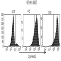
도 1a 및 b는 뮤라인 항체 6H3의 경쇄(도 1a) 및 중쇄(도 1b)의 가변 도메인의 콜리에 펄스(Collier Perles) 2D 도면을 보여주는 다이어그램이다. 사슬의 3개의 CDR 루프들이 다이어그램의 상단에 나타나있다.
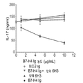
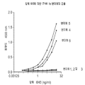
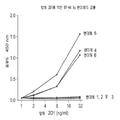
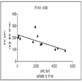
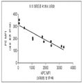
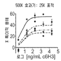
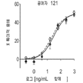
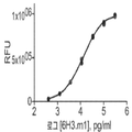
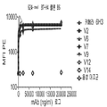
도 2a 및 b는 티미딘 삽입(도 2a) 및 IL-17 발현(도 2b)에 의해 측정된 B7-H4 Ig로 매개되는 T 세포 활성화의 억제를 차단하는 항체 6H3의 능력을 항체 농도(㎍/ml)의 함수로서 보여주는 선 그래프이다
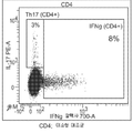
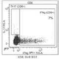
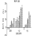
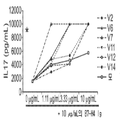
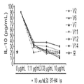
도 3a 내지 d는 항체 6H3이 난소암 환자의 T 세포 반응(IL-17 또는 INFγ을 생성하는 기능적 CD4 및 CD8 세포)을 난소암 환자로부터의 TAM의 존재 하에 향상시킬 수 있다는 것을 보여주는 산점도이다(도 3a: CD4, 동형 대조군; 도 3b: CD8, 동형 대조군; 도 3c: CD4, 항-B7-H4 항체 6H3; 도3d: CD8, 항-B7-H4 항체6H3).
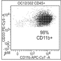
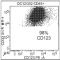
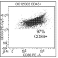
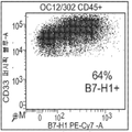
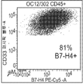
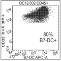
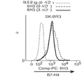
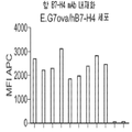
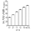
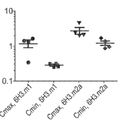
도 4a 내지 I는 난소암 환자 종양 관련 대식세포(tumor-associated macrophages, TAM)에 의한 B7-H4(도 4h) 및 다른 항원(CD11b(도 4a), CD14(도 4b), CD123(도 4c), CD86(도 4d), CD80(도 4e), HLA-DR(도 4f), B7-H1(도 4g), 및 B7-DC(도 4i))의 발현 및 이러한 세포에 의한 B7-H4 발현을 검출하는 항체 6H3의 능력을 보여주는 산점도이다.
도 5는 B7-H4에 결합하는 항체 6H3의 결합 동역학을 항체 농도(nM)의 함수로서 보여주는 결합 곡선이다.
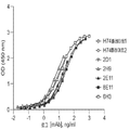
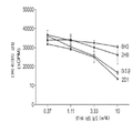
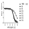
도 6은 다른 항 B7-H4 항체들(즉, 항체 H74, 2D1, 2H9, 2E11 및 8E11)에 비교하여 항체 6H3의 인간 H7-H4의 결합의 동역학을 항체 농도(log[mAb], ng/ml)의 함수로서 보여주는 결합 곡선이다.
도 7은 B7-H4의 IgC 영역(변이체 1: 서열 번호 1의 IgV 잔기 29-149; 변이체 2: 서열 번호 1의 IgV 잔기 29-154; 변이체 3: 서열 번호 1의 IgV 잔기 29-158; 변이체 4: ECD-hIgG4; 변이체 5: 서열 번호 1의 IgC 잔기 154-259; 변이체 6: ECD-hIgG1-서열 번호 7)에 결합하는 항체 6H3의 능력을 항체 농도(nM)의 함수로서 보여주는 결합 곡선이다.
도 8a 내지 c는 항 인간 B7-H4 항체 2H9(도 8a), 2D1(도 8b) 및 H74(도 8c)에 의해 인식되는 B7-H4의 영역(변이체 1: 서열 번호 1의 IgV 잔기 29-149; 변이체 2: 서열 번호 1의 IgV 잔기 29-154; 변이체 3: 서열 번호 1의 IgV 잔기 29-158; 변이체 4: ECD-hIgG4; 변이체 5: 서열 번호 1의 IgC 잔기 154-259; 변이체 6: ECD-hIgG1-서열 번호 7)을 항체 농도(nM)의 함수로서 보여주는 결합 곡선이다.
도 9는 항 인간 B7-H4 항체 6H3, 2H9 및 2D1의 B7-H4 Ig로 매개되는 T 세포 활성화의 억제(3H-티미딘 삽입에 의해 측정된 증식(ΔCPM))를 차단하는 능력을 항체 농도(㎍/ml)의 함수로서 비교한 선 그래프이다
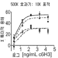
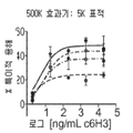
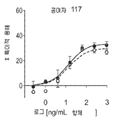
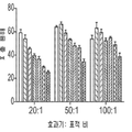
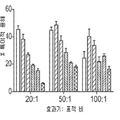
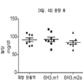
도 10a 내지 f는 항체(단핵구가 없는 대조군, 팔리비주맙, 2H9, 2D1, 6H3, 항-B7-H1, 항-PD1)의 IFNγ 초회감작된 단핵구 매개 억제를 역전시키는 능력을 보여주는 막대 그래프이다. T 세포는 IL-2(CD4+ T 세포, 도 10a; CD8+ T 세포, 도 10b), TNF-α(CD4+ T 세포, 도 10c; CD8+ T 세포, 도 10d) 또는 IL-8(CD4+ T 세포, 도 10e; CD8+ T 세포, 도 10f)에 대하여 염색되었다.
도 11a 및 b는 항체 6H3이 B7-H4로 형질감염된 세포주 및 B7-H4 양성 유방암 세포주 SK-BR-3의 표면에 발현된 B7-H4의 강력한 내재화를 유도한다는 것을 보여준다. 도 11a는 293T B7-H4 형질감염체의 CypHer5B 형광을 CypHer로 표지된 항체 6H3과 0, 1, 및 3 시간 항온배양한 후 유세포 측정한 결과를 보여주는 일련의 히스토그램이다. 도 11b는 시간에 따른 B7-H4 표면 염색 및 CyperHer 형광을 보여주는 선 그래프이다.
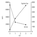
도 12a 및 b는 CypHer 표지된 6H3과의 항온배양 중 시간에 따라 B7-H4 양성 인간 유방암 세포주 SK-BR3 세포 상의 대조군에 대한 B7-H4 표면 염색 및 CypHer 형광을 보여주는 히스토그램이다.
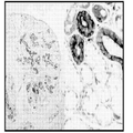
도 13a 내지 c는 항체 6H3의 조직학 검체의 B7-H4 양성 종양 세포 검출능을 보여주는 현미경 사진이다. 도 13a 및 b는 6H3으로 염색된 침샘 조직의 사진이고, 도 13c는 두 가지의 상이하지만 연관된 난소의 장액성 낭종 병변을 보여준다.
도 14는 4℃에서 30분 간 처리 후 EG7(B7-H4 음성 세포), EG7.IG7(B7-H4 저발현 세포) 및 EG7.IVB3(B7-H4 고발현 세포) 내에서 키메라 2E11(-●-), 키메라 2H9(-■-), 키메라 2D1(-▲-), 및 키메라 6H3(-▼-)의 내재화를 보여주는 선 그래프이다.
도 15는 4℃에서 30분간 처리 후 EG7(B7-H4 음성 세포), EG7.IG7(B7-H4 저발현 세포) 및 EG7.IVB3(B7-H4 고발현 세포) 내에서 키메라 6H3(-●-), 인간화 변이체 2(-▲-), 인간화 변이체 6(-▲-), 인간화 변이체 7(-▲-), 인간화 변이체 9(-▲-), 인간화 변이체 12(-▲-), 및 인간화 변이체 14(-▲-)의 내재화를 보여주는 선 그래프이다.
도 16은 항 B7-H4 항체의 내재화를 내재화가 우세한 조건 하에 B7-H4의 세포 표면 발현의 함수로서 보여주는 선 그래프이다.
도 17은 항 B7-H4 항체의 내재화를 내재화가 우세하지 않은 조건 하에 B7-H4의 세포 표면 발현의 함수로서 보여주는 선 그래프이다.
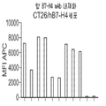
도 18a 및 b는 E.G7ova/hB7-H4 세포(도 18a) 및 CT26/hB7-H4 세포(도 18b) 내 항 B7-H4 항체의 내재화를 보여주는 막대 그래프이다(X 축을 따라 왼쪽부터 오른쪽): 인간 키메라 6H3, 인간화 6H3 V2, 인간화 6H3 V6, 인간화 6H3 V7, 인간화 6H3 V9, 인간화 6H3 V12, 인간화 6H3 V14, 6H3.m1(마우스 IgG1), 6H3.m2a(마우스 IgG2a), 음성 대조군(항 PD-1 항체), 무처리.
도 19a 내지 c는 항체 의존성 세포 매개 세포독성(antibody-dependent cell-mediated cytotoxicity, ADCC)을 측정하도록 설계된 검정에서 4종의 상이한 건강한 말초 혈액 단핵구 세포(peripheral blood mononucleated cell, PBMC) 공여자(117, 119, 121, 122)에 대하여 효과기 세포에 의한 EG7.B7H4 표적 세포의 특이적인 용해%를 3종의 상이한 효과기 세포:표적 세포 비(500,000:25,000(도 19a), 500,000:10,000(도 19b), 및 500,000:5,000(도 19c))에서의 키메라 6H3 항체의 농도((log (ng/ml))의 함수로서 보여주는 선 그래프이다.
도 20은 항체 의존성 세포 매개 세포독성(ADCC)을 측정하도록 설계된 검정에서 3종의 상이한 공여자의 말초 혈액 단핵구 세포(PBMC)(114, 121, 122)로부터의 효과기 세포에 의한 EG7.B7H4 표적 세포의 특이적인 용해%를 14종의 상이한 6H3의 인간화 변이체에 대하여 보여주는 막대 그래프이다(항체 농도 10 ng/ml, 1:20의 표적 세포:효과기 세포 비).
도 21a 내지 d는 항체 의존성 세포 매개 세포독성(ADCC)을 측정하도록 설계된 검정에서 효과기 세포로서 4종의 상이한 공여자(공여자 118(도 21a), 공여자 117(도 21b), 공여자 121(도 21c) 또는 공여자 122(도 21d)로부터의 말초 혈액 단핵구 세포(PBMC)와 조합하여 증가하는 농도의 변이체 11 또는 변이체 14 인간화 6H3 항체(log[ng/mL] 항체)를 이용하여 표적 세포의 특이적 용해%를 보여주는 막대 그래프이다.
도 22a 및 b는 항체 의존성 세포 매개 세포독성(ADCC)을 측정하도록 설계된 검정에서 대조군 IgG1, 또는 6종의 상이한 농도(2000 ng/ml, 500 ng/ml, 125 ng/ml, 31.3 ng/ml, 7.81 ng/ml, 1.95 ng/ml)의 키메라 6H3 항체 중 하나의 존재 하에 3가지의 상이한 효과기 세포:표적 세포 비(20:1, 50:1, 100:1)에서 117(도 22a) 또는 120(도 22b) 공여자 세포에 의한 SK-BR-3 세포의 용해 총%를 보여주는 막대 그래프이다.
도 23a 및 b는 항체 의존성 세포 매개 세포독성(ADCC)을 측정하도록 설계된 검정에서 대조군 IgG1, 또는 6종의 상이한 농도(2000 ng/ml, 500 ng/ml, 125 ng/ml, 31.3 ng/ml, 7.81 ng/ml, 1.95 ng/ml)의 키메라 6H3 항체 중 하나의 존재 하에 3가지의 상이한 효과기 세포:표적 세포 비(20:1, 50:1, 100:1)에서 117(도 23a) 또는 120(도 23b) 공여자 세포에 의한 SK-BR-3 세포의 특이적 용해 %를 보여주는 막대 그래프이다.
도 24a 및 b는 대조군, 키메라 6H3 항체 및 HERCEPTIN®항체를 활용한 보체 의존성 세포독성 검정의 결과를 보여주는 막대 그래프이다. 도 24a는 다양한 항체 및 항체 농도에 대한 대조군, 1:2 보체, 및 1:5 보체 희석액에서의 RFU(상대 형광 단위-black로 보정됨)를 보여준다. 왼쪽부터 오른쪽으로, 각 보체 희석액인 막대들의 집합은 다음을 나타낸다: 제형 완충액, 50 ㎍/ml IgG1(음성 대조군), 5 ㎍/ml IgG1(음성 대조군), 0.5 ㎍/ml IgG1(음성 대조군), 50 ㎍/ml 키메라 6H3, 5 ㎍/ml 키메라 6H3, 0.5 ㎍/ml 키메라 6H3, 50 ㎍/ml HERCEPTIN® 5 ㎍/ml HERCEPTIN® 0.5 ㎍/ml HERCEPTIN® 도 24b는 다양한 항체 및 항체 농도에 대한 대조군, 1:2 보체, 및 1:5 보체 희석액에서의 특이적 용해를 보여준다. 왼쪽부터 오른쪽으로, 각 보체 희석액인 막대들의 집합은 다음을 나타낸다: 50 ㎍/ml 키메라 6H3, 5 ㎍/ml 키메라 6H3, 0.5 ㎍/ml 키메라 6H3, 50 ㎍/ml HERCEPTIN® 5 ㎍/ml HERCEPTIN® 0.5 ㎍/ml HERCEPTIN®
도 25는 CTL-매개 용해 검정에서 상이한 효과기 세포:표적 세포 비에서의 Ag+ 표적 세포의 생존(CTL 부재시 평균의 %)을 보여주는 막대 그래프이다.
도 26은 CTL-매개 용해 검정에서 대조군 IgG 또는 키메라 6H3 항체의 존재 하에 상이한 효과기 세포:표적 세포 비에서 EG7.B7H4+OVA 표적 세포의 특이적 용해%를 보여주는 막대 그래프이다. 왼쪽부터 오른쪽으로, 각 효과기 세포:표적 세포비인 막대들의 집합은 다음을 나타낸다: 1 ㎍/ml 대조군 IgG, 10 ㎍/ml 대조군 IgG, 1 ㎍/ml 키메라 6H3, 및 10 ㎍/ml 키메라 6H3.
도 27a는 루이스 폐 암종(Lewis Lung Carcinoma, LCC) 종양 마우스 모델을 활용한 제1 실험에서 시간(종양 접종 후 날짜)에 따른 종양 부피(mm3)를 보여주는 선 그래프로서, 종양을 가진 마우스는 제형 완충액 대조군(-◆-), 6H3.m1(-■-), 또는 6H3.m2a(-▲-)로 처리되었다. 도 27b는 루이스 폐 암종(Lewis Lung Carcinoma, LCC) 종양 마우스 모델에서 시간(종양 접종 후 날짜)에 따른 마우스의 생존%를 보여주는 카플란 마이어(Kaplan-Meier) 곡선으로서, 마우스는 제형 완충액 대조군(-◆-), 6H3.m1(-■-), 또는 6H3.m2a(-▲-)로 처리되었다.
도 28a는 루이스 폐 암종(LCC) 종양 마우스 모델을 활용한 제2 실험에서 시간(종양 접종 후 날짜)에 따른 종양 부피(mm3)를 보여주는 선 그래프로서, 종양을 가진 마우스는 제형 완충액 대조군(-◆-), 6H3.m1(-■-), 또는 6H3.m2a(-▲-)로 처리되었다. 도 28b는 루이스 폐 암종(LCC) 종양 마우스 모델에서 시간(종양 접종 후 날짜)에 따른 마우스의 생존%를 보여주는 카플란 마이어 곡선으로서, 마우스는 제형 완충액 대조군(-◆-), 6H3.m1(-■-), 또는 6H3.m2a(-▲-)로 처리되었다.
도 29a는 CT26.B7H4 폐암 모델에서 종양 세포의 정맥 내 접종 후 치료의 부재 하에 시간(일)에 따른 종양의 성장 동역학(종양 수)을 보여주는 점도표이다. 도 29b는 CT26.B7H4 폐암 모델에서 종양 세포의 정맥 내 접종 및 제형 완충액(FB), 10일차부터 시작한 10 mg/kg 6H3.m2a(10 mg/kg-10); 10일차부터 시작한 1 mg/kg 6H3.m2a(1 mg/kg-10); 14일차부터 시작한 10 mg/kg 6H3.m2a(10 mg/kg-14); 또는 14일차부터 시작한 1 mg/kg 6H3.m2a(1 mg/kg-14)로의 치료 후 시간(일)에 따른 종양 수를 보여주는 점도표이다.
도 30은 제형 완충액 또는 100 mg/kg의 6H3.m1 또는 6H3.m2의 주 1회 투여의 4회 후 24일차의 마우스 혈당(mg/dl)을 보여주는 점도표이다.
도 31은 100 mg/kg의 6H3.m1 또는 6H3.m2의 주 1회 투여의 4회 후 마우스 혈청에서 6H3.m1 또는 6H3.m2의 최고점(Cmax) 및 골(Cmin)에 대한 항체 수준(mg/ml)을 보여주는 점도표이다.
도 32a 내지 b는 혈청 항체 농도(log[항체], pg/ml)의 함수로서 6H3.m1(32a) 또는 6H3.m2(32b)에 대한 상대 형광 단위(RFU)를 보여주는 선 그래프이다.
도 33은 B7-H4의 세포외 도메인에 결합하는 인간 키메라 6H3 및 14종의 인간화 변이체를 측정한 ELISA 검정(항체 농도(Log10 (nM))의 함수로서 OD450)의 결과를 보여주는 결합 곡선이다.
도 34a 및 b는 B7-H4-Ig에 결합하는 키메라 6H3 및 인간화 변이체 V1~V7(도 34a) 및 V8~V14(도 34b)를 측정한 경쟁적 ELISA 검정(항체 농도(ng/ml)log)의 함수로서 450 nm에서의 흡광도)의 미가공 결과를 보여주는 결합 곡선이다.
도 35a 및 b는 B7-H4를 발현하는 세포에 결합하는 키메라 6H3, 인간화 변이체 V2, V6, V7, V9, V12, V14, 및 음성 대조군 항 PD-1 항체(도 34a 단독)를 측정한 경쟁적 ELISA 검정(항체 농도((ng/ml)log)의 함수로서 450 nm에서의 흡광도)의 미가공 결과(도 35a) 및 배경 감산 결과(도 35b)를 보여주는 결합 곡선이다.
도 36은 B7-H4를 발현하는 HEK293 형질감염체에 결합하는 6H3의 인간화 변이체의 항체 결합(항체 농도(nM)의 함수로서 평균 형광 강도(mean fluorescent intensity, MFI))을 보여주는 결합 곡선이다.
도 37은 마우스 B7-H4mIg 융합 단백질에 결합하는 항체 키메라 6H3 및 6H3의 인간화 변이체를 측정한 ELISA 검정(항체 농도(log10(nM))의 함수로서 OD450)의 결과를 보여주는 결합 곡선이다.
도 38a 내지 d는 다양한 농도(0, 1.11 μ/g/ml, 3.33 μ/g/ml, 또는 10 μ/g/ml)의 키메라 6H3(“모”) 또는 6H3의 인간화 변이체와 조합하여 B7-H4-Ig 융합 단백질로 처리한 세포에 대한 IL-17(도 38a), IFNγ(도 38b), 증식([3H]-티미딘 삽입)(도 38c), 및 IL-10 분비의 분비(도 38d)를 보여주는 선 그래프이다.
* 대조군Ig 사용. Figures 1a and 1b are diagrams showing Collier Perles 2D diagrams of the variable domains of the light chain ( Figure 1a ) and heavy chain ( Figure 1b ) of mAb line antibody 6H3. The three CDR loops of the chain are shown at the top of the diagram.
Figures 2a and b show the ability of antibody 6H3 to block the inhibition of T cell activation mediated by B7-H4 Ig as measured by thymidine incorporation ( Figure 2a ) and IL-17 expression ( Figure 2b ) ml) < / RTI >
3a-d is a scatter plot showing that antibody 6H3 is (functional CD4 and CD8 cells to generate IL-17 or INFγ) T cell responses in ovarian cancer patients can be improved in the presence of TAM from ovarian cancer patients (Fig. 3a: CD4, isotype control; Fig. 3b: CD8, isotype control; Fig. 3c: CD4, anti -B7-H4 antibody 6H3; Figure 3d: CD8, wherein -B7-H4 antibody 6H3).
Figures 4A-I show B7-H4 ( Figure 4h ) and other antigens (CD11b ( Figure 4a ), CD14 ( Figure 4b ), CD123 ( Figure 4c ) by tumor- associated macrophages (TAM) , the expression, and B7-H4 expression by these cells of CD86 (Fig. 4d), CD80 (Fig. 4e), HLA-DR (Fig. 4f), B7-H1 (Fig. 4g), and B7-DC (Fig. 4i)) It is a scatter plot showing the ability of antibody 6H3 to detect.
Figure 5 is a binding curve showing the binding kinetics of antibody 6H3 binding to B7-H4 as a function of antibody concentration (nM).
Figure 6 shows the kinetics of the binding of human H7-H4 of antibody 6H3 to antibody concentration (log [mAb], ng / ml (as compared to other anti-B7-H4 antibodies (i.e., antibodies H74, 2D1, 2H9, 2E11 and 8E11) ) As a function of the coupling curve.
7 is IgC domain of B7-H4 (variant 1: IgV residues 29-149 of SEQ ID NO: 1; variant 2: SEQ ID NO: IgV residues 29-154 of 1; Mutant 3: IgV residues 29-158 of SEQ ID NO: 1; mutant 4 (ECD-hIgG4; mutant 5 : IgC residue 154-259 of variant SEQ ID NO: 1 ; mutant 6 : ECD-hIgG1- SEQ ID NO: 7 ) .
Figure 8a-c is an anti-human B7-H4 antibody 2H9 (Fig. 8a), 2D1 (FIG. 8b) and H74 (FIG. 8c) region of the B7-H4 recognized by a (variant 1: IgV residues 29-149 of SEQ ID NO: 1 Mutant 2 : IgV residue 29-154 of SEQ ID NO: 1 Mutant 3 : IgV residue 29-158 of SEQ ID NO: 1 Mutant 4 : ECD-hIgG4 Mutant 5 : IgC residue 154-259 of SEQ ID NO: 1 Mutant 6 : ECD-hIgG1- SEQ ID NO: 7 ) as a function of antibody concentration (nM).
Figure 9 shows the ability to block the inhibition of T cell activation mediated by anti-human B7-H4 antibodies 6H3, 2H9 and 2D1, B7-H4 Ig (proliferation (? CPM) as measured by 3H-thymidine incorporation) ≪ / RTI >< RTI ID = 0.0 > g / ml)
Figures 10a-f are bar graphs showing the ability of antibodies (monocyte-free control, palivizumab, 2H9, 2D1, 6H3, anti-B7-H1, anti-PD1) to reverse IFN gamma sensitized monoclonal mediated inhibition. T cells, IL-2 (CD4 + T cells, Figure 10a; CD8 + T cells, Figure 10b), TNF-α (CD4 + T cells, Figure 10c; CD8 + T cells, Fig. 10d), or IL-8 (CD4 + T cells, Fig. 10e ; CD8 + T cells, Fig. 10f ).
Figures 11a and b show that antibody 6H3 induces strong internalization of B7-H4 transfected cell lines and B7-H4 expressed on the surface of B7-H4 positive breast cancer cell line SK-BR-3. FIG. 11A is a series of histograms showing the results of flow cytometry after CypHer5B fluorescence of the 293T B7-H4 transfectant was incubated with the antibody 6H3 labeled with CypHer for 0, 1, and 3 hours. FIG. 11B is a line graph showing B7-H4 surface staining and CyperHer fluorescence with time.
Figures 12a and b are histograms showing B7-H4 surface staining and CypHer fluorescence for the control on the B7-H4 positive human breast cancer cell line SK-BR3 cells over time during incubation with CypHer-labeled 6H3.
Figures 13a-c are micrographs showing the ability of B7-H4 positive tumor cells to detect histological specimens of antibody 6H3. Figures 13a and b are photographs of salivary glands stained with 6H3 and Figure 13c shows serous cystic lesions of the ovary associated with two different but different.
FIG. 14 shows the results of the treatment of chimera 2E11 (- ●) in EG7 (B7-H4 negative cells), EG7.IG7 (B7-H4 low expressing cells) and EG7.IVB3 -), chimera 2H9 (-? -), chimera 2D1 (-? -), and chimera 6H3 (- ▼ -).
FIG. 15 shows the results of treatment of chimeric 6H3 (-. -) cells in EG7 (B7-H4 negative cells), EG7.IG7 (B7-H4 low expression cells) and EG7.IVB3 (B7- ), The humanized mutant 2 (- -), the humanized mutant 6 (- -), the humanized mutant 7 (- 14 is a line graph showing the internalization of (- - -).
Figure 16 is a line graph showing the internalization of anti-B7-H4 antibody as a function of cell surface expression of B7-H4 under conditions predominanting internalization.
Figure 17 is a line graph showing the internalization of anti-B7-H4 antibody as a function of cell surface expression of B7-H4 under conditions where internalization did not prevail.
Figures 18a and b are bar graphs (from left to right along the X axis) showing the internalization of anti-B7-H4 antibodies in E.G7ova / hB7-H4 cells ( Figure 18a ) and CT26 / hB7-H4 cells ( Figure 18b ) Human chimeric 6H3, humanized 6H3 V2, humanized 6H3 V6, humanized 6H3 V7, humanized 6H3 V9, humanized 6H3 V12, humanized 6H3 V14, 6H3.m1 (mouse IgG1), 6H3.m2a (mouse IgG2a) 1 antibody), no treatment.
Figures 19a-c show the results of a study designed to determine antibody-dependent cell-mediated cytotoxicity (ADCC) using four different healthy peripheral blood mononucleated cell (PBMC) donors 117, 119 , 121, 122) was determined by three different effector cell: target cell ratios (500,000: 25,000 ( Figure 19a ), 500,000: 10,000 ( Figure 19b ), and As a function of the concentration of chimeric 6H3 antibody ((log (ng / ml)) at 500,000: 5,000 ( Figure 19c ).
Figure 20 shows the effect of EG7.B7H4 target cells on effector cells from three different donor peripheral blood mononuclear cells (PBMC) (114, 121, 122) in assays designed to measure antibody dependent cellular mediated cytotoxicity (ADCC) (
Figures 21a-d show four different donors (donor 118 ( FIG. 21a ), donor 117 ( FIG. 21b ), donor 121 ( FIG. 21c ), or the like, as effector cells in assays designed to measure antibody- donor 122 (Figure 21d) the specificity of the peripheral blood mononuclear cells (PBMC) and target cells using a variant of the density to match increases in 11 or
Figures 22a and b show the effect of different concentrations of control IgG1, or 6 different concentrations (2000 ng / ml, 500 ng / ml, 125 ng / ml, 31.3 ng / ml, ( Figure 22A ) or 120 ( Figure 22A ) at three different effector cell: target cell ratios (20: 1, 50: 1, 100: 1) in the presence of one of the chimeric 6H3 antibodies ( FIG. 22b ) is a bar graph showing the total% dissolution of SK-BR-3 cells by donor cells.
Figures 23a and b show the effect of different concentrations of control IgG1, or 6 different concentrations (2000 ng / ml, 500 ng / ml, 125 ng / ml, 31.3 ng / ml, ( Figure 23A ) or 120 ( Figure 23A ) at three different effector cell: target cell ratios (20: 1, 50: 1, 100: 1) in the presence of one of the chimeric 6H3 antibodies ( Fig. 23 (b)) is a bar graph showing the specific% dissolution of SK-BR-3 cells by donor cells.
24A and 24B are bar graphs showing the results of a complement dependent cytotoxicity assay using the control, chimeric 6H3 antibody and HERCEPTIN® antibody. Figure 24A shows RFU (corrected to relative fluorescence unit-black) in the control, 1: 2 complement, and 1: 5 complement dilutions for various antibody and antibody concentrations. From left to right, the set of bars, each of the complement dilutions, represents: formulation buffer, 50 / / ml IgG1 (negative control), 5 / / ml IgG1 (negative control), 0.5 / / ml IgG1 (negative control) 50 / / ml HERCEPTIN 占 5 占 퐂 / ml HERCEPTIN 占 0.5 占 퐂 / ml HERCEPTIN 占Fig. 24b shows the results of a comparison of various antibody and antibody concentrations , 1: 2 complement, and 1: 5 complement dilution. From left to right, the set of rods that are each complement diluent represents: 50 μg / ml chimera 6H3, 5 μg / ml chimera 6H3, 0.5 μg / ml chimera 6H3, 50 μg /
Figure 25 is a bar graph showing the survival of Ag + target cells (% of mean at non-CTL) in different effector cells: target cell ratio in CTL-mediated lysis assay.
Figure 26 is a bar graph showing the% specific lysis of EG7.B7H4 + OVA target cells in different effector cell: target cell ratios in the presence of control IgG or chimeric 6H3 antibody in a CTL-mediated lysis assay. From the left to the right, each effector cell: a collection of target cell bead bars shows: 1 ig / ml control IgG, 10 ug / ml control IgG, 1 ug / ml chimera 6H3, and 10 ug / ml chimera 6H3.
27A is a line graph showing the tumor volume (mm 3 ) according to time (the date after tumor inoculation) in the first experiment using Lewis Lung Carcinoma (LCC) tumor mouse model, (-) -, 6H3.m1 (-) -, or 6H3.m2a (- -). Figure 27b is a Kaplan-Meier curve showing the survival% of mice according to time (day after tumor inoculation) in a Lewis Lung Carcinoma (LCC) tumor mouse model, in which the mouse is a formulation buffer control -), 6H3.m1 (-? -), or 6H3.m2a (-? -).
28A is a line graph showing tumor volume (mm 3 ) according to time (day after tumor inoculation) in a second experiment using a Lewis lung carcinoma (LCC) tumor mouse model, in which mice with tumors were treated with a formulation buffer (- - -), 6H3.m1 (- - -), or 6H3.m2a (- - -). Figure 28b is a Kaplan-Meier curves showing the survival% of mice according to time (day after tumor inoculation) in a Lewis lung carcinoma (LCC) tumor mouse model. The mice were divided into the formulation buffer control group (- -), or 6H3.m2a (- - -).
29A is a graph showing the growth kinetics (number of tumors) of a tumor over time (days) in the absence of treatment after intravenous inoculation of tumor cells in a CT26.B7H4 lung cancer model. Figure 29b shows the results of intravenous inoculation of tumor cells and formulation buffer (FB) in a CT26.B7H4 lung cancer model, 10 mg / kg 6H3.m2a (10 mg / kg-10) starting from
30 is a viscosity chart showing mouse blood glucose (mg / dl) at 4 days and 24 days after administration of the formulation buffer or 100 mg / kg of 6H3.m1 or 6H3.m2 once a week.
Figure 31 shows the antibody titers (mg) to peak (Cmax) and bone (Cmin) peak of 6H3.m1 or 6H3.m2 in mouse serum after 4 times of weekly administration of 100 mg / kg of 6H3.m1 or 6H3.m2 / ml).
Figures 32a-b are line graphs showing relative fluorescence units (RFU) for 6H3.m1 ( 32a ) or 6H3.m2 ( 32b ) as a function of serum antibody concentration (log [antibody], pg / ml).
Figure 33 is a binding curve showing the results of an ELISA assay (OD450 as a function of antibody concentration (Log10 (nM)) measuring human chimeric 6H3 binding to the extracellular domain of B7-H4 and 14 humanized variants.
Figure 34a and b are of the chimeric 6H3 and humanized variants V1 ~ V7 (Fig. 34a) and V8 ~ V14 a competitive ELISA measured (Fig. 34b) and black (an antibody concentration (ng / ml) log) binding to B7-H4-Ig Absorbance at 450 nm as a function).
Figures 35a and b show competitive ELISA (Figure 34a) measuring chimeric 6H3 binding to cells expressing B7-H4, humanized variants V2, V6, V7, V9, V12, V14 and negative control anti- ( Fig. 35A ) and the background subtraction result ( Fig. 35B ) of the antibody concentration (absorbance at 450 nm as a function of antibody concentration ((ng / ml) log).
Figure 36 is a binding curve showing antibody binding (mean fluorescent intensity (MFI) as a function of antibody concentration (nM)) of humanized variants of 6H3 binding to HEK293 transfectants expressing B7-H4.
Figure 37 is a binding curve showing the results of an ELISA assay (OD450 as a function of antibody concentration (log10 (nM)) measuring humanized variants of antibody chimeras 6H3 and 6H3 binding to murine B7-H4mIg fusion protein.
Figures 38a-d show the effect of B7-H3 in combination with chimeric 6H3 (" Mo ") or 6H3 humanized variants of various concentrations (0, 1.11 [mu] g / ml, 3.33 [mu] g / IL-17 (Figure 38a) for the treatment with H4-Ig fusion proteins, IFNγ (Figure 38b), proliferation ([3 H] - thymidine injection) (Fig. 38c), and IL-10 secretion of glands (Fig. 38d. ≪ / RTI >
* Control Ig use.
다수의 종양 조직, 예를 들어, 인간 난소암에서 발견되는 높은 수준의 B7-H4 발현은 면역 억제 매개에 있어서 B7-H4의 주요한 역할을 시사한다. 또한, B7-H4를 발현하는 TAM은 종양 관련 항원 특이적 T 세포 면역을 억제하는 것으로 밝혀졌다(Kryczek, I. et al. (2006) “B7-H4 Expression Identifies A Novel Suppressive Macrophage Population In Human Ovarian Carcinoma,” J. Exp. Med. 203(4):871-881). TAM 내 B7-H4 발현 강도는 종양 내 Treg 세포 수와 유의하게 상호관련된다. 더욱이, TAM 상에 발현된 B7-H4는 불량한 환자 결과에 관련된다(Kryczek, I. et al. (2006) “B7-H4 Expression Identifies A Novel Suppressive Macrophage Population In Human Ovarian Carcinoma,” J. Exp. Med. 203(4):871-881). 이전에 발표된 데이터는, 또한, TAM이, 종양 내로의 Treg 세포 추적(trafficking) 및 TAM 자신을 포함하는 항원 제시 세포(APC) 상의 Treg 세포로 유도된 B7-H4 발현을 매개하는 케모카인 CCL22를 자발적으로 생성한다는 것을 보여준다(Kryczek, I. et al. (2006) “Cutting Edge: Induction Of B7-H4 On APCs Through IL-10: Novel Suppressive Mode For Regulatory T Cells,” J. Immunol. 177(1):40-44). 종합하면, 이러한 발견은 B7-H4를 발현하는 종양 세포 및 TAM이 종양 미세환경 내 면역 억제에서 매우 중요한 역할을 수행하여 종양이 면역계에 의해 검출되는 것을 피할 수 있도록 한다(“면역 회피”)는 것을 가리킨다. B7-H4를 차단하거나, B7-H4의 표면 발현을 조절하거나, B7-H4 매개 신호 전달을 조절하거나, TAM을 고갈시킴으로써, B7-H4에 면역특이적으로 결합할 수 있거나, B7-H4의 원래의 수용체와의 상호작용을 예방할 수 있는 개시된 분자는 암에 대한 효과적인 면역요법으로서 하나의 전략을 제시한다.High levels of B7-H4 expression found in a number of tumor tissues, such as human ovarian cancer, suggest a major role for B7-H4 in immunosuppressive mediation. In addition, TAM expressing B7-H4 has been shown to inhibit tumor-specific antigen-specific T cell immunity (Kryczek, I. et al . (2006) B7-H4 Expression Identification A Novel Suppressive Macrophage Population In Human Ovarian Carcinoma , &Quot; J. Exp. Med., 203 (4): 871-881). The intensity of B7-H4 expression in TAM was significantly correlated with the number of Treg cells in the tumor. Furthermore, B7-H4 expressed on TAM is associated with poor patient outcome (Kryczek, I. et al . (2006) B7-H4 Expression Identification A Novel Suppressive Macrophage Population In Human Ovarian Carcinoma , 203 (4): 871-881). Previously published data also showed that TAM induced chemotaxis CCL22, which mediates Treg cell-induced B7-H4 expression on antigen presenting cells (APC), including Treg cell trafficking into the tumor and TAM itself, shows that the generated (Kryczek, I. et al (2006 ). "Cutting Edge: Induction Of B7-H4 On APCs Through IL-10: Novel Suppressive Mode For Regulatory T Cells," J. Immunol 177 (1).: 40-44). Taken together, this finding suggests that tumor cells expressing B7-H4 and TAM play a very important role in immunosuppression in the tumor microenvironment, thereby avoiding the tumor being detected by the immune system (" immune evasion ") Point. Can bind to B7-H4 immunospecifically by blocking B7-H4, modulating surface expression of B7-H4, modulating B7-H4 mediated signal transduction, depleting TAM, ≪ / RTI > the disclosed molecules offer a strategy as an effective immunotherapy for cancer.
B7-H4에 면역특이적으로 결합할 수 있는 분자뿐 아니라 암 및 다른 질환들의 치료 및 진단에서의 이러한 분자들의 용도가 제공된다. 종양 생장을 예방하거나 늦추고/늦추거나, 종양 매개 억제를 저해하고/하거나, 종양을 제거하고/하거나, 종양 관련 대식세포(tumor-associated macrophage, “TAM")의 활성을 차단하거나 고갈시켜서 그들의 활성을 변화시키고/시키거나 TAM 매개 면역 억제를 낮추기 위한 이러한 분자들의 용도가 또한 제공된다.The use of such molecules in the treatment and diagnosis of cancer and other diseases as well as molecules capable of binding specifically to B7-H4 are provided. Associated macrophages (" TAM ") and / or deplete and / or deplete the activity of tumor-associated macrophages (" TAM "), inhibit tumor growth and / The use of these molecules to alter and / or lower TAM mediated immunosuppression is also provided.
특히, 이러한 분자들, 그리고 특히 hIgG1 또는 hIgG4 항체인 이러한 분자들이 B7-H4를 발현하는 세포, 종양 또는 TAM을 표적화하고, 다양한 기능적 활성(B7-H4와 이의 수용체(들)간의 상호작용의 조절, B7-H4 수준의 조절, 및 음성 신호전달의 약화 및/또는 B7-H4 양성 세포의 고갈을 포함함)을 스크리닝하기 위해 사용될 수 있다. 재조합 DNA 기술을 사용하여, 이러한 분자들은 Fc 수용체(FcR) 결합 활성이 거의 또는 전혀 없거나, 향상된 항체 의존성 세포 매개 세포독성(ADCC) 또는 향상된 보체 의존성 세포독성(CDC) 활성들을 가지는 Fc 도메인을 형성하기 위한 불변 영역을 포함하도록 조작될 수 있다. 이러한 재조합 분자들은 종양 또는 TAM 상의 B7-H4를 낮추거나, 종양 또는 TAM 상의 B7-H4가 종양 미세환경에서 T 세포 또는 다른 세포 상의 저해성 수용체(들)과 상호작용하는 것을 예방하는 조절 분자로서 사용될 수 있어서, T 세포 또는 다른 기능적 세포를 B7-H4 체크 포인트(“휴식(break)”)/억제성 신호전달로부터 방출한다. 반대로, 이러한 분자들은 기능적 Fc 도메인을 형성하기 위한 불변 영역을 포함하도록 조작될 수 있고, 따라서, TAM의 고갈을 유발하는 ADCC 또는 CDC를 유도할 수 있고, 억제성 세포의 표면으로부터의 B7-H4 조절 또는 B7-H4를 발현하는 세포의 직접적인 살상과 같은 다른 활성에 부가하여 T 세포 또는 다른 기능적 세포를 체크 포인트 차단으로부터 해제할 수 있다. ADCC 활성을 가지는 항 B7-H4 항체가 B7-H4를 발현하는 종양 세포를 고갈시키는 동시에 TAM 매개 면역 억제를 저해하는 데에 특히 유용할 수 있다.In particular, these molecules, and in particular those molecules which are hIgG1 or hIgG4 antibodies, are capable of targeting cells, tumors or TAMs expressing B7-H4 and have a variety of functional activities (regulation of the interaction between B7-H4 and its receptor Modulation of B7-H4 levels, and attenuation of voice signal transduction and / or depletion of B7-H4 positive cells). Using recombinant DNA technology, these molecules have little or no Fc receptor (FcR) binding activity, or are capable of forming Fc domains with enhanced antibody-dependent cell-mediated cytotoxicity (ADCC) or enhanced complement-dependent cytotoxicity (CDC) ≪ / RTI > These recombinant molecules can be used as regulatory molecules to lower B7-H4 on the tumor or TAM, or to prevent B7-H4 on the tumor or TAM phase from interacting with the inhibitory receptor (s) on T cells or other cells in the tumor microenvironment To release T cells or other functional cells from the B7-H4 checkpoint (" break ") / inhibitory signaling. Conversely, these molecules can be engineered to contain a constant region for forming a functional Fc domain, thus leading to ADCC or CDC leading to the depletion of TAM, and can regulate B7-H4 regulation from the surface of inhibitory cells Or other activities, such as direct killing of cells expressing B7-H4, to release T cells or other functional cells from checkpoint blocking. Anti-B7-H4 antibodies with ADCC activity may be particularly useful for depleting B7-H4 expressing tumor cells while inhibiting TAM mediated immunosuppression.
특정한 일 양태에서, 본 개시는 Fc 변이체를 제공하되, Fc 영역은 적어도 하나의 변형(예컨대, 아미노산 치환, 아미노산 삽입, 아미노산 결실)을 카바트 등(1991, NIH Publication 91-3242, National Technical Information Service, Springfield, Va.)에 의해 기재된 바와 같은 EU 인덱스에 의해 번호부여된 228, 234, 235 및 331번으로 이루어진 군으로부터 선택된 하나 이상의 위치에 포함한다.In a particular embodiment, the disclosure provides an Fc variant, wherein the Fc region comprises at least one modification (e.g., amino acid substitution, amino acid insertion, amino acid deletion) in Kabat et al., 1991, NIH Publication 91-3242, National Technical Information Service 234, 235 and 331, which are numbered by the EU index as described by Wu et al., Springfield, Va.
일 양태에서, 변형은 228P, 234F, 235E, 235F, 235Y, 및 331S(카바트에 기재된 바와 같은 EU 인덱스에 의해 번호부여됨)로 이루어진 군으로부터 선택된 적어도 하나의 치환이다.In one embodiment, the modification is at least one substitution selected from the group consisting of 228P, 234F, 235E, 235F, 235Y, and 331S (numbered by EU index as described in Kabat).
다른 특정한 양태에서, 본 개시는 Fc 변이체를 제공하되, Fc 영역은 IgG4 Fc 영역이고, 228 및 235번(카바트에 기재된 바와 같은 EU 인덱스에 의해 번호부여됨)으로 이루어진 군으로부터 선택된 하나 이상의 위치에 적어도 하나의 변형을 포함한다. 또 다른 특정한 양태에서, Fc 영역은 IgG4 Fc 영역이고, 비자연적으로 발생하는 아미노산은 228P, 235E 및 235Y(카바트에 기재된 바와 같은 EU 인덱스에 의해 번호부여됨)로 이루어진 군으로부터 선택된다.In another particular embodiment, the disclosure provides an Fc variant, wherein the Fc region is an IgG4 Fc region and is at least one selected from the group consisting of 228 and 235 (numbered by EU index as described in Kabat) One variation. In another specific embodiment, the Fc region is an IgG4 Fc region and the amino acids occurring naturally are selected from the group consisting of 228P, 235E and 235Y (numbered by EU index as described in Kabat).
다른 특정한 양태에서, 본 개시는 Fc 변이체를 제공하되, Fc 영역은 239, 330 및 332번(카바트에 기재된 바와 같은 EU 인덱스에 의해 번호부여됨)으로 이루어진 군으로부터 선택된 하나 이상의 위치에 적어도 하나의 비자연적으로 발생하는 아미노산을 포함한다. 일 양태에서, 변형은 239D, 330L, 330Y, 및 332E(카바트에 기재된 바와 같은 EU 인덱스에 의해 번호부여됨)로 이루어진 군으로부터 선택된 적어도 하나의 치환이다. 그 전체가 본원에 참조로서 포함된 미국 특허 제7,317,091호를 참조한다.In another particular embodiment, the disclosure provides for an Fc variant, wherein the Fc region comprises at least one < RTI ID = 0.0 > non-specific < / RTI > ratio at one or more positions selected from the group consisting of 239, 330 and 332 (numbered by EU index as described in Kabat) It includes naturally occurring amino acids. In one embodiment, the variants are at least one substitution selected from the group consisting of 239D, 330L, 330Y, and 332E (numbered by EU index as described in Kabat). See U.S. Patent No. 7,317,091, which is incorporated herein by reference in its entirety.
다른 특정한 일 양태에서, 본 개시는 Fc 변이체를 제공하되, Fc 영역은 252, 254, 및 256번(카바트에 기재된 바와 같은 EU 인덱스에 의해 번호부여됨)으로 이루어진 군으로부터 선택된 하나 이상의 위치에 적어도 하나의 비자연적으로 발생하는 아미노산을 포함한다. 일 양태에서, 변형은 252Y, 254T 및 256E(카바트에 기재된 바와 같은 EU 인덱스에 의해 번호부여됨)로 이루어진 군으로부터 선택된 적어도 하나의 치환이다. 그 전체가 본원에 참조로서 포함된 미국 특허 제7,083,784호를 참조한다.In another specific embodiment, the disclosure provides for an Fc variant, wherein the Fc region comprises at least one of at least one selected from the group consisting of 252, 254, and 256 (numbered by EU index as described in Kabat) Of non-naturally occurring amino acids. In one aspect, the variant is at least one substitution selected from the group consisting of 252Y, 254T and 256E (numbered by EU index as described in Kabat). See U.S. Patent No. 7,083,784, the entirety of which is incorporated herein by reference.
일정 양태에서, 본 개시는 Fc 변이체를 제공하되, Fc 영역은 428번(카바트에 기재된 바와 같은 EU 인덱스에 의해 번호부여됨) 위치에 비자연적으로 발생하는 아미노산을 포함한다. 일 양태에서, 428번 위치의 변형은 428T, 428L, 428F, 및 428S(카바트에 기재된 바와 같은 EU 인덱스에 의해 번호부여됨)로 이루어진 군으로부터 선택된다. 그 전체가 본원에 참조로서 포함된 미국 특허 제7,670,600호를 참조한다. 다른 양태에서, Fc 변이체는 434번(카바트에 기재된 바와 같은 EU 인덱스에 의해 번호부여됨) 위치에 비자연적으로 발생하는 아미노산을 더 포함할 수 있다. 일 양태에서, 434번 위치의 변형은 434A, 434S, 및 434F(카바트에 기재된 바와 같은 EU 인덱스에 의해 번호부여됨)로 이루어진 군으로부터 선택된다. 다른 양태에서, 본 개시는 Fc 변이체를 제공하되, Fc 영역은 428번 및 434번(카바트에 기재된 바와 같은 EU 인덱스에 의해 번호부여됨) 위치에 비자연적으로 발생하는 아미노산을 포함한다. 특정한 일 양태에서, Fc 영역은 428L, 434S를 포함한다. 미국 특허 제8,088,376호를 참조한다.In certain embodiments, the disclosure provides Fc variants, wherein the Fc region comprises amino acids occurring naturally at positions 428 (numbered by EU index as described in Kabat). In one embodiment, the strain at position 428 is selected from the group consisting of 428T, 428L, 428F, and 428S (numbered by the EU index as described in Kabat). See U.S. Patent No. 7,670,600, the entirety of which is incorporated herein by reference. In another embodiment, the Fc variant may further comprise amino acids that occur naturally at positions 434 (numbered by the EU index as described in Kabat). In one embodiment, the strain at position 434 is selected from the group consisting of 434A, 434S, and 434F (numbered by the EU index as described in Kabat). In another embodiment, the disclosure provides Fc variants, wherein the Fc region comprises amino acids that occur naturally at positions 428 and 434 (numbered by the EU index as described in Kabat). In a particular embodiment, the Fc region comprises 428L, 434S. See U.S. Patent No. 8,088,376.
인간 B7-H4의 아미노산 서열은 (서열 번호 1)이다:The amino acid sequence of human B7-H4 is ( SEQ ID NO: 1) :
MASLGQILFW SIISIIIILA GAIALIIGFG ISGKHSITVT TVASAGNIGEMASLGQILFW SIISIIIILA GAIALIIGFG ISGKHSITVT TVASAGNIGE
DGILSCTFEP DIKLSDIVIQ WLKEGVLGLV HEFKEGKDEL SEQDEMFRGRDGILSCTFEP DIKLSDIVIQ WLKEGVLGLV HEFKEGKDEL SEQDEMFRGR
TAVFADQVIV GNASLRLKNV QLTDAGTYKC YIITSKGKGN ANLEYKTGAFTAVFADQVIV GNASLRLKNV QLTDAGTYKC YIITSKGKGN ANLEYKTGAF
SMPEVNVDYN ASSETLRCEA PRWFPQPTVV WASQVDQGAN FSEVSNTSFESMPEVNVDYN ASSETLRCEA PRWFPQPTVV WASQVDQGAN FSEVSNTSFE
LNSENVTMKV VSVLYNVTIN NTYSCMIEND IAKATGDIKV TESEIKRRSHLNSENVTMKV VSVLYNVTIN NTYSCMIEND IAKATGDIKV TESEIKRRSH
LQLLNSKASL CVSSFFAISW ALLPLSPYLM LKLQLLNSKASL CVSSFFAISW ALLPLSPYLM LK
뮤라인 B7-H4의 아미노산 서열은 (서열 번호 2)이다:The amino acid sequence of muline B7-H4 is ( SEQ ID NO: 2 ):
MASLGQIIFW SIINIIIILA GAIALIIGFG ISGKHFITVT TFTSAGNIGEMASLGQIIFW SIINIIIILA GAIALIIGFG ISGKHFITVT TFTSAGNIGE
DGTLSCTFEP DIKLNGIVIQ WLKEGIKGLV HEFKEGKDDL SQQHEMFRGRDGTLSCTFEP DIKLNGIVIQ WLKEGIKGLV HEFKEGKDDL SQQHEMFRGR
TAVFADQVVV GNASLRLKNV QLTDAGTYTC YIRTSKGKGN ANLEYKTGAFTAVFADQVVV GNASLRLKNV QLTDAGTYTC YIRTSKGKGN ANLEYKTGAF
SMPEINVDYN ASSESLRCEA PRWFPQPTVA WASQVDQGAN FSEVSNTSFESMPEINVDYN ASSESLRCEA PRWFPQPTVA WASQVDQGAN FSEVSNTSFE
LNSENVTMKV VSVLYNVTIN NTYSCMIEND IAKATGDIKV TDSEVKRRSQLNSENVTMKV VSVLYNVTIN NTYSCMIEND IAKATGDIKV TDSEVKRRSQ
LQLLNSGPSP CVFSSAFVAG WALLSLSCCL MLRLQLLNSGPSP CVFSSAFVAG WALLSLSCCL MLR
본원에서 사용된 바와 같이, 결합이 동족(cognate) 항원에 대한 항체의 친화도 및 특이성을 나타내는 경우 분자가 제2 분자에 “면역특이적으로 결합”할 수 있다고 진술된다. 결합이 면역글로불린 분자의 항원 인식 자리 관련된 경우 항체가 항원(특히, 항원 B7-H4)의 구조 또는 표적 영역(“에피토프”)에 “면역특이적으로 결합”할 수 있다고 진술된다. 특정 항원에 면역특이적으로 결합하는 항체는, 다른 항원이 (예컨대, 면역검정법, 비아코어(BIACORE® 검정법, 또는 당해 분야에 공지된 다른 검정법들에 의해 결정되는 바와 같이) 항원 인식 자리에 의해 인식되는 약간의 서열 또는 구조 유사성을 가지는 경우, 다른 항원에 낮은 친화도로 결합할 수 있지만, 전혀 관련없는 항원에는 결합하지 않을 것이다. 바람직하게는, 그러나, 바람직하게는, 항체(및 그들의 항원 결합 단편)은 다른 항원과 교차 반응하지 않을 것이다. 항체는 또한 면역특이적이지 않은 방식으로 다른 분자들, 예를 들면, Fc 영역과 같은 항원 인식 자리에 관련되지 않은 분자의 다른 영역/도메인 내 결합 도메인으로서 FcR 수용체에 결합할 수 있다. 결합이 동족 결합 리간드에 대한 수용체의 친화도 및 특이성을 나타낼 때 분자가 제2 분자에 “생리특이적으로 결합”한다고 진술된다. 결합이 B7-H4-수용체 인식 자리에 관련된 경우 B7-H4 분자(예컨대, B7-H4 단백질 또는 융합 분자 등)가 수용체 또는 B7-H4의 표적 영역 또는 구조(“에피토프”)에 “생리특이적으로 결합”할 수 있다고 진술된다. 분자는 하나를 초과하는 다른 분자에 생리특이적으로 결합할 수 있다.As used herein, it is stated that a molecule can " bind immunospecifically " to a second molecule if the binding exhibits affinity and specificity of the antibody to the cognate antigen. It is stated that if the binding is related to the antigen recognition site of the immunoglobulin molecule, the antibody can "immunospecifically bind " to the structure or target region ("epitope") of the antigen (particularly antigen B7-H4). Antibodies that immunospecifically bind to a particular antigen may be identified by the presence of other antigens (e. G., By immunoassays, by BIACORE® assays, or by antigen recognition sites as determined by other assays known in the art) Preferably, but preferably, the antibodies (and their antigen-binding fragments) are capable of binding to other antigens if they have some sequence or structural similarity to the antigen, Will not cross-react with other antigens. The antibody may also bind to other molecules in the non-immunospecific manner, such as FcR < RTI ID = 0.0 > Receptors. When the binding shows affinity and specificity of the receptor for a homologous binding ligand, the molecule binds to the second molecule Is stated that the "combination physiologically specific" binding the B7-H4- case related to receptor recognition digits B7-H4 molecules (e.g., B7-H4 protein, or fusion molecules, and so on) the receptor or B7-H4 or structure of a target area (&Quot; epitope "). A molecule can bind physiologically specifically to more than one other molecule.
개시된 분자는, 이에 제한되지는 않으나 종양 내 존재하는 TAM을 포함하는, B7-H4를 발현하는 세포의 농도를 “고갈”(즉, 부분적으로 또는 완전히 감소)시키거나 B7-H4를 발현하는 세포의 활성을 차단하는 능력을 가질 수 있다. 이러한 고갈은 종양 내에 존재하는 (또는 종양으로 동원된) 대식세포의 절대 수와 관련될 수 있거나, 활성 대식세포의 농도(즉, 전혈관신생 또는 전종양형성 효과를 매개하는 능력을 가지는 종양 내 또는 종양으로 동원된 대식세포의 농도)와 관련될 수 있다. 바람직하게, 이러한 고갈은 측정가능한 면역계 활성(예를 들어, 대식세포 수, 혈관신생능, 혈관형성, 대식세포 생존능 등)에서 적어도 10%의 변화, 더욱 바람직하게는, 이러한 활성에서 적어도 50%의 변화 또는 적어도 2배, 5배, 10배, 또는 더욱 더 바람직하게는 이러한 활성에서 적어도 100배의 변화를 제공할 것이다.The disclosed molecules can be used to " deplete " (i.e., partially or completely decrease) the concentration of cells expressing B7-H4, including but not limited to TAM present in tumors, It may have the ability to block activity. This depletion may be associated with the absolute number of macrophages present in the tumor (or mobilized into the tumor), or may be related to the concentration of active macrophages (i. E., In tumors having the ability to mediate pre-angiogenic or pre- The concentration of macrophages mobilized into the tumor). Preferably, this depletion is at least 10% change in measurable immune system activity (e.g., macrophage count, angiogenesis, angiogenesis, macrophage viability, etc.), more preferably at least 50% Or at least two, five, ten, or even more preferably at least a 100-fold change in such activity.
본원에서 사용된 바와 같은, “조절”이라는 용어는 효과 또는 결과를 바꾸는 능력에 관한 것이다. 예를 들어, 뮤라인 항 인간 B7-H4 항체 6H3, 또는 이의 키메라 또는 인간화 변이체, 또는 인간 B7-H4에 면역특이적으로 결합하는 이의 임의의 항원 결합 단편, 또는 B7-H4 또는 이의 동족 수용체에 생리특이적으로 결합하는 분자를 포함하는, B7-H4와 그의 동족 수용체간의 결합을 조절할 수 있고/있거나 B7-H4-동족 수용체 결합의 결과로 발생하는 신호 전달을 조절할 수 있는 폴리펩티드가 제공된다. 이러한 조절은 이러한 활성을 완전히 무효화(예컨대, B7-H4가 신호 전달을 매개하는 능력을 중화)시키거나, 일부 무효화(즉, B7-H4의 활성을 무효화시키지는 않으나 약화)시킬 수 있다. 조절은 항체의 결합 후 수용체의 내재화 또는 표적 세포 상의 수용체의 발현의 감소를 포함할 수 있다. 추가의 일 구현예에서, 이러한 조절은 B7-H4과 그의 동족 수용체간의 상호작용을 향상시키거나, 그렇지 않으면 아고나이징(agonize)함으로써 B7-H4 동족 수용체 결합 또는 신호전달을 용이하게 할 수 있다. 추가의 일 구현예에서, 이러한 조절은 B7-H4과 그의 동족 수용체간의 상호 작용의 성질을 변경시켜서 유발되는 신호 전달의 성질을 변경시킬 수 있다. 예를 들어, 분자는, B7-H4에 결합함으로써 이러한 분자들의 다른 수용체에 대한 결합능을 변경시키고, 이에 따라, B7-H4의 전체적인 활성을 변경할 수 있다. 바람직하게, 이러한 조절은 측정가능한 면역계 활성에서 적어도 10%의 변화, 더욱 바람직하게는, 이러한 활성에서 적어도 50%의 변화 또는 적어도 2배, 5배, 10배, 또는 더욱 더 바람직하게는 이러한 활성에서 적어도 100배의 변화를 제공할 것이다. 이러한 조절은, 따라서, (예를 들어, 종양 세포 상의) B7-H4가 그의 동족 수용체에 결합하는 능력을 약화시키거나 완전히 무력화시키는 것을 초래할 수 있고, 따라서, B7-H4에 의해 매개되는 면역 반응의 저해를 감소(또는 예방)할 수 있다. 이처럼, 향상된 면역 반응이 바람직한, 암, 감염성 질환 및 다른 질환을 위한 치료가 또한 개시된다. 대안적으로, B7-H4 결합 분자는 종양의 생장, 발달, 생존능, 활성 등에 직접적으로 영향을 미칠 수 있는 종양 특이적 B7-H4에 대한 조절 활성을 발휘할 수 있다.As used herein, the term " modulating " refers to its ability to alter effects or outcomes. For example, a murine anti-human B7-H4 antibody 6H3, or a chimeric or humanized variant thereof, or any antigen-binding fragment thereof that immunospecifically binds to human B7-H4, or a B7- There is provided a polypeptide that is capable of modulating binding between B7-H4 and its cognate receptor and / or modulating signaling resulting from B7-H4-kin receptor binding, including specifically binding molecules. This modulation may completely nullify this activity (e. G., Neutralize the ability of B7-H4 to mediate signal transduction) or partially nullify (i. E., Not nullify or attenuate the activity of B7-H4). Modulation may include internalization of the receptor after binding of the antibody or reduction of the expression of the receptor on the target cell. In a further embodiment, such modulation may facilitate B7-H4 kin receptor binding or signal transduction by enhancing or otherwise agonizing the interaction between B7-H4 and its kinase receptor. In a further embodiment, such modulation may alter the nature of the signaling induced by altering the nature of the interaction between B7-H4 and its cognate receptor. For example, a molecule may alter the binding capacity of these molecules to other receptors by binding to B7-H4, thereby altering the overall activity of B7-H4. Preferably, such modulation is at least 10% change in measurable immune system activity, more preferably at least 50% change in this activity or at least 2, 5, 10, or even more preferably, Will provide at least a 100-fold change. This modulation may thus result in the attenuation or complete abolishment of the ability of B7-H4 to bind to its cognate receptors (e. G. On tumor cells) and thus may be associated with the immune response mediated by B7-H4 The inhibition can be reduced (or prevented). As such, therapies for cancer, infectious diseases and other diseases in which an improved immune response is desired are also disclosed. Alternatively, the B7-H4 binding molecule may exert its modulatory activity against tumor-specific B7-H4 which may directly affect tumor growth, development, viability, and activity.
결합 또는 나타난 효과라는 맥락에서 사용된, “실질적으로”라는 용어는 관찰된 효과가 생리학적으로 또는 치료적으로 관련된다는 것을 나타내기 위한 것이다. 따라서, 예를 들어, 분자는 가용성 B7-H4 또는 막결합 B7-H4의 활성을, 차단의 정도가 생리학적으로 또는 치료적으로 관련되는 경우(예를 들어, 이러한 정도가 전체의 60% 초과, 전체의 70% 초과, 전체의 75% 초과, 전체의 80% 초과, 전체의 85% 초과, 전체의 90% 초과, 전체의 95% 초과, 또는 전체의 97% 초과인 경우), 실질적으로 차단할 수 있다. 유사하게, 분자는, 면역특이성 및 특성이 60% 동일성 초과, 70% 동일성 초과, 75% 동일성 초과, 80% 동일성 초과, 85% 동일성 초과, 90% 동일성 초과, 95% 동일성 초과, 또는 97% 동일성 초과인 경우에, 다른 분자와 실질적으로 동일한 면역특이성 및/또는 특성을 가진다고 진술된다.The term " substantially " as used in the context of an association or manifestation is intended to indicate that the observed effect is physiologically or therapeutically relevant. Thus, for example, the molecule may be capable of inhibiting the activity of soluble B7-H4 or membrane bound B7-H4, if the degree of blocking is physiologically or therapeutically relevant (e.g., , More than 70% of the total, more than 75% of the total, more than 80% of the total, more than 85% of the total, more than 90% of the total, more than 95% of the total, or more than 97% of the whole) have. Similarly, the molecules may be selected such that the immuno-specificity and characteristics are greater than 60% identity, greater than 70% identity, greater than 75% identity, greater than 80% identity, greater than 85% identity, greater than 90% identity, Is said to have substantially the same immunospecificity and / or properties as other molecules.
본원에서 사용된 바와 같은, B7-H4에 의해 매개되는 “공동 자극” 신호는 양성 공동 자극 신호(예컨대, 활성의 증진을 초래하는 신호) 및 음성 공동 자극 신호(예컨대, 활성의 저해를 초래하는 신호)를 포함한다.As used herein, a " co-stimulated " signal mediated by B7-H4 is a signal that results in a positive co-stimulatory signal (e.g., a signal that results in the promotion of activity) ).
본원에서 사용된 “항체”라는 용어는 항원 인식 자리인 “가변 영역”을 가지는 면역글로불린 분자를 나타내기 위한 것이다. “가변 영역”이라는 용어는 면역글로불린의 이러한 도메인을 항체가 광범위하게 공유하는 도메인(예를 들어, 항체 Fc 도메인)으로부터 구분하기 위한 것이다. 가변 영역은 그 잔기가 항원 결합을 담당하는 “과가변 영역”을 포함한다. 과가변 영역은 “상보성 결정 영역(complementarity determining region) 또는 “CDR”(즉, 통상적으로, 경쇄 가변 도메인 내 대략 24~34(L1), 50~56(L2) 및 89~97(L3) 잔기 및 중쇄 가변 도메인 내 대략 27~35(H1), 50~65(H2) 및 95~102(H3) 잔기(Kabat et al., Sequences of Proteins of Immunological Interest, 5th Ed. Public Health Service, National Institutes of Health, Bethesda, MD. (1991))로부터의 아미노산 잔기 및/또는 “과가변 루프”(즉, 경쇄 가변 도메인 내 26~32(L1), 50~52(L2) 및 91~96(L3) 잔기 및 중쇄 가변 도메인 내 26~32(H1), 53~55(H2) 및 96~101(H3)번 잔기(Chothia and Lesk, 1987, J. Mol. Biol. 196:901-917))로부터의 잔기를 포함한다. “골격 영역(framework region)” 또는 “FR” 잔기는 본원에 정의된 과가변 영역 잔기가 아닌 가변 도메인 잔기이다. 항체라는 용어는 단일클론 항체, 다중 특이적 항체, 인간 항체, 인간화 항체, 합성 항체, 키메라 항체, 카멜화 항체(예컨대, Muyldermans et al., 2001, Trends Biochem. Sci. 26:230; Nuttall et al., 2000, Cur. Pharm. Biotech. 1:253; Reichmann and Muyldermans, 1999, J. Immunol. Meth. 231:25; 국제 공개 WO 94/04678 및 WO 94/25591; 미국 특허 제6,005,079호 참조), 단쇄 FV(scFv)(예컨대, Pluckthun in The Pharmacology of Monoclonal Antibodies, vol. 113, Rosenburg and Moore eds. Springer-Verlag, New York, pp. 269-315 (1994) 참조), 단쇄 항체, 이황화 결합 Fv(sdFv), 인트라바디 및 항이디오타입(항-Id) 항체(예컨대, 항체에 대한 항-Id 및 항-항-Id 항체를 포함함)를 포함한다. 특히, 이러한 항체는 임의의 유형(예컨대, IgG, IgE, IgM, IgD, IgA 및 IgY), 클래스(예컨대, IgG1, IgG2, IgG3, IgG4, IgA1 및 IgA2) 또는 서브클래스의 면역글로불린 분자를 포함한다.The term " antibody ", as used herein, is intended to refer to an immunoglobulin molecule having a " variable region " The term " variable region " is intended to distinguish such domains of immunoglobulins from domains that the antibody shares extensively (e. G., Antibody Fc domain). The variable region includes " variable regions " whose residues are responsible for antigen binding. And the variable region comprise a "complementarity determining region" or " CDR " (ie, typically about 24-34 (L1), 50-56 (L2) and 89-97 (L3) residues in the light chain variable domain and (H1), 50-65 (H2) and 95-102 (H3) residues in the heavy chain variable domain (Kabat et al., Sequences of Proteins of Immunological Interest, 5th Ed. Public Health Service, National Institutes of Health (I.e., 26 to 32 (L1), 50 to 52 (L2) and 91 to 96 (L3) residues in the light chain variable domain) and / or amino acid residues from the amino acid sequence (SEQ ID NO: Residues from 26 to 32 (H1), 53 to 55 (H2) and 96 to 101 (H3) residues in the heavy chain variable domain (Chothia and Lesk, 1987, J. Mol. Biol. 196: 901-917) The term " framework region " or " FR " moiety is a variable domain residue that is not a variable region residue as defined herein. The term antibody refers to a monoclonal antibody, a multispecific antibody, , Synthetic term , Chimeric antibody, camel antibody (e. G., Muyldermans et al, 2001, Trends Biochem Sci 26:.... 230; Nuttall et al, 2000, Cur Pharm Biotech 1:... 253; Reichmann and Muyldermans, 1999, J . Immunol Meth 231:.. 25 ; see U.S. Patent No. 6,005,079), single-chain FV (scFv) (e.g., Pluckthun in the Pharmacology of Monoclonal Antibodies, vol 113, Rosenburg; International Publication WO 94/04678 and WO 94/25591. and Moore eds. Springer-Verlag, New York, pp. (Including anti-Id and anti-anti-Id antibodies to antibodies), single chain antibodies, disulfide bond Fv (sdFv), intrabodies and anti- ). In particular, these antibodies can be any type (e.g., IgG, IgE, IgM, IgD, IgA and IgY),
본원에서 사용된 항체의 “항원 결합 단편”이라는 용어는 항체의 하나 이상의 부분을 지칭하는 것으로, 항체의 상보성 결정 영역(CDR), 및 선택적으로 항체의 “가변 영역” 항원 인식 자리를 포함하는 골격 잔기를 함유하고, 항원에 면역특이적으로 결합하는 능력을 나타낸다. 이러한 단편은 Fab', F(ab')2, Fv, 단쇄(ScFv), 및 이의 돌연변이체, 자연적으로 발생하는 변이체, 및 항체의 “가변 영역” 항원 인식 자리 및 이종 단백질(예컨대, 독소, 상이한 항원에 대한 항원 인식 자리, 효소, 수용체, 또는 수용체 리간드 등)을 포함하는 융합 단백질을 포함한다. 본원에 사용된, “단편”이라는 용어는 적어도 5개의 연속적인 아미노산 잔기, 적어도 10개의 연속적인 아미노산 잔기, 적어도 15개의 연속적인 아미노산 잔기, 적어도 20개의 연속적인 아미노산 잔기, 적어도 25개의 연속적인 아미노산 잔기, 적어도 40개의 연속적인 아미노산 잔기, 적어도 50개의 연속적인 아미노산 잔기, 적어도 60개의 연속적인 아미노산 잔기, 적어도 70개의 연속적인 아미노산 잔기, 적어도 80개의 연속적인 아미노산 잔기, 적어도 90개의 연속적인 아미노산 잔기, 적어도 100개의 연속적인 아미노산 잔기, 적어도 125개의 연속적인 아미노산 잔기, 적어도 150개의 연속적인 아미노산 잔기, 적어도 175개의 연속적인 아미노산 잔기, 적어도 200개의 연속적인 아미노산 잔기, 또는 적어도 250개의 연속적인 아미노산 잔기의 아미노산 서열을 포함하는 펩티드 또는 폴리펩티드를 지칭한다.The term " antigen-binding fragment " of an antibody as used herein refers to one or more portions of an antibody, including a complementarity determining region (CDR) of the antibody and optionally a " variable region " And exhibits the ability to bind immunospecifically to an antigen. Such fragments can be derived from Fab ', F (ab') 2 , Fv, short chain (ScFv), and mutants thereof, naturally occurring variants, and "variable region" antigen recognition sites and heterologous proteins An antigen recognition site for an antigen, an enzyme, a receptor, or a receptor ligand). As used herein, the term " fragment " refers to a polypeptide comprising at least 5 contiguous amino acid residues, at least 10 contiguous amino acid residues, at least 15 contiguous amino acid residues, at least 20 contiguous amino acid residues, , At least 40 contiguous amino acid residues, at least 50 contiguous amino acid residues, at least 60 contiguous amino acid residues, at least 70 contiguous amino acid residues, at least 80 contiguous amino acid residues, At least 100 contiguous amino acid residues, at least 125 contiguous amino acid residues, at least 150 contiguous amino acid residues, at least 175 contiguous amino acid residues, at least 200 contiguous amino acid residues, or at least 250 contiguous amino acid residues Include It refers to a peptide or polypeptide.
임의의 개시된 항체 및 항원 결합 단편의 “유도체”의 생성 및 용도가 또한 개시된다. “유도체”라는 용어는 인간 B7-H4에 면역특이적으로 결합하지만, 항체 6H3에 대하여 1, 2, 3, 4, 또는 5개 이상의 아미노산 치환, 첨가, 결실 또는 변형을 포함함으로써, 뮤라인 항 인간 B7-H4 항체인 6H3과는 아미노산 서열이 상이한 항체 또는 이의 항원 결합 단편을 지칭한다. 바람직하게, 이러한 유도체는 항체 6H3과 실질적으로 동일한 면역특이성 및/또는 특성 또는 동일한 면역특이성 및 특성을 가질 것이다. 이러한 유도체의 아미노산 치환 또는 첨가는 자연적으로 발생(즉, DNA에 암호화됨)하거나 비자연적으로 발생하는 아미노산 잔기를 포함할 수 있다. “유도체”라는 용어는, 예를 들어, 변경된 CH1, 힌지, CH2, CH3 또는 CH4 영역을 가지는 변이체뿐만 아니라 항체 6H3의 키메라 또는 인간화 변이체를 포함함으로써, 예를 들어, 향상되거나 손상된 효과기 또는 결합 특성을 나타내는 다양한 Fc 영역을 가지는 항체 등을 형성한다. “유도체”라는 용어는 추가적으로 비아미노산 변형, 예를 들어, 글리코실화(예컨대, 변경된 만노스, 2-N-아세틸글루코사민, 갈락토스, 푸코스, 글루코스, 시알산, 5-N-아세틸뉴라민산, 5-글리코뉴라민 산 등의 내용물을 가짐), 아세틸화, 페길화, 인산화, 아미드화, 공지된 보호/차단기에 의한 유도체화, 단백질가수분해, 세포 리간드 또는 다른 단백질에 결합될 수 있는 아미노산 등을 포함한다. 일부 구현예에서, 변경된 탄수화물 변형은 다음 중 하나 이상을 조절한다: 항체의 가용화, 항체의 세포내 수송 및 분비의 용이화, 항체 조립의 촉진, 구조적 온전성, 및 항체 매개 효과기 기능. 특정한 일 구현예에서, 변경된 탄수화물 변형은 탄수화물 변형이 결여된 항체에 비해 항체 매개 효과기 기능을 향상시킨다. 변경된 항체 매개 효과기 기능을 야기하는 탄수화물 변형은 당해 분야에 잘 알려져 있다(예를 들어, Shields, R.L. et al. (2002) “Lack Of Fucose On Human IgG N-Linked Oligosaccharide Improves Binding To Human Fcgamma RIII And Antibody-Dependent Cellular Toxicity.,” J. Biol. Chem. 277(30): 26733-26740; Davies J. et al. (2001) “Expression Of GnTIII In A Recombinant Anti-CD20 CHO Production Cell Line: Expression Of Antibodies With Altered Glycoforms Leads To An Increase In ADCC Through Higher Affinity For FC Gamma RIII,” Biotechnology & Bioengineering 74(4): 288-294 참조). 탄수화물 내용물을 변경하는 방법은 당업자에게 공지되어 있으며, 예컨대 다음을 참조한다: Wallick, S.C. 등((1988) “Glycosylation Of A VH Residue Of A Monoclonal Antibody Against Alpha (1----6) Dextran Increases Its Affinity For Antigen,” J. Exp. Med. 168(3): 1099-1109); Tao, M.H. 등((1989) “Studies Of Aglycosylated Chimeric Mouse-Human IgG. Role Of Carbohydrate In The Structure And Effector Functions Mediated By The Human IgG Constant Region,” J. Immunol. 143(8): 2595-2601); Routledge, E.G. 등((1995) “The Effect Of Aglycosylation On The Immunogenicity Of A Humanized Therapeutic CD3 Monoclonal Antibody,” Transplantation 60(8):847-53); Elliott, S. 등((2003) “Enhancement Of Therapeutic Protein In Vivo Activities Through Glycoengineering,” Nature Biotechnol. 21:414-21); Shields, R.L. 등((2002) “Lack Of Fucose On Human IgG N-Linked Oligosaccharide Improves Binding To Human Fcgamma RIII And Antibody-Dependent Cellular Toxicity.,” J. Biol. Chem. 277(30): 26733-26740).The generation and use of " derivatives " of any of the disclosed antibodies and antigen-binding fragments are also disclosed. The term " derivative " refers to an antibody that binds to human B7-H4 in an immunospecific manner, but includes one, two, three, four, or five or more amino acid substitutions, additions, deletions, Refers to an antibody or an antigen-binding fragment thereof having an amino acid sequence different from that of 6H3, which is an antibody to B7-H4. Preferably, such derivatives will have substantially the same immuno-specificity and / or properties or the same immuno-specificity and properties as antibody 6H3. Amino acid substitutions or additions of such derivatives may naturally occur (i. E., Encoded in DNA) or include unnatural amino acid residues. The term " derivative " includes, for example, chimeric or humanized variants of the antibody 6H3 as well as variants having altered CH1, hinge, CH2, CH3 or CH4 regions, And antibodies having various Fc regions. The term " derivative " further encompasses non-amino acid modifications such as, for example, glycosylation (e.g., altered mannose, 2-N-acetylglucosamine, galactose, fucose, glucose, sialic acid, 5-N-acetylneuraminic acid, Amino acid that can be bound to a protein ligand or other protein, or the like, which is capable of binding to a protein, such as, for example, . In some embodiments, the altered carbohydrate modifications modulate one or more of the following: the solubilization of the antibody, facilitation of intracellular transport and secretion of the antibody, facilitation of antibody assembly, structural integrity, and antibody mediated effector function. In certain embodiments, the altered carbohydrate modification improves the antibody mediated effector function relative to the antibody lacking carbohydrate modification. Carbohydrate modifications that cause altered antibody-mediated effector function are well known in the art (see, for example, Shields, RL et al. (2002) " Lack Of Fucose On Human IgG N-Linked Oligosaccharide Improves Binding To Human Fcgamma RIII And Antibody -Dependent Cellular Toxicity,. "J. Biol Chem 277 (30):... 26733-26740; Davies J. et al (2001)" Expression Of GnTIII In A Recombinant Anti-CD20 CHO Production Cell Line: Expression Of Antibodies With Altered Glycoforms Leads To An Increase In ADCC Through Higher Affinity For FC Gamma RIII , " Biotechnology & Bioengineering 74 (4): 288-294). Methods of altering the carbohydrate content are known to those skilled in the art and are described, for example, in Wallick, SC et al. ( (1988) Glycosylation Of A VH Residue Of A Monoclonal Antibody Against Alpha Affinity For Antigen , " J. Exp. Med., 168 (3): 1099-1109); Tao, MH et al. ((1989) " Studies of Aglycosylated Chimeric Mouse-Human IgG. Role Of Carbohydrate In The Structure And Effector Functions Mediated By The Human IgG Constant Region ," J. Immunol. 143 (8): 2595-2601); Routledge, EG et al. ((1995) " The Effect Of Aglycosylation On The Immunogenicity Of A Humanized Therapeutic CD3 Monoclonal Antibody , " Transplantation 60 (8): 847-53); Elliott, S. et al. ((2003) " Enhancement Of Therapeutic Protein In Vivo Activities Through Glycoengineering ," Nature Biotechnol. 21: 414-21); Shields, RL et al. ((2002) " Lack Of Fucose On Human IgG N-Linked Oligosaccharide Improves Binding To Human Fcgamma RIII And Antibody-Dependent Cellular Toxicity . &Quot;, J. Biol. Chem. 277 (30): 26733-26740).
“키메라 항체”는 항체의 상이한 부분들이 상이한 면역글로불린 분자로부터 유래한 항체, 예를 들면 비인간 항체로부터 유래된 가변 영역 및 인간 면역글로불린 불변 영역을 가지는 항체이다. 키메라 항체를 생성하기 위한 방법은 당해 분야에 공지되어 있다. 예컨대, Morrison(1985, Science 229:1202); Oi 등(1986, BioTechniques 4:214); Gillies 등(1989, J. Immunol. Methods 125:191-202); 및 미국 특허 제6,311,415호, 제5,807,715호, 제4,816,567호, 및 제4,816,397호를 참조한다. 비 인간 종으로부터의 하나 이상의 CDR 및 인간 면역글로불린 분자로부터의 골격 영역을 포함하는 키메라 항체는, 예를 들어 CDR 이식(EP 239,400; 국제 공개 WO 91/09967; 및 미국 특허 제5,225,539호; 제5,530,101호; 및 제5,585,089호), 베니어링(veneering) 또는 표면치환(resurfacing)(EP 592,106; EP 519,596; Padlan, Molecular Immunology 28(4/5):489-498 (1991); Studnicka et al., 1994, Protein Engineering 7(805):805; 및 Roguska. et al., 1994, Proc. Natl. Acad. Sci. USA 91:969) 및 사슬 셔플링(미국 특허 제5,565,332호)을 포함하는 당해 분야에 공지된 다양한 기술들을 사용하여 생성될 수 있다.A " chimeric antibody " is an antibody having a variable region derived from an antibody derived from a different immunoglobulin molecule, for example, a non-human antibody, and a human immunoglobulin constant region. Methods for generating chimeric antibodies are known in the art. See, for example, Morrison (1985, Science 229: 1202); Oi et al. (1986, BioTechniques 4: 214); Gillies et al. ( 1989, J. Immunol. Methods 125: 191-202); And U.S. Patent Nos. 6,311,415, 5,807,715, 4,816,567, and 4,816,397. Chimeric antibodies comprising one or more CDRs from a non-human species and a framework region from a human immunoglobulin molecule can be used in the treatment of a variety of conditions including, for example, CDR transplantation (
본원에 개시된 항체는 “인간화 항체”를 포함한다(예컨대, 유럽 특허 EP 239,400, EP 592,106, 및 EP 519,596; 국제 공개 WO 91/09967 및 WO 93/17105; 미국 특허 제5,225,539호, 제5,530,101호, 제5,565,332호, 제5,585,089호, 제5,766,886호, 및 제6,407,213호; 및 Padlan, 1991, Molecular Immunology 28(4/5):489-498; Studnicka et al., 1994, Protein Engineering 7(6):805-814; Roguska et al., 1994, PNAS 91:969-973; Tan et al., 2002, J. Immunol. 169:1119-1125; Caldas et al., 2000, Protein Eng. 13:353-360; Morea et al., 2000, Methods 20:267-79; Baca et al., 1997, J. Biol. Chem. 272:10678-10684; Roguska et al., 1996, Protein Eng. 9:895-904; Couto et al., 1995, Cancer Res. 55 (23 Supp):5973s-5977s; Couto et al., 1995, Cancer Res. 55:1717-22; Sandhu, 1994, Gene 150:409-10; Pedersen et al., 1994, J. Mol. Biol. 235:959-973; Jones et al., 1986, Nature 321:522-525; Reichmann et al., 1988, Nature 332:323-329; 및 Presta, 1992, Curr. Op. Struct. Biol. 2:593-596 참조). 본원에 사용된 “인간화 항체”라는 용어는 인간 골격 영역 및 비인간(대개 마우스 또는 래트) 면역글로불린으로부터의 하나 이상의 CDR을 포함하는 면역글로불린을 지칭한다. CDR을 제공하는 비인간 면역글로불린은 “공여자”로 일컫어지며, 골격을 제공하는 인간 면역글로불린은 “수여자”로 일컫어진다. 불변 영역이 존재해야할 필요는 없지만, 만일 존재한다면, 불변영역은 인간 면역글로블린 불변 영역에 실질적으로 동일해야 하는데, 즉, 적어도 약 85 내지 99%, 바람직하게는 약 95% 이상 동일해야 한다. 따라서, 아마 CDR을 제외한 인간화 면역글로불린의 모든 부분은 자연적인 인간 면역글로불린 서열의 상응하는 부위에 실질적으로 동일하다. 인간화 항체는 인간화 경쇄 및 인간화 중쇄 면역글로불린을 포함하는 항체이다. 예를 들어, 인간화 항체는 통상적인 키메라 항체를 포함하지 않는데, 예를 들어, 키메라 항체의 가변 영역 전체가 인간이 아니기 때문이다. “인간화” 과정에 의해 공여자 항체가 “인간화”된 것으로 일컫어지기도 하는데, 이는 결과적인 인간화 항체가 CDR을 제공하는 공여자 항체와 동일한 항원에 결합하는 것으로 예상되기 때문이다. 대부분의 부위에서, 인간화 항체는 수여자의 과가변 영역 잔기가 비인간 종(예를 들어, 원하는 특이성, 친화도, 및 능력을 가지는 마우스, 래트, 토끼 또는 비인간 영장류)으로부터의 과가변 영역 잔기(공여자 항체)로 대체된 인간 면역글로불린(수여자 항체)이다. 일부 사례에서, 인간 면역글로불린의 골격 영역(FR) 잔기는 상응하는 비인간 잔기에 의해 대체된다. 더욱이, 인간화 항체는 수여자 항체 또는 공여자 항체 내에서 발견되지 않는 잔기를 포함할 수 있다. 이러한 변형은 항체 성능을 더욱 개선하기 위해 만들어진다. 일반적으로, 인간화 항체는, 적어도 하나, 그리고 통상적으로는 2개의 가변 영역 도메인의 모두를 실질적으로 포함할 수 있으며, 모든 또는 실질적으로 모든 과가변 영역은 비인간 면역글로불린 것에 대응하고, 모든 또는 실질적으로 모든 FR은 인간 면역글로불린 서열의 것이다. 또한, 인간화 항체는, 선택적으로, 통상적으로 FcγRIIB 폴리펩티드에 면역특이적으로 결합하는 인간 면역글로불린의 불변 영역이고, 아미노산 잔기 치환, 결실 또는 첨가의 도입(즉, 돌연변이)으로 인해 변경된, 면역글로불린 불변 영역(Fc)의 적어도 일부를 포함할 것이다.Antibodies disclosed herein include " humanized antibodies " (see, for example, EP 239,400, EP 592,106, and EP 519,596; International Publication Nos. WO 91/09967 and WO 93/17105; U.S. Patents 5,225,539; 5,530,101; Studenicka et al. , 1994, Protein Engineering 7 (6): 805-498, 1985, pp. 5,565,332, 5,585,089, 5,766,886, and 6,407,213; and Padlan, 1991, Molecular Immunology 28 (4/5) 814; Roguska et al, 1994, PNAS 91:... 969-973; Tan et al, 2002, J. Immunol 169:.. 1119-1125; Caldas et al, 2000, Protein Eng 13: 353-360; Morea 1997, J. Biol. Chem . 272: 10678-10684; Roguska et al. , 1996, Protein Eng 9: 895-904; Couto et al. , 2000, Methods 20: 267-79; Baca et al. . al, 1995, Cancer Res 55 (23 Supp):... 5973s-5977s; Couto et al, 1995, Cancer Res 55: 1717-22; Sandhu, 1994, Gene 150: 409-10; Pedersen et al,. 1983, Nature 321: 522-525, Reichmann et al., 1988, Nature 332: 323-329, and Presta, 1992, Curr. Op . , 1994, J. Mol. Biol . 235: 959-973; . Struct. Biol. 2: 593-596). As used herein, the term " humanized antibody " refers to an immunoglobulin comprising a human framework region and one or more CDRs from a non-human (usually mouse or rat) immunoglobulin. Non-human immunoglobulins that provide CDRs are referred to as " donors " and human immunoglobulins that provide skeletons are referred to as " recipients ". The constant region need not be present, but if present, the constant region should be substantially identical to the human immunoglobulin constant region, i. E. At least about 85 to 99%, preferably about 95% or more identical. Thus, all parts of the humanized immunoglobulin, except for the CDRs, are substantially identical to the corresponding sites of the natural human immunoglobulin sequence. Humanized antibodies are antibodies comprising humanized light chain and humanized heavy chain immunoglobulins. For example, a humanized antibody does not comprise a conventional chimeric antibody, for example, because the entire variable region of the chimeric antibody is not human. The donor antibody is sometimes referred to as being "humanized" by the "humanization" procedure because the resulting humanized antibody is expected to bind to the same antigen as the donor antibody that provides the CDRs. In most regions, the humanized antibody is a humanized antibody in which the hypervariable region residues from the donor (e.g., mouse, rat, rabbit or non-human primate with the desired specificity, affinity, and ability) (Antibody). ≪ / RTI > In some instances, the framework region (FR) residues of human immunoglobulin are replaced by corresponding non-human residues. Furthermore, the humanized antibody may comprise an acceptor antibody or a residue not found in the donor antibody. These modifications are made to further improve antibody performance. In general, a humanized antibody may comprise substantially all of at least one, and typically two, variable domain domains, wherein all or substantially all of the variable domains correspond to non-human immunoglobulins and all or substantially all FR is a human immunoglobulin sequence. In addition, the humanized antibody may optionally be a constant region of a human immunoglobulin that typically binds specifically to an Fc [gamma] RIIIB polypeptide and is an immunoglobulin constant region, altered by introduction of an amino acid residue substitution, deletion or addition (i.e., mutation) (Fc). ≪ / RTI >
특히 바람직한 일 구현예에서, 항체 및 항원 결합 단편이 종양 또는 TAM에 대한 결합능에 대하여 선택됨으로써 이러한 세포들을 고갈시키거나 세포들의 활성을 조절한다.In a particularly preferred embodiment, the antibody and the antigen-binding fragment are selected for their ability to bind to the tumor or TAM, thereby depleting these cells or modulating the activity of the cells.
뮤라인 항 인간 B7-H4 항체 6H3의 인간, 키메라, 또는 인간화 유도체가, 특히, 인간에서의 생체 내 사용에 바람직하지만, 뮤라인 항체 또는 다른 종의 항체들도 다양한 용도(예를 들어, 시험관 내 또는 원 위치(in situ) 검출 검정, 급성 생체 내 사용, 등)에 유리하게 채용될 수 있다. 이러한 인간 또는 인간화 항체는 아미노산 잔기 치환, 결실, 또는 첨가를 하나 이상의 비인간 CDR에 포함할 수 있다. 인간화 항체 유도체는 비유도 인간화 항체와 비교하여 실질적으로 동일하거나 더 강하거나 더 약한 결합을 가질 수 있다. 특정한 구현예에서, CDR의 1, 2, 3, 4, 또는 5개의 아미노산 잔기가 치환되거나 결실되거나 첨가된다(즉, 돌연변이된다). 완전한 인간 항체는 특히 인간 대상체의 치료적 처지에 바람직하다.Although human, chimeric, or humanized derivatives of the murine anti-human B7-H4 antibody 6H3 are particularly preferred for in vivo use in humans, MuLine antibodies or antibodies of other species may also be used in a variety of applications Or in situ detection assay, acute in vivo use, and the like). Such human or humanized antibodies may comprise amino acid residue substitutions, deletions, or additions in one or more non-human CDRs. Humanized antibody derivatives may have substantially the same, stronger or weaker binding as compared to non-humanized humanized antibodies. In certain embodiments, one, two, three, four, or five amino acid residues of the CDR are substituted, deleted, or added (i. E., Mutated). Complete human antibodies are particularly desirable for the therapeutic treatment of human subjects.
이러한 인간 항체는 인간 면역글로불린 서열로부터 유래된 항체 라이브러리를 사용하는 파지 디스플레이법을 포함하는 당해 분야에 공지된 다양한 방법에 의해 제조될 수 있다(U.S. 특허 제4,444,887호 및 제4,716,111호; 및 국제 공개 WO 98/46645, WO 98/50433, WO 98/24893, WO 98/16654, WO 96/34096, WO 96/33735, 및 WO 91/10741 참조). 이러한 인간 항체는 기능적 내인성 면역글로불린은 발현할 수 없지만 인간 면역글로불린 유전자를 발현할 수 있는 유전자이식 마우스를 사용하여 생성될 수 있다. 예를 들어, 인간 중쇄 및 경쇄 면역글로불린 유전자 복합체가 무작위적으로 또는 상동 재조합에 의해 마우스 배아 줄기 세포에 도입될 수 있다. 대안적으로, 인간 가변 영역, 불변 영역, 및 다양성 영역이 인간 중쇄 및 경쇄 유전자에 더하여 마우스 배아 줄기 세포 내에 도입될 수 있다. 마우스 중쇄 및 경쇄 면역글로불린 유전자는 상동재조합에 의한 인간 면역글로불린 유전자위의 도입과 동시에 또는 이와는 별개로 비 기능적이 될 수 있다. 특히, JH 영역의 동형접합성 결실은 내인성 항체 생성을 예방한다. 변형된 배아 줄기 세포를 증식시키고 배반포에 미세주입하여 키메라 마우스를 생성한다. 키메라 마우스를, 그런 다음, 교배하여 인간 항체를 발현하는 동형접합성 자손을 생성한다. 유전자이식 마우스는 선택된 항원, 예컨대 폴리펩티드의 전부 또는 일부를 이용하여 통상적인 방법을 사용하여 면역화된다. 항원에 대해 유도된 단일클론 항체가 통상적인 하이브리도마 기술을 이용하여 면역화된 유전자 이식 마우스로부터 수득될 수 있다(예컨대, 미국 특허 제5,916,771호를 참조). 유전자이식 마우스가 품고 있는 인간 면역글로불린 이식유전자는 B 세포 분화 중에 재배열되고, 이어서 클래스 전환 및 체세포 돌연변이를 거친다. 따라서, 이러한 기술을 이용하면, 치료적으로 유용한 IgG, IgA, IgM 및 IgE 항체를 생성할 수 있다. 이러한 인간 항체를 생성하기 위한 기술의 개관에 대해서는 론버그 및 허스자(1995, Int. Rev. Immunol. 13:65-93, 그 전체가 참조로서 본원에 포함됨)를 참조한다. 인간 항체 및 인간 단일클론 항체를 생성하기 위한 기술 및 이러한 항체를 생성하기 위한 프로토콜의 자세한 논의에 대해서는, 예컨대, 그 전체가 본원에 참조로서 포함된, 국제 공개 WO 98/24893, WO 96/34096, 및 WO 96/33735호; 및 미국 특허 제5,413,923호, 제5,625,126호, 제5,633,425호, 제5,569,825호, 제5,661,016호, 제5,545,806호, 제5,814,318호, 및 제5,939,598호를 참조한다. 또한, 압제닉스 사(Abgenix, Inc., Freemont, CA) 및 메다렉스 사(Medarex, Princeton, NJ)와 같은 화사들이 전술된 바와 유사한 기술을 사용하여 선택된 항원에 대해 유도된 인간 항체를 제공하는 데에 관여할 수 있다.Such human antibodies can be prepared by a variety of methods known in the art, including phage display methods using antibody libraries derived from human immunoglobulin sequences (US Pat. Nos. 4,444,887 and 4,716,111; and International Publication WO 98/46645, WO 98/50433, WO 98/24893, WO 98/16654, WO 96/34096, WO 96/33735, and WO 91/10741). Such human antibodies can be produced using transgenic mice that are unable to express functional endogenous immunoglobulin, but are capable of expressing human immunoglobulin genes. For example, human heavy and light chain immunoglobulin gene complexes can be introduced into mouse embryonic stem cells by random or by homologous recombination. Alternatively, human variable regions, constant regions, and diversity regions can be introduced into mouse embryonic stem cells in addition to human heavy and light chain genes. The mouse heavy and light chain immunoglobulin genes may be non-functional simultaneously or separately from the introduction of human immunoglobulin genes by homologous recombination. In particular, homozygous deletion of the J H region prevents endogenous antibody production. Modified embryonic stem cells are proliferated and microinjected into blastocysts to produce chimeric mice. Chimeric mice are then mated to generate homozygous offspring that express human antibodies. Transgenic mice are immunized using conventional methods using all or a portion of the selected antigen, e.g., a polypeptide. Antibody-directed monoclonal antibodies can be obtained from immunized transgenic mice using conventional hybridoma techniques (see, for example, U.S. Patent No. 5,916,771). The human immunoglobulin grafting gene carried by the transgenic mouse is rearranged during B cell differentiation, followed by class switching and somatic mutation. Thus, using such techniques, therapeutically useful IgG, IgA, IgM and IgE antibodies can be generated. An overview of techniques for generating such human antibodies is provided by Ronberg and Husz (1995, Int. Rev. Immunol . 13: 65-93, incorporated herein by reference in its entirety). For a detailed discussion of techniques for generating human antibodies and human monoclonal antibodies and protocols for generating such antibodies, see, for example, International Publication Nos. WO 98/24893, WO 96/34096, And WO 96/33735; And U.S. Patent Nos. 5,413,923, 5,625,126, 5,633,425, 5,569,825, 5,661,016, 5,545,806, 5,814,318, and 5,939,598. In addition, fluorescent materials such as Abgenix, Inc., Freemont, Calif. And Medarex, Princeton, NJ are used to provide human antibodies directed against selected antigens using techniques similar to those described above . ≪ / RTI >
일부 구현예에서, 개시된 항체는 단일특이적이다. 이러한 항체의 이중특이적 유도체, 이러한 항체의 삼중특이적 유도체, 또는 더 큰 다중특이성의 유도 항체(인간 B7-H4에 대한 특이성에 추가하여 상이한 면역계 표적들에 특이성을 나타냄)가 특히 흥미롭다. 예를 들어, 이러한 항체는 인간 B7-H4 및 특정한 유형의 세포 또는 조직으로 항체를 표적화하는데 중요한 항원(예를 들어, 치료될 종양의 암 항원에 관련된 항원)에 결합할 수 있다. 다른 구현예에서, 이러한 다중특이적 항체는 대안적인 면역조절 경로, 예를 들면, B7-H1, PD-1, CTLA4, TIM3, TIM4, OX40, CD40, GITR, 4-1-BB, LIGHT 또는 LAG3에 관련된 분자(수용체 또는 리간드)에 결합하여, 하나의 분자에서 리간드 차단, 면역 세포 활성화 및 직접적인 종양 표적화와 같은 다수의 작용 매커니즘과 조합하고 면역조절 효과를 향상시킨다. 예를 들어, B7-H1이 또한 종양 및 TAM 상에서 발현되고, B7-H1 및 B7-H4 둘 다를 표적으로 하는 이중특이적 항체가 TAM 매개 면역 억제의 저해의 향상뿐 아니라 종양 매개 B7-H1+ 및 B7-H4+ 면역 억제의 저해의 향상을 제공할 수 있다. 나아가, PD-1 및 B7-H4 둘 다를 표적으로 하는 이중특이적 항체는 TAM 매개 면역 억제를 저해하고, (B7-H4 및 PD-1 경로 모두를 통해) 종양 매개 면역 억제를 저해하고, 피로화된 T 세포를 활성화시켜서 효과기 CTL 인식을 증진시키고, 효과기 CTL을 PD-1:B7-H4 “다리”를 통해 종양으로 재유도/표적화 할 수 있다. 더욱이, 다중특이적 항체는, 특히 급성 및 만성 면역 반응을 조절하는 것에 관련된 사이토카인(예컨대, IL-7, IL-15, IL-12, IL-4 TGF-베타, IL-10, IL-17, IFNg, Flt3, BLys) 및 케모카인(예컨대, CCL21)과 같은 효과기 분자에 결합할 수 있다. 마찬가지로, B7-H4에 결합할 수 있는 내재화 또는 독소가 접합된 항체가 B7-H4를 발현하는 종양 세포의 유도 사멸 및 세포 내 흡수를 매개하기 위해 채용될 수 있다.In some embodiments, the disclosed antibodies are monospecific. Particularly interesting are bispecific derivatives of such antibodies, triple specific derivatives of such antibodies, or larger multispecific induction antibodies, which exhibit specificity for different immune system targets in addition to specificity for human B7-H4. For example, such antibodies may bind to human B7-H4 and an antigen (e. G., An antigen associated with cancer antigens of the tumor to be treated) that is important for targeting the antibody to a particular type of cell or tissue. In another embodiment, such a multispecific antibody is an alternative immunoregulatory pathway, for example, B7-H1, PD-1, CTLA4, TIM3, TIM4, OX40, CD40, GITR, 4-1-BB, (Receptor or ligand) associated with the receptor (s) to combine with multiple mechanisms of action such as ligand blockade, immune cell activation and direct tumor targeting in one molecule and improve immunomodulatory effects. For example, it has been shown that B7-H1 is also expressed on tumors and TAM, and that bispecific antibodies targeting both B7-H1 and B7-H4, as well as enhancement of inhibition of TAM mediated immunosuppression as well as tumor mediated B7-H1 + and B7 RTI ID = 0.0 > H4 + < / RTI > immunosuppression. Furthermore, bispecific antibodies targeting both PD-1 and B7-H4 inhibit TAM mediated immunosuppression, inhibit tumor-mediated immunosuppression (via both B7-H4 and PD-1 pathways) Activated T cells to enhance effector CTL recognition, and effector CTL can be re-targeted / targeted to tumors via PD-1: B7-H4 "legs". In addition, multispecific antibodies may be used to treat a variety of conditions including, but not limited to, cytokines (e.g., IL-7, IL-15, IL-12, IL-4 TGF-beta, IL-10, , IFNg, Flt3, BLys) and chemokines (e.g., CCL21). Similarly, an internalized or toxin conjugated antibody capable of binding to B7-H4 may be employed to mediate the induction and intracellular uptake of B7-H4 expressing tumor cells.
대식세포는 HIV 감염의 초기 단계에 상당히 기여하는 것으로 보여진다(Carter, C. A. et al. (2008) “Cell Biology Of HIV-1 Infection Of Macrophages,” Ann. Rev. Microbiol. 62:425-443; Noursadeghi, M. et al. (2006) “HIV-1 Infection Of Mononuclear Phagocytic Cells: The Case For Bacterial Innate Immune Deficiency In AIDS,” Lancet Infect. Dis. 6:794-804). 따라서, B7-H4 및 대식세포 특이적 마커(예를 들어, CD14, CD68, CD163, TLR2, 등)에 결합하는 개시된 항체 또는 이의 항원 결합 단편이 (특히, 독소에 접합된 경우) HIV 감염을 예방하기 위해 사용될 수 있다.Macrophages are seen to contribute significantly to the early stages of HIV infection (Carter, CA et al . (2008) Cell Biology Of HIV-1 Infection Of Macrophages , Ann. Rev. Microbiol. 62: 425-443; Noursadeghi , M. et al . (2006) " HIV-1 Infection Of Mononuclear Phagocytic Cells: The Case For Bacterial Innate Immune Deficiency In AIDS ," Lancet Infect. Dis. 6: 794-804). Thus, the disclosed antibodies or antigen-binding fragments thereof that bind to B7-H4 and macrophage specific markers (e. G., CD14, CD68, CD163, TLR2, etc.) Lt; / RTI >
바람직한 인간 수여자 골격 서열을 암호화하는 DNA 서열은, 이에 제한되지는 않으나, 인간 생식계열 VH 세그먼트 VH1-18 및 JH6 및 인간 생식계열 VL 세그먼트 VK-A26 및 JK4로부터의 FR 세그먼트들을 포함한다. 특정한 일 구현예에서, 하나 이상의 CDR이 일상적인 재조합 DNA 기술을 사용하여 골격 영역 내로 삽입된다. 골격 영역은 자연적으로 발생하거나 컨센서스 골격 영역일 수 있고, 바람직하게는 인간 골격 영역이다(예컨대, 인간 골격 영역의 목록에 관해서는, Chothia et al., 1998, “Structural Determinants In The Sequences Of Immunoglobulin Variable Domain,” J. Mol. Biol. 278: 457-479 을 참조).DNA sequences encoding a preferred human acceptor framework sequence include, but are not limited to, human germline VH segments VH1-18 and JH6 and FR segments from human germline VL segments VK-A26 and JK4. In one particular embodiment, one or more CDRs are inserted into the framework region using routine recombinant DNA techniques. The framework region may be naturally occurring or may be a consensus framework region, preferably a human framework region (see, for example, Chothia et al. , 1998, " Structural Determinants In The Sequences Of Immunoglobulin Variable Domain , &Quot; J. Mol. Biol . 278: 457-479).
인간, 인간화 또는 키메라 항체 유도체는, 적어도 하나, 그리고 통상적으로는 2개의 가변 도메인의 모두를 실질적으로 포함할 수 있으며, 모든 또는 실질적으로 모든 CDR 영역은 비인간 면역글로불린(즉, 공여자 항체)의 CDR 영역에 대응하고, 모든 또는 실질적으로 모든 골격 영역은 인간 면역글로불린 컨센서스 서열의 골격 영역이다. 바람직하게, 이러한 항체는, 또한, 통상적으로 인간 면역글로불린인 면역글로불린 불변 영역(Fc)의 적어도 일부를 포함한다. 이러한 항체의 불변 도메인은 항체의 제안된 기능, 특히 요구될 수 있는 효과기 기능에 대하여 선택될 수 있다. 일부 구현예에서, 이러한 항체의 불변 도메인은 인간 IgA, IgD, IgE, IgG 또는 IgM 도메인이거나 인간 IgA, IgD, IgE, IgG 또는 IgM 도메인을 포함할 수 있다. 특정한 일 구현예에서, 인간화 항체 유도체가 치료용도를 위한 것이고, 항체 의존성 세포 매개 세포독성(ADCC) 및 보체 의존성 세포독성(DCD) 활성과 같은 항체 효과기 기능이 요구될 때, 인간 IgG 불변 도메인, 특히 IgG1 및 IgG3 이소형의 불변 도메인이 사용된다. 대안적인 구현예에서, 항체가 치료목적을 위한 것이고 항체 효과기 기능이 요구되지 않을 때, IgG2 및 IgG4 이소형이 사용된다. 미국 특허 출원 공개 제2005/0037000호 및 제2005/0064514호에 개시된 바와 같이 항체 효과기 기능을 변경하는 하나 이상의 아미노산 변형을 포함하는 Fc 불변 도메인.A human, humanized or chimeric antibody derivative may comprise substantially all of at least one and typically two variable domains, wherein all or substantially all of the CDR regions comprise the CDR region of a non-human immunoglobulin (i. E., A donor antibody) And all or substantially all of the framework regions are the framework regions of the human immunoglobulin consensus sequence. Preferably, such an antibody also comprises at least a portion of an immunoglobulin constant region (Fc), typically a human immunoglobulin. The constant domains of such antibodies can be selected for the proposed function of the antibody, particularly the effector function that may be required. In some embodiments, the constant domain of such an antibody may be a human IgA, IgD, IgE, IgG or IgM domain or may comprise a human IgA, IgD, IgE, IgG or IgM domain. In certain embodiments, humanized antibody derivatives are for therapeutic use, and when an antibody-effector function such as antibody-dependent cell-mediated cytotoxicity (ADCC) and complement-dependent cytotoxicity (DCD) Constant domains of IgG1 and IgG3 isoforms are used. In alternative embodiments, IgG2 and IgG4 isoforms are used when the antibody is for therapeutic purposes and no antibody effector function is required. An Fc constant domain comprising one or more amino acid modifications that alter antibody effector function as disclosed in U.S. Patent Application Publication Nos. 2005/0037000 and 2005/0064514.
일부 구현예에서, 개시된 분자는 항체 중쇄의 적어도 가변 도메인뿐 아니라 항체 경쇄도 함유한다. 다른 구현예에서, 이러한 분자는 중쇄의 하나 이상의 CH1, 힌지, CH2, CH3, 및 CH4 영역(특히, CH1 및 힌지 영역, 또는 CH1, 힌지 및 CH2 영역, 또는 CH1, 힌지, CH2 및 CH3 영역)을 더 포함할 수 있다. 항체는 IgM, IgG, IgD, IgA 및 IgE를 포함하는 임의의 클래스의 면역글로불린 및 IgG1, IgG2, IgG3 및 IgG4를 포함하는 임의의 이소형으로부터 선택될 수 있다. 일부 구현예에서, 불변 도메인은 항체가 세포독성 활성을 나타내는 것이 바람직한 보체 결합 불변 도메인이고, 통상적으로 IgG1 클래스이다. 이러한 세포독성 활성을 원하지 않는 다른 구현예에서, 불변 도메인은 IgG2 또는 IgG4 클래스의 도메인일 수 있다. 항체는 하나 이상의 클래스 또는 이소형으로부터의 서열을 포함할 수 있고, 원하는 효과기 기능을 최적화하기 위해 특정 불변 도메인을 선택하는 것은 당해 분야의 일반적인 기술 범주내이다.In some embodiments, the disclosed molecule contains at least an antibody light chain as well as at least a variable domain of the antibody heavy chain. In other embodiments, such molecules comprise one or more CH1, hinge, CH2, CH3, and CH4 regions of the heavy chain (particularly CH1 and hinge regions, or CH1, hinge and CH2 regions, or CH1, hinge, CH2 and CH3 regions) . The antibody may be selected from IgM, IgG, IgD, IgA, and of any class that contains the IgE immunoglobulins and IgG 1, IgG 2, IgG 3 and IgG 4 any isoform containing. In some embodiments, the constant domain is a complement binding constant domain in which the antibody exhibits cytotoxic activity, and is typically the IgGl class. In other embodiments in which such cytotoxic activity is not desired, the constant domain may be a domain of the IgG 2 or IgG 4 class. Antibodies may comprise sequences from one or more classes or isoforms and it is within the general skill in the art to select specific constant domains to optimize the desired effector function.
인간화 항체의 골격 및 CDR 영역이 모 서열에 정확하게 상응해야 할 필요는 없는데, 예컨대, 공여자 CDR 또는 컨센서스 골격이 적어도 하나의 잔기의 치환, 삽입 또는 결실에 의해 돌연변이되어서 그 자리의 CDR 또는 골격 잔기가 컨센서스 또는 공여자 항체와 상응하지 않을 수 있다. 이러한 돌연변이는, 그러나, 바람직하게는 집중적이지 않다. 보통, 적어도 75%, 더욱 빈번하게는 90%, 그리고 가장 바람직하게는 95% 보다 큰 인간화 항체 잔기가 모 골격 영역(FR) 및 CDR 서열의 잔기에 상응할 것이다. 인간화 항체는, 이에 제한되지는 않으나, CDR 이식(유럽 특허 EP239,400; 국제 공개 WO 91/09967호; 및 미국 특허 제5,225,539호, 제5,530,101호; 및 제5,585,089호), 베니어링 또는 표면치환(유럽 특허 EP592,106 및 EP 519,596; Padlan, Molecular Immunology 28(4/5):489-498 (1991); Studnicka et al., Protein Engineering 7(6):805814 (1994); Roguska. et al., Proc. Natl. Acad. Sci. 91:969-973), 사슬 셔플링(미국 특허 제5,565,332호) 및, 예컨대, 미국 특허 제6,407,213호, 제5,766,886호, 제5,585,089호, 국제 공개 WO 9317105, Tan 등(2002, J. Immunol. 169:1119-25), Caldas 등(2000, Protein Eng. 13:353-60), Morea 등(2000, Methods 20:267-79), Baca 등(1997, J. Biol. Chem. 272:10678-84), Roguska 등(1996, Protein Eng. 9:895-904), Couto 등(1995, Cancer Res. 55 (23 Supp):5973s-5977s), Couto 등(1995, Cancer Res. 55:1717-22), Sandhu(1994, Gene 150:409-10), Pedersen 등(1994, J. Mol. Biol. 235:959-73), Jones 등(1986, Nature 321:522-525), Riechmann 등(1988, Nature 332:323), 및 Presta,(1992, Curr. Op. Struct. Biol. 2:593-596)에 개시된 기술들을 포함하는 당해 분야에 공지된 다양한 기술을 사용하여 생성될 수 있다. 종종, 골격 영역의 골격 잔기는 항원 결합을 변경, 바람직하게는, 개선하기 위해 CDR 공여자 항체로부터의 상응하는 잔기로 치환될 것이다. 이러한 골격 치환은 당해 분야에 잘 알려진 방법, 예컨대 항원 결합에 중요한 골격 잔기를 확인하기 위한 CDR과 골격 잔기의 상호작용의 모델링 및 특정 위치에서의 특이한 골격 잔기를 확인하기 위한 서열비교에 의해 확인된다(예컨대., Queen et al., U.S. 특허 제5,585,089호; 미국 공개 제2004/0049014호 및 제2003/0229208호; U.S. 특허 제6,350,861호; 제6,180,370호; 제5,693,762호; 제5,693,761호; 제5,585,089호; 및 제5,530,101호 및 Riechmann et al., 1988, Nature 332:323를 참조한다).It is not necessary that the framework and CDR regions of the humanized antibody correspond exactly to the parent sequence, for example, when the donor CDR or consensus framework is mutated by substitution, insertion or deletion of at least one residue, Or donor antibody. Such mutations, however, are preferably not intensive. Usually, at least 75%, more frequently 90%, and most preferably more than 95% of the humanized antibody residues will correspond to residues of the parental framework region (FR) and the CDR sequence. Humanized antibodies include, but are not limited to, CDR grafts (EP239,400; WO 91/09967; and US 5,225,539; 5,530,101; and 5,585,089), veneering or surface substitution European Patent No. EP592,106, and EP 519,596; Padlan, Molecular Immunology 28 (4/5): 489-498 (1991); Studnicka et al, Protein Engineering 7 (6):.. 805814 (1994); Roguska et al,. .. Proc Natl Acad Sci 91: .. 969-973), chain shuffling (U.S. Patent No. 5,565,332), and, for example, U.S. Patent No. 6,407,213, 1 - No. 5,766,886, No. 5,585,089 No., International Publication WO 9317105, Tan, etc. (2002, J. Immunol 169:. 1119-25), Caldas , etc. (2000,
B7-H4 결합 분자는 폴리펩티드의 생성에 유용한 당해 분야에 공지된 임의의 방법, 예컨대, 시험관 내 합성, 재조합 DNA 생성 등에 의해 제조될 수 있다. 바람직하게, 인간화 항체는 재조합 DNA 기술에 의해 제조된다. 항체는 재조합 면역글로불린 발현 기술을 사용하여 생성될 수 있다. 인간화 항체를 포함하는 면역글로불린 분자의 재조합 생성은 미국 특허 제4,816,397호(Boss et al.), 미국 특허 제6,331,415호 및 제4,816,567호(둘 다 Cabilly et al .), 영국 특허 GB 2,188,638(Winter et al.), 및 영국 특허 GB 2,209,757에 기술되어 있다. 인간화 면역글로불린을 포함하는 면역글로불린의 재조합 발현을 위한 기술은 또한 Goeddel 등(Gene Expression Technology Methods in Enzymology Vol. 185 Academic Press (1991)), 및 Borreback(Antibody Engineering, W. H. Freeman (1992))에서도 찾을 수 있다. 재조합 항체의 생성, 설계 및 발현에 관한 추가적인 정보는 Mayforth(Designing Antibodies, Academic Press, San Diego (1993))에서 찾을 수 있다.B7-H4 binding molecules can be produced by any method known in the art that is useful for the production of polypeptides, such as in vitro synthesis, recombinant DNA production, and the like. Preferably, the humanized antibody is produced by recombinant DNA technology. Antibodies can be generated using recombinant immunoglobulin expression techniques. Recombinant production of immunoglobulin molecules comprising humanized antibodies is described in U.S. Patent No. 4,816,397 (Boss et al. ), U.S. Patent Nos. 6,331,415 and 4,816,567 (both Cabilly et al . ), GB Patent GB 2,188,638 (Winter et al ), and it is described in British Patent GB 2,209,757. Techniques for the recombinant expression of immunoglobulins including humanized immunoglobulins have also been found in Goeddel et al. (Gene Expression Technology Methods in Enzymology Vol. 185 Academic Press (1991)) and Borreback (Antibody Engineering, WH Freeman have. Further information on the generation, design and expression of recombinant antibodies can be found in Mayforth (Designing Antibodies, Academic Press, San Diego (1993)).
재조합 키메라 항체를 생성하기 위한 예시적인 과정은 다음을 포함할 수 있다: a) 통상적인 분자생물학 방법에 의해 항 B7-H4 항체의 CDR 및 가변 영역이 인간 면역글로불린으로부터 유래된 Fc 영역에 융합된 항체 중쇄를 암호화하고 발현하는 발현 벡터를 제작함으로써, 키메라 항체 중쇄의 발현을 위한 벡터를 생성하는 단계; b) 통상적인 분자 생물학 방법에 의해 뮤라인 항 인간 B7-H4 단일클론 항체의 항체 경쇄를 암호화하고 발현하는 발현 벡터를 제작함으로써, 키메라 항체 경쇄의 발현을 위한 벡터를 생성하는 단계; c) 통상적인 분자 생물학 방법에 의해 발현 벡터를 숙주 세포에 전달하여 키메라 항체의 발현을 위한 형질감염된 숙주세포를 생성하는 단계; 및 d) 형질감염된 세포를 통상적인 세포 배양 기술에 의해 배양하여 키메라 항체를 생성하는 단계.Exemplary procedures for generating recombinant chimeric antibodies may include: a) Antibodies fused to CDRs and variable regions of the anti-B7-H4 antibody by a conventional molecular biology method from an Fc region derived from human immunoglobulin Producing a vector for expression of the chimeric antibody heavy chain by preparing an expression vector encoding and expressing the heavy chain; b) generating a vector for expression of a chimeric antibody light chain by preparing an expression vector encoding and expressing an antibody light chain of a murine anti-human B7-H4 monoclonal antibody by a conventional molecular biology method; c) transferring the expression vector to the host cell by a conventional molecular biology method to produce a transfected host cell for expression of the chimeric antibody; And d) culturing the transfected cells by conventional cell culture techniques to produce chimeric antibodies.
재조합 인간화 항체를 생성하기 위한 예시적인 과정은 다음을 포함할 수 있다: a) 통상적인 분자 생물학 방법에 의해 공여자 항체 결합 특이성을 유지하기 위해 요구되는 가변 영역 골격의 최소 부위 및 CDR이 항 인간 B7-H4 항체 6H3의 인간화 변이체로부터 유래되고, 항체의 나머지 부분은 인간 면역글로불린으로부터 유래된 항 인간 B7-H4 중쇄를 암호화하고 발현하는 발현 벡터를 제작함으로써, 인간화 항체 중쇄의 발현을 위한 벡터를 생성하는 단계; b) 통상적인 분자 생물학 방법에 의해 공여자 항체 결합 특이성을 유지하기 위해 요구되는 가변 영역 골격의 최소 부위 및 CDR이 뮤라인 항 인간 B7-H4 단일클론 항체 6H3과 같은 비 인간 면역글로불린으로부터 유래되고, 항체의 나머지 부분은 인간 면역글로불린으로부터 유래된 항체 경쇄를 암호화하고 발현하는 발현 벡터를 제작함으로써, 인간화 항체 경쇄의 발현을 위한 벡터를 생성하는 단계; c) 통상적인 분자 생물학 방법에 의해 발현 벡터를 숙주 세포에 전달하여 인간화 항체의 발현을 위한 형질감염된 숙주세포를 생성하는 단계; 및 d) 형질감염된 세포를 통상적인 세포 배양 기술에 의해 배양하여 인간화 항체를 생산하는 단계.Exemplary procedures for generating recombinant humanized antibodies may include: a) the minimal region of the variable region framework required to maintain donor antibody binding specificity by conventional molecular biology methods, and the minimal site of CDR binding to anti-human B7- Generating a vector for expression of the humanized antibody heavy chain by generating an expression vector derived from a humanized mutant of the H4 antibody 6H3, the remaining portion of the antibody encoding and expressing the anti-human B7-H4 heavy chain derived from human immunoglobulin ; b) the minimal region of the variable region framework required to maintain donor antibody binding specificity by conventional molecular biology methods and that the CDRs are derived from a non-human immunoglobulin such as muline-anti-human B7-H4 monoclonal antibody 6H3, Generating a vector for expression of a humanized antibody light chain by producing an expression vector encoding and expressing an antibody light chain derived from human immunoglobulin; c) delivering the expression vector to the host cell by a conventional molecular biology method to produce a transfected host cell for expression of the humanized antibody; And d) culturing the transfected cells by conventional cell culture techniques to produce humanized antibodies.
예시적인 방법에 대하여, 숙주 세포는 상이한 선택 마커를 포함할 수 있지만, 중쇄 및 경쇄를 암호화하는 서열을 제외하고는 바람직하게는 동일한, 이러한 발현 벡터들로 공동형질감염될 수 있다. 이러한 절차는 중쇄 및 경쇄 폴리펩티드의 동일한 발현을 제공한다. 대안적으로, 중쇄 및 경쇄 폴리펩티드 모두를 암호화하는 단일 벡터가 사용될 수 있다. 중쇄 및 경쇄에 대한 암호화 서열은 cDNA 또는 유전체 DNA 또는 둘 다를 포함할 수 있다. 재조합 항체를 발현하기 위해 사용되는 숙주 세포는 에스체리키아 콜라이(Escherichia coli)와 같은 박테리아 세포, 또는 더욱 바람직하게는 진핵 세포(예컨대, 차이니즈 햄스터 난소(CHO) 세포 또는 HEK-293 세포)일 수 있다. 발현 벡터의 선택은 숙주 세포의 선택에 따라 좌우되며, 선택된 숙주 세포에서 원하는 발현 및 조절 특성이 나타나도록 선택될 수 있다. 사용될 수 있는 다른 세포주는, 이에 제한되지는 않으나, CHO-K1, NSO, 및 PER.C6(Crucell, Leiden, 네덜란드)를 포함한다.For example methods, host cells may contain different selection markers, but may be co-transfected with such expression vectors, preferably identical except for sequences encoding heavy and light chains. This procedure provides the same expression of the heavy and light chain polypeptides. Alternatively, a single vector encoding both heavy and light chain polypeptides may be used. The coding sequence for the heavy and light chains may comprise cDNA or genomic DNA or both. The host cell used to express the recombinant antibody may be a bacterial cell such as Escherichia coli or more preferably a eukaryotic cell such as a Chinese hamster ovary (CHO) cell or a HEK-293 cell . The choice of expression vector will depend on the choice of host cell and may be selected to produce the desired expression and regulatory properties in the selected host cell. Other cell lines that may be used include, but are not limited to, CHO-K1, NSO, and PER.C6 (Crucell, Leiden, The Netherlands).
임의의 개시된 항체는 당업자에게 잘 알려진 기술을 사용하여 항 이디오타입 항체를 생성하는데 사용될 수 있다(예컨대, Greenspan, N.S. et al. (1989) “Idiotypes: Structure And Immunogenicity,” FASEB J. 7:437-444; and Nisinoff, A. (1991) “Idiotypes: Concepts And Applications,” J. Immunol. 147(8):2429-2438를 참조).Any disclosed antibody can be used to generate anti-idiotypic antibodies using techniques well known to those of skill in the art (see, e.g., Greenspan, NS et al . (1989) " Idiotypes: Structure And Immunogenicity ," FASEB J. 7: 437 And Iinootypes: Concepts And Applications , " J. Immunol. 147 (8): 2429-2438).
유도 항체 또는 항체 단편은, 특이적 화학적 절단, 아세틸화, 제형화, 튜니카마이신의 대사성 합성 등을 포함하나 이에 제한되지는 않는 당업자에게 공지된 기술을 사용한 화학적 변형에 의해 변형될 수 있다. 일 구현예에서, 항체 유도체는 모 항체와 유사하거나 동일한 기능을 가질 것이다. 다른 구현예에서, 항체 유도체는 모 항체에 대해 변경된 활성을 나타낼 것이다. 예를 들어, 유도 항체(또는 이의 단편)는 그의 에피토프에 모 항체보다 더 단단하게 결합하거나 단백질 분해에 대해 더욱 저항성이 있게 될 수 있다.Derived antibody or antibody fragment can be modified by chemical modification using techniques known to those skilled in the art, including, but not limited to, specific chemical cleavage, acetylation, formulation, metabolic synthesis of tunicamycin, In one embodiment, the antibody derivative will have a function similar or identical to that of the parent antibody. In other embodiments, the antibody derivative will exhibit altered activity against the parent antibody. For example, an inducible antibody (or fragment thereof) may bind to its epitope more tightly than the parent antibody or become more resistant to proteolysis.
유도체화된 항체 내 치환, 첨가, 또는 결실은 항체의 Fc 영역 내에 존재할 수 있고, 따라서 하나 이상의 FcγR에 대한 항체의 결합 친화도를 변형하는 역할을 수행할 수 있다. 하나 이상의 FcγR에 대해 변형된 결합을 가지는 항체를 변형하는 방법은 당해 분야에 공지되어 있으며, 예컨대 PCT 공개 WO 04/029207, WO 04/029092, WO 04/028564, WO 99/58572, WO 99/51642, WO 98/23289, WO 89/07142, WO 88/07089, 및 미국 특허 제5,843,597호 및 제5,642,821호를 참조한다. 일부 구현예에서, Fc 영역이 결실(예를 들어, Fab 또는 F(ab)2 등)되거나 변형되어서, 분자가 감소되거나 Fc 수용체(FcR) 결합 활성을 전혀 나타내지 않거나, 향상된 항체 의존성 세포 매개 세포독성(ADCC) 또는 보체 의존성 세포독성(CDC) 활성을 나타낸다. 일부 구현예에서, 항체는 활성화 FcγR, 예컨대, FcγRIIIA에 대해 변경된 친화도를 가진다. 바람직하게, 이러한 변형은 또한 변경된 Fc 매개 효과기 기능을 가진다. Fc 매개 효과기 기능에 영향을 미치는 변형들은 당해 분야에 잘 알려져 있다(예컨대, 미국 특허 제6,194,551호 및 WO 00/42072를 참조한다). 특정한 일 구현예에서, Fc 영역의 변형은 변경된 항체 매개 효과기 기능, 변경된 다른 Fc 수용체(예컨대, Fc 활성화 수용체)에 대한 결합, 변경된 항체 의존성 세포 매개 세포독성(ADCC) 활성, 변경된 C1q 결합 활성, 변경된 보체 의존성 세포독성 활성(CDC), 식세포 활성, 또는 이의 임의의 조합을 가지는 항체를 초래한다.Substitution, addition, or deletion in the derivatized antibody may be present in the Fc region of the antibody and thus may serve to modify the binding affinity of the antibody to one or more FcγRs. Methods for modifying antibodies with modified binding to one or more Fc [gamma] Rs are known in the art, see for example, PCT publications WO 04/029207, WO 04/029092, WO 04/028564, WO 99/58572, WO 99/51642 , WO 98/23289, WO 89/07142, WO 88/07089, and U.S. Pat. Nos. 5,843,597 and 5,642,821. In some embodiments, the Fc region may be deleted (e.g., Fab or F (ab) 2, etc.) or modified such that the molecule does not exhibit reduced or Fc receptor (FcR) binding activity or enhanced antibody- (ADCC) or complement dependent cytotoxicity (CDC) activity. In some embodiments, the antibody has a modified affinity for an activated Fc [gamma] R, such as Fc [gamma] RIIIA. Preferably, this variant also has a modified Fc mediator function. Variants affecting Fc mediator function are well known in the art (see, for example, U.S. Patent No. 6,194,551 and WO 00/42072). In certain embodiments, variants of the Fc region include altered antibody mediated effector function, binding to altered Fc receptors (e.g., Fc activation receptors), altered antibody dependent cell mediated cytotoxicity (ADCC) activity, altered C1q binding activity, altered Complement dependent cytotoxic activity (CDC), phagocytic activity, or any combination thereof.
유도체화된 항체는 포유동물, 바람직하게는 인간에서 모 항체의 반감기(예컨대, 혈청 반감기)를 변경시키기 위해 사용될 수 있다. 바람직하게, 이러한 변경은 15일보다 긴, 바람직하게는 20일보다 긴, 25일보다 긴, 30일보다 긴, 35일보다 긴, 40일보다 긴, 45일보다 긴, 2달보다 긴, 3달보다 긴, 4달보다 긴 또는 5달보다 긴 반감기를 초래할 것이다. 인간화 항체 또는 이의 단편의 증가된 반감기는 포유동물, 바람직하게는 인간에서, 상기 항체 또는 항체 단편의 포유동물 내 더 높은 혈청 역가를 초래하고, 이에 따라, 상기 항체 또는 항체 단편의 투여 빈도를 낮추고/낮추거나 상기 항체 또는 항체 단편이 투여되는 농도를 낮춘다. 증가된 생체 내 반감기를 가지는 항체 또는 이의 단편은 당업자에게 공지된 기술에 의해 생성될 수 있다. 예를 들어, 증가된 생체 내 반감기를 가지는 항체 또는 이의 단편은 Fc 도메인과 FcRn 수용체 사이의 상호작용에 관여하는 것으로 확인된 아미노산 잔기를 변형(예컨대, 치환, 결실 또는 첨가)시킴으로써 생성될 수 있다. 인간화 항체는 생물학적 반감기를 증가시키도록 조작될 수 있다(예컨대, 미국 특허 제6,277,375호를 참조). 예를 들어, 인간화 항체는 Fc 힌지 도메인이 증가된 생체 내 또는 혈청 반감기를 가지도록 조작될 수 있다.The derivatized antibody can be used to alter the half-life (e. G., Serum half-life) of the parent antibody in a mammal, preferably a human. Preferably, such a change may be made by adding more than 3, 3, 3, 4, 5, 5, 6, Longer than the moon, longer than 4 months, or longer than 5 months. The increased half-life of a humanized antibody or fragment thereof results in a higher serum titer in the mammal of the antibody or antibody fragment in a mammal, preferably a human, and thus reduces the frequency of administration of the antibody or antibody fragment and / Lowering the concentration at which the antibody or antibody fragment is administered. Antibodies or fragments thereof with increased in vivo half life may be produced by techniques known to those skilled in the art. For example, an antibody or fragment thereof with increased in vivo half life may be produced by modifying (e.g., substituting, deleting or adding) an amino acid residue that is found to be involved in the interaction between the Fc domain and the FcRn receptor. Humanized antibodies can be engineered to increase biological half-life (see, for example, U.S. Patent No. 6,277,375). For example, a humanized antibody can be engineered to have an increased in vivo or serum half-life of the Fc hinge domain.
증가된 생체 내 반감기를 가지는 항체 또는 이의 단편은 상기 항체 또는 항체 단편에 폴리머 분자, 예를 들어, 고분자량 폴리에틸렌글리콜(PEG)를 부착시킴으로써 생성할 수 있다. PEG는 다기능성 링커의 존재 또는 부재하에 상기 항체 또는 항체 단편의 N- 또는 C-말단에 대한 PEG의 자리 특이적 접합 또는 라이신 잔기에 존재하는 엡실론-아미노기를 통해서 상기 항체 또는 항체 단편에 부착될 수 있다. 생물학적 활성의 손실의 최소화를 야기하는 선형 또는 분지형 폴리머 유도체화가 사용될 것이다. 접합의 정도는 SDS-PAGE 및 질량 분광분석기로 엄중하게 모니터링되어 항체에 대한 PEG 분자의 적절한 접합을 보장할 것이다. 미반응 PEG는 항체-PEG 접합체로부터, 예컨대, 크기 배제 또는 이온 교환 크로마토그래피에 의해 분리될 수 있다.An antibody or fragment thereof with increased in vivo half life can be produced by attaching a polymer molecule, for example, high molecular weight polyethylene glycol (PEG), to the antibody or antibody fragment. PEG may be attached to the antibody or antibody fragment through an epsilon-amino group present in the lysine residue or a site-specific conjugation of the PEG to the N-or C-terminus of the antibody or antibody fragment in the presence or absence of a multifunctional linker have. Linear or branched polymer derivatization will be used which results in minimal loss of biological activity. The degree of conjugation will be tightly monitored with SDS-PAGE and mass spectrometry to ensure proper conjugation of PEG molecules to the antibody. Unreacted PEG may be separated from the antibody-PEG conjugate, for example, by size exclusion or ion exchange chromatography.
항체는 또한 포유동물 순환계에 실질절인 면역 반응 없이 주입될 수 있는 조성물을 제공하기 위해 Davis 등(미국 특허 제4,179,337호를 참조)에 기술된 커플링제 및 방법에 의해 변형될 수 있다.Antibodies can also be modified by the coupling agents and methods described in Davis et al. (See U.S. Patent No. 4,179,337) to provide compositions that can be injected into the mammalian circulatory system without substantial immune response.
인간화 항체의 골격 잔기는 변형될 수 있다. 골격 영역 내 잔기는 항원 결합을 변경, 바람직하게는, 개선하기 위해 CDR 공여자 항체로부터의 상응하는 잔기로 치환될 수 있다. 이러한 골격 치환은 당해분야에 잘 알려진 방법, 예컨대 항원 결합에 중요한 골격 잔기를 확인하기 위한 CDR과 골격 잔기의 상호작용의 모델링 및 특정 위치에서의 특이한 골격 잔기를 확인하기 위한 서열비교에 의해 확인된다(예컨대, 미국 특허 제5,585,089; 및 Riechmann, L. et al. (1988) “Reshaping Human Antibodies For Therapy,” Nature 332:323-327를 참조).Skeletal residues of humanized antibodies can be modified. Residues in the framework region may be replaced with corresponding residues from the CDR donor antibody to alter, preferably improve, antigen binding. Such skeletal substitutions are identified by methods well known in the art, for example by modeling the interaction of the skeletal residues with the CDRs to identify important skeletal residues in antigen binding and by comparing sequences to identify specific skeletal residues at specific positions for example, U.S. Patent No. 5,585,089; and Riechmann, L. et al (1988) "Reshaping Human Antibodies for Therapy," Nature 332: 323-327, see).
개시된 B7-H4 결합 분자는 이종 분자(즉, 관련없는 분자)에 재조합적으로 융합되거나 화학적으로 접합(공유 및 비공유 접합을 포함함)될 수 있다. 융합이 필수적으로 직접적이어야할 필요는 없으며, 링커 서열을 통해 일어날 수 있다.The disclosed B7-H4 binding molecules can be recombinantly fused or chemically conjugated (including covalent and noncovalent) to a heterologous molecule (i.e., an irrelevant molecule). Fusion does not necessarily have to be direct, but can occur through linker sequences.
일 구현예에서, 이러한 이종 분자는 적어도 10, 적어도 20, 적어도 30, 적어도 40, 적어도 50, 적어도 60, 적어도 70, 적어도 80, 적어도 90 또는 적어도 100개의 아미노산을 가지는 폴리펩티드이다. 이러한 이종 분자는 대안적으로 다음과 같은 효소, 호르몬, 세포 표면 수용체, 약물 모이어티 등일 수 있다: 대식세포 특이적 표적 시약(예를 들어, 세포 내 카복실에스터라제, hCE1(Needham, L.A. et al. (2011) “Drug Targeting To Monocytes And Macrophages Using Esterase-Sensitive Chemical Motif,” J. Pharmacol. Exp. Ther. DOI:10.1124/jpet.111.183640), 키틴 및 키토산(Muzzarelli, R.A. (2010) “Chitins And Chitosans As Immunoadjuvants And Non-Allergenic Drug Carriers,” Mar Drugs 8(2):292-312), 갈락토실화 저밀도 지단백질(Wu, F. et al. (009) “Galactosylated LDL Nanoparticles: A Novel Targeting Delivery System To Deliver Antigen To Macrophages And Enhance Antigen Specific T Cell Responses,” Molec. Pharm. 6(5):1506-1517), N-포밀-Met-Leu-Phe(fMLF), 대식세포 특이적 케모-유인제(Wan, L. et al. (2008) “Optimizing Size And Copy Number For PEG-Fmlf (N-Formyl-Methionyl-Leucyl-Phenylalanine) Nanocarrier Uptake By Macrophages,” Bioconjug. Chem. 19(1):28-38), 말레일화 또는 만노실화 단백질, 예를 들어 말레일화 알부민(Anatelli, F. et al. (2006) “Macrophage-Targeted Photosensitizer Conjugate Delivered By Intratumoral Injection,” Mol Pharm. 3(6):654-664; Bansal, P. et al. (1999) “MHC Class I-Restricted Presentation Of Maleylated Protein Binding To Scavenger Receptors,” J. Immunol. 162(8):4430-4437); 또한 Mukhopadhyay, A. et al. (2003) “Intracellular Delivery Of Drugs To Macrophages,” Adv. Biochem. Eng. Biotechnol. 84:183-209) 참조, 독소(예를 들어, 아브린, 리신 A, 슈도모나스 외독소(즉, PE-40), 디프테리아 독소, 리신, 겔로닌, 또는 미국자리공 항바이러스 단백질), 단백질(예를 들어, 종양 괴사 인자, 인터페론(예컨대, α-인터페론, β-인터페론), 신경 성장 인자, 혈소판 유래 성장 인자, 조직 플라스미노겐 활성 인자, 또는 세포 사멸제(예컨대, 종양 괴사 인자-α, 종양 괴사 인자-β)), 생물학적 반응 조절제(예를 들어, 림포카인(예컨대, 인터류킨-1(“IL-1”), 인터류킨-2(“IL-2”), 인터류킨-6(“IL-6”)), 과립구 대식세포 콜로니 자극 인자(“GM-CSF”), 과립구 콜로니 자극 인자(“G-CSF”), 또는 대식세포 콜로니 자극 인자(“M-CSF”), 또는 성장 인자(예컨대, 성장 호르몬(“GH”))), 세포독소(예컨대, 세포분열억제 또는 세포파괴제, 예를 들어, 파클리탁솔(paclitaxel), 사이토칼라신(cytochalasin) B, 그라미시딘(gramicidin) D, 에티듐 브로마이드, 에메틴(emetine), 미토마이신(mitomycin), 에토포사이드(etoposide), 테노포사이드(tenoposide), 빈크리스틴(vincristine), 빈블라스틴(vinblastine), 콜키신(colchicin), 독소루비신(doxorubicin), 다우노루비신(daunorubicin), 디하이드록시 안트라신 디온(dihydroxy anthracin dione), 미톡산트론(mitoxantrone), 미트라마이신(mithramycin), 액티노마이신(actinomycin) D, 1-데하이드로테스토스테론(dehydrotestosterone), 글루코코르티코이드, 프로카인(procaine), 테트라카인(tetracaine), 리도카인(lidocaine), 프로프라놀롤(propranolol), 및 퓨로마이신(puromycin) 및 이의 유사체 또는 상동체), 항대사제(예컨대, 메토트렉세이트(methotrexate), 6-머캅토퓨린, 6-티오구아닌, 시타라빈(cytarabine), 5-플루오로우라실 데카바진), 알킬화제(예컨대, 메클로레타민(mechlorethamine), 티오에파 클로람부실(thioepa chlorambucil), 멜팔란(melphalan), BiCNU™카무스틴(carmustine); BSNU) 및 로무스틴(lomustine(CCNU)), 사이클로토스파미드(cyclothosphamide), 부설판(busulfan), 디브로모만니톨(dibromomannitol), 스트렙토조토신(streptozotocin), 미토마이신(mitomycin) C, 및 시스디클로로디아민 플라티늄(cisdichlorodiamine platinum) (II)(DDP) 시스플라틴(cisplatin)), 안트라사이클린(anthracycline)(예컨대, 다우노루비신(daunorubicin(이전에는, 다우노마이신(daunomycin)) 및 독소루비신(doxorubicin)), 항생제(예컨대, 닥티노마이신(dactinomycin(이전에는, 악티노마이신(actinomycin)), 블레오마아신(bleomycin), 미트라마이신(mithramycin), 및 안트라마이신(anthramycin, AMC)), 또는 항 체세포분열제(예컨대, 빈크리스틴(vincristine) 및 빈블라스틴(vinblastine)).In one embodiment, the heterologous molecule is a polypeptide having at least 10, at least 20, at least 30, at least 40, at least 50, at least 60, at least 70, at least 80, at least 90 or at least 100 amino acids. These two kinds of molecule alternatively can be the following enzymes, hormones, cell surface receptors, a drug moiety or the like of: macrophage-specific targeting agents (e. G., Intracellular carboxyl esterase, hCE1 (Needham, LA et al . (2011) "Drug Targeting To monocytes and Macrophages Using Esterase-Sensitive Chemical Motif," J. Pharmacol Exp Ther DOI:... 10.1124 / jpet.111.183640), chitin and chitosan (Muzzarelli, RA (2010) " Chitins and Chitosans As Immunoadjuvants And Non-Allergenic Drug Carriers , "Mar Drugs 8 (2): 292-312), galacto-acylated low density lipoprotein (. Wu, F. et al ( 009)" Galactosylated LDL Nanoparticles: A Novel Targeting Delivery System To Deliver Antigen Specific Macrophages and Enhance Antigen Specific T Cell Responses , "Molec. Pharm. 6 (5): 1506-1517), N-formyl-Met-Leu-Phe (fMLF), macrophage specific chemo-inducers (Wan, L. et al . (2008) " Optimizing Size And Copy Number For PEG-Fmlf (N-Formyl-Methionyl-Leucyl-Phenylalanine) Na nocarrier Uptake By Macrophages, "Bioconjug Chem 19 (1):.. 28-38), maleic anecdotal or manno acylated protein, such as maleic anecdotal albumin (. Anatelli, F. et al ( 2006)" Macrophage-Targeted Photosensitizer Conjugate Delivered By Intratumoral Injection , " Mol Pharm. 3 (6): 654-664; Bansal, P. et al . (1999) " MHC Class I-Restricted Presentation of Maleyed Protein Binding To Scavenger Receptors , " J. Immunol. 162 (8): 4430-4437); Mukhopadhyay, A. et al . (2003) " Intracellular Delivery of Drugs To Macrophages , " Adv. Biochem. Eng. Biotechnol. 84: 183-209), see, toxin (e.g., abrin, ricin A, pseudomonas exotoxin (i.e., PE-40), diphtheria toxin, ricin, gel Ronin, or Phytolacca americana antiviral protein), proteins (e. (E. G., Tumor necrosis factor-alpha, tumor necrosis factor-alpha, < RTI ID = 0.0 > factor -β)), biological response modifiers (for example, a lymphokine (e.g., interleukin -1 ( "IL-1") , interleukin -2 ( "IL-2") , interleukin -6 ( "IL-6 ("G-CSF"), or macrophage colony stimulating factor ("M-CSF"), or a growth factor (eg, growth hormone ( "GH"))), cytotoxins (e. g., cell division or cell death suppressing agent, for example, wave Cleveland Taxol (paclitaxel), Saito collar a cytotoxic agent such as cytochalasin B, gramicidin D, ethidium bromide, emetine, mitomycin, etoposide, tenoposide, vincristine, but are not limited to, vinblastine, colchicin, doxorubicin, daunorubicin, dihydroxy anthracin dione, mitoxantrone, mithramycin, actinomycin actinomycin D, 1-dehydrotestosterone, glucocorticoid, procaine, tetracaine, lidocaine, propranolol, and puromycin, and analogs or homologs thereof ), Antioxidants (e.g., methotrexate, 6-mercaptopurine, 6-thioguanine, cytarabine, 5-fluorouracil decabazine), alkylating agents (e.g., mechlorethamine, tea Epa chlorambucil (thioepa chlorambucil), melphalan (melphalan), BiCNU ™ carmustine (carmustine); BSNU) and lomustine (CCNU), cyclothosphamide, busulfan, dibromomannitol, streptozotocin, mitomycin C, and cis (E.g., cisdichlorodiamine platinum (II) (DDP) cisplatin), anthracycline (e.g., daunorubicin (formerly daunomycin) and doxorubicin) Antibiotics such as dactinomycin (formerly actinomycin), bleomycin, mithramycin, and anthramycin (AMC)), or antiserum cleavage agents For example, vincristine and vinblastine.
다른 구현예에서, 분자는 제2 항체에 접합하여, Segal의 미국 특허 제4,676,980호에 기술된 바와 같은 항체 이종접합체를 형성한다. 이러한 이종접합 항체는 추가적으로 햅텐(예를 들어, 플루오레세인 등) 또는 세포 마커(예컨대, 4-1-BB, B7-H1, PD-1, CD4, CD8, CD14, CD25, CD27, CD40, CD68, CD163, CTLA4, GITR, LAG-3, OX40, TIM3, TIM4, TLR2, LIGHT, 등) 또는 사이토카인(예컨대, IL-4, IL-7, IL-10, IL-12, IL-15, IL-17, TGF-베타, IFNg, Flt3, BLys) 또는 케모카인(예컨대, CCL21) 등에 결합할 수 있다.In another embodiment, the molecule is conjugated to a second antibody to form an antibody heterozygosity as described in Segal, U.S. Patent No. 4,676,980. Such heterozygous antibodies may additionally comprise a hapten (e. G., Fluorescein, etc.) or a cell marker (e. G., 4-1-BB, B7-H1, PD-1, CD4, CD8, CD14, CD25, CD27, CD40, CD68 IL-10, IL-12, IL-15, IL-10, IL-12, IL-12, IL- -17, TGF-beta, IFNg, Flt3, BLys) or chemokines (e.g., CCL21).
융합 단백질의 Fc 부위는 이소형 또는 서브클래스에 따라 달라질 수 있고/있거나, 키메라 또는 혼성화일 수 있고/있거나, 예를 들어 효과기 기능을 개선하고, 반감기, 조직 접근성을 조절하고, 안정성과 같은 생물물리학적 특성을 증대시키고, 생성의 효율(및 적은 비용)을 개선하기 위해 변형될 수 있다. 개시된 융합 단백질의 제작에 유용한 많은 변형들 및 이를 제조하기 위한 방법들이 당해 분야에 공지되어 있는데, 예를 들어, Mueller, J.P. 등((1997) “Humanized Porcine VCAM-Specific Monoclonal Antibodies With Chimeric IgG2/G4 Constant Regions Block Human Leukocyte Binding To Porcine Endothelial Cells,” Mol. Immun. 34(6):441-452), Swann, P.G.((2008) “Considerations For The Development Of Therapeutic Monoclonal Antibodies,” Curr. Opin. Immun. 20:493-499 (2008)), 및 Presta, L.G.((2008) “Molecular Engineering And Design Of Therapeutic Antibodies,” Curr. Opin. Immun. 20:460-470)를 참조한다. 일부 구현예에서, Fc 영역은 원래의 IgG1, IgG2, 또는 IgG4 Fc 영역이다. 일부 구현예에서, Fc 영역은 혼성화, 예를 들어 IgG2/IgG4 Fc 불변 영역으로 이루어진 키메라이다. Fc 영역에 대한 변형은, 이에 제한되지는 않으나, Fc 감마 수용체 및 보체에 결합하는 것을 방지하도록 변형된 IgG4, 하나 이상의 Fc 감마 수용체에 결합하는 것을 개선하도록 변형된 IgG1, 효과기 기능을 최소화하도록 변형된 IgG1(아미노산 변화), (통상적으로 발현 숙주를 변화시킴으로써) 글리칸이 변경되거나 전혀 없는 IgG1 및 FcRn에 대하여 변경된 pH 의존성 결합을 가지는 IgG1를 포함한다. Fc 영역은 전체 힌지 영역, 또는 전체 힌지 영역보다 적은 영역을 포함할 수 있다.The Fc region of the fusion protein may be isomeric or subclass dependent and / or may be chimeric or hybridized, and / or may include, for example, enhancing effector function, modulating half-life, tissue accessibility, Can be modified to enhance the redox properties and improve the efficiency of production (and less cost). Many variants useful for making the disclosed fusion proteins and methods for making them are known in the art, for example, Mueller, JP et al. ( (1997) " Humanized Porcine VCAM-Specific Monoclonal Antibodies with Chimeric IgG2 / G4 Constant Regions Block Human Leukocyte Binding To Porcine Endothelial Cells,... "Mol Immun 34 (6):.. 441-452), Swann, PG ((2008)" Considerations For The Development Of Therapeutic Monoclonal Antibodies, "Curr Opin Immun 20 : 493-499 (2008)) and Presta, LG ((2008) Molecular Engineering And Design Of Therapeutic Antibodies , Curr. Opin. Immun. 20: 460-470). In some embodiments, the Fc region is the original IgG1, IgG2, or IgG4 Fc region. In some embodiments, the Fc region is a chimera consisting of a hybridization, e. G. An IgG2 / IgG4 Fc constant region. Modifications to the Fc region include, but are not limited to, modified IgG4 to prevent binding to the Fc gamma receptor and complement, modified IgG1 to improve binding to one or more Fc gamma receptors, modified IgG1 (amino acid change), IgG1 having altered pH-dependent binding to IgG1 and FcRn with altered or no glycans (typically by altering the expression host). The Fc region may include the entire hinge region, or an area smaller than the entire hinge region.
비호지킨 림프종 또는 발덴스트롬 거대글로불린혈증에 대하여 리툭시맙(CD20에 대한 키메라 마우스/인간 IgG1 단일클론 항체)으로 치료받는 환자에서 치료 결과는 개체의 Fcγ수용체(인간 IgG1의 Fc 도메인에 대하여 구분되는 고유 친화도를 가짐)의 대립형질 변이체의 발현과 상호관련된다. 특히, 저 친화도 활성 Fc 수용체 CD16A(FcγRIIIA)의 고 친화도 대립형질을 가지는 환자가 더 높은 반응률을 나타내었고, 비호지킨 림프종의 경우에는 무진행 생존을 개선하였다. 따라서, Fc 도메인은 개시된 항체 및 단편들이 저 친화도 저해성 Fc 수용체 CD32B(FcγRIIB)에 대한 결합을 감소시키고, 저 친화도 활성 Fc 수용체 CD16A(FcγRIIIA)에 대한 결합을 향상시키거나 결합의 야생형의 수준을 유지하는 하나 이상의 아미노산 삽입, 결실 또는 치환을 포함할 수 있도록 한다.In patients treated with rituximab (chimeric mouse / human IgGl monoclonal antibody against CD20) against non-Hodgkin's lymphoma or valgendrogmaglobulinemia, the treatment results include the Fc? Receptors of the individual (unique to the Fc domain of human IgG1 Affinity < / RTI > of allelic variants). In particular, patients with high affinity alleles of the low affinity active Fc receptor CD16A (FcγRIIIA) exhibited a higher response rate and improved progression-free survival in the case of non-Hodgkin lymphoma. Thus, the Fc domain reduces the binding of the disclosed antibodies and fragments to the low affinity Fc receptor CD32B (FcγRIIB), enhances binding to the low affinity active Fc receptor CD16A (FcγRIIIA) Deletion or substitution of one or more amino acid residues that retain the amino acid sequence of SEQ ID NO.
다른 구현예는 그들의 반감기를 증가시키는 FcγR에 대하여 감소된 결합을 가지는 IgG2-4 혼성화 및 IgG4 돌연변이체를 포함한다. 대표적인 IgG2-4 혼성화 및 IgG4 돌연변이체는 다음에 기술되어 있다: Angal, S. 등((1993) “A Single Amino Acid Substitution Abolishes The Heterogeneity Of Chimeric Mouse/Human (Igg4) Antibody,” Molec. Immunol. 30(1):105-108); Mueller, J.P. 등((1997) “Humanized Porcine VCAM-Specific Monoclonal Antibodies With Chimeric IgG2/G4 Constant Regions Block Human Leukocyte Binding To Porcine Endothelial Cells,” Mol. Immun. 34(6):441-452); 및 미국 특허 제6,982,323호. 일부 구현예에서, IgG1 및/또는 IgG2 도메인이 변형되는데, 예를 들어, Angal, S. 등(1993)에는 세린 241이 프롤린으로 교체된 IgG1 및 IgG2 변이체가 기술되어 있다.Other embodiments include IgG2-4 hybridization and IgG4 mutants with reduced binding to Fc [gamma] R that increase their half-life. Representative IgG2-4 hybridization and IgG4 mutants are described in Angal, S. et al. ((1993) " A Single Amino Acid Substitution Abolishes The Heterogeneity Of Chimeric Mouse / Human (Igg4) Antibody ," Molec. Immunol. (1): 105-108); (1997) " Humanized Porcine VCAM-Specific Monoclonal Antibodies with Chimeric IgG2 / G4 Constant Regions Block Human Leukocyte Binding To Porcine Endothelial Cells ," Mol. Immun. 34 (6): 441-452); And U.S. Patent No. 6,982,323. In some embodiments, the IgG1 and / or IgG2 domains are modified, for example, Angal, S. et al. (1993) describe IgG1 and IgG2 variants in which serine 241 is replaced with proline.
바람직한 일 구현예에서, 이러한 분자들의 Fc 도메인은 CD16A에 대한 결합을 향상시키는 아미노산 삽입, 결실 또는 치환을 함유한다. CD16A에 대한 결합을 증가시키고 CD32B에 대한 결합을 감소시키는 인간 IgG1의 Fc 도메인의 다수의 치환들이 당해 분야에 공지되어 있고, Stavenhagen, J.B. 등((2007) “Fc Optimization Of Therapeutic Antibodies Enhances Their Ability To Kill Tumor Cells In Vitro And Controls Tumor Expansion In Vivo Via Low-Affinity Activating Fcgamma Receptors,” Cancer Res. 57(18):8882-8890)에 기술되어 있다. CD32B에 대한 결합이 감소되고/되거나 CD16A에 대한 결합이 증가된 인간 IgG1 Fc 도메인의 예시적인 변이체들은 F243L, R929P, Y300L, V305I 또는 P296L 치환을 포함한다. 이러한 아미노산 치환들은 인간 IgG1 Fc 도메인에 임의의 조합으로 존재할 수 있다. 일 구현예에서, 인간 IgG1 Fc 도메인 변이체는 F243L, R929P 및 Y300L 치환을 포함한다. 다른 구현예에서, 인간 IgG1 Fc 도메인 변이체는 F243L, R929P, Y300L, V305I 및 P296L 치환을 포함한다. 다른 구현예에서, 인간 IgG1 Fc 도메인 변이체는 N297Q 치환을 포함하는데, 이러한 돌연변이에서는 FcR 결합이 무효화된다.In a preferred embodiment, the Fc domain of such molecules contains amino acid insertions, deletions or substitutions that enhance binding to CD16A. Many substitutions of the Fc domain of human IgGl that increase binding to CD16A and reduce binding to CD32B are known in the art and are described in Stavenhagen, JB et al. ((2007) " Fc Optimization Of Therapeutic Antibodies Enhances Their Ability To Kill Tumor Cells In Vitro And Controls Tumor Expansion In Vivo Via Low-Affinity Activating Fcgamma Receptors , " Cancer Res. 57 (18): 8882-8890. Exemplary variants of the human IgGl Fc domain with reduced binding to CD32B and / or increased binding to CD16A include F243L, R929P, Y300L, V305I or P296L substitutions. These amino acid substitutions may be present in any combination in the human IgGl Fc domain. In one embodiment, the human IgGl Fc domain variants comprise F243L, R929P and Y300L substitutions. In other embodiments, human IgGl Fc domain variants include F243L, R929P, Y300L, V305I, and P296L substitutions. In another embodiment, the human IgGl Fc domain variant comprises an N297Q substitution, in which mutation the FcR binding is nullified.
치료적 모이어티를 항체에 접합하기 위한 기술은 잘 알려져 있다(예컨대, Arnon et al.,“Monoclonal Antibodies For Immunotargeting Of Drugs In Cancer Therapy”, in Monoclonal Antibodies And Cancer Therapy, Reisfeld et al. (eds.), 1985, pp. 243-56, Alan R. Liss, Inc.; Hellstrom et al “Antibodies For Drug Delivery”, in Controlled Drug Delivery (2nd Ed.), Robinson et al. (eds.), 1987, pp. 623-53, Marcel Dekker, Inc. ; Thorpe “Antibody Carriers Of Cytotoxic Agents In Cancer Therapy: A Review”, in Monoclonal Antibodies ‘84: Biological And Clinical Applications, Pinchera et al. (eds.), 1985, pp. 475-506)); “Analysis, Results, And Future Prospective Of The Therapeutic Use Of Radiolabeled Antibody In Cancer Therapy”, in Monoclonal Antibodies For Cancer Detection And Therapy, Baldwin et al. (eds.), 1985, pp. 303-16, Academic Press; and Thorpe et al. (1982) “The Preparation And Cytotoxic Properties Of Antibody-Toxin Conjugates,” Immunol. Rev. 62:119-158을 참조).Techniques for conjugating therapeutic moieties to antibodies are well known (see, for example, Arnon et al ., &Quot; Monoclonal Antibodies For Immunotargeting Of Drugs In Cancer Therapy & quot ;, in Monoclonal Antibodies And Cancer Therapy, Reisfeld et al. , 1985, pp. 243-56, Alan R. Liss, Inc., Hellstrom et al " Antibodies For Drug Delivery ", Controlled Drug Delivery (2nd Ed.) , Robinson et al . 623-53, Marcel Dekker, Inc., Thorpe " A ntibody Carriers Of Cytotoxic Agents In Cancer Therapy: A Review & quot ;, in Monoclonal Antibodies '84: Biological And Clinical Applications, Pinchera et al. (Eds.), 1985, pp. 475-506); &Quot; Analysis, Results, And Future Prospective Of The Therapeutic Use Of Radiolabeled Antibody In Cancer Therapy & quot ;, in Monoclonal Antibodies For Cancer Detection And Therapy, Baldwin et al. (eds.), 1985, pp. 303-16, Academic Press; and Thorpe et al. (1982) " The Preparation And Cytotoxic Properties Of Antibody-Toxin Conjugates , " Immunol. Rev. 62: 119-158).
임의의 개시된 분자는 정제를 용이하게 하기 위해 마커 서열, 예를 들면, 펩티드에 융합될 수 있다. 바람직한 구현예에서, 마커 아미노산 서열은 헥사-히스티딘 펩티드, 인플루엔자 헤마글루티닌 단백질로부터 유래된 에피토프에 해당하는 헤마글루티닌 “HA” 태그(Wilson, I.A. et al. (1984) “The Structure Of An Antigenic Determinant In A Protein,” Cell, 37:767-778) 및 “플랙” 태그(Knappik, A. et al. (1994) “An Improved Affinity Tag Based On The FLAG Peptide For The Detection And Purification Of Recombinant Antibody Fragments,” Biotechniques 17(4):754-761)이다.Any of the disclosed molecules may be fused to a marker sequence, e. G., A peptide, to facilitate purification. In a preferred embodiment, the marker amino acid sequence is a hexa-histidine peptide, a hemagglutinin "HA" tag corresponding to an epitope derived from influenza hemagglutinin protein (Wilson, IA et al. (1984) " The Structure Of An Antigenic Determinant In A Protein, "Cell , 37: 767-778) and the" flag "tag (. Knappik, A. et al ( 1994)" An Improved Affinity tag Based On The FLAG Peptide For The Detection and Purification Of Recombinant Antibody Fragments , &Quot; Biotechniques 17 (4): 754-761).
개시된 B7-H4 결합 분자는 진단 또는 치료제 또는 혈청 반감기가 증가되는 것이 바람직한 다른 분자와 접합될 수 있다. 항체는, 예를 들어, 임상 시험 절차의 일부분으로서 질환, 장애 또는 감염의 진행 또는 발달을 모니터링하여, 예컨대 주어진 치료 계획의 효능을 결정하거나 특정 치료법에 반응할 가능성이 가장 높은 환자(예를 들어, 높은 수준의 침윤성 TAM을 가지고, 특히 높은 수준의 B7-H4를 발현하는 환자)를 선택하기 위한 (생체 내, 원 위치, 또는 시험관 내) 진단용으로 사용될 수 있다.The disclosed B7-H4 binding molecules may be conjugated to diagnostic or therapeutic agents or other molecules that are preferably increased in serum half-life. Antibodies can be used to monitor the progression or development of a disease, disorder, or infection, for example, as part of a clinical trial procedure, to determine the efficacy of, for example, the efficacy of a given treatment regimen, (In vivo, in situ, or in vitro) for the selection of patients with high levels of invasive TAM, particularly those that express high levels of B7-H4.
특히, 인간에서의 암의 대부분은 종양의 생존, 성장 및 진행에 필요한 구조의 지지 그룹(간질)과 서로 얽힌 암 세포로 구성된 고형 종양으로서 성장한다. 종양 간질 내 주요 구성요소는 섬유아세포, 신생 혈관 및 대식세포를 포함하는 면역 세포이다. 이러한 종양 관련 대식세포는 종양 간질의 가장 중요한 구성요소 중 하나일뿐만 아니라, 종양 관련 항원(TAA) 특이적 T 세포 면역을 개시하고 유지하는 데에 주요한 항원 제시 세포(APC)를 포함한다.In particular, most of the cancer in humans grows as a solid tumor consisting of a supporting group (epilepsy) of structures necessary for the survival, growth and progression of the tumor and intercalated cancer cells. The major components in tumor epilepsy are immune cells, including fibroblasts, neovasculature and macrophages. These tumor-associated macrophages are not only one of the most important components of tumor epilepsy, but also include the primary antigen presenting cells (APCs) in initiating and maintaining tumor-associated antigen (TAA) -specific T cell immunity.
종양 환경적 대식세포는 종양 내 존재하는 다른 APC, 예를 들어 수지상세포(DC)보다 현저하게 수적으로 우세하고, 고형 종양 내 APC의 중요한 하위 군집을 대표한다(Kryczek, I. et al. (2006) “B7-H4 Expression Identifies A Novel Suppressive Macrophage Population In Human Ovarian Carcinoma,” J. Exper. Med. 203(4):871-881). B7-H4+인 종양 환경적 대식세포는 T 세포 활성화를 유의하게 억제한다. B7-H4+ 대식세포는 IL-10 및 IL-6에 의해 시험관 내에서 B7-H4+ 대식세포로 전환될 수 있다(Kryczek, I. et al. (2006) “Cutting Edge: Induction Of B7-H4 On APCs Through IL-10: Novel Suppressive Mode For Regulatory T Cells,” J. Immunol. 177(1):40-44). 난소 종양 환경에서 높은 수준의 IL-10 및 IL-6가 발견되기 때문에, 이러한 사이토카인의 B7-H4 발현 유도능이 공격적인 종양에서 보이는 증가된 T 세포 활성화의 억제와 관련있을 것으로 여겨진다. 중요하게는, 이러한 억제성 활성은 B7-H4 발현을 차단하는 작용을 하는 2종의 수지상 세포 분화 사이토카인인 GM-CSF 또는 IL-4에 의해 감소될 수 있다. 이러한 억제성 활성은 또한 개시된 조성물로 B7-H4 활성을 차단함으로써 감소될 수 있다.Tumor-bearing macrophages are significantly more prevalent than other APCs present in tumors, such as dendritic cells (DCs), and represent an important subpopulation of APCs in solid tumors (Kryczek, I. et al . Expression Identification A Novel Suppressive Macrophage Population In Human Ovarian Carcinoma , " J. Exper. Med. 203 (4): 871-881). B7-H4 + Tumor-bearing macrophages significantly inhibit T cell activation. B7-H4 + macrophages can be transformed into B7-H4 + macrophages in vitro by IL-10 and IL-6 (Kryczek, I. et al . (2006) " Cutting Edge: Induction Of B7-H4 On APCs Through IL-10: Novel Suppressive Mode For Regulatory T Cells , " J. Immunol. 177 (1): 40-44). Since high levels of IL-10 and IL-6 are found in the ovarian tumor setting, the ability of these cytokines to induce B7-H4 expression is likely to be associated with the inhibition of increased T cell activation seen in aggressive tumors. Importantly, this inhibitory activity can be reduced by GM-CSF or IL-4, two dendritic cell differentiation cytokines that function to block B7-H4 expression. Such inhibitory activity can also be reduced by blocking B7-H4 activity with the disclosed compositions.
비록 종양 면역 내에서 수지상 세포에 의해 수행되는 역할과 표현형이 조사되었지만, 이러한 연구는 암 환자의 종양 미세환경에서 B7-H4+ 및 B7-H4_ 대식세포에 의해 수행되는 역할들을 밝혀내지는 못하였다. 개시된 B7-H4 결합 분자는 B7-H4+ 및 B7-H4-대식세포에 의해 수행되는 역할을 밝혀내는 데에 사용되고, 환자에서 종양의 임상적 예후(즉, 총 대식세포에 대한 B7-H4+ 대식세포의 정도는 종양 공격성 및 암의 중증도와 상호관련됨)를 평가하기 위한 수단으로서 사용된다. 이러한 평가는 총 대식세포에 대한 B7-H1+ 대식세포의 정도의 결정과 함께 특히 유용한데, 이는 종양 B7-H1 발현 및 양성 종양 B7-H4 발현이 독립적으로 암으로 인한 죽음에 관련되기 때문이다(Krambeck, A.E. et al. (2006) “B7-H4 Expression In Renal Cell Carcinoma And Tumor Vasculature: Associations With Cancer Progression And Survival,” Proc. Natl. Acad. Sci. (U.S.A.) 103(2): 10391-10396).Although the role and phenotype performed by dendritic cells in tumor immunity research, this study was not naejineun revealed the role played by the macrophage B7-H4 + and B7-H4 _ for the tumor microenvironment in cancer patients. The disclosed B7-H4 binding molecules are used to elucidate the role played by B7-H4 + and B7-H4 - macrophages and are useful in identifying the clinical prognosis of the tumor in the patient (i. E., B7-H4 + versus total macrophage The degree of phagocytosis correlated with tumor aggressiveness and the severity of cancer). This assessment is particularly useful with the determination of the degree of B7-H1 + macrophages for total macrophages, because tumor B7-H1 expression and benign tumor B7-H4 expression are independently associated with cancer-induced death (USA) 103 (2): 10391-10396). In addition, the expression of B7-H4 expression in renal cell carcinoma and tumoral vasculature was evaluated by the method of Krambeck, AE et al . .
검출은 분자, 예를 들어 항체 또는 이의 항원 결합 단편을 검출가능한 물질에 결합시킴으로써 용이해질 수 있다. 검출가능한 물질의 예는 다양한 효소, 보결 원자단, 형광 물질, 발광 물질, 생물발광 물질, 방사성 물질, 양전자 방출 금속, 및 비방사성 상자성 금속 이온들을 포함한다. 검출가능한 물질은 당해 분야에 공지된 기술을 사용하여, 직접적으로, 또는 (예를 들어, 당해 분야에 공지된 링커와 같은) 중간체를 통해 간접적으로 항체에 결합되거나 접합될 수 있다. 예를 들어, 진단제로서 사용하기 위한 항체에 접합될 수 있는 금속 이온에 대한 미국 특허 제4,741,900호를 참조한다. 이러한 진단 및 검출은 항체를, 다양한 효소, 호스래디쉬 퍼옥시다제, 알칼라인 포스파타제, 베타-갈락토시다제, 또는 아세틸콜린에스터라제를 포함하나 이에 제한되지는 않는 효소; 이에 제한되지는 않으나 스트렙트아비딘/비오틴 및 아비딘/비오틴과 같은 보결원자단 복합체; 이에 제한되지는 않으나, 움벨리페론, 플루오레세인, 플루오레세인 이소티오시아네이트, 로다민, 디클로로트리아지닐아민 플루오레세인, 댄실 클로라이드 또는 피코에리트린과 같은 형광 물질; 이에 제한되지는 않으나, 루미놀과 같은 발광 물질; 이에 제한되지는 않으나, 루시퍼라제, 루시페린, 및 에쿼린과 같은 생물발광 물질; 이에 제한되지는 않으나, 비스무트(213Bi), 탄소(14C), 크로뮴(51Cr), 코발트(57Co), 플루오린(18F), 가돌리늄(153Gd, 159Gd), 갈륨(68Ga, 67Ga), 게르마늄(68Ge), 홀뮴(166Ho), 인듐(115In, 113In, 112In, 111In), 요오드(131I, 125I, 123I, 121I), 란타늄(140La), 루테튬(177Lu), 망간(54Mn), 몰리브덴(99Mo), 팔라듐(103Pd), 인(32P), 프라세오디뮴(142Pr), 프로메튬(149Pm), 레늄(186Re, 188Re), 로듐(105Rh), 루테뮴(ruthemium)(97Ru), 사마륨(153Sm), 스칸듐(47Sc), 셀레늄(75Se), 스트론튬(85Sr), 황(35S), 테크네튬(99Tc), 탈륨(201Ti), 주석(113Sn, 117Sn), 삼중수소(3H), 크세논(133Xe), 이테르븀(169Yb, 175Yb), 이트륨(90Y), 아연(65Zn)과 같은 방사성 물질; 다양한 양전자 방출 단층 촬영을 이용하는 양전자 방출 금속 및 비방사성 상자성 금속 이온들을 포함하나 이에 제한되지는 않는 검출가능한 물질에 결합시킴으로써 달성될 수 있다.Detection may be facilitated by binding the molecule, e.g., an antibody, or antigen-binding fragment thereof, to a detectable substance. Examples of detectable materials include various enzymes, sub-atomic groups, fluorescent materials, luminescent materials, bioluminescent materials, radioactive materials, positron emitting metals, and non-radioactive paramagnetic metal ions. The detectable substance may be bound or conjugated to the antibody directly, or indirectly, through an intermediate (e. G., A linker known in the art), using techniques known in the art. See, for example, U.S. Pat. No. 4,741,900 for a metal ion that can be conjugated to an antibody for use as a diagnostic agent. Such diagnosis and detection can be accomplished by contacting the antibody with an enzyme including, but not limited to, various enzymes, horseradish peroxidase, alkaline phosphatase, beta-galactosidase, or acetylcholinesterase; But are not limited to, subpolar atom complexes such as streptavidin / biotin and avidin / biotin; But are not limited to, fluorescent materials such as umbelliferone, fluorescein, fluorescein isothiocyanate, rhodamine, dichlorotriazinylamine fluorescein, dansyl chloride or phycoerythrin; But are not limited to, luminescent materials such as luminol; But are not limited to, bioluminescent materials such as luciferase, luciferin, and equaline; But it is not limited to, bismuth (213 Bi), carbon (14 C), chromium (51 Cr), cobalt (57 Co), fluorine (18 F), gadolinium (153 Gd, 159 Gd), gallium (68 Ga , 67 Ga), germanium (68 Ge), holmium (166 Ho), indium (115 In, 113 In, 112 In, 111 In), iodine (131 I, 125 I, 123 I, 121 I), lanthanum (140 La), lutetium (177 Lu), manganese (54 Mn), molybdenum (99 Mo), palladium (103 Pd), the (32 P), praseodymium (142 Pr), promethium (149 Pm), rhenium (186 Re, 188 Re), rhodium (105 Rh), Lu temyum (ruthemium) (97 Ru), samarium (153 Sm), scandium (47 Sc), selenium (75 Se), strontium (85 Sr), sulfur (35 S), technetium (99 Tc), thallium (201 Ti), tin (113 Sn, 117 Sn), tritium (3 H), xenon (133 Xe), ytterbium (169 Yb, 175 Yb), yttrium (90 Y), zinc ( 65 Zn); But not limited to, positron emitting metals and non-radioactive paramagnetic metal ions using various positron emission tomography techniques.
개시된 분자는 표적 항원 또는 항체 또는 다른 항원 결합 단편과의 결합을 통해 지지체에 고정화된 표적 항원에 결합할 수 있는 다른 분자들의 정제 또는 면역검정에 특히 유용한 고체 지지체에 부착될 수 있다. 이러한 고체 지지체는 이에 제한되지는 않으나, 유리, 셀룰로스, 폴리아크릴아미드, 나일론, 폴리스티렌, 폴리비닐 클로라이드 또는 폴리프로필렌을 포함한다.The disclosed molecule may be attached to a solid support that is particularly useful for purification or immunoassay of other molecules capable of binding to the target antigen immobilized to the support via binding to the target antigen or antibody or other antigen binding fragment. Such solid supports include, but are not limited to, glass, cellulose, polyacrylamide, nylon, polystyrene, polyvinyl chloride or polypropylene.
임의의 이러한 항체, 융합 단백질 또는 단편뿐 아니라 이러한 핵산 분자를 전달하거나 복제할 수 있는 벡터 분자(예를 들어, 플라스미드)를 암호화하는 핵산 분자(DNA 또는 RNA)가 또한 개시된다. 이러한 핵산은 단일 가닥, 이중가닥일 수 있고, 단일 가닥 및 이중 가닥 부분 둘 다를 포함할 수 있다.Nucleic acid molecules (DNA or RNA) that encode any such antibody, fusion protein or fragment as well as vector molecules (e.g., plasmids) that can transfer or replicate such nucleic acid molecules are also disclosed. Such nucleic acids may be single-stranded, double-stranded, and may include both single-stranded and double-stranded portions.
A.A. 뮤라인 항 인간 B7-H4 항체 6H3 및 이의 CDRMu-line anti-human B7-H4 antibody 6H3 and its CDR
개시된 뮤라인 항 인간 B7-H4 항체 6H3은 인간 세포의 표면 상에 배열된 인간 B7-H4의 활성을 조절하는 능력을 (특히, 이러한 B7-H4가 내인성 농도로 발현된 경우) 가진다. “내인성 농도”라는 용어는 세포(세포는 정상 세포, 암세포 또는 감염된 세포일 수 있음)에 의해 분자가 원래 (즉, 발현 벡터 또는 재조합 프로모터의 부재 하에) 발현되는 수준을 지칭한다.The disclosed mu-line anti-human B7-H4 antibody 6H3 has the ability to modulate the activity of human B7-H4 arranged on the surface of human cells, particularly when such B7-H4 is expressed at an endogenous concentration. The term " endogenous concentration " refers to the level at which a molecule is originally expressed (i.e., in the absence of an expression vector or recombinant promoter) by a cell (the cell can be a normal cell, a cancer cell or an infected cell).
일 구현예에서, 이러한 조절은 이러한 항체들의 B7-H4(바람직하게는 내인성으로 발현되고 배열됨)에 대한 결합에 의해 유발될 것이다. 하나의 대안적인 구현예에서, 이러한 조절은 내인성으로 발현되고 배열되는 B7-H4의 결합을 향상시키거나 아니면 용이하게 하는 것을 포함할 것이다.In one embodiment, such modulation will be caused by binding of these antibodies to B7-H4 (preferably expressed endogenously and arranged). In one alternative embodiment, such modulation will include enhancing or facilitating binding of B7-H4 expressed and arranged endogenously.
뮤라인 항 인간 B7-H4 항체 6H3을 암호화하는 DNA를 서열분석하였다. 경쇄 및 중쇄 가변 도메인의 암호화 폴리뉴클레오티드 서열 및 아미노산 서열은 아래에 기재된 바와 같다. CDR 서열은 밑줄 치고 진하게 나타낸다:DNA encoding the murine anti-human B7-H4 antibody 6H3 was sequenced. The coding polynucleotide sequence and amino acid sequence of the light and heavy variable domains are as described below. The CDR sequence is underlined and bold:
항 인간 B7-H4 클론 6H3 경쇄 가변 영역: Anti-human B7-H4 clone 6H3 light chain variable region :
DVVMTQTPLS LPVSLGDQAS ISCRSS QSLV HINGNTY LHW YLQKPGQSPKDVVMTQTPLS LPVSLGDQAS ISCRSS QSLV HINGNTY LHW YLQKPGQSPK
VLIY KVS NRF SGVPDRFSGS GSGTDFTLKI SRVEAEDLGV YFC SQSTHVP VLIY KVS NRF SGVPDRFSGS GSGTDFTLKI SRVEAEDLGV YFC SQSTHVP
LT FGAGTKLE LK (서열 번호 3) LT FGAGTKLE LK ( SEQ ID NO: 3 )
경쇄 가변 영역을 암호화하는 폴리뉴클레오티드: Polynucleotides encoding light chain variable regions :
gatgttgtga tgacccaaac tcctctctcc ctgcctgtcagatgttgtga tgacccaaac tcctctctcc ctgcctgtca
gtcttggagagtcttggaga
tcaagcctcc atctcttgca gatctagtca gagccttgtatcaagcctcc atctcttgca gatctagtca gagccttgta
cacattaatgcacattaatg
gaaacaccta tttacattgg tacctgcaga agccaggccagaaacaccta tttacattgg tacctgcaga agccaggcca
gtctccaaaggtctccaaag
gtcctgatct acaaagtttc caaccgattt tctggggtccgtcctgatct acaaagtttc caaccgattt tctggggtcc
cagacaggttcagacaggtt
cagtggcagt ggatcaggga cagatttcac actcaagatccagtggcagt ggatcaggga cagatttcac actcaagatc
agcagagtggagcagagtgg
aggctgagga tctgggagtt tatttctgct ctcaaagtacaggctgagga tctgggagtt tatttctgct ctcaaagtac
acatgttccg acatgttccg
ctcacgttcg gtgctgggac caagctggag ctgaaacctcacgttcg gtgctgggac caagctggag ctgaaac
(서열 번호 4)( SEQ ID NO: 4 )
중쇄 가변 영역:Heavy chain variable region:
EVQLQQSGPV LVKPGTSVKM SCKAS GYTFT DYY MNWVKQS HGKSLEWIGVEVQLQQSGPV LVKPGTSVKM SCKAS GYTFT DYY MNWVKQS HGKSLEWIGV
INPYNGDT TY NQKFKGKATL TVDKSSSTAY MEVNSLTFED SAVYYC ARYP INPYNGDT TY NQKFKGKATL TVDKSSSTAY MEVNSLTFED SAVYYC ARYP
ESTY WGQGTL VTVSA (서열 번호 5) ESTY WGQGTL VTVSA ( SEQ ID NO: 5 )
경쇄 가변 영역을 암호화하는 폴리뉴클레오티드:Polynucleotides encoding light chain variable regions:
gaggtccagc tgcaacagtc tggacctgta ctggtgaagcgaggtccagc tgcaacagtc tggacctgta ctggtgaagc
ctgggacttcctgggacttc
agtgaagatg tcctgtaagg cttctggata cacattcactagtgaagatg tcctgtaagg cttctggata cacattcact
gactactatagactactata
tgaactgggt gaagcagagc catggaaaga gtcttgagtgtgaactgggt gaagcagagc catggaaaga gtcttgagtg
gattggagttgattggagtt
attaatcctt acaacggtga cactacctac aaccagaagtattaatcctt acaacggtga cactacctac aaccagaagt
tcaagggcaatcaagggcaa
ggccacattg actgttgaca agtcctccag cacagcctacggccacattg actgttgaca agtcctccag cacagcctac
atggaggtcaatggaggtca
acagcctgac atttgaggac tctgcagtct attactgtgcacagcctgac atttgaggac tctgcagtct attactgtgc
aagatacccgaagatacccg
gagagtactt actggggcca agggactctg gtcactgtctgagagtactt actggggcca agggactctg gtcactgtct
ctgcactgca
(서열 번호 6)( SEQ ID NO: 6 )
Chothia 및 Lesk의 범주를 사용하면, 경쇄 CDR1은 클래스 4 표준 구조(canonical structure)를 가지는 것으로 결론지었고; 경쇄 CDR2 및 CDR3 중쇄 CDR1은 클래스 1 표준 구조를 가지는 것으로 결론지었고; 중쇄 CDR2는 클래스 2 표준 구조를 가지는 것으로 결론지었다(Chothia, C. et al. (1987) “Canonical Structures For The Hypervariable Regions Of Immunoglobulins,” J. Mol. Biol. 196:901-917). 콜리에 데 펄스(Collier de Perles)는 가변 도메인의 2D 도면으로 가변 도메인 내 베타 가닥 및 루프의 아미노산 위치에 관한 정보를 제공한다(Ruiz, M. et al. (2002) “IMGT Gene Identification And Colliers de Perles Of Human Immunoglobulins With Known 3D Structures,” Immunogenetics 53(10-11):857-883). 뮤라인 6H3 항체 경쇄 및 중쇄 가변 영역의 콜리에 데 펄스는 도 1a 및 b에 각각 나타나 있다. 사슬의 3개의 CDR 루프들이 다이어그램의 상단에 나타나있다. 가변 경쇄 또는 중쇄 영역에 유리 Cys 잔기 또는 N-결합 글리코실화 자리는 존재하지 않는다.Using the categories of Chothia and Lesk, it was concluded that light chain CDR1 has a
B.B. 항 인간 B7-H4 항체 6H3의 인간화 변이체Humanized variants of the anti-human B7-H4 antibody 6H3
뮤라인 항 인간 B7-H4 항체 6H3의 개시된 인간화 변이체 및 이들의 항원 결합 유도체는 통상적으로 면역특이적 또는 생리특이적 B7-H4 결합 분자를 포함하고, 뮤라인 항 인간 B7-H4 항체 6H3와 실질적으로 동일하거나 동일한 결합 특성을 가질 수 있다. 예를 들어, 이들은 인간 세포의 표면 상에 배열된 인간 B7-H4의 활성을 조절하는 능력을 (특히, 이러한 B7-H4가 내인성 농도로 발현된 경우) 가진다.The disclosed humanized variants of MuLine anti-human B7-H4 antibody 6H3 and their antigen-binding derivatives typically comprise an immunospecific or physiologically specific B7-H4 binding molecule and are substantially humanized with the murine anti-human B7-H4 antibody 6H3 They may have the same or the same binding characteristics. For example, they have the ability to modulate the activity of human B7-H4 arranged on the surface of human cells, particularly when such B7-H4 is expressed at an endogenous concentration.
항 인간 B7-H4 항체 6H3의 다수의 바람직한 경쇄 및 중쇄 인간화 유도체가 제조되었다. IGKV2-30*02 IGKJ4*01 수여자 골격으로부터 유래된, 바람직한 인간화 변이체의 경쇄 가변 영역의 아미노산 서열을 아래에 보여진다(CDR은 밑줄로 나타냄):A number of preferred light and heavy chain humanized derivatives of the anti-human B7-H4 antibody 6H3 have been prepared. The amino acid sequence of the light chain variable region of a preferred humanized variant, derived from the IGKV2-30 * 02 IGKJ4 * 01 acceptor framework, is shown below (CDRs are underlined):
1. VL1A IGKV2-30*02 IGKJ4*01 (인간화 1):One. VL1A IGKV2-30 * 02 IGKJ4 * 01 (humanized 1):
DVVMTQSPLS LPVTLGQPAS ISCRSS QSLV HINGNTYLNWDVVMTQSPLS LPVTLGQPAS ISCRSS QSLV HINGNTY LNW
FQQRPGQSPRFQQRPGQSPR
RLIY KVS NRD SGVPDRFSGS GSGTDFTLKI SRVEAEDVGVRLIY KVS NRD SGVPDRFSGS GSGTDFTLKI SRVEAEDVGV
YYC SQSTHVP YYC SQSTHVP
LT FGGGTKVE IK (서열 번호 18) LT FGGGTKVE IK ( SEQ ID NO: 18 )
2. VL1B IGKV2-30*02 IGKJ4*01 (인간화 2):2. VL1B IGKV2-30 * 02 IGKJ4 * 01 (Humanized 2):
DVVMTQSPLS LPVTLGQPAS ISCRSS QSLV HINGNTY LHWDVVMTQSPLS LPVTLGQPAS ISCRSS QSLV HINGNTY LHW
YQQRPGQSPRYQQRPGQSPR
VLIY KVS NRF SGVPDRFSGS GSGTDFTLKI SRVEAEDVGVVLIY KVS NRF SGVPDRFSGS GSGTDFTLKI SRVEAEDVGV
YFC SQSTHVP YFC SQSTHVP
LT FGGGTKVE IK (서열 번호 19) LT FGGGTKVE IK ( SEQ ID NO: 19 )
3. VL1C IGKV2-30*02 IGKJ4*01 (인간화 3):3. VL1C IGKV2-30 * 02 IGKJ4 * 01 (humanized 3):
DVVMTQSPLS LPVTLGQPAS ISCRSS QSLV HINGNTY LHWDVVMTQSPLS LPVTLGQPAS ISCRSS QSLV HINGNTY LHW
YLQRPGQSPKYLQRPGQSPK
VLIY KVS NRF SGVPDRFSGS GSGTDFTLKI SRVEAEDVGVVLIY KVS NRF SGVPDRFSGS GSGTDFTLKI SRVEAEDVGV
YFC SQSTHVP YFC SQSTHVP
LT FGGGTKVE IK (서열 번호 20) LT FGGGTKVE IK ( SEQ ID NO: 20 )
CAA85590 수여자 골격으로부터 유래된, 항 인간 B7-H4 항체 6H3의 바람직한 인간화 변이체의 경쇄 가변 영역의 아미노산 서열이 아래에 나타나 있다(CDR은 밑줄로 나타냄):The amino acid sequence of the light chain variable region of the preferred humanized variant of the anti-human B7-H4 antibody 6H3, derived from the CAA85590 acceptor framework, is shown below (CDRs are underlined):
1. VL2A CAA85590 (인간화 1):One. VL2A CAA85590 (humanized 1):
DIVMTQTPLS LPVTLGQPAS ISCRSS QSLV HINGNTY LNWDIVMTQTPLS LPVTLGQPAS ISCRSS QSLV HINGNTY LNW
FQQRPGQSPRFQQRPGQSPR
RLIY KVS NRD SGVPDRFSGS GSGADFTLKI SRVEAEDVGVRLIY KVS NRD SGVPDRFSGS GSGADFTLKI SRVEAEDVGV
YYC SQSTHVP YYC SQSTHVP
LT FGQGTKVE IK (서열 번호 21) LT FGQGTKVE IK ( SEQ ID NO: 21 )
2. VL2B CAA85590 (인간화 2):2. VL2B CAA85590 (Humanized 2):
DIVMTQTPLS LPVTLGQPAS ISCRSS QSLV HINGNTY LHWDIVMTQTPLS LPVTLGQPAS ISCRSS QSLV HINGNTY LHW
YQQRPGQSPRYQQRPGQSPR
VLIY KVS NRF SGVPDRFSGS GSGTDFTLKI SRVEAEDVGVVLIY KVS NRF SGVPDRFSGS GSGTDFTLKI SRVEAEDVGV
YFC SQSTHVP YFC SQSTHVP
LT FGQGTKVE IK (서열 번호 22) LT FGQGTKVE IK ( SEQ ID NO: 22 )
3. VL2C CAA85590 (인간화 3):3. VL2C CAA85590 (humanized 3):
DIVMTQTPLS LPVTLGQPAS ISCRSS QSLV HINGNTY LHWDIVMTQTPLS LPVTLGQPAS ISCRSS QSLV HINGNTY LHW
YLQRPGQSPKYLQRPGQSPK
VLIY KVS NRF SGVPDRFSGS GSGTDFTLKI SRVEAEDVGVVLIY KVS NRF SGVPDRFSGS GSGTDFTLKI SRVEAEDVGV
YFC SQSTHVP YFC SQSTHVP
LT FGAGTKVE IK (서열 번호 23) LT FGAGTKVE IK ( SEQ ID NO: 23 )
IGHV1-46*03 IGHJ4*01 수여자 골격으로부터 유래된, 항 인간 B7-H4 항체 6H3의 바람직한 인간화 변이체의 중쇄 가변 영역의 아미노산 서열이 아래에 나타나 있다(CDR은 밑줄로 나타냄):The amino acid sequence of the heavy chain variable region of the preferred humanized variant of the anti-human B7-H4 antibody 6H3, derived from the IGHV1-46 * 03 IGHJ4 * 01 acceptor framework, is shown below (CDRs are underlined):
1. VH1A IGHV1-46*03 IGHJ4*01 (인간화 1):One. VH1A IGHV1-46 * 03 IGHJ4 * 01 (humanized 1):
QVQLVQSGAE VKKPGASVKV SCKAS GYTFT DYY MHWVRQAQVQLVQSGAE VKKPGASVKV SCKAS GYTFT DYY MHWVRQA
PGQGLEWMGIPGQGLEWMGI
INPYNGDT SY AQKFQGRVTM TRDTSTSTVY MELSSLRSED INPYNGDT SY AQKFQGRVTM TRDTSTSTVY MELSSLRSED
TAVYYC ARYP TAVYYC ARYP
ESTY WGQGTL VTVSS (서열 번호 24) ESTY WGQGTL VTVSS ( SEQ ID NO: 24 )
2. VH1B IGHV1-46*03 IGHJ4*01 (인간화 2):2. VH1B IGHV1-46 * 03 IGHJ4 * 01 (Humanized 2):
EVQLVQSGAE VKKPGASVKV SCKAS GYTFT DYY MNWVRQAEVQLVQSGAE VKKPGASVKV SCKAS GYTFT DYY MNWVRQA
PGQGLEWMGIPGQGLEWMGI
INPYNGDT SY NQKFQGRVTL TVDKSTSTVY MELSSLRSED INPYNGDT SY NQKFQGRVTL TVDKSTSTVY MELSSLRSED
TAVYYC ARYP TAVYYC ARYP
ESTY WGQGTL VTVSS (서열 번호 25) ESTY WGQGTL VTVSS ( SEQ ID NO: 25 )
3. VH1C IGHV1-46*03 IGHJ4*01 (인간화 3):3. VH1C IGHV1-46 * 03 IGHJ4 * 01 (Humanized 3):
EVQLVQSGAE VKKPGASVKV SCKAS GYTFT DYY MNWVRQAEVQLVQSGAE VKKPGASVKV SCKAS GYTFT DYY MNWVRQA
PGQGLEWIGIPGQGLEWIGI
INPYNGDT SY NQKFKGRVTL TVDKSTSTAY MELSSLRSED INPYNGDT SY NQKFKGRVTL TVDKSTSTAY MELSSLRSED
TAVYYC ARYP TAVYYC ARYP
ESTY WGQGTL VTVSS (서열 번호 26) ESTY WGQGTL VTVSS ( SEQ ID NO: 26 )
ABF83259 수여자 골격으로부터 유래된, 항 인간 B7-H4 항체 6H3의 바람직한 인간화 변이체의 중쇄 가변 영역의 아미노산 서열이 아래에 나타나 있다(CDR은 밑줄로 나타냄):The amino acid sequence of the heavy chain variable region of the preferred humanized variant of the anti-human B7-H4 antibody 6H3, derived from the ABF83259 recipient skeleton, is shown below (CDRs are underlined):
1. VH2A ABF83259 (인간화 1):One. VH2A ABF83259 (humanized 1):
QVQLVQSGAE MKKPGASVKV SCKAS GYTFT DYY IHWVRQAQVQLVQSGAE MKKPGASVKV SCKAS GYTFT DYY IHWVRQA
PGQSLEWMGWPGQSLEWMGW
INPYNGDT KY SQKFQGRVTV ARDTSATTAY MELSSLRSED INPYNGDT KY SQKFQGRVTV ARDTSATTAY MELSSLRSED
TAVYYC ARYP TAVYYC ARYP
ESTY WGQGTL VTVSS (서열 번호 27) ESTY WGQGTL VTVSS ( SEQ ID NO: 27 )
2. VH2B ABF83259 (인간화 2):2. VH2B ABF83259 (Humanized 2):
EVQLVQSGAE MKKPGASVKV SCKASGYTFT DYY MNWVRQAEVQLVQSGAE MKKPGASVKV SCKASGYTFT DYY MNWVRQA
PGQSLEWMGVPGQSLEWMGV
INPYNGDT TY NQKFQGRVTV AVDKSATTAY MELSSLRSED INPYNGDT TY NQKFQGRVTV AVDKSATTAY MELSSLRSED
TAVYYC ARYP TAVYYC ARYP
ESTY WGQGTL VTVSS (서열 번호 28) ESTY WGQGTL VTVSS ( SEQ ID NO: 28 )
3. VH2C ABF83259 (인간화 3):3. VH2C ABF83259 (Humanized 3):
EVQLVQSGAE MKKPGASVKV SCKAS GYTFT DYY MNWVRQAEVQLVQSGAE MKKPGASVKV SCKAS GYTFT DYY MNWVRQA
PGQSLEWIGVPGQSLEWIGV
INPYNGDT TY NQKFQGRVTV TVDKSATTAY MELSSLRSED INPYNGDT TY NQKFQGRVTV TVDKSATTAY MELSSLRSED
TAVYYC ARYP TAVYYC ARYP
ESTY WGQGTL VTVSS (서열 번호 29) ESTY WGQGTL VTVSS ( SEQ ID NO: 29 )
항 인간 B7-H4 항체 6H3의 개시된 인간화 변이체의 36개의 조합 중 임의의 것을 포함하는, 항체 및 이들의 항원 결합 단편이 제공된다. 구체적으로, 이러한 항체는 표 1에 나타낸 인간화 변이체 조합을 포함한다.Antibodies and antigen-binding fragments thereof are provided, including any of the 36 combinations of the disclosed humanized variants of the anti-human B7-H4 antibody 6H3. Specifically, these antibodies include the humanized mutant combinations shown in Table 1.
항체 또는 이의 단편은 위의 클론 중 임의의 것에 의해 생성된 항체의 가변 중쇄 및/또는 경쇄의 아미노산 서열에 적어도 45%, 적어도 50%, 적어도 55%, 적어도 60%, 적어도 65%, 적어도 70%, 적어도 75%, 적어도 80%, 적어도 85%, 적어도 90%, 적어도 95%, 또는 적어도 99% 동일한 가변 중쇄 및/또는 가변 경쇄의 아미노산 서열을 포함할 수 있고, 인간 B7-H4에 대한 면역특이적 결합을 나타낸다. 항체 또는 이의 단편은 위에 나열된 클론의 CDR의 아미노산 서열과 적어도 45%, 적어도 50%, 적어도 55%, 적어도 60%, 적어도 65%, 적어도 70%, 적어도 75%, 적어도 80%, 적어도 85%, 적어도 90%, 적어도 95%, 또는 적어도 99% 동일한 CDR을 포함할 수 있고, B7-H4에 대한 면역특이적 결합을 나타낸다. 2종의 아미노산 서열 간의 동일성 백분율의 결정은 BLAST 단백질 비교에 의해 결정될 수 있다.The antibody or fragment thereof comprises at least 45%, at least 50%, at least 55%, at least 60%, at least 65%, at least 70%, at least 70%, at least 70% , At least 75%, at least 80%, at least 85%, at least 90%, at least 95%, or at least 99% identical to the amino acid sequence of variable heavy and / Respectively. The antibody or fragment thereof is at least 45%, at least 50%, at least 55%, at least 60%, at least 65%, at least 70%, at least 75%, at least 80%, at least 85% At least 90%, at least 95%, or at least 99% identical CDRs and exhibits an immunospecific binding to B7-H4. The determination of the percent identity between two amino acid sequences can be determined by comparing BLAST proteins.
분자는 1, 2, 또는 3개의 중쇄 CDR 및 1, 2, 또는 3개의 경쇄 CDR(가장 바람직하게는, 3개의 경쇄 CDR 및 3개의 중쇄 CDR)을 포함하는 면역글로불린 분자(예컨대, 항체, 디아바디, 융합 단백질 등)일 수 있으며, 경쇄 CDR은 다음을 포함한다:The molecule may be an immunoglobulin molecule (e.g., an antibody, a diabody, or a combination thereof) comprising one, two, or three heavy chain CDRs and one, two, or three light chain CDRs (most preferably three light chain CDRs and three heavy chain CDRs) , Fusion proteins, etc.), and light chain CDRs include:
(1) 뮤라인 항 인간 B7-H4 항체 6H3 또는 이의 인간화 변이체의 경쇄 CDR1;(One) Light chain CDR1 of the mu-line anti-human B7-H4 antibody 6H3 or humanized mutants thereof;
(2) 뮤라인 항 인간 B7-H4 항체 6H3 또는 이의 인간화 변이체의 경쇄 CDR2;(2) Light chain CDR2 of muline-reactive human B7-H4 antibody 6H3 or humanized variants thereof;
(3) 뮤라인 항 인간 B7-H4 항체 6H3 또는 이의 인간화 변이체의 경쇄 CDR3;(3) Light chain CDR3 of murine anti-human B7-H4 antibody 6H3 or humanized mutants thereof;
(4) 마우스 항 인간 B7-H4 항체 6H3 또는 이의 인간화 변이체의 경쇄 CDR1 및 경쇄 CDR2;(4) Light chain CDR1 and light chain CDR2 of mouse anti-human B7-H4 antibody 6H3 or humanized variants thereof;
(5) 마우스 항 인간 B7-H4 항체 6H3 또는 이의 인간화 변이체의 경쇄 CDR1 및 경쇄 CDR3;(5) Light chain CDR1 and light chain CDR3 of mouse anti-human B7-H4 antibody 6H3 or humanized variants thereof;
(6) 마우스 항 인간 B7-H4 항체 6H3 또는 이의 인간화 변이체의 경쇄 CDR2 및 경쇄 CDR3;(6) Mouse anti-human B7-H4 antibody 6H3 or humanized mutants thereof, light chain CDR2 and light chain CDR3;
또는or
(7) 마우스 항 인간 B7-H4 항체 6H3 또는 이의 인간화 변이체의 경쇄 CDR1, 경쇄 CDR2, 및 경쇄 CDR3.(7) Light chain CDR1, light chain CDR2, and light chain CDR3 of mouse anti-human B7-H4 antibody 6H3 or humanized mutants thereof.
분자는 1, 2, 또는 3개의 중쇄 CDR 및 1, 2, 또는 3개의 경쇄 CDR(가장 바람직하게는, 3개의 경쇄 CDR 및 3개의 중쇄 CDR)을 포함하는 면역글로불린 분자일 수 있으며, 중쇄 CDR은 다음을 포함한다:Molecule may be a one, two, or three heavy chain CDR and one, two, or three light chain CDR (and most preferably, three light chain CDR and three heavy chain CDR) immunoglobulin molecule that includes a heavy chain CDR is Includes:
(1) 뮤라인 항 인간 B7-H4 항체 6H3 또는 이의 인간화 변이체의 중쇄 CDR1;(One) A heavy chain CDR1 of a muline-linked anti-human B7-H4 antibody 6H3 or a humanized variant thereof;
(2) 뮤라인 항 인간 B7-H4 항체 6H3 또는 이의 인간화 변이체의 중쇄 CDR2;(2) A heavy chain CDR2 of a muline-mediated anti-human B7-H4 antibody 6H3 or a humanized variant thereof;
(3) 뮤라인 항 인간 B7-H4 항체 6H3 또는 이의 인간화 변이체의 중쇄 CDR3;(3) A heavy chain CDR3 of a muline-reactive human B7-H4 antibody 6H3 or humanized variant thereof;
(4) 뮤라인 항 인간 B7-H4 항체 6H3 또는 이의 인간화 변이체의 중쇄 CDR1 및 중쇄 CDR2;(4) The heavy chain CDR1 and heavy chain CDR2 of the mu-line anti-human B7-H4 antibody 6H3 or humanized variants thereof;
(5) 뮤라인 항 인간 B7-H4 항체 6H3 또는 이의 인간화 변이체의 중쇄 CDR1 및 중쇄 CDR3;(5) The heavy chain CDR1 and heavy chain CDR3 of the mu-line anti-human B7-H4 antibody 6H3 or humanized variants thereof;
(6) 뮤라인 항 인간 B7-H4 항체 6H3 또는 이의 인간화 변이체의 중쇄 CDR2 및 중쇄 CDR3;(6) The heavy chain CDR2 and the heavy chain CDR3 of mu-line anti-human B7-H4 antibody 6H3 or humanized variants thereof;
또는or
(7) 중쇄 CDR1, 중쇄 CDR2 및 중쇄 CDR3 뮤라인 항 인간 B7-H4 항체 6H3 또는 이의 인간화 변이체.(7) Heavy chain CDR1, heavy chain CDR2 and heavy chain CDR3 myline anti-human B7-H4 antibody 6H3 or a humanized variant thereof.
분자는 1, 2, 또는 3개의 중쇄 CDR 및 1, 2, 또는 3개의 경쇄 CDR(가장 바람직하게는, 3개의 경쇄 CDR 및 3개의 중쇄 CDR)을 포함하는 면역글로불린 분자일 수 있으며, 경쇄 CDR은 다음을 포함하고,Molecule may be a one, two, or three heavy chain CDR and one, two, or three light chain CDR (and most preferably, three light chain CDR and three heavy chain CDR) an immunoglobulin molecule comprising a light chain CDR is Comprising:
(1) 뮤라인 항 인간 B7-H4 항체 6H3 또는 이의 인간화 변이체의 경쇄 CDR1;(One) Light chain CDR1 of the mu-line anti-human B7-H4 antibody 6H3 or humanized mutants thereof;
(2) 뮤라인 항 인간 B7-H4 항체 6H3 또는 이의 인간화 변이체의 경쇄 CDR2;(2) Light chain CDR2 of muline-reactive human B7-H4 antibody 6H3 or humanized variants thereof;
(3) 뮤라인 항 인간 B7-H4 항체 6H3 또는 이의 인간화 변이체의 경쇄 CDR3;(3) Light chain CDR3 of murine anti-human B7-H4 antibody 6H3 or humanized mutants thereof;
(4) 뮤라인 항 인간 B7-H4 항체 6H3 또는 이의 인간화 변이체의 경쇄 CDR1 및 경쇄 CDR2;(4) Light chain CDR1 and light chain CDR2 of murine anti-human B7-H4 antibody 6H3 or humanized variants thereof;
(5) 뮤라인 항 인간 B7-H4 항체 6H3 또는 이의 인간화 변이체의 경쇄 CDR1 및 경쇄 CDR3;(5) Light chain CDR1 and light chain CDR3 of murine anti-human B7-H4 antibody 6H3 or humanized variants thereof;
(6) 뮤라인 항 인간 B7-H4 항체 6H3 또는 이의 인간화 변이체의 경쇄 CDR2 및 경쇄 CDR3;(6) Light chain CDR2 and light chain CDR3 of murine anti-human B7-H4 antibody 6H3 or humanized variants thereof;
또는or
(7) 뮤라인 항 인간 B7-H4 항체 6H3 또는 이의 인간화 변이체의 경쇄 CDR1, 경쇄 CDR2, 및 경쇄 CDR3,(7) Light chain CDR1, light chain CDR2, and light chain CDR3 of the mu-line anti-human B7-H4 antibody 6H3 or humanized mutants thereof,
중쇄 CDR은 다음을 포함한다: Heavy chain CDRs include:
(1) 뮤라인 항 인간 B7-H4 항체 6H3 또는 이의 인간화 변이체의 중쇄 CDR1;(One) A heavy chain CDR1 of a muline-linked anti-human B7-H4 antibody 6H3 or a humanized variant thereof;
(2) 뮤라인 항 인간 B7-H4 항체 6H3 또는 이의 인간화 변이체의 중쇄 CDR2;(2) A heavy chain CDR2 of a muline-mediated anti-human B7-H4 antibody 6H3 or a humanized variant thereof;
(3) 뮤라인 항 인간 B7-H4 항체 6H3 또는 이의 인간화 변이체의 중쇄 CDR3;(3) A heavy chain CDR3 of a muline-reactive human B7-H4 antibody 6H3 or humanized variant thereof;
(4) 뮤라인 항 인간 B7-H4 항체 6H3 또는 이의 인간화 변이체의 중쇄 CDR1 및 중쇄 CDR2;(4) The heavy chain CDR1 and heavy chain CDR2 of the mu-line anti-human B7-H4 antibody 6H3 or humanized variants thereof;
(5) 뮤라인 항 인간 B7-H4 항체 6H3 또는 이의 인간화 변이체의 중쇄 CDR1 및 중쇄 CDR3;(5) The heavy chain CDR1 and heavy chain CDR3 of the mu-line anti-human B7-H4 antibody 6H3 or humanized variants thereof;
(6) 뮤라인 항 인간 B7-H4 항체 6H3 또는 이의 인간화 변이체의 중쇄 CDR2 및 중쇄 CDR3;(6) The heavy chain CDR2 and the heavy chain CDR3 of mu-line anti-human B7-H4 antibody 6H3 or humanized variants thereof;
또는or
(7) 뮤라인 항 인간 B7-H4 항체 6H3 또는 이의 인간화 변이체의 중쇄 CDR1, 중쇄 CDR2 및 중쇄 CDR3.(7) The heavy chain CDR1, the heavy chain CDR2 and the heavy chain CDR3 of the mu-line anti-human B7-H4 antibody 6H3 or humanized variants thereof.
예를 들어, 항체는 항 인간 B7-H4 항체 6H3의 CDR에 상응하는 CDR을 가지는 뮤라인 6H, 또는 이의 키메라 항체, 또는 인간화 변이체의 CDR을 가질 수 있다.For example, the antibody may have a Murine 6H, or a chimeric antibody thereof, or a CDR of a humanized variant, having a CDR corresponding to the CDR of the anti-human B7-H4 antibody 6H3.
특정한 일 구현예에서, 항체 또는 이의 항원 결합 단편은 항 인간 B7-H4 항체 6H3의 인간화 변이체의 1, 2, 3, 4, 5, 또는 가장 바람직하게는 6개의 CDR을 전부 포함하고, 항체 6H3과 동일한 인간 B7-H4에 대한 결합능을 나타낼 것이다.In a particular embodiment, the antibody or antigen-binding fragment thereof comprises all 1, 2, 3, 4, 5 or most preferably 6 CDRs of the humanized variant of the anti-human B7-H4 antibody 6H3, Will exhibit binding capacity to the same human B7-H4.
C. 치료 및 예방적 용도C. Therapeutic and prophylactic uses
통상적으로, 개시된 항체는 수여자 대상체 내 B7-H4에 면역특이적으로 결합한다. 본원에서 사용된, “대상체”는, 바람직하게는, 비영장류(예컨대, 소, 돼지, 말, 고양이, 개, 래트 등) 또는 영장류(예컨대, 원숭이 및 인간)와 같은 포유동물이고, 가장 바람직하게는 인간이다. 바람직한 구현예에서, 항체는 인간 B7-H4에 면역특이적으로 결합하는 인간화 항체, 또는 이의 항원 결합 단편이다.Typically, the disclosed antibodies immunospecifically bind to B7-H4 in the recipient subject. As used herein, a " subject " is preferably a mammal such as a non-primate (eg, cow, pig, horse, cat, dog, rat, etc.) or a primate (eg, monkey and human) Is a human being. In a preferred embodiment, the antibody is a humanized antibody, or antigen-binding fragment thereof, that immunospecifically binds to human B7-H4.
일부 구현예에서, 이러한 분자는 B7-H4를 발현하는 종양 세포 또는 TAM을 수여자 인간 또는 인간 조직에서 (원 위치 또는 생체 외로) 고갈시킬 수 있거나 이러한 세포들의 활성을 조절할 수 있다. B7-H4 양성 종양 세포 또는 TAM의 고갈 또는 B7-H4 수준의 이로운 감소는 개시된 항 B7-H4 항체 또는 다른 종양 특이적 또는 TAM 특이적 마커를 사용한 종양 조직의 IHC에 의해, 또는 PCR, 원 위치 혼성화 또는 당업자에게 공지된 다른 방법에 의한 B7-H4 mRNA 수준에서의 감소에 의해 모니터링될 수 있다. 항 B7-H4 항체로의 치료로부터 혜택을 받을 수 있는 환자는 표적 B7-H4 단백질을 종양 또는 TAM에 발현시킬 것이고, 이는 종양 시료의 IHC, FACS, 원 위치 혼성화 또는 당업자에게 공지된 다른 방법에 의해 평가될 수 있다.In some embodiments, such a molecule can deplete or modulate the activity of B7-H4 expressing tumor cells or TAM in recipient human or human tissue (either in situ or ex vivo). B7-H4 Positive tumor cells or TAM depletion or beneficial reduction of B7-H4 levels can be achieved by IHC of tumor tissue using the disclosed anti-B7-H4 antibody or other tumor-specific or TAM specific markers, or by PCR, Or by reduction at the B7-H4 mRNA level by other methods known to those skilled in the art. Patients who may benefit from treatment with an anti-B7-H4 antibody will express the target B7-H4 protein in the tumor or TAM, which may be obtained by IHC, FACS, one-way hybridization of tumor samples, or other methods known to those skilled in the art Can be evaluated.
본원에 사용된 “처리(치료)하다(treat)”, “처리(치료)하는(treating)”, “처리(치료)(treatment)”, “치료 용도(therapeutic use)”라는 용어는 B7-H4와 그의 수용체(들)의 상호작용에 의해, 또는 세포 표면 상에 배열되는 B7-H4의 발현 또는 제시에 의해 악화되는 질환 또는 장애의 하나 이상의 증상의 완화, 감소, 제거를 지칭한다. 본원에서 사용된 “치료적 유효량”이라는 용어는 이러한 증상들의 임상적으로 의미 있는 완화, 감소, 또는 제거를 매개하기에 충분한 치료제의 양을 지칭한다. 효과의 범위가 수여 대상체의 예후 또는 건강에 영향을 미치기에 충분한 경우에, 효과가 임상적으로 의미가 있다. 치료적 유효량은 질환의 발병을 지연시키거나 최소화, 예컨대, 암의 전파를 지연시키거나 최소화시키기에 충분한 치료제의 양을 지칭할 수 있다. 치료적 유효량은 또한 질환의 관리 또는 치료에서 치료적 혜택을 제공하는 치료제의 양을 지칭할 수 있다. 또한, 치료제에 대한 치료적 유효량은 질환의 관리 또는 치료에 치료적 혜택을 제공하는 치료제 단독의 양 또는 다른 치료법과 조합된 양, 예컨대, 질환을 관리하거나 치료하기에 충분한 치료 항체의 치료 효능을 향상시키기에 충분한 양을 의미한다.The "processing (treatment) is (treat)" used herein, the term "treatment (treatment) (treating) a," "treatment (treatment) (treatment)", "therapeutic use (therapeutic use)" is a B7-H4 Refers to the alleviation, reduction, or elimination of one or more symptoms of a disease or disorder that is exacerbated by the interaction of its receptor (s) with B7-H4 or by the expression or presentation of B7-H4 that is arranged on the cell surface. The term " therapeutically effective amount " as used herein refers to the amount of a therapeutic agent sufficient to mediate clinically significant relief, reduction, or elimination of such symptoms. The effect is clinically meaningful if the extent of the effect is sufficient to affect the prognosis or health of the subject being treated. A therapeutically effective amount may refer to an amount of a therapeutic sufficient to delay or minimize the onset of the disease, for example, to delay or minimize the spread of cancer. A therapeutically effective amount may also refer to the amount of a therapeutic agent that provides a therapeutic benefit in the management or treatment of the disease. In addition, a therapeutically effective amount for a therapeutic agent can be determined by quantifying the amount of the therapeutic agent alone that provides therapeutic benefit to the disease or treating it, or in combination with other therapies, for example, improving the therapeutic efficacy of the therapeutic antibody sufficient to manage or treat the disease Of the total amount of water.
본원에 사용된, “예방제”라는 용어는 장애 또는 질환의 임의의 증상을 발견하기 전에 이러한 장애 또는 질환을 예방하기 위해 사용할 수 있는 제제를 지칭한다. “예방적 유효”량은 이러한 보호를 매개하기에 충분한 예방제의 양이다. 예방적 유효량은 또한 질환의 예방에서 예방적 혜택을 제공하는 예방제의 양을 지칭할 수 있다. 또한, 예방제에 대한 예방적 유효량은 또는 질환의 예방에서 예방적 혜택을 제공하는 예방제 단독의 양 또는 다른 제제와 조합된 양을 의미한다.As used herein, the term " prophylactic agent " refers to an agent that can be used to prevent such disorder or disease before detecting any symptoms of the disorder or disorder. A " prophylactically effective " amount is an amount of prophylactic agent sufficient to mediate such protection. A prophylactically effective amount may also refer to the amount of prophylactic agent that provides a prophylactic benefit in the prevention of the disease. In addition, a prophylactically effective amount for a prophylactic agent means an amount of the prophylactic agent alone or a combination with other agents that provides preventive benefits in the prevention of the disease.
1. 면역계의 상향 조절제의 용도1. Uses of Upregulating Agents in the Immune System
바람직한 일 구현예에서, B7-H4에 결합하는 개시된 분자들은 B7-H4에 결합하여 (예를 들어, B7-H4와 이의 수용체의 결합 자리에 근접하고, B7-H4와 이의 수용체의 결합 자리에 지장을 주는 하나 이상의 자리에 결합함으로써 또는 그 구조가 이러한 결합에 의해 방해 받음으로써 수용체에 결합하는 능력이 손상되는 영역에 결합함으로써) “실질적으로” B7-H4와 이의 수용체(들) 사이의 결합을 방해(즉, 손상, 방지, 또는 약화)한다. 위에서 논의한 바와 같이, B7-H4와 이의 수용체 사이의 상호작용은 T 세포의 증식을 저해하고 다중 사이토카인의 생성을 포함하는 염증을 감소시킨다(Zang, X. et al. (2003) B7x: A Widely Expressed B7 Family Member That Inhibits T Cell Activation,” Proc. Natl. Acad. Sci. (USA) 100:10388-10392; Prasad, D.V. et al. (2003) B7S1, A Novel B7 Family Member That Negatively Regulates T Cell Activation,” Immunity 18:863-873). 따라서, 바람직한 일 구현예에서, 대상체로의 이러한 분자의 투여는 정상적인 B7-H4의 수용체에 대한 결합에 대한 길항작용(antagonizing)을 함으로써 대상체의 면역 반응을 상향 조절한다.In one preferred embodiment, the disclosed molecules that bind to B7-H4 bind to B7-H4 (e.g., close to the binding site of B7-H4 and its receptor and interfering with the binding site of B7-H4 and its receptor Quot; substantially " by interfering with the binding between B7-H4 and its receptor (s)) by binding to one or more sites that bind to the receptor, or by binding to a region where its ability to bind to the receptor is impaired by its structure being hampered by such binding (I.e., damaged, prevented, or weakened). As discussed above, the interaction between B7-H4 and its receptor inhibits T cell proliferation and reduces inflammation, including the production of multiple cytokines (Zang, X. et al . (2003) B7x: A Widely Expressed B7 Family Member That Inhibits T Cell Activation, "Proc Natl Acad Sci (USA) 100:..... 10388-10392; Prasad, DV et al (2003) B7S1, A Novel B7 Family Member That Negatively Regulates T Cell Activation , &Quot; Immunity 18: 863-873). Thus, in a preferred embodiment, administration of such a molecule to a subject upregulates the immune response of the subject by antagonizing normal B7-H4 binding to its receptor.
면역계의 상향 조절은 암 및 만성 감염의 치료에서 특히 바람직하며, 따라서, 본원에 개시된 조성물은 이러한 장애의 치료에 사용될 수 있다. B7-H4는 HIV 감염 시 과발현된다(Carter, C. A. et al. (2008) “Cell Biology Of HIV-1 Infection Of Macrophages,” Ann. Rev. Microbiol. 62:425-443; Noursadeghi, M. et al. (2006) “HIV-1 Infection Of Mononuclear Phagocytic Cells: The Case For Bacterial Innate Immune Deficiency In AIDS,” Lancet Infect. Dis. 6:794-804). 이에 따라, 개시된 항 B7-H4 항체는 HIV 감염 및 AIDS 치료의 치료제로서 특정한 용도를 가진다. 본원에 사용된 “암”이라는 용어는 세포의 조절되지 않는 비정상적인 성장으로부터 기인한 신생물 또는 종양을 지칭한다. 본원에 사용된, 암은 백혈병 및 림프종을 명백히 포함한다. 이러한 용어는 멀리 있는 자리로 전이될 잠재성을 가지고 있고 암이 아닌 세포와는 다른 표현형 형질, 예를 들어, 소프트 아가와 같은 3차원 기질 내 콜로니의 형성 또는 3차원 기저막 또는 세포 외 매트릭스 제제 내에 관 형상 망 또는 웹 형상 매트릭스의 형성을 나타내는 세포에 관련된 질환을 지칭한다. 암이 아닌 세포는 소프트 아가에 콜로니를 형성하지 않고, 3차원 기저막 또는 세포외 매트릭스 제제에 구와 유사한 구분되는 구조를 형성한다.Upregulation of the immune system is particularly desirable in the treatment of cancer and chronic infections, and thus the compositions disclosed herein may be used in the treatment of such disorders. B7-H4 is overexpressed in HIV infection (Carter, CA et al . (2008) Cell Biology Of HIV-1 Infection Of Macrophages , Ann. Rev. Microbiol. 62: 425-443; Noursadeghi, M. et al . (2006) " HIV-1 Infection Of Mononuclear Phagocytic Cells: The Case For Bacterial Innate Immune Deficiency In AIDS ," Lancet Infect. Dis. 6: 794-804). Accordingly, the disclosed anti-B7-H4 antibodies have specific uses as therapeutic agents for HIV infection and AIDS treatment. As used herein, the term " cancer " refers to a neoplasm or tumor resulting from uncontrolled abnormal growth of a cell. As used herein, cancer clearly includes leukemia and lymphoma. Such terms have the potential to translocate to a distant site and may result in the formation of a colony in a three dimensional matrix such as a phenotypic trait other than a noncancerous cell, for example, soft agar, or within a three dimensional basement membrane or extracellular matrix formulation Refers to a cell-associated disease that is indicative of the formation of a shaped net or web shaped matrix. Non-cancerous cells do not form colonies in soft agar, but form distinct structures in spherical basement membrane or extracellular matrix formulations similar to spheres.
B7-H의 발현은 항종양 면역, 특히, 효과기 세포 수준에서의 T 세포 반응의 하향 조절 기전을 나타내는 것으로 여겨진다. 따라서, B7-H4 매개 신호 전달, 특히 종양 미세환경에서, 특히 암 세포 또는 종양 관련 대식세포의 표면 상에 발현된 B7-H4를 차단하는 것이 항종양 면역 반응을 증가시키고 암의 하나 이상의 증상을 감소시키기 위해 사용될 수 있다. 무세포 B7-H4의 증가된 발현을 특징으로 하는 암을 치료하는 방법은 통상적으로 필요로 하는 대상체에 막관통 B7-H4 또는 이의 수용체의 길항제, 또는 이의 조합을 B7-H4 매개 신호 전달, 종양 관련 대식세포 의존성 B7-H4 매개 신호 전달, 또는 종양 의존성 B7-H4 매개 신호 전달을 감소시키기에 유효한 양으로 투여하는 단계를 포함한다.The expression of B7-H appears to represent a down-regulation mechanism of anti-tumor immunity, particularly T-cell response at the effector cell level. Thus, blocking B7-H4 mediated signal transduction, particularly in the tumor microenvironment, in particular blocking B7-H4 expressed on the surface of cancer cells or tumor-associated macrophages increases the antitumor immune response and reduces one or more symptoms of cancer . Methods for treating cancers characterized by increased expression of acellular B7-H4 comprise administering to a subject in need thereof a B7-H4 mediated signal transduction of a membrane-penetrating B7-H4 or its receptor antagonist, or a combination thereof, Macrophage dependent B7-H4 mediated signal transduction, or tumor-dependent B7-H4 mediated signal transduction.
본원에 개시된 항체를 포함하는, 암의 치료에 사용하기 위한 B7-H4 매개 신호전달에 적합한 길항제는 위에 논의되어 있다. 암을 치료하기 위한 방법은 대상체에게 막관통 B7-H4에 결합하고, 막관통 B7-H4 매개 면역억제를 예방하고, 낮추고, 차단하고 저해하는 항체를 투여하는 단계를 포함할 수 있다. 바람직한 일 구현예에서, 항체는 B7-H4의 IgC 도메인에 결합한다. 일부 구현예에서, 항체는 세포 표면 상의 막관통 B7-H4 의 능력의 감소 및 낮아진 막관통 B7-H4 매개 면역억제를 야기하는 B7-H4 내재화를 유발한다. 내재화된 B7-H4는 산성 리소좀 구획으로 유도되어, 여기서 분해되고, 면역 억제 신호 전달을 매개하는 데에 더 이상 유효하지 않게 되는 것으로 알려져 있다. 이러한 방법은 또한 B7-H4 매개 면역 억제에 의해 감염에 대한 신체의 면역 반응을 회피하는 박테리아, 바이러스 및 다른 감염을 치료하기 위해 사용될 수 있다.Antagonists suitable for B7-H4 mediated signaling for use in the treatment of cancer, including the antibodies disclosed herein, are discussed above. Methods for treating cancer may include administering to the subject an antibody that binds to the transmembrane B7-H4 and prevents, downsets, blocks and inhibits transmembrane B7-H4 mediated immunosuppression. In a preferred embodiment, the antibody binds to the IgC domain of B7-H4. In some embodiments, the antibody results in a decrease in the ability of the membrane penetrating B7-H4 on the cell surface and B7-H4 internalization leading to reduced membrane penetrating B7-H4 mediated immunosuppression. The internalized B7-H4 is induced in the acidic lysosomal compartment, where it is degraded and is no longer effective in mediating immunosuppressive signaling. This method may also be used to treat bacteria, viruses, and other infections that avoid the body's immune response to infection by B7-H4 mediated immunosuppression.
일부 구현예에서, 항체는 유효 성분에 접합된다. 유효 성분은, 예를 들어, 화학치료제 또는 방사성 동위원소와 같은 세포독성제일 수 있다. 막관통 B7-H4에 결합하면, 항체 및 이의 세포독성 적재물은 표적 세포에 내재화될 수 있다. 이러한 방식으로, 항체 약물 접합체는 표적 세포의 선택적인 파괴를 수행할 수 있다. 적합한 약물 접합체 및 면역 장애 및 암의 치료에서 항체 약물 접합체를 제조하고 사용하는 방법은 당해 분야에 공지되어 있다. 예를 들어, 항 CD70 약물 접합체를 기술한 본원에 그 전체가 참조로서 포함된 미국 공개 출원 제2012/0288512호를 참조한다.In some embodiments, the antibody is conjugated to an active ingredient. The active ingredient may be, for example, a cytotoxic agent such as a chemotherapeutic agent or a radioactive isotope. Upon binding to membrane perforation B7-H4, the antibody and its cytotoxic load can be internalized into the target cell. In this way, the antibody drug conjugate can perform selective destruction of target cells. Methods for making and using suitable drug conjugates and antibody drug conjugates in the treatment of immune disorders and cancer are known in the art. See, for example, U.S. Published Application No. 2012/0288512, herein incorporated by reference in its entirety, which describes an anti-CD70 drug conjugate.
표적 세포는, 이에 제한되지는 않으나, B7-H4를 발현하는 세포, 예를 들어, 형질전환되지 않았을 때는 통상적으로 B7-H4를 발현하지 않는 형질전환된 세포 또는 대조군 세포와 비교하여 증가된 발현을 특징으로 하는 세포를 포함한다. 바람직한 표적 세포는 암 세포 및 종양 관련 대식세포를 포함한다. 일부 구현예에서, 조성물은 종양 또는 종양 미세환경에 직접적으로 투여되어 종양 자리로의 조성물의 국소화를 향상시키고 B7-H4를 발현하는 비표적 세포에 대한 독성을 감소시킨다.Target cells may be used to express increased expression of B7-H4, such as, but not limited to, cells that express B7-H4, for example, transformed or control cells that normally do not express B7-H4 when not transformed Lt; / RTI > cells. Preferred target cells include cancer cells and tumor-associated macrophages. In some embodiments, the composition is administered directly to the tumor or tumor microenvironment to enhance localization of the composition to the tumor site and reduce toxicity to non-target cells expressing B7-H4.
일부 구현예에서, IgC 도메인에 결합하는 항체는 IgV 도메인에 결합하는 항체와 조합하여 공동 투여된다.In some embodiments, an antibody that binds to an IgC domain is co-administered in combination with an antibody that binds to an IgV domain.
B7-H4 특이적인 유효 성분은 단독으로 투여되거나, 통상적인 암 치료요법, 예를 들어, 화학요법, 방사성요법, 및 수술과 조합하여 투여될 수 있다.B7-H4 specific active ingredients may be administered alone or in combination with conventional cancer treatment regimens, such as chemotherapy, radiotherapy, and surgery.
개시된 방법 및 조성물에 의해 예방되거나 치료될 수 있는 암 및 관련 장애는, 이에 제한되지는 않으나, 다음을 포함한다: 이에 제한되지는 않으나, 급성 백혈병, 급성 림프구성 백혈병, 급성 골수성 백혈병, 예를 들면 골수아구성, 전골수구성, 골수단구성, 단구성, 적백혈병 백혈병 및 골수이형성증후군, 만성 백혈병, 예를 들면 이에 제한되지는 않으나 만성 골수성(과립구) 백혈병, 만성 림프구성 백혈병, 모양 세포성 백혈병을 포함하는 백혈병; 진성다혈구증; 림프종, 예를 들어, 이에 제한되지는 않으나, 호지킨 질환, 비호지킨 질환; 다발성 골수증, 예를 들면, 이에 제한되지는 않으나, 무증상 다발성 골수종, 비분비 골수종, 골경화 골수종, 형질세포성백혈병, 고립성 형질세포종 및 골수외 형질세포종; 발덴스트롬 거대글로불린혈증; 의미불명의 단클론 감마글로불린병증; 양성 단클론 감마글로불린병증; 중쇄 질환; 골 및 결합 조직 육종, 예를 들어, 이에 제한되지는 않으나, 뼈 육종, 골육종, 연골육종, 유잉 육종, 악성 거대세포종, 뼈의 섬유육종, 척색종, 골막육종, 연조직 육종, 맥관육종(혈관육종), 섬유육종, 카포시 육종, 평할근육종, 지방육종, 림프관 육종, 신경초종, 횡문근육종, 활액막육종; 이에 제한되지는 않으나, 신경교종, 성상 세포종, 뇌간 신경교종, 상의세포종, 희돌기교종, 비교종계열 종양, 청신경종, 두개인두종, 수모세포종, 수막종, 송과체세포종, 송과체아세포종, 원발성 뇌 림프종을 포함하는 뇌종양; 이에 제한되지는 않으나, 선암, 소엽(소세포) 암종, 관 암종, 수질 유방암, 점액성 유방암, 관형 유방암, 유두상 유방암, 파제트병, 및 염증성 유방암을 포함하는 유방암; 이에 제한되지는 않으나, 크롬친화세포종 및 부신 피질 암종을 포함하는 부신 암종; 갑산성 암, 예를 들어 이에 제한되지는 않으나 유두상 또는 여포성 갑상선암, 갑상선 수질암 및 역형성 갑상선암; 이에 제한되지는 않으나, 인슐린종, 가스트리노마, 글루카곤종, 비포마, 소마토스타틴 분비성 종양, 및 유암종 또는 섬세포 종양을 포함하는 췌장암; 이에 제한되지는 않으나, 쿠싱병, 프로락틴 분비성 종양, 말단 비대증, 및 당뇨 요붕증을 포함하는 뇌하수체암; 이에 제한되지는 않으나, 안구흑색종, 예를 들어, 홍채 흑색종, 맥락막의 흑생종, 및 모양체 흑색종, 및 망막아종을 포함하는 안암; 이에 제한되지는 않으나, 편평세포 암종, 선암, 및 흑색종을 포함하는 질암; 이에 제한되지는 않으나, 편평세포 암종, 흑색종, 선암, 기저세포암종, 육종, 및 파제트병을 포함하는 외음부암; 이에 제한되지는 않으나, 편평세포 암종, 및 선암을 포함하는 자궁경부암; 이에 제한되지는 않으나, 자궁 내막 암종 및 자궁육종을 포함하는 자궁암; 이에 제한되지는 않으나, 난소 상피 암종, 경계성 종양, 생식세포종, 및 기질 종양을 포함하는 난소암; 이에 제한되지는 않으나, 편평세포 암, 선암, 선양낭성암종, 점액표피양암종, 선편평세포암종, 육종, 흑색종, 형질세포종, 우췌양 암종, 및 귀리 세포(소세포) 암종을 포함하는 식도암; 이에 제한되지는 않으나, 선암, 진균양(폴립성), 궤양성, 표층 확산형, 미만 확산형, 악성 림프종, 지방 육종, 섬유 육종, 및 암육종을 포함하는 위암; 결장암; 직장암; 이에 제한되지는 않으나, 간세포 암종 및 간아세포종을 포함하는 간암, 이에 제한되지는 않으나, 선암을 포함하는 담낭암; 이에 제한되지는 않으나, 유두상, 결절성, 및 미만성을 포함하는 담관 암종; 이에 제한되지는 않으나, 비 소세포 폐암, 편평 세포 암종(표피 암종), 선암, 대세포 암종 및 소세포 폐암을 포함하는 폐암; 이에 제한되지는 않으나, 생식세포 종양, 정상피종, 역형성, 고전적(통상적), 정상피종, 비정상피종, 배아 암종, 기형 암종, 융모암종(난황낭 종양)을 포함하는 고환암, 이에 제한되지는 않으나, 선암, 평활근육종, 및 횡문근육종을 포함하는 전립선암; 음경 암; 이에 제한되지는 않으나, 편평 세포 암종을 포함하는 구강암; 기저형 암; 이에 제한되지는 않으나, 선암, 점액표피양 암종, 및 선양낭성암종을 포함하는 타액선 암; 이에 제한되지는 않으나, 편평 세포 암, 및 우상암을 포함하는 인두암; 이에 제한되지는 않으나, 기저형 암종, 편평 세포 암종 및 흑색종, 표층 확산형 흑생종, 결절형 흑색종, 악성 흑색점 흑색종, 선단 흑자성 흑색종을 포함하는 피부암; 이에 제한되지는 않으나, 신세포 암, 선암, 부신종, 섬유육종, 이행성 세포 암(신우 및/또는 요관)을 포함하는 신장암; 윌름 종양(Wilms’ tumor); 이에 제한되지는 않으나, 이행 세포 암종, 편평 세포 암종, 선암, 암육종을 포함하는 방광암. 또한, 암은 점액육종, 골육종, 내피육종, 림프관내피육종, 종피종, 활막종, 혈관모세포종, 상피 암종, 낭포선암, 기관지원성 암종, 한선 암종, 피지선 암종, 유두상 암종 및 유두상 선암종을 포함한다(이러한 장애의 검토를 위해서, Fishman et al., 1985, Medicine, 2d Ed., J.B. Lippincott Co., Philadelphia and Murphy et al., 1997, Informed Decisions: The Complete Book of Cancer Diagnosis, Treatment, and Recovery, Viking Penguin, Penguin Books U.S.A., Inc., 미국을 참조).Cancers and related disorders that may be prevented or treated by the disclosed methods and compositions include, but are not limited to, acute leukemia, acute lymphoblastic leukemia, acute myelogenous leukemia, e.g., But are not limited to, chronic myelogenous (granulocytic) leukemia, chronic lymphocytic leukemia, shape-cell leukemia, myeloid leukemia, myelodysplastic syndrome, myelogenous leukemia, Leukemia; Intestinal hypertension; Lymphoma, including, but not limited to, Hodgkin's disease, non-Hodgkin's disease; Multiple myelopathy such as, but not limited to, asymptomatic multiple myeloma, non-secretory myeloma, bone sclerosing myeloma, plasma cell leukemia, solitary plasmacyticoma and extramedullary plasmacytoma; Valgendrogmaglobulinemia; Unidentified monoclonal gammaglobulinemia; Benign monoclonal gammopathy; Heavy chain disease; Bone and connective tissue sarcoma, including, but not limited to, osteosarcoma, osteosarcoma, chondrosarcoma, Ewing's sarcoma, malignant giant cell tumor, bone fibrosarcoma, chondrosarcoma, osteosarcoma, soft tissue sarcoma, ), Fibrosarcoma, Kaposi sarcoma, equine sarcoma, liposarcoma, lymphatic sarcoma, schwannoma, rhabdomyosarcoma, synovial sarcoma; But are not limited to, glioma, astrocytoma, brainstem glioma, ependymoma, glioma, glioma, cervical neoplasm, craniopharyngioma, hematoblastoma, meningioma, pineal giant cell tumor, pineal giant cell tumor, primary brain lymphoma A brain tumor; But are not limited to, adenocarcinoma, lobular (small cell) carcinoma, ductal carcinoma, water quality breast cancer, mucinous breast cancer, tubular breast cancer, papillary breast cancer, Paget's disease, and inflammatory breast cancer; Adrenocortical carcinomas, including, but not limited to, chromosomal adenocarcinomas and adrenocortical carcinomas; Peritumoral cancers such as, but not limited to, papillary or follicular thyroid carcinoma, thyroid carcinoma and inverse thyroid carcinoma; Pancreatic cancer including, but not limited to, insulinoma, gastrinoma, glucagon species, non-amea, somatostatin-secreting tumors, and carcinoid or islet cell tumors; Pituitary cancer, including, but not limited to, Cushing's disease, prolactin-secreting tumors, acromegaly, and diabetes insipidus; But are not limited to, ocular melanomas, including, for example, iris melanoma, choroidal melanoma, and ciliary melanoma, and retinoblastoma; But are not limited to, squamous cell carcinoma, adenocarcinoma, and melanoma; But are not limited to, vulvar cancer including squamous cell carcinoma, melanoma, adenocarcinoma, basal cell carcinoma, sarcoma, and Paget's disease; But are not limited to, squamous cell carcinoma, and adenocarcinoma, including adenocarcinoma; But are not limited to, uterine cancer including endometrial carcinoma and uterine sarcoma; But are not limited to, ovarian cancer, including ovarian epithelial carcinoma, borderline tumor, germ cell tumor, and metastatic tumor; But are not limited to, squamous cell carcinoma, squamous cell carcinoma, adenocarcinoma, adenocarcinoma carcinoma, mucinous epidermoid carcinoma, squamous cell carcinoma, sarcoma, melanoma, plasma cell, right pancreatic carcinoma, and oat cell (small cell) carcinoma; Gastric cancer including, but not limited to, adenocarcinoma, fungal (polyposis), ulcerative, surface spread, diffuse diffuse, malignant lymphoma, liposarcoma, fibrosarcoma, and carcinosarcoma; Colon cancer; Rectal cancer; But are not limited to, liver cancer including hepatocellular carcinoma and hepatoblastoma, gallbladder cancer including, but not limited to, adenocarcinoma; But are not limited to, bile duct carcinoma, including papillary, nodular, and diffuse; But are not limited to, non-small cell lung cancer, squamous cell carcinoma (epidermal carcinoma), adenocarcinoma, large cell carcinoma, and small cell lung cancer; Including, but not limited to, germ cell tumors, normal, inverted, classic (normal), topical, abnormal pod, embryonic carcinoma, teratocarcinoma, testicular carcinoma including choriocarcinoma (yolk sac tumor) Prostate cancer, including adenocarcinoma, smooth muscle sarcoma, and rhabdomyosarcoma; Penile cancer; Oral cancer including, but not limited to, squamous cell carcinoma; Basal type cancer; But are not limited to, adenocarcinoma, adenocarcinoma, adenocarcinoma, and adenocarcinoma; But are not limited to, squamous cell carcinoma and squamous cell carcinoma including right carcinoma; Skin cancers including, but not limited to, basal cell carcinoma, squamous cell carcinoma and melanoma, surface spreading melanoma, nodular melanoma, malignant black dot melanoma, and tip black malignant melanoma; Kidney cancer including but not limited to renal cell carcinoma, adenocarcinoma, adrenal gland, fibrosarcoma, transitional cell carcinoma (pyelonephritis and / or ureter); Wilms'tumor; Bladder cancer, including, but not limited to, transitional cell carcinoma, squamous cell carcinoma, adenocarcinoma, and carcinosarcoma. In addition, the cancer includes mucosal sarcoma, osteosarcoma, endothelial sarcoma, lymphatic endothelial sarcoma, septic teratoma, synovial sarcoma, angioblastoma, epithelial carcinoma, cystadenocarcinoma, bronchogenic carcinoma, solitary carcinoma, sebaceous carcinoma, papillary carcinoma and papillary adenocarcinoma (For a review of these disorders, see Fishman et al. , 1985, Medicine , 2d Ed., JB Lippincott Co., Philadelphia and Murphy et al. , 1997, Informed Decisions: , Viking Penguin, Penguin Books USA, Inc., USA).
따라서, 개시된 방법 및 조성물은 또한 다양한 암(특히, 난소, 유방, 전립선, 위장관, 신장, 갑상선 및 자궁 암) 또는 (이에 제한되지는 않으나) 다음을 포함하는 다른 비정상적인 증식성 질환의 예방 또는 치료에 유용하다: 방광, 유방, 결장, 신장, 간, 폐, 난소, 췌장, 위, 자궁경부, 갑상선 및 피부의 암종을 포함하는 암종; 편평 세포 암종을 포함함; 백혈병, 급성 림프구성 백혈병, 급성 림프아구성 백혈병, B 세포 림프종, T 세포 림프종, 버키트 림프종(Berketts lymphoma)을 포함하는 림프 계열의 조혈성 종양; 급성 및 만성 골수성 백혈병 및 전골수성 백혈병을 포함하는 골수 계열의 조혈성 종양; 섬유육종 및 횡문근종육종을 포함하는 간엽 조직 기원의 종양; 흑색종, 정상피종, 기형암종, 신경아세포종 및 신경교종을 포함하는 기타 종양; 성상세포종, 신경아세포종, 신경교종, 및 신경초종을 포함하는 중추 및 말초 신경계의 종양; 섬유육종, 횡문근종육종, 및 골육종을 포함하는 간엽 조직 기원의 종양; 흑색종, 색소성 건피증, 각질극세포종, 정상피종, 갑상선 소엽암 및 기형암종을 포함하는 기타 종양. 또한, 아폽토시스에서의 이상에 의해 유발된 암도 또한 본원에 개시된 방법 및 조성물에 의해 치료될 수 있을 것으로 생각된다. 이러한 암은, 이에 제한되지는 않으나, 여포성 림프종, p53 돌연변이를 가지는 암종, 유방, 전립선 및 난소의 호르몬 의존성 종양, 및 가족성 선종성 용종증 및 골수이형성증후군과 같은 전암성 병변을 포함할 수 있다. 특정한 구현예에서, 난소, 방광, 유방, 결장, 폐, 피부, 췌장 또는 자궁 내 악성 또는 이상증식성 변화(예를 들어 화생 및 이형성), 또는 과증식성 장애는 개시된 방법 및 조성물에 의해 치료 또는 예방된다. 다른 특정한 구현예에서, 육종, 흑생종 또는 백혈병이 제공된 조성물 및 방법에 의해 예방되거나 치료된다.Thus, the disclosed methods and compositions are also useful for the prophylaxis or treatment of a variety of cancers, particularly ovarian, breast, prostate, gastrointestinal, renal, thyroid, and uterine cancers, or other abnormal proliferative disorders, including, Useful are: carcinomas including bladder, breast, colon, kidney, liver, lung, ovary, pancreas, stomach, cervix, thyroid and skin carcinoma; Squamous cell carcinoma; Leukemia, acute lymphoblastic leukemia, acute lymphoblastic leukemia, B-cell lymphoma, T-cell lymphoma, and Berketts lymphoma; Marrow-derived hematopoietic tumors including acute and chronic myelogenous leukemia and promyelocytic leukemia; Tumors of mesenchymal origin, including fibrosarcoma and rhabdomyosarcoma; Other tumors, including melanoma, topicaloma, teratoma, neuroblastoma and glioma; Tumors of the central and peripheral nervous system including astrocytomas, neuroblastomas, gliomas, and schwannomas; Tumors of mesenchymal origin, including fibrous sarcoma, rhabdomyosarcoma, and osteosarcoma; Melanoma, pigmented papilloma, keratocellular carcinoma, topical carcinoma, thyroid carcinoma, and other tumors including malignant carcinoma. It is also contemplated that the cancer caused by an abnormality in apoptosis may also be treated by the methods and compositions disclosed herein. Such cancers include, but are not limited to, follicular lymphomas, carcinomas with p53 mutations, hormone-dependent tumors of the breast, prostate and ovary, and pre-cancerous lesions such as familial adenomatous polyposis and myelodysplastic syndrome . In certain embodiments, ovarian, bladder, breast, colon, lung, skin, pancreatic or intrauterine malignant or hyperplastic changes (e. G., Erythema and dysplasia), or hyperproliferative disorders are treated or prevented by the disclosed methods and compositions . In another specific embodiment, sarcoma, melanoma or leukemia is prevented or treated by the provided compositions and methods.
비록 다양한 기전을 통해서라도, 암 세포는 그들의 발달 중에 특징적인 세트의 기능적인 능력들을 획득한다. 이러한 능력들은 아폽토시스의 회피, 성장 신호에서의 자가 충족, 항 성장 신호에 대한 둔감화, 조직 침습/전이, 제한없는 설명적인 가능성 및 지속적인 혈관신생을 포함한다. “암 세포”라는 용어는 전 악성 및 악성 암 세포 모두를 포함하는 것을 의미한다. 일부 구현예에서, 암은 국소화된 채로 남아있는 양성 종양을 지칭한다. 다른 구현예에서, 암은 이웃하는 신체 구조에 침투하고 이를 파괴하고, 멀리 떨어진 자리로 확산되는 악성 종양을 지칭한다. 또 다른 구현예에서, 암은 특정한 암 항원(예컨대, 범 암종 항원(KS 1/4), 난소 암종 항원(CA125), 전립선 특이적 항원(PSA), 암배아 항원(CEA), CD19, CD20, HER2/neu, 등)에 관련된다.Even through a variety of mechanisms, cancer cells acquire functional sets of functional sets during their development. These abilities include avoidance of apoptosis, self-sufficiency in growth signal, desensitization to anti-growth signal, tissue invasion / metastasis, unlimited explanatory potential and sustained angiogenesis. The term " cancer cell " is meant to include all malignant and malignant cancer cells. In some embodiments, the cancer refers to a benign tumor that remains localized. In another embodiment, cancer refers to malignant tumors that penetrate into neighboring body structures and destroy it and spread to distant sites. In another embodiment, the cancer is selected from the group consisting of certain cancer antigens, such as a pan carcinoma antigen (
위에서 논의된 바와 같은 종양에 대한 적용과 유사하게, 항체 및 항원 결합 단편은 독소 또는 자가 항원 또는 병원균(예컨대, 바이러스, 예를 들어, HIV, HTLV, 간염 바이러스, 인플루엔자 바이러스, 호흡기 세포융합 바이러스, 백시니아 바이러스, 광견병 바이러스; 박테리아, 예를 들어, 마이코박테리아(Mycobacteria), 스타필로코키(Staphylococci), 스트렙토코키(Streptococci), 뉴모노코키(Pneumonococci), 뇌척수막염균, 코노코키(Conococci), 클레브시엘라(Klebsiella), 프로테우스(Proteus), 세라티아(Serratia), 슈도모나스(Pseudomonas), 레지오넬라(Legionella), 코리네박테리아(Corynebacteria), 살모넬라(Salmonella), 비브리오(Vibrio), 클로스트리디아(Clostridia), 바실리(Bacilli), 파스테우렐라(Pasteurella), 렙토스피로시스(Leptospirosis), 보르데텔라(Bordatella)의 박테리아, 특히 콜레라, 파상풍, 보툴리누스증, 탄저병, 페스트, 및 라임병에 관련된 병원균; 또는 진균류 또는 기생충성 병원균, 예를 들어, 칸디디아(Candida(알비칸스(albicans)), 크루세이(krusei), 글라브라타(glabrata), 트로피칼리스(tropicalis) 등), 크립토코커스(Cryptococcus), 아스페르길루스(Aspergillus(주미가투스(jumigatus), 니제르(niger) 등), 뮤코랄레스 속(무코(mucor), 앱시디아(absidia), 리조푸스(rhizophus)), 스포로트릭스 센키(Sporothrix schenkii), 블라스토마이세스 더마티디스(Blastomyces dermatitidis), 파라콕시디오이데스 브라질리엔시스(Paracoccidioides brasiliensis), 콕시디오데스 임미티스(Coccidioides immitis) 및 히스토플라스마 캡술라튬(Histoplasma capsulatum), 엔타모에바 히스토리카(Entamoeba, histolytica), 발란티듐 콜라이(Balantidium coli), 네글레리아 파울러리(Naegleria fowleri), 아칸타모에바 속(Acanthamoeba sp.), 지아르디아 람비아(Giardia lambia), 크립토스포리듐 속(Cryptosporidium sp.), 뉴모시스티스 카리니(Pneumocystis carinii), 플라스모디움 비박스(Plasmodium vivax), 바베시아 마이크로티(Babesia microti), 트리파노소마 브루세이(Trypanosoma brucei), 트리파노소마 크루지(Trypanosoma cruzi), 톡소플라스마 곤디(Toxoplasma gondi), 등), 스포로트릭스(Sporothrix), 블라스토마이세스(Blastomyces), 파라콕시디오이데스(Paracoccidioides), 콕시디오이데스(Coccidioides), 히스토플라즈마(Histoplasma), 엔타모에바(Entamoeba), 히스토리티카(Histolytica), 발란티듐(Balantidium), 네글레리아(Naegleria), 아칸타모에바(Acanthamoeba), 지아르디아(Giardia), 크립토스포리듐(Cryptosporidium), 뉴모시스티스(Pneumocystis), 플라스모디움(Plasmodium), 바베시아(Babesia) 또는 트리파노소마(Trypanosoma) 등)에 대한 면역 반응을 자극하기 위해 단독으로 또는 어쥬번트로서 백신 또는 항미생물제와 조합하여 사용될 수 있다. 따라서, 항체 및 항원 결합 단편은 감염성 질환의 치료에 사용될 수 있다.Similar to application to tumors as discussed above, antibodies and antigen binding fragments can be conjugated to a toxin or autoantigen or pathogen (e.g. , a virus, such as HIV, HTLV, hepatitis virus, influenza virus, respiratory syncytial virus, California virus, rabies virus; bacteria, for example mycobacteria (mycobacteria), Staphylococcus Corky (Staphylococci), Streptococcus Corky (Streptococci), New mono Corky (Pneumonococci), spotted fever bacteria, Pocono Corky (Conococci), Cleve City Ella (Klebsiella), Proteus (Proteus), Serratia marcescens (Serratia), Pseudomonas (Pseudomonas), Legionella (Legionella), Corey four bacteria (Corynebacteria), Salmonella (Salmonella), Vibrio (Vibrio), Clostridia (Clostridia), Vasily (Bacilli), Paz bacteria, especially of the Chateau Pasteurella (Pasteurella), reptoseupi tuberculosis (Leptospirosis), Bordetella (Bordatella) Rera, tetanus, botulism syndrome, anthrax, plague, and pathogens associated with Lyme disease; or fungi, or parasitic pathogen, e.g., candida Dia (Candida (albicans (albicans)), krusei (krusei), glabrata glabrata, tropicalis and the like), Cryptococcus , Aspergillus (jumigatus , niger ) Etc.), Mu Corrales in (Temuco (mucor), Absecon Dia (absidia), Rizzo crispus (rhizophus)), matrix in Spokane senki (Sporothrix schenkii), Blas Saturday Mai Seth More Marty Edith (Blastomyces dermatitidis), para Hancock CD But are not limited to , Paracoccidioides brasiliensis , Coccidioides immitis and Histoplasma capsulatum , Entamoeba, histolytica , Balantidium coli , (For example , the genus Naegleria fowleri , Acanthamoeba sp., Giardia lambia , Cryptosporidium sp., Pneumocystis carinii , modium ratio box (Plasmodium vivax), barbecue cyano micro Ti (Babesia microti), teuripanosoma Brewer assay (Trypanosoma brucei), teuripanosoma crew support (Trypanosoma cruzi), Toxoplasma flasks Gondi (Toxoplasma gondi), and the like), sports as matrix (Sporothrix), Blas Sat My process (Blastomyces), para cock CD cucumber des (Paracoccidioides), pinto CD cucumber des (Coccidioides), histogram plasma (Histoplasma), enta Moe bar (Entamoeba), history Utica (histolytica), Balan ethidium (Balantidium), four Glenelg Ria (Naegleria), Oh Cantabria Moe's bar (Acanthamoeba), jiahreu Dia (Giardia), Cryptosporidium lithium (Cryptosporidium), New Moshi seutiseu (Pneumocystis ) , Plasmodium , Babesia , Or Trypanosoma ( Trypanosoma ) Etc.) alone or in combination with an antimicrobial agent as an adjuvant. Thus, antibodies and antigen-binding fragments can be used in the treatment of infectious diseases.
위에 나타낸 바와 같이, 개시된 항체 및 항원 결합 단편의 특히 바람직한 용도는 종양 세포 또는 TAM에 결합하고 바람직하게는 종양 세포 또는 TAM을 실질적으로 차단하여 종양 세포 또는 TAM의 면역 억제 활성을 조절하는 것이다. 나아가, 이러한 항체는 B7-H4를 발현하는 종양 세포 또는 TAM을 종양 미세환경 내에서 고갈시키거나 말초 혈액에서 TAM의 농도를 고갈시키기 위해 사용될 수 있다. 일 구현예에서, 이러한 조절 또는 고갈은 하나의 자리에 결합하여 정상적인 B7-H4 기능을 손상시키거나 방해하는 항 B7-H4 항체를 사용하여 달성된다. 이러한 방해의 결과로서, TAM 활성이 감소(조절)되고/되거나, 종양 내 실제 또는 유효(기능적) 농도의 대식세포가 고갈된다. 대안적으로, 이러한 조절 또는 고갈은 독소에 접합되어 종양 세포 또는 TAM에 대한 이들의 결합이 종양 세포 또는 대식세포의 사멸을 초래하는 항 B7-H4 항체를 사용하여 달성된다. 바람직하게, 각각의 구현예에서, 항체의 Fc 영역의 서열은 결실(예를 들어, Fab 또는 F(ab)2, 등)되거나 변형되어, 분자는 Fc 수용체(FcR) 결합 활성이 감소되거나 전혀 없을 것이거나, 향상된 항체 의존성 세포 매개 세포독성(ADCC) 또는 보체 의존성 세포독성(CDC) 활성을 나타낼 것이다. 감소된 Fc 수용체 결합 활성을 나타내거나 Fc 수용체 결합 활성이 전혀 없는 Fc 영역을 가지는 항체는 우선적으로 종양 또는 TAM 상의 B7-H4가 종양 미세환경 내 T 세포 상의 저해성 수용체(들)와 상호작용하는 것을 방지하는 차단제로서 작용할 것이다. 반면, 향상된 ADCC 또는 CDC 유도를 나타내는 Fc 영역을 가지는 항체의 용도는 우선적으로 종양 세포 또는 B7-H4를 발현하는 TAM의 고갈을 유발하기 위해 사용하는 것이다.As shown above, a particularly preferred use of the disclosed antibodies and antigen-binding fragments is to bind to tumor cells or TAM and preferably substantially block the tumor cells or TAM to modulate the immunosuppressive activity of the tumor cells or TAM. Furthermore, such antibodies can be used to deplete tumor cells expressing B7-H4 or TAM in the tumor microenvironment or to deplete the concentration of TAM in peripheral blood. In one embodiment, such modulation or depletion is achieved using an anti-B7-H4 antibody that binds to one site and impairs or prevents normal B7-H4 function. As a result of this disturbance, TAM activity is reduced (or regulated) and / or macrophages in actual or effective (functional) concentrations in the tumor are depleted. Alternatively, such modulation or depletion is achieved using an anti-B7-H4 antibody that is conjugated to a toxin to cause its binding to tumor cells or TAM resulting in the death of tumor cells or macrophages. Preferably, in each embodiment, the sequence of the Fc region of the antibody is deleted (e.g., Fab or F (ab) 2 , etc.) or modified so that the molecule has reduced or no Fc receptor (FcR) Or exhibit improved antibody-dependent cell-mediated cytotoxicity (ADCC) or complement-dependent cytotoxicity (CDC) activity. Antibodies with an Fc region that exhibits reduced Fc receptor binding activity or have no Fc receptor binding activity preferentially preferentially interact with the inhibitory receptor (s) on the T cell in the tumor microenvironment, either on the tumor or on the TAM phase Lt; / RTI > On the other hand, the use of an antibody having an Fc region exhibiting enhanced ADCC or CDC induction is primarily to be used to induce depletion of TAM expressing tumor cells or B7-H4.
2. 면역계의 하향 조절제의 사용2. Use of Downregulating Agents in the Immune System
항 B7-H4 항체는 B7-H4의 항 이디오타입 펩티드 또는 항체(Wallmann, J. et al. (2010) “Anti-Ids in Allergy: Timeliness of a Classic Concept,” World Allergy Organiz. J. 3(6):195-201; Nardi, M. et al. (2000) “Antiidiotype Antibody Against Platelet Anti-GpIIIa Contributes To The Regulation Of Thrombocytopenia In HIV-1-ITP Patients,” J. Exp. Med. 191(12):2093-2100), 또는 모사체(Zang, Y.C. et al. (2003) “Human Anti-Idiotypic T Cells Induced By TCR Peptides Corresponding To A Common CDR3 Sequence Motif In Myelin Basic Protein-Reactive T Cells,” Int. Immunol. 15(9):1073-1080; Loiarro, M. et al. (Epub 2010 Apr 8) “Targeting TLR/IL-1R Signalling In Human Diseases,” Mediators Inflamm. 2010:674363)를 생성하기 위해 채용될 수 있다. 이러한 분자는 B7-H4에 대한 대용체(surrogate)로 작용하고 B7-H4 수용체 활성을 아고나이징하거나 향상시켜서, 대상체에 이들을 투여하면, B7-H4 결합을 모사하거나 용이하게 되고 이러한 대상체의 면역계를 하향 조절한다. 이러한 분자들은, 따라서, 염증 및 자가면역 질환의 치료에 사용될 수 있다.Antibody B7-H4 antibody is an anti-idiotype peptide or antibody of B7-H4 (Wallmann, J. et al . (2010) " Anti-Ids in Allergy: Timeliness of a Classic Concept , 6):. 195-201; Nardi, M. et al (2000) "Antiidiotype Antibody Against Platelet Anti-GpIIIa Contributes To The Regulation Of Thrombocytopenia In HIV-1-ITP Patients," J. Exp Med 191 (12).. : 2093-2100), or a model (Zang, YC et al . (2003) " Human Anti-Idiotypic T Cells Induced By TCR Peptides Corresponding To A Common CDR3 Sequence Motif In Myelin Basic Protein-Reactive T Cells , Int Immunol IL-1R Signaling In Human Diseases , " Mediators Inflamm. 2010: 674363). ≪ RTI ID = 0.0 > have. These molecules act as surrogates to B7-H4 and agonize or enhance B7-H4 receptor activity, so that when they are administered to a subject, the B7-H4 binding can be simulated or facilitated and the immune system of such a subject Down . Such molecules may thus be used for the treatment of inflammation and autoimmune diseases.
면역계의 하향 조절은 염증 및 자가 면역 질환, 이식 거부에 대한 반응, 이식편 대 숙주 질환 또는 숙주 대 이식편 질환의 치료에 바람직하다. 개시된 항체를 투여함으로써 치료될 수 있는 자가면역 장애의 예는, 이에 제한되지는 않으나 다음을 포함한다: 원형 탈모, 강직성 척추염, 항인지질 증후군, 자가면역성 애디슨 병(Addison’s disease), 부신의 자가면역 질환, 자가면역성 용혈성 빈혈, 자가면역성 간염, 자가면역성 난소염 및 고환염, 자가면역성 혈소판감소증, 베체트병(Behcet’s disease), 수포성 유천포창, 심근증, 복강 스프루우-피부염(celiac sprue-dermatitis), 만성 피로 면역 장애 증후군(CFIDS), 만성 염증성 탈수초성 다발성 신경병증, 척-스트라우스증후군(Churg-Strauss syndrome), 반흔성유천포창, CREST 증후군, 한랭 응집소 질환, 크론병(Crohn’s disease), 원판성 루푸스, 원발성 혼합 한냉-글로블린증, 섬유근육통-섬유근육염, 사구체신염, 그레이브스병(Graves’ disease), 길랑-바레(Guillain-Barre), 하시모토 갑상선염(Hashimoto’s thyroiditis), 특발성 폐 섬유증, 특발성 혈소판감소성 자반증 (ITP), IgA 신경병증, 소아 관절염, 편평태선, 홍반성 루푸스, 메니에르병(Meniere's disease), 혼합결합조직병, 다발성 경화증, 시신경척수염(NMO), 제1형 또는 면역 매개 당뇨병, 중증근육무력증, 보통천포창, 악성빈혈, 결절다발동맥염, 다발연골염, 다분비선증후군, 류마티스성 다발성 근육통, 다발근육염 및 피부근육염, 원발성 무감마글로불린혈증, 원발성 담관간경화, 건선, 건선성 관절염, 레이노드 현상(Raynauld's phenomenon), 라이터 증후군(Reiter's syndrome), 류마티스성 관절염, 유육종증, 경피증, 쇼그렌 증후군(Sjoren's syndrome), 강직 인간 증후군, 전신성 홍반성 루푸스, 홍반성 루푸스, 타카야수 혈관염, 측두 동맥염/거대 세포 동맥염, 궤양성 대장염, 포도막염, 혈관염, 예를 들어 포진상 피부염 혈관염, 백반, 및 베게너 육아종증(Wegener’s granulomatosis).Down regulation of the immune system is desirable for the treatment of inflammation and autoimmune diseases, response to graft rejection, graft versus host disease, or host versus graft disease. Examples of autoimmune disorders that can be treated by administering the disclosed antibodies include, but are not limited to, the following: Autoimmune hemolytic anemia, autoimmune hepatitis, autoimmune oophoritis and autoimmune thrombocytopenia, autoimmune thrombocytopenia, Behcet's disease (autoimmune thrombocytopenia, autoimmune thrombocytopenia, autoimmune thrombocytopenia, Dyspneic neuropathy, Churg-Strauss syndrome, scarring, dyspepsia, dysmenorrhoea, cervical sprue-dermatitis, chronic fatigue immune dysfunction syndrome (CFIDS) Graves' disease, Guillain-Barre syndrome, Crohn's disease, Crohn's disease, discogenic lupus, primary mixed cold-globulin syndrome, fibromyalgia-fibrosis syndrome, glomerulonephritis, -Barre, Hashimoto's thyroiditis, idiopathic pulmonary fibrosis, idiopathic thrombocytopenic purpura (ITP), IgA neuropathy, pediatric arthritis, (1) or an immunomodulatory disorder selected from the group consisting of Parkinson's disease, glomerulonephritis, glomerulonephritis, glomerulonephritis, Meniere's disease, mixed connective tissue disease, multiple sclerosis, optic nerve palsy (NMO) (Eg, multiple sclerosis syndrome, rheumatoid multiple myalgia, multiple myalgia and skin myositis, primary non-gammaglobulinemia, primary biliary cirrhosis, psoriasis, psoriatic arthritis, Raynauld's phenomenon, Reiter's syndrome, rheumatoid arthritis, Sarcomatosis, scleroderma, Sjoren's syndrome, systemic lupus erythematosus, systemic lupus erythematosus, lupus erythematosus, lupus erythematosus, Takayasu's vasculitis, temporal arteritis / giant cell arteritis, ulcerative colitis, uveitis, vasculitis, , Alveoli, and Wegener's granulomatosis.
개시된 방법에 따라 예방되거나 치료되거나 관리될 수 있는 염증성 장애의 예는, 이에 제한되지는 않으나, 천식, 뇌염, 염증성 장 질환, 만성 폐쇄성 폐 질환(COPD), 알레르기성 장애, 패혈 쇼크, 폐 섬유증, 무증상 척추관절증, 무증상 관절증, 관절염, 염증성 골용해, 및 만성 바이러스 또는 박테리아 감염에 기인한 만성 염증을 포함한다.Examples of inflammatory disorders that may be prevented, treated or managed according to the disclosed methods include, but are not limited to, asthma, encephalitis, inflammatory bowel disease, COPD, allergic disorders, epileptic shock, pulmonary fibrosis, Asymptomatic spondylarthrosis, asymptomatic arthropathy, arthritis, inflammatory osteolysis, and chronic inflammation due to chronic viral or bacterial infection.
따라서, 개시된 항체 및 항원 결합 단편은 염증성 및 자가 면역 질환, 이식 거부 반응, 이식편 대 숙주 및 숙주 대 이식편 질환을 치료하기 위해 사용될 수 있다.Thus, the disclosed antibodies and antigen-binding fragments can be used to treat inflammatory and autoimmune diseases, transplant rejection, graft versus host, and host versus graft disease.
D. 투여 방법D. Method of administration
다양한 전달 시스템이 공지되어 있고, 개시된 치료적 또는 예방적 조성물을 투여하기 위해 사용될 수 있는데, 예컨대, 리포솜 내 캡슐화, 미세입자, 미세캡슐, 항체 또는 융합 단백질을 발현할 수 있는 재조합 세포, 수용체 매개 내포작용(endocytosis)(예를 들어, Wu et al., 1987, J. Biol. Chem. 262:4429-4432참조), 레트로바이러스 또는 다른 벡터의 일부로서 핵산의 제작 등이 있다.A variety of delivery systems are known and can be used to administer the disclosed therapeutic or prophylactic compositions including, for example, encapsulation in liposomes, microparticles, microcapsules, recombinant cells capable of expressing antibodies or fusion proteins, Endocytosis (see, for example, Wu et al ., 1987, J. Biol. Chem . 262: 4429-4432), production of nucleic acid as part of a retrovirus or other vector, and the like.
면역글로불린 분자(예컨대, 항체, 디아바디, 융합 단백질 등)을 투여하는 방법은, 이에 제한되지는 않으나, 비경구 투여(예컨대, 경피, 근육내, 복강내, 정맥 내, 및 피하), 경막외, 및 점막(예컨대, 비강내 및 경구 경로)을 포함한다. 특정한 일 구현예에서, 항체는 근육내, 정맥내 또는 피하로 투여된다. 조성물은, 예를 들어, 표피 또는 점막성 선(예컨대, 경구 점막, 직장 및 장점막 등)을 통한 흡수에 의한 주입 또는 볼루스 주사에 의해 투여될 수 있고 다른 생물학적 활성 제제와 함께 투여될 수 있다. 투여는 전신적이거나 국소적일 수 있다. 또한, 예컨대, 흡입기 또는 분무기(nebulizer) 및 에어로졸화제를 가지는 제형의 사용에 의한 폐 투여도 또한 채용될 수 있다. 예컨대, 미국 특허 제6,019,968호; 제5,985, 20호; 제5,985,309호; 제5,934,272호; 제5,874,064호; 제5,855,913호; 제5,290,540호; 및 제4,880,078호; 및 PCT 공개 WO 92/19244; WO 97/32572; WO 97/44013; WO 98/31346; 및 WO 99/66903를 참조한다. 특정한 일 구현예에서, 약학적 조성물을 치료가 필요한 영역에 국소적으로 투여하는 것이 바람직할 수 있는데; 이는, 제한하고자 함이 아닌 예시로서, 주사에 의하거나 이식 수단에 의한 국소 주입에 의해 달성될 수 있고, 상기 이식물은 사이아라스틱 막과 같은 막 또는 섬유를 포함하는 다공성, 비다공성 또는 젤라틴성 물질이다. 바람직하게, 항체를 투여할 때, 항체 또는 융합 단백질이 흡수되지 않는 물질을 사용하도록 주의해야 한다.Methods of administering immunoglobulin molecules (e.g., antibodies, diabodies, fusion proteins, etc.) include, but are not limited to, parenteral (e.g., transdermal, intramuscular, intraperitoneal, intravenous, and subcutaneous) , And mucosal (e.g., intranasal and oral routes). In certain embodiments, the antibody is administered intramuscularly, intravenously or subcutaneously. The composition may be administered by infusion or bolus injection by absorption through, for example, epidermal or mucosal lines (e. G., Oral mucosa, rectum and intestinal mucosa, etc.) and may be administered with other biologically active agents. Administration may be systemic or local. Also, pulmonary administration by the use of, for example, inhalers or nebulizers and formulations with aerosolizing agents may also be employed. See, for example, U.S. Patent Nos. 6,019,968; 5,985, 20; 5,985,309; 5,934, 272; 5,874, 064; 5,855,913; 5,290,540; And 4,880,078; And PCT Publication WO 92/19244; WO 97/32572; WO 97/44013; WO 98/31346; And WO 99/66903. In certain embodiments, it may be desirable to administer the pharmaceutical composition locally to the area in need of treatment; This may be accomplished, for example, by way of example and not by way of limitation, by injection or by local injection by implantation means, and the implant may be porous, non-porous or gelatinous, including membranes or fibers, such as sialastic membranes Material. Preferably, when administering the antibody, care should be taken to use a substance that does not absorb the antibody or fusion protein.
일부 구현예에서, 인간화 또는 키메라 항체는 항체의 표적화된 전달을 위해 리포솜 내에 제형화된다. 리포좀은 수상을 둘러싸는 동심으로 정돈된 인지질 이중층으로 구성된 소낭이다. 리포좀은 통상적으로 다양한 유형의 지질, 인지질 및/또는 계면활성제를 포함한다. 리포좀의 구성요소는 생물학적 막의 지질 배열과 유사한 이중층 구조로 배열된다. 리포좀은, 부분적으로는 그들의 생체적합성, 낮은 면역원성 및 낮은 독성으로 인해 특히 바람직한 전달 비히클이다. 리포좀을 제조하기 위한 방법들은 당해 분야에 공지되어 있으며, 예컨대, Epstein 등(1985, Proc. Natl. Acad. Sci. USA, 82: 3688); Hwang 등(1980 Proc. Natl. Acad. Sci. USA, 77: 4030-4); U.S. 특허 제4,485,045호 및 제4,544,545호를 참조한다.In some embodiments, the humanized or chimeric antibody is formulated in a liposome for targeted delivery of the antibody. Liposomes are vesicles composed of concentrically arranged phospholipid bilayers surrounding the aqueous phase. Liposomes typically comprise various types of lipids, phospholipids and / or surfactants. The components of the liposome are arranged in a bilayer structure similar to the lipid array of biological membranes. Liposomes are particularly desirable delivery vehicles due in part to their biocompatibility, low immunogenicity and low toxicity. Methods for making liposomes are known in the art and are described, for example, in Epstein et al. (1985, Proc. Natl. Acad. Sci. USA , 82: 3688); Hwang et al. (1980 Proc. Natl. Acad. Sci. USA , 77: 4030-4); See U.S. Patent Nos. 4,485,045 and 4,544,545.
미국 특허 제5,013,556호에 개시된 바와 같이 연장된 혈청 반감기, 즉 향상된 순환 시간을 가지는 리포좀을 제조하는 방법들이 또한 제공된다. 개시된 방법에 사용된 바람직한 리포좀은 순환계로부터 급격하게 제거되지 않는데, 즉, 단핵구 식세포계(MPS)로 흡수되지 않는다.Methods of making liposomes having extended serum half-lives, i.e. improved circulation times, as disclosed in U.S. Patent No. 5,013,556 are also provided. The preferred liposomes used in the disclosed methods are not abruptly removed from the circulatory system, i. E. They are not absorbed into the mononuclear cell system (MPS).
개시된 분자는 또한 면역리포좀으로 제형화될 수 있다. 면역리포좀은 항체 또는 이의 단편이 리포좀 표면에 공유 결합 또는 비공유 결합된 리포좀 조성물을 지칭한다. 항체를 리포좀 표면에 결합시키기 위한 화학은 당해 분야에 공지되어 있으며, 예컨대 미국 특허 제6,787,153호; Allen 등(1995, Stealth Liposomes, Boca Rotan: CRC Press, 233-44); Hansen 등(1995, Biochim. Biophys. Acta, 1239: 133-144)을 참조한다.The disclosed molecules can also be formulated as immuno-liposomes. Immunoliposomes refer to liposomal compositions in which the antibody or fragment thereof is covalently or noncovalently bound to the liposomal surface. Chemistry for binding antibodies to liposome surfaces is known in the art and is described, for example, in U.S. Patent Nos. 6,787,153; Allen et al. (1995, Stealth Liposomes, Boca Rotan: CRC Press, 233-44); Hansen et al. (1995, Biochim. Biophys. Acta, 1239: 133-144).
개시된 분자는 항체의 양이 기재된 기밀하게 밀봉된 용기, 예를 들어 앰플 또는 샤세(sachette) 내에 포장될 수 있다. 일 구현예에서, 항체는 기밀하게 밀봉된 용기 내 건조 멸균 동결 분말 또는 무수 농축물로서 제공되고, 예컨대, 대상체에 투여하기에 적절한 농도의 물 또는 염수로 재구성될 수 있다. 바람직하게, 항체는 적어도 5 mg, 더욱 바람직하게는 적어도 10 mg, 적어도 15 mg, 적어도 25 mg, 적어도 35 mg, 적어도 45 mg, 적어도 50 mg, 또는 적어도 75 mg의 단위 용량으로 기밀하게 밀봉된 용기 내 건조 멸균 동결 분말로서 제공된다. 동결된 항체는 그들의 원래 용기 내에 2 내지 8℃로 저장되어야 하고, 항체는 재구성된 후 12 시간 내, 바람직하게는 6 시간 내, 5시간 내, 3시간 내, 또는 1시간 내에 투여되어야 한다. 대안적인 일 구현예에서, 항체는 항체의 농도 및 양이 기재된 기밀하게 밀봉된 용기 내에 액체 형태로 제공된다. 바람직하게, 액체 형태의 항체는 적어도 1 mg/ml, 더욱 바람직하게는, 적어도 2.5 mg/ml, 적어도 5 mg/ml, 적어도 8 mg/ml, 적어도 10 mg/ml, 적어도 15 mg/kg, 적어도 25 mg/ml, 적어도 50 mg/ml, 적어도 100 mg/ml, 적어도 150 mg/ml, 적어도 200 mg/ml의 항체로 기밀하게 밀봉된 용기 내에 제공된다.The disclosed molecules can be packaged in an air-tightly sealed container, such as an ampoule or sachette, in which the amount of the antibody is described. In one embodiment, the antibody is provided as a dry sterile freeze-dried powder or anhydrous concentrate in an air-tightly sealed container and can be reconstituted with, for example, water or brine at a concentration suitable for administration to a subject. Preferably, the antibody is packaged in a hermetically sealed container at a unit dose of at least 5 mg, more preferably at least 10 mg, at least 15 mg, at least 25 mg, at least 35 mg, at least 45 mg, at least 50 mg, Freeze-dried powder. Frozen antibodies should be stored at 2-8 ° C in their original container and the antibody should be administered within 12 hours, preferably within 6 hours, within 5 hours, within 3 hours, or within 1 hour after reconstitution. In an alternative embodiment, the antibody is provided in liquid form in an air-tightly sealed container in which the concentration and amount of the antibody is described. Preferably, the antibody in liquid form contains at least 1 mg / ml, more preferably at least 2.5 mg / ml, at least 5 mg / ml, at least 8 mg / ml, at least 10 mg / 25 mg / ml, at least 50 mg / ml, at least 100 mg / ml, at least 150 mg / ml, at least 200 mg / ml in an air tightly sealed container.
제형 내에 채용될 정확한 용량은 또한 투여의 경로, 및 병태의 심각도에 좌우될 것이고, 의사의 판단 및 각 환자의 상황에 따라 결정되어야 한다. 유효 용량은 시험관 내 또는 동물 모델 시험 시스템으로부터 유래된 용량 반응 곡선으로부터 외삽될 수 있다. 항체에 대해서, 환자에게 투여되는 투여량은 통상적으로 환자 체중 kg 당 0.0001 mg 내지 100 mg이다. 바람직하게, 환자에게 투여되는 투여량은 환자의 체중의 kg 당 0.0001 mg/kg 및 20 mg/kg, 0.0001 mg/kg 및 10 mg/kg, 0.0001 mg/kg 및 5 mg/kg, 0.0001 및 2 mg/kg, 0.0001 및 1 mg/kg, 0.0001 mg/kg 및 0.75 mg/kg, 0.0001 mg/kg 및 0.5 mg/kg 사이, 0.0001 mg/kg 내지 0.25 mg/kg, 0.0001 내지 0.15 mg/kg, 0.0001 내지 0.10 mg/kg, 0.001 내지 0.5 mg/kg, 0.01 내지 0.25 mg/kg 또는 0.01 내지 0.10 mg/kg 사이이다. 일반적으로, 외부 폴리펩티드에 대한 면역 반응으로 인해, 인간 항체는 인체에서 다른 종으로부터 유래된 항체보다 더 긴 반감기를 가진다. 따라서, 더 낮은 투여량의 인간 항체 및 덜 빈번한 투여가 종종 가능하다. 또한, 항체 또는 이의 단편의 투여량 및 투여의 빈도는, 예를 들어, 지질화와 같은 변형에 의해 항체의 조직 투과 및 흡수를 향상시킴으로써 감소될 수 있다.The exact dose to be employed within the formulation will also depend on the route of administration and the severity of the condition and should be determined by the judgment of the physician and the circumstances of each patient. Effective doses can be extrapolated from dose response curves derived from in vitro or animal model test systems. For antibodies, the dosage administered to a patient is typically 0.0001 mg to 100 mg per kg body weight of the patient. Preferably, the dosage administered to a patient is 0.0001 mg / kg and 20 mg / kg, 0.0001 mg / kg and 10 mg / kg, 0.0001 mg / kg and 5 mg / kg, 0.0001 and 2 mg kg, 0.0001 and 1 mg / kg, between 0.0001 mg / kg and 0.75 mg / kg, between 0.0001 mg / kg and 0.5 mg / kg, between 0.0001 mg / kg and 0.25 mg / 0.10 mg / kg, 0.001 to 0.5 mg / kg, 0.01 to 0.25 mg / kg, or 0.01 to 0.10 mg / kg. In general, due to the immune response to an external polypeptide, human antibodies have a longer half-life than antibodies derived from other species in the human body. Thus, lower doses of human antibodies and less frequent administration are often possible. In addition, the dosage and frequency of administration of the antibody or fragment thereof can be reduced by improving tissue permeation and absorption of the antibody by, for example, lipidation.
또 다른 구현예에서, 조성물은 제어 방출 또는 서방출 시스템 내에서 전달될 수 있다. 당업자에게 공지된 임의의 기술이 하나 이상의 항체를 포함하는 서방형 제제를 생성하기 위해 사용될 수 있다. 예컨대, 미국 특허 제4,526,938호; PCT 공개 WO 91/05548; PCT 공개 WO 96/20698; Ning 등(1996, “Intratumoral Radioimmunotheraphy of a Human Colon Cancer Xenograft Using a Sustained-Release Gel,” Radiotherapy & Oncology 39:179-189), Song 등(1995, “Antibody Mediated Lung Targeting of Long-Circulating Emulsions,” PDA Journal of Pharmaceutical Science & Technology 50:372-397); Cleek 등(1997, “Biodegradable Polymeric Carriers for a bFGF Antibody for Cardiovascular Application,” Pro. Int’l. Symp. Control. Rel. Bioact. Mater. 24:853-854); 및 Lam 등(1997, “Microencapsulation of Recombinant Humanized Monoclonal Antibody for Local Delivery,” Proc. Int’l. Symp. Control Rel. Bioact. Mater. 24:759-760)을 참조한다. 일 구현예에서, 펌프가 제어 방출 시스템에서 사용될 수 있다(Langer, supra; Sefton, 1987, CRC Crit. Ref. Biomed. Eng. 14:20; Buchwald et al., 1980, Surgery 88:507; and Saudek et al., 1989, N. Engl. J. Med. 321:574 참조). 다른 구현예에서, 폴리머 물질이 항체의 제어 방출을 달성하기 위해 사용될 수 있다(예컨대, Medical Applications of Controlled Release, Langer and Wise (eds.), CRC Pres., Boca Raton, Florida (1974); Controlled Drug Bioavailability, Drug Product Design and Performance, Smolen and Ball (eds.), Wiley, New York (1984); Ranger and Peppas, 1983, J., Macromol. Sci. Rev. Macromol. Chem. 23:61 참조; 또한, Levy et al., 1985, Science 228:190; During et al., 1989, Ann. Neurol. 25:351; Howard et al., 1989, J. Neurosurg. 7 1:105); 미국 특허 제5,679,377호; 미국 특허 제5,916,597호; 미국 특허 제5,912,015호; 미국 특허 제5,989,463호; 미국 특허 제5,128,326호; PCT 공개 WO 99/15154; 및 PCT 공개 WO 99/20253 참조). 서방형 제제에 사용되는 폴리머의 예는, 이에 제한되지는 않으나, 폴리(2-하이드록시 에틸 메타크릴레이트), 폴리(메틸 메타크릴레이트), 폴리(아크릴 산), 폴리(에틸렌-co-비닐 아세테이트), 폴리(메타크릴 산), 폴리글리콜라이드(PLG), 폴리무수물, 폴리(N-비닐 피롤리돈), 폴리(비닐 알코올), 폴리 아크릴아미드, 폴리(에틸렌 글리콜), 폴리락타이드(PLA), 폴리(락타이드-co-글리콜라이드)(PLGA), 및 폴리오르토에스테르를 포함한다. 또 다른 구현예에서, 제어 방출형 시스템은 치료 표적(예컨대, 폐)에 근접하게 위치하기 때문에, 전신적 용량의 일부만을 요구한다(예컨대, Goodson, in Medical Applications of Controlled Release, supra, vol. 2, pp. 115-138 (1984) 참조). 다른 구현예에서, 제어 방출형 이식물로서 유용한 폴리머 조성물이 Dunn 등(미국 제5,945,155호 참조)에 따라 사용된다. 이러한 특정 방법은 폴리머 시스템으로부터의 생물활성 물질의 원 위치 제어 방출의 치료 효과를 기반으로 한다. 이식은 일반적으로 치료적 처치가 필요한 환자의 신체 내 어디에서나 일어날 수 있다. 다른 구현예에서, 비 폴리머 서방형 전달 시스템이 사용되는데, 대상체의 신체 내 비 폴리머 이식물은 약물 전달 시스템으로 사용된다. 신체 내로 이식되면, 이식물의 유기 용매는 조성물로부터 둘러싼 조직액으로 소멸되거나 분산되거나 침출될 것이고, 비 폴리머 물질은 점차적으로 응집되거나 침전되어 고체, 미세 다공성 매트릭스를 형성할 것이다(예컨대, 미국 제5,888,533호를 참조). 제어 방출 시스템은 Langer에 의한 리뷰(1990, Science 249:1527-1533)에 논의되어 있다. 당업자에게 공지된 임의의 기술이 하나 이상의 치료제를 포함하는 서방형 제제의 생성을 위해 사용될 수 있다. 예컨대, 미국 특허 제4,526,938호; 국제 공개 WO 91/05548 및 WO 96/20698; Ning 등(1996, Radiotherapy & Oncology 39:179-189); Song 등(1995, PDA Journal of Pharmaceutical Science & Technology 50:372-397); Cleek 등(1997, Pro. Int’l. Symp. Control. Rel. Bioact. Mater. 24:853-854); 및 Lam 등(1997, Proc. Int’l. Symp. Control Rel. Bioact. Mater. 24:759-760)을 참조한다.In another embodiment, the composition may be delivered in a controlled release or sustained release system. Any technique known to those skilled in the art can be used to generate a sustained release formulation comprising one or more antibodies. See, for example, U.S. Patent Nos. 4,526,938; PCT Publication WO 91/05548; PCT Publication WO 96/20698; (1995, " Antibody Mediated Lung Targeting of Long-Circulating Emulsions, " PDA (1996), " Intratumoral Radioimmunotherapy of a Human Colon Cancer Using a Sustained-Release Gel, " Radiotherapy and Oncology 39: 179-189 Journal of Pharmaceutical Science & Technology 50: 372-397); Cleek et al. (1997, "Biodegradable Polymeric Carriers for a bFGF Antibody for Cardiovascular Application," Pro. Int. Symp. Control. Rel. Bioact. Mater . 24: 853-854); And Lam et al. (1997, "Microencapsulation of Recombinant Humanized Monoclonal Antibody for Local Delivery," Proc. Int. Symp. Control Rel. Bioact. Mater. 24: 759-760). In one embodiment, a pump may be used in a controlled release system (Langer, supra ; Sefton, 1987, CRC Crit. Ref. Biomed. Eng . 14:20; Buchwald et al. , 1980, Surgery 88: 507; and Saudek et al. , 1989, N. Engl. J. Med. 321: 574). In other embodiments, polymeric materials can be used to achieve controlled release of the antibody (see, for example, Medical Applications of Controlled Release, Langer and Wise (eds.), CRC Pres., Boca Raton, Florida See also Ranger and Peppas, 1983, J., Macromol. Sci. Rev. Macromol. Chem. , 23:61, Bioavailability, Drug Product Design and Performance, Smolen and Ball (eds.), Wiley, New York (1984) Levy et al, 1985, Science 228 :.. 190; During et al, 1989, Ann Neurol 25: 351; Howard et al, 1989,
치료적 또는 예방적 조성물이 항체 또는 이의 항원 결합 단편을 암호화하는 핵산을 포함하는 특정한 일 구현예에서, 핵산은, 암호화된 항체의 발현을 촉진하기 위해, 적절한 핵산 발현 벡터의 일부로서 제작되고, 예컨대, 레트로바이러스 벡터(미국 특허 제4,980,286호 참조)의 사용에 의해, 또는 직접적인 주사에 의해, 또는 미세입자 포격(예컨대, 유전자 총; 바이오리스틱(Biolistic, Dupont)에 의해, 또는 지질 또는 세포 표면 수용체 또는 형질감염제로의 코팅에 의해, 또는 핵 안으로 들어가는 것으로 알려진 호메오박스 유사 펩티드(예컨대, Joliot et al., 1991, Proc. Natl. Acad. Sci. USA 88:1864-1868 참조)와 결합하여 투여함으로써, 세포 내에 도달하도록 벡터를 투여함으로써 생체 내로 투여될 수 있다. 대안적으로, 핵산은 세포 내로 도입될 수 있고, 상동 재조합에 의해 발현을 위한 숙주 세포 DNA 내로 삽입될 수 있다.In certain embodiments in which the therapeutic or prophylactic composition comprises a nucleic acid encoding an antibody or antigen-binding fragment thereof, the nucleic acid is engineered as part of a suitable nucleic acid expression vector to facilitate expression of the encoded antibody, (For example, by a gene gun; Biolistic, Dupont) or by lipid or cell surface receptors, or by the use of a retroviral vector (see US Patent No. 4,980, 286) (E. G., Joliot et al. , 1991, Proc. Natl. Acad. Sci. USA 88: 1864-1868) known to enter the nucleus by coating with the transfection agent Alternatively, the nucleic acid can be introduced into the cell and can be introduced into the cell by homologous recombination Lt; RTI ID = 0.0 > DNA < / RTI > for expression.
치료적 또는 예방적 유효량의 항체를 이용한 대상체의 치료는 단회 치료, 또는 바람직하게는 일련의 치료들을 포함할 수 있다.Treatment of a subject with a therapeutically or prophylactically effective amount of the antibody may comprise a single treatment, or preferably a series of treatments.
E. 조합 요법E. Combination Therapy
본원에 개시된 분자는, 또한, 암, 자가면역 질환, 감염성 질환 또는 중독의 예방 또는 치료를 위해, 이에 제한되지는 않으나, 현행 표준 및 실험적 화학요법, 호르몬 요법, 생물학적 요법, 면역요법, 방사선 치료법 또는 수술을 포함하는 당업자에게 공지된 하나 이상의 다른 치료법과 조합하여 투여될 수 있다. 일부 구현예에서, 분자는 암, 자가면역 질환, 감염성 질환 또는 중독의 치료 및/또는 예방을 위해 치료적 또는 예방적 유효량의 하나 이상의 제제, 치료용 항체, 또는 당업자에게 공지된 다른 제제와 조합하여 투여된다. 이러한 제제는, 예를 들어, 다음을 포함한다: 임의의 개시된 생물학적 반응 조절제, 세포독소, 항대사제, 알킬화제, 항생체 또는 항세포분열제 및 면역요법(예를 들어, ERBITUX™ (또한 IMC-C225으로도 알려짐)(ImClone Systems Inc.), EGFR에 대한 키메라화 단일클론 항체; 전이성 유방암을 가지는 환자의 치료를 위한 인간화 항 HER2 단일클론 항체인 HERCEPTIN®(Trastuzumab)(Genentech, CA); 응고 형성을 예방하기 위한 혈소판 상의 항 당단백질 IIb/IIIa 수용체인 REOPRO®(abciximab)(Centocor); 급성 신장 동종이식 거부를 예방하기 위한 면역억제성, 인간화 항 CD25 단일클론 항체인 ZENAPAX®(daclizumab)(Roche Pharmaceuticals, Switzerland)). 다른 예는 다음과 같다: 인간화 항 CD18 F(ab')2 (Genentech); 인간화 항 CD18 F(ab')2인 CDP860(Celltech, UK); CD4에 융합된 항 HIV gp120 항체인 PRO542(Progenics/Genzyme Transgenics); 항 CD14 항체인 C14(ICOS Pharm); 인간화 항 VEGF IgG1 항체(Genentech); 뮤라인 항 CA 125 항체인 OVAREX™(Altarex); 뮤라인 항 17-IA 세포 표면 항원 IgG2a 항체인 PANOREX™(Glaxo Wellcome/Centocor); 키메라 항 EGFR IgG 항체인 IMC-C225(ImClone System); 인간화 항 αVβ3 인테그린 항체인 VITAXIN™(Applied Molecular Evolution/MedImmune); 인간화 항 CD52 IgG1 항체인 Campath 1H/LDP-03(Leukosite); 인간화 항 CD33 IgG 항체인 Smart M195(Protein Design Lab/Kanebo); 키메라 항 CD20 IgG1 항체인 RITUXAN™(IDEC Pharm/Genentech, Roche/Zettyaku); 인간화 항 CD22 IgG 항체인 LYMPHOCIDE™(Immunomedics); 인간화 항 HLA 항체인 Smart ID10(Protein Design Lab); 방사성표지된 뮤라인 항 HLA DR 항체인 ONCOLYM™(Lym-1)(Techniclone); 인간화 IgG1 항체인 항-CD11a (Genetech/Xoma); 인간화 항 ICAM3 항체인 ICM3™(ICOS Pharm); 영장류화 항 CD80 항체인 IDEC-114™(IDEC Pharm/Mitsubishi); 방사성표지된 뮤라인 항 CD20 항체인 ZEVALIN™(IDEC/Schering AG); 인간화 항 CD40L 항체인 IDEC-131™(IDEC/Eisai); 영장류화 항 CD4항체인 IDEC-151™(IDEC); 영장류화 항 CD23항체인 IDEC-152™(IDEC/Seikagaku); 인간화 항 CD3 IgG인 SMART 항-CD3 (Protein Design Lab); 인간화 항 보체 인자 5(C5) 항체인 5G1.1™(Alexion Pharm); 영장화 항 CD4 IgG1 항체인 IDEC-151™(IDEC Pharm/SmithKline Beecham); 인간 항 CD4 IgG 항체인 MDX-CD4™(Medarex/Eisai/Genmab); 인간화 항 TNF-α IgG4 항체인 CDP571™(Celltech); 인간화 항 α4β7 항체인 LDP-02™(LeukoSite/Genentech); 인간화 항 CD4 IgG 항체인 OrthoClone OKT4A™(Ortho Biotech); 인간화 항 CD40L IgG 항체인 ANTOVA™(Biogen); 인간화 항 VLA-4 IgG 항체인 ANTEGREN™(Elan); 인간 항 CD64 (FcγR) 항체인 MDX-33™(Medarex/Centeon);인간화 항 IgE IgG1 항체인 rhuMab-E25™(Genentech/Norvartis/Tanox Biosystems); 영장류화 항 CD23 항체인 IDEC-152™(IDEC Pharm); 뮤라인 항 CD-147 IgM 항체인 ABX-CBL™(Abgenix); 래트 항 CD2 IgG 항체인 BTI-322™(Medimmune/Bio Transplant); 뮤라인 항 CD3 IgG2a 항체인 Orthoclone/OKT3™(ortho Biotech); 키메라 항 CD25 IgG1 항체인 SIMULECT™(Novartis Pharm); 인간화 항 β2-인테그린 IgG 항체인 LDP-01™(LeukoSite); 뮤라인 항 CD18 F(ab')2인 항-LFA-1™(Pasteur-Merieux/Immunotech); 인간 항 TGF-β2 항체인 CAT-152™(Cambridge Ab Tech); 및 키메라 항 인자 VII 항체인 Corsevin M™(Centocor), 등. 다른 구현예에서, 개시된 분자는 면역조절 효과를 향상시키기 위해 대안적인 면역조절 경로(예를 들어, CTLA4, TIM3, TIM4, OX40, CD40, GITR, 4-1-BB, B7-H1, PD-1, LIGHT 또는 LAG3)를 향상시키거나 방해하는 분자 또는 사이토카인(예컨대, IL-4, IL-7, IL-10, IL-12, IL-15, IL-17, GF-베타, IFNg, Flt3, Blys) 및 케모카인(예컨대, CCL21)과 같은 효과기 분자의 활성을 조절하는 분자와 조합하여 투여된다. 또 다른 구현예에서, 개시된 분자는 광범위한 면역 반응을 달성하기 위해 면역 반응의 상이한 단계 또는 양태를 활성화시키는 분자와 조합하여 투여된다. 예를 들어, 항 B7-H4 분자를 이용한 종양 매개 또는 TAM 매개 면역 억제의 차단은 더욱 강한 면역 반응을 달성하기 위해 T 세포 활성화 또는 초회감작(priming)을 향상시키는 분자와 조합될 수 있다.The molecules disclosed herein may also be used for the prevention or treatment of cancer, autoimmune disease, infectious disease or addiction, including, but not limited to, current and experimental chemotherapy, hormone therapy, biological therapy, immunotherapy, May be administered in combination with one or more other therapies known to those skilled in the art, including surgery. In some embodiments, the molecule is administered in combination with a therapeutically or prophylactically effective amount of one or more agents, therapeutic antibodies, or other agents known to those skilled in the art for the treatment and / or prevention of cancer, autoimmune disease, infectious disease, . These agents include, for example, any of the disclosed biological response modifiers, cytotoxins, antimetabolites, alkylating agents, antibiotics or anti-cell differentiation agents and immunotherapies (e.g., ERBITUX (also known as IMC-C225 (ImClone Systems Inc.), chimeric monoclonal antibody to EGFR, humanized anti-HER2 monoclonal antibody HERCEPTIN® (Trantuzumab) (Genentech, CA) for the treatment of patients with metastatic breast cancer, REOPRO® (abciximab) (Centocor), an anti-glycoprotein IIb / IIIa receptor on platelets for prevention of acute renal allograft rejection; ZENAPAX® (daclizumab), a humanized anti-CD25 monoclonal antibody , Switzerland). Other examples include: humanized anti-CD18 F (ab ') 2 (Genentech); CDP860, a humanized anti-CD18 F (ab ') 2 (Celltech, UK); PRO542 (Progenics / Genzyme Transgenics), an anti-HIV gp120 antibody fused to CD4; Anti-CD14 antibody C14 (ICOS Pharm); Humanized anti-VEGF IgG1 antibody (Genentech); Myulline anti-CA 125 antibody, OVAREX (TM) (Altarex); Mu lane 17-IA cell surface antigen IgG2a antibody PANOREX (TM) (Glaxo Wellcome / Centocor); IMC-C225 (ImClone System), a chimeric anti-EGFR IgG antibody; VITAXIN (TM) (Applied Molecular Evolution / MedImmune), a humanized anti-v [beta] 3 integrin antibody; Campath 1H / LDP-03 (Leukosite), a humanized anti-CD52 IgG1 antibody; Smart M195 (Protein Design Lab / Kanebo), a humanized anti-CD33 IgG antibody; RITUXAN ™ (IDEC Pharm / Genentech, Roche / Zettyaku), a chimeric anti-CD20 IgG1 antibody; LYMPHOCIDE ™ (Immunomedics), a humanized anti-CD22 IgG antibody; Smart ID10 (Protein Design Lab), a humanized anti-HLA antibody; ONCOLYM ™ (Lym-1) (Techniclone), a radiolabeled mAb line anti-HLA DR antibody; Anti-CD11a (Genetech / Xoma), a humanized IgG1 antibody; ICM3 (ICOS Pharm), a humanized anti-ICAM3 antibody; Primer Anti-CD80 antibody IDEC-114 (IDEC Pharm / Mitsubishi); ZEVALIN ™ (IDEC / Schering AG), a radiolabeled mAb line anti-CD20 antibody; IDEC-131 ™ (IDEC / Eisai), a humanized anti-CD40L antibody; Primate anti-CD4 antibody IDEC-151 (IDEC); Primer Anti-CD23 antibody IDEC-152 (IDEC / Seikagaku); SMART anti-CD3 (Protein Design Lab) which is a humanized anti-CD3 IgG; 5G1.1 (TM) (Alexion Pharm), a humanized anti-complement factor 5 (C5) antibody; IDEC-151 ™ (IDEC Pharm / SmithKline Beecham), a CD4 IgG1 antibody; MDX-CD4 (TM) (Medarex / Eisai / Genmab), a human anti-CD4 IgG antibody; CDP571 (TM) (Celltech), a humanized anti-TNF-alpha IgG4 antibody; LDP-02 ™ (LeukoSite / Genentech), a humanized anti-α4β7 antibody; OrthoClone OKT4A (TM) (Ortho Biotech), a humanized anti-CD4 IgG antibody; ANTOVA (TM) (Biogen), a humanized anti-CD40L IgG antibody; ANTEGREN (TM) (Elan), a humanized anti-VLA-4 IgG antibody; MDX-33 ™ (Medarex / Centeon), a human anti-CD64 (FcγR) antibody; rhuMab-E25 ™ (Genentech / Norvartis / Tanox Biosystems), a humanized anti-IgE IgG1 antibody; The priming anti-CD23 antibody IDEC-152 (IDEC Pharm); ABX-CBL (TM) (Abgenix), a Mu-line anti-CD-147 IgM antibody; BTI-322 ™ (Medimmune / Bio Transplant), a rat anti-CD2 IgG antibody; Orthoclone / OKT3 (TM) (ortho Biotech), a MuLine anti-CD3 IgG2a antibody; SIMULECT (TM) (Novartis Pharm), a chimeric anti-CD25 IgG1 antibody; LDP-01 (TM) (LeukoSite), a humanized anti-beta 2 -integrin IgG antibody; Anti-LFA-1 ™ (Pasteur-Mérieux / Immunotech) with the Muline line CD18 F (ab ') 2 ; CAT-152 ™ (Cambridge Ab Tech), a human anti-TGF-β 2 antibody; And Corsevin M (Centocor), a chimeric anti-Factor VII antibody. In other embodiments, the disclosed molecules may be used in conjunction with an alternative immunomodulatory pathway (e.g., CTLA4, TIM3, TIM4, OX40, CD40, GITR, 4-1-BB, B7- IL-17, GF-beta, IFNg, Flt3, IL-7, IL-10, Blys) and a chemokine (such as CCL21). In another embodiment, the disclosed molecules are administered in combination with a molecule that activates a different step or mode of an immune response to achieve a broader immune response. For example, blocking of tumor mediated or TAM mediated immunosuppression with anti-B7-H4 molecules can be combined with molecules that enhance T cell activation or priming to achieve a more potent immune response.
일정한 구현예에서, 하나 이상의 개시된 분자는 암의 치료에 유용한 하나 이상의 치료제와 동시에 포유동물, 바람직하게는 인간에게 투여될 수 있다. “동시”라는 용어는 예방제 또는 치료제를 정확하게 동시에 투여하는 것에 제한되기 보다는, 2개의 제제를 포유동물에게 연속적으로 일정한 시간 간격 이내에 투여하여, 2개의 제제가 함께 작용할 수 있어서, 이들이 다른 방식으로 투여되었던 경우보다 상승된 혜택을 제공하는 것을 의미한다. 예를 들어, 각각의 예방제 또는 치료제(예컨대, 화학요법, 방사선 요법, 호르몬 요법, 또는 생물학적 요법)은 동시에 또는 임의의 순서로 상이한 시점에 순차적으로 투여될 수 있지만; 동시에 투여되지 않은 경우, 이들은 원하는 치료 또는 예방 효과를 제공하도록 충분히 가까운 시간 또는 치료 혜택을 제공하는 충분히 가까운 계획으로 투여되어야 한다. 각각의 치료제는 임의의 적절한 형태로 임의의 적절한 경로에 의해 별도로 투여될 수 있다. 다양한 구현예에서, 예방제 또는 치료제는 1 시간 미만 간격으로, 약 1 시간 간격으로, 약 1 시간 내지 약 2 시간 간격으로, 약 2 시간 내지 약 3시간 간격으로, 약 3 시간 내지 약 4 시간 간격으로, 약 4 시간 내지 약 5 시간 간격으로, 약 5 시간 내지 약 6 시간 간격으로, 약 6 시간 내지 약 7 시간 간격으로, 약 7 시간 내지 약 8 시간 간격으로, 약 8 시간 내지 약 9 시간 간격으로, 약 9시간 내지 약 10 시간 간격으로, 약 10 시간 내지 약 11 시간 간격으로, 약 11 시간 내지 약 12 시간 간격으로, 24 시간 이하 간격으로 또는 48 시간 이하 간격으로 투여된다. 바람직한 구현예에서, 2종 이상의 구성요소가 동일한 환자 방문에서 투여된다.In certain embodiments, one or more of the disclosed molecules can be administered to a mammal, preferably a human, simultaneously with one or more therapeutic agents useful in the treatment of cancer. The term " concurrent " is intended to mean that the two agents may be administered to the mammal in successive time intervals, rather than being limited to precisely administering the prophylactic agent or therapeutic agent, so that the two agents may work together, It means providing an increased benefit over the case. For example, each prophylactic or therapeutic agent (e.g., chemotherapy, radiation therapy, hormone therapy, or biotherapy) may be administered sequentially at different points in time or in any order; If not administered at the same time, they should be administered in close enough time to provide the desired therapeutic or prophylactic effect or in a plan close enough to provide therapeutic benefit. Each therapeutic agent may be administered separately by any suitable route in any suitable form. In various embodiments, the prophylactic or therapeutic agents are administered at intervals of less than 1 hour, at intervals of about 1 hour, at intervals of about 1 hour to about 2 hours, at intervals of about 2 hours to about 3 hours, at intervals of about 3 hours to about 4 hours From about 4 hours to about 5 hours apart, from about 5 hours to about 6 hours apart, from about 6 hours to about 7 hours apart, from about 7 hours to about 8 hours apart, from about 8 hours to about 9 hours apart At intervals of about 9 hours to about 10 hours, at intervals of about 10 hours to about 11 hours, at intervals of about 11 hours to about 12 hours, at intervals of less than 24 hours, or at intervals of less than 48 hours. In a preferred embodiment, two or more components are administered at the same patient visit.
다른 구현예에서, 개시된 예방제 또는 치료제는 약 2 내지 4일 간격, 약 4 내지 6일 간격, 약 1주 간격, 약 1 내지 2주 간격 또는 2주 보다 큰 간격으로 투여된다. 바람직한 구현예에서, 예방제 또는 치료제는 2종의 제제가 여전히 활성이 있거나 약동력학적 효과가 존재하는 기간 안에 투여된다. 당업자는 투여된 제제의 반감기를 결정함으로써 이러한 기간을 결정할 수 있을 것이다.In other embodiments, the disclosed prophylactic or therapeutic agents are administered at intervals of about 2 to 4 days, about 4 to 6 days, about 1 week, about 1 to 2 weeks, or more than 2 weeks. In a preferred embodiment, the prophylactic or therapeutic agent is administered within a period in which the two agents are still active or where a pharmacodynamic effect is present. Those skilled in the art will be able to determine this period by determining the half-life of the administered formulation.
일정 구현예에서, 개시된 예방제 또는 치료제는 주기적으로 대상체에 투여된다. 주기적 치료법은 일정 기간의 제1 제제의 투여와 후속되는 일정 기간의 제2 및/또는 제3 제제의 투여를 포함하고, 이러한 순차적인 투여는 반복된다. 주기적 치료법은 하나 이상의 치료요법에 대한 내성의 발달을 감소시키고/시키거나, 치료요법 중 하나의 부작용을 피하거나 감소시키고/시키거나, 치료의 효능을 개선할 수 있다.In certain embodiments, the disclosed prophylactic or therapeutic agents are administered to a subject periodically. The periodic treatment includes administration of the first agent for a period of time followed by administration of the second and / or third agent for a subsequent period of time, and such sequential administration is repeated. Periodic therapies may reduce and / or reduce the development of tolerance to one or more therapeutic therapies, avoid or reduce the side effects of one of the therapies, or improve the efficacy of the therapy.
일정한 구현예에서, 개시된 예방제 또는 치료제는 약 3주 미만의 주기로, 약 2주마다 1번, 약 10일마다 1번, 또는 약 매주마다 1번 투여된다. 하나의 주기는, 매 주기마다 약 90분에 걸친 주입, 매 주기마다 약 1시간에 걸친 주입, 매 주기마다 약 45분에 걸친 주입에 의한 치료제 또는 예방제의 투여를 포함할 수 있다. 각 주기는 적에도 1주의 휴식, 적어도 2주의 휴식, 적어도 3주의 휴식을 포함할 수 있다. 투여되는 주기의 횟수는 약 1 내지 약 12주기, 보다 통상적으로는 약 2 내지 약 10주기, 보다 통상적으로는 약 2 내지 약 8주기이다.In certain embodiments, the disclosed prophylactic or therapeutic agents are administered at a frequency of less than about three weeks, once every about 2 weeks, once every about 10 days, or about once every week. One cycle may include administration of a therapeutic or prophylactic agent by infusion over about 90 minutes per cycle, infusion over about 1 hour per cycle, infusion over about 45 minutes per cycle. Each cycle can include 1 week of rest, at least 2 weeks of rest, or at least 3 weeks of rest. The number of cycles to be administered is from about 1 to about 12 cycles, more usually from about 2 to about 10 cycles, more usually from about 2 to about 8 cycles.
또 다른 구현예에서, 치료제 및 예방제는 연속 주입 또는 연장된 휴식 기간을 가지지 않는 빈번한 투여에 의해 메트로놈 투여 요법으로 투여된다. 이러한 메트로놈 투여는 휴식 기간 없이 일정한 간격으로 투여하는 것을 포함할 수 있다. 통상적으로, 치료제, 특히 세포독성제는 낮은 용량으로 사용된다. 이러한 용량 요법은 연장된 기간 동안의 상대적으로 낮은 용량의 만성 일일 투여를 포함한다. 바람직한 구현예에서, 낮은 용량의 사용은 독성 부작용을 최소화할 수 있고 휴식 기간을 없앨 수 있다. 일정한 구현예에서, 치료제 및 예방제는 약 24 시간 내지 약 2일까지, 약 1주까지, 약 2주까지, 약 3주까지, 약 1개월까지, 약 2개월까지, 약 3개월까지, 약 4개월까지, 약 5개월까지, 약 6개월까지의 만성 저용량 또는 연속적인 주입에 의해 전달된다. 이러한 용량 요법의 일정계획은 숙련된 의사에 의해 최적화될 수 있다.In another embodiment, the therapeutic agent and prophylactic agent are administered by metronome administration therapy by frequent administration without continuous infusion or prolonged rest period. Such metronomic administration may include administration at regular intervals without rest periods. Typically, therapeutic agents, particularly cytotoxic agents, are used at low doses. Such dosing regimens include chronic daily doses of relatively low doses for extended periods of time. In a preferred embodiment, the use of low doses can minimize toxic side effects and eliminate rest periods. In certain embodiments, the therapeutic and prophylactic agents are administered to a subject for about 24 hours to about 2 days, up to about 1 week, up to about 2 weeks, up to about 3 weeks, up to about 1 month, up to about 2 months, Month, up to about 5 months, up to about 6 months by chronic low dose or continuous infusion. The scheduling of these dosing regimens can be optimized by a skilled physician.
다른 구현예에서, 일련의 치료가 동시에 포유동물에게 투여되는데, 즉, 치료제의 개별 용량이 별도로 그렇지만 일정한 시간 간격 내에 투여되어 개시된 분자가 다른 제제 또는 제제들과 동시에 작용하도록 한다. 예를 들어, 하나의 구성요소는, 격주마다 1회 또는 3주마다 1회 투여될 수 있는 다른 구성요소와 조합하여 주 1회 투여될 수 있다. 다시 말해, 치료제에 대한 용량 계획은, 치료제가 동시에 또는 동일한 환자 방문 시에 투여되지 않는 경우에도 동시에 수행될 수 있다.In other embodiments, a series of treatments are simultaneously administered to the mammal, i. E., The individual doses of the therapeutic agent are administered separately, but within a period of time, such that the molecules initiated co-operate with other agents or agents. For example, one component may be administered once a week in combination with other components that may be administered once every two weeks or once every three weeks. In other words, capacity planning for the therapeutic agent can be performed simultaneously, even when the therapeutic agent is not administered at the same time or at the same patient visit.
다른 예방제 및/또는 치료제와 조합하여 사용될 때, 개시된 분자 및 예방제 및/또는 추가적인 치료제는 부가적으로 작용하거나 더욱 바람직하게는 상승적으로 작용할 수 있다. 예를 들어, 일부 구현예에서, 항체 또는 이의 항원 결합 단편은 동일한 약학적 조성물 내에서 하나 이상의 추가적인 치료제와 동시에 투여된다. 다른 구현예에서, 항체 또는 이의 항원 결합 단편은 별도의 약학적 조성물 내 하나 이상의 다른 치료제와 동시에 투여된다. 또 다른 구현예에서, 하나 이상의 개시된 분자는 다른 예방제 또는 치료제의 투여 이전에, 또는 이어서 투여된다. 조합 또는 동시 치료법의 2종 이상의 제제는 동일하거나 상이한 투여 경로, 예컨대, 경구 및 비경구로 투여될 수 있다. 일정한 구현예에서, 개시된 분자가, 이에 제한되지는 않으나, 독성을 포함하는 잠재적으로 해로운 부작용을 생성할 수 있는 제2 예방제 또는 치료제와 동시에 투여될 때, 제2 예방제 또는 치료제는 유리하게는 해로운 부작용이 유발되는 임계값보다 낮은 용량으로 투여될 수 있다.When used in combination with other prophylactic and / or therapeutic agents, the disclosed molecules and prophylactic agents and / or additional therapeutic agents may act additionally or more preferably synergistically. For example, in some embodiments, the antibody or antigen-binding fragment thereof is administered concurrently with one or more additional therapeutic agents in the same pharmaceutical composition. In other embodiments, the antibody or antigen-binding fragment thereof is administered concurrently with one or more other therapeutic agents in separate pharmaceutical compositions. In another embodiment, one or more of the disclosed molecules is administered prior to or subsequent to administration of another prophylactic or therapeutic agent. Two or more agents of combination or concurrent therapy may be administered by the same or different administration routes, for example, oral and parenteral. In certain embodiments, when the disclosed molecule is administered concurrently with a second prophylactic or therapeutic agent capable of producing potentially harmful side effects, including, but not limited to, toxicity, the second prophylactic or therapeutic agent advantageously has a deleterious side effect May be administered at a dose lower than the induced threshold.
본원에 제공된 투여의 빈도 및 투여량은 치료적 유효 및 예방적 유효라는 용어에 포함된다. 투여량 및 빈도는, 투여되는 구체적인 치료제 또는 예방제, 암의 중증도 및 유형, 투여의 경로, 및 환자의 연령, 체중, 반응 및 과거 병력에 따라 좌우되는 각 환자에 특이적인 인자들에 따라서 통상적으로 더 다양해질 수 있다. 적합한 계획은 이러한 인자들을 고려하고, 예를 들어, 문헌에 보고되고 문헌(Physician’s Desk Reference, 56th ed., 2002)에서 권고한 투여량에 따라서 당업자에 의해 선택될 수 있다.The frequency and dosage of administration provided herein are encompassed within the term therapeutically effective and prophylactically effective. The dose and frequency will depend on the particular agent or agent being administered, the severity and type of cancer being administered, the route of administration, and the age, weight, response, and past history of the patient, It can be varied. Suitable schemes can be selected by those skilled in the art, taking these factors into account and depending on, for example, the dosages reported in the literature and recommended in the literature ( Physician's Desk Reference , 56th ed., 2002).
F. 약학적 조성물F. Pharmaceutical composition
하나 이상의 개시된 분자를 포함하는 조성물이 제공된다. 조성물은 약학적 조성물의 제조에 유용한 대량의 약물 조성물(예컨대, 순수하지 않거나 멸균되지 않은 조성물) 및 단위 투여 형태의 제조에 사용할 수 있는 약학적 조성물(즉, 대상체 또는 환자에게 투여하기에 적합한 조성물)을 포함할 수 있다. 이러한 조성물은 통상적으로 예방적 또는 치료적 유효량의 본원에 개시된 예방제 및/또는 치료제 또는 이러한 제제들의 조합 및 약학적으로 허용가능한 담체를 포함한다. 바람직하게, 조성물은 예방적 또는 치료적 유효량의 하나 이상의 개시된 인간화 항체 또는 이의 항원 결합 단편 및 약학적으로 허용가능한 담체를 포함한다.Compositions comprising one or more of the disclosed molecules are provided. The compositions may be formulated for large quantities of pharmaceutical compositions useful in the manufacture of pharmaceutical compositions (e. G., Compositions that are not pure or non-sterile) and pharmaceutical compositions (i. E., Compositions suitable for administration to a subject or patient) . ≪ / RTI > Such compositions typically comprise a prophylactically or therapeutically effective amount of the prophylactic and / or therapeutic agents disclosed herein or a combination of such agents and a pharmaceutically acceptable carrier. Preferably, the composition comprises a prophylactically or therapeutically effective amount of one or more of the disclosed humanized antibodies or antigen-binding fragments thereof and a pharmaceutically acceptable carrier.
특정한 일 구현예에서, “약학적으로 허용가능한”이라는 용어는 동물, 더욱 상세하게는 인간용으로 연방 규제 기관 또는 주 정부에 의해 승인되거나 미국 약전 또는 다른 일반적으로 알려진 약전 내에 목록화되어 있는 것을 의미한다. “담체”라는 용어는 이와 함께 치료제가 투여되는 희석제, 어쥬번트(예컨대, 완전 및 불완전 프로인드 어쥬번트(Freund’s adjuvant)), 부형제 또는 비히클을 지칭한다. 이러한 약학적 담체는 물 및 오일(석유, 땅콩유, 대두유, 미네랄유, 호마유 등과 같은 동물, 식물 또는 합성 기원의 오일을 포함함)과 같은 멸균액일 수 있다. 약학적 조성물이 정맥 내 투여될 때 물이 바람직한 담체이다. 식염수 및 수용성 덱스트로스 및 글리세롤 용액이 또한 특히 주사용액을 위한 액체 담체로서 채용될 수 있다. 적합한 약학적 부형제는, 전분, 글루코스, 락토스, 자당, 젤라틴, 맥아, 쌀, 밀가루, 백악, 실리카 겔, 스테아르산 나트륨, 글리세롤 모노스테아레이트, 탈크, 염화 나트륨, 건조 탈지유, 글리세롤, 프로필렌, 글리콜, 물, 에탄올 등을 포함한다. 원한다면, 조성물은 또한 적은 양의 습윤제 또는 에멀전화제, 또는 pH 완충제를 함유할 수 있다. 이러한 조성물은 용액, 현탁액, 에멀젼, 정제, 알약, 캡슐, 분말, 서방성 제형 등의 형태를 취할 수 있다.In a particular embodiment, the term " pharmaceutically acceptable " means that it is approved by an animal, more particularly a human, for use by a federal regulatory agency or state, or is listed in the US Pharmacopoeia or other commonly known pharmacopoeia do. The term " carrier " refers to a diluent, adjuvant (e.g., complete and incomplete Freund's adjuvant), excipient or vehicle to which the therapeutic is administered. Such pharmaceutical carriers may be sterile solutions, such as water and oils (including oils of animal, plant or synthetic origin, such as petroleum, peanut oil, soybean oil, mineral oil, Water is the preferred carrier when the pharmaceutical composition is administered intravenously. Saline and water soluble dextrose and glycerol solutions can also be employed, especially as a liquid carrier for injection solutions. Suitable pharmaceutical excipients include, but are not limited to, starch, glucose, lactose, sucrose, gelatin, malt, rice, flour, chalk, silica gel, sodium stearate, glycerol monostearate, talc, sodium chloride, dried skim milk, glycerol, Water, ethanol, and the like. If desired, the composition may also contain a small amount of wetting or emulsifying agent, or pH buffering agent. Such compositions may take the form of solutions, suspensions, emulsions, tablets, pills, capsules, powders, sustained release formulations and the like.
일반적으로, 조성물의 성분들은 별도로 또는 단위 투여 형태, 예를 들어, 유효 성분의 양을 나타낸 앰플 또는 샤세와 같은 기밀하게 밀봉된 용기 내 건조 동결 분말, 또는 무수 농축물로 함께 혼합되어 공급된다. 조성물이 주입에 의해 투여되는 경우, 멸균된 약학적 등급의 물 또는 염수를 함유하는 주입병으로 분배될 수 있다. 조성물이 주사에 의해 투여되는 경우, 주사용 멸균수 또는 염수의 앰플이 제공될 수 있어서, 성분들이 투여 전에 혼합될 수 있다.In general, the components of the composition are supplied separately and mixed together in unit dosage form, for example, dry frozen powder in an air-tightly sealed container such as an ampoule or chase, or anhydrous concentrate, which shows the amount of active ingredient. When the composition is administered by injection, it can be dispensed with an infusion bottle containing sterile pharmaceutical grade water or salt water. When the composition is administered by injection, ampoules of injectable sterile water or saline can be provided so that the ingredients can be mixed prior to administration.
조성물은 중성 또는 염 형태로 제형화될 수 있다. 약학적으로 허용가능한 염은, 이에 제한되지는 않으나, 염화수소, 인산, 아세트, 옥살, 타르타르 산 등으로부터 유래된 음이온과 같은 음이온으로 형성된 염 및 나트륨, 칼륨, 암모늄, 칼슘, 철, 수산화물, 이소프로필아민, 트리에틸아민, 2-에틸아미노 에탄올, 히스티딘, 프로카인 등으로부터 유래된 양이온과 같은 양이온으로 형성된 염을 포함한다.The compositions may be formulated in neutral or salt form. Pharmaceutically acceptable salts include, but are not limited to, salts formed with anions such as anions derived from hydrogen chloride, phosphoric acid, acetone, oxalate, tartaric acid, and the like, and salts formed with anions such as sodium, potassium, ammonium, calcium, iron, Salts formed with cations such as cations derived from amines, triethylamine, 2-ethylamino ethanol, histidine, procaine and the like.
G. 키트G. Kit
개시된 인간화 항체 또는 이의 항원 결합 단편으로 충진된 하나 이상의 용기를 포함하는 약학적 포장 및 키트가 또한 제공된다. 추가적으로, 질환의 치료에 유용한 하나 이상의 다른 예방제 또는 치료제가 또한 약학적 포장 또는 키트 내에 포함될 수 있다. 약학적 포장 또는 키트는 약학적 조성물의 하나 이상의 성분으로 충진된 하나 이상의 용기를 포함할 수 있다. 선택적으로 이러한 용기(들)와 연관되는 것은 약학적 또는 생물학적 생성물의 제조, 사용 또는 판매를 규제하는 정부 기관에 의해 규정된 서식의 공고일 수 있으며, 이러한 공고는 인간 투여용으로의 제조, 사용 또는 판매에 대한 기관의 승인을 반영한다.Also provided are pharmaceutical packs and kits comprising one or more containers filled with the disclosed humanized antibodies or antigen-binding fragments thereof. In addition, one or more other prophylactic or therapeutic agents useful in the treatment of the disease may also be included in the pharmaceutical package or kit. The pharmaceutical package or kit may comprise one or more containers filled with one or more components of the pharmaceutical composition. Optionally, associated with such container (s) may be notice of the form prescribed by a governmental agency regulating the manufacture, use or sale of a pharmaceutical or biological product, which notice may be made, manufactured, used or sold for human administration To reflect the approval of the agency.
하나 이상의 개시된 인간화 항체 또는 이의 항원 결합 단편을 포함하는 키트는 하나 이상의 개시된 방법으로 사용할 수 있다. 예를 들어, 암의 치료에 사용하기 위한 키트는 암의 치료에 유용한 하나 이상의 다른 예방제 또는 치료제를 하나 이상의 용기 내에 포함할 수 있다. 다른 구현예에서, 키트는 하나 이상의 추가적인 항체, 예를 들어 암과 관련된 하나 이상의 암 항원에 결합하는 세포독성 항체를 포함한다. 일정 구현예에서, 다른 예방제 또는 치료제는 화학요법이다. 다른 구현예에서, 예방제 또는 치료제는 생물학적 또는 호르몬 치료법이다.Kits comprising one or more of the disclosed humanized antibodies or antigen-binding fragments thereof can be used in one or more of the disclosed methods. For example, a kit for use in the treatment of cancer may include one or more other prophylactic or therapeutic agents useful in the treatment of cancer in one or more containers. In other embodiments, the kit comprises a cytotoxic antibody that binds to one or more additional antibodies, for example, one or more cancer antigens associated with cancer. In certain embodiments, the other prophylactic or therapeutic agent is chemotherapy. In another embodiment, the prophylactic or therapeutic agent is a biological or hormonal therapy.
H. 진단 방법H. Diagnostic methods
개시된 항 B7-H4 결합 분자는 진단 목적, 예를 들어, B7-H4 발현과 관련된 질환, 장애 또는 감염을 모니터링하거나, 진단하거나, 검출하기 위해, 또는 적합한 환자 군집 또는 프로파일의 식별 또는 결정을 돕거나 결정하기 위해 사용될 수 있다. 질환, 장애 또는 감염, 특히 자가면역 질환의 진단 또는 검출은 다음을 포함한다: (a) B7-H4 또는 이의 유도체의 발현을 대상체의 세포, 혈청, 혈장, 혈액 또는 조직 시료에서 이러한 항원에 면역특이적으로 결합하는 하나 이상의 항체(또는 이의 단편)를 사용하여 검정하는 단계; 및The disclosed anti-B7-H4 binding molecules can be used to monitor, diagnose, or detect a disease, disorder or infection associated with diagnostic purposes, for example, B7-H4 expression, or to aid in the identification or determination of a suitable patient population or profile Can be used to determine. The diagnosis or detection of a disease, disorder or infection, in particular an autoimmune disease, comprises: (a) expressing the expression of B7-H4 or a derivative thereof in a cell, serum, plasma, blood or tissue sample of the subject, RTI ID = 0.0 > (or < / RTI > fragment thereof); And
(b) 항원의 수준을 대조군 수준, 예컨대, 정상 조직 시료 내 수준과 비교하는 단계로서, 여기서 항원의 대조군 수준과 비교하여 검정된 항원의 수준에서의 증가 또는 감소는 질환, 장애 또는 감염의 지표이다. 이러한 항체 및 단편은 바람직하게는 효소 결합 면역흡착 검정(ELISA), 방사면역검정(RIA) 및 형광 활성화 세포 분류(FACS)와 같은 면역검정법에 채용된다.(b) comparing the level of the antigen to a control level, e. g., a level in a normal tissue sample, wherein the increase or decrease in the level of the tested antigen compared to the control level of the antigen is indicative of the disease, disorder or infection . Such antibodies and fragments are preferably employed in immunoassay methods such as enzyme-linked immunosorbent assay (ELISA), radioimmunoassay (RIA) and fluorescence activated cell sorting (FACS).
일부 구현예에서, 항체 또는 단편이 시험관 내 또는 원 위치 조직 시료 또는 생체 내 세포에서의 IHC 분석을 위해 사용된다. 예를 들어, B7-H4는 특히 암세포에서 발현되지만 정상 조직에서는 발현되지 않기 때문에(Sica, G.L. et al. (2003) “B7-H4, A Molecule Of The B7 Family, Negatively Regulates T Cell Immunity,” Immunity18:849-861; Choi, I.H. et al. (2003) “Genomic Organization And Expression Analysis Of B7-H4, An Immune Inhibitory Molecule Of The B7 Family,” J. Immunol. 171:4650-4654), 이러한 세포의 항체 또는 단편에 대한 결합에 의해 세포 상에서 B7-H4의 존재를 검출하는 것이 암 세포 진단이고 지표이다. 따라서, 대상체에서 암의 존재를 진단하기 위한 세포진 검정이 제공된다.In some embodiments, the antibody or fragment is used for IHC analysis in vitro or in situ tissue samples or in vivo cells. For example, B7-H4 is particularly expressed in cancer cells but not in normal tissues (Sica, GL et al . (2003) B7-H4, A Molecule Of The B7 Family, Negatively Regulates T Cell Immunity , Immunity 18 :.. 849-861; Choi, IH et al (2003) "Genomic Organization And Expression Analysis of B7-H4, An Immune Inhibitory Molecule of the B7 Family," J. Immunol 171: 4650-4654), the antibody of this cell Or by detecting the presence of B7-H4 on the cell by binding to the fragment is an indicator of cancer cell diagnosis. Thus, a cytogenetic assay for diagnosing the presence of cancer in a subject is provided.
B7-H4는 HIV 감염 시 과발현되기 때문에(Carter, C. A. et al. (2008) “Cell Biology Of HIV-1 Infection Of Macrophages,” Ann. Rev. Microbiol. 62:425-443; Noursadeghi, M. et al. (2006) “HIV-1 Infection Of Mononuclear Phagocytic Cells: The Case For Bacterial Innate Immune Deficiency In AIDS,” Lancet Infect. Dis. 6:794-804), (개시된 항체 및 항원 결합 단편에 의해 검출된) 이러한 세포 상의 B7-H4의 발현은 인간에서 HIV를 진단하는데 사용될 수 있다.Since B7-H4 is overexpressed in HIV infection (Carter, CA et al . (2008) Cell Biology Of HIV-1 Infection Of Macrophages , Ann. Rev. Microbiol. 62: 425-443; Noursadeghi, M. et al (2006) " HIV-1 Infection Of Mononuclear Phagocytic Cells: The Case For Bacterial Innate Immune Deficiency In AIDS ," Lancet Infect. Dis. 6: 794-804), (detected by the disclosed antibodies and antigen binding fragments) Expression of B7-H4 on cells can be used to diagnose HIV in humans.
따라서, 항체 및 단편은 인간에서의 질환, 장애, 또는 감염의 검출 및 진단에 사용될 수 있다. 일 구현예에서, 이러한 진단은 다음을 포함한다: a) 이러한 표지된 항체 또는 항원 결합 단편의 유효량을 대상체에 (예를 들어, 비경구, 피하, 또는 복강 내) 투여하는 단계; b) 투여 후 표지된 분자가 대상체 내에서 B7-H4가 발현된 자리에 우선적으로 농축될 수 있는 (그리고 결합되지 않은 표지된 분자가 배경 수준까지 제거될 수 있는) 시간 간격 동안 기다리는 단계; c) 배경 수준을 결정하는 단계; 및 d) 대상체 내에서 표지된 항체를 검출하는 단계로서, 배경 수준보다 높은 표지된 항체의 국소화된 검출은 대상체가 질환, 장애 또는 감염을 가지고 있음을 나타내는 것인 단계. 이러한 구현예에 따르면, 항체는 당업자에게 공지된 영상화 시스템을 사용하여 생체 내에서 검출할 수 있는 영상화 모이어티로 표지될 수 있다. 배경 수준은 검출된 표지된 분자의 양을 특정한 시스템에 대하여 사전에 결정된 표준값과 비교하는 단계를 포함하는 다양한 방법에 의해 결정될 수 있다.Thus, antibodies and fragments can be used in the detection and diagnosis of diseases, disorders, or infections in humans. In one embodiment, such diagnosis comprises: a) administering (e.g., parenterally, subcutaneously, or intraperitoneally) an effective amount of such a labeled antibody or antigen-binding fragment to a subject; b) waiting for a time interval after which the labeled molecule can be preferentially enriched in the B7-H4 expressed position in the subject (and the unbound labeled molecule can be removed to the background level); c) determining a background level; And d) detecting the labeled antibody in the subject, wherein localized detection of the labeled antibody higher than the background level indicates that the subject has a disease, disorder or infection. According to this embodiment, the antibody can be labeled with an imaging moiety that can be detected in vivo using an imaging system known to those skilled in the art. Background levels can be determined by a variety of methods including comparing the amount of the detected labeled molecule to a predetermined standard value for a particular system.
대상체의 크기 및 사용되는 영상화 시스템이 진단 영상을 생성하기 위해 필요한 영상화 모이어티의 양을 결정할 것이라는 점이 당해 분야에서 이해될 것이다. 생체 내 종양 영상화는 S.W. Burchiel등의 문헌(“Immunopharmacokinetics of Radiolabeled Antibodies and Their Fragments,” (Chapter 13 in Tumor Imaging: The Radiochemical Detection of Cancer, S.W. Burchiel and B. A. Rhodes, eds., Masson Publishing Inc. (1982))에 기술되어 있다.It will be understood in the art that the size of the object and the imaging system used will determine the amount of imaging moiety required to produce a diagnostic image. In vivo tumor imaging has been described by SW Burchiel et al. (" Immunopharmacokinetics of Radiolabeled Antibodies and Their Fragments ," (
사용된 표지의 유형 및 투여의 방식을 포함하는 몇몇 변수들에 의해, 투여 후 표지된 분자가 대상체 내 자리에 우세하게 농축될 수 있고 결합하지 않은 표지된 분자가 배경 수준까지 제거될 수 있는 시간 간격은 6 내지 48 시간 또는 6 내지 24 시간 또는 6 내지 12시간이다. 다른 구현예에서, 투여 후 시간 간격은 5 내지 20일 또는 5 내지 10일이다.Some parameters, including the type of label used and the mode of administration, allow the labeled molecule to be predominantly concentrated in the site within the subject, and the time interval at which unbound labeled molecules can be removed to the background level Is from 6 to 48 hours or from 6 to 24 hours or from 6 to 12 hours. In other embodiments, the time interval after administration is 5 to 20 days or 5 to 10 days.
일 구현예에서, 질환, 장애 또는 감염의 모니터링은 질환, 장애 또는 감염을 진단하기 위한 방법을, 예를 들어, 초기 진단 후 1개월 후, 초기 진단 후 6개월 후, 초기 진단 후 1년 후 등에 반복함으로써 수행된다.In one embodiment, the monitoring of a disease, disorder or infection can include identifying a method for diagnosing a disease, disorder or infection, for example, one month after the initial diagnosis, six months after the initial diagnosis, one year after the initial diagnosis, Lt; / RTI >
표지된 분자의 존재는 당해 분야에 공지된 생체 내 스캐닝을 위한 방법을 사용하여 대상체 내에서 검출될 수 있다. 이러한 방법은 사용된 표지의 유형에 따라 좌우된다. 당업자는 특정한 표지를 검출하기에 적절한 방법을 결정할 수 있을 것이다. 진단 방법에 사용될 수 있는 방법 및 장치는, 이에 제한되지는 않으나, 컴퓨터 단층촬영(CT), 양전자 방출 단층 촬영(PET)와 같은 전신 스캔, 자기 공명 영상(MRI) 및 초음파를 포함한다.The presence of the labeled molecule can be detected in a subject using methods for in vivo scanning known in the art. This method depends on the type of label used. Those skilled in the art will be able to determine a suitable method for detecting a particular label. Methods and apparatus that may be used in the diagnostic method include, but are not limited to, whole body scan such as computed tomography (CT), positron emission tomography (PET), magnetic resonance imaging (MRI), and ultrasound.
특정한 일 구현예에서, 분자는 방사성 동위원소로 표지되고, 방사선 반응성 수술 기기를 사용하여 환자 내에서 검출된다(Thurston et al., U.S. 특허 제5,441,050호). 다른 구현예에서, 분자는 형광 화합물로 표지되고, 형광 반응성 스캔 기기를 사용하여 환자 내에서 검출된다. 다른 구현예에서, 분자는 양자 방출 금속으로 표지되고, 양자 방출 단층촬영을 사용하여 환자 내에서 검출된다. 또 다른 구현예에서, 분자는 상자성 표지로 표지되고, 자기 공명 영상(MRI)을 사용하여 환자 내에서 검출된다.In one particular embodiment, the molecule is labeled with a radioactive isotope and is detected in the patient using a radiation reactive surgical instrument (Thurston et al. , US Pat . No. 5,441,050). In another embodiment, the molecule is labeled with a fluorescent compound and is detected in the patient using a fluorescent reactive scanning device. In another embodiment, the molecule is labeled with a proton-emitting metal and detected in the patient using quantum-emission tomography. In another embodiment, the molecule is labeled with a paramagnetic label and is detected in the patient using magnetic resonance imaging (MRI).
당업자는 일상적인 실험 안에서 본원에 기술된 발명의 특정 구현예에 대한 많은 등가물들의 사용을 알아낼 수 있거나 인지할 것이다. 이러한 등가물들은 하기의 청구범위에 포함되는 것으로 의도된다.Those skilled in the art will be able to ascertain or recognize the use of many equivalents to the specific embodiments of the invention described herein in routine experimentation. Such equivalents are intended to be encompassed by the following claims.
실시예 1Example 1
항 인간 B7-H4 항체 6H3의 특성분석Characterization of anti-human B7-H4 antibody 6H3
재료 및 방법Materials and methods
미접촉(naive) T(CD4+CD62L+) 세포를 우선 DO11.10 마우스로부터 분리하였고, OVA 특이적 자극을 위해 OVA323-339로 펄스된 APC(APC/OVA)에 더하여 재조합 TGF-β(10 ng/mL), IL-6(50 ng/mL), IL-23(4 ng/mL), 항 IL-4(1 μg/mL), 항 IFN-γ(1 μg/mL) 및 항 IL-2(1 μg/mL)의 존재 하에 Th17 세포로 극화시켰다. 다양한 용량의 뮤라인 B7-H4 Ig 또는 대조군 IgG를 5 μg/mL의 6H3 존재 하에 배양물에 직접적으로 첨가하고, 3일간 배양하였다. 증식 평가를 위해, 1μci의 [3H]-티미딘을 수확 24시간 전에 첨가하였다. T 세포 증식을 티미딘 삽입에 의해 평가하였다(도 2a). 조건 배양 상층액을 IL-17 생성에 대하여 분석하였다(도 2b).Naive T cells (CD4 + CD62L + ) cells were first isolated from DO11.10 mice and cultured with OVA 323-339 for OVA-specific stimulation (10 ng / mL), IL-6 (50 ng / mL), IL-23 (4 ng / mL) and anti-IL-4 (1 μg / mL) in addition to pulsed APC ), Anti-IFN-y (1 μg / mL) and anti-IL-2 (1 μg / mL). Various amounts of mu-line B7-H4 Ig or control IgG were directly added to the culture in the presence of 5 μg / mL of 6H3 and cultured for 3 days. For proliferation evaluation, 1 [mu] Ci of [< 3 > H] -thymidine was added 24 hours prior to harvest. T cell proliferation was assessed by thymidine incorporation (Figure 2a). Conditioned supernatants were analyzed for IL-17 production (Figure 2b).
결과result
항체 6H3이 인간 B7-H4에 면역특이적으로 결합할 수 있다는 것을 정립하기 위해 다수의 독립적인 접근이 사용되었다. 이러한 접근 중 하나에서, 항체의 Th17(및 Th1) 분화의 B7-H4 Ig 매개 저해의 차단능을 평가하였다. 이에 따라, 8 내지 9 주령의 NOD 마우스로부터의 비장을 Th17 극화 조건 하에 대조군 Ig 또는 뮤라인 B7-H4.Ig 존재 하에서 항 CD3/CD28과 함께 3일간 배양하였다. 항체 6H3(10 mg/ml)의 B7-H4.Ig의 효과를 중화하는 능력을 배양물 내에서 평가하였다. 이러한 실험의 결과는 B7-H4 Ig의 투여가, 감소된 티미딘 삽입(도 2a) 및 감소된 IL-17의 발현(도 2b)으로 나타나는 바와 같이 증식을 억제한다는 것을 나타낸다. 이러한 저해는 항체 6H3의 투여(또는 공동 투여)에 의해 역전된다. IL-17의 생성은 유세포 분석을 사용하여 평가하였다.A number of independent approaches have been used to establish that antibody 6H3 can bind to human B7-H4 immunospecifically. In one of these approaches, the ability of the antibody to block the B7-H4 Ig mediated inhibition of Th17 (and Th1) differentiation was assessed. Thus, spleens from NOD mice at 8-9 weeks of age were incubated with anti-CD3 / CD28 for 3 days in the presence of control Ig or muline B7-H4.Ig under Th17 polarizing conditions. The ability of antibody 6H3 (10 mg / ml) to neutralize the effect of B7-H4.Ig was evaluated in culture. The results of these experiments indicate that administration of B7-H4 Ig inhibits proliferation, as shown by reduced thymidine incorporation (Figure 2a) and decreased IL-17 expression (Figure 2b). Such inhibition is reversed by administration (or co-administration) of antibody 6H3. Production of IL-17 was assessed using flow cytometry.
제2 접근에서, 난소 암 TAM 공동항온배양 후 세포에 의한 CTL 반응을 향상시키는 항체의 능력을 동종 T 세포 검정에서 평가하였다. 도 3a 내지 d는 항체 6H3이 이러한 향상된 CTL 반응을 매개할 수 있었다는 것을 보여준다(표 2).In a second approach, the ability of the antibody to enhance CTL response by cells after ovarian cancer TAM co-incubation was evaluated in an allogeneic T cell assay. Figures 3a-d show that antibody 6H3 was able to mediate this enhanced CTL response (Table 2).
제3 접근에서, 항체 6H3을 난소 암 환자 종양 관련 대식세포(TAM)에 의한 B7-H4의 발현을 검출하는 능력에 대하여 시험하였다. 도 4a 내지 i에 나타낸 바와 같이, 항체 6H3은 이러한 세포상의 B7-H4를 검출하였다.In a third approach, antibody 6H3 was tested for its ability to detect the expression of B7-H4 by tumor-associated macrophage (TAM) patients with ovarian cancer. As shown in Figures 4a-i, antibody 6H3 detected B7-H4 on these cells.
B7-H4에 결합하는 항체 6H3의 결합 동역학을 결정하기 위해, 전장 인간 B7-H4를 발현하였던 HEK293 세포를 다양한 농도의 항체(0, 0.01 ng, 0.03 ng, 0.1 ng, 0.3 ng, 1 ng, 10 ng, 30 ng, 100 ng, 300 ng, 1000 ng, 3000 ng, 10000 ng, 또는 30000 ng)의 존재 하에 RPMI 완전 배지(10% FBS)에서 배양(20분 간 4℃에서 200 μl 웰 내 1 x 10⁵으로 이용)하였다. 2차 항체(항 인간 Ig PE, 항 마우스 Ig APC, 항 햄스터 Ig PE 또는 항 래트 Ig PE)와의 항온배양 후 FACS 염색 플레이트를 사용하여 결합을 분석(FACS Canto II)하였다. 분석 결과는 표 3에서 보여진다.To determine the binding kinetics of antibody 6H3 binding to B7-H4, HEK293 cells expressing full-length human B7-H4 were incubated with various concentrations of antibody (0, 0.01 ng, 0.03 ng, 0.1 ng, 0.3 ng, 1 ng, 10 (10% FBS in 20 min at 4 [deg.] C in 200 [mu] l wells in RPMI complete medium (10% FBS) in the presence of 10 ng, 30 ng, 100 ng, 300 ng, 1000 ng, 3000 ng, 10000 ng, 10 5 ). After incubation with secondary antibodies (anti-human Ig PE, anti-mouse Ig APC, anti-hamster Ig PE or anti-rat Ig PE), binding was analyzed (FACS Canto II) using FACS staining plates. The results of the analysis are shown in Table 3.
결과는 도 5에서 도표로 보여진다.The results are plotted in Fig.
도 6은 다른 항 B7-H4 항체(즉, 항체 H74, 2D1, 2H9, 2E11 및 8E11)에 대한 항체 6H3의 인간 H7-H4 결합의 동역학을 나타낸 것이다. B7-H4 Ig는 미세역가 플레이트 상에 코팅되고, 세척되고, 차단되고, 다시 세척되었다. 다양한 농도의 항 B7-H4 항체가 결합할 수 있었다. 정제된 단백질이 클론 H74, 6H3, 및 8E11을 위하여 사용되었다. 하이브리도마 2D1, 2H9, 및 2E11로부터의 조건 배지가 사용되었다. 항온배양 후, 플레이트를 세척하였다. 결합된 항 B7-H4를 HRP-접합 염소 항 마우스(Jackson ImmunoResearch)를 사용하여 검출하였다.Figure 6 shows the kinetics of human H7-H4 binding of antibody 6H3 to other anti-B7-H4 antibodies (i.e., antibodies H74, 2D1, 2H9, 2E11 and 8E11). B7-H4 Ig was coated on microtiter plates, washed, blocked, and washed again. Various concentrations of anti-B7-H4 antibody could bind. Purified proteins were used for the clones H74, 6H3, and 8E11. Conditioned media from hybridomas 2D1, 2H9, and 2E11 were used. After incubation, the plates were washed. Bound anti-B7-H4 was detected using HRP-conjugated goat anti-mouse (Jackson ImmunoResearch).
실시예 2Example 2
항 B7-H4 항체 6H3은 B7-H4 IgC 영역을 인식한다Anti-B7-H4 antibody 6H3 recognizes the B7-H4 IgC region
재료 및 방법Materials and methods
PBS에 희석된 B7-H4 세포외 도메인의 상이한 세그먼트로 이루어진 100 ㎕ 1?㎍/ml 인간 IgG1 Fc 융합 단백질을 편평 바닥 96웰 플레이트(Costar 9017)에 4℃에서 밤새 고정하였다. B7-H4 융합 단백질은 변이체 1: 서열 번호 1의 IgV 잔기 29-149; 변이체 2: 서열 번호 1의 IgV 잔기 29-154; 변이체 3: 서열 번호: 1의 IgV 잔기 29-158; 변이체 4: ECD-hIgG4; 변이체 5: 서열 번호 1의 IgC 잔기 154-259; 변이체 6: ECD-hIgG1-서열 번호 7을 포함한다. 플레이트를 PBS+0.1% PS-20으로 2회 세척하였고, 상온에서 1시간 동안 200 ㎕/웰 PBS 10% FBS로 차단하였다. PBS 10% FBS에 희석된 적정 마우스 항 인간 B7-H4 항체 2H9, 2D1, 2E11, 6H3, 8E11 또는 H74를 플레이트에 첨가하고, 상온에서 1시간동안 항온배양하였다. 플레이트를 3회 세척하고, 100 ㎕ 1 ㎍/ml 항 마우스 Ig HRP(Sigma)를 각 웰에 첨가하고, 상온에서 1시간 동안 항온배양하였다. 플레이트를 6회 세척하고, 100 ㎕ TMB 기질(SurModics)을 각 웰에 5 내지 15분 간 첨가하였다. 100 ㎕ 정지 용액(0.1M 황산)을 각 웰에 첨가하였다. 플레이트를 450 nm 흡광도에서 PerkinElmer EnVision 2104 Multilabel Reader로 판독하였다.100 [mu] l 1 [mu] g / ml human IgG1 Fc fusion protein consisting of different segments of the B7-H4 extracellular domain diluted in PBS was fixed overnight at 4 [deg.] C in flat bottom 96-well plates (Costar 9017). B7-H4 fusion protein comprises variant 1 : IgV residues 29-149 of SEQ ID NO: 1 ; Variant 2 : an IgV residue of SEQ ID NO: 1 29-154; Variant 3 : IgV residues 29-158 of SEQ ID NO: 1 ; Variant 4 : ECD-hIgG4; Variant 5 : IgC residues 154 to 259 of SEQ ID NO: 1 ; Mutant 6 : ECD-hIgG1- includes SEQ ID NO: 7 . Plates were washed twice with PBS + 0.1% PS-20 and blocked with 200 [mu] l / well
결과result
B7-H4의 세포외 도메인은 IgV-유사 도메인(서열 번호 1의 잔기 Ile 48 ~ Phe 150) IgC-유사 도메인(서열 번호 1의 잔기 Val 157 ~ Gly 236)을 포함한다(Sica, G.L. et al. (2003) “B7-H4, A Molecule Of The B7 Family, Negatively Regulates T Cell Immunity,” Immunity18:849-861). IgV-유사 도메인은 CD28에 대한 완전한 결합 자리를 포함하고; IgC-유사 도메인은 CTLA4에 대한 친화도의 증가에 관여하는 것으로 여겨진다(Inobe, M. et al. (1994) “Identification Of An Alternatively Spliced Form Of The Murine Homologue Of B7,” Biochem. Biophys. Res. Commun. 200(1):443-449 참조).Extracellular domain of B7-H4 comprises (residues Ile 48 ~
항체 6H3에 의해 인식되는 B7-H4 도메인을 결정하기 위해, 항체를 정의된 결실을 가지는 B7-H4 변이체의 존재 하에 항온배양하였고, 결합의 정도를 측정하였다. 이러한 연구의 결과는 항체 6H3이 서열 번호 1의 잔기 29~149를 포함하는 B7-H4 변이체(변이체 1) 또는 서열 번호 1의 잔기 29~154를 포함하는 B7-H4 변이체(변이체 2), 또는 서열 번호 1의 잔기 29~158을 포함하는 B7-H4 변이체(변이체 3)에는 결합하지 않았지만, B7-H4 세포외 도메인(ECD)-hIgG4를 포함하는 B7-H4 변이체(변이체 4), 및 서열 번호 1의 잔기 154~259를 포함하는 B7-H4 변이체(변이체 5), 및 B7-H4 ECD-hIgG1-KRRSKQQS을 포함하는 B7-H4 변이체(변이체 6; KRRSKQQS는 서열 번호 7)에 결합하였다는 것을 나타내었다(도 7). 따라서, 항 B7-H4 항체 6H3은 B7-H4 IgC 영역을 인식한다.To determine the B7-H4 domain recognized by antibody 6H3, the antibody was incubated in the presence of B7-H4 variants with defined deletions and the degree of binding determined. The result of this study, antibody 6H3 is B7-H4 mutant (Mutant 2), or a sequence containing the B7-H4 variant (variant 1) or residues 29-154 of SEQ ID NO: 1 comprising the residues 29-149 of SEQ ID NO: 1 B7-H4 variants containing residues 29-158 of the number 1 (variant 3), although not binding, B7-H4 B7-H4 variants containing the extracellular domain (ECD) -hIgG4 cells (variant 4), and SEQ ID nO: 1 (
분석은 또한 항 인간 B7-H4 항체 2H9, 2D1 및 H74를 사용하여 수행하였다. 항 B7-H4 mAb 2H9 및 2D1의 경쇄 및 중쇄 가변 영역의 아미노산 서열은 다음과 같고(CDR은 밑줄로 나타냄); 항 H74는 상업적으로 입수가능한 항 인간 B7-H4 항체(eBioscience, San Diego, CA)이다.Analysis was also performed using anti-human B7-H4 antibodies 2H9, 2D1 and H74. The amino acid sequences of the light and heavy chain variable regions of anti-B7-H4 mAb 2H9 and 2D1 are as follows (CDRs are underlined); Anti-H74 is a commercially available anti-human B7-H4 antibody (eBioscience, San Diego, Calif.).
항 인간 B7-H4 클론 2H9Anti-human B7-H4 clone 2H9
경쇄 가변 영역: Light chain variable region :
DIVLTQSPAS LAVSLGQRAT ISC RASESID NYGISFMH WYDIVLTQSPAS LAVSLGQRAT ISC RASESID NYGISFMH WY
QQKPGQPPKLQQKPGQPPKL
LIY RASNLES GIPARFSGSG SRTDFTLTIN PVETDDVATYLIY RASNLES GIPARFSGSG SRTDFTLTIN PVETDDVATY
FC QQSDEGRT FC QQSDEGRT
FGGGTKLEIK (서열 번호 8)FGGGTKLEIK ( SEQ ID NO: 8 )
중쇄 가변 영역:Heavy chain variable region:
EVQLVESGGN LVKPGGSLKL SCAAS GFTFS NSAMS WVRQTEVQLVESGGN LVKPGGSLKL SCAAS GFTFS NSAMS WVRQT
PEKRLEWVA T PEKRLEWVA T
ISDGGRYTYY PDNVKG RFTI SRDNAKNNLY LQMSHLKSED ISDGGRYTYY PDNVKG RFTI SRDNAKNNLY LQMSHLKSED
TALYYCAR DR TALYYCAR DR
PHWYFDV WGT GATVTVSS (서열 번호 9) PHWYFDV WGT GATVTVSS ( SEQ ID NO: 9 )
항 인간 B7-H4 클론 2D1Anti-human B7-H4 clone 2D1
경쇄 가변 영역:Light chain variable region:
DVVMTQTPLS LPVSLGDQAS ISC RSSHSLV HSNGNTYLH WDVVMTQTPLS LPVSLGDQAS ISC RSSHSLV HSNGNTYLH W
YLQKPGQSPNYLQKPGQSPN
LLIY IVSNRF S GVPDRFSGS GSGTDFTLKI SRVEAEDLGVLLIY IVSNRF S GVPDRFSGS GSGTDFTLKI SRVEAEDLGV
YFC SQSTHVP YFC SQSTHVP
PT FGAGTKLE LK (서열 번호 10) PT FGAGTKLE LK ( SEQ ID NO: 10 )
중쇄 가변 영역:Heavy chain variable region:
EVQLVESGGG LVKPGGSLKL SCAAS GFTFN SHGMS WVRQTEVQLVESGGG LVKPGGSLKL SCAAS GFTFN SHGMS WVRQT
PEKRLDWVA T PEKRLDWVA T
ISDGGTYTYY PVNVKG RFTI SRDNAKNNLY LQMSHLKSED ISDGGTYTYY PVNVKG RFTI SRDNAKNNLY LQMSHLKSED
TAMYYCAR DG TAMYYCAR DG
GGGAY WGQGT LVTVSA (서열 번호 11) GGGAY WGQGT LVTVSA ( SEQ ID NO: 11 )
이러한 분석의 결과는 도 8a 내지 c에 나타나 있고, 표 4에 요약되어 있다.The results of this analysis are shown in Figures 8a-c and summarized in Table 4.
결과는 웨스턴 분석을 사용하여 확인하였다.Results were confirmed using Western analysis.
실시예 3Example 3
항 인간 B7-H4 항체 6H3은 다른 항 인간 B7-H4 항체와 비교하여 예상치 못한 특성을 가진다Anti-human B7-H4 antibody 6H3 has unexpected characteristics compared to other anti-human B7-H4 antibodies
재료 및 방법Materials and methods
미접촉 CD4+ T 세포(CD4+CD62L+)를 DO11.10 마우스로부터 분리하였고, OVA323-339 펩티드로 자극된 Balb/C 마우스로부터 수확한 방사선조사된 항원 제시 세포(APC)와 함께 증가되는 농도의 인간 B7-H4 Ig에 더하여 10 μg/mL의 항 B7-H4 Ab의 존재 하에 3일 간 배양하였다. 티미딘 삽입 분석 전 마지막 24시간에 [3H]-티미딘을 배양물에 첨가하였다.Noncontact CD4 + T cells (CD4 + CD62L + ) were isolated from DO11.10 mice and treated with increasing concentrations of irradiated antigen presenting cells (APC) harvested from Balb / C mice stimulated with OVA 323-339 peptide Were incubated for 3 days in the presence of 10 μg / ml of anti-B7-H4 Ab in addition to human B7-H4 Ig. ≪ 3 > H] -thymidine was added to the culture in the last 24 hours before the thymidine incorporation assay.
결과result
항 B7-H4 항체는 B7-H4 Ig 매개 T 세포 활성화의 억제를 차단한다. 항체 6H3의 특수성을 입증하기 위해, 미접촉 CD4+ T 세포(DO11.10 마우스로부터 분리됨)를 방사선조사된 OVA-특이적 항원 제시 세포(APC)와 함께 증가하는 농도의 인간 B7-H4 Ig에 더하여 10 mg/mL의 항 B7-H4 mAb 2E11 및 2H9의 존재 하에 배양하였다. 항 B7-H4 항체 2H9의 경쇄 및 중쇄 가변 영역의 아미노산 서열은 서열 번호 8 및 9이다. 항 B7-H4 항체 2E11의 경쇄 및 중쇄 가변 영역의 아미노산 서열은 다음과 같다(CDR에는 밑줄을 침).Anti-B7-H4 antibodies block the inhibition of B7-H4 Ig mediated T cell activation. To demonstrate the specificity of antibody 6H3, contacted CD4 + T cells (isolated from DO11.10 mice) were added to increasing concentrations of human B7-H4 Ig together with irradiated OVA-specific antigen presenting cells (APC) / mL of anti-B7-H4 mAb 2E11 and 2H9. The amino acid sequences of the light and heavy chain variable regions of anti-B7-H4 antibody 2H9 are SEQ ID NOs: 8 and 9 . The amino acid sequences of the light and heavy chain variable regions of the anti-B7-H4 antibody 2E11 are as follows (CDR underlined).
항 인간 B7-H4 클론 2E11Anti-human B7-H4 clone 2E11
경쇄 가변 영역:Light chain variable region:
DIVMSQSPSS LAVSVGEKVT VSC KSSQSLL YSTNQRTYLA DIVMSQSPSS LAVSVGEKVT VSC KSSQSLL YSTNQRTYLA
WFQQKPGQSPWFQQKPGQSP
KLLIY WASTR ES GVPDRFTG SGSGTDFTLT ISSVKAEDLAKLLIY WASTR ES GVPDRFTG SGSGTDFTLT ISSVKAEDLA
VYYC QQYYNY VYYC QQYYNY
PLT FGTGTKL ELK (서열 번호 12) PLT FGTGTKL ELK ( SEQ ID NO: 12 )
중쇄 가변 영역: Heavy chain variable region :
EVKLVESEGG LVQPGSSMKL SCTAS GFKFT DYYMA WVRQVEVKLVESEGG LVQPGSSMKL SCTAS GFKFT DYYMA WVRQV
PEKGLEWVA N PEKGLEWVA N
INYDGSSTYY LDSLKS RFII SRDNAKNILY LQMNSLKSED INYDGSSTYY LDSLKS RFII SRDNAKNILY LQMNSLKSED
TATYYCAR KG TATYCARE KG
YFDY WGQGTT LTVSS (서열 번호 13) YFDY WGQGTT LTVSS ( SEQ ID NO: 13 )
도 9에 나타낸 바와 같이, 항체 6H3은 B7-H4 Ig 매개 T 세포 활성화의 억제를 가장 크게 차단하는 것으로 밝혀졌다.As shown in Figure 9 , antibody 6H3 was found to block the most inhibition of B7-H4 Ig mediated T cell activation.
항 인간 B7-H4 항체들의 단핵구 매개 억제를 역전하는 능력들을 비교하였고, T 세포에 의해 생성된 사이토카인을 측정하였다. 건강한 공여자로부터의 IFN-γ로 초회감작된 단핵구를 항 B7-H4 항체, 항-B7-H1 항체, 항-PD-1 항체, 또는 음성 대조군 항체와 항온배양하였다. 자가유래 T 세포를 항 CD3 항체로 활성화시키고, 전처리된 단핵구와 항체의 존재 하에 1개의 대식세포 당 10개의 T 세포의 비로 공동항온배양하였다. T 세포를 수확하고, CD4 및 CD8에 대하여 염색하고, IL-2, TNF-α 및 IL-8에 대하여 세포 내 염색을 하였다. 도 10a 내지 f는 이러한 실험의 결과를 보여준다. 항체 6H3은 IL-2, TNF-α 및 IL-8의 가장 높은 발현을 유발하는 것으로 밝혀졌다. 음성 대조군 항체의 존재 하에 초회감작된 단핵구의 항온배양은 유의한 단핵구 억제를 초래하였다. 단핵구의 부재 시, T 세포 활성의 억제는 없었다.The ability to reverse monoclonal mediated inhibition of anti-human B7-H4 antibodies was compared and cytokines produced by T cells were measured. Mononuclear cells sensitized with IFN-y from healthy donors were incubated with anti-B7-H4 antibody, anti-B7-H1 antibody, anti-PD-1 antibody, or negative control antibody. The autologous T cells were activated with anti-CD3 antibody and co-incubated with 10 T cells per macrophage in the presence of pre-treated monocytes and antibodies. T cells were harvested, stained for CD4 and CD8, and intracellularly stained for IL-2, TNF-a and IL-8. Figures 10a-f show the results of these experiments. Antibody 6H3 was found to induce the highest expression of IL-2, TNF-a and IL-8. The incubation of the first sensitized mononuclear cells in the presence of the negative control antibody resulted in significant mononuclear inhibition. In the absence of mononuclear cells, T cell activation was not inhibited.
실시예 4Example 4
항 인간 B7-H4 항체 6H3은 B7-H4의 세포 표면 내재화를 유도한다Anti-human B7-H4 antibody 6H3 induces cell surface internalization of B7-H4
재료 및 방법Materials and methods
항 인간 B7-H4 항체 6H3의 세포의 표면 상에 발현된 B7-H4(즉, 막결합 B7-H4) 내재화 유도능을 입증하기 위해, 항체 6H3 및 대조군 항체를 CypHer5E NHS 에스테르(내재화되면 산성 리소좀 구획에서 최대 형광을 방출하는 pH 민감성 시아닌 염료)로 표지하였다. CypHer5E로 표지된 6H3 항체를 인간 B7-H4를 발현하는 안정적인 HEK 293T 세포주(293T.hB7-H4) 또는 B7-H4 양성 유방 암 종양 세포주인 SK-BR-3(ATCC)의 세포와 함께 다양한 시간으로 항온배양하였다. 항체 내재화는 BD FACS Canto II 기기를 사용한 유세포 분석에 의해 결정되는 CypHer5E 형광에서의 증가에 의해 결정하였다.To demonstrate the ability to induce B7-H4 (i.e., membrane bound B7-H4) internalization expressed on the surface of anti-human B7-H4 antibody 6H3 cells, antibody 6H3 and control antibody were incubated with CypHer5E NHS ester Gt; cyanine < / RTI > The 6H3 antibody labeled with CypHer5E was incubated with the cells of a
결과result
이러한 조사의 결과는 도 11a 및 b 및 도 12a 및 b에 나타나 있고, 항체 6H3이 B7-H4로 형질감염된 세포주 및 B7-H4 양성 유방암 세포주 SK-BR-3의 표면에 발현된 B7-H4의 강력한 내재화를 유도하였다는 것을 보여준다. 이러한 내재화는 두 세포주의 표면 B7-H4 수준을 감소시켰다. 이러한 결과에서 항체 6H3이 각각의 B7-H4 표면 수준을 감소시키고, 종양 또는 TAM B7-H4 매개 면역 억제를 저해하기 위해 B7-H4 양성 종양, 종양 세포 또는 종양 관련 대식세포(TAM)를 표적화하기 위해 사용될 수 있다는 것이 확인된다. 추가적으로, 이러한 결과로 항체 6H3의 접합체(예컨대, 약물, 독소 접합체 등)이 B7-H4 양성 종양, 종양 세포 및 종양 관련 대식세포(TAM)를 선택적으로 표적화하고 사멸시키기 위해 채용될 수 있다는 것이 확인된다. 도 12a 및 b는 CypHer로 표지된 6H3과의 항온배양 중 시간에 따른 B7-H4 양성 인간 유방암 세포주 SK-BR3 세포 상의 대조군에 대한 B7-H4 표면 염색 및 CypHer 형광을 보여주는 히스토그램이다.The results of these investigations are shown in Figs. 11a and b and Figs. 12a and b and show that the antibody 6H3 is highly potent of the B7-H4 transfected cell line and B7-H4 expressed on the surface of the B7-H4 positive breast cancer cell line SK-BR- And induce internalization. This internalization reduced surface B7-H4 levels in both cell lines. These results demonstrate that antibody 6H3 reduces the level of each B7-H4 surface and targets B7-H4 positive tumors, tumor cells or tumor-associated macrophages (TAM) to inhibit tumor or TAM B7-H4 mediated immunosuppression Can be used. Additionally, it can be seen that this result can be employed to selectively target and kill B7-H4 positive tumors, tumor cells and tumor-associated macrophages (TAM), conjugates of antibody 6H3 (e.g., drugs, toxin conjugates, etc.) . Figures 12a and b are histograms showing B7-H4 surface staining and CypHer fluorescence for control over B7-H4 positive human breast cancer cell line SK-BR3 cells over time during incubation with 6H3 labeled with CypHer.
실시예 5Example 5
항 인간 B7-H4 항체 6H3은 IHC에 의해 B7-H4 양성 세포를 염색할 수 있다Anti-human B7-H4 antibody 6H3 can stain B7-H4-positive cells by IHC
재료 및 방법Materials and methods
인간 조직 검체 내 B7-H4 발현을 클론 6H3을 사용한 IHC에 의해 평가하였고, 알칼라인 페록시다제 및 DAB를 사용하여 검출하였다; 절편은 병리학자에 의해 판독되었다. 동일한 검체로부터의 절편을 또한 의학적 진단에서 가장 널리 사용되는 염색인 헤마톡실린 및 에오신 염색(H+E)을 사용하여 염색하였다.Expression of B7-H4 in human tissue samples was assessed by IHC using clone 6H3 and detected using alkaline peroxidase and DAB; The intercept was read by a pathologist. The sections from the same specimens were also stained using hematoxylin and eosin staining (H + E), the most widely used stains in medical diagnostics.
결과result
항 인간 B7-H4 항체 6H3의 B7-H4 양성 세포를 검출하는 능력을 입증하기 위해, 항체를 표지하고 조직의 존재 하에 항온배양하였다. 도 13a 내지 c는 항체가 조직이 (H+E)으로 염색되었을 때 정상인 것으로 보였던 검체의 B7-H4 양성 종양 세포를 검출하는 능력을 보여준다. 도 13a 및 b는 H+E 염색 상에서는 정상으로 보였던, 6H3으로 염색한 침샘 조직의 이미지이다. B7-H4는 도관 상피 세포에서 가장 강하게 발현되는 것으로 보인다(양 이미지의 오른쪽 상단에 가장 잘 나타남). 선포세포에 의해서는 발현이 거의 되지 않는다(아래쪽 하단). 도 13c는 2개의 상이하지만 관련된 난소의 장액성 낭종 병변을 보여준다. 왼쪽 패널은 양성(장액성 낭포)이고, 오른쪽 패널은 악성 장액성 낭종암이다. 양 패널에서, 표피는 B7-H4를 발현하지만, 양성 종양보다 악성 쪽에서 더욱 집중적이며, 발현은 루멘(lumen) 표면 상에 강하게 나타나는 것으로 보이고; 이러한 증가된 강도는 B7-H4의 과발현을 반영하는 것으로 여겨진다.To demonstrate the ability to detect B7-H4 positive cells of the anti-human B7-H4 antibody 6H3, the antibodies were labeled and incubated in the presence of tissue. Figures 13a-c show the ability of the antibody to detect B7-H4 positive tumor cells of a specimen that appeared to be normal when the tissue was stained with (H + E). Figures 13a and b are images of salivary glands stained with 6H3 that appeared normal in the H + E staining. B7-H4 appears to be most strongly expressed in ductal epithelial cells (best seen in the upper right of both images). Expression is almost nonexistent by procaryotic cells (lower bottom). Figure 13c shows serous cystic lesions of two different but related ovaries. The left panel is benign (serous cyst) and the right panel is malignant serous cyst. In both panels, the epidermis expresses B7-H4, but is more intense on the malignant side than on benign tumors, and the expression appears to appear strongly on the lumen surface; These increased intensities are believed to reflect overexpression of B7-H4.
실시예 6Example 6
항 인간 B7-H4 항체 6H3의 인간화Humanization of anti-human B7-H4 antibody 6H3
뮤라인 항 B7-H4 항체 6H3은 상동성 모델링된 항체 3D 구조를 생성하는 단계 및 구조 모델링에 기반하여 모 항체의 프로파일을 생성하는 단계를 포함하는 과정을 사용하여 인간화되었다. 한 세트의 인간화 중쇄 및 경쇄 가변 영역 서열이 생성되었는데, 각각에서는 모 항체 서열의 특이적인 영역과 인간 골격 서열의 주요부분을 조합하였다. 총 6개의 인간화 중쇄 서열 및 6개의 인간화 경쇄 서열을 생성하였다.Murine line B7-H4 antibody 6H3 has been humanized using a procedure that includes generating a homology modeled antibody 3D structure and generating a profile of the parent antibody based on structural modeling. A set of humanized heavy and light chain variable region sequences was generated, each combining a specific region of the parent antibody sequence with a major portion of the human framework sequence. A total of six humanized heavy chain sequences and six humanized light chain sequences were generated.
뮤라인 항체 6H3의 가변 도메인을 인간 생식계열에 비교한 서열 정렬 및 재배열된 골격 서열 데이터베이스가 Geneious를 사용하여 생성되었다. 바람직한 수여자 골격은, 골격 전체에 걸친 총 서열 동일성, 매칭 인터페이스 위치, 유사한 클래스로 분류된 CDR의 표준 위치, 및 제거되어야 할 N-글리코실화가 존재하는 자리에 기반하여 식별되었다.Sequence alignments comparing the variable domains of murine antibody 6H3 to the human germline sequence and a rearranged framework sequence database were generated using Geneious. The preferred acceptance scaffold was identified based on the total sequence identity across the framework, the location of the matching interface, the standard position of the CDRs classified into similar classes, and the location where N-glycosylation is to be removed.
뮤라인 항체 6H3 가변 경쇄 및 중쇄의 구조적 모델을 Discovery Studio 내에 생성하였다. 주형 구조는 CDR을 가지는 그리고 CDR을 가지지 않는 6H3 경쇄 및 중쇄 가변 도메인 서열을 가지는 PDB 데이터베이스를 검색함으로써 식별하였다. 주형에 대한 6H3 서열의 정렬 및 상동성에 기반한 구조의 모델링은 MODELLER을 사용하여 수행하였다(Sali, A. et al. (1993) “Comparative Protein Modelling By Satisfaction Of Spatial Restraints,” J. Molec. Biol. 234(3):779-815).A structural model of MuLine antibody 6H3 variable light and heavy chains was generated in Discovery Studio. The template structure was identified by searching the PDB database with 6H3 light chain and heavy chain variable domain sequences with and without CDRs. Alignment of the 6H3 sequence to the template and modeling of the structure based on homology was performed using MODELLER (Sali, A. et al. (1993) " Comparative Protein Modeling by Satisfaction of Spatial Restraints ," J. Molec. (3): 779-815).
상이한 영역의 모 항체 서열과 인간 골격의 서열을 조합한 다수의 혼성화 서열을 3D 모델을 사용하여 조직적으로 분석하여 정의된 구조의 CDR에 가장 영향을 적게 미칠 것으로 예측되는 혼성화 서열을 식별하였다(Chothia, C. et al. (1987) “Canonical Structures For The Hypervariable Regions Of Immunoglobulins,” J. Mol. Biol. 196:901-917; Martin, A.C. et al. (1996) “Structural Families In Loops Of Homologous Proteins: Automatic Classification, Modelling And Application To Antibodies,” J. Molec. Biol. 263(5):800-815). 베르니에(Vernier) 구역 내, VH/VL 사슬 간 인터페이스 내, 또는 CDR 표준 클래스 결정 위치 내,5Å의 CDR 루프 이내의 인간 골격으로부터의 아미노산을 포함하는 혼성화 서열에 특히 주목하였는데, 이는 이러한 혼성화 서열이 얻어지는 인간화 항체의 기능에 대한 결정적인 효과를 가질 수 있는 것으로 판단되기 때문이다.Multiple hybridization sequences combining a region of the parent antibody and sequences of the human framework in different regions were systematically analyzed using a 3D model to identify hybridization sequences that were predicted to have the least effect on the CDRs of the defined structure (Chothia, . C. et al (1987) " Canonical Structures For The hypervariable Regions Of Immunoglobulins," J. Mol Biol 196:... 901-917; Martin, AC et al (1996) "Structural Families In Loops Of homologous Proteins: Automatic Classification, Modeling And Application To Antibodies , " J. Molec. Biol. 263 (5): 800-815). Particular attention has been paid to a hybridization sequence comprising amino acids from the human framework within the 5 Å CDR loop within the Vernier region, within the VH / VL interchain interface, or within the CDR standard class determination position, Since it is judged to have a decisive effect on the function of the humanized antibody.
모 항체의 프로파일을 CDR 분석 및 구조 모델링에 기반하여 생성하였다. 인간 수여자 골격을 서열 및 상동성 비교에 기반하여 식별하였다. 인간화 항체를 인간 골격 서열을 가지는 모 항체 서열의 일부와 융합되는 다수의 혼성화 서열을 생성함으로써 설계하였다. 3D 모델을 사용하여, 이러한 인간화 서열을 육안 및 컴퓨터 모델링으로 방법적으로 분석하여, 항원 결합을 보유할 가능성이 가장 높은 서열을 분리하였다. 원래의 항체 특이성을 보유하면서 최종 인간화 항체 내에 인간 서열의 양을 최대화하는 것이 목표였다.The profiles of the parent antibody were generated based on CDR analysis and structural modeling. Human acceptor scaffolds were identified based on sequence and homology comparisons. The humanized antibody was designed by generating a plurality of hybridization sequences fused to a portion of the parent antibody sequence having a human framework sequence. Using the 3D model, these humanized sequences were analyzed visually and computer modeling methodically to isolate sequences most likely to retain antigen binding. The goal was to maximize the amount of human sequence in the final humanized antibody while retaining the original antibody specificity.
뮤라인 6H3 항체 가변 도메인을 인간 생식계열 데이터베이스와 비교한 서열 정렬을 생성하였다. 총 서열 동일성, 매칭 인터페이스 위치, 및 유사한 클래스로 분류된 CDR 표준 위치에 기반하여, 생식계열 패밀리 IGKV2-30*02를 경쇄에 대한 가능한 수여자 골격으로 식별하였다. J-세그먼트 유전자를 FR4 및 J-세그먼트 전체에 걸쳐 모 서열에 비교하였고, IGKJ4*01를 경쇄에 대하여 선택하였다. 모 가변 경쇄를 비중복 데이터베이스와 정렬한 후, 재배열된 인간 카파 경쇄 CAA85590를 제2 가능 수여자 골격으로 식별하였다. 이러한 수여자 골격에 대한 모(6H3) 가변 경쇄의 정렬은 표 5에 나타나 있다(동일하지 않은 잔기는 밑줄로 나타냄).We generated a sequence alignment comparing the murine 6H3 antibody variable domain to a human germline sequence database. Based on the total sequence identity, the location of the matching interface, and the CDR standard position categorized into similar classes, the germline family IGKV2-30 * 02 was identified as the possible acceptance framework for the light chain. The J-segment gene was compared to the parent sequence over the FR4 and J-segments and IGKJ4 * 01 was selected for the light chain. After aligning the parent variable light chain with the non-redundant database, the rearranged human kappa light chain CAA85590 was identified as the second possible acceptor framework. Alignment of the parent (6H3) variable light chain to this acceptor skeleton is shown in Table 5 (unequal residues are underlined).
뮤라인 항체 6H3의 중쇄는 생식계열 IGHV1-46*03과 가장 유사한 것으로 밝혀졌다. J-세그먼트 유전자를 FR4 및 J-세그먼트 전체에 걸쳐 뮤라인 6H3 중쇄 서열에 비교하였고, IGHJ4*01를 중쇄에 대하여 선택하였다. 모 VL을 비중복 데이터베이스와 정렬한 후, 재배열된 인간 카파 경쇄 ABF83259를 제2 가능 수여자 골격으로 식별하였다. 뮤라인 가변 중쇄의 이러한 수여자 골격에 대한 정렬은 표6에 나타나 있다.The heavy chain of murine antibody 6H3 was found to be most similar to the germline IGHV1-46 * 03 . The J-segment gene was compared to the muline 6H3 heavy chain sequence over the FR4 and J-segments and IGHJ4 * 01 was selected for the heavy chain. After aligning the parent VL with the non-redundant database, the rearranged human kappa light chain ABF83259 was identified as the second possible acceptor framework. The alignment of the muline variable heavy chain to this acceptor skeleton is shown in Table 6.
경쇄에서는, 3종의 인간화 사슬이 2종의 수여자 골격 IGKV2-30*02 IGKJ4*01 및 CAA85590 각각에 대하여 생성됨으로써, 6개의 인간화 6H3 경쇄를 형성하였다. 각각의 수여자 골격에 대한 제1 인간화 사슬(VL1A, VL2A)이 최대 인간 골격을 포함한다(인간화 경쇄 1). 각 수여자 골격(VL1B, VL2B)에 대한 제2 인간화 사슬은 인간 골격 서열에 융합된 약간의 양의 모 서열을 포함하고, 이는 원래의 CDR 구조를 유지하는 것을 도울 수 있을 것이다(인간화 경쇄 2). 각각의 수여자 골격(VL1C, VL2C)에 대한 제3 인간화 사슬은 인간 골격에 융합된 모 서열을 더욱 많이 포함하는데, 이는 원래의 항체 특이성 및 CDR 구조를 유지하는 것을 도울 것이다(인간화 경쇄 3). 이러한 사슬들의 아미노산 서열은 아래에 나타낸 바와 같다.In the light chain, three humanized chains were generated for each of the two acceptor scaffolds IGKV2-30 * 02 IGKJ4 * 01 and CAA85590 , thereby forming six humanized 6H3 light chains. The first humanized chains (VL1A, VL2A) for each acceptor skeleton contains the greatest human skeleton ( humanized light chain 1 ). The second humanized chain for each acceptor skeleton (VL1B, VL2B) contains a small amount of the parent sequence fused to the human framework sequence, which may help to maintain the original CDR structure ( humanized light chain 2 ) . The third humanization chain for each acceptor skeleton (VL1C, VL2C) contains more of the parent sequence fused to the human skeleton, which will help maintain the original antibody specificity and CDR structure ( humanized light chain 3 ). The amino acid sequences of these chains are shown below.
IGKV2-30*02 IGKJ4*01 수여자 골격으로부터 유래된 항 인간 B7-H4 항체 6H3의 인간화 변이체의 경쇄 가변 영역의 아미노산 서열(CDR은 밑줄로 나타냄).Amino acid sequence (CDR is underlined) of the light chain variable region of the humanized variant of the anti-human B7-H4 antibody 6H3 derived from the IGKV2-30 * 02 IGKJ4 * 01 acceptor framework.
1. VL1A IGKV2-30*02 IGKJ4*01 (인간화 1):One. VL1A IGKV2-30 * 02 IGKJ4 * 01 (humanized 1):
DVVMTQSPLS LPVTLGQPAS ISCRSS QSLV HINGNTYLNWDVVMTQSPLS LPVTLGQPAS ISCRSS QSLV HINGNTY LNW
FQQRPGQSPRFQQRPGQSPR
RLIY KVS NRD SGVPDRFSGS GSGTDFTLKI SRVEAEDVGVRLIY KVS NRD SGVPDRFSGS GSGTDFTLKI SRVEAEDVGV
YYC SQSTHVP YYC SQSTHVP
LT FGGGTKVE IK (서열 번호 18) LT FGGGTKVE IK (SEQ ID NO: 18)
2. VL1B IGKV2-30*02 IGKJ4*01 (인간화 2):2. VL1B IGKV2-30 * 02 IGKJ4 * 01 (Humanized 2):
DVVMTQSPLS LPVTLGQPAS ISCRSS QSLV HINGNTY LHWDVVMTQSPLS LPVTLGQPAS ISCRSS QSLV HINGNTY LHW
YQQRPGQSPRYQQRPGQSPR
VLIY KVS NRF SGVPDRFSGS GSGTDFTLKI SRVEAEDVGVVLIY KVS NRF SGVPDRFSGS GSGTDFTLKI SRVEAEDVGV
YFC SQSTHVP YFC SQSTHVP
LT FGGGTKVE IK (서열 번호 19) LT FGGGTKVE IK (SEQ ID NO: 19)
3. VL1C IGKV2-30*02 IGKJ4*01 (인간화 3):3. VL1C IGKV2-30 * 02 IGKJ4 * 01 (humanized 3):
DVVMTQSPLS LPVTLGQPAS ISCRSS QSLV HINGNTY LHWDVVMTQSPLS LPVTLGQPAS ISCRSS QSLV HINGNTY LHW
YLQRPGQSPKYLQRPGQSPK
VLIY KVS NRF SGVPDRFSGS GSGTDFTLKI SRVEAEDVGVVLIY KVS NRF SGVPDRFSGS GSGTDFTLKI SRVEAEDVGV
YFC SQSTHVP YFC SQSTHVP
LT FGGGTKVE IK (서열 번호 20) LT FGGGTKVE IK (SEQ ID NO: 20)
CAA85590 수여자 골격으로부터 유래된 항 인간 B7-H4 항체 6H3의 인간화 변이체의 경쇄 가변 영역의 아미노산 서열(CDR은 밑줄로 나타냄):Amino acid sequence (CDR is underlined) of the light chain variable region of the humanized variant of the anti-human B7-H4 antibody 6H3 derived from CAA85590 recipient skeleton:
1. VL2A CAA85590 (인간화 1):One. VL2A CAA85590 (humanized 1):
DIVMTQTPLS LPVTLGQPAS ISCRSS QSLV HINGNTY LNWDIVMTQTPLS LPVTLGQPAS ISCRSS QSLV HINGNTY LNW
FQQRPGQSPRFQQRPGQSPR
RLIY KVS NRD SGVPDRFSGS GSGADFTLKI SRVEAEDVGVRLIY KVS NRD SGVPDRFSGS GSGADFTLKI SRVEAEDVGV
YYC SQSTHVP YYC SQSTHVP
LT FGQGTKVE IK (서열 번호 21) LT FGQGTKVE IK (SEQ ID NO: 21)
2. VL2B CAA85590 (인간화 2):2. VL2B CAA85590 (Humanized 2):
DIVMTQTPLS LPVTLGQPAS ISCRSS QSLV HINGNTY LHWDIVMTQTPLS LPVTLGQPAS ISCRSS QSLV HINGNTY LHW
YQQRPGQSPRYQQRPGQSPR
VLIY KVS NRF SGVPDRFSGS GSGTDFTLKI SRVEAEDVGVVLIY KVS NRF SGVPDRFSGS GSGTDFTLKI SRVEAEDVGV
YFC SQSTHVP YFC SQSTHVP
LT FGQGTKVE IK (서열 번호 22) LT FGQGTKVE IK (SEQ ID NO: 22)
3. VL2C CAA85590 (인간화 3):3. VL2C CAA85590 (humanized 3):
DIVMTQTPLS LPVTLGQPAS ISCRSS QSLV HINGNTY LHWDIVMTQTPLS LPVTLGQPAS ISCRSS QSLV HINGNTY LHW
YLQRPGQSPKYLQRPGQSPK
VLIY KVS NRF SGVPDRFSGS GSGTDFTLKI SRVEAEDVGVVLIY KVS NRF SGVPDRFSGS GSGTDFTLKI SRVEAEDVGV
YFC SQSTHVP YFC SQSTHVP
LT FGAGTKVE IK (서열 번호 23) LT FGAGTKVE IK (SEQ ID NO: 23)
중쇄에 대하여, 3개의 인간화 사슬이 위에서 식별된 IGHV1-46*03 IGHJ4*01 및 ABF83259 수여자 골격 각각에 대하여 생성되었다. 경쇄와 유사한 방식으로, 각각의 수여자 골격(VH1A, VH2A)에 대한 제1 인간화 사슬은 최대 인간 서열을 포함한다(인간화 1). 각각의 수여자 골격(VH1B, VH2B)에 대한 제2 인간화 사슬은 원래의 CDR 구조를 유지하는 것을 도울 것이다(인간화 2). 각각의 수여자 골격(VH1C, VH2C)에 대한 제3 사슬은 원래의 항체 특이성 및 CDR 구조를 유지하는 것을 도울 것이다(인간화 3). 이러한 사슬들의 아미노산 서열은 아래에 나타낸 바와 같다.For the heavy chain, three humanized chains were generated for each of the above identified IGHV1-46 * 03 IGHJ4 * 01 and ABF83259 acceptor skeletons. In a manner similar to the light chain, the first humanized chain for each acceptor skeleton (VH1A, VH2A) contains the largest human sequence (humanized 1). The second humanization chain for each acceptor skeleton (VH1B, VH2B) will help to maintain the original CDR structure (humanization 2). The third chain for each acceptor skeleton (VH1C, VH2C) will help to maintain the original antibody specificity and CDR structure (humanization 3). The amino acid sequences of these chains are shown below.
IGHV1-46*03 IGHJ4*01 수여자 골격으로부터 유래된 항 인간 B7-H4 항체 6H3의 인간화 변이체의 중쇄 가변 영역의 아미노산 서열(CDR은 밑줄로 나타냄):Amino acid sequence (CDR is underlined) of the heavy chain variable region of the humanized variant of the anti-human B7-H4 antibody 6H3 derived from the IGHV1-46 * 03 IGHJ4 * 01 acceptor skeleton:
1. VH1A IGHV1-46*03 IGHJ4*01 (인간화 1):One. VH1A IGHV1-46 * 03 IGHJ4 * 01 (humanized 1):
QVQLVQSGAE VKKPGASVKV SCKAS GYTFT DYY MHWVRQAQVQLVQSGAE VKKPGASVKV SCKAS GYTFT DYY MHWVRQA
PGQGLEWMGIPGQGLEWMGI
INPYNGDT SY AQKFQGRVTM TRDTSTSTVY MELSSLRSED INPYNGDT SY AQKFQGRVTM TRDTSTSTVY MELSSLRSED
TAVYYC ARYP TAVYYC ARYP
ESTY WGQGTL VTVSS (서열 번호 24) ESTY WGQGTL VTVSS (SEQ ID NO: 24)
2. VH1B IGHV1-46*03 IGHJ4*01 (인간화 2):2. VH1B IGHV1-46 * 03 IGHJ4 * 01 (Humanized 2):
EVQLVQSGAE VKKPGASVKV SCKAS GYTFT DYY MNWVRQAEVQLVQSGAE VKKPGASVKV SCKAS GYTFT DYY MNWVRQA
PGQGLEWMGIPGQGLEWMGI
INPYNGDT SY NQKFQGRVTL TVDKSTSTVY MELSSLRSED INPYNGDT SY NQKFQGRVTL TVDKSTSTVY MELSSLRSED
TAVYYC ARYP TAVYYC ARYP
ESTY WGQGTL VTVSS (서열 번호 25) ESTY WGQGTL VTVSS (SEQ ID NO: 25)
3. VH1C IGHV1-46*03 IGHJ4*01 (인간화 3):3. VH1C IGHV1-46 * 03 IGHJ4 * 01 (Humanized 3):
EVQLVQSGAE VKKPGASVKV SCKAS GYTFT DYY MNWVRQAEVQLVQSGAE VKKPGASVKV SCKAS GYTFT DYY MNWVRQA
PGQGLEWIGIPGQGLEWIGI
INPYNGDT SY NQKFKGRVTL TVDKSTSTAY MELSSLRSED INPYNGDT SY NQKFKGRVTL TVDKSTSTAY MELSSLRSED
TAVYYC ARYP TAVYYC ARYP
ESTY WGQGTL VTVSS (서열 번호 26) ESTY WGQGTL VTVSS (SEQ ID NO: 26)
ABF83259 수여자 골격으로부터 유래된 항 인간 B7-H4 항체 6H3의 인간화 변이체의 중쇄 가변 영역의 아미노산 서열(CDR은 밑줄로 나타냄):Amino acid sequence (CDR is underlined) of the heavy chain variable region of the humanized variant of the anti-human B7-H4 antibody 6H3 derived from the ABF83259 recipient skeleton:
1. VH2A ABF83259 (인간화 1):One. VH2A ABF83259 (humanized 1):
QVQLVQSGAE MKKPGASVKV SCKAS GYTFT DYY IHWVRQAQVQLVQSGAE MKKPGASVKV SCKAS GYTFT DYY IHWVRQA
PGQSLEWMGWPGQSLEWMGW
INPYNGDT KY SQKFQGRVTV ARDTSATTAY MELSSLRSED INPYNGDT KY SQKFQGRVTV ARDTSATTAY MELSSLRSED
TAVYYC ARYP TAVYYC ARYP
ESTY WGQGTL VTVSS (서열 번호 27) ESTY WGQGTL VTVSS (SEQ ID NO: 27)
2. VH2B ABF83259 (인간화 2):2. VH2B ABF83259 (Humanized 2):
EVQLVQSGAE MKKPGASVKV SCKASGYTFT DYY MNWVRQAEVQLVQSGAE MKKPGASVKV SCKASGYTFT DYY MNWVRQA
PGQSLEWMGVPGQSLEWMGV
INPYNGDT TY NQKFQGRVTV AVDKSATTAY MELSSLRSED INPYNGDT TY NQKFQGRVTV AVDKSATTAY MELSSLRSED
TAVYYC ARYP TAVYYC ARYP
ESTY WGQGTL VTVSS (서열 번호 28) ESTY WGQGTL VTVSS (SEQ ID NO: 28)
3. VH2C ABF83259 (인간화 3):3. VH2C ABF83259 (Humanized 3):
EVQLVQSGAE MKKPGASVKV SCKAS GYTFT DYY MNWVRQAEVQLVQSGAE MKKPGASVKV SCKAS GYTFT DYY MNWVRQA
PGQSLEWIGVPGQSLEWIGV
INPYNGDT TY NQKFQGRVTV TVDKSATTAY MELSSLRSED INPYNGDT TY NQKFQGRVTV TVDKSATTAY MELSSLRSED
TAVYYC ARYP TAVYYC ARYP
ESTY WGQGTL VTVSS (서열 번호 29) ESTY WGQGTL VTVSS (SEQ ID NO: 29)
본원에 개시된 항체 및 이들의 항원 결합 단편은 항 인간 B7-H4 항체 6H3의 개시된 인간화 변이체의 36개의 조합 중 임의의 것을 포함한다. 구체적으로, 이러한 항체는 표 1에 나타낸 조합을 포함한다.Antibodies and antigen-binding fragments thereof disclosed herein include any of the 36 combinations of the disclosed humanized variants of the anti-human B7-H4 antibody 6H3. Specifically, these antibodies include the combinations shown in Table 1.
제작된 36개의 인간화 변이체 중 20개가 인간 B7-H4에 대하여 결합 특이성을 보유한다. B7-H4에 결합하지만 기능성 검정에서는 더 이상 사용되지 않은 6개의 추가적인 중쇄 및 경쇄 조합은 다음과 같다: H1A L1C (서열 번호 24, 서열 번호 20), H1A L2C (서열 번호 24, 서열 번호 23), H1B L1B (서열 번호 25, 서열 번호 19), H1B L1C (서열 번호 25, 서열 번호 20), H1B L2B (서열 번호 25, 서열 번호 22) 및H1B L2C (서열 번호 25, 서열 번호 23)Twenty of the 36 humanized variants produced possess binding specificity for human B7-H4. Six additional heavy and light chain combinations that bind to B7-H4 but are no longer used in functional assays are: H1A L1C (SEQ ID NO: 24, SEQ ID NO: 20), H1A L2C (SEQ ID NO: 24, SEQ ID NO: 23) H1B L1B (SEQ ID NO: 25, SEQ ID NO: 19), H1B L1C (SEQ ID NO: 25, SEQ ID NO: 20), H1B L2B
실시예 7Example 7
키메라 및 인간화 6H3 항체는 세포에 의해 내재화된다Chimeric and humanized 6H3 antibodies are internalized by cells
재료 및 방법Materials and methods
항 B7-H4 항체 6H3의 키메라 및 인간화 변이체Anti-B7-H4 Antibody 6H3 chimeric and humanized variants
하기 실시예에서 키메라 6H3(또한 인간 키메라 6H3, c6H3 및 hc6H3으로도 지칭됨)은 마우스 경쇄 및 중쇄 가변 영역(서열 번호 3 및 5) 및 인간 IgG1 중쇄 및 인간 κ 경쇄로부터의 불변 영역을 포함하는 키메라 6H3 항체를 지칭한다.In the following examples, chimera 6H3 (also referred to as human chimera 6H3, c6H3, and hc6H3) is a chimera comprising a mouse light chain and heavy chain variable regions (SEQ ID NOS: 3 and 5) and a constant region from human IgG1 heavy chain and human kappa light chain 6H3 < / RTI >
표 7의 경쇄 가변 영역 및 중쇄 가변 영역 조합을 가지는 6H3의 인간화 변이체를 제조하였고, 하기 실시예에서 변이체 1~14(또한 V1~V14로도 지칭됨)로 지칭하였다.Humanized variants of 6H3 with the light chain variable region and heavy chain variable region combinations of Table 7 were prepared and were referred to in the following examples as variants 1-14 (also referred to as V1-V14).

항 B7-H4 항체 6H3의 인간화 변이체Anti-B7-H4 Humanized variants of antibody 6H3
내재화 검정Internalization test
EG7 세포EG7 cells
pH 민감성 형광 CypHER-5로 표지된 키메라 6H3 및 인간화 6H3 변이체를 EG7(B7-H4 음성 세포), EG7.IG7(B7-H4 저발현 세포), 및 EG7.IVB3(B7-H4 고발현 세포) 내로의 내재화에 대하여 시험하였다.The chimeric 6H3 and humanized 6H3 variants labeled with pH-sensitive fluorescent CypHER-5 were transfected into EG7 (B7-H4 negative cells), EG7.IG7 (B7-H4 low expressing cells), and EG7.IVB3 (B7- ≪ / RTI >
세포를 하기 조건에 따라 처리하였다: 4℃, 0.1% 아지드, 30 분(내재화 “없음” 우세); 4℃, 30 분; 또는 37℃; 4 시간(내재화 우세) 및 유세포 분석에 의해 분석함.Cells were treated according to the following conditions: 4 ° C, 0.1% azide, 30 min (dominant "no" dominance); 4 캜, 30 min; Or 37 ° C; 4 hours (predominantly dominant) and flow cytometry analysis.
624mel/hB7-H4 안정적 주624mel / hB7-H4 stable state
키메라 항 B7-H4 항체(c2E11, c2H9, c2D1, c6H3) 및 인간 변이체 항 B7-H4 항체의 내재화를 또한 624mel/hB7-H4 안정적 세포주에서 검증하였다. 간략하게, 세포를 CypHer5-표지된 항 B7-H4 항체(10 ?/ml)로 30분 간 펄스하고, 미결합 프로브를 제거하기 위해 한 차례 세척하고, 37℃에서 4시간 동안 항온배양하였다. 세포 표면 B7-H4 발현 및 항체 내재화를 유세포 분석으로 측정하였다.Internalization of chimeric anti-B7-H4 antibodies (c2E11, c2H9, c2D1, c6H3) and human variant anti-B7-H4 antibodies was also validated in a 624mel / hB7-H4 stable cell line. Briefly, cells were pulsed with CypHer5-labeled anti-B7-H4 antibody (10? / Ml) for 30 minutes, washed once to remove unbound probes, and incubated at 37 占 폚 for 4 hours. Cell surface B7-H4 expression and antibody internalization were measured by flow cytometry.
CT26/hB7-H4 안정적 세포주-클론 E4F2CT26 / hB7-H4 stable cell line-clone E4F2
항 B7-H4 항체 내재화를 또한 CT26/hB7-H4 안정적 세포주-클론 E4F2에서 검증하였다. 간략하게, 100,000 개의 세포를 37℃에서 4시간 동안 CypHer5-표지된 항 B7-H4 항체(2 ?/ml)로 표지하였다. 세포를 0.1% 소듐 아지드로 30분 간 얼음에서 전처리하였다. 그런 다음, 이들 세포를 4℃에서 4시간 동안 CypHer5로 표지된 항 B7-H4 항체(0.1% 아지드를 포함함)로 표지하였다. 항온배양 기간 후, 모든 세포를 펠렛화시키고, Fc 차단 배지에 재현탁하고, PE로 표지된 항 B7-H4 항체(H74)로 염색하여 세포 표면에서 B7-H4 수준을 측정하였다. 그런 다음 세포를 세척하고, Live/Dead nearIR viability 염료를 사용하여 염색하였다.Anti-B7-H4 antibody internalization was also validated in the CT26 / hB7-H4 stable cell line-clone E4F2. Briefly, 100,000 cells were labeled with CypHer5-labeled anti-B7-H4 antibody (2? / Ml) for 4 hours at 37 占 폚. The cells were pretreated with ice in 0.1% sodium azide for 30 minutes. These cells were then labeled with anti-B7-H4 antibody (containing 0.1% azide) labeled with CypHer5 for 4 hours at 4 ° C. After the incubation period, all cells were pelleted, resuspended in Fc blocking medium, and stained with anti-B7-H4 antibody (H74) labeled with PE to measure B7-H4 levels on the cell surface. Cells were then washed and stained with Live / Dead near IR viability dye.
마우스 6H3.m1(마우스 IgG1) 및 6H3.m2a(마우스 IgG2a) mAbMouse 6H3.m1 (mouse IgG1) and 6H3.m2a (mouse IgG2a) mAb
마우스 6H3.m1(마우스 IgG1) 및 6H3.m2a(마우스 IgG2a) mAb 내재화를 포함하는 항 B7-H4 항체 내재화를 또한 CT26/hB7-H4 및 E.G7ova/hB7-H4 안정적 세포 주에서 검증하였다. 간략하게, 100K 세포를 37℃ 배지 내에서 4시간 동안 CypHer5로 표지된 항 B7-H4 항체(2 ?/ml)로 표지하였다. 표지 전에, 세포의 Fc 수용체를 10 ? 마우스 IgG로 10분 간 차단하였다. 세포를, 그런 다음, 펠렛화하고, 세척하고, PE로 표지된 항 B7-H4 항체(H74)로 염색하여 세포 표면 상의 B7-H4 수준을 측정하였다. 마지막으로, 세포를 세척하고, Live/Dead™ nIR viability 염료로 염색하고 고정하였다.Anti-B7-H4 antibody internalization including murine 6H3.m1 (mouse IgG1) and 6H3.m2a (mouse IgG2a) mAb internalization was also tested in CT26 / hB7-H4 and E.G7ova / hB7-H4 stable cell lines. Briefly, 100K cells were labeled with anti-B7-H4 antibody (2 ' / ml) labeled with CypHer5 for 4 hours in 37 ° C medium. Before the labeling, the Fc receptors of the cells were 10? Mouse IgG for 10 minutes. The cells were then pelletized, washed and stained with anti-B7-H4 antibody (H74) labeled with PE to determine the level of B7-H4 on the cell surface. Finally, cells were washed and stained and fixed with Live / Dead ™ nIR viability dye.
공초점 현미경을 사용한 내재화 연구Internalization study using confocal microscope
B7-H4 mAb 키메라 6H3, 키메라 2E11, 키메라 2H9, 키메라 2D1, 6H3 인간화 변이체 V2, V6, V7, V9, V12, 및 V14을 pH-민감성 시안 염료 CypHer5E NHS 에스테르(내재화되면 산성 리소좀 구획에서 최대 형광을 방출함)로 표지하였다. 1 ㎍/ml CypHer5E로 표지된 6H3, 2E11, 2H9, 2D1 또는 6H3 인간화 변이체 V2, V6, V7, V9, V12 및 V14를 293T.hB7-H4 또는 B7-H4 양성 유방 암 종양 세포주 SK-BR-3과 함께 다양한 시간으로 항온배양하였고, 공초점 형광 현미경(Perkin Elmer Operetta)으로 내재화에 대하여 모니터링하였다(데이터 미도시).B7-H4 mAb Chimera 6H3, Chimera 2E11, Chimera 2H9, Chimera 2D1, 6H3 The humanized variants V2, V6, V7, V9, V12, and V14 were incubated with pH-sensitive cyan dye CypHer5E NHS ester (maximal fluorescence in the acidic lysosome compartment Released). V2, V6, V7, V9, V12 and V14 of the 6H3, 2E11, 2H9, 2D1 or 6H3 humanized variants V2, V6, V7, V9, V12 and V14 labeled with 1 ig / ml CypHer5E were incubated with the 293T.hB7-H4 or B7-H4 positive breast cancer tumor cell line SK- , Incubated at various times and monitored for internalization with a confocal fluorescence microscope (Perkin Elmer Operetta) (data not shown).
결과result
키메라 6H3은 내재화가 우세한 염색 프로토콜(37℃, 4시간) 중에서 EG7.IG7 (B7-H4 저발현 세포) 및 EG7.IVB3(B7-H4 고발현 세포) 모두에서 음성 대조군보다 높은 수준으로 내재화되었다.Chimera 6H3 was internalized at a higher level than negative control in both EG7.IG7 (B7-H4 low expression cell) and EG7.IVB3 (B7-H4 high expression cell) among the dominant staining protocols (37 ° C, 4 hours).
키메라 6H3은 심지어 내재화를 방지해야할 조건 하에서도 세포에 의해 내재화되었다. 예를 들어, 키메라 6H3은 EG7.IVB3(B7-H4 고발현 세포)에서 내재화를 선호하지 않는 4℃, 30분 조건에서 음성 대조군 항체보다 높은 수준으로 내재화되었다.Chimera 6H3 was internalized by the cells even under conditions that would prevent internalization. For example, chimeric 6H3 was internalized at a higher level than the negative control antibody at 4 ° C, 30 min, which does not favor internalization in EG7.IVB3 (B7-H4 highly expressing cells).
다른 키메라 항 B7-H4 항체(c2E11, c2H9, c2D1)도 또한 4℃에서 30분 간 처리 후, 일정한 정도의 내재화를 EG7.IG7(B7-H4 저발현 세포) 및/또는 EG7.IVB3(B7-H4 고발현 세포)에서 나타내었지만, 키메라 6H3이 더 높은 정도로 내재화되었다(도 14).Other chimeric anti-B7-H4 antibodies (c2E11, c2H9, c2D1) also were treated with EG7.IG7 (B7-H4 low expressing cells) and / or EG7.IVB3 (B7- H4-expressing cells), but the chimeric 6H3 was internalized to a higher degree ( Figure 14 ).
6개의 인간화 변이체에 대해서도 세포 내재화 평가를 시험하였다. 6개의 변이체 모두는 내재화능을 보유하였다. 변이체 9는 키메라 6H3 및 시험된 다른 변이체에 비해 증가된 내재화를 나타냈다(도 15).Cell innate evaluation was also tested for six humanized variants. All 6 variants had internalizing ability.
항 B7-H4 항체 내재화를 또한 624mel/hB7-H4 안정적 세포주에서 검증하였다. B7-H4의 세포 표면 발현 수준을 유세포 분석으로 정립하였다. 624mel 세포는 B7-H4를 전혀 발현하지 않았고, 클론 B7은 소량을 발현하였고, 클론 B6이 가장 많은 양을 발현하였다. CypHer5로 표지된 키메라 항 B7-H4 mAb(c2H9, c2D1 c6H3)의 624Mel.B7H4 세포(클론 B6)으로의 내재화는 음성 대조군 항체(항 PD-1 항체)보다 더 컸다. 내재화의 정도는 세포 표면 hB7-H4 발현의 수준과 상호관련되었다. 키메라 항 B7-H4 mAb들 중에서, c6H3이 가장 큰 내재화를 보였다. CypHer5로 표지된 인간 변이체 항 B7-H4 mAb(V2, V6, V7, V9, V12 및 V14)의 624Mel.B7H4 세포(클론 B6)로의 내재화는 c6H3와 유사하였다.Anti-B7-H4 antibody internalization was also verified in a 624mel / hB7-H4 stable cell line. The cell surface expression level of B7-H4 was determined by flow cytometry. 624mel cells did not express B7-H4 at all, clone B7 expressed a small amount, and clone B6 expressed the greatest amount. The internalization of the chimeric anti-B7-H4 mAb (c2H9, c2D1 c6H3) labeled with CypHer5 into 624Mel.B7H4 cells (clone B6) was greater than the negative control antibody (anti-PD-1 antibody). The degree of internalization correlated with the level of hB7-H4 expression on the cell surface. Of the chimeric B7-H4 mAbs, c6H3 showed the largest internalization. The internalization of the human mutant anti-B7-H4 mAb (V2, V6, V7, V9, V12 and V14) labeled with CypHer5 into 624Mel.B7H4 cells (clone B6) was similar to c6H3.
항 B7-H4 항체 내재화를 또한 CT26/hB7-H4 안정적 세포주-클론 E4F2에서 검증하였다. 키메라 항 B7-H4 항체(c2H9, c2D1, c6H3, 및 c2E11(일정 정도까지))가 내재화되었지만 음성 대조군 항체(항 PD-1 항체)는 내재화를 나타내지 않았다. 마찬가지로, 표지된 인간 변이체 항 B7-H4 mAb(V2, V6, V7, V9, V12 및 V14)는 내재화되었지만, 음성 대조군 항체는 내재화를 나타내지 않았다. 내재화의 정도는 세포 표면 hB7-H4 발현의 수준과 상호관련되었다(도 16). 항온배양 기간 후 세포 표면 상에 가능한 B7-H4의 양은 내재화된 항체의 양에 반비례하였다. 항 B7-H4 항체(c2H9, c2D1, c6H3, 및 변이체 V2, V6, V7, V9, V12 및 V14)는 또한 4℃ + 아지드 조건 하에서, 약간 적은 정도까지이긴 하지만 내재화되었다(도 16 및 17). 이러한 조건이 상당한 정도까지 내재화를 저해하는 데에 실패하는 이유는 명확하지 않다. CT26 모 세포주는 이러한 조건 하에서 항 B7-H4 항체의 내재화를 나타내지 않았다.Anti-B7-H4 antibody internalization was also validated in the CT26 / hB7-H4 stable cell line-clone E4F2. Chimeric anti-B7-H4 antibodies (c2H9, c2D1, c6H3, and c2E11 (to some extent)) were internalized, but negative control antibodies (anti-PD-1 antibodies) did not exhibit internalization. Similarly, the labeled human mutant anti-B7-H4 mAbs (V2, V6, V7, V9, V12 and V14) were internalized, but negative control antibodies did not exhibit internalization. The degree of internalization correlated with the level of cell surface hB7-H4 expression ( Figure 16 ). The amount of B7-H4 available on the cell surface after the incubation period was inversely proportional to the amount of the internalized antibody. Anti-B7-H4 antibody under (c2H9, c2D1, c6H3, and variants V2, V6, V7, V9, V12 and V14) are also 4 ℃ + azide conditions, to some small extent, though been internalized (Fig. 16 and 17) . The reason why these conditions fail to inhibit internalization to a significant extent is unclear. The CT26 parental line did not exhibit the internalization of the anti-B7-H4 antibody under these conditions.
또한 마우스 6H3.m1(마우스 IgG1) 및 6H3.m2a(마우스 IgG2a) mAb를 키메라 6H3 및 6H3의 인간화 변이체와 CT26/hB7-H4 및 E.G7ova/hB7-H4 안정적 세포주에서 비교하기 위한 내재화 검정법도 수행하였다. 항 B7-H4 항체는 두 세포주 모두에서 내재화되는 반면, 음성 대조군(항 PD-1 항체)은 내재화를 나타내지 않았다(도 18a 및 b). 6H3.m1 및 6H3.m2a는 동일한 정도로 내재화한다. 인간 변이체 7은 가장 강하게 내재화되었던 반면, 변이체 2, 9, 및 12는 약간 더 낮은 정도까지 내재화한다. 위에서 관찰된 바와 같이, 항온배양 기간 후 세포 표면 상에 가능한 B7-H4의 양은 일반적으로 내재화된 항체의 양에 반비례하였다.An internalization assay was also performed to compare mouse 6H3.m1 (mouse IgG1) and 6H3.m2a (mouse IgG2a) mAbs to humanized mutants of chimeras 6H3 and 6H3 and CT26 / hB7-H4 and E.G7ova / hB7-H4 stable cell lines Respectively. Anti-B7-H4 antibodies were internalized in both cell lines, whereas negative controls (anti-PD-1 antibody) did not show internalization ( Fig. 18a and b ). 6H3.m1 and 6H3.m2a are equally internalized. While
공초점 현미경을 사용하여, 6H3의 내재화를 개별적인 세포 내 반점 모양의 형광에 의해 시각화하였다. 내재화 공정과 일치하는, 형광 신호는 5시간에서는 원형질 막에 인접하게 축적된 반면, 24시간에서는 형광 소낭의 축적을 큰 세포질 영역에서 볼 수 있었다.Using a confocal microscope, the internalization of 6H3 was visualized by individual intracellular spot-like fluorescence. In agreement with the internalization process, the fluorescence signal was accumulated adjacent to the plasma membrane at 5 hours, whereas the fluorescence vesicle accumulation at 24 hours was found in the larger cytoplasmic region.
공초점 현미경을 사용하여, HEK293.hB7-H4 세포와의 5 시간 동안의 항온배양 후 CypHer5E로 표지된 6H3가 4종의 키메라 B7-H4 mAb(2E11, 2H9, 2D1, 6H3) 중에서 최대 내재화능을 나타내었다.After incubation with HEK293.hB7-H4 cells for 5 hours using a confocal microscope, the 6H3 labeled with CypHer5E showed the maximum internalizing capacity among the four chimeric B7-H4 mAbs (2E11, 2H9, 2D1, 6H3) Respectively.
공초점 현미경을 사용하면, HEK293.hB7-H4 세포와의 5시간의 항온배양 후 CypHer5E로 표지된 6H3 인간화 변이체 V2, V6 및 V7이 6개의 인간화 6H3 변이체 중에서 최대 내재화능을 나타내었다. 반면, SK-BR-3 세포와의 5시간의 항온배양 후 CypHer5E로 표지된 6H3 인간화 변이체 V6, V7 및 V14 이 6개의 인간화 6H3 변이체 중에서 최대 내재화능을 나타내었다. 인간화 6H3 V6 및 V7는 내재화를 유도하는 상위 2종의 변이체이다.Using a confocal microscope, the 6H3 humanized variants V2, V6 and V7 labeled with CypHer5E showed maximal internalization ability among the 6 humanized 6H3 variants after 5 hours of incubation with HEK293.hB7-H4 cells. On the other hand, after incubation for 5 hours with SK-BR-3 cells, the 6H3 humanized variants V6, V7 and V14 labeled with CypHer5E showed the maximum internalization ability among the 6 humanized 6H3 variants. Humanized 6H3 V6 and V7 are the top two variants that induce internalization.
실시예 8Example 8
6H3의 키메라 및 인간화 변이체는 항체 의존성 세포 매개 세포독성을 유도한다Chimeric and humanized variants of 6H3 induce antibody-dependent cell mediated cytotoxicity
재료 및 방법Materials and methods
ADCC 검정ADCC test
항체 의존성 세포 매개 세포독성(ADCC) 검정은 효과기 세포에 의한 표적 세포의 용해를 매개하는 키메라 6H3 및 6H3의 인간화 변이체의 능력을 시험하기 위해 설계되었다.Antibody-dependent cell-mediated cytotoxicity (ADCC) assays were designed to test the ability of humanized variants of chimeric 6H3 and 6H3 to mediate lysis of target cells by effector cells.
EG7.hB7-H4(클론 IVB3) 세포 및 SK-BR-3 세포를 표적 세포로서 시험하였다. 117, 119, 121, 및 122로 지칭된 4종의 상이한 건강한 공여자로부터의 말초 혈액 단핵구 세포(PBMC)를 효과기 세포로서 시험하였다.EG7.hB7-H4 (clone IVB3) cells and SK-BR-3 cells were tested as target cells. Peripheral blood mononuclear cells (PBMC) from four different healthy donors, designated 117, 119, 121, and 122, were tested as effector cells.
4종의 상이한 효과기 대 표적 비(예컨대, 100:1, 50:1, 및 20:1의 E:T 비)를 검증하였다.Four different effector-to-target ratios (e.g., E: T ratios of 100: 1, 50: 1, and 20: 1) were verified.
시험(pilot) 실험에서 EL4-유래 세포가 PBMC에 의한 용해에 아주 민감한 것으로 밝혀졌고, 검정법을 이러한 배경을 최소화하도록 최적화하였다. EG7.hB7-H4를 활용한 검정에서 용해를 위한 항온배양 기간은 4시간이었다. SK-BR-3 세포를 활용한 검정에서 용해를 위한 항온배양 기간은 20시간이었다.In pilot experiments, EL4-derived cells were found to be very sensitive to dissolution by PBMC and the assay was optimized to minimize this background. In the assay using EG7.hB7-H4, the incubation period for dissolution was 4 hours. The incubation period for dissolution in the assay using SK-BR-3 cells was 20 hours.
총 용해를 생존률 염색으로 측정하였다. 특이적 용해 = 100 x ([% 사멸]항 B7-H4 항체 처리- [% 사멸]대조군 IgG 처리) ÷ (100 -[% 사멸]대조군 IgG 처리).Total dissolution was measured by survival rate staining. Specific lysis = 100 x ([% killing] anti-B7-H4 antibody treatment- [% killing] control IgG treatment) ÷ (100 - [% killing] control IgG treatment).
결과result
키메라 6H3 및 EG7.B7H4 표적 세포를 활용한 초기 검정에서, 표적 세포의 특이적 용해가 4종의 공여자 모두에서 모든 시험된 비에서 검출되었다. 도 19a 내지 c는 3종의 상이한 효과기 세포:표적 세포 비에서의 결과를 나타낸 것이다. 20:1이 향후 검정을 위해 선택되었는데, 이는 20:1이 생체 내 ADCC 활성을 가장 잘 반영하기 때문이다.In the initial assays utilizing chimeric 6H3 and EG7.B7H4 target cells, specific lysis of the target cells was detected in all tested ratios in all four donors. Figures 19a-c show the results at three different effector cell: target cell ratios. 20: 1 was selected for further testing because 20: 1 best reflects in vivo ADCC activity.
최적 검정 조건을 6H3의 14종의 인간화 변이체를 단일 농도(10 ng/mL)에서 비교하기 위해 채용하였다(도 20). 4종의 효과기 세포 공여자 중 3종(114, 120, 122)이 특이적 용해를 나타내었다. 변이체 4, 5, 8 및 11은 가장 높은 ADCC 활성을 보였다. 시험한 14종의 변이체 중, 변이체 11이 가장 높은 ADCC 활성을 나타낸 반면, 변이체 14가 가장 낮은 ADCC 활성을 나타냈다. 변이체 11 및 14를 상이한 공여자/효과기 세포와 조합한 용량 적정 실험에서 항체 ADCC 활성에서의 상대적인 차이가 공여자 특이적이라는 것이 밝혀졌다(도 21a 내지 d).Optimal assay conditions were employed to compare 14 humanized variants of 6H3 at a single concentration (10 ng / mL) ( Figure 20 ). Three of the four types of effector cell donors (114, 120, and 122) showed specific lysis.
추가적인 ADCC 검정에서, SK-BR-3 세포를 표적 세포로 사용하였다. SK-BR-3 세포는 생체 내 표적 발현(막관통 B7-H4)을 전술된 검정에서 사용된 EG7.B7H4 세포보다 더 잘 모사하는 것으로 생각된다. 3가지의 상이한 효과기 세포:표적 세포 비(20:1. 50:1. 및 100:1)를 시험하였고, 2종의 상이한 효과기 세포 공여자(117 및 120)를 키메라 6H3 항체의 용량 적정으로 시험하였다.In an additional ADCC assay, SK-BR-3 cells were used as target cells. SK-BR-3 cells are thought to mimic in vivo target expression (membrane penetration B7-H4) better than the EG7.B7H4 cells used in the assay described above. Three different effector cells: target cell ratio (20: 1. 50: 1. and 100: 1) were tested and two different effector cell donors (117 and 120) were tested by the titration of chimeric 6H3 antibody .
PBMC 공여자(117 및 120)에 대하여 관찰된 총 용해(Live/Dead 염료 양성인 표적 세포의 %)는 높았다(도 22a 및 b). 검정은 20시간 동안 수행되었고, 높은 배경에도 불구하고, 2종의 공여자는 항체 용량 의존적 방식으로 특이적 용해를 나타내었다(도 23a 및 b). 50:1이 2종의 공여자에 대한 용량 의존적 활성을 관찰하는 데 바람직한 효과기:표적 비인 것으로 결정되었다.The total lysis (% of Live / Dead dye positive target cells) observed for
실시예 9Example 9
6H3은 보체 의존성 세포독성을 유도하지 않는다6H3 does not induce complement dependent cytotoxicity
재료 및 방법Materials and methods
6H3 항체의 보체 의존성 세포독성(CDC) 유도능을 시험하기 위한 검정을 설계하였다. SK-BR-3 세포(ATCC)를 표적으로 사용하였다. 표적 세포를 순수한 또는 희석된 정상 인간 혈청 보체(Quidel)와 6H3 또는 대조군 단백질 및 대사 표지 염료 AlamarBlue(Life Technologies)의 존재 하에 혼합하였다. HERCEPTIN™ 또한, 인간 IgG1 항체)을 양성 대조군으로 사용하였고, 인간 IgG1 이소형 대조군을 음성 대조군으로 사용하였다. AlamarBlue의 상대 형광 강도(생존 세포의 수에 비례함)를 EnVision 플레이트 판독기(PerkinElmer)를 사용하여 측정하였다.(CDC) induction ability of the 6H3 antibody was designed. SK-BR-3 cells (ATCC) were used as targets. The target cells were mixed with pure or diluted normal human serum complement (Quidel) in the presence of 6H3 or control protein and metabolic marker dye AlamarBlue (Life Technologies). HERCEPTIN ", also a human IgG1 antibody) was used as a positive control and a human IgG1 isotype control was used as a negative control. The relative fluorescence intensity of AlamarBlue (proportional to the number of living cells) was measured using an EnVision plate reader (PerkinElmer).
결과result
SK-BR-3 세포는 보체 내성 인자(예컨대, CD46, CD55, CD59)를 발현시키므로, 매우 많은 양의 보체를 사용하였다. HERCEPTIN™ 매개 사멸을 순수한 보체, 1:2 및 1:5-희석된 보체를 사용하여 관찰하였다. c6H3에 대하여 CDC 활성은 시험된 어떠한 조건에서도 관찰되지 않았다(도 24a 및 b).Since SK-BR-3 cells express complement resistant factors (e.g., CD46, CD55, CD59), a very large amount of complement was used. HERCEPTIN ™ mediated killing was observed using pure complement, 1: 2 and 1: 5 diluted complement. CDC activity against c6H3 was not observed under any conditions tested ( Figs. 24A and B ).
실시예 10Example 10
6H3은 B7-H4를 발현하는 세포의 CTL 매개 용해를 향상시킨다6H3 enhances CTL mediated dissolution of B7-H4 expressing cells
재료 및 방법Materials and methods
CTL 매개 용해 검정은 항원성 SIINFEKL(서열 번호 30) 펩티드로 펄스되고 IL-2가 보충된 APC의 존재 하에 시험관 내에서 초회감작된 OT-1 TCR 유전자이식 CD8 T 세포를 사용한다. CTL을, 그런 다음, 형광으로 표지된 표적 세포와 함께 다양한 효과기 대 표적(E:T) 비로 공동항온배양한다.CTL mediated dissolution assays use OT-1 TCR transgenic CD8 T cells in vitro in vitro in the presence of APC pulsed with antigenic SIINFEKL (SEQ ID NO: 30) peptide and supplemented with IL-2. The CTLs are then incubated with fluorescently labeled target cells at various effector-to-target (E: T) ratios.
EG7 세포주에는 OVA-유래 펩티드가 존재하지 않기 때문에, EG7 또는 EG7.hB7-H4 세포를 펩티드로 펄스하고 숙신이미드 에스테르 CFSE로 표지한다. 항원 음성(Ag-)인 EG7 세포를 DDAO 표지하고, 1:1의 비로 항원 양성(Ag+) 표적과 혼합하였다. EG7 or EG7.hB7-H4 cells are pulsed with peptides and labeled with succinimide ester CFSE because EG7 cell lines do not contain OVA-derived peptides. EG7 cells that were antigen negative (Ag-) were labeled with DDAO and mixed with antigen positive (Ag +) targets at a ratio of 1: 1.
4 시간의 항온배양 후, 세포를 Live/Dead 염색으로 염색하고, 생존 표적 세포를 정량하였다. CTL 매개 용해는 CFSE(Ag+) 및 DDAO(Ag-) 표적 사이의 비에서의 변화에 의해 결정된다.After incubation for 4 hours, the cells were stained with Live / Dead staining and the survival target cells were quantitated. The CTL mediated dissolution is determined by the change in the ratio between the CFSE (Ag +) and DDAO (Ag-) targets.
결과result
B7-H4를 발현하는 세포의 CTL 매개 용해에 대한 상이한 농도의 키메라 6H3 항체의 효과를 상이한 효과기:표적 세포 비에서 조사하였다. 도 25에 나타낸 바와 같이, Ag-에 대한 Ag+ 표적의 빈도는 E:T 비의 증가에 따라 감소한다.The effect of different concentrations of chimeric 6H3 antibody on CTL mediated dissolution of cells expressing B7-H4 was investigated at different effector: target cell ratios. As shown in Fig . 25 , the frequency of Ag + target to Ag- decreases with increasing E: T ratio.
특이적 용해는 Ag+ 대 Ag- 표적의 초기 비로 생존률을 조정하여 결정하였다. 인간 키메라 6H3 항체는 EG7.B7-H4 세포의 특이적 용해를 시험한 2가지 농도 모두에서 증가시켰다(도 26). 나아가, 키메라 6H3으로 유도된 B7-H4+ 세포의 용해를 CTL의 부재 하에 대조군 IgG와 비교하였다(도 26, 마지막 4개의 막대).The specific lysis was determined by adjusting the survival rate with the initial ratio of Ag + to Ag-target. The human chimeric 6H3 antibody increased the specific lysis of EG7.B7-H4 cells at both concentrations tested ( Figure 26 ). Further, lysis of B7-H4 + cells induced with chimeric 6H3 was compared to control IgG in the absence of CTL ( Figure 26 , last four bars).
실시예 11Example 11
6H3은 루이스 폐 암종 동계 종양 모델에서 종양 부피를 감소시키고 생존률을 증가시킨다6H3 reduces tumor volume and increases survival rate in Lewis lung carcinoma of the liver
재료 및 방법Materials and methods
루이스 폐 암종(LLC) 세포를 27 Bl6 마우스에 접종하였다. 종양(150~250 mm3)을 지니는 마우스를 선택하고, 8일차에 3개의 군(9 마우스/군)으로 나누었다. 마우스를 10 mg/kg의 마우스 6H3.m1(마우스 IgG1) 또는 6H3.m2a(마우스 IgG2a) mAb 또는 제형 완충액으로 주당 2회(총 5회 용량) 처리하였다. 종양 크기를 주당 3회 모니터링하였다.Lewis lung carcinoma (LLC) cells were inoculated into 27 Bl6 mice. Mice bearing tumors (150-250 mm 3 ) were selected and divided into 3 groups (9 mice / group) on
결과result
효과기 기능에 대한 잠재성이 있거나 없는 암 모델에서 항 B7-H4 mAb의 효능을 평가하기 위한 실험을 설계하였다. 루이스 폐 암종(LLC) 모델을 선택하였는데, 부분적으로 이러한 모델에서 생체 내 B7-H4 발현이 종양 및 종양 관련 대식세포에서 관찰되기 때문이다. 확립된 LLC 세포 종양을 가지는 마우스를 마우스 6H3.m1(마우스 IgG1) 또는 6H3.m2a(마우스 IgG2a)로 처리하였고, 시간에 따라 종양을 모니터링하였다.Experiments were designed to evaluate the efficacy of anti-B7-H4 mAb in cancer models with or without potential for effector function. Lewis lung carcinoma (LLC) model was selected, in part because in vivo B7-H4 expression is observed in tumor and tumor-associated macrophages in these models. Mice with established LLC cell tumors were treated with mouse 6H3.m1 (mouse IgG1) or 6H3.m2a (mouse IgG2a) and the tumors were monitored over time.
18일차에, 제형 완충액으로 처리된 마우스가 궤양이 생긴 LLC 종양을 가진 것이 관찰되었고, 상처가 거의 종양만큼 컸다. 6H3.m1으로 처리된 마우스는 제형 완충액 처리군과 유사한 것으로 보였다. 6H3.2ma으로 처리한 마우스는 아물기 시작한 작은 상처를 가졌다. 6H3.m2a에서는 느린 종양 성장과 증가된 생존 시간이 관찰되었다(p = 0.015, 도 27a 및 b). 결과를 제2 검정에서 확인하였다(도 28a 및 b).On
실시예 12Example 12
CT26.B7H4 동계 뮤라인 종양 모델CT26.B7H4 winter myuline tumor model
재료 및 물질Materials and materials
꼬리 정맥 주사를 통해 0일차에 마우스에 CT26-B7H4(1E05)를 접종하고, 10일 또는 14일차부터 시작하여 10 mg/kg 또는 1 mg/kg의 마우스 6H3.m1(마우스 IgG1) 또는 6H3.m2a(마우스 IgG2a)를 처리하였다. 마우스를 24일차에 안락사시키고, 폐 안의 종양 결절의 숫자 및 크기를 기록하였다. 모든 실험군으로부터 폐를 제거하고, 고정 용액 내에 저장하였다. 실험 설계 및 계획은 아래에 나타낸 바와 같다. 군 당 마우스는 12~13 마리였고, 주 당 2회의 용량이었다:Mice were inoculated with CT26-B7H4 (1E05) at
군 1, 제형 완충액(12마리 마우스)
군 2, 6H3.mIgG2a, 10 mg/kg 10일 차(12마리 마우스)
군 3, 6H3.mIgG2a, 1 mg/kg 10일 차(12마리 마우스)
군 4, 6H3.mIgG2a , 10 mg/kg 14일 차(13마리 마우스)
군 5, 6H3.mIgG2a, 1 mg/kg 14일 차(13마리 마우스)
결과result
CT26-B7H4 전이성 폐암 모델에서 6H3 항체의 효능을 시험하였다. 처리하지 않았을 때 시간에 따른 종양의 성장 동역학은 도 29a에 나타나있다. 실험적 검정에서, 마우스에 CT26-B7H4(1E05)를 꼬리 정맥 주사를 통해 0일차에 접종하였고, 10일 또는 14일차부터 시작하여 10 mg/kg 또는 1 mg/kg의 마우스 6H3.m1(마우스 IgG1) 또는 6H3.m2a(마우스 IgG2a)를 처리하였다. 일반적으로, 제형 완충액을 제공받은 한 마리의 마우스(6%의 체중이 줄어들고, 24일 차에 질병을 나타냄)를 제외하고는 체중에서의 유의한 변화는 없었다. 전이성 종양의 수는 6H3.mIgG2a 처리로 유의하게 감소하였다(p<0.001)(도 29b)The efficacy of the 6H3 antibody in the CT26-B7H4 metastatic lung cancer model was tested. The growth kinetics of the tumor over time when not treated are shown in Figure 29A . In experimental assays, mice were inoculated with CT26-B7H4 (1E05) by the tail vein at
실시예 13Example 13
독성의 평가Assessment of toxicity
재료 및 물질Materials and materials
군 당 8마리의 7 내지 9주령의 BALB/c 마우스를 100 mg/kg 투여량의 6H3.mIgG1, 6H3.mIgG2a, 또는 제형 완충액으로 한 달간 주 1회(총 5회 용량) 처리하였다. 혈당은 4번째 투여 후 시험하였다.Eight 8- to 7-week-old BALB / c mice per group were treated with 100 mg / kg doses of 6H3.mIgG1, 6H3.mIgG2a, or formulation buffer once a week for a total of 5 doses. Blood glucose was tested after the fourth administration.
PK(4회 투여 후 최고점과 저점)의 초기 평가를 위해 혈청을 수집하였다. 항체를 B7-H4-Ig 융합 단백질로 혈청으로부터 포획하고, 비오틴 항 마우스 IgG1(6H3.mIgG1) 또는 IgG2a(6H3.mIgG2a)로 검출하고, 유로폼-스트렙트아비딘(Europium-Streptavidin )으로 검출하였다.Serum was collected for initial evaluation of PK (peak and trough after 4 doses). Antibodies were captured from serum with B7-H4-Ig fusion protein and detected with biotin anti-mouse IgG1 (6H3.mIgG1) or IgG2a (6H3.mIgG2a) and detected with Europium-Streptavidin.
조직병리를 위해 기관들을 수집하였다: 심장, 간, 비장, 폐, 신장(특히, 사구체), 장(회장, 십이지장, 공장, 맹장, 결장), 자궁, 침샘, 담낭 및 췌장(특히 도(islet)에 주의를 기울임).(Especially glomeruli), bowel (duodenum, duodenum, plant, cecum, colon), uterus, salivary gland, gallbladder and pancreas (especially islet) To pay attention to.
결과result
잠재적 면역 효과기 기능을 가지거나 가지지 않는 항B7-H4 항체의 독성의 예비 평가를 제공하기 위한 독성 연구를 수행하였다. 모든 마우스가 희생이 계획된 때까지 생존하였다. 혈당 수준은 영향을 받지 않았다(도 30). 시험 물질 관련 독성 효과는 관찰되지 않았다. 최고점 및 저점 항체 농도를 4회 투여 후 평가하였다. 고혈청 농도를 확인하였고, Cmax와 Cmin 사이에서 약간의 차이가 관찰되었다(도 31 및 32a 및 b).Toxicity studies were performed to provide a preliminary assessment of the toxicity of anti-B7-H4 antibodies with or without potential immune effector function. All mice survived until sacrifice was planned. Blood glucose levels were not affected ( Figure 30 ). No toxic effects related to the test substance were observed. The highest and lowest antibody levels were evaluated after 4 doses. High serum concentrations were identified, with slight differences between Cmax and Cmin ( Fig. 31 and 32a and b ).
시험 물질 관련한 현미경적 발견은 조직병리학 분석 중에 나타나지 않았다. 관찰된 현미경적 발견은 이러한 마우스의 균주 및 연령에서 공통적으로 관찰되는 성질에 따르기 마련인 것이고/것이거나 제형 완충액을 수여받은 동물 및 6H3.m1 및 6H3.m2a을 수여받은 동물에서도 유사한 빈도 및 심각도로 나타나는 것으로 간주하였고, 이에 따라, 6H3.m1 및 6H3.m2a의 투여와는 관련이 없는 것으로 간주되었다. 신장 사구체 또는 췌장 섬에서 군 간의 차이는 나타나지 않았다.The microscopic findings related to the test substance did not appear during histopathological analysis. The observed microscopic findings are in accordance with the commonly observed properties in the strains and ages of these mice, and / or that the animal receiving the formulation buffer and animals given 6H3.m1 and 6H3.m2a have similar frequency and severity And was therefore considered unrelated to the administration of 6H3.m1 and 6H3.m2a. There were no differences between the groups in renal glomeruli or pancreatic islets.
폐의 일부 및 장 부분의 많은 부분은 자가용해의 증거를 보였다. 평가할 수 없는 소수의 자가용해성 조직 및 평가가 불가능한 소수의 조직은 6H3.m1 및 6H3.m2a 투여와 관련된 현미경적 변화의 평가에 영향을 미치지 않는 것으로 간주하였다.Part of the lung and much of the intestinal tract showed evidence of self-harm. A small number of self-soluble tissues that could not be evaluated and a few tissues that were not assessed were considered to have no effect on the evaluation of microscopic changes associated with 6H3.m1 and 6H3.m2a administration.
실시예 14Example 14
6H3의 인간화 변이체의 결합 특성분석Analysis of binding characteristics of humanized mutants of 6H3
재료 및 물질Materials and materials
PBS에 희석된 100 ㎕ 1 ㎍/ml B7-H4ECD his로 태그된 단백질(Sinobiologics)을 편평 바닥 96웰 플레이트(Costar 9017)에 4℃에서 밤새 고정하였다. 플레이트를 PBS+0.1% PS-20으로 2회 세척하고, 200 ㎕/웰 PBS 10% FBS로 상온에서 1시간 동안 차단하였다. PBS 10%FBS에 희석된 100 ㎕ 키메라 6H3 및 14개의 선택된 6H3 인간화 변이체를 각 웰에 첨가하고 상온에서 1시간 동안 항온배양하였다. 플레이트를 3회 세척하고, 100 ㎕ 1 ㎍/ml 항 인간 Ig HRP(Sigma)를 각 웰에 첨가하고 상온에서 1시간 동안 항온배양하였다. 플레이트를 6회 세척하였고, 100 ㎕ TMB 기질(SurModics)을 각 웰에 5 내지 15분 간 첨가하였다. 100 ㎕ 정지 용액(0.1M 황산)을 각 웰에 첨가하였다. 플레이트를 450 nm 흡광도에서 PerkinElmer EnVision 2104 Multilabel Reader로 판독하였다.Proteins (Sinobiologics) tagged with 100 [mu] l of 1 [mu] g / ml B7-H4ECD his, diluted in PBS, were fixed in flat bottom 96-well plates (Costar 9017) overnight at 4 [deg.] C. Plates were washed twice with PBS + 0.1% PS-20 and blocked with 200 [mu] l / well
결과result
인간화 변이체 6H3 항체의 인간 키메라 6H3에 대한 B7-H4 세포외 도메인에 대한 결합 특성분석을 위한 ELISA 검정을 설계하였다.An ELISA assay was designed to analyze the binding properties of the humanized variant 6H3 antibody to the B7-H4 extracellular domain of human chimeric 6H3.
결과는 도 33 및 표 8에 나타나있고, 14개의 시험된 인간화 변이체가 인간 키메라 6H3과 비교하여 인간 B7-H4에 대해 2배 더하거나 덜한 친화도를 유지한다는 것을 보여준다.The results are shown in Figure 33 and Table 8 and show that the 14 tested humanized variants retained a 2-fold or less affinity for human B7-H4 compared to human chimeric 6H3.
실시예 15Example 15
인간화 6H3의 경쟁적 결합 특성분석Competitive binding characterization of humanized 6H3
재료 및 물질Materials and materials
14개의 인간화 6H3 항체 변이체의 12 지점의, 3배 계대 희석액을 100 ?/ml 내지 0.5 ng/ml의 범위로 제조하였다. 5 ng/ml으로 비오틴화된 마우스 항 인간 B7-H4 6H3(각각, 경쇄 가변 영역 및 중쇄 가변 영역에 대해 서열 번호 3 및 5)를 함유하는 검정 희석액 내에 희석액을 제조하였다. 항체를 B7-H4-Ig로 코팅된 검정 플레이트(200 ng/웰)에 상온에서 1시간 동안 결합하도록 하고, 스트렙트아비딘 HRP로 검출하였다.Twelve-point, threefold passaged dilutions of 14 humanized 6H3 antibody variants were prepared in the range of 100? / Ml to 0.5 ng / ml. Dilutions were prepared in assay dilutions containing biotinylated mouse anti-human B7-H4 6H3 (SEQ ID NOS: 3 and 5 for the light chain variable region and the heavy chain variable region, respectively) at 5 ng / ml. Antibodies were allowed to bind to a plate (200 ng / well) coated with B7-H4-Ig at room temperature for 1 hour and detected with streptavidin HRP.
B7-H4-Ig는 다음을 서열을 가지는 융합 단백질이다:B7-H4-Ig is a fusion protein having the following sequence:
GFGISGRHSI TVTTVASAGN IGEDGIQSCT FEPDIKLSDI VIQWLKEGVL GLVHEFKEGKGFGISGRHSI TVTTVASAGN IGEDGIQSCT FEPDIKLSDI VIQWLKEGVL GLVHEFKEGK
DELSEQDEMF RGRTAVFADQ VIVGNASLRL KNVQLTDAGT YKCYIITSKG KGNANLEYKTDELSEQDEMF RGRTAVFADQ VIVGNASLRL KNVQLTDAGT YKCYIITSKG KGNANLEYKT
GAFSMPEVNV DYNASSETLR CEAPRWFPQP TVVWASQVDQ GANFSEVSNT SFELNSENVTGAFSMPEVNV DYNASSETLR CEAPRWFPQP TVVWASQVDQ GANFSEVSNT SFELNSENVT
MKVVSVLYNV TINNTYSCMI ENDIAKATGD IKVTESEIKR RSEPKSCDKT HTCPPCPAPEMKVVSVLYNV TINNTYSCMI ENDIAKATGD IKVTESEIKR RSEPKSCDKT HTCPPCPAPE
LLGGPSVFLF PPKPKDTLMI SRTPEVTCVV VDVSHEDPEV KFNWYVDGVE VHNAKTKPRELLGGPSVFLF PPKPKDTLMI SRTPEVTCVV VDVSHEDPEV KFNWYVDGVE VHNAKTKPRE
EQYNSTYRVV SVLTVLHQDW LNGKEYKCKV SNKALPAPIE KTISKAKGQP REPQVYTLPPEQYNSTYRVV SVLTVLHQDW LNGKEYKCKV SNKALPAPIE KTISKAKGQP REPQVYTLPP
SRDELTKNQV SLTCLVKGFY PSDIAVEWES NGQPENNYKT TPPVLDSDGS FFLYSKLTVDSRDELTKNQV SLTCLVKGFY PSDIAVEWES NGQPENNYKT TPPVLDSDGS FFLYSKLTVD
KSRWQQGNVF SCSVMHEALH NHYTQKSLSL SPGK KSRWQQGNVF SCSVMHEALH NHYTQKSLSL SPGK
(서열 번호 31).( SEQ ID NO: 31 ).
결과result
경쟁적 ELISA 검정을 14개의 인간화 6H3 항체 변이체(V1~V14)에 대하여 수행하였다. 경쟁적 결합의 이점은 비교하는 mAb가 직접적으로 표지되거나 검출되지 않으므로, 2차 항체에 대한 표지 또는 결합에서의 차이가 상대적인 친화도에 영향을 미칠 수 없다는 것이다.Competitive ELISA assays were performed on 14 humanized 6H3 antibody variants (V1-V14). The advantage of competitive binding is that differences in labeling or binding to the secondary antibody can not affect the relative affinity, since the compared mAbs are not directly labeled or detected.
표 9 및 도 34a 및 b에 나타낸 바와 같이, 변이체 V1 및 V8은 현저하게 감소된 B7-H4-Ig에 대한 결합 친화도를 가진다. 모든 다른 변이체는 항체 6H3의 인간 키메라 형태의 1.5배 내의 EC50 값을 가진다. 이러한 발견은 변이체 1 및 8이 또한 B7-H4-Ig에 대하여 불량한 결합 친화도를 나타낸다는 것을 보여주었던 비 경쟁적 결과와도 일치한다. 모든 다른 변이체는 키메라 6H3의 2배 이내이다.As shown in Table 9 and Figs. 34A and B , variants V1 and V8 have a binding affinity for B7-H4-Ig which is significantly reduced. All other variants have an EC50 value within 1.5-fold of the human chimeric form of antibody 6H3. This finding is consistent with the non-competitive results that
실시예 16Example 16
인간화 6H3의 포화 결합 특성분석Characterization of saturation binding of humanized 6H3
재료 및 물질Materials and materials
키메라 6H3 mAb 및 6H3의 인간화 변이체를 사용하여 624mel/B7-H4 세포에 대한 포화 결합 실험을 수행하였다. 간략하게, 50,000 세포를 0.1% 아지드로 얼음에서 30분 간 전처리하여 B7-H4 내재화를 차단하고, 그런 다음, 비오틴화된 항B7-H4 mAb로 30분 간 염색하였다. mAb를 200 ?/ml 부터 1 ng/ml로 낮아지는 12지점, 3배 계대 희석으로 희석하였다. 세포를 스트렙트아비딘 PE로 10분 간 염색하고, 세척하고, 고정하고, 유세포 분석으로 분석하였다.Saturated binding experiments on 624mel / B7-H4 cells were performed using humanized variants of chimeric 6H3 mAb and 6H3. Briefly, 50,000 cells were pretreated for 30 minutes in 0.1% azide ice to block B7-H4 internalization, and then stained with biotinylated anti-B7-H4 mAb for 30 minutes. The mAb was diluted at a 12-point, 3-fold sub-dilution, from 200? / ml to 1 ng / ml. Cells were stained with streptavidin PE for 10 minutes, washed, fixed, and analyzed by flow cytometry.
결과result
도 35a 및 b는 미가공(도 35a) 및 배경 감산 데이터(35b)에 대한 결합 곡선이다. 배경 감산 데이터는 항PD-1 항체(245 MFI)로부터의 평균 신호를 모든 다른 지점에서 감산하여 생성하였다. 감산된 값을 Kd 및 Bmax를 결정하기 위해 사용하였다. 항체는 Kd 값에 기반하여 2개의 군 이내에 나타난다(표 10). FIGS. 35A and 35B are a combined curve for raw (FIG. 35A) and background subtraction data 35b. Background subtraction data was generated by subtracting the mean signal from the anti-PD-1 antibody (245 MFI) at all other points. The subtracted values were used to determine Kd and Bmax. Antibodies are shown in two groups based on Kd values (Table 10).

항B7-H4 항체 결합 동역학Anti-B7-H4 antibody binding kinetics
실시예 17Example 17
내재화는 결합 및 Kd 추산에 영향을 미친다Internalization affects binding and Kd estimates
재료 및 물질Materials and materials
각각의 염색을 위해, 2십만 개의 HEK293.hB7-H4 형질감염체를 100 ㎕ 유세포 분석 완충액(PBS+2%FBS)에 재현탁하였다. 키메라 6H3 및 0, 0.1 ng, 0.3 ng, 1 ng, 3 ng, 10 ng, 30 ng, 100 ng, 300 ng, 1 ㎍, 3 ㎍ 및 10 ㎍의 14개의 인간화 변이체의 계대 희석액을 세포에 첨가하고, 4℃에서 30분 간 항온배양하였다. 그런 다음, 세포를 2 ml의 유세포 분석 완충액으로 2회 세척하고, 100 ㎕의 유세포 분석 완충액에 재현탁하였다. 1 ㎕ 항 hIg PE 이차 항체(Biolegend)를 첨가하고, 세포와 함께 15분 간 항온배양하였다. 시료를, 그런 다음, 세척하고, 100 ㎕의 유세포 분석 완충액 내에 재현탁하였다. 유세포 분석 데이터를 BD Canto(BD Biosciences)를 사용하여 플레이트 형식 상에서 획득하고, FlowJo 소프트웨어로 분석하였다. 염색 데이터(MFI)를, 그런 다음, Prism 5 소프트웨어 입력하여 결합 곡선을 생성시켰다. 한 자리 특이적 결합 알고리즘을 사용한 곡선 맞춤으로 각 변이체에 대한 개별적인 KD를 계산하였다.For each staining, 200,000 HEK293.hB7-H4 transfectants were resuspended in 100 [mu] l flow cell assay buffer (PBS + 2% FBS). Chimeric dilutions of 14 humanized variants of chimeric 6H3 and 0, 0.1 ng, 0.3 ng, 1 ng, 3 ng, 10 ng, 30 ng, 100 ng, 300 ng, 1 ug, 3 ug and 10 ug were added to the cells , And incubated at 4 ° C for 30 minutes. The cells were then washed twice with 2 ml of flow cytometry buffer and resuspended in 100 [mu] l of flow cytometric buffer. 1 μl of hIg PE secondary antibody (Biolegend) was added and the cells were incubated with the cells for 15 minutes. The sample was then washed and resuspended in 100 μl of flow cytometric buffer. Flow cytometry data were obtained in plate form using BD Canto (BD Biosciences) and analyzed with FlowJo software. The staining data (MFI) was then input to the
결과result
B7-H4를 발현하는 HEK293 형질감염 세포에 대한 6H3의 인간화 변이체의 결합의 동역학을 결정하기 위한 검정을 설계하였다. 결과는 아래의 표 11 및 도 36에 나타나 있다. 결과는 항체의 세포 내재화가 세포 결합 및 Kd 추산에 영향을 미친다는 것을 나타낸다.Assays were designed to determine the kinetics of binding of humanized variants of 6H3 to HEK293 transfected cells expressing B7-H4. The results are shown in Tables 11 and 36 below. The results indicate that cellular internalization of the antibody affects cell binding and Kd estimation.
실시예 18Example 18
6H3은 마우스 B7-H4에 결합한다6H3 binds to mouse B7-H4
재료 및 물질Materials and materials
PBS에 희석된 100 ㎕ 1 ㎍/ml 마우스 B7-H4ECD mIgG2a Fc 융합 단백질을 편평 바닥 96웰 플레이트(Costar 9017)에 4℃에서 밤새 고정하였다. 플레이트를 PBS+0.1% PS-20로 2회 세척하고, 200 ㎕/well PBS 10% FBS로 상온에서 1 시간 차단하였다. PBS 10%FBS에 희석된 100 ㎕ 키메라 6H3 및 14개의 6H3 인간화 변이체를 각 웰에 첨가하고, 상온에서 1시간 동안 항온배양하였다. 플레이트를 3회 세척하고, 100 ㎕ 1 ㎍/ml 항 인간 Ig HRP(Sigma)를 각 웰에 첨가하고 상온에서 1시간 동안 항온배양하였다. 플레이트를 6회 세척하고, 100 ㎕의 TMB 기질(SurModics)을 각 웰에 5 내지 15분 간 첨가하였다. 100 ㎕의 중지 용액(0.1M 황산)을 각 웰에 첨가하였다. 플레이트를 450 nm의 흡광도에서 PerkinElmer EnVision 2104 Multilabel Reader로 판독하였다.100 [mu] l 1 [mu] g / ml mouse B7-H4ECD mIgG2a Fc fusion protein diluted in PBS was fixed overnight at 4 [deg.] C in flat bottom 96-well plates (Costar 9017). The plates were washed twice with PBS + 0.1% PS-20 and blocked with 200 [mu] l / well
결과result
6H3의 인간화 변이체가 마우스 B7-H4-mIg 융합 단백질에 대한 결합능에 기반하여 마우스 B7-H4에 결합하는지를 결정하기 위한 ELISA 검정을 설계하였다. 결과는 아래의 표 12 및 도 37에 나타나 있다. 14개의 시험된 변이체 모두는 인간 키메라 6H3 결합과 비교할 때 2배 더해지거나 감해진 친화도를 유지하였다. 이러한 결과는 6H3의 인간화 변이체의 기능 분석이 세포 및/또는 동물이 마우스 B7-H4 단백질을 발현하는 뮤라인 모델 시스템을 사용하여 수행될 수 있다는 것을 나타낸다.ELISA assays were designed to determine if humanized variants of 6H3 bind to mouse B7-H4 based on their ability to bind to the mouse B7-H4-mIg fusion protein. The results are shown in Table 12 and Fig. 37 below. All fourteen tested variants retained a doubling of or doubled affinity for human chimeric 6H3 binding. These results indicate that functional analysis of 6H3 humanized variants can be performed using a murine model system in which cells and / or animals express the murine B7-H4 protein.
실시예 19Example 19
6H3은 B7-H4-Ig의 효과를 역전시킨다6H3 reverses the effect of B7-H4-Ig
재료 및 물질Materials and materials
마우스 림프절(LN)을 PLP139-151로 면역화된 SJL 마우스로 면역화 후 8일 차에 수확하였다. LN T 세포를 다양한 농도의 6H3의 인간화 변이체(10, 3.33, 1.11 & 0 μg/mL)에 더하여 10 μg/mL의 PLP139-151 및 10 μg/mL의 B7-H4 Ig(서열 번호 31) 또는 대조군 Ig로 자극하였다. T 세포 증식 평가를 위해 [3H]-티미딘을 배양의 마지막 48 시간 동안 배양물에 첨가하였다. 조건 배양 배지를 IL-17, IFNγ 및 IL-10 생성에 대하여 분석하였다.Mouse lymph nodes (LN) were harvested at
결과result
B7-H4- Ig를 가지는 세포의 치료에 대한 6H3의 인간화 변이체의 효과를 시험하기 위한 검정을 설계하였다. 도 38a 내지 d는 검정의 결과를 보여준다. B7-H4-Ig로 처리한 세포의 6H3, 또는 인간화 변이체 2, 6, 7, 11, 12, 또는 14와의 항온배양은 전염증성 사이토카인, 예를 들어, IL-17(도 38a) 및 IFNγ(도 38b)의 분비를 증가시키고, 세포 증식을 증가시키고([3H]-티미딘 삽입)(도 38c), 항 염증성 사이토카인, 예를 들어, IL-10의 분비를 감소시킨다(도 38d). 이러한 결과는 6H3 및 인간화 변이체가 B7-H4-Ig 매개 면역 저해성 반응을 역전시킨다는 것을 나타낸다.A test was designed to test the effect of 6H3 humanized variants on the treatment of cells with B7-H4-Ig. Figures 38a-d show the results of the test. Incubation with 6H3 of cells treated with B7-H4-Ig or with
본 명세서에서 언급된 모든 간행물 및 특허는 각각의 개별적인 간행물 또는 특허 출원이 구체적이고 개별적으로 그 전체가 참조로서 본원에 포함된다고 나타낸 것과 동일하게 본원에 참조로서 포함된다. 본 발명은 본 발명의 구체적인 구현예와 관련하여 기술되었지만, 추가의 변형이 가능하고, 본 출원이 일반적으로 본 발명의 원리를 따르고, 본 발명이 속하는 당해 분야에 공지되거나 관습적인 실시의 범위 내에 속하는 바와 같이 그리고 전술된 필수적인 특성들에 적용될 수 있는 바와 같이, 본 개시 내용을 벗어난 사항들을 포함하는 본 발명의 어떠한 변형들, 용도들 또는 적응들도 망라하고자 하는 것으로 이해해야 할 것이다. All publications and patents mentioned in this specification are herein incorporated by reference as if each individual publication or patent application was specifically and individually indicated to be incorporated by reference in its entirety. Although the present invention has been described in connection with specific embodiments thereof, it is evident that further modifications are possible and that the present application is subject to the principles of the present invention in general, and that it is within the scope of the invention, It should be understood that it is intended to cover any modifications, uses, or adaptations of the invention, including those out of the scope of this disclosure, as may be applied to the essential features described above and as described above.
SEQUENCE LISTING <110> Amplimmune, Inc. Johns Hopkins University Langermann, Solomon Yao, Sheng Overstreet, Michael Glen Liu, Linda Chen, Lieping <120> Anti-Human B7-H4 Antibodies and Their Uses <130> AMP h841 PCT <150> US 61/739,272 <151> 2012-12-19 <150> US 61/739,287 <151> 2012-12-19 <150> US 61/739,353 <151> 2012-12-19 <160> 31 <170> PatentIn version 3.5 <210> 1 <211> 282 <212> PRT <213> Homo sapiens <400> 1 Met Ala Ser Leu Gly Gln Ile Leu Phe Trp Ser Ile Ile Ser Ile Ile 1 5 10 15 Ile Ile Leu Ala Gly Ala Ile Ala Leu Ile Ile Gly Phe Gly Ile Ser 20 25 30 Gly Lys His Ser Ile Thr Val Thr Thr Val Ala Ser Ala Gly Asn Ile 35 40 45 Gly Glu Asp Gly Ile Leu Ser Cys Thr Phe Glu Pro Asp Ile Lys Leu 50 55 60 Ser Asp Ile Val Ile Gln Trp Leu Lys Glu Gly Val Leu Gly Leu Val 65 70 75 80 His Glu Phe Lys Glu Gly Lys Asp Glu Leu Ser Glu Gln Asp Glu Met 85 90 95 Phe Arg Gly Arg Thr Ala Val Phe Ala Asp Gln Val Ile Val Gly Asn 100 105 110 Ala Ser Leu Arg Leu Lys Asn Val Gln Leu Thr Asp Ala Gly Thr Tyr 115 120 125 Lys Cys Tyr Ile Ile Thr Ser Lys Gly Lys Gly Asn Ala Asn Leu Glu 130 135 140 Tyr Lys Thr Gly Ala Phe Ser Met Pro Glu Val Asn Val Asp Tyr Asn 145 150 155 160 Ala Ser Ser Glu Thr Leu Arg Cys Glu Ala Pro Arg Trp Phe Pro Gln 165 170 175 Pro Thr Val Val Trp Ala Ser Gln Val Asp Gln Gly Ala Asn Phe Ser 180 185 190 Glu Val Ser Asn Thr Ser Phe Glu Leu Asn Ser Glu Asn Val Thr Met 195 200 205 Lys Val Val Ser Val Leu Tyr Asn Val Thr Ile Asn Asn Thr Tyr Ser 210 215 220 Cys Met Ile Glu Asn Asp Ile Ala Lys Ala Thr Gly Asp Ile Lys Val 225 230 235 240 Thr Glu Ser Glu Ile Lys Arg Arg Ser His Leu Gln Leu Leu Asn Ser 245 250 255 Lys Ala Ser Leu Cys Val Ser Ser Phe Phe Ala Ile Ser Trp Ala Leu 260 265 270 Leu Pro Leu Ser Pro Tyr Leu Met Leu Lys 275 280 <210> 2 <211> 283 <212> PRT <213> Homo sapiens <400> 2 Met Ala Ser Leu Gly Gln Ile Ile Phe Trp Ser Ile Ile Asn Ile Ile 1 5 10 15 Ile Ile Leu Ala Gly Ala Ile Ala Leu Ile Ile Gly Phe Gly Ile Ser 20 25 30 Gly Lys His Phe Ile Thr Val Thr Thr Phe Thr Ser Ala Gly Asn Ile 35 40 45 Gly Glu Asp Gly Thr Leu Ser Cys Thr Phe Glu Pro Asp Ile Lys Leu 50 55 60 Asn Gly Ile Val Ile Gln Trp Leu Lys Glu Gly Ile Lys Gly Leu Val 65 70 75 80 His Glu Phe Lys Glu Gly Lys Asp Asp Leu Ser Gln Gln His Glu Met 85 90 95 Phe Arg Gly Arg Thr Ala Val Phe Ala Asp Gln Val Val Val Gly Asn 100 105 110 Ala Ser Leu Arg Leu Lys Asn Val Gln Leu Thr Asp Ala Gly Thr Tyr 115 120 125 Thr Cys Tyr Ile Arg Thr Ser Lys Gly Lys Gly Asn Ala Asn Leu Glu 130 135 140 Tyr Lys Thr Gly Ala Phe Ser Met Pro Glu Ile Asn Val Asp Tyr Asn 145 150 155 160 Ala Ser Ser Glu Ser Leu Arg Cys Glu Ala Pro Arg Trp Phe Pro Gln 165 170 175 Pro Thr Val Ala Trp Ala Ser Gln Val Asp Gln Gly Ala Asn Phe Ser 180 185 190 Glu Val Ser Asn Thr Ser Phe Glu Leu Asn Ser Glu Asn Val Thr Met 195 200 205 Lys Val Val Ser Val Leu Tyr Asn Val Thr Ile Asn Asn Thr Tyr Ser 210 215 220 Cys Met Ile Glu Asn Asp Ile Ala Lys Ala Thr Gly Asp Ile Lys Val 225 230 235 240 Thr Asp Ser Glu Val Lys Arg Arg Ser Gln Leu Gln Leu Leu Asn Ser 245 250 255 Gly Pro Ser Pro Cys Val Phe Ser Ser Ala Phe Val Ala Gly Trp Ala 260 265 270 Leu Leu Ser Leu Ser Cys Cys Leu Met Leu Arg 275 280 <210> 3 <211> 112 <212> PRT <213> Artificial Sequence <220> <223> Peptide <400> 3 Asp Val Val Met Thr Gln Thr Pro Leu Ser Leu Pro Val Ser Leu Gly 1 5 10 15 Asp Gln Ala Ser Ile Ser Cys Arg Ser Ser Gln Ser Leu Val His Ile 20 25 30 Asn Gly Asn Thr Tyr Leu His Trp Tyr Leu Gln Lys Pro Gly Gln Ser 35 40 45 Pro Lys Val Leu Ile Tyr Lys Val Ser Asn Arg Phe Ser Gly Val Pro 50 55 60 Asp Arg Phe Ser Gly Ser Gly Ser Gly Thr Asp Phe Thr Leu Lys Ile 65 70 75 80 Ser Arg Val Glu Ala Glu Asp Leu Gly Val Tyr Phe Cys Ser Gln Ser 85 90 95 Thr His Val Pro Leu Thr Phe Gly Ala Gly Thr Lys Leu Glu Leu Lys 100 105 110 <210> 4 <211> 337 <212> DNA <213> Homo sapiens <400> 4 gatgttgtga tgacccaaac tcctctctcc ctgcctgtca gtcttggaga tcaagcctcc 60 atctcttgca gatctagtca gagccttgta cacattaatg gaaacaccta tttacattgg 120 tacctgcaga agccaggcca gtctccaaag gtcctgatct acaaagtttc caaccgattt 180 tctggggtcc cagacaggtt cagtggcagt ggatcaggga cagatttcac actcaagatc 240 agcagagtgg aggctgagga tctgggagtt tatttctgct ctcaaagtac acatgttccg 300 ctcacgttcg gtgctgggac caagctggag ctgaaac 337 <210> 5 <211> 115 <212> PRT <213> Artificial Sequence <220> <223> Peptide <400> 5 Glu Val Gln Leu Gln Gln Ser Gly Pro Val Leu Val Lys Pro Gly Thr 1 5 10 15 Ser Val Lys Met Ser Cys Lys Ala Ser Gly Tyr Thr Phe Thr Asp Tyr 20 25 30 Tyr Met Asn Trp Val Lys Gln Ser His Gly Lys Ser Leu Glu Trp Ile 35 40 45 Gly Val Ile Asn Pro Tyr Asn Gly Asp Thr Thr Tyr Asn Gln Lys Phe 50 55 60 Lys Gly Lys Ala Thr Leu Thr Val Asp Lys Ser Ser Ser Thr Ala Tyr 65 70 75 80 Met Glu Val Asn Ser Leu Thr Phe Glu Asp Ser Ala Val Tyr Tyr Cys 85 90 95 Ala Arg Tyr Pro Glu Ser Thr Tyr Trp Gly Gln Gly Thr Leu Val Thr 100 105 110 Val Ser Ala 115 <210> 6 <211> 345 <212> DNA <213> Homo sapiens <400> 6 gaggtccagc tgcaacagtc tggacctgta ctggtgaagc ctgggacttc agtgaagatg 60 tcctgtaagg cttctggata cacattcact gactactata tgaactgggt gaagcagagc 120 catggaaaga gtcttgagtg gattggagtt attaatcctt acaacggtga cactacctac 180 aaccagaagt tcaagggcaa ggccacattg actgttgaca agtcctccag cacagcctac 240 atggaggtca acagcctgac atttgaggac tctgcagtct attactgtgc aagatacccg 300 gagagtactt actggggcca agggactctg gtcactgtct ctgca 345 <210> 7 <211> 8 <212> PRT <213> Artificial Sequence <220> <223> Peptide <400> 7 Lys Arg Arg Ser Lys Gln Gln Ser 1 5 <210> 8 <211> 110 <212> PRT <213> Artificial Sequence <220> <223> Peptide <400> 8 Asp Ile Val Leu Thr Gln Ser Pro Ala Ser Leu Ala Val Ser Leu Gly 1 5 10 15 Gln Arg Ala Thr Ile Ser Cys Arg Ala Ser Glu Ser Ile Asp Asn Tyr 20 25 30 Gly Ile Ser Phe Met His Trp Tyr Gln Gln Lys Pro Gly Gln Pro Pro 35 40 45 Lys Leu Leu Ile Tyr Arg Ala Ser Asn Leu Glu Ser Gly Ile Pro Ala 50 55 60 Arg Phe Ser Gly Ser Gly Ser Arg Thr Asp Phe Thr Leu Thr Ile Asn 65 70 75 80 Pro Val Glu Thr Asp Asp Val Ala Thr Tyr Phe Cys Gln Gln Ser Asp 85 90 95 Glu Gly Arg Thr Phe Gly Gly Gly Thr Lys Leu Glu Ile Lys 100 105 110 <210> 9 <211> 118 <212> PRT <213> Artificial Sequence <220> <223> Peptide <400> 9 Glu Val Gln Leu Val Glu Ser Gly Gly Asn Leu Val Lys Pro Gly Gly 1 5 10 15 Ser Leu Lys Leu Ser Cys Ala Ala Ser Gly Phe Thr Phe Ser Asn Ser 20 25 30 Ala Met Ser Trp Val Arg Gln Thr Pro Glu Lys Arg Leu Glu Trp Val 35 40 45 Ala Thr Ile Ser Asp Gly Gly Arg Tyr Thr Tyr Tyr Pro Asp Asn Val 50 55 60 Lys Gly Arg Phe Thr Ile Ser Arg Asp Asn Ala Lys Asn Asn Leu Tyr 65 70 75 80 Leu Gln Met Ser His Leu Lys Ser Glu Asp Thr Ala Leu Tyr Tyr Cys 85 90 95 Ala Arg Asp Arg Pro His Trp Tyr Phe Asp Val Trp Gly Thr Gly Ala 100 105 110 Thr Val Thr Val Ser Ser 115 <210> 10 <211> 112 <212> PRT <213> Artificial Sequence <220> <223> Peptide <400> 10 Asp Val Val Met Thr Gln Thr Pro Leu Ser Leu Pro Val Ser Leu Gly 1 5 10 15 Asp Gln Ala Ser Ile Ser Cys Arg Ser Ser His Ser Leu Val His Ser 20 25 30 Asn Gly Asn Thr Tyr Leu His Trp Tyr Leu Gln Lys Pro Gly Gln Ser 35 40 45 Pro Asn Leu Leu Ile Tyr Ile Val Ser Asn Arg Phe Ser Gly Val Pro 50 55 60 Asp Arg Phe Ser Gly Ser Gly Ser Gly Thr Asp Phe Thr Leu Lys Ile 65 70 75 80 Ser Arg Val Glu Ala Glu Asp Leu Gly Val Tyr Phe Cys Ser Gln Ser 85 90 95 Thr His Val Pro Pro Thr Phe Gly Ala Gly Thr Lys Leu Glu Leu Lys 100 105 110 <210> 11 <211> 116 <212> PRT <213> Artificial Sequence <220> <223> Peptide <400> 11 Glu Val Gln Leu Val Glu Ser Gly Gly Gly Leu Val Lys Pro Gly Gly 1 5 10 15 Ser Leu Lys Leu Ser Cys Ala Ala Ser Gly Phe Thr Phe Asn Ser His 20 25 30 Gly Met Ser Trp Val Arg Gln Thr Pro Glu Lys Arg Leu Asp Trp Val 35 40 45 Ala Thr Ile Ser Asp Gly Gly Thr Tyr Thr Tyr Tyr Pro Val Asn Val 50 55 60 Lys Gly Arg Phe Thr Ile Ser Arg Asp Asn Ala Lys Asn Asn Leu Tyr 65 70 75 80 Leu Gln Met Ser His Leu Lys Ser Glu Asp Thr Ala Met Tyr Tyr Cys 85 90 95 Ala Arg Asp Gly Gly Gly Gly Ala Tyr Trp Gly Gln Gly Thr Leu Val 100 105 110 Thr Val Ser Ala 115 <210> 12 <211> 113 <212> PRT <213> Artificial Sequence <220> <223> Peptide <400> 12 Asp Ile Val Met Ser Gln Ser Pro Ser Ser Leu Ala Val Ser Val Gly 1 5 10 15 Glu Lys Val Thr Val Ser Cys Lys Ser Ser Gln Ser Leu Leu Tyr Ser 20 25 30 Thr Asn Gln Arg Thr Tyr Leu Ala Trp Phe Gln Gln Lys Pro Gly Gln 35 40 45 Ser Pro Lys Leu Leu Ile Tyr Trp Ala Ser Thr Arg Glu Ser Gly Val 50 55 60 Pro Asp Arg Phe Thr Gly Ser Gly Ser Gly Thr Asp Phe Thr Leu Thr 65 70 75 80 Ile Ser Ser Val Lys Ala Glu Asp Leu Ala Val Tyr Tyr Cys Gln Gln 85 90 95 Tyr Tyr Asn Tyr Pro Leu Thr Phe Gly Thr Gly Thr Lys Leu Glu Leu 100 105 110 Lys <210> 13 <211> 115 <212> PRT <213> Artificial Sequence <220> <223> Peptide <400> 13 Glu Val Lys Leu Val Glu Ser Glu Gly Gly Leu Val Gln Pro Gly Ser 1 5 10 15 Ser Met Lys Leu Ser Cys Thr Ala Ser Gly Phe Lys Phe Thr Asp Tyr 20 25 30 Tyr Met Ala Trp Val Arg Gln Val Pro Glu Lys Gly Leu Glu Trp Val 35 40 45 Ala Asn Ile Asn Tyr Asp Gly Ser Ser Thr Tyr Tyr Leu Asp Ser Leu 50 55 60 Lys Ser Arg Phe Ile Ile Ser Arg Asp Asn Ala Lys Asn Ile Leu Tyr 65 70 75 80 Leu Gln Met Asn Ser Leu Lys Ser Glu Asp Thr Ala Thr Tyr Tyr Cys 85 90 95 Ala Arg Lys Gly Tyr Phe Asp Tyr Trp Gly Gln Gly Thr Thr Leu Thr 100 105 110 Val Ser Ser 115 <210> 14 <211> 40 <212> PRT <213> Homo sapiens <400> 14 Asp Val Val Met Thr Gln Ser Pro Leu Ser Leu Pro Val Thr Leu Gly 1 5 10 15 Gln Pro Ala Ser Ile Ser Cys Arg Ser Ser Gln Ser Leu Val His Ser 20 25 30 Asp Gly Asn Thr Tyr Leu Asn Trp 35 40 <210> 15 <211> 40 <212> PRT <213> Homo sapiens <400> 15 Asp Ile Val Met Thr Gln Thr Pro Leu Ser Leu Pro Val Thr Leu Gly 1 5 10 15 Gln Pro Ala Ser Ile Ser Cys Arg Ser Ser Arg Gly Leu Val His Ser 20 25 30 Asp Gly Asn Thr Tyr Leu Asn Trp 35 40 <210> 16 <211> 40 <212> PRT <213> Homo sapiens <400> 16 Gln Val Gln Leu Val Gln Ser Gly Ala Glu Val Lys Lys Pro Gly Ala 1 5 10 15 Ser Val Lys Val Ser Cys Lys Ala Ser Gly Tyr Thr Phe Thr Ser Tyr 20 25 30 Tyr Met His Trp Val Arg Gln Ala 35 40 <210> 17 <211> 40 <212> PRT <213> Homo sapiens <400> 17 Pro Gly Gln Ser Leu Glu Trp Met Gly Trp Ile Asn Pro Gly Asp Gly 1 5 10 15 Asp Thr Lys Tyr Ser Gln Lys Phe Gln Gly Arg Val Thr Val Ala Arg 20 25 30 Asp Thr Ser Ala Thr Thr Ala Tyr 35 40 <210> 18 <211> 112 <212> PRT <213> Artificial Sequence <220> <223> Antibody <400> 18 Asp Val Val Met Thr Gln Ser Pro Leu Ser Leu Pro Val Thr Leu Gly 1 5 10 15 Gln Pro Ala Ser Ile Ser Cys Arg Ser Ser Gln Ser Leu Val His Ile 20 25 30 Asn Gly Asn Thr Tyr Leu Asn Trp Phe Gln Gln Arg Pro Gly Gln Ser 35 40 45 Pro Arg Arg Leu Ile Tyr Lys Val Ser Asn Arg Asp Ser Gly Val Pro 50 55 60 Asp Arg Phe Ser Gly Ser Gly Ser Gly Thr Asp Phe Thr Leu Lys Ile 65 70 75 80 Ser Arg Val Glu Ala Glu Asp Val Gly Val Tyr Tyr Cys Ser Gln Ser 85 90 95 Thr His Val Pro Leu Thr Phe Gly Gly Gly Thr Lys Val Glu Ile Lys 100 105 110 <210> 19 <211> 112 <212> PRT <213> Artificial Sequence <220> <223> Antibody <400> 19 Asp Val Val Met Thr Gln Ser Pro Leu Ser Leu Pro Val Thr Leu Gly 1 5 10 15 Gln Pro Ala Ser Ile Ser Cys Arg Ser Ser Gln Ser Leu Val His Ile 20 25 30 Asn Gly Asn Thr Tyr Leu His Trp Tyr Gln Gln Arg Pro Gly Gln Ser 35 40 45 Pro Arg Val Leu Ile Tyr Lys Val Ser Asn Arg Phe Ser Gly Val Pro 50 55 60 Asp Arg Phe Ser Gly Ser Gly Ser Gly Thr Asp Phe Thr Leu Lys Ile 65 70 75 80 Ser Arg Val Glu Ala Glu Asp Val Gly Val Tyr Phe Cys Ser Gln Ser 85 90 95 Thr His Val Pro Leu Thr Phe Gly Gly Gly Thr Lys Val Glu Ile Lys 100 105 110 <210> 20 <211> 112 <212> PRT <213> Artificial Sequence <220> <223> Antibody <400> 20 Asp Val Val Met Thr Gln Ser Pro Leu Ser Leu Pro Val Thr Leu Gly 1 5 10 15 Gln Pro Ala Ser Ile Ser Cys Arg Ser Ser Gln Ser Leu Val His Ile 20 25 30 Asn Gly Asn Thr Tyr Leu His Trp Tyr Leu Gln Arg Pro Gly Gln Ser 35 40 45 Pro Lys Val Leu Ile Tyr Lys Val Ser Asn Arg Phe Ser Gly Val Pro 50 55 60 Asp Arg Phe Ser Gly Ser Gly Ser Gly Thr Asp Phe Thr Leu Lys Ile 65 70 75 80 Ser Arg Val Glu Ala Glu Asp Val Gly Val Tyr Phe Cys Ser Gln Ser 85 90 95 Thr His Val Pro Leu Thr Phe Gly Gly Gly Thr Lys Val Glu Ile Lys 100 105 110 <210> 21 <211> 112 <212> PRT <213> Artificial Sequence <220> <223> Antibody <400> 21 Asp Ile Val Met Thr Gln Thr Pro Leu Ser Leu Pro Val Thr Leu Gly 1 5 10 15 Gln Pro Ala Ser Ile Ser Cys Arg Ser Ser Gln Ser Leu Val His Ile 20 25 30 Asn Gly Asn Thr Tyr Leu Asn Trp Phe Gln Gln Arg Pro Gly Gln Ser 35 40 45 Pro Arg Arg Leu Ile Tyr Lys Val Ser Asn Arg Asp Ser Gly Val Pro 50 55 60 Asp Arg Phe Ser Gly Ser Gly Ser Gly Ala Asp Phe Thr Leu Lys Ile 65 70 75 80 Ser Arg Val Glu Ala Glu Asp Val Gly Val Tyr Tyr Cys Ser Gln Ser 85 90 95 Thr His Val Pro Leu Thr Phe Gly Gln Gly Thr Lys Val Glu Ile Lys 100 105 110 <210> 22 <211> 112 <212> PRT <213> Artificial Sequence <220> <223> Antibody <400> 22 Asp Ile Val Met Thr Gln Thr Pro Leu Ser Leu Pro Val Thr Leu Gly 1 5 10 15 Gln Pro Ala Ser Ile Ser Cys Arg Ser Ser Gln Ser Leu Val His Ile 20 25 30 Asn Gly Asn Thr Tyr Leu His Trp Tyr Gln Gln Arg Pro Gly Gln Ser 35 40 45 Pro Arg Val Leu Ile Tyr Lys Val Ser Asn Arg Phe Ser Gly Val Pro 50 55 60 Asp Arg Phe Ser Gly Ser Gly Ser Gly Thr Asp Phe Thr Leu Lys Ile 65 70 75 80 Ser Arg Val Glu Ala Glu Asp Val Gly Val Tyr Phe Cys Ser Gln Ser 85 90 95 Thr His Val Pro Leu Thr Phe Gly Gln Gly Thr Lys Val Glu Ile Lys 100 105 110 <210> 23 <211> 112 <212> PRT <213> Artificial Sequence <220> <223> Antibody <400> 23 Asp Ile Val Met Thr Gln Thr Pro Leu Ser Leu Pro Val Thr Leu Gly 1 5 10 15 Gln Pro Ala Ser Ile Ser Cys Arg Ser Ser Gln Ser Leu Val His Ile 20 25 30 Asn Gly Asn Thr Tyr Leu His Trp Tyr Leu Gln Arg Pro Gly Gln Ser 35 40 45 Pro Lys Val Leu Ile Tyr Lys Val Ser Asn Arg Phe Ser Gly Val Pro 50 55 60 Asp Arg Phe Ser Gly Ser Gly Ser Gly Thr Asp Phe Thr Leu Lys Ile 65 70 75 80 Ser Arg Val Glu Ala Glu Asp Val Gly Val Tyr Phe Cys Ser Gln Ser 85 90 95 Thr His Val Pro Leu Thr Phe Gly Ala Gly Thr Lys Val Glu Ile Lys 100 105 110 <210> 24 <211> 115 <212> PRT <213> Artificial Sequence <220> <223> Antibody <400> 24 Gln Val Gln Leu Val Gln Ser Gly Ala Glu Val Lys Lys Pro Gly Ala 1 5 10 15 Ser Val Lys Val Ser Cys Lys Ala Ser Gly Tyr Thr Phe Thr Asp Tyr 20 25 30 Tyr Met His Trp Val Arg Gln Ala Pro Gly Gln Gly Leu Glu Trp Met 35 40 45 Gly Ile Ile Asn Pro Tyr Asn Gly Asp Thr Ser Tyr Ala Gln Lys Phe 50 55 60 Gln Gly Arg Val Thr Met Thr Arg Asp Thr Ser Thr Ser Thr Val Tyr 65 70 75 80 Met Glu Leu Ser Ser Leu Arg Ser Glu Asp Thr Ala Val Tyr Tyr Cys 85 90 95 Ala Arg Tyr Pro Glu Ser Thr Tyr Trp Gly Gln Gly Thr Leu Val Thr 100 105 110 Val Ser Ser 115 <210> 25 <211> 115 <212> PRT <213> Artificial Sequence <220> <223> Antibody <400> 25 Glu Val Gln Leu Val Gln Ser Gly Ala Glu Val Lys Lys Pro Gly Ala 1 5 10 15 Ser Val Lys Val Ser Cys Lys Ala Ser Gly Tyr Thr Phe Thr Asp Tyr 20 25 30 Tyr Met Asn Trp Val Arg Gln Ala Pro Gly Gln Gly Leu Glu Trp Met 35 40 45 Gly Ile Ile Asn Pro Tyr Asn Gly Asp Thr Ser Tyr Asn Gln Lys Phe 50 55 60 Gln Gly Arg Val Thr Leu Thr Val Asp Lys Ser Thr Ser Thr Val Tyr 65 70 75 80 Met Glu Leu Ser Ser Leu Arg Ser Glu Asp Thr Ala Val Tyr Tyr Cys 85 90 95 Ala Arg Tyr Pro Glu Ser Thr Tyr Trp Gly Gln Gly Thr Leu Val Thr 100 105 110 Val Ser Ser 115 <210> 26 <211> 115 <212> PRT <213> Artificial Sequence <220> <223> Antibody <400> 26 Glu Val Gln Leu Val Gln Ser Gly Ala Glu Val Lys Lys Pro Gly Ala 1 5 10 15 Ser Val Lys Val Ser Cys Lys Ala Ser Gly Tyr Thr Phe Thr Asp Tyr 20 25 30 Tyr Met Asn Trp Val Arg Gln Ala Pro Gly Gln Gly Leu Glu Trp Ile 35 40 45 Gly Ile Ile Asn Pro Tyr Asn Gly Asp Thr Ser Tyr Asn Gln Lys Phe 50 55 60 Lys Gly Arg Val Thr Leu Thr Val Asp Lys Ser Thr Ser Thr Ala Tyr 65 70 75 80 Met Glu Leu Ser Ser Leu Arg Ser Glu Asp Thr Ala Val Tyr Tyr Cys 85 90 95 Ala Arg Tyr Pro Glu Ser Thr Tyr Trp Gly Gln Gly Thr Leu Val Thr 100 105 110 Val Ser Ser 115 <210> 27 <211> 115 <212> PRT <213> Artificial Sequence <220> <223> Antibody <400> 27 Gln Val Gln Leu Val Gln Ser Gly Ala Glu Met Lys Lys Pro Gly Ala 1 5 10 15 Ser Val Lys Val Ser Cys Lys Ala Ser Gly Tyr Thr Phe Thr Asp Tyr 20 25 30 Tyr Ile His Trp Val Arg Gln Ala Pro Gly Gln Ser Leu Glu Trp Met 35 40 45 Gly Trp Ile Asn Pro Tyr Asn Gly Asp Thr Lys Tyr Ser Gln Lys Phe 50 55 60 Gln Gly Arg Val Thr Val Ala Arg Asp Thr Ser Ala Thr Thr Ala Tyr 65 70 75 80 Met Glu Leu Ser Ser Leu Arg Ser Glu Asp Thr Ala Val Tyr Tyr Cys 85 90 95 Ala Arg Tyr Pro Glu Ser Thr Tyr Trp Gly Gln Gly Thr Leu Val Thr 100 105 110 Val Ser Ser 115 <210> 28 <211> 115 <212> PRT <213> Artificial Sequence <220> <223> Antibody <400> 28 Glu Val Gln Leu Val Gln Ser Gly Ala Glu Met Lys Lys Pro Gly Ala 1 5 10 15 Ser Val Lys Val Ser Cys Lys Ala Ser Gly Tyr Thr Phe Thr Asp Tyr 20 25 30 Tyr Met Asn Trp Val Arg Gln Ala Pro Gly Gln Ser Leu Glu Trp Met 35 40 45 Gly Val Ile Asn Pro Tyr Asn Gly Asp Thr Thr Tyr Asn Gln Lys Phe 50 55 60 Gln Gly Arg Val Thr Val Ala Val Asp Lys Ser Ala Thr Thr Ala Tyr 65 70 75 80 Met Glu Leu Ser Ser Leu Arg Ser Glu Asp Thr Ala Val Tyr Tyr Cys 85 90 95 Ala Arg Tyr Pro Glu Ser Thr Tyr Trp Gly Gln Gly Thr Leu Val Thr 100 105 110 Val Ser Ser 115 <210> 29 <211> 115 <212> PRT <213> Artificial Sequence <220> <223> Antibody <400> 29 Glu Val Gln Leu Val Gln Ser Gly Ala Glu Met Lys Lys Pro Gly Ala 1 5 10 15 Ser Val Lys Val Ser Cys Lys Ala Ser Gly Tyr Thr Phe Thr Asp Tyr 20 25 30 Tyr Met Asn Trp Val Arg Gln Ala Pro Gly Gln Ser Leu Glu Trp Ile 35 40 45 Gly Val Ile Asn Pro Tyr Asn Gly Asp Thr Thr Tyr Asn Gln Lys Phe 50 55 60 Gln Gly Arg Val Thr Val Thr Val Asp Lys Ser Ala Thr Thr Ala Tyr 65 70 75 80 Met Glu Leu Ser Ser Leu Arg Ser Glu Asp Thr Ala Val Tyr Tyr Cys 85 90 95 Ala Arg Tyr Pro Glu Ser Thr Tyr Trp Gly Gln Gly Thr Leu Val Thr 100 105 110 Val Ser Ser 115 <210> 30 <211> 8 <212> PRT <213> Artificial Sequence <220> <223> Peptide <400> 30 Ser Ile Ile Asn Phe Glu Lys Leu 1 5 <210> 31 <211> 454 <212> PRT <213> Artificial Sequence <220> <223> Fusion Protein <400> 31 Gly Phe Gly Ile Ser Gly Arg His Ser Ile Thr Val Thr Thr Val Ala 1 5 10 15 Ser Ala Gly Asn Ile Gly Glu Asp Gly Ile Gln Ser Cys Thr Phe Glu 20 25 30 Pro Asp Ile Lys Leu Ser Asp Ile Val Ile Gln Trp Leu Lys Glu Gly 35 40 45 Val Leu Gly Leu Val His Glu Phe Lys Glu Gly Lys Asp Glu Leu Ser 50 55 60 Glu Gln Asp Glu Met Phe Arg Gly Arg Thr Ala Val Phe Ala Asp Gln 65 70 75 80 Val Ile Val Gly Asn Ala Ser Leu Arg Leu Lys Asn Val Gln Leu Thr 85 90 95 Asp Ala Gly Thr Tyr Lys Cys Tyr Ile Ile Thr Ser Lys Gly Lys Gly 100 105 110 Asn Ala Asn Leu Glu Tyr Lys Thr Gly Ala Phe Ser Met Pro Glu Val 115 120 125 Asn Val Asp Tyr Asn Ala Ser Ser Glu Thr Leu Arg Cys Glu Ala Pro 130 135 140 Arg Trp Phe Pro Gln Pro Thr Val Val Trp Ala Ser Gln Val Asp Gln 145 150 155 160 Gly Ala Asn Phe Ser Glu Val Ser Asn Thr Ser Phe Glu Leu Asn Ser 165 170 175 Glu Asn Val Thr Met Lys Val Val Ser Val Leu Tyr Asn Val Thr Ile 180 185 190 Asn Asn Thr Tyr Ser Cys Met Ile Glu Asn Asp Ile Ala Lys Ala Thr 195 200 205 Gly Asp Ile Lys Val Thr Glu Ser Glu Ile Lys Arg Arg Ser Glu Pro 210 215 220 Lys Ser Cys Asp Lys Thr His Thr Cys Pro Pro Cys Pro Ala Pro Glu 225 230 235 240 Leu Leu Gly Gly Pro Ser Val Phe Leu Phe Pro Pro Lys Pro Lys Asp 245 250 255 Thr Leu Met Ile Ser Arg Thr Pro Glu Val Thr Cys Val Val Val Asp 260 265 270 Val Ser His Glu Asp Pro Glu Val Lys Phe Asn Trp Tyr Val Asp Gly 275 280 285 Val Glu Val His Asn Ala Lys Thr Lys Pro Arg Glu Glu Gln Tyr Asn 290 295 300 Ser Thr Tyr Arg Val Val Ser Val Leu Thr Val Leu His Gln Asp Trp 305 310 315 320 Leu Asn Gly Lys Glu Tyr Lys Cys Lys Val Ser Asn Lys Ala Leu Pro 325 330 335 Ala Pro Ile Glu Lys Thr Ile Ser Lys Ala Lys Gly Gln Pro Arg Glu 340 345 350 Pro Gln Val Tyr Thr Leu Pro Pro Ser Arg Asp Glu Leu Thr Lys Asn 355 360 365 Gln Val Ser Leu Thr Cys Leu Val Lys Gly Phe Tyr Pro Ser Asp Ile 370 375 380 Ala Val Glu Trp Glu Ser Asn Gly Gln Pro Glu Asn Asn Tyr Lys Thr 385 390 395 400 Thr Pro Pro Val Leu Asp Ser Asp Gly Ser Phe Phe Leu Tyr Ser Lys 405 410 415 Leu Thr Val Asp Lys Ser Arg Trp Gln Gln Gly Asn Val Phe Ser Cys 420 425 430 Ser Val Met His Glu Ala Leu His Asn His Tyr Thr Gln Lys Ser Leu 435 440 445 Ser Leu Ser Pro Gly Lys 450 SEQUENCE LISTING <110> Amplimmune, Inc. Johns Hopkins University Langermann, Solomon Yao, Sheng Overstreet, Michael Glen Liu, Linda Chen, Lieping <120> Anti-Human B7-H4 Antibodies and Their Uses <130> AMP h841 PCT ≪ 150 > US 61 / 739,272 <151> 2012-12-19 ≪ 150 > US 61 / 739,287 <151> 2012-12-19 ≪ 150 > US 61 / 739,353 <151> 2012-12-19 <160> 31 <170> PatentIn version 3.5 <210> 1 <211> 282 <212> PRT <213> Homo sapiens <400> 1 Met Ala Ser Leu Gly Gln Ile Leu Phe Trp Ser Ile Ile Ser Ile Ile 1 5 10 15 Ile Ile Leu Ala Gly Ale Ile Ale Leu Ile Ile Gly Phe Gly Ile Ser 20 25 30 Gly Lys His Ser Ile Thr Val Thr Thr Val Ala Ser Ala Gly Asn Ile 35 40 45 Gly Glu Asp Gly Ile Leu Ser Cys Thr Phe Glu Pro Asp Ile Lys Leu 50 55 60 Ser Asp Ile Val Ile Gln Trp Leu Lys Glu Gly Val Leu Gly Leu Val 65 70 75 80 His Glu Phe Lys Glu Gly Lys Asp Glu Leu Ser Glu Gln Asp Glu Met 85 90 95 Phe Arg Gly Arg Thr Ala Val Phe Ala Asp Gln Val Ile Val Gly Asn 100 105 110 Ala Ser Leu Arg Leu Lys Asn Val Gln Leu Thr Asp Ala Gly Thr Tyr 115 120 125 Lys Cys Tyr Ile Ile Thr Ser Lys Gly Lys Gly Asn Ala Asn Leu Glu 130 135 140 Tyr Lys Thr Gly Ala Phe Ser Met Pro Glu Val Asn Val Asp Tyr Asn 145 150 155 160 Ala Ser Ser Glu Thr Leu Arg Cys Glu Ala Pro Arg Trp Phe Pro Gln 165 170 175 Pro Thr Val Val Trp Ala Ser Gln Val Asp Gln Gly Ala Asn Phe Ser 180 185 190 Glu Val Ser Asn Thr Ser Phe Glu Leu Asn Ser Glu Asn Val Thr Met 195 200 205 Lys Val Val Ser Val Leu Tyr Asn Val Thr Ile Asn Asn Thr Tyr Ser 210 215 220 Cys Met Ile Glu Asn Asp Ile Ala Lys Ala Thr Gly Asp Ile Lys Val 225 230 235 240 Thr Glu Ser Glu Ile Lys Arg Arg Ser Ser Leu Gln Leu Leu Asn Ser 245 250 255 Lys Ala Ser Leu Cys Val Ser Ser Phe Phe Ala Ile Ser Trp Ala Leu 260 265 270 Leu Pro Leu Ser Pro Tyr Leu Met Leu Lys 275 280 <210> 2 <211> 283 <212> PRT <213> Homo sapiens <400> 2 Met Ala Ser Leu Gly Gln Ile Ile Phe Trp Ser Ile Ile Asn Ile Ile 1 5 10 15 Ile Ile Leu Ala Gly Ale Ile Ale Leu Ile Ile Gly Phe Gly Ile Ser 20 25 30 Gly Lys His Phe Ile Thr Val Thr Thr Phe Thr Ser Ala Gly Asn Ile 35 40 45 Gly Glu Asp Gly Thr Leu Ser Cys Thr Phe Glu Pro Asp Ile Lys Leu 50 55 60 Asn Gly Ile Val Ile Gln Trp Leu Lys Glu Gly Ile Lys Gly Leu Val 65 70 75 80 His Glu Phe Lys Glu Gly Lys Asp Asp Leu Ser Gln Gln His Glu Met 85 90 95 Phe Arg Gly Arg Thr Ala Val Phe Ala Asp Gln Val Val Val Gly Asn 100 105 110 Ala Ser Leu Arg Leu Lys Asn Val Gln Leu Thr Asp Ala Gly Thr Tyr 115 120 125 Thr Cys Tyr Ile Arg Thr Ser Lys Gly Lys Gly Asn Ala Asn Leu Glu 130 135 140 Tyr Lys Thr Gly Ala Phe Ser Met Pro Glu Ile Asn Val Asp Tyr Asn 145 150 155 160 Ala Ser Ser Glu Ser Leu Arg Cys Glu Ala Pro Arg Trp Phe Pro Gln 165 170 175 Pro Thr Val Ala Trp Ala Ser Gln Val Asp Gln Gly Ala Asn Phe Ser 180 185 190 Glu Val Ser Asn Thr Ser Phe Glu Leu Asn Ser Glu Asn Val Thr Met 195 200 205 Lys Val Val Ser Val Leu Tyr Asn Val Thr Ile Asn Asn Thr Tyr Ser 210 215 220 Cys Met Ile Glu Asn Asp Ile Ala Lys Ala Thr Gly Asp Ile Lys Val 225 230 235 240 Thr Asp Ser Glu Val Lys Arg Arg Ser Gln Leu Gln Leu Leu Asn Ser 245 250 255 Gly Pro Ser Pro Cys Val Phe Ser Ser Ala Phe Val Ala Gly Trp Ala 260 265 270 Leu Leu Ser Leu Ser Cys Cys Leu Met Leu Arg 275 280 <210> 3 <211> 112 <212> PRT <213> Artificial Sequence <220> <223> Peptide <400> 3 Asp Val Met Met Thr Gln Thr Pro Leu Ser Leu Pro Val Ser Leu Gly 1 5 10 15 Asp Gln Ala Ser Ile Ser Cys Arg Ser Ser Gln Ser Leu Val His Ile 20 25 30 Asn Gly Asn Thr Tyr Leu His Trp Tyr Leu Gln Lys Pro Gly Gln Ser 35 40 45 Pro Lys Val Leu Ile Tyr Lys Val Ser Asn Arg Phe Ser Gly Val Pro 50 55 60 Asp Arg Phe Ser Gly Ser Gly Ser Gly Thr Asp Phe Thr Leu Lys Ile 65 70 75 80 Ser Arg Val Glu Ala Glu Asp Leu Gly Val Tyr Phe Cys Ser Gln Ser 85 90 95 Thr His Val Pro Leu Thr Phe Gly Ala Gly Thr Lys Leu Glu Leu Lys 100 105 110 <210> 4 <211> 337 <212> DNA <213> Homo sapiens <400> 4 gatgttgtga tgacccaaac tcctctctcc ctgcctgtca gtcttggaga tcaagcctcc 60 atctcttgca gatctagtca gagccttgta cacattaatg gaaacaccta tttacattgg 120 tacctgcaga agccaggcca gtctccaaag gtcctgatct acaaagtttc caaccgattt 180 tctggggtcc cagacaggtt cagtggcagt ggatcaggga cagatttcac actcaagatc 240 agcagagtgg aggctgagga tctgggagtt tatttctgct ctcaaagtac acatgttccg 300 ctcacgttcg gtgctgggac caagctggag ctgaaac 337 <210> 5 <211> 115 <212> PRT <213> Artificial Sequence <220> <223> Peptide <400> 5 Glu Val Gln Leu Gln Gln Ser Gly Pro Val Leu Val Lys Pro Gly Thr 1 5 10 15 Ser Val Lys Met Ser Cys Lys Ala Ser Gly Tyr Thr Phe Thr Asp Tyr 20 25 30 Tyr Met Asn Trp Val Lys Gln Ser His Gly Lys Ser Leu Glu Trp Ile 35 40 45 Gly Val Ile Asn Pro Tyr Asn Gly Asp Thr Thr Tyr Asn Gln Lys Phe 50 55 60 Lys Gly Lys Ala Thr Leu Thr Val Asp Lys Ser Ser Ser Thr Ala Tyr 65 70 75 80 Met Glu Val Asn Ser Leu Thr Phe Glu Asp Ser Ala Val Tyr Tyr Cys 85 90 95 Ala Arg Tyr Pro Glu Ser Thr Tyr Trp Gly Gln Gly Thr Leu Val Thr 100 105 110 Val Ser Ala 115 <210> 6 <211> 345 <212> DNA <213> Homo sapiens <400> 6 gaggtccagc tgcaacagtc tggacctgta ctggtgaagc ctgggacttc agtgaagatg 60 tcctgtaagg cttctggata cacattcact gactactata tgaactgggt gaagcagagc 120 catggaaaga gtcttgagtg gattggagtt attaatcctt acaacggtga cactacctac 180 aaccagaagt tcaagggcaa ggccacattg actgttgaca agtcctccag cacagcctac 240 atggaggtca acagcctgac atttgaggac tctgcagtct attactgtgc aagatacccg 300 gagagtactt actggggcca agggactctg gtcactgtct ctgca 345 <210> 7 <211> 8 <212> PRT <213> Artificial Sequence <220> <223> Peptide <400> 7 Lys Arg Arg Ser Ser Lys Gln Gln Ser 1 5 <210> 8 <211> 110 <212> PRT <213> Artificial Sequence <220> <223> Peptide <400> 8 Asp Ile Val Leu Thr Gln Ser Pro Ala Ser Leu Ala Val Ser Leu Gly 1 5 10 15 Gln Arg Ala Thr Ile Ser Cys Arg Ala Ser Glu Ser Ile Asp Asn Tyr 20 25 30 Gly Ile Ser Phe Met His Trp Tyr Gln Gln Lys Pro Gly Gln Pro Pro 35 40 45 Lys Leu Leu Ile Tyr Arg Ala Ser Asn Leu Glu Ser Gly Ile Pro Ala 50 55 60 Arg Phe Ser Gly Ser Gly Ser Arg Thr Asp Phe Thr Leu Thr Ile Asn 65 70 75 80 Pro Val Glu Thr Asp Asp Val Ala Thr Tyr Phe Cys Gln Gln Ser Asp 85 90 95 Glu Gly Arg Thr Phe Gly Gly Gly Thr Lys Leu Glu Ile Lys 100 105 110 <210> 9 <211> 118 <212> PRT <213> Artificial Sequence <220> <223> Peptide <400> 9 Glu Val Gln Leu Val Glu Ser Gly Gly Asn Leu Val Lys Pro Gly Gly 1 5 10 15 Ser Leu Lys Leu Ser Cys Ala Ala Ser Gly Phe Thr Phe Ser Asn Ser 20 25 30 Ala Met Ser Trp Val Arg Gln Thr Pro Glu Lys Arg Leu Glu Trp Val 35 40 45 Ala Thr Ile Ser Asp Gly Gly Arg Tyr Thr Tyr Tyr Pro Asp Asn Val 50 55 60 Lys Gly Arg Phe Thr Ile Ser Arg Asp Asn Ala Lys Asn Asn Leu Tyr 65 70 75 80 Leu Gln Met Ser His Leu Lys Ser Glu Asp Thr Ala Leu Tyr Tyr Cys 85 90 95 Ala Arg Asp Arg Pro His Trp Tyr Phe Asp Val Trp Gly Thr Gly Ala 100 105 110 Thr Val Thr Val Ser Ser 115 <210> 10 <211> 112 <212> PRT <213> Artificial Sequence <220> <223> Peptide <400> 10 Asp Val Met Met Thr Gln Thr Pro Leu Ser Leu Pro Val Ser Leu Gly 1 5 10 15 Asp Gln Ala Ser Ile Ser Cys Arg Ser Ser Ser Ser Leu Val His Ser 20 25 30 Asn Gly Asn Thr Tyr Leu His Trp Tyr Leu Gln Lys Pro Gly Gln Ser 35 40 45 Pro Asn Leu Leu Ile Tyr Ile Val Ser Asn Arg Phe Ser Gly Val Pro 50 55 60 Asp Arg Phe Ser Gly Ser Gly Ser Gly Thr Asp Phe Thr Leu Lys Ile 65 70 75 80 Ser Arg Val Glu Ala Glu Asp Leu Gly Val Tyr Phe Cys Ser Gln Ser 85 90 95 Thr His Val Pro Thr Phe Gly Ala Gly Thr Lys Leu Glu Leu Lys 100 105 110 <210> 11 <211> 116 <212> PRT <213> Artificial Sequence <220> <223> Peptide <400> 11 Glu Val Gln Leu Val Glu Ser Gly Gly Gly Leu Val Lys Pro Gly Gly 1 5 10 15 Ser Leu Lys Leu Ser Cys Ala Ala Ser Gly Phe Thr Phe Asn Ser His 20 25 30 Gly Met Ser Trp Val Arg Gln Thr Pro Glu Lys Arg Leu Asp Trp Val 35 40 45 Ala Thr Ile Ser Asp Gly Gly Thr Tyr Thr Tyr Tyr Pro Val Asn Val 50 55 60 Lys Gly Arg Phe Thr Ile Ser Arg Asp Asn Ala Lys Asn Asn Leu Tyr 65 70 75 80 Leu Gln Met Ser Ser Leu Lys Ser Glu Asp Thr Ala Met Tyr Tyr Cys 85 90 95 Ala Arg Asp Gly Gly Gly Gly Aly Tyr Trp Gly Gln Gly Thr Leu Val 100 105 110 Thr Val Ser Ala 115 <210> 12 <211> 113 <212> PRT <213> Artificial Sequence <220> <223> Peptide <400> 12 Asp Ile Val Met Ser Gln Ser Ser Ser Leu Ala Val Ser Val Gly 1 5 10 15 Glu Lys Val Thr Val Ser Cys Lys Ser Ser Gln Ser Leu Leu Tyr Ser 20 25 30 Thr Asn Gln Arg Thr Tyr Leu Ala Trp Phe Gln Gln Lys Pro Gly Gln 35 40 45 Ser Pro Lys Leu Leu Ile Tyr Trp Ala Ser Thr Arg Glu Ser Gly Val 50 55 60 Pro Asp Arg Phe Thr Gly Ser Gly Ser Gly Thr Asp Phe Thr Leu Thr 65 70 75 80 Ile Ser Ser Val Lys Ala Glu Asp Leu Ala Val Tyr Tyr Cys Gln Gln 85 90 95 Tyr Tyr Asn Tyr Pro Leu Thr Phe Gly Thr Gly Thr Lys Leu Glu Leu 100 105 110 Lys <210> 13 <211> 115 <212> PRT <213> Artificial Sequence <220> <223> Peptide <400> 13 Glu Val Lys Leu Val Glu Ser Glu Gly Gly Leu Val Gln Pro Gly Ser 1 5 10 15 Ser Met Lys Leu Ser Cys Thr Ala Ser Gly Phe Lys Phe Thr Asp Tyr 20 25 30 Tyr Met Ala Trp Val Arg Gln Val Pro Glu Lys Gly Leu Glu Trp Val 35 40 45 Ala Asn Ile Asn Tyr Asp Ser Ser Thr Tyr Tyr Leu Asp Ser Leu 50 55 60 Lys Ser Arg Phe Ile Ile Ser Arg Asp Asn Ala Lys Asn Ile Leu Tyr 65 70 75 80 Leu Gln Met Asn Ser Leu Lys Ser Glu Asp Thr Ala Thr Tyr Tyr Cys 85 90 95 Ala Arg Lys Gly Tyr Phe Asp Tyr Trp Gly Gln Gly Thr Thr Leu Thr 100 105 110 Val Ser Ser 115 <210> 14 <211> 40 <212> PRT <213> Homo sapiens <400> 14 Asp Val Met Met Thr Gln Ser Pro Leu Ser Leu Pro Val Thr Leu Gly 1 5 10 15 Gln Pro Ala Ser Ile Ser Cys Arg Ser Ser Gln Ser Leu Val His Ser 20 25 30 Asp Gly Asn Thr Tyr Leu Asn Trp 35 40 <210> 15 <211> 40 <212> PRT <213> Homo sapiens <400> 15 Asp Ile Val Met Thr Gln Thr Pro Leu Ser Leu Pro Val Thr Leu Gly 1 5 10 15 Gln Pro Ala Ser Ile Ser Cys Arg Ser Ser Arg Gly Leu Val His Ser 20 25 30 Asp Gly Asn Thr Tyr Leu Asn Trp 35 40 <210> 16 <211> 40 <212> PRT <213> Homo sapiens <400> 16 Gln Val Gln Leu Val Gln Ser Gly Ala Glu Val Lys Lys Pro Gly Ala 1 5 10 15 Ser Val Lys Val Ser Cys Lys Ala Ser Gly Tyr Thr Phe Thr Ser Tyr 20 25 30 Tyr Met His Trp Val Arg Gln Ala 35 40 <210> 17 <211> 40 <212> PRT <213> Homo sapiens <400> 17 Pro Gly Gln Ser Leu Glu Trp Met Gly Trp Ile Asn Pro Gly Asp Gly 1 5 10 15 Asp Thr Lys Tyr Ser Gln Lys Phe Gln Gly Arg Val Thr Val Ala Arg 20 25 30 Asp Thr Ser Ala Thr Thr Ala Tyr 35 40 <210> 18 <211> 112 <212> PRT <213> Artificial Sequence <220> <223> Antibody <400> 18 Asp Val Met Met Thr Gln Ser Pro Leu Ser Leu Pro Val Thr Leu Gly 1 5 10 15 Gln Pro Ala Ser Ile Ser Cys Arg Ser Ser Gln Ser Leu Val His Ile 20 25 30 Asn Gly Asn Thr Tyr Leu Asn Trp Phe Gln Gln Arg Pro Gly Gln Ser 35 40 45 Pro Arg Arg Leu Ile Tyr Lys Val Ser Asn Arg Asp Ser Gly Val Pro 50 55 60 Asp Arg Phe Ser Gly Ser Gly Ser Gly Thr Asp Phe Thr Leu Lys Ile 65 70 75 80 Ser Arg Val Glu Ala Glu Asp Val Gly Val Tyr Tyr Cys Ser Gln Ser 85 90 95 Thr His Val Pro Leu Thr Phe Gly Gly Gly Thr Lys Val Glu Ile Lys 100 105 110 <210> 19 <211> 112 <212> PRT <213> Artificial Sequence <220> <223> Antibody <400> 19 Asp Val Met Met Thr Gln Ser Pro Leu Ser Leu Pro Val Thr Leu Gly 1 5 10 15 Gln Pro Ala Ser Ile Ser Cys Arg Ser Ser Gln Ser Leu Val His Ile 20 25 30 Asn Gly Asn Thr Tyr Leu His Trp Tyr Gln Gln Arg Pro Gly Gln Ser 35 40 45 Pro Arg Val Leu Ile Tyr Lys Val Ser Asn Arg Phe Ser Gly Val Pro 50 55 60 Asp Arg Phe Ser Gly Ser Gly Ser Gly Thr Asp Phe Thr Leu Lys Ile 65 70 75 80 Ser Arg Val Glu Ala Glu Asp Val Gly Val Tyr Phe Cys Ser Gln Ser 85 90 95 Thr His Val Pro Leu Thr Phe Gly Gly Gly Thr Lys Val Glu Ile Lys 100 105 110 <210> 20 <211> 112 <212> PRT <213> Artificial Sequence <220> <223> Antibody <400> 20 Asp Val Met Met Thr Gln Ser Pro Leu Ser Leu Pro Val Thr Leu Gly 1 5 10 15 Gln Pro Ala Ser Ile Ser Cys Arg Ser Ser Gln Ser Leu Val His Ile 20 25 30 Asn Gly Asn Thr Tyr Leu His Trp Tyr Leu Gln Arg Pro Gly Gln Ser 35 40 45 Pro Lys Val Leu Ile Tyr Lys Val Ser Asn Arg Phe Ser Gly Val Pro 50 55 60 Asp Arg Phe Ser Gly Ser Gly Ser Gly Thr Asp Phe Thr Leu Lys Ile 65 70 75 80 Ser Arg Val Glu Ala Glu Asp Val Gly Val Tyr Phe Cys Ser Gln Ser 85 90 95 Thr His Val Pro Leu Thr Phe Gly Gly Gly Thr Lys Val Glu Ile Lys 100 105 110 <210> 21 <211> 112 <212> PRT <213> Artificial Sequence <220> <223> Antibody <400> 21 Asp Ile Val Met Thr Gln Thr Pro Leu Ser Leu Pro Val Thr Leu Gly 1 5 10 15 Gln Pro Ala Ser Ile Ser Cys Arg Ser Ser Gln Ser Leu Val His Ile 20 25 30 Asn Gly Asn Thr Tyr Leu Asn Trp Phe Gln Gln Arg Pro Gly Gln Ser 35 40 45 Pro Arg Arg Leu Ile Tyr Lys Val Ser Asn Arg Asp Ser Gly Val Pro 50 55 60 Asp Arg Phe Ser Gly Ser Gly Ser Gly Ala Asp Phe Thr Leu Lys Ile 65 70 75 80 Ser Arg Val Glu Ala Glu Asp Val Gly Val Tyr Tyr Cys Ser Gln Ser 85 90 95 Thr His Val Pro Leu Thr Phe Gly Gln Gly Thr Lys Val Glu Ile Lys 100 105 110 <210> 22 <211> 112 <212> PRT <213> Artificial Sequence <220> <223> Antibody <400> 22 Asp Ile Val Met Thr Gln Thr Pro Leu Ser Leu Pro Val Thr Leu Gly 1 5 10 15 Gln Pro Ala Ser Ile Ser Cys Arg Ser Ser Gln Ser Leu Val His Ile 20 25 30 Asn Gly Asn Thr Tyr Leu His Trp Tyr Gln Gln Arg Pro Gly Gln Ser 35 40 45 Pro Arg Val Leu Ile Tyr Lys Val Ser Asn Arg Phe Ser Gly Val Pro 50 55 60 Asp Arg Phe Ser Gly Ser Gly Ser Gly Thr Asp Phe Thr Leu Lys Ile 65 70 75 80 Ser Arg Val Glu Ala Glu Asp Val Gly Val Tyr Phe Cys Ser Gln Ser 85 90 95 Thr His Val Pro Leu Thr Phe Gly Gln Gly Thr Lys Val Glu Ile Lys 100 105 110 <210> 23 <211> 112 <212> PRT <213> Artificial Sequence <220> <223> Antibody <400> 23 Asp Ile Val Met Thr Gln Thr Pro Leu Ser Leu Pro Val Thr Leu Gly 1 5 10 15 Gln Pro Ala Ser Ile Ser Cys Arg Ser Ser Gln Ser Leu Val His Ile 20 25 30 Asn Gly Asn Thr Tyr Leu His Trp Tyr Leu Gln Arg Pro Gly Gln Ser 35 40 45 Pro Lys Val Leu Ile Tyr Lys Val Ser Asn Arg Phe Ser Gly Val Pro 50 55 60 Asp Arg Phe Ser Gly Ser Gly Ser Gly Thr Asp Phe Thr Leu Lys Ile 65 70 75 80 Ser Arg Val Glu Ala Glu Asp Val Gly Val Tyr Phe Cys Ser Gln Ser 85 90 95 Thr His Val Pro Leu Thr Phe Gly Ala Gly Thr Lys Val Glu Ile Lys 100 105 110 <210> 24 <211> 115 <212> PRT <213> Artificial Sequence <220> <223> Antibody <400> 24 Gln Val Gln Leu Val Gln Ser Gly Ala Glu Val Lys Lys Pro Gly Ala 1 5 10 15 Ser Val Lys Val Ser Cys Lys Ala Ser Gly Tyr Thr Phe Thr Asp Tyr 20 25 30 Tyr Met His Trp Val Arg Gln Ala Pro Gly Gln Gly Leu Glu Trp Met 35 40 45 Gly Ile Ile Asn Pro Tyr Asn Gly Asp Thr Ser Tyr Ala Gln Lys Phe 50 55 60 Gln Gly Arg Val Thr Met Thr Arg Asp Thr Ser Thr Ser Thr Val Tyr 65 70 75 80 Met Glu Leu Ser Ser Leu Arg Ser Glu Asp Thr Ala Val Tyr Tyr Cys 85 90 95 Ala Arg Tyr Pro Glu Ser Thr Tyr Trp Gly Gln Gly Thr Leu Val Thr 100 105 110 Val Ser Ser 115 <210> 25 <211> 115 <212> PRT <213> Artificial Sequence <220> <223> Antibody <400> 25 Glu Val Gln Leu Val Glu Ser Gly Ala Glu Val Lys Lys Pro Gly Ala 1 5 10 15 Ser Val Lys Val Ser Cys Lys Ala Ser Gly Tyr Thr Phe Thr Asp Tyr 20 25 30 Tyr Met Asn Trp Val Arg Gln Ala Pro Gly Gln Gly Leu Glu Trp Met 35 40 45 Gly Ile Ile Asn Pro Tyr Asn Gly Asp Thr Ser Tyr Asn Gln Lys Phe 50 55 60 Gln Gly Arg Val Thr Leu Thr Val Asp Lys Ser Thr Ser Thr Val Tyr 65 70 75 80 Met Glu Leu Ser Ser Leu Arg Ser Glu Asp Thr Ala Val Tyr Tyr Cys 85 90 95 Ala Arg Tyr Pro Glu Ser Thr Tyr Trp Gly Gln Gly Thr Leu Val Thr 100 105 110 Val Ser Ser 115 <210> 26 <211> 115 <212> PRT <213> Artificial Sequence <220> <223> Antibody <400> 26 Glu Val Gln Leu Val Glu Ser Gly Ala Glu Val Lys Lys Pro Gly Ala 1 5 10 15 Ser Val Lys Val Ser Cys Lys Ala Ser Gly Tyr Thr Phe Thr Asp Tyr 20 25 30 Tyr Met Asn Trp Val Arg Gln Ala Pro Gly Gln Gly Leu Glu Trp Ile 35 40 45 Gly Ile Ile Asn Pro Tyr Asn Gly Asp Thr Ser Tyr Asn Gln Lys Phe 50 55 60 Lys Gly Arg Val Thr Leu Thr Val Asp Lys Ser Thr Ser Thr Ala Tyr 65 70 75 80 Met Glu Leu Ser Ser Leu Arg Ser Glu Asp Thr Ala Val Tyr Tyr Cys 85 90 95 Ala Arg Tyr Pro Glu Ser Thr Tyr Trp Gly Gln Gly Thr Leu Val Thr 100 105 110 Val Ser Ser 115 <210> 27 <211> 115 <212> PRT <213> Artificial Sequence <220> <223> Antibody <400> 27 Gln Val Gln Leu Val Gln Ser Gly Ala Glu Met Lys Lys Pro Gly Ala 1 5 10 15 Ser Val Lys Val Ser Cys Lys Ala Ser Gly Tyr Thr Phe Thr Asp Tyr 20 25 30 Tyr Ile His Trp Val Arg Gln Ala Pro Gly Gln Ser Leu Glu Trp Met 35 40 45 Gly Trp Ile Asn Pro Tyr Asn Gly Asp Thr Lys Tyr Ser Gln Lys Phe 50 55 60 Gln Gly Arg Val Thr Val Ala Arg Asp Thr Ser Ala Thr Thr Ala Tyr 65 70 75 80 Met Glu Leu Ser Ser Leu Arg Ser Glu Asp Thr Ala Val Tyr Tyr Cys 85 90 95 Ala Arg Tyr Pro Glu Ser Thr Tyr Trp Gly Gln Gly Thr Leu Val Thr 100 105 110 Val Ser Ser 115 <210> 28 <211> 115 <212> PRT <213> Artificial Sequence <220> <223> Antibody <400> 28 Glu Val Gln Leu Val Gln Ser Gly Ala Glu Met Lys Lys Pro Gly Ala 1 5 10 15 Ser Val Lys Val Ser Cys Lys Ala Ser Gly Tyr Thr Phe Thr Asp Tyr 20 25 30 Tyr Met Asn Trp Val Arg Gln Ala Pro Gly Gln Ser Leu Glu Trp Met 35 40 45 Gly Val Ile Asn Pro Tyr Asn Gly Asp Thr Thr Tyr Asn Gln Lys Phe 50 55 60 Gln Gly Arg Val Thr Val Ala Val Asp Lys Ser Ala Thr Thr Ala Tyr 65 70 75 80 Met Glu Leu Ser Ser Leu Arg Ser Glu Asp Thr Ala Val Tyr Tyr Cys 85 90 95 Ala Arg Tyr Pro Glu Ser Thr Tyr Trp Gly Gln Gly Thr Leu Val Thr 100 105 110 Val Ser Ser 115 <210> 29 <211> 115 <212> PRT <213> Artificial Sequence <220> <223> Antibody <400> 29 Glu Val Gln Leu Val Gln Ser Gly Ala Glu Met Lys Lys Pro Gly Ala 1 5 10 15 Ser Val Lys Val Ser Cys Lys Ala Ser Gly Tyr Thr Phe Thr Asp Tyr 20 25 30 Tyr Met Asn Trp Val Arg Gln Ala Pro Gly Gln Ser Leu Glu Trp Ile 35 40 45 Gly Val Ile Asn Pro Tyr Asn Gly Asp Thr Thr Tyr Asn Gln Lys Phe 50 55 60 Gln Gly Arg Val Thr Val Thr Val Asp Lys Ser Ala Thr Thr Ala Tyr 65 70 75 80 Met Glu Leu Ser Ser Leu Arg Ser Glu Asp Thr Ala Val Tyr Tyr Cys 85 90 95 Ala Arg Tyr Pro Glu Ser Thr Tyr Trp Gly Gln Gly Thr Leu Val Thr 100 105 110 Val Ser Ser 115 <210> 30 <211> 8 <212> PRT <213> Artificial Sequence <220> <223> Peptide <400> 30 Ser Ile Ile Asn Phe Glu Lys Leu 1 5 <210> 31 <211> 454 <212> PRT <213> Artificial Sequence <220> <223> Fusion Protein <400> 31 Gly Phe Gly Ile Ser Gly Arg His Ser Ile Thr Val Thr Thr Val Ala 1 5 10 15 Ser Ala Gly Asn Ile Gly Glu Asp Gly Ile Gln Ser Cys Thr Phe Glu 20 25 30 Pro Asp Ile Lys Leu Ser Asp Ile Val Ile Gln Trp Leu Lys Glu Gly 35 40 45 Val Leu Gly Leu Val His Glu Phe Lys Glu Gly Lys Asp Glu Leu Ser 50 55 60 Glu Gln Asp Glu Met Phe Arg Gly Arg Thr Ala Val Phe Ala Asp Gln 65 70 75 80 Val Ile Val Gly Asn Ala Ser Leu Arg Leu Lys Asn Val Gln Leu Thr 85 90 95 Asp Ala Gly Thr Tyr Lys Cys Tyr Ile Ile Thr Ser Lys Gly Lys Gly 100 105 110 Asn Ala Asn Leu Glu Tyr Lys Thr Gly Ala Phe Ser Met Pro Glu Val 115 120 125 Asn Val Asp Tyr Asn Ala Ser Ser Glu Thr Leu Arg Cys Glu Ala Pro 130 135 140 Arg Trp Phe Pro Gln Pro Thr Val Val Trp Ala Ser Gln Val Asp Gln 145 150 155 160 Gly Ala Asn Phe Ser Glu Val Ser Asn Thr Ser Phe Glu Leu Asn Ser 165 170 175 Glu Asn Val Thr Met Lys Val Val Ser Val Leu Tyr Asn Val Thr Ile 180 185 190 Asn Asn Thr Tyr Ser Cys Met Ile Glu Asn Asp Ile Ala Lys Ala Thr 195 200 205 Gly Asp Ile Lys Val Thr Glu Ser Glu Ile Lys Arg Arg Ser Glu Pro 210 215 220 Lys Ser Cys Asp Lys Thr His Thr Cys Pro Pro Cys Pro Ala Pro Glu 225 230 235 240 Leu Leu Gly Gly Pro Ser Val Phe Leu Phe Pro Pro Lys Pro Lys Asp 245 250 255 Thr Leu Met Ile Ser Arg Thr Pro Glu Val Thr Cys Val Val Val Asp 260 265 270 Val Ser His Glu Asp Pro Glu Val Lys Phe Asn Trp Tyr Val Asp Gly 275 280 285 Val Glu Val His Asn Ala Lys Thr Lys Pro Arg Glu Glu Gln Tyr Asn 290 295 300 Ser Thr Tyr Arg Val Val Ser Val Leu Thr Val Leu His Gln Asp Trp 305 310 315 320 Leu Asn Gly Lys Glu Tyr Lys Cys Lys Val Ser Asn Lys Ala Leu Pro 325 330 335 Ala Pro Ile Glu Lys Thr Ile Ser Lys Ala Lys Gly Gln Pro Arg Glu 340 345 350 Pro Gln Val Tyr Thr Leu Pro Pro Ser Arg Asp Glu Leu Thr Lys Asn 355 360 365 Gln Val Ser Leu Thr Cys Leu Val Lys Gly Phe Tyr Pro Ser Asp Ile 370 375 380 Ala Val Glu Trp Glu Ser Asn Gly Gln Pro Glu Asn Asn Tyr Lys Thr 385 390 395 400 Thr Pro Pro Val Leu Asp Ser Asp Gly Ser Phe Phe Leu Tyr Ser Lys 405 410 415 Leu Thr Val Asp Lys Ser Arg Trp Gln Gln Gly Asn Val Phe Ser Cys 420 425 430 Ser Val Met His Glu Ala Leu His Asn His Tyr Thr Gln Lys Ser Leu 435 440 445 Ser Leu Ser Pro Gly Lys 450
Claims (39)
(1) 항 인간 B7-H4 항체 6H3의 인간화 변이체의 경쇄 가변 영역으로서, 상기 경쇄 가변 영역이 서열 번호 18 내지 23 중 어느 하나의 아미노산 서열을 가지는 것인, 경쇄 가변 영역; 및
(2) 항 인간 B7-H4 항체 6H3의 인간화 변이체의 중쇄 가변 영역으로서, 상기 중쇄 가변 영역이 서열 번호 24 내지 29 중 어느 하나의 아미노산 서열을 가지는 것인, 중쇄 가변 영역을 포함하는, 분자.A molecule comprising an antigen-binding fragment of a humanized variant of an anti-human B7-H4 antibody 6H3, wherein said molecule binds to human B7-H4 in an immunospecific manner,
(1) a light chain variable region of the humanized variant of the anti-human B7-H4 antibody 6H3, wherein the light chain variable region has the amino acid sequence of any one of SEQ ID NOs: 18 to 23; And
(2) a heavy chain variable region of a humanized variant of an anti-human B7-H4 antibody 6H3, wherein said heavy chain variable region has an amino acid sequence of any of SEQ ID NOs: 24-29.
(I) 세포의 표면에 배열되거나;
(II) 내인성 농도로 살아있는 세포의 표면에 배열되거나;
(III) 살아있는 세포의 표면에 배열되고, B7-H4와 이의 세포 수용체 사이의 결합을 조절하거나;
(IV) 살아있는 세포의 표면에 배열되고, 종양 관련 대식세포에 의한 면역 억제를 저해하거나;
(V) 살아있는 세포의 표면에 배열되고, 종양 관련 대식세포의 활성을 조절하거나;
(VI) 살아있는 종양 세포의 표면에 배열되고, 종양 매개 억제를 저해하거나;
(VII) 살아있는 종양 세포의 표면에 배열되고 종양 특이적 세포 용해를 유발하는, 분자.2. The method of claim 1, wherein the molecule binds to human B7-H4 immunospecifically,
(I) cells;
(II) arranged on the surface of living cells at an endogenous concentration;
(III) are arranged on the surface of living cells and regulate the binding between B7-H4 and its cell receptor;
(IV) are arranged on the surface of living cells and inhibit immunosuppression by tumor-associated macrophages;
(V) are arranged on the surface of living cells and regulate the activity of tumor-associated macrophages;
(VI) are arranged on the surface of living tumor cells and inhibit tumor mediated inhibition;
(VII) A molecule arranged on the surface of a living tumor cell and causing tumor-specific cell lysis.
(1) 아미노산 서열 번호 19를 포함하는 항 인간 B7-H4 항체 6H3의 인간화 변이체의 경쇄 가변 영역 및 아미노산 서열 번호 24를 포함하는 항 인간 B7-H4 항체 6H3의 인간화 변이체의 중쇄 가변 영역;
(2) 아미노산 서열 번호 19를 포함하는 항 인간 B7-H4 항체 6H3의 인간화 변이체의 경쇄 가변 영역 및 아미노산 서열 번호 26을 포함하는 항 인간 B7-H4 항체 6H3의 인간화 변이체의 중쇄 가변 영역;
(3) 아미노산 서열 번호 19를 포함하는 항 인간 B7-H4 항체 6H3의 인간화 변이체의 경쇄 가변 영역 및 아미노산 서열 번호 28을 포함하는 항 인간 B7-H4 항체 6H3의 인간화 변이체의 중쇄 가변 영역;
(4) 아미노산 서열 번호 19를 포함하는 항 인간 B7-H4 항체 6H3의 인간화 변이체의 경쇄 가변 영역 및 아미노산 서열 번호 29를 포함하는 항 인간 B7-H4 항체 6H3의 인간화 변이체의 중쇄 가변 영역;
(5) 아미노산 서열 번호 20을 포함하는 항 인간 B7-H4 항체 6H3의 인간화 변이체의 경쇄 가변 영역 및 아미노산 서열 번호 26을 포함하는 항 인간 B7-H4 항체 6H3의 인간화 변이체의 중쇄 가변 영역;
(6) 아미노산 서열 번호 20을 포함하는 항 인간 B7-H4 항체 6H3의 인간화 변이체의 경쇄 가변 영역 및 아미노산 서열 번호 28을 포함하는 항 인간 B7-H4 항체 6H3의 인간화 변이체의 중쇄 가변 영역;
(7) 아미노산 서열 번호 20을 포함하는 항 인간 B7-H4 항체 6H3의 인간화 변이체의 경쇄 가변 영역 및 아미노산 서열 번호 29를 포함하는 항 인간 B7-H4 항체 6H3의 인간화 변이체의 중쇄 가변 영역;
(8) 아미노산 서열 번호 22를 포함하는 항 인간 B7-H4 항체 6H3의 인간화 변이체의 경쇄 가변 영역 및 아미노산 서열 번호 24를 포함하는 항 인간 B7-H4 항체 6H3의 인간화 변이체의 중쇄 가변 영역;
(9) 아미노산 서열 번호 22를 포함하는 항 인간 B7-H4 항체 6H3의 인간화 변이체의 경쇄 가변 영역 및 아미노산 서열 번호 26을 포함하는 항 인간 B7-H4 항체 6H3의 인간화 변이체의 중쇄 가변 영역;
(10) 아미노산 서열 번호 22를 포함하는 항 인간 B7-H4 항체 6H3의 인간화 변이체의 경쇄 가변 영역 및 아미노산 서열 번호 28을 포함하는 항 인간 B7-H4 항체 6H3의 인간화 변이체의 중쇄 가변 영역;
(11) 아미노산 서열 번호 22를 포함하는 항 인간 B7-H4 항체 6H3의 인간화 변이체의 경쇄 가변 영역 및 아미노산 서열 번호 29를 포함하는 항 인간 B7-H4 항체 6H3의 인간화 변이체의 중쇄 가변 영역;
(12) 아미노산 서열 번호 23을 포함하는 항 인간 B7-H4 항체 6H3의 인간화 변이체의 경쇄 가변 영역 및 아미노산 서열 번호 26을 포함하는 항 인간 B7-H4 항체 6H3의 인간화 변이체의 중쇄 가변 영역;
(13) 아미노산 서열 번호 23을 포함하는 항 인간 B7-H4 항체 6H3의 인간화 변이체의 경쇄 가변 영역 및 아미노산 서열 번호 28을 포함하는 항 인간 B7-H4 항체 6H3의 인간화 변이체의 중쇄 가변 영역; 또는
(14) 아미노산 서열 번호 23을 포함하는 항 인간 B7-H4 항체 6H3의 인간화 변이체의 경쇄 가변 영역 및 아미노산 서열 번호 29를 포함하는 항 인간 B7-H4 항체 6H3의 인간화 변이체의 중쇄 가변 영역을 포함하는, 분자.3. The method according to claim 1 or 2,
(1) a heavy chain variable region of a humanized variant of an anti-human B7-H4 antibody 6H3 comprising a light chain variable region of a humanized variant of an anti-human B7-H4 antibody 6H3 comprising the amino acid sequence SEQ ID NO: 19 and an amino acid sequence SEQ ID NO:
(2) a heavy chain variable region of a humanized variant of an anti-human B7-H4 antibody 6H3 comprising a light chain variable region of a humanized variant of an anti-human B7-H4 antibody 6H3 comprising the amino acid sequence SEQ ID NO: 19 and an amino acid sequence SEQ ID NO: 26;
(3) a heavy chain variable region of a humanized variant of an anti-human B7-H4 antibody 6H3 comprising a light chain variable region of a humanized variant of an anti-human B7-H4 antibody 6H3 comprising the amino acid sequence SEQ ID NO: 19 and an amino acid sequence SEQ ID NO: 28;
(4) a heavy chain variable region of a humanized variant of an anti-human B7-H4 antibody 6H3 comprising a light chain variable region of a humanized variant of an anti-human B7-H4 antibody 6H3 comprising the amino acid sequence SEQ ID NO: 19 and an amino acid sequence SEQ ID NO: 29;
(5) a heavy chain variable region of a humanized variant of an anti-human B7-H4 antibody 6H3 comprising a light chain variable region of a humanized variant of an anti-human B7-H4 antibody 6H3 comprising the amino acid sequence SEQ ID NO: 20 and an amino acid sequence SEQ ID NO: 26;
(6) a heavy chain variable region of a humanized variant of an anti-human B7-H4 antibody 6H3 comprising a light chain variable region of a humanized variant of an anti-human B7-H4 antibody 6H3 comprising the amino acid sequence SEQ ID NO: 20 and an amino acid sequence SEQ ID NO: 28;
(7) a heavy chain variable region of a humanized variant of an anti-human B7-H4 antibody 6H3 comprising a light chain variable region of a humanized variant of an anti-human B7-H4 antibody 6H3 comprising amino acid sequence number 20 and an amino acid sequence of SEQ ID NO: 29;
(8) a heavy chain variable region of a humanized variant of an anti-human B7-H4 antibody 6H3 comprising a light chain variable region of a humanized variant of an anti-human B7-H4 antibody 6H3 comprising the amino acid sequence SEQ ID NO: 22 and an amino acid sequence SEQ ID NO:
(9) a heavy chain variable region of a humanized variant of an anti-human B7-H4 antibody 6H3 comprising a light chain variable region of a humanized variant of an anti-human B7-H4 antibody 6H3 comprising the amino acid sequence SEQ ID NO: 22 and an amino acid sequence SEQ ID NO: 26;
(10) a heavy chain variable region of a humanized variant of an anti-human B7-H4 antibody 6H3 comprising a light chain variable region of a humanized variant of an anti-human B7-H4 antibody 6H3 comprising the amino acid sequence SEQ ID NO: 22 and an amino acid sequence SEQ ID NO: 28;
(11) a heavy chain variable region of a humanized variant of an anti-human B7-H4 antibody 6H3 comprising a light chain variable region of a humanized variant of an anti-human B7-H4 antibody 6H3 comprising the amino acid sequence SEQ ID NO: 22 and an amino acid sequence SEQ ID NO: 29;
(12) a heavy chain variable region of a humanized variant of an anti-human B7-H4 antibody 6H3 comprising a light chain variable region of a humanized variant of an anti-human B7-H4 antibody 6H3 comprising the amino acid sequence SEQ ID NO: 23 and an amino acid sequence SEQ ID NO: 26;
(13) a heavy chain variable region of a humanized variant of an anti-human B7-H4 antibody 6H3 comprising a light chain variable region of a humanized variant of an anti-human B7-H4 antibody 6H3 comprising the amino acid sequence SEQ ID NO: 23 and an amino acid sequence SEQ ID NO: 28; or
(14) a heavy chain variable region of a humanized variant of an anti-human B7-H4 antibody 6H3 comprising a light chain variable region of a humanized variant of an anti-human B7-H4 antibody 6H3 comprising the amino acid sequence SEQ ID NO: 23 and an amino acid sequence SEQ ID NO: molecule.
(I) 단일클론 항체, 인간 항체, 키메라 항체 또는 인간화 항체; 또는
(II) 이중특이적, 삼중특이적, 또는 다중특이적 항체인, 분자.2. The method of claim 1, wherein the molecule is an antibody,
(I) a monoclonal antibody, human antibody, chimeric antibody or humanized antibody; or
(II) a molecule that is bispecific, trispecific, or multispecific.
(I) 세포의 표면에 배열되거나;
(II) 내인성 농도로 살아있는 세포의 표면에 배열되거나;
(III) 살아있는 세포의 표면에 배열되고, B7-H4와 이의 세포 수용체 사이의 결합을 조절하거나;
(IV) 살아있는 세포의 표면에 배열되고, 종양 관련 대식세포에 의한 면역 억제를 저해하거나;
(V) 살아있는 세포의 표면에 배열되고, 종양 관련 대식세포의 활성을 조절하거나;
(VI) 살아있는 종양 세포의 표면에 배열되고, 종양 매개 억제를 저해하거나;
(VII) 살아있는 종양 세포의 표면에 배열되고 종양 특이적 세포 용해를 유발하는, 분자.21. The method of claim 20, wherein the molecule binds to human B7-H4 in an immuno-specific manner,
(I) cells;
(II) arranged on the surface of living cells at an endogenous concentration;
(III) are arranged on the surface of living cells and regulate the binding between B7-H4 and its cell receptor;
(IV) are arranged on the surface of living cells and inhibit immunosuppression by tumor-associated macrophages;
(V) are arranged on the surface of living cells and regulate the activity of tumor-associated macrophages;
(VI) are arranged on the surface of living tumor cells and inhibit tumor mediated inhibition;
(VII) A molecule arranged on the surface of a living tumor cell and causing tumor-specific cell lysis.
(I) 단일클론 항체, 인간 항체, 키메라 항체 또는 인간화 항체; 또는
(II) 이중특이적, 삼중특이적, 또는 다중특이적 항체인, 분자.21. The method of claim 20, wherein the molecule is an antibody,
(I) a monoclonal antibody, human antibody, chimeric antibody or humanized antibody; or
(II) a molecule that is bispecific, trispecific, or multispecific.
(1) 마우스의 항 인간 B7-H4 항체 6H3의 경쇄 가변 영역으로서, 상기 경쇄 가변 영역이 서열 번호 3의 아미노산 서열을 가지는 경쇄 가변 영역; 및
(2) 마우스의 항 인간 B7-H4 항체 6H3의 중쇄 가변 영역으로서, 상기 중쇄 가변 영역이 서열 번호 5의 아미노산 서열을 가지는 중쇄 가변 영역을 포함하는, 분자.A molecule comprising an antigen-binding fragment of a mouse anti-human B7-H4 antibody 6H3, wherein said molecule binds to human B7-H4 in an immunospecific manner,
(1) a light chain variable region of the mouse anti-human B7-H4 antibody 6H3, wherein the light chain variable region has the amino acid sequence of SEQ ID NO: 3; And
(2) a heavy chain variable region of an anti-human B7-H4 antibody 6H3 of a mouse, wherein the heavy chain variable region comprises a heavy chain variable region having the amino acid sequence of SEQ ID NO: 5.
Applications Claiming Priority (7)
| Application Number | Priority Date | Filing Date | Title |
|---|---|---|---|
| US201261739287P | 2012-12-19 | 2012-12-19 | |
| US201261739353P | 2012-12-19 | 2012-12-19 | |
| US201261739272P | 2012-12-19 | 2012-12-19 | |
| US61/739,287 | 2012-12-19 | ||
| US61/739,353 | 2012-12-19 | ||
| US61/739,272 | 2012-12-19 | ||
| PCT/US2013/076701 WO2014100483A1 (en) | 2012-12-19 | 2013-12-19 | Anti-human b7-h4 antibodies and their uses |
Publications (1)
| Publication Number | Publication Date |
|---|---|
| KR20150100716A true KR20150100716A (en) | 2015-09-02 |
Family
ID=49917300
Family Applications (1)
| Application Number | Title | Priority Date | Filing Date |
|---|---|---|---|
| KR1020157017826A KR20150100716A (en) | 2012-12-19 | 2013-12-19 | Anti-human b7-h4 antibodies and their uses |
Country Status (13)
| Country | Link |
|---|---|
| US (1) | US9574000B2 (en) |
| EP (2) | EP2934575A2 (en) |
| JP (2) | JP2016509582A (en) |
| KR (1) | KR20150100716A (en) |
| CN (1) | CN104955475A (en) |
| AU (2) | AU2013361231A1 (en) |
| BR (1) | BR112015014063A2 (en) |
| CA (2) | CA2894689A1 (en) |
| HK (1) | HK1210027A1 (en) |
| MX (1) | MX2015007846A (en) |
| RU (1) | RU2015129551A (en) |
| SG (1) | SG11201504764SA (en) |
| WO (2) | WO2014100439A2 (en) |
Cited By (2)
| Publication number | Priority date | Publication date | Assignee | Title |
|---|---|---|---|---|
| KR20170061152A (en) * | 2014-09-28 | 2017-06-02 | 더 리전트 오브 더 유니버시티 오브 캘리포니아 | Modulation of stimulatory and non-stimulatory myeloid cells |
| US11713487B2 (en) | 2017-07-14 | 2023-08-01 | The Regents Of The University Of California | Methods of predicting transplant rejection risk |
Families Citing this family (73)
| Publication number | Priority date | Publication date | Assignee | Title |
|---|---|---|---|---|
| WO2012145568A1 (en) * | 2011-04-21 | 2012-10-26 | Albert Einstein College Of Medicine Of Yeshiva University | Antibodies to human b7x for treatment of metastatic cancer |
| AU2012296613B2 (en) * | 2011-08-15 | 2016-05-12 | Amplimmune, Inc. | Anti-B7-H4 antibodies and their uses |
| ES2861435T3 (en) | 2011-11-03 | 2021-10-06 | Univ Pennsylvania | Specific compositions of isolated B7-H4 and methods of using them |
| WO2014159835A1 (en) | 2013-03-14 | 2014-10-02 | Genentech, Inc. | Anti-b7-h4 antibodies and immunoconjugates |
| ITMI20130692A1 (en) | 2013-04-26 | 2014-10-27 | Copan Italia Spa | DEVICE AND PROCEDURE FOR THE AUTOMATIC PROCESSING OF CROP PLATES FOR MICROBIOLOGICAL SAMPLES |
| EP3191518B1 (en) | 2014-09-12 | 2020-01-15 | Genentech, Inc. | Anti-b7-h4 antibodies and immunoconjugates |
| EP3223865A4 (en) * | 2014-10-31 | 2018-10-03 | Jounce Therapeutics, Inc. | Methods of treating conditions with antibodies that bind b7-h4 |
| SG11201707383PA (en) | 2015-03-13 | 2017-10-30 | Cytomx Therapeutics Inc | Anti-pdl1 antibodies, activatable anti-pdl1 antibodies, and methods of use thereof |
| AU2016243126A1 (en) * | 2015-03-27 | 2017-11-02 | University Of Southern California | CAR T-cells for the treatment of B7-H4 expressing solid tumors |
| WO2016176652A2 (en) | 2015-04-29 | 2016-11-03 | Fred Hutchinson Cancer Research Center | Modified stem cells and uses thereof |
| CA2984948A1 (en) | 2015-05-04 | 2016-11-10 | Cytomx Therapeutics, Inc. | Anti-cd166 antibodies, activatable anti-cd166 antibodies, and methods of use thereof |
| BR112018008904A2 (en) | 2015-11-03 | 2018-11-27 | Janssen Biotech Inc | antibodies specifically binding to tim-3 and their uses |
| SG10201601719RA (en) | 2016-03-04 | 2017-10-30 | Agency Science Tech & Res | Anti-LAG-3 Antibodies |
| SG10201603721TA (en) | 2016-05-10 | 2017-12-28 | Agency Science Tech & Res | Anti-CTLA-4 Antibodies |
| JP6994223B2 (en) | 2016-07-26 | 2022-02-04 | 静岡県 | Anti-B7-H4 antibody |
| WO2018058002A1 (en) | 2016-09-23 | 2018-03-29 | Fred Hutchinson Cancer Research Center | Tcrs specific for minor histocompatibility (h) antigen ha-1 and uses thereof |
| EP3595708A4 (en) | 2017-03-15 | 2020-12-16 | Fred Hutchinson Cancer Research Center | High affinity mage-a1-specific tcrs and uses thereof |
| US11932694B2 (en) | 2017-04-19 | 2024-03-19 | Bluefin Biomedicine, Inc. | Anti-VTCN1 antibodies and antibody drug conjugates |
| WO2018218072A1 (en) | 2017-05-24 | 2018-11-29 | Effector Therapeutics, Inc. | Compositions and methods for an improved antitumor immune response |
| BR112019025188A2 (en) | 2017-06-01 | 2020-06-23 | Cytomx Therapeutics, Inc. | ACTIVABLE ANTI-PDL1 ANTIBODIES AND METHODS OF USE OF THE SAME |
| US10814011B1 (en) | 2017-06-16 | 2020-10-27 | Nantbio, Inc. | Anti-B7-H4 antibodies and methods |
| EA202090247A1 (en) * | 2017-07-14 | 2020-05-12 | Цитомкс Терапьютикс, Инк. | ANTIBODIES AGAINST CD166 AND THEIR APPLICATION |
| WO2019040780A1 (en) * | 2017-08-25 | 2019-02-28 | Five Prime Therapeutics Inc. | B7-h4 antibodies and methods of use thereof |
| WO2019051135A1 (en) | 2017-09-06 | 2019-03-14 | Fred Hutchinson Cancer Research Center | Methods for improving adoptive cell therapy |
| CA3071661A1 (en) | 2017-09-06 | 2019-03-14 | Fred Hutchinson Cancer Research Center | Strep-tag specific chimeric receptors and uses thereof |
| AU2018327229A1 (en) | 2017-09-06 | 2020-02-27 | Fred Hutchinson Cancer Center | Strep-tag specific binding proteins and uses thereof |
| US20210069241A1 (en) | 2017-10-20 | 2021-03-11 | Fred Hutchinson Cancer Research Center | Compositions and methods of immunotherapy targeting tigit and/or cd112r or comprising cd226 overexpression |
| US20220267403A1 (en) | 2017-12-01 | 2022-08-25 | Fred Hutchinson Cancer Research Center | Binding proteins specific for 5t4 and uses thereof |
| WO2019140278A1 (en) | 2018-01-11 | 2019-07-18 | Fred Hutchinson Cancer Research Center | Immunotherapy targeting core binding factor antigens |
| WO2019147670A1 (en) * | 2018-01-23 | 2019-08-01 | Nextcure, Inc. | B7-h4 antibodies and methods of use thereof |
| BR112020015736A2 (en) | 2018-02-11 | 2020-12-08 | Jiangsu Hansoh Pharmaceutical Group Co., Ltd. | ANTI-B7-H4 ANTIBODY, ANTIGEN BINDING FRAGMENT OF THE SAME AND PHARMACEUTICAL USE OF THE SAME |
| US20200405762A1 (en) | 2018-02-12 | 2020-12-31 | Fred Hutchinson Cancer Research Center | Cyclin a1 specific t cell receptors and uses thereof |
| WO2019165075A1 (en) * | 2018-02-21 | 2019-08-29 | Five Prime Therapeutics, Inc. | B7-h4 antibody dosing regimens |
| JP2021514631A (en) | 2018-02-26 | 2021-06-17 | フレッド ハッチンソン キャンサー リサーチ センター | Compositions and Methods for Cellular Immunotherapy |
| BR112020017925A2 (en) | 2018-03-02 | 2020-12-22 | Five Prime Therapeutics, Inc. | ANTIBODIES AGAINST B7-H4 AND METHODS OF USE OF THE SAME |
| EP3773676A4 (en) * | 2018-04-03 | 2022-05-18 | Dragonfly Therapeutics, Inc. | Proteins binding nkg2d, cd16 and an antigen associated with tumors, mdscs and/or tams |
| MX2020011385A (en) | 2018-05-31 | 2021-04-13 | Glyconex Inc | Therapeutic antibodies binding to biantennary lewis b and lewis y antigens. |
| EP3806848A2 (en) | 2018-06-15 | 2021-04-21 | Flagship Pioneering Innovations V, Inc. | Increasing immune activity through modulation of postcellular signaling factors |
| WO2020018964A1 (en) | 2018-07-20 | 2020-01-23 | Fred Hutchinson Cancer Research Center | Compositions and methods for controlled expression of antigen-specific receptors |
| CN112912387A (en) | 2018-08-22 | 2021-06-04 | 弗雷德哈钦森癌症研究中心 | Immunotherapy targeting KRAS or HER2 antigens |
| WO2020068702A1 (en) | 2018-09-24 | 2020-04-02 | Fred Hutchinson Cancer Research Center | Chimeric receptor proteins and uses thereof |
| BR112021008973A8 (en) | 2018-11-09 | 2023-05-02 | Juno Therapeutics Inc | SPECIFIC T CELL RECEPTORS FOR MESOTHELIN AND THEIR USE IN IMMUNOTHERAPY |
| WO2020132091A2 (en) | 2018-12-19 | 2020-06-25 | Humabs Biomed Sa | Antibodies that neutralize hepatitis b virus and uses thereof |
| WO2020140070A1 (en) * | 2018-12-27 | 2020-07-02 | Gigagen, Inc. | Anti-b7-h4 binding proteins and methods of use thereof |
| JP2022521513A (en) | 2019-02-20 | 2022-04-08 | フレッド ハッチンソン キャンサー リサーチ センター | RAS neoantigen-specific binding proteins and their use |
| CN113784978A (en) | 2019-03-11 | 2021-12-10 | 弗雷德哈钦森癌症研究中心 | High affinity WT 1T cell receptor and uses thereof |
| US20220089735A1 (en) * | 2019-03-21 | 2022-03-24 | Suzhou Kanova Biopharmaceutical Co., Ltd. | Anti-b7s1 polypeptides and their use |
| MA55805A (en) | 2019-05-03 | 2022-03-09 | Flagship Pioneering Innovations V Inc | METHODS OF MODULATING IMMUNE ACTIVITY |
| US20220409661A1 (en) | 2019-08-20 | 2022-12-29 | Fred Hutchinson Cancer Research Center | T-cell immunotherapy specific for wt-1 |
| EP4021578A1 (en) | 2019-08-29 | 2022-07-06 | VIR Biotechnology, Inc. | Antibody compositions and methods for treating hepatitis b virus infection |
| JP2023509359A (en) | 2019-12-17 | 2023-03-08 | フラグシップ パイオニアリング イノベーションズ ブイ,インコーポレーテッド | Combination anticancer therapy with inducers of iron-dependent cell degradation |
| AU2021213840A1 (en) * | 2020-01-31 | 2022-09-29 | Gensun Biopharma Inc. | Bispecific T cell engagers |
| CA3182458A1 (en) | 2020-06-24 | 2021-12-30 | Laura ROSEN | Engineered hepatitis b virus neutralizing antibodies and uses thereof |
| JP2023532339A (en) | 2020-06-29 | 2023-07-27 | フラグシップ パイオニアリング イノベーションズ ブイ,インコーポレーテッド | Viruses engineered to promote sanotransmission and their use in treating cancer |
| US20230312722A1 (en) * | 2020-06-30 | 2023-10-05 | Nona Biosciences (Suzhou) Co., Ltd. | Anti-b7h4 antibody, and bispecific antibody and use thereof |
| AU2021327599A1 (en) | 2020-08-18 | 2024-02-15 | Abl Bio, Inc. | Anti-b7-h4/anti-4-1bb bispecific antibodies and use thereof |
| WO2022066965A2 (en) | 2020-09-24 | 2022-03-31 | Fred Hutchinson Cancer Research Center | Immunotherapy targeting sox2 antigens |
| WO2022066973A1 (en) | 2020-09-24 | 2022-03-31 | Fred Hutchinson Cancer Research Center | Immunotherapy targeting pbk or oip5 antigens |
| WO2022076353A1 (en) | 2020-10-06 | 2022-04-14 | Fred Hutchinson Cancer Research Center | Compositions and methods for treating mage-a1-expressing disease |
| EP4259651A2 (en) | 2020-12-14 | 2023-10-18 | Fred Hutchinson Cancer Center | Compositions and methods for cellular immunotherapy |
| JP2024501894A (en) | 2021-01-04 | 2024-01-16 | メルサナ セラピューティクス インコーポレイテッド | B7H4 targeting antibody-drug conjugates and methods of use thereof |
| JP2024504167A (en) | 2021-01-26 | 2024-01-30 | ヴィア・バイオテクノロジー・インコーポレイテッド | Antibody compositions and methods for treating hepatitis B virus infection |
| WO2022200525A1 (en) | 2021-03-26 | 2022-09-29 | Innate Pharma | Multispecific proteins comprising an nkp46-binding site, a cancer antgienge binding site fused to a cytokine for nk cell engaging |
| JP2024512669A (en) | 2021-03-31 | 2024-03-19 | フラグシップ パイオニアリング イノベーションズ ブイ,インコーポレーテッド | Tanotransmission polypeptides and their use in the treatment of cancer |
| WO2022258691A1 (en) | 2021-06-09 | 2022-12-15 | Innate Pharma | Multispecific proteins binding to nkg2d, a cytokine receptor, a tumour antigen and cd16a |
| WO2022258678A1 (en) | 2021-06-09 | 2022-12-15 | Innate Pharma | Multispecific proteins binding to nkp30, a cytokine receptor, a tumour antigen and cd16a |
| CN117616050A (en) | 2021-06-09 | 2024-02-27 | 先天制药公司 | Multispecific proteins that bind to NKP46, cytokine receptor, tumor antigen and CD16A |
| WO2023278641A1 (en) | 2021-06-29 | 2023-01-05 | Flagship Pioneering Innovations V, Inc. | Immune cells engineered to promote thanotransmission and uses thereof |
| WO2023288281A2 (en) | 2021-07-15 | 2023-01-19 | Fred Hutchinson Cancer Center | Chimeric polypeptides |
| US20230312724A1 (en) * | 2021-12-01 | 2023-10-05 | Kadmon Corporation, Llc | B7-H4 Antibodies and Anti-B7-H4 Antibody/IL-15 Fusion Proteins |
| WO2023215725A1 (en) | 2022-05-02 | 2023-11-09 | Fred Hutchinson Cancer Center | Compositions and methods for cellular immunotherapy |
| WO2023230439A1 (en) | 2022-05-23 | 2023-11-30 | Vir Biotechnology, Inc. | Fc-engineered hepatitis b virus neutralizing antibodies and uses thereof |
| WO2024077191A1 (en) | 2022-10-05 | 2024-04-11 | Flagship Pioneering Innovations V, Inc. | Nucleic acid molecules encoding trif and additionalpolypeptides and their use in treating cancer |
Family Cites Families (141)
| Publication number | Priority date | Publication date | Assignee | Title |
|---|---|---|---|---|
| US4179337A (en) | 1973-07-20 | 1979-12-18 | Davis Frank F | Non-immunogenic polypeptides |
| US4444887A (en) | 1979-12-10 | 1984-04-24 | Sloan-Kettering Institute | Process for making human antibody producing B-lymphocytes |
| US4485045A (en) | 1981-07-06 | 1984-11-27 | Research Corporation | Synthetic phosphatidyl cholines useful in forming liposomes |
| ATE37983T1 (en) | 1982-04-22 | 1988-11-15 | Ici Plc | DELAYED RELEASE AGENT. |
| US4716111A (en) | 1982-08-11 | 1987-12-29 | Trustees Of Boston University | Process for producing human antibodies |
| US4741900A (en) | 1982-11-16 | 1988-05-03 | Cytogen Corporation | Antibody-metal ion complexes |
| GB8308235D0 (en) | 1983-03-25 | 1983-05-05 | Celltech Ltd | Polypeptides |
| US4816567A (en) | 1983-04-08 | 1989-03-28 | Genentech, Inc. | Recombinant immunoglobin preparations |
| US4544545A (en) | 1983-06-20 | 1985-10-01 | Trustees University Of Massachusetts | Liposomes containing modified cholesterol for organ targeting |
| US5807715A (en) | 1984-08-27 | 1998-09-15 | The Board Of Trustees Of The Leland Stanford Junior University | Methods and transformed mammalian lymphocyte cells for producing functional antigen-binding protein including chimeric immunoglobulin |
| US5128326A (en) | 1984-12-06 | 1992-07-07 | Biomatrix, Inc. | Drug delivery systems based on hyaluronans derivatives thereof and their salts and methods of producing same |
| US4980286A (en) | 1985-07-05 | 1990-12-25 | Whitehead Institute For Biomedical Research | In vivo introduction and expression of foreign genetic material in epithelial cells |
| US4676980A (en) | 1985-09-23 | 1987-06-30 | The United States Of America As Represented By The Secretary Of The Department Of Health And Human Services | Target specific cross-linked heteroantibodies |
| US5225539A (en) | 1986-03-27 | 1993-07-06 | Medical Research Council | Recombinant altered antibodies and methods of making altered antibodies |
| GB8607679D0 (en) | 1986-03-27 | 1986-04-30 | Winter G P | Recombinant dna product |
| EP0307434B2 (en) | 1987-03-18 | 1998-07-29 | Scotgen Biopharmaceuticals, Inc. | Altered antibodies |
| US4880078A (en) | 1987-06-29 | 1989-11-14 | Honda Giken Kogyo Kabushiki Kaisha | Exhaust muffler |
| WO1989007142A1 (en) | 1988-02-05 | 1989-08-10 | Morrison Sherie L | Domain-modified constant region antibodies |
| GB8808015D0 (en) | 1988-04-06 | 1988-05-05 | Ritter M A | Chemical compounds |
| US5530101A (en) | 1988-12-28 | 1996-06-25 | Protein Design Labs, Inc. | Humanized immunoglobulins |
| US20040049014A1 (en) | 1988-12-28 | 2004-03-11 | Protein Design Labs, Inc. | Humanized immunoglobulins |
| US5413923A (en) | 1989-07-25 | 1995-05-09 | Cell Genesys, Inc. | Homologous recombination for universal donor cells and chimeric mammalian hosts |
| US5240846A (en) | 1989-08-22 | 1993-08-31 | The Regents Of The University Of Michigan | Gene therapy vector for cystic fibrosis |
| AU6430190A (en) | 1989-10-10 | 1991-05-16 | Pitman-Moore, Inc. | Sustained release composition for macromolecular proteins |
| US5013556A (en) | 1989-10-20 | 1991-05-07 | Liposome Technology, Inc. | Liposomes with enhanced circulation time |
| JP2571874B2 (en) | 1989-11-06 | 1997-01-16 | アルカーメス コントロールド セラピューティクス,インコーポレイテッド | Protein microsphere composition |
| GB8928874D0 (en) | 1989-12-21 | 1990-02-28 | Celltech Ltd | Humanised antibodies |
| DE69120146T2 (en) | 1990-01-12 | 1996-12-12 | Cell Genesys Inc | GENERATION OF XENOGENIC ANTIBODIES |
| US5661016A (en) | 1990-08-29 | 1997-08-26 | Genpharm International Inc. | Transgenic non-human animals capable of producing heterologous antibodies of various isotypes |
| US5625126A (en) | 1990-08-29 | 1997-04-29 | Genpharm International, Inc. | Transgenic non-human animals for producing heterologous antibodies |
| US5814318A (en) | 1990-08-29 | 1998-09-29 | Genpharm International Inc. | Transgenic non-human animals for producing heterologous antibodies |
| ES2246502T3 (en) | 1990-08-29 | 2006-02-16 | Genpharm International, Inc. | TRANSGENIC NON-HUMAN ANIMALS ABLE TO PRODUCE HETEROLOGICAL ANTIBODIES. |
| US5545806A (en) | 1990-08-29 | 1996-08-13 | Genpharm International, Inc. | Ransgenic non-human animals for producing heterologous antibodies |
| US5633425A (en) | 1990-08-29 | 1997-05-27 | Genpharm International, Inc. | Transgenic non-human animals capable of producing heterologous antibodies |
| JPH06507404A (en) | 1991-05-01 | 1994-08-25 | ヘンリー エム.ジャクソン ファウンデイション フォー ザ アドバンスメント オブ ミリタリー メディスン | How to treat infectious respiratory diseases |
| EP0519596B1 (en) | 1991-05-17 | 2005-02-23 | Merck & Co. Inc. | A method for reducing the immunogenicity of antibody variable domains |
| US5120727A (en) | 1991-05-29 | 1992-06-09 | American Home Products Corporation | Rapamycin dimers |
| ES2206447T3 (en) | 1991-06-14 | 2004-05-16 | Genentech, Inc. | HUMANIZED ANTIBODY FOR HEREGULINE. |
| US6787153B1 (en) | 1991-06-28 | 2004-09-07 | Mitsubishi Chemical Corporation | Human monoclonal antibody specifically binding to surface antigen of cancer cell membrane |
| US5202332A (en) | 1991-08-07 | 1993-04-13 | American Home Products Corporation | Rapamycin analog as immunosuppressant |
| US5162333A (en) | 1991-09-11 | 1992-11-10 | American Home Products Corporation | Aminodiesters of rapamycin |
| ES2136092T3 (en) | 1991-09-23 | 1999-11-16 | Medical Res Council | PROCEDURES FOR THE PRODUCTION OF HUMANIZED ANTIBODIES. |
| ES2202310T3 (en) | 1991-12-13 | 2004-04-01 | Xoma Corporation | METHODS AND MATERIALS FOR THE PREPARATION OF VARIABLE DOMAINS OF MODIFIED ANTIBODIES AND THEIR THERAPEUTIC USES. |
| GB9203459D0 (en) | 1992-02-19 | 1992-04-08 | Scotgen Ltd | Antibodies with germ-line variable regions |
| US5714350A (en) | 1992-03-09 | 1998-02-03 | Protein Design Labs, Inc. | Increasing antibody affinity by altering glycosylation in the immunoglobulin variable region |
| US5912015A (en) | 1992-03-12 | 1999-06-15 | Alkermes Controlled Therapeutics, Inc. | Modulated release from biocompatible polymers |
| PH30923A (en) | 1992-03-30 | 1997-12-23 | American Home Prod | Rapamycin formulation for iv injection. |
| US6005079A (en) | 1992-08-21 | 1999-12-21 | Vrije Universiteit Brussels | Immunoglobulins devoid of light chains |
| ES2162823T5 (en) | 1992-08-21 | 2010-08-09 | Vrije Universiteit Brussel | IMMUNOGLOBULINS DESPROVISTAS OF LIGHT CHAINS. |
| US5639641A (en) | 1992-09-09 | 1997-06-17 | Immunogen Inc. | Resurfacing of rodent antibodies |
| CH686365A5 (en) | 1992-10-06 | 1996-03-15 | Werner Hofliger | Mobile crane. |
| US5434260A (en) | 1992-10-13 | 1995-07-18 | American Home Products Corporation | Carbamates of rapamycin |
| US5489680A (en) | 1992-10-13 | 1996-02-06 | American Home Products Corporation | Carbamates of rapamycin |
| US5441050A (en) | 1992-12-18 | 1995-08-15 | Neoprobe Corporation | Radiation responsive surgical instrument |
| US5934272A (en) | 1993-01-29 | 1999-08-10 | Aradigm Corporation | Device and method of creating aerosolized mist of respiratory drug |
| AU6796094A (en) | 1993-04-29 | 1994-11-21 | Raymond Hamers | Production of antibodies or (functionalized) fragments thereof derived from heavy chain immunoglobulins of (camelidae) |
| US5387680A (en) | 1993-08-10 | 1995-02-07 | American Home Products Corporation | C-22 ring stabilized rapamycin derivatives |
| US5385908A (en) | 1993-11-22 | 1995-01-31 | American Home Products Corporation | Hindered esters of rapamycin |
| CN1046944C (en) | 1993-12-17 | 1999-12-01 | 山道士有限公司 | Rapamycin derivatives useful as immunosuppressants |
| US5362735A (en) | 1994-02-23 | 1994-11-08 | Smithkline Beecham Corporation | Rapamycin derivatives |
| WO1996020698A2 (en) | 1995-01-05 | 1996-07-11 | The Board Of Regents Acting For And On Behalf Of The University Of Michigan | Surface-modified nanoparticles and method of making and using same |
| US6019968A (en) | 1995-04-14 | 2000-02-01 | Inhale Therapeutic Systems, Inc. | Dispersible antibody compositions and methods for their preparation and use |
| ES2304786T3 (en) | 1995-04-27 | 2008-10-16 | Amgen Fremont Inc. | ANTI-IL-8 HUMAN ANTIBODIES, DERIVED FROM IMMUNIZED XENORATONES. |
| EP0823941A4 (en) | 1995-04-28 | 2001-09-19 | Abgenix Inc | Human antibodies derived from immunized xenomice |
| EP0850051A2 (en) | 1995-08-31 | 1998-07-01 | Alkermes Controlled Therapeutics, Inc. | Composition for sustained release of an agent |
| US5736152A (en) | 1995-10-27 | 1998-04-07 | Atrix Laboratories, Inc. | Non-polymeric sustained release delivery system |
| US5780462A (en) | 1995-12-27 | 1998-07-14 | American Home Products Corporation | Water soluble rapamycin esters |
| US5942328A (en) | 1996-02-29 | 1999-08-24 | International Business Machines Corporation | Low dielectric constant amorphous fluorinated carbon and method of preparation |
| US5985320A (en) | 1996-03-04 | 1999-11-16 | The Penn State Research Foundation | Materials and methods for enhancing cellular internalization |
| US5855913A (en) | 1997-01-16 | 1999-01-05 | Massachusetts Instite Of Technology | Particles incorporating surfactants for pulmonary drug delivery |
| US5985309A (en) | 1996-05-24 | 1999-11-16 | Massachusetts Institute Of Technology | Preparation of particles for inhalation |
| US5874064A (en) | 1996-05-24 | 1999-02-23 | Massachusetts Institute Of Technology | Aerodynamically light particles for pulmonary drug delivery |
| US5916771A (en) | 1996-10-11 | 1999-06-29 | Abgenix, Inc. | Production of a multimeric protein by cell fusion method |
| WO1998023289A1 (en) | 1996-11-27 | 1998-06-04 | The General Hospital Corporation | MODULATION OF IgG BINDING TO FcRn |
| DK1500329T3 (en) | 1996-12-03 | 2012-07-09 | Amgen Fremont Inc | Human antibodies that specifically bind TNF-alpha |
| PT954282E (en) | 1997-01-16 | 2005-06-30 | Massachusetts Inst Technology | PREPARATION OF PARTICLES FOR INHALATION |
| US6277375B1 (en) | 1997-03-03 | 2001-08-21 | Board Of Regents, The University Of Texas System | Immunoglobulin-like domains with increased half-lives |
| US5989591A (en) | 1997-03-14 | 1999-11-23 | American Home Products Corporation | Rapamycin formulations for oral administration |
| WO1998046645A2 (en) | 1997-04-14 | 1998-10-22 | Micromet Gesellschaft Für Biomedizinische Forschung Mbh | Method for the production of antihuman antigen receptors and uses thereof |
| US6235883B1 (en) | 1997-05-05 | 2001-05-22 | Abgenix, Inc. | Human monoclonal antibodies to epidermal growth factor receptor |
| US5989463A (en) | 1997-09-24 | 1999-11-23 | Alkermes Controlled Therapeutics, Inc. | Methods for fabricating polymer-based controlled release devices |
| SE512663C2 (en) | 1997-10-23 | 2000-04-17 | Biogram Ab | Active substance encapsulation process in a biodegradable polymer |
| US5843597A (en) | 1997-12-01 | 1998-12-01 | Eveready Battery Company, Inc. | Ribbed gasket for miniature galvanic cell |
| US6982323B1 (en) | 1997-12-23 | 2006-01-03 | Alexion Pharmaceuticals, Inc. | Chimeric proteins for diagnosis and treatment of diabetes |
| US6194551B1 (en) | 1998-04-02 | 2001-02-27 | Genentech, Inc. | Polypeptide variants |
| ATE375365T1 (en) | 1998-04-02 | 2007-10-15 | Genentech Inc | ANTIBODIES VARIANTS AND FRAGMENTS THEREOF |
| GB9809951D0 (en) | 1998-05-08 | 1998-07-08 | Univ Cambridge Tech | Binding molecules |
| CA2336139C (en) | 1998-06-24 | 2008-10-14 | Advanced Inhalation Research, Inc. | Large porous particles emitted from an inhaler |
| GB9814640D0 (en) | 1998-07-06 | 1998-09-02 | Fujisawa Pharmaceutical Co | New use |
| US6015809A (en) | 1998-08-17 | 2000-01-18 | American Home Products Corporation | Photocyclized rapamycin |
| PT1109937E (en) | 1998-09-02 | 2009-02-10 | Diadexus Inc | Method of diagnosing, monitoring, staging, and imaging various cancers |
| US6311415B1 (en) | 1998-09-14 | 2001-11-06 | Lind Shoe Company | Bowling shoe with replaceable tip |
| US7888477B2 (en) | 1998-12-17 | 2011-02-15 | Corixa Corporation | Ovarian cancer-associated antibodies and kits |
| US6962980B2 (en) | 1999-09-24 | 2005-11-08 | Corixa Corporation | Compositions and methods for the therapy and diagnosis of ovarian cancer |
| HUP0104865A3 (en) | 1999-01-15 | 2004-07-28 | Genentech Inc | Polypeptide variants with altered effector function |
| WO2002010187A1 (en) | 2000-07-27 | 2002-02-07 | Mayo Foundation For Medical Education And Research | B7-h3 and b7-h4, novel immunoregulatory molecules |
| EP2341060B1 (en) | 2000-12-12 | 2019-02-20 | MedImmune, LLC | Molecules with extended half-lives, compositions and uses thereof |
| US7317091B2 (en) | 2002-03-01 | 2008-01-08 | Xencor, Inc. | Optimized Fc variants |
| FR2844513B1 (en) | 2002-09-13 | 2007-08-03 | Lab Francais Du Fractionnement | ANTIBODIES FOR ADCC AND INDUCING PRODUCTION OF CYTOKINS. |
| FR2844455B1 (en) | 2002-09-13 | 2007-12-14 | Lab Francais Du Fractionnement | TREATMENT OF PATHOLOGIES EXCLUDING IMMUNE RESPONSE BY OPTIMIZED ANTIBODIES |
| BRPI0314814C1 (en) | 2002-09-27 | 2021-07-27 | Xencor Inc | antibody comprising an fc variant |
| EP2368578A1 (en) | 2003-01-09 | 2011-09-28 | Macrogenics, Inc. | Identification and engineering of antibodies with variant Fc regions and methods of using same |
| US7960512B2 (en) | 2003-01-09 | 2011-06-14 | Macrogenics, Inc. | Identification and engineering of antibodies with variant Fc regions and methods of using same |
| JP5356648B2 (en) | 2003-02-20 | 2013-12-04 | シアトル ジェネティックス, インコーポレイテッド | Anti-CD70 antibody-drug conjugates and their use for the treatment of cancer and immune disorders |
| US7534423B2 (en) | 2003-03-28 | 2009-05-19 | Faron Pharmaceuticals Oy | Method for inducing an elevated level of adenosine in an individual |
| JP4884224B2 (en) | 2003-05-09 | 2012-02-29 | ディアデクサス インコーポレーテッド | Ovr110 antibody compositions and methods of use |
| US20050208627A1 (en) | 2003-09-18 | 2005-09-22 | Bowdish Katherine S | Elicitation of antibodies to self peptides in mice by immunization with dendritic cells |
| WO2006007539A1 (en) | 2004-07-01 | 2006-01-19 | Virxsys Corporation | Vector packaging cell line |
| US8367805B2 (en) | 2004-11-12 | 2013-02-05 | Xencor, Inc. | Fc variants with altered binding to FcRn |
| US7964195B2 (en) | 2005-01-07 | 2011-06-21 | Diadexus, Inc. | Ovr110 antibody compositions and methods of use |
| WO2006117910A1 (en) * | 2005-04-28 | 2006-11-09 | Mochida Pharmaceutical Co., Ltd. | Monoclonal antibody against platelet membrane glycoprotein vi |
| WO2006121991A2 (en) | 2005-05-06 | 2006-11-16 | Diadexus, Inc. | Compositions and methods for detection, prognosis and treatment of breast cancer |
| RU2596491C2 (en) | 2005-06-08 | 2016-09-10 | Дана-Фарбер Кэнсер Инститьют | Methods and compositions for treating persistent infections |
| CA2612355A1 (en) | 2005-06-16 | 2006-12-28 | Virxsys Corporation | Antibody complexes |
| JP2006345852A (en) | 2005-06-16 | 2006-12-28 | Virxsys Corp | Antibody conjugate |
| WO2007041694A2 (en) | 2005-10-04 | 2007-04-12 | The Johns Hopkins University | Compositions and methods for treating inflammation |
| MX2008007286A (en) | 2005-12-08 | 2008-10-21 | Univ Louisville Res Found | In vivo cell surface engineering. |
| BRPI0620601A2 (en) * | 2005-12-08 | 2011-11-16 | Medarex Inc | isolated human monoclonal antibody or antigen-binding portion thereof, composition, immunoconjugate, isolated nucleic acid molecule, expression vector, host cell, method for preparing an anti-o8e antibody and method for treating or preventing a disease defined by the growth of o8e expressing tumor cells |
| US20070160578A1 (en) | 2005-12-14 | 2007-07-12 | The Gov. Of The Usa As Represented By The Secretary Of The Dep. Of Health And Human Services | Expansion of natural killer and CD8 T-cells with IL-15R/ligand activator complexes |
| US20090215084A1 (en) | 2006-01-05 | 2009-08-27 | Mayo Foundation For Medical Education And Research | B7-h1 and b7-h4 in cancer |
| WO2007087341A2 (en) | 2006-01-25 | 2007-08-02 | The Board Of Trustees Of The University Of Illinois | Tolerogenic biodegradable artificial antigen presenting system |
| CA2646508A1 (en) | 2006-03-17 | 2007-09-27 | Biogen Idec Ma Inc. | Stabilized polypeptide compositions |
| US9121853B2 (en) | 2006-03-20 | 2015-09-01 | Mayo Foundation For Medical Education And Research | B7-H4 expression on tumor vasculature |
| EP2010916A2 (en) | 2006-04-13 | 2009-01-07 | Oncomethylome Sciences SA | Novel tumour suppressor |
| WO2007124361A2 (en) | 2006-04-20 | 2007-11-01 | Mayo Foundation For Medical Education And Research | Soluble b7-h1 |
| US7989173B2 (en) * | 2006-12-27 | 2011-08-02 | The Johns Hopkins University | Detection and diagnosis of inflammatory disorders |
| US20090142342A1 (en) | 2006-12-27 | 2009-06-04 | Johns Hopkins University | B7-h4 receptor agonist compositions and methods for treating inflammation and auto-immune diseases |
| WO2008083239A2 (en) | 2006-12-27 | 2008-07-10 | The Johns Hopkins University | Compositions and methods for stimulating an immune response |
| US8455188B2 (en) | 2007-01-26 | 2013-06-04 | University Of Louisville Research Foundation, Inc. | Modification of exosomal components for use as a vaccine |
| CA2678180A1 (en) | 2007-02-13 | 2008-12-04 | Northeastern University | Methods and compositions for improving immune responses |
| AU2008334063A1 (en) * | 2007-11-30 | 2009-06-11 | Bristol-Myers Squibb Company | Anti-B7H4 monoclonal antibody-drug conjugate and methods of use |
| WO2009111315A2 (en) | 2008-02-29 | 2009-09-11 | Mayo Foundation For Medical Education And Research | Methods for reducing granulomatous inflammation |
| WO2010102167A1 (en) | 2009-03-05 | 2010-09-10 | Becton, Dickinson And Company | Matrix metalloproteinase-7 (mmp-7) monoclonal antibodies and methods for their use in the detection of ovarian cancer |
| WO2010102177A1 (en) | 2009-03-06 | 2010-09-10 | Becton, Dickinson And Company | Glycodelin monoclonal antibodies and methods for their use in the detection of ovarian cancer |
| US8445207B2 (en) | 2009-04-07 | 2013-05-21 | Korea Institute Of Science And Technology | Genes based on thalidomide, valproic acid and methotrexate treatment for screening of drug inducing teratogenicity and screening method using thereof |
| US20130209514A1 (en) | 2009-06-09 | 2013-08-15 | Eli Gilboa | Aptamer-targeted costimulatory ligand aptamer |
| CA2803525A1 (en) | 2009-06-23 | 2011-01-13 | University Of Miami | Aptamer-targeted sirna to inhibit nonsense mediated decay |
| EP2462161B1 (en) | 2009-08-06 | 2017-03-08 | Immunas Pharma, Inc. | Antibodies that specifically bind to a beta oligomers and use thereof |
| WO2011026122A2 (en) | 2009-08-31 | 2011-03-03 | Amplimmune, Inc. | B7-h4 fusion proteins and methods of use thereof |
| LT2699264T (en) | 2011-04-20 | 2018-07-10 | Medimmune, Llc | Antibodies and other molecules that bind b7-h1 and pd-1 |
| AU2012296613B2 (en) | 2011-08-15 | 2016-05-12 | Amplimmune, Inc. | Anti-B7-H4 antibodies and their uses |
-
2013
- 2013-12-19 WO PCT/US2013/076619 patent/WO2014100439A2/en active Application Filing
- 2013-12-19 AU AU2013361231A patent/AU2013361231A1/en not_active Abandoned
- 2013-12-19 CA CA2894689A patent/CA2894689A1/en not_active Abandoned
- 2013-12-19 CN CN201380066946.1A patent/CN104955475A/en active Pending
- 2013-12-19 BR BR112015014063A patent/BR112015014063A2/en not_active IP Right Cessation
- 2013-12-19 MX MX2015007846A patent/MX2015007846A/en unknown
- 2013-12-19 KR KR1020157017826A patent/KR20150100716A/en not_active Application Discontinuation
- 2013-12-19 JP JP2015549737A patent/JP2016509582A/en active Pending
- 2013-12-19 CA CA2894879A patent/CA2894879A1/en not_active Abandoned
- 2013-12-19 SG SG11201504764SA patent/SG11201504764SA/en unknown
- 2013-12-19 US US14/654,074 patent/US9574000B2/en not_active Expired - Fee Related
- 2013-12-19 RU RU2015129551A patent/RU2015129551A/en not_active Application Discontinuation
- 2013-12-19 JP JP2015549721A patent/JP2016505843A/en active Pending
- 2013-12-19 EP EP13815940.5A patent/EP2934575A2/en not_active Withdrawn
- 2013-12-19 EP EP13818155.7A patent/EP2934576A1/en not_active Withdrawn
- 2013-12-19 AU AU2013361275A patent/AU2013361275B2/en not_active Ceased
- 2013-12-19 WO PCT/US2013/076701 patent/WO2014100483A1/en active Application Filing
-
2015
- 2015-11-02 HK HK15110787.9A patent/HK1210027A1/en unknown
Cited By (2)
| Publication number | Priority date | Publication date | Assignee | Title |
|---|---|---|---|---|
| KR20170061152A (en) * | 2014-09-28 | 2017-06-02 | 더 리전트 오브 더 유니버시티 오브 캘리포니아 | Modulation of stimulatory and non-stimulatory myeloid cells |
| US11713487B2 (en) | 2017-07-14 | 2023-08-01 | The Regents Of The University Of California | Methods of predicting transplant rejection risk |
Also Published As
| Publication number | Publication date |
|---|---|
| CA2894879A1 (en) | 2014-06-26 |
| AU2013361275A1 (en) | 2015-06-11 |
| SG11201504764SA (en) | 2015-07-30 |
| CN104955475A (en) | 2015-09-30 |
| WO2014100439A3 (en) | 2014-09-25 |
| US9574000B2 (en) | 2017-02-21 |
| WO2014100483A1 (en) | 2014-06-26 |
| JP2016509582A (en) | 2016-03-31 |
| MX2015007846A (en) | 2016-04-28 |
| RU2015129551A (en) | 2017-01-25 |
| US20150315275A1 (en) | 2015-11-05 |
| BR112015014063A2 (en) | 2018-10-30 |
| EP2934576A1 (en) | 2015-10-28 |
| HK1210027A1 (en) | 2016-04-15 |
| JP2016505843A (en) | 2016-02-25 |
| CA2894689A1 (en) | 2014-06-26 |
| WO2014100439A2 (en) | 2014-06-26 |
| EP2934575A2 (en) | 2015-10-28 |
| AU2013361231A1 (en) | 2015-06-04 |
| AU2013361275B2 (en) | 2016-11-24 |
Similar Documents
| Publication | Publication Date | Title |
|---|---|---|
| US9574000B2 (en) | Anti-human B7-H4 antibodies and their uses | |
| JP6120848B2 (en) | Anti-B7-H4 antibody and use thereof | |
| KR102644544B1 (en) | Antibodies for SIGLEC-15 and methods of using the same | |
| AU2014268298B2 (en) | Anti-B7-H5 antibodies and their uses | |
| AU2017201231B2 (en) | ANTI-H7CR Antibodies | |
| TW202241511A (en) | Novel combinations of antibodies and uses thereof |
Legal Events
| Date | Code | Title | Description |
|---|---|---|---|
| WITN | Withdrawal due to no request for examination |