KR20150032287A - Implants/procedures related to tibial tuberosity advancement - Google Patents

Implants/procedures related to tibial tuberosity advancement Download PDF

Info

Publication number
KR20150032287A
KR20150032287A KR20157000725A KR20157000725A KR20150032287A KR 20150032287 A KR20150032287 A KR 20150032287A KR 20157000725 A KR20157000725 A KR 20157000725A KR 20157000725 A KR20157000725 A KR 20157000725A KR 20150032287 A KR20150032287 A KR 20150032287A
Authority
KR
South Korea
Prior art keywords
nodule
advancing
spacer
implant
end portion
Prior art date
Application number
KR20157000725A
Other languages
Korean (ko)
Inventor
진-노엘 보르도
티머씨 제이. 호란
Original Assignee
신세스 게엠바하
Priority date (The priority date is an assumption and is not a legal conclusion. Google has not performed a legal analysis and makes no representation as to the accuracy of the date listed.)
Filing date
Publication date
Application filed by 신세스 게엠바하 filed Critical 신세스 게엠바하
Publication of KR20150032287A publication Critical patent/KR20150032287A/en

Links

Images

Classifications

    • AHUMAN NECESSITIES
    • A61MEDICAL OR VETERINARY SCIENCE; HYGIENE
    • A61FFILTERS IMPLANTABLE INTO BLOOD VESSELS; PROSTHESES; DEVICES PROVIDING PATENCY TO, OR PREVENTING COLLAPSING OF, TUBULAR STRUCTURES OF THE BODY, e.g. STENTS; ORTHOPAEDIC, NURSING OR CONTRACEPTIVE DEVICES; FOMENTATION; TREATMENT OR PROTECTION OF EYES OR EARS; BANDAGES, DRESSINGS OR ABSORBENT PADS; FIRST-AID KITS
    • A61F2/00Filters implantable into blood vessels; Prostheses, i.e. artificial substitutes or replacements for parts of the body; Appliances for connecting them with the body; Devices providing patency to, or preventing collapsing of, tubular structures of the body, e.g. stents
    • A61F2/02Prostheses implantable into the body
    • A61F2/30Joints
    • A61F2/30721Accessories
    • A61F2/30724Spacers for centering an implant in a bone cavity, e.g. in a cement-receiving cavity
    • AHUMAN NECESSITIES
    • A61MEDICAL OR VETERINARY SCIENCE; HYGIENE
    • A61BDIAGNOSIS; SURGERY; IDENTIFICATION
    • A61B17/00Surgical instruments, devices or methods, e.g. tourniquets
    • A61B17/02Surgical instruments, devices or methods, e.g. tourniquets for holding wounds open; Tractors
    • A61B17/025Joint distractors
    • AHUMAN NECESSITIES
    • A61MEDICAL OR VETERINARY SCIENCE; HYGIENE
    • A61BDIAGNOSIS; SURGERY; IDENTIFICATION
    • A61B17/00Surgical instruments, devices or methods, e.g. tourniquets
    • A61B17/16Bone cutting, breaking or removal means other than saws, e.g. Osteoclasts; Drills or chisels for bones; Trepans
    • A61B17/17Guides or aligning means for drills, mills, pins or wires
    • A61B17/1739Guides or aligning means for drills, mills, pins or wires specially adapted for particular parts of the body
    • A61B17/1764Guides or aligning means for drills, mills, pins or wires specially adapted for particular parts of the body for the knee
    • AHUMAN NECESSITIES
    • A61MEDICAL OR VETERINARY SCIENCE; HYGIENE
    • A61BDIAGNOSIS; SURGERY; IDENTIFICATION
    • A61B17/00Surgical instruments, devices or methods, e.g. tourniquets
    • A61B17/56Surgical instruments or methods for treatment of bones or joints; Devices specially adapted therefor
    • A61B17/58Surgical instruments or methods for treatment of bones or joints; Devices specially adapted therefor for osteosynthesis, e.g. bone plates, screws, setting implements or the like
    • A61B17/60Surgical instruments or methods for treatment of bones or joints; Devices specially adapted therefor for osteosynthesis, e.g. bone plates, screws, setting implements or the like for external osteosynthesis, e.g. distractors, contractors
    • A61B17/66Alignment, compression or distraction mechanisms
    • AHUMAN NECESSITIES
    • A61MEDICAL OR VETERINARY SCIENCE; HYGIENE
    • A61BDIAGNOSIS; SURGERY; IDENTIFICATION
    • A61B17/00Surgical instruments, devices or methods, e.g. tourniquets
    • A61B17/56Surgical instruments or methods for treatment of bones or joints; Devices specially adapted therefor
    • A61B17/58Surgical instruments or methods for treatment of bones or joints; Devices specially adapted therefor for osteosynthesis, e.g. bone plates, screws, setting implements or the like
    • A61B17/68Internal fixation devices, including fasteners and spinal fixators, even if a part thereof projects from the skin
    • A61B17/80Cortical plates, i.e. bone plates; Instruments for holding or positioning cortical plates, or for compressing bones attached to cortical plates
    • A61B17/8061Cortical plates, i.e. bone plates; Instruments for holding or positioning cortical plates, or for compressing bones attached to cortical plates specially adapted for particular bones
    • AHUMAN NECESSITIES
    • A61MEDICAL OR VETERINARY SCIENCE; HYGIENE
    • A61BDIAGNOSIS; SURGERY; IDENTIFICATION
    • A61B17/00Surgical instruments, devices or methods, e.g. tourniquets
    • A61B17/56Surgical instruments or methods for treatment of bones or joints; Devices specially adapted therefor
    • A61B17/58Surgical instruments or methods for treatment of bones or joints; Devices specially adapted therefor for osteosynthesis, e.g. bone plates, screws, setting implements or the like
    • A61B17/68Internal fixation devices, including fasteners and spinal fixators, even if a part thereof projects from the skin
    • A61B17/80Cortical plates, i.e. bone plates; Instruments for holding or positioning cortical plates, or for compressing bones attached to cortical plates
    • A61B17/8095Wedge osteotomy devices
    • AHUMAN NECESSITIES
    • A61MEDICAL OR VETERINARY SCIENCE; HYGIENE
    • A61BDIAGNOSIS; SURGERY; IDENTIFICATION
    • A61B17/00Surgical instruments, devices or methods, e.g. tourniquets
    • A61B17/56Surgical instruments or methods for treatment of bones or joints; Devices specially adapted therefor
    • A61B17/58Surgical instruments or methods for treatment of bones or joints; Devices specially adapted therefor for osteosynthesis, e.g. bone plates, screws, setting implements or the like
    • A61B17/88Osteosynthesis instruments; Methods or means for implanting or extracting internal or external fixation devices
    • A61B17/8866Osteosynthesis instruments; Methods or means for implanting or extracting internal or external fixation devices for gripping or pushing bones, e.g. approximators
    • AHUMAN NECESSITIES
    • A61MEDICAL OR VETERINARY SCIENCE; HYGIENE
    • A61DVETERINARY INSTRUMENTS, IMPLEMENTS, TOOLS, OR METHODS
    • A61D1/00Surgical instruments for veterinary use
    • AHUMAN NECESSITIES
    • A61MEDICAL OR VETERINARY SCIENCE; HYGIENE
    • A61FFILTERS IMPLANTABLE INTO BLOOD VESSELS; PROSTHESES; DEVICES PROVIDING PATENCY TO, OR PREVENTING COLLAPSING OF, TUBULAR STRUCTURES OF THE BODY, e.g. STENTS; ORTHOPAEDIC, NURSING OR CONTRACEPTIVE DEVICES; FOMENTATION; TREATMENT OR PROTECTION OF EYES OR EARS; BANDAGES, DRESSINGS OR ABSORBENT PADS; FIRST-AID KITS
    • A61F2/00Filters implantable into blood vessels; Prostheses, i.e. artificial substitutes or replacements for parts of the body; Appliances for connecting them with the body; Devices providing patency to, or preventing collapsing of, tubular structures of the body, e.g. stents
    • A61F2/02Prostheses implantable into the body
    • A61F2/30Joints
    • A61F2/30721Accessories
    • A61F2/30734Modular inserts, sleeves or augments, e.g. placed on proximal part of stem for fixation purposes or wedges for bridging a bone defect
    • AHUMAN NECESSITIES
    • A61MEDICAL OR VETERINARY SCIENCE; HYGIENE
    • A61FFILTERS IMPLANTABLE INTO BLOOD VESSELS; PROSTHESES; DEVICES PROVIDING PATENCY TO, OR PREVENTING COLLAPSING OF, TUBULAR STRUCTURES OF THE BODY, e.g. STENTS; ORTHOPAEDIC, NURSING OR CONTRACEPTIVE DEVICES; FOMENTATION; TREATMENT OR PROTECTION OF EYES OR EARS; BANDAGES, DRESSINGS OR ABSORBENT PADS; FIRST-AID KITS
    • A61F2/00Filters implantable into blood vessels; Prostheses, i.e. artificial substitutes or replacements for parts of the body; Appliances for connecting them with the body; Devices providing patency to, or preventing collapsing of, tubular structures of the body, e.g. stents
    • A61F2/02Prostheses implantable into the body
    • A61F2/30Joints
    • A61F2/38Joints for elbows or knees
    • A61F2/389Tibial components
    • AHUMAN NECESSITIES
    • A61MEDICAL OR VETERINARY SCIENCE; HYGIENE
    • A61BDIAGNOSIS; SURGERY; IDENTIFICATION
    • A61B17/00Surgical instruments, devices or methods, e.g. tourniquets
    • A61B17/16Bone cutting, breaking or removal means other than saws, e.g. Osteoclasts; Drills or chisels for bones; Trepans
    • A61B17/1662Bone cutting, breaking or removal means other than saws, e.g. Osteoclasts; Drills or chisels for bones; Trepans for particular parts of the body
    • A61B17/1675Bone cutting, breaking or removal means other than saws, e.g. Osteoclasts; Drills or chisels for bones; Trepans for particular parts of the body for the knee
    • AHUMAN NECESSITIES
    • A61MEDICAL OR VETERINARY SCIENCE; HYGIENE
    • A61BDIAGNOSIS; SURGERY; IDENTIFICATION
    • A61B17/00Surgical instruments, devices or methods, e.g. tourniquets
    • A61B17/56Surgical instruments or methods for treatment of bones or joints; Devices specially adapted therefor
    • A61B17/58Surgical instruments or methods for treatment of bones or joints; Devices specially adapted therefor for osteosynthesis, e.g. bone plates, screws, setting implements or the like
    • A61B17/68Internal fixation devices, including fasteners and spinal fixators, even if a part thereof projects from the skin
    • A61B17/80Cortical plates, i.e. bone plates; Instruments for holding or positioning cortical plates, or for compressing bones attached to cortical plates
    • A61B17/8052Cortical plates, i.e. bone plates; Instruments for holding or positioning cortical plates, or for compressing bones attached to cortical plates immobilised relative to screws by interlocking form of the heads and plate holes, e.g. conical or threaded
    • A61B17/8057Cortical plates, i.e. bone plates; Instruments for holding or positioning cortical plates, or for compressing bones attached to cortical plates immobilised relative to screws by interlocking form of the heads and plate holes, e.g. conical or threaded the interlocking form comprising a thread
    • AHUMAN NECESSITIES
    • A61MEDICAL OR VETERINARY SCIENCE; HYGIENE
    • A61BDIAGNOSIS; SURGERY; IDENTIFICATION
    • A61B17/00Surgical instruments, devices or methods, e.g. tourniquets
    • A61B17/56Surgical instruments or methods for treatment of bones or joints; Devices specially adapted therefor
    • A61B17/58Surgical instruments or methods for treatment of bones or joints; Devices specially adapted therefor for osteosynthesis, e.g. bone plates, screws, setting implements or the like
    • A61B17/68Internal fixation devices, including fasteners and spinal fixators, even if a part thereof projects from the skin
    • A61B17/84Fasteners therefor or fasteners being internal fixation devices
    • A61B17/846Nails or pins, i.e. anchors without movable parts, holding by friction only, with or without structured surface
    • A61B17/848Kirschner wires, i.e. thin, long nails
    • AHUMAN NECESSITIES
    • A61MEDICAL OR VETERINARY SCIENCE; HYGIENE
    • A61BDIAGNOSIS; SURGERY; IDENTIFICATION
    • A61B17/00Surgical instruments, devices or methods, e.g. tourniquets
    • A61B17/56Surgical instruments or methods for treatment of bones or joints; Devices specially adapted therefor
    • A61B17/58Surgical instruments or methods for treatment of bones or joints; Devices specially adapted therefor for osteosynthesis, e.g. bone plates, screws, setting implements or the like
    • A61B17/88Osteosynthesis instruments; Methods or means for implanting or extracting internal or external fixation devices
    • A61B17/8863Apparatus for shaping or cutting osteosynthesis equipment by medical personnel
    • AHUMAN NECESSITIES
    • A61MEDICAL OR VETERINARY SCIENCE; HYGIENE
    • A61BDIAGNOSIS; SURGERY; IDENTIFICATION
    • A61B17/00Surgical instruments, devices or methods, e.g. tourniquets
    • A61B17/02Surgical instruments, devices or methods, e.g. tourniquets for holding wounds open; Tractors
    • A61B17/025Joint distractors
    • A61B2017/0268Joint distractors for the knee
    • AHUMAN NECESSITIES
    • A61MEDICAL OR VETERINARY SCIENCE; HYGIENE
    • A61BDIAGNOSIS; SURGERY; IDENTIFICATION
    • A61B17/00Surgical instruments, devices or methods, e.g. tourniquets
    • A61B17/56Surgical instruments or methods for treatment of bones or joints; Devices specially adapted therefor
    • A61B2017/564Methods for bone or joint treatment
    • AHUMAN NECESSITIES
    • A61MEDICAL OR VETERINARY SCIENCE; HYGIENE
    • A61BDIAGNOSIS; SURGERY; IDENTIFICATION
    • A61B90/00Instruments, implements or accessories specially adapted for surgery or diagnosis and not covered by any of the groups A61B1/00 - A61B50/00, e.g. for luxation treatment or for protecting wound edges
    • A61B90/06Measuring instruments not otherwise provided for
    • A61B2090/061Measuring instruments not otherwise provided for for measuring dimensions, e.g. length
    • AHUMAN NECESSITIES
    • A61MEDICAL OR VETERINARY SCIENCE; HYGIENE
    • A61BDIAGNOSIS; SURGERY; IDENTIFICATION
    • A61B90/00Instruments, implements or accessories specially adapted for surgery or diagnosis and not covered by any of the groups A61B1/00 - A61B50/00, e.g. for luxation treatment or for protecting wound edges
    • A61B90/06Measuring instruments not otherwise provided for
    • A61B2090/067Measuring instruments not otherwise provided for for measuring angles
    • AHUMAN NECESSITIES
    • A61MEDICAL OR VETERINARY SCIENCE; HYGIENE
    • A61FFILTERS IMPLANTABLE INTO BLOOD VESSELS; PROSTHESES; DEVICES PROVIDING PATENCY TO, OR PREVENTING COLLAPSING OF, TUBULAR STRUCTURES OF THE BODY, e.g. STENTS; ORTHOPAEDIC, NURSING OR CONTRACEPTIVE DEVICES; FOMENTATION; TREATMENT OR PROTECTION OF EYES OR EARS; BANDAGES, DRESSINGS OR ABSORBENT PADS; FIRST-AID KITS
    • A61F2/00Filters implantable into blood vessels; Prostheses, i.e. artificial substitutes or replacements for parts of the body; Appliances for connecting them with the body; Devices providing patency to, or preventing collapsing of, tubular structures of the body, e.g. stents
    • A61F2/02Prostheses implantable into the body
    • A61F2/30Joints
    • A61F2002/30001Additional features of subject-matter classified in A61F2/28, A61F2/30 and subgroups thereof
    • A61F2002/30667Features concerning an interaction with the environment or a particular use of the prosthesis
    • A61F2002/307Prostheses for animals

Landscapes

  • Health & Medical Sciences (AREA)
  • Orthopedic Medicine & Surgery (AREA)
  • Life Sciences & Earth Sciences (AREA)
  • Surgery (AREA)
  • Veterinary Medicine (AREA)
  • Animal Behavior & Ethology (AREA)
  • Engineering & Computer Science (AREA)
  • Public Health (AREA)
  • General Health & Medical Sciences (AREA)
  • Biomedical Technology (AREA)
  • Heart & Thoracic Surgery (AREA)
  • Molecular Biology (AREA)
  • Medical Informatics (AREA)
  • Nuclear Medicine, Radiotherapy & Molecular Imaging (AREA)
  • Neurology (AREA)
  • Oral & Maxillofacial Surgery (AREA)
  • Cardiology (AREA)
  • Transplantation (AREA)
  • Vascular Medicine (AREA)
  • Dentistry (AREA)
  • Physical Education & Sports Medicine (AREA)
  • Wood Science & Technology (AREA)
  • Zoology (AREA)
  • Prostheses (AREA)
  • Surgical Instruments (AREA)

Abstract

결절(30)을 경골체(23)에 대해 전진된 위치에서 유지시키도록 경골 결절 전진(TTA) 시스템(100)이 구성된다. TTA 시스템은 임플란트(104), 스페이서(102) 및 스페이서 고정 부재(128)를 포함한다.A tibial tuberosum advancement (TTA) system 100 is configured to maintain the nodal 30 in a position advanced relative to the tibial body 23. [ The TTA system includes an implant 104, a spacer 102, and a spacer fixation member 128.

Description

경골 결절 전진과 관련된 임플란트/시술{IMPLANTS/PROCEDURES RELATED TO TIBIAL TUBEROSITY ADVANCEMENT}[0001] IMPLANTS / PROCEDURES RELATED TO TIBIAL TUBEROSITY ADVANCEMENT [0002]

관련 출원과의 상호 참조Cross reference to related application

본 출원은 그 개시 내용이 본 명세서에 전체적으로 참고로 포함된, 2012년 6월 14일자로 출원된 미국 가출원 제61/659,655호의 이익을 주장한다.This application claims the benefit of U.S. Provisional Application No. 61 / 659,655, filed June 14, 2012, the disclosure of which is incorporated herein by reference in its entirety.

본 출원은 일반적으로 결손 무릎 관절(stifle)을 안정시키기 위한 시스템, 장치 및 방법에 관한 것으로, 보다 상세하게는 경골 결절 전진(tibial tuberosity advancement) 시술을 수행하기 위한 시스템, 장치 및 방법에 관한 것이다.The present application relates generally to systems, devices and methods for stabilizing a defective knee stifle, and more particularly to a system, apparatus and method for performing a tibial tuberosity advancement procedure.

도 1을 참조하면, 개 및 고양이와 같은 4족수(quadruped)의 슬관절(knee joint)(20)은 경골(tibia)(22) 및 대퇴골(femur)(24)을 피벗 관계로 연결한다. 슬관절(20)은 해부학적 기능 동안에 관절을 지지하는 다수의 안정화 힘줄 및 인대를 포함한다. 예를 들어, 사람의 전방 십자 인대와 유사한 전십자 인대(cranial cruciate ligament, CCL)가 동물의 체중의 대부분을 지탱하며, 슬관절(20)의 전체적인 안정성에 중요하다. CCL은 경골(22) 및 대퇴골(24)에 부착되고, 일반적으로 경골(22)이 대퇴골(24)에 대해 전방으로 또는 두개골 방향으로(cranially) 활주하는 것을 방지하거나 제한하고, 대퇴골(24)에 대한 경골(22)의 내부 회전뿐만 아니라 슬관절(20)의 과신장(hyperextension)을 추가로 제한한다. 슬관절(20)은 경골(22)과 대퇴골(24) 사이에 배치되는 그리고 충격을 흡수하는 그리고 대퇴골(24)과 경골(22)의 경골 고평부(tibial plateau)(28) 사이에 활주 표면을 제공하는 반달연골(meniscus)(26)을 추가로 포함한다.Referring to Figure 1, a quadruped knee joint 20, such as a dog and a cat, connects the tibia 22 and the femur 24 in a pivotal relationship. The knee joint 20 includes a plurality of stabilizing tendons and ligaments that support the joint during an anatomical function. For example, a cranial cruciate ligament (CCL) similar to a human anterior cruciate ligament carries most of the animal's weight and is important for the overall stability of the knee joint. The CCL is attached to the tibia 22 and the femur 24 and prevents or restricts the tibia 22 from generally sliding forward or cranially with respect to the femur 24, Thereby limiting not only the internal rotation of the tibia 22 but also the hyperextension of the knee joint 20. The knee joint 20 provides a sliding surface disposed between the tibia 22 and the femur 24 and between the femur 24 and the tibial plateau 28 of the tibia 22 to absorb the impact (Meniscus) 26, which is made up of a semicircular canal.

경골(22)은 경골체(tibial body)(23) 및 경골체(23)로부터 연장되는 결절(30)을 포함한다. 슬개건(patellar tendon)(32)이 결절(30)과 대퇴골(24) 사이에 고정된다. 도 1에 도시된 바와 같이, 슬개건에 수직하고 경골 고평부(28)를 향해 지향되는, 슬개건(32)을 통해 연장되는 선(27)은 경골 고평부(28)에 의해 대체로 한정되는 평면 내에 놓이고 슬개건(32)과 경골 고평부(28) 사이의 위치에서 선(27)과 교차하는 선(29)에 대해 각도 오프셋된다. 따라서, 개의 흔한 부상인 CCL의 손상 시, 체중이 부상을 입은 슬관절(20)에 인가될 때 경대퇴(tibiofemoral) 전단력으로 인해 슬개 인대(32)는 대퇴골(24)이 경골 고평부(28)를 따라 이동하는 것을 방지하지 못한다. 그 결과, CCL 손상은 흔히 다친 무릎의 절뚝거림, 대퇴골(24)에 가해지는 힘으로 인한 반달연골(26) 손상, 및 퇴행성 관절 질환을 초래한다. 또한, 동물은 부상을 입은 슬관절(20)을 과잉 보상하려는 경향이 있을 수 있으며, 이는 체중-지탱 해부학적 기능 동안에 다른 무릎의 CCL의 파열을 초래할 수 있다.The tibia 22 includes a tibial body 23 and a nodule 30 extending from the tibia body 23. A patellar tendon 32 is fixed between the nodule 30 and the femur 24. 1, a line 27 extending through the patella tendon 32, which is perpendicular to the patella tendon and directed toward the tibial eminence 28, is generally planar, defined by the tibial plateau 28, And is angularly offset with respect to a line 29 intersecting the line 27 at a location between the patellar tendon 32 and the tibial eminence 28. [ Therefore, when the CCL, which is a common injury, is injured, when the body weight is applied to the injured knee joint 20, the tibiofemoral shear force causes the knee ligaments 32 to move the femur 24 to the tibia tread 28 It does not prevent it from moving along. As a result, CCL injury often results in knee tears in the injured knee, paralyzed cartilage (26) damage due to force on the femur (24), and degenerative joint disease. In addition, the animal may tend to overcompensate the injured knee 20, which may lead to rupture of the CCL of the other knee during weight-bearing anatomical function.

또한 도 2를 참조하면, 경골 결절 전진(tibial tuberosity advancement, TTA)은 손상된 전십자 인대에 의해 영향을 받은 슬관절(20)을 수복하도록 고안된 시술이다. 종래의 TTA는 경골 결절(30)을 경골체(23)로부터 분리시키기 위해 절골술 절단(osteotomy cut)을 수행한 다음에, 경골 결절(30) 및 따라서 또한 슬개건(32)을 경골(22)로부터 이격된 위치로 두개골 방향으로 전진시켜 경골 결절(30)과 경골체(23) 사이에 간극(40)을 한정하는 단계를 포함한다. 예를 들어, TTA 동안에, 경골 결절(30)과 슬개건(32)은 전형적으로, 슬개건(32)에 수직하고 경골 고평부(28)를 향해 지향되는, 슬개건(32)을 통해 연장되는 선(27)이 경골 고평부(28)에 의해 대체로 한정되는 평면 내에 놓이는 선(29)에 또한 실질적으로 평행하도록 그리고 이와 일치할 수 있도록 전진된다. 따라서, 선(27)은 경골 고평부(28)에 의해 한정되는 평면에 실질적으로 평행하거나 이와 일치할 수 있다. 일반적으로, 선(27)은 TTA 전보다 TTA 후에, 선(29) 및 따라서 경골 고평부(28)에 의해 한정되는 평면에 더 평행하거나 이와 일치한다. 이어서 경골 결절(30)이 전진된 위치에서 고정되는데, 이는 체중이 슬관절(20)에 인가될 때 경대퇴 전단력을 상쇄시켜, CCL의 해부학적 기능을 감소시키거나 완전히 우회시킨다.Referring also to FIG. 2, the tibial tuberosity advancement (TTA) is a procedure designed to restore the knee joint 20 affected by the injured anterior cruciate ligament. Conventional TTA has performed an osteotomy cut to separate the tibial tuberosities 30 from the tibia bodies 23 and then the tibial tubercle 30 and thus also the patellar tendons 32 from the tibia 22 And advancing toward the skull in a spaced apart position to define a gap (40) between the tibial tubercle (30) and the tibia (23). For example, during TTA, the tibial nodule 30 and patella tendon 32 typically extend through the patella tendon 32, which is perpendicular to the tibial tendon 32 and directed toward the tibial plateau 28 Is advanced so that the line 27 is also substantially parallel to and coincident with the line 29 lying in a plane defined by the tibial eminence 28 in a generally defined plane. Thus, the line 27 may be substantially parallel to or coincident with the plane defined by the tibia plateau 28. Generally, line 27 is more parallel or conforming to a plane defined by line 29 and thus tibia plateau 28 after TTA than before TTA. The tibial nodule 30 is then fixed in the advanced position, which cancels the radicular femoral shear when weight is applied to the knee joint 20, thereby reducing or completely bypassing the anatomical function of the CCL.

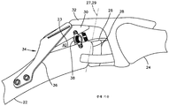
따라서, 계속 도 2를 참조하면, 종래의 TTA 시스템(34)은 전진된 경골 결절(30)과 경골체(23)의 고정을 제공하기 위해 일단부에서 경골(22)에 그리고 타단부에서 전진된 경골 결절(30)에 연결되는 뼈 플레이트(bone plate)(36); 및 뼈 플레이트(36)와는 별개이고, 슬개건(32)의 미골 방향으로(caudally) 지향되는 힘에 대항하여 경골 결절(30)과 경골체(23) 사이의 간극(40)을 유지시키기 위해 전진된 경골 결절(30)과 경골체(23) 사이에 배치되어 연결되는 케이지(cage) 형태의 스페이서(spacer)(38)를 포함한다.2, the conventional TTA system 34 is configured to move the tibial nodule 30 from one end to the tibia 22 and from the other end to the tibia 22, A bone plate 36 connected to the tibial nodule 30; And to maintain the gap 40 between the tibial nodule 30 and the tibia body 23 against a force that is separate from the bone plate 36 and directed caudally to the patella tendon 32 And a spacer 38 in the form of a cage disposed and connected between the tibial tubercle 30 and the tibia body 23.

개에 TTA 시술을 수행하기 위해 다양한 기기, 장치, 시스템 및 방법이 개발되었다. 그러나, 이러한 기기와 임플란트에 대한 개선이 여전히 요망된다.A variety of devices, devices, systems and methods have been developed to perform TTA procedures on dogs. However, improvements to such devices and implants are still desired.

본 발명은 전진된 결절을 경골체에 대해 전진된 위치에서 유지시키기 위한 TTA 시스템에 관한 것이다. 결절의 전진된 위치는 결절이 경골체와 일체일 때의 제1 위치에 대해 두개골 방향으로(cranially) 그리고 근위 방향으로(proximally) 이격된다. 일 실시예에서, TTA 시스템은 일반적으로 임플란트, 스페이서 및 스페이서 고정 부재를 포함한다. 임플란트는 전진된 결절을 전진된 위치에서 지지하도록 구성되는 근위 단부 부분(proximal end portion), 경골체에 부착되도록 구성되는 원위 단부 부분(distal end portion), 및 근위 단부 부분과 원위 단부 부분 사이에서 연장되는 중간 임플란트 부분을 한정하는 임플란트 본체를 포함한다. 중간 부분은 전진된 결절을 전진된 위치에서 유지시키기에 충분한 양 또는 거리로 근위 단부를 원위 단부 부분에 대해 두개골 방향으로 그리고 근위 방향으로 이격시키도록 형상화된다. 스페이서는 원위 단부 부분 및 근위 단부 부분이 경골체(23) 및 전진된 결절에 각각 부착된 때 전진된 결절과 경골체(23) 사이에 배치되는 간극 내에 끼워맞춤되도록 구성 및 크기 설정된다. 스페이서는 스페이서 본체를 포함하고, 스페이서 본체를 통해 연장되는 슬롯을 한정한다. 스페이서 고정 부재는 전진된 결절에 부착되도록 구성되는 제1 단부 부분, 경골체에 부착되도록 구성되는 제2 단부 부분, 및 제1 단부와 제2 단부 사이에서 연장되는 중간 고정 부분을 포함한다. 중간 고정 부분은 스페이서 고정 부재를 스페이서에 결합시키기 위해 슬롯 내에 적어도 부분적으로 수용되도록 구성 및 크기 설정된다.The present invention relates to a TTA system for maintaining an advanced nodule in an advanced position relative to a tibial body. The advanced position of the nodule is cranially and proximally spaced relative to the first position when the nodule is integral with the tibial body. In one embodiment, the TTA system generally comprises an implant, a spacer, and a spacer fixation member. The implant includes a proximal end portion configured to support the advanced nodule in an advanced position, a distal end portion configured to attach to the tibial body, and a proximal end portion extending between the proximal end portion and the distal end portion. And an implant body defining an intermediate implant portion. The middle portion is shaped to spaced the proximal end in the direction of the cranial and proximal directions relative to the distal end portion in an amount or distance sufficient to maintain the advanced nodule in the advanced position. The spacer is configured and sized to fit within a gap disposed between the advanced nodule and the tibia body 23 when the distal end portion and the proximal end portion are respectively attached to the tibia body 23 and the advanced nodule. The spacer includes a spacer body and defines a slot extending through the spacer body. The spacer fixation member includes a first end portion configured to attach to the advanced nodule, a second end portion configured to attach to the tibial body, and an intermediate fixation portion extending between the first end and the second end. The intermediate securing portion is configured and sized to be at least partially received within the slot for coupling the spacer securing member to the spacer.

본 발명은 또한 결절과 경골체 사이에 절골술이 이루어진 후에 결절을 제1 위치로부터 상기 경골체에 대해 전진된 위치로 전진시키도록 구성되는 TTA 전진 조립체에 관한 것이다. 일 실시예에서, TTA 전진 조립체는 경골체에 결합되도록 구성되는 전진 본체, 및 전진 본체에 이동 가능하게 결합되는 신연 아암(distraction arm)을 포함한다. 신연 아암은 결절에 결합되도록 구성되고, 신연 아암은 경골체에 대해 결절과 함께 병진이동하도록 구성되어 신연 아암이 전진 본체에 대해 사전결정된 거리만큼 이동하도록 한다. 사전결정된 거리만큼의 신연 아암의 병진이동은 전진 조립체로 하여금 결절이 제1 위치로부터 전진된 위치로 전진되었다는 표시를 제공하게 한다.The present invention also relates to a TTA advancing assembly configured to advance a nodule from a first position to an advanced position relative to the tibia body after osteotomy is performed between the nodule and the tibia body. In one embodiment, the TTA advance assembly includes a forward body configured to be coupled to the tibial body, and a distraction arm movably coupled to the forward body. The elongated arm is configured to engage the nodule and the elongated arm is configured to translationally move with the nodule relative to the tibial body so that the elongated arm moves a predetermined distance relative to the advancing body. Translational movement of the extensor arm by a predetermined distance causes the forwarding assembly to provide an indication that the nodule has advanced from the first position to the advanced position.

일 실시예에서, TTA 전진 조립체는 경골체에 결합되도록 구성되는 전진 본체, 및 전진 본체에 피벗 가능하게 결합되는 각도 조절 부재를 포함한다. 각도 조절 부재는 피벗 축을 중심으로 전진 본체에 대해 피벗하도록 구성되고, 각도 조절 부재는 절골술에 의해 형성되는 간극 내에 끼워맞춤되도록 구성되는 접촉 부재를 포함한다. 각도 조절 부재는 접촉 부재가 절골술 부위 내에 배치될 때 전진 본체가 절골술 부위에 대해 사전결정된 전진 각도로 배향되도록 전진 본체에 대해 피벗 가능하게 고정되도록 구성된다.In one embodiment, the TTA advance assembly includes a forward body configured to be coupled to the tibial body, and an angulation member pivotally coupled to the forward body. The angle adjusting member is configured to pivot relative to the advancing body about a pivot axis, and the angle adjusting member includes a contact member configured to fit within a gap formed by osteotomy. The angle adjusting member is configured to be pivotably fixed with respect to the advancing body such that the advancing body is oriented at a predetermined advance angle with respect to the osteotomy site when the contact member is disposed within the osteotomy site.

본 발명은 또한 결절과 경골체 사이에 절골술이 이루어진 후에 결절을 제1 위치로부터 경골체에 대해 전진된 위치로 전진시키기 위한 TTA 방법에 관한 것이다. 일 실시예에서, TTA 방법은 하기의 단계들 중 하나 이상을 포함한다: a) 전진 본체를, 전진 본체에 이동 가능하게 결합되고 전진 본체에 대해 병진이동하도록 구성되는 신연 아암을 통해, 결절에 결합시키는 단계; b) 전진 본체에 결합되는 접촉 부재를, 절골술 동안에 형성되어 결절과 경골체 사이에 배치되는 간극 내에, 배치하는 단계; c) 신연 아암을 전진 본체에 대해 이동시켜 결절을 제1 위치와 전진된 위치 사이에서 이동시키는 단계.The present invention also relates to a TTA method for advancing a nodule from a first position to an advanced position relative to the tibia after osteotomy is performed between the nodule and the tibia. In one embodiment, the TTA method comprises one or more of the following steps: a) bringing the advancement body into engagement with the nodule through an extensible arm that is movably coupled to the advancement body and configured to translate relative to the advancement body, ; b) disposing a contact member, coupled to the advancement body, within a gap formed during osteotomy and disposed between the tuberosity and the tibia body; c) moving the distracting arm relative to the advancing body to move the nodule between the first position and the advanced position.

전술한 개요뿐만 아니라 바람직한 실시예의 하기의 상세한 설명은, 첨부된 개략 도면과 함께 읽을 때 더 잘 이해된다. 본 발명을 예시하기 위해, 도면은 현재 바람직한 실시예를 도시한다. 그러나, 본 발명은 도면에 개시된 특정 수단으로 한정되지 않는다. 도면에서,
도 1은 개의 건강한 무릎을 예시하는 도면.
도 2는 예를 들어 무릎의 전십자 인대의 손상에 응하여, 도 1에 예시된 무릎 내에 이식된 종래의 경골 결절 전진 시스템의 측면도.
도 3은 스페이서, 스페이서 고정 부재 및 임플란트를 포함하는, 본 발명의 일 실시예에 따른 경골 결절 전진(TTA) 시스템의 적어도 일부의 사시도.
도 4a는 본 발명의 일 실시예에 따른 스페이서의 사시도.
도 4b는 도 4a에 도시된 스페이서의 정면도.
도 4c는 도 4a에 도시된 스페이서의 평면도.
도 4d는 단면선 4D-4D를 따라 취해진, 도 4a에 도시된 스페이서의 측단면도.
도 4e는 단면선 4D-4D를 따라 취해진, 도 4a에 도시된 스페이서의 단면도.
도 4f는 일 실시예에 따른 스페이서의 사시도.
도 4g는 다른 실시예에 따른 스페이서의 사시도.
도 4h는 다른 실시예에 따른 스페이서의 사시도.
도 4i는 다른 실시예에 따른 스페이서의 사시도.
도 4j는 다른 실시예에 따른 스페이서의 사시도.
도 4k는 다른 실시예에 따른 스페이서의 사시도.
도 4l은 다른 실시예에 따른 스페이서의 사시도.
도 5a는 도 3에 도시된 스페이서 고정 부재의 사시도.
도 5b는 다른 실시예에 따른 스페이서 고정 부재의 사시도.
도 6a는 다른 실시예에 따른 스페이서, 도 5b에 도시된 스페이서 고정 부재 및 체결구의 분해 사시도.
도 6b는 서로 연결된 도 6a에 도시된 스페이서, 스페이서 고정 부재 및 체결구의 사시도.
도 6c는 도 6a에 도시된 스페이서의 정면도.
도 7a는 일 실시예에 따른 스페이서 및 체결구의 사시도.
도 7b는 도 7a에 도시된 스페이서 및 체결구의 정면도.
도 8a는 경골체에 대한 결절의 전진을 안내하도록 구성되고 전진된 결절과 경골체에 결합된 안내 조립체, 도 3에 도시된 스페이서, 도 3에 도시된 스페이서 고정 부재, 및 도 3에 도시된 임플란트의 사시도.
도 8b는 도 8a에 도시된 안내 조립체의 사시도.
도 8c는 도 8a에 도시된 안내 조립체의 분해 사시도.
도 9는 경골체에 대한 결절의 길이방향 및 각도 전진을 결정하기 위한 공통 접선 방법의 개략도.
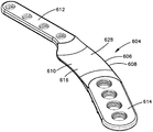
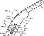
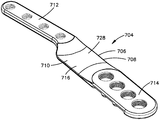
도 10a는 다른 실시예에 따른 임플란트의 상부 후방 사시도.
도 10b는 도 10a에 도시된 임플란트의 저부 전방 사시도.
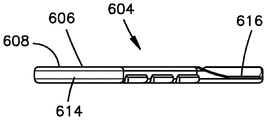
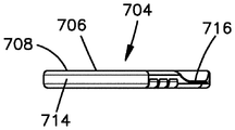
도 10c는 제1 배향에서의 도 10a에 도시된 임플란트의 좌측면도.
도 10d는 도 10a에 도시된 임플란트의 우측면도.
도 10e는 제2 배향에서의 도 10a에 도시된 임플란트의 좌측면도.
도 10f는 도 10a에 도시된 임플란트의 평면도.
도 10g는 도 10a에 도시된 임플란트의 저면도.
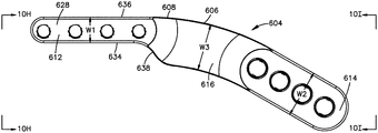
도 10h는 선 10H의 방향으로의 도 10f에 도시된 임플란트의 정면도.
도 10i는 선 10I의 방향으로의 도 10f에 도시된 임플란트의 배면도.
도 11a는 다른 실시예에 따른 임플란트의 상부 후방 사시도.
도 11b는 도 11a에 도시된 임플란트의 저부 전방 사시도.
도 11c는 제1 배향으로의 도 11a에 도시된 임플란트의 좌측면도.
도 11d는 도 11a에 도시된 임플란트의 우측면도.
도 11e는 제2 배향의 도 11a에 도시된 임플란트의 좌측면도.
도 11f는 도 11a에 도시된 임플란트의 평면도.
도 11g는 도 11a에 도시된 임플란트의 저면도.
도 11h는 선 11H의 방향으로의 도 11f에 도시된 임플란트의 정면도.
도 11i는 선 11I의 방향으로의 도 11f에 도시된 임플란트의 배면도.
BRIEF DESCRIPTION OF THE DRAWINGS The foregoing summary, as well as the following detailed description of the preferred embodiments, is better understood when read in conjunction with the accompanying schematic drawings. In order to illustrate the present invention, the drawings show currently preferred embodiments. However, the present invention is not limited to the specific means disclosed in the drawings. In the drawings,
Brief Description of the Drawings Figure 1 illustrates dogs healthy knees.
FIG. 2 is a side view of a conventional tibial tuberous advancement system implanted in the knee illustrated in FIG. 1 in response to, for example, damage of anterior cruciate ligament of the knee. FIG.
3 is a perspective view of at least a portion of a tibial tuberosity advance (TTA) system according to an embodiment of the invention, including a spacer, a spacer fixation member and an implant.
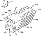
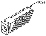
4A is a perspective view of a spacer according to one embodiment of the present invention.
Figure 4b is a front view of the spacer shown in Figure 4a;
Figure 4c is a top view of the spacer shown in Figure 4a.
4D is a side cross-sectional view of the spacer shown in Fig. 4A taken along section line 4D-4D; Fig.
Figure 4e is a cross-sectional view of the spacer shown in Figure 4a taken along section line 4D-4D.
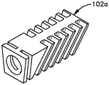
4f is a perspective view of a spacer according to one embodiment.
Figure 4G is a perspective view of a spacer according to another embodiment;
Figure 4h is a perspective view of a spacer according to another embodiment.
Figure 4i is a perspective view of a spacer according to another embodiment;
4J is a perspective view of a spacer according to another embodiment.
4k is a perspective view of a spacer according to another embodiment.
Figure 41 is a perspective view of a spacer according to another embodiment.
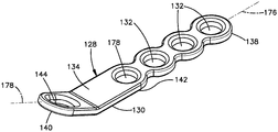
FIG. 5A is a perspective view of the spacer fixing member shown in FIG. 3; FIG.
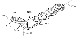
5B is a perspective view of a spacer fixing member according to another embodiment;
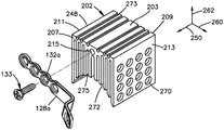
FIG. 6A is an exploded perspective view of a spacer according to another embodiment, the spacer fixing member and the fastener shown in FIG. 5B; FIG.
FIG. 6B is a perspective view of the spacer, spacer fixing member and fastener shown in FIG. 6A connected to each other. FIG.
Figure 6c is a front view of the spacer shown in Figure 6a.
7A is a perspective view of a spacer and fastener according to one embodiment.
7B is a front view of the spacer and fastener shown in Fig. 7A. Fig.
FIG. 8A shows a guide assembly constructed to guide advancement of a nodule to a tibial body and coupled to an advanced nodule and a tibial body, a spacer shown in FIG. 3, a spacer fixing member shown in FIG. 3, and an implant shown in FIG. FIG.
Figure 8b is a perspective view of the guide assembly shown in Figure 8a.
Figure 8c is an exploded perspective view of the guide assembly shown in Figure 8a.
9 is a schematic view of a common tangential method for determining longitudinal and angular advancement of a nodule relative to a tibial body;
10A is an upper rear perspective view of an implant according to another embodiment.
FIG. 10B is a bottom front perspective view of the implant shown in FIG. 10A. FIG.
Figure 10c is a left side view of the implant shown in Figure 10a in a first orientation.
Fig. 10D is a right side view of the implant shown in Fig. 10A. Fig.
Figure 10E is a left side view of the implant shown in Figure 10A in the second orientation.
Figure 10f is a top view of the implant shown in Figure 10a.
FIG. 10G is a bottom view of the implant shown in FIG. 10A. FIG.
Figure 10h is a front view of the implant shown in Figure 10f in the direction of line 10H.
Figure 10i is a rear view of the implant shown in Figure 10f in the direction of line 10I.
11A is an upper rear perspective view of an implant according to another embodiment.
FIG. 11B is a bottom front perspective view of the implant shown in FIG. 11A. FIG.
11C is a left side view of the implant shown in Fig. 11A in a first orientation. Fig.
Fig. 11D is a right side view of the implant shown in Fig. 11A. Fig.
Figure 11E is a left side view of the implant shown in Figure 11A of the second orientation.
11F is a plan view of the implant shown in FIG.
FIG. 11G is a bottom view of the implant shown in FIG. 11A. FIG.
11H is a front view of the implant shown in Fig. 11F in the direction of the line 11H.
FIG. 11I is a rear view of the implant shown in FIG. 11F in the direction of the line 11I.

오직 편의를 위해 하기의 설명에서 소정 용어가 사용되며, 제한적이지 않다. 단어 "우측", "좌측", "하부" 및 "상부"는 참조하는 도면에서 방향들을 가리킨다. 단어 "내측으로(medially)" 및 "외측으로(laterally)"는 신체를 통해, 예를 들어 개의 몸체의 머리부터 꼬리까지 연장되는 정중선(midline)을 향하는 방향 및 그로부터 멀어지는 방향을 각각 지칭한다. 단어 "근위" 및 "원위"는 외지(appendage)가 몸체의 나머지에 연결되는 곳을 향하는 방향 또는 그로부터 멀어지는 방향을 지칭한다. 단어 "전측(anterior)", "후측(posterior)", "등측(dorsal)", "복측(ventral)" 및 관련 단어 및/또는 문구는 참조하는 개의 몸체에서의 바람직한 위치 및 배향을 가리키며, 제한적인 것으로 의도되지 않는다. 예를 들어, "전측" 및 "후측"은 머리에 더 근접한 위치 및 꼬리에 더 근접한 위치를 지칭한다. 한편, "등측" 및 "복측"은 척주에 더 근접한 위치 및 배에 더 근접한 위치를 각각 지칭한다. 용어는 위에 열거된 단어, 그 파생어 및 유사한 의미의 단어들을 포함한다. 예를 들어, 도 8a에 도시된 바와 같이, 화살표(60)는 근위, 등측 또는 상향 방향을 나타낼 수 있다. 화살표(62)는 원위, 복측 또는 하향 방향을 나타낼 수 있다. 화살표(64)는 전방(front), 두측(cranial) 또는 전측 방향을 나타낼 수 있다. 화살표(66)는 미측(caudal), 후방 또는 후측 방향을 나타낼 수 있다. 화살표(68)는 외측 또는 멀어지는 방향을 나타낼 수 있다. 화살표(70)는 내측 또는 접근 방향을 나타낼 수 있다.For convenience only, certain terms are used in the following description and are not limiting. The words "right "," left ", "lower" and "upper" The words " medially "and" laterally "refer to directions through the body, for example, toward and away from the midline extending from the head to the tail of the dog. The terms "proximal" and "distal" refer to the direction toward or away from where the appendage is connected to the rest of the body. The words " anterior ", "posterior "," dorsal ", "ventral ", and related words and / or phrases refer to the preferred position and orientation in the referencing dog body, It is not intended to be. For example, "front" and "rear" refer to positions closer to the head and closer to the tail. On the other hand, "dorsal side" and "dorsal side" refer to positions closer to the abdomen and abdomen respectively. Terms include words listed above, derivatives thereof, and words of similar meaning. For example, as shown in FIG. 8A, the arrow 60 may indicate a proximal, an isometric or an upward direction. Arrow 62 may indicate a distal, a biceps, or a downward direction. The arrow 64 may indicate a front, a cranial or a frontal direction. The arrow 66 may indicate caudal, rearward, or rearward direction. The arrow 68 may indicate an outward or a departing direction. The arrow 70 may indicate an inward or approach direction.

도 3을 참조하면, 경골 결절 전진(TTA) 시스템(100)은 4족수의 전십자 인대-결손 무릎 관절을 안정시키도록 구성될 수 있다. 일 실시예에서, TTA 시스템(100)은 4족수를 위한, 경골 결절 전진(TTA) 임플란트와 같은 임플란트(104)를 포함한다. 임플란트(104)는 뼈 플레이트(108)와 같은 뼈 고정 부재(106)로서 구성될 수 있다. 도시된 실시예에서, 임플란트(104)는 근위 단부 부분(112), 대향하는 원위 단부 부분(114), 및 근위 단부 부분(112)과 원위 단부 부분(114) 사이에 배치되는 중간 임플란트 부분(116)을 포함하는 임플란트 본체(110)를 포함한다.Referring to FIG. 3, the tibial tuberosity advancing (TTA) system 100 may be configured to stabilize the four-tibial anterior cruciate ligament-deficient knee joint. In one embodiment, the TTA system 100 includes an implant 104, such as a tibial tuberosity forward (TTA) implant, for a quadruple prosthesis. The implant 104 may be configured as a bone fixation member 106, such as a bone plate 108. In the illustrated embodiment, the implant 104 includes a proximal end portion 112, an opposed distal end portion 114, and an intermediate implant portion 116 disposed between the proximal end portion 112 and the distal end portion 114 And an implant main body 110 including the implant main body 110.

임플란트 본체(110)의 근위 단부 부분(112)은 제1 위치로부터 전진된 위치로 경골체(23)에 대해 두개골 방향으로 일 방향으로 슬개건(32)(도 1에 도시됨)과 함께 전진되어 있는 결절(30)에 부착되도록 구성될 수 있다. 임플란트 본체(110)의 원위 단부 부분(114)은 경골체(23)에 부착되도록 구성될 수 있다. 슬개건(32)이 해부학적 부착 위치(43)에서 결절(30)에 부착된다는 것과, 결절(30)이 부착 위치(43)의 미측 위치에서 경골체(23)로부터 절제되어서 분리되어, 부착 위치(43)를 포함한 슬개건(32)이 분리된 결절(30)과 함께 제1 위치로부터 전진된 위치로 전진될 수 있는 것을 알아야 한다. 근위 단부 부분(112), 원위 단부 부분(114) 및 중간 임플란트 부분(116)은 집합적으로 일체식 구조체(monolithic structure)일 수 있다. 대안적으로, 근위 단부 부분(112), 원위 단부 부분(114) 및 중간 임플란트 부분(116)은 임플란트 본체(110)를 형성하도록 서로 연결되는 별개의 구성요소들일 수 있다.The proximal end portion 112 of the implant body 110 is advanced with the patella tendon 32 (shown in Figure 1) in one direction in the direction of the skull relative to the tibia body 23 from the first position to the advanced position The nodule 30 may be attached to the nodule 30. The distal end portion 114 of the implant body 110 may be configured to be attached to the tibia body 23. It is assumed that the patellar tendon 32 is attached to the nodule 30 at the anatomical attachment position 43 and that the nodule 30 is cut out from the tibial body 23 at the non- It is noted that the patellar tendon 32, including the nodule 43, may be advanced from the first position to the advanced position with the detached nodule 30. The proximal end portion 112, the distal end portion 114, and the intermediate implant portion 116 may be collectively a monolithic structure. Alternatively, the proximal end portion 112, the distal end portion 114, and the intermediate implant portion 116 may be separate components that are connected together to form the implant body 110.

근위 단부 부분(112)은 결절(30)에 대한 임플란트(104)의 부착을 용이하게 하기 위해 결절(30)의 내측 표면 또는 외측 표면에 정합하도록 윤곽 형성 및 구성될 수 있다. 또한, 근위 단부 부분(112)은 체결구 구멍들과 같은 하나 이상의 부착 위치들을 포함한다. 도시된 실시예에서, 임플란트 본체(110)의 근위 단부 부분(112)은 4개의 체결구 구멍(118a, 118b, 118c, 118d)들을 포함한다. 그러나, 근위 단부 부분(112)은 보다 많거나 보다 적은 체결구 구멍들을 포함할 수 있다. 체결구 구멍들의 특정 개수와 관계없이, 각각의 체결구 구멍(118a, 118b, 118c, 118d)은 임플란트 본체(110)를 통해 연장되고, 임플란트(104)를 결절(30)에 부착시킬 수 있는, 뼈 앵커(bone anchor)와 같은 체결구(120)를 수용하도록 구성 및 크기 설정된다.The proximal end portion 112 may be contoured and configured to conform to the inner or outer surface of the nodule 30 to facilitate attachment of the implant 104 to the nodule 30. [ The proximal end portion 112 also includes one or more attachment locations, such as fastener holes. In the illustrated embodiment, the proximal end portion 112 of the implant body 110 includes four fastener holes 118a, 118b, 118c, and 118d. However, the proximal end portion 112 may include more or fewer fastener holes. 118b, 118c, 118d extend through the implant body 110 and are capable of attaching the implant 104 to the nodule 30, regardless of the particular number of fastener apertures, And is configured and sized to receive fasteners 120, such as bone anchors.

적합한 체결구(120)의 예는 뼈 스크루, 못, 핀(pin), 및 임플란트(104)를 결절(30)에 부착시키도록 구성되는 임의의 다른 장치를 포함하지만 이로 한정되지 않는다. 예를 들어, 체결구 구멍(118a, 118b, 118c, 118d)은 뼈 스크루를 수용하도록 구성되는 나사형성된(threaded) 구멍일 수 있다. 또한, 체결구 구멍(118a, 118b, 118c, 118d)은 나사형성된 원추형 헤드를 갖는 뼈 스크루를 수용하도록 구성되는 원추형 나사 구멍일 수 있다. 체결구 구멍(118a, 118b, 118c, 118d)을 통한 체결구(120)의 삽입은 근위 단부 부분(112)이 결절(30)에 부착되게 한다. 체결구 구멍(118a, 118b, 118c, 118d)들은 서로 이격될 수 있고, 임플란트(104)가 전진된 결절(30)에 부착될 때 결절(30)의 연장 방향에 실질적으로 평행하게 연장되는 제1 길이방향 축(L1)을 따라 실질적으로 정렬될 수 있다. 일 실시예에서, 근위 단부 부분(112)은 길이방향 축(L1)을 따라 길 수 있다.Examples of suitable fasteners 120 include, but are not limited to, bone screws, nails, pins, and any other device configured to attach the implant 104 to the nodule 30. For example, fastener holes 118a, 118b, 118c, 118d may be threaded holes configured to receive a bone screw. Also, fastener holes 118a, 118b, 118c, 118d may be conical screw holes configured to receive bone screws having a threaded conical head. Insertion of the fastener 120 through the fastener holes 118a, 118b, 118c, 118d causes the proximal end portion 112 to attach to the nodule 30. The fastener holes 118a, 118b, 118c and 118d may be spaced apart from each other and may be spaced apart from each other by a first (not shown) extending substantially parallel to the extending direction of the nodule 30 when the implant 104 is attached to the advanced nodule 30. [ And may be substantially aligned along the longitudinal axis L1. In one embodiment, the proximal end portion 112 may be long along the longitudinal axis L1.

원위 단부 부분(114)은 경골체(23)에 대한 임플란트(104)의 부착을 용이하게 하기 위해 경골체(23)의 내측 표면 또는 외측 표면에 정합하도록 윤곽 형성 및 구성될 수 있다. 또한, 임플란트 본체(110)의 원위 단부 부분(114)은 체결구 구멍들과 같은 하나 이상의 앵커 위치들을 포함할 수 있다. 도시된 실시예에서, 원위 단부 부분(114)은 제1 체결구 구멍(122a) 및 제2 체결구 구멍(122b)을 포함한다. 체결구 구멍(122a, 122b)들 각각은 임플란트(104)를 경골체(23)에 부착시킬 수 있는, 뼈 앵커와 같은 체결구(124)를 수용하도록 구성 및 크기 설정될 수 있다.Distal end portion 114 may be contoured and configured to conform to an inner or outer surface of tibia body 23 to facilitate attachment of implant 104 to tibia body 23. [ The distal end portion 114 of the implant body 110 may also include one or more anchor locations, such as fastener holes. In the illustrated embodiment, the distal end portion 114 includes a first fastening hole 122a and a second fastening hole 122b. Each of the fastener holes 122a and 122b may be configured and sized to receive a fastener 124, such as a bone anchor, that may attach the implant 104 to the tibia body 23. [

적합한 체결구(124)의 예는 뼈 스크루, 못, 핀, 및 임플란트(104)를 경골체(23)에 부착시키도록 구성되는 임의의 다른 장치를 포함하지만 이로 한정되지 않는다. 체결구 구멍(122a, 122b)을 통한 체결구(124)의 삽입은 원위 단부 부분(114)이 경골체(23)에 부착되게 한다. 체결구 구멍(122a, 122b)들은 서로 이격되고 제2 길이방향 축(L2)을 따라 실질적으로 정렬될 수 있다. 일 실시예에서, 원위 단부 부분(114)은 제2 길이방향 축(L2)을 따라 길 수 있다. 제2 길이방향 축(L2)은 제1 길이방향 축(L1)으로부터 각도 오프셋될 수 있다.Examples of suitable fasteners 124 include, but are not limited to, bone screws, nails, pins, and any other device configured to attach the implant 104 to the tibia body 23. Insertion of the fastener 124 through the fastener holes 122a, 122b causes the distal end portion 114 to be attached to the tibia body 23. The fastener holes 122a, 122b may be spaced apart from one another and substantially aligned along the second longitudinal axis L2. In one embodiment, the distal end portion 114 may be long along the second longitudinal axis L2. The second longitudinal axis L2 may be angularly offset from the first longitudinal axis L1.

임플란트 본체(110)의 중간 임플란트 부분(116)은 제2 길이방향 축(L2)을 따라 길 수 있다. 대안적으로, 중간 임플란트 부분(116)은 제2 길이방향 축(L2)으로부터 각도 오프셋된 축을 따라 길 수 있다. 도면은 중간 임플란트 부분(116)에서 체결구 구멍과 같은 부착 위치를 도시하지 않지만, 중간 임플란트 부분(116)이 하나 이상의 체결구 구멍들 또는 임의의 다른 적합한 부착 특징부를 포함할 수 있다는 것이 구상된다. 중간 임플란트 부분(116)은 근위 단부 부분(112)과 원위 단부 부분(114) 사이에서 연장되고, 결절(30)을 전진된 위치에서 유지시키기에 충분한 양 또는 거리로 근위 단부 부분(112)을 원위 단부 부분(114)에 대해 두개골 방향으로 이격시키도록 형상화된다.The intermediate implant portion 116 of the implant body 110 may be long along the second longitudinal axis L2. Alternatively, the intermediate implant portion 116 may be long along an axis offset from the second longitudinal axis L2. It is contemplated that the figure does not show an attachment location such as a fastener hole in the intermediate implant portion 116, but the intermediate implant portion 116 may include one or more fastener holes or any other suitable attachment feature. The intermediate implant portion 116 extends between the proximal end portion 112 and the distal end portion 114 and extends the proximal end portion 112 in a distal or proximal direction with an amount or distance sufficient to maintain the nodal 30 in the advanced position And is spaced apart in the direction of the skull relative to the end portion 114.

TTA 시스템(100)은 결절(23)이 전진된 위치에 있을 때 경골체(23)와 결절(30) 사이의 거리를 유지시키도록 구성되는 스페이서(102)를 추가로 포함할 수 있다. 스페이서(102)는 경골체(23)와 전진된 결절(30) 사이에 한정되는 절골술 간극(40) 내에 적어도 부분적으로 끼워맞춤되도록 구성 및 크기 설정될 수 있다. 도시된 실시예에서, 스페이서(102)는 상세히 후술되는 바와 같은 케이지(cage)(126)로서 구성될 수 있다.The TTA system 100 may further include a spacer 102 configured to maintain a distance between the tibial body 23 and the nodule 30 when the nodule 23 is in the advanced position. The spacer 102 can be configured and sized to at least partially fit within the osteotomy gap 40 defined between the tibial bone body 23 and the advanced nodule 30. [ In the illustrated embodiment, the spacer 102 may be configured as a cage 126 as described in detail below.

스페이서(102) 외에도, TTA 시스템(100)은 스페이서(102)를 경골체(23)와 전진된 결절(30)에 결합시켜 스페이서(102)를 절골술 간극(40) 내에 고정시키도록 구성되는 스페이서 고정 부재(128)를 포함할 수 있다. 아래에서 상세히 논의되는 바와 같이, 스페이서 고정 부재(128)는 뼈 플레이트(130)로서 구성될 수 있다. 뼈 플레이트(130)의 적어도 일부분은 스페이서(102)를 통해 삽입되도록 구성 및 크기 설정될 수 있다. 스페이서 고정 부재(128)는 플레이트 본체로 또한 지칭되는 본체(134)를 포함한다. 스페이서 고정 부재(128)의 본체(134)는 길 수 있고, 제1 단부 부분(138), 제2 단부 부분(140), 및 제1 단부 부분(138)과 제2 단부 부분(140) 사이에 배치되는 중간 고정 부분(142)(도 5a에 도시됨)을 한정할 수 있다.In addition to the spacer 102, the TTA system 100 also includes a spacer fixture (not shown) configured to couple the spacer 102 to the tibia body 23 and the advanced nodule 30 to secure the spacer 102 within the osteotomy gap 40 Member 128 as shown in FIG. As discussed in detail below, the spacer fixation member 128 may be configured as a bone plate 130. At least a portion of the bone plate 130 may be configured and sized to be inserted through the spacer 102. The spacer fastening member 128 includes a body 134, also referred to as a plate body. The body 134 of the spacer fixation member 128 can be elongate and has a first end portion 138, a second end portion 140, and a second end portion 140 between the first end portion 138 and the second end portion 140 The intermediate fixing portion 142 (shown in FIG. 5A) can be defined.

제1 단부 부분(138)은 전진된 결절(30)에 부착되도록 구성될 수 있다. 이를 위해, 제1 단부 부분(138)은 전진된 결절(30)의 외측 표면 또는 내측 표면에 정합하도록 윤곽 형성 및 구성될 수 있으며, 체결구 구멍(132)들과 같은 하나 이상의 부착 위치들을 포함할 수 있다. 체결구 구멍(132)은 스페이서 고정 부재(128)를 전진된 결절(30)에 부착시킬 수 있는, 뼈 앵커와 같은 체결구(136)를 수용하도록 구성 및 크기 형성될 수 있다. 적합한 체결구(136)는 뼈 스크루, 못, 핀, 또는 제1 단부 부분(138)을 전진된 결절(30)에 부착시킬 수 있는 임의의 다른 체결구(136)를 포함하지만 이로 한정되지 않는다.The first end portion 138 may be configured to attach to the advanced nodule 30. To this end, the first end portion 138 may be contoured and configured to mate with an outer or inner surface of the advanced nodule 30, and may include one or more attachment locations, such as fastener holes 132 . The fastener holes 132 can be configured and sized to receive fasteners 136, such as bone anchors, that can attach the spacer fastening member 128 to the advanced nodule 30. Suitable fasteners 136 include, but are not limited to, bone screws, nails, pins, or any other fasteners 136 that can attach the first end portion 138 to the advanced nodule 30.

본체(134)의 제2 단부 부분(140)은 경골체(23)에 부착되도록 구성된다. 이를 위해, 제2 단부 부분(140)은 경골체(23)의 외측 표면 또는 내측 표면에 정합하도록 윤곽 형성 및 구성될 수 있으며, 체결구 구멍(144)들과 같은 하나 이상의 부착 위치들을 포함할 수 있다. 도시된 실시예에서, 제2 단부 부분(140)은 단지 하나의 체결구 구멍(144)만을 포함하지만; 제2 단부 부분(140)이 하나 초과의 체결구 구멍(144)을 한정할 수 있다는 것이 구상된다. 체결구 구멍(144)은 뼈 앵커와 같은 체결구(136)를 수용하도록 구성 및 크기 설정될 수 있다. 체결구(136)의 예는 뼈 스크루, 못, 핀, 또는 제2 단부 부분(140)을 경골체(23)에 부착시킬 수 있는 임의의 다른 장치를 포함하지만 이로 한정되지 않는다.The second end portion 140 of the body 134 is configured to attach to the tibia body 23. To this end, the second end portion 140 may be contoured and configured to mate with the outer or inner surface of the tibia body 23, and may include one or more attachment locations, such as fastener holes 144 have. In the illustrated embodiment, the second end portion 140 includes only one fastener hole 144; It is envisioned that the second end portion 140 may define more than one fastener hole 144. The fastener holes 144 may be configured and sized to receive fasteners 136, such as bone anchors. Examples of fasteners 136 include, but are not limited to, bone screws, nails, pins, or any other device that is capable of attaching second end portion 140 to tibia body 23.

중간 고정 부분(142)은, 제1 단부 부분(138)이 전진된 결절(30)에 부착되고 제2 단부가 경골체(23)에 부착될 때, 스페이서(102)를 절골술 간극(40) 내에 고정시키기 위해 스페이서(102)의 개구, 예를 들어 슬롯을 통해 삽입되도록 구성된다. 도시된 실시예에서, 중간 고정 부분(142)은 상세히 후술되는 바와 같이 실질적으로 평면인 구성을 가질 수 있다. 제1 단부 부분(138), 제2 단부 부분(140) 및 중간 고정 부분(142)은 일체식 구조체일 수 있다. 대안적으로, 제1 단부 부분(138), 제2 단부 부분(140) 및 중간 고정 부분(142)은 서로 연결되는 별개의 구성요소들일 수 있다. 중간 고정 부분(142)은 아래에서 논의되는 바와 같이 스페이서(102)의 슬롯 내에 끼워맞춤되도록 구성되는 실질적으로 평면인 구성을 한정할 수 있다.The intermediate fixation portion 142 is configured to allow the spacer 102 to be inserted into the osteotomy gap 40 when the first end portion 138 is attached to the advanced nodule 30 and the second end is attached to the tibia body 23. [ For example, through the slot of the spacer 102 to fix it. In the illustrated embodiment, the intermediate fastening portion 142 may have a substantially planar configuration as will be described in detail below. The first end portion 138, the second end portion 140, and the intermediate fixation portion 142 may be an integral structure. Alternatively, the first end portion 138, the second end portion 140, and the intermediate fixed portion 142 may be separate components that are connected to each other. The intermediate securing portion 142 may define a substantially planar configuration configured to fit within a slot of the spacer 102, as discussed below.

도 4a 내지 도 4e를 참조하면, 스페이서(102)는 절골술 간극(40) 내에 끼워맞춤되도록 구성 및 크기 설정되는 스페이서 본체(148)를 포함할 수 있다. 스페이서 본체(148)는 길이방향(150)을 따라 길 수 있고, 제1 길이방향 단부(152) 및 길이방향(150)을 따라 제1 길이방향 단부(152)로부터 이격되는 제2 길이방향 단부(154)를 한정할 수 있다. 또한, 스페이서 본체(148)는 제1 측방향 단부(156) 및 측방향(160)을 따라 제1 측방향 단부(156)로부터 이격되는 제2 측방향 단부(158)를 한정한다. 측방향(160)은 길이방향(150)에 실질적으로 수직이다.4A-4E, the spacer 102 may include a spacer body 148 configured and dimensioned to fit within the osteotomy gap 40. The spacer body 148 may extend along the longitudinal direction 150 and may have a first longitudinal end 152 and a second longitudinal end 152 spaced from the first longitudinal end 152 along the longitudinal direction 150 154 may be defined. The spacer body 148 also defines a first lateral end 156 that is spaced apart from the first lateral end 156 along the first lateral end 156 and the lateral direction 160. The lateral direction 160 is substantially perpendicular to the longitudinal direction 150.

구체적으로, 스페이서 본체(148)는 제1 횡방향 단부(164) 및 횡방향(162)을 따라 제1 횡방향 단부(164)로부터 이격되는 제2 횡방향 단부(166)를 구비할 수 있다. 횡방향(162)은 길이방향(150)과 측방향(160)에 실질적으로 수직이다. 도시된 실시예에서, 스페이서 본체(148)는 그의 폭이 횡방향(162)으로 증가하도록 실질적으로 부분적인 웨지(wedge) 형상을 한정할 수 있다. 스페이서 본체(148)의 폭은 제1 측방향 단부(156)와 제2 측방향 단부(158) 사이에서 한정된다. 도시된 실시예에서, 스페이서 본체(148)는 제2 횡방향 단부(166)에서의 제2 폭(W2)보다 큰 제1 횡방향 단부(164)에서의 제1 폭(W1)을 한정할 수 있다. 스페이서 본체(148)의 웨지-형상은 절골술 간극(40)이 실질적으로 웨지 형상을 갖기 때문에 절골술 간극(40) 내에서의 스페이서(102)의 삽입과 위치 설정을 용이하게 한다.Specifically, the spacer body 148 may have a first transverse end 164 and a second transverse end 166 spaced from the first transverse end 164 along the transverse direction 162. The lateral direction 162 is substantially perpendicular to the longitudinal direction 150 and the lateral direction 160. In the illustrated embodiment, the spacer body 148 may define a substantially partial wedge shape such that its width increases in the transverse direction 162. The width of the spacer body 148 is defined between the first lateral end 156 and the second lateral end 158. The spacer body 148 can define a first width W1 at the first transverse end 164 that is greater than the second width W2 at the second transverse end 166. In the illustrated embodiment, have. The wedge-shape of the spacer body 148 facilitates the insertion and positioning of the spacer 102 within the osteotomy gap 40 because the osteotomy gap 40 has a substantially wedge shape.

스페이서(102)는 제1 길이방향 단부(152)로부터 제2 길이방향 단부(154)까지 스페이서 본체(148)를 통해 연장되는 개구(168)를 추가로 포함할 수 있다. 따라서, 개구(168)는 길이방향(150)을 따라 길 수 있다. 개구(168)는 구멍으로서 구성될 수 있고, 뼈 이식편(bone graft), 또는 뼈 성장을 촉진시킬 수 있는 임의의 다른 천연 또는 합성 재료를 수용하도록 구성된다. 그러나, 개구(168)는 반드시 뼈 이식편 또는 임의의 다른 뼈 성장제로 충전될 필요는 없다. 개구(168)는 스페이서(102)가 절골술 간극(40) 내에 배치될 때 자연 뼈 성장을 허용하기 위한 개방 공간을 제공한다. 자연 뼈 성장 동안에, 자연 뼈가 성장할 수 있고, 스페이서(102)가 절골술 간극(40) 내에 배치될 때 개구(168)의 적어도 일부분을 충전할 수 있다.The spacer 102 may further include an opening 168 extending through the spacer body 148 from the first longitudinal end 152 to the second longitudinal end 154. Thus, the opening 168 may be long along the longitudinal direction 150. The openings 168 can be configured as apertures and configured to receive a bone graft, or any other natural or synthetic material that can promote bone growth. However, opening 168 need not necessarily be filled with bone graft or any other bone growth agent. The opening 168 provides an open space for allowing natural bone growth when the spacer 102 is disposed within the osteotomy gap 40. During natural bone growth, the natural bone can grow and fill at least a portion of the opening 168 when the spacer 102 is disposed within the osteotomy gap 40.

스페이서(102)는 제1 측방향 단부(156)로부터 제2 측방향 단부(158)까지 스페이서 본체(148)를 통해 연장되는 제1 슬롯(170)을 추가로 한정한다. 따라서, 제1 슬롯(170)은 측방향(160)을 따라 길 수 있다. 제1 슬롯(170)은 제2 길이방향 단부(154)보다 제1 길이방향 단부(152)에 더 근접하게 위치되고, 스페이서(102)를 뼈 고정 부재(128)에 결합시키기 위해 스페이서 고정 부재(128)의 적어도 일부분을 수용하도록 구성 및 크기 설정된다. 도시된 실시예에서, 제1 슬롯(170)은 길이방향(150)에 실질적으로 수직인 평면을 한정할 수 있다. 중간 고정 부재(142)는 스페이서(102)의 슬롯(170) 내에 끼워맞춤되도록 구성됨으로써 스페이서(102)를 스페이서 고정 부재(128)에 결합시키도록 실질적으로 평면인 구성을 가질 수 있다.The spacer 102 further defines a first slot 170 extending through the spacer body 148 from the first lateral end 156 to the second lateral end 158. Thus, the first slot 170 may be long along the lateral direction 160. [ The first slot 170 is positioned closer to the first longitudinal end 152 than the second longitudinal end 154 and is spaced from the spacer fixation member 128 to engage the spacer 102 with the bone fixation member 128. [ 128, respectively. In the illustrated embodiment, the first slot 170 may define a plane that is substantially perpendicular to the longitudinal direction 150. The intermediate fixation member 142 may be configured to be fit within the slot 170 of the spacer 102 to thereby have a substantially planar configuration to couple the spacer 102 to the spacer fixation member 128.

제1 슬롯(170)에 더하여, 스페이서(102)는 제1 측방향 단부(156)로부터 제2 측방향 단부(158)까지 스페이서 본체(148)를 통해 연장되는 적어도 하나의 제2 슬롯(172)을 포함한다. 도시된 실시예에서, 스페이서(102)는 길이방향(150)을 따라 서로 이격되는 복수의 제2 슬롯(172)들을 한정한다. 제2 슬롯(172)들 중 적어도 하나는 제1 길이방향 단부(152)보다 제2 길이방향 단부(154)에 더 근접하게 위치된다. 제2 슬롯(172)들 각각은 길이방향(150)에 대해 비스듬한 각도로 배향되는 평면을 한정한다. 사용 시, 제2 슬롯(172)은 뼈 이식편, 또는 뼈 성장을 촉진시킬 수 있는 임의의 다른 천연 또는 합성 재료를 수용하도록 구성된다. 제2 슬롯(172)은 반드시 뼈 이식편 또는 임의의 다른 뼈 성장제로 충전될 필요는 없다. 오히려, 제2 슬롯(172)은 스페이서(102)가 절골술 간극(40) 내에 배치될 때 자연 뼈 성장을 허용하기 위한 개방 공간을 제공한다.In addition to the first slot 170, the spacer 102 includes at least one second slot 172 extending through the spacer body 148 from the first lateral end 156 to the second lateral end 158, . In the illustrated embodiment, the spacers 102 define a plurality of second slots 172 that are spaced apart from each other along the lengthwise direction 150. At least one of the second slots 172 is positioned closer to the second longitudinal end 154 than the first longitudinal end 152. Each of the second slots 172 defines a plane oriented at an oblique angle with respect to the longitudinal direction 150. In use, the second slot 172 is configured to receive a bone graft, or any other natural or synthetic material that can promote bone growth. The second slot 172 does not necessarily have to be filled with bone grafts or any other bone growth agent. Rather, the second slot 172 provides an open space for allowing natural bone growth when the spacer 102 is disposed within the osteotomy gap 40.

자연 뼈 성장 동안에, 자연 뼈가 성장할 수 있고, 스페이서(102)가 절골술 간극(40) 내에 배치될 때 제2 슬롯(172)의 적어도 일부분을 충전할 수 있다. 제2 슬롯(172)은 또한 스페이서(102)를 절단하는 것을 용이하게 한다. 아래에서 상세히 논의되는 바와 같이, 스페이서(102)는 길이방향(150)을 따른 제1 길이방향 단부(152)와 제2 길이방향 단부(154) 사이의 거리에 의해 한정되는 스페이서의 길이(174)를 감소시키도록 절단될 수 있다. 작동 시, 스페이서(102)의 길이(174)는 스페이서(102)가 절골술 간극(40) 내에 적당하게 끼워맞춤될 수 있도록 단축되어야 할 수 있다. 이를 위해, 제2 슬롯(172)들 각각은 톱과 같은 절단 공구의 적어도 일부분을 수용하도록 구성 및 크기 설정될 수 있다. 작동 시, 톱은 슬롯(172)들 중 하나를 통해 삽입되어 스페이서(102)를 절단함으로써 스페이서의 길이(174)를 단축시킬 수 있다. 스페이서(102)는 또한 톱과 같은 절단 공구로 절단될 수 있는 재료로 부분적으로 또는 완전히 제조될 수 있다. 스페이서(102)는 또한 복수의 타인(tine)(173)들을 포함할 수 있다. 타인(173)들 중 적어도 일부는 2개의 슬롯(172)들 사이에 배치된다. 타인(173)들은 실질적으로 평면인 구성을 가질 수 있다. 도시된 실시예에서, 타인(173)은 길이방향(150)에 대해 비스듬히 기울어져 있다. 스페이서(102)는 폴리에테르에테르케톤(PEEK)과 같은 임의의 적합한 생체적합성 재료로 부분적으로 또는 완전히 제조될 수 있다.During natural bone growth, the natural bone can grow and fill at least a portion of the second slot 172 when the spacer 102 is placed in the osteotomy gap 40. The second slot 172 also facilitates cutting the spacer 102. The spacer 102 has a length 174 of the spacer defined by the distance between the first longitudinal end 152 and the second longitudinal end 154 along the longitudinal direction 150, As shown in FIG. In operation, the length 174 of the spacer 102 may have to be shortened so that the spacer 102 can be properly fitted within the osteotomy gap 40. To this end, each of the second slots 172 may be configured and sized to receive at least a portion of a cutting tool, such as a saw. In operation, the saw may be inserted through one of the slots 172 to shorten the length 174 of the spacer by cutting the spacer 102. The spacer 102 may also be partially or fully made of a material that can be cut with a cutting tool such as a saw. The spacer 102 may also include a plurality of tines 173. At least some of the tines 173 are disposed between the two slots 172. The tines 173 may have a substantially planar configuration. In the illustrated embodiment, the tines 173 are obliquely inclined with respect to the longitudinal direction 150. Spacer 102 may be partially or fully fabricated with any suitable biocompatible material, such as polyetheretherketone (PEEK).

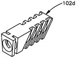
도 4f 내지 도 4l을 참조하면, TTA 시스템(100)은 상이한 크기의 스페이서들을 포함하는 키트의 일부일 수 있다. 따라서, 키트는 상이한 길이, 높이 및 폭을 갖는 스페이서들을 포함할 수 있다. 예를 들어, 키트는 스페이서(102a, 102b, 102c, 102d, 102e, 102f, 102g)들을 포함할 수 있다. 이들의 치수를 제외하고는, 스페이서(102a, 102b, 102c, 102d, 102e)들은 도 4a 내지 도 4e에 관하여 전술된 스페이서(102)와 실질적으로 유사하다. 따라서, 스페이서(102g, 102f)들은 도 4a 내지 도 4e에 관하여 전술된 스페이서(102)와 실질적으로 유사하지만; 크기 제한으로 인해, 스페이서(102g, 102f)들은 스페이서(102)의 개구(168)와 같은 개구를 포함하지 않는다. 또한, 스페이서(102g, 102f)들은 도 4a 내지 도 4e에 관하여 전술된 스페이서(102)보다 작다.Referring to Figures 4F-4L, the TTA system 100 may be part of a kit comprising spacers of different sizes. Thus, the kit may include spacers having different lengths, heights, and widths. For example, the kit may include spacers 102a, 102b, 102c, 102d, 102e, 102f, 102g. Except for their dimensions, the spacers 102a, 102b, 102c, 102d, 102e are substantially similar to the spacers 102 described above with respect to Figs. 4A-4E. Thus, the spacers 102g, 102f are substantially similar to the spacers 102 described above with respect to Figures 4A-4E; Due to size limitations, spacers 102g and 102f do not include openings, such as openings 168 in spacers 102. Also, the spacers 102g, 102f are smaller than the spacers 102 described above with respect to Figs. 4A-4E.

도 5a를 참조하면, 스페이서 고정 부재(128)는 스페이서(102)를 절골술 간극(40) 내에 고정시키기 위해 스페이서(102)를 경골체(23)와 전진된 결절(30)에 결합시키도록 구성된다. 도시된 실시예에서, 스페이서 고정 부재(128)는 뼈 플레이트(130)로서 구성될 수 있고, 스페이서(102)의 제1 슬롯(170) 내에 부분적으로 끼워맞춤되도록 구성 및 크기 설정되는 본체(134)를 포함한다. 본체(134)는 또한 플레이트 본체로 또한 지칭될 수 있다. 또한, 본체(134)는 제1 단부 부분(138), 제2 단부 부분(140), 및 제1 단부 부분(138)과 제2 단부 부분(140) 사이에 배치되는 중간 고정 부분(142)을 한정할 수 있다.5A, the spacer fixing member 128 is configured to couple the spacer 102 to the tibia body 23 and the advanced nodule 30 to fix the spacer 102 in the osteotomy gap 40 . The spacer fixation member 128 can be configured as a bone plate 130 and includes a body 134 configured and dimensioned to be partially fit within a first slot 170 of the spacer 102, . The body 134 may also be referred to as a plate body. The body 134 also includes a first end portion 138, a second end portion 140 and an intermediate fixed portion 142 disposed between the first end portion 138 and the second end portion 140 Can be limited.

제1 단부 부분(138)은 길이방향 축(176)을 따라 길 수 있고, 전진된 결절(30)에 부착되도록 구성될 수 있다. 스페이서 고정 부재(128)와 전진된 결절(30) 사이의 부착을 용이하게 하기 위해, 제1 단부 부분(138)은 전진된 결절(30)의 외측 표면 또는 내측 표면에 정합하도록 윤곽 형성 및 구성될 수 있으며, 체결구 구멍(132)들과 같은 하나 이상의 부착 위치들을 포함할 수 있다. 체결구 구멍(132)은 위에서 논의된 바와 같이 체결구(136)를 수용하도록 구성 및 크기 설정될 수 있다. 도시된 실시예에서, 제1 단부 부분(138)은 복수의 체결구 구멍(132)들을 한정한다. 복수의 체결구 구멍(132)들은 사용자가 제1 단부 부분(138)을 따라 상이한 부착 위치들에서 스페이서 고정 부재(128)를 전진된 결절(30)에 부착시키게 한다. 그러나, 제1 단부 부분(138)이 단지 하나의 체결구 구멍(132)만을 한정할 수 있다는 것이 고려된다.The first end portion 138 may be long along the longitudinal axis 176 and may be configured to attach to the advanced nodule 30. The first end portion 138 may be contoured and configured to conform to the outer or inner surface of the advanced nodule 30 to facilitate attachment between the spacer fixation member 128 and the advanced nodule 30 And may include one or more attachment locations, such as fastener holes 132. The fastener holes 132 can be configured and sized to accommodate fasteners 136 as discussed above. In the illustrated embodiment, the first end portion 138 defines a plurality of fastener holes 132. The plurality of fastener holes 132 allow the user to attach the spacer fastening member 128 to the advanced nodule 30 at different attachment locations along the first end portion 138. It is contemplated, however, that the first end portion 138 may define only one fastener aperture 132.

중간 고정 부분(142)은 길이방향 축(176)을 따라 길 수 있고, 뼈 앵커와 같은 체결구를 수용하도록 구성되는 적어도 하나의 체결구 구멍(178)을 한정할 수 있다. 필요하다면, 스페이서 고정 부재(128)는 이를 단축시키도록 절단될 수 있고, 스페이서 고정 부재(128)를 경골체(23)에 결합시키기 위해 체결구가 체결구 구멍(178)을 통해 경골체(23) 내로 삽입될 수 있다. 위에서 논의된 바와 같이, 중간 고정 부분(142)의 적어도 일부가 스페이서 고정 부재(128)를 스페이서(102)에 결합시키기 위해 제1 슬롯(170) 내로 삽입되도록 구성될 수 있다.The intermediate fixation portion 142 may be elongate along the longitudinal axis 176 and define at least one fastener hole 178 configured to receive a fastener such as a bone anchor. If necessary, the spacer fixing member 128 may be cut so as to shorten it, and a fastener may be inserted through the fastener hole 178 to connect the spacer fixing member 128 to the tibia body 23, ). ≪ / RTI > As discussed above, at least a portion of the intermediate securing portion 142 may be configured to be inserted into the first slot 170 to engage the spacer securing member 128 with the spacer 102.

제2 단부 부분(140)은 길이방향 축(176)에 대해 각도 오프셋되는 길이방향 축(178)을 따라 길 수 있다. 일 실시예에서, 제2 단부 부분(140)은 경골체(23)의 외측 또는 내측 표면에 정합하도록 윤곽 형성 및 구성될 수 있다. 스페이서 고정 부재(128)와 경골체(23) 사이의 부착을 용이하게 하기 위해, 제2 단부 부분(140)은 체결구 구멍(144)들과 같은 하나 이상의 부착 위치들을 포함할 수 있다. 도시된 실시예에서, 제2 단부 부분(140)은 단지 하나의 체결구 구멍(144)만을 한정한다. 그러나, 제2 단부 부분(140)은 하나 초과의 체결구 구멍(144)을 포함할 수 있다. 위에서 논의된 바와 같이, 스페이서 고정 부재(128)를 경골체(23)에 결합시키기 위해 체결구가 체결구 구멍(144)을 통해 경골체(23) 내로 삽입될 수 있다.The second end portion 140 may be long along a longitudinal axis 178 that is angularly offset relative to the longitudinal axis 176. In one embodiment, the second end portion 140 may be contoured and configured to conform to the outer or inner surface of the tibial body 23. [ The second end portion 140 may include one or more attachment locations, such as fastener holes 144, to facilitate attachment between the spacer securing member 128 and the tibia body 23. [ In the illustrated embodiment, the second end portion 140 defines only one fastener hole 144. However, the second end portion 140 may include more than one fastener hole 144. As discussed above, a fastener can be inserted through the fastener hole 144 into the tibia body 23 to engage the spacer fastening member 128 with the tibia body 23.

도 5b를 참조하면, 스페이서 고정 부재(128a)의 다른 실시예가 도 5a에 관하여 전술된 스페이서 고정 부재(128)와 실질적으로 유사하다. 그러나, 이 실시예에서, 제2 단부 부분(140a)은 중간 고정 부분(142a)에 연결되고 길이방향 축(177a)을 따라 긴 제1 섹션(141a)을 포함한다. 길이방향 축(177a)은 길이방향 축(176a)에 실질적으로 수직일 수 있다. 제2 단부 부분(140a)은 길이방향 축(179a)을 따라 긴 제2 섹션(143a)을 추가로 포함한다. 길이방향 축(179a)은 길이방향 축(177a)과 길이방향 축(176a)에 대해 각도 오프셋될 수 있다. 작동 시, 스페이서 고정 부재(128a)의 제2 단부 부분(140a)은 경골체(23)의 외측 표면 또는 내측 표면에 대한 스페이서 고정 부재(128a)의 연결을 용이하게 하기 위해 그 외측 또는 내측 표면에 정합하도록 윤곽 형성 및 구성될 수 있다.Referring to Fig. 5B, another embodiment of the spacer fixing member 128a is substantially similar to the spacer fixing member 128 described above with respect to Fig. 5A. However, in this embodiment, the second end portion 140a includes a first section 141a connected to the intermediate fixed portion 142a and extending along the longitudinal axis 177a. The longitudinal axis 177a may be substantially perpendicular to the longitudinal axis 176a. The second end portion 140a further includes a second section 143a along the longitudinal axis 179a. The longitudinal axis 179a may be angularly offset relative to the longitudinal axis 177a and the longitudinal axis 176a. In operation, the second end portion 140a of the spacer securing member 128a is attached to the outer or inner surface of the tibial body 23 to facilitate connection of the spacer securing member 128a to the outer or inner surface May be contoured and configured to match.

도 6a 내지 도 6c를 참조하면, 다른 실시예에 따른 스페이서(202)가 결절(30)을 경골체(23)에 대해 전진된 위치에서 유지시키기 위해 절골술 간극(40) 내에 위치될 수 있다. 스페이서(202)는 폴리에테르에테르케톤(PEEK) 또는 임의의 다른 적합한 재료로 부분적으로 또는 완전히 제조될 수 있는 스페이서 본체(248)를 포함할 수 있다. 스페이서 본체(248)는 상부 표면(203) 및 반대편의 하부 표면(205)을 한정한다. 상부 표면(203)은 횡방향(262)을 따라 하부 표면으로부터 이격된다. 스페이서 본체(248)는 전방 표면(207) 및 반대편의 후방 표면(209)을 포함할 수 있다. 후방 표면(209)은 길이방향(250)을 따라 전방 표면(207)으로부터 이격될 수 있다. 스페이서 본체(248)는 제1 측방향 표면(211)과 제2 측방향 표면(213)을 한정할 수 있다. 제2 측방향 표면(213)은 측방향(260)을 따라 제1 측방향 표면(211)으로부터 이격될 수 있다.6A to 6C, a spacer 202 according to another embodiment may be placed in the osteotomy gap 40 to hold the nodule 30 in the advanced position with respect to the tibia body 23. As shown in Fig. The spacer 202 may include a spacer body 248 that may be partially or fully fabricated with polyetheretherketone (PEEK) or any other suitable material. The spacer body 248 defines an upper surface 203 and an opposite lower surface 205. The upper surface 203 is spaced from the lower surface along the transverse direction 262. The spacer body 248 may include a front surface 207 and an opposite rear surface 209. The rear surface 209 may be spaced from the front surface 207 along the longitudinal direction 250. The spacer body 248 may define a first lateral surface 211 and a second lateral surface 213. The second lateral surface 213 may be spaced from the first lateral surface 211 along the lateral direction 260.

스페이서(202)는 스페이서 본체(248)의 하부 표면 내로 연장되는 복수의 슬롯(272)들을 추가로 한정한다. 슬롯(272)들은 측방향(260)을 따라 서로 이격될 수 있다. 슬롯(272)들 각각은 횡방향(262)을 따라 길 수 있다. 또한, 슬롯(272)들 각각은 전방 표면(207)으로부터 후방 표면(209)까지 스페이서 본체(248)를 통해 연장될 수 있다. 스페이서(202)가 절골술 간극(40) 내에 배치될 때, 슬롯(272)은 뼈 성장을 허용하기 위한 개방 공간을 제공한다. 슬롯(272)은 또한 스페이서의 길이를 단축시키기 위하여 스페이서(202)의 절단을 용이하게 한다. 게다가, 임의의 적합한 천연 또는 합성 뼈 성장 재료가 스페이서(202)가 절골술 간극(40) 내에 배치될 때 뼈 성장을 촉진시키기 위해 슬롯(272) 내에 배치될 수 있다. 슬롯(272)은 또한 스페이서(202)의 절단을 용이하게 한다. 위에서 논의된 바와 같이, 스페이서(202)는 절골술 간극(40) 내에 적당하게 끼워맞춤되도록 필요하다면 절단될 수 있다. 예를 들어, 스페이서(202)를 절단하여 스페이서(202)를 측방향(260)을 따라 단축시키기 위해 절단 공구가 슬롯(272)들 중 하나를 통해 삽입될 수 있다.The spacer 202 further defines a plurality of slots 272 that extend into the lower surface of the spacer body 248. The slots 272 may be spaced apart from one another along the lateral direction 260. Each of the slots 272 may be long along the transverse direction 262. Further, each of the slots 272 may extend through the spacer body 248 from the front surface 207 to the rear surface 209. When the spacer 202 is disposed within the osteotomy gap 40, the slot 272 provides an open space for allowing bone growth. The slot 272 also facilitates cutting of the spacer 202 to shorten the length of the spacer. In addition, any suitable natural or synthetic bone growth material may be disposed within the slot 272 to promote bone growth when the spacer 202 is disposed within the osteotomy gap 40. The slot 272 also facilitates cutting of the spacer 202. As discussed above, the spacer 202 may be severed if necessary to fit properly within the osteotomy gap 40. For example, a cutting tool may be inserted through one of the slots 272 to cut the spacer 202 and shorten the spacer 202 along the lateral direction 260. [

스페이서 본체(248)는 측방향(260)을 따라 서로 이격되는 복수의 탄성 타인(275)들을 포함할 수 있다. 각각의 탄성 타인(275)은 2개의 슬롯(272)들 사이에 배치된다. 탄성 타인(275)은, 스페이서(202)가 절골술 간극(40) 내에 배치될 때 스페이서 본체(248)가 측방향(260)을 따라 압축되게 하여 스페이서 본체(248)의 적어도 일부분이 절골술 간극(40)의 형상에 정합하게 한다. 탄성 타인(275)들은 또한 아치형 저부 하부 표면(205)을 한정하도록 상이한 길이들을 가질 수 있다. 특히, 탄성 타인(275)들은 스페이서(202)가 절골술 간극(40) 내에 배치될 때 절골술 간극(40)의 형상에 정합하도록 타인(275)들이 서로에 대항하여 압축되게 하는 오목한 하부 표면(205)을 한정할 수 있다.The spacer body 248 may include a plurality of elastic tines 275 spaced apart from one another along the lateral direction 260. Each elastic tine 275 is disposed between two slots 272. The elastic tines 275 allow the spacer body 248 to compress along the lateral direction 260 when the spacer 202 is disposed within the osteotomy gap 40 so that at least a portion of the spacer body 248 is in contact with the osteotomy gap 40 ). The elastic tines 275 may also have different lengths to define the arcuate bottom and bottom surface 205. In particular, the elastic tines 275 have a concave lower surface 205 that allows the tines 275 to compress against each other to match the shape of the osteotomy gap 40 when the spacer 202 is disposed within the osteotomy gap 40. [ .

스페이서(202)는 측방향(260)을 따라 스페이서 본체(248) 내로의 하나 이상의 구멍(270)들을 추가로 포함할 수 있다. 도시된 실시예에서, 구멍(270)은 측방향(260)을 따라 제1 측방향 표면(211)으로부터 제2 측방향 표면(213)까지 스페이서 본체(248)를 통해 연장된다. 스페이서(202)가 절골술 간극(40) 내에 배치될 때, 구멍(270)은 뼈 성장을 촉진시키기 위한 개방 공간을 제공한다. 구멍(270)에 더하여, 스페이서(202)는 상부 표면(203) 내로 연장되는 하나 이상의 리지(ridge)(273)들을 한정할 수 있다. 도시된 실시예에서, 리지(273)들은 측방향(260)을 따라 서로 이격될 수 있다. 리지(273)는 길이방향(250)을 따라 길 수 있다. 작동 시, 톱과 같은 절단 공구가 리지(273)들 중 하나 내로 삽입되어 스페이서(202)를 절단할 수 있다. 따라서, 리지(273)들은 스페이서(202)의 절단을 용이하게 한다. 리지(273)는 또한 스페이서(202)가 휘어지도록 허용한다.The spacer 202 may further include one or more apertures 270 into the spacer body 248 along the lateral direction 260. In the illustrated embodiment, the aperture 270 extends through the spacer body 248 from the first lateral surface 211 to the second lateral surface 213 along the lateral direction 260. When the spacer 202 is disposed within the osteotomy gap 40, the hole 270 provides an open space for promoting bone growth. In addition to the apertures 270, the spacers 202 may define one or more ridges 273 that extend into the top surface 203. In the illustrated embodiment, the ridges 273 may be spaced apart from one another along the lateral direction 260. The ridges 273 may be long along the longitudinal direction 250. In operation, a cutting tool, such as a saw, may be inserted into one of the ridges 273 to cut the spacer 202. Thus, the ridges 273 facilitate the cutting of the spacers 202. The ridge 273 also allows the spacer 202 to bend.

스페이서(202)는 체결구(133)를 수용하도록 구성 및 크기 설정되는 적어도 하나의 체결구 구멍(215)들을 추가로 한정할 수 있다. 체결구(133)는 스페이서 고정 부재(128a)(또는 임의의 다른 스페이서 고정 부재)를 스페이서(202)에 결합시키도록 구성될 수 있다. 도시된 실시예에서, 체결구(133)는 스크루로서 구성되고, 체결구 구멍(215)은 나사형성된 구멍일 수 있다. 그러나, 체결구(133)가 못, 핀, 또는 스페이서 고정 부재(128a)를 스페이서(202)에 결합시키도록 구성되는 임의의 다른 장치로서 구성될 수 있다는 것이 구상된다. 스페이서 고정 부재(128a)를 스페이서(202)에 결합시키기 위해, 체결구(133)가 스페이서 고정 부재(128a)의 체결구 구멍(132a)들 중 하나를 통해 체결구 구멍(215) 내로 삽입될 수 있다. 위에서 논의된 바와 같이, 스페이서 고정 부재(128a)는 또한 체결구를 통해 전진된 결절(30)과 경골체(23)에 결합될 수 있다.The spacer 202 may further define at least one fastener hole 215 that is configured and sized to receive the fastener 133. The fastener 133 can be configured to couple the spacer fastening member 128a (or any other spacer fastening member) to the spacer 202. [ In the illustrated embodiment, fastener 133 is configured as a screw, and fastener hole 215 may be a threaded hole. It is contemplated, however, that the fastener 133 may be configured as any other device configured to couple the nail, pin, or spacer securing member 128a to the spacer 202. [ The fastener 133 can be inserted into the fastener hole 215 through one of the fastener holes 132a of the spacer fastening member 128a in order to engage the spacer fastening member 128a with the spacer 202. [ have. As discussed above, the spacer fixation member 128a can also be coupled to the tongue body 23 and the nodule 30 advanced through the fastener.

도 7a 및 도 7b를 참조하면, 다른 실시예에 따른 스페이서(302)가 결절(30)을 경골체(23)에 대해 전진된 위치에서 유지시키기 위해 절골술 간극(40) 내에 위치될 수 있다. 스페이서(302)는 스페이서(202)와 실질적으로 유사하다. 그러나, 이 실시예에서, 탄성 타인(375)들은 실질적으로 유사하거나 동일한 길이를 가지며, 따라서 오목한 하부 표면(305)을 한정하지 않는다. 대신에, 하부 표면(205)은 실질적으로 평면인 구성을 가질 수 있다. 또한, 스페이서(302)는 하부 표면(305) 내로 연장되는, 부분 구멍들과 같은 리세스(recess)(371)들을 추가로 한정할 수 있다. 리세스(371)들은 길이방향(350)을 따라 서로 이격될 수 있다. 작동 시, 리세스(371)는 스페이서(302)가 절골술 간극(40) 내에 배치될 때 뼈 성장을 촉진시키기 위한 개방 공간을 제공한다.7A and 7B, a spacer 302 according to another embodiment may be placed in the osteotomy gap 40 to maintain the nodule 30 in the advanced position relative to the tibial body 23. [ The spacers 302 are substantially similar to the spacers 202. However, in this embodiment, the elastic tines 375 have substantially similar or identical length, and thus do not define a concave lower surface 305. Alternatively, the lower surface 205 may have a substantially planar configuration. Further, the spacer 302 may further define recesses 371, such as partial holes, that extend into the bottom surface 305. The recesses 371 may be spaced apart along the longitudinal direction 350. In operation, the recess 371 provides an open space for promoting bone growth when the spacer 302 is disposed within the osteotomy gap 40.

도 8a 내지 도 8c를 참조하면, TTA 시스템(100)은 또한 경골체(23)에 대한 결절(30)의 전진을 안내하도록 구성되는 TTA 전진 조립체(400)를 포함할 수 있다. 아래에서 상세히 논의되는 바와 같이, 전진 조립체(400)는 결절(30)을 경골체(23)에 대해 전진시키기 위해 사용될 수 있고, 상세히 후술된다. 전진 조립체(400)는 임플란트(104)에 결합되도록 구성되는 전진 부재(402)를 포함하고, 임플란트는 이어서 결절(30)에 결합된다. 구체적으로, 전진 부재(402)는 임플란트(104)의 근위 단부 부분(112)에 결합될 수 있다. 도시된 실시예에서, 전진 부재(402)는 체결구 구멍(118a)에 의해 한정되는 부착 위치에서 임플란트(104)에 결합될 수 있다. 임플란트(104)는 결절(30)과 전진 부재(402)에 부착될 수 있다. 따라서, 전진 부재(402)는 결절(30)을 경골체(23)에 대해 (임플란트(104)를 통해) 전진시키도록 조작될 수 있다.8A-8C, the TTA system 100 may also include a TTA advancing assembly 400 configured to guide the advancement of the nodule 30 relative to the tibial body 23. As shown in FIG. As discussed in detail below, the advancing assembly 400 can be used to advance the nodule 30 relative to the tibial sphincter 23 and will be described in detail below. Advance assembly 400 includes an advancing member 402 configured to be coupled to implant 104 and the implant is then coupled to nodule 30. Specifically, the advancing member 402 can be coupled to the proximal end portion 112 of the implant 104. In the illustrated embodiment, the advancing member 402 can be coupled to the implant 104 at an attachment location defined by a fastener hole 118a. The implant 104 may be attached to the nodule 30 and the advancing member 402. Thus, the advancing member 402 can be manipulated to advance the nodal 30 against the tibial body 23 (via the implant 104).

계속해서 도 8a 내지 도 8c를 참조하면, 전진 부재(402)는 지그(jig)(403)로서 구성될 수 있고, 전진 본체(404)를 포함할 수 있다. 전진 본체(404)는 지그 본체 또는 프레임으로서 구성될 수 있다. 그의 구성에 상관없이, 전진 본체(404)는 변위 눈금자(displacement scale)(408)를 안정적으로 수용하도록 구성되는 변위 눈금자 구멍(406)과 같은 부착 위치를 한정한다. 따라서, 전진 조립체(400)는 경골체(23)에 대한 결절(30)의 길이방향 변위를 측정하기 위해 사용될 수 있는 변위 눈금자(408)를 포함할 수 있다. 변위 눈금자(408)는 변위 눈금자 구멍(408)을 통해 전진 본체(404)에 제거 가능하게 부착될 수 있다.8A-8C, the advancing member 402 may be configured as a jig 403 and may include a forward body 404. The advancing body 404 may be configured as a jig body or a frame. Regardless of its configuration, the advancing body 404 defines an attachment position, such as a displacement ruler hole 406, which is configured to stably receive a displacement scale 408. Thus, the advancing assembly 400 can include a displacement ruler 408 that can be used to measure the longitudinal displacement of the nodule 30 relative to the tibia body 23. [ The displacement scale 408 may be removably attached to the advancement body 404 via a displacement ruler aperture 408.

변위 눈금자(408)는 경골체(23)에 대한 결절(30)의 길이방향 변위를 측정하기 위해 사용될 수 있는 측정 마킹(412)들을 포함하는 본체(410)를 포함한다. 본체(410)에 더하여, 변위 눈금자(408)는 본체(410)로부터 돌출되는 연결 부재(414)를 포함한다. 연결 부재(414)는 실질적으로 원통형인 본체로서 구성될 수 있고, 변위 눈금자 구멍(406) 내에 제거 가능하게 배치될 수 있다. 연결 부재(414)는 연결 본체(416), 및 연결 본체(416) 주위에 배치되는 링(418)을 포함할 수 있다. 연결 본체(416)는 변위 눈금자 구멍(406) 내에 적어도 부분적으로 수용되도록 구성 및 크기 설정된다. 연결 부재(414)가 변위 눈금자 구멍(406) 내에 적어도 부분적으로 배치될 때, 링(418)은 변위 눈금자 구멍(406)을 한정하는 전진 본체(404)의 내측 표면에 인접함으로써, 연결 부재(414)와 전진 본체(404) 사이의 마찰 끼워맞춤 연결을 확립한다.The displacement scale 408 includes a body 410 that includes measurement markings 412 that can be used to measure the longitudinal displacement of the nodule 30 relative to the tibial body 23. In addition to the body 410, the displacement scale 408 includes a connecting member 414 that projects from the body 410. The connecting member 414 may be configured as a substantially cylindrical body and may be removably disposed within the displacement ruler hole 406. [ The linking member 414 may include a linking body 416 and a ring 418 disposed about the linking body 416. The connection body 416 is configured and sized to be at least partially received within the displacement ruler bore 406. When the connecting member 414 is at least partially disposed within the displacement ruler hole 406, the ring 418 abuts the inner surface of the advancing body 404 defining the displacement ruler hole 406, And the forward main body 404, as shown in Fig.

전진 조립체(400)는 전진 조립체(400)가 결절(30)과 경골체(23)에 결합될 때 결절(30)을 경골체(23)에 대해 이동시키도록 구성되는 길이방향 신연 기구(distraction mechanism)(419)를 추가로 포함할 수 있다. 도시된 실시예에서, 신연 기구(419)는 전진 본체(404)에 이동 가능하게 결합되는 신연 아암(420), 및 노브(knob)(426)와 같은 액추에이터(424)를 포함할 수 있다. 작동 시, 신연 아암(420)은 액추에이터(424)의 작동 시 전진 본체(404)에 대해 이동하도록 구성된다. 따라서, 액추에이터(424)의 작동은 신연 아암(420)이 길이방향 신연축(422)을 따라 전진 본체(404)에 대해 이동하게 한다. 작동 시, 전진 본체(404)에 대한 신연 아암(420)의 이동은 전진 조립체(400)가 결절(30)과 경골체(23)에 결합될 때 결절(30)이 경골체(23)에 대해 이동하게 한다.The advancing assembly 400 includes a longitudinal distraction mechanism configured to move the nodule 30 relative to the tibia body 23 when the advancing assembly 400 is coupled to the nodule 30 and the tibia body 23. [ ) ≪ / RTI > The elongating mechanism 419 may include an elongate arm 420 movably coupled to the advancing body 404 and an actuator 424 such as a knob 426. In this embodiment, In operation, the extensor arm 420 is configured to move relative to the advancing body 404 upon actuation of the actuator 424. [ Thus, actuation of the actuator 424 causes the extensor arm 420 to move relative to the advancing body 404 along the longitudinally extending spur 422. The movement of the extensor arm 420 relative to the advancing body 404 causes the nodule 30 to move relative to the tibia body 23 when the advancing assembly 400 is engaged with the nodule 30 and tibia body 23, .

위에서 논의된 바와 같이, 액추에이터(424)는 노브(426)로서 구성될 수 있다. 노브(426)는 노브 본체(428)를 포함할 수 있고, 신연 아암(420)의 적어도 일부분을 수용하도록 구성 및 크기 설정되는 나사형성된 구멍(430)을 한정할 수 있다. 나사형성된 구멍(430)은 노브 본체(428)를 통해 연장될 수 있다. 신연 아암(420)은, 신연 아암(420) 주위로의 노브(426)의 회전이 신연 아암(420)을 길이방향 신연축(422)을 따라 노브(426)에 대해 이동시키도록, 나사형성된 구멍(430) 주위에 배치되는 내부 나삿니들과 맞물리도록 구성되는 외부 나삿니(432)들을 포함할 수 있다. 따라서, 신연 아암(420)은 노브(426)의 회전 시 전진 본체(404)에 대해 이동하도록 구성될 수 있다. 신연 아암(420)이 전진 본체(404)에 대해 길이방향으로 이동할 수 있는 반면에, 노브(426)는 전진 본체(404)에 대해 길이방향으로 고정된다.As discussed above, the actuator 424 may be configured as a knob 426. The knob 426 may include a knob body 428 and may define a threaded hole 430 that is configured and sized to receive at least a portion of the extensor arm 420. The threaded hole 430 may extend through the knob body 428. The elongate arm 420 is configured such that the rotation of the knob 426 about the elongate arm 420 moves the elongate arm 420 along the longitudinal elongate spindle 422 with respect to the knob 426, And external threads 432 that are configured to engage internal threads disposed about the perimeter 430. Referring now to FIG. Thus, the extensible arm 420 can be configured to move relative to the advancing body 404 upon rotation of the knob 426. [ The knob 426 is fixed in the longitudinal direction relative to the advancing body 404 while the extender arm 420 can move longitudinally relative to the advancing body 404.

전진 부재(402)는, 서로 이격되어 노브 채널(434)을 한정하는 제1 부착 프롱(prong)(436) 및 제2 부착 프롱(438)을 포함할 수 있다. 제1 부착 프롱(436)과 제2 부착 프롱(438)은 전진 본체(404)로부터 돌출될 수 있다. 노브 채널(434)은 노브(426)가 노브 채널(434) 내에서 회전하게 하면서 노브(426)를 전진 부재(402)에 대해 길이방향으로 고정시키도록 노브(426)를 수용하도록 구성 및 크기 설정될 수 있다. 노브(426)는 길이방향 신연 축(422)을 중심으로 회전하도록 구성될 수 있다. 제1 부착 프롱(436)은 신연 아암(420)의 적어도 일부분을 수용하도록 구성되는 신연 아암 구멍(440)을 한정한다. 신연 아암(402)은 신연 아암 구멍(440)을 통해 활주할 수 있다. 제2 부착 프롱(438)은 신연 아암(420)의 적어도 일부분을 수용하도록 구성 및 크기 설정되는 신연 아암 채널(442)을 한정할 수 있다. 신연 아암(420)은 신연 아암 채널(442)을 통해 활주할 수 있다. 제2 프롱(438)은 또한 제1 정지 부재(444)를 한정할 수 있고, 제1 정지 부재는 전진 부재(402)에 대한 신연 아암(420)의 길이방향 이동을 제한하기 위해 신연 아암(420)의 제2 정지 부재(446)에 인접하도록 구성된다.The advancing member 402 may include a first attachment prong 436 and a second attachment prong 438 that are spaced apart from each other to define a knob channel 434. [ The first attachment prong 436 and the second attachment prong 438 may protrude from the advance body 404. The knob channel 434 is configured and sized to accommodate the knob 426 to longitudinally lock the knob 426 against the advancing member 402 while causing the knob 426 to rotate within the knob channel 434. [ . The knob 426 may be configured to rotate about the longitudinal extension axis 422. The first attachment prong 436 defines an extensible arm hole 440 configured to receive at least a portion of the extensible arm 420. The extensible arm 402 can slide through the extensible arm hole 440. The second attachment prong 438 may define an elongate arm channel 442 that is configured and sized to receive at least a portion of the elongate arm 420. The extensible arm 420 can slide through the extensible arm channel 442. The second prongs 438 may also define a first stop member 444 that extends to the elongate arms 420 to limit longitudinal movement of the elongate arms 420 relative to the advancing members 402. [ The second stopping member 446 of the second embodiment.

신연 아암(420)은 제1 단부(448)와 제2 단부(450)를 한정할 수 있고, 제2 단부(450)는 이어서 예시된 실시예에 도시된 바와 같이 제2 정지 부재(446)를 한정할 수 있다. 제1 단부(448)는 길이방향 신연축(422)을 따라 제2 단부(450)로부터 이격될 수 있다. 신연 아암(420)은 신연 아암(420)의 제2 단부(450)를 통해 연장되는 드릴 안내 구멍(452)을 추가로 한정할 수 있다. 드릴 안내 구멍(452)은 슬리브로서 구성될 수 있는 드릴 안내체(454)를 수용하도록 구성 및 크기 설정될 수 있다. 드릴 안내체(454)는 서로 이격되는 제1 단부(460) 및 제2 단부(462)를 한정하는 드릴 안내체 본체(456)를 포함할 수 있다.The elongate arm 420 may define a first end 448 and a second end 450 and the second end 450 may define a second stop member 446 as shown in the following illustrative embodiment Can be limited. The first end 448 may be spaced from the second end 450 along the longitudinally extending spur 422. The elongated arm 420 may further define a drill guide hole 452 extending through the second end 450 of the elongate arm 420. [ The drill guide hole 452 can be configured and sized to accommodate a drill guide body 454 that can be configured as a sleeve. The drill guide body 454 may include a drill guide body 456 defining a first end 460 and a second end 462 that are spaced apart from one another.

제2 단부(462)는 드릴 안내체(454)를 임플란트(104)에 결합시키기 위해 임플란트(404)의 나사형성된 체결구 구멍(118a)(도 3)과 맞물리도록 구성 및 크기 설정되는 나사형성된 팁(464)을 한정할 수 있다. 나사형성된 팁(464)은 절두 원추형 형상을 가질 수 있다. 드릴 안내체(454)는 제1 단부(460)와 제2 단부(462) 사이에서 드릴 안내체 본체(456)를 통해 연장되는 드릴 안내 개구(458)를 한정할 수 있다. 드릴 안내 개구(458)는 드릴 비트 또는 와이어(466)와 같은 임시 고정 부재를 수용하도록 구성 및 크기 설정될 수 있다. 와이어(466)는 키르쉬너(Kirschner) 와이어일 수 있고, 드릴 안내체(454)가 신연 아암(420)에 결합될 때 전진 조립체(400)를 결절(30)에 결합시키기 위해 드릴 안내 개구(458)를 통해 결절(30) 내로 삽입되도록 구성된다.The second end 462 is configured and dimensioned to engage with the threaded fastener holes 118a (FIG. 3) of the implant 404 to engage the drill guide body 454 with the implant 104 Lt; RTI ID = 0.0 > 464 < / RTI > The threaded tip 464 may have a frusto-conical shape. The drill guide 454 may define a drill guide opening 458 extending through the drill guide body 456 between the first end 460 and the second end 462. The drill guide opening 458 may be configured and sized to receive a temporary fixation member such as a drill bit or wire 466. [ The wire 466 may be a Kirschner wire and may include a drill guide aperture 458 to couple the advance assembly 400 to the nodule 30 when the drill guide 454 is coupled to the extensor arm 420. [ Into the nodule 30 through the nipple 30.

전진 조립체(400)는 전진 조립체(400)가 경골체(23)와 결절(30)에 결합될 때 경골체(23)에 대한 결절(30)의 각도 위치를 조절하도록 구성되는 각도 조절 기구(468)를 추가로 포함할 수 있다. 도시된 실시예에서, 각도 조절 기구(468)는 전진 본체(404)에 이동 가능하게 결합되는 각도 조절 부재(470)를 포함할 수 있다. 구체적으로, 각도 조절 부재(470)는 피벗 축(R)을 따라 한정되는 부착 위치를 중심으로 회전하도록 구성된다. 특히, 전진 부재(402)는 구멍(472)과 같은 부착 위치를 한정한다. 구멍(472)은 피벗 축(R)을 따라 전진 본체(404)를 통해 연장되고 회전 액추에이터(474)의 적어도 일부분을 수용하도록 구성되어, 회전 액추에이터(474)가 구멍(472) 내에서 피벗 축(R)을 중심으로 회전하도록 구성되게 한다.The advancing assembly 400 includes an angle adjusting mechanism 468 configured to adjust the angular position of the nodule 30 relative to the tibia body 23 when the advancing assembly 400 is engaged with the tibia body 23 and the nodule 30. [ ). ≪ / RTI > In the illustrated embodiment, the angle adjusting mechanism 468 may include an angle adjusting member 470 movably coupled to the advancing body 404. Specifically, the angle-regulating member 470 is configured to rotate about an attachment position defined along the pivot axis R. [ In particular, the advancing member 402 defines an attachment position, such as a hole 472. The aperture 472 extends through the advancement body 404 along the pivot axis R and is configured to receive at least a portion of the rotary actuator 474 such that the rotary actuator 474 is positioned within the aperture 472 in the pivot axis R to rotate.

회전 액추에이터(474)는 노브(473)로서 구성될 수 있고, 각도 조절 부재(470)의 부착 부재(480)와 맞물리도록 구성되는 부착 부재(476)를 포함하여, 회전 액추에이터(474)를 각도 조절 부재(470)에 결합시킨다. 부착 부재(476)는 외부 나사형성된 본체(478)로서 구성될 수 있고, 부착 부재(480)는 나사형성된 구멍(482)으로서 구성될 수 있다. 나사형성된 구멍(482)은 회전 액추에이터(474)를 각도 조절 부재(470)에 결합시키기 위해 외부 나사형성된 본체(478)와 맞물리도록 구성될 수 있다. 각도 조절 부재(470)는 각도 눈금자로서 구성될 수 있다.The rotary actuator 474 may be configured as a knob 473 and includes an attachment member 476 configured to mate with the attachment member 480 of the angle adjustment member 470 so that the rotary actuator 474 can be angled Member 470 as shown in FIG. The attachment member 476 may be configured as an externally threaded body 478 and the attachment member 480 may be configured as a threaded hole 482. The threaded hole 482 may be configured to engage an externally threaded body 478 to engage the rotational actuator 474 with the angle adjusting member 470. The angle adjusting member 470 may be configured as an angle ruler.

각도 조절 부재(470)는 또한 회전 액추에이터(474)를 조임으로써 각도 본체(404)에 대해 각도 고정될 수 있다. 예를 들어, 피벗 축(R)을 중심으로 하는 제1 방향으로의 회전 액추에이터(474)의 회전은 외부 나사형성된 본체(478)를 나사형성된 구멍(482) 내에서 조임으로써, 각도 조절 부재(470)를 전진 본체(404)에 대해 각도 고정시킨다. 역으로, 피벗 축(R)을 중심으로 하는 제2 방향(제1 방향의 반대 방향)으로의 회전 액추에이터(474)의 회전은 나사형성된 구멍(482) 내에 배치된 외부 나사형성된 본체(478)를 풀리게 함으로써, 각도 조절 부재(470)가 전진 본체(404)에 대해 피벗 축(R)을 중심으로 회전하게 한다.The angle regulating member 470 can also be angularly fixed relative to the angle body 404 by tightening the rotary actuator 474. For example, rotation of the rotary actuator 474 in the first direction about the pivot axis R may be achieved by tightening the externally threaded body 478 within the threaded hole 482, ) To the forward main body (404). Conversely, the rotation of the rotary actuator 474 in a second direction (opposite to the first direction) about the pivot axis R causes an externally threaded body 478 disposed within the threaded hole 482 Thereby causing the angle regulating member 470 to rotate about the pivot axis R with respect to the advancing body 404.

각도 조절 부재(470)는 길이방향(486)을 따라 긴 각도 눈금자 본체(484)를 포함한다. 각도 눈금자 본체(484)는 실질적으로 평명인 구성을 가질 수 있고, 제1 눈금자 단부(488)와 제2 눈금자 단부(490)를 한정한다. 제2 눈금자 단부(490)는 길이방향(486)을 따라 제1 눈금자 단부(488)로부터 이격된다. 나사형성된 구멍(482)은 제1 눈금자 단부(488)에 또는 그에 근접하게 위치될 수 있다. 각도 조절 부재(470)는 측방향(492)을 따라 각도 눈금자 본체(484)로부터 돌출되는 접촉 부재(494)를 추가로 포함할 수 있다. 측방향(492)은 길이방향(486)에 실질적으로 수직일 수 있다. 접촉 부재(494)는 실질적으로 평면인 구성을 가질 수 있고, 절골술 간극(40) 내에 배치되도록 구성 및 크기 설정된다. 접촉 부재(494)는 절골술 간극(40) 내에 위치될 때 경골 결절(30), 경골체(23), 또는 둘 모두와 접촉하기에 적합한 브레이스(brace), 블레이드(blade) 또는 임의의 장치일 수 있다.The angle adjusting member 470 includes a long angle ruler body 484 along the length direction 486. The angular ruler body 484 may have a substantially planar configuration and define a first ruler end 488 and a second ruler end 490. The second graduation end 490 is spaced from the first graduation end 488 along the longitudinal direction 486. The threaded hole 482 may be located at or near the first scale end 488. The angle adjusting member 470 may further include a contact member 494 projecting from the angular ruler body 484 along the lateral direction 492. The lateral direction 492 may be substantially perpendicular to the longitudinal direction 486. The contact member 494 can have a substantially planar configuration and is configured and dimensioned to be disposed within the osteotomy gap 40. Contact member 494 may be a brace, blade or any device suitable for contacting tibial tuberosity 30, tibia body 23, or both when positioned within osteotomy gap 40 have.

각도 조절 부재(470)는 제2 눈금자 단부(490)를 따라 배치되는 각도 마킹(491)들을 추가로 포함할 수 있다. 각도 마킹(491)들은 사용자가 전진 본체(404)에 대한 접촉 부재(494)의 각도 배향을 결정하는 데 도움을 준다. 특히, 각도 마킹(491)들은 중심이 부착 부재(480)에 의해 한정되는 원호를 따라 배치된다. 각도 조절 부재(470)는 각도 마킹(491)들에 인접하게 배치되는 복수의 개구 또는 리세스(496)들을 추가로 포함한다. 개구(496)들은 중심이 부착 부재(480)에 의해 한정되는 원호를 따라 서로 이격된다. 개구(496)들 각각은 측방향(492)으로 전진 본체(404)로부터 돌출되는 지주(post)(498)를 수용하도록 구성 및 크기 설정된다. 지주(498)와 개구(496)들 각각 사이의 맞물림은 사용자가 각도 조절 부재(470)의 각도 배향을 사전결정된 증분으로 조절하게 한다.Angle adjustment member 470 may further include angle markings 491 disposed along second scale end 490. The angle markings 491 help the user determine the angular orientation of the contact member 494 relative to the advancing body 404. In particular, the angle markings 491 are disposed along an arc defined by the attachment member 480 at the center. The angle regulating member 470 further includes a plurality of apertures or recesses 496 disposed adjacent to the angle markings 491. The openings 496 are spaced apart from each other along an arc that is centered by the attachment member 480. Each of the openings 496 is configured and sized to receive a post 498 projecting from the advancement body 404 in a lateral direction 492. The engagement between the post 498 and each of the openings 496 allows the user to adjust the angular orientation of the angle adjusting member 470 in a predetermined increment.

각도 조절 부재(470)는 측방향(492)을 따라 각도 눈금자 본체(484)를 통해 연장되는 원호형 개구(499)를 추가로 한정한다. 원호형 개구(499)는 중심이 부착 부재(480)에 의해 한정되는 원호를 따라 길 수 있다. 도시된 실시예에서, 원호형 개구(499)는 와이어(497)와 같은 임시 고정 부재를 수용하도록 구성 및 크기 설정된다. 와이어(497)는 키르쉬너 와이어일 수 있고, 원호형 개구(499), 전진 부재(402)의 개구(495)를 통해 그리고 경골 골간(tibial diaphysis)과 같은 경골체(23)의 일부분 내로 삽입되도록 구성되어, 전진 조립체(400)를 경골체(23)에 결합시키게 한다. 위에서 논의된 바와 같이, 전진 부재(402)는 측방향(492)으로 전진 본체(404)를 통해 연장되는 개구(495)를 한정한다. 개구(495)는 원호형 개구(499)와 실질적으로 정렬되고, 와이어(497)를 수용하도록 구성 및 크기 설정될 수 있다. TTA 시스템(100)을 사용하는 동안, 외과 의사와 같은 사용자는 관찰 방향(72)을 따라 TTA 시스템의 작동들을 관찰할 수 있다. 따라서, TTA 시스템(100)을 사용할 때 사용자의 시선은 관찰 방향(72)을 따라 연장된다.The angle adjusting member 470 further defines an arcuate opening 499 extending through the angle ruler body 484 along the lateral direction 492. The arcuate opening 499 may be long along an arc defined by the attachment member 480 at its center. In the illustrated embodiment, arcuate opening 499 is configured and sized to receive a temporary fixation member, such as wire 497. The wire 497 may be a Kirschner wire and may be inserted through an arcuate opening 499, an opening 495 of the advancing member 402 and into a portion of the tibia body 23, such as a tibial diaphysis. Thereby allowing the advancing assembly 400 to engage the tibia body 23. As discussed above, the advancing member 402 defines an opening 495 extending through the advancement body 404 in the lateral direction 492. [ The opening 495 is substantially aligned with the arcuate opening 499 and can be configured and sized to accommodate the wire 497. During use of the TTA system 100, a user, such as a surgeon, can observe the operation of the TTA system along the viewing direction 72. Thus, when using the TTA system 100, the user's line of sight extends along the viewing direction 72.

도 9를 참조하면, 경골체(23)에 대한 결절(30)의 길이방향 및 각도 전진을 결정하기 위해 종래의 공통 접선 방법이 사용될 수 있다. 공통 접선 방법은 컴퓨터 내의 프로세서에 의해 수행될 수 있다. 대안적으로, 공통 접선 방법은 투명한 오버레이들을 x-선 필름 위에 배치함으로써 수행될 수 있다. 공통 접선 방법의 일례가 하기의 단계들 중 전부 또는 일부를 포함한다. 우선, 제1 원(502)이 대퇴골(24)의 관절 표면 주위에 그려진다. 제2 원(504)이 경골(22)의 관절 표면 주위에 그려진다.Referring to Fig. 9, a conventional common tangential method can be used to determine the longitudinal and angular advance of the nodule 30 relative to the tibial body 23. The common tangential method may be performed by a processor within the computer. Alternatively, the common tangential method can be performed by placing transparent overlays on the x-ray film. An example of a common tangential method includes all or some of the following steps. First, a first circle 502 is drawn around the joint surface of the femur 24. The second circle 504 is drawn around the joint surface of the tibia 22. [

제1 및 제2 원(502, 504)들은 예를 들어 제1 및 제2 원(502, 504)들이 서로 접하도록 접촉하여야 한다. 이어서, 제1 원(502)의 중심(508)과 제2 원(504)의 중심(510)을 연결하는 선(506)이 그려진다. 다음으로, 공통 접선(common tangent line, CTL)이 그려진다. 선(CTL)은 제1 원(502)과 제2 원(504)에 접하고, 선(506)에 수직하다. 선(CTL)은 경골 고평부(28)의 기울기 및 두측 경골 스러스트(cranial tibial thrust)의 방향을 나타낸다.The first and second circles 502, 504 should contact, for example, the first and second circles 502, 504 to contact each other. A line 506 connecting the center 508 of the first circle 502 and the center 510 of the second circle 504 is then drawn. Next, a common tangent line (CTL) is drawn. Line CTL is in contact with first circle 502 and second circle 504 and perpendicular to line 506. The line CTL represents the tilt of the tibial eminence 28 and the direction of the cranial tibial thrust.

다음으로, 슬개건(32)(도 1에 도시됨)의 길이가 측정된다. 슬개건(32)의 길이는 슬개건(32)이 시작되는 슬개골(511)의 원위 극(P)과 슬개건(32)이 삽입되는 결절(30) 내의 위치 사이에서 한정된다. 슬개건(32)이 결절(30) 내로 삽입되는 위치는 본 개시 내용에서 삽입점(I)으로 지칭된다. 이어서, 슬개건(32)의 길이가 거리(PI)로서 기록될 수 있다. 이어서, 목표점(T)을 결정하기 위해 선(512)이 슬개골(511)의 원위 극(P)으로부터 그려진다. 선(512)은 선(CTL)에 수직이고, 거리(PI)와 동일한 길이를 갖는다. 목표점(T)은 TTA 시술이 수행된 후의 결절(30)의 원하는 위치이다. 즉, 경골 결절(30)이 목표점(T)에 고정되는 경우에, 슬관절(20)에 대해 체중이 인가될 때 경대퇴 전단력이 상쇄됨으로써, CCL의 해부학적 기능을 감소시키거나 완전히 우회시킨다.Next, the length of the patellar tendon 32 (shown in Fig. 1) is measured. The length of the patella tendon 32 is defined between the distal pole P of the patella 511 where the patella tendon 32 starts and the position in the nodule 30 into which the patella tendon 32 is inserted. The location at which the patellar tendon 32 is inserted into the nodule 30 is referred to as the insertion point I in the present disclosure. The length of the patellar tendon 32 can then be recorded as the distance PI. The line 512 is then drawn from the distal pole P of the patella 511 to determine the target point T. [ Line 512 is perpendicular to line CTL and has a length equal to distance PI. The target point T is a desired position of the nodule 30 after the TTA procedure is performed. That is, when the tibial nodule 30 is fixed at the target point T, the transverse femoral shearing force is canceled when weight is applied to the knee joint 20, thereby reducing or completely bypassing the anatomical function of the CCL.

다음으로, 절골술 선(514)이 식별된다. 절골술 선(514)은 거디씨 결절(Gerdy's Tubercle)(즉, 경골의 외측 결절)과 경골 결절(30)의 원위 면(distal aspect) 사이에 배치될 수 있다. 삽입점(I)으로부터 절골술 선(514)의 최근위 단부까지의 거리(D1)가 측정된다. 삽입점(I)으로부터 절골술 선(514)의 최원위 단부까지의 거리(D2)가 측정된다. 다음으로, 목표점(T)으로부터 삽입점(I)까지 선(TI)이 그려진다. 이어서, 선(TI)이 절골술 선(514)까지 연장될 수 있다. 목표점(T)으로부터 삽입점(I)까지의 전진 거리(AD)가 측정된다. 이어서, 선(TI)과 절골술 선(514) 사이의 예각을 측정함으로써 전진 각도(AA)가 결정된다.Next, the osteotomy line 514 is identified. The osteotomy line 514 may be disposed between the distal aspect of the tibial tubercle 30 and the Gerdy's Tubercle (i.e., the lateral node of the tibia). The distance D1 from the insertion point I to the most recent upper end of the osteotomy line 514 is measured. The distance D2 from the insertion point I to the uppermost end of the osteotomy line 514 is measured. Next, a line TI is drawn from the target point T to the insertion point I. The line TI may then extend to the osteotomy line 514. The advance distance AD from the target point T to the insertion point I is measured. Then, the forward angle AA is determined by measuring the acute angle between the line TI and the osteotomy line 514.

다음으로, 컴퓨터에서, 임플란트(104)의 올바른 크기를 결정하기 위해 경골(22)과 대퇴골(24)의 가상 표현 위에 임플란트(104)의 가상 모델이 놓여진다. 대안적으로, 임플란트(104)의 크기는 임플란트(104)를 나타내는 오버레이를 경골(22)과 대퇴골(24)의 방사선 사진 위에 배치시킴으로써 결정될 수 있다. 이러한 과정에서, 임플란트(104)의 근위 단부 부분(112)은 결절의 두측 에지에 평행하여야 한다. 또한, 이러한 과정에서, 체결구 구멍(118a)은 선(TI)을 따라 삽입점(I)에 대해 미측으로 사전결정된 거리(예컨대, 약 1 내지 2 밀리미터)에 배치되어야 한다. 전술된 단계들은 수술 전 계획(pre-operative plan)으로서 정의될 수 있다.Next, in the computer, a virtual model of the implant 104 is placed on the virtual representation of the tibia 22 and the femur 24 to determine the correct size of the implant 104. Alternatively, the size of the implant 104 may be determined by placing an overlay representing the implant 104 over the radiographs of the tibia 22 and the femur 24. In this process, the proximal end portion 112 of the implant 104 should be parallel to the two sides of the nodule. Also, in this process, the fastener holes 118a should be disposed at a predetermined distance (for example, about 1 to 2 millimeters) to the insertion point I along the line TI. The above-described steps may be defined as a pre-operative plan.

수술 전 계획의 완료 시, 절골술이 수행될 수 있다. 특히, 절골술은 전술된 수술 전 계획에 따라 경골 결절의 원위 면으로부터 수행될 수 있다. 절골술은 임의의 적합한 절단 공구를 이용하여 이루어질 수 있다. 그러나, 절골술은 경골 결절(30)의 근위 피질로부터 사전결정된 거리(예컨대, 약 3 내지 4 밀리미터)에서 중단된다.At the completion of preoperative planning, osteotomy may be performed. In particular, osteotomy can be performed from the distal side of the tibial tuberosity according to the preoperative plan as described above. The osteotomy may be performed using any suitable cutting tool. However, the osteotomy is stopped at a predetermined distance (e.g., about 3 to 4 millimeters) from the proximal cortex of the tibial nodule 30.

도 8a 내지 도 9를 참조하면, 절골술을 부분적으로 수행한 후에, 드릴 안내체(454)가 드릴 안내 구멍(452)을 통해 적어도 부분적으로 삽입된다. 이어서, 드릴 안내체(452)의 제2 단부(462)가 상세히 전술된 바와 같이 임플란트(104)의 체결구 구멍(118a) 내에 고정된다. 이어서, 지주(498)가 사전결정된 전진 각도(AA)와 동일한 마킹과 정렬되도록 각도 조절 부재(470)가 전진 본체(404)에 대해 회전된다. 이어서, 회전 액추에이터(474)를 전술된 바와 같이 나사형성된 구멍(482) 내에 조임으로써 각도 조절 부재(470)가 전진 본체(404)에 대해 고정된다.8A to 9, after partially performing the osteotomy, a drill guide body 454 is at least partially inserted through the drill guide hole 452. As shown in FIG. The second end 462 of the drill guide body 452 is then fixed within the fastener hole 118a of the implant 104 as described in detail above. The angle adjusting member 470 is then rotated relative to the advancing body 404 such that the post 498 is aligned with the marking that is the same as the predetermined advance angle AA. The angle adjusting member 470 is then fixed with respect to the advancing body 404 by screwing the rotary actuator 474 into the threaded hole 482 as described above.

접촉 부재(494)가 절골술 부위에 삽입되고, 이어서 신연 아암(420)이 수술 전 계획에서 결정된 바와 같은 삽입점(I) 위에 배치될 때까지 블레이드가 절골술 부위 내로 추가로 이동된다. 이어서, 와이어(466)가 전진 조립체(400)와 임플란트(104)를 경골 결절(30)에 고정시키기 위해 드릴 안내체(454)와 체결구 구멍(118a)을 통해 경골 결절(30) 내로 삽입된다. 와이어(466)는 측방향(492)으로 배향되어야 한다. 이어서, 와이어(497)가 전진 조립체(400)를 경골체(23)에 고정시키기 위해 개구(495)와 원호형 개구(499)를 통해 경골 골간과 같은 경골체(23)의 일부분 내로 삽입될 수 있다. 가벼운 저항이 느껴질 때까지 절골술 부위를 압박하기 위해 액추에이터(424)가 신연 아암(420)을 경골체(23)를 향해 이동시키도록 작동된다. 신연 아암(420)은 노브(426)를 제1 방향으로 돌림으로써 경골체(23)를 향해 이동될 수 있다. 이때, 사용자는 변위 눈금자(408)의 마킹(412)과 관련하여 신연 아암(420)의 제1 단부(448)의 위치에 주목함으로써 신연 아암(420)의 시작점을 기록하여야 한다. 이어서, 임플란트(104)가 수술 전 계획에서 결정된 바와 같은 경골 결절(30)의 두측 면과 정렬되고, 임플란트(104)의 회전을 방지하기 위해 체결구가 체결구 구멍들 중 적어도 하나의 체결구 구멍(118b, 118c 또는 118d) 내로 삽입될 수 있다.The blade is further moved into the osteotomy site until the contact member 494 is inserted into the osteotomy site and then the extensor arm 420 is positioned over the insertion point I as determined in the preoperative plan. A wire 466 is inserted into the tibial nodule 30 through the drill guide 454 and the fastener hole 118a to fix the advance assembly 400 and the implant 104 to the tibial nodule 30 . The wire 466 should be oriented in the lateral direction 492. A wire 497 can then be inserted into a portion of the tibia body 23, such as the tibia bone bridge, through the opening 495 and arcuate opening 499 to secure the advancing assembly 400 to the tibia body 23 have. The actuator 424 is actuated to move the elongated arm 420 toward the tibia body 23 to press the osteotomy site until a slight resistance is felt. The extensible arm 420 can be moved toward the tibia body 23 by turning the knob 426 in the first direction. At this point the user must record the starting point of the extensor arm 420 by looking at the position of the first end 448 of the extensor arm 420 in relation to the marking 412 of the displacement ruler 408. Subsequently, the implant 104 is aligned with the two side surfaces of the tibial nodule 30 as determined in the preoperative plan, and the fastener is inserted into at least one of the fastener holes < RTI ID = 0.0 > 118b, 118c or 118d.

대안적으로, 하기의 단계들을 수행함으로써 전진 조립체(400)와 임플란트(104)가 경골 결절(30)과 경골체(23)에 결합될 수 있다. 우선, 드릴 안내체(454)와 동일할 수 있는 제2 드릴 안내체가, 드릴 안내체(454)를 임플란트(104)에 결합시키기 위해, 체결구 구멍(118a) 내로 적어도 부분적으로 삽입된다. 이어서, 임플란트(104)가 수술 전 계획에 따라 경골 결절(30) 상에 배치된다. 이어서, 제1 드릴 안내체(454)를 임플란트(104)에 결합된 상태로 남겨 두고 임플란트(104)를 경골 결절(30)에 맞닿은 상태로 유지하면서, 와이어(466)가 드릴 안내체(454)를 통해 경골 결절(30) 내로 삽입된다. 임플란트(104)의 근위 단부 부분(112)이 결절의 두측 에지에 실질적으로 평행하도록 임플란트(104)가 회전된다.Alternatively, the advancing assembly 400 and the implant 104 can be joined to the tibial nodule 30 and tibia body 23 by performing the following steps. A second drill guide body that may be the same as the drill guide body 454 is at least partially inserted into the fastener hole 118a to engage the drill guide body 454 with the implant 104. [ Then, the implant 104 is placed on the tibial nodule 30 according to the preoperative plan. The wire 466 is inserted into the drill guide body 454 while the first drill guide body 454 is left in the state of being coupled to the implant 104 and the implant 104 is held in contact with the tibia nodule 30, Into the tibial nodule 30 through the tibial nail 30. The implant 104 is rotated so that the proximal end portion 112 of the implant 104 is substantially parallel to the two side edges of the nodule.

이어서, 드릴 안내체(454)와 동일할 수 있는 제2 드릴 안내체가, 제2 드릴 안내체를 체결구 구멍(118d)에서 임플란트(104)에 결합시키기 위해, 체결구 구멍(118d)을 통해 삽입된다. 드릴 비트가 경골 결절(30) 내로 구멍을 드릴링하기 위해 제2 드릴 안내체와 체결구 구멍(118d)을 통해 삽입될 수 있다. 이어서, 로킹 스크루와 같은 체결구가 경골 결절(30) 내의 드릴링된 구멍 내로 삽입된다. 이어서, 각도 조절 부재(470)가 수술 전 계획에서 사전결정된 바와 같은 전진 각도(AA)로 조절된다. 이어서, 전진 본체(404)에 대한 각도 조절 부재(470)의 각도 배향을 고정시키기 위해 노브(473)가 조여진다. 이어서, 제1 드릴 안내체가 임플란트(104)로부터 분리되고, 동물로부터 빼내어진다. 이어서, 와이어(466)가 드릴 안내체 구멍(452) 내에 배치되도록 전진 부재(404)가 와이어(466) 위에서 전진된다. 접촉 부재(494)가 절골술 부위 내로 삽입될 수 있도록 신연 아암(420)이 경골체(23)로부터 멀어지게 이동될 수 있다. 이어서, 접촉 부재(494)가 절골술 부위에 삽입된다. 다음으로, 와이어(497)가 개구(495)와 원호형 개구(499)를 통해 경골 골간과 같은 경골체(23)의 일부분 내로 삽입될 수 있다.The second drill guide body which may be the same as the drill guide body 454 is then inserted through the fastener hole 118d in order to engage the second drill guide body with the implant 104 from the fastener hole 118d. do. A drill bit can be inserted through the second drill guide body and fastener hole 118d to drill the hole into the tibial nodule 30. Then, a fastener such as a locking screw is inserted into the drilled hole in the tibial nodule 30. Then, the angle adjusting member 470 is adjusted to the advance angle AA as predetermined in the pre-operation plan. Next, the knob 473 is tightened to fix the angular orientation of the angle regulating member 470 relative to the advancing body 404. Then, the first drill guide body is separated from the implant 104 and taken out from the animal. The advancing member 404 is then advanced over the wire 466 so that the wire 466 is disposed within the drill guide bore 452. [ The extensor arm 420 can be moved away from the tibia body 23 so that the contact member 494 can be inserted into the osteotomy site. Then, the contact member 494 is inserted into the osteotomy site. Next, a wire 497 can be inserted into a portion of the tibia body 23, such as the tibia alternate bone, through the opening 495 and the arcuate opening 499.

전진 조립체(400)와 임플란트(104)를 경골 결절(30)과 경골체(123)에 결합시킨 후, 경골 결절(30)의 근위 피질을 완전히 절단함으로써 절골술이 완료될 수 있다. 이어서, 신연 아암(420)이 전진 거리(AD)와 동일한 거리로 경골체(23)로부터 멀어지게 (액추에이터(424)를 통해) 이동된다. 신연 아암(420)의 변위를 측정하기 위해 변위 눈금자(408)가 사용될 수 있다. 이를 위해, 신연 아암(420)의 제1 단부(448)가 마킹(412)에 의해 측정되는 바와 같은 전진 거리(AD)와 실질적으로 동일한 거리를 이동할 때까지, 사용자는 노브(426)를 제2 방향으로 점진적으로 돌릴 수 있다. 따라서, 사전결정된 거리(즉, 전진 거리(AD))만큼의 신연 아암(420)의 병진이동은 결절이 제1 위치로부터 전진된 위치로 전진되었다는 표시를 전진 조립체(400)(예를 들어, 변위 눈금자(408))가 제공하게 한다.The osteotomy can be completed by completely cutting the proximal cortex of the tibial nodule 30 after bonding the advancing assembly 400 and the implant 104 to the tibial tubercle 30 and tibia body 123. [ The elongated arm 420 is then moved away from the tibial body 23 (via the actuator 424) at a distance equal to the advance distance AD. A displacement ruler 408 may be used to measure the displacement of the elongated arm 420. To this end, until the first end 448 of the extensor arm 420 moves a distance substantially equal to the advance distance AD as measured by the marking 412, the user moves the knob 426 to the second Direction. Thus, translational movement of the extensor arm 420 by a predetermined distance (i.e., advancing distance AD) causes the advancing assembly 400 (e.g., displacements) to move from the first position to the advanced position Ruler 408).

이어서, 경골 결절의 원위 단부(31)가 도 9에 보인 절골술 부위의 원위 단부를 한정하는 경골체(23)의 표면(25)과 접촉할 때까지 경골 결절(30)이 경골체(23)에 대해 회전된다. 이어서, 체결구(124)가 임플란트(104)의 원위 단부 부분(114)을 경골체(23)에 결합시키기 위해 체결구 구멍(122a, 122b)을 통해 경골체(23) 내로 삽입된다. 이어서, 절골술 간극(40)이 적절한 스페이서 크기 및 스페이서 고정 부재 크기를 결정하기 위해 측정된다. 이어서, 스페이서 고정 부재(128)가 전술된 바와 같이 스페이서(102)에 결합된다.The tibial tuberosity 30 is then applied to the tibia body 23 until the distal end 31 of the tibial tuberosity contacts the surface 25 of the tibia body 23 defining the distal end of the osteotomy site, . A fastener 124 is then inserted through the fastener holes 122a and 122b into the tibia body 23 to engage the distal end portion 114 of the implant 104 with the tibia body 23. [ The osteotomy gap 40 is then measured to determine the appropriate spacer size and spacer fixation member size. Next, the spacer fixing member 128 is coupled to the spacer 102 as described above.

이어서, 스페이서(102)가 적절한 크기가 선택되었음을 확인하기 위해 절골술 간극(40) 내로 삽입된다. 전진 조립체(400)는 작업 공간을 확장시키기 위해 동물로부터 빼내어질 수 있다. 적당한 스페이서(102)가 선택되었다면, 스페이서(102)는 (필요한 경우) 절골술 간극(40)의 크기에 정합하도록 절단된다. 이어서, 스페이서(102) 및 스페이서 고정 부재(128)는 체결구(136)를 체결구 구멍(132)을 통해 경골 결절(30) 내로 삽입함으로써 그리고 다른 체결구(136)를 체결구 구멍(144)을 통해 경골체(23) 내로 삽입함으로써 절골술 간극(40) 내에 고정될 수 있다. 전진 조립체(400)가 아직 동물로부터 제거되지 않았다면, 전진 조립체(400)가 경골 결절(30)과 경골체(23)로부터 결합해제되고 동물로부터 제거될 수 있다.The spacer 102 is then inserted into the osteotomy gap 40 to ensure that the proper size has been selected. Advance assembly 400 may be withdrawn from the animal to expand the workspace. If the appropriate spacer 102 is selected, the spacer 102 is cut to match the size of the osteotomy gap 40 (if necessary). The spacer 102 and the spacer fixation member 128 are then inserted into the tibial nodule 30 through the fastener hole 132 and into the fastener hole 144 by inserting the other fastener 136 into the fastener hole 144. [ To be inserted into the tibia body (23) and fixed within the osteotomy gap (40). Advance assembly 400 may be disengaged from tibial tuberosity 30 and tibia body 23 and removed from the animal if advancement assembly 400 has not yet been removed from the animal.

전진 조립체(400)는 와이어(466, 497)를 각각 경골 결절(30)과 경골체(23)로부터 제거함으로써 경골 결절(30)과 경골체(23)로부터 결합해제될 수 있다. 일단 결합해제되면, 전진 조립체(400)는 동물로부터 제거될 수 있다. 체결구(120)에 적절한 드릴 구멍을 생성하기 위해 드릴 비트가 체결구 구멍(118a)을 통해 삽입될 수 있다. 이어서, 체결구(120)가 임플란트(104)를 경골 결절(30)에 고정시키기 위해 체결구 구멍(118a)을 통해 경골 결절(30) 내로 삽입될 수 있다. 임플란트(104)와 경골 결절(30) 사이의 확고한 연결에 필요하다고 여겨지는 바와 같이 추가의 체결구(120)가 체결구 구멍(118b, 118c, 118d)을 통해 경골 결절(30) 내로 삽입될 수 있다.The advancing assembly 400 can be disengaged from the tibia nodule 30 and the tibia body 23 by removing the wires 466 and 497 from the tibial nodule 30 and the tibia body 23 respectively. Once disengaged, advance assembly 400 may be removed from the animal. A drill bit can be inserted through the fastener hole 118a to create a suitable drill hole in the fastener 120. [ The fastener 120 can be inserted into the tibial nodule 30 through the fastener hole 118a to fix the implant 104 to the tibial nodule 30. Additional fasteners 120 may be inserted through the fastener holes 118b, 118c and 118d into the tibial tuberosity 30 as would be required for a firm connection between the implant 104 and the tibial tubercle 30 have.

도 3 및 도 10a 내지 도 10i를 참조하면, 다른 실시예에서, TTA 시스템(100)은 임플란트(604)(도 10a 내지 도 10h에 도시됨)와 같은 임플란트(104)(도 3에 도시됨)의 대안적인 실시예를 포함할 수 있다. 임플란트(604)는 뼈 플레이트(608)와 같은 뼈 고정 부재(606)로서 구성될 수 있다. 도시된 실시예에서, 임플란트(604)는 근위 단부 부분(612), 대향하는 원위 단부 부분(614), 및 근위 단부 부분(612)과 원위 단부 부분(614) 사이에 배치되는 중간 임플란트 부분(616)을 포함하는 임플란트 본체(610)를 포함한다. 임플란트 본체(610)의 근위 단부 부분(612)은 제1 위치로부터 전진된 위치로 경골체(23)에 대해 두개골 방향으로 슬개건(32)(도 1에 도시됨)과 함께 전진되어 있는 결절(30)에 부착되도록 구성될 수 있다. 임플란트 본체(610)의 원위 단부 부분(614)은 경골체(23)에 부착되도록 구성될 수 있다.Referring to Figures 3 and 10A-IOi, in another embodiment, the TTA system 100 includes an implant 104 (shown in Figure 3), such as an implant 604 (shown in Figures 10A-10H) As shown in FIG. The implant 604 may be configured as a bone fixation member 606, such as a bone plate 608. In the illustrated embodiment, the implant 604 includes a proximal end portion 612, an opposed distal end portion 614, and an intermediate implant portion 616 disposed between the proximal end portion 612 and the distal end portion 614 (Not shown). The proximal end portion 612 of the implant body 610 extends from the first position to the advanced position with the patella tendon 32 (shown in Figure 1) in the direction of the skull relative to the tibial body 23 30, respectively. The distal end portion 614 of the implant body 610 may be configured to be attached to the tibia body 23.

슬개건(32)이 해부학적 부착 위치(43)에서 결절(30)에 부착된다는 것과, 결절(30)이 부착 위치(43)의 미측 위치에서 경골체(23)로부터 절제되어서 분리되어, 부착 위치(43)를 포함한 슬개건(32)이 분리된 결절(30)과 함께 제1 위치로부터 전진된 위치로 전진될 수 있는 것을 알아야 한다. 근위 단부 부분(612), 원위 단부 부분(614) 및 중간 임플란트 부분(616)은 집합적으로 일체식 구조체일 수 있다. 대안적으로, 근위 단부 부분(612), 원위 단부 부분(614) 및 중간 임플란트 부분(616)은 임플란트 본체(610)를 형성하도록 서로 연결되는 별개의 구성요소들일 수 있다.It is assumed that the patellar tendon 32 is attached to the nodule 30 at the anatomical attachment position 43 and that the nodule 30 is cut out from the tibial body 23 at the non- It is noted that the patellar tendon 32, including the nodule 43, may be advanced from the first position to the advanced position with the detached nodule 30. The proximal end portion 612, the distal end portion 614, and the intermediate implant portion 616 may be collectively an integral structure. Alternatively, the proximal end portion 612, the distal end portion 614, and the intermediate implant portion 616 may be separate components that are connected to each other to form the implant body 610.

근위 단부 부분(612)은 결절(30)에 대한 임플란트(604)의 부착을 용이하게 하기 위해 결절(30)의 내측 표면 또는 외측 표면에 정합하도록 윤곽 형성 및 구성될 수 있다. 또한, 근위 단부 부분(612)은 체결구 구멍들과 같은 하나 이상의 부착 위치들을 포함한다. 도시된 실시예에서, 임플란트 본체(610)의 근위 단부 부분(612)은 4개의 체결구 구멍(618a, 618b, 618c, 618d)들을 포함한다. 그러나, 근위 단부 부분(612)은 보다 많거나 보다 적은 체결구 구멍들을 포함할 수 있다. 각각의 체결구 구멍(618a, 618b, 618c, 618d)은 임플란트 본체(610)를 통해 연장되고, 임플란트(604)를 결절(30)에 부착시킬 수 있는, 뼈 앵커와 같은 체결구(120)를 수용하도록 구성 및 크기 설정된다. 체결구 구멍(618a, 618b, 618c, 618d)은 뼈 스크루를 수용하도록 구성되는 나사형성된 구멍일 수 있다. 다른 실시예에서, 체결구 구멍(618a, 618b, 618c, 618d)은 나사형성된 또는 비-나사형성된 원추형 헤드를 갖는 뼈 스크루를 수용하도록 구성되는 원추형 나사 구멍일 수 있다.The proximal end portion 612 may be contoured and configured to conform to the inner or outer surface of the nodule 30 to facilitate attachment of the implant 604 to the nodule 30. [ The proximal end portion 612 also includes one or more attachment locations, such as fastener holes. In the illustrated embodiment, the proximal end portion 612 of the implant body 610 includes four fastener holes 618a, 618b, 618c, and 618d. However, the proximal end portion 612 may include more or fewer fastener holes. Each fastener hole 618a, 618b, 618c, 618d extends through the implant body 610 and includes a fastener 120, such as a bone anchor, that can attach the implant 604 to the nodule 30 Configuration and size are set to accommodate. The fastener holes 618a, 618b, 618c, 618d may be threaded holes configured to receive bone screws. In other embodiments, fastener holes 618a, 618b, 618c, and 618d may be conical screw holes configured to receive a bone screw having a threaded or non-threaded conical head.

체결구 구멍(618a, 618b, 618c, 618d)을 통한 체결구(120)의 삽입은 근위 단부 부분(612)이 결절(30)에 부착되게 한다. 체결구 구멍(618a, 618b, 618c, 618d)들은 서로 이격될 수 있고, 임플란트(604)가 전진된 결절(30)에 부착될 때 결절(30)의 연장 방향에 실질적으로 평행하게 연장되는 제1 길이방향 축(L1')을 따라 실질적으로 정렬될 수 있다. 일 실시예에서, 근위 단부 부분(112)은 길이방향 축(L1')을 따라 길 수 있다.Insertion of the fastener 120 through the fastener holes 618a, 618b, 618c, 618d causes the proximal end portion 612 to attach to the nodule 30. The fastening holes 618a, 618b, 618c and 618d may be spaced apart from each other and may be spaced apart from each other by a first (not shown) extending substantially parallel to the extending direction of the nodule 30 when the implant 604 is attached to the advanced nodule 30 May be substantially aligned along the longitudinal axis L1 '. In one embodiment, the proximal end portion 112 may be long along the longitudinal axis L1 '.

원위 단부 부분(614)은 경골체(23)에 대한 임플란트(604)의 부착을 용이하게 하기 위해 경골체(23)의 내측 표면 또는 외측 표면에 정합하도록 윤곽 형성 및 구성될 수 있다. 또한, 임플란트 본체(610)의 원위 단부 부분(614)은 체결구 구멍들과 같은 하나 이상의 앵커 위치들을 포함할 수 있다. 예시된 실시예에서, 원위 단부 부분(614)은 체결구 구멍(622a, 622b, 622c, 622d)들을 포함한다. 체결구 구멍(622a, 622b, 622c, 622d)들 각각은 임플란트(604)를 경골체(23)에 부착시킬 수 있는, 뼈 앵커와 같은 체결구(124)를 수용하도록 구성 및 크기 설정될 수 있다.The distal end portion 614 may be contoured and configured to conform to an inner or outer surface of the tibia body 23 to facilitate attachment of the implant 604 to the tibia body 23. [ The distal end portion 614 of the implant body 610 may also include one or more anchor locations, such as fastener holes. In the illustrated embodiment, the distal end portion 614 includes fastener holes 622a, 622b, 622c, 622d. Each of the fastener holes 622a, 622b, 622c, 622d may be configured and sized to accommodate a fastener 124, such as a bone anchor, which may attach the implant 604 to the tibial body 23 .

체결구 구멍(622a, 622b, 622c, 622d)을 통한 체결구(124)의 삽입은 원위 단부 부분(614)이 경골체(23)에 부착되게 한다. 체결구 구멍(622a, 622b, 622c, 622d)들은 서로 이격될 수 있고, 제2 길이방향 축(L2')을 따라 실질적으로 정렬될 수 있다. 일 실시예에서, 원위 단부 부분(614)은 제2 길이방향 축(L2')을 따라 길 수 있다. 제1 길이방향 축(L1')은 오프셋 각도(OA)가 한정되도록 제2 길이방향 축(L2')으로부터 각도 오프셋될 수 있다. 제1 및 제2 길이방향 축(L1', L2')들은 오프셋 각도(OA)가 약 170도 내지 약 130도이도록 오프셋될 수 있다. 다른 실시예에서, 제1 및 제2 길이방향 축(L1', L2')들은 오프셋 각도(OA)가 약 150도이도록 오프셋될 수 있다. 다른 실시예에서, 오프셋 각도(OA)는 제1 및 제2 길이방향 축(L1', L2')이 평행하거나 각도 오프셋되지 않도록 약 180도(또는 0도)이다.Insertion of the fastener 124 through the fastener holes 622a, 622b, 622c, 622d causes the distal end portion 614 to attach to the tibia body 23. The fastener holes 622a, 622b, 622c, 622d may be spaced apart from one another and may be substantially aligned along the second longitudinal axis L2 '. In one embodiment, the distal end portion 614 may be long along the second longitudinal axis L2 '. The first longitudinal axis L1 'may be angularly offset from the second longitudinal axis L2' such that the offset angle OA is defined. The first and second longitudinal axes L1 ', L2' may be offset such that the offset angle OA is between about 170 degrees and about 130 degrees. In another embodiment, the first and second longitudinal axes L1 ', L2' may be offset such that the offset angle OA is approximately 150 degrees. In another embodiment, the offset angle OA is about 180 degrees (or 0 degrees) such that the first and second longitudinal axes L1 ', L2' are not parallel or angular offset.

임플란트 본체(610)의 중간 임플란트 부분(616)은 실질적으로 만곡될 수 있다. 대안적으로, 중간 임플란트 부분(616)은 제2 길이방향 축(L2')으로부터 각도 오프셋되거나 그에 평행한 축을 따라 실질적으로 곧고 길 수 있다. 도면이 중간 임플란트 부분(616)에서 체결구 구멍과 같은 부착 위치를 도시하지 않지만, 중간 임플란트 부분(616)이 하나 이상의 체결구 구멍들 또는 임의의 다른 적합한 부착 특징부를 포함할 수 있다는 것이 구상된다. 중간 임플란트 부분(616)은 근위 단부 부분(612)과 원위 단부 부분(614) 사이에서 연장되고, 결절(30)을 전진된 위치에서 유지시키기에 충분한 양 또는 거리로 근위 단부 부분(612)을 원위 단부 부분(614)에 대해 두개골 방향으로 이격시키도록 형상화된다.The intermediate implant portion 616 of the implant body 610 may be substantially curved. Alternatively, the intermediate implant portion 616 may be substantially straight and long along an axis offset from or parallel to the second longitudinal axis L2 '. It is envisioned that the intermediate implant portion 616 may include one or more fastener holes or any other suitable attachment feature, although the figure does not show an attachment location, such as a fastener hole, in the intermediate implant portion 616. The intermediate implant portion 616 extends between the proximal end portion 612 and the distal end portion 614 and extends the proximal end portion 612 to a distal end 612 in an amount or distance sufficient to maintain the nodal 30 in the advanced position And is configured to be spaced apart in the direction of the skull relative to the end portion 614.

임플란트 본체(610)는 제1 표면(626) 및 제1 표면(626)의 반대편에 있는 제2 표면(628)을 추가로 한정할 수 있다. 일 실시예에서, 임플란트(604)가 경골(22)에 인접하게 이식될 때, 제1 표면(626)은 경골(22)의 경골체(23)와 결절(30)에 대면하도록 구성되고, 제2 표면(628)은 경골체(23)와 결절(30)로부터 멀리 대면하도록 구성된다. 다른 실시예에서, 임플란트(604)가 경골(22)에 인접하게 이식될 때, 제2 표면(628)은 경골(22)의 경골체(23)와 결절(30)에 대면하도록 구성되고, 제1 표면(626)은 경골체(23)와 결절(30)로부터 멀리 대면하도록 구성된다.The implant body 610 may further define a first surface 626 and a second surface 628 that is opposite the first surface 626. In one embodiment, when the implant 604 is implanted adjacent the tibia 22, the first surface 626 is configured to face the tibia body 23 and the nodule 30 of the tibia 22, 2 surface 628 is configured to face away from the tibial body 23 and the nodule 30. In another embodiment, when the implant 604 is implanted adjacent the tibia 22, the second surface 628 is configured to face the tibia body 23 and the nodule 30 of the tibia 22, 1 surface 626 is configured to face away from the tibial body 23 and the nodule 30.

임플란트 본체(610)는 제1 표면(626)과 제2 표면(628) 사이에서 측정되는 두께를 한정할 수 있다. 일 실시예에서, 플레이트의 두께는 예를 들어 도 3에 도시된 바와 같이 임플란트 본체(610)를 따라 일정할 수 있다. 다른 실시예에서, 임플란트 본체(610)의 두께는 변할 수 있다. 예를 들어, 임플란트 본체(610)는 근위 부분 두께(T1), 원위 부분 두께(T2) 및 중간 부분 두께(T3)를 한정할 수 있다. 전술된 바와 같이, 근위 부분 두께(T1), 원위 부분 두께(T2) 및 중간 부분 두께(T3)는 모두 실질적으로 동일할 수 있다. 다른 실시예에서, 근위 부분 두께(T1), 원위 부분 두께(T2) 및 중간 부분 두께(T3)는 실질적으로 동일하지 않을 수 있다. 예를 들어, 중간 임플란트 부분(616)은 근위 및 원위 부분 두께(T1, T2)들 중 적어도 하나(또는 대안적으로 둘 모두)보다 작은 중간 부분 두께(T3)를 한정하는 박화된 또는 목부형(necked) 부분(630)을 포함할 수 있다. 목부형 부분(630) 및 감소된 중간 부분 두께(T3)는, 임플란트 본체(610)가 일정한 두께를 가진 경우보다 제1 표면(626)이 경골체(23) 및 결절(30)의 표면들과 더 밀접하게 대응하도록 임플란트(604)가 구부러지거나 휘어지게 할 수 있다. 다른 실시예에서, 근위 부분 두께(T1), 원위 부분 두께(T2) 및 중간 부분 두께(T3) 각각은 다른 부분 두께들 중 임의의 것보다 크거나 그보다 작거나 그와 동일할 수 있다.The implant body 610 may define a thickness that is measured between the first surface 626 and the second surface 628. In one embodiment, the thickness of the plate may be constant along the implant body 610, for example, as shown in FIG. In another embodiment, the thickness of the implant body 610 may vary. For example, the implant body 610 may define a proximal partial thickness T1, a distal partial thickness T2 and an intermediate partial thickness T3. As described above, the proximal partial thickness T1, the distal partial thickness T2 and the intermediate partial thickness T3 may all be substantially the same. In another embodiment, the proximal partial thickness T1, the distal partial thickness T2 and the intermediate partial thickness T3 may not be substantially the same. For example, the intermediate implant portion 616 may be a thinned or necked (e.g., tapered) implant that defines an intermediate portion thickness T3 that is less than at least one (or alternatively both) of the proximal and distal portion thicknesses T1, necked < / RTI > portion 630. The neck portion 630 and the reduced intermediate portion thickness T3 are greater than the first surface 626 relative to the surfaces of the tibial bodies 23 and nodules 30, The implant 604 can be bent or bent to more closely correspond. In another embodiment, each of the proximal partial thickness T1, the distal partial thickness T2 and the intermediate partial thickness T3 may be greater than, less than, or equal to any of the other partial thicknesses.

다른 실시예에서, 근위 단부 부분(612), 원위 단부 부분(614) 또는 둘 모두는 박화된 또는 목부형 부분(630)을 포함할 수 있다. 근위 단부 부분(612), 원위 단부 부분(614) 또는 중간 부분(616) 중 임의의 것의 목부형 부분(630)은 각자의 임플란트 부분의 일부분만을 포함하여, 각자의 두께(T1, T2 또는 T3)가 그 임플란트 부분 내에서 변하게 할 수 있다. 목부형 부분(630)은 임플란트 본체(610)의 두께가 변하는 적어도 하나의 천이부(632), 예를 들어 2개의 천이부(632)를 포함할 수 있다. 예시된 실시예에 도시된 바와 같이, 천이부(632)는 곡률반경 처리된 표면(radiused surface)(633)이어서 두께의 점진적인 변화를 초래할 수 있다. 다른 실시예에서, 천이부(632)는 단차부(step)를 포함하여 두께의 급격한 변화를 초래할 수 있다. 다른 실시예에서, 천이부(632)는 곡률반경 처리된 표면(633) 및 단차부 표면 둘 모두를 포함하여 두께의 부분적인 점진적 변화 및 두께의 부분적인 급격한 변화를 초래할 수 있다. 다른 실시예에서, 임플란트 본체(610)는 상이한 천이부(632)들, 예를 들어 곡률반경 처리된 표면(633)을 갖는 하나의 천이부(632) 및 단차부 표면을 갖는 다른 천이부(632)를 포함할 수 있다.In other embodiments, the proximal end portion 612, the distal end portion 614, or both may include a thinned or necked portion 630. The necked portion 630 of any of the proximal end portion 612, the distal end portion 614 or the intermediate portion 616 may have a thickness T1, T2, or T3, including only a portion of their respective implant portions, Can be varied within the implant portion. The necked portion 630 may include at least one transition portion 632, e.g., two transitions 632, of varying thickness of the implant body 610. As shown in the illustrated embodiment, the transition portion 632 may be a radiused surface 633, which may result in a gradual change in thickness. In another embodiment, the transition portion 632 may include a step to cause a sudden change in thickness. In another embodiment, the transition portion 632 may include both a progressive change in thickness and a partial abrupt change in thickness, including both the curved radiused surface 633 and the stepped surface. In another embodiment, the implant body 610 includes one transition portion 632 having a different transition portion 632, e.g., a radiused surface 633, and another transition portion 632 having a stepped surface ).

임플란트 본체(610)는 제1 측부 표면(634) 및 제1 측부 표면(634)의 반대편에 있는 제2 측부 표면(636)을 포함할 수 있다. 제1 및 제2 측부 표면(634, 636)들 각각은 일 방향으로 제1 표면(626)과 제2 표면(628) 사이에서 그리고 다른 방향으로 근위 단부 부분(612)과 원위 단부 부분(614) 사이에서 연장될 수 있다. 임플란트 본체(610)는 제1 측부 표면(634)과 제2 측부 표면(636) 사이에서 측정되는 폭을 한정할 수 있다. 일 실시예에서, 플레이트의 폭은 예를 들어 도 3에 도시된 바와 같이 임플란트 본체(610)를 따라 일정할 수 있다. 다른 실시예에서, 임플란트 본체(610)의 폭은 변할 수 있다. 예를 들어, 임플란트 본체(610)는 근위 부분 폭(W1), 원위 부분 폭(W2) 및 중간 부분 폭(W3)을 한정할 수 있다.The implant body 610 may include a first side surface 634 and a second side surface 636 opposite the first side surface 634. [ Each of the first and second side surfaces 634 and 636 includes a proximal end portion 612 and a distal end portion 614 between the first surface 626 and the second surface 628 in one direction and in a different direction. Respectively. The implant body 610 may define a width that is measured between the first side surface 634 and the second side surface 636. In one embodiment, the width of the plate may be constant along the implant body 610, for example, as shown in FIG. In another embodiment, the width of the implant body 610 may vary. For example, the implant body 610 may define a proximal partial width W1, a distal partial width W2, and an intermediate partial width W3.

전술된 바와 같이, 근위 부분 폭(W1), 원위 부분 폭(W2) 및 중간 부분 폭(W3)은 모두 실질적으로 동일할 수 있다. 다른 실시예에서, 근위 부분 폭(W1), 원위 부분 폭(W2) 및 중간 부분 폭(W3)은 실질적으로 동일하지 않을 수 있다. 예를 들어, 임플란트 본체(610)는 근위 단부 부분(612)과 중간 임플란트 부분(616) 사이의 목부(638)를 포함하여, 임플란트 본체(610)의 폭이 목부(638)를 따라 변하게 할 수 있다. 예시된 실시예에 도시된 바와 같이, 임플란트 본체의 폭은 목부(638)를 따라 보다 큰 중간 부분 폭(W3)으로부터 보다 작은 근위 부분 폭(W1)으로 천이된다. 다른 실시예에서, 근위 부분 폭(W1), 원위 부분 폭(W2) 및 중간 부분 폭(W3) 각각은 다른 부분 폭들 중 임의의 것보다 크거나 그보다 작거나 그와 동일할 수 있다.As described above, the proximal partial width W1, the distal partial width W2, and the intermediate partial width W3 may all be substantially the same. In another embodiment, the proximal partial width W1, the distal partial width W2 and the intermediate partial width W3 may not be substantially the same. For example, the implant body 610 may include a neck portion 638 between the proximal end portion 612 and the intermediate implant portion 616 to allow the width of the implant body 610 to vary along the neck portion 638 have. As shown in the illustrated embodiment, the width of the implant body transitions from neck portion 638 to a smaller proximal portion width W1 from a larger intermediate portion width W3. In another embodiment, each of the proximal partial width W1, the distal partial width W2 and the intermediate partial width W3 may be greater or less than or equal to any of the other partial widths.

일 실시예에서, 임플란트 본체(610)는 적어도 하나의 스캘럽형(scalloped) 부분(640)을 포함할 수 있다. 스캘럽형 부분(640)은 주변 측벽(642) 및 융기 표면(644)을 포함할 수 있다. 일 실시예에서, 융기 표면(644)은 제1 표면(626)으로부터 외부로 연장되고, 임플란트(604)가 경골(22)에 인접하게 이식될 때 경골(22)의 경골체(23)와 결절(30)에 대면하도록 구성될 수 있다. 예시된 실시예에서, 원위 단부 부분(614)은 스캘럽형 부분(640a, 640b, 640c, 640d)들을 포함한다. 일 실시예에서, 스캘럽형 부분(640)은 스캘럽형 부분(640d)의 외측 경계를 완전히 한정하지는 않는 부분적인 주변 측벽(642d)을 포함할 수 있다.In one embodiment, the implant body 610 may include at least one scalloped portion 640. The scalloped portion 640 may include a peripheral side wall 642 and a raised surface 644. In one embodiment, the ridge surface 644 extends outwardly from the first surface 626 and extends from the tibial body 23 of the tibia 22 to the nodule 22 when the implant 604 is implanted adjacent the tibia 22. [ (Not shown). In the illustrated embodiment, distal end portion 614 includes scalloped portions 640a, 640b, 640c, 640d. In one embodiment, the scalloped portion 640 may include a partial peripheral sidewall 642d that does not completely define the outer boundary of the scalloped portion 640d.

예시된 실시예에 도시된 바와 같이, 임플란트 본체(610)는 인접 스캘럽형 부분(640)들, 예를 들어 스캘럽형 부분(640b, 640c)들 또는 스캘럽형 부분(640c, 640d)들을 포함할 수 있다. 인접 스캘럽형 부분(640)들은 주변 측벽(642)들의 대면 부분들, 예를 들어 642b 및 642c에 의해 한정되는 간극(646)에 의해 분리될 수 있다. 간극(646)은 인접 스캘럽형 부분(640)들을 지지하는 각자의 임플란트 부분의 폭(근위 부분 폭(W1), 원위 부분 폭(W2), 중간 부분 폭(W3))의 전체를 통해 연장될 수 있다. 대안적인 실시예에서, 간극(646)은 인접 스캘럽형 부분(640)들을 지지하는 각자의 임플란트 부분의 폭을 통해 부분적으로만 연장될 수 있다. 간극(646)은 인접 스캘럽형 부분(640)들을 지지하는 임플란트 부분의 폭을 따라 크기가 변할 수 있다. 예를 들어, 예시된 실시예에 도시된 바와 같이, 간극(646)은 폭을 따라 간극(646)의 단부들(제1 및 제2 측벽(634, 636)들에 인접함)에서 보다 넓고 폭을 따라 간극(646)의 중간 부근에서 보다 좁을 수 있다.As shown in the illustrated embodiment, the implant body 610 can include adjacent scalloped portions 640, e.g., scalloped portions 640b, 640c or scalloped portions 640c, 640d. have. Adjacent scalloped portions 640 may be separated by a gap 646 defined by facing portions of peripheral sidewalls 642, e.g., 642b and 642c. The gap 646 may extend through the entire width of the implant portion (proximal portion width W1, distal portion width W2, intermediate portion width W3) of the respective implant portions that support adjacent scalloped portions 640 have. In an alternative embodiment, the gap 646 may be only partially extended through the width of the respective implant portion that supports the adjacent scalloped portions 640. The gap 646 may vary in size along the width of the implant portion that supports adjacent scalloped portions 640. For example, as shown in the illustrated embodiment, the gap 646 is wider and wider at the ends of the gap 646 (adjacent to the first and second sidewalls 634, 636) May be narrower in the vicinity of the middle of the gap 646.

인접 스캘럽형 부분(640b, 640c)들의 주변 측벽(642b, 642c)들의 대면하는 부분들은 테이퍼형 부분, 실질적으로 평행한 부분, 또는 둘 모두를 포함할 수 있다. 실질적으로 평행한 부분에서, 인접 스캘럽형 부분(640b, 640c)들의 주변 측벽(642b, 642c)들은 간극(646)의 크기가 실질적으로 일정하도록 서로 실질적으로 평행하게 폭을 따라 연장된다. 테이퍼형 부분에서, 인접 스캘럽형 부분(640b, 640c)들의 주변 측벽(642b, 642c)들은 폭을 따라 서로로부터 멀리 벌어진다. 예시된 실시예에 도시된 바와 같이, 인접 스캘럽형 부분(640b, 640c)들의 주변 측벽(642b, 642c)들은 제1 간극 각도(648)가 한정되도록 선형으로 서로로부터 멀리 벌어질 수 있다. 제1 간극 각도(648)는 약 45도 내지 약 135도일 수 있거나, 다른 실시예에서 제1 간극 각도(648)는 약 90도일 수 있다. 다른 실시예에서, 인접 스캘럽형 부분(640b, 640c)들의 주변 측벽(642b, 642c)들은 비선형으로 서로로부터 멀리 벌어질 수 있다.The facing portions of the peripheral side walls 642b, 642c of adjacent scalloped portions 640b, 640c may include a tapered portion, a substantially parallel portion, or both. In the substantially parallel portion, the peripheral side walls 642b and 642c of the adjacent scalloped portions 640b and 640c extend along the width substantially parallel to each other such that the size of the gap 646 is substantially constant. In the tapered portion, the peripheral side walls 642b and 642c of the adjacent scalloped portions 640b and 640c are spaced apart from each other along the width. As shown in the illustrated embodiment, the peripheral side walls 642b, 642c of the adjacent scalloped portions 640b, 640c may extend linearly away from each other so that the first gap angle 648 is defined. The first aperture angle 648 may be between about 45 degrees and about 135 degrees, or in other embodiments the first aperture angle 648 may be about 90 degrees. In other embodiments, the peripheral side walls 642b, 642c of the adjacent scalloped portions 640b, 640c may extend non-linearly away from one another.

플레이트의 폭을 따라 연장되는 것에 더하여, 간극(646)은 플레이트의 두께를 따라 연장될 수 있으며, 예를 들어 간극(646)은 제2 표면(628)을 향해 제1 표면(626) 내로 연장될 수 있다. 일 실시예에서, 인접 스캘럽형 부분(640b, 640c)들의 주변 측벽(642b, 642c)들은 임플란트 본체(610)의 두께를 따라 서로로부터 멀리 벌어진다. 예시된 실시예에 도시된 바와 같이, 인접 스캘럽형 부분(640b, 640c)들의 주변 측벽(642b, 642c)들은 제2 간극 각도(650)가 한정되도록 선형으로 서로로부터 멀리 벌어질 수 있다. 제2 간극 각도(650)는 약 0도 내지 약 60도일 수 있거나, 다른 실시예에서 제2 간극 각도(650)는 약 30도일 수 있다. 다른 실시예에서, 인접 스캘럽형 부분(640b, 640c)들의 주변 측벽(642b, 642c)들은 두께를 따라 비선형으로 서로로부터 멀리 벌어질 수 있다.In addition to extending along the width of the plate, the gap 646 can extend along the thickness of the plate, e.g., the gap 646 extends into the first surface 626 toward the second surface 628 . In one embodiment, the peripheral side walls 642b, 642c of adjacent scalloped portions 640b, 640c are spaced apart from one another along the thickness of the implant body 610. [ As shown in the illustrated embodiment, the peripheral side walls 642b, 642c of the adjacent scalloped portions 640b, 640c may extend linearly away from each other so that the second gap angle 650 is defined. The second gap angle 650 may be between about 0 degrees and about 60 degrees, or in another embodiment the second gap angle 650 may be about 30 degrees. In other embodiments, the peripheral side walls 642b, 642c of the adjacent scalloped portions 640b, 640c may extend non-linearly away from one another along the thickness.

도 3 및 도 11a 내지 도 11i를 참조하면, 다른 실시예에서, TTA 시스템(100)은 임플란트(704)(도 11a 내지 도 11h에 도시됨)와 같은 임플란트(104)(도 3에 도시됨)의 대안적인 실시예를 포함할 수 있다. 임플란트(704)는 뼈 플레이트(708)와 같은 뼈 고정 부재(706)로서 구성될 수 있다. 도시된 실시예에서, 임플란트(704)는 근위 단부 부분(712), 대향하는 원위 단부 부분(714), 및 근위 단부 부분(712)과 원위 단부 부분(714) 사이에 배치되는 중간 임플란트 부분(716)을 포함하는 임플란트 본체(710)를 포함한다. 임플란트 본체(710)의 근위 단부 부분(712)은 제1 위치로부터 전진된 위치로 경골체(23)에 대해 두개골 방향으로 일 방향으로 슬개건(32)(도 1에 도시됨)과 함께 전진되어 있는 결절(30)에 부착되도록 구성될 수 있다. 임플란트 본체(710)의 원위 단부 부분(714)은 경골체(23)에 부착되도록 구성될 수 있다.3 and 11A-11I, in another embodiment, the TTA system 100 includes an implant 104 (shown in FIG. 3), such as an implant 704 (shown in FIGS. 11A-11H) As shown in FIG. The implant 704 may be configured as a bone fixation member 706, such as a bone plate 708. In the illustrated embodiment, the implant 704 includes a proximal end portion 712, an opposing distal end portion 714, and an intermediate implant portion 716 disposed between the proximal end portion 712 and the distal end portion 714 And an implant body 710 including the implant body 710. The proximal end portion 712 of the implant body 710 is advanced with the patella tendon 32 (shown in Figure 1) in one direction in the direction of the cranium relative to the tibia body 23 from the first position to the advanced position The nodule 30 may be attached to the nodule 30. The distal end portion 714 of the implant body 710 may be configured to be attached to the tibia body 23.

슬개건(32)이 해부학적 부착 위치(43)에서 결절(30)에 부착된다는 것과, 결절(30)이 부착 위치(43)의 미측 위치에서 경골체(23)로부터 절제되어서 분리되어, 부착 위치(43)를 포함한 슬개건(32)이 분리된 결절(30)과 함께 제1 위치로부터 전진된 위치로 전진될 수 있는 것을 알아야 한다. 근위 단부 부분(712), 원위 단부 부분(714) 및 중간 임플란트 부분(716)은 집합적으로 일체식 구조체일 수 있다. 대안적으로, 근위 단부 부분(712), 원위 단부 부분(714) 및 중간 임플란트 부분(716)은 임플란트 본체(710)를 형성하도록 서로 연결되는 별개의 구성요소들일 수 있다.It is assumed that the patellar tendon 32 is attached to the nodule 30 at the anatomical attachment position 43 and that the nodule 30 is cut out from the tibial body 23 at the non- It is noted that the patellar tendon 32, including the nodule 43, may be advanced from the first position to the advanced position with the detached nodule 30. The proximal end portion 712, the distal end portion 714, and the intermediate implant portion 716 may be collectively an integral structure. Alternatively, the proximal end portion 712, the distal end portion 714, and the intermediate implant portion 716 may be separate components that are interconnected to form the implant body 710.

근위 단부 부분(712)은 결절(30)에 대한 임플란트(704)의 부착을 용이하게 하기 위해 결절(30)의 내측 표면 또는 외측 표면에 정합하도록 윤곽 형성 및 구성될 수 있다. 또한, 근위 단부 부분(712)은 체결구 구멍들과 같은 하나 이상의 부착 위치들을 포함한다. 도시된 실시예에서, 임플란트 본체(710)의 근위 단부 부분(712)은 4개의 체결구 구멍(718a, 718b, 718c, 718d)들을 포함한다. 그러나, 근위 단부 부분(712)은 보다 많거나 보다 적은 체결구 구멍들을 포함할 수 있다. 각각의 체결구 구멍(718a, 718b, 718c, 718d)은 임플란트 본체(710)를 통해 연장되고, 임플란트(704)를 결절(30)에 부착시킬 수 있는, 뼈 앵커와 같은 체결구(120)를 수용하도록 구성 및 크기 설정된다. 체결구 구멍(718a, 718b, 718c, 718d)은 뼈 스크루를 수용하도록 구성되는 나사형성된 구멍일 수 있다. 다른 실시예에서, 체결구 구멍(718a, 718b, 718c, 718d)은 나사형성된 또는 비-나사형성된 원추형 헤드를 갖는 뼈 스크루를 수용하도록 구성되는 원추형 나사 구멍일 수 있다.The proximal end portion 712 may be contoured and configured to conform to the inner or outer surface of the nodule 30 to facilitate attachment of the implant 704 to the nodule 30. [ The proximal end portion 712 also includes one or more attachment locations, such as fastener holes. In the illustrated embodiment, the proximal end portion 712 of the implant body 710 includes four fastener holes 718a, 718b, 718c, and 718d. However, the proximal end portion 712 may include more or fewer fastener holes. Each fastener orifice 718a, 718b, 718c, 718d extends through the implant body 710 and includes a fastener 120 such as a bone anchor that can attach the implant 704 to the nodule 30 Configuration and size are set to accommodate. The fastener holes 718a, 718b, 718c, 718d can be a threaded hole configured to receive a bone screw. In other embodiments, fastener holes 718a, 718b, 718c, and 718d may be conical screw holes configured to receive bone screws having threaded or non-threaded conical heads.

체결구 구멍(718a, 718b, 718c, 718d)을 통한 체결구(120)의 삽입은 근위 단부 부분(712)이 결절(30)에 부착되게 한다. 체결구 구멍(718a, 718b, 718c, 718d)들은 서로 이격될 수 있고, 임플란트(704)가 전진된 결절(30)에 부착될 때 결절(30)의 연장 방향에 실질적으로 평행하게 연장되는 제1 길이방향 축(L1")을 따라 실질적으로 정렬될 수 있다. 일 실시예에서, 근위 단부 부분(112)은 길이방향 축(L1")을 따라 길 수 있다.Insertion of the fastener 120 through the fastener holes 718a, 718b, 718c, 718d causes the proximal end portion 712 to attach to the nodule 30. The fastening holes 718a, 718b, 718c and 718d may be spaced apart from each other and may be spaced apart from each other by a distance corresponding to a distance between the distal end of the implant 704 and the distal end of the nodule 30, May be substantially aligned along the longitudinal axis L1 ". In one embodiment, the proximal end portion 112 may be long along the longitudinal axis L1 ".

원위 단부 부분(714)은 경골체(23)에 대한 임플란트(704)의 부착을 용이하게 하기 위해 경골체(23)의 내측 표면 또는 외측 표면에 정합하도록 윤곽 형성 및 구성될 수 있다. 또한, 임플란트 본체(710)의 원위 단부 부분(714)은 체결구 구멍들과 같은 하나 이상의 앵커 위치들을 포함할 수 있다. 예시된 실시예에서, 원위 단부 부분(714)은 체결구 구멍(722a, 722b, 722c, 722d)들을 포함한다. 체결구 구멍(722a, 722b, 722c, 722d)들 각각은 임플란트(704)를 경골체(23)에 부착시킬 수 있는, 뼈 앵커와 같은 체결구(124)를 수용하도록 구성 및 크기 설정될 수 있다.The distal end portion 714 may be contoured and configured to conform to an inner or outer surface of the tibia body 23 to facilitate attachment of the implant 704 to the tibia body 23. The distal end portion 714 of the implant body 710 may also include one or more anchor locations, such as fastener holes. In the illustrated embodiment, the distal end portion 714 includes fastener holes 722a, 722b, 722c, 722d. Each of the fastener holes 722a, 722b, 722c, 722d may be configured and sized to accommodate a fastener 124, such as a bone anchor, that may attach the implant 704 to the tibia body 23 .

체결구 구멍(722a, 722b, 722c, 722d)을 통한 체결구(124)의 삽입은 원위 단부 부분(714)이 경골체(23)에 부착되게 한다. 체결구 구멍(722a, 722b, 722c, 722d)들은 서로 이격될 수 있고, 제2 길이방향 축(L2")을 따라 실질적으로 정렬될 수 있다. 일 실시예에서, 원위 단부 부분(714)은 제2 길이방향 축(L2")을 따라 길 수 있다. 제1 길이방향 축(L1")은 오프셋 각도(OA')가 한정되도록 제2 길이방향 축(L2")으로부터 각도 오프셋될 수 있다. 제1 및 제2 길이방향 축(L1", L2")들은 오프셋 각도(OA')가 약 180도 내지 약 160도이도록 오프셋될 수 있다. 다른 실시예에서, 제1 및 제2 길이방향 축(L1", L2")들은 오프셋 각도(OA')가 약 170도이도록 오프셋될 수 있다. 다른 실시예에서, 오프셋 각도(OA')는 제1 및 제2 길이방향 축(L1", L2")들이 평행하거나 각도 오프셋되지 않도록 180도(또는 0도)이다.Insertion of the fastener 124 through the fastener holes 722a, 722b, 722c, 722d causes the distal end portion 714 to be attached to the tibia body 23. The fastener apertures 722a, 722b, 722c and 722d may be spaced apart from one another and may be substantially aligned along the second longitudinal axis L2 ". In one embodiment, 2 < / RTI > longitudinal axis L2 ". The first longitudinal axis L1 "may be angularly offset from the second longitudinal axis L2" such that the offset angle OA 'is defined. The first and second longitudinal axes L1 ", L2 "may be offset such that the offset angle OA 'is between about 180 degrees and about 160 degrees. In another embodiment, the first and second longitudinal axes L1 ", L2 "may be offset such that the offset angle OA 'is approximately 170 degrees. In another embodiment, the offset angle OA 'is 180 degrees (or 0 degrees) such that the first and second longitudinal axes L1 ", L2 "are not parallel or angular offset.

임플란트 본체(710)의 중간 임플란트 부분(716)은 실질적으로 만곡될 수 있다. 대안적으로, 중간 임플란트 부분(716)은 제2 길이방향 축(L2")으로부터 각도 오프셋되거나 그에 평행한 축을 따라 실질적으로 곧고 길 수 있다. 도면이 중간 임플란트 부분(716)에서 체결구 구멍과 같은 부착 위치를 도시하지 않지만, 중간 임플란트 부분(716)이 하나 이상의 체결구 구멍들 또는 임의의 다른 적합한 부착 특징부를 포함할 수 있는 것이 구상된다. 중간 임플란트 부분(716)은 근위 단부 부분(712)과 원위 단부 부분(714) 사이에서 연장되고, 결절(30)을 전진된 위치로 유지시키기에 충분한 양 또는 거리로 근위 단부 부분(712)을 원위 단부 부분(714)에 대해 두개골 방향으로 이격시키도록 형상화된다.The intermediate implant portion 716 of the implant body 710 may be substantially curved. Alternatively, intermediate implant portion 716 may be substantially straight and long along an axis offset or parallel to an angle from second longitudinal axis L2 ". As the figure shows, in the middle implant portion 716, It is contemplated that the intermediate implant portion 716 may include more than one fastener orifice or any other suitable attachment feature, although it does not illustrate the attachment location. The intermediate implant portion 716 includes a proximal end portion 712 Distal end portion 714 and spaced a proximal end portion 712 in the direction of the skull relative to the distal end portion 714 in an amount or distance sufficient to maintain the nodal 30 in the advanced position, do.

임플란트 본체(710)는 제1 표면(726) 및 제1 표면(726)의 반대편에 있는 제2 표면(728)을 추가로 한정할 수 있다. 일 실시예에서, 임플란트(704)가 경골(22)에 인접하게 이식될 때, 제1 표면(726)은 경골(22)의 경골체(23)와 결절(30)에 대면하도록 구성되고, 제2 표면(728)은 경골체(23)와 결절(30)로부터 멀리 대면하도록 구성된다. 다른 실시예에서, 임플란트(704)가 경골(22)에 인접하게 이식될 때, 제2 표면(728)은 경골(22)의 경골체(23)와 결절(30)에 대면하도록 구성되고, 제1 표면(726)은 경골체(23)와 결절(30)로부터 멀리 대면하도록 구성된다.The implant body 710 may further define a first surface 726 and a second surface 728 opposite the first surface 726. [ In one embodiment, when the implant 704 is implanted adjacent to the tibia 22, the first surface 726 is configured to face the tibia body 23 and the nodule 30 of the tibia 22, 2 surface 728 is configured to face away from the tibial body 23 and the nodule 30. In another embodiment, when the implant 704 is implanted adjacent the tibia 22, the second surface 728 is configured to face the tibia body 23 and the nodule 30 of the tibia 22, 1 surface 726 is configured to face away from the tibial body 23 and the nodule 30.

임플란트 본체(710)는 제1 표면(726)과 제2 표면(728) 사이에서 측정되는 두께를 한정할 수 있다. 일 실시예에서, 플레이트의 두께는 예를 들어 도 3에 도시된 바와 같이 임플란트 본체(710)를 따라 일정할 수 있다. 다른 실시예에서, 임플란트 본체(710)의 두께는 변할 수 있다. 예를 들어, 임플란트 본체(710)는 근위 부분 두께(T1'), 원위 부분 두께(T2') 및 중간 부분 두께(T3')를 한정할 수 있다. 전술된 바와 같이, 근위 부분 두께(T1'), 원위 부분 두께(T2') 및 중간 부분 두께(T3')는 모두 실질적으로 동일할 수 있다. 다른 실시예에서, 근위 부분 두께(T1'), 원위 부분 두께(T2') 및 중간 부분 두께(T3')는 실질적으로 동일하지 않을 수 있다. 예를 들어, 중간 임플란트 부분(716)은 근위 및 원위 부분 두께(T1', T2')들 중 적어도 하나(또는 대안적으로 둘 모두)보다 작은 중간 부분 두께(T3')를 한정하는 박화된 또는 목부형 부분(730)을 포함할 수 있다. 목부형 부분(730) 및 감소된 중간 부분 두께(T3')는 임플란트 본체(710)가 일정한 두께를 가졌던 경우보다 제1 표면(726)이 경골체(23) 및 결절(30)의 표면과 더 밀접하게 대응하도록 임플란트(704)가 구부러지거나 휘어지게 할 수 있다. 다른 실시예에서, 근위 부분 두께(T1'), 원위 부분 두께(T2') 및 중간 부분 두께(T3') 각각은 다른 부분 두께들 중 임의의 것보다 크거나 그보다 작거나 그와 동일할 수 있다.The implant body 710 may define a thickness that is measured between the first surface 726 and the second surface 728. In one embodiment, the thickness of the plate may be constant along the implant body 710, for example, as shown in FIG. In other embodiments, the thickness of the implant body 710 may vary. For example, the implant body 710 may define a proximal partial thickness T1 ', a distal partial thickness T2' and an intermediate partial thickness T3 '. As described above, the proximal partial thickness T1 ', the distal partial thickness T2' and the middle partial thickness T3 'may all be substantially the same. In other embodiments, the proximal partial thickness T1 ', the distal partial thickness T2' and the middle partial thickness T3 'may not be substantially the same. For example, the intermediate implant portion 716 may be a thinned or tapered portion that defines an intermediate portion thickness T3 'that is smaller than at least one (or alternatively both) of the proximal and distal portion thicknesses T1', T2 ' And a necked portion 730. The necked portion 730 and the reduced intermediate portion thickness T3 'are greater than the first surface 726 relative to the surface of the tibial body 23 and the nodule 30 more than when the implant body 710 had a constant thickness. The implant 704 may be bent or bent to closely correspond. In another embodiment, each of the proximal partial thickness T1 ', the distal partial thickness T2' and the intermediate partial thickness T3 'may each be greater than, less than, or equal to any of the other partial thicknesses .

다른 실시예에서, 근위 단부 부분(712), 원위 단부 부분(714) 또는 둘 모두는 박화된 또는 목부형 부분(730)을 포함할 수 있다. 근위 단부 부분(712), 원위 단부 부분(714) 또는 중간 부분(716) 중 임의의 것의 목부형 부분(730)은 각자의 임플란트 부분의 일부분만을 포함하여, 각자의 두께(T1', T2' 또는 T3')가 그 임플란트 부분 내에서 변하게 할 수 있다. 목부형 부분(730)은 임플란트 본체(710)의 두께가 변하는 적어도 하나의 천이부(732), 예를 들어 2개의 천이부(732)를 포함할 수 있다. 예시된 실시예에 도시된 바와 같이, 천이부(732)는 곡률반경 처리된 표면(733)이어서 두께의 점진적인 변화를 초래할 수 있다. 다른 실시예에서, 천이부(732)는 단차부를 포함하여 두께의 급격한 변화를 초래할 수 있다. 다른 실시예에서, 천이부(732)는 곡률반경 처리된 표면(733) 및 단차부 표면 둘 모두를 포함하여 두께의 부분적인 점진적 변화 및 두께의 부분적인 급격한 변화를 초래할 수 있다. 다른 실시예에서, 임플란트 본체(710)는 상이한 천이부(732)들, 예를 들어 곡률반경 처리된 표면(733)을 갖는 하나의 천이부(732) 및 단차부 표면을 갖는 다른 천이부(732)를 포함할 수 있다.In another embodiment, the proximal end portion 712, the distal end portion 714, or both may include a thinned or necked portion 730. The necked portion 730 of any of the proximal end portion 712, the distal end portion 714 or the intermediate portion 716 includes a portion of each of the implant portions to define their respective thicknesses T1 ', T2' T3 ') within the implant portion. The necked portion 730 may include at least one transition portion 732, e.g., two transition portions 732, the thickness of the implant body 710 varying. As shown in the illustrated embodiment, the transition portion 732 may be a curved radiused surface 733, which may result in a gradual change in thickness. In other embodiments, the transition portion 732 may include a step to cause a sudden change in thickness. In another embodiment, the transition portion 732 may include both a progressive change in thickness and a partial abrupt change in thickness, including both the curved radiused surface 733 and the stepped surface. In another embodiment, the implant body 710 includes one transition portion 732 having a different transition portion 732, e.g., a radiused surface 733, and another transition portion 732 having a stepped surface ).

임플란트 본체(710)는 제1 측부 표면(734) 및 제1 측부 표면(734)의 반대편에 있는 제2 측부 표면(736)을 포함할 수 있다. 제1 및 제2 측부 표면(734, 736)들 각각은 일 방향으로 제1 표면(726)과 제2 표면(728) 사이에서 그리고 다른 방향으로 근위 단부 부분(712)과 원위 단부 부분(714) 사이에서 연장될 수 있다. 임플란트 본체(710)는 제1 측부 표면(734)과 제2 측부 표면(736) 사이에서 측정되는 폭을 한정할 수 있다. 일 실시예에서, 플레이트의 폭은 예를 들어 도 3에 도시된 바와 같이 임플란트 본체(710)를 따라 일정할 수 있다. 다른 실시예에서, 임플란트 본체(710)의 폭은 변할 수 있다. 예를 들어, 임플란트 본체(710)는 근위 부분 폭(W1'), 원위 부분 폭(W2') 및 중간 부분 폭(W3')을 한정할 수 있다.The implant body 710 may include a first side surface 734 and a second side surface 736 opposite the first side surface 734. [ The first and second side surfaces 734 and 736 each have a proximal end portion 712 and a distal end portion 714 in one direction between the first surface 726 and the second surface 728 and in the other direction. Respectively. The implant body 710 may define a width that is measured between the first side surface 734 and the second side surface 736. In one embodiment, the width of the plate may be constant along the implant body 710, for example, as shown in FIG. In another embodiment, the width of the implant body 710 may vary. For example, the implant body 710 may define a proximal partial width W1 ', a distal partial width W2' and an intermediate partial width W3 '.

전술된 바와 같이, 근위 부분 폭(W1'), 원위 부분 폭(W2') 및 중간 부분 폭(W3')은 모두 실질적으로 동일할 수 있다. 다른 실시예에서, 근위 부분 폭(W1'), 원위 부분 폭(W2') 및 중간 부분 폭(W3')은 실질적으로 동일하지 않을 수 있다. 예를 들어, 임플란트 본체(710)는 근위 단부 부분(712)과 중간 임플란트 부분(716) 사이의 목부(738)를 포함하여, 임플란트 본체(710)의 폭이 목부(738)를 따라 변하게 할 수 있다. 예시된 실시예에 도시된 바와 같이, 임플란트 본체의 폭은 목부(738)를 따라 보다 큰 중간 부분 폭(W3')으로부터 보다 작은 근위 부분 폭(W1')으로 천이된다. 다른 실시예에서, 근위 부분 폭(W1'), 원위 부분 폭(W2') 및 중간 부분 폭(W3') 각각은 다른 부분 폭들 중 임의의 것보다 크거나 그보다 작거나 그와 동일할 수 있다.As described above, the proximal partial width W1 ', the distal partial width W2' and the intermediate partial width W3 'may all be substantially the same. In other embodiments, the proximal partial width W1 ', the distal partial width W2' and the intermediate partial width W3 'may not be substantially the same. For example, the implant body 710 may include a neck portion 738 between the proximal end portion 712 and the intermediate implant portion 716 to allow the width of the implant body 710 to vary along the neck portion 738 have. As shown in the illustrated embodiment, the width of the implant body transitions from neck portion 738 to a smaller proximal portion width W1 'from a larger intermediate portion width W3'. In another embodiment, each of the proximal partial width W1 ', the distal partial width W2' and the middle partial width W3 'may be greater than or less than any of the other partial widths.

일 실시예에서, 임플란트 본체(710)는 적어도 하나의 스캘럽형 부분(740)을 포함할 수 있다. 스캘럽형 부분(740)은 주변 측벽(742) 및 융기 표면(744)을 포함할 수 있다. 일 실시예에서, 융기 표면(744)은 제1 표면(726)으로부터 외부로 연장되고, 임플란트(704)가 경골(22)에 인접하게 이식될 때 경골(22)의 경골체(23)와 결절(30)에 대면하도록 구성될 수 있다. 예시된 실시예에서, 원위 단부 부분(714)은 스캘럽형 부분(740a, 740b, 740c, 740d)들을 포함한다. 일 실시예에서, 스캘럽형 부분(740)은 스캘럽형 부분(740d)의 외측 경계를 완전히 한정하지는 않는 부분적인 주변 측벽(742d)을 포함할 수 있다.In one embodiment, the implant body 710 may include at least one scalloped portion 740. The scalloped portion 740 may include a peripheral sidewall 742 and a raised surface 744. In one embodiment, the ridge surface 744 extends outwardly from the first surface 726 and extends from the tibial body 23 of the tibia 22 to the nodule 22 when the implant 704 is implanted adjacent the tibia 22. [ (Not shown). In the illustrated embodiment, distal end portion 714 includes scalloped portions 740a, 740b, 740c, 740d. In one embodiment, the scalloped portion 740 may include a partial peripheral sidewall 742d that does not completely define the outer boundary of the scalloped portion 740d.

예시된 실시예에 도시된 바와 같이, 임플란트 본체(710)는 인접 스캘럽형 부분(740)들, 예를 들어 스캘럽형 부분(740b, 740c)들 또는 스캘럽형 부분(740c, 740d)들을 포함할 수 있다. 인접 스캘럽형 부분(740)들은 주변 측벽(742)들의 대면 부분들, 예를 들어 742b 및 742c에 의해 한정되는 간극(746)에 의해 분리될 수 있다. 간극(746)은 인접 스캘럽형 부분(740)들을 지지하는 각자의 임플란트 부분의 폭(근위 부분 폭(W1'), 원위 부분 폭(W2'), 중간 부분 폭(W3'))의 전체를 통해 연장될 수 있다. 대안적인 실시예에서, 간극(746)은 인접 스캘럽형 부분(740)들을 지지하는 각자의 임플란트 부분의 폭을 통해 부분적으로만 연장될 수 있다. 간극(746)은 인접 스캘럽형 부분(740)들을 지지하는 임플란트 부분의 폭을 따라 크기가 변할 수 있다. 예를 들어, 예시된 실시예에 도시된 바와 같이, 간극(746)은 폭을 따라 간극(746)의 단부들(제1 및 제2 측벽(734, 736)들에 인접함)에서 보다 넓고 폭을 따라 간극(746)의 중간 부근에서 보다 좁을 수 있다.As shown in the illustrated embodiment, the implant body 710 may include adjacent scalloped portions 740, e.g., scalloped portions 740b, 740c or scalloped portions 740c, 740d. have. Adjacent scalloped portions 740 may be separated by a gap 746 defined by the facing portions of the peripheral side walls 742, e.g., 742b and 742c. The gap 746 extends through the entire width of the implant portion (proximal portion width W1 ', distal portion width W2', middle portion width W3 ') of the respective implant portions that support adjacent scalloped portions 740 Can be extended. In an alternative embodiment, the gap 746 may be only partially extended through the width of the respective implant portion that supports the adjacent scalloped portions 740. The gap 746 may vary in size along the width of the implant portion that supports the adjacent scalloped portions 740. For example, as shown in the illustrated embodiment, the gap 746 is wider and wider at the ends of the gap 746 (adjacent to the first and second sidewalls 734 and 736) May be narrower in the vicinity of the middle of the gap 746.

인접 스캘럽형 부분(740b, 740c)들의 주변 측벽(742b, 742c)들의 대면하는 부분들은 테이퍼형 부분, 실질적으로 평행한 부분, 또는 둘 모두를 포함할 수 있다. 실질적으로 평행한 부분에서, 인접 스캘럽형 부분(740b, 740c)들의 주변 측벽(742b, 742c)들은 간극(746)의 크기가 실질적으로 일정하도록 서로 실질적으로 평행하게 폭을 따라 연장된다. 테이퍼형 부분에서, 인접 스캘럽형 부분(740b, 740c)들의 주변 측벽(742b, 742c)들은 폭을 따라 서로로부터 멀리 벌어진다. 예시된 실시예에 도시된 바와 같이, 인접 스캘럽형 부분(740b, 740c)들의 주변 측벽(742b, 742c)들은 제1 간극 각도(748)가 한정되도록 선형으로 서로로부터 멀리 벌어질 수 있다. 제1 간극 각도(748)는 약 45도 내지 약 135도일 수 있거나, 다른 실시예에서 제1 간극 각도(748)는 약 90도일 수 있다. 다른 실시예에서, 인접 스캘럽형 부분(740b, 740c)들의 주변 측벽(742b, 742c)들은 비선형으로 서로로부터 멀리 벌어질 수 있다.Facing portions of the peripheral side walls 742b and 742c of adjacent scalloped portions 740b and 740c may include a tapered portion, a substantially parallel portion, or both. In the substantially parallel portion, the peripheral side walls 742b and 742c of the adjacent scalloped portions 740b and 740c extend along the width substantially parallel to each other such that the size of the gap 746 is substantially constant. In the tapered portion, the peripheral side walls 742b and 742c of adjacent scalloped portions 740b and 740c are spaced apart from each other along the width. As shown in the illustrated embodiment, the peripheral side walls 742b, 742c of adjacent scalloped portions 740b, 740c may extend linearly away from each other so that the first gap angle 748 is defined. The first gap angle 748 can be about 45 degrees to about 135 degrees, or in other embodiments the first gap angle 748 can be about 90 degrees. In other embodiments, the peripheral side walls 742b, 742c of the adjacent scalloped portions 740b, 740c may extend non-linearly away from one another.

플레이트의 폭을 따라 연장되는 것에 더하여, 간극(746)은 플레이트의 두께를 따라 연장될 수 있으며, 예를 들어 간극(746)은 제2 표면(728)을 향해 제1 표면(726) 내로 연장될 수 있다. 일 실시예에서, 인접 스캘럽형 부분(740b, 740c)들의 주변 측벽(742b, 742c)들은 임플란트 본체(710)의 두께를 따라 서로로부터 멀리 벌어진다. 예시된 실시예에 도시된 바와 같이, 인접 스캘럽형 부분(740b, 740c)들의 주변 측벽(742b, 742c)들은 제2 간극 각도(750)가 한정되도록 선형으로 서로로부터 멀리 벌어질 수 있다. 제2 간극 각도(750)는 약 0도 내지 약 45도일 수 있거나, 다른 실시예에서 제2 간극 각도(750)는 약 10도일 수 있다. 다른 실시예에서, 인접 스캘럽형 부분(740b, 740c)들의 주변 측벽(742b, 742c)들은 두께를 따라 비선형으로 서로로부터 멀리 벌어질 수 있다.In addition to extending along the width of the plate, the gap 746 can extend along the thickness of the plate, e.g., the gap 746 extends into the first surface 726 toward the second surface 728 . In one embodiment, the peripheral side walls 742b, 742c of the adjacent scalloped portions 740b, 740c are spaced apart from one another along the thickness of the implant body 710. [ As shown in the illustrated embodiment, the peripheral side walls 742b, 742c of adjacent scalloped portions 740b, 740c may extend linearly away from each other so that the second gap angle 750 is defined. The second gap angle 750 can be from about 0 degrees to about 45 degrees, or in other embodiments the second gap angle 750 can be about 10 degrees. In other embodiments, the peripheral side walls 742b, 742c of the adjacent scalloped portions 740b, 740c may extend non-linearly away from one another along the thickness.

도면에 도시된 실시예들의 예시 및 논의가 예시적 목적만을 위한 것이며, 본 발명을 제한하는 것으로 해석되어서는 안된다는 점에 주목해야 한다. 당업자는 본 발명이 다양한 실시예들을 고려한다는 것을 인식할 것이다. 또한, 일 실시예에 따라 기술되고 예시된 특징부들 및 구조들은 달리 언급되지 않은 한, 본 명세서에 기술된 바와 같은 모든 실시예들에 적용될 수 있다는 것을 이해하여야 한다. 부가적으로, 전술된 실시예들과 함께 전술된 개념들이 단독으로 또는 전술된 다른 실시예들 중 임의의 실시예와 조합되어 채용될 수 있다는 것을 이해하여야 한다.It should be noted that the illustrations and discussions of the embodiments shown in the figures are for illustrative purposes only and are not to be construed as limiting the invention. Those skilled in the art will recognize that the present invention contemplates various embodiments. It is also to be understood that the features and structures described and illustrated in accordance with one embodiment may be applied to all embodiments as described herein, unless otherwise stated. Additionally, it is to be understood that the above-described concepts in conjunction with the above-described embodiments, alone or in combination with any of the other embodiments described above, may be employed.

Claims (37)

결절(tuberosity)을 경골체(tibial body)에 대해 전진된 위치에서 유지시키도록 구성되는 TTA 시스템으로서, 상기 전진된 위치는 상기 결절이 상기 경골체와 일체일 때의 제1 위치에 대해 두개골 방향으로(cranially) 이격되는, 상기 TTA 시스템으로서,
상기 결절을 상기 전진된 위치에서 지지하도록 구성되는 근위 단부 부분(proximal end portion), 상기 경골체에 부착되도록 구성되는 원위 단부 부분(distal end portion), 및 상기 근위 단부 부분과 상기 원위 단부 부분 사이에서 연장되는 중간 임플란트 부분을 한정하는 임플란트 본체로서, 상기 중간 부분은 상기 결절을 상기 전진된 위치에서 유지시키기에 충분한 거리로 상기 근위 단부 부분을 상기 원위 단부 부분에 대해 두개골 방향으로 이격시키도록 형상화되는, 상기 임플란트 본체를 포함하는 임플란트;
상기 원위 단부 부분과 상기 근위 단부 부분이 상기 경골체와 상기 결절에 각각 부착된 때 상기 결절과 상기 경골체 사이에 배치되는 간극 내에 적어도 부분적으로 끼워맞춤되도록 구성 및 크기 설정되는 스페이서로서, 상기 스페이서는 스페이서 본체를 포함하고, 상기 스페이서는 상기 스페이서 본체를 통해 연장되는 슬롯을 한정하는, 상기 스페이서; 및
상기 결절에 부착되도록 구성되는 제1 단부 부분, 상기 경골체에 부착되도록 구성되는 제2 단부 부분, 및 상기 제1 단부와 상기 제2 단부 사이에서 연장되는 중간 고정 부분을 포함하는 스페이서 고정 부재로서, 상기 중간 고정 부분은 상기 스페이서 고정 부재를 상기 스페이서에 결합시키기 위해 상기 슬롯 내에 적어도 부분적으로 수용되도록 구성 및 크기 설정되는, 상기 스페이서 고정 부재
를 포함하는, TTA 시스템.
A TTA system configured to maintain tuberosity in an advanced position relative to a tibial body, wherein the advanced position is configured to position the tuberosity in a skull direction relative to a first position when the tuberosity is integral with the tibial body said TTA system being cranially spaced apart,
A proximal end portion configured to support the nodule in the advanced position, a distal end portion configured to attach to the tibial body, and a proximal end portion configured to support the distal end portion of the tibial body between the proximal end portion and the distal end portion An implant body defining an elongated intermediate implant portion, the intermediate portion configured to spaced the proximal end portion in a direction toward the cranial bone relative to the distal end portion, the distal end portion being at a distance sufficient to maintain the nodule in the advanced position, An implant including the implant body;
Wherein the spacer is configured and dimensioned to at least partially fit within a gap disposed between the nodule and the tibia body when the distal end portion and the proximal end portion are respectively attached to the tibial body and the nodule, A spacer body, the spacer defining a slot extending through the spacer body; And
A spacer fixation member comprising a first end portion configured to attach to the nodule, a second end portion configured to attach to the tibia body, and an intermediate fixation portion extending between the first end and the second end, Wherein the intermediate fastening portion is configured and sized to be at least partially received within the slot for coupling the spacer fastening member to the spacer,
Gt; TTA < / RTI > system.
제1항에 있어서, 상기 근위 단부 부분은 상기 원위 단부 부분을 상기 경골체에 부착하기 위해 체결구(fastener)를 수용하도록 각각 구성되는 복수의 체결구 구멍들을 한정하는, TTA 시스템.2. The TTA system of claim 1, wherein the proximal end portion defines a plurality of fastener holes each configured to receive a fastener for attaching the distal end portion to the tibia body. 제1항에 있어서, 상기 원위 단부 부분은 상기 근위 단부 부분을 상기 결절에 부착하기 위해 체결구를 수용하도록 각각 구성되는 복수의 체결구 구멍들을 한정하는, TTA 시스템.2. The TTA system of claim 1 wherein the distal end portion defines a plurality of fastener holes each configured to receive a fastener for attaching the proximal end portion to the nodule. 제1항에 있어서, 상기 제1 단부 부분은 상기 제1 단부 부분을 상기 결절에 부착하기 위해 체결구를 수용하도록 구성되는 적어도 하나의 체결구 구멍을 한정하는, TTA 시스템.2. The TTA system of claim 1, wherein the first end portion defines at least one fastener hole configured to receive a fastener for attaching the first end portion to the nodule. 제1항에 있어서, 상기 제2 단부 부분은 상기 제2 단부 부분을 상기 경골체에 부착하기 위해 체결구를 수용하도록 구성되는 적어도 하나의 체결구 구멍을 한정하는, TTA 시스템.2. The TTA system of claim 1, wherein the second end portion defines at least one fastener hole configured to receive a fastener for attaching the second end portion to the tibia body. 제1항에 있어서, 상기 슬롯은 제1 슬롯이고, 상기 스페이서는 상기 스페이서가 상기 간극 내에 배치될 때 뼈 성장을 허용하도록 상기 스페이서 본체 내로 연장되는 복수의 제2 슬롯들을 한정하는, TTA 시스템.2. The TTA system of claim 1 wherein the slot is a first slot and the spacer defines a plurality of second slots extending into the spacer body to permit bone growth when the spacer is disposed within the gap. 제6항에 있어서, 상기 스페이서 본체는 길이방향을 따라 길고, 상기 제2 슬롯들 각각은 상기 길이방향에 대해 비스듬한, TTA 시스템.7. The TTA system of claim 6, wherein the spacer body is elongate along the longitudinal direction and each of the second slots is angled with respect to the longitudinal direction. 제1항에 있어서, 상기 제1 단부 부분은 상기 제2 단부 부분에 대해 각도 오프셋되는(angularly offset), TTA 시스템.2. The TTA system of claim 1, wherein the first end portion is angularly offset relative to the second end portion. 제1항에 있어서, 상기 스페이서는 복수의 탄성 타인(tine)들을 포함하고, 상기 탄성 타인들 각각은 상기 스페이서가 상기 간극 내에 배치될 때 서로를 향해 이동되도록 구성되어 상기 스페이서의 적어도 일부분이 상기 간극의 형상에 정합하게 하는, TTA 시스템.2. The device of claim 1, wherein the spacer comprises a plurality of elastic tines, each of the elastic tines being configured to move toward each other when the spacer is disposed within the gap such that at least a portion of the spacer To match the shape of the TTA system. 제9항에 있어서, 상기 스페이서는 스페이서 본체를 포함하고, 상기 스페이서는 체결구를 수용하도록 구성되는 적어도 하나의 제1 체결구 구멍을 한정하며, 상기 적어도 하나의 체결구 구멍은 상기 스페이서 본체 내로 연장되고, 상기 중간 고정 부분은 적어도 하나의 제2 체결구 구멍을 한정하며, 상기 체결구는 상기 스페이서 고정 부재를 상기 스페이서에 결합시키기 위해 상기 적어도 하나의 제2 체결구 구멍을 통해 상기 적어도 하나의 체결구 구멍 내로 삽입되도록 구성되는, TTA 시스템.10. The apparatus of claim 9, wherein the spacer comprises a spacer body, the spacer defining at least one first fastening hole configured to receive a fastener, the at least one fastener opening extending into the spacer body Wherein the intermediate fastening portion defines at least one second fastening hole and wherein the fastening means comprises at least one fastening hole through the at least one second fastening hole for coupling the spacer fastening member to the spacer, Wherein the TTA system is configured to be inserted into the hole. 제9항에 있어서, 상기 스페이서는 상부 표면 및 반대편의 하부 표면을 한정하는 스페이서 본체를 포함하고, 상기 하부 표면은 상기 스페이서가 상기 간극 내에 배치될 때 상기 탄성 타인들의 서로를 향한 이동을 용이하게 하도록 실질적으로 오목한 형상을 형성하는, TTA 시스템.10. The device of claim 9, wherein the spacer comprises a spacer body defining an upper surface and an opposing lower surface, the lower surface being configured to facilitate movement of the elastic tines towards each other when the spacer is disposed within the gap The TTA system forms a substantially concave shape. 제1항에 있어서, 상기 근위 단부 부분은 제1 축을 따라 길고, 상기 원위 단부 부분은 제2 축을 따라 길며, 상기 제1 축과 상기 제2 축은 서로에 대해 각도 오프셋되는, TTA 시스템.2. The TTA system of claim 1, wherein the proximal end portion is elongated along a first axis and the distal end portion is elongated along a second axis, wherein the first axis and the second axis are angularly offset relative to each other. 제1항에 있어서, 상기 임플란트 본체는 상기 경골체와 상기 결절에 대면하도록 구성되는 제1 표면 및 상기 제1 표면의 반대편에 있는 제2 표면을 포함하고, 상기 임플란트 본체는 상기 근위 단부 부분에서 상기 제1 표면으로부터 상기 제2 표면까지 측정되는 제1 두께, 상기 원위 단부 부분에서 상기 제1 표면으로부터 상기 제2 표면까지 측정되는 제2 두께, 및 상기 중간 임플란트 부분에서 상기 제1 표면으로부터 상기 제2 표면까지 측정되는 제3 두께를 추가로 한정하며, 상기 제3 두께는 상기 제1 및 제2 두께들 중 적어도 하나의 두께보다 작은, TTA 시스템.2. The implant according to claim 1, wherein the implant body includes a first surface configured to face the tibial body and the nodule and a second surface opposite the first surface, A first thickness measured from the first surface to the second surface, a second thickness measured from the first surface to the second surface at the distal end portion, and a second thickness measured from the first surface to the second surface, Wherein the third thickness is further limited to a third thickness measured to the surface, wherein the third thickness is less than the thickness of at least one of the first and second thicknesses. 제13항에 있어서, 상기 제3 두께는 상기 제1 및 제2 두께들 둘 모두보다 작은, TTA 시스템.14. The TTA system of claim 13, wherein the third thickness is less than both of the first and second thicknesses. 제1항에 있어서, 상기 임플란트 본체는 제1 측부 표면 및 상기 제1 측부 표면의 반대편에 있는 제2 측부 표면을 포함하고, 상기 임플란트 본체는 상기 근위 단부 부분에서 상기 제1 측부 표면으로부터 상기 제2 측부 표면까지 측정되는 제1 폭, 상기 원위 단부 부분에서 상기 제1 측부 표면으로부터 상기 제2 측부 표면까지 측정되는 제2 폭, 및 상기 중간 임플란트 부분에서 상기 제1 측부 표면으로부터 상기 제2 측부 표면까지 측정되는 제3 폭을 추가로 한정하며, 상기 제1 폭은 상기 제2 및 제3 폭들 중 적어도 하나의 폭보다 작은, TTA 시스템.2. The implant of claim 1, wherein the implant body includes a first side surface and a second side surface opposite the first side surface, wherein the implant body is configured to receive, from the first side surface, A first width measured to the side surface, a second width measured from the first side surface to the second side surface at the distal end portion, and a second width measured from the first side surface to the second side surface at the intermediate implant portion Further defining a third width to be measured, the first width being less than the width of at least one of the second and third widths. 제15항에 있어서, 상기 제1 폭은 상기 제2 및 제3 폭들 둘 모두보다 작은, TTA 시스템.16. The TTA system of claim 15 wherein the first width is less than both of the second and third widths. 제1항에 있어서, 상기 임플란트 본체는 상기 경골체와 상기 결절에 대면하도록 구성되는 제1 표면 및 상기 제1 표면의 반대편에 있는 제2 표면을 포함하고, 상기 임플란트 본체는 상기 제1 표면으로부터 외부로 연장되는 스캘럽형(scalloped) 부분을 추가로 포함하며, 상기 스캘럽형 부분은 주변 측벽, 및 상기 경골체와 상기 결절에 대면하도록 구성되는 융기 표면을 포함하는, TTA 시스템.The implant according to claim 1, wherein the implant body includes a first surface configured to face the tibial body and the nodule, and a second surface opposite the first surface, Wherein the scalloped portion comprises a peripheral sidewall and a raised surface configured to face the tibial body and the nodule. 제17항에 있어서, 상기 스캘럽형 부분은 제1 스캘럽형 부분이고, 상기 임플란트 본체는 제2 스캘럽형 부분을 추가로 포함하며, 상기 제1 및 제2 스캘럽형 부분들의 주변 측벽들은 서로에 대면하는 상기 주변 측벽들의 일부분 사이에 간극을 한정하는, TTA 시스템.18. The implant according to claim 17, wherein the scalloped portion is a first scalloped portion and the implant body further comprises a second scalloped portion, wherein peripheral sidewalls of the first and second scalloped portions And defining a gap between portions of said peripheral sidewalls. 제1항에 있어서, 상기 TTA 시스템은 상기 결절과 상기 경골체 사이에서 절골술(osteotomy)이 이루어진 후에 상기 결절을 제1 위치로부터 상기 경골체에 대해 전진된 위치로 전진시키도록 구성되는 TTA 전진 조립체를 추가로 포함하고,
상기 TTA 전진 조립체는,
상기 경골체에 결합되도록 구성되는 전진 본체, 및
상기 전진 본체에 이동 가능하게 결합되는 신연 아암(distraction arm)으로서, 상기 신연 아암은 상기 결절에 결합되도록 구성되어 상기 신연 아암이 상기 경골체에 대해 상기 결절과 함께 이동하도록 구성되는, 상기 신연 아암
을 포함하는, TTA 시스템.
The TTA system of claim 1, wherein the TTA system is configured to advance the nodule from a first position to an advanced position relative to the tibia body after osteotomy between the nodule and the tibia body Further included,
The TTA advancing assembly includes:
A forward main body configured to be coupled to the tibia body, and
A distraction arm movably coupled to the advancing body, wherein the extensor arm is configured to be coupled to the nodule so that the extensor arm is configured to move with the nodule relative to the tibial body,
Gt; TTA < / RTI > system.
제19항에 있어서, 상기 신연 아암이 상기 전진 본체에 대해 이동할 때, 상기 전진 조립체는 상기 결절이 상기 제1 위치로부터 상기 전진된 위치를 향해 얼마나 멀리 전진되었는지의 표시를 제공하는, TTA 시스템.20. The TTA system of claim 19, wherein when the elongated arm moves relative to the advancing body, the advancing assembly provides an indication of how far the nodule has advanced from the first position toward the advanced position. 제20항에 있어서, 상기 전진 조립체는 상기 전진 본체에 결합되는 변위 눈금자(displacement scale)를 추가로 포함하고, 상기 변위 눈금자는 상기 표시를 제공하도록 구성되는, TTA 시스템.21. The TTA system of claim 20, wherein the advancing assembly further comprises a displacement scale coupled to the advancing body, wherein the displacement ruler is configured to provide the indication. 제19항에 있어서, 상기 TTA 시스템은 상기 전진 본체의 부착 위치에서 상기 전진 본체에 결합되는 각도 조절 부재를 추가로 포함하고, 상기 각도 조절 부재는 상기 부착 위치에 의해 한정되는 피벗 축(pivot axis)에 대해 피벗하도록 구성되며, 상기 각도 조절 부재는 상기 절골술에 의해 형성되어 상기 결절과 상기 경골체 사이에 배치되는 간극 내에 끼워맞춤되도록 구성되는 접촉 부재를 포함하는, TTA 시스템.20. The apparatus of claim 19, wherein the TTA system further comprises an angle adjusting member coupled to the advancing body at an attachment position of the advancing body, the angle adjusting member comprising a pivot axis defined by the attachment position, Wherein the angle adjusting member comprises a contact member formed by the osteotomy and configured to fit within a gap disposed between the nodule and the tibia body. 전진되는 결절과 경골체 사이에 절골술이 이루어진 후에 상기 결절을 제1 위치로부터 상기 경골체에 대해 전진된 위치로 전진시키도록 구성되는 TTA 전진 조립체로서, 상기 전진된 위치는 상기 결절이 상기 경골체와 일체일 때의 제1 위치에 대해 두개골 방향으로 그리고 근위 방향으로(proximally) 이격되는, 상기 TTA 전진 조립체로서,
상기 경골체에 결합되도록 구성되는 전진 본체; 및
상기 전진 본체에 이동가능하게 결합되는 신연 아암으로서, 상기 신연 아암은 상기 결절에 결합되도록 구성되고, 상기 신연 아암은 상기 경골체에 대해 상기 결절과 함께 이동하도록 구성되어 상기 신연 아암이 상기 전진 본체에 대해 사전결정된 거리만큼 이동하도록 하는, 상기 신연 아암을 포함하고,
상기 사전결정된 거리만큼의 상기 신연 아암의 병진이동은 상기 전진 조립체로 하여금 상기 결절이 상기 제1 위치로부터 상기 전진된 위치로 전진되었다는 표시를 제공하게 하는, TTA 전진 조립체.
Wherein the advancing position is configured to advance the nodule from the first position to the advanced position relative to the tibial body after osteotomy between the advancing nodule and the tibia body, Wherein said TTA advancing assembly is spaced proximally and in a direction towards the skull relative to a first position when integral therewith,
A forward main body configured to be coupled to the tibial body; And
Wherein the extensible arm is configured to be coupled to the nodule, and the extensible arm is configured to move with the nodule relative to the tibial body such that the extensible arm is configured to be movable relative to the advancing body Said elongated arms being adapted to move by a predetermined distance relative to said elongated arms,
Wherein translational movement of the extensible arm by the predetermined distance causes the advancing assembly to provide an indication that the nodule has advanced from the first position to the advanced position.
제23항에 있어서, 상기 TTA 전진 조립체는 상기 결절이 상기 제1 위치로부터 상기 전진된 위치로 전진되었다는 표시를 제공하도록 구성되는 변위 눈금자를 추가로 포함하고, 상기 변위 눈금자는 상기 전진 본체에 결합되며, 상기 변위 눈금자는 상기 신연 아암이 상기 전진 본체에 대해 이동될 때 상기 신연 아암의 상기 병진이동을 측정하도록 상기 신연 아암에 실질적으로 평행하게 배치되는, TTA 전진 조립체.24. The apparatus of claim 23, wherein the TTA advancing assembly further comprises a displacement ruler configured to provide an indication that the nodule has advanced from the first position to the advanced position, wherein the displacement ruler is coupled to the advancing body The displacement scale being disposed substantially parallel to the extensible arm to measure the translational movement of the extensible arm as the extensible arm is moved relative to the advancing body. 제23항에 있어서, 상기 TTA 전진 조립체는 상기 전진 본체의 부착 위치에서 상기 전진 본체에 결합되는 각도 조절 부재를 추가로 포함하고, 상기 각도 조절 부재는 상기 부착 위치에 의해 한정되는 피벗 축에 대해 피벗하도록 구성되며, 상기 각도 조절 부재는 상기 절골술에 의해 형성되어 상기 결절과 상기 경골체 사이에 배치되는 간극 내에 끼워맞춤되도록 구성되어 상기 신연 아암이 절골술 부위에 대해 사전결정된 전진 각도로 배향되도록 하는 접촉 부재를 포함하는, TTA 전진 조립체.24. The apparatus of claim 23, wherein the TTA advancing assembly further comprises an angle adjusting member coupled to the advancing body at an attachment position of the advancing body, wherein the angle adjusting member is pivotable about a pivot axis defined by the attachment position, Wherein the angle regulating member is configured to be fitted in a gap formed by the osteotomy and disposed between the nodule and the tibia body so that the elongated arm is oriented at a predetermined advance angle with respect to the osteotomy site, Gt; TTA < / RTI > 제25항에 있어서, 상기 각도 조절 부재는 상기 전진 본체에 대해 각도 고정되도록 구성되는, TTA 전진 조립체.26. The TTA advancing assembly of claim 25, wherein the angle adjusting member is configured to be angularly fixed relative to the advancing body. 제23항에 있어서, 상기 TTA 전진 조립체는 상기 결절과 상기 경골체에 부착되도록 구성되는 임플란트를 추가로 포함하고, 상기 임플란트는 상기 전진 본체에 대한 상기 신연 아암의 이동이 상기 임플란트의 적어도 일부분으로 하여금 상기 경골체에 대해 상기 결절과 함께 이동하게 하도록 상기 신연 아암에 결합되도록 구성되는, TTA 전진 조립체.24. The implant of claim 23, wherein the TTA advancing assembly further comprises an implant configured to attach to the nodule and the tibia body, wherein the movement of the elongated arm relative to the advancing body causes at least a portion of the implant And configured to engage the elongated arm to move with the nodule relative to the tibial body. 제27항에 있어서, 상기 TTA 전진 조립체는 상기 임플란트에 부착되도록 구성되는 드릴 안내체(drill guide)를 추가로 포함하고, 상기 신연 아암은 상기 드릴 안내체를 수용하도록 구성되는 드릴 안내 구멍을 한정하여, 상기 드릴 안내체가 상기 드릴 안내 구멍을 통해 삽입되고 상기 임플란트에 부착될 때 상기 드릴 안내체가 상기 신연 아암과 상기 임플란트에 결합되도록 구성되게 하는, TTA 전진 조립체.28. The method of claim 27, wherein the TTA advance assembly further comprises a drill guide configured to be attached to the implant, wherein the extensible arm defines a drill guide hole configured to receive the drill guide body Wherein the drill guide body is configured to engage the elongated arm and the implant when the drill guide body is inserted through the drill guide hole and attached to the implant. 제23항에 있어서, 상기 전진 본체는 와이어를 수용하도록 구성되는 개구를 한정하여, 상기 와이어가 상기 개구를 통해 상기 경골체 내로 삽입될 때 상기 전진 본체가 상기 경골체에 결합될 수 있게 하는, TTA 전진 조립체.26. The device of claim 23, wherein the advancing body defines an opening configured to receive a wire, and wherein the advancing body is configured to engage the tie body when the wire is inserted through the opening into the tibia body. Forward assembly. 제23항에 있어서, 상기 전진 본체는 지그(jig)로서 구성되는, TTA 전진 조립체.24. The TTA advancing assembly of claim 23, wherein the advancing body is configured as a jig. 전진되는 결절과 경골체 사이에 절골술이 이루어진 후에 상기 결절을 제1 위치로부터 상기 경골체에 대해 전진된 위치로 전진시키도록 구성되는 TTA 방법으로서, 상기 전진된 위치는 상기 결절이 상기 경골체와 일체일 때의 제1 위치에 대해 두개골 방향으로 그리고 근위 방향으로 이격되는, 상기 TTA 방법으로서,
전진 본체를, 상기 전진 본체에 이동 가능하게 결합되고 상기 전진 본체에 대해 이동하도록 구성되는 신연 아암을 통해, 상기 결절에 결합시키는 단계;
상기 전진 본체에 결합되는 접촉 부재를, 절골술 동안에 형성되어 상기 결절과 상기 경골체 사이에 배치되는 간극 내에, 배치하는 단계;
상기 신연 아암을 상기 전진 본체에 대해 이동시켜 상기 결절을 상기 제1 위치와 상기 전진된 위치 사이에서 이동시키는 단계
를 포함하는, TTA 방법.
Wherein the nodule is advanced from the first position to the advanced position relative to the tibia after the osteotomy is performed between the advancing nodule and the tibia body, Wherein said TTA method is spaced in a skull direction and in a proximal direction relative to a first position when said TTA is < RTI ID = 0.0 >
Coupling the advancing body to the nodule through an extensor arm movably coupled to the advancing body and configured to move relative to the advancing body;
Disposing a contact member coupled to the advancing body in a gap formed during the osteotomy and disposed between the nodule and the tibia body;
Moving said elongated arm relative to said advancing body to move said nodule between said first position and said advanced position
≪ / RTI >
전진되는 결절과 경골체 사이에 절골술이 이루어진 후에 상기 결절을 제1 위치로부터 상기 경골체에 대해 전진된 위치로 전진시키도록 구성되는 TTA 전진 조립체로서, 상기 전진된 위치는 상기 결절이 상기 경골체와 일체일 때의 제1 위치에 대해 두개골 방향으로 그리고 근위 방향으로 이격되는, 상기 TTA 전진 조립체로서,
상기 경골체에 결합되도록 구성되는 전진 본체; 및
상기 전진 본체에 피벗 가능하게 결합되어 피벗 축을 중심으로 상기 전진 본체에 대해 피벗하도록 구성되는 각도 조절 부재를 포함하고,
상기 각도 조절 부재는 절골술에 의해 형성되는 간극 내에 끼워맞춤되도록 구성되는 접촉 부재를 포함하며, 상기 각도 조절 부재는 상기 접촉 부재가 절골술 부위 내에 배치될 때 상기 전진 본체가 상기 절골술 부위에 대해 사전결정된 전진 각도로 배향되도록 상기 전진 본체에 대해 피벗 가능하게 고정되도록 구성되는, TTA 전진 조립체.
Wherein the advanced position is configured to advance the nodule from the first position to the advanced position relative to the tibia after osteotomy between the advancing nodule and the tibia body, Wherein said TTA advancing assembly is spaced in a skull direction and in a proximal direction relative to a first position when integral therewith,
A forward main body configured to be coupled to the tibial body; And
And an angle adjustment member pivotally coupled to the advancing body and configured to pivot relative to the advancing body about a pivot axis,
Wherein the angle regulating member includes a contact member configured to fit within a gap formed by osteotomy, wherein the angle regulating member is configured such that when the contact member is disposed within the osteotomy site, And is configured to pivotally secure with respect to the advancing body to be oriented at an angle.
제32항에 있어서, 상기 TTA 전진 조립체는 상기 전진 본체에 이동 가능하게 결합되는 신연 아암을 추가로 포함하고, 상기 신연 아암은 상기 결절에 결합되도록 구성되며, 상기 신연 아암은 상기 경골체에 대해 상기 결절과 함께 이동하게 병진이동하도록 구성되어 상기 신연 아암이 상기 전진 본체에 대해 사전결정된 거리만큼 이동하도록 하는, TTA 전진 조립체.33. The method of claim 32, wherein the TTA advancing assembly further comprises an extensor arm movably coupled to the advancing body, the extensor arm configured to engage the nodule, And configured to translationally move with the nodule to cause the extensible arm to move a predetermined distance relative to the advancing body. 제32항에 있어서, 상기 각도 조절 부재는 상기 각도 조절 부재가 상기 절골술 부위에 대해 상기 사전결정된 전진 각도로 배향될 때를 결정하도록 구성되는 각도 눈금자를 포함하는, TTA 전진 조립체.33. The TTA advancing assembly of claim 32, wherein the angle adjusting member comprises an angle ruler configured to determine when the angle adjusting member is oriented at the predetermined advance angle with respect to the osteotomy site. 제32항에 있어서, 상기 전진 본체는 지그로서 구성되는, TTA 전진 조립체.33. The TTA advancing assembly of claim 32, wherein the advancing body is configured as a jig. 제32항에 있어서, 상기 접촉 부재는 실질적으로 평면인 구성을 한정하는, TTA 전진 조립체.33. The TTA advancing assembly of claim 32, wherein the contact member defines a substantially planar configuration. 제36항에 있어서, 상기 접촉 부재는 블레이드(blade)로서 구성되는, TTA 전진 조립체.37. The TTA advancing assembly of claim 36, wherein the contact member is configured as a blade.
KR20157000725A 2012-06-14 2013-03-05 Implants/procedures related to tibial tuberosity advancement KR20150032287A (en)

Applications Claiming Priority (3)

Application Number Priority Date Filing Date Title
US201261659655P 2012-06-14 2012-06-14
US61/659,655 2012-06-14
PCT/US2013/028976 WO2013187950A1 (en) 2012-06-14 2013-03-05 Implants/procedures related to tibial tuberosity advancement

Publications (1)

Publication Number Publication Date
KR20150032287A true KR20150032287A (en) 2015-03-25

Family

ID=47997796

Family Applications (1)

Application Number Title Priority Date Filing Date
KR20157000725A KR20150032287A (en) 2012-06-14 2013-03-05 Implants/procedures related to tibial tuberosity advancement

Country Status (10)

Country Link
US (2) US20130338781A1 (en)
EP (1) EP2861170A1 (en)
JP (1) JP2015519176A (en)
KR (1) KR20150032287A (en)
CN (1) CN104540463A (en)
AU (1) AU2013274872A1 (en)
BR (1) BR112014031372A2 (en)
CA (1) CA2876834A1 (en)
IN (1) IN2014DN10252A (en)
WO (1) WO2013187950A1 (en)

Families Citing this family (13)

* Cited by examiner, † Cited by third party
Publication number Priority date Publication date Assignee Title
CA2876834A1 (en) 2012-06-14 2013-12-19 DePuy Synthes Products, LLC Implants/procedures related to tibial tuberosity advancement
GB2507965B (en) * 2012-11-14 2018-12-19 Steris Instrument Man Services Inc Cage assembly for tibial tuberosity advancement procedure
US9943370B2 (en) * 2013-01-30 2018-04-17 Conformis, Inc. Advanced methods and techniques for designing knee implant components
US9545276B2 (en) * 2013-03-15 2017-01-17 Aristotech Industries Gmbh Fixation device and method of use for a lapidus-type plantar hallux valgus procedure
WO2015194589A1 (en) * 2014-06-19 2015-12-23 京セラメディカル株式会社 Measurement instrument for joint surgery
FR3022450B1 (en) * 2014-06-23 2016-06-24 Albert Paoli FRAGMENTABLE HOLD
US9615931B2 (en) * 2015-03-20 2017-04-11 Globus Medical, Inc. Surgical plate systems
DE102017105768A1 (en) * 2017-03-17 2018-09-20 Rita Leibinger GmbH & Co. KG TTA implant, as well as TTA implant system
US11607255B2 (en) * 2019-08-23 2023-03-21 Randall Fitch Method and apparatus for treating cranial cruciate ligament disease in canines
CA3155188A1 (en) * 2019-09-20 2021-03-25 New Generation Devices, Inc. Tibial plateau leveling osteotomy plate with offset
EP4104771A4 (en) * 2020-02-10 2023-11-01 Ames Medical Prosthetic Solutions, S.A.U. Prosthesis
GB2604414A (en) * 2020-09-01 2022-09-07 Nextremity Solutions Inc Bone displacement system and method
USD1018854S1 (en) 2020-09-21 2024-03-19 Movora, Llc Tibial plate

Family Cites Families (15)

* Cited by examiner, † Cited by third party
Publication number Priority date Publication date Assignee Title
SE9900094D0 (en) * 1999-01-15 1999-01-15 Robert J Medoff A method of enabling bone screws to be installed at an angle in underlying bone
US6692503B2 (en) 1999-10-13 2004-02-17 Sdgi Holdings, Inc System and method for securing a plate to the spinal column
US6796986B2 (en) * 2001-03-29 2004-09-28 David W. Duffner Adjustable tibial osteotomy jig and method
US8388690B2 (en) * 2003-10-03 2013-03-05 Linvatec Corporation Osteotomy system
US7985222B2 (en) * 2004-04-21 2011-07-26 Medshape Solutions, Inc. Osteosynthetic implants and methods of use and manufacture
CN100471471C (en) 2004-05-07 2009-03-25 I平衡医疗公司 Open wedge osteotomy system and surgical method
US7727234B2 (en) 2004-07-15 2010-06-01 Thorsgard Eric O Apparatus and method for anterior cruciate repair
US7935119B2 (en) * 2005-01-31 2011-05-03 Ibalance Medical, Inc. Method for performing an open wedge, high tibial osteotomy
US8177818B2 (en) 2005-09-08 2012-05-15 Securos, Inc. Fixation plate
US20080195099A1 (en) * 2007-02-13 2008-08-14 The Brigham And Women's Hospital, Inc. Osteotomy system
US20100076564A1 (en) * 2008-09-23 2010-03-25 Schilling Eric M Tibial tuberosity advancement implant
US8795366B2 (en) 2010-01-11 2014-08-05 Innova Spinal Technologies, Llc Expandable intervertebral implant and associated surgical method
WO2012075349A2 (en) * 2010-12-03 2012-06-07 Board Of Trustees Of The Michigan State University Orthopedic cage
ES2533631T3 (en) 2011-01-31 2015-04-13 Synthes Gmbh System for patellar advance in quadrupeds
CA2876834A1 (en) 2012-06-14 2013-12-19 DePuy Synthes Products, LLC Implants/procedures related to tibial tuberosity advancement

Also Published As

Publication number Publication date
BR112014031372A2 (en) 2017-06-27
IN2014DN10252A (en) 2015-08-07
US20130338781A1 (en) 2013-12-19
US11224515B2 (en) 2022-01-18
AU2013274872A1 (en) 2014-12-18
CN104540463A (en) 2015-04-22
US20160151160A1 (en) 2016-06-02
CA2876834A1 (en) 2013-12-19
EP2861170A1 (en) 2015-04-22
JP2015519176A (en) 2015-07-09
WO2013187950A1 (en) 2013-12-19

Similar Documents

Publication Publication Date Title
US11224515B2 (en) Implants/procedures related to tibial tuberosity advancement
US11026699B2 (en) Tibial tubercule osteotomy
US7905924B2 (en) Extracapsular surgical procedure
US10467356B2 (en) Surgical guide with cut resistant insert
US4759350A (en) Instruments for shaping distal femoral and proximal tibial surfaces
EP2967686B1 (en) Intramedullary nail
US9795392B2 (en) Surgical instrumentation set and surgical technique
US9980760B2 (en) Step off bone plates, systems, and methods of use
US20110276052A1 (en) Method of Preparing an Ankle Joint for Replacement, Joint Prosthesis, and Cutting Alignment Apparatus for Use in Performing an Arthroplasty Procedure
US20120059382A1 (en) Guide systems and methods for ligament reconstruction
US20050149040A1 (en) Methods and apparatus for orthopedic surgical navigation and alignment
US20040102788A1 (en) Guide system for bone-repair devices
WO2006029346A2 (en) Extracapsular surgical procedure and surgical referencing instrument therefor
US9138259B2 (en) External tibial mill guide and method of use
US6702815B2 (en) Method and device to correct instability of hinged joints
JP7562545B2 (en) Implants, adjustment guides, systems, and methods of use
KR20230116806A (en) Tibial guide delivery device and method
US20220192687A1 (en) Implant guides, devices, systems, and methods of use
GB2546089A (en) A bone stabilisation device and uses thereof

Legal Events

Date Code Title Description
WITN Application deemed withdrawn, e.g. because no request for examination was filed or no examination fee was paid