KR20150009541A - Ocular therapeutics using embryonic stem cell microvesicles - Google Patents
Ocular therapeutics using embryonic stem cell microvesicles Download PDFInfo
- Publication number
- KR20150009541A KR20150009541A KR1020147031795A KR20147031795A KR20150009541A KR 20150009541 A KR20150009541 A KR 20150009541A KR 1020147031795 A KR1020147031795 A KR 1020147031795A KR 20147031795 A KR20147031795 A KR 20147031795A KR 20150009541 A KR20150009541 A KR 20150009541A
- Authority
- KR
- South Korea
- Prior art keywords
- cells
- esmv
- retinal
- mueller
- cell
- Prior art date
Links
Images


Classifications
-
- C—CHEMISTRY; METALLURGY
- C12—BIOCHEMISTRY; BEER; SPIRITS; WINE; VINEGAR; MICROBIOLOGY; ENZYMOLOGY; MUTATION OR GENETIC ENGINEERING
- C12N—MICROORGANISMS OR ENZYMES; COMPOSITIONS THEREOF; PROPAGATING, PRESERVING, OR MAINTAINING MICROORGANISMS; MUTATION OR GENETIC ENGINEERING; CULTURE MEDIA
- C12N5/00—Undifferentiated human, animal or plant cells, e.g. cell lines; Tissues; Cultivation or maintenance thereof; Culture media therefor
- C12N5/06—Animal cells or tissues; Human cells or tissues
- C12N5/0602—Vertebrate cells
- C12N5/0603—Embryonic cells ; Embryoid bodies
- C12N5/0606—Pluripotent embryonic cells, e.g. embryonic stem cells [ES]
-
- A—HUMAN NECESSITIES
- A61—MEDICAL OR VETERINARY SCIENCE; HYGIENE
- A61K—PREPARATIONS FOR MEDICAL, DENTAL OR TOILETRY PURPOSES
- A61K35/00—Medicinal preparations containing materials or reaction products thereof with undetermined constitution
- A61K35/12—Materials from mammals; Compositions comprising non-specified tissues or cells; Compositions comprising non-embryonic stem cells; Genetically modified cells
- A61K35/48—Reproductive organs
- A61K35/54—Ovaries; Ova; Ovules; Embryos; Foetal cells; Germ cells
- A61K35/545—Embryonic stem cells; Pluripotent stem cells; Induced pluripotent stem cells; Uncharacterised stem cells
-
- A—HUMAN NECESSITIES
- A61—MEDICAL OR VETERINARY SCIENCE; HYGIENE
- A61P—SPECIFIC THERAPEUTIC ACTIVITY OF CHEMICAL COMPOUNDS OR MEDICINAL PREPARATIONS
- A61P27/00—Drugs for disorders of the senses
-
- A—HUMAN NECESSITIES
- A61—MEDICAL OR VETERINARY SCIENCE; HYGIENE
- A61P—SPECIFIC THERAPEUTIC ACTIVITY OF CHEMICAL COMPOUNDS OR MEDICINAL PREPARATIONS
- A61P27/00—Drugs for disorders of the senses
- A61P27/02—Ophthalmic agents
-
- A—HUMAN NECESSITIES
- A61—MEDICAL OR VETERINARY SCIENCE; HYGIENE
- A61P—SPECIFIC THERAPEUTIC ACTIVITY OF CHEMICAL COMPOUNDS OR MEDICINAL PREPARATIONS
- A61P27/00—Drugs for disorders of the senses
- A61P27/02—Ophthalmic agents
- A61P27/06—Antiglaucoma agents or miotics
-
- A—HUMAN NECESSITIES
- A61—MEDICAL OR VETERINARY SCIENCE; HYGIENE
- A61P—SPECIFIC THERAPEUTIC ACTIVITY OF CHEMICAL COMPOUNDS OR MEDICINAL PREPARATIONS
- A61P27/00—Drugs for disorders of the senses
- A61P27/02—Ophthalmic agents
- A61P27/12—Ophthalmic agents for cataracts
-
- A—HUMAN NECESSITIES
- A61—MEDICAL OR VETERINARY SCIENCE; HYGIENE
- A61P—SPECIFIC THERAPEUTIC ACTIVITY OF CHEMICAL COMPOUNDS OR MEDICINAL PREPARATIONS
- A61P29/00—Non-central analgesic, antipyretic or antiinflammatory agents, e.g. antirheumatic agents; Non-steroidal antiinflammatory drugs [NSAID]
-
- C—CHEMISTRY; METALLURGY
- C12—BIOCHEMISTRY; BEER; SPIRITS; WINE; VINEGAR; MICROBIOLOGY; ENZYMOLOGY; MUTATION OR GENETIC ENGINEERING
- C12N—MICROORGANISMS OR ENZYMES; COMPOSITIONS THEREOF; PROPAGATING, PRESERVING, OR MAINTAINING MICROORGANISMS; MUTATION OR GENETIC ENGINEERING; CULTURE MEDIA
- C12N5/00—Undifferentiated human, animal or plant cells, e.g. cell lines; Tissues; Cultivation or maintenance thereof; Culture media therefor
- C12N5/06—Animal cells or tissues; Human cells or tissues
- C12N5/0602—Vertebrate cells
- C12N5/0618—Cells of the nervous system
- C12N5/0621—Eye cells, e.g. cornea, iris pigmented cells
-
- C—CHEMISTRY; METALLURGY
- C12—BIOCHEMISTRY; BEER; SPIRITS; WINE; VINEGAR; MICROBIOLOGY; ENZYMOLOGY; MUTATION OR GENETIC ENGINEERING
- C12N—MICROORGANISMS OR ENZYMES; COMPOSITIONS THEREOF; PROPAGATING, PRESERVING, OR MAINTAINING MICROORGANISMS; MUTATION OR GENETIC ENGINEERING; CULTURE MEDIA
- C12N5/00—Undifferentiated human, animal or plant cells, e.g. cell lines; Tissues; Cultivation or maintenance thereof; Culture media therefor
- C12N5/06—Animal cells or tissues; Human cells or tissues
- C12N5/0602—Vertebrate cells
- C12N5/0618—Cells of the nervous system
- C12N5/0622—Glial cells, e.g. astrocytes, oligodendrocytes; Schwann cells
-
- C—CHEMISTRY; METALLURGY
- C12—BIOCHEMISTRY; BEER; SPIRITS; WINE; VINEGAR; MICROBIOLOGY; ENZYMOLOGY; MUTATION OR GENETIC ENGINEERING
- C12N—MICROORGANISMS OR ENZYMES; COMPOSITIONS THEREOF; PROPAGATING, PRESERVING, OR MAINTAINING MICROORGANISMS; MUTATION OR GENETIC ENGINEERING; CULTURE MEDIA
- C12N2502/00—Coculture with; Conditioned medium produced by
- C12N2502/02—Coculture with; Conditioned medium produced by embryonic cells
-
- C—CHEMISTRY; METALLURGY
- C12—BIOCHEMISTRY; BEER; SPIRITS; WINE; VINEGAR; MICROBIOLOGY; ENZYMOLOGY; MUTATION OR GENETIC ENGINEERING
- C12N—MICROORGANISMS OR ENZYMES; COMPOSITIONS THEREOF; PROPAGATING, PRESERVING, OR MAINTAINING MICROORGANISMS; MUTATION OR GENETIC ENGINEERING; CULTURE MEDIA
- C12N2502/00—Coculture with; Conditioned medium produced by
- C12N2502/45—Artificially induced pluripotent stem cells
Abstract
인간 배아 줄기 세포-유도된 미소낭포가 포함된 치료 조성물, 그리고 눈 병리의 처리를 포함하는 이들의 이용 방법 그리고 망막 신경 세포 및 망막 줄기세포를 획득하는 방법이 공개된다. Therapeutic compositions comprising human embryonic stem cell-derived microcysts and methods of using them, including treatment of eye pathologies, and methods of obtaining retinal nerve cells and retinal stem cells are disclosed.
Description
관련 출원에 대한 교차 참조Cross-reference to related application
본 출원은 2012년 4월 16일자로 제출된 "Ocular Therapeutics Using Stem Cell Microvesicles" 제목의 미국 가특허 출원 번호 61/624,701을 우선권으로 주장한다. 상기 언급된 출원의 전문이 본 명세서의 참고자료에 편입된다. The present application claims priority to U.S. Provisional Patent Application No. 61 / 624,701 entitled " Ocular Therapeutics Using Stem Cell Microvesicles " filed April 16, The full text of the above-referenced application is incorporated herein by reference.
발명의 분야Field of invention
본 발명은 안과학 약물 분야, 더 구체적으로, 망막 퇴행성 및 이영양성 질환, 시신경 퇴화, 눈의 앞 구역 질환, 이를 테면 백내장, 각막 그리고 건성 안 질환, 그리고 눈에 유해한 영향을 주는 전신 및 국소 자가면역 질환의 치료 분야에 관한 것이다. The present invention relates to the field of ophthalmology, more specifically to the fields of retinal degenerative and ocular diseases, optic nerve degeneration, frontal area diseases of the eye such as cataracts, corneas and dry eye diseases, and systemic and local autoimmune diseases ≪ / RTI >
발명의 배경BACKGROUND OF THE INVENTION
안구 질환, 이를 테면 망막 퇴행 및 영양장애는 세계적으로 비가역적 실명의 주요 원인 중에 하나이며; 수백만명은 당뇨망막병, 그리고 다양한 형태의 황반 퇴행, 이를 테면 노화 황반 퇴행 및 기타 유전적 망막 및 황반 퇴화를 앓고 있다. 백내장, 녹내장, 각막 및 건성안 상태는 전반적인, 비-망막 실명 상태의 심각성을 나타낸다. 안구 조직, 이를 테면 망막의 재생 능력을 개선시킴으로써 안구 질환을 지연시키고, 조직의 복구 및 또는 재생을 증가시키는 치료가 절실하다. Ocular diseases, such as retinal degeneration and malnutrition, are one of the major causes of irreversible blindness worldwide; Millions have diabetic retinopathy and various forms of macular degeneration, such as senile macular degeneration and other genetic retinas and macular degeneration. Cataract, glaucoma, corneal and dry eye conditions indicate the severity of overall, non-retinal blindness. It is urgent to treat ocular tissue, for example, to improve ocular disease by improving the regenerative capacity of the retina, and to increase tissue repair and / or regeneration.
발명의 요약SUMMARY OF THE INVENTION
본 명세서는 배아 줄기 세포 (ESC)-유도된 미소낭포 (ESMVs)의 치료요법적 부분(fration)과 안구 질환 및 장애 치료를 위하여 눈의 다양한 격실에서 이의 치료요법적 용도에 관련된다. The present specification relates to the therapeutic treatment of ESC-induced ESMVs and its therapeutic utility in various compartments of the eye for the treatment of ocular diseases and disorders.
줄기세포가 눈에서 이들의 환경에 어떻게 영향을 끼치는지 밝혀졌다. 이러한 발견은 줄기세포 이식과 관련되지 않지만, 이들의 재생 신호를 거두고, 병이 든 안구 조직을 재생성 신호 만으로 치료하는 것과 관련된 본 치료를 개발하는데 이용되었다. 본 명세서는 치료 부분을 단리하고, 이의 단리를 확인하는 방법을 제공한다. 본 명세서는 내생적 선조 세포, 이를 테면 Mueller 및 미세교세포를 유도하고, 망막 세포 계통이 더욱 망막 줄기 세포 표현형으로 탈-분화되는 것을 유도하고, 특정 안구 세포의 재-분화를 촉진시킴으로써 눈에서 재생을 개시하는 방법들, 뿐만 아니라 안구 질환 및 장애를 치료하는 방법들을 더 제공한다. It has been revealed how stem cells affect their environment in the eye. Although these findings are not related to stem cell transplantation, they have been used to harvest the regeneration signals of these cells and to develop this therapy in treating diseased eye tissue with regenerative signals alone. The present specification provides a method of isolating a therapeutic moiety and confirming its isolation. The present disclosure relates to a method of inducing endogenous progenitor cells, such as Mueller and microglia, leading to further de-differentiation of the retinal cell lineage into retinal stem cell phenotypes, and promoting re-differentiation of specific ocular cells, Methods of treatment, as well as methods of treating ocular diseases and disorders.
한 측면에서, 망막 신경 세포를 획득하는 방법이 제공되는데, 이 방법은 안구 신경 선조세포가 망막 신경 세포로 분화되게 하는데 효과적인 ESMV 부분의 양으로 안구 신경 선조세포를 처리하는 것을 포함한다. 일부 구체예들에 있어서, 망막 선조세포 또는 미세교 및/또는 Mueller 세포의 분화는 처리된 세포내 글루타민 합성효소, Gad67, NeuN, Brn3a, 및 Syntaxin 1a의 존재로 측정된다. In one aspect, there is provided a method of obtaining retinal nerve cells, which comprises treating the ocular neuronal progenitor cells with an amount of ESMV fraction effective to cause the ocular neuronal progenitor cells to differentiate into retinal nerve cells. In some embodiments, differentiation of retinal progenitor cells or microglial and / or Mueller cells is measured by the presence of treated intracellular glutamine synthetase, Gad 67, NeuN, Brn3a, and Syntaxin 1a.
또다른 측면에서, 본 명세서는 망막 줄기 세포 표현형으로 세포를 획득하는 방법을 제공하는데, 이 방법은 미세교세포 및 Mueller 세포를 효과량의 배아 줄기 세포-유도된 미소낭포 부분으로 최소한 8시간 동안 처리하고, 그리고 처리된 세포 안에서 표피성장인자 (EGFR) 수용체의 수준을 측정하는 것을 포함하고, 망막 줄기 세포 표현형을 가진 세포에서 EGFR 수준은 처리안된 미세교세포와 Mueller 세포에서의 EGFR 수준과 비교하여 감소된다. 한 구체예에 있어서, ESMV 부분은 사람 ESMVs를 포함한다. In another aspect, the present disclosure provides a method of obtaining a cell with a retinal stem cell phenotype comprising treating microglial cells and Mueller cells with an effective amount of embryonic stem cell-derived microcystalline fraction for at least 8 hours , And measuring the level of epidermal growth factor (EGFR) receptor in treated cells, and EGFR levels in cells with retinal stem cell phenotype are reduced compared to levels of EGFR in untreated microglia and Mueller cells. In one embodiment, the ESMV portion comprises human ESMVs.
여전히 또다른 측면에서, 포유류에서 눈 병리를 치료하는 방법이 제공되는데, 이 방법은 포유류의 눈에 치료요법적으로 효과량의 배아 줄기 세포-유도된 미소낭포 (ESMV) 부분을 투여하는 것을 포함한다. 일부 구체예들에 있어서, 상기 ESMV 부분은 유리체내 주사, 망막아래 주사, 양안간 주사에 의해 투여되거나, 또는 국소 투여된다. 특정 구체예들에 있어서, 상기 ESMV 부분은 연속 또는 볼루스(bolus) 방출에 의해 투여된다.In yet another aspect, there is provided a method of treating ocular pathology in a mammal comprising administering an effective amount of a therapeutically effective amount of an embryonic stem cell-derived microcystin (ESMV) moiety to the eye of a mammal . In some embodiments, the ESMV portion is administered by intravitreal injection, subretinal injection, transbasal injection, or topically. In certain embodiments, the ESMV portion is administered by continuous or bolus release.
특이적인 구체예들에 있어서, 장치, 이를 테면 콘텍트 렌즈 또는 펌프를 포함하는 장치에 의해 투여될 수 있다. 일부 구체예들에 있어서, 투여 형태는 연속 또는 볼루스 방출이다.In specific embodiments, it may be administered by an apparatus, such as a device comprising a contact lens or a pump. In some embodiments, the dosage form is continuous or bolus release.
특정 구체예들에 있어서, 상기 ESMV 부분은 사람 ESMVs를 포함한다. In certain embodiments, the ESMV portion comprises human ESMVs.
일부 구체예들에 있어서, 치료된 눈 병리학은 노화 황반 퇴행, 근시변성, 당뇨망막병, 녹내장, 망막색소변성 복합, 유전된 망막 퇴행, 포도막염, 건성안, 시신경병, 각막 또는 전방 구역 안구 질환, 이를 테면, 안구 흉터유사천포창, 양성 및 악성 Mooren 각막 궤양, 또는 류마티스 관절염을 포함하나 이에 국한되지 않는다. In some embodiments, the treated ocular pathology is selected from the group consisting of aging macular degeneration, myopic degeneration, diabetic retinopathy, glaucoma, retinitis pigmentosa complex, inherited retinal degeneration, uveitis, dry eye, optic nerve disease, Including but not limited to ocular scarring pemphigus, benign and malignant Mooren corneal ulcers, or rheumatoid arthritis.
특정 구체예들에 있어서, 눈 병리학은 녹내장이며, 그리고 상기 ESMV 부분은 국소적으로, 안구내, 또는 유리체내 주사에 의해 투여된다. 기타 구체예들에 있어서, 눈 병리학은 노화 황반 퇴행 (AMD) 또는 광수용기/RPE 퇴행이며, 그리고 ESMC 부분은 유리체내, 안구내, 또는 망막아래 주사에 의해 투여된다. 여전히 다른 구체예들에 있어서, 눈 병리학은 망막 퇴행이며, 그리고 상기 ESMV 부분은 망막아래 주사에 의해 투여된다. 여전히 다른 구체예들에 있어서, 눈 병리학은 건성안, 각막 질환, 또는 전방 구역 안구 질환이며, 그리고 상기 ESMV 부분은 국소 적용에 의해 투여된다. In certain embodiments, the pathology of the eye is glaucoma, and the ESMV portion is administered by topical, intraocular, or intravitreal injection. In other embodiments, the ocular pathology is aged macular degeneration (AMD) or photoreceptor / RPE degeneration, and the ESMC portion is administered by intravesicular, intraocular, or subretinal injection. In still other embodiments, the pathology of the eye is retinal degeneration, and the ESMV portion is administered by injection under the retina. In still other embodiments, the ocular pathology is dry eye, corneal disease, or anterior segment eye disease, and the ESMV portion is administered by topical application.
특정 구체예들에 있어서, 치료되는 포유류는 인간이며, 그리고 상기 ESMV 부분은 사람 ESMVs를 포함한다. In certain embodiments, the mammal being treated is a human, and the ESMV portion comprises human ESMVs.
본 명세서는 안구 신경 선조세포를 재생시키는데 효과적인 양의 인간 배아 줄기 세포를 포함하는 치료 조성물을 또한 제공한다. 일부 구체예들에 있어서, 상기 안구 신경 선조세포는 망막 선조세포다. 특이적인 구체예들에 있어서, 상기 안구 신경 선조세포는 미세교세포 및/또는 Mueller 세포다. The present disclosure also provides therapeutic compositions comprising human embryonic stem cells in an amount effective to regenerate ocular neuronal progenitor cells. In some embodiments, the ocular neuronal progenitor cells are retinal progenitor cells. In specific embodiments, the ocular neuronal progenitor cells are microglial cells and / or Mueller cells.
본 명세서의 전술한 목적과 기타 목적, 본 발명의 다양한 특징들, 뿐만 아니라 본 발명 자체는 다음의 첨부 도면과 함께 보았을 때 다음의 설명으로부터 더 잘 이해될 수 있다:
도 1a는 균질한, 양극성, 방추와 유사한 흡착성 세포 "쉬트(sheets)"로 성장된 처리안된 Mueller 세포를 보여주는 현미경 사진이며, 이때 ESMV-처리된(T) 세포와 대조 (C) 세포는 각 처리후 카운트되며, 대조 세포에 대한 처리된 세포의 비율이 계산되었다(T/C 6 S.E.M.);
도 1b는 9회 ESMV 처리 후 형태학적으로 이질성 개별 세포로 성장한 Mueller 세포를 보여주는 현미경 사진으로, 이들 세포중 일부는 다중 세포성 과정을 가지며, 다른 일부는 확대된 핵 또는 다핵화되며, 많은 세포들은 눈에 보이는 중기 플레이트를 가지고 다수의 별모양을 가지며, 이때 ESMV-처리된(T) 세포와 대조 (C) 세포는 각 처리후 카운트되며, 대조 세포에 대한 처리된 세포의 비율이 계산되었다(T/C 6 S.E.M.);
도 1c는 ESMV 처리 군에 형태학적으로 독특한 개별 세포의 모음을 나타내는 현미경 사진이며, 이때 ESMV-처리된(T) 세포와 대조 (C) 세포는 각 처리후 카운트되며, 대조 세포에 대한 처리된 세포의 비율이 계산되었다(T/C 6 S.E.M.);
도 1d는 ESMV 처리 후 Mueller 세포에서 시간대별 발생되는 형태학적 변화를 나타낸다;
도 2a는 ESMV 처리후 대조 (오직 배지만)와 비교하여 8 시간, 24 시간, 그리고 48 시간 동안 배아 줄기 세포-특이적 마우스 Oct4 mRNA의 발현에 있어서 배수 변화를 나타내는 그래프이며, 이때 Gapdh는 qRT-PCR을 위한 로딩 대조물(loading control)로 이용되었으며, 에러바(error bars)는 S.E.M를 나타낸다. 실험 세포군과 대조 Mueller 세포 군 간의 차이를 분석하기 위하여 실행된 슈튜던트 t-테스트(Student's t-test), p-값 < 0.05, Nanog mRNA을 제외;
도 2b는 ESMV 처리후 대조 (오직 배지만)와 비교하여 8 시간, 24 시간, 그리고 48 시간 동안 배아 줄기 세포-특이적 마우스 Sox2 mRNA의 발현에 있어서 배수 변화를 나타내는 그래프이며, 이때 Gapdh는 qRT-PCR을 위한 로딩 대조물로 이용되었으며, 에러바는 S.E.M를 나타낸다. 실험 세포군과 대조 Mueller 세포 군 간의 차이를 분석하기 위하여 실행된 슈튜던트 t-테스트(Student's t-test), p-값 < 0.05, Nanog mRNA을 제외;
도 2c는 ESMV 처리후 대조 (오직 배지만)와 비교하여 8 시간, 24 시간, 그리고 48 시간 동안 배아 줄기 세포-특이적 마우스 Nanog mRNA의 발현에 있어서 배수 변화를 나타내는 그래프이며, 이때 Gapdh는 qRT-PCR을 위한 로딩 대조물로 이용되었으며, 에러바는 S.E.M를 나타낸다. 실험 세포군과 대조 Mueller 세포 군 간의 차이를 분석하기 위하여 실행된 슈튜던트 t -테스트(Student's t -test), p-값 < 0.05, Nanog mRNA을 제외;
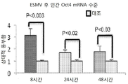
도 2d는 ESMV 처리후 대조 (오직 배지만)와 비교하여 8 시간, 24 시간, 그리고 48 시간 동안 배아 줄기 세포-특이적 인간 Oct4 mRNA의 발현에 있어서 배수 변화를 나타내는 그래프이며, 이때 Gapdh는 qRT-PCR을 위한 로딩 대조물로 이용되었으며, 에러바는 S.E.M를 나타낸다. 실험 세포군과 대조 Mueller 세포 군 간의 차이를 분석하기 위하여 실행된 슈튜던트 t-테스트(Student's t-test), p-값 < 0.05, Nanog mRNA을 제외;
도 2e는 ESMV 처리후 대조 (오직 배지만)와 비교하여 8 시간, 24 시간, 그리고 48 시간 동안 배아 줄기 세포-특이적 인간 Pax6 mRNA의 발현에 있어서 배수 변화를 나타내는 그래프이며, 이때 Gapdh는 qRT-PCR을 위한 로딩 대조물로 이용되었으며, 에러바는 S.E.M를 나타낸다. 실험 세포군과 대조 Mueller 세포 군 간의 차이를 분석하기 위하여 실행된 슈튜던트 t-테스트(Student's t-test), p-값 < 0.05, Nanog mRNA을 제외;
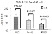
도 2f는 ESMV 처리후 대조 (오직 배지만)와 비교하여 8 시간, 24 시간, 그리고 48 시간 동안 배아 줄기 세포-특이적 인간 Rax mRNA의 발현에 있어서 배수 변화를 나타내는 그래프이며, 이때 Gapdh는 qRT-PCR을 위한 로딩 대조물로 이용되었으며, 에러바는 S.E.M를 나타낸다. 실험 세포군과 대조 Mueller 세포 군 간의 차이를 분석하기 위하여 실행된 슈튜던트 t-테스트(Student's t-test), p-값 < 0.05, Nanog mRNA을 제외;
도 3a는 대조과 비교하여 ESMV 처리 후 8 시간, 24 시간, 그리고 48 시간에서 Mueller 세포내 ESC-특이적 miRNA 292의 수준의 배수 변화를 나타내는 그래프이며, 이때 y-축은 Mueller 세포의 처리군과 대조군(연녹색) 사이의 배수 변화에 대응되며, 그리고 에러바는 S.E.M.를 나타내고; 실험군과 대조군 사이의 유의적인 차이는 슈튜던트 t-테스트에 의해 결정되며, 이때 모든 p-값은 < 0.01이었다;
도 3b는 대조과 비교하여 ESMV 처리 후 8 시간, 24 시간, 그리고 48 시간에서 Mueller 세포내 ESC-특이적 miRNA 295의 수준의 배수 변화를 나타내는 그래프이며, 이때 y-축은 Mueller 세포의 처리군과 대조군(연녹색) 사이의 배수 변화에 대응되며, 그리고 에러바는 S.E.M.를 나타내고; 실험군과 대조군 사이의 유의적인 차이는 슈튜던트 t-테스트에 의해 결정되며, 이때 모든 p-값은 < 0.01이었다;
도 4a는 ESMV 노출 후 8 시간, 24 시간, 그리고 48 시간 시점에서 대조 Mueller 세포와 비교하여 ESMV-처리된 Mueller 세포에서 마이크로어래이(microarray)에 의해 측정될 때 유전자 발현 변화의 Venn 도표를 나타낸다;
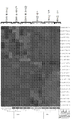
도 4b는 대조군과 비교하여 ESMV 처리후 Mueller 세포에서 차등적으로 조절되는 것으로 밝혀진 1894개 프로브에 기초하여 16개 시료의 계측정 군집(hierarchal clustering)의 히트맵(heat map)의 도해다 (p값, 0.001 그리고 발현에서 최저 3-배 차이), 이때 적색은 상향-조절을, 청색은 하향-조절을 나타내며; 열(rows)은 시료를 나타내며, 행(columns)은 유전자를 나타낸다;
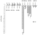
도 5a는 p < 0.001 수준 및 특이적 기능에서 배수 변화 ≥3의 조건에서 모든 테스트된 시점에서 ESMV-처리된 Mueller 세포와 대조 Mueller 세포 사이에 상이하게 조절된 1894개 유전자의 독창성 경로 분석(ingenuity pathway analysis)의 도표이며, 이때 유전자는 상당히 중요한 표준(canonical) 경로의 유전자 상호작용에서 전체적으로 관장된 데이터베이스에서 무작위 변화 연합에 대하여 특이적 세포 기능적 신호생성 경로에서 연합된 중요성에 대해 테스트되었다;
도 5b는 p < 0.001 수준 및 세포의 표준 신호생성 경로에서 배수 변화 ≥3의 조건에서 모든 테스트된 시점에서 ESMV-처리된 Mueller 세포와 대조 Mueller 세포 사이에 상이하게 조절된 1894개 유전자의 독창성 경로 분석(ingenuity pathway analysis)의 도표이며, 이때 유전자는 상당히 중요한 전통적인 경로의 유전자 상호작용에서 전체적으로 관장된 데이터베이스에서 무작위 변화 연합에 대하여 특이적 세포 표준 신호생성 경로에서 연합된 중요성에 대해 테스트되었다;
도 6은 처리안된 대조와 비교하여 ESMV 처리 후 24시간과 48시간 시점에서 Mueller 세포 안에서 마이크로어래이-확인된 유전자의 유전자 발현 변화를 나타내는 qRT-PCR 분석의 그래프이며, 이때 각 막대는 처리안된 Mueller 세포와 비교하여 ESMV-처리된 Mueller 세포에서 테스트된 유전자의 상대적 풍부함을 나타내고, 에러바는 S.E.M.을 나타낸다;
도 7a는 ESMV 처리 후 8 시간, 24 시간, 그리고 48 시간 시점에서 대조 Mueller 세포와 비교하여 ESMV-처리된 Mueller 세포에서 miRNA 발현 변화의 Venn 도표를 나타낸다;
도 7b는 테스트된 모든 시점에서 대조 Mueller 세포와 비교하여 ESMV-처리된 Mueller 세포에서 차등적으로 조절된 25개 miRNA 프로브에 기초하여 16개 시료의 계층적 군집화의 히트맵의 도표를 나타내며(p < 0.05, 발현에서 최소 3-배 차이), 이때 각 열은 단일 시료를 나타내고, 각 행은 단일 miRNA를 나타내며, 그리고 이때 적색 또는 청색은 각각 상대적으로 높은 발현 또는 낮은 발현을 나타낸다;
도 8은 대조 Mueller 세포와 비교하여 ESMV-처리된 Mueller 세포에서 전분화능의 유지, 탈-분화, 세포 운명 결정 그리고 분화와 관련된 miRNAs 선택하는 qRT-PCR 분석을 보여주는 그래프이며, 이때 각 막대는 처리안된 대조 세포와 비교하여 ESMV-처리된 Mueller 세포에서 테스트된 miRNAs의 상대적 풍부함을 나타내고, 에러바는 S.E.M.을 나타낸다;
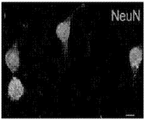
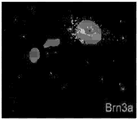
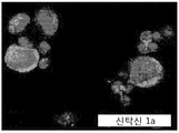
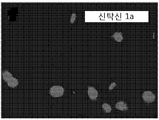
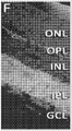
도 9a - 9r은 다양한 망막 계통의 표지들에 대해 면역착색된 대조 Mueller 세포와 ESMV-처리된 Mueller 세포를 나타내는 공촛점 현미경사진을 나타내며, 이때 도 a-l은 세포들이 Gad67(아마크린 세포와 수평 세포; 녹색) 또는 NeuN (아마크린 세포와 신경절 세포; 녹색), Mueller 세포의 표지, 글루타민 합성효소 (적색)로 이중책색되었음을 보여주고; 이때 도 9a - 9c는 Gad67-착색된 ESMV-처리된 Mueller 세포를 보여주며; 이때 도 9d - 9f는 Gad67-착색된 대조 Mueller 세포를 보여주고; 이때 도 9g - 9i는 NeuN-착색된 ESMV-처리된 Mueller 세포를 보여주고; 이때 도 9j - 9l은 NeuN-착색된 대조 Mueller 세포를 보여주고; 이때 각 열의 세번째 패널은 병합된 처음 두개의 상을 나타내고; 이때 도 9m은 망막 신경절 세포의 표지인 Brn3a (녹색)에 대해 착색된 ESMV-처리된 세포를 나타내고, 도 9n은 망막 신경절 세포의 표지인 Brn3a (녹색)에 대해 착색된 대조 Mueller 세포를 나타내고; 도 9o는 아마크린 세포의 표지인 Syntaxin 1a (녹색)에 대해 착색된 ESMV-처리된 세포를 나타내고, 도 9p는 아마크린 세포의 표지인 Syntaxin 1a (녹색)에 대해 착색된 대조 Mueller 세포를 나타내고; 도 9q는 로드(rod) 광수용기의 표지, 로돕신 (녹색)에 대해 착색된 ESMV-처리된 세포를 나타내고, 도 9r은 로드 광수용기의 표지, 로돕신 (녹색)에 대해 착색된 대조 Mueller 세포를 나타내고, 이때 세포 핵은 496-디아미디노-2-페닐인돌 (DAPI, 청색)으로 라벨되었으며, 그리고 모든 패널에서 기준자(scale bar)는 10 mm이며, 모든 채널에서 1564mm의 z-축 투영을 나타내는 영상이 있다;
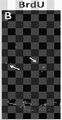
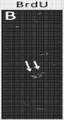
도 10a - 10f는 주사후 7일 (도 10a - 10c)과 30일(도 10d - 10f) 시점에서 Gad67 발현 (도 10a 및 10c), BrdU 발현 (도 10b 및 10d), 그리고 병합된 발현 (도 10c 및 10d)을 보여주는 공촛점 현미경 사진이다;
도 11a - 11f는 주사후 7일 (도 11a - 11c)과 30일(도 11d - 11f) 시점에서 Syntaxin 1a 발현 (도 11a 및 11c), BrdU 발현 (도 11b 및 11d), 그리고 병합된 발현 (도 11c 및 11d)을 보여주는 공촛점 현미경 사진이다;
도 12a - 12f는 주사후 7일 (도 12a - 12c)과 30일(도 12d - 12f) 시점에서 CRALBP 발현 (도 12a 및 12c), BrdU 발현 (도 12b 및 12d), 그리고 병합된 발현 (도 12c 및 12d)을 보여주는 공촛점 현미경 사진이다; 그리고
도 13은 0.05345 cd/m2의 임의적으로 선택된 자극 강도에서 ESMV 처리 후 개선된 동물중 하나의 암소성(scotopic) 어둠-적응된 ERG를 나타내며, 이때 처리안된 우측 눈에서 최대 파동 크기(적색)는 대략적으로 330 μV에서 유지되었으며, 한편 ESMV-처리된 좌측 눈에서 최대 파동 크기(청색)는 450 μV 이상으로 개선되었다. The foregoing and other objects, as well as various features of the present invention, as well as the invention itself may be better understood from the following description when taken in conjunction with the accompanying drawings, in which:
FIG. 1A is a micrograph showing untreated Mueller cells grown with homogeneous, bipolar, spindle-like, adsorptive cell "sheets " where ESMV-treated (T) and control (C) After counting, the percentage of treated cells to control cells was calculated (T /
FIG. 1B is a microscopic photograph showing Mueller cells morphologically grown as heterogeneous individual cells after 9 times ESMV treatment. Some of these cells have a multicellular process, others are enlarged nuclei or polynuclear cells, (T) cells and control (C) cells were counted after each treatment and the percentage of treated cells to control cells was calculated (T /
Figure 1c is a micrograph showing a collection of individual cells morphologically unique to the ESMV treated group, wherein the ESMV-treated (T) and control (C) cells are counted after each treatment and the treated cells Was calculated (T /
Figure 1d shows morphological changes occurring in time in the Mueller cells after ESMV treatment;
FIG. 2A is a graph showing the change in the expression of embryonic stem cell-specific mouse Oct4 mRNA during 8 hours, 24 hours, and 48 hours compared with the control (only in the stomach) after ESMV treatment, wherein Gapdh is a qRT-PCR , And error bars represent SEMs. [0033] The term " load control " The Student's t- test, p-value <0.05, except Nanog mRNA, was performed to analyze the differences between experimental cell groups and control Mueller cell groups;
FIG. 2B is a graph showing the change in the number of embryonic stem cell-specific mouse Sox2 mRNA expression during 8 hours, 24 hours, and 48 hours compared with the control (only in the stomach) after ESMV treatment, wherein Gapdh expresses qRT- Lt; / RTI > and the error bars represent SEM. The Student's t- test, p-value <0.05, except Nanog mRNA, was performed to analyze the differences between experimental cell groups and control Mueller cell groups;
FIG. 2c is a graph showing the change in multiplication in the expression of embryonic stem cell-specific mouse Nanog mRNA at 8 hours, 24 hours, and 48 hours compared with the control (only the embryo after ESMV treatment), wherein Gapdh was subjected to qRT-PCR Lt; / RTI > and the error bars represent SEM. The pendant t syutyu performed to analyze the differences between the control group and the test cell population cells Mueller-test (Student's t -test), except the p- values <0.05, Nanog mRNA;
FIG. 2d is a graph showing the change in the expression of embryonic stem cell-specific human Oct4 mRNA during 8 hours, 24 hours, and 48 hours as compared to the control (only in vivo) after ESMV treatment, wherein Gapdh is qRT-PCR Lt; / RTI > and the error bars represent SEM. The pendant t syutyu performed to analyze the differences between the control group and the test cell population cells Mueller-test (Student's t -test), except the p- values <0.05, Nanog mRNA;
Figure 2e is a graph showing the fold change in the expression of embryonic stem cell-specific human Pax6 mRNA for 8 hours, 24 hours, and 48 hours compared to the control (only multiplication) after ESMV treatment, wherein Gapdh is qRT-PCR Lt; / RTI > and the error bars represent SEM. The pendant t syutyu performed to analyze the differences between the control group and the test cell population cells Mueller-test (Student's t -test), except the p- values <0.05, Nanog mRNA;
FIG. 2f is a graph showing the change in multiplication in the expression of embryonic stem cell-specific human Rax mRNA for 8 hours, 24 hours, and 48 hours as compared to the control (only bred) after ESMV treatment, wherein Gapdh is qRT-PCR Lt; / RTI > and the error bars represent SEM. The pendant t syutyu performed to analyze the differences between the control group and the test cell population cells Mueller-test (Student's t -test), except the p- values <0.05, Nanog mRNA;
FIG. 3A is a graph showing changes in the level of ESC-specific miRNA 292 in Mueller cells at 8, 24, and 48 hours after ESMV treatment compared to the control, wherein the y-axis was the Mueller cell treated group and the control group Light green color), and the error bars represent SEM; Significant differences between the experimental and control groups were determined by the Schildndt t -test, where all p-values were <0.01;
FIG. 3B is a graph showing changes in the level of ESC-specific miRNA 295 in Mueller cells at 8 hours, 24 hours, and 48 hours after ESMV treatment compared with the control, wherein the y-axis was treated with Mueller cells and the control group Light green color), and the error bars represent SEM; Significant differences between the experimental and control groups were determined by the Schildndt t -test, where all p-values were <0.01;
Figure 4a shows the Venn plot of gene expression changes as measured by microarray in ESMV-treated Mueller cells compared to control Mueller cells at 8 hours, 24 hours, and 48 hours after ESMV exposure;
Figure 4b is a plot of the heat map of the hierarchical clustering of 16 samples based on 1894 probes that were found to be differentially regulated in Mueller cells after ESMV treatment compared to the control (p value , 0.001 and at least 3-fold difference in expression), with red indicating up-regulation and blue indicating down-regulation; Rows represent samples; columns represent genes;
Figure 5a shows the ingenuity pathway analysis of 1894 genes that were differentially regulated between ESMV-treated Mueller cells and control Mueller cells at all tested time points at the p < 0.001 level, analysis where the genes were tested for their associated significance in a specific cellular functional signaling pathway for random change association in a globally dominated database on gene interactions in the crucial canonical pathway;
Figure 5b shows the originality pathway analysis of 1894 genes that were differentially regulated between ESMV-treated Mueller cells and control Mueller cells at all tested time points at a level of p < 0.001 and a drainage change > where the genes have been tested for their associated significance in a specific cellular standard signaling pathway for random change association in a globally enforced database of gene pathway interactions in a fairly important traditional pathway;
FIG. 6 is a graph of qRT-PCR analysis showing the gene expression changes of microarray-identified genes in Mueller cells at 24 hours and 48 hours after ESMV treatment compared to untreated control, ≪ / RTI > represents the relative abundance of the tested genes in ESMV-treated Mueller cells compared to < RTI ID = 0.0 >
Figure 7a shows the Venn plot of miRNA expression changes in ESMV-treated Mueller cells compared to control Mueller cells at 8 hours, 24 hours, and 48 hours after ESMV treatment;
Figure 7b plots a heat map of hierarchical clustering of 16 samples based on 25 miRNA probes that are differentially regulated in ESMV-treated Mueller cells compared to control Mueller cells at all time points tested (p < 0.05, minimum 3-fold difference in expression), where each row represents a single sample, each row represents a single miRNA, and where red or blue exhibit relatively high expression or low expression, respectively;
Figure 8 is a graph showing qRT-PCR analysis of selecting miRNAs associated with maintenance of differentiation potential, de-differentiation, cell fate determination and differentiation in ESMV-treated Mueller cells compared to control Mueller cells, Indicates the relative abundance of miRNAs tested in ESMV-treated Mueller cells as compared to control cells, and error bars indicate SEM;
FIGS. 9a-9r show confocal microscopic photographs showing immunostained Mueller cells and ESMV-treated Mueller cells immunostained for various retinal system markers, wherein al cells harbor Gad67 (amaranth and horizontal cells; Green) or NeuN (amaranth gland cells and ganglion cells; green), the marker of Mueller cells, glutamine synthetase (red); Where 9a-9c show Gad67-stained ESMV-treated Mueller cells; Figures 9d-9f show Gad67-stained contrasted Mueller cells; Figures 9g-9i now show NeuN-stained ESMV-treated Mueller cells; Where 9j-9l show NeuN-stained control Mueller cells; Wherein the third panel of each column represents the first two phases merged; Figure 9m shows ESMV-treated cells stained for Brn3a (green), the marker for retinal ganglion cells, Figure 9n shows contrasted Mueller cells stained for Brn3a (green), a marker for retinal ganglion cells; FIG. 9 o represents ESMV-treated cells stained for Syntaxin 1a (green), which is a marker for amaranthin cells, and FIG. 9 p represents control Mueller cells stained for Syntaxin 1a (green), the marker of amaranthin cells; Figure 9q shows ESMV-treated cells stained for rod photoreceptor, rhodopsin (green), Figure 9r shows the control Mueller cells stained against rhodopsin (green), the label of the rod photoreceptor, , Where the cell nuclei were labeled with 496-diamidino-2-phenylindole (DAPI, blue) and the scale bar was 10 mm on all panels and 1564 mm z-axis projection on all channels There is a video;
Figures 10a-10f show Gad67 expression (Figures 10a and 10c), BrdU expression (Figures 10b and 10d), and combined expression (Figure 10a-10c) at 7 days post- 10c and < RTI ID = 0.0 > 10d) < / RTI >
11a-11f show Syntaxin 1a expression (FIGS. 11a and 11c), BrdU expression (FIGS. 11b and 11d), and merged expression (FIG. 11c and < RTI ID = 0.0 > 11d) < / RTI >
Figures 12a-12f show CRALBP expression (Figures 12a and 12c), BrdU expression (Figures 12b and 12d), and merged expression (Figure 12a and 12b) at 7 days post-injection (Figures 12a-12c) 12c and 12d); < RTI ID = 0.0 > And
Figure 13 shows one scotopic dark-adapted ERG of the improved animal after ESMV treatment at an arbitrarily selected stimulus intensity of 0.05345 cd / m2, wherein the maximum wave size (red) in the untreated right eye is approximately And the maximum wave size (blue) in the ESMV-treated left eye was improved to more than 450 μV.
상세한 설명details
본 출원을 통하여, 다양한 특허, 특허 출원, 그리고 공개들이 언급된다. 설명되고, 청구되는 본 발명 날짜에 당업자에게 공지된 기술의 상태를 좀더 충분하게 설명하기 위하여, 이들 특허, 특허 출원 및 공개의 명세서들은 전문이 본 출원에 참고자료로 편입된다. 본 내용은 특허, 특허 출원 및 공개와 본 내용간에 임의의 불일치가 있는 경우 본 명세서의 내용이 우선할 것이다. Throughout this application, various patents, patent applications, and disclosures are mentioned. In order to more fully describe the state of the art to those skilled in the art on the date of the invention being claimed and claimed, the disclosures of these patents, patent applications and publications are incorporated herein by reference in their entirety. In the event that there is any discrepancy between the contents of the patent, the patent application and the disclosure, the contents of this specification shall prevail.
본 명세서는 인간 배아 줄기 세포 (ESC) 미소낭포 (ESMVs)의 부분(fration)을 포함하는 치료 조성물, 이들 치료 부분의 단리 및 확인, 그리고 눈을 괴롭히는 질환의 대부분을 치료하기 위한 치료 양식(modality)으로 상기 부분의 용도를 제공한다. The present disclosure relates to a therapeutic composition comprising a fraction of human embryonic stem cell (ESC) microcysts (ESMVs), the isolation and identification of these therapeutic moieties, and the modality for treating most of the eye- To provide the use of said portion.
배아 줄기 세포(ESCs)는 (30nm 내지 1μm) 크기의 이질성 미소낭포(ESMVs) 집단을 세포외 환경으로 방출하는 것으로 공지되어 있다 (Ratajczak et al. (2006) Leukemia, 20:1487-1495). 이들 미소낭포는 다른 기원의 세포들에게 이들의 내용물을 전달하는 능력을 보유한다(Yuan et al. (2009) PLoS ONE, 4(3);e4722:1-8). ESMVs는 초기 전사 인자의 mRNAs 및 줄기 세포 전분화능에 중요한 miRNAs에 풍부하다. 줄기 세포 전분화능 유지 및 대부분 세포의 세포 운명 결정에 중추 역할을 하는 작은 넌-코딩 RNA 분자인 miRNAs와 마찬가지로, ESC 전분화능 유지에 중요한 mRNAs는 ESMVs에 풍부하다. Embryonic stem cells (ESCs) are known to release a population of heterogeneous microcysts (ESMVs) of size (30 nm to 1 μm) into the extracellular environment (Ratajczak et al. (2006) Leukemia , 20: 1487-1495). These microcysts have the ability to deliver their contents to cells of different origin (Yuan et al. (2009) PLoS ONE , 4 (3); e4722: 1-8). ESMVs are abundant in early transcription factor mRNAs and miRNAs important for stem cell pre-differentiation potential. Like miRNAs, which are small non-coding RNA molecules that play a central role in maintaining stem cell pre-differentiation potential and predisposing cell fate in most cells, mRNAs important for maintaining ESC pre-differentiation potential are abundant in ESMVs.
미세교세포와 유사한 Mueller 세포는 다중 망막 계통, 이를 테면 광수용기 및 내부 망막 뉴런으로 분화되는 능력을 보유하기 때문에 망막 선조세포다. Mueller cells similar to microglial cells are retinal progenitor cells because they have the ability to differentiate into multiple retinal systems, such as photoreceptors and internal retinal neurons.
ESMVs는 줄기 세포 mRNA, miRNA, 그리고 단백질의 내부 내용물을 배양된 인간 망막 선조세포 (Mueller 세포 및 미세교세포)로 이전시킬 수 있으며, 이로 인하여 손상된 조직 안에서 내성 세포, 성인 세포, 휴지(quiescent) 세포의 활성화를 유도시키는 것으로 밝혀졌다. 임의의 특정 이론에 국한된다는 의미는 아니지만, 이러한 전달은 ESMV 막과 다른 세포막들의 병합을 통하여 부분적으로 발생되는 것으로 보인다. ESMVs can transfer stem cell mRNAs, miRNAs, and inner contents of proteins to cultured human retinal maternal cells (Mueller cells and microglial cells), resulting in the formation of resistant cells, adult cells, quiescent cells Induce activation. This does not mean that it is limited to any particular theory, but it seems that this transmission is partially generated through the merging of ESMV membranes and other membranes.
망막 Mueller 세포의 배양물에 추가된 ESMVs는 더욱 탈-분화된 선조표현형으로 형태학적 변화를 유도하였다는 것이 확인되었다(도 1). 이러한 ESMV 부분은 선택적으로 ESC mRNA 및 miRNA를 전달하고, 그 결과로써 Mueller 세포에서 배아 및 초기 망막 유전자들이 유도된다. 또한, Mueller 세포의 ESMV 처리는 전사체(transcriptome) 변화를 유도하였는데, 이것은 탈-분화 및 망막 재생 프로그램의 활성화를 암시한다. Mueller 세포의 처리는 전분화능 및 초기 망막 유전자, 망막 보호 및 망막 재생의 유도물질에 관련된 유전자, 뿐만 아니라 망막 재생 허용 환경을 창조하는 다중 세포외 매트릭스(ECM)-변형 분자와 관련된 유전자의 상향-조절을 야기하였다. ESMV 처리는 분화를 촉진하는 유전자, 억제성 ECM 및 반흔 성분 유전자들의 하향-조절을 또한 야기하였다. 더욱이, ESMVs는 Mueller 세포의 miRNA에서 탈-분화된 선조 상태로의 이동(shift)을 유도하였다. 이러한 결과들은 ESMVs가 망막의 내생적 재생 잠재력을 활성화시킬 수 있는 치료 물질임을 설명한다. It was confirmed that ESMVs added to cultures of retinal Mueller cells induced morphological changes with more de-differentiated ancestral phenotype (Fig. 1). These ESMV segments selectively transfer ESC mRNA and miRNA, resulting in embryonic and early retinal genes being induced in Mueller cells. In addition, ESMV treatment of Mueller cells induced transcriptome changes suggesting activation of de-differentiation and retinal regeneration programs. The treatment of Mueller cells is an up-regulation of genes associated with pre-differentiation potential and early retinal genes, genes involved in retinal protection and inducers of retinal regeneration, as well as multiple extracellular matrix (ECM) . ESMV treatment also caused down-regulation of genes that promote differentiation, inhibitory ECM and scar component genes. Furthermore, ESMVs induced a shift from the miRNA of Mueller cells to a de-differentiated ancestral state. These results demonstrate that ESMVs are therapeutic agents that can activate the endogenous regeneration potential of the retina.
따라서, ESMVs의 전달 능력에 의해, ESMVs는 배양된 망막 선조세포에서 더욱 탈-분화된 표현형로의 형태학적 변화를 유도할 수 있고, 또한 망막 조직의 재생을 개시할 수 있다. Thus, by the ability of ESMVs to transfer, ESMVs can induce morphological changes from cultured retinas progenitor cells to more de-differentiated phenotypes and also initiate regeneration of retinal tissue.
ESMVs에 노출된 배양된 Mueller 세포는 전분화능에 관련된 유전자 (Oct4, Lin28, Klf4, 그리고 LIF)를 상향-조절할 수 있고, 초기 망막 유전자 (BMP7, Pax6, 그리고 Rax)를 상향조절할 수 있고, 망막 보호에 관련된 유전자 (IL6, CSF2), 그리고 재생에 관련된 유전자(FGF2, IGF2, GDNF)를 상향조절할 수 있고, 그리고 조직 재건을 허용하는 환경을 창조하는 것으로 알려진 세포외 메트릭스-변형 유전자 (이를 테면, MMP3)를 상향조절할 수 있다. 대조적으로, ESMVs는 분화를 촉진하는 유전자(이를 테면, DNMT3a 및 GATA4)는 하향-조절할 수 있다. Mueller 세포는 손상된 망막에서 활성화되는데, 일부 재생은 성공적이다. 그러나, 망막의 기능적 회복은 지금까지 성공하지 못하였다. Cultured Mueller cells exposed to ESMVs can up-regulate the genes associated with the ability to differentiate ( Oct4, Lin28, Klf4, and LIF ), up-regulate early retinal genes ( BMP7, Pax6 , and Rax ) gene (IL6, CSF2), and can up-regulate the gene (FGF2, IGF2, GDNF) related to reproduction, and a known extracellular matrix by creating an environment that allows for tissue reconstruction related-modified gene (such as, MMP3 ) Can be adjusted upward. In contrast, ESMVs can down-regulate genes that promote differentiation (such as DNMT3a and GATA4 ). Mueller cells are activated in the damaged retina, and some regeneration is successful. However, functional recovery of the retina has not been successful so far.
ESMVs는 망막 퇴행의 마우스 모델에서 손상된 망막의 기능을 개선시킨다. ESMVs는 손상된 망막을 되살리고 복구시키기 위하여 내생적 망막 선조세포를 유도함으로써, 최소한 부분적으로 재생을 촉진시킨다. ESMV 적용 이후, 마우스 ERG의 a와 b 파동(wave)의 상당한(70%) 개선, 뿐만 아니라 망막 세포 재증식의 면역조직화학적 증가가 발견되었다. ESMVs improve the function of the damaged retina in the mouse model of retinal degeneration. ESMVs promote regeneration, at least in part, by inducing endogenous retinal progenitor cells to restore and restore damaged retinas. After ESMV application, significant (70%) improvement of the a and b waves of mouse ERG, as well as immunohistochemical enhancement of retinal cell re-proliferation, were found.
인간에게 치료 용도로 ESCs와 비교하여 hESMVs를 이용할 때 장점은 hESMVs가 세포는 아니며, 표면 분자들을 활발하게 생산하지 못하고, 거부 및 종양 형성을 야기할 가능성이 적다는 점이다. 이용된 hESMV 조제물은 내독소, 비-면역원, 비-종양생성이며, 그리고 오염물질이 없다. hESMVs의 사용은 주입된(engrafted) 고유한 ESCs의 장기적 암화(maldifferentiation) 가능성을 회피하며, 이들의 악성 형질변환 위험을 제거한다. The advantage of using hESMVs in comparison to ESCs for therapeutic use in humans is that hESMVs are not cells and are less likely to actively produce surface molecules and cause rejection and tumor formation. The hESMV preparations used are endotoxin, non-immunogenic, non-tumorigenic, and free of contaminants. The use of hESMVs avoids the possibility of long-term maldifferentiation of unique engrafted ESCs and eliminates their risk of malignant transformation.
ESMVs는 손상의 재생 및 복구를 위하여 안구 구획의 재생을 특이적으로 자극 또는 개시할 수 있다는 것이 본 명세서에서 설명된다. 고유한(intrinsic) 재생성 물질을 활성화시키는 ESMVs에 의해 복구되는 손상의 예는 각막 긁힘, 궤양화 및/또는 반흔화에서부터 모든 형태의 망막 질환에 이른다. 하기 비제한적인 실시예에서 나타낸 바와 같이, ESMVs는 이의 내생적 재생 능력을 유도함으로써 손상된 망막의 재생을 개시시켰다. It is described herein that ESMVs can specifically stimulate or initiate regeneration of the ocular compartment for regeneration and repair of impairment. Examples of damage restored by ESMVs that activate intrinsic regenerative materials include corneal scratching, ulceration and / or scarring to all forms of retinal disease. As shown in the following non-limiting examples, ESMVs initiated regeneration of the damaged retina by inducing its endogenous regenerative capacity.
ESMV 치료 부분은 토종 또는 배양된 포유류 ESCs, 이를 테면 인간 ESCs로부터 하기 실시예에서 설명된 바와 같이 분별 원심분리에 의해 획득된다. 이의 존재는 공지의 ESC-특이적 mRNAs (Oct4, Sox2, Nanog, Lin28, Klf4) 그리고 microRNAs (miR-292, -294, 그리고 -295), 뿐만 아니라 특정 ESC-특이적 표면 단백질 항원 (CD9, Delta 1, 인테그린 α6, 인테그린 β1, 소닉 헷지호그(sonic hedgehog), 소닉 헷지호그 상동체, SSEA1, SSEA3, SSEA4, 그리고 TRA-1-60)의 존재에 의해 확인될 수 있다. 따라서, ESMV 치료 부분의 존재는 전분화능 인자들의 특정 조합을 이용하여 증명될 수 있다. 이러한 ESMV 부분은 전체 RNA 또는 특정 mRNAs 그리고 miRNAs를 함유하는 RNA 부분을 획득하기 위하여 세포의 탈-분화를 야기할 수 있거나 또는 특정 분화된 안구 세포의 발달 및 기능화에 연관된 유전자의 발현을 개시할 수 있는 ESMVs으로부터 추가적으로 분획화될 수 있다. The ESMV therapeutic moiety is obtained from native or cultured mammalian ESCs, such as human ESCs, by fractional centrifugation as described in the Examples below. Its presence is not only known for ESC-specific mRNAs ( Oct4, Sox2, Nanog, Lin28, Klf4 ) and for microRNAs (miR-292, -294 and -295), as well as for specific ESC-specific
상기 ESMV 부분은 치료될 질환에 효과적이라고 안과의사가 판단한 임의의 전달 방식에 의해 눈에 투여될 수 있다. 예를 들면, 유리체내 투여는 녹내장에 적절할 수 있지만, 반면 AMD 그리고 광수용기/RPE 퇴화는 망막아래 주사에 의해 치료될 수 있다. 전달은 볼루스, 간헐, 또는 연속적 전달이 될 수 있고, 그리고 장치, 이를 테면, 방출 펌프 또는 콘텍트렌즈에 의해 제공될 수 있지만 이에 국한되지 않는다. The ESMV portion may be administered to the eye by any delivery method as determined by the ophthalmologist to be effective in the disease to be treated. For example, intravitreal administration may be appropriate for glaucoma, whereas AMD and photoreceptor / RPE degradation can be treated by subretinal injection. The delivery may be bolus, intermittent, or continuous delivery, and may be provided by a device, such as a discharge pump or a contact lens, but is not limited thereto.
기타 비제한적 실시예들에 있어서, ESMVs는 이를 테면, 각막 상피 비정상 및 건성안을 치료하기 위하여 눈에 국소적으로 투여될 수 있다. 국소적으로 투여하기 위하여, ESMV 치료 부분은 연장된 방출 운반체, 이를 테면 하이드로겔 매트릭스 안에 내장될 수 있고(이를 테면, Zarembinski, et al. (2011) in Regenerative Medicine and Tissue Engineering-Cells and Biomaterials, Editor: Daniel Eberly, Chapter 16, pp. 341-364), 이 매트릭스는 콘텍트렌즈 측면에 부착되어 각막 질환을 치료하고, 각막 재생을 촉진시킬 수 있다. 이 부분은 펌프가 구비된 장치를 통하여 연속적으로 또는 간헐적으로 또한 투여될 수 있다. In other non-limiting examples, ESMVs can be administered topically to the eye to treat, for example, corneal epithelial abnormalities and dry eye. For topical administration, the ESMV therapeutic moiety can be embedded in an extended release carrier, such as a hydrogel matrix (see, for example, Zarembinski, et al. (2011) in Regenerative Medicine and Tissue Engineering-Cells and Biomaterials, Editor : Daniel Eberly, Chapter 16, pp. 341-364), which attaches to the side of the contact lens to treat corneal disease and promote corneal regeneration. This part can also be administered continuously or intermittently through a device equipped with a pump.
본 명세서에 따라 치료될 수 있는 질환은 눈의 모든 격실, 눈물을 만드는 샘 안에 세포 상실, 그리고 상기 안구 표면 상에 또는 부근 세포의 세포 상실, 각막 세포 상실, 전실(anterior chamber) 질환, 그리고 노화 황반 퇴행, 당뇨망막병, 녹내장, 망막색소변성 복합을 포함하나 이에 국한되지 않는 주요 망막 퇴행성 질환, 뿐만 아니라 유전된 망막 퇴화를 포함한다. 본 명세서에 따라 치료될 수 있는 기타 질환은 눈에 악영향을 주는 전신 및 국소 자가면역 장애, 이를 테면, 포도막염, 건성안, 안구 흉터유사천포창, 양성 및 악성 Mooren의 각막 궤양, 그리고 류마티스 관절염을 포함하나 이에 국한되지 않는다. Diseases that may be treated according to the present disclosure include all compartments of the eye, cell loss in the fountain-forming follicle, and cell loss, keratocyte loss, anterior chamber disease, Degenerative retinopathy, diabetic retinopathy, glaucoma, major retinal degenerative diseases including but not limited to retinitis pigmentosa complex, as well as inherited retinal deterioration. Other diseases that may be treated according to the present disclosure include systemic and local autoimmune disorders that adversely affect the eye, such as uveitis, dry eye, eye scarring pemphigus, benign and malignant corneal ulcers, and rheumatoid arthritis It is not limited.
특정 눈 질환은 안과의사에 판단에 의해 병에 걸린 부분에 상기 ESMV 부분의 1회 또는 반복적 투여에 의해 여러차례 치료될 수 있다. 상기 ESMV 부분은 단독으로 투여될 수 있거나 또는 상기 장애를 치료하는 것으로 알려진 다른 치료법과 함께 투여될 수 있는데, 단, 부차적 치료는 투여되는 ESMVs를 비활성화시키지 않아야 한다. Certain ocular diseases can be treated several times by one or repeated administration of the ESMV part to the diseased part of the ophthalmologist by judgment. The ESMV portion may be administered alone or in combination with other therapies known to treat the disorder, provided that the secondary treatment does not inactivate the administered ESMVs.
본 발명을 설명하는 특정 실시예들은 지금부터 언급할 것이다. 본 실시예들은 구체예를 설명하기 위하여 제공되는 것이며, 이로 인하여 본 발명의 범위에 어떠한 제약을 의도하지 않는다는 점을 인지해야만 한다. Specific embodiments for illustrating the invention will now be described. It is to be understood that the embodiments are provided to illustrate specific embodiments and that no limitation of the scope of the present invention is thereby intended.
실시예Example
실시예 1Example 1
마우스 ESMVs의 단리 및 특징화Isolation and characterization of mouse ESMVs
마우스 세포주 SV129로부터 유도된 배아 줄기 세포 (ESCs)는 분화를 억제하기 위하여 GSK3β 억제제(Millipore, Billerica, MA)가 보충된 ESGRO Complete PLUS 클론 등급의 배지에서 무-혈청 및 피더(feeder) 없는 조건하에서 확장되었다. 3.5 x 10⁶ 세포가 젤라틴-피복된 T175 cm2 배양 플라스크 상에 도말되었다. ESCs는 가습된 37℃, 5% CO2 배양기에서 배양되었다. ESCs의 성장은 현미경으로 관찰되었으며, 새로운 배양 배지는 매일 추가되었고, ESMV 단리를 위하여 매 48시간마다 수거되었다. ESCs는 ESCs의 분화를 회피하면서 ESMV 수율을 최대화시키기 위하여 80% 합류에서 ESC 콜로니가 유지되도록 48 내지 72시간 마다 ESGRO Complete Accutase (Millipore)를 이용하여 계대되었다. Embryonic stem cells (ESCs) derived from mouse cell line SV129 were expanded in ESGRO Complete PLUS clone grade medium supplemented with GSK3? Inhibitor (Millipore, Billerica, MA) to inhibit differentiation under serum-free and feeder- . 3.5 x 10 6 cells are gelatin - was blotted onto a covered T175 cm 2 culture flasks. ESCs were incubated in a humidified 37 ° C, 5% CO 2 incubator. Growth of ESCs was observed under a microscope, new culture medium was added daily and collected every 48 hours for ESMV isolation. ESCs were shipped using ESGRO Complete Accutase (Millipore) every 48 to 72 hours to maintain ESC colonies at 80% confluence to maximize ESMV yield while avoiding ESCs differentiation.
ESC 콜로니는 분화 징후에 대하여 매일 현미경을 통하여 눈으로 검사되었으며, Oct4, Sox2, 그리고 Nanog mRNA 발현은 PrimerQuestSM(Integrated DNA Technology-DNAsite, San Diego, CA)에 의해 기획된 마우스 특이적 프라이머 쌍을 이용한 qRT-PCR에 의해 분석되었다: ESC colonies were examined by microscope daily for signs of differentiation, and Oct4 , Sox2 , and Nanog mRNA expression was detected using a mouse-specific primer pair designed by PrimerQuest SM (Integrated DNA Technology-DNA Site, San Diego, CA) lt; RTI ID = 0.0 > qRT-PCR:
Oct4: 포워드-GCCGGGCTGGGTGGATTCTC (서열 번호:1), Oct4 : forward-GCCGGGCTGGGTGGATTCTC (SEQ ID NO: 1),
리버스-ATTGGGGCGGTCGGCACAGG (서열 번호:2), Reverse-ATTGGGGCGGTCGGCACAGG (SEQ ID NO: 2),
Nanog: 포워드-TCCAGAAGAGGGCGTCAGAT (서열 번호:3), Nanog : forward-TCCAGAAGAGGGCGTCAGAT (SEQ ID NO: 3),
리버스-CTTTGGTCCCAGCATTCAGG (서열 번호:4), Reverse-CTTTGGTCCCAGCATTCAGG (SEQ ID NO: 4),
Sox2: 포워드-AACAATCGCGGCGGCCCGAGGAG (서열 번호:5), Sox2 : forward-AACAATCGCGGCGGCCCGAGGAG (SEQ ID NO: 5),
리버스-GCCTCGGCGTGCCGGCCCTGCG (서열 번호:6). Reverse-GCCTCGGCGTGCCGGCCCTGCG (SEQ ID NO: 6).
ESMVs를 단리하기 위하여, 상청액은 50 ml 원심분리기 튜브에 수거되었으며, 3,500 g, 4℃에서 1시간 동안 회전되어 찌꺼지 및 파편화된 세포가 펠렛화되었다. 상기 상청액은 초원심분리기 튜브로 조심하여 이동되었고, Beckman Type 50.2Ti 로터에서 200,000 g, 4℃에서 3.5 시간 동안 회전되어, ESMVs가 펠렛화되었다. ESMVs는 그 다음 소낭으로부터 획득된 RNA 및/또는 단백질이 포함된 치료 조성물을 획득하기 이용되거나 분획화될 수 있다. To isolate ESMVs, the supernatant was collected in a 50 ml centrifuge tube and spun at 3,500 g, 4 ° C for 1 hour to pelletize the scum and the fragmented cells. The supernatant was carefully transferred into an ultracentrifuge tube and rotated at 200,000 g in a Beckman Type 50.2 Ti rotor for 3.5 hours at 4 ° C to pelletize the ESMVs. ESMVs can then be used or fractionated to obtain a therapeutic composition comprising the RNA and / or protein obtained from the follicle.
실시예 2Example 2
RNA를 단리하기 위하여 ESMV 분획화To isolate RNA, ESMV fractionation
mirVana miRNA 단리 키트를 이용하여 마우스 ESMVs로부터 전체 RNA가 단리되었고, 이는 작은 RNA 종(Ambion, Austin, TX)을 보유하며, DNA 잔량을 제거하기 위하여 TURBO DNAse (Ambion)로 처리되었으며, 그리고 마우스 Oct4, Sox2, Nanog (실시예 1에서 프라이머 쌍), 그리고 Klf4, Lin28, 그리고 mmu-miR292-3p, -294, 그리고 -295 (Taqman® 프라이머) 전사체의 존재에 대해 RT-PCR을 통하여 검사되었다. Total RNA was isolated from mouse ESMVs using the mirVana miRNA isolation kit, which had a small RNA species (Ambion, Austin, TX), treated with TURBO DNAse (Ambion) to remove residual DNA , The presence of Sox2, Nanog (primer pair in Example 1), and Klf4, Lin28 , and mmu-miR292-3p, -294, and -295 (Taqman® primers) transfectants were examined by RT-PCR.
유사하게, hESCs로부터 인간 ESMVs (hESMVs)에서 RNA는 전체 RNA 뿐만 아니라 miRNAs를 단리시키는 miRNeasy Mini™ 키트 (Qiagen, Germantown, MD, USA)를 이용하여 추출된다. 전체 RNA는 Affimetrix GeneChip U133 Plus 2.0 인간 유전자 발현 어레이 (Affimetrix, Santa Clara, CA)에 혼성화된다. Affymetrix GeneChip 발현 분석 표준 프로토콜에 따라 표적 조제 및 어레이 혼성화가 실행된다. 상기 어레이는 Affymetrix 7G 스캐너를 이용하여 스캔되며, Affymetrix GeneChip Command Console 1.1 (AGCC)를 이용하여 상(images)들이 획득된다. 발현된 유전자는 Affymetrix 출석 콜(present calls)에 의해 확인되며, 데이터 규격화를 위하여 Partek 게놈(genomics) Suite 6.4 및 RMA 알고리즘을 이용하여 분석된다. 유의적인 유전자의 선택을 위한 역치(thresholds)는 > = 2-배 및 FDR-교정된 p < 0.05으로 설정된다. microRNA의 경우, Exiqon miRCURY LNA microRNA 어레이가 제조업자의 지시에 따라 이용된다(Exiqon, Woburn, MA 01801). miRNA 어레이는 Axon GenePix 4100A 스캐너에 의해 스캔되며 그리고 GenePix Pro 6.0 소프트웨어로 처리된다. miRNA 미가공 데이터는 역치는 > = 2-배 및 FDR-교정된 p < 0.05의 역치로 Partek 게놈 세트(genomic suite) 6.4를 이용하여 하우스 키핑(housing keeping) miRNAs 및 불변 miRNAs와 NLYZED의 조합을 이용하여 표준화된다. hESMV 단백질은 Invitrogen ProtoArray Human Protein Microarrays (Invitrogen, Carlsbad, CA)에 혼성화되고 특징화된다. hESMV 표면 항원들은 ESC 표면 표지들 1, 인테그린 α6, 인테그린 β1, 소닉 헷지호그, 소닉 헷지호그 상동체, SSEA1, SSEA3, SSEA4, TRA-1-60 그리고 TRA-1-81에 대하여 유동세포분석(flow cytometry)에 의해 분석된다. hESC 배양물로부터 다수의 hESMVs 배치(batches)가 단리되며, PBS로 세척되고, BSA 및 아지드 나트륨이 보충된 PBS에 재현탁되고, 형광색소-접합된 대응하는 단클론성 항체들을 이용하여 착색된다. hESMVs는 배양 배지에 재현탁되며, 유동세포분석 분석을 위하여 취한다. 유동 세포 분석 및 실험의 최적화는 원하는 바와 같이 최적화된다. CELLQuest 소프트웨어를 이용하여 획득된 데이터가 분석된다. Similarly, RNA from human ESMVs (hESMVs) from hESCs is extracted using miRNeasy Mini ™ kit (Qiagen, Germantown, MD, USA) which isolates miRNAs as well as total RNA. Total RNA is hybridized to an Affimetrix GeneChip U133 Plus 2.0 human gene expression array (Affimetrix, Santa Clara, Calif.). Target formulation and array hybridization are performed according to the Affymetrix GeneChip Expression Assay Standard Protocol. The array is scanned using an Affymetrix 7G scanner and images are acquired using the Affymetrix GeneChip Command Console 1.1 (AGCC). Expressed genes are identified by Affymetrix present calls and analyzed using the Partek genomics Suite 6.4 and RMA algorithm for data normalization. The thresholds for significant gene selection are set to> = 2-fold and FDR-corrected p <0.05. For microRNA, an Exiqon miRCURY LNA microRNA array is used according to the manufacturer's instructions (Exiqon, Woburn, MA 01801). The miRNA arrays are scanned by the Axon GenePix 4100A scanner and processed with GenePix Pro 6.0 software. miRNA raw data were analyzed using a combination of house keeping miRNAs and invariant miRNAs and NLYZED using the Partek genomic suite 6.4 with thresholds of> = 2-fold and FDR-corrected p <0.05 Standardized. The hESMV protein is hybridized and characterized in Invitrogen ProtoArray Human Protein Microarrays (Invitrogen, Carlsbad, Calif.). hESMV surface antigens were analyzed by flow cytometry (flow cytometry) on
hESMV 조제물은 GenScript ToxinSensorTM Chromogenic LAL Endotoxin Assay Kit를 이용하여 hESMVs 안에 세균성 내독소의 부재를 확인하기 위하여 선별되고, 이때 상기 키트는 변형된 Limulus Amebocyte Lysate와 광범위한 범위(0.005 - 1 EU/ml)에서 색소원적으로 내독소를 정량적으로 탐지하기 위한 합성 색깔을 만들어내는 기질을 이용한다. The hESMV preparations were screened to confirm the absence of bacterial endotoxin in hESMVs using the GenScript ToxinSensor ™ Chromogenic LAL Endotoxin Assay Kit, which contains the modified Limulus Amebocyte Lysate and a range of pigments (0.005-1 EU / ml) Primarily, it uses a substrate that produces a synthetic color to quantitatively detect endotoxin.
실시예 3Example 3
Mueller 세포 배양 Mueller cell culture
사후-인간 신경 망막으로부터 처음으로 유도된 인간 Moorfield/Institute of Ophthalmology-Mueller 1 (MIO-M1) 세포 계통이 확립되었고, 이미 특징화되었다(Limb et al. (2002) Invest. Ophthalmol. Vis. Sci. 43:864869). 증식을 위하여 MIO-M1 세포는 175 cm2 조직 배양 플라스크에서 흡착성 세포 계통으로 유지되었고, 그리고 가습된 37℃, 5% CO2 배양기에서 ESMV 처리 실험을 위하여 6-웰 조직 배양 플레이트상에서 10% vol/vol 태아 소 혈청 (여과된, 열에 의해 비활성화된; Gemini Bioproducts, Sacramento, CA) 그리고 페니실린/스트렙토마이신(Invitrogen)과 함께 4500 mg/L 포도당, 피루베이트 나트륨 그리고 안정화된 L-글루타민 (GlutaMAX; Invitrogen, Grand Island, New York)이 포함된 DMEM 배지에 유지되었다. 합류에 도달될 때, 상기 세포는 인산염-완충 식염수(PBS)로 세척되고, 트립신(Invitrogen)을 이용하여 플라스크로부터 탈착시키고, 완전한 세포 배양 배지로 세척되고, 그리고 새로운 플라스크로 분리된다. ESMV 처리 실험은 Mueller 세포가 60% 합류에 도달되었을 때 시작되었다. The human Moorfield / Institute of Ophthalmology-Mueller 1 (MIO-M1) cell line first established from the post-human neural retina has been established and already characterized (Limb et al. (2002) Invest. Ophthalmol. 43: 864869). For proliferation, MIO-M1 cells were maintained in an adsorptive cell line in a 175 cm 2 tissue culture flask and incubated in a humidified 37 ° C, 5% CO 2 incubator at 10% vol / vol on a 6-well tissue culture plate for ESMV treatment experiments. L glutamine (GlutaMAX; Invitrogen, St. Louis, MO) with penicillin in serum (filtered, heat-inactivated; Gemini Bioproducts, Sacramento, Calif.) and penicillin / streptomycin (Invitrogen) Grand Island, New York). When confluence is reached, the cells are washed with phosphate-buffered saline (PBS), desorbed from the flask using trypsin (Invitrogen), washed with complete cell culture medium, and separated into fresh flasks. ESMV treatment experiments were initiated when Mueller cells reached 60% confluence.
실시예 4Example 4
Mueller 세포에서 ESMV에 의해 유도된 형태학적 변화 ESMV-induced morphological changes in Mueller cells
Mueller 세포는 웰당 1X10⁶ 세포로 2개의 6-웰 세포 배양 플레이트에 도말되었고, ESMV 처리가 개시되기 전 60% 합류에 도달되도록 하였다. 이를 위하여, 혈청-없는, 피터(feeder) 없는 조건하에서(상기 참고) 성장된 마우스 ESCs의 6 T175 cm2 플라스크 배지의 초원심분리에 의해 펠렛화된 ESMVs는 Mueller 세포 배지에 바로 재현탁되었고, 동량의 용적이 배양된 Mueller 세포가 있는 6-웰 플레이트중 하나의 각 웰에 추가되었다. 이 과정은 9회 연속 처리를 위하여 48시간 마다 반복되었다. 다른 6-웰 플레이트에 있는 대조 Mueller 세포 배양물은 ESMV 처리 대신 오직 배지 교환만 받았다. 60% 합류를 유지하기 위하여, 처리된 세포와 대조 세포는 모두 ESMV 처리가 종료될 때 요구되는 것과 같이 계대되었다. ESMV-노출된 Mueller 세포와 대조 Mueller 세포는 각 처리후 Leica DM IL LED 현미경을 이용하여 검사되었다. Mueller cells were plated in two 6-well cell culture plate at 1X10 6 cells per well, it was allowed to reach 60% confluence before they ESMV processing starts. To this end, the serum-free, Peter (feeder) under conditions that (see above) the pelleted by ultracentrifugation of 6 T175 cm 2 flask, the medium of the growing mouse ESCs ESMVs was immediately re-suspended in Mueller cell medium, the same amount Were added to each well of one of the 6-well plates with Mueller cells incubated. This process was repeated every 48 hours for 9 consecutive treatments. Control Mueller cell cultures in other 6-well plates received only medium exchange instead of ESMV treatment. To maintain 60% confluence, both treated and control cells were passaged as required when ESMV treatment was terminated. ESMV-exposed Mueller cells and control Mueller cells were examined using a Leica DM IL LED microscope after each treatment.
각 처리가 종료된 후 ESMVs에 의해 유도된 형태학적 변화를 평가하기 위하여, Mueller 세포는 15분 동안 100% 에탄올에 고정되었으며, Harris Hematoxylin 그리고 Eosin Y로 착색되었으며, 연속적인 에탄올 세척에 의해 탈수되었고, 대기 건조되었고, ProLong Gold 안티페드(antifade) 시약과 함께 커버슬립으로 덮었다. Zeiss Axiovert 135M 현미경과 Photometrics CoolSnap 카메라를 이용하여 전달된 광 차등 간섭 대조상(transmitted light differential interference contrast images)이 획득되었다. 각 처리 종료시 ESMV-노출된 Mueller 세포와 대조 Mueller 세포 배양물에 존재하는 세포의 수를 비교하기 위하여, Leica DCF295 디지털 카메라를 이용하여 3 내지 4개의 시정 영상(6-웰 세포 배양 플레이트의 각 웰에 대해 20X 확대에서 획득된)이 획득되었다. 각 상 안에 있는 세포는 Adobe Photoshop을 이용하여 개별적으로 표시되었으며, 카운트되었고, 그리고 처리된 세포/대조 세포 비율이 산출되었다. To evaluate the morphological changes induced by ESMVs after each treatment, Mueller cells were fixed in 100% ethanol for 15 min, stained with Harris Hematoxylin and Eosin Y, dehydrated by continuous ethanol washing, Air dried and covered with a cover slip with ProLong Gold antifade reagent. The transmitted light differential interference contrast images were obtained using a Zeiss Axiovert 135M microscope and a Photometrics CoolSnap camera. To compare the number of cells present in the Mueller cell cultures compared to the ESMV-exposed Mueller cells at the end of each treatment, three to four visually imaged images were obtained using a Leica DCF295 digital camera Obtained at 20X magnification) was obtained. Cells in each phase were individually labeled, counted, and processed cell / control cell ratios using Adobe Photoshop.
ESMV 처리 종료시, ESMV-노출된 Mueller 세포의 배양 배지는 흡출되었고; 그 다음 세포는 앰플 PBS로 3회 세척되어 임의의 잔류 ESMVs가 제거되었으며, 그리고 RNA 단리 및 유전자 발현 연구를 위하여 수집되었다. At the end of ESMV treatment, the culture medium of ESMV-exposed Mueller cells was aspirated; The cells were then washed three times with ampoule PBS to remove any residual ESMVs and were harvested for RNA isolation and gene expression studies.
비록 처리된 배양물과 비-처리된 (대조) 배양은 동일한 계대에서 출발되었지만, 세포의 수, 그리고 Mueller 세포의 합류 수준, 형태학적 차이는 처음 처리후 조기에 대조 세포와 ESMV-노출된 세포 사이에 명백하게 나타나기 시작되었다. 전형적인 Mueller 세포 배양물의 방추와 유사한, 흡착성 세포성 쉬트 특징으로 성장된 대조세포와 대조적으로 (도 1a), ESMV 처리가 진행됨에 따라, 노출된 Mueller 세포는 감소된 세포-세포 흡착, 다중 과정을 가진 세포의 존재, 성상(stellate) 세포, 다핵화된 세포, 그리고 일방적 보우통(unilateral boutons) 및 심화 과정을 겪는 세포를 설명하는 개별 이질성 세포로 성장되었다 (도 1b 및 1c). 대개, ESMV-처리된 세포의 핵은 확대되었고, 많은 것들은 눈에 보이는 중기플레이트를 나타낸다. ESMV-처리된 배양물과 대조 배양 간의 세포 카운트 비교에서 처리 군의 전반적인 세포 수의 상당한 감소가 드러나지 않았다(도 1d). Although the treated cultures and non-treated (control) cultures originated in the same passages, the number of cells, and the level of confluence and morphological differences of Mueller cells, were observed early between the control and ESMV-exposed cells . In contrast to the control cells grown with characteristic adsorptive cell sheets (Fig. 1a), similar to the spindle of a typical Mueller cell culture, exposed Mueller cells have reduced cell-cell adsorption, multiple processes Cells were grown into individual heterogeneous cells that describe the presence of cells, stellate cells, polynucleated cells, and cells undergoing unilateral boutons and deepening processes (FIGS. 1B and 1C). Usually, the nuclei of ESMV-treated cells are enlarged, and many exhibit visible mid-stage plates. Comparing the cell counts between the ESMV-treated cultures and the control cultures, a significant reduction in the overall cell number of the treated group was not revealed (Fig. 1d).
대안으로, ESMV-노출된 Mueller 세포 및 대조 Mueller 세포는 각 처리 후 Leica DM IL LED 현미경 (Leica Microsystems, Wetzlar, Germany)을 이용하여 검사되었으며, 그리고 세포 수를 결정하기 위하여, Leica DCF295 디지털 카메라를 이용하여 3 내지 4개의 시정 영상(처리된 세포와 대조 세포의 6-웰 세포 배양 플레이트의 각 웰에 대해 20X 확대에서 획득된)이 획득되었다. 각 상 안에 있는 세포는 Adobe Photoshop(Adobe Systems, San Jose, CA)을 이용하여 개별적으로 표시되었으며, 그 다음 카운트되었다. 처리군 및 대조군의 각 시점에서 시정마다 세포수가 획득되었고, 처리된 세포/대조 세포의 비율이 산출되었다. 형태학적 연구를 위하여, Mueller 세포는 15분 동안 100% 에탄올에 고정되었으며, Harris Hematoxylin 그리고 Eosin Y (Fisher Scientific, Pittsburgh, PA)로 착색되었으며, 연속적인 에탄올 세척에 의해 탈수되었고, 대기 건조되었고, ProLong Gold 안티페드 시약(Invitrogen)과 함께 커버슬립으로 덮었다. Zeiss Axiovert 135M 현미경과 Photometrics CoolSnap(Roper Scientific, Tucson, AZ) 카메라를 이용하여 전달된 광 차등 간섭 대조 상이 획득되었다.Alternatively, ESMV-exposed Mueller cells and control Mueller cells were examined using a Leica DM IL LED microscope (Leica Microsystems, Wetzlar, Germany) after each treatment, and a Leica DCF295 digital camera was used to determine cell number To obtain 3 to 4 visibility images (obtained at 20X magnification for each well of treated cell and control cell 6-well cell culture plates). Cells in each phase were individually labeled using Adobe Photoshop (Adobe Systems, San Jose, Calif.) And then counted. At each time point in the treated and control groups, cell counts were obtained at each time point and the percentage of treated cells / control cells was calculated. For morphological studies, Mueller cells were fixed in 100% ethanol for 15 min, stained with Harris Hematoxylin and Eosin Y (Fisher Scientific, Pittsburgh, Pa.), Dehydrated by continuous ethanol washing, Covered with cover slip with Gold Antiped reagent (Invitrogen). Optical differential interference contrasts delivered using a Zeiss Axiovert 135M microscope and Photometrics CoolSnap (Roper Scientific, Tucson, AZ) camera were obtained.
실시예 5Example 5
ESMV-처리된 Mueller 세포에서 RNA 분석RNA analysis in ESMV-treated Mueller cells
hESMVs는 Mueller 세포 배지에 재현탁됨으로써 인간 Mueller 세포 배양물에 추가되었고, ESMV-처리된 세포로부터 처리 후 그리고 처리안된 세포로부터 8 시간, 24 시간, 그리고 48 시간 시점에서 RNA가 단리된다. ESMV 처리 후 Mueller 세포에서 유전자 발현 변화의 초기 분석을 위하여, mirVanaTM miRNA Isolation Kit (Ambion)를 이용하여 3가지 상이한 조건(ESMVs에 노출되지 않은 Mueller 세포, 200,000 g에서 3.5 시간 동안 초원심분리후 남아있는 ESGRO 배지 성분으로 배양된 Mueller 세포, 그리고 200,000 g에서 3.5 시간 동안 초원심분리후 MEF 배양물의 조건화된 배지 성분으로 처리된 Mueller 세포)하에서 배양된 ESMV-처리된 Mueller 세포와 대조 Mueller 세포로부터 총 RNA가 단리되었다 . RNA는 정량화되었으며, Nanodrop ND-1000 분광광도계(Thermo Scientific, Wilmington, DE)로 평가되어 정성화되었고, 그리고 추가 조작에 앞서 TURBO DNAse (Ambion)로 처리되었다. RNA는 qRT-PCR를 위하여 SuperScriptTM III First-Strand Synthesis SuperMix(Invitrogen)를 이용하여 cDNA로 전환되었다. 마우스 ESMVs로부터 인간 Mueller 세포로의 배아 유전자 전달을 분석하기 위하여, Oct4, Sox2, 그리고 Nanog 에 대하여 상기에서 설명된 마우스-특이적 프라이머들이 이용되었고, 그리고 증폭은 Mx3000p qPCR 장비(Stratagene)에서 Brilliant Sybr Green qPCR Master Mix (Stratagene, La Jolla, CA)을 이용하여 탐지되었다. 모든 결과는 인간 하우스키핑 유전자 글리세르알데히드 3-포스페이트 탈수소효소(Gapdh)에 대해 표준화되었으며, 시판되는 이용가능한 프라이머 (IDT, Coralville, IA)를 이용하여 증폭되었다. 유전자 발현에서 상태적 변화는 2-ΔΔCt 비교 정량화 방법을 이용하여 측정되었다. hESMVs were added to human Mueller cell culture by resuspension in Mueller cell culture medium and RNA was isolated from ESMV-treated cells at 8, 24, and 48 hours post-treatment and untreated cells. For early analysis of gene expression changes in Mueller cells after ESMV treatment, three different conditions (Mueller cells not exposed to ESMVs, remained after ultracentrifugation at 200,000 g for 3.5 h) using the mirVana ™ miRNA Isolation Kit (Ambion) Mueller cells incubated with the ESGRO media components and Mueller cells treated with conditioned media components of MEF cultures after ultracentrifugation for 3.5 hours at 200,000 g) and total RNAs from control Mueller cells with ESMV-treated Mueller cells Was isolated. RNA was quantified and assessed and qualified with a Nanodrop ND-1000 spectrophotometer (Thermo Scientific, Wilmington, DE) and processed with TURBO DNAse (Ambion) prior to further manipulation. RNA was transformed into cDNA using SuperScript TM III First-Strand Synthesis SuperMix (Invitrogen) for qRT-PCR. Mouse-specific primers as described above for Oct4, Sox2 , and Nanog were used to analyze embryonic gene transfer from mouse ESMVs to human Mueller cells and amplification was performed using Brilliant Sybr Green in Mx3000p qPCR instrument (Stratagene) was detected using the qPCR Master Mix (Stratagene, La Jolla, Calif.). All results were normalized to the human housekeeping gene glyceraldehyde 3-phosphate dehydrogenase ( Gapdh ) and amplified using commercially available primers (IDT, Coralville, IA). Status changes in gene expression were measured using the 2 -ΔΔCt comparative quantification method.
인간 Mueller 세포에서 내생적 배아 및 초기 망막 유전자의 ESMVs에 의한 발현 유도를 탐지하기 위하여, 인간 Oct4, Pax6, 그리고 Rax 유전자용 TaqMan® 프라이머 그리고 TaqMan® Gene Expression Assays 프로토콜 및 시약들(Applied BioSystems, Carlsbad, CA)이 이용되었고; 표준화를 위하여 TaqMan® Gapdh 프라이머가 이용되었다. To detect induction of endogenous embryonic and ESMVs-induced expression of early retinal genes in human Mueller cells, TaqMan® primers for human Oct4, Pax6 , and Rax genes and TaqMan® Gene Expression Assays protocol and reagents (Applied BioSystems, Carlsbad, CA) was used; A TaqMan Gapdh primer was used for standardization.
qRT-PCR 및 종-특이적 프라이머의 이용과 함께, 마우스 ESCs로부터 mRNA 전사체의 ESMVs에 의한 전달은 인간 Mueller 세포의 내생적 전사체의 ESMVs에 의한 유도와는 구별되었다. 마우스 Oct4 및 Sox2 mRNAs는 ESMVs로부터 전달되었고, 그리고 ESMV 처리 후 48시간 시점에서 Mueller 세포내 상승된 상태로 유지되었지만 (도 2a 그리고 2b), 처리 후 임의의 시점에서 Nanog 전달이 관찰되지는 않았고(도 2c), 이러한 사실은 ESMVs가 선택적 기전에 의해 유전 정보를 전달함을 나타낸다. With the use of qRT-PCR and species-specific primers, the transfer of mRNA transcripts from mouse ESCs by ESMVs was distinguished from the induction by ESMVs of endogenous transcripts in human Mueller cells. Mouse Oct4 and Sox2 mRNAs were delivered from ESMVs and maintained elevated in Mueller cells at 48 hours post-ESMV treatment (Figs. 2a and 2b), but no Nanog delivery was observed at any time after treatment 2c), indicating that ESMVs transmit genetic information by selective mechanisms.
ESMV-처리된 Mueller 세포 안에 인간 Oct4 mRNA는 ESMV 노출 후 8시간과 같이 초기에 3배 증가되었고, 그 다음 40시간 동안 상승된 상태로 유지되었으며(도 2d), 이점은 ESMVs에 의하여 Mueller 세포의 내생적 Oct4 mRNA의 유도가 노출 직후 시작되며 수일간 지속된다는 것을 나타낸다. 다분화능 망막 선조세포에 의해 망막생성을 통하여 발현되는 전사 인자를 인코드하는 Pax6 및 Rax mRNAs의 수준은 ESMV 노출 후 8시간에 상승되었으며 48시간 시점까지 지속되었는 것이 확인되었다(도 2e 및 2f). In the ESMV-treated Mueller cells, human Oct4 mRNA was initially increased 3-fold, as in 8 hours after ESMV exposure, and then maintained elevated for 40 hours (Fig. 2d) Indicating that induction of the red Oct4 mRNA begins immediately after exposure and lasts for several days. The levels of Pax6 and Rax mRNAs encoding transcription factors expressed by retinal neuronal production by multipotent retinal afferent cells were elevated at 8 hours after ESMV exposure and persisted until 48 hours (Figs. 2e and 2f).
상기 결과들이 ESMV 처리에 대하여 특이적이라는 사실은 ESMVs에 노출되지 않은 Mueller 세포에 추가하여 2가지 다른 대조에 의해 입증되었다: (a) ESCs로부터 ESMVs의 단리에 이용된 것과 같은 ESGRO Complete PLUS 배지 동량으로부터 잔류물은 200,000 g에서 3.5 시간 동안 초원심분리 후, Mueller 세포 배양 배지에 재현탁되었고, 그 다음 이것이 특이적 mRNAs의 발현을 변형시킬 수 있는 지를 판단하기 위하여 8 시간, 24 시간, 또는 48 시간 동안 Mueller 세포 배양물과 함께 항온처리되었으며; 그리고 (b) ESCs로부터 ESMVs의 단리에 이용된 것과 같은 마우스 배아 섬유아세포(MEFs)의 배양물로부터 동량의 배지는 ESCs로부터 상이한 세포에 의해 방출된 미소낭포가 연구된 mRNAs의 발현을 또한 변화시킬 수 있는 지를 보기 위하여 (a)와 같이 처리되었다. The fact that these results were specific for ESMV treatment was demonstrated by two different contrasts in addition to Mueller cells not exposed to ESMVs: (a) from the same ESGR Complete PLUS medium equivalent as used for isolation of ESMVs from ESCs The residues were resuspended in Mueller cell culture medium after ultracentrifugation at 200,000 g for 3.5 hours and then resuspended for 8, 24 or 48 hours to determine if they could alter the expression of specific mRNAs Incubated with Mueller cell culture; And (b) an equal volume of medium from a culture of mouse embryonic fibroblasts (MEFs) such as those used for isolation of ESMVs from ESCs can also alter the expression of mRNAs from microcysts released by different cells from ESCs (A).
특이적 프라이머로 qRT-PCR 한 후, 마우스 Oct4 및 Sox2 뿐만 아니라 인간 Oct4, Pax6 그리고 Rax mRNAs의 수준은 ESMVs에 노출되지 않은 Mueller 세포 그리고 ESGRO 배지 성분 또는 MEF 조건화된 배지로 처리된 대조 Mueller 세포와 동일하였다. 이러한 결과는 ESMVs와 항온처리만으로 Mueller 세포에서 특이적 mRNA 수준을 변화시킨다는 것을 설명한다. After qRT-PCR as a specific primer, the levels of human Oct4, Pax6 and Rax mRNAs as well as mouse Oct4 and Sox2 were compared with those of Mueller cells that were not exposed to ESMVs and the control Mueller cells treated with ESGRO medium components or MEF conditioned media Respectively. These results demonstrate that ESMVs and incubation alone change the level of specific mRNA in Mueller cells.
작은 넌코딩 RNAs인 miRNAs는 유전자 발현 및 ESC 전분화능의 유지 그리고 세포 운명 결정에 중요한 조절물질들이다. ESMVs는 ESC 전분화능의 유지에 관련된 290개 클러스트의 ESC-특이적 miRNAs를 포함하는 miRNAs[6]에 매우 풍부하다. MiRNAs, small noncoding RNAs, are important regulators of gene expression and maintenance of ESC pre-differentiation potential and cell fate. ESMVs are abundant in miRNAs [6] containing ESC-specific miRNAs of 290 clusters involved in the maintenance of ESC pre-differentiation potential.
실시예 6Example 6
miRNA 분석miRNA analysis
ESMV-처리된 세포 및 대조 Mueller 세포로부터 전체 RNA를 획득한 후, mmu-miR-292-3p 및 mmumiR-295와 snRNA U6 (내생적 대조)의 비교 정량화(comparative quantification)는 제조업자의 프로토콜에 따라 TaqMan® miRNA qRT-PCR 분석 및 TaqMan® 프로브(Applied BioSystems)를 이용하여 3 내지 6개의 생물학적 시료에서 나란하게 실시되었고, 각 프라이머는 삼중 실시되었고, 그리고 miRNA 수준의 배수-변화를 검사하게 위하여 2-ΔΔCt 방법이 이용되었다. 이들 변화의 유의성은 스튜던트 t-테스트를 이용하여 평가되었다. TaqMan® 분석을 위한 시판되는 이용가능한 프라이머 서열은 제조업자의 웹사이트 (https://bioinfo.appliedbioSystems.com/ genome/database/gene/expression.html)로부터 다운로드받을 수 있다.After obtaining total RNA from ESMV-treated cells and control Mueller cells, a comparative quantification of mmu-miR-292-3p and mmumiR-295 and snRNA U6 (endogenous contrast) was performed using TaqMan ≪ / RTI > miRNA qRT-PCR analysis and TaqMan® probe (Applied BioSystems), each primer was run in triplicate, and 2 -ΔΔCt Method was used. The significance of these changes was assessed using the Student t -test. Commercially available primer sequences for TaqMan® analysis can be downloaded from the manufacturer's website (https://bioinfo.appliedbioSystems.com/ genome / database / gene / expression.html).
miRNAs의 전달은 ESMVs가 Mueller 세포 안에서 유전자 발현에 영향을 주는 기전중 하나일 가능성을 설명하기 위하여, 다음 실험이 실행되었다. qRT-PCR를 이용하여, miRNA-292 및 -295의 존재는 ESMV 처리 후 8 시간, 24 시간, 그리고 48 시간 시점에서 포유류 세포에서 산재되어 있는 작은 핵 RNA인 U6 snRNA를 정상화물질(normalizer)로 이용하여 Mueller 세포에서 테스트되었다. 성숙한 miRNA 전사체는 매우 짧기 때문에, 줄기-루프 RT 프라이머를 이용하는 전략이 이용되었다. ESMVs용 농축 방법에 의해 이러한 작은 전사체의 혼동(confounding) 수준을 피하기 위하여, miRNA 전달을 분석하기 위한 mRNA 전달 연구에서 초기 이용된 동일한 전체 RNA 시료들이 이용되었다. To demonstrate the possibility that miRNAs delivery is one of the mechanisms by which ESMVs affect gene expression in Mueller cells, the following experiments were performed. Using qRT-PCR, the presence of miRNA-292 and -295 was used as a normalizer for U6 snRNA, a small nuclear RNA scattered in mammalian cells at 8, 24, and 48 hours after ESMV treatment And tested in Mueller cells. Because mature miRNA transcripts are very short, a strategy using stem-loop RT primers has been used. To avoid confounding levels of these small transcripts by the enrichment method for ESMVs, the same total RNA samples initially used in mRNA delivery studies to analyze miRNA delivery were used.
miRNA -292 (도 3a) 및 295 (도 3b)는 Mueller 세포에게 효과적으로 전달되었고, 처리 후 48 시간 동안 지속되었음이 밝혀졌고, 이로써 miRNAs는 분해되지 않으며, Mueller 세포의 유전자 발현 변경에 역할을 한다는 점을 말한다. miRNA-292 (FIG. 3A) and 295 (FIG. 3B) were effectively delivered to Mueller cells and persisted for 48 hours after treatment, thereby preventing miRNAs from degrading and modulating Mueller cell gene expression .
다른 연구들에 있어서, ESMVs는 Mueller 세포 배지에서 재현탁에 의해 인간 Mueller 세포 배양물에 추가되었고, RNA는 노출후 8 시간, 24 시간, 그리고 48 시간 시점에서 ESMV-처리된 세포와 처리안된 세포로부터 단리된다. In other studies, ESMVs were added to human Mueller cell cultures by resuspension in Mueller cell culture, and RNA was isolated from ESMV-treated and untreated cells at 8, 24, and 48 hours post-exposure Lt; / RTI >
처리안된 Mueller 세포와 비교하여 ESMV 처리된 세포에서 RNA 발현 변화는 상기에서 설명된 바와 같이, Affimetrix GeneChip U133 Plus 2.0 인간 유전자 발현 어레이 (Affimetrix, Santa Clara, CA, USA)에 혼성화에 의해 확인된다. RNA expression changes in ESMV treated cells as compared to untreated Mueller cells are confirmed by hybridization to the Affimetrix GeneChip U133 Plus 2.0 human gene expression array (Affimetrix, Santa Clara, Calif., USA) as described above.
줄기 세포 전분화능의 유지에 중요한 mRNAs, Oct4, Sox2 그리고 Nanog, 줄기 세포 특이적 miRNAs 292 및 295, 그리고 초기 망막 전사체 Pax6 및 Rax 는 상기에서 설명된 바와 같이 제조업자의 프로토콜에 따라 Taqman™ Assays를 이용하여 qRT-PCR에 의해 검사된다. Oct4 , Sox2 and Nanog , stem cell specific miRNAs 292 and 295, and the initial retinal transcripts Pax6 and Rax , which are important for maintaining stem cell pre-differentiation potential, are assayed using Taqman ™ Assays according to the manufacturer's protocol as described above And examined by qRT-PCR.
실시예 7Example 7
ESMV-처리된 망막 선조세포에서 유전자 발현 분석Gene Expression Analysis in ESMV-Treated Retinal Tissue Cells
ESMV 노출후 8 시간, 24 시간, 그리고 48 시간 시점에서 Mueller 세포의 전사 반응은 대조 Mueller 세포와 ESMV-처리된 Mueller 세포의 3개(8 시간 및 24 시간) 및 2개(48 시간)의 독립된 생물학적 시료들을 각각 3중 반복으로 Agilent 인간 8X60K cDNA 어레이 (Agilent, Santa Clara, CA, USA)에 혼성화시킴으로써, 동일한 시간 동안 배양된 대조 Mueller 세포의 전사체에 비교되었다. RMA 알고리즘은 데이터 표준화를 위하여 이용되었다[30]. 유의적인 유전자의 선택을 위한 최소 역치는 ≥ 3 log2-변환된 배수 변화 및 FDR-교정된 p < 0.001로 설정되었다. 이 두 기준을 동시에 충족시키는 유전자는 상당히 변화된 것으로 간주되었다. At 8, 24, and 48 hours post-ESMV exposure, the Mueller cell transcription response was assessed using three independent (8 h and 24 h) and two (48 h) independent biologic Samples were compared to transcripts of control Mueller cells cultured for the same time, respectively, by hybridization to an Agilent human 8X60K cDNA array (Agilent, Santa Clara, Calif., USA) in triplicate. The RMA algorithm was used for data standardization [30]. The minimum threshold for significant gene selection was set to ≥ 3 log 2 -transformed multiple changes and FDR-corrected p <0.001. Genes that meet both of these criteria were considered to be significantly altered.
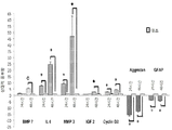
Mueller 교세포의 공지된 표지 유전자들은 마이크로어레이 (글루타민 합성효소 (Glu1), 클러스테린(Clu), 딕코프(dickkopf) 상동체 3 (Dkk3), 아쿼포린(aquaporin) 4 (Aqp4), S100 칼슘 결합 단백질 A16, 아포리포프로테인(Apolipoprotein) E (ApoE), 빈멘틴(Vimentin) (VIM), 그리고 아교세포섬유산성단백질, 교섬유산성단백질(GFAP))에서 탐지되었다.Known marker genes of Mueller glutamate are microarrays (glutamine synthetase (Glu1), clutelin (Clu), dickkopf homolog 3 (Dkk3), aquaporin 4 (Aqp4), S100 calcium binding Protein A16, apolipoprotein E (ApoE), vimentin (VIM), and glial cell fibrous acid protein, fibrous acid protein (GFAP).
1894개의 유전자는 ESMV 처리 후 3가지 시점 모두에서 상이하게 발현되었는데, 801개 유전자는 상향 조절되었고, 1093개 유전자는 하향-조절되었다(도 4a). 대조 Mueller 세포와 비교하여 ESMV-처리된 Mueller 세포에서 유전자의 단단한 클러스터링이 관찰되었고, 이때 처리된 세포는 광범위한 유전자에 걸쳐 유사한 유전자 발현 프로파일을 공유한다(도 4b). 유전자 발현 변화의 60% 이상은 처리 후 8시간에서 발생되었다. 1444개의 유전자는 ESMV 처리 후 8시간 시점에서 상향-조절되었고, 1878개의 유전자는 하향-조절되었고, 1623개의 유전자는 ESMV 처리 후 24시간 시점에서 상향-조절되었고, 1828개의 유전자는 ESMV 처리 후 24시간 시점에서 하향-조절되었고, 그리고 1711개의 유전자는 48시간 시점에서 하향-조절되었고, 1907개의 유전자는 하향-조절되었다. 유전자 발현 변화의 대부분 (95%)은 24 시간에 발생되었고, 48-시간 시점에 독특한 세포는 단지 624개의 유전자다(도 4a). 1894 genes were differentially expressed at all three time points after ESMV treatment, with 801 genes up-regulated and 1093 genes down-regulated (FIG. 4A). Hard clustering of genes was observed in ESMV-treated Mueller cells as compared to control Mueller cells, where treated cells share a similar gene expression profile across a wide range of genes (Fig. 4B). More than 60% of gene expression changes occurred at 8 hours after treatment. 1444 genes were up-regulated at 8 hours after ESMV treatment, 1878 genes were down-regulated, 1623 genes were up-regulated at 24 hours after ESMV treatment, 1828 genes were detected 24 hours after ESMV treatment , And 1711 genes were down-regulated at 48 hours and 1907 genes were down-regulated. Most of the gene expression changes (95%) occurred at 24 hours, and at 48-hour time, only 624 genes were unique (Figure 4a).
유전자 온톨로지(GO) 분석에서 처리 후 24시간 및 48시간 시점에 차등적으로 조절된 유전자중 많은 유전자들은 전사 인자 패밀리, 망막생성에 관련된 유전자, 기관 및 유기체 발달, 그리고 세포-세포 신호생성 분자를 인코드하는 유전자들, 형태 발생에 관련된 수용체들, 다중 사이토킨, 그리고 면역반응 유전자에 속한다는 것이 밝혀졌다. 기능적 분류에 의해 집단화될 때(도 5a), ESMV-처리된 Mueller 세포는 세포 이동 그리고 세포외 매트릭스 조성물, 염증, 세포 성장 및 증식, 손상, 분자 이동, 에너지 대사, 배아 발생, 세포 생존, DNA 복제에 대한 조직 반응, 그리고 안과 질환에 관련된 유전자로 차등적으로 발현되었다(표 S1). 모든 시점에서 ESMV-처리된 Mueller 세포에서 차등적으로 발현된 1894개 유전자중 많은 것들은 다른 무엇보다도 다음의 정규 신호생성 경로에 연결되어 있었다: 고유한 면역 세포와 적응 면역 세포간의 소통, G-단백질 결합된 수용체 신호생성, 비타민 D 수용체 그리고 레티노산 X 수용체 활성화, (신경 망막 발달에 연관된), IL6 신호생성 (망막-보호 경로), 뉴레글린 신호생성(망막 뉴런 생존을 촉진시키고, 망막발달에서 신경돌기 돌출에 역할을 하는 경로 (Bermingham-McDonogh et al. (1996) Development 122:14271438)), 그리고 엑손 유도(도 5b). Many of the genes that are differentially regulated at the 24 and 48 hour time points in the gene ontology (GO) analysis are the transcription factor family, genes involved in retinal production, organ and organism development, and cell- Genes that encode, receptors involved in morphogenesis, multiple cytokines, and immune response genes. When grouped by functional classification (Fig. 5A), ESMV-treated Mueller cells are characterized by cell migration and extracellular matrix composition, inflammation, cell growth and proliferation, impairment, molecular migration, energy metabolism, embryogenesis, (Table S1), and the genes associated with ocular disease (Table S1). Many of the 1894 genes that were differentially expressed in ESMV-treated Mueller cells at all time points were linked, among other things, to the following normal signaling pathways: communication between unique immune cells and adaptive immune cells, Stimulated retinal neuronal survival and stimulated neurite outgrowth in retinal development and stimulated retinal neuronal survival in the retinal development. (Bermingham-McDonogh et al. (1996) Development 122: 14271438)), and exon induction (Figure 5b).
상향-조절된 유전자들중 전분화능 유전자 Oct4, Lin28, Klf4, 그리고 LIF, 초기 망막 유전자 Bmp7, Olig2, FoxN4, Dll1, Pax6, 그리고 Rax, 유전자 IL6, 공지의 망막 보호 성질들을 가진 CSF2 , 그리고 망막 재생의 유도물질 (FGF2, IGF2, GDNF), 뿐만 아니라 조직 재건을 위한 허용 환경을 창조하는 것으로 공지된 다중 세포외 매트릭스 변형 분자, 이를 테면 매트릭스 금속단백분해효소 3에 대한 유전자(MMP3)가 있다. 하향-조절된 유전자들중 분화를 촉진시키는 것들, 이를 테면 DNMT3a 그리고 GATA4, 억제성세포외 매트릭스 성분, 이를 테면 Aggrecan, 헤파린 술페이트, 그리고 Tenascin, 그리고 억제성반흔 조직 성분, 이를 테면 GFAP 그리고 콘드로이틴 술페이트 프로테오글리칸이 있다. 이들 유전자에서 발현 변화는 처리후 24시간 및 48시간 시점에서 더욱 드러났다. 전분화능-유도 인자로 특징화가 잘 된 c-Myc의 발현이 Mueller 세포에서 탐지되는 동안, ESMV 처리 과정 동안 변화되지 않은 상태로 유지되었다. 세포 주기 재-진입, 탈-분화 그리고 Mueller 세포에서 망막 줄기 세포 표현형의 활성화를 조절하는 유전자인 Hes1, Notch 1, Notch2, 그리고 NeuroD1은 ESMV 처리 후 8시간 시점에서 매우 상향-조절되었고, 이때 이들 수준은 기준 이상으로 증가된 상태로 유지되지만 다른 시점들에서는 감소된다. 망막생성 동안 Mueller 교세포 운명을 향하여 망막 선조들이 유도되는데 관련된 유전자인 EGFR의 발현은 이들 3가지 시점에서 하향-조절되었다. Olig2, FoxN4, Dll1, Pax6 , and Rax , the gene IL6, CSF2 with known retinal protection properties, and retinal regeneration in the up-regulated genes Oct4, Lin28, Klf4 and LIF , the early retinal genes Bmp7, ( FGF2, IGF2, GDNF ) as well as multiple extracellular matrix modified molecules known to create an acceptable environment for tissue reconstruction, such as the gene for matrix metalloproteinase 3 ( MMP3 ). One of the down-regulated genes that promote differentiation, such as DNMT3a and GATA4 , an inhibitory extracellular matrix component such as Aggrecan, heparin sulfate, and Tenascin, and inhibitory scar tissue components such as GFAP and chondroitin sulfate There is proteoglycan. Expression changes in these genes were more evident at 24 and 48 hours after treatment. Expression of c-Myc , characterized by pre-multipotency-inducible factors, remained unchanged during detection of Mueller cells during ESMV treatment. Hes1, Notch1, Notch2, and NeuroD1 , which regulate cell cycle re-entry, de-differentiation and activation of the retinal stem cell phenotype in Mueller cells, were highly up-regulated at 8 hours after ESMV treatment, Is maintained in an increased state above the reference, but is reduced at other times. The expression of EGFR , a gene involved in retinal afferents leading to Mueller glia fate during retinal formation, was down-regulated at these three time points.
ESMV 처리에 의해 유도된 이들 Mueller 세포의 전사체에서 관찰된 변화는 이들 세포의 증식성과 재생성 프로그램의 활성화를 통하여 가능한 한 더욱 탈-분화된 상태로 이동을 보여준다. The observed changes in the transcripts of these Mueller cells induced by ESMV treatment show migration to a more de-differentiated state as far as possible through activation of the proliferation and regeneration programs of these cells.
추가적으로, 마이크로어레이 데이타 분석은 ESMVs에 노출된 Mueller 세포에서 수평 및 아마크린 망막 뉴런의 표지인 칼빈딘(calbindin) 1, 아마크린 세포의 표지인 Syntaxin 1a, 그리고 로드 광수용기의 표지인 로돕신을 포함한 다양한 망막 계통의 표지들을 인코드하는 몇 가지 유전자의 상향 조절을 나타낸다. 칼빈딘 1의 발현은 ESMV 처리 후 48시간에 가장 높았고, 한편 로돕신 및 Syntaxin 1a의 발현은 모든 테스트된 시점에서 증가되었다. In addition, microarray data analysis showed that in Mueller cells exposed to ESMVs,
이러한 발견은 Mueller 세포의 탈-분화되는 하위집단은 다른 망막 계통의 세포로 전환-분화된다는 것으로 설명된다. This finding is explained by the fact that the de-differentiating subpopulations of Mueller cells convert into different retinal system cells.
실시예 8Example 8
ESMV-처리된 망막 선조세포에서 miRNA 발현 분석MiRNA Expression Analysis in ESMV-Treated Retinal Tissue Cells
miRNAs는 망막생성, 초기 단계로부터 말기 단계로의 망막 선조세포 진행 조절, 그리고 다양한 망막 세포 계통으로의 이들 분화에 역할을 한다. 따라서, ESMVs에 의해 Mueller 세포로 전달되는 miRNAs가 Mueller 세포의 miRNA 및 mRNA 발현 프로파일을 변경시키고, 탈-분화된 상태로 이들 세포를 이동시키는지를 결정하기 위한 테스트가 실행되었다. miRNAs play a role in retinal formation, regulation of retinal progenitor cell progression from early stage to late stage, and their differentiation into various retinal cell lines. Thus, a test was conducted to determine whether miRNAs delivered by Mueller cells by ESMVs alter miRNA and mRNA expression profiles in Mueller cells and migrate these cells in a de-differentiated state.
상기에서 설명된 것과 같이 준비된 1μg의 전체 RNA 시료들은 Hy3™ 로 라벨되었고, 라벨된 miRNAs는 miRNA 어레이에 혼성화되었다. 927/648/351개의 인간/마우스/렛 miRNAs 뿐만 아니라 438개 miRPlus miRNAs가 포함된 Exiqon miRCURY LNA miRNA 어레이 (마이크로어래이 v11)는 제조업자의 지시에 따라 이용되었다. MiRNA 어레이 슬라이드는 Axon GenePix 4100A 스캐너(Molecular Devices, Sunnyvale, CA)로 스캔되었고, GenePix Pro 6.0 소프트웨어 (Molecular Devices)로 처리되었다. 가공안된 miRNA 데이터는 하우스키핑 miRNAs 및 불변 miRNAs의 조합을 이용하여 표준화되었다. 통계학적으로 상이한 miRNAs는 ≥ 3-배의 임계치 및 FDR 교정된 p < 0.05와 함께 Partek 게놈 세트(genomic suite) 6.4를 이용하여 선택되었다. 개별 miRNAs는 miRBase 온라인 데이터베이스(http://www.mirbase.org/)를 이용하여 연구되었고, 그리고 miRNA 표적 예측 분석은 TargetScan 6.0 소프트웨어 (http://www. targetscan.org/)을 이용하여 실행되었다. 1 [mu] g of total RNA samples prepared as described above were labeled Hy3 (TM), and labeled miRNAs were hybridized to miRNA arrays. Exiqon miRCURY LNA miRNA arrays (microarray v11) containing 438 miRPlus miRNAs as well as 927/648/351 human / mouse / rat miRNAs were used according to the manufacturer's instructions. The MiRNA array slides were scanned with an Axon GenePix 4100A scanner (Molecular Devices, Sunnyvale, CA) and processed with GenePix Pro 6.0 software (Molecular Devices). Unprocessed miRNA data were standardized using a combination of housekeeping miRNAs and invariant miRNAs. Statistically different miRNAs were selected using the Partek genomic suite 6.4 with ≥ 3-fold threshold and FDR corrected p <0.05. Individual miRNAs were studied using the miRBase online database (http://www.mirbase.org/), and miRNA target predictive analyzes were performed using TargetScan 6.0 software (http://www.targetscan.org/) .
결과result
전체적으로, 173개 miRNAs의 발현이 ESMV 처리에 의해 변경되었고; 이들 miRNAs중 25개는 테스트된 3가지 시점 모두에서 차등적으로 발현되었고(도 7a), 이중 11개는 상향-조절되었고, 14개는 하향-조절되었다 (도 7a). 25개 miRNAs는 ESMV 처리후 8시간 시점에서 상향조절되었고, 16개는 하향-조절되었으며, 24시간 시점에서 32개 miRNAs는 상향-조절되었고, 28개는 하향-조절되었고, 그리고 48시간 시점에서 87개 miRNAs는 상향-조절되었고, 61개는 하향-조절되었다(도 7b). miRNA 발현에서 변경의 대부분은 ESMV 노출 후 48시간 시점에서 발생되었다. Overall, the expression of 173 miRNAs was altered by ESMV treatment; Twenty-five of these miRNAs were differentially expressed at all three time points tested (Figure 7a), 11 of which were up-regulated and 14 were down-regulated (Figure 7a). Twenty-five miRNAs were up-regulated at 8 hours after ESMV treatment and 16 were down-regulated. At 24 hours, 32 miRNAs were up-regulated, 28 were down-regulated, and at 48 hours, Dog miRNAs were up-regulated and 61 were down-regulated (Figure 7b). Most of the changes in miRNA expression occurred at 48 hours after ESMV exposure.
ESMV-처리된 Mueller 세포에서 망막 발달시 상당히 발현되는 miR-1, miR96, miR-182 그리고 miR-183을 포함하는 miRNAs중 몇가지는 상향조절되었다. ESC 전분화능의 유지에 관련된 miRNA 클러스트인 290 클러스터에 속하는 miRNAs(miR-291b-5p, -292, -294, 그리고 -295)는 상향-조절되었고, ESMV-노출 후 48시간을 지나도 증가된 상태로 유지되었다. 동시에, 세포 주기 진행을 억제하고, 세포 분화를 촉진시키는 miR-let-7 클러스트에 속하는 miR-let-7b 및 miR-let-7c의 발현은 ESMV 처리 후 감소되었다. Yan 단백질의 발현을 억제하고, 그리고 광수용기 분화를 증가시키는 miR-7[44], 뿐만 아니라 성인 망막에 매우 풍부한 miR-125-2b는 ESMV 처리후 48시간을 지나서도 하향-조절되었다. miRNAs중에서 테스트된 3개 모든 시점에서 차례로 골격 근모세포 및 근원성 줄기 세포의 세포 증식을 촉진시키고, 분화를 억제시키는 miRNAs인 miR-133a (30-배 증가됨) 및 miR-146a (37-배 증가됨)는 상당히 상향-조절되었고, 후자는 망막 선조 분화를 조절하는 동일한 경로인 Notch 신호생성 경로를 통하여 활성화된다. miRNAs중에서 테스트된 3개 모든 시점에서 차례로 ESCs의 분화, 신경모세포, 그리고 평활근 선조세포의 분화를 차례로 촉진시키는 miR-199b-5p (70-배 감소됨), miR-214 (37-배 감소됨), 그리고 miR-143 (13-배 감소됨)가 상당히 하향-조절되었다(Cordes et al. (2009) Nature 460:705U780; Letzen et al. (2010) PLoS One 5:e10480; Fischer et al. (2001) Nat. Neurosci. 4:247252) (도 8). ESMV 노출 후 Mueller 세포에서 miRNA 발현 변화에서 관측된 프로파일은 mRNA 발현 변화에서 관찰된 것과 일치되는 탈-분화를 증명한다. Some of the miRNAs, including miR-1, miR96, miR-182 and miR-183, which are highly expressed in retinal development in ESMV-treated Mueller cells, were up-regulated. MiRNAs (miR-291b-5p, -292, -294, and -295) belonging to the 290 clusters of miRNA clusters involved in maintenance of ESC pre-differentiation capacity were up-regulated and increased after 48 hours of ESMV- Respectively. At the same time, expression of miR-let-7b and miR-let-7c, which belong to the miR-let-7 clusters inhibiting cell cycle progression and promoting cell differentiation, were reduced after ESMV treatment. MiR-7 [44], which inhibits the expression of Yan protein and increases photoreceptor differentiation, as well as miR-125-2b, a very abundant miR-125-2b in the adult retina, was down-regulated after 48 hours of ESMV treatment. miR-133a (30-fold increased) and miR-146a (37-fold increased), miRNAs that inhibit differentiation and promote skeletal myoblast and stem cell proliferation in turn at all three time points tested among miRNAs, Was significantly up-regulated and the latter was activated through the Notch signaling pathway, the same pathway that regulates retinal neuronal differentiation. miR-199b-5p (70-fold reduced), miR-214 (37-fold reduced), which in turn promoted the differentiation of ESCs, neuroblasts, and differentiation of smooth muscle progenitor cells in turn at all three tested time points in miRNAs, and miR-143 (13-fold reduced) was significantly down-regulated (Cordes et al. (2009) Nature 460: 705U780; Letzen et al. (2010) PLoS One 5 : e10480; Fischer et al . Neurosci. 4: 247252) (Fig. 8). The profile observed in miRNA expression changes in Mueller cells after ESMV exposure demonstrates the de-differentiation consistent with that observed in mRNA expression changes.
전분화능의 유지 (miR294, -146a, -133a) 및 분화 (miR-199b-5p, -214, -143)에 관련된 몇 가지 miRNAs들이 마이크로어래이 결과를 증명하기 위하여 선택되었다. ESMV 처리 후 24시간 및 48시간 시점(시점은 miRNA 발현 변화의 다수에 상응함)에서 Mueller 세포와 처리안된 대조 세포에서 전체 RNA 시료는 각 miRNA에 특이적인 줄기-루프 RT 프라이머를 포함하는 TaqMan miRNA Assays를 이용하여 qRT-PCR를 받게되었다. qRT-PCR 결과는 테스트된 모든 miRNAs에 대한 마이크로어레이 스크리닝에 의해 관찰된 발현 패턴을 확인하였다(도 8). Several miRNAs related to maintenance of pre-differentiation potential (miR294, -146a, -133a) and differentiation (miR-199b-5p, -214, -143) were selected to demonstrate microarray results. Total RNA samples in Mueller cells and untreated control cells at 24 and 48 h after ESMV treatment (corresponding to a large number of changes in miRNA expression) were incubated with TaqMan miRNA Assays containing stem-loop RT primers specific for each miRNA Were used for qRT-PCR. The qRT-PCR results confirmed the observed expression patterns by microarray screening for all miRNAs tested (Figure 8).
실시예 9Example 9
ESMV-유도된 망막 선조세포 전환분화의 면역세포화학적 분석Immunocytochemical analysis of ESMV-induced retinal neuronal differentiation
ESMVs로 처리된 Mueller 세포의 배양물에서 관찰된 형태학적으로 이질성 세포 집단을 더 특징화시키고, 그리고 마이크로어래이 데이터를 입증하기 위하여, 면역조직화학에 의해 처리안된 대조세포와 비교하여 ESMV-처리된 Mueller 세포들에서 다양한 망막 세포 계통의 표지들의 발현이 조사되었다. In order to further characterize the morphologically heterogeneous population of cells observed in cultures of Mueller cells treated with ESMVs, and to demonstrate microarray data, ESMV-treated Mueller ™ cells were compared with untreated control cells by immunohistochemistry The expression of various markers of the retinal cell lineage was examined in the cells.
결과result
ESMVs로 8회 처리된 Mueller 세포는 6-웰 배양 플레이트에 위치된 폴리-D-리신피복된 유리 덮개상에 파종되었고, 부착되도록 하였고, 그리고 상기에서 설명된 바와 같이 6 T175 플라스크로부터 유도된 ESMVs로 처리되었다. 24 시간 후, ESMV-처리된 세포와 대조 세포는 0.1 M PBS에서 헹궈내었고, 4% 파라포름알데히드(Electron Microscopy Sciences, Hatfield, PA)에서 30분간 고정되었고, 그 다음 0.1 M PBS에서 헹궈내고, 그리고 1% 소 혈청 알부민(BSA)이 포함된 10% 혈청과 0.5% Triton X-100에서 실온에서 1시간 동안 차단되었다. 1차 항체 및 2차 항체들은 3% 혈청, 1% BSA 그리고 0.5% Triton X-100가 포함된 PBS로 희석되었다. 세포는 4℃에서 하룻밤 동안 다음의 1차 항체와 항온처리되었다: 토끼 단클론성 항-Brn3a (1:500, Abcam, Cambridge, MA), 마우스 항-뉴런 핵(NeuN) 단클론성 항체 (1:500, Millipore) 마우스 항-HPC1 (Syntaxin 1a) 단클론성 항체 (1:1000, Sigma, St Louis, MO), 마우스 항-글루탐산 데카르복실라제 67 (Gad67) 단클론성 항체 (1:000, Millipore), 마우스 항-1D4 (로돕신) 단클론성 항체 (1:10,000, Millipore), 토끼 항-GS 다클론 항체 (1:1000, Sigma), 마우스 항-파르알부민 단클론성 항체 (1:1000, Swant, Marly, Switzerland) 그리고 기니아 피그 항-소낭 글루탐산염 운반물질 2 (vGluT2) (1:10,000, Millipore). ESMV-처리된 세포와 대조 세포는 AlexaFluor488, AlexaFluor568, 또는 AlexaFluor594 (Molecular Probes, Eugene, OR)에 접합된 적절한 2차 항체와 RT에서 1-2시간 동안 항온 처리되었고, 1:1000으로 희석되었다. 덮개는 핵 대조염색 DAPI (4,6-디아미디노-2-페닐인돌; Invitrogen)가 포함된 ProLong® Gold Antifade Reagent을 이용하여 슬라이드 상에 탑재되었고, 건조되도록 두었고, 그리고 캡쳐 및 프로세싱을 위하여 Olympus FluoView 소프트웨어를 이용하여(Olympus America, Center Valley, PA) Olympus FluoView FV1000 공촛점 레이져 스캐닝 현미경으로 상들이 획득되었다. Mueller cells treated with
Mueller 세포 표지인 글루타민 합성효소 (GS) (도 9a 및 9b)에 추가하여, 아마크린 및 수평 세포의 표지인 Gad67(도 9a 및 9c), 아마크린 및 망막 신경절 세포의 표지인 NeuN (도 9g 및 9i), 망막 신경절 세포의 표지인 Brn3a (도 9M), 그리고 아마크린 세포의 표지인 Syntaxin 1a(도 9o)에 대한 면역반응은 ESMV-처리된 Mueller 세포의 작은 집단에서 관찰되었다. 이들 표지들중 어느 것도 처리안된 대조 배양물 (도 9g 9l, 9n, 9p, 및 9r)에 존재하지 않았다. 흥미로운 것은, 로드 광수용기인 로돕신에 대한 면역반응은 매우 적은 수의 처리된 세포에서 세포질 과립에 주로 국소화되어 있는 것을 볼 수 있었다(도 9Q). 양극 및 수평 세포 표지인 파르알부빈(Parvalbumin), 또는 양극 및 광수용기 세포 말단에 대한 표지인 소낭 글루탐산염 운반물질(Vesicular Glutamate Transporter) 1에 대한 면역 반응도 찾아볼 수 없었다. 1차 항체들이 생략되었을 때 착색은 관찰되지 않았다. In addition to the Mueller cell markers Glutamine Synthase (GS) (Figures 9a and 9b) (Figure 9a and 9b), amacrine and horizontal cell markers Gad67 (Figures 9a and 9c), amaracrine and retinal ganglion cell marker NeuN 9i), Brn3a (FIG. 9M), a marker for retinal ganglion cells, and Syntaxin 1a (FIG. 9o), marker for amaranthin cells, were observed in a small group of ESMV-treated Mueller cells. None of these markers were present in the untreated control cultures (Fig. 9g, 9n, 9p, and 9r). Interestingly, the immune response to rhodopsin, a rod photoreceptor, was localized predominantly to cytoplasmic granules in a very small number of treated cells (Fig. 9Q). No immune response was found to the anodic and horizontal cell markers Parvalbumin, or to the
ESMV 처리는 Mueller 세포를 망막 신경 계통의 세포, 수평 또는 양극 세포가 아닌, 주로 아마크린 그리고 망막 신경절 세포로의 전환유도를 유도하였다는 것을 이들 데이터는 암시한다. ESMV 노출 후 로돕신의 매우 제한된 발현은 ESMV 처리가 또한 광수용기 계통 유전자의 최소한 부분적인 활성화를 유도하였다는 것을 암시한다. These data suggest that ESMV treatment induces Mueller cells to induce the conversion of cells to the retinal nervous system, not horizontal or bipolar cells, mainly into amaranthin and retinal ganglion cells. The very limited expression of rhodopsin after ESMV exposure suggests that ESMV treatment also induced at least partial activation of the photoreceptor lineage gene.
실시예 10Example 10
처리된 망막 선조세포에서 전사체 수준 변화의 입증Evidence of Transcript Level Changes in Treated Retinal Tissue Cells
24 시간 및 48 시간 동안 ESMV 처리를 받았던 Mueller 세포(재생 세포 집단의 대표적)로부터 RNA의 qRT-PCR로 입증된 바와 같이, 탈-분화(Cyclin D2, BMP7), 망막 보호 (IL6, IGF2), 복구 및 조직 재건 (MMP3) 과정에 관련된 유전자의 하위집단 발현 프로파일, 뿐만 아니라 반흔 형성(GFAP) 및 ECM 성분 억제(Aggrecan)에 관련된 유전자의 발현 프로파일 모두가 마이크로어레이 상에 있는 ESMV-처리된 집단에서 상당히 변화되었다. 망막 보호 및 재생 과정에 관련된 유전자의 발현에서 변화의 대부분이 24 시간 및 48 시간이내에 관찰되었기 때문에 이들 시점들이 선택되었다. ( Cyclin D2, BMP7 ), retinal protection ( IL6, IGF2 ), and repair , as evidenced by qRT-PCR of RNA from Mueller cells (representative of the reprogramming cell population) that had been subjected to ESMV treatment for 24 hours and 48 hours and tissue reconstruction (MMP3) subpopulation expression profiles of genes involved in the process, as well as scar formation (GFAP) and the expression profile of both genes involved in ECM component-suppressed (Aggrecan) ESMV- significantly in the treated group on the microarray Changed. These points were selected because most of the changes in expression of genes involved in retinal protection and regeneration processes were observed within 24 and 48 hours.
테스트된 모든 유전자는 마이크로어레이에서 관찰된 바와 같이 동일한 조절 패턴을 나타내었다(도 6). 특히, 망막을 포함하는 newt 성인 장기 복구에서 상향 조절하고, 상승된 수준으로 존재될 때 이식된 광수용체의 망막 안으로 통합을 촉진시키는 매트릭스 금속단백분해효소를 인코드하는 MMP3 mRNA는 IL6 mRNA와 같이 마이크로어레이 및 qRT-PCR 실험에서 상당히 상향-조절되었음이 밝혀졌고; 이의 발현된 인터루킨은 안쪽 망막 뉴런 상에 보호 효과를 가지는 것으로 설명되었다. 다른 한편, 정상적인 교세포 분화 및 발달에 요구되는 콘드로이틴 설페이트 프로테오글리칸을 인코드하는 Aggrecan 유전자는 유전자들 중에서 마우스 게놈으로부터 삭제될 때 망막 이식 융합이 개선되는 유전자인 GFAP와 마찬가지로 마이크로어레이 및 qRT-PCR 연구에서 하향-조절되었다. All genes tested showed the same regulatory pattern as observed in the microarray (Figure 6). In particular, MMP3 mRNA encoding matrix metalloproteinases, which upregulate in newt adult organs including the retina and promote integration into the retina of the transplanted photoreceptors when present at elevated levels, Arrays and qRT-PCR experiments; Its expressed interleukin has been described as having protective effects on inner retinal neurons. On the other hand, the Aggrecan gene encoding chondroitin sulfate proteoglycans required for normal diploid differentiation and development, as well as GFAP , a gene whose retinal transplant fusion improves when deleted from the mouse genome, is downgraded in microarray and qRT-PCR studies - Adjusted.
mRNA 그리고 miRNA 마이크로어래이 데이터로부터 발현 변화를 입증하기 위하여, ESMV-처리된 Mueller 세포와 대조 Mueller 세포의 독립적 시료의 qRT-PCR 분석이 실행되었다. 마이크로어래이 분석에서 관심 유전자에서 발현 변화 대부분이 ESMV 노출 후 24시간 및 48시간에 일어나는 것으로 나타났기 때문에, 이들 시점이 어레이 확증에 이용되었다. 전체 RNA는 miRNeasy Mini 키트(Qiagen)를 이용하여 단리되었고, 그리고 프로토콜 마다 컬럼-상(on-column) Dnase 절단을 받았다. 유전자 발현 변화 입증을 위하여, RNA는 상기에서 설명된 바와 같이 cDNA로 전환되었고, 그리고 qPCR은 Expression Assay 프로토콜에서 잠재적인 게놈 DNA 증폭을 제거하기 위하여 엑손-엑손 연결점을 걸쳐잇기 위하여 선택된 다음의 TaqMan® 프라이머를 이용하여 실행되었다(Applied BioSystems, https://products. appliedbioSystems.com/ab/en/US/adirect/ab?cmd=catNavigat2&catID = 601803): BMP7, IL6, MMP3, IGF2, Cyclin D2, Aggrecan, 그리고 GFAP, 그리고 내생 대조로 이용된 Gapdh. 각 실험에서, 역전사효소 없는 시료와 주형 없는 시료는 특이성 및 DNA 오염이 없음을 설명하기 위하여 포함되었다. To demonstrate expression changes from mRNA and miRNA microarray data, qRT-PCR analysis of independent samples of ESMV-treated Mueller cells and control Mueller cells was performed. These points were used for array validation because most of the expression changes in the gene of interest in the microarray analysis were seen to occur at 24 and 48 hours after ESMV exposure. Total RNA was isolated using miRNeasy Mini kit (Qiagen) and subjected to on-column Dnase cleavage per protocol. To demonstrate gene expression changes, the RNA was converted to cDNA as described above and qPCR was amplified using the following TaqMan primers selected to link over the exon-exon junctions to remove potential genomic DNA amplification in the Expression Assay protocol (Applied BioSystems, https: // products. AppliedbioSystems.com/ab/en/US/adirect/ab?cmd=catNavigat2&catID=601803): BMP7, IL6, MMP3, IGF2, Cyclin D2, Aggrecan , and GFAP , and Gapdh used as endogenous contrast. In each experiment, reverse transcriptase-free and template-free samples were included to demonstrate specificity and absence of DNA contamination.
miRNA 어레이 입증을 위하여 다음의 TaqMan® miRNA 분석(Applied BioSystems, 상기에서 언급된 링크)이 제조업자의 프로토콜에 따라 이용되었다: snRNA U6에 대하여 표준화된, hsa-miR-146a, hsa-miR133a, mmumiR-294, hsa-miR-199-5p, hsa-miR-214*, hsa-miR-143. 유전자 발현에서 배수-변화는 테스트된 각 mRNA 및 miRNA에 대해 2-ΔΔCt 방법을 이용하여 산출되었고; 3개의 생물학적 복제는 각 시료에서 나란하게 실시되었고, 각 프라이머는 삼중으로 실시되었다. 스튜던트 t-테스트는 유전자 발현 변화의 유의성을 평가하기 위하여 이용되었다. The following TaqMan® miRNA assays (Applied BioSystems, a link mentioned above) were used according to the manufacturer's protocol for miRNA array demonstration: hsa-miR-146a, hsa-miR133a, mmumiR-294 , hsa-miR-199-5p, hsa-miR-214 *, hsa-miR-143. Drain-changes in gene expression were calculated using the 2 -ΔΔCt method for each mRNA and miRNA tested; Three biological replicates were run side by side in each sample, and each primer was run in triplicate. Student's t -test was used to assess significance of gene expression changes.
실시예 11Example 11
NMDA-손상된 마우스 망막에서 hESMVs의 치료 활성의 검증Verification of therapeutic activity of hESMVs in NMDA-damaged mouse retina
망막 교세포 및 Mueller 세포를 ESMVs 에 노출시킨 상기 연구 결과에서 망막에서 ESMVs는 휴지 교세포 선조 Mueller 세포가 줄기 세포 표현형으로의 탈-분화, 세포 주기 재진입, Mueller 세포의 미세환경이 조직 재생 및 재생으로 이어지는 망막생성 프로그램을 위하여 좀더 허용 상태로의 변화를 유도하였음이 제시된다. In the above results of exposure of retinal ganglion cells and Mueller cells to ESMVs, ESMVs in the retina suggest that retinal ganglion cell anterior Mueller cells are degenerated into a stem cell phenotype, cell cycle re-entry, and the retina of the Mueller cell micro- It is suggested that the change to more permissive state is induced for the generation program.
27마리 마우스의 첫 연구에서, 어린 성인(약 7주령) 동물의 망막 신경절 세포 (RGCs) 및 내부 망막 뉴런의 손상은 각 마우스의 양쪽 눈 모두의 유리체내로 N-메틸-D-아스파르트산(NMDA, NMDA 수용체의 활성화에 의해 칼슘 이온의 세포내 유입, 활성 산소종의 생성, 그리고 궁극적으로 신경절 세포와 망막의 내부 핵 층 모두에 있는 신경 세포의 자가사멸로 이어지는 흥분독소)의 주사에 의해 유도되었다. 영향을 받은 뉴런의 세포 사멸은 NMDA 투여 후 24시간 이내에 발생되며; 따라서, 48 시간 후, BrdU 및 플루오레세인이 포함된 ESMV 현탁액(50 μg 내지 250 g) 1μl는 좌측 눈 망막아래로 주사되었고, 한편 손상된 우측 눈 (대조)에는 ESMV 현탁액과 동일한 농도의 BrdU 및 플루오레세인이 함유된 인산염 완충된 염수(PBS) 1μl가 주사되었다. (주사는 망막아래 주사와 함께 또는 주사 없이 유리체내로 실행될 수도 있다.) 유리체내 BrdU는 대부분 6시간 이내 눈에서 제거되기 때문에, BrdU는 주사 당일 복막으로 주사되었고, 그리고 그 이후 다음 6일 동안 매일 주사되었다. ESMV 노출 후 NDMA-손상된 망막에서 증식된 세포는 CRALBP (Mueller 교세포 표지), Syntaxin 1A, 그리고 Gad67 (둘다 아마크린 세포 표지)를 발현한다.In the first study of 27 mice, damage of retinal ganglion cells (RGCs) and inner retinal neurons in young adult (approximately 7 weeks old) animals was observed in the vitreous of both eyes of each mouse as N-methyl-D-aspartic acid , Intracellular entry of calcium ions by activation of the NMDA receptor, production of reactive oxygen species, and excitotoxin, which ultimately leads to autologous killing of neurons in both the ganglion cells and inner nuclear layers of the retina) . Cell death of affected neurons occurs within 24 hours of NMDA administration; Thus, after 48 hours, 1 [mu] l of ESMV suspension (50 [mu] g to 250 g) containing BrdU and fluorescein was injected below the left eye retina, while the injured right eye (control) contained BrdU and
결과result
BrdU-라벨된 세포는 형광 현미경에 의해 PBS-주사된 망막에서 탐지되지 않았지만, 주사후 7일 (도 10b, 도 11b, 및 도 12b), 뿐만 아니라 주사후 30일(도 10e, 도 11e, 도 12e) ESMV-처리된 눈에서 BrdU 착색된 소수의 세포가 관찰되었다. BrdU-양성 세포는 아마크린 뉴런의 핵이 주로 차지하는 영역인 내부 핵 층의 가장 안쪽 열(INL), 그리고 GCL (도 10b, 도 11b, 및 도 12b)에 위치하였다. ESMV 주사 후 30일 시점에서 (도 10e, 도 11e, 및 도 12e), BrdU 양성 세포의 수는 주사후 3일 시점 (도 10b, 도 11b, 및 도 12b)보다 감소되었지만, 그러나 BrdU 양성 세포는 INL에 근접한 내부 얼기층(IPL)에서 여전히 발견되었다. 세포 증식의 대부분은 잘 특징화된 Mueller 세포 표지인 CRALBP를 발현시켰는데 (도 12a 및 도 12d), 이는 Mueller 세포가 ESMV 처리에 반응하여 증식한다는 것을 설명한다. ESMV 주사후 30일에 검사된 증식 세포의 일부는 Syntaxin 1a (도 11d) 및 Gad67 (도 10d)를 발현시켰는데, 이는 이들 세포가 아마크린 계통과 함께 분화되었음을 나타낸다. 더욱이, 9마리 동물중 5마리에서 ESMV 주사후 30일 ERG b 파동에서 놀라운 개선이 관찰되었고 (NMDA-손상 후 약 51% 내지 65% 더높은 폭), ESMV 처리에 의해 망막기능의 회복이 반영된 것이다 (도 13). BrdU-labeled cells were not detected in the PBS-injected retina by fluorescence microscopy but were not detected 7 days post-injection (Fig. 10b, Fig. 11b and Fig. 12b), as well as 30 days post- 12e) Small numbers of BrdU stained cells were observed in ESMV-treated eyes. BrdU-positive cells were located in the innermost column (INL) of the inner nuclear layer, which is the region predominantly of the nucleus of the clean neurons, and GCL (Fig. 10b, Fig. 11b, and Fig. 12b). At 30 days after ESMV injection (Fig. 10E, Fig. 11E, and Fig. 12E), the number of BrdU positive cells decreased from the 3 day post-injection (Fig. 10B, Fig. 11B and Fig. 12B) Lt; RTI ID = 0.0 > (IPL) < / RTI > Most of the cell proliferation expressed well-characterized Mueller cell markers CRALBP (FIGS. 12A and 12D), demonstrating that Mueller cells proliferate in response to ESMV treatment. Some of the proliferating cells examined at 30 days post-ESMV injection developed Syntaxin 1a (FIG. 11d) and Gad 67 (FIG. 10d), indicating that these cells were differentiated with the amaranthin line. Moreover, in 5 of 9 animals, a remarkable improvement was observed at 30 days ERG b pulsation after ESMV injection (about 51% to 65% higher width after NMDA-injury), reflecting the recovery of retinal function by ESMV treatment (Fig. 13).
실시예 12Example 12
인간 ESMV 단리Human ESMV isolation
H1 및 H9 인간 ESC 계통(hESCs)은 혈청없는, 피터(feeder)-없는 조건하에서 배양되고, 확장된다. 간략하게 설명하자면, hESCs는 피터없이 hESC를 배양하는데 전형적으로 이용되는 TeSR2 및 Nutristem, (Invitrogen)의 2가지 특정된 무이종성 배지(xeno-free media) 1:1 비율에서 CELLtart™ CTS™ 특정된 기질(Invitrogen) 상에서 성장된다 . 세포는 배지의 매일 교체와 함께 유지된다. 이들 조건에서, hESCs는 전형적으로 분화되지 않은 형태를 보이며, 전분화능 표지들 Nanog, Oct4 그리고 Sox2를 발현시킨다. 그러나, hESC 조성물의 일관성을 유지시키기 위하여, 배양물은 매일 검사되며, 분화된 것으로 보이는 임의의 콜로니는 배지 교환에 앞서 수작업으로 제거된다. 세포는 물리적으로 4 내지 6일마다 한 번씩 계대된다(Karumbayaram et al. (2012) Stem Cells Transl. Med. 1(1):36-43). 합류 플레이트는 보통 1:6으로 분할된다. The H1 and H9 human ESC lines (hESCs) are cultured and expanded under serum-free, feeder-free conditions. Briefly, hESCs are used in a 1: 1 ratio of two specific xeno-free media of TeSR2 and Nutristem, (Invitrogen), typically used to cultivate hESCs without a CELLtart ™ CTS ™ -specific substrate (Invitrogen). The cells are maintained with daily replacement of the medium. In these conditions, hESCs typically exhibit undifferentiated morphology, expressing the differentiating markers Nanog, Oct4 and Sox2 . However, in order to maintain consistency of the hESC composition, cultures are examined daily, and any colonies which appear to be differentiated are removed manually prior to media exchange. Cells are physically transmitted once every 4 to 6 days (Karumbayaram et al. (2012) Stem Cells Transl. Med . 1 (1): 36-43). The confluence plate is usually divided into 1: 6.
hESMV 수집을 위하여, H1 및 H9 hESCs의 4일 내지 6일차 배양물의 배지가 수거되고, 찌꺼지와 파편화된 세포를 펠렛화시키기 위하여 3,500g에서 1 시간 동안 회전된다. 그 다음 상청액들은 정제된 hESMV 펠렛을 획득하기 위하여 200,000 g 에서 초원심분리 및 세척 단계를 겪었다(Katsman et al. (2012) PLoS ONE 7(11):e50417)). ESMV 조제물 안에 단백질 및 RNA 함량이 측정되어 단리 프로토콜의 일관성을 제공한다. For hESMV collection, medium of 4- to 6-day cultures of H1 and H9 hESCs were harvested and spun at 3,500 g for 1 hour to pellet the leavened and fragmented cells. The supernatants then underwent ultracentrifugation and washing steps at 200,000 g to obtain purified hESMV pellets (Katsman et al. (2012) PLoS ONE 7 (11): e50417)). The protein and RNA content in the ESMV preparation is measured to provide consistency of the isolation protocol.
hESMVs는 UCLA 임상 미생물 실험실(Clinical Microbiology laboratory)에서 미코플라스마(mycoplasma), 내독소, 호기성 및 혐기성 세균 그리고 곰팡이에 대하여 테스트된다. hESMVs are tested for mycoplasma, endotoxins, aerobic and anaerobic bacteria and fungi in the UCLA Clinical Microbiology laboratory.
실시예 13Example 13
hESMV 특징hESMV Features
상기에서 설명된 2가지 hESC로부터 유도된 hESMVs의 mRNAs, microRNAs, 및 단백질은 mESMVs와 비교되는데, 그 이유는 이들 세포는 외부 신호에 대한 반응 및 다양한 표지들의 발현에서 차이를 보인다(Ginis et al. (2012) Dev. Biol. 269(2):360-380). Yuan et al. ((2009) PLoS One 4(3):e4722)의 프로토콜에 따라 생성된 H1 및 H9 hESCs의 hESMVs와 mESC의 mESMVs에 RNA는 miRNeasy Mini™ 키트 (Qiagen)를 이용하여 추출되었고, 전체 RNA 뿐만 아니라 miRNAs가 단리된다. 각 RNA 시료는 부분들로 분할된다. 한 부분은 mRNA를 위하여 이용되며, 다른 부분은 miRNA 분석을 위하여 이용된다. ESC-특이적 mRNAs (Oct4, Nanog, Sox2, Lin28, 그리고 Klf4) 및 290 및 302-367 클러스터 내 miRNA의 존재는 RNA 정량화의 상대적 표준 곡선 방법을 이용하여 qRT-PCR에 의해 분석된다. 또한, 인간 및 마우스 전체 RNAs는 Affymetrix GeneChip Expression Analysis 표준 프로토콜에 따라 UCLA Clinical Microarray Core 설비를 이용하여 차례로 Affimetrix GeneChip U133 Plus 2.0 인간 유전자 발현 어레이 및 GeneChip Mouse Genome 430 2.0 발현 어레이에 혼성화된다. 어레이는 Affymetrix 7G 스캐너를 이용하여 스캔되며, 상들은 Affymetrix GeneChip Command Console 1.1을 이용하여 획득된다. 발현된 유전자는 Affymetrix 출석 콜(present calls)에 의해 확인된다. 인간 및 마우스의 차등적으로 발현된 유전자는 Partek 게놈(genomics) Suite 6.4를 이용하여 분석되고, RMA 알고리즘은 데이터 표준화에 이용된다. 유의적인 유전자의 선택을 위한 역치는 > = 2-배 및 FDR-교정된 p < 0.05으로 설정된다. 이둘 두 기준에 모두 부합되는 유전자는 상당히 상이한 것으로 간주된다. The mRNAs, microRNAs, and proteins of hESMVs derived from the two hESCs described above are compared to mESMVs because they differ in the response to external signals and the expression of various markers (Ginis et al . 2012) Dev Biol 269 (2): 360-380). Yuan et al . RNA was extracted from hESMVs of H1 and H9 hESCs and mESMVs of mESCs generated according to the protocol of ((2009) PLoS One 4 (3): e4722) using miRNeasy Mini ™ kit (Qiagen) Is isolated. Each RNA sample is divided into parts. One part is used for mRNA and the other part is used for miRNA analysis. ESC- specific mRNAs (Oct4, Nanog, Sox2, Lin28, and Klf4), and 290 and the presence of 302-367 cluster miRNA is analyzed by qRT-PCR using the relative standard curve method of RNA quantification. The human and mouse total RNAs are also hybridized to the Affimetrix GeneChip U133 Plus 2.0 human gene expression array and the GeneChip Mouse Genome 430 2.0 expression array, respectively, in accordance with the Affymetrix GeneChip Expression Analysis standard protocol using the UCLA Clinical Microarray Core facility. The array is scanned using an Affymetrix 7G scanner and the images are acquired using the Affymetrix GeneChip Command Console 1.1. Expressed genes are identified by Affymetrix present calls. Differentially expressed genes in humans and mice are analyzed using the Partek genomics Suite 6.4, and the RMA algorithm is used for data standardization. The threshold for the selection of significant genes is set to> = 2-fold and FDR-corrected p <0.05. Genes that meet both criteria are considered to be quite different.
인간 및 마우스 microRNA 분석 또한 실행된다. Exiqon miRCURY LNA microRNA 어레이는 제조업자의 지시에 따라 이용된다. miRNA 어레이는 Axon GenePix 4100A 스캐너에 의해 스캔되며 그리고 GenePix Pro 6.0 소프트웨어로 처리된다. 가공안된 miRNA 데이터는 하우스키핑 miRNAs 및 불변 miRNAs의 조합을 이용하여 표준화된다. 통계학적으로 상이한 miRNAs는 >= 2-배의 임계치 및 FDR 교정된 p < 0.05와 함께 Partek 게놈 세트(genomic suite) 6.4를 이용하여 선택된다. Human and mouse microRNA assays are also performed. The Exiqon miRCURY LNA microRNA array is used according to the manufacturer's instructions. The miRNA arrays are scanned by the Axon GenePix 4100A scanner and processed with GenePix Pro 6.0 software. Unprocessed miRNA data is standardized using a combination of housekeeping miRNAs and invariant miRNAs. Statistically different miRNAs are selected using the Partek genomic suite 6.4 with> = 2-fold threshold and FDR corrected p <0.05.
마이크로어레이 실험에서 발견된 인간 및 마우스 ESMVs 사이에 전사체 및 miRNA 수준 차이는 hESMV 및 mESMV 시료에서 대응하는 mRNAs 및 miRNAs의 qRT-PCR 분석에 의해 입증된다. 전체 RNA는 qRT-PCR (Invitrogen)용 SuperScriptTM III First-Strand Synthesis SuperMix를 이용하여 cDNA로 전환되며, qPCR은 잠재적인 게놈 DNA 증폭을 제거하기 위한 엑손-엑손 연결점을 걸쳐잇기 위하여 선택된 TaqMan® 프라이머와 내생 대조로 cDNAs의 경우 Gapdh 또는 microRNAs의 경우 snRNA U6를 이용하여 실행된다. 유전자 발현에서 배수-변화는 테스트된 각 유전자 및 miRNA에 대해 2-ΔΔCt 방법을 이용하여 산출되었고; 3개의 생물학적 복제는 각 시료에서 나란하게 실시된다. 스튜던트 t-테스트는 유전자 발현 차이의 유의성을 평가하기 위하여 이용된다. Transcript and miRNA level differences between human and mouse ESMVs found in microarray experiments are evidenced by qRT-PCR analysis of corresponding mRNAs and miRNAs in hESMV and mESMV samples. Total RNA is converted into cDNA using SuperScript TM III First-Strand Synthesis SuperMix for qRT-PCR (Invitrogen), qPCR is a TaqMan primer selected to link over the exon-exon junction to eliminate potential genomic DNA amplification In the endogenous comparison, Gapdh for cDNAs or snRNA U6 for microRNAs is performed. Drain-changes in gene expression were calculated using the 2 -ΔΔCt method for each gene and miRNA tested; Three biological replicates are carried out side by side in each sample. Student's t -test is used to assess the significance of differences in gene expression.
hESMV 단백질은 Invitrogen ProtoArray Human Protein Microarray에 혼성화되어 특징화된다. mESMVs를 이용하여 손상된 망막의 재생을 자극하는 것으로 밝혀진 단백질의 존재, 이를 테면 IL6, FGF2, 그리고 IGF2 및 이의 양은 블랏 분석에 의해 hESMVs에서 검사된다. The hESMV protein is characterized by hybridization to the Invitrogen ProtoArray Human Protein Microarray. The presence of proteins that have been shown to stimulate regeneration of damaged retina using mESMVs, such as IL6, FGF2, and IGF2 and their quantities, are examined in hESMVs by blot analysis.
hESMV 및 mESMV 표면 항원들은 ESC 표면 표지인 CD9, CD133, Delta 1, 인테그린 α6 그리고 β1, 소닉 헷지호그, Thy1, SSEA1, SSEA3, SSEA4, TRA-1-60, 그리고 TRA-1-81의 유동세포분석에 의해 비교된다. hESMVs 및 mESMVs의 다중 배치(batches)는 각각 차례로 hESC 및 mESC 배양물로부터 단리되고, BSA 및 아지드 나트륨이 보충된 PBS에 재현탁되고, 그리고 대응하는 형광색소-접합된 단클론성 항체를 이용하여 착색된다. 필요에 따라 실험 조건 및 유동세포분석 분석의 최적화는 UCLA's Flow Cytometry Core Laboratory에서 실행된다. 획득된 데이터는 CELLQuest 소프트웨어를 이용하여 분석된다.hESMV and mESMV surface antigens were analyzed by flow cytometry of ESC surface markers CD9, CD133,
실시예 14Example 14
hESMVs의 대체 단리 및 특징화Alternative isolation and characterization of hESMVs
일부 mRNAs 또는 miRNAs가 H1-유도된 hESMVs에서는 상당히 발현되지만 H9-유도된 hESMVs에서는 발현되지 않고, 반대인 경우도 일어난다면, 대체 방법이 이용된다. Some mRNAs or miRNAs are expressed fairly in Hl -induced hESMVs but not in H9-induced hESMVs, and vice versa, alternative methods are used.
Mueller 세포는 8 시간, 24 시간, 그리고 48 시간 동안 hESMVs와 함께 배양된다(mESMV 항온처리와 유사한 단백질의 양). mESMV에 노출된 후 실행되는 바와 같이 처리안된 대조 Mueller 세포와 비교함으로써 형태학적 변화가 평가된다. 배양 배지의 흡출 후, Mueller 세포는 임의의 잔류 hESMVs를 제거하기 위하여 앰플 PBS로 3회 세척된다. 그 다음 세포로부터 RNA가 단리되며, 유전자 발현 변화 (mRNAs 및 miRNAs)는 RT-qPCR에 의해 결정된다.Mueller cells are incubated with hESMVs for 8, 24, and 48 hours (amount of protein similar to mESMV incubation). Morphological changes are assessed by comparison with untreated control Mueller cells as performed after exposure to mESMV. After inhalation of the culture medium, Mueller cells are washed three times with ampoule PBS to remove any residual hESMVs. RNA is then isolated from the cells, and gene expression changes (mRNAs and miRNAs) are determined by RT-qPCR.
실시예 15Example 15
hiPSMVs를 이용한 손상된 망악의 재생Regeneration of damaged network malware using hiPSMVs
다음 테스트는 인간-유도된 다분화능 줄기 세포 미소낭포 (hiPSMVs), 배양된 인간-유도된 다분화능 줄기세포 (hiPSCs)로부터 획득된 인간 미소낭포가 선택된 hESMVs의 것에 필적하는 mRNA 및 miRNA를 보유하는지를 결정하고, 그리고 배양된 선조망막 Mueller 세포 상에서 hiPSMVs의 효과를 테스트하기 위하여 실행된다. 몇 가지 hiPSCs 계통이 Karumbayaram et al. ((2012) Stem Cells Transl. Med. 1(1):36-43))에서 설명된 프로토콜에 따라 생성되었다. 무이종성(xeno-free) 조건하에 유도되고 재프로그램된 (Sommer et al. (2009) Stem Cells 27:543-549) NHDF 및 Fibrogro hiPSC 계통이 이용되고, 그리고 GMP에 따라 특징화된다. 미소낭포는 상기에서 설명된 hESCs로부터 hESMVs를 획득하는데 이용된 동일한 프로토콜에 따라 계대 15 hiPSCs로부터 단리되었고, Mueller 세포 상에서 hiPSMVs의 효과는 hESMVs에 대하여 상기에서 설명된 것과 같이 테스트된다.The following tests determine whether human microcystins obtained from human-induced multipotential stem cell microcysts (hiPSMVs), cultured human-derived multipotential stem cells (hiPSCs) retain mRNA and miRNAs comparable to those of selected hESMVs And to test the effect of hiPSMVs on cultured anterior retinal Mueller cells. Several hiPSCs lines have been described by Karumbayaram et al . ((2012) Stem Cells Transl. Med. 1 (1): 36-43). NHDF and Fibrogro hiPSC lines are used and characterized by GMP, induced and reprogrammed under xeno-free conditions (Sommer et al . (2009) Stem Cells 27: 543-549). Microcysts were isolated from
선택된 hESMVs가 시험관 연구에서 이들이 mESMVs에서 관찰된 것들과 유사한 결과를 선조 Mueller 세포에서 만든다는 것을 보여줄 때, 대응하는 GMP 등급 hESC 계통은 WiCELL, Wisconsin에서 획득된다. 이들 세포는 선택된 내부 GMP 필적 hiPSC 계통과 함께 UCLA iPSC GMP 실험실에서 GMP 필적 조건하에 배양되고, GMP 등급 hESMVs 뿐만 아니라 hiPSMVs는 마우스 상에서 이들의 생체내 효과 연구를 위하여 상기에서 확립된 프로토콜에 따라 획득된다. The corresponding GMP-hESC lines are obtained from WiCELL, Wisconsin when the selected hESMVs show in in vitro studies that they produce similar results to those in the ancestral Mueller cells as those observed in mESMVs. These cells were cultured under GMP-matched conditions in a UCLA iPSC GMP lab with selected internal GMP-tagged hiPSC lines, and hMPMSVs as well as GMP-grade hESMVs were obtained according to the protocol established above for their in vivo efficacy studies on mice.
실시예 16Example 16
급성으로 NMDA-손상된 마우스 모델의 손상된 망막을 hESMV로 재생Regenerate the damaged retina of the NMDA-damaged mouse model acutely with hESMV
ESMVs가 휴지상태의 체류 선조세포의 탈-분화, 증식 및 초기 망막생성 프로그램의 개시를 자극하여 망막의 재거주하게 함으로써, 손상된 망막의 내생적 재생 능력을 유도하는지를 판단하기 위하여, 다음 테스트가 실행된다. To determine whether ESMVs induce endogenous regenerative capacity of the damaged retina, by inducing de-differentiation, proliferation and initiation of an early retinal formation program in dormant retention afferent cells to resuspend the retina, the following test is performed .
1. 망막기능 1. Retinal function
암소성(Scotopic) 및 광순응(photopic) ERGs는 Jules Stein Eye Institute LIFE (Life Imaging and Functional Evaluation) Core 설비에서 측정된다. ERGs의 장점은 살아있는 동물의 망막 기능 평가가 가능하다는 점이며; ERGs는 시간의 경과에 따라 기능을 판단하기 위하여 연속적으로 반복될 수 있다. 연구 시작에 앞서 이들 망막기능에 대한 정보를 수집하기 위하여 C57Bl/6J 마우스 코호트의 각 눈에 대한 기록을 얻는다. 그 다음 모든 망막은 NMDA의 안구내 주사에 의해 급성 손상되며, 그리고 손상된 수준을 평가하기 위하여 다음날 각 마우스의 양쪽 눈에 ERGs가 실행된다. 마우스는 두 집단으로 나뉜다. NMDA 주사 후 3일 차에, 한 집단에게 상기에서 선택된 (10 ng, 25 ng, 50 ng, 175 ng, 그리고 250 ng RNA/μl 멸균 염수) 투여 분량의 hESMVs가 안구내 주사되며, 다른 집단에게는 망막아래에 주사된다. 한 쪽 눈은 hESMV 주사를 맞고, 다른 쪽 눈은 대조로 삼아 PBS가 주사된다. 전달 방법의 효과를 비교하기 위하여, hESMV 주사 후 14일 시점과 30일 시점에 안구내 주사된 마우스와 망막아래 주사된 마우스에서 ERG에 의해 망막기능이 검사되며, 그리고 기능 회복 비율(%)은 hESMV-처리된 눈과 처리안된 눈에서 ERG-b 파동 절정 크기로부터 산출된다. 각 실험 집단의 하위 동물 집단은 동일한 시점에 희생되며, 그리고 이들의 눈은 고정되며, 공촛점 및 전자 현미경을 이용하여 형태학적 변화를 위하여 처리된다. 망막 형태의 개선은 가장 효과적인 전달 방법 및 hESMVs의 투여분량을 결정하기 위하여 ERG 발견과 관련되고, 이러한 것들은 후속 연구에 이용된다. Scotopic and photopic ERGs are measured at the Jules Stein Eye Institute LIFE (Core Imaging and Functional Evaluation) facility. The merits of ERGs are that it is possible to assess retinal function in living animals; The ERGs can be repeated continuously to determine the function over time. Obtain a record for each eye of the C57Bl / 6J mouse cohort to collect information about these retina functions prior to study initiation. All retinas are then acutely damaged by intra-ocular injection of NMDA, and ERGs are performed on both eyes of each mouse the next day to assess the level of injury. Mice are divided into two groups. On
NMDA-손상된 망막을 가진 마우스에서 GMP 필적 hESMVs의 통계학적으로 유의적인, 재생가능한 치료 활성을 획득하기 위하여, 가장 효과적인 것으로 확인된 투여분량과 전달방법이 각 15마리의 3개 코호트의 마우스 좌측 눈은 hESMVs으로 치료하는데 이용되며, 그리고 우측눈은 PBS로 처리된다. 8마리의 처리안된 마우스의 눈들로부터 그리고 코호트 1의 양쪽 눈으로부터 hESMV 주사 후 14일 및 30일 시점에서 암소성 및 광순응 ERG 반응이 기록된다. 기능 회복 및 세포 구조는 정량화된다. 또한, 코호트 2 및 3의 마우스는 첫 주사 후 5일 시점에 제 2 분량의 hESMVs를 제공받고, 그리고 코호트 3의 동물은 그 이후 5일 시점에 제 3 분량의 hESMV를 제공받는다. 코호트 2 및 3의 30마리 마우스의 눈에서 ERGs는 처음 hESMV 주사 이후 14일 및 30일 시점에 또한 기록된다. 수득된 결과는 반복된 hESMV 처리가 더 나은 결과를 확신하는데 필수적인지에 대한 판단을 할 수 있게 한다. To obtain a statistically significant, reproducible therapeutic activity of GMP-tagged hESMVs in mice with NMDA-injured retinas, the doses and delivery methods identified to be most effective were the mouse left eye of each of the three cohorts of 15 mice hESMVs, and the right eye is treated with PBS. Dark and light adaptive ERG responses were recorded at 14 and 30 days post hESMV injection from the eyes of 8 untreated mice and from both eyes of
실시예 17Example 17
hESMV 처리 후 세포 증식의 특징화Characterization of cell proliferation after hESMV treatment
면역-조직화학을 이용하여, 세포 증식 브로모데옥시우리딘(BrdU)의 표지는 망막 세포-특이적 표지들과 함께 공동-국소화된다. NMDA-손상된 망막된 마우스의 좌측 눈에는 상기에서 설명된 것과 같이 hESMVs와 함께 BrdU가 주사된다. 동일한 마우스의 우측 눈은 대조로 이용되어, PBS에 희석된 BrdU를 제공받는다. 적절한 시점에서, 동물은 관류되고, 이들의 눈들은 적출되고, 고정되고, 면역조직화학을 위하여 처리된다. 이떠한 세포 유형이 망막을 증식시키고 되살림으로써 hESMVs 노출에 반응하는 지를 판단하기 위하여 망막 부분은 BrdU 및 망막 세포-특이적 표지들(로돕신 (막대), 원뿔 옵신(원뿔), Brn3a (신경절 세포), Syntaxin 1a 및 Gad67 (아마크린 세포) 그리고 글루타민 합성효소 (Mueller 세포))에 대한 항체로 이중 그리고 삼중-착색된다. Using immunohistochemistry, the markers of cell proliferation bromodeoxyuridine (BrdU) co-localize with retinal cell-specific markers. In the left eye of NMDA-damaged retinal mice, BrdU is injected with hESMVs as described above. The right eye of the same mouse is used as a control and is given BrdU diluted in PBS. At the appropriate time, the animals are perfused, their eyes are extracted, fixed, and processed for immunohistochemistry. To determine whether this type of cell responds to exposure to hESMVs by proliferating and regenerating the retina, the retinal portion contains BrdU and retinal cell-specific markers (roodbys, cone, Brn3a, Syntaxin 1a and Gad67 (amaranthin cells) and glutamine synthetase (Mueller cells)).
실시예 18Example 18
hESMV 처리의 안전성 프로파일Safety profile of hESMV treatment
안전성을 평가하기 위하여, 장기 손상 및 종양 형성의 징후에 대하여 이들의 눈, 뇌, 간, 그리고 신장의 조직학에 의해 동물의 장기 생존이 결정되고 검사된다. 상기 안구 미세환경 내에서 hESMVs의 잠재적 면역원성은 조직학적, 단백체 및 유전자 발현 프로파일링을 이용하여 평가된다. 유리체내와 망막아래 두 가지 투여 경로가 독립적으로 테스트된다. NMDA 손상을 받지 않은 마우스의 무손상 눈에서 초기 연구가 실행된다. 초기 효과와 후기 효과를 조사하기 위하여 면역 반응으로부터 테스트의 시간적 경과가 평가된다. In order to assess safety, the long-term survival of animals is determined and examined by their eye, brain, liver, and kidney histology for signs of organ damage and tumor formation. Potential immunogenicity of hESMVs in the ocular microenvironment is assessed using histological, proteomic and gene expression profiling. Two routes of administration are tested independently in the vitreous and under the retina. Initial studies are performed in intact eyes of mice that are not NMDA-damaging. The time course of the test from the immune response is assessed to examine the initial and late effects.
염증 세포의 유입인 포도막염은 외부 단백질에 노출된 후 24시간 이내에 발생될 수 있고, 또는 발달하기 까지 2 내지 3주 소요될 수 있다. 시점(주사 후 1, 7, 14, 그리고 21 일 시점)마다 총 5 내지 10 마리 동물의 hESMV-주사된 눈과 대조 눈이 조직학적으로 평가되어 염증 세포 및 망막 통합의 존부를 확인한다(Caspi et al.(2008) Ophthalmic Res. 40:169-174; Caspi et al. (2010) J. Clin. Invest. 120:3073-3083). 고정(fixation) 프로토콜은 보존된 조직의 면역 조직화학을 실행하는 능력을 유지시킨다. 세포성 침윤물(infiltrate)이 관측된다면, 눈은 표준적인 그리고 차단된(masked) 방식으로 등급화되고, 면역조직화학을 이용하여 침윤 세포가 확인된다. 이 세포가 T, B, 또는 대식세포 계통의 것인지를 확인하는데에는 CD3, CD20, 그리고 CD68 표지들이 이용된다. Uveitis, an influx of infiltrating cells, can develop within 24 hours after exposure to external proteins, or can take 2-3 weeks to develop. Point is the (injection after 1, 7, 14, 21 and a point in time) of 5 to 10 hESMV- control eyes injected eye of each animal evaluated histologically to determine the presence or absence of inflammatory cells in the retina and integration (Caspi et . al (2008) Ophthalmic Res 40 :.... 169-174; Caspi et al (2010) J. Clin Invest 120: 3073-3083). The fixation protocol maintains the ability to perform immunohistochemistry of the preserved tissue. If cellular infiltrates are observed, the eyes are graded in a standard and masked manner, and infiltrating cells are identified using immunohistochemistry. CD3, CD20, and CD68 markers are used to determine whether these cells are of the T, B, or macrophage lineage.
조직학에 추가하여, 망막 세포의 자가사멸을 찾기 위하여 터널 착색(tunnel staining)이 실행된다. 이들 연구에서, 시신경에 인접된 망막을 통하여 수평 부분이 착색되고, 분석되고, 현미경을 이용하여 정량화된다. 인간-유도된 MVs의 마우스 안으로 전달이 변이생성능(xenogenicity)에 부차적인 반응을 도입한다면, 이들 연구는 면역결핍 마우스에서 반복된다. In addition to histology, tunnel staining is carried out to find the self-destruction of retinal cells. In these studies, the horizontal portion is colored through the retina adjacent to the optic nerve, analyzed, and quantified using a microscope. If transfer of human-induced MVs into the mouse introduces a secondary reaction to xenogenicity, these studies are repeated in immunodeficient mice.
세포 염증 반응 없이, hESMVs에 노출된 눈은 전-염증성 분자에 대한 단백체성 분석과 면역성 및 염증에 관련된 유전자의 상향조절을 이용하여 테스트된다. RNase-없는 조건을 이용한 RNA 단리 및 프로테아제 저해제들의 칵테일을 이용한 단백질 추출을 위하여 온전한 눈들이 준비된다. 포도막 조직 및 유리체는 단리되고, 균질화되고, 표준 키트를 이용하여 RNA 추출되거나, 또는 특이적 추출 완충액(50 mM Tris, 150 mM NaCl, 1 mM EDTA, 1% NP-40, 1 mM PMSF, 10 μg/ml 류펩틴, 10 μg/ml 아프로티닌, 그리고 1 mM 오르소바나데이트 나트륨)을 이용하여 단백질에 대해 추출된다. 이들 테스트는 4가지 시점에서 또한 실행된다: hESMV 주사 후 1, 7, 14, 그리고 21 일. 우선, Affymetrix GeneChip 2.0 ST 어레이 상에서 유전자 발현 분석을 위하여 집단 마다 4개의 눈 모듬(실험 눈, 대조 타안(fellow eye), 그리고 주사를 맞지 않은 처리안된 눈)이 이용되며; 유전자 발현 소프트웨어는 결과 평가에 이용된다. 마우스 시토카인 분석은 26-plex Luminex 어레이를 이용하여 단백질 추출물에서 실행된다. 이것은 다음을 테스트한다: 에오탁신(eotaxin), G-CSF, GM-CSF, IFN-감마, IL-10, IL-12p40, IL-12p70, IL-13, IL-17, IL-1-알파, IL-1-베타, IL-2, IL-3, IL-4, IL-5, IL-6, IL-23, IP-10, KC, MCP-1, MCP-3, MIP-1 알파, RANTES, TGF-베타, TNF-알파, 그리고 VEGF. 면역학적 반응이 유도된 예상치못한 사건에서, 이의 병적 결과는 단기 배양물의 면역 세포를 이용한 림프구 자극 연구에 의해 확인된다. Without cellular inflammatory response, eyes exposed to hESMVs are tested using proteomic analysis of pro-inflammatory molecules and upregulation of genes related to immunity and inflammation. Whole eyes are prepared for RNA isolation using RNase-free conditions and for protein extraction using cocktails of protease inhibitors. The uveal tissue and the vitreous are isolated, homogenized, RNA extracted using a standard kit, or extracted with a specific extraction buffer (50 mM Tris, 150 mM NaCl, 1 mM EDTA, 1% NP-40, 1 mM PMSF, / ml leupeptin, 10 μg / ml aprotinin, and 1 mM orthovanadate sodium). These tests are also performed at four time points: 1, 7, 14, and 21 days after hESMV injection. First, four eye assortments (experimental eye, fellow eye, and uninjected untreated eye) are used per gene for gene expression analysis on an Affymetrix GeneChip 2.0 ST array; Gene expression software is used to evaluate the outcome. Mouse cytokine analysis is performed on protein extracts using a 26-plex Luminex array. This tests the following: eotaxin, G-CSF, GM-CSF, IFN-gamma, IL-10, IL-12p40, IL-12p70, IL-13, IL- IL-1 beta, IL-2, IL-3, IL-4, IL-5, IL-6, IL-23, IP-10, KC, MCP- , TGF-beta, TNF-alpha, and VEGF. In unexpected events where an immunological response is induced, its pathological consequences are confirmed by lymphocyte stimulation studies in immune cells of short-term cultures.
실시예 19Example 19
인간 arRP의 Of human arRP rdlrdl 마우스 모델의 손상된 망막은 hESMVs로 복구 Repair of damaged mouse model of hESMVs in the retina
rd1 마우스는 Pde6b 유전자내 결함에 의한 망막 퇴행을 가진다: 엑손 7의 코돈 347내 C- > A 돌연변이. 로드 광수용기의 상실은 rd1 망막에서 삶의 초기에 시작되어 신속하게 진행된다. 로드의 내부 구획 안에 미토콘드리아는 출생 후 8일(P8)에 볼 수 있고, 이어서 외측 구획 디스크의 정돈된 더미가 파열되고, P12에서 매우 짧은 최대 길이에 도달된다(정상적인 로드와 비교하였을 때); 광수용기 핵은 P10에 색혈(pyknotic)되기 시작된다. 다음 수일 안에 로드 세포 사멸 파동은 광수용기의 핵이 포함된 망막 외측 핵 층(ONL)의 신속한 박층화를 초래한다. 대부분 로드는 P21에 사멸되며; 오직 원뿔 (모든 광수용기의 3%)만 이 시점에서 망막내에 유지되며, 그 이후 서서히 사멸된다. The rd1 mouse has retinal degeneration due to defects in the Pde6b gene: C - > A mutation in codon 347 of
rd1 마우스에서 hESMs의 치료 활성은 상기 실시예들에서 결정된 운반 방법 및 투여분량을 이용하여 조사된다. hESMV 처리의 효과는 망막 손상 수준이 Mueller 세포의 hESMV 활성화에 영향을 주는지를 결정하기 위하여 상이한 발생 시점에서 테스트된다. hESMVs는 P5, P8, P12, P15, 그리고 P20에 주사되며, 그리고 망막기능 개선은 ESMV 주사후 14일 및 30일 시점에서 기록된 ERGs의 a-파동(광수용기 반응)에서 증가로 반영된다. 퇴행 시작, 퇴행 동안 그리고 퇴행 후 바깥 핵 층의 폭을 정량화하고 (Danciger et al. (2000) Mammalian Genome 11:422-427) 그리고 회복 %를 결정하기 위하여 동일한 나이의 정상적인 마우스 망막의 ONLs의 것과 이들 수를 비교함으로써, hESMV-처리된 망막과 rd1 처리안된 망막의 형태학적 검사에 의해; 그리고 복원된 세포 유형의 면역조직화학적 확인에 의해 결과는 확증된다. Therapeutic activity of hESMs in rd1 mice is investigated using the delivery method and dosage amount determined in the above examples. The effect of hESMV treatment is tested at different time points to determine if retinal injury levels affect hESMV activation in Mueller cells. hESMVs are injected into P5, P8, P12, P15, and P20, and retinal improvement is reflected as an increase in the a-wave (photoreceptor response) of ERGs recorded at 14 and 30 days post-ESMV injection. (Danciger et al. (2000) Mammalian Genome 11: 422-427) and to determine the percent recovery of ONLs of normal mouse retinas of the same age and those of the same age By comparing the numbers, by morphological examination of hESMV-treated retina and rd1 untreated retina; And the results are confirmed by immunohistochemistry of restored cell types.
hESMV 처리에 의해 ERG a-파동 크기의 최소한 30%가 개선되고, 그리고 광수용기 세포의 약 30%가 hESMV 처리에 의해 rd1 마우스 망막에서 보충될 때, 그리고 ERG b-파동 크기의 30% 그리고 30% 아마크린 그리고 신경절 세포가 NMDA 손상 마우스 모델에서 복원될 때, hESMV 활성화된 Mueller 세포가 재생을 위한 망막의 내생적 능력을 지원하는지가 결정된다. When hESMV treatment improves at least 30% of the ERG a-wave size and about 30% of the photoreceptor cells are replenished in the rd1 mouse retina by hESMV treatment, and 30% and 30% of the ERG b- When amaracrine and ganglion cells are restored in the NMDA-damaged mouse model, it is determined whether hESMV-activated Mueller cells support the endogenous ability of the retina for regeneration.
등가물(Equivalence ( EQUIVALENTS)EQUIVALENTS)
당업계 숙련자는 일반적인 실험을 통하여 본 명세서에서 구체적으로 설명된 특정 구체예들에 대등한 다수의 등가물을 인지하거나 또는 알아낼 수 있을 것이다. 이러한 등가물은 다음의 청구범위에 포괄된다.
Those skilled in the art will recognize, or be able to ascertain using no more than routine experimentation, many equivalents to the specific embodiments specifically described herein. Such equivalents are encompassed by the following claims.
SEQUENCE LISTING <110> THE REGENTS OF THE UNIVERSITY OF CALIFORNIA <120> OCULAR THERAPEUTICS USING EMBRYONIC STEM CELL MICROVESICLES <130> UCLA-010PC <140> PCT/US13/30733 <141> 2013-03-13 <150> 61/624,701 <151> 2012-04-16 <160> 6 <170> PatentIn version 3.5 <210> 1 <211> 20 <212> DNA <213> Artificial Sequence <220> <223> Description of Artificial Sequence: Synthetic primer <400> 1 gccgggctgg gtggattctc 20 <210> 2 <211> 20 <212> DNA <213> Artificial Sequence <220> <223> Description of Artificial Sequence: Synthetic primer <400> 2 attggggcgg tcggcacagg 20 <210> 3 <211> 20 <212> DNA <213> Artificial Sequence <220> <223> Description of Artificial Sequence: Synthetic primer <400> 3 tccagaagag ggcgtcagat 20 <210> 4 <211> 20 <212> DNA <213> Artificial Sequence <220> <223> Description of Artificial Sequence: Synthetic primer <400> 4 ctttggtccc agcattcagg 20 <210> 5 <211> 23 <212> DNA <213> Artificial Sequence <220> <223> Description of Artificial Sequence: Synthetic primer <400> 5 aacaatcgcg gcggcccgag gag 23 <210> 6 <211> 22 <212> DNA <213> Artificial Sequence <220> <223> Description of Artificial Sequence: Synthetic primer <400> 6 gcctcggcgt gccggccctg cg 22 SEQUENCE LISTING <110> THE REGENTS OF THE UNIVERSITY OF CALIFORNIA <120> OCULAR THERAPEUTICS USING EMBRYONIC STEM CELL MICROVESICLES <130> UCLA-010PC <140> PCT / US13 / 30733 <141> 2013-03-13 <150> 61 / 624,701 <151> 2012-04-16 <160> 6 <170> PatentIn version 3.5 <210> 1 <211> 20 <212> DNA <213> Artificial Sequence <220> <223> Description of Artificial Sequence: Synthetic primer <400> 1 gccgggctgg gtggattctc 20 <210> 2 <211> 20 <212> DNA <213> Artificial Sequence <220> <223> Description of Artificial Sequence: Synthetic primer <400> 2 attggggcgg tcggcacagg 20 <210> 3 <211> 20 <212> DNA <213> Artificial Sequence <220> <223> Description of Artificial Sequence: Synthetic primer <400> 3 tccagaagag ggcgtcagat 20 <210> 4 <211> 20 <212> DNA <213> Artificial Sequence <220> <223> Description of Artificial Sequence: Synthetic primer <400> 4 ctttggtccc agcattcagg 20 <210> 5 <211> 23 <212> DNA <213> Artificial Sequence <220> <223> Description of Artificial Sequence: Synthetic primer <400> 5 aacaatcgcg gcggcccgag gag 23 <210> 6 <211> 22 <212> DNA <213> Artificial Sequence <220> <223> Description of Artificial Sequence: Synthetic primer <400> 6 gcctcggcgt gccggccctg cg 22
Claims (17)
Applications Claiming Priority (3)
| Application Number | Priority Date | Filing Date | Title |
|---|---|---|---|
| US201261624701P | 2012-04-16 | 2012-04-16 | |
| US61/624,701 | 2012-04-16 | ||
| PCT/US2013/030733 WO2013158258A1 (en) | 2012-04-16 | 2013-03-13 | Ocular therapeutics using embryonic system cell microvesicles |
Publications (1)
| Publication Number | Publication Date |
|---|---|
| KR20150009541A true KR20150009541A (en) | 2015-01-26 |
Family
ID=49383920
Family Applications (1)
| Application Number | Title | Priority Date | Filing Date |
|---|---|---|---|
| KR1020147031795A KR20150009541A (en) | 2012-04-16 | 2013-03-13 | Ocular therapeutics using embryonic stem cell microvesicles |
Country Status (8)
| Country | Link |
|---|---|
| US (1) | US20150079047A1 (en) |
| EP (1) | EP2838995A4 (en) |
| JP (1) | JP2015514760A (en) |
| KR (1) | KR20150009541A (en) |
| AU (1) | AU2013249786A1 (en) |
| CA (1) | CA2870466A1 (en) |
| IL (1) | IL235126A0 (en) |
| WO (1) | WO2013158258A1 (en) |
Cited By (1)
| Publication number | Priority date | Publication date | Assignee | Title |
|---|---|---|---|---|
| KR20230129828A (en) | 2022-03-02 | 2023-09-11 | 포항공과대학교 산학협력단 | Composition for preventing or treating of eye posterior segment diseases |
Families Citing this family (4)
| Publication number | Priority date | Publication date | Assignee | Title |
|---|---|---|---|---|
| KR101334404B1 (en) * | 2011-04-28 | 2013-12-12 | 포항공과대학교 산학협력단 | Method for preparing induced pluripotent stem cells using artificial microvesicles derived from embryonic stem cells |
| WO2018049284A1 (en) * | 2016-09-09 | 2018-03-15 | Cornell University | Delivery of nucleic acids, proteins and small molecules in vitreous vesicular bodies |
| US11653636B2 (en) * | 2019-11-15 | 2023-05-23 | University Of Ulsan Foundation For Industry Cooperation | Method of making a rat model of retinal degeneration and rat model made thereby |
| JP7007770B1 (en) | 2021-06-21 | 2022-02-10 | パナジー株式会社 | Eye symptom improver and eye symptom improvement method |
Family Cites Families (12)
| Publication number | Priority date | Publication date | Assignee | Title |
|---|---|---|---|---|
| US5824685A (en) * | 1995-02-01 | 1998-10-20 | The Johns Hopkins University School Of Medicine | Method of preventing proliferation of retinal pigment epithelium by retinoic acid receptor agonists |
| WO2004010959A2 (en) * | 2002-07-25 | 2004-02-05 | The Scripps Research Institute | Hematopoietic stem cells and methods of treatment of neovascular eye diseases therewith |
| US20060051860A1 (en) * | 2002-11-15 | 2006-03-09 | Kyushu Tlo Company, Limited | Method of organ regeneration |
| WO2006091895A2 (en) * | 2005-02-24 | 2006-08-31 | The Scripps Research Institute | Method for the treatment of retinopathy of prematurity and related retinopathic diseases |
| BRPI0615974A2 (en) * | 2005-07-27 | 2011-05-31 | Univ Florida | use of heat shock to treat an eye disease in an individual, method for recruiting a stem cell to an eye tissue of an individual in need thereof, use of heat shock to treat an eye disease or disorder in an individual in need of the same same, use of thermal shock to regenerate the retina in an individual in need of it, use of thermal shock to repair damage to the pgmentar retinal epithelium in an individual in need of it, use of thermal shock to treat macular degeneration in an individual individual in need thereof, pharmaceutical composition for stem cell recruitment, pharmaceutical composition for stem cell recruitment in an eye tissue, kit and method for identifying an agent that enhances stem cell recruitment in an eye tissue |
| KR101505202B1 (en) * | 2006-03-07 | 2015-03-27 | 지타 쉬로프 | Compositions comprising human embryonic stem cells and their derivatives, methods of use, and methods of preparation |
| JP5261494B2 (en) * | 2007-10-29 | 2013-08-14 | フレゼニウス メディカル ケア ドイッチェランド ゲゼルシャフト ミット ベシュレンクテル ハフツング | Use of stem cell-derived microvesicles (MV) and related methods in vitro and in vivo for the manufacture of a medicament for endothelium / epithelial regeneration of damaged or damaged tissue or organs |
| CA2711218C (en) * | 2008-01-04 | 2019-08-06 | Lydac Neuroscience Limited | A method of producing a population of differentiated cells |
| KR101268741B1 (en) * | 2009-10-06 | 2013-06-04 | 김지연 | Method for differentiation into retinal cells from stem cells |
| CN103210089A (en) * | 2010-08-13 | 2013-07-17 | 格拉斯哥大学大学行政评议会 | Therapeutic uses of microvesicles and related microRNAs |
| EP2745840B1 (en) * | 2011-08-16 | 2017-07-12 | Samsung Life Public Welfare Foundation | Composition including stem cell-derived microvesicles for use in promoting neurogenesis |
| JP2015520174A (en) * | 2012-05-31 | 2015-07-16 | ネオステム,インコーポレイティド | Human minimal embryo-like (VSEL) stem cells for treating eye diseases |
-
2013
- 2013-03-13 US US14/394,688 patent/US20150079047A1/en not_active Abandoned
- 2013-03-13 JP JP2015506995A patent/JP2015514760A/en active Pending
- 2013-03-13 CA CA2870466A patent/CA2870466A1/en not_active Abandoned
- 2013-03-13 WO PCT/US2013/030733 patent/WO2013158258A1/en active Application Filing
- 2013-03-13 AU AU2013249786A patent/AU2013249786A1/en not_active Abandoned
- 2013-03-13 KR KR1020147031795A patent/KR20150009541A/en not_active Application Discontinuation
- 2013-03-13 EP EP13778372.6A patent/EP2838995A4/en not_active Withdrawn
-
2014
- 2014-10-19 IL IL235126A patent/IL235126A0/en unknown
Cited By (1)
| Publication number | Priority date | Publication date | Assignee | Title |
|---|---|---|---|---|
| KR20230129828A (en) | 2022-03-02 | 2023-09-11 | 포항공과대학교 산학협력단 | Composition for preventing or treating of eye posterior segment diseases |
Also Published As
| Publication number | Publication date |
|---|---|
| WO2013158258A1 (en) | 2013-10-24 |
| AU2013249786A1 (en) | 2014-11-06 |
| EP2838995A4 (en) | 2016-01-06 |
| WO2013158258A8 (en) | 2014-09-04 |
| EP2838995A1 (en) | 2015-02-25 |
| CA2870466A1 (en) | 2013-10-24 |
| JP2015514760A (en) | 2015-05-21 |
| US20150079047A1 (en) | 2015-03-19 |
| IL235126A0 (en) | 2014-12-31 |
Similar Documents
| Publication | Publication Date | Title |
|---|---|---|
| Xu et al. | OLIG2 drives abnormal neurodevelopmental phenotypes in human iPSC-based organoid and chimeric mouse models of down syndrome | |
| Julia et al. | An efficient platform for astrocyte differentiation from human induced pluripotent stem cells | |
| Jadhav et al. | Development and neurogenic potential of Müller glial cells in the vertebrate retina | |
| JP7055638B2 (en) | Generation of muscle lineage cells from stem cells | |
| US9005966B2 (en) | Generation of pluripotent cells from fibroblasts | |
| US20140370007A1 (en) | Method of directed differentiation producing corneal endothelial cells, compositions thereof, and uses thereof | |
| Hou et al. | Direct conversion of human fibroblasts into neural progenitors using transcription factors enriched in human ESC-derived neural progenitors | |
| KR20150009541A (en) | Ocular therapeutics using embryonic stem cell microvesicles | |
| KR20170116921A (en) | Differentiation method of adipose-derived mesenchymal stem cells into neural stem cells, neural cells, and GABAergic neural cells | |
| De Filippis et al. | Neural stem cells and human induced pluripotent stem cells to model rare CNS diseases | |
| WO2017075271A1 (en) | Astrocyte differentiation protocol | |
| WO2017100498A1 (en) | Isolation of fusion-competent myoblasts and therapeutic applications thereof related to muscular dystrophy | |
| Sun et al. | Mutations in the transcriptional regulator MeCP2 severely impact key cellular and molecular signatures of human astrocytes during maturation | |
| Maqueda et al. | Repopulation of decellularized retinas with hiPSC-derived retinal pigment epithelial and ocular progenitor cells shows cell engraftment, organization and differentiation | |
| Gomes et al. | Astrocytes modulate neurodegenerative phenotypes associated with glaucoma in OPTN (E50K) human stem cell-derived retinal ganglion cells | |
| Gough et al. | Modeling Down syndrome in cells: From stem cells to organoids | |
| JP4576545B2 (en) | Embryonic stem cell culture method using amnion-derived factor | |
| Karamali et al. | Hepatocyte growth factor promotes the proliferation of human embryonic stem cell derived retinal pigment epithelial cells | |
| Yuan et al. | Primary culture of germ cells that portray stem cell characteristics and recipient preparation for autologous transplantation in the rhesus monkey | |
| Hills et al. | Neurite Outgrowth and Gene Expression Profile Correlate with Efficacy of Human Induced Pluripotent Stem Cell-Derived Dopamine Neuron Grafts | |
| US20150361391A1 (en) | Method for inducing tailored pluripotent stem cells using extract of plant stem cells or plant dedifferentiated stem cells, and pluripotent stem cells produced by means of the method | |
| WO2023008564A1 (en) | Method for producing highly proliferative cell, and highly proliferative cell and use thereof | |
| Guloglu et al. | Adipocytes derived from PA6 cells reliably promote the differentiation of dopaminergic neurons from human embryonic stem cells | |
| Bonaventura et al. | Induced pluripotent stem cells: an innovative patient-specific neurodegenerative disease modeling | |
| Upadhyay et al. | Transplantation of retinal pigment epithelium and photoreceptors generated concomitantly via small molecule-mediated differentiation rescues visual function in rodent models of retinal degeneration |
Legal Events
| Date | Code | Title | Description |
|---|---|---|---|
| WITN | Application deemed withdrawn, e.g. because no request for examination was filed or no examination fee was paid |