KR20140121842A - Implantable biologic holder - Google Patents
Implantable biologic holder Download PDFInfo
- Publication number
- KR20140121842A KR20140121842A KR1020147022621A KR20147022621A KR20140121842A KR 20140121842 A KR20140121842 A KR 20140121842A KR 1020147022621 A KR1020147022621 A KR 1020147022621A KR 20147022621 A KR20147022621 A KR 20147022621A KR 20140121842 A KR20140121842 A KR 20140121842A
- Authority
- KR
- South Korea
- Prior art keywords
- graft
- biomaterial
- connecting element
- central portion
- tissue
- Prior art date
Links
Images


Classifications
-
- A—HUMAN NECESSITIES
- A61—MEDICAL OR VETERINARY SCIENCE; HYGIENE
- A61F—FILTERS IMPLANTABLE INTO BLOOD VESSELS; PROSTHESES; DEVICES PROVIDING PATENCY TO, OR PREVENTING COLLAPSING OF, TUBULAR STRUCTURES OF THE BODY, e.g. STENTS; ORTHOPAEDIC, NURSING OR CONTRACEPTIVE DEVICES; FOMENTATION; TREATMENT OR PROTECTION OF EYES OR EARS; BANDAGES, DRESSINGS OR ABSORBENT PADS; FIRST-AID KITS
- A61F2/00—Filters implantable into blood vessels; Prostheses, i.e. artificial substitutes or replacements for parts of the body; Appliances for connecting them with the body; Devices providing patency to, or preventing collapsing of, tubular structures of the body, e.g. stents
- A61F2/02—Prostheses implantable into the body
- A61F2/08—Muscles; Tendons; Ligaments
- A61F2/0811—Fixation devices for tendons or ligaments
- A61F2002/0817—Structure of the anchor
- A61F2002/0823—Modular anchors comprising a plurality of separate parts
- A61F2002/0829—Modular anchors comprising a plurality of separate parts without deformation of anchor parts, e.g. fixation screws on bone surface, extending barbs, cams, butterflies, spring-loaded pins
-
- A—HUMAN NECESSITIES
- A61—MEDICAL OR VETERINARY SCIENCE; HYGIENE
- A61F—FILTERS IMPLANTABLE INTO BLOOD VESSELS; PROSTHESES; DEVICES PROVIDING PATENCY TO, OR PREVENTING COLLAPSING OF, TUBULAR STRUCTURES OF THE BODY, e.g. STENTS; ORTHOPAEDIC, NURSING OR CONTRACEPTIVE DEVICES; FOMENTATION; TREATMENT OR PROTECTION OF EYES OR EARS; BANDAGES, DRESSINGS OR ABSORBENT PADS; FIRST-AID KITS
- A61F2/00—Filters implantable into blood vessels; Prostheses, i.e. artificial substitutes or replacements for parts of the body; Appliances for connecting them with the body; Devices providing patency to, or preventing collapsing of, tubular structures of the body, e.g. stents
- A61F2/02—Prostheses implantable into the body
- A61F2/08—Muscles; Tendons; Ligaments
- A61F2/0811—Fixation devices for tendons or ligaments
- A61F2002/0847—Mode of fixation of anchor to tendon or ligament
- A61F2002/0852—Fixation of a loop or U-turn, e.g. eyelets, anchor having multiple holes
-
- A—HUMAN NECESSITIES
- A61—MEDICAL OR VETERINARY SCIENCE; HYGIENE
- A61F—FILTERS IMPLANTABLE INTO BLOOD VESSELS; PROSTHESES; DEVICES PROVIDING PATENCY TO, OR PREVENTING COLLAPSING OF, TUBULAR STRUCTURES OF THE BODY, e.g. STENTS; ORTHOPAEDIC, NURSING OR CONTRACEPTIVE DEVICES; FOMENTATION; TREATMENT OR PROTECTION OF EYES OR EARS; BANDAGES, DRESSINGS OR ABSORBENT PADS; FIRST-AID KITS
- A61F2/00—Filters implantable into blood vessels; Prostheses, i.e. artificial substitutes or replacements for parts of the body; Appliances for connecting them with the body; Devices providing patency to, or preventing collapsing of, tubular structures of the body, e.g. stents
- A61F2/02—Prostheses implantable into the body
- A61F2/08—Muscles; Tendons; Ligaments
- A61F2/0811—Fixation devices for tendons or ligaments
- A61F2002/0876—Position of anchor in respect to the bone
- A61F2002/0882—Anchor in or on top of a bone tunnel, i.e. a hole running through the entire bone
-
- A—HUMAN NECESSITIES
- A61—MEDICAL OR VETERINARY SCIENCE; HYGIENE
- A61F—FILTERS IMPLANTABLE INTO BLOOD VESSELS; PROSTHESES; DEVICES PROVIDING PATENCY TO, OR PREVENTING COLLAPSING OF, TUBULAR STRUCTURES OF THE BODY, e.g. STENTS; ORTHOPAEDIC, NURSING OR CONTRACEPTIVE DEVICES; FOMENTATION; TREATMENT OR PROTECTION OF EYES OR EARS; BANDAGES, DRESSINGS OR ABSORBENT PADS; FIRST-AID KITS
- A61F2250/00—Special features of prostheses classified in groups A61F2/00 - A61F2/26 or A61F2/82 or A61F9/00 or A61F11/00 or subgroups thereof
- A61F2250/0058—Additional features; Implant or prostheses properties not otherwise provided for
- A61F2250/0067—Means for introducing or releasing pharmaceutical products into the body
- A61F2250/0068—Means for introducing or releasing pharmaceutical products into the body the pharmaceutical product being in a reservoir
Abstract
조직 그래프트 현수 장치(10)가 플랫폼 부재(16), 그래프트 연결 요소의 일부가 조직 그래프트(22)에 대한 부착을 위한 루프를 형성하도록 상기 플랫폼 부재에 커플링된 그래프트 연결 요소(18), 및 상기 그래프트 연결 요소의 루프에 커플링되고 생체 재료(30)를 둘러싸도록 구성된 외장 부재(20)를 포함한다. 상기 외장 부재가 생체 재료를 수용하도록 구성된 개구부를 형성한다. 이용 중에, 상기 조직 그래프트가 상기 루프에 커플링되고 상기 외장 부재와 접촉하도록, 상기 그래프트 연결 요소 및 상기 외장 부재가 구성된다.A tissue graft suspending device 10 includes a platform member 16, a graft connecting element 18 coupled to the platform member such that a portion of the graft connecting element forms a loop for attachment to the tissue graft 22, And a sheath member (20) coupled to the loop of the graft connecting element and configured to surround the biomaterial (30). And the skin member forms an opening configured to receive the biomaterial. During use, the graft connecting element and the sheathing member are configured such that the tissue graft is coupled to the loop and in contact with the sheathing member.
Description
본원은 이식가능한 생체 홀더에 관한 것이다. The present invention relates to an implantable biomedical holder.
파괴되고 회복이 불가능한, 전방 십자 인대(ACL)와 같은 인대가 조직 그래프트(tissue graft)에 의해서 관절경으로 대체 될 수 있다. 조직 그래프트는 자연적일 수 있고 신체의 다른 부분으로부터 적출될 수 있다. 예를 들어, ACL 복원의 경우에, 조직 그래프트는 각각의 단부에서 소위 "뼈 블록(bone block)"을 가지는 슬개 힘줄 부분으로부터, 그리고 반건양근(semitendonosis) 및 박근으로부터 적출될 수 있다. 대안적으로, 조직 그래프트가 합성 재료로부터 또는 합성 및 천연 재료의 조합으로부터 형성될 수 있다. ACL를 복원할 때, 대퇴골(femur) 내의 통로에 형성된 소켓 내에 조직 그래프트의 일 단부를 고정하는 것, 및 경골(tibia) 내에 형성된 통로를 통해서 그래프트의 타 단부를 통과시키는 것에 의해서, 대체 조직 그래프트가 이식될 수 있다. Destroyed and unrecoverable, ligaments such as anterior cruciate ligament (ACL) can be replaced by arthroscopy by a tissue graft. Tissue grafts can be natural and can be extracted from other parts of the body. For example, in the case of ACL reconstruction, tissue grafts can be extracted from the patellofemoral portion having so-called "bone blocks " at each end, and from semitendonosis and roughening. Alternatively, the tissue graft may be formed from a synthetic material or from a combination of synthetic and natural materials. By restoring one end of the tissue graft in the socket formed in the passage in the femur and passing the other end of the graft through the passage formed in the tibia, an alternative tissue graft Can be transplanted.
치유(healing) 촉진을 위해서, 피브린 응괴(fibrin clot)와 같은 생체 재료가, 예를 들어 캐뉼러를 통해서, 인대 또는 다른 연성 조직 부상 부위 내로 삽입될 수 있고, 또는 예를 들어 봉합에 의해서, 조직 그래프트로 직접적으로 부착될 수 있다. 생체 재료를 부상 부위에 인가하는 것은 치유를 촉진하는데 도움이 될 수 있다. For promoting healing, a biomaterial such as a fibrin clot can be inserted into the ligament or other soft tissue floating site, for example, via the cannula, Can be attached directly to the graft. Applying the biomaterial to the injured area may help to promote healing.
하나의 양태에 따라서, 조직 그래프트 현수(suspension) 장치가 플랫폼 부재, 그래프트 연결 요소의 일부가 조직 그래프트에 대한 부착을 위한 루프를 형성하도록 상기 플랫폼 부재에 커플링된 그래프트 연결 요소, 및 상기 그래프트 연결 요소의 루프에 커플링되고 생체 재료를 둘러싸도록 구성된 외장 부재를 포함한다. 상기 외장 부재는 생체 재료를 수용하도록 구성된 개구부를 형성한다. 이용 중에, 조직 그래프트가 상기 루프에 커플링되고 상기 외장 부재와 접촉하도록, 상기 그래프트 연결 요소 및 상기 외장 부재가 구성된다. According to one aspect, a tissue graft suspension device includes a platform member, a graft connecting element coupled to the platform member such that a portion of the graft connecting element forms a loop for attachment to the tissue graft, And an outer member coupled to the loop of the biomaterial and configured to surround the biomaterial. The skin member forms an opening configured to receive the biomaterial. During use, the graft connecting element and the sheathing member are configured such that a tissue graft is coupled to the loop and in contact with the sheathing member.
이러한 양태의 구현예가 이하의 특징 중 하나 이상을 포함할 수 있을 것이다. Implementations of this aspect may include one or more of the following features.
예를 들어, 그래프트 연결 요소가 봉합사(suture)의 연속적인 루프일 수 있을 것이다. 루프가 외장 부재의 개구부를 통과할 수 있을 것이다. 루프가 외장 부재의 부착 요소를 통과할 수 있을 것이다. 외장 부재가 생체 재료를 수용하도록 구성된 2개의 개구부를 형성할 수 있을 것이다. 생체 재료가 피브린 응괴일 수 있을 것이다. 생체 재료가 혈소판 풍부 혈장(platelet rich plasma)일 수 있을 것이다. 조직 그래프트 현수 장치가 외장 부재에 부착된 하나 이상의 필라멘트를 더 포함할 수 있을 것이다. For example, the graft connecting element may be a continuous loop of a suture. The loop will be able to pass through the opening of the sheathing member. The loop will be able to pass through the attachment element of the sheathing member. The exterior member may form two openings configured to receive the biomaterial. The biomaterial may be a fibrin clump. The biomaterial may be a platelet rich plasma. The tissue graft suspending device may further include one or more filaments attached to the skin member.
다른 양태에 따라서, 조직 그래프트의 고정 방법이 그래프트 연결 요소에 커플링된 플랫폼 부재를 제공하는 단계로서, 상기 그래프트 연결 요소가 외장 부재에 커플링되고 상기 조직 그래프트에 대한 부착을 위한 루프를 형성하고, 상기 외장 부재가 생체 재료를 둘러싸도록 구성되는, 플랫폼 부재를 제공하는 단계, 상기 외장 부재 내의 개구부를 통해서 생체 재료를 삽입하는 단계, 상기 조직 그래프트가 상기 외장 부재와 접촉하도록 상기 조직 그래프트를 상기 그래프트 연결 요소에 부착하는 단계, 뼈 내에 뼈 터널을 형성하는 단계, 및 상기 조직 그래프트의 적어도 일부가 상기 뼈 터널 내에 위치되도록 상기 뼈의 표면 상에 상기 플랫폼 부재를 배치하는 단계를 포함한다. According to another aspect, there is provided a method of securing a tissue graft comprising providing a platform member coupled to a graft connecting element, the graft connecting element being coupled to a sheath member and forming a loop for attachment to the tissue graft, The method comprising the steps of: providing a platform member configured to enclose a biomaterial, the exterior member being configured to surround the biomaterial; inserting a biomaterial through an opening in the exterior member; contacting the tissue graft with the graft connection Attaching to the element, forming a bone tunnel in the bone, and disposing the platform member on a surface of the bone such that at least a portion of the tissue graft is located within the bone tunnel.
이러한 양태의 구현예가 이하의 특징 중 하나 이상을 포함할 수 있을 것이다. Implementations of this aspect may include one or more of the following features.
예를 들어, 상기 조직 그래프트를 상기 그래프트 연결 요소에 부착하는 단계가, 상기 외장 부재가 상기 조직 그래프트의 2개의 부분들 사이에 위치되도록 상기 조직 그래프트를 상기 그래프트 연결 요소의 루프를 통해서 루핑하는(looping) 단계를 포함할 수 있을 것이다. 상기 조직 그래프트를 상기 그래프트 연결 요소에 부착하는 단계가 상기 그래프트 연결 요소를 상기 조직 그래프트의 뼈 블록 내의 개구부를 통해서 루핑하는 단계를 포함할 수 있을 것이다. 상기 뼈 터널이 제1 개구부 및 제2 개구부를 포함할 수 있을 것이다. 상기 플랫폼 부재를 상기 뼈의 표면 상에 배치하는 단계가, 상기 플랫폼 부재를 상기 제1 개구부를 통해서 상기 뼈 터널 내로 삽입하는 단계, 상기 터널을 통해서 상기 제2 개구부의 외부로 상기 플랫폼 부재를 이동시키는 단계, 및 상기 그래프트 연결 요소에 부착된 조직 그래프트의 제1 부분이 상기 터널 내에 있도록 그리고 상기 조직 그래프트의 제2 부분이 상기 제1 개구부의 외부로 연장하도록, 상기 그래프트 연결 요소가 뼈 터널 내로 연장하는 상태에서 상기 플랫폼 부재를 상기 제2 개구부 위에 배치하는 단계를 포함할 수 있을 것이다. For example, attaching the tissue graft to the graft connecting element may include looping the tissue graft through the loop of the graft connecting element such that the skin member is positioned between two portions of the tissue graft, ) Step. Attaching the tissue graft to the graft connecting element may include looping the graft connecting element through an opening in the bone block of the tissue graft. The bone tunnel may include a first opening and a second opening. Wherein positioning the platform member on a surface of the bone comprises the steps of: inserting the platform member through the first opening into the bone tunnel, moving the platform member out of the second opening through the tunnel And the graft connecting element extends into the bone tunnel such that a first portion of the tissue graft attached to the graft connecting element is within the tunnel and a second portion of the tissue graft extends out of the first opening And placing the platform member over the second opening in a state that the platform member is in the open state.
또 다른 양태에 따라서, 생체 재료를 수용하기 위한 스캐폴드(scaffold)가, 생체 재료가 주입된(impregnated) 중앙 부분으로서, 상기 중앙 부분이 직조(weave) 패턴으로 배열된 필라멘트를 포함하는, 중앙 부분, 및 상기 중앙 부분의 엣지에 부착된 안내 부분으로서, 상기 안내 부분이 하나 이상의 필라멘트를 포함하는, 안내 부분을 포함한다. 상기 스캐폴드가 연성 조직 내의 파열부(tear) 내로 삽입되도록 구성된다. According to a further aspect there is provided a scaffold for receiving a biomaterial comprising a central portion which is impregnated with a biomaterial and the central portion comprises a filament arranged in a weave pattern, And a guiding portion attached to an edge of the central portion, wherein the guiding portion comprises one or more filaments. The scaffold is configured to be inserted into a tear in the soft tissue.
이러한 양태의 구현예가 이하의 특징 중 하나 이상을 포함할 수 있을 것이다. Implementations of this aspect may include one or more of the following features.
예를 들어, 연성 조직 내의 파열부가 반월판(meniscal) 파열부일 수 있을 것이다. 안내 부분이 상기 중앙 부분의 제1 엣지에 부착된 제1 안내 부분, 및 상기 중앙 부분의 제2 엣지에 부착된 제2 안내 부분을 포함할 수 있고, 상기 제2 엣지가 상기 제1 엣지에 대향한다. 상기 안내 부분이 상기 중앙 부분의 직조 패턴으로부터 외부로 연장하는 하나 이상의 필라멘트를 포함할 수 있을 것이다. For example, a rupture in a soft tissue may be a meniscal rupture. The guide portion may include a first guiding portion attached to a first edge of the central portion and a second guiding portion attached to a second edge of the central portion and the second edge may be opposite to the first edge, do. The guide portion may include one or more filaments extending outwardly from the woven pattern of the central portion.
추가적인 양태에 따라서, 연성 조직 파열부를 복원하기 위한 방법이, 중앙 부분 및 안내 부분을 포함하는 스캐폴드를 제공하는 단계로서, 상기 중앙 부분이 직조 패턴으로 배열된 필라멘트를 포함하는, 스캐폴드를 제공하는 단계, 상기 스캐폴드의 중앙 부분으로 생체 재료를 주입하는 단계, 상기 스캐폴드의 엣지를 상기 연성 조직 파열부 내로 삽입하는 단계, 및 상기 안내 부분을 조작하는 것에 의해서 상기 연성 조직 파열부 내의 스캐폴드의 중앙 부분을 최종 위치로 이동시키는 단계를 포함한다. According to a further aspect, a method for restoring a soft tissue rupture comprises providing a scaffold comprising a central portion and a guiding portion, wherein the central portion comprises a filament arranged in a woven pattern Implanting a biomaterial into a central portion of the scaffold; inserting an edge of the scaffold into the soft tissue rupture; and manipulating the guiding portion to move the scaffold within the soft tissue rupture And moving the central portion to a final position.
이러한 양태의 구현예가 이하의 특징 중 하나 이상을 포함할 수 있을 것이다. Implementations of this aspect may include one or more of the following features.
예를 들어, 연성 조직이 반월판일 수 있을 것이다. 상기 스캐폴드의 중앙 부분으로 생체 재료를 주입하는 단계가 상기 중앙 부분 주위에 피브린 응괴 또는 혈소판 풍부 혈장 중 적어도 하나를 형성하는 단계를 포함할 수 있을 것이다. 상기 스캐폴드의 중앙 부분으로 주입하는 단계가 상기 중앙 부분을 피브린 응괴 또는 혈소판 풍부 혈장 중 적어도 하나 내로 프레싱하는(pressing) 단계를 포함할 수 있을 것이다. For example, soft tissue may be a meniscus. And injecting the biomaterial into a central portion of the scaffold may include forming at least one of fibrin clot or platelet-rich plasma around the central portion. The step of injecting into the central portion of the scaffold may include pressing the central portion into at least one of fibrin clot or platelet rich plasma.
하나 이상의 구현예에 대한 상세한 내용이 첨부 도면 및 이하의 설명에서 기술된다. 다른 특징, 양태, 및 장점이 상세한 설명, 도면 및 청구항으로부터 자명하게 될 것이다.Details of one or more embodiments are set forth in the accompanying drawings and the description below. Other features, aspects, and advantages will become apparent from the detailed description, drawings, and claims.
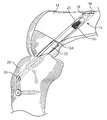
도 1a는 대퇴골 내에 이식된 그래프트 부착 장치의 사시도이다.
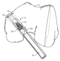
도 1b는 대퇴골 내의 그래프트 부착 장치의 대안적인 이식에 대한 사시도이다.
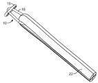
도 2는 그래프트 부착 장치의 사시도이다.
도 3a-3b는 조직 그래프트에 커플링된 그래프트 부착 장치의 사시도이다.
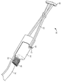
도 4는 대안적인 조직 그래프트에 커플링된 대안적인 그래프트 부착 장치의 사시도이다.
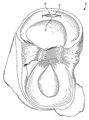
도 5는 반월판 파열부 내에 이식된 봉합사 스캐폴드의 상면도이다.
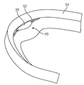
도 6은 봉합사 스캐폴드의 측면도이다.
도 7a는 반월판 파열부 내에 이식된 봉합사 스캐폴드의 사시도이다.
도 7b는 반월판 파열부 내에 이식된 봉합사 스캐폴드의 다른 상면도이다.
도 7c는 선 7C-7C를 따라서 취한 도 7b의 봉합사 스캐폴드의 횡단면적 사시도이다.1A is a perspective view of a graft attaching apparatus implanted in a femur.
1B is a perspective view of an alternative implant of the graft attachment device within the femur.
2 is a perspective view of the graft attaching apparatus.
Figures 3a-3b are perspective views of a graft attachment device coupled to a tissue graft.
Figure 4 is a perspective view of an alternative graft attachment device coupled to an alternative tissue graft.
5 is a top view of a suture scaffold implanted into a meniscus rupture;
6 is a side view of a suture scaffold.
7A is a perspective view of a suture scaffold implanted into a meniscus rupture.
7B is another top view of the suture scaffold implanted within the meniscus tear.
7C is a cross sectional area perspective view of the suture scaffold of Fig. 7B taken along
본원 명세서는 생체 재료, 예를 들어, 피브린 응괴를 유지할 수 있고, 생체 재료가 인대 또는 다른 연성 조직과 접촉하도록 인대 또는 다른 연성 조직 복원 부위 내에 배치될 수 있는, 이식가능한 외장 부재 및 이식가능한 스캐폴드의 예를 개시한다.The present disclosure relates to an implantable outer member and an implantable scaffold that can maintain biomaterials such as fibrin clot and can be placed within the ligament or other soft tissue repair site such that the biomaterial contacts the ligament or other soft tissue. Fig.
도 1a를 참조하면, 그래프트 부착 장치(10)가, 예를 들어, 전방 십자 인대(ACL) 복원 및 재구축 시술 중에 이식되고, 대퇴골(14)의 대퇴부(femoral) 터널(12a) 내에 배치된다. 일부 경우에, 그래프트 부착 장치(10)가 짧은 대퇴부 터널(12b)(도 1b) 내에 배치될 수 있다. 그래프트 부착 장치(10)가 플랫폼 부재(16), 그래프트 연결 요소(18), 및 외장 부재(20)를 포함하고, 조직 그래프트(22), 예를 들어, 반건양근 및 박근 그래프트에 커플링될 수 있다. 대퇴부 터널(12a, 12b)이 그래프트(22)의 일 단부를 수용할 수 있는 한편, 경골부(26) 내에 위치된 경골 터널(24)이 타 단부를 수용할 수 있다. 1A, a
도 2를 또한 참조하면, 그래프트 부착 장치(10)의 외장 부재(20)가 일반적으로, #5 Ultrabraid 봉합사와 같은 편조된(braided) 또는 직조된 메시 형태의 봉합사 재료, 콜라겐과 같은 천연 재료, 또는 그 조합으로 제조될 수 있는 색(sac)-유사 구조물이다. 외장 부재(20)가 외장 부재(20)의 대향 부분들에 배치된 2개의 개구부(28a, 28b)를 가진다. 개구부(28a, 28b)는, 이하에서 추가적으로 설명하는 바와 같이, 외장 부재(20)가 생체 재료(30)를 수용 및 유지할 수 있게 허용할 수 있다. 대안적으로, 외장 부재가 하나의 개구부 또는 둘 초과의 개구부를 가지거나 개구부를 가지지 않을 수 있다. 예를 들어, 외장 부재(20)를 그래프트 부착 장치(10) 또는 조직 그래프트(22)의 다른 부분에 커플링시키는 것을 돕기 위해서, 복수의 필라멘트, 스레드(threads), 봉합사 등이 외장 부재(20)의 부분에 부착될 수 있다. 일부 경우에, 외장 부재(20)가 봉합사 재료의 연속적인 밴드일 수 있다. 대안적으로, 외장 부재(20)가 생체 재료(30)가 주입되도록 구성된 스캐폴드-유사 구조물일 수 있다. 2, the
이하에서 추가적으로 설명하는 바와 같이, 그래프트 부착 장치(10)의 그래프트 연결 요소(18)가 조직 그래프트(22)(도 1a-1b)를 그래프트 부착 장치(10)에 커플링시킨다. 도 2에 도시된 바와 같이, 그래프트 연결 요소(18)가, 외장 부재(20)를 플랫폼 부재(16)에 커플링시키기 위해서 플랫폼 부재(16)의 개구부(32) 및 외장 부재(20)의 개구부(28a, 28b)를 통해서 루핑되는, #5 Ultrabraid 봉합사와 같은 봉합사, 또는 폴리에스터와 같은, 다른 재료의 연속적인 루프일 수 있다. 대안적으로, 또는 부가적으로, 그래프트 연결 요소(18)가 여러 가지 필라멘트 및 다른 부착 요소(미도시)를 통해서 외장 부재(20) 및 플랫폼 부재(16)에 커플링될 수 있다. The
그래프트 부착 장치(10)의 플랫폼 부재(16)가 대퇴골(14)(도 1a-1b)의 피질(cortical) 표면 상에 배치될 수 있고 그래프트 부착 장치(10) 및 조직 그래프트(22)를 대퇴부 터널(12a, 12b) 내에 고정하는 것을 도울 수 있다. 여러 가지 봉합사가 플랫폼 부재(16)의 하나 이상의 개구부(32)를 통해서 루핑될 수 있고, 희망하는 바에 따라서, 의사에 의해서 이용되어 대퇴부 터널(12a, 12b)을 통해서 플랫폼 부재(16)를 당기고 플랫폼 부재(16)를 대퇴골(14)의 피질 표면 상에 배치시킬 수 있다. The
여러 가지 필라멘트, 스레드, 또는 봉합사 등에 더하여, 그래프트 부착 장치(10)가, 특정 금속 합금 및 폴리머와 같은 임의의 생체적합 재료 또는 생체적합 재료의 조합으로 형성된 구성요소를 포함할 수 있다. 그래프트 부착 장치(10)의 구성요소가 PEEK 또는 Acetal과 같은 비-흡수성 재료를 포함할 수 있다. 대안적으로 또는 부가적으로, 그래프트 부착 장치(10)의 구성요소가 PLLA와 같은 생체흡수성 재료를 포함할 수 있다.In addition to various filaments, threads, or sutures, the
수술 중에, 의사는, 조직 그래프트(22)의 부착에 앞서서, 개구부(28a, 28b)를 통해서 생체 재료(30)를 외장 부재(20) 내로 삽입한다. 생체 재료(30)는, 인대 또는 조직 복원을 돕는 피브린 응괴 또는 임의의 다른 물질일 수 있다. 예를 들어, 생체 재료(30)가, 비제한적으로, 혈소판 풍부 혈장(PRP), 히알루론산(HA), 성장 인자(예를 들어, PDGF, FGF, BMP, GDF-5, 및 TGF-β상과(superfamily)의 다른 구성원), 자가 조직 절편, 및 약리학적 약제(예를 들어, 항염증제 및/또는 진통제)와 같은, 임의의 생체적합 재료 또는 생체적합 재료의 조합을 포함할 수 있다. 대안적으로 또는 부가적으로, 생체 재료(30)가 표면 주위에 형성되도록, 생체 재료(30)가 외장 부재(20)의 표면 내로 프레싱될 수 있다. 외장 부재(20)가 제조 중에 그래프트 부착 장치(10)에 미리-부착될 수 있다. 대안적으로, 외장 부재(20)가, 생체 재료(30)를 외장 부재(20) 내로 삽입하기 이전에 또는 이후에, 그래프트 부착 장치(10)로 커플링될 수 있다. 일부 경우에, 생체 재료(30)가 외장 부재(20)의 외부로 유출되는 것을 방지하기 위해서, 의사는, 예를 들어, 봉합사를 이용하여, 개구부(28a, 28b)를 폐쇄할 수 있다. During surgery, the physician inserts the
생체 재료(30)는, ACL 재구축과 같은, 수술 이후의 치유를 촉진하는데 도움이 될 수 있다. 예를 들어, 전형적으로 환자의 혈액으로부터 준비되는 페이스트-유사 재료인 피브린 응괴가, 치유를 촉진하는 여러 가지 성장 인자 및 화학적 물질을 방출함으로써, 그래프트 번들과 뼈 사이뿐만 아니라 그래프트 번들들 사이의 치유를 향상시킬 수 있다. The
도 3a 및 3b를 참조하면, 생체 재료(30)를 외장 부재(20) 내로 삽입하는 것에 이어서, 의사는, 예를 들어, 4배화된(quadrupled) 번들을 생성하기 위해서 그래프트 연결 요소(18)를 통해서 그래프트(22)를 통과시키고 배가(doubling) 시킴으로서, 반건양근 및 박근 그래프트, 또는 반건양근 그래프트의 2개의 절반체를 포함할 수 있는 조직 그래프트(22)를 그래프트 부착 장치(10)로 부착할 수 있다. 부착 프로세스 중에, 조직 그래프트(22)가 외장 부재(20) 위로 접힐 수 있고, 그에 따라 외장 부재(20) 내에 포함된 생체 재료(30)가 접혀진 조직 그래프트(22)의 내측 표면과 접촉될 수 있다. 일부 경우에, 외장 부재(20)가 접혀진 조직 그래프트(22)의 외측 표면에 근접하여 배치될 수 있다. 3A and 3B, following insertion of the
도 4를 참조하면, 일부 경우에, 뼈 블록(36)을 가지는 조직 그래프트(34)가 그래프트 부착 장치(38)에 부착될 수 있다. 일반적으로, 최적의 치유를 위해서, 뼈 블록(36)은 대퇴부 터널(12a, 12b)과 거의 일치되는 형상 및 크기를 가진다. 그래프트 연결 요소(40)를 뼈 블록(36) 내의 개구부(42)를 통해서 루핑함으로써, 조직 그래프트(34), 예를 들어, 슬개건(patellar tendon) 그래프트가 그래프트 부착 장치(38)에 부착될 수 있다. 이러한 프로세스 중에, 그래프트 부착 장치(38)가 또한 외장 부재(46)의 부착 부분(44)을 통해서 루핑될 수 있다. 생체 재료(30)가 전술한 바와 같이 외장 부재(46) 내로 삽입될 수 있다. 조직 그래프트(34) 및 외장 부재(46)를 그래프트 부착 장치(38)로 부착하는 것에 이어서, 외장 부재(46)가 일반적으로 뼈 블록(36)의 아래에 그리고 조직 그래프트(34)의 인대 부분 근처에 배치된다. 4, in some cases, a
그래프트 부착 장치(10, 38) 및 각각의 부착 조직 그래프트(22, 34)를 이식하기에 앞서서, 대퇴부 터널(12a, 12b)이 대퇴골(14)의 과간 절흔(intercondylar notch) 로부터 대퇴골(14)의 피질 표면을 향해서 드릴링될 수 있다. 경골 터널(24)이 경골부(26)의 전방 영역으로부터 과간 절흔을 향해서 드릴링될 수 있다.Before the
이식 중에, 그래프트 부착 장치(10, 38) 및 부착된 조직 그래프트(22, 34) 각각이, 플랫폼 부재(16)의 하나 이상의 개구부(32)를 통해서 루핑될 수 있는 유도(lead) 봉합사(미도시)를 이용하여, 대퇴부 터널(12a, 12b) 내에 배치될 수 있다. 예를 들어, 유도 봉합사가 과간 절흔 근처의 개구부로부터 대퇴부 터널(12a, 12b)로 통과되고, 그리고 그래프트 부착 장치(10, 38)를 대퇴부 터널(12a, 12b)을 통해서 그리고 대퇴골(14)의 피질 표면 근처의 개구부를 향해서 당기기 위해서 이용된다. 플랫폼 부재(16)가 피질 표면 근처의 개구부를 통해서 대퇴부 터널(12a, 12b)을 빠져나간 후에, 유도 봉합사를 이용하여 플랫폼 부재(16)를 뒤집고(flip) 배치시킬 수 있고, 그에 따라 부재(16)가 대퇴골(14)의 피질 표면에 대해서 편평하게 배치될 수 있고 조직 그래프트(22, 34)의 적어도 일부가 대퇴부 터널(12a, 12b) 내에 배치될 수 있다. 부착된 조직 그래프트(22, 34)의 원위 단부가 과간 절흔 근처의 개구부 외부로 연장할 수 있다. During implantation, the
인대 부상 부위에서의 치유를 촉진하는 것에 더하여, 예를 들어, ACL 재구축 수술 이후에, 생체 재료(30)는 연성 조직 부상 부위에서의, 예를 들어 반월판 조직 복원에서의 치유를 향상시키는데 도움이 될 수 있다. 반월판 파열부 복원 시술에서, 특히 반월판의 적색-백색 및 백색-적색 영역과 같은 낮은 혈관분포(vascularity)의 반월판 영역에서, 치유를 촉진하기 위해서, 예를 들어, 피브린 응괴 또는 PRP가 반월판 파열부 내로 삽입될 수 있다. In addition to promoting healing at the ligamentous wound site, for example, after ACL reconstruction surgery, the
도 5 및 6을 참조하면, 봉합사 스캐폴드(50)가 생체 재료(30)를 유지하도록 설계되고 치유 촉진을 위해서 반월판(54) 내의 파열부(52) 내로 삽입될 수 있다. 봉합사 스캐폴드(50)가 중앙 부분(56), 및 상기 중앙 부분(56)의 경계 또는 엣지 영역에 근접하여 부착된 하나 이상의 안내 부분(58, 60)을 포함한다. 5 and 6, a
상기 봉합사 스캐폴드(50)의 중앙 부분(56)에 생체 재료(30)가 주입되도록 구성되고, 그에 따라 수술 중에, 생체 재료(30)가 연성 조직 부상 부위로 용이하게 전달 및 고정될 수 있을 것이다. 예를 들어, 중앙 부분(56)이, 함께 직조되어 메시 표면을 형성하는, 복수의 수평 배향된 봉합사(62) 및 횡단방향으로 배향된 봉합사(64)를 포함할 수 있다. 생체 재료가 표면을 통해서 적어도 부분적으로 푸싱될 수 있게 허용할 수 있을 정도로 충분히 산재하면서도(sparse), 푸싱된 생체 재료를 제 위치에서 유지할 수 있을 정도로 충분히 조밀한 최종 메시가 얻어지도록, 중앙 부분(56)의 수평 및 횡단방향 봉합사(62, 64)가 배열될 수 있다. 일부 구현예에서, 봉합사(62, 64)가 푸싱된 생체 재료를 제 위치에서 유지하는데 도움이 될 수 있는 거친 표면 텍스쳐(texture), 또는 다른 표면 성질을 가질 수 있다. 사용 중에, 생체 재료(30)를 중앙 부분(56)의 메시 표면 내로 프레싱함으로써, 의사는 중앙 부분(56)에 생체 재료(30), 예를 들어 피브린 응괴를 주입할 수 있다. 중앙 부분(56)의 수평 봉합사(62) 및 횡단방향 봉합사(64)가 생체 재료(30)를 중앙 부분(56) 내에서 유지하고, 생체 재료(30)로 증가된 구조적 무결성(integrity)을 부여하여 재료(30)의 관리 및 전달을 도울 수 있다. 대안적으로 또는 부가적으로, 생체 재료(30)가, 예를 들어 트롬빈에 의한 피브리노겐의 응고를 통해서, 중앙 부분(56) 주위에서 화학적으로 형성될 수 있다. The
도 6에 도시된 바와 같이, 봉합사 스캐폴드(50)의 하나 이상의 안내 부분(58, 60)이 중앙 부분(56)의 엣지 영역에 부착되고, 파열부(52)(도 5) 내의 희망 위치에 중앙 부분(56) 및 주입된 생체 재료(30)를 배치하는 것을 도울 수 있다. 예를 들어, 안내 부분(58, 60)이, 중앙 부분(56)의 직조된 메시 표면의 대향 단부들로부터 외측으로 연장하는 하나 이상의 수평 봉합사(62)를 포함할 수 있다. 대안적으로 또는 부가적으로, 안내 부분(58, 60)이, 중앙 부분(56)의 직조된 메시 표면의 대향 단부들로부터 외측으로 연장하는 하나 이상의 횡단방향 봉합사(64)를 포함할 수 있다. 일부 경우에, 안내 부분(58, 60)이, 중앙 부분(56)의 하나 이상의 엣지 영역에 독립적으로 부착된 흡수가능 또는 비-흡수가능 구조물일 수 있다. One or
반월판 파열부 복원 시술 중에, 도 7a-7c에 도시된 바와 같이, 의사가 봉합사 스캐폴드(50) 및 주입된 생체 재료(30)를 파열부(52) 내로 이식할 수 있다. 예를 들어, 봉합사 스캐폴드(50) 및 생체 재료(30)가 파열부(52) 내의 적절한 깊이에 대체적으로 배치될 때까지, 의사가 봉합사 스캐폴드(50)의 유도(leading) 엣지(66)(도 7c)를 파열부(52) 내로 안내할 수 있다. 이어서, 생체 재료(30)의 희망 배치가 얻어질 때까지 안내 부분(58, 60)을 당기거나, 푸싱하거나, 달리 조작함으로써, 의사가 중앙 부분(56) 및 주입된 생체 재료(30)를 파열부(52) 내의 최종적인 희망 위치로 이동시킬 수 있다. 생체 재료(30)의 희망하는 배치가 달성된 후에, 안내 부분(58, 60)이, 예를 들어, 컷팅에 의해서 제거될 수 있고, 또는 제 위치에서 유지될 수 있다. 일부 경우에, 스캐폴드(50)의 전체 표면이 제거되어, 반월판 파열부(52) 내에서 생체 재료(30) 만을 제 위치에서 유지할 수 있다. 복원 부위에서의 치유를 향상시키기 위해서, 생체 재료(30)를 반월판 파열부(52) 내로 이식하는 것이 다른 반월판 파열부 복원 시술 및 도구와 함께 이용될 수 있다. During the meniscal tear restoration procedure, the surgeon can implant the
본원 명세서가 많은 구체적인 구현예의 상세 내용을 포함하고 있지만, 그러한 상세 내용은 어떠한 구현예의 또는 청구된 것의 범위도 제한하는 것으로 간주되지 않아야 하고, 오히려 특별한 구현예의 특별한 구현으로 특정된 특징을 설명하는 것으로서 간주되어야 한다. 분리된 구현예의 문맥으로 본원 명세서에서 설명된 특정 특징이 또한 단일 구현예와 조합하여 구현될 수 있다. 반대로, 단일 구현예의 문맥에서 설명된 여러 가지 특징이 또한 독립적으로 복수 구현예로 또는 임의의 적합한 하위 조합으로 구현될 수 있다. 또한, 비록 특징이 특정 조합으로 작용하는 것으로 앞서서 설명되었거나 그렇게 초기에 주장되었을 수 있지만, 청구된 조합으로부터의 하나 이상의 특징이, 일부 경우에, 조합으로부터 배제될 수 있고, 그리고 청구된 조합이 하위 조합 또는 하위 조합의 변형에 관한 것일 수 있을 것이다. 그에 따라, 청구 대상의 특별한 구현예가 설명된 것이다. 다른 구현예가 이하의 청구항의 범위에 포함된다. While the specification concludes with a description of many specific embodiments, such details are not to be considered limiting of the scope of any embodiment or claim, but rather, . Certain features described herein in the context of separate implementations may also be implemented in combination with a single implementation. Conversely, various features described in the context of a single embodiment may also be implemented independently in multiple implementations or in any suitable subcombination. It should also be understood that one or more features from the claimed combination may, in some cases, be excluded from the combination, and that the claimed combination may be combined with the subordinate combination Or a variation of the subcombination. Accordingly, specific embodiments of the claimed subject matter have been described. Other implementations are within the scope of the following claims.
Claims (20)
플랫폼 부재;
그래프트 연결 요소의 일부가 조직 그래프트에 대한 부착을 위한 루프를 형성하도록 상기 플랫폼 부재에 커플링된 그래프트 연결 요소; 및
상기 그래프트 연결 요소의 루프에 커플링되고 생체 재료를 둘러싸도록 구성된 외장 부재로서, 상기 외장 부재가 생체 재료를 수용하도록 구성된 개구부를 형성하는, 외장 부재를 포함하고;
이용 중에, 상기 조직 그래프트가 상기 루프에 커플링되고 상기 외장 부재와 접촉하도록, 상기 그래프트 연결 요소 및 상기 외장 부재가 구성되는, 조직 그래프트 현수 장치.Tissue graft suspensions as:
Platform member;
A graft connecting element coupled to the platform member such that a portion of the graft connecting element forms a loop for attachment to the tissue graft; And
An outer member coupled to the loop of the graft connecting element and configured to surround the biomaterial, the outer member forming an opening configured to receive the biomaterial;
Wherein during use, the tissue graft is coupled to the loop and is in contact with the sheath member, wherein the graft connecting element and the sheath member are configured.
상기 그래프트 연결 요소가 봉합사의 연속적인 루프인, 조직 그래프트 현수 장치.The method according to claim 1,
Wherein the graft connecting element is a continuous loop of suture.
상기 루프가 상기 외장 부재의 개구부를 통과하는, 조직 그래프트 현수 장치.The method according to claim 1,
The loop passing through the opening of the skin member.
상기 루프가 상기 외장 부재의 부착 요소를 통과하는, 조직 그래프트 현수 장치.The method according to claim 1,
Wherein the loop passes through an attachment element of the skin member.
상기 외장 부재가 생체 재료를 수용하도록 구성된 2개의 개구부를 형성하는, 조직 그래프트 현수 장치.The method according to claim 1,
Wherein said skin member forms two openings configured to receive a biomaterial.
상기 생체 재료가 피브린 응괴인, 조직 그래프트 현수 장치.The method according to claim 1,
Wherein the biomaterial is a fibrin clot.
상기 생체 재료가 혈소판 풍부 혈장인, 조직 그래프트 현수 장치.The method according to claim 1,
Wherein the biomaterial is platelet-rich plasma.
상기 외장 부재에 부착된 하나 이상의 필라멘트를 더 포함하는, 조직 그래프트 현수 장치.The method according to claim 1,
And further comprising at least one filament attached to the skin member.
그래프트 연결 요소에 커플링된 플랫폼 부재를 제공하는 단계로서, 상기 그래프트 연결 요소가 외장 부재에 커플링되고 상기 조직 그래프트에 대한 부착을 위한 루프를 형성하고, 상기 외장 부재가 생체 재료를 둘러싸도록 구성되는, 플랫폼 부재를 제공하는 단계;
상기 외장 부재 내의 개구부를 통해서 생체 재료를 삽입하는 단계;
상기 조직 그래프트가 상기 외장 부재와 접촉하도록 상기 조직 그래프트를 상기 그래프트 연결 요소에 부착하는 단계;
뼈 내에 뼈 터널을 형성하는 단계; 및
상기 조직 그래프트의 적어도 일부가 상기 뼈 터널 내에 위치되도록 상기 뼈의 표면 상에 상기 플랫폼 부재를 배치하는 단계를 포함하는, 방법.As a fixing method of the tissue graft:
Providing a platform member coupled to the graft connecting element such that the graft connecting element is coupled to the enclosure member and forms a loop for attachment to the tissue graft and wherein the enclosure member is configured to surround the biomaterial , Providing a platform member;
Inserting the biomaterial through the opening in the exterior member;
Attaching the tissue graft to the graft connecting element such that the tissue graft is in contact with the skin member;
Forming a bone tunnel within the bone; And
And positioning the platform member on a surface of the bone such that at least a portion of the tissue graft is located within the bone tunnel.
상기 조직 그래프트를 상기 그래프트 연결 요소에 부착하는 단계가, 상기 외장 부재가 상기 조직 그래프트의 2개의 부분들 사이에 위치되도록 상기 조직 그래프트를 상기 그래프트 연결 요소의 루프를 통해서 루핑하는 단계를 포함하는, 방법.10. The method of claim 9,
Wherein attaching the tissue graft to the graft connecting element comprises looping the tissue graft through a loop of the graft connecting element such that the skin member is positioned between two portions of the tissue graft. .
상기 조직 그래프트를 상기 그래프트 연결 요소에 부착하는 단계가 상기 그래프트 연결 요소를 상기 조직 그래프트의 뼈 블록 내의 개구부를 통해서 루핑하는 단계를 포함하는, 방법.10. The method of claim 9,
Wherein attaching the tissue graft to the graft connecting element comprises looping the graft connecting element through an opening in a bone block of the tissue graft.
상기 뼈 터널이 제1 개구부 및 제2 개구부를 포함하고,
상기 플랫폼 부재를 상기 뼈의 표면 상에 배치하는 단계가:
상기 플랫폼 부재를 상기 제1 개구부를 통해서 상기 뼈 터널 내로 삽입하는 단계;
상기 터널을 통해서 상기 제2 개구부의 외부로 상기 플랫폼 부재를 이동시키는 단계; 및
상기 그래프트 연결 요소에 부착된 조직 그래프트의 제1 부분이 상기 터널 내에 있도록 그리고 상기 조직 그래프트의 제2 부분이 상기 제1 개구부의 외부로 연장하도록, 상기 그래프트 연결 요소가 뼈 터널 내로 연장하는 상태에서 상기 플랫폼 부재를 상기 제2 개구부 위에 배치하는 단계를 포함하는, 방법.10. The method of claim 9,
Wherein the bone tunnel comprises a first opening and a second opening,
Wherein positioning the platform member on a surface of the bone comprises:
Inserting the platform member through the first opening into the bone tunnel;
Moving the platform member out of the second opening through the tunnel; And
A first portion of the tissue graft attached to the graft connecting element is within the tunnel and a second portion of the tissue graft extends outside the first opening, And disposing a platform member over the second opening.
생체 재료가 주입된 중앙 부분으로서, 상기 중앙 부분이 직조 패턴으로 배열된 필라멘트를 포함하는, 중앙 부분; 및
상기 중앙 부분의 엣지에 부착된 안내 부분으로서, 상기 안내 부분이 하나 이상의 필라멘트를 포함하는, 안내 부분을 포함하고;
상기 스캐폴드가 연성 조직 내의 파열부 내로 삽입되도록 구성되는, 스캐폴드.As a scaffold for receiving biomaterials:
A central portion into which a biomaterial is injected, the central portion comprising filaments arranged in a weave pattern; And
A guiding portion attached to an edge of the central portion, the guiding portion including one or more filaments;
Wherein the scaffold is configured to be inserted into the rupture portion within the soft tissue.
상기 연성 조직 내의 파열부가 반월판 파열부인, 스캐폴드.14. The method of claim 13,
Wherein the rupture part in the soft tissue is a meniscus rupture part.
상기 안내 부분이 상기 중앙 부분의 제1 엣지에 부착된 제1 안내 부분, 및 상기 중앙 부분의 제2 엣지에 부착된 제2 안내 부분을 포함하고, 상기 제2 엣지가 상기 제1 엣지에 대향하는, 스캐폴드.14. The method of claim 13,
Wherein the guide portion includes a first guide portion attached to a first edge of the central portion and a second guide portion attached to a second edge of the central portion, the second edge being opposite , Scaffold.
상기 안내 부분이 상기 중앙 부분의 직조 패턴으로부터 외부로 연장하는 하나 이상의 필라멘트를 포함하는, 스캐폴드.14. The method of claim 13,
Wherein the guide portion comprises at least one filament extending outwardly from the woven pattern of the central portion.
중앙 부분 및 안내 부분을 포함하는 스캐폴드를 제공하는 단계로서, 상기 중앙 부분이 직조 패턴으로 배열된 필라멘트를 포함하는, 스캐폴드를 제공하는 단계;
상기 스캐폴드의 중앙 부분으로 생체 재료를 주입하는 단계;
상기 스캐폴드의 엣지를 상기 연성 조직 파열부 내로 삽입하는 단계; 및
상기 안내 부분을 조작하는 것에 의해서 상기 연성 조직 파열부 내의 스캐폴드의 중앙 부분을 최종 위치로 이동시키는 단계를 포함하는, 방법. CLAIMS 1. A method for restoring a soft tissue rupture comprising:
Providing a scaffold comprising a central portion and a guiding portion, the central portion comprising a filament arranged in a woven pattern;
Injecting a biomaterial into a central portion of the scaffold;
Inserting an edge of the scaffold into the soft tissue rupture; And
And moving the central portion of the scaffold within the soft tissue rupture portion to a final position by manipulating the guide portion.
상기 연성 조직이 반월판인, 방법.18. The method of claim 17,
Wherein the soft tissue is a meniscus.
상기 스캐폴드의 중앙 부분으로 생체 재료를 주입하는 단계가 상기 중앙 부분 주위에 피브린 응괴 또는 혈소판 풍부 혈장 중 적어도 하나를 형성하는 단계를 포함하는, 방법.18. The method of claim 17,
Wherein injecting a biomaterial into a central portion of the scaffold comprises forming at least one of fibrin clot or platelet rich plasma around the central portion.
상기 스캐폴드의 중앙 부분으로 주입하는 단계가 상기 중앙 부분을 피브린 응괴 또는 혈소판 풍부 혈장 중 적어도 하나 내로 프레싱하는 단계를 포함하는, 방법.18. The method of claim 17,
Wherein injecting into the central portion of the scaffold comprises pressing the central portion into at least one of fibrin clot or platelet rich plasma.
Applications Claiming Priority (3)
| Application Number | Priority Date | Filing Date | Title |
|---|---|---|---|
| US13/365,000 | 2012-02-02 | ||
| US13/365,000 US9204959B2 (en) | 2012-02-02 | 2012-02-02 | Implantable biologic holder |
| PCT/US2013/024235 WO2013116574A1 (en) | 2012-02-02 | 2013-01-31 | Implantable biologic holder |
Publications (1)
| Publication Number | Publication Date |
|---|---|
| KR20140121842A true KR20140121842A (en) | 2014-10-16 |
Family
ID=47684073
Family Applications (1)
| Application Number | Title | Priority Date | Filing Date |
|---|---|---|---|
| KR1020147022621A KR20140121842A (en) | 2012-02-02 | 2013-01-31 | Implantable biologic holder |
Country Status (11)
| Country | Link |
|---|---|
| US (2) | US9204959B2 (en) |
| EP (1) | EP2809268B1 (en) |
| JP (1) | JP6141882B2 (en) |
| KR (1) | KR20140121842A (en) |
| CN (1) | CN104379091B (en) |
| AU (1) | AU2013215000B2 (en) |
| BR (1) | BR112014018965A8 (en) |
| IN (1) | IN2014DN06485A (en) |
| RU (1) | RU2014133464A (en) |
| WO (1) | WO2013116574A1 (en) |
| ZA (1) | ZA201405600B (en) |
Cited By (2)
| Publication number | Priority date | Publication date | Assignee | Title |
|---|---|---|---|---|
| KR20180002350U (en) | 2017-01-25 | 2018-08-02 | 조성찬 | A hair rod |
| KR20200039724A (en) * | 2017-09-07 | 2020-04-16 | 콘메드 코포레이션 | Bidirectional adjustable loop suspension system for fixation of soft tissue grafts |
Families Citing this family (8)
| Publication number | Priority date | Publication date | Assignee | Title |
|---|---|---|---|---|
| US9204959B2 (en) * | 2012-02-02 | 2015-12-08 | Smith & Nephew, Inc. | Implantable biologic holder |
| US9265600B2 (en) * | 2013-02-27 | 2016-02-23 | Orthopediatrics Corp. | Graft fixation |
| US10159518B2 (en) * | 2013-07-25 | 2018-12-25 | Arthrex, Inc. | Flexible obturator |
| US11331191B2 (en) | 2015-08-12 | 2022-05-17 | Howmedica Osteonics Corp. | Bioactive soft tissue implant and methods of manufacture and use thereof |
| US10729548B2 (en) | 2016-05-02 | 2020-08-04 | Howmedica Osteonics Corp. | Bioactive soft tissue implant and methods of manufacture and use thereof |
| CA2938576A1 (en) | 2015-08-12 | 2017-02-12 | Howmedica Osteonics Corp. | Methods for forming scaffolds |
| US9931196B2 (en) | 2015-08-26 | 2018-04-03 | Albert Einstein Healthcare Network | Connector for attaching tissue to bone |
| WO2018066921A2 (en) * | 2016-10-06 | 2018-04-12 | 아주대학교 산학협력단 | Device for supporting semilunar cartilage hoop stress |
Family Cites Families (177)
| Publication number | Priority date | Publication date | Assignee | Title |
|---|---|---|---|---|
| US3176316A (en) * | 1963-01-07 | 1965-04-06 | Bruce R Bodell | Plastic prosthetic tendon |
| US3613120A (en) * | 1969-10-21 | 1971-10-19 | Research Corp | Flexor tendon prosthesis |
| US4255820A (en) * | 1979-07-24 | 1981-03-17 | Rothermel Joel E | Artificial ligaments |
| US4469101A (en) * | 1980-10-23 | 1984-09-04 | Battelle Memorial Institute | Suture device |
| US4344193A (en) * | 1980-11-28 | 1982-08-17 | Kenny Charles H | Meniscus prosthesis |
| US4502161A (en) * | 1981-09-21 | 1985-03-05 | Wall W H | Prosthetic meniscus for the repair of joints |
| IL65855A (en) * | 1982-05-24 | 1986-09-30 | Yeda Res & Dev | Prosthetic tendon |
| US4834755A (en) * | 1983-04-04 | 1989-05-30 | Pfizer Hospital Products Group, Inc. | Triaxially-braided fabric prosthesis |
| GB8414344D0 (en) * | 1984-06-05 | 1984-07-11 | Showell A W Sugicraft Ltd | Surgical element |
| GB8418018D0 (en) * | 1984-07-16 | 1984-08-22 | Johnson & Johnson | Connective tissue prosthesis |
| US4744793A (en) * | 1985-09-06 | 1988-05-17 | Zimmer, Inc. | Prosthetic ligament connection assembly |
| US4731084A (en) * | 1986-03-14 | 1988-03-15 | Richards Medical Company | Prosthetic ligament |
| CH671686A5 (en) * | 1987-01-07 | 1989-09-29 | Sulzer Ag | |
| US5158574A (en) * | 1987-07-20 | 1992-10-27 | Regen Corporation | Prosthetic meniscus |
| US4773910A (en) * | 1987-08-17 | 1988-09-27 | Johnson & Johnson Consumer Products, Inc. | Permanent ligament prosthesis |
| US4917699A (en) * | 1988-05-16 | 1990-04-17 | Zimmer, Inc. | Prosthetic ligament |
| US4917700A (en) * | 1988-08-01 | 1990-04-17 | Zimmer, Inc. | Prosthetic ligament |
| US4919667A (en) * | 1988-12-02 | 1990-04-24 | Stryker Corporation | Implant |
| US5041118A (en) | 1989-04-28 | 1991-08-20 | Implant Technology Inc. | Femoral broach |
| US5067964A (en) * | 1989-12-13 | 1991-11-26 | Stryker Corporation | Articular surface repair |
| US5171322A (en) * | 1990-02-13 | 1992-12-15 | Kenny Charles H | Stabilized meniscus prosthesis |
| US5092894A (en) * | 1990-02-13 | 1992-03-03 | Kenny Charles H | Stabilized meniscus prosthesis |
| EP0447355A1 (en) * | 1990-03-12 | 1991-09-18 | Gebrüder Sulzer Aktiengesellschaft | Implant for the human body |
| GB9105957D0 (en) | 1991-03-21 | 1991-05-08 | Seedhom Bahaa B | Implantable fixing device |
| US5269783A (en) * | 1991-05-13 | 1993-12-14 | United States Surgical Corporation | Device and method for repairing torn tissue |
| FR2678823B1 (en) * | 1991-07-11 | 1995-07-07 | Legrand Jean Jacques | DEVICE FOR REINFORCING A LIGAMENT DURING A LIGAMENT PLASTY. |
| US6503277B2 (en) * | 1991-08-12 | 2003-01-07 | Peter M. Bonutti | Method of transplanting human body tissue |
| US5507812A (en) | 1992-12-28 | 1996-04-16 | Moore; David E. | Modular prosthetic ligament |
| US5306301A (en) | 1993-02-11 | 1994-04-26 | American Cyanamid Company | Graft attachment device and method of using same |
| DE69432734T2 (en) * | 1993-09-14 | 2004-04-29 | Smith & Nephew, Inc., Memphis | Biological tape replacement |
| DE69426414T2 (en) * | 1993-09-24 | 2001-05-03 | Takiron Co | IMPLANT MATERIAL |
| US5632745A (en) * | 1995-02-07 | 1997-05-27 | R&D Biologicals, Inc. | Surgical implantation of cartilage repair unit |
| FR2745710B1 (en) * | 1996-03-11 | 1998-05-29 | Rippstein Pascal Francois | TENDON PASSING DEVICE |
| AU4480097A (en) | 1996-09-20 | 1998-04-14 | Medicinelodge, Inc. | Adjustable length strap and footing for ligament mounting and method for its use |
| GB9620046D0 (en) * | 1996-09-26 | 1996-11-13 | Neoligaments | Attachment device for use in the implantation of prosthetic ligament |
| FR2755846B1 (en) * | 1996-11-20 | 1998-12-31 | Jacques Philippe Laboureau | PRE-ORIENT PROSTHETIC LIGAMENT AND METHOD OF MAKING |
| US6554862B2 (en) * | 1996-11-27 | 2003-04-29 | Ethicon, Inc. | Graft ligament anchor and method for attaching a graft ligament to a bone |
| WO1998030141A2 (en) * | 1997-01-09 | 1998-07-16 | Cohesion Technologies, Inc. | Devices for tissue repair and methods for preparation and use thereof |
| US5769894A (en) | 1997-02-05 | 1998-06-23 | Smith & Nephew, Inc. | Graft attachment device and method of attachment |
| AUPO530997A0 (en) * | 1997-02-25 | 1997-03-20 | Esnouf, Philip Stuart | Surgical aid for connective tissue grafting and method for employing same |
| US20020019670A1 (en) * | 1997-02-28 | 2002-02-14 | Jerald M. Crawley | Implantable tissue augmentation device |
| AUPP000797A0 (en) * | 1997-10-24 | 1997-11-20 | Cryptych Pty Ltd | Fixation of cruciate ligament grafts |
| US6214047B1 (en) * | 1998-03-10 | 2001-04-10 | University Of Cincinnati | Article and method for coupling muscle to a prosthetic device |
| US6027744A (en) * | 1998-04-24 | 2000-02-22 | University Of Massachusetts Medical Center | Guided development and support of hydrogel-cell compositions |
| JP3540158B2 (en) * | 1998-05-11 | 2004-07-07 | 株式会社アイメディック | Reconstruction ligament fixation device |
| US6669707B1 (en) * | 1998-07-21 | 2003-12-30 | Lee L. Swanstrom | Method and apparatus for attaching or locking an implant to an anatomic vessel or hollow organ wall |
| DE19851152A1 (en) * | 1998-11-06 | 2000-05-11 | Storz Karl Gmbh & Co Kg | Instruments for implanting a cruciate ligament replacement in a knee joint |
| US20020165611A1 (en) * | 1998-12-22 | 2002-11-07 | Robert-Jan Enzerink | Graft material convenience package |
| US6203572B1 (en) * | 1999-02-09 | 2001-03-20 | Linvatec Corporation | Device and method for ligament reconstruction |
| US6602291B1 (en) * | 1999-04-05 | 2003-08-05 | Raymedica, Inc. | Prosthetic spinal disc nucleus having a shape change characteristic |
| US6428576B1 (en) * | 1999-04-16 | 2002-08-06 | Endospine, Ltd. | System for repairing inter-vertebral discs |
| US6103255A (en) * | 1999-04-16 | 2000-08-15 | Rutgers, The State University | Porous polymer scaffolds for tissue engineering |
| US6245107B1 (en) * | 1999-05-28 | 2001-06-12 | Bret A. Ferree | Methods and apparatus for treating disc herniation |
| US6371990B1 (en) * | 1999-10-08 | 2002-04-16 | Bret A. Ferree | Annulus fibrosis augmentation methods and apparatus |
| US7273497B2 (en) * | 1999-05-28 | 2007-09-25 | Anova Corp. | Methods for treating a defect in the annulus fibrosis |
| US20060247665A1 (en) * | 1999-05-28 | 2006-11-02 | Ferree Bret A | Methods and apparatus for treating disc herniation and preventing the extrusion of interbody bone graft |
| US20040059416A1 (en) * | 1999-06-22 | 2004-03-25 | Murray Martha M. | Biologic replacement for fibrin clot |
| US20020095157A1 (en) * | 1999-07-23 | 2002-07-18 | Bowman Steven M. | Graft fixation device combination |
| US6517542B1 (en) | 1999-08-04 | 2003-02-11 | The Cleveland Clinic Foundation | Bone anchoring system |
| US7717961B2 (en) * | 1999-08-18 | 2010-05-18 | Intrinsic Therapeutics, Inc. | Apparatus delivery in an intervertebral disc |
| US6508839B1 (en) * | 1999-08-18 | 2003-01-21 | Intrinsic Orthopedics, Inc. | Devices and methods of vertebral disc augmentation |
| WO2002054978A2 (en) * | 1999-08-18 | 2002-07-18 | Intrinsic Orthopedics Inc | Devices and method for nucleus pulposus augmentation and retention |
| WO2004100841A1 (en) * | 1999-08-18 | 2004-11-25 | Intrinsic Therapeutics, Inc. | Devices and method for augmenting a vertebral disc nucleus |
| DE19941574A1 (en) * | 1999-09-01 | 2001-03-08 | Storz Karl Gmbh & Co Kg | Instruments for implanting a tendon replacement |
| US6964674B1 (en) * | 1999-09-20 | 2005-11-15 | Nuvasive, Inc. | Annulotomy closure device |
| US20030040796A1 (en) * | 1999-10-08 | 2003-02-27 | Ferree Bret A. | Devices used to treat disc herniation and attachment mechanisms therefore |
| US7615076B2 (en) * | 1999-10-20 | 2009-11-10 | Anulex Technologies, Inc. | Method and apparatus for the treatment of the intervertebral disc annulus |
| US20030153976A1 (en) * | 1999-10-20 | 2003-08-14 | Cauthen Joseph C. | Spinal disc annulus reconstruction method and spinal disc annulus stent |
| US7004970B2 (en) * | 1999-10-20 | 2006-02-28 | Anulex Technologies, Inc. | Methods and devices for spinal disc annulus reconstruction and repair |
| US7935147B2 (en) * | 1999-10-20 | 2011-05-03 | Anulex Technologies, Inc. | Method and apparatus for enhanced delivery of treatment device to the intervertebral disc annulus |
| US8632590B2 (en) * | 1999-10-20 | 2014-01-21 | Anulex Technologies, Inc. | Apparatus and methods for the treatment of the intervertebral disc |
| US20050070906A1 (en) | 1999-11-30 | 2005-03-31 | Ron Clark | Endosteal tibial ligament fixation with adjustable tensioning |
| US7887551B2 (en) * | 1999-12-02 | 2011-02-15 | Smith & Nephew, Inc. | Soft tissue attachment and repair |
| GB9929599D0 (en) * | 1999-12-15 | 2000-02-09 | Atlantech Medical Devices Limi | A graft suspension device |
| DE19964081B4 (en) * | 1999-12-29 | 2005-06-30 | Ethicon Gmbh | Stripe-type implant and surgical gripping instrument |
| US7097654B1 (en) * | 2000-01-03 | 2006-08-29 | Yosef Freedland | Flip-wing tissue retainer |
| US20030023304A1 (en) * | 2000-01-11 | 2003-01-30 | Carter Kevin C. | Materials and methods for improved bone tendon bone transplantation |
| US6635073B2 (en) * | 2000-05-03 | 2003-10-21 | Peter M. Bonutti | Method of securing body tissue |
| US6296659B1 (en) * | 2000-02-29 | 2001-10-02 | Opus Medical, Inc. | Single-tailed suturing method and apparatus |
| US6746483B1 (en) * | 2000-03-16 | 2004-06-08 | Smith & Nephew, Inc. | Sheaths for implantable fixation devices |
| US6579291B1 (en) * | 2000-10-10 | 2003-06-17 | Spinalabs, Llc | Devices and methods for the treatment of spinal disorders |
| US6805695B2 (en) * | 2000-04-04 | 2004-10-19 | Spinalabs, Llc | Devices and methods for annular repair of intervertebral discs |
| EP2314257B9 (en) * | 2000-05-01 | 2013-02-27 | ArthroSurface, Inc. | System for joint resurface repair |
| US6620185B1 (en) * | 2000-06-27 | 2003-09-16 | Smith & Nephew, Inc. | Surgical procedures and instruments |
| US6325804B1 (en) | 2000-06-28 | 2001-12-04 | Ethicon, Inc. | Method for fixing a graft in a bone tunnel |
| CA2692564C (en) * | 2000-10-23 | 2013-08-27 | Tyco Healthcare Group Lp | Absorbable fastener and applying apparatus |
| JP4130126B2 (en) * | 2000-10-27 | 2008-08-06 | ウォーソー・オーソペディック・インコーポレーテッド | Annulus repair system and method |
| US6752831B2 (en) * | 2000-12-08 | 2004-06-22 | Osteotech, Inc. | Biocompatible osteogenic band for repair of spinal disorders |
| US20020147461A1 (en) * | 2001-04-06 | 2002-10-10 | Aldrich William N. | Apparatus and methods for closing openings in spinal discs |
| US20030078579A1 (en) * | 2001-04-19 | 2003-04-24 | Ferree Bret A. | Annular repair devices and methods |
| US7144413B2 (en) | 2001-04-20 | 2006-12-05 | Synthes (U.S.A.) | Graft fixation system and method |
| US20040153153A1 (en) * | 2001-05-31 | 2004-08-05 | Elson Robert J. | Anterior cruciate ligament reconstruction system and method of implementing same |
| WO2003007784A2 (en) * | 2001-07-16 | 2003-01-30 | Depuy Products, Inc. | Meniscus regeneration device and method |
| US6736815B2 (en) * | 2001-09-06 | 2004-05-18 | Core Medical, Inc. | Apparatus and methods for treating spinal discs |
| US6767037B2 (en) * | 2001-09-27 | 2004-07-27 | Depuy Mitek, Inc. | Sliding and locking surgical knot |
| US6652563B2 (en) | 2001-10-02 | 2003-11-25 | Arthrex, Inc. | Suture anchor with internal suture loop |
| US6599319B2 (en) * | 2001-12-14 | 2003-07-29 | Celanese Advanced Materials, Inc. | Prosthetic ligament |
| DE50200594D1 (en) * | 2002-02-08 | 2004-08-12 | Storz Karl Gmbh & Co Kg | Anchor element for anchoring a ligament graft |
| US6730124B2 (en) * | 2002-03-08 | 2004-05-04 | Musculoskeletal Transplant Foundation | Bone-tendon-bone assembly with cancellous allograft bone block |
| US6843799B2 (en) * | 2002-03-25 | 2005-01-18 | Edwin C. Bartlett | Suture anchor system and associated method |
| US7223289B2 (en) * | 2002-04-16 | 2007-05-29 | Warsaw Orthopedic, Inc. | Annulus repair systems and techniques |
| US20030212456A1 (en) * | 2002-05-09 | 2003-11-13 | John Lipchitz | Implant for tissue repair |
| US6972027B2 (en) * | 2002-06-26 | 2005-12-06 | Stryker Endoscopy | Soft tissue repair system |
| US7033393B2 (en) * | 2002-06-27 | 2006-04-25 | Raymedica, Inc. | Self-transitioning spinal disc anulus occulsion device and method of use |
| AU2003268480A1 (en) * | 2002-09-06 | 2004-03-29 | Neville D. Alleyne | Seal for posterior lateral vertebral disk cavity |
| US7601155B2 (en) | 2003-05-20 | 2009-10-13 | Petersen Thomas D | Instruments and method for minimally invasive surgery for total hips |
| US7722644B2 (en) | 2003-06-11 | 2010-05-25 | Medicine Lodge, Inc. | Compact line locks and methods |
| US8226715B2 (en) | 2003-06-30 | 2012-07-24 | Depuy Mitek, Inc. | Scaffold for connective tissue repair |
| DE20315960U1 (en) * | 2003-10-13 | 2004-02-12 | Aesculap Ag & Co. Kg | Cartilage replacement implant |
| US7896917B2 (en) * | 2003-10-15 | 2011-03-01 | Biomet Sports Medicine, Llc | Method and apparatus for graft fixation |
| GB0325141D0 (en) * | 2003-10-28 | 2003-12-03 | Xiros Plc | Repair of damaged tissue on a bone site |
| US7901461B2 (en) | 2003-12-05 | 2011-03-08 | Ethicon, Inc. | Viable tissue repair implants and methods of use |
| US7833228B1 (en) | 2004-01-05 | 2010-11-16 | Biomet Manufacturing Corp. | Method and instrumentation for performing minimally invasive hip arthroplasty |
| US7846183B2 (en) * | 2004-02-06 | 2010-12-07 | Spinal Elements, Inc. | Vertebral facet joint prosthesis and method of fixation |
| WO2005092208A1 (en) * | 2004-03-03 | 2005-10-06 | Schwartz Biomedical, Llc | Articular cartilage fixation device and method |
| US8657881B2 (en) * | 2004-04-20 | 2014-02-25 | Depuy Mitek, Llc | Meniscal repair scaffold |
| US7585316B2 (en) * | 2004-05-21 | 2009-09-08 | Warsaw Orthopedic, Inc. | Interspinous spacer |
| US8109965B2 (en) | 2004-06-09 | 2012-02-07 | Biomet Sports Medicine, LLP | Method and apparatus for soft tissue fixation |
| US20060089646A1 (en) * | 2004-10-26 | 2006-04-27 | Bonutti Peter M | Devices and methods for stabilizing tissue and implants |
| US7594922B1 (en) * | 2005-04-07 | 2009-09-29 | Medicine Lodge, Inc | System and method for meniscal repair through a meniscal capsular tunnel |
| US20060247776A1 (en) * | 2005-05-02 | 2006-11-02 | The Board Of Trustees Of The Leland Stanford Junior University | Systems and methods for augmenting intervertebral discs |
| ES2263382B1 (en) | 2005-05-16 | 2007-11-16 | Fundacion Para La Investigacion Biomedica Del Hospital Gregorio Marañon | ARTIFICIAL MATRIX OF ENDOTHELIZED FIBRINE GEL SUPERPRODUCTOR OF PROANGIOGEN FACTORS. |
| US8097033B2 (en) * | 2005-06-16 | 2012-01-17 | The University Of Miami | Extraocular muscle prosthesis |
| US7850711B1 (en) * | 2005-06-22 | 2010-12-14 | Biomet Sports Medicine, Llc | Method and apparatus for securing soft tissue to bone |
| US8197509B2 (en) | 2005-06-29 | 2012-06-12 | Depuy Mitek, Inc. | Suture anchor with improved torsional drive head |
| US20070005140A1 (en) * | 2005-06-29 | 2007-01-04 | Kim Daniel H | Fabrication and use of biocompatible materials for treating and repairing herniated spinal discs |
| GB0516846D0 (en) * | 2005-08-17 | 2005-09-21 | Knight David P | Meniscal repair device |
| US20070067040A1 (en) * | 2005-09-02 | 2007-03-22 | Anova Corporation | Methods and apparatus for reconstructing the anulus fibrosus |
| US8202306B2 (en) * | 2005-09-12 | 2012-06-19 | Arthrex, Inc. | Mesh reinforced tissue anchor |
| EP1764117A1 (en) * | 2005-09-20 | 2007-03-21 | Zimmer GmbH | Implant for the repair of a cartilage defect and method for manufacturing the implant |
| US20090164014A1 (en) * | 2005-10-21 | 2009-06-25 | Artimplant Ab | Biodegradable ostochondreal implant |
| US7371260B2 (en) * | 2005-10-26 | 2008-05-13 | Biomet Sports Medicine, Inc. | Method and instrumentation for the preparation and transplantation of osteochondral allografts |
| US8403985B2 (en) * | 2005-11-02 | 2013-03-26 | Zimmer, Inc. | Joint spacer implant |
| US20070118218A1 (en) * | 2005-11-22 | 2007-05-24 | Hooper David M | Facet joint implant and procedure |
| US8801790B2 (en) * | 2005-12-27 | 2014-08-12 | Warsaw Orthopedic, Inc. | Intervertebral disc augmentation and rehydration with superabsorbent polymers |
| US20090306776A1 (en) * | 2006-01-25 | 2009-12-10 | Children's Medical Center Corporation | Methods and procedures for ligament repair |
| US20070179607A1 (en) * | 2006-01-31 | 2007-08-02 | Zimmer Technology, Inc. | Cartilage resurfacing implant |
| US9408599B2 (en) | 2006-02-03 | 2016-08-09 | Biomet Sports Medicine, Llc | Method and apparatus for coupling soft tissue to a bone |
| US20070225805A1 (en) * | 2006-03-21 | 2007-09-27 | Reinhold Schmieding | Ligament Fixation Using Graft Harness/Bolt Assembly |
| US20090043342A1 (en) | 2006-03-28 | 2009-02-12 | Yosef Freedland | Flat Shaft Fasteners |
| ES2337737T3 (en) | 2006-08-16 | 2010-04-28 | Arthrex, Inc. | BUTTON AND CONTINUOUS TIE FOR THE SETTING OF LIGAMENTS. |
| US7963983B2 (en) | 2006-10-17 | 2011-06-21 | Arthroscopic Innovations Llc | Fixation device for surgical repair |
| US20080097606A1 (en) * | 2006-10-19 | 2008-04-24 | Cragg Andrew H | Knee joint prosthesis and hyaluronate compositions for treatment of osteoarthritis |
| EP2111240B1 (en) * | 2006-12-27 | 2012-08-08 | Shriners Hospitals for Children | Methods of making high-strength ndga polymerized collagen fibers and related medical devices and constructs |
| US20080234819A1 (en) | 2007-02-15 | 2008-09-25 | Reinhold Schmieding | All-inside double-bundle acl reconstruction |
| US20080255664A1 (en) * | 2007-04-10 | 2008-10-16 | Mdesign International | Percutaneously deliverable orthopedic joint device |
| US20080255665A1 (en) * | 2007-04-11 | 2008-10-16 | Active Implants Corporation | Anchored prosthetic meniscus device |
| US8663324B2 (en) * | 2007-06-29 | 2014-03-04 | Arthrex, Inc. | Double socket ACL reconstruction |
| US7905918B2 (en) * | 2007-08-23 | 2011-03-15 | William Wayne Cimino | Elastic metallic replacement ligament |
| US8535703B2 (en) | 2007-08-27 | 2013-09-17 | Arthrex, Inc. | Methods of arthroscopic osteochondral resurfacing |
| US8197511B2 (en) | 2007-09-24 | 2012-06-12 | Miller M Todd | Suture anchor having a suture engaging structure and inserter arrangement |
| US8162997B2 (en) * | 2007-11-05 | 2012-04-24 | Steven Struhl | Device for treatment of acromioclavicular joint dislocations |
| CA2715578A1 (en) * | 2007-11-19 | 2009-05-28 | Magellan Spine Technologies, Inc. | Spinal implants and methods |
| JP5283377B2 (en) * | 2007-12-26 | 2013-09-04 | 株式会社ハイレックスコーポレーション | Ligament or tendon anchor, graft ligament set using the same, and ligament transplantation method using the same |
| US8016884B2 (en) * | 2008-04-09 | 2011-09-13 | Active Implants Corporation | Tensioned meniscus prosthetic devices and associated methods |
| ES2767974T3 (en) * | 2008-06-02 | 2020-06-19 | Univ Rutgers | Fibrocartilage implant obtained by tissue engineering |
| WO2009152487A2 (en) | 2008-06-13 | 2009-12-17 | Smith & Nephew, Inc. | Fixation devices for tissue repair |
| US8876900B2 (en) | 2008-11-17 | 2014-11-04 | Arthrex, Inc. | AC joint repair using suture button graft construct and method of surgery |
| US8439976B2 (en) * | 2009-03-31 | 2013-05-14 | Arthrex, Inc. | Integrated adjustable button-suture-graft construct with two fixation devices |
| WO2010120520A2 (en) * | 2009-03-31 | 2010-10-21 | Medicinelodge, Inc. Dba Imds Co-Innovate | Double bundle acl repair |
| AU2010239139A1 (en) | 2009-04-20 | 2011-12-01 | Peter Michael Sutherland Walker | Swivel screw ligament fixation device |
| JP5306900B2 (en) * | 2009-05-19 | 2013-10-02 | メイラ株式会社 | Transplanted tendon anchor with bone fragment for ligament reconstruction |
| JP5653418B2 (en) * | 2009-05-22 | 2015-01-14 | ソフト ティシュー リジェネレイション, インコーポレイテッド | Mechanically responsive scaffold for ligament and tendon regeneration |
| EP2263608B1 (en) | 2009-06-19 | 2016-09-07 | Arthrex, Inc. | Bone-tendon-bone suture button construct |
| US8460350B2 (en) * | 2009-06-19 | 2013-06-11 | Arthrex, Inc. | Graft protection mesh and fixation technique |
| WO2011106369A1 (en) * | 2010-02-25 | 2011-09-01 | Orteq B.V. | Meniscus repair assembly and method |
| US8920498B2 (en) * | 2010-05-06 | 2014-12-30 | Stichting Katholieke Universiteit | Non-resorbable meniscus prosthesis for the human knee joint |
| US8986380B2 (en) * | 2010-06-09 | 2015-03-24 | Trustees Of Tufts College | Multilayered silk scaffolds for meniscus tissue engineering |
| IT1402784B1 (en) | 2010-10-26 | 2013-09-18 | Minozzi | TENDINI FIXING DEVICE, IN PARTICULAR FOR THE FRONT CRYSTAL LATCH AND REAR CRUSADER. |
| US20120109302A1 (en) * | 2010-10-27 | 2012-05-03 | Warsaw Orthopedic | Medical implant and method for photodynamic therpy |
| EP2672928A1 (en) * | 2011-02-08 | 2013-12-18 | Prosthexis Pty Ltd | Prosthetic menisci and method of implanting in the human knee joint |
| GB201109515D0 (en) * | 2011-06-07 | 2011-07-20 | Imp Innovations Ltd | Implant and implant system |
| US8545558B2 (en) * | 2012-02-02 | 2013-10-01 | Depuy Mitek, Llc | Flipping-type graft fixation device and method |
| US9204959B2 (en) * | 2012-02-02 | 2015-12-08 | Smith & Nephew, Inc. | Implantable biologic holder |
| DE202012006023U1 (en) * | 2012-06-22 | 2013-10-15 | Feg Textiltechnik Forschungs- Und Entwicklungsgesellschaft Mbh | Ligamentous structures for augmentation of a ligament |
| US9498335B2 (en) * | 2012-10-02 | 2016-11-22 | Seth McCullen | Implantable devices for musculoskeletal repair and regeneration |
| WO2014152252A1 (en) * | 2013-03-14 | 2014-09-25 | Active Implants Corporation | Meniscus prosthetic devices with anti-migration or radiopaque features |
-
2012
- 2012-02-02 US US13/365,000 patent/US9204959B2/en not_active Expired - Fee Related
-
2013
- 2013-01-31 RU RU2014133464A patent/RU2014133464A/en not_active Application Discontinuation
- 2013-01-31 BR BR112014018965A patent/BR112014018965A8/en not_active IP Right Cessation
- 2013-01-31 AU AU2013215000A patent/AU2013215000B2/en not_active Ceased
- 2013-01-31 IN IN6485DEN2014 patent/IN2014DN06485A/en unknown
- 2013-01-31 JP JP2014555728A patent/JP6141882B2/en not_active Expired - Fee Related
- 2013-01-31 WO PCT/US2013/024235 patent/WO2013116574A1/en active Application Filing
- 2013-01-31 KR KR1020147022621A patent/KR20140121842A/en not_active Application Discontinuation
- 2013-01-31 CN CN201380018374.XA patent/CN104379091B/en not_active Expired - Fee Related
- 2013-01-31 EP EP13703711.5A patent/EP2809268B1/en not_active Not-in-force
-
2014
- 2014-07-29 ZA ZA2014/05600A patent/ZA201405600B/en unknown
-
2015
- 2015-11-09 US US14/935,557 patent/US9913710B2/en not_active Expired - Fee Related
Cited By (2)
| Publication number | Priority date | Publication date | Assignee | Title |
|---|---|---|---|---|
| KR20180002350U (en) | 2017-01-25 | 2018-08-02 | 조성찬 | A hair rod |
| KR20200039724A (en) * | 2017-09-07 | 2020-04-16 | 콘메드 코포레이션 | Bidirectional adjustable loop suspension system for fixation of soft tissue grafts |
Also Published As
| Publication number | Publication date |
|---|---|
| JP6141882B2 (en) | 2017-06-07 |
| AU2013215000A1 (en) | 2014-08-21 |
| AU2013215000B2 (en) | 2018-01-18 |
| US20160058550A1 (en) | 2016-03-03 |
| IN2014DN06485A (en) | 2015-06-12 |
| ZA201405600B (en) | 2016-08-31 |
| EP2809268B1 (en) | 2017-11-01 |
| BR112014018965A8 (en) | 2017-07-11 |
| US20130204367A1 (en) | 2013-08-08 |
| US9913710B2 (en) | 2018-03-13 |
| WO2013116574A1 (en) | 2013-08-08 |
| BR112014018965A2 (en) | 2017-06-20 |
| JP2015509032A (en) | 2015-03-26 |
| CN104379091A (en) | 2015-02-25 |
| RU2014133464A (en) | 2016-03-27 |
| US9204959B2 (en) | 2015-12-08 |
| EP2809268A1 (en) | 2014-12-10 |
| CN104379091B (en) | 2017-04-19 |
Similar Documents
| Publication | Publication Date | Title |
|---|---|---|
| US9913710B2 (en) | Implantable biologic holder | |
| EP1169979B1 (en) | Use of an adhesive substance for the manufacture of a bone adhesive for fixing a bone plug in a bone tunnel | |
| JP5457544B2 (en) | Double bundle ACL repair | |
| JP6151352B2 (en) | Graft anchor system and method | |
| US11918454B2 (en) | Tissue fixation device | |
| US11045304B2 (en) | Device, system, and method for delivery of a tissue fixation device | |
| GB2502959A (en) | Synthetic ligament assembly | |
| US11744695B2 (en) | Soft tissue attachment device | |
| US8652207B2 (en) | Plug components for bone tunnel | |
| US20230380955A1 (en) | Combination therapy for tissue repair |
Legal Events
| Date | Code | Title | Description |
|---|---|---|---|
| WITN | Application deemed withdrawn, e.g. because no request for examination was filed or no examination fee was paid |