KR20140076543A - Circulating biomarkers for cancer - Google Patents
Circulating biomarkers for cancer Download PDFInfo
- Publication number
- KR20140076543A KR20140076543A KR1020147000245A KR20147000245A KR20140076543A KR 20140076543 A KR20140076543 A KR 20140076543A KR 1020147000245 A KR1020147000245 A KR 1020147000245A KR 20147000245 A KR20147000245 A KR 20147000245A KR 20140076543 A KR20140076543 A KR 20140076543A
- Authority
- KR
- South Korea
- Prior art keywords
- cancer
- biomarkers
- follicles
- disease
- cell
- Prior art date
Links
Images


Classifications
-
- G—PHYSICS
- G01—MEASURING; TESTING
- G01N—INVESTIGATING OR ANALYSING MATERIALS BY DETERMINING THEIR CHEMICAL OR PHYSICAL PROPERTIES
- G01N33/00—Investigating or analysing materials by specific methods not covered by groups G01N1/00 - G01N31/00
- G01N33/48—Biological material, e.g. blood, urine; Haemocytometers
- G01N33/50—Chemical analysis of biological material, e.g. blood, urine; Testing involving biospecific ligand binding methods; Immunological testing
- G01N33/53—Immunoassay; Biospecific binding assay; Materials therefor
- G01N33/574—Immunoassay; Biospecific binding assay; Materials therefor for cancer
- G01N33/57407—Specifically defined cancers
- G01N33/57434—Specifically defined cancers of prostate
-
- C—CHEMISTRY; METALLURGY
- C12—BIOCHEMISTRY; BEER; SPIRITS; WINE; VINEGAR; MICROBIOLOGY; ENZYMOLOGY; MUTATION OR GENETIC ENGINEERING
- C12Q—MEASURING OR TESTING PROCESSES INVOLVING ENZYMES, NUCLEIC ACIDS OR MICROORGANISMS; COMPOSITIONS OR TEST PAPERS THEREFOR; PROCESSES OF PREPARING SUCH COMPOSITIONS; CONDITION-RESPONSIVE CONTROL IN MICROBIOLOGICAL OR ENZYMOLOGICAL PROCESSES
- C12Q1/00—Measuring or testing processes involving enzymes, nucleic acids or microorganisms; Compositions therefor; Processes of preparing such compositions
- C12Q1/68—Measuring or testing processes involving enzymes, nucleic acids or microorganisms; Compositions therefor; Processes of preparing such compositions involving nucleic acids
- C12Q1/6876—Nucleic acid products used in the analysis of nucleic acids, e.g. primers or probes
- C12Q1/6883—Nucleic acid products used in the analysis of nucleic acids, e.g. primers or probes for diseases caused by alterations of genetic material
- C12Q1/6886—Nucleic acid products used in the analysis of nucleic acids, e.g. primers or probes for diseases caused by alterations of genetic material for cancer
-
- G—PHYSICS
- G01—MEASURING; TESTING
- G01N—INVESTIGATING OR ANALYSING MATERIALS BY DETERMINING THEIR CHEMICAL OR PHYSICAL PROPERTIES
- G01N33/00—Investigating or analysing materials by specific methods not covered by groups G01N1/00 - G01N31/00
- G01N33/48—Biological material, e.g. blood, urine; Haemocytometers
- G01N33/50—Chemical analysis of biological material, e.g. blood, urine; Testing involving biospecific ligand binding methods; Immunological testing
- G01N33/53—Immunoassay; Biospecific binding assay; Materials therefor
- G01N33/574—Immunoassay; Biospecific binding assay; Materials therefor for cancer
- G01N33/57407—Specifically defined cancers
- G01N33/57449—Specifically defined cancers of ovaries
-
- G—PHYSICS
- G01—MEASURING; TESTING
- G01N—INVESTIGATING OR ANALYSING MATERIALS BY DETERMINING THEIR CHEMICAL OR PHYSICAL PROPERTIES
- G01N33/00—Investigating or analysing materials by specific methods not covered by groups G01N1/00 - G01N31/00
- G01N33/48—Biological material, e.g. blood, urine; Haemocytometers
- G01N33/50—Chemical analysis of biological material, e.g. blood, urine; Testing involving biospecific ligand binding methods; Immunological testing
- G01N33/53—Immunoassay; Biospecific binding assay; Materials therefor
- G01N33/574—Immunoassay; Biospecific binding assay; Materials therefor for cancer
- G01N33/57484—Immunoassay; Biospecific binding assay; Materials therefor for cancer involving compounds serving as markers for tumor, cancer, neoplasia, e.g. cellular determinants, receptors, heat shock/stress proteins, A-protein, oligosaccharides, metabolites
-
- C—CHEMISTRY; METALLURGY
- C12—BIOCHEMISTRY; BEER; SPIRITS; WINE; VINEGAR; MICROBIOLOGY; ENZYMOLOGY; MUTATION OR GENETIC ENGINEERING
- C12Q—MEASURING OR TESTING PROCESSES INVOLVING ENZYMES, NUCLEIC ACIDS OR MICROORGANISMS; COMPOSITIONS OR TEST PAPERS THEREFOR; PROCESSES OF PREPARING SUCH COMPOSITIONS; CONDITION-RESPONSIVE CONTROL IN MICROBIOLOGICAL OR ENZYMOLOGICAL PROCESSES
- C12Q2600/00—Oligonucleotides characterized by their use
- C12Q2600/158—Expression markers
-
- C—CHEMISTRY; METALLURGY
- C12—BIOCHEMISTRY; BEER; SPIRITS; WINE; VINEGAR; MICROBIOLOGY; ENZYMOLOGY; MUTATION OR GENETIC ENGINEERING
- C12Q—MEASURING OR TESTING PROCESSES INVOLVING ENZYMES, NUCLEIC ACIDS OR MICROORGANISMS; COMPOSITIONS OR TEST PAPERS THEREFOR; PROCESSES OF PREPARING SUCH COMPOSITIONS; CONDITION-RESPONSIVE CONTROL IN MICROBIOLOGICAL OR ENZYMOLOGICAL PROCESSES
- C12Q2600/00—Oligonucleotides characterized by their use
- C12Q2600/178—Oligonucleotides characterized by their use miRNA, siRNA or ncRNA
-
- G—PHYSICS
- G01—MEASURING; TESTING
- G01N—INVESTIGATING OR ANALYSING MATERIALS BY DETERMINING THEIR CHEMICAL OR PHYSICAL PROPERTIES
- G01N2333/00—Assays involving biological materials from specific organisms or of a specific nature
- G01N2333/435—Assays involving biological materials from specific organisms or of a specific nature from animals; from humans
- G01N2333/705—Assays involving receptors, cell surface antigens or cell surface determinants
- G01N2333/70578—NGF-receptor/TNF-receptor superfamily, e.g. CD27, CD30 CD40 or CD95
-
- G—PHYSICS
- G01—MEASURING; TESTING
- G01N—INVESTIGATING OR ANALYSING MATERIALS BY DETERMINING THEIR CHEMICAL OR PHYSICAL PROPERTIES
- G01N2800/00—Detection or diagnosis of diseases
- G01N2800/52—Predicting or monitoring the response to treatment, e.g. for selection of therapy based on assay results in personalised medicine; Prognosis
-
- G—PHYSICS
- G01—MEASURING; TESTING
- G01N—INVESTIGATING OR ANALYSING MATERIALS BY DETERMINING THEIR CHEMICAL OR PHYSICAL PROPERTIES
- G01N2800/00—Detection or diagnosis of diseases
- G01N2800/60—Complex ways of combining multiple protein biomarkers for diagnosis
Landscapes
- Health & Medical Sciences (AREA)
- Life Sciences & Earth Sciences (AREA)
- Chemical & Material Sciences (AREA)
- Immunology (AREA)
- Engineering & Computer Science (AREA)
- Molecular Biology (AREA)
- Hematology (AREA)
- Biomedical Technology (AREA)
- Urology & Nephrology (AREA)
- Pathology (AREA)
- Analytical Chemistry (AREA)
- Physics & Mathematics (AREA)
- Biochemistry (AREA)
- Biotechnology (AREA)
- Microbiology (AREA)
- Oncology (AREA)
- Hospice & Palliative Care (AREA)
- General Health & Medical Sciences (AREA)
- Cell Biology (AREA)
- Organic Chemistry (AREA)
- Proteomics, Peptides & Aminoacids (AREA)
- Medicinal Chemistry (AREA)
- Food Science & Technology (AREA)
- General Physics & Mathematics (AREA)
- Wood Science & Technology (AREA)
- Genetics & Genomics (AREA)
- Zoology (AREA)
- General Engineering & Computer Science (AREA)
- Biophysics (AREA)
- Bioinformatics & Cheminformatics (AREA)
- Measuring Or Testing Involving Enzymes Or Micro-Organisms (AREA)
- Investigating Or Analysing Biological Materials (AREA)
- Micro-Organisms Or Cultivation Processes Thereof (AREA)
Abstract
표현형, 가령, 상태 또는 질환, 또는 질환의 단계 또는 진전을 확인하고, 질환, 상태, 질병 단계 및 상태의 단계에 대한 후보 처치 섭생을 선택하고, 그리고 치료 효과를 결정하기 위하여 진단, 요법-관련된 또는 예후 방법을 위하여 생물지표들을 평가할 수 있다. 체액으로부터 순환하는 생물지표들을 이용하여 생리학적 상태의 프로파일링 또는 표현형을 결정할 수 있다. 여기에는 핵산, 단백질, 그리고 순환하는 구조 가령, 소낭 및 핵산-단백질 복합체를 포함한다. Associated with a disease, condition, stage of disease and condition, selecting a candidate treatment regimen for a stage of a disease, condition, disease stage, and / or condition, and diagnosing, Biomarkers can be assessed for prognostic methods. Biological indicators circulating from the body fluids can be used to determine the profile or phenotype of a physiological condition. These include nucleic acids, proteins, and circulating structures such as follicles and nucleic acid-protein complexes.
Description
교차 참조Cross-reference
본 출원은 2011년 6월 7일자로 제출된 U.S. 가특허출원 번호 61/494,196; 2011년 6월 7일자로 제출된 61/494,355; 그리고 2011년 7월 14일자로 제출된 61/507,989를 우선권으로 하며; 이들 각각은 전문이 여기에 참고자료로 편입된다. This application is a continuation-in-part of U. S. Provisional Application filed on June 7, U.S. Patent Application No. 61 / 494,196; 61 / 494,355, filed June 7, 2011; And 61 / 507,989 submitted on 14 July 2011 as priority; Each of these is incorporated as a reference here for reference.
본 출원은 2012년 2월 17일자로 제출된 국제 특허 출원 PCT/US2012/025741의 연속 출원이며, 이 출원은 2011년 2월 24일자로 제출된 U.S. 가특허출원 번호 61/446,313; 2011년 6월 27일자로 제출된 61/501,680; 2011년 4월 4일자로 제출된 61/471,417; 2011년 8월 15일자로 제출된 61/523,763; 그리고 2011년 2월 22일자로 제출된 61/445,273을 우선권으로 주장하며; 이들 각각은 전문이 여기에 참고자료로 편입된다. This application is a continuation-in-part of international patent application PCT / US2012 / 025741, filed February 17, 2012, which is filed on February 24, U.S. Patent Application No. 61 / 446,313; 61 / 501,680 submitted on June 27, 2011; 61 / 471,417, filed April 4, 2011; 61 / 523,763, filed August 15, 2011; And
본 출원은 2011년 8월 18일자로 제출된 국제 특허 출원 PCT/US2011/048327의 연속 출원이며, 이 출원은 2010년 8월 18일자로 제출된 U.S. 가특허출원 번호. 61/374,951; 2010년 9월 2일자로 제출된 61/379,670; 2010년 9월 9일자로 제출된 61/381,305; 2010년 9월 15일자로 제출된 61/383,305; 2010년 10월 8일자로 제출된 61/391,504; 2010년 10월 15일자로 제출된 61/393,823; 2010년 11월 9일자로 제출된 61/411,890; 2010년 11월 17일자로 제출된 61/414,870; 2010년 11월 23일자로 제출된 61/416,560; 2010년 12월 10일자로 제출된 61/421,851; 2010년 12월 15일자로 제출된 61/423,557; 2010년 12월 29일자로 제출된 61/428,196를 우선권으로 주장하며; 이들 각각은 전문이 여기에 참고자료로 편입된다. This application is a continuation-in-part of international patent application PCT / US2011 / 048327 filed on August 18, 2011, which is a continuation-in-part of U.S. Provisional Application filed on August 18, 2010. Patent application number. 61 / 374,951; 61 / 379,670, filed September 2, 2010; 61 / 381,305, filed September 9, 2010; 61 / 383,305, filed September 15, 2010; 61 / 391,504, filed October 8, 2010; 61 / 393,823, filed October 15, 2010; 61 / 411,890 filed November 9, 2010; 61 / 414,870, filed November 17, 2010; 61 / 416,560, filed November 23, 2010; 61 / 421,851, filed December 10, 2010; 61 / 423,557, filed December 15, 2010;
본 출원은 또한 2011년 3월 1일자로 제출된 국제 특허 출원 PCT/US2011/026750의 연속 출원이며, 이 출원은 2009년 11월 12일자로 제출된 미국 특허 출원 일련 번호 12/591,226의 연속 출원이며, 이 출원은 2008년 11월 12일자로 제출된 미국 가출원 번호 61/114,045; 2008년 11월 13일자로 제출된 61/114,065; 2009년 2월 9일자로 제출된 61/151,183; 2009년 10월 2일자로 제출된 61/278,049; 2009년 10월 9일자로 제출된 61/250,454; 그리고 2009년 10월 19일자로 제출된 61/253,027을 우선권으로 주장하고; 그리고 이 출원은 또한 2010년 3월 1일자로 제출된 미국 가출원 번호 61/274,124; 2010년 6월 22일자로 제출된 61/357,517; 2010년 7월 15일자로 제출된 61/364,785을 우선권으로 주장하며; 이들 각각은 전문이 여기에 참고자료로 편입된다. The present application is also a continuation of international patent application PCT / US2011 / 026750, filed March 1, 2011, which is a continuation of United States Patent Application Serial No. 12 / 591,226, filed November 12, 2009 This application is a continuation-in-part of U.S. Provisional Application No. 61 / 114,045, filed November 12, 2008; 61 / 114,065, filed November 13, 2008; 61 / 151,183, filed February 9, 2009; 61 / 278,049, filed October 2, 2009; 61 / 250,454 submitted October 9, 2009; And
본 출원은 또한 2011년 4월 6일자로 제출된 국제 특허 출원 PCT/US2011/031479의 연속 출원이며, 이 출원은 2010년 4월 6일자로 제출된 U.S. 가특허출원 번호 61/321,392; 2010년 4월 6일자로 제출된 61/321,407; 2010년 5월 6일자로 제출된 61/332,174; 2010년 5월 25일자로 제출된 61/348,214; 2010년 5월 26일자로 제출된 61/348,685; 2010년 6월 11일자로 제출된 61/354,125; 2010년 6월 16일자로 제출된 61/355,387; 2010년 6월 21일자로 제출된 61/356,974; 2010년 6월 22일자로 제출된 61/357,517; 2010년 7월 8일자로 제출된 61/362,674; 2010년 11월 12일자로 제출된 61/413,377; 2010년 4월 9일자로 제출된 61/322,690; 2010년 5월 13일자로 제출된 61/334,547; 2010년 7월 15일자로 제출된 61/364,785; 2010년 8월 2일자로 제출된 61/370,088; 2010년 9월 2일자로 제출된 61/379,670; 2010년 9월 9일자로 제출된 61/381,305; 2010년 9월 15일자로 제출된 61/383,305; 2010년 10월 8일자로 제출된 61/391,504; 2010년 10월 15일자로 제출된 61/393,823; 2010년 11월 9일자로 제출된 61/411,890; 그리고 2010년 11월 3일자로 제출된 61/416,560을 우선권으로 주장하며; 이들 각각은 전문이 여기에 참고자료로 편입된다.This application is also a continuation-in-part of international patent application PCT / US2011 / 031479 filed on April 6, 2011, which is filed on April 6, U.S. Patent Application No. 61 / 321,392; 61 / 321,407, filed April 6, 2010; 61 / 332,174, filed May 6, 2010; 61 / 348,214 filed May 25, 2010; 61 / 348,685 filed May 26, 2010; 61 / 354,125, filed June 11, 2010; 61 / 355,387 filed on June 16, 2010; 61 / 356,974 filed on June 21, 2010; 61 / 357,517, filed June 22, 2010; 61 / 362,674 filed July 8, 2010; 61 / 413,377, filed November 12, 2010; 61 / 322,690 submitted on April 9, 2010; 61 / 334,547, filed May 13, 2010; 61 / 364,785 filed July 15, 2010; 61 / 370,088, filed August 2, 2010; 61 / 379,670, filed September 2, 2010; 61 / 381,305, filed September 9, 2010; 61 / 383,305, filed September 15, 2010; 61 / 391,504, filed October 8, 2010; 61 / 393,823, filed October 15, 2010; 61 / 411,890 filed November 9, 2010; And 61 / 416,560, filed on November 3, 2010, as priority; Each of these is incorporated as a reference here for reference.
암과 같은 상태 및 질환들에 대한 생물지표들은 생물학적 분자들 가령, 단백질들, 펩티드들, 지질들, RNA, DNA 그리고 이의 변이 및 수정과 같은 것을 포함한다. Biomarkers for conditions and diseases such as cancer include biological molecules such as proteins, peptides, lipids, RNA, DNA, and variations and modifications thereof.
특이적 생물지표들, 가령 DNA, RNA 및 단백질들의 확인은 이상 또는 질환들의 진단, 예후, 또는 처치진단(처치진단)에 이용되는 생체특징(생체특징들)을 제공할 수 있다. 생물지표들은 순환하는 DNA, RNA, 단백질들, 및 소낭을 포함하는 체액에서 탐지할 수 있다. 순환하는 생물지표들은 단백질들, 가령 PSA 및 CA125, 그리고 핵산 가령 SEPT9 DNA 및 PCA3 메신져 RNA (mRNA)를 포함한다. 순환하는 생물지표들은 순환하는 소낭과 연합될 수 있다. 소낭(소낭)은 세포로부터 방출되며 그리고 혈액, 혈장, 혈청, 모유, 복수, 기관지폐포 세척액 그리고 소변을 포함하는 다수의 체액에서 발견되는 막에 싸인 구조들이다. 소낭은 단백질들, RNA, DNAs, 바이러스들, 그리고 프리온들의 운반 비이클로 세포간의 소통에 참여할 수 있다. MicroRNA는 메신져 RNA들의 전사 및 분해를 조절하는 짧은 RNA이다. MicroRNA는 체액에서 발견되며, 그리고 종양 세포들로부터 방출되는 소낭 안의 성분으로 관찰되었다. 소낭 및/또는 microRNA을 포함하는 질환과 연관된 순환하는 생물지표들의 분석은 질환 또는 이의 심각성의 탐지, 질환에 대한 경향, 뿐만 아니라 처치 결정을 하는데 도움이 될 것이다. Identification of specific biomarkers, such as DNA, RNA and proteins, can provide biomolecular features (biological characteristics) used in the diagnosis, prognosis, or treatment diagnosis of abnormalities or diseases (diagnosis of treatment). Biomarkers can be detected in body fluids including circulating DNA, RNA, proteins, and vesicles. The circulating biomarkers include proteins, such as PSA and CA125, and nucleic acids such as SEPT9 DNA and PCA3 messenger RNA (mRNA). Circulating biomarkers can be associated with circulating follicles. The follicles (follicles) are released from the cells and are enveloped in membranes found in a number of body fluids, including blood, plasma, serum, breast milk, ascites, bronchoalveolar lavage fluid, and urine. Follicles can engage in intercellular communication with proteins, RNA, DNAs, viruses, and transport vehicles of prions. MicroRNAs are short RNAs that regulate the transcription and degradation of messenger RNAs. MicroRNAs were found in body fluids and were observed as components in the follicles released from tumor cells. Analysis of circulating biomarkers associated with diseases, including follicles and / or microRNAs, will aid in the detection of disease or its severity, the trends in disease, as well as treatment decisions.
생물학적 시료 안에 존재하는 소낭은 생물지표들의 원천을 제공하는데, 가령, 이 지표들은 소낭 (소낭 페이로드)내에 존재하거나 또는 소낭의 표면에 존재한다. 소낭의 특징 (가령, 크기, 표면 항원들, 기원 세포의 결정, 페이로드)은 진단, 예후 또는 처치진단 정보(정보)를 또한 제공할 수 있다. 질환을 탐지하고 처치하는데 이용될 수 있는 생물지표들을 확인할 필요는 여전히 남아있다. 소낭과 연관된 microRNA, 단백질 및 다른 생물지표들과 소낭의 특징들은 진단, 예후, 또는 처치진단을 제공할 수 있다.The follicles present in the biological sample provide a source of biomarkers, for example, those present in the vesicle (follicular payload) or on the surface of the vesicle. The characteristics of the follicle (e.g., size, surface antigens, determination of origin cells, payload) may also provide diagnostic, prognostic or treatment diagnostic information (information). There is still a need to identify biomarkers that can be used to detect and treat disease. MicroRNAs, proteins and other biomarkers associated with follicles and follicular characteristics may provide diagnostic, prognostic, or diagnostic diagnosis.
본 발명은 질환 또는 질환 진행을 나타내는 생물지표들을 탐지하여 표현형을 특징화시키는 방법들 및 시스템들을 제공한다. 생물지표들은 소낭 지표, 단백질, 핵산, mRNA 또는 microRNA을 포함하나 이에 한정되지 않는 순환하는 생물지표들일 수 있다. 생물지표들은 핵산-단백질 복합체일 수 있다. The present invention provides methods and systems for detecting biomarkers indicative of disease or disease progression to characterize phenotypes. Biological indicators may be circulating biological indicators including, but not limited to, follicular markers, proteins, nucleic acids, mRNA or microRNA. Biomarkers can be nucleic acid-protein complexes.
요약summary
생물학적 시료안에 존재하는 소낭, microRNA 또는 단백질과 같은 순환하는 생물학적 지료들을 분석함으로써 표현형을 특징화하는 방법들 및 조성물들을 여기에서 제공한다. 피험자 또는 개체의 표현형의 특징화는 질환 또는 상태의 진단, 질환 또는 상태의 예후, 질환의 단계 또는 상태의 단계 결정, 약물 효과, 생리학적 상태, 장기 고통 또는 장기 거부, 질환 또는 상태 진전, 질환 또는 상태에 대한 처치법-관련성, 또는 특이적 생리학적 또는 생물학적 상태를 포함하나 이에 한정되지 않는다. Methods and compositions for characterizing phenotypes by analyzing circulating biological materials such as follicles, microRNAs or proteins present in biological samples are provided herein. Characterization of the phenotype of a subject or individual may be used to diagnose a disease or condition, to prognose a disease or condition, to determine the stage of a disease or condition, to determine a pharmacological effect, to a physiological condition, to a long-term pain or organ rejection, Condition-related, or specific physiological or biological state of the subject.
한 측면에서, 본 발명은 대상의 생물학적 시료 안에 하나 또는 그 이상 생물지표의 존재 또는 수준을 측정하고, 이때 하나 또는 그 이상 생물지표는 A2ML1, BAX, C10orf47, C1orf162, CSDA, EIFC3, ETFB, GABARAPL2, GUK1, GZMH, HIST1H3B, HLA-A, HSP90AA1, NRGN, PRDX5, PTMA, RABAC1, RABAGAP1L, RPL22, SAP18, SEPW1, SOX1, 및 이의 조합으로 구성된 군으로부터 선택되며; 그리고 하나 또는 그 이상 생물지표의 존재 또는 수준을 포함하는 생물특징(biosignature)을 확인하는 것을 포함하는 방법을 제공한다. 한 구체예에서, 하나 또는 그 이상 생물지표, 가령, 1, 2, 3, 4, 5 또는 6개의 생물지표는 A2ML1, GABARAPL2, PTMA, RABAC1, SOX1, EFTB, 및 이의 조합으로 구성된 군으로부터 선택된다. 하나 또는 그 이상 생물지표는 PTMA를 포함할 수 있다. In one aspect, the invention measures the presence or level of one or more biological indicators in a biological sample of a subject, wherein one or more biomarkers are selected from the group consisting of A2ML1, BAX, C10orf47, C1orf162, CSDA, EIFC3, ETFB, GABARAPL2, And a combination thereof; wherein the composition is selected from the group consisting of GUK1, GZMH, HIST1H3B, HLA-A, HSP90AA1, NRGN, PRDX5, PTMA, RABAC1, RABAGAP1L, RPL22, SAP18, SEPW1, SOX1 and combinations thereof; And identifying a biosignature that includes the presence or level of one or more biomarkers. In one embodiment, one or more biomarkers such as 1, 2, 3, 4, 5, or 6 biomarkers are selected from the group consisting of A2ML1, GABARAPL2, PTMA, RABAC1, SOX1, EFTB, . One or more biomarkers may include PTMA.
이 방법은 이 생물특징을 참조 생물특징에 비교하는 것을 더 포함할 수 있고, 이때 비교를 이용하여 암을 특징화한다. 일부 구체예들에서, 특징화는 이 암의 존재 또는 위험을 확인하거나, 또는 이 암이 전이성 또는 공격적인지를 확인하는 것을 포함한다. 일부 구체예들에서, 특징화는 대상이 처치요법적 처치에 반응하는지, 또는 대상이 처치요법적 처치에 반응할 가능성이 있는지 아니면 없는지를 결정하는 것을 포함한다. 이 처치는 본 명세서에서 기술된 또는 당업계에 공지되어 있는 임의의 암 처치, 가령, 주의관찰하면서 대기(예의주시), 외과적인 골반부 림프절절제술, 근본적인 전립선절제술, 전립선의 요도를 통한 절제(TURP), 고환절제술, 방사능 요법, 외부-광선 방사능 요법 (EBRT), I125, 팔라디움, 이리디움, 호르몬 요법, 황체화 호르몬-방출 호르몬 항진제, 루프로라이드(leuprolide), 고세레린(goserelin), 부세레린(buserelin), 항안드로겐, 플루타미드(flutamide), 비칼루타미드(bicalutamide), 메게스트롤 아세트산염(megestrol acetate), 니루타미드(nilutamide), 케토코나졸, 아미노글루테티미드, 고나도프로핀-방출 호르몬 (GnRH), 에스트로겐, 냉동치료(cry기타apy), 화학요법, 생물학적 요법, 초음파, 그리고 양성자 빔 방사를 포함하나 이에 한정되지 않는다. The method may further include comparing the biological characteristics to the reference biological characteristics, wherein the comparison characterizes the cancer. In some embodiments, characterization includes identifying the presence or risk of this cancer, or confirming whether the cancer is metastatic or aggressive. In some embodiments, the characterization includes determining whether the subject responds to the treatment regimen, or whether the subject has the potential to respond to the treatment regimen. This treatment may be performed by any of the cancer treatments described herein or known in the art, such as, for example, observing the state (observing), surgical pelvic lymphadenectomy, radical prostatectomy, TURP through the urethra of the prostate, , Testicular resection, radiotherapy, external-beam radiation therapy (EBRT), I 125 , palladium, iridium, hormone therapy, luteinizing hormone-releasing hormone agonist, leuprolide, goserelin, The compounds of the present invention are useful in the treatment and / or prophylaxis of diabetes mellitus, such as, for example, buserelin, antiandrogen, flutamide, bicalutamide, megestrol acetate, nilutamide, ketoconazole, aminoglutethimide, But are not limited to, pin-releasing hormone (GnRH), estrogen, cryotherapy (cryother apy), chemotherapy, biological therapy, ultrasound, and proton beam radiation.
여전히 다른 구체예들에서, 이 생물특징을 참조와 비교하는 단계는 하나 또는 그 이상 생물지표중 임의의 것이 참조와 비교하여 변경되었는지를 판단하고, 그리고 이로 인하여 암의 예후, 진단 또는 치료(theranostic) 결정을 제공하는 것을 포함한다. In still other embodiments, the step of comparing the biological characteristics with the reference is to determine whether any of the one or more biomarkers has been altered relative to the reference, and thereby determine the prognosis, diagnosis, or theranostic, And providing a decision.
암은 본 명세서에서 기술된 임의의 적절한 암일 수 있다. 한 구체예에서, 이 암은 전립선 암을 포함한다.The cancer may be any suitable cancer described herein. In one embodiment, the cancer comprises prostate cancer.
또다른 측면에서, 본 발명은 대상의 생물학적 시료 안에 하나 또는 그 이상 생물지표의 존재 또는 수준을 결정하고, 이때 하나 또는 그 이상 생물지표, 가령, 1, 2, 3, 4 또는 5개의 생물지표는 CA-125, CA 19-9, c-반응성 단백질, CD95, FAP-1 및 이의 조합으로 구성된 군으로부터 선택되며, 그리고 하나 또는 그 이상 생물지표의 존재 또는 수준을 포함하는 생물특징을 확인하는 것을 포함하는 방법을 제공한다. 한 구체예에서, 하나 또는 그 이상 생물지표는 EGFR, EGFRvIII, 아포리포단백질 AI, 아포리포단백질 CIII, 미오글로빈, 테나신 C, MSH6, 클라우딘-3, 클라우딘-4, 카베올린-1, 응고인자 III, CD9, CD36, CD37, CD53, CD63, CD81, CD136, CD147, Hsp70, Hsp90, Rab13, Desmocollin-1, EMP-2, CK7, CK20, GCDF15, CD82, Rab-5b, Annexin V, MFG-E8, HLA-DR, miR200 microRNA, 및 이의 조합으로 구성된 군으로부터 선택된 하나 또는 그 이상 생물지표를 더 포함한다. miR200 microRNA는 miR-200c일 수 있다.In another aspect, the invention provides a method for determining the presence or level of one or more biological indicators in a biological sample of a subject, wherein one or more biological indicators, such as 1, 2, 3, 4 or 5 biomarkers CA-125, CA 19-9, c-reactive protein, CD95, FAP-1, and combinations thereof, and identifying the biological characteristics including the presence or level of one or more biomarkers . ≪ / RTI > In one embodiment, the one or more biomarkers are selected from the group consisting of EGFR, EGFRvIII, apolipoprotein AI, apolipoprotein CIII, myoglobin, tenacin C, MSH6, claudin-3, claudin- , CD9, CD36, CD37, CD53, CD63, CD81, CD136, CD147, Hsp70, Hsp90, Rab13, Desmocollin-1, EMP-2, CK7, CK20, GCDF15, CD82, Rab-5b, Annexin V, MFG- One or more biomarkers selected from the group consisting of HLA-DR, miR200 microRNA, and combinations thereof. The miR200 microRNA can be miR-200c.
이 방법은 이 생물특징을 참조 생물특징에 비교하는 것을 더 포함할 수 있는데, 이때 이러한 비교는 암을 특징화하는데 이용된다. 일부 구체예들에서, 특징화는 이 암의 존재 또는 위험을 확인하거나, 또는 이 암이 전이성 또는 공격적인지를 확인하는 것을 포함한다. 일부 구체예들에서, 특징화는 대상이 처치요법적 처치에 반응하는지, 또는 대상이 처치요법적 처치에 반응할 가능성이 있는지 아니면 없는지를 결정하는 것을 포함한다. 여전히 다른 구체예들에서, 이 생물특징을 참조와 비교하는 단계는 하나 또는 그 이상 생물지표중 임의의 것이 참조와 비교하여 변경되었는지를 판단하고, 그리고 이로 인하여 암의 예후, 진단 또는 치료 결정을 제공하는 것을 포함한다. 한 구체예에서, 참조는 암이 아닌 시료를 포함하고, 그리고 참조와 비교하여 FAP-1의 증가된 수준은 암 또는 더 공격적 암을 나타낸다. 관련된 구체예에서, 참조는 암이 아닌 시료를 포함하고, 그리고 참조와 비교하여 CD95의 감소된 수준은 암 또는 더 공격적 암을 나타낸다. 여전히 또다른 관련된 구체예에서, 참조는 암이 아닌 시료를 포함하고, 그리고 참조와 비교하여 miR200 microRNA의 감소된 수준은 암 또는 더 공격적 암을 나타낸다. 이 암은 본 명세서에서 기술된 임의의 적절한 암일 수 있다. 한 구체예에서, 이 암은 난소암을 포함한다.The method may further include comparing the biological characteristics to reference biological characteristics, wherein such comparisons are used to characterize the cancer. In some embodiments, characterization includes identifying the presence or risk of this cancer, or confirming whether the cancer is metastatic or aggressive. In some embodiments, the characterization includes determining whether the subject responds to the treatment regimen, or whether the subject has the potential to respond to the treatment regimen. In still other embodiments, the step of comparing the biological characteristics with a reference may include determining whether any of the one or more biomarkers has been altered relative to the reference, and thereby providing a prognosis, diagnosis, or treatment decision of the cancer . In one embodiment, the reference includes a sample that is not a cancer, and an increased level of FAP-I compared to a reference indicates cancer or more aggressive cancer. In a related embodiment, the reference includes a sample that is not a cancer, and a reduced level of CD95 as compared to a reference indicates cancer or more aggressive cancer. In yet another related embodiment, the reference includes a sample that is not a cancer, and a reduced level of miR200 microRNA compared to a reference indicates cancer or more aggressive cancer. The cancer may be any suitable cancer described herein. In one embodiment, the cancer comprises an ovarian cancer.
본 발명의 이 방법에서, 생물학적 시료는 체내 유체를 포함할 수 있다. 적절한 체내 유체는 임의의 적합한 체액, 가령, 말초 혈액, 혈청, 혈장, 복수, 소변, 뇌척수액 (CSF), 객담, 타액, 골수, 활액, 수양액(aqueous humor), 양수, 귀지, 모유, 기관지폐포 세척액, 정액, 전립선액, cowper 유체 또는 사정전 유체(pre-ejaculatory), 여성 사정액, 땀, 대변 찌꺼기, 모(hair), 눈물, 낭종 유체, 늑막 및 복막 유체, 심장주변 유체, 림프, 미즙(chyme), 유미(chyle), 담즙, 간질성(interstitial) 유체, 월경, 고름, 피지, 구토물, 질 분비물, 점막 분비물, 대변 물, 췌액, 비강의 세척액, 기관지 폐 흡출액, 배낭강(blastocyl) 유체, 또는 탯줄 혈액을 포함하나 이에 한정되지 않는다. 예를 들면, 생물학적 시료는 가령, 뇨, 혈액 또는 혈액 유도체(혈청, 혈장 및 이와 유사한 것)을 포함하나 이에 한정되지 않는다.In this method of the invention, the biological sample may comprise a body fluid. Suitable body fluids include any suitable body fluid, such as peripheral blood, serum, plasma, ascites, urine, CSF, sputum, saliva, bone marrow, synovial fluid, aqueous humor, amniotic fluid, , Semen, prostate fluid, cowper fluid or pre-ejaculatory fluid, female ejaculatory fluid, sweat, stool, hair, tears, cyst fluid, pleural and peritoneal fluid, pericardial fluid, lymph, ), Chyle, bile, interstitial fluid, menstruation, pus, sebum, vomit, vaginal discharge, mucous secretion, feces, pancreatic juice, nasal wash, bronchial lung effluent, blastocyl fluid , Or umbilical cord blood. For example, biological samples include, but are not limited to, urine, blood or blood derivatives (serum, plasma and the like).
본 발명의 방법에서, 이 생물학적 시료는 하나 이상의 미세소낭을 포함할 수 있다. 일부 구체예에서, 하나 이상의 생물지표는 하나 이상의 미세소낭과 연합한다. 하나 이상의 미세소낭은 10 내지 2000 의 직경 가령, 20 내지 1500 , 20 내지 1000 , 20 내지 500 또는 20nm 내지 200nm의 직경을 가질 수 있다, In the method of the present invention, the biological sample may comprise one or more micro-vesicles. In some embodiments, the one or more biomarkers associate with one or more micro-vesicles. The one or more fine vesicles may have a diameter of from 10 to 2000 diameters, for example from 20 to 1500, from 20 to 1000, from 20 to 500 or from 20 to 200 nm,
하나 이상의 미세소낭은 본 명세서에서 기술된 또는 당업계에 공지되어 있는 방법을 이용하여 시료로부터 단리될 수 있다. 구체예들에서, 하나 또는 그 이상의 미세소낭은 크기 압출 크로마토그래피, 밀도 그라디언트 원심분리, 차등 원심분리, 나노막 한외여과, 면역흡착 캡쳐, 친화력 정제, 친화력 캡쳐, 친화력 선택, 면역분석, ELISA, 마이크로유체(microfludic) 분리, 유동 세포분석 또는 이의 조합을 거치게 된다. One or more fine vesicles may be isolated from the sample using methods described herein or known in the art. In embodiments, one or more microfilaments may be selected from the group consisting of size extrusion chromatography, density gradient centrifugation, differential centrifugation, nanofiber ultrafiltration, immunoadsorption capture, affinity purification, affinity capture, affinity selection, immunoassay, Microfludic separation, flow cell analysis, or a combination thereof.
하나 이상의 미세소낭은 하나 이상의 결합제와 접촉될 수 있다. 일부 구체예들에서, 이 하나 이상의 결합제는 핵산, DNA 분자, RNA 분자, 항체, 항체 단편, 압타머(압타머), 펩토이드, zDNA, 펩티드 핵산 (PNA), 잠김(잠긴) 핵산 (LNA), 렉틴, 펩티드, 덴드라이머(덴드라이머), 막 단백질 라벨링 물질, 또는 화학적 화합물 또는 이의 또는 이의 조합을 포함한다. 예를 들면, 이 하나 이상의 결합제는 항체 또는 압타머일 수 있다. 하나 이상의 결합제는 하나 이상의 미세소낭을 캡쳐 및/또는 탐지하는데 이용할 수 있다. 한 구체예에서, 이 하나 이상의 결합제는 하나 이상의 미세소낭에 있는 하나 이상의 표면 항원에 결합한다. 이 하나 이상의 표면 항원은 하나 또는 그 이상 단백질을 포함할 수 있다. . The one or more fine vesicles may be contacted with one or more binding agents. In some embodiments, the one or more binding agents are selected from the group consisting of nucleic acids, DNA molecules, RNA molecules, antibodies, antibody fragments, platamers, peptoids, zDNA, peptide nucleic acids (PNA) ), Lectins, peptides, dendrimers (dendrimers), membrane protein labeling materials, or chemical compounds or a combination thereof. For example, the one or more binding agents may be an antibody or an aptamer. The one or more binders can be used to capture and / or detect one or more fine vesicles. In one embodiment, the one or more binding agents binds to one or more surface antigens in one or more microfollicles. The one or more surface antigens may comprise one or more proteins. .
하나 또는 그 이상 단백질은 본 명세서에서 기술된 관심 소낭 상에 있는 임의의 유용한 생물지표일 수 있다. 한 구체예에서, 하나 또는 그 이상 단백질은 하나 또는 그 이상 세포 특이적 또는 암 특이적 소낭 지표, 이를 테면, 표 4와 5에서 CD9, CD63, CD81, PSMA, PCSA, B7H3, EpCam, 또는 단백질을 포함한다. 이 하나 이상의 단백질은 일반적인 소낭 지표, 가령, 하나 이상의 테트라스패닌(테트라스파닌), CD9, CD63, CD81, CD63, CD9, CD81, CD82, CD37, CD53, Rab-5b, Annexin V, MFG-E8, 또는 표 3의 단백질을 포함할 수 있다. 구체예들에서, 하나 또는 그 이상 단백질은 표 3-5중 임의의 것에서 하나 또는 그 이상 단백질을 포함할 수 있다. One or more proteins may be any useful biomarkers in the follicular phase of interest described herein. In one embodiment, the one or more proteins comprise one or more of a cell-specific or cancer-specific follicular indicator, such as CD9, CD63, CD81, PSMA, PCSA, B7H3, EpCam, . The one or more proteins may be selected from the group consisting of normal follicular markers such as one or more tetraspanin (tetraparanine), CD9, CD63, CD81, CD63, CD9, CD81, CD82, CD37, CD53, Rab-5b, Annexin V, MFG-E8 , Or the proteins of Table 3. In embodiments, the one or more proteins may comprise one or more proteins from any of Tables 3-5.
하나 또는 그 이상 결합제는 하나 또는 그 이상 미세소낭을 캡쳐하는데 이용될 수 있다. 캡쳐된 미세소낭들은 추가 평가에 이용될 수 있다. 예를 들면, 미세소낭 안에 페이로드(payload)가 평가될 수 있다. 미세소낭 페이로드는 하나 이상의 핵산, 펩티드, 단백질, 지질, 항원, 탄수화물, 및/또는 프로테오글리칸을 포함할 수 있다. 이 핵산은 하나 이상의 DNA, mRNA, microRNA, snoRNA, snRNA, rRNA, tRNA, siRNA, hnRNA, 또는 shRNA를 포함할 수 있다. 한 구체예에서, 하나 또는 그 이상 생물지표는 하나 또는 그 이상 캡쳐된 미세소낭 안에 페이로드를 포함한다. 예를 들면, 하나 또는 그 이상 생물지표는 mRNA 페이로드를 포함할 수 있다. 하나 또는 그 이상 생물지표는 microRNA 페이로드를 또한 포함할 수 있다. 하나 또는 그 이상 생물지표는 단백질 페이로드, 가령, 내부 막 단백질 또는 가용성 단백질을 또한 포함할 수 있다.One or more binders may be used to capture one or more fine vesicles. The captured micro-vesicles may be used for further evaluation. For example, a payload can be evaluated in a microfiber. The microfibrillar payload may comprise one or more nucleic acids, peptides, proteins, lipids, antigens, carbohydrates, and / or proteoglycans. The nucleic acid may comprise one or more DNA, mRNA, microRNA, snoRNA, snRNA, rRNA, tRNA, siRNA, hnRNA, or shRNA. In one embodiment, the one or more biomarkers include a payload in one or more captured micro vesicles. For example, one or more biomarkers may include an mRNA payload. One or more biomarkers may also include a microRNA payload. One or more biomarkers may also include protein payloads, such as internal membrane proteins or soluble proteins.
본 발명의 이 방법은 시험관내, 가령, 시험관내 생물학적 시료 또는 세포 배양 시료를 이용하여 실시될 수 있다.This method of the invention can be carried out in vitro, for example, using in vitro biological samples or cell culture samples.
추가 구체예에서, 분석중인 암은 비-소(small) 세포 폐암 및 소세포 폐암 (소세포 암종 (귀리 세포 암), 혼합형 소세포/거대 세포 암종, 및 복합형 소세포 암종을 포함)을 포함하는 폐암, 결장암, 유방암, 전립선 암, 간암, 췌장암, 뇌 암, 신장암, 난소암, 위암, 피부암, 골암, 위(gastric)암, 유방암, 췌장(pancreatic) 암, 신경교종, 교아종, 간세포 암종, 유두상 신장 암종, 두경부 편형상피세포 암종, 백혈병, 임파종, 골수종, 또는 충실성 종양일 수 있다.In a further embodiment, the cancer under analysis is lung cancer, including small cell lung cancer and small cell lung cancer (including small cell carcinoma (oat cell cancer), mixed small cell / giant cell carcinoma, and mixed small cell carcinoma) Cancer, breast cancer, prostate cancer, liver cancer, pancreatic cancer, brain cancer, renal cancer, ovarian cancer, gastric cancer, skin cancer, stomach cancer, breast cancer, pancreatic cancer, glioma, Kidney carcinoma, head and neck type epithelial cell carcinoma, leukemia, lymphoma, myeloma, or a solid tumor.
구체예들에서, 당해 방법에 의해 특징화되는 암은 comprises an 급성 임파아구성 백혈병; 급성 골수성 백혈병; 부신피질 암종; AIDS-관련된 암들; AIDS-관련된 임파종; 항문암; 맹장 암; 성상세포종; 비정형 기형/횡문근양 종양; 기저 세포 암종; 방광 암; 뇌 간(stem) 신경교종 ; 뇌 종양 (뇌 간(stem) 신경교종 , 중추 신경계 비정형 기형/횡문근양 종양, 중추 신경계 배아 종양, 성상세포종, 두개인두종, 상의모세포종, 상의종, 수아종, 수질상피종, 중간 분화의 송과체 실질 종양, 천막상 원시 신경외배엽 종양 및 송과모세포종을 포함); 유방암; 기관지 종양; Burkitt 임파종; 원발부위불명의 암; 카르시노이드 종양; 원발부위불명의 암종; 중추 신경계 비정형 기형/횡문근양 종양; 중추 신경계 배아 종양; 경부 암; 어린이 암들; 척색종; 만성 림프구성 백혈병; 만성 골수생성 백혈병; 만성 척수증식성 장애들; 결장암; 결장직장 암; 두개인두종; 피부의 T-세포 임파종; 내분비 췌장 섬 세포 종양; 자궁내막 암; 상의모세포종; 상의종; 식도 암; 감각신경모세포종; Ewing 육종; 두개외 세균 세포 종양; 생식선밖 세균 세포 종양; 간밖의 담관 암; 담낭 암; 위의 (위) 암; 위장 카르시노이드 종양; 위장 기질 세포 종양; 위장 기질 종양 (GIST); 임신성 융모성 종양; 신경교종 ; 모(hairy) 세포 백혈병; 두경부 암; 심장 암; Hodgkin 임파종; 하인두 암; 안구내 흑색종; 섬 세포 종양; Kaposi 육종; 신장암; Langerhans 세포 조직구증; 후두 암; 입술 암; 간암; 악성 섬유성 조직구종 골암; 수아종; 수질상피종; 흑색종; Merkel 세포 암종; Merkel 세포 피부암종; 중피종; 잠복 원발성 전이성 편평 목암; 입 암; 다발성 내분비 신성종 증후군; 다발성 골수종; 다발성 골수종/혈장 세포 신생물; 균상 식육종; 골수형성이상 증후군; 척수증식성 신생물; 비강 암; 비인두 암; 신경아종; 비-Hodgkin 임파종; 비흑색종 피부암; 비-소 세포 폐암; 구강 암; 구강 공동 암; 구강인후 암; 골육종; 기타 뇌 및 척추 종양; 난소암; 난소 상피 암; 난소 세균 세포 종양; 난소 낮은 악성 가능성 종양; 췌장 암; 유두종증; 부비동 암; 갑상선 암; 골반 암; 페니스 암; 인두 암; 중간 분화의 송과체 실질 종양; 송과모세포종; 뇌하수체 종양; 혈장 세포 신생물/다발성 골수종; 흉막폐아세포종; 1차 중추 신경계 (CNS) 임파종; 1차 간세포 간암; 전립선 암; 직장 암; 신장 암; 신장 세포 (kidney) 암; 신장 세포 암; 호흡기관 암; 망막아세포종; 횡문근육종; 타액선 암; Szary 증후군; 소세포 폐암; 소장 암; 연조직 육종; 편평성 세포 암종; 편평성 목 암; 위 (위의) 암; 천막상 원시 신경외배엽 종양; T-세포 임파종; 고환 암; 목 암; 흉선 암종; 흉선종; 갑상선 암; 전이성 세포 암; 신장 신우 및 요관의 전이성 세포암; 영양막 종양; 요관 암; 요도 암; 자궁 암; 자궁 육종; 질 암; 외음부 암; Waldenstrm 매크로글로블린혈증; 또는 Wilm 종양일 수 있다.In embodiments, the cancer characterized by the method comprises: an acute lymphocytic leukemia; Acute myelogenous leukemia; Adrenocortical carcinoma; AIDS-related cancers; AIDS-related lymphomas; Anal cancer; Appendix cancer; Astrocytoma; Atypical malformation / rhabdomyosarcoma; Basal cell carcinoma; Bladder cancer; Brain stem glioma; Brain tumor (brain stem glioma, central nervous system atypical malformation / rhabdomyosarcoma tumor, central nervous system embryo tumor, astrocytoma, craniopharyngioma, blastoma, phase, subspecies, , Primitive neuroectodermal tumors and rhabdomyosarcoma); Breast cancer; Bronchial tumor; Burkitt, L.; Cancer of unknown primary site; Carcinoid tumors; Carcinoma of unknown primary site; Central nervous system atypical malformation / rhabdomyosarcoma; Central nervous system embryo tumor; Cervical cancer; Children's arms; Choroid species; Chronic lymphocytic leukemia; Chronic myelogenous leukemia; Chronic myelogenous eating disorders; Colon cancer; Colon rectal cancer; Craniopharyngioma; T-cell lymphoma of the skin; Endocrine pancreatic islet cell tumor; Endometrial cancer; Blastoma on the stomach; A species on top; Esophageal cancer; Sensory neuroblastoma; Ewing breeding; Extracellular bacterial cell tumor; Germ cell tumor outside the gonad; Liver bile duct cancer; Gallbladder cancer; Above (upper) cancer; Gastric carcinoid tumors; Gastric stromal cell tumor; Gastrointestinal stromal tumor (GIST); Gestational trophoblastic tumor; Glioma; Hairy cell leukemia; Head and neck cancer; Heart cancer; Hodgkin lymphoma; Hypopharyngeal cancer; Intra-ocular melanoma; Islet cell tumor; Kaposi breeding; Kidney cancer; Langerhans cell histiocytosis; Laryngeal cancer; Lip cancer; Liver cancer; Malignant fibrous histiocytoma bone cancer; Subspecies; Water epithelium species; Melanoma; Merkel cell carcinoma; Merkel cell skin carcinoma; Mesothelioma; Latent primary metastatic squamous cell carcinoma; Mouth cancer; Multiple endocrine neoplasm syndrome; Multiple myeloma; Multiple myeloma / plasma cell neoplasms; Fungal sarcoma; Myelodysplastic syndrome; Myelodysplastic syndrome; Nasal cancer; Nasopharyngeal cancer; Neuroassay; Non-Hodgkin lymphoma; Non-melanoma skin cancer; Non-small cell lung cancer; Oral cancer; Oral cavity cancer; Oral throat cancer; Osteosarcoma; Other brain and spinal tumors; Ovarian cancer; Ovarian epithelial cancer; Ovarian bacterial cell tumor; Ovarian low malignant potential tumor; Pancreatic cancer; Papilloma; Sinus cancer; Thyroid cancer; Pelvic cancer; Penis arm; Pharyngeal cancer; Pineal parenchymal tumors of intermediate differentiation; Song and blastoma; Pituitary tumor; Plasma cell neoplasm / multiple myeloma; Pleural lung Primary central nervous system (CNS) lymphoma; Primary hepatocellular carcinoma; Prostate cancer; Rectal cancer; Kidney cancer; Kidney cancer; Renal cell carcinoma; Respiratory tract cancer; Retinoblastoma; Rhabdomyosarcoma; Salivary gland cancer; Szary syndrome; Small cell lung cancer; Small bowel cancer; Soft tissue sarcoma; Squamous cell carcinoma; Flat neck; Above (above) cancer; Transthoracic primitive neuroectodermal tumors; T-cell lymphoma; Testicular cancer; Neck cancer; Thymic carcinoma; Thymoma; Thyroid cancer; Metastatic cell cancer; Metastatic cell carcinoma of the renal pelvis and ureter; Trophoblastic tumor; Ureter cancer; Urethral cancer; Uterine cancer; Uterine sarcoma; Vaginal cancer; Vulvar cancer; Waldenstrm macroglobulinemia; Or Wilm's tumor.
한 측면에서, 본 발명은 본 발명의 임의의 방법을 실행하는 시약을 제공한다. 관련된 측면에서, 본 발명은 본 발명의 임의의 방법을 실행하기 위한 시약을 포함하는 키트를 제공한다. 시약은 하나 또는 그 이상 생물지표에 대해 항체 또는 압타머를 포함하나 이에 한정되지 않는 결합제일 수 있다. 일부 구체예들에서, 이 결합제는 직접적으로 라벨되거나 또는 간접적으로 라벨된 구성일 수 있다. In one aspect, the present invention provides reagents for carrying out any of the methods of the present invention. In a related aspect, the invention provides a kit comprising reagents for carrying out any of the methods of the present invention. Reagents may be binders, including, but not limited to, antibodies or platamers for one or more biomarkers. In some embodiments, the binding agent may be directly labeled or indirectly labeled.
또다른 측면에서, 본 발명은 A2ML1, BAX, C10orf47, C1orf162, CSDA, EIFC3, ETFB, GABARAPL2, GUK1, GZMH, HIST1H3B, HLA-A, HSP90AA1, NRGN, PRDX5, PTMA, RABAC1, RABAGAP1L, RPL22, SAP18, SEPW1, SOX1, 및 이의 조합으로 구성된 군으로부터 선택된 하나 또는 그 이상 mRNA를 포함하는 단리된 소장을 제공한다. 이 소낭은 전립선 암을 포함하나 이에 한정되지 않는 암이 있는 대상의 생물학적 시료로부터 단리될 수 있다. 대안으로, 이 소낭은 전립선 세포들을 포함하는 배양을 포함하나 이에 한정되지 않는 세포 배양을 포함하는 생물학적 시료로부터 단리될 수 있다. In another aspect, the present invention provides a method of treating or preventing a disease or condition selected from the group consisting of A2ML1, BAX, C10orf47, C1orf162, CSDA, EIFC3, ETFB, GABARAPL2, GUK1, GZMH, HIST1H3B, HLA-A, HSP90AA1, NRGN, PRDX5, PTMA, RABAC1, RABAGAP1L, One or more mRNAs selected from the group consisting of SEQ ID NO: 1, SEQ ID NO: 1, SEPWl, SOXl, and combinations thereof. The follicle may be isolated from a biological sample of a subject with cancer, including but not limited to prostate cancer. Alternatively, the follicles can be isolated from biological samples, including but not limited to cultures containing prostate cells.
또다른 측면에서, 본 발명은 CA-125, CA 19-9, 및/또는 c-반응성 단백질을 포함하는 단리된 미세소낭 집단을 제공한다. 한 측면에서, 본 발명은 CD95 및/또는 FAP-1 그리고 하나 또는 그 이상의 mir200 microRNA을 포함하는 단리된 미세소낭 집단을 제공한다. 한 구체예에서, mir200 microRNA는 mir200c를 포함한다. 일부 구체예들에서, 단리된 소낭 집단은 CA-125, CA 19-9, c-반응성 단백질, CD95, FAP-1, EGFR, EGFRvIII, 아포리포단백질 AI, 아포리포단백질 CIII, 미오글로빈, 테나신 C, MSH6, 클라우딘-3, 클라우딘-4, 카베올린-1, 응고인자 III, CD9, CD36, CD37, CD53, CD63, CD81, CD136, CD147, Hsp70, Hsp90, Rab13, Desmocollin-1, EMP-2, CK7, CK20, GCDF15, CD82, Rab-5b, Annexin V, MFG-E8, HLA-DR, miR200 microRNA, 및 이의 조합으로 구성된 군으로부터 선택된 하나 또는 그 이상 생물지표를 더 포함한다. 이 소낭은 난소암을 포함하나 이에 한정되지 않는 암을 가진 대상의 생물학적 시료로부터 단리될 수 있다. 대안으로, 이 소낭은 난소 세포들을 포함하는 배양을 포함하나 이에 한정되지 않는 세포 배양을 포함하는 생물학적 시료로부터 단리될 수 있다. In another aspect, the invention provides an isolated population of microfollicles comprising CA-125, CA 19-9, and / or c-reactive protein. In one aspect, the invention provides an isolated population of microcysts comprising CD95 and / or FAP-1 and one or more mir200 microRNAs. In one embodiment, the mir200 microRNA comprises mir200c. In some embodiments, the isolated follicular population is selected from the group consisting of CA-125, CA 19-9, c-reactive protein, CD95, FAP-1, EGFR, EGFRvIII, apolipoprotein AI, apolipoprotein CIII, myoglobin, 1, EMP-2, EGF-2, EGF-2, EGF-1, EGF-2, And one or more biomarkers selected from the group consisting of CK7, CK20, GCDF15, CD82, Rab-5b, Annexin V, MFG-E8, HLA-DR, miR200 microRNA, and combinations thereof. This follicle can be isolated from a biological sample of a subject having cancer, including, but not limited to, ovarian cancer. Alternatively, the follicles can be isolated from biological samples including cell cultures including, but not limited to, cultures comprising ovarian cells.
참고자료Resources
본 명세서에서 언급된 모든 공개물, 특허, 특허 출원들은 각 개별 공개, 특허 또는 특허 출원이 특이적으로 그리고 개별적으로 참고문헌에 통합된 것과 동일한 수준으로 참고자료에 통합된다. All publications, patents, and patent applications mentioned in this specification are incorporated by reference to the same extent as if each individual disclosure, patent or patent application were specifically and individually incorporated into the reference.
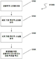
도 1A는 표현형을 특징화하기 위하여 핵산을 포함하는 생체특징을 확인하는 방법을 설명한다. 도 1B는 표현형을 특징화하기 위하여 소낭 또는 소낭 집단을 포함하는 생체특징을 확인하는 방법을 설명한다.
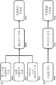
도 2는 소낭 생체특징을 평가함으로써 표현형을 특징화하는 방법들을 설명한다. 도 2A는 단백질을 발현시키는 소낭을 캡쳐하는 캡쳐 항체로 피복된 평면 기질의 개략도이다. 이 캡쳐 항체는 질환에 걸린 세포들로부터 유도한 소장 ("질환 소낭")에 특이적인 또는 특이적이지 않은 소낭 단백질에 대한 항체다. 탐지 항체는 캡쳐된 소낭에 결합하고, 형광 신호를 제공한다. 이 탐지 항체는 소낭에 일반적으로 연합된, 또는 기원 세포 또는 질환, 가령, 암과 연합된 항원을 탐지할 수 있다. 도 2B는 단백질을 발현시키는 소장을 캡쳐하는 캡쳐 항체로 피복된 비드의 개요도이다. 이 캡쳐 항체는 질환에 걸린 세포들로부터 유도한 소장 ("질환 소낭")에 특이적인 또는 특이적이지 않은 소낭 단백질에 대한 항체다. 이 탐지 항체는 캡쳐된 소낭에 결합하고, 형광 신호를 제공한다. 이 탐지 항체는 소낭에 일반적으로 연합된, 또는 기원 세포 또는 질환, 가령, 암과 연합된 항원을 탐지할 수 있다. 도 2C는 도 2B에서 나타낸 것과 같은 비드를 이용하여 다중적으로 실행될 수 있는 스크리닝 계획의 예가 된다. 도 2D는 표현형을 특징화하기 위하여 소낭을 캡쳐하고 탐지하기 위한 예시적인 계획을 제시한다. 도 2E는 표현형을 특징화하기 위하여 소낭 페이로드를 평가하기 위한 예시적인 계획을 제시한다.
도 3은 본 발명의 일부 예시적인 구체예들에서 이용할 수 있는 컴퓨터 시스템을 설명한다.
도 4는 피험자로부터 소낭을 탐지하는 비드 기반 방법을 이용한 결과를 나타내는 방법을 설명한다. 주어진 강도에서 캡쳐된 비드의 수는 얼마나 빈번하게 소낭이 이 강도에서 탐지 단백질을 발현시키는지를 나타낸다. 주어진 비드에서 좀더 강력한 신호는 탐지 단백질의 발현이 더 크다는 것을 말한다. 도면은 정상 환자를 한 곡선에 그리고 암 환자를 다른 곡선에 복합시키고, 이 곡선들을 구별하기 위하여 생물-통계학적 분석을 이용하여 수득한 표준화된 그래프다. 각 개체의 데이터는 탐지 기계에 의한 비드 판독 수의 변화를 설명하도록 표준화시키고, 각 집단에서 상이한 수의 시료를 설명하기 위하여 다시 표준화시킨다.
도 5는 TMPRSS2-ERG 발현을 평가함으로써 혈장으로부터 전립선암 세포들-유도된 소낭을 EpCam으로 캡쳐하는 것을 설명한다. VCaP 정제된 소낭은 정상 혈장에 고정되었고, 그 다음 EpCam 또는 이소타입 대조군 항체로 피복된 Dynal 자성 비드와 함께 항온처리하였다. RNA는 Dynal 비드로부터 직접적으로 단리하였다. 각 시료로부터 동량의 RNA를 RT-PCR 및 후속 Taqman 분석에 이용하였다.
도 6은 CD9 비드 캡쳐와 함께 miR-21 또는 miR-141 발현의 막대 그래프를 나타낸다. 전립선암 환자의 혈장 1㎖ 250 ng/㎖의 LNCaP, 또는 정제된 정상 소낭은 CD9 피복된 Dynal 비드와 함께 항온처리하였다. RNA는 비드 및 비드 상청액로부터 단리하였다. 한 가지 시료 (#6)는 또한 비교용으로 캡쳐되지 않았다. microRNA 발현은 qRT-PCR으로 측정하였고, 각 시료의 평균 CT 값을 비교하였다. CD9 캡쳐는 전립선암 시료에서 miR-21 및 miR-141의 탐지를 개선시킨다.
도 7은 MoFlo XDP를 이용하여 소낭을 분리 및 확인하는 것을 설명한다.
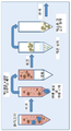
도 8은 시료 안에 소낭을 탐지하는 도식을 나타내는데, 여기에서 원하는 소낭의 존재 또는 수준은 미소구체 플랫포옴을 이용하여 평가한다. 도 8A는 컬럼 기반 필터링 방법을 이용하여 혈장으로부터 소낭을 단리하는 도식을 나타내는데, 여기에서 단리된 소낭들은 미소구체 플랫포옴을 이용하여 후속적으로 평가한다. 도 8B는 고속 원심분리, 가령 한외원심분리로 인하여 소낭의 막을 압착하는 도식을 나타낸다. 도 8C는 레이져 탐지를 이용하여 미소구체에 결합된 소낭을 탐지하는 도식을 나타낸다.
도 9A는 정상 전립선과 PCa 시료들을 구별하기 위하여 소낭 생체특징의 능력을 설명한다. 암 지표들은 EpCam 및 B7H3을 포함한다. 일반적인 소낭 지표들은 CD9, CD81 및 CD63을 포함하였다. 전립선 특이적 지표들은 PCSA을 포함하였다. PSMA 뿐만 아니라 PCSA도 이용될 수 있다. 테스트는 정상 시료들과 비교하여 PCa에 대해 98% 감응성이며 95% 특이성인 것으로 밝혀졌다. 도 9B는 정상인 및 전립선 암 환자들에서 도 9A의 소낭 표식에 대해 Y 축상에 평균 형광 강도(MFI)를 설명한다.
도 10은 시료가 전립선암에 양성인지를 판단하기 위한 소낭 전립선암 분석용의사결정수상도(decision tree)의 개요다.
도 11은 상승된 PSA 수준을 이용한 탐지 대 의사결정수상도에 따라 전립선 암에 대한 소낭 탐지 분석 결과를 나타낸다.
도 12는 대조군 그리고 PCa 시료로부터 단리된 소낭 안에 miR-14의 수준을 설명한다.
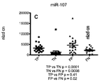
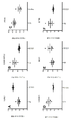
도 13A-13B는 전립선 암을 위한 소낭-기반 진단 분석으로부터 거짓 음성을 확인하는데 miR-107 및 miR-141의 사용을 설명한다. 도 13A는 거짓 음성을 참 양성으로 전환시키고, 이로 인하여 감응성을 개선시키는데 miR 분석을 이용하는 과정을 설명한다. 도 13B는 거짓 양성을 참 음성으로 전환시키고, 이로 인하여 특이성을 개선시키는데 miR 분석을 이용하는 과정을 설명한다. 소낭 진단 분석에 의한 참 양성(TP), 소낭 진단 분석에 의한 참 음성(TN), 소낭 진단 분석에 의한 거짓 양성(FP), 그리고 소낭 진단 분석에 의한 거짓 음성(FN)에 대해 miR-107 (도 13C) 및 miR-141 (도 13D)의 표준화된 수준을 나타낸다.
도14는 소낭 mRNA 페이로드 수준의 마이크로어래이 프로파일링으로부터 선택된 mRNA의 가공안된 배경 추출된 형광 값의 도트 플롯을 설명한다. 각 플롯에서, Y 축은 가공안된 배경 추출된 형광 값(Raw BGsub Florescence)을 나타낸다. X 축은 4가지 정상 대조군 혈장 및 전립선 암 환자들의 4가지 혈장의 도트 플롯을 나타낸다. 나타낸 mRNA는 A2ML1 (도14A), GABARAPL2 (도14B), PTMA (도14C), RABAC1 (도14D), SOX1 (도14E), 그리고 ETFB (도14F)이다.Figure 1A illustrates a method for identifying biomolecule characteristics comprising a nucleic acid to characterize a phenotype. Figure IB illustrates a method for identifying biomolecule characteristics, including follicular or follicular populations, to characterize a phenotype.
Figure 2 illustrates methods for characterizing phenotypes by evaluating follicular biomarkers. 2A is a schematic view of a planar substrate coated with a capture antibody that captures a vesicle that expresses the protein. This capture antibody is an antibody to a follicular protein that is specific or not specific for small intestine ("disease follicles") derived from diseased cells. The detection antibody binds to the captured follicle and provides a fluorescent signal. This detectable antibody can detect an antigen associated with a follicular cell or disease, such as cancer, that is generally associated with the follicle. Figure 2B is a schematic of a bead coated with a capture antibody that captures the small intestine that expresses the protein. This capture antibody is an antibody to a follicular protein that is specific or not specific for small intestine ("disease follicles") derived from diseased cells. This detection antibody binds to the captured follicle and provides a fluorescent signal. This detectable antibody can detect an antigen associated with a follicular cell or disease, such as cancer, that is generally associated with the follicle. Figure 2C is an example of a screening plan that can be performed multiple times using beads as shown in Figure 2B. Figure 2D presents an exemplary scheme for capturing and detecting vesicles to characterize phenotypes. Figure 2E presents an exemplary scheme for evaluating a fellow payload to characterize a phenotype.
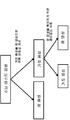
Figure 3 illustrates a computer system that may be used in some exemplary embodiments of the present invention.
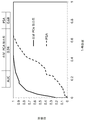
Figure 4 illustrates a method of representing results using a bead-based method of detecting vesicles from a subject. The number of beads captured at a given intensity indicates how frequently the follicles express the detected protein at this intensity. A more powerful signal in a given bead indicates that the expression of the detectable protein is greater. The figure is a standardized graph obtained using a biostatistical analysis to combine normal patients into one curve and cancer patients into another curve and distinguish these curves. The data of each individual is standardized to account for changes in the number of bead readings by the detection machine and standardized again to account for the different numbers of samples in each population.
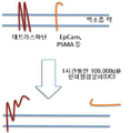
Figure 5 illustrates capturing prostate cancer cell-derived vesicles from plasma into EpCam by assessing TMPRSS2-ERG expression. The VCaP purified vesicles were fixed in normal plasma and then incubated with Dynal magnetic beads coated with EpCam or isotype control antibody. RNA was isolated directly from Dynal beads. Equal amounts of RNA from each sample were used for RT-PCR and subsequent Taqman analysis.
Figure 6 shows a histogram of miR-21 or miR-141 expression with CD9 bead capture.
Figure 7 illustrates the isolation and identification of follicles using MoFlo XDP.
Figure 8 shows a schematic for detecting follicles in a sample wherein the presence or level of the desired follicles is assessed using microsphere platforms. FIG. 8A shows a scheme for isolating follicles from plasma using a column-based filtering method, wherein the isolated follicles are subsequently assessed using microsphere platforms. 8B shows a schematic depicting the compression of the follicular membrane due to high speed centrifugation, e.g., ultrafiltration. Figure 8C shows a schematic for detecting follicles coupled to microspheres using laser detection.
FIG. 9A illustrates the ability of a follicular biomaterial character to distinguish normal prostate and PCa samples. Cancer indicators include EpCam and B7H3. Common follicular markers included CD9, CD81 and CD63. Prostate specific indicators included PCSA. PCSA as well as PSMA can be used. The test was found to be 98% sensitive and 95% specific for PCa compared to normal samples. Figure 9B illustrates the mean fluorescence intensity (MFI) on the Y-axis for the follicular markers of Figure 9A in normal and prostate cancer patients.
10 is an overview of a decision tree for analyzing cystic prostate cancer to determine whether a sample is positive for prostate cancer.
Figure 11 shows the results of the vaginal census detection assay for prostate cancer according to the detection versus decision aquisition using elevated PSA levels.
Figure 12 illustrates the levels of miR-14 in isolated follicles from control and PCa samples.
Figures 13A-13B illustrate the use of miR-107 and miR-141 to identify false negatives from a vesicle-based diagnostic assay for prostate cancer. Figure 13A illustrates the process of using miR analysis to convert false speech to positivity and thereby improve sensitivity. Figure 13B illustrates the process of using miR analysis to convert false positives to true and thereby improve specificity. (TP), phonation (TN) by follicular diagnostic analysis, false positive (FP) by follicular diagnostic analysis, and false negative (FN) (Fig. 13C) and miR-141 (Fig. 13D).
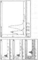
Figure 14 illustrates a dot plot of fluorescence values extracted from the raw background of mRNAs selected from microarray profiling of the follicular mRNA payload levels. In each plot, the Y-axis represents the raw fluorescence (Raw BGsub Florescence) extracted from the background. The X-axis represents a plot of the four plasma plots of four normal control plasma and prostate cancer patients. The indicated mRNAs are A2ML1 (FIG. 14A), GABARAPL2 (FIG. 14B), PTMA (FIG. 14C), RABAC1 (FIG. 14D), SOX1 (FIG. 14E), and ETFB (FIG. 14F).
생물학적 시료, 가령, 세포 배양물, 유기체, 또는 피험자의 시료의 표현형을 특징화하는 방법들 및 시스템을 설명한다. 이 표현형은 하나 이상의 생물지표들을 평가함으로써 특징화될 수 있다. 이 생물지표들은 소낭 또는 제시된 소낭 표면 항원들 또는 소낭 페이로드이 소낭 집단과 연합될 수 있다. 여기에서 이용된 것과 같이, 소낭 페이로드는 소낭안에 에워싸인 엔터티(entities)를 포함한다. 소낭 연관된 생물지표들은 막 결합된 그리고 가용성 생물지표들을 모두 포함할 수 있다. 이 생물학적지표들은 핵산(가령, microRNA) 또는 체액내에서 평가되는 단백질/폴리펩티드 또는 이의 기능적 단편들과 같은 순환하는 생물지표들일 수도 있다. 다른 명시가 없는 한, 소낭 또는 생물지표 성분들에 대해 여기에서 이용된 것과 같이 용어들 "정제된" 또는 "단리된"은 세포 또는 유기체로부터 이러한 성분들의 부분적 또는 완전한 정제 또는 분리를 의미한다. 더욱이, 다른 명시가 없는 한, 결합물질을 이용한 소낭 분리는 소낭에 결합물질을 결합시키고, 이러한 결합이 출발 물질에서 다른 생물학적 엔터티로부터 이 소낭을 완전하게 분리하는 지를 포함한다. Describes methods and systems for characterizing the phenotype of a biological sample, e.g., a cell culture, organism, or subject's sample. This phenotype can be characterized by evaluating one or more biomarkers. These biomarkers can be associated with the follicular or presented follicular surface antigens or the follicular payload with the follicular population. As used herein, a follicular payload includes entities enclosed within a vesicle. Follicular-associated biomarkers may include both membrane bound and soluble biomarkers. These biological indicators may be circulating biomarkers such as nucleic acid (e. G., MicroRNA) or protein / polypeptide or functional fragments thereof that are evaluated in body fluids. Unless otherwise indicated, the terms " purified "or" isolated, " as used herein for follicular or biomarker components, refer to partial or complete purification or separation of such components from a cell or organism. Moreover, unless otherwise specified, the fecal separation using the binding material involves binding the binding material to the follicle, and whether such binding completely separates the vesicle from other biological entities in the starting material.
순환하는 생물지표, 가령, 핵산 생물지표를 분석함으로써 표현형을 특징화하는 방법은 비-제한적인 예로써 도 1A의 계획 6100A에서 설명한다. 제 1 단계 6101에서, 생물학적 시료는 가령, 체액, 조직 시료 또는 세포 배양물로부터 수득한다. 핵산은 시료 6103로부터 단리된다. 이 핵산은 DNA 또는 RNA, 가령, microRNA일 수 있다. 이러한 핵산의 평가는 표현형에 대한 생체특징을 제시할 수 있다. 표적 표현형 (가령, 질환이 있음 대 건강함, 치료전과 치료후)과 연관된 핵산을 샘플링함으로써, 표현형의 지표인 하나 이상의 핵산 지표들을 결정할 수 있다. 본 발명의 다양한 측면들은 시료 6105에 존재하는 하나 이상의 핵산 분자들(가령, microRNA)를 평가함으로써 결정된 생체특징에 관한 것이며, 이때 이 생체특징은 예정된 표현형 6107에 상응한다. 도 1B는 이 핵산 분자들을 분리하기 위하여 소낭을 이용하는 계획 6100B를 설명한다. 한 실시예에서, 생물학적 시료를 얻고(6102), 하나 이상의 소낭, 가령, 특정 기원 세포의 소낭 및/또는 특정 질환 상태와 관련된 소낭은 시료로부터 단리한다(6104). 이 소낭은 이 소낭과 연관된 표면 항원들을 특징화시킴으로써 및/또는 이 소낭내에 존재하는 성분들 ("페이로드")의 존재 또는 수준을 결정함으로써 분석한다(6106). 다른 언급이 없는 한, 용어 "항원"은 여기에서 이용된 것과 같이 결합물질에 의해 결합될 수 있는 생물지표를 일반적으로 지칭하며, 이때 결합물질은 항체, 압타머, 렉틴이거나, 또는 이 생물학적지표의 다른 결합 물질일 수 있으며 그리고 이러한 생물지표가 숙주에서 면역 반응을 유도하는지에는 무관하다. 소낭 페이로드는 펩티드들 및 폴리펩티드들을 포함하는 단백질, 및/또는 DNA 및 RNA와 같은 핵산일 수 있다. RNA 페이로드는 메신져 RNA (mRNA) 및 microRNA (여기에서 miRNA 또는 miR으로 지칭될 수도 있다)를 포함한다. 표현형은 이 소낭의 생체특징에 근거하여 특징화된다(6108). 본 발명의 또다른 예시적인 방법에 있어서, 계획 6100A 및 6100B는 표현형을 특징화하기 위하여 함께 실행된다. 이러한 계획에서, 소낭 및 핵산, 가령, microRNA을 평가하고, 이에 의해 표현형을 특징화한다. Methods for characterizing phenotypes by analyzing circulating biomarkers, e.g., nucleic acid biomarkers, are described in
표현형Phenotype
막 소낭과 같은 소낭을 분석함으로써 개체의 표현형을 특징화하는 산물 및 과정을 여기에서 설명한다. 표현형은 질환 또는 상태, 질환 단계 또는 상태의 단계, 질환 또는 상태에 민감성, 질환 단계 또는 상태의 예후, 생리학적 상태, 또는 치료에 대한 반응과 같은 피험자에서 임의의 관찰가능한 특징들 또는 특색이 될 수 있다. 표현형은 피험자의 유전자 발현 뿐만 아니라 환경적 인자들의 영향과 이들 둘 사이의 상호작용으로 야기되는, 뿐만 아니라 핵산 서열들에 대한 후생적 수정으로 초래될 수 있다. Products and processes that characterize the phenotype of an individual by analyzing follicles such as dead follicles are described herein. A phenotype can be any observable characteristic or characteristic in a subject, such as a disease or condition, a stage of a disease state or condition, a disease or condition susceptibility, a disease stage or prognosis of a condition, a physiological condition, have. The phenotype can be caused not only by the expression of the subject's genes, but also by the effects of environmental factors and their interactions, as well as by welfare modifications to nucleic acid sequences.
피험자에서 표현형은 피험자로부터 생물학적 시료를 수득하고, 그리고 시료로부터 하나 이상의 소낭을 분석함으로써 특징화될 수 있다. 예를 들면, 피험자 또는 개체에 대한 표현형의 특징화는 질환 또는 상태 (전조 단계 이전의 초기 단계 탐지를 포함)를 탐지하고, 질환 또는 상태의 예후, 진단, 또는 치료진단을 결정하고, 또는 질환 또는 상태의 단계 또는 진전을 결정하는 것을 포함할 수 있다. 표현형의 특징화는 또한 특이적 질환, 상태, 질환 단계 및 상태의 단계에 대한 적합한 치료 또는 치료 효과를 확인하고, 질환 진전, 특히 질환 재발생, 전이성 전개 또는 질환 재발의 예측 및 가능성 분석을 포함한다. 표현형은 상태 또는 질환, 가령 암 또는 종양의 임상적으로 뚜렷한 유형 또는 준유형이 될 수도 있다. 표현형 결정은 생리학적 상태의 결정, 또는 장기 고통 또는 이식후 장기 거부와 같은 평가가 될 수도 있다. 여기에서 설명된 산물 및 과정들은 개별적으로 피험자를 평가할 수 있게 하고, 치료에서 좀더 효과적이고 경제적인 이익이 되는 판단을 제시할 수 있다. In a subject, the phenotype can be characterized by obtaining a biological sample from the subject and analyzing one or more follicles from the sample. For example, characterization of a phenotype for a subject or individual can be used to detect a disease or condition (including early stage pre-detection), to determine the prognosis, diagnosis, or treatment diagnosis of the disease or condition, Determining the stage or progress of the state. The phenotypic characterization also confirms the appropriate therapeutic or therapeutic efficacy for the specific disease, condition, stage of disease and condition, and includes disease progression, particularly disease recurrence, metastatic spread or prediction and likelihood analysis of disease relapse. The phenotype may be a clinically evident type or subtype of a condition or disease, such as cancer or a tumor. Phenotypic determinations may be assessments such as determination of physiological status, or long-term pain or post-transplant rejection. The products and processes described herein can be used to assess the subject individually and provide a more effective and economically beneficial judgment in treatment.
한 측면에서, 본 발명은 질환 또는 장애의 치료에 반응할 가능성이 있는지를 예측하기 위한 생체특징을 제공하기 위하여 소낭을 분석하는 것이다. 표현형의 특징화는 피험자의 반응자/비-반응자 상태를 예측하는 것을 포함하며, 여기에서 반응자는 질환의 치료에 반응하는 것이며, 비-반응자는 이 치료에 반응하지 않는다. 이 피험자에서 소낭을 분석하고, 그리고 해당 치료에 반응하거나 반응하지 않은 공지의 기존 피험자의 소낭 분석과 비교할 수 있다. 피험자의 소낭 생체특징이 치료에 반응하는 것으로 알려진 기존 피험자의 것과 좀더 밀접하게 배열된다면, 이 피험자는 치료에 대해 반응자로 특징화되거나 또는 예측될 수 있다. 유사하게, 피험자의 소낭 생체특징이 치료에 반응하지 않는 것으로 알려진 기존 피험자의 것과 좀더 밀접하게 배열된다면, 이 피험자는 치료에 대해 비-반응자로 특징화되거나 또는 예측될 수 있다. 이 치료는 임의의 적합한 질환, 장애 또는 다른 상태를 위한 것이다. 이 방법은 반응자/비-반응자 상태와 관련된 소낭 생체특징이 공지된 임의의 질환 환경에서 이용될 수 있다. In one aspect, the invention is to analyze follicles to provide biomarkers for predicting the likelihood of responding to the treatment of a disease or disorder. Characterization of a phenotype involves predicting a subject's responder / non-responder state, wherein the responder is responsive to the treatment of the disease and the non-responder does not respond to the therapy. The follicles can be analyzed in this subject and compared to the follicular analysis of known existing subjects who have not responded or responded to the treatment. If the subject's follicular biomolecule is more closely aligned to that of an existing subject known to be responsive to treatment, then the subject may be characterized or predicted as a respondent to treatment. Similarly, if the subject's follicular biomolecule is more closely aligned to that of an existing subject known to be unresponsive to treatment, then the subject may be characterized or predicted to be non-responders to treatment. This treatment is for any suitable disease, disorder or condition. This method can be used in any disease environment in which the vital biochemical characteristics associated with the responder / non-responder state are known.
여기에서 이용된 것과 같이, 용어 "표현형"은 본 발명의 방법들을 이용하여 확인될 수 있는 소낭 생체특징에 기여하는 임의의 특생 또는 특징을 의미할 수 있다. 예를 들면, 표현형은 치료에 반응할 것 같은 피험자의 확인이 될 수 있으며, 또는 좀더 광범위하게는, 표현형은 피험자로부터 수득한 시료에 대한 특징화된 생체특징을 근거하여 진단, 예후 또는 치료진단 결정이 될 수도 있다. As used herein, the term "phenotype" may refer to any predisposition or characteristic that contributes to a follicular biomolecule characteristic that can be identified using the methods of the present invention. For example, a phenotype may be an identification of a subject likely to respond to treatment, or, more broadly, a phenotype may be a diagnostic, prognostic or therapeutic diagnostic decision based on a characteristic of a biomarker for a sample obtained from the subject .
일부 구체예들에서, 표현형은 표 1에 열거된 것과 같은 질환 또는 상태를 포함한다. 예를 들면, 표현형은 종양, 신생물, 또는 암의 존재 또는 가능성을 포함할 수 있다. 여기에서 설명된 산물 또는 과정에 의해 탐지되거나 또는 평가되는 암은 다음을 포함하나 이에 한정되지 않는다: 유방암, 난소암, 폐암, 결장암, 과형성 폴립, 선종, 직장결장암, 고도이형성, 저도이형성, 전립선 이형성, 전립선암, 흑색종, 췌장암, 뇌암 (교아종과 같은), 혈액 악성, 간세포 암종, 경부암, 자궁내막암, 두경부암, 식도암, 위장기질 종양 (GIST), 신장 세포 암종 (RCC) 또는 위암. 직장결장암은 CRC Dukes B 또는 Dukes C-D일 수 있다. 혈액 악성은 B-세포 만성 임파성 백혈병, B-세포 임파종-DLBCL, B-세포 임파종-DLBCL-배 중심-유사, B-세포 임파종-DLBCL-활성화된 B-세포-유사, 및 Burkitt의 임파종일 수 있다. In some embodiments, the phenotype includes diseases or conditions such as those listed in Table 1. For example, the phenotype may include the presence or likelihood of a tumor, neoplasm, or cancer. Cancers that are detected or evaluated by the products or processes described herein include, but are not limited to: breast cancer, ovarian cancer, lung cancer, colon cancer, hyperplastic polyp, adenoma, rectal colorectal cancer, (GIST), renal cell carcinoma (RCC), or stomach cancer), prostate cancer, melanoma, pancreatic cancer, brain cancer (such as a glioblastoma), blood malignancy, hepatocellular carcinoma, cervical cancer, endometrial cancer, head and neck cancer. Rectal colorectal cancer may be CRC Dukes B or Dukes C-D. Blood cell malignancies include B-cell chronic lymphocytic leukemia, B-cell lymphoma-DLBCL, B-cell lymphoma-DLBCL-plexiform-like, B-cell lymphoma-DLBCL-activated B-cell-like, and Burkitt's lymphoma .
표현형은 전암 상태, 예를 들면, 악틴성 각화증, 위축성 위염, 백반증, 홍색비후증, 육아종성 림프종증, 전(pre)백혈병, 섬유증, 경부 형성이상, 자궁 경부 형성이상, 색소성 피부건조증, Barrett의 식도, 결장직장 폴립, 또는 악성 종양으로 발달할 가능성이 있는 다른 비정상적인 조직 성장 또는 병소일 수 있다. 변형성 바이러스 감염, 가령 HIV 및 HPV는 또한 본 발명에 따라 평가될 수 있는 표현형을 제시한다. The phenotypic form may be selected from the group consisting of pre-cancerous conditions such as, for example, actinic keratosis, atrophic gastritis, vitiligo, scleroderma, granulomatous lymphoma, preleukemia, fibrosis, cervical dysplasia, Esophagus, colorectal polyp, or other abnormal tissue growth or lesion that may develop into a malignant tumor. Deformable viral infections, such as HIV and HPV, also present phenotypes that can be evaluated in accordance with the present invention.
본 발명의 방법들에 의해 특징화되는 암은 다음을 포함하나 이에 한정되지 않는다: 암종, 육종, 임파종 또는 백혈병, 생식 세포 종양, 세포종(blastoma), 또는 다른 암. 암종(carcinoma)은 다음을 포함하나 이에 한정되지 않는다: 상피 신생물, 편평 세포 신생물 편평 세포 암종, 기저 세포 신생물 기저 세포 암종, 과도기성 세포 유두종 및 암종, 선종 및 선암종 (선(glands)), 선종, 선암종, 증식성 위벽염 인슐린종(linitis plastica insulinoma), 글루카곤종, 가스트린종, 장펩티드종(vipoma), 담관암, 간세포 암종, 아데노이드 포낭 암종, 맹장의 유암종 종양, 프로락티노마(prolactinoma), 호산성세포종(oncocytoma), 허틀(hurthle) 세포 선종, 신장 세포 암종, 그라비츠(grawitz) 종양, 다중 내분비 선종, 자궁내막양 선종, 부속기 및 피부 부속기 신생물, 점액표피양 신생물, 포낭, 뮤신 그리고 장액성신생물, 담관낭선종, 복막 가성점액종(pseudomyxoma peritonei), 도관, 소엽 그리고 수질부 신생물, 선방(acinar) 세포 신생물, 복합 상피 신생물, warthin의 종양, 가슴샘종, 분화된 성샘 신생물, 성삭(sex cord) 기질 종양, 난포막종, 과립층 세포 종양, 남아와세포종, Sertoli-Leydig 세포 종양, 사구 종양, 부신경교종, 크롬친화세포종, 사구 종양, nevi 및 흑색종, 멜라닌세포 모반, 악성 흑색종, 흑색종, 결절성 흑색종, 이형성 모반, 악성 흑자 흑색종, 표재 확장성 흑색종, 그리고 악성 선단 흑자성 흑색종. 육종은 다음을 포함하나 이에 한정되지 않는다: 피부의 종양, 보트로이드(botryodies), 연골육종, Ewing의 육종, 악성 혈관 내피종, 악성 슈반세포종, 골육종, 다음을 포함하는 연조직 육종: 폐포 유연부 육종, 혈관육종, 담낭육종 가엽, 피부섬유육종, 섬유의 종양, 복부결합조직형 작은 원형 세포 종양, 상피와 같은 육종, 골격외 연골육종, 골격외 골육종, 섬유육종, 혈관외피세포종, 혈관육종, Kaposi의 육종, 평활육종근, 지방육종, 림프관육종, 임파육종, 악성 섬유성 조직구종, 신경섬유육종, 횡문근육종, 그리고 활액육종. 임파종 및 백혈병은 다음을 포함하나 이에 한정되지 않는다: 만성림프구성 백혈병/작은 임파구성 임파종, B-세포 사전림프구성 백혈병, 임파혈질세포 임파종 (가령, Waldenstrm 매크로글로블린혈증), 비장 연변부 임파종, 혈장 세포 골수종, 형질세포종, 단클론 면역글로블린 침착 질환, 중쇄 질환, 결절외변연부 B 세포 임파종-malt 임파종이라고도 불림, 림프절 변연부 B 세포 임파종 (nmzl), 여포 임파종, 맨틀(mantle) 세포 임파종, 미만성 거대 B 세포 임파종, 종격동 (흉선) 거대 B 세포 임파종, 혈관내 거대 B 세포 임파종, 원발성 삼출성 임파종, Burkitt 임파종/백혈병, T 세포 사전림프구성 백혈병, T 세포 거대 과립성 림프구성 백혈병, 공격적인 NK 세포 백혈병, 성인T 세포 백혈병/임파종, 결절외 NK/T 세포 임파종, 코의 유형, 장질환-유형 T 세포 임파종, 간비장 T 세포 임파종, 모구성 NK 세포 임파종, 균상 식육종/세자리(sezary) 증후군, 원발성 피부의 CD30-양성 T 세포 임파증식성 장애, 원발성 피부의 역행성 거대 세포 임파종, 림프종양 구진증, 맥관면역모구성 T 세포 임파종, 말초 T 세포 임파종, 상세불명, 역행성 거대 세포 임파종, 고전적 Hodgkin 임파종(결절성 경화, 혼합형 세포성, 림프구-풍부, 림프구 감손된 또는 감손되지 않은), 그리고 결절성 림프구-우세 Hodgkin 임파종. 생식 세포 종양은 다음을 포함하나 이에 한정되지 않는다: 배아세포종, 이상종자배아세포종, 고환종, 비-배아세포종성 생식 세포 종양, 배아 암종, 내배엽 만곡 종양, 융모막암종, 기형종, 다배아종, 그리고 생식아세포종. 세포종은 다음을 포함하나 이에 한정되지 않는다: 신아세포종, 수아종, 및 망막아종. 다른 암들은 다음을 포함하나 이에 한정되지 않는다: 음순(labial) 암종, 후두 암종, 하인두 암종, 혀 암종, 타액선 암종, 위암종, 선암종, 갑상선암 (수질부 및 유두 갑상선암종), 신장 암종, 신장 유조직 암종, 경부암종, 자궁 체부 암종, 자궁 내막 암종, 융모막 암종, 고환 암종, 비뇨기 암종, 흑색종, 뇌종양 가령, 교아종, 성상세포종, 수막종, 수아종 그리고 말초 신경외배엽 종양, 쓸개암종, 기관지 암종, 다중 골수종, 기저세포암, 기형종, 망막아종, 맥락막 흑색종, 고환종, 횡문근육종, 두개인두종, 골육종, 연골육종, 골육종, 지방육종, 섬유육종, Ewing 육종, 및 형질세포종. Cancers characterized by the methods of the present invention include, but are not limited to, carcinoma, sarcoma, lymphoma or leukemia, germ cell tumors, blastoma, or other cancers. Carcinomas include, but are not limited to, epithelial neoplasms, squamous cell neoplasia squamous cell carcinomas, basal cell neoplastic basal cell carcinomas, transitional cell papillomas and carcinomas, adenomas and adenocarcinomas (glands) , Adenocarcinoma, adenoid cystadenocarcinoma, carcinoid carcinoid tumor, prolactinoma, gastric adenocarcinoma, adenocarcinoma, proliferative insuloma, proliferative insuloma, glucagon species, gastrin species, vipoma, cholangiocarcinoma, hepatocellular carcinoma, adenoid cystic carcinoma, Endocrine neoplasms, adnexal and adnexal neoplasms, epidermoid neoplasms, cysts, cysts, adenocarcinomas, adrenocortical adenomas, adrenal adenocarcinomas, adrenal adenocarcinomas, adrenal adenocarcinomas, Mucin and serous organisms, biliary cystadenoma, pseudomyxoma peritonei, ducts, lobules and neoplasms, acinar cell neoplasms, epithelial neoplasms, warthin tumors, chest A malignant neoplasm of the malignant glioma, a malignant neoplasm, a malignant neoplasm, a malignant neoplasm, a malignant neoplasm, a malignant neoplasm, a malignant neoplasm, a malignant neoplasm, , Melanocytic nevi, malignant melanoma, melanoma, nodular melanoma, dysplastic nevi, malignant surplus melanoma, superficial melanoma, and malignant prothoracic melanoma. Sarcomas include, but are not limited to: skin tumors, botryodies, chondrosarcoma, Ewing's sarcoma, malignant angi endothelioma, malignant schwannoma, osteosarcoma, soft tissue sarcomas including alveoli , Kaposi's sarcoma, sarcoma sarcoma, dermoid sarcoma, dermoid fibrosarcoma, fibroid tumor, small round cell tumor of abdominal connective tissue type, sarcoma like epithelium, extrahepatic chondrosarcoma, extrahepatic osteosarcoma, fibrous sarcoma, Sarcomas, smooth muscle sarcomas, liposarcoma, lymphatic sarcoma, lymphoma, malignant fibrous histiocytoma, nerve fibrosarcoma, rhabdomyosarcoma, and synovial sarcoma. Lymphoma and leukemia include, but are not limited to, chronic lymphocytic leukemia / small lymphocytic lymphoma, B-cell pre-lymphoid leukemia, lymphoid blood cell lymphoma (e.g., Waldenstrm macroglobulinemia), splenic lymphangioma, plasma cells Lymphoid lymphoblastic lymphoma (nmzl), follicular lymphoma, mantle cell lymphoma, diffuse large B-cell lymphoma, lymphoid malignant lymphoma, lymphoid malignant lymphoma, lymphoid malignant lymphoma, malignant melanoma, myeloma, plasma cell tumor, monoclonal immunoglobulin depositional disease, , Medullary large B cell lymphoma, intravascular large B cell lymphoma, primary exudative lymphoma, Burkitt lymphoma / leukemia, T cell prolymphocytic leukemia, T cell giant granular lymphocytic leukemia, aggressive NK cell leukemia, adult T cells Leukemia / lymphoma, nodular NK / T cell lymphoma, nasal type, intestinal disease - type T cell lymphoma, liver spleen T Lymphoma, lymphangioblastomas, vasculitis Immunohistochemistry T lymphocyte, lymphocyte subpopulation, lymphocyte subpopulation, lymphocyte subpopulation, lymph node metastasis, lymph node metastasis, lymph node metastasis, NK cell lymphoma, Cellular lymphoma, peripheral T cell lymphoma, unspecified, retrograde giant cell lymphoma, classical Hodgkin lymphoma (tuberous sclerosis, mixed cellular, lymphocyte - rich, lymphocyte detached or undamaged), and nodular lymphocyte - dominant Hodgkin lymphoma. Germ cell tumors include, but are not limited to, germ cell tumors, adenomatous germ cell tumors, testicular tumors, non-germ cell tumors germ cell tumors, embryonic carcinomas, endodermal curved tumors, choriocarcinomas, teratomas, And gonadoblastoma. Cell tumors include, but are not limited to: neoplasm, subacute, and retinoblastoma. Other cancers include but are not limited to: labial carcinoma, laryngeal carcinoma, hypopharyngeal carcinoma, tongue carcinoma, salivary carcinoma, gastric carcinoma, adenocarcinoma, thyroid carcinoma (renal carcinoma and papillary thyroid carcinoma) The present invention relates to a method of treating a cancer selected from the group consisting of carcinoma, cervical carcinoma, cervical carcinoma, endometrial carcinoma, choriocarcinoma, testicular carcinoma, urological carcinoma, melanoma, Cholangiocarcinoma, osteosarcoma, liposarcoma, fibrosarcoma, Ewing's sarcoma, and plasma cell tumor, including multiple myeloma, basal cell carcinoma, teratoma, retinoblastoma, choroidal melanoma, testicular species, rhabdomyosarcoma, craniopharyngioma, osteosarcoma, chondrosarcoma.
추가 구체예에서, 분석하게 되는 암은 비-소 세포 폐암 및 소 세포 폐암 (소 세포 암종 (귀리 세포 암)을 포함하는 폐암, 혼합형 소 세포/거대 세포 암종, 및 복합형 소 세포 암종을 포함), 결장암, 유방암, 전립선암, 간암, 췌장 암, 뇌암, 신장 암, 난소암, 위암, 피부 암, 골 암, 위암, 유방암, 췌장암, 신경교종, 교아종, 간세포 암종, 유두 신장 암종, 두경부 편평 세포 암종, 백혈병, 임파종, 골수종, 또는 충실성 종양을 포함할 수 있다.In a further embodiment, the cancer to be analyzed is selected from the group consisting of non-small cell lung cancer and small cell lung cancer (including lung cancer including small cell carcinoma (osteo-cell cancer), mixed small cell / giant cell carcinoma, Pancreatic cancer, glioma, glioma, hepatocellular carcinoma, papillary renal carcinoma, head and neck squamous cell carcinoma, colon cancer, breast cancer, prostate cancer, liver cancer, pancreatic cancer, brain cancer, kidney cancer, ovarian cancer, gastric cancer, skin cancer, Cell carcinoma, leukemia, lymphoma, myeloma, or solid tumors.
구체예들에서, 암은 급성 림프구성 백혈병; 급성 골수성 백혈병; 부신피질 암종; AIDS-관련된 암; AIDS-관련된 임파종; 항문암; 맹장암; 성상세포종; 비정형 유기형/간상 종양; 기저 세포 암종; 방광암; 뇌줄기 교종; 뇌종양 (뇌줄기 교종, 중추 신경계 비정형 유기형/간상 종양, 중추 신경계 배아 종양, 성상세포종, 두개인두종, 뇌실막모세포종, 상의세포종, 수아종, 수질상피종, 중간 분화의 송과체종 종양, 천막상부 원시성 외배엽 종양 및 송과체모세포종을 포함); 유방암; 기관지 종양; Burkitt 임파종; 원발부위 불명 암; 유암종 종양; 원발부위 불명 암의 암종; 중추 신경계 비정형 유기형/간상 종양; 중추 신경계 배아 종양; 경부암; 소아 암; 척색종; 만성림프구성 백혈병; 만성골수성 백혈병; 만성골증식성 장애; 결장암; 직장결장암; 두개인두종; 피부의 T-세포 임파종; 내분비 췌장 섬 세포 종양; 자궁내막암; 뇌실막모세포종; 상의세포종; 식도암; 감각신경모세포종; Ewing 육종; 두개밖 생식 세포 종양; 고환외 생식 세포 종양; 간밖의 담즙 관 암; 쓸개암; 위 암; 위장유암종 종양; 위장기질 세포 종양; 위장기질 종양 (GIST); 임신성 영양막 종양; 신경교종; 모(hair) 세포 백혈병; 두경부암; 심장 암; Hodgkin 임파종; 하인두 암; 안구내 흑색종; 섬 세포 종양; Kaposi 육종; 신장 암; Langerhans 세포 조직구증; 후두 암; 입술 암; 간암; 악성 섬유성 조직구종 골암; 수아종; 수질상피종; 흑색종; Merkel 세포 암종; Merkel 세포 피부 암종; 중피종; 잠복 원발성을 가진 전이성 편평세포 경부암; 구강암; 다중 내분비 신조직형성 증후군; 다중 골수종; 다중 골수종/혈장 세포 신생물; 균상 식육종; 골이형성 증후군; 골증식성 신생물; 비강 암; 비인두 암; 신경아종; 비-Hodgkin 임파종; 비-흑색종 피부 암; 비-소 세포 폐암; 구강 암; 구강 공동 암; 구강인후 암; 골육종; 다른 뇌 및 척수 종양; 난소암; 난소 상피 암; 난소 생식 세포 종양; 난소 악성 가능성이 낮은 종양; 췌장암; 유두종증; 부비동 암; 부갑상선암; 골반 암; 페니스 암; 인두 암; 중간 분화의 송과체종 종양; 송과체모세포종; 뇌하수체 종양; 혈장 세포 신생물/다중 골수종; 폐모세포종; 원발성 중추 신경계 (CNS) 임파종; 원발성 간세포 간암; 전립선암; 직장 암; 신장 암; 신장 세포 (신장) 암; 신장 세포 암; 기도 암; 망막아종; 횡문근육종; 타액선 암; Szary 증후군; 소 세포 폐암; 소장 암; 연조직 육종; 편평 세포 암종; 편평 목 암; 위암; 천막상부 원시성 외배엽 종양; T-세포 임파종; 고환 암; 목 암; 흉선 암종; 가슴샘종; 갑상선암; 과도기성 세포 암; 신장 신우 및 수뇨관의 과도기성 세포암; 영양막 종양; 수뇨관 암; 요도 암; 자궁 암; 자궁 육종; 질 암; 외음부 암; Waldenstrm 매크로글로블린혈증; 또는 Wilm의 종양을 포함한다. 본 발명의 방법들은 이들 암과 다른 암들을 특징화하는데 이용할 수 있다. 따라서, 표현형의 특징화는 여기에서 설명된 한 가지 암의 진단, 예후 또는 치료진단을 제시할 수 있다. In embodiments, the cancer is acute lymphocytic leukemia; Acute myelogenous leukemia; Adrenocortical carcinoma; AIDS-related cancer; AIDS-related lymphomas; Anal cancer; Appendix cancer; Astrocytoma; Atypical organic / hepatic tumors; Basal cell carcinoma; Bladder cancer; Brainstem glioma; Brain tumor (brain stem glioma, central nervous system atypical organic / hepatic tumor, central nervous system embryo tumor, astrocytoma, craniopharyngioma, parenchymal blastoma, ependymoma, subspecies, watery epithelial tumor, mediastinal pineal tumor tumor, Including pineal gland blastomas); Breast cancer; Bronchial tumor; Burkitt, L.; Primary site unknown cancer; Carcinoid tumors; Carcinoma of primary site unknown carcinoma; Central nervous system atypical organic / hepatic tumors; Central nervous system embryo tumor; Cervical cancer; Pediatric cancer; Choroid species; Chronic lymphocytic leukemia; Chronic myelogenous leukemia; Chronic osteoporotic disorder; Colon cancer; Rectal colon cancer; Craniopharyngioma; T-cell lymphoma of the skin; Endocrine pancreatic islet cell tumor; Endometrial cancer; Ventricular blastoma; Cell tumor on the stomach; Esophagus cancer; Sensory neuroblastoma; Ewing breeding; Two extra germ cell tumors; Testicular germ cell tumor; Extrahepatic bile duct cancer; Gall bladder cancer; Gastric cancer; Gastric carcinoid tumors; Gastric stromal cell tumor; Gastrointestinal stromal tumor (GIST); Gestational trophoblastic tumor; Glioma; Hair cell leukemia; Head and neck cancer; Heart cancer; Hodgkin lymphoma; Hypopharyngeal cancer; Intra-ocular melanoma; Islet cell tumor; Kaposi breeding; Kidney cancer; Langerhans cell histiocytosis; Laryngeal cancer; Lip cancer; Liver cancer; Malignant fibrous histiocytoma bone cancer; Subspecies; Water epithelium species; Melanoma; Merkel cell carcinoma; Merkel cell skin carcinoma; Mesothelioma; Metastatic squamous cell carcinoma with latent primary; Oral cancer; Multiple endocrine neoplasia syndrome; Multiple myeloma; Multiple myeloma / plasma cell neoplasms; Fungal sarcoma; Bone formation syndrome; Osteoproliferative neoplasm; Nasal cancer; Nasopharyngeal cancer; Neuroassay; Non-Hodgkin lymphoma; Non-melanoma skin cancer; Non-small cell lung cancer; Oral cancer; Oral cavity cancer; Oral throat cancer; Osteosarcoma; Other brain and spinal cord tumors; Ovarian cancer; Ovarian epithelial cancer; Ovarian germ cell tumor; Tumors that are less likely to have ovarian malignancy; Pancreatic cancer; Papilloma; Sinus cancer; Papillary thyroid cancer; Pelvic cancer; Penis arm; Pharyngeal cancer; Mediastinal pineal gland tumors; Pineal cell neoplasm; Pituitary tumor; Plasma cell neoplasm / multiple myeloma; Pulmonary blastoma; Primary central nervous system (CNS) lymphoma; Primary hepatocellular carcinoma; Prostate cancer; Rectal cancer; Kidney cancer; Kidney cell (kidney) cancer; Renal cell carcinoma; Airway cancer; Retinoblastoma; Rhabdomyosarcoma; Salivary gland cancer; Szary syndrome; Small cell lung cancer; Small bowel cancer; Soft tissue sarcoma; Squamous cell carcinoma; Flat neck arm; Gastric cancer; Tentacular supraventricular ectodermal tumors; T-cell lymphoma; Testicular cancer; Neck cancer; Thymic carcinoma; Thymoma; Thyroid cancer; Transitional cell carcinoma; Transitional cell carcinoma of kidneys and ureters; Trophoblastic tumor; Ureteral cancer; Urethral cancer; Uterine cancer; Uterine sarcoma; Vaginal cancer; Vulvar cancer; Waldenstrm macroglobulinemia; Or Wilm's tumor. The methods of the invention can be used to characterize these and other cancers. Thus, phenotypic characterization can suggest a diagnosis, prognosis, or therapeutic diagnosis of one cancer described herein.
표현형은 염증 질환, 면역 질환, 또는 자가면역 질환일 수도 있다. 예를 들면, 이 질환은 염증성 장 질환 (IBD), Crohn 질환 (CD), 궤양성 결장염 (UC), 골반 염증, 맥관염, 건선, 당뇨병, 자가면역 간염, 다발성 경화증, 중증 근무력증, 유형 I 당뇨병, 류마티스 관절염, 건선, 전신 홍반성 낭창 (SLE), Hashimoto의 갑상선염, Grave의 질환, 강직성 척추염, Sjogrens 질환, CREST 증후군, 경피증, 류마티스 질환, 장기 거부, 원발성 경화성 담관염, 또는 폐혈증일 수 있다. The phenotype may be an inflammatory disease, an autoimmune disease, or an autoimmune disease. For example, the disease is selected from the group consisting of inflammatory bowel disease (IBD), Crohn disease (CD), ulcerative colitis (UC), pelvic inflammation, vasculitis, psoriasis, diabetes, autoimmune hepatitis, multiple sclerosis, , Rheumatoid arthritis, psoriasis, systemic lupus erythematosus (SLE), Hashimoto's thyroiditis, Grave's disease, ankylosing spondylitis, Sjogren's disease, CREST syndrome, scleroderma, rheumatic disease, organ rejection, primary sclerosing cholangitis, or sepsis.
이 표현형은 또한 심혈관 질환, 가령 아테롬성경화, 울혈성 심부전, 취약성 죽상경화반, 발작, 또는 허혈을 포함할 수 있다. 심혈관 질환 또는 상태는 고혈압, 협착증, 혈관 폐색 또는 혈전증일 수 있다.This phenotype may also include cardiovascular disease, such as atherosclerosis, congestive heart failure, fragile atherosclerotic lesions, seizures, or ischemia. A cardiovascular disease or condition can be hypertension, stenosis, vascular occlusion or thrombosis.
표현형은 또한 신경 질환, 가령, 다발성 경화증 (MS), Parkinson 질환 (PD), Alzheimer 질환 (AD), 정신불열증, 양극성장애, 우울, 자폐증, Prion 질환, Pick 질환, 치매, Huntington 질환 (HD), Down 증후군, 뇌혈관 질환, Rasmussen의 뇌염, 바이러스성 수막염, 신경정신성 전신 홍반성 낭창 (NPSLE), 근위축성 측삭 경화, Creutzfeldt-Jacob 질환, Gerstmann-Straussler-Scheinker 질환,전염성 해마상뇌증, 허혈성 재관류 손상 (가령 발작), 뇌외상, 미생물 감염, 또는 만성피로 증후군을 포함할 수 있다. 이 표현형은 섬유근육통, 만성 신경병 통증, 또는 말초 신경병 통증과 같은 상태일 수도 있다.The phenotype also includes neurological diseases such as multiple sclerosis (MS), Parkinson's disease (PD), Alzheimer's disease (AD), mental illness, bipolar disorder, depression, autism, Prion disease, Pick disease, , Down syndrome, cerebrovascular disease, Rasmussen's encephalitis, viral meningitis, neuropsychiatric systemic lupus erythematosus (NPSLE), amyotrophic lateral sclerosis, Creutzfeldt-Jacob disease, Gerstmann-Straussler-Scheinker disease, infectious hippocampal encephalopathy, ischemic reperfusion (E. G., Seizures), brain trauma, microbial infection, or chronic fatigue syndrome. This phenotype may be a condition such as fibromyalgia, chronic neuropathic pain, or peripheral neuropathic pain.
이 표현형은 감염질환, 가령, 박테리아성, 바이러스성 또는 효모 감염을 또한 포함할 수 있다. 예를 들면, 이 질환 또는 상태는 Whipple 질환, Prion 질환, 간경변, 메티실린-저항성 스타필로코커스 아우레우스, HIV, 간염, 매독, 수막염, 말라리아, 결핵, 또는 인플루엔자일 수 있다. 바이러스성 단백질들, 가령 HIV 또는 HCV-유사 입자들은 바이러스성 상태를 특징화하기 위하여 소낭에서 평가할 수 있다. This phenotype may also include an infectious disease, e. G., Bacterial, viral or yeast infection. For example, the disease or condition may be Whipple disease, Prion disease, cirrhosis, methicillin-resistant Staphylococcus aureus, HIV, hepatitis, syphilis, meningitis, malaria, tuberculosis, or influenza. Viral proteins, such as HIV or HCV-like particles, can be assessed in vesicles to characterize the viral state.
이 표현형은 또한 주산기(perinatal) 또는 임신 관련된 상태 (가령 자간전증 또는 조산), 대사 질환 또는 상태, 가령, 철 대사와 연관된 대사 질환 또는 상태를 포함할 수 있다. 예를 들면, 헵시딘은 철 결핍을 특징화하기 위하여 소낭에서 분석될 수 있다. 대사 질환 또는 상태는 당뇨병, 염증, 또는 주산기 상태일 수 있다.This phenotype may also include metabolic diseases or conditions associated with perinatal or pregnancy related conditions (such as preeclampsia or prematurity), metabolic diseases or conditions, e.g., iron metabolism. For example, hepcidin can be analyzed in vats to characterize iron deficiency. The metabolic disease or condition may be diabetes, inflammation, or perinatal condition.
본 발명의 방법들을 이용하여 생물지표들을 통하여 평가될 수 있는 이들 질환 및 장애 그리고 다른 질환 및 장애를 특징화할 수 있다. 따라서, 표현형의 특징화는 여기에서 설명된 장애 및 질환중 하나의 진단, 예후 또는 치료진단을 제시할 수 있다. The methods of the invention can be used to characterize these diseases and disorders and other diseases and disorders that can be assessed through biomarkers. Thus, characterization of the phenotype may suggest a diagnosis, prognosis, or therapeutic diagnosis of one of the disorders and diseases described herein.
피험자(Subjects)Subjects
피험자의 하나 이상의 표현형은 하나 이상의 소낭, 가령, 피험자로부터 수득한 생물학적 시료내 소낭을 분석함으로써 결정할 수 있다. 피험자 또는 환자는 소, 조류, 갯과동물, 말, 고양잇과 동물, 양, 돼지와 같은 포유류, 또는 영장류 (인간 및 비-인간 영장류를 포함)를 포함하나 이에 한정되지 않는다. 피험자는 절멸위기에 처하여 중요한 포유류, 가령 시베리아 호랑이; 또는 인간의 소비를 위하여 농장에서 사육되는 동물과 같은 경제적 중요성, 또는 인간에게 사회적으로 중요한 동물, 가령, 애완동물 또는 동물원 동물을 포함할 수 있다. 이러한 동물의 예는 다음을 포함하나 이에 한정되지 않는다: 개 및 고양이와 같은 육식동물; 어린 돼지, 수퇘지, 멧돼지를 포함한 돼지; 소, 황소, 양, 기린, 사슴, 염소, 아메리카들소, 낙타 또는 말과 같은 반추동물 또는 유제동물(발굽). 멸종위기의 또는 동물원에 있는 조류, 뿐만 아니라 가금류, 더 구체적으로, 가축화된 가금류, 가령 사육 조류, 가령, 칠면조, 닭, 오리, 거위, 뿔닭을 또한 포함한다. 또한 길들여진 돼지 및 말(경주용 말 포함)을 포함한다. 추가로, 농업 및 수족관 및 다른 활동과 연관된 동물과 같은 상업적 활동과 연관된 임의의 동물 종이 포함되는데, 이때 질환 모니터링, 진단 및 치료법 선택은 경제적 생산성 및/또는 먹이 사슬의 안전성에 대해 농축산 분야에서 일반적인 관행이다. One or more phenotypes of the subject can be determined by analyzing one or more follicles, e. G., Vesicles in the biological sample obtained from the subject. The subject or patient includes, but is not limited to, cows, birds, corn and animals, horses, cats and animals, mammals such as sheep, pigs, or primates (including human and non-human primates). The subject is in danger of extinction and is an important mammal, such as the Siberian tiger; Or economic importance, such as animals raised on farms for human consumption, or animals socially important to humans, such as pet or zoo animals. Examples of such animals include, but are not limited to: carnivores such as dogs and cats; Pigs including young pigs, boar, and wild boar; Ruminants or emulsion animals (hooves) such as cattle, ox, sheep, giraffe, deer, goat, American bison, camel or horse. Also included are birds in endangered or zoos, as well as poultry, more specifically, domesticated poultry, such as breeding birds, such as turkeys, chickens, ducks, geese, guinea fowl. Also included are domesticated pigs and horses (including racing horses). In addition, any animal species associated with commercial activities such as agriculture and animals associated with aquariums and other activities, wherein the disease monitoring, diagnosis and treatment options are selected from the generic practices in the agricultural and livestock industry for economic productivity and / or food chain safety to be.
피험자는 암과 같이 이미-존재하는 질환 또는 상태를 보유할 수 있다. 대안으로, 이 피험자는 임의의 공지의 기존 상태를 보유하지 않을 수 있다. 피험자는 암 치료와 같은 현존 또는 과거 치료에 또한 비-반응성일 수 있다. The subject may have an already-existing disease or condition, such as cancer. Alternatively, the subject may not have any known existing state. The subject may also be non-responsive to existing or past treatments such as cancer therapy.
시료들(Samples)Samples
피험자로부터 수득한 생물학적 시료는 임의의 체액일 수 있다. 예를 들면, 이 생물학적 시료는 말초 혈액, 혈청, 혈장, 복수, 소변, 뇌척수액 (CSF), 객담, 타액, 골수, 활액, 수양액, 양수, 귀지, 모유, 기관지폐포 세척액, 정액 (전립선액을 포함), Cowper 유체 또는 사정전 유체, 여성 사정액, 땀, 대변 찌꺼기, 모(hair), 눈물, 낭종 유체, 늑막 및 복막 유체, 심장주변 유체, 림프, 미즙, 유미, 담즙, 간질성 유체, 월경, 고름, 피지, 구토물, 질 분비물, 점막 분비물, 대변 물, 췌액, 비강의 세척액, 기관지 폐 흡출액 또는 다른 세척 유체일 수 있다. 생물학적 시료는 또한 배낭강, 탯줄 혈액, 또는 태아 또는 모체 기원일 수 있는 모계 순환을 포함할 수 있다. 이 생물학적 시료는 소낭 및 다른 순환하는 생물지표들을 수득할 수 있는 조직 시료 또는 생검일 수도 있다. 예를 들면, 시료의 세포들은 배양되거나 또는 배양물로부터 단리된 소낭일 수 있다. (예를 들면, 실시예 1 참고). 다양한 구체예들에서, 여기에서 설명된 생물지표들 또는 구체적으로 생체특징은 다양한 방법들, 예를 들면, 혈액, 혈장, 혈청 또는 전술한 임의의 생물학적 시료들로부터 핵산 분자들의 추출, 폴리펩티드 (또는 기능적 단편) 생물지표(들)의 확인을 위하여 단백질 또는 항체 어레이의 이용, 뿐만 아니라 다른 어래이, 핵산 및 폴리펩티드 분자들의 확인 및 평가를 위하여 당업계에 공지되어 있는 서열화, PCR, 및 프로테오믹(proteomic) 기술을 이용하여 이러한 생물학적 시료로부터 직접적으로 평가할 수 있다(가령, 핵산 또는 폴리펩티드 생물지표들 또는 이의 기능적 단편의 존재 또눈 수준의 확인).The biological sample obtained from the subject may be any bodily fluid. For example, the biological sample may include peripheral blood, serum, plasma, ascites, urine, cerebrospinal fluid (CSF), sputum, saliva, bone marrow, synovial fluid, ), Cowper fluid or sleeping fluid, female ejaculatory fluid, sweat, stool, hair, tear, cyst fluid, pleural and peritoneal fluid, pericardial fluid, lymph, Pus, vomitus, vaginal discharge, mucous secretion, feces, pancreatic juice, nasal wash, bronchial lung aspirate or other cleansing fluid. The biological sample may also include a backpack river, umbilical cord blood, or a maternal circulation that may be fetal or maternal origin. This biological sample may be a tissue sample or a biopsy that can yield follicles and other circulating biomarkers. For example, the cells of the sample may be cultured or isolated from the culture. (See, for example, Example 1). In various embodiments, the biomarkers described herein, or specifically the biomarker, may be extracted from a variety of methods, such as extraction of nucleic acid molecules from blood, plasma, serum or any of the biological samples described above, PCR, and proteomic assays known in the art for the identification and evaluation of other arrays, nucleic acid and polypeptide molecules, as well as the use of protein or antibody arrays for identification of biomarker (s) (E. G., Confirmation of the presence or absence of nucleic acid or polypeptide biomarkers or functional fragments thereof). ≪ / RTI >
표 1은 질환, 상태, 또는 생물학적 상태와 소낭이 분석될 생물학적 시료의 해당 목록의 예시적인 설명을 열거한다. Table 1 lists an exemplary description of a disease, condition, or biological condition and a corresponding list of biological samples to be analyzed for vesicles.


본 발명의 방법은 혈액 시료 또는 혈액 유도체를 이용하여 표현형을 특징화하는데 이용할 수 있다. 혈액 유도체들은 혈장 및 혈청을 포함한다. 혈액 혈장은 전혈의 액체 성분이며, 총 혈액 용적의 대략 55% 용적을 차지한다. 혈장은 용액안에 소량의 미네랄, 염, 이온, 영양소 및 단백질과 주로 물로 구성된다. 전혈에서, 적혈구 세포, 백혈구세포, 및 혈소판은 혈장 안에 현탁된다. 혈액 혈청은 피브리노겐 또는 다른 응고 인자들(가령, 전혈에서 상기 두 세포들과 응고 인자를 뺀)이 없는 혈액 혈장을 말한다. The methods of the invention can be used to characterize phenotypes using blood samples or blood derivatives. Blood derivatives include plasma and serum. Blood plasma is the liquid component of whole blood, accounting for approximately 55% of the total blood volume. Plasma is made up of small amounts of minerals, salts, ions, nutrients and proteins in the solution and mainly water. In whole blood, red blood cells, white blood cells, and platelets are suspended in plasma. Blood serum refers to blood plasma free of fibrinogen or other coagulation factors (e.g., minus the coagulation factors from the two cells in the whole blood).
이 생물학적 시료는 이 생물학적지표들의 분석을 생물학적 시료의 직접적인 평가 또는 이 생물학적 시료로부터 수득된 하나 이상의 소낭을 프로파일링함으로써 생물지표들의 분석을 실행하지 않은 제 3 자를 통하여 수득할 수 있다. 예를 들면, 이 시료는 시료를 얻는 피험자의 임상의, 의사 또는 다른 건강관리인을 통하여 얻을 수 있다. 대안으로, 이 생물학적 시료는 이 소낭을 분석하는 동일한 단체로부터 수득할 수 있다. 추가로, 분석될 생물학적 시료들은 보관되거나(가령, 냉동됨) 또는 그렇지 않으면 보존 상태하에서 보관된다. This biological sample can be obtained through a third party that has not performed an analysis of these biological indicators by direct assessment of biological samples or by analysis of one or more follicles obtained from this biological sample. For example, the sample may be obtained from the subject's clinician, a physician or other health care provider who obtains the sample. Alternatively, this biological sample can be obtained from the same organization that analyzes this vesicle. In addition, the biological samples to be analyzed are stored (e.g., frozen) or otherwise stored under conserved conditions.
생물지표 분석에 이용되는 생물학적 시료들의 용적은 0.1-20mL 범위, 약 20mL 미만, 15mL 미만, 10mL 미만, 9mL 미만, 8mL 미만, 7mL 미만, 6mL 미만, 5mL 미만, 4mL 미만, 3mL 미만, 2mL 미만, 1 mL 미만 또는 0.1mL 미만일 수 있다.The volume of biological samples used for biomarker analysis is in the range of 0.1-20 mL, less than about 20 mL, less than 15 mL, less than 10 mL, less than 9 mL, less than 8 mL, less than 7 mL, less than 6 mL, less than 5 mL, less than 4 mL, Less than 1 mL or less than 0.1 mL.
체액 시료는 표현형을 특징화하기 위한 시료로 이용할 수 있다. 예를 들면, 시료안의 생물지표들은 질환의 진단, 예후 및/또는 치료진단을 제공하기 위하여 평가된다. 이 생물학적지표들은 순환하는 단백질들 또는 핵산과 같은 순환하는 생물지표들일 수 있다. 이 생물학적지표들은 소낭 또는 소낭 집단과 또한 연관될 수 있다. 본 발명의 방법들은 피험자내 생물학적 시료 안에 존재할 수 있는 하나 이상의 소낭, 뿐만 아니라 하나 이상의 상이한 소낭 집단을 평가하는데 적용할 수 있다. 생물학적 시료 안의 하나 이상의 생물지표들의 분석을 이용하여 분석을 위하여 추가적인 생물학적 시료를 얻어야 하는지를 판단할 수 있다. 예를 들면, 체액 시료 안에 하나 이상의 소낭 분석은 조직 생검을 얻어야하는지를 판단하는데 도움을 준다. Fluid samples can be used as a sample to characterize the phenotype. For example, biomarkers in a sample are evaluated to provide diagnosis, prognosis, and / or therapeutic diagnosis of the disease. These biological indicators may be circulating biological indicators such as circulating proteins or nucleic acids. These biological indicators may also be associated with follicular or follicular populations. The methods of the present invention can be applied to assess one or more follicle populations, as well as one or more different follicular populations, that may be present in a biological sample in a subject. An analysis of one or more biomarkers in a biological sample can be used to determine if additional biological samples should be obtained for analysis. For example, one or more follicular assays in a body fluid sample can help determine if a tissue biopsy should be obtained.
환자로부터 시료는 순환하는 생물지표들 및 후속 분석을 위하여 여기에 포함된 다른 엔터티들을 보존하는 조건들 하에서 수거할 수 있다. 한 구체예에서, 시료들은 하나 이상의 CellSave Preservative Tubes (Veridex, North Raritan, NJ), PAX유전자 Blood DNA Tubes (QIAGEN GmbH, Germ임의의), 및 RNAlater (QIAGEN GmbH, Germ임의의)을 이용하여 처리한다. The sample from the patient can be collected under conditions that preserve the circulating biomarkers and other entities included therein for subsequent analysis. In one embodiment, samples are processed using one or more CellSave Preservative Tubes (Veridex, North Raritan, NJ), PAX gene Blood DNA Tubes (QIAGEN GmbH, Germ random), and RNAlater (QIAGEN GmbH, Germ random) .
CellSave Preservative Tubes (CellSave 튜브)는 가스를 빼낸(evacuated) 멸균된 혈액 수거 튜브다. 각 튜브는 Na2EDTA 및 세포 보존제를 함유하는 용액을 담고있다. EDTA는 칼슘 이온을 흡수하고, 이는 혈액 응고를 줄이거나 또는 없앨 수 있다. 보존제는 상피 및 기타 세포들의 형태 및 세포 표면 항원 발현을 보존한다. 수거 및 프로세싱은 제조업자가 제공하는 프로토콜에서 설명한 것과 같이 실행할 수 있다. 각 튜브는 당업계 숙련자들에게 공지된 것과 같이, 표준 정맥절개술 과정에 따라 정맥 전혈을 빼내기 위하여 각 튜브로부터 내용물을 빼낸다. CellSave 튜브들은 미국 특허 제5,466,574호; 제5,512,332호; 제5,597,531호; 제5,698,271호; 제5,985,153호; 제5,993,665호; 제6,120,856호; 제6,136,182호; 제6,365,362호; 제6,551,843호; 제6,620,627호; 제6,623,982호; 제6,645,731호; 제6,660,159호; 제6,790,366호; 제6,861,259호; 제6,890,426호; 제7,011,794호; 제7,282,350호; 제7,332,288호; 제5,849,517호 및 제5,459,073호에서 설명하며, 이들 각각은 전문이 여기에 참고자료로 통합된다. CellSave Preservative Tubes (CellSave tubes) are evacuated sterilized blood collection tubes. Each tube contains a solution containing Na 2 EDTA and a cell preservative. EDTA absorbs calcium ions, which can reduce or eliminate blood clotting. Preservatives preserve epithelial and other cell morphology and cell surface antigen expression. Collection and processing may be performed as described in the protocol provided by the manufacturer. Each tube is withdrawn from each tube to withdraw venous whole blood according to standard venous incision procedures, as is known to those skilled in the art. CellSave tubes are described in U.S. Patent Nos. 5,466,574; 5,512,332; 5,597,531; 5,698, 271; 5,985,153; 5,993,665; 6,120,856; 6,136,182; 6,365,362; 6,551, 843; 6,620,627; 6,623,982; 6,645,731; 6,660,159; 6,790,366; 6,861,259; 6,890, 426; 7,011, 794; 7,282,350; 7,332, 288; 5,849,517 and 5,459,073, each of which is incorporated herein by reference in its entirety.
PAX유전자 Blood DNA Tube (PAX유전자 튜브)는 핵산 분리용으로 전혈 수거를 위한 플라스틱으로 된, 가스를 빼낸 튜브다. 이 튜브는 전혈 견본의 운반 및 저장 그리고 여기에 함유된 핵산, 가령, DNA 또는 RNA의 단리에 이용할 수 있다. 표준 정맥절개술 프로토콜하에서 첨가제를 담고 있는, 가스를 빼낸 튜브 안으로 혈액을 수집한다. 수거 및 프로세싱은 제조업자가 제공하는 프로토콜에 설명된 것과 같이 실행할 수 있다. PAX유전자 튜브는 미국 특허 제5,906,744호; 제4,741,446호; 제4,991,104호에서 설명하고 있으며, 이들 각각은 전문이 여기에 참고자료로 통합된다. PAX Gene The Blood DNA Tube is a plastic, gas-evacuated tube for collecting whole blood for nucleic acid isolation. This tube can be used to transport and store whole blood specimens and to isolate nucleic acids contained therein, such as DNA or RNA. Blood is collected into a degassed tube containing the additive under a standard venous incision protocol. Collection and processing may be performed as described in the protocol provided by the manufacturer. PAX gene tubes are described in U.S. Patent Nos. 5,906,744; 4,741,446; 4,991,104, each of which is incorporated herein by reference in its entirety.
RNAlater RNA Stabilization Reagent (RNAlater)는 조직 안에서 RNA의 즉각적인 안정화에 이용된다. RNA는 수거된 시료 안에서 불안정할 수 있다. 수성 RNAlater 시약은 조직 및 기타 생물학적 시료들을 침투하고, 이에 의해 여기에 함유된 RNA를 안정화시키고, 보호한다. 이러한 보호는 하류 분석이 조직 또는 기타 시료에서 RNA의 발현 프로파일을 반영하도록 지원한다. 시료들은 수거 직후 적절한 용적의 RNAlater 시약에 침수시킨다. 수거 및 프로세싱은 제조업자가 제공하는 프로토콜에서 설명된 것과 같이 실행할 수 있다. 제조업자에 따라, 시약은 37℃에서 최대 1일간, 18-25C에서 7일간, 또는 2-8C에서 4주간 RNA를 보존하여, 액화 질소 또는 드라이아이스 없이 시료의 프로세싱, 운반, 저장, 및 선적이 가능하다. 이 시료들은 또한 창고 저장을 위하여 -20C 또는 -80C에 보관할 수 있다. 보존된 시료들을 이용하여 tRNA, mRNA, 및 microRNA을 포함하나 이에 한정되지 않는 임의의 유형의 RNA를 분석할 수 있다. RNAlater는 DNA, RNA 및 단백질 분석을 위하여 시료를 수거하는데 또한 유용할 것이다. RNAlater는 미국 특허 제5,346,994호에서 설명하고 있으며, 이의 전문이 여기에 참고자료로 통합된다.RNAlater RNA Stabilization Reagent (RNAlater) is used for immediate stabilization of RNA in tissues. RNA may be unstable in the collected sample. The aqueous RNAlater reagent penetrates tissue and other biological samples, thereby stabilizing and protecting the RNA contained therein. This protection allows the downstream assay to reflect the expression profile of RNA in tissues or other samples. Samples are immersed in RNAlater reagent of appropriate volume immediately after collection. Collection and processing may be performed as described in the protocol provided by the manufacturer. Depending on the manufacturer, the reagents preserve RNA for up to 1 day at 37 ° C, 7 days at 18-25 ° C, or 4 weeks at 2-8 ° C, so that processing, transport, storage, and shipment of samples without liquefied nitrogen or dry ice It is possible. These samples can also be stored at -20C or -80C for warehouse storage. Conserved samples can be used to analyze any type of RNA, including, but not limited to, tRNA, mRNA, and microRNA. RNAlater may also be useful for collecting samples for DNA, RNA, and protein analysis. RNAlater is described in U.S. Pat. No. 5,346,994, the contents of which are incorporated herein by reference.
소낭follicle
본 발명의 방법들은 소낭 집단의 평가를 포함한 하나 이상의 소낭의 평가를 포함할 수 있다. 여기에서 이용된 것과 같이, 소낭은 세포들로부터 발산되는 막 소낭이다. 소낭 또는 막 소낭은 다음을 포함하나 이에 한정되지 않는다: 순환하는 마이크로소낭 (cMVs), 마이크로소낭(미세소낭), 엑소좀(exosome), 나노소낭(nano소낭), 덱소좀(dexosome), 물집(bleb), 물집모양의(blebbby), 프로스타좀(prostasome), 극미립자, 내강내 소낭, 막 단편, 내강내 엔도좀 소낭, 엔도좀-유사 소낭, 엑소사이토시스 비이클(exocytosis 비이클), 엔도좀(endosome) 소낭, 엔도좀 소낭, 아팝토시스성 체(apoptotic 바디), 다중소낭체(multivesicular 바디), 분비성 소낭, 인지질 소낭, 리포좀 소낭, 아르고좀(argosome), 텍사좀(texasome), 쎄크레좀(secresome), 토레로좀(tolerosome), 멜라노좀(melanosome), 온코좀(oncosome), 또는 배출된(exocytosed) 비이클 비이클. 더욱이, 비록 소낭은 상이한 세포 과정에 의해 만들어질 수 있지만, 본 발명의 방법들은 이러한 소낭이 생물학적 시료 안에 존재하고, 여기에서 설명된 방법들에 의해 특징화될 수 있다면, 임의의 한 기전에 한정되거나 의존하지 않는다. 다른 명시가 없는 한, 소낭의 한 종을 이용하는 방법들은 다른 유형의 소낭에도 적용할 수 있다. 소낭은 가용성 성분들, 일부의 경우 페이로드라고 불리는 것을 포함할 수 있는 안쪽 격실을 에워싸는 세포막과 유사한 지질 이중층을 가진 구형 구조를 포함한다. 일부 구체예들에서, 본 발명의 방법들은 약 40-100 의 직경을 가진 작은 배출된 소낭인, 엑소좀을 이용한다. 유형 및 특징을 포함한 막 소낭에 관해서는 Thery et al., Nat Rev Immunol. 2009 Aug;9(8):581-93을 참고한다. 상이한 유형의 소낭의 일부 성질들은 표 2에 포함되어 있다.The methods of the invention may include an assessment of one or more follicles, including an evaluation of a follicular population. As used herein, follicles are membrane vesicles that emanate from cells. The follicles or vesicles may include, but are not limited to, circulating micro vesicles (cMVs), micro vesicles (micro vesicles), exosomes, nano vesicles, dexosomes, blisters blebbing, prostasomes, microparticles, endoluminal follicles, membrane fragments, endosomal endosomal follicles, endosomal-like follicles, exocytosis vehicles (exocytosis vehicles), endosomes endosomal follicles, endosomal follicles, apoptotic bodies, multivesicular bodies, secretory follicles, phospholipid vesicles, liposomal vesicles, argosomes, texasomes, A secresome, a tolerosome, a melanosome, an oncosome, or an exocytosed vehicle. Moreover, although follicles can be made by different cell processes, the methods of the present invention are not limited to any one mechanism, if such follicles are present in the biological sample and can be characterized by the methods described herein Do not depend on it. Unless otherwise indicated, methods using one species of vesicle can be applied to other types of vesicles. The vesicles include spherical structures with lipid bilayers similar to membranes surrounding the soluble compartments, which may include what is called a payload, in some cases. In some embodiments, the methods of the present invention utilize an exosome, which is a small excreted follicle having a diameter of about 40-100. For the membrane follicles, including types and characteristics, Thery et al., Nat Rev Immunol. 2009 Aug; 9 (8): 581-93 . Some properties of different types of vesicles are included in Table 2.

소낭은 혈장 막 또는 내부 막으로부터 유도된 발산된 막 결합된 미립자, 또는 "극미립자"을 포함한다. 소낭은 세포들의 세포외부 환경으로 방출될 수 있다. 소낭 방출 세포들은 외배엽, 내배엽, 또는 중배엽으로부터 기인된 또는 이로부터 유도된 세포들을 포함하나 이에 한정되지 않는다. 이 세포들은 유전적, 환경적, 및/또는 임의의 다른 변이 또는 변경을 겪을 수 있다. 예를 들면, 세포는 종양 세포들일 수 있다. 소낭은 원천 세포 내 임의의 변화를 반영할 수 있고, 그리고 이에 의해 원천 세포들의 변화, 가령, 다양한 유전적 돌연변이들을 보유하는 세포들을 반영할 수 있다. 한 기전에서, 소낭은 세포 막의 한 분절이 자발적으로 끼워넣고, 궁극적으로 배출될 때 세포내에서 만들어진다(예를 들면, Keller et al., Immunol. Lett. 107 (2): 102-8 (2006)). 소낭은 헤르니아화된 생리팽출(herniated evagination) (물집형성) 분리 및 혈장막의 일부분의 봉인 모두로부터 생성된 지질 이중층 막 또는 종양-유도된 microRNA 또는 세포내 단백질들을 포함하나 이에 한정되지 않는 이 소낭 내강 안에 포함된 분자들과 함께 종양-유도된 단백질에 선택적으로 결합하는 숙주 순환계로부터 유도된 표면-결합된 분자들을 포함하는 종양 기원의 다양한 막-연관된 단백질들을 담고 있는 임의의 세포내 막-결합된 소낭 구조의 방출로부터 생성된 지질 이중층 막에 의해 결합된 세포-유도된 구조들을 또한 포함한다. 물집 및 물집형성은 Charras et al., Nature Reviews Molecular and Cell Biology, Vol. 9, No. 11, p. 730-736 (2008)에서 추가 설명된다. 순환계 또는 종양 세포들의 체액으로 발산되는 소낭은 "순환하는 종양-유도된 소낭"이라고 지칭할 수 있다. 이러한 소낭이 엑소좀인 경우, 순환하는-종양 유도된 엑소좀 (CTE)이라고 할 수 있다. 일부 경우들에서, 소낭은 특이적 세포 기원으로부터 유도될 수 있다. 기원 세포 특이적 소낭으로써 CTE는 전형적으로 가령, 체액으로부터 그리고 가끔 특이적 방식으로 CTE 또는 기원 세포 특이적 소낭의 분리를 허용하는 하나 이상의 독특한 생물지표들을 보유한다. 예를 들면, 세포 또는 조직 특이적 지표들을 이용하여 기원 세포를 확인한다. 이러한 세포 또는 조직 특이적 지표들의 실시예는 여기에서 설명하고 있으며, 그리고 조직-특이적 유전자 발현 및 조절 (TiGER) 데이터베이스에서 추가 평가된다, bioinfo.wilmer.jhu.edu/tiger/; Liu et al. (2008) TiGER: a database for 조직-specific 유전자 발현 and 조절. BMC Bioinformatics. 9:271; 조직 Distribution DBs, genome.dkfz-heidelberg.de/menu/조직_db/index.html. The vesicles include diatomic membrane bound microparticles, or "microparticles" derived from the plasma membrane or inner membrane. The follicles can be released into the extracellular environment of the cells. Fecal-releasing cells include, but are not limited to, cells derived from or derived from ectoderm, endoderm, or mesodermal lobe. These cells may undergo genetic, environmental, and / or any other variation or alteration. For example, the cells may be tumor cells. The follicles can reflect any changes in the source cells and thereby reflect changes in the source cells, for example, cells bearing various genetic mutations. In one mechanism, follicles are made intracellularly when a segment of the cell membrane spontaneously fits and eventually exits (see , for example, Keller et al., Immunol. Lett. 107 (2): 102-8 (2006) ). The follicles may be present in the follicular lumen, including, but not limited to, lipid bilayer membranes or tumor-derived microRNAs or intracellular proteins produced from both herniated evagination (blister formation) segregation and sealing of a portion of the plasma membrane Any intracellular membrane-bound follicular structures that contain various membrane-associated proteins of tumor origin, including surface-bound molecules derived from the host circulatory system that selectively bind to the tumor-derived protein with the molecules involved Derived structures bound by lipid bilayer membranes resulting from the release of < RTI ID = 0.0 > Blistering and blister formation are described in Charras et al., Nature Reviews Molecular and Cell Biology, Vol. 9, No. 11, p. 730-736 (2008) . The follicles that emanate into the body fluid of the circulatory or tumor cells may be referred to as "circulating tumor-induced vesicles ". When these follicles are exosomes, they can be referred to as circulating-tumor-derived exosomes (CTEs). In some cases, follicles can be derived from a specific cell source. As a cell-specific vesicle, the CTE typically possesses one or more unique biomarkers that allow separation of the CTE or source cell-specific vesicles, for example, from body fluids and sometimes in a specific manner. For example, cell or tissue-specific indicators are used to identify the origin cells. Examples of such cell- or tissue-specific indicators are described herein and further evaluated in a tissue-specific gene expression and regulation (TiGER) database, bioinfo.wilmer.jhu.edu/tiger/; Liu et al. (2008) TiGER: a database for tissue-specific gene expression and regulation. BMC Bioinformatics. 9: 271; Organization Distribution DBs, genome.dkfz-heidelberg.de/menu/ Organization_db / index.html .
소낭은 약 10 , 20 , 또는 30 이상의 직경을 가질 수 있다. 소낭은 40 , 50 , 100 , 200 , 500 , 1000 , 1500nm 이상의 직경 또는 10,000 이상의 직경을 가질 수 있다. 소낭은 약 20-1500nm, 약 30-1000 , 약 30-800 , 약 30-200 , 또는 약 30-100 의 직경을 보유할 수 있다. 일부 구체예들에서, 이 소낭은 10,000 미만, 1500nm 미만, 1000 미만, 800 미만, 500 미만, 200 미만, 100 미만, 50 미만, 40 미만, 30 미만, 20 미만 또는 10 미만의 직경을 가질 수 있다. 여기에서 이용된 것과 같이 용어 수치적 값 앞에"약"은 수치의 상위 또는 하위 10% 변이는 명시된 값의 범위내에 속한다는 것을 의미한다. 다양한 유형의 소낭에 대한 전형적인 크기는 표 2에 제공한다. 소낭을 평가하여 단일 소낭 또는 임의의 수의 소낭의 직경을 측정할 수 있다. 예를 들면, 소낭 집단의 직경 범위 또는 소낭 집단의 평균 직경의 범위를 결정할 수 있다. 소낭 직경은 당업계에 공지되어 있는 방법들, 가령, 전자 현미경과 같은 영상 기술을 이용하여 평가할 수 있다. 한 구체예에서, 하나 이상의 소낭의 직경은 광학적 미립자 탐지를 이용하여 측정한다. 가령, 미국 특허 제7,751,053호, "Optical Detection and Anaylsis of Particles"(July 6, 2010); 그리고 미국 특허 제7,399,600호, "Optical Detection and Anaylsis of Particles"(July 15, 2010). The follicle may have a diameter of about 10, 20, or 30 or more. The vesicle may have a diameter of 40, 50, 100, 200, 500, 1000, 1500 nm or more or a diameter of 10,000 or more. The follicles can have a diameter of about 20-1500 nm, about 30-1000, about 30-800, about 30-200, or about 30-100. In some embodiments, the vesicle may have a diameter of less than 10,000, less than 1500 nm, less than 1000, less than 800, less than 500, less than 200, less than 100, less than 50, less than 40, less than 30, less than 20, . As used herein, the term " about "before the numerical value of a term means that the upper or lower 10% variation of the numerical value falls within the range of the specified value. Typical sizes for various types of vesicles are given in Table 2. The follicles can be assessed to determine the diameter of a single follicle or any number of follicles. For example, the range of diameter of the follicle population or the range of the average diameter of the follicle population can be determined. The vesicle diameter can be assessed using methods known in the art, for example, imaging techniques such as electron microscopy. In one embodiment, the diameter of the at least one follicle is measured using optical particulate detection. See, for example, U.S. Patent No. 7,751,053, " Optical Detection and Anisolation of Particles" (July 6, 2010); And U. S. Patent No. 7,399, 600, " Optical Detection and Anisography of Particles" (July 15, 2010).
일부 구체예들에서, 소낭은 생물학적 시료의 사전 분리, 정제, 또는 농축없이 생물학적 시료료부터 직접적으로 분석한다. 예를 들면, 시료 내 소낭의 양 자체가 진단, 예후 또는 치료진단 결정을 제시하는 생체특징을 제시할 수 있다. 대안으로, 시료 내 소낭은 분석 전에 시료로부터 단리된, 캡쳐된, 정제된, 또는 농축된 소낭일 수 있다. 여기에서 이용된 것과 같이, 분리, 캡쳐 또는 정제는 시료 내 다른 성분으로부터 부분적 분리, 부분적 캡쳐 또는 부분적 정제를 포함한다. 소낭 분리는 여기에서 설명된 다양한 기술들 가령, 크로마토그래피, 여과, 원심분리, 유동 세포분석법, 친화력 캡쳐 (가령, 평면 표면 또는 비드에), 및/또는 마이크로유체를 이용하여 실행할 수 있다. In some embodiments, the follicles are analyzed directly from the biological sample without prior separation, purification, or concentration of the biological sample. For example, the amount of the follicle itself in the sample may provide a biologic feature that presents a diagnosis, a prognosis, or a diagnosis diagnosis decision. Alternatively, the vesicles in the sample can be captured, purified, or concentrated vesicles isolated from the sample prior to analysis. As used herein, separation, capture, or purification includes partial separation, partial capture, or partial purification from other components in a sample. Follicular separation can be performed using a variety of techniques described herein, such as chromatography, filtration, centrifugation, flow cytometry, affinity capture (e.g., on a flat surface or bead), and / or microfluidics.
엑소좀과 같은 소낭을 평가함으로써 기준에 대해 소낭 특징들을 비교함으로써, 표현형 특징을 제공할 수 있다. 일부 구체예들에서, 소낭 상의 표면 항원들을 평가한다. 표면 항원들은 해부학적 기원 및/또는 세포의 소낭 및 다른 표현형 정보, 가령, 종양 상태의 암시를 제공한다. 예를 들면, 여기에서 환자 시료, 가령, 혈액, 혈청 또는 혈장과 같은 체액에서 볼 수 있는 소낭은 결장직장 기원 및 암의 존재를 나타내는 표면 항원들에 대해 평가한다. 이 표면 항원들은 표면 단백질들, 지질들, 탄수화물, 그리고 다른 막 성분들을 포함하나 이에 한정되지 않는 소낭 막 표면에서 탐지할 수 있는 임의의 유익한 생물학적 집단을 포함할 수 있다. 예를 들면, 종양 항원을 발현시키는 결장 유도된 소낭의 양성 탐지는 이 환자가 직장결장암을 가지고 있음을 나타낼 수 있다. 이렇게, 본 발명의 방법들을 이용하여 피험자로부터 수득한 하나 이상의 소낭의 예를 들면, 질환-특이적 그리고 세포-특이적 생물지표들을 평가함으로써, 해부학적 또는 세포 기원과 연관된 임의의 질환 도는 상태를 특징화할 수 있다. By comparing follicular features against a reference by evaluating follicles such as exosomes, phenotypic characteristics can be provided. In some embodiments, surface antigens on the vesicle are evaluated. Surface antigens provide anatomical origin and / or cell follicles and other phenotypic information, such as indicia of tumor status. For example, a patient sample, such as a follicle seen in body fluids such as blood, serum or plasma, is evaluated for surface antigens that indicate the presence of colorectal origin and the presence of cancer. These surface antigens can include any beneficial biological population that can be detected on the surface of the follicular membrane, including, but not limited to, surface proteins, lipids, carbohydrates, and other membrane components. For example, positive detection of colon-induced follicles expressing tumor antigens may indicate that this patient has rectal colorectal cancer. Thus, by evaluating, for example, disease-specific and cell-specific biomarkers of one or more follicles obtained from the subject using the methods of the invention, any disease or condition associated with anatomical or cellular origin is characterized It can be changed.
또다른 구체예에서, 하나 이상의 소낭 페이로드를 평가하여 표현형 특징을 제공한다. 소낭의 페이로드는 이 소낭 안에 둘러싸인 것으로 탐지되는, 단백질들 및 핵산, 가령, 게놈 또는 cDNA, mRNA, 또는 이의 기능적 단편들, 뿐만 아니라 microRNA (miRs)을 포함하나 이에 한정되지 않는 임의의 유용한 생물학적 집단을 포함한다. 추가로, 본 발명의 방법들은 표현형 특징을 제공하기 위한 소낭 표면 항원들 (추가로 또는 소낭 페이로드로 한정)을 탐지에 관한 것이다. 예를 들면, 소낭은 소낭 표면 항원들에 특이적인 결합물질 (가령, 항체들 또는 압타머들)을 이용하여 특징화될 수 있으며, 그리고 결합된 소낭은 여기에서 설명된 하나 이상의 페이로드 성분들을 확인하기 위하여 추가 평가할 수 있다. 여기에서 설명된 것과 같이, 관심 표면 항원들을 가진 또는 관심 페이로드를 가진 소낭의 수준은 기준과 비교할 수 있고, 표현형을 특징화한다. 예를 들면, 기준과 비교하여 시료내 암-관련된 표면 항원들 또는 소낭 페이로드, 가령, 종양 연관된 mRNA 또는 microRNA의 과다발현은 시료 안에 암의 존재를 나타낼 수 있다. 평가되는 생물지표들은 바람직한 표적 시료의 선택 및 바람직한 기준 시료에 표적 시료의 비교에 근거하여 존재하거나 존재하지 않거나, 또는 증가되거나 또는 감소될 수 있다. 표적 시료들의 비-제한적인 예는 다음을 포함한다: 질환; 치료된/치료되지 않은; 장기적인 연구와 같이 상이한 시점; 그리고 기준 시료의 비-제한적인 실시예들: 비-질환; 정상; 상이한 시점; 그리고 후보 치료(들)에 대한 감응성 또는 저항성. In yet another embodiment, one or more follicular payloads are evaluated to provide phenotypic characteristics. The payload of the vesicle may be any useful biological group including, but not limited to, proteins and nucleic acids, such as genomic or cDNA, mRNA, or functional fragments thereof, as well as microRNAs (miRs) . In addition, the methods of the present invention are directed to detecting follicular surface antigens (further defined as follicular or follicular payloads) to provide phenotypic characteristics. For example, follicles can be characterized using binding agents specific for follicular surface antigens (e. G., Antibodies or ablators), and bound follicles can be identified using one or more of the payload components described herein Can be further evaluated in order to. As described herein, the level of vesicles with or with a payload of interest can be compared to a reference and characterize the phenotype. For example, overexpression of cancer-associated surface antigens or follicular payloads, such as tumor-associated mRNA or microRNA, in a sample relative to a reference can indicate the presence of cancer in the sample. Biomarkers to be evaluated may be present or absent, or may be increased or decreased based on the selection of the desired target sample and the comparison of the target sample to the desired reference sample. Non-limiting examples of target samples include: disease; Treated / untreated; Such as long-term studies; And non-limiting examples of reference samples: non-diseases; normal; Different time points; And susceptibility or resistance to candidate therapy (s).
MicroRNAMicroRNA
다양한 생물지표 분자들은 생물학적 시료들 또는 이러한 생물학적 시료들로부터 수득한 소낭에서 평가할 수 있다. MicroRNA는 본 발명의 방법들을 통하여 평가되는 한 부류의 생물지표들을 포함한다. MicroRNA, - 또한 miRNA 또는 miRs으로도 불리기도 함-는 길이가 대략 21-23개 뉴클레오티드인 짧은 RNA 스트랜드이다. MiRNA는 DNA로부터 전사되지만 단백질로 해독되지 않고, 따라서 넌-코딩 RNA를 포함하는 유전자에 의해 인코드된다. miRs는 공지의 원발성 전사체 pri-miRNA로부터 pre-miRNA라고 불리는 짧은 스템-루프(stem-loop) 구조로 프로세스되고, 최종적으로 생성된 단일 스트랜드 miRNA로 프로세스된다. pre-miRNA는 전형적으로 자가-상보 영역내에서 자체적으로 안으로 접히는 구조를 형성한다. 이들 구조는 그 다음 동물에서는 뉴클레아제 또는 식물에서는 DCL1에 의해 프로세스된다. 성숙 miRNA 분자들은 하나 이상의 메신져 RNA (mRNA) 분자들에 부분적으로 상보적이며, 그리고 단백질들의 전사를 조절하는 기능을 할 수 있다. miRNA의 확인된 서열들은 공개된 이용가능한 데이터베이스 가령, www.microRNA.org, www.mirbase.org, 또는 www.mirz.unibas.ch/cgi/miRNA.cgi.에서 평가할 수 있다. A variety of biomarker molecules can be assessed in biological samples or in vats obtained from these biological samples. MicroRNAs include a class of biomarkers that are evaluated through the methods of the present invention. MicroRNAs, also called miRNAs or miRs, are short RNA strands that are approximately 21-23 nucleotides in length. MiRNAs are transcribed from DNA but not decoded into proteins, and thus encoded by genes containing non-coding RNAs. miRs are processed from a known primary transcript pri-miRNA into a short stem-loop structure called pre-miRNA and finally processed into a single stranded miRNA that is finally produced. Pre-miRNAs typically form a self-folding structure within the self-complementary region. These structures are then processed by nuclease in the next animal or by DCL1 in plants. Mature miRNA molecules are partially complementary to one or more messenger RNA (mRNA) molecules and can function to regulate the transcription of proteins. Identified sequences of miRNAs can be evaluated in publicly available databases, such as www.microRNA.org, www.mirbase.org, or www.mirz.unibas.ch/cgi/miRNA.cgi .
miRNA는 " mir-[숫자]"과 같은 작명 협정에 따라 일반적으로 번호를 할당받는다. miRNA의 숫자는 이미 확인된 miRNA 종에 대해 발견된 순서에 따라 할당된다. 예를 들면, 만약 가장 최근에 공개된 miRNA가 mir-121이라면, 그 다음 발견되는 miRNA는 mir-122로 명명될 것이다. miRNA가 상이한 유기체의 공지의 miRNA에 유사한 것으로 발견되었다면, 이름은 [유기체 확인자]- mir-[숫자]의 형태로, 선택적 유기체 확인자 이름이 제공된다. 확인자는 호모 사피엔스의 경우 hsa 그리고 Mus Musculus의 경우 mmu를 포함한다. 예를 들면, mir-121에 인간 상동체는 hsa-mir-121로 명명될 것이며, 마우스 상동체는 mmu-mir-121로 명명될 것이다. miRNAs are generally numbered according to a naming convention such as "mir- [number]". The number of miRNAs is assigned according to the sequence found for miRNA species that have already been identified. For example, if the most recently revealed miRNA is mir-121, then the miRNA found will be named mir-122. If the miRNA is found to be similar to a known miRNA of a different organism, the name is provided in the form of [organism identifier] - mir- [number], the name of the selector organism identifier. The verifier includes hsa for Homo sapiens and mmu for Mus Musculus. For example, the human homolog at mir-121 will be designated hsa-mir-121 and the mouse homologue will be named mmu-mir-121.
성숙한 microRNA는 보통 접두사 "miR"이 붙고, 유전자 또는 전구물질 miRNA는 접두사 "mir"이 붙는다. 예를 들면, mir-121은 miR-121의 전구물질이다. 상이한 miRNA 유전자 또는 전구물질들이 동일한 성숙한 miRNA로 프로세스될 때, 이 유전자/전구물질은 숫자로 된 접미사로 나타낼 수 있다. 예를 들면, mir-121-1 및 mir-121-2는 miR-121로 프로세스되는 별개의 유전자 또는 전구물질을 지칭할 수 있다. 문자로 된 접미사들을 이용하여 밀접하게 관련된 성숙 서열들을 나타낸다. 예를 들면, mir-121a 및 mir-121b는 각각 밀접하게 관련된 miRNA miR-121a 및 miR-121b으로 프로세스될 수 있다. 본 발명의 내용에서, 접두사 mir-* 또는 miR-*를 가진 임의의 microRNA (miRNA 또는 miR)는 명시적으로 다른 언급이 없는 한, 전구물질 및/또는 성숙 종을 모두 포함하는 것으로 이해해야 한다. Mature microRNAs are usually prefixed with "miR", and gene or precursor miRNAs are prefixed with "mir". For example, mir-121 is a precursor of miR-121. When different miRNA genes or precursors are processed with the same mature miRNA, this gene / precursor can be represented by a numbered suffix. For example, mir-121-1 and mir-121-2 may refer to distinct genes or precursors that are processed with miR-121. Character suffixes are used to represent closely related maturation sequences. For example, mir-121a and mir-121b may be processed with closely related miRNAs miR-121a and miR-121b, respectively. In the context of the present invention, any microRNA (miRNA or miR) with the prefix mir- * or miR- * should be understood to include both precursors and / or mature species unless explicitly stated otherwise.
일부의 경우, 두 가지 성숙 miRNA 서열들은 동일한 전구물질로부터 기원됨이 관찰된다. 서열들중 하나는 다른 것에 비하여 더 많을 경우, "*" 접미사를 이용하여 일반성이 적은 변이체를 나타낸다. 예를 들면, miR-121는 우세한 산물이며, 반면 miR-121*는 전구물질의 반대 측에서 볼 수 있는 일반성이 적은 변이체를 나타낸다. 우세 변이체가 확인되지 않는다면, miRs는 전구물질의 5'가지로부터의 변이체에 대해 접미사 "5p"을 붙이고, 3'가지로부터의 변이체에 대해 접미사 "3p"을 붙여서 구별할 수 있다. 예를 들면, miR-121-5p는 전구물질의 5'가지로부터 기원되었고, 반면 miR-121-3p는 3'가지로부터 기원되었다. 흔하지는 않지만, 5p 및 3p 변이체들을 센스("s")과 안티센스 ("as")으 지칭할 수 있다. 예를 들면, miR-121-5p는 miR-121-s로 지칭할 수 있고, 반면 miR-121-3p는 miR-121-as으로 지칭할 수 있다. In some cases, it is observed that both mature miRNA sequences originate from the same precursor. When one of the sequences is more common than the other, the "*" suffix is used to indicate a less common variant. For example, miR-121 is the predominant product, whereas miR-121 * represents a less common variant seen on the opposite side of the precursor. If dominant variants are not identified, miRs can be distinguished by appending the suffix "5p" to the mutants from the 5 'branch of the precursor and the suffix "3p" to the mutants from the 3' branch. For example, miR-121-5p originated from the 5 'branch of the precursor, while miR-121-3p originated from the 3' branch. Although not common, 5p and 3p variants can be referred to as sense ("s") and antisense ("as"). For example, miR-121-5p may be referred to as miR-121-s, whereas miR-121-3p may be referred to as miR-121-as.
상기 명명 협정은 세월이 경과함에따라 발전하여왔고, 절대적인 규칙보다는 일반적인 지침이다. 예를 들면, miRNA의 let- 및 lin- 패밀리는 일반명으로 지속적으로 지칭되고 있다. 전구물질/성숙 형에 대한 mir/miR은 또한 지침으로, 어떤 형태로 부를지를 결정하기 위해 내용을 고려해야만 한다. miR 명명에 대한 상세한 내용은 www.mirbase.org 또는 Ambros et al., A uniform system for microRNA annotation, RNA 9:277-279 (2003)을 참고한다.The naming convention has evolved over time and is a general guide rather than an absolute rule. For example, the let- and lin- families of miRNAs are constantly being referred to as generic names. The mir / miR for precursor / mature types should also be considered as guidelines and content to determine what form to call. For more information on miR nomenclature, see www.mirbase.org or Ambros et al., A uniform system for microRNA annotation, RNA 9: 277-279 (2003) .
식물 miRNA는 Meyers et al., Plant Cell. 2008 20(12):3186-3190에서 설명된 것과 같이 상이한 명명 협정을 따른다.Plant miRNAs are described in Meyers et al., Plant Cell. 2008 20 (12): < RTI ID = 0.0 > 3186-3190 . ≪ / RTI >
다수의 miRNA는 유전자 조절에 관여하고, 그리고 miRNA는 유전자 조절의 주요 단계로 현재 인지되는 넌-코딩 RNA의 성장하는 부류의 일부다. 일부 경우에서, miRNA는 이들의 표적 mRNA의 3-UTRs에 박혀있는 조절 부위에 결합하여 해독의 억제를 유도함으로써 해독을 방해할 수 있다. 표적 인지는 표적 부위와 miRNA의 씨드 영역 (miRNA's 5단부에서 위치 2-8)의 상보적 염기 접합(pairing)이 관계하는데, 씨드 상보성의 정확한 수준은 정확하게 결정되지 않았고, 그리고 3 접합에 의해 수정될 수 있다. 접합. 다른 경우들에서, miRNA는 작은 간섭 RNA (siRNA)와 유사하게 기능을 하여, 그리고 완벽하게 상보적인 mRNA 서열들에 결합하여 표적 전사체를 파괴한다. Many miRNAs are involved in gene regulation, and miRNAs are part of a growing class of non-coding RNAs that are currently recognized as a key step in gene regulation. In some cases, miRNAs can interfere with detoxification by inducing inhibition of detoxification by binding to regulatory sites embedded in 3-UTRs of their target mRNA. Target cognition involves the complementary base pairing of the target site with the miRNA's seed region (positions 2-8 at the 5'-end of the miRNA), the exact level of seed complementarity is not precisely determined, . join. In other cases, miRNAs function similarly to small interfering RNAs (siRNAs) and bind to perfectly complementary mRNA sequences to destroy the target transcript.
다수의 miRNA의 특징은 이들이 초기 발생, 세포 증식 그리고 세포 사멸, 아팝토시스(자가사멸) 그리고 지방 대사를 포함하는 다양한 프로세스에 영향을 준다는 것을 나타낸다. 예를 들면, 일부 miRNA, 가령 lin-4, let-7, mir-14, mir-23, 그리고 반탐(bantam)은 세포 분화 및 조직 발달에 주요한 역할을 하는 것으로 나타났다. 다른 것들도 이들의 차등적 공간적 그리고 일시적 발현 패턴으로 인하여 유사한 중요한 역할을 가지는 것으로 본다. The characteristics of multiple miRNAs indicate that they affect various processes including early development, cell proliferation and apoptosis, apoptosis (self-destruction) and fat metabolism. For example, some miRNAs such as lin-4, let-7, mir-14, mir-23, and bantam have been shown to play a major role in cell differentiation and tissue development. Others are assumed to have a similar important role due to their differential spatial and temporal pattern of expression.
miRBase (www.mirbase.org)에서 이용가능한 miRNA 데이터베이스는 공개된 miRNA 서열들 및 주석의 검색가능한 데이터베이스를 포함한다. 더우기 정보 약 miRBase에 대한 정보는 다음의 문헌들에서 찾아볼 수 있고, 이들 각각은 전문이 참고자료로 첨부된다: Griffiths-Jones et al., miRBase: tools for microRNA genomicss. NAR 2008 36(database Issue):D154-D158; Griffiths-Jones et al., miRBase: microRNA sequences, targets and gene nomenclature. NAR 2006 34(database Issue):D140-D144; and Griffiths-Jones, S. microRNA Registry. NAR 2004 32(database Issue):D109-D111. 대표적 miRNA는 miRBase의 Release 16(September 2010)을 포함된다.The miRNA database available at miRBase ( www.mirbase.org ) includes open miRNA sequences and a searchable database of annotations. Furthermore, information about miRBase can be found in the following references, each of which is attached as a reference: Griffiths-Jones et al., MiRBase: tools for microRNA genomics. NAR 2008 36 (database Issue): D154-D158; Griffiths-Jones et al., MiRBase: microRNA sequences, targets and gene nomenclature. NAR 2006 34 (database Issue): D140-D144; and Griffiths-Jones, S. microRNA Registry. NAR 2004 32 (database Issue): D109-D111 . Representative miRNAs include miRBase, Release 16 (September 2010).
여기에서 설명된 것과 같이, microRNA는 암 및 기타 질환들에 관련된 것으로 알려져 있고, 그리고 시료 안에서 표현형을 특징화하기 위하여 평가할 수 있다. 가령, Ferracin et al., Micromarkers: miRNA in Cancer disgnosis and prostehics, Exp Rev Mol Diag, Apr 2010, Vol. 10, No. 3, Pages 297-308; Fabbri, miRNA as molecular biomarkers of Cancer, Exp Rev Mol Diag, May 2010, Vol. 10, No. 4, Pages 435-444. 소낭들 및 miRs를 단리하고 그리고 특징화하는 기술은 당업계의 숙련자들에게 공지되어 있다. 소낭 및 miRs를 분리하고 특징화시키는 기술들은 당업계에 공지되어 있다. 여기에서 제시한 방법에 추가로, 추가 방법들은 미국 특허 제7,888,035호, "METHODS FOR ASSESSING RNA PATTERN" (February 15, 2011); 그리고 국제 특허 출원 PCT/US2010/058461, "METHODS AND SYSTEM FOR ISOLATING, STORING, AND ANALYZING VESICLES"(November 30, 2010); 그리고 PCT/US2011/021160, "DETECTION OF GASTROINTESTINAL DISORDERS" (January 13, 2011); 이들 각 출원은 전문이 여기에 참고자료로 통합된다. As described herein, microRNAs are known to be involved in cancer and other diseases and can be evaluated to characterize phenotypes in the sample. For example, Ferracin et al., Micromarkers: miRNA in Cancer disgnosis and prosthics, Exp Rev Mol Diag, Apr 2010, Vol. 10, No. 3, Pages 297-308; Fabbri, miRNA as molecular biomarkers of Cancer, Exp Rev Mol Diag, May 2010, Vol. 10, No. 4, Pages 435-444 . Techniques for isolating and characterizing vesicles and miRs are known to those skilled in the art. Techniques for isolating and characterizing vesicles and miRs are well known in the art. In addition to the methods presented herein, additional methods are described in U.S. Patent No. 7,888,035, "METHODS FOR ASSESSING RNA PATTERN" (February 15, 2011); And International Patent Application No. PCT / US2010 / 058461, "METHODS AND SYSTEM FOR ISOLATING, STORING, AND ANALYZING VESICLES" (November 30, 2010); And PCT / US2011 / 021160, "DETECTION OF GASTROINTESTINAL DISORDERS" (January 13, 2011); Each of these applications is incorporated herein by reference in its entirety.
순환하는 생물지표들Circulating biomarkers
순환하는 생물지표들은 혈액, 혈장, 혈청과 같은 체액에서 탐지가능한 생물지표들을 포함한다. 순환하는 암 생물지표들의 예로는 심장 트로포닌 T (cTnT), 전립선암에 대한 전립선 특이적 항원(PSA)과 난소암에 대한 CA125를 포함한다. 본 발명에 따른 순환하는 생물지표들은 체액에서 탐지될 수 있는 단백질, 핵산, 가령, DNA, mRNA 및 microRNA, 지질들, 탄수화물 및 대사산물들을 포함하나 이에 한정되지 않는 임의의 적합한 생물지표들을 포함한다. 순환하는 생물지표들은 막 연합된, 막 단편에 묻힌, 생물학적 복합체의 일부분, 또는 용액에서 자유로운 생물지표들과 같은 세포와 연합되지 않은 생물지표들을 포함할 수 있다. 한 구체예에서, 순환하는 생물지표들은 피험자의 생물학적 유체 안에 존재하는 하나 이상의 소낭과 연합된 생물지표들이다.Circulating biomarkers include biomarkers that can be detected in body fluids such as blood, plasma, and serum. Examples of circulating cancer biomarkers include cardiac troponin T (cTnT), prostate-specific antigen (PSA) for prostate cancer and CA125 for ovarian cancer. The circulating biomarkers according to the present invention include any suitable biomarkers, including but not limited to proteins, nucleic acids, such as DNA, mRNA and microRNA, lipids, carbohydrates and metabolites, which can be detected in body fluids. The circulating biomarkers may include biomarkers that are not associated with cells, such as membrane associated, membrane fragments, a portion of a biological complex, or solution-free biomarkers. In one embodiment, the circulating biomarkers are biomarkers associated with one or more follicles present in the subject ' s biological fluid.
순환하는 생물지표들은 다양한 표현형의 특징화의 용도로 확인되었다. 가령, Ahmed N, et al., Proteomics-based Identification of haptoglobin-1 precursor as a novel circulating biomaker of ovarian cancer. Br. J. Cancer 2004; Mathelin et al., circulating proteinic biomakers and Breast cancer, Gynecol Obstet Fertil. 2006 Jul-Aug;34(7-8):638-46. Epub 2006 Jul 28; Ye et al., Recent technical strategies to identify diagnostic biomakers for ovarian Cancer. Expert Rev Protemicss. 2007 Feb;4(1):121-31; Carney, 순환하는 oncoproteins HER2/neu, EGFR and CAIX (MN) as novel cancer biomakers for ovarian cancer. Expert Rev Mol Diagn. 2007 May;7(3):309-19; Gagnon, Discovery and Application of protein biomakers for ovarian cancer, Curr Opin Obstet Gynecol. 2008 Feb;20(1):9-13; Pasterkamp et al., immuno regulatory cells: circulating biomakers factories in Cardivascular diseases. Clin Sci (Lond). 2008 Aug;115(4):129-31; Fabbri, miRNA as molecular biomarkers of cancer, Exp Rev Mol Diag, May 2010, Vol. 10, No. 4, Pages 435-444; PCT 특허 공개 WO/2007/088537; 미국 특허 제 7,745,150호 및 제7,655,479호; 미국 특허 공개 20110008808, 20100330683, 20100248290, 20100222230, 20100203566, 20100173788, 20090291932, 20090239246, 20090226937, 20090111121, 20090004687, 20080261258, 20080213907, 20060003465, 20050124071, 및 20040096915, 이들 각 출원은 전문이 여기에 참고자료로 통합된다.
The circulating biomarkers were identified for the purpose of characterizing various phenotypes. For example, Ahmed N, et al., Proteomics-based Identification of haptoglobin-1 precursor as a novel circulating biomaker of ovarian cancer. Br. J. Cancer 2004; Mathelin et al., Circulating proteinic biomarkers and Breast cancer, Gynecol Obstet Fertil. 2006 Jul-Aug; 34 (7-8): 638-46. Epub 2006 Jul 28; Ye et al., Recent technical strategies to identify diagnostic biomakers for ovarian cancer. Expert Rev Protemicss. 2007 Feb; 4 (1): 121-31; Carney, circulating oncoproteins HER2 / neu, EGFR and CAIX (MN) as novel cancer biomarkers for ovarian cancer. Expert Rev Mol Diagn. 2007 May; 7 (3): 309-19; Gagnon, Discovery and Application of protein biomarkers for ovarian cancer, Curr Opin Obstet Gynecol. 2008 Feb; 20 (1): 9-13; Pasterkamp et al., Immuno regulatory cells: circulating biomarkers in Cardiovascular diseases. Clin Sci (Lond). 2008 Aug; 115 (4): 129-31 ; Fabbri, miRNA as molecular biomarkers of cancer, Exp Rev Mol Diag, May 2010, Vol. 10, No. 4, Pages 435-444; PCT patent publication WO / 2007/088537; U.S. Patent Nos. 7,745,150 and 7,655,479; U.S. Patent Publications 20110008808, 20100330683, 20100248290, 20100222230, 20100203566, 20100173788, 20090291932, 20090239246, 20090226937, 20090111121, 20090004687, 20080261258, 20080213907, 20060003465, 20050124071, and 20040096915, each of which is incorporated herein by reference in its entirety.
소낭 농축(Enrichment)Enrichment
소낭 또는 소낭들의 집단은 분석 전/또는 분석하는 동안 단리되거나, 정제되거나, 농축되거나 또는 다른 수단으로 풍부하게 될 수 있다. 다른 언급이 없는 한, 소낭들 또는 생물지표 구성요소들과 관련하여 여기에서 사용된 용어 "정제된", "단리된" 또는 이와 유사한 것은 세포 또는 유기체로부터 이러한 구성요소들의 부분적 또는 완벽한 정제 또는 단리를 포함한다는 의미다. 소낭의 분석은 생물학적 시료의 하나 이상의 소낭 집단의 양을 정량화하는 것을 포함할 수 있다. 예를 들면, 이종(이질성) 집단의 소낭이 정량화되거나 또는 동종(균질성) 집단의 소낭, 가령 특정 생물지표 프로파일을 가진, 특정 생체특징을 가진, 또는 특정 세포 유형으로부터 유도된 소낭 집단은 이종 집단의 소낭으로부터 단리되고, 정량화될 수 있다. 소낭의 분석은 또한 하나 이상의 특정 생물지표 프로파일 또는 소낭의 생체특징을 여기에서 설명된 것과 같이, 정성적으로 또는 정량적으로 탐지하는 것을 포함할 수 있다.The population of follicles or vesicles may be isolated, purified, concentrated or otherwise enriched prior to / during analysis. Unless otherwise indicated, the terms "purified "," isolated ", or the like, as used herein with respect to vesicles or biomarker components, refer to the partial or complete purification or isolation of such components from a cell or organism It means to include. The analysis of the follicles can include quantifying the amount of one or more follicular populations of the biological sample. For example, follicles of heterogeneous populations may be quantified or populations of homogenous (homogeneous) populations, such as populations of populations of certain biomarker profiles, populations of specific biomarkers, or derived from particular cell types, Can be isolated from the follicle and quantitated. The analysis of the follicles can also include qualitative or quantitative detection of one or more specific biomarker profiles or vital characteristics of the vesicle as described herein.
소낭은 생물-유체 은행과 같은 곳에 보관하거나 저장할 수 있고, 필요시 분석을 위하여 꺼낼 수 있다. 소낭은 살아있는 또는 죽은 피험자로부터 이미 수거되고 보관된 생물학적 시료로부터 또한 단리할 수 있다. 추가로, 소낭은 King et al., Breast Cancer Res 7(5): 198-204 (2005)에서 설명된 것과 같이 수거된 생물학적 시료로부터 단리할 수 있다. 소낭은 보관된 또는 저장된 시료로부터 단리할 수 있다. 대안으로, 소낭은 시료의 보관 또는 저장없이 생물학적 시료로부터 단리되고 분석될 수 있다. 더욱이, 제 3 집단은 생물학적 시료를 수득하거나 보관할 수 있고, 또는 분석을 위하여 소낭을 구하거나 또는 보관할 수 있다.The follicles can be stored or stored in the same location as the bio-fluid bank, and retrieved for analysis as needed. The follicles can also be isolated from biological samples already collected and stored from living or dead subjects. In addition, follicles can be isolated from the collected biological samples as described in King et al., Breast Cancer Res 7 (5): 198-204 (2005) . The vesicles can be isolated from stored or stored samples. Alternatively, the follicles can be isolated and analyzed from the biological sample without storage or storage of the sample. Moreover, the third group can obtain or store the biological sample, or can save or store the follicle for analysis.
소낭의 농축된 집단은 생물학적 시료로부터 수득할 수 있다. 예를 들면, 소낭은 크기 압출 크로마토그래피, 밀도 그라디언트 원심분리, 차등 원심분리, 나노막 한외여과, 면역흡착 캡쳐, 친화력정제, 마이크로유체 분리, 또는 이의 조합들을 이용하여 생물학적 시료로부터 농축하거나 또는 단리할 수 있다. A concentrated population of follicles can be obtained from biological samples. For example, the vesicles can be concentrated or isolated from biological samples using size extrusion chromatography, density gradient centrifugation, differential centrifugation, nanofiber ultrafiltration, immunoabsorption capture, affinity purification, microfluidic separation, or combinations thereof. .
크기 압출 크로마토그래피, 가령 겔 침투 컬럼, 원심분리 또는 밀도 그라디언트 원심분리, 그리고 여과 방법들을 이용할 수 있다. 예를 들면, 소낭은 차등 원심분리, 음이온 교환 및/또는 겔 침투 크로마토그래피 (예를 들면, 미국 특허 제6,899,863호 및 제6,812,023호에서 설명된 것과 같이), 슈크로즈 밀도 그라디언트, 세포기관 전기영동 (예를 들면, 미국 특허 제7,198,923호에서 설명된 것과 같은), 자석 활성화된 세포 분류 (MACS), 또는 나노막 한외여과 농축기에 의해 단리될 수 있다. 다양한 조합의 분리 또는 농축 방법들을 이용할 수 있다. Size extrusion chromatography, such as gel permeation column, centrifugation or density gradient centrifugation, and filtration methods. For example, the vesicles can be subjected to differential centrifugation, anion exchange and / or gel permeation chromatography (as described, for example, in U.S. Patent Nos. 6,899,863 and 6,812,023), sucrose density gradients, (E.g., as described in U.S. Patent No. 7,198,923), magnetically activated cell sorting (MACS), or nanofiber ultrafiltration concentrators. Various combinations of separation or concentration methods may be used.
매우 풍부한 단백질들, 가령 알부민 및 면역글로블린은 생물학적 시료로부터 소낭의 분리를 방해할 수 있다. 예를 들면, 소낭은 생물학적 시료, 가령 혈액에서 발견되는 가장 풍부한 단백질들에 특이적인 다중 항체들을 이용하는 시스템에 의해 생물학적 시료로부터 단리시킬 수 있다. 이러한 시스템은 한번에 몇 가지 단백질들을 제거할 수 있고, 따라서 기원 세포 특이적 소낭과 같은 풍부성이 덜한 종들을 밝힐 수 있다. Highly abundant proteins, such as albumin and immunoglobulins, can interfere with the separation of the vesicles from biological samples. For example, follicles can be isolated from biological samples by a system that utilizes multiple antibodies specific to biological samples, such as the most abundant proteins found in blood. This system can remove several proteins at once, thus revealing less abundant species such as the origin cell specific vesicles.
이러한 유형의 시스템은 혈액, 뇌척수액 또는 소변과 같은 생물학적 시료로부터 소낭을 분리하는데 이용할 수 있다. 생물학적 시료로부터 소낭의 분리는 또한 Chromy et al. J Proteome Res 2004; 3:1120-1127에서 설명된 고도로 풍부한 단백질 제거 방법들에 의해 강화될 수 있다. 또다른 구체예에서, 생물학적 시료로부터 소낭의 분리는 Zhang et al, Mol Cell Protemics 2005;4:144-155에서 설명된 것과 같이 당펩티드 캡쳐를 이용하여 혈청 단백질을 제거함으로써 강화될 수 있다. 추가로, 생물학적 시료, 가령 소변으로부터 소낭의 단리는 차등 원심분리후 세포질 또는 항-세포질 에피토프에 대한 항체로 접촉시켜 실시할 수 있다(Pisitkun et al., Proc Natl Acad Sci U S A, 2004;101:13368-13373).This type of system can be used to separate follicles from biological samples such as blood, cerebrospinal fluid or urine. Isolation of vesicles from biological samples is also described by Chromy et al. J Proteome Res 2004; 3: 1120-1127 . ≪ / RTI > In another embodiment, separation of the follicle from the biological sample can be enhanced by removing the serum protein using glycopeptide capture as described in Zhang et al, Mol Cell Protemics 2005; 4: 144-155 . In addition, isolation of the follicles from biological samples, such as urine, can be carried out by differential centrifugation followed by contact with antibodies to cytoplasmic or anti-cytoplasmic epitopes ( Pisitkun et al., Proc Natl Acad Sci USA, 2004; 101: 13368 -13373 ).
생물학적 시료로부터 소낭의 분리 또는 농축(enrichment)은 초음파파쇄 (예를 들면, 초음파를 제공함으로써), 세제, 다른 막-활성화 물질, 또는 이의 임의의 조합을 이용하여 강화할 수 있다. 예를 들면, 초음파 에너지를 잠재적 종양 부위로 적용할 수 있고, 이론에 결부되지 않고, 조직으로부터 소낭의 방출을 증가시킬 수 있어, 이로써 여기에서 설명된 하나 이상의 방법들을 이용하여 생물학적 시료로부터 분석 또는 평가할 소낭 집단의 농축이 가능하다. The separation or enrichment of the vesicles from the biological sample can be enhanced using ultrasonic disruption (e.g., by providing ultrasound), detergents, other membrane-activating materials, or any combination thereof. For example, ultrasonic energy can be applied as a potential tumor site, coupled with theory, and can increase the release of follicles from tissue, thereby enabling analysis or evaluation from a biological sample using one or more of the methods described herein Enrichment of follicular populations is possible.
시료 취급(Sample Handling)Sample Handling
여기에서 설명된 것과 같이, 순환하는 생물지표들을 탐지하는 방법들, 가령, 항체 친화력 분리와 함께, 다양한 농축 또는 분리 과정을 이용하여 결과의 일관성을 최적화할 수 있다. 이러한 단계들은 쉐이킹 또는 볼텍싱과 같은 교반, 폴리머 기반의 분리, 가령, PEG와 함께 상이한 분리 기술들, 그리고 여과 또는 다른 단계들 동안 상이한 수준으로 농축시키는 것을 포함할 수 있다. 시료를 암유하는 소낭을 테스트하는 다양한 단계에 적용할 수 있다는 것은 당업자들이 인지할 것이다. 한 구체예에서, 시료 자체, 가령, 혈장 또는 혈청과 같은 체액을 볼텍스한다. 일부 구체예들에서, 하나 이상의 시료 처리 단계, 가령, 소낭 분리가 일어난 후, 시료를 볼텍스한다. 교반(Agitation)은 필요에 따라 일부 또는 모든 적절한 시료 처리 단계에서 일어날 수 있다. 첨가제는 가령, 관심대상의 생물지표들의 응집 또는 분해를 조절하는, 프로세스를 개선시키기 위하여, 다양한 단계에 도입시킬 수 있다.As described herein, methods of detecting circulating biomarkers can be used to optimize the consistency of results, for example, using various enrichment or separation procedures, along with antibody affinity separation. These steps may include stirring such as shaking or vortexing, polymer-based separation, such as separation techniques with PEG, and concentration at different levels during filtration or other steps. It will be appreciated by those skilled in the art that the method may be applied to various stages of testing the petri dishes. In one embodiment, the sample itself, such as body fluids such as plasma or serum, is vortexed. In some embodiments, the sample is vortexed after one or more sample treatment steps, e. Agitation can occur at some or all appropriate sample treatment stages as needed. The additive can be introduced into various steps, for example to improve the process, which controls the aggregation or degradation of biomarkers of interest.
결과는 이 시료를 다양한 물질들로 처리함으로써 바람직한 수준으로 또한 최적화시킬 수 있다. 이러한 물질들은 응집을 조절하기 위한 첨가제 및/또는 pH 또는 이온 강도를 조정하기 위한 첨가제를 포함한다. 응집을 조절하는 첨가제는 소 혈청 알부민 (BSA) 및 우유, 무질서유발(chaotropic) 물질들 가령, 구아니듐 하이드로클로라이드, 그리고 세척제 또는 계면활성제와 같은 차단 물질들을 포함한다. 유용한 이온성 세척제는 쇼듐 도데실 술페이트 (SDS, 쇼듐 라우릴 술페이트 (SLS)), 쇼듐 라우레 술페이트 (SLS, 쇼듐 라우릴 에테르 술페이트 (SLES)), 암모늄 라우릴 술페이트 (ALS), 센트리모니움 브롬화물, 센트리모니움 염화물, 센트리모니움 스테아레이트, 및 이와 유사한 것들을 포함한다. 유용한 비-이온성 (zwitterionic) 세척제는 폴리옥시에틸렌 글리콜, 폴리소르베이트 20 (Tween 20으로도 알려짐), 기타 폴리소르베이트 (가령, 40, 60, 65, 80, 등), Triton-X (가령, X100, X114), 3-[(3-콜라아미드프로필)디메틸암모니오]-1-프로판술포네이트(CHAPS), CHAPSO, 데옥시콜산, 쇼듐 데하이드로콜레이트, NP-40, 글리코시드, 옥틸-티오-글루코시드, 말토시드, 및 이와 유사한 것들을 포함한다. 일부 구체예들에서, 혈소판 응집을 감소시키는 것을 보여준 계면활성제, Pluronic F-68을 이용하여 분리 및/또는 탐지하는 동안 소낭을 함유하는 시료를 처리한다. F68은 0.1% 내지 10% 농도, 가령, 1%, 2.5% 또는 5% 농도로 이용할 수 있다. 용액의 pH 및/또는 이온 강도는 다양한 산, 또는 완충액, 또는 염으로 조정할 수 있는데, 예를 들면, 염화나트륨(NaCl), 인산염-완충된 염수 (PBS), 트리스-완충된 염수 (TBS), 인산나트륨염, 염화칼륨염, 인산칼륨염, 구연산나트륨염 그리고 염수-구연산나트륨염 (SSC) 완충액을 포함하나 이에 한정되지 않는다. 일부 구체예들에서, NaCl은 0.1% 내지 10%의 농도, 가령, 1%, 2.5% 또는 5%의 최종 농도로 추가한다. 일부 구체예들에서, Tween 20은 0.005 내지 2% 농도, 가령, 0.05%, 0.25% 또는 0.5 %의 최종 농도로 추가한다. 본 발명에 사용하는 차단 물질은 비활성 단백질, 가령, 우유 단백질, 비-지방 건유 단백질, 알부민, BSA, 카제인, 또는 혈청 가령, 신생 송아지 혈청 (NBCS), 염소 혈청, 토끼혈청 또는 연어 혈청을 포함한다. 단백질은 0.1% 내지 10% 농도, 가령, 1%, 2%, 3%, 3.5%, 4%, 5%, 6%, 7%, 8%, 9% 또는 10% 농도에서 추가할 수 있다. 일부 구체예들에서, BSA는 0.1% 내지 10% 농도, 가령, 1%, 2%, 3%, 3.5%, 4%, 5%, 6%, 7%, 8%, 9% 또는 10% 농도에서 추가한다. 한 구체예에서, 시료는 미국 특허 출원 11/632946, (July 13, 2005 제출)에서 제시한 방법에 따라 처리하며, 이 출원은 여기에 참고자료로 통합된다. 시판되는 차단제들, 가령, SuperBlock, StartingBlock, Protein-Free, Pierce (division of Thermo Fisher Scientific, Rockford, IL)를 이용할 수 있다. 일부 구체예들에서, SSC/세척제 (가령, 0.5%의 Tween 20 또는 0.1%의 Triton-X 100과 함께 20X SSC)는 0.1% 내지 10% 농도, 가령, 1.0% 또는 5.0% 농도로 추가한다. The results can also be optimized to the desired level by treating the sample with various materials. These materials include additives for controlling aggregation and / or additives for adjusting pH or ionic strength. Additives that control aggregation include bovine serum albumin (BSA) and milk, chaotropic materials such as guanidium hydrochloride, and blocking agents such as detergents or surfactants. Useful ionic detergents include sodium dodecyl sulfate (SDS, sodium lauryl sulfate (SLS), sodium lauryl sulfate (SLS), sodium lauryl ether sulfate (SLES), ammonium lauryl sulfate (ALS) , Centrimonium bromide, centrimonium chloride, centrimonium stearate, and the like. Useful non-ionic detergents include polyoxyethylene glycols, polysorbate 20 (also known as Tween 20), other polysorbates (such as 40, 60, 65, 80, etc.), Triton-X , X100 and X114), 3 - [(3-cola amide propyl) dimethylammonio] -1-propanesulfonate (CHAPS), CHAPSO, deoxycholic acid, sodium dehydrocholate, NP-40, glycoside, octyl- Thio-glucosides, maltosides, and the like. In some embodiments, a sample containing follicles is treated during separation and / or detection using a surfactant, Pluronic F-68, which has been shown to reduce platelet aggregation. F68 may be used at a concentration of 0.1% to 10%, for example, 1%, 2.5%, or 5%. The pH and / or ionic strength of the solution can be adjusted with various acids, buffers, or salts such as, for example, sodium chloride (NaCl), phosphate-buffered saline (PBS), tris- buffered saline But are not limited to, sodium salts, potassium chloride salts, potassium phosphate salts, sodium citrate and saline-citrate sodium salt (SSC) buffers. In some embodiments, NaCl is added at a concentration of 0.1% to 10%, e.g., 1%, 2.5%, or 5% final concentration. In some embodiments,
소낭들 및 기타 순환하는 생물지표들을 탐지하는 방법들은 여기에서 설명된 것과 같이, 프로토콜 및 처리를 다양하게 조합하여 원하는 데로 최적화시킬 수 있다. 탐지 프로토콜은 교반, 분리 방법들, 및 첨가제들의 다양한 조합에 의해 최적화할 수 있다. 일부 구체예들에서, 환자 시료는 분리 단계 전 그리고 단계 후에 볼텍스하고, 그리고 시료는 BSA 및/또는 F68을 포함하는 차단 물질로 처리한다. 이러한 처리는 큰 응집물 또는 단백질 또는 기타 생물학적 찌꺼기의 형성을 감소시킬 수 있고, 따라서 좀더 일관성있는 탐지 판독을 제공할 수 있다. Methods for detecting follicles and other circulating biomarkers can be optimized as desired by various combinations of protocols and treatments, as described herein. Detection protocols can be optimized by various combinations of agitation, separation methods, and additives. In some embodiments, the patient sample is vortexed before and after the separation step, and the sample is treated with a blocking material comprising BSA and / or F68. This treatment can reduce the formation of large aggregates or proteins or other biological debris and can therefore provide a more consistent detection readout.
여과percolation
소낭는 여과 모듈을 통하여 피험자로부터 생물학적 시료를 여과하고, 소낭을 포함하는 여과되지 못한 물질(retentate)을 수거하고, 이로써 생물학적 시료로부터 소낭을 단리시킬 수 있다. 이 방법은 필터를 포함하는 여과 모듈을 통하여 피험자로부터 생물학적 시료를 여과시키고, 그리고 소낭을 포함하는 여과되지 못한 물질(retentate)을 수거하고, 이로써 생물학적 시료로부터 소낭을 단리시키는 것을 포함할 수 있다. 한 구체예에서, 이 필터는 약 100 kilo Daltons 이상의 분자들을 유지시킨다. The vesicle can filter the biological sample from the subject through the filtration module and collect the retentate containing the vesicle, thereby isolating the vesicle from the biological sample. The method may include filtering the biological sample from the subject through a filtration module comprising the filter and collecting the retentate containing the follicle thereby isolating the follicle from the biological sample. In one embodiment, the filter retains molecules of about 100 kilo Daltons or more.
이 방법은 소낭의 생체특징을 결정하는 것을 더 포함할 수 있다. 이 방법은 여과되지 못한 물질을 다중 기질에 제공하는 것을 더 포함할 수 있는데, 여기에서 각 기질은 하나 이상의 캡쳐 물질에 연결되어 있으며, 그리고 다중 기질의 부분 집합은 다중 기질의 또다른 부분집합과는 상이한 켭쳐 물질 또는 캡쳐 물질 조합을 포함한다.The method may further comprise determining a biochemical characteristic of the vesicle. The method may further comprise providing unfiltered material to multiple substrates wherein each substrate is connected to one or more capture materials and the subset of multiple substrates is different from another subset of multiple substrates Includes a turn on material or capture material combination.
시료 안에 소낭의 생체특징을 결정하는 방법을 또한 제시하는데, 이 방법은 다음을 포함한다: 여과 모듈을 통하여 장애를 가진 피험자의 생물학적 시료를 여과하고, 이 여과 모듈로부터 하나 이상의 소낭을 포함하는 여과되지 못한 물질을 수거하고, 그리고 하나 이상의 소낭의 생체특징을 결정한다. 한 구체예에서, 이여과 모듈은 약 100 또는 150 kilo Daltons 이상의 분자들을 유지시키는 필터를 포함한다.A method of determining the biochemical characteristics of a follicle in a sample is also provided, comprising: filtering a biological sample of a subject with a disorder through a filtration module and filtering the biological sample from the filtration module, Collects the unacceptable material, and determines the biochemical characteristics of one or more follicles. In one embodiment, the conjugate module comprises a filter to hold molecules of about 100 or 150 kilo Daltons or more.
여기에서 설명된 방법은 여과 모듈을 통하여 피험자의 생물학적 시료를 여과하고; 이 여과 모듈로부터 하나 이상의 소낭을 포함하는 여과되지 못한 물질을 수거하고; 그리고 하나 이상의 소낭의 생체특징을 탐지하고; 이 생체특징에 근거하여 피험자의 표현형을 특징화함으로써 피험자의 표현형을 특징화하는 것을 더 포함할 수 있는데, 이때 특징화는 최소한 70% 감응성을 가진다. 일부 구체예들에서, 특징화는 이 생체특징을 가진 하나 이상의 소낭의 양을 결정하는 것을 포함한다. 더욱이, 특징화는 약 80% 내지 100% 감응성을 가질 수 있다.The method described herein comprises filtering a biological sample of a subject through a filtration module; Collecting from the filtration module unfiltered material comprising one or more follicles; And detecting biochemical characteristics of one or more follicles; The characterization may further include characterizing the phenotype of the subject by characterizing the phenotype of the subject based on the biomarker, wherein the characterization is at least 70% responsive. In some embodiments, the characterization comprises determining the amount of at least one follicle having this biomarker. Moreover, the characterization can have about 80% to 100% sensitivity.
다수의 소낭의 다중 분석을 위한 방법을 또한 여기에서 제시한다. 일부 구체예들에서, 이 방법은 여과 모듈을 통하여 피험자의 생물학적 시료를 여과하고; 이 여과 모듈로부터 다수의 소낭을 포함하는 여과되지 못한 물질을 수거하고; 다수의 소낭을 다수의 캡쳐 물질에 제공하고, 여기에서 다수의 캡쳐 물질은 다수의 기질에 연결되며, 그리고 다수의 기질의 각 부분집합은 다수의 기질의 또다른 부분집합과 상이하게 라벨되며; 다수의 소낭의 최소한 하나의 부분집합을 캡쳐하고; 그리고 캡쳐된 소낭의 최소한 하나의 부분집합에 대한 생체특징을 결정하는 것을 포함한다. 한 구체예에서, 각 기질은 하나 이상의 캡쳐 물질에 연결되며, 그리고 다수의 기질의 각 부분집합은 다수의 기질의 다른 부분집합과 비교하여 상이한 캡쳐 물질 또는 캡쳐 물질 조합을 포함한다. 일부 구체예들에서, 다수의 기질의 최소한 한 부분집합은 하나 이상의 라벨을 포함하여 본질적으로 라벨된다. 이 기질은 미립자 또는 비드, 또는 이의 임의의 조합일 수 있다. 한 구체예에서, 여과 모듈은 약 100 또는 150 kilo Daltons 이상의 분자들을 유지시키는 필터를 포함한다.A method for multiple analysis of multiple follicles is also presented here. In some embodiments, the method comprises filtering a biological sample of a subject through a filtration module; Collecting unfiltered material comprising a plurality of follicles from the filtration module; Providing a plurality of vesicles to a plurality of capture materials wherein a plurality of capture materials are coupled to the plurality of substrates and each subset of the plurality of substrates is labeled differently from another subset of the plurality of substrates; Capturing at least one subset of the plurality of vesicles; And determining biomarker characteristics for at least one subset of the captured follicles. In one embodiment, each substrate is coupled to one or more capture materials, and each subset of multiple substrates includes a different capture material or capture material combination as compared to another subset of multiple substrates. In some embodiments, at least one subset of the plurality of substrates is essentially labeled, including one or more labels. The substrate may be a particulate or a bead, or any combination thereof. In one embodiment, the filtration module comprises a filter to hold molecules of about 100 or 150 kilo Daltons or more.
일부 구체예들에서, 다수의 소낭의 다중 분석을 위한 방법은 여과 모듈을 통하여 피험자의 생물학적 시료를 여과시키고, 여기에서 여과 모듈은 약 100 또는 150 kilo Daltons 이상의 분자들을 유지시키는 필터를 포함하고; 여과 모듈로부터 다수의 소낭을 포함하는 여과되지 못한 물질을 수거하고; 다수의 소낭을 다수의 캡쳐 물질에 제공하고, 여기에서 다수의 캡쳐 물질은 마이크로어래이에 연결되며; 마이크로어래이 상에서 다수의 소낭의 최소한 하나의 부분집합을 캡쳐하고; 그리고 캡쳐된 소낭의 최소한 하나의 부분집합의 생체특징을 결정하는 것을 포함한다. 한 구체예에서, 여과 모듈은 약 100 또는 150 kilo Daltons 이상의 분자들을 유지시키는 필터를 포함한다.In some embodiments, a method for multiple analysis of a plurality of vesicles filters a biological sample of a subject through a filtration module, wherein the filtration module comprises a filter to retain molecules of about 100 or 150 kilo Daltons or more; Collecting the unfiltered material comprising a plurality of follicles from the filtration module; Providing a plurality of vesicles to a plurality of capture materials, wherein a plurality of capture materials are coupled to the microarrays; Capturing at least one subset of a plurality of vesicles on a microarray; And determining the biometric characteristics of the at least one subset of the captured follicles. In one embodiment, the filtration module comprises a filter to hold molecules of about 100 or 150 kilo Daltons or more.
이 생물학적 시료는 여과에 의한 분리전에 정화(clarification)할 수 있다. 예를 들면, 비-소낭 성분들 가령, 세포 찌꺼기를 제거할 수 있다. 정화는 저속 원심분리, 가령, 약 5,000x g, 4,000x g, 3,000x g, 2,000x g, 1,000x g, 또는 이 미만에서 저속 원심분리에 의해 실시할 수 있다. 소낭을 담고 있는 상층액, 또는 정화된 생물학적 시료는 그 다음 수거하고, 여과하여 정화된 생물학적 시료로부터 소낭을 단리시킬 수 있다. 일부 구체예들에서, 이 생물학적 시료는 여과에 의한 소낭 분리전, 정화되지 않는다.This biological sample can be clarified prior to separation by filtration. For example, non-follicular components, such as cell debris, can be removed. Purification can be carried out by low speed centrifugation, for example, at low speed centrifugation at about 5,000 x g, 4,000 x g, 3,000 x g, 2,000 x g, 1,000 x g, or less. The supernatant containing the follicles, or the purified biological sample, can then be collected and filtered to isolate the follicle from the purified biological sample. In some embodiments, the biological sample is not purified prior to follicle separation by filtration.
일부 구체예들에서, 시료로부터 소낭의 분리시에 고속 원심분리, 가령 한외원심분리를 이용하지 않는다. 예를 들면, 분리는 원심분리 속도, 가령 약 100,000x g 또는 그 이상의 이용을 요구하지 않을 수 있다. 일부 구체예들에서, 시료로부터 소낭의 분리시에 50,000x g, 40,000x g, 30,000x g, 20,000x g, 15,000x g, 12,000x g, 또는 10,000x g 미만의 속도를 이용한다.In some embodiments, high-speed centrifugation, such as ultrafiltration, is not used when separating the vesicles from the sample. For example, separation may not require centrifugation rate, for example, about 100,000 x g or more. In some embodiments, a speed of less than 50,000 x g, 40,000 x g, 30,000 x g, 20,000 x g, 15,000 x g, 12,000 x g, or 10,000 x g is used when separating the follicles from the sample.
생물학적 시료로부터 소낭을 단리시키는데 이용되는 여과 모듈은 섬유-기반 여과 카트릿지일 수 있다. 예를 들면, 이 섬유는 속이 빈 폴리머 섬유, 가령, 폴리프로필렌 속이 빈 섬유일 수 있다. 생물학적 시료는 시료 유체, 가령, 여기에서 설명된 생물학적 유체를 펌프 장치, 가령, 연동 펌프를 이용하여 모듈로 펌핑하여 여과모듈로 도입시킬 수 있다. 펌프 유속은 가령, 분당 약 0.25mL, 0.5mL, 1mL, 1.5mL, 2mL, 2.5mL, 3mL, 3.5mL, 4mL, 4.5mL, 5mL, 6mL, 7mL, 8mL, 9mL, 또는 10 mL/minute에서 다양할 수 있다. The filtration module used to isolate the vesicles from the biological sample may be a fiber-based filtration cartridge. For example, the fibers may be hollow polymeric fibers, such as hollow fibers of polypropylene. The biological sample may be pumped into the filtration module by pumping the sample fluid, e.g., the biological fluid described herein, using a pump device, such as a peristaltic pump. The pump flow rate varies, for example, from about 0.25 mL, 0.5 mL, 1 mL, 1.5 mL, 2 mL, 2.5 mL, 3 mL, 3.5 mL, 4 mL, 4.5 mL, 5 mL, 6 mL, 7 mL, 8 mL, 9 mL, or 10 mL / can do.
여과 모듈은 막 여과 모듈일 수 있다. 예를 들면, 막 여과 모듈은 교반된 세포 장치(가령, 자석 교반기를 포함하는) 내에 내장된 필터 디스크 막, 가령, 친수성 폴리비닐리덴 디플로라이드 (PVDF) 필터 디스크 막을 포함할 수 있다. 일부 구체예들에서, 시료는 필터 막의 어느 측면에 생긴 압력 차로 인하여 필터를 통하여 이동한다. The filtration module may be a membrane filtration module. For example, the membrane filtration module may comprise a filter disk membrane, such as a hydrophilic polyvinylidene difluoride (PVDF) filter disk membrane, housed in a stirred cell device (including, for example, a magnetic stirrer). In some embodiments, the sample travels through the filter due to pressure differentials on either side of the filter membrane.
필터는 낮은 소수성 흡수성 및/또는 높은 친수성 성질을 보유한 재질을 포함할 수 있다. 예를 들면, 필터는 소낭은 유지시키고 대부분의 단백질들은 침투시키는 평균 포어 크기를 보유하고 뿐만 아니라 친수성인 표면을 보유하여, 이로 인하여 단백질 흡착을 제한시킬 수 있다. 예를 들면, 필터는 폴리프로필렌, PVDF, 폴리에틸렌, 폴리플로오르에틸렌, 셀룰로오즈, 2차 셀룰로오즈 아세테이트, 폴리비닐알코올, 그리고 에틸렌비닐 알코올로 구성된 군으로부터 선택된 재료를 포함할 수 있다(EVAL?, Kuraray Co., Okayama, Japan). 필터에 이용될 수 있는 추가 재료는 폴리술폰 및 폴리에테르술폰을 포함하나 이에 한정되지 않는다. The filter may comprise a material having low hydrophobic absorbability and / or high hydrophilic properties. For example, the filter retains the follicle and retains the average pore size that most proteins penetrate, as well as having a hydrophilic surface, which can limit protein adsorption. For example, the filter may comprise a material selected from the group consisting of polypropylene, PVDF, polyethylene, polyfluoroethylene, cellulose, secondary cellulose acetate, polyvinyl alcohol, and ethylene vinyl alcohol (EVAL ?, Kuraray Co ., Okayama, Japan). Additional materials that may be used in the filter include, but are not limited to, polysulfone and polyethersulfone.
여과 모듈은 약 50, 60, 70, 80, 90, 100, 110, 120, 130, 140, 150, 160, 170, 180, 190, 200, 250, 300, 400, 또는 500 kilo Daltons (kDa)이상의 분자들을 유지시키는 필터, 가령, 약 50, 60, 70, 80, 90, 100, 110, 120, 130, 140, 150, 160, 170, 180, 190, 200, 250, 300, 400, 또는 500의 MWCO (분자량 컷오프)을 가진 필터를 보유할 수 있다. 일부 구체예들에서, 여과 모듈내 필터는 약 0.01 ㎛ 내지 약 0.15 ㎛의 평균 포어 직경을 보유하며, 일부 구체예들에서 약 0.05 ㎛ 내지 약 0.12 ㎛의 평균 포어 직경을 보유한다. 일부 구체예들에서, 필터는 약 0.06 ㎛, 0.07. ㎛, 0.08 ㎛, 0.09 ㎛, 0.1 ㎛, 또는 0.11 ㎛의 평균 포어 직경을 보유한다.The filtration module may comprise a filtration module having a pressure of about 50, 60, 70, 80, 90, 100, 110, 120, 130, 140, 150, 160, 170, 180, 190, 200, 250, 300, 400, or 500 kilo Daltons (kDa) For example, a filter that retains the molecules of a molecule that retains the molecules of the molecule, such as about 50, 60, 70, 80, 90, 100,110,120,130,140,150,160,170,180,190,200,250,300,400 or 500 A filter with MWCO (molecular weight cutoff) can be retained. In some embodiments, the filter in the filtration module has an average pore diameter of from about 0.01 microns to about 0.15 microns, and in some embodiments, an average pore diameter of from about 0.05 microns to about 0.12 microns. In some embodiments, the filter has a thickness of about 0.06 m, 0.07. Mu m, 0.08 mu m, 0.09 mu m, 0.1 mu m, or 0.11 mu m.
여과 모듈는 상업적으로 이용가능한 컬럼, 가령, 단백질들을 농축 또는 단백질을 분리하는데 전형적으로 이용되는 컬럼일 수 있다. 실시예는 Millpore (Billerica, MA)의 컬럼, 가령, Amicon? 원심분리 여과, 또는 Pierce? (Rockford, IL)의 컬럼, 가령, Pierce 농축 필터 장치를 포함하나 이에 한정되지 않는다. 유용한 컬럼들(Pierce company)은 9 kDa, 20 kDa 및/또는 150 kDa의 MWCO를 가진 일회용 한외여과 원심분리 장치들을 포함한다. 이들 농축장치들은 원뿔형 장치에 용접된 고성능 재생 셀룰로오즈 막으로 구성된다. 필터들은 미국 특허 제6,269,957호 또는 제6,357,601호에서 설명하는 것일 수 있고, 이둘 모두 전문이 여기에 참고자료로 통합된다. The filtration module may be a column that is commercially available, for example, a column that is typically used to concentrate or separate proteins. Examples include columns from Millpore (Billerica, Mass.), Such as Amicon? Centrifugation filtration, or Pierce? (Rockford, Ill.), Including, but not limited to, a Pierce concentration filter device. Useful columns (Pierce company) include disposable ultrafiltration centrifuge devices with MWCO of 9 kDa, 20 kDa and / or 150 kDa. These concentrating devices consist of a high performance regenerated cellulose membrane welded to a conical device. Filters may be as described in U.S. Patent No. 6,269,957 or No. 6,357,601, both of which are incorporated herein by reference.
단리된 소낭을 포함하는 여과되지 못한 물질은 여과 모듈로부터 수거할 수 있다. 여과되지 못한 물질은 필터로부터 여과되지 못한 물질을 흘러내리게 함으로써 수거할 수 있다. 이론에 결부되지 않고, 여과되지 못한 물질의 더 용이한 수거와 가혹한 사용을 감소시키고, 또는 시간-소모적 수거 기술을 최소화하기 위하여 친수성 표면 성질들을 보유하고, 이로 인하여 단백질 흡착을 제한시키는 필터 조성물을 선택할 수 있다. The unfiltered material containing the isolated follicles can be collected from the filtration module. The unfiltered material can be collected by letting unfiltered material flow down from the filter. To choose a filter composition that is hydrophilic in nature and which is not bound by theory and which has less hydrophobic surface properties to reduce easier collection and harsh use of unfiltered material or to minimize time-consuming collection techniques .
수거된 여과되지 못한 물질은 그 다음 여기에서 설명되는 것과 같이, 후속 분석, 가령, 여과되지 못한 물질 안의 하나 이상의 소낭의 생체특징을 평가하는 후속 분석에 이용할 수 있다. 이 분석은 수거된 여과되지 못한 물질에서 직접 실행할 수 있다. 대안으로, 수거된 여과되지 못한 물질은 하나 이상의 소낭을 분석하기 전 추가 농축 또는 정제할 수 있다. 예를 들면, 여과되지 못한 물질은 여기에서 설명된 것과 같이 더 농축시키거나 또는 크기 압출 크로마토그래피, 밀도 그라디언트 원심분리, 차등 원심분리, 면역흡착 캡쳐, 친화력정제, 마이크로유체 분리, 또는 이의 조합들을 이용하여 여과되지 못한 물질로부터 소낭을 단리시킬 수 있다. 일부 구체예들에서, 여과되지 못한 물질은 또다른 여과 단곌를 거칠 수 있다. 대안으로, 필터를 이용하여 소낭을 분리하기 전, 이 소낭은 크기 압출 크로마토그래피, 밀도 그라디언트 원심분리, 차등 원심분리, 면역흡착 캡쳐, 친화력정제, 마이크로유체 분리, 또는 이의 조합들을 이용하여 농축 또는 단리된다. The collected unfiltered material can then be used for subsequent analysis, e.g., subsequent analysis to assess the biomarker characteristics of one or more follicles in the unfiltered material, as described herein. This analysis can be carried out directly on the collected unfiltered material. Alternatively, the collected unfiltered material may be further concentrated or purified prior to analyzing the one or more follicles. For example, the unfiltered material may be further concentrated as described herein, or it may be further concentrated or fractionated using size extrusion chromatography, density gradient centrifugation, differential centrifugation, immuno sorbent capture, affinity purification, microfluidic separation, Thereby isolating the follicles from the unfiltered material. In some embodiments, the unfiltered material may undergo another filtration step. Alternatively, the follicles may be concentrated or isolated using size extrusion chromatography, density gradient centrifugation, differential centrifugation, immunoadsorption capture, affinity purification, microfluidic separation, or combinations thereof prior to separating the follicles using a filter. do.
예를 들면, 약 50, 60, 70, 80, 90, 100, 110, 120, 130, 140, 150, 160, 170, 180, 190, 200, 250, 300, 400, 또는 500 kilo Daltons (kDa)이상의 분자들을 유지시키는 필터, 가령, 약 50, 60, 70, 80, 90, 100, 110, 120, 130, 140, 150, 160, 170, 180, 190, 200, 250, 300, 400, 또는 500의 MWCO(분자량 컷오프)를 보유한 필터를 가진 여과 모듈을 통하여 생물학적 시료를 여과하기 전, 이 생물학적 시료는 약 0.01 ㎛ 내지 약 2 ㎛, 약 0.05 ㎛ 내지 약 1.5 ㎛의 포터 크기 또는 다공성을 보유한 필터를 통하여 우선 여과될 수 있으며, 일부 구체예들에서, 필터는 약 0.5㎛, 0.6㎛, 0.7㎛, 0.8㎛, 0.9㎛, 1.0㎛, 1.1㎛, 1.2㎛, 1.3㎛, 1.4㎛, 1.5㎛, 1.6㎛, 1.7㎛, 1.8㎛, 1.9㎛ 또는 2.0 ㎛의 포어 크기를 보유한다. 필터는 주사기 필터일 수 있다. 따라서, 한 구체예에서, 이 방법은 약 100 또는 150 kilo Daltons 이상의 분자들을 유지시키는 필터를 포함하는 여과 모듈을 통하여 시료를 여과시키기 전, 필터, 가령, 주사기 필터를 통하여 생물학적 시료를 여과시키는 것을 포함하는데, 여기에서 주사기 필터는 약 1 ㎛ 이상의 다공성을 보유한다. 한 구체예에서, 이 필터는 1.2 ㎛ 필터이며, 그리고 여과는 150 kDa 컷오프를 가진 7㎖ 또는 20㎖ 농축기 컬럼을 통하여 시료를 통과시킴으로 진행된다.(KDa) of about 50, 60, 70, 80, 90, 100, 110, 120, 130, 140, 150, 160, 170, 180, 190, 200, 250, 300, 400, For example, at least about 50, 60, 70, 80, 90, 100, 110, 120, 130, 140, 150, 160, 170, 180, 190, 200, 250, 300, 400, or 500 Prior to filtering the biological sample through a filtration module with a filter having a MWCO (Molecular Weight Cutoff) of about 0.01 to about 2 microns, a filter having a porator size or porosity of about 0.05 microns to about 1.5 microns And in some embodiments, the filter may have a thickness of about 0.5, 0.6, 0.7, 0.8, 0.9, 1.0, 1.1, 1.2, 1.3, 1.4, 1.5, 1.6 Mu m, 1.7 mu m, 1.8 mu m, 1.9 mu m, or 2.0 mu m. The filter may be a syringe filter. Thus, in one embodiment, the method comprises filtering the biological sample through a filter, such as a syringe filter, prior to filtering the sample through a filtration module that includes a filter that retains molecules of about 100 or 150 kilo Daltons or more Wherein the syringe filter has a porosity of at least about 1 micron. In one embodiment, the filter is a 1.2 [mu] m filter, and filtration proceeds by passing the sample through a 7 ml or 20 ml concentrator column with a 150 kDa cutoff.
여과 모듈은 마이크로유체 장치의 성분일 수 있다. "lab-on-a-chip" 시스템, 생물의학적 마이크로-전자-기계 시스템 (bioMEMs), 또는 다성분 집적 시스템으로도 불리는 마이크로유체 장치를 이용하여 소낭을 단리하고, 그리고 분석할 수 있다. 이러한 시스템들은 소낭의 결합, 생물지표들의 탐지, 그리고 여기에서 추가 설명된 다른 프로세스들을 가능하게 하는 프로세스를 소형화하고 그리고 구획화한다. The filtration module may be a component of a microfluidic device. The vesicles can be isolated and analyzed using microfluidic devices, also called "lab-on-a-chip" systems, biomedical micro-electromechanical systems (bioMEMs), or multicomponent integrated systems. These systems miniaturize and compartmentalize processes that enable binding of follicles, detection of biomarkers, and other processes described herein.
마이크로유체 장치는 여과 모듈을 포함함으로써 소낭 분리에 이용할 수도 있다. 예를 들면, 마이크로유체 장치는 생물학적 시료의 크기에 근거하여 생물학적 시료로부터 소낭을 단리시키기 위하여 하나 이상의 채널을 이용할 수 있다. 생물학적 시료는 하나 이상의 마이크로유체 채널로 도입될 수 있고, 이 채널은 소낭의 통과를 선택적으로 허용한다. 마이크로유체 장치는 결합물질을 더 포함하거나, 또는 소낭의 성질, 예를 들면, 크기, 모양, 기형성(deformability), 생물지표 프로파일, 또는 생체특징에 근거하여 소낭을 선별하는 하나 이상의 여과 모듈을 더 포함할 수 있다. The microfluidic device may also be used for vesicle separation by including a filtration module. For example, a microfluidic device may utilize one or more channels to isolate vesicles from a biological sample based on the size of the biological sample. The biological sample can be introduced into one or more microfluidic channels, which selectively allow passage of the vesicles. The microfluidic device may further comprise one or more filtration modules for sorting vesicles based on the nature of the vesicles, for example size, shape, deformability, biomarker profile, or biomolecule, .
결합물질(Binding Agents)Binding Agents
결합제(결합 시약으로도 또한 지칭됨)는 표적 생물지표에 결합할 수 있는 물질들을 포함한다. 결합제는 표적 생물지표에 특이적일 수 있고, 표적 생물지표에 결합할 수 있는 물질을 의미한다. 표적은 본 명세서에서 기술된 임의의 유용한 생물지표, 가령, 소낭 표면에 있는 생물지표와 같은 것일 수 있다. 일부 구체예들에서, 이 표적은 단일 분자, 이를 테면 단일 단백질인데, 결합제는 단일 단백질에 특이적이다. 다른 구체예들에서, 이 표적은 분자들의 집단, 이를 테면 유사한 에피토프 또는 모이어티를 보유하는 패밀리 또는 단백질이며, 이 결합제는 단백질의 패밀리 또는 집단에 특이적이다. 분자 집단은 분자의 분류, 이를 테면 단백질, DNA 또는 RNA일 수 있다. 이 결합제는 소낭의 성분 또는 생물지료에 결합함으로써 소낭을 캡쳐하는데 이용되는 캡쳐 물질일 수 있다. 일부 구체예들에서, 캡쳐 물질은 소낭상에 있는 항원에 결합하는 항체 또는 이의 단편, 또는 압타머일 수 있다. 캡쳐 물질은 임의선택적으로 기질에 결합될 수 있고, 소낭을 단리하는데 사용될 수 있는데, 하기에서 더 상세하게 설명된다. Binders (also referred to as binding reagents) comprise materials capable of binding to a target biomarker. The binder may be specific to the target biomarker and refers to a material capable of binding to the target biomarker. The target may be any useful biomarker described herein, such as a biomarker on the surface of the follicle. In some embodiments, the target is a single molecule, such as a single protein, wherein the binding agent is specific for a single protein. In other embodiments, the target is a population or group of molecules, such as a family or protein bearing a similar epitope or moiety, which is specific for a family or group of proteins. Molecular masses can be molecular classifications, such as proteins, DNA or RNA. The binding agent may be a component of the follicle or a capture substance used to capture the follicle by binding to the biological support. In some embodiments, the capture material may be an antibody that binds to an antigen on the follicle, or a fragment thereof, or an aptamer. The capture material may optionally be bound to a substrate and used to isolate the follicles, which are described in more detail below.
결합물질은 순환하는 생물지표, 가령, 소낭 또는 소낭의 성분에 결합하는 물질이다. 결합물질은 캡쳐 물질 및/또는 탐지 물질로 이용할 수 있다. 캡쳐 물질은 가령, 소낭의 성분 또는 생물지표에 결합함으로써, 순환하는 생물지표에 결합하고, 이를 캡쳐할 수 있다. 예를 들면, 캡쳐 물질은 소낭에 있는 항원에 결합하는 캡쳐 항체 또는 캡쳐 항원일 수 있다. 탐지 물질은 순환하는 생물지표에 결합할 수 있고, 이로 인하여 이 생물학적지표의 탐지를 실행한다. 예를 들면, 기질에 차단된 항체 또는 압타머를 포함하는 캡쳐 물질은 시료 안의 소낭을 캡쳐하는데 이용할 수 있고, 그리고 라벨을 운반하는 항체 또는 압타머를 포함하는 탐지 물질은 탐지 물질의 라벨의 탐지를 통하여 캡쳐된 소낭을 탐지하는데 이용할 수 있다. 일부 구체예들에서, 소낭은 동일한 소낭 생물지표들을 인지하는 캡쳐 및 탐지 물질을 이용하여 평가한다. 예를 들면, 소낭 집단은 테트라스파닌을 이용하여 가령, 기질에 결합된 항-CD9 항체를 이용함으로써 캡쳐되고, 그리고 캡쳐된 소낭은 캡쳐된 소낭에 라벨을 붙이는 형광으로 라벨된 항-CD9 항체를 이용하여 탐지할 수 있다. 다른 구체예들에서, 소낭은 상이한 소낭 생물지표들을 인지하는 캡쳐 및 탐지 물질을 이용하여 평가한다. 예를 들면, 소낭 집단은 세포-특이적 지표 가령, 기질에 결합된 항-PCSA 항체를 이용하여 캡쳐될 수 있고, 그리고 캡쳐된 소낭은 캡쳐된 소낭에 라벨을 붙이는 형광으로 라벨된 항-CD9 항체를 이용하여 탐지할 수 있다. 유사하게, 이 소낭 집단은 일반적 소낭 지표 가령, 기질에 결합된 항-CD9 항체를 이용하여 캡쳐될 수 있고, 그리고 캡쳐된 소낭은 캡쳐된 소낭에 라벨을 붙이는 세포-특이적 또는 질환 특이적 지표에 특이적인 형광으로 라벨된 항-CD9 항체를 이용하여 탐지할 수 있다. A binding substance is a substance that binds to a circulating biomarker, for example, a component of a follicle or follicle. The binding material can be used as a capture material and / or a detection material. The capture material can, for example, bind to a circulating biomarker and capture it by binding to a component of the follicle or a biomarker. For example, the capture material may be a capture antibody or capture antigen that binds to an antigen in the follicle. Detecting material can bind to circulating biomarkers, thereby enabling detection of these biomarkers. For example, a capture substance comprising a blocked antibody or platemaker in a substrate can be used to capture the vesicles in the sample, and a detection substance comprising an antibody or a platemper carrying the label can be used to detect the label of the detection substance Can be used to detect captured follicles. In some embodiments, the vesicles are assessed using capture and detection materials that recognize the same follicular biomarkers. For example, the follicular population can be captured using tetraspanin, for example, by using an anti-CD9 antibody conjugated to a substrate, and the captured follicles can be detected by fluorescence labeled anti-CD9 antibodies labeled on the captured follicles Can be detected. In other embodiments, the vesicles are assessed using capture and detection materials that recognize different follicular biomarkers. For example, the follicle population can be captured using a cell-specific indicator, such as an anti-PCSA antibody conjugated to the substrate, and the captured follicle can be detected by fluorescence labeled anti-CD9 antibody Can be detected. Similarly, the follicular population can be captured using a general follicular indicator, for example, an anti-CD9 antibody conjugated to a substrate, and the captured follicles can be labeled with a cell-specific or disease-specific indicator that labels the captured follicle Can be detected using an anti-CD9 antibody labeled with specific fluorescence.
이 결합제에 의해 인지되는 생물지표들은 때로 항원으로 지칭되기도 한다. 다른 언급이 없는 한, 여기에서 이용된 항원은 결합제의 유형 및 생물지료의 면역원성과는 무관하게 결합제에 의해 결합될 수 있는 임의의 엔터리를 포괄한다. 항원은 이의 기능성 단편을 더 포괄한다. 예를 들면, 항원은 결합제에 의해 결합될 수 있는 단백질 및 이의 단편을 포함하는 생물지표를 포괄할 수 있다. Biomarkers that are recognized by this binding agent are sometimes referred to as antigens. Unless otherwise indicated, the antigen used herein encompasses any entity that can be bound by the binding agent, regardless of the type of binding agent and the immunogenicity of the biological material. The antigen further encompasses its functional fragment. For example, an antigen can encompass biomarkers that include proteins and fragments thereof that can be bound by a binding agent.
한 구체예에서, 소낭은 소낭의 생물지표에 결합하는 캡쳐 물질을 이용하여 캡쳐된다. 캡쳐 물질은 기질에 연결되고, 소낭을 단리시키는데 이용할 수 있으며, 이에 대해서 더 설명한다. 한 구체예에서, 캡쳐 물질은 물질 또는 시료에 존재하는 소낭의 친화적 캡쳐 또는 분리에 이용한다. In one embodiment, the follicles are captured using a capture material that binds to biomarkers of the follicle. The capture material is connected to the substrate and can be used to isolate the vesicles, and this is further explained. In one embodiment, the capture material is used for the affinity capture or separation of the material or the vesicles present in the sample.
결합물질은 생물학적 시료로부터 소낭을 농축 또는 단리한 후에 이용할 수 있다. 예를 들면, 소낭은 생물학적 시료로부터 우선 단리시키고, 그 다음 특이적 생체특징을 가진 소낭을 단리 또는 탐지한다. 특이적 생체특징을 가진 소낭은 이 생물학적지표에 대한 결합물질을 이용하여 단리 또는 탐지할 수 있다. 특이적 생물지표를 가진 소낭은 이종 집단의 소낭으로부터 단리 또는 탐지할 수 있다. 대안으로, 사전 분리 또는 농축 단계 없이 소낭을 포함하는 생물학적 시료에 결합 물질을 이용할 수 있다. 예를 들면, 결합물질을 이용하여 생물학적 시료로부터 직접적으로 특이적 생체특징을 가진 소낭을 단리 또는 탐지한다. Binding substances may be used after concentration or isolation of the vesicles from the biological sample. For example, the follicles are first isolated from the biological sample, and then the follicles with specific biologic features are isolated or detected. The vesicles with specific biologic characteristics can be isolated or detected using a binding agent to this biological indicator. Follicles with specific biomarkers can be isolated or detected from the follicles of heterogeneous populations. Alternatively, the binding material may be used in a biological sample containing the follicle without a pre-separation or concentration step. For example, a binding substance is used to isolate or detect a vesicle having a specific biologic characteristic directly from a biological sample.
결합물질은 소낭의 성분에 결합할 수 있는 핵산, 단백질, 또는 다른 분자 일 수 있다. 결합물질은 DNA, RNA, 단클론 항체들, 다클론 항체들, Fabs, Fab', 단일 쇄 항체들, 합성 항체들, 압타머들 (DNA/RNA), 펩토이드, zDNA, 펩티드 핵산 (PNAs), 잠김 핵산 (LNAs), 렉틴, 합성 또는 자연 생성 화학적 화합물(약물, 라벨링 시약을 포함하나 이에 한정되지 않는), 덴드라이머, 또는 이의 조합을 포함할 수 있다. 예를 들면, 결합물질은 캡쳐 항체일 수 있다. 본 발명의 구체예들에서 , 결합물질은 막 단백질 라벨링 물질을 포함한다. 가령, 막 단백질 라벨링 물질은 Alroy et al., 미국 특허 공개 US 2005/0158708에서 설명한다. 한 구체예에서, 소낭은 여기에서 설명된 것과 같이 단리 또는 캡쳐되고, 하나 이상의 막 단백질 라벨링 물질을 이용하여 이 소낭을 탐지한다. The binding substance may be a nucleic acid, a protein, or other molecule capable of binding to a component of the vesicle. The binding material may be DNA, RNA, monoclonal antibodies, polyclonal antibodies, Fabs, Fab ', single chain antibodies, synthetic antibodies, DNA / RNA, peptoids, zDNA, peptide nucleic acid (PNAs) , Locked nucleic acids (LNAs), lectins, synthetic or naturally occurring chemical compounds (including but not limited to drugs, labeling reagents), dendrimers, or combinations thereof. For example, the binding agent may be a capture antibody. In embodiments of the present invention, the binding material comprises a membrane protein labeling material. For example, membrane protein labeling materials are described in Alroy et al., U.S. Patent Application Publication No. US 2005/0158708. In one embodiment, the follicle is isolated or captured as described herein, and detects the follicle using one or more membrane protein labeling materials.
일부 경우들에서, 단일 결합물질은 소낭을 단리 또는 탐지하는데 이용할 수 있다. 다른 경우들에서, 상이한 결합물질의 조합을 이용하여 소낭을 단리 또는 탐지할 수 있다. 예를 들면, 최소한 2개, 3개, 4개, 5개, 6개, 7개, 8개, 9개, 10개, 11개, 12개, 13개, 14개, 15개, 16개, 17개, 18개, 19개, 20개, 25개, 50개, 75개 또는 100개 상이한 결합물질을 이용하여 생물학적 시료로부터 소낭을 단리 또는 탐지할 수 있다. 더욱이, 하기에서 더 설명되는 것과 같이, 소낭에 대한 하나 이상의 상이한 결합물질은 소낭의 생체특징을 형성할 수 있다. In some cases, a single binding agent can be used to isolate or detect the vesicle. In other cases, a combination of different binding materials can be used to isolate or detect follicles. For example, at least two, three, four, five, six, seven, eight, nine, ten, eleven, twelve, thirteen, fourteen, fifteen, sixteen, 17, 18, 19, 20, 25, 50, 75 or 100 different binding substances can be used to isolate or detect follicles from biological samples. Moreover, as described further below, one or more different binding substances for the follicles can form the biochemical characteristics of the follicles.
상이한 결합물질을 다중화(multiplexing)에 또한 이용할 수 있다. 예를 들면, 하나 이상의 소낭 집단의 분리 또는 탐지는 상이한 결합물질을 가진 각 소낭 집단을 분리 또는 탐지함으로써 실행할 수 있다. 상이한 결합물질은 상이한 미립자에 결합할 수 있으며, 여기에서 상이한 미립자는 라벨된다. 또다른 구체예에서, 상이한 결합물질을 포함하는 어래이는 다중 분석에 이용할 수 있고, 여기에서 상이한 결합물질은 차등적으로 라벨되거나 또는 어래이 상에 결합 물질 위치에 근거하여 확인할 수 있다. 다중화는 하기에서 설명된 것과 같이 라벨 또는 탐지 방법의 분해 능력에 따라 성취될 수 있다. 결합물질을 이용하여 가령, 세포 기원 특이적 소낭을 탐지하기 위하여 소낭을 탐지할 수 있다. 결합물질 또는 다중 결합물질들 자체가 결합물질 프로파일을 형성하여 소낭에 대한 생체특징을 제공할 수 있다. 하나 이상의 결합물질은 2011년 4월 6일자로 제출된 국제 특허 출원 Serial No. PCT/US2011/031479(이 출원은 전문이 여기에 참고자료로 편입된다), "Circulating Biomakers for Disease"의 도 2로부터 선택할 수 있다. 예를 들면, 이종 집단의 소낭으로부터 소낭의 차등 탐지 또는 분리에서 2, 3, 4가지 이상의 결합 물질을 이용하여 소낭 집단이 탐지되거나 단리된다면, 소낭 집단에 대한 특정 결합물질 프로파일은 특정 소낭 집단에 대한 생체특징을 제공한다. 소낭은 다중 방식으로 임의의 수의 결합물질을 이용하여 탐지할 수 있다. 따라서, 결합물질을 또한 이용하여 소낭에 대한 생체특징을 형성할 수 있다. 이 생체특징을 이용하여 표현형을 특징화할 수 있다. Different binding materials can also be used for multiplexing. For example, isolation or detection of one or more follicular populations can be accomplished by isolating or detecting each follicle population with different binding material. The different binding materials can bind to different microparticles, where different microparticles are labeled. In another embodiment, an array comprising different binding materials can be used for multiple assays, wherein different binding materials can be differentially labeled or identified based on binding site location on the array. Multiplexing may be achieved depending on the resolution capabilities of the label or detection method, as described below. Binding materials can be used to detect follicles, for example, to detect cell origin-specific follicles. The binding material or multiple binding materials themselves can form a binding material profile to provide biochemical characteristics for the vesicle. One or more of the binding substances is disclosed in International Patent Application serial no. PCT / US2011 / 031479, which is incorporated herein by reference in its entirety, can be selected from FIG. 2 of "Circulating Biomakers for Disease". For example, if a follicular population is detected or isolated using two, three, or four or more binding substances in the differential detection or isolation of follicles from a heterogeneous group of follicles, Provide biometric features. The vesicles can be detected using any number of binding substances in multiple ways. Thus, the binding material may also be used to form biomarkers for the vesicles. This phenotype can be characterized using this biometric feature.
결합물질은 렉틴일 수 있다. 렉틴은 식물과 동물에 광범위하게 분포된 폴리사카라이드와 당단백질들에 선택적으로 결합하는 단백질들이다. 예를 들면, 렉틴 가령, 갈란투스 니발리스 어글루티닌(Galanthus nivalis agglutinin)("GNA")의 형태로 갈란투스 니발리스(Galanthus nivalis)로부터 유도된, 나르시수스 슈도나르시수스(Narcissus pseudonarcissus) 어글루티닌 ("NPA")의 형태로 나르시수스 슈도나르시수스(Narcissus pseudonarcissus)로부터 유도된 그리고 "시아노비린(시안ovirin)"으로 불리된 청녹조류 나스톡 엘립소스포룸(Nostoc ellipsosporum) (Boyd et al. Antimicrob agent Chemother 41(7): 1521 1530, 1997; Hammar et al. Ann N Y Acad Sci 724: 166 169, 1994; Kaku et al. Arch Biochem Biophys 279(2): 298 304, 1990) 렉틴들을 소낭을 단리하는데 이용할 수 있다. 이들 렉틴은 만노즈 함량이 높은 당단백질들에 결합할 수 있다(Chervenak et al. Biochemistry 34(16): 5685 5695, 1995). 만노즈 함량이 높은 당단백질은 α-13 또는 α-16 만노즈-만노즈 링키지 형태의 만노즈-만노즈 링키지를 보유하는 당단백질을 말한다. The binding substance may be a lectin. Lectins are proteins that selectively bind polysaccharides and glycoproteins that are widely distributed in plants and animals. For example, Narcissus pseudonarcissus , derived from Galanthus nivalis in the form of Lectin, for example, Galanthus nivalis agglutinin ("GNA"), eogeul Ruti Nin ( "NPA") derived from Narcissus Versus pseudo Narciso Versus (Narcissus pseudonarcissus) in the form of and "cyanobiphenyl Lin (cyan ovirin)" as a disadvantage blue green algae or stock ellipsis source Forum (Nostoc ellipsosporum) (Boyd et Antibodies to lectins were isolated from the follicles by a method known in the art , such as those described in Antimicrob agent Chemother 41 (7): 1521 1530, 1997; Hammar et al. Ann NY Acad Sci 724: 166 169, 1994; Kaku et al. Arch Biochem Biophys 279 . ≪ / RTI > These lectins can bind glycoproteins with high mannose content ( Chervenak et al. Biochemistry 34 (16): 5685 5695, 1995 ). A glycoprotein having a high mannose content refers to a glycoprotein having a mannose-mannose linkage in the form of α-13 or α-16 only nose-mannose linkage.
결합물질은 하나 이상의 렉틴에 결합하는 물질일 수 있다. 렉틴 캡쳐는 생물학적지표 카펩신 D의 분리에 적용시킬 수 있는데, 그 이유는 렉틴 갈란투스 니발리스 어글루티닌(GNA)과 콘카나발린 A (ConA)에 결합할 수 있는 당화된(당sylated) 단백질이기 때문이다.The binding substance may be a substance that binds to one or more lectins. Lectin capture can be applied to the separation of the biological indicator cappuccin D because it is a glycosylated (sugar sylated) protein capable of binding to Lectin Galantus nidulis Agglutinin (GNA) and Concanavalin A (ConA) It is a protein.
소낭을 캡쳐하는데 렉틴을 이용하는 방법 및 장치들은 국제 특허 출원 PCT/US2010/058461, "METHODS AND 시스템 FOR ISOLATING, STORING, AND ANALYZING 소낭" (November 30, 2010); PCT/US2009/066626, "AFFINITY CAPTURE OF CIRCULATING BIOMAKERS" (December 3, 2009); PCT/US2010/037467, "METHODS AND MATERIALS FOR ISOLATING EXOSOMES"(June 4, 2010); 그리고 PCT/US2007/006101, "EXTRACORPOREAL REMOVAL OF MICROVESICULAR PARTICLES"(March 9, 2007), 이들 각각은 전문이 여기에 참고자료로 통합된다. Methods and apparatus for using lectin to capture follicles are described in International patent application PCT / US2010 / 058461, " METHODS AND SYSTEMS FOR ISOLATING, STORING, AND ANALYZING PETROLEUM "(November 30, 2010); PCT / US2009 / 066626, "AFFINITY CAPTURE OF CIRCULATING BIOMAKERS" (December 3, 2009); PCT / US2010 / 037467, "METHODS AND MATERIALS FOR ISOLATING EXOSOMES" (June 4, 2010); And PCT / US2007 / 006101, "EXTRACORPOREAL REMOVAL OF MICROVESICULAR PARTICLES" (March 9, 2007), each of which is incorporated herein by reference.
결합물질은 항체일 수 있다. 예를 들면, 이 소낭에 존재하는 하나 이상의 항원에 특이적인 하나 이상의 항체를 이용하여 소낭을 단리할 수 있다. 예를 들면, 소낭은 이의 표면에 CD63을 보유할 수 있고, 그리고 CD63에 대한 항체, 또는 캡쳐 항체를 이용하여 이 소낭을 단리할 수 있다. 대안으로, 종양 세포로부터 유도된 소낭은 EpCam을 발현시킬 수 있고, EpCam 및 CD63에 대한 항체를 이용하여 소낭을 단리할 수 있다. 소낭을 단리시키기 위한 다른 항체들은 CD9, PSCA, TNFR, CD63, B7H3, MFG-E8, EpCam, Rab, CD81, STEAP, PCSA, PSMA, 또는 5T4에 대한 항체, 또는 캡쳐 항체를 포함할 수 있다. 소낭을 단리시키기 위한 다른 항체들은 DR3, STEAP, epha2, TMEM211, MFG-E8, 조직 인자 (TF), unc93A, A33, CD24, NGAL, EpCam, MUC17, TROP2, 또는 TETS에 대한 항체, 또는 캡쳐 항체를 포함할 수 있다.. The binding agent may be an antibody. For example, the follicles can be isolated using one or more antibodies specific for one or more of the antigens present in the vesicle. For example, a vesicle may have CD63 on its surface and isolate this vesicle using an antibody against CD63, or a capture antibody. Alternatively, follicles derived from tumor cells can express EpCam and can isolate follicles using antibodies against EpCam and CD63. Other antibodies for isolating follicles may include antibodies to, or capture antibodies to, CD9, PSCA, TNFR, CD63, B7H3, MFG-E8, EpCam, Rab, CD81, STEAP, PCSA, PSMA, or 5T4. Other antibodies for isolating follicles can be used to detect antibodies or capture antibodies to DR3, STEAP, epha2, TMEM211, MFG-E8, TF, unc93A, A33, CD24, NGAL, EpCam, MUC17, TROP2, You can include ...
일부 구체예들에서, 캡쳐 물질는 CD9, CD63, CD81, PSMA, PCSA, B7H3, EpCam, PSCA, ICAM, STEAP, 또는 EGFR에 대한 항체다. 캡쳐 물질은 소낭의 생물지표들를 확인하는데 또한 이용할 수 있다. 예를 들면, 캡쳐 물질 가령, CD9에 대한 항체는 소낭의 생물지표로 CD9를 확인할 수 있다. 일부 구체예들에서, 다수의 캡쳐 물질은 가령, 다중 분석에 이용할 수 있다. 다수의 캡쳐 물질은 CD9, CD63, CD81, PSMA, PCSA, B7H3, EpCam, PSCA, ICAM, STEAP, 및 EGFR중 하나 이상의에 대한 결합 물질을 포함할 수 있다. 일부 구체예들에서, 다수의 캡쳐 물질은 CD9, CD63, CD81, PSMA, PCSA, B7H3, MFG-E8, 및/또는 EpCam에 대한 결합 물질을 포함할 수 있다. 추가 다른 구체예들에서, 다수의 캡쳐 물질은 CD9, CD63, CD81, PSMA, PCSA, B7H3, EpCam, PSCA, ICAM, STEAP, 및/또는 EGFR에 대한 결합 물질을 포함할 수 있다. 다수의 캡쳐 물질은 TMEM211, MFG-E8, 조직 인자 (TF), 및/또는 CD24에 대한 결합 물질을 포함할 수 있다.In some embodiments, the capture material is an antibody to CD9, CD63, CD81, PSMA, PCSA, B7H3, EpCam, PSCA, ICAM, STEAP, or EGFR. The capture material can also be used to identify biomarkers of the follicle. For example, a capture agent, for example, an antibody to CD9, can identify CD9 as a biomarker of follicular. In some embodiments, multiple capture materials are available, for example, for multiple assays. A number of capture materials can include binding agents for one or more of CD9, CD63, CD81, PSMA, PCSA, B7H3, EpCam, PSCA, ICAM, STEAP, and EGFR. In some embodiments, the plurality of capture materials may comprise a binding agent for CD9, CD63, CD81, PSMA, PCSA, B7H3, MFG-E8, and / or EpCam. In yet other embodiments, the plurality of capture materials may comprise a binding agent for CD9, CD63, CD81, PSMA, PCSA, B7H3, EpCam, PSCA, ICAM, STEAP, and / or EGFR. A number of capture materials may include binding agents for TMEM211, MFG-E8, tissue factor (TF), and / or CD24.
여기에서 언급된 항체들은 면역글로블린 분자들 또는 면역글로블린 분자들의 면역학적으로 활성 부분들, 가령, 항원 및 합성 항체들에 특이적으로 결합하는 항원 결합 부위를 가진 분자들일 수 있다. 면역글로블린 분자들은 면역글로블린 분자의 임의의 류(가령, IgG, IgE, IgM, IgD 또는 IgA) 또는 아류(subclass)일 수 있다. 항체들은 다클론, 단클론, 이중특이적, 합성, 인간화된 그리고 키메라 항체들, 단일 쇄 항체들, Fab 단편 및 F(ab')2 단편들, Fv 또는 Fv' 부분들, Fab 발현 라이브러리의 발현에 의해 생성된 단편들, 항-이디오타입 (항-Id) 항체들, 또는 상기 임의의 에피토프-결합단편들을 포함하나 이에 한정되지 않는다. 항원 (또는 다른 분자)에 "특이적으로 결합하는" 항체 또는 일반적인 임의의 분자는 항원에 대해 선호적으로 결합한다면, 항체는 또다른 분자와는 가령, 약 30% 미만, 20% 미만, 10% 미만, 5% 미만 또는 1% 미만의 교차 활성을 보유한다. The antibodies referred to herein may be immunoglobulin molecules or immunologically active portions of immunoglobulin molecules, for example, molecules having antigen binding sites that specifically bind to antigen and synthetic antibodies. The immunoglobulin molecules may be any kind of immunoglobulin molecule (e.g., IgG, IgE, IgM, IgD or IgA) or subclass. Antibodies can be used for the expression of polyclonal, monoclonal, bispecific, synthetic, humanized and chimeric antibodies, single chain antibodies, Fab fragments and F (ab ') 2 fragments, Fv or Fv' (Anti-Id) antibodies, or any of the above epitope-binding fragments. An antibody that "specifically binds" to an antigen (or other molecule), or any general molecule in general, binds less than about 30%, less than 20%, less than 10% , Less than 5%, or less than 1%.
결합물질은 또한 폴리펩티드 또는 펩티드일 수 있다. 폴리펩티드는 광범위한 의미로 이용되며, 아단위(subunit) 아미노산, 아미노산 유사체들, 또는 펩티드모방체들의 서열을 포함할 수 있다. 아단위들은 펩티드 결합에 의해 연결될 수 있다. 폴리펩티드들은 자연 생성되거나, 자연 생성 폴리펩티드들의 프로세스된 형태(가령, 효소 절단에 의해), 화학적으로 합성되거나 또는 재조합에 의해 발현될 수 있다. 본 발명의 방법에 이용가능한 폴리펩티드들은 표준 기술을 이용하여 화학적으로 합성될 수 있다. 폴리펩티드들은 D-아미노산 (L-아미노산-특이적 프로테아제에 저항성인), D- 및 L-아미노산의 조합, β 아미노산, 또는 특정한 성질을 전하기 위하여 다양하게 기획된 또는 비-자연 생성 아미노산 (가령, β-메틸 아미노산, Cα-메틸 아미노산, 및 Nα-메틸 아미노산, 등.)을 포함할 수 있다. 합성 아미노산은 리신용 오르니틴, 그리고 류신 또는 이소류신용 노르류신을 포함할 수 있다. 추가로, 폴리펩티드들은 펩티드모방성 결합들, 가령, 에스테르 결합들을 보유하여, 새로운 성질을 가진 폴리펩티드들을 만들 수 있다. 예를 들면, 폴리펩티드는 환원된 펩티드 결합, 가령, R1-CH2-NH-R2,의 통합에 의해 만들어 질 수 있고, 여기서 R1 및 R2는 아미노산 잔기들 또는 서열들이다. 환원된 펩티드 결합은 디펩티드 아단위로 도입될 수 있다. 이러한 폴리펩티드는 프로테아제 활성에 저항성을 가질 수 있으며, 그리고 생체내에서 연장된 반감기를 보유할 수 있다. 폴리펩티드들은 또한 펩토이드(N-치환된 글리신)를 포함할 수 있는데, 이때 측쇄들은 아미노산에서 α-탄소에 붙어있는 것과는 달리, 분자의 골격을 따라 질소 원자들에게 붙어있다. 폴리펩티드들 및 펩티드들은 본 출원을 통하여 상호호환적으로 이용될 수 있으며, 가령 용어 펩티드가 이용될 때, 이 용어는 폴리펩티드들을 또한 포함할 수 있으며, 용어 폴리펩티드들이 이용될 때, 이는 또한 펩티드들을 포함할 수 있다. 용어 "단백질"은 다른 언급이 없는 한, 본 명세서를 통하여 용어 "폴리펩티드" 및 "펩티드"과 호환되어 사용된다. The binding agent may also be a polypeptide or a peptide. Polypeptides are used in a broad sense and may include sequences of subunit amino acids, amino acid analogs, or peptide mimetics. Subunits can be linked by peptide bonds. The polypeptides may be naturally occurring, or may be expressed by a processed form of naturally occurring polypeptides (e.g., by enzymatic cleavage), by chemical synthesis, or by recombination. Polypeptides that can be used in the methods of the invention can be chemically synthesized using standard techniques. Polypeptides may include a variety of structurally or non-naturally occurring amino acids (e. G., ≪ RTI ID = 0.0 > -Methyl amino acid, C [alpha] -methyl amino acid, and N [alpha] -methyl amino acid, etc.). Synthetic amino acids may include lyringornithine, and norleucine for leucine or isoleucine. In addition, the polypeptides can retain peptide mimetic bonds, such as ester bonds, to create polypeptides with novel properties. For example, a polypeptide can be made by incorporation of a reduced peptide bond, e.g., R 1 -CH 2 -NH-R 2 , wherein R 1 and R 2 are amino acid residues or sequences. The reduced peptide bond may be introduced as a dipeptide subunit. Such polypeptides can have resistance to protease activity and retain prolonged half-life in vivo. Polypeptides may also contain a peptide (N-substituted glycine), which is attached to the nitrogen atoms along the backbone of the molecule, as opposed to the side chains attached to the a-carbon in the amino acid. Polypeptides and peptides may be used interchangeably throughout this application, for example when the term peptide is used, the term may also include polypeptides, and when the term polypeptides are used, they also include peptides . The term "protein" is used interchangeably with the terms "polypeptide" and "peptide"
소낭은 결합물질을 이용하여 단리, 캡쳐 또는 탐지할 수 있다. 결합물질은 소낭 "하우스킵핑 단백질," 또는 일반적 소낭 생물지표에 결합하는 물질일 수 있다. 이 생물지표는 CD63, CD9, CD81, CD82, CD37, CD53, 또는 Rab-5b, Annexin V 또는 MFG-E8일 수 있다. 4가지 막통과 도메인을 가진 막 단백질의 패밀리인 테트라스파닌을 일반적 소낭 지표들로 이용할 수 있다. 테트라스파닌은 CD151, CD53, CD37, CD82, CD81, CD9 및 CD63을 포함한다. 포유동물에서 확인된 다음을 포함하는 30가지 이상의 테트라스파닌이 있다: TSPAN1 (TSP-1), TSPAN2 (TSP-2), TSPAN3 (TSP-3), TSPAN4 (TSP-4, NAG-2), TSPAN5 (TSP-5), TSPAN6 (TSP-6), TSPAN7 (CD231, TALLA-1, A15), TSPAN8 (CO-029), TSPAN9 (NET-5), TSPAN10 (Oculospanin), TSPAN11 (CD151-like), TSPAN12 (NET-2), TSPAN13 (NET-6), TSPAN14, TSPAN15 (NET-7), TSPAN16 (TM4-B), TSPAN17, TSPAN18, TSPAN19, TSPAN20 (UP1b, UPK1B), TSPAN21 (UP1a, UPK1A), TSPAN22 (RDS, PRPH2), TSPAN23 (ROM1), TSPAN24 (CD151), TSPAN25 (CD53), TSPAN26 (CD37), TSPAN27 (CD82), TSPAN28 (CD81), TSPAN29 (CD9), TSPAN30 (CD63), TSPAN31 (SAS), TSPAN32 (TSSC6), TSPAN33, 및 TSPAN34. 다른 흔히 관찰되는 소낭 지표들은 표 3에 열거된 것들을 포함한다. 이들중 임의의 것을 소낭 지표들로 이용할 수 있다. The vesicles can be isolated, captured or detected using a binding agent. The binding agent may be a vesicle "high-skipping protein," or a substance that binds to a normal follicular biomarker. This biomarker may be CD63, CD9, CD81, CD82, CD37, CD53, or Rab-5b, Annexin V or MFG-E8. Tetrapanin, a family of membrane proteins with four transmembrane domains, can be used as general vesicular indexes. Tetrapanin includes CD151, CD53, CD37, CD82, CD81, CD9 and CD63. There are more than 30 tetraspanins identified in mammals, including TSPAN1 (TSP-1), TSPAN2 (TSP-2), TSPAN3 (TSP-3), TSPAN4 (TSP-4, NAG- TSPAN5 (TSP-5), TSPAN6 (TSP-6), TSPAN7 (CD231, TALLA-1, A15), TSPAN8 (CO-029), TSPAN9 (NET- TSPAN 12, TSPAN 19,

결합물질은 특이적 세포 유형, 가령, 종양 세포 또는 특이적 기원 세포로부터 유도된 소낭에 결합하는 물질(가령 조직 인자, EpCam, B7H3, RAGE 또는 CD24에 대한 결합 물질) 일 수 있다. 소낭을 단리하거나 또는 탐지하는데 이용되는 결합물질은 2011년 4월 6일자로 제출된 국제 특허 출원 번호. PCT/US2011/031479(이 출원은 전문이 여기에 참고자료로 편입된다), "Circulating Biomakers for Disease"의 도 1로부터 선택된 항원에 대한 결합물질일 수 있다. 소낭에 대한 결합물질은 또한 특허 출원 번호. PCT/US2011/031479의 도 2에서 열거한 것들로부터 선택할 수 있다. 결합물질은 항원 가령, 테트라스파닌, MFG-E8, 어넥신 V, 5T4, B7H3, 카베올린, CD63, CD9, E-캐드헤린, 조직 인자, MFG-E8, TMEM211, CD24, PSCA, PCSA, PSMA, Rab-5B, STEAP, TNFR1, CD81, EpCam, CD59, CD81, ICAM, EGFR, 또는 CD66에 대한 것일 수 있다. 혈소판용 결합제는 당단백질 가령, GpIa-IIa, GpIIb-IIIa, GpIIIb, GpIb, 또는 GpIX일 수 있다. 결합제는 하나 또는 그 이상의 CD9, Erb2, Erb4, CD81, Erb3, MUC16, CD63, DLL4, HLA-Drpe, B7H3, IFNAR, 5T4, PCSA, MICB, PSMA, MFG-E8, Muc1, PSA, Muc2, Unc93a, VEGFR2, EpCAM, VEGF A, TMPRSS2, RAGE, PSCA, CD40, Muc17, IL-17-RA, 그리고 CD80을 포함하는 항원에 대한 것일 수 있다. 예를 들면, 이 결합제는 하나 또는 그 이상의 CD9, CD63, CD81, B7H3, PCSA, MFG-E8, MUC2, EpCam, RAGE 그리고 Muc17일 수 있다. 하나 이상의 결합물질, 가령, 2개 이상의 항원에 대한 하나 이상의 결합물질을 이용하여 소낭을 단리 또는 탐지할 수 있다. 이용된 결합물질은 특정 세포 유형으로부터 유도된 또는 기원 세포 특이적 소낭의 단리 또는 탐지하고자 하는 희망에 근거하여 선택할 수 있다. The binding agent may be a specific cell type, for example, a substance that binds to a follicle derived from a tumor cell or a specific origin cell (for example, a binding agent for a tissue factor, EpCam, B7H3, RAGE or CD24). The binding substance used to isolate or detect the follicle is the International Patent Application No. filed on April 6, PCT / US2011 / 031479, which is incorporated herein by reference in its entirety, can be a binding agent for the antigen selected from Figure 1 of "Circulating Biomakers for Disease". The binding material to the follicle is also a patent application number. Can be selected from those listed in Fig. 2 of PCT / US2011 / 031479. The binding agent can be selected from the group consisting of antigens such as tetraspanin, MFG-E8, annexin V, 5T4, B7H3, caveolin, CD63, CD9, E-cadherin, tissue factor, MFG-E8, TMEM211, CD24, PSCA, , Rab-5B, STEAP, TNFRl, CD81, EpCam, CD59, CD81, ICAM, EGFR, or CD66. The platelet binding agent may be a glycoprotein, such as GpIa-IIa, GpIIb-IIIa, GpIIIb, GpIb, or GpIX. The binding agent may be one or more of CD9, Erb2, Erb4, CD81, Erb3, MUC16, CD63, DLL4, HLA-Drpe, B7H3, IFNAR, 5T4, PCSA, MICB, PSMA, MFG- E8, Muc1, PSA, Muc2, Unc93a, VEGFR2, EpCAM, VEGF A, TMPRSS2, RAGE, PSCA, CD40, Muc17, IL-17-RA and CD80. For example, the binding agent may be one or more of CD9, CD63, CD81, B7H3, PCSA, MFG-E8, MUC2, EpCam, RAGE and Muc17. One or more binding substances, for example, one or more binding substances for two or more antigens, may be used to isolate or detect follicles. The binding substance used can be selected based on the desirability of isolating or detecting the source cell-specific vesicles derived from the particular cell type.
결합물질은 또한 직접적 또는 간접적으로 고형 표면 또는 기질에 연결할 수 있다. 고형 표면 또는 기질은 마이크로어래이 및 웰에 의해 제공되는 표면들, 미립자 가령, 비드, 컬럼, 광학 섬유들, 와이프(wipes), 유리 및 변형된 또는 기능화된 유리, 석영, 운모, 디아조화된 막 (종이 또는 나일론), 폴리포름알데히드, 셀룰로오즈, 셀룰로오즈 아세테이트, 종이, 세라믹, 금속, 메탈로이드, 반도체 물질, 양자점들(quantum dots), 피복된 비드 또는 미립자, 다른 크로마토그래피 재료들, 자석 미립자; 플라스틱(아크릴, 폴리스티렌, 스티렌 또는 다른 재료들의 코폴리머, 폴리프로필렌, 폴리에틸렌, 폴리부틸렌, 폴리우레탄, TEFLON™, 등을 포함), 폴리사카라이드, 나일론 또는 니트로셀룰로오즈, 수지, 실리콘 및 변형 실리콘을 포함하는 실리카 또는 실리카-기반 재료들, 탄소, 금속, 무기 유리, 플라스틱, 세라믹, 전도성 폴리머(가령, 폴리피롤 및 폴리인돌과 같은 폴리머 포함); 마이크로 또는 나노구조화된 표면 가령, 핵산 틸링(tiling) 어래이, 나노튜브, 나노와이어 또는 나노미립자 장식된 표면; 또는 다공성 표면 또는 겔, 가령, 메타그릴레이트, 아크릴아미드, 슈가 폴리머들, 셀룰로오즈, 실리케이트, 또는 다른 섬유성 또는 스트랜드화된 폴리머들을 포함하나 이에 한정되지 않는 결합물질이 직접적 또는 간접적으로 부착되는 임의의 물리적 고형 물질일 수 있다. 추가로, 당업계에 공지되어 있는 것과 같이, 기질은 폴리머들, 가령, 덱스트란, 아크릴아미드, 젤라틴 또는 아가로즈를 포함하는 임의의 다수의 재료들로 수동적 또는 화학적으로 유도화된 코딩제를 이용하여 피복시킬 수 있다. 이러한 코팅은 생물학적 시료에 어래이의 사용을 용이하게 할 수 있도록 한다.The binding material may also be connected directly or indirectly to a solid surface or substrate. The solid surface or substrate can be selected from surfaces provided by microarrays and wells such as microparticles such as beads, columns, optical fibers, wipes, glass and modified or functionalized glass, quartz, mica, Paper, or nylon), polyformaldehyde, cellulose, cellulose acetate, paper, ceramics, metals, metalloids, semiconducting materials, quantum dots, coated beads or particulates, other chromatographic materials, magnetic particulates; (Including copolymers of acrylic, polystyrene, styrene or other materials, polypropylene, polyethylene, polybutylene, polyurethane, TEFLON ™, etc.), polysaccharides, nylons or nitrocelluloses, resins, Based materials, carbon, metal, inorganic glass, plastics, ceramics, conductive polymers (including polymers such as polypyrrole and polyindole); Micro or nano structured surfaces such as nucleic acid tiling arrays, nanotubes, nanowires or nanoparticle decorated surfaces; Or any material to which a binding material, including but not limited to a porous surface or gel, such as, for example, methacrylate, acrylamide, sugar polymers, cellulose, silicate or other fibrous or stranded polymers is directly or indirectly attached It may be a physical solid material. In addition, as is well known in the art, the substrate can be made of a variety of materials including passivated or chemically derivatized coders with polymers, such as dextran, acrylamide, gelatin or agarose Can be coated. Such coatings facilitate the use of the arrays in biological samples.
예를 들면, 소낭을 단리시키는데 이용되는 항체는 고형 기질 가령, 웰, 가령, 시판되는 이용가능한 플레이트(가령 Nunc, Milan Italy)에 결합될 수 있다. 각 웰은 항체로 피복될 수 있다. 일부 구체예들에서, 소낭을 단리시키는데 이용되는 항체는 고형 기질 가령, 어래이에 결합된다. 어래이는 분자 상호작용의 예정된 공간적 배열, 결합 섬, 생물분자들, 구역, 도메인 또는 별도의 경계 내에 배치된 결합 섬 또는 결합물질의 공간적 배열을 보유할 수 있다. 더우기, 용어 어래이는 표면에 배열된 다중 어래이를 지칭하는데 이용될 수 있는데, 가령, 표면이 어래이의 다중 복사체로 뚫여있는 표면인 경우가 될 것이다. 다중 어래이를 보유한 이러한 표면은 다중 어래이 또는 반복 어래이라고도 부를 수 있다. For example, the antibody used to isolate follicles can be coupled to a solid substrate, such as a well, such as a commercially available plate (e.g., Nunc, Milan Italy). Each well can be coated with an antibody. In some embodiments, the antibody used to isolate the follicle is bound to a solid substrate, e.g., an egg. The donor may have a spatial arrangement of the binding islands or binding substances disposed within a predetermined spatial arrangement, binding islands, biomolecules, zones, domains or separate boundaries of the molecular interactions. Moreover, the term array may be used to refer to multiple arrays arranged on a surface, for example, a surface whose surface is an array of multi-arrays of arrays. Such surfaces with multiple arrays may be referred to as multiple arrays or repeating arrays.
어래이는 전형적으로 엔터티, 가령, 결합 사건을 통하여 시료 안에 있는 소낭의 존재를 탐지할 수 있는 접근가능한 모이어티를 포함한다. 어래이는 마이크로어래이라고 칭할 수 있다. 어래이 또는 마이크로어래이는 DNA 마이크로어래이, 이를 테면 cDNA 마이크로어래이, 올리고뉴클레오티드 마이크로어래이 그리고 SNP 마이크로어래이, microRNA 어래이, 단백질 마이크로어래이, 항체 마이크로어래이, 조직 마이크로어래이, 세포의 마이크로어래이 (형질감염 마이크로어래이라고도 함), 화학적 화합물 마이크로어래이, 그리고 탄수화물 어래이 (당어래이)를 포함하나 이에 한정되지 않는다. DNA 어래이는 전형적으로 시료 안에 존재하는 서열들에 결합할 수 있는 접근가능한 뉴클레오티드 서열을 포함한다. MicroRNA 어래이, 가령, University of Louisville 또는 Agilent의 시판 시스템에서 구한 MMChips 어래이는 microRNA을 탐지하는데 이용될 수 있다. 단백질 마이크로어래이는 단백질 키나아제, 전사 인자 단백질-활성화의 기질들을 식별을 포함하는 단백질-단백질 상호작용을 식별, 또는 생물학적으로 활성의 작은 분자들의 표적을 식별하는데 이용될 수 있다. 단백질 어래이는 상이한 단백질 분자들, 일반적으로 항체들, 또는 관심 단백질에 결합되는 뉴클레오티드 서열의 어래이를 포함할 수 있다. 비-제한적 실시예에서, 단백질 어래이는 소낭의 표면에 특정 단백질을 보유하는 소낭을 탐지하는데 이용될 수 있다. 항체 어래이는 시료, 가령, 세포 또는 조직 용해물 용액으로부터 단백질 또는 다른 생물학적 물질을 탐지하기 위한 캡쳐 분자로 이용되는 단백질 칩상에 표지된 항체들을 포함한다. 예를 들면, 항체 어래이는 체내 유체, 가령, 혈청 또는 뇨로부터 소낭-연합된 생물지표들을 탐지하는데 이용될 수 있다. 조직 마이크로어래이는 다중 조직학적 분석이 허용되도록 어래이 방식에서 어샘블된 별도의 조직 코어를 포함한다. 형질감염 마이크로어래이라고도 불리는 세포성 마이크로어래이는 다양한 캡쳐 물질, 이를 테면 접근가능한 위치에서 켭쳐가 촉진되도록 세포들과 상호작용할 수 있는 항체들, 단백질, 또는 지질을 포함한다. 세포성 어래이는 소낭 그리고 세포의 막 사이에 유사성으로 인하여 소낭을 캡쳐하는데 또한 이용될 수 있다. 화학적 화합물 마이크로어래이는 화학적 화합물들의 어래이를 포함하고, 이 화합물에 결합되는 단백질 또는 다른 생물학적 물질을 탐지하는데 이용될 수 있다. 탄수화물 어래이 (당어래이)는 탄수화물의 어래이를 포함하고, 가령, 당 모이어티에 결합하는 단백질을 탐지할 수 있다. 당업자는 본 발명의 방법에 따른 유사한 기술 또는 개선된 기술이 이용될 수 있음을 인지할 것이다. An array typically includes an accessible moiety capable of detecting the presence of a vesicle in a sample through an entity, e.g., a binding event. It can be called a microarray. Microarray, microarray, microarray, protein microarray, antibody microarray, tissue microarray, microarray of a cell (also referred to as a transfected microarray), or a DNA microarray such as a cDNA microarray, an oligonucleotide microarray and a SNP microarray. ), Chemical compound microarrays, and carbohydrate sources. DNA fragments typically contain accessible nucleotide sequences capable of binding to sequences present in a sample. MicroRNA arrays, such as those obtained from the University of Louisville or commercially available systems from Agilent, can be used to detect microRNAs. Protein microarrays can be used to identify protein-protein interactions, including identifying protein kinases, substrates for transcription factor protein-activation, or to identify targets of biologically active small molecules. Protein fragments may comprise different protein molecules, generally antibodies, or a fragment of a nucleotide sequence that is linked to a protein of interest. In a non-limiting embodiment, the proteinaceous agent can be used to detect vesicles bearing a specific protein on the surface of the vesicle. Antibody fragments include antibodies labeled on a protein chip, which is used as a capture molecule for detecting proteins, or other biological substances, for example, from cell or tissue lysate solutions. For example, antibody fragments can be used to detect biocompatible indicators in vivo fluid, e.g., veterinary-associated from serum or urine. Tissue microarrays include separate tissue cores that are ordered in an array so that multihistochemical analysis is allowed. A cellular microarray, also referred to as a transfected microarray, includes a variety of capture materials, such as antibodies, proteins, or lipids that can interact with cells to facilitate their turn on at accessible locations. Cellular factor can also be used to capture vesicles due to similarity between the follicles and the membrane of the cells. The chemical compound microarray contains an array of chemical compounds and can be used to detect proteins or other biological materials that are bound to the compound. Carbohydrate sources include carbohydrates of carbohydrates and can, for example, detect proteins that bind to sugar moieties. Those skilled in the art will recognize that similar or improved techniques may be used in accordance with the methods of the present invention.
결합물질은 미립자 가령, 비드 또는 미소구체에 또한 결합될 수 있다. 예를 들면, 소낭의 성분에 특이적인 항체는 미립자에 결합할 수 있고, 그리고 항체-결합된 미립자를 이용하여 생물학적 시료로부터 소낭을 단리한다. 일부 구체예들에서, 미소구체는 자석 또는 형광으로 라벨된다. 추가로, 소낭 분리용 결합 물질은 고형 기질 자체일 수도 있다. 예를 들면, 라텍스 비드, 가령, 알데히드/설페이트 비드 (Interfacial Dynamics, Portland, OR)를 이용할 수 있다. The bonding material may also be bonded to a particulate, e.g., a bead or microsphere. For example, an antibody specific for the component of the follicle can bind to the microparticles, and isolate the follicle from the biological sample using antibody-bound microparticles. In some embodiments, the microspheres are labeled with a magnet or fluorescence. In addition, the binding material for fecal isolation may be a solid substrate itself. For example, latex beads, such as aldehyde / sulfate beads (Interfacial Dynamics, Portland, OR), may be used.
자석 비드에 결합된 결합물질을 또한 이용하여 소낭을 단리시킬 수 있다. 예를 들면, 생물학적 시료 가령, 환자의 혈청을 결장암 스크리닝을 위해 수거할 수 있다. 시료는 자석 마이크로비드에 연결된 항-CCSA-3 (결장암-특이적 항원)과 함께 항온처리할 수 있다. 저 밀도 마이크로컬럼은 MACS Separator의 자장내에 배치하고, 그리고 이 컬럼은 완충 용액 가령, Tris-완충된 염수로 씻어낸다. 자석 면역 복합체를 컬럼에 제공하고, 결합안된, 비-특이적 물질은 버린다. CCSA-3 선택된 소낭은 분리기로부터 컬럼을 제거하고, 이를 수거 튜브에 놓아둠으로써 회수할 수 있다. 완충액을 컬럼에 추가할 수 있고, 자석으로 라벨된 소낭은 컬럼에 공급된 플런져(plunger)를 제공함으로써 방출시킬 수 있다. 단리된 소낭은 IgG 용리 완충액에서 희석시킬 수 있고, 그리고 그 다음 복합체는 원심분리되어, 소낭으로부터 마이크로비드를 분리시킬 수 있다. 펠렛화된 단리된 기원 세포 특이적 소낭은 완충액 가령, 인산염-완충된 염수에 재현탁시키고, 정량화시킬 수 있다. 대안으로, 항체 캡쳐된 기원 세포 특이적 소낭과 자석 마이크로비드 사이의 강력한 점착력으로 인하여, 단백질분해성 효소 가령, 트립신을 이용하여 원심분리 필요없이 캡쳐된 소낭을 방출시킬 수 있다. 단백질분해성 효소는 이 소낭을 방출시키는데 충분한 시간동안 항체 캡쳐된 기원 세포 특이적 소낭과 함께 항온처리할 수 있다.The binding material bound to the magnetic beads may also be used to isolate the follicles. For example, a biological sample, for example, a patient's serum, may be collected for colon cancer screening. The sample can be incubated with anti-CCSA-3 (colon cancer-specific antigen) linked to the magnet microbeads. The low-density microcolumns are placed in the magnetic field of the MACS Separator and the column is washed with buffer solution, for example, Tris-buffered saline. A magnet immune complex is provided in the column, and unbound, non-specific material is discarded. CCSA-3 The selected follicles can be recovered by removing the column from the separator and placing it on the collection tube. A buffer can be added to the column, and the magnet labeled follicles can be released by providing a plunger supplied to the column. The isolated follicles can be diluted in IgG elution buffer, and then the complex can be centrifuged to separate the microbeads from the follicles. The pelleted isolated cell-specific follicle can be resuspended and quantified in a buffer, for example, phosphate-buffered saline. Alternatively, due to the strong adhesion between the antibody-captured source cell-specific follicle and the magnetic microbeads, the proteolytic enzyme, such as trypsin, can release the captured follicles without the need for centrifugation. The proteolytic enzyme may be incubated with the antibody-captured parental cell-specific vesicle for a sufficient time to release the vesicle.
소낭을 분리하기 위한 결합물질, 가령, 항체는 이 소낭의 성분에 결합물질이 결합하도록 최소한 충분한 시간동안 관심 소낭을 포함하는 생물학적 시료와 바람직하게 접촉한다. 예를 들면, 항체는 약 10 분, 30 분, 1 시간, 3 시간, 5 시간, 7 시간, 10 시간, 15 시간, 1 일, 3 일, 7 일 또는 10 일을 포함하나 이에 한정되지 않는 추가 날짜 범위의 다양한 간격으로 생물학적 시료와 접촉시킬 수 있다. The binding substance for separating the follicle, for example, the antibody, is preferably in contact with a biological sample containing the follicle of interest for at least sufficient time to allow the binding substance to bind to the component of the follicle. For example, the antibody may be administered at a dose of about 10 minutes, 30 minutes, 1 hour, 3 hours, 5 hours, 7 hours, 10 hours, 15 hours, 1 day, 3 days, 7 days, The biological sample can be contacted at various intervals of the date range.
결합물질, 가령, 2011년 4월 6일자 제출된 국제 특허 출원 번호 PCT/US2011/031479, "Circulating Biomarkers for Disease"(이 출원은 전문이 여기에 참고자료로 편입된다)의 도 1에 열거한 항원에 특이적인 항체, 또는 PCT/US2011/031479의 도 2에 열거한 결합물질은 탐지를 촉진시키도록 라벨될 수 있다. 적절한 라벨은 자석 라벨, 형광 모이어티, 효소, 화학적발광 프로브, 금속 미립자, 비-금속 콜로이드성 미립자, 폴리머 염료 미립자, 안료 분자, 안료 미립자, 전기화학적으로 활성 종들, 반도체 나노결정 또는 양자점들 또는 골드 미립자, 형광단, 양자 도트, 또는 방사능활성 라벨을 포함하는 다른 나노미립자를 포함하나 이에 한정되지 않는다. 단백질 라벨은 하기에서 설명된 것과 같이 녹색 형광 단백질 (GFP) 그리고 이의 변이체 (가령, 시안 형광 단백질 그리고 황색 형광 단백질); 그리고 발광 단백질 이를 테면 루시퍼라제를 포함한다. 방사능활성의 라벨 방사능동위원소 (방사능핵종), 이를 테면 3H, 11C, 14C, 18F, 32P, 35S, 64Cu, 68Ga, 86Y, 99Tc, 111In, 123I, 124I, 125I, 131I, 133Xe, 177Lu, 211At, 또는 213Bi을 포함하나 이에 한정되지 않는다. 형광 라벨은 희토류 킬레이트 (가령, 유로품 킬레이트), 로다민, 플로우레신, 가령, FITC, 5-카르복시플로우레신, 6-카르복시 플로우레신을 포함하나 이에 한정되지 않는 유형; TAMRA; 단실; Lissamine; 시아닌; 피코에리틴; Texas Red; Cy3, Cy5, 다폭실, NBD, Cascade Yellow, 단실, PyMPO, 피렌, 7-디에틸아미노쿠마린-3-카르복실산 그리고 기타 쿠마린 유도체들, Marina Blue™, Pacific Blue™, Cascade Blue™, 2-안트라센술포닐, PyMPO, 3,4,9,10-페릴렌-테트라카르복실산, 2,7-디플로오르플루오레신 (Oregon Green™ 488-X), 5-카르복시플루오레신, Texas Red™-X, Alexa Fluor 430, 5-카르복시테트라메틸로다민 (5-TAMRA), 6-카르복시테트라메틸로다민 (6-TAMRA), BODIPY FL, 비마네, 그리고 Alexa Fluor 350, 405, 488, 500, 514, 532, 546, 555, 568, 594, 610, 633, 647, 660, 680, 700, 그리고 750, 그리고 이의 유도체들을 포함하나 이에 한정되지 않는다. 가령, "The Handbook--A Guide to Fluorescent Probe and Labeling Technologies," Tenth Edition, available on the internet at probe (dot) invitrogen (dot) com/handbook. 형광 라벨은 하나 이상의 FAM, dRHO, 5-FAM, 6FAM, dR6G, JOE, HEX, VIC, TET, dTAMRA, TAMRA, NED, dROX, PET, BHQ, Gold540 및 LIZ일 수 있다.Binding substances, such as the antigens listed in Figure 1 of International Patent Application No. PCT / US2011 / 031479, "Circulating Biomarkers for Disease," filed April 6, 2011 (the application of which is incorporated herein by reference) Or the binding substances listed in Figure 2 of PCT / US2011 / 031479 may be labeled to promote detection. Suitable labels include, but are not limited to, magnetic labels, fluorescent moieties, enzymes, chemiluminescent probes, metal microparticles, non-metallic colloidal microparticles, polymer dye microparticles, pigment molecules, pigment microparticles, electrochemically active species, semiconductor nanocrystals or quantum dots, But are not limited to, microparticles, fluorescent moieties, quantum dots, or other nanoparticles including radioactive labels. Protein labels include green fluorescent protein (GFP) and variants thereof (e.g., cyan fluorescent protein and yellow fluorescent protein) as described below; And luminescent proteins such as luciferase. Label radioactive isotopes (radionuclides) of radioactivity For instance 3 H, 11 C, 14 C , 18 F, 32 P, 35 S, 64 Cu, 68 Ga, 86 Y, 99 Tc, 111 In, 123 I, 124 I, 125 I, 131 I, 133 Xe, 177 Lu, 211 At, or 213 Bi. Fluorescent labels include, but are not limited to, rare earth chelates (e. G., Europium chelates), rhodamine, flowresin, such as FITC, 5-carboxyfluorescein, 6-carboxyfluorescein; TAMRA; Dock; Lissamine; Cyanine; Pico erythin; Texas Red; Pacific Blue ™, Cascade Blue ™, 2-, 3-, or 4-dimethylaminocoumarin-3-carboxylic acid and other coumarin derivatives such as Cy3, Cy5, NBD, Cascade Yellow, Anthracene sulfonyl, PyMPO, 3,4,9,10-perylene-tetracarboxylic acid, 2,7-difluorofluorescein (Oregon Green ™ 488-X), 5-carboxyfluorescein, Texas Red (5-TAMRA), 6-carboxytetramethylrhodamine (6-TAMRA), BODIPY FL, Bimane, and
결합물질은 직접적 또는 간접적으로 라벨될 수 있는데, 가령, 라벨은 바이오틴-스트렙타아비딘을 통하여 항체에 부착된다. 대안으로, 항체는 라벨되지 않지만, 제 1 항체가 관심 항원된 결합된 후 라벨된 제 1 항체와 나중에 접촉한다. Binding substances can be labeled directly or indirectly, for example, the label is attached to the antibody via biotin-streptavidin. Alternatively, the antibody is not labeled, but the first antibody is later contacted with the labeled first antibody after binding with the antigen of interest.
예를 들면, 다양한 효소-기질 라벨들은 이용가능하며 또는 설명되고 있다 (예를 들면, 미국 특허 제4,275,149호). 효소는 다양한 기술을 이용하여 측정할 수 있는 색소생산성 기질의 화학적 변경을 일반적으로 촉매한다. 예를 들면, 효소는 기질 안에 색의 변화를 촉매할 수 있고, 이러한 변화는 분광광도적으로 측정할 수 있다. 대안으로, 효소는 기질의 형광 또는 화학적발광을 변경시킬 수 있다. 효소 라벨들의 예는 루시퍼라제 (가령, 반딧불이 루시퍼라제 및 박테리아성 루시퍼라제; 미국 특허 제4,737,456호), 루시페린, 2,3-디하이드로프탈라진디온, 말레이트 데하이드로게나제, 우레아제, 퍼옥시다제 가령, 냉고추냉이 퍼옥시다제 (HRP), 알칼리 포스파타제 (AP), β-갈락토시다제, 글루코아밀라제, 라이소자임, 사카라이드 옥시다제 (가령, 포도당 옥시다제, 갈락토즈 옥시다제, 그리고 포도당-6-인산염 데하이드로게나제), 헤테로사이클 옥시다제 (가령, 유리카제 및 산틴 옥시다제), 락토퍼옥시다제, 마이크로퍼옥시다제, 및 이와 유사한 것들을 포함한다. 효소-기질 조합의 예는 냉고추냉이 퍼옥시다제 (HRP)와 기질로 수소 퍼옥시다제, 여기에서 수소 퍼옥시다제는염료 전구물질 (가령, 오르소페닐렌 디아민 (OPD) 또는3,3',5,5'-테트라메틸벤지딘 하이드로클로라이드 (TMB))를 산화시키고; 알칼리 포스파타제 (AP)와 색소생산성 기질로서 파라-니트로페닐 염산염; 그리고 β-D-갈락토시다제 (β-D-Gal)와 색소생산성 기질 (가령, p-니트로페닐-β-D-갈락토시다제) 또는 플로오로게닉 기질 4-메틸움베리페릴-β-D-갈락토시다제를 포함하나 이에 한정되지 않는다. For example, various enzyme-substrate labels are available or described (for example, U.S. Patent No. 4,275,149). Enzymes generally catalyze the chemical modification of pigment-producing substrates that can be measured using a variety of techniques. For example, enzymes can catalyze changes in color in a substrate, and these changes can be measured spectrophotometrically. Alternatively, the enzyme can alter the fluorescence or chemical luminescence of the substrate. Examples of enzyme labels are luciferase (e.g., firefly luciferase and bacterial luciferase; U.S. Patent No. 4,737,456), luciferin, 2,3-dihydropthalazineion, malate dehydrogenase, urease, peroxy The present invention also relates to a method for the production of a medicament for the treatment of chronic obstructive pulmonary disease such as chronic obstructive pulmonary disease such as chronic cold peroxidase (HRP), alkaline phosphatase (AP), beta -galactosidase, glucoamylase, lysozyme, saccharide oxidase (e.g. glucose oxidase, galactose oxidase, 6-phosphate dehydrogenase), heterocycle oxidase (e. G., Free-radical and sun-oxidase), lactoperoxidase, microperoxidase, and the like. Examples of enzyme-substrate combinations are hydrogen peroxidase (HRP) and substrate hydrogen peroxidase, wherein the hydrogen peroxide is a dye precursor (e.g., orthophenylenediamine (OPD) or 3,3 ' , 5,5'-tetramethylbenzidine hydrochloride (TMB)); Alkaline phosphatase (AP) and para-nitrophenyl hydrochloride as pigment-producing substrate; (For example, p-nitrophenyl-β-D-galactosidase) or a fluorogenic substrate 4-methylumbelliferyl-β-D-galactosidase -D-galactosidase. ≪ / RTI >
이용되는 분리 또는 탐지 방법에 따라, 결합물질은 고형 표면 또는 기질, 가령, 어래이, 미립자, 웰 및 상기에서 설명한 다른 기질에 연결될 수 있다. 세포 표면에 항체들의 화학적 연결을 위한 방법들은 당업계에 공지되어 있으며, 그리고 예를 들면, 글루타알데히드 또는 말레이미드 활성화된 항체들을 이용한 커플링을 포함할 수 있다. 다중 단계 과정을 이용한 화학적 커플링 방법들은 바이오티닐화, 예를 들면 이들 화합물의 숙시니미드 에스테르를 이용한 트리니트로페놀 (TNP) 또는 디곡시게닌의 커플링을 포함한다. 바이오티닐화(바이오티닐화)는 예를 들면, D-바이오티닐-N-하이드록시숙시니미드를 이용하여 실행할 수 있다. 숙시니미드 군들은 pH 7 이상의 값, 그리고 약 pH 8.0 내지 약 pH 8.5에서에서 선호적으로 아미노 기와 효과적으로 반응한다. 바이오티닐화는 예를 들면, 세포들을 디티오트레톨로 처리하고, 이어서 바이오틴 말레이미드를 추가하여 실행할 수 있다. Depending on the separation or detection method employed, the binding material may be linked to a solid surface or substrate, such as an arrays, microparticles, wells and other substrates as described above. Methods for the chemical linking of antibodies to cell surfaces are well known in the art and may include coupling using, for example, glutaraldehyde or maleimide activated antibodies. Chemical coupling methods using multi-step processes include biotinylation, for example coupling of trinitrophenol (TNP) or digoxigenin with succinimide esters of these compounds. Biotinylation (biotinylation) can be carried out, for example, using D-biotinyl-N-hydroxysuccinimide. The succinimides react preferentially with the amino group at a pH of at least 7 and at a pH of about 8.0 to about pH 8.5. Biotinylation can be performed, for example, by treating cells with dithiothreitol followed by addition of biotin maleimide.
유동 세포분석법Flow cell analysis
미립자 가령, 비드 또는 미소구체를 이용하여 소낭의 분리 또는 탐지는 유동 세포분석법을 이용하여 또한 실행할 수 있다. 유동 세포분석법은 유체의 흐름에 현탁된 미시적 미립자를 분류하는데 이용할 수 있다. 미립자가 통과할 때, 미립자들은 선택적으로 하전되고, 출구는 별도 경로의 흐름으로 편향될 수 있다. 따라서, 원래 혼합물, 가령, 생물학적 시료로부터 고도의 정확성과 속도로 집단들을 분리하는 것이 가능하다. 유동 세포분석법은 광학적/전자 탐지 장치를 통하여 단일 세포 흐름의 물리적 및/또는 화학적 특징의 동시 다중매개변수적 분석을 가능하게 한다. 단일 주파수(색)의 광선, 보통 레이져 광선은 유체의 유체역학적 기류로 향하도록 한다. 다수의 검출자들은 기류가 광선을 통과하는 지점을 겨냥하고 있고; 하나는 광선과 일렬로 (Forward Scatter 또는 FSC) 그리고 몇 가지는 광선에 수직으로(Side Scatter 또는 SSC) 그리고 하나 이상의 형광 검출자. Separation or detection of the follicles using, for example, beads or microspheres can also be performed using flow cytometry. Flow cytometry can be used to classify microscopic particles suspended in a fluid stream. When the particulates pass, the particulates are selectively charged and the outlet can be deflected into a separate path of flow. Thus, it is possible to separate groups from the original mixture, e.g., biological samples, with high accuracy and speed. Flow cytometry permits simultaneous multiple parametric analysis of the physical and / or chemical characteristics of a single cell flow through an optical / electronic detection device. A single frequency (color) of light, usually a laser beam, is directed at the fluid dynamic flow of the fluid. The plurality of detectors aiming at the point where the airflow passes through the light; One is in line with the beam (Forward Scatter or FSC) and some are perpendicular to the beam (Side Scatter or SSC) and one or more fluorescence detectors.
광선을 통과하는 각 현탁된 미립자는 어떤 방식으로던 빛을 산란시키고, 미립자내 형광 화합물은 광원보다 더 낮은 주파수에서 여기되어 빛을 방출할 수 있다. 이러한 분산된 그리고 형광 광의 조합이 검출자에 의해 수집되고, 그리고 각 검출자(각 형광 방출 피크에 대해 하나의 검출자)에서 파동을 광으로 분석함으로써 각 개별 미립자의 물리적 그리고 화학적 구조에 대한 다양한 실체를 유추하는 것이 가능하다. FSC는 세포 크기와 관계있고, SSC는 미립자의 내부 복합성, 가령, 핵의 모양, 세포질 미립자의 양 및 유형 또는 막 거침성에 따라 달라진다. 일부 유동 세포분석법은 형광의 필요성을 제거하여, 측정을 위해 오직 광 분산만을 이용한다. Each suspended particle passing through the light can scatter light in some way, and the fluorescent compound in the particle can emit light by being excited at a lower frequency than the light source. This combination of dispersed and fluorescent light is collected by the detector and by analyzing the waves in light at each detector (one detector for each fluorescence emission peak), various entities for the physical and chemical structure of each individual particle It is possible to deduce. FSC is related to cell size, and SSC depends on the internal complexity of the particles, for example, the shape of the nucleus, the amount and type of cellular fine particles, or the clogging. Some flow cytometry methods eliminate the need for fluorescence and use only optical dispersion for measurement.
유동 세포분석법은 "실시간"에서 매 순간 수천만 미립자를 분석할 수 있고, 그리고 특화된 성질을 보유하는 미립자를 활발하게 분리 및 단리시킬 수 있다. 유동 세포분석법은 각 분석 세션 동안에 다수의 단일 세포들에 대한 고처리량 자동화된 정량분석과 분리를 제공한다. 유동 세포분석기(Flow cytomers)는 다중 레이져 및 형광 검출자들을 보유하여, 다중 라벨들의 표현형에 의해 표적 집단을 좀더 정확하게 특징짓는데 이용할 수 있도록 한다. 따라서, 유동 세포분석기, 가령, 다색(multicolor) 유동 세포분석기를 이용하여 다중 형광 라벨들 또는 색깔을 가진 하나 이상의 소낭을 탐지할 수 있다. 일부 구체예들에서, 유동 세포분석기는 또한 가령, 크기 또는 상이한 지표들에 의해 상이한 소낭 집단을 분류 또는 분리할 수 있다. Flow cytometry can analyze tens of millions of particles every minute in "real-time" and actively isolate and isolate particulate matter that possesses specialized properties. Flow cytometry provides high throughput automated quantitation and isolation for multiple single cells during each analysis session. Flow cytometers have multiple lasers and fluorescence detectors that can be used to more accurately characterize the target population by the expression of multiple labels. Thus, a flow cytometer, e.g., a multicolor flow cytometer, can be used to detect multiple fluorescent labels or one or more follicles with a color. In some embodiments, the flow cytometer may also sort or separate different populations of follicles by size or by different indicators, for example.
유동 세포분석기는 하나 이상의 레이져, 가령, 1개, 2개, 3개, 4개, 5개, 6개, 7개, 8개, 9개, 10개 이상의 레이져를 보유할 수 있다. 일부 구체예들에서, 유동 세포분석기는 하나 이상의 색 또는 형광 라벨, 가령, 최소한 2개, 3개, 4개, 5개, 6개, 7개, 8개, 9개, 10개, 11개, 12개, 13개, 14개, 15개, 16개, 17개, 18개, 19개, 또는 20개의 상이한 색 또는 형광 라벨을 탐지할 수 있다. 예를 들면, 유동 세포분석기는 최소한 2개, 3개, 4개, 5개, 6개, 7개, 8개, 9개, 10개, 11개, 12개, 13개, 14개, 15개, 16개, 17개, 18개, 19개, 또는 20개의 형광 검출자를 보유할 수 있다. A flow cytometer may have more than one laser, e.g., 1, 2, 3, 4, 5, 6, 7, 8, 9, 10 or more lasers. In some embodiments, the flow cell analyzer comprises one or more color or fluorescent labels, such as at least two, three, four, five, six, seven, eight, nine, ten, 12, 13, 14, 15, 16, 17, 18, 19, or 20 different color or fluorescent labels. For example, a flow cell analyzer may comprise at least two, three, four, five, six, seven, eight, nine, ten, eleven, twelve, thirteen, fourteen, fifteen , 16, 17, 18, 19, or 20 fluorescence detectors.
하나 이상의 소낭을 탐지 또는 분석하고, 상이한 집단의 소낭을 분류 또는 분리시키는데 이용할 수 있는 상업적으로 이용가능한 유동 세포분석기의 예는 MoFlo™ XDP Cell Sorter (Beckman Coulter, Brea, CA), MoFlo™ Legacy Cell Sorter (Beckman Coulter, Brea, CA), BD FACSAria™ Cell Sorter (BD Biosciences, San Jose, CA), BD™ LSRII (BD Biosciences, San Jose, CA), and BD FACSCalibur™ (BD Biosciences, San Jose, CA)을 포함하나 이에 한정되지 않는다. 다색 또는 다중-형광 세포측정기의 용도는 하기에서 더 설명하는 바와 같이 소낭의 다중 분석에 이용할 수 있다. 일부 구체예들에서, 유동 세포분석기는 하나 이상의 특징들에 근거하여 하나 이상의 소낭 집단을 분류하고, 이로 인하여 하나 이상의 소낭집단을 수거 또는 분류할 수 있다. 예를 들면, 2개 집단의 소낭은 크기가 상이하여, 각 집단내 소낭은 유사한 크기 범위를 가지며, 따라서 차등적으로 탐지되거나 또는 분류될 수 있다. 또다른 구체예에서, 2개 집단의 소낭은 차등적으로 라벨된다. Examples of commercially available flow cytometry analyzers that can be used to detect or analyze one or more follicles and to classify or isolate different populations of follicles include MoFlo ™ XDP Cell Sorter (Beckman Coulter, Brea, CA), MoFlo ™ Legacy Cell Sorter (BD Biosciences, San Jose, Calif.), BD ™ LSRII (BD Biosciences, San Jose, Calif.), And BD FACSCalibur ™ (BD Biosciences, San Jose, Calif.). But is not limited thereto. The use of multicolor or multi-fluorescence cytometry can be used for multiplex analysis of vesicles as described further below. In some embodiments, the flow cytometer may classify one or more follicle populations based on one or more characteristics, thereby collecting or classifying one or more follicular populations. For example, the two groups of vesicles are different in size, so that the vesicles within each group have similar size ranges and can therefore be detected or classified differentially. In another embodiment, the two groups of vesicles are labeled differentially.
유동 세포측정기로부터 생성된 데이터는 1차원적으로 막대그래프를 생성하기 위하여, 또는 도트 플롯으로 2차원적으로 보기위하여 또는 새로운 소프트웨어를 가진 3차원적으로 플롯할 수 있다. 플롯상의 영역은 게이트(gates)로 명명된 일련의 부분집합 추출에 의해 순차적으로 구별될 수 있다. 혈액학과 특히 연관하여 진단 및 임상 목적의 특이적 게이팅(gating) 프로토콜이 존재한다. 플롯들은 흔히 로그자에서 만들어진다. 상이한 형광 염료의 방출 스펙트럼은 중첩되기 때문에 검출자의 신호는 전자적으로 뿐만 아니라 계산적으로 보정되어야만 한다. 라벨링 생물지표들에 대한 형광단은 Ormerod, 유동 세포분석 2nd ed., Springer-Verlag, New York (1999), 그리고 Nida et al., Gynecologic Oncology 2005;4 889-894에 설명된 것들을 포함할 수 있고, 이들 각각은 전문이 여기에 참고자료로 통합된다. The data generated from the flow cytometer can be plotted one dimensionally to generate a bar graph, or two-dimensionally with a dot plot, or three-dimensionally with new software. Areas on the plot can be sequentially distinguished by a series of subset extraction named gates. There is a specific gating protocol for diagnostic and clinical purposes particularly relevant to hematology. Plots are often created in the logger. Since the emission spectra of different fluorescent dyes overlap, the detector's signal must be computationally corrected as well as electronically. Fluorospheres for labeled biomarkers can include those described in Ormerod, Flow Cell Analysis 2nd ed., Springer-Verlag, New York (1999) , and Nida et al., Gynecologic Oncology 2005; 4 889-894 , Each of which is incorporated herein as a reference.
다중화(Multiplexing)Multiplexing
다중 실험은 단일 분석에서 다중 분석물질을 동시에 측정할 수 있는 실험들을 포함한다. 소낭 및 연관된 생물지표들은 다중 방식으로 평가할 수 있다. 다중화 상이한 순환 생물지표, 가령, microRNA, 단백질 또는 소낭 집단의 다중화에 상이한 결합 물질을 이용할 수 있다. 상이한 생물지표, 가령, 상이한 소낭 집단은 상이한 결합물질을 이용하여 단리 또는 탐지할 수 있다. 생물학적 시료내 각 집단은 상이한 신호생성 라벨, 상기에서 설명된 것과 같은 가령, 형광단, 양자점, 또는 방사능활성 라벨로 라벨할 수 있다. 라벨은 결합물질에 직접적으로 콘쥬게이트되거나 또는 간접적으로 이용하여 소낭에 결합하는 결합 물질을 탐지할 수 있다. 두 개 이상의 친화력 요소들에 결합하는 2개 이상의 차등적으로 라벨된 소낭 집단은 합쳐진 신호를 생산할 수 있기 때문에, 다중화 분석에서 탐지되는 다수의 집단은 라벨들의 해리 능력 및 신호의 총합에 따라 달라진다Multiple experiments involve experiments that can simultaneously measure multiple analytes in a single assay. Follicles and associated biomarkers can be assessed in a multiplexed fashion. Multiplexing It is possible to use different binding substances for multiplexing different circulating biomarkers, for example, microRNA, protein or follicle population. Different biomarkers, e. G., Different populations of fungi, can be isolated or detected using different binding materials. Each population in the biological sample may be labeled with a different signal generation label, such as a fluorophore, a quantum dot, or a radioactive label as described above. The label can be either directly conjugated to the binding substance or indirectly used to detect the binding substance binding to the follicle. Since two or more differentially labeled populations that bind to two or more affinity elements can produce a combined signal, the number of populations detected in the multiplex analysis depends on the dissociation capabilities of the labels and the sum of the signals
최소한 2개, 3개, 4개, 5개, 6개, 7개, 8개, 9개, 10개, 11개, 12개, 13개, 14개, 15개, 16개, 17개, 18개, 19개, 20개, 25개, 50개, 75개 또는 100개의 상이한 순환 생물지표들의 다중화를 실행할 수 있다. 예를 들면, 한 가지 기원 세포에 특이적인 하나의 소낭 집단은 상이한 기원 세포에 특이적인 제 2 소낭 집단과 함께 분석할 수 있으며, 이때 각 집단은 상이한 라벨로 라벨시킨다. 대안으로, 특정 생물지표 또는 생체특징을 가진 소낭 집단은 상이한 생물지표 또는 생체특징을 가진 제 2의 소낭 집단과 함께 분석할 수 있다. 일부 경우들에서, 수백 또는 수천개의 소낭이 단일 분석으로 평가될 수 있다.At least 2, 3, 4, 5, 6, 7, 8, 9, 10, 11, 12, 13, 14, 15, 16, 17, 18 , 19, 20, 25, 50, 75, or 100 different cyclic biomarkers. For example, a single follicular population specific to one origin cell can be analyzed with a second follicular population specific to different origin cells, each labeled with a different label. Alternatively, populations of cucumbers with particular biomarkers or biomarkers can be analyzed with a second population of vesicles having different biomarkers or biomarkers. In some cases, hundreds or thousands of follicles can be evaluated in a single assay.
한 구체예에서, 다중 분석은 다수의 기질, 가령, 비드에 하나 이상의 소낭 집단을 포함하는 다수의 소낭을 제공하여 실행한다. 각 비드는 하나 이상의 캡쳐 물질에 연결된다. 다수의 비드는 부분집합으로 나뉘는데, 동일한 캡쳐 물질을 가진 비드 또는 캡쳐 물질의 조합은 비드의 부분집합을 형성하고, 비드의 각 부분집합은 상이한 캡쳐 물질을 보유하거나 또는 비드의 또다른 부분집합과는 다른 캡쳐 물질을 보유한다. 비드는 캡쳐 물질에 결합하는 성분을 포함하는 소낭을 캡쳐하는데 이용할 수 있다. 상이한 부분집합을 이용하여 상이한 소낭 집단을 캡쳐할 수 있다. 캡쳐된 소낭은 하나 이상의 생물지표들을 탐지함으로써 분석할 수 있다. In one embodiment, the multiple assays are performed by providing a plurality of substrates, such as a plurality of vesicles comprising at least one population of vesicles in the beads. Each bead is connected to one or more capture materials. A plurality of beads are divided into sub-sets, wherein a combination of beads or capture materials with the same capture material forms a subset of the beads, and each subset of beads retains a different capture material or is different from another subset of beads Retains the capture material. The beads may be used to capture a follicle containing a component that binds to the capture material. Different populations can be used to capture different populations. The captured follicles can be analyzed by detecting one or more biomarkers.
유동 세포분석법은 미립자-기반 또는 비드 기반 분석과 결합하여 이용할 수 있다. 높은 감응성 자동제어(automation)와 일치하는 특이적 활성을 가진 동족(cognate) 리간드 및 리포터 분자들과 함께 비드 코딩을 이용한 다중매개변수적 면역분석 또는 다른 고처리량 탐지 분석을 이용할 수 있다. 예를 들면, 각 부분집합 안의 비드는 또다른 부분 집단과는 차등적으로 라벨될 수 있다. 미립자 기반 분석 시스템에서 소장의 결합물질 또는 캡쳐 물질 가령, 캡쳐 항체는 접근가능한 비드 또는 미소구체 상에 고정시킬 수 있다. 각 개별 결합분석 (가령, 결합물질이 항체인 경우 면역 분석)에서 각 결합 물질은 별개의 유형의 미소구체 (가령, 마이크로비드)에 연결될 수 있고 그리고 결합분석 반응은 미소구체의 표면에서 일어난다. 미소구체는 상이한 라벨들에 의해 구별될 수 있는데, 예를 들면, 특이적 캡쳐 물질을 가진 미소구체는 상이한 캡쳐 물질을 가진 또다른 미소구체와 비교하여 상이한 신호생성 라벨을 보유할 것이다. 예를 들면, 미소구체는 별개의 형광 강도로 착색될 수 있고, 특이적 결합물질을 가진 미소구체의 형광 강도는 상이한 결합물질을 가진 또다른 미소구체의 강도와는 상이하다. 상이한 캡쳐 물질에 의해 결합된 생물지표들은 상이한 라벨을 이용하여 차등적으로 탐지될 수 있다. Flow cytometry can be used in conjunction with particle-based or bead-based analysis. Multiple parametric immunoassays using bead coding or other high throughput detection assays can be used with cognate ligand and reporter molecules with specific activity consistent with high sensitivity automatic control. For example, beads within each subset can be labeled differently than another subset. In a particle-based assay system, small intestinal binding substances or capture substances, such as capture antibodies, can be immobilized on accessible beads or microspheres. In each individual binding assay (e.g., immunoassay if the binding agent is an antibody), each binding agent can be linked to a distinct type of microsphere (e. G., A microbead) and the binding assay reaction occurs at the surface of the microsphere. Microspheres can be distinguished by different labels, for example, microspheres with a specific capture material will have different signal generation labels compared to another microsphere with a different capture material. For example, microspheres can be colored with distinct fluorescence intensities, and the fluorescence intensities of microspheres with specific binding substances are different from those of other microspheres with different binding substances. Biological indicators bound by different capture materials can be differentially detected using different labels.
미소구체는 최소한 2가지 상이한 라벨들 또는 염료로 라벨 또는 착색될 수 있다. 일부 구체예들에서, 미소구체는 최소한 3가지, 4가지, 5가지, 6가지, 7가지, 8가지, 9가지, 또는 10가지의 상이한 라벨들로 라벨된다. 다수의 미소구체 안에 상이한 미소구체는 하나 이상의 라벨 또는 염료를 보유할 수 있고, 여기에서 미소구체의 다양한 부분집합은 상이한 결합물질을 가진 상이한 미소구체의 탐지를 허용하도록 다양한 라벨 또는 염료의 비율 및 조합을 가진다. 예를 들면, 라벨들 및 염료의 다양한 비율 및 조합은 상이한 형광 강도를 허용할 수 있다. 대안으로, 다양한 비율 및 조합을 결합물질을 확인하기 위한 상이한 탐지 패턴을 만드는데 이용할 수도 있다. 미소구체는 외부적으로 라벨 또는 착색될 수 있거나, 또는 내부 형광 또는 신호생성 라벨들을 보유할 수 있다. 비드는 이들의 적합한 결합물질과 함께 별도로 적하될 수 있고, 따라서, 상이한 소낭 집단들은 상이한 결합물질이 연결되는 차등적으로 라벨된 미소구체에 기초한 상이한 결합 물질에 근거하여 단리될 수 있다. The microspheres may be labeled or colored with at least two different labels or dyes. In some embodiments, the microspheres are labeled with at least three, four, five, six, seven, eight, nine, or ten different labels. The different microspheres in a plurality of microspheres may have one or more labels or dyes wherein the various subset of microspheres may comprise various labels or combinations of dyes and combinations thereof to allow detection of different microspheres with different binding materials . For example, different ratios and combinations of labels and dyes may allow different fluorescence intensities. Alternatively, various ratios and combinations may be used to create different detection patterns to identify binding substances. The microspheres may be externally labeled or colored, or may have internal fluorescence or signal generation labels. The beads may be separately loaded with their suitable binding materials and thus different clusters of cytes may be isolated based on different binding materials based on differentially labeled microspheres to which different binding materials are ligated.
또다른 구체예에서, 다중 분석은 평면 기질을 이용하여 실행할 수 있는데, 여기에서 이 기질은 다수의 캡쳐 물질을 포함한다. 다수의 캡쳐 물질은 하나 이상의 소낭 집단을 캡쳐할 수 있고, 그리고 캡쳐된 소낭의 하나 이상의 생물지표들을 탐지할 수 있다. 평면 기질은 여기에서 추가로 설명되는 마이크로어래이 또는 다른 기질일 수 있다. In another embodiment, multiple assays can be performed using a planar substrate, wherein the substrate comprises a plurality of capture materials. A plurality of capture materials can capture more than one follicle population and detect one or more biomarkers of the captured follicle. The planar substrate may be a microarray or other substrate as further described herein.
신규한 결합물질(Novel Binding Agents)Novel Binding Agents
소낭은 소낭의 신규한 성분에 대한 결합물질, 가령, 관심 소낭에 특이적인 신규한 항원에 대한 항체를 이용하여 단리 또는 탐지할 수 있다. 관심 소낭에 특이적인 신규한 항원들은 공간의 작은 부분만을 이용하여 다량의 화학적/구조적 공간을 적절하게 표본화를 허용하는 기질, 가령, 어래이 또는 다수의 미립자에 결합된 공지의 조성물의 상이한 테스트 화합물을 이용하여 단리하거나 또는 확인할 수 있다. 확인된 신규한 항원은 소낭의 생물지표로 또한 작용할 수 있다. 예를 들면, 기원 세포 특이적 소낭에 대해 확인된 신규한 항원은 유용한 생물지표일 것이다.The follicles can be isolated or detected using a binding agent for a novel component of the follicle, for example, an antibody against a novel antigen specific to the follicle of interest. New antigens specific for the follicular cavity of interest can be prepared using a different test compound of a known composition coupled to a substrate, e.g., an egg or a plurality of microparticles, that allows for the appropriate sampling of a large amount of chemical / structural space using only a small portion of space And can be isolated or identified. Identified novel antigens can also act as biomarkers of follicles. For example, new antigens identified against the source cell-specific follicles may be useful biomarkers.
시료 접촉에 대해 이용된 용어 "물질" 또는 "시약"은 표적 폴리펩티드, 펩티드, 핵산 분자들, 렙틴, 지질, 또는 본 명세서에서 기술된 것과 같이 또는 당업계에 공지되어 것과 같이 탐지될 수 있는 임의의 기타 생물학적 엔터티를 포함하는 표적 분자에 결합하고, 혼성화되고, 연합되거나 또는 표적 분자의 탐지 또는 탐지를 촉진시키도록 기획된 임의의 엔터티를 의미할 수 있다. 이러한 물질/시약의 예들은 당업계에 공지되어 있고, 범용의 또는 특이적 핵산 프라이머, 핵산 프로브, 항체들, 압타머, 펩토이드, 펩티드 핵산, 잠긴 핵산, 렉틴, 덴드라이머, 화학적 화합물, 또는 본 명세서에서 기술된 또는 당업계에 공지되어 있는 기타 엔터티를 포함하나 이에 한정되지 않는다. The term " substance "or" reagent "used in reference to a sample contact is intended to encompass a target polypeptide, a peptide, nucleic acid molecules, leptin, a lipid, or any of those that can be detected as described herein or as known in the art May refer to any entity that is designed to bind, hybridize, associate, or facilitate the detection or detection of a target molecule, including other biological entities. Examples of such materials / reagents are well known in the art and include, but are not limited to, general or specific nucleic acid primers, nucleic acid probes, antibodies, peptoids, peptide nucleic acids, locked nucleic acids, lectins, dendrimers, chemical compounds, Or any other entity as described herein or known in the art.
결합물질은 테스트 화합물들에 대항하는 동종 또는 이종 소낭 집단을 스크리닝함으로써 확인할 수 있다. 기질 표면의 각 테스트 화합물의 조성물은 공지이기 때문에, 이것은 친화력 요소들에 대해 스크린을 구성한다. 예를 들면, 테스트 화합물 어래이는 기질 접근가능한 위치들 상에 존재하는 특정 위치에 있는 테스트 화합물들을 포함하고, 그리고 소낭에 대한 하나 이상의 결합물질을 확인하는데 이용할 수 있다. 테스트 화합물들은 모두 관련없거나 또는 핵심 서열 또는 구조의 작은 변이에 근거하여 관련될 수 있다. 상이한 테스트 화합물들은 주어진 테스트 화합물의 변이체 (가령, 폴리펩티드 이소폼), 구조적으로 또는 조성적으로 무관한 테스트 화합물들, 또는 이의 조합을 포함할 수 있다.The binding agent can be identified by screening a population of allogeneic or heterotrophic cells against the test compounds. Since the composition of each test compound on the substrate surface is known, it constitutes a screen for the affinity elements. For example, a test compound array can include test compounds at specific locations present on substrate accessible locations, and can be used to identify one or more binding substances for the follicles. The test compounds may all be irrelevant or related based on small variations of the core sequence or structure. The different test compounds may include variants of a given test compound (e.g., polypeptide isoform), structurally or compositionally independent test compounds, or a combination thereof.
테스트 화합물은 펩토이드, 폴리사카라이드, 유기 화합물, 무기화합물, 폴리머, 지질들, 핵산, 폴리펩티드, 항체, 단백질, 폴리사카라이드, 또는 다른 화합물일 수 있다. 테스트 화합물은 천연 또는 합성된 것일 수 있다. 테스트 화합물은 다수의 링키지 또는 링키지의 조합 (가령, 아미드, 에스테르, 에테르, 티올, 라디칼 추가, 금속 배위, 등.), 수지상(dendritic) 구조, 원형 구조, 공동 구조 또는 특이적 추가가 일어날 때 기본골격으로 작용하는 부착 부위 부근 다중의 다른 구조에 근거한 선형 또는 가지형 이형폴리머 화합물들을 포함하거나 또는 구성할 수 있다. 테스트 화합물은 당업계에 공지되어 있는 표준 방법들을 이용하여 기질상에 배치되거나 또는 그 자리에서(in situ) 합성될 수 있다. 추가로, 테스트 화합물은 유용한 상호작용, 가령, 협조적 결합을 탐지하기 위하여 함께 배치되거나 또는 그 자리에서(in situ) 합성될 수 있다The test compound may be a peptide, polysaccharide, organic compound, inorganic compound, polymer, lipids, nucleic acid, polypeptide, antibody, protein, polysaccharide, or other compound. The test compound may be natural or synthetic. The test compound can be used as a base when a number of linkages or combinations of linkages (e.g., amide, ester, ether, thiol, radical addition, metal coordination, etc.), dendritic structure, circular structure, And may comprise or consist of linear or branched oligomeric polymer compounds based on multiple other structures in the vicinity of the attachment site acting as the framework. The test compound may be placed on a substrate or synthesized in situ using standard methods known in the art. In addition, test compounds can be placed together or in situ synthesized to detect useful interactions, such as cooperative binding
테스트 화합물은 공지의 아미노산 서열을 가진 폴리펩티드일 수 있고, 따라서, 소낭과 결합하는 테스트 화합물의 탐지는 결합물질로 이용할 수 있는 공지의 아미노 서열의 폴리펩티드의 확인으로 이어질 수 있다. 예를 들면, 동질성 소낭 집단을 다양한 길이의 아미노산을 보유하는 몇 개 내지 1,000,000개 테스트 폴리펩티드를 보유하는 슬라이드 상의 스팟티드(spotted) 어래이에 제공할 수 있다. 폴리펩티드들은 C-말단을 통하여 표면에 부착할 수 있다. 폴리펩티드들의 서열은 시스테인을 제외한 19개 아미노산으로부터 무작위로 생성될 수 있다. 결합 반응은 비-특이적 경쟁자, 가령, 또다른 염료로 라벨된 과량의 박테리아성 단백질들을 포함할 수 있고, 각 폴리펩티드 결합표적에 대한 특이적 비율을 결정할 수 있다. 최고 특이성 및 결합을 가진 폴리펩티드를 선택할 수 있다. 각 스팟 상의 폴리펩티드의 정체는 공지이며, 따라서 바로 확인할 수 있다. 동종 소낭 집단, 가령, 기원 세포 특이적 소낭에 대해 특이적인 신규한 항원이 확인되었다면, 이러한 기원 세포 특이적 소낭은 이하에서 설명하는 방법들에서 이러한 항원을 이용하여 후속적으로 단리시킬 수 있다. The test compound may be a polypeptide having a known amino acid sequence, and thus detection of a test compound that binds to the vesicle may lead to identification of a polypeptide of known amino sequence that is available as a binding agent. For example, the homozygous follicle population can be provided in a spotted population on a slide bearing several to 1,000,000 test polypeptides carrying amino acids of various lengths. Polypeptides can attach to the surface through the C-terminus. The sequence of the polypeptides may be randomly generated from the 19 amino acids except cysteine. The binding reaction may comprise an excess of bacterial proteins labeled with a non-specific competitor, e. G., Another dye, and may determine a specific ratio for each polypeptide binding target. Polypeptides with the highest specificity and binding can be selected. The identity of the polypeptide on each spot is known and can therefore be determined immediately. If a novel antigen specific to a homologous follicle population, for example, a follicle-specific follicle, has been identified, such a source cell-specific follicle can be subsequently isolated using these antigens in the methods described below.
어래이를 또한 이용하여 소낭에 대한 결합물질로 작용하는 항체를 확인할 수 있다. 테스트 항체들은 어래이에 부착할 수 있고, 그리고 소낭을 단리 또는 확인하는데 이용할 수 있는 항체들을 확인하기 위하여 이종 소낭 집단에 대해 스크리닝할 수 있다. 소낭의 동종 집단 가령, 기원 세포 특이적 소낭은 또한 항체 어래이로 스크리닝할 수 있다. 동종 집단의 소낭을 단리 또는 탐지하기 위하여 항체들을 확인하는 것을 제외하고, 상동성 집단에 특이적인 하나 이상의 단백질 생물지표들을 확인할 수 있다. 테스트 항체들 사전-선택된 테스트 항체들을 가진 상업적으로 이용가능한 플랫포옴 또는 어래이에 부착된 테스트 항체들의 맞춤 선별을 이용할 수 있다. 예를 들면, Full Moon Biosystem의 항체 어래이는 결합 물질로써 Bcl-XL, ERCC1, 케라틴 15, CD81/TAPA-1, CD9, 상피 특이적 항원 (ESA), 및 Mast Cell Chymase에 대한 항체를 확인하는 전립선암 세포 유도된 소낭을 이용하여 스크리닝할 수 있고, 그리고 확인된 단백질들은 이 소낭에 대한 생물지표들로 이용할 수 있다. 생물지표는 존재하거나 또는 없거나, 과소발현 또는 과다발현되고, 상태를 특징화하는데 이용될 수 있다. The flies can also be used to identify antibodies that act as binding agents for the follicles. Test antibodies can be attached to the flora and screened against the heterozygous populations to identify antibodies that can be used to isolate or identify the follicles. Homologous populations of follicles, for example, cell-specific follicles, can also be screened with antibody fragments. One or more protein biomarkers specific to a homologous population can be identified, except that antibodies are identified to isolate or detect homologous follicles. Test Antibodies A custom screening of test antibodies attached to commercially available platforms or fragments with pre-selected test antibodies can be used. For example, the Full Moon Biosystem antibody conjugate binds to the prostate gland to identify antibodies to Bcl-XL, ERCC1,
결합물질로 이용할 수 있는 항체 또는 합성 항체는 또한 펩티드 어래이를 통하여 확인할 수 있다. 또다른 방법은 항체 파아지 디스플레이를 통하여 합성 항체 생성을 이용하는 것이다. 항체들의 M13 박테리오 파아지 라이브러리 (가령 Fabs)는 피복 단백질에 융합물질로써 파아지 미립자의 표면에 디스플레이된다. 각 파아지 미립자는 독특한 항체는 나타내고, 또한 인코딩 DNA를 함유하는 벡터를 포집한다. 고도로 다양한 라이브러리를 구축할 수 있고, 파아지 풀(pool)로 제공되어, 고정된 항원에 결합을 위한 항체 선별에 이용할 수 있다. 항원-결합 파아지는 고정된 항원에 의해 유지되고, 비-결합 파아지는 세척에 의해 제거된다. 유지된 파아지 풀은 대장균(Escherichia coli) 숙주의 감염에 의해 증폭될 수 있고, 증폭된 풀(pool)은 항원-결합 클론에 의해 우세한 집단을 궁극적으로 수득하기 위한 추가 선별 라운드에 이용한다. 이 단계에서, 개별 상(파아지) 클론은 단리하고, DNA 서열화를 거쳐 디스플레이된 항체들의 서열을 해독할 수 있다. 파아지 디스플레이 및 당업계에 공지되어 있는 다른 방법들을 이용하여 소낭에 대한 높은 친화력있는 설계 항체들을 만들 수 있다. Antibodies or synthetic antibodies that can be used as binding substances can also be identified through peptide inserts. Another method is to use synthetic antibody production through antibody phage display. The M13 bacteriophage library of antibodies (such as Fabs) is displayed on the surface of the phage microparticles as a fusion material to the coat protein. Each phage microparticle represents a unique antibody and also collects a vector containing the encoding DNA. A highly diverse library can be constructed, provided as a phage pool, and used for antibody screening for binding to immobilized antigens. The antigen-binding phage is retained by the immobilized antigen, and the non-binding phage is removed by washing. The retained phage pool can be amplified by infection of an Escherichia coli host and the amplified pool is used for further screening rounds to ultimately obtain a population predominantly by antigen-binding clones. At this stage, individual phase (phage) clones can be isolated and sequenced of the displayed antibodies via DNA sequencing. Phage display and other methods known in the art can be used to make high affinity design antibodies to the follicles.
비드-기반 분석을 또한 이용하여 소낭을 단리 또는 탐지하기 위한 신규한 결합물질을 확인할 수 있다. 테스트 항체 또는 펩티드는 미립자에 콘쥬게이트할 수 있다. 예를 들면, 비드는 항체 또는 펩티드에 콘쥬게이트할 수 있고 그리고 특이적 조직 또는 종양 유형으로부터 소낭을 표적으로 할 수 있는 신규한 항체들에 대한 발견 또는 특히 선택하기 위하여 소낭 집단의 표면에서 발현되는 단백질들을 탐지하고 정량화하는데 이용할 수 있다. 유기 기원의 임의의 분자는 제조업자의 지시에 따라 시판되는 키트를 사용하여 폴리스티렌 비드에 성공적으로 콘쥬게이트할 수 있다. 각 비드 세트는 특정 탐지가능한 파장으로 채색될 수 있고, 그리고 이들 각각은 어떤 비드가 기존의 콘쥬게이트된 항체들 또는 펩티드들의 에피토프에 일치하는 엑소좀 단백질들에 연결되어 있는지를 특별히 측정하는데 이용할 수 있는 공지의 항체 또는 펩티드에 연결할 수 있다. 상이한 강도를 가진 각 비드가 상기에서 설명한 상이한 결합물질을 가지므로 비드는 별개의 형광 강도로 채색될 수 있다. Bead-based assays can also be used to identify novel binding substances for isolating or detecting follicles. Test antibodies or peptides can be conjugated to the microparticles. For example, the beads can be conjugated to antibodies or peptides and can be used to detect novel antibodies capable of targeting follicles from a specific tissue or tumor type, or to identify proteins specifically expressed on the surface of the follicle population Can be used to detect and quantify. Any molecule of organic origin can be successfully conjugated to polystyrene beads using a commercially available kit according to the manufacturer ' s instructions. Each set of beads can be colored with a specific detectable wavelength and each of these can be used to specifically determine which beads are linked to exo-somatic proteins that match the epitopes of existing conjugated antibodies or peptides Can be linked to known antibodies or peptides. Since each bead having a different intensity has the different binding material described above, the bead can be colored with a different fluorescence intensity.
예를 들면, 정제된 소낭 준비물은 실험적으로 결정된 분석 역학 범위에 따라 분석 완충액 내에서 적합한 농도로 희석시킬 수 있다. 충분한 용적의 연결된 비드를 준비할 수 있고, 대략 1 ㎕의 항체-연결된 비드는 웰에 한 방울씩 넣고, 최종 용적을 대략 50 ㎕으 조정할 수 있다. 항체-콘쥬게이트된 비드는 진공에 적합한 플레이트에 추가되었을 때, 비드를 세척하여 적절한 결합 상태를 유지시킬 수 있다. 소낭 준비물의 적합한 용적을 테스트할 각 웰에 추가할 수 있고, 그 다음 혼합물은 가령, 15-18 시간 동안 항온처리한다. 탐지 항체 희석 용액을 이용하여 충분한 용적의 탐지 항체를 분지할 수 있고, 1시간 동안 또는 필요한 시간 만큼 혼합물과 항온처리할 수 있다. 그 다음 비드를 세척하고 스트렙타아비딘 피코에리틴으로 구성된 탐지 항체(바이오틴 발현시키는) 혼합물을 추가한다. 그 다음 비드를 세척하고, 기구에서 제공하는 소프트웨어를 이용한 현탁 어래이 시스템 상에서 분석하기 전에 몇 차례 진공으로 흡출시킬 수 있다. 이 소낭을 선택적으로 추출하는데 이용할 수 있는 항원의 정체성은 분석으로부터 밝혀질 수 있을 것이다.For example, the purified cryoprecipitate can be diluted to an appropriate concentration in the assay buffer according to the experimentally determined analytical dynamic range. A sufficient volume of connected beads can be prepared, and approximately 1 [mu] l of antibody-linked beads can be dispensed into the wells and adjusted to a final volume of approximately 50 [mu] l. When the antibody-conjugated beads are added to a plate suitable for vacuum, the beads can be washed to maintain proper bonding. Appropriate volumes of the cryoprecipitate can be added to each well to be tested, and then the mixture is incubated, for example, for 15-18 hours. Detection antibody A sufficient volume of the detection antibody can be used to dilute the antibody solution and can be incubated with the mixture for 1 hour or as long as necessary. The beads are then washed and a detection antibody (biotin-expressing) mixture composed of streptavidin picofarin is added. The beads can then be cleaned and vacuumed several times before analysis on a suspended particle system using software provided by the instrument. The identity of the antigens that can be used to selectively extract these follicles can be determined from the analysis.
영상화 시스템을 이용한 분석을 이용하여 특이적 조직, 세포 또는 종양 유형으로부터 소낭을 발견하고, 이를 특별히 선택하고, 농축시키기 위하여 소낭의 표면에 발현되는 단백질을 탐지하고 정량화할 수 있다. 다중 웰 다중화 탄소 피복된 플레이트에 콘쥬게이트된 항체, 펩티드 또는 세포들을 이용할 수 있다. 웰 안의 많은 분석 물질의 동시 측정은 패턴화된 탄소 작업 표면에 어래이 된 캡쳐 항체의 이용을 통하여 이루어질 수 있다. 그 다음 분석물질은 강화된 전기-화학적 발광 플레이트를 가진 전극 웰에서 시약으로 라벨될 항체로 탐지할 수 있을 것이다. 유기 기원의 임의의 분자는 탄소 피복된 플레이트에 성공적으로 콘쥬게이트시킬 수 있다. 소낭의 표면에 발현되는 단백질들을 이 분석으로부터 확인할 수 있고, 그리고 특이적 조직 또는 종양 유형으로부터 소낭을 특별히 선별하고 이를 농축시키기 위한 표적으로 이용할 수 있다.Analyzes using imaging systems can be used to detect and quantify proteins expressed on the surface of follicles for the discovery, specific selection, and concentration of follicles from specific tissue, cell, or tumor types. Antibodies, peptides or cells conjugated to a multiwell multiplexed carbon-coated plate can be used. Simultaneous determination of many analytes in the wells can be accomplished through the use of capture antibodies that are located on the patterned carbon work surface. The analyte may then be detected as an antibody to be labeled with a reagent in an electrode well with an enhanced electrochemical luminescent plate. Any molecule of organic origin can be successfully conjugated to a carbon-coated plate. Proteins expressed on the surface of follicles can be identified from this assay and used as targets to specifically screen and concentrate follicles from specific tissue or tumor types.
결합물질은 또한 압타머일 수 있는데, 이는 이들의 상보 서열이외의 분자들에 결합할 수 있는 핵산을 지칭한다. 압타머는 전형적으로 30-80개 핵산을 보유하며, 특정 표적 분자에 대해 높은 친화력을 보유할 수 있다(보고된 Kd는 10-11~ 10- 6mole/l). 표적을 위한 압타머는 기하급수적 농축에 의해 리간드의 조직적 발달을 이용하여 확인할 수 있다(SELEX) (Tuerk & Gold, Science 249:505-510, 1990; Ellington & Szostak, Nature 346:818-822, 1990), 가령, 미국 특허 제5,270,163호, 제6,482,594호, 제6,291,184호, 제6,376,190호 그리고 제6,458,539호에서 설명됨. 핵산의 라이브러리에 표적 소낭을 접촉시킬 수 있고, 표적에 특이적으로 결합된 이들 핵산은 표적에 특이적으로 결합하지 않은 라이브러리안의 나머지 핵산들로부터 분할된다. 분할된 핵산은 증폭되어 리간드-농축된 풀(pool)을 만든다. 결합, 분할 및 증폭(가령, 선별)의 다수 순환으로 바람직한 활성을 가진 하나 이상의 압타머들을 확인한다. 소낭을 분리하기 위하여 압타머를 확인하는 또다른 방법은 미국 특허 제6,376,190호에서 설명하고 있으며, 여기에서는 화학적으로 합성된 펩티드에 결합으로 라이브러리 안에 핵산의 빈도의 증가 또는 감소를 설명한다. 변형된 방법들, 가령, 레이져 SELEX 또는 deSELEX(미국 특허 공개 20090264508에서 설명된)을 또한 이용할 수 있다. The binding material may also be an aptamer, which refers to a nucleic acid capable of binding to molecules other than their complementary sequence. Abtamers typically have 30-80 nucleotides and can retain high affinity for certain target molecules (reported K d of 10 -11 to 10 -6 mole / l). Abtamers for the target can be identified by geometric progression of the ligand by exponential enrichment (SELEX) ( Tuerk & Gold, Science 249: 505-510, 1990; Ellington & Szostak, Nature 346: 818-822, 1990 ) For example, in U.S. Patent Nos. 5,270,163, 6,482,594, 6,291,184, 6,376,190, and 6,458,539. These nucleic acids, which are capable of contacting the target follicles with a library of nucleic acids and specifically bound to the target, are separated from the remaining nucleic acids in the library that are not specifically bound to the target. The cleaved nucleic acid is amplified to create a ligand-enriched pool. Multiple cycles of binding, splitting and amplification (e.g., screening) identify one or more plasmids with the desired activity. Another method of identifying platemers for isolating follicles is described in U.S. Patent No. 6,376,190, which describes the increase or decrease in the frequency of nucleic acids in libraries by binding to chemically synthesized peptides. Alternative methods, such as laser SELEX or deSELEX (described in US Patent Publication No. 20090264508), may also be used.
결합제에 대해 본 명세서에서 이용된 것과 같이, 용어 "특이적"은 기타 표적보다 이 표적에 더 큰 친화력을 가진다는 의미인데, 전형적으로 이 결합제가 이의 표적에 절대적으로 특이적임을 요구하지는 않지만, 훨씬 더 큰 친화력을 가진다. The term "specific" as used herein for a binding agent means that it has a greater affinity for this target than other targets, and typically does not require that this binding agent be absolutely specific to its target, It has a larger affinity.
마이크로유체(Microfluidics)Microfluidics
소낭을 확인하고 단리시키는 방법들은 마이크로유체 장치와 조합하여 이용할 수 있다. 여기에서 설명된 소낭을 단리 또는 탐지하는 방법들은 가령, 마이크로유체 장치를 이용하여 실시할 수 있다. "lab-on-a-chip" 시스템, 생물의학적 마이크로-전기-시스템 (bioMEMs), 또는 다중성분 집적된 시스템으로도 불리는 마이크로유체 장체는 소낭을 단리하고 분석하는데 이용할 수 있다. 이러한 시스템들은 소낭의 결합, 생체특징의 탐지, 그리고 다른 프로세스를 허용하는 프로세스를 소형화하고, 구별시킨다. Methods for identifying and isolating vesicles can be used in combination with microfluidic devices. Methods for isolating or detecting the follicles described herein can be carried out, for example, using microfluidic devices. Microfluidic bodies, also called "lab-on-a-chip" systems, biomedical micro-electrical-systems (bioMEMs), or multicomponent integrated systems, can be used to isolate and analyze vesicles. These systems miniaturize and differentiate processes that allow the binding of vesicles, detection of biomedical features, and other processes.
마이크로유체 장치를 이용하여 크기 차등 또는 친화력 선별을 통하여 소낭을 분리하는데 또한 이용할 수 있다. 예를 들면, 마이크로유체 장치는 크기에 근거하여 생물학적 시료로부터 소낭을 단리시키는 하나 이상의 채널을 이용하거나 또는 생물학적 시료로부터 소낭을 단리하기 위한 하나 이상의 결합물질을 이용한다. 생물학적 시료는 소낭의 통과를 선택적으로 허용하는 하나 이상의 마이크로유체 채널로 도입될 수 있다. 선별은 소낭의 성질, 가령, 소낭의 크기, 모양, 기형성, 또는 생체특징에 근거할 수 있다.Microfluidic devices can also be used to isolate follicles through size discrimination or affinity selection. For example, the microfluidic device utilizes one or more channels to isolate the follicles from the biological sample based on size, or one or more binding materials to isolate the follicle from the biological sample. The biological sample may be introduced into one or more microfluidic channels that selectively allow passage of the vesicles. Selection may be based on the nature of the follicles, such as the size, shape, geometry, or biomolecule characteristics of the follicles.
한 구체예에서, 이종 집단의 소낭을 마이크로유체 장치로 도입시킬 수 있으며, 그리고 하나 이상의 상이한 동종 집단의 소낭을 얻을 수 있다. 예를 들면, 상이한 채널는 상이한 소낭 집단을 선별하도록 상이한 크기 선별 또는 결합물질을 보유할 수 있다. 따라서, 마이크로유체 장치는 다수의 소낭을 단리시킬 수 있으며, 여기에서 다수의 소낭의 최소한 일부분 집합은 다수의 소낭의 또다른 부분 집합과는 상이한 생체특징을 포함한다. 예를 들면, 마이크로유체 장치는 최소한 2가지, 3가지, 4가지, 5가지, 6가지, 7가지, 8가지, 9가지, 10가지, 15가지, 20가지, 25가지, 30가지, 40가지, 50가지, 60가지, 70가지, 80가지, 90가지, 또는 100가지의 상이한 부분집합을 분리할 수 있고, 여기에서 소낭의 각 부분집합은 상이한 생체특징을 포함한다. In one embodiment, heterogeneous populations of follicles can be introduced into the microfluidic device and one or more different homogeneous populations of follicles can be obtained. For example, different channels may have different size sorting or binding substances to screen different populations of fungi. Thus, the microfluidic device can isolate a plurality of follicles wherein at least a subset of the plurality of follicles contains biochemical characteristics that are different from another subset of the plurality of follicles. For example, a microfluidic device can have at least two, three, four, five, six, seven, eight, nine, ten, fifteen, twenty, twenty, thirty, , 50, 60, 70, 80, 90, or 100 different subsets, wherein each subset of vesicles contains different biometric characteristics.
일부 구체예들에서, 마이크로유체 장치는 소낭의 추가 농축 또는 선별을 허용하는 하나 이상의 채널을 포함할 수 있다. 제 1 채널을 통과한 후 농축된 소낭 집단은 제 2 채널로 도입되어, 제 2 채널 안에 존재하는 하나 이상의 결합물질을 통하여 추가 농축될 바람직한 소낭 또는 소낭 집단의 통과를 허용한다. In some embodiments, the microfluidic device may include one or more channels that allow for additional concentration or screening of the vesicles. After passage through the first channel, the concentrated follicular population is introduced into the second channel to permit the passage of the desired follicle or follicle population to be further concentrated through one or more binding substances present in the second channel.
어래이-기반 분석 및 비드-기반 분석은 마이크로유체 장치로 이용할 수 있다. 예를 들면, 결합물질은 비드에 연결될 수 있으며, 비드와 소낭 사이에 결합 반응은 마이크로유체 장치 내에서 실시할 수 있다. 다중화(multiplexing) 또한 마이크로유체 장치를 이용하여 실행할 수 있다. 상이한 격실은 상이한 집단의 소낭에 대한 상이한 결합물질을 포함할 수 있으며, 여기에서 각 집단은 상이한 기원 세포 특이적 소낭 집단에 대한 것이다. 한 구체예에서, 각 집단은 상이한 생체특징을 보유한다. 미소구체와 소낭 사이에 혼성화 반응은 마이크로유체 장치 안에서 실행할 수 있으며, 그리고 반응 혼합물은 탐지 장치로 운반될 수 있다. 탐지 장치, 가령, 이중 또는 다중 레이져 탐지 시스템은 마이크로유체 시스템의 일부분일 수 있으며 그리고 이의 색-코딩에 의해 각 비드 또는 미소구체를 확인하기 위하여 레이져를 이용할 수 있고, 그리고 또다른 레이져는 각 비드에 관련된 혼성화 신호를 탐지할 수 있다. Array-based analysis and bead-based analysis are available as microfluidic devices. For example, the binding material may be connected to the bead, and the binding reaction between the bead and the vesicle may be performed within the microfluidic device. Multiplexing can also be performed using a microfluidic device. Different compartments may contain different binding substances for different groups of follicles, where each group is for a different population of differentiated cell-specific follicles. In one embodiment, each population has a different biometric characteristic. The hybridization reaction between the microspheres and the vesicle can be carried out in a microfluidic device, and the reaction mixture can be carried to the detection device. Detectors, such as dual or multi-laser detection systems, can be part of a microfluidic system and can use a laser to identify each bead or microsphere by its color-coding, and another laser can be used for each bead It is possible to detect an associated hybridization signal.
본 발명의 방법들에는 임의의 적절한 마이크로유체 장치가 이용될 수 있다. 소낭에 사용하는 또는 소낭에 사용하도록 개작된 마이크로유체 장치의 예는 다음을 포함하나 이에 한정되지 않는다: 미국 특허 7,591,936, 7,581,429, 7,579,136, 7,575,722, 7,568,399, 7,552,741, 7,544,506, 7,541,578, 7,518,726, 7,488,596, 7,485,214, 7,467,928, 7,452,713, 7,452,509, 7,449,096, 7,431,887, 7,422,725, 7,422,669, 7,419,822, 7,419,639, 7,413,709, 7,411,184, 7,402,229, 7,390,463, 7,381,471, 7,357,864, 7,351,592, 7,351,380, 7,338,637, 7,329,391, 7,323,140, 7,261,824, 7,258,837, 7,253,003, 7,238,324, 7,238,255, 7,233,865, 7,229,538, 7,201,881, 7,195,986, 7,189,581, 7,189,580, 7,189,368, 7,141,978, 7,138,062, 7,135,147, 7,125,711, 7,118,910, 7,118,661, 7,640,947, 7,666,361, 7,704,735; 그리고 국제 특허 공개 WO 2010/072410; 이들 각각은 전문이 여기에 참고자료로 통합된다. 여기에서 설명된 방법들을 이용할 수 있는 또다른 예는 Chen et al., "Microfluidic Separation and transcriptome analysis of serum vescile," Lab on a Chip, Dec. 8, 2009 DOI: 10.1039/b916199f.에서 설명된다.Any suitable microfluidic device may be used in the methods of the present invention. Examples of microfluidic devices adapted for use in vesicles or adapted for use in vials include, but are not limited to, the following: U.S. Pat. Nos. 7,591,936, 7,581,729, 7,579,136, 7,575,722, 7,568,399, 7,552,741, 7,544,506, 7,541,578, 7,518,726, 7,488,596, 7,485,214, 7467928, 7452713, 7452509, 7449096, 7431887, 7422725, 7422669, 7419822, 7419639, 7413709, 7411184, 7402229, 7390463, 7381471, 7357864, 7351592, 7.35138 million, 7,338,637, 7,329,391, 7.32314 million, 7,261,824, 7,258,837, 7,253,003, 7,238,324, 7,238,255, 7,233,865, 7,229,538, 7,201,881, 7,195,986, 7,189,581, 7,189,580, 7,189,368, 7,141,978, 7,138,062, 7,135,147, 7,125,711, 7,118,910, 7,118,661, 7,640,947, 7,666,361, 7,704,735; And International Patent Publication No. WO 2010/072410; Each of these is incorporated into the reference here as a reference. Another example that can utilize the methods described herein is described in Chen et al., "Microfluidic Separation and Transcriptome Analysis of Serum Vescile," 8, 2009 DOI: 10.1039 / b916199f. .
본 발명에 이용하기 위한 기타 마이크로유체 장치는 U.S. 특허 5,376,252, 6,408,878, 6,645,432, 6,719,868, 6,793,753, 6,899,137, 6,929,030, 7,040,338, 7,118,910, 7,144,616, 7,216,671, 7,250,128, 7,494,555, 7,501,245, 7,601,270, 7,691,333, 7,754,010, 7,837,946; U.S. 특허 출원 2003/0061687, 2005/0084421, 2005/0112882, 2005/0129581, 2005/0145496, 2005/0201901, 2005/0214173, 2005/0252773, 2006/0006067; 그리고 EP 특허 0527905 그리고 1065378을 포함하나 이에 한정되지 않는, 탄성중합체 층들, 벨브 그리고 펌프를 포함하는 장치를 포함하한다: 이들 각각은 전문이 여기에 참고자료로 편입된다. 일부 경우들에서, 대부분 장치 또는 모든 장치는 탄성중합체 재료를 포함한다. 특정 장치는 장치를 통하여 용액의 흐름을 조절하도록 하나 또는 그 이상 탄성중합체 벨브가 포함된 장치와 열 순환 반응(가령, PCR)을 실행하도록 기획된다. 이 장치는 반응 부위 어래이를 포함할 수 있고, 따라서 이에 의해 다수의 반응이 실행된다. 따라서, 이 장치는 다중 방식으로 소낭으로부터 단리된 microRNA를 포함하는 순환하는 microRNA들을 평가하는데 이용될 수 있다. 한 구체예에서, 마이크로유체 장치는 (a) 탄성중합체 기질에 형성된 제 1 다중 유동 채널; (b) 반응 부위 어래이의 경계를 정하기 위하여 제 1 다중 유동 채널과 교차되는, 탄성중합체 기질 내에 형성된 제 2 다중 유동 채널, 각 반응 부위는 제 1 및 제 2 다중 유동 채널의 교차점에 위치한다; (c) 제 1 및 제 2 다중 유동 채널을 따라 배치된 그리고 다른 반응 부위에서 용액으로부터 각 반응 부위안에 용액을 단리시키도록 작용되는 반응 부위 사이에 위치한 다중 격리 벨브, 이때 격리 벨브는 하나 또는 그 이상의 유동 채널을 덮고, 교차되는 하나 또는 그 이상의 조절 채널을 포함하고; 그리고 (d) 서로에 대해 반응 부위들을 격리시키기 위하여 벨브를 동시에 작동시키는 수단을 포함한다. 본 발명의 범위내에서 이 장치의 기본 구조에 다양한 변형이 계획된다. PCR 방법을 이용하여 각 반응 부위에서 MicroRNA가 탐지될 수 있다. 예를 들면, 이 방법은 다음의 단계들을 포함할 수 있다: (i) 마이크로유체 장치를 제공하는 단계, 이 마이크로유체 장치는 다음을 포함한다: 채널을 통하여 서로 유체 소통되는 제 1 단부와 제 2 단부를 가진 제 1 유동 채널; 다중 유동 채널, 각 유동 채널은 말단 벽에서 끝나고; 이때 각 유동 채널은 제 1 유동 채널로부터 분기되고 제 1 유동 채널과 유체 소통되며, 이때 제 1 유동 채널로부터 유동 채널을 하나로 진입되는 수성 유체는 제 1 유동 채널 만을 통하여 유동 채널 밖으로 흘러나갈 수 있고; 그리고, 제 1 유동 채널과 유체 소통되는 주입구, 이 주입구는 시료 유체를 도입시키기 위한 주입구; 이때 각 유동 채널은 벨브와 연합되어, 닫힌 경우, 제 1 유동 채널로부터 유동 채널의 한 단부를 격리시키고, 이로 인하여 벨브와 말단 벽 사이에 격리된 반응 부위가 형성되고; 조절 채널; 이때 각 벨브는 작동력이 조절 채널에 가해지면 벨브에 연합된 유동 채널 안으로 휘고, 이에 의해 벨브가 닫히게 되는 편향가능한 막이고; 그리고 이때 작동력이 조절 채널에 가해질 때, 각 유동 채널의 벨브는 닫히고, 각 유동 채널에서 격리된 반응 부위가 만들어진다; (ii) 시료 유체를 주입구로 도입시키는 단계, 이 시료 유체는 유동 채널을 채우게 되고; (iii) 벨브를 작동시켜 시료 유체는 유동 채널 안에 별도 부분으로 분리되고; (iv) 시료 유체 안에 핵산을 증폭시키는 단계; (v) 시료 유체의 일부분을 분석하는 단계, 증폭이 반응을 만드는 지를 판단한다. 시료 유체는 증폭가능한 핵산 표적, 가령, microRNA를 포함할 수 있고, 그리고 조건은 중합효소 연쇄 반응 (PCR) 조건일 수 있고, 반응은 PCR 산물의 형성을 초래한다. Other microfluidic devices for use in the present invention are described in U.S. Pat. Patents 5,376,252, 6,408,878, 6,645,432, 6,719,868, 6,793,753, 6,899,137, 6,929,030, 7,040,338, 7,118,910, 7,144,616, 7,216,671, 7,250,128, 7,494,555, 7,501,245, 7,601,270, 7,691,333, 7,754,010, 7,837,946; U.S.A. Patent Application 2003/0061687, 2005/0084421, 2005/0112882, 2005/0129581, 2005/0145496, 2005/0201901, 2005/0214173, 2005/0252773, 2006/0006067; And devices comprising elastomeric layers, valves, and pumps, including, but not limited to, EP Patents 0527905 and 1065378, each of which is incorporated herein by reference in its entirety. In some cases, most or all of the devices comprise an elastomeric material. Certain devices are contemplated to perform a thermal cycling reaction (e.g., PCR) with an apparatus that includes one or more elastomeric valves to regulate the flow of solution through the device. The apparatus may comprise a reaction site array, whereby a multiplicity of reactions are performed. Thus, the device can be used to evaluate circulating microRNAs containing microRNA isolated from follicles in a multiplex fashion. In one embodiment, the microfluidic device comprises (a) a first multiple flow channel formed in an elastomeric substrate; (b) a second multiple flow channel formed in the elastomeric substrate that intersects the first plurality of flow channels to delimit reaction site arrays, each reaction site being located at an intersection of the first and second multiple flow channels; (c) a plurality of isolation valves disposed along the first and second multiple flow channels and positioned between the reaction sites, wherein the isolation valves are operative to isolate the solution from the solution in each reaction zone, wherein the isolation valve comprises one or more One or more control channels overlying and crossing the flow channels; And (d) means for simultaneously actuating the valves to isolate the reaction sites relative to each other. Various modifications to the basic structure of the apparatus are contemplated within the scope of the present invention. MicroRNA can be detected at each reaction site using PCR method. For example, the method may comprise the steps of: (i) providing a microfluidic device, the microfluidic device comprising: a first end in fluid communication with the channel and a second end in fluid communication with the channel, A first flow channel having an end; Multiple flow channels, each flow channel terminating at the end wall; Wherein each flow channel is branched from the first flow channel and is in fluid communication with the first flow channel, wherein the aqueous fluid entering the flow channel from the first flow channel can flow out of the flow channel only through the first flow channel; And an injection port in fluid communication with the first flow channel, the injection port comprising: an injection port for introducing the sample fluid; Wherein each flow channel is associated with a valve to isolate one end of the flow channel from the first flow channel when closed, thereby forming an isolated reaction zone between the valve and the end wall; An adjustment channel; Wherein each valve is a deflectable membrane that is deflected into a associated flow channel in the valve when the actuation force is applied to the adjustment channel, thereby causing the valve to close; And when the actuation force is applied to the control channel, the valve of each flow channel is closed and an isolated reaction zone is created in each flow channel; (ii) introducing the sample fluid into the injection port, the sample fluid being filled in the flow channel; (iii) actuating the valve to separate the sample fluid into separate portions within the flow channel; (iv) amplifying the nucleic acid in the sample fluid; (v) analyzing a portion of the sample fluid, and determining whether the amplification makes the reaction. The sample fluid may contain an amplifiable nucleic acid target, such as a microRNA, and the conditions may be polymerase chain reaction (PCR) conditions, and the reaction results in the formation of a PCR product.
한 구체예에서, microRNA을 탐지하는데 이용되는 PCR은 디지털 PCR이며, 이는 Brown, et al., U.S. Pat. No. 6,143,496, "Method of sampling, amplifying and quantifying segment of nucleic acid, polymerase chain reaction assembly having nanoliter-sized chambers and methods of filling chambers", 그리고 by Vogelstein, et al, U.S. Pat. No. 6,446,706, "Digital PCR"에서 설명되며, 이들 각각은 전문이 여기에 참고자료로 편입된다. 디지털 PCR에서, 시료는 분할되어, 시료 안 개별 핵산 분자들이 이를 테면, 상기에서 설명된 마이크로유체 장치의 반응 부위와 같은 많은 별개 영역에 위치되고, 농축된다. 시료의 분할은 Poisson에 따라 평가함으로써 분자들의 카운트를 허용한다. 그 결과, 각 부분은 "0" 또는 "1" 분자들, 또는 음성 또는 양성 반응을 각각 포함할 것이다. PCR 증폭 후, 핵산은 PCR 단부-산물, 양성 반응을 포함하는 영역들을 카운트함으로써 정량화될 수 있다. 통상적인 PCR에서, 출발 복사 수는 PCR 증폭 주기의 수에 비례한다. 그러나, 디지털 PCR은 초기 시료 양을 결정하기 위하여 증폭 주기 수에 의존하지 않고, 불확실한 지수적 데이터에 의존성을 제거하고, 절대적인 정량화를 제공한다. 따라서, 이 방법은 시료 안에 microRNA를 탐지하는 감각적인 방법을 제공할 수 있다. In one embodiment, the PCR used to detect microRNA is digital PCR, which is described in Brown, et al., U.S.A. Pat. No. No. 6,143,496, " Method of sampling, amplifying and quantifying segments of nucleic acid, polymerase chain reaction assembly having nanoliter-sized chambers and methods of filling chambers ", and by Vogelstein, et al. Pat. No. 6,446,706, "Digital PCR ", each of which is incorporated herein by reference in its entirety. In digital PCR, the sample is divided and placed in many distinct regions, such as the reaction sites of the microfluidic device described above, and enriched in the individual nucleic acid molecules in the sample. Partitioning of samples allows counting of molecules by evaluating according to Poisson. As a result, each moiety will contain a "0" or "1" molecule, or a negative or positive response, respectively. After PCR amplification, the nucleic acid can be quantified by counting the regions containing the PCR end-product, a positive reaction. In a typical PCR, the starting number of copies is proportional to the number of PCR amplification cycles. However, digital PCR does not depend on the number of amplification cycles to determine the initial sample volume, eliminating dependence on uncertain exponential data, and providing absolute quantification. Thus, this method can provide a sensible way to detect microRNA in a sample.
한 구체예에서, 소낭을 단리 또는 탐지하기 위한 마이크로유체 장치는 폭이 약 1㎜ 미만, 2㎜ 미만, 3㎜ 미만, 4㎜ 미만, 5㎜ 미만, 6㎜ 미만, 7㎜ 미만, 8㎜ 미만, 9㎜ 미만, 10㎜ 미만, 11㎜ 미만, 12㎜ 미만, 13㎜ 미만, 14㎜ 미만, 15㎜ 미만, 16㎜ 미만, 17㎜ 미만, 18㎜ 미만, 19㎜ 미만, 20㎜ 미만, 21㎜ 미만, 22㎜ 미만, 23㎜ 미만, 24㎜ 미만, 25㎜ 미만, 26㎜ 미만, 27㎜ 미만, 28㎜ 미만, 29㎜ 미만, 30㎜ 미만, 35㎜ 미만, 40㎜ 미만, 45㎜ 미만, 50㎜ 미만, 55㎜ 미만, 60 ㎜ 미만 또는 약 2-60㎜, 3-50㎜, 3-40㎜, 3-30㎜, 3-20㎜, 또는 4-20 mm 사이인 채널을 포함한다. 마이크로채널은 약 10㎛ 미만, 11㎛ 미만, 12㎛ 미만, 13㎛ 미만, 14㎛ 미만, 15㎛ 미만, 16㎛ 미만, 17㎛ 미만, 18㎛ 미만, 19㎛ 미만, 20㎛ 미만, 21㎛ 미만, 22㎛ 미만, 23㎛ 미만, 24㎛ 미만, 25㎛ 미만, 26㎛ 미만, 27㎛ 미만, 28㎛ 미만, 29㎛ 미만, 30㎛ 미만, 31㎛ 미만, 32㎛ 미만, 33㎛ 미만, 34㎛ 미만, 35㎛ 미만, 36㎛ 미만, 37㎛ 미만, 38㎛ 미만, 39㎛ 미만, 40㎛ 미만, 45㎛ 미만, 50㎛ 미만, 55㎛ 미만, 60㎛ 미만, 65㎛ 미만 또는 70 ㎛ 미만의 폭, 또는 약 10-70 ㎛, 10-40 ㎛, 15-35 ㎛, 또는 20-30 ㎛의 폭을 보유할 수 있다. 더욱이, 마이크로채널은 약 1㎝ 미만, 2㎝ 미만, 3㎝ 미만, 3.5㎝ 미만, 4㎝ 미만, 4.5㎝ 미만, 5㎝ 미만, 5.5㎝ 미만, 6㎝ 미만, 6.5㎝ 미만, 7㎝ 미만, 7.5㎝ 미만, 8㎝ 미만, 8.5㎝ 미만, 9㎝ 미만, 9.5㎝ 미만 또는 10 ㎝ 미만의 길이를 보유할 수 있다. 마이크로유체 장치는 약 40㎛, 41㎛, 42㎛, 43㎛, 44㎛, 45㎛, 46㎛, 47㎛, 48㎛, 49㎛, 50㎛, 51㎛, 52㎛, 53㎛, 54㎛, 55㎛, 56㎛, 57㎛, 58㎛, 59㎛, 60㎛, 65㎛, 70㎛, 75㎛, 또는 80 ㎛ 폭 미만의, 또는 약 40-80㎛, 40-70㎛, 40-60㎛ 또는 45-55 ㎛ 폭을 가지는 홈을 천장에 보유할 수 있다. 홈은 약 1㎛, 2㎛, 3㎛, 4㎛, 5㎛, 6㎛, 7㎛, 8㎛, 9㎛, 10㎛, 11㎛, 12㎛, 13㎛, 14㎛, 15㎛, 16㎛, 17㎛, 18㎛, 19㎛, 20㎛, 25㎛, 30㎛, 35㎛, 40㎛, 45㎛, 또는 50 ㎛ 미만의 깊이, 가령, 약 1-50㎛, 5-40㎛, 5-30㎛, 3-20㎛ 또는 5-15 ㎛의 깊이를 보유할 수 있다. In one embodiment, the microfluidic device for isolating or detecting follicles has a width of less than about 1 mm, less than 2 mm, less than 3 mm, less than 4 mm, less than 5 mm, less than 6 mm, less than 7 mm, less than 8 mm Less than 9 mm, less than 10 mm, less than 11 mm, less than 12 mm, less than 13 mm, less than 14 mm, less than 15 mm, less than 16 mm, less than 17 mm, less than 18 mm, less than 19 mm, Less than 22 mm, less than 23 mm, less than 24 mm, less than 25 mm, less than 26 mm, less than 27 mm, less than 28 mm, less than 29 mm, less than 30 mm, less than 35 mm, less than 40 mm, less than 45 mm , Less than 50 mm, less than 55 mm, less than 60 mm or about 2-60 mm, 3-50 mm, 3-40 mm, 3-30 mm, 3-20 mm, or 4-20 mm . Less than 10 microns, less than 11 microns, less than 12 microns, less than 13 microns, less than 14 microns, less than 15 microns, less than 16 microns, less than 17 microns, less than 18 microns, less than 19 microns, Less than 22, less than 23, less than 24, less than 26, less than 27, less than 28, less than 30, less than 31, less than 32, less than 33, Less than 34 占 퐉, less than 35 占 퐉, less than 36 占 퐉, less than 37 占 퐉, less than 38 占 퐉, less than 39 占 퐉, less than 40 占 퐉, less than 45 占 퐉, less than 50 占 퐉, less than 55 占 퐉, , Or a width of about 10-70, 10-40, 15-35, or 20-30 [mu] m. Further, the microchannels may have a diameter of less than about 1 cm, less than 2 cm, less than 3 cm, less than 3.5 cm, less than 4 cm, less than 4.5 cm, less than 5 cm, less than 5.5 cm, less than 6 cm, less than 6.5 cm, Less than 7.5 cm, less than 8 cm, less than 8.5 cm, less than 9 cm, less than 9.5 cm, or less than 10 cm. The microfluidic device has a size of about 40 占 퐉, 41 占 퐉, 42 占 퐉, 43 占 퐉, 44 占 퐉, 45 占 퐉, 46 占 퐉, 47 占 퐉, 48 占 퐉, 49 占 퐉, 50 占 퐉, 51 占 퐉, 52 占 퐉, 53 占 퐉, 60 μm, 65 μm, 70 μm, 75 μm, or 80 μm width, or about 40-80 μm, 40-70 μm, 40-60 μm Or a groove having a width of 45-55 占 퐉 may be retained on the ceiling. The grooves may have a width of about 1 占 퐉, 2 占 퐉, 3 占 퐉, 4 占 퐉, 5 占 퐉, 6 占 퐉, 7 占 퐉, 8 占 퐉, 9 占 퐉, 10 占 퐉, 11 占 퐉, 12 占 퐉, 13 占 퐉, 14 占 퐉, , About 17 μm, about 18 μm, about 19 μm, about 20 μm, about 25 μm, about 30 μm, about 35 μm, about 40 μm, about 45 μm, 30 [mu] m, 3-20 [mu] m or 5-15 [mu] m in depth.
마이크로유체 장치는 채널의 표면에 부착된 또는 채널에 존재하는 하나 이상의 결합 물질을 보유할 수 있다. 예를 들면, 마이크로채널은 하나 이상의 캡쳐 물질, 가령, EpCam, CD9, PCSA, CD63, CD81, PSMA, B7H3, PSCA, ICAM, STEAP, 및 EGFR에 대한 캡쳐 물질을 보유한다. 한 구체예에서, 마이크로채널 표면은 아비딘 및 캡쳐 물질, 가령, 바이오티닐화 된 항체로 처리되는데, 아비딘에 결합하기 위하여 채널안으로 주입될 수 있다. 다른 구체예들에서, 캡쳐 물질들은 마이크로유체 장치의 쳄버 또는 기타 성분들 안에 존재한다. 캡쳐 물질들은 마이크로유체 채널들을 통하여 이동되도록 조정될 수 있는 비드에 또한 부착될 수 있다. 한 구체예에서, 캡쳐 물질들은 자석 비드에 부착된다. 비드는 자석을 이용하여 조정될 수 있다. The microfluidic device may have one or more binding substances attached to the surface of the channel or present in the channel. For example, a microchannel has capture materials for one or more capture materials, such as EpCam, CD9, PCSA, CD63, CD81, PSMA, B7H3, PSCA, ICAM, STEAP, and EGFR. In one embodiment, the microchannel surface is treated with avidin and capture material, such as a biotinylated antibody, which may be injected into the channel for binding to avidin. In other embodiments, the capture materials are present in the chamber or other components of the microfluidic device. Capture materials may also be attached to beads that can be adjusted to move through the microfluidic channels. In one embodiment, the capture materials are attached to the magnetic beads. The bead can be adjusted using a magnet.
생물학적 시료는 마이크로유체 장치, 또는 마이크로채널안으로 가령, 분당 최소한 약 1㎕, 2㎕, 3㎕, 4㎕, 5㎕, 6㎕, 7㎕, 8㎕, 9㎕, 10㎕, 11㎕, 12㎕, 13㎕, 14㎕, 15㎕, 16㎕, 17㎕, 18㎕, 19㎕, 20㎕, 25㎕, 30㎕, 35㎕, 40㎕, 45㎕, 또는 50 ㎕의 속도로, 가령, 분당 약 1-50㎕, 5-40㎕, 5-30㎕, 3-20㎕ 또는 5-15 ㎕의 속도로 흘러보낼 수 있다. 하나 이상의 소낭은 캡쳐될 수 있고, 마이크로유체 장체에서 직접적으로 탐지될 수 있다. 대안으로, 캡쳐된 소낭은 방출되고, 분석 전 마이크로유체 장치를 나온다. 또다른 구체예에서, 하나 이상의 캡쳐된 소낭은 마이크로채널 안에서 용해되고, 가령, 소낭 안에 패이로드를 검사하기 위하여 이 용해물을 분석할 수 있다. 용해 완충액은 채널을 통하여 흘러들어가서 캡쳐된 소낭을 용해시킬 수 있다. 예를 들면, 용해 완충액은 이 장치 또는 마이크로채널로 가령, 분당 최소한 약 1㎕, 2㎕, 3㎕, 4㎕, 5㎕, 6㎕, 7㎕, 8㎕, 9㎕, 10㎕, 11㎕, 12㎕, 13㎕, 14㎕, 15㎕, 16㎕, 17㎕, 18㎕, 19㎕, 20㎕, 25㎕, 30㎕, 35㎕, 40㎕, 45㎕, 또는 50 ㎕의 속도로, 가령, 분당 약 1-50㎕, 5-40㎕, 5-30㎕, 3-20㎕ 또는 5-15 ㎕의 속도로 흘러들어갈 수 있다. 용해물은 수거하고, 그리고 소낭의 하나 이상의 생물지표들을 탐지하기 위하여 분석할 수 있다. The biological sample may be introduced into a microfluidic device or microchannel, for example at least about 1 μl, 2 μl, 3 μl, 4 μl, 5 μl, 6 μl, 7 μl, 8 μl, 9 μl, 10 μl, 15 μl, 16 μl, 17 μl, 18 μl, 19 μl, 20 μl, 25 μl, 30 μl, 35 μl, 40 μl, 45 μl, or 50 μl, for example, About 1-50 [mu] L per minute, 5-40 [mu] L, 5-30 [mu] L, 3-20 [mu] L or 5-15 [mu] l. One or more follicles can be captured and can be detected directly in the microfluidic system. Alternatively, the captured follicles are released and exit the microfluidic device before analysis. In another embodiment, the one or more captured follicles are lysed in the microchannel and, for example, the lysate may be analyzed to examine the payload in the vesicle. The lysis buffer can flow through the channels and dissolve the captured follicles. For example, the lysis buffer may be supplied to the device or microchannel, for example, at least about 1 μl, 2 μl, 3 μl, 4 μl, 5 μl, 6 μl, 7 μl, 8 μl, 9 μl, 15 μl, 16 μl, 17 μl, 18 μl, 19 μl, 20 μl, 25 μl, 30 μl, 35 μl, 40 μl, 45 μl or 50 μl, For example, about 1-50 [mu] L per minute, 5-40 [mu] L, 5-30 [mu] L, 3-20 [mu] L or 5-15 [mu] l can be flowed. The lysate can be collected and analyzed to detect one or more biomarkers of the follicle.
본 명세서에서 기술된 다양한 격리 및 탐지 시스템은 진단, 예후, 질환 계층화, 치료진단(theranosis), 반응자/비-반응자 상태의 예측, 질환 관찰, 처치 관찰 그리고 이러한 질환 및 장애들에 관련된 유사한 것들에 대한 정보를 제공하는 순환하는 생물지표들 이를 테면 소낭을 단리 또는 탐지하는데 이용될 수 있다. 격리 기술의 조합도 본 발명의 범위 내에 있다. 비-제한적 실시예에서, 시료는 이를 테면 전기이동적 운동성의 크기와 같은 성질에 근거하여 소낭을 단리하기 위하여 크로마토그래피 컬럼을 통하여 이동될 수 있고, 그리고 그 다음 이 소낭은 마이크로유체 장치를 통하여 통과될 수 있다. 결합제는 이 단계들 전, 후 또는 이 단계 동안에 이용될 수 있다. The various isolation and detection systems described herein can be used for diagnosis, prognosis, stratification of disease, theranosis, prediction of respondent / non-responder condition, disease observation, treatment observations, and the like relating to such diseases and disorders Can be used to isolate or detect circulating biomarkers that provide information, such as vesicles. Combinations of isolation techniques are also within the scope of the present invention. In a non-limiting embodiment, the sample can be moved through a chromatographic column to isolate the vesicle based on properties such as, for example, the magnitude of dynamic kinetics, and then the vesicle is passed through the microfluidic device . The binder may be used before, after, or during these steps.
기원 세포(cell of origin) 및 질환-특이적 소낭The cell of origin and disease-specific follicles
여기에서 설명된 결합 물질을 이용하여 소낭, 가령, 기원 세포 소낭 또는 특이적 생체특징을 가진 소낭을 단리 또는 탐지할 수 있다. 결합 물질을 이용하여 소낭으로부터 이종 집단의 소낭을 탐지 또는 단리할 수 있고, 또는 이종 집단의 소낭으로부터 특이적 생체특징을 가진 소낭, 가령, 기원 세포 특이적 소낭의 동종 집단을 단리 또는 탐지할 수 있다. The binding material described herein can be used to isolate or detect follicles, such as, for example, follicular follicles or vesicles with specific biologic characteristics. Binding materials can be used to detect or isolate heterogeneous populations of follicles from a follicle or to isolate or detect homologous populations of follicles, such as cell-specific follicles, having specific biomarkers from a heterogeneous population of follicles .
소낭, 가령, 기원 세포 특이적 소낭의 동종 집단은 분석하고, 그리고 이를 이용하여 피험자의 표현형을 특징화할 수 있다. 기원 세포 특이적 소낭은 특이적 조직의 세포들, 관심 대상의 특이적 종양의 세포들 또는 관심 대상의 질병든 조직, 순환하는 종양 세포들, 또는 모계 또는 태아 기원의 세포들을 포함하나 이에 한정되지 않는 특이적 세포 유형으로부터 유도된 소낭이다. 소낭은 종양 세포들 또는 폐, 췌장, 위, 장, 방광, 신장, 난소, 고환, 피부, 결장직장, 유방, 전립선, 뇌, 식도, 간, 태반, 또는 태아 세포들로부터 유도할 수 있다. 단리된 소낭은 또한 특정 시료 유형, 가령, 비뇨기 소낭으로부터 유도된 것일 수 있다.Homologous populations of follicles, e. G., Cell-specific follicles, can be analyzed and used to characterize the subject's phenotype. Origin Cell-specific follicles include, but are not limited to, cells of a specific tissue, cells of a specific tumor of interest or disease or tissue of interest, circulating tumor cells, or cells of maternal or fetal origin It is a follicle derived from a specific cell type. The follicles can be derived from tumor cells or from the lungs, pancreas, stomach, intestines, bladder, kidney, ovaries, testicles, skin, colon rectum, breast, prostate, brain, esophagus, liver, placenta or fetal cells. The isolated follicles may also be derived from a particular sample type, such as urinary follicles.
생물학적 시료로부터 기원 세포 특이적 소낭은 기원 세포에 특이적인 하나 이상의 결합 물질을 이용하여 단리할 수 있다. 질환 또는 상태의 분석용 소낭은 이 질환 또는 상태의 생물지표들들에 특이적인 하나 이상의 결합 물질을 이용하여 단리할 수 있다.From the biological sample, the source cell specific vesicles can be isolated using one or more binding substances specific for the origin cells. A follicle for the analysis of a disease or condition can be isolated using one or more binding substances specific to biological indicators of the disease or condition.
기원 세포 특이적 소낭을 분리하기 이전 이종 소낭 집단을 만들기 위하여, 여기에서 설명된 것과 같이 가령, 원심분리, 크로마토그래피, 또는 여과를 이용하여 기원 세포 특이적 소낭의 분리 또는 탐지 이전에 소낭을 농축시킬 수 있다. 대안으로, 이 소낭은 농축되지 않거나, 이 생물학적 시료는 기원 세포 소낭을 분리하기 전, 소낭으로 농축되지 않는다. Prior to isolation of the source cell specific follicles, the follicles are concentrated prior to isolation or detection of the source cell-specific follicles using, for example, centrifugation, chromatography, or filtration, as described herein, . Alternatively, the follicle is not concentrated or the biological sample is not concentrated into the vesicle prior to isolating the originating cell follicles.
도 1B는 기원 세포 특이적 소낭을 단리 또는 확인하기 위한 한 가지 방법을 나타내는 순서도(6100B)를 설명한다. 제 1, 생물학적 시료는 단계(6102)에서 피험자로부터 수득한다. 시료는 분석을 실행하는 동일한 집단 또는 제 3 집단으로부터 수득할 수 있다. 그 다음, 기원 세포 특이적 소낭은 단계(6104)에서 생물학적 시료로부터 단리된다. 단리된 기원 세포 특이적 소낭은 그 다음 단계(6106)에서 분석하고, 그리고 특정 표현형에 대한 생물지표 또는 생체특징은 단계(6108)에서 확인한다. 이 방법은 다수의 표현형에 대해 이용할 수도 있다. 일부 구체예들에서, 단계(6104) 이전에, 소낭은 동종 집단의 소낭을 얻기 위하여 생물학적 시료로부터 농축하거나 또는 단리한다. 예를 들면, 특이적 세포 유형으로부터 유도된 소낭을 단리 또는 확인하는데 특이적인 하나 이상의 결합 물질을 사용하기 전, 소낭의 이종 집단은 원심분리, 크로마토그래피, 여과, 또는 여기에서 설명된 것과 같은 다른 방법들을 이용하여 단리할 수 있다. Figure IB illustrates a flowchart (6100B) illustrating one method for isolating or identifying a source cell-specific vesicle. The first, biological sample is obtained from the subject in
기원 세포 특이적 소낭은 기원 세포 특이적 소낭에 높은 특이성으로 결합하는 하나 이상의 결합 물질을 이용함으로써 피험자의 생물학적 시료로부터 단리할 수 있다. 일부 경우들에서, 단일 결합물질을 이용하여 기원 세포 특이적 소낭을 단리할 수 있다. 다른 경우들에서, 결합물질의 조합을 이용하여 기원 세포 특이적 소낭을 단리시킬 수 있다. 예를 들면, 최소한 2가지, 3가지, 4가지, 5가지, 6가지, 7가지, 8가지, 9가지, 10가지, 11가지, 12가지, 13가지, 14가지, 15가지, 16가지, 17가지, 18가지, 19가지, 20가지, 25가지, 50가지, 75가지, 또는 100가지의 상이한 결합물질을 이용하여 기원 세포 소낭을 단리할 수 있다. 따라서, 소낭 집단 (가령, 동일한 결합물질 프로파일을 보유한 소낭)은 단일 또는 다수의 결합물질을 이용하여 확인할 수 있다. The source cell-specific follicles can be isolated from the subject's biological sample using one or more binding agents that bind to the source cell-specific follicles with high specificity. In some cases, a single binding material can be used to isolate the source cell-specific follicles. In other cases, a combination of binding materials can be used to isolate the cell-specific follicles. For example, at least two, three, four, five, six, seven, eight, nine, ten, eleven, twelve, thirteen, fourteen, fifteen, It is possible to isolate the origin cell follicles using 17, 18, 19, 20, 25, 50, 75, or 100 different binding substances. Thus, a follicular population (e. G., A vesicle having the same binding material profile) can be identified using single or multiple binding agents.
하나 이상의 결합물질은 기원 세포, 가령, 종양, 자가면역 질환, 심혈관 질환, 신경 질환, 감염 또는 다른 질환 또는 장애와 관련된 기원 세포에 특이적인 표적 항원(들)에 대한 이들의 특이성에 근거하여 선택한다. 이 기원 세포는 진단, 예후, 질환 특화(stratification), 치료진단, 반응자/비-반응자 상태의 예측, 질환 모니터링, 치료 모니터링 그리고 이러한 질환 및 장애와 관련된 이와 유사한 것들에 대한 정보를 제공하는 세포일 수 있다. 기원 세포는 또한 생물지표들의 용도를 발견하는데 유용한 세포일 수도 있다. 기원 세포 특이적 소낭, 질환 특이적 소낭, 또는 종양 특이적 소낭을 단리하기 위하여 단독으로 또는 조합으로 이용할 수 있는 비-제한적인 항원의 예는 2011년 4월 6일자로 제출된 국제 특허 출원 번호PCT/US2011/031479, "Circulating Biomakers for Disease"(니는 전문이 여기에 참고자료로 편입된다)의 도 1에 나타내고, 또한 여기에서 설명한다. 항원은 결합물질에 접근가능한 막 결합된 항원을 포함할 수 있다. 항원은 표현형을 특징화하는데 관련된 생물지표들일 수 있다. The one or more binding substances are selected based on their specificity for the target cell (s) specific for the origin cell associated with the origin cell, such as a tumor, autoimmune disease, cardiovascular disease, neurological disease, infection or other disease or disorder . This origin cell can be a cell that provides information about diagnosis, prognosis, disease stratification, treatment diagnosis, prediction of responder / non-responder condition, disease monitoring, treatment monitoring, and the like have. Origin cells may also be useful cells to discover the use of biomarkers. Examples of non-limiting antigens that may be used alone or in combination to isolate a source cell-specific follicle, a disease-specific follicle, or a tumor-specific follicle are provided in International Patent Application No. PCT / US2011 / 031479, "Circulating Biomakers for Disease" (which is incorporated herein by reference) and is described here. The antigen may comprise a membrane bound antigen accessible to the binding agent. Antigens can be biomarkers related to characterizing phenotypes.
당업자는 정보를 제공하는 소낭을 단리하는데 이용되는 임의의 이용가능한 항원이 본 발명에 의해 고려된다는 것을 인지할 것이다. 여기에서 제시한 것과 같이, 표면 항원들 및/또는 이의 단편들을 인지하는 결합물질, 가령, 항체들, 압타머들 및 렉틴이 선택될 수 있다. 결합물질은 바람직한 세포 유형 또는 위치에 특이적인 항원을 인지할 수 있고 및/또는 바람직한 세포들과 관련된 생물지표들을 인지할 수 있다. 이 세포들은 가령, 종양 세포들, 다른 질환이 있는 세포들, 가령, 활성화된 면역 세포들과 같은, 질환의 지표로 작용할 수 있는 세포들일 수 있다. 당업자는 관심 대상의 임의의 세포에 대한 결합 물질은 이들 세포와 관련된 소낭을 단리하는데 유용다는 것을 인지할 것이다. 당업자는 또한 여기에서 설명된 결합 물질은 관심 대상의 소낭을 탐지하는데 이용할 수 있다는 것을 추가 인지할 것이다. 비-제한적인 예로서, 소낭 생물지표에 대한 결합 물질은 하나 이상의 동일한 또는 상이한 결합물질에 의해 결합된 소낭을 탐지하기 위하여 직접적 또는 간접적으로 라벨할 수 있다.Those skilled in the art will recognize that any available antigen used to isolate the information-providing follicles is contemplated by the present invention. As provided herein, binding agents that recognize surface antigens and / or fragments thereof, such as antibodies, platamers, and lectins, can be selected. The binding agent may recognize antigens specific for the desired cell type or location and / or recognize biomarkers associated with the desired cells. These cells may be, for example, cells that can act as indicators of disease, such as tumor cells, cells with other diseases, such as activated immune cells. Those skilled in the art will recognize that binding agents for any cell of interest are useful for isolating follicles associated with these cells. Those skilled in the art will further appreciate that the binding agents described herein can be used to detect the vesicles of interest. As a non-limiting example, a binding agent for a follicular biomarker can be labeled, directly or indirectly, to detect a follicle bound by one or more of the same or different binding agents.
암, 자가면역 질환, 심혈관 질환, 신경 질환, 감염 또는 다른 질환 또는 장애와 관련된 소낭에 결합하는데 유용한 결합 물질에 대한 다수의 표적은 표 4에 제시한다. 열거한 장애들중 하나와 관련된 세포로부터 유도된 소낭은 표에 있는 항원중 하나를 이용하여 특징화할 수 있다. 결합물질, 가령, 항체 또는 압타머는 열거된 항원들, 이의 단편들의 에피토프를 인지할 수 있고, 또는 결합물질은 임의의 적합한 조합에 대항하여 이용할 수 있다. 소낭을 특징화하시 위하여 이 질환 또는 장애와 연관된 다른 항원들이 인지될 수도 있다. 당업자는 소낭을 특징화하기 위하여 분리, 캡쳐 또는 탐지용으로 정보를 제공하는 소낭을 평가하는데 이용할 수 있는 임의의 이용가능한 항원이 고려된다는 것을 인식할 것이다. A number of targets for binding agents useful for binding to follicles associated with cancer, autoimmune diseases, cardiovascular diseases, neurological diseases, infections or other diseases or disorders are presented in Table 4. Cells derived from cells associated with one of the listed disorders can be characterized using one of the antigens listed in the table. A binding substance, such as an antibody or an aptamer, may recognize epitopes of the listed antigens, fragments thereof, or the binding substance may be used against any suitable combination. Other antigens associated with this disease or disorder may be recognized to characterize the follicle. Those skilled in the art will appreciate that any available antigen that can be used to assess the follicle providing information for isolation, capture or detection to characterize the follicle is contemplated.

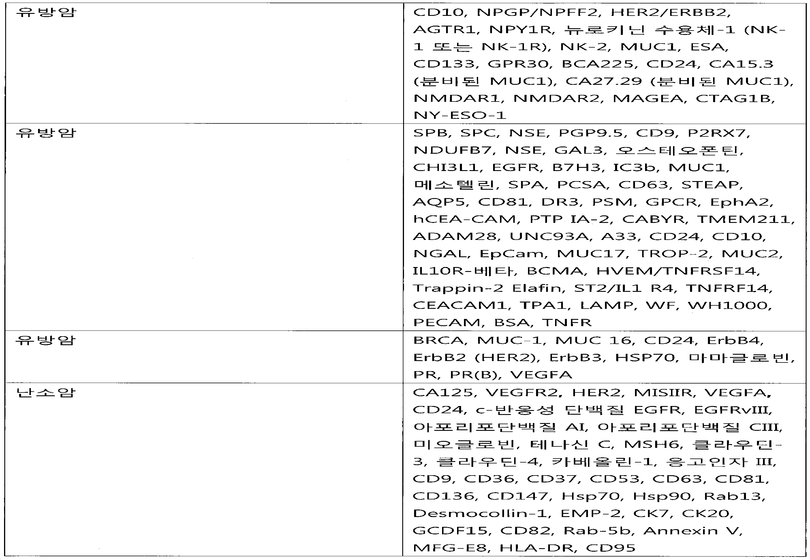
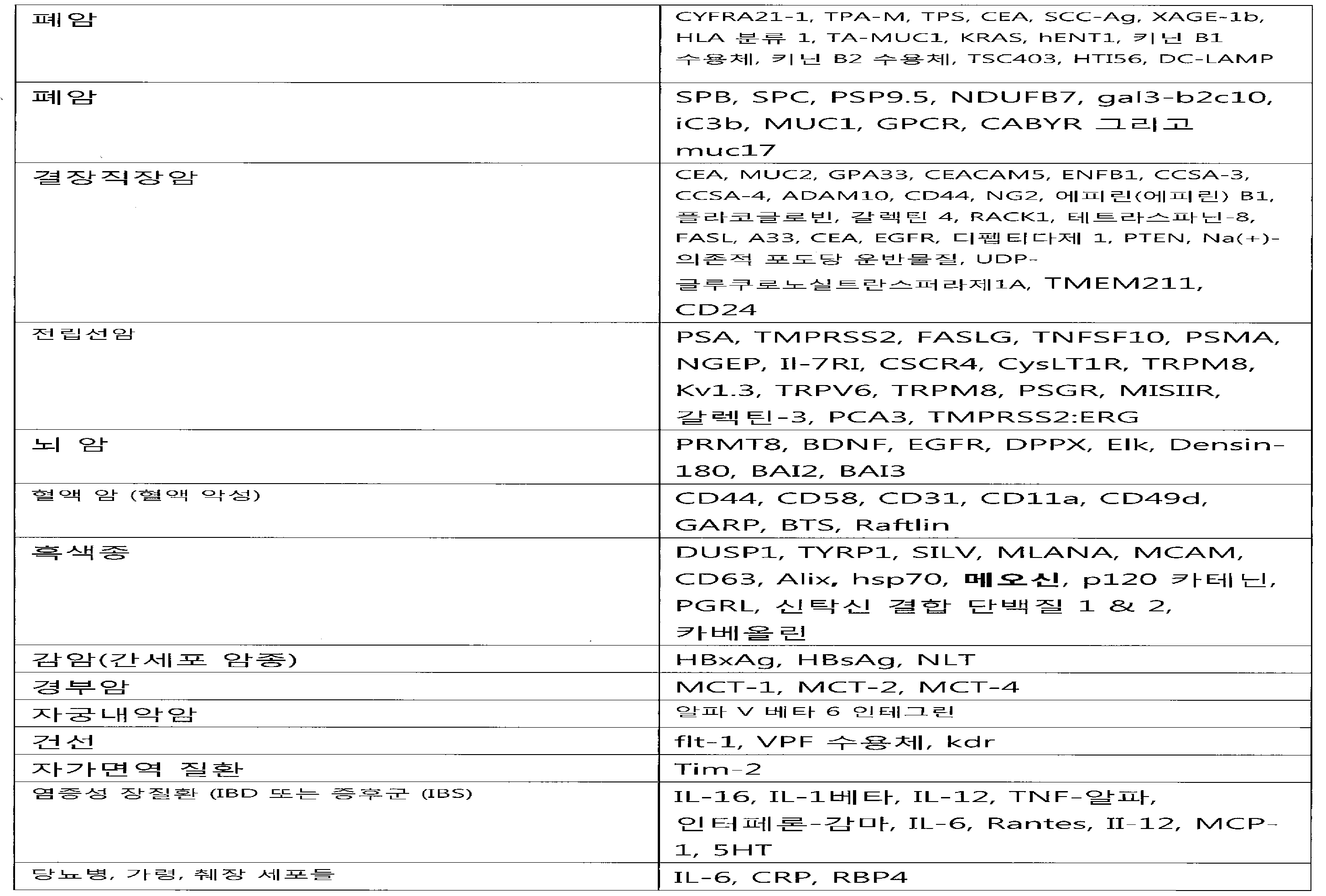
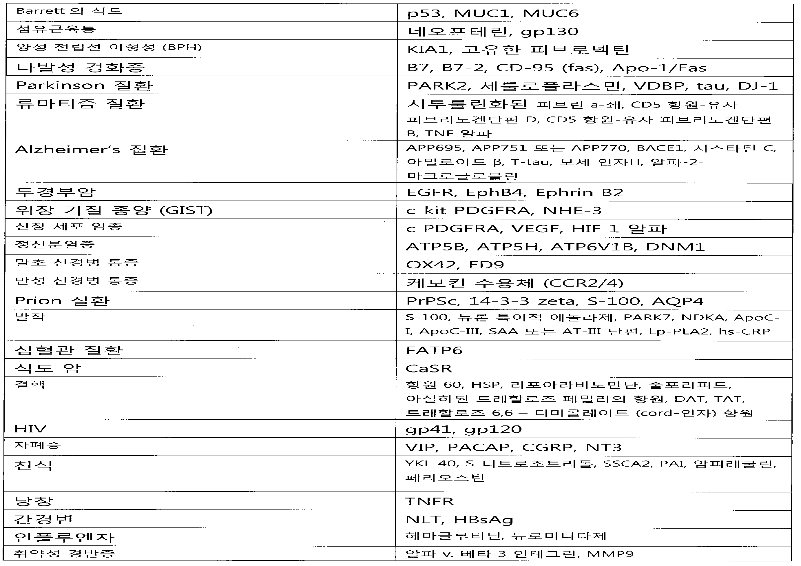
전술한 표 4, 뿐만 아니라 본 명세서에서 공개된 기타 생물지표들은 설명을 위한 것이며, 출원인은 상이한 질환 상태 또는 조건에 걸쳐 설명된 다양한 생물지표들의 통합도 고려한다. 예를 들면, 본 발명의 방법은 상이한 질환 또는 조건에 걸쳐 다양한 생물지료를 이용할 수 있는데, 이때 생물지표들은 진단, 예후 또는 치료진단 특징을 제공하는데 유용하다. 한 구체예에서, 본 명세서에서 기술된 또는 당업계에 공지되어 있는 혈관신생, 염증 또는 면역-연합된 항원 (또는 생물지표들)은 생물특징의 식별에서 생물학적 시료를 스크리닝하기 위한 본 발명의 방법들에 이용될 수 있다. 게다가, 미세소낭 집단을 평가하기 위하여 출원인의 다중 방법의 유연성은 상이한 조건 또는 질환에 대한 다양한 지표들(그리고 일부 경우에 중첩되는 지표들)의 평가를 용이하게 하고, 이들의 병인관계는 필수적으로 특정 세포의 그리고 생물학적 기전을 공유할 수 있는데, 가령, 상이한 암들은 신생혈관생성, 또는 면역 반응 조절 또는 조정을 위한 생물지료들과 관련된다. 미세소낭-연합된 생물지표들과 함께, 조직 또는 세포-특이적 생물지표들과 이러한 중첩되는 생물지표들의 조합으로 본 발명의 방법 및 조성물을 실행하는 강력한 일련의 도구를 제공한다.Table 4, above, as well as other biomarkers disclosed herein, are for illustrative purposes and applicants also contemplate the incorporation of various biomarkers described over different disease states or conditions. For example, the methods of the present invention may utilize a variety of biological materials across different diseases or conditions, wherein the biomarkers are useful in providing diagnostic, prognostic, or therapeutic diagnostic features. In one embodiment, angiogenic, inflammatory, or immuno-associated antigens (or biomarkers) described herein or known in the art include methods of the present invention for screening biological samples in the identification of biological characteristics Lt; / RTI > In addition, the flexibility of the applicant's multiple methods to assess the microfollicle population facilitates the evaluation of different indicators (or in some cases overlapping indicators) of different conditions or diseases, and their etiologic relationship is essentially Cellular and biological mechanisms, for example, different cancers are associated with neoplastic angiogenesis, or with biological substrates for modulation or regulation of the immune response. In combination with micro-vesicle-associated biomarkers, tissue or cell-specific biomarkers and these overlapping biomarkers provide a powerful set of tools for implementing the methods and compositions of the present invention.
기원 세포 특이적 소낭은 여기에서 설명된 것과 같이, 신규한 결합 물질과 방법을 이용하여 단리할 수 있다. 더욱이, 기원 세포 특이적 소낭은 이러한 소낭의 세포 결합 짝 또는 결합 물질에 근거한 분리 방법을 이용하여 생물학적 시료로부터 단리시킬 수 있다. 이러한 세포성 결합 짝은 하나 이상의 특이적 생물지표들이 존재하는 이러한 소낭에만 결합하는 펩티드들, 단백질들, RNA, DNA, 압타머들, 세포들 또는 혈청-연관된 단백질들을 포함하나 이에 한정되지 않는다. 기원 세포 특이적 소낭의 분리 또는 탐지는 단일 결합짝 또는 결합물질, 또는 결합 짝 또는 결합물질의 조합으로 실시할 수 있는데, 이들의 단일 적용 또는 복합 적용으로 기원 세포 특이적 분리 또는 탐지가 이루어진다. 이러한 결합물질의 비-제한적 예는 2011년 4월 6일자로 제출된 국제 특허 출원 번호 PCT/US2011/031479, "Circulating Biomakers for Disease"(이 출원은 전문이 여기에 참고자료로 편입된다)의 도 2에 제공한다. 예를 들면, 특징화 유방암의 소낭은 에스트로겐, 프로게스테론, 트라스투주마브, CCND1, MYC PNA, IGF-1 PNA, MYC PNA, SC4 압타머 (Ku), AII-7 압타머 (ERB2), Galectin-3, 뮤신-유형 O-글리칸, L-PHA, Galectin-9, 또는 이의 임의의 조합을 포함하나 이에 한정되지 않는 하나 이상의 결합 물질로 단리할 수 있다. The source cell specific vesicles can be isolated using novel binding materials and methods, as described herein. Furthermore, the source cell-specific follicles can be isolated from the biological sample using a cell-binding partner of such follicles or a separation method based on a binding agent. Such cellular binding partners include, but are not limited to, peptides, proteins, RNA, DNA, enzymes, cells or serum-associated proteins that bind only to these vesicles in which there are one or more specific biomarkers. Segregation or detection of the originating cell-specific follicles can be carried out with a single binding partner or a binding agent, or a combination of binding partners or binding agents, wherein single or multiple applications lead to cell-specific separation or detection. Non-limiting examples of such binding substances are disclosed in International Patent Application No. PCT / US2011 / 031479, "Circulating Biomakers for Disease", filed April 6, 2011, the disclosure of which is incorporated herein by
결합물질은 또한 다음에 근거하여 기원 세포 특이적 소낭을 단리 또는 탐지하는데 이용할 수 있다: i) 기원 세포 특이적 소낭에 특이적인 항원의 존재; ii) 기원 세포 특이적 소낭에 특이적인 지표들의 부재; 또는 iii) 기원 세포 특이적 소낭에 특이적인 생물지표들의 발현 수준. 이종 소낭 집단은 소낭의 기원 세포 특징들을 배제하거나 또는 확인하기 위하여 기획된 특이적 결합 물질로 피복된 표면에 제공할 수 있다. 다양한 결합물질, 가령, 항체들은 고형 표면 또는 기질 상에 어래이될 수 있고, 이종 소낭 집단은 상호작용이 일어날 수 있도록 충분한 시간 동안 고형 표면 또는 기질과 접촉시킨다. 어래이 표면 또는 기질 상의 주어진 항체 위치에 특이적 결합 또는 비-결합은 주어진 기원 세포에 특이적인 소낭 집단의 항원 특이적 특징들을 확인하는 기능을 할 수 있다. 즉, 결합 사건들은 결합된 항체에 의해 인지되는 항원을 보유하는 소낭의 존재를 알릴 수 있다. 역으로, 결합 사건의 부족은 결합된 항체에 의해 인지되는 항원을 보유하는 소낭의 부재를 알릴 수 있다.Binding materials can also be used to isolate or detect a source cell-specific follicle based on: i) the presence of an antigen specific to the source cell-specific follicle; ii) absence of indices specific to the origin cell specific follicles; Or iii) expression level of biomarkers specific to the origin cell-specific follicle. The heterozygous follicle population can be provided on a surface coated with a specific binding agent designed to eliminate or identify the origin cell features of the follicle. A variety of binding agents, such as antibodies, can be harvested on a solid surface or substrate, and the xanthomas population is contacted with the solid surface or substrate for a sufficient time to allow interaction to occur. Specific binding or non-binding to a given antibody site on an array surface or substrate can serve to identify antigen-specific characteristics of the follicle population specific to a given source cell. That is, binding events can signal the presence of vesicles bearing antigens recognized by the bound antibody. Conversely, the lack of binding events may indicate the absence of vesicles bearing antigens recognized by the bound antibody.
기원 세포 특이적 소낭은 상기에서 설명된 것과 같은 자석 캡쳐 방법, 형광 활성화된 세포 분류 (FACS) 또는 레이져 혈구계산을 이용한 하나 이상의 결합 물질을 이용하여 농축 도는 단리시킬 수 있다. 자석 캡쳐 방법들은 자석으로 활성화된 세포 분류기 (MACS) 마이크로비드 또는 자석 컬럼의 이용을 포함하나 이에 한정되지 않는다. 이용될 수 있는 면역친화력 및 자석 미립자 방법들의 예는 미국 특허 제4,551,435호, 제4,795,698호, 제4,925,788호, 제5,108,933호, 제5,186,827호, 제5,200,084호 또는 제5,158,871호에 설명된다. 기원 세포 특이적 소낭은 또한 미국 특허 제7,399,632호에 설명된 일반적인 방법들에 따라, 소낭에 특이적인 항원 조합을 이용하여 단리할 수 있다. The source cell specific vesicles can be enriched or isolated using one or more binding materials using magnetic capture methods, fluorescence activated cell sorting (FACS) or laser hemocyte counting as described above. Magnet capture methods include, but are not limited to, use of magnetically activated cell sorters (MACS) microbeads or magnetic columns. Examples of immune affinity and magnetic particulate methods that may be used are described in U.S. Patent Nos. 4,551,435, 4,795,698, 4,925,788, 5,108,933, 5,186,827, 5,200,084 or 5,158,871. The source cell-specific follicles can also be isolated using a fungal-specific antigen combination according to the general methods described in U.S. Patent No. 7,399,632.
생물학적 시료에 대해 기원 세포 특이적 소낭을 단리 또는 농축시키는 임의의 다른 적합한 방법들은 본 발명과 조합될 수 있다. 예를 들면, 크기 압출 크로마토그래피 가령, 겔 침투 컬럼, 원심분리 또는 밀도 그라디언트 원심분리, 그리고 여과 방법들은 여기에서 설명된 항원 선별 방법들과 복합하여 이용할 수 있다. 기원 세포 특이적 소낭은 Koga et al., Anticancer Research, 25:3703-3708 (2005), Taylor et al., Gynecologic Oncology, 110:13-21 (2008), Nanjee et al., Clin Chem, 2000;46:207-223 또는 미국 특허 제7,232,653호에 설명된 방법들에 따라 단리할 수 있다. Any other suitable method for isolating or concentrating the source cell specific vesicle for a biological sample can be combined with the present invention. For example, size extrusion chromatography, such as gel permeation columns, centrifugation or density gradient centrifugation, and filtration methods can be used in combination with the antigen screening methods described herein. Generation of cell-specific follicles is described in Koga et al., Anticancer Research, 25: 3703-3708 (2005), Taylor et al., Gynecologic Oncology, 110: 13-21 (2008), Nanjee et al.,
소낭은 진단, 예후, 질환 계층화, 치료진단, 반응자/비-반응자 상태의 예측, 질환 관찰, 처치 관찰 그리고 이와 유사한 것을 제공하기 위하여 단리되거나 및/또는 탐지될 수 있다. 한 구체예에서, 소낭은 종양, 또는 악성 성장, 자가면역 질환의 부위, 심혈관 질환, 신경 질환, 또는 감염으로부터 유도된 세포로부터 단리될 수 있다. 일부 구체예들에서, 단리된 소낭은 이러한 질환 및 장애와 관련된 세포들, 가령, 이 질환의 병인에서 역할을 하는 면역 세포들로부터 유도되고, 소낭의 분석은 진단, 예후, 질환 특화, 치료진단, 반응자/비-반응자 상태의 예측, 질환 모니터링, 치료 모니터링 그리고 이러한 질환 및 장애와 관련된 이와 유사한 것들에 대해 정보를 제공한다. 이 소낭은 새로운 생물지표들을 발견하는데 추가로 유용하다. 소낭과 연합된 생물지표들을 확인함으로써, 단리된 소낭은 여기에서 설명된 것과 같이, 표현형의 특징화를 위하여 평가할 수 있다. The follicles can be isolated and / or detected to provide diagnosis, prognosis, disease stratification, therapeutic diagnosis, prediction of responder / non-responder condition, disease observation, treatment observations and the like. In one embodiment, the follicle can be isolated from a tumor, or a cell derived from malignant growth, a site of autoimmune disease, cardiovascular disease, neurological disease, or infection. In some embodiments, the isolated follicles are derived from cells associated with such diseases and disorders, such as immune cells that play a role in the pathogenesis of the disease, and the analysis of the follicles is diagnostic, prognostic, disease-specific, Provide information on predictions of responder / non-responder status, disease monitoring, treatment monitoring, and the like related to these diseases and disorders. This follicle is further useful for finding new biomarkers. By identifying biomarkers associated with follicles, the isolated follicles can be evaluated for characterization of the phenotype, as described herein.
일부 구체예들에서, 본 발명의 방법은 개체의 생물학적 시료에 존재하는 하나 또는 그 이상 미세소낭 집단을 평가함으로써 개인에서 암의 존재 또는 암발생 가능성을 특징화하는 것에 관한 것이다. 미세소낭은 본 명세서에서 기술된 또는 당업계에 공지되어 있는 하나 또는 그 이상 공정을 이용하여 단리될 수 있다. In some embodiments, the methods of the present invention relate to characterizing the presence of cancer or the likelihood of developing cancer in an individual by evaluating one or more populations of microvilli present in the biological sample of the individual. Microfilaments can be isolated using one or more of the processes described herein or known in the art.
이러한 미세소낭 집단은 테스트 시료에 대해 진단, 예후 또는 치료진단 특징을 제공하기 위하여 참조 시료와 미세소낭 집단에 대한 생물지표 프로파일, 또는 생물특징을 비교함으로써, 개인에 대한 질환 표현형 특징을 별도로 또는 집합적으로 제공할 수 있다. These microvessel populations can be used to assess disease phenotypic characteristics of individuals separately or collectively by comparing biomarker profiles, or biological characteristics, with reference samples and microcysteal populations to provide diagnostic, prognostic, or therapeutic diagnostic features for the test sample As shown in FIG.
본 공개는 주어진 테스트 시료에 대한 생물특징을 결정함에 있어서 평가되는 다양한 생물지표들을 제공하는데, 다양한 암들, 뿐만 아니라 the state of 이 암의 상태 (가령, 전이성 v. 비-전이성)와 연합된 폴리펩티드 및/또는 핵산 생물지표들의 평가를 포함한다. The present disclosure provides a variety of biomarkers that are evaluated in determining biological characteristics for a given test sample, including but not limited to various cancers, as well as polypeptides associated with the state of the cancer (e.g., metastatic v. Non-metastatic) / RTI > and / or nucleic acid biomarkers.
한 실시예에서, 테스트 시료는 CA-125, CA 19-9, 그리고 c-반응성 단백질을 포함하나 이에 한정되지 않는, 하나 또는 그 이상의 생물지표의 존재 또는 수준을 결정함으로써 암에 대해 평가될 수 있다. 암은 생식 관, 가령, 난소암의 암일 수 있다. 하나 또는 그 이상 생물지표는 하나 또는 그 이상의CD95, FAP-1, miR-200 microRNA, EGFR, EGFRvIII, 아포리포단백질 AI, 아포리포단백질 CIII, 미오글로빈, 테나신 C, MSH6, 클라우딘-3, 클라우딘-4, 카베올린-1, 응고인자 III, CD9, CD36, CD37, CD53, CD63, CD81, CD136, CD147, Hsp70, Hsp90, Rab13, Desmocollin-1, EMP-2, CK7, CK20, GCDF15, CD82, Rab-5b, Annexin V, MFG-E8 그리고 HLA-DR을 더 포함할 수 있다. MiR-200 microRNA (가령, miR-200 microRNA 패밀리)는 miR-200a, miR-200b, miR-200c, miR-141 그리고 miR-429를 포함한다. 이러한 평가는 단백질, 핵산, 또는 본 명세서에서 기술된 각 생물지표들의 존재 또는 수준 또는 존재와 수준을 결정하는 것을 포함한다. In one embodiment, a test sample can be evaluated for cancer by determining the presence or level of one or more biomarkers, including but not limited to CA-125, CA 19-9, and c-reactive protein . Cancer can be a gonadal duct, for example, a cancer of ovarian cancer. One or more biomarkers may include one or more of CD95, FAP-1, miR-200 microRNA, EGFR, EGFRvIII, apolipoprotein AI, apolipoprotein CIII, myoglobin, tenacin C, MSH6, claudin- 4, caveolin-1, coagulation factor III, CD9, CD36, CD37, CD53, CD63, CD81, CD136, CD147, Hsp70, Hsp90, Rab13, Desmocollin-1, EMP-2, CK7, CK20, GCDF15, -5b, Annexin V, MFG-E8 and HLA-DR. MiR-200 microRNA (e.g., miR-200 microRNA family) includes miR-200a, miR-200b, miR-200c, miR-141 and miR-429. Such assessments include determining the presence or level, or presence and level, of a protein, nucleic acid, or each biomarker described herein.
CD95 (Fas, Fas 항원, Fas 수용체, FasR, TNFRSF6, APT1 또는 APO-1)는 자가사멸의 유도를 통하여 면역계에서 주로 조직 항상성을 조절하는 모범적 사멸 수용체다. 암 진행 동안, CD95는 빈번하게 하향조절되고, 세포들은 자가사멸 저항성을 부여받게 되고, 이로 인하여, 종양 침입에 대한 기전의 일부로 CD95의 상실에 말려든다. CD95의 종양생성 활성은 JNK 및 Jun가 관련된 경로에 의해 조정된다. FAP-1 (Fas-연합된 포스파타제 1, 단백질 티로신 포스파타제, 비-수용체 type 13 (APO-1/CD95 (Fas)-연합된 포스파타제), PTPN13로도 칭함)는 단백질 티로신 포스파타제 (PTP) 패밀리의 구성원이다. FAP-1은 CD95와 상호작용하여 탈포스포릴화시켜, Fas 매개된 예정된 세포 사멸에서 역할을 하는 것으로 보고된 바 있다. MiR-200 패밀리 구성원들은 CD95 및 FAP-1을 조절할 수 있다. Schickel et al. miR-200c regulates induction of apoptosis through CD95 by tatgeting FAP-1. Mol. Cell., 38, 908-915 (2010) 참고, 이는 전문이 여기에 참고자료로 편입된다.CD95 (Fas, Fas antigen, Fas receptor, FasR, TNFRSF6, APT1 or APO-1) is an exemplary death receptor that mainly regulates tissue homeostasis in the immune system through induction of auto-apoptosis. During cancer progression, CD95 is frequently down-regulated and cells are endowed with self-destructive resistance, which leads to the loss of CD95 as part of the mechanism of tumor invasion. The tumorigenic activity of CD95 is regulated by the pathways involved in JNK and Jun. FAP-1 (also called Fas-associated
본 명세서에서 기술된 본 발명의 방법은 본 명세서에서 기술된 암들을 포함하나 이에 한정되지 않는, 암의 존재 또는 경향을 결정하기 위하여 생물학적 시료 안에 존재하는 미세소낭 집단의 CD95 및/또는 FAP-1 특징 또는 프로파일링을 이용할 수 있다. 다발성 생물지표들에 대한 다중 분석을 포함하는 본 발명의 방법은 miR-200 microRNA (가령, miR-200c)을 포함하나 이에 한정되지 않는 본 명세서에서 기술된 기타 생물지표들과 함께, CD95 및/또는 FAP-1 생물지표 특징화를 이용한다. 한 구체예에서, 개체의 생물학적 테스트 시료를 평가하여 CD95 및/또는 FAP-1 단백질의 존재 및 수준, 또는 CD95+ 및/또는 FAP-1+ 순환하는 미세소낭 ("cMV") 집단의 존재 및 수준을 결정하고, 그리고 존재 또는 수준은 참조 (가령, 비-질환 또는 정상, 처치전, 또는 상이한 처치 시점의 시료)와 비교된다. 이러한 비교를 이용하여 테스트 시료를 특징화한다. 예를 들면, 테스트 시료 와 참조에서 CD95 단백질, FAP-1 단백질, CD95+ cMVs 및/또는 FAP-1+ cMVs의 존재 또는 수준 비교를 이용하여 질환 표현형을 결정하고 또는 처치에 대한 반응/비-반응을 예측한다. 관련된 구체예들에서, cMV 집단은 miR-200 microRNA의 존재 및 수준을 결정하기 위하여 더 평가되고, 이는 질환 또는 기타 예후, 치료진단 또는 진단 정보를 나타내는 참조 시료의 모의 세트에서 사전결정된다. 암이 아닌 참조와 비교하여 테스트 시료 안에 FAP-1 수준의 증가는 암의 존재, 또는 더 공격적 암의 존재를 나타낼 수 있다. 암이 아닌 참조와 비교하였을 때, CD95 또는 miR200 패밀리 구성요소들 이를 테면 miR-200c의 감소는 암의 존재, 또는 더 공격적 암의 존재를 나타낸다. 경가되는 cMV 집단은 면역침전, 유동 세포분석 유동 세포분석, 또는 본 명세서에서 기술된 또는 당업계에 공지되어 있는 기타 격리 방법을 통하여 단리될 수 있다. The methods of the invention described herein may be used to determine the CD95 and / or FAP-I characteristics of microcysts present in a biological sample to determine the presence or tendency of the cancer, including but not limited to the cancers described herein Or profiling can be used. The methods of the invention, including multiple assays for multiple biomarkers, may be combined with other biomarkers described herein including, but not limited to, miR-200 microRNA (e. G., MiR-200c) FAP-1 biomarker characterization. In one embodiment, biological test samples of an individual are evaluated to determine the presence and level of CD95 and / or FAP-1 protein, or the presence and level of CD95 + and / or FAP-1 + circulating micro vesicle ("cMV" And the presence or level is compared to a reference (e. G., A sample of non-disease or normal, pretreatment, or different treatment time point). These comparisons are used to characterize the test sample. For example, the presence or level comparison of CD95 protein, FAP-1 protein, CD95 + cMVs and / or FAP-1 + cMVs in a test sample and reference can be used to determine the disease phenotype and / Predict. In related embodiments, the cMV population is further evaluated to determine the presence and level of the miR-200 microRNA, which is predetermined in a simulated set of reference samples representing disease or other prognosis, therapeutic diagnosis or diagnostic information. An increase in FAP-1 levels in test samples compared to non-cancer references can indicate the presence of cancer, or the presence of more aggressive cancer. A decrease in the CD95 or miR200 family members, such as miR-200c, when compared to a reference other than cancer, indicates the presence of cancer, or the presence of more aggressive cancer. The resulting population of cMV can be isolated through immunoprecipitation, flow cell analysis, flow cytometry, or other isolation methods described herein or known in the art.
관련된 측면에서, 본 발명은 A2ML1, BAX, C10orf47, C1orf162, CSDA, EIFC3, ETFB, GABARAPL2, GUK1, GZMH, HIST1H3B, HLA-A, HSP90AA1, NRGN, PRDX5, PTMA, RABAC1, RABAGAP1L, RPL22, SAP18, SEPW1, SOX1, 및 이의 조합으로 구성된 군으로부터 선택된 하나 또는 그 이상 생물지표, 가령, 1, 2, 3,4 ,5, 6, 7, 8, 9, 10, 11, 12, 13, 14, 15, 16, 17, 18, 19, 20, 21 또는 22개의 생물지표들의 수준을 탐지하는 것을 포함하는, 암을 특징화하는 방법을 제공한다. 하나 또는 그 이상 생물지표는 티모신알파-1로 절단되는 프로/파라티모신 패밀리의 구성요소인 PTMA (프로티모신, 알파)를 포함할 수 있고, 이는 면역 조정에 역할을 한다. 티모신 알파-1는 간염 B 그리고 C의 치료용으로 최소한 35개국에서 승인되었으며, 그리고 기타 질환의 처치에서 면역반응을 부스터하기 위하여 백신에 포함되는데 승인되었다. 한 구체예에서, 생물지표들은 mRNA를 포함한다. mRNA는 본 명세서에서 기술된 것과 같이 단리된 소낭으로부터 단리될 수 있다. 일부 구체예들에서, 시료 안 전체 소낭 집단은 가령, 여과 또는 원심분리에 의해 단리된다. 이 소낭은 가령, 일반적인 소낭 생물지표, 질환 생물지표 또는 세포-특이적 생물지표에 대한 결합제를 이용하여, 친화력에 의해 또한 단리될 수 있다. 생물지표들의 수준은 암이 없는 시료와 같은 대조군과 비교될 수 있고, 이때 생물지표들의 수준과 대조군 사이에서 변화를 이용하여 암을 특징화한다. 암은 전립선 암일 수 있다. In a related aspect, the present invention provides a compound of formula (I) or a pharmaceutically acceptable salt thereof, which is A2ML1, BAX, C10orf47, C1orf162, CSDA, EIFC3, ETFB, GABARAPL2, GUK1, GZMH, HISTA1, HLA- 1, 2, 3, 4, 5, 6, 7, 8, 9, 10, 11, 12, 13, 14, 15, and 15 selected from the group consisting of, 16, 17, 18, 19, 20, 21 or 22 levels of biomarkers. One or more biomarkers may include PTMA (prothymosin, alpha), a component of the pro / paratomycin family that is truncated with thymosin alpha-1, which plays a role in immunomodulation. Thymosin alpha-1 has been approved in at least 35 countries for the treatment of hepatitis B and C and has been approved for inclusion in the vaccine to boost the immune response in the treatment of other diseases. In one embodiment, the biomarkers include mRNA. mRNA can be isolated from isolated follicles as described herein. In some embodiments, the sample oocytes population is isolated, for example, by filtration or centrifugation. The follicles can also be isolated by affinity, for example, using binders for common follicular biomarkers, disease biomarkers or cell-specific biomarkers. Levels of biomarkers can be compared to controls such as cancer-free samples, where changes between levels of biomarkers and controls are used to characterize cancer. Cancer can be a prostate cancer.
더욱이, 확인된 이 생물특징들을 비교하기 위하여 적절한 참조 시료를 선택하면, 진단 정보 (가령, 참조 시료는 정상 또는 비-질환), 예후 (가령, 참조 시료는 나쁜 또는 좋은 질환 결과, 공격적인 것이거나 또는 이와 유사한 것), 또는 치료진단 (가령, 참조 시료는 선택된 처치에 대해 코호트 반응성 또는 비-반응성이다)를 제공할 수 있다.Moreover, if an appropriate reference sample is selected to compare these identified biologic features, the diagnostic information (e.g., the reference sample may be a normal or non-diseased), a prognosis (e.g., a reference sample is bad or a good disease outcome, Or similar), or therapeutic diagnosis (e.g., the reference sample is cohort responsive or non-responsive to the selected treatment).
이 소낭 집단(들)은 본 명세서에서 기술된 다양한 생물학적 시료 그리고 체내 유체로부터 평가될 수 있다.The follicle population (s) can be assessed from the various biological samples described herein and from body fluids.
생물지표 평가(Biomarker Assesment)Biomarker Assesment
본 발명의 한 측면에서, 생물학적 피험자로부터 얻은 시료를 분석하고, 시료 안에 하나 또는 그 이상의 순환하는 생물 지표들의 집단, 가령, 순환하는 소낭, 단백질 또는 핵산의 존재, 수준, 양 또는 농도를 측정함으로써, 피험자에 대해 표현형을 특징화시킬 수 있다. 구체예들에서, 특징화는 이 시료 안에 순환하는 생물지표들이 참고(표준 또는 대조군으로도 칭할 수 있다)와 비교하여 변경되는지를 결정하는 것을 포함한다. 변화는 시료와 참조 사이에 존재 또는 부재, 정량적 수준, 참조와 비교하여 상대적 수준, 가령, 존재하는 모든 소낭의 수준, 하우스키핑(하우스킵핑) 지표의 수준, 및/또는 스파이크-인(spiked-in) 지표의 수준, 상승된 수준, 감소된 수준, 과다발현, 과소발현, 차등 발현, 돌연변이 또는 기타 변경된 서열, 변형 (당화, 인산화, 후성적 변화) 그리고 이와 유사한 것을 포함하는 임의의 측정가능한 차이를 포함하나 이에 한정되지 않는다. 일부 구체예들에서, 순환하는 생물지표들은 이들의 양을 결정하기 앞서 시료로부터 정제 또는 농축된다. 다른 언급이 없는 한, 본 명세서에서 이용된 것과 같이,"정제된" 또는 "단리된"은 부분적 또는 완전한 정제 또는 격리를 지칭한다. 다른 구체예들에서, 순환하는 생물지표들은 사전 정제 또는 농축없이 시료로부터 직접적으로 평가된다. 순환하는 소낭은 세포 기원의 특이적 소낭 또는 특이적 생물특징을 가진 소낭일 수 있다. 생물특징은 특이적 패턴의 생물지표들, 가령, 이를 테면 질환 표현형을 탐지하는데 바람직한 표현형을 나타내는 생물지표 패턴을 포함한다. 이 생물특징은 하나 또는 그 이상의 순환하는 생물지표들을 포함한다. 생물특징은 표현형의 특징화, 가령, 진단, 예후, 치료진단, 또는 반응자/비-반응자 상태의 예측을 특징화할 때 소낭의 양을 이용할 수 있다. 일부 구체예들에서, 생물특징은 생리학적 또는 생물학적 상태, 가령, 임신 또는 임신 단계를 결정하는데 이용한다. 생물특징의 양을 또한 이용하여 치료 효과, 질환 또는 상태의 단계, 또는 질환 또는 상태의 진전을 판단할 수 있다. 예를 들면, 하나 이상의 소낭의 양은 질환 단계 또는 진전에서 증가와 비례 또는 역비례할 수 있다. 탐지된 소낭의 양을 또한 이용하여 질환 또는 상태의 진전을 모니터하거나 또는 치료에 대한 피험자의 반응을 모니터할 수 있다. In one aspect of the invention, a sample from a biological subject is analyzed and the presence, level, amount or concentration of one or more circulating biological indicators, e. G., Circulating follicles, protein or nucleic acid, The phenotype can be characterized for the subject. In embodiments, the characterization includes determining whether biomarkers circulating in the sample are altered relative to a reference (also referred to as a standard or control). The change may be determined by comparing the presence or absence between the sample and the reference, the quantitative level, the relative level relative to the reference, for example, the level of all existing vesicles, the level of the housekeeping (the left skippin) indicator, and / or the spiked- including any of the measurable differences, including, but not limited to, the level, in elevated levels, reduced levels, overexpression, underexpression, differential expression, mutations or other altered sequences, variants (glycosylation, phosphorylation, But is not limited thereto. In some embodiments, circulating biomarkers are purified or concentrated from the sample prior to determining their amount. Unless otherwise indicated, as used herein, "purified" or "isolated" refers to partial or complete purification or sequestration. In other embodiments, circulating biomarkers are directly assessed from the sample without prior purification or concentration. Circulating follicles can be cell follicle-specific follicles or pellicles with specific biological characteristics. Biological characteristics include biomarkers of a specific pattern, for example, biomarker patterns that exhibit the desired phenotype for detecting a disease phenotype. This biological feature includes one or more circulating biomarkers. Biological characteristics can be used to characterize the phenotype, for example, the amount of follicle when characterizing the diagnosis, prognosis, therapeutic diagnosis, or prediction of the response / non-responder state. In some embodiments, biological characteristics are used to determine a physiological or biological condition, such as a pregnancy or pregnancy stage. The amount of biological characteristics may also be used to determine the therapeutic effect, the stage of the disease or condition, or the progression of the disease or condition. For example, the amount of one or more follicles can be proportional or inversely proportional to the increase in disease stage or progression. The amount of detected follicles can also be used to monitor the progression of the disease or condition or to monitor the subject's response to treatment.
순환하는 생물지표들은 기준 수준 또는 값과 순환하는 생물지표들을 비교함으써 평가될 수 있다. 기준 값은 물리적 또는 일시적 종료점에 특징적일 수 있다. 예를 들면, 기준 값은 시료를 평가하는 동일 피험자의 것이거나 또는 기준 값은 시료들의 대표 집단(가령, 질환의 증상을 나타내지 않는 정상 피험자의 시료들)의 것일 수 있다. 따라서, 기준 값은 주어진 시료에서 분석되는 생물특징에 대한 피험자 시료의 정보와 비교되는 임계치를 제공할 수 있다. 이러한 기준 값은 나이 (가령, 신생, 유아, 청년기, 젊은 층, 중년층, 노련청 그리고 다양한 연령대의 성인), 인종/민족 집단, 정상 대 질환이 있는 피험자, 흡연자 대 비-흡연자, 치료를 받은 피험자 대 치료를 받지 않은 피험자, 유사하게 진단을 받은 또는 치료된 피험자 군 또는 개체에서 상이한 치료 시점들 또는 이의 조합들을 포함하나 이에 한정되지 않는 특정 무리에 대응하는 시료군으로부터 모아진 데이터에 따라 설정될 수 있다. 더욱이, 특정 개체의 상이한 시점의 치료에서 생물특징을 판단함으로써, 치료될 개체에 대한 질환 또는 상태의 치료 또는 진전에 대한 개체의 반응을 모니터할 수 있다. Cycling biomarkers can be assessed by comparing baseline levels or values with circulating biomarkers. The reference value may be characteristic of a physical or temporary end point. For example, the reference value may be of the same subject who is evaluating the sample, or the reference value may be of a representative population of samples (e. G., Normal subject samples that do not exhibit symptoms of the disease). Thus, the baseline value may provide a threshold that is compared to the subject's sample information for biological characteristics that are analyzed in a given sample. These criteria include age (eg, newborns, infants, young adults, young adults, middle-aged people, labor associations and adults of all ages), racial / ethnic groups, subjects with normal large-scale disease, smokers versus non-smokers, May be set according to data collected from a sample group corresponding to a specific group, including, but not limited to, subjects who have not undergone major treatment, subjects who are similarly diagnosed or treated, or different treatment points in a subject or combinations thereof . Moreover, by monitoring biological characteristics in the treatment of different aspects of a particular individual, the individual's response to treatment or progression of the disease or condition to the individual to be treated can be monitored.
기준 값은 개별화된 추적을 제공하기 위하여 동일한 피험자로부터 평가된 시료에 근거할 수 있다. 일부 구체예에서, 대상의 시료 안에 생물특징의 빈번한 테스트는 이 대상에 대해 기존에 확립된 기준 값에 대한 더 나은 비교를 제공한다. 이러한 시간 일정 측정을 이용하면 의사는 대상에 대해 이미 확립된 기준값에 대해 더 나은 비교를 제공할 수 있으며, 따라서 치료에 대한 더 나은 판단을 제공할 수 있다. 일부 경우에서, 대상의 고유 생물 특징과 비교하였을 때, 시간이 경과함께 따라 생물특징의 가변성의 감소는 이 대상에 특화된 개별화된 임계치, 가령, 진단이 이루어지는 임계치를 허용한다. 설명을 위한 예로써, 전립선 세포로부터 유도된 소낭의 수준은 시간의 경과에 따라 이 대상에서 측정되는 것이 고려된다. 대상의 혈액내 전립선에서 유도된 소낭의 수준의 스파이크는 전립선 암으로 인하여 전립선 세포의 과다증식을 나타낼 수 있다. The baseline value may be based on samples evaluated from the same subject to provide individualized tracking. In some embodiments, frequent testing of biological characteristics in a sample of a subject provides a better comparison of previously established baseline values for this subject. Using this time-of-day measurement, the physician can provide a better comparison of previously established baseline values for the subject, thus providing better judgment of treatment. In some cases, a decrease in the variability of a biological feature over time, as compared to the unique biological characteristics of the subject, allows for an individualized threshold that is specific to this subject, e.g., a threshold at which diagnosis is made. As an illustrative example, the level of follicular cells derived from prostate cells is considered to be measured in this subject over time. A spike in the level of the follicle derived from the prostate in the blood of the subject may indicate overgrowth of the prostate cells due to prostate cancer.
영향을 받지 않은 개체에서 관심 생물특징의 양을 측정함으로써 특정 표현형 없는 영향을 받지 않은 개체 (다양한 연령, 인종적 배경 및 성별)에 대해 기준값을 확립할 수 있다. 예를 들면, 기준 집단에 대한 기준 값을 테스트 피험자에서 하나 이상의 순환하는 생물지표의 탐지를 위한 기준선으로 이용할 수 있다. 피험자의 시료가 기준과 유사한 수준 또는 값을 보유한다면, 이 피험자는 이 질환을 보유하지 않거나, 또는 질환의 발달 가능성이 낮은 것으로 확인할 수 있다. By measuring the amount of the biologic features of interest in the unaffected individual, a baseline value can be established for individuals not affected by a particular phenotype (age, racial background and gender). For example, a reference value for a reference population can be used as a baseline for the detection of one or more circulating biomarkers in a test subject. If the sample of the subject has a level or value similar to the reference, the subject may not have the disease or be found to have a low probability of developing the disease.
대안으로, 표현형을 가진 개체에서 하나 이상의 소낭 집단의 양을 측정함으로써 특정 표현형을 가진 개체에 대해 기준 값 또는 수준을 확립할 수 있다. 추가로, 특정 표현형에 대한 색인 값이 생성될 수 있다. 예를 들면, 상이한 질환 단계는 가령, 상이한 질환 단계를 가진 개체로부터 수득된 상이한 값을 보유할 수 있다. 피험자의 값은 색인과 비교할 수 있고, 이 질환의 진단 또는 예후, 가령, 이 질환의 단계 또는 진전을 결정할 수 있고, 이때 대상의 수준은 이 색인과 가장 밀접하게 관련된다. 다른 구체예들에서, 색인 값은 치료 효과에 대해 생성된다. 예를 들면, 특정 질환을 가진 개체의 소낭 수준이 생성될 수 있고, 이 개체에 무슨 치료가 효과적인지를 알 수 있다. 소낭의 수준을 이용하여 값을 만들고, 이 값을 피험자의 값과 비교하고, 그리고 가령, 이 수준으로부터 이 피험자가 치료에 대해 반응자 또는 비-반응자인지를 예측함으로써 개체에 대한 치료 또는 치료법을 선택할 수 있다. Alternatively, a reference value or level can be established for an individual with a particular phenotype by measuring the amount of one or more follicular populations in the individual having the phenotype. In addition, an index value for a particular phenotype can be generated. For example, different disease stages may have different values, for example, obtained from an individual having a different disease stage. The subject's value can be compared to the index and determine the diagnosis or prognosis of the disease, e.g., the stage or progression of the disease, where the level of the subject is most closely related to this index. In other embodiments, the index value is generated for the therapeutic effect. For example, a follicular level of an individual with a particular disease can be generated, and it is possible to know what treatment is effective for this individual. The level of the follicle can be used to make a value, compare this value to the value of the subject, and, for example, select a treatment or cure for the subject by predicting, from this level, whether the subject is a respondent or non- have.
일부 구체예들에서, 특정 암에 대한 생물지표를 특이적으로 표적하는 항원을 가진 순환하는 생물지표들을 단리 또는 탐지함으로써, 특정 암을 가지지 않는 개체에 대해 기준 값을 정한다. 비-제한적 예로써, 영향을 받지 않은 개체에 대해 처방된 동일한 기술을 이용하여, 다양한 단계의 직장결장암 및 암이 아닌 용종을 가진 개체들을 조사할 수 있고, 그리고 각 집단에 대한 순환하는 소낭의 수준들을 결정할 수 있다. 일부 구체예들에서, 이 수준은 최소한 이중 또는 삼중으로 실시된 최소한 2가지 별개 실험으로부터 평균 ± 표준 편차로 정의한다. 관찰된 구별되는 생물지표들의 통계학적 유의성을 판단하기 위하여 통계학적 테스트를 이용하여 이들 집단 간을 비교할 수 있다. 일부 구체예들에서, 통계학적 유의성은 매개변수적 통계학적 테스트를 이용하여 측정한다. 매개변수적 통계학적 테스트는 분별적 인자 기획, 가변성 분석(ANOVA), t-테스트, 최소제곱법, Pearson 상관관계, 단순 선형 회귀, 비-선형 회귀, 다중 선형 회귀, 또는 다중 비선형 회귀를 포함하나 이에 한정되지 않는다. 대안으로, 매개변수적 통계학적 테스트는 일원 변량 분석, 이원 변량 분석, 또는 반복 변형 분석 측정을 포함한다. 다른 구체예들에서, 통계학적 유의성은 비-매개변수적 통계학적 테스트를 이용하여 측정한다. 실시예는 Wilcoxon signed-rank 테스트, Mann-Whitney 테스트, Kruskal-Wallis 테스트, Friedman 테스트, Spearman 순위 상관 계수, Kendall Tau 분석, 그리고 비-매개변수적 회귀 테스트를 포함하나 이에 한정되지 않는다. 일부 구체예들에서, 통계학적 유의성은 0.05, 0.01, 0.005, 0.001, 0.0005, 또는0.0001 미만의 p-값에서 측정한다. p-값은 가령, Bonferroni 보정, 이의 변형, 또는 당업계에 공지되어 있는 다른 기술, 가령, Hochberg 보정, Holm-Bonferroni 보정, idk 보정, Dunnett의 보정 또는 Tukey의 다중 비교를 이용하여 보정할 수 있다. 일부 구체예들에서, ANOVA는 각 집단의 생물지표들의 테스트 후 비교를 위하여 Tukey 보정을 한다.In some embodiments, reference values are determined for individuals that do not have a particular cancer, by isolating or detecting circulating biomarkers with antigens that specifically target biomarkers for a particular cancer. By way of non-limiting example, using the same techniques prescribed for unaffected individuals, individuals with various stages of colorectal cancer and non-cancerous polyps can be screened, and the level of circulating follicles for each population Can be determined. In some embodiments, this level is defined as the mean ± standard deviation from at least two separate experiments conducted with at least double or triplicate. Statistical tests can be used to compare these groups to determine the statistical significance of the observed distinct biomarkers. In some embodiments, statistical significance is measured using parametric statistical tests. The parametric statistical tests include fractional factoring planning, variability analysis (ANOVA), t-test, least squares, Pearson correlation, simple linear regression, nonlinear regression, multiple linear regression, or multiple nonlinear regression But is not limited thereto. Alternatively, parametric statistical tests include one-way ANOVA, two-way ANOVA, or repeated ANOVA. In other embodiments, statistical significance is measured using non-parametric statistical tests. Examples include, but are not limited to, Wilcoxon signed-rank test, Mann-Whitney test, Kruskal-Wallis test, Friedman test, Spearman rank correlation coefficient, Kendall Tau analysis, and non-parametric regression test. In some embodiments, the statistical significance is measured at a p-value of less than 0.05, 0.01, 0.005, 0.001, 0.0005, or 0.0001. The p-value can be corrected using, for example, Bonferroni correction, its variants, or other techniques known in the art, such as Hochberg correction, Holm-Bonferroni correction, idk correction, Dunnett correction or Tukey multiple comparisons . In some embodiments, the ANOVA performs a Tukey correction for comparison after testing of the biomarkers of each group.
질환 재발 모니터링 (또는 MS에서 악화 파아지), 치료 반응 모니터링, 또는 반응자/비-반응자 상태의 예측을 위하여 기준 값을 확립할 수 있다.Reference values can be established for disease recurrence monitoring (or worsening phage in MS), therapeutic response monitoring, or for predicting the response / non-responder state.
일부 구체예들에서, 소낭에 대한 기준 값은 합성 소낭이라고도 불리는 인공적인 소낭을 이용하여 측정한다. 인공적인 소낭을 제조하는 방법들은 당업계에 공지되어 있으며, 가령, 리포좀을 이용하여 만들 수 있다. 인공적인 소낭은 US20060222654 및 US4448765에서 공개된 방법을 이용하여 제조할 수 있다(이들 각각은 전문이 여기에 참고자료로 통합된다). 공지의 지표를 가진 인공적인 소낭을 작제하여 캡쳐 및/또는 탐지를 용이하게 할 수 있다. 일부 구체예들에서, 인공적인 소낭은 프로세싱하기 전 체내 시료에 고정시킨다. 초기 시료 대 프로세스된 시료에서 소낭의 양에 대한 기준을 제공하기 위하여, 프로세싱 동안 고유의 합성 소낭의 수준은 여기에서 설명된 여과 또는 다른 분리 방법을 이용하여 추적할 수 있다. 유사하게, 임의의 프로세스 단계 전 또는 후, 인공적인 소낭을 시료에 고정시킬 수 있다. 일부 구체예들에서, 인공적인 소낭을 이용하여 소낭의 분리 및 탐지에 이용되는 장비의 구경을 정한다. In some embodiments, the reference value for the follicle is measured using an artificial vial, also referred to as a synthetic vesicle. Methods for making artificial follicles are known in the art and can be made, for example, using liposomes. Artificial vesicles can be prepared using the methods disclosed in US20060222654 and US4448765, each of which is incorporated herein by reference. Artificial follicles with known indicators can be constructed to facilitate capture and / or detection. In some embodiments, the artificial vesicles are fixed in the sample prior to processing. In order to provide a reference for the amount of follicles in the initial sample versus the treated sample, the level of the original synthetic follicle during processing can be traced using filtration or other separation methods described herein. Similarly, artificial follicles can be anchored to the sample either before or after any process step. In some embodiments, artificial vesicles are used to calibrate the equipment used to isolate and detect vesicles.
인공적인 소낭을 만들고, 그리고 분석, 가령, 비드-기반 분석의 가능성을 테스트하기 위한 대조군으로 이용할 수 있다. 인공적인 소낭은 비드 및 탐지 항체들 모두에 결합할 수 있다. 따라서, 인공적인 소낭은 이들 각 항체가 결합하는 아미노산 서열/구조를 포함한다. 인공적인 소낭은 정제된 단백질 또는 항체가 결합하는 합성 펩티드 서열을 포함할 수 있다. 인공적인 소낭은 비드, 가령, 생물학적 분자들이 부착할 수 있는 폴리스티렌 비드일 수도 있다. 비드가 이용가능한 카르복실기를 보유한다면, 단백질 또는 펩티드는 가령, 카르보디이미드 커플링과 같은 이용가능한 아민 기를 통하여 비드에 부착할 수 있다. Artificial follicles, and can be used as a control for testing, for example, the possibility of bead-based analysis. Artificial vesicles can bind to both beads and detection antibodies. Thus, artificial vesicles contain the amino acid sequence / structure to which each of these antibodies bind. Artificial follicles may contain purified peptide or synthetic peptide sequences to which the antibody binds. The artificial vesicles may be beads, such as polystyrene beads, to which biological molecules may adhere. If the bead has an available carboxyl group, the protein or peptide may be attached to the bead through an available amine group, such as, for example, a carbodiimide coupling.
또다른 구체예에서, 인공적인 소낭은 아비딘으로 피복된 폴리스티렌 비드일 수 있으며, 그리고 바이오틴은 합성 시점에 또는 바이오틴-말레이미드 화학물질을 통하여 선택된 단백질 또는 펩티드상에 위치한다. 비드상에 있는 단백질들/펩티드들은 인공적인 소낭이 이용되는 분야에 특이적인 비율로 함께 혼합할 수 있으며, 그 다음 비드에 콘쥬게이트시킨다. 그 다음 이들 인공적인 소낭은 캡쳐 비드와 탐지 항체들 사이의 링크로 작용하고, 이로 인하여 분석 성분들이 적절히 작용하는지를 보유주기 위한 대조군을 제공한다. In yet another embodiment, the artificial vesicle may be avidin-coated polystyrene beads, and the biotin is located at the time of synthesis or on the protein or peptide selected through biotin-maleimide chemistry. The proteins / peptides on the beads can be mixed together in a specific ratio to the field in which the artificial vesicles are used and then conjugated to the beads. These artificial vesicles then serve as links between the capture beads and the detection antibodies, thereby providing a control to keep the analytical components functioning properly.
값은 정량적 또는 정성적 값일 수 있다. 이 값은 소낭의 수준(예로, 용적당 질량)의 직접적인 측정이거나 간접적 측정, 가령, 특이적 생물지표의 양일 수도 있다. 이 값은 정량적, 가령, 수치 값일 수 있다. 다른 구체예들에서, 이 값은 정성적, 가령, 소낭이 없음, 낮은 수준의 소낭, 중간 수준의 소낭, 높은 수준의 소낭, 또는 이의 변이일 수 있다. The value may be a quantitative or qualitative value. This value may be a direct measure of the level of the follicle (eg mass per dose) or an indirect measure, for example, the amount of specific biomarkers. This value can be quantitative, e.g., a numerical value. In other embodiments, the value may be qualitative, such as no follicles, low levels of follicles, medium level follicles, high levels of follicles, or variations thereof.
기준 값은 데이터베이스에 저장할 수 있고, 그리고 특이적 생체특징을 가진 소낭으로부터 순환하는 생물지표들, 가령, 소낭 또는 microRNA의 총량, 또는 특이적 집단의 소낭 또는 microRNA의 양, 가령, 기원 세포 특이적 소낭 또는 microRNA 또는 microRNA의 양 또는 수준에 근거하여 진단, 예후, 치료진단, 질환 특화, 질환 모니터링, 치료 모니터링 또는 질환 또는 상태의 비-반응자/반응자 상태의 예측을 위한 기준으로 이용할 수 있다. 설명을 위한 예로서, 암을 진단하는 방법을 고려한다. 이 암이 있는 그리고 없는 기준 피험자의 소낭 또는 다른 순환하는 생물지표들을 평가하고, 데이터베이스에 보관한다. 기준 피험자들은 이 암 또는 또다른 상태, 가령, 건강한 상태를 나타내는 생체특징을 제공한다. 테스트 피험자의 시료를 그 다음 분석하고, 그리고 데이터베이스에 있는 것과 대조하여 microRN생체특징을 비교한다. 피험자의 생체특징이 암을 나타내는 기준 값에 좀더 근접하다면, 암 진단이 내려질 수 있다. 역으로, 피험자의 생체특징이 건강한 상태를 나타내는 기준 값과 좀더 밀접하다면, 이 피험자는 이 질환이 없는 것으로 판단할 수 있다. 당업자는 이 실시예는 비-한정적이며, 다른 표현형, 가령, 다른 질환, 예후, 치료진단, 질환 특화, 질환 모니터링, 치료 모니터링 또는 비-반응자/반응자 상태의 예측, 및 이와 유사한 것들을 평가하는 것까지 확장시킬 수 있음을 인지할 것이다. The baseline value may be stored in a database and may be calculated from biomarkers that circulate from a follicle with specific biomarkers, such as the total amount of follicles or microRNAs, or the amount of a specific population of follicles or microRNAs, Prognosis, treatment diagnosis, disease-specificization, disease monitoring, treatment monitoring, or prediction of the status of non-responders / responders of a disease or condition based on the amount or level of microRNA or microRNA. For illustrative purposes, consider a method of diagnosing cancer. The follicles or other circulating biomarkers of the reference subject with and without cancer are evaluated and stored in a database. Reference subjects provide biochemical features indicative of this cancer or another condition, e.g., a healthy condition. The test subject's sample is then analyzed, and the microRNA biological characteristics are compared against those in the database. If the subject's biometric characteristics are closer to the reference value for cancer, a diagnosis of cancer may be made. Conversely, if the subject's biometric characteristic is more closely related to a reference value indicating a healthy condition, the subject can be judged to have no such disease. Those skilled in the art will appreciate that this embodiment is non-limiting and can be used to evaluate other phenotypes, such as, for example, other diseases, prognosis, treatment diagnosis, disease-specificization, disease monitoring, treatment monitoring or prediction of non-responders / ≪ / RTI >
표현형을 특징화하기 위한 생체특징은 표면 항원 또는 페이로드와 같은 소낭과 연합된 생물지표들을 포함하는 순환하는 생물지표들을 탐지함으로써 결정할 수 있다. 소낭 안의 페이로드 가령, 단백질 또는 mRNA 또는 microRNA와 같은 RNA의 종이 평가될 수 있다. 대안으로, 시료 안의 페이로드를 분석하여 이 소낭으로부터 페이로드의 분석없이 표현형을 특징화한다. 소낭을 평가하기 위한 많은 분석학적 기술이 이용가능하다. 일부 구체예들에서, 당업계에 공지되어 있는 과정에 따라 질량 분석, 유동 세포분석법, 면역세포화학적 착색, Western 블랏팅, 전기영동, 크로마토그래피 또는 x-ray 결정학을 이용하여 소낭 수준을 특징화한다. 예를 들면, Clayton et al., Journal of immunological Methods 2001;163-174에서 설명된 것과 같이(이들 각각은 전문이 여기에 참고자료로 통합된다), 유동 세포분석법을 이용하여 소낭을 특징화하고, 그리고 정량적으로 측정할 수 있다. 상기에서 설명된 것과 같이, 결합물질을 이용하여 소낭 수준을 결정할 수 있다. 예를 들면, 소낭에 대한 결합 물질을 라벨하고, 이 라벨을 탐지하고, 그리고 시료 안에 소낭의 양을 결정하는데 이용한다. 결합물질은 상기에서 설명된 것과 같이, 가령, 어래이 또는 미립자에 결합시킬 수 있다. Biological characteristics for characterizing phenotypes can be determined by detecting circulating biomarkers that include biomarkers associated with follicles such as surface antigens or payloads. The payload in the vesicle can be assessed, for example, as a protein or a species of RNA, such as mRNA or microRNA. Alternatively, the payload in the sample may be analyzed to characterize the phenotype without analysis of the payload from the vesicle. A number of analytical techniques for assessing vesicles are available. In some embodiments, follicular levels are characterized using mass spectrometry, flow cytometry, immunocytochemical staining, Western blotting, electrophoresis, chromatography, or x-ray crystallography according to procedures known in the art . For example, as described in Clayton et al., Journal of
소낭의 양을 측정하기 위하여 전기영동적 tag 또는 eTag를 이용할 수 있다. eTag는 핵산 또는 항체들에 연결된 작은 형광 분자들이며 그리고 하나의 특이적 핵산 서열 또는 단백질에 결합하도록 기획된다. eTag가 이의 표적에 결합하면, 이 표적으로부터 결합된 eTag를 절단하는데 효소를 이용한다. "리포터"라고 불리는 방출된 eTag로부터 생선된 신호는 시료 안에 있는 표적 핵산 또는 단백질의 양에 비례한다. eTag 리포터는 모세 전기영동으로 확인할 수 있다. 각 eTag 리포터의 독특한 전하-대-질량 비율--즉, 이의 분자량으로 나눈 전하--은 모세 전기영동 정보에서 특이적 피크로 나타난다. 따라서, eTag을 가진 소낭의 특이적 생물지표들을 표적함으로써, 소낭의 양 또는 수준을 측정할 수 있다. An electrophoretic tag or eTag can be used to measure the amount of follicles. eTag is a small fluorescent molecule linked to nucleic acids or antibodies and is designed to bind to one specific nucleic acid sequence or protein. When eTag binds to its target, an enzyme is used to cleave the bound eTag from this target. The signal produced from the released eTag, referred to as a "reporter ", is proportional to the amount of target nucleic acid or protein in the sample. The eTag reporter can be identified by capillary electrophoresis. The unique charge-to-mass ratio of each eTag reporter - that is, charge divided by its molecular weight - appears as a specific peak in capillary electrophoresis information. Thus, by targeting specific biomarkers of the follicle with eTag, the amount or level of the follicle can be measured.
이 소낭 수준은 이종 소낭 집단, 가령, 시료 안의 총 소낭 집단으로부터 측정한다. 대안으로, 이 소낭 수준은 동질성 집단, 또는 실질적으로 동질성 소낭 집단으로부터 측정하는데, 가령, 특이적 기원 세포 소낭, 가령, 전립선암 세포들의 소낭 수준으로부터 측정한다. 추가 다른 구체예들에서, 특정 생물지표 또는 생물지표들의 조합, 가령, 전립선 암에 특이적 생물지표를 가진 소낭에서 이 수준을 측정한다. 소낭의 수준 측정은 소낭의 생물학적지표 또는 생물지표들의 조합을 측정하는 것과 병합하여 실행할 수 있다. 대안으로, 소낭의 양을 측정하는 것은 이 소낭의 생물지표 또는 생물지표들의 조합을 측정하기 전 또는 측정 후에 실행할 수 있다. This follicular level is measured from a heterozygous follicular population, for example, a total follicular population within a sample. Alternatively, the follicular level is measured from an homogeneous population, or a substantially homogeneous follicular population, for example, from a follicular level of a specific origin cell follicle, such as prostate cancer cells. In still further embodiments, this level is measured in a particular biomarker or a combination of biomarkers, e.g., a follicle with a biomarker specific to prostate cancer. The measurement of the level of the follicle can be carried out in conjunction with measuring the biomarker of the follicle or a combination of biomarkers. Alternatively, measuring the amount of follicles may be carried out before or after measuring the biomarkers of these follicles or the combination of biomarkers.
소낭의 양을 측정하는 것은 다양한 방식으로 분석할 수 있다. 예를 들면, 하나 이상의 소낭 집단, 가령, 상이한 생물지표들 또는 생물지표들의 조합을 가진 상이한 기원 세포 특이적 소낭의 양은 여기에서 설명된 바와 같이 실행할 수 있다. Measuring the amount of follicles can be analyzed in a variety of ways. For example, the amount of different source cell specific vesicles with one or more follicular populations, e.g., different biological indicators or combinations of biological indicators, can be performed as described herein.
진단 또는 관련된 테스트의 성과는 통계학적 측정을 이용하여 전형적으로 평가한다. 특징의 성과는 감응성, 특이성 그리고 관련된 측정을 측정함으로써 평가할 수 있다. 예를 들면, 관심 순환하는 생물지표들의 수준을 분석하여 표현형을 특징화, 가령, 질환의 탐지할 수 있다. 질환을 탐지하기 위한 분석의 감응성 및 특이성을 결정한다. The performance of diagnostics or related tests is typically assessed using statistical measures. The performance of a feature can be assessed by measuring sensitivity, specificity, and associated measurements. For example, the level of the biomarkers of interest may be analyzed to characterize the phenotype, for example, to detect disease. Determine the sensitivity and specificity of the assay to detect disease.
참(true) 양성은 특징적인 피험자, 가령, 특징을 가진 것으로 정확하게 확인된 질환 또는 장애를 가진 피험자이다. 거짓(false) 양성은 특징으로 가진 것으로 테스트에서 잘못 확인된, 특징을 가지지 않은 피험자이다. 참(true) 음성은 특징을 가지지 않은 것으로 정확하게 확인된, 특징이 없는 피험자이다. 거짓(false) 음성은 특징을 가지지 않은 것으로 잘못 확인된, 특징을 가진 피험자이다. 이들 부류를 구별하는 테스트의 능력이 테스트 성과를 측정하도록 한다. True positive is a characteristic subject, for example, a subject with a disease or disorder that has been correctly identified as having characteristics. A false positive is a person who is characterized by a characteristic that is misidentified in the test. A true voice is an uncharacteristic subject that has been correctly identified as having no characteristics. A false voice is a person with a characteristic that is misidentified as having no characteristics. The ability to distinguish between these classes allows you to measure test performance.
테스트의 특이성은 참(true) 음성 수치를 사실상의 음성 (가령, 참(true) 음성과 거짓(false) 양성의 합) 수치로 나눈 것으로 정의한다. 감응성(sensitivity)은 얼마나 많은 피험자가 정확하게 음성으로 확인되었는지의 측정이다. 100% 특이성은 테스트가 사실상의 모든 음성을 인지한다는 것을 의미한다 - 예를 들면, 건강한 모든 사람이 건강한 것으로 인지될 수 있다. 낮은 특이성은 더 많은 음성이 양성으로 판단된다는 것을 나타낸다. The specificity of the test is defined as the true voice value divided by the actual voice (for example, the sum of true voice and false positive). Sensitivity is a measure of how many subjects were correctly identified by speech. 100% specificity means that the test recognizes virtually all of the voices - for example, healthy people can be perceived as healthy. Low specificity indicates that more negative is judged positive.
테스트의 감응성은 참(true) 양성의 수치를 사실상의 양성 (가령, 참(true) 양성과 거짓(false) 음성의 합) 숫자로 나눈 것으로 정의한다. 특이성은 얼마나 많은 피험자가 정확하게 양성으로 확인되는 지의 측정이다. 100% 감응성은 테스트가 사실상의 모든 양성을 인지한다는 것을 말한다 - 예를 들면, 모든 아픈 사람을 아픈 것으로 인지할 것이다. 낮은 감응성은 더 많은 양성이 음성으로 잘못 판단된다는 것을 나타낸다. The sensitivity of the test is defined as the number of true positive divided by the number of true positive (ie, the sum of true positive and false negative). Specificity is a measure of how many subjects are correctly identified as positive. 100% responsiveness means that the test recognizes virtually all positive - for example, all sick people will be perceived as sick. Low susceptibility indicates that more positive is misdiagnosed as negative.
테스트의 정확성은 참(true) 양성과 참(true) 음성의 수치를 모든 참(true)과 거짓(false) 양성 그리고 모든 참(true)과 거짓(false) 음성의 합으로 나눈 것으로 정의한다. 정확성은 감응성과 특이성 측정을 복합한 하나의 수치를 제공한다. The accuracy of the test is defined as the number of true positive and true speech divided by the sum of all true and false positive and all true and false voices. Accuracy provides a single figure that combines sensitivity and specificity measurements.
감응성, 특이성 그리고 정확성은 특정 식별 임계치 값에서 결정된다. 예를 들면, 전립선암 (PCa) 탐지를 위한 공통적인 임계치는 혈청에서 4 ng/mL의 전립선 특이적 항원 (PSA)이다. 임계치와 동일한 또는 이보다 높은 PSA 수준은 PCa에 대해 양성으로 간주하고 그리고 이보다 낮은 임의의 수준은 음성으로 간주한다. 임계치는 가변성이기 때문에, 감응성 및 특이성 또한 가변적이다. 예를 들면, 암을 탐지하기 위한 임계치가 증가됨에 따라, 특이성이 증가될 것이고, 이는 피험자를 양성으로 부르기 곤란하기 때문에 거짓(false) 양성이 더 적어지게 된다. 동시에 감응성은 감소할 것이다. 식별 임계치는 가변적이기 때문에 수신자 판단 특정 곡선 (ROC 곡선)은 이원적 분류 시스템에 대한 참(true) 양성비율 (가령, 감응성) 대 거짓(false) 양성 비율(가령, 1 특이성)의 그래프 플롯이다. ROC 곡선은 임계치가 변화될 때 감응성 및 특이성이 어떻게 변화되는지를 보여준다. ROC 곡선의 곡선 아래 면적(AUC)은 개괄적 값을 제시하는데, 이는 전체 범위의 임계치 이상의 테스트의 성과를 나타낸다. AUC는 분류자가 무작위로 선택한 양성 시료를 무작위로 선택한 음성 시료보다 더 높이 분류할 가능성과 동일하다. AUC가 0.5라는 것은 테스트가 50%의 적합한 순위를 가진다는 것을 말하고, 이는 분별력이 없는 것과 등가이다 (동전 뒤집기 또한 50%의 적합한 순위를 가진다). AUC가 1.0이라는 것은 테스트가 모든 피험자를 적절하게 등급화시킨다(분류)는 것을 의미한다. AUC는 Wilcoxon 순위 매김 테스트와 대등하다. Sensitivity, specificity, and accuracy are determined at specific identification threshold values. For example, a common threshold for prostate cancer (PCa) detection is 4 ng / mL prostate-specific antigen (PSA) in serum. A PSA level equal to or higher than the threshold is considered positive for PCa, and any lower level is considered negative. Since the threshold is variable, the sensitivity and specificity are also variable. For example, as the threshold for detecting a cancer is increased, the specificity will increase and this will result in fewer false positives because it is difficult to call the subject positive. At the same time, the sensitivity will decrease. Because the discrimination threshold is variable, the recipient judgment specific curve (ROC curve) is a graph plot of a true positive rate (e.g., irritability) versus a false positive rate (e.g., 1 specificity) for a binary classification system. The ROC curve shows how the sensitivity and specificity change when the threshold is changed. The area under the curve (AUC) of the ROC curve presents a general value, which represents the performance of the test over the entire range of thresholds. The AUC is the same as the likelihood that the classifier will randomly select a positive sample and classify it higher than the randomly selected negative sample. AUC of 0.5 means that the test has an appropriate rank of 50%, which is equivalent to no discrimination (the coin flipping also has a 50% appropriate ranking). AUC of 1.0 means that the test properly grades (categorizes) all subjects. The AUC is comparable to the Wilcoxon ranking test.
본 발명에 따른 생체특징을 이용하여 최소한 50%, 51%, 52%, 53%, 54%, 55%, 56%, 57%, 58%, 59%, 60%, 61%, 62%, 63%, 64%, 65%, 66%, 67%, 68%, 69%, 또는 70% 감응성, 가령, 최소한 71%, 72%, 73%, 74%, 75%, 76%, 77%, 78%, 79%, 80%, 81%, 82%, 83%, 84%, 85%, 86%, 또는 87% 감응성으로 표현형을 특징화시킬 수 있는데, 일부 구체예들에서, 표현형은 최소한 87.1%, 87.2%, 87.3%, 87.4%, 87.5%, 87.6%, 87.7%, 87.8%, 87.9%, 88.0%, 또는 89% 감응성, 가령, 최소한 90% 감응성으로 특징화된다. 표현형은 최소한 91%, 92%, 93%, 94%, 95%, 96%, 97%, 98%, 99% 또는 100% 감응성으로 특징화된다.Using at least 50%, 51%, 52%, 53%, 54%, 55%, 56%, 57%, 58%, 59%, 60%, 61%, 62%, 63% At least 71%, 72%, 73%, 74%, 75%, 76%, 77%, 78%, 64%, 64%, 65%, 66%, 67%, 68%, 69% The phenotype may be characterized by susceptibility to at least 87.1%, 79%, 80%, 81%, 82%, 83%, 84%, 85%, 86%, or 87% , 87.2%, 87.3%, 87.4%, 87.5%, 87.6%, 87.7%, 87.8%, 87.9%, 88.0%, or 89% sensitive, e.g., at least 90% sensitive. The phenotype is characterized by at least 91%, 92%, 93%, 94%, 95%, 96%, 97%, 98%, 99% or 100% responsiveness.
본 발명에 따른 생체특징을 이용하여 최소한 50%, 51%, 52%, 53%, 54%, 55%, 56%, 57%, 58%, 59%, 60%, 61%, 62%, 63%, 64%, 65%, 66%, 67%, 68%, 69%, 70%, 71%, 72%, 73%, 74%, 75%, 76%, 77%, 78%, 79%, 80%, 81%, 82%, 83%, 84%, 85%, 86%, 87%, 88%, 89%, 90%, 91%, 92%, 93%, 94%, 95%, 96%, 또는 97% 특이성, 가령, 최소한 97.1%, 97.2%, 97.3%, 97.4%, 97.5%, 97.6%, 97.7%, 97.8%, 97.8%, 97.9%, 98.0%, 98.1%, 98.2%, 98.3%, 98.4%, 98.5%, 98.6%, 98.7%, 98.8%, 98.9%, 99.0%, 99.1%, 99.2%, 99.3%, 99.4%, 99.5%, 99.6%, 99.7%, 99.8%, 99.9% 또는 100% 특이성으로 피험자의 표현형을 특징화할 수 있다. Using at least 50%, 51%, 52%, 53%, 54%, 55%, 56%, 57%, 58%, 59%, 60%, 61%, 62%, 63% 78%, 79%, 70%, 64%, 65%, 66%, 67%, 68%, 69%, 70%, 71%, 72%, 73%, 74%, 75%, 76% , 80%, 81%, 82%, 83%, 84%, 85%, 86%, 87%, 88%, 89%, 90%, 91%, 92%, 93%, 94% , Or 97% specificity, such as 97.1%, 97.2%, 97.3%, 97.4%, 97.5%, 97.6%, 97.7%, 97.8%, 97.8%, 97.9%, 98.0%, 98.1%, 98.2% 99.9%, 99.3%, 99.4%, 99.5%, 99.6%, 99.7%, 99.8%, 99.9% or 100 The phenotype of the subject can be characterized by the% specificity.
본 발명에 따른 생체특징을 이용하여 가령, 순환하는 생물지표들의 수준 또는 다른 특징들에 근거하여 최소한 50% 감응성 그리고 최소한 60%, 65%, 70%, 75%, 80%, 85%, 90%, 95%, 99%, 또는 100% 특이성으로; 최소한 55% 감응성 및 최소한 60%, 65%, 70%, 75%, 80%, 85%, 90%, 95%, 99%, 또는 100% 특이성으로; 최소한 60% 감응성과 최소한 60%, 65%, 70%, 75%, 80%, 85%, 90%, 95%, 99%, 또는 100% 특이성으로; 최소한 65% 감응성과 최소한 60%, 65%, 70%, 75%, 80%, 85%, 90%, 95%, 99%, 또는 100% 특이성으로; 최소한 70% 감응성과 최소한 60%, 65%, 70%, 75%, 80%, 85%, 90%, 95%, 99%, 또는 100% 특이성으로; 최소한 75% 감응성과 최소한 60%, 65%, 70%, 75%, 80%, 85%, 90%, 95%, 99%, 또는 100% 특이성으로; 최소한 80% 감응성과 최소한 60%, 65%, 70%, 75%, 80%, 85%, 90%, 95%, 99%, 또는 100% 특이성으로; 최소한 85% 감응성과 최소한 60%, 65%, 70%, 75%, 80%, 85%, 90%, 95%, 99%, 또는 100% 특이성으로; 최소한 86% 감응성과 최소한 60%, 65%, 70%, 75%, 80%, 85%, 90%, 95%, 99%, 또는 100% 특이성으로; 최소한 87% 감응성과 최소한 60%, 65%, 70%, 75%, 80%, 85%, 90%, 95%, 99%, 또는 100% 특이성으로; 최소한 88% 감응성과 최소한 60%, 65%, 70%, 75%, 80%, 85%, 90%, 95%, 99%, 또는 100% 특이성으로; 최소한 89% 감응성과 최소한 60%, 65%, 70%, 75%, 80%, 85%, 90%, 95%, 99, 또는 100% 특이성으로; 최소한 90% 감응성과 최소한 60%, 65%, 70%, 75%, 80%, 85%, 90%, 95%, 99%, 또는 100% 특이성으로; 최소한 91% 감응성과 최소한 60%, 65%, 70%, 75%, 80%, 85%, 90%, 95%, 99%, 또는 100% 특이성으로; 최소한 92% 감응성과 최소한 60%, 65%, 70%, 75%, 80%, 85%, 90%, 95%, 99%, 또는 100% 특이성으로; 최소한 93% 감응성과 최소한 60%, 65%, 70%, 75%, 80%, 85%, 90%, 95%, 99%, 또는 100% 특이성으로; 최소한 94% 감응성과 최소한 60%, 65%, 70%, 75%, 80%, 85%, 90%, 95%, 99%, 또는 100% 특이성으로; 최소한 95% 감응성과 최소한 60%, 65%, 70%, 75%, 80%, 85%, 90%, 95%, 99%, 또는 100% 특이성으로; 최소한 96% 감응성과 최소한 60%, 65%, 70%, 75%, 80%, 85%, 90%, 95%, 99%, 또는 100% 특이성으로; 최소한 97% 감응성과 최소한 60%, 65%, 70%, 75%, 80%, 85%, 90%, 95%, 99%, 또는 100% 특이성으로; 최소한 98% 감응성과 최소한 60%, 65%, 70%, 75%, 80%, 85%, 90%, 95%, 99%, 또는 100% 특이성으로; 최소한 99% 감응성과 최소한 60%, 65%, 70%, 75%, 80%, 85%, 90%, 95%, 99%, 또는 100% 특이성으로; 또는 실질적으로 100% 감응성과 최소한 60%, 65%, 70%, 75%, 80%, 85%, 90%, 95%, 99%, 또는 100% 특이성으로 피험자의 표현형을 특징화할 수 있다. Using biometric characteristics according to the present invention, it is possible to use at least 50% sensitivity and at least 60%, 65%, 70%, 75%, 80%, 85%, 90% , 95%, 99%, or 100% specificity; At least 55% responsive and at least 60%, 65%, 70%, 75%, 80%, 85%, 90%, 95%, 99%, or 100% specificity; With at least 60% sensitivity and at least 60%, 65%, 70%, 75%, 80%, 85%, 90%, 95%, 99%, or 100% specificity; With at least 65% sensitivity and at least 60%, 65%, 70%, 75%, 80%, 85%, 90%, 95%, 99%, or 100% specificity; With at least 70% sensitivity and at least 60%, 65%, 70%, 75%, 80%, 85%, 90%, 95%, 99%, or 100% specificity; With at least 75% sensitivity and at least 60%, 65%, 70%, 75%, 80%, 85%, 90%, 95%, 99%, or 100% specificity; With at least 80% sensitivity and at least 60%, 65%, 70%, 75%, 80%, 85%, 90%, 95%, 99%, or 100% specificity; With at least 85% sensitivity and at least 60%, 65%, 70%, 75%, 80%, 85%, 90%, 95%, 99%, or 100% specificity; With at least 86% sensitivity and at least 60%, 65%, 70%, 75%, 80%, 85%, 90%, 95%, 99%, or 100% specificity; With at least 87% sensitivity and at least 60%, 65%, 70%, 75%, 80%, 85%, 90%, 95%, 99%, or 100% specificity; With at least 88% sensitivity and at least 60%, 65%, 70%, 75%, 80%, 85%, 90%, 95%, 99%, or 100% specificity; With at least 89% sensitivity and at least 60%, 65%, 70%, 75%, 80%, 85%, 90%, 95%, 99, or 100% specificity; With at least 90% sensitivity and at least 60%, 65%, 70%, 75%, 80%, 85%, 90%, 95%, 99%, or 100% specificity; With at least 91% sensitivity and at least 60%, 65%, 70%, 75%, 80%, 85%, 90%, 95%, 99%, or 100% specificity; With at least 92% sensitivity and at least 60%, 65%, 70%, 75%, 80%, 85%, 90%, 95%, 99%, or 100% specificity; With at least 93% sensitivity and at least 60%, 65%, 70%, 75%, 80%, 85%, 90%, 95%, 99%, or 100% specificity; With at least 94% sensitivity and at least 60%, 65%, 70%, 75%, 80%, 85%, 90%, 95%, 99%, or 100% specificity; With at least 95% sensitivity and at least 60%, 65%, 70%, 75%, 80%, 85%, 90%, 95%, 99%, or 100% specificity; With at least 96% sensitivity and at least 60%, 65%, 70%, 75%, 80%, 85%, 90%, 95%, 99%, or 100% specificity; With at least 97% sensitivity and at least 60%, 65%, 70%, 75%, 80%, 85%, 90%, 95%, 99%, or 100% specificity; With at least 98% sensitivity and at least 60%, 65%, 70%, 75%, 80%, 85%, 90%, 95%, 99%, or 100% specificity; With at least 99% sensitivity and at least 60%, 65%, 70%, 75%, 80%, 85%, 90%, 95%, 99%, or 100% specificity; Or can characterize the subject's phenotype with substantially 100% sensitivity and at least 60%, 65%, 70%, 75%, 80%, 85%, 90%, 95%, 99%, or 100% specificity.
본 발명에 따른 생체특징을 이용하여 최소한 60%, 61%, 62%, 63%, 64%, 65%, 66%, 67%, 68%, 69%, 70%, 71%, 72%, 73%, 74%, 75%, 76%, 77%, 78%, 79%, 80%, 81%, 82%, 83%, 84%, 85%, 86%, 87%, 88%, 89%, 90%, 91%, 92%, 93%, 94%, 95%, 96%, 또는 97% 정확성으로, 가령, 최소한 97.1%, 97.2%, 97.3%, 97.4%, 97.5%, 97.6%, 97.7%, 97.8%, 97.8%, 97.9%, 98.0%, 98.1%, 98.2%, 98.3%, 98.4%, 98.5%, 98.6%, 98.7%, 98.8%, 98.9%, 99.0%, 99.1%, 99.2%, 99.3%, 99.4%, 99.5%, 99.6%, 99.7%, 99.8%, 99.9% 또는 100% 정확성으로 피험자의 표현형을 특징화할 수 있다. Using at least 60%, 61%, 62%, 63%, 64%, 65%, 66%, 67%, 68%, 69%, 70%, 71%, 72%, 73% %, 74%, 75%, 76%, 77%, 78%, 79%, 80%, 81%, 82%, 83%, 84%, 85%, 86%, 87% At least 97.1%, 97.2%, 97.3%, 97.4%, 97.5%, 97.6%, 97.7%, 90%, 91%, 92%, 93%, 94%, 95%, 96% 98.9%, 98.9%, 98.9%, 98.8%, 98.9%, 99.0%, 99.1%, 99.2%, 99.3%, 99.3%, 97.8%, 97.8%, 97.9%, 98.0%, 98.1%, 98.2%, 98.3% , 99.4%, 99.5%, 99.6%, 99.7%, 99.8%, 99.9% or 100% accuracy of the subject's phenotype.
일부 구체예들에서, 본 발명에 따른 생체특징을 이용하여 최소한 0.60, 0.61, 0.62, 0.63, 0.64, 0.65, 0.66, 0.67, 0.68, 0.69, 0.70, 0.71, 0.72, 0.73, 0.74, 0.75, 0.76, 0.77, 0.78, 0.79, 0.80, 0.81, 0.82, 0.83, 0.84, 0.85, 0.86, 0.87, 0.88, 0.89, 0.90, 0.91, 0.92, 0.93, 0.94, 0.95, 0.96, 또는 0.97, 가령, 최소한 0.971, 0.972, 0.973, 0.974, 0.975, 0.976, 0.977, 0.978, 0.978, 0.979, 0.980, 0.981, 0.982, 0.983, 0.984, 0.985, 0.986, 0.987, 0.988, 0.989, 0.99, 0.991, 0.992, 0.993, 0.994, 0.995, 0.996, 0.997, 0.998, 0.999 또는 1.00의 AUC로 피험자의 표현형을 특징화한다. In some embodiments, at least 0.60, 0.61, 0.62, 0.63, 0.64, 0.65, 0.66, 0.67, 0.68, 0.69, 0.70, 0.71, 0.72, 0.73, 0.74, 0.75, 0.76, 0.97, 0.97, 0.97, 0.97, 0.95, 0.96, or 0.97, such as at least 0.971, 0.972, or 0.97, 0.78, 0.79, 0.80, 0.81, 0.82, 0.83, 0.84, 0.85, 0.86, 0.87, 0.88, 0.89, 0.90, 0.91, 0.92, 0.93, 0.997, 0.997, 0.996, 0.993, 0.994, 0.995, 0.996, 0.973, 0.974, 0.974, 0.975, 0.976, 0.977, 0.978, 0.978, 0.979, 0.980, 0.981, 0.982, 0.983, 0.984, 0.985, 0.986, Characterizes the subject's phenotype with an AUC of 0.997, 0.998, 0.999 or 1.00.
더욱이, 특이성, 감응성, 정확성 또는AUC를 결정하는 신뢰도 수준은 최소한 50%, 51%, 52%, 53%, 54%, 55%, 56%, 57%, 58%, 59%, 60%, 61%, 62%, 63%, 64%, 65%, 66%, 67%, 68%, 69%, 70%, 71%, 72%, 73%, 74%, 75%, 76%, 77%, 78%, 79%, 80%, 81%, 82%, 83%, 84%, 85%, 86%, 87%, 88%, 89%, 90%, 91%, 92%, 93%, 94%, 95%, 96%, 97%, 98%, 또는 99% 신뢰도에서 측정할 수 있다.Furthermore, confidence levels that determine specificity, sensitivity, accuracy, or AUC are at least 50%, 51%, 52%, 53%, 54%, 55%, 56%, 57%, 58%, 59% 75%, 62%, 63%, 64%, 65%, 66%, 67%, 68%, 69%, 70%, 71%, 72%, 73%, 74% 78%, 79%, 80%, 81%, 82%, 83%, 84%, 85%, 86%, 87%, 88%, 89%, 90%, 91%, 92%, 93% , 95%, 96%, 97%, 98%, or 99% confidence.
다른 관련된 성과 측정은 양성 및 음성 가능성 비율 [양성 LR = 감응성/(1-특이성); 음성 LR = (1-감응성)/특이성]을 포함한다. 이러한 측정을 또한 이용하여 본 발명의 방법들에 따른 테스트 성과를 측정할 수 있다. Other related performance measures include positive and negative likelihood ratios [positive LR = susceptibility / (1-specificity); Negative LR = (1-sensitive) / specificity]. These measurements can also be used to measure test performance according to the methods of the present invention.
분류(CLassification)CLASSIFICATION
본 발명에 따른 생체특징을 이용하여 시료를 분류할 수 있다. 식별력있는 분석을 위한 기술은 당업계에 공지되어 있다. 예를 들면, 주어진 질환 또는 장애에 대해 주어진 치료에 반응자인지 또는 비-반응자인지를 시료를 분류 또는 예측할 수 있다. 많은 통계학적 분류 기술에 당업자에게 공지되어 있다. 감독을 받은 학습 접근법(supervised learning approaches)에서, 두 개 이상의 집단의 시료 군을 통계학적 분류 방법으로 분석한다. 두 개 이상의 집단을 구별하는 분류자를 구축하는데 이용할 수 있는 생물지표들을 발견할 수 있다. 그 다음 새로운 시료를 분석하여 분류자는 새로운 지표를 두 개 이상의 집단중 하나와 연관시킬 수 있다. 통상적으로 이용되는 감독된 분류자는 뉴럴 네트워크(다중 층 퍼셉트론), 서포터 벡터 기계, k-nearest neighbors, Gaussian 혼합물 모델, Gaussian, 경험이 없는 Bayes, 의사결정수상도 및 방사성 기반 기능(RBF) 분류자를 포함하나 이에 한정되지 않는다. 선형 분류 방법들은 Fisher의 선형 식별, 로직 회귀(logistic regression), 경험이 없는 Bayes 분류자, 퍼셉트론(perceptron), 그리고 서포트 벡터 기계(SVMs)를 포함한다. 본 발명에 이용할 수 있는 다른 분류자는 이차 분류자(quadratic 분류자), k-nearest neighbor, 부스팅(boosting), 의사결정수상도, 랜덤 포레스트(random forests), 뉴럴 네트워크, 패턴 인지, Bayesian 네트워크 그리고 Hidden Markov 모델을 포함한다. 당업자는 이러한 또는 상기 분류자의 임의의 개선된 분류자를 포함하여, 다른 분류자을 본 발명의 범위내에서 고려할 수 있음을 인지할 것이다. The sample can be classified using the biometric characteristic according to the present invention. Techniques for discriminating analysis are known in the art. For example, a sample can be categorized or predicted to be responding or non-responding to a given treatment for a given disease or disorder. Many statistical classification techniques are known to those skilled in the art. In supervised learning approaches, two or more groups of samples are analyzed by statistical classification. Biomarkers that can be used to construct a classifier that distinguishes two or more populations can be found. The new sample can then be analyzed so that the classifier can associate the new indicator with one of two or more groups. Commonly used supervised classifiers include neural networks (multilayer perceptron), supporter vector machines, k-nearest neighbors, Gaussian mixture models, Gaussian, Bayes inexperienced, decision-making aids and radiobased function (RBF) But is not limited thereto. Linear classification methods include Fisher's linear identification, logistic regression, inexperienced Bayes classifiers, perceptron, and support vector machines (SVMs). Other classifiers that can be used in the present invention are quadratic classifiers, k-nearest neighbors, boosting, decision trees, random forests, neural networks, pattern recognition, Bayesian networks, and Hidden Markov model. Those skilled in the art will recognize that other classifiers may be considered within the scope of the present invention, including such or any improved classifier of the classifier.
감독된 방법들을 이용한 분류는 일반적으로 다음 방법으로 실시한다: Classification using supervised methods is generally carried out in the following way:
감독된 학습(가령 손필적을 인지하는 학습)의 주어진 문제를 해결하기 위하여, 다양한 단계들을 고려해야만 한다:In order to solve a given problem of supervised learning (eg learning to recognize handwriting), various steps must be considered:
1. 훈련 세트 모집. 이는 예를 들면, 질환 또는 장애가 있는 또는 없는 피험자, 치료에 반응하는 또는 반응하지 않는 피험자, 질환이 진행된 또는 진행되지 않는 피험자들로부터 시료들을 포함한다. 훈련 시료들은 분류자를 "훈련"시키는데 이용한다. 1. Training set recruitment. This includes, for example, subjects from subjects with or without a disease or disorder, subjects who are responding to or are unresponsive to treatment, and subjects from whom the disease has progressed or is not progressing. Training samples are used to "train" the classifier.
2. 학습된 기능의 입력 "특징"을 결정한다. 학습된 기능의 정확성은 입력 대상이 어떻게 제시되는지에 따라 달라진다. 전형적으로, 입력 대상은 특징 벡터로 형질전환되고, 이 벡터는 대상을 설명하는 다수의 특징들을 보유한다. 과정의 크기로 인하여 특징들의 수는 너무 크지 않아야 하지만, 결과물을 정확하게 예측할 수 있을 정도로 충분한 크기를 가지야 한다. 특징들은 생물지표들의 모임 가령, 여기에서 설명된 것과 같이, 소낭으로부터 유도된 생물지표들을 포함할 수 있다. 2. Determine the input "feature" of the learned function. The accuracy of the learned function depends on how the input object is presented. Typically, the input subject is transformed into a feature vector, which holds a number of features describing the subject. Due to the size of the process, the number of features should not be too large, but should be large enough to accurately predict the outcome. The features may include biomarkers derived from follicles, such as a collection of biomarkers, for example, as described herein.
3. 학습된 기능의 구조와 대응하는 학습 알고리즘을 결정한다. 가령, 인공적인 뉴랄 네트워크, 의사결정수상도, Bayes 분류자 또는 서포트 벡터 기계로 학습 알고리즘을 선택한다. 학습 알고리즘을 이용하여 분류자를 구축한다. 3. Determine the structure of the learned function and the corresponding learning algorithm. For example, an artificial neural network, a decision tree, a Bayes classifier, or a support vector machine selects a learning algorithm. Build a classifier using learning algorithms.
4. 분류자의 구축. 모집된 훈련 세트에서 학습 알고리즘을 운용한다. 학습 알고리즘의 매개변수는 부분집합 훈련 세트의 부분 집합(실증 세트로 불림)에서 성화를 최적화하거나 또는 교차-실증(cross-validation)으로 조절할 수 있다. 메게변수 조정 및 학습 후, 알로리즘의 성과는 훈련 세트와는 분리된 고유(경험이 없는) 시료의 테스트 세트에서 측정할 수 있다. 4. Construction of classifiers. Operate the learning algorithm in the recruited training set. The parameters of the learning algorithm can be optimized or cross-validated in a subset of the subset training set (called an empirical set). After adjustment of parameters and learning, the performance of the algorithm can be measured in a test set of intrinsic (inexperienced) samples separated from the training set.
상기에서 설명한 것과 같이 분류자가 결정된 후, 분류자는 본 발명의 방법들에 의해 분석하게될 피험자의 시료를 분류하는데 이용할 수 있다. 한 예로써, 훈련 및 테스트 세트로 질환을 가진 그리고 질환이 없는 기준 피험자에서 관심대상의 순환하는 생물지표들의 수준에 대한 데이터를 이용하여 분류자를 구축할 수 있다. 테스트 피험자의 시료에서 발견된 순환하는 생물지표들의 수준을 평가하고, 그리고 이 분류자를 이용하여 이 피험자를 질환을 가진 또는 질환이 없는 피험자로 분류한다. 또다른 예로써, 훈련 및 테스트 세트로 특정 질환에 반응을 하는 또는 반응을 하지 않는 것으로 확인된 기준 피험자에서 관심대상의 소낭의 생물지표들의 수준에 대한 데이터를 이용하여 분류자를 구축할 수 있다. 테스트 피험자의 시료에서 발견된 소낭의 생물지표들을 평가하고, 그리고 이 분류자를 이용하여 이 피험자를 질환을 가진 또는 질환이 없는 피험자로 분류한다.After the classifier is determined as described above, the classifier can be used to classify the subject's sample to be analyzed by the methods of the present invention. As an example, a classifier can be constructed using data on the level of circulating biomarkers of interest in a reference subject with and without disease with training and test sets. Assessing the level of circulating biomarkers found in a test subject's sample, and using the classifier to classify the subject as having a disease or having no disease. As another example, a classifier can be constructed using data on the level of biomarkers of the follicle of interest in a reference subject who has been identified as not responding to or responding to a particular disease with training and test sets. Assess the biomarkers of the follicles found in the test sample of the test subject and classify the subject as a disease-free or disease-free subject using this classifier.
감독되지 않은 학습 접근법을 또한 본 발명에 이용할 수 있다. 클러스터링(Clustering)은 감독되지 않은 학습 접근법이며, 여기에서 클러스터링 알고리즘은 라벨들을 이용하지 않는 일련의 시료와 상관있다. 가장 유사한 시료들을 "클러스터(Clustering)"으 분류한다. 새로운 시료를 클러스터로 분류할 수 있고, 이로 인하여 가장 밀접하게 연관된 다른 구성요소들을 가진 것으로 분류할 수 있다. 당업계에 공지되어 있는 많은 클러스터링 알고리즘, 가령, 계층적(hierarchical) 클러스크링을 본 발명에 이용할 수 있다. Unmonitored learning approaches can also be used in the present invention. Clustering is an unchecked learning approach where the clustering algorithm is correlated with a set of samples that do not use labels. Classify the most similar samples as "Clustering". New samples can be classified as clusters, and thus classified as having the most closely related other components. Many clustering algorithms known in the art, such as hierarchical clustering, may be used in the present invention.
생체특징(Biosignatures)Biosignatures
표면 및 페이로드 소낭 연관된 생물지표들, 및/또는 순환하는 microRNA 및 단백질을 포함하는 순환하는 생물지표들을 포함하는 소낭 집단을을 평가함으로써 본 발명에 따른 생체특징을 얻을 수 있다. 피험자로부터 유도된 생체특징을 이용하여 피험자의 표현형을 특징화할 수 있다. 생체특징은 하나 이상의 추가 생물지표들, 가령, 관심 대상의 소낭과 연관된 순환하는 생물지표들 또는 생물지표들의 수준을 포함할 수 있다. 관심대상의 소낭의 생체특징은 소낭에 존재하는 특정 항원 또는 생물지표들을 포함할 수 있다. 생체특징은 microRNA을 포함하는 소낭 내에 페이로드로 운반되는 하나 이상의 항원들 또는 생물지표들을 포함할 수 있다. 생체특징은 이 소낭에서 탐지되는 하나 이상의 생물지표들을 가진 소낭에 존재하는 하나 이상의 항원들 또는 생물지표들의 조합을 포함할 수 있다. 생체특징은 소낭의 생물지표들이외에 소낭에 대한 다른 정보를 더 포함할 수 있다. 이러한 정보는 소낭 크기, 순환 반감기, 대사 반감기, 그리고 생체내 또는 시험관에서 특이적 활성을 포함할 수 있다. 생체특징은 분류자를 구축하는데 이용되는 생물지표들 또는 다른 특징들을 포함할 수 있다. Biological characteristics according to the present invention can be obtained by evaluating a population of vesicles containing circulating biomarkers, including surface and payload vesicle-associated biomarkers, and / or circulating microRNAs and proteins. The subject's phenotype can be characterized using biomolecule characteristics derived from the subject. Biometric characteristics may include one or more additional biomarkers, for example, the level of circulating biomarkers or biomarkers associated with the follicle of interest. Biological characteristics of the follicle of interest may include specific antigen or biomarkers present in the follicle. Biological characteristics may include one or more antigens or biomarkers that are carried in a payload within a follicle containing the microRNA. Biometric characteristics may include one or more antigens or a combination of biomarkers present in the vesicle with one or more biomarkers detected in the vesicle. Biological characteristics may include other information about the vesicles as well as biomarkers of the follicles. This information may include vital size, circulating half-life, metabolic half-life, and specific activity in vivo or in vitro. The biometric feature may include biometric indices or other features used to construct the classifier.
일부 구체예들에서, microRNA는 생물학적 시료 안에서 직접적으로 탐지한다. 예를 들면, 체액내 RNA는 시판되는 이용가능한 키트 가령, mirVana 키트 (Applied Biosystems/ Ambion, Austin, TX), MagMAX™ RNA 분리 키트 (Applied Biosystems/ Ambion, Austin, TX), 및 QIAzol 용해 시약과 RNeasy Midi Kit (Qiagen Inc., Valencia CA)를 이용하여 단리할 수 있다. 상기에서 설명된 것과 같은 어래이 또는 PCR 기술을 이용하여 microRNA의 특정 종들을 측정할 수 있다. In some embodiments, the microRNA directly detects within the biological sample. For example, RNA in body fluids can be obtained from commercially available kits such as the mirVana kit (Applied Biosystems / Ambion, Austin, TX), the MagMAXTM RNA isolation kit (Applied Biosystems / Ambion, Austin, TX), and the QIAzol lysis reagent and RNeasy Can be isolated using a Midi Kit (Qiagen Inc., Valencia CA). Certain species of microRNAs can be measured using an array or PCR technique as described above.
일부 구체예들에서, 소낭의 microRNA 페이로드를 평가하여 표현형을 특징화한다. 이 소낭의 생체특징을 결정하기 전, 소낭을 정제하거나 또는 농축할 수 있다. 예를 들면, 기원 세포 특이적 소낭을 단리하고, 이의 생체특징을 결정할 수 있다. 대안으로, 소낭의 생체특징은 사전 정제 또는 농축없이 시료로부터 직접적으로 분석할 수 있다. 본 발명의 생체특징을 이용하여 여기에서 설명된 질환 또는 상태의 진단, 예후, 또는 치료진단 또는 유사한 측정을 할 수 있다. 생체특징은 또한 치료 효과, 질환 또는 상태의 단계, 또는 질환 또는 상태의 진전, 또는 반응자/비-반응자 상태를 결정하는데 이용할 수 있다. 더욱이, 생체특징을 이용하여 생리학적 상태, 가령, 임신을 판단할 수 있다. In some embodiments, the microRNA payload of the vesicle is evaluated to characterize the phenotype. The follicles can be purified or concentrated prior to determining the biochemical characteristics of the follicle. For example, the origin cell-specific follicles can be isolated and their biological characteristics can be determined. Alternatively, the vital characteristics of the follicles can be analyzed directly from the sample without prior purification or concentration. The biologic features of the present invention may be used to diagnose, prognose, or diagnose a disease or condition, or make similar measurements, of the disease or condition described herein. Biometric characteristics may also be used to determine the therapeutic effect, the stage of the disease or condition, or the progression of the disease or condition, or the condition of the subject / non-subject. Moreover, the physiological state, for example, pregnancy can be judged by using the biometric characteristic.
소낭 안의 그리고 소낭 자체의 특징을 평가하여 생체특징을 결정한다. 이 특징을 이용하여 질환 단계 또는 진전, 질환 또는 상태의 치료학적 영향을 진단, 탐지 또는 결정하고, 또는 생리학적 상태를 특징화할 수 있다. 이러한 특징들은 소낭의 양 또는 수준, 소낭 크기, 소낭 반감기내 변이의 일시적 평가, 순환하는 소낭 반감기, 소낭의 대사 반감기, 또는 소낭의 활성을 포함하나 이에 한정되지 않는다. The characteristics of the follicle itself and the follicle itself are evaluated to determine the biological characteristics. This feature may be used to diagnose, detect or determine the therapeutic effect of a disease stage or progression, disease or condition, or to characterize a physiological condition. These characteristics include, but are not limited to, the amount or level of the follicle, the size of the follicle, the temporal evaluation of the mutation in the follicle half-life, the circulating follicle half-life, the half life of the follicle, or the activity of the follicle.
생체특징에 포함될 수 있는 생물지표들은 하나 이상의 단백질들 또는 펩티드들 (가령, 단백질 특징을 제공), 핵산 (가령 설명된 RNA 특징 또는 또는 DNA 특징), 지질들 (가령 지질 특징), 또는 이의 조합들을 포함한다. 일부 구체예들에서, 이 생체특징은 또한 소낭에 존재하는 약물 또는 약물 대사물질의 유형 또는 양을 포함할 수 있고 (가령, 약물 특징을 제공), 이러한 약물은 생물학적 시료를 수득하는 피험자로부터 취할 수 있는데 그 이유는, 이 약물 또는 약물의 대사 산물을 소낭이 가지고 있기 때문이다. Biomarkers that may be included in a biomolecule include one or more proteins or peptides (e.g., providing a protein feature), a nucleic acid (such as an RNA feature or a DNA feature described), lipids (e.g., a lipid feature) . In some embodiments, the biomarker may also include a type or amount of a drug or drug metabolite present in the follicle (eg, providing a drug characteristic) and the drug may be taken from a subject obtaining the biological sample The reason for this is that the fungus has a metabolite of this drug or drug.
생체특징은 또한 하나 이상의 생물지표들의 발현 수준, 존재, 부재, 돌연변이, 변이체 변이체 복제수, 절두, 복제, 변이 또는 분자 연합을 포함할 수 있다. 유전적 변이, 또는 뉴클레오티드 변이는 코딩 및 넌-코딩 영역 안에서 뉴클레오티드 염기 결손, 삽입, 역전, 그리고 치환을 포함하나 이에 한정되지 않는 특정 위치에서의 유전자 또는 cDNA 서열에 변화 또는 변경을 지칭한다. 결손은 단일 뉴클레오티드 염기의 결손, 유전자의 뉴클레오티드 서열의 부분 또는 영역의 결손 또는 전체 유전자 서열의 결손일 수도 있다. 삽입은 하나 이상의 뉴클레오티드 염기의 삽입일 수 있다. 유전자 변이는 전사 조절 영역, mRNA의 미해독 영역, 엑손, 인트론, 또는 엑손/인트론 접합에서 일어날 수 있다. 유전자 변이는 중단 코돈, 틀 이동(frame shifts), 아미노산의 결손, 변경된 유전자 전사체 접합 형 또는 변경된 아미노산 서열을 초래하거나 또는 초래하지 않을 수 있다. Biological characteristics may also include the level of expression, presence, absence, mutation, number of copies of variant variants, truncation, replication, mutation or molecular association of one or more biomarkers. Genetic variation, or nucleotide variation refers to a change or change in a gene or cDNA sequence at a particular position, including, but not limited to, nucleotide base deletions, insertions, inversions, and substitutions within the coding and non-coding regions. The deletion may be a deletion of a single nucleotide base, a deletion of a part or region of the nucleotide sequence of the gene, or a deletion of the entire gene sequence. Insertion may be insertion of one or more nucleotide bases. Genetic mutations can occur in transcriptional regulatory regions, non-detoxifying regions of mRNA, exons, introns, or exon / intron junctions. Genetic variations may or may not result in termination codons, frame shifts, amino acid deletions, altered gene transcript junctional or altered amino acid sequences.
한 구체예에서, 뉴클레오티드 변이에 대해 소낭 내부의 핵산 페이로드를 포함하는 핵산 생물지표들을 평가한다. 이 핵산 생물지표는 하나 이상의 RNA 종들, 가령, mRNA, miRNA, snoRNA, snRNA, rRNA, tRNA, siRNA, hnRNA, shRNA, 인헨서 RNA (eRNA), 또는 이의 조합을 포함할 수 있다. 유사하게, DNA 페이로드를 평가하여 DNA 특징을 만들 수 있다.In one embodiment, nucleic acid biomarkers comprising a nucleic acid payload within the vesicle for nucleotide variation are assessed. The nucleic acid biomarker may comprise one or more RNA species, such as mRNA, miRNA, snoRNA, snRNA, rRNA, tRNA, siRNA, hnRNA, shRNA, enhancer RNA (eRNA), or combinations thereof. Similarly, DNA payloads can be evaluated to create DNA features.
RNA 특징 또는 DNA 특징은 소낭 안에 존재하는 RNA 또는 DNA의 돌연변이, 후생적(epigenetic) 변형, 또는 유전적 변이체 분석을 또한 포함한다. 후생적 수정은 DNA 메틸화 패턴을 포함한다. 가령, Lesche R. and Eckhardt F., DNA methylation markers: a versatile diagnostic tool for routine clinical use. Curr Opin Mol Ther. 2007 Jun;9(3):222-30, 이의 전문이 여기에 참고자료로 통합된다. 따라서, 생물지표는 DNA 분절의 메틸화 상태일 수 있다. RNA features or DNA features also include mutations, epigenetic modifications, or genetic variant analysis of RNA or DNA present in the vesicles. Welfare fertilization includes DNA methylation patterns. For example, Lesche R. and Eckhardt F., DNA methylation markers: a versatile diagnostic tool for routine clinical use. Curr Opin Mol Ther. 2007 Jun; 9 (3): 222-30 , the text of which is incorporated herein by reference. Thus, the biomarker may be a methylation state of the DNA segment.
생체특징은 mRNA 특징, DNA 특징, 단백질 특징, 펩티드 특징, 항원 특징, 또는 이의 임의의 조합을 포함하나 이에 한정되지 않는 하나 이상의 추가 특징과 복합된, 하나 이상의 miRNA 특징을 포함할 수 있다. 예를 들면, 이 생체특징은 하나 이상의 DNA 생물지표들, 하나 이상의 mRNA 생물지표들, 하나 이상의 snoRNA 생물지표들, 하나 이상의 단백질 생물지표들, 하나 이상의 펩티드 생물지표들, 하나 이상의 항원 생물지표들, 하나 이상의 항원 생물지표들, 하나 이상의 지질 생물지표들, 또는 이의 임의의 조합과, 하나 이상의 miRNA 생물지표들을 포함할 수 있다. Biometric characteristics may include one or more miRNA features combined with one or more additional features including but not limited to mRNA features, DNA features, protein features, peptide features, antigenic features, or any combination thereof. For example, the biomarker may comprise one or more DNA biomarkers, one or more mRNA biomarkers, one or more snoRNA biomarkers, one or more protein biomarkers, one or more peptide biomarkers, one or more antigenic biomarkers, One or more antigenic biomarkers, one or more antigenic biomarkers, one or more lipid biomarkers, or any combination thereof, and one or more miRNA biomarkers.
생체특징은 2011년 4월 6일자로 제출된 국제 특허 출원 번호 PCT/US2011/031479, "Circulating Biomakers for Disease"(이들은 전문이 여기에 참고자료로 편입된다)의 각 도 1, 2 또는 본 명세서에서 설명된 것과 같은, 하나 이상의 항원들 또는 결합물질의 조합 (가령, 하나 이상의 결합물질에 결합하는 능력)을 포함할 수 있다. 이 생체특징은 가령, miRNA, DNA (가령 단일 스트랜드 DNA, 상보 DNA, 또는 넌코딩 DNA), 또는 mRNA를 포함하나 이에 한정되지 않는 하나 이상의 다른 생물지표들을 더 포함할 수 있다. 소낭의 생체특징은 가령, 국제 특허 출원 번호 PCT/US2011/031479의 도 1에 나타낸 하나 이상의 항원들과, 가령, 국제 특허 출원 번호 PCT/US2011/031479의 도 2에 나타낸 하나 이상의 결합물질, 그리고 국제 특허 출원 번호 PCT/US2011/031479의 도 3-60에 열거한 상태 또는 질환에 대한 하나 이상의 생물지표들의 조합을 포함할 수 있다. 이 생체특징은 하나 이상의 생물지표들, 예를 들면 miRNA와 함께 암 세포에 특이적인 하나 이상의 항원들(예를 들면, 국제 특허 출원 번호 PCT/US2011/031479의 도 1에 나타낸 것과 같은)을 포함할 수 있다. Biological characteristics are described in detail in International Patent Application No. PCT / US2011 / 031479, "Circulating Biomakers for Disease", filed April 6, 2011, each of which is incorporated herein by reference, A combination of one or more antigens or binding agents (e.g., the ability to bind to one or more binding agents), such as those described above. The biomarker may further comprise one or more other biomarkers, including, but not limited to, miRNA, DNA (e.g., single stranded DNA, complementary DNA, or noncoding DNA), or mRNA. The biochemical characteristics of the follicles can be determined, for example, by comparing one or more of the antigens shown in Figure 1 of International Patent Application No. PCT / US2011 / 031479 with one or more of the antigens shown in Figure 2 of International Patent Application No. PCT / US2011 / 031479, And combinations of one or more biomarkers for the conditions or diseases listed in Figures 3-60 of Patent Application No. PCT / US2011 / 031479. This biomarker may include one or more biomarkers, e.g., one or more antigens specific for cancer cells, such as miRNAs (such as those shown in Figure 1 of International Patent Application No. PCT / US2011 / 031479) .
일부 구체예들에서, 해당 벙법에 이용되는 소낭은 기원 세포에 특이적인 생체 특징을 보유하며, 그리고 질환-특이적 또는 생물학적 상태 특이적 진단, 예후 기원 세포의 대표적인 예후 또는 치료-관련된 생체특징을 유도하는데 이용한다. 다른 구체예들에서, 소낭은 진단, 예후, 단계확인(staging), 치요법-관련된 결정 또는 생리학적 상태 특징에 이용하기 위한 기원 세포의 생체특징과는 상이한 주어진 질환 또는 생리학적 상태에 특이적인 생체특징을 보유한다. 생체특징은 또한 기원 세포 특이적 소낭과 비-특이적 소낭의 조합을 포함할 수 있다. In some embodiments, the follicles used in the method have biomolecular features specific to the origin cells, and are capable of inducing a disease-specific or biological condition-specific diagnosis, representative prognosis of prognostic cell or treatment-related biological characteristics . In other embodiments, the follicle is selected from the group consisting of a biological entity that is specific for a given disease or physiological condition that is different from the biologic features of the originating cells for use in diagnosis, prognosis, staging, . Biometric characteristics may also include a combination of a source cell specific vesicle and a non-specific vesicle.
생체특징을 이용하여 가령, 질환의 존재, 질환 단계확인, 질환 모니터링, 질환 특화, 또는 질환의 탐지, 전이 또는 재발 또는 진전의 감시와 같은 진단 기준을 평가할 수 있다. 생체특징은 또한 치료상의 중재를 포함하는 치료 양태와 관련된 판단을 하는데 임상적으로 이용할 수 있다. 생체특징은 수술을 실행할지, 또는 어떤 치료 표준을 수술(가령, 수술 전 또는 수술 후)과 함께 이용할 지를 포함하는 치료 판단을 하는데 임상적으로 더 이용할 수 있다. 설명을 위한 예로서, 공격적인 암 형태를 나타내는 순환하는 생물지표들의 생체특징은 환자를 치료하기 위하여 좀더 공격적인 외과적인 과정 및/또는 좀더 공격적인 치료 섭생을 요구할 수 있다. Biometric characteristics can be used to assess diagnostic criteria such as, for example, the presence of a disease, the identification of a disease stage, disease monitoring, disease specificity, or detection of a disease, metastasis or monitoring of recurrence or progression. Biological characteristics can also be clinically used to make decisions related to therapeutic modalities including therapeutic interventions. Biometric features can be further used clinically to perform a surgical procedure, or to make treatment decisions that include which treatment standard to use with surgery (e.g., pre- or post-surgical). For illustrative purposes, the biometric characteristics of circulating biomarkers indicative of an aggressive cancer form may require a more aggressive surgical procedure and / or a more aggressive therapeutic regimen to treat the patient.
생체특징은 질환을 진단하는데 유용한 테스트를 제공하고, 정확한 치료 섭생을 선택하기 위하여, 가령, 치료 진단을 제공하기 위하여 치료 관련된 진단에 이용할 수 있다. 치료진단은 질환이 있는 상태의 치료 또는 치료법에 영향을 주는 능력을 제공하는 진단 테스트를 포함한다. 치료진단 테스트는 진단 또는 예후 테스트가 각각 진단 또는 예후를 제시하는 유사한 방식으로 치료진단을 제공한다. 여기에서 이용된 것과 같이, 치료진단은 예측 의학, 개인맞춤화된 의학, 통합 의학, 약리게놈학, 그리고 Dx/Rx 파트너화를 포함하는, 치료법 관련된 임의의 바람직한 테스트 형태를 포함한다. 요법 관련된 테스트를 이용하여 개체 피험자에서 약물 반응을 예측하고 평가하여, 가령, 개인맞춤화된 의학을 제공한다. 약물 반응의 예측은 가령, 이 피험자가 후보 치료 물질에 노출되기 전, 또는 치료법으로 치료하기 전, 후보 치료 물질에 반응자일지 또는 비-반응자일지를 판단하는 것일 수 있다. 약물 반응의 평가는 치료를 시작한 후 시간에 걸쳐 피험자의 개선 또는 개선의 부족을 모니터링함으로써 약물에 대한 반응을 모니터링하는 것일 수 있다. 요법 관련된 테스트는 치료로부터 효과를 얻을 수 있는 해당 치료에 대한 피험자를 선택하는데 유용하거나 또는 개체 피험자내에서 치료 효과를 초기에 그리고 객관적으로 나타내는데 유용하다. 따라서, 여기에서 설명된 생체특징은 좀더 전망적인 치료를 선택하여, 이로 인하여 이로운 치료를 지체하는 손실을 피하고, 그리고 무효한 약물을 투여함으로써 재정적 그리고 병적 비용을 피하도록 치료를 변경해야 한다는 것을 나타낼 수 있다. Biometric characteristics can be used to provide useful tests to diagnose a disease, to select an accurate treatment regimen, for example, to provide treatment diagnosis, and to provide therapy diagnosis. Therapeutic diagnostics include diagnostic tests that provide the ability to affect the treatment or treatment of a condition with a disease. Therapeutic diagnostic tests provide a diagnostic diagnosis in a similar manner in which the diagnostic or prognostic test presents a diagnosis or prognosis, respectively. As used herein, the treatment diagnosis includes any desired therapy-related test forms, including predictive medicine, personalized medicine, integrated medicine, pharmacogenomics, and Dx / Rx partnering. Therapy-related tests are used to predict and assess drug responses in individual subjects, for example, to provide personalized medicine. The prediction of the drug response may be, for example, to determine whether the subject is responding to the candidate therapeutic substance or not, before exposure to the candidate therapeutic substance, or before treatment with the therapy. Assessment of the drug response may be to monitor the response to the drug by monitoring the lack of improvement or improvement of the subject over time after initiation of treatment. Therapy-related tests are useful in selecting subjects for the treatment that can benefit from treatment, or for initially and objectively demonstrating therapeutic effects within a subject subject. Thus, the biomedical features described herein may indicate that a more prospective treatment should be selected, thereby avoiding the loss of delaying beneficial treatment and changing therapy to avoid financial and pathological costs by administering an invalid drug have.
요법 관련된 진단은 다양한 질환 및 장애의 임상적 진단 및 관리에 또한 유용한데, 다양한 질환 및 장애는 심혈관 질환, 암, 감염질환, 폐혈증, 신경 질환, 중추 신경계 관련된 질환, 내부맥관 관련된 질환, 및 자가면역 관련된 질환을 포함하나 이에 한정되지 않는다. 요법 관련된 진단은 또한 약물 독성, 약물 저항성 또는 약물 반응의 예측을 돕는다. 요법 관련된 테스트는 임의의 적절한 진단 테스트 포맷으로 발달될 수 있는데, 가령, 면역조직화학적 테스트, 임상 화학, 면역분석, 세포-기반 기술, 핵산 테스트 또는 신체 영상화 방법들을 포함하나 이에 한정되지 않는다. 요법 관련된 테스트는 요법의 결정을 지원하는 테스트, 치료상의 독성을 모니터하는 테스트, 또는 요법 테스트에 대한 반응을 포함하나 이에 한정되지 않는다. 따라서, 생체특징을 이용하여 치료에 대한 피험자의 반응을 예측 또는 모니터할 수 있다. 생체특징은 특정 치료를 개시, 제거 또는 변경한 후, 상이한 시간대별로 피험자에서 측정할 수 있다. Therapeutic diagnoses are also useful in the clinical diagnosis and management of various diseases and disorders. Various diseases and disorders are associated with cardiovascular diseases, cancer, infectious diseases, hematologic diseases, neurological diseases, central nervous system related diseases, But are not limited to, related diseases. Therapeutic diagnostics also help predict drug toxicity, drug resistance or drug response. Therapeutic-related tests can be developed in any suitable diagnostic test format including, but not limited to, immunohistochemical testing, clinical chemistry, immunoassays, cell-based techniques, nucleic acid testing or body imaging methods. Therapeutic-related tests include, but are not limited to, testing to support determination of therapy, testing to monitor therapeutic toxicity, or response to therapy testing. Thus, the subject's response to treatment can be predicted or monitored using biometric characteristics. Biometric characteristics can be measured in subjects at different times of day after initiating, eliminating, or altering a particular treatment.
일부 구체예들에서, 피험자가 치료에 반응적인지의 결정 또는 예측은 생체특징의 하나 이상의 성분들(가령, microRNA, 관심대상 소낭 및/또는 생물지표들)의 양, 특정 생체특징의 하나 이상의 성분들의 양, 또는 성분들에 대해 탐지된 생체특징에서의 변화에 근거한다. 또다른 구체예에서, 상이한 시간대별로 생체특징을 판단함으로써 피험자의 상태를 모니터한다. 상태의 진전, 퇴행 또는 재발을 판단한다. 요법에 대한 반응 또한 시간을 두고 측정할 수 있다. 따라서, 본 발명은 피험자의 질환의 상태, 또는 다른 의학적 상태를 모니터링하는 방법을 제시하는데, 이 방법은 생물학적 피험자로부터 얻은 시료로부터 생체특징을 단리 또는 탐지하고, 특정 생체특징의 성분들의 전체적인 양을 탐지하고, 또는 하나 이상의 성분들의 생체특징을 탐지(가령, 생물지표의 존재, 부재, 또는 발현 수준)하는 것을 포함한다. 이 생체특징을 이용하여 이 질환의 단계 또는 상태를 모니터한다.In some embodiments, a determination or prediction of whether a subject is responsive to treatment may be based on the amount of one or more components of the biomarker (e.g., microRNA, interest follicle and / or biomarkers), one or more components of a particular biomarker Quantity, or composition of the organism. In yet another embodiment, the subject's condition is monitored by determining biomarker characteristics at different time zones. Judge progress, regression or recurrence of the condition. The response to therapy can also be measured over time. Accordingly, the present invention provides a method of monitoring a subject's disease state, or other medical condition, which method comprises the steps of isolating or detecting biological characteristics from a sample obtained from a biological subject, detecting the overall amount of components of a particular biological characteristic (E.g., the presence, absence, or expression level of a biomarker) of one or more components. This biologic feature is used to monitor the stage or condition of the disease.
소낭의 하나 또는 그 이상의 신규한 생물특징들이 또한 식별될 수 있다. 예를 들면, 약물 처치 또는 처치 섭생에 반응하는 대상으로부터 하나 또는 그 이상의 소낭들이 단리될 수 있고, 참조, 이를 테면, 약물 처치 또는 처치 섭생에 반응하지 않은 또다른 대상과 비교된다. 이 생물특징들 간에 차이를 결정하고, 이를 이용하여 특정 약물 또는 처치 섭생에 대한 반응자 또는 비-반응자로 다른 대상들을 식별한다.One or more novel biological features of the vesicle may also be identified. For example, one or more vesicles from a subject responding to a drug treatment or treatment regimen can be isolated and compared with another subject who has not responded to a reference, such as a drug treatment or treatment regimen. The difference between these biological characteristics is determined and used to identify other subjects as responders or non-responders to a particular drug or treatment regimen.
일부 구체예들에서, 생체특징을 이용하여 특정 질환 또는 상태가 약물에 저항성인지를 판단한다. 피험자가 약물 저항성인 경우, 의사는 이러한 약물 치료로 귀중한 시간을 낭비할 필요가 없다. 약물 선택 또는 치료 섭생의 초기 실증을 수득하기 위하여, 피험자로부터 수득한 시료에 대해 생체특징을 측정한다. 이 생체특징을 이용하여 특정 피험자의 질환이 약물 저항성과 관련된 생물지표들을 보유하는지를 평가한다. 이러한 결정으로 의사는 유효한 치료에 귀중한 시간 뿐만 아니라 환자의 재정에 전념할 수 있도록 한다. In some embodiments, biomolecular features are used to determine whether a particular disease or condition is drug resistant. If the subject is drug resistant, the doctor does not have to waste valuable time with these medications. To obtain an initial demonstration of drug selection or therapeutic regimens, biomarkers are measured for samples obtained from subjects. Using this biometric characteristic, it is evaluated whether the disease of a specific subject has biomarkers related to drug resistance. This decision allows the doctor to focus on the patient's finances as well as valuable time for effective treatment.
더욱이, 생체특징을 이용하여 질환으로 고통을 받고 있는 피험자가 질환의 발전 위험에 처해있는지 또는 이 질환의 단계 또는 진전을 평가할 수 있다. 예를 들면, 생체특징을 이용하여 피험자가 전립선암 또는 결장암 또는 본 명세서에서 기술된 다른 암을 가지고 있는지를 판단할 수 있다. 더욱이, 생체특징을 이용하여 질환 또는 상태의 단계, 가령, 결장암을 판단할 수 있다. Moreover, using the biomolecule, the subject suffering from the disease may be at risk of developing the disease or evaluate the stage or progress of the disease. For example, biometric characteristics can be used to determine if a subject has prostate cancer or colon cancer or other cancer described herein. Moreover, biomolecular features can be used to determine the stage of a disease or condition, such as colon cancer.
더욱이, 소낭의 양, 가령, 이종 소낭 집단의 양과 하나 이상의 동종 소낭 집단, 가령, 동일한 생체특징을 가진 소낭의 집단의 양을 결정하면, 표현형을 특징화하는데 이용할 수 있다. 예를 들면, 시료내에서 전체 소낭의 양 (가령 세포-유형 특이적이지 않은)을 측정하고, 그리고 하나 이상의 상이한 기원 세포 특이적 소낭의 존재를 판단함으로써 표현형을 특징화할 수 있다. 임계치값, 또는 기준 값 또는 양은 하기에서 추가도 설명되는 것과 같이, 정상 피험자와 관심 표현형을 가진 피험자를 비교하고, 그리고 측정된 임계치 또는 기준 값에 근거한 기준에의해 결정한다. 상이한 기준을 이용하여 표현형을 특징화할 수 있다. Moreover, determining the amount of follicles, such as the amount of heterozygous populations and the amount of one or more homozygous populations, such as populations of populations with the same biometric characteristics, can be used to characterize the phenotype. For example, the phenotype can be characterized by measuring the amount of total follicles (e.g., cell-type specific) in the sample and determining the presence of one or more different source cell-specific vesicles. The threshold value, or reference value or amount, is determined by comparing a normal subject with a subject with the phenotype of interest, and by criteria based on the measured threshold or reference value, as further described below. Different criteria can be used to characterize the phenotype.
하나의 기준은 시료내 이종 소낭 집단의 양에 근거할 수 있다. 한 구체예에서, 일반적 소낭 지표들, 가령, CD9, CD81, 및 CD63을 이용하여 시료내 소낭의 양을 측정할 수 있다. CD9, CD81, CD63, 또는 이의 조합의 발현 수준을 탐지하고, 이 수준이 임계치 수준 이상이라면, 기준에 부합되는 것이다. 또다른 구체예에서, CD9, CD81, CD63, 또는 이의 조합의 수준이 임계치값 또는 기준 값 보다 더 낮다면 기준에 부합되는 것이다. 또다른 구체예에서, 기준은 소낭의 양이 임계치 또는 기준 값보다 더 높은지에 근거할 수 있다. 또다른 기준은 특이적 생체특징을 가진 소낭의 양에 근거할 수 있다. 특이적 생체특징을 가진 소낭의 양이 임계치 또는 기준 값보다 더 낮다면, 기준에 부합되는 것이다. 또다른 구체예에서, 특이적 생체특징을 가진 소낭의 양이 임계치 또는 기준 값보다 더 높다면, 기준에 부합되는 것이다. 특정 세포 유형으로부터 유도된 소낭의 양에 기준이 근거할 수 있다. 이 양이 임계치 또는 기준 값보다 더 낮다면, 기준에 부합되는 것이다. 또다른 구체예에서, 이 양이 임계치 값보다 더 높다면, 기준에 부합되는 것이다.One criterion may be based on the amount of heterotrophs in the sample. In one embodiment, the amount of follicles in the sample can be measured using conventional vesicular indexes, such as CD9, CD81, and CD63. CD9, CD81, CD63, or a combination thereof and, if this level is above the threshold level, meets the criteria. In yet another embodiment, if the level of CD9, CD81, CD63, or a combination thereof is lower than a threshold value or a reference value, then the criterion is met. In another embodiment, the criterion may be based on whether the amount of vesicles is higher than a threshold or reference value. Another criterion may be based on the amount of vesicles with specific biomarkers. If the amount of vesicles with specific biologic characteristics is lower than the threshold value or the reference value, it meets the criterion. In another embodiment, the criterion is met if the amount of vesicles with specific biomarkers is higher than a threshold or reference value. The criteria may be based on the amount of follicles derived from a particular cell type. If this amount is lower than the threshold or reference value, it meets the criterion. In another embodiment, if the amount is higher than the threshold value, it meets the criteria.
비-제한적인 예에서, 전립선 세포들로부터 소낭은 생물학적지표 PCSA 또는 PSCA의 탐지로부터 측정되는 것을 고려하여, 탐지된 PCSA 또는 PSCA의 수준이 임계치 수준보다 더 높다면 기준에 부합되는 것이다. 임계치는 대조군 세포계 또는 대조군 피험자의 시료내 동일한 지표들의 수준일 수 있다. 또다른 기준은 암 세포로부터 유도된 소낭의 양 또는 하나 이상의 암 특이적 생물지표들을 포함하는지에 근거할 수 있다. 예를 들면, 생물학적지표들 B7H3, EpCam, 또는 이 둘 모두를 측정하고, 탐지된 B7H3 및/또는 EpCam의 수준이 임계치 수준 이상이거나 또는 미리 결정한 범위내에 있다면 기준에 부합하는 것이다. 그 양이 임계치 또는 기준 값 보다 더 낮거나 또는 더 높다면, 기준에 부합되는 것이다. 기준은 또한 결과의 신뢰성일 수 있는데, 가령, 특질 대조군 측정 또는 값에 부합하는 것이다. 테스트 시료 내 B7H3 및/또는 EpCam의 탐지된 양이 대조군 시료내 지표들의 양보다 많은 경우, 테스트 시료내 암이 존재함을 나타낼 수 있다. In a non-limiting example, considering that follicular cells from prostate cells are measured from the detection of biological indicators PCSA or PSCA, the criteria are met if the level of detected PCSA or PSCA is higher than the threshold level. The threshold value may be the same level of the indicators in the sample of the control cell line or the control subject. Another criterion may be based on whether the amount of follicles derived from cancer cells or one or more cancer-specific biomarkers are included. For example, the biological indicators B7H3, EpCam, or both are measured, and the criteria are met if the level of B7H3 and / or EpCam detected is above the threshold level or within a predetermined range. If the amount is lower or higher than the threshold or reference value, it meets the criteria. The criterion can also be the reliability of the result, for example, in accordance with a characteristic control measure or value. If the detected amount of B7H3 and / or EpCam in the test sample is greater than the amount of indicators in the control sample, it may indicate the presence of cancer in the test sample.
설명한 것과 같이, 기준에 부합하는 지를 평가하기 위하여, 다중 지표들의 분석을 복합시킬 수 있다. 설명을 위한 예에서, 생체특징을 이용하여 하나 이상의 일반적 소낭 지표들 CD9, CD63 및 CD81; PCSA 또는 PSMA를 포함하는 하나 이상의 전립선 상피 지표들; 그리고 하나 이상의 암 지표들 가령, B7H3 및/또는 EpCam를 탐지함으로써 피험자가 전립선암을 가지고 있는지를 평가한다. 피험자의 시료내 지표들의 수준이 전립선암이 없는 대조군 개체의 수준보다 더 높다면, 피험자는 전립선암을 가지고 있음을 나타낸다. 일부 구체예들에서, 다중 지표들은 다중 방식으로 평가한다. As described, the analysis of multiple indicators can be compounded to assess whether they meet the criteria. In an illustrative example, biomolecular features can be used to identify one or more of the common follicular markers CD9, CD63, and CD81; One or more prostate epithelial indicators comprising PCSA or PSMA; And assesses whether the subject has prostate cancer by detecting one or more cancer indicators, e.g., B7H3 and / or EpCam. If the level of the subject's indicator in the sample is higher than the level of the control individual without prostate cancer, then the subject has prostate cancer. In some embodiments, the multiple indicators are evaluated in a multiplex manner.
상기에서 설명된 기준에 부합되는지에 근거한 이러한 규칙을 임의의 적합한 생물지표에도 적용할 수 있음을 인지할 것이다. 예를 들면, 이 기준을 소낭 특징들 가령, 존재하는 소낭의 양, 존재하는 특정 생체특징을 가진 소낭의 양, 존재하는 소낭 페이로드 생물지표들의 양, 존재하는 microRNA 또는 다른 순환하는 생물지표들의 양, 그리고 이와 유사한 것들과 같은 소낭 특징들에 적용할 수 있다. 적합한 생물지표들의 비율을 측정할 수 있다. 설명을 위한 예로써, 기준은 또다른 소낭 표면 단백질에 대한 소낭의 표면 단백질의 비율, microRNA에 대한 소낭 표면 단백질의 비율, 또다른 소낭 집단에 대한 한 가지 소낭 집단의 비율, 또다른 순환하는 생물지표에 대한 한 가지 순환하는 생물지표의 비율, 등이 될 수 있다.It will be appreciated that this rule based on whether the criteria described above are met may be applied to any suitable biomarkers. For example, this criterion may be compared to the follicular characteristics, such as the amount of follicle present, the amount of follicles with specific biometric characteristics present, the amount of follicular biotypic indicators present, the amount of microRNA or other circulating biomarkers present , And the like. The proportion of suitable biomarkers can be measured. As an example for illustration, the criterion is the ratio of the surface protein of the follicle to another follicular surface protein, the ratio of the follicular surface protein to the microRNA, the ratio of one follicle population to another follicle population, The percentage of one biomarker in circulation, and so on.
피험자의 표현형은 임의의 다수의 유용한 기준에 부합하는 것에 근거하여 특징화할 수 있다. 일부 구체예들에서, 각 생물지표에 대해 최소한 한 가지 기준을 이용한다. 일부 구체예들에서, 최소한 1가지, 2가지, 3가지, 4가지, 5가지, 6가지, 7가지, 8가지, 9가지, 10가지, 15가지, 20가지, 30가지, 40가지, 50가지, 60가지, 70가지, 80가지, 90가지 또는 최소한 100가지의 기준을 이용한다. 예를 들면, 암의 특징화를 위하여, 암을 진단할 때 다수의 상이한 기준을 이용할 수 있다: 1) 피험자의 시료 내 microRNA의 양이 기준 값 보다 더 높은지; 2) 세포 유형 특이적 소낭 (가령 특이적 조직 또는 장기로부터 유도된 소낭)안의 microRNA의 양이 기준 값보다 더 높은지; 또는 3) 하나 이상의 암 특이적 생물지표들을 가진 소낭 안에 microRNA의 양이 기준 값보다 더 높은지. microRNA의 양이 기준보다 적거나 또는 기준과 동일하다면, 유사한 규정을 적용할 수 있다. 이 방법은 품질 대조군 측정을 더 포함할 수 있고, 시료들이 품질 대조군 측정에 보합하다면, 해당 피험자에 대한 결과를 제공한다. 일부 구체예들에서, 기준에는 부합하지만, 품질 대조군이 미심쩍은 경우, 이 피험자를 재평가한다. The subject ' s phenotype can be characterized based on meeting any of a number of useful criteria. In some embodiments, at least one criterion is used for each biomarker. In some embodiments, at least one, two, three, four, five, six, seven, eight, nine, ten, fifteen, twenty, thirty, Branch, 60, 70, 80, 90 or at least 100 criteria. For example, to characterize cancer, a number of different criteria can be used to diagnose cancer: 1) the amount of microRNA in the subject's sample is higher than the reference value; 2) whether the amount of microRNA in the cell type-specific follicle (eg, a follicle derived from a specific tissue or organ) is higher than the reference value; Or 3) the amount of microRNA in the vesicle with one or more cancer-specific biomarkers is higher than the reference value. Similar rules can be applied if the amount of microRNA is less than or equal to the baseline. The method may further comprise a quality control measure and provides results for the subject if the samples are consistent with a quality control measure. In some embodiments, the subject is reassessed if the criteria match but the quality control is uncertain.
다른 구체예들에서, 다중 생물지표들의 평가에 단일 측정하고, 그리고 이 측정을 기준과 비교한다. 설명하자면, 전립선암에 대한 테스트는 혈액 시료안에 miR-141의 수준에 대항하여 PSA 수준을 곱하는 것을 포함할 것이다. 이 수준의 결과가 임계치 이상이라면 기준에 부합되는 것이고, 이는 암의 존재를 나타낸다. 또다른 설명으로, 일반적 소낭 지표들에 대해 다수의 결합 물질은 동일한 라벨, 가령, 동일한 형광단을 운반할 것이다. 탐지된 라벨의 수준을 임계치와 비교할 수 있다. In other embodiments, a single measurement is made for the evaluation of multiple biomarkers and the measurement is compared to a reference. To put it simply, testing for prostate cancer will involve multiplying the PSA level against the level of miR-141 in the blood sample. If the outcome of this level is above the threshold, it meets the criteria, indicating the presence of cancer. As another illustration, for a typical vesicle index, a number of binding materials will carry the same label, e.g., the same fluorophore. The level of the detected label can be compared to a threshold value.
동일한 유형의 다중 생물지표들에 추가하여, 다중 유형의 생물지표들에도 기준을 적용할 수 있다. 예를 들면, 하나 이상의 순환하는 생물지표들 (가령, RNA, DNA, 펩티드들)의 수준, 소낭, 돌연변이, 등을 기준과 비교할 수 있다. 생체특징의 상이한 성분들은 상이한 기준을 가질 수 있다. 비-제한적 예로써, 암 진단에 이용된 생체특징은 기준과 비교하였을 때 한 가지 miR 종의 과다발현과, 그리고 또다른 기준과 비교하였을 때 소낭 표면 항원의 과소발현을 포함할 수 있다. In addition to the same type of multiple biomarkers, the criteria can be applied to multiple types of biomarkers. For example, one can compare the levels of one or more circulating biomarkers (e.g., RNA, DNA, peptides), follicles, mutations, and the like to baseline. The different components of the biomedical characteristic may have different criteria. As a non-limiting example, biomarkers used for cancer diagnosis may include overexpression of one miR species when compared to baseline, and under-expression of follicular antigen in comparison to another reference.
소낭의 양, 소낭의 구조, 또는 소낭의 임의의 다른 정보를 제공하는 특징들을 비교함으로써, 생체특징을 결정할 수 있다. 소낭 구조는 전송 전자 현미경, 예를 들면, Hansen et al., Journal of Biomechanics 31, Supplement 1: 134-134(1) (1998), 또는 주사 전자 현미경을 이용하여 평가할 수 있다. 하나 이상의 소낭을 분석하는 방법 및 기술들의 다양한 조합을 이용하여 피험자에 대한 표현형을 결정할 수 있다.By comparing the features that provide the amount of follicles, the structure of the follicles, or any other information of the vesicles, the biologic characteristics can be determined. The follicular structure can be evaluated using a transmission electron microscope, for example, Hansen et al., Journal of Biomechanics 31, Supplement 1: 134-134 (1) (1998) , or a scanning electron microscope. Methods for analyzing one or more follicles and various combinations of techniques may be used to determine the phenotype for the subject.
생체특징은 생물지표의 존부, 복사체의 수, 발현 수준, 또는 활성 수준을 포함하나 이에 한정되지 않는다. 생체 특징의 다른 유용한 성분들은 돌연변이 (가령, 전사의 활성 또는 해독 산물에 영향을 주는 돌연변이, 가령, 치환, 결손, 또는 삽입 돌연변이)의 존재, 변이체, 또는 생물지표의 해독후 변형을 포함한다. 단백질 생물지표의 해독후 변형은 아실화, 아세틸화, 포스포릴화, 유비퀴틴화(ubiquitination), 탈아세틸화, 알킬화, 메틸화, 아미드화, 바이오티닐화, 감마-카르복실화, 글루타밀화, 글리코실화, 당화(glycyation), 하이드록시화, heme 모이어티의 공유적 부착, 요오드화, 이소프레닐화, 리포일화(lipoylation), 프레닐화(prenylation), GPI 앙커 형성, 미리스토일화, 파르네실화, 제가닐게라닐화(geranylgeranylation), 뉴클레오티드 또는 이의 유도체의 공유적 부착, ADP-리보실화, 플라빈 부착, 산화, 팔미토일화, 페길화, 포스파티딜이노시톨의 공유적 부착, 포스포판테이닐화(phosphopantetheinylation), 폴리시알화(polysialylation), 피로글루타메이트(pyroglutamate) 형성, 프로일 이소메라제에 의한 프롤린 라셈체화, 아미노산의 tRNA-중개 추가, 가령, 아르기닐화, 술페이트화, 티로신에 설페이트 기 추가, 또는 생물지표들의 세레노일화(selenoylation)를 포함하나 이에 한정되지 않는다. Biological characteristics include, but are not limited to, the presence of biomarkers, the number of copies, the level of expression, or the level of activity. Other useful components of the biomolecule include the presence of a mutation (e. G., A mutation that affects the activity or transcription product of the transcription, such as a substitution, deletion, or insertion mutation), variant, or post-translational modification of the biomarker. The post-translational modification of the protein biomarkers is accomplished by a variety of means including acylation, acetylation, phosphorylation, ubiquitination, deacetylation, alkylation, methylation, amidation, biotinylation, gamma- But are not limited to, covalent attachment, miscibility, glycation, hydroxylation, covalent attachment of heme moieties, iodination, isoprenylation, lipoylation, prenylation, GPI anchor formation, myristoylation, The covalent attachment of nucleotides or derivatives thereof, ADP-ribosylation, Flavin attachment, oxidation, palmitoylation, pegylation, covalent attachment of phosphatidylinositol, phosphopantetheinylation, But are not limited to, polysialylation, pyroglutamate formation, proline rhamemization by proline isomerase, addition of tRNA intermediates of amino acids, such as arginylation, sulfation, One contains three episodes Leno (selenoylation) of the group to add, or biological indicators is not limited to this.
여기에서 설명된 방법들은 질환, 상태 또는 생리학적 상태와 연관된 생체 특징을 확인하는데 이용할 수 있다. 이 생체특징은 피험자가 암을 앓고 있는지, 또는 암이 발생할 위험에 처해있는지를 판단하는데 또한 이용할 수 있다. 암이 발생할 위험에 처해있는 피험자는 전조 단계 이전의 초기 단계 질환을 가진 또는 가질 경향이 있는 피험자를 포함할 수 있다. The methods described herein can be used to identify biologic features associated with a disease, condition, or physiological condition. This biometric feature can also be used to determine if a subject is suffering from cancer or is at risk of developing cancer. Subjects at risk for developing cancer may include subjects with or predisposed to pre-stage disease prior to the precursor stage.
생체특징은 자가면역 질환, 염증성 장 질환, 심혈관 질환, Alzheimer 질환, Parkinson 질환과 같은 신경계 장애, 다발성 경화증, 폐혈증 또는 췌장염 또는 2011년 4월 6일자로 제출된 국제 특허 출원 번호 PCT/US2011/031479, "Circulating Biomakers for Disease"(이들은 전문이 여기에 참고자료로 편입된다)의 도 3-58에 열거한 임의의 질환, 상태 또는 증상을 포함하나 이에 한정되지 않는 다른 질환에 대한 진단 또는 치료진단 결정을 제공하는데 유용할 것이다. Biomedical features may be selected from the group consisting of autoimmune diseases, inflammatory bowel disease, cardiovascular diseases, neurological disorders such as Alzheimer's disease, Parkinson's disease, multiple sclerosis, sepsis or pancreatitis, or International Patent Application No. PCT / US2011 / 031479, filed April 6, A diagnostic or therapeutic diagnosis of other diseases including, but not limited to, any of the diseases, conditions or symptoms listed in Figures 3-58 of the "Circulating Biomakers for Disease ", which are incorporated herein by reference .
이 생체특징을 또한 이용하여 말초 혈액, 탯줄 혈액, 또는 양수 (가령 Downs 증후군에 특이적인 miRNA 특징)로부터 현재 임신 상태를 확인하거나 또는 불리한 임신 결과 가령, 임신중독증, 조산, 막의 조기 파열, 자궁내 성장 제한 또는 습관성 유산을 확인한다. 이 생체특징을 또한 이용하여 엄마의 건강, 모든 발달 단계에서의 태아, 착상전 배 또는 신생아의 건강을 알 수 있다. This biomedical feature may also be used to identify current pregnancy status from peripheral blood, umbilical cord blood, or amniotic fluid (eg, miRNA features specific to Downs syndrome), or to identify adverse pregnancy outcomes such as pregnancy toxemia, prematurity, premature rupture of membranes, Identify restricted or addictive heritage. This biometric feature can also be used to determine the health of the mother, the fetus at all stages of development, the preimplantation stage, or the health of the newborn.
생체특징을 전조 단계 이전의 진단에 이용할 수 있다. 더욱이, 이 생체특징을 이용하여 질환을 탐지하고, 질환 단계 또는 진전을 결정하고, 질환의 재발을 판단하고, 치료 프로토콜을 확인하고, 치료 프로토콜의 효과를 판단하고 또는 나이 및 환경적 노출과 관련된 개체의 생리학적 상태를 평가할 수 있다. Biological characteristics can be used for diagnosis prior to the preceding stage. Furthermore, the biomedical features can be used to detect the disease, determine the stage of disease or progress, determine the recurrence of the disease, identify the treatment protocol, determine the effectiveness of the treatment protocol, Can be evaluated.
소낭의 생체특징의 모니터링을 또한 이용하여 공지의 독성 물질 또는 미확인 독성 물질에 초기 노출 상태를 포함한, 피험자의 독성 노출을 확인할 수 있다. 작용 기전에 대한 임의의 한 가지 특정 이론에 결부되지 않고, 소낭은 손상된 세포들로부터 발산될 수 있고, 그리고 프로세스에서 막 성분들 및 합입된 세포질 성분들 모두를 포함하는 세포의 특이적 성분들을 분류할 수 있다. 독성 물질/화학물에 노출된 세포는 독성 물질 또는 이의 대사산물들을 방출하기 위하여 소낭 발산을 증가시킬 수 있고, 따라서 소낭 수준이 증가된다. 따라서, 모니터링 소낭 수준, 소낭 생체특징, 또는 이들 모두를 모니터링하면, 잠재적 독성 물질에 대한 개체의 반응을 평가할 수 있다. Monitoring of vital characteristics of the follicles can also be used to identify toxic exposures to a subject, including initial exposure to known toxins or unidentified toxins. Not being bound to any one particular theory of mechanism of action, vesicles can be dissociated from damaged cells and classify specific constituents of cells, including both membrane components and incorporated cytosolic components in the process . Cells exposed to toxic substances / chemicals can increase follicular divergence to release toxic substances or their metabolites, thus increasing follicular levels. Thus, by monitoring the monitoring follicular level, the vital biocompatibility, or both, one can evaluate the individual's response to a potential toxicant.
본 발명의 소낭 및/또는 다른 생물지표들을 이용하여, 하나 이상의 특이적 항원, 결합물질, 생물지표, 또는 이의 임의의 조합을 탐지함으로써 약물-유도된 독성 또는 장기 손상 상태를 확인할 수 있다. 소낭의 수준, 소낭의 생체특징의 변화, 또는 이 둘 모두를 이용하여 약물, 항생제, 산업 화학물질, 독성 항생제 대사산물들, 허브, 가정 화학물질, 자연 생성 또는 실재하는 합성된 기타 유기체에 의해 생성된 화학물질을 포함하나 이에 한정되지 않는 임의의 다수 독성 물질에 급성, 만성 또는 직업에 의한 노출에 대해 개체를 모니터할 수 있다. 추가로, 생체특징을 이용하여 미확인 기원의 암, 또는 미확인 원발성 (CUP) 암을 포함하는 이상 또는 질환을 확인할 수 있다. Using the follicles and / or other biomarkers of the present invention, drug-induced toxic or organ damage states can be identified by detecting one or more specific antigens, binding agents, biomarkers, or any combination thereof. Antibiotics, industrial chemicals, toxic antibiotic metabolites, herbs, home chemicals, naturally-occurring or otherwise synthesized organisms using both the level of the follicle, the change in the biomechanical characteristics of the follicle, or both The individual may be monitored for acute, chronic or occupational exposure to any of a number of toxic substances, including, but not limited to, any of the listed chemicals. In addition, biomedical features can be used to identify anomalies or diseases, including unidentified origin, or unidentified primary (CUP) cancers.
소낭은 이종 소낭 집단을 출현시키기 위하여 이미 설명된 것과 같이, 생물학적 시료로부터 단리할 수 있다. 그 다음, 이 이종 소낭 집단은 주어진 기원 세포에 특이적인 소낭 집단의 항원 특이적 특징들을 배제하거나 또는 확인하기 위하여 기획된 특이적 결합 물질로 피복된 기질에 접촉시킬 수 있다. 더우기, 상기에서 설명한 것과 같이, 소낭의 생체특징은 세포의 암 상태와 관련있을 수 있다. 피험자에서 암을 억제하는 화합물들은 소낭의 생체특징을 변화시킬 수 있고, 이것은 시간을 두고, 그리고 치료 과정에서 연속적으로 소낭을 분리함으로써 모니터할 수 있다. 소낭의 수준 또는 특이적 생체특징을 사진 소낭의 수준 변화를 모니터할 수 있다. The follicles can be isolated from biological samples, as previously described, for the emergence of heterozygous populations. This heterozygous populations can then be contacted with a substrate coated with a specific binding agent designed to exclude or identify antigen-specific features of the follicular population specific to a given origin cell. Moreover, as described above, the biochemical characteristics of the follicles may be related to the cancerous state of the cells. Compounds that inhibit cancer in a subject can alter the vital character of the vesicle, which can be monitored over time and by continuously isolating the vesicles in the course of treatment. The level of the follicle or the specific biomolecule can be monitored for changes in the level of the photoperiod.
한 측면에서, 대상의 표현형의 특징화는 이 대상이 치료법에 반응할지 또는 반응하지 않을 지를 결정하는 방법을 포함한다. 본 발명의 이 방법은 또한 이 대상이 치료법에 반응할지 또는 반응하지 않을 지를 예측하는데 유용한 새로운 생물특징들을 측정하는 것도 포함한다. 치료법에 반응하는 하나 또는 그 이상의 대상들(반응자)와 동일한 요법에 반응하지 않는 하나 또는 그 이상의 대상들(비-반응자)는 정보를 보낸 이들의 소낭을 보유할 수 있다. 관심 처치에 대하여 대상을 반응자 또는 비-반응자로 분류하는 소낭 생물특징들을 식별하기 위한 심문(Interrogation)이 실행될 수 있다. 일부 특면에서, 소낭의 존재, 양, 그리고 페이로드가 분석된다. 소낭의 페이로드는 예를 들면, 내부 단백질, 핵산 이를 테면 miRNA, 지질 또는 탄수화물을 포함한다. In one aspect, the characterization of the phenotype of the subject includes a method of determining whether the subject will respond to the therapy or not. The method of the invention also includes measuring new biological characteristics useful for predicting whether the subject will respond to or not respond to the therapy. One or more subjects (non-responders) that do not respond to the same therapy as one or more subjects (responders) responding to the therapy may have the vesicles of those who sent the information. Interrogation may be performed to identify follicular biologic features that classify the subject as a respondent or non-responder for an interest treatment. In some aspects, the presence, amount, and payload of the vesicle are analyzed. The payload of the vesicle includes, for example, an internal protein, a nucleic acid, such as a miRNA, a lipid or a carbohydrate.
한 측면에서, 본 발명은 생물지표 발견 및 생체특징 발견과 관련된다. 한 구체예에서, 요법에 반응하는 한 사람이상의 피험자(반응자)와 동일한 요법에 반응하지 않는 한 사람 이상의 피험자(비-반응자)는 조사할 이들의 소낭을 보유할 수 있다. 여기에서 설명된 임의의 생물지표들을 포함하는 하나 이상의 생물지표들의 존재를 확인하도록 조사(Interrogation)를 실행한다. 한 측면에서, miR의 존재, 양 및 페이로드를 분석한다. miR의 페이로드는 예를 들면, 표면 또는 내부 단백질, 핵산, 지질 또는 탄수화물일 수 있다. In one aspect, the invention relates to biomarker discovery and biomarker discovery. In one embodiment, one or more subjects (non-responders) who do not respond to the same therapy as one or more subjects (responders) who respond to therapy may have their vesicles to be examined. An interrogation is performed to confirm the presence of one or more biological indicators comprising any of the biological indicators described herein. In one aspect, the presence, amount, and payload of miR are analyzed. The payload of miR may be, for example, surface or internal protein, nucleic acid, lipid or carbohydrate.
비-반응자가 아닌, 반응자에서 생체특징의 존재 또는 부재를 치료진단에 이용할 수 있다. 반응자의 시료를 다음중 하나 이상에 대해 분석할 수 있다: 소낭의 양, 소낭의 독특한 부분집합 또는 종들의 양, 이러한 소낭에서 생물지표들의 양, 이러한 소낭의 생체특징, 등. 한 경우에서, 반응자 및 비-반응자로부터 소낭 가령, 마이크로소낭 또는 엑소좀은 하나 이상의 miRNA, 가령, miRNA 122, miR-548c-5p, miR-362-3p, miR-422a, miR-597, miR-429, miR-200a, 및/또는 miR-200b의 존재 및/또는 양을 분석한다. 반응자 및 비-반응자에서 생체특징의 차이를 치료진단에 이용할 수 있다. 또다른 구체예에서, 질환 또는 상태를 가진 피험자로부터 소낭을 얻는다. 이러한 질환 또는 상태가 없는 피험자로부터 또한 소낭을 얻는다. 한 집단의 모든 피험자와 관련되지만, 다른 집단의 피험자와는 관련되지 않은 독특한 생체특징에 대해 이 두 집단의 피험자로부터 얻은 소낭을 분석한다. 이러한 생체특징 또는 생물지표들을 이용하여 상태 또는 질환의 존부에 대해 진단하거나 또는 집단들중 어느 집단에 이 피험자가 속하는지(질환, 공격적인/비-공격적인 질환의 유무, 반응자/비-반응자, 등)를 확인할 수 있다. The presence or absence of biomolecular features in the non-responder, but not the responder, may be used for therapeutic diagnosis. Reactant samples can be analyzed for one or more of the following: the amount of follicles, the unique subset or number of follicles, the amount of biomarkers in these follicles, the biologic characteristics of these follicles, etc. In one case, the follicle or microsomes or exosomes from the responder and the non-responder can contain one or more miRNAs, such as miRNA 122, miR-548c-5p, miR-362-3p, miR-422a, miR- 429, miR-200a, and / or miR-200b. Differences in biomechanical characteristics between the responders and the non-responders can be used for therapeutic diagnosis. In another embodiment, a follicle is obtained from a subject having a disease or condition. They also get follicles from subjects without these diseases or conditions. Analyze the vital cysts from the subjects of these two groups for unique biologic features that are associated with all subjects in one group but not in another group. These biomarkers or biomarkers may be used to diagnose the presence or absence of a condition or disease, or to identify to which of the groups the subject belongs (disease, aggressive / non-aggressive disease, responder / non-responder, etc.) .
한 측면에서, 대상의 표현형은 질병의 단계를 결정하는 방법을 포함한다. 본 발명의 방법은 단계결정에 유용한 신규한 생물특징을 또한 포함한다. 설명을 위한 실시예에서, 단계 I의 암을 가진 환자와 동일한 암의 단계 II 또는 단계 III을 가진 환자로부터 소낭을 분석한다. 일부 구체예들에서, 전이 암을 가진 환자들에서 소낭이 분석된다. 각 집단 환자의 소낭에서 생체특징 또는 생물지표들의 차이를 확인하고(가령, 단계 III의 암으로부터 수득한 소낭은 하나 이상의 유전자 또는 miR의 발현이 증가되었을 수 있음), 이로 인하여 질환의 상이한 단계들을 구별짓는 생체특징 또는 생물지표를 확인할 수 있다. 그 다음 이러한 생체특징은 이 질환을 가진 환자의 단계를 결정하는데 이용할 수 있다. In one aspect, the phenotype of the subject includes a method of determining the stage of the disease. The methods of the present invention also include novel biological characteristics useful for determining the step. In an illustrative embodiment, the follicles are analyzed from a patient with stage II or stage III of the same cancer as the patient with stage I cancer. In some embodiments, the follicles are analyzed in patients with metastatic cancer. Identifying differences in biomarkers or biomarkers in the follicles of each group of patients (e.g., the follicles obtained from the cancer of step III may have increased expression of one or more genes or miRs), thereby differentiating the different stages of the disease You can identify biomarkers or biomarkers that you build. This biometric feature can then be used to determine the stage of the patient with the disease.
일부 경우들에서, 생물특징은 상당한 시간에 걸쳐, 가령, 매일, 주2회, 주1회, 2주에 1회, 한달에 2회, 한달에 1회, 2달에 1회, 1.5개월에 1회, 3개월에 1회, 반년에 1회, 2년에 1회 또는 매년 피험자로부터 소낭을 분석하여 생체특징을 결정한다. 예를 들면, 요법에 대한 반응자 또는 비-반응자를 나타내는 특징들을 탐지하기 위하여 주어진 요법에 대해 환자들에 있는 생물특징들이 시간을 두고 감시될 수 있다. 유사하게, 상이한 질환 단계를 가진 환자들은 시간을 두고 심문되는 이들의 소낭을 보유한다. 각 시점에서 소낭의 페이로드 또는 물리적 속성을 비교할 수 있다. 따라서, 임시 패턴은 치료진단, 진단, 예후, 질환 특화, 치료 모니터링, 질환 모니터링 또는 반응자/비-반응자 상태의 예측하게 하는데 이용할 수 있는 생체특징을 형성할 수 있다. 오로지 설명을 위한 예로써, 시간이 경과함에 따라 소낭에서 생물지표 (가령, miR 122)의 양이 증가한다는 것은 전이성 암과 관련되며, 반대로 시간이 경과함에 따라 이 생물학적지표의 양이 정체된다는 것은 비-전이성 암과 관련된다. 시간 일정은 최소한 1 주, 2 주, 3 주, 4 주, 1 개월, 6 주, 8 주, 2 개월, 10 주, 12 주, 3 개월, 4 개월, 5 개월, 6 개월, 7 개월, 8 개월, 9 개월, 10 개월, 11 개월, 또는 12 개월, 1년, 18개월, 2년 또는 최소한 3년에 결쳐 지속될 수 있다. In some cases, biological characteristics may be assessed over a considerable period of time, such as daily, twice weekly, once a week, once every two weeks, once a month, once a month, once every two months, The biochemical characteristics are determined by analyzing the follicles from the subject once, once every three months, once a year, once every two years, or every year. For example, biological characteristics in patients can be monitored over time for a given therapy to detect features that represent responders or non-responders to therapy. Similarly, patients with different disease stages have their follicles interrogated over time. At each point in time, the payload or physical properties of the vesicles can be compared. Thus, ad hoc patterns can form biomarkers that can be used to diagnose treatment diagnosis, diagnosis, prognosis, disease specificity, treatment monitoring, disease monitoring, or predictor of response / non-responder state. For illustrative purposes only, increasing amounts of biomarkers (such as miR 122) in vesicles over time are associated with metastatic cancer, while conversely, over time, the amount of biomarkers stagnant - It is associated with metastatic cancer. The time schedule should be at least 1 week, 2 weeks, 3 weeks, 4 weeks, 1 month, 6 weeks, 8 weeks, 2 months, 10 weeks, 12 weeks, 3 months, 4 months, 5 months, 6 months, 7 months, 8 Months, 9 months, 10 months, 11 months, or 12 months, 1 year, 18 months, 2 years or at least 3 years.
소낭의 수준, 특이적 생체특징을 가진 소낭의 수준, 또는 소낭의 생체특징을 또한 이용하여 상태에 대한 요법의 효과를 평가할 수 있다. 예를 들면, 소낭의 수준, 특이적 생체특징을 가진 소낭의 수준, 또는 소낭의 생체특징을 이용하여 암 치료, 가령, 화학요법, 방사능요법, 외과술, 또는 피험자에서 암을 억제하는데 유용한 임의의 다른 치료상의 접근법의 효과를 평가할 수 있다. 추가로, 이 소낭의 생체특징에 조절 효과를 가지는 후보 또는 테스트 화합물들 또는 물질 (가령, 단백질들, 펩티드들, 펩티드모방체들, 펩토이드, 소 분자들 또는 다른 약물)를 확인하기 위하여 스크리닝 분석에서 생체특징을 이용할 수 있다. 이러한 스크리닝 분석을 이용하여 확인된 화합물들은 예를 들면, 이상 또는 질환들의 억제, 완화, 치료 또는 예방과 같은 조절에 유용할 수 있다. The level of follicles, the level of follicles with specific biomarkers, or the biochemical characteristics of follicles can also be used to assess the effect of therapy on the condition. For example, cancer therapy, such as chemotherapy, radiotherapy, surgery, or any combination of therapies, which are useful for inhibiting cancer in a subject, may be used, for example, by using the level of the follicle, the level of the follicle with specific biologic characteristics, The effectiveness of other therapeutic approaches can be assessed. In addition, screening to identify candidate or test compounds or substances (e. G., Proteins, peptides, peptide mimetics, peptoids, small molecules or other drugs) that have a modulating effect on the biocompatibility of the follicles Biometric features can be used in the analysis. Compounds identified using such screening assays may be useful in modulating such as, for example, abnormalities or inhibition, alleviation, treatment or prevention of diseases.
예를 들면, 소낭의 생체특징은 특정 암에 대한 성공적인 치료를 받고 있는 환자로부터 얻을 수 있다. 동일 약물로 치료를 받지 않은 암 환자의 세포를 배양할 수 있고, 배양물로부터 생체특징을 결정하기 위하여 소낭을 수득할 수 있다. 세포들을 테스트 화합물들로 처리할 수 있고, 배양물로부터 얻은 소낭의 이 생체특징은 성공적인 치료를 받은 환자로부터 수득한 소낭의 생체특징과 비교할 수 있다. 성공적인 치료를 받은 환자의 것과 유사한 생체특징을 초래하는 테스트 화합물들을 앞으로의 연구에 선택할 수 있다. For example, vital characteristics of follicles can be obtained from patients undergoing successful treatment for certain cancers. Cells of cancer patients that have not been treated with the same drug can be cultured and follicles can be obtained to determine biomarker characteristics from the culture. Cells can be treated with test compounds and this biomarker of the follicles obtained from the culture can be compared to the biochemical characteristics of the follicle obtained from the patient who has been successfully treated. Test compounds that produce similar biologic features to those of patients who have successfully treated can be selected for further study.
이 소낭의 생체특징은 또한 임상 시도에서 생체특징에 대하여 물질 (가령, 약물 화합물들)의 영향을 모니터하는데 이용할 수 있다. 모니터링 소낭의 수준, 이 소낭의 생체특징에서 변화, 또는 이둘 모두를 가령, 암 세포들을 억제하는 테스트 화합물의 효과를 평가하는 방법에 이용할 수 있다. The vital characteristics of the follicles can also be used to monitor the effect of a substance (e.g., drug compounds) on biological characteristics in clinical trials. Can be used to assess the level of monitoring follicles, changes in biomarker characteristics of these follicles, or both, for example, the effect of test compounds that inhibit cancer cells.
질환, 상태 또는 증후군의 존재 또는 발생 위험의 진단 또는 확인에 추가로, 본 명세서에서 기술된 방법 및 조성물들은 이러한 질환, 상태 또는 증후군을 가진 대상의 처치를 최적화시키기 위한 시스템을 제공한다. 소낭의 수준, 이 소낭의 생체특징, 또는 이둘 모두는 특정 치료상의 중재의 효과 (약제학적 또는 비-약제학적)를 판단하는데 이용할 수 있고, 그리고 1) 불리한 결과를 발달시키는 위험을 감소시키기 위한 중재를 변경시키고, 2) 중재의 효과를 강화시키고 또는 3) 저항성 상태를 확인하는데 이용할 수 있다. 따라서, 추가로, 상태 또는 증후군의 존재 또는 발생 위험을 진단 또는 확인하기 위하여, 여기에서 설명된 방법들 그리고 조성물들은 또한 이러한 질환, 상태 또는 증후군을 보유한 피험자의 치료를 최적화시키는 시스템을 제공한다. 예를 들면, 소낭의 생체특징을 확인함으로써, 시험자의 실시간 치료를 개선시키기 위하여 진단 및 치료를 통합함으로써 질환, 상태 또는 증후군을 치료하는 요법-관련된 접근법을 결정할 수 있다. In addition to diagnosing or confirming the presence or risk of a disease, condition, or syndrome, the methods and compositions described herein provide a system for optimizing treatment of a subject having such a disease, condition, or syndrome. The level of the follicle, the biologic characteristics of the follicle, or both can be used to determine the effect of a particular therapeutic intervention (either pharmaceutical or non-pharmacologic) and 1) intervention to reduce the risk of developing adverse outcomes , 2) to enhance the effect of intervention, or 3) to identify resistance status. Thus, to further diagnose or identify the presence or risk of developing a condition or syndrome, the methods and compositions described herein also provide a system for optimizing treatment of a subject with such a disease, condition, or syndrome. For example, by identifying the vital characteristics of the follicles, a therapy-related approach can be determined to treat the disease, condition, or syndrome by integrating diagnostics and therapies to improve the tester's real-time treatment.
소낭의 수준, 이 소낭의 생체특징, 또는 이둘 모두를 확인하는 테스트를 이용하여, 어떤 환자가 특정 요법에 가장 적합한지를 확인하고, 그리고 치료 섭생을 최적화시키기 위하여 약물이 어떻게 작용하는지에 대한 피이드백을 제공할 수 있다. 예를 들면, 임신-유도된 고혈압 및 연관된 상태에서, 요법-관련된 진단은 료를 최적화하기 위하여 시간을 두고 중요한 매개변수들(가령, 사이토킨 및/또는 성장 인자 수준)의 변화를 융통성있게 모니터할 수 있다.Using a test to determine the level of the follicle, the biomechanical characteristics of the follicle, or both, determine which patient is best suited for the particular therapy and provide feedback on how the drug works to optimize the treatment regimen . For example, in pregnancy-induced hypertension and related conditions, therapy-related diagnoses can be monitored over time to flexibly monitor changes in important parameters (e.g., cytokine and / or growth factor levels) to optimize the diet have.
FDA, MDA, EMA, USDA, 및 EMEA에서 정의한 바와 같이, 조사 물질의 임상 시험 환경내에서, 여기에서 설명된 생체특징에 의해 결정된 요법-관련된 진단은 임상시험 기획을 최적화하고, 효과를 모니터하고, 그리고 약물 안전성을 강화시키는 주요 정보를 제공할 수 있다. 예를 들면, 임상시험 기획을 위하여, 요법-관련된 진단은 환자 특화, 환자 적격성의 결정(포함/배제), 동종 치료 집단의 생성, 그리고 대등한 경우 대조군 무리에 최적화된 환자 시료의 선별에 이용할 수 있다. 이러한 요법-관련된 진단은 따라서 환자 효과 집대성에 대한 평균을 제공할 수 있고, 이로 인하여 임상시험 모집에 필요한 개체의 수를 최소화시킬 수 있다. 예를 들면, 효과의 경우, 요법-관련된 진단은 요법을 모니터링하고 그리고 효과 기준을 평가하는데 유용하다. 대안으로, 안전성의 경우, 요법-관련된 진단을 이용하여 부작용 약물 반응을 예방하거나 또는 투약 오류를 피하고, 그리고 치료 섭생에 대한 순응성(compliance)을 모니터할 수 있다.As defined in the FDA, MDA, EMA, USDA, and EMEA, therapy-related diagnoses determined by the biomedical features described herein within the clinical trial environment of the irradiated material can be used to optimize clinical trial planning, monitor effects, And can provide key information that enhances drug safety. For example, for clinical trial planning, therapy-related diagnoses can be used to select patient specimens that are optimized for patient specificity, determination of patient eligibility (inclusion / exclusion), generation of homologous therapy groups, have. Such therapy-related diagnoses can thus provide an average for patient benefit aggregation, thereby minimizing the number of individuals required for clinical trial recruitment. For example, in the case of effects, therapy-related diagnoses are useful for monitoring therapy and evaluating efficacy criteria. Alternatively, in the case of safety, therapy-related diagnoses can be used to prevent adverse drug reactions or to avoid medication errors and to monitor compliance with treatment regimens.
일부 구체예들에서, 본 발명은 임상시험중인 치료에 대해 반응자 및 비-반응자를 확인하는 방법을 제공하는데, 이 방법은 임상시험에 등록된 피험자에서 순환하는 생물지표들을 포함하는 생체특징을 탐지하고, 그리고 반응자 및 비-반응자를 구별짓는 생체특징을 확인하는 것을 포함한다. 추가 구체예에서, 이 생체특징은 약물 경험이 없는(naive) 피험자에서 측정하고, 그리고 이 피험자가 반응자 또는 비-반응자인지를 예측하는데 이용한다. 이러한 예측은 약물 경험이 없는 피험자의 생체특징이 반응자로 확인된 임상시험 피험자와 좀더 밀접하게 관련있는지에 근거를 둘 수 있고, 그리고 이로 인하여 약물 경험이 없는 피험자는 반응자가 될 것이다. 역으로, 약물 경험이 없는 피험자의 생체특징이 비-반응자로 확인된 임상시험 피험자와 좀더 밀접하게 관련있다면, 본 발명의 방법들은 이 약물 경험이 없는 피험자를 비-반응자로 예측할 수 있다. 따라서, 예측은 이 치료에 대해 잠재적 반응자 및 비-반응자로 계층화시키는데 이용할 수 있다. 일부 구체예들에서, 이 예측을 이용하여 치료 의사가 이 약물의 투여를 결정하는 것을 도움으로써 치료 과정을 지도한다. 일부 구체예들에서, 이 예측을 이용하여 앞으로의 추가 임상 시험의 등록을 위한 환자의 선별을 지도한다. 비-제한적인 실시예에서, 단계 II 임상시험에서 반응자/비-반응자 상태를 예측하는 생체특징을 이용하여 단계 III 임상시험을 위한 환자를 선택할 수 있고, 이로 인하여 단계 III의 환자 집단에서 반응 가능성을 증가시킨다. 당업자는 이 방법을 채택하여 반응자/비-반응자 상태 이외의 다른 기준으로 피험자를 계층 특화시킬 수 있는 생체특징을 확인할 수 있다는 것은 인지할 것이다. 한 구체예에서, 기준은 치료 안전성이다. 따라서 치료에 대해 부작용을 가질 것같은 또는 가지지 않을 것 같은 피험자를 확인하는데 상기와 같이 이 방법을 수행한다. 비-제한적인 실시예에서, 단계 II의 임상시험에서 안정성 프로파일을 예측하는 생체특징을 이용하여 단계 III의 임상시험을 위한 환자를 선택하고, 이로 인하여 단계 III 환자 집단에서 치료 안정성 프로파일을 증가시킬 수 있다. In some embodiments, the invention provides a method of identifying a respondent and a non-responder for a treatment undergoing a clinical trial, the method comprising detecting biomarkers comprising biomarkers circulating in a subject enrolled in the trial, , And identifying biologic features that distinguish between the responders and non-responders. In a further embodiment, the biomarker is measured in a naive subject and used to predict whether the subject is a respondent or a non-respondent. Such a prediction can be based on whether the biomarker of the subject who has no drug experience is more closely related to the clinical trial subject identified as the respondent, and thus the subject who has no drug experience will be the respondent. Conversely, if the biologic characteristics of a subject without a drug experience are more closely related to a clinical trial subject identified as a non-respondent, the methods of the present invention can predict a subject who is not experiencing this drug experience as a non-respondent. Thus, prediction can be used to layer the potential responders and non-responders for this treatment. In some embodiments, this prediction is used to direct the treatment process by helping the treating physician determine the administration of the drug. In some embodiments, this prediction is used to guide the selection of patients for further registration of further clinical trials. In a non-limiting example, a biomarker that predicts a responder / non-responder condition in a Phase II clinical trial can be used to select patients for Phase III clinical trials, . Those skilled in the art will recognize that this method can be employed to identify biomarker features that can be used to classify subjects by other criteria than the responder / non-responder state. In one embodiment, the criteria is therapeutic safety. Thus, this method is performed as described above to identify subjects who may or may not have side effects for treatment. In a non-limiting example, a biomarker that predicts a stability profile in clinical trials in step II may be used to select patients for clinical trials of step III, thereby increasing the therapeutic stability profile in a population of patients with stage III have.
따라서, 소낭의 수준, 이 소낭의 생체특징, 또는 이둘 모두를 이용하여, 약물 효과를 모니터하고, 주어진 약물에 대한 반응 또는 저항성을 결정하고, 또는 이둘 모두, 이로 인하여 약물 안전성을 강화시킬 수 있다. 예를 들면, 결장암에서, 소낭은 결장암 세포들로부터 전형적으로 방사되고, 그리고 말초 혈액으로부터 단리시키고, 그리고 이를 이용하여 하나 이상의 생물지표들 가령, KRAS mRNA를 단리하고, KRAS 돌연변이를 탐지하기 위하여 이를 서열화시킬 수 있다. mRNA 생물지표들의 경우, mRNA은 cDNA로 역전사시키고, 그리고 서열화시켜(가령, Sanger 서열화, 피로시퀀싱, NextGen 서열화, RT-PCR 분석) 약물 (가령, 세투시마브 또는 파니투미마브)에 저항성을 부여하는 돌연변이가 존재하는지를 판단한다. 또다른 실시예에서, 폐암 세포들로부터 특히 발산된 소낭을 생물학적 시료로부터 단리하고고, 그리고 폐암 생물지표, 가령, EGFR mRNA를 단리하는데 이용한다. EGFR mRNA는 cDNA로 프로세스되고, 그리고 서열화하여 특이적 약물에 대한 저항성 또는 반응을 보이는 또는 폐암 치료에 저항성 또는 반응을 보이는 EGFR 돌연변이의 존재를 결정한다. Thus, using the level of the follicle, the biometric characteristics of the follicle, or both, the drug effect can be monitored, the response or resistance to a given drug determined, or both, thereby enhancing drug safety. For example, in colon cancer, follicles are typically radiated from colon cancer cells and isolated from peripheral blood and used to isolate one or more biomarkers such as KRAS mRNA and sequence it to detect KRAS mutation . In the case of mRNA biomarkers, mRNA is reverse transcribed into cDNA and sequenced (e.g., Sanger sequencing, fat sequencing, NextGen sequencing, RT-PCR analysis) to confer resistance to the drug (eg, ceftazimab or panitumimab) And judges whether a mutation exists. In yet another embodiment, follicularized specimens, especially from lung cancer cells, are isolated from biological samples and used to isolate lung cancer biomarkers, e. G., EGFR mRNA. EGFR mRNA is processed into cDNA and sequenced to determine the presence of EGFR mutations that show resistance or response to specific drugs or resist or respond to lung cancer treatment.
하나 이상의 생체특징을 집단으로 묶어서, 특정 집단내 일련의 생체특징에 대하여 수득한 정보는 임상적으로 관련된 판단 가령, 진단, 예후, 또는 치료의 관리, 가령, 치료 선택의 관리를 포함하나 이에 한정되지 않는 결정을 위한 합리적인 근거를 제공할 수 있다. The information obtained for a set of biomedical features within a particular population by grouping one or more biomedical features into a group includes, but is not limited to, the management of clinically relevant judgment, diagnosis, prognosis, or treatment, Can provide a reasonable basis for the decision.
대부분의 진단 지표들과 마찬가지로, 정확한 의학적 판단을 제공하는데 충분한 최소한 수의 피료를 이용하는 것이 흔히 바람직하다. 이것은 추가 분석까지 치료의 지체 뿐만 아니라 시간 및 재원의 부적절한 이용을 방지한다. As with most diagnostic indicators, it is often desirable to use a minimum number of pills sufficient to provide accurate medical judgment. This prevents improper use of time and resources as well as delay of treatment until further analysis.
정성적 그리고 정량적 성질들, 가령, 소낭의 생체특징과 질환 상태, 질환 단계, 진전, 예후; 치료의 효과 또는 선별; 또는 생리학적 상태에 대한 임상적 결과의 관련짓기를 목적으로 시료(가령, 혈청 및 조직 생체은행)에서 소급(retrospective) 분석을 실행하는 방법들을 또한 설명한다. 더욱이, 여기에서 설명된 방법들과 조성물들은 정성적 그리고 정량적 소낭의 생체특징과 질환 상태, 질환 단계, 진전, 예후; 치료의 효과 또는 선별; 또는 생리학적 상태에 대한 임상 결과의 상관관계 짓기를 목적으로 시료(가령, 임상 시험에서 개체로부터 수거한 혈청 및/또는 조직 수거된)에서 가망(prospective) 분석을 실행하는데 이용한다. 여기에서 이용된 것과 같이, 소낭의 생체특징을 이용하여 기원 세포 특이적 소낭을 확인할 수 있다. 더욱이, 소낭의 표면 지표 프로파일 또는 소낭의 내용물에 근거하여 생체특징을 판단할 수 있다. Qualitative and quantitative properties, such as vital characteristics and disease status of follicles, disease stage, progression, prognosis; Effect or selection of treatment; Methods of performing retrospective analysis in samples (e.g., serum and tissue biopsy banks) for the purpose of correlating clinical outcomes with respect to physiological conditions or physiological conditions. Moreover, the methods and compositions described herein may be used to determine the biologic characteristics and disease state of the qualitative and quantitative follicles, disease stage, progression, prognosis; Effect or selection of treatment; Or to perform a prospective analysis in a sample (e. G., Serum and / or tissue collected from an individual in a clinical trial) for the purpose of correlating clinical outcomes with respect to physiological conditions. As used herein, the biochemical characteristics of the follicle can be used to identify the originating cell-specific follicles. Furthermore, the biometric characteristics can be determined based on the surface index profile of the follicle or the contents of the follicles.
본 발명에 따라 표현형을 특징화하는데 이용되는 생체특징은 다중 성분들 (가령, microRNA, 소낭 또는 다른 생물지표들) 또는 특징들 (가령, 소낭 크기 또는 형태)를 포함할 수 있다. 이 생체특징은 최소한 2가지, 3가지, 4가지, 5가지, 6가지, 7가지, 8가지, 9가지, 10가지, 11가지, 12가지, 13가지, 14가지, 15가지, 16가지, 17가지, 18가지, 19가지, 20가지, 25가지, 30가지, 40가지, 50가지, 75가지, 또는 100가지 성분들 또는 특징들을 포함할 수 있다. 하나 이상의 성분 또는 특징, 가령, 최소한 2가지, 3가지, 4가지, 5가지, 6가지, 7가지, 8가지, 9가지, 10가지, 11가지, 12가지, 13가지, 14가지, 15가지, 16가지, 17가지, 18가지, 19가지, 20가지, 25가지, 30가지, 40가지, 50가지, 75가지, 또는 100가지의 성분들을 가진 생체특징은 표현형의 특징화에 더 높은 감응성 및/또는 특이성을 제공할 수 있다. 일부 구체예들에서, 다수의 성분들 또는 특징들의 분석은 더 적은 성분들 또는 특징들의 평가와 비교하였을 때 증가된 감응성 및/또는 특이성을 제공한다. 다른 한편으로, 정확한 의학적 판단을 하는데 충분한 최소의 수의 성분들 또는 특징들을 이용하는 것이 흔히 바람직하다. 더 적은 지표들은 분류자의 통계학적 과도적용(overfitting)을 피하고, 추가 분석으로 치료의 지연을 예방하고, 뿐만 아니라 시간 및 재원의 부적절한 사용을 피할 수 있도록 한다. 따라서, 본 발명의 방법들은 최적의 수의 성분들 또는 특징들을 결정하는 것을 포함한다. Biological characteristics used to characterize a phenotype in accordance with the present invention may include multiple components (e.g., microRNA, follicles or other biomarkers) or features (e.g., vesicle size or shape). This biometric feature has at least two, three, four, five, six, seven, eight, nine, ten, eleven, twelve, thirteen, fourteen, fifteen, 17, 18, 19, 20, 25, 30, 40, 50, 75, or 100 components or features. One or more components or features, such as at least two, three, four, five, six, seven, eight, nine, ten, eleven, twelve, thirteen, fourteen, fifteen Biological features with 16, 17, 18, 19, 20, 25, 30, 40, 50, 75, or 100 components have higher susceptibility to phenotypic characterization and / RTI > and / or specificity. In some embodiments, analysis of multiple components or features provides increased sensitivity and / or specificity when compared to evaluation of fewer components or features. On the other hand, it is often desirable to use a minimum number of components or features sufficient to make an accurate medical judgment. Fewer indicators avoid statistical overfitting of the classifier, prevent delays in treatment with further analysis, as well as avoiding inadequate use of time and resources. Thus, the methods of the present invention include determining an optimal number of components or features.
본 발명에 따른 생체특징을 이용하여 상기에서 설명한 것과 같이, 감응성, 특이성, 정확성, 또는 유사한 성과 계량을 가진 표현형을 특징화시킬 수 있다. 이 생체특징을 또한 이용하여 시료를 하나의 집단에 속하도록 가령, 질환을 가진 또는 질환을 가지지 않은 집단, 또는 공격적인 질환을 가진 또는 가지지 않는 집단, 또는 반응자 또는 비-반응자 집단으로 분류되도록 하는 분류자를 구축할 수 있다. 한 구체예에서, 분류자를 이용하여 피험자가 공격적인 또는 비-공격적인 암을 보유하는지를 판단한다. 설명을 위한 전립선암의 경우, 이 암을 지켜볼지, 가령, "주의깊게 관찰(예의주시)"을 처방할지 또는 전립선절제술을 시행할 지를 의사가 판단하는 것을 도울 수 있다. 또다른 구체예에서, 분류자를 이용하여 유방암 환자가 탐옥시펜에 반응을 하는지 또는 하지 않는지를 판단하고, 이로 인하여 의사가 탐옥시펜 또는 또다른 약물로 이환자를 치료할지 또는 치료하지 않을지를 판단하는 것을 돕는다. Using the biometric characteristics according to the present invention, a phenotype with sensitivity, specificity, accuracy, or similar performance metric can be characterized as described above. It is also possible to use this biometric characteristic to classify the sample as belonging to one group, such as a disease-free or non-diseased group, or a group with or without an aggressive disease, or a classifier that is classified as a respondent or non- Can be constructed. In one embodiment, the classifier is used to determine if the subject has an aggressive or non-aggressive cancer. For illustrative prostate cancer, you can help your doctor decide whether to watch this cancer, for example, to prescribe "careful observation" or to perform prostate resection. In another embodiment, a classifier is used to determine whether a breast cancer patient is or does not respond to tamoxifen, thereby determining whether the doctor will treat or treat the patient with tamoxifen or another drug Help.
생물지표들(Biomarkers)Biomarkers
표현형을 특징화하는데 이용되는 생체특징은 하나 이상의 생물지표들을 포함할 수 있다. 이 생물지표는 순환하는 지표, 막 연관된 지표, 또는 소낭 안에 또는 소낭의 표면에 존재하는 성분일 수 있다. 이들 생물지표들은 핵산 (가령 RNA (mRNA, miRNA, 등.) 또는 DNA), 단백질, 펩티드, 폴리펩티드, 항원, 지질, 탄수화물, 또는 프로테오글리칸을 포함하나 이에 한정되지 않는다. The biometric characteristic used to characterize the phenotype may include one or more biomarkers. This biomarker may be a circulating indicator, a membrane associated indicator, or an element present in the follicle or on the surface of the follicle. These biomarkers include, but are not limited to, nucleic acids (such as RNA (mRNA, miRNA, etc.) or DNA), proteins, peptides, polypeptides, antigens, lipids, carbohydrates or proteoglycans.
이 생체특징은 생체특징(2011년 4월 6일자로 제출된 국제 특허 출원 번호 PCT/US2011/031479, "Circulating Biomakers for Disease"(이들은 전문이 여기에 참고자료로 편입된다)의 도 1, 3-60에 열거된 임의의 하나 이상의 생물지표들)의 존재 또는 부재, 발현 수준, 돌연변이 상태, 유전적 변이체 상태, 또는 임의의 변형 (가령, 후생학적 변형, 해독후변형)을 포함할 수 있다. 생물지표의 발현 수준은 대조군 또는 기준과 비교하고, 시료내 생물지표들의 과다발현 또는 과소발현 (또는 상향조절 또는 하향조절)을 결정할 수 있다. 일부 구체예들에서, 대조군 또는 기준 수준은 동일한 생물지표, 가령, 상태 또는 질환을 보유하거나 나타내지 않은 피험자의 대조군 시료안의 miRNA의 양을 포함한다. 또다른 구체예에서, 대조군 또는 기준 수준은 가령, 질환이 있는 상태 대 질환이 없는 상태의 상이한 생물학적 환경에서 치료가 최저로 영향을 받는, 관리유지 지표의 수준을 포함한다. 또다른 구체예에서, 대조군 또는 기준 수준은 동일한 피험자에서 상이한 시간대에 취한 시료내 동일한 지표의 수준을 포함한다. 다른 유형의 대조군도 여기에서 설명한다. These biomolecular features can be determined using biometric characteristics (see International Patent Application No. PCT / US2011 / 031479, "Circulating Biomakers for Disease", filed April 6, 2011, which are incorporated herein by reference) (E.g., any one or more biomarkers listed in Table 60), expression level, mutation status, genetic variant status, or any modification (e. G., Epigenetic, post-translational modification). The level of expression of the biomarker can be compared to the control or reference and the overexpression or underexpression (or upregulation or downregulation) of the biomarkers in the sample. In some embodiments, the control or reference level includes the amount of miRNA in a control sample of a subject that does not possess or display the same biomarkers, e.g., a condition or disease. In another embodiment, the control or reference level includes the level of maintenance maintenance index, where treatment is minimally affected, e.g., in a diseased condition versus a non-diseased condition in a different biological environment. In another embodiment, the control or reference level comprises the same level of the indicator in the sample taken at different times in the same subject. Other types of controls are also described here.
핵산 생물지표들은 다양한 RNA 또는 DNA 종들을 포함한다. 예를 들면, 이 생물지표는 mRNA, microRNA (miRNA), 작은 조직(nucleolar) RNA (snoRNA), 작은 핵 RNA (snRNA), 리보좀성 RNA (rRNA), 이종 핵 RNA (hnRNA), 리보좀성 RNAS (rRNA), siRNA, 전이 RNA (tRNA), 또는 shRNA일 수 있다. DNA는 이중-스트랜드 DNA, 단일 스트랜드 DNA, 상보 DNA, 또는 넌코딩 DNA일 수 있다. miRNA는 길이가 평균 약 22개 뉴클레오티드인 짧은 리보핵산(RNA) 분자들이다. miRNA는 표적 메신져 RNA 전사체s (mRNA)의 3' UTRs(three prime untranslated region)에 있는 상보 서열에 결합하는 전사후 조절물질로 작용하고, 이는 유전자 침묵을 유도한다(유전자 침묵). 한 개의 miRNA은 1000개의 mRNA에 작용할 수 있다. miRNA는 음성(negative) 조절, 가령, 전사체 분해 및 격리, 해독 억제에서 다중 역할을 하고, 또한 양성(양성) 조절, 가령, 전사 및 해독 활성화에 역할을 할 수 있다. 유전자 조절에 영향을 줌으로써, miRNA는 많은 생물학적 프로세스에 영향을 줄 수 있다. 상이한 세포 유형 및 조직에서 상이한 발현된 miRNA 세트를 발견한다. Nucleic acid biomarkers include various RNA or DNA species. For example, this biomarker can be used to detect and quantitate the expression of mRNA, microRNA (miRNA), small nucleolar RNA (snoRNA), small nuclear RNA (snRNA), ribosomal RNA (rRNA), heteronuclear RNA (hnRNA), ribosomal RNAS rRNA), siRNA, transitional RNA (tRNA), or shRNA. The DNA can be double-stranded DNA, single stranded DNA, complementary DNA, or noncoding DNA. miRNAs are short ribonucleic acid (RNA) molecules with an average length of about 22 nucleotides. miRNA acts as a transcriptional regulator that binds to a complementary sequence in the 3 'UTRs (3 prone untranslated region) of the target messenger RNA transcript s (mRNA), which induces gene silencing (gene silencing). One miRNA can act on 1000 mRNAs. miRNAs play a multiplicative role in negative regulation, such as transcriptional degradation and sequestration, detoxification, and can also play a role in positive (benign) regulation, such as transcription and translation activation. By influencing gene regulation, miRNAs can affect many biological processes. Different sets of miRNAs are found in different cell types and tissues.
본 발명에 이용하기 위한 생물지표들은 펩티드들, 폴리펩티드들, 또는 단백질들을 더 포함하는데, 이들 용어들은 다른 언급이 없는 한, 서로 호환사용된다. 일부 구체예들에서, 단백질 생물지표는 이의 변형 상태, 절두, 돌연변이, 발현 수준 (가령, 기준 수준과 비교하여 과다발현 또는 과소발현), 및/또는 상기에서 설명된 것과 같은, 가령, 해독후 수정을 포함한다. 비-제한적인 실시예에서, 질환에 대한 생체특징은 특정 해독후 변형을 보유한 단백질을 포함할 수 있는데, 이 변형은 없는 것보다는 있는 경우 이 질환과 연관된 시료에서 더 우세한다. The biomarkers for use in the present invention further comprise peptides, polypeptides, or proteins, which terms are used interchangeably, unless otherwise indicated. In some embodiments, the protein biomarker is selected from the group consisting of its modified state, truncation, mutation, level of expression (e.g., overexpression or underexpression in comparison to a baseline level), and / . In a non-limiting example, a biomarker for a disease may include a protein having a specific post-translational modification, which variant is more prevalent in the sample associated with the disease, if any, than is not present.
생체특징은 다수의 동일한 유형의 생물지표들 (가령, 2가지 상이한 microRNA 또는 mRNA 종들) 또는 하나 이상의 상이한 유형의 생물지표들 (가령 mRNA, miRNA, 단백질들, 펩티드들, 리간드, 그리고 항원들)을 포함할 수 있다.Biological characteristics may include a number of same types of biomarkers (e.g., two different microRNA or mRNA species) or one or more different types of biomarkers (e.g., mRNA, miRNA, proteins, peptides, ligands, and antigens) .
하나 이상의 생체특징은 2011년 4월 6일자로 제출된 국제 특허 출원 번호 PCT/US2011/031479, "Circulating Biomakers for Disease"(이들은 전문이 여기에 참고자료로 편입된다)의 도 1, 3-60에 열거된 것들로부터 선택된 최소한 하나의 생물지표를 포함할 수 있다. 특이적 기원 세포 생체특징은 하나 이상의 생물지표들을 포함할 수 있다. 국제 특허 출원 번호 PCT/US2011/031479의 도 3-58은 소낭으로부터 유도되고, 그리고 분석될 수 있는 다수의 질환 또는 상태 특이적 생물지표들을 열거한 표들이다. 이 생물학적지표는 CD24, 미드킨(midkine), 헵시딘, TMPRSS2-ERG, PCA-3, PSA, EGFR, EGFRvIII, BRAF 변이체, MET, cKit, PDGFR, Wnt, 베타-카테닌(catenin), K-ras, H-ras, N-ras, Raf, N-myc, c-myc, IGFR, PI3K, Akt, BRCA1, BRCA2, PTEN, VEGFR-2, VEGFR-1, Tie-2, TEM-1, CD276, HER-2, HER-3, 또는 HER-4일 수 있다. 이 생물지표는 또한 어넥신 V, CD63, Rab-5b, 또는 카베올린, 또는 miRNA, 가령, let-7a; miR-15b; miR-16; miR-19b; miR-21; miR-26a; miR-27a; miR-92; miR-93; miR-320 또는 miR-20일 수 있다. 이 생물지표는 또한 PCT 공개 WO2009/100029에 공개된 임의의 유전자 또는 이의 단편, 가령, 표 3-15에서 열거한 것들일 수도 있다. One or more biomedical features may be identified in Figures 1, 3-60 of International Patent Application No. PCT / US2011 / 031479, "Circulating Biomakers for Disease" filed April 6, 2011 And may include at least one biomarker selected from those listed. The specific origin cell biomolecule may comprise one or more biological indicators. Figures 3-58 of International Patent Application No. PCT / US2011 / 031479 are tables listing a number of disease or condition-specific biomarkers that can be derived and analyzed from follicles. These biological indicators include CD24, midkine, hepcidin, TMPRSS2-ERG, PCA-3, PSA, EGFR, EGFRvIII, BRAF variant, MET, cKit, PDGFR, Wnt, beta-catenin, , H-ras, N-ras, Raf, N-myc, c-myc, IGFR, PI3K, Akt, BRCA1, BRCA2, PTEN, VEGFR-2, VEGFR-1, Tie-2, TEM-1, -2, HER-3, or HER-4. This biomarker also includes annexin V, CD63, Rab-5b, or caveolin, or miRNA, such as let-7a; miR-15b; miR-16; miR-19b; miR-21; miR-26a; miR-27a; miR-92; miR-93; miR-320 or miR-20. This biomarker may also be any gene or fragment thereof disclosed in PCT Publication WO2009 / 100029, such as those listed in Tables 3-15.
또다른 구체예에서, 소낭은 이를 테면, PCT 공개 번호 WO2006054991에서 설명된 것과 같은 희귀 세포로부터 유도된 세포 단편 또는 세포의 찌꺼기를 포함한다. 하나 또는 그 이상 생물지표들, 이를 테면 CD 146, CD 105, CD31, CD 133, CD 106, 또는 이의 조합이 소낭에 대해 평가될 수 있다. 한 구체예에서, 하나 또는 그 이상 생물지표들에 대한 캡쳐 물질을 이용하여 소낭을 단리 또는 탐지한다. 일부 구체예들에서, 하나 또는 그 이상의 생물지표들 CD45, 사이토케라틴 (CK) 8, CK18, CK19, CK20, CEA, EGFR, GUC, EpCAM, VEGF, TS, Muc-1, 또는 이의 조합이 소낭에 대해 평가된다. 한 구체예에서, 종양-유도된 소낭은 CD45-, CK+이며, 핵산을 포함하는데, 이때 막 소낭은 CD45가 없거나, 또는 CD45의 낮은 발현 또는 탐지를 가지고, 사이토케라틴 (이를 테면 CK8, CK18, CK19, 또는 CK20)의 탐지가능한 발현, 그리고 핵산의 탐지가능한 발현을 가진다. In another embodiment, the vesicles include, for example, cell debris or cell debris derived from rare cells such as those described in PCT Publication No. WO2006054991. One or more biomarkers, such as CD 146,
본 출원을 통하여 공개된 소낭 생물특징의 일부로 평가될 수 있는 임의의 수의 유용한 생물지표들은 CD9, EphA2, EGFR, B7H3, PSM, PCSA, CD63, STEAP, CD81, ICAM1, A33, DR3, CD66e, MFG-E8, TROP-2, Mammaglobin, Hepsin, NPGP/NPFF2, PSCA, 5T4, NGAL, EpCam, 뉴로키닌 수용체-1 (NK-1 또는 NK-1R), NK-2, Pai-1, CD45, CD10, HER2/ERBB2, AGTR1, NPY1R, MUC1, ESA, CD133, GPR30, BCA225, CD24, CA15.3 (분비된 MUC1), CA27.29 (분비된 MUC1), NMDAR1, NMDAR2, MAGEA, CTAG1B, NY-ESO-1, SPB, SPC, NSE, PGP9.5, P2RX7, NDUFB7, NSE, GAL3, 오스테오폰틴, CHI3L1, IC3b, 메소텔린, SPA, AQP5, GPCR, hCEA-CAM, PTP IA-2, CABYR, TMEM211, ADAM28, UNC93A, MUC17, MUC2, IL10R-베타, BCMA, HVEM/TNFRSF14, Trappin-2 Elafin, ST2/IL1 R4, TNFRF14, CEACAM1, TPA1, LAMP, WF, WH1000, PECAM, BSA, TNFR, 또는 이의 조합을 포함하나 이에 한정되지 않는다.Any number of useful biomarkers that can be evaluated as part of the follicular biologic features disclosed through this application are CD9, EphA2, EGFR, B7H3, PSM, PCSA, CD63, STEAP, CD81, ICAM1, A33, DR3, CD66e, MFG (NK-1 or NK-1R), NK-2, Pai-1, CD45, CD10, NK-1 or NK-1R, , HER2 / ERBB2, AGTR1, NPY1R, MUC1, ESA, CD133, GPR30, BCA225, CD24, CA15.3 (secreted MUC1), CA27.29 (secreted MUC1), NMDAR1, NMDAR2, MAGEA, CTAG1B, 1, SPB, SPC, NSE, PGP9.5, P2RX7, NDUFB7, NSE, GAL3, osteopontin, CHI3L1, IC3b, mesothelin, SPA, AQP5, GPCR, hCEA-CAM, PTP IA-2, CABYR, TMEM211 BSA, TNFR, or a combination thereof, or a combination thereof, or a pharmaceutically acceptable salt thereof, or a pharmaceutically acceptable salt thereof, or a pharmaceutically acceptable salt thereof, for use as a medicament for the treatment and / or prophylaxis of a disease or condition selected from the group consisting of ADAM28, UNC93A, MUC17, MUC2, IL10R-beta, BCMA, HVEM / TNFRSF14, Trappin-2 Elafin, ST2 / IL1R4, TNFRF14, CEACAM1, TPA1, LAMP, WF, WH1000, But is not limited thereto.
여기에서 설명된 방법들과 조성물들에서 평가에 유용한 다른 생물지표들은 미국 특허 제 6,329,179호 및 제7,625,573호; 미국 특허 공개번호 2002/106684, 2004/005596, 2005/0159378, 2005/0064470, 2006/116321, 2007/0161004, 2007/0077553, 2007/104738, 2007/0298118, 2007/0172900, 2008/0268429, 2010/0062450, 2007/0298118, 2009/0220944 그리고 2010/0196426; 미국 특허 출원 12/524,432, 12/524,398, 12/524,462; 캐나다 특허 CA 2453198; 그리고 국제 PCT 공개 번호 WO1994022018, WO2001036601, WO2003063690, WO2003044166, WO2003076603, WO2005121369, WO2005118806, WO/2005/078124, WO2007126386, WO2007088537, WO2007103572, WO2009019215, WO2009021322, WO2009036236, WO2009100029, WO2009015357, WO2009155505, WO 2010/065968 및 WO 2010/070276에서 설명된 상태 또는 생리학적 상태와 관련된 것들을 포함하며; 이들 각각은 전문이 여기에 참고자료로 통합된다. 이들 특허 및 출원에서 공개된 소낭 생물지표들 및 microRNA을 포함하는 생물지표들은 가령, 암 또는 다른 질환의 진단, 예후 또는 치료진단을 제공하는 것과 같이, 표현형을 특징화하기 위한 특징의 일부로 평가될 것이다. 더욱이, 설명된 방법들 및 기술들을 이용하여 소낭 생물지표들 및 microRNA을 포함하는 생물지표들을 평가할 수 있다.Other biomarkers useful in evaluation in the methods and compositions described herein are described in U.S. Patent Nos. 6,329,179 and 7,625,573; U.S. Patent Publication Nos. 2002/106684, 2004/005596, 2005/0159378, 2005/0064470, 2006/116321, 2007/0161004, 2007/0077553, 2007/104738, 2007/0298118, 2007/0172900, 2008/0268429, 2010 / 0062450, 2007/0298118, 2009/0220944 and 2010/0196426; U.S. Patent Application 12 / 524,432, 12 / 524,398, 12 / 524,462; Canadian Patent CA 2453198; And International PCT Publication Nos. WO994022018, WO2001036601, WO2003063690, WO2003044166, WO2003076603, WO2005121369, WO2005118806, WO / 2005/078124, WO2007126386, WO2007088537, WO2007103572, WO2009019215, WO2009021322, WO2009036236, WO2009100029, WO2009015357, WO2009155505, WO 2010/065968 and WO 2010 / 070276; < / RTI > Each of these is incorporated into the reference here as a reference. Biochemical indicators including follicular biomarkers and microRNAs disclosed in these patents and applications will be evaluated as part of a feature for characterizing phenotypes, such as providing diagnosis, prognosis, or therapeutic diagnosis of, for example, cancer or other diseases . Moreover, the described methods and techniques can be used to evaluate biological indicators including follicular biomarkers and microRNAs.
여기에서 설명된 방법 및 조성물들에서 평가를 위한 또다른 집단의 생물지표들은 미국 특허 제6,692,916호, 제6,960,439호, 제6,964,850호, 제7,074,586호; 미국 특허 출원 번호 11/159,376, 11/804,175, 12/594,128, 12/514,686, 12/514,775, 12/594,675, 12/594,911, 12/594,679, 12/741,787, 12/312,390; 그리고 국제 PCT 출원 번호 PCT/US2009/049935, PCT/US2009/063138, PCT/US2010/000037에서 공개된 암 진단, 예후 및 치료 진단과 연관된 것들을 포함한다; 이들 각각은 전문이 여기에 참고자료로 통합된다. 유용한 생물지표들은 염증 질환의 경우에 있어서 미국 특허 출원 번호 10/703,143 및 US 10/701,391에서 설명된 것들; 류마티스 관절염의 경우 미국 특허 출원 번호 11/529,010에서 설명된 것들; 다발성 경화증의 경우 미국 특허 출원 번호 11/454,553 및 11/827,892; 이식 거부의 경우 미국 특허 출원 번호 11/897,160에서 설명된 것들; 낭창의 경우 미국 특허 출원 번호 12/524,677에서 설명된 것들; 골관절염의 경우 PCT/US2009/048684에서 설명된 것들; 감염 질환 및 폐혈증의 경우 미국 특허 출원 번호 10/742,458에서 설명된 것들, 그리고 폐혈증의 경우 미국 특허 출원 번호 12/520,675에서 설명된 것들을 더 포함한다; 이들 각각은 전문이 여기에 참고자료로 통합된다. mRNA를 포함하는 이들 특허 및 출원에서 공개된 생물지표들은 표현형을 특징화하기 위한, 가령, 암 또는 다른 질환의 진단, 예후 또는 치료진단을 제공하기 위한 특징의 일부분으로 평가된다. 더욱이, 설명된 방법들 및 기술들을 이용하여 소낭 생물지표들 및 microRNA을 포함하는 생물지표들을 평가할 수 있다.Another group of biomarkers for evaluation in the methods and compositions described herein are U.S. Patent Nos. 6,692,916, 6,960,439, 6,964,850, 7,074,586; U.S. Patent Application No. 11 / 159,376, 11 / 804,175, 12 / 594,128, 12 / 514,686, 12 / 514,775, 12 / 594,675, 12 / 594,911, 12 / 594,679, 12 / 741,787, 12 / 312,390; And those related to cancer diagnosis, prognosis and treatment diagnosis disclosed in International PCT Application No. PCT / US2009 / 049935, PCT / US2009 / 063138, PCT / US2010 / 000037; Each of these is incorporated into the reference here as a reference. Useful biomarkers are those described in U.S. Patent Application Nos. 10 / 703,143 and 10 / 701,391 in the case of inflammatory diseases; Those described in U.S. Patent Application No. 11 / 529,010 for rheumatoid arthritis; In the case of multiple sclerosis, U.S. Patent Application Nos. 11 / 454,553 and 11 / 827,892; In the case of transplant rejection, those described in U.S. Patent Application No. 11 / 897,160; Those described in U. S. Patent Application No. 12 / 524,677 for lupus; Those described in PCT / US2009 / 048684 for osteoarthritis; Infectious diseases, and those described in U.S. Patent Application No. 10 / 742,458 for sepsis, and those described in U.S. Patent Application No. 12 / 520,675 for sepsis; Each of these is incorporated into the reference here as a reference. These biomarkers disclosed in these patents and applications, including mRNA, are evaluated as part of a feature to characterize the phenotype, for example to provide diagnostic, prognostic or therapeutic diagnosis of cancer or other disease. Moreover, the described methods and techniques can be used to evaluate biological indicators including follicular biomarkers and microRNAs.
여기에서 설명된 방법 및 조성물들에서 평가에 유용한 다른 생물지표들은 Wieczorek et al., Isolation and characterization of an RNA-proteo지질 복합체 associated with the malignant state in humans, Proc Natl Acad Sci U S A. 1985 May;82(10):3455-9; Wieczorek et al., Diagnostic and prognostic value of RNA-proteolipid in sera of patients with malignant disorders following therapy: first clinical evaluation of a novel tumor marker, Cancer Res. 1987 Dec 1;47(23):6407-12; Escola et al. Selective enrichment of tetraspan proteins on the internal vesciles of multivesicular endosomes and on exosomes secreted by human B-lymphocytes. J. Biol. Chem. (1998) 273:20121-27; Pileri et al. Binding of hepatitis C virus to CD81 Science, (1998) 282:938-41); Kopreski et al. Detection of Tumor Messenger RNA in the serum of patients with Malignant Melanoma, Clin. Cancer Res. (1999) 5:1961-1965; Carr et al. Circulating Membrance vesciles in Leukemic Blood, Cancer Research, (1985) 45:5944-51; Weichert et al. Cytoplasmic CD24 expression in colorectal cancer independently correlates with shortenened patient survival. Clinical Cancer Research, 2005, 11:6574-81); Iorio et al. MicroRNA gene expression deregulation in human breast cancer. Cancer Res (2005) 65:7065-70; Taylor et al. Tumour-derived exosomes and their role in Cancer-associated T-cell signaling defects British J Cancer (2005) 92:305-11; Valadi et al. Exosome-mediated delivery of mRNA and microRNA is a novel mechanism of genetic exchange between cells Nature Cell Biol (2007) 9:654-59; Taylor et al. Pregancy-associated exosomes and their control of T cell signaling J Immunol (2006) 176:1534-42; Koga et al. Purification, characterization and biological significance of tumor-derived exosomes Anticancer Res (2005) 25:3703-08; Seligson et al. Epithelial cell adhesion molecule (KSA) expression: pathobiology and its role as an independent predictor of survival in renal cell carcinoma Clin Cancer Res (2004) 10:2659-69; Clayton et al. (Antigen-presenting cell exosomes are protected from complement-mediated lysis by expression of CD55 and CD59. Eur J Immunol (2003) 33:522-31); Simak et al. Cell Membrane Microparticles in Blood and Blood Products: Potentially pathological Agents and Diagnostic Markers Trans Med Reviews (2006) 20:1-26; Choi et al. Protemics analysis of microvesciles derived from human colorectal cancer cells J Proteome Res (2007) 6:4646-4655; Iero et al. Tumour-released exosomes and their implications in Cancer immunity Cell Death Diff (2008) 15:80-88; Baj-Krzyworzeka et al. Tumour-derived microvesciles carry several surface determinants and mRNA of tumour cells and delivery some of these determinants to monocellulars Cencer Immunol Immunother (2006) 55:808-18; Admyre et al. B cell-derived exosomes can present allergen peptides and activate allergen-specific T cells to proliferate and produce TH2-like cytokines J allerghy Clin Immunol (2007) 120:1418-1424; Aoki et al. Identification and characterization of microvesciles secreted by 3T3-Ll adipocytes: redox- and hormone dependent induction of milk fat globule-epithermal growth factor 8-associated microvesciles Endocrinol (2007) 148:3850-3862; Baj-Krzyworzeka et al. Tumour-derived microvesciles carry several surface determinants and mRNA of tumour cells and transfer some of these determinants to monocells Cencer ImmunolImmunother (2006) 55:808-18; Skog et al. Glioblastoma microvesciles Delivery RNA and Proteins that promote tumour growth and provide diagnostic biomarkers Nature Cell Biol (2008) 10:1470-76; El-Hefnawy et al. Characterization of amplifiable, circulating RNA in serum and its potential as a tool for cancer diagnostics Clin Chem (2004) 50:564-573; Pisitkun et al., Proc Natl Acad Sci U S A, 2004; 101:13368-13373; Mitchell et al., Can urinary exosomes act as treatment response markers in prostate cancer?, Journal of Translational Medicine 2009, 7:4; Clayton et al., Human Tumor-Derived Exosomes seletively Impair Lymphocyte Responses to Interlukin-2, Cancer Res 2007; 67: (15). August 1, 2007; Rabesandratana et al. Decay-accelerating factor (CD55) and membrance inhibitor of reactive lysis (CD59) are released within exosomes during In vitro maturation of reticulocytes. Blood 91:2573-2580 (1998); Lamparski et al. Production and characterization of clinical grade exosomes derived from dendritic cells. J Immunol Methods 270:211-226 (2002); Keller et al. CD24 is a marker of exosomes secreted into urine and amniotic fluid. Kidney Int'l 72:1095-1102 (2007); Runz et al. Malignant ascites-derived exosomes of ovarian carcinoma patients contain CD24 and EpCAM. Gyn Oncol 107:563571 (2007); Redman et al. Circulating microparticles in normal pregnancy and preeclampsia placenta. 29:73-77 (2008); Gutwein et al. Cleavage of L 1 in exosomes and apoptotic membrance vesciles released from ovarian Carcinoma cells. Clin Cancer Res 11:2492-2501 (2005); Kristiansen et al., CD24 is an independent prognostic marker of survival in nonsmall cell lung cancer patients, Brit J Cancer 88:231- 236 (2003); Lim and Oh, The Role of CD24 in Various Human Epithelial Neoplasias, Pathol Res Pract 201:479-86 (2005); Matutes et al., The Immunophenotype of Splenic Lymphoma with Villous Lymphocytes and its Relevance to the Differential Diagnosis With Other B-Cell disorders, Blood 83:1558-1562 (1994); Pirruccello and Lang, Differential Expression of CD24-Related Epitopes in Mycosis Fungoides/Sezary Syndrome: A Potential Marker for Circulating Sezary Cells, Blood 76:2343-2347 (1990). 이들 문헌에서 설명된 소낭 생물지표들 및 microRNA을 포함하는 생물지표들은 표현형을 특징화하기 위한, 가령, 암 또는 다른 질환의 진단, 예후 또는 치료진단을 제공하기 위한 특징의 일부분으로 평가된다. 더욱이, 설명된 방법들 및 기술들을 이용하여 소낭 생물지표들 및 microRNA을 포함하는 생물지표들을 평가할 수 있다.Other biomarkers useful for evaluation in the methods and compositions described herein are described in Wieczorek et al., Isolation and characterization of an RNA-proteo lipid complex associated with the malignant state in humans, Proc Natl Acad Sci USA 1985 May; 82 (10): 3455-9; Wieczorek et al., Diagnostic and prognostic value of RNA-proteolipid in sera of patients with malignant disorders following therapy: first clinical evaluation of a novel tumor marker, Cancer Res.
여기에서 설명된 방법 및 조성물들에서 평가에 유용한 다른 생물지표들은 Rajendran et al., Proc Natl Acad Sci U S A 2006;103:11172-11177, Taylor et al., Gynecol Oncol 2008;110:13-21, Zhou et al., Kidney Int 2008;74:613-621, Buning et al., Immunology 2008, Prado et al. J Immunol 2008;181:1519-1525, Vella et al. (2008) Vet Immunol Immunopathol 124(3-4): 385-93, Gould et al. (2003). Proc Natl Acad Sci U S A 100(19): 10592-7, Fang et al. (2007). PLoS Biol 5(6): e158, Chen, B. J. and R. A. Lamb (2008). Virology 372(2): 221-32, Bhatnagar, S. and J. S. Schorey (2007). J Biol Chem 282(35): 25779-89, Bhatnagar et al. (2007) Blood 110(9): 3234-44, Yuyama, et al. (2008). J Neurochem 105(1): 217-24, Gomes et al. (2007). Neurosci Lett 428(1): 43-6, Nagahama et al. (2003). Autoimmunity 36(3): 125-31, Taylor, D. D., S. Akyol, et al. (2006). J Immunol 176(3): 1534-42, Peche, et al. (2006). Am J Transplant 6(7): 1541-50, Iero, M., M. Valenti, et al. (2008). Cell Death and Differenatiation 15: 80-88, Gesierich, S., I. Berezoversukiy, et al. (2006), 암 Res 66(14): 7083-94, Clayton, A., A. Turkes, et al. (2004). Faseb J 18(9): 977-9, Skriner., K. Adolph, et al. (2006). Arthritis Rheum 54(12): 3809-14, Brouwer, R., G. J. Pruijn, et al. (2001). Arthritis Res 3(2): 102-6, Kim, S. H., N. Bianco, et al. (2006). Mol Ther 13(2): 289-300, Evans, C. H., S. C. Ghivizzani, et al. (2000). Clin Orthop Relat Res (379 Suppl): S300-7, Zhang, H. G., C. Liu, et al. (2006). J Immunol 176(12): 7385-93, Van Niel, G., J. Mallegol, et al. (2004). Gut 52: 1690-1697, Fiasse, R. and O. Dewit (2007). Expert Opinion on Therapeutic Patents 17(12): 1423-1441(19).에서 공개된 것들을 포함한다. 이들 문헌에서 설명된 소낭 생물지표들 및 microRNA을 포함하는 생물지표들은 표현형을 특징화하기 위한, 가령, 암 또는 다른 질환의 진단, 예후 또는 치료진단을 제공하기 위한 특징의 일부분으로 평가된다. 더욱이, 설명된 방법들 및 기술들을 이용하여 소낭 생물지표들 및 microRNA을 포함하는 생물지표들을 평가할 수 있다.Other biomarkers useful for evaluation in the methods and compositions described herein are described in Rajendran et al., Proc Natl Acad Sci USA 2006; 103: 11172-11177, Taylor et al., Gynecol Oncol 2008; 110: 13-21, Zhou Kidney Int 2008; 74: 613-621, Buning et al., Immunology 2008, Prado et al. J Immunol 2008; 181: 1519-1525; Vella et al. (2008) Vet Immunol Immunopathol 124 (3-4): 385-93, Gould et al. (2003). Proc Natl Acad Sci USA 100 (19): 10592-7, Fang et al. (2007). PLoS Biol 5 (6): e158, Chen, BJ and RA Lamb (2008). Virology 372 (2): 221-32, Bhatnagar, S. and JS Schorey (2007). J Biol Chem 282 (35): 25779-89, Bhatnagar et al. (2007) Blood 110 (9): 3234-44, Yuyama, et al. (2008). J Neurochem 105 (1): 217-24, Gomes et al. (2007). Neurosci Lett 428 (1): 43-6, Nagahama et al. (2003). Autoimmunity 36 (3): 125-31, Taylor, DD, S. Akyol, et al. (2006). J Immunol 176 (3): 1534-42, Peche, et al. (2006). Am J Transplant 6 (7): 1541-50, Iero, M., M. Valenti, et al. (2008). Cell Death and Differentialization 15: 80-88, Gesierich, S., I. Berezoversukiy, et al. (2006), Canc Res 66 (14): 7083-94, Clayton, A., A. Turkes, et al. (2004). Faseb J 18 (9): 977-9, Skriner., K. Adolph, et al. (2006). Arthritis Rheum 54 (12): 3809-14, Brouwer, R., GJ Pruijn, et al. (2001). Arthritis Res 3 (2): 102-6, Kim, SH, N. Bianco, et al. (2006). Mol Ther 13 (2): 289-300, Evans, CH, SC Ghivizzani, et al. (2000). Clin Orthop Relat Res (379 Suppl): S300-7, Zhang, HG, C. Liu, et al. (2006). J Immunol 176 (12): 7385-93, Van Niel, G., J. Mallegol, et al. (2004). Gut 52: 1690-1697, Fiasse, R. and O. Dewit (2007). Expert Opinion on Therapeutic Patents 17 (12): 1423-1441 (19). Including those disclosed in. Biochemical indicators including follicular biomarkers and microRNAs described in these references are evaluated as part of the features to characterize the phenotype, for example, to provide diagnosis, prognosis, or therapeutic diagnosis of cancer or other disease. Moreover, the described methods and techniques can be used to evaluate biological indicators including follicular biomarkers and microRNAs.
또다른 측면에서, 본 발명은 대상의 시료 안에 다음으로 구성된 군으로부터 선택된 하나 또는 그 이상 순환하는 생물지표들의 수준을 탐지하는 것을 포함하는 암을 평가하는 방법을 제공한다: CD9, HSP70, Gal3, MIS, EGFR, ER, ICB3, CD63, B7H4, MUC1, DLL4, CD81, ERB3, VEGF, BCA225, BRCA, CA125, CD174, CD24, ERB2, NGAL, GPR30, CYFRA21, CD31, cMET, MUC2 또는 ERB4. CD9, HSP70, Gal3, MIS, EGFR, ER, ICB3, CD63, B7H4, MUC1, DLL4, CD81, ERB3, VEGF, BCA225, BRCA, BCA200, CA125, CD174, CD24, ERB2, NGAL, GPR30, CYFRA21, CD31, cMET, MUC2 또는 ERB4. 또다른 구체예에서, 하나 또는 그 이상의 순환하는 생물지표들은 CD9, EphA2, EGFR, B7H3, PSMA, PCSA, CD63, STEAP, STEAP, CD81, B7H3, STEAP1, ICAM1 (CD54), PSMA, A33, DR3, CD66e, MFG-8e, EphA2, Hepsin, TMEM211, EphA2, TROP-2, EGFR, Mammoglobin, Hepsin, NPGP/NPFF2, PSCA, 5T4, NGAL, NK-2, EpCam, NGAL, NK-1R, PSMA, 5T4, PAI-1, 그리고 CD45로 구성된 군으로부터 선택된다. 여전히 또다른 구체예에서, 하나 또는 그 이상의 순환하는 생물지표들은 CD9, MIS Rii, ER, CD63, MUC1, HER3, STAT3, VEGFA, BCA, CA125, CD24, EPCAM, 그리고 ERB B4로 구성된 군으로부터 선택된다. 임의의 수 가령, 1, 2, 3, 4, 5, 6, 7, 8, 9, 10 또는 그 이상의 유용한 생물지표들이 이들 집단으로부터 평가될 수 있다. 일부 구체예들에서, 하나 또는 그 이상의 생물지표들은 하나 또는 그 이상의 Gal3, BCA200, OPN 그리고 NCAM, 가령, Gal3 그리고 BCA200, OPN 그리고 NCAM, 또는 이들 4가지 모두이다. 암의 평가는 암의 진단, 예후 또는 치료진단을 포함할 수 있다. 이 암은 유방암일 수 있다. 이들 지표는 소낭 또는 소낭 집단과 연합될 수 있다. 예를 들면, 하나 또는 그 이상의 순환하는 생물지표는 소낭 표면 항원 또는 소낭 페이로드일 수 있다. 소낭 표면 항원들은 캡쳐 항원들, 탐지 항원들, 또는 이 둘 모두로 더 이용될 수 있다. In yet another aspect, the invention provides a method of assessing cancer comprising detecting the level of one or more circulating biomarkers selected from the following group in a sample of a subject: CD9, HSP70, Gal3, MIS , EGFR, ER, ICB3, CD63, B7H4, MUC1, DLL4, CD81, ERB3, VEGF, BCA225, BRCA, CA125, CD174, CD24, ERB2, NGAL, GPR30, CYFRA21, CD31, cMET, MUC2 or ERB4. CD31, HSP70, Gal3, MIS, EGFR, ER, ICB3, CD63, B7H4, MUC1, DLL4, CD81, ERB3, VEGF, BCA225, BRCA, BCA200, CA125, CD174, CD24, ERB2, NGAL, GPR30, CYFRA21, CD31, cMET, MUC2 or ERB4. In another embodiment, one or more of the circulating biomarkers is selected from the group consisting of CD9, EphA2, EGFR, B7H3, PSMA, PCSA, CD63, STEAP, STEAP, CD81, B7H3, STEAP1,
본 발명은 대상의 시료 안에 다음에서 선택된 하나 또는 그 이상 순환하는 생물지표들의 수준을 탐지하는 것을 포함하는 처치요법적 물질에 대한 예상된 반응의 방법을 더 제공한다: CD9, HSP70, Gal3, MIS, EGFR, ER, ICB3, CD63, B7H4, MUC1, DLL4, CD81, ERB3, VEGF, BCA225, BRCA, CA125, CD174, CD24, ERB2, NGAL, GPR30, CYFRA21, CD31, cMET, MUC2 또는 ERB4. 또다른 구체예에서, 하나 또는 그 이상의 순환하는 생물지표들은 CD9, EphA2, EGFR, B7H3, PSMA, PCSA, CD63, STEAP, STEAP, CD81, B7H3, STEAP1, ICAM1 (CD54), PSMA, A33, DR3, CD66e, MFG-8e, EphA2, Hepsin, TMEM211, EphA2, TROP-2, EGFR, Mammoglobin, Hepsin, NPGP/NPFF2, PSCA, 5T4, NGAL, NK-2, EpCam, NGAL, NK-1R, PSMA, 5T4, PAI-1, 그리고 CD45로 구성된 군으로부터 선택된다. 여전히 또다른 구체예에서, 하나 또는 그 이상 순환하는 생물지표들은 CD9, MIS Rii, ER, CD63, MUC1, HER3, STAT3, VEGFA, BCA, CA125, CD24, EPCAM, 그리고 ERB B4로 구성된 군으로부터 선택된다. 임의의 수 가령, 1, 2, 3, 4, 5, 6, 7, 8, 9, 10 또는 그 이상의 유용한 생물지표들이 이들 집단으로부터 평가될 수 있다. 일부 구체예들에서, 하나 또는 그 이상의 생물지표들은 하나 또는 그 이상의 Gal3, BCA200, OPN 그리고 NCAM, 가령, Gal3 그리고 BCA200, OPN 그리고 NCAM, 또는 이들 4가지 모두이다. 처치요법적 물질은 암을 치료하기 위한 처치요법적 물질일 수 있다. 이 암은 유방암일 수 있다. 이들 지표는 소낭 또는 소낭 집단과 연합될 수 있다. 예를 들면, 하나 또는 그 이상 순환하는 생물지표는 소낭 표면 항원 또는 소낭 페이로드일 수 있다. 소낭 표면 항원들은 캡쳐 항원들, 탐지 항원들, 또는 이 둘 모두로 더 이용될 수 있다. The present invention further provides a method of predicted response to a treatment regulatory substance comprising detecting the level of one or more circulating biomarkers selected from the following in a sample of a subject: CD9, HSP70, Gal3, MIS, EGFR, ER, ICB3, CD63, B7H4, MUC1, DLL4, CD81, ERB3, VEGF, BCA225, BRCA, CA125, CD174, CD24, ERB2, NGAL, GPR30, CYFRA21, CD31, cMET, MUC2 or ERB4. In another embodiment, one or more of the circulating biomarkers is selected from the group consisting of CD9, EphA2, EGFR, B7H3, PSMA, PCSA, CD63, STEAP, STEAP, CD81, B7H3, STEAP1,
하나 또는 그 이상 생물지표들은 항체 어래이, 마이크로비드, 또는 본 명세서에서 공개된 또는 당업계 공지된 기타 방법을 이용하여 탐지될 수 있다. 예를 들면, 하나 또는 그 이상 생물지표들에 대한 캡쳐 항체 또는 압타머는 어래이 또는 비드에 결합될 수 있다. 그 다음 캡쳐된 소낭은 탐지가능한 물질을 이용하여 탐지될 수 있다. 일부 구체예들에서, 캡쳐된 소낭은 소낭의 전반적인 집단을 탐지하는 일반적인 소낭 생물지표들, 이를 테면 테트라스파닌 또는 MFG-E8을 인지하는 물질, 가령, 항체 또는 압타머를 이용하여 탐지된다. 다른 구체예들에서, 캡쳐된 소낭은 소낭 기원, 가령, 조직 또는 장기의 유형에 특이적 지표들을 이용하여 탐지된다. 일부 구체예들에서, 캡쳐된 소낭은 내피 기원의 세포 또는 소낭에 대한 지표인, CD31을 이용하여 탐지된다. 원하는 바와 같이, 캡쳐에 이용되는 생물지표들이 탐지에 또한 이용될 수 있으며, 이역도 가능하다. One or more biomarkers may be detected using antibody fragments, microbeads, or other methods disclosed herein or known in the art. For example, a capture antibody or an aptamer for one or more biomarkers can be bound to an egg or bead. The captured follicles can then be detected using detectable material. In some embodiments, the captured follicles are detected using common follicular biomarkers that detect the entire population of vesicles, such as tetraspanin or a substance that recognizes MFG-E8, such as an antibody or an umbilical. In other embodiments, the captured follicles are detected using specific indicators of the type of follicular origin, e.g., tissue or organ. In some embodiments, the captured follicles are detected using CD31, an indicator of cells or vesicles of endothelial origin. As desired, biomarkers used for capture may also be used for detection, and transient is also possible.
한 측면에서, 본 발명은 대상의 시료 안에 다음에서 선택된 하나 또는 그 이상의 순환하는 생물지표를 탐지하는 것을 포함하는 암의 평가 방법을 제공한다: 5T4 (영양막), ADAM10, AGER/RAGE, APC, APP (β-아밀로이드), ASPH (A-10), B7H3 (CD276), BACE1, BAI3, BRCA1, BDNF, BIRC2, C1GALT1, CA125 (MUC16), Calmodulin 1, CCL2 (MCP-1), CD9, CD10, CD127 (IL7R), CD174, CD24, CD44, CD63, CD81, CEA, CRMP-2, CXCR3, CXCR4, CXCR6, CYFRA 21, 델린(derlin) 1, DLL4, DPP6, E-CAD, EpCaM, EphA2 (H-77), ER(1) ESR1 α, ER(2) ESR2 β, Erb B4, Erbb2, erb3 (Erb-B3), PA2G4, FRT (FLT1), Gal3, GPR30 (G-coupled ER1), HAP1, HER3, HSP-27, HSP70, IC3b, IL8, insig, 졍션 플라코글로빈(junction plakoglobin), Keratin 15, KRAS, Mammaglobin, MART1, MCT2, MFGE8, MMP9, MRP8, Muc1, MUC17, MUC2, NCAM, NG2 (CSPG4), Ngal, NHE-3, NT5E (CD73), ODC1, OPG, OPN, p53, PARK7, PCSA, PGP9.5 (PARK5), PR(B), PSA, PSMA, RAGE, STXBP4, Survivin, TFF3 (분비된), TIMP1, TIMP2, TMEM211, TRAF4 (스캐폴딩), TRAIL-R2 (사멸 수용체 5), TrkB, Tsg 101, UNC93a, VEGF A, VEGFR2, YB-1, VEGFR1, GCDPF-15 (PIP), BigH3 (TGFb1-유도된 단백질), 5HT2B (세로토닌 수용체 2B), BRCA2, BACE 1, CDH1-캐드헤린. 탐지된 생물지표는 단백질, RNA 또는 DNA를 포함할 수 있다. 하나 또는 그 이상의 지표는 소낭, 가령, 소낭 표면 항원 또는 소낭 페이로드 (가령, 가용성 단백질, mRNA 또는 DNA)와 연합될 수 있다. 임의의 수 가령, 1, 2, 3, 4, 5, 6, 7, 8, 9, 10 또는 그 이상의 유용한 생물지표들이 이들 집단으로부터 평가될 수 있다. 이 암은 유방암일 수 있다. 이 지표들은 소낭 또는 소낭 집단과 연합될 수 있다. 예를 들면, 하나 또는 그 이상 순환하는 생물지표는 소낭 표면 항원 또는 소낭 페이로드일 수 있다. 소낭 표면 항원들은 캡쳐 항원들, 탐지 항원들, 또는 이 둘 모두로 더 이용될 수 있다. In one aspect, the invention provides a method of evaluating cancer comprising detecting one or more circulating biomarkers selected from the following in a sample of a subject: 5T4 (trophic membrane), ADAM10, AGER / RAGE, APC, APP (A-10), B7H3 (CD276), BACE1, BAI3, BRCA1, BDNF, BIRC2, C1GALT1, CA125 (MUC16), Calmodulin 1, CCL2 (MCP-1), CD9, CD10, CD127 (IL-7R), CD174, CD24, CD44, CD63, CD81, CEA, CRMP-2, CXCR3, CXCR4, CXCR6, CYFRA 21, derlin 1, DLL4, DPP6, E-CAD, EpCaM, EphA2 ), ER (1) ESR1?, ER (2) ESR2 ?, ErbB4, Erbb2, erb3, Er2B4, PA2G4, FRT, Gal3, GPR30, HAP1, HER3, HSP MUC1, MUC1, MUC1, MUC2, MUC1, MUC1, MUC2, MUC1, MUC2, MUC1, MUC2, (Secreted), Ngal, NHE-3, NT5E (CD73), ODC1, OPG, OPN, p53, PARK7, PCSA, PGP9.5 (PARK5), PRB, PSA, PSMA, RAGE, STXBP4, Survivin, , TIMP1, TIMP2, TME (TGFb1-derived protein), M211, TRAF4 (scaffolding), TRAIL-R2 (death receptor 5), TrkB, Tsg 101, UNC93a, VEGF A, VEGFR2, YB-1, VEGFR1, GCDPF- , 5HT2B (serotonin receptor 2B), BRCA2, BACE 1, CDH1-cadherin. Detected biomarkers may include proteins, RNA or DNA. One or more indicators may be associated with a follicle, such as a follicular surface antigen or a follicular payload (e.g., soluble protein, mRNA or DNA). Any number of, for example, 1, 2, 3, 4, 5, 6, 7, 8, 9, 10 or more useful biomarkers may be evaluated from these populations. This cancer can be breast cancer. These indicators may be associated with a group of follicles or follicles. For example, one or more circulating biomarkers may be follicular surface antigens or follicular payloads. Follicular surface antigens may further be used as capture antigens, detection antigens, or both.
본 발명은 대상의 시료 안에서 면역조정에 대한 하나 또는 그 이상의 순환하는 생물지표의 수준, 전이에 대한 하나 또는 그 이상의 순환하는 생물지표, 그리고 신생혈관생성에 대한 하나 또는 그 이상 순환하는 생물지표를 탐지하고; 그리고 참조와 이 수준을 비교하고, 이로써 암을 평가하는 것을 포함하는 암 평가 방법을 또한 제공한다. 면역조정에 대한 하나 또는 그 이상 순환하는 생물지표는 하나 또는 그 이상의 CD45, FasL, CTLA4, CD80 그리고 CD83일 수 있다. 전이에 대한 하나 또는 그 이상 순환하는 생물지표는 하나 또는 그 이상의 Muc1, CD147, TIMP1, TIMP2, MMP7, 그리고 MMP9일 수 있다. 신생혈관생성에 대한 하나 또는 그 이상 순환하는 생물지표는 하나 또는 그 이상의 HIF2a, Tie2, Ang1, DLL4 그리고 VEGFR2일 수 있다. 임의의 수 가령, 1, 2, 3, 4, 5, 6, 7, 8, 9, 10 또는 그 이상의 유용한 생물지표들이 이들 집단으로부터 평가될 수 있다. 이 암은 유방암일 수 있다. 이 지표들은 소낭 또는 소낭 집단과 연합될 수 있다. 예를 들면, 하나 또는 그 이상 순환하는 생물지표는 소낭 표면 항원 또는 소낭 페이로드일 수 있다. 소낭 표면 항원들은 캡쳐 항원들, 탐지 항원들, 또는 이 둘 모두로 더 이용될 수 있다. The invention provides a method for detecting one or more circulating biomarkers for one or more circulating biomarkers for immunomodulation, one or more circulating biomarkers for metastasis, and one or more circulating biomarkers for neovascularization in a sample of a subject and; And comparing this level with the reference, and thereby evaluating the cancer. One or more circulating biomarkers for immunomodulation may be one or more of CD45, FasL, CTLA4, CD80 and CD83. One or more circulating biomarkers for metastasis may be one or more Muc1, CD147, TIMP1, TIMP2, MMP7, and MMP9. One or more circulating biomarkers for neovascularization may be one or more of HIF2a, Tie2, Ang1, DLL4 and VEGFR2. Any number of, for example, 1, 2, 3, 4, 5, 6, 7, 8, 9, 10 or more useful biomarkers may be evaluated from these populations. This cancer can be breast cancer. These indicators may be associated with a group of follicles or follicles. For example, one or more circulating biomarkers may be follicular surface antigens or follicular payloads. Follicular surface antigens may further be used as capture antigens, detection antigens, or both.
일부 구체예들에서, 하나 또는 그 이상 생물지표들은 DLL4 또는 cMET를 포함한다. 델타-유사 4 (DLL4)는 Notch-리간드이며, 신생혈관생성 동안 상향-조절된다. cMET (c-Met, MET, 또는 MNNG HOS 형질변환 유전자로도 칭함)는 막 수용체 티로신 키나제를 인코드하는 프로토-온코겐(proto-oncogene)으로, 이의 리간드는 간세포 성장 인자 (HGF)이다. MET 단백질은 때로는 간세포 성장 인자 수용체 (HGFR)로 불린다. MET는 상피 세포들에서 정상적으로 발현되며, 그리고 부적절한 활성화는 종양 성장, 신생혈관생성 그리고 전이를 촉발시킬 수 있다. DLL4 그리고 cMET를 생물지표들로 이용하여 소낭 집단을 탐지할 수 있다.In some embodiments, one or more biomarkers include DLL4 or cMET. Delta-like 4 (DLL4) is a Notch-ligand and is up-regulated during neovascularization. cMET (also referred to as c-Met, MET, or MNNG HOS transgene) is a proto-oncogene that encodes membrane receptor tyrosine kinase, and its ligand is hepatocyte growth factor (HGF). The MET protein is sometimes referred to as the hepatocyte growth factor receptor (HGFR). MET is normally expressed in epithelial cells, and improper activation can trigger tumor growth, neovascularization and metastasis. DLL4 and cMET can be used as biomarkers to detect fowl populations.
소낭으로부터 유도되고 분석될 수 있는 생물지표들은 miRNA (miR), miRNA*넌센스 (miR*), 그리고 다른 RNA ( mRNA, preRNA, priRNA, hnRNA, snRNA, siRNA, shRNA를 포함하나 이에 한정되지 않는)이다. miRNA 생물지표는 이의 miRNA 및 microRNA*넌센스 뿐만 아니라 이의 전구물질 분자들: pri-microRNA (pri-miRs) 그리고 pre-microRNA (pre-miRs)을 포함할 수 있다. miRNA의 서열은 공적으로 이용가능한 데이터베이스 가령, http://www.mirbase.org/, http://www.microrna.org/, 또는 이용가능한 임의의 다른 것으로부터 얻을 수 있다. 다른 언급이 없는 한, miR, miRNA 및 microRNA는 명시적 표시가 없는 한, 호환 사용된다. 일부 구체예들에서, 본 발명의 방법은 소낭을 단리하고, 단리된 소낭 안에 miRNA 페이로드를 평가하는 것을 포함한다. 이 생물지표는 또한 핵산 분자 (가령 DNA), 단백질, 또는 펩티드일 수 있다. 이 생물지표들에 대한 존재 또는 부재, 발현 수준, 돌연변이 (예를 들면 유전적 돌연변이, 가령, 결손, 전좌, 복제, 뉴클레오티드 또는 아미노산 치환, 그리고 이와 유사한 것들)를 판단할 수 있다. 생물지표의 임의의 후생적 조절 또는 복사체 수의 변이 또한 분석할 수 있다. Biological indicators that can be derived and analyzed from follicles are miRNA (miR), miRNA * nonsense (miR *), and other RNAs including but not limited to mRNA, preRNA, priRNA, hnRNA, snRNA, . The miRNA biomarker may include its miRNA and microRNA * nonsense as well as its precursor molecules: pri-microRNAs (pri-miRs) and pre-microRNAs (pre-miRs). The sequence of the miRNA can be obtained from a publicly available database, such as http://www.mirbase.org/, http://www.microrna.org/, or any other available. Unless otherwise noted, miRs, miRNAs, and microRNAs are used interchangeably, unless expressly indicated. In some embodiments, the methods of the invention include isolating the follicles and evaluating the miRNA payload in the isolated follicles. The biomarker may also be a nucleic acid molecule (e.g. DNA), a protein, or a peptide. The presence or absence of these biomarkers, the level of expression, mutations (eg, genetic mutations, eg, deletions, translocations, replications, nucleotides or amino acid substitutions, and the like). Any arbitrary epigenetic adjustment of biomarkers or variations in the number of copies can also be analyzed.
분석될 하나 이상의 생물지표들은 특정 조직 또는 기원 세포, 질환, 또는 생리학적 상태의 지표일 것이다. 더욱이, 여기에서 설명된 하나 이상의 이 생물지표들의 존재, 부재 또는 발현 수준은 질환, 상태, 예후 또는 약물 효과를 포함하는 피험자의 표현형에 관련될 것이다. 하기에서 나타낸 것과 같은 특이적 생물지표 및 생체특징은 질환, 상태 비교, 상태, 및/또는 생리학적 상태에 대한 비-포괄절 예로 구성된다. 더욱이, 표현형을 위하여 평가하는 하나 이상의 생물지표는 기원 세포 특이적 소낭일 것이다.The one or more biomarkers to be analyzed may be indicative of a particular tissue or origin cell, disease, or physiological condition. Moreover, the presence, absence or level of expression of one or more of these biomarkers described herein will relate to the phenotype of the subject, including the disease, condition, prognosis, or drug effect. Specific biomarkers and biomarkers as shown below comprise non-inclusion examples of diseases, state comparisons, states, and / or physiological states. Moreover, one or more biomarkers that are evaluated for phenotype may be a cell-specific vesicle.
표현형을 특징화하는데 이용하는 하나 이상의 miRNA는 PCT Publication No. WO2009/036236에 공개된 것들로부터 선택할 수 있다. 예를 들면, 표 I-VI (도 6-11)에서 열거하는 하나 이상의 miRNA를 이용하여 하기에서 추가 설명되는 것과 같이, 결장 선종, 직장결장암, 전립선암, 폐암, 유방암, b-세포 임파종, 췌장암, 미만성 거대 BCL 암, CLL, 방광암, 신장 암, 저산소증-종양, 자궁 양성 평활근 종양(leiomyomas), 난소암, C형 간염 바이러스-연관된 간세포 암종, ALL, Alzheimer 질환, 골섬유증, 골섬유증, 진성적혈구 증가, 혈소판혈증, HIV, 또는 HIV-I 잠복을 특징화할 수 있다. One or more miRNAs used to characterize the phenotype are described in PCT Publication No. < RTI ID = 0.0 > Can be selected from those disclosed in WO2009 / 036236. For example, as described further below using one or more of the miRNAs listed in Tables I-VI (Figures 6-11), colon adenoma, rectal colon cancer, prostate cancer, lung cancer, breast cancer, b-cell lymphoma, pancreatic cancer , Diffuse large BCL cancer, CLL, bladder cancer, kidney cancer, hypoxia-tumor, leiomyomas, ovarian cancer, hepatitis C virus-associated hepatocellular carcinoma, ALL, Alzheimer's disease, osteofibrosis, , Thrombocythemia, HIV, or HIV-I latency.
하나 이상의 miRNA는 소낭에서 탐지할 수 있다. 하나 이상의 miRNA는 miR-223, miR-484, miR-191, miR-146a, miR-016, miR-026a, miR-222, miR-024, miR-126, 및 miR-32일 것이다. 하나 이상의 miRNA는 PBMC에서 또한 탐지할 수 있다. 하나 이상의 miRNA는 miR-223, miR-150, miR-146b, miR-016, miR-484, miR-146a, miR-191, miR-026a, miR-019b, 또는 miR-020a일 것이다. 하나 이상의 miRNA는 특정 질환 또는 상태을 특징화하는데 이용할 수 있다. 예를 들면, 방광암 질환의 경우, 하나 이상의 miRNA, 가령, miR-223, miR-26b, miR-221, miR-103-1, miR-185, miR-23b, miR-203, miR-17-5p, miR-23a, miR-205 또는 이의 임의의 조합을 탐지할 수 있다. 하나 이상의 miRNA는 상향조절되거나 또는 과다발현될 수 있다. One or more miRNAs can be detected in the follicle. One or more miRNAs will be miR-223, miR-484, miR-191, miR-146a, miR-016, miR-026a, miR-222, miR-024, miR-126, and miR-32. One or more miRNAs can also be detected in PBMCs. One or more miRNAs may be miR-223, miR-150, miR-146b, miR-016, miR-484, miR-146a, miR-191, miR-026a, miR-019b, or miR-020a. One or more miRNAs can be used to characterize a particular disease or condition. For example, in the case of bladder cancer, one or more miRNAs, such as miR-223, miR-26b, miR-221, miR-103-1, miR-185, miR-23b, miR- , miR-23a, miR-205, or any combination thereof. One or more miRNAs can be upregulated or overexpressed.
일부 구체예들에서, 하나 이상의 miRNA를 이용하여 저산소증-종양을 특징화한다. 하나 이상의 miRNA는 miR-23, miR-24, miR-26, miR-27, miR-103, miR-107, miR-181, miR-210, 또는 miR-213일 수 있고, 그리고 상향조절된다. 하나 이상의 miRNA을 또한 이용하여 자궁 양성 평활근 종양(leiomyomas)을 특징화할 수 있다. 예를 들면, 자궁 평활근 종양을 특징화하는데 이용하는 하나 이상의 miRNA는 let-7 패밀리 구성요소, miR-21, miR-23b, miR-29b, 또는 miR-197일 수 있다. miRNA는 상향조절될 수 있다. In some embodiments, one or more miRNAs are used to characterize hypoxia-tumor. One or more of the miRNAs can be miR-23, miR-24, miR-26, miR-27, miR-103, miR-107, miR-181, miR-210 or miR-213 and is upregulated. One or more miRNAs may also be used to characterize uterine benign smooth muscle tumors (leiomyomas). For example, one or more miRNAs used to characterize uterine smooth muscle tumors may be the let-7 family member, miR-21, miR-23b, miR-29b, or miR-197. miRNAs can be up-regulated.
골섬유증은 상향조절된 하나 이상의 miRNA, 가령, miR-190에 의해 특징화되거나, 하향조절되는 miR-31, miR-150 및 miR-95에 의해 특징화되거나 또는 이의 임의의 조합에 의해 특징화될 수 있다. 더욱이, 골섬유증, 진성적혈구 증가 또는 혈소판혈증은 가령, miR-34a, miR-342, miR-326, miR-105, miR-149, miR-147, 또는 이의 임의의 조합을 포함하나 이에 한정되지 않는 하나 이상의 miRNA를 탐지함으로써 특징화할 수 있다. 하나 이상의 miRNA는 하향조절될 수 있다. Bone fibrosis is characterized by one or more miRNAs that are up-regulated, such as miR-31, miR-150 and miR-95, characterized by miR-190 or downregulated, or by any combination thereof . Moreover, bone fibrosis, intact erythropoiesis or thrombocytopenia may include, but is not limited to, miR-34a, miR-342, miR-326, miR-105, miR-149, miR-147, Can be characterized by detecting one or more miRNAs. One or more miRNAs can be down-regulated.
하나 이상의 생물지표들에 대하여 소낭을 평가함으로써 특징화할 수 있는 다른 표현형의 예는 여기에서 더 설명한다. Examples of other phenotypes that can be characterized by evaluating vesicles for one or more biomarkers are further described herein.
하나 이상의 생물지표들은 프로브를 이용하여 탐지할 수 있다. 프로브는 올리고뉴클레오티드, 가령, DNA 또는 RNA, 압타머, 단클론 항체, 다클론 항체, Fabs, Fab', 단일 쇄 항체, 합성 항체, 펩토이드, zDNA, 펩티드 핵산 (PNA), 잠김 핵산 (LNA), 렉틴, 합성 또는 자연 생성 화학적 화합물 (약물 또는 라벨링 시약을 포함하나 이에 한정되지 않는), 덴드라이머, 또는 이의 조합을 포함할 수 있다. 프로브는 예를 들면, 직접적으로 라벨되어 직접적으로 탐지할 수 있거나, 또는 가령, 라벨링 시약을 통하여 간접적으로 탐지할 수 있다. 프로브는 선택적으로 생물지표를 인지할 수 있다. 예를 들면, 올리고뉴클레오티드인 프로브는 선택적으로 miRNA 생물지표에 혼성화될 수 있다.One or more biomarkers can be detected using a probe. The probes may be oligonucleotides such as DNA or RNA, plasmids, monoclonal antibodies, polyclonal antibodies, Fabs, Fab ', single chain antibodies, synthetic antibodies, peptoids, zDNA, peptide nucleic acids (PNA) , Lectins, synthetic or naturally occurring chemical compounds (including, but not limited to, drugs or labeling reagents), dendrimers, or combinations thereof. Probes can be directly labeled, for example, directly, or indirectly, for example, through labeling reagents. The probe may optionally recognize biomarkers. For example, probes that are oligonucleotides can optionally be hybridized to miRNA biomarkers.
여러 측면들에서, 본 발명은 피험자에서 진단, 치료진단, 예후, 질환 특화, 질환 단계확인, 치료 모니터링 또는 질환 또는 장애의 반응자/비-반응자 상태의 예측을 위한 것이다. 본 발명은 피험자로부터 소낭을 평가하는 것을 포함하는데, 이 소낭에 있는 생물지표들의 평가 및/또는 이 소낭 안에 있는 페이로드, 가령, 단백질, 핵산 또는 다른 생물학적 분자들의 평가를 포함한다. 소낭을 이용하여 평가할 수 있고, 질환 또는 장애와 관련된 임의의 적합한 생물지표를 이용하여 본 발명의 방법들을 실행할 수 있다. 더욱이, 여기에서 설명된 것과 같이, 소낭을 평가하는 임의의 적합한 기술을 이용할 수 있다. 본 발명의 방법들에 따라 평가할 수 있는 특이적 질환에 대한 예시적인 생물지표들은 2011년 4월 6일자로 제출된 국제 특허 출원 번호 PCT/US2011/031479, "Circulating Biomakers for Disease"(이들은 전문이 여기에 참고자료로 편입된다)에서 설명된 생물지표들을 포함한다. In various aspects, the invention is directed to a method of diagnosing, diagnosing, diagnosing, diagnosing, prognosing, diagnosing a disease, identifying a stage of a disease, monitoring therapy, or predicting a respondent / non-responder state of a disease or disorder. The present invention includes evaluating follicles from a subject, including evaluation of biological indicators in the follicle and / or evaluation of a payload in the follicle, e.g., protein, nucleic acid or other biological molecules. Can be assessed using a follicle, and the methods of the invention can be practiced using any suitable biomarkers associated with the disease or disorder. Moreover, as described herein, any suitable technique for assessing vesicles can be used. Exemplary biomarkers for specific diseases that can be evaluated according to the methods of the present invention are described in International Patent Application No. PCT / US2011 / 031479, "Circulating Biomakers for Disease" filed April 6, 2011, Which is incorporated herein by reference).
본 명세서에서 기술된 임의의 유형의 생물지표들 또는 특이적 생물지표들은 생물특징의 일부로 평가될 수 있다. 예시적인 생물지표들은 표 5에 있는 것들을 포함하나 이에 한정되지 않는다. 이 표 안에 지표들은 본 명세서에서 기술된 것과 같이 표현형들을 특징화하기 위한 소낭의 캡쳐 및/또는 탐지에 이용될 수 있다. 일부 경우들에서, 다발성 캡쳐 및/또는 탐지는 특징을 강화하는데 이용된다. 이 지표들은 자유롭게 순환되는 또는 복합체 형태의 단백질 또는 mRNA로 탐지될 수 있다. 이 지표들은 소낭 표면 항원들 또는 그리고 소낭 페이로드로써 탐지될 수 있다. "실례가 되는 분류(Illustrative Class)"은 이 지표들이 공지의 지표들임을 나타낸다. 당업자는 특정 경우들에서 대체 세팅에 이들 지표가 이용될 수 있음을 인지할 것이다. 예를 들면, 한 가지 유형의 질환을 특징화하는데 이용될 수 있는 지표는 적합한 경우 또다른 질환을 특징화하는데 이용될 수도 있다. Any type of biomarkers or specific biomarkers described herein may be assessed as part of a biological feature. Exemplary biomarkers include, but are not limited to those listed in Table 5. Indicators in this table can be used to capture and / or detect vesicles to characterize phenotypes as described herein. In some cases, multiple capture and / or detection is used to enhance the feature. These indicators can be detected as freely circulating or complexed forms of protein or mRNA. These indicators can be detected as follicular surface antigens or as follicular payloads. "Illustrative Class" indicates that these indicators are known indicators. Those skilled in the art will recognize that these indicators may be used in alternative settings in certain instances. For example, an indicator that may be used to characterize one type of disease may be used to characterize another disease, if appropriate.
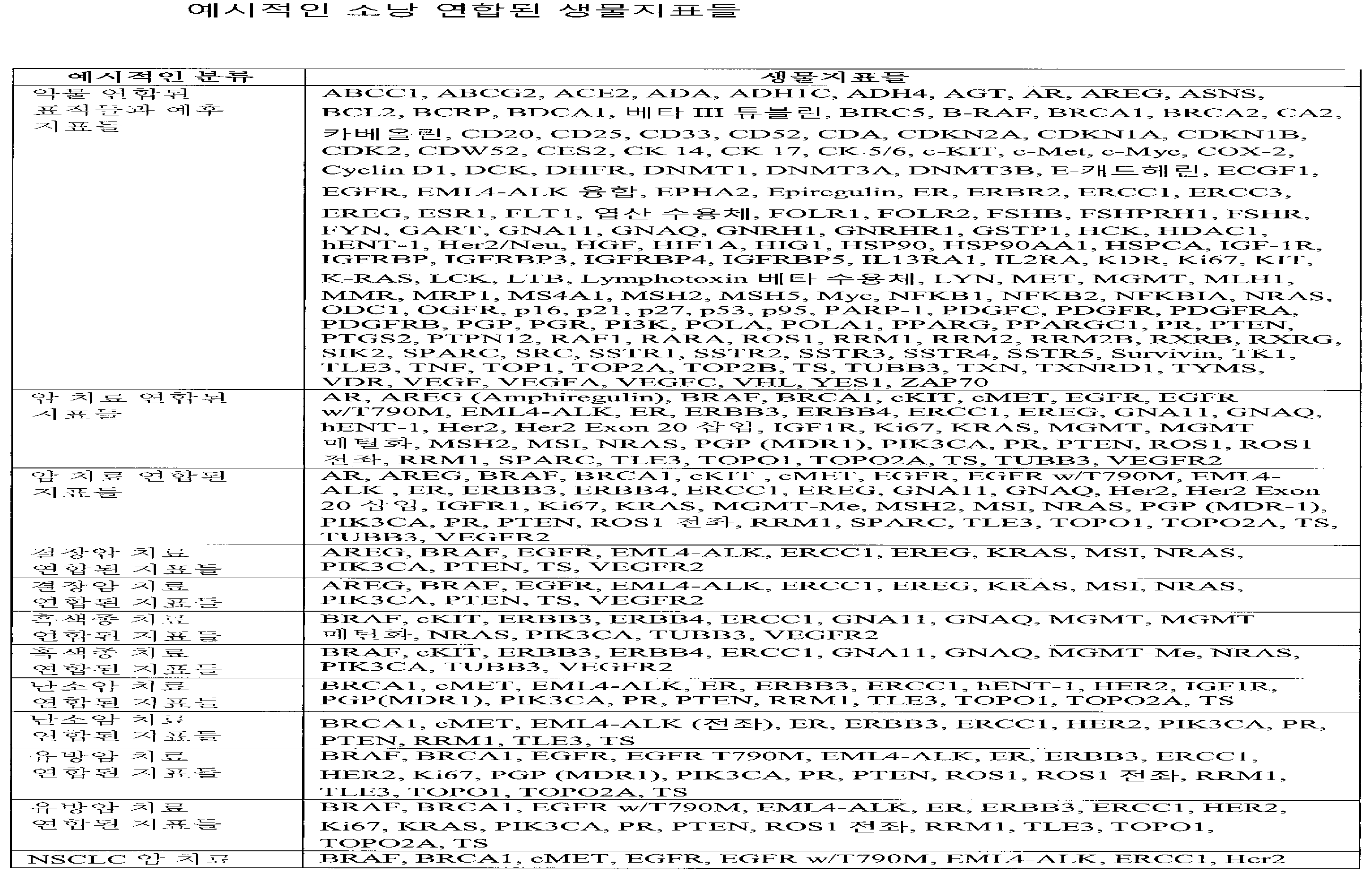
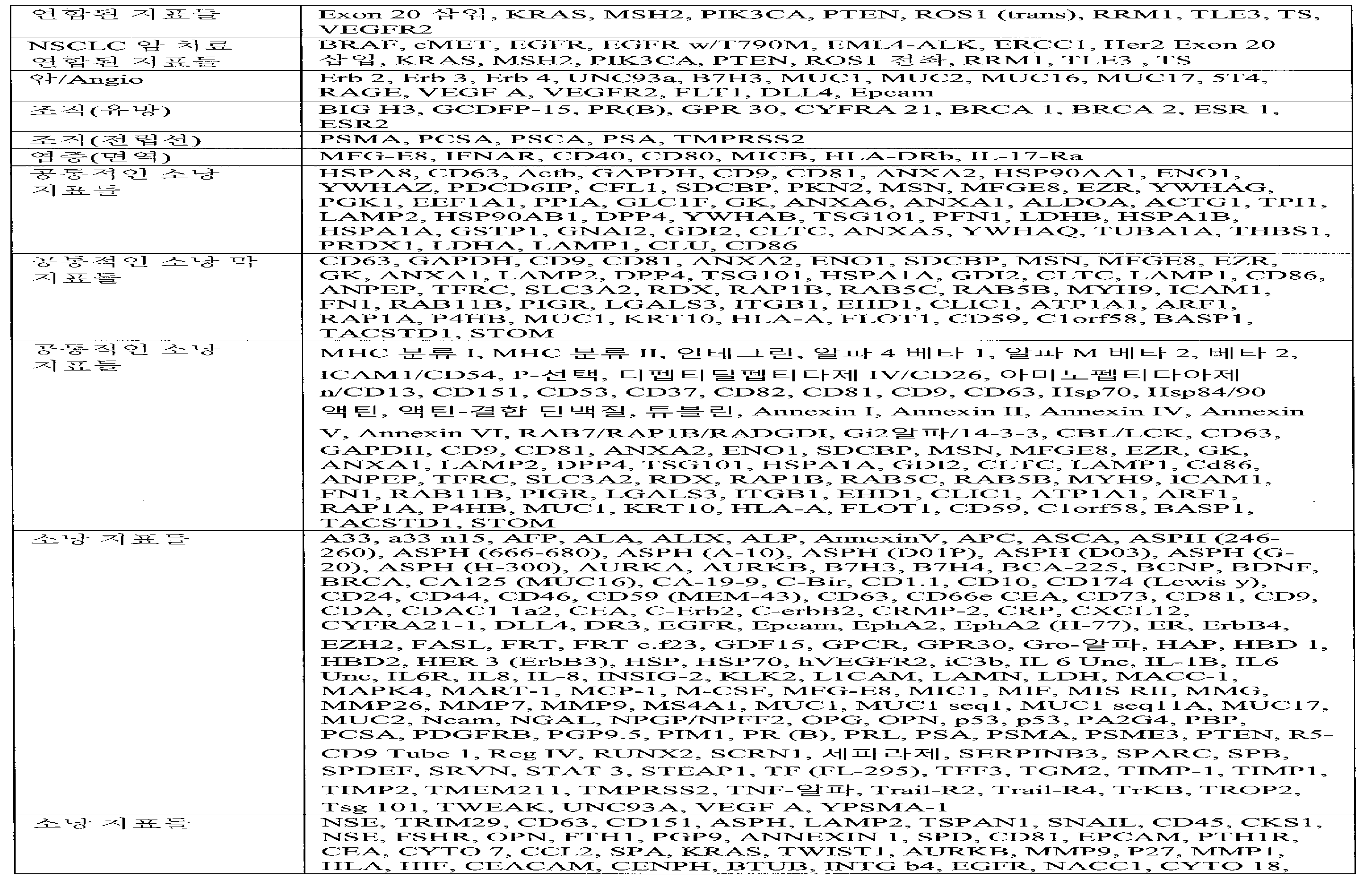
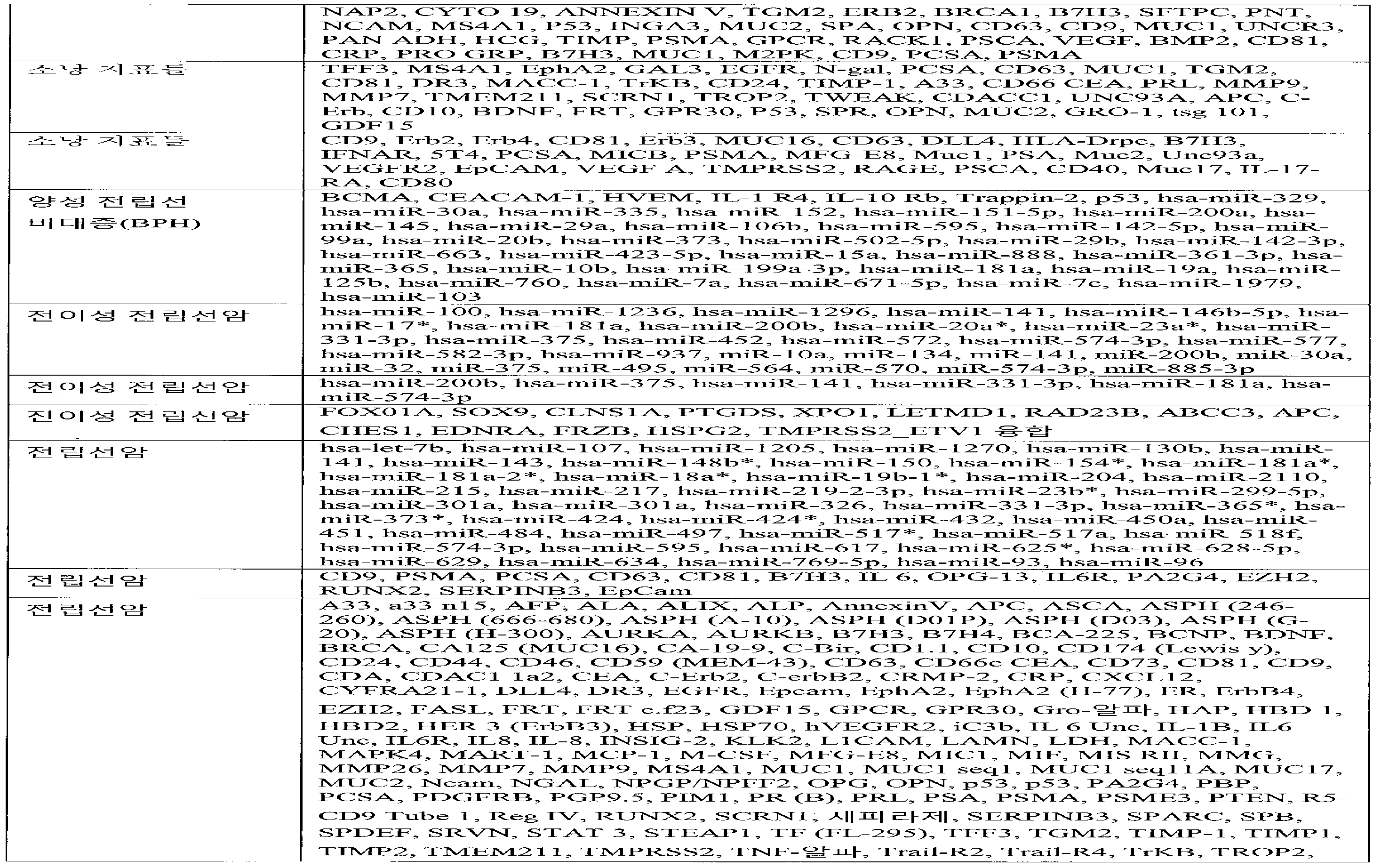
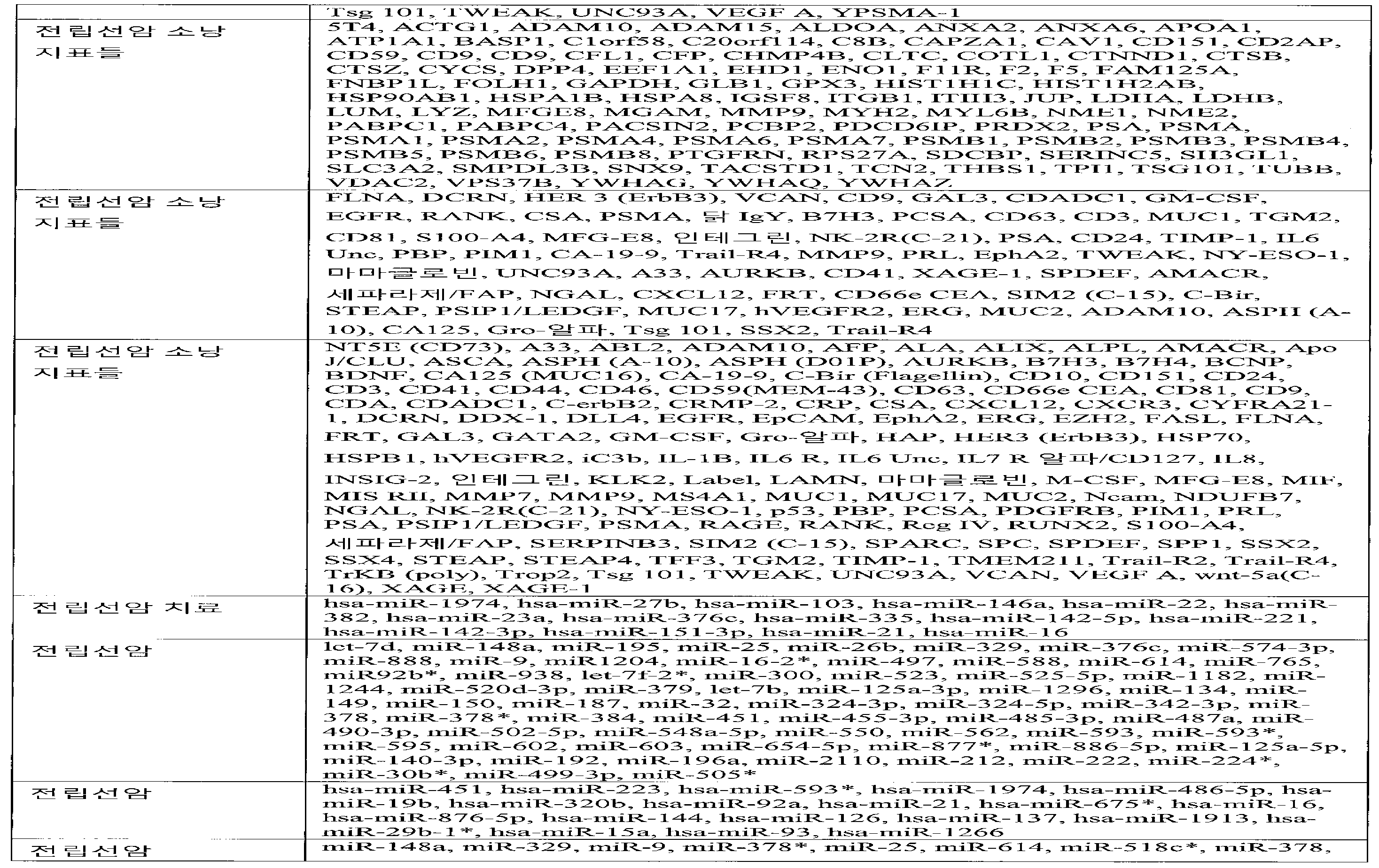
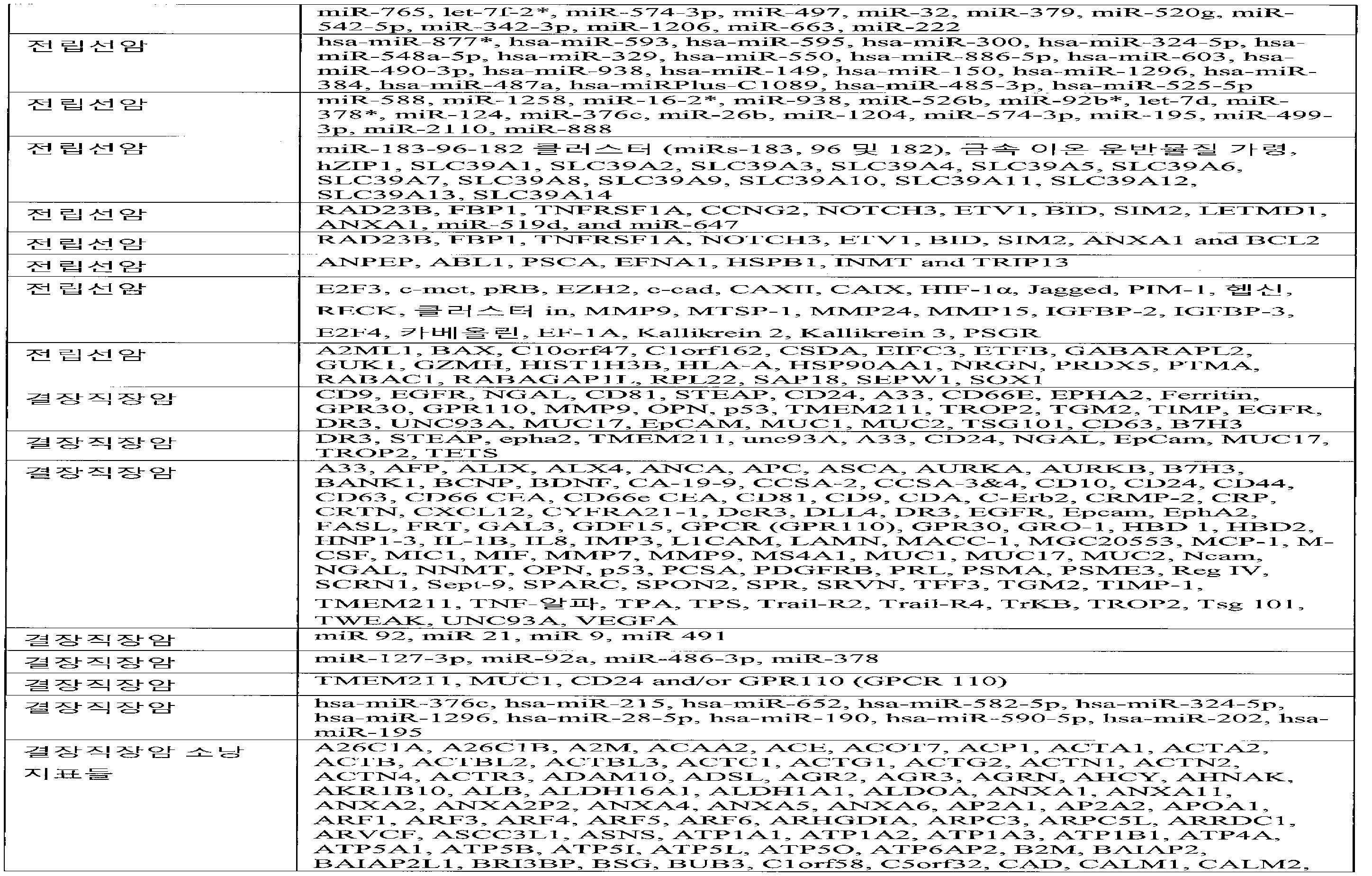
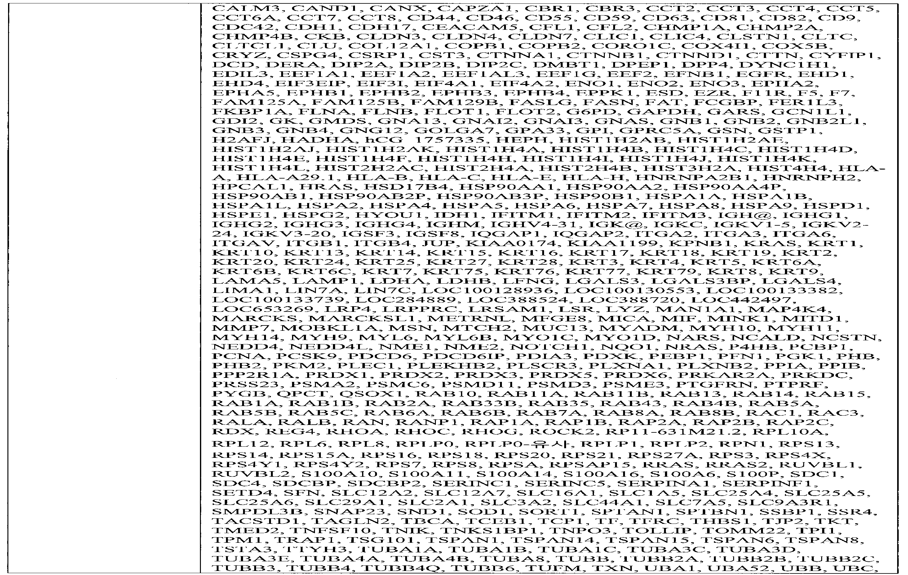

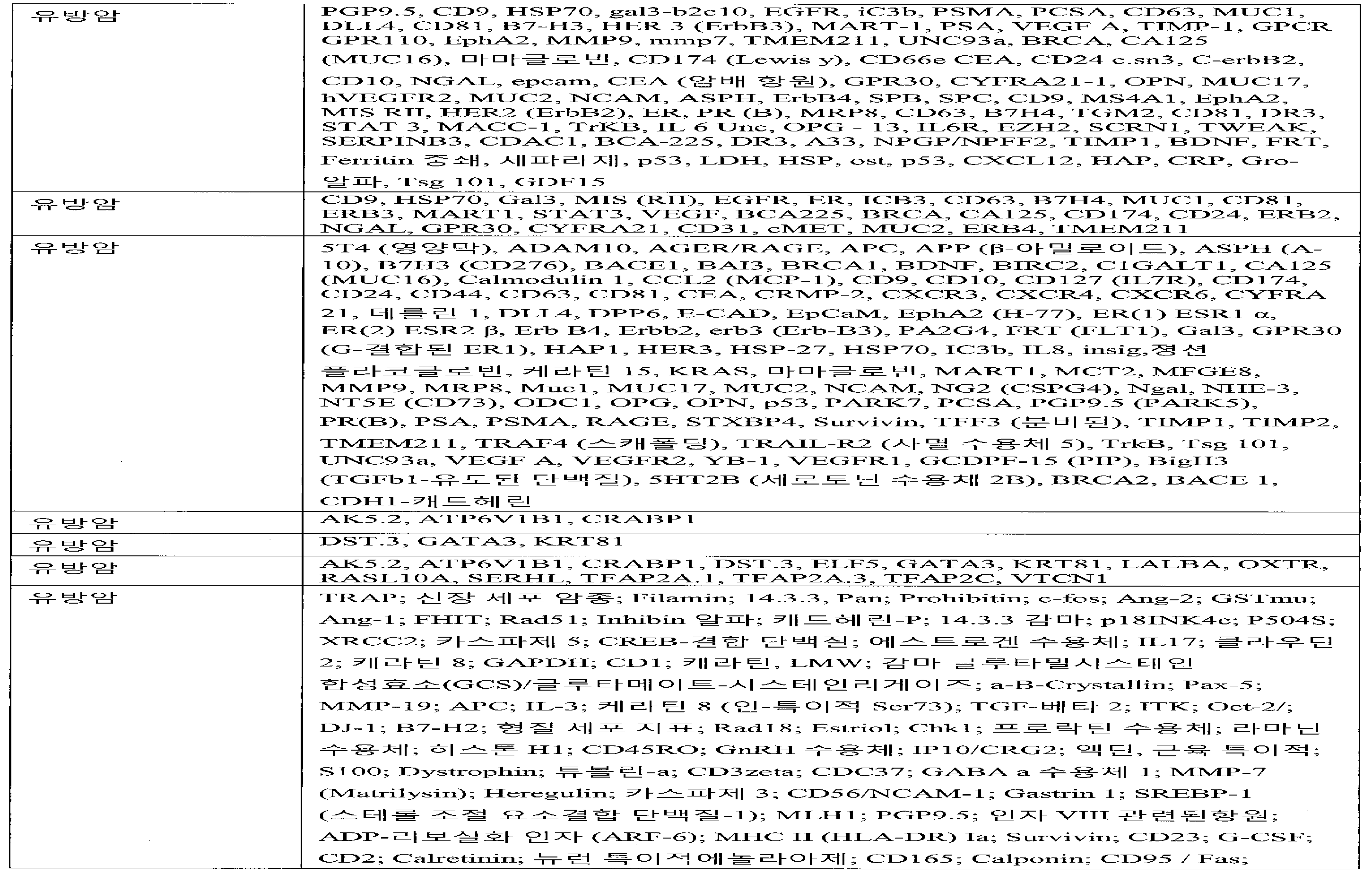


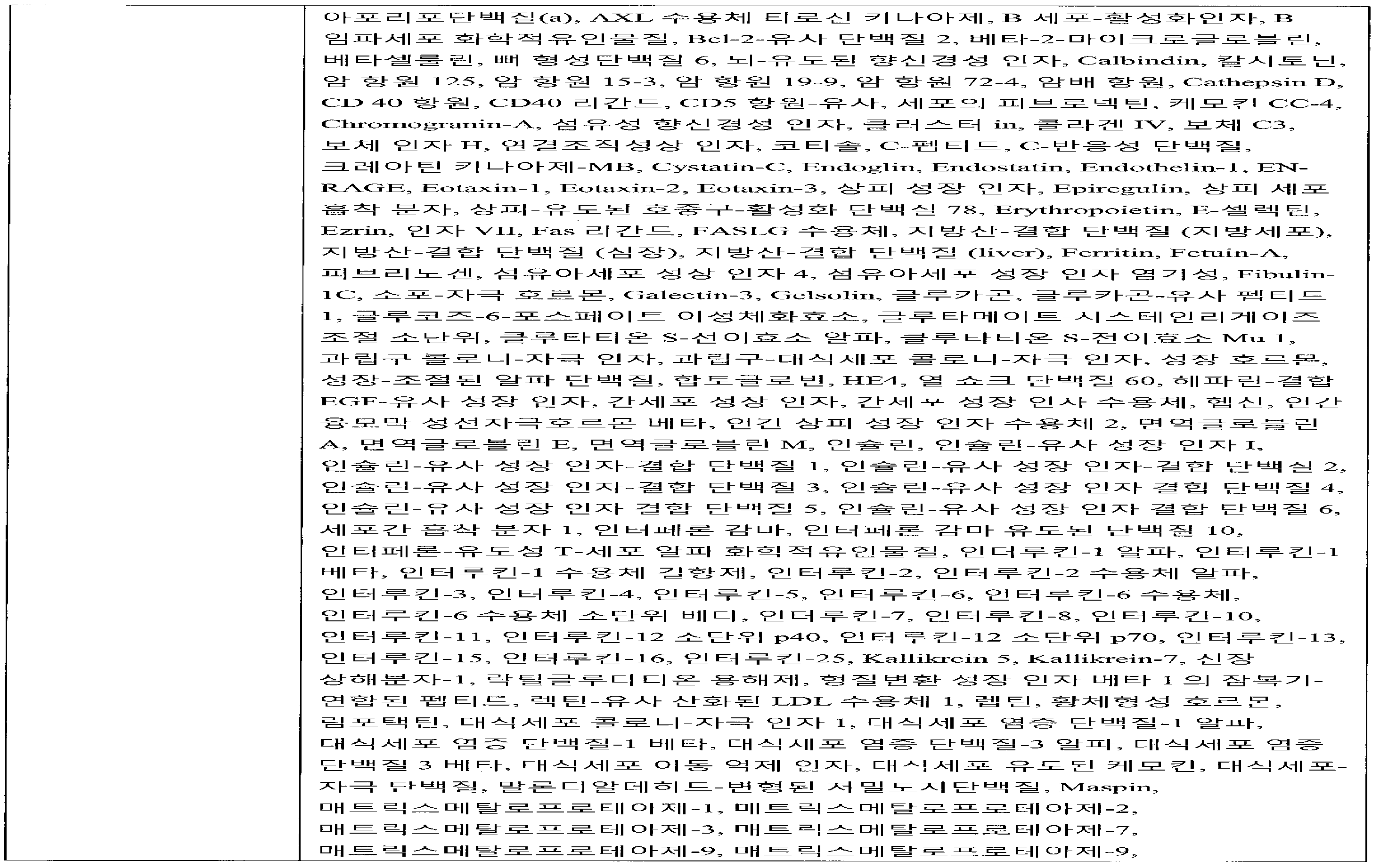


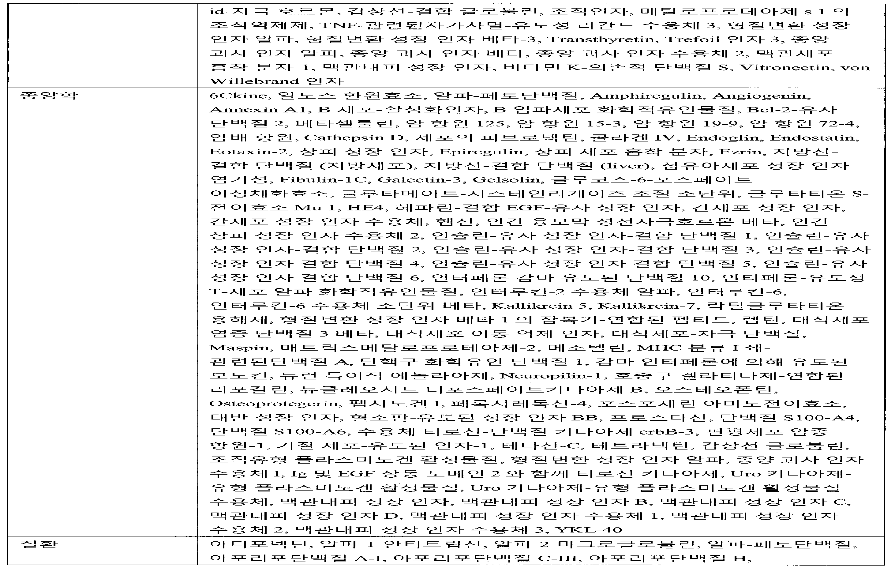
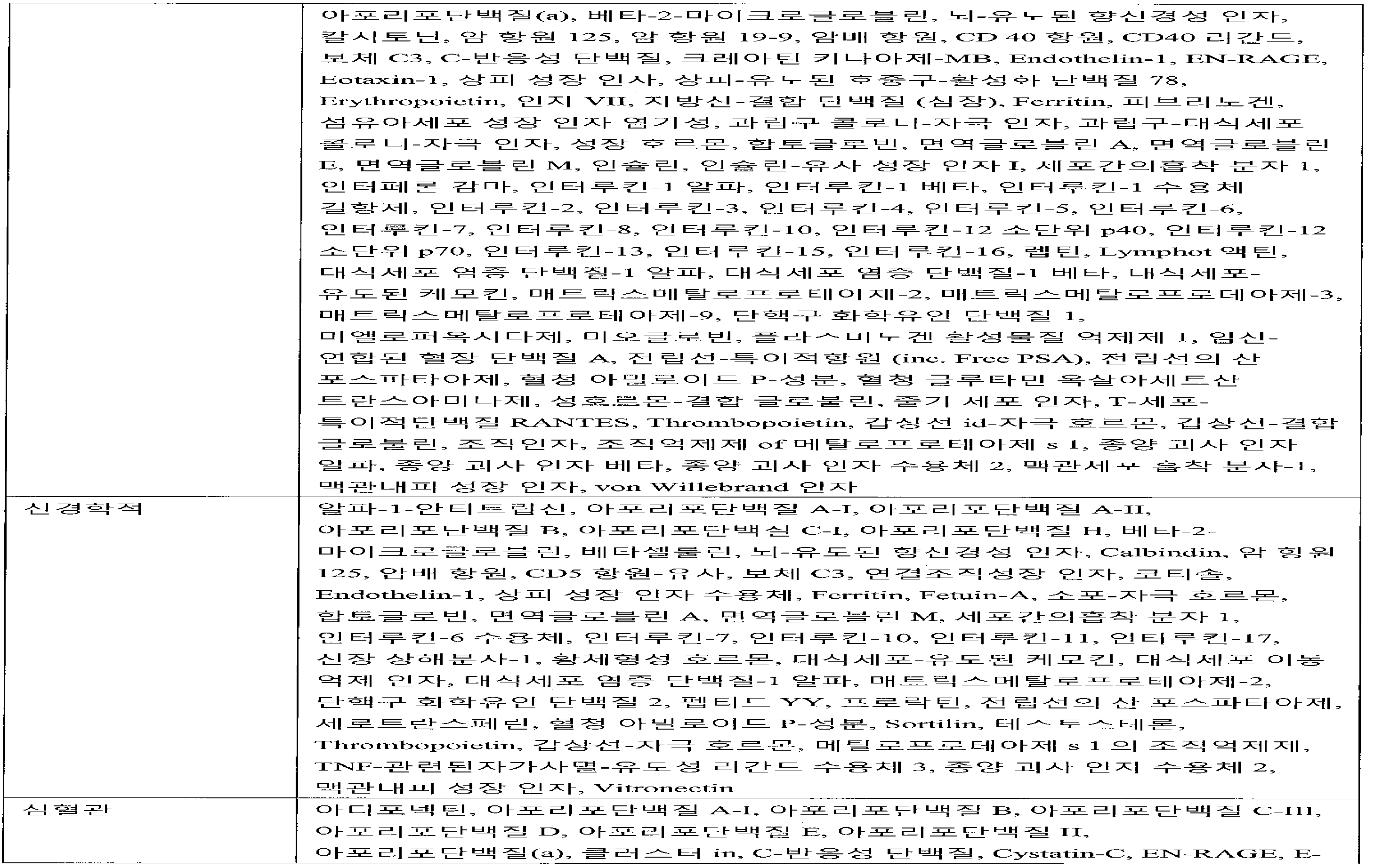

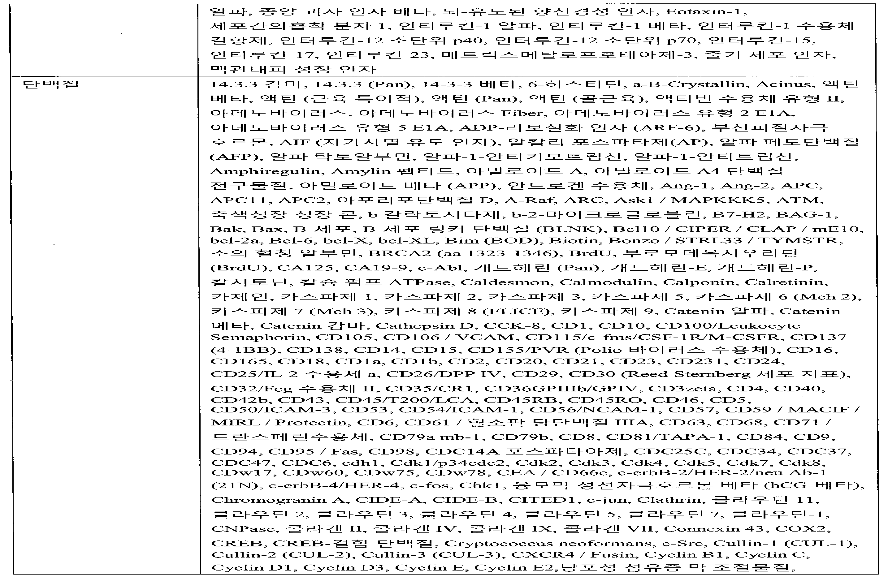


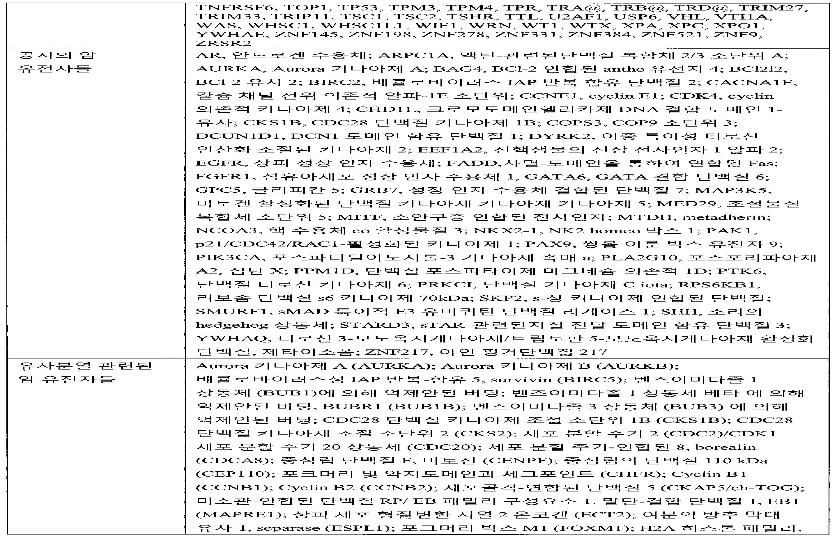
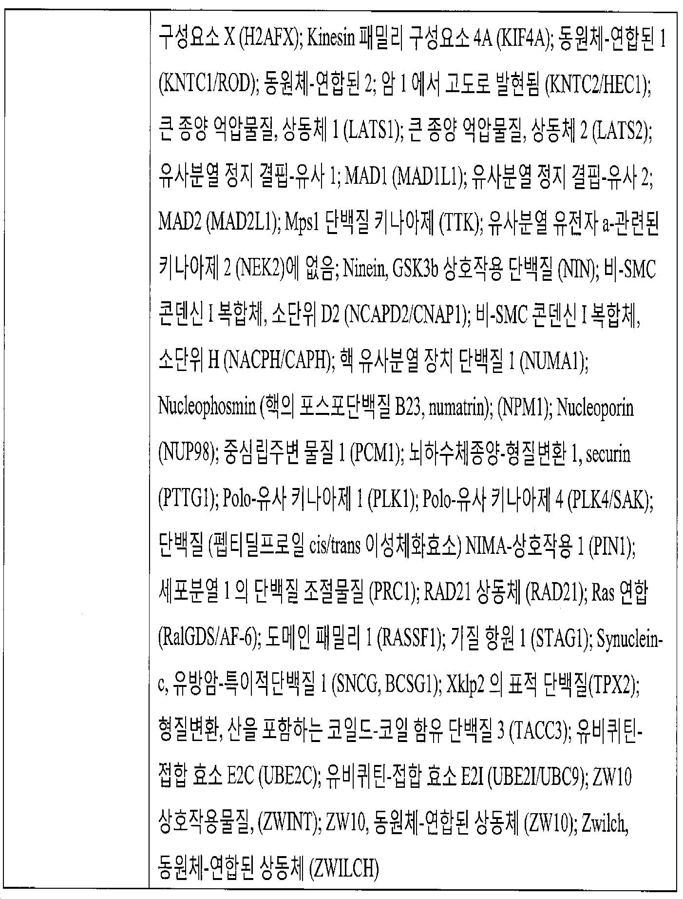
유전자 융합(Gene Fusions)Gene Fusions
소낭의 평가된 하나 이상의 생물지표들은 유전자 융합일 수 있다. 융합 유전자는 두 개의 이미 분리된 유전자의 병치(juxtaposition)에 의해 만들어진 하이브리드 유전자다. 염색체 전위(translocation) 또는 역전, 결손 또는 횡단-접합(trans-splicing)을 통하여 일어날 수 있다. 생성된 융합 유전자는 유전자의 비정상적 임시 및 공간적 발현의 원인이되는데, 가령, 세포 성장 인자들, 맥관형성 인자들, 종양 프로모터들 또는 세포의 신형성 형질전환 및 종양의 생성에 기여하는 다른 인자들의 의 비정상적인 발현을 유도한다. 이러한 융합 유전자는 1) 세포 성장 인자의 코딩 영역, 종양 프로모터 또는 상승된 유전자 발현으로 이어지는 맥관형성을 촉진시키는 다른 유전자의 옆에 있는 한 유전자의 강력한 프로모터 영역에 병치하고, 또는 2) 2개의 상이한 유전자들의 코딩 영역의 융합으로 인하여, 키메라 유전자가 발생하고, 따라서 비정상적인 활성을 가진 키메라 단백질이 생성되기 때문에, 발암성일 수 있다. One or more biomarkers of the follicle evaluated may be gene fusion. The fusion gene is a hybrid gene created by the juxtaposition of two already isolated genes. Chromosomal translocation, or through inversion, deletion, or trans-splicing. The resulting fusion gene is responsible for the abnormal temporal and spatial expression of the gene, for example, the expression of cell growth factors, angiogenic factors, tumor promoters or other factors that contribute to the renatogenization of the cells and the production of tumors Leading to abnormal expression. Such a fusion gene may either: 1) engage in a strong promoter region of one gene next to another region that promotes angiogenesis leading to the coding region of a cell growth factor, a tumor promoter or elevated gene expression, or 2) Due to the fusion of the coding region of the chimeric gene, and thus a chimeric protein with abnormal activity is produced.
융합 유전자의 예는 BCR-ABL로써, 이는 만성골수성 백혈병 (CML)의 ~90%와 급성 백혈병의 부분집합에서 특징적인 분자 이상이다(Kurzrock et al., Annals of Internal Medicine 2003; 138(10):819-830). BCR-ABL은 염색체 9와 22의 전위로 생성된 것이다. BCR 유전자의 5'영역과 ABL1의 3' 영역을 함께 가져가는 전위는 키메라 BCR-ABL1 유전자를 만들고, 이 유전자는 구성적으로 활성인 티로신 키나제 활성을 가진 단백질을 인코드한다 (Mittleman et al., Nature Reviews Cancer 2007; 7(4):233-245). 비정상적인 티로신 키나제 활성은 하향조절된 세포 신호생성, 세포 성장 및 세포 생존, 아팝토시스 저항성 및 성장 인자 독립을 유도하고, 이들 모두 백혈병의 병리생리학의 원인이 된다(Kurzrock et al., Annals of Internal Medicine 2003; 138(10):819-830). An example of a fusion gene is BCR-ABL, which is characterized by ~ 90% of chronic myelogenous leukemia (CML) and a subset of acute leukemia ( Kurzrock et al., Annals of Internal Medicine 2003; 138 (10) 819-830 ). BCR-ABL is produced by the potential of
또다른 융합 유전자는 Burkitt 임파종의 ~80%의 한정된 특징인 IGH-MYC이다 (Ferry et al. Oncologist 2006; 11(4):375-83). 이의 원인은 염색체 8과 14 사이의 전위이며, 면역글로블린 중쇄 유전자의 강력한 프로모터에 인접하게 c-Myc 종양유전자를 가져오고, c-myc 과다발현의 원인이 된다(Mittleman et al., Nature Reviews Cancer 2007; 7(4):233-245). c-myc 재배열은 끊임없는 증식성 상태를 초래하기 때문에 임파종 생성에 중추적인 사건이다. 세포 주기, 세포성 분화, 아팝토시스, 및 세포 흡착 (Ferry et al. Oncologist 2006; 11(4):375-83)을 통하여 진전에 광범위한 효과를 가진다. Another fusion gene is IGH-MYC ( Ferry et al. Oncologist 2006; 11 (4): 375-83 ), which is a defined feature of ~ 80% of Burkitt's lymphomas. The cause of this is the dislocation between
다수의 재발 융합 유전자는 Mittleman 데이터베이스 (cgap.nci.nih.gov/Chromosomes/Mitelman)에 열거되어 있으며, 소낭에서 평가할 수 있고, 표현형을 특징화하는데 이용할 수 있다. 유전자 융합을 이용하여 혈액 악성 또는 상피 종양을 특징화하는데 이용할 수 있다. 예를 들면, TMPRSS2-ERG, TMPRSS2-ETV 및 SLC45A3-ELK4 융합을 탐지할 수 있고, 전립선암을 특징화하는데 이용할 수 있으며; 그리고 ETV6-NTRK3 및 ODZ4-NRG1은 유방암을 특징화하는데 이용할 수 있다.A number of recurrent fusion genes are listed in the Mittleman database (cgap.nci.nih.gov/Chromosomes/Mitelman), which can be evaluated in vesicles and used to characterize phenotypes. Gene fusion can be used to characterize blood malignant or epithelial tumors. For example, it can detect TMPRSS2-ERG, TMPRSS2-ETV and SLC45A3-ELK4 fusion and can be used to characterize prostate cancer; And ETV6-NTRK3 and ODZ4-NRG1 can be used to characterize breast cancer.
더욱이, 융합 유전자의 존재 또는 부재, 또는 발현 수준의 평가를 이용하여 표현형의 진단 가령, 암 진단 뿐만 아니라 선택된 치료에 대한 치료의 반응을 모니터링할 수 있다. 예를 들면, BCR-ABL 융합 유전자의 존재는 CML의 진단에 대해 특징적이며, 그리고 CML 치료용 수용체 티로신 키나제 억제제인 Novartis 약물 이마티니브 메실레이트 (Gleevec)의 표적이 된다. 이마티니브(Imatinib) 치료는 분자 반응 (BCR-ABL+ 혈액 세포들의 소멸)과 BCR-ABL+ CML 환자에서 병의 진전-없는 생존을 개선시켰다(Kantarjian et al., Clinical Cancer Research 2007; 13(4):1089-1097). Moreover, the presence or absence of a fusion gene, or an assessment of the level of expression, can be used to diagnose the phenotype, for example, to diagnose cancer as well as to monitor the response of treatment to a selected treatment. For example, the presence of the BCR-ABL fusion gene is characteristic of the diagnosis of CML and is the target of Novartis drug imatinib mesylate (Gleevec), a receptor tyrosine kinase inhibitor for CML therapy. Imatinib therapy improved disease progression-free survival in patients with BCR-ABL + CML and molecular reactions (BCR-ABL + extinction of blood cells) ( Kantarjian et al., Clinical Cancer Research 2007; 13 (4) : 1089-1097 ).
유전자 융합의 존재, 부재 또는 발현 수준에 대하여 소낭의 평가는 유전자 융합의 존재, 부재 또는 발현 수준에 대해 이종 소낭 집단을 평가하는 것일 수 있다. 대안으로, 평가할 소낭은 상기에서 설명한 것과 같이, 특이적 세포 유형으로부터 유도된, 가령, 기원 세포 특이적 소낭일 수 있다. 표현형을 특징화하기 위하여 평가하게 될 융합을 이용하는 설명을 위한 예는 2011년 4월 6일자로 제출된 국제 특허 출원 번호 PCT/US2011/031479, "Circulating Biomakers for Disease"(이들은 전문이 여기에 참고자료로 편입된다)에서 설명된 것들을 포함한다. Assessment of follicles for the presence, absence, or expression level of gene fusion may be by assessing the heterogeneous follicular population for the presence, absence, or expression level of the gene fusion. Alternatively, the vesicles to be evaluated may be, for example, a cell-specific vesicle derived from a specific cell type, as described above. Examples for explanations using fusion that will be evaluated to characterize phenotypes include International Patent Application No. PCT / US2011 / 031479, "Circulating Biomakers for Disease," filed April 6, 2011, Which are incorporated herein by reference.
유전자-연합된 MiRNA 생물지표들Gene-associated miRNA biomarkers
특정 전사체와 상호작용하고, 표현형을 특징화하기 위하여 평가될 수 있는 것으로 공지된 miRNA 생물지표들의 용도의 설명을 위한 실시예는 2011년 4월 6일자로 제출된 국제 특허 출원 번호 PCT/US2011/031479, "Circulating Biomakers for Disease"(이들은 전문이 여기에 참고자료로 편입된다)에서 설명된 것들을 포함한다. An example for the description of the use of miRNA biomarkers known to be able to be evaluated to interact with a particular transcript and to characterize the phenotype is set forth in International Patent Application No. PCT / US2011 / 031479, "Circulating Biomakers for Disease ", which are incorporated herein by reference.
핵산 - 단백질 복합체 생물지표들Nucleic acid-protein complex biomarkers
인간 혈장 안에 MicroRNA는 순환하는 미세소낭, Argonaute 단백질, 그리고 HDL 그리고 LDL 복합체들과 연합되는 것으로 밝혀졌다. 가령, Arroyo et al., Argonaute2 complexes carry a population of circulating microRNA independent of vesicles in human plasma. Proc Natl Acad Sci USA. 2011. 108:5003-08. Epub 2011 Mar 7; Collino et al., Microvesicles derived from adult human bone marrow and tissue specific mesenchymal stem cells shuttle selected pattern of miRNA. PLOS One. 2010 5(7):e11803. 단백질의 Argonaute 패밀리는 RNA 간섭 (RNAi) 유전자 침묵에 역할을 한다. Argonaute 단백질은 짧은 RNA 이를 테면 microRNA (miRNA) 또는 짧은 간섭 RNA (siRNA)에 결합하고, 그리고 이들의 상보적인 mRNA의 해독을 억제한다. 이들은 전사 유전자 침묵 (TGS)에도 관련되는데, 이때 항원 RNA 또는 agRNA으로 알려진 짧은 RNA는 상보적인 프로모터 영역들의 전사 억제를 지시한다. Argonaute 패밀리 구성원들은 Argonaute 1 ("진핵생물의 해독 개시 인자 2C, 1", EIF2C1, AGO1), Argonaute 2 ("진핵생물의 해독 개시 인자 2C, 2", EIF2C2, AGO2), Argonaute 3 ("진핵생물의 해독 개시 인자 2C, 3", EIF2C3, AGO3), 그리고 Argonaute 4 ("진핵생물의 해독 개시 인자 2C, 4", EIF2C4, AGO4)를 포함한다. 몇 가지 Argonaute 이소타입이 확인되었다. Argonaute 2는 RNA-유도된 침묵 복합체 (RISC) 안에 효과물질 단백질이며, microRNA 침묵 경로에서 표적 메신져 RNA의 침묵에 역할을 한다. MicroRNAs in human plasma have been shown to associate with circulating micro vesicles, Argonaute protein, and HDL and LDL complexes. For example, Arroyo et al., Argonaute2 complexes carry a population of microRNAs independent of vesicles in human plasma. Proc Natl Acad Sci USA. 2011. 108: 5003-08. Epub 2011 Mar 7; Collino et al., Microvesicles derived from adult human bone marrow and tissue specific mesenchymal stem cells shuttle selected pattern of miRNA. PLOS One. 2010 5 (7): e11803. The Argonaute family of proteins plays a role in RNA interference (RNAi) gene silencing. The Argonaute protein binds to short RNAs, such as microRNAs (miRNAs) or short interfering RNAs (siRNAs), and suppresses the decoding of their complementary mRNAs. These are also involved in transcriptional gene silencing (TGS), where short RNAs known as antigen RNAs or agRNAs direct transcriptional repression of complementary promoter regions. The Argonaute family members include Argonaute 1 ("eukaryotic
단백질 GW182은 미세소낭과 연합되고, 그리고 모든 인간 Argonaute 단백질 (가령, Ago1, Ago2, Ago3, Ago4) 및 이들의 연합된 miRNA에 결합하는 능력을 보유한다. 가령, Gibbings et al., Multivesicular 바디 associate with 성분들 of miRNA effector complexes and modulate miRNA activity, Nat Cell Biol 2009 11:1143-1149. Epub 2009 Aug 16; Lazzaretti et al., The C-terminal Domains of human TNRC6A, TNRC6B, and TNRC6C silence binding transcripts independently of Argonaute protein. RNA. 2009 15:1059-66. Epub 2009 Apr 21. GW182, which is encoded by the TNRC6A gene (trinucleotide repeat containing 6A), functions in post-transcritptional gene sliencing through the RNA interference (RNAi) and microRNA pathways 참고. TNRC6B 및 TNRC6C는 삼뉴클레오티드 반복을 포함하는 6개 패밀리의 구성원들이며, 유전자 침묵에서 유사한 역할을 한다. GW182는 GW-바디(bodies) 또는 P-바디로 알려진 세포질 바디에서 mRNA 및 Argonaute 단백질과 연합된다. GW182는 miRNA-의존적 해복 억제 및 argonaute 패밀리 단백질에 의한 상보적인 mRNA의 siRNA-의존적 말단 뉴클레오티드 절단에 관계된다. Protein GW182 is associated with microfollicles and retains the ability to bind to all human Argonaute proteins (e.g., Ago1, Ago2, Ago3, Ago4) and their associated miRNAs. For example, Gibbings et al., Multivesicular body associate with components of miRNA effector complexes and modulate miRNA activity, Nat Cell Biol 2009 11: 1143-1149. Epub 2009 Aug 16; Lazzaretti et al., The C-terminal Domains of human TNRC6A, TNRC6B, and TNRC6C independently silencing binding transcripts of Argonaute protein. RNA. 2009 15: 1059-66. Epub 2009 Apr. 21. GW182, which is encoded by the TNRC6A gene (6A), functions in post-transcritptional gene sliencing through the RNA interference (RNAi) and microRNA pathways. TNRC6B and TNRC6C are members of six families, including trinucleotide repeats, which play a similar role in gene silencing. GW182 is associated with mRNA and Argonaute protein in the cytoplasmic body known as GW-bodies or P-bodies. GW182 is involved in miRNA-dependent relaxation suppression and siRNA-dependent terminal nucleotide cleavage of complementary mRNA by the argonaute family protein.
한 측면에서, 본 발명은 핵산 - 단백질 복합체 생물지표들을 분석하는 것을 포함하는 표현형을 특징화하는 방법을 제공한다. 본 명세서에서 이용된 것과 같이, 핵산 - 단백질 복합체는 최소한 하나의 핵산 그리고 최소한 하나의 단백질을 포함하고, 기타 성분들 이를 테면 지질을 또한 포함할 수 있다. 핵산 - 단백질 복합체는 소낭과 연합될 수 있다. 한 구체예에서, RNA - 단백질 복합체들은 단리되고, 연합된 RNA의 수준이 평가되며, 이때 이 수준은 본 명세서에서 기술된 것과 같이 표현형, 가령, 진단, 예후, 치료진단, 또는 기타 표현형을 제공하는 것과 같이, 표현형을 특징화하는데 이용된다. RNA는 microRNA일 수 있다. MicroRNA는 소낭 및 단백질과 연합된 것으로 발견된다. 일부 경우들에서, 이러한 연합은 RNAes 또는 기타 인자들을 통한 분해로부터 miRNA를 보호하는 기능을 할 수 있다. 소낭 연합된 miRs, Ago-연합된 miRs, 세포-기원 소낭 연합된 miRs, 순환하는 Ago-결합된 miRs, 순환하는 HDL-결합된 miRs, 그리고 전체 miR 함량을 포함하나 이에 한정되지 않는 microRNA의 다양한 집단의 내용물이 시료 내에서 평가될 수 있다. In one aspect, the invention provides a method of characterizing a phenotype comprising analyzing nucleic acid-protein complex biomarkers. As used herein, a nucleic acid-protein complex includes at least one nucleic acid and at least one protein, and may also include other components such as lipids. Nucleic acid-protein complexes can be associated with vesicles. In one embodiment, the RNA-protein complexes are isolated and the level of the associated RNA is assessed, wherein the level is at least one of a phenotype, such as that provided herein, such as a diagnosis, prognosis, therapeutic diagnosis, As such, it is used to characterize the phenotype. The RNA can be a microRNA. MicroRNAs are found to be associated with follicles and proteins. In some cases, such association may serve to protect miRNAs from degradation through RNAs or other factors. The various populations of microRNAs, including, but not limited to, vesicle-associated miRs, Ago-associated miRs, cell-derived vesicle associated miRs, circulating Ago-linked miRs, circulating HDL-associated miRs, Can be evaluated in the sample.
복합체들을 단리하는데 이용되는 단백질 생물지표는 하나 또는 그 이상의 Argonaute 단백질, 또는 Argonaute 패밀리 구성원들과 연합된 기타 단백질일 수 있다. 이들은 Argonaute 단백질 Ago1, Ago2, Ago3, Ago4, 그리고 이의 다양한 이소폼을 포함하나 이에 한정되지 않는다. 단백질 생물지표는 GW182 (TNRC6A), TNRC6B 및/또는 TNRC6C일 수 있다. 단백질 생물지표는 P-바디 또는 GW-바디, 이를 테면 SW182, argonaute, 디캡핑(decapping) 효소 또는 RNA 헬리카제와 연합된 단백질일 수 있다. 가령, Kulkarni et al. On track with P-body. Biochem Soc Trans 2010, 38:242-251 참고. 단백질 생물지표는 can also be 하나 또는 그 이상의 HNRNPA2B1 (이질성 핵 리보핵단백질 a2/b1), HNRPAB (이질성 핵 리보핵단백질 A/B), ILF2 (인터루킨 인헨서 결합 인자 2, 45 kda), NCL (Nucleolin), NPM1 (Nucleophosmin (조직(nucleolar) 인단백질 b23, numatrin)), RPL10A (리보좀 단백질 l10a), RPL5 (리보좀 단백질 l5), RPLP1 (리보좀 단백질, 거대, p1), RPS12 (리보좀 단백질 s12), RPS19 (리보좀 단백질 s19), SNRPG (작은 핵 리보핵단백질 폴리펩티드 g), TROVE2 (Trove 도메인 패밀리, 구성원 2). Wang et al., Export of microRNA and microRNA-protective protein by mammalian cells. Nucleic Acids Res. 38:7248-59. Epub 2010 Jul 7 참고. 단백질 생물지표는 아포리포단백질일 수도 있는데, 이는 지단백질을 형성하기 위하여 지질(오일-가용성 물질 이를 테면, 지방 및 콜레스테롤)에 결합하는 단백질이며, 림프 및 순환계를 통하여 지질이 운반된다. Vickers et al., MicroRNA are delivered in plasma and delivered to recipient cells by high-density lipoprotein, Nat Cell Biol 2011 13:423-33, Epub 2011 Mar 20 참고. 아포리포단백질은 아포리포단백질 A (apo A-I, apo A-II, apo A-IV, 그리고 apo A-V를 포함), 아포리포단백질 B (apo B48 그리고 apo B100를 포함), 아포리포단백질 C (apo C-I, apo C-II, apo C-III, 그리고 apo C-IV를 포함), 아포리포단백질 D (ApoD), 아포리포단백질 E (ApoE), 아포리포단백질 H (ApoH), 또는 이의 조합일 수 있다. 아포리포단백질은 APOL1, APOL2, APOL3, APOL4, APOL5, APOL6, APOLD1, 또는 이의 조합을 포함하는 아포리포단백질 L이다. 아포리포단백질 L (Apo L)은 콜레스테롤 운반에 중심 역할을 하는 고밀도 지단백질 패밀리에 속한다. 단백질 생물지표는 지단백질, 이를 테면 킬로미크론(chylomicron)의 성분, 초저밀도 지단백질 (VLDL), 중간 밀도 지단백질 (IDL), 저밀도 지단백질 (LDL) 및/또는 고밀도 지단백질 (HDL)의 성분일 수 있다. 한 구체예에서, 단백질 생물지표는 LDL 또는 HDL의 성분이다. 성분은 ApoE일 수 있다. 성분은 ApoA1일 수 있다. 단백질 생물지표은 일반적인 소낭 지표, 이를 테면 CD9, CD63 및/또는 CD81을 포함하나 이에 한정되지 않는 표 3에서 열거된 테트라스파닌 또는 기타 단백질일 수 있다. 단백질 생물지표는 암 지표 이를 테면 EpCam, B7H3 및/또는 CD24일 수 있다. 단백질 생물지표는 조직 특이적 생물지표, 이를 테면 전립선 생물지표들 PSCA, PCSA 및/또는 PSMA일 수 있다. 이들 또는 기타 유용한 단백질 생물지표들의 조합을 이용하여 관심 복합체의 특적 집단을 단리할 수 있다. The protein biomarkers used to isolate the complexes may be one or more of the Argonaute proteins, or other proteins associated with the Argonaute family members. These include, but are not limited to, Argonaute proteins Ago1, Ago2, Ago3, Ago4, and various isoforms thereof. Protein biomarkers may be GW182 (TNRC6A), TNRC6B and / or TNRC6C. The protein biomarker may be a protein associated with a P-body or GW-body, such as SW182, argonaute, a decapping enzyme or an RNA helicase. For example, Kulkarni et al. On track with P-body. Biochem Soc Trans 2010, 38: 242-251. Protein biomarkers can also include one or more of HNRNPA2B1 (heterogeneous nucleated ribonucleoprotein a2 / b1), HNRPAB (heterogeneous nucleated ribonucleoprotein A / B), ILF2 (interleukin- Nucleolin), NPM1 (Nucleophosmin (protein b23, numatrin), RPL10A (ribosomal protein l10a), RPL5 (ribosomal protein l5), RPLP1 (ribosomal protein, giant p1), RPS12 (ribosomal protein s12) RPS19 (ribosomal protein s19), SNRPG (small nuclear ribonucleoprotein polypeptide g), TROVE2 (Trove domain family, member 2). Wang et al., Export of microRNA and microRNA-protective protein by mammalian cells. Nucleic Acids Res. 38: 7248-59. See Epub 2010 Jul 7. Protein biomarkers may be apolipoprotein, a protein that binds lipids (oil-soluble substances, such as fat and cholesterol) to form lipoproteins and transports lipids through the lymph and circulatory system. Vickers et al., MicroRNAs are delivered in plasma and delivered to recipient cells by high-density lipoprotein, Nat Cell Biol 2011 13: 423-33, Epub 2011
핵산 - 단백질 복합체들은 복합체의 하나 또는 그 이상의 성분에 대한 결합제를 이용하여 단리될 수 있다. 단백질을 단리하는 다양한 기술이 당업계에 공지되어 있거나 본 명세서에서 기술되는데, 친화력 격리, 면역캡쳐, 면역침전 그리고 유동 세포분석을 포함하나 이에 한정되지 않는다. 이 결합제는 본 명세서에서 기술된임의의 적절한 결합제일 수 있는데, 이를 테면 하나 또는 그 이상 결합제는 핵산, DNA 분자, RNA 분자, 항체, 항체 단편, 압타머, 펩토이드, zDNA, 펩티드 핵산 (PNA), 잠긴 핵산 (LNA), 렉틴, 펩티드, 덴드라이머, 막 단백질 라벨링 물질, 화학적 화합물, 또는 이의 조합를 포함한다. 한 구체예에서, 이 결합제는 항체, 항체 접합물(conjugate), 항체 단편, 및/또는 압타머를 포함한다. 본 발명에 이용될 수 있는 단백질-핵산 복합체를 평가하는 추가 방법은 Wang et al., Export of microRNA and microRNA-protective protein by mammalian cells. Nucleic Acids Res. 38:7248-59. Epub 2010 Jul 7; Keene et al., RIP-Chip: the isolation and identification of mRNA, microRNA and protein components of ribonucleoprotein complexes from cell extracts. Nat Protoc 2006 1:302-07; Hafner, Transcriptome-wide identification of RNA-binding protein and microRNA target sites by PAR-CLIP. Cell 2010 141:129-41 참고.The nucleic acid-protein complexes may be isolated using a binding agent for one or more components of the complex. Various techniques for isolating proteins are known in the art or described herein, including but not limited to affinity isolation, immunoprecipitation, immunoprecipitation and flow cytometry. The binder may be any suitable binder described herein, for example, one or more of the binding agents may be selected from the group consisting of nucleic acids, DNA molecules, RNA molecules, antibodies, antibody fragments, platamers, peptides, zDNA, peptide nucleic acids ), Locked nucleic acids (LNA), lectins, peptides, dendrimers, membrane protein labeling agents, chemical compounds, or combinations thereof. In one embodiment, the binding agent comprises an antibody, an antibody conjugate, an antibody fragment, and / or an extramammary. Additional methods for evaluating protein-nucleic acid complexes that may be used in the present invention are described in Wang et al., Export of microRNA and microRNA-protective protein by mammalian cells. Nucleic Acids Res. 38: 7248-59. Epub 2010 Jul 7; Keene et al., RIP-Chip: the isolation and identification of mRNA, microRNA and protein components of ribonucleoprotein complexes from cell extracts. Nat Protoc 2006 1: 302-07; Hafner, transcriptome-wide identification of RNA-binding protein and microRNA target sites by PAR-CLIP. Cell 2010 141: 129-41.
본 발명은 단백질과 복합체로 발견되는 miRNA를 확인하는 방법을 더 제공한다. 한 구체예에서, 단백질 - 핵산 복합체의 집단은 상기에서 설명된 바와 같이 단리된다. 이 집단의 miRNA 함량이 평가된다. 이 방법은 다양한 관심 시료(가령, 질병이 있는, 질병이 없는, 반응자, 비-반응자)에서 시용될 수 있고, 시료 안에 miRNA 함량이 비교되어 시료간에 차등되는 miRNA를 식별할 수 있다. miRNA를 탐지하는 방법들이 제공된다(어래이, PCR, 등). 확인된 miRNA는 본 명세서에서 기술된 방법에 따라 표현형을 특징화하는데 이용될 수 있다. 예를 들면, 발견에 이용되는시료는 암 및 암이 아닌 혈장 시료일 수 있다. 암과 암이 아닌 시료를 구별하는 단백질-복합된 miRNA가 식별될 수 있고, 차등 miRNA들이 평가되어 혈장 시료안에 암이 탐지될 수 있다. The present invention further provides a method for identifying miRNAs found as complexes with proteins. In one embodiment, the population of protein-nucleic acid complexes is isolated as described above. The miRNA content of this population is assessed. This method can be used in a variety of interesting samples (eg, diseased, disease free, responders, non-responders) and the miRNA content in the sample can be compared to identify miRNAs that differ between samples. Methods to detect miRNAs are provided (arrays, PCR, etc.). Identified miRNAs can be used to characterize phenotypes according to the methods described herein. For example, the sample used for the discovery may be a plasma sample that is not cancerous or cancerous. Protein-complexed miRNAs that differentiate cancer from non-cancerous samples can be identified, and differential miRNAs can be evaluated to detect cancer in plasma samples.
본 발명은 소낭-함유 시료로부터 비-페이로드 miR을 제거하고, 이 소낭 안에 miR를 평가함으로써 소낭 안에 microRNA 페이로드를 구별하는 방법을 또한 제공한다. RNAes 또는 기타 miRNA를 분해하는 기타 엔터티를 이용하여 시료로부터 miR을 제거할 수 있다. 일부 구체예들에서, 이 시료는 RNAe 처치에 앞서 단백질 복합체로부터 microRNA를 제거하기 위하여 물질로 처리된다. 단백질을 분해하는 효소, 가령, 프로테아제 이를 테면 프로테아제 K 또는 트립신, 또는 임의의 기타 적절한 효소일 수 있다. 이 방법을 이용하여 순환하는 단백질 복합체 안에 자유 miRNA 또는 miRNA와는 별로도 소낭안에 포함된 microRNA 분획을 평가함으로써 본 명세서에서 기술된 방법에 따라 표현형을 특징화할 수 있다. The present invention also provides a method for distinguishing microRNA payloads in vesicles by removing non-payload miRs from follicular-containing samples and evaluating miRs in these vesicles. MiRs can be removed from the sample using RNAes or other entities that degrade other miRNAs. In some embodiments, the sample is treated with a material to remove microRNAs from the protein complex prior to RNAe treatment. An enzyme that degrades the protein, such as a protease such as protease K or trypsin, or any other suitable enzyme. This method can be used to characterize phenotypes according to the methods described herein by evaluating the microRNA fraction contained in the vesicles, often with free miRNAs or miRNAs in circulating protein complexes.
생물지표 탐지Biological surface detection
여기에서 설명된 것과 같이, microRNA, 단백질, 소낭 또는 다른 생물지표들과 같은 순환하는 생물지표들의 존재, 수준 또는 농도를 탐지함으로써 생체특징을 정성적으로 또는 정량적으로 탐지할 수 있다. 이들 생체특징 성분들은 당업계에 공지되어 다양한 기술을 이용하여 탐지할 수 있다. 예를 들면, 생물지표는 마이크로어래이 분석, 폴리머라제 쇄 반응 (PCR) (PCR-기반 방법들 가령, 실시간 폴리머라제 쇄 반응 (RT-PCR), 정량적 실시간 폴리머라제 쇄 반응 (Q-PCR/qPCR) 그리고 이와 유사한 것들), 대립형질-특이적 프로브와의 혼성화, 효소 돌연변이 탐지, 결찰(ligation) 쇄 반응 (LCR), 올리고뉴클레오티드 결찰(ligation) 분석 (OLA), 유동-세포분석 헤테로듀플렉스 분석, 미스메취의 화학적 절단, 질량 분석, 핵산 서열화, 단일 스트랜드 구조 다형성 (SSCP), 변성 그라디언트 겔 전기영동 (DGGE), 온도 그라디언트 겔 전기영동 (TGGE), 제한(restriction) 단편 다형성, 유전자 발현 (SAGE)의 일련의 분석, 또는 이의 조합들을 이용하여 탐지할 수 있다. 생물지표, 가령, 핵산을 탐지하기 전에 증폭시킬 수 있다. 생물지표는 또한 면역분석, 면역 블랏, 면역침전, 효소-연결된 면역흡착 분석 (ELISA; EIA), 방사능면역분석 (RIA), 유동 세포분석법, 또는 전자 현미경 (EM)으로 탐지할 수 있다.As described herein, biological characteristics can be detected qualitatively or quantitatively by detecting the presence, level or concentration of circulating biological indicators such as microRNA, protein, follicle or other biomarkers. These biomarker components are known in the art and can be detected using a variety of techniques. For example, biomarkers may be analyzed by microarray analysis, polymerase chain reaction (PCR) (PCR-based methods such as real-time polymerase chain reaction (RT-PCR), quantitative real- (LCR), oligonucleotide ligation analysis (OLA), flow-cell analysis heteroduplex analysis, analysis of heterotypic heterozygosity, and the like, and hybridization with allelic-specific probes, enzyme mutation detection, ligation chain reaction (SSCP), denaturing gradient gel electrophoresis (DGGE), temperature gradient gel electrophoresis (TGGE), restriction fragment polymorphism, gene expression (SAGE) A series of analyzes, or combinations thereof. Biological indicators, such as nucleic acids, can be amplified before detection. Biomarkers can also be detected by immunoassay, immunoblot, immunoprecipitation, enzyme-linked immunosorbent assay (ELISA), radioactive immunoassay (RIA), flow cytometry, or electron microscopy (EM).
생체특징은 여기에서 설명된 것과 같이, 캡쳐 물질과 탐지 물질을 이용하여 탐지할 수 있다. 캡쳐 물질은 항체, 압타머 또는 생물지표를 인지하는 다른 엔터티를 포함할 수 있으며, 그리고 이 생물지표를 캡쳐하는데 이용할 수 있다. 캡쳐될 수 있는 생물지표들은 순환하는 생물지표들, 가령, 체액 용액내 단백질, 핵산, 지질 또는 생물학적 복합체를 포함한다. 유사하게, 캡쳐 물질을 이용하여 소낭을 캡쳐할 수 있다. 탐지 물질은 생물지표를 인지하는 항체 또는 다른 엔터티를 포함할 수 있고, 그리고 소낭의 생물지표 소낭을 탐지하거나, 또는 소낭을 인지하여, 소낭을 탐지하는데 유용한 생물지표를 탐지하는데 이용할 수 있다. 일부 구체예들에서, 탐지 물질은 라벨되고, 그리고 라벨을 탐지하여, 이로 인하여 이 생물학적지표 또는 소낭을 탐지한다. 탐지 물질은 결합물질, 가령, 항체 또는 압타머일 수 있다. 다른 구체예들에서, 탐지 물질은 작은 분자 가령, 막 단백질 라벨링 물질을 포함한다. 가령, Alroy et al.,의 미국 특허 공개 2005/0158708에 공개된 막 단백질 라벨링 물질을 포함한다. 한 구체예에서, 소낭은 여기에서 설명된 것과 같이, 단리 또는 캡쳐하고, 하나 이상의 막 단백질 라벨링 물질을 이용하여 이 소낭을 탐지한다. 많은 경우에서, 캡쳐 및 탐지 물질에 의해 인지되는 항원 또는 다른 소낭-모이어티는 호환가능하다. 비-제한적 예로써, 소낭의 표면에 기원-세포 특이적 항원과 암-특이적 항원을 보유하는 소낭을 고려한다. 한 경우에서, 가령, 이 캡쳐 항체를 기질에 묶고, 그리고 이 암-특이적 항원에 대한 항체를 이용하여 이 소낭을 탐지하는데, 가령, 형광 염료로 탐지 항체를 라벨링하고, 그리고 염료에 의해 방출되는 형광 방사능을 탐지함으로써, 기원 세포 특이적 항원에 대한 항체를 이용하여 소낭을 캡쳐할 수 있다. 또다른 경우에, 암 특이적 항원, 가령, 이 캡쳐 항체를 기질에 묶고, 그리고 기원 세포에 대한 항체를 이용하여 이 소낭을 탐지하는데 가령, 형광 염료로 탐지 항체를 라벨링하고, 그리고 염료에 의해 방출되는 형광 방사능을 탐지함으로써, 암 세포 특이적 항원에 대한 항체를 이용하여 소낭을 캡쳐할 수 있다. Biological characteristics can be detected using capture materials and detection materials, as described herein. The capture material may include antibodies, platamers or other entities that recognize biomarkers and may be used to capture these biomarkers. Biomarkers that can be captured include circulating biomarkers, such as proteins, nucleic acids, lipids, or biological complexes in a body fluid solution. Similarly, the capture material can be used to capture the follicles. Detecting materials can include antibodies or other entities that recognize biomarkers and can be used to detect biomarkers useful in detecting follicular vaginal vesicles, or vesicles, and detecting vesicles. In some embodiments, the detection material is labeled and detects the label, thereby detecting this biological indicator or vesicle. The detection material may be a binding material, such as an antibody or an aptamer. In other embodiments, the detection material comprises a small molecule, such as a membrane protein labeling material. For example, the membrane protein labeling material disclosed in Alroy et al., US 2005/0158708. In one embodiment, the follicle is isolated or captured, as described herein, and detects the follicle using one or more membrane protein labeling materials. In many cases, the antigens or other vesicle-moieties recognized by the capture and detection material are compatible. By way of non-limiting example, consider a follicle having a source-cell specific antigen and a cancer-specific antigen on the surface of the follicle. In one case, for example, the capture antibody is bound to a substrate and an antibody against the cancer-specific antigen is used to detect the follicle, for example, label the detection antibody with a fluorescent dye, By detecting fluorescence activity, the follicles can be captured using an antibody against the source cell-specific antigen. In another case, cancer-specific antigens, such as the capture antibody, are bound to a substrate and antibodies to the origin cells are used to detect the follicle, for example, label the detection antibody with a fluorescent dye, By detecting fluorescence activity, the follicles can be captured using antibodies against cancer cell specific antigens.
일부 구체예들에서, 동일한 생물지표는 캡쳐 물질과 탐지 물질 모두에 의해 인지된다. 이 계획은 환경에 따라 이용할 수 있다. 한 구체예에서, 이 생물지표는 관심 대상의 소낭, 가령, 기원 세포 특이적 소낭을 탐지하는데 충분하다. 다른 구체예들에서, 이 생물지표는 다기능성(multifunctional), 가령, 기원 세포 특이적 그리고 암 특이적 성질들을 모두 보유한다. 이 생물지표는 캡쳐 및 탐지를 위한 다른 생물지표들과 함께 이용할 수 있다. In some embodiments, the same biomarker is recognized by both the capture material and the detection material. This plan is available depending on the environment. In one embodiment, the biomarker is sufficient to detect a follicle of interest, such as a follicle-specific follicle of origin. In other embodiments, the biomarker possesses multifunctional, e.g., both cell-specific and cancer-specific properties. These biomarkers can be used with other biomarkers for capture and detection.
생물지표를 탐지하는 한 가지 방법은 상기에서 설명한 것과 같이, 생물학적 시료로부터 이종 소낭 집단을 정제 또는 단리하고, 그리고 샌드위치 분석을 실행하는 것을 포함한다. 집단내 소낭은 캡쳐 물질로 캡쳐할 수 있다. 캡쳐 물질은 캡쳐 항체, 가령, 원발성 항체일 수 있다. 이 캡쳐 항체는 기질, 예를 들면, 어래이, 웰, 또는 미립자에 결합할 수 있다. 캡쳐된 또는 결합된 소낭은 탐지 물질, 가령, 탐지 항체로 탐지할 수 있다. 예를 들면, 이 탐지 항체는 소낭의 항원을 위한 것일 수 있다. 이 탐지 항체는 직접적으로 라벨하거나 또는 탐지할 수 있다. 대안으로, 탐지 물질은 이 탐지 물질과 반응할 수 있는 효소 연결된 2차 항체를 통하여 간접적으로 라벨하고 탐지할 수 있다. 가령, PCT 공개 번호 WO2009092386에서 설명된 것과 같이, 탐지 시약 또는 탐지 기질을 추가할 수 있고, 반응을 탐지할 수 있다. 설명을 위한 예에서 여기에서 캡쳐 물질은 Rab-5b에 결합하고, 그리고 탐지 물질은 CD63 또는 카베올린-1에 결합하고 또는 탐지하며, 캡쳐 물질은 항-Rab 5b 항체이고, 그리고 탐지 물질은 항-CD63 또는 항-카베올린-1 항체일 수 있다. 일부 구체예들에서, 캡쳐 물질은 CD9, PSCA, TNFR, CD63, B7H3, MFG-E8, EpCam, Rab, CD81, STEAP, PCSA, PSMA, 또는 5T4일 수 있다. 예를 들면, 캡쳐 물질은 CD9, PSCA, TNFR, CD63, B7H3, MFG-E8, EpCam, Rab, CD81, STEAP, PCSA, PSMA, 또는 5T4에 대한 항체일 수 있다. 캡쳐 물질은 또한 MFG-E8, 어넥신 V, 조직 인자, DR3, STEAP, epha2, TMEM211, unc93A, A33, CD24, NGAL, EpCam, MUC17, TROP2, 또는 TETS에 대한 항체일 수 있다. 탐지 물질은 CD63, CD9, CD81, B7H3, 또는 EpCam에 결합하거나 또는 탐지하는 물질, 가령, CD63, CD9, CD81, B7H3, 또는 EpCam에 대한 탐지 항체 또는 압타머일 수 있다. 캡쳐 및/또는 탐지 물질의 다양한 조합을 동시에 이용할 수 있다. 한 구체예에서, 캡쳐 물질은 PCSA, PSMA, B7H3 과선택적으로 EpCam을 포함하고, 그리고 탐지 물질은 하나 이상의 일반 소낭 생물지표, 가령, 테트라스파닌 가령, CD9, CD63 및 CD81을 포함한다. 또다른 구체예에서, 캡쳐 물질은 TMEM211 및 CD24을 포함하고, 그리고 탐지 물질은 하나 이상의 테트라스파닌 가령, CD9, CD63 및 CD81을 포함한다. 또다른 구체예에서, 캡쳐 물질은 CD66 및 EpCam을 포함하고, 탐지 물질은 하나 이상의 테트라스파닌 가령, CD9, CD63 및 CD81을 포함한다. 캡쳐 물질 및/또는 탐지 물질은 하나 또는 그 이상의 CD9, Erb2, Erb4, CD81, Erb3, MUC16, CD63, DLL4, HLA-Drpe, B7H3, IFNAR, 5T4, PCSA, MICB, PSMA, MFG-E8, Muc1, PSA, Muc2, Unc93a, VEGFR2, EpCAM, VEGF A, TMPRSS2, RAGE*, PSCA, CD40, Muc17, IL-17-RA, 그리고 CD80을 포함하는 항원일 수 있다. 예를 들면, 캡쳐 물질 및/또는 탐지 물질은 하나 또는 그 이상의 CD9, CD63, CD81, B7H3, PCSA, MFG-E8, MUC2, EpCam, RAGE 그리고 Muc17에 대한 것일 수 있다. 이러한 테트라스파닌 및/또는 다른 일반적 소낭 지표들의 수를 증가시키면, 일부 경우에서 탐지 신호를 개선시킬 수 있다. 단백질들 또는 다른 순환하는 생물지표들은 샌드위치 접근법을 이용하여 또한 탐지할 수 있다. 캡쳐된 소낭을 수거하고, 여기에 포함된 페이로드 가령, mRNA, microRNA, DNA 및 가용성 단백질을 분석할 수 있다. One way to detect biomarkers involves refining or isolating xenogeneic vesicle populations from biological samples and performing sandwich analysis, as described above. The follicles in the population can be captured with capture material. The capture material may be a capture antibody, such as a primary antibody. The capture antibody may bind to a substrate, for example, an array, a well, or a particulate. The captured or bound follicles can be detected as a detection substance, e.g., a detection antibody. For example, this detection antibody may be for the antigen of the follicle. This detectable antibody can be labeled or detected directly. Alternatively, the detectable material may be indirectly labeled and detected through an enzyme linked secondary antibody capable of reacting with the detectable substance. For example, as described in PCT Publication No. WO2009092386, detection reagents or detection substrates can be added and reactions detected. In the example for illustration, the capture material binds to Rab-5b and the detection material binds to or detects CD63 or caveolin-1, the capture material is an anti-Rab 5b antibody, and the detection material is anti- CD63 or an anti-caveolin-1 antibody. In some embodiments, the capture material can be CD9, PSCA, TNFR, CD63, B7H3, MFG-E8, EpCam, Rab, CD81, STEAP, PCSA, PSMA, or 5T4. For example, the capture material may be an antibody to CD9, PSCA, TNFR, CD63, B7H3, MFG-E8, EpCam, Rab, CD81, STEAP, PCSA, PSMA, or 5T4. The capture material may also be an antibody to MFG-E8, annexin V, tissue factor, DR3, STEAP, epha2, TMEM211, unc93A, A33, CD24, NGAL, EpCam, MUC17, TROP2, or TETS. The detection agent may be a detection antibody or an aptamer for a substance that binds to or detects CD63, CD9, CD81, B7H3, or EpCam, such as CD63, CD9, CD81, B7H3, or EpCam. Various combinations of capture and / or detection materials can be used simultaneously. In one embodiment, the capture material comprises PCSA, PSMA, B7H3 and optionally EpCam, and the detection material comprises one or more general follicular biomarkers, such as tetraspanin, e.g., CD9, CD63 and CD81. In yet another embodiment, the capture material comprises TMEM 211 and CD24, and the detection material comprises at least one tetraspanin such as CD9, CD63 and CD81. In another embodiment, the capture material comprises CD66 and EpCam, and the detection material comprises at least one tetraspanin such as CD9, CD63 and CD81. The capture and / or detection material may comprise one or more of CD9, Erb2, Erb4, CD81, Erb3, MUC16, CD63, DLL4, HLA-Drpe, B7H3, IFNAR, 5T4, PCSA, MICB, PSMA, MFG- PSA, Muc2, Unc93a, VEGFR2, EpCAM, VEGF A, TMPRSS2, RAGE *, PSCA, CD40, Muc17, IL-17-RA and CD80. For example, the capture and / or detection material may be for one or more of CD9, CD63, CD81, B7H3, PCSA, MFG-E8, MUC2, EpCam, RAGE and Muc17. Increasing the number of such tetraspanins and / or other common follicular markers can improve the detection signal in some cases. Proteins or other circulating biomarkers can also be detected using a sandwich approach. The captured follicles can be harvested and the payloads included therein, for example, mRNA, microRNA, DNA, and soluble proteins can be analyzed.
일부 구체예들에서, 캡쳐 물질은 EpCam, B7H3, RAGE 또는 CD24에 결합하거나 표적으로 하고, 그리고 이 소낭에서 탐지된 하나 이상의 생물지표들은 CD9 및/또는 CD63이다. 한 구체예에서, 캡쳐 물질은 EpCam에 결합하거나 표적으로 하고, 그리고 이 소낭에서 탐지된 하나 이상의 생물지표들은 CD9, EpCam 및/또는 CD81이다. 단일 캡쳐 물질은 CD9, PSCA, TNFR, CD63, B7H3, MFG-E8, EpCam, Rab, CD81, STEAP, PCSA, PSMA, 또는 5T4로부터 선택할 수 있다. 단일 캡쳐 물질은 또한 DR3, STEAP, epha2, TMEM211, unc93A, A33, CD24, NGAL, EpCam, MUC17, TROP2, MFG-E8, TF, 어넥신 V 또는 TETS에 대한 항체일 수 있다. 일부 구체예들에서, 단일 캡쳐 물질은 PCSA, PSMA, B7H3, CD81, CD9 및 CD63으로부터 선택한다 In some embodiments, the capture material binds to or targets EpCam, B7H3, RAGE or CD24, and the one or more biomarkers detected in the vesicle are CD9 and / or CD63. In one embodiment, the capture material binds to or targets EpCam and one or more biomarkers detected in the vesicle are CD9, EpCam, and / or CD81. The single capture material can be selected from CD9, PSCA, TNFR, CD63, B7H3, MFG-E8, EpCam, Rab, CD81, STEAP, PCSA, PSMA, or 5T4. The single capture material may also be an antibody to DR3, STEAP, epha2, TMEM211, unc93A, A33, CD24, NGAL, EpCam, MUC17, TROP2, MFG-E8, TF, Annexin V or TETS. In some embodiments, the single capture material is selected from PCSA, PSMA, B7H3, CD81, CD9, and CD63
다른 구체예들에서, 캡쳐 물질은 PCSA를 표적으로 하고, 그리고 캡쳐된 소낭에서 탐지된 하나 이상의 생물지표들은 B7H3 및/또는 PSMA이다. 다른 구체예들에서, 캡쳐 물질은 PSMA를 표적으로 하고, 그리고 캡쳐된 소낭에서 탐지된 하나 이상의 생물지표들은 B7H3 및/또는 PCSA이다. 다른 구체예들에서, 캡쳐 물질은 B7H3을 표적으로 하고, 그리고 캡쳐된 소낭에서 탐지된 하나 이상의 생물지표들은 PSMA 및/또는 PCSA이다. 추가 다른 구체예들에서, 캡쳐 물질은 CD63을 표적으로 하고, 그리고 이 소낭에서 탐지된 하나 이상의 생물지표들은 CD81, CD83, CD9 및/또는 CD63이다. 여기에서 설명된 상이한 캡쳐 물질과 생물지표 조합을 이용하여 표현형을 특징화할 수 있는데, 가령, 질환, 가령, 암의 탐지, 진단 또는 예후할 수 있다. 일부 구체예들에서, EpCam를 표적으로 하는 캡쳐 물질과 CD9 및 CD63의 탐지를 이용하여; PCSA를 표적으로 하는 캡쳐 물질과 B7H3 및 PSMA의 탐지를 이용하여; 또는 CD63의 캡쳐 물질과 CD81의 탐지를 이용하여, 전립선 암을 특정화하기 위하여 소낭을 분석한다. 다른 구체예들에서, CD63을 표적으로 하는 캡쳐 물질과 CD63의 탐지, 또는 CD63의 탐지와 연관된 CD9를 표적으로 하는 캡쳐 물질을 이용하여, 결장암을 특징화하는데 소낭을 이용한다. 당업자는 캡쳐 물질 및 탐지 물질의 표적들을 서로 호환할 수 있다는 것을 인지할 것이다. 설명을 위한 예에서, PCSA를 표적으로 하는 캡쳐 물질과 B7H3 및 PSMA를 표적으로 하는 탐지 물질을 고려한다. 이들 지표들 모두 PCa 유도된 소낭을 탐지하는데 유용하기 때문에, 캡쳐 물질에 의해 B7H3 또는 PSMA를 표적으로 할 수 있고, 그리고 탐지 물질에 의해 PCSA를 인지할 수 있다. 예를 들면, 일부 구체예들에서, 탐지 물질은 PCSA를 표적으로 하고, 그리고 이 소낭을 캡쳐하는데 이용하는 하나 이상의 생물지표들은 B7H3 및/또는 PSMA를 이용한다. 다른 구체예들에서, 탐지 물질은 PSMA를 표적으로 하고, 그리고 이 소낭을 캡쳐하는데 이용하는 하나 이상의 생물지표들은 B7H3 및/또는 PCSA를 포함한다. 다른 구체예들에서, 탐지 물질은 B7H3을 표적으로 하고, 그리고 이 소낭을 캡쳐하는데 이용하는 하나 이상의 생물지표들은 PSMA 및/또는 PCSA를 포함한다. 일부 구체예들에서, 본 발명은 PSMA, B7H3 및/또는 PCSA에 대한 캡쳐 물질 및/또는 탐지 물질을 이용하여 체액에서 전립선암 세포들을 탐지하는 방법을 제공한다. 이 체액은 혈청 또는 혈장을 포함하는 혈액을 포함할 수 있다. 이 체액은 사정액 또는 정자를 포함할 수 있다. 추가 구체예들에서, 전립선암을 탐지하는 방법들은 CD81, CD83, CD9 및/또는 CD63에 대한 캡쳐 물질 및/또는 탐지 물질을 더 이용한다. 이 방법은 GI 장애를 특징화하는 방법을 더 제공하는데, 하나 이상의 DR3, STEAP, epha2, TMEM211, unc93A, A33, CD24, NGAL, EpCam, MUC17, TROP2, 및 TETS으로 소낭을 캡쳐하고, 그리고 하나 이상의 일반적 소낭 항원, 가령, CD81, CD63 및/또는 CD9으로 캡쳐된 소낭을 탐지하는 것을 포함한다. 추가 물질은 테스트 성과를 개선시킬 수 있는데, 가령, 추가 생물학적 분별력을 제공함으로써 및/또는 실험 노이즈를 감소시킴으로써 테스트 정확성 또는 AUC를 개선시킬 수 있다. In other embodiments, the capture material targets PCSA, and the one or more biomarkers detected in the captured follicle are B7H3 and / or PSMA. In other embodiments, the capture material targets PSMA and the one or more biomarkers detected in the captured follicle are B7H3 and / or PCSA. In other embodiments, the capture material targets B7H3 and the one or more biomarkers detected in the captured follicle are PSMA and / or PCSA. In yet other embodiments, the capture material targets CD63 and the one or more biomarkers detected in the vesicle are CD81, CD83, CD9, and / or CD63. The different capture materials and biomarker combinations described herein can be used to characterize the phenotype, for example, to detect, diagnose, or prognose a disease, such as cancer. In some embodiments, using capture material targeting EpCam and detection of CD9 and CD63; Using detection of capture material targeting PCSA and B7H3 and PSMA; Or using the detection of CD63 capture material and CD81, the follicles are analyzed to characterize prostate cancer. In other embodiments, capturing material targeting CD63 and capturing material targeting CD9, which is associated with the detection of CD63, or CD63, is used to characterize the colon cancer. Those skilled in the art will appreciate that the targets of the capture material and the detection material may be compatible with each other. For illustrative purposes, consider the capture material targeting PCSA and the detection material targeting B7H3 and PSMA. Since both of these indicators are useful for detecting PCa-induced follicles, B7H3 or PSMA can be targeted by the capture material, and PCSA can be recognized by the detection material. For example, in some embodiments, the detection material targets PCSA and one or more biomarkers used to capture the vesicle use B7H3 and / or PSMA. In other embodiments, the detection material targets PSMA and one or more biomarkers used to capture the vesicle include B7H3 and / or PCSA. In other embodiments, the detection material targets B7H3 and one or more biomarkers used to capture the vesicle include PSMA and / or PCSA. In some embodiments, the invention provides methods of detecting prostate cancer cells in body fluids using capture and / or detection materials for PSMA, B7H3, and / or PCSA. This body fluid may contain serum or plasma containing plasma. This body fluid may contain ejaculatory fluid or sperm. In further embodiments, the methods for detecting prostate cancer further utilize capture and / or detection materials for CD81, CD83, CD9 and / or CD63. The method further provides a method for characterizing GI disorders that capture vesicles with one or more DR3, STEAP, epha2, TMEM211, unc93A, A33, CD24, NGAL, EpCam, MUC17, TROP2, and TETS, Lt; RTI ID = 0.0 > CD81, < / RTI > CD63 and / or CD9. Additional materials can improve test performance, for example, by providing additional biological discrimination and / or reducing test noise to improve test accuracy or AUC.
본 발명에 이용하기 위한 생물지표들을 탐지하는 기술은 특정 생체특징의 탐지를 용이하게 하는 캡쳐 물질로 기질에 고정된 분자들을 가진 평면 기질 가령, 어래이 (가령, 바이오칩 또는 마이크로어래이)의 사용을 포함한다. 어래이는 하나 이상의 생물지표들 또는 소낭을 분석하기 위한 키트의 일부로 제공할 수 있다. 상기에서 설명된 것과 같은 그리고 2011년 4월 6일자로 제출된 국제 특허 출원 번호 PCT/US2011/031479, "Circulating Biomakers for Disease"(이들은 전문이 여기에 참고자료로 편입된다)의 도 1 또는 도 3-60에서 설명한 생물지표들과 뿐만 아니라 항원들을 확인하는 분자를 전조이전(presymptomatic) 질환을 포함하는 질환의 탐지 및 진단을 위하여 어래이에 포함시킬 수 있다. 일부 구체예들에서, 어래이는 특히 관심 대상의 생물지표들을 확인하기 위하여 선택한 생물분자를 포함하는 맞춤형(custom) 어래이를 포함한다. 주문형 어래이를 변형시켜 통계학적 성과를 증가시키는 생물지표들을 탐지할 수 있는데, 가령, 다변령 예측 모델(가령, 로직 회귀, 분별 분석, 또는 회귀 트리 모델)에서 개선된 교차-유효화된 오류 비율을 유도하는, 생체특징을 확인하는 생물분자들을 추가한다. 일부 구체예들에서, 주문형 어래이(들)을 작제하여 질환, 상태 또는 증후군의 생리학을 연구하고 그리고 특정화된 생리학적 상태에서 생체특징을 프로파일한다. 주문형 어래이에 포함시킬 지표들은 통계학적 기준에 근거하여 선택하는데, 가령, 표현형 또는 생리학적 상태를 구별하는데 있어서 통계학적으로 유의적인 바람직한 수준을 가진 것을 근거하여 선택한다. 일부 구체예들에서, p-값 = 0.05인 표준 유의성은 마이크로어래이에 생물분자들의 배체 또는 포함하기 위하여 선택한다. 다중 비교를 위하여 p-값를 수정할 수 있다. 설명을 위한 예로서, 질환이 있거나 또는 없는 피험자의 시료로부터 추출한 핵산을 수천개의 유전자 서열들에 결합하는 고밀도 마이크로어래이에 혼성화할 수 있다. 핵산의 수준은 이 질환이 있는 시료와 없는 시료 간에 상당히 상이한 핵산을 이 질환을 가진 또는 가지지 않은 시료들을 구별하는 생물지표로 이용하기 위하여 선택할 수 있다. 주문형 어래이를 작제하여 선택된 생물지표들을 탐지할 수 있다. 일부 구체예들에서, 주문형 어래이는 저밀도 마이크로어래이를 포함하는데, 이는 더 적은 수의 접근가능한 결합물질, 가령, 수천개 대신 수십 또는 수백개의 물질을 가진 어래이를 지칭한다. 저밀도 어래이는 기질 상에 형성될 수 있다. 일부 구체예들에서, 맞춤가능한 저밀도 어래이는 플레이트 웰, 가령, TaqMan? Gene Expression Assay (Applied Biosystems by Life Technologies Corporation, Carlsbad, CA)에서 PCR 증폭을 이용한다. Techniques for detecting biomarkers for use in the present invention include the use of flat substrates, such as arrays (e. G., Biochips or microarrays) with molecules immobilized to the substrate as capture materials to facilitate detection of specific biomolecular features . The organism may be provided as one or more biomarkers or as part of a kit for analyzing the vesicle. 1 or 3 of International Patent Application No. PCT / US2011 / 031479, "Circulating Biomakers for Disease," filed April 6, 2011, The biomarkers described in-60 as well as molecules identifying the antigens can be included in the fragments for detection and diagnosis of diseases, including presymptomatic disorders. In some embodiments, the array comprises a custom array comprising biomolecules selected to identify biological indicators of particular interest. It is possible to detect biomarkers that increase statistical outcomes by modifying the custom population, for example by inducing improved cross-validated error ratios in a multivariate predictive model (eg, logistic regression, fractional analysis, or regression tree model) Add biomolecules to identify biomolecular features. In some embodiments, customized arrays (s) are constructed to study the physiology of a disease, condition, or syndrome and to profile biological characteristics in a specified physiological condition. Indicators to include in the custom population are selected on the basis of statistical criteria, for example, on the basis of having a statistically significant desirable level in distinguishing phenotype or physiological condition. In some embodiments, the standard significance of p-value = 0.05 is selected for inclusion or inclusion of biomolecules in the microarray. The p-value can be modified for multiple comparisons. As an illustrative example, nucleic acids extracted from a sample of subjects with or without disease can be hybridized to high density microarrays that bind to thousands of gene sequences. The level of nucleic acid may be selected to utilize nucleic acids that are significantly different between the diseased and non-diseased samples as biomarkers to distinguish between the diseased and non-diseased samples. It can detect the selected biomarkers by constructing custom-made flies. In some embodiments, the custom-made filament includes a low-density micro-array, which refers to a fewer accessible combinatorial materials, such as those with tens or hundreds of materials instead of thousands. Low density arrays can be formed on the substrate. In some embodiments, a customizable low density array of plate wells, such as TaqMan? PCR amplification is used in the Gene Expression Assay (Applied Biosystems by Life Technologies Corporation, Carlsbad, Calif.).
평면 어래이는 어래이 포맷에서 생물분자들의 접근가능한 위치(가령, 패드, 어드래스 또는 마이크로-위치)를 포함한다. 어래이의 크기는 조성물과 어래이의 최종 용도에 따라 달라질 것이다. 어래이는 2가지 상이한 분자들 내지 수천가지 분자들을 포함하도록 만들 수 있다. 일반적인으로, 어래이는 어래이의 최종 용도 및 제조 방법에 따라, 2개 내지 100,000개 또는 그 이상 분자들을 포함한다. 본 발명에 이용하는 마이크로어래이는 관심 대상의 생체특징, 가령, microRNA 또는 이 생체 특징을 구성하는 다른 생물분자 또는 소낭에 존재하는 생물지표들을 확인 또는 캡쳐하는 최소한 하나의 생물분자를 포함한다. 일부 어래이에서, 상이한 또는 동일한 조성물들의 다중 기질을 이용한다. 따라서, 평면 어래이는 다수의 더 작은 기질을 포함할 수 있다.The planar array includes accessible locations (e.g., pads, ellipses, or micro-locations) of biomolecules in the array format. The size of the arrays will vary depending on the end use of the composition and the arrays. It can be made to contain two different molecules or thousands of molecules. Generally, the filament contains 2 to 100,000 or more molecules, depending on the end use of the filament and the method of fabrication. The microarray used in the present invention includes at least one biomolecule that identifies or captures biomolecules of interest, such as microRNAs or other biomolecules that constitute the biomolecule or biomarkers that are present in the vesicle. In some arrays, multiple substrates of different or identical compositions are used. Thus, the planar array may comprise a plurality of smaller substrates.
본 발명은 생물지표, 가령, 관심 대상의 생체특징과 관련된 생물지표을 탐지하기 위한 많은 유형의 어래이를 이용할 수 있다. 유용한 어래이 또는 마이크로어래이는 DNA 마이크로어래이, 가령, cDNA 마이크로어래이, 올리고뉴클레오티드 마이크로어래이 및 SNP 마이크로어래이, microRNA 어래이, 단백질 마이크로어래이, 항체 마이크로어래이, 조직 마이크로어래이, 세포성 마이크로어래이 (형질감염(transfection) 마이크로어래이라고도 부름), 화학적 화합물 마이크로어래이, 그리고 탄수화물 어래이 (당(glyco)어래이)를 포함하나 이에 한정되지 않는다. 이들 어래이는 상기에서 상세하게 설명한다. 일부 구체예들에서, 마이크로어래이는 인지 분자들(가령, 항체들)의 고밀도 고정된 어래이를 제공하는 바이오칩을 포함하는데, 여기에서 생물지표 결합은 간접적으로 (가령, 형광을 통하여) 모니터된다. 도 2A는 관심 대상의 소낭 항원에 대한 캡쳐 항체들이 표면에 연결된 설명용 배치를 보여준다. 그 다음 캡쳐된 소낭은 관심 대상의 동일한 또는 상이한 소낭 항원들에 대항한 검출자 항체들을 이용하여 탐지한다. 캡쳐 항체들은 이용가능한 그리고 바람직한 연결된 압타머들로 치환될 수 있다. 형광 검출자가 보인다. 다른 검출자들 가령, 효소 반응, 탐지가능한 나노미립자, 방사능라벨들, 및 이와 유사한 것들도 유사하게 이용할 수 있다. 다른 구체예들에서, 어래이는 생화학적 또는 분자간 상호작용에 의해 단백질들을 캡쳐하는 포맷을 포함하고, 그리고 질량 분석 (MS)으로 탐지하도록 연결되어 있다. 이 소낭은 표면으로부터 용리시킬 수 있고, 내부의 페이로드, 가령, microRNA를 분석할 수 있다. The present invention may utilize many types of birds to detect biomarkers, e.g., biomarkers related to biomolecules of interest. DNA microarrays, such as cDNA microarrays, oligonucleotide microarrays and SNP microarrays, microRNA arrays, protein microarrays, antibody microarrays, tissue microarrays, cellular microarrays (transfection, But are not limited to, chemical compound microarrays, and carbohydrate (glyco) arrays. These elements are described in detail above. In some embodiments, the microarray comprises a biochip that provides a dense immobilized array of cognate molecules (e. G., Antibodies) wherein biomarker binding is monitored indirectly (e. G., Via fluorescence). Figure 2A shows an illustrative arrangement in which capture antibodies against the follicular antigen of interest are attached to a surface. The captured follicles are then detected using detector antibodies against the same or different follicular antigens of interest. Capture antibodies can be replaced with available and preferred linked plasmids. Fluorescence detector is visible. Other detectors, such as enzymatic reactions, detectable nanoparticles, radioactive labels, and the like, may similarly be utilized. In other embodiments, the recipient contains a format for capturing proteins by biochemical or intermolecular interactions and is linked to detect by mass spectrometry (MS). This follicle can be eluted from the surface and the internal payload, such as microRNA, can be analyzed.
생체특징의 하나 이상의 생물지표들을 탐지하는데 이용할 수 있는 어래이 또는 마이크로어래이는 미국 특허 제6,329,209호; 제6,365,418호; 제6,406,921호; 제6,475,808호; 그리고 제6,475,809호, 그리고 미국 특허 출원 번호 10/884,269에서 설명하는 방법들에 따라 만들 수 있고, 이들 각각은 전문이 여기에 참고자료로 통합된다. 여기에서 설명된 생물지표들의 세트의 특이적 선별을 탐지하기 위한 맞춤 어래이는 이들 특허에서 설명하는 방법들을 이용하여 만들 수 있다. 다음을 포함하나 이에 한정되지 않는 시판되는 이용가능한 마이크로어래이는 본 발명의 방법들을 실행하는데 또한 이용할 수 있다: Affymetrix (Santa Clara, CA), Illumina (San Diego, CA), Agilent (Santa Clara, CA), Exiqon (Denmark), or Invitrogen (Carlsbad, CA). 맞춤 및/또는 시판되는 어래이는 여기에서 설명된 단백질들, 핵산, 그리고 다른 생물학적 분자들 및 엔터티 (가령, 세포들, 소낭, virii)을 탐지하기 위한 어래이를 포함한다. Arrhythmia or microarray that can be used to detect one or more biomarkers of a biomarker can be found in U.S. Patent No. 6,329,209; 6,365,418; 6,406, 921; 6,475,808; And 6,475,809, and U.S. Patent Application No. 10 / 884,269, each of which is incorporated herein by reference in its entirety. The customization to detect the specific selection of the set of biological indicators described herein can be made using the methods described in these patents. Available commercially available microarrays including but not limited to the following can also be used to practice the methods of the present invention: Affymetrix (Santa Clara, CA), Illumina (San Diego, Calif.), Agilent (Santa Clara, CA) , Exiqon (Denmark), or Invitrogen (Carlsbad, Calif.). Customized and / or commercially available fragments include proteins, nucleic acids, and other biological molecules and entities described herein (e. G., Cells, vesicles, virii) for detection.
일부 구체예들에서, 어래이에 고정시키는 분자들은 단백질들 또는 펩티드들을 포함한다. 하나 이상의 유형의 단백질들을 표면에 고정시킬 수 있다. 특정 구체예들에서, 단백질들의 변성을 최소화시키고, 단백질들의 활성의 변경을 최소화시키는, 또는 단백질과 이 단백질이 고정된 표면 사이에 상호작용을 최소화시키는 방법들 및 재료들을 이용하여 단백질을 고정시킨다. In some embodiments, the molecules to immobilize on an array include proteins or peptides. One or more types of proteins can be immobilized on the surface. In certain embodiments, proteins are immobilized using methods and materials that minimize denaturation of proteins, minimize alterations in the activity of proteins, or minimize interaction between the protein and the surface to which the protein is immobilized.
유용한 어래이 표면은 임의의 바람직한 모양, 형태, 또는 크기를 가질 수 있다. 표면의 비-제한적인 예는 칩, 연속 표면, 곡선화된 표면, 유연성 표면, 필름, 플레이트, 쉬트 또는 튜브를 포함한다. 표면은 대략 미크론 제곱 내지 대략 500 ㎠ 범위의 면적을 가질 수 있다. 표면의 면적, 길이 및 폭은 실행할 분석의 요구조건에 따라 변할 수 있다. 고려사항은 예를 들면, 취급의 용이성, 표면을 형성하는 물질의 제한, 탐지 시스템의 요구사항들, 침착 시스템 (가령, 어래이어)의 요구사항, 또는 이와 유사한 것들을 포함할 수 있다. Useful arrays of surfaces can have any desired shape, shape, or size. Non-limiting examples of surfaces include chips, continuous surfaces, curved surfaces, flexible surfaces, films, plates, sheets or tubes. The surface can have an area in the range of about microns square to about 500 square centimeters. The area, length and width of the surface can vary depending on the requirements of the analysis to be performed. Considerations may include, for example, ease of handling, restriction of the material forming the surface, requirements of the detection system, requirements of a deposition system (e.g., an array), or the like.
특정 구체예들에서, 결합 섬 또는 고정된 생물분자들의 집단 또는 어래이를 분리하기 위한 물리적 수단을 이용하는 것이 바람직한데: 이러한 물리적 분리는 관심 대상의 상이한 용액에 상이한 집단 또는 어래이의 노출을 용이하게 한다. 따라서, 특정 구체예들에서, 어래이는 임의의 수의 웰을 보유하는 마이크로웰 플레이트 안에 위치한다. 이러한 구체예들에서, 웰의 바닥은 어래이의 형성을 위한 표면으로 작용할 수 있거나, 또는 어래이를 다른 표면에서 만들고, 그 다음 웰안에 위치시킬 수 있다. 특정 구체예들에서, 가령, 웰이 없는 표면을 이용할 경우, 결합 섬을 형성하거나 또는 분자들을 표면에 고정시키고, 그리고 섬 또는 생물분자에 대응하도록 공간적으로 구멍이 배열되어 있는 가스켓(gasket)을 이 표면에 위치시킬 수 있다. 이러한 가스켓은 바람직하게는 액체가 스미들지 않는다. 가스켓은 어래이를 만드는 과정 동안 임의의 시점에 표면에 위치시키고, 그리고 집단 또는 어래이의 분리가 더 이상 필요하지 않을 때 제거할 수 있다. In certain embodiments, it is desirable to use physical means to isolate a population or a group of immobilized isoforms or immobilized biomolecules. Such physical separation facilitates exposure of different populations or arrays to different solutions of interest. Thus, in certain embodiments, the arrays are located in a microwell plate holding an arbitrary number of wells. In these embodiments, the bottom of the well may act as a surface for the formation of an ar- ray, or it may be made at another surface and then placed in a well. In certain embodiments, for example, if a well-free surface is used, a gasket may be formed that forms a bonded island or immobilizes molecules on the surface, and that has holes spatially arranged to correspond to the islands or biomolecules. Can be placed on the surface. These gaskets preferably do not smear the liquid. The gasket can be placed on the surface at any point during the process of making the flock, and removed when no further separation of the flock or the flock is needed.
일부 구체예들에서, 고정된 분자들은 고정된 분자들과 접촉하는 생물학적 시료 안에 존재하는 하나 이상의 생물지표들 또는 소낭에 결합할 수 있다. 일부 구체예들에서, 고정된 분자들은 고정된 분자들과 접촉하는 하나 이상의 소낭 안에 존재하는 분자들을 변형시키거나 이 분자들에 의해 변형된다. 시료와의 접촉은 전형적으로 어래이 상에 시료를 씌우는 것을 포함한다.In some embodiments, the immobilized molecules can bind to one or more biomarkers or vesicles present in a biological sample in contact with immobilized molecules. In some embodiments, the immobilized molecules are modified by, or modified by, molecules present in one or more of the follicles that contact the immobilized molecules. Contact with the sample typically involves placing the sample on the array.
어래이 상의 용액내 또는 고정된 분자들의 변형 또는 결합은 당업계에 공지되어 있는 탐지 기술을 이용하여 탐지할 수 있다. 이러한 기술들의 예는 면역학적 기술 가령, 경쟁적 결합분석과 샌드위치 분석; 가령, 공촛점 스캐너, 공촛점 현미경, 또는 CCD-기반 시스템을 이용하는 형광 탐지 그리고 기술 가령, 형광, 형광 편광 (FP), 형광 공명 에너지 전달 (FRET), 전체 내부 반사 형광 (TIRF), 형광 대비 분광기 (FCS); 발색/분광 기술; 표면에서 흡수된 재료의 양의 변화를 측정하는 표면 플라스몬 공명; 통상적인 방사성 동위원소 결합 및 섬광 근접 분석 (SPA)을 포함하는 방사성 동위원소를 이용하는 기술; 질량 분광기, 가령, 매트릭스-지원된 레이져 탈리/전리 질량 분광기 (MALDI)와 MALDI-time of flight (TOF) 질량 분광기; 단백질 필름의 두께를 측정하는 광학적 방법인, 편광해석법; 표면에 흡수되는 재료들의 양을 측정하는 고감도 방법인 석영 결정 미량천정(QCM); 주사 프로브 현미경, 가령, 원자력 현미경(AFM), 주사력 현미경(SFM) 또는 주사 전자 현미경 (SEM); 그리고 기술 가령, 전기화학적, 임피던스, 음향, 마이크로파, 그리고 IR/Raman 탐지를 포함한다. Mere L, et al.,"Miniaturized FRET analysis and microfluidics: key components for ultra-high-throughput screening," Drug Discovery Today 4(8):363-369 (1999), and references cited therein; Lakowicz J R, Principles of Fluorescence Spectroscopy, 2nd Edition, Plenum Press (1999), or Jain KK: Integrative Omics, Pharmacoproteomics, and Human Body Fluids. In: Thongboonkerd V, ed., ed. Protemicss of Human Body Fluids: Principles, Methods and Applications. Volume 1: Totowa, N.J.: Humana Press, 2007 참고, 이들 각각은 전문이 여기에 참고자료로 통합된다.Deformation or binding of the molecules in the solution or in the fixed phase can be detected using detection techniques known in the art. Examples of such techniques include, but are not limited to, immunological techniques such as competitive binding assays and sandwich assays; Fluorescence polarization (FP), fluorescence resonance energy transfer (FRET), total internal reflection fluorescence (TIRF), fluorescence contrast spectroscopy (FIR), and the like, using fluorescence detection and techniques using, for example, a confocal scanner, a confocal microscope, or a CCD- (FCS); Coloring / spectroscopic techniques; Surface plasmon resonance, which measures changes in the amount of material absorbed from the surface; Techniques employing radioactive isotopes including conventional radioisotope binding and scintillation proximity analysis (SPA); A mass spectrometer, such as a matrix-assisted laser desorption / ionization mass spectrometer (MALDI) and a MALDI-time of flight (TOF) mass spectrometer; A polarization analysis method, which is an optical method for measuring the thickness of a protein film; Quartz crystal micro-ceiling (QCM), a highly sensitive method of measuring the amount of materials absorbed on a surface; Scanning probe microscope, e.g., atomic force microscope (AFM), scanning electron microscope (SFM) or scanning electron microscope (SEM); And techniques include electrochemical, impedance, acoustic, microwave, and IR / Raman detection. Mere L, et al., "Miniaturized FRET analysis and microfluidics: key components for ultra-high throughput screening," Drug Discovery Today 4 (8): 363-369 (1999), and references cited therein; Lakowicz JR, Principles of Fluorescence Spectroscopy, 2nd Edition, Plenum Press (1999), or Jain KK: Integrative Omics, Pharmacoproteomics, and Human Body Fluids. In: Thongboonkerd V, ed., Ed. Protemics of Human Body Fluids: Principles, Methods and Applications. Volume 1: Totowa, NJ: Humana Press, 2007 , each of which is incorporated herein as a reference.
마이크로어래이 기술을 질량 분광기 (MS) 분석 및 다른 도구들과 복합할 수 있다. 질량분석계에 전자분무 인터페이스를 마이크로유체 장치 안의 모세관에 통합시킬 수 있다. 예를 들면, 시판되는 한 가지 이용가능한 시스템은 독특하고, 잘 한정된 전기영동적 이동성을 가진 형광 라벨인 eTag 리포터를 포함한다; 각 라벨은 절단가능한 링키지를 통하여 생물학적 또는 화학적 프로브들에게 연결된다. 각 eTag 리포터의 명백한 이동성 어드레스로 이들 테그 혼합물을 모세 전기영동에 의해 신속하게 해체하고, 그리고 정량화할 수 있다. 이 시스템은 동일한 시료로부터 유전자 발현, 단백질 발현 및 단백질 기능 분석을 동시에 한다. Jain KK: Integrative Omics, Pharmacoproteomics, and Human Body Fluids. In: Thongboonkerd V, ed., ed. Proteomics of Human Body Fluids: Principles, Methods and Applications. Volume 1: Totowa, N.J.: Humana Press, 2007, 이들 각각은 전문이 여기에 참고자료로 통합된다. Microarray technology can be combined with mass spectrometry (MS) analysis and other tools. An electrospray interface can be incorporated in the mass spectrometer into the capillary in the microfluidic device. For example, one commercially available system includes an eTag reporter, which is a fluorescent label with unique, well defined electrophoretic mobility; Each label is linked to biological or chemical probes through a cleavable linkage. These tag mixtures can be quickly disassembled and quantified by capillary electrophoresis at the apparent mobility addresses of each eTag reporter. The system simultaneously analyzes gene expression, protein expression and protein function from the same sample. Jain KK: Integrative Omics, Pharmacoproteomics, and Human Body Fluids. In: Thongboonkerd V, ed., Ed. Proteomics of Human Body Fluids: Principles, Methods and Applications. Volume 1: Totowa, NJ: Humana Press, 2007 , each of which is incorporated herein by reference.
바이오칩은 마이크로유체 또는 나노유체 분석을 위한 성분들을 포함할 수 있다. 마이크로유체 장치를 이용하여 생물지표들의 단리 또는 분석, 가령, 생체특징을 결정할 수 있다. 마이크로유체 시스템은 소낭의 단리, 캡쳐 또는 탐지, microRNA의 탐지, 순환하는 생물지표의 탐지, 생체특징의 탐지의 하나 이상의 프로세스, 그리고 다른 프로세스의 최소화 및 구분을 허용한다. 마이크로유체 장치들은 시스템의 최소한 한 측면에서 하나 이상의 탐지 시약을 이용하고, 그리고 이러한 탐지 시약을 이용하여 하나 이상의 생물지표들을 탐지할 수 있다. 한 구체예에서, 이 장치는 단리된 또는 결합된 소낭에서 생물지표를 탐지한다. 다양한 프로브들, 항체들, 단백질들, 또는 다른 결합물질을 이용하여 마이크로유체 시스템 안에 있는 생물지표를 탐지할 수 있다. 탐지 물질은 마이크로유체 장치의 상이한 격실내에 고정될 수 있거나 또는 장치의 다양한 채널을 통하여 혼성화 또는 탐지 반응으로 진입할 수 있다. The biochip may comprise components for microfluidic or nanofluid analysis. The microfluidic device can be used to isolate or analyze biomarkers, for example, biological characteristics. The microfluidic system allows for the minimization and differentiation of follicle isolation, capture or detection, detection of microRNA, detection of circulating biomarkers, detection of biologic features, and other processes. Microfluidic devices can use one or more detection reagents on at least one side of the system, and can detect one or more biological indicators using such detection reagents. In one embodiment, the device detects biomarkers in isolated or bound follicles. Various probes, antibodies, proteins, or other binding materials can be used to detect biomarkers in the microfluidic system. Detecting materials may be immobilized in different chambers of the microfluidic device or may enter a hybridization or detection reaction through the various channels of the device.
마이크로유체 장치 안의 소낭은 용해될 수 있고, 이의 내용물, 가령, 단백질들 또는 핵산, 가령, DNA 또는 RNA 가령, miRNA 또는 mRNA는 마이크로유체 장치 내에서 탐지된다. 이 핵산은 탐지 이전에 증폭되거나, 또는 마이크로유체 장치 안에서 직접적으로 탐지할 수 있다. 따라서, 마이크로유체 시스템은 또한 다양한 생물지표들의 다중 탐지에 이용할 수 있다. 한 구체예에서, 마이크로유체 장치 안에서 소낭을 캡쳐하고, 캡쳐된 소낭은 해리되며, 이 소낭 페이로드로부터 microRNA의 생체특징을 결정한다. 이 생체특징은 이 소낭을 캡쳐하는데 이용하는 캡쳐 물질을 더 포함할 수 있다. The vesicles in the microfluidic device can be dissolved and the contents thereof, such as proteins or nucleic acids, such as DNA or RNA, for example miRNA or mRNA, are detected in the microfluidic device. This nucleic acid can be amplified prior to detection, or can be detected directly in a microfluidic device. Thus, microfluidic systems can also be used for multiple detection of various biological indicators. In one embodiment, the follicle is captured in a microfluidic device, and the captured follicle is dissociated, determining the biologic characteristics of the microRNA from the follicular payload. This biometric feature may further include a capture material that is used to capture the vesicle.
신규한 나노직조(nanofabrication) 기술은 이종(heterogeneous) 나노어래이로 알려진 뉴클레오티드-기반 칩 및 단백질 어래이와 같은 고밀도, 정밀 어래이의 직조에 의존하는 바이오센싱 이용에 대한 가능성을 열어둔다. 나노유체는 마이크로칩에서 유체 분석물의 양을 나노리터 수준으로 더 감소시킬 수 있고, 여기에서 이용된 칩을 나노칩(nanochips)이라고 한다(가령, Unger M et al., Biotechniques 1999; 27(5):1008-14, Kartalov EP et al., Biotechniques 2006; 40(1):85-90, 이들 각각은 전문이 여기에 참고자료로 통합된다) 시판되는 현재 이용가능한 나노칩은 간단한 일단계 분석 가령, 전체 콜레스테롤, 전체 단백질 또는 포도당 분석을 제공하는데, 이는 시료와 시약을 복합하고, 혼합하고, 반응을 모니터링하여서 진행할 수 있다. 액체 크로마토그래피 (LC)에 근거한 겔-없는 분석법 및 nanoLC 분리 (Cutillas et al. Protemics, 2005;5:101-112 and Cutillas et al., Mol Cell Protemics 2005;4:1038-1051, 이들 각각은 전문이 여기에 참고자료로 통합된다)을 나노칩과 복합하여 이용할 수 있다. The new nanofabrication technology opens up the possibility of using biosensing that relies on dense, precise grain weaving, such as nucleotide-based chips and protein fragments known as heterogeneous nanoarrays. Nanofluids can further reduce the amount of fluid analyte in the microchip to nanoliter levels, and the chip used here is called nanochips (eg, Unger M et al., Biotechniques 1999; 27 (5) : 1008-14, Kartalov EP et al., Biotechniques 2006; 40 (1): 85-90 , each of which is incorporated herein by reference). Total cholesterol, whole protein or glucose analysis, which can be done by combining the sample and reagent, mixing and monitoring the reaction. Gel-free assay based on liquid chromatography (LC) and nanoLC separation ( Cutillas et al. Protemics, 2005; 5: 101-112 and Cutillas et al., Mol Cell Protemics 2005; 4: 1038-1051, Are incorporated herein by reference) can be used in combination with the nanopip.
질환, 상태, 증후군 또는 생리학적 상태를 확인하는데 적합한 어래이는 키트에 포함될 수 있다. 키트는 비-제한적 예로써, 어래이의 결합 섬 또는 영역 상에 고정되는 분자들을 준비하는데 유용한 하나 또는 그 이상의 시약, 고정된 분자들에게 소낭의 결합을 탐지하는데 유용한 시약, 그리고 이용 지침을 포함할 수 있으나 이에 한정되지 않는다.A kit suitable for identifying a disease, condition, syndrome or physiological condition may be included in the kit. The kit may, as a non-limiting example, include one or more reagents useful for preparing molecules immobilized on the binding island or region of the array, reagents useful for detecting binding of the vesicles to fixed molecules, But are not limited thereto.
생물학적 시료 안에서 특정 생체특징의 탐지를 용이하게 하는 신속한 탐지 장치를 여기에서 더 제공한다. 이 장치는 칩 상에서 생물학적 시료 준비물에 폴리머라제 쇄 반응 (PCR)을 통합시킬 수 있다. 이 장치는 생물학적 시료 안에서 특정 소낭의 생체특징의 탐지를 용이하게 할 수 있으며, 그리고 Pipper et al., Angewandte Chemie, 47(21), p. 3900-3904 (2008)에서 설명한 것과 같은 예가 제공되며, 전문이 여기에 참고자료로 통합된다. 마이크로-/나노-전기화학적 시스템 (MEMS/NEMS) 센스와 Li et al., Adv Dent Res 18(1): 3-5 (2005)(전문이 여기에 참고자료로 통합된다)에서 설명하는 것과 같은 진단용 구강 유체를 이용하여 생체특징을 병합시킬 수 있다. Further provided herein are rapid detection devices that facilitate detection of specific biomolecular features within a biological sample. The device can incorporate a polymerase chain reaction (PCR) on the biological sample preparation on the chip. This device can facilitate the detection of biochemical features of a particular follicle within a biological sample, and is described in Pipper et al., Angewandte Chemie, 47 (21), p. 3900-3904 (2008) , the full text of which is incorporated herein by reference. (MEMS / NEMS) sense and Li et al., Adv Dent Res 18 (1): 3-5 (2005), which is incorporated herein by reference Biological features can be combined using diagnostic oral fluids.
여기에서 설명된 것과 같이, 평면 어래이에 대한 대안으로, 미립자를 이용한 분석, 가령, 비드 기반 분석은 유동 세포분석법과 복합하여 이용할 수 있다. 높은 감응성 자동제어와 일관되는 특이적 활성을 가진 동족 리간드 및 리포트 분자들을 가진 비드 코팅을 이용하여 다중 매개변수적 분석 또는 다른 고처리량 탐지 분석을 이용할 수 있다. 비드 기반 분석 시스템에서, 생물지표 또는 소낭에 대한 결합 물질, 가령, 캡쳐 물질 (가령, 캡쳐 항체)은 접근가능한 미소구체 상에 고정시킬 수 있다. 각 개체 결합분석을 위한 각 결합 물질은 별개 유형의 미소구체(가령, 마이크로비드)에 연결될 수 있고, 그리고 분석 반응은 가령, 도 2B에서 설명된 것과 같이, 미소구체의 표면에서 일어난다. 소낭의 결합물질은 비드에 연결된 캡쳐 항체일 수 있다. 별개의 형광 강도를 가진 착색된 미소구체는 이들의 적합한 결합물질 또는 캡쳐 프로브들과 함께 별도로 로딩한다. 상이한 결합물질을 운반하는 상이한 비드 세트를 맞춤형 비드 어래이를 만드는데 필요에 따라 출자할 수 있다. 비드 어래이는 그 다음 분석을 실행하기 위하여 단일 반응 용기 안에 있는 시료와 항온처리한다. 본 발명에 이용되는, 또는 본 발명에 사용하도록 채택된 마이크로유체 장치의 예는 여기에서 설명된 것들을 포함하나 이에 한정되지 않는다. As described herein, as an alternative to the planar array, analysis using microparticles, such as bead-based analysis, can be used in combination with flow cell analysis. A multi-parametric analysis or other high throughput detection analysis can be utilized using bead coatings with cognate ligands and report molecules with specific activity consistent with high sensitivity automatic control. In a bead-based assay system, a biomarker or a binding agent for a follicle, such as a capture material (e.g., capture antibody), can be immobilized on an accessible microsphere. Each binding agent for each individual binding assay can be linked to a distinct type of microspheres (e.g., microbeads), and the assay reaction occurs at the surface of the microspheres, e.g., as described in Figure 2B. The binding material of the follicle may be a capture antibody linked to the bead. The colored microspheres with distinct fluorescence intensities are loaded separately with their appropriate binding substances or capture probes. Different sets of beads carrying different binding materials can be made as needed to make a custom bead array. The bead array is then incubated with the sample in a single reaction vessel to perform the assay. Examples of microfluidic devices used in the present invention or adapted for use in the present invention include, but are not limited to, those described herein.
고정된 캡쳐 분자 또는 결합물질을 가진 생물지표의 생성물 형성은 형광 기반 리포터 시스템으로 탐지할 수 있다(예를 들면, 도 2A-B). 이 생물지표는 형광단을 이용하여 직접적으로 라벨하거나 또는 제 2 형광으로 라벨된 캡쳐 생물분자에 의해 탐지될 수 있다. 캡쳐된 생물지표들로부터 유도된 신호 강도는 유동 세포분석기에서 측정할 수 있다. 유동 세포분석기는 이의 개별 색 코드에 의해 각 미소구체를 우선 확인할 수 있다. 예를 들면, 별개의 비드는 별개의 형광 강도로 착색하여, 상이한 강도를 가진 각 비드는 상이한 결합물질을 보유한다. 이 비드는 최소한 2가지 상이한 라벨 또는 염료로 라벨되거나 또는 착색될 수 있다. 일부 구체예들에서, 이 비드는 최소한 3가지, 4가지, 5가지, 6가지, 7가지, 8가지, 9가지, 또는 10가지 상이한 라벨들로 라벨된다. 하나 이상의 라벨 또는 염료를 가진 비드는 라벨 또는 염료의 다양한 비율 및 조합을 또한 가질 수 있다. 이 비드는 외부적으로 라벨거나 또는 착색되거나, 또는 본질적인 형광 또는 신호생성 라벨들을 보유할 수 있다. Product formation of biomarkers with fixed capturing molecules or binding substances can be detected with a fluorescence-based reporter system (e.g., Figures 2A-B). This biomarker can be directly labeled with a fluorophore or detected by capture biomolecules labeled with a second fluorescence. Signal intensity derived from captured biomarkers can be measured in a flow cytometer. The flow cell analyzer can identify each microsphere by its individual color code first. For example, separate beads may be colored with different fluorescence intensities so that each bead having a different intensity retains a different binding material. The beads may be labeled or colored with at least two different labels or dyes. In some embodiments, the beads are labeled with at least three, four, five, six, seven, eight, nine, or ten different labels. Beads with one or more labels or dyes may also have various ratios and combinations of labels or dyes. The beads may be externally labeled or colored, or may have intrinsic fluorescence or signal generation labels.
각 개체 비드 상에 캡쳐된 생물지표들의 양은 결합된 표적에 대해 특이적인 제 2 색의 형광으로 측정할 수 있다. 이로써 동일한 실험내에서 단일 시료로부터 다중 표적들의 다중화된 정량화를 허용한다. 감응성, 신뢰성 그리고 정확성을 비교하거나 또는 표준 미량적정 ELISA 과정으로 개선될 수 있다. 비드-기반 시스템의 장점은 캡쳐 생물분자의 개별 커플링이며 또는 별개의 미소구체에 대한 소낭의 결합물질은 다중화 능력을 제공한다. 예를 들면, 도 2C에서 나타낸 것과 같이, 탐지되는 5가지 상이한 생물지표들(항원들 가령, CD63, CD9, CD81, B7H3, 및EpCam에 대한 항체들에 의해 탐지되는)과 캡쳐 소낭을 캡쳐하기 위한 20가지 생물지표들(가령, CD9, PSCA, TNFR, CD63, B7H3, MFG-E8, EpCam, Rab, CD81, STEAP, PCSA, PSMA, 5T4, 및/또는 CD24에 대한 캡쳐 항체들을 이용하여)의 조합은 대략 100가지 탐지되는 조합을 발생시킬 수 있다. 도 2C에서 "EpCam 2x"으 나타낸 것과 같이, "CD63 2X," 단일 표적에 대한 다중 항체들을 이용하여 다양한 에피토프에 대항하는 프로브 탐지에 이용될 수 있다. 또다른 실시예에서, 다중 분석은 CD24에 대한 결합 물질을 이용하여 소낭을 캡쳐하고, 그리고 CD9, CD63, 및/또는 CD81에 대한 결합 물질을 이용하여 캡쳐된 소낭을 탐지하는 것을 포함한다. 캡쳐된 소낭은 탐지 물질 가령, 항체를 이용하여 탐지할 수 있다. 탐지 물질은 여기에서 설명된 것과 같이, 직접적 또는 간접적으로 라벨할 수 있다. The amount of biomarkers captured on each entity bead can be measured by fluorescence of a second color specific to the combined target. This allows multiplex quantification of multiple targets from a single sample within the same experiment. Sensitivity, reliability and accuracy, or can be improved by a standard microfluidic ELISA procedure. The advantage of a bead-based system is the individual coupling of captured biomolecules or the binding material of the vesicles to separate microspheres provides multiplexing capability. For example, as shown in Fig. 2C, there are five different biomarkers that are detected (detected by antibodies to antigens such as CD63, CD9, CD81, B7H3, and EpCam) and capture vesicles A combination of 20 biomarkers (eg, using capture antibodies against CD9, PSCA, TNFR, CD63, B7H3, MFG-E8, EpCam, Rab, CD81, STEAP, PCSA, PSMA, 5T4, and / 0.0 > 100 < / RTI > detected combinations. Can be used for probe detection against various epitopes using multiple antibodies to "
최소한 2가지, 3가지, 4가지, 5가지, 6가지, 7가지, 8가지, 9가지, 10가지, 11가지, 12가지, 13가지, 14가지, 15가지, 16가지, 17가지, 18가지, 19가지, 20가지, 25가지, 50가지, 75가지 또는 100가지 상이한 생물지표들의 다중화를 실행할 수 있다. 예를 들면, 이종 소낭 집단의 분석으로 차등적으로 라벨된 다수의 미립자로 실행할 수 있다. 최소한 2가지, 3가지, 4가지, 5가지, 6가지, 7가지, 8가지, 9가지, 10가지, 11가지, 12가지, 13가지, 14가지, 15가지, 16가지, 17가지, 18가지, 19가지, 20가지, 25가지, 50가지, 75가지 또는 100가지의 차등적으로 라벨된 미립자가 있을 수 있다. 미립자는 가령, 태그로 외부적으로 라벨되거나 또는 이들 자체가 본질적으로 라벨된 것을 수 있다. 차등적으로 라벨된 각 미립자는 캡쳐 물질, 가령, 소낭에 대한 결합물질에 연결되어, 소낭의 캡쳐를 초래한다. 일반적 소낭 생물지표들, 기원 세포 특이적 생물지표들, 그리고 질환 생물지표들에 대한 캡쳐 물질을 포함하는, 다중 캡쳐 물질을 선택하여 관심 대상의 표현형을 특징화할 수 있다. 캡쳐된 소낭의 하나 이상의 생물지표들은 그 다음 다수의 결합물질에 의해 탐지할 수 있다. 결합물질은 탐지를 용이하게 하도록 직접적으로 라벨할 수 있다. 대안으로, 결합물질은 2차 물질에 의해 라벨할 수 있다. 예를 들면, 결합물질은 소낭 상의 생물지표에 대한 항체일 수 있다. 결합물질은 바이오틴에 연결된다. 2차 물질은 리포터에 연결된 스트렙타아비딘을 포함하고, 이 생물지표를 탐지하기 위하여 추가할 수 있다. 일부 구체예들에서, 최소한 2가지, 3가지, 4가지, 5가지, 6가지, 7가지, 8가지, 9가지, 10가지, 11가지, 12가지, 13가지, 14가지, 15가지, 16가지, 17가지, 18가지, 19가지, 20가지, 25가지, 50가지, 75가지 또는 100가지의 상이한 생물지표들에 대해 캡쳐된 소낭을 분석할 수 있다. 예를 들면, 다중 검출자, 가령, 캡쳐된 소낭 또는 소낭 집단의 다중 생물지표들의 탐지는 수득된 신호를 증가시킬 수 있고, 감응성, 특이성, 또는 이둘 모두의 증가를 허용하고, 더 적은 량의 시료를 이용하는 것을 허용한다. 예를 들면, 하나 이상의 일반적 소낭 지표의 탐지는 더 적은 수의 탐지 지표들, 가령, 단일 지표를 이용한 것과 비교하였을 때 신호를 개선시킬 수 있다. 설명하자면, CD9, CD63 및 CD81중 2개 또는 3가지에 대한 라벨된 결합 물질을 가진 소낭의 탐지는 테트라스파닌중 임의의 하나를 탐지한 것과 비교하여 신호를 개선시킬 수 있다. At least 2, 3, 4, 5, 6, 7, 8, 9, 10, 11, 12, 13, 14, 15, 16, 17, 18 It is possible to perform multiplexing of branches, 19, 20, 25, 50, 75 or 100 different biomarkers. For example, an analysis of a heterogeneous follicle population may be performed with a plurality of differentially labeled microparticles. At least 2, 3, 4, 5, 6, 7, 8, 9, 10, 11, 12, 13, 14, 15, 16, 17, 18 There may be branches, 19, 20, 25, 50, 75 or 100 differentially labeled particulates. The particulates may, for example, be externally labeled with a tag, or they may themselves be essentially labeled. Each differentially labeled microparticle is connected to a capture substance, e. G. A binding substance for the follicle, resulting in capture of the follicle. Multiple capture materials can be selected to characterize the phenotype of interest, including general follicular biomarkers, cell-specific biomarkers, and capture materials for disease biomarkers. One or more biomarkers of the captured follicles can then be detected by a number of binding agents. The binding material can be labeled directly to facilitate detection. Alternatively, the binding material can be labeled by a secondary material. For example, the binding agent may be an antibody to a biomarker on the follicular phase. The binding substance is linked to biotin. The secondary material contains streptavidin linked to a reporter and can be added to detect this biomarker. In some embodiments, at least two, three, four, five, six, seven, eight, nine, ten, eleven, twelve, thirteen, fourteen, fifteen, You can analyze captured follicles for egg, 17, 18, 19, 20, 25, 50, 75 or 100 different biomarkers. For example, the detection of multiple biomarkers of a multi-detector, e.g., captured follicular or follicular population, can increase the signal obtained and allow for an increase in sensitivity, specificity, or both, . For example, the detection of one or more general vesicular indexes can improve the signal when compared to using a smaller number of detection indices, e.g., a single index. Described, detection of vesicles with labeled binding material for two or three of CD9, CD63 and CD81 can improve the signal compared to detecting any one of the tetraspanins.
면역분석 기반 방법 또는 샌드위치 분석을 이용하여 소낭의 생물지표를 탐지할 수 있다. 예로는 ELISA를 포함한다. 결합물질 또는 캡쳐 물질은 웰에 결합될 수 있다. 예를 들면, 소낭의 항원에 대한 항체를 웰에 부착시킬 수 있다. 캡쳐된 소낭에 있는 생물지표는 여기에서 설명된 방법들에 근거하여 탐지할 수 있다. 도2A는 면역 분석의 샌드위치-유형에 대한 설명을 위한 개요를 나타낸다. 이 캡쳐 항체는 관심 대상의 소낭 항원, 가령, 일반적 소낭 생물지표, 기원 세포 지표, 또는 질환 지표에 대한 것일 수 있다. 도면에서, 캡쳐된 소낭은 관심 소낭 항원들에 대항하는 형광으로 라벨된 항체들을 이용하여 탐지한다. 다중 캡쳐 항체들은 가령, 어래이 또는 면역분석 플레이트의 상이한 웰 상의 구별되는 어드레스에서 이용할 수 있다. 이 탐지 항체들은 이 캡쳐 항체와 동일한 항원에 대항하는 것일 수 있고, 또는 다른 지표들을 지향하는 것일 수도 있다. 이 캡쳐 항체들은 대체 결합물질, 가령, 연결된 압타머들 또는 렉틴으로 대체될 수 있고, 및/또는 검출자 항체들은 가령, 탐지가능한 (가령, 라벨된) 압타머들, 렉틴 또는 다른 결합단백질들 또는 엔터티로 유사하게 치환시킬 수도 있다. 한 구체예에서, 일반적 소낭 생물지표, 기원 세포 지표, 및/또는 질환 지표에 대한 하나 이상의 캡쳐 물질은 하나 이상의 CD9, CD63 및 CD81을 포함하나 이에 한정되지 않는 테트라스파닌 분자들에 대항하는 탐지 물질과 함께 이용한다.Immunoassay-based methods or sandwich assays can be used to detect biomarkers of the follicles. Examples include ELISA. The binding or capture material may be bound to the well. For example, an antibody against the antigen of the follicle can be attached to the well. The biomarkers in the captured follicles can be detected based on the methods described herein. Figure 2A shows an overview for an explanation of the sandwich-type of immunoassay. The capture antibody may be for a follicular antigen of interest, such as a general follicular biomarker, a progenitor cell marker, or a disease marker. In the figure, the captured follicles are detected using antibodies labeled with fluorescence against the follicular antigens of interest. Multiple capture antibodies are available, for example, at distinct addresses on different wells of the array or immunoassay plate. These detection antibodies may be against the same antigen as the capture antibody, or may be directed against other indicators. These capture antibodies can be replaced by alternative binding agents, such as linked platamers or lectins, and / or the detector antibodies can be, for example, detectable (e.g., labeled) platamers, lectins or other binding proteins or Entities can be similarly substituted. In one embodiment, the at least one capture substance for a general follicular biomarker, a source cell indicator, and / or a disease indicator is a detectable substance against tetraspanin molecules, including but not limited to one or more of CD9, CD63 and CD81 .
도 2D는 본 발명의 방법들에 따라 소낭을 분석하는 설명을 위한 도식을 나타낸다. 캡쳐 물질을 이용하여 소낭을 캡쳐하고, 검출자를 이용하여 캡쳐된 소낭을 탐지하고, 그리고 캡쳐된 그리고 탐지된 항체들의 수준 또는 존재를 이용하여 표현형을 특징화한다. 캡쳐 물질, 검출자 및 표현형의 특징화는 여기에서 설명된 임의의 것일 수 있다. 예를 들면, 캡쳐 물질은 관심 대상의 소낭 항원을 인지하는 기질에 연결된 항체들 또는 압타머들을 포함하고, 검출자는 관심 대상의 소낭 항원에 대한 라벨된 항체들 또는 압타머들을 포함하고, 그리고 표현형의 특징화는 질환의 진단, 예후, 또는 치료진단을 포함한다. 도 2D i)에 나타낸 계획에서, 소낭의 집단은 일반적 소낭 생물지표들에 대항한 하나 이상의 캡쳐 물질로 캡쳐된다 (6300). 그 다음 캡쳐된 소낭은 기원 세포 생물지표들 (6301) 및/또는 질환 특이적 생물지표들 (6302)에 대항하는 검출자로 라벨된다. 기원 세포 검출자만을 이용한다면(6301), 표현형을 특징화하는데 이용되는 생체특징 (6303)은 일반적 소낭 지표들 (6300)과 기원 세포 생물지표들 (6301)을 포함할 수 있다. 질환 검출자만을 이용한다면(6302), 표현형을 특징화하는데 이용되는 생체특징(6303)은 일반적 소낭 지표들 (6300)과 질환 생물지표들 (6302)을 포함할 수 있다. 대안으로, 검출자를 이용하여 기원 세포 생물지표들 (6301)과 질환 특이적 생물지표들 (6302)을 모두 탐지한다. 이 경우에서, 표현형을 특징화하는데 이용되는 생체특징(6303)은 일반적 소낭 지표들 (6300), 기원 세포 생물지표들 (6301) 그리고 질환 생물지표들 (6302)을 포함할 수 있다. 이 생물지표들 조합을 선택하여 관심 대상의 표현형을 특징화하고, 그리고 여기에서 설명된 이 생물지표들과 표현형으로부터 선택할 수 있다. Figure 2D shows a schematic for explanation for analyzing the vesicles according to the methods of the present invention. Capturing follicles using a capture material, detecting the captured follicles using the detector, and characterizing the phenotype using the level or presence of the captured and detected antibodies. Characterization of capture materials, detectors and phenotypes may be any of those described herein. For example, the capture material may comprise antibodies or platamers linked to a substrate that recognizes the follicular antigen of interest, wherein the detector comprises labeled antibodies or platamers for the follicular antigen of interest, Characterization includes diagnosis, prognosis, or therapeutic diagnosis of the disease. In the scheme shown in Figure 2D i), the population of follicles is captured (6300) with one or more capture materials against common follicular biomarkers. The captured follicles are then labeled with a detector against the
도 2D ii)에서 나타낸 계획에서, 소낭의 집단은 기원 세포 생물지표들 (6310) 및/또는 질환 생물지표들 (6311)에 대한 하나 이상의 캡쳐 물질로 캡쳐한다. 그 다음 캡쳐된 소낭은 일반적 소낭 생물지표들 (6312)에 대한 검출자를 이용하여 탐지한다. 기원 세포 캡쳐 물질만 이용한 경우(6310), 표현형을 특징화하는데 이용된 생체특징 (6313)은 기원 세포 생물지표들 (6310)과 일반적 소낭 지표들 (6312)을 포함한다. 질환 생물지표 캡쳐 물질만을 이용한 경우(6311), 표현형을 특징화하는데 이용된 생체특징 (6313)은 질환 생물지표들 (6311)과 일반적 소낭 생물지표들 (6312)을 포함한다. 대안으로, 하나 이상의 기원 세포 생물지표들 (6310) 및 하나 이상의 질환 특이적 생물지표들 (6311)에 대한 캡쳐 물질을 이용하여 소낭을 캡쳐한다. 이 경우에서, 표현형을 특징화하는데 이용된 생체특징 (6313)은 기원 세포 생물지표들 (6310), 질환 생물지표들 (6311), 그리고 일반적 소낭 지표들 (6313)을 포함할 수 있다. 이 생물지표들 조합을 선택하여 관심 대상의 표현형을 특징화하고, 그리고 여기에서 설명된 이 생물지표들과 표현형으로부터 선택할 수 있다. In the scheme shown in Figure 2D ii), the population of follicles is captured with one or more capture materials for the genetic
소낭 페이로드를 포함하는 생물지표들을 분석하여 표현형을 특징화할 수 있다. 페이로드는 소낭 막 안에 포함된 생물학적 엔터티를 포함한다. 이들 엔터티는 다음을 포함하나 이에 한정되지 않는다: 핵산, 가령, mRNA, microRNA, 또는DNA 단편들; 단백질, 가령, 가용성 그리고 막 연관된 단백질들; 탄수화물; 지질들; 대사산물들; 그리고 다양한 작은 분자들, 가령, 호르몬. 페이로드는 소낭이 세포 환경내에서 형성되기 때문에 에워싸인 세포성 환경의 일부일 수 있다. 본 발명의 일부 구체예들에서, 페이로드를 추가로 분석하여 탐지 소낭 표면 항원들을 탐지한다. 소낭의 특이적 집단은 상기에서 설명된 것과 같이 캡쳐될 수 있고, 캡쳐된 소낭 안의 페이로드를 이용하여 표현형을 특징화할 수 있다. 예를 들면, 기질에 캡쳐된 소낭은 여기에서 페이로드를 평가하기 위하여 추가 단리시킬 수 있다. 대안으로, 시료 안의 소낭을 탐지하고, 캡쳐없이 분류한다. 탐지된 소낭을 추가 단리하여 여기에서 페이로드를 평가할 수 있다. 한 구체예에서, 소낭 집단은 유동 세포분석법에 의해 분류하고, 분류된 소낭 안에 있는 페이로드를 분석한다. 도 2E iii)에 나타낸 계획에서, 소낭의 집단은 하나 이상의 기원 세포 생물지표들 (6320), 질환 생물지표들 (6321), 그리고 일반적 소낭 지표들 (6322)을 이용하여 캡쳐 및/또는 탐지된다(6320). 단리된 소낭의 페이로드를 평가한다(6323). 페이로드안에서 탐지된 생체특징을 이용하여 표현형을 특징화한다(6324). 비-제한적인 실시예에서, 하나 이상의 관심 대상 소낭 항원들에 대항하는 항체를 이용하여 환자의 혈장 시료에서 소낭 집단을 분석할 수 있다. 항체들은 바람직한 소낭 집단을 단리하기 위하여 기질에 연결된 캡쳐 항체들일 수 있다. 대안으로, 이 항체들을 직접적으로 라벨시키고, 그리고 유동 세포분석법에 의한 분류에 의해 라벨된 소낭을 단리한다. 단리된 소낭 집단으로부터 추출된 microRNA 또는 mRNA의 존재 또는 수준을 이용하여 생체특징을 탐지할 수 있다. 이 생체특징은 그 다음 환자의 진단, 예후 또는 치료진단에 이용한다. The phenotype can be characterized by analyzing biomarkers including follicular payloads. The payload includes biological entities contained within the follicular membrane. These entities include, but are not limited to: nucleic acids, such as mRNA, microRNA, or DNA fragments; Proteins, e.g., soluble and membrane associated proteins; carbohydrate; Lipids; Metabolites; And various small molecules, such as hormones. The payload can be part of a cellular environment surrounded by the follicles because they form in the cellular environment. In some embodiments of the invention, the payload is further analyzed to detect the detection follicle surface antigens. A specific population of follicles can be captured as described above and the payload in the captured follicle can be used to characterize the phenotype. For example, the vesicles captured in the substrate can be further isolated here for evaluation of the payload. Alternatively, the vesicles in the sample are detected and sorted without capture. The detected follicles can be further isolated to assess the payload here. In one embodiment, the follicular populations are sorted by flow cytometry and analyzed for payloads within the sorted follicles. In the scheme shown in Figure 2E iii), the population of follicles is captured and / or detected using one or more of the
다른 구체예들에서, 소낭 페이로드는 소낭의 부분집합의 탐지 또는 캡쳐 없이 소낭 집단 안에서 분석한다. 예를 들면, 소낭은 원심분리, 여과, 크로마토그래피, 또는 여기에서 설명된 다른 기술을 이용하여 시료로부터 일반적으로 단리시킬 수 있다. 단리된 소낭의 페이로드는 그 다음 분석하여 생체특징을 탐지하고, 그리고 표현형을 특징화시킨다. 도 2E iv)에 나타낸 계획에서, 소낭의 집단은 단리되고(6330) 그리고 단리된 소낭의 페이로드를 평가한다(6331). 페이로드 안에서 탐지된 생체특징을 이용하여 표현형을 특징화할 수 있다(6332). 비-제한적인 실시예에서, 소낭 집단은 크기 압출 및 막 여과를 이용하여 환자의 혈장 시료로부터 단리한다. 단리된 소낭 집단으로부터 추출된 microRNA 또는 mRNA의 존재 또는 수준을 이용하여 생체특징을 탐지할 수 있다. 이 생체특징은 그 다음 환자의 진단, 예후 또는 치료진단에 이용한다. In other embodiments, the follicular payload is analyzed within the follicular population without detection or capture of a subset of follicles. For example, the vesicles can be generally isolated from the sample using centrifugation, filtration, chromatography, or other techniques described herein. The payload of the isolated vesicles is then analyzed to detect biomolecule characteristics and characterize the phenotype. In the scheme shown in Figure 2E iv), the population of follicles is isolated (6330) and the isolated follicles are assessed (6331). The phenotype can be characterized using biometric features detected within the payload (6332). In a non-limiting embodiment, the vesicle population is isolated from the patient's plasma sample using size extrusion and membrane filtration. Biological characteristics can be detected using the presence or level of microRNA or mRNA extracted from the isolated follicle population. This biometric feature is then used to diagnose, progn or diagnose the patient.
표현형을 특징화하는 이 방법은 관심 시료 안에 소낭 집단을 평가하는 복합 기술을 이용할 수 있다. 한 구체예에서, 시료는 다양한 분획으로 나누고, 각각이 별도로 분석된다. 예를 들면, 하나 또는 그 이상의 분획의 단백질 함량이 결정되고, 하나 또는 그 이상의 기타 분획의 microRNA 함량이 결정된다. 단백질 함량과 microRNA 함량이 복합되어 표현형이 특징화될 수 있다. 또다른 구체예에서, 관심 소낭이 단리되고, 그 안의 페이로드가 평가된다. 예를 들면, 주어진 표면 지표를 가진 소낭 집단은 관심 표면 지표에 대한 결합제를 이용한 친화력 격리 이를 테면 유동 세포분석 면역침전 또는 기타 면역캡쳐 기술로 단리될 수 있다. 단리된 소낭은 생물지표들 이를 테면 표면 함량 또는 페이로드에 대해 평가될 수 있다. 주어진 표면 지표를 보유하는 소낭의 생물지표 프로파일을 이용하여 표현형이 특징화될 수 있다. 비-제한적 실시예로써, PCSA+ 캡쳐 물질이 이용되어 전립선 특이적 소낭 집단이 단리될 수 있다. 표면 항원들 이를 테면 PCSA 자체, PSMA, B7H3, 또는 EpCam의 수준이 PCSA+ 소낭으로부터 평가될 수 있다. PCSA+ 안에 페이로드, 가령, microRNA 또는 mRNA 함량의 수준이 또한 평가될 수 있다. PCSA+ 소낭 집단 안에 이 지표들의 조합으로부터 생물특징이 구축될 수 있다.This method of characterizing the phenotype can utilize a hybrid technique to assess the follicular population in the sample of interest. In one embodiment, the samples are divided into various fractions, each separately analyzed. For example, the protein content of one or more fractions is determined, and the microRNA content of one or more other fractions is determined. Protein content and microRNA content can be combined to characterize the phenotype. In another embodiment, the follicles of interest are isolated and the payload therein is evaluated. For example, a population of vesicles with a given surface index can be isolated by affinity isolation using a binding agent for the surface surface of interest, such as by flow cytometric immunoprecipitation or other immune capture techniques. The isolated follicles can be assessed for biomarkers, such as surface content or payload. The phenotype can be characterized using the biomarker profile of the follicles holding a given surface index. As a non-limiting example, a PCSA + capture material can be used to isolate a prostate-specific follicle population. Levels of surface antigens such as PCSA itself, PSMA, B7H3, or EpCam can be assessed from PCSA + vesicles. The level of payload, e. G., MicroRNA or mRNA content, in PCSA + can also be assessed. Biological characteristics can be constructed from a combination of these indicators in the PCSA + vesicle population.
펩티드 또는 단백질 생물지표는 질량 분석 또는 유동 세포분석법을 이용하여 분석할 수 있다. 소낭의 단백질체학 분석은 면역세포화학적 착색, Western 블랏팅, 전기영동, SDS-PAGE, 크로마토그래피, x-ray 결정학 또는 당업계에 공지되어 있는 과정에 따른 다른 단백질 분석 기술을 이용하여 실시할 수 있다. 다른 구체예들에서, 단백질 소낭의 생체특징은 Chromy et al. J Proteome Res, 2004;3:1120-1127에서 설명된 2 D 차등 겔 전기영동을 이용하여 분석할 수 있어나(전문이 여기에 참고자료로 통합된다) 또는 Zhang et al. Mol Cell Protemics, 2005;4:144-155(전문이 여기에 참고자료로 통합된다)에서 설명된 것과 같이, 액체 크로마토그래피 질량 분석을 이용하여 분석할 수 있다. 소낭은 예를 들면, Berger et al., Am J Pharmacogenomics, 2004;4:371-381에서 설명된 것과 같이, 활성-기반 단백질 프로파일링을 받을 수 있다(전문이 여기에 참고자료로 통합된다). 다른 구체예들에서, 소낭은 Pisitkun et al., Proc Natl Acad Sci U S A, 2004; 101:13368-13373(전문이 여기에 참고자료로 통합된다)에서 설명된 것과 같이, 나노스프레이 액체 크로마토그래피-텐덤 질량 분석을 이용하여 프로파일할 수 있다, 또다른 구체예에서, 이 소낭은 예를 들면 LTQ 및 LTQ-FT 이온 프랩 질량 분석을 이용하여, 텐덤 질량 분석 (MS) 가령, 액체 크로마토그래피/MS/MS (LC-MS/MS)으로 프로파일할 수 있다. 단백질 확인을 한 후, 상대적인 정량화는 Smalley et al., J Proteome Res, 2008;7:2088-2096(전문이 여기에 참고자료로 통합된다)에서 설명된 것과 같은 스펙트럼 카운트를 비교하여 평가할 수 있다.Peptide or protein biomarkers can be analyzed using mass spectrometry or flow cytometry. Proteomic analysis of the follicles can be performed using immunocytochemical staining, Western blotting, electrophoresis, SDS-PAGE, chromatography, x-ray crystallography or other protein analysis techniques according to procedures known in the art . In other embodiments, the biofilm characteristics of the protein follicles are determined by the method of Chromy et al. Can be analyzed using 2 D differential gel electrophoresis as described in J Proteome Res, 2004; 3: 1120-1127 (incorporated herein by reference) or Zhang et al. Can be analyzed using liquid chromatography mass spectrometry, as described in Mol Cell Protemics, 2005; 4: 144-155 (the text of which is incorporated herein by reference). The follicles may undergo active-based protein profiling, as described, for example, in Berger et al., Am J Pharmacogenomics, 2004; 4: 371-381 (the full text is incorporated herein by reference). In other embodiments, follicles can be obtained from Pisitkun et al., Proc Natl Acad Sci USA, 2004; Can be profiled using nano spray liquid chromatography-tandem mass spectrometry, as described in < RTI ID = 0.0 > 101: 13368-13373 (the full text is incorporated herein by reference). In another embodiment, (MS), for example, by liquid chromatography / MS / MS (LC-MS / MS) using the LTQ and LTQ-FT ionic probe mass spectrometry. After protein identification, the relative quantification can be assessed by comparing the spectral counts as described in Smalley et al., J Proteome Res, 2008; 7: 2088-2096 (the full text is incorporated herein by reference).
소낭 안의 순환하는 단백질 생물지표들 또는 단백질 페이로드의 발현 또한 확인할 수 있다. 후자 분석은 관심 대상의 집단을 캡쳐하기 위하여 캡쳐 물질을 이용한 특이적 소낭의 분리후에 선택적으로 이어질 수 있다. 한 구체예에서, 면역세포화학적 착색를 이용하여 단백질 발현을 분석한다. 시료는 완충액에서 재현탁시키고, 면역세포화학적 착색을 위하여 준비된 접착 슬라이드 상에서 세포원심분리기를 이용하여 100 x g 예를 들면, 3 분간 원심분리한다. 사이토스핀(cytospins)은 하룻밤 동안 대기-건조시킬 수 있고, 착색때가지 -80℃에서 보관한다. 그 다음 슬라이드를 고정시키고, 혈청 없는 차단 시약으로 차단시킬 수 있다. 슬라이드는 관심 대상 단백질의 발현을 탐지하기 위하여 특이적 항체로 항온처리할 수 있다. 일부 구체예들에서, 이 소낭은 단백질 발현 분석 이전에 정제되거나, 단리되거나 또는 농축되지 않는다. Expression of circulating protein biomarkers or protein payloads in the follicles can also be determined. The latter assay can optionally lead to the separation of specific follicles using capture material to capture a population of interest. In one embodiment, protein expression is analyzed using immunocytochemical staining. Samples are resuspended in buffer and centrifuged at 100 x g for 3 minutes, using a cell centrifuge on an adhesive slide prepared for immunocytochemical staining. Cytospins can be air-dried overnight, and stored at -80 ° C for coloration. The slides can then be fixed and blocked with serum-free blocking reagents. The slide can be incubated with a specific antibody to detect the expression of the protein of interest. In some embodiments, the follicles are not purified, isolated, or concentrated prior to protein expression analysis.
소낭 페이로드를 포함하는 생체특징은 소낭 안에 대사물질 지표 또는 대사물질의 분석으로 특징화할 수 있다. 다양한 대사물질-지향적 접근법은 가령, 대사물질 표적 분석, 대사물질 프로파일링, 또는 대사 핑거프린팅으로 설명된다. 예를 들면, Denkert et al., Molecular Cancer 2008; 7: 4598-4617, Ellis et al., Analyst 2006; 8: 875-885, Kuhn et al., Clinical Cancer Research 2007; 24: 7401-7406, Fiehn O., Comp Funct Genomics 2001;2:155-168, Fancy et al., Rapid Commun Mass Spectrom 20(15): 2271-80 (2006), Lindon et al., Pharm Res, 23(6): 1075-88 (2006), Holmes et al., Anal Chem. 2007 Apr 1;79(7):2629-40. Epub 2007 Feb 27. Erratum in: Anal Chem. 2008 Aug 1;80(15):6142-3, Stanley et al., Anal Biochem. 2005 Aug 15;343(2):195-202., Lehtimki et al., J Biol Chem. 2003 Nov 14;278(46):45915-23, 이들 각각은 전문이 여기에 참고자료로 통합된다. Biological characteristics, including follicular payloads, can be characterized by analysis of metabolic indicators or metabolites in the follicles. A variety of metabolite-directed approaches are described, for example, by metabolic target analysis, metabolite profiling, or metabolic fingerprinting. See, for example, Denkert et al., Molecular Cancer 2008; 7: 4598-4617, Ellis et al., Analyst 2006; 8: 875-885, Kuhn et al., Clinical Cancer Research 2007; Pharm Res, et al., Rapid Commun Mass Spectrom 20 (15): 2271-80 (2006), Lindon et al., Pharm Res., 24: 23 (6): 1075-88 (2006), Holmes et al., Anal Chem. 2007
펩티드들은 Jain KK: Integrative Omics, Pharmacoproteomics, and Human Body Fluids. In: Thongboonkerd V, ed., ed. Proteimics of Human Body Fluids: Principles, Methods and Applications. Volume 1: Totowa, N.J.: Humana Press, 2007에서 설명된 시스템을 이용하여 분석할 수 있다(이들 각각은 전문이 여기에 참고자료로 통합된다). 이 시스템은 신체 유체 뿐만 아니라 소낭 안에 존재하는 단백질의 감응성 분자 핑거프린트(fingerprints)를 만들 수 있다. 크로마토그래피/질량 분광기와 인간 신체의 모든 적합한 대사산물의 기준 라이브러리, 예를 들면 Paradigm Genetic's Human Metabolome Project를 이용하는 시판되는 어플리케이션을 이용하여, 대사물질 생체특징을 결정할 수 있다. 대사 프로파일을 분석하는 다른 방법들은 미국 특허 제6,683,455호 (Metabometrix), 미국 특허 출원 공개 번호 20070003965 및 20070004044 (Biocrates Life Science)에서 설명된 방법들과 장치를 포함할 수 있다(이들 각각은 전문이 여기에 참고자료로 통합된다). 다른 단백체공학 프로파일링 기술은 Kennedy, Toxiol Lett 120:379-384 (2001), Berven et al., Curr Pharm Biotechnol 7(3): 147-58 (2006), Conrads et al., Expert Rev Proteimics 2(5): 693-703, Decramer et al., World J Urol 25(5): 457-65 (2007), Decramer et al., Mol Cell Proteiomics 7(10): 1850-62 (2008), Decramer et al., Contrib Nephrol, 160: 127-41 (2008), Diamandis, J Proteome Res 5(9): 2079-82 (2006), Immler et al., Proteiomics 6(10): 2947-58 (2006), Khan et al., J Proteome Res 5(10): 2824-38 (2006), Kumar et al., Biomarkers 11(5): 385-405 (2006), Noble et al., Breast Cancer Res Treat 104(2): 191-6 (2007), Omenn, Dis markers 20(3): 131-4 (2004), Powell et al., Expert Rev Proteiomics 3(1): 63-74 (2006), Rai et al., Arch Pathol Lab Med, 126(12): 1518-26 (2002), Ramstrom et al., Proteiomics, 3(2): 184-90 (2003), Tammen et al., Breast Cancer Res Treat, 79(1): 83-93 (2003), Theodorescu et al., Lancet Oncol, 7(3): 230-40 (2006), or Zurbig et al., Eletrophoreiss, 27(11): 2111-25 (2006)에서 설명한다. The peptides are Jain KK: Integrative Omics, Pharmacoproteomics, and Human Body Fluids. In: Thongboonkerd V, ed., Ed. Proteinics of Human Body Fluids: Principles, Methods and Applications. Volume 1: Totowa, NJ: Humana Press, 2007 (each of which is incorporated herein by reference). The system can create sensitive molecular fingerprints of proteins present in the vesicles as well as body fluids. Metabolic biological characteristics can be determined using commercially available applications using a chromatographic / mass spectrometer and a reference library of all suitable metabolites of the human body, for example the Paradigm Genetic's Human Metabolome Project. Other methods of analyzing metabolic profiles can include methods and devices described in U.S. Patent No. 6,683,455 (Metabometrix), U.S. Patent Application Publication Nos. 20070003965 and 20070004044 (Biocrates Life Science), each of which is herein incorporated by reference in its entirety Incorporated by reference). Other proteomic engineering profiling techniques are described in Kennedy, Toxiol Lett. 120: 379-384 (2001), Berven et al., Curr Pharm Biotechnol 7 (3): 147-58 (2006), Conrads et al., Expert Rev Proteimics 2 5: 693-703, Decramer et al., World J Urol 25 (5): 457-65 (2007), Decramer et al., Mol Cell Proteiomics 7 (10): 1850-62 , Proteinomics 6 (10): 2947-58 (2006), Khan (2006), Contrib Nephrol, 160: 127-41 (2008), Diamandis, J Proteome Res 5 (2006), Noble et al., Breast Cancer Res Treat 104 (2), < RTI ID = 0.0 > : Experimental Rev. Proteiomics 3 (1): 63-74 (2006), Rai et al., Arch (2007), Omenn, Dis markers 20 (2002), Ramstrom et al., Proteiomics, 3 (2): 184-90 (2003), Tammen et al., Breast Cancer Res Treat, 79 (2003), Theodorescu et al., Lancet Oncol, 7 (3): 230-40 (2006), or Zurbig et al., Eletrophoreiss, 27 (11): 2111-25 006) .
mRNA, miRNA 또는 다른 작은 RNA의 분석을 위하여, 전체 RNA는 가령, 미국 특허 출원 공개 2008132694(전문이 여기에 참고자료로 통합된다)에서 설명된 방법들과 같은 핵산을 단리하는 임의의 공지의 방법들을 이용하여 단리할 수 있다, 이들 방법은 시판되는 이용가능한 막 기반 RNA 정제를 실행하는 키트를 포함하나 이에 한정되지 않는다. 일반적인으로, 키트는 세포들 및 조직들로부터 작은 규모의 RNA 준비물(30㎎ 이하), 세포들 및 조직들로부터 중간 규모의 준비물(250㎎ 이하), 그리고 세포들 및 조직들로부터 큰 규모의 준비물(1g 최대)에 이용한다. 작은 RNA를 포함하는 전체 RNA의 효과적인 분리에 이용하는 시판되는 이용가능한 다른 키트를 이용한다. 이러한 방법들을 이용하여 소낭으로부터 핵산을 단리할 수 있다. For analysis of mRNA, miRNA or other small RNAs, the total RNA may be analyzed using any known method of isolating nucleic acids, such as those described in U.S. Patent Application Publication No. 2008132694 (the entire disclosure of which is incorporated herein by reference) These methods include, but are not limited to, kits that perform commercially available membrane-based RNA purification. As a general rule, kits can be prepared from small-scale RNA preparations (less than 30 mg), cells and tissues to medium-sized preparations (less than 250 mg), and large-scale preparations from cells and tissues 1g maximum). Lt; RTI ID = 0.0 > RNA < / RTI > These methods can be used to isolate nucleic acids from the follicles.
대안으로, 미국 특허 제7,267,950호(전문이 여기에 참고자료로 통합된다)에서 설명한 방법을 이용하여 RNA를 단리할 수 있다. 미국 특허 제7,267,950호는 생물학적 시스템(세포들, 세포 단편들, 세포기관, 조직, 장기, 또는 유기체)로부터 RNA를 추출하는 방법을 설명하는데, 이때 RNA를 포함하는 용액은 RNA가 결합하는 기질과 접촉되고, RNA는 음성 압력을 제공함으로써 기질로부터 빼낸다. 대안으로, RNA는 미국 특허 출원 번호 20050059024(작은 RNA 분자의 분리를 설명하며, 전문이 여기에 참고자료로 통합된다)에서 설명하는 방법을 이용하여 단리할 수 있다, 다른 방법들은 미국 특허 출원 번호 20050208510, 20050277121, 20070238118에서 설명하고 있으며, 이들 각각은 전문이 여기에 참고자료로 통합된다. Alternatively, RNA can be isolated using the methods described in U.S. Patent No. 7,267,950 (the entire text of which is incorporated herein by reference). U.S. Patent No. 7,267,950 describes a method for extracting RNA from a biological system (cells, cell fragments, cell organs, tissues, organs, or organisms), wherein the solution comprising the RNA is contacted with a substrate And RNA is extracted from the substrate by providing negative pressure. Alternatively, RNA can be isolated using the method described in U.S. Patent Application No. 20050059024, which describes the isolation of small RNA molecules, and which is incorporated herein by reference. Other methods are described in U.S. Patent Application No. 20050208510 , 20050277121, 20070238118, each of which is incorporated herein by reference in its entirety.
한 구체예에서, mRNA 발현 분석은 시료로부터 단리된 소낭으로부터 mRNA에서 실행할 수 있다. 일부 구체예들에서, 이 소낭은 기원 세포 특이적 소낭이다. 소낭으로부터 생성된 발현 패턴은 주어진 질환 상태, 질환 단계, 요법 관련된 특징, 또는 생리학적 상태의 지표일 수 있다. In one embodiment, mRNA expression assays can be performed on mRNA from isolated follicles isolated from the sample. In some embodiments, the follicle is a cell-specific follicle. The expression pattern generated from the follicle can be an indication of a given disease state, disease stage, therapy-related characteristic, or physiological condition.
한 구체예에서, 전체 RNA가 일단 단리되었다면, cDNA를 합성하고, 그리고 특이적 mRNA 표적에 대한 qRT-PCR 분석 (가령 Applied Biosystem's Taqman? 분석)을 제조업자의 프로토콜에 따라 실행하거나, 또는 발현 마이크로어래이를 실행하여 한 실험에서 매우 다양화된 발현 지표들의 세트를 볼 수 있다. 유전자 발현 프로파일을 확립하기 위한 방법들은 단백질 또는 펩티드을 코드하는 유전자에 의해 생산된 RNA 양을 결정하는 것을 포함한다. 정량적 역 전사효소 PCR (qRT-PCR), 경쟁적 RT-PCR, 실시간 RT-PCR, 차등 디스플레이 RT-PCR, Northern 블랏 분석 또는 다른 관련된 테스트를 이용하여 실행할 수 있다. 개별 PCR 반응을 이용하여 이들 기술을 실행하는 것도 가능하지만, mRNA로부터 생성된 상보 DNA (cDNA) 또는 상보 RNA (cRNA) 를 증폭시키고, 그리고 마이크로어래이를 통하여 이를 분석하는 것 또한 가능하다. In one embodiment, if the total RNA has been isolated once, cDNA synthesis and qRT-PCR analysis (e.g., Applied Biosystem ' s Taqman? Analysis) on the specific mRNA target may be performed according to the manufacturer's protocol, A set of highly expressed expression indicators can be seen in one experiment run. Methods for establishing a gene expression profile include determining the amount of RNA produced by a gene encoding a protein or peptide. Can be performed using quantitative reverse transcriptase PCR (qRT-PCR), competitive RT-PCR, real-time RT-PCR, differential display RT-PCR, Northern blot analysis or other related tests. It is also possible to perform these techniques using individual PCR reactions, but it is also possible to amplify complementary DNA (cDNA) or complementary RNA (cRNA) generated from mRNA and analyze it through microarrays.
시료 안에 miRNA 산물의 수준은 생물학적 시료에서 mRNA 발현 수준을 탐지하는데 적합한 임의의 적합한 기술을 이용하여 측정할 수 있는데, 예를 들면, Northern 블랏 분석, RT-PCR, qRT-PCR, in situ 혼성화 또는 마이크로어래이 분석을 포함하나 이에 한정되지 않는다. 예를 들면, 유전자 특이적 프라이머와 표적 cDNA를 이용하여, qRT-PCR은 적은 수의 표적 miRNA (단일 및 다중 분석을 통하여) 또는 플랫포옴의 감응적 그리고 정량적 miRNA 측정을 가능하게 하고, 96-웰 또는 384-웰 플레이트 포맷을 이용하여 고처리량 측정을 실행하도록 채택할 수도 있다. 예를 들면, Ross JS et al, Oncologist. 2008 May;13(5):477-93(전문이 여기에 참고자료로 통합된다)을 참고한다. 다수의 상이한 어래이 배치와 마이크로어래이 생산을 위한 방법들은 당업계에 공지되어 있고, 그리고 하기 미국 특허에서 설명하고 있다: 미국 특허 제5,445,934호; 제5,532,128호; 제5,556,752호; 제5,242,974호; 제5,384,261호; 제5,405,783호; 제5,412,087호; 제5,424,186호; 제5,429,807호; 제5,436,327호; 제5,472,672호; 제5,527,681호; 제5,529,756호; 제5,545,531호; 제5,554,501호; 제5,561,071호; 제5,571,639호; 제5,593,839호; 제5,599,695호; 제5,624,711호; 제5,658,734호; 또는 제5,700,637호; 이들 각각은 전문이 여기에 참고자료로 통합된다. miRNA를 프로파일링하는 다른 방법들은 Taylor et al., Gynecol Oncol. 2008 Jul;110(1):13-21, Gilad et al, PLoS ONE. 2008 Sep 5;3(9):e3148, Lee et al., Annu Rev Pathol. 2008 Sep 25 and Mitchell et al, Proc Natl Acad Sci U S A. 2008 Jul 29;105(30):10513-8, Shen R et al, BMC Genomics. 2004 Dec 14;5(1):94, Mina L et al, Breast Cancer Res Treat. 2007 Jun;103(2):197-208, Zhang L et al, Proc Natl Acad Sci U S A. 2008 May 13;105(19):7004-9, Ross JS et al, Oncologist. 2008 May;13(5):477-93, Schetter AJ et al, JAMA. 2008 Jan 30;299(4):425-36, Staudt LM, N Engl J Med 2003;348:1777-85, Mulligan G et al, Blood. 2007 Apr 15;109(8):3177-88. Epub 2006 Dec 21, McLendon R et al, Nature. 2008 Oct 23;455(7216):1061-8, 그리고 미국 특허 제5,538,848호, 제5,723,591호, 제5,876,930호, 제6,030,787호, 제6,258,569호, 그리고 제5,804,375호에서 설명하고 있으며, 이들 각각은 전문이 여기에 참고자료로 통합된다. 일부 구체예들에서, microRNA 패널의 어래이를 이용하여 다중 miRs의 발현을 동시에 캐낸다. Exiqon mIRCURY LNA microRNA PCR 시스템 패널 (Exiqon, Inc., Woburn, MA) 또는 the TaqMan? MicroRNA Assay 및 Array systems(Applied Biosystems (Foster City, CA))는 이러한 목적에 이용할 수 있다. The level of miRNA product in the sample can be determined using any suitable technique suitable for detecting mRNA expression levels in a biological sample, such as Northern blot analysis, RT-PCR, qRT-PCR, in situ hybridization, But are not limited to, sparse analysis. For example, using gene-specific primers and target cDNA, qRT-PCR enables sensitive and quantitative miRNA measurement of a small number of target miRNAs (through single and multiple assays) or platforms, and 96- 384-well plate format to perform high throughput measurements. For example, Ross JS et al, Oncologist. 2008 May; 13 (5): 477-93, which is incorporated herein by reference. Methods for a large number of different array configurations and microarray production are known in the art and are described in the following US patents: U.S. Pat. No. 5,445,934; 5,532,128; 5,556,752; 5,242, 974; 5,384, 261; 5,405, 783; 5,412,087; 5,424, 186; 5,429,807; 5,436,327; 5,472,672; 5,527,681; 5,529,756; 5,545,531; 5,554,501; 5,561,071; 5,571, 639; 5,593,839; 5,599,695; 5,624, 711; 5,658, 734; Or 5,700,637; Each of these is incorporated into the reference here as a reference. Other methods of profiling miRNAs are described by Taylor et al., Gynecol Oncol. 2008 Jul; 110 (1): 13-21, Gilad et al, PLoS ONE. 2008
마이크로어래이 기술은 다수의 전사체 또는 miRNA의 일정한 상태의 mRNA 또는 miRNA 수준을 측정할 수 있고, 이로 인하여 조절안된 세포 증식의 개시, 정지 도는 조절을 확인하는 효과적인 도구를 제시한다. 두 가지 마이크로어래이 기술, 가령, cDNA 어래이 및 올리고뉴클레오티드 어래이를 이용할 수 있다. 이들 분석의 산물은 전형적으로 마이크로어래이 상에 공지의 위치에 있는 핵산 서열에 혼성화되는 시료의 cDNA를 탐지하는데 이용되는 라벨된 프로브로부터 수용된 신호의 강도 측정이다. 전형적으로, 이 신호의 강도는 cDNA의 양에 비례하고, 따라서 시료 세포들에서 발현된 mRNA 또는 miRNA에 비례한다. 다양한 수의 이러한 기술이 이용가능하고 그리고 유용하다. 유전자 발현을 결정하기 위한 방법들은 미국 특허 제6,271,002호(Linsley, et al.,); 미국 특허 제6,218,122호(Friend, et al.,); 미국 특허 제6,218,114호(Peck et al.,); 또는 미국 특허 제6,004,755호(Wang, et al.,)에서 설명하고 있으며, 이들 각각은 전문이 여기에 참고자료로 통합된다. The microarray technology can measure mRNA or miRNA levels in a given state of multiple transcripts or miRNAs, thereby providing an effective tool for identifying initiation, arrest, or regulation of uncontrolled cell proliferation. Two microarray technologies, such as cDNA arrays and oligonucleotide arrays, can be used. The product of these assays is typically a measure of the strength of a received signal from a labeled probe that is used to detect the cDNA of a sample that hybridizes to a nucleic acid sequence at a known position on the microarray. Typically, the intensity of this signal is proportional to the amount of cDNA, and thus is proportional to the mRNA or miRNA expressed in the sample cells. A variety of these techniques are available and useful. Methods for determining gene expression are described in U.S. Patent No. 6,271,002 ( Linsley, et al. ); U.S. Patent No. 6,218,122 ( Friend, et al. ); U.S. Patent No. 6,218,114 ( Peck et al. ); Or U.S. Patent No. 6,004,755 ( Wang, et al. ), Each of which is incorporated herein by reference in its entirety.
발현 수준의 분석은 이러한 강도를 비교함으로써 실행할 수 있다. 테스트 시료와 대조군 시료에서 유전자 발현 강도의 비율 매트릭스를 생성하여 실행할 수 있다. 대조군 시료는 기준으로 이용할 수 있고, 그리고 나이, 인종 및 성별을 설명하는 상이한 기준을 이용할 수 있다. 상이한 이상 또는 질환들, 뿐만 아니라 질환 또는 상태의 상이한 단계, 뿐만 아니라 치료상의 효과를 판단하는데 상이한 기준들을 이용할 수 있다. Analysis of expression levels can be performed by comparing these intensities. A ratio matrix of gene expression intensity in the test sample and the control sample can be generated and executed. Control samples are available on a standard basis, and different criteria may be used to describe age, race and gender. Different criteria may be used to determine different anomalies or diseases, as well as different stages of a disease or condition, as well as therapeutic effects.
예를 들면, 소낭으로부터 단리된 조직을 포함한, 질환이 있는 조직으로부터 유도된 mRNA 또는 miRNA의 유전자 발현 강도를 동일한 유형의 정상 조직(가령, 질환이 있는 유방 조직 시료 대 정상 유방 조직 시료)에 있는 동일한 엔터티의 발현 강도와 비교할 수 있다. 이들 발현 강도의 비율은 테스트와 대조군 시료들 사이에 유전자 발현에서 배수-변화를 나타낸다. 대안으로, 소낭이 정상 조직(가령, 유방)에 정상적으로 존재하지 않는다면, 그러면, 당업계에 공지되어 있는 절대적인 정량화 방법들을 이용하여 정상 조직으로부터 유도한 소낭으로부터 단리된 miRNA 또는 mRNA의 요구 없이 존재하는 miRNA 분자들의 수를 특정할 수 있을 것이다. For example, the gene expression intensity of an mRNA or miRNA derived from a diseased tissue, including tissue isolated from a follicle, can be determined by comparing the intensity of gene expression of the mRNA or miRNA with that of the same type of normal tissue (e. G., Diseased breast tissue sample versus normal breast tissue sample) It can be compared with the expression intensity of the entity. The ratios of these expression intensities represent multiples in gene expression between test and control samples. Alternatively, if the follicle does not normally reside in normal tissue (e.g., breast) then miRNAs or miRNAs isolated from the follicle derived from normal tissue using the absolute quantification methods known in the art, You will be able to specify the number of molecules.
유전자 발현 프로파일은 다양한 방법으로 또한 나타낼 수 있다. 공통적인 방법은 가공안된 형광 강도 또는 비율 매트릭스를 그래픽 덴도그람(dendogram)에 배열하는 것으로, 이때 세로는 테스트 시료들을 나타내고 그리고 가로는 유전자를 나타낸다. 유사한 발현 프로파일을 보유하는 유전자들이 서로에 대해 가까이 있도록 데이터를 배열한다. 각 유전자에 대한 발현 비율은 색으로 나타낸다. 예를 들면, 1 미만의 비율(하향-조절을 나타내는)은 스펙트럼의 청색 부분에 나타낼 수 있고, 1이상의 비율(상향-조절을 나타내는)은 스펙트럼의 적색 부분에 색으로 나타낼 수 있다. 시판되는 이용가능한 컴퓨터 소프트웨어 프로그램은 이러한 데이터를 나열하는데 이용가능하다. Gene expression profiles can also be expressed in a variety of ways. A common method is to arrange an unfused fluorescent intensity or ratio matrix on a graphical dendogram, where the vertical represents the test sample and the transverse represents the gene. Arrange the data so that the genes having similar expression profiles are close to each other. Expression rates for each gene are color-coded. For example, a ratio of less than 1 (indicating down-regulation) may appear in the blue portion of the spectrum, and a ratio of one or more (indicating up-regulation) may be indicated by a color in the red portion of the spectrum. Commercially available computer software programs are available for listing such data.
차등적으로 발현되는 것으로 간주되는 mRNA 또는 miRNA는 질환이 없는 개체와 비교하여 질환을 가진 환자에서 과다 또는 과소발현될 수 있다. 과다발현 및 과소발현은 일부 기준에 대해 (이를 측정하는데 이용하는 시스템에서 노이즈 원인을 넘어선) mRNA 또는 miRNA의 발현 양에서 탐지가능한 차이를 발견한다는 것을 뜻하는 상대적인 용어다. 이 경우에서, 기저선은 질환이 없는 개체에서 측정된 mRNA/miRNA 발현이다. 질환이 있는 세포들에서 관심 대상의 mRNA/miRNA는 동일한 측정 방법을 이용하여 기저 수준에 비교하여 과다 또는 과소발현될 수 있다. 본 내용에서, "질환이 있는"이란 세포들의 조절안된 증식이 일어날 때, 신체 기능의 적절한 수행을 중단 또는 혼란시키는 또는 혼란을 줄 잠재력을 가진 신체 상태의 변경을 의미한다. 개인의 유전자형 또는 표현형의 일부 측면이 질환이 있는 경우와 일치할 때, 질환을 가진 것으로 진단받는다. 그러나, 진단 또는 예후를 실행하는 작업은 질환/상태 문제의 결정의 판단 가령, 재발 또는 전이의 가능성을 결정하고 요법을 모니터링하는 것을 포함한다. 요법 모니터링에서, mRNA/miRNA 발현 프로파일이 변화되었는지 또는 정상조직과 좀더 일치하는 패턴으로 변화되었는 지를 결정하기 위하여 시간이 경과함에 따라 유전자의 발현을 비교함으로써, 주어진 요법 과정의 효과에 대한 임상적 판단을 한다. MRNA or miRNAs that are considered to be differentially expressed may be over- or under-expressed in patients with the disease as compared to those without the disease. Overexpression and underexpression are relative terms that mean that they detect a detectable difference in the amount of mRNA or miRNA expression for some criteria (beyond the noise cause in the system used to measure it). In this case, the baseline is mRNA / miRNA expression measured in diseased individuals. In diseased cells, the mRNA / miRNA of interest can be over-expressed or underexpressed relative to baseline levels using the same measurement method. In the present context, "having disease" refers to a change in physical condition that has the potential to disrupt or disrupt or disrupt proper functioning of body function when uncontrolled proliferation of cells occurs. When some aspect of an individual's genotype or phenotype is consistent with the disease, it is diagnosed as having the disease. However, the task of performing a diagnosis or prognosis involves determining the likelihood of a recurrence or metastasis, such as determining a disease / condition problem, and monitoring therapy. In therapeutic monitoring, the clinical judgment of the effectiveness of a given therapy process by comparing the expression of the gene over time to determine whether the mRNA / miRNA expression profile has changed or has changed to a more consistent pattern with normal tissue do.
과다 발현 및 과소 발현의 수준은 혼성화된 마이크로어래이 프로브들의 강도 측정의 배수 변화에 근거하여 구별된다. 이러한 차별을 만드는데 2X 차이가 바람직하거나 또는 0.05 미만의 p-값이 바람직하다. 즉, 질환이 있는/재발한 세포 대 정상/비-재발 세포들에서 mRNA/miRNA가 차등적으로 발현되기 전, 질환이 있는 세포는 정상 세포들보다 최소한 2배 이상 또는 2배 미만의 강도를 만든다는 것을 알았다. 배수 차이가 클수록 이 유전자를 진단 또는 예후 도구로 이용하데 더 바람직하다. 본 발명의 발현 프로파일을 위하여 선택한 mRNA/miRNA는 임상 실험 기구를 이용하여 배경을 초과하는 양으로 정상 또는 비-조절된 유전자로부터 구별되는 신호를 야기하는 발현 수준을 보유한다.The levels of overexpression and underexpression are distinguished on the basis of changes in the multiples of the intensity measurements of the hybridized microarray probes. A p-value of 2X difference is preferred or less than 0.05 to make such discrimination desirable. That is, the cells with the disease produce at least two or more times less intense than normal cells before the mRNA / miRNA is differentially expressed in diseased / recurrent cells versus normal / non-recurrent cells . The larger the difference in the multiple, the more desirable it is to use this gene as a diagnostic or prognostic tool. The mRNA / miRNA selected for the expression profile of the present invention retains expression levels that cause signals to be distinguished from normal or non-regulated genes in amounts exceeding background using clinical laboratories.
통계학적 값을 이용하여 비-조절된 mRNA/miRNA 및 노이즈로부터 조절된 mRNA/miRNA를 확실하게 구별할 수 있다. 통계학적 테스트는 다양한 시료 집단들 사이에 대부분 상당히 상이한 mRNA/miRNA를 발현한다. Student's t-테스트는 두 집단간의 상당한 차이를 발견하는데 이용하는 강력한 통계학적 테스트의 예가 된다. p-값이 더 낮을 수록, 이 유전자는 상이한 집단 간의 차이를 보여준다는 증거가 더 설들력있다. 그럼에도 불구하고, 마이크로어래이는 한 번에 하나 이상의 mRNA/miRNA를 측정하기 때문에, 수많은 통계학적 테스트를 한 번에 실행할 수 있다. 이 때문에, 확률적으로 작은 p-값을 보기는 힘들고, Sidak 보정 뿐만 아니라 무작위화/순열 실험을 이용하여 이를 조절을 할 수 있다. t-테스트에 의해 0.05 미만의 p-값은 이 유전자가 상당히 상이하다는 증가다. Sidak 보정을 포함한 후, 0.05 미만의 p-값은 좀더 강력한 증가다. 각 집단에서 상당 수의 시료에 대해, 무작위화/순연 테스트 후에 0.05 미만의 p-값은 유의적 차이의 가장 강력한 증거다. Statistical values can be used to reliably distinguish between non-regulated mRNA / miRNA and regulated mRNA / miRNA from noise. Statistical tests express mRNA / miRNAs that are largely different between the various sample groups. Student's t-test is an example of a powerful statistical test used to find significant differences between two groups. The lower the p-value, the more evident there is a difference between these groups. Nevertheless, because microarrays measure more than one mRNA / miRNA at a time, many statistical tests can be run at once. Because of this, it is difficult to see a small p-value with probability, and it can be controlled by using randomization / permutation as well as Sidak correction. A p-value of less than 0.05 by the t-test is an increase that the gene is significantly different. After incorporating the Sidak correction, a p-value of less than 0.05 is a stronger increase. For a significant number of samples in each group, a p-value of less than 0.05 after randomization / sequencing tests is the strongest evidence of significant differences.
한 구체예에서, 진단, 예후, 요법-관련된, 또는 생리학적 상태 특이적 생체특징 수치를 가능하게 하도록 하기 위한 사후확률(posterior probaility) 수치를 생성하는 방법은 통계학적으로 유의적인 숫자의 환자로부터 순환하는 생물지표 발현 데이터를 수득하고; 선택된 생물지표들을 수득하기 위하여 이들 데이터에 선형 차별 분석을 적용하고; 그리고 사후 확률 수치로 적용할 수 있는 예측 모델을 얻기 위하여 식별 기능 인자를 가진 선택된 생물지표들에게 측정된 발현 수준을 적용함으로써, 얻을 수 있다. 다른 분석학적 도구, 가령, 로직 회귀 및 뉴랄 네트워크 접근법을 또한 이용하여 동일한 질문에 답할 수 있다. In one embodiment, a method of generating a posterior probability number to enable diagnosis, prognosis, therapy-related, or physiological condition-specific biometric feature values is performed by cyclically counting from a statistically significant number of patients To obtain biomarker expression data; Applying linear discriminant analysis to these data to obtain selected biomarkers; And by applying the measured expression levels to selected biomarkers with an identifying functional factor to obtain a predictive model that can be applied as a posterior probability value. Other analytical tools, such as logic regression and neural network approaches, can also be used to answer the same questions.
예를 들면, 다음을 선형 분별 분석에 이용할 수 있다:For example, the following can be used for linear discriminant analysis:
여기에서From here
I(psid) = 밑이 2인 로그 함수에서 ( ) 안에 포함된 프로브 세트의 강도. d(cp) = 질환 양성 부류에 대한 분별 함수. d(CN) = 질환 음성 부류에 대한 분별 함수I (p s i d ) = the intensity of the probe set contained in parenthesized logarithm function. d (cp) = discriminant function for disease-positive classes. d (C N ) = discriminant function for the disease speech class
P(CP) = 질환 양성 부류에 대한 사후 p-값P ( CP ) = post-p-value for the disease-positive class
P(CN) = 질환 음성 부류에 대한 사후 p-값P ( CN ) = post-p-value for the disease voice class
패턴 인지의 다수의 다른 공지된 방법들을 이용할 수 있다. 다음 문헌들은 일부 예를 제시한다: Weighted Voting: Golub et al. (1999); Support VectorMachines: Su et al. (2001); and Ramaswamy et al. (2001); K-nearest Neighbors: Ramaswamy (2001); and Correlation Coefficients: van 't Veer et al. (2002), 이들 각각은 전문이 여기에 참고자료로 통합된다.A number of other known methods of pattern recognition can be used. The following documents provide some examples: Weighted Voting: Golub et al. (1999); Support VectorMachines: Su et al. (2001); and Ramaswamy et al. (2001); K-nearest Neighbors: Ramaswamy (2001); and Correlation Coefficients: van 't Veer et al. (2002), each of which is incorporated herein by reference in its entirety.
하기에서 더 상세하게 설명하는 생체특징 포트폴리오는 포트폴리오 내에서 생물지표들의 조합이 개별 생물지표들 또는 무작위로 선택된 생물지표들의 조합과 비교하여 개선된 감응성 및 특이성을 나타내도록 확립할 수 있다. 한 구체예에서, 생체특징 포트폴리오의 감응성은 배수 차이로 반영될 수 있는데, 예를 들면, 정상 상태와 비교하여 질환이 있는 상태에서의 전사체의 발현으로 나타낸 배수 차이다. 특이성은 관심 대상 상태의 전사체 발현의 신호 생성의 상관관계의 통계학적 측정으로 반영시킬 수 있다. 예를 들면, 이러한 측정으로 표준 편차를 이용할 수 있다. 생체특징 포트폴리오에 포함시킬 수 있는 생물지표 집단을 고려함에 있어서, 발현 측정에서 작은 표준 편차는 더 큰 특이성과 관련있다. 다른 변이 가령, 상관 계수의 측정 또한 이 기능에 이용할 수 있다. The biometric feature portfolio described in more detail below can establish that the combination of biomarkers in the portfolio exhibits improved sensitivity and specificity compared to individual biomarkers or a combination of randomly selected biomarkers. In one embodiment, the sensitivity of a biometric feature portfolio can be reflected in a multiple difference, e. G., A multiple of the difference in expression of a transcript in a diseased state compared to a steady state. The specificity can be reflected in the statistical measurement of the correlation of the signal generation of transcript expression of the state of interest. For example, standard deviations can be used for these measurements. In considering biomarker populations that can be included in a biomarker portfolio, small standard deviations in expression measurements are associated with greater specificity. Measurements of other variables, such as correlation coefficients, may also be used for this function.
비-조절된 mRNA/miRNA 또는 노이즈보다 더 큰 신호를 생성하는 mRNA/miRNA를 선별하는데 이용할 수 있는 또다른 매개변수는 절대적인 신호 차이 측정을 이용하는 것이다. 조절된 mRNA/miRNA 발현에 의해 생성된 신호는 정상 또는 비-조절된 유전자의 것보다 최소한 20% 상이하다(절대적인 기준에 근거하여). 이러한 mRNA/miRNA는 정상 또는 비-조절된 mRNA/miRNA 보다 최소한 30% 상이한 발현 패턴을 만드는 것이 훨씬 더 바람직하다.Another parameter that can be used to select mRNA / miRNAs that produce larger signals than non-regulated mRNA / miRNAs or noise is to use absolute signal difference measurements. Signals produced by regulated mRNA / miRNA expression are at least 20% different (based on absolute criteria) than normal or non-regulated genes. It is even more desirable that such mRNA / miRNAs produce an expression pattern that is at least 30% different than a normal or non-regulated mRNA / miRNA.
생물학적 시료로부터 증폭시키고, 그리고 미국 특허 제7,250,496호, 미국 출원 공개 번호 20070292878, 20070042380 또는 20050222399 (이들 각각은 전문이 여기에 참고자료로 통합된다) 그리고 여기에서 언급된 문헌들에서 설명하는 방법들을 이용하여 측정함으로써, miRNA를 탐지하고 측정할 수 있다. microRNA는 미국 특허 제7,888,035호 "METHODS FOR ASSESSING RNA PATTERN" (February 15, 2011, 전문이 여기에 참고자료로 통합된다)에서 설명된 것과 같이 평가할 수 있다. Amplified from a biological sample, and amplified using the methods described in U.S. Patent No. 7,250,496, U.S. Publication Nos. 20070292878, 20070042380, or 20050222399, each of which is incorporated herein by reference in its entirety, By measuring, miRNAs can be detected and measured. microRNAs can be evaluated as described in U.S. Patent No. 7,888,035, "METHODS FOR ASSESSING RNA PATTERN" (February 15, 2011, the full text is incorporated herein by reference).
당업계에 공지되어 있는 다양한 기술을 이용하여 microRNA 수준이 표준화될 수 있다. 예를 들면, miRNA 발현의 상대적인 정량화는 2-ΔΔCT 방법 (Applied Biosystems User Bulletin N°2)을 이용하여 실행될 수 있다. microRNA의 수준은 하우스킵핑(housekeeping) 핵산, 이를 테면 하우스킵핑 mRNA, microRNA 또는 snoRNA에 대하여 표준화될 수 있다. 본 발명에 이용될 수 있는 miRNA 수준을 표준화시키는 추가 방법은 Vasilescu, MicroRNA fingerprints identify miR-150 as a plasma prognostic marker in patients with sepsis. PLoS One. 2009 Oct 12;4(10):e7405; 그리고 Peltier and Latham, normalization of microRNA expression levels in quantitative RT-PCR assays: identification of suitable reference RNA targets in normal 그리고 cancerous human solid tissues. RNA. 2008 May;14(5):844-52. Epub 2008 Mar 28에서 더 설명되며; 이들 각각은 전문이 여기에 참고자료로 편입된다. MicroRNA levels can be standardized using a variety of techniques known in the art. For example, relative quantification of miRNA expression can be performed using the 2 -ΔΔCT method (Applied Biosystems User Bulletin N ° 2). The level of microRNA can be normalized to housekeeping nucleic acids, such as, for example, husking mRNA, microRNA or snoRNA. Additional methods of standardizing miRNA levels that may be used in the present invention include, but are not limited to, Vasilescu, MicroRNA fingerprints identify miR-150 as a plasma prognostic marker in patients with sepsis. PLoS One.
인산염-슈가 폴리뉴클레오티드 기본골격을 유연성 슈도-펩티드 폴리머로 대체한 새로운 부류의 합성 핵산 유사체인 펩티드 핵산(PNS)를 생체특징을 분석하는데 이용할 수 있다. PNAs는 상보 RNA 및 DNA 서열들에 대해 높은 친화력과 특이성으로 혼성화할 수 있고, 그리고 뉴클레아제 및 프로테나제에 의한 분해에 상당한 저항성을 가진다. 펩티드 핵산 (PNAs)은 인간 염색체의 신속한 in situ 확인 및 복사체 수의 변이의 탐지(CNV)를 위한 세포 유전학에 이용하는 흥미로운 새로운 부류의 프로브들이다. 다색 펩티드 핵산-형광 in situ 혼성화 (PNA-FISH) 프로토콜은 몇 가지 인간 CNV-관련된 장애와 감염질환을 확인하는데 설명되었었다. PNAs를 종양 표적화된 방사능핵종-PNA-펩티드 키메라로 종양 유전자 mRNA를 비-침습적으로 측정하기 위한 분자 진단 도구로 이용할 수 있다. PNAs를 이용하는 방법들은 Pellestor F et al, Curr Pharm Des. 2008;14(24):2439-44, Tian X et al, Ann N Y Acad Sci. 2005 Nov;1059:106-44, Paulasova P and Pellestor F, Annales de Gntique, 47 (2004) 349-358, Stender H. Expert Rev Mol Diagn. 2003 Sep;3(5):649-55. Review, Vigneault et al., Nature Methods, 5(9), 777 - 779 (2008)에 더 설명되어 있고, 이들 각각은 전문이 여기에 참고자료로 통합된다. 이들 방법을 이용하여 소낭으로부터 단리된 유전적 재료들을 스크리닝할 수 있다. 기원 세포 특이적 소낭에 이들 기술을 적용할 때, 이를 이용하여 기원 세포에 직접적으로 속하는 주어진 분자 신호를 확인할 수 있다. A new class of peptide nucleic acids (PNS), a synthetic class of nucleic acid analogs, that replace the phosphate-sugar-polynucleotide basic framework with a flexible pseudo-peptide polymer can be used to characterize biomolecules. PNAs can hybridize to high affinity and specificity for complementary RNA and DNA sequences and are highly resistant to degradation by nuclease and protease. Peptide nucleic acids (PNAs) are an exciting new class of probes used in cytogenetics for rapid in situ identification of human chromosomes and detection of mutations in copy number (CNV). Multicolor peptide nucleic acid-fluorescence in situ hybridization (PNA-FISH) protocols have been described to identify several human CNV-related disorders and infectious diseases. PNAs can be used as a molecular diagnostic tool to non-invasively measure tumor gene mRNA with tumor-targeted radionuclide-PNA-peptide chimeras. Methods using PNAs are described in Pellestor F et al, Curr Pharm Des. 2008; 14 (24): 2439-44, Tian X et al., Ann NY Acad Sci. 2005 Nov; 1059: 106-44, Paulasova P and Pellestor F, Annales de Gntique, 47 (2004) 349-358, Stender H. Expert Rev Mol. 2003 Sep; 3 (5): 649-55. Review, Vigneault et al., Nature Methods, 5 (9), 777-779 (2008) , each of which is incorporated herein by reference. These methods can be used to screen genetic material isolated from the follicle. When these techniques are applied to the source cell-specific vesicles, they can be used to identify a given molecular signal that belongs directly to the origin cell.
소낭으로부터 확인된 것들을 포함하는 mRNA 및 DNA에 대해 돌연변이 분석을 실시할 수 있다. RNA 기원의 표적 또는 생물지표의 돌연변이 분석을 위하여, RNA (mRNA, miRNA 또는 기타)를 cDNA로 역전사시킬 수 있고, 후속적으로 서열화하거나 또는 가령, 공지의 SNPs (Taqman SNP 분석, 예를 들면) 또는 단일 뉴클레오티드 돌연변이에 대해 분석하고, 뿐만 아니라 기원 세포 안에 존재하는 돌연변이를 결정하기 위하여 삽입 또는 결손을 찾는 서열화를 이용하여 분석할 수 있다. 다양화된 결찰(ligation) 의존성 프로브 증폭 (MLPA)은 작고 관심대상의 특정 지역에서 CNV를 확인하는 목적으로 대안으로 이용할 수 있다. 예를 들면, 단리된 결장암-특이적 소낭으로부터 전체 RNA를 수득한 후, cDNA를 합성하거나 KRAS의 엑손 2와 3에 특이적인 프라이머를 이용하여 KRAS 유전자의 코돈 12, 13 및 61을 포함하는 이들 2개 엑손을 증폭시킬 수 있다. ABI 3730에서 Big Dye Terminator 서열 분석용으로 PCR 증폭에 이용되는 동일한 프라이머를 이용하여 KRAS의 엑손 2와 3에서 돌연변이를 확인한다. 이들 코돈에 있는 돌연변이는 약물 가령, 세투시마브 및 파니투미마브에 대해 저항성을 부여하는 것으로 알려져 있다. 돌연변이 분석을 실행하는 방법들은 Maheswaran S et al, July 2, 2008 (10.1056/NEJMoa0800668) and Orita, M et al, PNAS 1989, (86): 2766-70에서 설명하고 있으며, 이들 각각은 전문이 여기에 참고자료로 통합된다. Mutation analysis can be performed on mRNA and DNA, including those identified from follicles. RNA (mRNA, miRNA or the like) can be reverse transcribed into cDNA and sequenced subsequently or, for example, known SNPs (Taqman SNP assay, for example), or for the mutagenesis of targets or biomarkers of RNA origin Can be analyzed for single nucleotide mutations, as well as using sequencing to find insertions or deletions to determine mutations present in the origin cells. Divergent ligation-dependent probe amplification (MLPA) is small and can be used as an alternative for identifying CNV in specific regions of interest. For example, after obtaining total RNA from isolated colon cancer-specific follicles, cDNAs were synthesized or primers specific for
돌연변이 분석을 실행하는 다른 방법들은 miRNA 서열화를 포함한다. miRNA를 확인하고 프로파일링하는 응용은 클로닝 기술과 모세관 DNA 서열화 또는 "차세대" 서열화 기술을 이용하여 실행할 수 있다. 현재 이용가능한 새로운 서열화 기술은 낮은 풍부성을 가진 miRNA 또는 혼성화-기반 방법들에 의해 탐지할 수 없는 서열들의 중간 수준의 발현 차이를 나타내는 것들을 확인할 수 있다. 이러한 새로운 서열화 기술은 대규모 병렬 특징 서열화 (MPSS) 방법(Nakano et al. 2006, 핵산s Res. 2006;34:D731-D735. doi: 10.1093/nar/gkj077), Roche/454 플랫포옴(Margulies et al. 2005, Nature. 2005;437:376-380) 또는 Illumina 서열화 플랫포옴(Berezikov et al. Nat. Genet. 2006b;38:1375-1377)을 포함하고, 이들 각각은 전문이 여기에 참고자료로 통합된다. Other methods of performing mutation analysis include miRNA sequencing. Applications for identifying and profiling miRNAs can be performed using cloning techniques and capillary DNA sequencing or "next generation" sequencing techniques. The currently available new sequencing techniques can identify miRNAs with low abundance or those showing intermediate level expression differences in sequences that can not be detected by hybridization-based methods. This new sequencing technique is based on the massively parallel feature sequencing (MPSS) method ( Nakano et al. 2006, Nucleic Acids Res. 2006; 34: D731-D735. Doi: 10.1093 / nar / gkj077 ), Roche / 454 platform ( Margulies et al. 2005, Nature. 2005; 437: 376-380 ) or the Illumina sequencing platform ( Berezikov et al., Nat. Genet., 2006b; 38: 1375-1377 ), each of which is incorporated herein by reference in its entirety.
생체특징을 결정하는 추가 방법들은 대립형질-특이적 PCR에 의한 생물지표의 분석을 포함하는데, 여기에는 유전자의 두 대립형질 사이에 단일-스트랜드 구조 다형성 (SSCP)과 DNA 및 RNA 압타머들을 동시에 증폭시키고 식별하는 특이적 프라이머를 포함하고, 이는 서열에서 미묘한 차이에 근거하여 단일-스트랜드 핵산의 전기영동적 분리와 관계한다. DNA 및 RNA 압타머들은 높은 친화력으로 특정 분자에 결합하는 능력에 근거하여 랜덤 풀로부터 선택할 수 있는 짧은 올리고뉴클레오티드 서열들이다. 압타머들을 이용하는 방법은 Ulrich H et al, Comb Chem High Throughput Screen. 2006 Sep;9(8):619-32, Ferreira CS et al, Anal Bioanal Chem. 2008 Feb;390(4):1039-50, Ferreira CS et al, Tumour Biol. 2006;27(6):289-301에서 설명하고 있으며, 이들 각각은 전문이 여기에 참고자료로 통합된다. Additional methods for determining biomarker characteristics include analysis of biomarkers by allele-specific PCR, which involves simultaneous amplification of single-stranded structural polymorphism (SSCP) and DNA and RNA abstractions between two alleles of the gene Specific primers that correlate with the electrophoretic separation of single-stranded nucleic acids based on subtle differences in sequence. DNA and RNA plasmids are short oligonucleotide sequences that can be selected from random pools based on their ability to bind to specific molecules with high affinity. Methods for using platamers are described in Ulrich H et al, Comb Chem High Throughput Screen. 2006 Sep; 9 (8): 619-32, Ferreira CS et al, Anal Bioanal Chem. 2008 Feb; 390 (4): 1039-50, Ferreira CS et al, Tumor Biol. 2006; 27 (6): 289-301 , each of which is incorporated herein by reference in its entirety.
생물지표들은 형광 in situ 혼성화 (FISH)를 이용하여 또한 탐지할 수 있다. 특이적 DNA 서열들을 탐지하고 국소화시키고, 조직 시료들 안에 특이적 mRNA를 국소화시키고, 또는 염생체 이상을 확인하기 위하여 FISH를 이용하는 방법들은 Shaffer DR et al, Clin Cancer Res. 2007 Apr 1;13(7):2023-9, Cappuzo F et al, Journal of Thoracic Oncology, Volume 2, Number 5, May 2007, Moroni M et al, Lancet Oncol. 2005 May;6(5):279-86에서 설명하고 있으며, 이들 각각은 전문이 여기에 참고자료로 통합된다. Biomarkers can also be detected using fluorescence in situ hybridization (FISH). Methods of detecting and localizing specific DNA sequences, localizing specific mRNAs in tissue samples, or using FISH to identify salt biological anomalies are described in Shaffer DR et al, Clin Cancer Res. 2007
페이로드에 대해 소낭의 집단을 분석하는 설명을 위한 계획이 도 2E에 제시된다. 한 구체예에서, 본 발명의 방법들은 소낭을 캡쳐하고(6330) 그리고 여기에 포함된 microRNA 종의 수준을 측정하고(6331), 이로 인하여 표현형을 특징화함으로써(6332), 표현형을 특징화하는 것을 포함한다. A scheme for explaining a group of follicles for the payload is shown in Figure 2E. In one embodiment, the methods of the invention include capturing (6330) a follicle and measuring (6331) the level of microRNA species contained therein, thereby characterizing the phenotype (6332) and characterizing the phenotype .
순환하는 생물지표 또는 소낭을 포함하는 생체특징은 이에 대한 결합 물질을 포함할 수 있다. 결합물질은 DNA, RNA, 압타머, 단클론 항체, 다클론 항체, Fabs, Fab', 단일 쇄 항체, 합성 항체, 압타머 (DNA/RNA), 펩토이드, zDNA, 펩티드 핵산 (PNA), 잠김 핵산 (LNA), 렉틴, 합성 또는 자연 생성 화학적 화합물들 (약물 및 라벨링 시약들을 포함하나 이에 한정되지 않는)일 수 있다. Biological characteristics, including circulating biomarkers or vesicles, may include binding substances thereto. The binding substance may be selected from the group consisting of DNA, RNA, platamer, monoclonal antibody, polyclonal antibody, Fabs, Fab ', single chain antibody, synthetic antibody, DNA / RNA, peptoid, zDNA, peptide nucleic acid (PNA) Nucleic acid (LNA), lectin, synthetic or naturally occurring chemical compounds (including, but not limited to, drugs and labeling reagents).
결합물질은 상기에서 설명된 것과 같이, 소낭의 성분들에 결합하여 소낭을 단리시키거나 또는 탐지하는데 이용할 수 있다. 결합물질을 이용하여 소낭, 가령, 기원 세포 특이적 소낭을 탐지할 수 있다. 결합물질 또는 다중 결합물질은 자체적으로 소낭의 생체특징을 제공하는 결합 물질 프로파일을 형성할 수 있다. 예를 들면, 이종 소낭 집단으로부터 소낭의 차등 탐지 또는 분리에서 2가지, 3가지 또는 4가지 또는 그 이상의 결합 물질을 이용하여 소낭 집단이 탐지된다면, 이 소낭 집단에 대한 특정 결합물질 프로파일은 특정 소낭 집단에 대한 생체특징을 제공한다.Binding materials can be used to isolate or detect follicles by binding to components of the follicle as described above. The binding material can be used to detect follicles, e.g., cell-specific follicles. The binding material or multiple binding material may itself form a binding material profile that provides the vital character of the vesicle. For example, if a follicular population is detected using two, three, or four or more binding substances in differential detection or isolation of follicles from a heterologous follicle population, Lt; / RTI >
설명을 위한 예로써, 암을 특징화하기 위한 소낭은 PSA, PSMA, PCSA, PSCA, B7H3, EpCam, TMPRSS2, mAB 5D4, XPSM-A9, XPSM-A10, Galectin-3, E-세렉틴, Galectin-1, 또는 E4 (IgG2a kappa), 또는 이의 임의의 조합에 대한 하나 이상의 결합 물질을 포함하나 이에 한정되지 않는 결합 물질로 탐지할 수 있다. For example, the vesicles for characterizing the cancer are PSA, PSMA, PCSA, PSCA, B7H3, EpCam, TMPRSS2, mAB 5D4, XPSM-A9, XPSM-A10, Galectin- 1, or E4 (IgG2a kappa), or any combination thereof.
결합물질은 일반적 소낭 생물지표, 가령, "유지관리(housekeeping) 단백질" 또는 항원에 대한 것일 수 있다. 이 생물지표는 CD9, CD63, 또는 CD81일 수 있다. 예를 들면, 결합물질은 CD9, CD63, 또는 CD81에 대한 항체일 수 있다. 결합물질은 또한 다른 단백질들, 가령, 조직 특이적 또는 암 특이적 소낭에 대한 것일 수도 있다. 결합물질은 PCSA, PSMA, EpCam, B7H3 또는 STEAP에 대한 것일 수 있다. 결합물질은 DR3, STEAP, epha2, TMEM211, MFG-E8, 어넥신 V, TF, unc93A, A33, CD24, NGAL, EpCam, MUC17, TROP2, 또는 TETS에 대한 것일 수 있다.. 예를 들면, 결합물질은 PCSA, PSMA, EpCam, B7H3, DR3, STEAP, epha2, TMEM211, MFG-E8, 어넥신 V, TF, unc93A, A33, CD24, NGAL, EpCam, MUC17, TROP2, 또는 TETS에 대한 항체 또는 압타머일 수 있다. The binding agent may be for a general follicular biomarker, such as a "housekeeping protein" or antigen. This biomarker may be CD9, CD63, or CD81. For example, the binding agent may be an antibody against CD9, CD63, or CD81. The binding agent may also be for other proteins, such as tissue-specific or cancer-specific follicles. The binding material may be for PCSA, PSMA, EpCam, B7H3 or STEAP. The binding substance may be for DR3, STEAP, epha2, TMEM211, MFG-E8, annexin V, TF, unc93A, A33, CD24, NGAL, EpCam, MUC17, TROP2, or TETS. May be an antibody or an aptamer for PCSA, PSMA, EpCam, B7H3, DR3, STEAP, epha2, TMEM211, MFG-E8, annexin V, TF, unc93A, A33, CD24, NGAL, EpCam, MUC17, TROP2, have.
다양한 단백질들은 전형적으로 소낭 껍질에 균일하게 또는 고르게 분포되지 않는다. 소낭-특이적 단백질들은 전형적으로 좀더 공통적이며, 반면 암-특이적 단백질들은 덜 공통적이다. 일부 구체예들에서, 소낭의 캡쳐는 좀더 공통적인, 암-특이성이 덜한 단백질, 가령, 하나 이상의 유지관리 단백질들 또는 항원 또는 일반적 소낭 항원 (가령, 테트라스파닌)을 이용하고, 그리고 하나 이상의 암-특이적 생물지표들 및/또는 하나 이상의 기원 세포 특이적 생물지표들은 탐지 단계에서 이용한다. 또다른 구체예에서, 하나 이상의 암-특이적 생물지표들 및/또는 하나 이상의 기원 세포 특이적 생물지표들은 캡쳐용으로 이용하고, 그리고 하나 이상의 유지관리 단백질들 또는 항원 또는 일반적 소낭 항원 (가령, 테트라스파닌)은 탐지용으로 이용한다. 구체예들에서, 동일한 생물지표를 캡쳐 및 탐지 모두에 이용한다. 동일한 생물지표에 대한 상이한 결합물질, 가령, 항원의 상이한 에피토프에 결합하는 항체들 도는 압타머를 이용할 수 있다. The various proteins are typically not evenly or evenly distributed in the follicle shell. Follicular-specific proteins are typically more common, while cancer-specific proteins are less common. In some embodiments, capture of the vesicles may be accomplished using a more common, less cancer-specific protein, such as one or more maintenance proteins or an antigen or a common follicular antigen (e.g., tetraspanin) Specific biomarkers and / or one or more genomic cell-specific biomarkers are used in the detection step. In yet another embodiment, one or more cancer-specific biomarkers and / or one or more genomic cell-specific biomarkers are used for capture, and one or more maintenance proteins or an antigen or a common follicular antigen (e.g., tetra Spanin) is used for detection. In embodiments, the same biomarker is used for both capture and detection. Antibodies that bind to different binding substances for the same biomarker, such as different epitopes of the antigen, may be used.
추가 세포성 결합 짝 또는 결합물질은 당업계에 공지되어 있는 임의의 통상적인 방법들에 의해 확인하거나, 또는 여기에서 설명된 것과 같은 방법을 이용하여 확인하고, 그리고 추가적으로 진단, 예후 또는 요법-관련된 지표로 이용할 수 있다. 예를 들면, 소낭은 본 명세서에서 표 3, 4 또는 5에 열거된 하나 또는 그 이상의 결합제를 이용하여 탐지될 수 있다. 예를 들면, 이 결합제는 일반적인 소낭 생물지표, 이를 테면 "하우스킵핑 단백질" 또는 항원에 대한 것일 수도 있다. 일반적인 소낭 생물지표는 표 3에서 CD9, CD63, 또는 CD81, 또는 기타 생물지표일 수 있다. 이 결합제는 기타 단백질, 이를 테면 세포 기원 특이적 또는 암 특이적 소낭에 대한 것일 수도 있다. 비-제한적 실시예로써, 전립선 암의 경우, 이 결합제는 PCSA, PSMA, EpCam, B7H3, RAGE 또는 STEAP에 대한 것일 수 있다. 예를 들면, 이 결합제는 PCSA, PSMA, EpCam, B7H3, RAGE 또는 STEAP에 대한 항체 또는 압타머일 수 있다.Additional cellular binding partners or binding agents may be identified by any of the conventional methods known in the art or identified using methods such as those described herein, and additionally include diagnostic, prognostic, or therapy-related indicators . For example, vesicles may be detected using one or more of the binders listed in Tables 3, 4 or 5 herein. For example, the binding agent may be for a normal follicular biomarker, such as a "high skipping protein" or antigen. Typical follicular biomarkers may be CD9, CD63, or CD81, or other biomarkers in Table 3. The binding agent may be for other proteins, such as cell-origin specific or cancer-specific follicles. As a non-limiting example, in the case of prostate cancer, the binding agent may be for PCSA, PSMA, EpCam, B7H3, RAGE or STEAP. For example, the binding agent may be an antibody or an aptamer for PCSA, PSMA, EpCam, B7H3, RAGE or STEAP.
다양한 단백질은 소낭 표면 상에 고르게 또는 균일하게 분포되지 않을 수 있다. 예를 들면, 소낭-특이적 단백질은 일반적으로 더 흔하며, 반면 암-특이적 단백질은 덜 흔하다. 일부 구체예들에서, 소낭의 캡쳐는 더 흔한, 암-특이성이 덜한 단백질, 이를 테면 하우스킵핑 단백질 또는 항원을 이용하여 이루어지고, 그리고 암-특이적 단백질은 탐지 단계에서 이용된다. 탐지 시스템의 감응성에 따라, 반대 방법이 또한 이용될 수 있는데, 이때 거대 소낭 집단은 일반적인 소낭 지표에 대한 결합제를 이용하여 캡쳐되고, 그 다음 세포-특이적 소낭은 관심대상의 하위-집단에 특이적인 탐지 물질들로 탐지된다. The various proteins may not evenly or evenly distribute on the surface of the follicle. For example, follicle-specific proteins are generally more common, while cancer-specific proteins are less common. In some embodiments, capture of the follicles is accomplished using a more common, less cancer-specific protein, such as a higher skipping protein or antigen, and a cancer-specific protein is used in the detection step. Depending on the susceptibility of the detection system, the opposite method may also be used, wherein the giant follicular population is captured using a binder for normal follicular markers, and then the cell-specific follicles are specific for a sub-population of interest It is detected as detectable substances.
추가 세포성 결합 짝 또는 결합물질은 당업계에 공지되어 있는 임의의 통상적인 방법들에 의해 확인하거나, 또는 여기에서 설명된 것과 같은 방법을 이용하여 확인하고, 그리고 추가적으로 진단, 예후 또는 요법-관련된 지표로 이용할 수 있다. Additional cellular binding partners or binding agents may be identified by any of the conventional methods known in the art or identified using methods such as those described herein, and additionally include diagnostic, prognostic, or therapy-related indicators .
암에 대한 생체특징Biological characteristics of cancer
여기에서 설명된 것과 같이, 순환하는 생물지표들을 포함하는 생체특징을 이용하여 암을 특징화할 수 있다. 이 단락은 가령, 전립선, GI, 또는 난소 암의 생체특징의 일부로 이용할 수 있는 비-배타적인 생물지표들의 목록을 제공한다. 일부 구체예들에서, 순환하는 생물지표들은 소낭 또는 소낭의 집단과 연합한다. 예를 들면, 소낭과 연합된 순환하는 생물지표들을 이용하여 소낭 또는 소낭 집단을 캡쳐 및/또는 탐지할 수 있다. 여기에서 제시되는 생물지표들은 기타 지환들, 가령, 기타 세포 또는 조직 장기의 기타 증식성 장애들 그리고 암에 대한 생체특징에 유용할 수 있다. 예를 들면, 다양한 세포 유형들에서 형질변환은 통상적인 사건, 가령, p53에서 돌연변이 또는 기타 종양 억제물질에서의 돌연변이로 인한 것일 수 있다. 기원 세포(cell-of-origin) 생물지표들과 암 생물지표들을 포함하는 생체특징을 이용하여 암의 성질을 더 평가할 수 있다. 전이성 암에 대한 생물지표들은 전이성 암을 평가하기 위하여, 기원 세포 생물지표들과 함께 이용할 수 있다. 본 발명에 이용하는 이러한 생물지표들은 Dawood, Novel Bio표식들 of metastatic cancer, Exp Rev Mol Diag July 2010, Vol. 10, No. 5, Pages 581-590에 설명된 것들을 포함하며, 이 공개는 전문이 여기에 참고자료로 통합된다.As described herein, cancer features can be characterized using biometric features, including circulating biomarkers. This section provides a list of non-exclusive biomarkers that can be used, for example, as part of a biopsy feature of prostate, GI, or ovarian cancer. In some embodiments, circulating biomarkers associate with a population of follicles or follicles. For example, circulating biomarkers associated with follicles can be used to capture and / or detect follicles or follicle populations. The biomarkers presented herein may be useful for other biochemical features of cancer and other proliferative disorders, such as other cell or tissue organs. For example, transfection in various cell types may be due to mutations in the usual events, such as mutations in p53 or other tumor suppressor substances. Biochemical features, including cell-of-origin biomarkers and cancer biomarkers, can be used to further assess the nature of cancer. Biomarkers for metastatic cancer can be used in conjunction with genetic cell biomarkers to assess metastatic cancer. These biomarkers for use in the present invention are described in Dawood, Novel Bio markers of metastatic cancer, Exp Rev Mol Diag July 2010, Vol. 10, No. 5, Pages 581-590 , the disclosures of which are incorporated herein by reference in their entirety.
본 발명의 생체특징들은 기준에 따라 상향조절된, 하향조절된 또는 변화가 없는 표식을 포함할 수 있다. 단순히 설명을 하기 위하여, 기준이 정상 시료라면, 이 생체특징은 피험자의 생체특징이 기준과 비교하여 변화되지 않았다면 피험자는 정상이라고 할 것이다. 대안으로, 이 생체특징이 돌연변이된 핵산 또는 아미노산 서열을 포함할 경우, 생체특징내 구성요소들의 수준은 정상 기준과 질환이 있는 시료 사이에 동일하다. 또다른 경우, 이 기준은 암 시료일 수 있는데, 이 피험자의 생체특징이 기준과 실질적으로 유사한 경우, 피험자의 생체특징은 암을 나타낸다. 피험자의 생체특징은 기준과 비교하여 상향조절되고, 그리고 하향조절된 구성효소들을 모두 포함할 수 있다. 오직 설명을 위하여, 기준이 정상시료라면, 암 생체특징은 상향조절된 암유전자와 하향조절된 종양 억제물질들을 모두 포함할 수 있다. 소낭 표식들은 다양한 환경(setting)에서 상이하게 또한 발현될 수 있다. 예를 들면, 테트라스패닌은 암이 아닌 소낭들과 비교하여 암 소낭들에서 과다발현될 수 있고, 반면에 MFG-E8은 암 소낭들과 비교하여 암이 아닌 소낭들에서 과다발현될 수 있다. The biometric characteristics of the present invention may include upwards, downwards, or unchanged indications according to the criteria. For simplicity's sake, if the reference is a normal sample, the biomarker will be said to be normal unless the subject's biomolecule has been altered relative to the reference. Alternatively, when the biomarker comprises a mutated nucleic acid or amino acid sequence, the level of the biomarker component is the same between the reference and the diseased sample. In other cases, this criterion may be a cancer sample, where the subject's biomolecule is substantially similar to the criteria, the subject's biomarker indicates cancer. The subject ' s biometric characteristics may be up-regulated relative to baseline, and may include all of the downregulated constitutive enzymes. For illustrative purposes only, if the criteria is a normal sample, cancer biomarkers may include both up-regulated cancer genes and down-regulated tumor suppressor substances. Follicle markers can also be expressed differently in various settings. For example, tetraspanin can be overexpressed in cancer follicles compared to non-cancerous follicles, whereas MFG-E8 can be overexpressed in non-cancerous follicles compared with cancer follicles.
치료진단(Theranosis)Theranosis
여기에서 설명된 것과 같이, 소낭 생물지표들 및/또는 순환하는 생물지표들을 포함하는 하나 이상의 생물지표들을 평가함으로써 피험자에 대한 표현형을 특징화하는 방법들을 설명한다. 이 생물지표들은 여기에서 설명된 소낭 생물지표들의 다양화된 분석을 위한 방법들을 이용하여 평가할 수 있다. 표현형의 특징화는 피험자에 대한 치료진단을 제공하는 것을 포함할 수 있는데, 가령, 피험자가 치료에 대하여 반응성으로 예측되는지 또는 치료에 대해 비-반응성으로 예측되는 지를 결정한다. 치료에 대해 반응하는 피험자는 반응자로 불릴 수 있고, 반면에 치료에 대해 반응하지 않는 피험자는 비-반응자로 불릴 수 있다. 이상으로 고통을 받는 피험자는 치료에 대하여 이상에 대한 하나 이상의 증상의 개선; 기존 치료의 하나 이상의 부작용의 감소; 기존 치료 또는 다른 치료와 비교하여 하나 이상의 증상의 개선 증가 또는 개선율 증가; 또는 기존 치료 또는 다른 치료와 비교하여 또는 치료를 안한 것과 비교하여 생존율 증가를 포함하나 이에 한정되지 않는 것들에 근거하여 반응자로 간주할 수 있다. 예를 들면, 이상으로 고통을 받는 피험자는 치료에 대하여 하나 이상의 증상의 경감 또는 완화, 질환 정도의 감소, 질환의 안정화된 상태(가령, 악화되지 않는), 질환의 확산 방지, 질환 진전의 지연 또는 늦춤, 이 질환 상태의 개선 또는 경감, 그리고 탐지가능한 또는 탐지불가능한 완화(부분적 또는 전체적)를 포함하나 이에 한정되지 않는 유익한 또는 바람직한 임상적 결과에 근거하여 반응자로 간주할 수 있다. 치료는 또한 치료를 받지 않은 또는 상이한 치료를 받은 경우 예상되는 생존과 비교하여 연장된 생존을 포함한다. As described herein, methods for characterizing a phenotype for a subject by evaluating one or more biomarkers that include follicular biomarkers and / or circulating biomarkers are described. These biomarkers can be assessed using methods for the diversified analysis of the follicular biomarkers described herein. Characterization of phenotypes may include providing a therapeutic diagnosis for the subject, for example, determining whether the subject is predicted to be responsive to treatment or non-responsive to treatment. Subjects responding to treatment may be referred to as responders, while subjects who do not respond to treatment may be referred to as non-responders. The subject suffering an abnormality may be treated for an improvement in one or more symptoms of the disorder; Reduction of one or more side effects of existing therapy; An increase in the improvement or improvement in the improvement of one or more symptoms compared to an existing therapy or other treatment; Or may be considered as responders based on those including, but not limited to, increased survival compared to, or compared to, conventional or other treatments. For example, a subject suffering an abnormality may experience a reduction or alleviation of one or more symptoms, a reduction in the severity of the disease, a stabilized state of the disease (e.g., not worsening), prevention of the spread of the disease, And may be considered as responders based on beneficial or desired clinical results, including, but not limited to, delayed, improved or lessened conditions of the disease, and detectable or undetectable mitigation (partial or total). Treatment also includes prolonged survival compared to expected survival if untreated or treated with different treatments.
여기에서 설명된 시스템과 방법들을 이용하여 치료를 요하는 피험자에 대한 후보 치료를 선택할 수 있다. 요법의 선별은 소낭의 하나 이상의 특징들, 가령, 이 소낭의 생체특징, 소낭의 양, 또는 이둘 모두에 근거할 수 있다. 소낭 유형또는 프로파일링, 가령, 소낭의 생체특징의 확인, 소낭의 양, 또는 이둘 모두를 이용하여 이상을 앓고 있는 개체에 대한 하나 이상의 후보 치료 물질을 확인할 수 있다. 예를 들면, 소낭 프로파일링을 이용하여 특정 치료, 가령, 피험자가 암을 앓고 있다면 암 치료에 반응자인지 또는 비-반응자인지를 결정할 수 있다. The systems and methods described herein may be used to select candidate treatment for subjects requiring treatment. The selection of therapy may be based on one or more features of the vesicle, such as the vital characteristics of the vesicle, the amount of vesicles, or both. One or more candidate therapeutic agents for individuals suffering from an abnormality can be identified using follicular type or profiling, for example, identification of vital characteristics of follicles, amount of follicles, or both. For example, veterinary profiling can be used to determine whether a particular treatment, such as, for example, a subject is a respondent or a non-respondent to cancer treatment if the subject has cancer.
소낭 프로파일링을 이용하여 피험자에 대한 진단 또는 예후를 제공할 수 있고, 그리고 요법은 진단 또는 예후에 근거하여 선택할 수 있다. 대안으로, 요법 선별은 피험자의 소낭 프로파일에 직접적으로 근거할 수 있다. 더욱이, 피험자의 소낭 프로파일을 이용하여 질환의 진전을 추적하고, 투약 효과를 평가하고, 질환 또는 상태를 앓고 있는 피험자에 대한 기존 치료를 수정하고, 또는 질환 또는 상태를 앓고 있는 피험자를 위한 새로운 치료를 선택할 수 있다. Follicular profiling can be used to provide a diagnosis or prognosis for a subject, and therapy can be selected based on diagnosis or prognosis. Alternatively, the therapy screening can be based directly on the subject's vesicle profile. Furthermore, the subject's vesicle profile can be used to track the progress of the disease, evaluate the dosage effect, modify the existing treatment for the subject suffering from the disease or condition, or provide a new treatment for the subject suffering from the disease or condition You can choose.
치료에 대한 피험자의 반응은 소낭, microRNA, 및 다른 순환하는 생물지표들을 포함하는 생물지표들을 이용하여 평가할 수 있다. 한 구체예에서, 임의의 치료이전에 평가된 피험자의 소낭 프로파일에 근거하여 피험자가 비-반응자 또는 반응자인지를 결정하고, 분류하거나 또는 확인한다. 전치료 동안, 피험자는 비-반응자 또는 반응자로 분류될 수 있고, 이로 인하여 불필요한 치료 선택을 감소시키고, 비효과적인 치료로부터 있을 수 있는 부작용을 감소시킨다. 더욱이, 이 피험자가 특정 치료에 대해 반응자로 확인되면, 개인맞춤화된 치료를 제공함으로써, 따라서 소낭 프로파일링을 이용하여 피험자의 생존을 연장시키고, 피험자의 증상 또는 상태를 개선시키거나 이둘 모두를 제공할 수 있다. 따라서, 이상을 앓고 있는 피험자는 여기에서 설명된 하나 이상의 시스템 및 방법을 이용하여 소낭 및 다른 순환하는 생물지표들로부터 생성된 생체특징을 보유할 수 있고, 그리고 이 프로파일을 이용하여 피험자가 이 이상에 대한 특정 치료에 비-반응자인지 또는 반응자인지를 판단할 수 있다. 피험자가 처음 고려된 치료에 대하여 비-반응자인지 또는 반응자인지를 예측하기 위한 생체특징의 용도에 근거하여, 이 피험자의 상태를 치료하기 위하여 고려된 특정 치료는 이 피험자를 위하여 선택할 수 있거나, 또는 또다른 잠재적인 더 최적의 치료를 선택할 수 있다. The subject's response to treatment can be assessed using biomarkers including follicles, microRNA, and other circulating biomarkers. In one embodiment, the subject is determined, classified, or identified based on the follicular profile of the subject assessed prior to any treatment, whether the subject is a non-respondent or a respondent. During pre-treatment, subjects can be classified as non-responders or responders, thereby reducing unnecessary treatment choices and reducing possible side effects from ineffective treatment. Furthermore, if the subject is identified as a respondent for a particular treatment, then by providing a personalized treatment, thus using fellow profiling to extend the survival of the subject, to improve the subject's symptoms or condition, or both . Thus, a subject suffering from an abnormality can possess biometric characteristics generated from follicles and other circulating biomarkers using one or more of the systems and methods described herein, and using this profile, A non-respondent or a respondent to a particular treatment for a disease. Based on the use of the biomolecule to predict whether the subject is a non-respondent or a respondent to the initial treatment considered, the particular treatment considered for treating the subject's condition may be selected for this subject, or alternatively, Other potential and more optimal treatments can be selected.
한 구체예에서, 건강상태로 고통을 받는 피험자는 일반적 치료로 치료받는다. 치료하기 전 그리고 치료 과정 동안 하나 이상의 시점에서 피험자로부터 시료를 구할 수 있다. 시료로부터 소낭 또는 다른 생물지표들을 포함하는 생체특징을 평가할 수 있고, 시간에 따른 생체특징의 변화에 근거하여 이를 이용하여 약물에 대한 피험자의 반응을 결정할 수 있다. 이 피험자가 이 치료에 반응이 없다면, 가령, 이 생체특징은 환자가 반응하지 않음을 나타내고, 이 피험자는 치료에 비-반응적 또는 비-반응자로 분류할 수 있다. 유사하게, 악화된 상태와 관련된 하나 이상의 생물지표들이 탐지된다면, 이 생체특징은 치료에 대해 우호적으로 반응하는데 환자가 실패함을 나타낸다. 또다른 실시예에서, 이상과 관련된 하나 이상의 생물지표들이 치료에도 불구하고 동일하게 유지된다면, 이 건강 상태는 개선되지 않음을 나타낸다. 따라서, 생체특징에 근거하여, 피험자를 위한 치료 섭생은 상이한 치료의 선택을 포함하여, 변화되거나 또는 개작될 수 있다. In one embodiment, the subject suffering from a health condition is treated with general treatment. Samples may be obtained from the subject at one or more time points prior to treatment and during the course of the treatment. Biometric characteristics including follicles or other biomarkers can be assessed from the sample and the subject's response to the drug can be determined based on changes in biometric characteristics over time. If this subject is not responding to this treatment, for example, the biomarker indicates that the patient is not responding and the subject can be classified as non-responding or non-responding to treatment. Similarly, if one or more biomarkers associated with an exacerbated condition are detected, this biomarker indicates that the patient fails to respond favorably to treatment. In another embodiment, this health state is not improved if one or more biomarkers associated with the disorder remain the same despite treatment. Thus, based on biometric characteristics, the therapeutic regimen for a subject can be altered or modified, including the choice of different treatments.
대안으로, 이 피험자가 치료에 대해 반응성인 것으로 판단될 수 있고, 이 피험자는 치료에 반응성 또는 반응자로 분류할 수 있다. 예를 들면, 상태 또는 장애의 개선과 관련된 하나 이상의 생물지표들을 탐지할 수 있다. 또다른 실시예에서, 건상 상태와 연관된 하나 이상의 생물지표들이 변화되면, 따라서, 개선을 나타낸다. 따라서, 기존 치료는 지속되어야 한다. 또다른 구체예에서, 개선이 나타남에도 불구하고, 이 생체특징이 또다른 치료가 더 효과적임을 나타내는 경우, 기존 치료는 개작하거나 또는 변형할 수 있다. 기존 치료는 또다른 치료와 복합될 수 있고, 현재 치료의 투약량을 증가시키거나 또는 상이한 후보 치료 또는 치료법을 선택할 수 있다. 상이한 후보 치료의 선택을 위한 기준은 환경설정에 따라 달라질 수 있다. 한 구체예에서, 후보 치료는 기존 치료에 성공한 피험자에게 유효한 것으로 공지되어 있을 수도 있다. 또다른 구체예에서, 후보 치료는 유사한 생체특징을 가진 다른 피험자에게 유효한 것으로 공지되어 있을 수도 있다. Alternatively, the subject may be judged to be responsive to treatment, and the subject may be classified as responsive or respondent to treatment. For example, one or more biomarkers related to improvement of a condition or disorder may be detected. In another embodiment, if one or more biological indicators associated with the dry state are changed, then an improvement is indicated. Therefore, existing treatments should be continued. In another embodiment, an existing treatment may be modified or modified if the biomarker indicates that another treatment is more effective, although improvement may be present. Existing therapies may be combined with another treatment, increase the dosage of the current treatment, or choose a different candidate treatment or treatment. The criteria for selection of different candidate treatments may vary depending on the preference. In one embodiment, the candidate treatment may be known to be effective for a subject who has succeeded in conventional therapy. In another embodiment, the candidate treatment may be known to be effective for other subjects with similar biometric characteristics.
일부 구체예들에서, 이 피험자는 치료, 가령, 암 치료의 제 2, 제 3 또는 그 이상의 치료를 받고 있다. 본 발명에 따른 생체특징은 피험자가 제 2, 제 3 또는 그 이상의 치료선에 대해 반응자 또는 비-반응자인지를 판단하기 위하여 제 2, 제 3 또는 그 이상의 치료를 받기 전에 이 피험자에 대해 결정될 수 있다. 또다른 구체예에서, 생체특징은 피험자가 제 2, 제 3 또는 그 이상의 치료선에 대해 반응자 또는 비-반응자인지를 판단하기 위하여 제 2, 제 3 또는 그 이상의 치료를 받는 동안 이 피험자에 대해 결정될 수 있다. In some embodiments, the subject is receiving treatment, such as a second, third, or more treatment of cancer treatment. The biometric characteristic according to the present invention may be determined for this subject before the second, third or further treatment to determine whether the subject is a respondent or a non-respondent to the second, third or more treatment lines . In another embodiment, the biometric characteristic is determined for the subject during a second, third or further treatment to determine whether the subject is a respondent or a non-respondent to the second, third, .
하나 이상의 소낭을 평가하기 위하여 여기에서 설명된 방법들과 시스템을 이용하여 건강 이상을 앓고 있는 피험자가 치료에 대하여 반응성인지를 판단할 수 있고, 그리고 따라서 건강 이상의 하나 이상의 증상을 개선시키고; 기존 치료의 하나 이상의 부작용을 감소시키고; 그리고 기존 또는 다른 치료와 비교하여 하나 이상의 증상을 개선 또는 개선율을 증가시키거나 또는 치료를 받지 않은 또는 기존 또는 다른 치료와 비교하여 생존을 연장시키는 치료를 선택하는데 이용할 수 있다. 따라서, 여기에서 설명된 방법들을 이용하여 개인맞춤화된 치료 선택을 제공함으로써 피험자의 생존을 연장시킬 수 있고, 및/또는 피험자에 불필요한 치료 선택 및 불필요한 부작용을 감소시킬 수 있다. Using the methods and systems described herein to assess one or more follicles, determine whether a subject suffering from a health condition is responsive to treatment and thereby improve one or more symptoms of health condition; Reduce one or more side effects of existing therapy; And may be used to select one of the treatments that increase or ameliorate one or more symptoms compared to existing or other therapies, or that prolong survival as compared to existing or other treatments. Thus, the methods described herein can be used to prolong the survival of a subject by providing a personalized treatment option, and / or reduce unnecessary treatment choices and unnecessary side effects to the subject.
연장된 생존은 진전이 없는 생존의 증가(PFS)일 수 있는데, 이는 치료 과정을 개시한 후, 질환, 가령 암을 앓고 있는 개체 또는 개체군에서 질환의 진전이 없이 유지하는 기회를 말한다. 이는 명시된 기간 후 개체의 질환이 적절하게 유지되는(가령, 질환의 진전 징후가 없는) 집단에서 개체의 비율을 지칭할 수 있다. 진전-없는 생존율은 특정 치료의 효과의 지표가 된다. 다른 구체예들에서, 연장된 생존은 질환-없는 생존 (DFS)으로 암을 앓는 개체 또는 개체군에서 특정 치료 후 질환이 없이 유지되는 기회를 말한다. 이는 명기된 기간 후 질환이 없을 것 같은 개체의 집단내 비율을 지칭할 수 있다. 질환-없는 생존율은 특정 치료의 효과에 대한 지표다. 유사한 환자군에서 얻은 질환 없는 생존에 근거하여 2가지 치료 전략을 비교할 수 있다. 질환 없는 생존은 암 생존을 설명할 때 전반적인 생존과 함께 흔히 사용된다. Prolonged survival can be an increase in survival without progress (PFS), which refers to the opportunity to maintain a disease, such as an individual or population with cancer, without progression of the disease, after initiation of the treatment process. This may refer to the proportion of individuals in a population in which the disease of the individual remains adequately maintained (e.g., without signs of disease progression) after a specified period of time. Progression - free survival rate is an indicator of the effectiveness of a particular treatment. In other embodiments, prolonged survival refers to the chance that disease-free survival (DFS) will be maintained in a subject or population with cancer without a particular post-treatment disease. This may refer to the proportion of individuals within a group of individuals who are likely to have no disease after a specified period of time. Disease-free survival rates are indicators of the effectiveness of certain treatments. Two treatment strategies can be compared based on disease-free survival in similar patient populations. Disease-free survival is often used with overall survival when describing cancer survival.
여기에서 설명된 것과 같이, 소낭 프로파일에 의해 선택된 후보 치료는 이 피험자가 진행했던 가장 최근 요법에 대하여(A 기간) PFS를 가진 소낭 프로파일링(B 기간)에 의해 선택한 요법을 이용하여 진전 없는 생존(PFS)를 비교함으로서, 비-소낭 프로파일링 선택된 치료와 비교할 수 있다. 한 가지 환경에서, PFSB/PFSA 비율이 ≥ 1.3이라는 것은 이 소낭 프로파일링 선택된 요법이 피험자에게 유익함을 제공한다는 것을 말한다(예를 들면, Robert Temple, Clinical measurement in Drug evaluation. Edited by Wu Ningano and G.T. Thicker John Wiley and Sons Ltd. 1995; Von Hoff, D.D. Clin Can Res. 4: 1079, 1999: Dhani et al. Clin Cancer Res. 15: 118-123, 2009). As described herein, the candidate treatment selected by the follicular profile is a progressive survival using the therapy selected by the vesicle profiling (period B) with PFS for the most recent therapy PFS) can be compared to non-vesicle profiling selected treatments. In one environment, a PFSB / PFSA ratio of ≥ 1.3 suggests that this cystic profiling provided the benefit to the subject (see, for example, Robert Temple, Clinical measurement in Drug evaluation. Edited by Wu Ningano and GT Thicker John Wiley and Sons Ltd. 1995; Von Hoff, DD Clin Can Res. 4: 1079, 1999: Dhani et al. Clin Cancer Res. 15: 118-123, 2009 ).
소낭 프로파일링에 의해 선택된 치료를 비교하는 다른 방법들은 4 개월 시점에서 진전 또는 사멸 없는 피험자의 반응율(RECIST) 및 피험자의 비율을 결정함으로써 비-소낭 프로파일링 선택된 치료와 비교할 수 있다. PFS에 대한 수치 값의 내용에서 이용된 용어 "약"은 수치값에 대하여 ± 10% 변이를 의미한다. 소낭 프로파일링에 의해 선택된 치료에서 PFS는 비-소낭 프로파일링 선택된 치료와 비교하여 최소한 10%, 15%, 20%, 30%, 40%, 50%, 60%, 70%, 80%, 또는 최소한 90% 연장될 수 있다. 일부 구체예들에서, 소낭 프로파일링에 의해 선택된 치료로부터 PFS는 비-소낭 프로파일링 선택된 치료와 비교하여 최소한 100%, 150%, 200%, 300%, 400%, 500%, 600%, 700%, 800%, 900%, 또는 최소한 약 1000% 연장될 수 있다. 추가 다른 구체예들에서, PFS 비율 (소낭 프로파일링 선택된 요법 또는 새로운 치료에서 PFS/기존 요법 또는 치료에서 PFS)는 최소한 약 1.3이다. 추가 다른 구체예들에서, PFS 비율은 최소한 약 1.1, 1.2, 1.3, 1.4, 1.5, 1.6, 1.7, 1.8, 1.9, 또는 2.0이다. 추가 다른 구체예들에서, PFS 비율은 최소한 약 3, 4, 5, 6, 7, 8, 9 또는 10이다. Other methods of comparing treatments selected by fellow profiling can be compared to non-fecal-profiling selected treatments by determining the response rate (RECIST) and subject ratio of subjects without progress or death at 4 months. The term "about" used in the context of the numerical value for PFS means a variation of +/- 10% with respect to the numerical value. In a treatment selected by vaginal profiling, the PFS is at least 10%, 15%, 20%, 30%, 40%, 50%, 60%, 70%, 80% Can be extended by 90%. In some embodiments, the PFS from the treatment selected by vincrophage profiling is at least 100%, 150%, 200%, 300%, 400%, 500%, 600%, 700% , 800%, 900%, or at least about 1000%. In still other embodiments, the PFS ratio (PFS in a vaginally profiling selected therapy or new therapy or PFS in an existing therapy or therapy) is at least about 1.3. In still other embodiments, the PFS ratio is at least about 1.1, 1.2, 1.3, 1.4, 1.5, 1.6, 1.7, 1.8, 1.9, or 2.0. In still other embodiments, the PFS ratio is at least about 3, 4, 5, 6, 7, 8, 9 or 10.
유사하게, DFS는 본 발명에 따른 생체특징을 결정하고 또는 생체특징의 결정 없이 선택된 피험자의 치료와 비교할 수 있다. 소낭 프로파일링에 의해 선택된 치료의 DFS는 비-소낭 프로파일링 선택된 치료와 비교하여 최소한 10%, 15%, 20%, 30%, 40%, 50%, 60%, 70%, 80%, 또는 최소한 90% 연장될 수 있다. 일부 구체예들에서, 소낭 프로파일링에 의해 선택된 치료의 DFS는 비-소낭 프로파일링 선택된 치료와 비교하였을 때, 최소한 100%, 150%, 200%, 300%, 400%, 500%, 600%, 700%, 800%, 900%, 또는 최소한 약 1000% 연장될 수 있다. 추가 다른 구체예들에서, DFS 비율 (소낭 프로파일링 선택된 요법 또는 새로운 치료에서 DFS/기존 요법 또는 치료에서 PFS)는 최소한 약 1.3이다. 추가 다른 구체예들에서, DFS 비율은 최소한 약 1.1, 1.2, 1.3, 1.4, 1.5, 1.6, 1.7, 1.8, 1.9, 또는 2.0이다. 추가 다른 구체예들에서, DFS 비율은 최소한 약 3, 4, 5, 6, 7, 8, 9 또는 10이다. Similarly, the DFS can determine the biometric characteristic according to the present invention or compare it with the treatment of the selected subject without determination of the biometric characteristic. The DFS of the treatment selected by the vesicle profiling is at least 10%, 15%, 20%, 30%, 40%, 50%, 60%, 70%, 80% Can be extended by 90%. In some embodiments, the DFS of the treatment selected by vincropy profiling is at least 100%, 150%, 200%, 300%, 400%, 500%, 600%, 100% 700%, 800%, 900%, or at least about 1000%. In still other embodiments, the DFS ratio (PFS in DFS / conventional therapy or therapy in a cystic profiling selected therapy or new therapy) is at least about 1.3. In further embodiments, the DFS ratio is at least about 1.1, 1.2, 1.3, 1.4, 1.5, 1.6, 1.7, 1.8, 1.9, or 2.0. In still other embodiments, the DFS ratio is at least about 3, 4, 5, 6, 7, 8, 9 or 10.
일부 구체예들에서, 현미소낭(microvescile) 프로파일링에 의해 선택된 후부 치료는 피험자에서 PFS 비율 또는 DFS 비율을 증가시키지 않는다; 그럼에도 불구하고 소낭 프로파일링은 피험자에게 이익을 제공한다. 예를 들면, 일부 구체예들에서 이 피험자를 위한 이용가능한 공지의 치료는 없다. 이러한 경우들에서, 소낭 프로파일링은 현재 확인된 치료가 없을 때 후부 치료를 확인하는 방법을 제공한다. 이 소낭 프로파일링은 PFS, DFS 또는 수명을 최소한 1 주, 2 주, 3 주, 4 주, 1 개월, 5 주, 6 주, 7 주, 8 주, 2 개월, 9 주, 10 주, 11 주, 12 주, 3 개월, 4 개월, 5 개월, 6 개월, 7 개월, 8 개월, 9 개월, 10 개월, 11 개월, 12 개월, 13 개월, 14 개월, 15 개월, 16 개월, 17 개월, 18 개월, 19 개월, 20 개월, 21 개월, 22 개월, 23 개월, 24 개월 또는 2 년 연장시킬 수 있다. 이 소낭 프로파일링은 PFS, DFS 또는 수명을 최소한 2 년, 3 년, 4 년, 5 년, 또는 그 이상 연장시킬 수 있다. 일부 구체예들에서, 본 발명의 방법들은 결과를 개선시켜, 피험자가 회복시기에 있게 한다. In some embodiments, the posterior treatment selected by microvescile profiling does not increase the PFS ratio or the DFS ratio in the subject; Nevertheless, fellow profiling provides benefits to the subject. For example, in some embodiments there is no known treatment available for this subject. In these cases, vesicle profiling provides a way to identify posterior treatment when there is currently no treatment identified. This vasculoprofile profiling can be used for at least one week, two weeks, three weeks, four weeks, one month, five weeks, six weeks, seven weeks, eight weeks, two months, nine weeks, ten weeks, eleven weeks , 12 weeks, 3 months, 4 months, 5 months, 6 months, 7 months, 8 months, 9 months, 10 months, 11 months, 12 months, 13 months, 14 months, 15 months, 16 months, 17 months, 18 Months, 19 months, 20 months, 21 months, 22 months, 23 months, 24 months or 2 years. The vesicle profiling can extend the PFS, DFS or lifetime by at least 2, 3, 4, 5 or more years. In some embodiments, the methods of the present invention improve the outcome, allowing the subject to be in recovery.
치료의 효과는 다른 측정으로 모니터할 수 있다. 완전한 반응 (CR)은 질환이 완전하게 사라지는 것을 포함한다: 질환이 없음은 검사, 스캔 또는 다른 테스트로 명백하게 안다. 부분적 반응 (PR)은 신체에 어느 정도의 질환이 남아있지만, 병소의 크기 또는 수가 30% 또는 그 이상으로 감소되었음을 나타낸다. 안정적인 질환 (SD)은 병소의 크기 및 수는 상대적으로 변화없이 유지되는 질환을 말한다. 일반적인으로, 크기가 50% 미만으로 감소 또는 약간 감소한 것은 안정적인 질환으로 설명할 수 있다. 진행성 질환 (PD)은 치료에서 크기 또는 수가 증가된 질환을 말한다. 일부 구체예들에서, 본 발명에 따른 소낭 프로파일링으로 완전한 반응 또는 부분적 반응을 초래한다. 일부 구체예들에서, 본 발명의 방법들은 안정적인 질환을 야기한다. 일부 구체예들에서, 본 발명은 안정적인 질환을 얻을 수 있으며, 비-소낭 프로파일링은 진행성 질환을 초래한다. The effect of treatment can be monitored by other measurements. A complete response (CR) involves complete disappearance of the disease: the absence of the disease is apparently known as a test, scan or other test. A partial response (PR) indicates that the size or number of lesions has been reduced to 30% or more, although some disease remains in the body. Stable disease (SD) refers to a disease in which the size and number of lesions remain relatively unchanged. Generally, a decrease in size or a decrease in size of less than 50% can be explained by a stable disease. Progressive disease (PD) refers to a disease of increased size or number in treatment. In some embodiments, the follicular profiling according to the present invention results in complete or partial response. In some embodiments, the methods of the invention cause stable disease. In some embodiments, the invention provides stable disease, and non-vesicle profiling results in a progressive disease.
본 발명의 생체특징에 근거한 치료 진단은 여기에서 열거한 것들을 포함하나 이에 한정되지 않는 표현형에 대한 것일 수 있다. 표현형의 특징화는 하기 위한 피험자를 위한 치료 진단을 결정하는 것을 포함하는데, 가령, 피험자가 치료에 반응성인지 ("반응자") 또는 치료에 비-반응성인지("비-반응자")을 예측한다. 여기에서 이용된 것과 같이, 치료에 대해 피험자가 "반응자"인지 또는 치료에 대해 "비-반응자"인지를 확인하는 것은 이 피험자가 치료에 반응할 것 같은지 또는 치료에 반응하지 않을 것 같은지를 확인하는 것을 포함하고, 그리고 피험자 반응의 확정적인 예측을 결정하지 않는다. 피험자로부터 수득한 하나 이상의 소낭, 또는 소낭 집단을 이용하여 여기에서 설명된 생물지표들, 가령 표 5에 열거한 것들을 평가함으로써, 피험자가 특정 치료에 비-반응자 또는 반응자인지를 판단한다. 생물지표의 높은 또는 낮은 발현 수준의 탐지 또는 생물지표의 돌연변이의 탐지를 이용하여 질환이 있는 피험자에 대한 후보 치료, 가령, 약제학적 중재를 선택할 수 있다. 표 5는 이러한 상태 및 이런 상태에 대한 약제학적 중재를 포함한다. 이 표는 중재의 효과에 영향을 주는 생물지표들을 열거한다. 이 생물지표들, 가령, 순환하는 생물지표들 또는 소낭과 연합된 지표들을 본 발명의 방법들을 이용하여 평가할 수 있다. Therapeutic diagnoses based on the biometric characteristics of the present invention may be for phenotypes including but not limited to those listed herein. Characterization of a phenotype involves determining a therapeutic diagnosis for a subject to be subjected to, for example, predicting whether the subject is responsive to the treatment ("respondent") or non-responsive to treatment ("non-responders"). As used herein, confirming whether a subject is a " responder "for a treatment or a" non-responder "for a treatment is to determine whether the subject is likely to respond to treatment or not , And does not determine a definitive prediction of subject response. One or more follicles, or follicle populations obtained from the subject, are used to determine whether the subject is a non-respondent or a respondent to a particular treatment by evaluating the bioindicators described herein, such as those listed in Table 5. Detection of high or low expression levels of biomarkers or detection of mutations in biomarkers may be used to select candidate treatments, e.g., pharmaceutical intervention, for subjects with the disease. Table 5 includes these conditions and pharmaceutical interventions for these conditions. This table lists biomarkers that affect the effectiveness of interventions. These biomarkers, for example, circulating biomarkers or indexes associated with the vesicles can be assessed using the methods of the present invention.


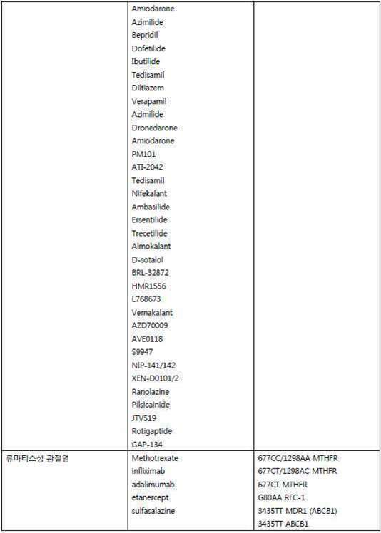
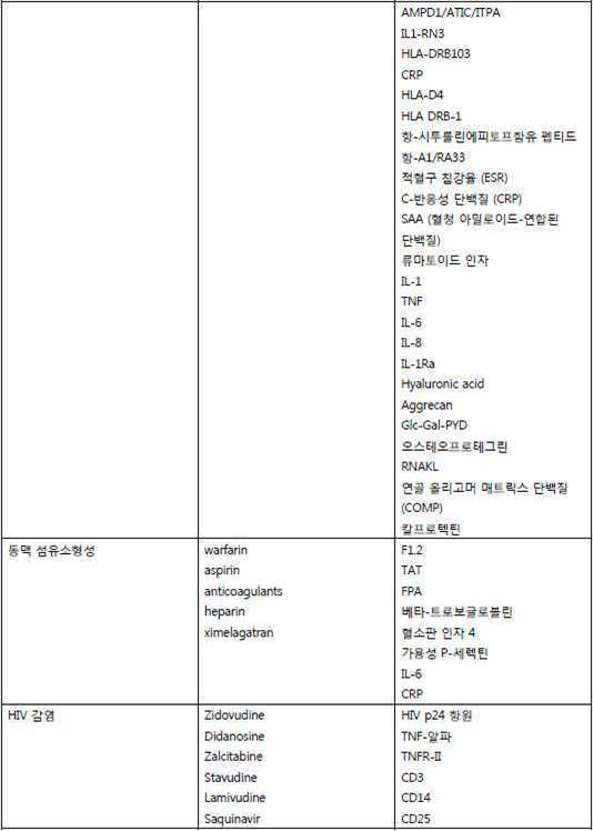

암(Cancer)Cancer
소낭 생체특징을 암의 치료진단에 이용할 수 있는데, 가령, 암을 앓고 있는 피험자가 특정 암 치료에 반응자인지 또는 비-반응자인지를 확인한다. 해당 방법들을 이용하여 여기에서 설명된, 가령 상기 "표현형" 부분의 암을 치료진단할 수 있다. 여기에는 폐암, 비-소 세포 폐암, 소 세포 폐암 (소 세포 암종 (귀리 세포 암), 혼합형 소 세포/거대 세포 암종, 그리고 복합형 소 세포 암종), 결장암, 유방암, 전립선암, 간암, 췌장암, 뇌암, 신장 암, 난소암, 위암, 흑색종, 골 암, 위암, 유방암, 신경교종, 교모세포종, 간세포 암종, 유두 신장 암종, 두경부 편평 세포 암종, 백혈병, 임파종, 골수종, 또는 다른 충실성 종양을 포함하나 이에 한정되지 않는다.The follicular biomolecule characteristics can be used to diagnose cancer, for example, whether a subject suffering from cancer is a respondent or a non-subject to a specific cancer treatment. The methods can be used to diagnose, for example, cancer of the "phenotype" portion described herein. These include lung cancer, non-small cell lung cancer, small cell lung cancer (small cell carcinoma (osteocarcinoma), mixed small cell / giant cell carcinoma and mixed small cell carcinoma), colon cancer, breast cancer, prostate cancer, liver cancer, pancreatic cancer, Neoplasms, glioma, glioma, hepatocellular carcinoma, papillary renal carcinoma, head and neck squamous cell carcinoma, leukemia, lymphoma, myeloma, or other fulminant tumors. But is not limited thereto.
암을 앓고 있는 피험자의 시료내 소낭과 연합된 표식을 포함하는 순환하는 생물지표의 생체 특징을 이용하여 피험자의 후보 치료를 선택할 수 있다. 생체특징은 여기에서 제시한 본 발명의 방법들에 따라 측정할 수 있다. 일부 구체예들에서, 후보 치료는 암의 표준 치료를 포함한다. 이 생체특징을 이용하여 피험자가 특정 치료 또는 표준 치료에 비-반응자인지 또는 반응자인지를 판단할 수 있다. 표준 치료 또는 치료는 암 치료 가령, 방사능요법, 외과술, 화학요법 또는 이의 조합일 수 있다. 이 치료는 치료 가령, 항-암 물질 및 화학요법 섭생일 수 있다. Candidate treatment of the subject can be selected using the biometric characteristics of the circulating biomarkers including the markers associated with the follicles in the sample of the subject suffering from cancer. Biological characteristics can be measured according to the methods of the present invention presented herein. In some embodiments, the candidate treatment includes standard treatment of cancer. This biometric feature can be used to determine whether a subject is a non-respondent or a respondent to a particular treatment or standard treatment. Standard therapy or therapy may be cancer therapy, radiotherapy, surgery, chemotherapy or a combination thereof. This treatment can be, for example, an anti-cancer substance and chemotherapy.
본 발명에 이용하기 위한 암 치료는 표 8에 열거된 것들을 포함하나 이에 한정되지 않는다.Cancer treatments for use in the present invention include, but are not limited to, those listed in Table 8.
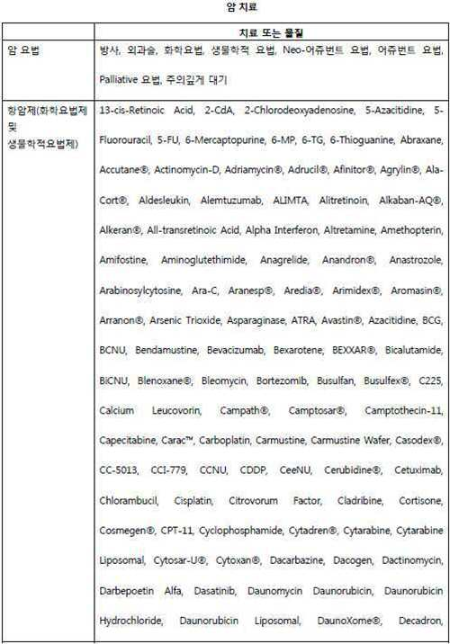


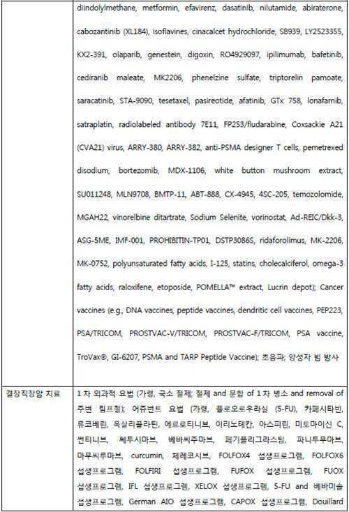
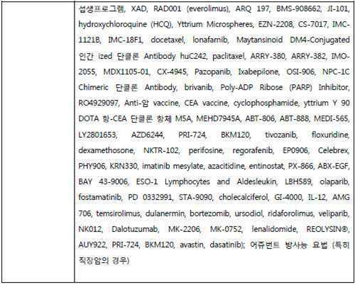
표 8에 나타낸 것과 같이, 암 처리는 다양한 외과적 그리고 치료요법적 처리를 포함한다. 항-암 물질들운 적운 분자들과 생물학제와 같은 약물을 포함한다. 본 발명의 방법들을 이용하여 처리 효과를 모니터링, 피험자를반응자 또는 비-반응자로 분류, 또는 후보 치료 물질을 선택하는 등과 같은 진단치료 목적으로 이용할 수 있는 순환하는 생물지표들을 포함하는 생체특징을 확인할 수 있다. 본 발명을 이용하여 표 8-10에 암 처리와 관련된 치료진단을 포함하나 이에 한정되지 않는 암 처리용 치료 진단을 제공할 수 있다. 본 발명의 방법들에 따른 후보 처리로 확인될 수 있는 암 요법은 표 8-10에 열거된 화학치료 물질들 및 이의 임의의 적절한 조합을 포함하나 이에 한정되지 않는다. 한 구체예에서, 이 처리들은 특이적 유형의 암에 특이적인데, 예를 들면, 표 8에서 전립선 암, 결장직장 암, 유방 암 및 폐암에 대해 열거된 처리와 같이 특이적이다. 다른 구체예들에서, 이 처리들은 이의 기원과 무관하게, 그러나, 표 9-10에 열거한 표식을 포함하는 생체특징을 나타내는 종양에 대해 특이적이다. As shown in Table 8, cancer treatment includes various surgical and therapeutic regimens. Anti-cancer substances include drugs such as opportunistic molecules and biological agents. The methods of the invention can be used to identify biomarkers that include circulating biomarkers that can be used for diagnostic purposes such as monitoring treatment effects, classifying subjects as responders or non-responders, or selecting candidate therapeutic agents have. Using the present invention, Tables 8-10 can provide therapeutic diagnosis for cancer treatment, including, but not limited to, treatment diagnosis related to cancer treatment. Cancer therapies that may be identified by candidate treatment according to the methods of the present invention include, but are not limited to, the chemotherapeutic agents listed in Tables 8-10 and any suitable combination thereof. In one embodiment, these treatments are specific for a specific type of cancer, such as the treatment listed in Table 8 for prostate cancer, colorectal cancer, breast cancer, and lung cancer. In other embodiments, these treatments are specific for tumors irrespective of their origin, but which exhibit biologic features including the markers listed in Tables 9-10.
본 발명은 암 처리를 모니터하는 방법들을 제공하는데, 이 방법들은 시간이 경과함에 따라, 가령, 처리 전, 그리고 처리 후, 또는 처리후 시간이 경과한 후, 피험자에서 일련의 생체특징들을 확인하는 것을 포함한다. 이 생체특징들을 기준과 비교하여 처리의 효과를 결정한다. 한 구체예에서, 이 처리는 가령, 방사능, 외과술, 화학요법, 생물학적 요법, 네오-어쥬번트 요법, 어쥬번트 요법, 또는 주의깊게 관찰하는 것과 같이, 표 8-10에서 선택한다. 기준은 또다른 개별 또는 개별 집단의 것을 수도 있고, 동일한 피험자의 것일 수도 있다. 예를 들면, 암 전-처리를 나타내는 생체특징을 가진 피험자는 성공적인 처리 후 건강상태를 나타내는 생체특징을 가질 수 있다. 역으로, 이 피험자는 성공적이지 못한 처리 후 암을 나타내는 생체특징을 가질 수 있다. 이 생체특징들을 시간의 경과에 따라 비교하여, 피험자의 생체 특징이 상태의 개선, 악화 또는 변화없음을 나타내는 지를 판단할 수 있다. 이 암이 시간이 경과함에 따라 악화되었는지 또는 변화가 없는 지에 대해 추가 처리를 요청할 수 있다. 예를 들면, 외과술 또는 방사능 요법에 추가하여 호르몬 요법을 이용하여, 좀더 공격적인 전립선 암을 치료할 수 있다. 전립선 암 처리의 효과를 모니터하기 위하여 생체 특징에서 다음의 miR중 하나 이상을 이용할 수 있다: hsa-miR-1974, hsa-miR-27b, hsa-miR-103, hsa-miR-146a, hsa-miR-22, hsa-miR-382, hsa-miR-23a, hsa-miR-376c, hsa-miR-335, hsa-miR-142-5p, hsa-miR-221, hsa-miR-142-3p, hsa-miR-151-3p, hsa-miR-21, hsa-miR-16. 다음의 공개문헌에 열거된 하나 이상의 miRs를 이용하여 GI 관의 암 처리를 모니터할 수 있다: Albulescu et al., Tissular and soluble miRNA for diagnostic and therapy improvement in digestive tract cancers, Exp Rev Mol Diag, 11:1, 101-120. The present invention provides methods for monitoring cancer treatment, including identifying a series of biologic features in a subject over time, e.g., before, after, and after treatment, or after a period of time . These biometric features are compared to a reference to determine the effect of the treatment. In one embodiment, the treatment is selected in Tables 8-10, such as, for example, radiation, surgery, chemotherapy, biotherapy, neo-adjuvant therapy, adjuvant therapy, or careful observation. The criteria may be of another individual or individual group, or of the same subject. For example, subjects with biomechanical features representing cancer pre-treatment may have biomechanical characteristics that indicate health status after successful treatment. Conversely, the subject may have biomarkers that indicate cancer after unsuccessful treatment. By comparing these biometric characteristics with the passage of time, it is possible to judge whether the biometric characteristic of the subject indicates improvement, deterioration or no change of the condition. You can ask for further treatment as to whether this cancer has deteriorated over time or has not changed. For example, in addition to surgery or radiation therapy, hormone therapy can be used to treat more aggressive prostate cancer. One or more of the following miRs may be used in the biochemical features to monitor the effect of prostate cancer treatment: hsa-miR-1974, hsa-miR-27b, hsa-miR-103, hsa-miR- MiR-232, hsa-miR-23a, hsa-miR-376c, hsa-miR-335, hsa-miR-142-5p, hsa-miR-221, hsa-miR-142-3p, hsa -mR-151-3p, hsa-miR-21, hsa-miR-16. One or more miRs listed in the following publications can be used to monitor the cancer treatment of the GI tract: Albulescu et al., Tissular and soluble miRNA for diagnostic and therapy improvement in digestive tract cancers, Exp Rev Mol Diag, 1, 101-120 .
일부 구체예들에서, 본 발명은 후보 치료를 선택하기 위하여 피험자의 시료 안에 생체특징을 확인하는 방법을 제공한다. 예를 들면, 이 생체특징은 약물-연합된 표적이 돌연변이된 것인지 또는 차등적으로 발현된 것인지를 나타내고, 이에 의해, 피험자가 특정 처리에 반응할 것인지 또는 반응하지 않는지를 나타낸다. 후보 처리는 항-암 물질들 또는 표 8-10에서 확인된 치료 물질들의 부류로부터 선택할 수 있다. 일부 구체예들에서, 해당 방법들에 따라 확인된 후보 치료는 다음으로 구성된 군으로부터 선택한다: 5-플로오로우라실, 아라레릭스, 아렘투주마브, 아미노글루테티미드, 아나스트로졸, 아스파라기나제, 아스피린, ATRA, 아자씨티딘, 베바시주마브, 벡사로텐, 비칼루티미드, 칼시트리올, 카페시타빈, 카르보플라틴, 세리콕시브, 세투시마브, 화학요법, 콜레칼시페롤, 시스플라틴, 시타라빈, 다사티니브, 다우노루비신, 데시타빈, 독소루비신, 에피루비신, 에르로티니브, 이토포시드, 엑세미스탄, 플루타미드, 풀베스트란트, 게피티니브, 겜시타빈, 고나도레린, 고세레린, 하이드록시우레아, 이마티니브, 이리노테칸, 라파티니브, 레트로졸, 루프로리드, 리포좀성-독소루비신, 메드록시프로게스테론, 메게스트롤, 메게스트롤 아세테이트, 메토트렉세이트, 미토마이신, nab-파클리탈셀, 오트레오티드, 옥사리플라틴, 파클리탁셀, 파니투무마브, 페가스파라가스, 페메트렉시드, 펜토스타틴, 소라페니브, 수리니티브, 탐옥시펜, 탁센, 테모졸로미드, 토레미펜, 트라스투주마브, VBMCP, 및 빈크리스틴.In some embodiments, the invention provides a method for identifying biomarker characteristics in a subject's sample to select a candidate therapy. For example, the biomarker indicates whether the drug-associated target is mutated or differentially expressed, thereby indicating whether the subject will respond to a particular treatment or not. Candidate treatment may be selected from anti-cancer substances or classes of therapeutic substances identified in Tables 8-10. In some embodiments, candidate therapies identified according to the methods are selected from the group consisting of: 5-fluorouracil, ararylx, alemtuzumab, aminoglutethimide, anastrozole, asparaginase, Aspirin, ATRA, azacytidine, bevacizumab, biscarotene, bicalutimide, calcitriol, capecitabine, carboplatin, sericoxib, cetucimab, chemotherapy, cholecalciferol, cisplatin, But are not limited to, but are not limited to, but not limited to, arabinose, lavine, dasatinib, daunorubicin, decitabine, doxorubicin, epirubicin, erlotinib, itoposide, exemestane, flutamide, , Liposomal-doxorubicin, medroxyprogesterone, megestrol, megestrol acetate, methotrexate, mitoglitazone, doxorubicin, medroxyprogesterone, megestrol, methotrexate, methotrexate, The present invention relates to the use of a compound of formula (I) or a pharmaceutically acceptable salt thereof in the manufacture of a medicament for the treatment or prophylaxis of diabetic nephropathy, nephropathy, nab-pachylylthel, otorhodide, oxaplastatin, paclitaxel, panithotamus, Meade, Tremem Pen, Trastuzumab, VBMCP, and Vincristine.
후보 처리를 선택하는 것과 유사하게, 본 발명은 암을 치료할 수 있는지에 대한 판단 방법 또한 제공한다. 예를 들면, 전립선 암은 피험자에게 실질적으로 유해할 것 같지 않은 비-공격적 질환일 수도 있다. 안드로겐 제거 (호르몬 감소)와 함께 방사능 요법은 국소적으로 진행된 전립선 암을 치료하는 표준 방법이다. 호르몬 요법의 병적상태는 발기부전, 안면 홍조, 그리고 성욕의 상실을 포함한다. 추가로, 전립선절제술과 같은 처리는 발기부전 또는 실금과 같은 병적상태를 보유할 수 있다. 따라서, 본 발명은 암이 공격성인지 또는 암의 진행(가령, 단계 또는 등급)을 나타내는 생체특징을 제공한다. 비-공격적 암 또는 국소화된 암은 즉각적인 처리를 요구하지는 않고 오히려, 가령, 전립선 암의"예의주시(watchful waiting)"을 요한다. 반면에, 공격적 또는 진전된 단계 병소는 좀더 공격적 처리 섭생프로그램을 요구할 수 있다. Similar to selecting a candidate treatment, the present invention also provides a method of determining whether cancer is treatable. For example, prostate cancer may be a non-aggressive disease that is not likely to be substantially harmful to the subject. Radioactivity with androgen abstraction (hormone reduction) is the standard method of treating locally advanced prostate cancer. The pathological conditions of hormone therapy include erectile dysfunction, facial flushing, and loss of sexual desire. In addition, treatments such as prostatectomy can have pathological conditions such as impotence or incontinence. Thus, the present invention provides biomolecule characteristics that indicate whether the cancer is aggressive or the progression of cancer (e.g., stage or grade). Non-aggressive cancer or localized cancer does not require immediate treatment, but rather requires, for example, a "watchful waiting" of prostate cancer. On the other hand, aggressive or advanced stage lesions may require a more aggressive treatment regimen.
표 9에서는 탐지할 생물지표들의 예들, 선택할 또는 가급적 피할 치료 물질을 열거한다. 예를 들면, 전립선암을 가진 피험자를 위한 생체특징을 확인하고, 여기에서 이 생체특징은 안드로겐 수용체 (AR)의 수준을 포함한다. AR의 과다발현 또는 과다생산, 가령, 소낭 안에서 높은 수준의 mRNA 수준 또는 단백질 수준은 피험자를 위한 후보 치료를 확인시킨다. 이러한 치료는 이 피험자를 치료하는 물질 가령, 비칼루타미드, 플루타미드, 루프로라이드, 또는 고세레린을 포함한다. 따라서, 이 피험자는 비칼루타미드, 플루타미드, 루프로라이드, 또는 고세레린에 대해 반응자로 확인된다. 또다른 설명을 위한 예에서, BCRP mRNA, 단백질, 또는 이둘 모두는 NSCLC를 앓고 있는 피험자의 소낭에서 높은 수준으로 탐지된다. 그 다음 이 피험자는 물질 Cisplatin 및 Carboplatin 또는 이 피험자에서 NSCLC를 치료하기 위하여 다른 물질보다 덜 효과적으로 간주된다. 다음의 생물지표들중 임의의 것을 피험자로부터 수득한 소낭 안에서 평가할 수 있고, 그리고 이 생물지표는 하나 이상의 핵산, 폴리펩티드, 펩티드 또는 펩티드 모방체를 포함하나 이에 한정되지 않는 형태일 수 있다. 또다른 설명을 위한 실시예에서, 하나 이상의 KRAS, BRAF, PIK3CA, 및/또는 c-KIT에서 돌연변이를 이용하여 후보 치료를 선택할 수 있다. 예를 들면, 환자에서 KRAS 또는 BRAF에서 돌연변이는 세투시마브 및/또는 파니투무마브가 이 환자를 치료하는데 덜 효과적일 것이라는 것을 의미할 수 있다. Table 9 lists examples of biomarkers to be detected, and the therapeutic substances to be selected or avoided as much as possible. For example, we identify biologic features for subjects with prostate cancer, wherein the biologic features include levels of the androgen receptor (AR). Overexpression or overproduction of AR, e.g., high levels of mRNA or protein levels in the follicles, confirm candidate therapy for subjects. Such treatments include the substances that treat this subject, such as bicalutamide, flutamide, loulolides, or sertraline. Thus, the subject is identified as a respondant to bicalutamide, flutamide, looproly, or high serine. In another illustrative example, BCRP mRNA, protein, or both are detected at high levels in the vesicles of subjects suffering from NSCLC. This subject is then considered less effective than the other substances to treat the substance Cisplatin and Carboplatin or NSCLC in this subject. Any of the following biomarkers may be evaluated in a follicle obtained from the subject, and the biomarker may be in a form including, but not limited to, one or more nucleic acids, polypeptides, peptides or peptide mimetics. In another illustrative embodiment, candidate therapies may be selected using mutations in one or more KRAS, BRAF, PIK3CA, and / or c-KIT. For example, a mutation in KRAS or BRAF in a patient may mean that ceftazimab and / or panithothimab will be less effective in treating this patient.
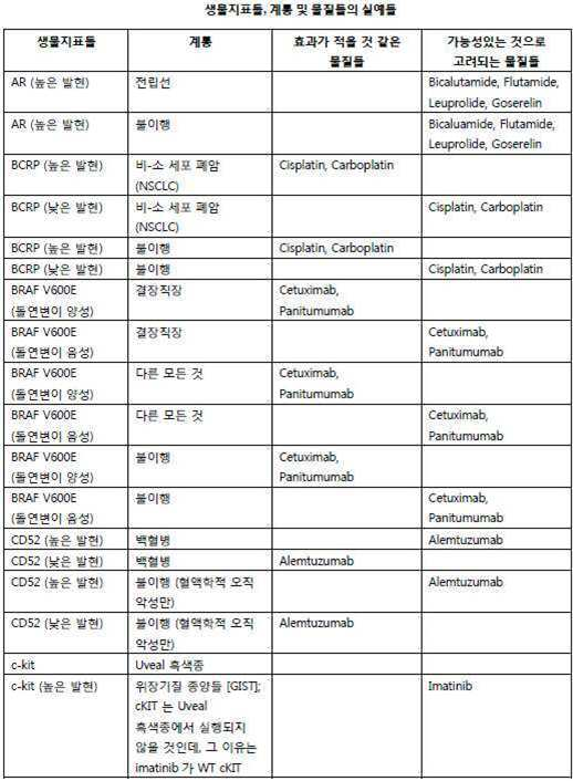


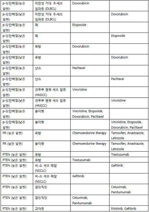
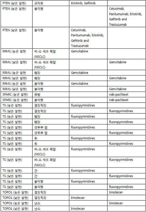
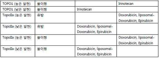
표 10에서는 탐지할 생물지표들의 예들, 선택할 또는 가급적 피할 치료 물질을 열거한다. 예를 들면, 암을 앓고 있는 피험자의 경우, 피험자의 소낭 안에 ADA의 과다 발현의 탐지는 이 피험자는 펜토스타틴에 대해 반응자로 분류되며, 또는 펜토스타틴은 이 피험자를 치료하는데 이용하는 물질로 확인된다. 또다른 실시예에서, 암을 앓고 있는 피험자의 경우, 피험자의 소낭 안에 BCRP의 과다 발현의 탐지는 이 피험자는 시스플라틴, 카르보플라틴, 이리노테칸, 및 토포테칸에 대해 비-반응자로 분류되며, 이는 시스플라틴, 카르보플라틴, 이리노테칸, 및 토포테칸은 이 피험자를 치료하기 위한 준-적합 물질로 확인된다. Table 10 lists examples of biomarkers to be detected, and a list of therapeutic substances to avoid or to avoid. For example, in the case of a subject suffering from cancer, detection of overexpression of ADA in a subject's vesicle may be categorized as responders to pentostatin, or pentostatin may be identified as a substance used to treat this subject. In another embodiment, in the case of a subject suffering from cancer, the detection of overexpression of BCRP in the subject's follicles is categorized as non-responders to cisplatin, carboplatin, irinotecan, and topotecan, , Carboplatin, irinotecan, and topotecan are identified as semi-compatible substances for treating this subject.

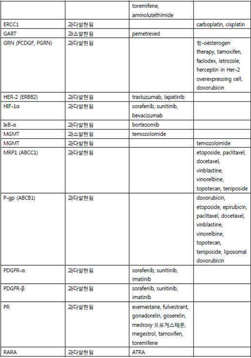

더욱이, 본 발명의 구체예들에서 이용된 약물 연합 및 규칙은 미국 특허 출원 12/658,770, 출원인 (February 12, 2010); 국제 국제 PCT 특허 출원 PCT/US2010/000407, 출원일(February 11, 2010); 국제 PCT 특허 출원 PCT/US2010/54366, 출원일(October 27, 2010); 그리고 미국 가특허 출원 61/427,788, 출원일(December 28, 2010)에서 찾아볼 수 있다; 이들 각각은 전문이 여기에 참고자료로 통합된다. 가령, PCT/US2010/54366의"표 4: 치료 선별을 위한 규칙 요약".Moreover, the drug associations and rules used in embodiments of the present invention are described in U.S. Patent Application 12 / 658,770, Applicant (February 12, 2010); International PCT Patent Application PCT / US2010 / 000407 filed on February 11, 2010; International PCT Patent Application PCT / US2010 / 54366, filed on October 27, 2010; And U.S.
임의의 약물-연관된 표적은 치료진단을 제시하기 위한 생체특징의 일부분일 수 있다. 치료 물질 가령, 작은 분자 또는 생물학적 분자로 조절될 수 있는 표적을 포함하는 "약물화가능한 표적(druggable target)"은 본 발명의 생체특징에 포함시킬 수 있는 후보다. 약물-연관된 표적들은 또한 가령, 표 9와 10에 나타낸 것과 같이, 치료에 저항성을 부여할 수 있는 생물지표들을 포함한다. 이 생체특징은 유전자, 가령, DNA 서열, 및/또는 유전자 산물, 가령, mRNA 또는 단백질, 또는 약물-연관된 표적에 근거할 수 있다. 이러한 핵산 및/또는 폴리펩티드는 존재 또는 부재, 수준 또는 양, 활성, 돌연변이, 서열, 단상형(haplotype), 재배열, 복사체 수, 또는 다른 측정가능한 특징에 적용가능하도록 프로파일될 수 있다. 유전자 또는 유전자 산물은 소낭 집단, 가령, 소낭 표면 지표와 연관될 수 있거나 또는 소낭 페이로드일 수 있다. 한 구체예에서, 본 발명은 암의 치료진단 방법을 제공하는데, 이 방법은 하나 이상의 약물-연관된 표적의 존재 또는 수준을 포함하는 생체특징을 확인하고, 그리고 이 생체특징에 근거하여 후보 치료를 선택하는 것을 포함한다. 약물-연관된 표적은 순환하는 생물지표, 소낭, 또는 소낭연관된 생물지표일 수 있다. 약물 연합된 표적들은 조직 또는 기원 세포와 독립적일 수 있고, 약물-연합된 표적들을 포함하는 생체특징들을 이용하여 CUPS와 같은 기원 불명의 암들을 포함하는 다양한 해부학적 기원으로부터 유래된 암과 같은, 임의의 증식성 질환에 대한 치료진단을 제공할 수 있다. Any drug-related target may be part of a biomarker for presenting a therapeutic diagnosis. A "druggable target " comprising a therapeutic substance, such as a small molecule or a target capable of being regulated by a biological molecule, may be included in the biological characteristics of the present invention. Drug-related targets also include biomarkers that can confer resistance to treatment, such as shown in Tables 9 and 10. The biomarker may be based on a gene, such as a DNA sequence, and / or a gene product, such as an mRNA or protein, or a drug-related target. Such nucleic acids and / or polypeptides may be profiled to be applicable to the presence or absence, level or amount, activity, mutation, sequence, haplotype, rearrangement, number of copies, or other measurable characteristics. The gene or gene product may be associated with a follicular population, such as a follicular surface index, or it may be a follicular payload. In one embodiment, the invention provides a method of therapeutic diagnosis of cancer, comprising identifying a biomarker characteristic comprising the presence or level of one or more drug-associated targets, and selecting a candidate treatment based on the biomarker characteristic . Drug-related targets may be circulating biomarkers, follicles, or follicle-associated biomarkers. The drug-associated targets may be independent of the tissue or origin cell, and may be biochemical, including cancer derived from various anatomical origins, including undigested cancers, such as CUPS, using biometric characteristics, including drug- RTI ID = 0.0 > proliferative < / RTI >
본 발명의 방법들을 이용하여 평가한 약물-연관된 표적들은 ABCC1, ABCG2, ACE2, ADA, ADH1C, ADH4, AGT, AR, AREG, ASNS, BCL2, BCRP, BDCA1, 베타 III 튜블린, BIRC5, B-RAF, BRCA1, BRCA2, CA2, 카베올린, CD20, CD25, CD33, CD52, CDA, CDKN2A, CDKN1A, CDKN1B, CDK2, CDW52, CES2, CK 14, CK 17, CK 5/6, c-KIT, c-Met, c-Myc, COX-2, 사이클린 D1, DCK, DHFR, DNMT1, DNMT3A, DNMT3B, E-캐드헤린, ECGF1, EGFR, EML4-ALK 융합, EPHA2, 에피레귤린, ER, ERBR2, ERCC1, ERCC3, EREG, ESR1, FLT1, 엽산 수용체, FOLR1, FOLR2, FSHB, FSHPRH1, FSHR, FYN, GART, GNA11, GNAO, GNRH1, GNRHR1, GSTP1, HCK, HDAC1, hENT-1, Her2/Neu, HGF, HIF1A, HIG1, HSP90, HSP90AA1, HSPCA, IGF-1R, IGFRBP, IGFRBP3, IGFRBP4, IGFRBP5, IL13RA1, IL2RA, KDR, Ki67, KIT, K-RAS, LCK, LTB, 림포톡신 베타 수용체, LYN, MET, MGMT, MLH1, MMR, MRP1, MS4A1, MSH2, MSH5, Myc, NFKB1, NFKB2, NFKBIA, NRAS, ODC1, OGFR, p16, p21, p27, p53, p95, PARP-1, PDGFC, PDGFR, PDGFRA, PDGFRB, PGP, PGR, PI3K, POLA, POLA1, PPARG, PPARGC1, PR, PTEN, PTGS2, PRPN12, RAF1, RARA, ROS1, RRM1, RRM2, RRM2B, RXRB, RXRG, SIK2, SPARC, SRC, SSTR1, SSTR2, SSTR3, SSTR4, SSTR5, 설바이빈, TK1, TLE3, TNF, TOP1, TOP2A, TOP2B, TS, TUBB3, TXN, TXNRD1, TYMS, VDR, VEGF, VEGFA, VEGFC, VHL, YES1, ZAP70, 또는 이의 임의의 조합을 포함하나 이에 한정되지 않는다. 이들 지표들중 하나 또는 지표들의 조합을 포함하는 생체특징을 이용하여 본 발명에 따른 표현형을 특징화할 수 있는데, 가령, 치료진단을 제공할 수 있다. 이들 지표들은 증식성 질환에 대항하여 다양한 화학요법 물질의 효과에 역할을 하는 것으로 알려져 있다. 따라서, 지표들을 평가하여 암의 기원 또는 유형과는 독립적으로 암에 대한 후보 치료를 선택할 수 있다. 한 구체예에서, 본 발명은 암에 대한 후보 치료를 선택하는 방법을 제공하는데, 이 방법은 하나 이상의 약물 연관된 표적의 수준 또는 존재를 포함하는 생체특징을 확인하고, 그리고 이 생체특징을 가진 환자에 대해 예측되는 효과에 근거하여 후보 치료를 선택하는 것을 포함한다. 하나 이상의 약물-연관된 표적은 상기 언급된 표적들 또는 표 9-10에서 언급된 표적들중 하나일 수 있다. 일부 구체예들에서, 하나 이상의 약물-연관된 표적들의 최소한 2가지, 3가지, 4가지, 5가지, 6가지, 7가지, 8가지, 9가지, 10가지, 12가지, 15가지, 20가지, 25가지, 30가지, 35가지, 40가지, 45가지, 또는 최소한 50가지를 평가한다. 하나 이상의 약물-연관된 표적은 소낭과 연관될 수 있는데, 가령, 소낭 표면 지표 또는 핵산 (가령, DNA, mRNA) 또는 단백질과 같은 소낭 페이로드일 수 있다. 일부 구체예들에서, 하나 이상의 약물-연관된 표적과 상호작용하는 것으로 공지된 microRNA의 존재 또는 수준을 평가하고, 여기에서 하나 이상의 약물-연관된 표적을 억제하는 것으로 알려진 microRNA의 수준이 높다는 것은 하나 이상의 약물-연관된 표적의 발현 수준이 낮음을 나타내고, 따라서 이 약물-연관된 표적에 대항하는 치료에 대해 반응 가능성이 더 낮음을 나타낼 수 있다. 하나 이상의 약물-연관된 표적은 순환하는 생물지표들일 수 있다. 하나 이상의 약물-연관된 표적은 조직 시료 안에서 평가할 수 있다. 예측된 효과는 기준 값에 대한 하나 이상의 약물-연관된 표적의 존재 또는 수준을 비교하여 결정할 수 있는데, 여기에서 기준보다 더 높은 수준은 이 피험자가 반응자일 가능성이 있음을 나타낸다. 예측된 효과는 분류자 알고리즘을 이용하여 결정할 수 있는데, 여기에서 분류자는 후보 치료에 대해 반응자 또는 비-반응자로 공지된 피험자에서 하나 이상의 약물-연관된 표적의 생체특징을 비교하여 훈련받는다. 하나 이상의 약물-연관된 표적과 적합한 후보 표적들의 분자 연합은 표 9-10과 미국 특허 출원 12/658,770, 출원일(February 12, 2010); 국제 PCT 특허 출원 PCT/US2010/000407, 출원일(February 11, 2010); 국제 PCT 특허 출원 PCT/US2010/54366, 출원일(October 27, 2010); 2011년 4월 6일자로 제출된 국제 특허 출원 번호 PCT/US2011/031479, "Circulating Biomakers for Disease"(이들은 전문이 여기에 참고자료로 편입된다); 그리고 U.S. 가특허 출원 61/427,788, 출원일(December 28, 2010)에 나타내며; 이들 각각은 전문이 여기에 참고자료로 통합된다. The drug-related targets evaluated using the methods of the present invention are ABCC1, ABCG2, ACE2, ADA, ADH1C, ADH4, AGT, AR, AREG, ASNS, BCL2, BCRP, BDCA1, Beta III tubulin, BIRC5, , BRCA1, BRCA2, CA2, caveolin, CD20, CD25, CD33, CD52, CDA, CDKN2A, CDKN1A, CDKN1B, CDK2, CDW52, CES2, CK14, CK17, CK5 / EGFR, EML4-ALK fusion, EPHA2, epiregulin, ER, ERBR2, ERCC1, ERCC3, EGFR, EGFR, e-cadherin, c-Myc, COX-2, cyclin D1, DCK, DHFR, DNMT1, DNMT3A, DNMT3B, HGF1, HIF1A, HIF1, HIF1, HIF1, HIF1, HIF1, HIF1, HIF1, IGFRBP3, IGFRBP4, IGFRBP5, IL13RA1, IL2RA, KDR, Ki67, KIT, K-RAS, LCK, LTB, lymphotoxin beta receptor, LYN, MET, MGMT, MLH1, HSP90, HSP90AA1, HSPCA, IGF-1R, IGFRBP, PDGFRB, PGP, PGR, PDGFR, PDGFR, PDGFR, PDGFR, PDGFR, MMR, MRP1, MS4A1, MSH2, MSH5, Myc, NFKB1, NFKB2, NFKBIA, NRAS, ODC1, OGFR, p16, p21, p27, p53, p95, PI3K, POLA, POLA1, PPAR RRM1, RRM1, RRM2, RRM2B, RXRB, RXRG, SIK2, SPARC, SRC, SSTR1, SSTR2, SSTR3, SSTR4, SSTR5, sulbivine, TK1, GST, PPARGC1, PR, PTEN, PTGS2, PRPN12, RAF1, But are not limited to, TLE3, TNF, TOP1, TOP2A, TOP2B, TS, TUBB3, TXN, TXNRD1, TYMS, VDR, VEGF, VEGFA, VEGFC, VHL, YES1, ZAP70 or any combination thereof. Biometric characteristics, including one of these indicators or a combination of indicators, may be used to characterize a phenotype according to the present invention, for example, to provide a therapeutic diagnosis. These indicators are known to play a role in the efficacy of various chemotherapeutic agents against proliferative diseases. Thus, the indicators may be evaluated to select candidate treatment for cancer, independent of the origin or type of cancer. In one embodiment, the invention provides a method of selecting a candidate treatment for a cancer, the method comprising identifying a biomarker comprising a level or presence of one or more drug-related targets, And selecting a candidate treatment based on the predicted effect. One or more drug-related targets may be one of the targets mentioned above or the targets mentioned in Tables 9-10. In some embodiments, at least two, three, four, five, six, seven, eight, nine, ten, twelve, fifteen, twenty, 25, 30, 35, 40, 45, or at least 50. One or more drug-related targets may be associated with the follicles, such as a follicular surface indicator or a follicular payload, such as a nucleic acid (e.g., DNA, mRNA) or protein. In some embodiments, a high level of microRNAs known to inhibit one or more drug-associated targets is used to assess the presence or level of microRNAs known to interact with one or more drug-associated targets, - indicates that the level of expression of the relevant target is low and thus may be less likely to respond to treatment against this drug-related target. One or more drug-related targets may be circulating biomarkers. One or more drug-related targets may be assessed within tissue samples. The predicted effect can be determined by comparing the presence or level of one or more drug-related targets to a reference value, wherein a higher level than the reference indicates that the subject is likely to be a respondent. The predicted effect can be determined using a classifier algorithm wherein the classifier is trained to compare the biometric characteristics of one or more drug-related targets in subjects known to be responders or non-responders to candidate therapy. Molecular associations of one or more drug-associated and suitable candidate targets are shown in Tables 9-10 and US Patent Application 12 / 658,770, filed on February 12, 2010; International PCT Patent Application PCT / US2010 / 000407, filed on February 11, 2010; International PCT Patent Application PCT / US2010 / 54366, filed on October 27, 2010; International Patent Application No. PCT / US2011 / 031479, "Circulating Biomakers for Disease" filed April 6, 2011, which are incorporated herein by reference; And U.S. Is disclosed in the
국제 특허 출원 번호 PCT/US2011/031479, "Circulating Biomakers for Disease"(이들은 전문이 여기에 참고자료로 편입된다)의 표 11은 유전자 및 이에 대응하는 단백질 심볼 그리고 본 발명의 방법들에 따라 분석되는 많은 치료진단 표적들의 이름을 제시한다. 당업자들이 인지할 수 있는 바와 같이, 유전자 및 단백질들은 과학 논문들에서 다수의 대체 이름으로 발달되어왔다. 따라서, 국제 특허 출원 번호 PCT/US2011/031479, "Circulating Biomakers for Disease"(이들은 전문이 여기에 참고자료로 편입된다)의 표 11에 열거된 것들은 설명을 위한 것이며, 완전하게 포괄적으로 편집된 것은 아니다. 유전자의 가명 및 설명의 추가 목록들은 GeneCards? (www.genecards.org), HUGO Gene Nomenclature (www.genenames.org), Entrez Gene (www.ncbi.nlm.nih.gov/entrez/query.fcgi?db=gene), UniProtKB/Swiss-Prot (www.uniprot.org), UniProtKB/TrEMBL (www.uniprot.org), OMIM (www.ncbi.nlm.nih.gov/entrez/query.fcgi?db=OMIM), GeneLoc (genecards.weizmann.ac.il/geneloc/), 및 Ensembl (www.ensembl.org)을 포함하는 온라인 데이터베이스를 이용하면 찾아볼 수 있다. 일반적인으로, 유전자 심볼 및 이름은 HUGO에 의해 승인된 것들에 대응하며, 그리고 단백질 이름은 UniProtKB/Swiss-Prot에서 권장하는 것들이다. 통상의 대체 이름 또한 제시한다. 단백질 이름이 전구물질을 나타낼 경우, 성숙 단백질 또한 내포한다. 출월을 통하여 유전자 및 단백질의 심볼은 서로 호환가능하며, 의미는 필요할 때 본 내용으로부터 유추할 수 있다. Table 11 of International Patent Application No. PCT / US2011 / 031479, "Circulating Biomakers for Disease ", which is incorporated herein by reference, describes genes and their corresponding protein symbols, and many Name treatment diagnosis targets. As will be appreciated by those skilled in the art, genes and proteins have been developed into a number of alternative names in scientific papers. Thus, those listed in Table 11 of International Patent Application No. PCT / US2011 / 031479, "Circulating Biomakers for Disease ", which are incorporated herein by reference, are intended to be illustrative and not to be comprehensively edited . Additional lists of gene pseudonyms and descriptions can be found in GeneCards? (www.genecards.org), HUGO Gene Nomenclature (www.genenames.org), Entrez Gene (www.ncbi.nlm.nih.gov/entrez/query.fcgi?db=gene), UniProtKB / Swiss-Prot .uniprot.org), UniProtKB / TrEMBL (www.uniprot.org), OMIM (www.ncbi.nlm.nih.gov/entrez/query.fcgi?db=OMIM), GeneLoc (genecards.weizmann.ac.il/ by using an online database that contains geneloc /), and Ensembl (www.ensembl.org) can be found. As a general rule, gene symbols and names correspond to those approved by HUGO, and protein names are those recommended by UniProtKB / Swiss-Prot. The usual alternative names are also provided. When a protein name refers to a precursor, it also implies a mature protein. Throughout the coming years, the symbols of genes and proteins are interchangeable, and their meanings can be deduced from the text when necessary.
예를 들면, 비-소 세포 폐암을 앓는 피험자를 위한 치료를 선택할 수 있다. 하나 이상의 생물지표들, 가령, EGFR, 절제 복구 교차-상보성 집단 1 (ERCC1), p53, Ras, p27, class III 베타 튜블린, 유방암 유전자 1 (BRCA1), 유방암 유전자 1 (BRCA2), 그리고 리보뉴클레오티드 환원효소 메신져 1 (RRM1)를 포함하나 이에 한정되지 않는 지표들을 피험자의 소낭으로부터 평가될 수 있다. 하나 이상의 생물지표들의 하나 이상의 특징에 근거하여, 이 피험자가 치료, 가령, 에르로티니브, Carboplatin, Paclitaxel, Gefitinib, 또는 이의 조합을 포함하나 이에 한정되지 않는 치료에 대하여 반응자인지 또는 비-반응자인지를 결정할 수 있다.For example, treatment for non-small cell lung cancer subjects can be selected. (ERCC1), p53, Ras, p27, class III beta-tubulin, breast cancer gene 1 (BRCA1), breast cancer gene 1 (BRCA2), and ribonucleotides Indicators including, but not limited to, reducing enzyme messenger 1 (RRM1) can be assessed from the subject's vesicles. Based on one or more characteristics of one or more biomarkers, the subject can determine whether the subject is a respondent or non-respondent to treatment, including, but not limited to, erlotinib, Carboplatin, Paclitaxel, Gefitinib, You can decide.
또다른 구체예에서, 직장결장암을 앓는 피험자를 위한 치료를 선택할 수 있고, 가령, K-ras을 포함하나 이에 한정되지 않는 생물지표를 피험자의 소낭으로부터 평가할 수 있다. 하나 이상의 생물지표들의 하나 이상의 특징에 근거하여, 이 피험자가 치료, 가령, 파니투무마브, 쎄투시마브 또는 이의 조합을 포함하나 이에 한정되지 않는 치료에 대하여 반응자인지 또는 비-반응자인지를 결정할 수 있다.In another embodiment, treatment can be selected for a subject suffering from rectal colorectal cancer, and biomarkers, including, but not limited to, K-ras, can be assessed from the subject's follicles. Based on one or more characteristics of one or more biomarkers, the subject can determine whether the subject is a respondent or non-respondent to a treatment, including, but not limited to, panitumumab, sertuzimab, or combinations thereof .
또다른 구체예에서, 유방암을 앓는 피험자를 위한 치료를 선택할 수 있고, 가령, HER2, 토포이소메라제 II α, 에스트로겐 수용체, 및 프로게스테론 수용체를 포함하나 이에 한정되지 않는 생물지표를 피험자의 소낭으로부터 평가할 수 있다. 하나 이상의 생물지표들의 하나 이상의 특징에 근거하여, 이 피험자가 치료, 가령, Trastuzumab, Anthracyclines, Ttaxane, Methotrexate, Flurouracil 또는 이의 조합을 포함하나 이에 한정되지 않는 치료에 대하여 반응자인지 또는 비-반응자인지를 결정할 수 있다. In another embodiment, treatment can be selected for a subject suffering from breast cancer, and biomarkers including, but not limited to, HER2, topoisomerase IIa, estrogen receptor, and progesterone receptor, can be assessed from the subject's follicles . Based on one or more characteristics of one or more biomarkers, it may be determined whether the subject is a respondent or non-respondent to treatment, such as, but not limited to, Trastuzumab, Anthracyclines, Ttaxane, Methotrexate, Flurouracil or combinations thereof .
상기에서 설명한 것과 같이, 암을 치료진단하는데 이용되는 생체특징은 단백질 또는 mRNA 또는 microRNA를 포함하는 핵산일 수 있는 하나 이상의 생물지표들의 분석을 포함할 수 있다. 이 생물지표는 체액 안에서 탐지될 수 있으며 및/또는 소낭과 연관될 수 있는데, 가령, 소낭 항원 또는 소낭 페이로드일 수 있다. 설명을 위한 예에서, 이 생체특징을 이용하여 티로신 키나제 억제제에 환자가 반응자 또는 비-반응자인지를 확인한다. 이 생물지표들은 WO/2010/121238, "METHODS AND KIT TO PREDICT THERAPEUTIC OUTCOME OF TYROSIN KINASE INHIBITORS"출원일(April 19, 2010); 또는WO/2009/105223, "SYSTEMS AND METHODS OF CANCER STAGING AND TREATMENT" 출원일(February 19, 2009); 이들 각각은 전문이 여기에 참고자료로 통합된다)에서 설명된 것들일 수 있다. As described above, biomarkers used to therapeutically diagnose cancer may include analysis of one or more biological indicators, which may be nucleic acids, including proteins or mRNA or microRNA. This biomarker can be detected in body fluids and / or can be associated with a follicle, such as a follicular antigen or a follicular payload. In an illustrative example, this biometric feature is used to determine if a patient is a respondent or a non-respondent to a tyrosine kinase inhibitor. These biomarkers are described in WO / 2010/121238, "METHODS AND KIT TO PREDICT THERAPEUTIC OUTCOME OF TYROSIN KINASE INHIBITORS" (April 19, 2010); Or WO / 2009/105223, "SYSTEMS AND METHODS OF CANCER STAGING AND TREATMENT" filed on February 19, 2009; Each of which is incorporated herein by reference).
한 측면에서, 본 발명은 피험자가 티로신 키나제 억제제에 반응을 할지 또는 하지 않을 지를 판단하는 방법을 제공하는데, 이 방법은 피험자로부터 얻은 시료 안의 소낭 집단에서 하나 이상의 생물지표들을 확인하는 것을 포함하고, 여기에서 기준과 비교하여 시료내 하나 이상의 생물지표들의 차등 발현은 이 피험자가 티로신 키나제 억제제에 대해 반응자 또는 비-반응자인지를 나타낸다. 한 구체예에서, 하나 이상의 생물지표는 miR-497을 포함하는데, 여기에서 miR-497의 감소된 발현은 이 피험자가 반응자임을 나타낸다 (가령, 티로신 키나제 억제제에 민감성). 또다른 구체예에서, 하나 이상의 생물지표는 하나 이상의 miR-21, miR-23a, miR-23b, 및 miR-29b을 포함하는데, 여기에서 microRNA의 상향조절은 이 피험자가 비-반응자일 가능성을 나타낸다 (가령, 티로신 키나제 억제제에 저항성). 일부 구체예들에서, 하나 이상의 생물지표는 하나 이상의 hsa-miR-029a, hsa-let-7d, hsa-miR-100, hsa-miR-1260, hsa-miR-025, hsa-let-7i, hsa-miR-146a, hsa-miR-594-Pre, hsa-miR-024, FGFR1, MET, RAB25, EGFR, 키트 및VEGFR2을 포함한다. 또다른 구체예에서, 하나 이상의 생물지표는 FGF1, HOXC10 또는 LHFP를 포함하는데, 여기에서 이 생물지표의 더 높은 발현은 이 피험자가 비-반응자 (가령, 티로신 키나제 억제제에 저항성)임을 나타낸다. 이 방법을 이용하여 티로신 키나제 억제제에 대한 암, 가령, 비-소 세포 폐암 세포, 신장 암 또는 GIST의 감응성을 판단할 수 있다. 티로신 키나제 억제제는 에르로티니브(에르로티니브), 반데타니브(vandetanib), 수니티니브(sunitinib) 및/또는 소라페니브(sorafenib), 또는 유사한 작용 기전에 의해 작용하는 다른 억제제들일 수 있다. 티로신 키나제 억제제는 특이적 또는 비-특이적 방식으로 하나 이상의 티로신 키나제의 작용을 억제하는 임의의 물질을 포함한다. 티로신 키나제 억제제는 직접적으로, 간접적으로, 알로스페릴적으로(allosterically), 또는 임의의 다른 방식으로 티로신 잔기 포스포릴화를 억제하는 작은 분자들, 항체들, 펩티드들, 또는 임의의 적합한 엔터티를 포함한다. 티로신 키나제 억제제들의 특정 예로는 N-(트리플로오르메틸페닐)-5-메틸오소옥사졸-4-카르복사미드, 3-[(2,4-디메틸피롤-5-yl)메틸리데닐)인돌린-2-온, 17-(아릴아미노)-17-데메톡시겔다나마이신, 4-(3-클로로-4- 플로오로페닐아미노)-7-메톡시-6-[3-(4-몰포리닐)프로폭실]퀴나졸린, N-(3-에티닐페닐)-6,7-비스(2-메톡시에톡시)-4-퀴나졸린아민, BIBX1382, 2,3,9,10,11,12- 헥사하이드로-10-(하이드록시메틸)-10-하이드록시-9-메틸-9,12-에폭시-1H-하이드로[1,2,3-fg:3',2',1'-kl]피롤로[3,4-i][l,6]벤조디아조신-1-온, SH268, 게니스테인, STI571, CEP2563, 4-(3-클로로페닐아미노)-5,6-디메틸-7H-피롤로[2,3-d]피리미딘메탄 술포네이트, 4-(3-브로모-4-하이드록시페닐)아미노-6,7-디메톡시퀴나졸린, 4-(4'-하이드록시페닐)아미노-6,7-디메톡시퀴나졸린, SU6668, STI571A, N-4-클로로페닐-4-(4-피리딜메틸)-1-프탈진아민, N-[2-(디에틸아미노)에틸]-5-[(Z)-(5-플로오로-1,2-디하이드로-2-옥소-3H-인돌-3-일리덴)메틸]-2,4-디메틸-1H-피롤-3-카르복사미드 (일반적으로 수니티니브(sunitinib)로 공지됨), A-[A-[[4-클로로-3(트리플로오르메틸)페닐]카르바모일아미노]페녹시]-N-메틸-피리딘-2-카르복사미드 (일반적으로 소라페니브(sorafenib)로 공지됨), EMD121974, 그리고 N-(3-에티닐페닐)-6,7- 비스(2-메톡시에톡시)퀴나졸린-4-아민(일반적으로 에르로티니브(에르로티니브)로 공지됨). 일부 구체예들에서, 티로신 키나제 억제제는 상피 성장 인자 수용체 (EGFR), VEGFR, PDGFR 베타, 및/또는 FLT3에서 억제제 활성을 보유한다. In one aspect, the invention provides a method of determining whether a subject will or will not respond to a tyrosine kinase inhibitor, comprising identifying one or more biomarkers in a follicular population in a sample from a subject, The differential expression of one or more biomarkers in the sample relative to the baseline indicates that the subject is a responding or non-responding to a tyrosine kinase inhibitor. In one embodiment, the at least one biomarker comprises miR-497, wherein reduced expression of miR-497 indicates that the subject is a responder (e. G., Susceptible to tyrosine kinase inhibitors). In another embodiment, the at least one biomarker comprises one or more miR-21, miR-23a, miR-23b, and miR-29b wherein upregulation of the microRNA indicates the possibility that the subject is a non- (E. G., Resistant to tyrosine kinase inhibitors). In some embodiments, the one or more biomarkers include one or more of hsa-miR-029a, hsa-let-7d, hsa-miR-100, hsa-miR-1260, hsa- -mR-146a, hsa-miR-594-Pre, hsa-miR-024, FGFR1, MET, RAB25, EGFR, kit and VEGFR2. In another embodiment, the at least one biomarker comprises FGF1, HOXC10 or LHFP, wherein higher expression of the biomarker indicates that the subject is a non-responder (e.g., resistant to a tyrosine kinase inhibitor). This method can be used to determine the susceptibility of a tumor to a tyrosine kinase inhibitor, such as non-small cell lung cancer cells, kidney cancer or GIST. Tyrosine kinase inhibitors may be erlotinib (erlotinib), vandetanib, sunitinib and / or sorafenib, or other inhibitors that act by similar mechanism of action. Tyrosine kinase inhibitors include any substance that inhibits the action of one or more tyrosine kinases in a specific or non-specific manner. Tyrosine kinase inhibitors include small molecules, antibodies, peptides, or any suitable entity that inhibits tyrosine residue phosphorylation either directly, indirectly, allosterically, or in any other way . Specific examples of tyrosine kinase inhibitors include N- (trifluoromethylphenyl) -5-methyloxooxazole-4-carboxamide, 3- [(2,4-dimethylpyrrol-5-yl) methylidenyl) indoline (3-chloro-4-fluorophenylamino) -7-methoxy-6- [3- (4-morpholino) (3-ethynylphenyl) -6,7-bis (2-methoxyethoxy) -4-quinazoline amine, BIBX1382, 2,3,9,10,11, Hydroxymethyl-9,12-epoxy-1H-hydro [1,2,3-fg: 3 ', 2', 1'-kl ] Pyrrolo [3,4-i] [1,6] benzodiazocin-1-one, SH268, genistein, STI571, CEP2563, 4- (3- chlorophenylamino) -5,6-dimethyl- Pyrrolo [2,3-d] pyrimidine methanesulfonate, 4- (3-bromo-4-hydroxyphenyl) amino-6,7-dimethoxyquinazoline, 4- Amino-6,7-dimethoxyquinazoline, SU6668, STI571A, N-4-chlorophenyl-4- (4- pyridylmethyl) (Z) - (5-fluoro-1,2-dihydro-2-oxo-3H-indol-3-ylidene) methyl] -2 (Known as sunitinib), A- [A - [[4-chloro-3- (trifluoromethyl) phenyl] carbazole (Commonly known as sorafenib), EMD121974, and N- (3-ethynylphenyl) -6,7-bis (2-methoxyethoxy) quinazolin-4-amine (commonly known as erlotinib (erlotinib)). In some embodiments, the tyrosine kinase inhibitor possesses inhibitor activity in the epithelial growth factor receptor (EGFR), VEGFR, PDGFR beta, and / or FLT3.
따라서, 본 발명의 방법들에 의해 확인된 생체특징에 근거하여, 암을 앓고 있는 피험자를 위한 치료를 선택할 수 있다. 따라서, 이 생체특징은 microRNA, 소낭, 또는 임의의 유용한 소낭 연관된 생물지표를 포함하는 순환하는 생물지표의 존재 또는 수준을 포함할 수 있다. Thus, based on the biometric characteristics identified by the methods of the present invention, treatment for a subject suffering from cancer can be selected. Thus, the biomarker may include the presence or level of a circulating biomarker comprising a microRNA, a vesicle, or any useful vesicle-associated biomarker.
본 발명의 이 방법을 이용하여, 심혈관 질환, 신경성 질환 및 장애들, 면역 질환및 장애들 그리고 감염성 질환을 포함하는 기타 질환의 치료진단에 이용될 수 있는 생물지표들은 2011년 4월 6일자로 제출된 국제 특허 출원 번호 PCT/US2011/031479, "Circulating Biomakers for Disease"(이들은 전문이 여기에 참고자료로 편입된다)에 설명된다. Using this method of the present invention, biomarkers that may be used in the therapeutic diagnosis of cardiovascular diseases, neurological diseases and disorders, immune diseases and disorders, and other diseases including infectious diseases are submitted as of April 6, 2011 International Patent Application No. PCT / US2011 / 031479, "Circulating Biomakers for Disease ", which are incorporated herein by reference in their entirety.
소낭 조성물들Follicular compositions
특정 생체특징을 가진 단리된 소낭을 또한 여기에서 제공한다. 단리된 소낭은 특이적 세포 유형에 특이적인, 또는 상기에서 설명된 표현형을 특징화하기 위한 하나 이상의 생물지표들을 포함할 수 있다. Isolated follicles with specific biometric characteristics are also provided herein. The isolated follicles may comprise one or more biomarkers specific for a particular cell type, or for characterizing the phenotype described above.
단리된 소낭은 또한 하나 이상의 생물지표들을 포함할 수 있는데, 여기에서 하나 이상의 생물지표들의 발현 수준은 정상 세포 (가령, 관심 대상의 표현형이 없는 피험자로부터 유도된 세포)로부터 유도된 단리된 소낭과 비교하여 더 높거나, 더 낮거나 또는 동일하다. 예를 들면, 단리된 소낭은 B7H3, PSCA, MFG-E8, Rab, STEAP, PSMA, PCSA, 5T4, miR-9, miR-629, miR-141, miR-671-3p, miR-491, miR-182, miR-125a-3p, miR-324-5p, miR-148b, 및 miR-222로 구성된 군으로부터 선택된 하나 이상의 생물지표들을 포함할 수 있고, 여기에서 하나 이상의 생물지표들의 발현 수준은 정상 세포로부터 유도된 것들과 비교하여 단리된 소낭에서 더 높다. 단리된 소낭은 집단으로부터 선택한 최소한 2가지, 3가지, 4가지, 5가지, 6가지, 7가지, 8가지, 9가지, 10가지, 11가지, 13가지, 14가지, 15가지, 16가지, 17가지, 18가지, 또는 19가지 생물지표들을 포함할 수 있다. 단리된 소낭은 EpCam, B7H3, PSMA, PSCA, PCSA, CD63, CD59, CD81, 또는 CD9으로 구성된 군으로부터 선택된 하나 이상의 생물지표들을 더 포함할 수 있다.Isolated follicles may also include one or more biomarkers, wherein the level of expression of one or more biomarkers is compared to isolated follicles derived from normal cells (e. G., Cells derived from a subject without a phenotype of interest) Higher, lower, or the same. For example, the isolated follicles can be selected from the group consisting of B7H3, PSCA, MFG-E8, Rab, STEAP, PSMA, PCSA, 5T4, miR-9, miR-629, miR- 141, miR-671-3p, miR- 182, miR-125a-3p, miR-324-5p, miR-148b, and miR-222, wherein the expression levels of one or more biomarkers are determined from normal cells Lt; RTI ID = 0.0 > isolated < / RTI > The isolated follicles were selected from groups of at least 2, 3, 4, 5, 6, 7, 8, 9, 10, 11, 13, 14, 15, 17, 18, or 19 biomarkers. The isolated follicles may further comprise one or more biomarkers selected from the group consisting of EpCam, B7H3, PSMA, PSCA, PCSA, CD63, CD59, CD81, or CD9.
단리된 소낭은 생물지표들 PCSA, EpCam, CD63, 그리고 CD8; 이 생물지표들 PCSA, EpCam, B7H3 그리고 PSMA을 포함할 수 있다. 단리된 소낭은 생물지표들 miR-9, miR-629, miR-141, miR-671-3p, miR-491, miR-182, miR-125a-3p, miR-324-5p, miR-148b, 및 miR-222를 포함할 수 있다.Isolated follicles are biomarkers such as PCSA, EpCam, CD63, and CD8; These biomarkers can include PCSA, EpCam, B7H3, and PSMA. The isolated follicles contain the biomarkers miR-9, miR-629, miR-141, miR-671-3p, miR-491, miR-182, miR-125a-3p, miR- miR-222. < / RTI >
단리된 소낭을 포함하는 조성물을 또한 여기에서 제공한다. 이 조성물은 하나 이상의 단리된 소낭을 포함할 수 있다. 예를 들면, 이 조성물은 다수의 소낭, 또는 소낭의 하나 이상의 집단을 포함할 수 있다. Compositions comprising isolated follicles are also provided herein. The composition may comprise one or more isolated fibrils. For example, the composition may comprise a plurality of follicles, or one or more populations of follicles.
이 조성물은 소낭에 대해 실질적으로 농축된(enriched) 것일 수 있다. 예를 들면, 이 조성물은 세포성 찌꺼기, 세포들, 또는 비-엑소좀 단백질들, 펩티드들, 또는 핵산 (가령, 소낭 안에 포함되지 않은 생물학적 분자들)을 실질적으로 가지고 있지 않다. 세포성 찌꺼기, 세포들, 또는 비-엑소좀 단백질들, 펩티드들, 또는 핵산은 소낭과 함께 생물학적 시료내에 존재할 수 있다. 세포성 찌꺼기, 세포들, 또는 비-엑소좀 단백질들, 펩티드들, 또는 핵산 (가령, 소낭 안에 포함되지 않은 생물학적 분자들)이 실질적으로 없는 조성물은 여기에서 설명된 임의의 방법, 가령, 하나 이상의 소낭에 대한 하나 이상의 결합 물질 또는 캡쳐 물질을 이용하여 수득할 수 있다. 이 소낭은 전체 조성물의 중량 또는 양의 최소한 30%, 40%, 50%, 60%, 70%, 80%, 90%, 95% 또는 99%를 포함할 수 있다. 이 조성물의 소낭은 이종 동종 소낭 집단일 수 있다. 예를 들면, 동종 소낭의 집단은 하나 이상의 성질들 또는 특징들에 대해 동종인 소낭을 포함한다. The composition may be substantially enriched with respect to the follicle. For example, the composition is substantially free of cellular debris, cells, or non-exosome proteins, peptides, or nucleic acids (e.g., biological molecules not contained in vesicles). Cellular debris, cells, or non-exosome proteins, peptides, or nucleic acids may be present in the biological sample with the follicle. Compositions that are substantially free of cellular debris, cells, or non-exosomal proteins, peptides, or nucleic acids (e.g., biological molecules not contained in vesicles) may be administered by any of the methods described herein, One or more binding agents or capture materials for the follicles. The follicle may comprise at least 30%, 40%, 50%, 60%, 70%, 80%, 90%, 95% or 99% of the weight or amount of the total composition. The follicles of this composition may be a heterogeneous allograft population. For example, a population of allogeneic vesicles contains vesicles homologous to one or more properties or features.
따라서, 일부 구체예들에서, 이 조성물은 실질적으로 농축된(enriched) 소낭 집단을 포함한다. 이 조성물은 하나 이상의 성질들 또는 특징들에 대해 최소한 30%, 40%, 50%, 60%, 70%, 80%, 90%, 95% 또는 99% 동종인 소낭 집단에 대해 농축될 수 있다. 예를 들면, 하나 이상의 특징들은 하나 이상의 동일한 생물지표들, 동일한 세포 유형으로부터 유도된 실질적으로 유사한 또는 동일한 생체특징, 특정 크기의 소낭 그리고 이의 조합으로 구성된 군으로부터 선택될 수 있다. 예를 들면, 하나 이상의 특징들은 하나 이상의 동일한 생물지표들, 동일한 세포 유형으로부터 유도된 실질적으로 유사한 또는 동일한 생체특징, 특정 크기의 소낭 그리고 이의 조합으로 구성된 군으로부터 선택될 수 있다. 예를 들면, 소낭의 집단은 특정 생체특징을 가지고, 동일한 생물지표를 가지고, 동일한 생물지표 조합을 가지고, 또는 동일한 세포 유형으로부터 유도됨으로써 동종일 수 있다. 일부 구체예들에서, 이 조성물은 가령, 특이적 생체특징을 가진, 특이적 세포로부터 유도된 집단, 또는 이둘 모두인 실질적으로 동종의 소낭 집단을 포함한다.Thus, in some embodiments, the composition comprises a population of vesicles that is substantially enriched. The composition may be concentrated for a population of follicles that is at least 30%, 40%, 50%, 60%, 70%, 80%, 90%, 95% or 99% homologous to one or more properties or characteristics. For example, the one or more features may be selected from the group consisting of one or more of the same biomarkers, substantially similar or identical biomarkers derived from the same cell type, a particular size of vesicle, and combinations thereof. For example, the one or more features may be selected from the group consisting of one or more of the same biomarkers, substantially similar or identical biomarkers derived from the same cell type, a particular size of vesicle, and combinations thereof. For example, a population of follicles can be homologous by having the same biomarker, with the same biomarker, with the same biomarker combination, or derived from the same cell type. In some embodiments, the composition includes, for example, a population derived from a specific cell, with a specific biomarker, or a population of substantially homogeneous populations, both of which are specific.
소낭의 집단은 하나 이상의 동일한 생물지표들을 포함할 수 있다. 이 생물지표는 임의의 성분 가령, 임의의 핵산 (가령 RNA 또는 DNA), 단백질, 펩티드, 폴리펩티드, 항원, 지질, 탄수화물, 또는 프로테오글리칸과 같은 임의의 성분일 수 있다. 예를 들면, 집단에서 각 소낭은 동일한 또는 똑같은 하나 이상의 생물지표들을 포함할 수 있다. 일부 구체예들에서, 각 소낭은 동일한 1개, 2개, 3개, 4개, 5개, 6개, 3개, 4개, 5개, 6개, 7개, 8개, 9개, 10개, 11개, 12개, 13개, 14개, 15개, 16개, 17개, 18개, 19개, 20개, 25개, 50개, 75개 또는 100개 생물지표들을 포함한다. A population of follicles may contain one or more identical biomarkers. This biomarker can be any component, such as any component, such as any nucleic acid (such as RNA or DNA), protein, peptide, polypeptide, antigen, lipid, carbohydrate, or proteoglycan. For example, in a population, each follicle may contain one or more biomarkers that are the same or identical. In some embodiments, each vial has the same one, two, three, four, five, six, three, four, five, six, seven, eight, nine, 12, 13, 14, 15, 16, 17, 18, 19, 20, 25, 50, 75 or 100 biomarkers.
동일한 또는 똑같은 생물지표를 포함하는 이 소낭 집단은 생물지표의 존재 또는 부재, 발현 수준, 돌연변이 상태, 또는 변형이 동일한 집단내 각 소낭을 포함할 수 있다. 예를 들면, 농축된(enriched) 소낭의 집단은 각 소낭이 동일한 생물지표를 가지고, 동일한 생물지표를 가지지 않고, 동일한 발현 수준의 생물지표를 가지고, 동일하게 변형된 생물지표를 가지거나, 또는 동일한 돌연변이된 생물지표를 가지는 소낭을 포함할 수 있다. 동일한 발현 수준의 생물지표는 정량적 또는 정성적 측정을 말하는데, 가령, 집단내 소낭은 기준 수준과 비교하여 생물지표들의 동일한 발현 수준 또는 과다발현 또는 과소발현 수준을 가진다. These follicle populations, which contain the same or the same biomarkers, can include each vesicle in the same population with the presence or absence of biomarkers, level of expression, mutation status, or variant. For example, the population of enriched follicles can be determined such that each follicle has the same biomarker, does not have the same biomarkers, has biomarkers of the same expression level, has the same biomarker, Lt; RTI ID = 0.0 > mutated < / RTI > biomarkers. Biomarkers of the same expression level refer to quantitative or qualitative measurements, for example, follicles within a population have the same expression level or overexpression or underexpression level of biomarkers as compared to baseline levels.
대안으로, 생물지표들의 동일한 발현 수준은 집단내 각 소낭에 대해 유사한 생물지표들의 발현을 나타내는 수치값일 수 있다. 예를 들면, miRNA의 복사체 수, 단백질의 양 또는 mRNA의 수준이 집단내 각 소낭에서 정량적으로 유사할 수 있고, 각 소낭의 수치는 이러한 변이가 적합할 때 집단내 각 소낭의 양으로부터 ± 1%, 2%, 3%, 4%, 5%, 6%, 7%, 8%, 9%, 10%, 15%, 또는 20% 일 수 있다. Alternatively, the same expression level of biomarkers may be a numerical value representing the expression of similar biomarkers for each vesicle in the population. For example, the number of miRNAs, the amount of protein, or the level of mRNA may be quantitatively similar in each follicle in a population, and the value of each follicle should be within ± 1% of the amount of each follicle in the population, , 2%, 3%, 4%, 5%, 6%, 7%, 8%, 9%, 10%, 15% or 20%.
일부 구체예들에서, 이 조성물은 실질적으로 농축된(enriched) 소낭 집단을 포함하며, 여기에서 농축된(enriched) 집단내 소낭은 실질적으로 유사한 또는 똑같은 생체특징을 가진다. 이 생체특징은 소낭의 하나 이상의 특징, 가령, 소낭의 수준 또는 양, 소낭 반감기내에서 변이의 일시적 평가, 순환하는 소낭 반감기, 소낭의 대사 반감기, 또는 소낭의 활성을 포함한다. 생체특징은 또한 생물지표, 가령, 여기에서 설명된 생물지표들의 존재 또는 부재, 발현 수준, 돌연변이 상태 또는 변형을 포함할 수 있다. In some embodiments, the composition comprises a population of substantially enriched vesicles, wherein the vesicles in the population enriched have substantially similar or identical biologic characteristics. These biomarkers include one or more characteristics of the vesicle, such as the level or amount of vesicle, a temporal evaluation of the variation within the vesicle half-life, circulating vesicle half-life, vesicle half-life, or activity of the vesicle. Biometric characteristics may also include biological indicators, such as the presence or absence of biomarkers described herein, expression levels, mutation status or modifications.
집단내 각 소낭의 생체특징은 최소한 30%, 40%, 50%, 60%, 70%, 80%, 90%, 95%, 또는99% 똑같다. 일부 구체예들에서, 각 소낭의 생체특징은 100% 똑같다. 농축된(enriched) 집단내 각 소낭의 생체특징은 동일한 1개, 2개, 3개, 4개, 5개, 6개, 3개, 4개, 5개, 6개, 7개, 8개, 9개, 10개, 11개, 12개, 13개, 14개, 15개, 16개, 17개, 18개, 19개, 20개, 25개, 50개, 75개 또는 100개 특징들을 가질 수 있다. 예를 들면, 농축된(enriched) 집단내 소낭의 생체특징은 제 1 생물지표의 존재, 제 2 생물지표의 존재, 그리고 제 3 생물지표의 과소발현일 수 있다. 동일한 집단내 또다른 소낭은 존재하는 동일한 제 1 및 제 2 생물지표들을 보유하고, 제 3 생물지표들의 과소발현인, 100% 동일할 수 있다. 대안으로, 동일한 집단내 소낭은 동일한 제 1 및 제 2 생물지표를 보유하지만, 제 3 생물지표들의 과소발현은 보유하지 않을 수 있다. At least 30%, 40%, 50%, 60%, 70%, 80%, 90%, 95%, or 99% of the vital characteristics of each follicle in the population are the same. In some embodiments, the vital character of each follicle is 100% identical. The biochemical characteristics of each follicle in the enriched population are the same 1, 2, 3, 4, 5, 6, 3, 4, 5, 6, 7, Having 9, 10, 11, 12, 13, 14, 15, 16, 17, 18, 19, 20, 25, 50, 75 or 100 features . For example, the vital characteristics of the follicles in the enriched population may be the presence of a first biomarker, the presence of a second biomarker, and an underexpression of a third biomarker. Another follicle in the same population may have the same first and second biological indicators present and be 100% identical, an under-expression of the third biological indicators. Alternatively, the follicles in the same population may have the same first and second biomarkers but not the under-expression of the third biomarkers.
일부 구체예들에서, 이 조성물은 실질적으로 농축된(enriched) 소낭 집단을 포함하는데, 여기에서 이 소낭은 동일한 세포 유형으로부터 유도된다. 예를 들면, 이 소낭은 특이적 조직의 세포들, 관심대상의 특이적 종양의 세포들 또는 관심대상의 질환 조직의 세포들, 순환하는 종양 세포들, 또는 모계 또는 태아 기원의 세포들로부터 모두 유도될 수 있다. 이 소낭은 종양 세포들로부터 모두 유도될 수 있다. 이 소낭은 폐, 췌장, 위, 소장, 방광, 신장, 난소, 고환, 피부, 결장직장, 유방, 전립선, 뇌, 식도, 간, 태반, 또는 태아 세포들을 포함하나 이에 한정되지 않는 동일한 조직 또는 세포로부터 모두 유도될 수 있다. In some embodiments, the composition comprises a population of substantially enriched vesicles wherein the vesicles are derived from the same cell type. For example, the follicles can be derived from cells of a specific tissue, cells of a specific tumor of interest or cells of diseased tissue of interest, circulating tumor cells, or cells of maternal or fetal origin . These follicles can all be derived from tumor cells. The follicle may be the same tissue or cell including but not limited to lung, pancreas, stomach, small intestine, bladder, kidney, ovary, testicle, skin, colon rectum, breast, prostate, brain, esophagus, liver, placenta, Respectively.
실질적으로 농축된(enriched) 소낭의 집단을 포함하는 이 조성물은 특정 크기의 소낭을 또한 포함할 수 있다. 예를 들면, 이 소낭은 약 10, 20, 또는 30 이상의 직경을 모두 가질 수 있다. 이들은 모두 약 10-1000, 예를 들면, 약 30-800, 약 30-200, 또는 약 30-100의 직경을 보유할 수 있다. 일부 구체예들에서, 이 소낭은 모두 10,000, 1000, 800, 500, 200, 100 또는 50 미만의 직경을 보유할 수 있다. This composition comprising a population of substantially enriched follicles may also comprise a particular size of follicle. For example, the vesicle may have a diameter of about 10, 20, or 30 or more. They can all have diameters of about 10-1000, such as about 30-800, about 30-200, or about 30-100. In some embodiments, the vesicles may all have diameters of 10,000, 1000, 800, 500, 200, 100 or less.
하나 이상의 특징들에 대해 동종 집단의 소낭은 조성물의 최소한 약 30%, 40%, 50%, 60%, 70%, 80%, 90%, 95%, 또는 99%를 포함할 수 있다. 일부 구체예들에서,실질적으로 농축된(enriched) 소낭의 집단을 포함하는 조성물은 이 조성물이 유도된 생물학적 시료내 소낭의 농도와 비교하여 소낭의 농도가 최소한 2, 3, 4, 5, 10, 20, 25, 50, 100, 250, 500, 또는 1000배인 소낭을 포함한다. 추가 다른 구체예들에서, 이 조성물은 소낭의 제 2 농축된(enriched) 집단을 더 포함할 수 있는데, 여기에서 소낭의 집단은 여기에서 설명된 하나 이상의 특징들에 대해 최소한 30% 동종이다. For one or more of the features, a homologous follicle may comprise at least about 30%, 40%, 50%, 60%, 70%, 80%, 90%, 95%, or 99% of the composition. In some embodiments, a composition comprising a population of substantially enriched follicles is characterized in that the concentration of the follicle is at least 2, 3, 4, 5, 10, or even 10 times greater than the concentration of the follicle in the biological sample from which the composition is derived. 20, 25, 50, 100, 250, 500, or 1000 times. In further embodiments, the composition may further comprise a second enriched population of follicles wherein the population of follicles is at least 30% homologous to one or more of the characteristics described herein.
다중 분석을 이용하여 하나 이상의 소낭 집단, 가령, 최소한 2개, 3개, 4개, 5개, 6개, 7개, 8개, 9개, 10개 소낭, 집단에 대해 실질적으로 농축된 조성물을 수득할 수 있다. 각 실질적으로 농축된(enriched) 소낭 집단은 질량 또는 양에 근거하여 조성물의 최소한 5%, 10%, 15%, 20%, 25%, 30%, 35%, 40%, 45%, 46%, 47%, 48%, 또는 49%를 포함할 수 있다. 일부 구체예들에서, 실질적으로 농축된(enriched) 소낭 집단은 질량 또는 양에 근거하여 조성물의 최소한 약 30%, 40%, 50%, 60%, 70%, 80%, 90%, 95%, 또는 99%를 포함한다. Using multiple assays, a substantially concentrated composition for at least one follicular population, such as at least 2, 3, 4, 5, 6, 7, 8, 9, 10 follicles, . Each substantially enriched population of follicles comprises at least 5%, 10%, 15%, 20%, 25%, 30%, 35%, 40%, 45%, 46% 47%, 48%, or 49%. In some embodiments, the substantially enriched follicle population comprises at least about 30%, 40%, 50%, 60%, 70%, 80%, 90%, 95%, 90%, 90% Or 99%.
실질적으로 농축된(enriched) 소낭의 집단은 여기에서 설명된 하나 이상의 방법들, 프로세스들 또는 시스템을 이용하여 수득할 수 있다. 예를 들면, 시료로부터 소낭 집단의 분리는 소낭의 하나 이상의 생물지표들에 대한 결합 물질, 가령, 소낭의 두 개 이상의 생물지표들을 표적으로 하는 두 개 이상의 결합 물질을 이용하여 실행할 수 있다. 하나 이상의 캡쳐 물질을 이용하여 소낭의 실질적으로 농축된(enriched) 집단을 수득할 수 있다. 하나 이상의 탐지 물질을 이용하여 실질적으로 농축된(enriched) 소낭 집단을 확인할 수 있다. A group of substantially enriched vesicles can be obtained using one or more methods, processes or systems described herein. For example, separation of a follicular population from a sample can be carried out using a binding agent for one or more biological indicators of the follicle, for example, two or more binding agents targeting two or more biological indicators of the follicle. One or more capture materials can be used to obtain a substantially enriched population of follicles. One or more detection materials can be used to identify a group of fungi that are substantially enriched.
한 구체예에서, 특정 생체특징을 가진 소낭 집단은 이 생체특징의 생물지표들에 대한 하나 이상의 결합 물질을 이용하여 수득한다. 이 소낭은 단리하면, 특정 생체특징을 가진 실질적으로 농축된 소낭 집단을 포함하는 조성물을 얻는다. 또다른 구체예에서, 관심 대상의 특정 생체특징을 가진 소낭의 집단은 관심 대상의 생체특징의 성분이 아닌 생물지표들에 대한 하나 이상의 결합 물질을 이용하여 수득할 수 있다. 따라서, 결합물질을 이용하여 관심대상의 생체특징을 보유하지 않은 소낭을 제거하고, 따라서 생성된 조성물은 관심대상의 특정 생체특징을 가진 실질적으로 농축된(enriched) 소낭의 집단이다. 생성된 조성물은 결합 물질에 대한 생물지표들을 포함하는 소낭이 실질적으로 없다. In one embodiment, a population of vesicles with specific biomarkers is obtained using one or more binding substances for biomarkers of the biomarker. This follicle is isolated to obtain a composition comprising a substantially enriched population of follicles with specific biomarker characteristics. In another embodiment, a population of follicles with specific biomarker features of interest can be obtained using one or more binding agents for biomarkers that are not components of the biomarker of interest. Thus, the binding material is used to remove follicles that do not retain the biologic features of interest, and thus the resulting composition is a group of substantially enriched follicles with specific biomarkers of interest. The resulting composition is substantially free of vesicles containing biomarkers for the binding agent.
2011년 4월 6일자로 제출된 국제 특허 출원 번호 PCT/US2011/031479, "Circulating Biomakers for Disease"(이들은 전문이 여기에 참고자료로 편입된다). International Patent Application No. PCT / US2011 / 031479, "Circulating Biomakers for Disease" filed April 6, 2011, which are incorporated herein by reference in their entirety.
탐지 시스템 및 키트Detection systems and kits
하나 이상의 소낭의 생체특징을 측정하도록 설계된 탐지 시스템을 또한 제공한다. 이 탐지 시스템을 이용하여 이종 소낭 집단 또는 하나 이상의 동종 소낭 집단을 탐지할 수 있다. 이 탐지 시스템은 다수의 소낭을 탐지하도록 설계될 수 있고, 여기에서 다수의 소낭의 최소한 부분집합은 다수의 소낭의 또다른 부분집합과는 상이한 생체특징을 포함한다. 이 탐지 시스템은 소낭의 최소한 2가지, 3가지, 4가지, 5가지, 6가지, 7가지, 8가지, 9가지, 10가지, 15가지, 20가지, 25가지, 30가지, 40가지, 50가지, 60가지, 70가지, 80가지, 90가지, 또는 100가지 상이한 부분집합을 탐지하고, 여기에서 소낭의 부분집합은 상이한 생체특징을 포함한다. 예를 들면, 탐지 시스템, 가령, 여기에서 설명된 하나 이상의 방법들, 프로세스, 그리고 조성물들을 이용하여 소낭의 최소한 2가지, 3가지, 4가지, 5가지, 6가지, 7가지, 8가지, 9가지, 10가지, 15가지, 20가지, 25가지, 30가지, 40가지, 50가지, 60가지, 70가지, 80가지, 90가지, 또는 100가지 상이한 부분집합을 탐지한다. The present invention also provides a detection system designed to measure the biometric characteristics of one or more follicles. This detection system can be used to detect a heterozygous follicular population or one or more homogeneous follicular populations. The detection system can be designed to detect multiple vesicles, wherein at least a subset of the plurality of vesicles includes biometric characteristics that are different from another subset of the plurality of vesicles. This detection system has at least two, three, four, five, six, seven, eight, nine, ten, fifteen, twenty, twenty, thirty, Branch, 60, 70, 80, 90, or 100 different subsets, wherein the subset of vesicles contains different biometric characteristics. For example, a detection system, such as at least two, three, four, five, six, seven, eight, nine of the follicles using one or more methods, processes, 10, 15, 20, 25, 30, 40, 50, 60, 70, 80, 90, or 100 different subassemblies.
이 탐지 시스템은 하나 이상의 소낭에 대해 소낭의 최소한 2가지, 3가지, 4가지, 5가지, 6가지, 7가지, 8가지, 9가지, 10가지, 15가지, 20가지, 25가지, 30가지, 40가지, 50가지, 60가지, 70가지, 80가지, 90가지, 100가지, 1000가지, 2500가지, 5000가지, 7500가지, 10,000가지, 100,000가지, 150,000가지, 200,000가지, 250,000가지, 300,000가지, 350,000가지, 400,000가지, 450,000가지, 500,000가지, 750,000가지, 또는 1,000,000가지의 상이한 생물지표들을 평가하도록 설계될 수 있다. 일부 구체예들에서, 하나 이상의 생물지표들은 표 3-5중 임의의 것, 또는 여기에서 설명된 것으로부터 선택한다. 이 탐지 시스템은 소낭의 특이적 집단, 가령, 특이적 기원 세포의 소낭을 평가하거나, 다수의 특이적 소낭 집단을 평가하도록 설계될 수 있고, 여기에서 각 소낭의 집단은 특이적 생체특징을 보유한다. This detection system can detect at least two of the vesicles with at least two, three, four, five, six, seven, eight, nine, ten, fifteen, twenty, twenty, thirty , 40, 50, 60, 70, 80, 90, 100, 1000, 2500, 5000, 7500, 10,000, 100,000, 150,000, 200,000, 250,000, 300,000 , 350,000, 400,000, 450,000, 500,000, 750,000, or 1,000,000 different biological indicators. In some embodiments, the one or more biomarkers are selected from any of Tables 3-5, or those described herein. This detection system can be designed to assess a specific population of follicles, such as follicles of specific origin cells, or to evaluate a number of specific follicular populations, wherein each population of follicles holds specific biologic features .
탐지 시스템은 저밀도 탐지 시스템 또는 고밀도 탐지 시스템일 수 있다. 예를 들면, 저밀도 탐지 시스템은 1개, 2개, 3개, 4개, 5개, 6개, 7개, 8개, 9개, 또는 10개의 상이한 소낭 집단을 탐지할 수 있고, 반면 고밀도 탐지 시스템은 최소한 약 15개, 20개, 25개, 50개, 또는 100개의 상이한 소낭 집단을 탐지할 수 있다. 또다른 구체예에서, 저밀도 탐지 시스템은 최대 약 100개, 200개, 300개, 400개, 또는 500개의 상이한 생물지표들을 탐지할 수 있고, 반면에 고밀도 탐지 시스템은 최소한 약 750개, 1000개, 2000개, 3000개, 4000개, 5000개, 6000개, 7000개, 8000개, 9,000개, 10,000개, 15,000개, 20,000개, 25,000개, 50,000개, 또는 100,000개의 상이한 생물지표들을 탐지할 수 있다. 또다른 구체예에서, 저밀도 탐지 시스템은 최대 약 100개, 200개, 300개, 400개, 또는 500개의 상이한 생체특징 또는 생물지표 조합을 탐지할 수 있고, 반면 고밀도 탐지 시스템은 최소한 약 750개, 1000개, 2000개, 3000개, 4000개, 5000개, 6000개, 7000개, 8000개, 9,000개, 10,000개, 15,000개, 20,000개, 25,000개, 50,000개, 또는 100,000개의 생체특징 또는 생물지표 조합을 탐지할 수 있다. The detection system may be a low density detection system or a high density detection system. For example, a low density detection system can detect one, two, three, four, five, six, seven, eight, nine, or ten different follicular populations, while high density detection The system can detect at least about 15, 20, 25, 50, or 100 different follicular populations. In yet another embodiment, a low density detection system can detect up to about 100, 200, 300, 400, or 500 different biomarkers, while a high density detection system can detect at least about 750, 1000, 2000, 3000, 4000, 5000, 6000, 7000, 8000, 9000, 10,000, 15,000, 20,000, 25,000, 50,000, or 100,000 different biomarkers . In yet another embodiment, a low density detection system may detect up to about 100, 200, 300, 400, or 500 different biometric features or combinations of biomarkers, while a high density detection system may detect at least about 750, 1000, 2000, 3000, 4000, 5000, 6000, 7000, 8000, 9000, 10,000, 15,000, 20,000, 25,000, 50,000, or 100,000 biometric features or biomarkers Combinations can be detected.
탐지 시스템은 소낭에 선택적으로 혼성화되는 프로브를 포함할 수 있다. 탐지 시스템은 소낭을 탐지하기 위한 다수의 프로브들을 포함할 수 있다. 일부 구체예들에서, 다수의 프로브들을 이용하여 이종 소낭 집단 안에 소낭의 양을 탐지한다. 추가 다른 구체예들에서, 다수의 프로브들을 이용하여 동종 소낭 집단을 탐지한다. 다수의 프로브들을 이용하여 소낭의 최소한 2개 상이한 부분집단을 단리 또는 탐지할 수 있고, 여기에서 소낭의 각 부분집합은 상이한 생체특징을 포함한다.The detection system can include a probe that is selectively hybridized to the vesicle. The detection system may include a plurality of probes for detecting the vesicles. In some embodiments, multiple probes are used to detect the amount of follicles in the xenograft population. In yet other embodiments, multiple probes are used to detect allogeneic clusters. A plurality of probes may be used to isolate or detect at least two different subpopulations of follicles wherein each subset of follicles contains different bioprosthetic features.
탐지 시스템, 가령, 여기에서 설명된 하나 이상의 방법들, 프로세스, 그리고 조성물들은 여기에서 설명된 하나 이상의 방법들, 프로세스, 그리고 조성물들을 이용하여 소낭의 최소한 2가지, 3가지, 4가지, 5가지, 6가지, 7가지, 8가지, 9가지, 10가지, 15가지, 20가지, 25가지, 30가지, 40가지, 50가지, 60가지, 70가지, 80가지, 90가지, 또는 100가지 상이한 부분집합을 탐지 또는 단리하도록 설계된 다수의 프로브를 포함할 수 있는데, 여기에서 소낭의 각 부분집합은 상이한 생체특징을 포함한다. One or more methods, processes, and compositions described herein may be used to detect at least two, three, four, five, or more of the follicles using one or more of the methods, processes, 6, 7, 8, 9, 10, 15, 20, 25, 30, 40, 50, 60, 70, 80, 90 or 100 different parts And may include a plurality of probes designed to detect or isolate a set wherein each subset of vesicles contains different biometric characteristics.
예를 들면, 탐지 시스템은 소낭의 최소한 2가지, 3가지, 4가지, 5가지, 6가지, 7가지, 8가지, 9가지, 10가지, 15가지, 20가지, 25가지, 30가지, 40가지, 50가지, 60가지, 70가지, 80가지, 90가지, 또는 100가지 상이한 부분집합을 탐지 또는 단리하도록 설계된 다수의 프로브를 포함할 수 있다. 이 탐지 시스템은 하나 이상의 소낭의 최소한 2가지, 3가지, 4가지, 5가지, 6가지, 7가지, 8가지, 9가지, 10가지, 15가지, 20가지, 25가지, 30가지, 40가지, 50가지, 60가지, 70가지, 80가지, 90가지, 100가지, 1000가지, 2500가지, 5000가지, 7500가지, 10,000가지, 100,000가지, 150,000가지, 200,000가지, 250,000가지, 300,000가지, 350,000가지, 400,000가지, 450,000가지, 500,000가지, 750,000가지, 또는 1,000,000가지 상이한 생물지표들에 선택적으로 혼성화되도록 설계된 다수의 프로브를 포함할 수 있다. 일부 구체예들에서, 하나 이상의 생물지표들은 표 3-5중 임의의 것, 또는 여기에서 설명된 것으로부터 선택한다. 다수의 프로브는 소낭의 특이적 집단, 가령, 특이적 기원 세포의 소낭을 평가하거나, 다수의 특이적 소낭 집단을 평가하도록 설계될 수 있고, 여기에서 각 소낭의 집단은 특이적 생체특징을 보유한다. For example, the detection system may include at least two, three, four, five, six, seven, eight, nine, ten, fifteen, twenty, twenty, thirty, And may include a plurality of probes designed to detect or isolate a subset of 50, 60, 70, 80, 90, or 100 different subsets. This detection system has at least two, three, four, five, six, seven, eight, nine, ten, fifteen, twenty, twenty, thirty, , 50, 60, 70, 80, 90, 100, 1000, 2500, 5000, 7500, 10,000, 100,000, 150,000, 200,000, 250,000, 300,000, 350,000 , 400,000, 450,000, 500,000, 750,000, or 1,000,000 different biomarkers. ≪ Desc /
탐지 시스템은 저밀도 탐지 시스템 또는 고밀도 탐지 시스템일 수 있다. 예를 들면, 저밀도 탐지 시스템은 1개, 2개, 3개, 4개, 5개, 6개, 7개, 8개, 9개, 또는 10개 상이한 소낭 집단을 탐지할 수 있고, 반면 고밀도 탐지 시스템은 최소한 약 15개, 20개, 25개, 50개, 또는 100개 상이한 소낭 집단을 탐지할 수 있다. 또다른 구체예에서, 저밀도 탐지 시스템은 최대 약 100개, 200개, 300개, 400개, 또는 500개 상이한 생물지표들을 탐지할 수 있고, 반면에 고밀도 탐지 시스템은 최소한 약 750개, 1000개, 2000개, 3000개, 4000개, 5000개, 6000개, 7000개, 8000개, 9,000개, 10,000개, 15,000개, 20,000개, 25,000개, 50,000개, 또는 100,000개 상이한 생물지표들을 탐지할 수 있다. 또다른 구체예에서, 저밀도 탐지 시스템은 최대 약 100개, 200개, 300개, 400개, 또는 500개 상이한 생체특징 또는 생물지표 조합을 탐지할 수 있고, 반면 고밀도 탐지 시스템은 최소한 약 750개, 1000개, 2000개, 3000개, 4000개, 5000개, 6000개, 7000개, 8000개, 9,000개, 10,000개, 15,000개, 20,000개, 25,000개, 50,000개, 또는 100,000개 생체특징 또는 생물지표 조합을 탐지할 수 있다. The detection system may be a low density detection system or a high density detection system. For example, a low density detection system can detect one, two, three, four, five, six, seven, eight, nine, or ten different follicular populations, while high density detection The system can detect at least about 15, 20, 25, 50, or 100 different populations of follicles. In yet another embodiment, the low density detection system can detect up to about 100, 200, 300, 400, or 500 different biomarkers, while the high density detection system can detect at least about 750, 1000, 2000, 3000, 4000, 5000, 6000, 7000, 8000, 9000, 10,000, 15,000, 20,000, 25,000, 50,000, or 100,000 different biomarkers . In another embodiment, the low density detection system can detect up to about 100, 200, 300, 400, or 500 different biometric features or biometric index combinations, while the high density detection system can detect at least about 750, 1000, 2000, 3000, 4000, 5000, 6000, 7000, 8000, 9000, 10,000, 15,000, 20,000, 25,000, 50,000, or 100,000 biomolecules or biomarkers Combinations can be detected.
프로브들은 특이적 소낭 집단의 탐지에 특이적일 수 있는데, 예를 들면 특정 생체특징을 가진 소낭, 그리고 상기에서 설명한 것과 같은 소낭에 대해 특이적일 수 있다. 탐지 전립선 특이적 소낭을 탐지하기 위한 다수의 프로브가 또한 제시된다. 다수의 프로브들은 표 3-5의 생물지표들중 하나 이상을 탐지하기 위한 프로브를 포함할 수 있다. 다중 프로브는 표 3-5에 있는 하나 또는 그 이상의 생물지표들을 탐지하기 위한 하나 또는 그 이상의 프로브를 또한 포함할 수 있다. Probes may be specific for the detection of specific follicular populations, for example, follicles with specific biologic characteristics, and follicles such as those described above. A number of probes for detecting detected prostate-specific vesicles are also presented. The plurality of probes may include probes for detecting one or more of the biomarkers of Table 3-5. Multiple probes may also include one or more probes for detecting one or more biomarkers in Table 3-5.
탐지 소낭의 하나 이상의 miRNA를 탐지하기 위한 다수의 프로브는 다음의 하나 이상의 miRNA를 탐지하기 위한 프로브들을 포함할 수 있다: miR-9, miR-629, miR-141, miR-671-3p, miR-491, miR-182, miR-125a-3p, miR-324-5p, miR-148b, 및 miR-222. 또다른 구체예에서, 다수의 프로브들은 EpCam, CD9, PCSA, CD63, CD81, PSMA, B7H3, PSCA, ICAM, STEAP, 및 EGFR을 탐지하기 위한 하나 이상의 프로브를 포함한다. 일부 구체예들에서, 다수의 프로브들 EpCam, CD9, PCSA, CD63, CD81, PSMA, 및 B7H3를 탐지하기 위한 하나 이상의 프로브를 포함한다. 다른 구체예들에서, 다수의 프로브들은 EpCam, CD9, PCSA, CD63, CD81, PSMA, B7H3, PSCA, ICAM, STEAP, 및 EGFR를 탐지하기 위한 하나 이상의 프로브를 포함한다. 또다른 구체예에서, 다수의 프로브들의 부분집합은 하나 이상의 EpCam, CD9, PCSA, CD63, CD81, PSMA, B7H3, PSCA, ICAM, STEAP, 및 EGFR에 대한 캡쳐 물질이며, 그리고 또다른 부분집합은 하나 이상의 CD9, CD63, 및 CD81를 탐지하기 위한 프로브들이다. 다수의 프로브들은 또한 miR-92a-2*, miR-147, miR-574-5p, 또는 이의 조합을 탐지하기 위한 하나 이상의 프로브를 포함할 수 있다. 다수의 프로브들은 또한 miR-548c-5p, miR-362-3p, miR-422a, miR-597, miR-429, miR-200a, miR-200b 또는 이의 조합을 탐지하기 위한 하나 이상의 프로브를 포함할 수 있다. 다수의 프로브들은 또한 EpCam, CK, 및 CD45를 탐지하기 위한 하나 이상의 프로브를 포함할 수 있다. 일부 구체예들에서, 하나 이상의 프로브들은 캡쳐 물질일 수 있다. 또다른 구체예에서, 프로브들은 탐지 물질일 수 있다. 또다른 구체예에서, 다수의 프로브들은 캡쳐 물질 및 탐지 물질을 포함한다.Multiple probes for detecting one or more miRNAs of a detection follicle can include probes for detecting one or more of the following miRNAs: miR-9, miR-629, miR-141, miR-671-3p, miR- 491, miR-182, miR-125a-3p, miR-324-5p, miR-148b, and miR-222. In yet another embodiment, the plurality of probes comprise one or more probes for detecting EpCam, CD9, PCSA, CD63, CD81, PSMA, B7H3, PSCA, ICAM, STEAP, and EGFR. In some embodiments, the plurality of probes include one or more probes for detecting EpCam, CD9, PCSA, CD63, CD81, PSMA, and B7H3. In other embodiments, the plurality of probes include one or more probes for detecting EpCam, CD9, PCSA, CD63, CD81, PSMA, B7H3, PSCA, ICAM, STEAP, and EGFR. In yet another embodiment, the subset of multiple probes is a capture material for one or more EpCam, CD9, PCSA, CD63, CD81, PSMA, B7H3, PSCA, ICAM, STEAP, and EGFR, Of probes for detecting CD9, CD63, and CD81. The plurality of probes may also include one or more probes to detect miR-92a-2 *, miR-147, miR-574-5p, or a combination thereof. The plurality of probes may also include one or more probes for detecting miR-548c-5p, miR-362-3p, miR-422a, miR-597, miR-429, miR-200a, miR- have. The plurality of probes may also include one or more probes for detecting EpCam, CK, and CD45. In some embodiments, the one or more probes may be a capture material. In yet another embodiment, the probes may be detectable. In yet another embodiment, the plurality of probes includes a capture material and a detection material.
프로브들, 가령, 캡쳐 물질은 고형 기질, 가령, 어래이 또는 비드에 부착될 수 있다. 대안으로, 프로브들, 가령, 탐지 물질은 부착되지 않는다. 이 탐지 시스템은 가령, 상기에서 설명한 어래이 기반 시스템, 서열화 시스템, PCR-기반 시스템, 또는 비드-기반 시스템일 수 있다. 이 탐지 시스템은 또한 상기에서 설명된 것과 같이, 마이크로유체 장치일 수 있다. Probes, e.g., capture material, can be attached to a solid substrate, such as an egg or bead. Alternatively, the probes, e.g., the detection material, are not attached. The detection system may be, for example, an arrays based system, a sequencing system, a PCR-based system, or a bead-based system as described above. The detection system may also be a microfluidic device, as described above.
이 탐지 시스템은 키트의 일부분일 수 있다. 대안으로, 이 키트는 여기에서 설명된 것과 같이 하나 이상의 프로브 세트 또는 다수의 프로브들을 포함할 수 있다. 이 키트는 이종 집단 안에서 소낭 또는 다수의 소낭을 탐지하기 위한 프로브를 포함할 수 있다. 이 키트는 동종 소낭 집단을 탐지하기 위한 프로브들을 포함할 수 있다. 예를 들면, 이 키트는 특이적 기원 세포 소낭 집단, 또는 동일한 특이적 생체특징을 가진 소낭 집단을 탐지하기 위한 프로브들을 포함할 수 있다.This detection system may be part of a kit. Alternatively, the kit may comprise one or more probe sets or multiple probes as described herein. The kit may include probes for detecting follicles or multiple follicles in a heterogeneous population. The kit may include probes for detecting homologous follicle populations. For example, the kit may include a specific population of primary cell clusters, or probes for detecting clusters of cells with the same specific biometric characteristics.
컴퓨터 시스템Computer system
예를 들면, 하나 이상의 생물지표들의 양, 존재 또는 부재를 결정함으로써 분자 특징에 대해 소낭을 분석할 수 있다. 생성된 데이터를 이용하여 컴퓨터 시스템, 가령, 도 3에 나타낸 것과 같이 컴퓨터 시스템에 의해 저장되고 분석될 수 있는 생체특징을 만들 수 있다. 하나 이상의 표현형을 가진 생체특징의 분석 또는 상관성은 컴퓨터 시스템, 가령, 컴퓨터 실행가능한 로직을 이용하여 실행할 수 있다. For example, follicles can be analyzed for molecular characteristics by determining the amount, presence, or absence of one or more biological indicators. The generated data can be used to create biometric features that can be stored and analyzed by a computer system, e.g., a computer system as shown in FIG. The analysis or correlation of biological features with one or more phenotypes can be performed using computer systems, e.g., computer executable logic.
컴퓨터 시스템, 가령, 도 3에 나타낸 것과 같은 시스템을 이용하여, 분석이후 데이터 및 결과를 전송할 수 있다. 따라서, 도 3은 소낭으로부터 결과를 분석하고, 분석을 기록 또는 생성하는 대표적인 예시적 로직 디바이스를 나타내는 블록선도이다. 도 3은 소낭으로부터 생성된 데이터를 수용 및 보관하고, 데이터를 분석하여 하나 이상의 생체특징을 만들고, 그리고 하나 이상의 생체특징 또는 표현형 특징의 보고서를 작성하는 컴퓨터 시스템 (또는 디지털 장치) 800을 보여준다. 컴퓨터 시스템은 또한 생성된 생체특징의 비교 및 분석을 실행하고 그리고 결과를 전송할 수 있다. 대안으로, 컴퓨터 시스템은 네트워크를 통하여 데이터 전송을 통하여 소낭 분석의 가공안된 데이터를 수용하고, 분석을 실행할 수 있다. A computer system, such as a system such as that shown in FIG. 3, may be used to transmit data and results after analysis. Thus, Figure 3 is a block diagram illustrating an exemplary logic device for analyzing results from a follicle and recording or generating an analysis. FIG. 3 shows a computer system (or digital device) 800 that receives and stores data generated from a vesicle, analyzes the data to create one or more biometric features, and generates a report of one or more biometric or phenotypic characteristics. The computer system may also perform a comparison and analysis of the generated biometric features and transmit the results. Alternatively, the computer system can accept the raw data of the follicular analysis through data transmission over the network and perform analysis.
컴퓨터 시스템 800은 매체 811 및/또는 네트워크 포트 805로부터 명령을 읽을 수 있는 로직 장치로 이해할 수 있으며, 고정된 매체 812를 보유하는 서버 809에 선택적으로 연결될 수 있다. 도 3에 나타낸 시스템은 CPU 801, 디스크 드라이브 803, 임의의 입력 장치 가령, 키보드 815 및/또는 마우스 816 그리고 임의의 모니터 807을 포함한다. 데이터 소통은 로컬 또는 먼거리에 있는 서버 809에 표시된 통신 매체를 통하여 이루어질 수 있다. 통신 매체는 임의의 데이터 전송 및/또는 수용 수단을 포함할 수 있다. 예를 들면, 통신 매체는 네트워크 연결, 무선 연결 또는 인터넷 연결일 수 있다. 이러한 연결은 World Wide Web을 통한 통신을 제공할 수 있다. 본 발명에 따른 데이터는 이러한 네트워크 또는 파티 822에 의한 리셉션 및/또는 리뷰를 위한 연결을 통하여 전송될 수 있다. 수용 파티 822는 개인, 건강 관리 제공자 도는 건강 관리 매니져를 포함하나 이에 한정되지 않는다. 따라서, 테스트 결과상의 정보 및 데이터는 세계 어디에서건 만들 수 있고, 다른 곳으로 전송할 수 있다. 예를 들면, 분석은 상이한 건물, 도시, 주, 나라, 대륙 또는 해안에서 실행될 때, 테스트 결과의 정보 및 데이터가 생성될 수 있고 상기에서와 같이 전달가능한 형태로 제공될 수 있다. 전달가능한 형태의 테스트 결과는 U.S의 수용 파티 822로 수입될 수 있다. 따라서, 본 발명은 또한 개체로부터 하나 이상의 시료들의 진단시에 전달가능한 형태의 정보를 만드는 방법을 포함한다. 이 방법은 (1) 본 발명의 방법에 따라 시료들로부터 진단, 예후, 치료진단 또는 이와 유사한 것들을 결정하는 단계; 그리고 (2) 결정 단계의 결과를 전달가능한 형태로 구체화시키는 단계를 포함한다. 전달가능한 형태는 생산 방법의 산물이다. 한 구체예에서, 컴퓨터-판독가능한 매체는 생물학적 시료의 분석 결과, 가령, 생체특징의 전송에 적합한 매체를 포함한다. 매체는 소낭에 대한 결과, 가령, 피험자의 생체특징을 포함할 수 있는데, 여기에서 이러한 결과는 여기에서 설명된 방법에 의해 유도된다.
실시예Example
실시예 1: 전립선암 세포계로부터 소낭의 정제Example 1: Purification of follicles from a prostate cancer cell line
전립선암 세포계는 20% FBS (태아 소 혈청) 및 1% P/S/G를 함유하는 배양 배지에서 3-4일간 배양한다. 그 다음 이 세포들을 400x g, 4℃에서 10분간 사전-회전시킨다. 이 상층액을 보존하고, 2000 x g, 4℃에서 20분간 원심분리시킨다. 소낭을 포함하는 상층액은 Millipore Centricon Plus-70 (Cat # UFC710008 Fisher)을 이용하여 농축시킬 수 있다. Prostate cancer cell lines are cultured for 3-4 days in a culture medium containing 20% FBS (fetal bovine serum) and 1% P / S / G. These cells are then pre-rotated at 400 x g for 10 minutes at 4 ° C. This supernatant is preserved and centrifuged at 2000 x g for 20 minutes at 4 占 폚. The supernatant containing the follicles can be concentrated using Millipore Centricon Plus-70 (Cat # UFC710008 Fisher).
Centricon은 1000 x g, 3 분, 실온에서 30㎖ PBS로 사전-세척한다. 그 다음, 15-70㎖의 사전-회전된 세포 배양물 상층액을 Concentrate Cup에 붓고, Swing Bucket Adapter (Fisher Cat # 75-008-144)에서, 30 분, 1000 x g, 실온에서 원심분리시킨다. Centricon is pre-washed with 1000 x g, 3 min, 30 ml PBS at room temperature. Then, 15-70 ml of the pre-rotated cell culture supernatant is poured into the Concentrate Cup and centrifuged in a Swing Bucket Adapter (Fisher Cat # 75-008-144) for 30 minutes, 1000 x g, at room temperature.
채집 컵(Collection Cup)을 통하여 유동액을 따라낸다. 농축 컵(Concentrate Cup)에의 용적은 임의의 추가 상층액으로 60㎖으 채워둔다. 농축 컵은 30 분간, 1000 x g, 실온에서 원심분리하여, 세포 상층액을 농축시킨다.The fluid is drawn through the Collection Cup. The volume in the Concentrate Cup is filled to 60 ml with any additional supernatant. The concentrated cup is centrifuged for 30 minutes at 1000 x g, room temperature, and the cell supernatant is concentrated.
농축 컵은 70㎖의 PBS를 추가하여 세척하고, 대략 2㎖이 남을 때까지 30-60 분, 1000 x g에서 원심분리한다. 이 소낭은 작은 시료 컵으로 농축물을 뒤집어 넣어 필터로부터 제거하고, 1 분간 4℃에서 원심분리한다. 이 소낭은 현재 농축되어 있으며, 30% 슈크로즈 쿠션에 추가한다. The concentrated cup is washed by adding 70 ml of PBS and centrifuged at 1000 x g for 30-60 min until approximately 2 ml is left. This vial is removed from the filter by inverting the concentrate with a small sample cup and centrifuged at 4 ° C for 1 min. The vesicles are now concentrated and add to the 30% sucrose cushion.
쿠션을 만들기 위하여, 4 ㎖의 Tris/30%슈크로즈/D2O 용액 (30g 프로테아제-없는 슈크로즈, 2.4g Tris base, 50㎖ D2O, 10N NCL 드롭으로 pH는 7.4로 조정, D2O으로 100㎖으 용적을 조정, 0.22-um 필터를 통하여 통과시켜 멸균)을 30㎖ V자형 바닥, 얇은 벽으로 된 한외여과 튜브에 로딩한다. 농축된 소낭의 희석된 25㎖을 인터페이스의 혼란 없이 슈크로즈 쿠션위에 서서히 추가하고, 그리고 75 분간, 100,000 x g, 4℃에서 원심분리한다. 슈크로즈 쿠션 위 ~25㎖을 10㎖ 피펫으로 조심스럽게 빼내고, 소낭 3.5㎖은 미세 팁 운반 피펫(SAMCO 233)으로 수거하고, 그리고 30㎖ PBS가 추가되어 있는 새로운 한외원심분리 튜브로 옮긴다. 튜브는 70 분간, 100,000 x g, 4℃에서 원심분리한다. 상층액을 조심스럽게 따라낸다. 펠렛은 200㎕ PBS에 재현탁시키고, 4℃에 보관하거나 분석에 이용할 수 있다. BCA 분석 (1:2)을 이용하여 단백질 함량을 결정할 수 있고, 그리고 Western 블랏팅 또는 전자현미경관찰을 이용하여 소낭 순도를 결정할 수 있다. To make the cushion, the pH was adjusted to 7.4 with 4 ml of Tris / 30% sucrose / D2O solution (30 g protease-free sucrose, 2.4 g Tris base, 50 ml D2O, 10 N NCL drop, 100 ml volume with D2O , Sterilized by passage through a 0.22-um filter) is loaded into a 30 mL V-shaped bottom, thin-walled ultrafiltration tube. Dilute 25 ml of the concentrated feces is slowly added onto the sucrose cushion without disruption of the interface and centrifuged at 100,000 x g, 4 ° C for 75 minutes. Carefully remove 25 ml of the sucrose cushion from the 10 ml pipette. Collect 3.5 ml of the vials with a microtip transfer pipette (SAMCO 233) and transfer to a new ultrafiltrifuge tube containing 30 ml PBS. The tubes are centrifuged for 70 min at 100,000 x g, 4 ° C. Carefully follow the supernatant. The pellet can be resuspended in 200 μl PBS, stored at 4 ° C or used for analysis. The protein content can be determined using BCA analysis (1: 2), and purity can be determined using Western blotting or electron microscopy.
실시예 2: VCaP 및 22Rv1로부터 소낭의 정제Example 2: Purification of follicles from VCaP and 22Rv1
인간 전립선 암종 이종이식편 (CWR22R)으로부터 유도된 인간 전립선 암종 세포계, 전립선 (VCaP) 및 22Rv1의 척추-암으로부터 혈장을 동량의 PBS (1 ㎖)으로 희석하여 한외원심분리에 의해 소낭을 수거하였다. 희석된 유체는 15 ㎖ 팔콘 튜브로 옮기고, 30 분간, 2000 x g, 4℃에서 원심분리하였다. 상층액 (~2 ㎖)은 한외원심분리 튜브 5.0 ㎖ PA 박벽 튜브(Sorvall # 03127)로 옮기고, 그리고 12,000 x g, 4℃, 45 분간 원심분리하였다. Plasma from human prostate carcinoma cell line, prostate (VCaP) and spine-cancer of 22Rv1 derived from human prostate carcinoma xenograft (CWR22R) was diluted with an equal volume of PBS (1 ml) and follicles were collected by ultrafiltration. The diluted fluid was transferred to a 15 ml Falcon tube and centrifuged at 2000 x g, 4 ° C for 30 minutes. The supernatant (~ 2 ml) was transferred to an ultrafiltration tube, 5.0 ml PA thin wall tube (Sorvall # 03127), and centrifuged at 12,000 x g, 4 캜 for 45 minutes.
상층액 (~2 ㎖)은 새로운 5.0 ㎖ 한외원심분리 튜브로 옮기고, 그리고 2.5 ㎖ PBS를 추가하여 최대 용적으로 채우고, 그리고 90 분 동안, 110,000 x g, 4℃에서 원심분리하였다. 상층액은 펠렛을 흩트리지 않고 따라내고, 펠렛은 1 ㎖ PBS로 재현탁시켰다. 튜브에 4.5 ㎖의 PBS를 추가하여 최대 용적으로 채우고, 그리고 110,000 x g, 4℃, 70 분 동안 원심분리하였다. The supernatant (~ 2 ml) was transferred to a fresh 5.0 ml ultrafiltration tube, filled with the maximum volume by addition of 2.5 ml PBS, and centrifuged at 110,000 x g, 4 ° C for 90 minutes. The supernatant was discarded without disturbing the pellet, and the pellet resuspended in 1 ml PBS. The tube was filled to the maximum volume by the addition of 4.5 mL of PBS, and centrifuged at 110,000 x g, 4 DEG C for 70 minutes.
상층액은 펠렛을 흩트리지 않고 따라내고, 추가 1㎖의 PBS를 추가하여 펠렛을 세척하였다. 용적은 4.5㎖의 PBS를 추가하여 최대 용적으로 증가시켰고, 110,000 x g, 4℃에서 70 분 동안 원심분리하였다. 튜브의 바닥에 100㎕의 PBS가 남아있을 때까지, P-1000 피펫으로 상층액을 제거하였다. ~ 90 ㎕의 나머지는 P-200 페펫으로 제거하였고, P-20 피펫을 이용하여 서서히 피펫팅함으로써 남아있는 ~10㎕ PBS로 수거된 펠렛은 마이크로원심분리 튜브에 넣는다. 남아있는 펠렛은 추가 5 ㎕의 새로운 PBS로 건조한 튜브의 바닥으로부터 씻어내고, 마이크로원심분리 튜브로 수거하고, 인산염 완충된 염수 (PBS)로 현탁시켜, 500 ㎍/㎖으로 농축시킨다.The supernatant was pelleted without disturbing the pellet, and an additional 1 ml of PBS was added to wash the pellet. The volume was increased to the maximum volume by addition of 4.5 ml of PBS and centrifuged at 110,000 x g, 4 ° C for 70 minutes. The supernatant was removed with a P-1000 pipette until 100 [mu] l of PBS remained at the bottom of the tube. The rest of ~ 90 μl was removed with a P-200 pipette and the remaining ~10 μl PBS-collected pellet was pipetted slowly using a P-20 pipette into a microcentrifuge tube. The remaining pellet is washed from the bottom of the dried tube with an additional 5 μl of fresh PBS, collected in a microcentrifuge tube, suspended in phosphate buffered saline (PBS), and concentrated to 500 μg / ml.
실시예 3: 혈장 수거 및 소낭 정제Example 3: Plasma collection and follicle purification
7㎖ K2-EDTA 튜브에서 표준 정맥천자를 통하여 채혈한다. 시료는 혈액 세포들로부터 혈장을 분리하기 위하여 4℃ 원심분리기에서 10분간 400g에서 회전시켰다(SORVALL Legend RT+ centrifuge). 상층액 (혈장)을 조심스럽게 피펫팅하여 15㎖ Falcon 원심분리 튜브로 옮겼다. 혈장은 2,000g, 20 분 동안 회전시키고, 상층액을 수거한다. Blood is drawn through a standard venous puncture in a 7 ml K2-EDTA tube. Samples were spun at 400 g for 10 min on a 4 ° C centrifuge (SORVALL Legend RT + centrifuge) to separate plasma from blood cells. The supernatant (plasma) was carefully pipetted into a 15 ml Falcon centrifuge tube. The plasma is rotated at 2,000 g for 20 minutes and the supernatant is collected.
보관용으로, 대략 1 ㎖의 혈장 (상층액)을 드라이아이스에 둔 냉동바이알에 한방울씩 넣고, 이를 냉동하여 -80℃에 보관한다. 소낭 정제이전에, 시료들이 -80℃에 보관되어있다면, 시료를 5분간 냉수에서 해동시켰다. 불용성 물질들을 분산시키기 위하여 시료들을 손으로 끝까지 뒤집어 혼합한다. For storage, drop approximately 1 ml of plasma (top layer) into a freezing vial placed in dry ice, freeze it and store at -80 ° C. Prior to follicular purification, if the samples were stored at -80 ° C, the samples were thawed in cold water for 5 minutes. In order to disperse the insoluble materials, the samples are turned upside down by hand and mixed.
제 1 프레스핀(prespin)에서, 혈장은 동량의 PBS (예를 들면, 대략 2 ㎖의 혈장은 2 ㎖의 PBS로 희석한다)로 희석한다. 희석된 유체는 15 ㎖ Falcon 튜브로 옮기고, 30 분 동안 2000 x g, 4℃에서 원심분리한다. In the first presspin, the plasma is diluted with an equal volume of PBS (e.g., approximately 2 ml of plasma is diluted with 2 ml of PBS). The diluted fluid is transferred to a 15 ml Falcon tube and centrifuged at 2000 x g, 4 ° C for 30 minutes.
제 1 프레스핀(prespin)에서, 상층액 (대략 4 ㎖)은 50㎖ Falcon 튜브로 조심스럽게 옮기고 Sorval에서 12,000 x g, 4℃, 45분간 원심분리하였다.In the first presspin, the supernatant (approximately 4 ml) was carefully transferred into a 50 ml Falcon tube and centrifuged at Sorang for 12 minutes at 4 ° C for 45 minutes.
분리 단계에서, 상층액 (대략 2 ㎖)은 P1000 피펫을 이용하여 5.0 ㎖ 한외원심분리 PA thinwall 튜브 (Sorvall # 03127)로 조심스럽게 옮기고, 그리고 추가 0.5 ㎖의 PBS로 최대 용적으로 채운다. 튜브는 110,000 x g, 4℃에서 90분간 원심분리시킨다.In the separation step, the supernatant (approximately 2 mL) is carefully transferred to a 5.0 mL ultrafiltration PA thinwall tube (Sorvall # 03127) using a P1000 pipette, and filled to the maximum volume with an additional 0.5 mL of PBS. The tubes are centrifuged at 110,000 x g, 4 ° C for 90 minutes.
제 1 세척 단계에서, 상층액은 펠렛이 흩트러지지 않도록 따라낸다. 펠렛은 1 ㎖ PBS로 재현탁시키거나 또는 세척하고, 튜브를 추가 4.5 ㎖의 PBS로 최대 용적으로 채운다. 튜브는 110,000 x g, 4℃에서 70 분 동안 원심분리시킨다. 제 2 세척은 동일한 단계를 반복하여 실행한다. In the first washing step, the supernatant is drained so that the pellets are not disturbed. The pellet is resuspended or washed with 1 ml PBS and the tube is filled to the maximum volume with an additional 4.5 ml of PBS. The tubes are centrifuged at 110,000 x g for 70 min at 4 < 0 > C. The second washing is carried out by repeating the same steps.
이 소낭은 튜브 바닥에 100㎕의 PBS가 남아있을 때까지 상층액을 P-1000 피펫으로 제거함으로써, 수거한다. 대략 90 ㎕의 PBS를 제거하고, P-200 피펫으로 버린다. 펠렛과 남아있는 PBS는 P-20 피펫을 이용하여 서서히 피펫팅하여 수거한다. 잔류 펠렛은 추가 5 ㎕의 새로운 PBS를 이용하여 건조 튜브 바닥으로부터 세척하고, 마이크로원심분리 튜브 안에 수거한다. The vials are collected by removing the supernatant with a P-1000 pipette until 100 μl of PBS remains on the bottom of the tube. Approximately 90 [mu] l of PBS is removed and discarded with a P-200 pipette. Pellet and remaining PBS are collected by pipetting slowly using a P-20 pipette. The remaining pellet is washed from the bottom of the drying tube with an additional 5 μl of fresh PBS and collected in a microcentrifuge tube.
실시예 4: 항체-연결된 미소구체 및 직접적으로 콘쥬게이트된 항체들을 이 용한 소낭의 분석 Example 4 Analysis of Follicles Using Antibody-Linked Microspheres and Directly Conjugated Antibodies
이 실시예는 항체가 소낭을 캡쳐하는 항체에 연결된 미립자의 이용을 설명한다 (예를 들면, 도 2A). 항체, 검출자 항체는 라벨에 직접적으로 연결되고, 그리고 이를 이용하여 캡쳐된 소낭에 있는 생물지표를 탐지한다.This example illustrates the use of microparticles wherein the antibody is linked to an antibody that captures the vesicle (e. G., Figure 2A). The antibody, the detector antibody, is directly linked to the label and uses it to detect biomarkers in the captured follicles.
제 1, 항체-연결된 미소구체 세트를 선택한다(Luminex, Austin, TX). 미소구체 세트는 다양한 항체들을 포함할 수 있고, 따라서 다중화를 허용한다. 미소구체는 볼텍스에 의해 재현탁시키고, 초음파파쇄 for 대략 20 초간 초음파파쇄시킨다. 작업 미소구체 혼합물은 연결된 미소구체 원액을 Startblock (Pierce (37538))에서 각 세트당 100개/㎕ 미소구체 농도가 되도록 희석하여 준비한다 (50 ㎕의 작업 미소구체 혼합물을 각 웰에서 이용한다). PBS-1% BSA 또는 PBS-BN (PBS, 1% BSA, 0.05% Azide, pH 7.4)를 분석 완충액으로 이용할 수 있다. A first, antibody-linked set of microspheres is selected (Luminex, Austin, Tex.). The set of microspheres may contain a variety of antibodies and thus permit multiplexing. The microspheres are resuspended by a vortex and ultrasonicated for approximately 20 seconds. The working microsphere mixture is prepared by diluting the connected microsphere stock solution with Start block (Pierce (37538)) to a concentration of 100 microspheres per set (50 μl working microsphere mixture is used in each well). PBS-1% BSA or PBS-BN (PBS, 1% BSA, 0.05% Azide, pH 7.4) can be used as assay buffer.
1.2 ㎛ Millipore 필터 플레이트는 100 ㎕/well의 PBS-1% BSA (Sigma (P3688-10PAK + 0.05% NaAzide (S8032)))로 사전 젖게 하고, 그리고 진공 다기관에 의해 흡출시킨다. 50 ㎕의 작업 미소구체 혼합물을 필터 플레이트의 적정웰로 분배한다 (Millipore Multiscreen HTS (MSBVN1250)). 50 ㎕ 분취량의 표준 또는 시료를 적합한 웰로 분배한다. 필터 플레이트를 덮고, 플레이트 쉐이커 상에서 실온에서 60분간 항온처리한다. 플레이트를 실러로 덮고, 오비탈 쉐이커 상에 두고, 15-30 초간 900으로 설정하여 비드를 재현탁시킨다. 그 다음 속도를 항온처리 하는 동안 550으로 설정한다. The 1.2 μm Millipore filter plate is pre-wetted with 100 μl / well of PBS-1% BSA (Sigma (P3688-10PAK + 0.05% NaAzide (S8032)) and pumped by vacuum manifold. 50 [mu] l of working microsphere mixture is dispensed into the appropriate wells of the filter plate (Millipore Multiscreen HTS (MSBVN1250)). A 50 [mu] l aliquot of the standard or sample is dispensed into the appropriate wells. Cover the filter plate and incubate on a plate shaker at room temperature for 60 minutes. The plate is covered with a sealer, placed on an orbital shaker, and set at 900 for 15-30 seconds to re-suspend the beads. The speed is then set to 550 during the incubation.
상층액은 진공 매니폴드 (모든 흡출 단계에서 5 inches Hg 미만)로 흡출한다. 각 웰은 100 ㎕의 PBS-1% BSA (Sigma (P3688-10PAK + 0.05% NaAzide (S8032)))로 2차례 세척하고, 그리고 진공 매니폴드로 흡출시킨다. 미소구체는 50 ㎕의 PBS-1% BSA (Sigma (P3688-10PAK + 0.05% NaAzide (S8032)))에서 재현탁시킨다. PE 콘쥬게이트된 탐지 항체는 PBS-1% BSA (Sigma (P3688-10PAK + 0.05% NaAzide (S8032)))에서 4 ㎍/mL (또는 적합한 농도)로 희석시킨다. (Note: 각 반응에서 50 ㎕의 희석된 탐지 항체를 필요로 한다) 50 ㎕ 분취량의 희석된 탐지 항체를 각 웰에 추가한다. 필터 플레이트를 덮고, 플레이트 쉐이커 상에서 실온에서 60분간 항온처리한다. 플레이트를 실러로 덮고, 오비탈 쉐이커 상에 두고, 15-30 초간 900으로 설정하여 비드를 재현탁시킨다. 그 다음 속도를 항온처리 하는 동안 550으로 설정한다. 상층액은 진공 매니폴드로 흡출한다. 웰은 100 ㎕의 PBS-1% BSA (Sigma (P3688-10PAK + 0.05% NaAzide (S8032)))으로 2회 세척하고, 진공 매니폴드로 흡출한다. 미소구체는 100 ㎕의 PBS-1% BSA (Sigma (P3688-10PAK + 0.05% NaAzide (S8032)))에서 재현탁시킨다. 미소구체는 시스템 매뉴얼에 따라 Luminex 분석기에서 분석한다. The supernatant is pumped to a vacuum manifold (less than 5 inches Hg at all pumping stages). Each well is washed twice with 100 μl of PBS-1% BSA (Sigma (P3688-10PAK + 0.05% NaAzide (S8032)) and pumped with a vacuum manifold. The microspheres are resuspended in 50 μl of PBS-1% BSA (Sigma (P3688-10PAK + 0.05% NaAzide (S8032)). PE conjugated detection antibody is diluted to 4 ug / mL (or a suitable concentration) in PBS-1% BSA (Sigma (P3688-10PAK + 0.05% NaAzide (S8032)). (Note: requires 50 [mu] l diluted detection antibody in each reaction) Add 50 [mu] l aliquots of diluted detection antibody to each well. Cover the filter plate and incubate on a plate shaker at room temperature for 60 minutes. The plate is covered with a sealer, placed on an orbital shaker, and set at 900 for 15-30 seconds to re-suspend the beads. The speed is then set to 550 during the incubation. The supernatant is drawn into a vacuum manifold. The wells are washed twice with 100 [mu] l of PBS-1% BSA (Sigma (P3688-10PAK + 0.05% NaAzide (S8032)) and drawn into a vacuum manifold. The microspheres are resuspended in 100 μl of PBS-1% BSA (Sigma (P3688-10PAK + 0.05% NaAzide (S8032)). The microspheres are analyzed in a Luminex analyzer according to the system manual.
실시예 5: 항체-연결된 미소구체 및 바이오티닐화된 항체를 이용한 소낭의 분석 Example 5: Analysis of follicles using antibody-linked microspheres and biotinylated antibodies
이 실시예는 항체에 연결된 미립자의 이용을 설명하는데, 이때 항체가 소낭을 캡쳐한다. 항체, 검출자 항체는 바이오티닐화시킨다. 스트렙타아비딘에 연결된 라벨을 이용하여 생물지표를 탐지한다. This example illustrates the use of microparticles linked to an antibody, where the antibody captures the follicle. The antibody and the detector antibody are biotinylated. Detect biomarkers using labels linked to streptavidin.
우선, 적합한 항체-연결된 미소구체 세트를 선택한다(Luminex, Austin, TX). 미소구체는 볼텍스로 재현탁시키고, 그리고 대략 20 초간 초음파파쇄시킨다. A 작업 미소구체 혼합물은 작업 미소구체 혼합물은 연결된 미소구체 원액을 Startblock (Pierce (37538))에서 각 세트당 50개/㎕ 미소구체 농도가 되도록 희석하여 준비한다 (Note: 50 ㎕의 작업 미소구체 혼합물이 각 웰에 요구된다). Start Block 안의 비드는 30분간, 그러나 1시간 이내로 차단시켜야만 한다. First, a suitable antibody-linked set of microspheres is selected (Luminex, Austin, TX). The microspheres are resuspended in vortex and ultrasonicated for approximately 20 seconds. A working microsphere mixture is prepared by diluting the microsphere stocks connected with the working microsphere mixture to a concentration of 50 microspheres per set in the Startblock (Pierce (37538)) (Note: 50 μl working microsphere mixture Required for each well). Beads in the Start Block should be blocked for 30 minutes but not more than 1 hour.
1.2 ㎛ Millipore 필터 플레이트는 100 ㎕/well의 PBS-1% BSA (Sigma (P3688-10PAK + 0.05% NaAzide (S8032)))로 사전 젖게 하고, 그리고 진공 다기관에 의해 흡출시킨다. 50 ㎕의 작업 미소구체 혼합물을 필터 플레이트의 적정웰로 분배한다 (Millipore Multiscreen HTS (MSBVN1250)). 50 ㎕ 분취량의 표준 또는 시료를 적합한 웰로 분배한다. 필터 플레이트를 덮고, 플레이트 쉐이커 상에서 실온에서 60분간 항온처리한다. 플레이트를 실러로 덮고, 오비탈 쉐이커 상에 두고, 15-30 초간 900으로 설정하여 비드를 재현탁시킨다. 그 다음 속도를 항온처리 하는 동안 550으로 설정한다. The 1.2 μm Millipore filter plate is pre-wetted with 100 μl / well of PBS-1% BSA (Sigma (P3688-10PAK + 0.05% NaAzide (S8032)) and pumped by vacuum manifold. 50 [mu] l of working microsphere mixture is dispensed into the appropriate wells of the filter plate (Millipore Multiscreen HTS (MSBVN1250)). A 50 [mu] l aliquot of the standard or sample is dispensed into the appropriate wells. Cover the filter plate and incubate on a plate shaker at room temperature for 60 minutes. The plate is covered with a sealer, placed on an orbital shaker, and set at 900 for 15-30 seconds to re-suspend the beads. The speed is then set to 550 during the incubation.
상층액은 진공 매니폴드 ((모든 흡출 단계에서 5 inches Hg 미만)로 흡출시킨다. 흡출은 Pall 진공 매니폴드로 실행할 수 있다. 플레이트가 매니폴드에 있을 때 벨브는 풀 오프 위치에 있다. 서서히 흡출하기 위하여, 벨브를 열고, 웰로부터 유체를 빼내고, 100 ㎕의 시료의 경우 대략 3초가 소요되며, 웰로부터 비드를 충분히 흡출시킨다. 일단 시료를 빼내면, 매니폴드에 있는 청소 버튼을 눌러서 플레이트로부터 남아있는 진공압을 제거한다. The supernatant is pumped to a vacuum manifold (less than 5 inches Hg at all pumping stages). The pumping can be performed with a Pall vacuum manifold. When the plate is in the manifold, the valve is in the pull-off position. Once the sample has been withdrawn, press the cleaning button on the manifold to expel the remaining beads from the wells, and the sample is removed from the wells. Remove vacuum pressure.
각 웰은 100 ㎕의 PBS-1% BSA + Azide (PBS-BN) (Sigma (P3688-10PAK + 0.05% NaAzide (S8032)))로 2회 세척하고, 진공 매니폴드로 흡출시킨다. 미소구체는 50 ㎕의 PBS-1% BSA+ Azide (PBS-BN) ((Sigma (P3688-10PAK + 0.05% NaAzide (S8032)))에서 재현탁시킨다.Each well is washed twice with 100 μl of PBS-1% BSA + Azide (PBS-BN) (Sigma (P3688-10PAK + 0.05% NaAzide (S8032)) and pumped with a vacuum manifold. The microspheres are resuspended in 50 μl of PBS-1% BSA + Azide (PBS-BN) (Sigma (P3688-10PAK + 0.05% NaAzide (S8032)).
바이오티닐화된 탐지 항체는 PBS-1% BSA + Azide (PBS-BN) (Sigma (P3688-10PAK + 0.05% NaAzide (S8032)))에서 4㎍/㎖으 희석시킨다. (Note: 50 ㎕ 의 희석된 탐지 항체가 각 반응에 요구된다) 50㎕ 분취량의 희석된 탐지 항체를 각 웰에 추가한다. The biotinylated detection antibody is diluted to 4 μg / ml in PBS-1% BSA + Azide (PBS-BN) (Sigma (P3688-10PAK + 0.05% NaAzide (S8032)). (Note: 50 [mu] l of diluted detection antibody is required for each reaction) Add 50 [mu] l aliquots of diluted detection antibody to each well.
필터 플레이트를 실러(sealer)로 덮고, 플레이트 쉐이커 상에서 실온에서 60분간 항온처리한다. 이 플레이트는 오비탈 쉐이커에 두고, 비드를 재현탁시키기 위하여 15-30초간 900으로 설정한다. 그 다음, 속도는 항온처리하는 동안 550으로 설정한다. Cover the filter plate with a sealer and incubate on a plate shaker at room temperature for 60 minutes. Place this plate on the orbital shaker and set it to 900 for 15-30 seconds to resuspend the bead. The speed is then set to 550 during the incubation.
이 상청액은 진공 매니폴드로 흡출시킨다. 흡출은 Pall 진공 매니폴드로 실시할 수 있다. 벨브는 플레이트가 매니폴드에 위치할 때, 풀오프 위치에 둔다. 서서히 흡출시키기 위하여, 벨브를 열고, 웰로부터 유체를 빼내는데, 100㎕ 시료와 비드를 웰로부터 완전하게 흡출시키는데 경우 대략 3초 소요된다. 일단 모든 시료가 배출되면, 매니폴드에 있는 퍼지(purge) 버턴을 눌러 플레이트로부터 남아있는 진공 압력을 해제한다. This supernatant is drawn into a vacuum manifold. Suction can be done with Pall vacuum manifold. The valve is in the pull-off position when the plate is located on the manifold. For slow withdrawal, the valve is opened and fluid is withdrawn from the well, which takes approximately 3 seconds to completely draw 100 μl sample and bead from the well. Once all the sample is exhausted, press the purge button on the manifold to release the remaining vacuum pressure from the plate.
각 웰은 100 ㎕의 PBS-1% BSA + Azide (PBS-BN)( (Sigma (P3688-10PAK + 0.05% NaAzide (S8032)))로 2회 세척하고, 진공 매니폴드로 흡출한다. 미소구체들은 50 ㎕의 PBS-1% BSA (Sigma (P3688-10PAK + 0.05% NaAzide (S8032)))에서 재현탁시킨다.Each well was washed twice with 100 μl of PBS-1% BSA + Azide (PBS-BN) (Sigma (P3688-10PAK + 0.05% NaAzide (S8032)) and poured into a vacuum manifold. And resuspended in 50 [mu] l PBS-1% BSA (Sigma (P3688-10PAK + 0.05% NaAzide (S8032)).
스트렙타아비딘-R-피코에리틴 리포터 (Molecular Probes 1 ㎎/㎖)은 PBS-1% BSA+ Azide (PBS-BN)에서 4㎍/㎖으 희석시킨다. 50 ㎕의 희석된 스트렙타아비딘-R-피코에리틴을 각 반응에 이용하였다. 50 ㎕ 분획량의 희석된 스트렙타아비딘-R-피코에리틴을 각 웰에 추가한다. Streptaavidin-R-ficofolitin reporter (Molecular Probes 1 mg / ml) is diluted to 4 μg / ml in PBS-1% BSA + Azide (PBS-BN). 50 [mu] l of diluted streptavidin-R-picoerythin was used for each reaction. 50 [mu] l aliquots of diluted streptavidin-R-ficofolitin are added to each well.
필터 플레이트는 실러(sealer)로 덮고, 플레이트 쉐이커 상에서 실온에서 60분간 항온처리한다. 이 플레이트는 오비탈 쉐이커에 두고, 비드를 재현탁시키기 위하여 15-30초간 900으로 설정한다. 그 다음, 속도는 항온처리하는 동안 550으로 설정한다. The filter plate is covered with a sealer and incubated on a plate shaker at room temperature for 60 minutes. Place this plate on the orbital shaker and set it to 900 for 15-30 seconds to resuspend the bead. The speed is then set to 550 during the incubation.
이 상청액은 진공 매니폴드로 흡출시킨다. 흡출은 Pall 진공 매니폴드로 실시할 수 있다. 벨브는 플레이트가 매니폴드에 위치할 때, 풀오프 위치에 둔다. 서서히 흡출시키기 위하여, 벨브를 열고, 웰로부터 유체를 빼내는데, 100㎕ 시료와 비드를 웰로부터 완전하게 흡출시키는데 경우 대략 3초 소요된다. 일단 모든 시료가 배출되면, 매니폴드에 있는 퍼지(purge) 버턴을 눌러 플레이트로부터 남아있는 진공 압력을 해제한다. This supernatant is drawn into a vacuum manifold. Suction can be done with Pall vacuum manifold. The valve is in the pull-off position when the plate is located on the manifold. For slow withdrawal, the valve is opened and fluid is withdrawn from the well, which takes approximately 3 seconds to completely draw 100 μl sample and bead from the well. Once all the sample is exhausted, press the purge button on the manifold to release the remaining vacuum pressure from the plate.
각 웰은 100 ㎕의 PBS-1% BSA + Azide (PBS-BN)( (Sigma (P3688-10PAK + 0.05% NaAzide (S8032)))로 2회 세척하고, 진공 매니폴드로 흡출한다. 미소구체들은 100㎕의 PBS-1% BSA (Sigma (P3688-10PAK + 0.05% NaAzide (S8032)))에서 재현탁시키고, 그리고 이 시스템 매뉴얼에 따라 Luminex 분석기에서 분석한다. Each well was washed twice with 100 μl of PBS-1% BSA + Azide (PBS-BN) (Sigma (P3688-10PAK + 0.05% NaAzide (S8032)) and poured into a vacuum manifold. Resuspended in 100 μl of PBS-1% BSA (Sigma (P3688-10PAK + 0.05% NaAzide (S8032)) and analyzed on a Luminex analyzer according to this system manual.
실시예 6: 혈장으로부터 소낭 농축Example 6: Enrichment of follicles from plasma
공급 및 장비: Pall life sciences Acrodisc, 25㎜ 주사기 필터 w/1.2 ㎛, Versapor 막 (멸균) Part number: 4190; Pierce 농축기 7 ㎖/150 K MWCO (분자량 컷오프), Part number: 89922; BD 주사기 필터, 10 ㎖, Part number: 305482; Sorvall Legend RT Plus Series Benchtop Centrifuge w 15 ㎖ swinging bucket rotor; 살균 분자 등급수에서 준비된 PBS, pH 7.4, Sigma cat#P3813-10PAK; 코-폴리머 1.7㎖ 마이크로원심분리 튜브, USA Scientific, cat#1415-2500. 시약에 사용된 물은 살균 여과된 분자 등급수이다(Sigma, cat#W4502). 취급 of 환자 혈장의 취급은 생물안전 후드에서 실시한다.Supply and equipment: Pall life sciences Acrodisc, 25 mm syringe filter w / 1.2 μm, Versapor membrane (sterilized) Part number: 4190; Pierce concentrate 7 ml / 150 K MWCO (molecular weight cutoff), Part number: 89922; BD syringe filter, 10 ml, Part number: 305482; Sorvall Legend RT Plus Series
과정: process:
1. 혈장 시료들을 위한 여과 과정1. Filtration process for plasma samples
1.1. -80℃(-65℃ 내지 -85℃) 냉동기로부터 혈장 시료를 빼냄1.1. -80 ° C (-65 ° C to -85 ° C) Plasma samples are drawn from the freezer
1.2. 실온의 물에서 시료들을 해동(10-15 분간).1.2. Thaw samples from room temperature water (10-15 minutes).
1.3. 이들 케이스로부터 불필요한 수를 제거하기 위하여 주사기와 필터를 준비1.3. Prepare a syringe and filter to remove unnecessary water from these cases.
1.4. 피스톤을 당겨서 4mL의 멸균 분자 등급수를 이 주사기로 넣음. Attach a 1.2㎛ 필터를 주사기 팁에 고정시키고, 필터를 통하여 내용물을 7 ㎖/150 K MWCO Pierce 컬럼에 넣는다.1.4. Pull the piston and
1.5 Cap the 컬럼에 캡을 씌우고, Sorvall Legend RT에서, 1000xg에서 스핀하는 스윙 버켓 원심분리기에 넣고, 20C (16C-24C)에서 4분간 원심분리한다.Cap Cap the column and place in a Sorvall Legend RT, spin at 1000 xg, and centrifuge for 4 minutes at 20C (16C-24C).
1.6 스핀하는 동안 주사기로부터 필터를 떼어낸다. 그 다음 주사기로부터 피스톤을 빼낸다. 1.6 Remove the filter from the syringe while spinning. The piston is then withdrawn from the syringe.
1.7. 튜브를 통하여 나오는 유체를 버리고, 종이 타올 위에 컬럼을 부드럽게 두드려, 임의의 남아있는 물을 제거한다. 1.7. Discard the fluid coming out of the tube, tap the column gently on a paper towel, and remove any remaining water.
1.8 모든 혈장 시료들에 대해 시작 용적을 측정하고 기록한다. 900㎕ 미만의 용적을 가진 시료는 진행하지 않을 수 있다. 1.8 Measure and record the starting volume for all plasma samples. A sample with a volume of less than 900 μl may not proceed.
1.9. 개방 주사기와 필터를 개방 Pierce 컬럼 위에 둔다. 주사기의 개방 단부를 5.2mL의 1X PBS로 채우고, 혼합 혈장을 PBS에 3-4회 피펫팅한다. 1.9. Place the open syringe and filter on an open Pierce column. Fill the open end of the syringe with 5.2 mL of 1X PBS and pipette the mixed plasma 3-4 times to PBS.
1.10. 주사기의 피스톤을 복귀시키고, 주사기의 내용물이 필터를 통하여 Pierce 컬럼 안으로 통과할 때까지 피스톤을 서서히 내리누른다. 내용물은 필터를 통하여 방울씩 통과해야만 한다. 1.10. Return the piston of the syringe and slowly push the piston down until the contents of the syringe pass through the filter into the Pierce column. The contents must pass through the filter in droplets.
2. 미세소낭 농축 원심분리 프로토콜2. Microfollicle concentration centrifugation protocol
2.1. 2000xg, 20℃(16℃-24℃)에서 60분간 또는 용적이 250-300㎕에 이를 때까지 7 ml/150 K MWCO Pierce 컬럼들을 스핀한다. 필요하다면, 요구되는 용적에 이를 때까지 추가 15분씩 증분하면서 스핀한다. 2.1. Spin 7 ml / 150 K MWCO Pierce columns at 2000 xg, 20 ° C (16 ° C-24 ° C) for 60 minutes or until the volume reaches 250-300 μl. If necessary, spin for an additional 15 minutes to reach the required volume.
2.2. 스핀 종료시, 컬럼 15x상에 믹스를 피펫하고 (버블이 생기는 것을 피하고) 용적 (300㎕ 또는 이 이만)을 빼내고, 그리고 새로운 1.7mL 코-폴리머 튜브로 옮긴다.2.2. At the end of the spin, pipette the mix on column 15x (avoiding bubbles), remove the volume (300 μl or less), and transfer to a new 1.7 mL co-polymer tube.
2.3. 혈장 농축물의 최종 용적은 혈장의 최초 용적에 따라 달라진다. 원래 혈장 용적이 1㎖이라면 혈장은 300㎕으로 농축된다. 원래 혈장 용적이 1㎖ 미만이라면 농축물의 용적은 이 비율로 일관성있게 유지되어야 한다. 예를 들면, 원래 용적이 900㎕이라면, 농축물의 용적은 270㎕이다. 식은 다음과 같다: x=(y/1000)*300, 여기에서 x는 농축물의 최종 용적이며, y는 혈장의 최초 용적이다. 2.3. The final volume of the plasma concentrate depends on the initial volume of plasma. If the original plasma volume is 1 ml, the plasma is concentrated to 300 μl. If the original plasma volume is less than 1 ml, the concentrate volume should remain consistent at this rate. For example, if the original volume is 900 l, the volume of the concentrate is 270 l. The equation is: x = (y / 1000) * 300, where x is the final volume of the concentrate and y is the initial volume of the plasma.
2.4. 이 시료 용적을 기록하고, 이 시료에 1X PBS를 추가하여, 최종 시료 용적을 만든다. 2.4. Record this sample volume and add 1X PBS to this sample to make the final sample volume.
2.5. 4℃ (2℃ 내지 8℃)에 농축된 미세소낭 시료를 보관한다.2.5. Store concentrated microfuge samples at 4 ° C (2 ° C to 8 ° C).
계산:Calculation:
1. 농축된 혈장 시료의 최종 용적1. Final volume of concentrated plasma sample
x=(y/1000)*300, 여기에서 x는 농축물의 최종 용적이며, y는 혈장의 최초 용적이다. x = (y / 1000) * 300, where x is the final volume of the concentrate and y is the initial volume of plasma.
실시예 7: 자석 비드를 이용한 소낭의 캡쳐 Example 7: Capture of a follicle using magnetic beads
실시예 2에서 설명된 것과 같이 단리된 소낭을 이용한다. 대략 40 ㎕의 소낭은 대략 5 ㎍ (~50 ㎕)의 EpCam 항체 피복된 Dynal 비드 (Invitrogen, Carlsbad, CA)와 50 ㎕의 Starting Block과 함께 항온처리한다. 이 소낭과 비드는 쉐이킹 인큐베이트에서 45℃에서 2시간 동안 흔들면서 항온처리한다. Dynal 비드를 포함하는 튜브는 1분간 자석 분리기 상에 두고, 상층액은 제거한다. 비드는 2차례 세척하고, 매번 상층액을 제거한다. 비드를 2차례 세척하고, 매번 상층액은 버린다.An isolated follicle is used as described in Example 2. Approximately 40 [mu] l of vials are incubated with approximately 5 [mu] g (~ 50 [mu] l) of EpCam antibody coated Dynal beads (Invitrogen, Carlsbad, CA) and 50 [mu] l Starting Block. This follicle and beads are incubated in shaking incubate at 45 ° C for 2 hours with shaking. The tubes containing the Dynal beads are placed on a magnetic separator for 1 minute, and the supernatant is removed. The beads are washed twice and the supernatant is removed each time. Wash the beads twice and discard the supernatant each time.
실시예 8: 소낭에서 TMPRSS2:ERG의 탐지Example 8: Detection of TMPRSS2: ERG in the follicle
실시예 7의 비드-결합된 소낭으로부터 RNA는 제조업자의 지시에 따라 Qiagen miRneasy™ 키트, (Cat. No. 217061)를 이용하여 단리하였다. RNA from the bead-bound follicles of Example 7 was isolated using Qiagen miRneasyTM kit (Cat. No. 217061) according to the manufacturer's instructions.
이 소낭은 QIAzol™ 용해 시약 (Cat. No. 79306)을 이용하여 균질화시킨다. 클로로포름을 추가한 후, 균질화물은 원심분리에 의해 수성 및 유기 상으로 분리한다. RNA는 상측 수성 부분에 분할되고, DNA는 중간상에 그리고 단백질은 아래, 유기 상 또는 중간상에 분할된다. 위쪽 수성 상을 추출하고, 에탄올을 추가하여 18개 뉴클레오티드 (nt) 업워드(upwards)로부터 모든 RNA 분자들에 대한 적합한 결합 상태를 제공한다. 그 다음 시료를 RNeasy™ Mini spin column에 제공하고, 여기에서 전체 RNA는 막에 결합하고, 페놀과 다른 오염물질은 효과적으로 씻어낸다. 고품질의 RNA는 RNase 없는 물로 용리시킨다. The vesicles are homogenized using QIAzol ™ dissolution reagent (Cat. No. 79306). After addition of chloroform, the homogenate is separated into aqueous and organic phases by centrifugation. The RNA is divided in the upper aqueous part, the DNA in the intermediate phase and the protein in the lower, organic phase or intermediate phase. The upper aqueous phase is extracted and ethanol is added to provide the appropriate binding conditions for all RNA molecules from the 18 nucleotide (nt) upwards. The samples are then provided to the RNeasy ™ Mini spin column, where the total RNA is bound to the membrane and the phenol and other contaminants are effectively washed away. High quality RNA is eluted with RNase-free water.
VCAP 비드 캡쳐된 소낭으로부터 RNA는 Taqman TMPRSS:ERG 융합 전사체 분석으로 측정하였다(Kirsten D. Mertz et al. Neoplasia. 2007 March; 9(3): 200-206.). 22Rv1 비드 캡쳐된 소낭으로부터 RNA는 Taqman SPINK1 전사체 분석로 측정하였다(Scott A. Tomlins et al. Cancer Cell 2008 June 13(6):519-528). GAPDH 전사체 (대조군 전사체)는 또한 소낭 RNA 두 개 세트 모두에 대하여 측정하였다. RNA from the VCAP bead-captured follicles was measured by Taqman TMPRSS: ERG fusion progenitor assay ( Kirsten D. Mertz et al. Neoplasia 2007 March; 9 (3): 200-206. ). RNA from the 22Rv1 bead captured follicles was measured by Taqman SPINK1 transcript assay ( Scott A. Tomlins et al. Cancer Cell 2008 June 13 (6): 519-528 ). GAPDH transcripts (control transcripts) were also measured for both sets of viral RNA.
더 높은 CT 값은 전사체 발현의 더 낮음을 나타낸다. 주기 임계치(CT)에서 하나의 변화는 2 배 변화와 등가이며, 3 CT 차이는 4배 변화와 등가인 방식으로 다음의 식으로 계산할 수 있다: 2^CT1-CT2 . 이 실험에서 융합 전사체 TMPRSS:ERG의 발현의 CT에서의 차이와 IgG2 음성 대조군 비드로 캡쳐된 등가를 보여준다(도 5). 22RV1 소낭에서 SPINK1 전사체의 동일한 비교는 6.14의 CT 차이는 70.5의 배수차이를 나타내었다. GAPDH 결과는 유사하다. Higher CT values indicate lower transcript expression. One change in the periodic threshold (CT) is equivalent to a 2-fold change, and the 3-CT difference is equivalent to a 4-fold change in the following equation: 2 ^ CT1-CT2 . In this experiment we show differences in CT expression of fusion transcript TMPRSS: ERG expression and equivalents captured by IgG2 negative control beads (FIG. 5). The same comparison of SPINK1 transcripts in 22RV1 follicles showed a multiple of 70.5 in the CT difference of 6.14. The GAPDH results are similar.
실시예 9: 피험자들로부터 혈청 시료들을 수득하기Example 9: Obtaining Serum Samples from Subjects
피험자들 (건강한 피험자들과 암에 걸린 피험자들)의 혈액을 EDTA 튜브, 구연산염 튜브 또는 10 ㎖의 Vacutainer SST + 채혈 튜브(BD367985 or BD366643, BD Biosciences)에 채혈한다. 혈액은 채혈 2시간 이내에 혈장 분리를 위하여 처리한다. Blood from subjects (healthy subjects and cancer patients) is drawn into EDTA tubes, citrate tubes or 10 ml Vacutainer SST + collection tubes (BD367985 or BD366643, BD Biosciences). Blood is treated for plasma separation within 2 hours of blood collection.
시료들은 최소 30분간, 최대 2시간 실온에서 방치한다. 혈액 덩어리는 4℃, 1,000-1,300 x g에서 15-20분간 원심분리를 통하여 분리하였다. 혈청을 제거하고, 500 내지 750㎕의 냉동튜브로 500㎕을 분취하여 분배한다. 견본은 -80℃에 보관한다. Samples should be left at room temperature for a minimum of 30 minutes and a maximum of 2 hours. Blood chunks were separated by centrifugation at 4 ° C, 1,000-1,300 x g for 15-20 minutes. Serum is removed, and 500 μl of a 500 ml to 750 μl freezing tube is dispensed. Store samples at -80 ° C.
주어진 환경에서, 빼낸 혈액의 양은 ~20 내지 ~90 ㎖ 범위가 될 수 있다. 몇 개의 EDTA 튜브로부터 혈액을 모으고, RNase/DNase-없는 50-㎖의 원뿔 튜브 (Greiner)로 옮기고, 그리고 10분간 Hettich Rotanta 460R 벤치탑 원심분리기에서 실온, 1,200 x g에서 원심분리하였다. 혈장을 새로운 튜브로 옮기면, 펠렛이 교란되지 않도록 펠렛 위의 0.5 ㎝ 고정된 높이의 혈장 상청액만 남는다. 혈장을 분취하고(분획물의ed), 각 분취물이 혼합될 수 있도록 뒤집고, -80℃에 보관한다. In a given environment, the amount of blood withdrawn may range from ~ 20 to ~ 90 ml. Blood was collected from several EDTA tubes, transferred to a RNase / DNase-free 50-ml conical tube (Greiner), and centrifuged for 10 minutes on a Hettich Rotanta 460R benchtop centrifuge at room temperature, 1,200 x g. When the plasma is transferred to a new tube, only the plasma supernatant with a fixed height of 0.5 cm above the pellet is left so that the pellet is not disturbed. Plasma is collected (ed of fractions), inverted so that each aliquot can be mixed, and stored at -80 ° C.
실시예 10: 사람 혈장 및 혈청 시료들로부터 RNA 분리Example 10 Isolation of RNA from Human Plasma and Serum Samples
400㎕의 인간 혈장 또는 혈청은 얼음위에서 해동시키고, 동량의 2X Denaturing Solution (Ambion)으로 용해시킨다. RNA는 액체 시료들 (Ambion)에 대한 제조업자의 프로토콜에 따라 mirVana PARIS kit를 이용하여 단리하고, 그리고 시료들은 동량의 산-페놀 클로로포름(Ambion kit에서 공급된)으로 2회 추출하여 변형시킨다. RNA는 제조업자의 프로토콜에 따라 105㎕의 Ambion 용리액으로 용리시킨다. 각 컬럼으로부터 회수한 용리액의 평균 용적은 약 80㎕이다. 400 μl of human plasma or serum is thawed on ice and dissolved in an equal volume of 2X Denaturing Solution (Ambion). RNA is isolated using the mirVana PARIS kit according to the manufacturer's protocol for liquid samples (Ambion), and the samples are extracted and extracted twice with an equal volume of acid-phenol chloroform (supplied in the Ambion kit). RNA is eluted with 105 의 of Ambion eluant according to the manufacturer's protocol. The average volume of the eluant recovered from each column is about 80 μl.
mirVana PARIS (Ambion) 프로토콜의 확장 형태를 또한 이용한다: 10㎖의 혈장을 얼음위에서 해동시키고, 5-㎖ 분획물 2개를 50㎖ 튜브로 옮기고, 동일한 용적의 mirVana PARIS 2X Denaturing Solution으로 희석하고, 30초간 볼텍스에 의해 철저하게 혼합하고, 얼음 위에서 5분간 항온처리한다. 동량(10 ㎖)의 of 산/페놀/클로로포름 (Ambion)을 각 분획물에 추가한다. 생성된 용액은 1분간 볼텍스하고, JA17 로터에서 20℃, 8,000rpm에서 5분간 회전시켰다. 산/페놀/클로로포름 추출은 3회 반복한다. 생성된 수성 용적은 1.25 용적의 100% 분자-등급 에탄올로 철저하게 혼합하고, mirVana PARIS 컬럼을 통과시켜 연속 700-㎕ 분획물로 얻는다. 이 컬럼은 제조업자의 프로토콜에 따라 세척하고, 그리고 105㎕의 용리 완충액 (95℃)으로 용리시킨다. 총 1.5㎕의 용리물이 정량화되었다(Nanodrop).10 mL of plasma is thawed on ice, two 5-mL fractions are transferred to a 50 mL tube, diluted with an equal volume of
실시예 11: qRT-PCR을 이용하여 혈장 및 혈청으로부터 RNA 안에 있는 miRNA 수준의 측정 Example 11: Measurement of miRNA levels in RNA from plasma and serum using qRT-PCR
주어진 시료의 RNA 분리물 약 ~80㎕ 용리물로부터 1.67㎕의 고정된 용적의 RNA 용액을 역전사(RT) 반응의 입력물로 이용한다. 400-㎕ 혈장 또는 혈청 시료로부터 단리된 RNA 시료인 경우, 예를 들면, 1.67㎕의 RNA 용액은 (1.67/80) X 400 = 8.3㎕ 혈장 또는 혈청에 대응하는 RNA를 나타낸다. 알려진 miRNA에 대응하는 화학적으로 합성된 RNA 올리고뉴클레오티드의 표준 곡선 생성을 위하여, 각 올리고뉴클레오티드의 물에서의 희석을 다양하게 하여 RT 반응물로의 최종 입력 용적은 은 1.67㎕이 된다. 입력 RNA는 TaqMan miRNA Reverse Transcription Kit 및 miRNA-특이적 스템-루프 프라이머(Applied BioSystems)를 이용하여 소규모 RT 반응물(1.387㎕의 H2O, 0.5㎕의 10X Reverse-Transcription 완충액, 0.063㎕의 RNase-Inhibitor (20 units/㎕), 0.05㎕의 100 mM dNTPs+ dTTP, 0.33㎕의 Multiscribe Reverse-Transcriptase, 그리고 1.67㎕의 입력 RNA를 포함)에서 역전사된다; 입력 RNA를 제외한 구성요소들은 16℃에서 30분, 42℃에서 30 분 그리고 85℃에서 5분의Tetrad2 Peltier Thermal Cycler (BioRad)를 이용하여 더 큰 용적의 마스터 믹스로 준비할 수 있다. Real-time PCR은 Applied BioSystems 7900HT thermocycler 상에서 95℃에서 10분, 이어서 40 cycles of 95℃에서 15초와 60℃에서 1분의 주기를 40회 실시한다. 데이터는 기저수준을 부여하기 위한 자동 Ct 설정 및 Ct 측정을 위한 임계치와 함께 SDS Relative Quantification Software version 2.2.2 (Applied BioSystems.)로 분석한다. 1.67 μl of a fixed volume of RNA solution from about 80 μl eluate of the RNA sample of a given sample is used as an input for the RT reaction. For example, 1.67 [mu] l of RNA solution represents (1.67 / 80) X400 = 8.3 [mu] l plasma or serum corresponding to an RNA sample isolated from a 400-μl plasma or serum sample. For standard curve generation of chemically synthesized RNA oligonucleotides corresponding to known miRNAs, the final input volume to the RT reaction is 1.67 μL, varying the dilution of each oligonucleotide in water. The input RNA was amplified using a TaqMan miRNA Reverse Transcription Kit and miRNA-specific stem-loop primers (Applied BioSystems) in a small-scale RT reaction (1.387 l H 2 O, 0.5 l 10X Reverse-Transcription buffer, 0.063 l RNase- (20 units / 占 퐇), 0.05 占 퐇 of 100 mM dNTPs + dTTP, 0.33 占 Multiscribe Reverse-Transcriptase, and 1.67 占 퐇 of input RNA); Components other than the input RNA can be prepared with a master mix of larger volume using a Tetrad2 Peltier Thermal Cycler (BioRad) at 16 ° C for 30 minutes, 42 ° C for 30 minutes and 85 ° C for 5 minutes. Real-time PCR is performed on an Applied BioSystems 7900HT thermocycler at 95 ° C for 10 minutes, followed by 40 cycles of 95 ° C for 15 seconds and 60 ° C for 1 minute for 40 cycles. The data is analyzed with SDS Relative Quantification Software version 2.2.2 (Applied BioSystems.) With thresholds for Ct measurement and automatic Ct setting to give baseline levels.
이 프로토콜은 전-증폭단계, miRNA를 탐지하는 것과 같은 단계를 포함하도록 변형할 수 있다. 1.25-㎕ 분획물의 희석안된 RT 산물은 3.75㎕의 Preamplification PCR 시약 [반응물당 2.5㎕의 TaqMan PreAmp Master Mix (2X) 및 1.25㎕의 0.2X TaqMan miRNA Assay (TE에서 희석된)을 포함]과 복합시켜, 5.0-㎕ 전-증폭 PCR을 만들고, 이는 Tetrad2 Peltier Thermal Cycler (BioRad)에서 95℃으 10분간 가열시킨 후, 이어서 95℃에서 15초와 60℃에서 4분간의 14회 주기로 실행한다. 전-증폭 PCR 산물은 희석시키고 (20㎕의 H2O를 5-㎕의 전-증폭 반응 산물에 추가하여), 2.25㎕의 희석된 물질을 real-time PCR로 도입시키고, 설명한 것과 같이 진행한다. This protocol can be modified to include steps such as pre-amplification, detection of miRNAs. The undiluted RT product of the 1.25-μl fraction was combined with 3.75 μl of Preamplification PCR reagent [containing 2.5 μl of TaqMan PreAmp Master Mix (2X) per reaction and 1.25 μl of 0.2X TaqMan miRNA Assay (diluted in TE)] , 5.0-μl pre-amplified PCR was performed, which was heated at 95 ° C for 10 minutes in a
실시예 12: 소낭들로부터 microRNA의 추출Example 12: Extraction of microRNA from follicles
여기에서 설명된 것과 같이, 환자 시료들로부터 단리된 소낭에서 microRNA를 추출한다. 가령, 실시예 6 참고. 소낭들의 분리 및 농축 방법들이 여기에서 제시된다. 이 실시예의 방법들을 또한 이용하여 소낭을 우선적으로 단리하지 않고, 환자 시료들로부터 microRNA를 단리할 수 있다. As described herein, microRNA is extracted from isolated follicles from patient samples. For example, see Example 6. Methods for separation and enrichment of vesicles are presented here. The methods of this example can also be used to isolate microRNAs from patient samples without preferentially isolating the follicles.
Trizol을 이용하는 프로토콜Protocol using Trizol
이 프로토콜은 QIAzol Lysis Reagent 및 RNeasy Midi Kit(Qiagen Inc., Valencia CA)를 이용하여 농축된 소낭으로부터 microRNA를 추출한다. 이 방법의 단계들은 다음을 포함한다: This protocol extracts microRNAs from concentrated follicles using QIAzol Lysis Reagent and RNeasy Midi Kit (Qiagen Inc., Valencia CA). The steps of this method include:
1. 2㎕의 RNase A를 50㎕의 소낭 농축물에 추가하고, 37℃에서 20분간 항온처리한다. 1. Add 2 μl of RNase A to 50 μl of follicular concentrate and incubate at 37 ° C for 20 minutes.
2. 700㎕의 QIAzol Lysis Reagent를 추가하고, 1 분간 볼텍스한다. minute. Spike 시료들 with 25 fmol/㎕의 C. elegans microRNA (1㎕) after the addition of QIAzol을 추가한 후, 시료에 25 fmol/㎕의 C. elegans microRNA (1㎕)을 박아넣고(spike), 각 총 시료 안에 75 fmol/㎕ spike가 되도록 한다(3개 분획물이 복합됨).2. Add 700 μl of QIAzol Lysis Reagent and vortex for 1 minute. minute. Spike samples were added with 25 fmol / μl of C. elegans microRNA (1 μl) after the addition of QIAzol, then 25 μl / μl of C. elegans microRNA (1 μl) was spiked into the sample, Ensure that the total sample contains 75 fmol / μl spike (3 fractions are combined).
3. 55℃에서 5분간 항온처리한다. 3. Incubate at 55 ° C for 5 minutes.
4. 140㎕의 클로로포름을 추가하고, 15초간 활발하게 흔든다.
4. Add 140 μl of chloroform and shake vigorously for 15 seconds.
*12005. 얼음위에서 2-3 분간 냉각시킨다* 12005. Cool on ice for 2-3 minutes
6. 12,000 x g, 4℃에서 15분간 원심분리한다. 6. Centrifuge at 12,000 x g, 4 ° C for 15 minutes.
7. 수성 상(300㎕)은 새 튜브로 옮기고, 1.5 용적의 100% EtOH (가령, 450㎕)을 추가한다.7. Transfer the aqueous phase (300 μl) to a new tube and add 1.5 volumes of 100% EtOH (eg, 450 μl).
8. 15㎖(50㎕ 농축물 3개의 용해물 복합)의 채집 튜브 안에 RNeasy Midi 스핀 컬럼 상에 4 ㎖의 시료를 피펫한다.8. Pipet 4 ml of the sample onto the RNeasy Midi spin column in a collection tube of 15 ml (50 μl concentrated 3 lysate complex).
9. 2700 x g, 실온에서 5분간 스핀한다. 9. Spin at 2700 x g, room temperature for 5 minutes.
10. 스핀으로부터 흘러나온 유체(flowthrough)는 버린다. 10. Discard the flowthrough from the spin.
11. 1㎖의 완충액 RWT를 컬럼에 추가하고, 2700 x g, 실온에서 5분간 원심분리한다. Midi kit에서 공급되는 완충액 RW1을 사용하지 않는다. 완충액 RW1은 miRNA를 씻어낼 수 있다. 완충액 RWT는 Qiagen Inc의 Mini kit에서 공급된다.11. Add 1 ml of buffer RWT to the column and centrifuge at 2700 x g for 5 minutes at room temperature. Do not use the buffer RW1 supplied by the Midi kit. Buffer RW1 can rinse miRNA. Buffer RWT is supplied from Qiagen Inc's Mini kit.
12. 흘러내린 유체는 버린다. 12. Discard the flowing fluid.
13. 1 ㎖의 완충액 RPE을 컬럼에 얹고, 2700 x g, 실온에서 2분간 원심분리한다. 13.
14. 단계 12와 13을 반복한다.14. Repeat steps 12 and 13.
16. 컬럼을 새로운 15 ㎖ 수거 튜브에 두고, 150 ㎕ 용리 완충액을 추가한다. 실온에서 3분간 항온처리한다. 16. Place the column in a new 15 ml collection tube and add 150 μl elution buffer. Incubate at room temperature for 3 minutes.
17. 2700 x g, 3분간 실온에서 원심분리한다. 17. Centrifuge at 2700 x g for 3 minutes at room temperature.
18. 시료를 볼텍스하고 1.7 mL 튜브로 옮긴다. -80℃에 추출한 시료를 보관한다.18. Transfer sample to vortex and transfer to 1.7 mL tube. Store samples extracted at -80 ° C.
변형된 Trizol 프로토콜The modified Trizol protocol
1. Epicentre RNase A를 최종 229 μg/ml로 첨가한다 (Epicentre?, an Illumina? company, Madison, WI). (예를 들면, 150 ul의 농출물에 450 μl PBS 및 28.8 μl Epicentre Rnase A [5μg/μl]를 첨가.) 간단하게 혼합한다. 37℃에서 20분간 항온처리한다. 역 피펫팅을 이용하여 "babies" in increments of 100 μl 씩 증분으로 "babies"분획한다. 1. Add Epicenter RNase A at a final concentration of 229 μg / ml (Epicenter, an Illumina? Company, Madison, Wis.). (For example, add 450 μl PBS and 28.8 μl Epicenter Rnase A [5 μg / μl] to 150 μl of the extract). And incubated at 37 ° C for 20 minutes. "Babies" in increments of 100 μl in increments of "babies" using reverse pipetting.
2. 원심분리를 4℃ 온도로 설정한다. 2. Set the centrifuge to 4 ° C.
3. 750 μl의 Trizol LS를 각 100 μl 시료에 첨가하고, 즉시 혼합한다. 3. Add 750 μl of Trizol LS to each 100 μl sample and mix immediately.
5. 벤치탑(benchtop)에서 실온에서 5분간 항온처리한다. 5. Incubate for 5 minutes at room temperature on a benchtop.
6. 모든 시료는 1400 rpm의 MixMate에서 실온에서 30분간 교반시킨다. 혼합하는 동안 BCP 상 분리 물질을 플레이트에 첨가한다. 6. All samples are stirred at 1400 rpm MixMate for 30 minutes at room temperature. The BCP-phase separation material is added to the plate during mixing.
7. 간단하게 튜브를 원심분리한다. 시료를 수거용 마이크로뷰트 랙으로 이동시킨다. 7. Simply centrifuge the tube. Move the sample to the collection micro-beet rack.
8. 플레이트의 시료에 150 μl BCP를 첨가한다. 플레이트에 캡을 씌우고 15초간 활발하게 흔든다. 8. Add 150 μl BCP to the plate sample. Place the cap on the plate and shake vigorously for 15 seconds.
9. 실온에서 3분간 항온처리한다. 9. Incubate at room temperature for 3 minutes.
10. 6,000 x g, 4℃에서 15분간 원심분리한다. 원심분리 온도는 24℃ (RT)로 재설정한다. 10. Centrifuge at 6,000 x g, 4 ° C for 15 minutes. The centrifuge temperature is reset to 24 ° C (RT).
11. 500 μl 100% EtOH를 새로운 S-블록의 적절한 웰에 추가한다. 200 μl 수성 상을 새로운 S-블록으로 이동시키고, 10X 피펫팅하여 수성/EtOH를 혼합한다.11. Add 500
12. 간단하게 원심분리한다.12. Simply centrifuge.
13. 새로운 S-블록의 상단에 RNeasy 96 (Qiagen, Inc., Valencia, CA) 플레이트를 위치시킨다. 수성/EtOH 시료 혼합물을 RNeasy 96 플레이트의 웰 안으로 페펫팅한다. RNeasy 96 플레이트는 AirPore 테이프로 봉한다.13. Place the RNeasy 96 (Qiagen, Inc., Valencia, CA) plate on top of the new S-block. The aqueous / EtOH sample mixture is pipetted into the wells of an RNeasy 96 plate. RNeasy 96 plates are sealed with AirPore tape.
14. 6000 rpm (~5600 x g)에서 실온에서 4분간 회전시킨다. 온도는 24℃이하를 피한다. 14. Rotate at 6000 rpm (~ 5600 x g) for 4 minutes at room temperature. Avoid temperatures below 24 ° C.
15. S-블록을 통과하는 유체를 버림으로써 블록을 비우고 AirPore 테이프를 제거한다. 15. Empty the block by draining the fluid through the S-block and remove the AirPore tape.
14. 700 μl의 완충액 RWT를 플레이트에 추가하고, AirPore 테이프로 봉하고, 그리고 6,000 rpm, 실온에서 4분간 원심분리한다. S-블록을 비우고, AirPore 테이프를 제거한다. 14. Add 700 μl of buffer RWT to the plate, seal with AirPore tape, and centrifuge at 6,000 rpm, room temperature for 4 minutes. Empty the S-block and remove the AirPore tape.
15. 500 μl의 완충액 RPE를 플레이트에 추가하고, AirPore 테이프로 밀봉하고, 그리고 6,000 rpm, 실온에서 4분간 원심분리한다. S-블록을 비우고, AirPore 테이프를 제거한다. 15. Add 500 μl of Buffer RPE to the plate, seal with AirPore tape, and centrifuge at 6,000 rpm, room temperature for 4 minutes. Empty the S-block and remove the AirPore tape.
16. 또다른 500 μl의 완충액 RPE를 플레이트에 추가하고, AirPore 테이프로 밀봉하고, 그리고 6,000 rpm, 실온에서 10분간 원심분리한다. S-블록을 비우고, AirPore 테이프를 제거한다. 16. Add another 500 μl of Buffer RPE to the plate, seal with AirPore tape, and centrifuge at 6,000 rpm, room temperature for 10 minutes. Empty the S-block and remove the AirPore tape.
17. Rneasy 96 플레이트를 맑은 용리액 마이크로튜브 랙의 상단에 위치시킨다. 30 μl의 RNase-없는 물을 Rneasy 96 플레이트의 컬럼에 피펫팅한다. AirPore 테이프로 봉한다. 17. Place the Rneasy 96 plate on top of the clear eluent microtube rack.
18. 물은 컬럼에 5분간 방치한다. 18. Leave the water in the column for 5 minutes.
19. RNA를 용리하기 위하여 6,000 rpm에서 4분간 컬럼을 원심분리한다. 마이크로튜브는 용리 마이크로튜브 캡으로 씌운다. babies를 함께 모은다. 19. Centrifuge the column at 6,000 rpm for 4 minutes to elute the RNA. The microtube is covered with an eluting microtube cap. Collect babies together.
20. -80℃에서 저장한다.20. Store at -80 ° C.
MagMax를 이용하는 프로토콜Protocol using MagMax
프로토콜은 Applied Biosystems/Ambion, Austin, TX의 MagMAX™ RNA 분리 키트를 이용하여 농축된 소낭들로부터 microRNA를 농축시킨다. 이 방법의 단계들은 다음을 포함한다:The protocol is used to concentrate microRNAs from concentrated follicles using the MagMAX ™ RNA isolation kit from Applied Biosystems / Ambion, Austin, TX. The steps of this method include:
1. 700 ㎖의 QIAzol Lysis Reagent를 추가하고 1분간 볼텍스한다. 1. Add 700 mL of QIAzol Lysis Reagent and vortex for 1 minute.
2. 벤치탑에서 실온에서 5분간 항온처리한다. 2. Allow to stand at room temperature for 5 minutes on a bench top.
3. 140㎕의 클로로포름을 추가하고, 15초간 활발하게 흔든다. 3. Add 140 μl of chloroform and shake vigorously for 15 seconds.
4. 벤치탑에서 2-3분간 항온처리한다. 4. Heat on benchtop for 2-3 minutes.
5. 12,000 x g, 4℃에서 5분간 원심분리한다. 5. Centrifuge at 12,000 x g for 5 minutes at 4 ° C.
6. 수성 상을 딥 웰 플레이트로 옮기고, 1.25 용적의 100% 이소프로판올을 추가한다. 6. Transfer the aqueous phase to a deep well plate and add 1.25 volumes of 100% isopropanol.
7. MagMAX™ 결합 비드 웰을 흔단다. 10㎕의 RNA 결합 비드를 각 웰에 추가한다. 7. Immerse the MagMAX ™ conjugated bead well. 10 [mu] l of RNA-bound beads are added to each well.
8. 두 개의 용리 플레이트와 두 개의 추가 딥 웰 플레이트를 합친다. 8. Combine two elution plates and two additional deep-well plates.
9. 한 개의 용리 플레이트 "Elution"라고 붙이고, 다른 플레이트는 "Tip Comb"이라고 붙인다.9. Attach one elution plate "Elution" and the other plate "Tip Comb".
10. 한 개의 딥 웰에는 "1st Wash 2"를, 그리고 다른 웰에는 "2nd Wash 2"을 붙인다.10. Add "
11. Wash 2 딥 웰 플레이트 모두에 150㎕의 Wash 2를 채우고, 미리 세척하기 위하여 에탄올을 확실히 추가한다. 시료가 있는 동일한 수의 웰에 채운다.11. Fill both
12. MagMax Particle Processor에서 적절한 채집 프로그램을 선택한다. 12. Select the appropriate collection program in the MagMax Particle Processor.
13. 시작을 누르고, 각 적절한 플레이트를 로드한다. 13. Click Start, and load each appropriate plate.
14. 시료들을 마이크로원심분리 튜브로 옮긴다. 14. Transfer samples to a microcentrifuge tube.
15. 볼텍스하고, -80℃에 보관한다. 잔유 비드는 시료에서 볼 수 있다. 15. Store in a vortex, at -80 ° C. Residual beads can be seen in the sample.
실시예 13: MicroRNA 어래이Example 13: MicroRNA array
시료에서 MicroRNA 수준은 고밀도 및 저밀도 어레이 모두를 포함한 어레이 포맷을 이용하여 분석할 수 있다. 어레이 분석을 이용하여 원하는 환경에서 차등적으로 발현된다는 것을 발견할 수 있는데, 가령, 두 시료들 안에서 다중 miR의 발현을 분석하고, 이 시료들 사이에서 어느 것이 차등적으로 발현되는지를 판단하기 위한 통계학적 분석을 실행하고, 따라서 생체특징으로 이용할 수 있다. 이 어레이를 또한 시료 안에 생체특징을 확인함으로써, 표현형을 특징짓기 위하여 단일 시료 안에 하나 이상의 microRNA의 존재 또는 수준을 확인하는데 이용할 수 있다. 이 실시예는 본 발명의 방법들을 실행하기 위하여 이용되는 시판되는 시스템을 설명한다. MicroRNA levels in samples can be analyzed using array formats including both high density and low density arrays. It can be found that differential expression is expressed in the desired environment using the array analysis, for example, by analyzing the expression of multiple miRs in two samples and comparing the results to determine the differential expression of these miRNAs And thus can be used as a biometric feature. This array can also be used to identify the presence or level of one or more microRNAs in a single sample to characterize the phenotype by identifying biomolecular features in the sample. This embodiment describes a commercially available system used to implement the methods of the present invention.
TaqMan 저밀도 어레이TaqMan low density array
TaqMan 저밀도 어레이 (TLDA) miRNA 카드를 이용하여 원하는 다양한 시료군에서 miRNA의 발현을 비교한다. miRNA는 TaqMan? MicroRNA Assays 및 어레이 시스템(Applied Biosystems, Foster City, CA)을 이용하여 채집하고 분석한다. Applied Biosystems TaqMan? Human MicroRNA 어레이는 제조업자가 공급하는 Megaplex™ Pools Quick Reference Card 프로토콜에 따라 이용한다. The TaqMan low-density array (TLDA) miRNA card is used to compare the expression of miRNAs in various sample groups of interest. miRNA is TaqMan? MicroRNA assays and array systems (Applied Biosystems, Foster City, Calif.). Applied Biosystems TaqMan? Human MicroRNA arrays are used according to the Megaplex ™ Pool Quick Reference Card protocol supplied by the manufacturer.
Exiqon mIRCURY LNA microRNAExiqon mIRCURY LNA microRNA
Exiqon miRCURY LNA™ Universal RT microRNA PCR Human Panels I 및 II (Exiqon, Inc, Woburn, MA)을 이용하여 원하는 다양한 시료 군에서 miRNA의 발현을 비교한다. Exiqon 384 웰 패널들은 750가지 miRs를 포함한다. 시료들을 표준화시켜, 합성 RNA spike-in (Universal cDNA synthesis kit (UniSp6 CP))에 대한 프라이머를 조절한다. 결과들을 인터-플레이트 측정(calibrator) 프로브에 표준화시킨다. Exiqon miRCURY LNA ™ Universal RT microRNA PCR Compare the expression of miRNAs in various sample groups using Human Panels I and II (Exiqon, Inc., Woburn, MA). Exiqon 384 well panels contain 750 miRs. Samples are standardized to control primers for synthetic RNA spike-in (Universal cDNA synthesis kit (UniSp6 CP)). The results are normalized to an inter-plate calibrator probe.
어느 시스템을 사용하건, 품질 관리 표준을 시행한다. 각 표시에 대한 3가지 데이터 세트에 대해 각 프로브의 표준화된 값을 평균한다. 20%이상의 평균 CV%를 가진 프로브는 분석에 이용하지 않는다. 두 시료 군 사이에 차등적으로 발현된 miR을 찾기 위하여 결과는 paired t-test를 한다. P-값은 Benjamini and Hochberg false-discovery rate test로 보정한다. 결과는 Results GeneSpring 소프트웨어(Agilent Technologies, Inc., Santa Clara, CA)를 이용하여 분석한다.Regardless of which system we use, we implement quality control standards. Averaging the normalized values of each probe for the three data sets for each display. Probes with an average CV% of more than 20% are not used for analysis. The results are paired t-test to find differentially expressed miRs between the two sample groups. P-values are corrected by the Benjamini and Hochberg false-discovery rate test. Results are analyzed using Results GeneSpring software (Agilent Technologies, Inc., Santa Clara, Calif.).
실시예 14: 소낭에서 MicroRNA 프로파일Example 14: MicroRNA profile in follicle
실시예 1 및 3에서 설명된 것과 같이, 22Rv1, LNCaP, Vcap 및 정상 혈장으로부터(16명의 기증자로부터 모은) 한외원심분리에 의해 소낭을 수거하였다. RNA는 Exiqon miR 분리 키트 (Cat. No. 300110, 300111)를 이용하여 추출하였다. BCA 분석에 의해 결정된 것과 같이 동량의 소낭 (30㎍)을 이용하였다.The follicles were harvested by ultrafiltration (collected from 16 donors) from 22Rv1, LNCaP, Vcap and normal plasma, as described in Examples 1 and 3. RNA was extracted using an Exiqon miR separation kit (Cat. No. 300110, 300111). The same amount of follicles (30 μg) as determined by BCA analysis was used.
동일한 용적(5 ㎕)을 microRNA를 위한 역-전사 반응에 이용하였다. 역-전사효소 반응은 81 ㎕의 뉴클레아제-없는 물에 희석시키고, 그 다음 이 용액 9 ㎕을 각 개별 miR 분석에 추가하였다. MiR-629만이 PCa (전립선암) 소낭에서 발현된 것으로 밝혀졌고, 그리고 정상 혈장 소낭에서는 실질적으로 탐지되지 않았다. MiR-9는 모든 PCa 세포계에서 상당히 과다발현된 것으로 발견되었고(복사체 수로 측정하였을 때 정상에 비교하여 ~704배 증가), 그리고 정상 혈장 소낭에서는 매우 낮게 발현하였다. The same volume (5 μl) was used for the reverse-transcription reaction for microRNA. The reverse-transcriptase reaction was diluted in 81 [mu] l of nuclease-free water, and then 9 [mu] l of this solution was added to each individual miR assay. Only MiR-629 was found to be expressed in PCa (prostate cancer) follicles, and was virtually undetectable in normal plasma vesicles. MiR-9 was found to be overexpressed in all PCa cell lines (~ 704-fold increase compared to normal as measured by the number of replicates), and very low in normal plasma vesicles.
실시예 15: 자성 EpCam-캡쳐된 소낭의 MicroRNA 프로파일Example 15: MicroRNA profile of magnetic EpCam-captured follicles
실시예 7의 비드-결합된 소낭을 트QIAzol™ 용해 시약 (Cat. #79306)에 둔다. c. elegans miR-39의 125fmol 분취량을 추가하였다. 제조업자의 지시에 따라 RNA는 Qiagen miRneasy™ 키트, (Cat. # 217061)를 이용하여 단리하였고, 30 ㎕ RNAe 없는 물에서 용리시켰다. The bead-bound follicles of Example 7 are placed in a QIAzol (TM) Lysis Reagent (Cat. # 79306). c. An aliquot of 125 fmol of elegans miR-39 was added. Following manufacturer's instructions, RNA was isolated using Qiagen miRneasy ™ kit, Cat. # 217061, and eluted in 30 μl RNAe-free water.
10 ㎕의 정제된 RNA는 Veriti 96-well thermocycler를 이용하여 miR-9, miR-141 및 miR-629를 위하여 사전-증폭 반응에 두었다. 1:5 희석된 사전-증폭 용액을 이용하여 miR9 (ABI 4373285), miR-141 (ABI 4373137) 및 miR-629 (ABI 4380969) 뿐만 아니라 c. elegans miR-39 (ABI 4373455)에 대한 qRT-PCR를 셋업하였다. 결과는 각 시료에 대해 c. elegans 결과로 표준화시켰다. 10 [mu] l of purified RNA was pre-amplified for miR-9, miR-141 and miR-629 using a Veriti 96-well thermocycler. MiR9 (ABI 4373285), miR-141 (ABI 4373137) and miR-629 (ABI 4380969) as well as miR9 (ABI 4373285) using a 1: 5 diluted pre-amplification solution . qRT-PCR was set up for elegans miR-39 (ABI 4373455). Results for each sample c. The elegans results were normalized.
실시예 16: CD9-캡쳐된 소낭의 MicroRNA 프로파일Example 16: MicroRNA profile of CD9-captured follicles
CD9 피복된 Dynal 비드 (Invitrogen, Carlsbad, CA)는 실시예 15에서 EpCam 피복된 비드를 대신하여 사용하였다. 전립선암 환자, LNCaP, 또는 정상 정제된 엑소좀으로부터 소낭은 CD9 피복된 비드와 함께 항온처리하였고, 실시예 15에서 설명된 것과 같이 RNA는 단리하였다. miR-21 및 miR-141의 발현은 qRT-PCR로 탐지하였고, 결과는 도 6에 나타내었다. CD9-coated Dynal beads (Invitrogen, Carlsbad, Calif.) Were used instead of EpCam coated beads in Example 15. The vesicles from the prostate cancer patients, LNCaP, or the normally purified exosomes were incubated with CD9-coated beads and RNA was isolated as described in Example 15. Expression of miR-21 and miR-141 was detected by qRT-PCR and the results are shown in Fig.
실시예 17: 여과 모듈을 이용한 소낭의 분리 Example 17: Separation of follicles using a filtration module
6 ㎖의 PBS를 ㎖의 혈장에 추가한다. 그 다음 시료는 1.2 micron (㎛) Pall 주사기 필터를 통하여 바로 100 kDa MWCO (Millipore, Billerica, MA), 7 ㎖ 컬럼+150 kDa MWCO (Pierce?, Rockford, IL), 15㎖ 컬럼 + 100 kDa MWCO (Millipore, Billerica, MA), 또는 20 ㎖ 컬럼 + 150 kDa MWCO (Pierce?, Rockford, IL)에 넣는다.Add 6 ml of PBS to the ml of plasma. The samples were then directly transferred to a 100 kDa MWCO (Millipore, Billerica, MA), 7 ml column +150 kDa MWCO (Pierce, Rockford, Ill.), 15 ml column + 100 kDa MWCO Millipore, Billerica, MA) or 20 ml column + 150 kDa MWCO (Pierce, Rockford, IL).
튜브는 용적이 약 250㎕될 때까지 60 내지 90분간 원심분리한다. 막을 통과하지 못한 물질(retentate)을 수집하고, PBC를 추가하여 시료가 최대 300㎕이 되도록 한다. 50㎕의 시료를 하기 실시예들에서 추가 설명하는 것과 같이 추가 소낭 분석에 이용한다. The tubes are centrifuged for 60 to 90 minutes until the volume is about 250 μl. Collect retentate that does not pass through the membrane, and add PBC to make the sample up to 300 μl. 50 [mu] l of sample is used for further follicular analysis as further described in the following examples.
실시예 18: 필터로 단리된 소낭들의 다중(multiplex) 분석Example 18: Multiplex analysis of filter-isolated vesicles
실시예 17에서 설명한 것과 같은 방법들을 이용하여 수득한 소낭 시료들은 여기에서 설명된 것과 같이 다중 분석에 이용한다. 가령, 실시예 23-24 참고. 캡쳐 항체들은 CD9, CD63, CD81, PSMA, PCSA, B7H3, 및 EpCam이다. 탐지 항체들은 생물지표들 CD9, CD81, 및 CD63 또는 B7H3 및 EpCam에 대한 것이다.The follicular samples obtained using methods such as those described in Example 17 are used for multiple assays as described herein. See, for example, Examples 23-24. Capture antibodies are CD9, CD63, CD81, PSMA, PCSA, B7H3, and EpCam. Detection antibodies are for biomarkers CD9, CD81, and CD63 or B7H3 and EpCam.
실시예 19: 소낭들의 유동 세포분석법 Example 19: Flow cell analysis of follicles
정제된 혈장 소낭들은 MoFlo XDP (Beckman Coulter, Fort Collins, CO, USA)를 이용하여 분석하고, 그리고 중앙 형광 강도는 Summit 4.3 Software (Beckman Coulter)를 이용하여 분석하였다. 소낭들은 항체들로 직접 라벨하고, 또는 비드 또는 미소구체 (가령, 자성, BD FACS 7-color setup, catalog no. 335775을 포함하는 폴리스티렌)를 통합시킬 수 있다. 소낭들은 다음의 소낭 항원들에 대항하는 결합제로 탐지할 수 있다: CD9 (마우스 항-인간 CD9, MAB1880, R&D Systems, Minneapolis, MN, USA), PSM (마우스 항-인간 PSM, sc-73651, Santa Cruz, Santa Cruz, CA, USA), PCSA (마우스 항-인간 전립선 세포 표면 항원, MAB4089, Millipore, MA, USA), CD63 (마우스 항-인간 CD63, 556019, BD Biosciences, San Jose, CA, USA), CD81 (마우스 항-인간 CD81, 555675, BD Biosciences, San Jose, CA, USA) B7-H3 (염소 항-인간 B7-H3, AF1027, R&D Systems, Minneapolis, MN, USA), EpCAM (마우스 항-인간 EpCAM, MAB9601, R&D Systems, Minneapolis, MN, USA). 소낭들은 바람직한 소낭 항원에 대항하는 형광 라벨된 항체들로 탐지할 수 있다. 예를 들면, FITC, 피코에리틴 (PE) 및 Cy7가 항체들을 라벨하는데 흔히 이용된다. Purified plasma vesicles were analyzed using MoFlo XDP (Beckman Coulter, Fort Collins, CO, USA) and the central fluorescence intensity was analyzed using Summit 4.3 software (Beckman Coulter). The vesicles may be directly labeled with antibodies or beads or microspheres (e.g., polystyrene including magnetic, BD FACS 7-color setup, catalog no. 335775). The follicles can be detected as binding agents against the following follicular antigens: CD9 (mouse anti-human CD9, MAB1880, R & D Systems, Minneapolis, Minn., USA), PSM (mouse anti-human PSM, sc-73651, Santa USA), PC63 (mouse anti-human CD63, 556019, BD Biosciences, San Jose, Calif., USA) B7-H3 (goat anti-human B7-H3, AF1027, R & D Systems, Minneapolis, MN, USA), EpCAM (mouse anti- human CD8, 555675, BD Biosciences, San Jose, CA, USA) Human EpCAM, MAB9601, R & D Systems, Minneapolis, MN, USA). Follicles can be detected with fluorescently labeled antibodies against the desired follicular antigen. For example, FITC, piceritin (PE) and Cy7 are commonly used to label antibodies.
다중 미소구체로 항체들을 캡쳐하기 위하여, 미소구체는 Luminex (Austin, TX, USA)에서 구입할 수 있고, 그리고 Sulfo-NHS 및 EDC(Pierce Thermo (Cat. No. 24510 및 22981 차례로) Rockford, Ill, USA))를 이용하여 원하는 항체들에게 콘쥬게이트한다. Microspheres are available from Luminex (Austin, Tex., USA) and are available from Sulfo-NHS and EDC (Pierce Thermo (Cat. No. 24510 and 22981 in order) in Rockford, Ill., USA )) ≪ / RTI > to the desired antibodies.
정제된 소낭들 (10㎍/㎖)은 실온에서 1시간 동안 흔들면서 5,000개의 미소구체와 항온처리한다. 이 시료들은 1700 rpms에서 10분간 FACS 완충액 (0.5% FBS/PBS)에서 세척한다. 이 탐지 항체들은 실온에서 1시간 동안 흔들면서 제조업자의 추천 농도에서 항온처리한다. 1700 rpms에서 10분간 FACS 완충액으로 세척한 후, 이 시료들은 100㎕ FACS 완충액에 재현탁시키고, FACS 기계에서 넣는다.The purified vesicles (10 μg / ml) are incubated with 5,000 microspheres while shaking at room temperature for 1 hour. These samples are washed in FACS buffer (0.5% FBS / PBS) for 10 minutes at 1700 rpms. The detection antibodies are incubated at the manufacturer's recommended concentration, shaking for 1 hour at room temperature. After washing with 1700 rpms of FACS buffer for 10 minutes, these samples are resuspended in 100 μl FACS buffer and placed on a FACS machine.
소낭들을 탐지하기 위하여 미소구체를 이용할 때, 라벨된 소낭들은 이들의 탐지 항체 함량에 따라 상이한 튜브로 분류한다. 예를 들면, FITC 또는 PE 라벨된 미소구체를 이용하여, 제 1 튜브는 탐지자가 없는 미소구체 집단을 함유하고, the 제 2 튜브는 PE 탐지자를 가진 집단을 함유하고, 제 3 튜브는 FITC 탐지자를 가진 집단을 함유하고, 그리고 제 4 집단은 PE 및 FITC 탐지자를 가진 집단을 함유한다. 분류된 소낭 집단은 mRNA, microRNA 또는 단백질 함량과 같은 페이로드를 검사하여 추가 분석할 수 있다. When using microspheres to detect vesicles, the labeled vesicles are classified as different tubes depending on their detection antibody content. For example, using FITC or PE labeled microspheres, the first tube contains a population of microspheres without detectors, the second tube contains a population with a PE detector, and the third tube contains a FITC detector , And the fourth group contains a population with PE and FITC detectors. Classified follicular populations can be further analyzed by examining payloads such as mRNA, microRNA or protein content.
도 7은 MoFlo XDP를 이용하여 소낭들을 분리 및 확인하는 것을 보여준다. 이 실험에서, 단순 완충액으로 약 3000회 촉발(trigger) 이벤트가 있다(가령 약 큰 소낭 크기로 미립자를 만든다). 착색안된 소낭들 (레이져로 분산시키기 위하여 충분한 크기의 43,000개 소낭들)로 약 46,000회 촉발 이벤트가 있다. 착색된 소낭들로 약 500,000회 촉발 이벤트가 있다. 모두 FITC로 라벨한 테트라스패닌 CD9, CD63, 및 CD81에 대한 탐지 물질을 이용하여 소낭들을 탐지하였다. 더 작은 소낭들은 탐지 물질들로 착색하였을 때 탐지할 수 있다.Figure 7 shows the separation and identification of vesicles using MoFlo XDP. In this experiment, there is about 3,000 trigger events with a simple buffer (eg, to produce fine particles in a large vesicle size). There are about 46,000 trigger events with uncolored vesicles (43,000 vesicles of sufficient size to disperse by laser). There are about 500,000 trigger events with colored follicles. All were detected using FITC labeled tetraspanin CD9, CD63, and CD81 detection reagents. Smaller vesicles can be detected when they are colored with detectable substances.
특이적 집단의 소낭들을 분류하여 실시되는 물리적 분리는 부분적으로 또는 온전하게 정제된 소낭 집단에서 microRNA 분석과 같은 추가 연구를 용이하게 한다. Physical separation performed by categorizing specific groups of vesicles facilitates further studies such as microRNA analysis in partially or fully purified vesicle populations.
실시예 20: 소낭들의 항체 탐지Example 20: Antibody detection of follicles
환자 시료 안에 소낭들은 여기에서 설명된 것과 같은 기술들을 이용하여 시료 안 소낭들을 탐지하기 위하여 항체-피복된 비드를 이용하여 평가한다. 다음의 일반적인 프로토콜을 이용한다:The follicles in the patient sample are evaluated using antibody-coated beads to detect the sample inner vesicles using techniques such as those described herein. Use the following general protocol:
a. 관리 시점(가령, 진료소, 의사 진료실, 병원)에서 환자로부터 채혈한다. a. Blood is drawn from the patient at the point of care (eg, clinic, doctor's office, hospital).
b. 혈액의 혈장 분획물을 추가 분석에 이용한다. b. Plasma fractions of blood are used for further analysis.
c. 큰 입자를 제거하고, 소낭을 포함하는 분획물을 단리시키기 위하여, 혈장 시료는 0.8 또는 1.2 micron (㎛) 주사기 필터로 여과하고, 그 다음 컬럼, 가령, 150 kDa 분자량 컷 오프를 가진 크기 압출 컬럼을 통과시킨다. 이 과정은 도 8A에 나타낸다. 도 8B에서 설명된 것과 같이, 여과는 한외원심분리에 바람직할 수 있다. 이론에 구속되지 않고, 고속 원심분리는 막에 좀 더 단단하게 고정되는 테트라스패닌과는 반대로 막에 약하게 고정되는 단백질 표적을 제거할 수 있고, 그리고 소낭 안에 세포 특이적 표적들을 제거할 수 있고, 소낭의 생체특징의 후속 분석에서 탐지되지 않을 수 있다. c. In order to remove large particles and isolate fractions containing follicles, plasma samples are filtered with 0.8 or 1.2 micron (s) syringe filters and then passed through a column, for example a size extrusion column with a 150 kDa molecular weight cut-off . This process is shown in Fig. 8A. As illustrated in Figure 8B, filtration may be preferred for ultrafiltration. Without being bound by theory, high-speed centrifugation can remove protein targets that are weakly immobilized on the membrane, as opposed to tetraspanin, which is more tightly immobilized to the membrane, and can remove cell-specific targets in the follicle, May not be detected in a subsequent analysis of the vital characteristics of the follicles.
d. 소낭 분획물은 관심 대상의 표식에 대한 "캡쳐" 항체와 콘쥬게이트된 비드와 함께 항온처리한다. 그 다음 캡쳐된 소낭들은 라벨된 "탐지" 항체들, 가령, 피코에리틴 또는 FITC 콘쥬게이트된 항체들로 테그할 수 있다. 비드도 마찬가지로 라벨될 수 있다.d. The vesicle fraction is incubated with the "capture" antibody and conjugated beads for the marker of interest. The captured vesicles can then be tagged with labeled "detection" antibodies, such as picoeritin or FITC conjugated antibodies. Beads can be labeled as well.
e. 시료 안에 캡쳐된 그리고 테그된 캡쳐된 소낭들을 탐지한다. 형광적으로 라벨된 비드와 탐지 항체들은 도 8C에 나타낸 것과 같이 탐지할 수 있다. 라벨된 비드 및 라벨된 탐지 항체들의 이용으로 캡쳐 항체에 의해 여기에 결합된 소낭들을 가진 비드의 평가가 가능하다.e. Detect captured and tagged captured vesicles in the sample. Fluorescently labeled beads and detection antibodies can be detected as shown in Figure 8C. The use of labeled beads and labeled detection antibodies makes it possible to evaluate beads with the vesicles bound thereto by the capture antibody.
f. 데이터를 분석한다. 특정 캡쳐 항체의 중앙 형광 강도(MFI)에 대한 임계치를 설정할 수 있다. 상기 임계치 이상의 캡쳐 항체에 대한 판독은 특정 표현형을 나타낼 수 있다. 실시예로 설명하는 것과 같이, 암 표식을 지향하는 캡쳐 항체에 대한 임계치 이상의 MFI는 환자 시료에 암이 존재함을 나타낼 수 있다. f. Analyze the data. A threshold for the central fluorescence intensity (MFI) of a particular capture antibody can be set. Readings for the capture antibody above the threshold value may indicate a particular phenotype. As illustrated by the examples, MFI above the threshold for capture antibodies directed against cancer markers may indicate the presence of cancer in the patient sample.
도 8에서, 비드(816)는 모세관(811)을 통하여 이동한다. 상이한 파장에서 이중 레이져(812)의 이용으로 비드로부터 유도된 형광 신호로부터 캡쳐 항체(818)의 탐지기(813)에서 그리고 라벨된 탐지 항체들 (819)로부터 중앙 형광 강도(MFI)를 분리 탐지하도록 한다. 관심 대상의 상이한 캡쳐 항체들에 콘쥬게이트된 라벨된 비드를 이용하면(각 비드는 상이한 플로오르로 라벨됨) 나타낸 것과 같이, 단일 분석에서 상이한 소낭(817)의 다중 분석을 허용한다. 레이져 1(815)는 비드 유형(가령, 캡쳐 항체)의 탐지를 허용하고 그리고 레이져 2(814)는 탐지 항체들의 측정을 허용하여, 이는 CD9, CD63 및 CD81을 포함하는 테트라스패닌과 같은 일반적인 소낭 표식들을 포함할 수 있다. 상이한 비드 집단 및 레이져의 이용으로 단일 분석에서 많은 상이한 집단의 동시 다중 분석이 허용된다.In Fig. 8, the
실시예 21: 전립선암의 탐지Example 21: Detection of prostate cancer
상업적 공급업자로부터 고품질 훈련 세트 시료들을 구하였다. 시료들은 42명의 정상 전립선을 가진 자, 42명의 PCa 및 15명의 BPH 환자들의 혈장을 포함하였다. PCa 시료들은 4개가 단계 III 그리고 나머지는 상태 II를 포함한다. 모든 작업이 완료될 때까지 시료들을 밝히지 않았다. High quality training set samples were obtained from commercial suppliers. Samples included plasma from 42 normal prostate, 42 PCa and 15 BPH patients. PCa samples contain four phases III and the remainder state II. I did not identify the samples until all work was completed.
시료들로부터 소낭은 여과에 의해 수득하여 1.5 microns이상의 미립자를 제거하고, 이어서 컬럼 농축과 속이 빈 섬유 막 튜브를 이용하여 정제하였다. 상기에서 설명한 것과 같이, 다양화된 비드-기반 분석 시스템을 이용하여 시료들을 분석하였다. The vesicles from the samples were obtained by filtration to remove fine particles above 1.5 microns and then purified using column concentrators and hollow fiber membrane tubes. As described above, the samples were analyzed using a diversified bead-based analysis system.
다음의 단백질들에 대한 항체를 분석하였다Antibodies against the following proteins were analyzed
a. 일반적인 소낭 (MV) 지표들: CD9, CD81, 및 CD63a. General vesicle (MV) indicators: CD9, CD81, and CD63
b. 전립선 MV 지표들: PCSAb. Prostate MV indices: PCSA
c. 암-연관된 MV 지표들: EpCam 및 B7H3c. Cancer-Associated MV Indicators: EpCam and B7H3
시료들은 다음과 같은 품질 테스트를 통과해야만 하였다: 다양화된 중앙 형광 강도 (MFI) PSCA + MFI B7H3 + MFI EpCam < 200 이라면, 시료는 배경 이상의 신호 부족으로 기능을 하지 못한다. 훈련 세트에서, 6가지 시료들 (3가지 정상과 3가지 전립선암)는 적절한 품질 수치를 얻지 못하였고, 따라서 제외하였다. MFI에서 상한은 다음과 같이 확립하였다: EpCam의 MFI가 > 6300이면, 테스트는 상한 수치 이상이며, 시료는 암이 아닌 것으로 간주한다(가령, 테스트 목적에 대해 "음성").The samples had to pass the following quality tests: Diversified central fluorescence intensity (MFI) PSCA + MFI B7H3 + MFI If EpCam <200, the sample fails to function due to lack of signal beyond background. In the training set, six samples (three normal and three prostate cancers) did not achieve adequate quality figures and were therefore excluded. The upper limit for MFI was established as follows: If the MFI of EpCam is > 6300, the test is above the upper limit and the sample is considered to be not cancerous (e.g., "negative" for test purposes).
훈련 세트 단백질들에 대한 6개 항체들의 MFI 수치 결과에 따라 시료를 분류하였고, 여기에서 다음 조건에 부합되어야만 시료를 PCa 양성으로 분류하였다:Samples were sorted according to the MFI numerical results of the six antibodies to the training set proteins, and the samples were classified as PCa positive only if the following conditions were met:
a. 일반적인 MV 지표들의 평균 MFI > 1500a. Average MFI of general MV indicators> 1500
b. PCSA MFI > 300b. PCSA MFI> 300
c. B7H3 MFI > 550c. B7H3 MFI> 550
d. EpCam MFI 550 - 6300d. EpCam MFI 550 - 6300
84개의 정상 및 PCa 훈련 데이타 시료들를 이용하여, 테스트는 PCa vs 정상 시료들에 대해 98% 민감성 및 95% 특이적인 것으로 나타났다. 도 9A 참고. 정상과 비교하여 PCa 시료의 증가된 MFI는 도 9B에 나타낸다. PSA 및 PCA3 테스트와 비교하여, 실시예에서 제시한 PCa 테스트는 스크린된 모든 1000명의 정상인에서 PCa없는 220명을 불필요한 생검을 하는 것으로부터 구할 수 있다. Using 84 normal and PCa training data samples, the test showed 98% sensitivity and 95% specificity for PCa vs normal samples. See FIG. 9A. The increased MFI of the PCa sample compared to normal is shown in Figure 9B. Compared to the PSA and PCA3 tests, the PCa test presented in the examples can be obtained from unnecessary biopsies of 220 patients without PCa in all 1000 normal screened patients.
실시예 22: 미소구 소낭 전립선 암 분석 프로토콜Example 22: Microsphere cystic prostate cancer assay protocol
이 실시예에서, 이 소낭 PCa 테스트는 전립선암 환자의 혈장으로부터 얻은 소낭에 존재하는 단백질 생물지표들 세트를 탐지하기 위한 면역 분석 기반의 미소구체다. 테스트는 다음의 생물지표들에 대한 특이적 항체들을 이용한다 : CD9, CD59, CD63, CD81, PSMA, PCSA, B7H3 및 EpCAM. 항체 피복된 미소구체에 의한 소낭의 캡쳐 이후, 피코에리틴-라벨된 항체들은 소낭 특이적 생물지표들의 탐지에 이용한다. 환자의 혈장으로부터 얻은 소낭에 대한 이들 항체의 결합 수준에 따라 전립선암의 존재 또는 부재가 결정된다. In this example, the vesicle PCa test is an immunoassay-based microsphere for detecting a set of protein biomarkers present in the vesicles from the plasma of a patient with prostate cancer. The tests utilize specific antibodies to the following biomarkers: CD9, CD59, CD63, CD81, PSMA, PCSA, B7H3 and EpCAM. After capturing the follicles by antibody-coated microspheres, the picoeritin-labeled antibodies are used to detect follicular-specific biomarkers. The presence or absence of prostate cancer is determined by the level of binding of these antibodies to the follicles from the patient's plasma.
소낭은 상기에서 설명된 것과 같이 단리한다. The follicles are isolated as described above.
미소구체Microsphere
특정 항체들은 미소구(Luminex)에 콘쥬게이트한다. 각 미소구체는 특이적 항체에 콘쥬게이트되고, 그 이후 미소구체는 다음으로 구성된 Microsphere Master Mix를 만들기 위하여 복합된다: L100-C105-01; L100-C115-01; L100-C119-01; L100-C120-01; L100-C122-01; L100-C124-01; L100-C135-01; 및 L100-C175-01. xMAP? Classification Calibration Microsphere L100-CAL1 (Luminex)는 Luminex LX200 기구용 기구 측정 시약으로 이용한다. xMAP? Reporter Calibration MicrosphereL100-CAL2 (Luminex)는 Luminex LX200 기구의 기구 리포터 측정 시약으로 이용한다. xMAP? Classification Control Microsphere L100-CON1 (Luminex)는 Luminex LX200 기구의 기구 관리 시약으로 이용한다. xMAP Reporter Control Microsphere L100-CON2 (Luminex)는 Luminex LX200 기구의 리포터 관리 시약으로 이용한다. Certain antibodies conjugate to microspheres (Luminex). Each microsphere is conjugated to a specific antibody, after which the microspheres are complexed to form a Microsphere Master Mix consisting of: L100-C105-01; L100-C115-01; L100-C119-01; L100-C120-01; L100-C122-01; L100-C124-01; L100-C135-01; And L100-C175-01. xMAP? Classification Calibration Microsphere L100-CAL1 (Luminex) is used as instrument measurement reagent for Luminex LX200 instruments. xMAP? Reporter Calibration Microsphere L100-CAL2 (Luminex) is used as instrumental reporter assay reagent for the Luminex LX200 instrument. xMAP? Classification Control Microsphere L100-CON1 (Luminex) is used as instrument control reagent for Luminex LX200 instrument. xMAP Reporter Control Microsphere L100-CON2 (Luminex) is used as a reporter management reagent for the Luminex LX200 instrument.
캡쳐 항체들Capture antibodies
다음의 항체들을 이용하여 이 실시예에서 소낭에 있는 항체들의 각 단백질 표적들에 결합함으로써 소낭의 특정 집단을 캡쳐하는데 이용되는 Luminex 미소구체를 피복한다: a. 마우스 항-인간 CD9 단클론 항체는 *EPCLMACD9-C105를 만들기 위하여 미소구체 L100-C105를 피복하는데 이용한 IgG2b이다; b. 마우스 항-인간 PSMA 단클론 항체는 EPCLMAPSMA-C115를 만들기 위하여 미소구체 L100-C115를 피복하는데 이용한 IgG1이다; c. 마우스 항-인간 PCSA 단클론 항체는 EPCLMAPCSA-C119를 만들기 위하여 미소구체 L100-C119를 피복하는데 이용한 IgG1이다; d. 마우스 항-인간 CD63단클론 항체는 EPCLMACD63-C120를 만들기 위하여 미소구체 L100-C120을를 피복하는데 이용한 IgG1이다; e. 마우스 항-인간 CD81 단클론 항체는 EPCLMACD81-C124를 만들기 위하여 미소구체 L100-C124를 피복하는데 이용한 IgG1이다; f. Goat 항-인간 B7-H3 다클론 항체는 EPCLGAB7-H3-C125를 만들기 위하여 미소구체 L100-C125를 피복하는데 이용한 IgG 정제된 항체이다; 그리고 g. 마우스 항-인간 EpCAM 단클론 항체는 EPCLMAEpCAM-C175를 만들기 위하여 미소구체 L100-C175를 피복하는데 이용한 IgG2b이다.The following antibodies are used to coat the Luminex microspheres used in this example to capture a specific population of vesicles by binding to the respective protein targets of the antibodies in the follicles: a. Mouse anti-human CD9 monoclonal antibody is IgG2b used to coat microsphere L100-C105 to make * EPCLMACD9-C105; b. Mouse anti-human PSMA monoclonal antibody is IgGl used to coat microspheres L100-C115 to make EPCLMAPSMA-C115; c. Mouse anti-human PCSA monoclonal antibody is IgGl used to coat microspheres L100-C119 to make EPCLMAPCSA-C119; d. Mouse anti-human CD63 monoclonal antibody is IgG1 used to coat microspheres L100-C120 to make EPCLMACD63-C120; e. Mouse anti-human CD81 monoclonal antibody was IgGl used to coat microspheres L100-C124 to make EPCLMACD81-C124; f. Goat anti-human B7-H3 polyclonal antibody is an IgG purified antibody used to coat microspheres L100-C125 to make EPCLGAB7-H3-C125; And g. The mouse anti-human EpCAM monoclonal antibody is IgG2b used to coat microsphere L100-C175 to make EPCLMAEpCAM-C175.
탐지 항체들Detection antibodies
이 분석에서 다음의 피코에리틴 (PE) 라벨된 항체들을 탐지 프로브들로 이용한다: a. EPCLMACD81PE: 마우스 항-인간 CD81 PE 라벨된 항체는 캡쳐된 소낭에서 CD81을 탐지하는데 이용된 IgG1 항체이다; b. EPCLMACD9PE: 마우스 항-인간 CD9 PE 라벨된 항체는 캡쳐된 소낭에서 CD9를 탐지하는데 이용되는 IgG1 항체이다; c. EPCLMACD63PE: 마우스 항-인간 CD63 PE 라벨된 항체는 detect CD63 on 캡쳐된 소낭에서 CD63을 탐지하는데 이용되는 IgG1 항체이다; d. EPCLMAEpCAMPE: 마우스 항-인간 EpCAM PE 라벨된 항체는 캡쳐된 소낭에서 EpCAM을 탐지하는데 이용된 IgG1 항체이다; e. EPCLMAPSMAPE: 마우스 항-인간 PSMA PE 라벨된 항체는 캡쳐된 소낭에서 PSMA를 탐지하기 위하여 이용된 IgG1 항체이다; f. EPCLMACD59PE: 마우스 항-인간 CD59 PE 라벨된 항체는 캡쳐된 소낭에서CD59를 탐지하는데 이용된 IgG1 항체이다; 그리고 g. EPCLMAB7-H3PE: 마우스 항-인간 B7-H3 PE 라벨된 항체는 캡쳐된 소낭에서 B7-H3를 탐지하기 위하여 이용된 IgG1 항체이다.In this assay, the following picoeritin (PE) labeled antibodies are used as detection probes: a. EPCLMACD81PE: mouse anti-human CD81 PE-labeled antibody is an IgG1 antibody used to detect CD81 in the captured follicles; b. EPCLMACD9PE: mouse anti-human CD9 PE labeled antibody is an IgG1 antibody used to detect CD9 in the captured follicles; c. EPCLMACD63PE: mouse anti-human CD63 PE labeled antibody is an IgG1 antibody used to detect CD63 in detect CD63 on captured follicles; d. EPCLMAEpCAMPE: mouse anti-human EpCAM PE labeled antibody is the IgG1 antibody used to detect EpCAM in the captured follicles; e. EPCLMAPSMAPE: Mouse anti-human PSMA PE-labeled antibody is an IgG1 antibody used to detect PSMA in the captured follicles; f. EPCLMACD59PE: mouse anti-human CD59 PE labeled antibody is the IgG1 antibody used to detect CD59 in the captured follicles; And g. EPCLMAB7-H3PE: Mouse anti-human B7-H3 PE-labeled antibody is an IgG1 antibody used to detect B7-H3 in the captured follicles.
시약 준비물(Reagent Preparation)Reagent Preparation
항체 정제: 표 12에서 다음 항체들을 판매업자로부터 제공받고, 다음의 프로토콜에 따라 바람직한 작업 농도로 조정한다. Antibody purification: In Table 12, the following antibodies are provided by the vendor and adjusted to the desired working concentration according to the following protocol.
Usage
EPCLMACD9
Microsphere covering for follicular capture
EPCLMACD63
Microsphere covering for follicular capture
EPCLMACD81
Microsphere covering for follicular capture
EPCLMAPSMA
Microsphere covering for follicular capture
EPCLGAB7-H3
Microsphere covering for follicular capture
EPCLMAEpCAM
Microsphere covering for follicular capture
EPCLMAPCSA
Microsphere covering for follicular capture
EPCLMACD81PE
PE-coated antibody for detecting follicular biomarkers
EPCLMACD9PE
PE-coated antibody for detecting follicular biomarkers
EPCLMACD63PE
PE-coated antibody for detecting follicular biomarkers
EPCLMAEpCAMPE
PE-coated antibody for detecting follicular biomarkers
EPCLMAPSMAPE
PE-coated antibody for detecting follicular biomarkers
EPCLMACD59PE
PE-coated antibody for detecting follicular biomarkers
EPCLMAB7-H3PE
PE-coated antibody for detecting follicular biomarkers
항체 정제 프로토콜: 항체들은 Pierce (단백질 G spin kit, prod # 89979)의 단백질 G 수지를 이용하여 정제한다. 여과된 P-200 tips으로부터 만들어진 마이크로-크로마토그래피 컬럼을 정제에 이용한다. Antibody purification protocol: Antibodies are purified using protein G resin from Pierce (protein G spin kit, prod # 89979). Micro-chromatography columns made from filtered P-200 tips are used for purification.
100㎕의 단백질 G 수지를 Pierce 키트로부터 100㎕ 완충액과 함께 각 마이크로 컬럼에 로딩한다. 수지가 가라앉도록 몇 분간 기다린 후, 공기압을 P-200 Pipettman에 제공하여 컬럼이 건조한 상태로 있지 않도록 하면서 필요할 때 완충액을 배수한다. 컬럼은 0.6㎖의 결합 완충액 (pH 7.4, 100mM 인산염 완충액, 150mM NaCl; (Pierce, Prod # 89979)으로 평형을 이루게 한다. 항체를 컬럼에 제공한다 (1mg 미만의 항체를 컬럼에 로딩한다). 컬럼은 1.5㎖의 결합완충액으로 세척한다. 5개의 튜브(1.5 ㎖ 마이크로 원심분리 튜브)를 준비하고, 10 ㎕의 중화 용액(Pierce, Prod # 89979)을 각 튜브에 제공한다. 항체는 키트의 용리 완충액으로 5개의 각 튜브로, 각 튜브당 100㎕씩 용리시킨다(총 500 ㎕). 각 분획물의 상대 흡수는 Nanodrop (Thermo scientific, Nanodrop 1000 spectrophotometer)를 이용하여 280에서 측정한다. 최고 OD 판독값을 가진 분획물을 다음 용도로 선택한다. 시료들은 0.25 liters PBS 완충액에 대항하여 Pierce Slide-A-Lyzer Dialysis Cassette (Pierce, prod 66333, 3KDa 컷오프)를 이용하여 투석시킨다. 완충액은 4℃에서 지속적으로 교반시키면서 최소 3회 교환하는 동안 매 2시간마다 교환한다. 투석된 시료들 1.5㎖ 마이크로원심분리 튜브로 옮기고, 그리고 라벨시킬 수 있고, 4C (단기) 또는 -20C (장기)에서 저장할 수 있다.100 μl of protein G resin is loaded from each Pierce kit into each microcolumn with 100 μl buffer. Wait for a few minutes for the resin to settle, then apply air pressure to the P-200 Pipettman to drain the buffer as needed, keeping the column dry. The column is equilibrated with 0.6 ml of binding buffer (pH 7.4, 100 mM phosphate buffer, 150 mM NaCl (Pierce, Prod # 89979).) Antibodies are provided in the column (less than 1 mg of antibody is loaded on the column) (Pierce, Prod # 89979) is supplied to each tube. The antibody is diluted in the elution buffer of the kit (500 μl total) for each tube, and the relative absorbance of each fraction is measured at 280 using a Nanodrop (Thermo scientific,
미소구체 작업 믹스 어셈블리: 미소구체 작업 믹스 MWM101는 표 13의 처음 4개 열의 항체, 미소구체 그리고 피복된 미소구체를 포함한다. Microsphere Working Mix Assembly : Microsphere Working Mix MWM101 contains the antibodies, microspheres and coated microspheres of the first four columns of Table 13.
EPCLMACD9
L100-C105
EPCLMACD9-C105
EPCLMACD63
L100-C120
EPCLMACD63-C120
EPCLMACD81
L100-C124
EPCLMACD81-C124
EPCLMAPSMA
L100-C115
EPCLMAPSMA-C115
EPCLGAB7-H3
L100-C125
EPCLGAB7-H3-C125
bEPCLMAEpCAM
L100-C175
EPCLMAEpCAM-C175
EPCLMAPCSA
L100-C119
EPCLMAPCSA-C119
미소구체는 다음의 프로토콜에 따라 상기에서 열거하고 있는 이들의 해당 항체들로 피복된다. The microspheres are coated with their corresponding antibodies as listed above according to the following protocol.
카르복실화된 미소구체에 단백질의 2-단계 카르보디이미드 커플링을 위한 프로토콜: 미소구체는 이 과정을 통하여 광에 장시간 노출되는 것으로부터 보호되어야 한다. 스톡(stock) 비-연결된 미소구체는 미소구체 (xMAP 기술, MicroPlex TM Microsphere)에서 제공되는 Product Information Sheet에서 설명된 지시에 따라 현탁시킨다. 5 x 10⁶ 개의 스톡 미소구체를 USA Scientific 1.5㎖ 마이크로원심분리 튜브로 옮긴다. 스톡 미소구체는 실온에서 ≥ 8000 x g, 1-2 분간 마이크로원심분리에 의해 펠렛화시킨다. 상층액을 제거하고, 펠렛화된 미소구체는 볼텍스에 의해 100 ㎕의 dH2O에서 재현탁시키고, 그리고 대략 20초간 초음파파쇄한다. 미소구체는 실온에서 ≥ 8000 x g, 1-2 분간 마이크로원심분리에 의해 펠렛화시킨다. 상층액을 제거하고, 세척된 미소구체는 80 ㎕의 100 mM 일염기 인산나트륨염, pH 6.2에서 볼텍스에 의해 재현탁시키고, 그리고 대략 20초간 초음파파쇄한다(Branson 1510, Branson ULTransonics Corp.). 10 ㎕의 50 ㎎/㎖ Sulfo-NHS (Thermo Scientific, Cat#24500) (dH20에서 희석됨)를 미소구체에 첨가하고, 그리고 볼텍스로 서서히 혼합한다. 10 ㎕의 50 ㎎/㎖ EDC (Thermo Scientific, Cat# 25952-53-8) (dH20에서 희석됨)를 미소구체에 첨가하고, 그리고 볼텍스로 서서히 혼합한다. 미소구체는 10분 간격으로 볼텍스하여 서서히 혼합하면서, 실온에서 20분간 항온처리한다. 활성화된 미소구체는 실온에서 ≥ 8000 x g, 1-2 분간 마이크로원심분리에 의해 펠렛화시킨다. 상층액을 제거하고, 그리고 미소구체는 볼텍스에 의해 250 ㎕의 50 mM MES, pH 5.0 (MES, Sigma, Cat# M2933)에서 재현탁시키고, 대략 20초간 초음파파쇄한다.(오직 PBS-1% BSA+ Azide (PBS-BN)( (Sigma (P3688-10PAK + 0.05% NaAzide (S8032)))는 분석 완충액 뿐만 아니라 세척 완충액으로 이용되어야 한다). 미소구체는 그 다음 실온에서 ≥ 8000 x g, 1-2 분간 마이크로원심분리에 의해 펠렛화시킨다. The protocol for the two-step carbodiimide coupling of proteins to carboxylated microspheres: microspheres should be protected from prolonged exposure to light through this process. The stock non-linked microspheres are suspended according to the instructions described in the Product Information Sheet provided on the microspheres (MicroPlex TM Microsphere). Transfer 5 x 10 6 stock microspheres to a USA Scientific 1.5 ml microcentrifuge tube. The stock microspheres are pelletized by micro-centrifugation at < RTI ID = 0.0 > 8000 xg, < / RTI > The supernatant is removed and the pelleted microspheres are resuspended in 100 μl of dH 2 O by vortex and sonicated for approximately 20 seconds. The microspheres are pelletized by micro-centrifugation at < RTI ID = 0.0 > 8000 xg, < / RTI > The supernatant is removed and the washed microspheres are resuspended by vortexing in 80 [mu] l of 100 mM monobasic sodium phosphate, pH 6.2, and sonicated for approximately 20 seconds (Branson 1510, Branson ULTransonics Corp.). 10 μl of 50 mg / ml Sulfo-NHS (Thermo Scientific, Cat # 24500) (diluted in dH 2 O) is added to the microspheres and mixed gently with the vortex. 10 μl of 50 mg / ml EDC (Thermo Scientific, Cat # 25952-53-8) (diluted in dH 2 O) is added to the microspheres and mixed gently with the vortex. The microspheres are vortexed at intervals of 10 minutes and incubated for 20 minutes at room temperature while slowly mixing. The activated microspheres are pelletized by micro-centrifugation at < RTI ID = 0.0 > 8000 xg, < / RTI > The supernatant is removed and the microspheres are resuspended in 250 μl of 50 mM MES, pH 5.0 (MES, Sigma, Cat # M2933) by vortex and sonicated for approximately 20 seconds (only PBS-1% BSA + Azide (PBS-BN) ((Sigma (P3688-10PAK + 0.05% NaAzide (S8032)) should be used as wash buffer as well as assay buffer.) The microspheres are then incubated at room temperature for ≥8000 xg for 1-2 minutes And pelletized by micro-centrifugation.
상층액을 제거하고, 볼텍스에 의해 250 ㎕의 50 mM MES, pH 5.0 (MES, Sigma, Cat# M2933)에서 재현탁시키고, 대략 20초간 초음파파쇄한다 (Only PBS-1% BSA+ Azide (PBS-BN) ((오직 PBS-1% BSA+ Azide (PBS-BN)( (Sigma (P3688-10PAK + 0.05% NaAzide (S8032)))는 분석 완충액 뿐만 아니라 세척 완충액으로 이용되어야 한다). 미소구체는 그 다음 실온에서 ≥ 8000 x g, 1-2 분간 마이크로원심분리에 의해 펠렛화시키고, 따라서 50 mM MES, pH 5.0으로 2회 세척으로 완료된다. The supernatant is removed and resuspended in 250 μl of 50 mM MES, pH 5.0 (MES, Sigma, Cat # M2933) by vortex and sonicated for approximately 20 seconds (Only PBS-1% BSA + ) ((Only PBS-1% BSA + Azide (PBS-BN) (Sigma (P3688-10PAK + 0.05% NaAzide (S8032)) should be used as well as assay buffer as well as wash buffer) Gt; 8000 xg, < / RTI > for 1-2 minutes, and is thus completed by washing twice with 50 mM MES, pH 5.0.
상층액을 제거하고, 활성화된 미소구체는 10 ㎕의 50 mM MES, pH 5.0에서 재현탁시키고, 대략 20초간 초음파파쇄한다.The supernatant is removed and the activated microspheres are resuspended in 10 [mu] l of 50 mM MES, pH 5.0 and sonicated for approximately 20 seconds.
125㎍, 25㎍, 5㎍ 또는 1 ㎍의 단백질을 재현탁시킨다(주석: 1 내지 125 ㎍ 범위의 적정을 실행하여 특이적 커플링 반응당 최적의 단백질 양을 결정할 수 있다). 전체 용적은 50 mM MES, pH 5.0으로 500㎕까지 만든다. 커플링 반응은 볼텍스로 혼합하고, 실온에서 혼합하면서 2시간 동안 항온처리한다(Labquake rotator, Barnstead에서 회전). 연결된 미소구체는 실온에서 ≥ 8000 x g, 1-2 분간 마이크로원심분리에 의해 펠렛화한다. 상층액을 제거하고, 그리고 펠렛화된 미소구체는 500 ㎕의 PBS-TBN에서 볼텍스에 의해 재현탁시키고, 대략 20초간 초음파파쇄시킨다(특이적 시약, 분석 상태, 사용시에 다중화의 수준, 등에 대해 농도를 최적화할 수 있다). 125 [mu] g, 25 [mu] g, 5 [mu] g or 1 [mu] g of protein is resuspended (tin: titration ranging from 1 to 125 [mu] g can be performed to determine the optimal amount of protein per specific coupling reaction). The total volume is made up to 500 μl with 50 mM MES, pH 5.0. The coupling reaction is mixed with the vortex and incubated for 2 hours with mixing at room temperature (Labquake rotator, rotating in Barnstead). Connected microspheres are pelletized by micro-centrifugation at ≥ 8000 x g for 1-2 minutes at room temperature. The supernatant is removed and the pelleted microspheres are resuspended by vortex in 500 [mu] l PBS-TBN and sonicated for approximately 20 seconds (specific reagent, assay state, concentration at the time of use, Can be optimized).
미소구체는 실온에서 혼합하면서 30분간 항온처리한다(Labquake rotator, Barnstead에서 회전). 연결된 미소구체는 실온에서 ≥ 8000 x g, 1-2 분간 마이크로원심분리에 의해 펠렛화한다. 상층액을 제거하고, 그리고 미소구체는 1㎖의 PBS-TBN에서 볼텍스에 의해 재현탁시키고, 대략 20초간 초음파파쇄시키고(매번 시료들, 검출자 항체 또는 SA-PE를 추가하고, 플레이트는 실러와 빛 차단물질(가령, 알루미늄 호일)로 덮고, 오비탈 쉐이커 위에 위치시키고, 비드를 재현탁시키기 위하여 15-30초간 900으로 설정한다. 그 다음 속도는 항온처리 하는 동안 550으로 설정시켜 높아야 한다). The microspheres are incubated for 30 minutes with mixing at room temperature (Labquake rotator, rotated in Barnstead). Connected microspheres are pelletized by micro-centrifugation at ≥ 8000 x g for 1-2 minutes at room temperature. The supernatant was removed and the microspheres were resuspended by vortex in 1 ml of PBS-TBN and sonicated for approximately 20 seconds (samples, detector antibody or SA-PE were added each time, Cover with light-blocking material (such as aluminum foil), place on orbital shaker and set to 900 for 15-30 seconds to re-suspend the beads. The speed should then be set to 550 during the incubation).
미소구체는 ≥ 8000 x g, 1-2 분간 마이크로원심분리에 의해 펠렛화한다. 상층액을 제거하고, 그리고 미소구체는 1㎖의 PBS-TBN에서 볼텍스에 의해 재현탁시키고, 대략 20초간 초음파파쇄시킨다. 미소구체는 ≥ 8000 x g, 1-2 분간 마이크로원심분리에 의해 펠렛화한다(1 ㎖의 PBS-TBN로 총 2번의 세척을 하게 된다).Microspheres are pelleted by micro-centrifugation for ≥ 8000 x g for 1-2 minutes. The supernatant is removed, and the microspheres are resuspended by vortex in 1 ml of PBS-TBN and sonicated for approximately 20 seconds. The microspheres are pelleted by microcentrifugation at ≥ 8000 x g for 1-2 minutes (a total of 2 washes with 1 ml of PBS-TBN).
미소구체 분석을 위한 프로토콜 : 다중 피코에리틴 검출자을 위한 준비물의 경우, 실시예 4에서 설명된 것이 항체를 이용한다. 100㎕은 시스템 매뉴얼에 따라(High PMT setting) Luminex 분석기(Luminex 200, xMAP technologies)상에서 분석한다. Protocols for microsphere analysis : For preparations for multiple phoequitin detectors, the antibodies described in Example 4 are used. 100 μl is analyzed on a Luminex analyzer (
의사결정수상도(Decision Tress) : 피험자가 암을 가지고 있는 지를 판단하기 위하여, 의사결정수상도 (도 10)를 이용하여 미소구체 분석의 결과를 평가한다. MFI에서 임계치 한계를 확립하고 그리고 시료가 분석을 실행하기 위한 충분한 신호를 보유하는지(가령, 분석을 위한 유효 시료인지, 또는 추가 분석에 무효 시료인지, 이때 제 2 환자 시료를 얻을 수 있다) 그리고 시료가 PCa 양성인지를 판단하기 위하여, 항체들에 대한 MFI 수치의 결과에 따라 시료를 분류한다. 도 10은 PCSA, PSMA, B7-H3, CD9, CD81 및 CD63으로 수득한 MFI를 이용한 의사결정수상도를 보여준다. MFI가 예정된 임계치(TH)의 표준 편차 이내에 있다면 이 시료는 불확정으로 분류된다. 이 경우에서, 제 2 환자 시료를 구할 수 있다. 실증을 위하여, 시료는 개별 테트라스파닌으로 소낭을 캡쳐하고, 모든 테트라스파닌으로 라벨할 때 충분한 신호를 보유해야만 한다. 실증에 통과한 시료는 전립선-특이적 지표 (PSMA 또는 PCSA)가 양성으로 간주되고, 이 암 지표(B7-H3)의 신호 또한 양성으로 간주된다면 양성으로 부른다. Decision Tress : To determine if a subject has cancer, the decision result is evaluated using the decision tree (Figure 10). It is necessary to establish a threshold limit at the MFI and determine whether the sample has sufficient signal to carry out the analysis (for example, whether it is an effective sample for analysis or an ineffective sample for further analysis, at which time a second patient sample can be obtained) Are PCa positive, the samples are sorted according to the results of the MFI values for the antibodies. Fig. 10 shows the decision winning image using MFI obtained with PCSA, PSMA, B7-H3, CD9, CD81 and CD63. If the MFI is within the standard deviation of the predetermined threshold (TH), this sample is classified as indeterminate. In this case, a second patient sample can be obtained. For demonstration purposes, samples must capture enough vesicles with individual tetraspanins and retain sufficient signal when labeled with all tetraspanins. Specimens that pass the demonstration are referred to as positive if the prostate-specific indicator (PSMA or PCSA) is considered positive and the signal of this cancer indicator (B7-H3) is also considered positive.
실시예 23: 미소구체 소낭 PCa 분석 실행Example 23: Execution of microsphere vesicle PCa assay
이 실시예에서, 소낭 PCa 테스트는 전립선 암 환자들의 혈장으로부터 얻은 소낭들에 존재하는 단백질 생물지표들의 탐지를 위한 미소구체 기반 면역분석이다. 이 테스트는 하기에서 나타낸 것과 같이 변형으로 실시예 22와 유사하게 진행한다. In this example, the vesicle PCa test is a microsphere-based immunoassay for the detection of protein biomarkers present in the vesicles from the plasma of patients with prostate cancer. This test proceeds similarly to Example 22 with modifications as shown below.
이 테스트는 순환하는 미세소낭들을 탐지하기 위하여 기획된 다중화된 면역분석을 이용한다. 이 테스트는 혈장과 같은 환자 시료들에 존재하는 미세소낭을 캡쳐하기 위하여 PCSA, PSMA 및 B7H3을 이용하고, 그리고 이 캡쳐된 미세소낭들을 탐지하기 위하여 CD9, CD81, 및 CD63을 이용한다. 이 분석의 결과는 미세소낭에서 개별 캡쳐 단백질과 탐지 단백질을 모두 함유하는 미세소낭들의 형광적으로 라벨된 항체 탐지로 인한 중앙 형광 강도(MFI)다. PSMA 또는 PCSA, 및 B7H3 단백질-함유하는 미세소낭들의 MFI 수준이 실험적으로 결정한 임계치 이상이면 이 테스트에서 시료는 "POSITIVE"다. 2011년 4월 6일자로 제출된 국제 특허 출원 번호 PCT/US2011/031479, "Circulating Biomakers for Disease"(이들은 전문이 여기에 참고자료로 편입된다)의 실시예 33에 제시된 임계치를 측정하는 방법. 이들 두 미세소낭 캡쳐중 임의의 하나가 실험적으로 결정한 임계치 이하의 MFI 수준을 나타낸다면, 시료는 "NEGATIVE"으 결정된다. 대안으로, 시료 MFI 값이 특정 임계치에 부합되지 않거나 또는 되풀이된 데이터가 통계학적으로 너무 변이가 심하여 양성 또는 음성 결과를 분명하게 만들지 못한다면, "INDETERMINATE"결과가 보고될 것이다. 이 테스트에서"NON-EVALUABLE"해석은 실험적으로 유도된 임계치를 결정하기 위한 방법으로 국제 특허 출원 번호 PCT/US2011/031479, "Circulating Biomakers for Disease"(이들은 전문이 여기에 참고자료로 편입된다)의 실시예 33 참고. This test uses a multiplexed immunoassay designed to detect circulating micro-vesicles. This test utilizes PCSA, PSMA and B7H3 to capture micro-vesicles present in patient samples such as plasma, and uses CD9, CD81, and CD63 to detect these captured micro vesicles. The result of this analysis is the central fluorescence intensity (MFI) due to the detection of fluorescently labeled antibodies in micro-vesicles containing both capture and detection proteins in microfollicles. If the MFI level of PSMA or PCSA, and the B7H3 protein-containing microcysts is above the experimentally determined threshold, the sample is "POSITIVE" in this test. A method of measuring the threshold values set forth in Example 33 of International Patent Application No. PCT / US2011 / 031479, "Circulating Biomakers for Disease," filed April 6, 2011, which is incorporated herein by reference. If any one of these two micro vesicle captures exhibits an MFI level below the empirically determined threshold, the sample is determined to be "NEGATIVE ". Alternatively, an "INDETERMINATE" result will be reported if the sample MFI value does not meet a certain threshold, or if the recurring data is statistically too mutable and does not make positive or negative results clear. In this test, the "NON-EVALUABLE" interpretation is a method for determining empirically derived thresholds as described in International Patent Application No. PCT / US2011 / 031479, "Circulating Biomakers for Disease" See Example 33.
이 테스트는 다음의 생물지표들에 대한 특이적 항체들을 이용한다: 실시예 22에서와 같이 CD9, CD59, CD63, CD81, PSMA, PCSA, 및 B7H3. 판단 규정은 표 14에서 나타낸 것과 같이, 시료를 양성, 음성 또는 중간인지를 판단하도록 설정된다. 실시예 22 참고. 양성으로 불리는 시료의 경우, 반복값은 테트라스패닌 표식들 (CD9, CD63, CD81), 전립선 표식들 (PSMA 또는 PCSA), 그리고 B7H3에 대해 결정된 4가지 MFI 컷오프 값 모두를 초과해야 한다. 시료들은 PSMA 및 PCSA로부터 3가지 반복 모두 또는 B7H3 항체들로부터 3가지 반복중 임의의 것이 컷오프 MFI 값에 미치는 경우 "중간"으로 부른다. 테트라스패닌 표식들 (CD9, CD63, 및 CD81), 전립선 표식들 (PSMA 또는 PCSA), B7H3 중 최소한 하나가 MFI 컷오프 아래로 떨어진다면 시료를 음성으로 부른다. This test utilizes specific antibodies to the following biomarkers: CD9, CD59, CD63, CD81, PSMA, PCSA, and B7H3 as in Example 22. The judgment rule is set so as to judge whether the sample is positive, negative or intermediate, as shown in Table 14. See Example 22. For samples called positive, the repeat value should exceed both the four MFI cutoff values determined for tetraspanin markers (CD9, CD63, CD81), prostate markers (PSMA or PCSA), and B7H3. Samples are termed "intermediate" when any of the three iterations from PSMA and PCSA or any of the three iterations from B7H3 antibodies are to the cutoff MFI value. If at least one of the tetraspanin markers (CD9, CD63, and CD81), prostate markers (PSMA or PCSA), B7H3 falls below the MFI cutoff, the sample is negative.
결정
result
decision
If the average of all repeats of the three tetraspanins is greater than MFI> 500
Both repetitions of the two prostate markers were MFI> 350 for PCSA and> 90 for PSMA
When all repetitions of B7H3 are MFI> 300
If all three are true, the sample is called positive
All repetitions of the prostate marker are above or below MFI = 350 for PCSA, and MFI = 90 for PSMA
Any repetition of B7H3 has both MFI = 300 and below
If either one is saliva, the sample is called intermediate.
All repeats of the three tetraspanins have MFI < 500
All repetitions of one of the two prostate markers were MFI <350 for PCSA and <90 for PSMA
All repetitions of B7H3 have an MFI < 300
If any of the three is true, the sample is referred to as negative if the given sample is not confined to the medium.
소낭 PCa 테스트는 생검으로 확인한 것과 같이, PCa 또는 PCa가 아닌 296명의 환자들 무리에서 상승된 PSA와 비교하였다. 결과의 ROC 곡선은 도 11에 나타낸다. 나타낸 것과 같이, 소낭 PCa 테스트에 대한 곡선 아래 면적(AUC)은 0.94이었으며, 반면에 동일 시료에서 상승된 PSA에 대한 AUC는 단지 0.68이었다. PCa 시료들은 높은 PSA 값으로 인하여 발견될 것 같았다. 따라서, 이 집단은 PSA에 유리하게 왜곡되어, 이는 실제 임상 환경에서 관찰되는 것 이상의 더 높은 AUC 때문이다. The vesicle PCa test was compared to elevated PSA in a population of 296 patients, not PCa or PCa, as confirmed by biopsy. The ROC curve of the result is shown in Fig. As shown, the area under the curve (AUC) for the vesicle PCa test was 0.94, while the AUC for elevated PSA in the same sample was only 0.68. PCa samples were likely to be found due to high PSA values. Thus, this population is advantageously distorted by PSA, which is due to higher AUC than is observed in the actual clinical setting.
소낭 PCa 테스트는 933명의 환자 혈장 시료들 집단에서 추가 실행하였다. 결과는 표 15에 요약한다: The vesicle PCa test was further performed in a population of 933 patient plasma samples. The results are summarized in Table 15:
True positive
409
True voice
307
Bred
50
A false voice
72
Not rated
63
middle
32
933
85%
86%
85%
Unevaluation rate
8%
Medium ratio
5%
표 15에 나타낸 것과 같이, 소낭 PCa 테스트는 85% 정확성에 대해 86% 특이성 수준에서 85% 감응성을 얻었다. 대조적으로, 85% 감응성에서 PSA는 약 55% 특이도 11. 933개 시료들중 약 12%는 평가-불가하였거나 또는 중간이었다. 환자들의 시료들은 재-채집하고, 그리고 재-평가할 수 있었다. 소낭 PCa 테스트는 933개 시료들에 대해 0.92의 AUC를 보유하였다.As shown in Table 15, the vesicle PCa test obtained 85% sensitivity at an 86% specificity level for 85% accuracy. In contrast, at 85% sensitivity, PSA was approximately 55% specific. Approximately 12% of the 9,333 samples were unevaluated or intermediate. Patients' samples were re-collected and re-evaluated. The vesicle PCa test had an AUC of 0.92 for 933 samples.
실시예 24: 전립선 암을 탐지하기 위한 소낭 단백질 어래이Example 24: Follicular protein filament for detecting prostate cancer
이 실시예에서, 이 소낭 PCa 테스트는 전립선암을 가진 환자의 혈장으로부터 얻은 소낭에 존재하는 단백질 생물지표들 세트의 탐지를 위하여 단백질 어래이, 좀더 구체적으로 항체 어래이를 이용하여 실행된다. 이 어래이는 다음의 단백질 생물지표들: CD9, CD59, CD63, CD81에 특이적인 캡쳐 항체들을 포함한다. 실시예 6에서 설명된 것과 같이, 소낭을 단리한다. PCa 에 대한 위험에 처한(가령, 50세 이상의) 남성의 혈장으로부터 얻은 소낭을 여과 및 분리한 후, 혈장 시료들을 다양한 캡쳐 항체들을 품고 있는 어래이와 항온처리한다. 어래이에 혼성화되는 환자의 혈장의 소낭에 결합되는 PSMA, PSCA, B7H3 및 EpCAM에 대한 형광으로 라벨된 탐지 항체들의 결합 수준에 따라, 전립선암의 존재 또는 부재를 결정한다. In this embodiment, the vesicle PCa test is performed using a proteinaceous agent, more specifically an antibody fragment, for the detection of a set of protein biomarkers present in the follicle from the plasma of a patient with prostate cancer. This fragment contains capture antibodies specific for the following protein biomarkers: CD9, CD59, CD63, CD81. The follicles are isolated as described in Example 6. After filtering and separating the vesicles from the plasma of the male at risk for PCa (eg, over 50 years old), the plasma samples are incubated with the harvested fractions containing various capture antibodies. The presence or absence of prostate cancer is determined by the level of binding of fluorescently labeled detection antibodies to PSMA, PSCA, B7H3, and EpCAM, which bind to the follicles of the plasma of patients who are hybridized to the embryos.
제 2 어래이 포맷에서, 이 소낭은 혈장으로부터 단리시키고, 그리고 CD9, CD59, CD63, CD81, PSMA, PCSA, B7H3 및 EpCam을 포함하는 어래이에 혼성화시킨다. 캡쳐된 소낭은 Cy3 및/또는 Cy5로 라벨된 비-특이적 소낭 항체들로 테그한다. 형광이 탐지된다. 결합 패턴에 따라, 전립선암의 존재 또는 부재를 판단한다. In a second allelic format, the follicles are isolated from plasma and hybridize to an array comprising CD9, CD59, CD63, CD81, PSMA, PCSA, B7H3 and EpCam. The captured follicles are tagged with non-specific vesicle antibodies labeled with Cy3 and / or Cy5. Fluorescence is detected. Based on the binding pattern, the presence or absence of the prostate cancer is determined.
실시예 25: miRs를 이용한 BPH와 PCa의 구별Example 25: Distinction of BPH and PCa with miRs
9명의 정상인 남성과 전립선 암 3기인 9명의 개인의 혈장 유도된 소낭에서 RNA를 Exiqon mIRCURY LNA microRNA PCR 시스템 패널에서 분석하였다. Exiqon 384 웰 패널들은 750개 miRs를 측정한다. Universal cDNA synthesis kit (UniSp6 CP)의 합성 RNA spike-in에 대한 프라이머를 조절하기 위하여 시료들을 표준화시켰다. 각 표시 (BPH 또는 PCa)에 대하여 세 가지 데이터 세트를 통한 각 프로브의 표준화 값을 평균하였다. 20% 이상의 평균 CD%를 가진 프로브는 분석에 이용하지 않았다. RNA was analyzed on the Exiqon mIRCURY LNA microRNA PCR system panel in plasma - induced vesicles of 9 normal men and 9 individuals with
결과 분석에서 전립선 암 3기의 시료들과 비교하여 BPH 시료들에서 2배 또는 그 이상으로 과다발현된 몇 가지 microRNA가 나타났다. 이들 miRs는 표 16에 나타낸 것과 같이 다음을 포함한다: hsa-miR-329, hsa-miR-30a, hsa-miR-335, hsa-miR-152, hsa-miR-151-5p, hsa-miR-200a 및 hsa-miR-145:In the results analysis, several microRNAs overexpressed in the BPH samples were found to be over-expressed in comparison with the samples of the prostate cancer-3 stage. These miRs include as follows: hsa-miR-329, hsa-miR-30a, hsa-miR-335, hsa-miR- 200a and hsa-miR-145:
Change in drainage
hsa-miR-329
12.32
hsa-miR-30a
6.16
hsa-miR-335
6.00
hsa-miR-152
4.73
hsa-miR-151-5p
3.16
hsa-miR-200a
3.16
hsa-miR-145
2.35
PCa와 비교하여 BPH에서 과다발현된 miRs
Compared to PCa, over-expressed miRs in BPH
실시예 26: 대조군과 PCa 시료들에서 miR-145Example 26: Comparison of miR-145 < RTI ID = 0.0 >
도 12는 대조군과 전립선 암 시료들에서 miR-145의 비교를 설명한다. RNA 는 실시예 12에서와 같은 채집하였다. 대조군은 PSA < 4 ng/㎖이고, 직장 수지 검사(DRE)가 양성인 75세 이상의 Caucasians과 65세 이상의 Aferican American을 포함한다. 도면에서 볼 수 있는 것과 같이, miR-145는 PCa 시료들에서 과소발현되었다. miR-145는 양성 전립선 변화(가령, BPH)와 비교하여 초기/진행이 더딘 PCa를 가진 환자를 확인하는데 유용하다.Figure 12 illustrates a comparison of miR-145 in control and prostate cancer samples. RNA was collected as in Example 12. Controls include PSA <4 ng / mL, Caucasians 75 years of age or older and
실시예 27: 소낭 진단 분석 성과를 강화시키기 위한 miRExample 27: miRNA < RTI ID = 0.0 >
여기에서 설명된 것과 같이, 소낭들은 혈장 환자의 혈장 시료들에서 농축되고, 진단, 예후 또는 치료진단 판독을 제공하기 위하여 평가한다. 환자 시료들의 소낭 분석은 여기에서 설명된 것과 같이 소낭 표면 생물지표들, 가령, 표면 항원, 및/또는 소낭 페이로드, 가령, mRNA 및 microRNA의 탐지를 포함한다. 소낭 안에 페이로드를 평가하여 분석 성과를 강화시킬 수 있다. 예를 들면, 도 13A는 거짓 음성을 참 양성으로 전환시키고, 이로 인하여 감응성을 개선시키기 위하여, 소낭 안에 miR 분석을 이용하는 계획안을 설명한다. 이 계획안에서, 소낭 표면 항원 분석에 의해 음성으로 불리는 시료들은 소낭들과 페이로드를 평가함으로써 참 음성 또는 참 양성으로 추가 확인한다. 유사하게, 도 13B는 거짓 양성을 참 음성으로 전환시키고, 이로 인하여 특이성을 개선시키기 위하여, 소낭 안에 miR 분석을 이용하는 계획안을 설명한다. 이 계획안에서, 소낭 표면 항원 분석에 의해 양성으로 불리는 시료들은 소낭들과 페이로드를 평가함으로써 참 음성 또는 참 양성으로 추가 확인한다. As described herein, vesicles are enriched in plasma samples of plasma patients and evaluated to provide diagnostic, prognostic or therapeutic diagnostic readings. Follicular analysis of patient samples involves detection of follicular surface biomarkers, such as surface antigens, and / or follicular payloads, such as mRNA and microRNA, as described herein. Payloads can be evaluated in the vesicles to enhance analytical performance. For example, Figure 13A illustrates a scheme that uses miR analysis in the vesicles to convert false positives to positivity and thereby improve sensitivity. In this proposal, samples called negative by follicular surface antigen analysis are additionally confirmed as true negative or positive by evaluating vesicles and payload. Similarly, FIG. 13B illustrates a scheme using miR analysis in the follicles to convert false positives to true negatives and thereby improve specificity. In this scheme, samples called positives by follicular surface antigen analysis are additionally identified as true or false by evaluating vesicles and payloads.
전립선 암의 진단 테스트는 전립선 암의 존재 또는 부재를 나타내는 소낭을 탐지하기 위하여 환자의 혈액 시료로부터 소낭들을 단리하는 것을 포함한다. 가령, 실시예 20-23 참고. 혈액은 혈청 또는 혈장일 수 있다. 이 소낭들은 특이적 소낭 표면 항원을 인지하는 "캡쳐 항체들"으 캡쳐함으로써 단리된다. 전립선 암 진단 분석에 대한 표면 항원들은 혈액내 소낭에 일반적으로 존재하는 테트라스패닌 CD9, CD63 및 CD81이며, 따라서 일반적인 소낭 생물지표들, 전립선 특이적 생물지표들 PSMA 및 PCSA, 그리고 암 특이적 생물지표 B7H3으로 작용한다. 캡쳐 항체들 형광적으로 라벨된 비드에 연결되어 있고, 여기에서 비드는 각 캡쳐 항체에 차등적으로 라벨된다. 캡쳐된 소낭들은 테트라스패닌 CD9, CD63 및 CD81에 대한 형광적으로 라벨된 "탐지 항체들"을 이용하여 추가 강조된다. 비드와 탐지 항체들의 형광을 이용하여 전립선 암 진단 분석을 위한 표면 항원들을 발현시키는 혈장 시료 안의 소낭의 양을 결정한다. 시료안에 형광 수준은 기준 수준과 비교하여, 전립선 암을 가진 시료들을 구별할 수 있다. 이 실시예에서, microRNA 분석을 이용하여 소낭-기반 전립선 암 진단 분석의 성과를 강화시킨다.Diagnostic testing of prostate cancer involves isolating vesicles from a patient's blood sample to detect follicles that indicate the presence or absence of prostate cancer. See, for example, Examples 20-23. Blood can be serum or plasma. These vesicles are isolated by capturing "capture antibodies" that recognize specific follicular surface antigens. Surface antigens for prostate cancer diagnostic assays are tetraspanin CD9, CD63, and CD81, which are generally present in the follicles in the blood, and thus are indicative of normal follicular biomarkers, prostate-specific biomarkers PSMA and PCSA, and cancer- B7H3. Capture antibodies are linked to fluorescently labeled beads, wherein the beads are labeled differently for each capture antibody. The captured follicles are further highlighted using fluorescently labeled " detection antibodies "for tetraspanin CD9, CD63 and CD81. The fluorescence of beads and detection antibodies is used to determine the amount of follicles in plasma samples expressing surface antigens for diagnostic analysis of prostate cancer. Fluorescence levels in the sample can distinguish samples with prostate cancer compared to baseline levels. In this example, microRNA analysis is used to enhance the performance of follicular-based prostate cancer diagnostic assays.
도 13C는 소낭-기반 전립선 암 진단 분석에 의해 평가된 시료 안에 miR-107의 탐지 결과를 보여준다. 도 13D는 소낭-기반 전립선 암 진단 분석에 의해 평가된 시료 안에 miR-141의 탐지 결과를 보여준다. 이 도면에서, 표시된 miR의 표준화된 수준은 소낭 진단 분석에 의해 참 양성(TP), 소낭 진단 분석에 의해 참 음성(TN), 소낭 진단 분석에 의해 거짓 양성(FP), 그리고 소낭 진단 분석에 의해 거짓 음성(FN)에 대해 Y 축에 나타낸다. 도 13C에서 나타낸 것과 같이, miR-107의 이용은 참 음성으로부터 거짓 음성을 구별함으로써, 소낭 분석의 감응성을 강화시킨다(p=0.0008). 유사하게, 도 13D는 miR-141의 이용은 참 음성으로부터 거짓 음성을 구별함으로써, 소낭 분석의 감응성을 강화시킨다(p=0.0001). miR-141의 추가 결과는 표 17에 나타낸다. miR-574-3p는 유사하게 실행한다. Figure 13C shows the detection results of miR-107 in a sample evaluated by a follicular-based prostate cancer diagnostic assay. Figure 13D shows the detection results of miR-141 in a sample evaluated by a follicular-based prostate cancer diagnostic assay. In this figure, the normalized levels of the indicated miRs were determined by positivity (TP) by follicular diagnostic assay, by true pharyngeal (TN) by follicular diagnostic assay, by false positive (FP) by follicular diagnostic assay, The false voice (FN) is shown on the Y-axis. As shown in Figure 13C, the use of miR-107 enhances the sensitivity of follicular assays by discriminating false negatives from true negatives (p = 0.0008). Similarly, Figure 13D shows that the use of miR-141 enhances the sensitivity of follicular assays by distinguishing false voices from true voices (p = 0.0001). Additional results of miR-141 are shown in Table 17. miR-574-3p performs similarly.
85%
98%
86%
86%
이 실시예에서, 전립선 암을 나타내는 표면 항원을 통하여 소낭들을 탐지하고, 그리고 특징의 성과는 소낭들 안에 있는 miR을 검사함으로써 광화되어, 가령, 특이성에 부정적으로 영향을 주지 않고 감응성은 증가된다. 이러한 일반적인 방법은 표면 항원 또는 기타 정보를 제공하는 특징들에 대해 소낭들을 프로파일하여, 하나 이상의 추가 생물지표를 이용하여 특징을 강화시키는 임의의 설정에 대해 확장시킬 수 있다. 여기에서, 하나 이상의 추가 생물지표들은 miRs이다. 이들은 mRNA, 가용성 단백질, 지질s, 탄수화물 및 관심 대상의 표현형을 특징화시키는데 유용한 임의의 다른 소낭-연합된 생물학적 엔터티를 또한 포함할 수 있다. In this example, the follicles are detected through surface antigens representing prostate cancer, and the performance of the features is mineralized by examining the miR in the vesicles, e.g., the sensitivity is increased without adversely affecting the specificity. This general method can be extended to any setting that profiles the vesicles for features providing surface antigens or other information and enhances features using one or more additional biomarkers. Herein, the one or more additional biomarkers are miRs. They may also include mRNA, soluble proteins, lipids, carbohydrates and any other follicle-associated biological entities useful for characterizing the phenotype of interest.
실시예 28: 소낭 분리 및 탐지 방법들Example 28: Follicle separation and detection methods
당업자들에게 공지된 다수의 기술을 이용하여 상기에서 설명하는 것들에 추가하여 본 발명의 방법들을 실행하기 위하여 소낭들을 분리 및 탐지할 수 있다. 이러한 방법들중 몇 가지를 다음에서 설명한다. A number of techniques known to those skilled in the art can be used to isolate and detect vesicles to perform the methods of the invention in addition to those described above. Some of these methods are described below.
유리 마이크로비드(Glass Microbeads). VeraCode/BeadXpress (Illumina, Inc. San Diego, CA, USA)로 이용가능. 단계들은 다음과 같다: Glass Microbeads . Available as VeraCode / BeadXpress (Illumina, Inc. San Diego, Calif., USA). The steps are as follows:
1. 이용가능한 카르복실 집단에 대한 항체들의 직접적인 콘쥬게이트에 의해 비드를 준비한다.1. Prepare beads by direct conjugation of antibodies to available carboxy groups.
2. 비드의 표면에 있는 비-특이적 결합 부위를 차단한다.2. Block non-specific binding sites on the surface of the beads.
3. 소낭 농축물 시료에 비드를 추가한다.3. Add beads to the follicular concentrate sample.
4. 결합안된 소낭들을 제거하기 위하여 시료들을 세척한다.4. Wash the samples to remove unconjugated vesicles.
5. 이 소낭들에 특이적으로 결합할 수 있는 탐지 항체로 형광 라벨된 항체를 제공한다.5. Provide fluorescently labeled antibodies as detectable antibodies that can specifically bind to these vesicles.
6. 결합안된 탐지 항체들을 제거하기 위하여 플레이트를 세척한다. 6. Wash plates to remove unbound detection antibodies.
7. 소낭의 존재를 결정하기 위하여 플레이트 웰의 형광을 측정한다. 7. Measure the fluorescence of the plate wells to determine the presence of vesicles.
효소 링크된 면역흡착 분석 (ELISA). Methods of performing ELISA를 실행하는 방법들은 당업계 숙련자들이 잘 알고 있다. 이 단계들은 일반적으로 다음과 같다 : Enzyme Linked Immunosorbent Assay (ELISA) . Methods for performing methods of performing ELISAs are well known to those skilled in the art. These steps are generally as follows:
1. 공지의 양의 캡쳐 항체가 결합된 표면을 준비한다. 1. Prepare a surface to which a known amount of capture antibody is bound.
2. 이 표면에 비-특이적 결합 부위를 차단한다.2. Block non-specific binding sites on this surface.
3. 이 플레이트에 소낭 시료를 제공한다.3. Provide follicular samples on this plate.
4. 결합안된 소낭들을 제거하기 위하여 플레이트를 세척한다. 4. Wash plates to remove unconjugated vesicles.
5. 이 소낭들에게 특이적으로 또한 결합하는 탐지 항체들로서의 효소 연결된 1차 항체들을 제공한다.5. Provide enzyme-linked primary antibodies as detection antibodies specifically binding to these vesicles.
6. 이 플레이트를 세척하여, 결합안된 항체-효소 콘쥬게이트를 제거한다.6. Wash the plate to remove unbound antibody-enzyme conjugate.
7. 효소에 의해 색, 형광 또는 전기화학적 신호로 전환시키는 화학물질을 제공한다. 7. Provide chemicals that convert enzymes to color, fluorescence, or electrochemical signals.
8. 소낭들의 존재 또는 양을 결정하기 위하여 플레이트 웰의 흡수도, 형광 또는 전기화학 신호(가령, 전류)를 측정한다.8. Determine the absorbance, fluorescence or electrochemical signal (e.g., current) of the plate wells to determine the presence or amount of vesicles.
전기화학발광(Electrochemiluminescence) 탐지 어레이. (Meso Scale Discovery, Gaithersburg, MD, USA의 것을 이용가능: Electrochemiluminescence detection array . (Meso Scale Discovery, Gaithersburg, MD, USA available:
1. 5 mL의 선택 완충액 (가령 PBS, TBS, HEPES) 및 75 ㎕의 1% Triton X-100 (0.015% 최종)을 복합하여 플레이트 피복 완충액을 준비한다.1. Prepare plate-coated buffers by combining 5 mL of selective buffer (eg, PBS, TBS, HEPES) and 75 μl of 1% Triton X-100 (0.015% final).
2. 피복될 캡쳐 항체를 희석한다.2. Dilute the capture antibody to be coated.
3. 플레이트 피복 완충액(Triton과 함께)을 이용하여 5 ㎕/웰의 캡쳐 항체를 준비한다. 3. Prepare 5 μl / well of capture antibody using plate-coating buffer (with Triton).
4. 절연체를 파괴하지 않도록 조심하면서 작업 전극 표면의 중앙에 직접 5 ㎕의 희석된 캡쳐 항체를 제공한다. 이 작은 방울은 시간이 경과함에 따라 절연체 장벽의 모서리로 확산되어야 하지만, 이를 넘어가서는 안된다. 4. Provide 5 μl of diluted capture antibody directly at the center of the working electrode surface, being careful not to destroy the insulator. This small droplet should spread over the edge of the insulation barrier over time, but it should not cross it.
5. 플레이트는 덮지 않고, 교란되지 않도록 하룻밤 동안 세워둔다. 5. Do not cover the plate and allow it to stand overnight to avoid disturbance.
소낭 함유하는 시료와 이 라벨된 탐지 항체를 함유하는 용액을 이 플레이트 웰에 추가하였다. 탐지 항체는 전기화학발광 화합물, MSD SULFO-TAG로 라벨된 항-표적 항체다. 시료 안에 존재하는 소낭들은 전극상에 고정된 캡쳐 항체에 결합하고, 그리고 라벨된 탐지 항체는 소낭의 표적에 결합하여 샌드위치가 완성된다. MSD 판독 완충액을 추가하여 전기화학발광 탐지를 위한 필수적인 환경을 제공한다. 이 플레이트를 판독기에 삽입하고, 여기에서 플레이트 전극에 볼트를 제공하여, 전극 표면에 결합된 라벨이 빛을 방출하도록 한다. 판독기는 방출된 빛의 강도를 탐지하여 시료안에 소낭의 양의 정량적 측정을 제공한다. A sample containing the follicle and a solution containing the labeled detection antibody were added to the plate well. Detection antibodies are anti-target antibodies labeled with an electroluminescent compound, MSD SULFO-TAG. The vesicles present in the sample bind to the capture antibody immobilized on the electrode, and the labeled detection antibody binds to the target of the vesicle to complete the sandwich. An MSD read buffer is added to provide an essential environment for electrochemiluminescence detection. This plate is inserted into a reader, where a bolt is provided to the plate electrode to cause the label attached to the electrode surface to emit light. The reader detects the intensity of the emitted light and provides a quantitative measure of the amount of follicles in the sample.
나노입자(Nanoparticles). 금 나노입자의 다중 세트는 각 입자들에 결합된 별도 항체와 함께 준비한다. 농축된 미세소낭들은 글라스 슬라이드 상에서 37℃, 4시간 동안 단일 비드 유형과 함께 항온처리한다. 충분한 표적이 존재한다면, 붉은 색에서 자색으로 발색 변이가 있다. 이 분석은 각 표적에 대해 별도로 실행한다. 금 나노입자는 Nanosphere, Inc. of Northbrook, Illinois, USA에서 이용가능하다. Nanoparticles. Multiple sets of gold nanoparticles are prepared with separate antibodies bound to each particle. The concentrated microspheres are incubated on a glass slide at 37 [deg.] C for 4 hours with a single bead type. If there are enough targets, there is a color variation from red to purple. This analysis is performed separately for each target. Gold nanoparticles are available from Nanosphere, Inc. of Northbrook, Illinois, USA.
나노사이트(Nanosight). 하나 이상의 소낭들의 직경은 광학 입자 탐지를 이용하여 측정할 수 있다. 미국 특허 제7,751,053호, "Optical Detection and Analysis of Particles"(July 6, 2010); 그리고 미국 특허 제7,399,600호. "Optical Detection and Analysis of Particles"(July 15, 2010) 참고. 입자들은 또한 라벨될 수 있고, 카운트되어, 시료안에 별개의 소낭들 또는 소낭 집단의 양을 평가할 수 있다. Nanosight . The diameter of one or more follicles can be measured using optical particle detection. U.S. Patent No. 7,751,053, "Optical Detection and Analysis of Particles" (July 6, 2010); And U.S. Patent No. 7,399,600. See "Optical Detection and Analysis of Particles" (July 15, 2010). The particles can also be labeled and counted to evaluate the amount of distinct follicles or follicle population in the sample.
실시예 29: 순환하는 미세소낭 (cMV)로부터 mRNA의 마이크로어래이 프로파일링Example 29: Microarray profiling of mRNA from circulating microfollicles (cMV)
cMV 안에 거고밀도 어래이 또는 mRNA 수준에서 대규모 스크리닝은 시료 양 및 질에 의해 방해를 받을 수 있다. 정상으로부터 전립선 암을 구별하는 cMV 페이로드 mRNA의 확고한 분석을 허용하는 프로토콜이 개발되었다. Large-scale screening at high density or mRNA levels in cMV can be interrupted by sample volume and quality. Protocols have been developed that allow for robust analysis of cMV payload mRNAs that distinguish prostate cancer from normal.
실시예 6에서 설명된 것과 같이, 4가지 전립선 암과 4가지 암이 아닌 대조군 시료로부터 여과 및 농축을 이용하여 1ml의 혈장으로부터 cMV가 단리되었다. RNA는 100μl의 혈장 농축물로부터 추출되었고, 이는 RNASE A로 처치한 이후 Trizol LS (Invitrogen, by life technologies, Carlsbad, CA)과 함께 용해시키기 위하여 25 μl 분획으로 세분되었다. 4가지 각 분획의 수성 상은 70% 에탄올로 침전되었고, 단일 Qiagen mini RNA 추출 컬럼(Qiagen, Inc., Valencia, CA) 상에서 복합되고, 30 μl 용적으로 용리되었다. 용리된 RNA는 표준 방법에 의해 신뢰성있도록 정량화시키는 것이 어려울 수 있다. 따라서, 10 μl 용적은 후속 라벨링 반응에 이용되었다. 시료는 제조업자의 지시(Agilent Technologies, Santa Clara, CA)에 따라 단색 유전자 발현 분석을 위한 Agilent의 Low Input Quick Amp Labeling"키트로 다음의 변형과 함께 cy-3 라벨되었다: 1) Cy3 라벨링을 위하여 스파이크-인 혼합물이 변형되어, 3번쩨 희석물은 1:5이었고, 1 μl가 각 시료에 추가되었다; 2) 10 μl의 시료는 각 시료에서 이중으로 진공원심분리(vacufuge)를 이용하여 2.5 μl로 용적이 감소되었다; 3) 모든 시료는 증폭된 시료의 정제 단계까지 프로토콜에서 모사본과 함께 처리되었다. 정제 프로토콜 시작 시에, 모사본(duplicate) 시료들은 복합되고, 후속적으로 컬럼을 통하여 통과된다; 4) 시료는 정제된 후에 정량화되지 않지만, 정제된 시료의 전체 용적은 어레이에 혼성화되었다. 그 다음 라벨된 시료는 제조업자의 지시에 따라 Agilent Whole Genome 44K 마이크로어래이에 혼성화되었다(Agilent Technologies). 데이터는 Feature Extractor 소프트웨어(Agilent Technologies)로 추출하였고, GeneSpring GX (Agilent Technologies)로 분석되었다. 시료의 최소한 50%에서 발현된 유전자는 최종 분석에 포함되었다. 이들 기준에 부합되는 2155개의 프로브가 탐지되었다. 이들 2155개 중에서, 24개는 전립선 암 집단과 대조군 집단 사이에 유의미적으로 상이한 발현을 가진 것으로 확인되었다(p 값 < 0.05). 표18 및 도 14 참고. 표 18은 4명의 전립선 암 환자 시료와 4명의 건강한 대조군 시료에서 cMV의 mRNA 페이로드 사이에서 유의미적으로 상이하게 발현된 24개 유전자를 보여준다. 도 14는 마이크로어래이로부터 선택된 유전자들의 형광 값을 뺀 가공안된 배경의 도트 플롯을 나타낸다.
As described in Example 6, cMV was isolated from 1 ml of plasma using filtration and concentration from control specimens that were not 4 prostate cancers and 4 cancers. RNA was extracted from 100 μl of plasma concentrate which was subdivided into 25 μl fractions for dissolution with Trizol LS (Invitrogen, by life technologies, Carlsbad, Calif.) After treatment with RNASE A. The aqueous phase of each of the four fractions was precipitated with 70% ethanol and complexed on a single Qiagen mini RNA extraction column (Qiagen, Inc., Valencia, Calif.) And eluted in 30 μl volumes. Eluted RNA may be difficult to quantify reliably by standard methods. Thus, 10 μl volumes were used for subsequent labeling reactions. Samples were labeled with cy-3 with Agilent's Low Input Quick Amp Labeling kit for monochromatic gene expression analysis according to manufacturer's instructions (Agilent Technologies, Santa Clara, Calif.) With the following modifications: 1) Spikes for Cy3 labeling -, the mixture was modified, the third dilution was 1: 5, and 1 μl was added to each sample; 2) 10 μl of each sample was doubly diluted to 2.5 μl using a vacuum centrifuge (vacufuge) At the beginning of the purification protocol, the duplicate samples were combined and subsequently passed through the column; 4) all the samples were combined with the replicates in the protocol until the purification step of the amplified samples. ) The sample was not quantified after purification but the total volume of the purified sample was hybridized to the array. The labeled samples were then transferred to the Agilent Whole Genome 44K microarray < RTI ID = 0.0 > Data were extracted with Feature Extractor software (Agilent Technologies) and analyzed with GeneSpring GX (Agilent Technologies) The genes expressed in at least 50% of the samples were included in the final analysis. Of the 2155 of these, 24 were found to have significantly differentially expressed (p value <0.05) between the prostate cancer group and the control group, see Table 18 and Figure 14. Table 18 Shows 24 genes that were differentially expressed differentially between cMV mRNA payloads in 4 prostate cancer patient samples and 4 healthy control samples. Figure 14 shows the number of genes that were differentially expressed in the untreated background ≪ / RTI >

표 18에서 약어: "GeneSymbol"은 어래이에서 각 유전자 특징에 이용가능한 명명법을 말한다. 각 유전자에 대한 상세한 설명은 Agilent (www.chem.agilent.com) 또는 HUGO 데이터베이스(www.genenames.org)에서 확인한다. "FCAbsolute"은 집단 간에 탐지된 mRNA 수준에서 절대적인 배수-변화를 보여준다. Abbreviation in Table 18: "GeneSymbol" refers to the nomenclature available for each gene feature in the arrays. A detailed description of each gene is available at Agilent (www.chem.agilent.com) or the HUGO database ( www.genenames.org) . "FCAbsolute" shows an absolute multiple-change at the level of mRNA detected between groups.
실시예 30: 난소 암에 대한 순환하는 미세소낭 검사Example 30: Circulating microfollicle test for ovarian cancer
이 실시예에서, 이 소낭 난소암 테스트는 난소암을 가진 환자의 혈장으로부터 소낭에 존재하는 일련의 단백질 생물지표들의 탐지를 위한 미소구 기반 면역분석이다. 이 테스트는 CD95, CD9, CD59, CD63, CD81, 그리고 EpCAM의 단백질 생물지표들에 대해 결합 특이성을 가지는 항체들 또는 기타 리간드 또는 결합제 (가령, 압타머, 펩티드, 펩티드-핵산)을 이용한다. CD95 및 EpCAM에 대한 항체(또는 기타 결합제) 피복된 미소구에 의한 소낭의 캡쳐 이후, 피코에리트린-라벨된 항체들은 일반적인 소낭 생물지표들 (here CD9, CD59, CD63, 및/또는 CD81)의 탐지용으로 이용된다. 환자의 결장으로부터 소낭에 대해 이들 항체들의 결합 수준에 따라 난소암의 존재 또는 부재가 결정된다. In this example, the follicular ovarian cancer test is a microsphere-based immunoassay for the detection of a series of protein biomarkers present in the follicle from the plasma of a patient with ovarian cancer. This test utilizes antibodies or other ligands or binders (e.g., platamers, peptides, peptide-nucleic acids) that have binding specificity for protein biomarkers of CD95, CD9, CD59, CD63, CD81, and EpCAM. Following capture of the follicles by antibodies (or other binding agents) coated microspheres for CD95 and EpCAM, the picoeritrin-labeled antibodies are detected in the presence of normal follicular biomarkers (here CD9, CD59, CD63, and / or CD81) . The presence or absence of ovarian cancer is determined by the level of binding of these antibodies to the follicles from the patient's colon.
소낭은 가령, 실시예 22 및 23에서 설명된 것과 같이 단리된다. 이러한 단백질 생물지표들에 대한 프로파일링 자체는 참조 시료의 것에 대해 테스트 시료의 프로파일을 비교함으로써, 진단, 예후 또는 치료진단 정보를 나타낼 수 있다. 참조 시료는 암이 없는 정상 시료 안에 미세소낭의 수준일 수 있고, 이때 CD95, CD9, CD59, CD63, CD81, 및 EpCAM를 포함하는 소낭의 상승된 수준은 난소암의 존재를 나타낸다. The vesicles are isolated, for example, as described in Examples 22 and 23. The profiling itself for these protein biomarkers can indicate diagnostic, prognostic or therapeutic diagnostic information by comparing the profile of the test sample against that of the reference sample. Reference samples can be microfilament levels in normal, cancer-free samples, where elevated levels of follicles, including CD95, CD9, CD59, CD63, CD81, and EpCAM, indicate the presence of ovarian cancer.
추가로, 생물지표들은 미세소낭 집단에 존재하는 또는 이와 연합된 추가 생물지표들을 더 조사할 수 있는 특정 테스트 시료를 프로파일, 식별 또는 단리하는데 이용된다. 예를 들면, 미세소낭의 입력 시료는 생물지표 (여기에서, 기질-결합된 항체 결합 CD95 및/또는 EpCam)에 특이적 결합제를 이용한 친화력 또는 면역침전을 받게 되고, 그리고 단리된 생물지표-양성 (BM+) 하위집단은 본 명세서에서 기술된 방법들 또는 당업계에 공지되어 방법을 이용하여 추가 처리되어, 미세소낭의 하위집단에 존재하는 추가적인 생물지표들 (가령, 단백질, 펩티드, RNA, DNA)을 특징화 및 결정한다. In addition, biomarkers are used to profile, identify, or isolate specific test samples that may be further probed for additional biomarkers that are present in or associated with microcysts. For example, an input sample of micro-vesicles is subjected to affinity or immunoprecipitation using a specific binding agent to a biomarker (here, substrate-bound antibody-bound CD95 and / or EpCam), and isolated biomarker- BM +) sub-population may be further treated using methods described herein or methods known in the art to identify additional biomarkers (e. G., Protein, peptide, RNA, DNA) present in a subset of micro- Characterization and determination.
이 테스트는 실시예 14-16에서 제시한 방법들을 이용하여, 캡쳐된 소낭 안에 microRNA 수준을 평가하는 것을 더 포함할 수 있다. microRNA는 miR-200c를 포함하는 microRNA 구성원들을 포함한다. 암이 아닌 참조와 비교하였을 때, mir200 microRNA의 감소된 수준은 난소암의 존재를 나타낸다. miR200의 더 낮은 수준은 추가로 더 공격적인 암을 나타낸다.This test may further include evaluating microRNA levels in the captured follicles using the methods set forth in Examples 14-16. The microRNAs include microRNA members including miR-200c. A reduced level of mir200 microRNA, when compared to a reference not cancer, indicates the presence of ovarian cancer. The lower level of miR200 represents an additional more aggressive cancer.
본 발명의 바람직한 구체예들을 여기에서 제시하고 설명하였지만, 당업자들은 이러한 구체예들이 설명을 위하여 제공됨을 인지할 것이다. 본 발명을 벗어나지 않고 다양한 변이, 변화 및 치환이 일어날 수 있다. 여기에서 설명된 본 발명의 구체예들에 대한 다양한 대안이 본 발명을 실행하는데 이용될 수 있음을 인지해야 한다. 다음의 청구범위는 본 발명의 범위를 한정하고, 이 청구항의 범위내에 방법들 및 구성 및 이의 등가물도 이로 인하여 포함된다. While preferred embodiments of the invention have been shown and described herein, those skilled in the art will appreciate that such embodiments are provided for illustrative purposes. Numerous variations, changes, and substitutions can be made without departing from the invention. It should be appreciated that various alternatives to the embodiments of the invention described herein can be utilized in practicing the invention. It is intended that the following claims define the scope of the invention and that methods and structures within the scope of the claims and their equivalents are thereby included.
Claims (47)
대상의 생물학적 시료 안에 하나 또는 그 이상의 생물지표의 존재 또는 이 수준을 결정하는 단계, 하나 또는 그 이상 생물지표는 A2ML1, BAX, C10orf47, C1orf162, CSDA, EIFC3, ETFB, GABARAPL2, GUK1, GZMH, HIST1H3B, HLA-A, HSP90AA1, NRGN, PRDX5, PTMA, RABAC1, RABAGAP1L, RPL22, SAP18, SEPW1, SOX1, 및 이의 조합으로 구성된 군으로부터 선택되며; 그리고
하나 또는 그 이상 생물지표의 존재 또는 수준을 포함하는 생물특징을 확인하는 단계.How to include:
The presence or the presence of one or more biomarkers in the biological sample of the subject, wherein the one or more biomarkers are selected from the group consisting of A2ML1, BAX, C10orf47, C1orf162, CSDA, EIFC3, ETFB, GABARAPL2, GUK1, GZMH, HIST1H3B, HLA-A, HSP90AA1, NRGN, PRDX5, PTMA, RABAC1, RABAGAP1L, RPL22, SAP18, SEPW1, SOX1, and combinations thereof; And
Identifying biological characteristics, including the presence or level of one or more biomarkers.
대상의 생물학적 시료 안에 하나 또는 그 이상의 생물지표의 존재 또는 이 수준을 결정하는 단계, 하나 또는 그 이상 생물지표는 CA-125, CA 19-9, c-반응성 단백질, CD95, FAP-1, 및 이의 조으로 구성된 군으로부터 선택되며; 그리고
하나 또는 그 이상 생물지표의 존재 또는 수준을 포함하는 생물특징을 확인하는 단계.How to include:
The presence or the presence of one or more biomarkers in a biological sample of a subject, wherein the one or more biomarkers are selected from the group consisting of CA-125, CA 19-9, c-reactive protein, CD95, FAP- ≪ / RTI > And
Identifying biological characteristics, including the presence or level of one or more biomarkers.
Applications Claiming Priority (7)
| Application Number | Priority Date | Filing Date | Title |
|---|---|---|---|
| US201161494355P | 2011-06-07 | 2011-06-07 | |
| US201161494196P | 2011-06-07 | 2011-06-07 | |
| US61/494,196 | 2011-06-07 | ||
| US61/494,355 | 2011-06-07 | ||
| US201161507989P | 2011-07-14 | 2011-07-14 | |
| US61/507,989 | 2011-07-14 | ||
| PCT/US2012/041387 WO2012170711A1 (en) | 2011-06-07 | 2012-06-07 | Circulating biomarkers for cancer |
Publications (1)
| Publication Number | Publication Date |
|---|---|
| KR20140076543A true KR20140076543A (en) | 2014-06-20 |
Family
ID=47296447
Family Applications (1)
| Application Number | Title | Priority Date | Filing Date |
|---|---|---|---|
| KR1020147000245A KR20140076543A (en) | 2011-06-07 | 2012-06-07 | Circulating biomarkers for cancer |
Country Status (9)
| Country | Link |
|---|---|
| US (1) | US20140228233A1 (en) |
| EP (1) | EP2718721A4 (en) |
| JP (1) | JP2014526032A (en) |
| KR (1) | KR20140076543A (en) |
| CN (1) | CN103782174A (en) |
| AU (1) | AU2012267884A1 (en) |
| BR (1) | BR112013031591A2 (en) |
| CA (1) | CA2838728A1 (en) |
| WO (1) | WO2012170711A1 (en) |
Families Citing this family (98)
| Publication number | Priority date | Publication date | Assignee | Title |
|---|---|---|---|---|
| TWI625121B (en) | 2009-07-13 | 2018-06-01 | 基利科學股份有限公司 | Apoptosis signal-regulating kinase inhibitors |
| JP5808349B2 (en) | 2010-03-01 | 2015-11-10 | カリス ライフ サイエンシズ スウィッツァーランド ホールディングスゲーエムベーハー | Biomarkers for theranosis |
| BR112012025593A2 (en) | 2010-04-06 | 2019-06-25 | Caris Life Sciences Luxembourg Holdings | circulating biomarkers for disease |
| JP5705314B2 (en) | 2010-07-02 | 2015-04-22 | ギリアード サイエンシーズ, インコーポレイテッド | Apoptosis signal-regulating kinase inhibitor |
| ES2624284T3 (en) * | 2010-07-07 | 2017-07-13 | Aethlon Medical Inc | Methods to quantify exosomes |
| WO2012103424A2 (en) * | 2011-01-27 | 2012-08-02 | Virginia Commonwealth University | Diagnostic and prognostic markers for metastasis |
| EP3150702B1 (en) | 2011-06-19 | 2021-05-19 | DNA Genotek, Inc. | Devices, solutions and methods for sample collection |
| UY34573A (en) | 2012-01-27 | 2013-06-28 | Gilead Sciences Inc | QUINASE INHIBITOR REGULATING THE APOPTOSIS SIGNAL |
| CA2870835C (en) * | 2012-04-19 | 2020-06-16 | Hon Sing LEONG | Method for detecting or monitoring prostate cancer |
| CA2879322A1 (en) * | 2012-07-19 | 2014-01-23 | Reneuron Limited | Stem cell microparticles |
| US20140087964A1 (en) * | 2012-09-24 | 2014-03-27 | University Of Virginia Patent Foundation | Compositions and methods for detecting aberrant regulation, expression, and levels of hgh |
| CA2887058C (en) * | 2012-10-03 | 2022-02-22 | Exosome Diagnostics, Inc. | Use of microvesicles in diagnosis, prognosis, and treatment of medical diseases and conditions |
| US9903870B2 (en) | 2012-10-04 | 2018-02-27 | The Wistar Institute Of Anatomy And Biology | Methods and compositions for the diagnosis of ovarian cancer |
| ES2912033T3 (en) | 2012-10-23 | 2022-05-24 | Caris Science Inc | Aptamers and uses thereof |
| US10942184B2 (en) | 2012-10-23 | 2021-03-09 | Caris Science, Inc. | Aptamers and uses thereof |
| US20140170678A1 (en) | 2012-12-17 | 2014-06-19 | Leukodx Ltd. | Kits, compositions and methods for detecting a biological condition |
| US10610861B2 (en) | 2012-12-17 | 2020-04-07 | Accellix Ltd. | Systems, compositions and methods for detecting a biological condition |
| EP2932268A4 (en) | 2012-12-17 | 2016-10-19 | Leukodx Ltd | Systems and methods for detecting a biological condition |
| JP6441567B2 (en) * | 2012-12-18 | 2018-12-19 | 三星電子株式会社Samsung Electronics Co.,Ltd. | Breast cancer diagnostic composition and kit containing polynucleotide in vesicle, and breast cancer diagnostic method using the same |
| EP2935628B1 (en) | 2012-12-19 | 2018-03-21 | Caris Life Sciences Switzerland Holdings GmbH | Compositions and methods for aptamer screening |
| AU2014254091B2 (en) * | 2013-04-15 | 2019-03-21 | Regeneron Pharmaceuticals, Inc. | Markers of tumor cell response to anti-cancer therapy |
| WO2014189842A2 (en) * | 2013-05-21 | 2014-11-27 | NX Pharmagen | Use of tenascin-c as an extracellular marker of tumor-derived microparticles |
| WO2014193522A1 (en) * | 2013-05-29 | 2014-12-04 | The Trustees Of Columbia University In The City Of New York | Biomolecular events in cancer revealed by attractor molecular signatures |
| US9574241B2 (en) | 2013-06-03 | 2017-02-21 | The Trustees Of Columbia University In The City Of New York | Recurrent mutations in epigenetic regulators, RHOA and FYN kinase in peripheral T-cell lymphomas |
| WO2015006515A1 (en) | 2013-07-09 | 2015-01-15 | Sri International | Biomarker panel for dose assessment of radiation injury and micro plasma filter |
| GB201317887D0 (en) | 2013-10-09 | 2013-11-20 | Reneuron Ltd | Product |
| WO2015120382A1 (en) * | 2014-02-07 | 2015-08-13 | The Johns Hopkins University | Predicting response to epigenetic drug therapy |
| AU2015241198A1 (en) * | 2014-04-03 | 2016-11-17 | Invictus Oncology Pvt. Ltd. | Supramolecular combinatorial therapeutics |
| JP2017528471A (en) | 2014-09-24 | 2017-09-28 | ギリアード サイエンシーズ, インコーポレイテッド | How to treat liver disease |
| US20170319649A1 (en) * | 2014-11-19 | 2017-11-09 | The Board of Regents of the University of Texsas System | Novel peptide activator of cyclin c-dependent kinase 8 (cdk8) |
| WO2016094409A1 (en) * | 2014-12-09 | 2016-06-16 | Villarreal Anna | Methods and devices for female health monitoring |
| CA2969912A1 (en) * | 2014-12-10 | 2016-06-16 | Neogenomics Laboratories, Inc. | Automated flow cytometry analysis method and system |
| CN107430587A (en) * | 2014-12-10 | 2017-12-01 | 尼欧基因组学实验室股份有限公司 | Automate flow cytometry method and system |
| MA41252A (en) | 2014-12-23 | 2017-10-31 | Gilead Sciences Inc | SOLID FORMS OF AN ASK 1 INHIBITOR |
| ES2842749T3 (en) | 2014-12-23 | 2021-07-14 | Gilead Sciences Inc | Processes for preparing ASK1 inhibitors |
| US20180031543A1 (en) * | 2015-02-09 | 2018-02-01 | Abogen, Inc. | Devices, solutions and methods for sample collection related applications, analysis and diagnosis |
| JP2016204360A (en) * | 2015-02-25 | 2016-12-08 | エー1エム ファーマ エービーA1M Pharma AB | Alpha-1-microglobulin for use in protection of kidneys in radionuclide therapy |
| PL3311158T3 (en) * | 2015-06-19 | 2023-11-27 | Sera Prognostics, Inc. | Biomarker pairs for predicting preterm birth |
| WO2017004243A1 (en) | 2015-06-29 | 2017-01-05 | Caris Science, Inc. | Therapeutic oligonucleotides |
| EP3919507A3 (en) | 2015-07-01 | 2022-01-12 | Immatics Biotechnologies GmbH | Novel peptides and combination of peptides for use in immunotherapy against ovarian cancer and other cancers |
| GB201511546D0 (en) | 2015-07-01 | 2015-08-12 | Immatics Biotechnologies Gmbh | Novel peptides and combination of peptides for use in immunotherapy against ovarian cancer and other cancers |
| WO2017019918A1 (en) | 2015-07-28 | 2017-02-02 | Caris Science, Inc. | Targeted oligonucleotides |
| US10900086B1 (en) * | 2015-11-13 | 2021-01-26 | Dana-Farber Cancer Institute, Inc. | Compositions and methods for diagnosing prostate cancer using a gene expression signature |
| EP3423839A4 (en) * | 2016-02-05 | 2020-03-25 | The Regents of the University of California | Tools for predicting the risk of preterm birth |
| EP4339288A2 (en) | 2016-03-18 | 2024-03-20 | Caris Science, Inc. | Oligonucleotide probes and uses thereof |
| TWI794171B (en) | 2016-05-11 | 2023-03-01 | 美商滬亞生物國際有限公司 | Combination therapies of hdac inhibitors and pd-l1 inhibitors |
| TWI808055B (en) | 2016-05-11 | 2023-07-11 | 美商滬亞生物國際有限公司 | Combination therapies of hdac inhibitors and pd-1 inhibitors |
| AU2017271579B2 (en) | 2016-05-25 | 2023-10-19 | Caris Science, Inc. | Oligonucleotide probes and uses thereof |
| EP3464610A4 (en) * | 2016-06-01 | 2020-03-11 | University Of Ottawa | Protein composition and methods for analysing microbiota |
| CN113481294B (en) * | 2016-07-08 | 2024-03-12 | 花王株式会社 | Method for preparing nucleic acid sample |
| US11913061B2 (en) | 2016-07-08 | 2024-02-27 | Kao Corporation | Method for preparing nucleic acid sample |
| TWI600767B (en) | 2016-07-12 | 2017-10-01 | 大江生醫股份有限公司 | Method of detecting a risk of cancer |
| CN106290888B (en) * | 2016-08-04 | 2018-02-13 | 韩晓 | Detect antibody compositions and its application of ductal adenocarcinoma of pancreas immunohistochemical markers protein combination |
| KR101853118B1 (en) * | 2016-09-02 | 2018-04-30 | 주식회사 바이오인프라생명과학 | Complex biomarker group for detecting lung cancer in a subject, lung cancer diagnostic kit using the same, method for detecting lung cancer using information on complex biomarker and computing system executing the method |
| WO2018076025A1 (en) | 2016-10-21 | 2018-04-26 | The United States Of America, As Represented By The Secretary, Department Of Health And Human Services | Molecular nanotags |
| US11467152B2 (en) * | 2016-12-16 | 2022-10-11 | Health Research, Inc. | Circulating survivin-positive exosomes |
| CN116908452A (en) * | 2016-12-16 | 2023-10-20 | 布里格姆及妇女医院股份有限公司 | System and method for protein crown sensor array for early detection of disease |
| US20210072255A1 (en) | 2016-12-16 | 2021-03-11 | The Brigham And Women's Hospital, Inc. | System and method for protein corona sensor array for early detection of diseases |
| CA3062706A1 (en) | 2017-05-08 | 2018-11-15 | Glyca Inc. | Methods for diagnosing high-risk cancer using polysialic acid and one or more tissue-specific biomarkers |
| US10636512B2 (en) * | 2017-07-14 | 2020-04-28 | Cofactor Genomics, Inc. | Immuno-oncology applications using next generation sequencing |
| WO2019036032A1 (en) | 2017-08-18 | 2019-02-21 | Sera Prognostics, Inc | Pregnancy clock proteins for predicting due date and time to birth |
| JP7298929B2 (en) * | 2017-09-27 | 2023-06-27 | ロゼッタ エクソソーム | Method for analyzing extracellular endoplasmic reticulum using size exclusion chromatography and use thereof |
| WO2019068092A1 (en) * | 2017-10-01 | 2019-04-04 | The Regents Of The University Of California | Immune and growth-related biomarkers associated with preterm birth across subtypes and preeclampsia during mid-pregnancy, and uses thereof |
| WO2019084022A1 (en) | 2017-10-23 | 2019-05-02 | The United States Of America , As Represented By The Secretary, Department Of Health And Human Services | Optical configuration methods for spectral scatter flow cytometry |
| CN109868312B (en) * | 2017-12-05 | 2023-07-28 | 上海市精神卫生中心 | IL-1ra and olanzapine induced metabolic adverse reaction |
| CN109283335A (en) * | 2018-01-05 | 2019-01-29 | 上海领潮生物新材料有限公司 | Kit and its preparation method for quantitative detection oophoroma excretion body antigen |
| CN108491689B (en) * | 2018-02-01 | 2019-07-09 | 杭州纽安津生物科技有限公司 | Tumour neoantigen identification method based on transcript profile |
| JP7281092B2 (en) * | 2018-02-14 | 2023-05-25 | ミナリスメディカル株式会社 | Method for measuring exosomes in sample, measurement reagent and measurement kit |
| TWI805777B (en) * | 2018-06-08 | 2023-06-21 | 中央研究院 | Biomarkers for predicting prostate cancer progression |
| CN110872628A (en) * | 2018-08-30 | 2020-03-10 | 杨昆德 | Use of extracellular vesicles as biomarkers for the preparation of kits |
| US20210389297A1 (en) * | 2018-10-05 | 2021-12-16 | Anpac Bio-Medical Science Co., Ltd. | New Apparatus and Methods for Disease Detection and Treatment |
| EP3657171A1 (en) * | 2018-11-20 | 2020-05-27 | Philipps-Universität Marburg | Method for the determination of the prognosis of ovarian carcinoma (oc) |
| KR102197723B1 (en) * | 2019-03-08 | 2020-12-31 | 주식회사 네오젠티씨 | Markers for predicting the response of lymphocytes to a tumor and use thereof |
| CN109991427A (en) * | 2019-04-08 | 2019-07-09 | 王延博 | It is a kind of for detecting the kit of excretion body surface face protein marker in serum |
| WO2020248007A1 (en) * | 2019-06-12 | 2020-12-17 | University Of South Australia | Prostate cancer detection |
| AU2020326698A1 (en) | 2019-08-05 | 2022-02-24 | Seer, Inc. | Systems and methods for sample preparation, data generation, and protein corona analysis |
| KR102162110B1 (en) * | 2020-01-21 | 2020-10-06 | 연세대학교 산학협력단 | Composition for diagnosing of hyperprolactinemia and a method of providing information for predicting the responsiveness of a medication |
| CN111579785B (en) * | 2020-04-29 | 2022-09-13 | 南方医科大学深圳医院 | Early diagnosis marker for cervical cancer caused by HPV infection based on plasma exosome protein and application thereof |
| CN111489829A (en) * | 2020-05-29 | 2020-08-04 | 杭州广科安德生物科技有限公司 | Method for constructing mathematical model for detecting pancreatic cancer in vitro and application thereof |
| CN111575228A (en) * | 2020-05-29 | 2020-08-25 | 上海思路迪生物医学科技有限公司 | Immunomagnetic bead separation method capable of obtaining complete exosomes |
| CN111540469A (en) * | 2020-05-29 | 2020-08-14 | 杭州广科安德生物科技有限公司 | Method for constructing mathematical model for in-vitro detection of gastric cancer and application thereof |
| CN111562395A (en) * | 2020-06-11 | 2020-08-21 | 青岛大学 | Marker of pancreatic cancer tumor and application and kit thereof |
| CN111617263B (en) * | 2020-06-15 | 2022-11-18 | 苏州大学 | Brain metastasis tumor targeted nano drug delivery system capable of transferring across blood brain tumor barrier and penetrating tumor, and preparation method and application thereof |
| CN112255408B (en) * | 2020-10-16 | 2023-04-14 | 牡丹江医学院 | Tumor biomarker and tumor detection kit |
| CN112433053A (en) * | 2020-11-20 | 2021-03-02 | 四川大学华西医院 | Application of Tetraspin-14 protein detection reagent in preparation of lung cancer screening kit |
| CN112415198B (en) * | 2020-11-20 | 2022-11-11 | 四川大学华西医院 | Application of GP1BB detection reagent in preparation of lung cancer screening kit |
| US20240052422A1 (en) | 2020-12-24 | 2024-02-15 | Pontificia Universidad Catolica De Chile | In-vitro method for diagnosing and predicting the aggressiveness of thyroid cancer, and the precision surgery options and type to be used to remove a tumour from a patient; kit; reagents forming the kit; and use of the reagents and use of molecular markers as part of the method |
| CN113061658A (en) * | 2021-04-14 | 2021-07-02 | 智乾生物科技(浙江)有限公司 | Gene for breast cancer genetic screening and application thereof |
| WO2022256549A2 (en) * | 2021-06-02 | 2022-12-08 | H. Lee Moffitt Cancer Center And Research Institute, Inc. | Methods and kits for the detection of malignancies |
| CN113552353B (en) * | 2021-07-12 | 2023-08-25 | 江南大学 | Magnetic particle chemiluminescence kit for PCa and CRPC disease diagnosis |
| CA3227133A1 (en) * | 2021-07-21 | 2023-01-26 | Mercy Bioanalytics, Inc. | Compositions and methods for cancer detection |
| PL441319A1 (en) * | 2022-05-31 | 2023-12-04 | Uniwersytet Medyczny Im. Piastów Śląskich We Wrocławiu | Set of protein biomarkers and in vitro method for diagnosing colorectal cancer |
| CN115273978B (en) * | 2022-08-29 | 2023-04-07 | 西安交通大学 | Method for obtaining splicing epigenetic code suitable for multilayer pedigree tree |
| WO2024047441A1 (en) | 2022-09-01 | 2024-03-07 | Fundação D. Anna De Sommer Champalimaud E Dr. Carlos Montez Champalimaud - Centro De Investigação Da Fundação Champalimaud | Method for monitoring tumor burden in subjects during therapeutic intervention |
| CN115631849B (en) * | 2022-10-19 | 2023-04-28 | 哈尔滨工业大学 | Breast cancer prognosis indicating system, storage medium and equipment based on deep neural network |
| CN116519954B (en) * | 2023-06-28 | 2023-10-27 | 杭州广科安德生物科技有限公司 | Colorectal cancer detection model construction method, colorectal cancer detection model construction system and biomarker |
| CN117187385A (en) * | 2023-08-18 | 2023-12-08 | 上海爱谱蒂康生物科技有限公司 | Application of biomarker in preparation of kit for predicting and/or diagnosing UTUC |
| CN117074679A (en) * | 2023-09-20 | 2023-11-17 | 上海爱谱蒂康生物科技有限公司 | Biomarker combination and application thereof in predicting effect of immunotherapy combined with chemotherapy in treating esophageal cancer |
Family Cites Families (8)
| Publication number | Priority date | Publication date | Assignee | Title |
|---|---|---|---|---|
| US6030775A (en) * | 1995-12-22 | 2000-02-29 | Yang; Soo Young | Methods and reagents for typing HLA Class I genes |
| WO2001053349A2 (en) * | 2000-01-21 | 2001-07-26 | Ludwig Institute For Cancer Research | Small cell lung cancer associated antigens and uses therefor |
| US20040224337A1 (en) * | 2003-03-04 | 2004-11-11 | Erik Foehr | Use of biomolecular targets in the treatment and visualization of tumors |
| US20070042405A1 (en) * | 2003-08-15 | 2007-02-22 | University Of Pittsburgh -Of The Commonwealth System Of Higher Education | Enhanced diagnostic multimarker serological profiling |
| WO2008141318A1 (en) * | 2007-05-14 | 2008-11-20 | Dr. Susan Love Research Foundation | Device for determining risk of developing breast cancer and method thereof |
| ES2936256T3 (en) * | 2008-02-01 | 2023-03-15 | Massachusetts Gen Hospital | Use of microvesicles in the diagnosis, and prognosis of diseases and medical conditions |
| WO2010056337A2 (en) * | 2008-11-12 | 2010-05-20 | Caris Mpi, Inc. | Methods and systems of using exosomes for determining phenotypes |
| US20140045915A1 (en) * | 2010-08-31 | 2014-02-13 | The General Hospital Corporation | Cancer-related biological materials in microvesicles |
-
2012
- 2012-06-07 US US14/124,548 patent/US20140228233A1/en not_active Abandoned
- 2012-06-07 KR KR1020147000245A patent/KR20140076543A/en not_active Application Discontinuation
- 2012-06-07 EP EP12796933.5A patent/EP2718721A4/en not_active Withdrawn
- 2012-06-07 JP JP2014514849A patent/JP2014526032A/en active Pending
- 2012-06-07 CA CA2838728A patent/CA2838728A1/en not_active Abandoned
- 2012-06-07 CN CN201280038761.5A patent/CN103782174A/en active Pending
- 2012-06-07 BR BR112013031591A patent/BR112013031591A2/en not_active IP Right Cessation
- 2012-06-07 WO PCT/US2012/041387 patent/WO2012170711A1/en active Application Filing
- 2012-06-07 AU AU2012267884A patent/AU2012267884A1/en not_active Abandoned
Also Published As
| Publication number | Publication date |
|---|---|
| CA2838728A1 (en) | 2012-12-13 |
| EP2718721A4 (en) | 2014-10-01 |
| US20140228233A1 (en) | 2014-08-14 |
| JP2014526032A (en) | 2014-10-02 |
| AU2012267884A1 (en) | 2014-01-23 |
| CN103782174A (en) | 2014-05-07 |
| WO2012170711A1 (en) | 2012-12-13 |
| EP2718721A1 (en) | 2014-04-16 |
| BR112013031591A2 (en) | 2016-12-13 |
Similar Documents
| Publication | Publication Date | Title |
|---|---|---|
| KR20140076543A (en) | Circulating biomarkers for cancer | |
| KR20140072014A (en) | Biomarker compositions and methods | |
| KR20140067001A (en) | Biomarker compositions and methods | |
| JP5808349B2 (en) | Biomarkers for theranosis | |
| AU2011237669B2 (en) | Circulating biomarkers for disease | |
| AU2010324594B2 (en) | Methods and systems for isolating, storing, and analyzing vesicles | |
| US20140148348A1 (en) | Dectection of gastrointestinal disorders | |
| US20160041153A1 (en) | Biomarker compositions and markers | |
| JP2013540995A (en) | Circulating biomarkers for disease | |
| JP2014507160A (en) | Circulating biomarker | |
| AU2013229762A1 (en) | Biomarker compositions and methods | |
| CN103025890B (en) | The circulating biological mark of disease | |
| CN103025890A (en) | Circulating biomarkers for disease |
Legal Events
| Date | Code | Title | Description |
|---|---|---|---|
| WITN | Application deemed withdrawn, e.g. because no request for examination was filed or no examination fee was paid |