KR20140063565A - Methods of treatment for retinal diseases - Google Patents
Methods of treatment for retinal diseases Download PDFInfo
- Publication number
- KR20140063565A KR20140063565A KR1020147000563A KR20147000563A KR20140063565A KR 20140063565 A KR20140063565 A KR 20140063565A KR 1020147000563 A KR1020147000563 A KR 1020147000563A KR 20147000563 A KR20147000563 A KR 20147000563A KR 20140063565 A KR20140063565 A KR 20140063565A
- Authority
- KR
- South Korea
- Prior art keywords
- manf
- cdnf
- neurotrophic factor
- seq
- cells
- Prior art date
Links
Images


Classifications
-
- A—HUMAN NECESSITIES
- A61—MEDICAL OR VETERINARY SCIENCE; HYGIENE
- A61K—PREPARATIONS FOR MEDICAL, DENTAL OR TOILETRY PURPOSES
- A61K38/00—Medicinal preparations containing peptides
- A61K38/16—Peptides having more than 20 amino acids; Gastrins; Somatostatins; Melanotropins; Derivatives thereof
- A61K38/17—Peptides having more than 20 amino acids; Gastrins; Somatostatins; Melanotropins; Derivatives thereof from animals; from humans
- A61K38/18—Growth factors; Growth regulators
-
- A—HUMAN NECESSITIES
- A61—MEDICAL OR VETERINARY SCIENCE; HYGIENE
- A61P—SPECIFIC THERAPEUTIC ACTIVITY OF CHEMICAL COMPOUNDS OR MEDICINAL PREPARATIONS
- A61P25/00—Drugs for disorders of the nervous system
- A61P25/28—Drugs for disorders of the nervous system for treating neurodegenerative disorders of the central nervous system, e.g. nootropic agents, cognition enhancers, drugs for treating Alzheimer's disease or other forms of dementia
-
- A—HUMAN NECESSITIES
- A61—MEDICAL OR VETERINARY SCIENCE; HYGIENE
- A61P—SPECIFIC THERAPEUTIC ACTIVITY OF CHEMICAL COMPOUNDS OR MEDICINAL PREPARATIONS
- A61P27/00—Drugs for disorders of the senses
- A61P27/02—Ophthalmic agents
-
- A—HUMAN NECESSITIES
- A61—MEDICAL OR VETERINARY SCIENCE; HYGIENE
- A61P—SPECIFIC THERAPEUTIC ACTIVITY OF CHEMICAL COMPOUNDS OR MEDICINAL PREPARATIONS
- A61P27/00—Drugs for disorders of the senses
- A61P27/02—Ophthalmic agents
- A61P27/06—Antiglaucoma agents or miotics
-
- C—CHEMISTRY; METALLURGY
- C07—ORGANIC CHEMISTRY
- C07K—PEPTIDES
- C07K14/00—Peptides having more than 20 amino acids; Gastrins; Somatostatins; Melanotropins; Derivatives thereof
- C07K14/435—Peptides having more than 20 amino acids; Gastrins; Somatostatins; Melanotropins; Derivatives thereof from animals; from humans
- C07K14/475—Growth factors; Growth regulators
Landscapes
- Health & Medical Sciences (AREA)
- Life Sciences & Earth Sciences (AREA)
- Chemical & Material Sciences (AREA)
- General Health & Medical Sciences (AREA)
- Medicinal Chemistry (AREA)
- Engineering & Computer Science (AREA)
- Bioinformatics & Cheminformatics (AREA)
- Organic Chemistry (AREA)
- Veterinary Medicine (AREA)
- Pharmacology & Pharmacy (AREA)
- Animal Behavior & Ethology (AREA)
- Public Health (AREA)
- Zoology (AREA)
- Proteomics, Peptides & Aminoacids (AREA)
- Gastroenterology & Hepatology (AREA)
- Nuclear Medicine, Radiotherapy & Molecular Imaging (AREA)
- General Chemical & Material Sciences (AREA)
- Chemical Kinetics & Catalysis (AREA)
- Ophthalmology & Optometry (AREA)
- Epidemiology (AREA)
- Immunology (AREA)
- Neurology (AREA)
- Neurosurgery (AREA)
- Biomedical Technology (AREA)
- Biophysics (AREA)
- Toxicology (AREA)
- Genetics & Genomics (AREA)
- Molecular Biology (AREA)
- Biochemistry (AREA)
- Hospice & Palliative Care (AREA)
- Psychiatry (AREA)
- Medicines That Contain Protein Lipid Enzymes And Other Medicines (AREA)
- Pharmaceuticals Containing Other Organic And Inorganic Compounds (AREA)
- Medicinal Preparation (AREA)
- Peptides Or Proteins (AREA)
- Medicines Containing Plant Substances (AREA)
Abstract
본 발명은 신경 영양 인자의 유효량을 망막 장애가 있는 치료 대상에게 투여하는 단계를 포함하는 망막 장애의 치료 방법을 제공한다. 본 발명에 유용한 신경 영양 인자로는 중뇌 성상교세포 유래의 신경 영양 인자 (MANF) 및 보존 도파민 신경 영양 인자 (CDNF)를 포함한다. 본 발명은 또한 MANF 및 CDNF를 포함하는 약학 조성물 및 키트를 포함한다.The present invention provides a method for treating retinal disorders comprising administering an effective amount of a neurotrophic factor to a subject having a retinal disorder. Neurotrophic factors useful in the present invention include Neurotrophic Factor (MANF) and Conserved Dopaminergic Neurotrophic Factor (CDNF) derived from mesenchymal stem cells. The invention also encompasses pharmaceutical compositions and kits comprising MANF and CDNF.
Description
관련 출원의 상호 참조Cross reference of related application
본 출원은 2011년 6월 9일자로 출원되고 그 전문을 본 명세서에 참고로 인용하는 미국 가명세서 출원번호 제61/495,182호의 우선권을 주장하는 출원이다.This application is a filing claiming priority from U.S. Ser. No. 61 / 495,182, filed June 9, 2011, which is incorporated herein by reference in its entirety.
연방 정부가 지원하는 연구 및 개발과 관련한 언급References to federally funded research and development
본 발명은 국립 안과 연구소/국립 보건원 (NEI/NIH)의 지원을 받은 등록번호 RO-1 EY-018586, RO-1 EY-015289, P30 EY-14801 및 미국 국방부의 지원을 받은 등록번호 W81XWH-09-1-0674 하에 미국 정부 지원으로 이루어졌다. 미국 정부는 본 발명에 대해 특정의 권리를 가질 수 있다.The present invention is based on registration number W81XWH-09, which is supported by the US Department of Defense, with registration numbers RO-1 EY-018586, RO-1 EY-015289, P30 EY- -1-0674 under US government support. The US government may have certain rights to the invention.
발명의 분야Field of invention
본 발명은 일반적으로 망막 퇴행성 장애 분야에 관한 것이다. 더 구체적으로, 본 발명은 신경 영양 인자 (neurotrophic factor)를 사용하여 망막 퇴행성 장애를 치료하는 방법 및 신경 영양 인자를 포함하는 조성물 및 키트에 관한 것이다.The present invention relates generally to the field of retinal degenerative disorders. More particularly, the present invention relates to a method of treating retinal degenerative disorders using neurotrophic factors and compositions and kits comprising neurotrophic factors.
관련 기술에 관한 설명Description of related technology
중뇌 성상교세포 유래의 신경 영양 인자 (mesencephalic astrocyte-derived neurotrophic factor, MANF) 및 보존 도파민 신경 영양 인자 (conserved dopamine neurotrophic factor, CDNF)는 신경 영양능을 가진 신규의 진화적으로 보존된 단백질군의 두 개의 알려진 일원이다 (Petrova et al., 2003; Lindholm et al., 2007). 상기 군의 첫번째 일원인 MANF는 래트 (rat)의 타입-1 성상교세포주, 즉 복측의 중뇌 세포주 1 (VMCL1)의 조건 배지로부터, 배양된 배의 도파민 작동성 뉴런의 생존을 촉진하는 인자인 것으로 확인되었다 (Petrova et al., 2003). MANF는 또한 래트의 뇌졸중 모델에서 허혈성 피질에서의 경색을 상당히 감소시키고 (Airavaara et al., 2009), 배양된 심근 세포의 생존을 촉진하기도 한다 (Tadimalla et al., 2008). 한편, CDNF는 가상 환경에서 (in silico) 최초로 확인된 후, 생화학적으로 특성 분석되었다 (Lindholm et al., 2007). 그것은 뇌를 포함하는 쥐 및 인간의 조직에서 발현되었다. CDNF의 1회 주입으로 흑질에서 도파민 작동성 뉴런의 암페타민 유도된 손실이 구제된다. (Lindholm et al., 2007). 구조 분석은 MANF 및 CDNF가 둘다 N-말단 사포신 유사 지질 결합 도메인 및 소포체 (ER) 스트레스 반응을 담당할 수 있는 C-말단 도메인을 가지며, 어느 단백질도 기지의 어떤 성장인자와 유사하지 않음을 나타냈다 (Parkash et al., 2009). CDNF 및 MANF의 수용체 및 신호작용 경로는 알려지지 않았다. 이들 두 단백질이 파킨슨병의 유력한 치료제로 간주되어 왔지만, 본 발명자는 본 발명에서 이들을 망막 퇴행성 장애, 예컨대, 유전되는 망막 장애, 연령 관련 황반 변성 및 녹내장을 포함하여 기타 신경 퇴행성 장애의 유력한 치료제로 간주하였다.The mesencephalic astrocyte-derived neurotrophic factor (MANF) and the conserved dopamine neurotrophic factor (CDNF) are two of the newly evolved conserved proteins with neurotrophic potential (Petrova et al., 2003; Lindholm et al., 2007). The first member of this group, MANF, is a factor promoting the survival of dopamine-working neurons in cultured embryos from rat conditioned media of type I astrocytomas, ie, ventral mesangial cell line 1 (VMCL1) (Petrova et al., 2003). MANF also significantly reduces infarcts in the ischemic cortex in the rat stroke model (Airavaara et al., 2009) and promotes the survival of cultured myocardial cells (Tadimalla et al., 2008). On the other hand, CDNF is in the virtual environment (in silico ) were first identified and biochemically characterized (Lindholm et al., 2007). It was expressed in mouse and human tissues containing the brain. Amphetamine-induced loss of dopaminergic neurons is relieved in black matter by a single injection of CDNF. (Lindholm et al., 2007). Structural analysis showed that both MANF and CDNF have a C-terminal domain capable of N-terminal saposin-like lipid binding domain and ER stress response, and that none of the proteins is similar to any known growth factor (Parkash et al., 2009). The receptor and signaling pathways of CDNF and MANF were not known. Although these two proteins have been considered as potent remedies for Parkinson's disease, the present inventors contemplate the present invention as a potent remedy for retinal degenerative disorders such as inherited retinal disorders, age-related macular degeneration and other neurodegenerative disorders including glaucoma Respectively.
발명의 개요Summary of the Invention
신경 퇴행성 장애에서의 그들의 신경 영양능 및 치료력을 고려하여, 본 발명은 유전되는 망막 장애, 연령 관련 황반 변성 및 녹내장을 포함하는 환자들에서 망막 퇴행성 장애가 있는 광수용체 및 망막 신경절 세포를 구제하는 데 사용되는 신경 영양 인자인 MANF 및 CDNF를 개시한다.In view of their neurotrophic and therapeutic potential in neurodegenerative disorders, the present invention is used to ameliorate photoreceptors and retinal ganglion cells with retinal degenerative disorders in patients with inherited retinal disorders, age-related macular degeneration and glaucoma Lt; RTI ID = 0.0 > CDF < / RTI >
구체적으로, 본 발명은 신경 영양 인자의 유효량을 망막 장애가 있는 치료 대상에게 투여하는 단계를 포함하는 망막 장애의 치료 방법을 제공한다. 치료를 요하는 대상은 포유동물 (예, 인간)을 포함할 수 있는 동물일 수 있다. 본 발명의 방법에 의한 치료 또는 억제가 잘 듣는 망막 장애로는 연령 관련 황반 변성, 녹내장, 유전되는 망막 장애, 산발성 망막 장애, 기타 퇴행성 망막 장애 또는 망막 손상과 같은 신경 퇴행성 장애를 포함한다.Specifically, the present invention provides a method of treating a retinal disorder comprising administering to a subject having an retinal disorder an effective amount of a neurotrophic factor. The subject in need of treatment may be an animal which may include a mammal (e. G., Human). Retinal disorders that are well treated or inhibited by the methods of the invention include age related macular degeneration, glaucoma, inherited retinal disorders, sporadic retinal disorders, other degenerative retinal disorders, or neurodegenerative disorders such as retinal damage.
본 발명의 구현예에서 투여될 수 있는 신경 영양 인자로는 개별적으로 또는 병합하여 투여되는 MANF 및 CDNF가 있다. 신경 영양 인자는 재조합 또는 분리된 인자일 수 있고, 특히 유용한 구현예에서, 신경 영양 인자는 인간 신경 영양 인자이다. Neurotrophic factors that may be administered in embodiments of the present invention include MANF and CDNF administered either separately or in combination. Neurotrophic factors may be recombinant or isolated factors, and in particularly useful embodiments, the neurotrophic factor is a human neurotrophic factor.
특정 구현예에서, 상기 신경 영양 인자는 약학적으로 허용 가능한 비히클 (vehicle)에 넣어 투여된다. 일부 구현예에서, 상기 신경 영양 인자는 이를 필요로 하는 치료 대상의 눈 속으로 주입되는데, 서방형 비히클을 사용하여 투여될 수도 있다.In certain embodiments, the neurotrophic factor is administered in a pharmaceutically acceptable vehicle. In some embodiments, the neurotrophic factor is injected into the eye of the subject in need thereof, which may be administered using a sustained-release vehicle.
본 발명은 또한 뉴런 세포를 신경 영양 인자와 접촉시키는 단계를 포함하는, 뉴런 세포에서 신경 보호를 촉진하는 방법을 제공하는데, 상기 신경 영양 인자는 개별적으로 또는 병합하여 투여되는 신경 영양 인자 MANF 및 CDNF를 포함할 수 있다. 뉴런 세포의 접촉은 시험관내 또는 생체내에서 일어날 수 있다. The present invention also provides a method of promoting neuroprotection in neuronal cells, comprising contacting neuronal cells with neurotrophic factors, said neurotrophic factors being selected from the group consisting of neurotrophic factors, MANF and CDNF, administered individually or in combination . Contact of neuronal cells can occur in vitro or in vivo.
본 발명의 방법에 의한 치료가 잘 듣는 세포 유형은 신경절 세포 또는 광수용체 세포를 포함한다.Cell types that are well tolerated by the methods of the present invention include ganglion cells or photoreceptor cells.
본 발명은 또한 신경 영양 인자를 포함하는 약학 조성물을 제공하는데, 상기 신경 영양 인자는 개별적으로 또는 병합하여 투여되는 MANF 및 CDNF를 포함할 수 있다. 본 발명은 또한 신경 영양 인자, 시약 및 그 사용 지시문을 포함하는 부품의 키트에 관한 것이다. 또한, 본 발명은 서열번호 1, 2, 3 또는 4의 서열을 가진 신경 영양 인자를 이용할 수 있다.The present invention also provides a pharmaceutical composition comprising a neurotrophic factor, wherein the neurotrophic factor may comprise MANF and CDNF administered separately or in combination. The invention also relates to a kit of parts comprising a neurotrophic factor, a reagent and instructions for use thereof. In addition, the present invention may utilize neurotrophic factors having the sequence of SEQ ID NO: 1, 2, 3 or 4.
본 명세서에 기재된 방법, 조성물 및 키트는 당해 분야의 숙련자가 본 발명의 개시 내용을 읽었을 때 확인 가능하듯이, 약학적, 의학적 및 수의학적 응용뿐만 아니라 기초과학 연구 및 방법과 관련하여 사용될 수 있다. 본 발명의 상기 및 기타 목적, 특징 및 장점은 하기의 상세한 설명뿐만 아니라 도면을 고려하면 더 명확해질 것이다.The methods, compositions and kits described herein can be used in connection with basic scientific research and methods as well as pharmaceutical, medical and veterinary applications, as can be seen by those skilled in the art when reading the disclosure of the present invention. These and other objects, features and advantages of the present invention will become more apparent in light of the following detailed description, as well as the drawings.
본 발명의 본성에 대한 더 자세한 이해를 위해, 하기의 상세한 설명에서는 첨부 도면과 관련하여 다음 사항을 참조하여야 한다.
도 1은 정제된 MANF 단백질의 크기를 약 20 킬로돌턴 (KD)으로 나타내는 표준 분자량 (MW) 래더와 비교하여 정제된 재조합 인간 MANF 단백질의 1㎍ 및 5㎍의 양의 겔 전기영동의 사진을 도시한 것이다.
도 2는 정제된 CDNF 단백질의 크기를 약 18 킬로돌턴 (KD)으로 나타내는 표준 분자량 (MW) 래더와 비교하여 정제된 재조합 인간 CDNF 단백질의 5㎍의 양의 겔 전기영동의 사진을 도시한 것이다.
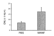
도 3A 내지 3C는 광학 현미경 하의 대조군 및 MANF 처리된 S334ter3 래트의 망막의 외핵층의 단면 사진 및 각각의 외핵층의 두께의 정량 분석을 도시한 것이다. 도 3A는 대조군인 PBS (인산염 완충 염수) 처리된 망막을 도시한 것이고, 도 3B는 MANF-처리된 망막을 나타내는 것이다. 기준자는 25㎛이다. 도 3C는 PBS 처리 및 MANF 처리된 망막에서 망막의 외핵층의 두께의 정량 분석을 그래프로 나타낸 것이다.
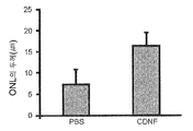
도 4A 내지 4C는 대조군 및 CNDF 처리된 S334ter3 래트의 망막의 외핵층의 단면의 광학 현미경 사진 및 각각의 외핵층의 두께의 정량 분석을 도시한 것이다. 도 4A는 대조군인 PBS (인산염 완충 염수) 처리된 망막을 나타낸 것이고, 도 4B는 CDNF-처리된 망막을 나타내는 것이다. 기준자는 25㎛이다. 도 4C는 PBS 처리 및 CDNF 처리된 망막에서 망막의 외핵층의 두께의 정량 분석을 그래프로 나타낸 것이다.
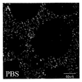
도 5A 내지 5C는 대조군 및 MANF 처리된 S334ter3 래트에서 알렉사 플루오르 488 공액 PNA (땅콩 응집소, peanut agglutinin)로 염색된 전체 착상된 망막의 추상체 외분절 (COS)의 형광 현미경 사진 (도 5A 및 5B), 및 각각의 정량 분석 (도 5C)을 도시한 것이다. 도 5A는 대조군인 PBS 처리된 망막을 나타낸 것이고, 도 5B는 MANF-처리된 망막을 나타내는 것이다. 기준자는 50㎛이다. 도 5C는 PBS 처리 및 MANF 처리된 망막에서 망막의 표지된 세포수의 정량 분석을 그래프로 나타낸 것이다.
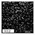
도 6A 내지 6C는 대조군 및 CDNF 처리된 S334ter3 래트에서 알렉사 플루오르 488 공액 PNA로 염색된 전체 착상된 망막의 COS의 형광 현미경 사진 (도 6A 및 6B), 및 각각의 정량 분석 (도 6C)을 도시한 것이다. 도 6A는 대조군인 PBS 처리된 망막을 나타낸 것이고, 도 6B는 CDNF-처리된 망막을 나타내는 것이다. 도 6C는 PBS 처리 및 CDNF 처리된 망막에서 망막의 표지된 세포수의 정량 분석을 그래프로 나타낸 것이다.
도 7A 내지 7C는 대조군 래트 (7A), 대조군으로서 PBS 처리에 이은 광신경 분쇄 (crush) 이후의 래트 (7B), 및 MANF 처리에 이은 광신경 분쇄 이후의 래트 (7C)의 전체 착상된 망막의 플루오로-골드 레트라표지된 신경절 세포의 대표적인 현미경 사진을 도시한 것이다. 도 7A는 광신경 분쇄를 경험하지 않은 대조군 망막 신경절 세포를 나타낸다. 도 7B는 광신경 분쇄 및 PBS 처리 2주 후의 망막 신경절 세포를 나타낸다. 도 7C는 광신경 분쇄 및 MANF 처리 2주 후의 망막 신경절 세포를 나타낸다.
도 8은 MANF 및 β-액틴 (로딩 대조군) 용으로 프로브 처리된 웨스턴 블롯의 사진을 도시한 것이다. 야생형 스프래그 돌리 래트의 망막으로부터의 추출물에서 MANF 발현 레벨은 PD 1, PD 5, PD 8, PD 10, PD 12, PD 16, PD 25, PD 30, PD 40 및 PD 60으로 도시되어 있다.
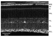
도 9는 항-MANF 항체로 프로브 처리된 래트 망막의 동결 절편의 형광 현미경 사진을 도시한 것이다. 기준자는 50㎛이다. 상기 절편 상에 표지된 층은 망막 색소 상피 (RPE), 외부 광수용체 분절 (OS), 내부 광수용체 분절 (IS), 외핵층 (ONL), 내핵층 (INL), 내부 총상층 (IPL), 및 신경절 세포층 (GCL)을 포함한다. MANF의 면역 활성은 RPE 세포, 뮐러 세포 섬유 및 세포체에서 뿐만 아니라 GCL에서도 나타난다.
도 10은 서열번호 1의 핵산 서열을 도시한 것이다.
도 11은 서열번호 2의 핵산 서열을 도시한 것이다.
도 12는 서열번호 3의 아미노산 서열을 도시한 것이다.
도 13은 서열번호 4의 아미노산 서열을 도시한 것이다.
유사 참조 번호는 도면의 여러 시점 전체에 걸쳐 유사한 부분을 의미한다.For a more complete understanding of the nature of the invention, reference should be made to the following detailed description taken in conjunction with the accompanying drawings, in which:
Figure 1 is a photograph of gel electrophoresis in an amount of 1 [mu] g and 5 [mu] g of purified recombinant human MANF protein in comparison with a standard molecular weight (MW) ladder, which shows the size of the purified MANF protein at about 20 kilodaltons (KD) It is.
Figure 2 depicts a photograph of a 5 ug amount of gel electrophoresis of a purified recombinant human CDNF protein in comparison with a standard molecular weight (MW) ladder, which shows the size of the purified CDNF protein as about 18 kilodaltons (KD).
3A to 3C show cross-sectional photographs of the outer core layer of the retina of the control and MANF-treated S334ter3 rats under an optical microscope and quantitative analysis of the thickness of the respective outer nuclear layer. FIG. 3A shows a PBS (phosphate buffered saline) treated retina as a control group, and FIG. 3B shows a MANF-treated retina. The standard is 25 탆. Figure 3C is a graphical representation of quantitative analysis of the thickness of the outer nuclear layer of the retina in PBS treated and MANF treated retinas.
Figures 4A-4C show optical microscopic photographs of cross-sections of the outer nuclear layer of the retina of the control and CNDF treated S334ter3 rats and quantitative analysis of the thickness of each outer nuclear layer. Figure 4A shows the control (PBS) (phosphate buffered saline) treated retina, and Figure 4B shows the CDNF-treated retina. The standard is 25 탆. 4C is a graphical representation of the quantitative analysis of the thickness of the outer nuclear layer of the retina in PBS treated and CDNF treated retinas.
Figures 5A-5C are fluorescence microscopy (Figure 5A and 5B) of total extracellular segments (COS) of whole implanted retinas stained with Alexa Fluor 488 conjugated PNA (peanut agglutinin) in control and MANF treated S334ter3 rats. , And quantitative analysis of each (Fig. 5C). Figure 5A shows the PBS-treated retina as a control, and Figure 5B shows the MANF-treated retina. The standard is 50 m. Figure 5C is a graphical representation of quantitative analysis of the number of labeled cells in the retina in PBS treated and MANF treated retinas.
6A-6C are fluorescence micrographs (Figs. 6A and 6B) of COS of whole-implanted retinas stained with Alexa Fluor 488 conjugated PNA in the control and CDNF treated S334ter3 rats (Figs. 6A and 6B) will be. Figure 6A shows the PBS treated retina as a control, and Figure 6B shows the CDNF-treated retina. Fig. 6C is a graphical representation of quantitative analysis of the number of labeled cells of the retina in PBS treated and CDNF treated retinas.
FIGS. 7A-7C are photographs showing the results of immunofluorescence analysis of control rats (7A), PBS treatment as a control, rats (7B) after optical neurrhage crush, and MANF- Fluorescent-gold-trail-labeled ganglion cells are representative photomicrographs. FIG. 7A shows control retinal ganglion cells that have not undergone light nerve breakdown. Figure 7B shows retinal ganglion cells after 2 weeks of optical neurrhage and PBS treatment. Figure 7C shows retinal ganglion cells after 2 weeks of optical neuron crush and MANF treatment.
Figure 8 shows a photograph of a western blot probed for MANF and beta -actin (loading control). The levels of MANF expression in the extracts from the retina of the wild-type Sprague Dawley rats are shown as
Figure 9 shows a fluorescence microscope photograph of a frozen section of a rat retina probed with an anti-MANF antibody. The standard is 50 m. The layer labeled on the slice may be a retinal pigment epithelium (RPE), an outer photoreceptor segment (OS), an inner photoreceptor segment (IS), an outer nuclear layer (ONL), an inner nuclear layer (INL) And ganglion cell layer (GCL). The immunological activity of MANF appears not only in RPE cells, Müller cell fibrils and cell bodies, but also in GCL.
10 shows a nucleic acid sequence of SEQ ID NO: 1.
Figure 11 shows the nucleic acid sequence of SEQ ID NO: 2.
12 shows the amino acid sequence of SEQ ID NO: 3.
13 shows the amino acid sequence of SEQ ID NO: 4.
Like reference numerals refer to like parts throughout the various views of the drawings.
바람직한 desirable 구현예의Implementation example 상세한 설명 details
본 발명은 망막 장애의 치료 방법, 조성물 및 키트에 관한 것이다. The present invention relates to methods, compositions and kits for treating retinal disorders.
본 발명의 몇 가지 측면은 단지 예시 목적의 실시예들과 관련하여 후술한다. 다수의 구체적인 상세, 관계 및 방법들이 본 발명의 완전한 이해를 제공하기 위해 제시됨을 이해하여야 한다. 그러나, 관련 분야에서 통상의 기술을 가진 자라면 본 발명이 하나 이상의 구체적인 상세 없이 실시되거나 또는 다른 방법, 프로토콜, 시약, 세포주 및 동물로 실시될 수 있음을 쉽게 인지할 것이다. 본 발명은 일부 행위들이 상이한 순서로 및/또는 다른 행위 또는 이벤트와 동시에 일어날 수 있기 때문에 행위 또는 이벤트의 예시된 순서에 의해 제한되지 않는다. 본 명세서에 기재되거나 참고된 다수의 기술 및 절차는 당해 분야의 숙련자에 의해 잘 이해되고, 종래의 방법을 사용하여 통상적으로 이용된다. Some aspects of the present invention are described below with reference to embodiments for illustrative purposes only. It should be understood that numerous specific details, relationships and methods are set forth in order to provide a thorough understanding of the present invention. However, those of ordinary skill in the relevant art will readily recognize that the invention may be practiced without one or more of the specific details, or with other methods, protocols, reagents, cell lines, and animals. The present invention is not limited by the illustrated order of acts or events, as some acts may occur in different orders and / or concurrently with other acts or events. Many of the techniques and procedures described or referenced herein are well understood by those skilled in the art and are routinely used using conventional methods.
달리 정의되지 않는 한, 본 명세서에 사용된 모든 기술 용어, 인용 및 기타 과학 용어 또는 용어학은 본 발명이 속하는 기술 분야의 숙련자에 의해 통상 이해되는 의미를 갖는 것이다. 일부 경우에는, 통상 이해되는 의미를 갖는 용어들이 명료성을 위해 및/또는 쉬운 참조를 위해 본 명세서에 정의되고, 그러한 정의를 포함하는 것이 반드시 당해 분야에서 일반적으로 이해되는 것 이상의 상당한 차이를 나타내는 것으로 해석되어서는 아니된다. 통상 사용되는 사전에 정의된 것들과 같은 용어들은 관련 분야의 맥락에서 그 의미와 일치하고/하거나 아니면 본 명세서에 정의된 것으로의 의미를 갖는 것으로 해석되어야 함은 추가로 이해될 것이다. Unless defined otherwise, all technical terms, citations, and other scientific terms or terminology used herein have the same meaning as commonly understood by one of ordinary skill in the art to which this invention belongs. In some instances, terms with commonly understood meanings are defined herein for clarity and / or for ease of reference, and the inclusion of such definitions necessarily represents a significant difference beyond what is generally understood in the art. It should not be. It will be further understood that terms such as commonly used predefined terms are to be construed as meaning in the context of the relevant field and / or have meanings as defined herein.
본 명세서에 사용된 용어론은 특정의 구현예를 기술할 목적일 뿐이며, 본 발명을 한정하기 위한 의도는 없다. 본 명세서에서 사용되는 부정 관사 "a", "an" 및 "the"는 문맥상 명백히 달리 나타내지 않는 한 복수의 의미를 포함하는 것으로 이해되어야 한다. The terminology used herein is for the purpose of describing particular embodiments only and is not intended to be limiting of the invention. The indefinite articles "a," an, " and "the ", as used herein, should be understood to include a plurality of meanings, unless the context clearly dictates otherwise.
본 명세서에 사용된 "및/또는"이란 문구는 결합된 요소, 즉 일부 경우에는 결합하여 존재하고, 어떤 경우에는 분리하여 존재하는 요소들 중의 "어느 하나 또는 둘다"를 의미하는 것으로 해석되어야 한다. As used herein, the phrase "and / or" is to be construed as meaning a combined element, that is, "either or both" of elements that in some cases are present in combination and in some cases are present in isolation.
본 명세서에 사용된 "또는"이란 상기 정의한 "및/또는"과 동일한 의미를 갖는 것으로 이해되어야 한다. 예컨대, 항목들의 목록을 분리할 때, "및/또는"이나 "또는"은 포함되는 것으로, 즉 다수의 항목들 중의 적어도 하나를 포함하지만 하나 이상을 포함하기도 하고, 선택적으로는 추가의 열거되지 않은 항목도 포함하는 것으로 해석되어야 한다. "~ 중의 단 하나" 또는 "~ 중의 정확히 하나", 또는 특허청구범위에 사용될 때 "~로 구성되는"과 같은 명확하게 반대로 나타낸 용어들만이 다수 목록의 요소들 중의 정확히 하나의 요소를 포함하는 것을 의미한다. 일반적으로, 본 명세서에 사용된 "또는"이란 용어는 배타성의 용어, 예컨대, "어느 하나 (either)", "~중의 하나 (one of)", "~중의 단 하나 (only one of)" 또는 "~중의 정확히 하나 (exactly one of)"가 선행될 때 배타적인 대체물 (즉, "하나 또는 다른 하나 그러나 둘다는 아닌")을 나타내는 것으로만 해석될 수 있다. As used herein, "or" should be understood to have the same meaning as "and / or" as defined above. For example, "and / or" or " or "when included in the list of items, include at least one of a plurality of items but also include one or more, Should be construed as including items. It is to be understood that clearly contradictory terms such as " consisting of "or " when used in a claim " include exactly one of the elements of the multiple list it means. Generally, the term "or ", as used herein, refers to an exclusivity term such as" any, "" one of, "" Can be interpreted only as representing an exclusive substitute (i.e., "one or the other but not both") when preceded by exactly one of.
본 명세서에 사용된 "포함하는 (including)", "포함하다 (includes)", "갖는 (having)", "가지다 (has)", "가지고 (with)", 또는 이들의 변형과 같은 용어들은 "포함하는 (comprising)"이란 용어와 유사하게 포함되는 것을 의미한다.The terms "including", "includes", "having", "has", "with", or variations thereof, Is intended to be inclusive in a manner similar to the term "comprising ".
본 명세서에 개시된 모든 유전자 및 유전자 산물 (RNA 및 단백질을 포함), 및 그 각각의 이름은 본원에 개시된 조성물 및 방법들이 적용되는 임의의 종으로부터의 상동체에 상응하는 것이다. 특정 종으로부터의 유전자 또는 유전자 산물이 개시될 때, 이 개시 내용은 예시적인 것일 뿐이며, 그것이 명확하게 보이는 문맥이 달리 나타내지 않는 한 한정하는 것으로 해석되어서는 안되는 것으로 이해된다. 예컨대, 일부 구현예에서 포유동물 (인간을 포함)의 핵산 및/또는 아미노산 서열과 관련이 있는 본원에 개시된 유전자 및 유전자 산물은 기타 포유동물, 어류, 파충류, 양서류, 조류 및 기타 척추동물을 포함하나 이에 국한되지는 않는 다른 동물로부터의 상동성 및/또는 병렬 상동성 (orthologous) 및/또는 직렬 상동성 (paralogous) 유전자 및 유전자 산물을 포함하는 것이다. All genes and gene products (including RNA and proteins) disclosed herein, and their respective names, correspond to homologues from any species to which the compositions and methods disclosed herein are applied. When the gene or gene product from a particular species is disclosed, it is to be understood that this disclosure is only illustrative and should not be construed as limiting unless the context clearly indicates otherwise. For example, in some embodiments, the genes and gene products disclosed herein that relate to the nucleic acid and / or amino acid sequence of a mammal (including a human) include other mammals, fish, reptiles, amphibians, birds and other vertebrates But are not limited to, homologous and / or orthologous and / or serial paralogous genes and gene products from other animals.
본 발명의 내용 중에, "폴리펩티드" 및 "단백질"이란 용어는 동등하며 상호 교환 가능하다. 이들 용어는 임의의 아미노산쇄를 의미하고, 그것에 대한 임의의 해독후 변형 (예컨대, 인산화 또는 글리코실화)를 포함한다.In the context of the present invention, the terms "polypeptide" and "protein" are equivalent and interchangeable. These terms refer to any amino acid chain and include any post-translational modifications thereto (e.g., phosphorylation or glycosylation).
본원에 사용된 "치료 대상 (subject)"이라는 용어는 임의의 동물 (예컨대, 포유류, 조류, 파충류, 양서류, 어류)을 의미하며, 특정 치료의 수용자가 되는 인간, 비인간 영장류, 설치류 등을 포함하나, 이에 국한하지 않는다. 통상, "치료 대상" 및 "환자"라는 용어들은 치료 대상과 관련하여 본 명세서에서 상호 교환적으로 사용될 수 있다. 또한, 돌연변이 동물들 (예컨대, 돌연변이 래트 및 마우스)이 본 발명의 방법에 유용하다. As used herein, the term "subject" refers to any animal (eg, mammal, bird, reptile, amphibian, fish) and includes humans, non-human primates, rodents, etc., , But not limited to. In general, the terms "subject to be treated" and "patient" may be used interchangeably herein with respect to a subject to be treated. In addition, mutant animals (e.g., mutant rats and mice) are useful in the methods of the invention.
본원에 사용된 "화합물"이란 용어는 달리 명확하게 언급하지 않는 한 신경 영양 인자를 의미한다. 상기 신경 영양 인자는 본 발명의 목적상 재조합 DNA, RNA 또는 단백질 형태로 나타내어지고, 기재되고/되거나 적용될 수 있다. 상기 신경 영양 인자는 치료 대상이 될 수도 있는 동물로부터 분리되고 정제된 그대로 분리된 형태로 존재할 수도 있다. 일부 구현예에서, 상기 신경 영양 인자는 폴리펩티드, 폴리뉴클레오티드 또는 그 단편일 수 있다. "생물 제제 (biologic)"이란 용어는 본 명세서에서 본 발명의 신경 영양 인자를 의미하는 "화합물"과 호환될 수도 있다.As used herein, the term "compound " means a neurotrophic factor unless explicitly stated otherwise. The neurotrophic factors may be represented, described and / or applied in the form of recombinant DNA, RNA or protein for the purposes of the present invention. The neurotrophic factor may be isolated from the animal that may be the subject of treatment, and may be present in separate form as purified. In some embodiments, the neurotrophic factor may be a polypeptide, polynucleotide or a fragment thereof. The term "biologic" may also be interchangeably referred to herein as a "compound" meaning a neurotrophic factor of the invention.
본 명세서에 사용된 "단편 (fragment)"이란 용어는 화합물의 일부분을 의미한다. 예컨대, 단백질을 언급할 때, 단편은 폴리펩티드의 전체 길이보다 더 작은 것을 포함하는 복수의 연속 아미노산이다. 예컨대, 화합물의 단편은 모화합물과 그 서열의 99%, 95%, 90%, 85%, 80%, 75%, 70%, 65% 또는 60%까지를 공유할 수 있다. As used herein, the term " fragment " means a portion of a compound. For example, when referring to a protein, the fragment is a plurality of contiguous amino acids comprising smaller than the entire length of the polypeptide. For example, a fragment of a compound may share up to 99%, 95%, 90%, 85%, 80%, 75%, 70%, 65% or 60% of the parent compound.
본 명세서에 사용된 "투여하는 (administering)"이란 용어는 유리체내, 안구내, 안과적, 망막하, 척추강내, 정맥내, 피하, 경피, 피내, 두개내, 국소 등의 투 여 경로를 사용하여 치료학적으로 유효량의 화학적 또는 생물학적 화합물 또는 약학 조성물을 치료 대상에게 제공하는 것을 의미한다. 본 발명의 화학적 또는 생물학적 화합물은 단독으로 투여될 수 있으나, 선택된 투여 경로 및 표준 약학 실무에 기초하여 선택되는 기타 화합물, 부형제, 충전제, 결합제, 담체 또는 기타 비히클과 함께 투여될 수 있다. 투여는 담체 또는 비히클, 예컨대, 멸균 수성 또는 비수성 용액, 또는 염수 용액을 포함하는 주사 용액; 크림; 로션; 캡슐; 정제; 그래뉼; 펠릿; 산제; 현탁액, 유제 또는 미세유제; 패치; 마이셀; 리포좀; 소포; 미세임플란트를 포함하는 임플란트; 점안액; 기타 단백질 및 펩티드; 합성 중합체; 미소구; 나노입자 등에 의해 수행될 수 있다. The term "administering " as used herein refers to the use of a delivery route such as intraocular, ocular, ophthalmic, subretinal, intraspinal, intravenous, subcutaneous, transdermal, intradermal, intracranial, To provide a therapeutically effective amount of a chemical or biological compound or pharmaceutical composition to the subject. The chemical or biological compounds of the present invention may be administered alone, but may be administered with other compounds, excipients, fillers, binders, carriers or other vehicles selected on the basis of the chosen route of administration and standard pharmaceutical practice. Administration may be by injection solutions comprising a carrier or vehicle, such as a sterile aqueous or nonaqueous solution, or a saline solution; cream; Lotion; capsule; refine; Granules; Pellets; Acid; Suspensions, emulsions or microemulsions; patch; Micelles; Liposomes; parcel; An implant comprising a fine implant; eye drops; Other proteins and peptides; Synthetic polymers; Microsphere; Nanoparticles, and the like.
본 발명의 화학적 또는 생물학적 화합물 또는 약학 조성물은 약학적으로 허용 가능한 담체, 부형제, 결합제 및 충전제와 같은 기타 비독성 화합물이 포함되거나 함께 포장될 수도 있는데, 상기 충전제로는 포도당, 유당, 아카시아 검, 젤라틴, 만니톨, 크산탄 검, 로커스트 빈 검, 갈락토스, 올리고당 및/또는 다당류, 전분 페이스트, 삼규산마그네슘, 활석, 옥수수 전분, 전분 단편, 케라틴, 콜로이드성 실리카, 감자 전분, 우레아, 덱스트란, 덱스트린 등을 포함하나 이에 국한되지 않는다. 구체적으로, 본 발명의 실시에 사용하기 위해 고려된 약학적으로 허용 가능한 담체, 부형제, 결합제 및 충전제는 본 발명의 화합물들이 유리체내 전달, 안구내 전달, 안과적 전달, 망막하 전달, 척추강내 전달, 정맥내 전달, 피하 전달, 경피 전달, 피내 전달, 두개내 전달, 국소 전달 등이 잘 듣게 하는 것들이다. 더욱이, 포장 재료는 플라스틱 중합체, 실리콘 등과 같이 생물학적으로 비활성이거나 또는 생물 활성이 부족할 수 있고, 함께 포장되고/되거나 전달된 신경 영양 인자의 효능에 영향을 끼치지 않으면서 치료 대상에 의해 내부적으로 처리될 수 있다. The chemical or biological compound or pharmaceutical composition of the present invention may contain or be packaged together with other non-toxic compounds such as pharmaceutically acceptable carriers, excipients, binders and fillers, such as glucose, lactose, acacia, gelatin Starch paste, starch, starch, keratin, colloidal silica, potato starch, urea, dextran, dextrin, etc., and the like, for example, as a sweetening agent such as mannitol, xanthan gum, locust bean gum, galactose, oligosaccharide and / or polysaccharide, starch paste, magnesium trisilicate, But are not limited to. In particular, pharmaceutically acceptable carriers, excipients, binders and fillers contemplated for use in the practice of the present invention are those compounds of the present invention that are suitable for delivery to the body, including intraocular, intraocular, ophthalmic, , Intravenous delivery, subcutaneous delivery, transdermal delivery, intradermal delivery, intracranial delivery, and local delivery. Moreover, the packaging material may be biologically inactive, such as a plastic polymer, silicone, or may be deficient in biological activity, treated internally by the subject to be treated without affecting the efficacy of neurotrophic factors packaged and / or delivered together .
본 발명의 화합물들은 캡슐화 세포 기술 (ECT) 또는 기타 유사한 또는 미래에 유도되는 미세이식 기술과 같이 이식 비히클에 의해 투여될 수 있다는 것도 고려된다. ECT는 본원에 참고로 인용되는 문헌 [Tao, W. et al., 2006, Tao, W. and Wen, R., 2007 and Sieving et al., 2006]에 기재되어 있다. 일부 구현예에서, ECT 비히클은 매일 1x10⁶ 세포당 약 250 ng 내지 약 800 ng의 비율로 본 발명의 화합물을 방출할 수 있다. 이식된 비히클은 또한 임의의 기타 유사 서방형 비히클, 즉 추후 개발되는 것들일 수도 있다. It is also contemplated that the compounds of the present invention may be administered by an implantable vehicle, such as encapsulated cell technology (ECT) or other similar or future induced microinjection techniques. ECT is described in Tao, W. et al., 2006, Tao, W. and Wen, R., 2007 and Sieving et al., 2006, which is incorporated herein by reference. In some embodiments, the ECT vehicle is capable of releasing a compound of the invention at a rate of about 250 ng to about 800 ng per 1 x 10 6 cells per day. The implanted vehicle may also be any other similar sustained-release vehicle, i.e., those that are developed in the future.
본 명세서에서 화합물(들), 생물 제제 및 약학 조성물에 적용된 "유효량 (effective amount)"이란 용어는 목적하는 치료 결과를 얻는 데 필요한 양을 의미한다. 예컨대, 유효량이란 치료용 화합물, 생물 제제 또는 조성물이 투여되는 장애의 증상을 치료, 치유 또는 경감시키는 데 효과적인 레벨이다. 추구하는 특정 치료 목표에 효과적인 양은 치료되는 장애 및 그 심도 및/또는 발육/진행의 단계; 생체 이용성, 및 사용된 특정 화합물, 생물 제제 또는 약학 조성물의 활성; 투여 경로 또는 방법과 치료 대상에게의 투입 지점; 특정 화합물 또는 생물 제제 및 기타 약리학적 성질의 제거 속도; 치료 기간; 접종 체계; 특정 화합물, 생물 제제 또는 조성물과 병합 또는 동시에 사용되는 약제; 치료 중인 대상의 연령, 체중, 성별, 식사량, 생리학 및 일반적인 건강; 및 관련 과학 분야의 숙련자에게 널리 알려진 유사 인자를 포함하여 다양한 인자들에 의해 좌우될 수 있다. 투여량의 약간의 변화는 반드시 치료 중인 대상의 상태에 의존하여 일어날 것이고, 치료제를 투여하는 의사 또는 기타 개인은 어쨌든 개별 환자에게 적합한 용량을 결정할 것이다.The term "effective amount " applied to the compound (s), biologics and pharmaceutical compositions herein refers to the amount required to achieve the desired therapeutic result. For example, an effective amount is a level effective to treat, cure, or ameliorate the symptoms of the disorder to which the therapeutic compound, biological agent, or composition is administered. The amount effective for the particular therapeutic objective sought is the extent of the disorder being treated and its depth and / or development / progression; Bioavailability, and the activity of the particular compound, biologic or pharmaceutical composition employed; The route of administration or method and the site of entry into the subject; The rate of elimination of certain compounds or biologics and other pharmacological properties; Treatment period; Inoculation system; Agents used in combination or concurrent with certain compounds, biologics or compositions; Age, weight, sex, diet, physiology and general health of the subject being treated; And similar factors well known to those skilled in the relevant arts. A slight change in dosage will necessarily occur depending on the condition of the subject being treated, and the physician or other individual administering the therapeutic agent will, in any event, determine the appropriate dose for the individual patient.
본 명세서에 사용된 "장애 (disorder)"란 용어는 장애, 질병 또는 상태, 또는 건강하거나 정상적인 생물학적 활동으로부터의 기타 이탈을 의미하며, 이들 용어는 상호 교환적으로 사용될 수 있다. 상기 용어들은 정상의 기능을 손상시키는 임의의 상태를 의미한다. 상기 상태는 산발적 또는 유전적 유전자 이상에 의해 야기될 수 있다. 상기 상태는 또한 비유전자 이상에 의해서도 야기될 수 있다. 상기 상태는 절단, 분쇄, 연소, 천공, 연신, 전단, 주사 또는 치료 대상의 세포(들), 조직(들), 기관(들), 계(들) 등을 변형시키는 것들 (이에 국한되지 않음)과 같은 환경 인자로부터 치료 대상에게 가해진 손상에 의해 야기될 수도 있다.The term " disorder ", as used herein, refers to a disorder, disease or condition, or other departure from a healthy or normal biological activity, and these terms may be used interchangeably. The terms refer to any condition that impairs normal functioning. Such conditions can be caused by sporadic or genetic abnormalities. This state can also be caused by non-dielectric anomalies. Such conditions include, but are not limited to, cutting, grinding, burning, puncturing, stretching, shearing, injecting or modifying the cell (s), tissue (s), organ ≪ RTI ID = 0.0 > and / or < / RTI >
본 명세서에 사용된 "치료 (treatment)" 또는 "치료하는 (treating)"이란 용어는 장애의 발달 또는 진행을 저지하거나 저해하거나, 또는 저지 또는 저해를 시도하는 것 및/또는 장애 및/또는 그 증상의 경감, 억제, 회귀 또는 진정을 야기하거나 또는 야기를 시도하는 것을 의미한다. 당해 분야의 숙련자는 이해하듯이, 다양한 임상적 및 과학적 방법 및 분석을 사용하여 장애의 발달 또는 진행을 평가할 수 있고, 유사하게 다양한 임상적 및 과학적 방법 및 분석을 사용하여 장애 또는 그 증상의 경감, 회귀 또는 진정을 평가할 수 있다. 또한, 치료는 치료 대상에게 또는 세포 배양에 적용될 수 있다. The term " treatment "or " treating ", as used herein, refers to inhibiting or inhibiting the development or progression of a disorder, or attempting to inhibit or inhibit it and / Restraining, regressing or calming of, or attempting to cause. As will be understood by those skilled in the art, various clinical and scientific methods and assays can be used to assess the development or progression of a disorder, and similarly, using a variety of clinical and scientific methods and assays to mitigate, It is possible to evaluate regression or sedation. In addition, the treatment can be applied to the subject or to the cell culture.
본 발명의 하나 이상의 구현예에 따르면, 치료가 필요한 대상의 망막 장애의 치료 방법은 본 명에서에 기재한 화합물의 유효량을 치료 대상에게 투여하는 단계를 포함한다. 일 구현예에서, 상기 화합물은 신경 영양 인자이다. According to one or more embodiments of the present invention, a method of treating retinal disorders in a subject in need thereof comprises administering to the subject an effective amount of a compound described herein. In one embodiment, the compound is a neurotrophic factor.
"신경 영양 인자 (neurotrophic factor)"란 용어는 발육 중인 신경 세포의 성장 및 생존, 및 성인 신경 세포의 유지를 담당하는 데옥시리보핵산 (DNA), 및 리보핵산 (RNA) 및 그로부터 유래된 단백질과 그 단편들을 의미한다. 본 발명의 일부 구현예에서, 상기 신경 영양 인자는 중뇌 성상교세포 유래의 신경 영양 인자 (MANF)이다. 추가의 구현예에서, 상기 신경 영양 인자는 보존 도파민 신경 영양 인자 (CDNF)이다. 또한, 투여되는 신경 영양 인자는 MANF 및 CDNF의 병합물일 수 있다. 당해 분야의 숙련자에게 이해되듯이, MANF 및 CDNF는 본 발명에 사용하기 위해 추가로 고려되는 상동체, 병렬 상동체 및/또는 직렬 상동체일 수 있다. The term "neurotrophic factor" is used to refer to the growth and survival of developing nerve cells and the maintenance of adult neuronal cells, as well as to the production of oxyribonucleic acid (DNA), ribonucleic acid (RNA) It means the fragments. In some embodiments of the invention, the neurotrophic factor is a neurotrophic factor (MANF) derived from a midbrain astrocytoma. In a further embodiment, the neurotrophic factor is a conserved dopamine neurotrophic factor (CDNF). In addition, the neurotrophic factor administered may be a combination of MANF and CDNF. As will be understood by those skilled in the art, MANF and CDNF may be a homolog, a parallel homolog and / or a serial homolog, which are further considered for use in the present invention.
본 발명의 일 구현예에서, 상기 신경 영양 인자는 재조합 폴리펩티드이다. 상기 재조합 폴리펩티드는 재조합 MANF 또는 CDNF 단백질일 수 있다. 본 발명에서는 재조합 MANF 및/또는 CDNF 폴리펩티드 단편들을 본 명세서에 기재된 방법 및 키트에 사용할 수 있다는 것도 고려된다. 재조합 MANF 및/또는 CDNF의 전 길이 DNA, cDNA 또는 mRNA (또는 그 단편들)를 본 명세서에 기재된 방법 및 키트에 이용할 수 있다는 것도 고려된다. 일부 구현예에서, 상기 DNA, cDNA 또는 mRNA (또는 그 단편들)는 플라스미드, 벡터 등에 포함될 수 있다. 예컨대, 폴리뉴클레오티드 및 그 단편들은 유전자 치료 기술 또는 캡슐화 세포 기술 (ECT)에 의해 이용될 수 있다. 또한, 본 발명은 서열번호 1, 2, 3 또는 4의 서열(또는 그 단편, 재조합체, 키메라 또는 이들의 조합)을 가진 신경 영양 인자를 이용할 수 있다. In one embodiment of the invention, the neurotrophic factor is a recombinant polypeptide. The recombinant polypeptide may be a recombinant MANF or CDNF protein. It is contemplated herein that recombinant MANF and / or CDNF polypeptide fragments may be used in the methods and kits described herein. It is contemplated that full-length DNA, cDNA, or mRNA (or fragments thereof) of recombinant MANF and / or CDNF may be used in the methods and kits described herein. In some embodiments, the DNA, cDNA, or mRNA (or fragments thereof) may be included in a plasmid, vector, or the like. For example, polynucleotides and fragments thereof can be used by gene therapy techniques or by encapsulation cell technology (ECT). Further, the present invention may utilize a neurotrophic factor having a sequence of SEQ ID NO: 1, 2, 3 or 4 (or a fragment thereof, a recombinant, a chimera or a combination thereof).
본 발명의 방법에서, 상기 신경 영양 인자는 약학적으로 허용 가능한 담체 또는 비히클과 함께 투여된다. 예컨대, 약학적으로 허용 가능한 담체 또는 비히클은 염수 용액 또는 본원에 고려된 임의의 기타 비히클일 수 있다.In the method of the present invention, the neurotrophic factor is administered together with a pharmaceutically acceptable carrier or vehicle. For example, the pharmaceutically acceptable carrier or vehicle may be a saline solution or any other vehicle contemplated herein.
특히, 일 구현예에서, 상기 신경 영양 인자는 주사에 의해 투여된다. 일부 구현예에서, 상기 주사 지점은 치료 대상의 눈이고, 안구내, 유리체내, 망막하 등의 투여일 수 있다. 다른 구현예에서, 상기 신경 영양 인자는 눈에 인접한 영역에 투여되고, 주사 또는 본 명세서에 기재된 다른 전달 방법을 통해 투여될 수 있다. In particular, in one embodiment, the neurotrophic factor is administered by injection. In some embodiments, the injection site is an eye of the subject to be treated, and may be administered intra-ocularly, intra-vitally, sub-retinal, and the like. In another embodiment, the neurotrophic factor is administered to the area adjacent to the eye and may be administered via injection or other delivery methods described herein.
상기 신경 영양 인자는 치료될 대상의 눈 속으로 비히클을 이식하여 투여될 수도 있다. 상기 비히클은 캡슐화 세포 기술 (ECT) 또는 기타 유사 또는 미래에 유도되는 미세 이식 기술과 같이 미세 이식 장치일 수 있다. The neurotrophic factor may be administered by implanting the vehicle into the eye of the subject to be treated. The vehicle may be a micro-implant device such as encapsulated cell technology (ECT) or other similar or future induced micro-implant technology.
본 발명의 방법에 의해 치료되는 망막 장애는 중추신경계의 조직 또는 세포에 대한 손상의 결과일 수 있다. 치료되는 망막 장애는 신경 퇴행성 장애 (예, 망막색소 변성증)일 수도 있다. 신경 퇴행성 장애로 손상되거나 발병된 조직 또는 세포는 망막 신경절 세포와 같은 신경절 세포 또는 광수용체 세포일 수 있다. 일부 구현예에서, 치료되는 상기 망막 장애는 신경절 세포 퇴행을 포함한다. 그러한 신경절 세포 퇴행은 녹내장에 의해 유도될 수 있다.The retinal disorder treated by the method of the present invention may be a result of damage to the tissues or cells of the central nervous system. The retinal disorder to be treated may be a neurodegenerative disorder (e.g., retinitis pigmentosa). A tissue or cell damaged or developed by a neurodegenerative disorder may be a ganglion cell or a photoreceptor cell, such as a retinal ganglion cell. In some embodiments, the retinal disorder being treated comprises ganglion cell degeneration. Such ganglion cell degeneration can be induced by glaucoma.
본 명세서에 기재된 치료를 위해 고려되는 신경 퇴행성 장애는 본래 유전적이거나 산발성일 (즉, 분리된 비유전적 이벤트로서 발행하는) 수 있다. 당해 분야의 숙련자라면 이해하듯이, 신경 퇴행성 장애는 망막 신경 퇴행성 질병 이외의 상태를 포함하기도 하고, 본 발명의 방법, 조성물 및 키트는 그러한 기타 장애에도 적용 가능하다. 그러한 장애로는 알츠하이머병, 헌팅턴병, 파킨슨병, 근위축성 축삭 경화증, 녹내장, 연령 관련 청력 상실, 진행형 핵상성 마비 (progressive supranuclear palsy), 경도 인지 장애 (mild cognitive impairment), 치매, 척수 소뇌 운동실조증 (spinocerebellar ataxias) 등을 포함한다.Neurodegenerative disorders contemplated for treatment described herein may be inherently genetic or sporadic (i. E., Issued as discrete non-genetic events). As will be understood by those skilled in the art, neurodegenerative disorders also include conditions other than retinal neurodegenerative diseases, and the methods, compositions and kits of the present invention are also applicable to such other disorders. Such disorders include but are not limited to Alzheimer's disease, Huntington's disease, Parkinson's disease, amyotrophic axillary sclerosis, glaucoma, age-related hearing loss, progressive supranuclear palsy, mild cognitive impairment, dementia, spinocerebellar ataxias) and the like.
하나 이상의 구현예에서, 상기 신경 영양 인자는 신경 퇴행성 장애에 의한 손상 또는 발병 지점에 또는 손상 또는 발병 지점에 인접한 영역에 투여된다. 상기 신경 영양 인자는 본원에서 논의된 ECT에서와 같이 제어되는 (즉, 시간 및/또는 용량 의존적) 방식으로 상기 인자를 방출하는 비히클에 의해 투여될 수도 있다. In one or more embodiments, the neurotrophic factor is administered to a site of injury or onset to a neurodegenerative disorder or adjacent to a point of onset or onset. The neurotrophic factor may be administered by a vehicle that releases the factor in a controlled (i. E., Time and / or dose dependent) manner as in the ECT discussed herein.
본 발명의 또 다른 구현예에 따르면, 뉴런 세포에서 신경 보호를 촉진하는 방법은 뉴런 세포를 MANF, CDNF 또는 그 조합과 같은 신경 영양 인자와 접촉시키는 단계를 포함한다. 본 명세서에서 "신경 보호 (neuroprotection)"란 용어는 신경 손상, 뉴런 악화 및/또는 뉴런의 사망을 방지, 중지, 저해 또는 서행하는 것을 의미한다. 신경 보호는 노화, 유전적 인자, 환경적 변화, 물리적 스트레스 또는 손상, 내인성 또는 외인성 생물학적 또는 화학적 인자 (예컨대, 뉴로트로핀, 비타민, 알코올, 약학 제제, 허혈 등), 뇌졸중 등에 의해 야기되는 손상 또는 악화 이후에 유도될 수 있다.According to another embodiment of the present invention, a method of promoting neuronal protection in a neuron cell comprises contacting the neuron cell with a neurotrophic factor such as MANF, CDNF, or a combination thereof. As used herein, the term " neuroprotection " means preventing, arresting, inhibiting or slowing down nerve damage, neuronal deterioration and / or neuronal death. Neuroprotection is the treatment of damage caused by aging, genetic factors, environmental changes, physical stress or damage, endogenous or exogenous biological or chemical factors (such as neurotrophins, vitamins, alcohol, pharmaceuticals, ischemia, etc.) Can be induced after deterioration.
본 명세서에 사용된 "접촉하는 (contacting)"이란 용어는 신경 보호와 같이 접촉된 세포(들)에서 목적하는 생물학적 반응을 얻기에 적합한 조건 하에 기정된 특정 시간 동안 제공되는, 세포(들)와 상기 신경 영양 인자(들) (또는 상기 신경 영양 인자(들)를 함유하는 비히클) 사이의 공간적 관계의 생성을 유도하는 작용을 의미한다. 상기 세포(들)와 상기 신경 영양 인자(들) 사이의 공간적 관계는 상기 인자가 접촉된 세포의 표면 상에서 반응을 직접 유도하거나 또는 추가 작용을 위해 세포 속으로 들어가게 하는 직접 접촉, 또는 상기 인자가 세포외 신호 작용 (예컨대, 접촉된 세포와 상호작용하는 또 다른 물질의 활성화 또는 변형 이후에)을 통해 세포 상에서 반응을 유도하게 하는 간접 접촉을 포함할 수 있다. 본원에 적용되는 생물학적 반응은 신경 퇴행성 반응 또는 상기 세포(들)의 장애의 저지, 저해, 경감 또는 회귀를 야기하는 상기 세포(들)에 의한 임의의 기타 반응을 포함한다. The term " contacting ", as used herein, refers to the ability of the cell (s) and the cell (s) to be provided for a specified period of time, under conditions suitable for obtaining the desired biological response in the contacted cell Refers to an action that induces the production of a spatial relationship between a neurotrophic factor (s) (or a vehicle containing said neurotrophic factor (s)). The spatial relationship between the cell (s) and the neurotrophic factor (s) can be determined by direct contact, which causes the factor to directly induce the reaction on the surface of the cell to which it is contacted or into the cell for further action, Or indirect contact that leads to a response on the cell through external signaling (e.g., after activation or deformation of another substance that interacts with the contacted cell). The biological response as applied herein includes any other reaction by the cell (s) causing a neurodegenerative reaction or inhibition, inhibition, alleviation or regression of the disorder of the cell (s).
본 발명의 특정 구현예에서, 신경 영양 인자에 의한 뉴런 세포의 접촉은 시험관내에서 일어난다. "시험관내 (in vitro)"라는 것은 세포 또는 조직 배양에서, 또는 시험관 배양에서를 포함한다. 일부 구현예에서, 뉴런 세포의 접촉은 생체내에서 일어난다. "생체내 (in vivo)"라는 것은 동물 모델 (예컨대, 마우스 또는 래트와 같은 돌연변이 동물) 또는 본 명세서에 정의되는 대로 인간을 포함하는 생물체를 포함한다. 또 다른 구현예에서, 뉴런 세포의 접촉은 생체외에서 일어난다. "생체외 (ex vivo)"라는 것은 공급원으로부터 분리되거나 추출된 치료 대상에게서 유래한 천연 조직, 기관 또는 계, 또는 그 일부분을 포함할 수 있다. 본 명세서에 사용된 "분리된 (isolated)"이란 용어는 기재된 항목이 구분되거나 분리된다 (물리적으로 또는 화학적으로)는 것을 의미한다. 분리되는 어떤 것은 여전히 치료 대상 내에 있거나 치료 대상의 외부에 존재할 수 있다. 본 명세서에서 "추출된 (extracted)"이란 용어는 기재된 항목이 치료 대상으로부터 제거되어 치료 대상의 외부에 존재한다는 것을 의미한다.In certain embodiments of the invention, contact of neuron cells with neurotrophic factors occurs in vitro. "In the test ( in in vitro "includes " in cell or tissue culture, or in vitro culture. In some embodiments, contact of neuronal cells occurs in vivo. vivo "includes animal models (e. g., mutant animals such as mice or rats) or organisms including humans as defined herein. In another embodiment, contact of neuronal cells occurs ex vivo. in vitro (ex vivo "as used herein may include natural tissues, organs or systems, or parts thereof, derived from a therapeutic subject isolated or extracted from a source. As used herein, the term "isolated" (Physically or chemically). Something that is separate may still be in the subject of treatment or may be external to the subject of treatment. The term "extracted " It is removed from the subject to be treated and is present outside the subject to be treated.
본 발명의 일부 구현예에서, 본 발명의 방법에 의한 치료가 잘 듣는 뉴런 세포 유형은 신경절 세포 또는 광수용체 세포를 포함한다. 한 가지 특정 구현예에서, 본 발명의 방법에 의한 치료가 잘 듣는 뉴런 세포 유형은 망막 신경절 세포를 포함한다. In some embodiments of the invention, neuronal cell types that are well tolerated by the methods of the invention include ganglion cells or photoreceptor cells. In one particular embodiment, neuronal cell types that are well tolerated by the methods of the present invention include retinal ganglion cells.
본 발명의 신경 영양 인자는 재조합 또는 분리된 신경 영양 인자일 수 있고, 재조합 또는 분리된 인간 신경 영양 인자일 수 있다. 유전자 또는 그 단편들, 또는 유전자 산물 또는 그 단편과 관련하여 본 명세서에 사용된 "분리된 (isolated)"이란 용어는 동물의 세포로부터 제거되고/되거나 본원에 기재된 방법에 사용하기 위해 정제된 것으로 정의된다. 본 명세서에 사용된 "유전자"란 용어는 RNA 및 단백질을 코딩하는 염색체로부터 유래한 폴리뉴클레오티드를 의미한다. 본 명세서에 사용된 유전자는 특정 유전자와 관련된 모든 인트론, 엑손, 프로모터 영역, 비코딩 영역 등을 포함하거나 포함하지 않을 수 있다.The neurotrophic factor of the present invention may be a recombinant or isolated neurotrophic factor and may be a recombinant or isolated human neurotrophic factor. The term " isolated ", as used herein in reference to a gene or fragments thereof, or a gene product or fragment thereof, is defined as being purified from cells of an animal and / or purified for use in the methods described herein do. As used herein, the term "gene" refers to polynucleotides derived from chromosomes encoding RNA and proteins. The gene used in the present specification may or may not include all introns, exons, promoter regions, noncoding regions, etc. related to a specific gene.
본 발명은 또한 MANF, CDNF 또는 그 조합과 같은 신경 영양 인자를 포함하는 약학 조성물 또는 약제에 관한 것이다. 상기 약학 조성물은 선택된 투여 경로 및 표준 약학 실무에 기초하여 선택되는 기타 약학적으로 허용 가능한 화합물, 부형제, 첨가제, 충전제, 결합제, 보조제 또는 담체 또는 비히클을 포함할 수도 있다. 그와 같이, 상기 신경 영양 인자(들)는 약제 및 약학 조성물의 제작 또는 제조에 사용할 수 있다. 또한, 상기 신경 영양 인자를 포함하는 약제 및 약학 조성물은 본원에 기재한 장애의 치료용으로 사용할 수 있다. The invention also relates to pharmaceutical compositions or medicaments comprising neurotrophic factors such as MANF, CDNF or a combination thereof. The pharmaceutical composition may also contain other pharmaceutically acceptable compounds, excipients, additives, fillers, binders, adjuvants or carriers or vehicles selected based on the chosen route of administration and standard pharmaceutical practice. As such, the neurotrophic factor (s) can be used in the manufacture or manufacture of pharmaceutical and pharmaceutical compositions. In addition, medicaments and pharmaceutical compositions comprising said neurotrophic factors may be used for the treatment of the disorders described herein.
본 발명은 또한 신경 영양 인자(들) 및 본 발명의 방법(들)을 실시하는 데 필요한 기타 시약을 포함하는 부품의 키트에 관한 것이다. 상기 부품 키트는 또한 사용을 위한 지시문을 포함할 수 있다. 상기 신경 영양 인자(들) 및 시약은 하나 이상의 조성물에 포함될 수 있고, 각각의 신경 영양 인자(들) 및 시약은 적합한 비히클과 배합하여 조성물로 제공되거나, 독립적으로 존재할 수 있다. 상기 부품 키트는 정제된 단백질 (재조합 또는 동물로부터 분리된)로서 또는 정제된 폴리뉴클레오티드 (재조합 또는 동물로부터 분리된)로서 MANF, CDNF 또는 그 조합을 포함할 수 있다. The invention also relates to a kit of parts comprising a neurotrophic factor (s) and other reagents necessary to carry out the method (s) of the present invention. The part kit may also include instructions for use. The neurotrophic factor (s) and reagents may be included in one or more compositions, and each neurotrophic factor (s) and reagent may be provided in a composition in combination with a suitable vehicle, or may be present independently. The kit of parts may comprise MANF, CDNF or a combination thereof as a purified protein (recombinant or isolated from an animal) or as a purified polynucleotide (recombinant or isolated from an animal).
기타 구현예에서, 상기 부품 키트는 특정 뉴런 세포 유형에 특이적인 표지된 바이오마커, 예컨대, 광수용체 바이오마커 또는 망막 신경절 세포 바이오마커, 참조 표준, 및 본원의 개시 내용을 읽을 때 당해 분야의 숙련자에 의해 확인 가능한 추가의 성분들을 포함한다. In other embodiments, the kit of parts may include a labeled biomarker specific to a particular neuronal cell type, such as a photoreceptor biomarker or retinal ganglion cell biomarker, a reference standard, and a person skilled in the art upon reading the disclosure herein. ≪ / RTI >
추가의 고심 없이, 당해 분야의 숙련자라면 상기 설명을 사용하여 본 발명을 그 최대 한도까지 이용할 수 있을 것으로 믿어진다. 하기의 실시예들은 한정하기 위함이 아니라 예시를 위해 제시한다. 구체적인 실시예들이 제시되었지만, 상기의 설명은 예시하는 것이며, 제한하는 것이 아니다. 전술한 구현예의 하나 이상의 특징들은 본 발명에서 임의의 기타 구현예들의 하나 이상의 특징과 함께 임의의 방식으로 조합될 수 있다. 또한, 본 발명의 다양한 변형예들이 당해 분야의 숙련자에게는 본 명세서의 검토 하에 명백할 것이다. Without further elaboration, it is believed that one skilled in the art can, using the preceding description, utilize the present invention to its fullest extent. The following examples are presented for purposes of illustration and not limitation. Although specific embodiments have been presented, the above description is illustrative and not restrictive. One or more aspects of the above-described embodiments may be combined in any manner with one or more of the features of any of the other embodiments in the present invention. In addition, various modifications of the invention will be apparent to those skilled in the art from consideration of the specification.
본원에 인용된 모든 출판물 및 특허 문서는 각각의 개별 출판물 또는 특허 문서가 개별적으로 표시되는 것과 동일한 정도로 모든 목적에 대해 적합한 부분에 참고로 인용된다. 상기 문서에서의 다양한 참고 문헌들의 인용에 의해, 본 출원인은 임의의 특정 참고 문헌이 그들의 발명에 대해서는 "종래 기술"이라는 것을 인정하지 않는다. All publications and patent documents cited herein are hereby incorporated by reference in their entirety to the same extent as if each individual publication or patent document were individually indicated. By citing the various references in the document, Applicants do not recognize that any particular reference is "prior art" to their invention.
실시예Example
본 명세서에 기재된 방법 및 조성물과 관련 키트는 예시를 위해 제시되고 제한하고자 하는 것은 아닌 하기 실시예들에서 추가로 예시된다. 성분들의 요소들의 비율의 변화 및 요소들의 대체예는 당해 분야의 숙련자에게는 명확할 것이고, 본 발명의 구현예의 범위 내에 속한다는 것이 이해될 것이다. 이론적인 측면은 본 출원인이 제시된 이론에 의해 구속되기를 원치 않는다는 것을 전제로 제시된다.The methods and compositions described herein and related kits are further illustrated in the following examples, which are not intended to be limiting and illustrative. It will be appreciated that variations in the proportions of the elements of the components and alternative examples of elements will be apparent to those skilled in the art and are within the scope of embodiments of the invention. The theoretical aspects are provided on the premise that the Applicant does not want to be bound by the presented theory.
하기의 재료 및 방법은 본 명세서에 예시된 모든 방법 및 조성물을 위해 사용되었다. The following materials and methods were used for all the methods and compositions illustrated herein.
재조합 인간 MANF 및 CDNF 단백질의 클로닝: MANF (서열번호 1) 및 CDNF (서열번호 2)의 오픈 리딩 프레임 (ORF)은 각각 인간 뇌 cDNA로부터 폴리머라제 연쇄 반응 (PCR)에 의해 클로닝하고, 생성되는 클로닝된 서열을 확인하였다. 각각의 ORF는 프레임내 N-말단에 대한 6xHis-태그 코딩 서열을 보유하는 발현 벡터 pQE30 (캘리포니아 발렌시아 소재의 퀴아젠) 내로 서브클로닝하였다. 이어서, 각각의 MANF 및 CDNF 서열을 보유하는 상기 발현 벡터를 이. 콜리 (캘리포니아 라 졸라 소재의 스트래터진 사의 XL-블루)에서 발현시키고, 상응하는 발현된 단백질을 Ni-NTA 아가로스 칼럼 (퀴아젠) 상에서 천연 조건 하에 고정화된 금속 친화도 크로마토크래피에 의해 정제하였다. 용출된 단백질을 인산염 완충 염수 (PBS)로 완충제 교환하고, 사용시까지 소량 표본으로 -80℃에서 보관하였다. 적합한 인간 MANF 단백질 서열은 서열번호 3으로 나타내어지고, 적합한 인간 CDNF 단백질 서열은 서열번호 4로 나타내어진다. Cloning of recombinant human MANF and CDNF proteins : The open reading frame (ORF) of MANF (SEQ ID NO: 1) and CDNF (SEQ ID NO: 2) are cloned by polymerase chain reaction (PCR) from human brain cDNA, respectively, . Each ORF was subcloned into the expression vector pQE30 (quizagen, Valencia, Calif.), Which retains the 6xHis-tag coding sequence for the N-terminus in the frame. Subsequently, the expression vector harboring the respective MANF and CDNF sequences is ligated to the < RTI ID = 0.0 > (XL-blue from Stratagene, La Jolla, Calif.) And the corresponding expressed protein is purified on a Ni-NTA agarose column (quiazene) by metal affinity chromato- Respectively. The eluted proteins were buffer exchanged with phosphate buffered saline (PBS) and stored at -80 ° C in small aliquots until use. A suitable human MANF protein sequence is shown in SEQ ID NO: 3, and a suitable human CDNF protein sequence is shown in SEQ ID NO:
정제된 MANF 및 CDNF 단백질의 시각화: 1 ㎍ 및/또는 5 ㎍의 정제된 단백질을 4 내지 12% NuPAEG 겔 상에서 전기영동시키고, 쿠마시 블루로 시각화하여 정제 및 적합한 분자량을 확인하였다. 분자량 마커들 (MW)을 1 ㎍ 및/또는 5 ㎍ 샘플 다음 레인에서 전기영동시켰다. Visualization of purified MANF and CDNF proteins : 1 ug and / or 5 ug of the purified protein was electrophoresed on a 4-12% NuPAEG gel and visualized with Coomassie blue to confirm purification and appropriate molecular weight. Molecular weight markers (MW) were electrophoresed in lane following 1 및 and / or 5 샘플 samples.
돌연변이 동물: S334ter-3 래트로 알려진 쥣과의 로돕신 돌연변이 S334ter를 보유하는 돌연변이 래트를 생성키시고 전술한 바와 같이 이용하였다 (Liu et al., (1999)). Mutant animals : Mutant rats carrying the rhodopsin mutant S334ter, known as S334ter-3 rats, were generated and used as described previously (Liu et al., (1999)).
광수용체 보호 분석: MANF 및 PBS (대조군)의 유리체내 주사를 S334ter-3 래트에게 1회 실시하였다. 구체적으로, 6 ㎍ MANF를 생후 (PD) 9일된 래트의 한쪽 눈에 주사하고, 3 ㎕의 PBS를 대조군으로서 동일한 래트의 나머지 눈에 동시에 주사하였다. 주사는 10 ㎕의 미량 주사기 (네바다 레노 소재의 해밀튼)에 연결된 33-게이지 바늘을 통해 수행하였다. PD 21에 동물을 희생시키고, 각각의 눈을 취하고 플라스틱에 매립하여 전술한 바와 같이 절단하였다 (Liu et al., (1999)). 생성되는 반-박 망막 절편을 톨루이딘 블루로 염색하고 광학 현미경으로 검사하였다. 유사한 실험을 6 ㎍ CDNF를 사용하여 수행하였다. Photoreceptor protection assay : Intra-glass injection of MANF and PBS (control) was performed once in S334ter-3 rats. Specifically, 6 [mu] g of MANF was injected into one eye of postnatal (PD) 9-day-old rats and 3 [mu] l of PBS was injected simultaneously into the remaining eyes of the same rats as a control. Injection was carried out through a 33-gauge needle connected to a 10 [mu] l microsyringe (Hamilton, Reno, Nev.). Animals were sacrificed on PD 21 and each eye was removed and embedded in plastic and cut as described above (Liu et al., (1999)). The resulting semi-thin retinal sections were stained with toluidine blue and examined under an optical microscope. A similar experiment was performed using 6 ug CDNF.
원추형 광수용체 외분절 ( COS ) 보호 분석: MANF 및 PBS (대조군)의 1회의 유리체내 주사를 S334ter3 래트에게 제공하였다. 6 ㎍ MANF를 생후 (PD) 20일된 래트의 한쪽 눈에 주사하고, 3 ㎕의 PBS를 대조군으로서 동일한 래트의 나머지 눈에 동시에 주사하였다. 주사는 10 ㎕의 미량 주사기 (네바다 레노 소재의 해밀튼)에 연결된 33-게이지 바늘을 통해 수행하였다. PD 30에서의 치료 10일 후에 동물을 희생시키고, 각각의 눈을 취했다. 전체 착상된 망막을 원추형 광수용체의 외분절에 특이적으로 결합하는 알렉사 플루오르 488 공액 PNA (땅콩 응집소)로 염색하고, 형광 현미경으로 검사하였다. 유사한 실험을 6 ㎍ CDNF를 사용하여 수행하였다. Conical photoreceptor Outer segment ( COS ) protection assay : One glass intravitreal injection of MANF and PBS (control) was provided to S334ter3 rats. 6 [mu] g MANF was injected into one eye of postnatal (PD) 20-day-old rats, and 3 [mu] l of PBS was injected simultaneously into the remainder of the same rats as a control. Injection was carried out through a 33-gauge needle connected to a 10 [mu] l microsyringe (Hamilton, Reno, Nev.). After 10 days of treatment with
광신경 분쇄 분석: 야생형 스프래그 돌리 래트의 망막 신경절 세포를 플루오로-골드로 레트라표지법으로 표지하였다. 표지 1 주일 후, 광신경을 분쇄하고 즉시 6 ㎍ MANF를 유리체내로 주사하였다. 광신경 분쇄 및 치료 2주 후, 래트를 희생시키고, 망막을 취했다. 전체 착상된 망막을 형광 현미경으로 검사하였다. Optical neuronal crushing analysis : Wild-type Sprague Dawley rat retinal ganglion cells were labeled with fluoro-gold Lorentz labeling. One week after the labeling, the optic nerve was pulverized and immediately injected with 6 μg of MANF into the vitreous body. After 2 weeks of photic nerve crushing and treatment, the rats were sacrificed and the retina was harvested. Whole implanted retinas were examined by fluorescence microscopy.
망막의 MANF 단백질 발현 분석: PD 1, PD 5, PD 8, PD 10, PD 12, PD 16, PD 25, PD 30, PD 40 및 PD 60의 야생형 스프래그 돌리 래트의 망막으로부터의 동일한 양의 단백질 추출물들을 폴리아크릴아미드 겔 상에서 영동시키고, 막으로 옮기고, MANF 및 β-액틴용 항체로 프로브 처리하였다. 구조 단백질인 β-액틴의 발현을 분석하여 분석되는 각 시점에서의 단백질 추출물의 일정한 로딩을 확인하였다. Analysis of MANF protein expression in the retina : The same amount of protein from the retina of the wild type Sprague Dawley rats of
실시예 1: 재조합 인간 중뇌 성상교세포 유래의 신경 영양 인자 (MANF)의 정제 Example 1 : Purification of neurotrophic factor (MANF) derived from recombinant human midbrain astrocytes
신경 보호 성질에 대해 후보 신경 영양 인자들을 시험하기 위해, 재조합 인간 MANF 및 CDNF 단백질을 생성시키고, 추가 실험을 위해 정제하였다. 재료 및 방법에서 전술한 바와 같이 재조합 인간 MANF를 이. 콜리에서 발현시키고, 정제하고 시각화하였다. 도 1에 예시된 결과는 1 ㎍ 및 5 ㎍의 정제된 MANF는 20kDa의 단일 밴드로서 시각화되었음을 나타낸다. 레인 1은 분자량 마커들 (MW)을 나타내고; 레인 2는 1 ㎍의 정제된 MANF를 나타내며; 레인 3은 5 ㎍의 정제된 MANF를 나타낸다. "KD"이란 "킬로돌턴"을 의미한다.To test candidate neurotrophic factors for neuroprotective properties, recombinant human MANF and CDNF proteins were generated and purified for further experiments. The recombinant human < RTI ID = 0.0 > MANF < / RTI > Expressed in coli, purified and visualized. The results illustrated in Figure 1 indicate that 1 mu g and 5 mu g of purified MANF were visualized as a single band of 20 kDa.
실시예 2: 재조합 인간 도파민 신경 영양 인자 (CDNF)의 정제 Example 2: Purification of recombinant human dopamine neurotrophic factor (CDNF)
신경 보호 성질을 가진 후보 신경 영양 인자로서 CDNF도 이용하였다. 재료 및 방법에서 전술한 바와 같이 재조합 인간 CDNF를 이. 콜리에서 발현시키고, 정제하고 시각화하였다. 도 2에 예시된 결과는 5 ㎍의 정제된 CDNF는 18kDa의 단일 밴드로서 시각화되었음을 나타낸다. 레인 1은 분자량 마커들 (MW)을 나타내고; 레인 2는 5 ㎍의 정제된 CDNF를 나타낸다. "KD"이란 "킬로돌턴"을 의미한다.CDNF was also used as a neurotrophic candidate neurotrophic factor. Recombinant human CDNF was prepared as described above in Materials and Methods. Expressed in coli, purified and visualized. The results illustrated in Figure 2 indicate that 5 [mu] g of purified CDNF was visualized as a single band of 18 kDa.
실시예 3: 망막 퇴행 설치류 모델의 망막에서 MANF에 의한 광수용체의 보호 Example 3: Photoreceptor protection by MANF in retina of retinal degenerative rodent model
유전되는 망막 장애 (예, 망막색소 변성증), 연령 관련 황반 변성 및 녹내장을 포함하는 신경 퇴행성 장애에서 광수용체를 구제할 수 있는 신경 영양 인자를 탐색함에 있어서, 망막 퇴행 설치류 모델에서 광수용체 보호 성질에 대해 재조합 인간 MANF 단백질을 시험하였다. 이 목적으로, 그 특성화된 진행성 망막 광수용체 퇴행으로 인해 S334ter3 돌연변이 래트를 이용하였다 (Liu et al., (1999)).In exploring neurotrophic factors capable of remitting photoreceptors in neurodegenerative disorders including inherited retinal disorders (eg retinitis pigmentosa), age-related macular degeneration and glaucoma, the photoreceptor protection properties in retinal degenerative rodent models RTI ID = 0.0 > MANF < / RTI > For this purpose, S334ter3 mutant rats were used due to their characteristic progressive retinal photoreceptor degeneration (Liu et al., (1999)).
도 3A에 예시된 결과는 PBS (대조군) 처리된 S334ter3 래트에서, 망막의 외핵층이 PD21에서 단 하나의 핵렬 (도 3A의 화살촉 참조)을 가졌음을 나타낸다. 도 3B에 예시된 결과는 외핵층이 3 내지 4개의 핵렬 (도 3B의 두 화살촉 사이를 참조)을 보유하고 있기 때문에 동일 동물의 나머지 눈에서 PD 9에서의 MANF의 1회 유리체내 주사는 PD 21 시점에서의 퇴행으로부터 광수용체를 보호하였음을 나타낸다.The results illustrated in Figure 3A indicate that in the PBS (control) treated S334ter3 rats, the outer nuclear layer of the retina had only one nucleus (see arrowhead in Figure 3A) in PD21. The results illustrated in Figure 3B indicate that a single intra-vitals injection of MANF at PD9 in the remainder of the same animal, with 3 to 4 nucleotides (see between the two arrowheads in Figure 3B) Indicating that the photoreceptor was protected from degeneration at the time point.
도 3C에 도시된 바와 같이 우수한 망막의 외핵층의 두께의 정량 분석은 MANF 처리된 망막의 외핵층 (17.47±3.96μΜ, n=5)이 대조군 PBS 처리된 망막의 외핵층 (7.07±1.12μΜ, n=5)(평균±SD)보다 상당히 더 두꺼웠음을 나타낸다. P<0.001 (학생 t-시험).Quantitative analysis of the thickness of the outer nuclear layer of the superior retina as shown in Figure 3C showed that the outer core layer (17.47 ± 3.96 μM, n = 5) of the MANF-treated retina was in the outer nuclear layer of the PBS-treated retina (7.07 ± 1.12 μM, n = 5) (mean +/- SD). P <0.001 (Student t-test).
실시예 4: 망막 퇴행 설치류 모델의 망막에서 CDNF에 의한 광수용체의 보호 Example 4 : Protection of photoreceptors by CDNF in retina of retinal degenerative rodent model
망막 퇴행성 장애에서 광수용체를 구제할 수 있는 기타 신경 영양 인자를 탐색함에 있어서, 전술한 MANF 연구에 사용된 동일한 망막 퇴행 설치류 모델에서 광수용체 보호 성질에 대해 재조합 인간 CDNF 단백질을 시험하였다. In exploring other neurotrophic factors capable of rescuing photoreceptors in retinal degenerative disorders, recombinant human CDNF proteins were tested for photoreceptor protection properties in the same retinal degenerative rodent model used in the MANF study described above.
도 4A에 예시된 결과는 PBS (대조군) 처리된 S334ter3 래트에서, 망막의 외핵층이 PD 21에서 단 하나의 핵렬 (도 4A의 화살촉 참조)을 가졌음을 나타낸다. 도 4B에 예시된 결과는 외핵층이 3 내지 4개의 핵렬 (도 4B의 두 화살촉 사이를 참조)을 보유하고 있기 때문에 동일 동물의 나머지 눈에서 PD 9에서의 CDNF의 1회 유리체내 주사는 PD 21 시점에서의 퇴행으로부터 광수용체를 보호하였음을 나타낸다.The results illustrated in Figure 4A indicate that in the PBS (control) treated S334ter3 rats, the outer nuclear layer of the retina had only one nucleus (see arrowhead in Figure 4A) in PD21. The results illustrated in Figure 4B indicate that a single intra-vitals injection of CDNF at PD9 in the remainder of the same animal, with three to four nucleotides (see between the two arrowheads in Figure 4B) Indicating that the photoreceptor was protected from degeneration at the time point.
도 4C에 도시된 바와 같이 우수한 망막의 외핵층의 두께의 정량 분석은 CDNF 처리된 망막의 외핵층 (16.61±2.87μΜ, n=6)이 대조군 PBS 처리된 망막의 외핵층 (7.4±3.43μΜ, n=6)(평균±SD)보다 상당히 더 두꺼웠음을 나타낸다. P<0.001 (학생 t-시험).Quantitative analysis of the thickness of the outer nuclear layer of the superior retina as shown in Figure 4C showed that the outer nuclear layer (16.61 +/- 2.87 mu M, n = 6) of the CDNF-treated retina was the outer nuclear layer of the PBS treated retina (7.4 +/- 3.43 mu M, n = 6) (mean +/- SD). P <0.001 (Student t-test).
실시예 5: 망막 퇴행 설치류 모델의 망막에서 MANF에 의한 원추형 광수용체의 외분절의 보호 Example 5 : Protection of outer segments of cone photoreceptors by MANF in retina of retinal degenerative rodent model
신경 영양 인자의 광수용체 보호 능력의 또 다른 시험으로서, 재조합 인간 MANF 단백질 노출 이후에 전술한 바와 같은 동일한 망막 퇴행 설치류 모델에서 원추형 외분절을 분석하였다.As another test of the photoreceptor protection ability of neurotrophic factors, extracarticular segments were analyzed in the same retinal degenerative rodent model as described above after exposure of recombinant human MANF protein.
도 5B에 예시된 결과는 망막 퇴행 설치류 모델의 눈 속으로 PD 20에 MANF의 1회 유리체내 주사는 PD 30 시점에서 측정할 때 PBS (대조군)로 주사된 동일 동물 (도 5A)의 나머지 눈에 비교하여 원추형 외분절을 퇴행으로부터 보호하였음을 나타낸다.The results illustrated in FIG. 5B show that a single intra-vitals injection of MANF into the
도 5C에 도시된 결과는 원추형 광수용체의 존재를 나타내는, 알렉사 플루오르 488 공액 PNA-양성 염색된 세포수의 정량 분석이다. 그래프는 MANF 처리된 망막 이 대조군 PBS 처리된 망막 (398.7±25.4μΜ)(C, 평균±SD)와 비교하여 상당히 더 큰 수의 원추형 광수용체 (569.5±46.5μΜ)를 보유하였음을 나타낸다. P<0.001 (학생 t-시험).The result shown in Figure 5C is a quantitative analysis of the number of Alexa Fluor 488 conjugated PNA-positive stained cells, indicating the presence of cone photoreceptors. The graph shows that MANF-treated retinas possessed a significantly larger number of conical photoreceptors (569.5 ± 46.5 μM) compared to control PBS-treated retinas (398.7 ± 25.4 μM) (C, mean ± SD). P <0.001 (Student t-test).
실시예 6: 망막 퇴행 설치류 모델의 망막에서 CDNF에 의한 원추형 광수용체의 외분절의 보호 Example 6 : Protection of outer segments of cone photoreceptors by CDNF in retina of retinal degenerative rodent model
재조합 인간 CDNF 단백질의 광수용체 보호 능력을 추가로 시험하기 위해 실시예 5에서와 동일한 실험 절차를 이용하였다.The same experimental procedure as in Example 5 was used to further test the ability of the recombinant human CDNF protein to protect the photoreceptor.
도 6B에 예시된 결과는 망막 퇴행 설치류 모델의 눈 속으로 PD 20에 CDNF의 1회 유리체내 주사는 PD 30 시점에서 측정할 때 PBS (대조군)로 주사된 동일 동물 (도 6A)의 나머지 눈에 비교하여 원추형 외분절을 퇴행으로부터 보호하였음을 나타낸다.The results illustrated in FIG. 6B show that a single intravitreal injection of CDNF into the
도 6C에 도시된 결과는 원추형 광수용체의 존재를 나타내는, 알렉사 플루오르 488 공액 PNA-양성 염색된 세포수의 정량 분석이다. 그래프는 CDNF 처리된 망막 이 대조군 PBS 처리된 망막 (412.75±40.9μΜ)(C, 평균±SD)와 비교하여 상당히 더 큰 수의 원추형 광수용체 (561.5±81.3μΜ)를 보유하였음을 나타낸다. P=0.012 (학생 t-시험).The result shown in Figure 6C is a quantitative analysis of the number of Alexa Fluor 488 conjugated PNA-positive stained cells, indicating the presence of cone photoreceptors. The graph shows that the CDNF-treated retinas possessed a significantly larger number of conical photoreceptors (561.5 ± 81.3 μM) compared to the control PBS-treated retinas (412.75 ± 40.9 μM) (C, mean ± SD). P = 0.012 (Student t-test).
실시예 7: 래트에서 광신경 분쇄 이후에 CDNF에 의한 망막 신경절 세포의 보호 Example 7 : Protection of retinal ganglion cells by CDNF after photoreactivation in rats
신경 영양 인자 MANF의 신경 보호능에 대한 추가 시험으로서, 래트에서 광신경 분쇄 및 MANF 노출 이후에 망막 신경절 세포를 분석하였다. As a further test for the neuroprotective activity of the neurotrophic factor, MANF, retinal ganglion cells were analyzed after optical neuron crushing and MANF exposure in rats.
도 7A 내지 7C에 도시된 결과는 광신경 분쇄 이후에 망막 신경절 세포를 보호하는 MANF의 능력을 나타낸다. 도 7A에 도시된 바와 같이, 레트라표지된 신경절 세포는 대조군 마우스에서 대표 현미경 사진 전체에 분포되어 있다. 도 7B에 도시된 바와 같이, 레트라표지된 신경절 세포는 PBS 처리된 망막 (대조군)에서 광신경 분쇄 2주 후에 대부분 퇴행된다. 반대로, 도 7C에 도시된 바와 같이 MANF 처리는 광신경 분쇄 및 처리 2주 후에 다수의 망막 신경절 세포를 구제할 수 있다.The results shown in Figs. 7A-7C show the ability of MANF to protect retinal ganglion cells after optical nerve breakdown. As shown in Figure 7A, retinally labeled ganglion cells are distributed throughout the representative micrographs in control mice. As shown in FIG. 7B, retinally labeled ganglion cells are mostly regressed after 2 weeks of optical nerve breakdown in PBS-treated retinas (control). Conversely, as shown in FIG. 7C, MANF treatment can relieve multiple retinal ganglion cells after 2 weeks of optical nerve breakdown and treatment.
실시예 8: 망막에서의 MANF 단백질 발현은 래트에서 광수용체의 발달 중에 더 높다 Example 8 : MANF protein expression in retina is higher during development of photoreceptors in rats
PD 1, PD 5, PD 8, PD 10, PD 12, PD 16, PD 25, PD 30, PD 40 및 PD 60에서 야생형 스프래그 돌리 래트의 망막으로부터의 단백질 추출물을 MANF 발현 레벨에 대한 웨스턴 블롯 분석에 의해 분석하였다. 도 8에 도시한 바와 같이, 고레벨의 MANF 발현이 생후 발육 중에 (PD 1 내지 PD 16) 검출되었다. 망막이 PD 16을 지나 성숙함에 따라 그 발현이 감소된다 (도 8에서 연속된 발현 레벨이 PD 25 로부터 PD 60까지 감소함을 참조). Western blot analysis of protein extracts from the retina of wild-type Sprague Dawley rats at levels of MANF expression in PD1, PD5, PD8, PD10, PD12, PD16, PD25, PD30, PD40 and PD60 Lt; / RTI > As shown in Fig. 8, a high level of MANF expression was detected during development (
실시예 9: 망막에서의 MANF 면역 활성 Example 9 : MANF immunoreactivity in the retina
도 9에 도시한 바와 같이, 항-MANF 항체로 프로브 처리된 동결 절편은 MANF의 면역 활성이 망막 색소 상피 (RPE) 세포, 뮐러 세포 섬유 및 세포체에 뿐만 아니라 망막 신경절 세포층에도 나타났음을 입증하였다. As shown in Fig. 9, frozen sections probed with anti-MANF antibody demonstrated that the immunoreactivity of MANF appeared not only in retinal pigment epithelium (RPE) cells, Müller cell fibrils and cell bodies, but also retinal ganglion cell layer.
이와 함께, 실시예들에 제시된 결과는 MANF 및 CDNF가 광수용체에 대한 신경 보호 성질을 가지며, 적어도 MANF는 추가로 망막 신경절 세포에서 신경 보호 성질을 가진다는 것을 나타낸다. 또한, 적어도 MANF는 광수용체의 발달 중에 높은 발현 레벨을 가지며, 그 레벨은 광수용체가 성숙함에 따라 감소한다. 이러한 결과는 MANF 및 CDNF가 망막 퇴행성 장애에 대한 치료제라는 것을 시사한다. In addition, the results presented in the examples demonstrate that MANF and CDNF have neuroprotective properties against photoreceptors, at least MANF also has neuroprotective properties in retinal ganglion cells. Also, at least MANF has a high expression level during the development of the photoreceptor, which level decreases as the photoreceptor matures. These results suggest that MANF and CDNF are therapeutic agents for retinal degenerative disorders.
요약서 부분이 아닌 상기 상세한 설명은 특허청구범위를 해석하는 데 사용될 수 있다는 것은 이해될 수 있다. 요약서 부분은 발명자(들)가 고려한 대로 본 발명의 하나 이상의, 그러나 전부는 아닌 예시적인 구현예를 제시할 수 있으며, 따라서, 그것이 본 발명 및 첨부의 특허청구범위를 어떤 식으로든 한정하여서는 아니 된다.It is to be understood that the above detailed description, rather than the summary part, may be used to interpret the claims. The Summary section can provide one or more, but not all, exemplary implementations of the invention as contemplated by the inventor (s), and thus should not limit the invention and the appended claims in any way.
구체적인 구현예에 관한 상기 설명은 다른 사람들이 당해 분야의 기술 범위내의 지식을 적용함으로써 본 발명의 일반적인 개념을 벗어나지 않으면서 과도한 실험 없이도 그러한 구체적인 구현예를 다양한 용도로 용이하게 개조 및/또는 개작할 수 있도록 본 발명의 일반적인 특성을 충분히 나타내야 한다. 다수의 개조, 변형 및 변경이 상세하게 본 발명의 전술한 바람직한 구현예에 이루어질 수 있기 때문에, 상기 상세한 설명에 기재되고 첨부 도면에 도시된 모든 사항들은 예시적인 것이고 한정하는 의미가 아니라는 것으로 해석되어야 한다. 따라서, 본 발명의 범위는 첨부하는 특허청구범위 및 그 합법적 등가물에 의해 결정되어야 한다. 또한, 본 발명의 너비와 범위는 상기 예시적인 구현예들 중 어느 것에 의해서도 한정되어서는 아니되며, 하기 특허청구범위 및 그 등가물에 따라서만 유사하게 정의되어야 한다. The above description of specific implementations may be readily adapted and / or modified for various uses by those skilled in the art, without undue experimentation, without departing from the generic concept of the present invention, It should be sufficient to show the general characteristics of the present invention. It should be understood that all the features described in the foregoing detailed description and shown in the accompanying drawings are illustrative and not restrictive, since numerous modifications, variations, and alterations can be made in detail in the above-described preferred embodiments of the invention . Accordingly, the scope of the present invention should be determined by the appended claims and their legal equivalents. Furthermore, the breadth and scope of the present invention should not be limited by any of the above exemplary embodiments, but should be defined similarly only in accordance with the following claims and their equivalents.
본 발명을 설명하였으므로, 이하에 특허청구범위를 기재한다.Having thus described the invention, the following claims are set forth.
SEQUENCE LISTING <110> University of Miami Wen, Rong <120> Methods of Treatment for Retinal Diseases <130> 4681-12 <150> US 61/495,182 <151> 2011-06-09 <160> 4 <170> PatentIn version 3.5 <210> 1 <211> 549 <212> DNA <213> Homo sapiens <400> 1 atgaggagga tgtgggccac gcaggggctg gcggtggcgc tggctctgag cgtgctgccg 60 ggcagccggg cgctgcggcc gggcgactgc gaagtttgta tttcttatct gggaagattt 120 taccaggacc tcaaagacag agatgtcaca ttctcaccag ccactattga aaacgaactt 180 ataaagttct gccgggaagc aagaggcaaa gagaatcggt tgtgctacta tatcggggcc 240 acagatgatg cagccaccaa aatcatcaat gaggtatcaa agcctctggc ccaccacatc 300 cctgtggaga agatctgtga gaagcttaag aagaaggaca gccagatatg tgagcttaag 360 tatgacaagc agatcgacct gagcacagtg gacctgaaga agctccgagt taaagagctg 420 aagaagattc tggatgactg gggggagaca tgcaaaggct gtgcagaaaa gtctgactac 480 atccggaaga taaatgaact gatgcctaaa tatgccccca aggcagccag tgcacggacc 540 gatttgtag 549 <210> 2 <211> 564 <212> DNA <213> Homo sapiens <400> 2 atgtggtgcg cgagcccagt tgctgtggtg gccttttgcg ccgggctttt ggtctctcac 60 ccggtgctga cgcagggcca ggaggccggg gggcggccag gggccgactg tgaagtatgt 120 aaagaattct tgaaccgatt ctacaagtca ctgatagaca gaggagttaa cttttcgctg 180 gacactatag agaaagaatt gatcagtttt tgcttggaca ccaaaggaaa agaaaaccgc 240 ctgtgctatt atctaggagc cacaaaagac gcagccacaa agatcctaag tgaagtcact 300 cgcccaatga gtgtgcatat gcctgcaatg aagatttgtg agaagctgaa gaagttggat 360 agccagatct gtgagctgaa atatgaaaaa acactggact tggcatcagt tgacctgcgg 420 aagatgagag tggcagagct gaagcagatc ctgcatagct ggggggagga gtgcagggcc 480 tgtgcagaaa aaactgacta tgtgaatctc attcaagagc tggcccccaa gtatgcagcg 540 acacacccca aaacagagct ctga 564 <210> 3 <211> 182 <212> PRT <213> Homo sapiens <400> 3 Met Arg Arg Met Trp Ala Thr Gln Gly Leu Ala Val Ala Leu Ala Leu 1 5 10 15 Ser Val Leu Pro Gly Ser Arg Ala Leu Arg Pro Gly Asp Cys Glu Val 20 25 30 Cys Ile Ser Tyr Leu Gly Arg Phe Tyr Gln Asp Leu Lys Asp Arg Asp 35 40 45 Val Thr Phe Ser Pro Ala Thr Ile Glu Asn Glu Leu Ile Lys Phe Cys 50 55 60 Arg Glu Ala Arg Gly Lys Glu Asn Arg Leu Cys Tyr Tyr Ile Gly Ala 65 70 75 80 Thr Asp Asp Ala Ala Thr Lys Ile Ile Asn Glu Val Ser Lys Pro Leu 85 90 95 Ala His His Ile Pro Val Glu Lys Ile Cys Glu Lys Leu Lys Lys Lys 100 105 110 Asp Ser Gln Ile Cys Glu Leu Lys Tyr Asp Lys Gln Ile Asp Leu Ser 115 120 125 Thr Val Asp Leu Lys Lys Leu Arg Val Lys Glu Leu Lys Lys Ile Leu 130 135 140 Asp Asp Trp Gly Glu Thr Cys Lys Gly Cys Ala Glu Lys Ser Asp Tyr 145 150 155 160 Ile Arg Lys Ile Asn Glu Leu Met Pro Lys Tyr Ala Pro Lys Ala Ala 165 170 175 Ser Ala Arg Thr Asp Leu 180 <210> 4 <211> 187 <212> PRT <213> Homo sapiens <400> 4 Met Trp Cys Ala Ser Pro Val Ala Val Val Ala Phe Cys Ala Gly Leu 1 5 10 15 Leu Val Ser His Pro Val Leu Thr Gln Gly Gln Glu Ala Gly Gly Arg 20 25 30 Pro Gly Ala Asp Cys Glu Val Cys Lys Glu Phe Leu Asn Arg Phe Tyr 35 40 45 Lys Ser Leu Ile Asp Arg Gly Val Asn Phe Ser Leu Asp Thr Ile Glu 50 55 60 Lys Glu Leu Ile Ser Phe Cys Leu Asp Thr Lys Gly Lys Glu Asn Arg 65 70 75 80 Leu Cys Tyr Tyr Leu Gly Ala Thr Lys Asp Ala Ala Thr Lys Ile Leu 85 90 95 Ser Glu Val Thr Arg Pro Met Ser Val His Met Pro Ala Met Lys Ile 100 105 110 Cys Glu Lys Leu Lys Lys Leu Asp Ser Gln Ile Cys Glu Leu Lys Tyr 115 120 125 Glu Lys Thr Leu Asp Leu Ala Ser Val Asp Leu Arg Lys Met Arg Val 130 135 140 Ala Glu Leu Lys Gln Ile Leu His Ser Trp Gly Glu Glu Cys Arg Ala 145 150 155 160 Cys Ala Glu Lys Thr Asp Tyr Val Asn Leu Ile Gln Glu Leu Ala Pro 165 170 175 Lys Tyr Ala Ala Thr His Pro Lys Thr Glu Leu 180 185 SEQUENCE LISTING <110> University of Miami Wen, Rong <120> Methods of Treatment for Retinal Diseases <130> 4681-12 ≪ 150 > US 61 / 495,182 <151> 2011-06-09 <160> 4 <170> PatentIn version 3.5 <210> 1 <211> 549 <212> DNA <213> Homo sapiens <400> 1 atgaggagga tgtgggccac gcaggggctg gcggtggcgc tggctctgag cgtgctgccg 60 ggcagccggg cgctgcggcc gggcgactgc gaagtttgta tttcttatct gggaagattt 120 taccaggacc tcaaagacag agatgtcaca ttctcaccag ccactattga aaacgaactt 180 ataaagttct gccgggaagc aagaggcaaa gagaatcggt tgtgctacta tatcggggcc 240 acagatgatg cagccaccaa aatcatcaat gaggtatcaa agcctctggc ccaccacatc 300 cctgtggaga agatctgtga gaagcttaag aagaaggaca gccagatatg tgagcttaag 360 tatgacaagc agatcgacct gagcacagtg gacctgaaga agctccgagt taaagagctg 420 aagaagattc tggatgactg gggggagaca tgcaaaggct gtgcagaaaa gtctgactac 480 atccggaaga taaatgaact gatgcctaaa tatgccccca aggcagccag tgcacggacc 540 gatttgtag 549 <210> 2 <211> 564 <212> DNA <213> Homo sapiens <400> 2 atgtggtgcg cgagcccagt tgctgtggtg gccttttgcg ccgggctttt ggtctctcac 60 ccggtgctga cgcagggcca ggaggccggg gggcggccag gggccgactg tgaagtatgt 120 aaagaattct tgaaccgatt ctacaagtca ctgatagaca gaggagttaa cttttcgctg 180 gacactatag agaaagaatt gatcagtttt tgcttggaca ccaaaggaaa agaaaaccgc 240 ctgtgctatt atctaggagc cacaaaagac gcagccacaa agatcctaag tgaagtcact 300 cgcccaatga gtgtgcatat gcctgcaatg aagatttgtg agaagctgaa gaagttggat 360 agccagatct gtgagctgaa atatgaaaaa acactggact tggcatcagt tgacctgcgg 420 aagatgagag tggcagagct gaagcagatc ctgcatagct ggggggagga gtgcagggcc 480 tgtgcagaaa aaactgacta tgtgaatctc attcaagagc tggcccccaa gtatgcagcg 540 acacacccca aaacagagct ctga 564 <210> 3 <211> 182 <212> PRT <213> Homo sapiens <400> 3 Met Arg Arg Met Trp Ala Thr Gln Gly Leu Ala Val Ala Leu Ala Leu 1 5 10 15 Ser Val Leu Pro Gly Ser Arg Ala Leu Arg Pro Gly Asp Cys Glu Val 20 25 30 Cys Ile Ser Tyr Leu Gly Arg Phe Tyr Gln Asp Leu Lys Asp Arg Asp 35 40 45 Val Thr Phe Ser Pro Ala Thr Ile Glu Asn Glu Leu Ile Lys Phe Cys 50 55 60 Arg Glu Ala Arg Gly Lys Glu Asn Arg Leu Cys Tyr Tyr Ile Gly Ala 65 70 75 80 Thr Asp Ala Ala Thr Lys Ile Ile Asn Glu Val Ser Lys Pro Leu 85 90 95 Ala His His Ile Pro Val Glu Lys Ile Cys Glu Lys Leu Lys Lys Lys 100 105 110 Asp Ser Gln Ile Cys Glu Leu Lys Tyr Asp Lys Gln Ile Asp Leu Ser 115 120 125 Thr Val Asp Leu Lys Lys Leu Arg Val Lys Glu Leu Lys Lys Ile Leu 130 135 140 Asp Asp Trp Gly Glu Thr Cys Lys Gly Cys Ala Glu Lys Ser Asp Tyr 145 150 155 160 Ile Arg Lys Ile Asn Glu Leu Met Pro Lys Tyr Ala Pro Lys Ala Ala 165 170 175 Ser Ala Arg Thr Asp Leu 180 <210> 4 <211> 187 <212> PRT <213> Homo sapiens <400> 4 Met Trp Cys Ala Ser Pro Val Ala Val Val Ala Phe Cys Ala Gly Leu 1 5 10 15 Leu Val Ser His Pro Val Leu Thr Gln Gly Gln Glu Ala Gly Gly Arg 20 25 30 Pro Gly Ala Asp Cys Glu Val Cys Lys Glu Phe Leu Asn Arg Phe Tyr 35 40 45 Lys Ser Leu Ile Asp Arg Gly Val Asn Phe Ser Leu Asp Thr Ile Glu 50 55 60 Lys Glu Leu Ile Ser Phe Cys Leu Asp Thr Lys Gly Lys Glu Asn Arg 65 70 75 80 Leu Cys Tyr Tyr Leu Gly Ala Thr Lys Asp Ala Ala Thr Lys Ile Leu 85 90 95 Ser Glu Val Thr Arg Pro Met Ser Val Met Met Ala Met Lys Ile 100 105 110 Cys Glu Lys Leu Lys Lys Leu Asp Ser Gln Ile Cys Glu Leu Lys Tyr 115 120 125 Glu Lys Thr Leu Asp Leu Ala Ser Val Asp Leu Arg Lys Met Arg Val 130 135 140 Ala Glu Leu Lys Gln Ile Leu His Ser Trp Gly Glu Glu Cys Arg Ala 145 150 155 160 Cys Ala Glu Lys Thr Asp Tyr Val Asn Leu Ile Gln Glu Leu Ala Pro 165 170 175 Lys Tyr Ala Ala Thr His Pro Lys Thr Glu Leu 180 185
Claims (47)
46. The method of claim 45, wherein the CDNF comprises the amino acid sequence of SEQ ID NO: 4 or a fragment thereof.
Applications Claiming Priority (3)
| Application Number | Priority Date | Filing Date | Title |
|---|---|---|---|
| US201161495182P | 2011-06-09 | 2011-06-09 | |
| US61/495,182 | 2011-06-09 | ||
| PCT/US2012/041701 WO2012170918A2 (en) | 2011-06-09 | 2012-06-08 | Methods of treatment for retinal diseases |
Publications (1)
| Publication Number | Publication Date |
|---|---|
| KR20140063565A true KR20140063565A (en) | 2014-05-27 |
Family
ID=47296781
Family Applications (1)
| Application Number | Title | Priority Date | Filing Date |
|---|---|---|---|
| KR1020147000563A KR20140063565A (en) | 2011-06-09 | 2012-06-08 | Methods of treatment for retinal diseases |
Country Status (12)
| Country | Link |
|---|---|
| US (2) | US20140243264A1 (en) |
| EP (1) | EP2717903B1 (en) |
| JP (1) | JP6258197B2 (en) |
| KR (1) | KR20140063565A (en) |
| CN (1) | CN103987400B (en) |
| AU (1) | AU2012267519A1 (en) |
| BR (1) | BR112013031652A2 (en) |
| CA (1) | CA2838818A1 (en) |
| EA (1) | EA201400002A1 (en) |
| ES (1) | ES2594605T3 (en) |
| WO (1) | WO2012170918A2 (en) |
| ZA (1) | ZA201309557B (en) |
Families Citing this family (13)
| Publication number | Priority date | Publication date | Assignee | Title |
|---|---|---|---|---|
| EP1373502B8 (en) | 2001-03-20 | 2010-12-22 | Amarantus Therapeutics, Inc. | Dopaminergic neuronal survival-promoting factors and uses thereof |
| EP2271358B1 (en) * | 2008-03-25 | 2018-11-21 | Amarantus Therapeutics, Inc. | use of mesencephalic astrocyte-derived neurotrophic factor for treating Parkinson's disease |
| JP6258197B2 (en) | 2011-06-09 | 2018-01-17 | ユニバーシティ オブ マイアミ | Retinal disease treatment method |
| WO2015134485A1 (en) * | 2014-03-05 | 2015-09-11 | Schwartz Lawrence M | Methods and compositions for the protection of sensory cells |
| US20170252403A1 (en) * | 2014-10-06 | 2017-09-07 | Amarantus Bioscience Holdings, Inc. | Methods and compositions for treating retinal disorders |
| CN107149673A (en) * | 2017-05-18 | 2017-09-12 | 山东大学 | Applications of the neurotrophic factor CDNF in ICVD medicine is prepared |
| KR20200138798A (en) * | 2018-03-29 | 2020-12-10 | 헬싱인 일리오피스토 | C-terminal CDNF fragment, pharmaceutical composition comprising same, and use thereof |
| CN110499330A (en) * | 2018-05-16 | 2019-11-26 | 上海市同济医院 | Recombined glandulae correlation viral vectors and its application |
| CN108925783B (en) * | 2018-07-23 | 2021-09-21 | 浙江大学 | Application of bemisia tabaci MANF gene or expressed protein thereof as target in preparation of drug for preventing and treating bemisia tabaci |
| EP3829598A4 (en) * | 2018-08-01 | 2022-06-29 | Nanoneuron Therapeutics, Inc. | Neuroprotective compositions and use thereof |
| DE102019123659A1 (en) | 2019-09-04 | 2021-03-04 | Sick Ag | Method for creating a map, method for determining a pose of a vehicle, mapping devices and localization devices |
| EP3838912A1 (en) * | 2019-12-20 | 2021-06-23 | Herantis Pharma Oyj | Retro-inverso peptides |
| CN111317814B (en) * | 2020-04-23 | 2020-12-18 | 中国中医科学院眼科医院 | Borneol and neurotrophic factor combined composition and application |
Family Cites Families (27)
| Publication number | Priority date | Publication date | Assignee | Title |
|---|---|---|---|---|
| US4612012A (en) | 1982-07-28 | 1986-09-16 | White Thomas C | Corneal implant |
| FR2744013B1 (en) | 1996-01-30 | 1998-04-24 | Corneal Ind | KERATOPROTHESIS DEVICE |
| ES2262317T3 (en) * | 1998-03-23 | 2006-11-16 | Genentech, Inc. | GFR-ALFA3 AND ITS USES. |
| WO2001070158A1 (en) | 1998-08-27 | 2001-09-27 | Weyerhaeuser Company | Absorbent composite having surface dryness |
| AU5091201A (en) * | 2000-03-21 | 2001-10-03 | Curagen Corp | Vegf-modulated genes and methods employing them |
| EP1368468A4 (en) | 2001-02-23 | 2005-02-16 | Human Genome Sciences Inc | 83 human secreted proteins |
| EP1373502B8 (en) * | 2001-03-20 | 2010-12-22 | Amarantus Therapeutics, Inc. | Dopaminergic neuronal survival-promoting factors and uses thereof |
| US20050177231A1 (en) | 2001-09-10 | 2005-08-11 | Ricci John L. | Intra-ocular implant promoting direction guided cell growth |
| US7503155B2 (en) * | 2002-08-26 | 2009-03-17 | Meyers John G | Method for packaging a tape substrate |
| WO2007068784A1 (en) * | 2005-12-14 | 2007-06-21 | Licentia Ltd | Novel neurotrophic factor protein and uses thereof |
| US7452969B2 (en) * | 2002-08-30 | 2008-11-18 | Licentia Ltd | Neurotrophic factor protein and uses thereof |
| US20060195915A1 (en) * | 2002-08-30 | 2006-08-31 | Licentia Ltd. | Novel neurotrophic factor protein and uses thereof |
| EP1567198A4 (en) * | 2002-12-02 | 2006-05-31 | Genvec Inc | Materials and methods for treating ocular-related disorders |
| US20050255114A1 (en) | 2003-04-07 | 2005-11-17 | Nuvelo, Inc. | Methods and diagnosis for the treatment of preeclampsia |
| US7223275B2 (en) | 2003-05-27 | 2007-05-29 | Yichieh Shiuey | System for cutting the cornea of an eye |
| US20050261234A1 (en) * | 2004-04-22 | 2005-11-24 | Florida Atlantic University | GFAP-based gene therapy for treatment of retinal diseases |
| US9999497B2 (en) | 2005-01-31 | 2018-06-19 | Yichieh Shiuey | Corneal implants and methods and systems for placement |
| US8868177B2 (en) * | 2009-03-20 | 2014-10-21 | ElectroCore, LLC | Non-invasive treatment of neurodegenerative diseases |
| US20090196854A1 (en) * | 2008-02-04 | 2009-08-06 | Kytos Biosystems S.A. | Methods and compositions for use of crl 5803 cells for expression of biotherapeutics and encapsulated cell-based delivery |
| EP2271358B1 (en) * | 2008-03-25 | 2018-11-21 | Amarantus Therapeutics, Inc. | use of mesencephalic astrocyte-derived neurotrophic factor for treating Parkinson's disease |
| FI20080326A0 (en) * | 2008-04-30 | 2008-04-30 | Licentia Oy | Neurotrophic factor MANF and its uses |
| US8109997B2 (en) | 2009-01-18 | 2012-02-07 | Eyeon Medical Ltd. | Hydrophobic pseudo-endothelial implants for treating corneal edema |
| US9127083B2 (en) * | 2009-10-30 | 2015-09-08 | Ntf Therapeutics, Inc. | Neurturin molecules |
| CA2778678A1 (en) * | 2009-10-30 | 2011-05-05 | Cns Therapeutics, Inc. | Improved neurturin molecules |
| US20130315962A1 (en) * | 2010-07-06 | 2013-11-28 | Nanologica Ab | Method for stem cell differentiation in vivo by delivery of morphogenes with mesoporous silica and corresponding pharmceutical active ingredients |
| JP6258197B2 (en) | 2011-06-09 | 2018-01-17 | ユニバーシティ オブ マイアミ | Retinal disease treatment method |
| US20140094413A1 (en) * | 2012-04-02 | 2014-04-03 | Washington State University | Hepatocyte growth factor (hgf) mimics as therapeutic agents |
-
2012
- 2012-06-08 JP JP2014514904A patent/JP6258197B2/en active Active
- 2012-06-08 KR KR1020147000563A patent/KR20140063565A/en not_active Application Discontinuation
- 2012-06-08 US US14/124,802 patent/US20140243264A1/en not_active Abandoned
- 2012-06-08 BR BR112013031652A patent/BR112013031652A2/en not_active IP Right Cessation
- 2012-06-08 ES ES12797309.7T patent/ES2594605T3/en active Active
- 2012-06-08 CN CN201280038869.4A patent/CN103987400B/en active Active
- 2012-06-08 CA CA2838818A patent/CA2838818A1/en not_active Abandoned
- 2012-06-08 WO PCT/US2012/041701 patent/WO2012170918A2/en active Application Filing
- 2012-06-08 EA EA201400002A patent/EA201400002A1/en unknown
- 2012-06-08 EP EP12797309.7A patent/EP2717903B1/en active Active
- 2012-06-08 AU AU2012267519A patent/AU2012267519A1/en not_active Abandoned
-
2013
- 2013-12-18 ZA ZA2013/09557A patent/ZA201309557B/en unknown
-
2017
- 2017-02-17 US US15/436,050 patent/US10195251B2/en active Active
Also Published As
| Publication number | Publication date |
|---|---|
| BR112013031652A2 (en) | 2019-09-24 |
| US20180028607A1 (en) | 2018-02-01 |
| EP2717903A2 (en) | 2014-04-16 |
| US10195251B2 (en) | 2019-02-05 |
| AU2012267519A1 (en) | 2014-01-09 |
| WO2012170918A2 (en) | 2012-12-13 |
| ES2594605T3 (en) | 2016-12-21 |
| JP6258197B2 (en) | 2018-01-17 |
| CN103987400B (en) | 2019-09-13 |
| EP2717903A4 (en) | 2015-04-29 |
| ZA201309557B (en) | 2015-05-27 |
| CA2838818A1 (en) | 2012-12-13 |
| NZ618743A (en) | 2016-02-26 |
| EP2717903B1 (en) | 2016-08-10 |
| US20140243264A1 (en) | 2014-08-28 |
| CN103987400A (en) | 2014-08-13 |
| EA201400002A1 (en) | 2014-08-29 |
| WO2012170918A3 (en) | 2014-05-08 |
| JP2014518204A (en) | 2014-07-28 |
Similar Documents
| Publication | Publication Date | Title |
|---|---|---|
| US10195251B2 (en) | Methods of treatment for retinal diseases using MANF and CNDF | |
| JP7390290B2 (en) | Primate retinal pigment epithelial cell-specific promoter SynP61 | |
| JP7071361B2 (en) | Promoter SYNP107 for specific expression of genes in interneurons | |
| KR20200091432A (en) | Promoter SynPIII for specific expression of genes in retinal pigment epithelium | |
| KR20180084136A (en) | A promoter for the specific expression of genes in the light photoreceptor SynP161 | |
| DE69926764T2 (en) | METHODS FOR DETERMINING COMPONENTS FOR MODULATING THE BODY WEIGHT | |
| Lu et al. | Photoreceptor protection by mesencephalic astrocyte-derived neurotrophic factor (MANF) | |
| JPH10505238A (en) | Nutritional factors for central nervous system regeneration | |
| JP4581115B2 (en) | Treatment or prevention agent for pruritic skin diseases | |
| KR20190034546A (en) | TATκ-CDKL5 fusion proteins, compositions, formulations and uses thereof | |
| KR20130021315A (en) | Composition for preventing or treating erectile dysfunction comprising dkk2 protein or gene therefor and use thereof | |
| WO1998022504A1 (en) | Novel semaphorin genes (i) | |
| US7091316B2 (en) | Protein induced by homogeneous blood transfusion and DNA encoding the same | |
| JP2001507924A (en) | Conanthkins | |
| NZ618743B2 (en) | Methods of treatment for retinal diseases | |
| KR102678740B1 (en) | Pharmaceutical composition for retinal neurodegenerative disease comprising inhibitor of Prox1 as an active ingredient | |
| US9365627B2 (en) | Endomucin as an anti-inflammatory agent | |
| JP2018526439A5 (en) | ||
| KR20100136633A (en) | DOG β-NERVE GROWTH FACTOR AND VETERINARY COMPOSITION FOR TREATMENT OF DISEASES RELATED TO NEURONAL INJURY OF DOG CONTAINING THE SAME | |
| WO2006000464A2 (en) | Method for discovering pain-relevant substances using pain-relevant proteins | |
| WO2016183202A1 (en) | Compositions and methods for spinal cord regeneration | |
| CN116769004A (en) | Inhibitor BmK IT2 of sodium ion channel Nav1.6 and application thereof | |
| DE69737660T2 (en) | TUMOR SUPPRESSOR FROM BASAL CELL CARCINOMA | |
| Beltran | Cellular and molecular studies of ciliary neurotrophic factor receptor alpha expression and ciliary neurotrophic factor mediated neuroprotection in the canine retina | |
| Lee et al. | https://www. jbc. org/cgi/doi/10.1074/jbc. RA120. 016053 The latest version is at |
Legal Events
| Date | Code | Title | Description |
|---|---|---|---|
| WITN | Withdrawal due to no request for examination |