KR20140008397A - Assays for detecting autoantibodies to anti-tnfa drugs - Google Patents
Assays for detecting autoantibodies to anti-tnfa drugs Download PDFInfo
- Publication number
- KR20140008397A KR20140008397A KR1020137024583A KR20137024583A KR20140008397A KR 20140008397 A KR20140008397 A KR 20140008397A KR 1020137024583 A KR1020137024583 A KR 1020137024583A KR 20137024583 A KR20137024583 A KR 20137024583A KR 20140008397 A KR20140008397 A KR 20140008397A
- Authority
- KR
- South Korea
- Prior art keywords
- tnfα
- labeled
- sample
- drug
- haca
- Prior art date
Links
Images


Classifications
-
- G—PHYSICS
- G01—MEASURING; TESTING
- G01N—INVESTIGATING OR ANALYSING MATERIALS BY DETERMINING THEIR CHEMICAL OR PHYSICAL PROPERTIES
- G01N33/00—Investigating or analysing materials by specific methods not covered by groups G01N1/00 - G01N31/00
- G01N33/48—Biological material, e.g. blood, urine; Haemocytometers
- G01N33/50—Chemical analysis of biological material, e.g. blood, urine; Testing involving biospecific ligand binding methods; Immunological testing
- G01N33/53—Immunoassay; Biospecific binding assay; Materials therefor
- G01N33/564—Immunoassay; Biospecific binding assay; Materials therefor for pre-existing immune complex or autoimmune disease, i.e. systemic lupus erythematosus, rheumatoid arthritis, multiple sclerosis, rheumatoid factors or complement components C1-C9
-
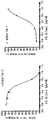
- G—PHYSICS
- G01—MEASURING; TESTING
- G01N—INVESTIGATING OR ANALYSING MATERIALS BY DETERMINING THEIR CHEMICAL OR PHYSICAL PROPERTIES
- G01N33/00—Investigating or analysing materials by specific methods not covered by groups G01N1/00 - G01N31/00
- G01N33/48—Biological material, e.g. blood, urine; Haemocytometers
- G01N33/50—Chemical analysis of biological material, e.g. blood, urine; Testing involving biospecific ligand binding methods; Immunological testing
- G01N33/94—Chemical analysis of biological material, e.g. blood, urine; Testing involving biospecific ligand binding methods; Immunological testing involving narcotics or drugs or pharmaceuticals, neurotransmitters or associated receptors
-
- G—PHYSICS
- G01—MEASURING; TESTING
- G01N—INVESTIGATING OR ANALYSING MATERIALS BY DETERMINING THEIR CHEMICAL OR PHYSICAL PROPERTIES
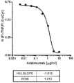
- G01N2800/00—Detection or diagnosis of diseases
- G01N2800/52—Predicting or monitoring the response to treatment, e.g. for selection of therapy based on assay results in personalised medicine; Prognosis
Abstract
본 발명은 시료에서 항-TNFα 약물 치료에 대한 자가항체를 검출하고, 그의 존재여부 또는 그의 수준을 측정하기 위한 검정법을 제공한다. 약물에 대한 자가항체의 존재 여부 또는 그 수준을 검출하는 본 발명은 치료요법을 최적화하고, 항-TNFα 약물 치료법을 제공받는 환자의 모니터링하기에 유용하다. 본 발명은 또한 TNFα-매개성 질환 또는 장애의 치료를 위한 치료요법의 선별, 치료요법의 최적화 및/또는 항-TNFα 약물을 제공받는 대상체에서의 독성 경감을 위한 방법을 제공한다. The present invention provides an assay for detecting autoantibodies to anti-TNFα drug treatment in a sample and determining the presence or level thereof. The present invention, which detects the presence or level of autoantibodies for drugs, is useful for optimizing therapy and for monitoring patients receiving anti-TNFα drug therapy. The invention also provides methods for screening therapies for the treatment of TNFα-mediated diseases or disorders, optimizing therapies and / or reducing toxicity in a subject receiving anti-TNFα drugs.

Description
관련 출원의 상호 참조Cross Reference of Related Application
본 출원은 2011 년 2 월 17 일에 출원된 미국 가출원 번호 61/444,097, 2011 년 5 월 10 일에 출원된 미국 가출원 번호 61/484,594, 및 2011 년 6 월 13 일에 출원된 미국 가출원 번호 61/496,501 에 대한 우선권을 청구하며, 상기 문헌들은 그 어떤 목적으로든 그 전체가 본원에 참고문헌으로 포함된다. This application is directed to US Provisional Application No. 61 / 444,097, filed Feb. 17, 2011, US Provisional Application No. 61 / 484,594, filed May 10, 2011, and US Provisional Application No. 61 /, filed June 13, 2011. 496,501, which is hereby incorporated by reference in its entirety for any purpose.
자가면역 장애는 심각하고 만연되어 있는 의학적인 과제이다. 예를 들면, 류마티스 관절염 (RA) 은 약 2백만 이상의 미국인에게 영향을 미치고 있는 자가면역 질환이다. RA 는 관절의 만성 염증을 야기하고 전형적으로 관절 파괴 및 기능적 불능을 야기할 수 있는 잠재성이 있는 진행성 질병이다. 비록 유전적 소인, 감염 인자 및 환경적 인자가 모두 상기 질병의 병인에 상관있지만, 류마티스 관절염의 원인은 알려져 있지 않다. 활성 RA 에서, 증상은 피로, 식욕 부진, 미열, 근육 및 관절의 통증 및 뻣뻣함을 포함할 수 있다. 질병이 발병되는 동안, 관절은 활막 (synovium) 의 염증으로 인해, 종종 붉게 부풀어오르고, 통증을 느끼며 약해진다. 또한, RA 는 전신적인 질병이기 때문에, 염증이 눈과 입의 샘, 폐 라이닝 (lining), 심낭막 및 혈관을 포함하는, 관절 이외의 다른 신체의 장기 및 영역에 영향을 줄 수 있다. Autoimmune disorders are a serious and widespread medical challenge. For example, rheumatoid arthritis (RA) is an autoimmune disease that affects about two million Americans. RA is a progressive disease with the potential to cause chronic inflammation of the joints and typically to lead to joint destruction and malfunction. Although genetic predisposition, infectious agents and environmental factors all correlate with the pathogenesis of the disease, the cause of rheumatoid arthritis is unknown. In active RA, symptoms may include fatigue, anorexia, mild fever, muscle and joint pain and stiffness. During the development of the disease, the joints often swell red, feel pain and weaken due to inflammation of the synovium. In addition, because RA is a systemic disease, inflammation can affect organs and areas of the body other than the joints, including glands of the eyes and mouth, lung linings, pericardium and blood vessels.
RA 및 다른 자가면역 장애를 관리하기 위한 통상적인 치료방법은 "1 차 약물" 로 먼저 조치를 취하고 "2 차 약물" 로 후속 조치를 취하는 것이다. 1 차 약물은 통증 및 염증을 경감시킨다. 그러한 1 차 약물의 예는 아스피린, 아프록센, 이부프로펜, 에토도락 및 다른 비-스테로이드성 항-염증 약물(NSAID) 뿐만 아니라 코르티코스테로이드를 포함하고, 상기 약물은 경구적으로 또는 조직 및 관절로 직접 주입된다. 2 차 약물은 질병을 완화시키고 진행성 관절 파괴를 막는 것으로 질병-변형 항-류마티스 약물 또는 DMARD 를 지칭한다. 상기 2차 약물은 하이드로 클로로퀴닌, 아주르피딘 및 면역억제제, 예를 들면 메토트렉세이트, 아자티오프린, 사이클로포스파미드, 클로라부실 및 시클로스포린을 포함한다. 그러나, 상기 약물의 대부분은 치명적인 부작용을 가질 수 있다. 따라서, 류마티스 관절염 및 다른 자가면역 장애와 관련된 추가적인 치료요법이 모색되고 있다. A common treatment for managing RA and other autoimmune disorders is to first take action with a "primary drug" and follow up with a "secondary drug." Primary drugs relieve pain and inflammation. Examples of such primary drugs include aspirin, aproxen, ibuprofen, etodorak and other non-steroidal anti-inflammatory drugs (NSAIDs) as well as corticosteroids, which drugs are injected orally or directly into tissues and joints do. Secondary drugs are disease-modified anti-rheumatic drugs or DMARDs that alleviate disease and prevent progressive joint destruction. Such secondary drugs include hydro chloroquinine, azuridin and immunosuppressive agents such as methotrexate, azathioprine, cyclophosphamide, chlorabusil and cyclosporin. However, most of the drugs can have fatal side effects. Thus, additional therapies associated with rheumatoid arthritis and other autoimmune disorders are being sought.
종양 괴사 인자 알파 (TNF-α) 는 단핵구 및 대식세포를 포함하는, 많은 세포들로부터 생성된 싸이토카인으로, 본래 특정 마우스 종양의 괴사를 유도하는 능력을 기반으로 밝혀졌다. 그 다음 악액질 (cachexia) 과 관련된 카케틴 (cachectin) 으로 불리는 인자들이 TNF-α와 동일한 것으로 나타났다. TNF-α는 쇼크, 패혈증, 감염, 자가면역 질환, RA, 크론병, 이식 거부반응 및 이식편-대-숙주 질병을 포함하는, 다양한 인간 질병 및 장애의 병리생리학에 관여한다. Tumor necrosis factor alpha (TNF-α) is a cytokine produced from many cells, including monocytes and macrophages, and was originally discovered based on its ability to induce necrosis of certain mouse tumors. Then a factor called cachectin associated with cachexia appeared to be identical to TNF-α. TNF-α is involved in the pathophysiology of various human diseases and disorders, including shock, sepsis, infections, autoimmune diseases, RA, Crohn's disease, transplant rejection and graft-versus-host disease.
다양한 인간 장애에서의 인간 TNF-α (hTNF-α)의 해로운 역할로 인해, hTNF-α 활성을 억제하거나 중화시키기 위한 치료적 전략이 고안되어 왔다. 특히, hTNF-α에 결합할 수 있고, 이를 중화시킬 수 있는 항체들이 hTNF-α 활성을 억제하기 위한 수단으로서 모색되어 왔다. 가장 최초의, 그러한 항체로서 마우스의 림프구로부터 제조된 하이브리도마에 의해 분비되는, 마우스 모노클로날 항체 (mAbs) 가 있다 (예를 들면, Moeller 등에 허여된 미국 특허 5,231,024) 이러한 마우스 항-hTNF-α 항체는 hTNF-α에 대한 높은 친화성을 나타내고 hTNF-α 활성을 중화시킬 수 있으나, 생체내에서의 그들의 사용은 짧은 혈청 반감기, 특정 인간 영향인자 가능의 유발 불능, 및 인간 내의 마우스 항체에 대한 원치않는 면역 반응 ("인간 항-마우스 항체" (HAMA) 반응) 과 같은 인간에서의 마우스 항체의 투여와 관련된 문제들로 인해 제한적이었다. Because of the detrimental role of human TNF-α (hTNF-α) in various human disorders, therapeutic strategies have been devised to inhibit or neutralize hTNF-α activity. In particular, antibodies that can bind to and neutralize hTNF-α have been sought as a means to inhibit hTNF-α activity. The first such antibody is mouse monoclonal antibody (mAbs), secreted by hybridomas prepared from lymphocytes of mice (eg, US Pat. No. 5,231,024 to Moeller et al.). Such mouse anti-hTNF- α antibodies show high affinity for hTNF-α and can neutralize hTNF-α activity, but their use in vivo is due to short serum half-life, inability to induce certain human influencers, and to mouse antibodies in humans. Limited due to problems associated with the administration of mouse antibodies in humans, such as unwanted immune responses (“human anti-mouse antibody” (HAMA) responses).
더욱 최근에는, 생물학적 치료가 류마티스 관절염과 같은 자가면역 장애의 치료에 적용되어 왔다. 예를 들면, 4 개의 TNFα 억제인자, REMICADE™ (인플릭시마브; infliximab), 키메라성 항-TNFα mAb, ENBREL™ (에타네르셉트; etanercept), TNFR-Ig Fc 융합 단백질, HUMIRA™ (아달리무마브; adalimumab), 인간 항-TNFα mAb, 및 CIMZIA® (세르토리주마브 페골; certolizumab pegol), PEG화된 Fab 단편이 류마티스 관절염의 치료를 위해 FDA의 승인을 받았다. CIMZIA®는 경증 내지 중증 크론병 (CD) 의 치료용으로 사용된다. 그러한 생물학적 치료제가 류마티스 관절염 및 CD와 같은 다른 자가면역 장애의 치료에 성공적임이 입증되었지만, 모든 치료받은 환자들이 그러한 치료에 반응을 하거나 잘 반응하는 것은 아니다. 게다가, TNFα 억제인자의 투여는 약물에 대한 면역 반응을 유도할 수 있고, 인간 항-키메라성 항체 (HACA), 인간 항-인간화 항체 (HAHA), 및 인간 항-마우스 항체 (HAMA) 와 같은 자가항체의 생산을 야기할 수 있다. 그러한 HACA, HAHA, 또는 HAMA 응답 반응은 과민감성 반응 및 약물을 이용한 추가적인 치료를 막는 면역치료학적 TNFα 억제인자의 약동학 및 생체분배에서의 급격한 변화와 관련될 수 있다. 따라서, 당해 기술분야에서는 TNFα 억제인자 치료요법을 모니터링하고 치료 결정을 안내하기 위하여, 환자 시료 중의 항-TNFα 생물학적 제제에 대한 자가항체의 존재를 검출할 검정법의 필요성이 요구되고 있다. 본 발명은 그러한 필요를 충족시키며, 관련된 장점도 제공한다.More recently, biological therapies have been applied to the treatment of autoimmune disorders such as rheumatoid arthritis. For example, four TNFα inhibitors, REMICADE ™ (infliximab), chimeric anti-TNFα mAb, ENBREL ™ (etanercept), TNFR-Ig Fc fusion protein, HUMIRA ™ (adali) Mumab; adalimumab), human anti-TNFα mAb, and CIMZIA® (certolizumab pegol), PEGylated Fab fragments are FDA approved for the treatment of rheumatoid arthritis. CIMZIA® is used for the treatment of mild to severe Crohn's disease (CD). Although such biological therapies have proven successful in the treatment of other autoimmune disorders such as rheumatoid arthritis and CD, not all treated patients respond or respond well to such treatments. In addition, the administration of TNFα inhibitors can induce an immune response to the drug and can cause autoantibodies such as human anti-chimeric antibodies (HACA), human anti-humanized antibodies (HAHA), and human anti-mouse antibodies (HAMA). May result in the production of antibodies. Such HACA, HAHA, or HAMA response responses may be associated with rapid changes in pharmacokinetics and biodistribution of immunotherapeutic TNFα inhibitors that prevent hypersensitivity reactions and further treatment with drugs. Thus, there is a need in the art for assays to detect the presence of autoantibodies against anti-TNFα biologicals in patient samples to monitor TNFα inhibitor therapy and guide treatment decisions. The present invention fulfills that need and provides related advantages as well.
발명의 개요Summary of the Invention
본 발명은 시료에서 항-TNFα 약물 치료에 대한 자가항체를 검출하고, 그의 존재여부 또는 그의 수준을 측정하기 위한 검정법을 제공한다. 약물에 대한 자가항체 (예를 들어, HACA 및/또는 HAHA) 의 존재 여부 또는 그 수준을 검출하는 본 발명은 치료요법을 최적화하고, 항-TNFα 약물 치료법을 제공받는 환자의 모니터링하기에 유용하다. 본 발명은 또한 TNFα-매개성 질환 또는 장애의 치료를 위해 항-TNFα 약물을 제공받는 대상체에서의 치료요법의 선별, 치료요법의 최적화 및/또는 독성 경감 방법을 제공한다.The present invention provides an assay for detecting autoantibodies to anti-TNFα drug treatment in a sample and determining the presence or level thereof. The present invention, which detects the presence or level of autoantibodies to drugs (eg, HACA and / or HAHA), is useful for optimizing therapy and for monitoring patients receiving anti-TNFα drug therapy. The invention also provides methods of screening therapy, optimizing therapy and / or reducing toxicity in a subject receiving an anti-TNFα drug for the treatment of a TNFα-mediated disease or disorder.
한 국면에서, 본 발명은 하기 단계를 포함하는, 시료 중 항-TNFα 약물로부터의 간섭없이 시료 중 항-TNFα 약물에 대한 자가항체의 존재유무 또는 그의 수준 검출 방법을 제공한다:In one aspect, the invention provides a method for detecting the presence or level of autoantibodies to an anti-TNFα drug in a sample without interference from the anti-TNFα drug in the sample, comprising:
(a) 시료를 산과 접촉시켜 자가항체와 항-TNFα 약물의 예비형성 복합체를 해리시키는 단계, 여기서 시료는 항-TNFα 약물에 대한 자가항체를 갖거나 또는 가질 것으로 추정됨; (a) contacting the sample with an acid to dissociate the preformed complex of autoantibody and anti-TNFα drug, wherein the sample has or is assumed to have autoantibody to the anti-TNFα drug;
(b) 예비형성 복합체의 해리 후 상기 시료를 표지된 항-TNFα 약물과 접촉시키는 단계; (b) contacting the sample with a labeled anti-TNFα drug after dissociation of the preformed complex;
(c) 시료 중 산을 중화시켜, 표지된 항-TNFα 약물 및 자가항체의 표지된 복합체를 형성하는 단계; (c) neutralizing the acid in the sample to form a labeled complex of labeled anti-TNFα drug and autoantibody;
(d) 상기 표지된 복합체를 크기 배제 크로마토그래피에 적용시켜 표지된 복합체를 분리하는 단계; 및(d) subjecting the labeled complex to size exclusion chromatography to separate the labeled complex; And
(e) 표지된 복합체를 검출함으로써, 시료 중 항-TNFα 약물의 간섭 없이 자가항체의 존재유무 또는 그의 수준을 검출하는 단계.(e) detecting the presence of or levels of autoantibodies without interference of anti-TNFα drugs in the sample by detecting labeled complexes.
일부 구현예에서, 항-TNFα 약물이 REMICADE™ (인플릭시마브; infliximab), ENBREL™ (에타네르셉트; etanercept), HUMIRA™ (아달리무마브; adalimumab), CIMZIA® (세르토리주마브 페골; certolizumab pegol), SIMPONI® (골림무마브; golimumab; CNTO 148), 및 이들의 조합으로 이루어진 군으로부터 선택된다. In some embodiments, the anti-TNFα drug is administered in REMICADE ™ (infliximab), ENBREL ™ (etanercept), HUMIRA ™ (adalimumab; adalimumab), CIMZIA® (Sertorizumab Pegol) certolizumab pegol), SIMPONI® (golimumab; CNTO 148), and combinations thereof.
다른 구현예에서, 항-TNFα 약물 자가항체가, 이에 제한됨 없이, 항-TNFα 약물에 대한 자가항체가 인간 항-키메라성 항체 (HACA), 인간 항-인간화 항체(HAHA), 인간 항-마우스 항체 (HAMA), 및 이들의 조합을 포함한다. In other embodiments, anti-TNFα drug autoantibodies include, but are not limited to, autoantibodies to anti-TNFα drugs are human anti-chimeric antibodies (HACA), human anti-humanized antibodies (HAHA), human anti-mouse antibodies (HAMA), and combinations thereof.
특정한 대안적인 구현예에서, 단계 (a) 및 (b) 는 동시에 수행되는데, 예를 들어 시료를 산 및 표지된 항-TNFα 약물에 동시에 접촉시킨다. 특정한 다른 대안적인 구현예에서, 단계 (b) 는 단계 (a) 전에 수행되는데, 예를 들어 시료를 먼저 표지된 항-TNFα 약물에 접촉시키고, 이어서 산과 접촉시킨다. 추가 구현예에서, 단계 (b) 및 (c) 는 동시에 수행되는데, 예를 들어 시료를 표지된 항-TNFα 약물에 접촉시키면서 동시에 중화 (예를 들어, 시료를 하나 이상의 중화제와 접촉시킴) 시킨다.In certain alternative embodiments, steps (a) and (b) are performed simultaneously, eg, contacting a sample with an acid and a labeled anti-TNFα drug simultaneously. In certain other alternative embodiments, step (b) is performed before step (a), eg, contacting the sample with a labeled anti-TNFα drug first, followed by contact with an acid. In further embodiments, steps (b) and (c) are performed simultaneously, for example, while the sample is contacted with a labeled anti-TNFα drug and simultaneously neutralized (eg, the sample is contacted with one or more neutralizing agents).
특별한 구현예에서, 시료를 자가항체와 항-TNFα 약물의 예비형성된 복합체를 해리시키기에 충분한 양의 산과 접촉시켜, 표지된 항-TNFα 약물, 표지되지 않은 항-TNFα 약물 및 항-TNFα 약물에 대한 자가항체가 평형을 이루고 그들 사이에 복합체를 형성하도록 한다. In a particular embodiment, the sample is contacted with an amount of acid sufficient to dissociate the preformed complex of autoantibody and anti-TNFα drug, thereby allowing for labeled anti-TNFα drug, unlabeled anti-TNFα drug and anti-TNFα drug. Allow autoantibodies to equilibrate and form complexes between them.
또다른 국면에서, 본 발명은 하기 단계를 포함하는, 항-TNFα 약물을 이용하는 치료요법 과정을 제공받는 대상체에서의 치료요법 최적화 및/또는 항-TNFα 약물에 대한 독성의 경감을 위한 방법을 제공한다:In another aspect, the invention provides a method for optimizing therapy and / or reducing toxicity to anti-TNFα drugs in a subject receiving a therapy course using an anti-TNFα drug, comprising the following steps: :
(a) 하기 단계를 포함하는, 시료 중 항-TNFα 약물로부터의 간섭없이 대상체 유래의 시료 중 항-TNFα 약물에 대한 자가항체의 존재유무 또는 그의 수준을 검출하는 단계: (a) detecting the presence or level of autoantibodies to anti-TNFα drugs in a sample from a subject without interference from the anti-TNFα drugs in the sample, comprising:
(i) 시료를 산과 접촉시켜 자가항체와 항-TNFα 약물의 예비형성된 복합체를 해리시키는 단계, 여기서 시료는 항-TNFα 약물에 대한 자가항체를 갖고 있거나 또는 가질 것으로 추정됨; (i) contacting the sample with an acid to dissociate the preformed complex of autoantibody and anti-TNFα drug, wherein the sample has or is assumed to have autoantibodies to the anti-TNFα drug;
(ii) 예비형성 복합체의 해리 후 상기 시료를 표지된 항-TNFα 약물과 접촉시키는 단계;(ii) contacting the sample with a labeled anti-TNFα drug after dissociation of the preformed complex;
(iii) 시료 중의 산을 중화하여 표지된 항-TNFα 약물 및 자가항체의 표지된 복합체를 형성하는 단계; (iii) neutralizing the acid in the sample to form a labeled complex of labeled anti-TNFα drug and autoantibody;
(iv) 상기 표지된 복합체를 크기 배제 크로마토그래피에 적용시켜 표지된 복합체를 분리하는 단계; 및(iv) subjecting the labeled complex to size exclusion chromatography to separate the labeled complex; And
(v) 표지된 복합체를 검출하여 시료 중 항-TNFα 약물로부터의 간섭없이 자가항체의 존재유무 또는 그의 수준을 검출하는 단계; 및 (v) detecting the labeled complex to detect the presence or level of autoantibodies in the sample without interference from anti-TNFα drugs; And
(b) 자가항체의 존재여부 또는 그의 수준을 근거로 대상체를 위한 치료요법 과정의 후속 투여용량 또는 상이한 치료요법 과정을 대상체에 적용해야 하는지 여부를 결정하는 단계, (b) determining whether a subsequent dose or different therapy course of therapy course for the subject should be applied to the subject based on the presence or level of autoantibodies,
이로써 치료요법을 최적화하고/하거나 항-TNFα 약물에 대한 독성을 경감시키는 단계.Thereby optimizing therapy and / or alleviating toxicity to anti-TNFα drugs.
항-TNFα 약물 및 항-약물 항체의 검출 방법은 PCT 공보 번호 WO 201 1/056590 에 추가로 기재되어 있고, 그 개시내용은 어떤 목적으로든 전문이 본원에 참고문헌으로 포함된다.Methods for detecting anti-TNFα drugs and anti-drug antibodies are further described in PCT Publication No.
다른 국면에서, 본 발명은 하기 단계를 포함하는, TNFα-매개성 질환 또는 장애의 치료를 위한 치료요법 과정의 선택 (예를 들어, 적절한 항-TNFα 약물의 선택) 방법을 제공한다: In another aspect, the invention provides a method of selecting (eg, selecting an appropriate anti-TNFα drug) for a therapy course for the treatment of a TNFα-mediated disease or disorder, comprising the following steps:
(a) 대상체로부터 수득한 시료를 분석하여 시료 중 하나 이상의 마커의 존재유무, 그의 수준 또는 유전자형을 분석하는 단계;(a) analyzing a sample obtained from the subject to analyze the presence, level or genotype of one or more markers in the sample;
(b) 단계 (a) 에서 결정된 하나 이상의 마커의 존재유무, 그의 수준 또는 유전자형에 통계적 알고리즘을 적용하여 질환 활성/중증도 지수를 생성하는 단계; 및(b) applying a statistical algorithm to the presence, level or genotype of one or more markers determined in step (a) to generate a disease activity / severity index; And
(c) 질환 활성/중증도 지수를 기준으로 대상체에 대한 적절한 치료요법 과정 (예를 들어, 항-TNFα 치료요법) 을 선택하는 단계. (c) selecting an appropriate therapy course (eg anti-TNFα therapy) for the subject based on the disease activity / severity index.
관련 국면에서, 본 발명은 하기 단계를 포함하는, TNFα-매개성 질환 또는 장애의 치료를 위한 치료요법 과정을 제공받는 대상체에서 치료요법을 최적화하고/하거나 독성을 경감하는 방법을 제공한다:In a related aspect, the present invention provides a method of optimizing therapy and / or reducing toxicity in a subject receiving a therapeutic course for the treatment of a TNFα-mediated disease or disorder, comprising the following steps:
(a) 대상체로부터 수득한 시료를 분석하여 시료 중 하나 이상의 마커의 존재유무, 그의 수준 또는 유전자형을 분석하는 단계;(a) analyzing a sample obtained from the subject to analyze the presence, level or genotype of one or more markers in the sample;
(b) 단계 (a) 에서 결정된 하나 이상의 마커의 존재유무, 그의 수준 또는 유전자형에 통계적 알고리즘을 적용하여 질환 활성/중증도 지수를 생성하는 단계; 및(b) applying a statistical algorithm to the presence, level or genotype of one or more markers determined in step (a) to generate a disease activity / severity index; And
(c) 질환 활성/중증도 지수를 기준으로 대상체에 대한 치료요법 과정의 후속 투여용량 또는 상이한 치료요법 과정이 대상체에 제공되어야 하는지 여부를 결정하는 단계. (c) determining whether the subject should be given a subsequent dose or different therapy course of therapy course for the subject based on the disease activity / severity index.
특별한 구현예에서, 본 발명의 방법은 하기의 바이오마커 카테고리 중 한가지 이상에서 하나 이상의 특정 마커의 존재유무, 그의 수준 (농도 (예를 들어, 총 농도) 및/또는 활성화 (예를 들어, 인산화)) 또는 유전자형을 검출, 측정 또는 결정하는 것을 포함한다: In particular embodiments, the methods of the present invention provide for the presence, presence (eg, concentration (eg, total concentration), and / or activation (eg, phosphorylation) of one or more specific markers in one or more of the following biomarker categories: Or detecting, measuring or determining genotypes:
(1) 염증 마커 (1) inflammation markers
(2) 성장 인자 (2) growth factors
(3) 혈청학 (예를 들어, 면역 마커) (3) serology (eg, immune markers)
(4) 싸이토카인 및 케모카인 (4) Cytokines and Chemokines
(5) 산화 스트레스의 마커 (5) markers of oxidative stress
(6) 세포 표면 수용체 (예를 들어, CD64, 기타) (6) cell surface receptors (eg, CD64, etc.)
(7) 신호전달 경로 (7) signaling path
(8) 기타 마커 (예를 들어, 유전자 마커, 예컨대 염증 경로 유전자).(8) other markers (eg, genetic markers such as inflammatory pathway genes).
추가 구현예에서, 하기 마커들 중 하나 또는 두가지 모두의 존재유무 및/또는 그의 수준이 환자 시료 (예를 들어, 항-TNF 약물 치료요법 중인 환자 유래의 혈청 시료) 에서 검출, 측정 또는 결정될 수 있다: (9) 항-TNF 약물 수준 (예를 들어, 유리된 항-TNFα 치료 항체의 수준); 및/또는 (10) 항-약물 항체 (ADA) 수준 (예를 들어, 항-TNF 약물에 대한 자가항체의 수준).In further embodiments, the presence and / or level of one or both of the following markers can be detected, measured or determined in a patient sample (eg, a serum sample from a patient under anti-TNF drug therapy). (9) anti-TNF drug levels (eg, levels of free anti-TNFα therapeutic antibodies); And / or (10) anti-drug antibody (ADA) levels (eg, levels of autoantibodies to anti-TNF drugs).
특별한 구현예에서, 단일 통계적 알고리즘 또는 두가지 이상의 통계적 알고리즘의 조합이 시료 중에서 검출, 측정 또는 결정되는 존재유무, 그의 농도 수준, 활성화 수준 또는 유전자형에 대해 적용될 수 있어, 질환 활성/중증도 지수가 생성된다.In a particular embodiment, a single statistical algorithm or a combination of two or more statistical algorithms can be applied to the presence, concentration level, activation level or genotype detected, measured or determined in a sample, resulting in a disease activity / severity index.
특정한 경우, 시료는 당업계에 공지된 임의의 기법을 이용해 PBMC 및/또는 PMN 세포를 단리하여 수득된다. 또다른 구현예에서, 시료는 예를 들어 염증 부위, 예컨대 위장관 또는 활액 조직의 일부분 유래의 조직 생검이다.In certain instances, samples are obtained by isolating PBMCs and / or PMN cells using any technique known in the art. In another embodiment, the sample is a tissue biopsy, eg, from a site of inflammation, such as a gastrointestinal tract or synovial tissue.
따라서, 일부 국면에서, 본 발명의 방법은, 예를 들어 최초 치료를 위한 적절한 항-TNFα 치료요법을 선별함으로써, 항-TNFα 약물의 후속 투여용량을 언제 또는 어떻게 조정 또는 변경 (예를 들어 증가 또는 감소) 할 것인지를 결정함으로써, 항-TNFα 약물과 하나 이상의 면역억제제, 예컨대 메토트렉세이트 (MTX) 및/또는 아자티오프린 (AZA) 를 언제 또는 어떻게 병용할지 (예를 들어, 최초에, 증가, 감소 또는 동일 투여용량) 를 결정함으로써 및/또는 현재의 치료요법 과정을 변경 (예를 들어 상이한 항-TNFα 약물로 또는 상이한 메커니즘을 표적으로 하는 약물, 예컨대 IL-6 수용체-저해 모노클로날 항체로 전환) 을 언제 또는 어떻게 할지 결정함으로써 항-TNFα 약물 치료요법을 제공받고 있거나 또는 제공받게 될 환자의 치료 결정을 안내하기에 유용한 정보를 제공한다.Thus, in some aspects, the method of the present invention adjusts or alters (e.g., increases or reduces the subsequent dose of anti-TNFα drug, for example, by selecting an appropriate anti-TNFα therapy for initial treatment. By determining when or how to combine (eg, initially increase, decrease or reduce) an anti-TNFα drug with one or more immunosuppressive agents such as methotrexate (MTX) and / or azathioprine (AZA). And / or alter the current therapy course (e.g., to different anti-TNFα drugs or to drugs that target different mechanisms, such as IL-6 receptor-inhibiting monoclonal antibodies) Provide information useful to guide the treatment decision of a patient who is or will be receiving anti-TNFα drug therapy by deciding when or how The.
본 발명의 여타 목적, 특징 및 장점은 하기의 상세한 설명 및 도면으로부터 당업자에게 자명할 것이다. Other objects, features and advantages of the invention will be apparent to those skilled in the art from the following detailed description and drawings.
도 1 은 본 발명의 검정법에 대한 예시적인 구현예를 나타내는 것으로, 여기서 크기 배제 HPLC 가 TNFα-Alexa647과 HUMIRA™의 결합을 검출하기 위하여 사용된다.
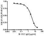
도 2 는 TNFα-Alexa647에 결합하는 HUMIRA™의 투여용량 응답 곡선을 나타낸다.
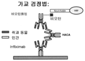
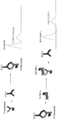
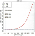
도 3 은 가교 검정법으로 공지된, HACA 수준을 측정하기 위한 현재의 ELISA-기반 방법을 나타낸다.
도 4 는 REMICADE™에 의해 발생되는 HACA/HAHA 의 농도를 측정하기 위한 본 발명의 자가항체 검출 검정법의 예시적인 개요를 나타낸다.
도 5 는 REMICADE™-Alexa647에 결합하는 항-인간 IgG 항체의 투여용량 응답 분석을 나타낸다.
도 6 은 REMICADE™-Alexa647에 결합하는 항-인간 IgG 항체의 제 2 투여용량 응답 분석을 나타낸다.
도 7 은 REMICADE™-Alexa647에 결합하는 항-인간 IgG 항체의 투여용량 응답 곡선을 나타낸다.
도 8 은 정상 인간 혈청 및 HACA 양성 혈청 내의 REMICADE™-Alexa647 면역복합체 형성을 나타낸다.
도 9 는 본 발명의 가교 검정법 또는 이동성 변동 검정법을 사용하여 수행된 20 명의 환자 혈청 시료로부터의 HACA 측정의 요약을 제공한다.
도 10 은 HACA 의 혈청 농도를 측정하기 위한 현재의 방법과 본 발명의 신규한 HACA 검정법의 요약 및 비교를 제공한다.
도 11 은 정상 (NHS) 또는 HACA-양성(HPS) 혈청으로 인큐베이션된 형광물질 (F1)-표지된 IFX 의 SE-HPLC 프로파일을 나타낸다. 인큐베이션 혼합물에 증량된 HACA-양성 혈청의 양을 첨가하면, IFX-F1 피크가 더 높은 분자량 용출 위치, C1 및 C2 로 투여용량-의존적으로 이동된다.
도 12 는 이동성 변동 검정법에 의해 측정된 HACA-양성 혈청의 희석량을 증가시키면서 생성되는 결합 및 유리된 IFX-F1 의 투여용량-응답성 곡선을 나타낸다. (A) HACA-양성 혈청의 희석을 확대하면서 37.5 ng의 IFX-F1 과 인큐베이션하였다. 희석이 확대될수록 (HACA 미만), 더 많은 유리된 IFX-F1 가 SE-HPLC 분석에서 발견되었다. (B) HACA-양성 혈청의 희석을 증가시키면서 37.5 ng의 IFX-F1와 인큐베이션하였다. 희석이 증가할수록 (HACA 미만) 더 적은 HACA 결합 IFX-F1이 SE-HPLC 분석에서 발견되었다.
도 13 은 정상 (NHS) 또는 IFX-스파이크된 혈청과 인큐베이션된 TNFα-F1 의 SE-HPLC 프로파일을 나타낸다. 인큐베이션 혼합물에 첨가된 IFX-스파이크된 혈청의 양이 증가할수록, 형광 TNFα 피크는 더 높은 분자량 용출 위치로 투여용량-의존적으로 이동한다.
도 14 는 이동성 변동 검정법에 의해 측정된 IFX-스파이크된 혈청의 희석을 증가시키는 것에 의해 발생된 결합 및 유리된 TNFα 의 투여용량-응답성 곡선을 나타낸다. 인큐베이션 혼합물에 첨가된 IFX 의 농도가 증가하면, 결합 TNFα 의 백분율을 증가하는 반면, 유리된 TNFα 의 백분율을 감소한다 .
도 15 는 이동성 변동 검정법에 의해 상이한 시점에서 IFX 로 치료받은 IBD 환자에서의 상대적인 HACA 수준 및 IFX 농도의 측정을 보여준다.
도 16 은 환자 관리 -상이한 시점에서 IFX 로 치료받은 IBD 환자 혈청 중 HACA 수준 및 IFX 농도의 측정- 를 보여준다.
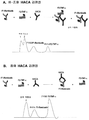
도 17 은 HACA 와 같은 (A) 비-중화 또는 (B) 중화 자가항체의 존재를 검출하기 위한 본 발명의 검정법의 예시적인 구현예를 나타낸다.
도 18 은 HACA 와 같은 중화 자가항체의 존재유무를 검출하기 위한 본 발명의 검정법의 대안적인 구현예를 나타낸다.
도 19 는 상이한 양의 항-인간 IgG 의 존재시 정상 인간 혈청 (NHS) 과 인큐베이션된 F1-표지된 ADL 의 이동성 변동 프로파일을 나타낸다. 인큐베이션 혼합물에 항-인간 IgG 첨가량을 증가시키면, 유리된 F1-ADL 피크 (FA)는 더 높은 분자량 용출 위치, C1 및 C2 로 투여용량-의존적으로 이동하는 반면, 내부 대조군 (IC) 피크는 변화하지 않는다.
도 20 은 유리된 F1-ADL 의 이동시 항-인간 IgG 의 투여용량-응답성 곡선을 나타낸다. 항-인간 IgG 의 양을 증가시키면서, 37.5 ng의 F1-ADL 및 내부 대조군과 인큐베이션하였다. 더 많은 항체를 반응 혼합물에 첨가할수록 내부 대조군에 대한 유리된 F1-ADL 비율이 더 낮아진다.
도 21 은 상이한 양의 ADL 존재시 정상 인간 혈청 (NHS) 과 인큐베이션된 F1-표지된 TNF-α 의 이동성 변동 프로파일을 나타낸다. Ex = 494 nm; Em = 519 nm. 인큐베이션 혼합물에 ADL 의 첨가량을 증가시키면, 유리된 TNF-F1 피크 (FT)는 더 큰 분자량 용출 위치로 투여용량-의존적으로 이동하는 반면, 내부 대조군(IC) 피크는 변화하지 않는다.
도 22 는 유리된 TNF-α-F1 의 이동에 대한 ADL 의 투여용량-응답성 곡선을 나타낸다. ADL 의 양을 증가시키면서 100 ng의 TNF-α-F1 및 내부 대조군과 인큐베이션하였다. 항체 ADL이 반응 혼합물에 많이 첨가될수록, 내부 대조군에 대한 유리된 TNF-α-F1 의 비율이 낮아진다.
도 23 은 정상 (NHS) 또는 모아들인 HACA 양성 환자 혈청과 인큐베이션된 F1-표지된 Remicade (IFX)의 이동성 변동 프로파일을 나타낸다.
도 23 은 정상 (NHS) 또는 모아들인 HACA 양성 환자 혈청과 인큐베이션된 F1-표지된 Remicade (IFX)의 이동성 변동 프로파일을 나타낸다.
도 24 는 정상 (NHS) 또는 마우스 항-인간 IgG1 항체와 인큐베이션된 F1-표지된 HUMIRA (ADL) 의 이동성 변동 프로파일을 나타낸다.
도 25 는 정상 (NHS) 또는 모아들인 HAHA 양성 환자 혈청과 인큐베이션된 F1 -표지된 HUMIRA (ADL) 의 이동성 변동 프로파일을 나타낸다.
도 26 은 산 해리 단계의 유효성을 도시한다. "A" 는 표지된-Remicade 를 나타내고, "B" 는 HACA 를 나타내고, "C" 는 Remicade 를 나타낸다.
도 27 은 산 해리 단계 부재 하의 로그 환자 혈청 백분율의 함수로서의 백분율 유리된 표지된-Infliximab 를 나타낸다.
도 28 은 산 해리 단계 존재 하의 로그 환자 혈청 백분율의 함수로서의 백분율 유리된 표지된-Infliximab 를 나타낸다.
도 29 은 환자 케이스 1 에 대해 시간의 함수로서 Infliximab 로 치료한 환자에서의 혈청 IFX 수준을 보여준다.
도 30 은 환자 케이스 3 에 대해 시간의 함수로서 Infliximab 로 치료한 환자에서의 혈청 IFX 를 보여준다.
도 31 은 환자 케이스 3 에 대해 시간의 함수로서 Infliximab 로 치료한 환자에서의 혈청 TNFα 수준을 보여준다.
도 32 는 환자 케이스 1 (A); 환자 케이스 2 (B, C); 및 환자 케이스 4 (D) 에 대해 F1-표지된-IFX 의 이동성 변동 프로파일을 보여준다.
도 33 은 환자 케이스 5 (A); 환자 케이스 6 (B, C); 및 환자 케이스 7 (D, E) 에 대해 F1-표지된-IFX 의 이동성 변동 프로파일을 보여준다.
도 34 는 상이한 환자 혈청 군에서의 싸이토카인 수준을 보여준다.
도 35 는 상이한 값으로 증가 설정한 형광 검출기를 이용한 이동성 변동 검정법에 의한 TNF-Alexa488 및 Remicade 를 포함하는 시료의 분석을 보여준다.
도 36 은 HPLC 이동상 (1X PBS, 수중 0.1% BSA) 에서의 정상 인간 혈청 (상단 패널) 및 TNF-Alexa488 (하단 패널) 에 대해 취해진 등흡광 곡선 (isoabsorbance plot) 을 보여준다. 여기 파장을 Y-축에 표시했고, 발광 파장을 X-축에 표시했다.
도 37 은 표시한 설정으로 검출된 정상 인간 혈청 (좌측) 및 25ng TNF-Alexa488 (우측) 의 HPLC 분석을 보여준다. 정상 인간 혈청으로부터의 형광의 배경 수준은 크게 감소되어 있다.
도 38 은 고정된 양의 TNF-Alexa488 를 포함하는 시료의 HPLC 분석에 의해 생성되고, 다양한 양의 Remicade 로 적가된 표준 곡선을 보여준다.
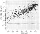
도 39 는 이동성 변동 검정법 및 ELISA 에 의한 임상 시료에서의 Infliximab 측정 비교를 보여준다. 진회색 점은 HACA-양성 시료에 대한 것이고, 연회색 점은 HACA-음성 시료에 대한 것이다. 파선은 각 방법에 대한 정량의 하한선을 나타낸다.
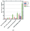
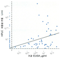
도 40 은 이동성 변동 검정법 및 ELISA 에 의한 임상 시료에서의 HACA 측정 비교를 보여준다.
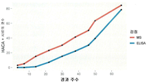
도 41 은 이동성 변동 검정법 및 ELISA 에 의해 결정되는 HACA-양성 임상 시료의 누적 계수를 보여준다. 1 shows an exemplary embodiment of the assay of the present invention wherein size exclusion HPLC is used to detect binding of TNFα-Alexa 647 with HUMIRA ™.
2 shows a dose response curve of HUMIRA ™ that binds TNFα-Alexa 647 .
3 shows current ELISA-based methods for measuring HACA levels, known as crosslinking assays.
4 shows an exemplary schematic of the autoantibody detection assay of the present invention for measuring the concentration of HACA / HAHA generated by REMICADE ™.
5 shows a dose response analysis of anti-human IgG antibody binding to REMICADE ™ -Alexa 647 .
6 shows a second dose response analysis of anti-human IgG antibody that binds REMICADE ™ -Alexa 647 .
7 shows the dose response curves of anti-human IgG antibodies that bind to REMICADE ™ -Alexa 647 .
8 shows REMICADE ™ -Alexa 647 immunocomplex formation in normal human serum and HACA positive serum.
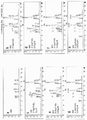
9 provides a summary of HACA measurements from 20 patient serum samples performed using the crosslinking assay or mobility shift assay of the present invention.
10 provides a summary and comparison of the current method for measuring serum concentrations of HACA with the novel HACA assay of the present invention.
FIG. 11 shows SE-HPLC profiles of fluorescent (F1) -labeled IFX incubated with normal (NHS) or HACA-positive (HPS) serum. Adding the amount of increased HACA-positive serum to the incubation mixture causes the IFX-F1 peak to be dose-dependently shifted to higher molecular weight elution sites, C1 and C2.
12 shows dose-response curves of bound and free IFX-F1 produced with increasing dilution of HACA-positive serum as measured by the mobility shift assay. (A) Dilutions of HACA-positive serum were incubated with 37.5 ng of IFX-F1. As the dilution expanded (below HACA), more free IFX-F1 was found in the SE-HPLC analysis. (B) Incubated with 37.5 ng of IFX-F1 with increasing dilution of HACA-positive serum. As dilution increased (less than HACA), less HACA binding IFX-F1 was found in SE-HPLC analysis.
FIG. 13 shows SE-HPLC profiles of TNFα-F1 incubated with normal (NHS) or IFX-spiked serum. As the amount of IFX-spiked serum added to the incubation mixture increases, the fluorescent TNFa peak shifts dose-dependently to the higher molecular weight elution site.
FIG. 14 shows dose-response curves of binding and free TNFα generated by increasing dilution of IFX-spiked serum measured by mobility shift assay. Increasing the concentration of IFX added to the incubation mixture increases the percentage of bound TNFα, while decreasing the percentage of free TNFα.
15 shows the measurement of relative HACA levels and IFX concentrations in IBD patients treated with IFX at different time points by the mobility shift assay.
FIG. 16 shows patient management—determination of HACA levels and IFX concentrations in IBD patient serum treated with IFX at different time points.
FIG. 17 shows exemplary embodiments of the assays of the invention for detecting the presence of (A) non-neutralizing or (B) neutralizing autoantibodies such as HACA.
18 shows an alternative embodiment of the assay of the present invention for detecting the presence of neutralizing autoantibodies such as HACA.
19 shows the mobility variation profile of F1-labeled ADL incubated with normal human serum (NHS) in the presence of different amounts of anti-human IgG. Increasing the amount of anti-human IgG added to the incubation mixture results in a dose-dependent shift of the free F1-ADL peak (FA) to the higher molecular weight elution sites, C1 and C2, while the internal control (IC) peak does not change. Do not.
20 shows dose-response curves of anti-human IgG upon migration of free F1-ADL. Increasing the amount of anti-human IgG was incubated with 37.5 ng of F1-ADL and internal control. The more antibody added to the reaction mixture, the lower the free F1-ADL ratio for the internal control.
21 shows the mobility shift profile of F1-labeled TNF-α incubated with normal human serum (NHS) in the presence of different amounts of ADL. Ex = 494 nm; Em = 519 nm. Increasing the amount of ADL added to the incubation mixture, the free TNF-F1 peak (FT) shifts dose-dependently to the higher molecular weight elution site, while the internal control (IC) peak does not change.
FIG. 22 shows dose-response curves of ADL for the migration of free TNF-α-F1. Incubation with 100 ng of TNF-α-F1 and internal control with increasing amount of ADL. The more antibody ADL is added to the reaction mixture, the lower the ratio of free TNF-α-F1 to the internal control.
FIG. 23 shows the mobility variation profile of F1-labeled Remicade (IFX) incubated with normal (NHS) or pooled HACA positive patient serum.
FIG. 23 shows the mobility variation profile of F1-labeled Remicade (IFX) incubated with normal (NHS) or pooled HACA positive patient serum.
FIG. 24 shows the mobility variation profile of F1-labeled HUMIRA (ADL) incubated with normal (NHS) or mouse anti-human IgG1 antibody.
FIG. 25 shows the mobility variation profile of F1-labeled HUMIRA (ADL) incubated with normal (NHS) or pooled HAHA positive patient serum.
26 shows the validity of the acid dissociation step. "A" represents labeled-Remicade, "B" represents HACA and "C" represents Remicade.
27 shows the percentage free labeled-Infliximab as a function of the log patient serum percentage in the absence of the acid dissociation step.
28 shows the percentage free labeled-Infliximab as a function of the log patient serum percentage in the presence of the acid dissociation step.
FIG. 29 shows serum IFX levels in patients treated with Infliximab as a function of time for
FIG. 30 shows serum IFX in patients treated with Infliximab as a function of time for
FIG. 31 shows serum TNFα levels in patients treated with Infliximab as a function of time for
32 is Patient Case 1 (A); Patient case 2 (B, C); And mobility variation profile of F1-labeled-IFX for patient case 4 (D).
33 is Patient Case 5 (A); Patient case 6 (B, C); And mobility variation profile of F1-labeled-IFX for patient case 7 (D, E).
34 shows cytokine levels in different patient serum groups.
FIG. 35 shows analysis of samples comprising TNF-Alexa488 and Remicade by mobility shift assay with fluorescence detector set to different values.
FIG. 36 shows isoabsorbance plots taken for normal human serum (top panel) and TNF-Alexa488 (bottom panel) in HPLC mobile phase (1 × PBS, 0.1% BSA in water). The excitation wavelength is indicated on the Y-axis and the emission wavelength is indicated on the X-axis.
37 shows HPLC analysis of normal human serum (left) and 25 ng TNF-Alexa488 (right) detected at the indicated settings. Background levels of fluorescence from normal human serum are greatly reduced.
FIG. 38 shows standard curves generated by HPLC analysis of samples containing fixed amounts of TNF-Alexa488 and added to various amounts of Remicade.
39 shows a comparison of Infliximab measurements in clinical samples by mobility shift assay and ELISA. Dark gray dots are for HACA-positive samples and light gray dots are for HACA-negative samples. The dashed line represents the lower limit of quantification for each method.
40 shows a comparison of HACA measurements in clinical samples by mobility shift assay and ELISA.
41 shows cumulative counts of HACA-positive clinical samples as determined by mobility shift assay and ELISA.
발명의 상세한 설명 DETAILED DESCRIPTION OF THE INVENTION
I. 도입부 I. INTRODUCTION
본 발명은 부분적으로는 면역 복합체의 평형화를 가능케 하는 크기 배제 크로마토그래피 및 산 해리를 이용한 균질 이동성 변동 검정법이 항-TNFα 약물에 대해 생성되는 자가항체들 (예를 들어, HACA, HAHA 등) 의 존재여부 또는 수준을 측정하기에 특히 유리하다는 발견사실을 근거로 한다. 그러한 자가항체들은 항-약물 항체 또는 ADA 로도 공지되어 있다. 그 결과, 필요로 인해 대상체에 투여된 항-TNFα 약물에 대한 자가항체들의 존재여부 또는 그 수준이 대상체의 시료 중에도 존재하는 투여된 항-TNFα 약물로부터의 실질적인 간섭없이 측정될 수 있다. 특히, 대상체의 시료는 높은 항-TNFα 약물 수준에 의한 실질적인 간섭없이 항-TNFα 약물의 존재 하에 자가항체들의 존재여부 또는 그의 수준 측정용으로 제공되는 충분한 일정량의 산과 함께 인큐베이션될 수 있다. The present invention relates in part to the presence of autoantibodies (e.g., HACA, HAHA, etc.) in which homogeneous mobility shift assays using size exclusion chromatography and acid dissociation to enable equilibration of immune complexes are produced for anti-TNFα drugs. It is based on the finding that it is particularly advantageous to measure whether or not. Such autoantibodies are also known as anti-drug antibodies or ADA. As a result, the presence or level of autoantibodies to the anti-TNFα drug administered to the subject as needed can be measured without substantial interference from the administered anti-TNFα drug present in the subject's sample. In particular, a subject's sample may be incubated with a sufficient amount of acid provided for the presence or determination of levels of autoantibodies in the presence of anti-TNFα drugs without substantial interference with high anti-TNFα drug levels.
시료 중 높은 항-TNFα 약물 수준 (예를 들어, 높은 인플릭시마브 수준) 은 항-약물 항체 수준 (예를 들어, HACA 수준) 의 측정을 간섭한다. 특정한 높은 약물 상태에서는, 시료에 존재하는 항-약물 항체가 시료 중에 역시 존재하는 비표지 약물과 복합체를 형성한다. 표지된 약물, 예를 들어 표지된-인플릭시마브가 시료와 접촉하는 경우, 시료에 존재하는 항-약물 항체는 표지된 약물과의 복합체 형성으로부터 운동에너지에 의해 갖히게 된다 (kinetically trapped). 그러한 방식으로, 항-약물 항체 및 비표지 약물의 예비형성된 복합체는 항-약물 항체의 측정을 간섭하게 되고, 이는 존재하는 항-약물 항체 및 표지된 약물 사이의 복합체 형성에 좌우된다. 본원에 기재된 산 해리 단계는 시료에 존재하는 항-약물 항체가 비표지 약물로부터 해리되도록 하고, 표지 및 비표지 약물의 두가지 모두와 복합체를 다시 형성한다. 비표지 약물로부터 항-약물 항체를 해리시킴으로써, 시료에 존재하는 항-약물 항체는 표지된 약물 및 비표지 약물 사이에서 평형을 이룰 수 있게 된다. High anti-TNFα drug levels (eg, high infliximab levels) in the sample interfere with the measurement of anti-drug antibody levels (eg, HACA levels). In certain high drug states, anti-drug antibodies present in the sample complex with unlabeled drugs that are also present in the sample. When a labeled drug, such as labeled-infliximab, comes into contact with a sample, the anti-drug antibody present in the sample is trapped by kinetic energy from complexation with the labeled drug. In such a manner, the preformed complex of anti-drug antibody and unlabeled drug interferes with the measurement of the anti-drug antibody, which depends on the formation of the complex between the anti-drug antibody and the labeled drug present. The acid dissociation step described herein causes the anti-drug antibody present in the sample to dissociate from the unlabeled drug and recomplex with both the labeled and unlabeled drug. By dissociating the anti-drug antibody from the unlabeled drug, the anti-drug antibody present in the sample is able to equilibrate between the labeled drug and the unlabeled drug.
도 27 에 제시된 바와 같이, 높은 수준의 항-TNFα 약물 (예를 들어, 인플릭시마브) 은 이동성 변동 검정법을 산 해리 단계 없이 수행했을 때 항-약물 항체 (예를 들어, 인플릭시마브 또는 ATI 에 대한 항체) 의 검출을 간섭한다. 그러나, 도 28 은 산 해리에 후속하는 균질 용액 상 결합 동역학이 항-TNFα 약물 용인능이 현저히 증가된 면역 복합체의 평형화 및 재형성을 가능케 하여, 항-약물 항체들이 높은 수준의 항-TNFα 약물 (예를 들어, 약 60 μg/mL 까지 또는 그 이상) 의 존재 하에 측정될 수 있다는 점을 보여준다. 마찬가지로, 본 발명의 검정법은 현재 이용가능한 방법들 중 특히 유리한데, 이는 본 발명의 방법이 항-TNFα 약물 (예를 들어, 시료, 예컨대 혈액 시료 중의 저, 중 또는 고 수준의 항-TNFα 약물과는 무관하게) 을 이용하는 치료요법 동안 언제라도 항-약물 항체들의 검출 및 측정을 가능하게 하기 때문인데, 이로써 약물의 저점 농도에서 시료 수거를 해야 하는 선행기술의 방법의 주된 제약점을 극복한다. As shown in FIG. 27, high levels of anti-TNFα drug (eg, infliximab) were used to determine anti-drug antibodies (eg, infliximab or when the mobility shift assay was performed without an acid dissociation step. Interference with ATI). However, FIG. 28 shows that homogeneous solution phase binding kinetics following acid dissociation allows equilibrium and remodeling of immune complexes with markedly increased anti-TNFα drug tolerance, leading to high levels of anti-TNFα drug (eg For example, it can be measured in the presence of (up to about 60 μg / mL or more). Likewise, the assays of the present invention are particularly advantageous among the currently available methods, where the methods of the present invention are combined with anti-TNFα drugs (eg, low, medium or high levels of anti-TNFα drugs in a sample, such as a blood sample). Irrespective of the possibility of detecting and measuring anti-drug antibodies at any time during therapy, thereby overcoming the major limitations of the prior art methods of sample collection at the low concentration of the drug.
특정 국면에서, 본 발명은, 부분적으로는 항-TNFα 약물 치료요법을 제공받고 있거나 또는 곧 제공받을 환자들에 있어서 치료 결정을 안내하기에 유용한 정보를 제공함으로써, 항-TNFα 약물, 예컨대 인플릭시마브의 투여와 연관된 현재의 제약점을 해결하고 극복하기 때문에 유리하다. 특히, 본 발명의 방법은 최초의 치료를 위해, 적절한 항-TNFα 치료요법을 선택하기 위해, 치료 효능의 최적화 및/또는 독성 경감을 위한 항-TNFα 약물의 후속 투여용량을 조정 또는 변경 (예를 들어, 증가 또는 감소) 하는 시기 또는 방법을 결정하기 위해, 항-TNFα 약물과 하나 이상의 면역억제제, 예컨대 메토트렉세이트 (MTX) 또는 아자티오프린 (AZA) 과 병용하는 시기 또는 방법 (예를 들어, 최초에, 증량하여, 감량하여, 또는 동일한 투여용량으로) 을 결정하기 위해 및/또는 현재의 치료요법 과정을 변경 (예를 들어, 상이한 항-TNFα 약물로의 전환 또는 상이한 메커니즘을 표적으로 하는 약물로의 전환) 하기 위해 사용된다. In certain aspects, the present invention provides anti-TNFα drugs, such as inflix, in part by providing useful information to guide treatment decisions in patients who are or will soon receive anti-TNFα drug therapy. It is advantageous because it solves and overcomes the current constraints associated with the administration of Marb. In particular, the methods of the present invention may be used to adjust or alter subsequent doses of anti-TNFα drugs to optimize treatment efficacy and / or to reduce toxicity, in order to select the appropriate anti-TNFα therapy for the initial treatment. Eg, increase or decrease), or when (eg, initially) a combination of an anti-TNFα drug with one or more immunosuppressive agents, such as methotrexate (MTX) or azathioprine (AZA) To increase, decrease, or at the same dosage, and / or alter the current therapy course (eg, to a different anti-TNFα drug or to a drug that targets different mechanisms) Conversion).
따라서, 본 발명은 치료 결정을 안내함으로써 하기의 환자 관리 개선 방법에서 특히 유용하다: Thus, the present invention is particularly useful in the following methods of improving patient care by guiding treatment decisions:
1. 크론병 예후: 치료요법이 가장 유익하도록 환자를 치료함Crohn's Disease Prognosis: Treating Patients for the Best Benefit
2. 항-치료 항체 모니터링 (ATM) + 바이오마커-기반 질환 활성 지수2. Anti-Therapeutic Antibody Monitoring (ATM) + Biomarker-Based Disease Activity Index
3. ATM 하위계층화 (sub-stratification)3. ATM sub-stratification
4. 약동학적 모델링과 함께 하는 ATM4. ATM with Pharmacokinetic Modeling
5. 응답성 모니터링 및 악화 위험성의 예측: 5. Responsive monitoring and prediction of exacerbation risk:
a. 재발 위험이 낮은 환자에서 만성 유지 치료요법 회피a. Avoiding chronic maintenance therapy in patients at low risk of recurrence
b. 점막 치유 마커 b. Mucosal healing markers
c. 치료요법 선별: 항-TNF 약물과 면역억제제, 예컨대 MTX 또는 AZA 와의 병용 여부 결정c. Therapeutic Screening: Determination of Combination of Anti-TNF Drugs with Immunosuppressants such as MTX or AZA
6. 생물 활동을 위한 환자 선정.6. Patient Selection for Biological Activities.
IIII . 정의. Justice
본 명세서에서 사용되는 바와 같이, 하기의 용어들은 달리 구체화되어 있지 않는 한, 그들 본래의 의미를 갖는다. As used herein, the following terms have their original meanings, unless otherwise specified.
본 명세서에서 사용된, 용어 "항-TNFα 약물 (anti-TNFα drug)" 또는 "TNFα 억제인자 (TNFα inhibitor)" 는 단백질, 항체, 항체 단편, 융합 단백질 (예를 들면, Ig 융합 단백질 또는 Fc 융합 단백질), 다가 결합 단백질 (예를 들면, DVD Ig), 소분자 TNFα 안타고니스트 및 유사한 천연적으로- 또는 비천연적으로-발생된 분자, 및/또는 재조합체 및/또는 그것의 가공된 형태와 같이, 직접적으로 또는 간접적으로, 예를 들면 TNFα와 TNFα에 대한 세포 표면 수용체와의 상호작용을 억제하는 것에 의해, TNFα 단백질 생산을 억제하는 것에 의해, TNFα 유전자 발현을 억제하는 것에 의해, 세포로부터의 TNFα 분비를 억제하는 것에 의해, 대상체 내의 TNFα 활성을 감소시키는 TNFα 수용체 신호전달 또는 다른 임의의 수단을 억제시키는 것에 의해, TNFα 활성을 억제시키는 작용제를 포함한다. 상기 용어 "항-TNFα 약물" 또는 "TNFα 억제인자"는 바람직하게는 TNFα 활성을 방해하는 작용제를 포함한다. TNFα 억제인자의 예는 인플릭시마브 (REMICADE™, Johnson and Johnson), 인간 항-TNF 모노클로날 항체 아달리무마브 (D2E7/HUMIRA™, Abbott Laboratories), 에타네르셉트 (ENBREL™, Amgen), 세르토리주마브 페골 (CIMZIA®, UCB, Inc.), 골림무마브 (SIMPONI®; CNT0148), CDP 571 (Celltech), 및 CDP 870 (Celltech), 뿐만 아니라 TNFα 활성이 유해한 장애 (예를 들면, RA)로 고생하고 있거나 고생할 위험이 있는 대상체에 투여시, 장애가 치료되도록 하는, TNFα 활성을 억제하는 다른 화합물들을 포함한다. As used herein, the terms “anti-TNFα drug” or “TNFα inhibitor” refer to proteins, antibodies, antibody fragments, fusion proteins (eg, Ig fusion proteins or Fc fusions). Proteins), multivalent binding proteins (eg, DVD Ig), small molecule TNFα antagonists and similar naturally- or non-naturally-occurring molecules, and / or recombinants and / or processed forms thereof, directly TNFα secretion from cells, either indirectly or indirectly, by inhibiting TNFα protein production by inhibiting the interaction of TNFα with cell surface receptors for TNFα, and by inhibiting TNFα gene expression, for example. Agents that inhibit TNFα activity by inhibiting TNFα receptor signaling or any other means that decreases TNFα activity in a subject. The term "anti-TNFα drug" or "TNFα inhibitor" preferably includes agents that interfere with TNFα activity. Examples of TNFα inhibitors are infliximab (REMICADE ™, Johnson and Johnson), human anti-TNF monoclonal antibody adalimumab (D2E7 / HUMIRA ™, Abbott Laboratories), etanercept (ENBREL ™, Amgen) , Sertolizumab Pegol (CIMZIA®, UCB, Inc.), Golimmumab (SIMPONI®; CNT0148), CDP 571 (Celltech), and CDP 870 (Celltech), as well as disorders in which TNFα activity is detrimental (e.g. And other compounds that inhibit TNFα activity, such that when the disorder is treated in a subject suffering from or at risk of suffering from RA), the disorder is treated.
용어 "TNFα" 는 17 kDa 의 분비된 형태 및, 17 kDa 분비된 형태 및 26 kDa 의 막 결합 형태로서 존재하는, 비공유적으로 결합된 17 kDa 분자들의 삼량체로 이루어지 생물학적 활성 형태인 인간 싸이토카인을 포함하는 것을 의도로 한다. TNFα 의 구조는 예를 들어 문헌 [Jones 등, Nature, 338:225-228 (1989)] 에 추가로 기재되어 있다. 용어 TNFα 는 인간 TNFα, 재조합 인간 TNFα (rhTNF-a), 또는 인간 TNFα 단백질과 약 80% 이상 동일한 TNFα 를 포함하는 것을 의도로 한다. 인간 TNFα 는 35 아미노산 (aa) 세포질 도메인, 21 aa 막통과 분절, 및 177 aa 세포외 도메인 (ECD) 으로 이루어진다 (Pennica, D. 등 (1984) Nature 312:724). ECD 에서는, 인간 TNFα 는 붉은털원숭이 TNFα 와는 97% aa 서열 동일성을, 소, 개, 코튼 랫트, 말, 고양이, 마우스, 돼지 및 래트 TNFα 와는 71% 내지 92% 의 동일성을 공유한다. TNFα 는 표준 재조합 발현 방법으로 제조될 수 있거나 또는 시판하여 입수가능하다 (R & D Systems, Catalog No. 210-TA, Minneapolis, Minn.).The term "TNFα" includes a human cytokine which is a biologically active form consisting of a trimer of non-covalently bound 17 kDa molecules present in a secreted form of 17 kDa and a membrane bound form of 17 kDa and 26 kDa. It is intended to be. The structure of TNFα is further described, for example, in Jones et al., Nature, 338: 225-228 (1989). The term TNFα is intended to include human TNFα, recombinant human TNFα (rhTNF-a), or TNFα at least about 80% identical to a human TNFα protein. Human TNFα consists of a 35 amino acid (aa) cytoplasmic domain, a 21 aa transmembrane segment, and a 177 aa extracellular domain (ECD) (Pennica, D. et al. (1984) Nature 312: 724). In ECD, human TNFα shares 97% aa sequence identity with Rhesus monkey TNFα and 71% to 92% identity with cattle, dog, cotton rat, horse, cat, mouse, pig and rat TNFα. TNFα can be prepared by standard recombinant expression methods or commercially available (R & D Systems, Catalog No. 210-TA, Minneapolis, Minn.).
특정 구현예에서, "TNFα" 는 "항원" 으로서, 항-TNF-a 약물이 결합될 수 있는 분자 또는 그 분자의 일부분을 포함하는 것이다. TNFα 는 하나 이상의 에피토프를 가질 수 있다. 특정한 경우, TNFα 는 매우 선별적인 방식으로 항-TNFα 항체와 반응한다. 항-TNFα 항체의 항체, 절편 및 영역에 결합하는 바람직한 항원은 인간 TNFα 의 5 개 이상의 아미노산을 포함한다. 특정한 경우, TNFα 는 항-TNFα 항체, 그의 절편 및 영역에 결합할 수 있는 TNFα 의 에피토프를 갖는 충분한 길이의 것이다. In certain embodiments, “TNFα” is an “antigen” that includes a molecule or portion of a molecule to which an anti-TNF-a drug can be bound. TNFα may have one or more epitopes. In certain cases, TNFα reacts with anti-TNFα antibodies in a very selective manner. Preferred antigens that bind to antibodies, fragments and regions of anti-TNFα antibodies include five or more amino acids of human TNFα. In certain cases, TNFα is of sufficient length with an epitope of TNFα capable of binding to anti-TNFα antibodies, fragments and regions thereof.
본 명세서에서 사용된, 용어 "TNFα 억제인자에 대한 응답성 예측 (predicting responsiveness to a TNFα inhibitor)" 은 TNFα 억제인자를 이용한 대상체의 치료가 대상체 내에서 유효하거나 유효하지 않을 (예를 들면, 대상체에 측정가능한 이점을 제공하는) 가능성을 평가하기 위한 능력을 의미하는 것이다. 특히, 치료가 유효하거나 유효하지 않을 가능성을 평가하기 위한 그러한 능력은 치료가 시작된 이후에, 그리고 유효성에 대한 표식자 (예를 들면, 측정가능한 이점의 표식자) 가 대상체에서 발견된 이후에 발휘된다. 특히 바람직한 TNFα 억제인자는 TNFα-매개 질병 또는 장애의 치료시 인간에게 사용하기 위하여 FDA로부터 승인을 받은 생물학적 작용제이며, 본원에 기재된 항-TNFα 약물을 포함한다. As used herein, the term “predicting responsiveness to a TNFα inhibitor” means that treatment of a subject with a TNFα inhibitor will or will not be effective (eg, in a subject). Implying the ability to assess the likelihood of providing a measurable advantage. In particular, such an ability to assess the likelihood of a treatment being effective or ineffective is exerted after treatment has begun and after an indicator of efficacy (eg, a marker of measurable benefit) is found in a subject. Particularly preferred TNFα inhibitors are biological agents approved by the FDA for use in humans in the treatment of TNFα-mediated diseases or disorders and include the anti-TNFα drugs described herein.
용어 "크기 배제 크로마토그래피" (SEC)는 용액 내의 분자들이 그들의 크기 및/또는 유체역학적 부피를 바탕으로 분리되는 크로마토그래피 방법을 포함하는 것이다. 그것은 단백질 및 그들의 컨쥬게이트와 같은 큰 분자 또는 거대분자 복합체에 적용된다. 통상적으로, 수용액이 컬럼을 통한 시료의 이동에 사용되는 경우, 상기 기술은 겔 여과 크로마토그래피로 알려져 있다. The term “size exclusion chromatography” (SEC) is meant to encompass chromatographic methods in which molecules in solution are separated based on their size and / or hydrodynamic volume. It is applied to large molecules or macromolecular complexes such as proteins and their conjugates. Typically, the technique is known as gel filtration chromatography when an aqueous solution is used to transport the sample through the column.
용어 "복합체 (complex)", "면역-복합체 (immuno-complex)", "컨쥬게이트 (conjugate)", 및 "면역컨쥬게이트 (immunoconjugate)"는 항-TNFα 약물에 (예를 들면, 비-공유결합 수단에 의해) 결합된 TNFα, 항-TNFα 약물에 대한 자가항체에 (예를 들면, 비-공유결합 수단에 의해) 결합된 항-TNFα 약물, 및 TNFα 및 항-TNFα 약물에 대한 자가항체에 (예를 들면, 비-공유결합 수단에 의해) 결합된 항-TNFα 약물을 포함하지만, 이에 제한되는 것은 아니다. The terms “complex”, “immuno-complex”, “conjugate”, and “immunoconjugate” refer to anti-TNFα drugs (eg, non-covalent Binding to TNFα bound to autoantibodies to anti-TNFα drugs (eg, by non-covalent means) to anti-TNFα drugs bound to autoantibodies to TNFα and anti-TNFα drugs Anti-TNFα drugs bound (eg, by non-covalent means), including but not limited to.
본 명세서에서 사용된 바와 같이, 용어 "표지된 (labeled)" 에 의해 변형된 개체는 임의의 개체, 분자, 단백질, 효소, 항체, 항체 단편, 싸이토카인, 또는 경험적으로 검출가능한 다른 분자 또는 화학물질과 컨쥬게이트된 관련 종을 포함한다. 표지된-개체의 표지물질로서 적합한 화학 종은 형광 염료, 예를 들면 Alexa Fluor® 647와 같은 Alexa Fluor® 염료, 양자점, 광학 염료, 발광 염료, 및 방사선, 예를 들면 125I 를 포함하지만, 이에 제한되는 것은 아니다. As used herein, an individual modified by the term “labeled” is any individual, molecule, protein, enzyme, antibody, antibody fragment, cytokine, or other molecule or chemical that is empirically detectable. Related conjugated species. Chemical species suitable as labeled-individual markers include, but are not limited to, fluorescent dyes such as Alexa Fluor® dyes such as
용어 "유효량 (effective amount)" 은 대상체에서 그들의 필요에 따른 치료적 효과를 달성할 수 있는 약물의 투여용량 뿐만 아니라 구성물질의 생체이용가능한 양을 포함한다. 용어 "생체이용가능 (bioavailable)" 은 치료적 활성에 이용가능한 약물의 투여된 용량의 단편을 포함한다. 예를 들면, TNF-α 가 병리생리학에 관여하는 질병 및 장애의 치료에 유용한 약물의 유효량은 한가지 또는 그 이상의 상기와 관련된 증후를 예방하거나 경감시킬 수 있는 양일 수 있다. The term "effective amount" includes the bioavailable amount of the constituents as well as the dosage of drug that can achieve a therapeutic effect according to their needs in the subject. The term "bioavailable" includes fragments of the administered dose of drug available for therapeutic activity. For example, an effective amount of a drug useful for the treatment of diseases and disorders in which TNF-α is involved in pathophysiology may be an amount that can prevent or alleviate one or more of the symptoms associated with the above.
어구 "형광 표지 검출 (fluorescence label detection)" 은 형광 표지물질을 검출하기 위한 수단을 포함한다. 검출 수단은 예를 들면, Agilent-1200 HPLC System이 있으나, 이에 제한되지 않는, 예를 들면, 크기 배제-고성능 액체 크로마토그래피가 있으나, 이에 제한되는 것은 아닌, 크로마토그래피 기구에 공통적으로 통합된, 분광광도계, 형광계, 광도계, 검출기구를 포함하나, 이에 제한되는 것은 아니다. The phrase "fluorescence label detection" includes means for detecting a fluorescent label. Detection means include, for example, but not limited to, the Agilent-1200 HPLC System, for example, size exclusion-high performance liquid chromatography, but is not limited to spectroscopy, which is commonly incorporated in chromatography instruments. Including but not limited to photometers, fluorometers, photometers, detectors.
어구 "치료요법의 최적화 (optimize therapy)" 는 특정 치료요법의 투여용량 (예를 들면, 유효량 또는 수준) 및/또는 유형을 최적화하는 것을 포함한다. 예를 들면, 항-TNFα 약물의 투여용량의 최적화는 대상체에 후속적으로 투여되는 항-TNFα 약물의 양을 증가시키거나 감소시키는 것을 포함한다. 특정 예에서, 항-TNFα 약물의 유형의 최적화는 하나의 약물로부터의 상이한 약물 (예를 들면, 상이한 항-TNFα 약물)로 전환하는 것을 포함한다. 특정 다른 예에서, 치료요법의 최적화는 면역억제성 약물과 함께, 투여용량의 항-TNFα 약물 (예를 들면, 이전의 투여량에 비해 증가되거나, 감소되거나, 또는 동일한 투여용량으로) 을 병용-투여하는 것을 포함한다. The phrase “optimize therapy” includes optimizing the dosage (eg, effective amount or level) and / or type of particular therapy. For example, optimizing the dosage of an anti-TNFα drug includes increasing or decreasing the amount of anti-TNFα drug subsequently administered to a subject. In certain instances, optimization of the type of anti-TNFα drug includes converting from one drug to a different drug (eg, a different anti-TNFα drug). In certain other instances, the optimization of therapy is in combination with an immunosuppressive drug in combination with a dose of anti-TNFα drug (e.g., increased, decreased or at the same dose compared to the previous dose). Administering.
용어 "병용-투여 (co-administer)" 는 하나의 활성 작용제의 생리학적으로 유효한 기간이 제 2 활성 작용제의 생리학적으로 유효한 기간과 겹쳐지도록, 하나 이상의 작용제를 투여하는 것을 포함한다. The term “co-administer” includes administering one or more agents such that the physiologically effective period of one active agent overlaps with the physiologically effective period of the second active agent.
용어 "대상체 (subject)", "환자 (patient)", 또는 "개체 (individual)"는 통상적으로 인간을 의미하는 것이지만, 다른 동물 예를 들면, 다른 영장류, 설치류, 개과, 고양이과, 말과, 양과, 돼지과 등을 포함한다. The term “subject”, “patient”, or “individual” typically means human, but other animals, such as other primates, rodents, canines, felines, horses, and poultry Includes pigs, etc.
용어 "치료요법 과정(course of therapy)"은 TNFα-매개 질병 또는 장애와 관련된 일 또는 그 이상의 증상을 경감시키거나 예방하기 위해 받고 있는 임의의 치료학적 접근법을 포함한다. 상기 용어는 TNFα-매개 질환 또는 장애를 가진 개체의 건강을 개선하기에 유용한 임의의 화합물, 약물, 절차, 및/또는 요법의 적용을 포함하는 것으로, 본 명세서에 기술된 임의의 치료제를 포함한다. 당해 기술분야의 통상의 기술자들은 치료요법 과정 또는 현재 치료요법 과정의 투여용량이 항-TNFα 약물 및/또는 항-TNFα 약물에 대한 자가항체의 존재 또는 농도 수준에 따라 바뀔 수 있음 (예를 들면, 증가되거나 감소될 수 있음) 을 이해할 것이다.The term “course of therapy” includes any therapeutic approach that is being taken to alleviate or prevent one or more symptoms associated with a TNFα-mediated disease or disorder. The term includes the application of any compound, drug, procedure, and / or therapy useful for improving the health of an individual with a TNFα-mediated disease or disorder, and includes any therapeutic agent described herein. Those skilled in the art will appreciate that the dosage of the therapy course or current therapy course may vary depending on the presence or concentration level of autoantibodies to the anti-TNFα drug and / or anti-TNFα drug (eg, May be increased or decreased).
용어 "면역억제성 약물" 또는 "면역억제제" 는 면역억제 효과 예를 들면, 방사선 또는 항-대사산물, 항-림프구 혈청 항체 등과 같은 약물의 투여에 의해 면역 반응의 예방 또는 경감을 달성할 수 있는 임의의 성분들을 포함한다. 면역억제성 약물의 예는 아자티오프린 (AZA) 과 같은 티오푸린 약물 및 그것의 대사산물; 메토트렉세이트 (MTX)와 같은 항-대사산물; 시롤리무스 (라파마이신); 템시로리무스; 에베로리무스; 타크로리무스 (FK-506); FK-778; 항-림프구 글로불린 항체, 항-흉선 글로불린 항체, 항-CD3 항체, 항-CD4 항체, 및 항체-독성 컨쥬게이트; 시클로스포린; 마이코페놀레이트; 미조리빈 모노포스페이트; 스코파론; 글라티라머 아세테이트; 그것의 대사산물; 그것의 약제학적으로 허용가능한 염; 그것의 유도체; 그것의 전구약물; 및 그것의 조합물을 포함하나, 이에 제한되는 것은 아니다. The term "immunosuppressive drug" or "immunosuppressant" is intended to achieve the prevention or alleviation of an immune response by immunosuppressive effects, for example, by the administration of drugs such as radiation or anti-metabolites, anti-lymphocyte serum antibodies, and the like. It contains optional ingredients. Examples of immunosuppressive drugs include thiopurine drugs such as azathioprine (AZA) and metabolites thereof; Anti-metabolites such as methotrexate (MTX); Sirolimus (rapamycin); Temsirolimus; Everolimus; Tacrolimus (FK-506); FK-778; Anti-lymphocyte globulin antibodies, anti-thymic globulin antibodies, anti-CD3 antibodies, anti-CD4 antibodies, and antibody-toxic conjugates; Cyclosporine; Mycophenolate; Myzoribin monophosphate; Scoparon; Glatiramer acetate; Its metabolites; Its pharmaceutically acceptable salts; Its derivatives; Its prodrugs; And combinations thereof.
용어 "티오푸린 약물 (thiopurine dug)" 은 아자티오프린 (AZA), 6-머캅토퓨린 (6-MP), 또는 치료적 효능을 갖는 임의의 대사산물로서의 6-티오구아닌 (6-TG), 6-메틸머캅토퓨린 리보사이드, 6-티오인노신 뉴클레오티드 (예를 들면, 6-티오이노신 모노포스페이트, 6-티오이노신 디포스페이트, 6-티오이노신 트리포스페이트), 6-티오구아닌 뉴클레오티드 (예를 들면, 6-티오구아노신 모노포스페이트, 6-티오구아노신 디포스페이트, 6-티오구아노신 트리포스페이트), 6-티오산토신 뉴클레오티드 (예를 들면, 6-티오산토신 모노포스페이트, 6-티오산토신 디포스페이트, 6-티오산토신 트리포스페이트), 그것의 유도체, 그것의 유사체, 및 그것의 조합물을 포함하나, 이에 제한되는 것은 아니다. The term "thiopurine dug" means azathioprine (AZA), 6-mercaptopurine (6-MP), or 6-thioguanine (6-TG) as any metabolite with therapeutic efficacy, 6-methylmercaptopurine riboside, 6-thioinosine nucleotides (eg 6-thioinosine monophosphate, 6-thioinosine diphosphate, 6-thioinosine triphosphate), 6-thioguanine nucleotides (eg For example, 6-thioguanosine monophosphate, 6-thioguanosine diphosphate, 6-thioguanosine triphosphate), 6-thiosantosine nucleotides (eg 6-thiosantosine monophosphate, 6-thiosanto Cinna diphosphate, 6-thiosantosine triphosphate), derivatives thereof, analogs thereof, and combinations thereof.
용어 "시료" 는 개체로부터 수득되는 임의의 생물학적 시편을 포함한다. 시료에는 전혈, 혈청, 혈장, 적혈구, 백혈구 (예를 들어, 말초혈 단핵 세포 (PBMC), 다형상성핵 (PMN) 세포), 도관 세정액, 유두 흡입물, 림프 (예를 들어, 림프절의 유포된 종양 세포), 골수 흡입물, 타액, 뇨, 변 (예를 들어, 대변), 담, 기관지 세정액, 눈물, 세침 흡입물 (예를 들어, 무작위적인 유선염 세침 흡입물), 임의의 기타 체액, 조직 시료, 예컨대 염증 부위의 생검 (예를 들어, 천자 생검), 그의 세포 추출물 및 하나 이상의 상기 체액 또는 조직으로부터 유도된 면역글로불린 풍부화 분획을 포함하나, 이에 제한되지 않는다. 당업자는 혈청 시료와 같은 적절한 시료가 분석 전에 희석될 수 있음을 알 수 있을 것이다. 특정 구현예에서, 시료는 당업계에 공지된 임의의 기법을 이용해 PBMC 및/또는 PMN 세포를 분리함으로써 수득된다. 특정한 다른 구현예에서, 시료는 예를 들어 위장관 또는 윤활 조직의 일부분과 같은 염증 부위 유래의 것과 같은 조직 생검이다. The term "sample" includes any biological specimen obtained from an individual. Samples may include whole blood, serum, plasma, erythrocytes, leukocytes (e.g., peripheral blood mononuclear cells (PBMC), polymorphonuclear (PMN) cells), catheter lavage fluid, nipple aspirates, lymph (e.g. Tumor cells), bone marrow inhalation, saliva, urine, stool (eg feces), bile, bronchial lavage fluid, tears, fine needle inhalations (eg random mastitis inhalation), any other body fluid, tissue Samples such as, but not limited to, biopsies of inflammatory sites (eg, puncture biopsies), cell extracts thereof, and immunoglobulin enriched fractions derived from one or more such body fluids or tissues. Those skilled in the art will appreciate that appropriate samples, such as serum samples, may be diluted prior to analysis. In certain embodiments, the sample is obtained by isolating PBMC and / or PMN cells using any technique known in the art. In certain other embodiments, the sample is a tissue biopsy, such as from an inflammation site, such as for example a portion of the gastrointestinal tract or lubricating tissue.
본 발명의 방법의 단계들은 그들이 제시된 특별한 순서로 수행되어야 하는 것은 아니다. 당업자는 본 발명의 방법의 단계들의 다른 순서배치들도 본 발명의 범위에 포함된다는 것을 이해할 것이다. The steps of the method of the invention do not have to be performed in the particular order in which they are presented. Those skilled in the art will understand that other ordering of steps of the method of the present invention is included within the scope of the present invention.
괄호 "[ ]" 는 괄호 내부의 종들에 대한 그들의 농도를 의미한다. The brackets "[]" refer to their concentrations for the species inside the brackets.
IIIIII . . 구현예의Implementation example 설명 Explanation
본 발명은 시료 중에 항-TNFα 약물 치료법에 대한 자가항체의 존재유무 또는 그의 수준을 검출 및 측정하기 위한 검정법을 제공한다. 본 발명은 약물에 대한 자가항체 (예를 들어, HACA 및/또는 HAHA) 의 존재유무 또는 그의 수준을 검출하여 치료요법을 최적화하고 항-TNFα 약물 치료법을 제공받고 있는 환자를 모니터링하기에 유용하다. 본 발명은 또한 TNFα-매개성 질환 또는 장애의 치료를 위해 항-TNFα 약물을 제공받는 대상체에서의 치료요법 선택, 치료요법 최적화 및/또는 독성 경감을 위한 방법을 제공한다.The present invention provides assays for detecting and measuring the presence or level of autoantibodies to anti-TNFα drug therapy in a sample. The present invention is useful for optimizing therapy and monitoring patients who are receiving anti-TNFα drug therapy by detecting the presence or level of autoantibodies to the drug (eg HACA and / or HAHA). The invention also provides methods for selecting therapy, optimizing therapy and / or alleviating toxicity in a subject receiving an anti-TNFα drug for the treatment of a TNFα-mediated disease or disorder.
한 국면에서, 본 발명은 하기 단계를 포함하는, 항-TNFα 약물로부터의 간섭없이 시료 중 항-TNFα 약물에 대한 자가항체의 존재유무 또는 그의 수준을 검출하는 방법을 제공한다: In one aspect, the invention provides a method for detecting the presence or level of autoantibodies to anti-TNFα drugs in a sample without interference from the anti-TNFα drugs, comprising:
(a) 시료를 산과 접촉시켜 자가항체와 항-TNFα 약물의 예비형성 복합체를 해리시키는 단계, 여기서 시료는 항-TNFα 약물에 대한 자가항체를 갖거나 또는 가질 것으로 추정됨; (a) contacting the sample with an acid to dissociate the preformed complex of autoantibody and anti-TNFα drug, wherein the sample has or is assumed to have autoantibody to the anti-TNFα drug;
(b) 예비형성 복합체의 해리 후 상기 시료를 표지된 항-TNFα 약물과 접촉시키는 단계; (b) contacting the sample with a labeled anti-TNFα drug after dissociation of the preformed complex;
(c) 시료 중 산을 중화시켜, 표지된 항-TNFα 약물 및 자가항체의 표지된 복합체를 형성하는 단계; (c) neutralizing the acid in the sample to form a labeled complex of labeled anti-TNFα drug and autoantibody;
(d) 상기 표지된 복합체를 크기 배제 크로마토그래피에 적용시켜 표지된 복합체를 분리하는 단계; 및(d) subjecting the labeled complex to size exclusion chromatography to separate the labeled complex; And
(e) 표지된 복합체를 검출함으로써, 시료 중 항-TNFα 약물의 간섭 없이 자가항체의 존재유무 또는 그의 수준을 검출하는 단계.(e) detecting the presence of or levels of autoantibodies without interference of anti-TNFα drugs in the sample by detecting labeled complexes.
임의의 특정 이론에 구애됨이 없이, 산 해리는 자가항체들 (항-약물 항체 또는 ADA 로도 공지되어 있음) 및 항-TNFα 약물 사이의 Ka 를 변경시키는 것으로 여겨진다. 특히, 산 해리는 ADA 및 항-TNFα 약물 사이의 결합을 붕괴시킨다는 이론이 제시되어 있다. 그러한 결합은 수소 결합, 정전기적 결합, 반 데르 발스 힘 및/또는 소수성 결합을 포함하나, 이에 제한되지는 않는다. 산의 첨가는 pH 를 감소시켜, 수소 이온 농도가 증가한다. 수소 결합은 앞서 언급한 비-공유 상호작용과 경쟁할 수 있다. 그러한 경쟁은 ADA 와 항-TNFα 약물 사이의 Ka 를 낮춘다.Without being bound to any particular theory, acid dissociation is believed to alter Ka between autoantibodies (also known as anti-drug antibodies or ADAs) and anti-TNFα drugs. In particular, it has been suggested that acid dissociation disrupts the bond between ADA and anti-TNFα drugs. Such bonds include, but are not limited to, hydrogen bonds, electrostatic bonds, van der Waals forces, and / or hydrophobic bonds. The addition of acid reduces the pH, increasing the hydrogen ion concentration. Hydrogen bonds can compete with the aforementioned non-covalent interactions. Such competition lowers Ka between ADA and anti-TNFα drugs.
일부 구현예에서, 항-TNFα 약물은 REMICADE™ (인플릭시마브), ENBREL™ (에타네르셉트), HUMIRA™ (아달리무마브), CIMZIA® (세르토리주마브 페골), SIMPONI® (골림무마브; CNTO 148), 및 이들의 조합으로 이루어진 군으로부터 선택된다.In some embodiments, the anti-TNFα drugs are REMICADE ™ (Infliximab), ENBREL ™ (Etanercept), HUMIRA ™ (Adalimumab), CIMZIA® (Sertorizumab Pegol), SIMPONI® (Bone) Rimmub; CNTO 148), and combinations thereof.
다른 구현예에서, 항-TNFα 약물 자가항체는 인간 항-키메라성 항체 (HACA), 인간 항-인간화된 항체 (HAHA), 및 인간 항-마우스 항체 (HAMA), 및 이들의 조합을 포함하나, 이에 제한되지 않는다.In other embodiments, anti-TNFα drug autoantibodies include human anti-chimeric antibodies (HACA), human anti-humanized antibodies (HAHA), and human anti-mouse antibodies (HAMA), and combinations thereof, This is not restrictive.
특정한 대안적인 구현예에서, 단계 (a) 및 (b) 는 동시에 수행되는데, 예를 들어 시료가 산 및 표지된 항-TNFα 약물에 동시에 접촉된다. 특정한 다른 대안적인 구현예에서, 단계 (b) 는 단계 (a) 에 앞서 수행되는데, 예를 들어 시료를 먼저표지된 항-TNFα 약물과 접촉시킨 후, 산과 접촉시킨다. 추가 구현예에서, 단계 (b) 및 (c) 는 동시에 수행되는데, 예를 들어 시료를 표지된 항-TNFα 약물과 접촉시키면서 동시에 중화 (예를 들어, 시료를 하나 이상의 중화제와 접촉시킴) 시킨다.In certain alternative embodiments, steps (a) and (b) are performed simultaneously, eg, the sample is contacted simultaneously with an acid and a labeled anti-TNFα drug. In certain other alternative embodiments, step (b) is performed prior to step (a), for example by first contacting a sample with a labeled anti-TNFα drug and then with an acid. In further embodiments, steps (b) and (c) are performed simultaneously, for example, while simultaneously neutralizing (eg, contacting the sample with one or more neutralizing agents) while contacting the sample with a labeled anti-TNFα drug.
특별한 구현예에서, 시료는 자가항체와 항-TNFα 약물의 예비형성된 복합체를 해리시키기에 충분한 양의 산과 접촉시켜, 표지된 항-TNFα 약물, 표지되지 않은 항-TNFα 약물 및 항-TNFα 약물에 대한 자가항체가 평형을 이루고 그들 사이에 복합체를 형성할 수 있도록 한다.In a particular embodiment, the sample is contacted with an amount of acid sufficient to dissociate the preformed complex of autoantibody and anti-TNFα drug, thereby allowing for labeled anti-TNFα drug, unlabeled anti-TNFα drug and anti-TNFα drug. Allow autoantibodies to equilibrate and form complexes between them.
바람직한 구현예에서, 본 발명의 방법은 시료에도 존재하는 항-TNFα 약물로부터의 실질적인 간섭없이 자가항체의 존재유무 또는 그의 수준을 검출하는 것을 포함한다. 상기 구현예에서, 시료는 높은 수준의 항-TNFα 약물의 존재 하에 자가항체를 검출 및/또는 측정하기에 충분한 양의 산과 접촉시킬 수 있다. In a preferred embodiment, the methods of the present invention comprise detecting the presence or level of autoantibodies without substantial interference from anti-TNFα drugs also present in the sample. In such embodiments, the sample may be contacted with an amount of acid sufficient to detect and / or measure autoantibodies in the presence of high levels of anti-TNFα drug.
일부 구현예에서, 어구 "높은 수준의 항-TNFα 약물" 은 약 10 내지 약 100 μg/mL, 약 20 내지 약 80 μg/mL, 약 30 내지 약 70 μg/mL, 또는 약 40 내지 약 80 μg/mL 의 수준의 약물을 포함한다. 다른 구현예에서, 어구 "높은 수준의 항-TNFα 약물" 은 약 10, 20, 30, 40, 50, 60, 70, 80, 90, 또는 100 μg/mL 이상의 수준의 약물을 포함한다.In some embodiments, the phrase “high level anti-TNFα drug” is about 10 to about 100 μg / mL, about 20 to about 80 μg / mL, about 30 to about 70 μg / mL, or about 40 to about 80 μg. drug at a level of / mL. In another embodiment, the phrase “high level anti-TNFα drug” includes a drug at a level of about 10, 20, 30, 40, 50, 60, 70, 80, 90, or 100 μg / mL or more.
일부 구현예에서, 산은 유기산을 포함한다. 다른 구현예에서, 산은 무기산을 포함한다. 추가 구현예에서, 산은 유기산 및 무기산의 혼합물을 포함한다. 유기산의 비제한적 예시는 시트르산, 이소시트르산, 글루탐산, 아세트산, 락트산, 포름산, 옥살산, 요산, 트리플루오로아세트산, 벤젠 술폰산, 아미노메탄술폰산, 캄포르-10-술폰산, 클로로아세트산, 브로모아세트산, 요오도아세트산, 프로판산, 부탄산, 글리세르산, 숙신산, 말산, 아스파르트산, 이들의 조합을 포함한다. 무기산의 비제한적 예시는 염산, 질산, 인산, 황산, 붕산, 불산, 브롬산 및 이들의 조합을 포함한다. In some embodiments, the acid comprises an organic acid. In other embodiments, the acid comprises an inorganic acid. In further embodiments, the acid comprises a mixture of organic and inorganic acids. Non-limiting examples of organic acids include citric acid, iso citric acid, glutamic acid, acetic acid, lactic acid, formic acid, oxalic acid, uric acid, trifluoroacetic acid, benzene sulfonic acid, aminomethanesulfonic acid, camphor-10-sulfonic acid, chloroacetic acid, bromoacetic acid, iodo Docetic acid, propanoic acid, butanoic acid, glyceric acid, succinic acid, malic acid, aspartic acid, combinations thereof. Non-limiting examples of inorganic acids include hydrochloric acid, nitric acid, phosphoric acid, sulfuric acid, boric acid, hydrofluoric acid, bromic acid, and combinations thereof.
특정 구현예에서, 산의 양은 약 0.01M 내지 약 10M, 약 0.1M 내지 약 5M, 약 0.1M 내지 약 2M, 약 0.2M 내지 약 1M, 또는 약 0.25M 내지 약 0.75M 의 산 또는 산의 혼합물에 해당한다. 다른 구현예에서, 산의 양은 약 0.01M, 0.05M, 0.1M, 0.2M, 0.3M, 0.4M, 0.5M, 0.6M, 0.7M, 0.8M, 0.9M, 1M, 2M, 3M, 4M, 5M, 6M, 7M, 8M, 9M, 또는 10M 이상의 농도의 산 또는 산의 혼합물에 해당한다. 산의 pH 는 예를 들어 약 0.1, 0.5, 1.0, 1.5, 2.0, 2.5, 3.0, 3.5, 4.0, 4.5, 5.0, 5.5, 6.0, 또는 6.5 일 수 있다. In certain embodiments, the amount of acid is from about 0.01M to about 10M, about 0.1M to about 5M, about 0.1M to about 2M, about 0.2M to about 1M, or about 0.25M to about 0.75M of an acid or mixture of acids. Corresponds to In another embodiment, the amount of acid is about 0.01M, 0.05M, 0.1M, 0.2M, 0.3M, 0.4M, 0.5M, 0.6M, 0.7M, 0.8M, 0.9M, 1M, 2M, 3M, 4M, Corresponds to acids or mixtures of acids at concentrations of 5M, 6M, 7M, 8M, 9M, or 10M or higher. The pH of the acid can be, for example, about 0.1, 0.5, 1.0, 1.5, 2.0, 2.5, 3.0, 3.5, 4.0, 4.5, 5.0, 5.5, 6.0, or 6.5.
일부 구현예에서, 시료를 산과 자가항체 및 항-TNFα 약물의 예비형성 복합체를 해리하기에 충분한 시간으로 접촉시킨다. 특정한 경우, 시료를 산과 약 0.1 시간 내지 약 24 시간, 약 0.2 시간 내지 약 16 시간, 약 0.5 시간 내지 약 10 시간, 약 0.5 시간 내지 약 5 시간, 또는 약 0.5 시간 내지 약 2 시간의 기간 동안 접촉 (예를 들어, 인큐베이션) 시킨다. 다른 경우, 시료를 산과 약 0.1, 0.2, 0.3, 0.4, 0.5, 0.6, 0.7, 0.8, 0.9, 1, 1.5, 2, 2.5, 3, 3.5, 4, 4.5, 5, 6, 7, 8, 9, 또는 10 시간 이상의 기간 동안 접촉 (예를 들어, 인큐베이션) 시킨다. 시료를 산과 4℃, 실온 (RT) 또는 37℃ 에서 접촉시킨다. In some embodiments, the sample is contacted with a time sufficient to dissociate the preformed complex of acid with autoantibody and anti-TNFα drug. In certain instances, the sample is contacted with an acid for a period of about 0.1 hour to about 24 hours, about 0.2 hours to about 16 hours, about 0.5 hours to about 10 hours, about 0.5 hours to about 5 hours, or about 0.5 hours to about 2 hours. (For example, incubation). In other cases, the sample may be acid with about 0.1, 0.2, 0.3, 0.4, 0.5, 0.6, 0.7, 0.8, 0.9, 1, 1.5, 2, 2.5, 3, 3.5, 4, 4.5, 5, 6, 7, 8, 9 Or contact (eg, incubate) for a period of at least 10 hours. The sample is contacted with acid at 4 ° C., room temperature (RT) or 37 ° C.
특정 구현예에서, 산 중화 단계는 표지된 항-TNFα 약물 및 항-TNFα 약물에 대한 자가항체 사이의 복합체 뿐만 아니라 항-TNFα 약물 및 자가항체 사이의 복합체의 형성을 가능케 하도록 시료의 pH 를 상승시키는 것을 포함한다. 일부 구현예에서, 산은 예를 들어 강산, 약산, 완충 용액 및 이들의 조합과 같은 하나 이상의 중화제의 첨가에 의해 중화된다. 당업자는 중화반응이 반드시 결과로서 제공하는 pH 가 7 임을 의미하지는 않는다는 점을 감안할 것이다. 일부 예시에서, 산 중화는 염기성인 시료를 결과로서 제공한다. 다른 예시에서, 산 중화는 산성인 시료를 결과로서 제공한다 (중화제를 첨가하기 전 시료보다는 pH 가 더 높음). 특별한 구현예에서, 중화제는 pH 가 약 7.3 인 인산염 완충 식염수 (예를 들어, 1O×PBS) 과 같은 완충제를 포함한다. In certain embodiments, the acid neutralization step raises the pH of the sample to allow formation of a complex between the anti-TNFα drug and the autoantibody as well as the complex between the autoantibody against the labeled anti-TNFα drug and the anti-TNFα drug. It includes. In some embodiments, the acid is neutralized by the addition of one or more neutralizing agents, for example strong acids, weak acids, buffer solutions, and combinations thereof. Those skilled in the art will appreciate that neutralization does not necessarily mean that the resulting pH is 7. In some instances, acid neutralization results in a sample that is basic. In another example, acid neutralization results in an acidic sample (higher pH than the sample prior to addition of the neutralizing agent). In a particular embodiment, the neutralizer comprises a buffer such as phosphate buffered saline (eg, 10 × PBS) having a pH of about 7.3.
일부 구현예에서, 단계 (b) 는 추가로 내부 대조군과 시료를 표지된 항-TNFα 약물과 함께 (예를 들어, 예비형성 복합체의 해리 이전, 동안 또는 이후에) 접촉시키는 것을 포함한다. 특정 경우에, 내부 대조군은 표지된 내부 대조군, 예컨대 예를 들어 Biocytin-Alexa 488 를 포함한다. 특정한 다른 경우에, 표지된 내부 대조군의 양은 분석할 시료 100 당 약 1 ng 내지 약 25 ng, 약 5 ng 내지 약 25 ng, 약 5 ng 내지 약 20 ng, 약 1 ng 내지 약 20 ng, 약 1 ng 내지 약 10 ng, 또는 약 1 ng 내지 약 5 ng 의 범위이다. 추가 예시에서, 표지된 내부 대조군의 양은 분석할 시료 100 ㎕ 당 약 1 ng, 5 ng, 10 ng, 15 ng, 20 ng, 또는 25 ng 이상이다.In some embodiments, step (b) further comprises contacting the internal control with the sample (eg, before, during or after dissociation of the preformed complex) with the labeled anti-TNFα drug. In certain cases, internal controls include labeled internal controls, such as, for example, Biocytin-Alexa 488. In certain other instances, the amount of labeled internal control may range from about 1 ng to about 25 ng, about 5 ng to about 25 ng, about 5 ng to about 20 ng, about 1 ng to about 20 ng, about 1 per 100 samples to be analyzed. ng to about 10 ng, or about 1 ng to about 5 ng. In further examples, the amount of labeled internal control is at least about 1 ng, 5 ng, 10 ng, 15 ng, 20 ng, or 25 ng per 100 μl of sample to be analyzed.
본 발명의 방법의 한가지 비제한적 예시로서, 혈청 시료 (예를 들어, 항-TNFα 약물, 예컨대 Remicade (IFX) 를 이용한 치료요법을 받고 있는 대상체로부터의 혈청) 와 같은 시료를 0.5M 시트르산, pH 3.0 과 1 시간 동안 실온에서 인큐베이션시킬 수 있다. (비표지) 항-TNFα 약물과 자가항체 사이의 예비형성 복합체의 항-TNFα 약물 (예를 들어, 항-약물 자가항체, 예컨대 항-IFX 항체 (ATI)) 의 해리 후, 표지된 항-TNFα 약물 (예를 들어, IFX-Alexa 488) 및 내부 대조군이 첨가될 수 있고, 반응 혼합물이 (예를 들어, 즉시) 10x PBS, pH 7.3 과 같은 중화제로 중화된다. 중화 후, 반응 혼합물을 한시간 더 실온에서 (예를 들어, 플레이트 쉐이커 상에서) 인큐베이션시켜 평형화시키고, 표지 또는 비표지 항-TNFα 약물 및 항-약물 항체 사이의 면역 복합체의 재형성을 완료할 수 있다. 이어서, 시료는 여과하고, 본원에 기재된 바와 같이 SEC-HPLC 로 분석할 수 있다.As one non-limiting example of the method of the invention, a sample, such as a serum sample (e.g., serum from a subject being treated with an anti-TNFα drug such as Remicade (IFX)), may be prepared with 0.5M citric acid, pH 3.0. May be incubated at room temperature for 1 hour. After dissociation of the anti-TNFα drug (eg, anti-drug autoantibody such as anti-IFX antibody (ATI)) of the preformed complex between the (unlabeled) anti-TNFα drug and the autoantibody, the labeled anti-TNFα Drugs (eg IFX-Alexa 488) and internal controls can be added and the reaction mixture is neutralized (eg, immediately) with a neutralizing agent such as 10 × PBS, pH 7.3. After neutralization, the reaction mixture can be incubated for one more hour at room temperature (eg on a plate shaker) to equilibrate and complete the reconstitution of the immune complex between the labeled or unlabeled anti-TNFα drug and the anti-drug antibody. The sample can then be filtered and analyzed by SEC-HPLC as described herein.
특별한 구현예에서, 본 발명의 방법 (예를 들어, 산 해리에 이어 균질 용액상 결합 동역학을 포함) 은 IFX 약물 용인능을 극적으로 증가시켜, ATI 가 IFX 의 존재 하에 약 60 μg/mL 까지 측정될 수 있다. 실시예 14 및 도 27 내지 28 참조. 달리 말하면, 본 발명의 방법은 항-TNFα 약물에 대한 자가항체, 예컨대 ATI 뿐만 아니라 타 항-TNFα 약물에 대한 자가항체의 존재여부 또는 그 수준을 높은 수준의 항-TNFα 약물 (예를 들어, IFX) 의 존재 하에, 그러나 그로부터의 실질적 간섭없이 검출할 수 있다.In a particular embodiment, the methods of the invention (including, for example, acid dissociation followed by homogeneous solution phase binding kinetics) dramatically increase IFX drug tolerability such that ATI is measured up to about 60 μg / mL in the presence of IFX. Can be. See Example 14 and FIGS. 27-28. In other words, the methods of the present invention are directed to the presence or absence of autoantibodies to anti-TNFα drugs, such as ATI as well as other anti-TNFα drugs, to high levels of anti-TNFα drugs (eg, IFX). ), But without substantial interference therefrom.
또다른 국면에서, 본 발명은 하기 단계를 포함하는, 항-TNFα 약물을 이용한 치료요법의 과정을 제공받고 있는 대상체에서의 치료요법 최적화 및/또는 항-TNFα 약물에 대한 독성 감소를 위한 방법을 제공한다:In another aspect, the present invention provides a method for optimizing therapy and / or reducing toxicity for anti-TNFα drugs in a subject who is receiving a course of therapy with an anti-TNFα drug, comprising the following steps: do:
(a) 하기를 포함하는, 대상체 유래의 시료 중 항-TNFα 약물에 대한 자가항체의 존재여부 또는 그의 수준을 시료 중 항-TNFα 약물로부터의 간섭없이 검출하는 단계: (a) detecting the presence or level of autoantibodies to anti-TNFα drugs in a sample from a subject, comprising: without interference from anti-TNFα drugs in the sample:
(i) 시료를 산과 접촉시켜 자가항체 및 항-TNFα 약물의 예비형성 복합체를 해리하는 단계로서, 시료가 항-TNFα 약물에 대한 자가항체를 갖고 있거나 또는 갖고 있는 것으로 추정되는 단계; (i) dissociating the preformed complex of autoantibody and anti-TNFα drug by contacting the sample with an acid, the sample having or presumed to have autoantibodies to the anti-TNFα drug;
(ii) 시료를 표지된 항-TNFα 약물과 접촉시킨 후 예비형성 복합체를 해리하는 단계; (ii) contacting the sample with a labeled anti-TNFα drug and then dissociating the preformed complex;
(iii) 시료 중의 산을 중화하여 표지된 항-TNFα 약물 및 자가항체 (즉, 표지된 항-TNFα 약물 및 자가항체가 서로 공유결합으로 결합되어 있지 않음) 의 표지된 복합체 (즉, 면역복합체 또는 컨쥬게이트) 를 형성하는 단계; (iii) a neutralized acid in the sample to label labeled complexes (ie, immunocomplexes) of labeled anti-TNFα drugs and autoantibodies (ie, the labeled anti-TNFα drugs and autoantibodies are not covalently bound to each other); Forming a conjugate);
(iv) 표지된 복합체를 크기 배제 크로마토그래피에 적용시켜 표지된 복합체를 (예를 들어, 유리된 표지된 항-TNFα 약물로부터) 분리하는 단계; 및 (iv) subjecting the labeled complex to size exclusion chromatography to separate the labeled complex (eg, from the free labeled anti-TNFα drug); And
(v) 표지된 복합체를 검출하는 단계 (예를 들어, 이로써 시료 중 항-TNFα 약물로부터의 간섭없이 자가항체의 존재유무 또는 그의 수준을 검출); 및 (v) detecting the labeled complex (eg, thereby detecting the presence or level of autoantibodies without interference from anti-TNFα drugs in the sample); And
(b) 자가항체의 존재여부 또는 그의 수준을 근거로 하여 대상체를 위한 치료요법 과정의 후속 투여용량을 결정하거나, 또는 대상체에 상이한 투여요법 과정이 적용되어야 하는지 여부를 결정하는 단계, (b) determining the subsequent dose of a therapeutic course for the subject based on the presence or level of autoantibodies, or determining whether a different course of therapy should be applied to the subject,
이로써, 치료요법을 최적화하고/하거나 항-TNFα 약물에 대한 독성을 경감시킴.This optimizes therapies and / or reduces toxicity to anti-TNFα drugs.
특정 구현예에서, 치료요법 과정의 후속 투여용량은 자가항체의 존재여부 또는 그의 수준을 근거로 하여 증가되거나, 감소되거나 또는 유지된다. 비제한적 예시로서, 본 발명의 치료요법의 과정의 후속 투여용량은 높은 수준의 자가항체가 시료에서 검출되는 경우, 치료요법 과정의 후속 투여용량은 감소된다. 다른 구현예에서, 상이한 과정의 치료요법은 상이한 항-TNFα 약물을, 면역억제제와 함께 현재의 치료요법 과정을 포함하거나, 또는 항-TNFα 약물이 아닌 (예를 들어, 항-TNFα 치료 항체의 불연속적 이용) 현재 통용되는 치료요법의 과정을 대체한다. 비제한적 예시로서, 높은 수준의 자가항체가 시료에서 검출되는 경우 상이한 치료요법 과정을 적용한다.In certain embodiments, subsequent doses of the therapy course are increased, decreased or maintained based on the presence or level of autoantibodies. As a non-limiting example, subsequent doses of the course of the therapy of the present invention are reduced if subsequent levels of autoantibodies are detected in the sample. In other embodiments, the different course of therapy comprises a different anti-TNFα drug with a current therapeutic course in combination with an immunosuppressive agent, or a non-TNFα drug (eg, an anti-TNFα therapeutic antibody Sequential use) to replace the current course of therapy. As a non-limiting example, different therapeutic procedures apply when high levels of autoantibodies are detected in a sample.
특정한 대안적인 구현예에서, 단계 (i) 및 (ii) 는 동시에 수행되는데, 예를 들어 시료를 산 및 표지된 항-TNFα 약물에 동시에 접촉시킨다. 특정한 다른 대안적인 구현예에서, 단계 (ii) 를 단계 (i) 전에 수행하는데, 예를 들어 시료를 먼저 표지된 항-TNFα 약물에 접촉시킨 후, 산과 접촉시킨다. 추가 구현예에서, 단계 (ii) 및 (iii) 을 동시에 수행하는데, 예를 들어 시료를 표지된 항-TNFα 약물과 접촉시키면서, 동시에 중화시킨다 (예를 들어, 시료를 하나 이상의 중화제에 접촉시킴).In certain alternative embodiments, steps (i) and (ii) are performed simultaneously, eg, contacting a sample with an acid and a labeled anti-TNFα drug simultaneously. In certain other alternative embodiments, step (ii) is performed before step (i), eg, the sample is first contacted with a labeled anti-TNFα drug and then with acid. In a further embodiment, steps (ii) and (iii) are performed simultaneously, for example, while the sample is contacted with a labeled anti-TNFα drug and simultaneously neutralized (eg, contacting the sample with one or more neutralizing agents). .
TNFα 또는 항-TNFα 약물은 다양한 검출가능한 기(들)로 표지될 수 있다. 바람직한 구현예에서, TNFα 또는 항-TNFα 약물은 형광물질 또는 형광 염료로 표지된다. 본 발명에서 사용되기에 적합한 예시적인 형광물질은 본원에 참고문헌으로 본 명세서에 포함되는, Molecular Probes Catalogue 에 나열되어 있는 것들을 포함한다 (문헌 [R. Haugland, The Handbook-A Guide to Fluorescent Probes and Labeling Technologies, 10th Edition, Molecular probes, Inc. (2005)] 참고). 그러한 예시적인 형광물질은 Alexa Fluor® 염료, 예를 들면 Alexa Fluor® 350, Alexa Fluor® 405, Alexa Fluor® 430, Alexa Fluor® 488, Alexa Fluor® 514, Alexa Fluor® 532, Alexa Fluor® 546, Alexa Fluor® 555, Alexa Fluor® 568, Alexa Fluor® 594, Alexa Fluor® 610, Alexa Fluor® 633, Alexa Fluor® 635, Alexa Fluor® 647, Alexa Fluor® 660, Alexa Fluor® 680, Alexa Fluor® 700, Alexa Fluor® 750, 및/또는 Alexa Fluor® 790 뿐만 아니라, 다른 형광 물질 예를 들면 Dansyl Chloride (DNS-CI), 5-(이오도아세타미다)플루오로세인 (5-IAF), 플루오로세인 5-이소티오시아네이트(FITC), 테트라메틸로다민 5-(및 6-)이소티오시아네이트(TRITC), 6-아크릴로일-2-디메틸아미노아프탈렌 (아크릴로단), 7-니트로벤조-2-옥사-l,3-디아졸-4-일 클로라이드 (NBD-Cl), 에티디엄 브로마이드, 루시퍼 옐로우, 5-카복시로다민 6G 하이드로클로라이드, 리사민 로다민 B 술포닐 클로라이드, Texas Red™ 술포닐 클로라이드, BODIPY™, 나프탈아민 술폰산 (예를 들면, 1-아닐리노나프탈렌-8-술폰산 (ANS), 6-(p-토루이디닐)나프탈렌-이-2-술폰산 (TNS), 및 등), 안트로일 지방산, DPH, 파리나린산, TMA-DPH, 플루오레닐 지방산, 플루오레세인-포스파디틸에탄올아민, Texas Red-포스파디틸에탄올아민, 피레닐-포파티딜콜린, 플루오레닐-포스포티딜콜린, 메로시아닌 540,1-(3-술포나토프로필)-4-[β-[2[(디-n-부틸아미노)-6 나프틸]비닐]피리디니움 베타인 (나프틸 스티릴), 3,3'디프로필티아디카르보시아닌 (디S-C3-(5)), 4-(p-디펜틸 아미노스티릴)-l-메틸피리디니움 (디-5-ASP), Cy-3 요오드 아세타미드, Cy-5-N-하이드록시숙신이미드, Cy-7-이소티오시아네이트, 로다민 800, IR-125, 티아졸 오렌지, 아주르 B, 나일 블루, Al 프탈로시아닌, 옥사신 1,4',6-디아미디노-2-페닐린돌 (DAPI), Hoechst 33342, TOTO, 아크리딘 오렌지, 에티듐 동종이량체, N(에톡시카보닐메틸)-메톡시퀴노리엄 (MQAE), Fura-2, 칼슘 그린, 카복시 SNARF-6, BAPTA, 쿠마린, 피토플루오르즈, 코로넨, 금속-리간드 복합체, IRDye® 700DX, IRDye® 700, IRDye® 800RS, IRDye® 800CW, IRDye® 800, Cy5, Cy5.5, Cy7, DY676, DY680, DY682, DY780, 및 그것의 혼합물을 포함하나, 이에 제한되지 않는다. 추가적으로 적합한 형광물질은 효소-보조인자; 란타니드, 녹색 형광 단백질, 황색 형광 단백질, 적색 형광 단백질, 또는 그것의 돌연변이체 및 유도체를 포함한다. 본 발명의 일 구현예에서, 특이적 결합쌍의 제2 일원은 그곳에 부착되는 검출가능한 기를 가진다. TNFα or anti-TNFα drugs may be labeled with various detectable group (s). In a preferred embodiment, the TNFα or anti-TNFα drug is labeled with a fluorescent or fluorescent dye. Exemplary fluorescent materials suitable for use in the present invention include those listed in the Molecular Probes Catalog, incorporated herein by reference (R. Haugland, The Handbook-A Guide to Fluorescent Probes and Labeling). Technologies, 10th Edition, Molecular probes, Inc. (2005). Such exemplary phosphors include Alexa Fluor® dyes such as Alexa Fluor® 350, Alexa Fluor® 405, Alexa Fluor® 430, Alexa Fluor® 488, Alexa Fluor® 514, Alexa Fluor® 532, Alexa Fluor® 546, Alexa Fluor® 555, Alexa Fluor® 568, Alexa Fluor® 594, Alexa Fluor® 610, Alexa Fluor® 633, Alexa Fluor® 635, Alexa Fluor® 647, Alexa Fluor® 660, Alexa Fluor® 680, Alexa Fluor® 700, Alexa Fluor® 750, and / or Alexa Fluor® 790, as well as other fluorescent materials such as Dansyl Chloride (DNS-CI), 5- (iodoacetamida) fluorosane (5-IAF), fluorosane 5- Isothiocyanate (FITC), Tetramethylodamine 5- (and 6-) isothiocyanate (TRITC), 6-Acryloyl-2-dimethylaminoaphthalene (acrylodan), 7-nitrobenzo- 2-oxa-1,3-diazol-4-yl chloride (NBD-Cl), ethidium bromide, lucifer yellow, 5-carboxyrodamine 6G hydrochloride, lysamine rhodamine B sulfonyl chloride , Texas Red ™ sulfonyl chloride, BODIPY ™, naphthalamine sulfonic acid (eg 1-anilinonaphthalene-8-sulfonic acid (ANS), 6- (p-toruidinyl) naphthalene-i-2-sulfonic acid ( TNS), and the like), antroyl fatty acid, DPH, parinaric acid, TMA-DPH, fluorenyl fatty acid, fluorescein-phosphaditylethanolamine, Texas Red-phosphaditylethanolamine, pyrenyl-fo Patylcholine, fluorenyl-phosphatidylcholine, merocyanine 540,1- (3-sulfonatopropyl) -4- [β- [2 [(di-n-butylamino) -6 naphthyl] vinyl ] Pyridinium betaine (naphthyl styryl), 3,3'dipropylthiadicarbocyanine (diSC 3- (5)), 4- (p-dipentyl aminostyryl) -l-methylpyri Dinium (di-5-ASP), Cy-3 iodine acetamide, Cy-5-N-hydroxysuccinimide, Cy-7-isothiocyanate, rhodamine 800, IR-125, thiazole orange , Azur B, Nile Blue, Al Phthalocyanine, Oxasin 1,4 ', 6-Diamidino-2-phenyllindol (DAPI), Hoechst 3 3342, TOTO, Acridine Orange, Ethidium Homodimer, N (ethoxycarbonylmethyl) -methoxyquinorium (MQAE), Fura-2, Calcium Green, Carboxy SNARF-6, BAPTA, Coumarin, Phytofluor , Coronene, metal-ligand composites, IRDye® 700DX, IRDye® 700, IRDye® 800RS, IRDye® 800CW, IRDye® 800, Cy5, Cy5.5, Cy7, DY676, DY680, DY682, DY780, and mixtures thereof Including, but not limited to. Additionally suitable fluorescent materials include enzyme-cofactors; Lanthanides, green fluorescent proteins, yellow fluorescent proteins, red fluorescent proteins, or mutants and derivatives thereof. In one embodiment of the invention, the second member of a specific binding pair has a detectable group attached thereto.
일반적으로, 형광 기는 폴리메틴, 프탈로시아닌, 시아닌, 산테닌, 플루오렌, 로다민, 쿠마린, 플루오레세인 및 BODIPY™를 포함하는 염의 카테고리로부터 선택되는 형광물질이다. In general, the fluorescent group is a phosphor selected from the category of salts including polymethine, phthalocyanine, cyanine, acidenine, fluorene, rhodamine, coumarin, fluorescein and BODIPY ™.
한 구현예에서, 상기 형광 기는 약 650 내지 약 900 nm 사이의 범위에서 방출되는 근-적외선 (NIR) 형광물질이다. 근적외선 형광 기술의 이용은 그것이 실질적으로 생체기질의 자동 형광으로부터 배경 (background) 을 없애거나 경감시키기 때문에 생물학적 검정에서 이점을 가진다. 근-IR 형광 기술의 다른 이점은 산란 강도가 파장의 여기원으로부터의 산란된 빛의 역 4 제곱에 비례하기 때문에, 여기원으로부터 산란된 빛이 크게 감소된다는 점이다. 낮은 배경 형광 및 낮은 산란은 높은 민감도 검출에 필수적인, 높은 신호 대 노이즈 비율을 제공한다. 더욱이, 생물학적 조직 내의 근-IR 영역 (650 nm 내지 900 nm)에서 시각적으로 투명한 창은 NIR 형광을 생물학적 성분을 통한 빛의 통과를 요구하는 생체내 이미지 및 세포아세포 검출 적용하기에 유용하다. 이 구현예의 측면 내에서, 상기 형광기는 바람직하게는 IRDye® 700DX, IRDye® 700, IRDye® 800RS, IRDye® 800CW, IRDye® 800, Alexa Fluor® 660, Alexa Fluor® 680, Alexa Fluor® 700, Alexa Fluor® 750, Alexa Fluor® 790, Cy5, Cy5.5, Cy7, DY 676, DY680, DY682 및 DY780 로 이루어지는 군으로부터 선택된다. 특정 예에서, 근적외선 기는 IRDye® 800CW, IRDye® 800, IRDye® 700DX, IRDye® 700, 또는 Dynomic DY676 이다. In one embodiment, the fluorescent group is a near-infrared (NIR) phosphor emitting in the range between about 650 and about 900 nm. The use of near-infrared fluorescence technology has advantages in biological assays because it substantially eliminates or mitigates background from automatic fluorescence of the biosubstrate. Another advantage of the near-IR fluorescence technique is that the scattered light from the excitation source is greatly reduced because the scattering intensity is proportional to the inverse square of the scattered light from the excitation source of the wavelength. Low background fluorescence and low scattering provide a high signal to noise ratio, which is essential for high sensitivity detection. Moreover, visually transparent windows in the near-IR region (650 nm to 900 nm) in biological tissues are useful for in vivo imaging and cell-cell detection applications that require NIR fluorescence to pass light through biological components. Within aspects of this embodiment, the fluorescent group is preferably IRDye® 700DX, IRDye® 700, IRDye® 800RS, IRDye® 800CW, IRDye® 800, Alexa Fluor® 660, Alexa Fluor® 680, Alexa Fluor® 700, Alexa Fluor ® 750, Alexa Fluor® 790, Cy5, Cy5.5, Cy7, DY 676, DY680, DY682 and DY780. In certain instances, the near infrared group is IRDye® 800CW, IRDye® 800, IRDye® 700DX, IRDye® 700, or Dynomic DY676.
형광 표지는 형광물질의 화학적 응답성 유도체를 사용하여 달성된다. 통상적인 반응기는 FITC 및 TRITC와 같은 이소티오시아네이트 유도체 (플루오레세인 및 로다민의 유도체), NHS-플루오레세인과 같은 아민 응답성 숙시니미딜 에스테르, 및 플루오레세인-5-말레이미드와 같은 술프히드릴 응답성 말레이미드 활성화된 플루오르를 포함하는 것으로, 상기 물질들은 상업적으로 이용가능하다. TNFα 또는 항-TNFα 약물과 임의의 상기 응답성 염료의 반응은 형광물질과 TNFα 또는 항-TNFα 약물 사이에 형성되는 안정한 공유결합을 초래한다. Fluorescent labeling is achieved using chemically reactive derivatives of the fluorescent material. Typical reactors include isothiocyanate derivatives (derives of fluorescein and rhodamine) such as FITC and TRITC, amine responsive succinimidyl esters such as NHS-fluorescein, and fluorescein-5-maleimide Sulfhydryl responsive maleimide activated fluorine, which materials are commercially available. The reaction of the TNFα or anti-TNFα drug with any of the responsive dyes results in a stable covalent bond formed between the phosphor and the TNFα or anti-TNFα drug.
특정 예에서, 형광 표지 반응 이후에, 종종 표지된 표적 분자들로부터 임의의 비반응된 형광물질을 제거하는 것이 요구된다. 이것은 종종 형광물질과 표지된 부분 사이의 크기 차이의 이점을 갖는, 크기 배제 크로마토그래피에 의해 달성된다. In certain instances, after the fluorescent labeling reaction, it is often desired to remove any unreacted fluorescent material from the labeled target molecules. This is often achieved by size exclusion chromatography, which has the advantage of a size difference between the phosphor and the labeled portion.
반응성 형광 안료는 많은 공급원으로부터 이용가능하다. 그들은 표적 분자 내부의 다양한 관능기에 부착하기 위해 다수의 반응기로 수득될 수 있다. 그들은 또한 표지 반응을 수행하기 위해 모든 성분들을 함유하는 표지 키트에서 이용가능하다. 한 바람직한 국면에서, Invitrogen로부터의 Alexa Fluor® 647 C2 말레이미드가 사용된다 (Cat. No. A-20347). Reactive fluorescent pigments are available from many sources. They can be obtained with multiple reactors to attach to various functional groups inside the target molecule. They are also available in label kits containing all the components for carrying out labeling reactions. In one preferred aspect,
항-TNFα 항체와 TNFα 의 결합 또는 항-약물 항체 (ADA)와 항-TNFα 항체의 특이적인 면역학적 결합은 직접적으로 또는 간접적으로 검출될 수 있다. 직접적 표지는 항체에 부착하는, 형광 또는 발광 태그, 금속, 염료, 방사성핵종 등을 포함한다. 특정 예에서, 요오드-125 (125I)로 표지된 TNFα 또는 항-TNFα 항체가 각각 시료 중의 항-TNFα 항체 또는 ADA의 농도 수준을 측정하기 위하여 사용될 수 있다. 다른 예에서, 시료 중의 항-TNFα 항체 또는 ADA에 특이적인 화학발광 TNFα 또는 항-TNFα 항체를 사용하는 화학발광 검정법은 각각 항-TNFα 항체 또는 ADA 농도 수준의 특이적, 비-방사성 검출에 적합하다. 특정 예에서, 형광체로 표지된 TNFα 또는 항-TNFα 항체는 또한 각각 시료 중의 항-TNFα 항체 또는 ADA의 농도 수준을 측정하기에 적합하다. 형광체의 예는 Alexa Fluor® 염료, DAPI, 플루오레세인, Hoechst 33258, R-피코시아닌, B-피코에리트린, R-피코에리트린, 로다민, 텍사스 레드, 및 리싸민을 포함하나, 이에 제한되는 것은 아니다. 형광체에 연결되는 2 차 항체는 시판하여 입수가능하며, 예를 들면 염소 F(ab')2 항-인간 IgG-FITC는 Tago Immunologicals (Burlingame, CA) 로부터 이용가능하다.Binding of anti-TNFα antibodies to TNFα or specific immunological binding of anti-drug antibodies (ADA) and anti-TNFα antibodies can be detected directly or indirectly. Direct labels include fluorescent or luminescent tags, metals, dyes, radionuclides, and the like, attached to the antibody. In certain instances, TNFα or anti-TNFα antibodies labeled with iodine-125 ( 125 I) may be used to determine the concentration level of anti-TNFα antibody or ADA in the sample, respectively. In another example, chemiluminescent assays using chemiluminescent TNFα or anti-TNFα antibodies specific for anti-TNFα antibodies or ADA in a sample are suitable for specific, non-radioactive detection of anti-TNFα antibodies or ADA concentration levels, respectively. . In certain instances, fluorescently labeled TNFα or anti-TNFα antibodies are also suitable for measuring the concentration level of anti-TNFα antibody or ADA in a sample, respectively. Examples of phosphors include, but are not limited to, Alexa Fluor® dye, DAPI, Fluorescein, Hoechst 33258, R-Picocyanine, B-Phycoerythrin, R-Picoerythrin, Rhodamine, Texas Red, and Lysamine It is not limited. Secondary antibodies linked to the phosphor are commercially available, for example goat F (ab ') 2 anti-human IgG-FITC is available from Tago Immunologicals (Burlingame, Calif.).
간접적 표지는 고추냉이 퍼옥시다아제 (HRP), 알칼라인 포스파타제 (AP), β-갈락토시다제, 우레아제, 등과 같은 당해 기술분야에서 잘 알려진 다양한 항체를 포함한다. 고추냉이 퍼옥시다아제 검출 시스템은 예를 들면, 450 nm에서 검출가능한 과산화수소의 존재 하에 가용성 생산물을 산출하는, 발색성 기질 테트라메틸 벤지딘 (TMB)과 함께 사용될 수 있다. 알칼라인 포스파타제 검출 시스템은 예를 들면, 405 nm에서 용이하게 검출가능한 가용성 생산물을 산출하는, 발색성 기질 p-니트로페닐 포스페이트와 함께 사용될 수 있다. 마찬가지로, β-갈락토시다제 검출 시스템은 410 nm에서 검출가능한 가용성 생산물을 산출하는, 발색성 기질 o-니트로페닐-β-D-갈락토피라노시드 (ONPG)와 함께 사용될 수 있다. 우레아제 검출 시스템은 우레아-브로모크레솔 퍼플 (Sigma Immunochemicals; St. Louis, MO) 과 같은 기질과 함께 사용될 수 있다. 효소에 연결되기에 유용한 2차 항체는 다수의 상업적 공급원으로부터 얻어질 수 있는 것으로, 예를 들면 염소 (ab')2 항-인간 IgG-알칼라인 포스파타제는 Jackson ImmunoResearch (West Grove, PA.)로부터 입수될 수 있다. Indirect labels include various antibodies well known in the art, such as horseradish peroxidase (HRP), alkaline phosphatase (AP), β-galactosidase, urease, and the like. Horseradish peroxidase detection system can be used, for example, with a chromogenic substrate tetramethyl benzidine (TMB), which yields a soluble product in the presence of detectable hydrogen peroxide at 450 nm. The alkaline phosphatase detection system can be used with a chromogenic substrate p-nitrophenyl phosphate, for example, yielding a soluble product that is readily detectable at 405 nm. Likewise, the β-galactosidase detection system can be used with a chromogenic substrate o-nitrophenyl-β-D-galactopyranoside (ONPG), which yields a soluble product detectable at 410 nm. Urease detection systems can be used with substrates such as urea-bromocresol purple (Sigma Immunochemicals; St. Louis, MO). Secondary antibodies useful for linking to enzymes can be obtained from a number of commercial sources, for example goat (ab ') 2 anti-human IgG-alkaline phosphatase can be obtained from Jackson ImmunoResearch (West Grove, PA.). Can be.
직접적 또는 간접적 표지로부터의 신호는 예를 들면, 발색성 기질로부터 색채를 검출하기 위한 분광광도계; 125I 검출을 위한 감마 측정기와 같은 방사선을 검출하기 위한 방사선 측정기; 또는 특정 파장의 빛의 존재시 형광을 검출하기 위한 형광계를 사용하여 분석될 수 있다. 효소-연결 항체의 검출을 위하여, 항-TNFα 항체의 양 또는 ADA 수준의 정량적 분석은 제조사의 사용지시에 따라 EMAX Microplate Reader (Molecular Devices; Menlo Park, CA)와 같은 분광광도계를 사용하여 이루어질 수 있다. 원하는 경우, 본 발명의 검정법은 자동화되거나 로보트로 수행될 수 있고, 다수의 시료로부터의 신호는 동시에 검출될 수 있다. Signals from direct or indirect labels may include, for example, a spectrophotometer for detecting color from a chromogenic substrate; A radiation meter for detecting radiation, such as a gamma meter for detection of 125 I; Or by using a fluorometer to detect fluorescence in the presence of light of a particular wavelength. For detection of enzyme-linked antibodies, quantitative analysis of the amount or ADA level of anti-TNFα antibody can be made using a spectrophotometer such as an EMAX Microplate Reader (Molecular Devices; Menlo Park, CA), depending on the manufacturer's instructions. . If desired, the assay of the present invention can be automated or performed with a robot, and signals from multiple samples can be detected simultaneously.
특정 구현예에서, 크기 배제 크로마토그래피가 사용된다. SEC의 기본 원리는 다른 크기의 입자들이 다른 속도로 고정상을 통하여 용출 (여과)된다는 것이다. 이는 크기를 바탕으로 입자들의 용액을 분리하는 것이다. 모든 입자들이 동시에 또는 거의 동시에 로딩되면, 동일한 크기의 입자들이 함께 용출된다. 각각의 크기 배제 컬럼은 분리될 수 있는 분자량의 범위를 가진다. 배제 한계는 분자들이 고정상에서 포획되기에 너무 큰 범위의 상한에서 분자량을 제한한다. 투과 한계는 분리 범위의 하한에서 분자량을 제한하고 충분히 작은 크기의 분자량은 고정상의 구멍에서 완전히 투과될 수 있으며 이 분자량 이하의 모든 분자들은 단일 밴드로 용출되기에 너무 작다. In certain embodiments, size exclusion chromatography is used. The basic principle of SEC is that particles of different sizes are eluted (filtered) through the stationary phase at different rates. This is to separate a solution of particles based on size. If all the particles are loaded simultaneously or near simultaneously, particles of the same size elute together. Each size exclusion column has a range of molecular weights that can be separated. The exclusion limit limits the molecular weight at the upper end of the range which is too large for the molecules to be captured in the stationary phase. The permeation limit limits the molecular weight at the lower end of the separation range and molecular weights of sufficiently small size can completely penetrate through the pores of the stationary phase and all molecules below this molecular weight are too small to elute in a single band.
특정 국면에서, 용출물은 일정 부피, 또는 분획물로 수거된다. 입자들은 크기가 유사할수록, 그들은 동일한 분획물로 존재하고 개별적으로 검출되지 않는 경향이 있다. 바람직하게는, 수거된 분획물은 분광 기술로 평가됨으로써 용출된 입자들의 농도가 측정된다. 통상적으로, 본 발명에서 유용한 분광 검출 기술은 형광분석법, 굴절율 (RI), 및 자외선 (UV)을 포함하나, 이에 제한되는 것은 아니다. 특정 예에서, 용출 부피는 분자 유체역학적 부피에 대수적으로 대체로 선형적으로 감소한다 (즉, 더 무거운 모이어티가 먼저 용출됨). In certain aspects, the eluate is collected in volume, or fractions. The more similar the particles are in size, the more likely they are in the same fraction and not detected individually. Preferably, the collected fractions are evaluated by spectroscopic technique to determine the concentration of the eluted particles. Typically, spectroscopic detection techniques useful in the present invention include, but are not limited to, fluorometry, refractive index (RI), and ultraviolet (UV). In certain instances, the elution volume generally decreases linearly linearly with the molecular hydrodynamic volume (ie, heavier moieties are eluted first).
본 발명은 나아가 시료에서 항-TNFα 약물에 대한 자가항체의 존재여부 또는 그의 수분을 검출하기 위한 키트를 제공한다. 특별한 구현예에서, 상기 키트는 하나 이상의 다음의 구성요소를 포함한다: 산 (또는 산의 혼합물), 표지된 항-TNFα 약물 (예를 들어, 표지된 항-TNFα 항체), 표지된 내부 대조군, 중화제 (또는 그의 혼합물), 검출 수단 (예를 들어, 형광 검출기), 크기 배제-고성능 액체 크로마토그래피 (SE-HPLC) 기기, 및/또는 키트 사용 설명서.The present invention further provides a kit for detecting the presence or the water of an autoantibody against an anti-TNFα drug in a sample. In a particular embodiment, the kit comprises one or more of the following components: acid (or mixture of acids), labeled anti-TNFα drug (eg, labeled anti-TNFα antibody), labeled internal control, Neutralizer (or mixture thereof), detection means (eg, fluorescence detector), size exclusion-high performance liquid chromatography (SE-HPLC) instrument, and / or kit instructions.
다른 국면에서, 본 발명은 하기 단계를 포함하는 대상체에서 TNFα-매개성 질환 또는 장애 치료를 위한 치료요법 과정의 선택 (예를 들어, 적절한 항-TNFα 약물의 선택) 방법을 제공한다:In another aspect, the present invention provides a method of selecting a therapeutic course (eg, selecting an appropriate anti-TNFα drug) for treating a TNFα-mediated disease or disorder in a subject comprising the following steps:
(a) 대상체로부터 수득한 시료를 분석하여 시료 중 하나 이상의 마커의 존재여부, 그의 수준 또는 그의 유전자형을 결정하는 단계;(a) analyzing a sample obtained from the subject to determine the presence, level, or genotype thereof of one or more markers in the sample;
(b) 단계 (a) 에서 결정된 하나 이상의 마커의 존재여부, 그의 수준 또는 그의 유전자형에 통계 알고리즘을 적용하여 질환 활성/중증도 지수를 생성하는 단계; 및(b) applying a statistical algorithm to the presence, level, or genotype thereof of one or more markers determined in step (a) to generate a disease activity / severity index; And
(c) 질환 활성/중증도 지수를 기준으로 대상체를 위한 치료요법 (예를 들어, 항-TNFα 치료요법) 의 적당한 과정을 선택하는 단계. (c) selecting an appropriate course of therapy (eg, anti-TNFα therapy) for the subject based on the disease activity / severity index.
관련 국면에서, 본 발명은 하기 단계를 포함하는, TNFα-매개성 질환 또는 장애의 치료를 위한 치료요법의 과정을 제공받고 있는 대상체에서의 치료요법 최적화 및/또는 독성 경감 방법을 제공한다:In a related aspect, the present invention provides a method for optimizing therapy and / or reducing toxicity in a subject who is receiving a course of therapy for the treatment of a TNFα-mediated disease or disorder, comprising the following steps:
(a) 대상체로부터 수득한 시료를 분석하여 시료 중 하나 이상의 마커의 존재여부, 그의 수준 또는 그의 유전자형을 결정하는 단계;(a) analyzing a sample obtained from the subject to determine the presence, level, or genotype thereof of one or more markers in the sample;
(b) 단계 (a) 에서 결정된 하나 이상의 마커의 존재여부, 그의 수준 또는 그의 유전자형에 통계 알고리즘을 적용하여 질환 활성/중증도 지수를 생성하는 단계; 및(b) applying a statistical algorithm to the presence, level, or genotype thereof of one or more markers determined in step (a) to generate a disease activity / severity index; And
(c) 질환 활성/중증도 지수를 기준으로 대상체를 위한 치료요법 과정의 후속 투여용량 또는 상이한 치료요법 과정이 대상체에 적용되어야 하는지 여부를 결정하는 단계. (c) determining whether subsequent doses or different therapeutic courses of therapy courses for the subject should be applied to the subject based on the disease activity / severity index.
일부 구현예에서, 치료요법 과정은 항-TNFα 항체를 포함한다. 특정 경우에, 항-TNFα 항체는 REMICADE™ (인플릭시마브), ENBREL™ (에타네르셉트), HUMIRA™ (아달리무마브), CIMZIA® (세르토리주마브 페골), SIMPONI® (골림무마브); CNTO 148), 및 이들의 조합으로 이루어진 군으로부터 선택된다. 다른 구현예에서, 치료요법의 과정은 항-TNFα 항체를 면역억제제와 함께 함유한다.In some embodiments, the therapy procedure comprises an anti-TNFα antibody. In certain cases, the anti-TNFα antibodies are REMICADE ™ (Infliximab), ENBREL ™ (Etanercept), HUMIRA ™ (Adalimumab), CIMZIA® (Sertolizumab Pegol), SIMPONI® (Golimmu) Mab); CNTO 148), and combinations thereof. In another embodiment, the course of therapy contains an anti-TNFα antibody with an immunosuppressant.
특정 구현예에서, 하나 이상의 마커의 수준은 전체 수준, 활성화 수준 또는 이들의 조합을 포함한다. 특별한 경우, 하나 이상의 마커는 염증 마커; 성장 인자; 혈청학 마커, 싸이토카인 및/또는 케모카인, 산화 스트레스의 마커, 세포 표면 수용체, 신호전달 마커, 유전자 마커, 항-TNFα 항체, 항-약물 항체 (ADA), 및 이들의 조합을 포함한다. In certain embodiments, the level of one or more markers comprises an overall level, an activation level, or a combination thereof. In particular cases, the one or more markers are inflammatory markers; Growth factors; Serological markers, cytokines and / or chemokines, markers of oxidative stress, cell surface receptors, signaling markers, genetic markers, anti-TNFα antibodies, anti-drug antibodies (ADA), and combinations thereof.
일부의 경우, 염증 마커는 CRP, SAA, VCAM, ICAM, 칼프로텍틴 (calprotectin), 락토페린 (lactoferrin), IL-8, Rantes, TNFα, IL-6, S100A 12, M2-피루베이트 키나아제 (PK), IFN, IL-2, TGF, IL-13, IL-15, IL-12, 및 이들의 조합으로 이루어진 군으로부터 선택되는 구성원이다. 다른 경우에는, 성장 인자는 GM-CSF, VEGF, EGF, 케라티노사이트 성장 인자 (KGF; FGF7), 및 이들의 조합으로 이루어진 군으로부터 선택되는 구성원이다. 다른 경우, 혈청학적 마커는 항-호중구 항체, 항-미생물 항체, 항-사카로마이세스 세레비시애 항체 및 이들의 조합으로 이루어진 군으로부터 선택되는 구성원이다. 추가의 경우, 싸이토카인은 TNFα, IL-6, IL-1β, IFN-γ, IL-10, 및 이들의 조합으로 이루어진 군으로부터 선택되는 구성원이다. 다른 경우, 세포 표면 수용체는 CD64 이다. 또다른 경우, 신호전달 마커는 신호 전달 분자이다. 다른 경우, 유전자 마커는 염증 경로 유전자에서의 돌연변이다.In some cases, inflammatory markers are CRP, SAA, VCAM, ICAM, calprotectin, lactoferrin, IL-8, Rantes, TNFα, IL-6,
특정 구현예에서, 단계 (a) 는 시료 중 적어도 둘, 셋, 넷, 다섯, 여섯, 일곱, 여덟, 아홉, 열, 열하나, 열둘, 열셋, 열넷, 열다섯 또는 그 이상의 마커의 존재여부, 그의 수준 및/또는 유전자형의 결정을 포함한다. 특정 경우, 시료는 혈청, 혈장, 전혈, 변, 말초혈 단핵구 (PBMC), 다형핵 (PMN) 세포 및 조직 생검으로 이루어진 군으로부터 선택된다.In certain embodiments, step (a) comprises at least two, three, four, five, six, seven, eight, nine, ten, eleven, twelve, thirteen, fourteen, fifteen, or more markers in the sample, Determination of levels and / or genotypes. In certain cases, the sample is selected from the group consisting of serum, plasma, whole blood, feces, peripheral blood monocytes (PBMC), polymorphonuclear (PMN) cells, and tissue biopsies.
다른 구현예에서, 통계 알고리즘은 학습 통계 분류 시스템 (learning statistical classifier system) 을 포함한다. 일부의 경우, 학습 통계 분류 시스템은 랜덤 포레스트 (random forest), 분류 회귀 트리 (classification and regression tree), 부스티드 트리 (boosted tree), 뉴트럴 네트워크 (neural network), 서포트 벡터 머신 (support vector machine), 일반 카이-스퀘어 자동 상호작용 검출 모델 (general chi-squared automatic interaction detector model), 인터렉티브 트리 (interactive tree), 멀티어댑티브 회귀 스플라인 (multiadaptive regression spline), 기계 학습 분류기 (machine learning classifier) 및 이들의 조합으로 이루어진 군으로부터 선택된다. 특정한 경우, 통계 알고리즘은 단일 학습 통계 분류자 시스템 (single learning statistical classifier system) 을 포함한다. 특정한 다른 예에서, 통계 알고리즘은 두가지 이상의 학습 통계 분류 시스템의 조합을 포함한다. 일부의 경우, 두가지 이상의 학습 통계 분류자 시스템이 동시에 적용된다. 본 발명에 사용하기에 적합한 통계 알고리즘 및 통계의 비제한적 예시가 본원에 그 어떤 목적으로든 전문이 참고문헌으로 포함되는, 2011 년 10 월 18 일에 출원된 국제 출원 번호 PCT/US2011/056777 에 기재되어 있다.In another implementation, the statistical algorithm includes a learning statistical classifier system. In some cases, the learning statistic classification system may include random forests, classification and regression trees, boosted trees, neutral networks, support vector machines, General chi-squared automatic interaction detector model, interactive tree, multiadaptive regression spline, machine learning classifier and combinations of these Selected from the group consisting of: In certain cases, the statistical algorithm includes a single learning statistical classifier system. In another particular example, the statistical algorithm includes a combination of two or more learning statistics classification systems. In some cases, two or more learning statistics classifier systems are applied simultaneously. Non-limiting examples of statistical algorithms and statistics suitable for use in the present invention are described in International Application No. PCT / US2011 / 056777, filed Oct. 18, 2011, which is hereby incorporated by reference in its entirety for all purposes. have.
일부 구현예에서, 상기 방법은 추가로 단계 (c) 의 선택 또는 결정 결과를 임상의에게 보내는 단계를 포함한다. 다른 구현예에서, 단계 (c) 는 대상체를 위한 치료요법의 초기 단계를 선택하는 단계를 포함한다. In some embodiments, the method further comprises sending the selection or decision result of step (c) to the clinician. In another embodiment, step (c) comprises selecting an initial stage of therapy for the subject.
다른 구현예에서, 단계 (b) 는 추가로 치료요법의 과정 동안 초기에 결정된 하나 이상의 마커의 존재여부, 그의 수준 또는 유전자형에 통계적 알고리즘을 적용하여 초기의 질환 활성/중증도 지수를 생성하는 단계를 포함한다. 일부의경우, 초기 질환 활성/중증도 지수는 단계 (b) 에서 생성된 질환 활성/중증도 지수와 비교하여 치료요법 과정의 후속 투여용량 또는 상이한 치료요법의 과정이 적용되어야 하는지 여부를 결정하게 된다. 특정 구현예에서, 치료요법 과정의 후속 투여용량은 단계 (b) 에서 생성된 질환 활성/중증도 지수를 근거로 하여 증가, 감소 또는 유지된다. 일부의 경우, 상이한 치료요법의 과정은 상이한 항-TNFα 항체를 포함한다. 다른 경우, 상이한 치료요법 과정은 현 치료요법의 과정과 함께 면역억제제를 포함한다.In another embodiment, step (b) further comprises applying a statistical algorithm to the presence, level, or genotype of one or more markers initially determined during the course of therapy to generate an initial disease activity / severity index do. In some cases, the initial disease activity / severity index will be compared with the disease activity / severity index generated in step (b) to determine whether subsequent doses of the course of therapy or a course of different therapy should be applied. In certain embodiments, subsequent doses of the therapy course are increased, decreased or maintained based on the disease activity / severity index generated in step (b). In some cases, the course of different therapies includes different anti-TNFα antibodies. In other cases, different therapeutic procedures include immunosuppressive agents in combination with current therapeutic procedures.
항-TNFα 항체 및 항-약물 항체 (ADA) 검출 방법은 본원 및, 어떤 목적으로든 그 전문이 본원에 참고문헌으로 포함되는 PCT 공보 번호 WO 2011/056590 에 기재되어 있다. 특별한 구현예에서, 항-약물 항체의 존재여부 또는 그의 수준은, 시료를 표지된 항-TNFα 약물과 접촉시키기 이전에, 도중에 및/또는 이후에 시료를 산과 접촉시키는 산 해리 단계를 포함하는 본 발명의 방법에 따라 결정된다. .Anti-TNFα antibodies and anti-drug antibody (ADA) detection methods are described herein and in PCT Publication No. WO 2011/056590, which is hereby incorporated by reference in its entirety for any purpose. In a particular embodiment, the presence or level of anti-drug antibody comprises an acid dissociation step of contacting the sample with an acid before, during and / or after contacting the sample with a labeled anti-TNFα drug. It depends on the method. .
또다른 국면에서, 본 발명은 하기 단계를 포함하는, 대상체에서 TNFα-매개성 질환 또는 장애의 과정을 예측하는 방법을 제공한다:In another aspect, the invention provides a method of predicting a course of a TNFα-mediated disease or disorder in a subject, comprising the following steps:
(a) 대상체로부터 수득한 시료를 분석하여 시료 중의 하나 이상의 마커의 존재여부, 그의 수준 또는 유전자형을 결정하는 단계; (a) analyzing a sample obtained from the subject to determine the presence, level or genotype of one or more markers in the sample;
(b) 통계적 알고리즘을 단계 (a) 에서 결정된 하나 이상의 마커의 존재여부, 그의 수준 또는 유전자형에 적용하여 질환 활성/중증도 지수를 생성하는 단계; 및 (b) applying a statistical algorithm to the presence, level or genotype of one or more markers determined in step (a) to generate a disease activity / severity index; And
(c) 단계 (b) 에서 생성된 질환 활성/중증도 지수를 근거로 TNFα-매개성 질환 또는 장애의 과정을 예측하는 단계.(c) predicting the course of a TNFα-mediated disease or disorder based on the disease activity / severity index generated in step (b).
일부 구현예에서, 단계 (b) 는 추가로 통계적 알고리즘을 초기에 결정된 하나 이상의 마커의 존재여부, 그의 수준 또는 유전자형에 적용하여 초기의 질환 활성/중증도 지수를 생성하는 단계를 포함한다. 특정의 경우, 초기의 질환 활성/중증도 지수는 단계 (b) 에서 생성된 질환 활성/중증도 지수와 비교하여 TNFα-매개성 질환 또는 장애의 과정을 예측한다.In some embodiments, step (b) further comprises applying a statistical algorithm to the presence, level or genotype of one or more markers initially determined to generate an initial disease activity / severity index. In certain cases, the initial disease activity / severity index predicts the course of a TNFα-mediated disease or disorder compared to the disease activity / severity index generated in step (b).
본원에 기재된 방법에 따라, TNFα 가 병리생리학에서 중요한 질환 및 장애, 예를 들어 이에 제한되지 않으나 쇼크, 패혈증, 감염, 자가면역 질환, RA, 크론병, 이식 거부 및 이식편-대-숙주 질환으로 진단된 대상체에서 항-TNFα 약물 치료요법을 제공받고 있는 대상체의 진단 또는 예후가 결정되거나 또는 항-TNFα 약물에 대한 응답성의 가능성이 예측되면 일단, 본 발명은 진단, 예후 또는 예측을 근거로 하여 치료요법의 과정 권장을 추가로 포함할 수 있다. 특정의 경우, 본 발명은 추가로 대상체에 대한 치료 유효량의, TNFα-매개성 질환 또는 장애와 연관된 하나 이상의 징후의 치료에 유용한 항-TNFα 약물의 투여를 포함할 수 있다. 치료 적용을 위해서는, 항-TNFα 약물이 단독으로 또는 하나 이상의 추가적인 항-TNFα 약물 및/또는 항-TNFα 약물과 연합하여 부작용을 경감시키는 하나 이상의 약물 (예를 들어, 면역억제제) 과 병용되어, 본 발명은 유리하게는 올바른 약물이 올바른 환자에게 올바른 시기에 제공되도록 항-TNFα 약물에 대해 치료 결정을 안내하고 치료요법 서택 및 최적화를 안내함으로써 임상의로 하여금 "개별맞춤 의약" 을 실시할 수 있도록 한다. According to the methods described herein, TNFα is diagnosed as a disease and disorder important in pathophysiology such as, but not limited to, shock, sepsis, infection, autoimmune disease, RA, Crohn's disease, transplant rejection and graft-versus-host disease Once the diagnosis or prognosis of a subject receiving anti-TNFα drug therapy in a subject has been determined or the likelihood of responsiveness to the anti-TNFα drug is predicted, the present invention provides a therapy based on the diagnosis, prognosis or prediction. It may further include the recommendation of the process. In certain instances, the invention may further comprise the administration of an anti-TNFα drug useful for the treatment of one or more indications associated with a TNFα-mediated disease or disorder in a subject. For therapeutic applications, anti-TNFα drugs, alone or in combination with one or more additional anti-TNFα drugs and / or anti-TNFα drugs, alleviate side effects, such as immunosuppressive agents, The invention advantageously enables clinicians to implement “personalized medicines” by guiding treatment decisions and anticipating therapy selection and optimization for anti-TNFα drugs so that the right medications are delivered to the right patients at the right time. .
IVIV . 질환 활성/중증도 지수. Disease Activity / Severity Index
특정 국면에서, 본 발명은 치료요법 선택, 치료요법 최적화 및/또는 항-TNFα 약물 치료요법에 대한 치료적인 처리의 효능의 모니터링의 정확성을 개선하기 위한 하나 또는 복수 (예를 들어, 둘, 셋, 넷, 다섯, 여섯, 일곱 또는 그 이상) 의 바이오마커의 알고리즘-기반 분석을 제공한다. In certain aspects, the present invention provides one or more (eg, two, three, or three or more) to improve the accuracy of therapy selection, therapy optimization and / or monitoring of the efficacy of therapeutic treatments for anti-TNFα drug therapy. Provide algorithm-based analysis of four, five, six, seven or more biomarkers).
비제한적 예시로서, 한 구현예에서의 질환 활성/중증도 지수는 하기 카테고리의 바이오마커들 중 한가지 이상에서의 하나 이상의 특이적 바이오마커의 존재여부, 수준 (농도 (예를 들어, 총합)) 및/또는 활성화 (예를 들어, 인산화) 또는 유전자형을 검출, 측정 또는 결정하는 단계를 포함한다: By way of non-limiting example, the disease activity / severity index in one embodiment is determined by the presence, level (concentration (eg, total)) of one or more specific biomarkers in one or more of the following categories of biomarkers, and / Or detecting, measuring or determining activation (eg, phosphorylation) or genotypes:
(1) 염증 마커 (1) inflammation markers
(2) 성장 인자 (2) growth factors
(3) 혈청학 (예를 들어, 면역 마커) (3) serology (eg, immune markers)
(4) 싸이토카인 및 케모카인 (4) Cytokines and Chemokines
(5) 산화 스트레스의 마커 (5) markers of oxidative stress
(6) 세포 표면 수용체 (예를 들어, CD64, 기타) (6) cell surface receptors (eg, CD64, etc.)
(7) 신호전달 경로 (7) signaling path
(8) 기타 마커 (예를 들어, 유전자 마커, 예컨대 염증 경로 유전자).(8) other markers (eg, genetic markers such as inflammatory pathway genes).
추가 구현예에서, 하기의 마커들 중 하나 또는 두가지 모두의 존재여부 및/또는 그의 수준이 또한 환자 시료 (예를 들어, 항-TNFα 약물 치료요법 중인 환자 유래의 혈청 시료) 에서 검출, 측정 또는 결정될 수 있다: (9) 항-TNFα 약물 수준 (예를 들어, 유리된 항-TNFα 치료 항체의 수준); 및/또는 (10) 항-약물 항체 (ADA) 수준 (예를 들어, 항-TNFα 약물에 대한 항체의 수준).In further embodiments, the presence and / or level of one or both of the following markers may also be detected, measured or determined in a patient sample (eg, a serum sample from a patient under anti-TNFα drug therapy). (9) anti-TNFα drug levels (eg, levels of free anti-TNFα therapeutic antibodies); And / or (10) anti-drug antibody (ADA) levels (eg, levels of antibodies to anti-TNFα drugs).
이어서, 본원에 기재된 단독 통계 알고리즘 또는 두가지 이상의 통계 알고리즘의 조합이 시료에서 검출, 측정 또는 결정될 마커의 존재유무, 농도 수준, 활성화 수준 또는 유전자형에 적용되어 치료요법을 선택하거나, 치료요법을 최적화하거나, 독성을 경감시키거나 또는 항-TNFα 약물을 이용한 치료요법 치료의 효능을 모니터링할 수 있다. 이와 같이, 본 발명의 방법은 환자 면역 상태의 결정에 의해 환자 관리 결정에 이용된다.Subsequently, a single statistical algorithm described herein or a combination of two or more statistical algorithms is applied to the presence, concentration level, activation level or genotype of a marker to be detected, measured or determined in a sample to select a therapy, optimize a therapy, Efficacy can be alleviated or the efficacy of therapy treatment with anti-TNFα drugs can be monitored. As such, the methods of the present invention are used to determine patient care by determining patient immune status.
질환의 임상적인 과정을 이해하는 것은 내과의로 하여금 그들의 염증 질환 환자 (예를 들어, IBD (예를 들어, 크론병), 류마티스 관절염 (RA), 기타) 에 대해 더 나은 정보를 갖고 치료 결정을 할 수 있게 해 주며, 장래 신규한 약물 개발을 지시하도록 도울 수도 있다. 본원에 기재된 질환 활성/중증도 지수에 사용하기에 이상적인 바이오마커(들)은 질환 위험이 있는 개인들을 식별해 낼 수 있어야 하며, 질환-특이적이어야 한다. 나아가, 바이오마커(들)은 질환 활성을 검출할 수 있어야 하고, 치료의 유효성을 모니터링할 수 있어야 하고; 질환의 악화 또는 재발에 대한 예측값을 가져야 한다. 그러나, 질환 과정을 예측한다는 것은 현재 질환의 재발을 넘어서 확장되지만, 아마도 더 중요한 것은 수술을 포함한 질병의 합병증의 예측을 포함하는 것이다. 본 발명은 질환 활성 및/또는 중증도의 표식을 제공하고, 관해 중인 환자에서 재발의 위험 예측을 가능하게 하므로 특히 유리하다. 추가로, 본 발명의 바이오마커 및 질환 활성/중증도 지수는 환자 관리 뿐만 아니라 치료요법 결정에 대단한 영향을 갖거나, 또는 치료로 인해 이익을 얻을 환자에게 적절한 치료요법을 지시함에 있어서 보조를 하거나 돕게 되며, 재발 위험이 낮은 이들에세는 만성적인 유지 치료요법의 비용 및 잠재적인 독성을 회피하게 한다.Understanding the clinical course of the disease may allow physicians to have better information and make treatment decisions about patients with their inflammatory diseases (eg, IBD (eg, Crohn's disease), rheumatoid arthritis (RA), etc.). It may also help to direct future drug development. The biomarker (s) ideal for use in the disease activity / severity index described herein should be able to identify individuals at risk of disease and should be disease-specific. Furthermore, the biomarker (s) must be able to detect disease activity and monitor the effectiveness of the treatment; It should have a predictive value for disease exacerbation or recurrence. However, predicting disease course extends beyond the recurrence of the current disease, but perhaps more importantly includes the prediction of complications of the disease, including surgery. The present invention is particularly advantageous as it provides an indication of disease activity and / or severity and enables prediction of the risk of recurrence in patients in remission. In addition, the biomarkers and disease activity / severity index of the present invention will assist or assist in directing appropriate therapy to patients who have a significant impact on the treatment decision as well as the treatment or benefit from the treatment. However, those with low risk of recurrence can avoid the cost and potential toxicity of chronic maintenance therapy.
A. 염증 A. Inflammation 마커Marker
염증 질환의 질환 과정이 일반적으로 백혈구 세포 계수를 이용하는 비침습적 시험에 의한 염증 활성 측면에서 측정되지만, 상기 방법은 특이성이 낮고 질환 활성과 제한된 상관관계를 나타낸다. Although the disease course of inflammatory diseases is generally measured in terms of inflammatory activity by non-invasive testing using white blood cell counts, the method has low specificity and limited correlation with disease activity.
이와 같이, 특정 구현예에서, 생화학적 마커, 혈청학적 마커, 단백질 마커, 유전자 마커 및 여타 임상적 또는 초음파 검사 상의 특징들을 포함하는 다양한 염증 마커들이 치료요법 선택, 치료요법 최적화, 독성 경감 및/또는 생물학적 제제 (예를 들어, 항-TNFα 약물) 과 같은 하나 이상의 치료제를 이용한 치료요법 치료의 효능 모니터링을 위한 본 발명의 방법에 특히 유용하다. 특정 국면에서, 본원에 기재된 방법은 하나 이상의 염증 마커 (예를 들어, 단독 마커 또는 타 카테고리의 바이오마커와의 병용) 에 대해 결정되는 그의 존재유무, 농도 수준 및/또는 유전자형에 대한 알고리즘 (예를 들어, 통계적 분석) 의 적용을 이용하여, 질환 과정 예측을 지원 또는 보조하거나, 적절한 항-TNFα 약물 치료요법을 선택하거나, 항-TNFα 약물 치료요법을 최적화하거나, 항-TNFα 약물 치료요법과 관련된 독성을 경감하거나 또는 항-TNFα 약물을 이용한 치료요법 치료의 효능을 모니터링한다.As such, in certain embodiments, a variety of inflammatory markers, including biochemical markers, serological markers, protein markers, genetic markers and other clinical or ultrasonographic features, may be selected for therapy selection, therapy optimization, toxicity reduction, and / or Particularly useful in the methods of the invention for monitoring the efficacy of a therapeutic treatment with one or more therapeutic agents, such as biological agents (eg anti-TNFα drugs). In certain aspects, the methods described herein include algorithms for their presence, concentration levels, and / or genotypes (e.g., determined with respect to one or more inflammatory markers (e.g., alone or in combination with other categories of biomarkers). For example, using statistical analysis to support or assist in disease course prediction, select appropriate anti-TNFα drug therapy, optimize anti-TNFα drug therapy, or toxicity associated with anti-TNFα drug therapy. Or the efficacy of the therapy treatment with anti-TNFα drugs.
본 발명에 사용하기에 적합한 염증 마커의 비제한적 예시는 예를 들어 싸이토카인, 케모카인, 급성기 단백질, 세포 유착 분자, S100 단백질 및/또는 염증 마커와 같은, 생화학적, 혈청학적 및 단백질 마커를 포함한다.Non-limiting examples of inflammatory markers suitable for use in the present invention include biochemical, serological and protein markers such as, for example, cytokines, chemokines, acute phase proteins, cell adhesion molecules, S100 proteins and / or inflammatory markers.
1. One. 싸이토카인Cytokine 및 And 케모카인Chemokine
시료 중 하나 이상의 싸이토카인 또는 케모카인의 존재유무 또는 그의 수준의 결정은 본 발명에 특히 유용하다. 본원에 사용된 바와 같이, 용어 "싸이토카인" 은 일정 범위의 면역 시스템 기능을 조절하고 케모카인과 같은 소형 싸이토카인을 포함하는 면역 세포에 의해 분비되는 임의의 다양한 폴리펩티드 또는 단백질을 포함한다. 용어 "싸이토카인" 은 또한 아디포싸이토카인을 포함하는데, 이는 예를 들어 체중, 조혈, 혈관생성, 상처 치유, 인슐린 내성, 면역 응답성 및 염증 응답성의 조절에서 기능하는 지방세포에 의해 분비되는 싸이토카인의 군을 포함한다. Determination of the presence or level of one or more cytokines or chemokines in a sample is particularly useful in the present invention. As used herein, the term “cytokine” includes any of various polypeptides or proteins that regulate a range of immune system functions and are secreted by immune cells, including small cytokines such as chemokines. The term “cytokine” also includes adipocytokine, which is a group of cytokines secreted by fat cells that function, for example, in the regulation of body weight, hematopoiesis, angiogenesis, wound healing, insulin resistance, immune response and inflammatory response. It includes.
특정 국면에서, TNFα, 아폽토시스의 TNF-관련 약한 유도인자 (TWEAK), 오스테오프로테게린 (OPG), IFN-α, IFN-β, IFN-γ, IL-1α, IL-Ιβ, IL-1 수용체 안타고니스트 (IL-1ra), IL-2, IL-4, IL-5, IL-6, 가용성 IL-6 수용체 (sIL-6R), IL-7, IL-8, IL-9, IL-10, IL-12, IL-13, IL-15, IL-17, IL-23, 및 IL-27 을 포함하나 이에 제한되지는 않는 하나 이상의 싸이토카인의 존재유무 또는 그의 수준이 시료에서 결정된다. 특정한 다른 국면에서, 예를 들어 CXCL1/GRO1/GROα, CXCL2/GR02, CXCL3/GR03, CXCL4/PF-4, CXCL5/ENA-78, CXCL6/GCP-2, CXCL7/NAP-2, CXCL9/MIG, CXCL10/IP-10, CXCL11/I-TAC, CXCL12/SDF-1, CXCL13/BCA-1, CXCL 14/BRAK, CXCL15, CXCL16, CXCL17/DMC, CCL1, CCL2/MCP-1, CCL3/MIP-1α, CCL4/MIP-1β, CCL5/RANTES, CCL6/C10, CCL7/MCP-3, CCL8/MCP-2, CCL9/CCL10, CCL11/Eotaxin, CCL12/MCP-5, CCL13/MCP-4, CCL14/HCC-1, CCL15/MIP-5, CCL16/LEC, CCL17/TARC, CCL18/MIP-4, CCL19/MIP-3β, CCL20/MIP-3α, CCL21/SLC, CCL22/MDC, CCL23/MPIF1, CCL24/Eotaxin-2, CCL25/TECK, CCL26/Eotaxin-3, CCL27/CTACK, CCL28/MEC, CL1, CL2, 및 CX3CL1 와 같은 하나 이상의 케모카인의 존재유무 또는 그의 수준이 시료에서 결정된다. 특정의 추가 국면에서, 렙틴, 아디포넥틴, 레시스틴, 활성 또는 총합 플라스미노겐 활성화제 저해제-1 (PAI-1), 비스파틴 및 레티놀 결합 단백질 4 (RBP4) 를 포함하나 이에 제한되지 않는 하나 이상의 아디포싸이토카인의 존재유무 또는 그의 수준이 시료에서 결정된다. 바람직하게는, TNFα, IL-6, IL-8, IL-1β, IL-2, IL-12, IL-13, IL-15, IFN (예를 들어, IFN-α, IFN-β, IFN-γ), IL-10, CCL5/RANTES 및/또는 기타 싸이토카인 또는 케모카인의 존재유무 또는 그의 수준이 결정된다.In certain aspects, TNFα, TNF-related weak inducer of apoptosis (TWEAK), osteoprotegerin (OPG), IFN-α, IFN-β, IFN-γ, IL-1α, IL-Ιβ, IL-1 receptor Antagonists (IL-1ra), IL-2, IL-4, IL-5, IL-6, soluble IL-6 receptor (sIL-6R), IL-7, IL-8, IL-9, IL-10, The presence or level of one or more cytokines, including but not limited to IL-12, IL-13, IL-15, IL-17, IL-23, and IL-27, is determined in the sample. In certain other aspects, for example CXCL1 / GRO1 / GROα, CXCL2 / GR02, CXCL3 / GR03, CXCL4 / PF-4, CXCL5 / ENA-78, CXCL6 / GCP-2, CXCL7 / NAP-2, CXCL9 / MIG, CXCL10 / IP-10, CXCL11 / I-TAC, CXCL12 / SDF-1, CXCL13 / BCA-1,
특정한 경우, 특별한 싸이토카인 또는 케모카인의 존재유무 또는 그의 수준은 예를 들어 혼성화 검정법 또는 증폭-기반의 검정법과 같은 검정법을 이용한 mRNA 발현 수준에서 검출된다. 특정한 다른 경우에, 특별한 싸이토카인 또는 케모카인의 존재유무 또는 그의 수준은 예를 들어 면역검정법 (예를 들어, ELISA)) 또는 면역세포화학적 검정법을 이용한 단백질 발현 수준에서 검출된다. 혈장, 혈청, 타액 또는 뇨 시료에서 관심대상의 싸이토카인 또는 케모카인의 존재유무 또는 그의 수준을 결정하기 위한 적합한 ELISA 키트는 예를 들어 R&D Systems, Inc. (Minneapolis, MN), Neogen Corp. (Lexington, KY), Alpco Diagnostics (Salem, NH), Assay Designs, Inc. (Ann Arbor, MI), BD Biosciences Pharmingen (San Diego, CA), Invitrogen (Camarillo, CA), Calbiochem (San Diego, CA), CHEMICON International, Inc. (Temecula, CA), Antigenix America Inc. (Huntington Station, NY), QIAGEN Inc. (Valencia, CA), Bio-Rad Laboratories, Inc. (Hercules, CA), 및/또는 Bender MedSystems Inc. (Burlingame, CA) 로부터 입수가능하다. In certain instances, the presence or level of particular cytokines or chemokines is detected at the mRNA expression level using assays such as, for example, hybridization assays or amplification-based assays. In certain other instances, the presence or the level of a particular cytokine or chemokine is detected at the level of protein expression using, for example, immunoassay (eg, ELISA) or immunocytochemical assay. Suitable ELISA kits for determining the presence or level of cytokines or chemokines of interest in plasma, serum, saliva or urine samples are described, for example, by R & D Systems, Inc. (Minneapolis, MN), Neogen Corp. (Lexington, KY), Alpco Diagnostics (Salem, NH), Assay Designs, Inc. (Ann Arbor, MI), BD Biosciences Pharmingen (San Diego, CA), Invitrogen (Camarillo, CA), Calbiochem (San Diego, CA), CHEMICON International, Inc. (Temecula, CA), Antigenix America Inc. (Huntington Station, NY), QIAGEN Inc. (Valencia, CA), Bio-Rad Laboratories, Inc. (Hercules, CA), and / or Bender MedSystems Inc. Available from (Burlingame, CA).
인간 IL-6 폴리펩티드 서열은 예를 들어 Genbank 접근 번호 NP_000591 에 제시되어 있다. 인간 IL-6 mRNA (코딩) 서열은 예를 들어 Genbank 접근 번호 NM _000600 에 제시되어 있다. 당업자는 IL-6 가 또한 인터페론 베타 2 (IFNB2), HGF, HSF 및 BSF2 로도 공지되어 있다는 점을 감안할 것이다.Human IL-6 polypeptide sequences are shown, for example, in Genbank Accession No. NP — 000591. Human IL-6 mRNA (coding) sequences are for example set forth in Genbank Accession Number NM — 000600. Those skilled in the art will appreciate that IL-6 is also known as interferon beta 2 (IFNB2), HGF, HSF and BSF2.
인간 IL-1β 폴리펩티드 서열은 예를 들어 Genbank 접근 번호 NP_000567 에 제시되어 있다. 인간 IL-1β mRNA (코딩) 서열은 예를 들어 Genbank 접근 번호 NM_000576 에 제시되어 있다. 당업자는 IL-1β 가 또한 IL1F2 및 IL-1 베타로도 공지되어 있다는 점을 감안할 것이다.Human IL-1β polypeptide sequences are for example set forth in Genbank Accession Number NP — 000567. Human IL-1β mRNA (coding) sequences are for example set forth in Genbank Accession Number NM_000576. Those skilled in the art will appreciate that IL-1β is also known as IL1F2 and IL-1 beta.
인간 IL-8 폴리펩티드 서열은 예를 들어 Genbank 접근 번호 NP 000575 (서열 식별 번호 1) 에 제시되어 있다. 인간 IL-8 mRNA (코딩) 서열은 예를 들어 Genbank 접근 번호 NM_000584 (서열 식별 번호 2) 에 제시되어 있다. 당업자는 IL-8 가 또한 CXCL8, K60, NAF, GCP1, LECT, LUCT, NAP1, 3-10C, GCP-1, LYNAP, MDNCF, MONAP, NAP-1, SCYB8, TSG-1, AMCF-I, 및 b-ENAP 로도 공지되어 있음을 감안할 것이다.Human IL-8 polypeptide sequences are shown, for example, in Genbank Accession Number NP 000575 (SEQ ID NO: 1). Human IL-8 mRNA (coding) sequences are for example set forth in Genbank Accession Number NM_000584 (SEQ ID NO: 2). Those skilled in the art will appreciate that IL-8 may also be CXCL8, K60, NAF, GCP1, LECT, LUCT, NAP1, 3-10C, GCP-1, LYNAP, MDNCF, MONAP, NAP-1, SCYB8, TSG-1, AMCF-I, and Consider also known as b-ENAP.
인간 TWEAK 폴리펩티드 서열은 예를 들어 Genbank 접근 번호 NP 003800 및 AAC51923 에 제시되어 있다. 인간 TWEAK mRNA (코딩) 서열은 예를 들어 Genbank 접근 번호 NM_003809 및 BC104420 에 제시되어 있다. 당업자는 TWEAK 가 또한 종양 괴사 인자 리간드 수퍼패밀리 구성원 12 (TNFSF12), AP03 리간드 (AP03L), CD255, DR3 리간드, 성장 인자-유도성 14 (Fn14) 리간드 및 UNQ181/PRO207 로도 공지되어 있음을 감안할 것이다.Human TWEAK polypeptide sequences are shown, for example, in Genbank Accession Numbers NP 003800 and AAC51923. Human TWEAK mRNA (coding) sequences are for example set forth in Genbank Accession Numbers NM_003809 and BC104420. Those skilled in the art will appreciate that TWEAK is also known as tumor necrosis factor ligand superfamily member 12 (TNFSF12), AP03 ligand (AP03L), CD255, DR3 ligand, growth factor-induced 14 (Fn14) ligand and UNQ181 / PRO207.
2. 급성기 단백질2. Acute Phase Protein
시료 중 하나 이상의 급성기 단백질의 존재유무 또는 그의 수준 결정이 또한 본 발명에서 유용하다. 급성기 단백질은 염증에 응답하여 그의 혈청 농도가 증가하거나 (양성 급성기 단백질) 또는 감소하는 (음성 급성기 단백질) 단백질의 부류이다. 그러한 응답은 급성기 반응으로 지칭한다 (급성기 응답성으로도 지칭함). 양성 급성기 단백질의 예시는 C-반응성 단백질 (CRP), D-이량체 단백질, 만노오스-결합 단백질, 알파 1-안티트립신, 알파 1-안티키모트립신, 알파 2-마크로글로불린, 피브리노겐, 프로트롬빈, 인자 VIII, 본 빌레브랜 (von Willebrand) 인자, 플라스미노겐, 보체 인자, 페리틴, 혈청 아밀로이드 P 구성성분, 혈청 아밀로이드 A (SAA), 오로소뮤코이드 (알파 1-산 당단백질, AGP), 세룰로플라스민, 합토글로빈 및 이들의 조합을 포함하나 이에 제한되지는 않는다. 음성 급성기 단백질의 비제한적 예시는 알부민, 트란스페린, 트란스티레틴, 트란스코르틴, 레티놀-결합 단백질 및 이들의 조합을 포함한다. 바람직하게는, CRP 및/또는 SAA 의 존재유무 또는 그의 수준이 결정된다.Determination of the presence or level of one or more acute phase proteins in a sample is also useful in the present invention. Acute phase proteins are a class of proteins that increase (positive acute phase protein) or decrease (negative acute phase protein) in response to inflammation. Such a response is referred to as an acute response (also referred to as an acute response). Examples of positive acute phase proteins are C-reactive protein (CRP), D-dimer protein, mannose-binding protein, alpha 1-antitrypsin, alpha 1-antichymotrypsin, alpha 2-macroglobulin, fibrinogen, prothrombin, factor VIII , Von Willebrand factor, plasminogen, complement factor, ferritin, serum amyloid P component, serum amyloid A (SAA), orosomucoid (alpha 1-acid glycoprotein, AGP), ceruloplasm Min, haptoglobin and combinations thereof, including but not limited to. Non-limiting examples of negative acute phase proteins include albumin, transferrin, transstyrene, transcortin, retinol-binding proteins, and combinations thereof. Preferably, the presence or level of CRP and / or SAA is determined.
특정한 경우, 특별한 급성기 단백질의 존재유무 또는 그의 수준이 예를 들어 혼성화 검정법 또는 증폭-기반의 검정법과 같은 검정법을 이용한 mRNA 발현 수준에서 검출된다. 특정한 다른 경우에, 특별한 급성기 단백질의 존재유무 또는 그의 수준이 예를 들어 면역검정 (예를 들어, ELISA) 또는 면역화학적 검정법을 이용한 단백질 발현 수준에서 검출된다. 예를 들어, Alpco Diagnostics (Salem, NH) 로부터 이용가능한 샌드위치 비색 ELISA 검정법이 사용되어 혈장, 혈청, 뇨 또는 변 시료 중의 CRP 수준을 결정할 수 있다. 유사하게, Biomeda Corporation (Foster City, CA) 로부터 입수가능한 ELISA 키트를 사용하여 시료 중 CRP 수준을 검출할 수 있다. 시료 중 CRP 수준 결정을 위한 다른 방법들이 예를 들어 미국 특허 번호 6,838,250 및 6,406,862; 및 미국 특허 공보 번호 20060024682 및 20060019410 에 기재되어 있다. CRP 수준을 결정하는 추가적인 방법은, 예를 들어 면역혼탁검정법, 신속 면역확산 검정법 및 시각적 응집 검정법 (visual agglutination assay) 을 포함한다. 혈청, 혈장, 타액, 뇨 또는 변 중의 SAA 의 존재여부 또는 그의 수준을 결정하기에 적합한 ELISA 키트는 예를 들어 Antigenix America Inc. (Huntington Station, NY), Abazyme (Needham, MA), USCN Life (Missouri City, TX), 및/또는 U.S. Biological (Swampscott, MA) 로부터 입수가능하다.In certain instances, the presence or level of a particular acute phase protein is detected at the mRNA expression level using assays such as, for example, hybridization assays or amplification-based assays. In certain other cases, the presence or level of a particular acute phase protein is detected at the level of protein expression, eg, using an immunoassay (eg, ELISA) or an immunochemical assay. For example, a sandwich colorimetric ELISA assay available from Alpco Diagnostics (Salem, NH) can be used to determine CRP levels in plasma, serum, urine or stool samples. Similarly, ELISA kits available from Biomeda Corporation (Foster City, CA) can be used to detect CRP levels in a sample. Other methods for determining CRP levels in a sample are described, for example, in US Pat. Nos. 6,838,250 and 6,406,862; And US Patent Publication Nos. 20060024682 and 20060019410. Additional methods of determining CRP levels include, for example, immunocloudy assays, rapid immunodiffusion assays, and visual agglutination assays. Suitable ELISA kits for determining the presence or level of SAA in serum, plasma, saliva, urine or feces are described, for example, in Antigenix America Inc. (Huntington Station, NY), Abazyme (Needham, Mass.), USCN Life (Missouri City, TX), and / or U.S. Available from Biological (Swampscott, MA).
C-반응성 단백질 (CRP) 은 염증에 응답하여 혈액에서 발견되는 단백질이다 (급성기 단백질). CRP 는 일반적으로 간 및 지방 세포 (아디포싸이트) 에서 생산된다. 이는 단백질의 펜트락신 (pentraxin) 패밀리의 구성원이다. 인간 CRP 폴리펩티드 서열은 예를 들어 Genbank 접근 번호 NP 000558 에 제시되어 있다. 인간 CRP mRNA (코딩) 서열은 예를 들어 Genbank 접근 번호 NM_000567 에 제시되어 있다. 당업자는 CRP 가 또한 PTX1, MGC88244 및 MGC149895 로도 공지되어 있음을 감안할 것이다.C-reactive protein (CRP) is a protein found in the blood in response to inflammation (acute protein). CRP is generally produced in liver and adipocytes (adiposite). It is a member of the pentraxin family of proteins. Human CRP polypeptide sequences are shown, for example, in Genbank Accession Number NP 000558. Human CRP mRNA (coding) sequences are for example set forth in Genbank Accession Number NM_000567. Those skilled in the art will appreciate that CRP is also known as PTX1, MGC88244 and MGC149895.
혈청 아밀로이드 A (SAA) 단백질은 혈장 중의 고밀도 지단백 (HDL) 과 관련된 아포지단백질의 패밀리이다. SAA 의 상이한 동종형이 상이한 수준으로 구조적으로 발현되거나 (구조적 SAA) 또는 염증성 자극에 응답하여 (급성기 SAA) 발현된다. 그러한 단백질들은 주로 간에서 생산된다. 무척추동물 및 척추동물을 통틀어 그러한 단백질들의 보존은 SAA 가 모든 동물에서 매우 핵심적인 역할을 한다는 점을 시사한다. 급성기 혈청 아밀로이드 A 단백질 (A-SAA) 은 염증의 급성기 동안 분비된다. 인간 SAA 폴리펩티드 서열은 예를 들어 Genbank 접근 번호 NP 000322 에 제시되어 있다. 인간 SAA mRNA (코딩) 서열은 예를 들어 Genbank 접근 번호 NM_000331 에 제시되어 있다. 당업자는 SAA 가 PIG4, TP53I4, MGC111216 및 SAA1 로도 공지되어 있음을 감안할 것이다. Serum amyloid A (SAA) protein is a family of apolipoproteins associated with high density lipoprotein (HDL) in plasma. Different isotypes of SAA are structurally expressed at different levels (structural SAA) or in response to inflammatory stimuli (acute SAA). Such proteins are produced mainly in the liver. Preservation of such proteins throughout invertebrates and vertebrates suggests that SAA plays a very important role in all animals. Acute phase serum amyloid A protein (A-SAA) is secreted during the acute phase of inflammation. Human SAA polypeptide sequences are shown, for example, in Genbank Accession Number NP 000322. Human SAA mRNA (coding) sequences are for example set forth in Genbank Accession Number NM_000331. Those skilled in the art will appreciate that SAA is also known as PIG4, TP53I4, MGC111216 and SAA1.
3. 세포 유착 분자 (3. Cell adhesion molecule ( IgSFIgSF CAMsCAMs ) )
시료 중 하나 이상의 면역글로불린 수퍼패밀리 세포 유착 분자들의 존재여부 또는 그의 수준 결정이 또한 본 발명에서 유용하다. 본원에 사용된 바와 같이, 용어 "면역글로불린 수퍼패밀리 세포 유착 분자" (IgSF CAM) 는 하나 이상의 면역글로불린형 폴드 도메인을 갖고 있으며, 세포내 부착 및/또는 신호 전달에서 기능하는 세포 표면 상에 위치한 임의의 다양한 폴리펩티드 또는 단백질을 포함한다. 많은 경우에 있어서, IgSF CAM 은 막통과 단백질이다. IgSF CAM 의 비제한적 예시는 신경 세포 유착 분자 (NCAM; 예를 들어, NCAM-120, NCAM-125, NCAM-140, NCAM-145, NCAM-180, NCAM-185 등), 세포간 부착 분자 (ICAM, 예를 들어, ICAM-1, ICAM-2, ICAM-3, ICAM-4, and ICAM-5), 혈관 세포 유착 분자-1 (VCAM-1), 혈소판-내피 세포 유착 분자-1 (PECAM-1), LI 세포 유착 분자 (LI CAM), LI CAM 와 상동적인 세포 유착 분자 (LI 의 가까운 상동체) (CHL1), 시알산 결합 Ig-형 렉틴 (SIGLEC; 예를 들어, SIGLEC-1, SIGLEC-2, SIGLEC-3, SIGLEC-4 등), 넥틴 (예를 들어, Nectin-1, Nectin-2, Nectin-3 등), 및 넥틴-형 분자 (예를 들어, Necl-1, Necl-2, Necl-3, Necl-4, 및 Necl-5) 를 포함한다. 바람직하게는, ICAM-1 및/또는 VCAM-1 의 존재여부 또는 그의 수준이 결정된다.Determination of the presence or level of one or more immunoglobulin superfamily cell adhesion molecules in a sample is also useful in the present invention. As used herein, the term “immunoglobulin superfamily cell adhesion molecule” (IgSF CAM) has one or more immunoglobulin-like fold domains and is located on a cell surface that functions in intracellular attachment and / or signal transduction. Of various polypeptides or proteins. In many cases, IgSF CAM is a transmembrane protein. Non-limiting examples of IgSF CAMs include neuronal cell adhesion molecules (NCAM; for example, NCAM-120, NCAM-125, NCAM-140, NCAM-145, NCAM-180, NCAM-185, etc.), intercellular adhesion molecules (ICAM). For example, ICAM-1, ICAM-2, ICAM-3, ICAM-4, and ICAM-5), vascular cell adhesion molecule-1 (VCAM-1), platelet-endothelial cell adhesion molecule-1 (PECAM- 1), LI cell adhesion molecule (LI CAM), cell adhesion molecule homologous to LI CAM (close homologue of LI) (CHL1), sialic acid binding Ig-type lectin (SIGLEC; e.g. SIGLEC-1, SIGLEC -2, SIGLEC-3, SIGLEC-4, etc.), nectins (eg, Nectin-1, Nectin-2, Nectin-3, etc.), and nectin-type molecules (eg, Necl-1, Necl-2) , Necl-3, Necl-4, and Necl-5). Preferably, the presence or level of ICAM-1 and / or VCAM-1 is determined.
ICAM-1 은 백혈구 및 내피 세포의 세포막에 낮은 농도로 지속적으로 존재하는 막통과 세포의 유착 단백질이다. 싸이토카인 자극시, 그 농도는 급격히 증가한다. ICAM-1 은 IL-1 및 TNFα 에 의해 유도될 수 있고, 혈관 내피, 마크로파지 및 림프구에 의해 발현된다. IBD 에서, 전염증성 싸이토카인은 ICAM-1 및 VCAM-1 와 같은 유착 분자들의 발현을 상향조절하여 염증을 초래한다. 유착 분자들의 증가된 발현은 감염된 조직에 더 많은 림프구들을 모이게 하여, 조직 염증을 일으키게 된다 (참고문헌은, Goke 등, J., Gastroenterol, 32:480 (1997); 및 Rijcken 등, Gut, 51 :529 (2002)). ICAM-1 은 세포간 유착 분자 1 유전자에 의해 인코딩되며 (ICAM1; Entrez GeneID:3383; Genbank 접근 번호 NM_000201), 세포간 유착 분자 1 전구체 폴리펩티드 (Genbank 접근 번호 NP_000192) 의 가공 후 제조된다.ICAM-1 is a transmembrane adhesion protein that is consistently present at low concentrations in the cell membranes of leukocytes and endothelial cells. Upon cytokine stimulation, its concentration increases rapidly. ICAM-1 can be induced by IL-1 and TNFα and is expressed by vascular endothelial, macrophages and lymphocytes. In IBD, proinflammatory cytokines up-regulate the expression of adhesion molecules such as ICAM-1 and VCAM-1 resulting in inflammation. Increased expression of adhesion molecules causes more lymphocytes to collect in infected tissue, resulting in tissue inflammation (see, eg, Goke et al., J., Gastroenterol, 32: 480 (1997); and Rijcken et al., Gut, 51: 529 (2002)). ICAM-1 is encoded by the
VCAM-1 은 림프구, 단핵구, 호산구 및 호염기구의 유착을 중재하는 세포의 유착 단백질이다. 싸이토카인에 의한 내피 세포에서의 VCAM-1 의 상향조절은 증가된 유전자 전사 (예를 들어, 종양 괴사 인자-알파 (TNFα) 및 인터류킨-1 (IL-1) 에 응답) 의 결과로서 일어난다. VCAM-1 은 혈관 세포 유착 분자 1 유전자 (VCAM1; Entrez GeneID:7412) 에 의해 인코딩되고, 전사물 (Genbank 접근 번호 NM_001078 (변이체 1) 또는 NM_080682 (변이체 2)) 의 차별적인 스플라이싱 및 전구체 폴리펩티드 스플라이스 동종형 (Genbank 접근 번호 NP_001069 (동종형 a) 또는 NP_542413 (동종형 b)) 의 가공 후에 생산된다.VCAM-1 is a cell adhesion protein that mediates adhesion of lymphocytes, monocytes, eosinophils and basophils. Upregulation of VCAM-1 in endothelial cells by cytokines occurs as a result of increased gene transcription (eg, in response to tumor necrosis factor-alpha (TNFα) and interleukin-1 (IL-1)). VCAM-1 is encoded by the vascular
특정한 경우, IgSF CAM 의 존재유무 또는 그의 수준은 예를 들어 혼성화 검정법 예를 들어 혼성화 검정법 또는 증폭-기반의 검정법과 같은 검정법을 이용한 mRNA 발현 수준에서 검출된다. 특정한 다른 경우에, 특별한 싸이토카인 또는 케모카인의 존재유무 또는 그의 수준은 예를 들어 면역검정법 (예를 들어, ELISA)) 또는 면역세포화학적 검정법을 이용한 단백질 발현 수준에서 검출된다. 조직 시료, 생검, 혈장, 혈청, 타액 또는 뇨 시료에서 ICAM-1 및/또는 VCAM-1 의 존재유무 또는 그의 수준 결정을 위한 적합한 항체 및/또는 ELISA 키트는 예를 들어 Invitrogen (Camarillo, CA), Santa Cruz Biotechnology, Inc. (Santa Cruz, CA) 및/또는 Abeam Inc. (Cambridge, MA) 로부터 입수가능하다.In certain cases, the presence or level of IgSF CAM is detected at the mRNA expression level, for example using assays such as hybridization assays such as hybridization assays or amplification-based assays. In certain other instances, the presence or the level of a particular cytokine or chemokine is detected at the level of protein expression using, for example, immunoassay (eg, ELISA) or immunocytochemical assay. Suitable antibodies and / or ELISA kits for determining the presence or level of ICAM-1 and / or VCAM-1 in tissue samples, biopsies, plasma, serum, saliva or urine samples are described, for example, Invitrogen (Camarillo, CA), Santa Cruz Biotechnology, Inc. (Santa Cruz, CA) and / or Abeam Inc. Available from (Cambridge, MA).
4. 4. S100S100 단백질 protein
시료 중의 S100 단백질의 존재유무 또는 그의 수준의 결정이 또한 본 발명에서 유용하다. 본원에 사용된 바와 같이, 용어 "S100 단백질" 은 세포-유형-특이적 발현 및 2 EF-핸드 칼슘-결합 도메인이 존재함을 특징으로 하는 저분자량 산성 단백질 패밀리의 임의의 구성원이다. 인간에서는 적어도 21 가지의 상이한 유형의 S100 단백질이 존재한다. 그 명칭은 S100 단백질이 중성 pH 에서 암모늄 설페이트에 100% 가용성인 사실로부터 유래했다. 대부분의 S100 단백질은 비공유 결합에 의해 함께 취해지는 2 개의 동일한 폴리펩티드로 이루어진 단독 이량체이다. S100 단백질이 칼모듈린과 구조적으로 유사하긴 하지만, 이들은 환경 인자에 따라 상이한 수준으로 특별한 세포에서 세포-특이적으로 발현된다는 점에서 상이하다. S-100 단백질은 보통 신경관으로부터 유도되는 세포 (예를 들어, 슈반 세포, 멜라노사이트, 교질 세포), 연골 세포, 지방 세포, 근상피 세포, 마크로파지, 랑게르한스 세포, 수지상 세포, 및 케라티노사이트에 존재한다. S100 단백질은 단백질 인산화, 전사 인자, Ca2 + 항상성유지, 세포골격 내용물의 동역학, 효소 활성, 세포 생장 및 분화 및 염증 응답성의 조절과 같은 다양한 세포내 및 세포외 기능에 연루되어 있다.Determination of the presence or level of S100 protein in a sample is also useful in the present invention. As used herein, the term “S100 protein” is any member of a low molecular weight acidic protein family characterized by cell-type-specific expression and the presence of 2 EF-hand calcium-binding domains. In humans there are at least 21 different types of S100 protein. The name derives from the fact that the S100 protein is 100% soluble in ammonium sulfate at neutral pH. Most S100 proteins are sole dimers consisting of two identical polypeptides taken together by noncovalent bonds. Although S100 proteins are structurally similar to calmodulin, they differ in that they are cell-specifically expressed in particular cells at different levels depending on environmental factors. S-100 proteins are usually present in cells derived from neural tubes (eg, Schwann cells, melanocytes, glial cells), chondrocytes, adipocytes, myoepithelial cells, macrophages, Langerhans cells, dendritic cells, and keratinocytes. do. S100 proteins are involved in various intracellular and extracellular functions, such as protein phosphorylation, a transcription factor, Ca 2 + homeostasis, the dynamics of the cytoskeleton content, enzymatic activity, cell growth and differentiation, and inflammation-responsive control.
칼그라눌린 (Calgranulin) 은 신장 상피 세포 및 호중구를 포함하는 여러 세포 유형에서 발현되는 S100 단백질이고, 만성 염증 상태 하에서 침윤성 단핵구 및 과립구에 풍부하다. 칼그라눌린의 예시는 칼그라눌린 A (S100A8 또는 MRP-8 로도 공지됨), 칼그라눌린 B (S100A9 또는 MRP-14 로도 공지됨), 및 칼그라눌린 C (S100A12 로도 공지됨) 을 포함하나 이에 제한되지는 않는다.Calgranulin is an S100 protein expressed in several cell types, including renal epithelial cells and neutrophils, and is abundant in infiltrating monocytes and granulocytes under chronic inflammatory conditions. Examples of cal granulin include Calignulin A (also known as S100A8 or MRP-8), Cal Granulin B (also known as S100A9 or MRP-14), and Cal Granulin C (also known as S100A12) It is not limited to this.
특정한 경우, 특별한 S100 단백질의 존재유무 또는 그의 수준이 예를 들어 혼성화 검정법 또는 증폭-기반의 검정법과 같은 검정법을 이용해 mRNA 발현 수준에서 검출된다. 특정한 다른 경우, 특별한 S100 단백질의 존재유무 또는 그의 수준은 예를 들어 면역검정법 (예를 들어, ELISA) 또는 면역세포화학 검정법을 이용하여 단백질의 발현 수준에서 검출된다. 혈청, 혈장 또는 뇨 시료 중의 칼그라눌린 A (S100A8), 칼그라눌린 B (S100A9) 또는 칼그라눌린 C (S100A12) 과 같은 S100 단백질의 존재유무 또는 그의 수준을 결정하기 위한 적합한 ELISA 키트가 예를 들어 Peninsula Laboratories Inc. (San Carlos, CA) 및 Hycult biotechnology b.v. (Uden, The Netherlands) 로부터 입수가능하다.In certain instances, the presence or level of a particular S100 protein is detected at the mRNA expression level using assays such as, for example, hybridization assays or amplification-based assays. In certain other instances, the presence or level of a particular S100 protein is detected at the level of expression of the protein, eg, using an immunoassay (eg, ELISA) or an immunocytochemical assay. Suitable ELISA kits for determining the presence or level of an S100 protein, such as cal granulin A (S100A8), cal granulin B (S100A9), or cal granulin C (S100A12) in serum, plasma or urine samples are described. For Peninsula Laboratories Inc. (San Carlos, CA) and Hycult biotechnology b.v. (Uden, The Netherlands).
S100A8 및 S100A9 의 복합체인 칼프로텍틴 (Calprotectin) 은 호중구, 단핵구 및 케라티노사이트에서 칼슘- 및 아연-결합 단백질이다. 칼프로텍틴은 호중구성 과립구 및 마크로파지에서 주된 단백질이며, 상기 세포들의 세포질 분획 중 총 단백질의 60% 에 달한다. 따라서, 이는 호중구 턴오버 (turnover) 의 대용적인 마커이다. 변에서의 그의 농도는 장 점막의 호중구 침투 정도 및 염증 중증도와 상관관계가 있다. 일부의 경우, 칼프로텍틴은 적은 (50 내지 100 mg) 변 시료를 이용하는 ELISA 로 측정될 수 있다 (참고문헌은, 예를 들어 Johne 등, Scand J Gastroenterol., 36:291-296 (2001)).Calprotectin, a complex of S100A8 and S100A9, is a calcium- and zinc-binding protein in neutrophils, monocytes and keratinocytes. Calprotectin is the major protein in neutrophil granulocytes and macrophages, accounting for 60% of the total protein in the cytoplasmic fraction of these cells. Thus, it is a surrogate marker of neutrophil turnover. Its concentration in stool correlates with the degree of neutrophil infiltration and severity of intestinal mucosa. In some cases, calprotectectin can be measured by ELISA using small (50-100 mg) fecal samples (see, eg, Johne et al., Scand J Gastroenterol., 36: 291-296 (2001)). .
5. 기타 염증 5. Other Inflammation 마커Marker
시료 중 락토페린의 존재유무 또는 그의 수준의 결정이 또한 본 발명에서 유용하다. 특정한 경우, 락토페린의 존재유무 또는 그의 수준이 예를 들어 혼성화 검정법 또는 증폭-기반의 검정법과 같은 검정법을 이용한 mRNA 발현 수준으로 검출된다. 특정한 다른 경우, 락토페린의 존재유무 또는 그의 수준이 예를 들어 면역검정법 (예를 들어, ELISA) 또는 면역세포화학적 검정법을 이용하여 단백질 발현 수준에서 검출된다. Calbiochem (San Diego, CA) 로부터 입수가능한 락토페린 ELISA 키트는 혈장, 뇨, 기관지내시경 또는 뇌척수액 시료 중의 인간 락토페린 검출에 이용될 수 있다. 유사하게는, U.S. Biological (Swampscott, MA) 로부터 입수가능한 ELISA 키트가 사용되어 혈장 시료 중의 락토페린의 수준 결정에 이용될 수 있다. 미국 특허 공개 번호 20040137536 는 변 시료 중의 상승된 락토페린 수준의 존재유무를 결정하기 위한 ELISA 검정법을 기재한다. 유사하게, 미국 특허 공개 번호 20040033537 는 변, 점액 또는 담즙 시료 중에 내재하는 락토페린의 농도를 결정하기 위한 ELISA 검정법을 기재한다. 일부 구현예에서, 항-락토페린 항체의 존재유무 또는 그의 수준이 예를 들어 락토페린 단백질 또는 그의 절편을 이용해 시료에서 검출될 수 있다.Determination of the presence or level of lactoferrin in a sample is also useful in the present invention. In certain instances, the presence or level of lactoferrin is detected by mRNA expression levels using assays such as, for example, hybridization assays or amplification-based assays. In certain other instances, the presence or level of lactoferrin is detected at the protein expression level using, for example, immunoassays (eg, ELISA) or immunocytochemical assays. The lactoferrin ELISA kit, available from Calbiochem (San Diego, CA), can be used to detect human lactoferrin in plasma, urine, bronchoscopy or cerebrospinal fluid samples. Similarly, U.S. An ELISA kit available from Biological (Swampscott, Mass.) Can be used to determine the level of lactoferrin in plasma samples. US Patent Publication No. 20040137536 describes ELISA assays for determining the presence of elevated lactoferrin levels in stool samples. Similarly, US Patent Publication No. 20040033537 describes ELISA assays for determining the concentration of lactoferrin inherent in feces, mucus or bile samples. In some embodiments, the presence or level of anti-lactoferrin antibody can be detected in the sample, for example using lactoferrin protein or fragments thereof.
시료 중의 M1-PK 및 M2-PK 와 같은 하나 이상의 피루베이트 키나아제 이소자임의 존재유무 또는 그의 수준의 결정이 또한 본 발명에 유용하다. 특정한 경우, M1-PK 및/또는 M2-PK 의 존재유무 또는 그의 수준이 예를 들어 혼성화 검정법 또는 증폭-기반의 검정법과 같은 검정법을 이용한 mRNA 발현 수준으로 검출된다. 특정한 다른 경우, M1-PK 및/또는 M2-PK 의 존재유무 또는 그의 수준이 예를 들어 면역검정법 (예를 들어, ELISA) 또는 면역세포화학적 검정법을 이용해 단백질 발현 수준에서 검출된다. 피루베이트 키나아제 이소자임 M1/M2 은 또한 피루베이트 키나아제 근육 이소자임 (PKM), 피루베이트 키나아제 유형 K, 세포질 티로이드 호르몬-결합 단백질 (CTHBP), 티로이드 호르몬-결합 단백질 1 (THBP1), 또는 오파-상호작용 단백질 3 (OIP3) 로도 공지되어 있다.Determination of the presence or level of one or more pyruvate kinase isozymes such as M1-PK and M2-PK in a sample is also useful in the present invention. In certain instances, the presence or level of M1-PK and / or M2-PK is detected by mRNA expression levels using assays such as, for example, hybridization assays or amplification-based assays. In certain other instances, the presence or level of M1-PK and / or M2-PK is detected at the protein expression level, for example using immunoassay (eg, ELISA) or immunocytochemical assay. Pyruvate kinase isozyme M1 / M2 is also pyruvate kinase muscle isozyme (PKM), pyruvate kinase type K, cytoplasmic thyroid hormone-binding protein (CTHBP), thyroid hormone-binding protein 1 (THBP1), or opa Also known as -interacting protein 3 (OIP3).
추가 구현예에서, 시료 중의 하나 이상의 성장 인자의 존재유무 또는 그의 수준의 결정이 또한 본 발명에 유용하다. 성장 인자의 비제한적 예시는 하기에 더욱 상세하게 기재되는 형질전환 성장 인자 (TGF), 예컨대 TGF-α, TGF-β, TGF-P2, TGF-P3 등을 포함한다.In further embodiments, determination of the presence or level of one or more growth factors in a sample is also useful in the present invention. Non-limiting examples of growth factors include transforming growth factors (TGF), such as TGF-α, TGF-β, TGF-P2, TGF-P3, and the like, which are described in more detail below.
6. 염증성 6. Inflammatory 마커의Marker 예시 설정 Example setting
특별한 구현예에서, 적어도 하나 또는 복수 (예를 들어, 둘, 셋, 넷, 다섯, 여섯, 일곱, 여덟, 아홉, 열 또는 그 이상의, 예를 들어 패널) 의 하기 염증 마커들이 (예를 들어, 단독으로 또는 타 카테고리의 바이오마커와 조합되어) 검출되어 질환 과정 예측을 지원 또는 보조하고/하거나 치료요법 선택의 정확성, 치료요법의 최적화, 독성 경감 및/또는 항-TNFα 약물 치료요법에 대한 치료요법 치료의 효능 모니터링을 개선할 수 있다: In a particular embodiment, the following inflammatory markers of at least one or a plurality (eg, two, three, four, five, six, seven, eight, nine, ten or more, for example panels) (eg, Detected alone or in combination with other categories of biomarkers) to support or assist in predicting disease progression and / or the accuracy of therapy selection, optimization of therapy, reduction of toxicity and / or therapy for anti-TNFα drug therapy Improve efficacy monitoring of treatments:
a. CRP a. CRP
b. SAA b. SAA
c. VCAM c. VCAM
D. ICAM D. ICAM
e. 칼프로텍틴 e. Carl Protectin
f. 락토페린 f. Lactoferrin
g. IL8 g. IL8
h. 란테스 (Rantes)h. Rantes
i. TNFα i. TNFα
j. IL-6 j. IL-6
k. IL-1 베타 k. IL-1 beta
l. S100A2 l. S100A2
m. M2-피루베이트 키나아제 (PK) m. M2-pyruvate kinase (PK)
n. IFN n. IFN
o. IL2 o. IL2
p. TGF p. TGF
q. IL-13 q. IL-13
r. IL-15 r. IL-15
s. IL12 p. IL12
t. 기타 케모카인 및 싸이토카인.t. Other chemokines and cytokines.
B. 성장 인자B. Growth Factors
생화학적 마커, 혈청학적 마커, 단백질 마커, 유전자 마커 및 기타 임상적 또는 초음파기기 특징을 포함하는 다양한 성장 인자들이 치료요법 선택, 치료요법 최적화, 독성 경감 및/또는 생물학제제 (예를 들어, 항-TNFα 약물) 와 같은 하나 이상의 치료제를 이용한 치료요법 치료의 효능 모니터링을 위한 본 발명의 방법에 사용하기에 적합하다. 특정 국면에서, 본원에 기재된 방법은 하나 이상의 성장 인자 (예를 들어, 단독으고 또는 타 카테고리의 바이오마커와 조합하여) 에 대해 결정되는 존재유무, 농도 수준 및/또는 유전자형에 대한 알고리즘 (예를 들어, 통계적 분석) 의 적용을 이용하여 질환 과정의 예측, 적절한 항-TNFα 약물 치료요법의 선택, 항-TNFα 약물 치료요법의 최적화, 항-TNFα 약물 치료요법과 관련된 독성 경감 또는 항-TNFα 약물을 이용하는 치료요법 치료의 효능 모니터링을 지원 또는 보조한다.Various growth factors, including biochemical markers, serological markers, protein markers, genetic markers, and other clinical or ultrasound features, may be used to select therapy, optimize therapy, reduce toxicity, and / or biologics (eg, anti- Suitable for use in the methods of the invention for monitoring the efficacy of a therapeutic treatment with one or more therapeutic agents, such as TNFα drugs. In certain aspects, the methods described herein include algorithms for the presence, concentration level, and / or genotype (eg, determined for one or more growth factors (eg, alone or in combination with other categories of biomarkers). Predictive of disease course, selection of appropriate anti-TNFα drug therapy, optimization of anti-TNFα drug therapy, alleviation of toxicity associated with anti-TNFα drug therapy or use of anti-TNFα drug. Support or assist in monitoring the efficacy of the therapy treatment.
이와 같이, 특정 구현예에서, 시료 중의 하나 이상의 성장 인자의 존재유무 또는 그의 수준의 결정은 본 발명에서 유용하다. 본원에 사용된 바와 같이, 용어 "성장 인자" 는 세포 증식 및/또는 세포 분화를 자극할 수 있는 임의의 다양한 펩티드, 폴리펩티드 또는 단백질을 포함한다.As such, in certain embodiments, determination of the presence or level of one or more growth factors in a sample is useful in the present invention. As used herein, the term “growth factor” includes any of a variety of peptides, polypeptides or proteins capable of stimulating cell proliferation and / or cell differentiation.
특정 국면에서, 상피 성장 인자 (EGF), 헤파린-결합 상피 성장 인자 (HB-EGF), 혈관 내피 성장 인자 (VEGF), 색소 상피-유도 인자 (PEDF; SERPINF1 로도 공지되어 있음), 앰피레굴린 (AREG; 슈반-유도 성장 인자 (SDGF) 로도 공지되어 있음), 염기성 섬유모세포 성장 인자 (bFGF), 간세포 성장 인자 (HGF), 형질전환 성장 인자-α (TGF-α), 형질전환 성장 인자-β (TGF-β1, TGF-β2, TGF-β3, 등), 엔도텔린-1 (ET-1), 케라티노사이트 성장 인자 (KGF; FGF7 로도 공지되어 있음), 뼈 형성 단백질 (예를 들어, BMP 1 -BMP 15), 혈소판-유도성 성장 인자 (PDGF), 신경 성장 인자 (NGF), β-신경 성장 인자 (β-NGF), 신경영양성 인자 (예를 들어, 뇌-유도성 신경영양 인자 (BDNF), 뉴로트로핀 3 (NT3), 뉴로트로핀 4 (NT4), 등), 성장 분화 인자-9 (GDF-9), 과립구-집락 자극 인자 (G-CSF), 과립구-마크로파지 집락 자극 인자 (GM-CSF), 미오스타틴 (GDF-8), 에리트로포이에틴 (EPO), 및 트롬보포이에틴 (TPO) 을 포함하지만, 이에 제한되지 않는 하나 이상의 성장 인자의 존재유무 또는 그의 수준이 시료에서 결정된다. 특별한 구현예에서, VEGF, EGF, bFGF, ET-1, TGF-β2 및/또는 TGF-β3 중 하나 이상의 존재유무 또는 그의 수준이 결정된다. 그러한 마커는 대조군에서보다 활성 IBD 에서 훨씬 더 높다는 것을 알게 되었는데, 이는 그들이 IBD 에서 장의 관강내 표면의 점막 손상 후 치유를 촉진함에 있어서 역할을 할 수 있다는 점을 나타낸다.In certain aspects, epidermal growth factor (EGF), heparin-binding epithelial growth factor (HB-EGF), vascular endothelial growth factor (VEGF), pigment epithelial-inducing factor (PEDF; also known as SERPINF1), amphiregulin ( AREG; also known as Schwan-derived growth factor (SDGF)), basic fibroblast growth factor (bFGF), hepatocyte growth factor (HGF), transforming growth factor-α (TGF-α), transforming growth factor-β (TGF-β1, TGF-β2, TGF-β3, etc.), endothelin-1 (ET-1), keratinocyte growth factor (KGF; also known as FGF7), bone forming proteins (eg, BMP 1 -BMP 15), platelet-induced growth factor (PDGF), nerve growth factor (NGF), β-nerve growth factor (β-NGF), neurotrophic factor (e.g., brain-induced neurotrophic factor ( BDNF), neurotrophin 3 (NT3), neurotropin 4 (NT4), etc.), growth differentiation factor-9 (GDF-9), granulocyte-colon stimulating factor (G-CSF), granulocyte-macrophage colony stimulating factor (GM-CSF), Oh statin (GDF-8), erythropoietin (EPO), and thrombopoietin bopo The presence or a level of tin include (TPO), however, more than one, but not limited to growth factors is determined in the sample. In particular embodiments, the presence or level of one or more of VEGF, EGF, bFGF, ET-1, TGF-β2 and / or TGF-β3 is determined. It has been found that such markers are much higher in active IBD than in the control group, indicating that they may play a role in promoting healing after mucosal damage of the lumenal surface of the intestinal tract in IBD.
특정한 경우, 특별한 성장 인자의 존재유무 또는 그의 수준이 예를 들어 혼성화 검정법 또는 증폭-기판의 검정법과 같은 검정법을 이용해 mRNA 발현의 수준으로 검출된다. 특정한 다른 경우, 특별한 성장 인자의 존재유무 또는 그의 수준이 예를 들여 면역검정법 (예를 들어, ELISA) 또는 면역세포화학적 검정법을 이용하여 단백질 발현의 수준에서 검출된다. 혈청, 혈장, 타액 또는 뇨 시료 중의 성장 인자의 존재유무 또는 그의 수준을 결정하기 위해 적합한 ELISA 키트는 예를 들어 Antigenix America Inc. (Huntington Station, NY), Promega (Madison, WI), R&D Systems, Inc. (Minneapolis, MN), Invitrogen (Camarillo, CA), CHEMICON International, Inc. (Temecula, CA), Neogen Corp. (Lexington, KY), PeproTech (Rocky Hill, NJ), Alpco Diagnostics (Salem, NH), Pierce Biotechnology, Inc. (Rockford, IL), 및/또는 Abazyme (Needham, MA) 로부터 입수가능하다.In certain instances, the presence or level of a particular growth factor is detected at the level of mRNA expression using assays such as, for example, hybridization assays or amplification-substrate assays. In certain other instances, the presence or level of a particular growth factor is detected at the level of protein expression, for example using immunoassays (eg, ELISA) or immunocytochemical assays. Suitable ELISA kits for determining the presence or level of growth factors in serum, plasma, saliva or urine samples are described, for example, in Antigenix America Inc. (Huntington Station, NY), Promega (Madison, WI), R & D Systems, Inc. (Minneapolis, MN), Invitrogen (Camarillo, CA), CHEMICON International, Inc. (Temecula, CA), Neogen Corp. (Lexington, KY), PeproTech (Rocky Hill, NJ), Alpco Diagnostics (Salem, NH), Pierce Biotechnology, Inc. (Rockford, IL), and / or Abazyme (Needham, Mass.).
인간 상피 성장 인자 (EGF) 폴리펩티드 서열은 예를 들어 Genbank 접근 번호 NP_001954 (서열 식별 번호 19) 에 제시되어 있다. 인간 EGF mRNA (코딩) 서열은 예를 들어 Genbank 접근 번호 NM_001963 (서열 식별 번호 20) 에 제시되어 있다. 당업자는 EGF 가 또한 베타-유로가스트론, URG 및 HOMG4 로도 공지되어 있음을 감안할 것이다.Human epidermal growth factor (EGF) polypeptide sequences are set forth in, for example, Genbank Accession No. NP — 001954 (SEQ ID NO: 19). Human EGF mRNA (coding) sequences are for example set forth in Genbank Accession Number NM_001963 (SEQ ID NO: 20). Those skilled in the art will appreciate that EGF is also known as beta-urogastron, URG and HOMG4.
인간 혈관 내피 성장 인자 (VEGF) 폴리펩티드 서열은 예를 들어 Genbank 접근 번호 NP_001020537 (서열 식별 번호 21), NP_001020538, NP_001020539, NP 001020540, NP_001020541, NP_001028928, 및 NP_003367 에 제시되어 있다. 인간 VEGF mRNA (코딩) 서열은 예를 들어 Genbank 접근 번호 NM_001025366 (서열 식별 번호 22), NM_001025367, NM_001025368, NM_001025369, NM_001025370, NM_001033756, 및 NM_003376 에 제시되어 있다. 당업자는 VEGF 가 또한 VPF, VEGFA, VEGF-A 및 MGC70609 로도 공지되어 있음을 감안할 것이다. Human vascular endothelial growth factor (VEGF) polypeptide sequences are set forth in, for example, Genbank Accession Numbers NP_001020537 (SEQ ID NO: 21), NP_001020538, NP_001020539, NP 001020540, NP_001020541, NP_001028928, and NP_003367. Human VEGF mRNA (coding) sequences are for example set forth in Genbank Accession Number NM_001025366 (SEQ ID NO: 22), NM_001025367, NM_001025368, NM_001025369, NM_001025370, NM_001033756, and NM_003376. Those skilled in the art will appreciate that VEGF is also known as VPF, VEGFA, VEGF-A and MGC70609.
특별한 구현예에서, 적어도 하나 또는 다수 (예를 들어, 둘, 셋, 넷, 다섯, 여섯, 일곱, 여덟, 아홉, 열 또는 그 이상의 예를 들어 패널) 의 하기 성장 인자들이 (예를 들어 단독으로 또는 타 카테고리의 바이오마커와 조합하여) 검출되어 질환 과정 예측을 지원 또는 보조하고/하거나 치료요법 선택의 정확성 개선, 치료요법의 최적화, 독성 경감 및/또는 항-TNFα 약물 치료요법에 대한 치료요법 치료의 효능 모니터링을 지원 또는 보조할 수 있다: GM-CSF; VEGF; EGF; 케라티노사이트 성장 인자 (KGF; FGF7); 및 기타 성장 인자.In a particular embodiment, at least one or more (eg, two, three, four, five, six, seven, eight, nine, ten or more, for example panels) of the following growth factors (eg, alone) Or in combination with other categories of biomarkers) to support or assist in disease course prediction and / or to improve the accuracy of therapy selection, to optimize therapy, to reduce toxicity, and / or to treat therapy for anti-TNFα drug therapy. May assist or assist in the monitoring of efficacy of GM-CSF; VEGF; EGF; Keratinocyte growth factor (KGF; FGF7); And other growth factors.
C. 혈청학 (면역 C. Serology (immunity) 마커Marker ) )
시료 (예를 들어, 혈청 시료) 에서의 자가항체와 같은 혈청학적 또는 면역 마커의 결정이 또한 본 발명에 유용하다. IL-10, TGF-β 등과 같은 항염증 분자에 대한 항체들은 염증을 제어하는 신체의 능력을 억제하고, 환자에서의 그러한 항체들의 존재유무 또는 그의 수준이 항-TNFα 약물과 같은 강력한 면역억제 의약의 사용을 지시한다. 점막 치유는 예를 들어 OmpC, 플라젤린 (cBir-1, Fla-A, Fla-X 등), I2 등 (pANCA, ASCA 등) 과 같은 박테리아 항원에 대한 항체의 항체 역가에 있어서 감소를 초래할 수 있다.Determination of serological or immune markers, such as autoantibodies in a sample (eg serum sample), is also useful in the present invention. Antibodies to anti-inflammatory molecules such as IL-10, TGF-β, and the like inhibit the body's ability to control inflammation, and the presence or level of such antibodies in a patient is a potent immunosuppressive drug such as anti-TNFα drugs. Instruct use. Mucosal healing can result in a decrease in antibody titers of antibodies to bacterial antigens such as, for example, OmpC, flagellin (cBir-1, Fla-A, Fla-X, etc.), I2, etc. (pANCA, ASCA, etc.). .
이와 같이, 특정 국면에서, 본원에 기재된 방법은 하나 이상의 면역 마커 (예를 들어, 단독으로 또는 타 카테고리의 바이오마커와 조합하여) 에 대해 결정되는 그의 존재유무, 그의 수준 및/또는 유전자형에 대한 알고리즘 (예를 들어, 통계 분석) 의 적용을 이용하여 질환 과정 예측, 적절한 항-TNFα 약물 치료요법의 선택, 항-TNFα 약물 치료요법의 최적화, 항-TNFα 약물 치료요법과 연관된 독성 경감 또는 항-TNFα 약물을 이용한 치료요법 치료의 효능 모니터링을 지원 또는 보조한다. As such, in certain aspects, the methods described herein are algorithms for their presence, their levels, and / or genotypes as determined for one or more immune markers (eg, alone or in combination with other categories of biomarkers). (E.g., statistical analysis) to predict disease course, select appropriate anti-TNFα drug therapy, optimize anti-TNFα drug therapy, alleviate toxicity associated with anti-TNFα drug therapy, or anti-TNFα Support or assist in monitoring the efficacy of therapeutic therapy with drugs.
본 발명에서의 사용에 적합한 혈청학적 면역 마커의 비제한적 예시는 항-호중구 항체, 항-사카로마이세스 세레비시애 항체 및/또는 항-미생물 항체를 포함한다.Non-limiting examples of serological immune markers suitable for use in the present invention include anti-neutrophil antibodies, anti-saccharomyces cerevisiae antibodies, and / or anti-microbial antibodies.
1. 항-호중구 항체1. Anti-neutrophil Antibodies
시료 중 ANCA 수준 및/또는 pANCA 의 존재 또는 부재의 결정은 본 발명의 방법에 유용하다. 본원에 사용된 바와 같이, 용어 "항-호중구 세포질 항체" 또는 "ANCA" 는 호중구의 세포질 및/또는 핵 구성성분에 대한 항체를 포함한다. ANCA 활성은 호중구에서의 ANCA 염색 패턴을 기준으로 몇가지의 광범위한 카테고리로 나뉠 수 있다: (1) 핵 주위가 강조되지 않게 염색되는 세포질 호중구 (cANCA); (2) 핵의 외부 테두리 주변의 핵 주위 염색 (pANCA); (3) 핵의 내부 테두리 주변의 핵주위 염색 (NSNA); 및 (4) 온전한 호중구 전체에 반점이 있는 확산 염색 (SAPPA). 특정한 경우, pANCA 염색은 DNase 처리에 민감하다. 용어 ANCA 는 cANCA, pANCA, NSNA 및 SAPPA 을 포함하지만 이에 제한되지 않는 모든 종류의 항-호중구 반응성을 포함한다. 유사하게, 용어 ANCA 는 면역글로불린 A 및 G 을 포함하나 이에 제한되지 않는 모든 면역글로불린 동형을 포함한다.Determination of ANCA levels and / or the presence or absence of pANCA in a sample is useful in the methods of the present invention. As used herein, the term “anti-neutrophil cytoplasmic antibody” or “ANCA” includes antibodies to the cytoplasmic and / or nuclear component of neutrophils. ANCA activity can be divided into several broad categories based on ANCA staining patterns in neutrophils: (1) cytoplasmic neutrophils (cANCA) stained without highlighting around the nucleus; (2) perinuclear staining around the outer rim of the nucleus (pANCA); (3) peri nuclear staining (NSNA) around the inner rim of the nucleus; And (4) diffuse staining (SAPPA) throughout intact neutrophils. In certain cases, pANCA staining is sensitive to DNase treatment. The term ANCA includes all kinds of anti-neutrophil reactivity including but not limited to cANCA, pANCA, NSNA and SAPPA. Similarly, the term ANCA includes all immunoglobulin isotypes, including but not limited to immunoglobulins A and G.
개체로부터의 시료 중의 ANCA 수준은 예를 들어 알콜-고정 호중구를 이용하는 효소-연계 면역흡수 검정법 (ELISA) 과 같은 면역검정법을 이용하여 결정될 수 있다. pANCA 와 같은 ANCA 의 특별한 카테고리의 존재 또는 부재는 예를 들어 간접 형광 항체 (IFA) 검정법과 같은 면역세포화학적 검정법을 이용해 결정될 수 있다. 바람직하게는, 시료 중 pANCA 의 존재 또는 부재는 DNase-처리된, 고정된 호중구를 이용하는 면역형광 검정법을 이용해 결정된다. 고정된 호중구에 추가하여, ANCA 수준 결정에 적합한 ANCA 에 특이적인 항원은 미정제 또는 부분 정제 호중구 추출물; 정제된 단백질, 단백질 절편, 또는 합성 펩티드, 예컨대 히스톤 H1 또는 그의 ANCA-반응성 절편 (참고문헌은, 예를 들어 미국 특허 번호 6,074,835); 히스톤 H1-형 항원, 포린 항원, 박테로이드 항원, 또는 그의 ANCA-반응성 절편 (참고문헌은, 예를 들어 미국 특허 번호 6,033,864); 분비 소포 항원 또는 그의 ANCA-반응성 절편 (참고문헌은, 예를 들어 미국 특허 출원 번호 08/804,106); 및 항-ANCA 이디오티픽 항체를 포함하나, 이에 제한되지 않는다. 당업자는 ANCA 에 특이적인 추가적인 항원의 이용이 본 발명의 범위에 있다는 점을 알 수 있을 것이다.ANCA levels in a sample from an individual can be determined using an immunoassay such as, for example, an enzyme-linked immunosorbent assay (ELISA) using alcohol-fixed neutrophils. The presence or absence of a particular category of ANCA, such as pANCA, can be determined using immunocytochemical assays such as, for example, indirect fluorescent antibody (IFA) assays. Preferably, the presence or absence of pANCA in the sample is determined using an immunofluorescence assay using DNase-treated, fixed neutrophils. In addition to immobilized neutrophils, antigens specific for ANCA that are suitable for determining ANCA levels include crude or partially purified neutrophil extracts; Purified proteins, protein fragments, or synthetic peptides such as histone H1 or ANCA-reactive fragments thereof (see, eg, US Pat. No. 6,074,835); Histone H1-type antigens, porin antigens, bacteroid antigens, or ANCA-reactive fragments thereof (see, eg, US Pat. No. 6,033,864); Secretory vesicle antigens or ANCA-reactive fragments thereof (see, eg, US Patent Application No. 08 / 804,106); And anti-ANCA idiotic antibodies. Those skilled in the art will appreciate that the use of additional antigens specific for ANCA is within the scope of the present invention.
2. 항-2. Anti- 사카로마이세스Sakaromayses 세레비시애Serbian 항체 Antibody
시료 중 ASCA (예를 들어, ASCA-IgA 및/또는 ASCA-IgG) 수준의 결정은 본 발명에서 유용하다. 본원에 사용된 바와 같이, 용어 "항-사카로마이세스 세레비시애 면역글로불린 A" 또는 "ASCA-IgA" 는 사카로마이세스 세레비시애와 특이적으로 반응하는 면역글로불린 A 동형의 항체이다. 유사하게, 용어 "항-사카로마이세스 세레비시애 면역글로불린 G" 또는 "ASCA-IgG" 는 사카로마이세스 세레비시애와 특이적으로 반응하는 면역글로불린 G 동형의 항체를 포함한다.Determination of ASCA (eg, ASCA-IgA and / or ASCA-IgG) levels in a sample is useful in the present invention. As used herein, the term “anti-saccharomyces cerevisiae immunoglobulin A” or “ASCA-IgA” is an antibody of the immunoglobulin A isoform that specifically reacts with Saccharomyces cerevisiae. Similarly, the term “anti-saccharomyces cerevisiae immunoglobulin G” or “ASCA-IgG” includes antibodies of the immunoglobulin G isoform that specifically react with Saccharomyces cerevisiae.
시료가 ASCA-IgA 또는 ASCA-IgG 에 대해 양성인지 여부는 ASCA 에 특이적인 항원을 이용하여 결정된다. 그러한 항원은 ASCA-IgA 및/또는 ASCA-IgG 에 의해 특이적으로 결합되는 임의의 항원 또는 항원들의 혼합물일 수 있다. ASCA 항체들이 처음에는 사카로마이세스 세레비시애에 결합하는 그의 능력을 특징으로 하겠지만, 당업자는 ASCA 에 의해 특이적으로 결합되는 항원이 사카로마이세스 세레비시애로부터 또는 항원이 ASCA 항체에 특이적으로 결합할 수 있는 한 다양한 다른 공급원으로부터 수득될 수 있다는 점을 이해할 것이다. 따라서, 시료 중 ASCA-IgA 및/또는 ASCA-IgG 의 수준을 결정하는데 사용될 수 있는 ASCA 에 특이적인 항원의 예시 공급원은 전체의 사멸된 효모 세포, 예컨대 사카로마이세스 또는 캔디다 세포; 효모 세포 벽 만난, 예컨대 포스포펩티도만난 (PPM); 올리고당, 예컨대 올리고만노사이드; 네오글리코리피드; 항-ASCA 이디오타입 항체; 등을 포함하나, 이에 제한되지 않는다. 상이한 종 및 균주의 효모, 예컨대 사카로마이세스 세레비시애 균주 Sul, Su2, CBS 1315 또는 BM 156, 또는 캔디다 알비칸스 (Candida albicans) 균주 VW32 가 ASCA-IgA 및/또는 ASCA-IgG 에 특이적인 항원으로 사용하기에 적합하다. ASCA 에 특이적인 정제 및 합성 항원이 또한 시료 중 ASCA-IgA 및/또는 ASCA-IgG 의 수준 결정에 사용하기에 적합하다. 정제된 항원의 예시는, 정제된 올리고당 항원, 예컨대 올리고만노사이드를 포함하나 이에 제한되지 않는다. 합성 항원의 예시는, 합성 올리고만노사이드, 예컨대 미국 특허 공개 번호 20030105060 에 기재된 것, 예를 들어 D-Man β(1-2) D-Man β(1-2) D-Man β(1-2) D-Man-OR, D-Man α(1-2) D-Man α(1-2) D-Man α(1-2) D-Man-OR, 및 D-Man α(1-3) D-Man α(1-2) D-Man α(1-2) D-Man-OR (여기서, R 은 수소 원자, C1 내지 C20 알킬 또는 임의로 표지된 커넥터기임) 를 포함하나, 이에 제한되지 않는다.Whether a sample is positive for ASCA-IgA or ASCA-IgG is determined using an antigen specific for ASCA. Such antigen may be any antigen or mixture of antigens specifically bound by ASCA-IgA and / or ASCA-IgG. Although ASCA antibodies will initially be characterized by their ability to bind Saccharomyces cerevisiae, those skilled in the art will recognize that antigens specifically bound by ASCA are from Saccharomyces cerevisiae or antigens are specific for ASCA antibodies. It will be appreciated that it can be obtained from a variety of different sources as long as they can be combined. Thus, exemplary sources of ASCA-specific antigens that can be used to determine the levels of ASCA-IgA and / or ASCA-IgG in a sample include whole killed yeast cells, such as Saccharomyces or Candida cells; Yeast cell wall encounters such as phosphopeptidomannan (PPM); Oligosaccharides such as oligomannosides; Neoglycolipid; Anti-ASCA idiotype antibody; But are not limited thereto. Antigens of different species and strains, such as Saccharomyces cerevisiae strain Sul, Su2, CBS 1315 or BM 156, or Candida albicans strain VW32, are specific for ASCA-IgA and / or ASCA-IgG Suitable for use as Purified and synthetic antigens specific for ASCA are also suitable for use in determining the levels of ASCA-IgA and / or ASCA-IgG in a sample. Examples of purified antigens include, but are not limited to, purified oligosaccharide antigens such as oligomannosides. Examples of synthetic antigens include synthetic oligomannosides, such as those described in US Patent Publication No. 20030105060, for example D-Man β (1-2) D-Man β (1-2) D-Man β (1- 2) D-Man-OR, D-Man α (1-2) D-Man α (1-2) D-Man α (1-2) D-Man-OR, and D-Man α (1-3 ) D-Man α (1-2) D-Man α (1-2) D-Man-OR, wherein R is a hydrogen atom, C 1 to C 20 alkyl or an optionally labeled connector group It is not limited.
효모 세포벽 만난 제제, 예를 들어 PPM 는 시료 중의 ASCA-IgA 및/또는 ASCA-IgG 의 수준 결정에 사용될 수 있다. 그러한 수용성 표면 항원은, 예를 들어 오토클레이브에 의한 방법을 포함한 당업계에 공지된 임의의 적절한 추출 기법으로 제조될 수 있거나, 또는 시판하여 입수할 수 있다 (참고문헌은, 예를 들어 Lindberg 등, Gut, 33:909-913 (1992)). PPM 의 산 안정성 분획이 또한 본 발명의 통계적 알고리즘에 유용하다 (Sendid 등, Clin. Diag. Lab. Immunol, 3:219-226 (1996)). 시료 중 ASCA 수준 결정에 유용한 예시 PPM 는 사카로마이세스 우바룸 (S. uvarum) 균주 ATCC #38926 로부터 유도된다.Yeast cell wall met agents, such as PPM, can be used to determine the levels of ASCA-IgA and / or ASCA-IgG in a sample. Such water soluble surface antigens may be prepared by any suitable extraction technique known in the art, including, for example, by autoclave, or may be obtained commercially (see, eg, Lindberg et al., Gut, 33: 909-913 (1992). Acid stable fractions of PPM are also useful in the statistical algorithm of the present invention (Sendid et al., Clin. Diag. Lab. Immunol, 3: 219-226 (1996)). Example PPM useful for determining ASCA levels in a sample is derived from S. uvarum strain ATCC # 38926.
올리고만노사이드와 같은 정제된 올리고당 항원이 또한 시료 중의 ASCA-IgA 및/또는 ASCA-IgG 의 수준 결정에 유용할 수 있다. 정제된 올리고만노사이드 항원은 바람직하게는 예를 들어 문헌 [Faille 등, Eur. J. Microbiol. Infect. Dis., 11 :438-446 (1992)] 에 기재된 바와 같이 네오글리코리피드로 변환된다. 당업자는 그러한 올리고만노사이드 항원의 ASCA 와의 반응성이 만노실 사슬 길이의 다변화 (Frosh 등 , Proc Natl. Acad. Sci. USA, 82: 1194-1198 (1985)); 아노머 (anomeric) 구조배치의 다변화 (Fukazawa 등, In "Immunology of Fungal Disease," E. Kurstak (ed.), Marcel Dekker Inc., New York, pp. 37-62 (1989); Nishikawa 등, Microbiol. Immunol, 34:825-840 (1990); Poulain 등, Eur. J. Clin. Microbiol, 23:46-52 (1993); Shibata 등, Arch. Biochem. Biophys., 243:338-348 (1985); Trinel 등, Infect. Immun., 60:3845-3851 (1992)); 또는 연결 위치의 다변화 (Kikuchi 등, Planta, 190:525-535 (1993)) 에 의해 최적화될 수 있다는 점을 이해할 것이다.Purified oligosaccharide antigens such as oligomannosides may also be useful for determining the levels of ASCA-IgA and / or ASCA-IgG in a sample. Purified oligomannoside antigens are preferably described, for example, in Faille et al., Eur. J. Microbiol. Infect. Dis., 11: 438-446 (1992), to neoglycolipids. Those skilled in the art will appreciate that the reactivity of such oligomannoside antigens with ASCA can vary in mannosyl chain length (Frosh et al., Proc Natl. Acad. Sci. USA, 82: 1194-1198 (1985)); Diversification of Anomeric Structural Layout (Fukazawa et al., In "Immunology of Fungal Disease," E. Kurstak (ed.), Marcel Dekker Inc., New York, pp. 37-62 (1989); Nishikawa et al., Microbiol Immunol, 34: 825-840 (1990); Poulain et al., Eur. J. Clin.Microbiol, 23: 46-52 (1993); Shibata et al., Arch.Biochem.Biophys., 243: 338-348 (1985) Trinel et al., Infect. Immun., 60: 3845-3851 (1992)); Or by diversification of linkage locations (Kikuchi et al., Planta, 190: 525-535 (1993)).
본 발명의 방법에 사용하기에 적합한 올리고만노사이드는 만노테트라오스 Man(1-3) Man(1-2) Man(1-2) Man 를 가진 올리고만노사이드를 포함하나 이에 제한되지 않는다. 그러한 올리고만노사이드는 예를 들어 문헌 [Faille 등, 상기 문헌] 에 기재된 바와 같이 PPM 으로부터 정제될 수 있다. ASCA 에 특이적인 예시 네오글리코리피드가 그의 각각의 PPM 으로부터 올리고만노사이드를 방출하고, 후속하여 방출된 올리고만노사이드를 4-헥사데실아닐린 등으로 커플링함으로써 구축될 수 있다.Oligomannosides suitable for use in the methods of the invention include, but are not limited to, oligomannosides with mannotetraose Man (1-3) Man (1-2) Man (1-2) Man. Such oligomannosides can be purified from PPM as described, for example, in Faille et al., Supra. Exemplary neoglycolipids specific for ASCA can be constructed by releasing oligomannosides from their respective PPMs and subsequently coupling the released oligomannosides to 4-hexadecylaniline and the like.
3. 항-미생물 항체 3. Anti-Microbial Antibodies
시료 중 항-OmpC 항체 수준의 결정이 또한 본 발명에서 유용하다. 본원에 사용된 바와 같이, 용어 "항-외막 단백질 C 항체" 또는 "항-OmpC 항체" 는 예를 들어 PCT 특허 공보 WO 01/89361 에 기재된 바와 같이 박테리아 외막 포린에 대한 항체를 포함한다. 용어 "외막 단백질 C" 또는 "OmpC" 는 항-OmpC 항체와 면역반응성인 박테리아 포린을 지칭한다.Determination of anti-OmpC antibody levels in a sample is also useful in the present invention. As used herein, the term “anti-membrane protein C antibody” or “anti-OmpC antibody” includes antibodies to bacterial outer membrane porins as described, for example, in PCT patent publication WO 01/89361. The term "external membrane protein C" or "OmpC" refers to a bacterial porin that is immunoreactive with an anti-OmpC antibody.
개체로부터의 시료 중에 존재하는 항-OmpC 항체의 수준은 OmpC 단백질 또는 그의 단편, 예컨대 그의 면역반응성 단편을 이용해 결정될 수 있다. 시료 중의 항-OmpC 항체 수준 결정에 유용한 적합한 OmpC 항원은 OmpC 단백질, OmpC 단백질 단백질과 실질적으로 동일한 아미노산 서열을 가진 OmpC 폴리펩티드 또는 그의 단편, 예컨대 그의 면역반응성 단편을 포함하나 이에 제한되지는 않는다. 본원에 사용된 바와 같이, OmpC 폴리펩티드는 일반적으로 OmpC 단백질의 아미노산 서열과 약 50% 초과의 동일성, 바람직하게는 약 60% 초과의 동일성, 더욱 바람직하게는 약 70% 초과의 동일성, 더욱더 바람직하게는 약 80%, 85%, 90%, 95%, 96%, 97%, 98%, 또는 99% 초과의 동일성을 가진 아미노산 서열을 가진 폴리펩티드를 기재하며, 여기서 상기 아미노산 서열 동일성은 CLUSTALW 와 같은 서열 정렬 프로그램을 이용해 결정된다. 상기 항원은 예를 들어 Genbank 접근 번호 K00541 와 같은 핵산의 재조합 발현에 의해, 용액 또는 고체 상 펩티드 합성과 같은 합성 수단에 의해 또는 파지 디스플레이에 의해 대장균 (E. coli) 과 같은 장 박테리아로부터의 정제에 의해 제조될 수 있다.The level of anti-OmpC antibody present in a sample from an individual can be determined using an OmpC protein or fragment thereof, such as an immunoreactive fragment thereof. Suitable OmpC antigens useful for determining anti-OmpC antibody levels in a sample include, but are not limited to, OmpC protein, an OmpC polypeptide or fragment thereof having an amino acid sequence substantially identical to an OmpC protein protein, such as an immunoreactive fragment thereof. As used herein, OmpC polypeptides generally have greater than about 50% identity, preferably greater than about 60% identity, more preferably greater than about 70% identity, even more preferably an amino acid sequence of the OmpC protein. A polypeptide having an amino acid sequence having greater than about 80%, 85%, 90%, 95%, 96%, 97%, 98%, or 99% identity, wherein said amino acid sequence identity is a sequence alignment such as CLUSTALW It is determined using the program. The antigen is purified for purification from intestinal bacteria such as E. coli by recombinant expression of nucleic acids, for example Genbank accession number K00541, by synthetic means such as solution or solid phase peptide synthesis or by phage display. Can be prepared by
시료 중의 항-I2 항체 수준의 결정이 또한 본 발명에 유용하다. 본원에 사용된 바와 같이, 용어 "항-I2 항체" 는 예를 들어 미국 특허 번호 6,309,643 에 기재된 바와 같이 박테리아 전사 조절자와 상동적인 미생물 항원에 대한 항체를 포함한다. 용어 "I2" 는 항-I2 항체와 면역반응성인 미생물 항원을 지칭한다. 미생물 I2 단백질은 미코박테리움 튜베르쿨로시스 (Mycobacterium tuberculosis) 유래의 클로스트리디움 파스퇴리아눔 (C. pasteurianum), Rv3557c 유래의 예측된 단백질 4 및 아퀴펙스 아에올리쿠스 (Aquifex aeolicus) 유래의 전사 조절자와 약간의 유사성 약한 상동성을 공유하는 100 개 아미노산의 폴리펩티드이다. I2 단백질에 대한 핵산 및 단백질 서열은 예를 들어 미국 특허 번호 6,309,643 에 기재되어 있다.Determination of anti-I2 antibody levels in a sample is also useful in the present invention. As used herein, the term “anti-I2 antibody” includes antibodies against microbial antigens homologous to bacterial transcriptional regulators, as described, for example, in US Pat. No. 6,309,643. The term "I2" refers to a microbial antigen that is immunoreactive with an anti-I2 antibody. I2 microbial protein is Mycobacterium tuberculosis cool tunic Berger (Mycobacterium tuberculosis) Clostridium Paz compost Ria num (C. pasteurianum) of origin, up to the predicted
개체로부터의 시료 중에 존재하는 항-I2 항체의 수준은 I2 단백질 또는 그의 단편, 예컨대 그의 면역반응성 단편을 이용해 결정될 수 있다. 시료 중의 항-I2 항체 수준 결정에 유용한 적합한 I2 항원은 I2 단백질, I2 단백질과 실질적으로 동일한 아미노산 서열을 가진 I2 폴리펩티드 또는 그의 단편, 예컨대 그의 면역반응성 단편을 포함하나, 이에 제한되지 않는다. 상기 I2 폴리펩티드는 클로스트리디움 파스퇴리아눔 (C. pasteurianum) 단백질 4 보다는 I2 단백질에 대해 더 큰 서열 유사성을 나타내고, 그의 동형 변이체 및 상동체를 포함한다. 본원에 사용된 바와 같이, I2 폴리펩티드는 일반적으로 자연-발생 I2 단백질과의 아미노산 서열 동일성이 약 50% 초과, 바람직하게는 약 60% 초과, 더욱 바람직하게는 약 70% 초과, 더욱더 바람직하게는 약 80%, 85%, 90%, 95%, 96%, 97%, 98%, 또는 99% 초과하며, 상기 아미노산 동일성은 CLUSTALW 와 같은 서열 정렬 프로그램을 이용해 결정된다. 상기 I2 항원은 예를 들어 미생물로부터의 정제에 의해, I2 항원을 인코딩하는 핵산의 재조합 발현에 의해, 용액 또는 고체상 펩티드 합성과 같은 합성 수단에 의해 또는 파지 디스플레이에 의해 제조될 수 있다.The level of anti-I2 antibody present in a sample from an individual can be determined using an I2 protein or fragment thereof, such as an immunoreactive fragment thereof. Suitable I2 antigens useful for determining anti-I2 antibody levels in a sample include, but are not limited to, I2 protein, an I2 polypeptide or fragment thereof having an amino acid sequence substantially identical to the I2 protein, such as an immunoreactive fragment thereof. The I2 polypeptide exhibits greater sequence similarity to the I2 protein than
시료 중 항-플라젤린 항체 수준의 결정이 또한 본 발명에서 유용하다. 본원에 사용된 바와 같이, 용어 "항-플라젤린 항체" 는 예를 들어, PCT 특허 공보 번호 WO 03/053220 및 미국 특허 공개 번호 20040043931 에 기재된 바와 같이 박테리아 플라젤라의 단백질 구성성분에 대한 항체를 포함한다. 용어 "플라젤린" 은 항-플라젤린 항체와 면역반응성인 박테리아 플라젤룸 단백질을 지칭한다. 미생물 플라젤린은 중공 원통에서 스스로 정렬하여 필라멘트를 형성하는 박테리아 플라젤룸에서 발견되는 단백질이다.Determination of anti-flagellin antibody levels in a sample is also useful in the present invention. As used herein, the term “anti-flagellin antibody” includes antibodies against the protein components of bacterial flagella as described, for example, in PCT Patent Publication No. WO 03/053220 and US Patent Publication No. 20040043931. do. The term "flagellin" refers to a bacterial flagelum protein that is immunoreactive with an anti-flagellin antibody. Microbial flagellin is a protein found in the bacterial flagelum that aligns itself in a hollow cylinder to form a filament.
개체로부터의 시료 중에 존재하는 항-플라젤린 항체의 수준은 플라젤린 단백질 또는 그의 단편, 예컨대 그의 면역반응성 단편을 이용하여 결정될 수 있다. 시료 중의 항-플라젤린 항체 수준 결정에 유용한 적합한 플라젤린 항원은 플라젤린 단백질, 예컨대 Cbir-1 플라젤린, 플라젤린 X, 플라젤린 A, 플라젤린 B, 그의 단편 및 이들의 조합, 플라젤린 단백질과 실질적으로 동일한 아미노산 서열을 가진 플라젤린 폴리펩티드 또는 그의 단편, 예컨대 그의 면역반응성 단편을 포함하나, 이에 제한되지는 않는다. 본원에 사용된 바와 같이, 플라젤린 폴리펩티드는 일반적으로 자연-발생 플라젤린 단백질과 아미노산 서열 동일성이 약 50% 초과, 바람직하게는 약 60% 초과, 더욱 바람직하게는 약 70% 초과, 더욱더 바람직하게는 약 80%, 85%, 90%, 95%, 96%, 97%, 98% 또는 99% 초과이며, 여기서 상기 아미노산 동일성은 CLUSTALW 와 같은 서열 정렬 프로그램을 이용해 결정된다. 상기 플라젤린 항원은 예를 들어 박테리아, 예컨대 헬리코박터 빌리스 (Helicobacter Bills), 헬리코박터 무스텔라에 (Helicobacter mustelae), 헬리코박터 파일로리 (Helicobacter pylori), 부티를블브를로 피브리솔벤스 (Butyrlvlbrlo fibrisolvens) 및 맹장에서 발견되는 박테리아로부터의 정제에 의해, 플라젤린 항원을 인코딩하는 핵산의 재조합 발현에 의해, 용액 또는 고체 상 펩티드 합성과 같은 합성 수단에 의해 또는 파지 디스플레이를 이용하여 제조될 수 있다. The level of anti-flagellin antibody present in a sample from an individual can be determined using flagellin proteins or fragments thereof, such as immunoreactive fragments thereof. Suitable flagellin antigens useful for determining anti-flagellin antibody levels in a sample include flagellin proteins such as Cbir-1 flagellin, flagellin X, flagellin A, flagellin B, fragments thereof and combinations thereof, flagellin protein Flagellin polypeptides or fragments thereof having substantially identical amino acid sequences, such as, but not limited to, immunoreactive fragments thereof. As used herein, flagellin polypeptides generally have greater than about 50%, preferably greater than about 60%, more preferably greater than about 70%, even more preferred amino acid sequence identity with a naturally-occurring flagellin protein. Greater than about 80%, 85%, 90%, 95%, 96%, 97%, 98% or 99%, wherein the amino acid identity is determined using a sequence alignment program such as CLUSTALW. The flagellin antigen is for example a bacterium, such as Helicobacter Bills ), Helicobacter mustelae), Helicobacter pylori (Helicobacter pylori), the blood in the butynyl beulbeu debris brush Bence (Butyrlvlbrlo fibrisolvens ) and purification from bacteria found in the cecum, by recombinant expression of nucleic acids encoding flagellin antigens, by synthetic means such as solution or solid phase peptide synthesis or using phage display.
D. 산화 스트레스 D. Oxidative Stress 마커Marker
시료 중 산화 스트레스 마커의 결정이 또한 본 발명에서 유용하다. 산화 스트레스 마커의 비제한적 예시는 단백질-기반 또는 DNA-기반의 것을 포함하며, 이들은 각각 단백질 산화 및 DNA 단편화 측정으로 검출될 수 있다. 산화 스트레스 마커의 기타 예시는 유기 화합물, 예컨대 말론디알데히드를 포함한다.Determination of oxidative stress markers in a sample is also useful in the present invention. Non-limiting examples of oxidative stress markers include those that are protein-based or DNA-based, which can be detected by protein oxidation and DNA fragmentation measurements, respectively. Other examples of oxidative stress markers include organic compounds such as malondialdehyde.
산화 스트레스는 반응성 산소 종들의 생산 및 출현 및, 반응성 중간체들을 즉시 해독하고 결과로서 제공된 손상을 복구하는 생물학적 시스템의 능력 사이의 불균형을 나타낸다. 조직의 정상적인 산화환원 상태에서의 어긋남은 단백질, 지질 및 DNA 를 포함하는 세포의 모든 구성성분에 해를 입히는 과산화물 및 자유 라디칼의 생산을 통해 독한 효과를 초래할 수 있다. 일부 반응성 산화 종들은 심지어 산화환원 신호전달로 지칭되는 현상을 통해 메신저로 작용할 수 있다.Oxidative stress represents an imbalance between the production and appearance of reactive oxygen species and the ability of the biological system to immediately decipher reactive intermediates and repair the resulting damage. Mismatches in the normal redox state of tissue can lead to toxic effects through the production of peroxides and free radicals that harm all components of the cell, including proteins, lipids and DNA. Some reactive oxidizing species can even act as messengers through a phenomenon called redox signaling.
특정 구현예에서, 반응성 산화 대사물들의 유도체 (DROM), 환원된 글루타티온으로 산화되는 비율 (Eh GSH), 및/또는 환원된 시스테인의 비율 (Eh CySH) 이 산화 스트레스 정량에 이용될 수 있다. 참고문헌은, 예를 들어 Neuman et al, Clin. Chem., 53: 1652-1657 (2007). 티로신 포스파타아제 및 티오레옥신-관련 단백질에서 매우 반응성인 시스테인 잔기의 산화적 개질이 또한 예를 들어 질량 분광계 (MS) 와 같은 기법을 이용해 검출 또는 측정될 수 있다. 참고문헌은, 예를 들어 Naito et al, Anti-Aging Medicine, 7 (5):36-44 (2010). 산화 스트레스의 기타 마커는 예를 들어, 문헌 [Uchida 등, PNAS, 95 (9) 4882-4887 (1998)] 에 기재된 바와 같은 단백질-결합성 아크롤레인, 유기 히드로퍼옥시드의 수준을 반영하는 자유 산소 라디칼 테스트 (FORT), 및 환원된 글루타티온/글루타티온 디설파이드 커플의 산화환원 퍼텐셜, (Eh) GSH/GSSG 을 포함한다. 참고문헌은, 예를 들어 Abramson 등, Atherosclerosis, 178(1): 115-21 (2005).In certain embodiments, derivatives of reactive oxidizing metabolites (DROM), oxidized to reduced glutathione (Eh GSH), and / or ratio of reduced cysteine (Eh CySH) may be used to quantify oxidative stress. References are made, for example, to Neuman et al, Clin. Chem., 53: 1652-1657 (2007). Oxidative modification of highly reactive cysteine residues in tyrosine phosphatase and thioreoxine-related proteins can also be detected or measured using techniques such as, for example, mass spectrometry (MS). Reference is made, for example, to Naito et al, Anti-Aging Medicine, 7 (5): 36-44 (2010). Other markers of oxidative stress are free oxygen radicals that reflect the levels of protein-binding acrolein, organic hydroperoxide, as described, for example, in Uchida et al., PNAS, 95 (9) 4882-4887 (1998). Test (FORT), and the redox potential of reduced glutathione / glutathione disulfide couples, (Eh) GSH / GSSG. References are made, for example, to Abramson et al., Atherosclerosis, 178 (1): 115-21 (2005).
E. 세포 표면 수용체E. Cell Surface Receptors
시료 중의 세포 표면 수용체의 결정이 또한 본 발명에서 유용하다. 항-TNFα 약물, 예컨대 Remicade 및 Humira 의 반감기는 높은 수준의 염증을 가진 환자에서 현저히 감소된다. 면역글로불린 (Ig) G1 및 IgG3 에 대한 고친화성 수용체인 CD64 는 단핵 식세포에 의해 주로 발현된다. 휴지 중인 다형핵 (PMN) 세포는 CD64 를 거의 발현하지 않으나, 상기 마커의 발현은 골수 중의 미엘로이드 전구체 상에 작용하는 인터페론및 과립구-집락-자극 인자에 의해 상향조절된다. CD64 의 IgG 복합체를 이용한 가교는 엔도싸이토시스, 옵소닌 처리된 입자들의 파고싸이토시스, 식균작용, 탈과립, 산화 파열의 활성화 및 싸이토카인의 방출에 의한 면역 복합체의 내재화를 포함하는 다수의 세포 응답을 발휘한다. Determination of cell surface receptors in a sample is also useful in the present invention. The half-life of anti-TNFα drugs such as Remicade and Humira is significantly reduced in patients with high levels of inflammation. CD64, a high affinity receptor for immunoglobulin (Ig) G1 and IgG3, is mainly expressed by mononuclear phagocytes. Resting polymorphonuclear (PMN) cells rarely express CD64, but the expression of this marker is upregulated by interferon and granulocyte-colon-stimulating factors acting on myeloid precursors in the bone marrow. Cross-linking with IgG complexes of CD64 has a number of cellular responses including endocytosis, phagocytosis of opsonized particles, phagocytosis, degranulation, activation of oxidative rupture and internalization of immune complexes by release of cytokines Exert.
이와 같이, 특정 국면에서, 본원에 기재된 방법은 CD64 (예를 들어, 단독으로 또는 타 카테고리의 바이오마커와 병용하여) 와 같은 하나 이상의 세포 표면 수용체에 대해 결정되는 존재유무, 그의 농도 수준 및/또는 유전자형에 알고리즘 (예를 들어, 통계 분석) 의 적용을 이용하여 일환 과정의 예측, 적절한 항-TNFα 약물 치료요법의 선택, 항-TNFα 약물 치료요법의 최적화, 항-TNFα 약물 치료요법과 관련된 독성 경감 또는 항-TNFα 약물을 이용한 치료요법 치료의 효능 모니터링을 지원 또는 보조한다. As such, in certain aspects, the methods described herein can be determined for the presence or absence of one or more cell surface receptors, such as CD64 (eg, alone or in combination with other categories of biomarkers), their concentration levels, and / or Application of algorithms (e.g., statistical analysis) to genotypes to predict some processes, select appropriate anti-TNFα drug therapies, optimize anti-TNFα drug therapies, reduce toxicity associated with anti-TNFα drug therapies Or assist or assist in the monitoring of efficacy of therapy treatment with anti-TNFα drugs.
F. 신호전달 경로 F. Signaling Path
시료에서의 신호전달 경로의 결정이 또한 본 발명에서 유용하다. 다형핵 (PMN) 세포 활성화에 이은 장점막 (RA 에 대해서는 활막) 으로의 침투 및 소낭선 상피를 통과하는 이동은 IBD 의 주요 특징으로 간주된다. 변 인듐-111-표지 백혈구 분비에 의하면, IBD 환자에서는 PMN 세포의 소장의 병든 부분으로의 순환에 의한 이동이 10 배 초과하여 증가하는 것으로 추산된다. 따라서, 특정 국면에서 공동의 효소 강화 반응 면역검정법 (Collaborative Enzyme Enhanced Reactive ImmunoAssay (CEER)) 과 같은 검정법을 이용한 신호경로 측정에 의한 혈액 또는 조직 염증 유래의 PMN 세포의 활성화 측정이 염증 질환 이해를 위한 이상적인 방안이다. CEER 기법은 어떤 목적으로든 그의 전문이 본원에 각각 참고문헌으로 포함되는 하기의 특허 문헌에 기재되어 있다: PCT 공보 번호 WO 2008/036802, WO 2009/012140, WO 2009/108637, WO 2010/132723, WO 2011/008990, 및 WO 2011/050069; 및 PCT 출원 번호 PCT/US2011/066624.Determination of signaling pathways in a sample is also useful in the present invention. Penetration into polyfollicle (PMN) cell activation followed by intestinal mucosa (synovial membrane for RA) and migration through follicular epithelium is considered a major feature of IBD. Invariant indium-111-labeled leukocyte secretion is estimated to increase the circulating migration of PMN cells to the diseased part of the small intestine by more than 10-fold in IBD patients. Therefore, in certain aspects, measurement of activation of PMN cells derived from blood or tissue inflammation by measuring signal pathways using assays such as collaborative Enzyme Enhanced Reactive ImmunoAssay (CEER) is an ideal tool for understanding inflammatory diseases. That's the way. The CEER technique is described in the following patent documents, each of which is incorporated by reference in its entirety for any purpose: PCT Publication Nos. WO 2008/036802, WO 2009/012140, WO 2009/108637, WO 2010/132723, WO 2011/008990, and WO 2011/050069; And PCT Application No. PCT / US2011 / 066624.
이와 같이, 특정 국면에서, 본원에 기재된 방법은 하나 이상의 신호전달 경로의 하나 이상의 신호 전달 분자 (예를 들어, 단독으로 또는 타 카테고리 유래의 바이오마커와 병용하여) 의 존재유무, 그의 농도 수준 및/또는 유전자형에 대한 알고리즘 (예를 들어, 통계 분석) 의 적용을 이용하여 질환 과정 예측, 적절한 항-TNFα 약물 치료요법의 선택, 항-TNFα 약물 치료요법의 최적화, 항-TNFα 약물 치료요법과 관련된 독성 경감, 또는 항-TNFα 약물을 이용한 치료요법 치료의 효능 모니터링을 지원 또는 보조한다. 바람직한 구현예에서, 하나 이상의 신호전달 경로에서의 하나 이상의 신호 전달 분자의 합계 수준 및/또는 활성화 (예를 들어, 인산화) 수준이 측정된다.As such, in certain aspects, the methods described herein are capable of the presence or absence of one or more signaling molecules (eg, alone or in combination with biomarkers from other categories), concentration levels thereof, and / or one or more signaling pathways. Or use of algorithms (eg, statistical analysis) for genotypes to predict disease course, select appropriate anti-TNFα drug therapy, optimize anti-TNFα drug therapy, toxicity associated with anti-TNFα drug therapy Support or assist in monitoring the efficacy of the alleviation, or therapy treatment with anti-TNFα drugs. In a preferred embodiment, the sum level and / or activation (eg phosphorylation) level of one or more signaling molecules in one or more signaling pathways is measured.
용어 "신호 전달 분자" 또는 "신호 전달자" 는 일반적으로 세포 내부에서 순서가 정해진 배열의 생화학적 반응에 관여하는, 세포가 세포외 신호 또는 자극을 응답으로 변환시키는 프로세스를 수행하는 단백질 및 기타 분자들을 포함한다. 신호 전달 분자의 예시는 수용체 타이로신 키나아제, 예컨대 EGFR (예를 들어, EGFR/HER1/ErbB1, HER2/Neu/ErbB2, HER3/ErbB3, HER4/ErbB4), VEGFR1/FLT1, VEGFR2/FLK1/KDR, VEGFR3/FLT4, FLT3/FLK2, PDGFR (예를 들어, PDGFRA, PDGFRB), c-KIT/SCFR, INSR (인슐린 수용체), IGF-IR, IGF-IIR, IRR (인슐린 수용체-관련 수용체), CSF-1R, FGFR 1-4, HGFR 1-2, CCK4, TRK A-C, c-MET, RON, EPHA 1-8, EPHB 1-6, AXL, MER, TYR03, TIE 1-2, TEK, RYK, DDR 1-2, RET, c-ROS, V-cadherin, LTK (백혈구 타이로신 키나아제), ALK (악성 림프종 키나아제; anaplastic lymphoma kinase), ROR 1-2, MUSK, AATYK 1-3, 및 RTK 106; 수용체 타이로신 키나아제의 절단된 형태, 예컨대 아미노-말단 세포외 도메인이 없어진 절단된 HER2 수용체 (예를 들어, p95ErbB2 (p95m), p110, p95c, p95n 등), 아미노-말단 세포외 도메인이 없어진 절단된 cMET 수용체, 및 아미노-말단 세포외 도메인이 없어진 절단된 HER3 수용체; 수용체 타이로신 키나아제 이량체 (예를 들어, p95HER2/HER3; p95HER2/HER2; HER1, HER2, HER3 또는 HER4 이 있는 절단된 HER3 수용체; HER2/HER2; HER3/HER3; HER2/HER3; HER1/HER2; HER1/HER3; HER2/HER4; HER3/HER4; 등); 비-수용체 타이로신 키나아제, 예컨대 BCR-ABL, Src, Frk, Btk, Csk, Abl, Zap70, Fes/Fps, Fak, Jak, Ack 및 LIMK; 타이로신 키나아제 신호전달 캐스캐이드 구성성분, 예컨대 AKT (예를 들어, AKT1, AKT2, AKT3), MEK (MAP2K1), ERK2 (MAPK1), ERK1 (MAPK3), PI3K (예를 들어, PIK3CA (p110), PIK3R1 (p85)), PDK1, PDK2, 포스파타아제 및 텐신 (tensin) 동족체 (PTEN), SGK3, 4E-BP1, P70S6K (예를 들어, p70 S6 키나아제 스플라이스 변이체 알파 I), 단백질 타이로신 포스파타아제 (예를 들어, PTP1B, PTPN13, BDP1 등), RAF, PLA2, MEKK, JNKK, JNK, p38, She (p66), Ras (예를 들어, K-Ras, N-Ras, H-Ras), Rho, Rac1, Cdc42, PLC, PKC, p53, 싸이클린 D1, STAT1, STAT3, 포스파티딜이노시톨 4,5-비스포스페이트 (PIP2), 포스파티딜이노시톨 3,4,5-트리스포스페이트 (PIP3), mTOR, BAD, p21, p27, ROCK, IP3, TSP-1, NOS, GSK-3P, RSK 1-3, JNK, c-Jun, Rb, CREB, Ki67, 팍실린, NF-kB, 및 IKK; 핵 호르몬 수용체, 예컨대 에스트로겐 수용체 (ER), 프로게스테론 수용체 (PR), 안드로겐 수용체, 글루코코르티코이드 수용체, 미네랄로코르티코이드 수용체, 비타민 A 수용체, 비타민 D 수용체, 레티노이드 수용체, 타이로이드 호르몬 수용체, 및 올펀 수용체 (orphan receptor); 핵 수용체 보조활성화제 및 억제제, 예컨대 각각 유방암에서 증폭되는 인자-1 (amplified in breast cancer-1) (AIB1) 및 핵 수용체 코어리프레서 (nuclear receptor corepressor 1) (NCOR); 및 이들의 조합을 포함하나, 이에 제한되지 않는다.The term “signaling molecule” or “signaling carrier” generally refers to proteins and other molecules that carry out a process in which a cell converts an extracellular signal or stimulus into a response, involved in an ordered array of biochemical reactions inside the cell. Include. Examples of signal transduction molecules include receptor tyrosine kinases such as EGFR (eg, EGFR / HER1 / ErbB1, HER2 / Neu / ErbB2, HER3 / ErbB3, HER4 / ErbB4), VEGFR1 / FLT1, VEGFR2 / FLK1 / KDR, VEGFR3 / FLT4, FLT3 / FLK2, PDGFR (e.g. PDGFRA, PDGFRB), c-KIT / SCFR, INSR (insulin receptor), IGF-IR, IGF-IIR, IRR (insulin receptor-related receptor), CSF-1R, FGFR 1-4, HGFR 1-2, CCK4, TRK AC, c-MET, RON, EPHA 1-8, EPHB 1-6, AXL, MER, TYR03, TIE 1-2, TEK, RYK, DDR 1-2 , RET, c-ROS, V-cadherin, LTK (leukocyte tyrosine kinase), ALK (malignant lymphoma kinase), ROR 1-2, MUSK, AATYK 1-3, and RTK 106; Truncated forms of receptor tyrosine kinases, such as truncated HER2 receptors missing the amino-terminal extracellular domain (eg, p95ErbB2 (p95m), p110, p95c, p95n, etc.), truncated cMET missing the amino-terminal extracellular domain Truncated HER3 receptors missing the receptor and amino-terminal extracellular domain; Cleaved HER3 receptor with receptor tyrosine kinase dimers (eg, p95HER2 / HER3; p95HER2 / HER2; HER1, HER2, HER3 or HER4; HER2 / HER2; HER3 / HER3; HER2 / HER3; HER1 / HER2; HER1 / HER3; HER2 / HER4; HER3 / HER4; etc.); Non-receptor tyrosine kinases such as BCR-ABL, Src, Frk, Btk, Csk, Abl, Zap70, Fes / Fps, Fak, Jak, Ack and LIMK; Tyrosine kinase signaling cascade components such as AKT (eg, AKT1, AKT2, AKT3), MEK (MAP2K1), ERK2 (MAPK1), ERK1 (MAPK3), PI3K (eg, PIK3CA (p110), PIK3R1 (p85)), PDK1, PDK2, phosphatase and tensin homologues (PTEN), SGK3, 4E-BP1, P70S6K (eg, p70 S6 kinase splice variant alpha I), protein tyrosine phosphatase (E.g., PTP1B, PTPN13, BDP1, etc.), RAF, PLA2, MEKK, JNKK, JNK, p38, She (p66), Ras (e.g., K-Ras, N-Ras, H-Ras), Rho , Rac1, Cdc42, PLC, PKC, p53, cyclin D1, STAT1, STAT3, phosphatidylinositol 4,5-bisphosphate (PIP2), phosphatidylinositol 3,4,5-trisphosphate (PIP3), mTOR, BAD, p21, p27, ROCK, IP3, TSP-1, NOS, GSK-3P, RSK 1-3, JNK, c-Jun, Rb, CREB, Ki67, paxillin, NF-kB, and IKK; Nuclear hormone receptors such as estrogen receptor (ER), progesterone receptor (PR), androgen receptor, glucocorticoid receptor, mineralocorticoid receptor, vitamin A receptor, vitamin D receptor, retinoid receptor, tyroid hormone receptor, and orphan receptor (orphan) receptor); Nuclear receptor coactivators and inhibitors, such as amplified in breast cancer-1 (AIB1) and nuclear receptor corepressor 1 (NCOR), respectively, amplified in breast cancer; And combinations thereof, but is not limited thereto.
용어 "활성화 상태" 는 특별한 신호 전달 분자가 활성화되어 있는지 여부에 관한 용어이다. 유사하게, 용어 "활성화 수준" 은 특별한 신호 전달 분자가 활성화된 정도를 지칭한다. 활성화 상태는 일반적으로 하나 이상의 신호 전달 분자의 인산화, 유비퀴틴화 및/또는 복합체 형성 상태에 해당한다. 활성화 상태 (괄호 안 열거) 의 비제한적 예시는 하기를 포함한다: HER1/EGFR (EGFRvIII, 인산화 (p-) EGFR, EGFR: She, 유비퀴틴화 (u-) EGFR, p-EGFRvIII); ErbB2 (p-ErbB2, p95HER2 (절단된 ErbB2), p-p95HER2, ErbB2:Shc, ErbB2:PI3K, ErbB2:EGFR, ErbB2:ErbB3, ErbB2:ErbB4); ErbB3 (p-ErbB3, 절단된 ErbB3, ErbB3:PI3K, p-ErbB3:PI3K, ErbB3:Shc); ErbB4 (p-ErbB4, ErbB4:Shc); c-MET (p-c-MET, 절단된 c-MET, c-Met:HGF 복합체); AKT1 (p-AKTl); AKT2 (p-AKT2); AKT3 (p-AKT3); PTEN (p-PTEN); P70S6K (p-P70S6K); MEK (p-MEK); ERK1 (p-ERK1); ERK2 (p-ERK2); PDK1 (p-PDKl); PDK2 (p-PDK2); SGK3 (p-SGK3); 4E-BP1 (P-4E-BP1); PIK3R1 (p-PIK3Rl); c-KIT (p-c-KIT); ER (p-ER); IGF-1R (p-IGF-lR, IGF-1R:IRS, IRS:PI3K, p-IRS, IGF-1R:PI3K); INSR (p-INSR); FLT3 (p-FLT3); HGFR1 (p-HGFR1); HGFR2 (p-HGFR2); RET (p-RET); PDGFRA (p-PDGFRA); PDGFRB (p-PDGFRB); VEGFR1 (p-VEGFR1, VEGFR1 :PLCy, VEGFR1:Src); VEGFR2 (p-VEGFR2, VEGFR2:PLCy, VEGFR2:Src, VEGFR2:heparin sulphate, VEGFR2:VE-cadherin); VEGFR3 (p-VEGFR3); FGFR1 (p-FGFRl); FGFR2 (p-FGFR2); FGFR3 (p-FGFR3); FGFR4 (p-FGFR4); TIE1 (p-TIE1); TIE2 (p-TIE2); EPHA (p-EPHA); EPHB (p-EPHB); GSK-3P (p-GSK-3P); NF-kB (p-NF-kB, NF-kB-IkB 알파 복합체 및 기타), IkB (p-IkB, p-P65:IkB); IKK (phospho IKK); BAD (p-BAD, BAD: 14-3-3); mTOR (p-mTOR); Rsk-1 (p-Rsk-1); Jnk (p-Jnk); P38 (p-P38); STAT1 (p-STAT1); STAT3 (p-STAT3); FAK (p-FAK); RB (p-RB); Ki67; p53 (p-p53); CREB (p-CREB); c-Jun (p-c-Jun); c-Src (p-c-Src); paxillin (p-paxillin); GRB2 (p-GRB2), She (p-Shc), Ras (p-Ras), GAB1 (p-GAB1), SHP2 (p-SHP2), GRB2 (p-GRB2), CRKL (p-CRKL), PLCγ (p-PLCγ), PKC (예를 들어, p-PKCα, p-PKCβ, p-PKCδ), 아둑신 (adducin) (p-아둑신), RBI (p-RBl) 및 PYK2 (p-PYK2).The term "activation state" is a term relating to whether a particular signal transduction molecule is activated. Similarly, the term "activation level" refers to the extent to which a particular signal transduction molecule is activated. The activation state generally corresponds to the phosphorylation, ubiquitination and / or complex formation state of one or more signal transduction molecules. Non-limiting examples of activation states (listed in parentheses) include: HER1 / EGFR (EGFRvIII, phosphorylated (p-) EGFR, EGFR: She, ubiquitination (u-) EGFR, p-EGFRvIII); ErbB2 (p-ErbB2, p95HER2 (truncated ErbB2), p-p95HER2, ErbB2: Shc, ErbB2: PI3K, ErbB2: EGFR, ErbB2: ErbB3, ErbB2: ErbB4); ErbB3 (p-ErbB3, truncated ErbB3, ErbB3: PI3K, p-ErbB3: PI3K, ErbB3: Shc); ErbB4 (p-ErbB4, ErbB4: Shc); c-MET (p-c-MET, truncated c-MET, c-Met: HGF complex); AKT1 (p-AKTl); AKT2 (p-AKT2); AKT3 (p-AKT3); PTEN (p-PTEN); P70S6K (p-P70S6K); MEK (p-MEK); ERK1 (p-ERK1); ERK2 (p-ERK2); PDK1 (p-PDK1); PDK2 (p-PDK2); SGK3 (p-SGK3); 4E-BP1 (P-4E-BP1); PIK3R1 (p-PIK3Rl); c-KIT (p-c-KIT); ER (p-ER); IGF-1R (p-IGF-lR, IGF-1R: IRS, IRS: PI3K, p-IRS, IGF-1R: PI3K); INSR (p-INSR); FLT3 (p-FLT3); HGFR1 (p-HGFR1); HGFR2 (p-HGFR2); RET (p-RET); PDGFRA (p-PDGFRA); PDGFRB (p-PDGFRB); VEGFR1 (p-VEGFR1, VEGFR1: PLCy, VEGFR1: Src); VEGFR2 (p-VEGFR2, VEGFR2: PLCy, VEGFR2: Src, VEGFR2: heparin sulphate, VEGFR2: VE-cadherin); VEGFR3 (p-VEGFR3); FGFR1 (p-FGFR1); FGFR2 (p-FGFR2); FGFR3 (p-FGFR3); FGFR4 (p-FGFR4); TIE1 (p-TIE1); TIE2 (p-TIE2); EPHA (p-EPHA); EPHB (p-EPHB); GSK-3P (p-GSK-3P); NF-kB (p-NF-kB, NF-kB-IkB alpha complex and others), IkB (p-IkB, p-P65: IkB); Phospho IKK (IKK); BAD (p-BAD, BAD: 14-3-3); mTOR (p-mTOR); Rsk-1 (p-Rsk-1); Jnk (p-Jnk); P38 (p-P38); STAT1 (p-STAT1); STAT3 (p-STAT3); FAK (p-FAK); RB (p-RB); Ki67; p53 (p-p53); CREB (p-CREB); c-Jun (p-c-Jun); c-Src (p-c-Src); paxillin (p-paxillin); GRB2 (p-GRB2), She (p-Shc), Ras (p-Ras), GAB1 (p-GAB1), SHP2 (p-SHP2), GRB2 (p-GRB2), CRKL (p-CRKL), PLCγ (p-PLCγ), PKC (eg p-PKCα, p-PKCβ, p-PKCδ), adducin (p-adxin), RBI (p-RBl) and PYK2 (p-PYK2) .
하기의 표는 전체 수준 및/또는 활성화 (예를 들어, 인산화) 수준이 시료에서 (예를 들어, 단독으로 또는 타 카테고리의 바이오마커와 병용되어) 측정되어 질환 과정 예측, 적절한 항-TNFα 약물 치료요법의 선택, 항-TNFα 약물 치료요법의 최적화, 항-TNFα 약물 치료요법과 관련된 독성 경감, 또는 항-TNFα 약물을 이용한 치료요법 치료의 효능 모니터링을 지원 또는 보조할 수 있는 추가적인 신호 전달 분자의 예시를 제공한다.The table below shows overall levels and / or activation (eg, phosphorylation) levels measured in a sample (eg, alone or in combination with other categories of biomarkers) to predict disease course, appropriate anti-TNFα drug treatment. Examples of additional signal transduction molecules that can assist or assist in the selection of therapies, optimizing anti-TNFα drug therapy, reducing toxicity associated with anti-TNFα drug therapy, or monitoring efficacy of therapeutic therapy with anti-TNFα drugs. To provide.
표 1Table 1

표 2Table 2

G. 유전자 G. Gene 마커Marker
시료 중의 하나 이상의 유전자 마커 (예를 들어, 단독으로 또는 타 카테고리의 바이오마커와 병용하여) 에서의 대립형질 변이체 (예를 들어, SNP) 의 존재 또는 부재 여부의 결정이 또한 질환 과정의 예측, 적절한 TNFα 약물 치료요법의 선택, 항-TNFα 약물 치료요법의 최적화, 항-TNFα 약물 치료요법과 관련된 독성 경감 또는 항-TNFα 약물을 이용한 치료요법 치료의 효능 모니터링을 지원 또는 보조하기 위한 본 발명의 방법에 유용하다.Determination of the presence or absence of allelic variants (eg, SNPs) in one or more genetic markers (eg, alone or in combination with other categories of biomarkers) in a sample may also be appropriate for predicting disease processes, as appropriate. In the methods of the present invention to assist or assist in the selection of TNFα drug therapy, optimization of anti-TNFα drug therapy, alleviation of toxicity associated with anti-TNFα drug therapy, or monitoring efficacy of therapy treatment with anti-TNFα drugs. useful.
유전자 마커의 비제한적 예시는 표 3 에 제시된 유전자형 (예를 들어, NOD2/CARD15 유전자, IL12/IL23 경로 유전자 등) 일 수 있는 임의의 염증 경로 유전자 및 상응하는 SNP 를 포함하나 이에 제한되지 않는다. 바람직하게는, NOD2/CARD15 유전자 및/또는 IL12/IL23 경로의 하나 이상의 유전자 중의 하나 이상의 대립형질 변이체, 예를 들어 단독 뉴클레오티드 다형체 (SNP) 가 결정된다. 참고문헌은, 예를 들어 Barrett 등, Nat. Genet., 40:955-62 (2008) 및 Wang 등, Amer. J. Hum. Genet., 84:399-405 (2009).Non-limiting examples of genetic markers include, but are not limited to, any inflammatory pathway gene and corresponding SNP, which may be of the genotypes (eg, NOD2 / CARD15 gene, IL12 / IL23 pathway gene, etc.) set forth in Table 3. Preferably, one or more allelic variants of the NOD2 / CARD15 gene and / or one or more genes of the IL12 / IL23 pathway, eg, single nucleotide polymorphs (SNPs), are determined. References are made, for example, to Barrett et al., Nat. Genet., 40: 955-62 (2008) and Wang et al., Amer. J. Hum. Genet., 84: 399-405 (2009).
표 3TABLE 3


본 발명에 유용한 추가적인 SNP 는 예를 들어 rs2188962, rs9286879, rsl 1584383, rs7746082, rsl456893, rsl551398, rsl7582416, rs3764147, rsl736135, rs4807569, rs7758080, 및 rs8098673 을 포함한다. 참고문헌은, 예를 들어 Barrett 등, Nat. Genet., 40:955-62 (2008).Additional SNPs useful in the present invention include, for example, rs2188962, rs9286879, rsl 1584383, rs7746082, rsl456893, rsl551398, rsl7582416, rs3764147, rsl736135, rs4807569, rs7758080, and rs8098673. References are made, for example, to Barrett et al., Nat. Genet., 40: 955-62 (2008).
1. One. NOD2NOD2 /Of CARD15CARD15
NOD2/CARD15 유전자에서의 대립형질 변이체, 예컨대 SNP 의 존재 또는 부재 여부의 결정이 본 발명에 특히 유용하다. 본원에 사용된 바와 같이, 용어 "NOD2/CARD15 변이체" 또는 "NOD2 변이체" 는 야생형 NOD2 유전자에 비해 하나 이상의 변경을 포함하는 NOD2 유전자의 뉴클레오티드 서열 또는 야생형 NOD2 폴리펩티드 서열에 비해 하나 이상의 변경을 포함하는 NOD2 폴리펩티드의 아미노산 서열을 포함한다. CARD 15 로도 공지되어 있는 NOD2 는 16 번 염색체 상의 IBD1 좌에 위치하며, 위치추적 클로닝 (Hugot 등, Nature, 411 : 599-603 (2001)) 및 위치추적 후보자 유전자 전략 (Ogura 등, Nature, 411: 603-606 (2001); Hampe 등, Lancet, 357: 1925-1928 (2001)) 으로 동정된다. IBD1 좌는 염증성 장 질환에 대해 높은 멀티포인트 연계 스코어 (MLS) 를 갖는다 (16q12 에서 마커 D16S411 에서 MLS=5.7). 참고문헌은, 예를 들어, Cho 등, Inflamm. Bowel Dis., 3: 186-190 (1997); Akolkar 등, Am. J. Gastroenterol, 96: 1127-1132 (2001); Ohmen 등, Hum. Mol Genet., 5: 1679-1683 (1996); Parkes 등, Lancet, 348: 1588 (1996); Cavanaugh 등, Ann. Hum. Genet., 62:291-8 (1998); Brant 등, Gastroenterology, 115: 1056-1061 (1998); Curran 등, Gastroenterology, 115: 1066-1071 (1998); Hampe 등, Am. J. Hum. Genet., 64:808-816 (1999); 및 Annese 등, Eur. J. Hum. Genet., 7:567-573 (1999).Determination of the presence or absence of allelic variants in the NOD2 / CARD15 gene, such as SNPs, is particularly useful in the present invention. As used herein, the term “NOD2 / CARD15 variant” or “NOD2 variant” refers to a NOD2 comprising one or more alterations relative to a nucleotide sequence or a wild-type NOD2 polypeptide sequence of a NOD2 gene comprising one or more alterations relative to a wild-type NOD2 gene. Amino acid sequence of the polypeptide. NOD2, also known as
인간 NOD2 의 mRNA (코딩) 및 폴리펩티드 서열은 예를 들어 각각 Genbank 접근 번호 NM_022162 및 NP_071445 에 제시되어 있다. 추가로, NOD2 를 포함하는 인간 염색체 16 클론 RP11-327F22 의 완전한 서열은 예를 들어 Genbank 접근 번호 AC007728 에 제시되어 있다. 나아가, 타 종 유래의 NOD2 의 서열은 GenBank 데이터베이스에서 찾을 수 있다.MRNA (coding) and polypeptide sequences of human NOD2 are shown, for example, in Genbank Accession Numbers NM_022162 and NP_071445, respectively. In addition, the complete sequence of
NOD2 단백질은 NF-카파 B (NF-kB) 및 몇가지 카르복시-말단 류신-풍부 반복 도메인을 활성화할 수 있는 아미노-말단 카스파아제 모집 도메인 (CARD) 를 포함한다 (Ogura 등, J. Biol. Chem., 276:4812-4818 (2001)). NOD2 은 아폽토시스 조절자 Apaf-1/CED-4 및 식물 질환 내성 유전자 생성물 계열과 구조적인 상동성을 갖고 있다 (Ogura 등, 상기 문헌). 식물 질환 내성 유전자 생성물과 유사하게, NOD2 는 아미노-말단 효과기 도메인, 뉴클레오티드-결합 도메인 및 류신 풍부 반복부 (LRR) 를 갖고 있다. 야생형 NOD2 는 핵 인자 NF-카파 B 를 활성화시켜, 박테리아 지질다당류에 응답성이 되도록 한다 (LPS; Ogura 등, 상기 문헌; Inohara 등, J. Biol. Chem., 276:2551-2554 (2001). NOD2 는 LPS 에 대한 세포간 수용체로서 작용할 수 있으며, 응답성에 필요한 류신 풍부 반복부를 갖고 있다. NOD2 protein comprises NF-kappa B (NF-kB) and an amino-terminal caspase recruitment domain (CARD) capable of activating several carboxy-terminal leucine-rich repeat domains (Ogura et al., J. Biol. Chem. , 276: 4812-4818 (2001)). NOD2 has structural homology with the apoptosis regulator Apaf-1 / CED-4 and the plant disease resistance gene product family (Ogura et al., Supra). Similar to the plant disease resistance gene product, NOD2 has an amino-terminal effector domain, a nucleotide-binding domain and a leucine rich repeat (LRR). Wild type NOD2 activates nuclear factor NF-kappa B, making it responsive to bacterial lipopolysaccharides (LPS; Ogura et al., Supra; Inohara et al., J. Biol. Chem., 276: 2551-2554 (2001). NOD2 can act as an intercellular receptor for LPS and has leucine rich repeats necessary for responsiveness.
NOD2 의 코딩 영역 내 3 개의 단일 뉴클레오티드 다형상에서의 변이는 선행기술에서 기재된 바 있다. R702W ("SNP 8"), G908R ("SNP 12") 및 1007fs ("SNP 13") 로 정해진 그러한 세가지 SNP 가 NOD2 유전자의 카르복시-말단 영역에 위치한다 (Hugot 등, 상기 문헌). 본 발명에 사용하기에 적합한 NOD2 유전자에서의 SNP 8, SNP 12 및 SNP 13 뿐만 아니라, 추가적인 SNP 에 대한 추가적인 기재내용을 예를 들어 미국 특허 번호 6,835,815; 6,858,391; 및 7,592,437; 및 미국 특허 공보 번호 20030190639, 20050054021 및 20070072180 에서 찾을 수 있다.Variations in three single nucleotide polymorphisms in the coding region of NOD2 have been described in the prior art. Three such SNPs, designated R702W ("
일부 구현예에서, NOD2 변이체가 예를 들어, NOD2 폴리펩티드의 카르복시 말단 부분 내의 몇가지 류신-풍부 반복부를 인코딩하는 영역 내의 NOD2 좌의 코딩 영역에 위치한다. NOD2 의 류신-풍부 반복 영역에 위치한 그러한 NOD2 변이체들은 R702W ("SNP 8") 및 G908R ("SNP 12") 를 포함하나, 이에 제한되지 않는다. 본 발명에 유용한 NOD2 변이체가 또한 야생형 NOD2 폴리펩티드에 의한 NF-카파 B 활성화에 비해 NF-카파 B 를 활성화하는 능력이 더 적으면서 NOD2 폴리펩티드를 인코딩할 수 있다. 비제한적 예시로서, NOD2 변이체 1007fs ("SNP 13") 는 LPS 자극에 대한 응답으로 NF-카파 B 를 유도하는 능력이 덜한 절단된 NOD2 폴리펩티드를 결과물로 제공한다 (Ogura 등, Nature, 411 :603-606 (2001)).In some embodiments, the NOD2 variant is located in the coding region of the NOD2 locus in a region encoding, for example, some leucine-rich repeats in the carboxy terminal portion of the NOD2 polypeptide. Such NOD2 variants located in the leucine-rich repeat region of NOD2 include, but are not limited to, R702W ("
본 발명에 유용한 NOD2 변이체는 예를 들어 R702W, G908R 또는 1007fs 이다. R702W, G908R 및 1007fs 는 NOD2 의 코딩 영역 내에 위치한다. 한 구현예에서, 본 발명의 방법은 R702W NOD2 변이체를 이용하여 실시된다. 본원에 사용된 바와 같이, 용어 "R702W" 는 NOD2 유전자의 엑손 4 에 단일 뉴클레오티드 다형상을 포함하는데, 이는 NOD2 단백질의 아미노산 702 를 인코딩하는 트리플릿 내에 있다. 야생형 NOD2 대립형질은 AC007728 서열의 위치 138,991 의 싸이토신 (c) 잔기를 포함하는데, 이는 아미노산 702 에 있는 아르기닌을 인코딩하는 트리플릿 내에 있다. R702W NOD2 변이체는 AC007728 서열의 위치 138,991 에 티민 (t) 잔기를 포함하여, NOD2 단백질의 아미노산 702 에서 아르기닌 (R) 이 트립토판 (W) 으로 치환된다. 따라서, 상기 NOD2 변이체는 "R702W" 또는 "702 W" 로 표시되고, 또한 Hugot 등의 상기 문헌의 초기 번호배열 시스템을 기준으로 하면 "R675W" 로도 표시될 수 있다. 추가로, R702W 변이체는 또한 "SNP 8" 대립형질 또는 SNP 8 에서의 "2" 대립형질로도 공지되어 있다. R702W 또는 SNP 8 에 대한 NCBI SNP 번호는 rs2066844 이다. R702W NOD2 변이체 및 기타 NOD2 변이체의 존재유무는 예를 들어 대립형질 구별 검정법 또는 서열 분석에 의해 편리하게 검출될 수 있다.NOD2 variants useful in the present invention are, for example, R702W, G908R or 1007fs. R702W, G908R and 1007fs are located in the coding region of NOD2. In one embodiment, the method of the present invention is carried out using the R702W NOD2 variant. As used herein, the term “R702W” includes a single nucleotide polymorphism in
본 발명의 방법이 또한 G908R NOD2 변이체를 이용해 실시될 수 있다. 본원에 사용된 바와 같이, 용어 "G908R" 은 NOD2 유전자의 엑손 8 에 단일 뉴클레오티드 다형상을 포함하는데, 이는 NOD2 단백질의 아미노산 908 을 인코딩하는 트리플릿 내에 있다. 아미노산 908 은 NOD2 유전자의 류신 풍부 반복 영역 내에 위치한다. 야생형 NOD2 대립형질은 AC007728 서열의 위치 128, 377 에 구아닌 (g) 잔기를 포함하는데, 이는 아미노산 908 에서 글리신을 인코딩하는 트리플릿 내에 있다. G908R NOD2 변이체는 AC007728 서열의 위치 128,377 에 싸이토신 (c) 잔기를 포함하여, NOD2 단백질의 아미노산 908 에서 글리신 (G) 을 아르기닌 (R) 으로 치환하게 된다. 따라서, 상기 NOD2 변이체는 "G908R" 또는 "908R" 로 나타내고, 또한 Hugot 등의 상기 문헌의 초기 번호배열 시스템을 기준으로 하면 "G881R" 로도 나타낼 수 있다. 추가로, G908R 변이체는 "SNP 12" 대립형질 또는 SNP12 에서의 "2" 대립형질로도 공지되어 있다. G908R SNP 12 에 대한 NCBI SNP ID 번호는 rs2066845 이다.The method of the invention can also be practiced using G908R NOD2 variants. As used herein, the term “G908R” includes a single nucleotide polymorphism in
본 발명의 방법은 또한 1007fs NOD2 변이체를 이용해 실시될 수도 있다. 상기 변이는 NOD2 단백질의 10 번째 류신-풍부 반복부에서의 프레임 변동을 야기하는 단일 뉴클레오티드의 삽입이고 이어서 때이른 정지 코돈이 위치한다. 결과로서 수득되는 NOD2 단백질의 절단은 박테리아 지단백질에 대한 응답으로 NF-카파B 의 활성화를 방해는 것으로 나타난다 (Ogura 등, 상기 문헌). 본원에 사용된 바와 같이, 용어 "1007fs" 는 NOD2 유전자의 엑손 11 에 단일 뉴클레오티드 다형상을 포함하는데, 이는 NOD2 단백질의 아미노산 1007 을 인코딩하는 트리플릿 내에 있다. 1007fs 변이체는 AC007728 서열의 위치 121,139 에서 부가된 싸이토신을 포함하며, 이는 아미노산 1007 에서의 프레임 변동 돌연변이를 야기한다. 따라서, 상기 NOD2 변이체는 "1007fs" 로 나타내고, 또한 Hugot 등의 상기 문헌의 초기 번호배열 시스템을 기준으로 하면 "3020insC" 또는 "980fs" 로도 나타낼 수 있다. 추가로, 1007fs NOD2 변이체는 또한 "SNP 13" 대립형질 또는 SNP 13 에서의 "2" 대립형질로도 공지되어 있다. 1007fs 또는 SNP 13 에 대한 NCBI SNP ID 번호는 rs2066847 이다.The method of the invention may also be practiced using 1007fs NOD2 variants. The mutation is the insertion of a single nucleotide resulting in a frame change in the 10th leucine-rich repeat of the NOD2 protein followed by the premature stop codon. Cleavage of the resulting NOD2 protein appears to interfere with the activation of NF-kappaB in response to bacterial lipoproteins (Ogura et al., Supra). As used herein, the term “1007fs” includes a single nucleotide polymorphism in
당업자는 특별한 NOD2 변이체 대립형질 또는 기타 다형상 대립형질이 예를 들어 Centre d'Etude du Polymorphisme Humain (CEPH) 기준 개체, 예컨대 1347-02 로 정해진 개체 (Dib 등, Nature, 380: 152-154 (1996))와 비교하여 예를 들어 PE Biosystems 로부터 수득되는 시판하여 입수가능한 기준 DNA (Foster City, CA) 를 이용하여 편리하게 정해질 수 있다는 점을 인지할 것이다. 추가로, SNP 에 대한 구체적인 정보는 National Center for Biotechnology Information (NCBI) 의 dbSNP 로부터 입수가능하다.Those skilled in the art will appreciate that a particular NOD2 variant allele or other polymorphic allele is for example a Center d'Etude du Polymorphisme Humain (CEPH) reference entity, such as 1347-02 (Dib et al., Nature, 380: 152-154 (1996). It will be appreciated that compared to)) can be conveniently determined using, for example, commercially available reference DNA (Foster City, CA) obtained from PE Biosystems. In addition, specific information on SNPs is available from dbSNP of the National Center for Biotechnology Information (NCBI).
NOD2 변이체는 또한 NOD2 좌의 비-코딩 영역 내에 위치할 수 있다. 비-코딩 영역은 예를 들어 인트론 서열 뿐만 아니라 5' 및 3' 비번역 서열을 포함한다. NOD2 변이체의 비-코딩 영역에 위치하는 NOD2 변이체 대립형질의 비제한적 예시는 JW1 변이체인데, 이는 문헌 [Sugimura 등, Am. J. Hum. Genet., 72:509-518 (2003)] 및 미국 특허 공개 번호 20070072180 에 기재되어 있다. NOD2 유전자의 3' 비번역 영역에 위치하는 NOD2 변이체 대립형질의 예시는 미국 특허 공개 번호 20070072180 에 기재되어 있는 JW15 및 JW16 변이체 대립형질을 포함하나, 이에 제한되지 않는다. NOD2 유전자의 5' 비번역 영역 (예를 들어, 프로모터 영역) 에 위치한 NOD2 변이체 대립형질의 예시는 미국 특허 공개 번호 20070072180 에 기재되어 있는 JW17 및 JW18 변이체 대립형질을 포함하나, 이에 제한되지 않는다.NOD2 variants may also be located within the non-coding region of the NOD2 locus. Non-coding regions include, for example, intron sequences as well as 5 'and 3' untranslated sequences. A non-limiting example of a NOD2 variant allele located in the non-coding region of the NOD2 variant is the JW1 variant, which is described in Sugimura et al., Am. J. Hum. Genet., 72: 509-518 (2003) and US Patent Publication No. 20070072180. Examples of NOD2 variant alleles located in the 3 'untranslated region of the NOD2 gene include, but are not limited to, the JW15 and JW16 variant alleles described in US Patent Publication No. 20070072180. Examples of NOD2 variant alleles located in the 5 'untranslated region (eg, promoter region) of the NOD2 gene include, but are not limited to, the JW17 and JW18 variant alleles described in US Patent Publication No. 20070072180.
본원에 사용된 바와 같이, 용어 "JW1 변이체 대립형질" 은 NOD2 유전자의 개입 서열 8 (인트론 8) 의 뉴클레오티드 158 에서의 유전자 변이를 포함한다. AC007728 서열과 관련하여, JW1 변이체 대립형질이 위치 128,143 에 위치한다. 인트론 8 의 뉴클레오티드 158 에서의 유전자 변이는 단일 뉴클레오티드 치환, 다중 뉴클레오티드 치환 또는 하나 이상의 뉴클레오티드의 결실 또는 삽입을 포함하나, 이제 제한되지 않는다. 인트론 8 의 야생형 서열은 위치 158 에 싸이토신을 갖는다. 비제한적 예시로서, JW1 변이체 대립형질은 인트론 8 의 뉴클레오티드 158 에서 싸이토신 (c) 대신 아데닌 (a), 싸이토신 (c) 대신 구아닌 (g), 또는 싸이토신 (c) 대신 티민 (t) 을 치환하여 가질 수 있다. 한 구현예에서, JW1 변이체 대립형질은 NOD2 인트론 8 의 뉴클레오티드 158 에서 싸이토신 (c) 으로부터 티민 (t) 으로의 변화가 있다.As used herein, the term “JW1 variant allele” includes genetic variation at nucleotide 158 of intervention sequence 8 (Intron 8) of the NOD2 gene. With respect to the AC007728 sequence, the JW1 variant allele is located at position 128,143. Genetic variation at nucleotide 158 of
용어 "JW15 변이체 대립형질" 은 AC007728 서열의 뉴클레오티드 위치 118,790 에서의 NOD2 의 3' 비번역 영역에 유전자 변이를 포함한다. 뉴클레오티드 118,790 에서의 유전자 변이는 단일 뉴클레오티드 치환, 다중 뉴클레오티드 치환 또는 하나 이상의 뉴클레오티드의 결실 또는 삽입일 수 있다. 야생형 서열은 위치 118,790 에서 아데닌 (a) 을 갖는다. 비제한적 예시로서, JW15 변이체 대립형질은 뉴클레오티드 118,790 에서 아데닌 (a) 대신 싸이토신 (c) 을, 아데닌 (a) 대신 구아닌 (g) 을, 또는 아데닌 (a) 대신 티민 (t) 을 치환하여 가질 수 있다. 한 구현예에서, JW15 변이체 대립형질에는 뉴클레오티드 118,790 에서 아데닌 (a) 에서 싸이토신 (c) 으로의 변화가 있다.The term “JW15 variant allele” includes genetic variation in the 3 ′ untranslated region of NOD2 at nucleotide positions 118,790 of the AC007728 sequence. Genetic variation at nucleotides 118,790 can be a single nucleotide substitution, a multiple nucleotide substitution or the deletion or insertion of one or more nucleotides. The wild type sequence has adenine (a) at position 118,790. As a non-limiting example, the JW15 variant allele has at its nucleotide 118,790 a substitution of cytosine (c) for adenine (a), guanine (g) for adenine (a), or thymine (t) for adenine (a) Can be. In one embodiment, the JW15 variant allele has a change from adenine (a) to cytosine (c) at nucleotides 118,790.
본원에 사용된 바와 같이, 용어 "JW16 변이체 대립형질" 은 AC007728 서열의 뉴클레오티드 위치 118,031 에서 NOD2 의 3' 비번역 영역에 유전자 변이를 포함한다. 뉴클레오티드 118,031 에서의 유전자 변이는 단일 뉴클레오티드 치환, 다중 뉴클레오티드 치환 또는 하나 이상의 뉴클레오티드의 결실 또는 삽입일 수 있으나, 이에 제한되지 않는다. 야생형 서열은 위치 118,031 에 구아닌 (g) 을 갖는다. 비제한적 예시로서, JW16 변이체 대립형질은 뉴클레오티드 118,031 에 구아닌 (g) 대신 싸이토신 (c) 을, 구아닌 (g) 대신 아데닌 (a) 을, 또는 구아닌 (g) 대신 티민 (t) 을 치환하여 가질 수 있다. 한 구현예에서, JW16 변이체 대립형질은 뉴클레오티드 118,031 에서 구아닌 (g) 으로부터 아데닌 (a) 으로의 변화가 있다.As used herein, the term "JW16 variant allele" includes genetic variation in the 3 'untranslated region of NOD2 at nucleotide position 118,031 of the AC007728 sequence. Genetic variation at nucleotides 118,031 can be, but is not limited to, single nucleotide substitutions, multiple nucleotide substitutions, or deletions or insertions of one or more nucleotides. The wild type sequence has guanine (g) at position 118,031. As a non-limiting example, the JW16 variant allele has nucleotides 118,031 substituted for guanine (g) for cytosine (c), adenine (a) for guanine (g), or thymine (t) for guanine (g) Can be. In one embodiment, the JW16 variant allele has a change from guanine (g) to adenine (a) at nucleotide 118,031.
용어 "JW17 변이체 대립형질" 은 AC007728 서열의 뉴클레오티드 위치 154,688 에서 NOD2 의 5' 비번역 영역 내에 유전자 변이를 포함한다. 뉴클레오티드 154,688 에서의 유전자 변이는 단일 뉴클레오티드 치환, 다중 뉴클레오티드 치환 또는 하나 이상의 뉴클레오티드의 결실 또는 삽입일 수 있으나, 이에 제한되지 않는다. 야생형 서열은 위치 154,688 에 싸이토신 (c) 을 갖는다. 비제한적 예시로서, JW17 변이체 대립형질은 뉴클레오티드 154,688 에 싸이토신 (c) 대신 아데닌 (a), 싸이토신 (c) 대신 구아닌 (g), 또는 싸이토신 (c) 대신 티민 (t) 을 치환하여 가질 수 있다. 한 구현예에서, JW17 변이체 대립형질에는 뉴클레오티드 154,688 에 싸이토신 (c) 에서 티민 (t) 으로의 변화가 있다.The term “JW17 variant allele” includes genetic variation within the 5 ′ untranslated region of NOD2 at nucleotide positions 154,688 of the AC007728 sequence. Genetic variation at nucleotides 154,688 can be, but is not limited to, a single nucleotide substitution, a multiple nucleotide substitution, or the deletion or insertion of one or more nucleotides. The wild type sequence has cytosine (c) at position 154,688. As a non-limiting example, the JW17 variant allele has nucleotides 154,688 substituted for adenine (a) instead of cytosine (c), guanine (g) instead of cytosine (c), or thymine (t) instead of cytosine (c). Can be. In one embodiment, the JW17 variant allele has a change from cytosine (c) to thymine (t) at nucleotide 154,688.
본원에 사용된 바와 같이, 용어 "JW18 변이체 대립형질" 은 AC007728 서열의 뉴클레오티드 위치 154,471 의 NOD2 의 5' 미번역 영역 내의 유전자 변이를 포함한다. 뉴클레오티드 154,471 에서의 유전자 변이는, 단일 뉴클레오티드 치환, 다중 뉴클레오티드 치환 또는 하나 이상의 뉴클레오티드의 결실 또는 삽입을 포함하나, 이에 제한되지 않는다. 야생형 서열은 위치 154,471 에 싸이토신 (c) 을 갖는다. 비제한적 예시로서, JW18 변이체 대립형질은 뉴클레오티드 154,471 에서 싸이토신 (c) 을 구아닌 (g) 대신, 싸이토신 (c) 을 아데닌 (a) 대신, 또는 싸이토신 (c) 을 티민 (t) 대신 치환하여 가질 수 있다. 한 구현예에서, JW18 변이체 대립형질은 뉴클레오티드 154,471 에서 싸이토신 (c) 으로부터 티민 (t) 로의 변화를 갖는다.As used herein, the term "JW18 variant allele" includes genetic variation within the 5 'untranslated region of NOD2 at nucleotide positions 154,471 of the AC007728 sequence. Genetic variations at nucleotides 154,471 include, but are not limited to, single nucleotide substitutions, multiple nucleotide substitutions, or deletions or insertions of one or more nucleotides. The wild type sequence has cytosine (c) at position 154,471. As a non-limiting example, the JW18 variant allele replaces cytosine (c) for guanine (g), cytosine (c) for adenine (a), or cytosine (c) for thymine (t) at nucleotides 154,471 You can have it. In one embodiment, the JW18 variant allele has a change from cytosine (c) to thymine (t) at nucleotides 154,471.
본 발명의 방법이 NOD2 좌의 코딩 영역 또는 비-코딩 영역 (예를 들어, 인트론 또는 프로모터 영역) 에 위치한 상기 또는 타 NOD2 변이체 대립형질을 이용해 실시될 수 있음이 이해될 것이다. 추가로, 본 발명의 방법이 SNP 8, SNP 12 및 SNP 13 대립형질, 및 기타 코딩 및 1ㅣ-코딩 영역 변이체를 포함하나, 이에 제한되지 않는, 하나, 둘, 셋, 넷 또는 그 이상의 NOD2 변이체의 존재유무 결정을 수반할 수 있다는 점이 이해될 것이다. It will be appreciated that the methods of the present invention may be practiced using these or other NOD2 variant alleles located in coding regions or non-coding regions (eg, intron or promoter regions) of the NOD2 locus. In addition, the methods of the invention include, but are not limited to, one, two, three, four or more NOD2 variants, including, but not limited to,
V. V. 실시예Example
본 발명은 특정의 실시예에 의해 더욱 상세히 기술될 것이다. 하기의 실시예들은 예시적인 목적으로 제공된 것일 뿐, 본 발명을 임의의 방법으로 제한하려는 의도는 아니다. 본 기술분야의 당업자들은 필수적으로 동일한 결과를 산출하기 위하여 변경하거나 변화될 수 있는 중요하지 않은 다양한 파라미터를 용이하게 인식하고 있을 것이다. The invention will be described in more detail by the specific embodiments. The following examples are provided for illustrative purposes only and are not intended to limit the invention in any way. Those skilled in the art will readily recognize various non-critical parameters that may be altered or changed to yield essentially the same results.
실시예 1: 항- TNF α 생물학적 제제의 수준을 측정하기 위한 신규한 이동성 변동 검정법. Example 1 Novel Mobility Variation Assay to Measure Levels of Anti- TNF α Biological Agents.
본 실시예는 형광 표지된 TNFα에 대한 항-TNFα 약물의 결합을 검출하기 위하여, 크기 배제 크로마토그래피를 사용하여 환자 시료 (예를 들면, 혈청) 내의 항-TNFα 약물 농도를 측정하기 위한 신규한 균질 검정법을 예시한다. 본 검정법은 세척 단계의 필요성을 제거하고, 배경 및 혈청 간섭 문제를 감소시키고, 형광 표지 검출의 높은 민감도로 인해 낮은 역가를 사용하고도 환자에서의 항-TNFα 약물을 검출하는 능력을 증가시키는 가시광선 및/또는 IR 스펙트럼 상의 검출을 가능하게 하는 형광물질을 사용하고, 그리고 액상 반응으로 일어나, ELISA 플레이트와 같은 고체 표면에 부착하는 것에 의한 에피토프 내의 임의의 변화 기회를 경감시키는 이점을 가진다. This example describes a novel homogeneity for measuring anti-TNFα drug concentrations in patient samples (eg, serum) using size exclusion chromatography to detect binding of anti-TNFα drugs to fluorescently labeled TNFα. The assay is illustrated. This assay eliminates the need for washing steps, reduces background and serum interference issues, and increases the ability to detect anti-TNFα drugs in patients even with low titers due to the high sensitivity of fluorescent label detection. And / or have the advantage of using any fluorescent material that allows detection on the IR spectrum, and mitigating any chance of change in the epitope by rising to a liquid phase reaction and adhering to a solid surface such as an ELISA plate.
한가지 예시적인 구현예에서, TNFα는 형광물질 (예를 들면, Alexa647)로 표지되고, 여기서 상기 형광물질은 가시광선 또는 IR 스펙트럼 중 하나 또는 상기 모두에서 검출될 수 있다. 상기 표지된 TNFα는 액상 반응에서 인간 혈청과 인큐베이션됨으로써 혈청내 존재하는 항-TNFα 약물과 결합한다. 상기 표지된 TNFα는 또한 표준 곡선을 형성하기 위하여 액상 반응 중에 공지된 양의 항-TNFα 약물과 인큐베이션될 수 있다. 인큐베이션 이후에, 상기 시료는 크기 배제 컬럼상에 직접 로딩된다. 항-TNFα 약물과 표지된 TNFα 의 결합은 표지된 TNFα 단독의 경우와 비교해 볼 때 피크가 왼쪽으로 이동된다. 이어서, 혈청 시료에 존재하는 항-TNFα 약물의 농도는 표준 곡선 및 대조군과 비교될 수 있다. In one exemplary embodiment, TNFα is labeled with a phosphor (eg, Alexa 647 ), where the phosphor can be detected in either or both of the visible or IR spectrum. The labeled TNFα binds to anti-TNFα drugs present in serum by incubation with human serum in a liquid phase reaction. The labeled TNFα may also be incubated with known amounts of anti-TNFα drug during the liquid phase reaction to form a standard curve. After incubation, the sample is loaded directly onto the size exclusion column. The binding of the anti-TNFα drug with the labeled TNFα shifts the peak to the left compared to the case of the labeled TNFα alone. The concentration of anti-TNFα drug present in serum samples can then be compared to standard curves and controls.
도 1 은 본 발명의 검정법의 예를 나타내는 것으로, 여기서 크기 배제 HPLC은 TNFα-Alexa647와 HUMIRA™ (아달리무마브)의 결합을 검출하기 위하여 사용된다. 도 1 에 나타낸 바와 같이, HUMIRA™와 TNFα-Alexa647의 결합은 TNFα-Alexa647 피크를 왼쪽으로 이동시켰다. 1 shows an example of the assay of the present invention wherein size exclusion HPLC is used to detect binding of TNFα-Alexa 647 with HUMIRA ™ (adalimumab). As shown in FIG. 1, the binding of HUMIRA ™ with TNFα-Alexa 647 shifted the TNFα-Alexa 647 peak to the left.
도 2 는 TNFα-Alexa647에 결합하는 HUMIRA™ 의 응답성 곡선을 나타낸다. 특히, 도 2A 는 HUMIRA™가 크기 배제 크로마토그래피 검정법에서 TNFα-Alexa647의 이동을 투여용량-의존적으로 증가시킴을 나타낸다. 도 2B 는 1% 인간 혈청이 존재한다고 해서 크기 배제 크로마토그래피 검정법에서 TNFα-Alexa647의 변동에 유의하게 영향을 미치지 않음을 나타낸다. 도 2C 는 모아들인 RF-양성 혈청이 존재한다고 해서 크기 배제 크로마토그래피 검정법에서 TNFα-Alexa647의 이동에 유의하게 영향을 미치지 않음을 나타낸다. 2 shows the responsiveness curve of HUMIRA ™ binding to TNFα-Alexa 647 . In particular, FIG. 2A shows that HUMIRA ™ dose-dependently increases the migration of TNFα-Alexa 647 in a size exclusion chromatography assay. 2B shows that the presence of 1% human serum does not significantly affect the variation of TNFα-Alexa 647 in the size exclusion chromatography assay. 2C shows that the presence of the collected RF-positive serum does not significantly affect the migration of TNFα-Alexa 647 in the size exclusion chromatography assay.
이와 같이, 본 실시예는: (1) 적절한 약물 투여량의 측정을 안내하기 위하여; (2) 약물동력학을 평가하기 위하여, 예를 들면 상기 약물이 신체로부터 빨리 제거되는지 여부를 측정하기 위하여; 그리고 (3) 치료 결정, 예를 들면 현재 항-TNFα 약물을 다른 TNFα 억제인자 또는 다른 유형의 치료요법으로 전환해야 하는지 여부를 안내하기 위하여, HUMIRA™와 같은 항-TNFα 약물을 제공받는 중인 환자를 모니터링함에 있어서, 본 발명의 유용성을 입증한다. As such, this example comprises: (1) to guide measurement of an appropriate drug dosage; (2) to assess pharmacokinetics, for example to determine whether the drug is quickly removed from the body; And (3) to treat patients who are receiving anti-TNFα drugs, such as HUMIRA ™, to guide treatment decisions, such as whether current anti-TNFα drugs should be converted to other TNFα inhibitors or other types of therapy. In monitoring, the utility of the present invention is demonstrated.
실시예Example 2: 2: HACAHACA 및 And HAHAHAHA 수준을 측정하기 위한 To measure the level 신규한New 이동성 변동 검정법. Mobility Variation Assay.
본 실시예는 형광으로 표지된 항-TNFα 약물에 대한 자가항체의 결합을 검출하기 위하여, 크기 배제 크로마토그래피를 사용하여 환자 시료 (예를 들면, 혈청) 내의 자가항체 (예를 들면, HACA 및/또는 HAHA)의 농도를 측정하기 위한 신규한 균질 검정법을 예시한다. 본 검정법은 낮은 친화도 HACA 및 HAHA 를 제거하는 세척 단계의 필요성을 제거하고, 배경 및 혈청 간섭 문제를 감소시키고 형광 표지 검출의 높은 민감도로 인한 낮은 역가를 사용하고도 환자에서의 HACA 및 HAHA 검출 능력을 증가시키는 가시광선 및/또는 IR 스펙트럼 상의 검출을 가능하게 하는 형광물질을 사용하고, 그리고 액상 반응으로 일어나, ELISA 플레이트와 같은 고체 표면에 부착하는 것에 의한 에피토프 내의 임의의 변화 기회를 감소시키기 때문에 유리하다. This example uses autoantibodies (e.g., HACA and /) in patient samples (e.g., serum) using size exclusion chromatography to detect binding of autoantibodies to fluorescently labeled anti-TNFa drugs. Or a novel homogeneous assay for measuring the concentration of HAHA). This assay eliminates the need for a wash step to remove low affinity HACA and HAHA, reduces background and serum interference issues and the ability to detect HACA and HAHA in patients even with low titers due to the high sensitivity of fluorescent label detection. Glass because it uses a fluorescent material that allows detection on visible and / or IR spectra to increase the amount of light, and reduces the chance of any change in the epitope by rising to a liquid phase reaction and adhering to a solid surface such as an ELISA plate. Do.
TNFα 억제인자에 대해 발생되는 자가항체 (예를 들면, HACA, HAHA, 등)을 측정하는 임상적 실용성은 HACA가 1 mg/kg, 3 mg/kg, 및 10 mg/kg 인플릭시마브로 치료 받은 류마티스 관절염 환자의 53%, 21 %, 및 7%에서 검출된다는 사실에 의해 입증된다. 인플릭시마브가 메토트렉세이트와 조합되는 경우, 항체의 발생은 15%, 7%, 및 0% 낮아져, 병용투여된 면역억제 치료요법이 항-약물 응답성을 낮추는데 효과적임을 나타내지만, 또한 항-TNFα 약물의 높은 투여용량이 내성을 야기할 수 있음을 나타낸다. 크론병에서, 훨씬 더 높은 발생이 보고되었고; 5 번째 주입 이후에, 환자의 61 %가 HACA 를 가졌다. 임상적 응답성은 HACA 가 존재할 때 단축되었다. 문헌 [Rutgeerts, N. Engl. J. Med., 348:601-608 (2003)] 참고. 155명의 환자에서 2005년부터 2008년까지 3년 동안에 걸쳐 측정된 인플릭시마브 및 HACA 의 소급 연구는, HACA가 염증성 장 질환을 갖는 환자의 22.6% (N = 35)에서 검출되었음을 나타낸다. 문헌 [Afif 등, "Clinical Utility of Measuring Infliximab and Human anti-Chimeric Antibody Levels in Patients with Inflammatory Bowel Disease"]; 및 논문 [Digestive Disease Week; May 30-June 3, 2009; Chicago, IL] 참고. 상기 저자들은 임상적인 증상만을 기초로 한 치료의 변경은 부적절한 관리를 야기할 수도 있다는 결론을 내렸다. The clinical utility of measuring autoantibodies (eg, HACA, HAHA, etc.) generated against TNFα inhibitors is that HACA was treated with 1 mg / kg, 3 mg / kg, and 10 mg / kg infliximab. This is evidenced by the fact that it is detected in 53%, 21%, and 7% of patients with rheumatoid arthritis. When infliximab is combined with methotrexate, the incidence of antibodies is reduced by 15%, 7%, and 0%, indicating that co-administered immunosuppressive therapies are effective in lowering anti-drug response, but also anti-TNFα High doses of the drug indicate that it may cause resistance. In Crohn's disease, a much higher incidence has been reported; After the fifth infusion, 61% of patients had HACA. Clinical response was shortened in the presence of HACA. Rutgeerts, N. Engl. J. Med., 348: 601-608 (2003). Retrospective studies of infliximab and HACA measured over three years from 2005 to 2008 in 155 patients indicated that HACA was detected in 22.6% of patients with inflammatory bowel disease (N = 35). Afif et al., “Clinical Utility of Measuring Infliximab and Human anti-Chimeric Antibody Levels in Patients with Inflammatory Bowel Disease”; And papers [Digestive Disease Week; May 30-June 3, 2009; Chicago, IL]. The authors concluded that alterations in treatment based only on clinical symptoms may lead to inadequate management.
본 발명의 방법은 항-TNFα 약물의 간섭 없이 (예를 들면, 가교 검정법을 이용하면 HACA 의 측정은 항-TNFα 약물의 저점 수준에서 수행되어야 한다), 그리고 항체의 다가성에 의존함이 없이 (예를 들면, IgG4 항체는 이중특이적이고 동일 항원과 가교-결합할 수 없기 때문에, IgG4 항체는 가교 검정법을 사용하여 검출될 수 없다), ELISA 플레이트에서의 비-특이적 결합 및 고상 간섭 없이 HACA 와 같은 자가항체의 농도를 측정할 수 있기 때문에, 균질 이동성 변동 검정법은 환자 시료 중의 자가항체 (예를 들면, HACA 및/또는 HAHA) 농도를 측정하기 위해 도 3에 나타낸 가교 검정법과 같이 현재 사용되는 방법 이상의 이점을 가진다. 이과 같이, 본 발명은 하기와 같이 현재의 방법을 능가하는 장점을 갖는다: 고체 표면에 대한 항원의 부착을 피할 수 있고 (변성을 피할 수 있다); IgG4 효과를 제거할 수 있고; 치료 항체 저점 문제를 극복할 수 있고; 약한 친화도를 갖는 항체를 검출할 수 있고; 무관한 IgG로의 비-특이적 결합을 제거할 수 있고; 그리고 검출 민감도 및 특이도를 증가시킬 수 있다. The method of the present invention can be performed without interference of anti-TNFα drugs (e.g., using a crosslinking assay, measurement of HACA should be performed at the low level of anti-TNFα drugs), and without relying on the multiplicity of the antibody (e.g., For example, since IgG4 antibodies are bispecific and cannot cross-link with the same antigen, IgG4 antibodies cannot be detected using cross-linking assays), such as HACA without non-specific binding and solid phase interference in ELISA plates. Since the concentration of autoantibodies can be measured, the homogeneity mobility shift assay is more than the method currently used, such as the crosslinking assay shown in FIG. 3, for measuring autoantibody (eg, HACA and / or HAHA) concentrations in patient samples. Has an advantage. As such, the present invention has the advantage over current methods as follows: adhesion of antigens to solid surfaces can be avoided (denaturation can be avoided); Can eliminate the IgG4 effect; Can overcome therapeutic antibody trough problems; Antibodies with weak affinity can be detected; Can eliminate non-specific binding to irrelevant IgG; And increase detection sensitivity and specificity.
한 구현예에서, 항-TNFα 약물 (예를 들면, REMICADE™) 은 형광물질 (예를 들면, Alexa647) 로 표지되고, 여기서 상기 형광물질은 가시광선 또는 IR 스펙트럼의 어느 하나에서 또는 상기 모두에서 검출될 수 있다. 상기 표지된 항-TNFα 약물은 액상 반응에서 인간 혈청과 인큐베이션됨으로써 혈청 내에 존재하는 HACA 및 HAHA 과 결합한다. 상기 표지된 항-TNFα 약물은 또한 표준 곡선을 생성하기 위하여 액상 반응에서 공지된 양의 항-IgG 항체와 인큐베이션될 수 있다. 인큐베이션 이후에, 상기 시료는 크기 배제 컬럼 상에 직접 로딩된다. 표지된 항-TNFα 약물과 자가항체의 결합은 표지된 약물 단독의 경우에 비해 피크를 왼쪽으로 이동시킨다. 이어서, 혈청 시료에 존재하는 HACA 및 HAHA 의 농도는 표준 곡선 및 대조군과 비교될 수 있다. 도 4 는 REMICADE™에 의해 발생된 HACA/HAHA 의 농도를 측정하기 위한 본 발명의 자가항체 검출 검정법의 예시적인 개요를 나타낸다. 특정 예에서, 높은 HACA/HAHA 수준은 REMICADE™를 사용하는 현재 치료요법이 HUMIRA™와 같은 다른 항-TNFα 약물로 전환되어야 함을 나타낸다. In one embodiment, an anti-TNFα drug (eg, REMICADE ™) is labeled with a phosphor (eg, Alexa 647 ), wherein the phosphor is in either or both of the visible or IR spectrum Can be detected. The labeled anti-TNFα drug is incubated with human serum in a liquid phase reaction to bind HACA and HAHA present in the serum. The labeled anti-TNFα drugs may also be incubated with known amounts of anti-IgG antibodies in a liquid phase reaction to generate a standard curve. After incubation, the sample is loaded directly onto the size exclusion column. Binding of the labeled anti-TNFα drug with the autoantibody shifts the peak to the left compared to the labeled drug alone. The concentrations of HACA and HAHA present in serum samples can then be compared to standard curves and controls. 4 shows an exemplary schematic of the autoantibody detection assay of the present invention for measuring the concentration of HACA / HAHA generated by REMICADE ™. In certain instances, high HACA / HAHA levels indicate that current therapy using REMICADE ™ should be converted to other anti-TNFα drugs such as HUMIRA ™.
이 검정법의 원리는 복합체의 분자량의 증가로 인한 크기 배제-고성능 액체 크로마토그래피 (SE-HPLC) 상의 유리된 Alexa647-표지된 REMICADE™에 대한 항체 결합된 Alexa647-표지된 REMICADE™ 복합체의 이동성 변동을 근거로 한다. The principle of this assay is the mobility variation of the antibody bound Alexa 647 -labeled REMICADE ™ complex against free Alexa 647 -labeled REMICADE ™ on size exclusion-high performance liquid chromatography (SE-HPLC) due to the increase in molecular weight of the complex. Based on
본 실시예에서 크로마토그래피는 650 nm에서 UV 검출을 사용하는 분자량 분획 범위 5,000 - 700,000 의 Bio-Sep 300×7.8 mm SEC-3000 컬럼 (Phenomenex) 이 있고, 유속 0.5 mL/min 으로 이동상 1X PBS, 7.4 pH 을 사용하여 Agilent-1200 HPLC System 상에서 수행되었다. Bio-Sep 300×7.8 mm SEC-3000 컬럼을 사용하는 Agilent-1200 HPLC System의 앞에, BioSep 75x7.8 mm SEC-3000인 분석적 사전-컬럼이 있다. 100 ㎕시료 부피가 각각의 분석을 위해 컬럼 상에 로딩된다. The chromatography in this example has a Bio-Sep 300 × 7.8 mm SEC-3000 column (Phenomenex) with a molecular weight fraction range of 5,000-700,000 using UV detection at 650 nm,
항체 결합 Alexa647-표지된 REMICADE™ 복합체는 SE-HPLC 분석 전에 1시간 동안 실온에서 1X PBS, pH 7.3 용출 완충액에서 항체 및 공지된 양의 Alexa647-표지된 REMICADE™ 과 인큐베이션함으로써 형성된다. Antibody Binding Alexa 647 -labeled REMICADE ™ complex is formed by incubating the antibody with a known amount of Alexa 647 -labeled REMICADE ™ in 1 × PBS, pH 7.3 elution buffer at room temperature for 1 hour prior to SE-HPLC analysis.
도 5 는 본 발명의 크기 배제 크로마토그래피 검정법을 사용하여 검출된, REMICADE™-Alexa647에 결합하는 항-인간 IgG 항체의 투여용량 응답 분석을 나타낸다. 항-IgG 항체와 REMICADE™-Alexa647의 결합은 REMICADE™-Alexa647 피크를 왼쪽으로 이동시켰다. 도 6 은 본 발명의 크기 배제 크로마토그래피 검정법을 사용하여 검출된, REMICADE™-Alexa647에 결합하는 항-인간 IgG 항체의 제 2 투여용량 응답 분석을 나타낸다. EMICADE™-Alexa647 피크의 왼쪽으로의 이동으로 나타난 바와 같이, 항-IgG 항체의 양이 많을수록 항-IgG/REMICADE™-Alexa647 복합체의 형성이 투여용량-의존적으로 증가되었다. 도 7 은 REMICADE™-Alexa647에 결합하는 항-IgG 항체의 투여용량 응답 곡선을 나타낸다. 5 shows a dose response analysis of anti-human IgG antibody binding to REMICADE ™ -Alexa 647 , detected using the size exclusion chromatography assay of the present invention. Binding of the anti-IgG antibody with REMICADE ™ -Alexa 647 shifted the REMICADE ™ -Alexa 647 peak to the left. 6 shows a second dose response analysis of anti-human IgG antibody binding to REMICADE ™ -Alexa 647 , detected using the size exclusion chromatography assay of the present invention. As indicated by the shift to the left of the EMICADE ™ -Alexa 647 peak, higher amounts of anti-IgG antibody resulted in a dose-dependent increase in the formation of anti-IgG / REMICADE ™ -Alexa 647 complex. 7 shows the dose response curves of anti-IgG antibodies that bind to REMICADE ™ -Alexa 647 .
도 8 은 100㎕ 의 주입된 시료를 본 발명의 크기 배제 크로마토그래피를 사용하여 분석한, 정상 인간 혈청 및 HACA 양성 혈청 내의 REMICADE™-Alexa647 면역복합체 형성을 나타낸다. 도 8 에 나타낸 바와 같이, 환자 시료에 존재하는 HACA 와 REMICADE™-Alexa647의 결합은 REMICADE™-Alexa647 피크를 왼쪽으로 이동시켰다. 이와 같이, 본 발명의 크기 배제 크로마토그래피 검정법은 REMICADE™의 존재시 HACA 를 측정하기 때문에, 환자가 치료요법을 받고 있는 동안에도 이용될 수 있고, 약 및 강 HACA 결합을 모두 검출할 수 있고, 혼합 및 검독 (mix and read) 이동성 변동 검정법이며, HACA 및 REMICADE™와 평형을 이루는 표지된 REMICADE™을 사용하는 다른 접근법으로 확장될 수 있기 때문에 유리하다. FIG. 8 shows REMICADE ™ -Alexa 647 in normal human serum and HACA positive serum analyzed for 100 μl of injected sample using size exclusion chromatography of the present invention. Immunocomplex formation. As shown in FIG. 8, the combination of HACA and REMICADE ™ -Alexa 647 present in the patient sample shifted the REMICADE ™ -Alexa 647 peak to the left. As such, the size exclusion chromatography assay of the present invention measures HACA in the presence of REMICADE ™ and therefore can be used while the patient is on therapy, detect both weak and strong HACA binding, and mix And mix and read mobility shift assays, which is advantageous because it can be extended to other approaches using labeled REMICADE ™ in equilibrium with HACA and REMICADE ™.
도 9 는 본 발명의 가교 검정법 또는 이동성 변동 검정법을 사용하여 수행된 20명의 환자 혈청 시료로부터의 HACA 측정 요약을 제공한다. 이 비교분석법은 가교 검정법을 사용하여 측정된 HACA에 음성인 3개의 시료가 본 발명의 이동상 검정법을 사용하여 측정될 때에는 HACA에 양성이기 때문에 현재 방법을 능가하는 증가된 민감도를 갖는다 (특허 # SK07070305, SK07070595, 및 SK07110035 참조)9 provides a summary of HACA measurements from 20 patient serum samples performed using the crosslinking assay or mobility shift assay of the present invention. This comparative assay has increased sensitivity over current methods because three samples negative for HACA measured using the crosslinking assay are positive for HACA when measured using the mobile phase assay of the present invention (Patent # SK07070305, See SK07070595, and SK07110035)
이와 같이, 본 실시예는, 그러한 면역 응답성이 그 약물을 이용한 추가적인 치료를 불가능하게 하는 항-TNFα 약물의 과민감성 반응 및 약물동역학 및 생체분포에서의 급진적인 변화와 관련될 수 있기 때문에, 약물에 대한 자가항체 (예를 들면, HACA 및/또는 HAHA)의 존재유무 또는 그의 수준을 검출하기 위하여 항-TNFα 약물 (예를 들면, REMICADE™) 을 제공받는 중인 환자를 모니터링함에 있어서의 본 발명의 유용성을 입증한다. As such, this embodiment of the drug may be associated with a hypersensitivity response of anti-TNFα drugs and radical changes in pharmacokinetics and biodistribution that render such immune response impossible to further treat with the drug. Of the present invention in monitoring patients receiving anti-TNFα drugs (eg REMICADE ™) to detect the presence or level of autoantibodies (eg HACA and / or HAHA) to Demonstrate usefulness.
결론적으로, 실시예 1 및 2는 TNFα 및 항-TNFα 항체가 Alexa647로 유효하게 표지될 수 있음을 입증한다. 표지된 TNFα-Alexa647가 항-TNFα 항체와 인큐베이션되면, 표지된 TNFα/항-TNFα 약물 복합체의 체류 시간은 변동되기 때문에, 그러한 변동을 야기하는 항-TNFα 약물의 양은 HPLC 로 정량될 수 있다. 또한, 표지된 항-TNFα 약물이 항-인간 IgG 항체와 인큐베이션될 때, 표지된 항-TNFα 항체/항-IgG 항체 복합체의 체류 시간은 변동되기 때문에, 상기 변동을 야기하는 항-IgG 항체의 양은 HPLC로 정량화될 수 있다. 또한, 상기 검정법 시스템 내의 낮은 혈청 함량은 HPLC 분석에 영향을 거의 주지 않는 것으로 나타났다. 마지막으로, 표준 곡선은 항-TNFα 약물 및 HACA/HAHA 검정법에 의해 생성될 수 있고, 환자 혈청 항-TNFα 약물 또는 HACA/HAHA 수준을 정량화하기 위하여 사용될 수 있다. 유리하게는, 본 발명은 특정 국면에서, 약물 및 약물에 대한 항체를 측정하기 위하여 개발된 균질의 혼합 및 검독 검정법과 같은 이동성 변동 검정법을 제공한다. 표준 곡선은 항-TNFα 생물학적 제제 REMICADE™ 및 HUMIRA, 또한 REMICADE™에 대한 HACA 항체에 대해 생성된다. ELISA 와 달리, 이동성 변동 검정법 포맷은 고체 표면에 항체가 코팅되지 않고, 무관한 IgG 의 비-특이적 결합에 의해 영향을 받지 않는다. 상기 검정법 포맷은 단순하고, 매우 민감도가 높으며 또한 환자 혈청 내의 모든 항-TNFα 생물학적 제제 (예를 들면, REMICAD™, HUMIRA, Enbrel 및 Cimzia) 뿐만 아니라 중화 항체 (항-REMICADE™) 검출에 사용될 수 있다. In conclusion, Examples 1 and 2 demonstrate that TNFα and anti-TNFα antibodies can be effectively labeled with Alexa 647 . When labeled TNFα-Alexa 647 is incubated with an anti-TNFα antibody, since the retention time of the labeled TNFα / anti-TNFα drug complex is varied, the amount of anti-TNFα drug causing such variation can be quantified by HPLC. In addition, when the labeled anti-TNFα drug is incubated with an anti-human IgG antibody, since the retention time of the labeled anti-TNFα antibody / anti-IgG antibody complex is varied, the amount of anti-IgG antibody causing the variation is It can be quantified by HPLC. In addition, low serum content in the assay system was found to have little effect on HPLC analysis. Finally, standard curves can be generated by anti-TNFα drugs and HACA / HAHA assays and used to quantify patient serum anti-TNFα drugs or HACA / HAHA levels. Advantageously, the present invention provides, in certain aspects, mobility shift assays, such as homogeneous mixing and read assays developed to measure drugs and antibodies to drugs. Standard curves are generated for the anti-TNFα biologics REMICADE ™ and HUMIRA, as well as HACA antibodies against REMICADE ™. Unlike ELISA, the mobility shift assay format does not coat antibodies on solid surfaces and is not affected by non-specific binding of irrelevant IgG. The assay format is simple, highly sensitive and can be used to detect neutralizing antibodies (anti-REMICADE ™) as well as all anti-TNFα biologicals (eg REMICAD ™, HUMIRA, Enbrel and Cimzia) in patient serum. .
실시예Example 3: 3: 신규한New 이동성 변동 검정법을 사용한 환자 혈청 내의 인간 항-키메라성 항체 ( Human anti-chimeric antibodies in patient serum using the mobility shift assay ( HACAHACA ) 및 인플릭시마브 () And Infliximab ( IFXIFX ) 수준의 측정.) Level measurement.
요약summary
배경: 인플릭시마브 (IFX)는 류마티스 관절염 (RA) 및 염증성 장 질환 (IBD)과 같은 자가면역 질환을 치료함에 있어서 유효성을 나타내는 TNFα에 대한 키메라 모노클로날 항체 치료제이다. 그러나, IFX에 대한 항체는 약물의 효능을 감소시키고 부작용을 유도할 수도 있다는 것이, 인간 항-키메라성 항체 (HACA) 의 검출을 통해 일부 IFX-치료 환자에서 발견되었다. 각각의 환자에서의 HACA 및 IFX 수준의 모니터링은 IFX 를 이용한 투여 및 치료를 최적화하는데 도움을 줄 수도 있다. HACA를 검출하기 위한 현재의 방법들은 고체-상 검정법을 기반으로 하는 것으로, 순환시 IFX 의 존재는 HACA 의 존재를 숨길 수 있으므로, 측정이 IFX 의 투여 이후의 적어도 8주 내에 수행되어야만 하다는 제한이 있다. 더욱이, 상기 시간-경과는 혈액 순환시 고분자량 면역 복합체의 빠른 제거로 인해 검정법에 혼란을 가한다. 이러한 단점을 극복하기 위하여, 본 발명자들은 IFX 로 치료받은 환자에서 혈청 IFX 및 HACA 의 수준을 측정하기 위한 신규한 방법을 개발하고 이를 평가하였다. BACKGROUND: Infliximab (IFX) is a chimeric monoclonal antibody therapeutic against TNFα that is effective in treating autoimmune diseases such as rheumatoid arthritis (RA) and inflammatory bowel disease (IBD). However, it has been found in some IFX-treated patients through the detection of human anti-chimeric antibodies (HACA) that antibodies against IFX may reduce the efficacy of the drug and induce side effects. Monitoring of HACA and IFX levels in each patient may help to optimize dosing and treatment with IFX. Current methods for detecting HACA are based on solid-phase assays, with the limitation that the measurement must be performed within at least 8 weeks after the administration of IFX, since the presence of IFX in circulation can hide the presence of HACA. . Moreover, the time-lapse confuses the assay due to the rapid removal of high molecular weight immune complexes in the blood circulation. To overcome this drawback, we developed and evaluated a novel method for measuring the levels of serum IFX and HACA in patients treated with IFX.
방법: 신규한, 비-방사성 표지된, 액상, 크기-배제 (SE)-HPLC 이동성 변동 검정법이 IFX 로 치료받은 환자로부터의 혈청 내의 HACA 및 IFX 수준을 측정하기 위하여 개발되었다. 상기 면역-복합체 (예를 들면, TNFα/IFX 또는 IFX/HACA), 유리된 TNFα 또는 IFX, 및 결합/유리 비율이 높은 민감도로 검출 및 산출될 수 있다. IFX 또는 HACA 의 혈청 농도는 다른 농도의 IFX 또는 모아들인 HACA-양성 혈청과 인큐베이션하여 생성된 표준 곡선으로 측정된다. 이 신규한 검정법을 사용하여, 본 발명자들은 IFX 로 치료받는 IBD 재발 환자로부터 수거된 혈청 중 IFX 및 HACA 를 측정하고, 통상적인 가교 ELISA 검정법에 의해 얻어진 결과와 비교하였다. Methods: A novel, non-radiolabeled, liquid, size-exclusion (SE) -HPLC mobility shift assay was developed to measure HACA and IFX levels in serum from patients treated with IFX. The immuno-complex (eg, TNFα / IFX or IFX / HACA), free TNFα or IFX, and binding / free ratios can be detected and calculated with high sensitivity. Serum concentrations of IFX or HACA are determined by standard curves generated by incubation with other concentrations of IFX or pooled HACA-positive serum. Using this novel assay, we measured IFX and HACA in serum collected from IBD relapse patients treated with IFX and compared it with the results obtained by conventional crosslinking ELISA assays.
결과: 투여용량-응답성 곡선이 높은 민감도를 가진 신규한 검정법으로부터 생성되었다. HACA 의 검출은 과량의 IFX 의 존재시 입증되었다. IFX 로 치료받은 환자로부터의 117개의 혈청 시료 중에서, 65개의 시료가 검출 한계 이상의 IFX 수준을 갖는 것으로 발견되었고, 상기 평균은 11.0+6.9 mg/mL이었다. HACA 수준의 경우, 33 (28.2%) 개의 시료에서 양성으로 발견된 반면, 가교 ELISA 검정법에 의해서는 단지 24개의 시료에서 양성이 발견되었다. 본 발명자들은 또한 가교 검정법에 의해 측정된 시료로부터 9개의 거짓 양성 및 9개의 거짓 음성을 확인하였다. HACA 수준은 IFX 치료 과정 동안 11명의 환자에서 증가된 것으로 확인된 반면, IFX 수준은 유의하게 감소된 것으로 확인되었다. Results: Dose-response curves were generated from a novel assay with high sensitivity. Detection of HACA was demonstrated in the presence of excess IFX. Of 117 serum samples from patients treated with IFX, 65 samples were found to have IFX levels above the detection limit, with an average of 11.0 + 6.9 mg / mL. For HACA levels, it was found positive in 33 (28.2%) samples, whereas only 24 samples were found positive by crosslinking ELISA assay. We also identified nine false positives and nine false negatives from the samples measured by the crosslinking assay. HACA levels were found to be increased in 11 patients during the course of IFX treatment, while IFX levels were found to be significantly reduced.
결론: 신규한 비-방사능 표지, 액상, 이동성 변동 검정법은 IFX 로 치료받은 환자로부터의 혈청 중 IFX 및 HACA 수준을 측정하기 위해 개발되었다. 본 검정법은 높은 민감도 및 정확도를 가질 뿐만 아니라, 동일한 결과의 재현이 가능하다. 본 신규한 검정법은 유리하게도 환자가 치료요법을 받고 있는 동안에도 HACA 및 IFX 수준 측정에 사용될 수 있다. CONCLUSIONS: A new non-radiolabelling, liquid, and mobility shift assay was developed to measure serum IFX and HACA levels from patients treated with IFX. This assay not only has high sensitivity and accuracy, but also allows the same results to be reproduced. This novel assay can advantageously be used to measure HACA and IFX levels while the patient is on therapy.
도입Introduction
종양 괴사 인자-알파 (TNFα) 는 크론병 (CD) 및 류마티스 관절염 (RA)과 같은 자가면역 질환의 발병에 중추적인 역할을 한다. 인플릭시마브 (인간-뮤린 키메라성 모노클로날 IgGκ) 또는 아달리무마브 (전체 인간 모노클로날 항체) 와 같은 치료 항체로 TNFα 를 차단하는 것은 CD 및 RA 에서 질병 활성을 감소시키는 것으로 보고되어 있다. 그러나, 약 30-40% 의 환자들은 항-TNFα 치료요법에 응답하지 않으며, 일부 환자들은 충분한 반응의 결핍으로 인해 더 높은 투여용량 또는 투여 빈도 조절이 요구된다. 각각의 환자에서의 약물 생체이용가능성 및 약물동역학의 차이는 치료의 실패에 기여할 수도 있다. 환자가 HACA/HAHA 를 발생하도록 하는 약물의 면역원성은 미약한 알러지성 쇼크에서부터 과민성 쇼크까지 범위의 불리한 반응을 야기할 수 있다. 이러한 문제는 현재 많은 연구자들, 약물-관리 기관, 건강 보험사, 및 제약 회사에 의해 인식되고 있다. 또한, 하나의 항-TNFα 약물에 대한 2 차 응답에 실패한 많은 환자들은 다른 항-TNFα 약물로의 전환시 유리하므로, 이는 치료에 사용되는 단백질에 대한 특이적으로 검출되는 중화 항체의 역할을 제시한다 (문헌 [Radstake et ah , Ann. Rheum. Dis. , 68(11): 1739-45 (2009)] 참고). 따라서, 환자에서 약물 및 HACA/HAHA 수준 모니터링은 약물 투여가 개개의 환자에 따라 조절될 수 있고, 연장된 치료요법이 환자에 대한 거의 아무런 위험 없이 효과적이고 경제적으로 제공될 수 있도록 한다 (문헌 [Bendtzen et ah, ScanD. J. Gastroenterol., 44(7):774-81 (2009)] 참고).Tumor necrosis factor-alpha (TNFα) plays a pivotal role in the development of autoimmune diseases such as Crohn's disease (CD) and rheumatoid arthritis (RA). Blocking TNFα with therapeutic antibodies such as infliximab (human-murine chimeric monoclonal IgGκ) or adalimumab (whole human monoclonal antibody) has been reported to reduce disease activity in CD and RA have. However, about 30-40% of patients do not respond to anti-TNFα therapy, and some patients require higher dose or frequency adjustment due to lack of sufficient response. Differences in drug bioavailability and pharmacokinetics in each patient may contribute to treatment failure. The immunogenicity of drugs that cause patients to develop HACA / HAHA can cause adverse reactions ranging from mild allergic shock to anaphylactic shock. This problem is currently recognized by many researchers, drug-management agencies, health insurers, and pharmaceutical companies. In addition, many patients who fail a secondary response to one anti-TNFα drug have an advantage in switching to another anti-TNFα drug, suggesting the role of neutralizing antibodies that are specifically detected for the protein used for treatment. (Radstake et ah, Ann. Rheum. Dis., 68 (11): 1739-45 (2009)). Thus, drug and HACA / HAHA levels monitoring in patients allows drug administration to be tailored to individual patients, allowing extended therapy to be provided effectively and economically with virtually no risk to the patient (Bendtzen). et ah, Scan D. J. Gastroenterol., 44 (7): 774-81 (2009)].
다수의 효소-결합 면역검정법이 약물 및 HACA/HAHA 의 순환 수준을 평가하기 위하여 사용되어 왔다. 도 10은 본 발명의 신규한 HACA 검정법과 HACA 의 측정에서 이용가능한 현재의 검정법의 비교한 요약을 제공한다. 현재 방법의 한계 중 하나는 항체 수준이 순환시 측정가능한 양으로 약물이 존재하는 경우, 측정되기 어렵다는 점에 있다. 측정이 IFX 의 투여 이후 적어도 8주 이내에 수행되어야 하는 HACA 검출을 위한 현재의 고상 방법과는 달리, 본 발명의 신규한 검정법은 IFX 로 치료받는 동안에도 환자로부터 혈청 내의 HACA 및 IFX 수준을 측정할 수 있는 비-방사능표지된, 액상, 크기-배제 (SE)-HPLC 검정법이다. Numerous enzyme-linked immunoassays have been used to assess the levels of circulation of drugs and HACA / HAHA. 10 provides a comparative summary of the novel HACA assay of the present invention and current assays available in the determination of HACA. One of the limitations of current methods is that antibody levels are difficult to measure when the drug is present in measurable amounts in circulation. Unlike current solid state methods for the detection of HACA, where the measurement should be performed at least 8 weeks after the administration of IFX, the novel assay of the present invention can measure HACA and IFX levels in serum from a patient while being treated with IFX. Non-radiolabeled, liquid, size-exclusion (SE) -HPLC assay.
하기는 환자에서 항-TNFα 생물학적 약물 및 TNFα 생물학적 약물에 대한 항체의 혈청 농도를 측정하기 위한 근거를 나타낸다; (1) 임상적 시도에서 PK 연구를 위한 것이다; (2) 생물학적 약물에 대한 환자의 면역 응답을 모니터링할 수 있도록 임상적 시도 동안 FDA에 의해 요구될 수도 있다; (3) 각각의 환자를 위해 약물 투여를 안내할 수 있도록 HACA 또는 HAHA 를 측정하는 것에 의해 생물학적 약물에 대한 환자의 반응을 관찰하기 위한 것이다; 및 (4) 초기 약물 실패시 다른 생물학적 약물로 전환하기 위한 안내로 사용하기 위한 것이다.The following shows the basis for determining serum concentrations of antibodies against anti-TNFα biological drugs and TNFα biological drugs in patients; (1) for PK studies in clinical trials; (2) may be required by the FDA during clinical trials to be able to monitor the patient's immune response to the biological drug; (3) to monitor the patient's response to the biological drug by measuring HACA or HAHA to guide drug administration for each patient; And (4) as a guide to switch to another biological drug upon initial drug failure.
방법Way
환자 혈청 내의 인플릭시마브 ( IFX ) 수준의 SE - HPLC 분석. 인간 재조합 TNFα를 제조사의 사용지시에 따라 형광물질 ("F1", 예를 들면, Alexa Fluor® 488) 로 표지하였다. 표지된 TNFα 를 상이한 양의 IFX 또는 환자 혈청과 실온에서 한 시간 동안 인큐베이션 하였다. 100㎕ 부피의 시료들을 HPLC 시스템 상에서 크기-배제 크로마토그래피로 분석하였다. FLD 을 사용함으로써, 유리된 TNFα-F1 및 결합 TNFα-F1 면역-복합체를, 그들의 체류 시간을 바탕으로 모니터링하였다. 혈청 IFX 수준을 표준 곡선으로부터 계산하였다. SE - HPLC analysis of infliximab ( IFX ) levels in patient serum . Human recombinant TNFα was labeled with a fluorescent material (“F1”, eg Alexa Fluor® 488) according to the manufacturer's instructions. Labeled TNFα was incubated with different amounts of IFX or patient serum for one hour at room temperature. 100 μl volumes of samples were analyzed by size-exclusion chromatography on an HPLC system. By using FLD, the free TNFα-F1 and binding TNFα-F1 immune-complexes were monitored based on their retention time. Serum IFX levels were calculated from the standard curve.
환자 혈청 중 HACA 수준의 SE - HPLC 분석. 정제된 IFX 를 F1 로 표지하였다. 표지된 IFX 를 모아들인 HACA-양성 혈청의 다른 희석물 및 희석된 환자 혈청과 실온에서 한 시간 동안 인큐베이션 하였다. 100 ㎕ 부피의 시료를 HPLC 시스템 상의 크기-배제 크로마토그래피로 분석하였다. FLD 을 사용하여, 유리된 IFX-F1 및 결합 IFX-F1 면역-복합체를 그들의 체류 시간을 바탕으로 모니터링하였다. 결합 및 유리된 IFX-F1 비율을 사용하여 HACA 수준을 측정하였다. SE - HPLC analysis of HACA levels in patient serum . Purified IFX was labeled with F1. Labeled IFX was incubated with other dilutions of the collected HACA-positive serum and diluted patient serum for one hour at room temperature. Samples of 100 μl volume were analyzed by size-exclusion chromatography on an HPLC system. Using FLD, the free IFX-F1 and binding IFX-F1 immune-complexes were monitored based on their retention time. HACA levels were measured using bound and free IFX-F1 ratios.
혈청 중 HACA 를 측정하기 위한 이동상 검정법 과정. 본 검정법의 원리는 복합체의 분자량 증가로 인한, 크기 배제-고성능 액체 크로마토그래피 (SE-HPLC) 상의 유리된 F1-표지된 IFX 에 대한 HACA 결합 F1-표지된 인플릭시마브 (IFX) 복합체의 이동성을 바탕으로 한다. 상기 크로마토그래피는 FLD 검출로, 분자량 분획 범위 5,000-700,000 의 Bio-Sep 300×7.8 mm SEC-3000 컬럼 (Phenomenex) 및 pH 7.3, 유속 0.5 mL/min 의 이동상 1X PBS 을 이용하는, Agilent-1200 HPLC System 상에서 수행된다. 100 ㎕ 의 시료 부피는 각각의 분석을 위한 컬럼 상에 로딩된다. HACA 결합 F1-표지된 IFX 복합체는 SE-HPLC 분석 전에 1X PBS, pH 7.3, 용출 완충액 중 IFX 로 치료받은 환자 유래의 혈청 및 F1-표지된 IFX 을 1시간 동안 실온에서 인큐베이션하여 형성된다. Mobile phase assay procedure for measuring HACA in serum . The principle of this assay is the mobility of HACA binding F1-labeled infliximab (IFX) complexes to free F1-labeled IFX on size exclusion-high performance liquid chromatography (SE-HPLC) due to increased molecular weight of the complex. Based on The chromatography was subjected to FLD detection using an Agilent-1200 HPLC System, using a Bio-Sep 300 × 7.8 mm SEC-3000 column (Phenomenex) with molecular weight fraction range 5,000-700,000 and
결과result
도 11 은 고분자량 복합체의 이동성으로 인한 유리된 IFX-F1 로부터의 HACA 결합 IFX-F1 복합체의 분리를 나타낸다. 패널 c 및 d 로부터 알 수 있듯이, 형광 피크의 체류 시간은 21.8 분에서부터 15.5 내지 19.0 분으로 이동하였다. 더 많은 HACA가 반응 혼합물 내에 존재할수록, 더 적은 IFX-F1가 크로마토그램 내에 남아있고 더 많은 면역-복합체가 형성된다. 도 12는 HACA 의 첨가에 의해 야기되는 형광 피크 이동의 투여용량-응답성 곡선을 나타낸다. HACA 양성 시료를 사용함으로써, 본 발명자들은 혈청의 1:1000 희석물로도 피크 이동을 검출할 수 있었다. FIG. 11 shows separation of HACA binding IFX-F1 complexes from free IFX-F1 due to the mobility of the high molecular weight complexes. As can be seen from panels c and d , the residence time of the fluorescence peak shifted from 21.8 minutes to 15.5 to 19.0 minutes. The more HACA is present in the reaction mixture, the less IFX-F1 remains in the chromatogram and more immune-complex is formed. Figure 12 shows a dose-response curve of fluorescence peak shift caused by the addition of HACA. By using HACA positive samples, we were able to detect peak shifts even with 1: 1000 dilutions of serum.
도 13 은 고분자량 복합체의 이동성으로 인한 유리된 TNFα-F1 로부터 IFX 결합 TNFα-F1 복합체의 분리를 나타낸다. 패널 c 및 d 로부터 알 수 있듯이, 형광 피크의 체류 시간은 24 분에서부터 13-19.5 분으로 이동하였다. 더 많은 IFX가 반응 혼합물 내에 존재할수록, 적은 양의 유리된 TNFα-F1 가 크로마토그램 내에 남아있고, 더 많은 양의 면역-복합체가 형성된다. 도 14는 IFX 의 첨가에 의해 야기되는 TNFα-F1 피크 변동의 투여용량-응답성 곡선을 나타낸다. 첨가된 IFX를 바탕으로, 검출 한계는 혈청 중 IFX 10 ng/mL이다. FIG. 13 shows separation of IFX binding TNFα-F1 complexes from free TNFα-F1 due to the mobility of high molecular weight complexes. As can be seen from panels c and d , the residence time of the fluorescence peak shifted from 24 minutes to 13-19.5 minutes. The more IFX is present in the reaction mixture, the lesser amount of free TNFα-F1 remains in the chromatogram and the greater the amount of immune-complex is formed. 14 shows dose-response curves of TNFα-F1 peak variations caused by the addition of IFX. Based on the added IFX, the detection limit is 10 ng / mL of IFX in serum.
본 발명의 신규한 이동상 검정법은 가교 검정법 (표 4) 에 의해 측정된 HACA 양성 및 음성 환자로부터의 혈청 시료를 실험하는 것에 의해 입증되었다. 본 분석법을 사용함으로써, 본 발명자들은 50명의 건강한 대상체 및 IFX 로 치료받는 117명의 IBD 환자로부터의 혈청 시료를 분석하였다. 50명의 건강한 대상체는 모두 검출 한계 이하의 IFX 수준을 가졌으나, 환자 시료 중 65개는 평균 IFX 농도 11.0 μg/ml 를 가졌다. 표 5 는 가교 검정법 및 이동상 검정법에 의해 측정된 건강한 대조군 및 IFX 로 치료받는 IBD 환자의 혈청 중 HACA 수준을 나타낸다. 가교 검정법은 이동상 검정법보다 HACA-양성 환자를 덜 검출했으며, 더 많은 거짓 음성뿐만 아니라 더 많은 거짓 양성이 검출되었다. The novel mobile phase assay of the present invention was demonstrated by testing serum samples from HACA positive and negative patients as measured by the crosslinking assay (Table 4). By using this assay, we analyzed serum samples from 50 healthy subjects and 117 IBD patients treated with IFX. All 50 healthy subjects had IFX levels below the detection limit, but 65 of the patient samples had an average IFX concentration of 11.0 μg / ml. Table 5 shows the HACA levels in serum of IBD patients treated with IFX and healthy controls measured by crosslinking assays and mobile phase assays. The crosslinking assay detected less HACA-positive patients than the mobile phase assay and detected more false positives as well as more false negatives.
표 4. 가교 검정법 상의 강한 양성 및 음성 유래의 환자 혈청 중 상대적Table 4. Relative in patient serum from strong positive and negative derived crosslinking assay HACA 수준과 HACA level and SESE -- HPLCHPLC 검정법의 상관관계 Test correlation

표 5. 가교 검정법 (Table 5. Crosslinking Assays ( 컷 오프Cut-off 1.69 μg/ 1.69 μg / mlml ) 및 ) And HPLCHPLC 변동 검정법 ( Variable test method 컷 오Cut o 프 0.19, 결합 및 유리된 FR 0.19, combined and liberated IFXIFX 비율) 을 이용한 Ratio) HACA 의Of HACA 혈청 수준에서의 환자 시료 분석 Patient Sample Analysis at Serum Levels

거짓 음성 결과는 HACA 측정 상의 가교 검정법을 간섭하는 높은 수준의 IFX를 함유하는 환자 혈청으로 인해 야기되는 반면, SE-HPLC 검정법은 영향받지 않는다. 거짓 양성 결과는 가교 검정법을 방해할 수도 있는 높은 수준의 비-특이적 간섭 물질을 함유하는 환자 혈청에 의해 야기된다. False negative results are caused by patient serum containing high levels of IFX that interfere with the crosslinking assay on HACA measurements, while the SE-HPLC assay is not affected. False positive results are caused by patient serum containing high levels of non-specific interference materials that may interfere with the crosslinking assay.
도 15 는 IFX 치료 과정 동안 IBD 환자에서의 HACA 수준과 IFX 농도의 관계를 나타낸다. HACA는 V 10 (30 주) 만큼 빨리 검출될 수 있고, IFX 치료 동안 일부 환자에서 지속적으로 증가될 수 있다. 도 16은 HACA 가 본 발명의 검출법을 사용하여 IFX 의 존재시 검출될 수 있음을 나타낸다. 혈청 중 HACA 의 더 높은 수준은 검출될 수 있는 IFX 의 낮은 수준 (예를 들면, 생체이용성의 감소) 와 관련된다. 이와 같이, IFX 로 치료 동안 HACA 의 초기 검출은 의사 및/또는 환자에게 다른 항-TNF 약물로 전환하거나 또는 IFX 의 투여용량을 증가시킬 것을 안내할 수 있다. 15 shows the relationship between HACA levels and IFX concentrations in IBD patients during the course of IFX treatment. HACA can be detected as fast as V 10 (30 weeks) and can be continuously increased in some patients during IFX treatment. 16 shows that HACA can be detected in the presence of IFX using the detection method of the present invention. Higher levels of HACA in serum are associated with lower levels of IFX that can be detected (eg, reduced bioavailability). As such, early detection of HACA during treatment with IFX may guide physicians and / or patients to switch to other anti-TNF drugs or to increase the dosage of IFX.
검정법은 검정법 내외 정확성 (CV 파라미터를 기준으로 함) 및 간섭 약물제에 대한 민감도 측면에서 평가했다. 상기 분석은 하기에 제시된다: Assays were evaluated in terms of accuracy within and outside the assay (based on CV parameters) and sensitivity to interfering drugs. The analysis is presented below:


결론conclusion
항-TNFα 생물학적 약물은 형광물질 ("F1")로 용이하게 표지될 수 있고, HACA/HAHA 를 측정하기 위해 사용되는 이동성 변동 검정법 포맷은 통상적인 ELISA 와 같이 고체 표면 상의 항원 코팅 및 다수의 세척 및 인큐베이션 단계를 없앤 균질 검정법이다. F1-표지된 IFX와 HACA-양성 혈청의 인큐베이션은 SE-HPLC 에서의 유리된 F1-표지된 IFX 에 비해 다른 위치에서 용출되는 면역 복합체를 형성하므로, HACA 의 양은 정량될 수 있다. 다른 혈청 성분의 존재는 이동상 검정법에 거의 영향을 미치지 않는다. ELISA 와 달리, 상기 이동상 변동 검정법 포맷은 무관한 IgG 의 비-특이적 결합에 의해 영향받지 않고 IgG4 동종형을 검출한다. 건강한 혈청 시료는 F1-표지된 IFX 의 이동상 변동을 야기하지 않으며, IFX 로 치료받은 환자의 28.2%가 본 발명의 검정법에 의해 HACA를 갖는 것으로 발견되었다. 이와 같이, 본 명세서에 기술된 검정법 포맷은 매우 민감하고 환자 혈청 내의 모든 생물학적 약물 HACA (예를 들면, REMICADE, HUMIRA, Enbrel 및 Cimzia) 뿐만 아니라 그들의 항체 (예를 들면, 항-REMICADE, 항-HUMIRA, 항-Enbrel 및 항-Cimzia)을 검출하기 위하여 적용될 수 있다. 놀랍게도, HACA 는 본 발명의 이동성 변동 검정법을 사용하여 IFX 의 존재시 검출될 수 있기 때문에, IFX 치료 동안의 HACA 의 초기 검출은 의사 및/또는 환자에게 다른 항-TNF 약물로 전환하거나 IFX 의 후속 투여용량을 증가시킬 것을 안내할 수 있다. Anti-TNFα biological drugs can be easily labeled with fluorescent material (“F1”), and the mobility shift assay format used to measure HACA / HAHA is characterized by antigen coating on a solid surface and multiple washing and Homogeneous assay with no incubation step. Incubation of F1-labeled IFX with HACA-positive serum forms an immune complex that elutes at a different position compared to free F1-labeled IFX in SE-HPLC, so the amount of HACA can be quantified. The presence of other serum components has little effect on mobile phase assays. Unlike ELISA, this mobile phase shift assay format detects IgG4 isotypes without being affected by non-specific binding of irrelevant IgG. Healthy serum samples did not cause mobile phase variation of F1-labeled IFX, and 28.2% of patients treated with IFX were found to have HACA by the assay of the present invention. As such, the assay format described herein is very sensitive and all biological drugs HACA (eg REMICADE, HUMIRA, Enbrel and Cimzia) in patient serum as well as their antibodies (eg anti-REMICADE, anti-HUMIRA). , Anti-Enbrel and anti-Cimzia) can be applied. Surprisingly, since HACA can be detected in the presence of IFX using the mobility shift assay of the present invention, initial detection of HACA during IFX treatment can be converted to other anti-TNF drugs or subsequent administration of IFX to doctors and / or patients. Guide to increasing the dose.
본 발명자는 IFX 로 치료받는 환자로부터 수득된 혈청 시료 중의 IFX 및 HACA 수준을 측정하기 위하여 비-방사능 표지된, 액상, SE-HPLC 검정법을 개발하였다. 본 신규한 검정법은 높은 민감도, 정확성, 및 정밀도를 가지며, 높은 재현가능성을 가지는 것으로, 본 검정법은 다수의 인간 혈청 시료의 일상적 실험에 적합하다. ELISA 와 달리, 그러한 새로운 검정법 포맷은 항원을 고체 표면에 코팅하지 않고 무관한 IgG 의 비-특이적 결합에 의해 영향받지 않는다. 본 명세서에 기술된 검정법 형태의 이러한 장점은 시험의 거짓 양성 및 거짓 음성 결과를 감소시킨다. 유리하게는, 본 발명의 검정법 포맷은 매우 민감하므로, 환자가 치료받는 동안 혈청 내에 존재하는 모든 생물학적 약물뿐만 아니라 그들의 항체를 검출하기 위하여 사용될 수 있다. We have developed a non-radiolabeled, liquid, SE-HPLC assay to measure IFX and HACA levels in serum samples obtained from patients treated with IFX. This novel assay has high sensitivity, accuracy, and precision, and has high reproducibility, making the assay suitable for routine experimentation of many human serum samples. Unlike ELISA, this new assay format does not coat the antigen on a solid surface and is not affected by non-specific binding of irrelevant IgG. This advantage of the assay form described herein reduces the false positive and false negative results of the test. Advantageously, the assay format of the present invention is very sensitive and can be used to detect all antibodies as well as their antibodies present in serum while the patient is being treated.
실시예Example 4. 4. 신규한New 이동상Mobile phase 변동 검정법을 이용한 중화 및 비-중화 인간 항- Neutralized and non-neutralized human anti- 키메라성Chimera Castle 항체 Antibody (( HACAHACA ) 의 구별.) Distinction.
본 실시예는 형광으로 표지된 TNFα 의 존재 하에 형광으로 표지된 항-TNFα 약물과 자가항체의 결합을 검출하기 위하여 크기 배제 크로마토그래피를 사용하여, 환자 시료 (예를 들면, 혈청) 중 자가항체 (예를 들면, HACA) 농도를 측정하고 그러한 자가항체가 중화 또는 비-중화 자가항체인지 여부를 확인하기 위한 신규한 균질 검정법을 예시한다. 본 검정법은 저친화도 HACA를 제거하는 세척 단계의 필요성을 배제하고, 배경 및 혈청 간섭 문제를 감소시키는 가시광선 및/또는 IR 스펙트럼 상의 검출을 가능하게 하는 별개의 형광물질을 사용하고, 형광 표지 검출의 높은 민감도로 인해 낮은 역가로도 환자에서의 중화 또는 비-중화 HACA 를 검출하는 능력을 증가시키고, 그리고 액상 반응을 발생시킴으로써, ELISA 플레이트와 같은 고체 표면에 대한 부착에 의한 에피토프 내의 임의의 변화 가능성을 줄이는 이점을 가진다. This example uses autoantibodies in patient samples (e.g., serum) using size exclusion chromatography to detect binding of autoantibodies with fluorescently labeled anti-TNFα drugs in the presence of fluorescently labeled TNFα. For example, a novel homogeneous assay to measure HACA) concentrations and to determine whether such autoantibodies are neutralized or non-neutralized autoantibodies. This assay eliminates the need for a wash step to remove low affinity HACA, and uses separate fluorescent materials that allow detection on visible and / or IR spectra that reduce background and serum interference issues, and detect fluorescent labels. Possibility of any change in the epitope by adhesion to a solid surface, such as an ELISA plate, by increasing the ability to detect neutralized or non-neutralized HACA in the patient even at low titers due to its high sensitivity Has the advantage of reducing.
일 예시적인 구현예에서, 항-TNFα 약물 (예를 들면, REMICADE™)은 형광물질 "F1" (예를 들면, 도 17A 참고) 로 표지되고, 여기서 상기 형광물질은 가시광선 뿐만 아니라 IR 스펙트럼 또는 상기 모두 상에서 검출될 수 있다. 마찬가지로, TNFα는 형광물질 "F2" (예를 들면, 도 17A 참고) 로 표지되고, 여기서 상기 형광물질은 가시광선 뿐만 아니라 IR 스펙트럼 또는 상기 모두 상에 검출될 수 있고, 그리고 여기서 "F1" 및 "F2" 는 상이한 형광물질이다. 상기 표지된 항-TNFα 약물은 액상 반응에서 인간 혈청과 인큐베이션되고, 상기 표지된 TNFα는 상기 반응에 첨가됨으로써 혈청 내에 존재하는 표지된 항-TNFα 약물, 표지된 TNFα, 및/또는 HACA 의 복합체 (즉, 면역-복합체)를 형성한다. 인큐베이션 후에, 상기 시료는 크기 배제 크로마토그래피 상에 바로 로딩된다. 표지된 항-TNFα 약물로의 자가항체 (예를 들면, HACA) 및 표지된 TNFα 의 결합은 자가항체와 표지된 항-TNFα 약물 (예를 들면, 도 17A 내의 "면역-복합체 2")의 이성분 복합체의 경우와 비교할 때, 표지된 약물 단독의 경우와 비교할 때, 또는 표지된 TNFα 단독의 경우와 비교할 때, 피크 (예를 들면, 도 17A 내의 "면역-복합체 1")를 왼쪽으로 이동시킨다. 자가항체 (예를 들면, HACA), 표지된 TNFα, 및 표지된 항-TNFα 약물의 삼성분 복합체가 존재한다는 것은, 그 자가항체가 항-TNFα 항체 및 TNFα 의 결합을 방해하지 않도록 혈청 시료에 존재하는 자가항체가 자가항체 (예를 들면, HACA)의 비-중화 형태임을 나타낸다. 특정 구현예에서, 도 17A 에 나타낸 바와 같이, 비-중화 HACA가 혈청 내에 존재하지 않으면, F1-REMICADE™ 및 F2-TNFα에서 이동이 관찰되며, 이로 인해 면역-복합체 1 및 면역-복합체 2 피크는 증가되고 유리 F1-REMICADE™ 및 유리 F2-TNFα 피크는 감소될 것이다. 그러나, 자가항체 (예를 들면, HACA), 표지된 TNFα 및 표지된 항-TNFα 약물의 삼성분 복합체의 부재시 자가항체 (예를 들면, HACA) 및 표지된 항-TNFα 약물의 이성분 복합체 (예를 들면, 도 17B 내의 "면역-복합체 2") 가 존재한다는 것은 그 자가항체가 항-TNFα 약물과 TNFα 사이의 결합을 방해하지 않도록, 혈청 시료에 존재하는 자가항체는 자가항체 (예를 들면, HACA)의 중화 형태임을 나타낸다. 일 특정의 구현예에서, 도 17B에 나타낸 바와 같이, 중성 HACA가 혈청 내에 존재하면, F1-REMICADE™ 에서 변동이 관찰되므로, 면역-복합체 2 피크는 증가되고, 유리된 면역-복합체 2 피크는 감소되며 유리된 F2-TNFα 피크는 변화하지 않는다. 특정 예에서, 중성 HACA 가 존재한다는 것은 REMICADE™ 에 의한 현재 치료요법이 HUMIRA™ 와 같은 또다른 항-TNFα 약물로 전환되어야 함을 나타낸다. In one exemplary embodiment, the anti-TNFα drug (eg REMICADE ™) is labeled with the phosphor “F1” (see, eg, FIG. 17A), where the phosphor is not only visible but also has an IR spectrum or All of these can be detected. Likewise, TNFα is labeled with the phosphor “F2” (see, eg, FIG. 17A), where the phosphor can be detected on the IR spectrum or both, as well as visible light, and where “F1” and “ F2 "is a different fluorescent substance. The labeled anti-TNFα drug is incubated with human serum in a liquid phase reaction, and the labeled TNFα is added to the reaction so that a complex of labeled anti-TNFα drug, labeled TNFα, and / or HACA present in the serum (ie , Immune-complex). After incubation, the sample is loaded directly onto size exclusion chromatography. Binding of autoantibodies (eg, HACA) and labeled TNFa to labeled anti-TNFα drugs is the heterogeneity of autoantibodies with labeled anti-TNFα drugs (eg, “immuno-complex 2” in FIG. 17A). Shift the peak (eg, “immune-complex 1” in FIG. 17A) to the left when compared to the case of the secretory complex, compared to the case of the labeled drug alone, or the case of the labeled TNFα alone. . The presence of a ternary complex of autoantibodies (eg, HACA), labeled TNFα, and labeled anti-TNFα drugs is present in serum samples such that the autoantibodies do not interfere with the binding of anti-TNFα antibodies and TNFα. Autoantibodies are non-neutralizing forms of autoantibodies (eg, HACA). In certain embodiments, as shown in FIG. 17A, if no neutralizing HACA is present in the serum, migration is observed in F1-REMICADE ™ and F2-TNFα, whereby immune-complex 1 and immune-complex 2 peaks are The free F1-REMICADE ™ and free F2-TNFα peaks will be decreased. However, in the absence of a ternary complex of autoantibodies (eg, HACA), labeled TNFα and labeled anti-TNFα drugs, bicomponent complexes of autoantibodies (eg, HACA) and labeled anti-TNFα drugs (eg For example, the presence of “immune-complex 2” in FIG. 17B indicates that the autoantibodies present in the serum sample are autoantibodies (eg, such that the autoantibodies do not interfere with the binding between the anti-TNFα drug and TNFα). HACA). In one specific embodiment, as neutral HACA is present in the serum, as shown in FIG. 17B, since changes in F1-REMICADE ™ are observed, the immune-complex 2 peak is increased and the released immune-complex 2 peak is decreased. And the free F2-TNFα peak does not change. In certain instances, the presence of neutral HACA indicates that current therapy with REMICADE ™ should be converted to another anti-TNFα drug such as HUMIRA ™.
대안적인 구현예에서, 표지된 항-TNFα 약물은 우선 액상 반응용 인간 혈청과 인큐베이션됨으로써 혈청 내에 존재하는 표지된 항-TNFα 약물과 HACA 의 복합체 (즉, 면역-복합체) 를 형성한다. 인큐베이션 후, 상기 시료는 크기-배제 컬럼으로 직접 로딩된다. 표지된 항-TNFα 약물에 대한 자가항체 (예를 들면, HACA) 의 결합은 표지된 약물 단독의 경우에 비해 피크를 왼쪽으로 이동시킨다 (예를 들면, 도 18 내의 "면역-복합체 2" 참고). 이어서, 표지된 TNFα이 상기 반응에 첨가됨으로써, 그것이 표지된 항-TNFα 약물에 결합하기 위한 자가항체 (예를 들면, HACA) 를 대신 (예를 들면, 상기와 경쟁) 하여, 표지된 항-TNFα 약물과 표지된 TNFα 사이의 복합체 형성이 가능한지 여부가 결정된다. 인큐베이션 후에, 상기 시료는 제2 크기 배제 컬럼 상에 직접 로딩된다. 표지된 TNFα과 표지된 항-TNFα 약물의 결합은 표지된 TNFα 단독의 경우에 비해 피크를 왼쪽으로 이동시킨다 (예를 들면, 도 18 내의 "면역-복합체 3"). 표지된 TNFα 의 첨가에 의해 자가항체 (예를 들면, HACA) 및 표지된 항-TNFα 약물 사이의 결합이 방해된다는 것은, 자가항체가 항-TNFα 약물과 TNFα 사이의 결합을 방해하도록 혈청 시료에 존재하는 자가항체가 자가항체 (예를 들면, HACA) 의 중화 형태임을 나타낸다. 특정 예에서, 중화 HACA 의 존재는 현재 REMICADE™에 의한 치료요법이 HUMIRA™ 와 같은 다른 항-TNFα 약물로 전환되어야 함을 나타낸다. In an alternate embodiment, the labeled anti-TNFα drug is first incubated with human serum for liquid reaction to form a complex (ie, immune-complex) of the labeled anti-TNFα drug and HACA present in the serum. After incubation, the sample is loaded directly into the size-exclusion column. Binding of autoantibodies (eg, HACA) to labeled anti-TNFα drugs shifts the peak to the left compared to the case of labeled drugs alone (see, eg, “Immune-
실시예Example 5. 5. 신규한New 균질 이동성 변동 검정법을 이용한 환자 혈청 중 In Serum of Patients Using Homogeneous Mobility Variation Assay 아달리무마브에Adalimumab 대한 인간 항-약물 항체 ( Human anti-drug antibody ( ADAADA )의 분석) Analysis
배경 및 목적: 인플릭시마브 (IFX), 아달리무마브 (HUMIRA™), 및 세르토리주마브와 같은 TNF-α에 대한 모노클로날 항체는 염증성 장 질환 (IBD) 및 다른 염증성 장애를 치료함에 있어서 유효한 것으로 나타나 있다. 항-약물 항체 (ADA)는 약물의 효능을 감소시킬 수도 있고/있거나 부작용을 유도할 수도 있다. 그러나, ADA 는 키메라성 항체 인플릭시마브로 치료받은 환자에서 뿐만 아니라, 인간화 항체 아달리무마브로 치료한 환자에서도 발견되었다. 개별 환자에서의 ADA 및 약물 수준을 모니터링하는 것은 환자의 치료 및 투여량을 최적화함에 있어서 도움을 줄 수도 있다. 본 발명자는 환자로부터 HACA (인간 항-키메라성 항체) 및 IFX 를 혈청 내에서 정확히 측정하기 위한 비-방사능 표지된 액상 균질 이동상 변동 검정법을 개발하였다. 본 검정법 방법은 HACA 검출시 현재 고체-상 분석의 주된 한계, 즉 순환 중인 IFX 존재시 HACA 검출의 정확성 부족을 극복하였다. 본 연구에서, 본 발명자들은 인간화 항체 약물, 아달리무마브로 치료받은 환자에서의 혈청 ADA 및 약물 수준을 측정하기 위한 새로운 방법을 평가하였다. BACKGROUND AND PURPOSE: Monoclonal antibodies against TNF-α, such as infliximab (IFX), adalimumab (HUMIRA ™), and sertolizumab, have been shown to treat inflammatory bowel disease (IBD) and other inflammatory disorders. It is shown to be valid. Anti-drug antibodies (ADA) may reduce the efficacy of the drug and / or induce side effects. However, ADA was found not only in patients treated with chimeric antibody infliximab but also in patients treated with humanized antibody adalimumab. Monitoring of ADA and drug levels in individual patients may help in optimizing treatment and dosage of patients. We have developed a non-radioactive labeled liquid homogeneous mobile phase shift assay to accurately measure HACA (human anti-chimeric antibody) and IFX in serum from patients. This assay method overcomes the major limitation of current solid-phase analysis in HACA detection, that is, lack of accuracy in HACA detection in the presence of circulating IFX. In this study, we evaluated new methods for measuring serum ADA and drug levels in patients treated with humanized antibody drug, adalimumab.
방법: 본 이동성 변동 검정법은 크기-배제 분리 상의 유리된 항원 대 항원-항체 면역복합체의 체류 시간 변동을 바탕으로 한다. 형광물질-표지된 아달리무마브 또는 TNF-α 및 내부 대조군을 혈청 시료과 혼합함으로써, ADA 또는 약물 존재시 유리된 아달리무마브 및 TNF-α 의 이동성 변동을 측정하였다. 유리된 아달리무마브 또는 TNF-α 대 내부 대조군의 비율에서의 변화가 면역복합체 형성의 표식자이다. 항-인간 IgG 항체 또는 정제된 아달리무마브를 상이한 농도로 인큐베이션 함으로써 생성된 표준 곡선을 사용하여 ADA 또는 아달리무마브의 혈청 농도를 측정하였다. 이동성 변동 검정법을 사용함으로써, 본 발명자들은 응답성을 잃어버린 아달리무마브로 치료받은 IBD 환자로부터 수거된 혈청 중 아달리무마브 및 ADA 수준을 측정하였다. Method: This mobility shift assay is based on the retention time variation of the free antigen to antigen-antibody immunocomplex on size-exclusion separation. Mobility fluctuations of free adalimumab and TNF-α in the presence of ADA or drug were measured by mixing the fluorophore-labeled adalimumab or TNF-α and internal controls with serum samples. Changes in the ratio of free adalimumab or TNF-α to internal control are markers of immunocomplex formation. Serum concentrations of ADA or adalimumab were measured using standard curves generated by incubating anti-human IgG antibodies or purified adalimumab at different concentrations. By using the mobility shift assay, we measured the levels of adalimumab and ADA in serum collected from IBD patients treated with adalimumab who had lost responsiveness.
결과: 투여-응답성 곡선을 표지된 아달리무마브의 이동성 변동의 측정을 위한 항-인간 IgG 항체를 사용하여 만들었다. 상기 검정법의 검출 한계는 항-인간 IgG 1 ng 이었다. 50 명의 건강한 대조군으로부터의 혈청으로 ADA 에 대해 시험하였고, 모든 시료는 검출 한계 이하의 ADA 수준을 가졌다 (즉, 유리된 표지된-아달리무마브는 이동하지 않음). ADA 의 검출은 또한 외부에서 첨가된 아달리무마브의 존재 하에 입증되었다. 아달리무마브로 치료받은 환자에서의 약물 농도를 측정하기 위하여, 본 발명자들은 표지된 TNF-α 의 이동성 변동 상에서 상이한 양의 아달리무마브를 이용해 표준 곡선을 생성하였고, 아달리무마브의 검출 한계는 10 ng이었다. Results: Dose-response curves were made using anti-human IgG antibodies for measurement of the mobility shift of labeled adalimumab. The detection limit of this assay was 1 ng of anti-human IgG. Serum from 50 healthy controls was tested for ADA and all samples had ADA levels below the limit of detection (ie, free labeled-adalimumab did not migrate). The detection of ADA was also demonstrated in the presence of adalimumab added externally. In order to measure drug concentration in patients treated with adalimumab, we generated a standard curve using different amounts of adalimumab on the mobility shift of labeled TNF-α, and the limit of detection of adalimumab Was 10 ng.
결론: 본 발명의 비-방사성 표지된 액상 균질 이동성 변동 검출법은 아달리무마브로 치료받은 환자로부터의 혈청 시료 중 ADA 및 아달리무마브 수준을 측정하기 위해 적용되었다. 상기 검정법은 높은 민감도 및 정확도로 재현가능한 것으로 나타났고, 아달리무마브로 치료받은 환자로부터의 혈청 시료 중 ADA 수준을 평가하기 위하여 사용될 수 있다. Conclusion : The non-radiolabelled liquid homogeneous mobility shift detection method of the present invention was applied to measure ADA and adalimumab levels in serum samples from patients treated with adalimumab. The assay has been shown to be reproducible with high sensitivity and accuracy and can be used to assess ADA levels in serum samples from patients treated with adalimumab.
실시예Example 6. 6. 신규한New 본 발명자 소유의 이동성 변동 검정법을 사용하여 환자 혈청 중 In our patient serum using our proprietary mobility shift assay 아달리무마브에Adalimumab 대한 항-약물 항체 ( Anti-drug antibodies ( ADAADA )의 분석) Analysis
요약summary
배경: 인플릭시마브 (IFX) 및 아달리무마브 (ADL)와 같은 항-TNF-α 약물은 염증성 장 질환 (IBD)을 치료함에 있어서 유효한 것으로 나타났다. 그러나, 치료된 환자에서의 ADA 유도는 약물 효능을 감소시킬 수도 있고/있거나 부작용을 유도할 수도 있다. 사실 ADA는 IFX 로 치료받은 환자에서만 발견되는 것이 아니라, ADL로 치료받은 환자에서도 발견된다. 개별적인 환자에서의 ADA 및 약물 수준의 모니터링은 환자의 치료 및 투여를 최적화함에 있어서 도움을 줄 수도 있다. 본 발명자들은 HACA (인간 항-키메라성 항체) 뿐만 아니라 IFX-치료받은 환자로부터의 IFX 혈청 중 정확히 측정하기 위한 본 발명자 소유의 이동성 변동 검정법을 개발하였다. 본 검정법은 HACA를 검출하기 위한 현재의 고상 검정법의 주요한 한계, 즉 순환 중인 IFX 의 존재시 HACA 의 부정확한 검출을 극복한다. 본 연구에서, 본 발명자들은 전체 인간 항체 약물, ADL로 치료받은 환자에서 혈청 ADA 및 약물 수준을 측정하기 위한 새로운 검정법을 평가하였다. Background: Anti-TNF-α drugs such as infliximab (IFX) and adalimumab (ADL) have been shown to be effective in treating inflammatory bowel disease (IBD). However, ADA induction in treated patients may reduce drug efficacy and / or induce side effects. In fact, ADA is not only found in patients treated with IFX, but also in patients treated with ADL. Monitoring of ADA and drug levels in individual patients may help in optimizing treatment and administration of patients. We have developed our proprietary mobility shift assay for accurate measurement in IFX serum from HACA (human anti-chimeric antibodies) as well as IFX-treated patients. This assay overcomes the major limitations of current solid phase assays for detecting HACA, namely incorrect detection of HACA in the presence of circulating IFX. In this study, we evaluated a new assay to measure serum ADA and drug levels in patients treated with whole human antibody drug, ADL.
방법: 이동성 변동 검정법은 크기-배제 크로마토그래피 상의 유리된 항원에 대한 항원-항체 면역복합체의 체류 시간에서의 변동을 바탕으로 한다. 형광물질-표지된 ADL 또는 TNF-α 및 내부 대조군을 혈청 시료과 혼합함으로써 ADA 또는 약물의 존재 하에 표지된 ADL 및 TNF-α 의 이동성을 측정하였다. 내부 대조군에 대한 유리된 ADL 또는 TNF-α 의 비율에서의 변화는 면역복합체 형성의 표식이다. ADA 또는 ADL 의 혈청 농도는 상이한 농도의 항-인간 IgG 항체 또는 정제된 ADL 와 인큐베이션함으로써 생성된 표준 곡선으로 측정되었다. 상기 검정법을 사용함으로써, 본 발명자들은 ADL로 치료받은 IBD 환자로부터 수거된 혈청 중 ADL 및 ADA 수준을 측정하였다. Methods: Mobility variation assays are based on variations in retention time of antigen-antibody immunocomplexes against free antigen on size-exclusion chromatography. The mobility of labeled ADL and TNF-α in the presence of ADA or drug was measured by mixing the fluorophore-labeled ADL or TNF-α and internal controls with serum samples. Changes in the ratio of free ADL or TNF-α relative to the internal control are markers of immunocomplex formation. Serum concentrations of ADA or ADL were measured with standard curves generated by incubation with different concentrations of anti-human IgG antibody or purified ADL. By using this assay, we measured ADL and ADA levels in serum collected from IBD patients treated with ADL.
결과: 투여-응답성 곡선이 표지된 ADL 의 이동성 변동 측정을 위한 항-인간 IgG 항체에 의해 생성되었다. 상기 검정법의 검출 한계는 항-인간 IgG 10 ng 이었다. 100명의 건강한 대조군으로부터의 혈청을 ADA 에 대해 실험하였고, 모든 시료는 검출 한계 이하의 ADA 수준을 가졌다 (유리된 표지된 ADL 은 변동 없음). ADA의 검출은 ADL로 치료받은 114명의 IBD 환자 시료 중 5개에서 발견되었다. ADL로 치료받은 환자에서의 약물 농도를 측정하기 위하여, 본 발명자들은 10 ng 의 검출 한계를 갖는 표지된 TNF-α 의 변동에 대해 상이한 양의 ADL 를 이용해 표준 곡선을 생성하였다. Results: Dose-response curves were generated by anti-human IgG antibodies for measuring the mobility shift of labeled ADL. The detection limit of this assay was 10 ng of anti-human IgG. Serum from 100 healthy controls were tested for ADA and all samples had ADA levels below the detection limit (free labeled ADL remained unchanged). Detection of ADA was found in five of the 114 IBD patient samples treated with ADL. To measure drug concentration in patients treated with ADL, we generated a standard curve using different amounts of ADL for variations in labeled TNF-α with a detection limit of 10 ng.
결론: 본 발명자들은 ADL로 치료받은 환자 유래의 혈청 중 ADA 및 ADL 수준을 측정하기 위하여 본 발명자 소유의 비-방사능 표지된 액상 균질 이동성 변동 검정법을 적용하였다. 본 검정법은 높은 민감도 및 정확도로 재현가능하고, ADL로 치료받은 환자로부터의 혈청 시료 중 ADL 수준 평가에 유용하다. CONCLUSIONS: We applied our proprietary non-radiolabeled liquid homogeneous mobility shift assay to measure ADA and ADL levels in serum from patients treated with ADL. This assay is reproducible with high sensitivity and accuracy and is useful for assessing ADL levels in serum samples from patients treated with ADL.
도입Introduction
인플릭시마브 (IFX), 에타네르셉트, 아달리무마브 (ADL) 및 세르토리주마브 페골와 같은 항-종양 괴사 인자-알파 (TNF-α) 생물학적 제제는 크론병 (CD) 및 류마티스 관절염 (RA)을 포함하는, 다수의 자가면역 질환에서 질병 활성을 감소시키는 것으로 나타났다. 그러나, 일부 환자들은 항-TNF-α 치료요법에 반응을 하지 못하는 반면, 다른 환자들은 충분한 응답성 부족 또는 주입물 반응으로의 진행 때문에 더 높은 투여량 또는 더 잦은 투여가 요구된다. Anti-tumor necrosis factor-alpha (TNF-α) biologics such as infliximab (IFX), etanercept, adalimumab (ADL), and sertolizumab pegol are associated with Crohn's disease (CD) and rheumatoid arthritis It has been shown to reduce disease activity in many autoimmune diseases, including (RA). However, some patients do not respond to anti-TNF-α therapy, while others require higher or more frequent administration due to lack of sufficient response or progression to infusion reactions.
환자가 약물에 대한 항체를 발생시키도록 하는 치료 항체의 면역원성은 치료 및 주입물 반응의 실패에 기여할 수도 있다. IFX 와 같은 키메라성 항체는 ADL와 같은 완전 인간화 항체에 비해 항체 생성 유도 가능성이 더 크다. RA 환자에서의 IFX (HACA)에 대한 항체의 유발은 12% 내지 44%로 다양하고 환자 혈청 내의 IFX 의 수준 및 치료학적 응답성에 반비례한다. 완전 인간화 ADL가 뮤린 또는 키메라성 항체에 비해 면역원성이 적은 것으로 추정되는데 반해, 몇몇의 연구는 인간 항-인간화 항체 (HAMA) 의 형성을 보고한 바 있고, RA 및 CD 환자에서의 1% 내지 87% 의 항체 형성 유발을 나타낸다 (문헌 [Aikawa 등, Immunogenicity of anti-TNF-alpha agents in autoimmune diseases. Clin. Rev. Allergy Immunol., 38(2-3):82-9 (2010)].Immunogenicity of therapeutic antibodies that allow a patient to generate antibodies to drugs may contribute to the failure of treatment and infusion reactions. Chimeric antibodies such as IFX are more likely to induce antibody production compared to fully humanized antibodies such as ADL. The induction of antibodies to IFX (HACA) in RA patients varies from 12% to 44% and is inversely proportional to the level and therapeutic response of IFX in patient serum. While fully humanized ADL is estimated to be less immunogenic than murine or chimeric antibodies, some studies have reported the formation of human anti-humanized antibodies (HAMA), ranging from 1% to 87 in RA and CD patients. % Of antibody formation induction (Aikawa et al., Immunogenicity of anti-TNF-alpha agents in autoimmune diseases. Clin. Rev. Allergy Immunol., 38 (2-3): 82-9 (2010)).
하나의 항-TNF-α 약물에 대한 2 차 응답에 실패한 많은 환자들은 다른 항-TNF-α 약물로 전환되거나 투여량 및/또는 투여 빈도를 증가시키는게 유리할 수도 있다. 따라서, 환자의 약물 및 항-약물 항체 (ADA) 수준을 모니터링하는 것은 약물 투여가 개개인의 환자에 따라 조절될 수 있음을 보장한다. 이러한 접근법은 보장시 투여용량 조절 또는 ADA 수준이 제시될 때 투약 중단을 가능하게 한다. 문헌 [(Bendtzen 등, Individual medicine in inflammatory bowel disease: monitoring bioavailability, pharmacokinetics and immunogenicity of anti-tumour necrosis factor-alpha antibodies. Scand. J. Gastroenterol, 44(7):774-81 (2009); Afif 등, Clinical utility of measuring infliximab and human anti-chimeric antibody concentrations in patients with inflammatory bowel disease. Am. J. Gastroenterol, 105(5): 1 133-9 (2010))] 참고. Many patients who fail a secondary response to one anti-TNF-α drug may be advantageously converted to another anti-TNF-α drug or to increase the dose and / or frequency of administration. Thus, monitoring the patient's drug and anti-drug antibody (ADA) levels ensures that drug administration can be adjusted to the individual patient. This approach allows for dosing adjustment when covered or discontinuation when ADA levels are presented. Bendtzen et al., Individual medicine in inflammatory bowel disease: monitoring bioavailability, pharmacokinetics and immunogenicity of anti-tumour necrosis factor-alpha antibodies.Scand. J. Gastroenterol, 44 (7): 774-81 (2009); Afif et al., Clinical utility of measuring infliximab and human anti-chimeric antibody concentrations in patients with inflammatory bowel disease.Am. J. Gastroenterol, 105 (5): 1 133-9 (2010)).
많은 검정법들이 HACA 및 HAHA 를 측정하기 위해 개발되어 왔다. 현재 방법들의 한계 중 하나는 순환 중 약물이 높은 수준으로 존재할 때 ADA 수준이 제대로 측정되지 않는다는 점이다. Many assays have been developed to measure HACA and HAHA. One of the limitations of current methods is that ADA levels are not properly measured when drugs are present at high levels in the circulation.
본 발명자들은 혈청 중 약물의 존재로 인해 영향을 받지 않는 ADL로 치료 받은 환자로부터의 혈청 중 ADA 및 ADL 수준을 측정하기 위한 본 발명자 소유의 비-방사성 표지된, 액체-상, 이동성 변동 검정법을 개발하였다. We developed our own non-radiolabeled, liquid-phase, mobility shift assay to measure ADA and ADL levels in serum from patients treated with ADL not affected by the presence of drugs in the serum. It was.
방법Way
형광물질 (F1)-표지된 ADL 을 환자 혈청과 인큐베이션 함으로서 면역복합체를 형성하였다. F1-표지된 소형 펩티드는 각각의 반응에서 내부 대조군로서 포함되어 있다. 상이한 양의 항-인간 IgG 를 사용함으로서 표준 곡선을 생성시키고 혈청 ADA 수준을 측정하였다. 유리된 F1-표지된 ADL 를 크기-배제 크로마토그래피를 사용하여 그것의 분자량을 바탕으로 복합체에 결합된 항체로부터 분리하였다. 각각의 시료로부터의 내부 대조군에 대한 유리된 F1-표지된 ADL 의 비율을 이용하여 표준 곡선으로부터 HAHA 농도를 추론하였다. 유사한 방법을 사용하여 F1-표지된 TNF-α 이 있는 환자 혈청 시료 중 ADL 수준을 측정하였다. Immunocomplexes were formed by incubating the fluorescent (F1) -labeled ADL with patient serum. F1-labeled small peptides were included as internal controls in each reaction. Standard curves were generated and serum ADA levels were measured by using different amounts of anti-human IgG. The free F1-labeled ADL was separated from the antibody bound to the complex based on its molecular weight using size-exclusion chromatography. The HAHA concentration was inferred from the standard curve using the ratio of free F1-labeled ADL to the internal control from each sample. A similar method was used to measure ADL levels in patient serum samples with F1-labeled TNF-α.
결과result
도 19 는 고분자량 복합체의 이동성 변동으로 인한 유리된 F1-ADL로부터의 항-인간 IgG 결합된 F1-ADL 복합체의 분리를 나타낸다. 패널 c 내지 h 로부터 알 수 있듯이, 형광 피크의 체류 시간은 10.1 분으로부터 7.3-9.5 분으로 변동하였다. 더 많은 양의 항-인간 IgG를 반응 혼합물에 첨가할수록, 더 적은 양의 유리된 ADL 가 크로마토그램 내에 남아있고 더 많은 양의 면역복합체가 형성되었다 (h 내지 c). 내부 대조군의 체류 시간은 13.5 분이었다. FIG. 19 shows separation of anti-human IgG bound F1-ADL complexes from free F1-ADL due to mobility shifts of high molecular weight complexes. As can be seen from panels c to h , the residence time of the fluorescence peak varied from 10.1 minutes to 7.3-9.5 minutes. The greater the amount of anti-human IgG added to the reaction mixture, the less free ADL remained in the chromatogram and the greater the amount of immunocomplexes formed ( h to c ). The residence time of the internal control was 13.5 minutes.
도 20 은 항-인간 IgG 의 첨가에 의해 야기된 형광 피크 이동의 투여용량-응답성 곡선을 나타낸다. 항-인간 IgG 의 농도 증가는 면역복합체의 형성으로 인해 내부 대조군에 대한 유리된 ADL 의 비율을 감소시킨다. 본 분석법의 민감도는 항-인간 IgG 10 ng/ml 이었다. 내부 대조군 "F1-BioCyt"는 비오틴 및 알데하이드-고정가능한 일차 아민 (리신) (Invitrogen Corp.; Carlsbad, CA) 및 녹색-형광 Alexa Fluor® 488 형광물질이 조합된 Alexa Fluor® 488-biocytin (BioCyt) 에 해당한다.20 shows dose-response curves of fluorescence peak shift caused by the addition of anti-human IgG. Increasing the concentration of anti-human IgG reduces the ratio of free ADL to internal controls due to the formation of immunocomplexes. The sensitivity of this assay was 10 ng / ml anti-human IgG. The internal control “F1-BioCyt” is Alexa Fluor® 488-biocytin (BioCyt) in combination with biotin and aldehyde-fixable primary amines (lysine) (Invitrogen Corp .; Carlsbad, Calif.) And green-fluorescent Alexa Fluor® 488 phosphors Corresponds to
도 21 은 고분자량 복합체의 이동성 변동으로 인한 유리된 TNF-α-F1 로부터 ADL 결합 TNF-α-F1 복합체의 분리를 나타낸다. 패널 c 및 j 에서 알 수 있듯이, 형광 피크의 체류 시간은 11.9 분에서부터 6.5-10.5 분으로 변동하였다. 더 많은 양의 ADL 가 반응 혼합물에 첨가될수록, 더 적은 양의 유리된 TNF-α-F1 피크가 크로마토그램 내에 잔존하고 더 많은 양의 면역-복합체가 형성된다. FIG. 21 shows separation of ADL-bound TNF-α-F1 complexes from free TNF-α-F1 due to mobility shifts of high molecular weight complexes. As can be seen from panels c and j , the residence time of the fluorescence peak varied from 11.9 minutes to 6.5-10.5 minutes. As more ADL is added to the reaction mixture, less free TNF-α-F1 peaks remain in the chromatogram and a greater amount of immune-complex is formed.
도 22 는 ADL 의 첨가에 의해 야기된 TNF-α-F1 피크 변동에 대한 투여용량-응답성 곡선을 나타낸다. 첨가된 ADL을 바탕으로, 혈청 중 ADL 의 검출 한계는 1O ng/ml 이다. 22 shows dose-response curves for TNF-α-F1 peak variations caused by the addition of ADL. Based on the added ADL, the limit of detection of ADL in serum is 10 ng / ml.
도 6 은 100 명의 건강한 대상체 및 ADL 로 치료받은 114명의 IBD 환자로부터의 혈청 시료 중 ADA 및 ADL 수준을 본 발명의 이동성 변동 검정법을 사용하여 분석한 것을 나타낸다. 100 명의 건강한 대상체 시료는 모두 검출 한계 이하의 ADA 수준을 가졌으나 (유리된 F1-ADL 의 변동 없음), 114 명의 환자 시료 중 5 개는 0.012 내지 >20 g/ml의 ADA 농도를 가졌다. 100명의 건강한 대상체 시료에서 ADL 수준의 평균은 0.76±1.0 μg/ml (0 내지 9.4 μg/ml 범위)이었다. ADL로 치료받은 환자로부터의 114개의 혈청 시료 중 ADL 수준의 평균은 10.8±17.8 g/ml (0 내지 139 μg/ml 범위) 이었다. ADL 양성 시료 5개 중 4개는 검출불가능한 ADL 수준을 가졌다. 6 shows the analysis of ADA and ADL levels in serum samples from 100 healthy subjects and 114 IBD patients treated with ADL using the mobility shift assay of the present invention. All 100 healthy subject samples had ADA levels below the detection limit (no change in free F1-ADL), but 5 out of 114 patient samples had ADA concentrations of 0.012 to> 20 g / ml. The average of ADL levels in 100 healthy subject samples was 0.76 ± 1.0 μg / ml (range 0-9.4 μg / ml). The average of ADL levels in 114 serum samples from patients treated with ADL was 10.8 ± 17.8 g / ml (range 0-139 μg / ml). Four of five ADL positive samples had undetectable ADL levels.
표 6. 이동성 변동 검정법에 의해 측정된 Table 6. Measured by Mobility Variation Assay ADAADA 및 ADL 의 환자 혈청 수준 And patient serum levels of ADL

결론conclusion
HACA/IFX 를 측정하기 위해 사용된 이동성 변동 검정법 포맷은 전형적인 ELISA 와 같이 고체 표면에 대한 항원의 코팅, 여러번의 세척 및 인큐베이션 단계가 배제되는 균질 검정법이다. 본 검정법은 ADA 및 항-TNF 약물 측정에 적용될 수 있다. 검정법의 민감도 (μg/ml 범위 내)는 ELISA 방법 (mg/ml 범위 내)과 비교하여 환자 혈청으로 ADA 및 ADL 측정시 더 높다. 건강한 대조군 혈청 시료는 F1-표지된 ADL 의 이동성 변동을 야기하지 않았고, ADL로 치료받는 환자의 4.3%는 본 분석법에 의해 ADA 를 갖고 있는 것으로 발견되었다. 비록 건강한 대조군 혈청 시료가 TNF-α 의 수용성의 유리된 수용체의 존재로 인해 존재할 수도 있는, F1-표지된 TNF-α 의 이동성 변동을 야기하였지만, ADL 로 치료받은 환자에서 ADL 의 평균은 더 높았다 (10.8 vs.0.76 g/ml). 환자가 ADL 치료를 받고 있는 동안 ADA 의 초기 검출 및 ADL 약물 수준의 모니터링은 의사가 ADL 의 투여용량을 최적화하고 적절한 때 다른 항-TNF-α 약물로 전환할 수 있게 함으로써 환자의 질병에 대한 전체적인 관리를 최적화한다. The mobility shift assay format used to measure HACA / IFX is a homogeneous assay that excludes coating, antigen washing, and incubation steps on solid surfaces, such as a typical ELISA. This assay can be applied to ADA and anti-TNF drug measurements. The sensitivity of the assay (in the μg / ml range) is higher when measuring ADA and ADL with patient serum compared to the ELISA method (in the mg / ml range). Healthy control serum samples did not cause mobility shifts of F1-labeled ADL, and 4.3% of patients treated with ADL were found to have ADA by this assay. Although healthy control serum samples resulted in mobility shifts of F1-labeled TNF-α, which may be present due to the presence of soluble free receptors of TNF-α, the mean of ADL was higher in patients treated with ADL ( 10.8 vs. 0.76 g / ml). Initial detection of ADA and monitoring of ADL drug levels while the patient is receiving ADL treatment provides a holistic management of the patient's disease by allowing the doctor to optimize the dose of ADL and switch to another anti-TNF-α drug in a timely manner. To optimize.
표 7. 이동성 변동 검정법으로 측정된 Table 7. Measured by Mobility Variation Assay ADAADA 및 And ADL 의Of ADL 환자 혈청 수준 Patient serum levels

그러한 이동성 변동 검정법을 이용해 100 명의 건강한 대상체 및 ADL 로 치료한 114 명의 IBD 환자 유래의 혈청 시료를 ADA 및 AOL 수준에 대해 분석했다. 모든 100 명의 건강한 대상체 시료는 검출 한계 미만의 ADA 수준을 가진 반면 (유리된 R-ADA 변동이 없음), 0 내지 4 ㎍/ml ADA 을 이용한 42 명의 환자 시료 중 4 개에서의 평균 ADA 농도는 0.012 내지 > 20 ㎍/ml 였다. 100 명의 건강한 대상체에서의 평균 ADL 수준은 0.76±1.0 mg/ml (범위가 0 내지 9.4 mg/ml) 였다. ADL 로 치료한 환자 유래의 혈청 시료의 평균 ADL 수준은 10.8±17.8 mg/ml (범위가 0 내지 139 mg/ml) 였다. 4 개의 ADA 양성 시료 중 4 개가 검출불가한 수준의 ADL 을 가졌다. ADA 의 검출과 관련하여, ADL 로 치료한 114 명의 IBD 환자는, 0 내지 4 ㎍/ml 의 ADL 및 4 ㎍/ml 를 초과하는 ADL 의 2 개의 카테고리로 나뉘었다. 4 ㎍/ml 를 초과하는 ADL 를 이용한 환자는 더 많은 양의 ADL-F1 로 시험하여 순환 ALD 과 ADL-F1 경쟁을 해명했다. Serum samples from 100 healthy subjects and 114 IBD patients treated with ADL were analyzed for ADA and AOL levels using this mobility shift assay. All 100 healthy subject samples had ADA levels below the detection limit (no free R-ADA fluctuations), whereas the mean ADA concentration in four of the 42 patient samples with 0-4 μg / ml ADA was 0.012. To> 20 μg / ml. The average ADL level in 100 healthy subjects was 0.76 ± 1.0 mg / ml (range 0-9.4 mg / ml). The mean ADL level of serum samples from patients treated with ADL was 10.8 ± 17.8 mg / ml (range 0-139 mg / ml). Four of the four ADA positive samples had undetectable levels of ADL. Regarding the detection of ADA, 114 IBD patients treated with ADL were divided into two categories, ADLs from 0-4 μg / ml and ADLs above 4 μg / ml. Patients with ADL in excess of 4 μg / ml were tested with higher amounts of ADL-F1 to clarify ADL-F1 competition with circulating ALD.
건강한 대조군 혈청 시료는 F1-표지된 ADL 의 이동성 변동을 야기하지 않는다. 예비 연구에서, 0.4 ㎍/ml 의 ADL 을 이용한 9.52% 의 환자들은 본 검정법에서 ADA 를 갖는 것으로 나타났다. Healthy control serum samples do not cause mobility shifts of F1-labeled ADL. In a preliminary study, 9.52% of patients with 0.4 μg / ml ADL were found to have ADA in this assay.
실시예Example 7: 7: DREMICADEDREMICADE ™ 및 인간 항-약물 항체의 농도 수준 측정Levels of Human ™ and Human Anti-Drug Antibodies
본 실시예는 혈청 시료 중 항-TNF-α 약물, 예를 들면 REMICADE™ (인플릭시마브)의 수준을 측정할 뿐만 아니라 인간 항-약물 항체, 예를 들면 REMICADE™ (인플릭시마브)에 대한 인간 항-키메라성 항체 (HACA)의 수준을 측정하기 위한 방법을 기술한다. This example not only measures the levels of anti-TNF-α drugs, such as REMICADE ™ (Infliximab) in serum samples, but also measures human anti-drug antibodies such as REMICADE ™ (Infliximab). A method for measuring the level of human anti-chimeric antibody (HACA) is described.
단계 1: 시료 중 Step 1: In Sample REMICADEREMICADE ™ (인플릭시마브)의 농도 수준 측정™ (Infliximab) concentration level measurement
일 예시적인 구현예에서, TNFα는 형광물질 (예를 들면 Alexa647)로 표지되고, 여기서 상기 형광물질은 가시광선 또는 형광 스펙트럼 또는 상기 모두로 검출될 수 있다. 표지된 TNFα를 액상 반응으로 인간 혈청과 인큐베이션됨으로써 혈청에 존재하는 항-TNFα 약물과 결합한다. 상기 표지된 TNFα는 또한 표준 곡선을 생성하기 위하여 액상 반응으로 공지된 양의 항-TNFα 약물과 인큐베이션될 수 있다. 인큐베이션 이후에, 상기 시료는 크기 배제 컬럼으로 직접 로딩된다. 표지된 TNFα 에 대한 항-TNFα 약물의 결합은 표지된 TNFα 단독의 경우에 비해 피크를 왼쪽으로 이동시킨다. 이어서, 혈청 시료에 존재하는 항-TNFα 약물의 농도는 표준 곡선 및 대조군과 비교될 수 있다. In one exemplary embodiment, TNFα is labeled with a phosphor (eg, Alexa 647 ), where the phosphor can be detected in the visible or fluorescence spectrum or both. The labeled TNFα is incubated with human serum in a liquid phase reaction to bind anti-TNFα drugs present in the serum. The labeled TNFα can also be incubated with an amount of anti-TNFα drug known in a liquid phase reaction to generate a standard curve. After incubation, the sample is loaded directly into the size exclusion column. Binding of anti-TNFα drugs to labeled TNFα shifts the peak to the left compared to the case of labeled TNFα alone. The concentration of anti-TNFα drug present in serum samples can then be compared to standard curves and controls.
환자 혈청 중 In patient serum REMICADEREMICADE ™ (인플릭시마브) 수준의 ™ (Infliximab) level SESE -- HPLCHPLC 분석 analysis
인간 재조합 TNFα 를 제조사의 사용지시에 따라, 형광물질, Alexa Fluor® 488 로 표지하였다. 표지된 TNFα 를 상이한 양들의 REMICADE™ 또는 환자 혈청과 실온에서 한 시간 동안 인큐베이션하였다. 100 ㎕ 부피의 시료들을 HPLC 시스템 상에서 크기-배제 크로마토그래피로 분석하였다. 형광 표지 검출을 사용하여 그들의 체류 시간을 바탕으로 유리된 표지된 TNFα 및 결합된 표지된 TNFα 면역-복합체를 모니터링하였다. 혈청 REMICADE™ 수준을 표준 곡선으로부터 계산하였다. Human recombinant TNFα was labeled with the fluorescent substance Alexa Fluor® 488 according to the manufacturer's instructions. Labeled TNFα was incubated with different amounts of REMICADE ™ or patient serum for one hour at room temperature. Samples of 100 μl volume were analyzed by size-exclusion chromatography on an HPLC system. Fluorescent label detection was used to monitor the free labeled TNFα and bound labeled TNFα immune-complexes based on their retention time. Serum REMICADE ™ levels were calculated from the standard curve.
하기의 등식은 본 분석법과 관련된다:The following equation relates to this assay:
등식 I: 표지된-TNFα + REMICADE™ → (표지된-TNFα·REMICADE™)복합체 Equation I : Labeled-TNFα + REMICADE ™ → (Marked-TNFα.REMICADE ™) Complex
등식 II: [REMICADE™]표지된 - TNF α-부재의-기존 = [(표지된-TNFα·REMICADE™)복합체] Equation II: [REMICADE ™] The cover-member of the TNF α--existing = [(A · -TNFα REMICADE ™ labeled) complex;
등식 III: [REMICADE™] = [(표지된-TNFα·REMICADE™)복합체]/[표지된-TNFα] x [표지된-TNFα] Equation III : [REMICADE ™] = [(labeled-TNFα · REMICADE ™) complex ] / [labeled-TNFα] x [labeled-TNFα]
단계 1 에서, 소정량의 표지된-TNFα는 REMICADE™-함유 혈청 시료과 접촉된다. 상기 표지된-TNFα 및 REMICADE™는 복합체, (표지된-TNFα·REMICADE™)복합체를 형성한다. 등식 I 참고. 대부분의 REMICADE™가 표지된-TNFα와 복합체를 형성하므로, 표지된-TNFα 의 도입 전 기존의 REMICADE™의 농도는 측정된 표지된-TNFα·REMICADE™복합체의 농도와 일치한다. 등식 II 참고. REMICADE™의 농도 수준은 [(표지된-TNFα·REMICADE™)복합체]/[표지된-TNFα] 비율을 [표지된-TNFα]로 곱하는 것에 의해 계산된다. 등식 III 참고. 비율, [(표지된-TNF·REMICADE™)복합체]/[표지된-TNFα]는 크기 배제 HPLC 로부터의 용출 시간의 함수로서 신호 강도의 그래프로부터, (표지-TNFα·REMICADE™)복합체 피크에 대한 곡선의-하부-면적을 적분하고, 상기 결과를 그래프로부터의 표지된-TNFα 피크에 대한 곡선의-하부-면적의 적분 결과로 나누어 구해진다. [표지된-TNFα]는 사전에 이미 알려져 있다. In
단계 2: 인간 항-키메라성 항체, HACA 의 수준 측정.Step 2: Determination of the level of human anti-chimeric antibody, HACA.
일 예시적인 구현예에서, 항-TNRα 약물, 예를 들어 REMICADETM 는 형광물질, 예를 들면, Alexa647로 표지되고, 여기서 상기 형광물질은 가시광선 또는 형광 스펙트럼 또는 상기 모두에 의해 검출될 수 있다. 상기 표지된 항-TNFα 약물은 액상 반응 중에 인간 혈청과 인큐베이션됨으로서 혈청 내 존재하는 임의의 HACA 와 결합한다. 표지된 항-TNFα 약물은 또한 소정량의 항-IgG 항체 모아들인 양성 환자 혈청과 액상 반응 중에 인큐베이션됨으로써 표준 곡선을 생성할 수 있다. 인큐베이션 이후에, 상기 시료는 크기 배제 컬럼 상에 바로 로딩된다. 표지된 항-TNFα 약물과 자가항체의 결합은 단독적인 표지된 약물의 경우에 비해 피크를 왼쪽으로 이동시킨다. 이어서, 혈청 시료에 존재하는 HACA 의 농도는 표준 곡선 및 대조군과 비교될 수 있다. .In one exemplary embodiment, an anti-TNRα drug, such as REMICADE ™, is labeled with a phosphor, eg, Alexa 647 , where the phosphor can be detected by visible light or fluorescence spectrum or both. . The labeled anti-TNFα drug is incubated with human serum during the liquid phase reaction to bind any HACA present in the serum. Labeled anti-TNFα drugs may also be incubated in a liquid phase reaction with positive patient serum, which is a collection of a predetermined amount of anti-IgG antibody, to generate a standard curve. After incubation, the sample is loaded directly onto the size exclusion column. Binding of the labeled anti-TNFα drug with the autoantibody shifts the peak to the left compared to the case of the alone labeled drug. The concentration of HACA present in serum samples can then be compared to standard curves and controls. .
환자 혈청 중 HACA 수준의 SE - HPLC 분석. 정제된 REMICADE™를 형광물질로 표지하였다. 표지된 REMICADE™ 를 상이한 희석량의 모아들인 HACA-양성 혈청 또는 희석된 환자 혈청과 실온에서 한 시간 동안 인큐베이션하였다. 100 ㎕ 부피의 시료를 HPLC 시스템 상의 크기-배제 크로마토그래피로 분석하였다. 형광 표지 검출법을 사용하여 유리된 표지된 REMICADE™ 및 결합된 표지된 REMICADE™ 면역-복합체를 모니터링하였다. 결합된 것과 유리된 표지된 REMICADE™의 비율을 사용하여 하기에 기술된 바와 같이 HACA 수준을 측정하였다. SE - HPLC analysis of HACA levels in patient serum . Purified REMICADE ™ was labeled with fluorescent material. Labeled REMICADE ™ was incubated with different dilutions of collected HACA-positive serum or diluted patient serum for one hour at room temperature. Samples of 100 μl volume were analyzed by size-exclusion chromatography on an HPLC system. Fluorescent label detection was used to monitor the labeled labeled REMICADE ™ and bound labeled REMICADE ™ immune-complex. HACA levels were measured as described below using the ratio of bound and free labeled REMICADE ™.
혈청 내의 HACA 측정을 위한 이동성 변동 검정법 과정. 본 검정법의 원리는 복합체의 분자량 증가로 인한 크기 배제-고성능 액체 크로마토그래피 (SE-HPLC) 상에서의, 유리된 Alexa647-표지된 REMICADE™에 대한 항-약물 항체, 예를 들면 HACA 와 Alexa647-표지된 REMICADE™의 복합체의 이동성 변동을 바탕으로 한다. 상기 크로마토그래피는 형광 표지 검출법, 예를 들면 650 nm 에서의 UV 검출법으로, 분자량 분획 범위 5,000-700,000 의 Bio-Sep 300×7.8 mm SEC-3000 컬럼 (Phenomenex) 및 유속 0.5 -1.0 mL/min 의 이동 상 1X PBS, pH 7.3 을 이용하여, Agilent-1200 HPLC System 에서 수행된다. Bio-Sep 300×7.8 mm SEC-3000 컬럼이 있는 Agilent-1200 HPLC System 앞에, BioSep 75x7.8 mm SEC-3000 인 분석 예비-컬럼이 있다. 100㎕ 시료 부피는 각각의 분석 컬럼으로 로딩된다. HACA 및 표지된 REMICADE™ 복합체의 복합체는 SE-HPLC 분석 전에 1시간 동안 실온에서 1X PBS, pH 7.3 용출 완충액 중 EMICADE™-처리된 환자 유래의 혈청과 표지된 REMICADE™ 의 인큐베이션에 의해 형성된다. Mobility Variation Assay Procedure for Determination of HACA in Serum . The principle of this assay is that anti-drug antibodies against free Alexa 647 -labeled REMICADE ™, such as HACA and Alexa647-labeled, on size exclusion-high performance liquid chromatography (SE-HPLC) due to increased molecular weight of the complex. Based on the mobility variation of the composite of REMICADE ™. The chromatography is fluorescent label detection, for example UV detection at 650 nm, a Bio-Sep 300 × 7.8 mm SEC-3000 column (Phenomenex) with a molecular weight fraction range of 5,000-700,000 and a flow rate of 0.5-1.0 mL / min. Phase is performed in an Agilent-1200 HPLC System using phase IX PBS, pH 7.3. Before the Agilent-1200 HPLC System with a Bio-Sep 300 × 7.8 mm SEC-3000 column, there is an analytical pre-column that is BioSep 75 × 7.8 mm SEC-3000. 100 μl sample volume is loaded into each assay column. Complexes of HACA and labeled REMICADE ™ complex are formed by incubation of labeled REMICADE ™ with serum from EMICADE ™ -treated patients in 1 × PBS, pH 7.3 elution buffer at room temperature for 1 hour prior to SE-HPLC analysis.
하기의 등식들은 본 검정법과 관련된다:The following equations relate to this assay:
등식 IV: REMICADE™ + 표지된-REMICADE™ + HACA→ Equation IV : REMICADE ™ + labeled-REMICADE ™ + HACA →
(REMICADE™·HACA)복합체 + (표지된-REMICADE™·HACA)복합체 (REMICADE ™ HACA) Complex + (labeled-REMICADE ™ HACA) Complex
등식 V: [REMICADE™]/[REMICADE™·HACA복합체] = [표지된-REMICADE™]/[ 표지된-REMICADE™·HACA복합체] Equation V : [REMICADE ™] / [REMICADE ™ · HACA complex ] = [labeled-REMICADE ™] / [labeled-REMICADE ™ HACA complex ]
등식 VI: [HACA] = [REMICADE™·HACA]복합체 + [표지된-REMICADE™·HACA]복합체 Equation VI: [HACA] = [REMICADE ™ · HACA] + complex [labeled -REMICADE ™ · HACA] Composite body
등식 VII: [REMICADE™·HACA복합체] = [REMICADE™] x [표지된-REMICADE™·HACA복합체]/[표지된-REMICADE™] Equation VII : [REMICADE ™ HACA complex ] = [REMICADE ™] x [labeled-REMICADE ™ HACA complex ] / [labeled-REMICADE ™]
등식 VIII: [표지된-REMICADE™·HACA복합체] = [표지된-REMICADE™] x Equation VIII : [labeled-REMICADE ™ HACA complex ] = [labeled-REMICADE ™] x
[표지된-REMICADE™·HACA복합체]/[표지된-REMICADE™] [Labeled-REMICADE ™ HACA complex ] / [labeled-REMICADE ™]
등식 IX: [REMICADE™]유효-량 = [REMICADE™] - [HACA] Equation IX : [REMICADE ™] Effective-Amount = [REMICADE ™]-[HACA]
인간 항- TNF α 약물 항체, 예를 들면 HACA 의 농도 수준 측정. 공지된 농도의 표지된-REMICADE™가 혈청 시료에 첨가된다. HACA는 REMICADE™ 또는 표지된-REMICADE™를 갖는 복합체를 형성한다, 등식 IV 참고. 상기 단계 1 에서 [REMICADE™] 가 측정된다. 표지된-REMICADE™·HACA복합체에 대한 곡선의-하부-면적을 적분하고 상기 결과를 유리된 표지된-REMICADE™에 대한 곡선의-하부-면적에 대한 적분 결과로 나누어, [표지된-REMICADE™]에 대한 [표지된-REMICADE™·HACA복합체]의 비율을 얻는다. [REMICADE™·HACA복합체]에 대한 [REMICADE™]의 비율은 [표지된-REMICADE™·HACA)복합체]에 대한 [표지된-REMICADE™]의 비율과 동일하다. 등식 V 참고. HACA가 평형을 이루고, REMICADE™ 뿐만 아니라 표지된-REMICADE™ 와 복합체를 형성하기 때문에, HACA 의 전체 양은 REMICADE™·HACA복합체의 양과 표지된-REMICADE™·HACA복합체의 양의 합과 동일하다. 등식 VI 참고. [REMICADE™·HACA복합체]에 대한 [REMICADE™] 비율이 [표지된-REMICADE™·HACA복합체]에 대한 [표지된-REMICADE™]의 비율과 동일하기 때문에, [REMICADE™-HACA]복합체 뿐만 아니라 [표지된-REMICADE™-HACA복합체]는 [표지된-REMICADE™·HACA복합체)]/[표지된-REMICADE™] 비율을 각각 사전에 공지된, 단계 1에서 결정된 REMICADE™의 농도양 및 표지된-REMICADE™의 농도양과 곱하는 것에 의해 결정된다. 등식 VII 및 VIII 참고. 따라서, HACA 의 전체 양은 (1) 단계 1로부터의 [REMICADE™]와 [표지된-REMICADE™·HACA)복합체]/[표지된-REMICADE™]의 곱, 및 (2) 사전이 공지된, [표지된 REMICADE™]와 [표지된-REMICADE™·HACA)복합체]/ [표지된-REMICADE™]의 곱의 합과 동일하다. Determination of the concentration level of human anti- TNF α drug antibody, eg HACA. Known concentrations of labeled-REMICADE ™ are added to the serum sample. HACA forms a complex with REMICADE ™ or labeled-REMICADE ™, see equation IV . [REMICADE ™] is measured in
REMICADE ™의 유효 농도 수준의 측정. HACA가 REMICADE™와 복합체를 형성하기 때문에, 혈청 시료 중에서 이용가능한 REMICADE™의 유효량은 단계 1 에서 측정된 REMICADE™ 양으로부터 단계 2 에서 측정된 HACA 양을 뺀 것이다. 등식 IX 참고. Determination of the effective concentration level of REMICADE ™. Since HACA complexes with REMICADE ™, the effective amount of REMICADE ™ available in serum samples is the amount of RECACADE ™ measured in
예시적인 계산. V10 상의 환자 JAG에서, [REMICADE™]는 7.5 μg/ml로 결정되었다. 도 16c 참고. 이 결과는 단계 1 이후에 등식 I- III 를 사용하여 구해졌다. 7.5 μg/ml 는 30 ng/4㎕ 와 같다, 4㎕의 시료가 단계 2에서의 측정에서 사용되기 때문에, 총 30.0 ng의 REMICADE™가 분석된 시료 중에 존재하였다. VI0 상의 환자 JAG 에 대한 [표지된-REMICADE™·HACA복합체]/[표지된-REMICADE™]의 비율은 0.25 이였다. 도 16b 참고. 시료 중로 도입된 [표지된-REMICADE™]는 37.5 ng/100μL 이었다. 100μL의 표지된-REMICADE가 단계 2에서의 측정에서 사용되었기 때문에, 총 37.5 ng 의 표지된-REMICADE™ 이 분석된 시료 중에 존재하였다. 등식 VII 를 사용하면, REMICADE™·HACA복합체의 전체 양은 37.5 ng 에 0.25 를 곱한 값으로, 7.5 ng 표지된-REMICADE™·HACA복합체와 같다. 등식 VIII 를 사용하면, 표지된-REMICADE™·HACA복합체 의 총량은 37.5 ng 에 0.25 를 곱한 것으로, 9.4 ng 표지된-REMICADE™·HACA복합체 와 같다. 등식 VI를 사용하면, HACA 의 전체 양은 9.4 ng와 7.5 ng의 합으로, 16.9 ng HACA이다. 16.9 ng HACA이 4 μL의 시료 중에 존재하였다. [HACA]는 16.9 ng/4μL로서, 4.23 μg/ml와 같다. 등식 IX을 사용하면, REMICADE™의 유효량은 단계 1로부터 측정된 7.5 μg/ml REMICADE™에서 단계 2로부터 측정된 4.23 μg/ml HACA를 뺀 값과 같았다. 이 예시적인 계산에서, 유효량 [REMICADE™]는 3.27g/ml이다. Example calculation. In patient JAG on V10, [REMICADE ™] was determined at 7.5 μg / ml. See FIG. 16C. This result was obtained using equation I- III after
실시예Example 8: 8: HUMIRAHumira ™ 및 인간 항-약물 항체의 농도 수준 측정Levels of Human ™ and Human Anti-Drug Antibodies
본 실시예는 혈청 시료 중의 HUMIRA™ 수준 측정 뿐만 아니라 인간 항-인간 항체 (HAHA) 의 수준을 측정하기 위한 방법을 기술한다. This example describes a method for measuring levels of human anti-human antibody (HAHA) as well as measuring HUMIRA ™ levels in serum samples.
단계 1: 시료 중의 Step 1: In Sample HUMIRAHumira ™의 농도 수준 측정.™ concentration level measurement.
일 예시적인 구현예에서, TNFα는 형광물질 (예를 들면 Alexa647)로 표지되고, 여기서 상기 형광물질은 가시광선 또는 형광 스펙트럼 중 하나 또는 상기 모두로 검출될 수 있다. 상기 표지된 TNFα 는 액상 반응에서 인간 혈청과 인큐베이션됨으로써 혈청 내 존재하는 항-TNFα 약물과 결합된다. 표지된 TNFα는 또한 표준 곡선을 형성하기 위하여 액상 반응 내의 이미 알려진 양의 항-TNFα 약물과 인큐베이션될 수 있다. 인큐베이션 이후에, 상기 시료는 크기 배제 컬럼 상에 바로 로딩된다. 표지된 TNFα 에 대한 항-TNFα 약물의 결합은 표지된 TNFα 단독의 경우에 비해 피크를 왼쪽으로 이동시킨다. 이어서, 혈청 시료에 존재하는 항-TNFα 약물의 농도는 표준 곡선 및 대조군과 비교될 수 있다. In one exemplary embodiment, TNFα is labeled with a phosphor (eg, Alexa 647 ), where the phosphor can be detected with either or both of the visible or fluorescence spectra. The labeled TNFα is incubated with human serum in a liquid phase reaction to bind anti-TNFα drugs present in the serum. Labeled TNFα can also be incubated with known amounts of anti-TNFα drug in a liquid phase reaction to form a standard curve. After incubation, the sample is loaded directly onto the size exclusion column. Binding of anti-TNFα drugs to labeled TNFα shifts the peak to the left compared to the case of labeled TNFα alone. The concentration of anti-TNFα drug present in serum samples can then be compared to standard curves and controls.
인간 혈청 내의 HUMIRA ™ 수준의 분석. 인간 재조합 TNFα를 제조사의 사용지시에 따라 형광물질, Alexa Fluor® 488로 표지하였다. 표지된 TNFα를 상이한 양들의 HUMIRA™ 또는 환자 혈청과 실온에서 한 시간 동안 인큐베이션하였다. 100 μL 부피의 시료를 HPLC 시스템 상의 크기-배제 크로마토그래피로 분석하였다. 형광 표지 검출법을 사용하여 실온에서 유리된 표지된 TNFα 및 결합된 표지된 TNFα 면역-복합체를 그들의 체류 시간을 바탕으로 모니터링하였다. 혈청 HUMIRA™ 수준을 표준 곡선으로부터 산출하였다. Analysis of HUMIRA ™ Levels in Human Serum . Human recombinant TNFα was labeled with the fluorescent material Alexa Fluor® 488 according to the manufacturer's instructions. Labeled TNFα was incubated with different amounts of HUMIRA ™ or patient serum for one hour at room temperature. Samples of 100 μL volume were analyzed by size-exclusion chromatography on an HPLC system. Fluorescent label detection was used to monitor the labeled labeled TNFα and bound labeled TNFα immune-complexes at room temperature based on their retention time. Serum HUMIRA ™ levels were calculated from the standard curve.
하기의 등식은 본 검정법과 관련된다:The following equation relates to this assay:
등식 X: 표지된-TNFα + HUMIRA™ → (표지된-TNFα·HUMIRA™)복합체 Equation X: Labeled-TNFα + HUMIRA ™ → (Marked-TNFα.HUMIRA ™) Complex
등식 XI: [HUMIRA™] = [(표지된-TNFα·HUMIRA)복합체] Equation XI : [HUMIRA ™] = [(labeled-TNFα · HUMIRA) complex ]
등식 XII: [HUMIRA™] = [(표지-TNFα·HUMIRA™)복합체]/[표지된-TNFα] x Equation XII : [HUMIRA ™] = [(labeled-TNFα · HUMIRA ™) complex ] / [labeled-TNFα] x
[표지된-TNFα] [Labeled-TNFα]
단계 1 에서, 알려진 양의 표지된-TNFα는 HUMIRA™-함유 혈청 시료와 접촉된다. 상기 표지된-TNFα 및 HUMIRA™는 복합체, (표지된-TNFα·HUMIRA™)복합체를 형성한다. 등식 X 참고. 대부분의 모든 HUMIRA™ 가 표지된-TNFα 와 복합체를 형성하기 때문에, 표지된-TNFα 의 도입 전에 존재하는 [HUMIRA™] 는 측정된 [(표지된-TNFα·HUMIRA™)복합체]와 동일하다. 등식 XI 참고. [HUMIRA™]는 [(표지-TNFα·HUMIRA™)복합체]/[표지된-TNFα]의 비율을 [표지된-TNFα]와 곱하여 계산된다. 등식 XII 참고. 표지된-TNFα에 대한 곡선의-하부-면적 및 표지된-TNFα·HUMIRA™)복합체에 대한 곡선의-하부-면적을 적분하고, (표지된-TNFα·HUMIRA™)복합체에 대한 적분 결과를 표지된-TNFα에 대한 적분 결과로 나눔으로써, [표지된-TNFα]에 대한 [(표지-TNFα·HUMIRA™)복합체]의 비율이 구해진다. [표지된-TNFα]는 사전에 공지되어 있다. In
단계 2: 인간 항-인간 항체, 예를 들면 HAHA 의 수준 측정Step 2: Determination of Levels of Human Anti-Human Antibodies, For example HAHA
일 예시적인 구현예에서, 항-TNFα 약물, 예를 들면, HUMIRA™는 형광물질, 예를 들면, Alexa647로 표지되고, 여기서 상기 형광물질은 가시광선 또는 형광 스펙트럼 중 하나 또는 상기 모두에 의해 검출될 수 있다. 표지된 항-TNFα 약물은 액상 반응에서 인간 혈청과 인큐베이션됨으로써 혈청 내에 존재하는 임의의 HAHA와 결합된다. 상기 표지된 항-TNFα 약물은 또한 소정량의 항-IgG 항체 또는 모아들인 양성 활자 혈청과 액상 반응에서 인큐베이션됨으로써 표준 곡선을 형성할 수 있다. 인큐베이션 후, 상기 시료는 크기 배제 컬럼 상에 바로 로딩된다. 표지된 항-TNFα 약물에 대한 자가항체의 결합은 표지된 약물 단독의 경우에 비해 피크를 왼쪽으로 이동시킨다. 혈청 시료에 존재하는 HAHA 의 농도는 그 다음 표준 곡선 및 대조군과 비교될 수 있다. .In one exemplary embodiment, an anti-TNFα drug, such as HUMIRA ™, is labeled with a phosphor, eg, Alexa 647 , where the phosphor is detected by one or both of the visible or fluorescence spectra. Can be. The labeled anti-TNFα drug is incubated with human serum in a liquid phase reaction to bind any HAHA present in the serum. The labeled anti-TNFα drug may also be incubated in a liquid phase reaction with an amount of anti-IgG antibody or pooled positive type serum to form a standard curve. After incubation, the sample is loaded directly onto the size exclusion column. Binding of autoantibodies to labeled anti-TNFα drugs shifts the peak to the left compared to the case of labeled drugs alone. The concentration of HAHA present in serum samples can then be compared to standard curves and controls. .
환자 혈청 내의 HAHA 수준의 SE - HPLC 분석. 정제된 HUMIRA™를 형광물질로 표지하였다. 표지된 HUMIRA™를 상이한 희석량의 모아들인 HAHA-양성 혈청 또는 희석된 환자 혈청과 실온에서 한 시간 동안 인큐베이션하였다. 100㎕ 부피의 시료를 HPLC 시스템 상의 크기-배제 크로마토그래피로 분석하였다. 형광 표지 검출법을 사용하여 유리된 표지된 HUMIRA™ 및 결합된 표지된 HUMIRA™ 면역-복합체를 그들의 체류 시간을 바탕으로 모니터링하였다. 결합된 것과 유리된 표지된 HUMIRA™의 비율을 사용하여 하기에 기술된 바와 같이 HAHA 수준을 측정하였다. SE - HPLC analysis of HAHA levels in patient serum . Purified HUMIRA ™ was labeled with fluorescent material. Labeled HUMIRA ™ was incubated with different dilutions of pooled HAHA-positive serum or diluted patient serum for one hour at room temperature. Samples of 100 μl volume were analyzed by size-exclusion chromatography on an HPLC system. Fluorescent label detection was used to monitor the labeled labeled HUMIRA ™ and bound labeled HUMIRA ™ immune-complexes based on their retention time. HAHA levels were measured as described below using the ratio of bound to free labeled HUMIRA ™.
혈청 내의 HAHA 를 측정하기 위한 이동상 변동 검정법 과정. 본 검정법의 원리는 복합체의 분자량 증가로 인한, 크기 배제-고성능 액체 크로마토그래피 (SE-HPLC) 상의 유리 Alexa647-표지된 HUMIRA™에 대한 항체, 예를 들면 HAHA, 결합된 Alexa647-표지된 HUMIRA™ 복합체의 이동성 변동을 바탕으로 한다. 상기 크로마토그래피는 형광 표지 검출법, 예를 들면 650 nm 에서 UV 검출법으로 분자량 분획 범위 5,000-700,000 인 Bio-Sep 300×7.8 mm SEC-3000 컬럼 (Phenomenex) 및 유속 0.5-1.0 mL/min 의 이동상 1X PBS, pH 7.3 를 이용하는, Agilent- 1200 HPLC Syste에서 수행된다. Bio-Sep 300×7.8 mm SEC-3000 컬럼이 있는 Agilent-1200 HPLC System 앞에 BioSep 75x7.8 mm SEC-300 분석 예비-컬럼이 있다. 100 μL의 시료 부피를 각각의 분석 컬럼 상에 로딩한다. HAHA 결합 표지된 HUMIRA™ 복합체가 SE-HPLC 분석 전에 1 시간 동안 실온에서 1X PBS, pH 7.3, 용출 완충액 내의 HUMIRA-치료 받은 환자의 혈청 및 표지된 HUMIRA™ 을 인큐베이션함으로써 형성된다. Mobile Phase Variation Assay Procedure for Determining HAHA in Serum . The principle of this assay is that antibodies against free Alexa 647 -labeled HUMIRA ™ on size exclusion-high performance liquid chromatography (SE-HPLC), for example HAHA, bound Alexa 647 -labeled HUMIRA, due to the increased molecular weight of the complex ™ based on mobility variation of the complex. The chromatography was subjected to fluorescent label detection, e.g. UV detection at 650 nm, Bio-Sep 300 × 7.8 mm SEC-3000 column (Phenomenex) with molecular weight fraction range 5,000-700,000 and
등식 XIII: HUMIRA™ + 표지된-HUMIRA™ + HAHA→ Equation XIII : HUMIRA ™ + labeled-HUMIRA ™ + HAHA →
(HUMIRA™·HAHA)복합체 + (표지된-HUMIRA™·HAHA)복합체 (HUMIRA ™ HAHA) Complex + (Marked-HUMIRA ™ HAHA) Complex
등식 XIV: [HUMIRA™]/[HUMIRA™·HAHA복합체] = [표지된-HUMIRA™]/[표지된-HUMIRA-HAHA복합체] Equation XIV : [HUMIRA ™] / [HUMIRA ™ · HAHA complex ] = [labeled-HUMIRA ™] / [labeled-HUMIRA-HAHA complex ]
등식 XV: [HAHA] = [HUMIRA™·HAHA 복합체] + [표지된-HUMIRA™ -HAHA 복합체] Equation XV : [HAHA] = [HUMIRA ™ HAHA Complex ] + [labeled-HUMIRA ™ -HAHA Complex ]
등식 XVI: [HUMIRA™·HAHA복합체] = [HUMIRA™] x [표지된-HUMIRA™·HAHA복합체]/[표지된-HUMIRA™] Equation XVI : [HUMIRA ™ HAHA complex ] = [HUMIRA ™] x [labeled-HUMIRA ™ HAHA complex ] / [labeled-HUMIRA ™]
등식 XVII: [표지된-HUMIRA™·HAHA복합체] = [표지된-HUMIRA™] x [표지된-HUMIRA™·HAHA복합체]/[표지된-HUMIRA™] Equation XVII : [labeled-HUMIRA ™ .HAHA complex ] = [labeled-HUMIRA ™] x [labeled-HUMIRA ™ .HAHA complex ] / [labeled-HUMIRA ™]
등식 XVIII:[HUMIRA™]유효-량 = [HUMIRA™] - [HAHA] Equation XVIII : [HUMIRA ™] effective-amount = [HUMIRA ™]-[HAHA]
단계 2 에 대한 산출: 공지된 농도의 표지된-HUMIRA™ 가 혈청 시료에 첨가된다. HAHA는 HUMIRA™ 또는 표지된-HUMIRA™와 복합체를 형성한다. 등식 XIII 참조. [HUMIRA]는 상기 기술한 바와 같이 단계 1에서 측정된다. 표지된-HUMIRA™·HAHA복합체의 곡선의-하부-면적과 표지된-HUMIRA™에 대한 곡선의-하부-면적을 적분하고 표지된-HUMIR A™·HAHA복합체 에 대한 적분 결과를 표지된-HUMIRA™에 대한 적분 결과로 나눔으로써, [표지된-HUMIRA™]에 대한 [HUMIRA™]의 비율이 얻어진다. [HUMIRA™·HAHA복합체]에 대한 [HUMIRA™]의 비율은 [표지된-HUMIRA™·HAHA복합체]에 대한 [표지된-HUMIRA™]의 비율과 동일하다. 등식 XIV 참고. HAHA 가 평형을 이루고 HUMIRA 뿐만 아니라 표지된-HUMIRA™와 복합체를 형성하기 때문에, HAHA 의 전체 양은 HUMIRA™·HAHA복합체 및 표지된-HUMIRA™-HAHA복합체의 양의 합과 동일하다. 등식 XV 참고. [HUMIRA™·HAHA복합체]에 대한 [HUMIRA™]의 비율이 [표지된-HUMIRA™·HAHA복합체]에 대한 [표지된-HUMIRA]의 비율과 동일하기 때문에, [HUMIRA™-HAHA복합체] 뿐만 아니라 [표지된-HUMIRA™-HAHA복합체]의 농도는 [표지된-HUMIRA·HAHA복합체]/[표지된-HUMIRA]를 단계 1에서 측정된, [HUMIRA™] 및 사전에 공지된 [표지된-HUMIRA™] 와 곱해 결정된다. HAHA 가 HUMIRA™와 복합체를 형성하기 때문에, 혈청 시료 중에서 이용가능한 HUMIRA™ 의 유효량은 단계 1에서 측정된 HUMIRA 의 양에서 단계 2 에서 측정된 HAHA 의 양을 뺀 것이다. 등식 XVIII 참고.Calculation for Step 2 : Known concentrations of labeled-HUMIRA ™ are added to the serum sample. HAHA forms a complex with HUMIRA ™ or labeled-HUMIRA ™. See equation XIII . [HUMIRA] is measured in
예시적인 계산. 도 25를 참고하면 환자 SL03246013에서, [HUMIRA™]는 16.9 μg/ml로 측정되었다. 도 25 참고. 상기 결과는 단계 1 이후에 등식 X- XII를 사용하여 얻어졌다. 16.9 μg/ml는 67.6 ng/ 4μL와 같다. 4 μL 시료가 단계 2에서의 측정에서 사용되기 때문에, 총 67.6 ng 의 HUMIRA™이 분석된 시료 중에 존재하였다. 환자 SL03246013에서 [표지된-HUMIRA™·HAHA]복합체/[표지된-HUMIRA™]의 비율은 0.055 이었다, 도 25 참고. 시료에 도입된 [표지된-HUMIRA™]는 37.5 ng/100 ㎕ 이었다. 표지된-HUMIRA™ 100μL 이 단계 2에서의 측정에서 사용되었기 때문에, 총 37.5 ng 의 표지된-HUMIRA™가 분석된 시료 중에 존재하였다. 등식 XVI를 사용하면, HUMIRA™·HAHA복합체 의 총량 67.6 ng 에 0.055 를 곱한 것으로, 3.71 ng 의 표지된-HUMIRA™·HAHA복합체 와 같다. 등식 XVII 를 사용하면, 표지된-HUMIRA™·HAHA복합체의 전체 양은 37.5 ng 를 0.055 와 곱한 것으로, 2.06 ng 표지된-HUMIRA™·HAHA복합체와 같다. 등식 XV를 사용하면, HAHA 의 전체 양은 3.71 ng와 2.06 ng의 합으로, 5.77 ng HAHA와 같다. 5.77 ng HAHA 가 4μL의 시료 중에 존재한다. [HAHA]는 5.77 ng/4 ㎕으로, 1.44 μg/ml와 동일하다. 등식 XVIII를 사용하면, HUMIRA™의 유효량은 단계 1로부터 결정된 16.99 μg/ml HUMIRA™에서 단계 2로부터 결정된 1.44 ㎍/ml HAHA 를 뺀 것이다. 본 예시적인 계산에서, [HUMIRA™]의 유효량은 15.46 μg/ml이다. Example calculation. Referring to FIG. 25, in patient SL03246013, [HUMIRA ™] was measured at 16.9 μg / ml. See FIG. 25. The results were obtained using equation X- XII after
실시예Example 9: 9: REMICADEREMICADE ™, ™, 표지된Labeled -- REMICADEREMICADE ™, ™, HUMIRAHumira , 또는 , or 표지된Labeled -- HUMIRA 와의With HUMIRA , , HACAHACA 또는 or HAHA 의Of HAHA 복합체의 양 결정 Determination of the amount of complex
본 실시예는 내부 표준을 기준삼아 REMICADE™, 표지된-REMICADE™, HUMIRA, 또는 표지된-HUMIRA™ 와 HACA 또는 HAHA 와의 복합체의 양을 결정하는 방법을 기술한다. This example describes a method for determining the amount of a complex of REMICADE ™, labeled-REMICADE ™, HUMIRA, or labeled-HUMIRA ™ with HACA or HAHA based on internal standards.
내부 대조군, 예를 들면 Biocytin-Alexa 488을 사용하면, 본 실험으로부터 또다른 실험까지의 혈청 인공물 및 변형물이 확인되고 적절히 분석될 수 있다. 내부 대조군, 예를 들면 Biocytin-Alexa 488의 양은 분석된 100㎕ 중 약 50 내지 약 200 pg이다. Using an internal control such as Biocytin-Alexa 488, serum artifacts and modifications from this experiment to another experiment can be identified and appropriately analyzed. The amount of internal control, eg Biocytin-Alexa 488, is about 50 to about 200 pg in 100 μl analyzed.
형광물질 (F1)-표지된 HUMIRA™는 환자 혈청과 인큐베이션됨으로써 면역복합체가 형성된다. F1-표지된 소형 펩티드, 예를 들면 Biocytin-Alexa 488는 각 반응에서 내부 대조군로서 포함되었다. 일 예에서, 상이한 양들의 항-인간 IgG를 사용함으로써 혈청 HAHA 수준을 측정하기 위한 표준 곡선을 형성하였다. 다른 예에서, 정제된 HAHA 로 보정된 적정된 모아들인 양성 환자 혈청을 사용하여 혈청 HAHA 수준을 결정하기 위한 표준 곡선을 생성하였다. 또다른 예로서, 실시예 7 에 기술된 방법을 사용하여 혈청 HAHA 수준을 결정하기 위한 표준 곡선을 형성하였다. 유리된 표지된 HUMIRA 를 크기-배제 크로마토그래피에 의해 그의 분자량을 바탕으로 항체 결합 복합체로부터 분리하였다. 각각의 시료로부터의 내부 대조군에 대한 유리된 표지된 HUMIRA 의 비율을 사용하여 표준 곡선으로부터 HAHA 농도를 추론하였다. 유사한 방법을 사용하여 표지된 TNF-α 를 이용해 환자 혈청 시료 중 HUMIRA 수준을 결정하였다. Phosphor (F1) -labeled HUMIRA ™ is incubated with patient serum to form an immunocomplex. F1-labeled small peptides such as Biocytin-Alexa 488 were included as internal controls in each reaction. In one example, standard curves for measuring serum HAHA levels were formed by using different amounts of anti-human IgG. In another example, positive patient serum, titrated pools calibrated with purified HAHA, was used to generate a standard curve for determining serum HAHA levels. As another example, the method described in Example 7 was used to form a standard curve for determining serum HAHA levels. Free labeled HUMIRA was separated from antibody binding complexes based on their molecular weight by size-exclusion chromatography. The HAHA concentration was inferred from the standard curve using the ratio of free labeled HUMIRA to internal control from each sample. HUMIRA levels in patient serum samples were determined using labeled TNF-α using a similar method.
내부 대조군에 대한 표지된-약물, 즉 표지된-REMICADE™ 또는 표지된-HUMIRA의 초기 비율은 100이다. 도 23 및 24 에 제시된 바와 같이, 내부 대조군에 대한 표지된-약물의 비율이 95 이하로 떨어지면, 표지된-약물은 약물 결합 화합물, 예를 들면 HACA, HAHA와 복합체를 형성하는 것으로 추정된다. [내부 대조군]에 대한 [표지된-약물]의 비율은 표지된 약물 및 내부 대조군에 대한 곡선의-하부-면적을 적분하고, 표지된-약물에 대한 상기 적분 결과를 내부 대조군에 대한 적분 결과로 나누어 구해진다. The initial ratio of labeled-drug, ie labeled-REMICADE ™ or labeled-HUMIRA, to the internal control is 100. As shown in FIGS. 23 and 24, if the ratio of labeled-drug to internal control drops below 95, the labeled-drug is presumed to form a complex with the drug binding compound, eg HACA, HAHA. The ratio of [labeled-drug] to [internal control] integrates the lower-area area of the curve for the labeled drug and the internal control, and the result of the integration for the labeled-drug as the integration result for the internal control. It is divided.
실시에 10: 10 to implementation: 비복합체Noncomplex 항- term- TNFTNF α 약물에 대한 복합체 항-Complex anti- TNFTNF α 약물의 비율 결정Determine the proportion of α drugs
비복합체 항-TNFα 약물에 대한 복합체 항-TNFα 약물의 비율은 복합체 항-TNFα 약물 및 비복합체 항-TNFα 약물에 대한 곡선의-하부-면적을 적분하고, 복합체 항-TNFα에 대한 적분 결과를 비복합체 항-TNFα 약물에 대한 적분 결과로 나누어 구해진다. The ratio of the complex anti-TNFα drug to the non-complex anti-TNFα drug integrates the lower-area area of the curve for the complex anti-TNFα drug and the non-complex anti-TNFα drug, and the integration result for the complex anti-TNFα non- Obtained by dividing by the integration result for the complex anti-TNFα drug.
한 구현예에서, 비복합체 항-TNFα 약물은 시료 중에서 약 0 ng 내지 100 ng 사이의 수준을 갖는 REMICADE™이다. 표지된-REMICADE™의 양은 약 37.5 ng이다. In one embodiment, the non-complex anti-TNFα drug is REMICADE ™ having a level between about 0 ng and 100 ng in the sample. The amount of labeled-REMICADE ™ is about 37.5 ng.
내부 대조군, 예를 들면 Biocytin-Alexa 488를 사용하면, 한 실험으로부터 또다른 실험까지의 혈청 인공물 및 변형물이 확인되고 적절히 분석될 수 있다. 내부 대조군, 예를 들면 Biocytin-Alexa 488의 양은 분석된 100㎕ 중 약 50 내지 약 200 pg이다. Using internal controls such as Biocytin-Alexa 488, serum artifacts and modifications from one experiment to another can be identified and analyzed appropriately. The amount of internal control, eg Biocytin-Alexa 488, is about 50 to about 200 pg in 100 μl analyzed.
표지된 내부 대조군에 대한 표지된 항-TNFα 약물, 예를 들면 REMICADE™ 또는 HUMIRA™의 비율은 표지된 항-TNFα 약물 및 표지된 내부 대조군에 대한 곡선의-하부-면적을 적분하고, 표지된 항-TNFα 약물에 대한 적분 결과를 표지된 내부 대조군에 대한 적분 결과로 나누어 구해진다. The ratio of labeled anti-TNFα drug, such as REMICADE ™ or HUMIRA ™ to labeled internal control, integrates the lower-area area of the curve for the labeled anti-TNFα drug and the labeled internal control, and the labeled anti The integration result for the -TNFα drug is obtained by dividing the integration result for the labeled internal control.
[(표지된-항-TNFα 약물·자가항체)복합체]/[내부 대조군]의 비율은 크기 배제 HPLC 로부터 용출 시간의 함수로서 신호 강도의 곡선으로부터 (표지된-항-TNFα 약물·자가항체)복합체 피크에 대한 곡선의-하부-면적을 적분하고, 상기 결과를 상기 곡선으로부터 내부 대조군 피크에 대한 곡선의-하부-면적으로 나누어 구해진다. 몇몇 구현예에서, 상기 표지된 항-TNFα 약물은 표지된 REMICADE™이다. 몇몇 다른 구현예에서, 상기 표지된 항-TNFα 약물은 표지된 HUMIRA™ 이다. The ratio of [(labeled-anti-TNFα drug, autoantibody) complex ] / [internal control] is determined from the curve of signal intensity as a function of elution time from size exclusion HPLC (labeled-anti-TNFα drug, autoantibody) complex The lower-area area of the curve for the peak is integrated and the result is obtained by dividing the lower-area area of the curve for the internal control peak from the curve. In some embodiments, the labeled anti-TNFα drug is labeled REMICADE ™. In some other embodiments, the labeled anti-TNFα drug is labeled HUMIRA ™.
실시예Example 11: 유리된 11: liberated TNFTNF α 및 복합체 α and complex 표지된Labeled TNFTNF α 의 비율 측정measuring the ratio of α
본 실시예는 내부 표준을 기준삼아 REMICADE™ 또는 HUMIRA™ 과 표지된-TNFα 와의 복합체의 양을 측정하기 위한 방법을 기술한다. This example describes a method for determining the amount of complexes of REMICADE ™ or HUMIRA ™ with labeled-TNFα based on internal standards.
내부 대조군, 예를 들면 Biocytin-Alexa 488를 사용하면, 한 실험으로부터 또다른 실험까지의 혈청 인공물 및 변형물은 확인되고 적절히 분석될 수 있다. 내부 대조군, 예를 들면 Biocytin-Alexa 488 의 양은 분석된 100 ㎕ 중 약 1 내지 약 25 ng이다. Using internal controls such as Biocytin-Alexa 488, serum artifacts and modifications from one experiment to another can be identified and analyzed appropriately. The amount of internal control, eg Biocytin-Alexa 488, is about 1 to about 25 ng in 100 μl analyzed.
한 구현예에서, 비복합체 표지된 TNFα 은 시료 중에 약 50 ng 내지 150 ng 사이의 수준을 가진다. 특정 예에서, 표지된-TNFα 의 양은 약 100.0 ng이다.In one embodiment, the non-complex labeled TNFα has a level between about 50 ng and 150 ng in the sample. In certain instances, the amount of labeled-TNFα is about 100.0 ng.
형광물질 (F1)-표지된 TNFα는 환자 혈청과 인큐베이션됨으로써 면역복합체를 형성하였다. F1-표지된 소형 펩티드, 예를 들면 Biocytin-Alexa 488은 각 반응에서 내부 대조군으로 포함되었다. 공지된 농도의 정제된 항-TNFα 약물을 스파이킹하고 곡선으로부터 추론함으로써 표준 곡선이 생성되어 μg/mL 의 단위의 농도로 결정되었다. Phosphor (F1) -labeled TNFα was incubated with patient serum to form immunocomplexes. F1-labeled small peptides such as Biocytin-Alexa 488 were included as internal controls in each reaction. By spiking a known concentration of purified anti-TNFα drug and inferring from the curve, a standard curve was generated and determined in concentrations in μg / mL.
내부 대조군에 대한 표지된-TNFα 의 초기 비율은 100이다. 내부 대조군에 대한 표지된-TNFα 의 비율이 95 이하로 떨어지면, 표지된-TNFα는 항-TNFα 약물, 예를 들면 Remicade™, Humira™와 복합체를 형성한 것으로 추정된다. [내부 대조군]에 대한 [표지된-TNFα]의 비율은 표지된-TNFα 및 내부 대조군에 대한 곡선의-하부-면적을 적분한 후 표지된-TNFα에 대한 적분 결과를 내부 대조군에 대한 적분 결과로 나누어 구해진다. The initial ratio of labeled-TNFα to the internal control is 100. If the ratio of labeled-TNFα to internal control falls below 95, it is presumed that labeled-TNFα complexed with anti-TNFα drugs such as Remicade ™, Humira ™. The ratio of [labeled-TNFα] to [internal control] is calculated by integrating the -bottom-area of the curve for labeled-TNFα and the internal control, and then converting the integration result for labeled -TNFα to the integration result for the internal control. It is divided.
실시예Example 12: 항- 12: anti- TNFTNF α 약물 및/또는 항-약물 항체 (α drug and / or anti-drug antibodies ( ADAADA ) 수준 측정에 의한 항-TNFα 약물 치료요법의 최적화Optimization of Anti-TNFα Drug Therapy by Level Measurement
본 실시예는 항-TNFα 약물 치료요법을 최적화하고, 항-TNFα 약물 치료요법과 관련된 독성을 경감시키기 위한 방법, 및/또는 항-TNFα 약물 치료요법을 받고 있는 대상체로부터 시료 중의 항-TNFα 약물의 양 (예를 들면, 유리 항-TNFα 치료 항체의 수준) 및/또는 항-약물 항체 (ADA) (예를 들면, 항-TNFα 약물에 대한 자가항체의 수준)의 양을 측정으로 항-TNFα 약물로 치료제의 효능을 모니터링하기 위한 방법을 기술한다. 따라서, 본 발명의 실시예에 기술된 방법은 예를 들면 항-TNFα 약물의 후속 투여용량을 언제 또는 어떻게 조절할 것인지 또는 변경할 것인지 (예를 들면, 증가시키거나 감소시킬지) 결정함으로써, 메토트렉세이트 (MTX) 또는 아자티오프린과 같은 한가지 또는 그 이상의 면역억제제와 함께 항-TNFα 약물을 (예를 들어, 증가, 감소 또는 동일한 투여용량으로) 언제 또는 어떻게 조합할 것인지를 결정함으로써, 및/또는 치료요법의 현재 과정을 언제 또는 어떻게 변경 (예를 들면, 다른 항-TNFα 약물과 전환할 것인지) 할 것인지를 결정함으로써, 치료 결정을 안내하기에 유용한 정보를 제공한다. This example provides a method for optimizing anti-TNFα drug therapy, alleviating toxicity associated with anti-TNFα drug therapy, and / or anti-TNFα drug in a sample from a subject receiving anti-TNFα drug therapy. Anti-TNFα drugs by measuring the amount (eg, level of free anti-TNFα therapeutic antibody) and / or anti-drug antibody (ADA) (eg, level of autoantibody to anti-TNFα drug) A method for monitoring the efficacy of a therapeutic agent is described. Thus, the methods described in the Examples of the present invention can be used to determine methotrexate (MTX), for example, by determining when or how to adjust or change (eg, increase or decrease) subsequent doses of anti-TNFα drugs. Or by determining when or how to combine the anti-TNFα drug (eg, increase, decrease or at the same dosage) with one or more immunosuppressive agents such as azathioprine, and / or present treatment By determining when or how to change the process (eg, switch with other anti-TNFα drugs), it provides useful information to guide treatment decisions.
설명할 목적을 위해서만, 하기의 시나리오가 항-TNFα 약물 치료요법을 받고 있는 대상체로부터 시료 중의 항-TNFα 약물 (예를 들면, 유리 항-TNFα 치료 항체의 수준) 및/또는 ADA (예를 들면, 항-TNFα 약물에 대한 자가항체의 수준)의 수준을 바탕으로 어떻게 본 발명의 방법이 유리하게 치료요법을 최적화하고 독성을 경감시키거나 최소화는지에 대한 설명을 제공한다. 항-TNFα 약물 및 ADA의 수준은 본 명세서에 기술된 신규한 검정법으로 측정될 수 있다. For purposes of explanation only, the following scenarios may be used in anti-TNFα drugs (eg, levels of free anti-TNFα therapeutic antibodies) and / or ADA (eg, in a sample from a subject undergoing anti-TNFα drug therapy). The level of autoantibodies to anti-TNFα drugs) provides an explanation of how the methods of the present invention advantageously optimize therapy and reduce or minimize toxicity. Levels of anti-TNFα drug and ADA can be measured by the novel assay described herein.
시나리오 #1 : 낮은 수준의 항-약물 항체 ( ADA )와 높은 수준의 항- TNF α 약물. Scenario # 1: Low Level Anti-Drug Antibody ( ADA ) and High Level Anti- TNF α Drug .
약물 수준 = 10-50 ng/10㎕; ADA 수준 = 0.1-2 ng/10㎕. 상기 프로파일을 갖는 환자 시료는 비지트 10 (visit 10, "V 10") 상의 환자 BAB 및 JAA 유래의 시료를 포함한다. 도 16b 참고Drug level = 10-50 ng / 10 μl; ADA levels = 0.1-2 ng / 10 μl. Patient samples with this profile include samples from patient BAB and JAA on visit 10 ("
항-TNFα 약물을 제공받고 있고 상기 특정 프로파일을 갖는 환자들은 항-TNFα 약물 (예를 들면, 인플릭시마브)와 함께 아자티오프린 (AZA)과 같은 면역억제성 약물로 치료받아야 한다 .Patients receiving anti-TNFα drugs and having this particular profile should be treated with immunosuppressive drugs such as azathioprine (AZA) along with anti-TNFα drugs (eg, infliximab).
시나리오 #2: 보통의 수준의 항-Scenario # 2: Moderate Anti- TNFTNF α 약물과 낮은 수준의 α drugs and low levels ADAADA ..
약물 수준 = 5-20 ng/10㎕; ADA 수준 = 0.1-2ng/10㎕. 상기 프로파일을 갖는 환자 시료는 V10 상의 환자 DGO, JAG, 및 JJH 유래의 시료를 포함한다. 도 16b 참조.Drug level = 5-20 ng / 10 μl; ADA levels = 0.1-2 ng / 10 μl. Patient samples with this profile include samples from patient DGO, JAG, and JJH on V10. See FIG. 16B.
항-TNFα 약물을 제공받고 있고 상기 특정 프로파일을 갖는 환자들은 더 높은 투여용량의 항-TNFα 약물 (예를 들면, 인플릭시마브) 과 함께 아자티오프린 (AZA)와 같은 면역억제성 약물로 치료받아야 한다. 본 기술분야의 당업자들은 약물 치료요법이 최적화될 수 있도록 현재의 치료요법 과정보다 투여용량이 적합하게 더 높거나 더 낮게 조절될 수 있음을 이해할 것이고, 예를 들면, 후속 투여용량은 현재 투여용량 보다 약 1.5, 2, 2.5, 3, 3.5, 4, 4.5, 5, 5.5, 6, 6.5, 7, 7.5, 8, 8.5, 9, 9.5, 10, 15, 20, 25, 30, 35, 40, 45, 50, 또는 100-배 이상 더 높거나 또는 더 낮을 수 있다. Patients receiving anti-TNFα drugs and having this particular profile are treated with immunosuppressive drugs such as azathioprine (AZA) along with higher doses of anti-TNFα drugs (eg, infliximab) Should receive Those skilled in the art will understand that the dosage may be suitably higher or lower than the current therapy course so that drug therapy can be optimized, for example, subsequent doses may be 1.5, 2, 2.5, 3, 3.5, 4, 4.5, 5, 5.5, 6, 6.5, 7, 7.5, 8, 8.5, 9, 9.5, 10, 15, 20, 25, 30, 35, 40, 45 It may be higher or lower by at least 50, or 100-fold.
시나리오 #3: 보통의 수준의 항-Scenario # 3: Moderate Anti- TNFTNF α 약물 및 보통의 수준의 α drugs and moderate levels ADAADA ..
약물 수준 = 5-20 ng/㎕; ADA 수준 = 0.5-10 ng/10㎕. 상기 프로파일을 갖는 환자 시료는 비지트 10 ("V10") 상의 환자 JMM 및 비지트 14 ("V14") 상의 환자 J-L 유래의 시료를 포함한다. Drug level = 5-20 ng / μl; ADA levels = 0.5-10 ng / 10 μl. Patient samples with this profile include samples from patient JMM on visit 10 (“V10”) and patient J-L on visit 14 (“V14”).
항-TNFα 약물 치료요법을 제공받고 있고 상기 특정 프로파일을 갖는 환자는 다른 약물로 치료받아야 한다. 비-제한적 예로서, 인플릭시마브 (IFX) 치료요법 및 중간 수준의 IFX 및 ADA (즉, HACA) 을 가진 환자는 아달리무마브 (HUMIRA™)로 치료요법을 전환하여야 한다. Patients receiving anti-TNFα drug therapy and having this particular profile should be treated with other drugs. As a non-limiting example, patients with infliximab (IFX) therapy and intermediate levels of IFX and ADA (ie HACA) should switch therapy to adalimumab (HUMIRA ™).
시나리오 #4: 낮은 수준의 항-Scenario # 4: Low Level Anti- TNFTNF α 약물 및 높은 수준의 α drugs and high levels ADAADA ..
약물 수준 = 0-5 ng/10㎕; ADA 수준 = 3.0-50 ng/10 ㎕. 상기 프로파일을 갖는 환자 시료는 도 16b에서 V14 상의 모든 환자 유래의 시료를 포함한다.Drug level = 0-5 ng / 10 μl; ADA levels = 3.0-50 ng / 10 μl. Patient samples with this profile include samples from all patients on V14 in FIG. 16B.
항-TNFα 약물 치료요법을 제공받고 있고 상기 특정 프로파일을 갖는 환자는 다른 약물로 치료받아야 한다. 비-제한적 예로서, 인플릭시마브 (IFX) 치료요법 중이며 낮은 수준의 IFX 및 높은 수준의 ADA (즉, HACA) 를 가진 환자는 아달리무마브 (HUMIRA™)로 치료요법을 전환하여야 한다.Patients receiving anti-TNFα drug therapy and having this particular profile should be treated with other drugs. As a non-limiting example, patients on infliximab (IFX) therapy and with low levels of IFX and high levels of ADA (ie HACA) should switch therapy to adalimumab (HUMIRA ™).
실시예Example 13. 13. HPLCHPLC 이동성 변동 검정법에 의한 환자 혈청 시료에서의 인간 항-키메라성 항체 ( Human anti-chimeric antibody in patient serum samples by mobility shift assay ( HACAHACA ) 의 측정.) Measurement.
본 실시예는 환자 혈청 시료에서 Remicade 에 대한 항체의 수준을 정량하려는 의도의 고성능 액체 크로마토그래피 (HPLC) 과정을 기술한다. This example describes a high performance liquid chromatography (HPLC) procedure intended to quantify the level of antibody against Remicade in patient serum samples.
HPLC 이동성 검정법의 원리는 크기-배제 HPLC 크로마토그래피에서 유리된 항원에 대한 항원-항체 면역 복합체의 체류 시간에서의 변동을 기반으로 한다. 표준, 대조군 및 환자 시료를 1 시간 동안 산 해리시킨 후, 형광-표지된 Remicade 및 형광-표지된 내부 대조군을 첨가하여, 순환하는 Remicade 의 유효성을 경감시킨다. 이어서, 모든 반응물을 중화시키고, 1 시간 동안 인큐베이션하여 면역 복합체가 형성되도록 했다. 크기 배제 컬럼에 주입하기 전에, 모든 반응물들을 여과하고, 저장 온도 4℃ 의 HPLC 시스템 상에 로딩했다. Remicade 에 결합된 HACA 를 크기-배제 크로마토그래피에 의해 유리된 Remicade 로부터 분리했다. HACA 의 양은 표지된 내부 대조군 피크의 면wjr에 대한 유리된 표지된 Remicade 피크의 면적의 비율로 결정된다. The principle of the HPLC mobility assay is based on the variation in the retention time of the antigen-antibody immune complex against the antigen liberated in size-exclusion HPLC chromatography. After standard, control and patient samples are acid dissociated for 1 hour, fluorescently-labeled Remicade and fluorescently-labeled internal controls are added to mitigate the effectiveness of the circulating Remicade. All reactions were then neutralized and incubated for 1 hour to allow immune complexes to form. Prior to injection into the size exclusion column, all reactions were filtered and loaded onto an HPLC system with a storage temperature of 4 ° C. HACA bound to Remicade was separated from Remicade liberated by size-exclusion chromatography. The amount of HACA is determined by the ratio of the area of the free labeled Remicade peak to the face wjr of the labeled internal control peak.
혈액은 환자로부터 정맥 천자에 의해 수거할 수 있다. 하기의 추가적인 재료들이 이용될 수 있다: Chromasolv HPLC 용수; 1.2 mL Micro Titer tubes; Nunc 96 Well Sample Plate; 1O×PBS pH 7.4; Remicade-AlexaFluor 488/Biocytin-AlexaFluor 488; 1L 멸균 여과 시스템; Multiscreen HTS, GV 96-웰 Filter Plates; BioSep-SEC-S 3000 Guard Column, 75×7.8mm; BioSep-SEC-S 3000 Analytical Column, 300×7.8 mm; 0.05% Na Azide/HPLC 용수; Detector Waste Capillary; HPLC 바이알; HPLC 시료 인서트; Multiscreen HTS Vacuum Manifold; Agilentl200 HPLC system.Blood may be collected from the patient by venipuncture. The following additional materials may be used: Chromasolv HPLC water; 1.2 mL Micro Titer tubes; Nunc 96 Well Sample Plate; 10 × PBS pH 7.4; Remicade-Alexa Fluor 488 / Biocytin-Alexa Fluor 488; 1 L sterile filtration system; Multiscreen HTS, GV 96-well Filter Plates; BioSep-SEC-S 3000 Guard Column, 75 × 7.8 mm; BioSep-SEC-S 3000 Analytical Column, 300 × 7.8 mm; 0.05% Na Azide / HPLC water; Detector Waste Capillary; HPLC vials; HPLC sample inserts; Multiscreen HTS Vacuum Manifold;
HPLC 이동상 (PBS pH 7.3±0.1 의 1× 용액) 을 준비했다. 200 mL 의 10× PBS pH 7.4 을 눈금 실린더에서 1750 ml 의 HPLC 물과 조합했다. 결과물의 pH 를 결정하고, 1 N HCl 을 이용해 조정했다. 전체 부피를 HPLC 물을 이용해 2000 mL 로 증가시켰다. 결과물을 0.22μΜ 막을 통해 여과했다. HPLC 시스템용의 Phenomenex BioSep-SEC-S 3000 가드 컬럼 및 BioSep-SEC-S 3000 분석 컬럼을 이용했다. UV 검출기를 280 nm 및 210 nm 에서 기록하도록 설정했다.An HPLC mobile phase (1 × solution of PBS pH 7.3 ± 0.1) was prepared. 200 mL of 10 × PBS pH 7.4 was combined with 1750 ml of HPLC water in a graduated cylinder. The pH of the resultant was determined and adjusted with 1 N HCl. The total volume was increased to 2000 mL with HPLC water. The result was filtered through a 0.22 μM membrane. Phenomenex BioSep-SEC-S 3000 Guard Column and BioSep-SEC-S 3000 Analytical Column for HPLC System were used. UV detectors were set to record at 280 nm and 210 nm.
표준, 대조군 및 환자 시료를 준비했다. 표준, 대조군 및 환자 혈청 시료를 희석했다. 혈청 시료, 표준 및 대조군을 얼음 상의 Nunc 96 웰 플레이트의 0.5 mL 웰에 준비했다. 혈청 시료를 가장 먼저 첨가한 후, 0.5M Citric Acid pH 3.0 에 이어, 마지막에 HPLC 물을 첨가해야 한다. 표준, 대조군 및 시료를 플레이트 쉐이커 상에서 실온으로 1 시간 동안 인큐베이션하여 시료가 완전히 해리되도록 한다. 상기 플레이트를 인큐베이션 동안 포일로 덮어둔다. Remicade-AlexaFluor488/Biocytin-AlexaFluor488 를 첨가한다. HPLC 용수 중의 특정 부피의 Remciade-AlexaFluor488/Biocytin-AlexaFluor488 를 준비한다. HPLC 용수 6 ㎕ 를 적절한 웰에 첨가한다. Remciade-AlexaFluor488/Biocytin-AlexaFluor488 를 적절한 웰에 첨가한다. Standard, control and patient samples were prepared. Standard, control and patient serum samples were diluted. Serum samples, standards and controls were prepared in 0.5 mL wells of Nunc 96 well plates on ice. Serum samples should be added first, followed by 0.5 M Citric Acid pH 3.0 followed by HPLC water. Standards, controls and samples are incubated for 1 hour at room temperature on a plate shaker to ensure that samples are completely dissociated. The plate is covered with foil during incubation. Add Remicade-AlexaFluor488 / Biocytin-AlexaFluor488. Prepare a specific volume of Remciade-Alexa Flulu 488 / Biocytin-Alexa Flulu 488 in HPLC water. 6 μL of HPLC water is added to the appropriate wells. Remciade-Alexa Fluor 488 / Biocytin-Alexa Fluor 488 is added to the appropriate wells.
아스코르브산 또는 아세트산을 포함하나 이에 제한되지 않는 기타 유기산이 상기 검정법에 사용하기에 적합할 수 있다.Other organic acids, including but not limited to ascorbic acid or acetic acid, may be suitable for use in the assay.
시료 중화. 특정 부피의 10× PBS pH 7.4 를 적절한 웰에 첨가한다. 피펫으로 6 회 상하로 섞어 시료를 혼합했다. 표준, 대조군 및 시료를 플레이트 쉐이커 상에서 실온으로 1 시간 동안 인큐베이션하여 면역-복합체가 완전히 생성되도록 한다. 플레이트를 인큐베이션 동안 포일로 덮어둔다. 인큐베이션된 혼합물을 바로 HPLC 바이알로 옮길 것이 아니라면 4℃ 냉장고로 이동시킨다.Sample neutralization. Add a specific volume of 10 × PBS pH 7.4 to the appropriate wells. The sample was mixed up and down 6 times with a pipette. Standards, controls and samples are incubated for 1 hour at room temperature on a plate shaker to allow complete production of the immune-complex. The plate is covered with foil during incubation. The incubated mixture is transferred to a 4 ° C. refrigerator unless it is transferred directly to HPLC vials.
컬럼 표준을 15 ㎕ 의 Column Standard 가 있는 새로운 시료 플레이트에서 제조하고, 285 ㎕ 의 이동상을 동일한 주어진 웰에 첨가한다. 표준, 대조군 및 시료를 2% 혈청로 희석한다. 특정 부피의 각 표준, 대조군 및 시료를 새로운 시료 플레이트의 적절한 웰에 이동시킨다. 동일한 시료 플레이트에 제조한 컬럼 표준을 첨가한다. 특정 부피의 10× PBS pH 7.4 를 적절한 웰에 첨가한다. 특정 부피의 HPLC 물을 적절한 웰에 첨가한다. 피펫으로 6 회 상하로 섞어 시료를 혼합한다. 시료를 0.2 ㎛ Multiscreen 필터 플레이트를 통해 여과한다. 필터 플레이트 아래에 수거 플레이트를 더한다. 295 ㎕ 의 시료를 필터 플레이트 상의 각 위치에 이동시킨다. 결합된 필터 플레이트를 시료 및 수거 플레이트와 함께 진공 매니폴드에 추가한다. 시료를 수거 플레이트로 여과시킨다. 표준, 대조군 및 시료를 HPLC 바이알로 옮긴다.Column standards are prepared in fresh sample plates with 15 μl of Column Standard, and 285 μl of mobile phase is added to the same given well. Standards, controls and samples are diluted with 2% serum. A specific volume of each standard, control and sample is transferred to the appropriate wells of a new sample plate. Add prepared column standards to the same sample plate. Add a specific volume of 10 × PBS pH 7.4 to the appropriate wells. A specific volume of HPLC water is added to the appropriate wells. Mix samples by pipetting up and down six times. Samples are filtered through a 0.2 μm Multiscreen filter plate. Add collection plate under filter plate. 295 μl of sample is transferred to each position on the filter plate. The combined filter plate is added to the vacuum manifold along with the sample and collection plates. Samples are filtered through a collection plate. Standards, controls and samples are transferred to HPLC vials.
250 ㎕ 의 표준, 대조군 및 시료를 표지된 HPLC 바이알로 옮길 때 피펫을 이용한다. 표준, 대조군 및 시료를 HPLC 상에 로딩한다. HPLC 파라미터는 하기의 것을 포함할 수 있다: 주입 부피: 100 ㎕; 유속: 용출 완충액 A 는 1.0 mL/min; 중단 시간: 20 분; 포스트 시간 (Post time): 오프 (Off); 최소 압력: 0 Bar; 최대 압력: 400 Bar; 온도 조절 장치: 오프 (Off); DAD 파라미터는 210nm 및 280nm 와 함께 4nm 및 레퍼런스 오프 (Reference Off); 피크 너비 (응답 시간): 0.1 분 초과 (2s); 슬릿 (Slit): 4 nm; FLD 파라미터 여기: 494 nm, 발광: 519 nm; 바이알 당 1 회 주입량; 각 시료에 대해 100 ㎕ 주입 부피.Pipettes are used to transfer 250 μL of standards, controls and samples into labeled HPLC vials. Standards, controls and samples are loaded on HPLC. HPLC parameters may include the following: injection volume: 100 μl; Flow rate: Elution buffer A is 1.0 mL / min; Downtime: 20 minutes; Post time: Off; Pressure: 0 Bar; Pressure: 400 Bar; Thermostat: Off; DAD parameters include 4 nm and Reference Off with 210 nm and 280 nm; Peak width (response time): greater than 0.1 min (2s); Slit: 4 nm; FLD parameter excitation: 494 nm, luminescence: 519 nm; One dose per vial; 100 μl injection volume for each sample.
실시예Example 14. 14. HACAHACA 산 해리 검정법. Acid dissociation assay.
도 26 에 도시된 바와 같이, 산 해리 단계는 구성 종들의 농도 수준 측정 전에 복합체를 형성하는 종들의 적절한 평형을 가능케 한다. 높은 약물 수준은 항-약물 항체, 예컨대 HACA 의 검출을 간섭할 수 있다. 도 26 에 제시된 바와 같이, 산 해리 단계는 표지된-약물 "A" 또는 미표지-약물 "C" 와 항-약물 항체 HACA, "B" 와의 복합체 형성의 평형을 가능케 한다. BC 복합체를 해리하는 산의 도입 후, 높은 수준의 A 가 첨가될 수 있다. 이후에, 시료를 희석하고, "AB" 의 농도를 측정할 수 있다. 산 해리 단계 후 "BC" 의 농도는 "A" 및 "B" 의 공지된 양 또는 측정된 양을 기준으로 산출될 수 있다.As shown in FIG. 26, the acid dissociation step allows for proper equilibrium of the species forming the complex prior to determination of the concentration level of the constituent species. High drug levels can interfere with the detection of anti-drug antibodies, such as HACA. As shown in FIG. 26, the acid dissociation step allows for the equilibrium of complex formation of the labeled-drug “A” or unlabeled-drug “C” with the anti-drug antibody HACA, “B”. After introduction of the acid dissociating the BC complex, high levels of A can be added. Thereafter, the sample can be diluted and the concentration of "AB" can be measured. The concentration of "BC" after the acid dissociation step can be calculated based on known or measured amounts of "A" and "B".
도 27 및 28 은 산 해리 단계의 존재 및 부재 하의 각각의 로그 환자 혈청 백분율의 함수로서의 백분율 유리된 표지된-인플릭시마브를 도시한다.27 and 28 show percentage free labeled-infliximab as a function of each log patient serum percentage with and without acid dissociation step.
하기의 재료들을 본 검정법에 채용할 수 있다: Remicade-Alexa488/Biocytin-Alexa488; 정상 인간 혈청; HACA 양성 대조군 (HPC); 컬럼 표준; 10×PBS; 1X PBS pH 7.3; 멀티스크린 필터 플레이트; 시료 플레이트; 1N HCl; 0.5M 시트르산. NHS 중의 HPC 적정물을 준비한다. 35 ㎕ 의 시료를 35 ㎕ 의 NHS 에 옮겨 2 배 일련 희석물을 준비한다. 상기 시료와 사용하기 위해 하기의 용액들이 제조될 수 있다:The following materials can be employed in this assay: Remicade-Alexa488 / Biocytin-Alexa488; Normal human serum; HACA positive control (HPC); Column standards; 10 x PBS; 1 × PBS pH 7.3; Multiscreen filter plates; Sample plate; 1N HCl; 0.5 M citric acid. Prepare an HPC titrant in NHS. Transfer a 35 μl sample to 35 μl NHS to prepare a 2-fold serial dilution. The following solutions can be prepared for use with the sample:
용액 1 : 90 ㎕ 의 25% HPC/75%NHS;Solution 1: 90 μl of 25% HPC / 75% NHS;
용액 2: 90 ㎕ 의 12.5% HPC/87.5%NHS;Solution 2: 90 μl of 12.5% HPC / 87.5% NHS;
용액 3: 90 ㎕ 의 6.25%HPC/93.75%NHS.Solution 3: 90 μl of 6.25% HPC / 93.75% NHS.
시료를 본원에 기재된 분석 이전에, 그 동안에 및 이후에 얼음에서 유지할 수 있다.Samples can be kept on ice prior to, during, and after the assay described herein.
하기 용액을 준비한다:Prepare the following solutions:
용액 4: 완충 용액;Solution 4: buffer solution;
용액 5: 컬럼 표준 용액;Solution 5: column standard solution;
용액 6: 2% NHS; Solution 6: 2% NHS;
용액 7: 2% NHS + 37.5 Remicade-Alexa-488/Biocytin-Alexa488.Solution 7: 2% NHS + 37.5 Remicade-Alexa-488 / Biocytin-Alexa488.
96 웰 시료 플레이트 중의 상기 용액에 혈청 시료, 시트르산, HPLC 물을 첨가한다. 혈청 시료를 각 웰에 첨가한다. 0.5 M 시트르산 pH 3.0 을 각 웰에 첨가한다. HPLC 물을 각 웰에 첨가한다.To the solution in a 96 well sample plate is added serum sample, citric acid, HPLC water. Serum samples are added to each well. 0.5 M citric acid pH 3.0 is added to each well. HPLC water is added to each well.
하기를 포함하는 일련의 시료를 준비한다: Prepare a series of samples including:
용액 8: 완충액;Solution 8: buffer;
용액 9: 15 ㎕, 컬럼 표준 및 285 ㎕, 1× PBS pH 7.3;Solution 9: 15 μl, column standard and 285 μl, 1 × PBS pH 7.3;
용액 10: 2% NHS;Solution 10: 2% NHS;
용액 11 : 2% NHS + 37.5 Remicade-Alexa-488/Biocytin-Alexa488;Solution 11: 2% NHS + 37.5 Remicade-Alexa-488 / Biocytin-Alexa488;
용액 12: 2%HPC + 0% NHS + 37.5 Remicade-Alexa-488/Biocytin-Alexa488;Solution 12: 2% HPC + 0% NHS + 37.5 Remicade-Alexa-488 / Biocytin-Alexa488;
용액 13: 1%HPC + 1% NHS + 37.5 Remicade-Alexa-488/Biocytin-Alexa488;Solution 13: 1% HPC + 1% NHS + 37.5 Remicade-Alexa-488 / Biocytin-Alexa488;
용액 14: 0.5%HPC + 1.5% NHS + 37.5 Remicade-Alexa-488/Biocytin-Alexa488;Solution 14: 0.5% HPC + 1.5% NHS + 37.5 Remicade-Alexa-488 / Biocytin-Alexa488;
용액 15: 0.25%HPC + 1.75% NHS + 37.5 Remicade-Alexa-488/Biocytin-Alexa488;Solution 15: 0.25% HPC + 1.75% NHS + 37.5 Remicade-Alexa-488 / Biocytin-Alexa488;
용액 16: 0.125%HPC + 1.875% NHS + 37.5 Remicade-Alexa-488/Biocytin-Alexa488;Solution 16: 0.125% HPC + 1.875% NHS + 37.5 Remicade-Alexa-488 / Biocytin-Alexa488;
용액 17: 0.063%HPC + 1.937% NHS + 37.5 Remicade-Alexa-488/Biocytin-Alexa488;Solution 17: 0.063% HPC + 1.937% NHS + 37.5 Remicade-Alexa-488 / Biocytin-Alexa488;
용액 18: 0.031%HPC + 1.969% NHS + 37.5 Remicade-Alexa-488/Biocytin-Alexa488;Solution 18: 0.031% HPC + 1.969% NHS + 37.5 Remicade-Alexa-488 / Biocytin-Alexa488;
용액 19: 0.016%HPC + 1.984% NHS + 37.5 Remicade-Alexa-488/Biocytin-Alexa488;Solution 19: 0.016% HPC + 1.984% NHS + 37.5 Remicade-Alexa-488 / Biocytin-Alexa488;
용액 20: 2%HPC + 0% NHS + 37.5 Remicade-Alexa-488/Biocytin-Alexa488;Solution 20: 2% HPC + 0% NHS + 37.5 Remicade-Alexa-488 / Biocytin-Alexa488;
용액 21 : 고 (high) 대조군;Solution 21: high control;
용액 22: 중 (medium) 대조군;Solution 22: medium control;
용액 23 : 저 (low) 대조군,Solution 23: low control,
용액 24: 2% NHS;Solution 24: 2% NHS;
용액 25: 2% NHS + 37.5 Remicade-Alexa-488/Biocytin-Alexa488.Solution 25: 2% NHS + 37.5 Remicade-Alexa-488 / Biocytin-Alexa488.
모든 시료들에는 그에 첨가된 5.5 ㎕ 0.5M pH 3 시트르산 및 10.9 ㎕ HPLC 용수가 있었다. All samples had 5.5 μl 0.5
450 ㎕ 의 0.074 mg/mL Remicade-Alexa488/Biocytin-Alexa488 를 제조한다. 6 ㎕ 의 HPLC 용수를 3 개의 별개 웰에 추가한다. 6 ㎕ 의 0.074mg/mL Remicade-AlexaFlour488/Biocytin-AlexaFluor488 를 나머지 웰에 첨가한다.450 μl of 0.074 mg / mL Remicade-Alexa488 / Biocytin-Alexa488 is prepared. 6 μl of HPLC water is added to three separate wells. 6 μl of 0.074 mg / mL Remicade-Alexa Fluor 488 / Biocytin-Alexa Fluor 488 is added to the remaining wells.
시료 중화. 27.6 ㎕ 의 10XPBS pH 7.3 를 1 개를 제외한 모든 웰에 첨가한다. 피펫으로 6 회 상하로 섞어 시료를 혼합한다. 시료를 암실에서 플레이트 쉐이커 상에서 실온으로 1 시간 동안 인큐베이션한다. 15 ㎕ 의 컬럼 표준을 웰에 첨가하는데, 여기에 27.6 ㎕ 의 10×PBS pH 7.3 는 첨가되지 않는다. 285 ㎕ 의 1×PBS pH 7.3 를 웰에 첨가하고, 여기에는 27.6 ㎕ 의 10×PBS pH 7.3 가 첨가되지 않는다. 시료를 2% 혈청으로 희석한다.Sample neutralization. Add 27.6 μl of 10XPBS pH 7.3 to all wells except one. Mix samples by pipetting up and down six times. Samples are incubated for 1 hour at room temperature on a plate shaker in the dark. 15 μl of column standard is added to the wells without adding 27.6 μl of 10 × PBS pH 7.3. 285 μl of 1 × PBS pH 7.3 is added to the well, and 27.6 μl of 10 × PBS pH 7.3 is not added thereto. Dilute the sample with 2% serum.
18.4 ㎕ 의 각 시료를 새로운 시료 플레이트의 대응하는 웰에 옮긴다. 동일한 시료 플레이트를 이용해 22.6 ㎕ 의 10× PBS 를, 27.6 ㎕ 의 10×PBS pH 7.3 가 첨가되지 않는 웰을 제외하고 모든 웰에 첨가하여 표준을 제조했다. 254 ㎕ 의 HPLC 용수를, 27.6 ㎕ 의 10×PBS pH 7.3 가 첨가되지 않은 웰을 제외한 모든 웰에 첨가한다. 피펫을 상하로 움직여 시료를 혼합한다. 295 ㎕ 의 표준, 대조군 및 시료를 96 웰 필터 플레이트로 옮긴다. 피펫을 이용해, 250 ㎕ 의 표준, 대조군 및 시료를 HPLC 삽입 바이알로 옮긴다.18.4 μl of each sample is transferred to the corresponding wells of a new sample plate. Standards were prepared by adding 22.6 μl of 10 × PBS to all wells except for wells without adding 27.6 μl of 10 × PBS pH 7.3 using the same sample plate. 254 μL of HPLC water is added to all wells except wells without 27.6 μL of 10 × PBS pH 7.3. Move the pipette up and down to mix the sample. Transfer 295 μl of standard, control and sample to a 96 well filter plate. Using a pipette, 250 μl of standards, controls and samples are transferred to HPLC insert vials.
실시예Example
15. 항- 15.
TNFTNF
α 치료요법으로 재발한 환자의 환자 케이스 1.Patient Cases of Patients Recurring with
최초의 시험은 혈청 중에 HACA 가 없고, 신속하게 제거되는 IFX 수준을 나타냈다. IFX 에 대한 반감기는 46.9 시간으로 산출되었다. IFX 의 투여용량 및 빈도가 증가했다. 환자에서 응답성이 나타났다. 시간의 함수로서의 IFX 의 수준 설명에 대해서는 도 29 를 참조.The first test showed IFX levels that were free of HACA in serum and were rapidly removed. The half-life for IFX was calculated to be 46.9 hours. Dosage and frequency of IFX increased. Responsiveness appeared in the patient. See FIG. 29 for a description of the level of IFX as a function of time.
3 개월 후, 환자는 재발하고, 환자를 다시 시험하여 낮은 HACA 을 갖고 있으며 검출가능한 IFX 은 갖고 있지 않은 것을 알게 되었다. 시험한 모든 싸이토카인은 정상 범위에 있었다.After 3 months, the patient relapsed and the patient was tested again to find that he had low HACA and no detectable IFX. All cytokines tested were in the normal range.

제안된 치료제는 아자티오프린 (Azathioprine) 이고, 선택적으로는 대안적인 항-TNF 약물 치료요법으로 변경했다. 또한, 여타 항-약물 항체 (ADA) 가 형성되는지 여부를 보기 위한 환자 모니터링을 계속한다.The proposed therapeutic agent is Azathioprine and optionally has been changed to alternative anti-TNF drug therapy. In addition, patient monitoring is continued to see if other anti-drug antibodies (ADA) are formed.
실시예Example
16. 항- 16.
TNF
최초 시험 4 개월 후, 8 일의 간격을 두고 수거한 2 개의 시료를 시험했다. HACA 수준은 높았고, IFX 수준은 검출가능하지 않았다. 환자가 대안적인 항-TNF 치료요법으로 전환할 것이 권장된다.Four months after the initial test, two samples were tested at intervals of eight days. HACA levels were high and IFX levels were not detectable. It is recommended that patients switch to alternative anti-TNF therapy.

실시예Example
17. 항- 17. The anti-
TNFTNF
α 치료요법으로 재발한 환자의 환자 케이스 3.Patient Cases Recurring with
IFX 농도가 상이한 농도의 IFX 의 표지된 TNF-α 에 대한 반응에 의해 생성된 표준 곡선을 이용해 산출되었다. 11 일째의 시료는 1:25 희석에서 3.8 ug/ml 였다. (적어도 3 회의 반감기). 시간의 함수로서의 인플릭시마브의 혈청 수준의 설명에 대해서는 도 30 참조. 시간의 함수로서의 TNF-α 의 혈청 수준의 설명에 대해서는 도 31 참조. 권장되는 치료법은 IFX 와 면역억제 약물과의 병용 또는, 선택적으로는 대안적인 항-TNF 약물로의 전환이다.IFX concentrations were calculated using standard curves generated by reaction of labeled TNF-α with different concentrations of IFX. The sample on
실시예Example
18. 항- 18. Anti-
TNFTNF
α 치료요법으로 재발한 환자의 환자 케이스 4.Patient Cases Recurring Patients with
환자는 높은 HACA 를 갖고 있으며 검출가능한 IFX 는 갖고 있지 않는 것으로 나타났다. TNF-α 수준은 상승되었고; 시험한 모든 기타 싸이토카인은 정상 범위에 있었다. 제안된 치료법은 대안적인 항-TNF 치료요법으로 전환하는 것이다.The patient was shown to have high HACA and no detectable IFX. TNF-α levels were elevated; All other cytokines tested were in the normal range. The proposed treatment is to switch to alternative anti-TNF therapies.

도 32 는 환자 케이스 1 (A); 환자 케이스 2 (B, C); 및 환자 케이스 4 (D) 에 대한 F1-표지된-IFX 의 이동성 변동 프로파일을 보여준다.32 is Patient Case 1 (A); Patient case 2 (B, C); And mobility variation profile of F1-labeled-IFX for patient case 4 (D).
실시예Example
19. 항- 19. Anti-
TNFTNF
α 치료요법으로 재발한 환자의 환자 케이스 5.Patient Cases Recurring with
환자는 낮은 HACA 를 갖고 있으며, 검출가능한 IFX 수준은 갖고 있지 않는 것으로 나타났다. TNF-α 수준은 매우 높았고; 시험한 모든 여타 싸이토카인 수준은 정상 범위 내에 있었다. 제안된 치료요법은 IFX 의 투여용량 또는 투여 빈도를 증가시키는 것 또는 면역억제 약물의 첨가와 함께 대안적인 항-TNF 약물로 전환하는 것이다. 또한, 제안된 치료요법은 HACA/ADA 수준이 증가하는 것을 보기 위한 환자 모니터링을 지속하는 것이다. The patient was shown to have low HACA and no detectable IFX levels. TNF-α levels were very high; All other cytokine levels tested were within normal ranges. The proposed therapy is to increase the dosage or frequency of administration of IFX or to switch to alternative anti-TNF drugs with the addition of immunosuppressive drugs. In addition, the proposed therapy is to continue patient monitoring to see increased HACA / ADA levels.

실시예Example
20. 항- 20.
TNFTNF
α 치료요법으로 재발한 환자의 환자 케이스 6.Patient Cases Recurring with
환자는 중간 정도의 HACA 수준 및 낮은 IFX 수준을 갖는 것으로 나타났다. IL-1β 및 IL-6 수준은 매우 높았다. IFN-γ 은 약간 상승되었으며, TNF-α 는 정상 범위 내에 있었다. 제안된 치료법은 상이한 항-TNFα 약물로 전환하는 것 또는 면역억제 약물과 함께 상이한 메커니즘을 표적으로 하는 약물 (예를 들어, IL-6 수용체-저해 모노클로날 항체, 예컨대 Actemra (토실리주마브 (tocilizumab)) 을 이용한 치료요법을 이용하는 것이다. The patient was shown to have moderate HACA levels and low IFX levels. IL-1β and IL-6 levels were very high. IFN-γ was slightly elevated and TNF-α was within the normal range. The proposed therapies are those that target different mechanisms, such as switching to different anti-TNFα drugs or immunosuppressive drugs (eg, IL-6 receptor-inhibiting monoclonal antibodies such as Actemra (tosilizumab ( tocilizumab)).

실시예Example
21. 항- 21.
TNFTNF
α 치료요법으로 재발한 환자의 환자 케이스 7.Patient Cases Recurring with
환자는 낮은 HACA 수준을 갖는 것으로 나타났다. 낮은 수준의 IFX 가 검출되었다. IFN-γ 수준은 높았고; 시험한 모든 여타 싸이토카인 수준은 정상 범위 내에 있었다. 제안된 치료법은 IFX 의 투여용량을 늘리는 것 또는 상이한 메커니즘을 표적으로 하는 약물 (예를 들어, 항-INFy 항체, 예컨대 폰토리주마브 (fontolizumab)) 을 이용한 치료요법으로의 전환이다. 대안적으로, 제안된 치료법은 면역억제 약물을 추가하는 것일 수 있다.The patient was shown to have low HACA levels. Low levels of IFX were detected. IFN-γ levels were high; All other cytokine levels tested were within normal ranges. The proposed therapy is to increase the dosage of IFX or to switch to therapy with drugs that target different mechanisms (eg anti-INFy antibodies such as fontolizumab). Alternatively, the proposed therapy may be adding an immunosuppressive drug.

도 33 은 환자 케이스 5 (A); 환자 케이스 6 (B, C); 및 환자 케이스 7 (D, E) 에 대한 F1-표지된 IFX 의 이동성 변동 프로파일을 보여준다.33 is Patient Case 5 (A); Patient case 6 (B, C); And mobility variation profile of F1-labeled IFX for patient case 7 (D, E).
실시예Example 22. 상이한 환자 혈청 군에서의 22. In Different Patient Serum Groups 싸이토카인Cytokine 수준. level.
본 실시예는 정상 대조군, 인플릭시마브 치료 UC, humira 치료 CD 및 HACA 양성 혈청 시료 중의, 이에 제한되지 않으나 IFN-γ, II-1 β, IL-6 및 TNFα 과 같은 싸이토카인의 수준을 기재한다. 도 34 에 도시된 바와 같이, HACA-양성 환자 혈청은 일반적으로 더 높은 수준의, 시험한 모든 싸이토카인 (예를 들어, IFN-γ, II- 1 β, IL-6 및 TNFα) 의 수준을 가졌다. IFX 에 대한 항체 (예를 들어, HACA) 및 높은 수준의 싸이토카인이 존재함을 근거로 하여, 상기 환자들은 선택적으로는 면역억제 약물과 병용하여, 대안적인 항-TNF 약물로 전환되어야 한다.This example describes the levels of cytokines such as, but not limited to, IFN-γ, II-1 β, IL-6 and TNFα in normal control, infliximab treated UC, humira treated CD and HACA positive serum samples. . As shown in FIG. 34, HACA-positive patient serum generally had higher levels of all cytokines tested (eg IFN-γ, II-1 β, IL-6 and TNFα). Based on the presence of antibodies to IFX (eg HACA) and high levels of cytokines, the patients should be converted to alternative anti-TNF drugs, optionally in combination with immunosuppressive drugs.
실시예Example 23: 산 해리 검정법에 의한 23: by acid dissociation test HACAHACA 표준의 정량. Quantification of Standards.
본 실시예는 고정된 양의 Remicade™-AlexaFluor488 및 변동하는 양의 비표지 Remicade™ 를 이용해 실시예 14 에 기재된 바와 같은 산 해리 검정법을 이용해 표준 시료에서 HACA 의 정량을 기술한다. 특히, 25 U/mL 내지 100 U/mL 범위의 HACA 농도는 여러 자릿수에 걸친 범위에서 비표지 Remicade™ 의 존재 하에 결정될 수 있다. 저농도 표준 (25 U/mL), 중간 농도 표준 (50 U/mL), 및 고농도 표준 (100 U/mL) 에서 HACA 의 결정을 위한 데이터는 각각 표 8, 9 및 10 에 제시되었다. 각 시료에서의 비표지 Remicade™ 의 농도는 실시예 1 에 기재된 이동성 변동 검정법을 이용해 결정된다. 산 해리 및 평형화에 후속하여, 결과로서 수득한 주어진 시료 중 HACA/Remicade™-AlexaFluor488 복합체는 SE-HPLC 로 결정되었고, 전체 HACA 는 실시예 7 에 제시된 산출법에 따라 산출되었다. 각 분석에서 (표준에서의 HACA 의 공지된 농도를 기준으로 한) HACA 의 백분율 회복율이 제시된다.This example describes the quantification of HACA in a standard sample using an acid dissociation assay as described in Example 14 with a fixed amount of Remicade ™ -AlexaFluor488 and a varying amount of unlabeled Remicade ™. In particular, HACA concentrations ranging from 25 U / mL to 100 U / mL can be determined in the presence of unlabeled Remicade ™ in a range over several digits. Data for determination of HACA at low concentration standard (25 U / mL), medium concentration standard (50 U / mL), and high concentration standard (100 U / mL) are presented in Tables 8, 9 and 10, respectively. The concentration of unlabeled Remicade ™ in each sample is determined using the mobility shift assay described in Example 1. Following acid dissociation and equilibration, the HACA / Remicade ™ -AlexaFluor488 complex in the resulting sample obtained was determined by SE-HPLC and total HACA was calculated according to the calculations presented in Example 7. In each assay the percent recovery of HACA (based on known concentrations of HACA in the standard) is shown.
표 8. Table 8. RemicadeRemicade 농도를 다양하게 한 저농도 Low concentration with varying concentration HACAHACA 표준 (25 U/ Standard (25 U / mLmL ) 의 정량.Quantification of

표 9. Table 9. RemicadeRemicade 농도를 다양하게 Vary the concentration 한 중간One middle 농도 density HACAHACA 표준 (50 U/ Standard (50 U / mLmL ) 의 정량.Quantification of

표 10. Table 10. RemicadeRemicade 농도를 다양하게 한 고농도 High concentration that varied concentration HACAHACA 표준 (100 U/ Standard (100 U / mLmL ) 의 정량.Quantification of

실시예Example 24: 항- 24: anti- TNFTNF 약물 치료요법에 대한 새로운 패러다임. New paradigm for drug therapy.
환자 시료에서 결정된 약물 수준 및 HACA 수준으로 근거로 한 항-TNF 약물 치료요법에 대한 기존 패러다임을 하기의 표 11 에 요약하였다: Existing paradigms for anti-TNF drug therapy based on drug levels and HACA levels determined in patient samples are summarized in Table 11 below:
표 11Table 11

그러나, 상기 패러다임은 HACA-비한정 환자에서의 약물 수준에 있어서 높은 변동성으로 인해 맞지 않음이 입증되었다.However, this paradigm proved unsuitable due to the high variability in drug levels in HACA-unlimited patients.
본 발명의 치료 패러다임은 항-TNF 약물을 이용한 치료요법의 선택, 치료요법의 최적화, 독성 경감, 치료요법 치료의 효능 모니터링 또는 이들의 병용을 위한 하나 이상의 바이오마커의 알고리즘-기반 분석으로부터 유도된 질환 활성/중증도 지수를 이용한다. 특정 국면에서, 그러한 새로운 패러다임을 기반으로 하여 취해지는 활동들이 하기 표 12 에서 다양한 설명 시나리오에 대해 요약되어 있다:The treatment paradigm of the present invention is a disease derived from algorithm-based analysis of one or more biomarkers for selection of therapy with anti-TNF drugs, optimization of therapy, reduction of toxicity, monitoring efficacy of therapy therapy, or combinations thereof. Use activity / severity index. In certain aspects, the actions taken on the basis of such a new paradigm are summarized in Table 12 below for the various illustrative scenarios:
표 12. 본 발명의 패러다임Table 12. Paradigms of the Invention

중간 범위의 HACA 수준을 가진 환자에 대한 치료 활동은 질환 활성에서의 변화를 모니터링하여 후속될 수 있다는 점을 알 수 있다. 특정한 경우, 높은 HACA 수준은 상태의 면역학적 본질로 인해 타 파라미터에도 불구하고 치료요법에서의 변화를 촉발할 수 있다.It can be seen that therapeutic activity for patients with intermediate ranges of HACA levels can be followed by monitoring changes in disease activity. In certain cases, high HACA levels may trigger changes in therapy despite other parameters due to the immunological nature of the condition.
실시예Example 25: 조직 시료에서의 낮은 수준의 25: low levels in tissue samples RemicadeRemicade 검출. detection.
류마티스 관절염 (RA) 이 있는 환자가 치료 과정 동안 100 ng/mL 미만의 Remicade 에 대한 응답성을 갖는 것으로 나타났다. 환자 혈청 중의 Remicade 의 존재여부를 검출하는 Remicade HPLC 이동성 변동 검정법은 본원에 논의된 바와 같이 개발되어 ELISA 포맷을 가진 많은 경우를 피하게 되었다. 특정 국면에서, 상기 본 발명의 검정법에 대한 현재 정량 하한 (LLOQ) 이 약 0.49 μg/mL 라서, 대부분의 환자의 분석이 가능하다. 본 발명자의 현재 연구는 형광 검출기의 다양한 파라미터를 조정 (발광 파장을 525 nm 로 변동하고, PMTGain 을 16 으로 증가시킴) 하여, Remicade HPLC 이동성 변동 검정법이 혈청 중에 50 ng/mL 만큼 적은 Remicade 도 높은 재현성을 갖고 정량하여 검출할 수 있다. 사실, 그러한 수준의 민감도는 작은 (10 mg 미만) 조직 시료에서의 Remicade 수준의 분석을 가능케 해 준다. 조직 내 Remicade 의 검출은 염증 부위에 도달한 Remicade 의 양을 알 수 있게 해 주며, 약물의 약동학적 및 기계론적 상세사항에 대한 더 많은 정보를 제공해 준다.Patients with rheumatoid arthritis (RA) have been shown to have a response to Remicade of less than 100 ng / mL during the course of treatment. Remicade HPLC mobility shift assays to detect the presence of Remicade in patient serum have been developed as discussed herein to avoid many cases with the ELISA format. In certain aspects, the current lower limit of quantitation (LLOQ) for the assay of the present invention is about 0.49 μg / mL, allowing analysis of most patients. The present study adjusted the various parameters of the fluorescence detector (varied the emission wavelength to 525 nm and increased the PMTGain to 16), so that the Remicade HPLC mobility shift assay was as high as 50 ng / mL in Remicade with high reproducibility. Can be quantified and detected. In fact, such levels of sensitivity allow for analysis of Remicade levels in small (less than 10 mg) tissue samples. The detection of remicades in tissues reveals the amount of remicades that have reached the site of inflammation and provides more information about the pharmacokinetic and mechanistic details of the drug.
방법Way
환자 조직으로부터의 단백질의 분리는 전체 세포 추출에 의해 실행했다. 조직의 1 내지 10 mg 슬라이스를 튜브에 넣고, 이어서 초저온-환경에서 냉동시켰다. 이어서, 초저온 시료를 Covaris CryoPrep 기계식 조직 교란기 (tissue disruptor) 를 이용해 균질화했다. 미분화 후, 시료를 포유류 프로테아제 저해제 칵테일 (Sigma, St Louis, Mo) 을 포함하는 약 300 ㎕ 추출 완충액 (50 mM 트리스, pH 8.0, 150 mM NaCl, 1% NP-40, 0.25% 데옥시콜레이트, 1 mM EDTA) 을 포함하는 튜브로 옮겼다. 이어서, 시료를 초음파처리로 추가로 분쇄하기 위해 CryoPrep 기기의 방음 부분에 이동시켰다. 이어서, 시료를 45 분간 얼음에서 인큐베이션하여 세포 구성성분들이 완전히 해리되도록 했다. 추출물을 고속에서 15 분간 4℃ 에서 원심분리했다. 상청액을 분취하고, -80℃ 에서 냉동시켰다. 단백질 농도를 Lowry 단백질 검정법 (Bio-Rad) 을 이용해 정량했다. 200 ㎕ 분취물을 해동시키고, 이어서 5.0 ng 의 형광 표지 재조합 TNF-α (TNF-Alexa488) 를 첨가했다. 실온에서 1 시간 동안 인큐베이션 후, 용액이 평형상태가 되고, 분자량을 증가시킨 다양한 TNF-Alexa488/Remicade 복합체가 형성되었다. 여과 후, 시료를 Phenomenex BioSep S-3000 HPLC 크기 배제 컬럼에 주입했다. 실시간의 액체상 검정법이 형성된 복합체의 크기에 기반하여 유리된 TNF 로부터 Remicade-TNF 복합체를 분석했다.Isolation of proteins from patient tissues was performed by whole cell extraction. 1-10 mg slices of tissue were placed in a tube and then frozen in cryogenic-environment. The cryogenic sample was then homogenized using a Covaris CryoPrep mechanical tissue disruptor. After micronization, the samples were sampled with about 300 μl extraction buffer (50 mM Tris, pH 8.0, 150 mM NaCl, 1% NP-40, 0.25% deoxycholate, 1) containing a mammalian protease inhibitor cocktail (Sigma, St Louis, Mo). transferred to a tube containing mM EDTA). The sample was then moved to the soundproof portion of the CryoPrep instrument for further grinding by sonication. The sample was then incubated on ice for 45 minutes to allow cell components to dissociate completely. The extract was centrifuged at 4 ° C. for 15 minutes at high speed. The supernatant was aliquoted and frozen at -80 ° C. Protein concentration was quantified using Lowry protein assay (Bio-Rad). 200 μl aliquots were thawed and then 5.0 ng of fluorescently labeled recombinant TNF-α (TNF-Alexa488) was added. After incubation for 1 hour at room temperature, the solution was equilibrated and various TNF-Alexa488 / Remicade complexes were formed with increased molecular weight. After filtration, samples were injected into a Phenomenex BioSep S-3000 HPLC size exclusion column. Remicade-TNF complexes were analyzed from free TNFs based on the size of the complexes in which real-time liquid phase assays were formed.
현재의 정량 하한이 대부분의 환자에 적합한 반면, RA 환자에서 사용하기 위해서는 민감도를 증가시킬 필요가 있다 (상기 참조). 한 국면에서, 검정법은 HPLC 크기 배제 컬럼 상의 100 ㎕ 주입시 25 ng 의 TNF-Alexa488 검출에 의존한다. 검출 방법으로서 형광물 이용은 여기 및 발광 최적화를 위한 유연성 뿐만 아니라 광전증배관 (PMT) 의 증분을 증가시키는 능력을 제공한다. 현재 Remicade 평가에 사용되는 설정은 다음과 같다: While the current lower limit of quantitation is suitable for most patients, it is necessary to increase the sensitivity for use in RA patients (see above). In one aspect, the assay relies on detection of 25 ng of TNF-Alexa488 upon injection of 100 μl on an HPLC size exclusion column. The use of fluorescent as a detection method provides the flexibility to optimize excitation and luminescence as well as the ability to increase the increment of the photomultiplier tube (PMT). The settings currently used for Remicade evaluation are:
FLDλEx=494, λEm=519FLDλ Ex = 494, λ Em = 519
PMTGain = 12PMTGain = 12
상기 설정은 AlexaFluor 488 그룹에 대한 공개된 파장 뿐 아니라 Agilen 1200 계열 FLD 에 대한 일반적인 PMTGain 설정을 기준으로 선택된다. PMTGain 를 증가시키는 것은 신호 및 노이즈를 증가시키나, 특정 지수까지는 신호에서의 증가가 노이즈에서의 증가보다 더 높다. 증분에서 증분까지의 단계는 2 의 지수에 해당한다. 최적화하는데 가장 중요한 파라미터는 여기 및 발광 파장이며, 공개된 최대값이 출발점으로 유용한 반면, 여기가 화합물 자체 뿐만 아니라 특정 기기 특징에 좌우되기 때문에 그들을 최적화하는 것이 종종 필요하다.The setting is chosen based on the published wavelength for the AlexaFluor 488 group as well as the general PMTGain setting for the Agilen 1200 series FLD. Increasing PMTGain increases signal and noise, but up to a certain exponent the increase in the signal is higher than the increase in noise. Incremental to incremental steps correspond to an exponent of 2. The most important parameters for optimization are the excitation and emission wavelengths, and while the published maximums are useful as starting points, it is often necessary to optimize them because the excitation depends not only on the compound itself but also on the particular instrument characteristics.
적은 양의 Remicade 를 검출할 때, TNF-Alexa488 및 Remicade 의 복합체를 반영하는 특이적 피크는 9.2 분의 체류 시간에 나타났다. 한 국면에서, 상기 피크의 높이가 배경에 비해 적어도 3 배 높고, 여러번 반복하여 산출된 혈청 농도의 변동 계수가 20% 미만인 점이 중요하다. 따라서, 특정 구현예에서, 정상적인 인간 혈청 배경과 비교해 상기 특이적인 Remicade-TNFAlexa488 피크의 신호 대 노이즈는 검정법 민감도를 증가시키기 위한 출발점이다. When detecting a small amount of Remicade, specific peaks reflecting the complex of TNF-Alexa488 and Remicade appeared at a retention time of 9.2 minutes. In one aspect, it is important that the height of the peak is at least three times higher than the background and the coefficient of variation in serum concentrations calculated over and over again is less than 20%. Thus, in certain embodiments, the signal-to-noise of the specific Remicade-TNFAlexa488 peak compared to normal human serum background is the starting point for increasing assay sensitivity.
민감도를 증가시키기 위해, PMTGain 뿐만 아니라 여기 및 발광 파장이 증폭 곡선 및 동흡광도 (isoabsorbance) 곡선의 결과를 바탕으로 최적화되었다. Remicade 는 12 내지 18 의 범위의 상이한 PMTGain 수준에서 TNF-Alexa488 의 희석물의 존재 하에, 각각 494 내지 519 nm 의 현재 여기 및 발광 파장을 이용하여 적정되었다. In order to increase the sensitivity, excitation and emission wavelengths as well as PMTGain were optimized based on the results of the amplification curves and isoabsorbance curves. Remicade was titrated using current excitation and emission wavelengths of 494-519 nm, respectively, in the presence of dilutions of TNF-Alexa488 at different PMTGain levels ranging from 12-18.
도 35 는 TNF-Alexa488 의 표준량 뿐만 아니라 Remicade-TNF 복합체를 반영하는 Rt=9.2 에서의 작은 피크를 보여준다 (상단 패널). TNF-Alexa488 의 양을 2.5 ng 로 감소시킬 시, 4% 정상 인간 혈청으로부터의 배경이 유리된 TNF 피크 뿐만 아니라 Remicade-TNF 복합체를 반영하는 9.2 분에서의 피크의 분해를 간섭한다 (중간 패널) 는 점이 명백하다. PMTGain 를 18 로 증가시키는 것은 (하단 패널) 신호 및 노이즈를 똑같이 증가시킨다 (데이터는 PMT 수준에 대해 비슷함).35 shows small peaks at Rt = 9.2 reflecting the standard amount of TNF-Alexa488 as well as the Remicade-TNF complex (top panel). When the amount of TNF-Alexa488 is reduced to 2.5 ng, the background from 4% normal human serum interferes with the breakdown of the peak at 9.2 minutes reflecting the Remicade-TNF complex as well as the free TNF peak (middle panel). The point is clear. Increasing PMTGain to 18 (bottom panel) increases signal and noise equally (data is similar for PMT levels).
데이터로부터 정상적 인간 혈청 유래의 배경 형광이 현재 설정을 이용한 낮은 수준의 Remicade 의 정량을 방해한다는 점이 명백하다. 검정법의 민감도를 높이기 위해, 혈청 배경 신호를 감소시키기 위해서는 FLD 설정의 추가 변형이 필요하다. 이를 조사하기 위해, 동흡광도 곡선으로부터의 결과를 바탕으로 상이한 여기 및 발광 파장에서 실험을 수행했다. 동흡광도 곡선은 정상적 인간 혈청, TNF-Alexa488, 이동상 (1X PBS/0.1%BSA), 및 물에서 취했다.It is clear from the data that background fluorescence from normal human serum interferes with the quantification of low levels of Remicade using current settings. In order to increase the sensitivity of the assay, further modification of the FLD setting is needed to reduce the serum background signal. To investigate this, experiments were conducted at different excitation and emission wavelengths based on the results from the dynamic absorbance curve. Dynamic absorbance curves were taken in normal human serum, TNF-Alexa488, mobile phase (1 × PBS / 0.1% BSA), and water.
도 36 은 Y-축 상에 좌표표기된 여기 파장 및 X-축 상에 좌표표기된 발광 파장을 보여준다. 정상 인간 혈청 (상단 패널) 및 TNF-Alexa488 (하단 패널) 에 대한 좌표를 비교하면 여기 및 발광 최대값 두가지 모두에서의 현저한 중복이 나타난다 (그래프에서 v-형 영역의 꼭지점). 525 nm 이상으로의 발광 파장 변동은 TNF-Alexa488 에 대한 높은 민감도를 유지하는 한편, 정상 혈청 배경을 감소시킨다. 발광 파장은 525 nm 으로 설정한 후, TNF-Alexa488 및 정상 인간 혈청 배경을 보면서 실험을 반복했다. TNF-Alexa488 를 4% NHS 의 존재 하에 주입하고, 신호-대-노이즈를 평가했다.36 shows excitation wavelengths plotted on the Y-axis and emission wavelengths plotted on the X-axis. Comparing the coordinates for normal human serum (top panel) and TNF-Alexa488 (bottom panel) shows significant overlap in both excitation and luminescence maximums (vertex of v-type region in the graph). Fluorescence wavelength fluctuations above 525 nm maintain a high sensitivity to TNF-Alexa488 while reducing the normal serum background. After setting the emission wavelength to 525 nm, the experiment was repeated looking at the TNF-Alexa488 and normal human serum background. TNF-Alexa488 was injected in the presence of 4% NHS and signal-to-noise was assessed.
도 37 은 제시한 설정을 이용한 HPLC 에 의한 정상 인간 혈청 (좌측 패널) 및 25 ng TNF-Alexa488 (우측 패널) 의 분석을 보여준다. 정상 인간 혈청으로부터의 형광의 배경 수준은 크게 감소된다. 혈청으로부터의 배경 형광의 수준을 입증한 후, 검정법의 신호 대 노이즈를 12 내지 18 범위의 여러 상이한 PMTGain 수준에서 평가했다. 하기 표 13 에 제시된 분석 결과는 PMTGain 이 16 이면 현저한 이익을 제공한다는 점을 규명한다. 37 shows analysis of normal human serum (left panel) and 25 ng TNF-Alexa488 (right panel) by HPLC using the settings shown. Background levels of fluorescence from normal human serum are greatly reduced. After demonstrating the level of background fluorescence from the serum, the signal-to-noise of the assay was evaluated at several different PMTGain levels ranging from 12-18. The analytical results presented in Table 13 below demonstrate that PMTGain provides significant benefits if 16.
표 13Table 13

이어서, 검정법의 민감도는 도 28 에 제시된 그래프와 같은 표준 곡선을 만들어 추적했다. 주입마다 2.5 ng TNF-Alexa488 이 사용되었고, Remicade 는 50 ng/mL 내지 5.86 μg/mL 의 범위에서 적정되어 검출 한계를 밝혀냈다. 9.2 의 체류 시간에서의 피크가 다시 신호-대-노이즈의 판단으로 모니터링되고, 3:1 피크 높이가 반복적으로 (n=20) 생기도록 하는 최저 농도가 LOQ 산출에 이용되었다. 그러한 종류의 분석 결과는 하기 표에 제시된다.The sensitivity of the assay was then tracked by making a standard curve such as the graph shown in FIG. 28. 2.5 ng TNF-Alexa488 was used per injection and Remicade was titrated in the range of 50 ng / mL to 5.86 μg / mL to reveal the detection limit. The peak at the retention time of 9.2 was again monitored in the judgment of signal-to-noise, and the lowest concentration that caused the 3: 1 peak height to occur repeatedly (n = 20) was used for the LOQ calculation. The results of that kind of analysis are presented in the table below.
표 14Table 14

발광 파장을 525 nm 로 변동하고, PMT 증분을 16 로 증가시킴으로써, Remicade HPLC 이동성 변동 검정법은 이제 혈청 중의 50 ng/mL 만큼 적은 Remicade 도 높은 재현성을 갖고 정량적으로 검출할 수 있다. 추가의 최적화는 민감도를 더 크게 증가시킬 수 있지만, 새로운 포맷은 매우 낮은 Remicade 혈청 농도에도 응답성을 나타내는 RA 환자의 분석을 가능케 해야 한다. 낮은 Remicade 수준과 환자 응답성, 임상 결과 데이터 및 관련 바이오마커의 상관관계는 치료에 대한 더욱 개별맞춤된 접근을 위한 결정을 제공한다. By varying the emission wavelength to 525 nm and increasing the PMT increment to 16, the Remicade HPLC mobility shift assay can now quantitatively detect even Remicade in as little as 50 ng / mL with high reproducibility. Further optimization may increase the sensitivity even further, but the new format should allow analysis of RA patients that are responsive to very low Remicade serum concentrations. The correlation between low Remicade levels and patient responsiveness, clinical outcome data and related biomarkers provides a decision for a more personalized approach to treatment.
실시예Example 26: 이동성 변동 검정법 대 26: Mobility Variation Assay ELISA 의ELISA 임상적 연구 분석. Clinical research analysis.
최초 연구는 수주간 인플릭시마브로 치료한 활성 CD 환자 (N = 117) 및 UC 환자 (N = 10) 로부터의 시료를 이용해 상기와 같이 수행했다. 이동성 변동 검정법은 ELISA 결과와 비교했다.The initial study was performed as above using samples from active CD patients (N = 117) and UC patients (N = 10) treated with infliximab for several weeks. The mobility shift assay was compared with the ELISA results.
도 39 에 도시된 바와 같이, 시료 중 인플릭시마브의 결정을 위해 두 방법의 상관관계를 알아보았다 (상관 계수 = 0.812, 상기 수거된 데이터 및 정량값의 하한에 대한 p < 2.2 × 10⁻¹⁶). ELISA 로 인플릭시마브-음성으로 결정된 6% 의 시료는 이동성 변동 검정법으로는 인플릭시마브-양성으로 나타났다. 이동성 변동 검정법으로 인플릭시마브-음성으로 결정되는 시료들 중 어느 것도 ELISA 에 의해 인플릭시마브-양성으로 결정되지 않았다. 이동성 변동 검정법으로 결정된 바와 같이, 4 가지 인플릭시마브-음성 시료가 HACA-양성인 것으로 나타났다. ELISA 및 이동성 변동 검정법 데이터가 또한 도 40 에 나타난 바와 같이 HACA 의 결정과 상관관계가 있었다. ELISA 에 의해 HACA-음성으로 결정된 시료 37 개는 이동성 변동 검정법에 의해서는 HACA-양성인 것으로 나타났다. As shown in FIG. 39, the correlation of the two methods for the determination of infliximab in the sample was examined (correlation coefficient = 0.812, p <2.2 × 10 −16 for the lower limit of the collected data and quantitative values). ). 6% of samples determined to be infliximab-negative by ELISA showed infliximab-positive by the mobility shift assay. None of the samples determined to be infliximab-negative by the mobility shift assay were determined to be infliximab-positive by ELISA. As determined by the mobility shift assay, four infliximab-negative samples were shown to be HACA-positive. ELISA and mobility shift assay data also correlated with the determination of HACA as shown in FIG. 40. 37 samples determined to be HACA-negative by ELISA were found to be HACA-positive by the mobility shift assay.
HACA-양성 시료의 주 당 누적 계수는 도 41 에 제시된 바와 같이 표로 제시되었다. 이동성 변동 검정법 (도 41, 상단 트레이스) 및 ELISA (도 41, 하단 트레이스) 에 대한 데이터가 60 주 후에는 수렴하기 시작하는 반면, 이동성 변동 검정법은 더욱 이른 시점에 HACA-양성 시편의 더 높은 계수를 결과로 제공한다. 피셔 추출 시험 (Fisher's exact test) 을 다양한 시점에 수거된 데이터에 적용했다. 그 시험에 의해 결정된 p-값은 46 주째, 50 주째 및 66 주째에 각각 0.0381, 0.0240, 및 0.6791 였다. 취합하면, 임상 연구는 이동성 변동 검정법이 ELISA 에서의 가변성 및 간섭 한계를 극복한다. 그 기법은 또한 류마티스 관절염 및 염증성 장 질환과 같은 상태에 대한 광범위한 단백질 치료제에 적용가능하다. 치료제 전략 개발에서 약물 수준 및 항-약물 항체의 정확한 검출에 대한 필요에 응하여, 이동성 변동 검정법은 환자 치료의 더 나은 관리를 가능케 한다. Cumulative counts per week for HACA-positive samples are presented in a table as shown in FIG. 41. While data for the mobility shift assay (FIG. 41, top trace) and ELISA (FIG. 41, bottom trace) begin to converge after 60 weeks, the mobility shift assay yielded higher coefficients of HACA-positive specimens at an earlier point in time. Provided as a result. Fisher's exact test was applied to the data collected at various time points. The p-values determined by the test were 0.0381, 0.0240, and 0.6791 at 46 weeks, 50 weeks, and 66 weeks, respectively. Taken together, clinical studies show that mobility variability assays overcome variability and interference limits in ELISA. The technique is also applicable to a wide range of protein therapeutics for conditions such as rheumatoid arthritis and inflammatory bowel disease. In response to the need for accurate detection of drug levels and anti-drug antibodies in developing therapeutic strategies, mobility shift assays enable better management of patient care.
실시예Example 27: 인간 혈청에서 인간 항- 27: human anti- in human serum 키메라성Chimera Castle 항체 ( Antibody ( HACAHACA ) 및 인플릭시마브 (IFX) 수준의 측정을 위한 ) And for measuring infliximab (IFX) levels 신규한New 균질 이동성 변동 검정법의 평가. Evaluation of Homogeneous Mobility Variation Assay.
배경: 염증성 장 질환 (IBD) 및 류마티스 관절염 (RA) 과 같은 염증 질환 치료에 이용가능한 항체-기반의 생물치료제의 목록이 꾸준히 증가하고 있다. 그러나, 특정 환자는 약물 약동학의 변경, 약물 효능의 감소/소실 및 부적절한 약물 반응을 포함하는 일정 범위의 결과를 초래할 수 있는 항-약물 항체 (ADA) 를 생성할 것이다. 항체 약물 및 ADA 수준에 대한 환자의 모니터링은 약물 개발 과정에서 FDA 에서 요구할 뿐만 아니라 그러한 약물을 이용한 치료 동안 적절한 환자 관리를 위해서도 매우 중요하다. ADA 및 약물 수준의 평가를 위해서 상이한 방법들이 이용가능한데, 이는 고상 면역검정법 (solid phase immunoassay), 방사면역침전법 (radioimmunoprecipitation (RIPA)) 및 표면 플라즈몬 공명 (Surface Plasmon Resonance (SPR)) 을 포함한다. 그러나, 그러한 방법에서는 항원 고정 또는 표지화에 의한 차폐/변경되는 에피토프, 종 특이성 및 동형 검출을 정하는 능력의 부재, 낮은 친화성의 항체 검출 불가, 전용 기기 또는 방사표지된 시약의 필요, 및 시료 중 낮은 약물 용인성을 포함하는 많은 단점들이 관찰된다. IFX 로 치료한 환자 유래의 혈청 중의 HACA 및 약물 수준을 측정하는 비-방사성 표지 액상 균질 이동성 변동 검정법을 개발했다. 상기 방법은 HACA 및 약물 수준의 측정을 위한 현재의 방법의 한계점 중 다수를 극복한다. BACKGROUND: The list of antibody-based biotherapeutics available for treating inflammatory diseases such as inflammatory bowel disease (IBD) and rheumatoid arthritis (RA) is steadily increasing. However, certain patients will produce anti-drug antibodies (ADA) that can result in a range of consequences, including altered pharmacokinetics, reduced / lossed drug efficacy, and inappropriate drug responses. Patient monitoring of antibody drugs and ADA levels is not only required by the FDA during the drug development process, but also important for proper patient management during treatment with such drugs. Different methods are available for the assessment of ADA and drug levels, including solid phase immunoassay, radioimmunoprecipitation (RIPA) and Surface Plasmon Resonance (SPR). However, in such methods, epitopes that are masked / modified by antigen immobilization or labeling, the absence of the ability to determine species specificity and isotype detection, inability to detect low affinity antibodies, the need for dedicated instruments or radiolabeled reagents, and low drugs in the sample Many disadvantages are observed, including tolerability. A non-radiolabeled liquid homogeneous mobility shift assay was developed to measure HACA and drug levels in serum from patients treated with IFX. The method overcomes many of the limitations of current methods for measuring HACA and drug levels.
방법: 이동성 변동 HACA 검정법을 수행하기 위해, Alexa488 로딩 대조군을 포함하는 Alexa Fluor 488 (Alexa488) 표지 Infliximab (IFX) 를 HACA 양성 혈청과 함께 인큐베이션하고, 평형에 이르게 한다. 평형화 후, 반응 혼합물을 HPLC 컬럼에 주입한다. 유리된 Alexa488-IFX 및 면역 복합체를 크기 배제 크로마토그래피 (SEC) HPLC 로 분리하고, 각 분리된 피크에서의 형광 강도를 형광 검출기 (FLD) 로 측정한다. 유리된 Alexa488 IFX 피크 면적 대 Alexa488 내부 대조군 피크 면적의 비율에서의 변화는 형성된 면역 복합체의 양을 표시한다. HACA 양성 혈청의 상이한 희석물들을 사용해 표준 곡선을 만드는데, 이는 비대칭성을 고려하여 5-파라미터 로지스틱 모델을 이용해 피팅했다. 시료 중의 HACA 의 양을 표준 곡선으로부터 산출했다. 유사한 방법론 및 분석을 이용해 혈청 중의 IFX 수준을 측정했는데, Alexa488 표지된 TNF-α 를 IFX 결합에 이용하고, 정제된 IFX 를 표준으로 이용한 점이 달랐다. HACA 및 IFX 검정법의 두가지 모두에 대한 완전한 방법 평가를 수행했으며, 임상 시료 시험 결과와 ELISA 방법으로부터 수득된 것을 비교했다. Methods: To perform the mobility shift HACA assay, Alexa Fluor 488 (Alexa488) labeled Infliximab (IFX) containing Alexa488 loading control was incubated with HACA positive serum and allowed to equilibrate. After equilibration, the reaction mixture is poured into an HPLC column. Free Alexa488-IFX and immune complexes are separated by size exclusion chromatography (SEC) HPLC and the fluorescence intensity at each separated peak is measured by a fluorescence detector (FLD). Changes in the ratio of free Alexa488 IFX peak area to Alexa488 internal control peak area indicate the amount of immune complexes formed. Different dilutions of HACA positive sera were used to create a standard curve, which was fitted using a 5-parameter logistic model taking into account asymmetry. The amount of HACA in the sample was calculated from the standard curve. IFX levels in serum were measured using similar methodologies and assays, with Alexa488 labeled TNF-α used for IFX binding and purified IFX as standard. Complete method evaluation was performed for both HACA and IFX assays, comparing clinical sample test results with those obtained from the ELISA method.
결과: 이동성 변동 HACA 검정법의 평가는 혈청 시료에서의 6.75U/ml 의 정량 하한을 밝혀냈는데, 이는 35.4 ng/ml 과 동등하며, 상기 값은 산업 요건 (250 내지 500 ng/ml) 보다 더 낮다. 정량의 선형 범위는 6.75 내지 150 U/ml 이다. 검정법 내부 및 검정법 사이의 정확성 결정은 15% 미만의 변동 계수를 제공했고, 검정법의 정확성은 20% 이내이다. 검정법에서의 IFX 약물 용인성은 시험 혈청에서 100 g/ml 이하이다. 아자티오프린 (AZA) 및 메토트렉세이트 (MTX) 의 치료 수준, 류마티스 인자의 존재유무 (774 IU/ml), 면역글로불린의 정상 수준, TNF 및 가용성 TNF 수용체가 상기 검정법에서 유의한 간섭이 없었다. 약물을 접하지 않은 (drug naive) 건강한 100 명의 대상체 유래의 혈청 시료를 시험해 컷오프 포인트 6.75U/ml (평균±1.65SD) 을 설정했다. 가교 ELISA 로 분석한 100 개의 HACA 양성 혈청 시료들을 또한 이동성 변동 검정법으로 평가했다. 전체적으로, HACA 수준에 대해서는 두 방법 사이에 강한 상관관계가 있었다 (Spearman's Rho = 0.337, p = 0.0196). 그러나, 새로운 방법은 가교 ELISA 로부터의 23 개의 거짓 양성 시료를 밝혀낼 수 있었다. 유사한 결과가 이동성 변동 IFX 검정법의 평가로부터 수득되었다. Results: The assessment of the mobility shift HACA assay revealed a lower limit of quantitation of 6.75 U / ml in serum samples, which is equivalent to 35.4 ng / ml, which is lower than industry requirements (250-500 ng / ml). The linear range of quantitation is 6.75 to 150 U / ml. Determination of accuracy within and between assays provided a coefficient of variation of less than 15%, and the accuracy of the assay is within 20%. IFX drug tolerability in the assay is less than or equal to 100 g / ml in test serum. Treatment levels of azathioprine (AZA) and methotrexate (MTX), presence of rheumatoid factor (774 IU / ml), normal levels of immunoglobulins, TNF and soluble TNF receptors were not significantly interfered with in this assay. Serum samples from 100 healthy naive subjects were tested to set a cutoff point of 6.75 U / ml (mean ± 1.65 SD). 100 HACA positive serum samples analyzed by crosslinking ELISA were also assessed by the mobility shift assay. Overall, there was a strong correlation between the two methods for HACA levels (Spearman's Rho = 0.337, p = 0.0196). However, the new method was able to identify 23 false positive samples from crosslinked ELISA. Similar results were obtained from the evaluation of the mobility shift IFX assay.
결론: 본 연구 결과는 환자 혈청 시료 중의 HACA 및 IFX 측정에 있어서 이동성 변동 검정법의 우월성을 증명했다. 상기 방법은 또한 아달리무마브 (adalimumab) 로 치료한 환자들과 같은 환자 혈청 시료 중의 기타 생약학물 및 AD 검출에도 적용될 수 있다.CONCLUSION: This study demonstrates the superiority of the mobility shift assay in measuring HACA and IFX in patient serum samples. The method can also be applied to the detection of AD and other biopharmaceuticals in patient serum samples, such as patients treated with adalimumab.
비록 상기 발명은 이해를 명확하게 할 목적으로 도면 및 실시예에 의해 상세히 기술되고 있지만, 당해 기술분야의 통상의 지식을 가진 자는 특정 변형 및 변경이 첨부된 청구항의 범위 내에서 예측될 수도 있음을 이해할 것이다. 게다가, 본 명세서에 제공된 각각의 참고는 각각의 참고가 참고로서 개별적으로 통합되는 바와 같이, 그것의 전체 개시내용은 참조로 본 명세서에 통합된다. Although the invention has been described in detail by the drawings and examples for purposes of clarity of understanding, it will be understood by those skilled in the art that certain modifications and changes may be anticipated within the scope of the appended claims. will be. In addition, each reference provided herein is incorporated herein by reference in its entirety, as each reference is incorporated individually by reference.
Claims (28)
(a) 시료를 산과 접촉시켜 자가항체와 항-TNFα 약물의 예비형성 복합체를 해리시키는 단계, 여기서 시료는 항-TNFα 약물에 대한 자가항체를 갖고 있거나 또는 가질 것으로 추정됨;
(b) 예비형성 복합체의 해리 후 상기 시료를 표지된 항-TNFα 약물과 접촉시키는 단계;
(c) 시료 중 산을 중화시켜, 표지된 항-TNFα 약물 및 자가항체의 표지된 복합체를 형성하는 단계;
(d) 상기 표지된 복합체를 크기 배제 크로마토그래피에 적용시켜 표지된 복합체를 분리하는 단계; 및
(e) 표지된 복합체를 검출함으로써, 시료 중 항-TNFα 약물의 간섭 없이 자가항체의 존재유무 또는 그의 수준을 검출하는 단계.A method for detecting the presence or level of autoantibodies to an anti-TNFα drug in a sample without interference from the anti-TNFα drug in the sample, comprising:
(a) contacting the sample with an acid to dissociate the preformed complex of autoantibody and anti-TNFα drug, wherein the sample has or is assumed to have autoantibodies to the anti-TNFα drug;
(b) contacting the sample with a labeled anti-TNFα drug after dissociation of the preformed complex;
(c) neutralizing the acid in the sample to form a labeled complex of labeled anti-TNFα drug and autoantibody;
(d) subjecting the labeled complex to size exclusion chromatography to separate the labeled complex; And
(e) detecting the presence of or levels of autoantibodies without interference of anti-TNFα drugs in the sample by detecting labeled complexes.
(a) 하기 단계를 포함하는, 시료 중 항-TNFα 약물로부터의 간섭없이 대상체 유래의 시료 중 항-TNFα 약물에 대한 자가항체의 존재유무 또는 그의 수준을 검출하는 단계:
(i) 시료를 산과 접촉시켜 자가항체와 항-TNFα 약물의 예비형성된 복합체를 해리시키는 단계, 여기서 시료는 항-TNFα 약물에 대한 자가항체를 갖고 있거나 또는 가질 것으로 추정됨;
(ii) 예비형성 복합체의 해리 후 상기 시료를 표지된 항-TNFα 약물과 접촉시키는 단계;
(iii) 시료 중의 산을 중화하여 표지된 항-TNFα 약물 및 자가항체의 표지된 복합체를 형성하는 단계;
(iv) 상기 표지된 복합체를 크기 배제 크로마토그래피에 적용시켜 표지된 복합체를 분리하는 단계; 및
(v) 표지된 복합체를 검출하여 시료 중 항-TNFα 약물로부터의 간섭없이 자가항체의 존재유무 또는 그의 수준을 검출하는 단계; 및
(b) 자가항체의 존재유무 또는 그의 수준을 근거로 대상체를 위한 치료요법 과정의 후속 투여용량 또는 상이한 치료요법 과정을 대상체에 적용해야 하는지 여부를 결정하는 단계,
이로써 치료요법을 최적화하고/하거나 항-TNFα 약물에 대한 독성을 경감시킴.A method for optimizing therapy and / or reducing toxicity to anti-TNFα drugs in a subject receiving a therapy course using an anti-TNFα drug, comprising:
(a) detecting the presence or level of autoantibodies to anti-TNFα drugs in a sample from a subject without interference from the anti-TNFα drugs in the sample, comprising:
(i) contacting the sample with an acid to dissociate the preformed complex of autoantibody and anti-TNFα drug, wherein the sample has or is assumed to have autoantibodies to the anti-TNFα drug;
(ii) contacting the sample with a labeled anti-TNFα drug after dissociation of the preformed complex;
(iii) neutralizing the acid in the sample to form a labeled complex of labeled anti-TNFα drug and autoantibody;
(iv) subjecting the labeled complex to size exclusion chromatography to separate the labeled complex; And
(v) detecting the labeled complex to detect the presence or level of autoantibodies in the sample without interference from anti-TNFα drugs; And
(b) determining whether a subsequent dose or different therapy course of therapy course for the subject should be applied to the subject based on the presence or level of autoantibodies,
This optimizes therapies and / or reduces toxicity to anti-TNFα drugs.
Applications Claiming Priority (7)
| Application Number | Priority Date | Filing Date | Title |
|---|---|---|---|
| US201161444097P | 2011-02-17 | 2011-02-17 | |
| US61/444,097 | 2011-02-17 | ||
| US201161484594P | 2011-05-10 | 2011-05-10 | |
| US61/484,594 | 2011-05-10 | ||
| US201161496501P | 2011-06-13 | 2011-06-13 | |
| US61/496,501 | 2011-06-13 | ||
| PCT/US2012/025437 WO2012154253A1 (en) | 2011-02-17 | 2012-02-16 | ASSAYS FOR DETECTING AUTOANTIBODIES TO ANTI-TNFα DRUGS |
Publications (1)
| Publication Number | Publication Date |
|---|---|
| KR20140008397A true KR20140008397A (en) | 2014-01-21 |
Family
ID=45809635
Family Applications (1)
| Application Number | Title | Priority Date | Filing Date |
|---|---|---|---|
| KR1020137024583A KR20140008397A (en) | 2011-02-17 | 2012-02-16 | Assays for detecting autoantibodies to anti-tnfa drugs |
Country Status (17)
| Country | Link |
|---|---|
| US (1) | US20140045276A1 (en) |
| EP (1) | EP2676137B1 (en) |
| JP (1) | JP6063398B2 (en) |
| KR (1) | KR20140008397A (en) |
| CN (1) | CN103502815B (en) |
| AU (1) | AU2012254150B2 (en) |
| BR (1) | BR112013020981A2 (en) |
| CA (1) | CA2827609A1 (en) |
| DK (1) | DK2676137T3 (en) |
| ES (1) | ES2530175T3 (en) |
| HK (1) | HK1191688A1 (en) |
| IL (1) | IL227853A (en) |
| MX (1) | MX343327B (en) |
| RU (1) | RU2013142278A (en) |
| SG (1) | SG192770A1 (en) |
| WO (1) | WO2012154253A1 (en) |
| ZA (1) | ZA201306950B (en) |
Cited By (2)
| Publication number | Priority date | Publication date | Assignee | Title |
|---|---|---|---|---|
| KR20220063800A (en) | 2020-11-09 | 2022-05-18 | 바디텍메드(주) | A rapid detection kit for monitoring anti-tnf alpha drug |
| WO2022103075A1 (en) * | 2020-11-16 | 2022-05-19 | 바디텍메드(주) | Rapid detection method for anti-drug antibody |
Families Citing this family (32)
| Publication number | Priority date | Publication date | Assignee | Title |
|---|---|---|---|---|
| US20110229471A1 (en) | 2008-11-26 | 2011-09-22 | Cedars-Sinai Medical Center | Methods of determining responsiveness to anti-tnf alpha therapy in inflammatory bowel disease |
| CN102695955B (en) | 2009-10-26 | 2015-03-18 | 雀巢产品技术援助有限公司 | Assays for the detection of anti-TNF drugs and autoantibodies |
| EP2630495B1 (en) | 2010-10-18 | 2017-02-08 | Nestec S.A. | Methods for determining anti-drug antibody isotypes |
| EP3130921B1 (en) | 2011-07-06 | 2018-09-12 | Nestec S.A. | Assays for detecting neutralizing autoantibodies to biologic therapy with tnf alpha |
| US10036745B2 (en) | 2012-10-03 | 2018-07-31 | Gyros Patent Ab | Method and kit for analyte determination at acidic conditions |
| WO2014054013A1 (en) | 2012-10-05 | 2014-04-10 | Nestec S.A. | Methods for predicting and monitoring mucosal healing |
| BR112015012482A2 (en) * | 2012-11-30 | 2017-07-11 | Nestec Sa | Assays for detecting neutralizing autoantibodies to biological therapy |
| WO2014122600A1 (en) | 2013-02-05 | 2014-08-14 | Nestec S.A. | Methods of selecting combination therapy for colorectal cancer patients |
| AU2014241162A1 (en) | 2013-03-27 | 2015-10-22 | Cedars-Sinai Medical Center | Mitigation and reversal of fibrosis and inflammation by inhibition of TL1A function and related signaling pathways |
| EP4105236A1 (en) | 2013-07-19 | 2022-12-21 | Cedars-Sinai Medical Center | Anti-tl1a (tnfsf15) antibody for treatment of inflammatory bowel disease |
| CA2932571A1 (en) | 2013-12-03 | 2015-06-11 | Nestec S.A. | Methods for predicting post-operative recurrence of crohn's disease |
| WO2015110989A1 (en) | 2014-01-23 | 2015-07-30 | Nestec S.A. | Biomarker panel for assessment of mucosal healing |
| EP3105592B1 (en) | 2014-02-11 | 2018-10-17 | Genzyme Corporation | Assays for detecting the presence or amount of an anti-drug antibody |
| WO2015154052A1 (en) | 2014-04-04 | 2015-10-08 | Mayo Foundation For Medical Education And Research | Isotyping immunoglobulins using accurate molecular mass |
| CN104034896A (en) * | 2014-06-19 | 2014-09-10 | 华北制药集团新药研究开发有限责任公司 | Double-antibody sandwiched ELISA (enzyme-linked immunosorbent assay) detection method |
| EP3175242A4 (en) | 2014-07-29 | 2017-12-27 | Mayo Foundation for Medical Education and Research | Quantifying monoclonal antibody therapeutics by lc-ms/ms |
| MX2017004742A (en) * | 2014-10-20 | 2017-07-20 | Nestec Sa | Methods for prediction of anti-tnf alpha drug levels and autoantibody formation. |
| RU2719642C2 (en) * | 2014-11-05 | 2020-04-21 | Ф. Хоффманн-Ля Рош Аг | Method for detecting antibodies to drugs with respect to human or humanised medicinal antibodies with suppressed effector function |
| EP3536705A1 (en) | 2014-12-05 | 2019-09-11 | Société des Produits Nestlé S.A. | Isolated soluble alpha-4 integrin, beta-7 integrin and alpha-4/beta-7 heterodimer integrin |
| EP3243078A1 (en) | 2015-01-09 | 2017-11-15 | Atonomics A/S | A universal assay for determining the quantity of tnf inhibitory drugs and their corresponding anti-drug-antibodies |
| WO2016135707A1 (en) | 2015-02-27 | 2016-09-01 | Nestec S.A. | Diagnosis of major depressive disorder, mild cognitive impairment, and alzheimer's disease and other neurologic and psychiatric disorders |
| WO2017053932A1 (en) | 2015-09-24 | 2017-03-30 | Mayo Foundation For Medical Education And Research | Identification of immunoglobulin free light chains by mass spectrometry |
| EP3165922B1 (en) * | 2015-11-06 | 2020-06-17 | Promise Proteomics | A method for quantifying therapeutic antibodies |
| CN109462996A (en) | 2016-03-17 | 2019-03-12 | 西达-赛奈医疗中心 | The method for diagnosing inflammatory bowel disease by RNASET2 |
| US11119096B2 (en) | 2016-07-08 | 2021-09-14 | W. Health L.P. | Universal assay for determining the quantity of therapeutic monoclonal antibodies and their corresponding anti-drug-antibodies in samples |
| JP7060510B2 (en) * | 2016-09-06 | 2022-04-26 | 富士レビオ株式会社 | Tumor marker measurement method and measurement reagent |
| AU2017325022B2 (en) | 2016-09-07 | 2022-10-13 | Mayo Foundation For Medical Education And Research | Identification and monitoring of cleaved immunoglobulins by molecular mass |
| EP3681528A4 (en) | 2017-09-13 | 2021-07-21 | Mayo Foundation for Medical Education and Research | Identification and monitoring of apoptosis inhibitor of macrophage |
| KR20210041009A (en) * | 2018-08-03 | 2021-04-14 | 브리스톨-마이어스 스큅 컴퍼니 | Methods of detecting anti-drug antibodies |
| JP2020115139A (en) * | 2020-03-11 | 2020-07-30 | 国立大学法人千葉大学 | Inspection method of vasculitis |
| CN112362649A (en) * | 2020-11-09 | 2021-02-12 | 长春金赛药业有限责任公司 | Detection method of rhGH neutralizing antibody |
| CN114236139A (en) * | 2021-12-30 | 2022-03-25 | 苏州和锐生物科技有限公司 | Antibody detection kit for TNF-alpha biological agent and preparation method thereof |
Family Cites Families (38)
| Publication number | Priority date | Publication date | Assignee | Title |
|---|---|---|---|---|
| US4459359A (en) * | 1981-11-19 | 1984-07-10 | New York Blood Center, Inc. | Sensitive immunoassays of antigens or antibodies sequestered within immune complexes |
| FR2590674B1 (en) * | 1985-11-25 | 1989-03-03 | Inst Nat Sante Rech Med | NEW DIAGNOSTIC REAGENTS |
| DE3631229A1 (en) | 1986-09-13 | 1988-03-24 | Basf Ag | MONOCLONAL ANTIBODIES AGAINST HUMAN TUMORNESCROSE FACTOR (TNF) AND THEIR USE |
| US5094740A (en) * | 1990-02-16 | 1992-03-10 | Glycomed, Inc. | Two-dimensional electrophoretic separation of carbohydrates |
| GB9028123D0 (en) * | 1990-12-28 | 1991-02-13 | Erba Carlo Spa | Monoclonal antibodies against human tumor necrosis factor alpha |
| US6033864A (en) | 1996-04-12 | 2000-03-07 | The Regents Of The University Of California | Diagnosis, prevention and treatment of ulcerative colitis, and clinical subtypes thereof, using microbial UC pANCA antigens |
| US6074835A (en) | 1996-04-12 | 2000-06-13 | Regents Of The Univ. Of California | Diagnosis, prevention and treatment of ulcerative colitis, and clinical subtypes thereof, using histone H1 |
| US6406862B1 (en) | 1998-10-06 | 2002-06-18 | The United States Of America As Represented By The Secretary Of The Army | Dip-stick assay for C-reactive protein |
| JP2002538170A (en) * | 1999-03-02 | 2002-11-12 | セントコール, インコーポレイテッド | Anti-TNFα antibodies in the treatment of asthma |
| US6309643B1 (en) | 1999-04-30 | 2001-10-30 | The Regents Of The University Of California | IBD-associated microbial antigens and methods of using same |
| FR2802536B1 (en) | 1999-11-23 | 2003-06-13 | Chru Lille | SYNTHESIS OLIGOMANNOSIDES, THEIR PREPARATION AND THEIR USE FOR THE DETECTION OF ANTIBODIES AND THE PREVENTION OF INFECTIONS |
| FR2806739B1 (en) | 2000-03-27 | 2005-02-18 | Fond Jean Dausset Ceph | GENES INVOLVED IN INFLAMMATORY BOWEL DISEASES AND THEIR USE |
| US6838250B2 (en) | 2000-03-31 | 2005-01-04 | Ortho-Clinical Diagnostics, Inc. | Immunoassay for C-reactive protein |
| US7138237B1 (en) | 2000-05-19 | 2006-11-21 | Cedars-Sinai Medical Center | Diagnosis, prevention and treatment of Crohn's disease using the OmpC antigen |
| AU2002243415B2 (en) | 2000-10-30 | 2007-05-24 | Regents Of The University Of Michigan | Nod2 nucleic acids and proteins |
| AU2002227350B2 (en) | 2000-10-30 | 2007-03-22 | Regents Of The University Of Michigan | Nod2 nucleic acids and proteins |
| AU2002220029B2 (en) | 2000-11-14 | 2007-09-06 | Techlab, Inc. | Method for differentiating between irritable bowel syndrome and inflammatory bowel disease using lactoferrin as marker |
| ES2405790T3 (en) | 2001-12-17 | 2013-06-03 | Corixa Corporation | Compositions and methods for therapy and diagnosis of inflammatory bowel disease |
| US20040053263A1 (en) | 2002-08-30 | 2004-03-18 | Abreu Maria T. | Mutations in NOD2 are associated with fibrostenosing disease in patients with Crohn's disease |
| ES2306902T3 (en) | 2002-10-25 | 2008-11-16 | Techlab, Inc. | IBD-FIRST CHEK DIAGNOSTIC PANEL FOR THE INFLAMMATORY DISEASE OF THE INTESTINE AND THE IRRITABLE COLON SYNDROME. |
| US7662569B2 (en) | 2003-04-11 | 2010-02-16 | Cedars-Sinai Medical Center | Methods of assessing Crohn's disease patient phenotype by I2 serologic response |
| US7439019B2 (en) | 2003-11-03 | 2008-10-21 | Duke University | Methods of identifying individuals at risk of perioperative bleeding, renal dysfunction |
| WO2006004958A2 (en) * | 2004-06-30 | 2006-01-12 | Centocor, Inc. | Detection or measurement of antibodies to antigenic proteins in biological tissues or samples |
| WO2006012472A1 (en) | 2004-07-21 | 2006-02-02 | Qualyst, Inc. | Apparatus, kits and methods for evaluating binding interactions, for detecting and quantifying binding molecules, and for sample preparation |
| JP2007147367A (en) * | 2005-11-25 | 2007-06-14 | Sekisui Chem Co Ltd | Method of measuring cytokine production ability |
| US20110066624A1 (en) | 2006-08-01 | 2011-03-17 | Divyank Turakhia | system and method of generating related words and word concepts |
| NZ575827A (en) | 2006-09-21 | 2012-02-24 | Prometheus Lab Inc | Antibody-based arrays for detecting multiple signal transducers in rare circulating cells |
| EP2179291B1 (en) | 2007-07-13 | 2014-10-22 | Nestec S.A. | Drug selection for lung cancer therapy using antibody-based arrays |
| US20090035216A1 (en) * | 2007-08-03 | 2009-02-05 | Biomonitor Aps | Method for determining in vivo biopharmaceutical concentration or bioavailability |
| WO2009022001A1 (en) * | 2007-08-16 | 2009-02-19 | Novartis Ag | Improvement of drug tolerance in immunogenicity testing |
| JP2011510291A (en) * | 2008-01-15 | 2011-03-31 | スティヒティンク サンクイン ブロイドフォーツィーニンク | Methods and kits for detecting antibodies against therapeutic antibodies |
| ES2398618T3 (en) | 2008-02-25 | 2013-03-20 | Nestec S.A. | Drug selection for breast cancer therapy using antibody matrices |
| EP2344884B1 (en) * | 2008-10-07 | 2013-07-31 | Gyros Patent Ab | Semi-sequential assay for detection of an analyte in a sample |
| AU2010248884B2 (en) | 2009-05-14 | 2015-04-02 | Nestec S.A. | Biomarkers for determining sensitivity of breast cancer cells to HER2-targeted therapy |
| JP5795311B2 (en) | 2009-07-15 | 2015-10-14 | ネステク ソシエテ アノニム | Drug selection for gastric cancer therapy using antibody-based arrays |
| US8408369B2 (en) | 2009-09-08 | 2013-04-02 | GM Global Technology Operations LLC | Bimetallic brake rotor |
| NZ599460A (en) | 2009-10-20 | 2014-07-25 | Nestec Sa | Proximity-mediated assays for detecting oncogenic fusion proteins |
| CN102695955B (en) * | 2009-10-26 | 2015-03-18 | 雀巢产品技术援助有限公司 | Assays for the detection of anti-TNF drugs and autoantibodies |
-
2012
- 2012-02-16 RU RU2013142278/15A patent/RU2013142278A/en not_active Application Discontinuation
- 2012-02-16 KR KR1020137024583A patent/KR20140008397A/en not_active Application Discontinuation
- 2012-02-16 MX MX2013009570A patent/MX343327B/en active IP Right Grant
- 2012-02-16 BR BR112013020981A patent/BR112013020981A2/en not_active Application Discontinuation
- 2012-02-16 WO PCT/US2012/025437 patent/WO2012154253A1/en active Application Filing
- 2012-02-16 SG SG2013061692A patent/SG192770A1/en unknown
- 2012-02-16 AU AU2012254150A patent/AU2012254150B2/en not_active Ceased
- 2012-02-16 CA CA2827609A patent/CA2827609A1/en not_active Abandoned
- 2012-02-16 ES ES12707193.4T patent/ES2530175T3/en active Active
- 2012-02-16 CN CN201280018741.1A patent/CN103502815B/en not_active Expired - Fee Related
- 2012-02-16 DK DK12707193.4T patent/DK2676137T3/en active
- 2012-02-16 EP EP12707193.4A patent/EP2676137B1/en active Active
- 2012-02-16 JP JP2013554602A patent/JP6063398B2/en not_active Expired - Fee Related
-
2013
- 2013-08-07 US US13/961,841 patent/US20140045276A1/en not_active Abandoned
- 2013-09-03 IL IL227853A patent/IL227853A/en active IP Right Grant
- 2013-09-16 ZA ZA2013/06950A patent/ZA201306950B/en unknown
-
2014
- 2014-05-12 HK HK14104474.1A patent/HK1191688A1/en not_active IP Right Cessation
Cited By (3)
| Publication number | Priority date | Publication date | Assignee | Title |
|---|---|---|---|---|
| KR20220063800A (en) | 2020-11-09 | 2022-05-18 | 바디텍메드(주) | A rapid detection kit for monitoring anti-tnf alpha drug |
| WO2022103075A1 (en) * | 2020-11-16 | 2022-05-19 | 바디텍메드(주) | Rapid detection method for anti-drug antibody |
| KR20220067567A (en) * | 2020-11-16 | 2022-05-25 | 바디텍메드(주) | Rapid detection method of anti-drug antibody |
Also Published As
| Publication number | Publication date |
|---|---|
| ZA201306950B (en) | 2015-04-29 |
| NZ614170A (en) | 2015-04-24 |
| SG192770A1 (en) | 2013-09-30 |
| CN103502815B (en) | 2015-09-23 |
| IL227853A0 (en) | 2013-09-30 |
| CN103502815A (en) | 2014-01-08 |
| US20140045276A1 (en) | 2014-02-13 |
| WO2012154253A1 (en) | 2012-11-15 |
| DK2676137T3 (en) | 2015-01-19 |
| IL227853A (en) | 2017-07-31 |
| MX2013009570A (en) | 2013-09-06 |
| JP2014508930A (en) | 2014-04-10 |
| ES2530175T3 (en) | 2015-02-26 |
| AU2012254150B2 (en) | 2015-08-13 |
| RU2013142278A (en) | 2015-03-27 |
| BR112013020981A2 (en) | 2016-10-11 |
| CA2827609A1 (en) | 2012-11-15 |
| AU2012254150A1 (en) | 2013-05-02 |
| EP2676137A1 (en) | 2013-12-25 |
| EP2676137B1 (en) | 2014-12-31 |
| HK1191688A1 (en) | 2014-08-01 |
| MX343327B (en) | 2016-11-01 |
| JP6063398B2 (en) | 2017-01-18 |
Similar Documents
| Publication | Publication Date | Title |
|---|---|---|
| DK2676137T3 (en) | TESTS TO DETECT AUTO ANTIBODIES FOR ANTI-TNF PHARMACEUTICALS | |
| US20220096629A1 (en) | Methods of disease activity profiling for personalized therapy management | |
| CA2815026C (en) | Methods for determining anti-drug antibody isotypes | |
| MX2013015420A (en) | Assays for detecting neutralizing autoantibodies to biologic therapy with tnf alpha. | |
| NZ614170B2 (en) | Assays for detecting autoantibodies to anti-tnf? drugs | |
| RU2574976C2 (en) | Method for determining anti-drug antibody isotype |
Legal Events
| Date | Code | Title | Description |
|---|---|---|---|
| A201 | Request for examination | ||
| E902 | Notification of reason for refusal | ||
| E601 | Decision to refuse application |