KR20140006775A - Cell characterisation - Google Patents
Cell characterisation Download PDFInfo
- Publication number
- KR20140006775A KR20140006775A KR1020137007313A KR20137007313A KR20140006775A KR 20140006775 A KR20140006775 A KR 20140006775A KR 1020137007313 A KR1020137007313 A KR 1020137007313A KR 20137007313 A KR20137007313 A KR 20137007313A KR 20140006775 A KR20140006775 A KR 20140006775A
- Authority
- KR
- South Korea
- Prior art keywords
- cell
- cells
- coding rna
- profile
- rna profile
- Prior art date
Links
Images


Classifications
-
- C—CHEMISTRY; METALLURGY
- C12—BIOCHEMISTRY; BEER; SPIRITS; WINE; VINEGAR; MICROBIOLOGY; ENZYMOLOGY; MUTATION OR GENETIC ENGINEERING
- C12Q—MEASURING OR TESTING PROCESSES INVOLVING ENZYMES, NUCLEIC ACIDS OR MICROORGANISMS; COMPOSITIONS OR TEST PAPERS THEREFOR; PROCESSES OF PREPARING SUCH COMPOSITIONS; CONDITION-RESPONSIVE CONTROL IN MICROBIOLOGICAL OR ENZYMOLOGICAL PROCESSES
- C12Q1/00—Measuring or testing processes involving enzymes, nucleic acids or microorganisms; Compositions therefor; Processes of preparing such compositions
- C12Q1/68—Measuring or testing processes involving enzymes, nucleic acids or microorganisms; Compositions therefor; Processes of preparing such compositions involving nucleic acids
- C12Q1/6876—Nucleic acid products used in the analysis of nucleic acids, e.g. primers or probes
- C12Q1/6881—Nucleic acid products used in the analysis of nucleic acids, e.g. primers or probes for tissue or cell typing, e.g. human leukocyte antigen [HLA] probes
-
- C—CHEMISTRY; METALLURGY
- C12—BIOCHEMISTRY; BEER; SPIRITS; WINE; VINEGAR; MICROBIOLOGY; ENZYMOLOGY; MUTATION OR GENETIC ENGINEERING
- C12Q—MEASURING OR TESTING PROCESSES INVOLVING ENZYMES, NUCLEIC ACIDS OR MICROORGANISMS; COMPOSITIONS OR TEST PAPERS THEREFOR; PROCESSES OF PREPARING SUCH COMPOSITIONS; CONDITION-RESPONSIVE CONTROL IN MICROBIOLOGICAL OR ENZYMOLOGICAL PROCESSES
- C12Q1/00—Measuring or testing processes involving enzymes, nucleic acids or microorganisms; Compositions therefor; Processes of preparing such compositions
- C12Q1/68—Measuring or testing processes involving enzymes, nucleic acids or microorganisms; Compositions therefor; Processes of preparing such compositions involving nucleic acids
- C12Q1/6876—Nucleic acid products used in the analysis of nucleic acids, e.g. primers or probes
- C12Q1/6883—Nucleic acid products used in the analysis of nucleic acids, e.g. primers or probes for diseases caused by alterations of genetic material
- C12Q1/6886—Nucleic acid products used in the analysis of nucleic acids, e.g. primers or probes for diseases caused by alterations of genetic material for cancer
-
- C—CHEMISTRY; METALLURGY
- C12—BIOCHEMISTRY; BEER; SPIRITS; WINE; VINEGAR; MICROBIOLOGY; ENZYMOLOGY; MUTATION OR GENETIC ENGINEERING
- C12N—MICROORGANISMS OR ENZYMES; COMPOSITIONS THEREOF; PROPAGATING, PRESERVING, OR MAINTAINING MICROORGANISMS; MUTATION OR GENETIC ENGINEERING; CULTURE MEDIA
- C12N5/00—Undifferentiated human, animal or plant cells, e.g. cell lines; Tissues; Cultivation or maintenance thereof; Culture media therefor
- C12N5/06—Animal cells or tissues; Human cells or tissues
-
- C—CHEMISTRY; METALLURGY
- C12—BIOCHEMISTRY; BEER; SPIRITS; WINE; VINEGAR; MICROBIOLOGY; ENZYMOLOGY; MUTATION OR GENETIC ENGINEERING
- C12Q—MEASURING OR TESTING PROCESSES INVOLVING ENZYMES, NUCLEIC ACIDS OR MICROORGANISMS; COMPOSITIONS OR TEST PAPERS THEREFOR; PROCESSES OF PREPARING SUCH COMPOSITIONS; CONDITION-RESPONSIVE CONTROL IN MICROBIOLOGICAL OR ENZYMOLOGICAL PROCESSES
- C12Q1/00—Measuring or testing processes involving enzymes, nucleic acids or microorganisms; Compositions therefor; Processes of preparing such compositions
- C12Q1/68—Measuring or testing processes involving enzymes, nucleic acids or microorganisms; Compositions therefor; Processes of preparing such compositions involving nucleic acids
- C12Q1/6809—Methods for determination or identification of nucleic acids involving differential detection
-
- C—CHEMISTRY; METALLURGY
- C12—BIOCHEMISTRY; BEER; SPIRITS; WINE; VINEGAR; MICROBIOLOGY; ENZYMOLOGY; MUTATION OR GENETIC ENGINEERING
- C12Q—MEASURING OR TESTING PROCESSES INVOLVING ENZYMES, NUCLEIC ACIDS OR MICROORGANISMS; COMPOSITIONS OR TEST PAPERS THEREFOR; PROCESSES OF PREPARING SUCH COMPOSITIONS; CONDITION-RESPONSIVE CONTROL IN MICROBIOLOGICAL OR ENZYMOLOGICAL PROCESSES
- C12Q2600/00—Oligonucleotides characterized by their use
- C12Q2600/112—Disease subtyping, staging or classification
-
- C—CHEMISTRY; METALLURGY
- C12—BIOCHEMISTRY; BEER; SPIRITS; WINE; VINEGAR; MICROBIOLOGY; ENZYMOLOGY; MUTATION OR GENETIC ENGINEERING
- C12Q—MEASURING OR TESTING PROCESSES INVOLVING ENZYMES, NUCLEIC ACIDS OR MICROORGANISMS; COMPOSITIONS OR TEST PAPERS THEREFOR; PROCESSES OF PREPARING SUCH COMPOSITIONS; CONDITION-RESPONSIVE CONTROL IN MICROBIOLOGICAL OR ENZYMOLOGICAL PROCESSES
- C12Q2600/00—Oligonucleotides characterized by their use
- C12Q2600/158—Expression markers
-
- C—CHEMISTRY; METALLURGY
- C12—BIOCHEMISTRY; BEER; SPIRITS; WINE; VINEGAR; MICROBIOLOGY; ENZYMOLOGY; MUTATION OR GENETIC ENGINEERING
- C12Q—MEASURING OR TESTING PROCESSES INVOLVING ENZYMES, NUCLEIC ACIDS OR MICROORGANISMS; COMPOSITIONS OR TEST PAPERS THEREFOR; PROCESSES OF PREPARING SUCH COMPOSITIONS; CONDITION-RESPONSIVE CONTROL IN MICROBIOLOGICAL OR ENZYMOLOGICAL PROCESSES
- C12Q2600/00—Oligonucleotides characterized by their use
- C12Q2600/178—Oligonucleotides characterized by their use miRNA, siRNA or ncRNA
Abstract
본 발명은 비암호화 RNA 프로파일이 특정 세포의 특성 및/또는 프로파일을 모니터링, 평가, 비교, 확립 및/또는 결정하는 수단으로서 사용될 수 있다는 연구결과에 관한 것이다. 따라서, 본 발명은 세포 특성화 및/또는 프로파일링에 사용되는 비암호화 RNA 분자의 용도를 제공한다.The present invention relates to the findings that non-coding RNA profiles can be used as a means of monitoring, evaluating, comparing, establishing and / or determining the properties and / or profiles of particular cells. Accordingly, the present invention provides the use of non-coding RNA molecules for use in cell characterization and / or profiling.
Description
본 발명은 세포 특성화 및/또는 프로파일링(profiling) 방법에 사용되는 비암호화 RNA(non-coding RNA)의 용도를 제공한다. 특히 본 명세서에 기술되는 방법 및 용도는 세포 및/또는 세포 배양물의 특성, 동일성, 순도, 효능 및 안전성을 평가하는데 이용될 수 있다.The present invention provides the use of non-coding RNAs for use in cell characterization and / or profiling methods. In particular, the methods and uses described herein can be used to assess the properties, identity, purity, efficacy and safety of cells and / or cell cultures.
새로운 치료제 및 의약품, 그 중 생존가능한 세포(viable cell)을 함유하는 제품의 개발을 선도하는 생명공학 및 의약 분야에서의 급속한 진전이 있어왔다. 이 새로운 세포-기반 제품(cell-based products)은 미충족 의료 수요(unmet medical needs)가 있는 다양한 질환의 치료에 있어서 큰 잠재성을 가진다. 세포 제품은 줄기세포의 경우, 직접 치료 목적으로 사용되거나, 줄기세포, 하나 이상의 계통(lineages)으로 분화되는 세포 또는 특정 계통의 최종분화 세포(terminally-differentiated cells)의 균질 공급원(homogeneous source)을 제공함으로써 약물 개발에 도움을 주는 연구수단(research tool)으로서 사용된다. 연구에 사용되는 포유류 세포주(cell line)는 기본적인 생물학적 개념을 이해하기 위한 생체 수단이며, 바이오프로세싱(bioprocessing) 분야에 사용되는 세포는 연구 목적 또는 임상 분야에 사용되는 거대분자(macromolecules)를 수득할 수 있다.Rapid progress has been made in the fields of biotechnology and medicine leading the development of new therapeutics and medicines, including those containing viable cells. These new cell-based products have great potential in the treatment of various diseases with unmet medical needs. Cell products are used for direct therapeutic purposes in the case of stem cells, or provide a homogeneous source of stem cells, cells that differentiate into one or more lineages, or tertially-differentiated cells of a specific lineage. It is then used as a research tool to aid drug development. Mammalian cell lines used in research are biological means for understanding basic biological concepts, and cells used in the bioprocessing field can obtain macromolecules for research purposes or clinical field. have.
현 특성화 및 안전성 시험 방법String characterization and safety test methods
줄기세포 및 세포 배양물의 품질, 일관성 및 효능을 평가하는데 이용되는 다수의 방법이 있다. 줄기세포의 경우, 자가-재생능(self-renewal capacity) 및 특이적 마커의 발현에 의해 정의된다. 원하는 세포군의 동일성이 정의되어야 한다. 현 hESC 세포주는 일련의 표준화된 메트릭스(metrics), 즉 표면 항원, 특정 효소 활성(예컨대, 알칼리성 포스파타제)의 발현, 유전자 발현, 후생유전학적 마커(epigenetic markers), 게놈 안정성 평가, 세포학 및 형태 뿐만 아니라 시험관내(배아체 형성) 및 생체내 분화 잠재성(테라토마계 이종이식류(teratoma-like xenografts) 형성)을 이용하여, 측정가능한 미생물학적 감염의 부재에 의해 특성화된다. 그러나, 이들 줄기세포 특성을 평가하는데 이용되는 절차는 숙련된 인원을 필요로 하나 정보 내용이 상대적으로 적고 시간 소비적이며 비용이 많이 든다. 게다가, 안전성 프로파일 및/또는 이로부터 생성된 세포의 목적 적합성에 관한 결정적인 정보를 보여주지 못한다. 줄기세포의 경우, 미분화 세포의 증식을 지원하는 조건 하에 세포군의 확장을 포함하여, 배양에 있어서 계속적인 패시지(passage) 하에 유도 단계의 줄기세포주의 품질 및 일관성에 관한 정보를 제공하는 저숙련, 저비용, 풍부한 정보의 QC 분석 및 키트에 대한 필요성이 존재한다. 이 QC 체크는 또한 목적 적합성 및 임상적 용도로 개발되는 경우 개발 안전성에 관련한 생물학적 정보를 제공해야 한다.There are a number of methods used to assess the quality, consistency and efficacy of stem cells and cell cultures. In the case of stem cells, they are defined by self-renewal capacity and expression of specific markers. The identity of the desired cell population should be defined. Current hESC cell lines include a series of standardized metrics, namely surface antigens, expression of specific enzymatic activities (eg alkaline phosphatase), gene expression, epigenetic markers, genomic stability assessments, cytology and morphology, as well as Characterized by the absence of measurable microbial infections using in vitro (embryoform) and in vivo differentiation potential (teratoma-like xenografts formation). However, the procedures used to assess these stem cell characteristics require skilled personnel, but the information content is relatively small, time consuming and expensive. In addition, it does not show critical information regarding the safety profile and / or the suitability of the cells produced therefrom. For stem cells, low-skilled, low-cost information provides information about the quality and consistency of the stem cell line in the induction phase under continuous passage in culture, including the expansion of the cell population under conditions that support the proliferation of undifferentiated cells. There is a need for a wealth of informational QC analysis and kits. This QC check should also provide biological information regarding developmental safety if developed for adequacy and clinical use.
세포-기반 출발물질의 제조과정 동안 변화를 최소화하도록 하기 위해서 인간-기반 세포산물의 고유 이질성(inherent heterogeneity)을 연속적으로 평가해야 할 필요가 있다. 따라서, 배양시 세포의 표현형 드리프트(phenotypic drift)에 관해 보고하고 세포가 의약품으로서 사용되는 경우 안전성 프로파일(예컨대, 종양형성(tumorigenicity))의 가능성 평가를 제공하는 상대적으로 확실한 분석의 필요성이 존재한다.There is a need to continuously evaluate the inherent heterogeneity of human-based cell products in order to minimize changes during the preparation of cell-based starting materials. Thus, there is a need for a relatively robust assay that reports on the phenotypic drift of cells in culture and provides an assessment of the likelihood of safety profiles (eg, tumorigenicity) when the cells are used as pharmaceuticals.
마이크로RNA(miRNA)는 약 18 내지 25개의 뉴클레오티드 길이를 갖는 단일 가닥 RNA이다. miRNA는 1993년 Victor Ambiros에 의해 최초로 서술되었으며, 그 이래로 miRNA를 주제로 2000편 이상의 논문이 출간되어 왔다. 일부는 수치를 수 만으로 추정하지만, 인간의 경우 약 1,000개의 miRNA인 것으로 예상된다. miRNA는 단백질로 번역되지 않으나, 대신에 하나 이상의 유전자의 발현을 조절한다. 현재 공지된 생물학은 마이크로RNA가 특정 개개의 메신저 RNA(mRNA) 또는 메신저 RNA 그룹을 표적으로 하여 번역을 방해하거나 mRNA 분해를 촉진한다는 것을 알려준다. 성숙한 단일 가닥 miRNA 분자는 RNA-유도 사일런싱 복합체(RISC) 단백질과 화합하고 표적 유전자로부터의 단백질 암호화 mRNA의 3'비번역 영역(3'-UTR; untranslated region) 내의 부분 상보적 서열에 결합한다.MicroRNAs (miRNAs) are single stranded RNAs having a length of about 18 to 25 nucleotides. miRNA was first described by Victor Ambiros in 1993, and since then more than 2000 papers have been published on the topic of miRNA. Some estimate the numbers to tens of thousands, but in humans it is expected to be about 1,000 miRNAs. miRNAs are not translated into proteins, but instead regulate the expression of one or more genes. Currently known biology indicates that microRNAs target specific individual messenger RNAs (mRNAs) or messenger RNA groups to interfere with translation or promote mRNA degradation. Mature single stranded miRNA molecules combine with RNA-induced silencing complex (RISC) proteins and bind to partial complementary sequences within the 3'-UTR (untranslated region) of protein encoding mRNA from the target gene.
사일런싱 복합체를 형성하기 위해 추가적 단백질이 채용되며, 표적 유전자 산물의 발현은 mRNA의 번역을 차단하는 메커니즘에 의해 억제된다.Additional proteins are employed to form the silencing complex, and expression of the target gene product is inhibited by a mechanism that blocks translation of the mRNA.
mRNA의 생물학 및 사일런싱 복합체의 조성 및 작용 메커니즘에 대하여 밝혀져야 할 것이 많이 남아 있으나, 분명한 점은 miRNA가 많은 유전자의 조절에 관여한다는 것이다. miRNA는 번역 레벨에서 모든 유전자의 30%(Xie et al, 2005) 정도의 많은 유전자를 조절하는 것으로 생각된다. miRNA는 다중 유전자(multiple genes)를 조절할 수 있으며, 각 유전자는 조직 및 세포 내의 miRNA/mRNA 네트워크 간의 복합적 연관관계를 허용하는 다중 miRNA에 의해 조절될 수 있다.While much remains to be said about the composition and mechanism of action of the biological and silencing complexes of mRNA, it is clear that miRNA is involved in the regulation of many genes. miRNAs are thought to regulate as many genes as 30% of all genes (Xie et al, 2005) at the translation level. miRNAs can regulate multiple genes, and each gene can be regulated by multiple miRNAs that allow multiple associations between miRNA / mRNA networks in tissues and cells.
miRNA의 조직-특이적 발현은 세포가 분화하도록 유도 및/또는 세포 혹은 조직 동일성을 능동적으로 유지하기 위한 것으로 생각된다. 상이한 miRNA들 간의 이러한 광범위한 영향 및 상호작용은 단일 miRNA 또는 작은 서브-세트의 miRNA의 조절되지 않은 발현으로 인해 생리학적 또는 병태생리학적 변화 및 복합 질병 특징을 발견할 수 있다(Lim et al, 2005)는 것을 시사한다. 알려진 인간 miRNA의 50%보다 많은 양이 암 세포 내에서 변화하기 쉬운 게놈 영역에 존재한다(Calin et al, 2004). 놀랄 것 없이, miRNA의 발현 패턴은 암 및 다른 질병 상태에서 변화한다. 이 정보는 암을 분류하고 단계를 정하는데, 예후 및 반응용 바이오마커를 밝히는데, 치료적 개입(therapeutic intervention)을 유도하기 위해 결정적인 결정인자(determinant)를 제공하는데 사용되기 시작하고 있다.Tissue-specific expression of miRNAs is thought to induce cells to differentiate and / or to actively maintain cell or tissue identity. Such widespread influences and interactions between different miRNAs may find physiological or pathophysiological changes and complex disease characteristics due to unregulated expression of a single miRNA or a small subset of miRNAs (Lim et al, 2005). Suggests that. More than 50% of known human miRNAs exist in genomic regions that are prone to change in cancer cells (Calin et al, 2004). Not surprisingly, expression patterns of miRNAs change in cancer and other disease states. This information is beginning to be used to classify and stage cancer, identify prognostic and reactive biomarkers, and provide determinants to induce therapeutic interventions.
각각의 miRNA의 발현 정도는 상이한 생리학적 조건 하에 유지되는 세포 유형 사이에 또는 세포 유형 내에서 상당히 다양하므로, 세포 유형, 세포의 생리학적 상태를 정의하고 환경적 변화에 대한 반응을 모니터링하는데 사용될 수 있다는 것을 증가하는 근거의 실체가 확인해 준다.The degree of expression of each miRNA varies considerably between or within cell types maintained under different physiological conditions, so that it can be used to define cell types, the physiological state of cells and to monitor their responses to environmental changes. The substance of the ground to increase things confirms.
배아 및 유도만능 줄기세포(induced pluripotent stem cell)는 자가-재생능 및 모든 세포유형으로 분화하는 능력을 특징으로 한다. 이 과정 이면의 분자 매커니즘은 복잡하며, 전사 인자들 간의 상호작용, miRNA를 포함한 후생유전학적 조절자 및 신호전달 경로에 의존한다. 마이크로RNA는 전분화능(pluripotency), 증식 및 분화의 유지에 있어서 중요한 역할을 한다. 배아 줄기세포 및 유도만능 줄기세포 둘 다의 자가-재생 및 전분화능을 제어하는 조절 회로(regulatory circuitry)에 있어서 miRNA의 특이적 역할을 명확히 하기 위해 최근 연구가 시작되었다. 이 연구는 체세포를 만능 세포로 재프로그래밍(reprogramming)하는 과정에서 miRNA의 결정적인 역할을 강조하고 있다.Embryonic and induced pluripotent stem cells are characterized by self-renewal and the ability to differentiate into all cell types. The molecular mechanisms behind this process are complex and depend on interactions between transcription factors, epigenetic regulators including miRNAs, and signaling pathways. MicroRNAs play an important role in the maintenance of pluripotency, proliferation and differentiation. Recent studies have begun to clarify the specific role of miRNAs in regulatory circuitry controlling the self-renewal and pluripotency of both embryonic stem cells and induced pluripotent stem cells. The study highlights the critical role of miRNAs in reprogramming somatic cells into pluripotent cells.
본 발명자는 세포 동일성 및 기능 용량(functional capacity)의 유지를 모니터링하기 위해서 세포의 miRNA 발현 프로파일 내에 포함되어 있는 정보 내용으로부터 발췌한'지문'패턴을 사용하여 왔다. miRNA 프로파일은 세포 생물학에 특유한 통찰력을 제공하며, 마이크로RNA 스크리닝을 위한 단일 개발 플랫폼(single development platform)을 사용하여 다중 패시지에 걸쳐 전분화능, 세포-운명(cell-fate), 세포-동일성 및 표현형 드리프트를 모니터링하기 위한 키트의 개발을 통해 실행과정을 줄일 수 있다.We have used 'fingerprint' patterns extracted from information content contained within the miRNA expression profile of a cell to monitor cell identity and maintenance of functional capacity. miRNA profiles provide insights specific to cell biology and provide pluripotency, cell-fate, cell-identity, and phenotypic drift across multiple passages using a single development platform for microRNA screening Development of a kit to monitor the system can reduce the implementation process.
본 발명의 목적은 세포의 목적 적합성 및 특성을 모니터링하기 위한 대안적 방법을 제공하는 데에 있다.It is an object of the present invention to provide an alternative method for monitoring the target suitability and properties of a cell.
발명의 개요Summary of the Invention
본 발명은 세포를 특성화하고 및/또는 시험관내 세포 배양계의 품질 및 안전성 프로파일을 모니터링하는 수단으로서 비암호화 RNA 발현 분석을 이용하는 방법에 관한 것이다.The present invention relates to methods of using non-coding RNA expression assays as a means of characterizing cells and / or monitoring the quality and safety profiles of in vitro cell culture systems.
본 발명의 구현예는 세포 및 시험관내 세포 배양계의 연속 패시지(serial passage)의 비암호화 RNA/마이크로RNA를 결정하는 것을 포함하나, 이에 한정되는 것은 아니다. "세포"라는 용어는 어떠한 진핵생물 세포도 포함하는 것으로 이해해야 한다. 예를 들어, 본 발명의 맥락 내에서 "세포"는 예컨대 줄기세포 또는 iPS 세포를 포함한 포유류(성체, 태아 또는 배아) 세포일 수 있다. 한 구현예에서, 본 발명에 따르는 "세포계(cell system)"는 (i) 만능 배아줄기(ES)세포; (ii) 유도만능 줄기세포(iPS) 혹은 ES 혹은 iPS 세포 및/또는 하나 이상의 최종 분화 상태로 분화되는 중간 단계; (iii) 성체 줄기세포(조직-특이적 전구세포 또는 중간엽/기질 세포) 또는 배지 내에서 외부 인자의 영향 하에 하나 이상의 최종 분화 상태로 분화되는 중간 물질; 다양한 분화 프로파일을 갖는 세포 혼합물; (iv) 바이오프로세싱, 예컨대 임상-등급 또는 연구-등급 생물학적 거대분자의 생성을 위해 유전자조작되거나 연구에 사용되는 세포주이다(또는 포함한다). 한 구현예에서, "세포"는 예컨대 효모 세포와 같은 균류세포(fungal cell)일 수 있다. 세포 및 세포 배양계는 세포의 비암호화/마이크로RNA 프로파일에 미치는 영향을 결정하기 위해서, 최적 성장 조건 하에서 및/또는 세포의 성장 지속 레짐의 중요 요소에 대한 변경과 같은 개입(intervention)이 변경되는 조건 하에서 모니터링될 수 있다.Embodiments of the present invention include, but are not limited to, determining non-coding RNA / microRNA of serial passages of cells and in vitro cell culture systems. The term "cell" should be understood to include any eukaryotic cell. For example, within the context of the present invention a "cell" can be a mammalian (adult, fetal or embryonic) cell, including for example stem cells or iPS cells. In one embodiment, a "cell system" according to the invention comprises (i) pluripotent embryonic stem (ES) cells; (ii) an intermediate step of differentiating into induced pluripotent stem cells (iPS) or ES or iPS cells and / or one or more final differentiation states; (iii) adult stem cells (tissue-specific progenitor cells or mesenchymal / stromal cells) or intermediates that differentiate into one or more final differentiation states under the influence of external factors in the medium; Cell mixtures with various differentiation profiles; (iv) is a cell line that is genetically engineered or used for research for bioprocessing, such as the production of clinical- or research-grade biological macromolecules. In one embodiment, a "cell" can be a fungal cell such as, for example, a yeast cell. Cells and cell culture systems are subject to changes in intervention, such as alterations under optimal growth conditions and / or changes in critical elements of the cell's sustained growth regime, to determine the effect on the cell's non-encoding / microRNA profile. Can be monitored under the following conditions.
본 발명은 마이크로RNA 발현 프로파일에 기반한 샘플 군집화(sample clustering)를 보여주며, 모니터링되는 세포계의 바람직하지 않은 또는 특성화되지 않은 변경에 대한 지속적이고 신뢰할만한 마커이며, 따라서 미래의 연구, 치료 또는 바이오프로세싱 분야에 있어 세포계의 지속적인 유용성에 관해 중요한 결정-지지 도구를 제공하는 통계적으로 유효한 후보 비암호화/마이크로RNA를 확인한다.The present invention demonstrates sample clustering based on microRNA expression profiles and is a persistent and reliable marker for undesired or uncharacterized alterations of the monitored cell lines, and thus for the field of future research, treatment or bioprocessing. Identify statistically valid candidate noncoding / microRNAs that provide an important decision-supporting tool regarding the ongoing utility of the cell line.
본 발명은 비암호화 RNA 프로파일이 특정 세포 특성 및/또는 프로파일을 모니터링, 평가, 비교, 확립 및/또는 결정하는 수단으로서 사용될 수 있다는 연구결과에 관한 것이다. 한 구현예에서, 본 명세서에 기술되는 다양한 용도 및/또는 방법은 또한 세포 품질 및/또는 안전성의 마커이기도 한 세포 특성을 결정, 모니터링, 확립, 비교 및/또는 평가하는데 이용될 수 있다.The present invention relates to the findings that non-coding RNA profiles can be used as a means of monitoring, evaluating, comparing, establishing and / or determining specific cellular characteristics and / or profiles. In one embodiment, the various uses and / or methods described herein can also be used to determine, monitor, establish, compare, and / or evaluate cellular properties that are also markers of cell quality and / or safety.
따라서, 제1 양태에 있어서, 본 발명은 세포의 특성화 및/또는 프로파일링에 사용되는 비암호화 RNA 분자의 용도를 제공한다.Thus, in a first aspect, the present invention provides the use of a non-coding RNA molecule for use in characterizing and / or profiling cells.
본 발명자는 비암호화 RNA 분자 발현의 프로파일(이후로는 비암호화 RNA 발현 프로파일이라 칭함)이 특별한 세포 특성 및/또는 특정 세포 프로파일의 존재와 관련되거나 연결되거나 매치될 수 있는 "지문"을 제공한다고 결정하였다. 하나 이상의 세포 특성(들) 또는 특별한 세포 프로파일을 나타내는 비암호화 RNA 발현 프로파일을 확립함으로써, 비암호화 RNA 발현 프로파일의 단순 비교에 의해 다른 세포를 해당 특성 및/또는 프로파일에 대해 평가할 수 있다. 또한 본 발명자는 놀랍게도 표준 분석 방법(예컨대 유동 세포분석법(flow cytometry) 및/또는 세포표면/세포질/핵 마커 분석법 등)의해 표현형으로 동일한 것으로 보여지는 세포가 본 명세서에 기술되는 마이크로 RNA 프로파일링 방법에 의해 유전자적으로 (및 따라서 거의 표현형으로) 구별되는/다른 것으로 보여질 수 있다는 것을 알게 되었다. 세포 안전성 및 품질과 관련해서, 안전하지 못한(예를 들어 종양형성) 세포 또는 세포들 및/또는 좋지 못한 품질(아마도 특이적 마커의 발현 부족)의 세포 간의 표현형 차이는 표준 방법에 의해서 검출되지 않을 수 있다. 본 발명은 세포 또는 세포계(예를 들어 세포 배양물의 세포군)가 일련의 사전결정된 표준에 부합하는지 여부를 확립하는 고도로 민감하고 정교한 수단을 제공한다. 숙련자는 일련의 사전결정된 안전성 및/또는 품질 표준에 부합하는 것으로 알려져 있는 세포의 마이크로RNA 프로파일을 확립하고, 동일 유형의 다른 세포에 대해 마이크로 RNA의 비교에 의해 사전결정된 품질 및/또는 특성에 부합하는지를 평가할 수 있다는 점을 인지할 것이다.The inventors have determined that a profile of non-coding RNA molecule expression (hereinafter referred to as a non-coding RNA expression profile) provides a "fingerprint" that can be associated, linked or matched with particular cellular characteristics and / or the presence of a particular cellular profile. It was. By establishing a non-coding RNA expression profile that exhibits one or more cellular characteristic (s) or particular cellular profile, other cells can be evaluated for that characteristic and / or profile by simple comparison of the non-coding RNA expression profile. The inventors also surprisingly note that the microRNA profiling methods described herein are cells that appear to be phenotyped identically by standard analytical methods (such as flow cytometry and / or cell surface / cytoplasmic / nuclear marker assays, etc.). It has been found that the gene can be seen as genetically (and thus almost phenotypic) distinctive / different. With regard to cell safety and quality, phenotypic differences between unsafe (eg tumorigenic) cells or cells and / or cells of poor quality (possibly lacking expression of specific markers) may not be detected by standard methods. Can be. The present invention provides a highly sensitive and sophisticated means of establishing whether a cell or cell line (eg a cell population of a cell culture) meets a set of predetermined standards. The skilled person establishes a microRNA profile of a cell that is known to meet a set of predetermined safety and / or quality standards and determines whether it meets a predetermined quality and / or property by comparison of the microRNA against other cells of the same type. It will be appreciated that you can evaluate it.
상술한 점을 고려할 때, 본 발명의 한 구현예는 세포의 특성화 및/또는 프로파일링에 사용되는 비암호화 RNA 분자의 용도를 제공하며, 이 때 세포는 마이크로RNA 프로파일링과는 다른 방법에 의해서는 기준 세포(reference cell)와 표현형으로 동일한 것으로 보여진다. 한 구현예에서, 세포와 기준 세포가 동일한 것으로 보여지는 방법은 유동 세포분석법일 수 있다. 여기에서, 기준 세포는 사전결정된 일련의 안전성 및/또는 품질 표준에 부합하는 세포일 수 있다.In view of the foregoing, one embodiment of the present invention provides for the use of non-coding RNA molecules for use in the characterization and / or profiling of cells, wherein the cells may be prepared by methods other than microRNA profiling. It appears to be the same phenotype as the reference cell. In one embodiment, the method in which the cells and reference cells appear to be the same can be flow cytometry. Here, the reference cell can be a cell that meets a predetermined set of safety and / or quality standards.
한 구현예에서, 본 발명에 의해 제공되는 방법은 한 분화 세포 상태를 다른 것과 구별하기 위해서 마이크로RNA 프로파일링을 이용하는 방법을 제외시킬 수 있다. 예를 들어, 일부 구현예에서, 본 발명은 줄기세포의 다른 세포 유형으로의 분화를 평가하는데 마이크로RNA 프로파일링을 이용하는 것을 포함하지 않을 수 있다.In one embodiment, the methods provided by the present invention may exclude methods using microRNA profiling to distinguish one differentiated cell state from another. For example, in some embodiments, the present invention may not include the use of microRNA profiling to assess differentiation of stem cells into other cell types.
본 발명의 제2 양태는 세포의 특성화 및/또는 프로파일링 방법을 제공하며, 상기 방법은 상기 세포의 비암호화 RNA 프로파일을 기준 비암호화 RNA 발현 프로파일과 비교하는 단계를 포함한다. 한 구현예에서, 기준 비암호화 RNA 발현 프로파일은 특성화/프로파일링되는 세포에 의해 나타나거나 및/또는 존재해야 하는 세포 프로세싱 특성 및/또는 프로파일로부터 유도될 수 있다.A second aspect of the invention provides a method of characterizing and / or profiling a cell, the method comprising comparing the non-coding RNA profile of the cell with a reference non-coding RNA expression profile. In one embodiment, the reference non-coding RNA expression profile can be derived from cell processing characteristics and / or profiles that must be present and / or present by the cells being characterized / profiled.
세포 "특성" 또는 "프로파일"은 동일성(유형), 형태, 유전형, 표현형, 생존성, 효능(예를 들어 전분화능의 정도), 오염 정도, 안전성(예를 들어 종양형성) 및/또는 성질과 같은 세포 특징과 관련이 있을 수 있다는 것으로 이해되어야 한다. 특정 구현예에서, 세포 "프로파일"은 세포의 형태, 유전형, 표현형, 생존성, 효능(전분화능), 오염 정도, 안전성(종양형성) 및/또는 품질 중 하나 이상의 양태를 확립함으로써 결정될 수 있다. 숙련자는 세포 "특성" 및/또는 "프로파일"이라는 용어가 생물학적 활성 및/또는 화합물 분비/생성 프로파일과 관련이 있을 수 있다는 것을 인지할 것이다. 실시예를 통해서, 세포 특성 및/또는 프로파일은 세포가 예를 들어, 단백질, 펩타이드, 아미노산, 핵산, 탄수화물 및/또는 다른 작은 유기 화합물과 같은 천연 비균질 화합물 또는 화합물들을 발현, 생성 및/또는 분비하는 능력과 관련이 있을 수 있다.Cell “characteristic” or “profile” refers to identity (type), morphology, genotype, phenotype, viability, efficacy (eg degree of pluripotency), degree of contamination, safety (eg tumorigenicity) and / or properties. It should be understood that they may be related to the same cellular characteristics. In certain embodiments, the cell “profile” can be determined by establishing one or more aspects of the morphology, genotype, phenotype, viability, efficacy (potentiability), degree of contamination, safety (tumorogenesis) and / or quality of the cell. The skilled person will appreciate that the terms cell "characteristic" and / or "profile" may be related to biological activity and / or compound secretion / production profiles. By way of example, the cellular properties and / or profiles are such that the cell expresses, produces and / or secretes natural heterogeneous compounds or compounds, such as, for example, proteins, peptides, amino acids, nucleic acids, carbohydrates and / or other small organic compounds. It may be related to ability.
"비암호화 RNA"라는 용어는 마이크로RNA(miRNA) 분자 및 miRNA 전구물질과 성숙한 miRNA 중의 하나 또는 둘 다를 포함할 수 있다. 이 용어는 또한 작은 간섭 RNA(small interfering RNA; siRNA), 피위-상호작용 RNA(piwi-interacting RNA; piRNA), 소핵 RNA(snRNA) 및 짧은 헤어핀 RNA(sh RNA)를 포함할 수 있다. 본 발명에 따르는 "비암호화 RNA"는 또한 비암호화 RNA 발현의 리포터로서 작용을 할 수 있는 형질전환된 비암호화 RNA를 포함할 수 있다. 비암호화 RNA는 에피솜 형태(episomal)일 수 있으며, 본 명세서에 기술되는 방법 및/또는 용도는 에피솜 형태 DNA가 본 명세서에 기술된 세포 내로 도입되어 에피솜 형태 DNA가 전사됨으로써 프로파일링된 비암호화 RNA의 전부 또는 일부를 구성하는 비암호화 RNA를 생성할 수 있는 초기 단계를 필요로 할 수 있다. 한 구현예에서, "비암호화 RNA"는 "텔로RNA(teloRNA)" 또는 "텔로RNA 마크"라고 알려져 있는 비암호화 RNA를 포함하지 않는다.The term “non-coding RNA” may include one or both of microRNA (miRNA) molecules and miRNA precursors and mature miRNAs. The term may also include small interfering RNA (siRNA), piwi-interacting RNA (piRNA), micronuclear RNA (snRNA), and short hairpin RNA (sh RNA). "Non-coding RNA" according to the present invention may also include transformed non-coding RNA that can act as a reporter of non-coding RNA expression. Non-coding RNAs may be episomal, and the methods and / or uses described herein are non-profiled by introducing episomal DNA into the cells described herein and transcribing the episomal DNA. It may require an initial step to generate non-coding RNA that makes up all or part of the coding RNA. In one embodiment, "non-coding RNA" does not include non-coding RNA, known as "teloRNA" or "teloRNA mark."
비암호화 RNA 발현 프로파일은 적어도 하나의 비암호화 RNA의 발현 및/또는 동일성과 관련이 있을 수 있다. 한 구현예에서, 비암호화 RNA 발현 프로파일은 복수의 비암호화 RNA의 발현과 관련이 있다. 따라서, 비암호화 RNA 발현 프로파일은 임의로 세포 내의 하나 이상의 비암호화 RNA의 발현 정도의 정량적 및/또는 정성적 측정치과 함께, 세포에 의해 발현되는 하나 이상의 비암호화 RNA의 동일성을 나타내는 몇 가지 표시(indication)를 포함할 수 있다.The non-coding RNA expression profile may be related to the expression and / or identity of at least one non-coding RNA. In one embodiment, the non-coding RNA expression profile is associated with the expression of a plurality of non-coding RNAs. Thus, the non-encoding RNA expression profile, along with some indication of the identity of one or more non-coding RNAs expressed by the cell, optionally with quantitative and / or qualitative measures of the degree of expression of the one or more non-coding RNAs in the cell. It may include.
특정 구현예에서, 본 명세서에 기술되는 방법 및 용도는 비암호화 RNA 발현 프로파일 데이터베이스의 사용을 필요로 할 수 있다. 이러한 데이터베이스를 비암호화 RNA 기준 라이브러리라고 칭할 수 있다. 본 명세서에 기술되는 비암호화 RNA 데이터베이스는 하나 이상의 기준 비암호화 RNA 프로파일을 포함할 수 있으며, 그 각각은 공지된 특성/프로파일을 갖는 세포 및/또는 특정 프로토콜에 따라 배양되거나 및/또는 공지된 또는 정의된 개입을 받는 세포들로부터 유도된다.In certain embodiments, the methods and uses described herein may require the use of a non-coding RNA expression profile database. Such a database may be referred to as a non-coding RNA reference library. The non-coding RNA databases described herein may comprise one or more reference non-coding RNA profiles, each of which is cultured according to a specific protocol and / or a specific protocol and / or known or defined It is derived from the cells under the intervention.
한 구현예에서, 기준 비암호화 RNA 프로파일은 분리된 세포, 세포 배양물로부터 유래되는 세포, 세포주 및/또는 저장된 세포 제제로부터 유도될 수 있다. 추가적으로 또는 대안적으로, 기준 비암호화 RNA 프로파일은 하나 이상의 정의된 또는 사전결정된 개입을 받는 세포 및/또는 특정 배양 프로토콜, 변경된 배양 조건 및/또는 하나 이상의 개입을 받는 세포로부터 얻을 수 있다. 본 명세서에 기술되는 기준 비암호화 RNA 프로파일은 단일 세포 유형 및/또는 복수의 상이한 세포 유형으로부터 유도되는 비암호화 RNA 프로파일을 포함할 수 있다. 다른 구현예에서, 기준 비암호화 RNA 프로파일은 일차 세포 배양물 및/또는 불멸화 세포 (immortalized cells)로부터 유도될 수 있다. 유리하게도, 기준 비암호화 RNA 프로파일은 공지된 및/또는 원하는 특성(들), 원하는 및/또는 정확한 프로파일을 나타내는 세포 또는 세포들 및/또는 특정의 사전결정된 성질 및/또는 표준을 충족시키는 세포 또는 세포들로부터 얻어진다.In one embodiment, the reference non-coding RNA profile can be derived from isolated cells, cells derived from cell culture, cell lines, and / or stored cell preparations. Additionally or alternatively, a reference non-coding RNA profile can be obtained from cells that are subjected to one or more defined or predetermined interventions and / or cells that have specific culture protocols, altered culture conditions, and / or cells that are subjected to one or more interventions. Reference non-coding RNA profiles described herein can include non-coding RNA profiles derived from a single cell type and / or from a plurality of different cell types. In other embodiments, the reference non-coding RNA profile can be derived from primary cell culture and / or immortalized cells. Advantageously, a reference non-coding RNA profile is a cell or cells exhibiting known and / or desired property (s), a desired and / or accurate profile, and / or a cell or cells meeting certain predetermined properties and / or standards. Obtained from them.
기준 비암호화 RNA 발현 프로파일은 공지된(원하는) 특성 및/또는 프로파일을 나타내는 세포로부터 유도되기 때문에, 숙련자는 비교할만한 비암호화 RNA 프로파일을 나타내는 어떠한 세포도 유사한 특성(들) 및 유사한 프로파일을 가져야 한다는 것을 인지할 것이다.Since the reference non-coding RNA expression profile is derived from cells exhibiting known (desired) properties and / or profiles, the skilled person should be aware that any cell exhibiting comparable non-coding RNA profiles should have similar property (s) and similar profiles. Will recognize.
기준 비암호화 RNA 발현 프로파일은 공지된 특성 및/또는 공지된 프로파일을 갖는 세포의 반복적인 비암호화 RNA 발현 분석으로부터 및/또는 공지되거나 승인된 표준에 부합하는 세포의 비암호화 RNA 발현 분석으로부터 얻어지는 다수 세트의 데이터를 사용하여 편집될 수 있다.A reference non-coding RNA expression profile is a set of multiples obtained from repeated non-coding RNA expression analysis of cells having known properties and / or known profiles and / or from non-coding RNA expression analysis of cells meeting known or approved standards. Can be edited using
편의상, 본 명세서에 기술되는 기준 비암호화 RNA 프로파일은 "비교 마이크로RNA 프로파일"이라고 칭할 수 있다.For convenience, the reference unencrypted RNA profile described herein may be referred to as a "comparative microRNA profile."
본 명세서에 기술되는 바와 같은 (임의로 데이터베이스 내에 포함되어 있는) 기준 비암호화 RNA 프로파일과, 특성화된, 프로파일링된 및/또는 품질 평가된 세포로부터 얻어지는 비암호화 RNA 발현 프로파일의 비교 과정은 비암호화 RNA 프로파일 간의 상관관계의 확인을 포함할 수 있다. 특성화된, 프로파일링된 및/또는 품질 평가된 세포의 비암호화 RNA 프로파일 간의 상관관계는 전형적으로 하나 이상의 비암호화 RNA의 발현 변화 사이의 양의 또는 음의 상관관계이다. 예를 들어, 양의 상관관계는 특성화된, 프로파일링된 및/또는 품질 관리된 세포의 특정 비암호화 RNA 프로파일 및 기준 비암호화 RNA 프로파일(또는 데이터베이스) 중 동일한 비암호화 RNA 프로파일의 확인을 포함할 수 있다. 음의 상관관계는 특성화된, 프로파일링된 및/또는 품질 관리된 세포의 특정 비암호화 RNA 프로파일, 및 해당 비암호화 RNA의 발현을 나타내면서 다양한 또는 차별적인 발현 정도를 나타내는 기준 비암호화 RNA 프로파일의 확인을 포함할 수 있다(즉, 기준 프로파일 중의 특정 비암호화 RNA의 발현은 특성화된, 프로파일링된 및/또는 품질 관리된 세포 내에서 확인되는 동일한 비암호화 RNA의 발현과 비교할 때 보다 적을 수 있다).A comparison of the reference unencrypted RNA profile (optionally included in the database) as described herein with the non-encoded RNA expression profile obtained from the characterized, profiled and / or quality assessed cells is provided. Confirmation of the correlation between the two. The correlation between the non-coding RNA profiles of the characterized, profiled and / or quality assessed cells is typically a positive or negative correlation between expression changes of one or more non-coding RNAs. For example, the positive correlation may include identification of the same non-coding RNA profile of a particular non-coding RNA profile and a reference non-coding RNA profile (or database) of the characterized, profiled and / or quality controlled cells. have. Negative correlations confirm the identification of specific non-coding RNA profiles of characterized, profiled and / or quality-controlled cells, and reference non-coding RNA profiles that exhibit varying or differential degrees of expression while expressing the expression of that non-coding RNA. (Ie, the expression of certain non-coding RNAs in a reference profile may be less compared to the expression of the same non-coding RNAs found in characterized, profiled and / or quality controlled cells).
본 명세서에 기술되는 기준 비암호화 프로파일 및/또는 데이터베이스는 기준 비암호화 RNA 프로파일에 존재하는 유사성에 기반하여 분류된(군집화된 또는 그룹핑된) 비암호화 RNA 발현 프로파일을 포함할 수 있다. 예를 들어, 특정 세포 유형 및/또는 특정 방식으로 배양된 세포와 관련한 데이터는 특성화된, 프로파일링된 및/또는 품질 관리된 세포의 비암호화 RNA 프로파일과의 상관관계에 관한 데이터베이스의 조사를 용이하게 하도록 함께 그룹핑(grouping)될 수 있다.The baseline unencrypted profile and / or database described herein may comprise a non-coding RNA expression profile that is classified (grouped or grouped) based on similarities present in the baseline unencrypted RNA profile. For example, data relating to a particular cell type and / or cells cultured in a particular manner facilitates investigation of a database regarding correlation with non-coding RNA profiles of characterized, profiled and / or quality controlled cells. Can be grouped together.
상술한 점을 고려할 때, 본 발명에 의해 제공되는 기준 비암호화 프로파일 내에 포함되어 있는 비암호화 RNA 프로파일은 하나 이상의 유형의 세포, 다양한 배양 단계의 세포, 특정 프로토콜에 따라 배양되는 세포 및/또는 하나 이상의 개입 -아마도 배양 중 이루어지는 개입 -을 받는 세포의 프로파일을 대표할 수 있다.In view of the foregoing, non-coding RNA profiles included within the reference non-coding profiles provided by the present invention may include one or more types of cells, cells of various culture steps, cells cultured according to particular protocols, and / or one or more The profile of the cells receiving the intervention—perhaps an intervention during culture—can be represented.
"개입"이라는 용어는 세포에 화합물 또는 화합물들을 투여하는 행위를 포함하는 의미이다. 다른 구현예에서, 개입은 배양 배지의 변화, 하나 이상의 배지 보충제의 첨가 뿐만 아니라 예를 들어, 시간, 온도, pH 및/또는 삼투압농도 (osmolality)와 같은 배양 조건의 변경도 포함할 수 있다. 개입은 또한 한 배양 용기에서 다른 배양 용기로의 세포 이동(아마도 세포 서브-배양 과정의 결과로서)을 포함할 수 있다.The term "intervention" is meant to encompass the act of administering a compound or compounds to a cell. In other embodiments, the intervention may include changing the culture medium, adding one or more medium supplements, as well as changing the culture conditions such as, for example, time, temperature, pH and / or osmolality. Intervention may also include cell migration (probably as a result of the cell sub-culture process) from one culture vessel to another.
본 발명은 하나 이상의 세포 개입 또는 프로토콜이 세포 배양물 중의 세포에 해로운 영향을 주지 않는다는 것을 보장해야 하는 세포 배양 분야에 있어서 특별히 적용된다. 예를 들어, 하나 이상의 프로토콜에 따른 성공적인 배양 이전에, 중에 및/또는 이후에 유리한 또는 원하는 특성을 나타내는 세포의 기준 비암호화 프로파일을 편집함으로써, 비암호화 RNA 프로파일의 단순 비교에 의해 동일한 프로토콜에 따라 배양되는 다른 세포가 배양 이전에, 중에 및/또는 이후에 동일한 특성을 나타내는지 여부를 확립할 수 있다.The present invention is particularly applicable in the field of cell culture, where it is necessary to ensure that one or more cell interventions or protocols do not deleteriously affect the cells in the cell culture. Cultivation according to the same protocol by simple comparison of non-coding RNA profiles, for example, by editing a baseline unencrypted profile of cells exhibiting favorable or desired properties before, during and / or after successful cultivation according to one or more protocols. It is possible to establish whether other cells of interest exhibit the same characteristics before, during and / or after culture.
기준 비암호화 RNA 프로파일이 배양되는 세포의 특성 및/또는 특징을 나타내기 위한 경우, 비암호화 RNA 프로파일은 각 패시지에서 또는 그 과정 동안 및/또는 배양 중 다양한 다른 시점에서 세포의 연속 패시지한(serially passaged)(스플릿 및/또는 계대배양한(subcultured)) 배양물로부터 얻어질 수 있다. 추가적으로 또는 대안적으로, 배양 조건이 변경되거나 배양물의 세포가 개입 (아마도 보충제(항생제, 영양제 또는 동종류)의 첨가)을 받는 경우, 기준 비암호화 RNA 발현 프로파일이 얻어질 수 있다.If the reference non-encoding RNA profile is to represent the characteristics and / or characteristics of the cells being cultured, the non-coding RNA profile is serially passaged of the cells at each passage or during the process and / or at various other points in the culture. ) (Split and / or subcultured) cultures. Additionally or alternatively, a reference non-coding RNA expression profile can be obtained when the culture conditions are changed or when cells in the culture undergo intervention (possibly adding a supplement (antibiotic, nutrient or the like)).
이런 방식으로, 배양물 내의 세포의 비암호화 RNA 프로파일을 반영하는 하나 이상의 기준 비암호화 RNA 프로파일을 포함하는 데이터베이스를 구축할 수 있다. 숙련자는 이러한 데이터베이스가 세포 배양물로부터의 세포의 비암호화 RNA 프로파일과 데이터베이스의 기준 비암호화 RNA 프로파일의 비교에 의해 세포 배양물을 모니터링 및/또는 평가하는데 사용될 수 있다는 점을 인지할 것이다.In this way, a database can be constructed that includes one or more reference non-coding RNA profiles that reflect the non-coding RNA profiles of the cells in culture. The skilled artisan will appreciate that such a database can be used to monitor and / or evaluate cell culture by comparing the non-coding RNA profile of the cells from the cell culture with the reference non-coding RNA profile of the database.
한 구현예에서, 본 발명에 의해 제공되는 방법은 특이적 배양 기질(또는 그 성분)이 세포 및 세포 배양물에 미치는 영향을 평가하기 위해 사용될 수 있다. 예를 들어, 본 발명의 방법은 줄기세포의 전분화능 또는 특이적 분화 상태를 유지하기 위해 사용될 수 있는 나노섬유/나노스케일 성장 표면의 성능을 평가 또는 모니터링하는 수단으로서 사용될 수 있다. 이러한 경우, 만능세포 또는 정확하게 분화된 세포를 나타내는 마이크로RNA 프로파일이 얻어지며, 세포가 만능인지 또는 정확하게 분화되어 있는지를 결정하기 위해서 나노섬유/나노스케일 성장 표면 상에서 배양되는 세포의 마이크로RNA 프로파일과 비교된다.In one embodiment, the methods provided by the present invention can be used to assess the effect of specific culture substrates (or components thereof) on cells and cell culture. For example, the methods of the present invention can be used as a means of assessing or monitoring the performance of nanofiber / nanoscale growth surfaces that can be used to maintain the pluripotency or specific differentiation state of stem cells. In this case, a microRNA profile representing pluripotent or correctly differentiated cells is obtained and compared to the microRNA profile of the cells cultured on the nanofiber / nanoscale growth surface to determine whether the cells are pluripotent or correctly differentiated. .
다른 구현예에서, 본 발명에 의해 제공되는 마이크로RNA 프로파일링 방법은 동결건조법(lyophilisation technique)의 유효성 또는 이러한 공정을 거친 세포의의 생존성을 평가하는데 사용될 수 있다. 다시 말해서, 비교 마이크로RNA는 동결건조 공정 이전 및 이후의 세포 및/또는 동결건조 후에 생존가능한 세포로부터 얻어진다. 이러한 방법은 적혈구 동결건조 프로토콜에 적용될 수 있다.In another embodiment, the microRNA profiling methods provided by the present invention can be used to evaluate the effectiveness of a lyophilisation technique or the viability of cells undergoing this process. In other words, comparative microRNAs are obtained from cells before and after the lyophilization process and / or viable cells after lyophilization. This method can be applied to erythrocyte lyophilization protocols.
또 다른 구현예에서, 본 발명에 의해 제공되는 마이크로RNA 프로파일링은 한 세포 유형의 다른 유형으로의 분화를 강요하는 프로토콜의 효과를 평가하기 위해 사용될 수 있다. 이러한 프로토콜은 만능 중간체없이 분화를 유발하는 프로토콜을 포함할 수 있다. 실시예를 통해서, 본 발명의 마이크로RNA 프로파일링 방법은 섬유아세포/적혈구 분화 프로토콜의 성공을 평가하기 위해 사용될 수 있으며, 비교 마이크로RNA 프로파일은 정확하게 분화된 적혈구 세포로부터 얻어진다.In another embodiment, microRNA profiling provided by the present invention can be used to assess the effect of a protocol forcing differentiation of one cell type into another. Such protocols may include protocols that cause differentiation without pluripotent intermediates. By way of example, the microRNA profiling methods of the present invention can be used to assess the success of a fibroblast / erythrocyte differentiation protocol, and comparative microRNA profiles are obtained from correctly differentiated red blood cells.
비암호화 RNA 발현 프로파일은 특정 그룹 또는 서브세트의 비암호화 RNA 내의 각 비암호화 RNA에 대해 측정 또는 결정될 수 있다. 추가적으로 또는 대안적으로, 비암호화 RNA 발현 프로파일은 각 비암호화 RNA의 확인 및 그 발현의 측정 및/또는 결정을 포함할 수 있다.Non-coding RNA expression profiles can be measured or determined for each non-coding RNA in a particular group or subset of non-coding RNAs. Additionally or alternatively, the non-coding RNA expression profile can include the identification of each non-coding RNA and the measurement and / or determination of its expression.
발현 정도는 리포터 구조체(reporter construct), 예를 들어 세포의 게놈 속에 도입되는 형질전황 리포터 구조체의 활성 정도 또는 양의 측정을 통해 간접적으로 결정될 수 있다.The expression level may be determined indirectly through a reporter construct, for example, by measuring the amount or activity of a transgenic reporter construct introduced into the genome of a cell.
본 발명의 방법 및 용도는 세포 품질 관리 및/또는 안전성 분석 과정에 특별히 적용될 수 있다. 당해 분야의 숙련자는 저장된 세포 및/또는 소비자에 공급되는 세포가 특정 사전결정된 표준을 충족시킨다는 점을 보장하기 위해서 주로 세포-특히 저장된 세포주로부터 유도되는 세포-의 상업적 생산, 판매 및 공급이 엄격한 품질 및 안전성 관리 하에 이루어진다는 것을 인지할 것이다. 예를 들어, 기술된 바와 같이(동일성 및 형태 면에서) 저장 세포주로부터 배양된 세포가 생존가능하며 특정 특성(특징 및/또는 특색)을 나타낸다는 것을 보장할 필요가 있을 수 있다.The methods and uses of the present invention can be specifically applied in the process of cell quality control and / or safety analysis. Those skilled in the art will appreciate that commercial production, sales and supply of mainly cells—particularly cells derived from stored cell lines—are subject to stringent quality and to ensure that stored cells and / or cells supplied to consumers meet certain predetermined standards. It will be appreciated that it is under safety control. For example, it may be necessary to ensure that cells cultured from a storage cell line as described (in terms of identity and morphology) are viable and exhibit certain characteristics (features and / or features).
현 세포 품질 관리 공정 또는 절차는 복잡하고 시간 소비적이며 비용이 드는 테스트를 포함한다. 각 테스트는 세포가 사전결정된 표준을 충족시킨다는 점을 확인하기 위한 것이다. 이러한 테스트는 세포주가 소비자에게로 출하되기 전에 뿐만 아니라 저장 또는 배양 중에 정기적으로 수행될 수 있다. 실시예를 통해서, 세포 품질 관리 절차는 세포 동일성/형태, 세포 표현형, 세포 유전형, 세포 오염 정도, 전분화능 정도, 세포 생존성 및/또는 세포 안전성을 평가하기 위한 테스트를 포함할 수 있다. 이러한 테스트는 DNA 프로파일링 기술, 면역조직화학 (immunohistochemistry), 알칼리성 포스파타제 염색, 유동 세포분석, 유전자 발현 분석(아마도 발현 어레이 등을 사용), 혈액형 타이핑, 핵학(karyology), 미생물 스크리닝(PCR 및 면역학적 기반 방법 사용), 테라토마(teratoma) 및 배상체 형성(특히 줄기세포의 전분화능이 테스트되는 경우와 관련) 및 생존성을 결정하기 위한 단순 생존/사망(트리판 블루) 염색의 사용을 포함할 수 있다.Current cell quality control processes or procedures include complex, time consuming and costly tests. Each test is to confirm that the cells meet predetermined criteria. Such tests can be performed regularly before storage and culture, as well as before cell lines are shipped to consumers. By way of example, cell quality control procedures may include tests to assess cell identity / morphology, cell phenotype, cell genotype, degree of cell contamination, degree of pluripotency, cell viability and / or cell safety. These tests include DNA profiling techniques, immunohistochemistry, alkaline phosphatase staining, flow cytometry, gene expression analysis (probably using expression arrays, etc.), blood type typing, karyology, microbial screening (PCR and immunological) Based methods), teratoma and embryoid body formation (particularly when stem cell pluripotency is tested) and the use of simple survival / death (trypan blue) staining to determine viability. have.
특정 세포 "표준" 또는 "품질 표준"을 나타내는 기준 또는 비교 비암호화 RNA 프로파일을 확립함으로써, 비암호화 RNA 프로파일의 비교에 의해 세포를 품질 관리하는 것이 가능하다. 실시예를 통해서, 저장된 세포주로부터 배양된 세포의 비암호화 RNA 프로파일은 하나 이상의 사전결정된 표준을 충족시키는 것으로 알려져 있는 동일한 유형의 세포의 비암호화 RNA 프로파일(즉, 기준 비암호화 RNA 프로파일)과 비교될 수 있다. 만일 배양된 세포의 비암호화 RNA 프로파일이 하나 이상의 사전결정된 표준을 충족시키는 것으로 알려져 있는 세포로부터 유도되는 (기준) 비암호화 RNA 프로파일에 필적하거나 매칭된다면, 배양된 세포는 동일한 표준을 충족시킨다고 결론지을 수 있다.By establishing a reference or comparative non-coding RNA profile that represents a particular cell “standard” or “quality standard,” it is possible to quality control the cells by comparison of the non-coding RNA profiles. By way of example, non-coding RNA profiles of cells cultured from stored cell lines can be compared to non-coding RNA profiles (ie, reference non-coding RNA profiles) of cells of the same type known to meet one or more predetermined criteria. have. If the non-coding RNA profile of the cultured cells matches or matches the (reference) non-coding RNA profile derived from cells known to meet one or more predetermined standards, one can conclude that the cultured cells meet the same standards. have.
"표준" 또는 "품질 표준"이라는 용어는 사용(어떻든 간에), 판매 또는 공급 이전에 소정의 세포가 나타내어야 할 제한된 기준 또는 특징과 관련이 있을 수 있다는 것으로 이해해야 한다. 이러한 표준은 규제 당국에 의해 설정될 수 있으며, 또한 특정 용도 -예를 들어 분석 등의 용도-에 적합한 세포가 되도록 국지적으로 결정된 세포 특색 및/또는 특성과 관련이 있을 수 있다.It is to be understood that the term "standard" or "quality standard" may relate to the limited criteria or characteristics that a given cell must exhibit prior to use (anyway), sale or supply. Such standards may be established by regulatory authorities and may also relate to cell characteristics and / or properties that have been locally determined to be cells suitable for a particular use—for example, for analysis and the like.
상술한 점을 고려할 때, 본 발명은 세포 품질 관리에 사용되는 비암호화 RNA 프로파일의 용도를 제공한다.In view of the foregoing, the present invention provides the use of an unencrypted RNA profile for use in cell quality control.
또 다른 구현예에서, 본 발명은 세포의 품질관리 방법으로, 품질 관리되어야 하는 세포의 비암호화 RNA 프로파일을 기준 비암호화 RNA 프로파일과 비교하는 단계를 포함하는 방법을 제공한다. 한 구현예에서, 기준 비암호화 RNA 프로파일은 특정 품질 표준을 충족시키는 것으로 알려져 있는 세포 또는 세포들로부터 유도될 수 있다. 기준 비암호화 RNA 프로파일이 하나 이상의 사전결정된 표준(들)을 충족시키는 세포로부터 유도되기 때문에, 기준 비암호화 RNA 프로파일에 상응하는 비암호화 RNA 프로파일을 나타내는 세포는 유사한 품질 표준을 가져야 한다. 한 구현예에서, 품질 관리되어야 하는 세포의 비암호화 RNA 프로파일은 하나 이상의 기준 비암호화 RNA 프로파일을 포함하는 데이터베이스와 비교될 수 있다.In another embodiment, the present invention provides a method for quality control of a cell, the method comprising comparing a non-coding RNA profile of a cell to be quality controlled with a reference non-coding RNA profile. In one embodiment, a reference non-coding RNA profile can be derived from a cell or cells known to meet certain quality standards. Since the reference unencrypted RNA profile is derived from a cell that meets one or more predetermined standard (s), cells exhibiting an unencrypted RNA profile corresponding to the reference unencrypted RNA profile should have similar quality standards. In one embodiment, the non-coding RNA profile of the cell to be quality controlled can be compared to a database comprising one or more reference non-coding RNA profiles.
한 구현예에서, 품질 관리 절차는 저장 및/또는 배양 세포의 동일성, 표현형, 유전형, 오염 정도, 생존성 및/또는 전분화능을 확립하는 단계를 포함한다.In one embodiment, the quality control procedure comprises establishing the identity, phenotype, genotype, degree of contamination, viability and / or pluripotency of the stored and / or cultured cells.
유리하게도, 본 명세서에 기술되는 기준 비암호화 RNA 프로파일은 공지된 동일성 및 정의된 표현형 및/또는 유전형, 공지된 수준의 오염(저/무 오염, 중간 또는 고 오염), 정의된 전분화능(예를 들어, 완전한, 부분적 또는 무-전분화능), 및 정의된 수준의 생존성을 갖는 세포로부터 유도될 수 있다.Advantageously, the reference unencrypted RNA profile described herein is known identity and defined phenotype and / or genotype, known levels of contamination (low / no contamination, medium or high contamination), defined pluripotency (eg For example, full, partial or non-differentiating capacity), and cells with defined levels of viability.
예를 들어, 세포의 전분화능의 평가 방법은 미지의 전분화능을 갖는 세포의 비암호화 RNA 프로파일을 공지된 수준의 전분화능을 갖는 동일 유형의 세포의 비암호화 RNA 프로파일과 비교하는 단계를 포함한다.For example, a method for assessing the pluripotency of a cell includes comparing a non-coding RNA profile of a cell with unknown pluripotency with a non-coding RNA profile of a cell of the same type having a known level of pluripotency.
유사하게, 세포 동일성은 세포(아마도 미지의 동일성의 세포)의 비암호화 RNA 프로파일을 공지된 동일성의 세포의 비암호화 RNA 프로파일과 비교함으로써 확인될 수 있다. 만일 미지의 세포의 비암호화 RNA 프로파일이 공지된 어떤 세포의 비암호화 RNA 프로파일에 상응하거나 매치된다면, 미지의 세포는 상응하는 또는 매치되는 비암호화 RNA 프로파일이 유도되는 세포와 동일한 것으로 결론지을 수 있다.Similarly, cell identity can be confirmed by comparing the unencoding RNA profile of a cell (perhaps a cell of unknown identity) with the non-coding RNA profile of a cell of known identity. If the unencoding RNA profile of an unknown cell corresponds to or matches the known noncoding RNA profile of any cell, it can be concluded that the unknown cell is the same as the cell from which the corresponding or matched noncoding RNA profile is derived.
한 구현예에서, 본 명세서에 기술되는 방법은 세포 또는 세포들 내의 마이코플라즈마( Micoplasma ) 오염 정도를 확립하는데 사용될 수 있다. 숙련자는 비교 또는 기준 비암호화 RNA 프로파일이 마이코플라즈마 오염물질이 없는 것으로 알려져 있는 상응하는 세포 유형 또는 세포군으로부터 얻을 수 있다는 점을 인지할 것이다.In one embodiment, methods described herein can be used to establish a mycoplasma (Micoplasma) contamination within the cell or cells. The skilled person will appreciate that comparative or reference non-coding RNA profiles can be obtained from corresponding cell types or cell populations known to be free of mycoplasma contaminants.
숙련자는 세포 배양, 본 발명 및 특히 세포 품질 관리와 관련한 구현예가 세포 배양, 특히 다량의 세포를 저장 및 배양하는 상업용 세포 배양의 분야에 특별히 적용될 수 있다는 점을 인지할 것이다.The skilled artisan will appreciate that cell culture, the present invention and particularly embodiments related to cell quality control may be specifically applied in the field of cell culture, particularly commercial cell culture, which stores and cultures large amounts of cells.
세포 배양시, 배양물이 특정 사전결정된 표준을 충족시키는 세포를 함유한다는 것을 보장하기 위해서 정규 검사를 하는 것이 때때로 중요하다. 예를 들어, 배양된 세포가 정확한 세포 유형을 갖는다는 것을 입증하는 것 이상으로, 세포가 특정 마커를 발현한다는 점 또는 세포가 특정 화합물 또는 화합물들을 발현한다는 점 또는 세포 배양 중 발생할 수 있는 개입이 세포에 해로운 영향을 주지 않는다는 점을 보장하는 것이 필요할 수 있다. 세포 배양물이 줄기세포를 포함하는 경우, 배양물의 세포가 패시지 전반에 걸쳐 만능으로 존재하는 세포를 포함한다는 점 및/또는 세포가 특정 분화 경로를 따른다는 점을 보장하는 것이 필요할 수 있다. 배양된 세포의 비암호화 RNA 프로파일을 공지된 또는 사전결정된 배양 표준에 부합하는 배양 세포의 비암호화 RNA 프로파일과 비교함으로써, 배양된 세포가 동일한 표준을 충족시킨다는 점을 보장할 수 있다.In cell culture, it is sometimes important to have a regular test to ensure that the culture contains cells that meet certain predetermined standards. For example, beyond demonstrating that a cultured cell has the correct cell type, that the cell expresses a particular marker or that the cell expresses a particular compound or compounds, or that an intervention that may occur during cell culture may occur. It may be necessary to ensure that there is no harmful effect. If the cell culture comprises stem cells, it may be necessary to ensure that the cells of the culture comprise cells that are pluripotent throughout the passage and / or that the cells follow a specific differentiation pathway. By comparing the non-encoding RNA profile of the cultured cells with the non-coding RNA profile of the cultured cells conforming to known or predetermined culture standards, it can be ensured that the cultured cells meet the same standards.
한 구현예에서, 하나 이상의 기준 비암호화 RNA 프로파일을 포함하는 데이터베이스는 연속 패시지 및 다양한 배양 단계의 세포로부터 얻어지는 비암호화 RNA 프로파일을 포함할 수 있다. 예를 들어, 데이터베이스는 초기-, 중기- 및/또는 후기-상(late phase) 패시지 혹은 배양 중, 또는 그 사이의 다른 시점의 하나 이상의 상이한 유형의 세포의 비암호화 RNA 프로파일을 포함할 수 있다. 추가적으로 또는 대안적으로, 데이터베이스는 일부 형태의 배양 조건이 변경된(예를 들어 시간, 온도, pH, 영양제 및/또는 대사산물 가용성 변경) 세포의 비암호화 RNA 프로파일을 포함할 수 있다. 다른 구현예에서, 데이터베이스는 예를 들어 비타민, 영양제, 핵산, 항생제, 후보 약물 화합물, 테스트 제제(test agent), 항체, 탄수화물, 단백질, 펩타이드 및/또는 아미노산을 포함한 성장 배지 보충제와 같은 다양한 작용제와 접촉된 하나 이상의 세포로부터 얻어지는 비암호화 RNA 프로파일을 포함할 수 있다. 데이터베이스는 다양한 상이한 세포 유형으로부터 얻어지는 많은 비암호화 프로파일을 포함할 수 있다는 것을 이해해야 한다.In one embodiment, the database comprising one or more reference non-coding RNA profiles may comprise non-coding RNA profiles obtained from cells of various passages and continuous passages. For example, the database may include non-coding RNA profiles of one or more different types of cells at early-, mid-, and / or late phase passages or in culture, or at different times in between. Additionally or alternatively, the database may include unencrypted RNA profiles of cells in which some form of culture conditions have changed (eg, changes in time, temperature, pH, nutrients and / or metabolite solubility). In other embodiments, the database may comprise various agents such as, for example, growth medium supplements, including vitamins, nutritional supplements, nucleic acids, antibiotics, candidate drug compounds, test agents, antibodies, carbohydrates, proteins, peptides and / or amino acids. Non-coding RNA profiles obtained from one or more cells in contact. It is to be understood that the database may include many non-coding profiles obtained from a variety of different cell types.
숙련자는 기준 비암호화 RNA 프로파일을 포함하는 데이터가 예를 들어 통계수학적 방법과 같은 데이터 프로세싱/분석법의 도움을 받아, 테스트되는 세포의 데이터와 비교될 수 있다는 점을 인지할 것이다. 예를 들어, 데이터베이스 내에 포함되어 있는 데이터와 테스트되는 세포로부터 얻어지는 비암호화 RNA 발현 프로파일 간의 상관관계를 확인하기 위해서 주성분 분석(principal component analysis) 및 패턴인식 알고리즘(pattern recognition algorithm)과 같은 방법이 사용될 수 있다.The skilled artisan will recognize that data comprising a reference unencrypted RNA profile can be compared with data of the cell being tested, with the help of data processing / analysis such as, for example, statistical mathematical methods. For example, methods such as principal component analysis and pattern recognition algorithms can be used to confirm the correlation between the data contained in the database and the unencrypted RNA expression profile obtained from the cells tested. have.
다른 양태에 있어서, 본 발명은 세포를 특성화, 프로파일링 및/또는 품질 관리하기 위한 키트를 제공할 수 있으며, 상기 키트는 하나 이상의 기준 비암호화 RNA 프로파일과, 특성화, 프로파일링 및/또는 품질 관리되어야 하는 세포로부터 비암호화 RNA 프로파일을 얻는데 필수적인 분석 시스템, 장치 및/또는 시약을 포함한다. 사용자는 특성화, 프로파일링 및/또는 품질 관리되어야 하는 세포의 비암호화 RNA 프로파일을 간단하게 얻을 수 있고, 그 비암호화 RNA 프로파일을 데이터베이스의 비암호화 RNA 프로파일과 간단하게 비교할 수 있다.In another aspect, the invention can provide a kit for characterizing, profiling and / or quality controlling a cell, which kit must be characterized, characterized, profiled, and / or quality controlled with one or more reference non-coding RNA profiles. Assay systems, devices, and / or reagents necessary to obtain non-coding RNA profiles from cells. The user can simply obtain an unencrypted RNA profile of the cell to be characterized, profiled and / or quality controlled, and simply compare the non-encrypted RNA profile with the non-encoded RNA profile in the database.
또 다른 양태에 있어서, 본 발명은 서비스 제공자가 제3자로부터 특성화, 프로파일링 및/또는 품질 관리되어야 하는 세포를 수취하는 특성화, 프로파일링 및/또는 품질 관리 시스템과 관련이 있을 수 있다. 서비스 제공자는 제3자에 의해 제공되는 세포의 비암호화 RNA 프로파일을 비교하는데 사용될 수 있는, 본 명세서에 기술되는 유형의 비암호화 RNA 데이터베이스를 하나 이상 가질 수 있다. 일단 제3자에 의해 제공되는 세포의 비암호화 RNA 프로파일이 데이터베이스의 비암호화 RNA 프로파일과 비교되면, 제3자는 세포의 특성화, 프로파일링 및/또는 품질에 관한 정보가 상세하게 기재된 보고서를 제공받을 수 있다.In another aspect, the present invention may relate to a characterization, profiling and / or quality control system in which a service provider receives cells that must be characterized, profiled and / or quality controlled from a third party. The service provider may have one or more non-coding RNA databases of the type described herein, which may be used to compare the non-coding RNA profiles of the cells provided by a third party. Once a cell's non-encrypted RNA profile provided by a third party is compared to a database's non-encrypted RNA profile, the third party can be provided with a report detailing information about the characterization, profiling and / or quality of the cell. have.
이러한 서비스는 세포 저장 및/또는 세포 배양에 관여하는 제3자에게 특히 유용할 수 있다. 이 서비스는 세포 동일성/유형, 세포 표현형/유전형, 생존성, 전분화능, 오염 정도 등을 결정하기 위해서 저장 또는 배양시 정규 검사를 해야 하는 사람에게 특별히 이용될 수 있다. 또한, 본 명세서에 기술되는 서비스는 특정 개입 또는 배양 프로토콜이 적용되는 세포가 프로토콜 및/또는 개입의 실행 이전에, 실행 중에 및 그 이후에 필요한 특성을 가진다는 점을 보장하는데 이용될 수 있다.Such services may be particularly useful for third parties involved in cell storage and / or cell culture. This service can be specifically used for those who need regular testing during storage or culture to determine cell identity / type, cell phenotype / genotype, viability, pluripotency, degree of contamination, etc. In addition, the services described herein may be used to ensure that cells to which a particular intervention or culture protocol applies have the necessary characteristics before, during and after the execution of the protocol and / or intervention.
제3자는 또한 세포를 배양하기 위해 사용되는 배양 프로토콜에 관한 정보 및/또는 특성화, 프로파일링 및/또는 품질 관리되어야 하는 세포가 가져야 할 특징, 특색 및/또는 특성에 관한 정보를 제공할 수 있다.The third party can also provide information about the culture protocol used to culture the cells and / or information about the features, features and / or properties that the cells should have to be characterized, profiled and / or quality controlled.
이제 본 발명은 첨부도면을 참조하여 보다 상세하게 설명될 것이다.
도 1은 본 발명에 따르는 방법의 흐름도이다.
도 2는 마이크로어레이 및 QPCR 데이터 패널 둘 다를 이용한 hESCs의 확장된 시험관내 패시지의 has-miRNA-210의 발현 감소 및 has-miR-1274a 및 has-miR-302c*의 발현 증가를 도시한다. 도 2a: 왼쪽 패널: 주성분 분석은 인간 배아 줄기세포주 RCM1의 세포 패시지 넘버에 기반한 샘플의 분리를 보여준다.
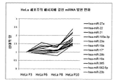
오른쪽 패널: 패시지 간의 발현을 현저하게 변화시키지 않는 has-miRNA-210 및 3개의 다른 마이크로RNA에 대한 마이크로RNA 마이크로어레이 발현 데이터 (어레이로부터 정규화된 신호 강도)의 발현 프로파일 분석을 보여준다. 도 2b: qRT-PCR 데이터에 의한 중요한 마이크로RNA 발현 차이의 확인을 보여준다.
도 3은 확장된 시험관내 패시지의 인간 암-유도 세포주(Hela 및 MCF-7)의 표현형 '드리프트'를 도시한다. 도 3a: 연속 패시지한 인간 종양-유도 세포주 (Hela 및 MCF-7)의 마이크로RNA 프로파일의 변화; 마이크로RNA 데이터세트의 주성분 분석은 MCF-7 세포의 세포 패시지 넘버에 기반한 샘플의 분리를 보여준다. 도 3b 및 프로파일 분석(도 3c)은 배양시 MCF-7 세포의 연속 패시지 동안 20개의 miRNA가 변화되었다는 것을 보여준다. 20개의 miRNA 모두는 모니터링된 7 패시지에 걸쳐 현저한 유전자 발현 감소를 보여준다. 변화는 최초 패시지(P3) 세포와 비교시 상대적 변화(배수 변화)로서 나타낸다. 도 3d 및 프로파일 분석(도 3e)은 배양시 HeLa 세포의 연속 패시지 동안 20개의 miRNA가 변화되었다는 것을 보여준다. 20개의 miRNA 모두는 모니터링된 7 패시지에 걸쳐 현저한 miRNA 발현 변화를 보여준다. 변화는 최초 패시지(P3) 세포와 비교시 상대적 변화(배수 변화)로서 나타낸다.
도 4a는 확장된 패시지 동안 동일한 배양 조건 하에 유지되는 2 hESC 세포 군에 대한 유동 세포분석 결과이다.
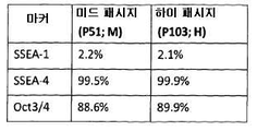
도 4b는 미드- 및 하이-패시지 hESC 세포군의 miRNA 프로파일의 주성분 분석(PCA)이다.
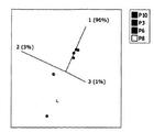
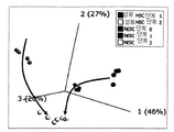
도 4c는 미드-패시지(P51)와 하이-패시지(P103) 세포 간의 마이크로RNA의 발현 차이를 나타내는 화산형 그래프(volcano plot)이다. 배수 변화 차이가 2 또는 그 이상인 5개의 차별-발현된 miRNA는 빨간색 원형으로 표시되어 있다.
도 4d는 P51과 P103 hESC 배양물 간의 발현 차이가 2배 보다 크다는 것을 보여주는 5개의 마이크로RNA(도 4에서 빨간 원형으로 표시)의 확인을 도시한다.
도 5는 miRNA 발현 프로파일의 차이에 기반한 상이한 샘플 그룹의 군집화를 보여주는 시각화 도면이다. A. 화살표가 분화의 궤적을 나타내는 주성분 분석(PCA)을 이용한 시각화. B. 계층적 군집화 및 히트맵(heatmap)을 이용한 샘플 관계의 시각화.The invention will now be described in more detail with reference to the accompanying drawings.
1 is a flow chart of a method according to the invention.
FIG. 2 shows reduced expression of has-miRNA-210 and increased expression of has-miR-1274a and has-miR-302c * in expanded in vitro passage of hESCs using both microarray and QPCR data panels. FIG. 2A: Left panel: Principal Component Analysis shows separation of samples based on cell passage number of human embryonic stem cell line RCM1.
Right panel: Expression profile analysis of microRNA microarray expression data (normalized signal intensity from array) for has-miRNA-210 and three other microRNAs that do not significantly change expression between passages. 2B: Confirmation of significant microRNA expression differences by qRT-PCR data.
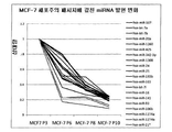
3 depicts the phenotype 'drift' of human cancer-induced cell lines (Hela and MCF-7) of expanded in vitro passages. 3A: Change in microRNA profile of continuous passaged human tumor-induced cell lines (Hela and MCF-7); Principal component analysis of the microRNA dataset shows separation of samples based on cell passage number of MCF-7 cells. 3B and profile analysis (FIG. 3C) show that 20 miRNAs were changed during consecutive passages of MCF-7 cells in culture. All 20 miRNAs show a significant decrease in gene expression over the seven passages monitored. Change is expressed as relative change (fold change) when compared to the original passage (P3) cells. 3D and profile analysis (FIG. 3E) show that 20 miRNAs were changed during successive passages of HeLa cells in culture. All 20 miRNAs show significant miRNA expression changes over 7 monitored passages. Change is expressed as relative change (fold change) when compared to the original passage (P3) cells.
4A shows flow cytometry results for 2 hESC cell populations maintained under the same culture conditions during extended passages.
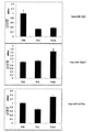
4B is principal component analysis (PCA) of miRNA profiles of mid- and high-passage hESC cell populations.
4C is a volcano plot showing the difference in expression of microRNAs between mid-passage (P51) and high-passage (P103) cells. Five differentially-expressed miRNAs with a fold change difference of 2 or more are shown in red circles.
4D shows the identification of five microRNAs (indicated in red circle in FIG. 4) showing that the difference in expression between P51 and P103 hESC cultures is greater than two times.
5 is a visualization plot showing clustering of different sample groups based on differences in miRNA expression profiles. A. Visualization using Principal Component Analysis (PCA), where the arrows represent the trajectories of differentiation. B. Visualization of sample relationships using hierarchical clustering and heatmaps.
실시예Example 1 One
본 발명의 한 실시예에서, miRNA 발현 데이터 세트의 데이터베이스(측정된 비암호화 RNA 발현 프로파일로부터 유도되는 발현 데이터 세트의 일례임)을 준비한다. 도 1을 참조하면, 적합한 인간 배아 줄기세포를 공지의 방법에 의해 확장된 기간 동안 배양시키고, 유도 이후 3개 시점에서, 즉 패시지 38, 51 및 103에서 샘플링한다. 그 다음, 처리된 세포 내의 다수의 miRNA 각각의 발현 정도를 결정하기 위해서 각 패시지의 세포 샘플을 사용하여 miRNA 발현 프로파일을 측정한다.In one embodiment of the invention, a database of miRNA expression data sets is prepared, which is an example of an expression data set derived from a measured non-coding RNA expression profile. Referring to Figure 1, suitable human embryonic stem cells are cultured for an extended period of time by known methods and sampled at three time points after induction, i.e. at
miRNA 발현 프로파일을 측정하기 위한 2가지 대안적인 방법, 마이크로어레이 분석법 및 정량적 실시간 PCR 분석법이 이후에 기술된다.Two alternative methods for measuring miRNA expression profiles, microarray assays and quantitative real-time PCR assays are described later.
(1) (One) miRNAmiRNA 마이크로어레이Microarray 및 데이터 분석 And data analysis
덴마크의 Vedbaek, Exiqon A/S사의 컬럼계 키트를 사용하여 기준 세포 (n=3)로부터 전체 RNA를 분리한다. 각 샘플의 전체 RNA 2㎍을 miRNA 마이크로어레이에 의해 분석한다. 라벨링, 혼성화(hybridization), 스캐닝, 정규화(normalization) 및 데이터 분석을 포함하는 miRNA 마이크로어레이 분석은 다수의 공급처, 예를 들어 Exiqon A/S로부터 상업적으로 입수할 수 있다. 간단히 말하자면, RNA 품질 관리는 바이오아날라이저 2100 마이크로플루이딕스 플랫폼(Bioanayser 2100 microfluidics platform)(바이오아날라이저는 Agilent Technologies의 상표임)을 사용하여 수행된다. 샘플은 Agilent사의 컴플리트 라벨링 하이브 키트(complecom Labelling Hyb kit)를 사용하여 제공된 지시사항에 따라 라벨링된다.Total RNA is separated from reference cells (n = 3) using a column-based kit from Exedon A / S, Vedbaek, Denmark. 2 μg total RNA of each sample is analyzed by miRNA microarray. MiRNA microarray analysis, including labeling, hybridization, scanning, normalization, and data analysis, is commercially available from a number of sources, for example Exiqon A / S. In short, RNA quality control is performed using the Bioanayser 2100 microfluidics platform (Bioanalyzer is a trademark of Agilent Technologies). Samples are labeled according to the instructions provided using Agilent's completecom Labeling Hyb kit.
(2) 정량적 실시간 PCR(2) quantitative real-time PCR
상술한 선택(1)에 따라서, Exiqon사의 컬럼계 키트를 사용하여 제조자의 지시시항에 따라 모든 세포 RNA를 추출한다. TaqMan 실시간 PCR에 의한 miRNA의 정량화는 제조자(미국, 캘리포니아, 포스터 시티의 Applied Biosystems)에 의해 기재된 바와 같이 수행된다(TaqMan은 Roche Molecular Systems, Inc의 상표임). 간단히 말하자면, TaqMan 마이크로RNA 역전사 키트 및 miRNA-특이적 스템-루프 프라이머(stem-loop primer)를 이용한 역전사(RT)용 템플릿(templet)으로서 10ng의 RNA를 사용한다. 분취량(1.5㎕)의 RT 산물을 95℃에서 10분간 ABI 7900HT 서모사이클러(Applied Biosystems)의 96-웰 플레이트 내에서 배양되는 20㎕의 PCR 반응물에 주입한 후에, 95℃에서 15초 및 60℃에서 1분의 사이클을 40회 수행한다. 표적 유전자 발현은 U48 RNA(작은 비암호화 RNA) 발현(또는, U48이 샘플마다 다른 것으로 밝혀지는 경우에는 U6 RNA)의 수치에 의거하여 상이한 샘플들 간에 정규화된다.According to selection (1) above, all cellular RNAs are extracted using Exiqon's column-based kits according to the manufacturer's instructions. Quantification of miRNA by TaqMan real time PCR is performed as described by the manufacturer (Applied Biosystems, Foster City, CA, USA) (TaqMan is a trademark of Roche Molecular Systems, Inc.). In short, 10 ng of RNA is used as a template for reverse transcription (RT) using the TaqMan microRNA reverse transcription kit and miRNA-specific stem-loop primers. Aliquots (1.5 μl) of RT product were injected into 20 μl of PCR reaction incubated in 96-well plate of ABI 7900HT Thermocycler (Applied Biosystems) for 10 minutes at 95 ° C., followed by 15 seconds and 60 ° C. at 95 ° C. 40 cycles of 1 minute are performed at < RTI ID = 0.0 > Target gene expression is normalized between different samples based on the level of U48 RNA (small non-coding RNA) expression (or U6 RNA if U48 is found to differ from sample to sample).
실험 결과 및 그 의미Experiment result and its meaning
기술된 방법을 사용하여, 본 발명자는 miRNA 발현 데이터의 그룹핑에 기반하여 세포의 표현형 그리프트의 확인을 모니터링하는 신규한 방식을 결정할 수 있다는 것을 입증하게 되었다. 또한, 본 방법은 전분화능 및 종양형성(tumourigenicity)을 포함한 세포 기능의 잠재적 변화를 나타내는 발현 정도를 갖는 특정 miRNA를 확인하는데 사용될 수 있다. 이들 miRNA는 향후 개입 스크리닝이 특이적 서브세트를 이용한 세포 생리학/병리생리학에 있어서 중요한 변화를 확인하기 위해서 상대적 소그룹의 miRNA 발현 정도 변화를 분석하는 것을 가능하게 할 것이며, 전체 miRNA 레퍼토리(repertoire)는 그러하지 않고 조사되는 특정 종료점에 따라 사용된다.Using the described method, the inventors have demonstrated that new ways of monitoring the identification of phenotypic grafts of cells can be determined based on grouping of miRNA expression data. In addition, the method can be used to identify specific miRNAs with expression levels that indicate potential changes in cellular function, including pluripotency and tumorigenicity. These miRNAs will enable future intervention screening to analyze changes in relative subgroup miRNA expression levels to identify significant changes in cell physiology / pathophysiology with specific subsets, but the overall miRNA repertoire does not. Is used depending on the specific endpoint being examined.
인간 배아 줄기세포군의 잠재적 안전성을 결정하기 위해 선별된 소그룹의 miRNA를 사용하는 예는 이후에 기술된다.Examples of using selected small groups of miRNAs to determine the potential safety of the human embryonic stem cell population are described later.
재료 및 방법Materials and methods
RCM1 세포 배양RCM1 cell culture
유도(Judo( derivationderivation ))
세포주 RCM-1은 갓 수취된 6일 배반포(blastocyst)로부터 유도하였다. 이를 Swemed 줄기세포 커팅 툴(Vitrolife AB, Cat No:14601)을 사용하여 수동으로 부화시키고 내부 세포 덩어리(inner cell mass)를 분리하여 인간 섬유아세포(Cascade Biologics)상에 플레이트시켰다. 이 섬유아세포는 인간 라미닌(Sigma, Cat No:L4544)층으로 사전-코팅된 조직 배양 웰 상에서 사전-플레이트된 것이다. 24ng/ml의 인간 염기성 섬유아세포 성장 인자(hbFGF)(Invitrogen, Cat No:PHG261)를 함유하는 조절화된 배지(conditioned medium) 내에서 세포를 배양하였다. 이렇게 얻어된 파생물을 Swemed 줄기세포 커팅 툴을 사용하여 수동으로 패시지하고 초기 확장을 통해 전형적인 미분화 형태를 계속해서 나타내면서 라미닌/피더(feeder)에 hbFGF 배양계를 더하였다. 세포주의 특성은 온라인상으로 http://www.roslincells.com/sitepix/downloads/RCM-1.pdf에서 입수할 수 있는 요약 문서에 기술되어 있다.Cell line RCM-1 was derived from freshly received 6 day blastocysts. This was manually hatched using a Swemed stem cell cutting tool (Vitrolife AB, Cat No: 14601) and the inner cell mass was isolated and plated on human fibroblasts (Cascade Biologics). These fibroblasts were pre-plated on tissue culture wells pre-coated with a layer of human laminin (Sigma, Cat No: L4544). Cells were cultured in a conditioned medium containing 24 ng / ml of human basic fibroblast growth factor (hbFGF) (Invitrogen, Cat No: PHG261). The resulting derivatives were manually passaged using a Swemed stem cell cutting tool and an hbFGF culture system was added to the laminin / feeder while continuing to show typical undifferentiated forms through initial expansion. The characteristics of the cell lines are described in a summary document available online at http://www.roslincells.com/sitepix/downloads/RCM-1.pdf.
확장(expansion( expansionexpansion ))
8ng/ml의 hbFGF를 함유하는 StemPRO(SP)(Invitrogen, Cat No:A1000701) 배지를 포함하는 CellSTART 매트릭스(CS)(Invitrogen, Cat No:A10142-01)의 지지세포-없는(feeder-free) 배양계에 RCM-1을 적응시키고, 이 조건하에 미분화 형태를 유지시켰다. 효소적 방법보다 더 선호되는 기계적/수동적 방법을 사용하여 다수의 패시지를 통해 세포주를 확장하였다. 세포주가 확장되는 동안, 다양한 패시지 단계에서 CryoStor CS10(Stemcell Technologies, Cat No:07930)을 사용하여 제조자 지시사항에 따라 기술한 바와 같이 세포를 동결보존하였다.Feeder-free culture of CellSTART Matrix (CS) (Invitrogen, Cat No: A10142-01) containing StemPRO (SP) (Invitrogen, Cat No: A1000701) medium containing 8 ng / ml of hbFGF RCM-1 was adapted to the system and the micronized form was maintained under these conditions. Cell lines were expanded through multiple passages using mechanical / passive methods, which are more preferred than enzymatic methods. During cell line expansion, cells were cryopreserved using CryoStor CS10 (Stemcell Technologies, Cat No: 07930) at various passage stages as described according to manufacturer's instructions.
동결보존으로부터 회수Recovery from cryopreservation
조사를 위해 3개의 패시지 시점, 초기, 중기 및 후기 패시지, 즉 패시지 P38, P51 및 P103을 해동하였다.Three passage time points, early, middle and late passages, namely passages P38, P51 and P103, were thawed for investigation.
-150℃ 냉동기로부터 3통의 유리병을 꺼내어 37℃에서 신속히 해동하였다. 그 다음, 해동된 세포를 사전-가온된 배지로 2회 세척한 후에 신선한 사전-가온된 배지 내에서 현탁시키고, 8ng/ml의 hbFGF를 함유하는 StemPRO(SP) 배지를 포함하는 CellSTART 매트릭스(CS)(Invitrogen)의 배양계의 웰 내에 플레이트시켰다. 반복해서 배지를 변화시키면서 7일 동안 세포를 배양(도 1)한 후에, RNA 추출을 위해 수확하였다(이후 참조).Three vials were removed from the -150 ° C freezer and thawed rapidly at 37 ° C. The thawed cells are then washed twice with pre-warmed medium and then suspended in fresh pre-warmed medium and CellSTART Matrix (CS) comprising StemPRO (SP) medium containing 8 ng / ml of hbFGF. Plated in wells of the Invitrogen culture system. Cells were incubated for 7 days with changing medium repeatedly (FIG. 1) and then harvested for RNA extraction (see below).
유동 세포분석Flow cell analysis
RNA 추출을 위해 수확된 세포는 또한 전분화능 및 분화의 다중 마커의 발현을 결정하기 위해 샘플링되었다.Cells harvested for RNA extraction were also sampled to determine the expression of multiple markers of pluripotency and differentiation.
배양물에 남아있는 세포로부터 단일 세포 현탁액을 만들고, 분화 또는 미분화 상태와 관련있는 다양한 마커에 대해 염색하였다. 그 마커는 인간 및 쥐의 만능 줄기세포 분석 키트(BD, Cat No: 560477) 사용시, 상향 조절(up regulation)이 분화 상태를 나타내는 단계-특이적 배양 항원 1(SSEA-1), 상향 조절(up regulation)이 미분화 상태를 나타내는 단계-특이적 배양 항원 4(SSEA-4), 및 배아 줄기(ES) 세포 및 생식 세포 내에서 발현되며 그 발현이 세포 자가-재생 및 전분화능을 유지하는데 필요한 Oct3/4, 34 kDa POU 전사인자였다.Single cell suspensions were made from the cells remaining in the culture and stained for various markers associated with differentiation or undifferentiated status. The markers are phase-specific culture antigen 1 (SSEA-1), up-regulation that upregulation indicates differentiation when using human and rat pluripotent stem cell assay kits (BD, Cat No: 560477). is expressed in stage-specific cultured antigen 4 (SSEA-4), and embryonic stem (ES) cells and germ cells, in which regulation is indicative of undifferentiated state and Oct3 / is expressed in order to maintain cellular self-renewal and pluripotency. 4, 34 kDa POU transcription factor.
유동 세포분석법을 이용하여 염색된 세포를 분석하고, 도출된 결과는 분석된 마커에 대해 수치적으로 및 그래프로 나타내어 세포주의 상태를 제공한다. 도 4a.Stained cells are analyzed using flow cytometry and the results obtained are numerically and graphically presented for the markers analyzed to provide the state of the cell line. Figure 4a.
종양-유도 세포주Tumor-induced cell line
표준 방법을 사용하여 HeLa 및 MCF-7을 배양 및 패시지(계대배양)하였다.HeLa and MCF-7 were incubated and passaged (passage) using standard methods.
RNARNA 추출 extraction
miRNA 프로파일링 분석 이전에, 세포로부터 전체 RNA를 분리하고 품질 분석을 해야 한다. Exiqon(덴마크)으로부터 입수할 수 있는 miRCURY RNA 분리 키트를 사용하여, 상이한 패시지 넘버의 줄기세포로부터 전체 RNA를 분리한다. 제조자의 지시사항에 따라서, 특이적 용해 완충액을 사용하여 조직 배양 접시 내에서 세포를 용해시키고, 이를 컬럼으로 이동시켜 RNA를 세척한 후에 용출시킨다. RNA 정량 및 품질은 Nanodrop ND-1000 분광광도계(미국, MA, 월섬의 Thermo Fisher) 및 Bioanalyser 2100 미세유체-베이스드 플랫폼(미국, CA, 산타 클라라의 Agilent Technologies)을 사용하여 검사한다.Prior to miRNA profiling assays, total RNA must be isolated from cells and analyzed for quality. Total RNA is isolated from stem cells of different passage numbers using the miRCURY RNA Separation Kit, available from Exiqon (Denmark). According to the manufacturer's instructions, cells are lysed in tissue culture dishes using specific lysis buffers, which are transferred to a column and washed after RNA washing. RNA quantification and quality are examined using a Nanodrop ND-1000 spectrophotometer (Thermal Fisher, Waltham, MA, USA) and the Bioanalyser 2100 microfluidic-based platform (Agilent Technologies, Santa Clara, CA).
상이한 패시지 넘버의 줄기세포 샘플에 대한 마이크로 RNA 발현 프로파일은 이들 샘플로부터 전체 RNA를 분리하고 이를 두가지 방법; 즉 (1) miRNA 마이크로어레이 및 (2) 정량적 실시간 PCR(QPCR)에 의해 분석함으로써 결정될 수 있다.Micro RNA expression profiles for stem cell samples of different passage numbers can be used to isolate total RNA from these samples and to identify two methods; That is, by (1) miRNA microarray and (2) quantitative real-time PCR (QPCR).
마이크로어레이는 인간 851 miRNA의 발현 정도에 관한 데이터를 동시에 수집함으로써 샘플의 완전한 miRNA 프로파일을 달성하기 위해서 사용된다. QPCR는 발현 정도의 차이가 결정될 수 있도록 다수의 샘플 중 관심있는 각 miRNA를 조사하는데 사용된다.Microarrays are used to achieve a complete miRNA profile of a sample by simultaneously collecting data on the expression level of human 851 miRNAs. QPCR is used to examine each miRNA of interest in a number of samples so that differences in expression levels can be determined.
(1) miRNA 마이크로어레이 및 데이터 분석(1) miRNA microarrays and data analysis
품질 검사를 하고 적절한 농도로 희석된 전체 RNA를 Agilent사의 마이크로어레이 플랫폼 상의 miRNA 프로파일링을 위한 출발물질로서 사용한다. 각 샘플로부터 얻은 100ng의 전체 RNA를 마이크로어레이 프로토콜에 따라 실행시키는데, 여기에서 마이크로RNA는 라벨링된 다음, 어레이와 혼성화되고 Agilent 마이크로어레이 스캐너를 이용하여 스캐닝된다. 샘플은 Agilent사의 'miRNA 완전 라벨링 및 혼성화 키트'를 사용하여 Cy3 염료로 라벨링된 다음, Agilent 마이크로어레이에서 밤새 혼성화되며, 이 중 8개는 각 유리 슬라이드 상에서 보여진다. 어레이에서, 각 miRNA는 적어도 2개의 다른 프로브에 의해 16회 나타난다. 또한, 반응물의 라벨링 및 혼성화 효율을 평가하기 위해서 스파이크-인 콘트롤(spike-in controls)이 사용될 수 있다. 어레이의 스캔 이미지는 이미지 상의 각 지점을 분석하고 이를 특이적 miRNA에 할당하여 방출된 형광 신호에 대한 수치를 산정하는 Agilent 피쳐 엑스트랙션 소프트웨어를 위한 입력 데이터를 구성한다. 이 프로세싱으로부터 나오는 출력물은 일련의 QC 보고서로, 어레이 프로세싱의 품질 및 텍스트 파일을 평가하며 원 마이크로어레이 데이터를 포함한다. 이 텍스트 파일은 상이한 샘플간의 miRNA 발현 변화를 확인하는데 사용되는 통계 분석의 기반을 형성한다. 가장 좋은 실험 계획에 있어서, 재현성(reproducibility)을 보장하기 위해서 생물학적 복제(n=3)는 상이한 슬라이드 상에서 수행된다. 마이크로어레이 데이터는 GeneSpring(Agilent Technologies) 및/또는 Omics Explorer(스웨덴의 Qlucore of Lund)와 같은 통계 분석 프로그램에 의해, 그리고 시스테믹스 인하우스(Sistemic's in-house) 통계법에 의해 해석된다(이후 기술).Quality test and total RNA diluted to appropriate concentration is used as starting material for miRNA profiling on Agilent's microarray platform. 100 ng of total RNA from each sample is run according to the microarray protocol, where the microRNA is labeled, hybridized with the array and scanned using an Agilent microarray scanner. Samples are labeled with Cy3 dye using Agilent's 'miRNA complete labeling and hybridization kit' and then hybridized overnight in Agilent microarrays, eight of which are shown on each glass slide. In the array, each miRNA is represented 16 times by at least two different probes. In addition, spike-in controls can be used to assess the labeling and hybridization efficiency of the reactants. The scanned image of the array constitutes input data for Agilent feature extraction software that analyzes each point on the image and assigns it to a specific miRNA to calculate the value for the emitted fluorescence signal. The output from this processing is a series of QC reports that evaluate the quality of the array processing and the text file and contain the raw microarray data. This text file forms the basis of the statistical analysis used to identify changes in miRNA expression between different samples. In the best experimental design, biological replication (n = 3) is performed on different slides to ensure reproducibility. Microarray data are interpreted by statistical analysis programs such as GeneSpring (Agilent Technologies) and / or Omics Explorer (Qlucore of Lund, Sweden) and by Sistemic's in-house statistics (hereinafter described). .
RNA 추출RNA extraction
RNA는 이들 세포로부터 Exiqon의 컬럼계 키트를 사용하여 다음의 절차를 따라 분리 및 정제되었다. 세포가 증식되는 배지를 흡출하고, 세포 단층을 적당량의 PBS로 세척하였다. PBS를 추가로 흡출하였다. 350 μL의 용해 용액을 배양 플레이트에 직접 가하였다. 배양 접시를 가볍게 두드리고 나서 5분간 플레이트 표면 주위로 완충액을 스월링(swirling)함으로써 세포를 용해시켰다. 그 다음, 용해물을 마이크로-원심분리관으로 이동시킨다. 이 용해물에 200 μL의 95% 내지 100% 에탄올을 가하고 10초간 볼텍싱(vortexing)에 의해 혼합하였다. 키트에서 제공되는 튜브 중 하나를 사용하여 컬럼을 조립하였다. 그 컬럼에 600 μL의 용해물/에탄올을 가하고 1분간 14,000xg로 원심분리하였다. 플로우-쓰루(the flow-through)를 버리고 수집관(collection tube)을 사용하여 스핀 컬럼을 재조립하였다. 그 컬럼에 400 μL의 공급 세척 용액을 가하고 1분간 14,000xg에서 원심분리하였다. 플로우-쓰루를 버리고 수집관을 사용하여 스핀 컬럼을 재조립하였다. 400 μL의 추가적 세척 용액을 가함으로써 컬럼을 2회 더 세척하고 1분간 14,000xg로 원심분리하였다. 플로우-쓰루를 버리고 수집관을 사용하여 스핀 컬럼을 재조립하였다. 그 컬럼을 1분간 14,000xg로 회전시킴으로써 수지를 완전히 건조시킨 후에 수집관을 폐기하였다. 컬럼을 키트와 함께 제공되는 1.7 mL의 용출 관(elution tube)으로 조립하였다. 그 컬럼에 50 μL의 용출 완충액을 가하고 2분간 200xg로, 그 다음 1분간 14,000xg로 원심분리하였다. 이렇게 정제된 RNA 샘플은 -20℃에서 수일간 저장될 수 있다. 22 텀의 긴 저장 기간 동안 샘플은 -70℃에서 저장되었다.RNA was isolated and purified from these cells using the Exiqon columnar kit following the following procedure. The medium in which the cells proliferate was aspirated and the cell monolayers were washed with an appropriate amount of PBS. PBS was further aspirated. 350 μL of lysis solution was added directly to the culture plate. The cells were lysed by tapping the culture dish and then swirling the buffer around the plate surface for 5 minutes. The lysate is then transferred to a micro-centrifuge tube. 200 μL of 95% to 100% ethanol was added to the lysate and mixed by vortexing for 10 seconds. The column was assembled using one of the tubes provided in the kit. 600 μL of lysate / ethanol was added to the column and centrifuged at 14,000 × g for 1 minute. The flow-through was discarded and the spin column was reassembled using a collection tube. 400 μL of feed wash solution was added to the column and centrifuged at 14,000 × g for 1 minute. The flow-through was discarded and the spin column reassembled using a collection tube. The column was washed two more times by adding 400 μL of additional wash solution and centrifuged at 14,000 × g for 1 minute. The flow-through was discarded and the spin column reassembled using a collection tube. The collection tube was discarded after the resin was completely dried by rotating the column at 14,000 × g for 1 minute. The column was assembled into 1.7 mL of elution tube provided with the kit. 50 μL of elution buffer was added to the column and centrifuged at 200 × g for 2 minutes and then 14,000 × g for 1 minute. This purified RNA sample can be stored at -20 ° C for several days. Samples were stored at -70 [deg.] C. for a long storage period of 22 terms.
(1) (One) miRNAmiRNA 마이크로어레이Microarray 및 분석 And analysis
라벨링Labeling
정제된 RNA 샘플을 Agilent의 라벨링 키트를 사용하여 라벨링하였다. 전체 RNA 샘플을 1 x TE(pH 7.5)으로 50ng/μL로 희석하였다. 희석된 전체 RNA를 1.5 mL 마이크로-원심분리관에 가하고 얼음에 넣었다. 사용하기 바로 전에, 0.4 μL의 10 x 송아지 장 프스파타제 완충액, 1.1 μL의 뉴클레아제 비함유 물 및 0.5 μL의 송아지 장 프스파타제를 부드럽게 혼합함으로써 송아지 장 알칼리성 포스파타제 마스터 믹스를 제조하였다. 총 반응 분량(reaction volume)이 4 μL가 되도록 각 샘플 튜브에 2 μL의 송아지 장 알칼리성 프스파타제 마스터 믹스를 가하고, 피펫팅(pipetting)에 의해 부드럽게 혼합하였다. 반응 분량을 순환 수조 내에서 30분간 37℃에서 배양하였다. 각 샘플에 2.8 μL의 100% DMSO를 가하였다. 샘플을 순환 수조 내에서 5 내지 10분간 100℃에서 배양한 다음, 즉시 얼음조(ice bath)로 이동시켰다.Purified RNA samples were labeled using Agilent's labeling kit. Total RNA samples were diluted to 50 ng / μL with 1 x TE (pH 7.5). Diluted total RNA was added to a 1.5 mL micro-centrifuge tube and placed on ice. Immediately before use, a calf intestinal alkaline phosphatase master mix was prepared by gently mixing 0.4 μL of 10 × calf intestinal fate buffer, 1.1 μL of nuclease free water and 0.5 μL of calf intestinal fatase. 2 μL of calf intestinal alkaline spatase master mix was added to each sample tube so that the total reaction volume was 4 μL and mixed gently by pipetting. The reaction volume was incubated at 37 ° C. for 30 minutes in a circulating water bath. 2.8 μL of 100% DMSO was added to each sample. Samples were incubated at 100 ° C. for 5-10 minutes in a circulating water bath and then immediately transferred to an ice bath.
10 x T4 RNA 리가제(ligase) 완충액을 37℃로 가온한 다음, 모든 침전물이 용해될 때까지 회전시켰다. 사용하기 바로 전에, 1 μL의 10 x T4 RNA 리가제 완충액, 3 μL의 시아닌3-pCp 및 0.5 μL의 T4 RNA 리가제를 부드럽게 혼합함으로써 라이게이션(ligation) 마스터 믹스를 제조한 후에 얼음에 넣었다. 총 반응 분량이 11.3 μL가 되도록 각 샘플 튜브에 4.5 μL의 라이게이션 마스터 믹스를 가하였다. 샘플을 피펫팅에 의해 부드럽게 혼합하고 스핀다운시켰다. 그 다음, 샘플을 순환 수조 내에서 2시간 동안 16℃에서 배양하였다. 그 다음, 진공 농축기를 사용하여 45℃ 내지 55℃에서 샘플을 건조시키고 나서, 튜브를 가볍게 쳤을 때 펠릿(pellet)이 움직이거나 퍼지지 않았다면 샘플이 건조한 것으로 결정하였다.10 × T4 RNA ligase buffer was warmed to 37 ° C. and then spun until all precipitates dissolved. Immediately before use, ligation master mixes were prepared by gentle mixing of 1 μL of 10 × T4 RNA ligase buffer, 3 μL of cyanine3-pCp and 0.5 μL of T4 RNA ligase and then placed on ice. 4.5 μL of ligation master mix was added to each sample tube so that the total reaction volume was 11.3 μL. Samples were gently mixed and spun down by pipetting. The samples were then incubated at 16 ° C. for 2 hours in a circulating water bath. The sample was then dried at 45 ° C. to 55 ° C. using a vacuum concentrator and the sample was determined to be dry if the pellet did not move or spread when the tube was lightly tapped.
혼성화Hybridization
Agilent 키트와 함께 제공되는 동결건조된 10 x GE 차단제(blocking agent)가 들어 있는 유리병에 125 μL의 뉴클레아제 비함유 물을 가하고 혼합하였다. 건조된 샘플을 18 μL의 뉴클레아제 비함유 물에 재현탁시켰다. 각 샘플에 4.5 μL의 10 x GE 차단제를 가하였다. 각 샘플에 22.5 μL의 2 x Hi-RPM 혼성화 완충액을 가하고 잘 혼합하였다. 이렇게 생성된 샘플을 100℃에서 5분간 배양한 다음, 즉시 얼음 수조로 이동시켜 5분간 더 유지하였다. Agilent SureHyb 챔버 바닥이 가스켓 슬라이드(gasket slide)로 채워지도록 챔버 바닥에 깨끗한 가스켓 슬라이드를 올려 놓았다. 버블이 존재하지 않도록 가스켓 상에 혼성화 샘플을 잘 분배하였다.125 μL of nuclease-free water was added and mixed to a glass bottle containing the lyophilized 10 × GE blocking agent provided with the Agilent kit. The dried sample was resuspended in 18 μL of nuclease free water. 4.5 μL of 10 × GE blocker was added to each sample. 22.5 μL of 2 × Hi-RPM hybridization buffer was added to each sample and mixed well. The sample thus produced was incubated at 100 ° C. for 5 minutes and then immediately transferred to an ice bath for 5 minutes. A clean gasket slide was placed on the chamber floor so that the Agilent SureHyb chamber floor was filled with a gasket slide. Hybridization samples were well dispensed on the gasket so that no bubbles were present.
SureHyb 가스켓 슬라이드 상에 활성측(active side)이 아래로 되도록 어레이를 배치하고 SureHyb 챔버 커버로 조립함으로써 조립 챔버를 형성하였다. 이 조립 챔버를 55℃로 설정된 혼성화 오븐 속에 넣고, 그 온도에서 20시간 동안 20rpm으로 회전시켰다.The assembly chamber was formed by placing the array with the active side down on the SureHyb gasket slide and assembling with the SureHyb chamber cover. The assembly chamber was placed in a hybridization oven set at 55 ° C. and spun at 20 rpm for 20 hours at that temperature.
그 다음, 공급된 GE 세척 완충액을 사용하여 어레이를 세척한 후에 스캐닝하였다.The array was then scanned after washing with the supplied GE wash buffer.
(2) 정량적 실시간 PCR(2) quantitative real-time PCR
정량적 실시간 PCR은 3단계로 수행된다. 처음 두 단계는 전체 RNA 샘플로부터 cDNA를 합성하기 위한 단계로, qScript miRNA cDNA 합성 키트(Quanta Biosciences)를 사용한다. 세 번째 단계인 QPCR 반응은 SYBR Green PerfeCTa 저 산화환원 반응 믹스(Quanta Biosciences)를 사용한다.Quantitative real time PCR is performed in three steps. The first two steps are for synthesizing cDNA from whole RNA samples, using the qScript miRNA cDNA Synthesis Kit (Quanta Biosciences). The third step, the QPCR reaction, uses the SYBR Green PerfeCTa low redox reaction mix (Quanta Biosciences).
폴리(A) 테일링 반응Poly (A) Tailing Reaction
전체 RNA 샘플(100n과 1㎍ 사이의)을 신선한 0.5 ml 튜브에 나누어 넣고 뉴클레아제 비함유 물로 7㎕가 되게 하였다. 각 튜브에 2 μL의 5 x PAP(폴리(A) 폴리머라제) 테일링 완충액 및 1 μL의 폴리(A) 폴리머라제를 가한 다음, 이 튜브를 볼텍싱하고 원심분리하였다. 그리고 나서, 샘플을 37℃에서 20분간, 그 다음 70℃에서 5분간의 조건 하에 서멀 사이클러(thermal cycler) 내에서 배양하였다. 이 반응 후에, 샘플을 얼음에 넣었다.Total RNA samples (between 100 n and 1 μg) were divided into fresh 0.5 ml tubes and brought to 7 μl with nuclease free water. To each tube was added 2 μL of 5 × PAP (poly (A) polymerase) tailing buffer and 1 μL of poly (A) polymerase, and then the tubes were vortexed and centrifuged. The samples were then incubated in a thermal cycler under conditions of 20 minutes at 37 ° C. and then 5 minutes at 70 ° C. After this reaction, the sample was placed on ice.
cDNAcDNA 합성 반응 Synthetic reaction
각 샘플이 9㎕의 miRNA cDNA 반응 믹스 및 1㎕의 qScript 역전사효소를 수취하게 되도록 RT의 마스터 믹스를 준비한다. 각 샘플에 이 믹스 10㎕를 가한 다음, 튜브를 볼텍싱하고 원심분리하였다. 그리고 나서, 샘플을 42℃에서 20분간, 그 다음 85℃에서 5분간의 조건 하에 서멀 사이클러 내에서 배양하였다. 이 반응 후에, 샘플을 얼음에 넣은 다음, 1 x TE 완충액으로 5배 희석하였다.Prepare a master mix of RT so that each sample receives 9 μl of miRNA cDNA reaction mix and 1 μl of qScript reverse transcriptase. 10 μl of this mix was added to each sample, then the tubes were vortexed and centrifuged. The samples were then incubated in a thermal cycler for 20 minutes at 42 ° C. and then 5 minutes at 85 ° C. After this reaction, the sample was placed on ice and then diluted 5 times with 1 x TE buffer.
QPCR 반응QPCR reaction
각 샘플 웰이 하기의 키트 성분을 수취하게 되도록 SYBR Green 반응 믹스 및 프라이머의 마스터 믹스를 준비한다:Prepare a master mix of SYBR Green reaction mix and primers so that each sample well receives the following kit components:
· 10㎕의 2 x SYBR Green PerfeCTa 저 산화환원 반응 믹스10 μl of 2 x SYBR Green PerfeCTa low redox reaction mix
· 0.4㎕의 UA3PA Univesal 역프라이머(10 □M)0.4 μl UA3PA Univesal Reverse Primer (10 □ M)
· 0.4㎕의 miRNA-특이적 프라이머(10 □M)0.4 μl of miRNA-specific primer (10 □ M)
· 4.2㎕의 뉴클레아제-비함유 물4.2 μl of nuclease-free water
각 웰에 5㎕의 cDNA를 가하였다. 모든 웰이 채워지면, 플레이트를 플라스틱 광학 뚜껑으로 밀봉하고 공기 방울을 제거시키기 위해 원심분리한다. 플레이트를 Agilent MX3005P 서모사이클러 속에 넣고 하기의 사이클링 조건 하에 처리한다:5 μl of cDNA was added to each well. Once all the wells are filled, the plate is sealed with a plastic optical cap and centrifuged to remove air bubbles. The plate is placed in an Agilent MX3005P thermocycler and processed under the following cycling conditions:
· 95℃ 5분간95 ° C for 5 minutes
· (95℃ 5초간, 60℃ 30초간) x 40 사이클(40 ° C for 5 seconds, 60 ° C for 30 seconds) x 40 cycles
· 형광성 데이터는 매 어닐링(annealing)/확장 단계의 말기에 수집된다.Fluorescent data is collected at the end of every annealing / expansion step.
데이터 분석Data Analysis
이들 두 방법으로 얻은 데이터는 각 플레이트의 스파이크-인 miRNA 지점에 대해 정규화됨으로써, 각 어레이로부터 얻은 데이터를 비교할 수 있게 하였다. 정규화된 데이터는 miRNA 발현 프로파일 간의 상관관계를 확인하기 위해서 당해 분야의 숙련자가 잘 이해하고 있는 표준 방법인 주성분 분석법을 이용하여 분석되었으며, 관찰된 데이터의 그룹핑은 각 miRNA의 발현에 대해 원세포와 비교하여 15개의 특정 테스트 조건의 영향의 결과인 것으로 결정하였다.Data obtained by these two methods was normalized to the spike-in miRNA points of each plate, allowing comparison of data from each array. Normalized data were analyzed using principal component analysis, a standard method well understood by those skilled in the art to confirm correlations between miRNA expression profiles, and grouping of observed data compared to progenitor cells for expression of each miRNA. Was determined to be the result of the effects of 15 specific test conditions.
도 1은 마이크로RNA의 발현 프로파일을 얻기 위한 방법의 흐름도이다.1 is a flowchart of a method for obtaining an expression profile of a microRNA.
도 2는 마이크로어레이에 의해 확인되고 성숙한 마이크로RNA의 QPCR 측정에 의해 입증된 패시지 넘버 간의 has-miRNA-210, has-miR1274a 및 has-miR-302c*의 변화를 보여준다.FIG. 2 shows the changes in has-miRNA-210, has-miR1274a and has-miR-302c * between passage numbers confirmed by microarray and verified by QPCR measurements of mature microRNAs.
도 3은 연속 패시지한 인간 종양-유도 세포주(Hela 및 MCF-7)의 마이크로RNA 프로파일의 변화를 보여준다.3 shows changes in microRNA profiles of continuous passaged human tumor-induced cell lines (Hela and MCF-7).
도 2에서 볼 수 있는 바와 같이, 결과는 명백히 그룹핑되어 있으며, 이 그룹핑은 miRNA가 발현된 세포의 패시지 넘버에 따른다. 다시 말해서, 동일하게 패시지한 세포의 복제 샘플은 유사하지만 구별되는 miRNA 발현 프로파일을 갖는다고 결정할 수 있다.As can be seen in FIG. 2, the results are clearly grouped and this grouping is according to the passage number of the cell in which the miRNA is expressed. In other words, it can be determined that replicated samples of identically passaged cells have a similar but distinct miRNA expression profile.
miRNA 발현 패턴의 데이터베이스는 세포 패시지 넘버의 많은 비교를 수행하고 그로부터 miRNA 발현 변화를 분석함으로써 구축될 수 있다. 이러한 데이터베이스는 만능 줄기세포 또는 바이오프로세싱에서 사용되는 세포주의 표현형 드리프트를 확인할 수 있게 하며, 최적의 기능성, 전자의 경우 만능 잠재성, 후자의 경우 원하는 거대 분자의 생성의 손실을 표시한다. 또한, miRNA 발현 데이터의 데이터베이스 구축은 세포 생리학에 있어 바람직하지 않거나 정의되지 않은 변경을 나타내는 지표인 특정 miRNA의 세브세트를 보여줄 수 있다. 일단 지시(indicative) miRNA의 서브세트가 확인되면, 새로운 세포주의 향후 테스트는 세포에 의해 생성되는 miRNA의 전체 범위가 아닌, 지시 miRNA 발현 프로파일 중 서브세트의 발현 프로파일을 관찰함으로서 수행될 수 있다. miRNA는 생물학적 개입 간의 또는 세포 생리학에 유사한 영향을 미치는 것으로 알려져 있거나 추정되는 개입 그룹간의 변별에 대한 발현 정도의 관련성 순서로 랭크될 수 있다. miRNA는 생물학적 개입간의, 또는 세포 생리학에 유사한 영향을 미치는 것으로 알려져 있거나 추정되는 개입그룹간의 변별에 대한 발현 정도의 관련성을 나타내는 수치를 배정받을 수 있다. 예를 들어, 수치는 주성분의 변화에 대한 miRNA의 발현 정도의 기여도와 관련이 있을 수 있다. 주성분 분석과 같은 통계법을 이용한 miRNA 발현 프로파일의 비교의 대안으로 또는 이에 더하여, 세포 배양 패시지가 한정된 그룹의 miRNA(예를 들어, 10 내지 50)의 각각의 발현에 미치는 영향은 확인될 수 있으며, 생물학적 개입이 각각의 miRNA의 발현에 미치는 영향에 코드 그룹으로부터 선택된 코드를 부여하는데 사용될 수 있다. 이렇게 얻어진 코드는 영향의 유사성을 확인하기 위해 비교될 수 있다.A database of miRNA expression patterns can be built by performing many comparisons of cell passage numbers and analyzing miRNA expression changes therefrom. This database allows the identification of phenotypic drift of pluripotent stem cells or cell lines used in bioprocessing, and indicates the optimal functionality, pluripotent potential in the former, and loss of production of the desired macromolecule in the latter. In addition, building a database of miRNA expression data can reveal a subset of specific miRNAs that are indicative of undesirable or undefined alterations in cell physiology. Once a subset of the indicative miRNAs is identified, future testing of the new cell line can be performed by observing the expression profile of the subset of the indicator miRNA expression profiles, rather than the full range of miRNAs produced by the cell. miRNAs may be ranked in the order of relevance of the degree of expression for discrimination between groups of interventions known or suspected to have similar effects between biological interventions or cellular physiology. miRNAs can be assigned a value indicating the relationship of expression levels to discrimination between biological interventions or between groups of interventions known or suspected of having similar effects on cell physiology. For example, the numerical value may be related to the contribution of the degree of expression of miRNA to changes in the principal component. Alternatively or in addition to the comparison of miRNA expression profiles using statistical methods such as principal component analysis, the effect of cell culture messages on the expression of each of a limited group of miRNAs (eg, 10-50) can be identified and biologically The effect of the intervention on the expression of each miRNA can be used to assign a code selected from the code group. The code thus obtained can be compared to confirm the similarity of the impacts.
예를 들어, 비교(예를 들어 세포 패시지 넘버)를 위해, 하기에 의거하여 각 랭크된 miRNA에 코드로서 세자리 이진수가 배정될 수 있다:For example, for comparison (e.g., cell passage number), a three digit binary number can be assigned as a code to each ranked miRNA based on:
1. 생물학적 개입에 대한 반응에 있어서 miRNA의 발현이 변화없는 경우(실험적 가변성의 정상 한계 내에서), 첫 번째 비트는 0으로 설정된다. 발현이 상당히 변화된 경우는, 첫 번째 비트가 1로 설정된다.1. If miRNA expression is unchanged (within normal limits of experimental variability) in response to biological intervention, the first bit is set to zero. If the expression has changed significantly, the first bit is set to one.
2. 발현 정도의 변화가 확인되고 그 변화가 증가인 경우, 두 번째 비트는 1로 설정된다. 생물학적 개입으로부터 발생되는 변화가 감소인 경우는, 두 번째 비트가 0으로 설정된다.2. If a change in the extent of expression is identified and the change is an increase, the second bit is set to one. If the change resulting from biological intervention is a decrease, the second bit is set to zero.
3. 발현 정도의 변화가 4배보다 큰 경우, 세 번째 비트는 1로 설정되고, 그렇지 않은 경우는, 세 번째 비트가 0으로 설정된다.3. If the change in expression degree is greater than four times, the third bit is set to one, otherwise, the third bit is set to zero.
따라서, 세포 패시지 또는 배양 조건 간의 차이가 miRNA의 발현에 미치는 영향은 하기의 5가지 가능한 수치 중 하나를 갖는 코드를 배정받는다.Thus, the effect of differences between cell passages or culture conditions on the expression of miRNAs is assigned a code having one of the following five possible values.
1. 발현에 2 변화없음 -0001. No change in expression -000
2. 발현 증가 큼 -1112. Increased expression greater than -111
3. 발현 증가 적음 -1103. Less expression increase -110
4. 발현 감소 큼 -1014. Greater Reduction in Expression
5. 발현 감소 적음 -1005. Low expression decrease -100
배양 시간 확장(즉, 패시지 넘버의 증가)이 miRNA 그룹의 발현 정도에 미치는 영향은 관련 코드에 의해 특성화될 수 있으며, 이는 주성분 분석으로부터 즉시 나타나지 않는 발현 정도 변화의 확인을 가능하게 하고, 테스트 조건 또는 개입의 유사성을 스코어링(scoring)하기 위한 대안 방법을 가능하게 하며, 이렇게 얻어진 발현 데이터를 육안 검사(visual inspection)에 의해 이해할 수 있게 한다.The effect of the incubation time extension (ie, increase in the number of passages) on the expression level of the miRNA group can be characterized by the relevant code, which allows the identification of changes in the expression level that do not appear immediately from the principal component analysis, and the test conditions or An alternative method for scoring the similarity of interventions is made possible and the expression data thus obtained can be understood by visual inspection.
세포 지속 레짐의 영향을 특성화하기 위한, 그리고 상이한 생물학적 개입이 miRNA 발현에 미치는 영향 간의 상관관계를 결정하기 위한 또 다른 방식은 개입이 그룹(전형적으로 10 내지 50)miRNA의 각각의 발현에 미치는 영향을 결정하기 위해서, 그리고 영향의 순서로, 예를 들어 발현 증가가 가장 큰 그룹의 miRNA에서부터 발현 감소가 가장 큰 그룹의 miRNA까지의 순서로, 아니면 그 반대의 순서로 그 그룹의 miRNA를 랭크하기 위해서 발현 분석을 수행하는 것이다. 이러한 랭킹은 특정 테스트 시점 또는 개입의 영향을 나타내는 지표이다. 따라서, 다른 개입이 miRNA 그룹에 미치는 영향은 측정될 수 있으며, 그룹의 miRNA는 영향의 순서로 랭크될 수 있다. 이러한 랭킹을 비교함으로써 개입의 영향 간의 상관관계가 확인될 수 있다.Another way to characterize the effects of the cell persistence regime and to determine the correlation between the effects of different biological interventions on miRNA expression is to determine how the intervention affects each expression of a group (typically 10 to 50) miRNAs. To determine and rank the miRNAs in that order, e.g., in order of influence, e.g., from the group of miRNAs with the largest increase in expression to the group of miRNAs with the largest decrease in expression, or vice versa. To perform the analysis. This ranking is an indicator of the impact of a particular test point or intervention. Thus, the impact of other interventions on the miRNA group can be measured and the miRNAs in the group can be ranked in order of influence. By comparing these rankings, the correlation between the effects of the intervention can be identified.
잠재적인 신규한 치료적 용도를 위해 세포 패시지 및/또는 개입의 영향을 선별할 수 있는 효율 및 속도를 현저히 증가시키기 위해서 지시 miRNA의 서브세트를 테스트하는데 사용할 수 있는 플레이트를 포함하는 키트가 제공될 수 있다.Kits may be provided that include plates that can be used to test a subset of directed miRNAs to significantly increase the efficiency and speed with which the effects of cell passage and / or intervention can be screened for potential new therapeutic uses. have.
본 명세서에 기술되는 본 발명의 범위 내에서 추가적인 변경 및 수정이 가 능하다.Additional changes and modifications are possible within the scope of the invention described herein.
참고문헌references
1. Xie, X., et al., 몇몇 포유류의 비교에 의한 인간 프로모터의 조절 모티프 및 3'-UTRs의 계통적 발견(Systematic discovery of regulatory motifs in human promoters and 3'-UTRs by comparison of several mammals), Nature, 2005. 434(7031): p.338-45Xie, X., et al., Systematic discovery of regulatory motifs in human promoters and 3'-UTRs by comparison of several mammals , Nature, 2005.434 (7031): p.338-45
2. Lim, L.P., et al., 마이크로어레이 분석은 일부 마이크로RNA가 다수의 표적 mRNA를 하향조절한다는 것을 보여준다(Microarray analysis shows that some microRNAs downregulate large numbers of target mRNA), Nature, 2005. 433(7072): p.769-73Lim, LP, et al., Microarray analysis shows that some microRNAs downregulate large numbers of target mRNAs, Nature, 2005. 433 (7072) ): p.769-73
3. Galin. G.A., et al., 마이크로RNA 프로파일링은 B 세포 만성 림프성 백혈병에 있어서 뚜렷한 특징을 나타낸다(MicroRNA profiling reveals distinct signatures in chronic lymphocytic leukemias), Proc Natl Acad Aci USA, 2004. 101(32): p.11755-603. Galin. GA, et al., MicroRNA Profiling reveals distinct features in B cell chronic lymphocytic leukemia (MicroRNA profiling reveals distinct signatures in chronic lymphocytic leukemias), Proc Natl Acad Aci USA, 2004. 101 (32): p. 11755-60
실시예 2Example 2
개요summary
1. 연속 패시지한 줄기세포의 마이크로RNA 프로파일은 세포 표면 및 전분화능 및 분화의 내부 단백질 항원 마커를 평가하는 상업용 키트 및 유동 세포분석을 이용하여 '동일한' 세포군으로 평가된 세포의 차이를 보여준다(도 4).1. MicroRNA profiles of serially passaged stem cells show differences in cells evaluated in 'identical' cell populations using flow cytometry and commercial kits that assess cell surface and internal protein antigen markers of pluripotency and differentiation (FIG. 4).
2. 마이크로RNA는 동등한 발전 단계의 성인 CD34+ 조혈 줄기세포(HSCs; 도 5)와의 비교를 위해 인간 배아 줄기세포(hESCs)의 직접 분화에 의해 유도되는 두 가지 CD34+ 세포군으로부터의 miRNA 프로파일을 비교함으로써 hESC의 적혈구로의 직접 분화를 모니터링하는데 사용될 수 있다.2. MicroRNAs were hESC by comparing miRNA profiles from two CD34 + cell populations induced by direct differentiation of human embryonic stem cells (hESCs) for comparison with adult CD34 + hematopoietic stem cells (HSCs; FIG. 5) at equivalent stages of development. Can be used to monitor direct differentiation into red blood cells.
방법Way
방법은 상술한 실시예 1에 기술되어 있다("유동 세포분석" 및 "데이터 분석"이라는 표제의 항목 -특히 PCA 참조).The method is described in Example 1 above (see the items entitled "Flow Cytometry" and "Data Analysis-in particular PCA).
데이터의 계층적 군집화 및 히트맵 시각화는 Qlucore Omics Explorer(Qlucore AB)를 이용하여 달성되었다.Hierarchical clustering and heatmap visualization of the data was achieved using the Qlucore Omics Explorer (Qlucore AB).
화산형 그래프(volcano plot)는 복제 데이터로 구성되는 대형 데이터세트의 변화를 신속하게 확인하기 위해 사용되는 대표적인 그래프이다. 이 그래프는 y-축 및 x-축 상에 각각 유효수 대 배수 변화를 도시한다. 화산형 그래프는 hESC 데이터세트에 대한 ANOVA 분석의 결과를 이용하여 작성되었다. ANOVA 분석 및 화산형 그래프 둘 다는 Partek's Genomic Suite(Partek, Inc)을 사용하여 작성되었다.Volcano plots are representative graphs used to quickly identify changes in large datasets that consist of replicated data. This graph shows the effective number versus multiple change on the y- and x-axes, respectively. Volcanic graphs were created using the results of ANOVA analysis on the hESC dataset. Both ANOVA analysis and volcanic graphs were created using Partek's Genomic Suite (Partek, Inc).
결과 및 논의Results and discussion
1. 다른 방법으로는 동일한 것으로 평가되는 만능 hESC 세포군의 miRNA 차이의 확인1. Identification of miRNA differences in pluripotent hESC cell populations evaluated to be identical by other methods
로슬린 셀랩(Roslin Cellabs)은 인간 배아 줄기세포주인 RCM1을 사용하였다. 세포는 중기-패시지 51(P51) 및 후기-패시지(P103)에서 얻어졌으며, 이 때 각 패시지(즉, 세포 계대배양 사이의 기간)는 약 1주이다. 유동 세포분석 및 miRNA 프로파일링에 의한 분석을 위해 충분한 세포를 생성시키기 위해서 액체 질소 저장으로부터 소생후(post resuscitation) 3 패시지까지 세포를 증식하였다.Rosslin Cellabs used RCM1, a human embryonic stem cell line. Cells were obtained at mid-passage 51 (P51) and late-passage (P103), with each passage (ie, the period between cell passages) being about 1 week. Cells were propagated from liquid nitrogen storage to post
유동 세포분석, 마이크로RNA 프로파일링 및 데이터 분석Flow Cytometry, MicroRNA Profiling and Data Analysis
각 패시지의 세포는 로슬린 세포에 의해 수행되는 유동 세포분석을 이용하여 분석되었다. 이 분석은 두 세포군이 상업용 테스트에서 사용되는 전분화능 및 분화 마커에 대해 구별이 불가능하다는 것을 시사한다(도 4a). 그러나, 도 4b에서 알 수 있는 바와 같이, 각 패시지의 생물학적 복제물(n=3)은 명백하게 miRNA가 발현된 세포의 패시지 넘버에 따라 함께 그룹핑된다. 다시 말해서, 동일하게 패시지한 세포의 복제 샘플은 유사하지만 다른 miRNA 발현 프로파일을 갖는다고 결정할 수 있다.The cells of each passage were analyzed using flow cytometry performed by Rosslyn cells. This analysis suggests that the two cell populations are indistinguishable for the pluripotency and differentiation markers used in commercial tests (FIG. 4A). However, as can be seen in FIG. 4B, biological copies of each passage (n = 3) are apparently grouped together according to the passage number of the cell in which the miRNA is expressed. In other words, it can be determined that replicated samples of identically passaged cells have a similar but different miRNA expression profile.
배수 변화가 2 또는 그 이상인 5개의 차별-발현된 miRNA가 존재하였으며(도 4c), 이들 miRNA의 동일성은 도 4d에 기재되어 있다.There were five differentially-expressed miRNAs with a fold change of 2 or more (FIG. 4C), and the identity of these miRNAs is described in FIG. 4D.
2. hESC-유도 및 성체 조혈 줄기세포가 적혈구로 분화되도록 모니터링2. Monitor hESC-induced and adult hematopoietic stem cells to differentiate into red blood cells
상위 50개의 가장 변화하기 쉬운 miRNA 전사체(transcripts)의 PCA가 도 5a에 도시되어 있다. 샘플은 세포 유형 및 단계에 기반하여 분명하게 군집화되며, 이는 또한 도 5b의 히트맵으로부터 분명하게 나타난다. 1단계에서, hESC 및 성체 HSC 카테고리는 PCA 그래프 상에서 별도의 공간을 차지하는데, 이는 이들 세포 유형이 뚜렷하게 다른 특성을 가진다는 것을 암시한다. 그러나, 2단계에서, hESC 및 성체 HSC 카테고리는 대체로 함께 그룹핑되는데, 이는 샘플의 miRNA 프로파일이 매우 유사하다는 것을 입증한다.PCAs of the top 50 most mutable miRNA transcripts are shown in FIG. 5A. Samples are clearly clustered based on cell type and stage, which is also evident from the heat map of FIG. 5B. In
하기 샘플 그룹을 분석하였다:The following sample groups were analyzed:
。 hESC 단계0: 미분화된 hESCHESC Phase 0: Undifferentiated hESC
。 hESC 단계1: 분화 프로토콜 10일째의 hESCHESC Phase 1: hESC on Day 10 of Differentiation Protocol
。 hESC 단계2: 분화 프로토콜 24일째의 hESCHESC Phase 2: hESC at
。 성체 HSC 단계1: hESC 단계1에 상응하는 분화 단계의 성체 HSC 세포Adult HSC stage 1: Adult HSC cells in the differentiation stage corresponding to
。 성체 HSC 단계2: hESC 단계2에 상응하는 분화 단계의 성체 HSC 세포(분화의 유도 후 14일)Adult HSC stage 2: Adult HSC cells in the differentiation stage corresponding to hESC stage 2 (14 days after induction of differentiation)
본 발명의 구현예는 하기의 단계를 포함하는 방법에 관한 것이다:Embodiments of the present invention relate to a method comprising the following steps:
i. 세포가 샘플링되는 각 패시지에서 연속 패시지한 배양물로서 세포주를 성장시키고, 예를 들어 마이크로어레이 분석에 의해 마이크로RNA 발현 프로파일을 결정하고, 정의된 개입 또는 성장 조건에 대해 변화가 없는 경우를 따르는 단계i. Growing the cell line as a continuous passaged culture in each passage in which the cells are sampled, determining the microRNA expression profile by, for example, microarray analysis, and following the case where there is no change for the defined intervention or growth conditions
ii. 적절한 통계적 테스트, 예를 들어 주성분 분석을 이용하여, 패시지 넘버에 기반한 샘플들 간의 분리, 성장 조건에 대한 변화, 약물 또는 다른 외부 인자로 처리, 유전자(들)의 트랜스팩션(transfection)/바이러스 형질도입(viral transduction)을 정의하는 단계ii. Using appropriate statistical tests, for example principal component analysis, separation between samples based on passage number, changes to growth conditions, treatment with drugs or other external factors, transfection / viral transduction of gene (s) (viral transduction)
iii. 테스트 조건 간의 변화를 정의하는 마이크로RNA를 결정하는 단계.iii. Determining the microRNA defining the change between test conditions.
이들 miRNA는 최적의 만능, 최적의 분화 및/또는 최적의 성장 세포군으로부터의 세포의 '드리프트'에 대한 정보, 및/또는 바이오프로세싱, 약물 개발 또는 재생 의학에 있어서 그 목적에 맞는 안전성에 대한 정보를 제공할 수 있다. 즉, 세포군의 동일성, 순도, 효능 또는 안전성(줄기세포의 종양형성, 미생물학적 오염)에 관한 중요한 정보를 밝힐 수 있다.These miRNAs provide information on optimal pluripotency, optimal differentiation and / or 'drift' of cells from optimal growth cell populations, and / or information on safety suitable for that purpose in bioprocessing, drug development or regenerative medicine. Can provide. That is, important information regarding the identity, purity, efficacy or safety of the cell population (tumor formation of stem cells, microbiological contamination) can be revealed.
여기에서, 세포는 포유류(아마도 인간 및/또는 설치류)의 미분화된, 만능, 배아 줄기세포 또는 iPS 세포이다(iPS 세포(유도 만능 줄기세포)는 하나 이상의 형질도입된 벡터로부터 외인성 cDNAs/mRNAs/miRs의 직접 발현에 의해 재프로그래밍된 성체 체세포로 정의된다). 생성에 필요한 화학 물질(chemical entity)을 포함할 수 있는 조합물의 경우,Wherein the cells are undifferentiated, pluripotent, embryonic stem cells or iPS cells of a mammal (possibly human and / or rodent) (iPS cells (derived pluripotent stem cells) are exogenous cDNAs / mRNAs / miRs from one or more transduced vectors. Defined as adult somatic cells reprogrammed by direct expression of a). In the case of combinations that may contain the chemical entities necessary for production,
세포는 hESC 또는 iPS 세포로부터 유도되는 하나 이상의 원시배엽(primary germ layer) 또는 전구세포(progenitor cell)의 혼합물이다.The cell is a mixture of one or more primary germ layers or progenitor cells derived from hESC or iPS cells.
세포는 mirPS 세포(Mello Inc사로부터) 또는 하나 이상의 형질도입된 벡터로부터 외인성 miRNA(들)의 직접 발현에 의해 재프로그래밍되는 다른 세포이다.The cells are mirPS cells (from Melo Inc) or other cells that are reprogrammed by direct expression of the exogenous miRNA (s) from one or more transduced vectors.
생물학적 시스템은 hESC 게놈 내에 제어가능하게 삽입되며 유전자조작된 세포로부터 유도되는 분화 계통 뿐만 아니라 만능 계통에서 능동적으로 발현되게 하는 플라스미드-기반 분석 시스템을 대표한다.Biological systems represent plasmid-based assay systems that are controllably inserted into the hESC genome and are actively expressed in pluripotent lines as well as differentiation lines derived from engineered cells.
조직-특이적 줄기세포는 분화를 유도하는 생물학적 인자 및/또는 화학 물질에 노출 후 하나 이상의 최종 분화된 계통을 생성하는데 필요하다.Tissue-specific stem cells are required to generate one or more final differentiated lineages after exposure to biological factors and / or chemicals that induce differentiation.
세포는 인간 또는 동물 다능(multipotent) 중간엽 줄기세포 또는 다른 성체 줄기세포군인이다.The cell is a human or animal multipotent mesenchymal stem cell or other adult stem cell population.
세포는 인간 또는 동물 조직으로부터 유래되는 일차 세포(primary cell) 배양물이다.The cell is a primary cell culture derived from human or animal tissue.
세포는 유전적 변형(예컨대, 외인성효소, 단백질 또는 펩타이드의 바이러스 또는 플라스미드-기반 발현)으로 및 없이 확립된 세포주이다.Cells are cell lines established with and without genetic modification (eg, viral or plasmid-based expression of exogenous enzymes, proteins or peptides).
성장 조건의 변화는 2차원에서 3차원 배양계로 교환을 포함한 세포 매트릭스의 변경, 세포 배지 조성, 이종 성분, 약물, 부형제 및 화장품에 사용되는 물질을 포함한 화학물질의 첨가, 생물학적 작용제 & 그 바이오시밀러(biosimilars)의 첨가, 물리적 조건(예컨대 온도, 방사선 등)의 변화를 포함한다.Changes in growth conditions include changes in cell matrix, including exchange from two to three dimensional cultures, cell media composition, addition of chemicals, including heterologous components, drugs, excipients and materials used in cosmetics, biological agents & biosimilars thereof. addition of biosimilars, changes in physical conditions (eg, temperature, radiation, etc.).
단독의 또는 조합된 소분자 및 생물학적 인자(생물학적 제제 또는 바이오시밀러)에 노출 후 특이적 계통에 관련하여 모니터링한다.Monitoring in relation to specific lines after exposure to small molecules and biological factors (biological agents or biosimilars) alone or in combination.
특별히 바이오프로세싱에 사용하기 위해, pH, 삼투압농도 등으로 인한 변화의 효과를 모니터링한다.Specifically for use in bioprocessing, the effects of changes due to pH, osmolarity, etc. are monitored.
마이크로RNA가 변화하는 방식과 관련한 다른 것 -마이크로RNA 변화의 조합 뿐만 아니라 양의 또는 음의 상관관계, 즉 miRNA 변화의 패턴은 세포 표현형의 변화를 정의한다.Others relating to the way microRNAs change—combinations of microRNA changes as well as positive or negative correlations, ie patterns of miRNA changes, define changes in cell phenotype.
Claims (15)
특성화, 프로파일링 및/또는 품질 평가될 세포는 하기 세포들로 이루어진 군으로부터 선택되는 것인 용도 또는 방법.
(i) 비-마이크로RNA 프로파일링 기반 방법에 의해 기준 세포와 표현형으로 및/또는 유전학적으로 동일한 것으로 보여지는 세포;
(ii) 분리된 세포 또는 세포들;
(iii) 세포주(cell line) 및/또는 저장된 세포 제제로부터의 세포 또는 세 포들;
(iv) 세포 배양물로부터의 세포 또는 세포들;
(v) 정의된 프로토콜에 따라 배양된 세포 또는 세포들;
(vi) 변경된 배양 파라미터(예를 들어, 변경된(증가된 또는 강하된) 온도, 배양 기간, pH, 삼투압농도 등)에 따르는 세포; 및
(vii) 하나 이상의 개입을 받는 세포3. The method according to claim 1 or 2,
Use or method wherein the cells to be characterized, profiling and / or quality are selected from the group consisting of the following cells.
(i) cells that appear to be phenotype and / or genetically identical to a reference cell by a non-microRNA profiling based method;
(ii) isolated cell or cells;
(iii) cells or cells from cell lines and / or stored cell preparations;
(iv) a cell or cells from cell culture;
(v) a cell or cells cultured according to a defined protocol;
(vi) cells according to altered culture parameters (eg, altered (increased or decreased) temperature, incubation period, pH, osmolarity, etc.); And
(vii) cells undergoing one or more interventions
기준 비암호화 RNA 프로파일이 유도되는 세포는 하기 세포들로 이루어진 군으로부터 하나 이상 선택되는 것인 방법.
(i) 분리된 세포 또는 세포들(일차세포 배양물 및/또는 불멸화 세포 포함);
(ii) 하나 이상의 사전결정된 표준, 품질 표준 및/또는 특성에 부합하는 세포;
(iii) 세포주 및/또는 저장된 세포 제제로부터의 세포 또는 세포들;
(iv) 세포 배양물로부터의 세포 또는 세포들;
(v) 정의된 프로토콜에 따라 배양된 세포 또는 세포들;
(vi) 변경된 배양 파라미터(예를 들어, 변경된(증가된 또는 강하된) 온도, 배양 기간, pH, 삼투압농도 등)에 따르는 세포; 및
(vii) 하나 이상의 개입을 받는 세포The method according to claim 2 or 3,
And wherein the cell from which the reference non-coding RNA profile is derived is selected from one or more of the following cells.
(i) isolated cells or cells (including primary cell culture and / or immortalized cells);
(ii) cells conforming to one or more predetermined standards, quality standards and / or properties;
(iii) a cell or cells from a cell line and / or stored cell preparation;
(iv) a cell or cells from cell culture;
(v) a cell or cells cultured according to a defined protocol;
(vi) cells according to altered culture parameters (eg, altered (increased or decreased) temperature, incubation period, pH, osmolarity, etc.); And
(vii) cells undergoing one or more interventions
특성화, 프로파일링 및/또는 품질 평가될 세포 및/또는 기준 비암호화 RNA 프로파일이 유도되는 세포는 하기 세포들로 이루어진 군으로부터 선택되는 것인 용도 또는 방법.
(i) 진핵생물 및/또는 원핵생물 세포들;
(ii) 포유류, 곤충, 균류, 원생동물 및/또는 박테리아 세포들;
(iii) 성체 및/또는 배아/태아 세포들; 및
(iv) 줄기세포, 만능 세포 및/또는 iPS 세포들5. The method according to any one of claims 1 to 4,
Use or method wherein the cell to be characterized, profiling and / or quality assessed and / or the cell from which the reference non-coding RNA profile is derived is selected from the group consisting of the following cells.
(i) eukaryotes and / or prokaryotic cells;
(ii) mammalian, insect, fungus, protozoan and / or bacterial cells;
(iii) adult and / or embryo / fetal cells; And
(iv) stem cells, pluripotent cells and / or iPS cells
상기 기준 비암호화 RNA 프로파일이 유도되는 세포는 공지된 및/또는 정의된 동일성 및/또는 유형을 가지며, 및/또는 정의된 표현형, 정의된 유전자형, 정의된 효능, 공지된 오염 정도, 정의된 생존성, 정의된 안전성(예를 들어 종양형성) 및/또는 공지된 품질을 갖는 것인 방법.6. The method according to any one of claims 2 to 5,
The cell from which the reference non-coding RNA profile is derived has a known and / or defined identity and / or type, and / or a defined phenotype, defined genotype, defined efficacy, known degree of contamination, defined viability , Having defined safety (eg tumorigenicity) and / or known quality.
(i) 평가될 세포의 비암호 RNA 프로파일을 결정하는 단계; 및
(ii) 적절한 및/또는 정확한 동일성, 순도, 효능 및/또는 안전성을 갖는 세포 또는 세포들로부터 유도되는 기준 비암호 RNA 프로파일과 (i) 단계에서 결정된 비암호 RNA 프로파일을 비교하는 단계
이 때, 평가될 세포가 기준 비암호 RNA 프로파일에 필적하거나 매칭되는 비암호 RNA 프로파일을 얻는다면, 평가되는 세포는 적절한 및/또는 정확한 동일성, 순도, 효능 및/또는 안전성을 갖는 것인 방법.In a method of assessing the quality, identity, purity, efficacy and / or safety of a cell, the method comprises the following steps,
(i) determining the non-coding RNA profile of the cell to be evaluated; And
(ii) comparing the reference non-coding RNA profile derived from the cell or cells with appropriate and / or accurate identity, purity, efficacy and / or safety with the non-coding RNA profile determined in step (i)
Wherein the cells to be evaluated have a non-coding RNA profile that matches or matches the reference non-coding RNA profile, wherein the cells to be evaluated have appropriate and / or accurate identity, purity, efficacy and / or safety.
평가될 세포는 세포 배양물 및/또는 그 패시지로부터 유도되는 것인 방법.The method of claim 10,
The cell to be evaluated is derived from cell culture and / or its passages.
(i) 평가될 세포의 비암호 RNA 프로파일을 결정하는 단계; 및
(ii) 추가적 용도에 적합한 것으로 알려져 있는 세포 또는 세포들로부터 유도되는 기준 비암호 RNA 프로파일과 (i)단계에서 결정된 비암호 RNA 프로파일을 비교하는 단계
이 때, 평가될 세포가 기준 비암호 RNA 프로파일에 필적하거나 매칭되는 비암호 RNA 프로파일을 얻는다면, 평가되는 세포는 또한 추가적 용도에 적합한 것인 방법.In the method of assessing the suitability of a cell for further use, the method comprises the following steps,
(i) determining the non-coding RNA profile of the cell to be evaluated; And
(ii) comparing the reference non-coding RNA profile derived from the cell or cells known to be suitable for further use with the non-coding RNA profile determined in step (i).
At this time, if the cell to be evaluated obtains a non-coding RNA profile that matches or matches the reference uncoding RNA profile, then the cell to be evaluated is also suitable for further use.
(i) 평가될 세포의 비암호 RNA 프로파일을 결정하는 단계; 및
(iii) 오염물질이 없는 것으로 알려져 있는 세포 또는 세포들로부터 유도되는 기준 비암호 RNA 프로파일과 (i)단계에서 결정된 비암호 RNA 프로파일을 비교하는 단계
이 때, 평가될 세포가 기준 비암호 RNA 프로파일에 필적하거나 매칭되는 비암호 RNA 프로파일을 얻는다면, 평가되는 세포는 오염물질이 없는 것인 방법.In the method of evaluating the degree of cell contamination, the method comprises the following steps,
(i) determining the non-coding RNA profile of the cell to be evaluated; And
(iii) comparing the reference non-coding RNA profile derived from the cell or cells known to be free of contaminants with the non-coding RNA profile determined in step (i).
Wherein if the cells to be evaluated obtain a non-coding RNA profile that matches or matches the reference uncoding RNA profile, then the cells to be evaluated are free of contaminants.
마이코플라즈마(Micoplasma) 오염 정도를 평가하는 방법으로, 기준 비암호 RNA 프로파일은 마이코플라즈마 오염물질이 없는 것으로 알려져 있는 세포 또는 세포들로부터 유도되는 것인 방법.The method of claim 13,
A method for assessing the degree of mycoplasma contamination, wherein the reference non-coding RNA profile is derived from a cell or cells known to be free of mycoplasma contaminants.
Applications Claiming Priority (3)
| Application Number | Priority Date | Filing Date | Title |
|---|---|---|---|
| GBGB1014049.9A GB201014049D0 (en) | 2010-08-23 | 2010-08-23 | Cell characterisation |
| GB1014049.9 | 2010-08-23 | ||
| PCT/GB2011/001241 WO2012025709A1 (en) | 2010-08-23 | 2011-08-19 | Cell characterisation |
Publications (1)
| Publication Number | Publication Date |
|---|---|
| KR20140006775A true KR20140006775A (en) | 2014-01-16 |
Family
ID=42984495
Family Applications (1)
| Application Number | Title | Priority Date | Filing Date |
|---|---|---|---|
| KR1020137007313A KR20140006775A (en) | 2010-08-23 | 2011-08-19 | Cell characterisation |
Country Status (12)
| Country | Link |
|---|---|
| US (4) | US20130196875A1 (en) |
| EP (1) | EP2609217B1 (en) |
| JP (1) | JP2013535979A (en) |
| KR (1) | KR20140006775A (en) |
| CN (1) | CN103403180A (en) |
| AU (1) | AU2011294936A1 (en) |
| BR (1) | BR112013004202A2 (en) |
| CA (1) | CA2847151C (en) |
| GB (1) | GB201014049D0 (en) |
| IL (1) | IL224888B (en) |
| SG (2) | SG10201506595QA (en) |
| WO (1) | WO2012025709A1 (en) |
Families Citing this family (7)
| Publication number | Priority date | Publication date | Assignee | Title |
|---|---|---|---|---|
| EP2859119A2 (en) * | 2012-06-12 | 2015-04-15 | Institute for Systems Biology | Complex rna composition of bodily fluids |
| WO2017160880A1 (en) * | 2016-03-14 | 2017-09-21 | Aelan Cell Technologies, Inc. | Compositions and methods for the quality control of stem cell preparations |
| SG11201809902QA (en) * | 2016-05-09 | 2018-12-28 | Sistemic Scotland Ltd | Functional prediction of cellular functions by means of microrna expression profiling in mesenchymal stem cells |
| JP6629770B2 (en) * | 2017-01-19 | 2020-01-15 | シスメックス株式会社 | Method for assessing cell differentiation status |
| JP6918062B2 (en) * | 2017-01-19 | 2021-08-11 | シスメックス株式会社 | How to evaluate the differentiation status of cells |
| US20210139984A1 (en) * | 2018-01-22 | 2021-05-13 | Sistemic Scotland Ltd | Cell contamination assay |
| WO2020072545A1 (en) * | 2018-10-01 | 2020-04-09 | Lundquist Institute For Biomedical Innovation At Harbor-Ucla Medical Center | MicroRNA TRANSCRIPTIONAL SIGNATURE (miR-TS) EVALUATION OF STEM CELLS |
Family Cites Families (11)
| Publication number | Priority date | Publication date | Assignee | Title |
|---|---|---|---|---|
| US20050123952A1 (en) * | 2003-09-04 | 2005-06-09 | Griffey Richard H. | Methods of rapid detection and identification of bioagents using microRNA |
| EP1784501B1 (en) * | 2004-05-14 | 2015-11-18 | Rosetta Genomics Ltd | VIRAL AND VIRUS ASSOCIATED MicroRNAS AND USES THEREOF |
| WO2007073737A1 (en) * | 2005-12-29 | 2007-07-05 | Exiqon A/S | Detection of tissue origin of cancer |
| CN101316935B (en) * | 2006-11-28 | 2012-02-29 | 博奥生物有限公司 | Chip for esophagus cancer diagnosis |
| CN101622350A (en) * | 2006-12-08 | 2010-01-06 | 奥斯瑞根公司 | miR-126 regulated genes and pathways as targets for therapeutic intervention |
| WO2008112283A2 (en) * | 2007-03-12 | 2008-09-18 | Government Of The United States Of America, As Represented By The Secretary, Department Of Health And Human Services | Microrna profiling of androgen responsiveness for predicting the appropriate prostate cancer treatment |
| US8361714B2 (en) * | 2007-09-14 | 2013-01-29 | Asuragen, Inc. | Micrornas differentially expressed in cervical cancer and uses thereof |
| WO2009062503A1 (en) * | 2007-11-14 | 2009-05-22 | Dandrit Biotech A/S | Micro rnas as markers of the functional state of a dendritic cell |
| AU2009219203B2 (en) * | 2008-02-28 | 2014-04-03 | The Ohio State University Research Foundation | MicroRNA signatures associated with cytogenetics and prognosis in acute myeloid leukemia (AML) and uses thereof |
| US20100003674A1 (en) * | 2008-07-03 | 2010-01-07 | Cope Frederick O | Adult stem cells, molecular signatures, and applications in the evaluation, diagnosis, and therapy of mammalian conditions |
| SG173042A1 (en) * | 2009-01-19 | 2011-08-29 | Sistemic Scotland Ltd | Methods employing non-coding rna expression assays |
-
2010
- 2010-08-23 GB GBGB1014049.9A patent/GB201014049D0/en not_active Ceased
-
2011
- 2011-08-19 EP EP11760806.7A patent/EP2609217B1/en active Active
- 2011-08-19 JP JP2013525347A patent/JP2013535979A/en active Pending
- 2011-08-19 KR KR1020137007313A patent/KR20140006775A/en not_active Application Discontinuation
- 2011-08-19 SG SG10201506595QA patent/SG10201506595QA/en unknown
- 2011-08-19 CN CN2011800509567A patent/CN103403180A/en active Pending
- 2011-08-19 BR BR112013004202A patent/BR112013004202A2/en not_active IP Right Cessation
- 2011-08-19 WO PCT/GB2011/001241 patent/WO2012025709A1/en active Application Filing
- 2011-08-19 AU AU2011294936A patent/AU2011294936A1/en not_active Abandoned
- 2011-08-19 SG SG2013038724A patent/SG190837A1/en unknown
- 2011-08-19 CA CA2847151A patent/CA2847151C/en active Active
- 2011-08-19 US US13/818,750 patent/US20130196875A1/en not_active Abandoned
-
2013
- 2013-02-24 IL IL224888A patent/IL224888B/en active IP Right Grant
-
2016
- 2016-08-22 US US15/243,202 patent/US20170218446A1/en not_active Abandoned
-
2019
- 2019-08-26 US US16/551,337 patent/US20200080148A1/en not_active Abandoned
-
2022
- 2022-12-13 US US18/079,922 patent/US20230183803A1/en active Pending
Also Published As
| Publication number | Publication date |
|---|---|
| US20200080148A1 (en) | 2020-03-12 |
| SG190837A1 (en) | 2013-07-31 |
| BR112013004202A2 (en) | 2019-09-24 |
| US20130196875A1 (en) | 2013-08-01 |
| EP2609217B1 (en) | 2019-03-27 |
| GB201014049D0 (en) | 2010-10-06 |
| SG10201506595QA (en) | 2015-10-29 |
| JP2013535979A (en) | 2013-09-19 |
| CN103403180A (en) | 2013-11-20 |
| AU2011294936A1 (en) | 2013-04-11 |
| CA2847151A1 (en) | 2012-03-01 |
| EP2609217A1 (en) | 2013-07-03 |
| IL224888B (en) | 2020-10-29 |
| US20170218446A1 (en) | 2017-08-03 |
| CA2847151C (en) | 2023-11-28 |
| US20230183803A1 (en) | 2023-06-15 |
| WO2012025709A1 (en) | 2012-03-01 |
Similar Documents
| Publication | Publication Date | Title |
|---|---|---|
| Pollen et al. | Low-coverage single-cell mRNA sequencing reveals cellular heterogeneity and activated signaling pathways in developing cerebral cortex | |
| Green et al. | A comprehensive roadmap of murine spermatogenesis defined by single-cell RNA-seq | |
| US20230183803A1 (en) | Cell characterisation | |
| US10626445B2 (en) | Early developmental genomic assay for characterizing pluripotent stem cell utility and safety | |
| Jima et al. | Deep sequencing of the small RNA transcriptome of normal and malignant human B cells identifies hundreds of novel microRNAs | |
| Chen et al. | Spatial genome re-organization between fetal and adult hematopoietic stem cells | |
| Stelzer et al. | Global analysis of parental imprinting in human parthenogenetic induced pluripotent stem cells | |
| Fang et al. | miRNA expression profile of colon cancer stem cells compared to non-stem cells using the SW1116 cell line | |
| Martinez | Analysis of gene expression and gene silencing in human macrophages | |
| US20220356461A1 (en) | High-throughput single-cell libraries and methods of making and of using | |
| Chakraborty et al. | Single‐cell sequencing of miRNAs: A modified technology | |
| Liu et al. | Characterization of embryonic skin transcriptome in Anser cygnoides at three feather follicles developmental stages | |
| Git et al. | PMC42, a breast progenitor cancer cell line, has normal-like mRNA and microRNA transcriptomes | |
| Sampath et al. | Identifying translationally regulated genes during stem cell differentiation | |
| Ruan et al. | Expression profile of long non-coding RNAs during the differentiation of human umbilical cord derived mesenchymal stem cells into cardiomyocyte-like cells | |
| Albert et al. | MiRNome and transcriptome aided pathway analysis in human regulatory T cells | |
| Wilton et al. | Simultaneous studies of gene expression and alternative polyadenylation in primary human immune cells | |
| Bonetta | An expression of interest | |
| Han et al. | Gene regulatory networks underlying human microglia maturation | |
| Lazar-Contes et al. | Dynamics of transcriptional programs and chromatin accessibility in mouse spermatogonial cells from early postnatal to adult life | |
| Strand | Transcriptomic identification of cell types in the lower urinary tract | |
| CN106148342B (en) | A kind of miRNA sequence, preparation method and applications | |
| Wu et al. | Oocytes, a single cell and a tissue | |
| Ciani | Regulatory modules discovery and mesenchymal stem cells characterization from high-throughput cancer genomics data | |
| Jurczak | Stemness in human embryonic stem cells |
Legal Events
| Date | Code | Title | Description |
|---|---|---|---|
| A201 | Request for examination | ||
| E902 | Notification of reason for refusal | ||
| E601 | Decision to refuse application |