KR20130133700A - Microfluidic diagnostic chip comprising nanostructures based on three-dimensional network of carbon nanotubes - Google Patents
Microfluidic diagnostic chip comprising nanostructures based on three-dimensional network of carbon nanotubes Download PDFInfo
- Publication number
- KR20130133700A KR20130133700A KR1020130061218A KR20130061218A KR20130133700A KR 20130133700 A KR20130133700 A KR 20130133700A KR 1020130061218 A KR1020130061218 A KR 1020130061218A KR 20130061218 A KR20130061218 A KR 20130061218A KR 20130133700 A KR20130133700 A KR 20130133700A
- Authority
- KR
- South Korea
- Prior art keywords
- carbon nanotube
- diagnostic chip
- microfluidic diagnostic
- nanotube network
- nanostructure
- Prior art date
Links
Images


Classifications
-
- G—PHYSICS
- G01—MEASURING; TESTING
- G01N—INVESTIGATING OR ANALYSING MATERIALS BY DETERMINING THEIR CHEMICAL OR PHYSICAL PROPERTIES
- G01N33/00—Investigating or analysing materials by specific methods not covered by groups G01N1/00 - G01N31/00
- G01N33/48—Biological material, e.g. blood, urine; Haemocytometers
- G01N33/50—Chemical analysis of biological material, e.g. blood, urine; Testing involving biospecific ligand binding methods; Immunological testing
-
- B—PERFORMING OPERATIONS; TRANSPORTING
- B01—PHYSICAL OR CHEMICAL PROCESSES OR APPARATUS IN GENERAL
- B01L—CHEMICAL OR PHYSICAL LABORATORY APPARATUS FOR GENERAL USE
- B01L9/00—Supporting devices; Holding devices
- B01L9/52—Supports specially adapted for flat sample carriers, e.g. for plates, slides, chips
- B01L9/527—Supports specially adapted for flat sample carriers, e.g. for plates, slides, chips for microfluidic devices, e.g. used for lab-on-a-chip
-
- B—PERFORMING OPERATIONS; TRANSPORTING
- B82—NANOTECHNOLOGY
- B82B—NANOSTRUCTURES FORMED BY MANIPULATION OF INDIVIDUAL ATOMS, MOLECULES, OR LIMITED COLLECTIONS OF ATOMS OR MOLECULES AS DISCRETE UNITS; MANUFACTURE OR TREATMENT THEREOF
- B82B3/00—Manufacture or treatment of nanostructures by manipulation of individual atoms or molecules, or limited collections of atoms or molecules as discrete units
-
- G—PHYSICS
- G01—MEASURING; TESTING
- G01N—INVESTIGATING OR ANALYSING MATERIALS BY DETERMINING THEIR CHEMICAL OR PHYSICAL PROPERTIES
- G01N35/00—Automatic analysis not limited to methods or materials provided for in any single one of groups G01N1/00 - G01N33/00; Handling materials therefor
- G01N35/08—Automatic analysis not limited to methods or materials provided for in any single one of groups G01N1/00 - G01N33/00; Handling materials therefor using a stream of discrete samples flowing along a tube system, e.g. flow injection analysis
-
- B—PERFORMING OPERATIONS; TRANSPORTING
- B82—NANOTECHNOLOGY
- B82Y—SPECIFIC USES OR APPLICATIONS OF NANOSTRUCTURES; MEASUREMENT OR ANALYSIS OF NANOSTRUCTURES; MANUFACTURE OR TREATMENT OF NANOSTRUCTURES
- B82Y15/00—Nanotechnology for interacting, sensing or actuating, e.g. quantum dots as markers in protein assays or molecular motors
-
- G—PHYSICS
- G01—MEASURING; TESTING
- G01N—INVESTIGATING OR ANALYSING MATERIALS BY DETERMINING THEIR CHEMICAL OR PHYSICAL PROPERTIES
- G01N35/00—Automatic analysis not limited to methods or materials provided for in any single one of groups G01N1/00 - G01N33/00; Handling materials therefor
- G01N35/00029—Automatic analysis not limited to methods or materials provided for in any single one of groups G01N1/00 - G01N33/00; Handling materials therefor provided with flat sample substrates, e.g. slides
- G01N2035/00099—Characterised by type of test elements
- G01N2035/00158—Elements containing microarrays, i.e. "biochip"
-
- G—PHYSICS
- G01—MEASURING; TESTING
- G01N—INVESTIGATING OR ANALYSING MATERIALS BY DETERMINING THEIR CHEMICAL OR PHYSICAL PROPERTIES
- G01N2800/00—Detection or diagnosis of diseases
- G01N2800/50—Determining the risk of developing a disease
Abstract
Description
본 발명은 질병을 진단하는 미세유체 칩과 그 제조 방법에 관한 것으로서, 보다 상세하게는 비침습성 타액으로부터 바이오 마커를 검출할 수 있는 미세유체 칩 및 그 제조 방법에 관한 것이다.The present invention relates to a microfluidic chip for diagnosing a disease and a method of manufacturing the same, and more particularly, to a microfluidic chip capable of detecting a biomarker from non-invasive saliva and a method of manufacturing the same.
조기진단은 성공적인 치료를 위해 매우 중요하다 구강암 같은 경우 초기 발견시 95%의 생존률을 갖지만 그 이후에는 전이 및 재발률이 높아 장기 생존율이 50% 미만일 정도로 예후가 좋지 않다. 질병의 조기진단을 위해 내시경, 혈액검사, 조직검사 등은 번거롭고 조직검사를 할 경우 결과가 음성이면 환자를 불안하게 만들기 때문에 의사들도 꺼린다.Early diagnosis is very important for successful treatment In the case of oral cancer, the prognosis is poor, with a survival rate of 95% at the initial detection, but a long-term survival rate of less than 50%. Endoscopy, blood tests, biopsies, etc. are cumbersome for early diagnosis of the disease, and doctors are reluctant because biopsies result in negative anxiety.
이와 같이 기존 질병진단을 위한 검사에서의 단점인 장시간 소요, 전문인력 필요, 대형장비 요구 등의 이유로 사람의 체액(피, 땀, 침)을 이용한 칩 베이스 검사기법이 개발되고 있다. 체액을 이용한 소형 의료기기의 경우 혈당측정기가 우세적으로 시장을 차지하고 있지만 새로운 바이오 마커들의 개발과 조기진단의 중요성의 대두로 구강암, 위암, 췌장암, 자궁암, 전립선암 등 암 조기 검진 진단 키트 및 대사증후군, 전염병과 같은 경우도 소형기기로 개발되고 있다.As such, chip-based inspection techniques using human body fluids (blood, sweat, saliva) have been developed for reasons such as long-term lapses, professional manpower, and large-scale equipment, which are disadvantages of conventional disease diagnosis. In the case of small body fluid-based medical devices, blood glucose meters dominate the market, but the development of new biomarkers and the importance of early diagnosis make cancer screening kits and metabolic syndrome such as oral cancer, gastric cancer, pancreatic cancer, uterine cancer and prostate cancer important. In some cases, such as infectious diseases are being developed as small devices.
그러나, 현재 바이오 마커 분석에 이용되고 있는 기기와 측정 방법은 여러 가지 한계를 나타내고 있다. 예를 들어, 효소면역분석법(ELISA)은 항원, 항체 면역 반응을 이용하여 발색 효소를 통해 흡광을 측정하는 방법이다. 그러나, 많은 시료가 소모되고, 다중 마커 검출이 불가능하며, 반응시간이 길고 감도도 낮다는 단점이 있다. 또한, 화학발광면역측정법(CMIA)은 항체가 결합된 마이크로 입자간의 면역 반응을 이용하여 면역복합체의 형광 세기를 측정하는 방법으로 바이오 마커를 검출한다. 그러나, 전용시약을 사용해야 하고, 감도가 낮으며, 전처리가 필요하고, 다중 마커 검출이 불가능하다. 또한, 표면플라즈몬공명법(SPR)은 금 박막 위에서 항원-항체 반응을 이용하여 표면플라즈몬 공명현상을 통해 단백질을 정량화함으로써 바이오 마커를 검출하는 방법이지만, 이 방법 또한 다중 마커 검출이 불가능하고, 고가의 장비와 소모품이 필요하며, 감도가 낮다.However, the instruments and measurement methods currently used for biomarker analysis present various limitations. For example, enzyme-linked immunosorbent assay (ELISA) is a method of measuring absorbance through colorimetric enzymes using antigen and antibody immune responses. However, many samples are consumed, multiple markers cannot be detected, and the reaction time is long and the sensitivity is low. In addition, chemiluminescent immunoassay (CMIA) detects a biomarker by measuring the fluorescence intensity of an immunocomplex using an immune response between microparticles to which antibodies are bound. However, a dedicated reagent must be used, low sensitivity, pretreatment required, and multiple marker detection is not possible. In addition, surface plasmon resonance (SPR) is a method for detecting biomarkers by quantifying proteins through surface plasmon resonance using antigen-antibody reactions on gold thin films, but this method is also impossible to detect multiple markers. Equipment and consumables are required and the sensitivity is low.
한편 타액으로부터 바이오 마커를 검출하는 방법은 채취가 간단하고, 비침습적이며, 특별한 기구가 필요하지 않다. 또한 전문적으로 훈련된 사람을 양성할 필요가 없으며, 혈액보다 보관 가능 기간도 길다. 따라서, 타액 등으로부터 극미량의 바이오 마커를 검지할 수 있는 보다 효과적이며 경제적인 조기 진단칩이 개발된다면 의약 분야에서 그 활용도가 매우 높을 것으로 예상된다. 그러나, 타액의 경우 혈액의 농도의 1/100이기 때문에 타액을 이용한 진단의 경우에는 극미량 분석이 가능해야 된다.On the other hand, the method of detecting biomarkers from saliva is simple to collect, non-invasive, and does not require special instruments. It also eliminates the need to train professionally trained people and has a longer shelf life than blood. Therefore, if a more effective and economical early diagnosis chip capable of detecting a trace amount of biomarker from saliva or the like is developed, its utilization in the pharmaceutical field is expected to be very high. However, saliva is 1/100 of the concentration of blood, so in the case of diagnosis using saliva should be able to analyze the trace amount.
이와 관련하여, 사람의 체액, 피에서 암에 대한 진단확률을 높이기 위해 미세유체 칩에 필러 모양을 제작하여 표면적을 증가시켜 혈중종양세포, CTC(Circulatig Tumour Cell)를 검출하는 방법이 발표된 바 있으며[Nature Vol 450 20 (2007)], 체액 속 미량 검출 효율을 높이기 위하여 미세유체 칩 내 채널 바닥을 실리콘 나노필러으로 패턴을 하고 채널을 덮는 커버에 빗살무늬를 넣어 흘러가는 유체가 흘러갈 때 표면 처리된 기판과 접촉 빈도를 높임으로 검출 효율을 높인 방법도 보고된 바 있다.[Angewandte Chemie Vol 50, 13, 3084-3088 (2011)].In this regard, a method of detecting blood tumor cells and CTC (Circulatig Tumour Cells) by increasing the surface area by forming a filler shape on a microfluidic chip in order to increase the diagnosis probability of cancer in human body fluids and blood has been published. [Nature Vol 450 20 (2007)], patterning the bottom of the channel in the microfluidic chip with silicon nanofiller to increase the efficiency of trace detection in body fluids, and inserting the comb pattern on the cover covering the channel to surface the flowing fluid. It has also been reported to increase the detection efficiency by increasing the contact frequency with the prepared substrates (Angewandte Chemie
그러나, 종래 기술 등은 주로 피에 초점이 맞춰져 있는데 채널 내 필러가 100 마이크로미터 이상으로 크고 채널의 사이 간격이 넓어서 단백질과 같은 나노사이즈 물질이 미량 존재하는 유체에서는 검출의 한계가 있으며, 나노필라를 합성하거나 실리콘 기반으로 제작하기 때문에 그 단가가 높다. 따라서, 제작비용이 저렴하여 경제적이며 감도가 높은 비침습성 조기진단칩의 개발이 절실하게 요구되고 있다.However, the prior art mainly focuses on blood, and since the filler in the channel is more than 100 micrometers and the distance between the channels is wide, there is a limit of detection in the fluid in which the nano-sized substance such as protein is present. The cost is high because it is synthesized or manufactured on a silicon basis. Therefore, there is an urgent need for the development of a non-invasive early diagnostic chip having low manufacturing cost and high economical and high sensitivity.
[선행기술문헌][Prior Art Literature]
[특허문헌][Patent Literature]
특허문헌 1 한국등록특허 10-1071215Patent Document 1 Korea Patent Registration 10-1071215
[비특허문헌][Non-Patent Document]
비특허문헌 1 Angewandte Chemie Vol 50, 13, 3084-3088 (2011) Non-Patent Document 1 Angewandte Chemie
본 발명의 목적은 낮은 농도의 비침습성 타액으로부터 극미량의 바이오 마커를 효과적으로 검출할 수 있는 고감도 미세유체 진단칩을 제공하는 것이다.It is an object of the present invention to provide a highly sensitive microfluidic diagnostic chip capable of effectively detecting trace amounts of biomarkers from low invasive saliva.
또한, 본 발명의 다른 목적은 바이오 마커의 종류와 검출 양에 맞도록 나노 구조체의 높이 등을 조절할 수 있으며, 다중 마커 검출이 가능하여 진단 효과가 우수하고, 제작 비용이 저렴하며, 사용자 편리성이 향상된 미세유체 조기 진단칩을 제공하는 것이다.In addition, another object of the present invention is to adjust the height of the nanostructures and the like to match the type and detection amount of the biomarker, it is possible to detect multiple markers, excellent diagnostic effect, low production cost, user convenience It is to provide an improved microfluidic early diagnosis chip.
상기 기술적 과제를 해결하기 위하여, 본 발명은 바이오 마커를 검지할 수 있도록 표면 개질된 3차원 탄소나노튜브 네트워크 기반의 나노구조체를 포함하는 미세유체 진단칩을 제공한다.In order to solve the above technical problem, the present invention provides a microfluidic diagnostic chip comprising a nanostructure based on a three-dimensional carbon nanotube network surface modified to detect a biomarker.
본 발명에 따른 미세유체 진단칩은 비침습성 타액으로부터 질병을 진단할 수 있는 것이 특징이다.Microfluidic diagnostic chip according to the invention is characterized in that it can diagnose the disease from non-invasive saliva.
본 발명에 있어서, 상기 3차원 탄소나노튜브 네트워크는 실리콘 기판상에 형성된 실리콘 필러 간에 병렬적으로 수평 성장되어 복수의 탄소나노튜브 브리지(bridge)가 형성된 것이다.In the present invention, the three-dimensional carbon nanotube network is a horizontal growth in parallel between the silicon filler formed on the silicon substrate to form a plurality of carbon nanotube bridges (bridge).
본 발명의 일 실시예에 따르면, 상기 3차원 탄소나노튜브 네트워크는 금속산화물로 코팅된 것으로서, 금속산화물은 예를 들어, Al2O3, HfO2, ZrO2, ZnO2, CuOx 중에서 선택될 수 있다.According to an embodiment of the present invention, the 3D carbon nanotube network is coated with a metal oxide, and the metal oxide may be selected from, for example, Al 2 O 3 , HfO 2 , ZrO 2 , ZnO 2 , CuO x . Can be.
또한, 본 발명의 다른 일 실시예에 의하면, 상기 3차원 탄소나노튜브 네트워크 기반의 나노구조체는 플라스틱 기판상에 탑재된 것일 수 있으며, 이때, 3차원 탄소나노튜브 네트워크 기반의 나노구조체는 플라스틱 기판상에 2 개 이상 탑재될 수 있다. 2개 이상의 나노구조체는 각각 다른 바이오 마커를 검출할 수 있도록 표면개질된 경우 바이오 마커의 다중 검출이 가능하여 검출 효과를 향상시킬 수 있다.
In addition, according to another embodiment of the present invention, the three-dimensional carbon nanotube network-based nanostructures may be mounted on a plastic substrate, wherein, the three-dimensional carbon nanotube network-based nanostructures on a plastic substrate It can be mounted more than two. When two or more nanostructures are surface modified to detect different biomarkers, multiple detection of the biomarkers may be performed, thereby improving the detection effect.
또한, 본 발명은 하기의 단계를 포함하는 것을 특징으로 하는 비침습성 타액으로부터 바이오 마커를 검지할 수 있는 3차원 탄소나노튜브 네트워크 기반의 나노구조체를 포함하는 미세유체 진단칩의 제조 방법을 제공한다.In addition, the present invention provides a method for manufacturing a microfluidic diagnostic chip comprising a nanostructure based on a three-dimensional carbon nanotube network that can detect a biomarker from non-invasive saliva, characterized in that it comprises the following steps.
1) 실리콘 기판 위에 실리콘 필러(pillar)를 형성하는 단계,1) forming a silicon pillar on a silicon substrate,
2) 상기 실리콘 필러 사이에 3차원 탄소나노튜브 네트워크를 형성하여 나노구조체를 제조하는 단계,2) preparing a nanostructure by forming a three-dimensional carbon nanotube network between the silicon filler,
3) 상기 3차원 탄소나노튜브 네트워크가 형성된 나노구조체에 금속산화물을 코팅하는 단계,3) coating a metal oxide on the nanostructure in which the 3D carbon nanotube network is formed;
4) 상기 금속산화물이 코팅된 나노구조체를 바이오 마커 검출이 가능하도록 표면개질하는 단계.4) surface modification of the metal oxide-coated nanostructure to enable biomarker detection.
상기 3) 단계에서 금속산화물은 원자층 증착법을 통해 코팅하는 것이 바람직하며, 4) 단계 다음에 표면개질된 3차원 탄소나노튜브 네트워크 기반의 나노구조체를 플라스틱 기판에 탑재하는 단계를 더 포함할 수 있다. 이때, 상기 3차원 탄소나노튜브 네트워크 나노구조체는 플라스틱 기판에 2개 이상 탑재할 수 있으며, 2개 이상의 나노구조체는 각각 다른 바이오 마커를 검출할 수 있도록 표면 개질된 것이 바람직하다.In the step 3), the metal oxide is preferably coated by atomic layer deposition, and the step 4) may further include mounting the surface-modified three-dimensional carbon nanotube network-based nanostructure on the plastic substrate. . In this case, the three-dimensional carbon nanotube network nanostructures may be mounted on two or more plastic substrates, two or more nanostructures are preferably surface-modified to detect different biomarkers, respectively.
본 발명에 따라 3차원 탄소나노튜브 네트워크 기반의 미세유체 구조체를 이용하여 진단칩을 제조하면, 마이크로 사이즈의 필러 사이에 3차원 탄소나노튜브 네트워크가 촘촘하게 연결되어 표면적이 넓어지며, 유체가 지나갈 경우 2차원이 아닌 3차원 면적으로 존재하고 있기 때문에 용액내의 바이오마커 검지가 훨씬 유리하다. 그리고 탄소나노튜브 채널 사이를 다양하게 조절 가능함으로 각 검출 농도 한계에 맞게 제작할 수 있으며, 각기 다르게 개질한 미세유체 구조체를 플라스틱 기판에 삽입하여 진단칩을 제작하면 질병진단에 필요한 여러 개의 바이오마커를 한 샘플에서 동시에 검출할 수 있어 검지 확률이 높고, 편리성과 경제성이 우수하다.When the diagnostic chip is manufactured using the microfluidic structure based on the three-dimensional carbon nanotube network according to the present invention, the three-dimensional carbon nanotube network is tightly connected between the micro-sized fillers and the surface area is widened. Detection of biomarkers in solution is much more advantageous because it exists in three dimensions rather than dimensions. In addition, it is possible to make various adjustments between carbon nanotube channels so that they can be manufactured according to each detection concentration limit, and when a diagnostic chip is manufactured by inserting differently modified microfluidic structures into a plastic substrate, several biomarkers necessary for diagnosing disease can be obtained. Simultaneous detection in the sample provides high detection probability and excellent convenience and economy.
도 1은 본 발명에 따른 3차원 탄소나노튜브 네트워크 나노구조체가 플라스틱 기판에 탑재된 상태를 보여주는 모식도이다.
도 2는 바이오틴으로 표면 개질된 3차원 네트워크 기판의 (a) 광학 이미지와 (b) 형광 이미지이다.
도 3은 2차원 평면 기판과 3차원 네트워크 구조의 형광 세기를 비교한 그래프이다.
도 4는 본 발명의 일 실시예에 따른 3차원 탄소나노튜브 네트워크 기반의 나노구조체가 플라스틱 기판상에 탑재된 미세유체 진단칩의 이미지이다.
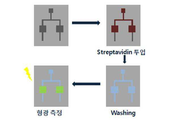
도 5a는 본 발명에 따른 미세유체 진단칩에 스타렙타아비딘을 주입한 후에 농도별 형광세기를 측정하는 방법을 나타내는 개념도이다.
도 5b는 본 발명에 따라 플라스틱 칩에 삽입된 3차원 네트워크를 이용하여 확인한 스타렙타아비딘의 농도별 형광세기를 보여주는 그래프이다.
도 6은 본 발명에 따른 3차원 네트워크가 형성된 기판, 다공성 구조를 갖는 멤브레인, CNT 없이 실리콘 기둥만 있는 기판 및 2차원 평면과 비교한 농도별 형광 세기 그래프이다.
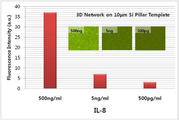
도 7은 본 발명에 따른 미세유체 진단칩을 이용한 염증반응에서 발현되는 IL-8 바이오 마커 검지를 확인한 형광 세기 그래프이다.
도 8은 침 희석 농도에 따른 바이오 마커 검지를 확인한 형광 세기 그래프이다.1 is a schematic diagram showing a state in which a three-dimensional carbon nanotube network nanostructure according to the present invention is mounted on a plastic substrate.
2 is (a) optical image and (b) fluorescence image of a three-dimensional network substrate surface-modified with biotin.
3 is a graph comparing fluorescence intensities of a two-dimensional planar substrate and a three-dimensional network structure.
4 is an image of a microfluidic diagnostic chip in which a nanostructure based on a 3D carbon nanotube network is mounted on a plastic substrate according to an embodiment of the present invention.
Figure 5a is a conceptual diagram showing a method for measuring the fluorescence intensity for each concentration after injecting the stareptavidin into the microfluidic diagnostic chip according to the present invention.
Figure 5b is a graph showing the fluorescence intensity for each concentration of staleptavividin confirmed using a three-dimensional network inserted in the plastic chip according to the present invention.
6 is a graph showing fluorescence intensity by concentration compared to a substrate having a three-dimensional network, a membrane having a porous structure, a substrate having only silicon pillars without CNTs, and a two-dimensional plane according to the present invention.
7 is a fluorescence intensity graph confirming the detection of IL-8 biomarker expressed in the inflammatory response using the microfluidic diagnostic chip according to the present invention.
8 is a graph showing fluorescence intensity confirming biomarker detection according to saliva dilution concentration.
이하, 본 발명을 더욱 상세히 설명한다.Hereinafter, the present invention will be described in more detail.
본 발명에 따른 미세유체 진단칩은 바이오 마커를 검지할 수 있도록 표면 개질된 3차원 탄소나노튜브 네트워크 기반의 나노구조체를 포함하며, 비침습성 타액으로부터 질병을 진단할 수 있는 것이 특징이다.The microfluidic diagnostic chip according to the present invention includes a nanostructure based on a three-dimensional carbon nanotube network surface modified to detect a biomarker, and is capable of diagnosing a disease from non-invasive saliva.
타액을 이용한 진단 방법은 누구나 거부감 없이 채취할 수 있으며, 비침습성 체취가 가능하기 때문에 환자 친화적인 방식으로서 매우 유용한 질병 진단 방법이다. 그러나, 타액의 경우 혈액의 농도의 1/100이기 때문에 타액을 이용한 진단의 경우에는 극미량에 대한 분석이 요구된다. 본 발명은 3차원 탄소나노튜브 네트워크로 이루어진 미세 채널을 갖는 나노구조체가 탑재된 미세유체 진단칩을 통해서 극미량의 타액 내의 정보를 검사하여 질병을 조기진단 하고자 한다.Anyone can use a saliva diagnostic method without rejection, and because it is non-invasive odor, it is a very useful disease diagnosis method. However, saliva is 1/100 of the concentration of blood, so in the case of diagnosis using saliva, analysis of the trace amount is required. The present invention aims to diagnose diseases early by examining a small amount of saliva information through a microfluidic diagnostic chip equipped with a nanostructure having a microchannel consisting of a three-dimensional carbon nanotube network.
본 발명에 따라, 실리콘 필러 사이에 합성된 탄소나노튜브 네트워크는 그 높이를 조절하여 각 질병에 따른 검출 양에 맞추어 제작할 수 있으며, 각기 다르게 개질된 여러 개(4-5 종류)의 나노구조체를 기판에 탑재하여 바이오마커의 다중 검출이 가능하도록 함으로써 질병 진단의 정확도를 높일 수 있다. 또한, 실리콘 칩이 아닌 플라스틱에 실리콘 기판을 작게 탑재하여 제조 원가도 낮춤으로써 경제적이며, 이에 따라 비침습적으로 남녀노소 누구나 쉽게 조기진단의 혜택을 받을 수 있다는 장점이 있다.According to the present invention, the carbon nanotube network synthesized between the silicon fillers can be manufactured according to the detection amount according to each disease by adjusting its height, and the substrates are formed of several (4-5 types) nanostructures modified differently. It is possible to increase the accuracy of disease diagnosis by enabling the multiple detection of the biomarker mounted on the. In addition, it is economical by lowering the manufacturing cost by mounting a silicon substrate in a plastic rather than a silicon chip, and thus there is an advantage that non-invasive age and age can easily benefit from early diagnosis.
본 발명에 있어서, 상기 3차원 탄소나노튜브 네트워크는 실리콘 기판상에 형성된 실리콘 필러 간에 병렬적으로 수평성장되어 복수의 탄소나노튜브 브리지(bridge)가 형성된 것이다. 본 발명에 따른 3차원 탄소나노튜브 네트워크의 형성 방법은 본 발명자의 대한민국 특허등록 10-1071215에 상세히 기재되어 있다.In the present invention, the three-dimensional carbon nanotube network is a horizontal growth in parallel between the silicon filler formed on the silicon substrate to form a plurality of carbon nanotube bridges (bridge). The method of forming a three-dimensional carbon nanotube network according to the present invention is described in detail in Korean Patent Registration No. 10-1071215.
본 발명에 있어서, 상기 3차원 탄소나노튜브 네트워크는 금속산화물로 코팅하는 것이 바람직하다. 이와 같은 금속산화물 코팅은 미세유체 채널의 강도를 증가시켜 나노구조를 유지시켜 주며, 이에 따라 다양한 바이오마커 검출이 가능하도록 표면 개질을 용이하게 해준다. 사용가능한 금속산화물로는 예를 들어, Al2O3, HfO2, ZrO2, ZnO2 , CuOx 등을 들 수 있다.In the present invention, the three-dimensional carbon nanotube network is preferably coated with a metal oxide. Such metal oxide coatings increase the strength of microfluidic channels to maintain nanostructures, thereby facilitating surface modification to enable detection of various biomarkers. Examples of the metal oxide that can be used include Al 2 O 3 , HfO 2 , ZrO 2 , ZnO 2 , CuO x , and the like.
또한, 본 발명의 다른 일 실시예에 의하면, 상기 3차원 탄소나노튜브 네트워크 기반의 나노구조체는 플라스틱 기판상에 탑재된 것일 수 있으며, 이때, 3차원 탄소나노튜브 네트워크 기반의 나노구조체는 플라스틱 기판상에 2개 이상 탑재될 수 있다. 2개 이상의 나노구조체는 각각 다른 바이오 마커를 검출할 수 있도록 표면개질된 경우 바이오 마커의 다중 검출이 가능하여 검출 효과를 향상시킬 수 있다. 이와 같이 상대적으로 저렴한 플라스틱 기판을 기초로 하면서 나노구조체만 실리콘 계열로 제조할 경우 제작 비용을 현저히 감소시킬 수 있어 그 활용도를 높일 수 있다.
In addition, according to another embodiment of the present invention, the three-dimensional carbon nanotube network-based nanostructures may be mounted on a plastic substrate, wherein, the three-dimensional carbon nanotube network-based nanostructures on a plastic substrate More than two can be mounted on. When two or more nanostructures are surface modified to detect different biomarkers, multiple detection of the biomarkers may be performed, thereby improving the detection effect. When manufacturing nano-structures based on relatively inexpensive plastic substrates alone and based on silicon, the manufacturing cost can be significantly reduced, thereby increasing their utilization.
또한, 본 발명에 따른 비침습성 타액으로부터 바이오 마커를 검지할 수 있는 3차원 탄소나노튜브 네트워크 기반의 나노구조체를 포함하는 미세유체 진단칩의 제조 방법은 다음과 같은 단계를 포함한다.In addition, the method of manufacturing a microfluidic diagnostic chip including a nanostructure based on a three-dimensional carbon nanotube network capable of detecting a biomarker from a non-invasive saliva according to the present invention includes the following steps.
1) 실리콘 기판 위에 실리콘 필러(pillar)를 형성하는 단계,1) forming a silicon pillar on a silicon substrate,
2) 상기 실리콘 필러 사이에 3차원 탄소나노튜브 네트워크를 형성하여 나노구조체를 제조하는 단계,2) preparing a nanostructure by forming a three-dimensional carbon nanotube network between the silicon filler,
3) 상기 3차원 탄소나노튜브 네트워크가 형성된 나노구조체에 금속산화물을 코팅하는 단계,3) coating a metal oxide on the nanostructure in which the 3D carbon nanotube network is formed;
4) 상기 금속산화물이 코팅된 나노구조체를 바이오 마커 검출이 가능하도록 표면개질하는 단계.4) surface modification of the metal oxide-coated nanostructure to enable biomarker detection.
상기 3) 단계에서 금속산화물은 원자층 증착법을 통해 코팅하는 것이 바람직하며, 4) 단계 다음에 표면개질된 3차원 탄소나노튜브 네트워크 기반의 나노구조체를 하나 또는 2 이상 플라스틱 기판에 탑재하는 단계를 더 포함할 수 있다. 이때, 2개 이상의 나노구조체는 각각 다른 바이오 마커를 검출할 수 있도록 표면 개질함으로써 검출 효율을 높일 수 있다.
In the step 3), the metal oxide is preferably coated by atomic layer deposition, and the step 4) further includes the step of mounting the surface-modified three-dimensional carbon nanotube network-based nanostructure on one or more plastic substrates. It may include. In this case, the two or more nanostructures may increase the detection efficiency by surface modification to detect different biomarkers, respectively.
이하 실시예를 통해 본 발명을 보다 상세히 설명하고자 하나, 이는 발명의 이해를 돕기 위해 예시적으로 제공된 것으로서, 본 발명의 범위가 이에 제한되는 것으로 해석되어서는 안된다.
Hereinafter, the present invention will be described in more detail with reference to the following examples, which are provided by way of example to help understanding of the present invention, and the scope of the present invention should not be construed as being limited thereto.
실시예Example 1 : 3차원 탄소나노튜브 합성 1: 3D carbon nanotube synthesis
3차원 탄소나노튜브는 다음과 같이 합성하였다. 30 마이크로미터 높이의 실리콘 필러를 딥에치 방법으로 공정한 후, 피라나 처리 과정을 통해 표면을 개질하고 클리닝을 한다. 철과 몰리브데늄의 이촉매(Bicatalyst) 하에서 한 시간 동안 용액에 담근 후 에탄올에서 10분간 분산시켰다.3D carbon nanotubes were synthesized as follows. After a 30 micrometer high silicon filler is processed by deep etching, the surface is modified and cleaned through a piranha process. After dipping in the solution for one hour under a bicatalyst of iron and molybdenum, it was dispersed in ethanol for 10 minutes.
이와 같이 준비된 시료를 열화학증착기(Thermal Chemical Vapor Deposition, T-CVD)를 이용하여 800 ℃에서 합성하였다. 800 ℃에서 암모니아 가스를 300 sccm 유속으로 10분간 흘려주고, 이후 아세틸렌 가스를 10 sccm의 유속으로 20분간 흘려준 다음 히터를 끄고 냉각시킨다.
The sample thus prepared was synthesized at 800 ° C. using a Thermal Chemical Vapor Deposition (T-CVD). At 800 ° C., ammonia gas was flowed at 300 sccm for 10 minutes, and then acetylene gas was flowed at 10 sccm for 20 minutes, and the heater was turned off and cooled.
실시예Example 2 : 네트워크 채널 강도를 증가를 위한 2: for increasing network channel strength 원자막증착법Atomic film deposition
3차원 탄소나노튜브는 타액과 같이 물이 99 %인 용액에서 소수성으로 잘 흐르지 않고 속도가 있을 경우 탄소나노튜브가 성장한 기판과 물리적 흡착이 되어 있어 그 모양이 변형이 된다. 따라서 원자막증착법(Atomic Layer Deposition, ALD)을 이용하여 여러 금속산화물을 표면에 코팅하여 채널의 강도를 높이고 화학적 표면 개질할 수 있는 종류의 범위를 더욱 넓혔다.3D carbon nanotubes do not flow hydrophobicly well in a 99% water solution like saliva, and if they have a velocity, they are physically adsorbed to the substrate on which the carbon nanotubes are grown. Therefore, Atomic Layer Deposition (ALD) is used to coat various metal oxides on the surface to increase the strength of the channel and broaden the range of chemical surface modification.
탄소나노튜브 표면에 고르게 금속 산화물을 코팅하기 위해서 오존처리를 30분 동안 하였다. 오존 처리 후, 고진공 챔버에 기판을 넣고 100 ℃로 가열시킨다. 이후 TMA를 펄스로 하여 증착시키고, Ar 가스로 반응하고 남은 TMA 소스 및 반응 부산물을 퍼지로 하여 배기시킨다. 다시 H2O를 펄스로 하여 표면에 흡착되어 있던 TMA와 반응하여 한 층의 Al2O3 박막을 형성하게 된다. 그런 다음 H2O와 반응 부산물을 퍼지하여 배기시킨다.
Ozone treatment was performed for 30 minutes to evenly coat the metal oxide on the surface of the carbon nanotubes. After ozone treatment, the substrate is placed in a high vacuum chamber and heated to 100 ° C. The TMA is then deposited in pulses, reacted with Ar gas, and the remaining TMA source and reaction by-products are purged and evacuated. In addition, H 2 O is pulsed to react with TMA adsorbed on the surface to form a layer of Al 2 O 3 thin film. The H 2 O and reaction byproducts are then purged and evacuated.
실시예Example 3 : 플라스틱 미세유체 기판 제작 3: plastic microfluidic substrate fabrication
각 타겟이 되는 암을 진단할 경우 체내에 염증이 있어서 나오는 CEA, CA19-9와 같은 물질과 함께 각 암에 대해 특이성 있게 검출되는 단백질들도 함께 검출할 때 그 진단 확률이 높다. 예를 들어 위암의 경우 VEGF는 내피세포 성장인자로 조기암 환자인 경우 매우 높게 나온다. 그래서 각각의 바이오 마커를 검출할 수 있는 기판이 삽입될 수 있는 플라스틱 기반의 기판을 제작한다.When diagnosing cancers of each target, the detection probability is high when the proteins that are specifically detected for each cancer are detected along with substances such as CEA and CA19-9, which are caused by inflammation in the body. For example, in stomach cancer, VEGF is an endothelial growth factor, which is very high in early cancer patients. Thus, a substrate based on plastic can be inserted into which a biomarker can be detected.
플라스틱 기판은 금속산화물로 코팅된 3차원 네트워크 기판이 삽입 가능하며 마이크로 플루이딕 채널이 형성이 가능한 디자인으로 마스크를 제작하고, 마스크를 기반으로 만든 실리콘 몰드를 제작한다. 이 후에 실리콘 몰드위에 PDMS 물질을 경화제와 함께 10:1로 섞어 부어준 후, 70 ℃에서 1시간 정도 경화한 후, 실리콘 몰드에서 떼어낸다.
The plastic substrate can be inserted into a three-dimensional network substrate coated with a metal oxide, a microfluidic channel can be formed into a mask, and a mask-based silicon mold. Thereafter, the PDMS material is poured into the silicone mold with a curing agent at a ratio of 10: 1, cured at 70 ° C. for about 1 hour, and then removed from the silicone mold.
실시예Example 4 : 3차원 탄소나노튜브 네트워크 기반의 나노구조체가 플라스틱 4: Nanostructure based on 3D carbon nanotube network is plastic 기group 판 상에 2개 이상 탑재된 미세유체 진단칩의 제조.(도 4a)Fabrication of two or more microfluidic diagnostic chips mounted on a plate (FIG. 4A).
상기 실시예 1-2를 통하여 제조된 3차원 네트워크가 형성된 기판을 바이오마커를 검지할 수 있도록 표면 개질한 후, 5 mm × 5 mm로 자르고 PDMS로 제작된 칩에 삽입하여 미세유체 진단칩을 제조하였다.
After modifying the substrate on which the 3D network formed in Example 1-2 was formed to detect the biomarker, cut it into 5 mm × 5 mm and insert it into a chip made of PDMS to manufacture a microfluidic diagnostic chip. It was.
실험예 1 : 금속산화물로 코팅된 3차원 네트워크 표면 개질에 따른 특성 확인Experimental Example 1: Confirmation of the properties according to the surface modification of the three-dimensional network coated with metal oxide
도 2는 표면이 개질된 3차원 네트워크 구조가 있는 기판의 현미경 사진이다. (a)는 광학 사진으로 하얀색 작은 원의 실리콘 필러 표면만 보인다. 그러나 (b) 형광 사진에서는 실리콘 사이에 연결된 채널이 바이오 물질로 개질되어 형광을 나타내는 것을 보여준다. 3차원 네트워크 구조와 2차원 평면기판을 같은 처리를 했을 경우, 진한 농도에서는 2 배 정도의 형광 세기 차이밖에 나지 않지만 1000배를 희석한 경우에는 200 배의 형광 세기가 차이가 나는 것을 볼 수 있다.(도 3)
2 is a micrograph of a substrate having a three-dimensional network structure with a modified surface. (a) is an optical photograph showing only the silicon filler surface of a small white circle. However, (b) the fluorescence photograph shows that the channel connected between the silicon is modified with a biomaterial to show fluorescence. When the same treatment of the three-dimensional network structure and the two-dimensional planar substrate is performed, the difference in fluorescence intensity is only about 2 times in the dark concentration, but when the 1000 times diluted, the fluorescence intensity is 200 times different. (Figure 3)
실험예Experimental Example 2 : 본 발명에 따른 미세유체 진단칩으로 확인한 2: confirmed with the microfluidic diagnostic chip according to the present invention 스트렙타아비딘의Streptavidin 농도별 By concentration 형광세기Fluorescence intensity 확인 Confirm
금속산화물이 코팅된 3차원 네트워크 표면을 -NH2으로 개질하기 위해 3-Aminopropyltriethoxysilane(ATPES) 물질로 톨루엔 용매에 2 wt%로 만들어 기판을 담구워 12시간을 둔다. 이후에 여러 번 용매로 표면을 세척한 후, NSH로 활성된 바이오틴을 1 mg/mL 용액에 2시간을 담군다. 바이오틴으로 최종 개질된 기판을 5 mm × 5 mm로 자른 후, 이미 제작된 PDMS 진단 칩에 삽입한다. 마이크로펌프를 이용하여 형광염료가 달린 스트렙타아비딘을 PBS 버퍼 용액에 1 ug/mL, 1 ng/mL, 500 pg/mL, 10 pg/mL의 농도로 만든 용액을 흘려서 바이오틴으로 개질된 표면에 스트렙타아비딘을 농도별로 개질한다. 이후 PBM 버퍼 용액으로 3번 헹궈주고 형광현미경을 이용하여 스트렙타아비딘의 농도별 형광 세기를 관측하였다.In order to modify the surface of the metal oxide-coated three-dimensional network with -NH 2 , the substrate was immersed in 3 wt% of toluene solvent with 3-Aminopropyltriethoxysilane (ATPES) material and left for 12 hours. After washing the surface several times with a solvent, soaked in NSH-activated biotin in 1 mg / mL solution for 2 hours. The final modified substrate with biotin is cut into 5 mm x 5 mm and then inserted into a prefabricated PDMS diagnostic chip. Using a micropump, streptavidin with fluorescent dye was poured into a PBS buffer solution at a concentration of 1 ug / mL, 1 ng / mL, 500 pg / mL, or 10 pg / mL, and then streptavided onto the biotin-modified surface. Taavidine is modified by concentration. After rinsing three times with PBM buffer solution and using a fluorescence microscope to observe the fluorescence intensity of each concentration of streptavidin.
하기 도 5b를 살펴보면, 10 pg/mL의 농도에서도 측정이 가능함을 알 수 있다.
Referring to Figure 5b, it can be seen that even at a concentration of 10 pg / mL can be measured.
실험예Experimental Example 3 3
도 6은 본 발명에 따른 3차원 네트워크를 갖는 기판을 다공성 구조를 갖는 멤브레인, CNT가 없이 실리콘 기둥만 있는 기판 및 2차원 평면과 비교한 농도별 형광 세기 비교 데이터이다. PDMS로 제작된 진단 칩에 각 구조를 삽입하여 1 ng/mL와 100 ng/mL의 형광염료가 달린 스트렙타아비딘을 흘려주었을 때, 각 구조별 형광을 비교하였다.FIG. 6 is a concentration-specific fluorescence intensity comparison data comparing a substrate having a three-dimensional network according to the present invention with a membrane having a porous structure, a substrate having only a silicon pillar without CNTs, and a two-dimensional plane. Each structure was inserted into a diagnostic chip made of PDMS, and when streptavidin with 1 ng / mL and 100 ng / mL fluorescent dye was flowed, the fluorescence of each structure was compared.
하기 도 6에 나타나 있는 바와 같이, 본 발명에 따른 3차원 네트워크가 형성된 기판이 저농도에서도 현저히 우수한 고감도를 보인다.
As shown in Figure 6, the substrate on which the three-dimensional network is formed according to the present invention shows a remarkably excellent high sensitivity even at low concentrations.
실험예Experimental Example 4. 본 발명에 따른 미세유체 진단칩을 이용한 염증 반응에서 발현되는 4. It is expressed in the inflammatory response using the microfluidic diagnostic chip according to the present invention. ILIL -8 바이오 -8 bio 마커Marker 검지 Index finger
도 7은 본 발명에 따른 3차원 네트워크에 IL-8을 농도별로 검출한 데이터이다. 스트렙타아비딘 검출 실험과 동일하게 3차원 탄소나노튜브 합성 후, 금속산화물을 코팅하고 표면은 아민으로 개질한다. 이후에 바이오틴으로 표면을 개질하고 스트렙타아비딘으로 개질한다. 바이오마커를 검지하기 위해서 바이오틴이 붙어있는 IL-8 안티바디를 구매하여 안티바디를 개질하고 진단칩에 삽입한 후, IL-8 형광염료가 붙은 안티젠을 500 ng/mL, 5 ng/mL, 500 pg/mL 농도별로 흘린 후, 버퍼용액으로 2-3번 헹구고 형광 현미경으로 관측하였을 때, 500 pg/mL까지 3차원 네트워크 구조로 검지가 가능한 것을 확인하였다.
7 is data of detecting IL-8 by concentration in a three-dimensional network according to the present invention. After synthesizing three-dimensional carbon nanotubes in the same manner as the streptavidin detection experiment, the metal oxide is coated and the surface is modified with amine. The surface is then modified with biotin and streptavidin. To detect biomarkers, purchase biotin-containing IL-8 antibodies, modify the antibodies and insert them into the diagnostic chip. 500 ng / mL, 5 ng / mL, 500 antigens with IL-8 fluorescent dye After flowing by pg / mL concentration, when rinsed 2-3 times with buffer solution and observed under a fluorescence microscope, it was confirmed that it can be detected in a three-dimensional network structure up to 500 pg / mL.
실험예Experimental Example 5. 침 희석 농도에 따른 바이오 5. Bio according to saliva dilution concentration 마커Marker 검지 Index finger
PBS 버퍼 용액에서 진행한 바이오 마커 검지 실험 외에, 실제 침에서도 같은 농도가 동일하게 침 속에서 검지가 되는지를 확인하기 위해서 기존에 사용한 버퍼 용액과 정상인의 침, 그 침을 버퍼용액에 20%와 11% 혼합시켜 같은 양의 100 ng/mL의 스트렙타아비딘을 바이오틴이 개질된 3차원 구조가 삽입된 진단칩에 주입하여 형광 세기를 평가하였으며, 그 결과를 하기 도 8에 나타내었다.In addition to the biomarker detection experiments conducted in PBS buffer solution, to confirm whether the same concentration is detected in the saliva in the actual saliva, the buffer solution and the saliva of normal persons, and the saliva, were added to the buffer solution. The fluorescence intensity was evaluated by injecting the same amount of 100 ng / mL of streptavidin into the diagnostic chip into which the biotin-modified three-dimensional structure was inserted, and the results are shown in FIG. 8.
하기 도 8에서 보는 바와 같이, 침으로만 이루어진 시료에서는 형광세기가 조금 떨어지지만 전반적으로 침에 있는 다른 단백질들의 영향을 받지 않고 선택적으로 반응이 일어나므로 진단에 있어서 본 발명에 따른 3차원 네트워크 구조가 매우 유용함을 알 수 있다.As shown in FIG. 8, in the sample consisting of saliva, the fluorescence intensity is slightly decreased, but the reaction occurs selectively without being influenced by other proteins in the saliva. It is very useful.
Claims (13)
상기 미세유체 진단칩은 비침습성 타액으로부터 질병을 진단할 수 있는 것을 특징으로 하는 미세유체 진단칩.The method of claim 1,
The microfluidic diagnostic chip is a microfluidic diagnostic chip, characterized in that for diagnosing a disease from non-invasive saliva.
상기 3차원 탄소나노튜브 네트워크는 실리콘 기판상에 형성된 실리콘 필러 간에 병렬적으로 수평 성장되어 복수의 탄소나노튜브 브리지(bridge)가 형성된 것을 특징으로 하는 미세유체 진단칩.The method of claim 1,
And the three-dimensional carbon nanotube network is horizontally grown in parallel between silicon fillers formed on a silicon substrate to form a plurality of carbon nanotube bridges.
상기 3차원 탄소나노튜브 네트워크는 금속산화물로 코팅된 것을 특징으로 하는 미세유체 진단칩.The method of claim 1,
The three-dimensional carbon nanotube network is a microfluidic diagnostic chip, characterized in that the coating with a metal oxide.
상기 금속산화물은 Al2O3, HfO2, ZrO2, ZnO2 및 CuOx 중에서 선택되는 것을 특징으로 하는 미세유체 진단칩.5. The method of claim 4,
The metal oxide is Al 2 O 3 , HfO 2 , ZrO 2 , ZnO 2 And CuO x microfluidic diagnostic chip, characterized in that selected from.
상기 3차원 탄소나노튜브 네트워크 기반의 나노구조체가 플라스틱 기판상에 탑재되어 있는 것을 특징으로 하는 미세유체 진단칩.The method of claim 1,
The microfluidic diagnostic chip, wherein the 3D carbon nanotube network-based nanostructure is mounted on a plastic substrate.
상기 3차원 탄소나노튜브 네트워크 기반의 나노구조체가 플라스틱 기판상에 2개 이상 탑재되어 있는 것을 특징으로 하는 미세유체 진단칩.The method according to claim 6,
Microfluidic diagnostic chip, characterized in that two or more nanostructures based on the three-dimensional carbon nanotube network is mounted on a plastic substrate.
상기 2개 이상의 나노구조체는 각각 다른 바이오 마커를 검출할 수 있도록 표면개질된 것을 특징으로 하는 미세유체 진단칩.The method of claim 7, wherein
The two or more nanostructures are microfluidic diagnostic chip, characterized in that the surface is modified to detect different biomarkers, respectively.
1) 실리콘 기판 위에 실리콘 필러(pillar)를 형성하는 단계;
2) 상기 실리콘 필러 사이에 3차원 탄소나노튜브 네트워크를 형성하여 나노구조체를 제조하는 단계;
3) 상기 3차원 탄소나노튜브 네트워크가 형성된 나노구조체에 금속산화물을 코팅하는 단계; 및
4) 상기 금속산화물이 코팅된 나노구조체를 바이오 마커 검출이 가능하도록 표면개질하는 단계;를 포함하는 미세유체 진단칩의 제조방법.In the method of manufacturing a microfluidic diagnostic chip comprising a nanostructure based on a three-dimensional carbon nanotube network that can detect a biomarker from non-invasive saliva,
1) forming a silicon pillar on the silicon substrate;
2) forming a nanostructure by forming a three-dimensional carbon nanotube network between the silicon filler;
3) coating a metal oxide on the nanostructure in which the three-dimensional carbon nanotube network is formed; And
And 4) surface-modifying the metal oxide-coated nanostructure to enable biomarker detection.
상기 3) 단계에서 금속산화물은 원자층 증착법을 통해 코팅되는 것을 특징으로 하는 미세유체 진단칩의 제조방법.10. The method of claim 9,
In the step 3), the metal oxide is a method of manufacturing a microfluidic diagnostic chip, characterized in that the coating by atomic layer deposition.
상기 4) 단계 다음에 표면개질된 3차원 탄소나노튜브 네트워크 기반의 나노구조체를 플라스틱 기판에 탑재하는 단계;를 더 포함하는 미세유체 진단칩의 제조방법.The method of claim 10,
And mounting the surface-modified three-dimensional carbon nanotube network-based nanostructure on the plastic substrate after the step 4).
상기 3차원 탄소나노튜브 네트워크 나노구조체를 플라스틱 기판에 2개 이상 탑재하는 것을 특징으로 하는 미세유체 진단칩의 제조방법.12. The method of claim 11,
A method of manufacturing a microfluidic diagnostic chip, characterized in that at least two three-dimensional carbon nanotube network nanostructures are mounted on a plastic substrate.
상기 2개 이상의 나노구조체는 각각 다른 바이오 마커를 검출할 수 있도록 표면 개질된 것을 특징으로 하는 미세유체 진단칩의 제조 방법.The method of claim 12,
The method of manufacturing a microfluidic diagnostic chip, wherein the two or more nanostructures are surface-modified to detect different biomarkers, respectively.
Applications Claiming Priority (2)
| Application Number | Priority Date | Filing Date | Title |
|---|---|---|---|
| KR1020120056799 | 2012-05-29 | ||
| KR20120056799 | 2012-05-29 |
Publications (1)
| Publication Number | Publication Date |
|---|---|
| KR20130133700A true KR20130133700A (en) | 2013-12-09 |
Family
ID=49981593
Family Applications (1)
| Application Number | Title | Priority Date | Filing Date |
|---|---|---|---|
| KR1020130061218A KR20130133700A (en) | 2012-05-29 | 2013-05-29 | Microfluidic diagnostic chip comprising nanostructures based on three-dimensional network of carbon nanotubes |
Country Status (1)
| Country | Link |
|---|---|
| KR (1) | KR20130133700A (en) |
Cited By (3)
| Publication number | Priority date | Publication date | Assignee | Title |
|---|---|---|---|---|
| US9719926B2 (en) | 2015-11-16 | 2017-08-01 | International Business Machines Corporation | Nanopillar microfluidic devices and methods of use thereof |
| CN107105675A (en) * | 2014-10-28 | 2017-08-29 | 杨百翰大学 | Microbial resistance material and the device of correlation, system and method |
| US10517995B2 (en) | 2016-11-01 | 2019-12-31 | Brigham Young University | Super-hydrophobic materials and associated devices, systems, and methods |
-
2013
- 2013-05-29 KR KR1020130061218A patent/KR20130133700A/en not_active Application Discontinuation
Cited By (5)
| Publication number | Priority date | Publication date | Assignee | Title |
|---|---|---|---|---|
| CN107105675A (en) * | 2014-10-28 | 2017-08-29 | 杨百翰大学 | Microbial resistance material and the device of correlation, system and method |
| EP3212002A4 (en) * | 2014-10-28 | 2018-05-23 | Brigham Young University | Microorganism-resistant materials and associated devices, systems, and methods |
| CN107105675B (en) * | 2014-10-28 | 2023-11-07 | 杨百翰大学 | Microbial resistant materials and related devices, systems, and methods |
| US9719926B2 (en) | 2015-11-16 | 2017-08-01 | International Business Machines Corporation | Nanopillar microfluidic devices and methods of use thereof |
| US10517995B2 (en) | 2016-11-01 | 2019-12-31 | Brigham Young University | Super-hydrophobic materials and associated devices, systems, and methods |
Similar Documents
| Publication | Publication Date | Title |
|---|---|---|
| Yu et al. | Quantitative real-time detection of carcinoembryonic antigen (CEA) from pancreatic cyst fluid using 3-D surface molecular imprinting | |
| Nagaraj et al. | NanoMonitor: a miniature electronic biosensor for glycan biomarker detection | |
| US11650204B2 (en) | Plasmo photoelectronic immunosensor | |
| WO2012112746A1 (en) | Biocompatible graphene sensor | |
| Lei et al. | CMOS biosensors for in vitro diagnosis–transducing mechanisms and applications | |
| Tuteja et al. | Liquid exfoliation of 2D MoS 2 nanosheets and their utilization as a label-free electrochemical immunoassay for subclinical ketosis | |
| Song et al. | Fluorescence-based immunosensor using three-dimensional CNT network structure for sensitive and reproducible detection of oral squamous cell carcinoma biomarker | |
| Hu et al. | Layer-by-layer self-assembly of MoS2/PDDA hybrid film in microfluidic chips for ultrasensitive electrochemical immunosensing of alpha-fetoprotein | |
| CN108387563A (en) | Fluorescence Increasing structure, fluorescence detecting system based on nanometer rods and automatic sampling detection chip | |
| Pourakbari et al. | Early stage evaluation of colon cancer using tungsten disulfide quantum dots and bacteriophage nano-biocomposite as an efficient electrochemical platform | |
| Chakraborty et al. | Nano‐diagnostics as an emerging platform for oral cancer detection: Current and emerging trends | |
| KR20130133700A (en) | Microfluidic diagnostic chip comprising nanostructures based on three-dimensional network of carbon nanotubes | |
| US11536721B2 (en) | Electrochemical immunosensors | |
| Yadav et al. | Biocompatible epoxysilane substituted polymer-based nano biosensing platform for label-free detection of cancer biomarker SP17 in patient serum samples | |
| Hsieh et al. | Construction of the nickel oxide nanocoral structure on microscope slides for total self-assembly-oriented probe immobilization and signal enhancement | |
| Mohanty et al. | Field effect transistor nanosensor for breast cancer diagnostics | |
| Chakraborty et al. | Competitive Impedance Spectroscopy in a Schottky-Contacted ZnO Nanorod Structure for Ultrasensitive and Specific Biosensing in a Physiological Analyte | |
| Bhardwaj et al. | Recent advancement in the detection of potential cancer biomarkers using the nanomaterial integrated electrochemical sensing technique: a detailed review | |
| Hu et al. | A sensitive electrochemical platform integrated with a 3D graphene aerogel for point-of-care testing for tumor markers | |
| KR20140017439A (en) | Fabrication of immunosensor using gold nanorod array and the immunosensor by using the same method | |
| KR102031194B1 (en) | Method for measuring a concentration of beta-amyloid | |
| Shankaran | Nano-Enabled Immunosensors for Point-of-Care Cancer Diagnosis | |
| Quazi | The potential implementation of biosensors for the diagnosis of biomarkers of various cancer | |
| Lv et al. | In-situ formation of “electron conductive wires” threaded ZIF-8 membrane for multiplexed immunoassay of human interleukins | |
| Pearton et al. | Functionalized GaN-Based Transistors for Biosensing |
Legal Events
| Date | Code | Title | Description |
|---|---|---|---|
| A201 | Request for examination | ||
| E902 | Notification of reason for refusal | ||
| E601 | Decision to refuse application |