KR20130028044A - Methods and compositions using peroxiredoxin1 (prx1) as an adjuvant - Google Patents
Methods and compositions using peroxiredoxin1 (prx1) as an adjuvant Download PDFInfo
- Publication number
- KR20130028044A KR20130028044A KR1020127016307A KR20127016307A KR20130028044A KR 20130028044 A KR20130028044 A KR 20130028044A KR 1020127016307 A KR1020127016307 A KR 1020127016307A KR 20127016307 A KR20127016307 A KR 20127016307A KR 20130028044 A KR20130028044 A KR 20130028044A
- Authority
- KR
- South Korea
- Prior art keywords
- prx1
- tlr4
- cells
- antigen
- protein
- Prior art date
Links
- 239000000203 mixture Substances 0.000 title claims abstract description 34
- 238000000034 method Methods 0.000 title claims abstract description 23
- 239000002671 adjuvant Substances 0.000 title abstract description 7
- 101150081137 PRX1 gene Proteins 0.000 title 1
- 102000007456 Peroxiredoxin Human genes 0.000 title 1
- 108030002458 peroxiredoxin Proteins 0.000 title 1
- 239000000427 antigen Substances 0.000 claims abstract description 62
- 102000036639 antigens Human genes 0.000 claims abstract description 62
- 108091007433 antigens Proteins 0.000 claims abstract description 62
- 102000004169 proteins and genes Human genes 0.000 claims abstract description 55
- 108090000623 proteins and genes Proteins 0.000 claims abstract description 55
- 230000028993 immune response Effects 0.000 claims abstract description 25
- 230000004936 stimulating effect Effects 0.000 claims abstract description 5
- 210000004027 cell Anatomy 0.000 claims description 86
- 210000002540 macrophage Anatomy 0.000 claims description 77
- 206010028980 Neoplasm Diseases 0.000 claims description 22
- 210000004443 dendritic cell Anatomy 0.000 claims description 14
- 201000011510 cancer Diseases 0.000 claims description 12
- 230000002463 transducing effect Effects 0.000 claims 1
- 230000004853 protein function Effects 0.000 abstract 1
- 102000002689 Toll-like receptor Human genes 0.000 description 101
- 108020000411 Toll-like receptor Proteins 0.000 description 101
- 229940071127 thioglycolate Drugs 0.000 description 51
- CWERGRDVMFNCDR-UHFFFAOYSA-M thioglycolate(1-) Chemical compound [O-]C(=O)CS CWERGRDVMFNCDR-UHFFFAOYSA-M 0.000 description 51
- 102000004127 Cytokines Human genes 0.000 description 35
- 108090000695 Cytokines Proteins 0.000 description 35
- 230000028327 secretion Effects 0.000 description 30
- 241000699670 Mus sp. Species 0.000 description 29
- 239000002158 endotoxin Substances 0.000 description 28
- 229920006008 lipopolysaccharide Polymers 0.000 description 25
- 108090001005 Interleukin-6 Proteins 0.000 description 23
- 102000004889 Interleukin-6 Human genes 0.000 description 23
- MHMNJMPURVTYEJ-UHFFFAOYSA-N fluorescein-5-isothiocyanate Chemical compound O1C(=O)C2=CC(N=C=S)=CC=C2C21C1=CC=C(O)C=C1OC1=CC(O)=CC=C21 MHMNJMPURVTYEJ-UHFFFAOYSA-N 0.000 description 21
- 230000027455 binding Effects 0.000 description 20
- 230000001419 dependent effect Effects 0.000 description 19
- 239000006228 supernatant Substances 0.000 description 19
- MZOFCQQQCNRIBI-VMXHOPILSA-N (3s)-4-[[(2s)-1-[[(2s)-1-[[(1s)-1-carboxy-2-hydroxyethyl]amino]-4-methyl-1-oxopentan-2-yl]amino]-5-(diaminomethylideneamino)-1-oxopentan-2-yl]amino]-3-[[2-[[(2s)-2,6-diaminohexanoyl]amino]acetyl]amino]-4-oxobutanoic acid Chemical compound OC[C@@H](C(O)=O)NC(=O)[C@H](CC(C)C)NC(=O)[C@H](CCCN=C(N)N)NC(=O)[C@H](CC(O)=O)NC(=O)CNC(=O)[C@@H](N)CCCCN MZOFCQQQCNRIBI-VMXHOPILSA-N 0.000 description 17
- 101000946889 Homo sapiens Monocyte differentiation antigen CD14 Proteins 0.000 description 17
- 102100035877 Monocyte differentiation antigen CD14 Human genes 0.000 description 17
- 108091027967 Small hairpin RNA Proteins 0.000 description 17
- 108060008682 Tumor Necrosis Factor Proteins 0.000 description 17
- 102000000852 Tumor Necrosis Factor-alpha Human genes 0.000 description 17
- 239000004055 small Interfering RNA Substances 0.000 description 16
- 101000979342 Homo sapiens Nuclear factor NF-kappa-B p105 subunit Proteins 0.000 description 15
- 102100023050 Nuclear factor NF-kappa-B p105 subunit Human genes 0.000 description 15
- 230000003993 interaction Effects 0.000 description 14
- 239000002609 medium Substances 0.000 description 13
- 230000000694 effects Effects 0.000 description 12
- 230000000638 stimulation Effects 0.000 description 12
- 210000004881 tumor cell Anatomy 0.000 description 12
- 108091003079 Bovine Serum Albumin Proteins 0.000 description 11
- 238000004458 analytical method Methods 0.000 description 11
- 102000010168 Myeloid Differentiation Factor 88 Human genes 0.000 description 10
- 108010077432 Myeloid Differentiation Factor 88 Proteins 0.000 description 10
- 238000007792 addition Methods 0.000 description 10
- 229940098773 bovine serum albumin Drugs 0.000 description 10
- 101001046686 Homo sapiens Integrin alpha-M Proteins 0.000 description 9
- 102100022338 Integrin alpha-M Human genes 0.000 description 9
- 108010093965 Polymyxin B Proteins 0.000 description 8
- 102000007056 Recombinant Fusion Proteins Human genes 0.000 description 8
- 108010008281 Recombinant Fusion Proteins Proteins 0.000 description 8
- 239000013592 cell lysate Substances 0.000 description 8
- 238000002474 experimental method Methods 0.000 description 8
- DOUYETYNHWVLEO-UHFFFAOYSA-N imiquimod Chemical compound C1=CC=CC2=C3N(CC(C)C)C=NC3=C(N)N=C21 DOUYETYNHWVLEO-UHFFFAOYSA-N 0.000 description 8
- 238000011534 incubation Methods 0.000 description 8
- 239000002953 phosphate buffered saline Substances 0.000 description 8
- YXHLJMWYDTXDHS-IRFLANFNSA-N 7-aminoactinomycin D Chemical compound C[C@H]1OC(=O)[C@H](C(C)C)N(C)C(=O)CN(C)C(=O)[C@@H]2CCCN2C(=O)[C@@H](C(C)C)NC(=O)[C@H]1NC(=O)C1=C(N)C(=O)C(C)=C2OC(C(C)=C(N)C=C3C(=O)N[C@@H]4C(=O)N[C@@H](C(N5CCC[C@H]5C(=O)N(C)CC(=O)N(C)[C@@H](C(C)C)C(=O)O[C@@H]4C)=O)C(C)C)=C3N=C21 YXHLJMWYDTXDHS-IRFLANFNSA-N 0.000 description 7
- 108700012813 7-aminoactinomycin D Proteins 0.000 description 7
- 101100339431 Arabidopsis thaliana HMGB2 gene Proteins 0.000 description 7
- 108700010013 HMGB1 Proteins 0.000 description 7
- 101150021904 HMGB1 gene Proteins 0.000 description 7
- 102100037907 High mobility group protein B1 Human genes 0.000 description 7
- 101000831567 Homo sapiens Toll-like receptor 2 Proteins 0.000 description 7
- 102100024333 Toll-like receptor 2 Human genes 0.000 description 7
- 230000004913 activation Effects 0.000 description 7
- 230000000903 blocking effect Effects 0.000 description 7
- 238000001514 detection method Methods 0.000 description 7
- 229920000024 polymyxin B Polymers 0.000 description 7
- 229960005266 polymyxin b Drugs 0.000 description 7
- 108010027814 HSP72 Heat-Shock Proteins Proteins 0.000 description 6
- 102100040352 Heat shock 70 kDa protein 1A Human genes 0.000 description 6
- 102000003992 Peroxidases Human genes 0.000 description 6
- FAPWRFPIFSIZLT-UHFFFAOYSA-M Sodium chloride Chemical compound [Na+].[Cl-] FAPWRFPIFSIZLT-UHFFFAOYSA-M 0.000 description 6
- 239000011324 bead Substances 0.000 description 6
- 238000000684 flow cytometry Methods 0.000 description 6
- 229960002751 imiquimod Drugs 0.000 description 6
- 230000001965 increasing effect Effects 0.000 description 6
- 239000003446 ligand Substances 0.000 description 6
- 108040007629 peroxidase activity proteins Proteins 0.000 description 6
- 239000011534 wash buffer Substances 0.000 description 6
- 238000002965 ELISA Methods 0.000 description 5
- 238000008157 ELISA kit Methods 0.000 description 5
- 241001465754 Metazoa Species 0.000 description 5
- 108010006519 Molecular Chaperones Proteins 0.000 description 5
- 241000699666 Mus <mouse, genus> Species 0.000 description 5
- 108010058846 Ovalbumin Proteins 0.000 description 5
- 238000003556 assay Methods 0.000 description 5
- 239000012895 dilution Substances 0.000 description 5
- 238000010790 dilution Methods 0.000 description 5
- 239000012091 fetal bovine serum Substances 0.000 description 5
- 230000006870 function Effects 0.000 description 5
- 210000002865 immune cell Anatomy 0.000 description 5
- 239000012678 infectious agent Substances 0.000 description 5
- 239000006166 lysate Substances 0.000 description 5
- 239000000463 material Substances 0.000 description 5
- 230000035772 mutation Effects 0.000 description 5
- 238000012758 nuclear staining Methods 0.000 description 5
- 229940092253 ovalbumin Drugs 0.000 description 5
- 230000012846 protein folding Effects 0.000 description 5
- 102000005962 receptors Human genes 0.000 description 5
- 108020003175 receptors Proteins 0.000 description 5
- 210000002966 serum Anatomy 0.000 description 5
- 238000007619 statistical method Methods 0.000 description 5
- 230000001225 therapeutic effect Effects 0.000 description 5
- 229960005486 vaccine Drugs 0.000 description 5
- 201000009030 Carcinoma Diseases 0.000 description 4
- 241000700605 Viruses Species 0.000 description 4
- 150000001413 amino acids Chemical group 0.000 description 4
- 230000000259 anti-tumor effect Effects 0.000 description 4
- 210000001185 bone marrow Anatomy 0.000 description 4
- 239000000872 buffer Substances 0.000 description 4
- 238000005516 engineering process Methods 0.000 description 4
- 102000037865 fusion proteins Human genes 0.000 description 4
- 108020001507 fusion proteins Proteins 0.000 description 4
- 230000004807 localization Effects 0.000 description 4
- 230000035800 maturation Effects 0.000 description 4
- UCSJYZPVAKXKNQ-HZYVHMACSA-N streptomycin Chemical compound CN[C@H]1[C@H](O)[C@@H](O)[C@H](CO)O[C@H]1O[C@@H]1[C@](C=O)(O)[C@H](C)O[C@H]1O[C@@H]1[C@@H](NC(N)=N)[C@H](O)[C@@H](NC(N)=N)[C@H](O)[C@H]1O UCSJYZPVAKXKNQ-HZYVHMACSA-N 0.000 description 4
- 230000009885 systemic effect Effects 0.000 description 4
- 238000012360 testing method Methods 0.000 description 4
- 210000001519 tissue Anatomy 0.000 description 4
- 238000001262 western blot Methods 0.000 description 4
- 241000894006 Bacteria Species 0.000 description 3
- 241000283707 Capra Species 0.000 description 3
- PEDCQBHIVMGVHV-UHFFFAOYSA-N Glycerine Chemical compound OCC(O)CO PEDCQBHIVMGVHV-UHFFFAOYSA-N 0.000 description 3
- 206010061218 Inflammation Diseases 0.000 description 3
- 206010058467 Lung neoplasm malignant Diseases 0.000 description 3
- 108091028043 Nucleic acid sequence Proteins 0.000 description 3
- 108010060818 Toll-Like Receptor 9 Proteins 0.000 description 3
- 102100033117 Toll-like receptor 9 Human genes 0.000 description 3
- 239000000556 agonist Substances 0.000 description 3
- 210000004369 blood Anatomy 0.000 description 3
- 239000008280 blood Substances 0.000 description 3
- 230000008859 change Effects 0.000 description 3
- 239000003153 chemical reaction reagent Substances 0.000 description 3
- 239000000539 dimer Substances 0.000 description 3
- LOKCTEFSRHRXRJ-UHFFFAOYSA-I dipotassium trisodium dihydrogen phosphate hydrogen phosphate dichloride Chemical compound P(=O)(O)(O)[O-].[K+].P(=O)(O)([O-])[O-].[Na+].[Na+].[Cl-].[K+].[Cl-].[Na+] LOKCTEFSRHRXRJ-UHFFFAOYSA-I 0.000 description 3
- 201000010099 disease Diseases 0.000 description 3
- 208000037265 diseases, disorders, signs and symptoms Diseases 0.000 description 3
- 231100000673 dose–response relationship Toxicity 0.000 description 3
- 230000001976 improved effect Effects 0.000 description 3
- 230000004054 inflammatory process Effects 0.000 description 3
- 239000007924 injection Substances 0.000 description 3
- 238000002347 injection Methods 0.000 description 3
- 201000001441 melanoma Diseases 0.000 description 3
- 230000018491 positive regulation of cytokine secretion Effects 0.000 description 3
- 230000000770 proinflammatory effect Effects 0.000 description 3
- 230000000069 prophylactic effect Effects 0.000 description 3
- 230000003248 secreting effect Effects 0.000 description 3
- 239000011780 sodium chloride Substances 0.000 description 3
- 238000002415 sodium dodecyl sulfate polyacrylamide gel electrophoresis Methods 0.000 description 3
- 238000010561 standard procedure Methods 0.000 description 3
- 238000005406 washing Methods 0.000 description 3
- 201000003076 Angiosarcoma Diseases 0.000 description 2
- 206010004146 Basal cell carcinoma Diseases 0.000 description 2
- 201000009047 Chordoma Diseases 0.000 description 2
- 206010009944 Colon cancer Diseases 0.000 description 2
- 108020004414 DNA Proteins 0.000 description 2
- 208000001258 Hemangiosarcoma Diseases 0.000 description 2
- 241000713772 Human immunodeficiency virus 1 Species 0.000 description 2
- MHAJPDPJQMAIIY-UHFFFAOYSA-N Hydrogen peroxide Chemical compound OO MHAJPDPJQMAIIY-UHFFFAOYSA-N 0.000 description 2
- -1 IL-1α Proteins 0.000 description 2
- 102100022297 Integrin alpha-X Human genes 0.000 description 2
- 208000000172 Medulloblastoma Diseases 0.000 description 2
- 108091034117 Oligonucleotide Proteins 0.000 description 2
- 206010061902 Pancreatic neoplasm Diseases 0.000 description 2
- 229930040373 Paraformaldehyde Natural products 0.000 description 2
- 229930182555 Penicillin Natural products 0.000 description 2
- JGSARLDLIJGVTE-MBNYWOFBSA-N Penicillin G Chemical compound N([C@H]1[C@H]2SC([C@@H](N2C1=O)C(O)=O)(C)C)C(=O)CC1=CC=CC=C1 JGSARLDLIJGVTE-MBNYWOFBSA-N 0.000 description 2
- 206010039491 Sarcoma Diseases 0.000 description 2
- PXIPVTKHYLBLMZ-UHFFFAOYSA-N Sodium azide Chemical compound [Na+].[N-]=[N+]=[N-] PXIPVTKHYLBLMZ-UHFFFAOYSA-N 0.000 description 2
- UIIMBOGNXHQVGW-UHFFFAOYSA-M Sodium bicarbonate Chemical compound [Na+].OC([O-])=O UIIMBOGNXHQVGW-UHFFFAOYSA-M 0.000 description 2
- 208000024313 Testicular Neoplasms Diseases 0.000 description 2
- 102000005747 Transcription Factor RelA Human genes 0.000 description 2
- 108010031154 Transcription Factor RelA Proteins 0.000 description 2
- JLCPHMBAVCMARE-UHFFFAOYSA-N [3-[[3-[[3-[[3-[[3-[[3-[[3-[[3-[[3-[[3-[[3-[[5-(2-amino-6-oxo-1H-purin-9-yl)-3-[[3-[[3-[[3-[[3-[[3-[[5-(2-amino-6-oxo-1H-purin-9-yl)-3-[[5-(2-amino-6-oxo-1H-purin-9-yl)-3-hydroxyoxolan-2-yl]methoxy-hydroxyphosphoryl]oxyoxolan-2-yl]methoxy-hydroxyphosphoryl]oxy-5-(5-methyl-2,4-dioxopyrimidin-1-yl)oxolan-2-yl]methoxy-hydroxyphosphoryl]oxy-5-(6-aminopurin-9-yl)oxolan-2-yl]methoxy-hydroxyphosphoryl]oxy-5-(6-aminopurin-9-yl)oxolan-2-yl]methoxy-hydroxyphosphoryl]oxy-5-(6-aminopurin-9-yl)oxolan-2-yl]methoxy-hydroxyphosphoryl]oxy-5-(6-aminopurin-9-yl)oxolan-2-yl]methoxy-hydroxyphosphoryl]oxyoxolan-2-yl]methoxy-hydroxyphosphoryl]oxy-5-(5-methyl-2,4-dioxopyrimidin-1-yl)oxolan-2-yl]methoxy-hydroxyphosphoryl]oxy-5-(4-amino-2-oxopyrimidin-1-yl)oxolan-2-yl]methoxy-hydroxyphosphoryl]oxy-5-(5-methyl-2,4-dioxopyrimidin-1-yl)oxolan-2-yl]methoxy-hydroxyphosphoryl]oxy-5-(5-methyl-2,4-dioxopyrimidin-1-yl)oxolan-2-yl]methoxy-hydroxyphosphoryl]oxy-5-(6-aminopurin-9-yl)oxolan-2-yl]methoxy-hydroxyphosphoryl]oxy-5-(6-aminopurin-9-yl)oxolan-2-yl]methoxy-hydroxyphosphoryl]oxy-5-(4-amino-2-oxopyrimidin-1-yl)oxolan-2-yl]methoxy-hydroxyphosphoryl]oxy-5-(4-amino-2-oxopyrimidin-1-yl)oxolan-2-yl]methoxy-hydroxyphosphoryl]oxy-5-(4-amino-2-oxopyrimidin-1-yl)oxolan-2-yl]methoxy-hydroxyphosphoryl]oxy-5-(6-aminopurin-9-yl)oxolan-2-yl]methoxy-hydroxyphosphoryl]oxy-5-(4-amino-2-oxopyrimidin-1-yl)oxolan-2-yl]methyl [5-(6-aminopurin-9-yl)-2-(hydroxymethyl)oxolan-3-yl] hydrogen phosphate Polymers Cc1cn(C2CC(OP(O)(=O)OCC3OC(CC3OP(O)(=O)OCC3OC(CC3O)n3cnc4c3nc(N)[nH]c4=O)n3cnc4c3nc(N)[nH]c4=O)C(COP(O)(=O)OC3CC(OC3COP(O)(=O)OC3CC(OC3COP(O)(=O)OC3CC(OC3COP(O)(=O)OC3CC(OC3COP(O)(=O)OC3CC(OC3COP(O)(=O)OC3CC(OC3COP(O)(=O)OC3CC(OC3COP(O)(=O)OC3CC(OC3COP(O)(=O)OC3CC(OC3COP(O)(=O)OC3CC(OC3COP(O)(=O)OC3CC(OC3COP(O)(=O)OC3CC(OC3COP(O)(=O)OC3CC(OC3COP(O)(=O)OC3CC(OC3COP(O)(=O)OC3CC(OC3COP(O)(=O)OC3CC(OC3COP(O)(=O)OC3CC(OC3CO)n3cnc4c(N)ncnc34)n3ccc(N)nc3=O)n3cnc4c(N)ncnc34)n3ccc(N)nc3=O)n3ccc(N)nc3=O)n3ccc(N)nc3=O)n3cnc4c(N)ncnc34)n3cnc4c(N)ncnc34)n3cc(C)c(=O)[nH]c3=O)n3cc(C)c(=O)[nH]c3=O)n3ccc(N)nc3=O)n3cc(C)c(=O)[nH]c3=O)n3cnc4c3nc(N)[nH]c4=O)n3cnc4c(N)ncnc34)n3cnc4c(N)ncnc34)n3cnc4c(N)ncnc34)n3cnc4c(N)ncnc34)O2)c(=O)[nH]c1=O JLCPHMBAVCMARE-UHFFFAOYSA-N 0.000 description 2
- 238000002835 absorbance Methods 0.000 description 2
- 208000009956 adenocarcinoma Diseases 0.000 description 2
- 230000001580 bacterial effect Effects 0.000 description 2
- 229940022399 cancer vaccine Drugs 0.000 description 2
- 230000020411 cell activation Effects 0.000 description 2
- 230000011748 cell maturation Effects 0.000 description 2
- 238000002512 chemotherapy Methods 0.000 description 2
- 230000000139 costimulatory effect Effects 0.000 description 2
- OPTASPLRGRRNAP-UHFFFAOYSA-N cytosine Chemical compound NC=1C=CNC(=O)N=1 OPTASPLRGRRNAP-UHFFFAOYSA-N 0.000 description 2
- 238000001962 electrophoresis Methods 0.000 description 2
- 230000003511 endothelial effect Effects 0.000 description 2
- 230000002708 enhancing effect Effects 0.000 description 2
- 239000000499 gel Substances 0.000 description 2
- 208000002672 hepatitis B Diseases 0.000 description 2
- 238000001727 in vivo Methods 0.000 description 2
- 238000007912 intraperitoneal administration Methods 0.000 description 2
- 239000007928 intraperitoneal injection Substances 0.000 description 2
- 238000002372 labelling Methods 0.000 description 2
- 238000011068 loading method Methods 0.000 description 2
- 201000005202 lung cancer Diseases 0.000 description 2
- 208000020816 lung neoplasm Diseases 0.000 description 2
- 208000012804 lymphangiosarcoma Diseases 0.000 description 2
- 208000015486 malignant pancreatic neoplasm Diseases 0.000 description 2
- 230000001404 mediated effect Effects 0.000 description 2
- 239000012528 membrane Substances 0.000 description 2
- 238000002156 mixing Methods 0.000 description 2
- 208000025189 neoplasm of testis Diseases 0.000 description 2
- 201000002528 pancreatic cancer Diseases 0.000 description 2
- 208000008443 pancreatic carcinoma Diseases 0.000 description 2
- 229920002866 paraformaldehyde Polymers 0.000 description 2
- 244000045947 parasite Species 0.000 description 2
- 229940049954 penicillin Drugs 0.000 description 2
- 239000000546 pharmaceutical excipient Substances 0.000 description 2
- 239000002244 precipitate Substances 0.000 description 2
- 108090000765 processed proteins & peptides Proteins 0.000 description 2
- 238000000159 protein binding assay Methods 0.000 description 2
- 238000000746 purification Methods 0.000 description 2
- 230000018448 secretion by cell Effects 0.000 description 2
- 230000019491 signal transduction Effects 0.000 description 2
- 239000012064 sodium phosphate buffer Substances 0.000 description 2
- 238000010186 staining Methods 0.000 description 2
- 229960005322 streptomycin Drugs 0.000 description 2
- 238000007920 subcutaneous administration Methods 0.000 description 2
- 201000003120 testicular cancer Diseases 0.000 description 2
- 229940044616 toll-like receptor 7 agonist Drugs 0.000 description 2
- 238000011282 treatment Methods 0.000 description 2
- 230000004614 tumor growth Effects 0.000 description 2
- JKMHFZQWWAIEOD-UHFFFAOYSA-N 2-[4-(2-hydroxyethyl)piperazin-1-yl]ethanesulfonic acid Chemical compound OCC[NH+]1CCN(CCS([O-])(=O)=O)CC1 JKMHFZQWWAIEOD-UHFFFAOYSA-N 0.000 description 1
- BFSVOASYOCHEOV-UHFFFAOYSA-N 2-diethylaminoethanol Chemical compound CCN(CC)CCO BFSVOASYOCHEOV-UHFFFAOYSA-N 0.000 description 1
- HLXHCNWEVQNNKA-UHFFFAOYSA-N 5-methoxy-2,3-dihydro-1h-inden-2-amine Chemical compound COC1=CC=C2CC(N)CC2=C1 HLXHCNWEVQNNKA-UHFFFAOYSA-N 0.000 description 1
- 229920000936 Agarose Polymers 0.000 description 1
- 206010003571 Astrocytoma Diseases 0.000 description 1
- 206010004593 Bile duct cancer Diseases 0.000 description 1
- 206010005003 Bladder cancer Diseases 0.000 description 1
- 102000004506 Blood Proteins Human genes 0.000 description 1
- 108010017384 Blood Proteins Proteins 0.000 description 1
- 206010006187 Breast cancer Diseases 0.000 description 1
- 208000026310 Breast neoplasm Diseases 0.000 description 1
- BVKZGUZCCUSVTD-UHFFFAOYSA-L Carbonate Chemical compound [O-]C([O-])=O BVKZGUZCCUSVTD-UHFFFAOYSA-L 0.000 description 1
- 206010008342 Cervix carcinoma Diseases 0.000 description 1
- 241000606161 Chlamydia Species 0.000 description 1
- 208000005243 Chondrosarcoma Diseases 0.000 description 1
- 208000006332 Choriocarcinoma Diseases 0.000 description 1
- 208000001333 Colorectal Neoplasms Diseases 0.000 description 1
- 208000009798 Craniopharyngioma Diseases 0.000 description 1
- 241000701022 Cytomegalovirus Species 0.000 description 1
- 229920002271 DEAE-Sepharose Polymers 0.000 description 1
- 230000004568 DNA-binding Effects 0.000 description 1
- KCXVZYZYPLLWCC-UHFFFAOYSA-N EDTA Chemical compound OC(=O)CN(CC(O)=O)CCN(CC(O)=O)CC(O)=O KCXVZYZYPLLWCC-UHFFFAOYSA-N 0.000 description 1
- 238000012286 ELISA Assay Methods 0.000 description 1
- 241001466953 Echovirus Species 0.000 description 1
- 241000709661 Enterovirus Species 0.000 description 1
- 241000991587 Enterovirus C Species 0.000 description 1
- 102000004190 Enzymes Human genes 0.000 description 1
- 108090000790 Enzymes Proteins 0.000 description 1
- 206010014967 Ependymoma Diseases 0.000 description 1
- 241000283074 Equus asinus Species 0.000 description 1
- 241000588724 Escherichia coli Species 0.000 description 1
- 208000000461 Esophageal Neoplasms Diseases 0.000 description 1
- 208000006168 Ewing Sarcoma Diseases 0.000 description 1
- 201000008808 Fibrosarcoma Diseases 0.000 description 1
- 238000012413 Fluorescence activated cell sorting analysis Methods 0.000 description 1
- 206010016935 Follicular thyroid cancer Diseases 0.000 description 1
- 241000233866 Fungi Species 0.000 description 1
- 208000032612 Glial tumor Diseases 0.000 description 1
- 206010018338 Glioma Diseases 0.000 description 1
- 108010017213 Granulocyte-Macrophage Colony-Stimulating Factor Proteins 0.000 description 1
- 102100039620 Granulocyte-macrophage colony-stimulating factor Human genes 0.000 description 1
- 239000007995 HEPES buffer Substances 0.000 description 1
- 102000006992 Interferon-alpha Human genes 0.000 description 1
- 108010047761 Interferon-alpha Proteins 0.000 description 1
- 108010065805 Interleukin-12 Proteins 0.000 description 1
- 108090001007 Interleukin-8 Proteins 0.000 description 1
- 238000011050 LAL assay Methods 0.000 description 1
- 241000589248 Legionella Species 0.000 description 1
- 208000007764 Legionnaires' Disease Diseases 0.000 description 1
- 208000018142 Leiomyosarcoma Diseases 0.000 description 1
- 206010025323 Lymphomas Diseases 0.000 description 1
- 241000712079 Measles morbillivirus Species 0.000 description 1
- 208000007054 Medullary Carcinoma Diseases 0.000 description 1
- 206010027406 Mesothelioma Diseases 0.000 description 1
- 206010027476 Metastases Diseases 0.000 description 1
- 102000005431 Molecular Chaperones Human genes 0.000 description 1
- 208000003445 Mouth Neoplasms Diseases 0.000 description 1
- 208000034578 Multiple myelomas Diseases 0.000 description 1
- 241000711386 Mumps virus Species 0.000 description 1
- 241001529936 Murinae Species 0.000 description 1
- 101100154863 Mus musculus Txndc2 gene Proteins 0.000 description 1
- 102000008300 Mutant Proteins Human genes 0.000 description 1
- 108010021466 Mutant Proteins Proteins 0.000 description 1
- 241000186359 Mycobacterium Species 0.000 description 1
- 241000204031 Mycoplasma Species 0.000 description 1
- SBKRTALNRRAOJP-BWSIXKJUSA-N N-[(2S)-4-amino-1-[[(2S,3R)-1-[[(2S)-4-amino-1-oxo-1-[[(3S,6S,9S,12S,15R,18R,21S)-6,9,18-tris(2-aminoethyl)-15-benzyl-3-[(1R)-1-hydroxyethyl]-12-(2-methylpropyl)-2,5,8,11,14,17,20-heptaoxo-1,4,7,10,13,16,19-heptazacyclotricos-21-yl]amino]butan-2-yl]amino]-3-hydroxy-1-oxobutan-2-yl]amino]-1-oxobutan-2-yl]-6-methylheptanamide (6S)-N-[(2S)-4-amino-1-[[(2S,3R)-1-[[(2S)-4-amino-1-oxo-1-[[(3S,6S,9S,12S,15R,18R,21S)-6,9,18-tris(2-aminoethyl)-15-benzyl-3-[(1R)-1-hydroxyethyl]-12-(2-methylpropyl)-2,5,8,11,14,17,20-heptaoxo-1,4,7,10,13,16,19-heptazacyclotricos-21-yl]amino]butan-2-yl]amino]-3-hydroxy-1-oxobutan-2-yl]amino]-1-oxobutan-2-yl]-6-methyloctanamide sulfuric acid Polymers OS(O)(=O)=O.CC(C)CCCCC(=O)N[C@@H](CCN)C(=O)N[C@@H]([C@@H](C)O)C(=O)N[C@@H](CCN)C(=O)N[C@H]1CCNC(=O)[C@@H](NC(=O)[C@H](CCN)NC(=O)[C@H](CCN)NC(=O)[C@H](CC(C)C)NC(=O)[C@@H](Cc2ccccc2)NC(=O)[C@@H](CCN)NC1=O)[C@@H](C)O.CC[C@H](C)CCCCC(=O)N[C@@H](CCN)C(=O)N[C@@H]([C@@H](C)O)C(=O)N[C@@H](CCN)C(=O)N[C@H]1CCNC(=O)[C@@H](NC(=O)[C@H](CCN)NC(=O)[C@H](CCN)NC(=O)[C@H](CC(C)C)NC(=O)[C@@H](Cc2ccccc2)NC(=O)[C@@H](CCN)NC1=O)[C@@H](C)O SBKRTALNRRAOJP-BWSIXKJUSA-N 0.000 description 1
- 108010057466 NF-kappa B Proteins 0.000 description 1
- 102000003945 NF-kappa B Human genes 0.000 description 1
- 241000588653 Neisseria Species 0.000 description 1
- 206010029260 Neuroblastoma Diseases 0.000 description 1
- 102000007999 Nuclear Proteins Human genes 0.000 description 1
- 108010089610 Nuclear Proteins Proteins 0.000 description 1
- 206010030155 Oesophageal carcinoma Diseases 0.000 description 1
- 201000010133 Oligodendroglioma Diseases 0.000 description 1
- 241000150452 Orthohantavirus Species 0.000 description 1
- 206010033128 Ovarian cancer Diseases 0.000 description 1
- 206010061535 Ovarian neoplasm Diseases 0.000 description 1
- 239000002033 PVDF binder Substances 0.000 description 1
- 241001631646 Papillomaviridae Species 0.000 description 1
- 208000007641 Pinealoma Diseases 0.000 description 1
- 206010035226 Plasma cell myeloma Diseases 0.000 description 1
- 206010060862 Prostate cancer Diseases 0.000 description 1
- 208000000236 Prostatic Neoplasms Diseases 0.000 description 1
- 208000006930 Pseudomyxoma Peritonei Diseases 0.000 description 1
- 108020004511 Recombinant DNA Proteins 0.000 description 1
- 208000006265 Renal cell carcinoma Diseases 0.000 description 1
- 241000725643 Respiratory syncytial virus Species 0.000 description 1
- 201000000582 Retinoblastoma Diseases 0.000 description 1
- 206010038997 Retroviral infections Diseases 0.000 description 1
- 241000606701 Rickettsia Species 0.000 description 1
- 208000006257 Rinderpest Diseases 0.000 description 1
- 241000702670 Rotavirus Species 0.000 description 1
- 241000710799 Rubella virus Species 0.000 description 1
- 201000010208 Seminoma Diseases 0.000 description 1
- 229920005654 Sephadex Polymers 0.000 description 1
- 239000012507 Sephadex™ Substances 0.000 description 1
- 108010090804 Streptavidin Proteins 0.000 description 1
- 238000000692 Student's t-test Methods 0.000 description 1
- 239000012505 Superdex™ Substances 0.000 description 1
- 210000001744 T-lymphocyte Anatomy 0.000 description 1
- 102100035100 Transcription factor p65 Human genes 0.000 description 1
- 229920004890 Triton X-100 Polymers 0.000 description 1
- 239000013504 Triton X-100 Substances 0.000 description 1
- 208000006105 Uterine Cervical Neoplasms Diseases 0.000 description 1
- 208000014070 Vestibular schwannoma Diseases 0.000 description 1
- 208000033559 Waldenström macroglobulinemia Diseases 0.000 description 1
- 208000008383 Wilms tumor Diseases 0.000 description 1
- 239000002253 acid Substances 0.000 description 1
- 208000004064 acoustic neuroma Diseases 0.000 description 1
- 230000000996 additive effect Effects 0.000 description 1
- 230000001464 adherent effect Effects 0.000 description 1
- 230000000240 adjuvant effect Effects 0.000 description 1
- 230000000735 allogeneic effect Effects 0.000 description 1
- 230000033115 angiogenesis Effects 0.000 description 1
- 238000010171 animal model Methods 0.000 description 1
- 230000005809 anti-tumor immunity Effects 0.000 description 1
- 210000000612 antigen-presenting cell Anatomy 0.000 description 1
- 239000003963 antioxidant agent Substances 0.000 description 1
- 230000003078 antioxidant effect Effects 0.000 description 1
- 238000000376 autoradiography Methods 0.000 description 1
- 230000008901 benefit Effects 0.000 description 1
- 201000007180 bile duct carcinoma Diseases 0.000 description 1
- 230000008827 biological function Effects 0.000 description 1
- 238000001574 biopsy Methods 0.000 description 1
- 201000001531 bladder carcinoma Diseases 0.000 description 1
- 208000003362 bronchogenic carcinoma Diseases 0.000 description 1
- 238000007675 cardiac surgery Methods 0.000 description 1
- 230000001413 cellular effect Effects 0.000 description 1
- 238000005119 centrifugation Methods 0.000 description 1
- 201000010881 cervical cancer Diseases 0.000 description 1
- 238000006243 chemical reaction Methods 0.000 description 1
- 230000008045 co-localization Effects 0.000 description 1
- 201000010989 colorectal carcinoma Diseases 0.000 description 1
- 239000002299 complementary DNA Substances 0.000 description 1
- 238000011109 contamination Methods 0.000 description 1
- 230000002596 correlated effect Effects 0.000 description 1
- 238000012258 culturing Methods 0.000 description 1
- 208000002445 cystadenocarcinoma Diseases 0.000 description 1
- 229940104302 cytosine Drugs 0.000 description 1
- 230000006378 damage Effects 0.000 description 1
- 238000012217 deletion Methods 0.000 description 1
- 230000037430 deletion Effects 0.000 description 1
- 230000036425 denaturation Effects 0.000 description 1
- 238000004925 denaturation Methods 0.000 description 1
- 230000007783 downstream signaling Effects 0.000 description 1
- 239000003937 drug carrier Substances 0.000 description 1
- 230000008030 elimination Effects 0.000 description 1
- 238000003379 elimination reaction Methods 0.000 description 1
- 210000002257 embryonic structure Anatomy 0.000 description 1
- 208000037828 epithelial carcinoma Diseases 0.000 description 1
- 201000004101 esophageal cancer Diseases 0.000 description 1
- 238000011156 evaluation Methods 0.000 description 1
- 239000013613 expression plasmid Substances 0.000 description 1
- 239000013604 expression vector Substances 0.000 description 1
- 239000000284 extract Substances 0.000 description 1
- 210000000416 exudates and transudate Anatomy 0.000 description 1
- 230000004720 fertilization Effects 0.000 description 1
- 108010006205 fluorescein isothiocyanate bovine serum albumin Proteins 0.000 description 1
- 230000008014 freezing Effects 0.000 description 1
- 238000007710 freezing Methods 0.000 description 1
- 210000004907 gland Anatomy 0.000 description 1
- 239000001963 growth medium Substances 0.000 description 1
- 229940029575 guanosine Drugs 0.000 description 1
- 238000003306 harvesting Methods 0.000 description 1
- 208000025750 heavy chain disease Diseases 0.000 description 1
- 201000002222 hemangioblastoma Diseases 0.000 description 1
- 208000010710 hepatitis C virus infection Diseases 0.000 description 1
- 206010073071 hepatocellular carcinoma Diseases 0.000 description 1
- 230000028996 humoral immune response Effects 0.000 description 1
- 230000008348 humoral response Effects 0.000 description 1
- 229910052739 hydrogen Inorganic materials 0.000 description 1
- 239000001257 hydrogen Substances 0.000 description 1
- 238000010191 image analysis Methods 0.000 description 1
- 230000036039 immunity Effects 0.000 description 1
- 238000003119 immunoblot Methods 0.000 description 1
- 230000002163 immunogen Effects 0.000 description 1
- 238000001114 immunoprecipitation Methods 0.000 description 1
- 238000012744 immunostaining Methods 0.000 description 1
- 238000009169 immunotherapy Methods 0.000 description 1
- 238000000338 in vitro Methods 0.000 description 1
- 230000000937 inactivator Effects 0.000 description 1
- 230000001939 inductive effect Effects 0.000 description 1
- 230000002757 inflammatory effect Effects 0.000 description 1
- 206010022000 influenza Diseases 0.000 description 1
- 238000001802 infusion Methods 0.000 description 1
- 230000005764 inhibitory process Effects 0.000 description 1
- 238000001361 intraarterial administration Methods 0.000 description 1
- 230000003834 intracellular effect Effects 0.000 description 1
- 238000007918 intramuscular administration Methods 0.000 description 1
- 238000001990 intravenous administration Methods 0.000 description 1
- 238000010253 intravenous injection Methods 0.000 description 1
- 238000011835 investigation Methods 0.000 description 1
- 230000007794 irritation Effects 0.000 description 1
- 208000032839 leukemia Diseases 0.000 description 1
- 230000021633 leukocyte mediated immunity Effects 0.000 description 1
- 108020001756 ligand binding domains Proteins 0.000 description 1
- 208000012987 lip and oral cavity carcinoma Diseases 0.000 description 1
- 206010024627 liposarcoma Diseases 0.000 description 1
- 239000007788 liquid Substances 0.000 description 1
- 201000005296 lung carcinoma Diseases 0.000 description 1
- 239000012139 lysis buffer Substances 0.000 description 1
- 201000004792 malaria Diseases 0.000 description 1
- 238000004519 manufacturing process Methods 0.000 description 1
- 208000023356 medullary thyroid gland carcinoma Diseases 0.000 description 1
- 206010027191 meningioma Diseases 0.000 description 1
- 230000009401 metastasis Effects 0.000 description 1
- 230000005012 migration Effects 0.000 description 1
- 238000013508 migration Methods 0.000 description 1
- 238000008995 multiplex Luminex assay kit Methods 0.000 description 1
- 208000001611 myxosarcoma Diseases 0.000 description 1
- OHDXDNUPVVYWOV-UHFFFAOYSA-N n-methyl-1-(2-naphthalen-1-ylsulfanylphenyl)methanamine Chemical compound CNCC1=CC=CC=C1SC1=CC=CC2=CC=CC=C12 OHDXDNUPVVYWOV-UHFFFAOYSA-N 0.000 description 1
- 239000013642 negative control Substances 0.000 description 1
- 208000002154 non-small cell lung carcinoma Diseases 0.000 description 1
- 239000002773 nucleotide Substances 0.000 description 1
- 125000003729 nucleotide group Chemical group 0.000 description 1
- 201000008968 osteosarcoma Diseases 0.000 description 1
- 208000004019 papillary adenocarcinoma Diseases 0.000 description 1
- 230000037361 pathway Effects 0.000 description 1
- 208000024724 pineal body neoplasm Diseases 0.000 description 1
- 201000004123 pineal gland cancer Diseases 0.000 description 1
- 239000013612 plasmid Substances 0.000 description 1
- 229920002401 polyacrylamide Polymers 0.000 description 1
- 229960003548 polymyxin b sulfate Drugs 0.000 description 1
- 229920001184 polypeptide Polymers 0.000 description 1
- 229920002981 polyvinylidene fluoride Polymers 0.000 description 1
- 230000034190 positive regulation of NF-kappaB transcription factor activity Effects 0.000 description 1
- 230000024715 positive regulation of secretion Effects 0.000 description 1
- 230000003389 potentiating effect Effects 0.000 description 1
- 230000008569 process Effects 0.000 description 1
- 102000004196 processed proteins & peptides Human genes 0.000 description 1
- 208000023958 prostate neoplasm Diseases 0.000 description 1
- 230000006916 protein interaction Effects 0.000 description 1
- 238000001742 protein purification Methods 0.000 description 1
- 230000002685 pulmonary effect Effects 0.000 description 1
- 238000001959 radiotherapy Methods 0.000 description 1
- 238000010188 recombinant method Methods 0.000 description 1
- 230000006798 recombination Effects 0.000 description 1
- 238000005215 recombination Methods 0.000 description 1
- 230000004044 response Effects 0.000 description 1
- 230000001177 retroviral effect Effects 0.000 description 1
- 201000009410 rhabdomyosarcoma Diseases 0.000 description 1
- 208000018964 sebaceous gland cancer Diseases 0.000 description 1
- 238000004062 sedimentation Methods 0.000 description 1
- 238000005204 segregation Methods 0.000 description 1
- 239000012679 serum free medium Substances 0.000 description 1
- 230000011664 signaling Effects 0.000 description 1
- 208000000587 small cell lung carcinoma Diseases 0.000 description 1
- 229910000030 sodium bicarbonate Inorganic materials 0.000 description 1
- 235000017557 sodium bicarbonate Nutrition 0.000 description 1
- CDBYLPFSWZWCQE-UHFFFAOYSA-L sodium carbonate Substances [Na+].[Na+].[O-]C([O-])=O CDBYLPFSWZWCQE-UHFFFAOYSA-L 0.000 description 1
- 229910000029 sodium carbonate Inorganic materials 0.000 description 1
- 239000000243 solution Substances 0.000 description 1
- 206010041823 squamous cell carcinoma Diseases 0.000 description 1
- 239000013589 supplement Substances 0.000 description 1
- 230000008093 supporting effect Effects 0.000 description 1
- 238000011477 surgical intervention Methods 0.000 description 1
- 230000004083 survival effect Effects 0.000 description 1
- 201000010965 sweat gland carcinoma Diseases 0.000 description 1
- 238000012353 t test Methods 0.000 description 1
- 238000010257 thawing Methods 0.000 description 1
- 238000002560 therapeutic procedure Methods 0.000 description 1
- 238000011285 therapeutic regimen Methods 0.000 description 1
- 239000007143 thioglycolate medium Substances 0.000 description 1
- 208000030901 thyroid gland follicular carcinoma Diseases 0.000 description 1
- 229940044655 toll-like receptor 9 agonist Drugs 0.000 description 1
- 230000014616 translation Effects 0.000 description 1
- 201000008827 tuberculosis Diseases 0.000 description 1
- 208000029729 tumor suppressor gene on chromosome 11 Diseases 0.000 description 1
- 241000701161 unidentified adenovirus Species 0.000 description 1
- 241001529453 unidentified herpesvirus Species 0.000 description 1
- 208000010570 urinary bladder carcinoma Diseases 0.000 description 1
- 239000012646 vaccine adjuvant Substances 0.000 description 1
- 230000035899 viability Effects 0.000 description 1
Images


Classifications
-
- A—HUMAN NECESSITIES
- A61—MEDICAL OR VETERINARY SCIENCE; HYGIENE
- A61K—PREPARATIONS FOR MEDICAL, DENTAL OR TOILETRY PURPOSES
- A61K38/00—Medicinal preparations containing peptides
- A61K38/16—Peptides having more than 20 amino acids; Gastrins; Somatostatins; Melanotropins; Derivatives thereof
- A61K38/43—Enzymes; Proenzymes; Derivatives thereof
- A61K38/44—Oxidoreductases (1)
-
- A—HUMAN NECESSITIES
- A61—MEDICAL OR VETERINARY SCIENCE; HYGIENE
- A61K—PREPARATIONS FOR MEDICAL, DENTAL OR TOILETRY PURPOSES
- A61K39/00—Medicinal preparations containing antigens or antibodies
- A61K39/39—Medicinal preparations containing antigens or antibodies characterised by the immunostimulating additives, e.g. chemical adjuvants
-
- A—HUMAN NECESSITIES
- A61—MEDICAL OR VETERINARY SCIENCE; HYGIENE
- A61K—PREPARATIONS FOR MEDICAL, DENTAL OR TOILETRY PURPOSES
- A61K39/00—Medicinal preparations containing antigens or antibodies
- A61K39/46—Cellular immunotherapy
- A61K39/461—Cellular immunotherapy characterised by the cell type used
- A61K39/4614—Monocytes; Macrophages
-
- A—HUMAN NECESSITIES
- A61—MEDICAL OR VETERINARY SCIENCE; HYGIENE
- A61K—PREPARATIONS FOR MEDICAL, DENTAL OR TOILETRY PURPOSES
- A61K39/00—Medicinal preparations containing antigens or antibodies
- A61K39/46—Cellular immunotherapy
- A61K39/461—Cellular immunotherapy characterised by the cell type used
- A61K39/4615—Dendritic cells
-
- A—HUMAN NECESSITIES
- A61—MEDICAL OR VETERINARY SCIENCE; HYGIENE
- A61K—PREPARATIONS FOR MEDICAL, DENTAL OR TOILETRY PURPOSES
- A61K39/00—Medicinal preparations containing antigens or antibodies
- A61K39/46—Cellular immunotherapy
- A61K39/462—Cellular immunotherapy characterized by the effect or the function of the cells
- A61K39/4622—Antigen presenting cells
-
- A—HUMAN NECESSITIES
- A61—MEDICAL OR VETERINARY SCIENCE; HYGIENE
- A61K—PREPARATIONS FOR MEDICAL, DENTAL OR TOILETRY PURPOSES
- A61K39/00—Medicinal preparations containing antigens or antibodies
- A61K39/46—Cellular immunotherapy
- A61K39/464—Cellular immunotherapy characterised by the antigen targeted or presented
- A61K39/4643—Vertebrate antigens
- A61K39/4644—Cancer antigens
-
- A—HUMAN NECESSITIES
- A61—MEDICAL OR VETERINARY SCIENCE; HYGIENE
- A61K—PREPARATIONS FOR MEDICAL, DENTAL OR TOILETRY PURPOSES
- A61K47/00—Medicinal preparations characterised by the non-active ingredients used, e.g. carriers or inert additives; Targeting or modifying agents chemically bound to the active ingredient
- A61K47/50—Medicinal preparations characterised by the non-active ingredients used, e.g. carriers or inert additives; Targeting or modifying agents chemically bound to the active ingredient the non-active ingredient being chemically bound to the active ingredient, e.g. polymer-drug conjugates
-
- A—HUMAN NECESSITIES
- A61—MEDICAL OR VETERINARY SCIENCE; HYGIENE
- A61P—SPECIFIC THERAPEUTIC ACTIVITY OF CHEMICAL COMPOUNDS OR MEDICINAL PREPARATIONS
- A61P35/00—Antineoplastic agents
-
- A—HUMAN NECESSITIES
- A61—MEDICAL OR VETERINARY SCIENCE; HYGIENE
- A61P—SPECIFIC THERAPEUTIC ACTIVITY OF CHEMICAL COMPOUNDS OR MEDICINAL PREPARATIONS
- A61P37/00—Drugs for immunological or allergic disorders
- A61P37/02—Immunomodulators
- A61P37/04—Immunostimulants
-
- C—CHEMISTRY; METALLURGY
- C12—BIOCHEMISTRY; BEER; SPIRITS; WINE; VINEGAR; MICROBIOLOGY; ENZYMOLOGY; MUTATION OR GENETIC ENGINEERING
- C12N—MICROORGANISMS OR ENZYMES; COMPOSITIONS THEREOF; PROPAGATING, PRESERVING, OR MAINTAINING MICROORGANISMS; MUTATION OR GENETIC ENGINEERING; CULTURE MEDIA
- C12N9/00—Enzymes; Proenzymes; Compositions thereof; Processes for preparing, activating, inhibiting, separating or purifying enzymes
- C12N9/0004—Oxidoreductases (1.)
- C12N9/0065—Oxidoreductases (1.) acting on hydrogen peroxide as acceptor (1.11)
-
- C—CHEMISTRY; METALLURGY
- C12—BIOCHEMISTRY; BEER; SPIRITS; WINE; VINEGAR; MICROBIOLOGY; ENZYMOLOGY; MUTATION OR GENETIC ENGINEERING
- C12Y—ENZYMES
- C12Y111/00—Oxidoreductases acting on a peroxide as acceptor (1.11)
- C12Y111/01—Peroxidases (1.11.1)
- C12Y111/01015—Peroxiredoxin (1.11.1.15)
-
- A—HUMAN NECESSITIES
- A61—MEDICAL OR VETERINARY SCIENCE; HYGIENE
- A61K—PREPARATIONS FOR MEDICAL, DENTAL OR TOILETRY PURPOSES
- A61K39/00—Medicinal preparations containing antigens or antibodies
- A61K2039/555—Medicinal preparations containing antigens or antibodies characterised by a specific combination antigen/adjuvant
- A61K2039/55511—Organic adjuvants
- A61K2039/55516—Proteins; Peptides
-
- A—HUMAN NECESSITIES
- A61—MEDICAL OR VETERINARY SCIENCE; HYGIENE
- A61K—PREPARATIONS FOR MEDICAL, DENTAL OR TOILETRY PURPOSES
- A61K38/00—Medicinal preparations containing peptides
-
- Y—GENERAL TAGGING OF NEW TECHNOLOGICAL DEVELOPMENTS; GENERAL TAGGING OF CROSS-SECTIONAL TECHNOLOGIES SPANNING OVER SEVERAL SECTIONS OF THE IPC; TECHNICAL SUBJECTS COVERED BY FORMER USPC CROSS-REFERENCE ART COLLECTIONS [XRACs] AND DIGESTS
- Y02—TECHNOLOGIES OR APPLICATIONS FOR MITIGATION OR ADAPTATION AGAINST CLIMATE CHANGE
- Y02A—TECHNOLOGIES FOR ADAPTATION TO CLIMATE CHANGE
- Y02A50/00—TECHNOLOGIES FOR ADAPTATION TO CLIMATE CHANGE in human health protection, e.g. against extreme weather
- Y02A50/30—Against vector-borne diseases, e.g. mosquito-borne, fly-borne, tick-borne or waterborne diseases whose impact is exacerbated by climate change
Landscapes
- Health & Medical Sciences (AREA)
- Life Sciences & Earth Sciences (AREA)
- Chemical & Material Sciences (AREA)
- General Health & Medical Sciences (AREA)
- Cell Biology (AREA)
- Medicinal Chemistry (AREA)
- Immunology (AREA)
- Veterinary Medicine (AREA)
- Public Health (AREA)
- Animal Behavior & Ethology (AREA)
- Pharmacology & Pharmacy (AREA)
- Epidemiology (AREA)
- Microbiology (AREA)
- Mycology (AREA)
- Organic Chemistry (AREA)
- Engineering & Computer Science (AREA)
- Bioinformatics & Cheminformatics (AREA)
- Zoology (AREA)
- Wood Science & Technology (AREA)
- Genetics & Genomics (AREA)
- General Engineering & Computer Science (AREA)
- Biochemistry (AREA)
- General Chemical & Material Sciences (AREA)
- Chemical Kinetics & Catalysis (AREA)
- Oncology (AREA)
- Biomedical Technology (AREA)
- Biotechnology (AREA)
- Molecular Biology (AREA)
- Nuclear Medicine, Radiotherapy & Molecular Imaging (AREA)
- Gastroenterology & Hepatology (AREA)
- Proteomics, Peptides & Aminoacids (AREA)
- Medicines That Contain Protein Lipid Enzymes And Other Medicines (AREA)
- Peptides Or Proteins (AREA)
- Hematology (AREA)
- Medicines Containing Antibodies Or Antigens For Use As Internal Diagnostic Agents (AREA)
- Developmental Biology & Embryology (AREA)
- Virology (AREA)
- Medicines Containing Material From Animals Or Micro-Organisms (AREA)
Abstract
제공된 것은 항원에 대한 면역 반응을 자극하는 조성물 및 방법이다. 조성물은 자극된 면역 반응이 필요로 하는 항원 및 분리된 Prx1 단백질을 함유한다. Prx1 단백질은 보조제로서 기능해서 항원 및 Prx1을 포함하는 조성물에 의해 자극된 항원에 대한 면역 반응은 항원 단독에 의해 자극된 면역 반응보다 크다.Provided are compositions and methods for stimulating an immune response to an antigen. The composition contains an antigen and an isolated Prx1 protein in need of a stimulated immune response. The Prx1 protein functions as an adjuvant such that the immune response to the antigen stimulated by the composition comprising the antigen and Prx1 is greater than the immune response stimulated by the antigen alone.
Description
본 출원은 2009년 12월 8일 출원된 미국 특허 번호 제61/279,487호의 우선권을 주장하며, 이것의 개시는 본원에 참고로 포함된다.This application claims the priority of US Patent No. 61 / 279,487, filed December 8, 2009, the disclosure of which is incorporated herein by reference.
기술분야Field of technology
본 발명은 일반적으로 면역치료의 분야와 관련이 있고, 더 특이적으로 보조제로서 Prx1의 사용에 의해 항원에 대한 면역 반응을 향상시키는 것과 관련이 있다.The present invention is generally related to the field of immunotherapy and more specifically to enhancing the immune response to antigen by the use of Prx1 as an adjuvant.
Prx1은 전형적인 2-시스테인 퍼옥시레독신패밀리의 멤버인데, 이것의 주요 세포 내 기능은 퍼옥시다제 활성을 통한 히드로겐 퍼옥사이드 시그날링의 조절기 및 단백질 샤페론이다. Prx1 발현은 식도암, 췌장암, 폐암, 여포성 갑상선암 및 구강암을 포함하는, 다양한 암에서 증가된다. 증가된 Prx1 레벨은 좋지 않은 임상적 결과와 관련이 있고 전체 환자의 생존률이 감소하였다. 최근의 연구는 Prx1이 비-소세포성 폐암 세포에 의해, 가능한 비-고전적 분비 경로를 통해, 분비될 수 있다는 것을 증명하였다. 하지만, 지금까지, 분비된 Prx1의 기능은 알려지지 않았고 이전에 치료상의 목적으로 이용된 적은 없다.Prx1 is a member of the typical 2-cysteine peroxyredoxin family, whose main intracellular function is the regulator and protein chaperone of hydrogen peroxide signaling through peroxidase activity. Prx1 expression is increased in a variety of cancers, including esophageal cancer, pancreatic cancer, lung cancer, follicular thyroid cancer and oral cancer. Increased Prx1 levels are associated with poor clinical outcomes and reduced overall survival. Recent studies have demonstrated that Prx1 can be secreted by non-small cell lung cancer cells via possible non-classical secretory pathways. However, to date, the function of secreted Prx1 is unknown and has not previously been used for therapeutic purposes.
본 발명은 면역 반응을 자극하는 조성물 및 방법을 제공한다. 조성물은 항원 및 분리된 Prx1 단백질을 포함한다. 항원 및 Prx1 단백질은 복합체에서 제공될 수 있거나, 서로 공유결합으로 연결될 수도 있다. 복합체에서 Prx1 단백질은 멀티머로 존재할 수 있다. 한 구체예에서, 멀티머는 데카머이다. 조성물은 항원 및/또는 Prx1 단백질에 노출된 항원제공세포를 더 포함할 수도 있다. The present invention provides compositions and methods for stimulating an immune response. The composition comprises an antigen and an isolated Prx1 protein. The antigen and Prx1 protein may be provided in a complex or may be covalently linked to each other. Prx1 protein in the complex may exist as a multimer. In one embodiment, the multimer is a decamer. The composition may further comprise an antigen presenting cell exposed to the antigen and / or Prx1 protein.
항원은 암 세포 또는 감염원에 의해 발현되는 항원을 포함하지만 필수적으로 제한되지 않는, 자극된 면역 반응을 필요로 하는 것에 대한 어떤 항원일 수도 있다. 한 구체예에서, 항원은 종양 세포에 의해 발현된다.The antigen may be any antigen that requires a stimulated immune response, including but not necessarily limited to an antigen expressed by cancer cells or infectious agents. In one embodiment, the antigen is expressed by tumor cells.
항원 및 분리된 Prx1 단백질을 포함하는 조성물을 개인에게 투여하는 단계를 포함하는, 개인에서 항원에 대한 면역 반응을 자극하는 방법. 자극된 면역 반응은 분리된 Prx1 단백질 투여의 부재시 항원에 의해 자극되는 면역 반응보다 더 클 수 있다. 자극된 면역 반응은 세포 매개된 면역 반응, 체액성 면역 반응 및 이들의 조합을 포함할 수 있다.A method of stimulating an immune response to an antigen in an individual, comprising administering to the individual a composition comprising an antigen and an isolated Prx1 protein. The stimulated immune response may be greater than the immune response stimulated by the antigen in the absence of separate Prx1 protein administration. Stimulated immune responses may include cell mediated immune responses, humoral immune responses, and combinations thereof.
한 구체예에서, 항원에 대한 면역 반응이 자극된 개인은 암에 걸릴 위험이 있거나, 암에 걸린 것으로 의심되거나 또는 암으로 진단된 개인이다.In one embodiment, the individual whose immune response to the antigen is stimulated is an individual at risk of, suspected of having cancer or diagnosed with cancer.
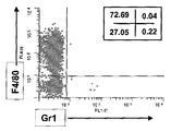
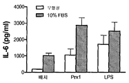
도 1. Prx1 는 마크로파지로부터 시토킨 분비를 자극한다.
(a) TG 유도된 마크로파지를 CD11b, Gr1, 및 F4/80의 발현에 대해 유동 세포분석법으로 분석하였다. 3개의 독립적으로 분리된 대표적인 히스토그램은 CD11b+ 세포에 의한 Gr1 및 F4/80 발현을 나타내고 묘사한다. 삽도의 숫자들은 각 사분면에서 CD11b+ 세포의 퍼센트를 나타낸다. (b) TG 유도된 마크로파지를 자극제와 함께 24시간 동안 배양하였다; 상층액을 수확하였고 TNF-α (오픈 바) 및 IL-6 레벨 (회색 바)에 대해 분석하였다. 결과는 pg/ml로서 나타나고 3개의 독립적인 실험의 대표이다; 에러 바는 표준 편차를 대표한다. (c) TG 유도된 마크로파지를 배지 단독 (검은 바), 100 nM LPS 또는 2000 nM Prx1 (오픈 바), 10 ug/mL 폴리믹신 B와 함께 20분 동안 사전 배양된 100 nM LPS 또는 2000 nM Prx1 (평행선 막대), 또는 100 nM LPS 또는 변성된 2000 nM Prx1 (회색 바)와 함께 24시간 동안 배양하였다. 별표는 Prx1 또는 LPS 단독으로 처리된 세포와 함께 비교하여 p≤0.01을 나타낸다. (d) TG 유도된 마크로파지를 10% FBS가 있거나 (회색 바) 또는 없을 때 (오픈 바) 배지 단독, Prx1 (50 nM) 또는 (100 nM)와 함께 24시간 동안 배양하였다. 상층액을 수확하였고 IL-6 레벨에 대해 분석하였다. 결과는 pg/ml로서 나타난다; 에러 바는 표준 편차를 대표한다.
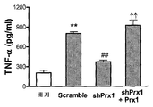
도 2. Prx1 은 수지상세포의 성숙화 및 활성화를 자극한다.
(a 및 b) 미성숙 골수 유도된 수지상세포 (iBMDCs)를 배지 단독, 20-200 nM Prx1 또는 100 nM LPS와 함께 24시간 동안 배양하였다. (a) 배양 후 세포를 유동 세포분석법으로 CD11c 및 CD86의 발현에 대해 분석하였다. 결과는 퍼센트 전체 세포로서 나타난다; 에러 바는 표준 편차를 대표한다. (b) 상층액을 수확하였고 TNF-α에 대해 분석하였다. 결과는 pg/ml로서 나타나고 3개의 독립적인 실험의 대표이다; 에러 바는 표준 편차를 대표한다. (c) TG 유도된 마크로파지를 대조군 shRNA (Scramble) 또는 Prx1에 대해 특이적인 shRNA (shPrx1)에 대해 암호화하는 cDNA를 트랜스펙션한 전립선 종양 세포주로부터 수확한 배지와 함께 또는 Prx1 특이적 shRNA를 발현하는 세포로부터 수확한 배지에 50 nM 외인성 Prx1을 추가한 배지에서 배양하였다. 24시간 배양 후, 상층액을 수확하였고 TNF-α에 대해 분석하였다. 결과는 pg/ml로서 나타나고 3개의 독립적인 실험의 대표이다; 에러 바는 표준 편차를 대표한다. **: 배지만 함께 배양된 세포에서 분비된 TNF-α 레벨과 비교할 때 P≤0.01; ##: 대조군 shRNA를 발현하는 세포의 배지와 함께 배양된 세포에서 분비된 TNF-α 레벨과 비교할 때 P≤0.01; ††: Prx1에 대해 특이적인 shRNA를 발현하는 세포의 배지와 함께 배양된 세포에서 분비된 TNF-α 레벨과 비교할 때 P≤0.01.
도 3. Prx1 유도된 시토킨 분비는 TLR4 의존적이다.
(a) iBMDCs를 C57BL/6 (TLR+/+; 오픈 바) 및 C57BL/10ScNJ (TLR4-/-; 닫힌 바) 마우스로부터 분리하였고 200 nM Prx1, 100 nM LPS, 또는 100 mM Pam3Cys로 자극하였다. 상층액을 수확하였고 IL-6 ELISA 키트로 분석하였다. (b) TG 유도된 마크로파지를 C57BL/6 (TLR+/+; 오픈 바) 및 C57BL/10ScNJ (TLR4-/-; 닫힌 바) 마우스로부터 분리하였고 200 nM Prx1, 100 nM LPS, 또는 100 mM Pam3Cys로 자극하였다. 상층액을 수확하였고 IL-6 ELISA 키트로 분석하였다. 결과는 pg/ml로서 나타난다; 에러 바는 표준 편차를 대표한다; 별표는 0.01 이하인 P 값을 나타낸다. (c) 나이브 C57BL/6 (TLR+/+; 오픈 바) 및 C57BL/10ScNJ (TLR4-/-; 닫힌 바) 마우스에 200 nm Prx1를 복강에 주사하였다. 6시간 후, 혈액을 수거하였고 ELISA로 IL-6의 존재에 대해 분석하였다. 결과는 pg/ml로서 나타난다; 에러 바는 표준 편차를 대표한다; 별표는 P≤0.0002를 나타낸다.
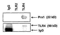
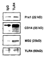
도 4. Prx1 과 TLR4 의 상호작용은 CD14 및 MD2 에 의존적이다.
(a) TG 유도된 마크로파지를 C57BL/6 마우스로부터 분리하였고 대조군 또는 Prx1, CD14 또는 MD2에 대한 차단 항체가 있거나 또는 없을 때 50 nM Prx1로 24시간 동안 자극하였다. 상층액을 수거하였고 IL-6 ELISA 키트로 분석하였다. 결과는 pg/ml로서 나타난다; 에러 바는 SEM을 대표한다; 별표는 0.01 이하인 P 값을 나타낸다. (b) TG 유도된 마크로파지를 수확하였고 재료 및 방법에서 설명된 바와 같이 세포 용해물을 TLR4, TLR2, 및 마우스/고트 IgG에 대한 항체와 함께 침전시켰다; 얻어진 침전물을 SDS-PAGE로 분리하였고 Western 블롯 분석으로 Prx1의 존재에 대해 탐지하였다. 블롯을 또한 로딩 대조군으로서 TLR4 또는 TLR2에 대한 항체로 탐지하였다. (c) TG 유도된 마크로파지를 수확하였고 재료 및 방법에서 설명된 바와 같이 세포 용해물을 TLR4 또는 마우스/고트 IgG에 대한 항체와 함께 배양하였다; 얻어진 침전물을 SDS-PAGE로 분리하였고 Western 블롯 분석으로 Prx1, CD14 및 MD2의 존재에 대해 탐지하였다. 블롯을 또한 로딩 대조군으로서 TLR4에 대한 항체로 탐지하였다.
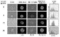
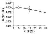
도 5: TLR4 / Prx1 상호작용의 동력학 .
(a) TG 유도된 마크로파지를 200 nM FITC-Prx1 또는 PE-결합된 항-TLR4 (PE-TLR4)로 자극하였다. 샘플을 지정된 시간에 수확하였고 세포수를 Amnis 기술로 분석하였다. 면역염색된 세포 및 각 시점에 대한 2개의 염색이 병합된 이미지의 대표적인 예가 나타난다. 맨 오른쪽 칼럼은 y-축은 세포의 수이고 x-축은 Prx1 및 TLR4 사이의 유사성 계수로 분석된 각 세포 (n=5,000)의 픽셀 바이 픽셀 통계 분석에 히스토그램을 나타낸다. (b)각 시점에 대한 모든 세포의 유사성 계수의 평균이 나타난다; 에러 바는 표준 편차를 대표한다.
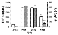
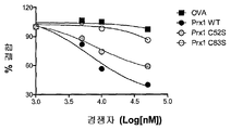
도 6. TLR4 와 결합하는 Prx1 는 구조 의존적이다.
(a) TG 유도된 마크로파지를 TLR4+/+ (흰 바) 또는 TLR4-/- (가득 찬 바) 마크로파지로부터 분리하였고 배지 (없음), Prx1, Prx1C52S, 또는 Prx1C83S 200 nM과 함깨 24시간 동안 배양하였고 상층액을 수거하였고 TNF-α 및 IL-6의 존재에 대해 분석하였다. (b) TG 유도된 마크로파지를 TLR4+/+ (흰 바) 또는 TLR4-/- (가득 찬 바) 마크로파지로부터 분리하였고 2000 nM FITC 표지된 단백질과 함께 20분 동안 배양한 후, 유동 세포분석법으로 분석하였다. 살아있는 세포를 7-AAD 높은 집단의 소거에 의한 분석을 위해 선택하였다. 결과는 FITC 표지의 차이에 대해 표준화되고 nM 단백질 당 MFI/FITC로 보고된다; 에러 바는 표준 편차를 대표한다. 별표는 P 값≤0.01을 나타낸다. (c) TG 유도된 마크로파지를 FITC-BSA (사각형), Prx1 (어두운 원), Prx1C52S (회색 원), 및 Prx1C83S (오픈 원)와 함께 다양한 농도로 20분 동안 배양하였고 유동 세포분석법으로 분석하였다. 결과는 FITC 표지의 차이에 대해 표준화되고 nM 단백질 당 MFI/FITC로 보고된다. 각 곡선은 3명의 개인 실험으 leovy이다. (d) TG 유도된 마크로파지를 1000 nM Prx1과 함께 배양하였고, 세척하였고 증가된 농도의 경쟁자: OVA (사각형), Prx1 (어두운 원), Prx1C52S (회색 원), Prx1C83S (오픈 원)와 함께 배양하였다. 결과는 경쟁자 없이 FITC-Prx1의 퍼센트 MFI로서 나타난다; 에러 바는 표준 편차를 대표한다. 모든 실험을 세 번 수행하였고 조합된 결과를 나타낸다.
도 7. 마크로파지의 Prx1 자극은 MyD88 의존적이고 NF κB의 핵 위치 변화를 일으킨다.
(a) 대조군 (오픈 바) 또는 MyD88DN (가득 찬 바) 발현 플라스미드를 함유하는 RAW264.7 마크로파지 세포주의 안정한 발현 세포를 100 nM LPS 또는 1000 nM Prx1으로 24시간 동안 자극하였고 결과의 발현 세포를 ELISA로 IL-6에 대해 검정하였다. ELISA 분석을 세 번의 독립적인 실험에서 수행하였다; 에러바는 표준 편차를 대표한다. 별표는 P 값≤0.001을 나타낸다. (b) C3H/GeNCr (TLR4+/+) 및 C3H/GeNCr (TLR4-/-) 마우스로부터 분리한 TG 유도된 마크로파지를 완전 배지에서 200 nM Prx1으로 자극하였다. 지정된 시점에서 세포를 NFκB p65 및 DRAQ5 (핵 염색)에 대한 FITC 결합 항체로 10분 동안 염색하였고 Amnis 기술을 사용하여 분석하였다. 맨 오른쪽 컬럼은 NFκB 및 핵 염색의 유사성에 대한 픽셀 바이 픽셀 통계 분석을 나타낸다. (c) C3H/HeNCr (가득 찬 원) 및 C3H/HeNJ (오픈 원) 마크로파지 둘 다에서 각 시점에 대한 전반적인 유사계수의 평균 숫자 값은; 에러 바는 표준 편차를 대표한다. (d) TG 유도된 마크로파지를 지정된 농도의 Prx1과 함께 1시간 동안 배양하였다. EMSA 분석을 실시예 1에 설명된 바와 같이 수행하였다.
도 8. PC -3M 세포에서 Prx1 에 대해 특이적인 shRNA 의 발현은 Prx1 발현의 감소를 초래한다.
(a) 대조군 (Scramble) shRNA 또는 Prx1 특이적 shRNA (shPrx1)을 발현할 수 있게 고안된 PC-3M 세포로부터 분리된 세포 용해물 (오른쪽 패널)은 전기영동에 의해 분리하였고, 블롯 하였고 Prx1에 대해 특이적 항체로 탐지하였다. (b) Prx1에 대해 특이적인 shRNA의 발현은 감소된 Prx1 레벨을 초래한다. 대조군 shRNA (Scramble) 또는 Prx1에 대해 특이적인 shRNA를 발현할 수 있게 고안된 PC3-M 세포주를 수확하였고 Western 분석으로 Prx1 또는 Prx2의 발현에 대해 분석하였다. (c) TG 유도된 마크로파지로부터 IL-6 분비의 Prx1 자극은 CD14 및 MD2에 의존적인데, 이것은 TLR4의 공동 인자이다. TG 유도된 마크로파지를 C57BL/6 마우스로부터 분리하였고 대조군 또는 CD14 또는 MD2에 대한 차단 항체에 상관없이 LPS로 24시간 동안 자극하였다. 상층액을 수거하였고 IL-6 ELISA 키트로 분석하였다. 결과는 pg/ml로서 나타난다; 에러 바는 SEM을 대표한다.
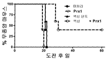
도 9는 동물 모델에서 종양에 대한 Prx1의 보조제 효과를 증명하는 그래리를 제공한다. Figure 1. Prx1 is from macrophages Stimulates cytokine secretion
(a) TG induced macrophages were analyzed by flow cytometry for expression of CD11b, Gr1, and F4 / 80. Three independently isolated representative histograms show and depict Gr1 and F4 / 80 expression by CD11b + cells. The numbers in the inset represent the percentage of CD11b + cells in each quadrant. (b) TG induced macrophages were incubated with stimulant for 24 hours; Supernatants were harvested and analyzed for TNF-α (open bar) and IL-6 levels (grey bar). The results are shown as pg / ml and are representative of three independent experiments; Error bars represent standard deviations. (c) 100 nM LPS or 2000 nM Prx1 precultured for 20 minutes with TG induced macrophages alone (black bar), 100 nM LPS or 2000 nM Prx1 (open bar), 10 ug / mL polymyxin B ( Parallel bars), or incubated with 100 nM LPS or denatured 2000 nM Prx1 (grey bars) for 24 hours. Asterisks indicate p ≦ 0.01 compared with cells treated with Prx1 or LPS alone. (d) TG induced macrophages were incubated with medium alone, Prx1 (50 nM) or (100 nM) for 24 hours with or without 10% FBS (grey bar) or without (open bar). Supernatants were harvested and analyzed for IL-6 levels. The result is shown as pg / ml; Error bars represent standard deviations.
2. Prx1 also stimulates the maturation and activation of dendritic cells.
(a and b) Immature bone marrow induced dendritic cells (iBMDCs) were incubated for 24 hours with medium alone, 20-200 nM Prx1 or 100 nM LPS. (a) After incubation, cells were analyzed for expression of CD11c and CD86 by flow cytometry. The results are shown as percent total cells; Error bars represent standard deviations. (b) Supernatants were harvested and analyzed for TNF-α. The results are shown as pg / ml and are representative of three independent experiments; Error bars represent standard deviations. (c) expressing Prx1-specific shRNA or with media harvested from a prostate tumor cell line transfected with cDNA encoding TG induced macrophages for control shRNA (Scramble) or shRNA specific for Prx1 (shPrx1). The medium harvested from the cells was cultured in a medium in which 50 nM exogenous Prx1 was added. After 24 h incubation, supernatants were harvested and analyzed for TNF-α. The results are shown as pg / ml and are representative of three independent experiments; Error bars represent standard deviations. **: P ≦ 0.01 as compared to TNF-α levels secreted in cells cultured only with embryos; ##: P ≦ 0.01 when compared to TNF-α levels secreted from cells incubated with medium of cells expressing control shRNA; ††: P ≦ 0.01 as compared to TNF-α levels secreted from cells cultured with medium of cells expressing shRNA specific for Prx1.
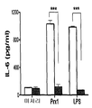
Figure 3. The cytokine secretion induced Prx1 is a TLR4-dependent.
(a) iBMDCs were isolated from C57BL / 6 (TLR + / + ; open bars) and C57BL / 10ScNJ (TLR4 − / − ; closed bars) mice and stimulated with 200 nM Prx1, 100 nM LPS, or 100 mM Pam 3 Cys It was. Supernatants were harvested and analyzed with IL-6 ELISA kit. (b) TG induced macrophages were isolated from C57BL / 6 (TLR + / + ; open bars) and C57BL / 10ScNJ (TLR4 − / − ; closed bars) mice and were 200 nM Prx1, 100 nM LPS, or 100 mM Pam 3 Stimulated with Cys. Supernatants were harvested and analyzed with IL-6 ELISA kit. The result is shown as pg / ml; Error bars represent standard deviations; Asterisks indicate P values that are 0.01 or less. (c) Naïve C57BL / 6 (TLR + / + ; open bar) and C57BL / 10ScNJ (TLR4 − / − ; closed bar) mice were injected intraperitoneally with 200 nm Prx1. After 6 hours, blood was collected and analyzed for the presence of IL-6 by ELISA. The result is shown as pg / ml; Error bars represent standard deviations; Asterisks indicate P ≦ 0.0002.
Figure 4. The interaction of Prx1 with TLR4 is dependent on CD14 and MD2 .
(a) TG induced macrophages were isolated from C57BL / 6 mice and stimulated with 50 nM Prx1 for 24 hours with or without control or blocking antibodies to Prx1, CD14 or MD2. Supernatants were harvested and analyzed with IL-6 ELISA kit. The result is shown as pg / ml; Error bars represent SEM; Asterisks indicate P values that are 0.01 or less. (b) TG derived macrophages were harvested and cell lysates precipitated with antibodies to TLR4, TLR2, and mouse / goat IgG as described in Materials and Methods; The precipitate obtained was separated by SDS-PAGE and detected for the presence of Prx1 by Western blot analysis. Blots were also detected with antibodies against TLR4 or TLR2 as loading controls. (c) TG derived macrophages were harvested and cell lysates were incubated with antibodies against TLR4 or mouse / goat IgG as described in Materials and Methods; The precipitate obtained was separated by SDS-PAGE and detected by Western blot analysis for the presence of Prx1, CD14 and MD2. Blots were also detected with antibodies against TLR4 as a loading control.
5: Kinetics of TLR4 / Prx1 interaction .
(a) TG induced macrophages were stimulated with 200 nM FITC-Prx1 or PE-bound anti-TLR4 (PE-TLR4). Samples were harvested at designated times and cell numbers analyzed by Amnis technology. Representative examples of images in which immunostained cells and two staining for each time point are merged are shown. The rightmost column shows the histogram in the pixel-by-pixel statistical analysis of each cell (n = 5,000) analyzed with the similarity coefficient between Prx1 and TLR4, with the y-axis being the number of cells. (b) the average of the similarity coefficients of all cells for each time point is shown; Error bars represent standard deviations.
Figure 6. Prx1 binding to TLR4 is structure dependent.
(a) TG induced macrophages were isolated from TLR4 + / + (white bar) or TLR4 − / − (full bar) macrophages and incubated with medium (none), Prx1, Prx1C52S, or
Figure 7. Macrophage Prx1 MyD88-dependent stimulation and causes a change in position of nuclear NF κB.
(a) Stable expressed cells of RAW264.7 macrophage cell line containing control (open bar) or MyD88DN (full bar) expression plasmids were stimulated with 100 nM LPS or 1000 nM Prx1 for 24 hours and the resulting expressed cells were subjected to ELISA. Assay for IL-6. ELISA analysis was performed in three independent experiments; Error bars represent standard deviations. Asterisks indicate P value ≤ 0.001. (b) TG induced macrophages isolated from C3H / GeNCr (TLR4 + / + ) and C3H / GeNCr (TLR4 − / − ) mice were stimulated with 200 nM Prx1 in complete medium. At designated time points, cells were stained for 10 minutes with FITC binding antibodies against NFκB p65 and DRAQ5 (nuclear staining) and analyzed using Amnis technology. The rightmost column shows pixel by pixel statistical analysis of the similarity of NFκB and nuclear staining. (c) The average numerical value of the overall similarity coefficient for each time point in both C3H / HeNCr (full circle) and C3H / HeNJ (open circle) macrophages; Error bars represent standard deviations. (d) TG induced macrophages were incubated with the indicated concentration of Prx1 for 1 hour. EMSA analysis was performed as described in Example 1.
Figure 8. Expression of shRNA specific for Prx1 in PC- 3M cells results in a decrease in Prx1 expression.
(a) Cell lysates (right panel) isolated from PC-3M cells designed to express control (Scramble) shRNA or Prx1 specific shRNA (shPrx1) were isolated by electrophoresis, blotted and specific for Prx1. Detection with enemy antibody. (b) Expression of shRNA specific for Prx1 results in reduced Prx1 levels. PC3-M cell lines designed to express shRNA specific for control shRNA (Scramble) or Prx1 were harvested and analyzed for expression of Prx1 or Prx2 by Western analysis. (c) Prx1 stimulation of IL-6 secretion from TG induced macrophages is dependent on CD14 and MD2, which are cofactors of TLR4. TG induced macrophages were isolated from C57BL / 6 mice and stimulated with LPS for 24 hours regardless of control or blocking antibodies to CD14 or MD2. Supernatants were harvested and analyzed with IL-6 ELISA kit. The result is shown as pg / ml; Error bars represent SEM.
FIG. 9 provides Grarey demonstrating the adjuvant effect of Prx1 on tumors in animal models.
본 발명은 퍼옥시레독신1 (Prx1)이 Toll 유사 수용체 (TLR4)에 대한 리간드이고, Prx1의 이 기능은 그것을 보조제로서 작용할 수 있게 한다는 예상밖의 발견에 기초한다. 본 발명은 개인에서 항원에 대한 면역 반응을 향상시키는 조성물 및 방법을 제공한다. 조성물은 분리된 Prx1 및 항원을 포함한다. 그것을 암호화하는 Prx1의 아미노산 서열 및 RNA 서열 및 RNA 서열은 업계에 잘 알려져 있다. 한 구체예에서, 분리된 Prx1 단백질은 데카머로서 제공된다. 분리된 Prx1 단백질은 191 아미노산 서열을 포함하거나 그것으로 구성되는데, 여기서 191 아미노산 서열을 포함하거나 그것으로 구성되는 단백질은 퍼옥시레독신활성을 갖는다. 한 구체예에서, Prx1은 2009년 8월 23일에 여기에 참고로 포함되는 NCBI 참고 서열: NP_859047에서 나타나는 아미노산 서열을 포함한다. 어떤 스플라이스 변형체 및/또는 Prx1 이성질체도 본 발명에 사용될 수 있다. The present invention is based on the unexpected finding that peroxyredoxin1 (Prx1) is a ligand for Toll-like receptor (TLR4) and this function of Prx1 makes it possible to act as an adjuvant. The present invention provides compositions and methods for enhancing an immune response to an antigen in an individual. The composition comprises an isolated Prx1 and an antigen. The amino acid sequence and RNA sequence and RNA sequence of Prx1 encoding it are well known in the art. In one embodiment, the isolated Prx1 protein is provided as a decamer. An isolated Prx1 protein comprises or consists of a 191 amino acid sequence, wherein the protein comprising or consisting of the 191 amino acid sequence has peroxyredoxin activity. In one embodiment, Prx1 comprises the amino acid sequence represented by NCBI Reference Sequence: NP_859047, incorporated herein by reference on August 23, 2009. Any splice variant and / or Prx1 isomer may be used in the present invention.
본 발명의 방법은 개인에게 조성물을 투여하는 것을 포함해서 항원에 대한 면역 반응은 자극된다. 자극된 면역 반응은 치료의 또는 예방의 효과를 가질 수 있고 세포 매개된 및/또는 체액의 반응, 또는 그것의 조합을 포함할 수 있다. 자극된 면역 반응은 항원 단독에 의해 자극된 면역 반응보다 클 수 있다. The method of the present invention comprises administering the composition to an individual, thereby stimulating an immune response to the antigen. The stimulated immune response can have a therapeutic or prophylactic effect and can include cell mediated and / or humoral responses, or a combination thereof. The stimulated immune response may be greater than the immune response stimulated by the antigen alone.
Prx1은 항산화제이고 대부분 세포 타입에서 발견되는 샤페론 분자이고 형질전환된 및 활성화된 세포로부터 분비된다. TLR4는 Toll 유사 수용체 (TLR) 패밀리의 멤버이다. TLR들과 그들의 리간드의 상호작용은 염증유발 시토킨 같은 염증 조절기의 방출 및 면역 반응의 세포 성숙화/활성화를 개시하게 한다. 염증 및 면역 세포 성숙화/활성화 둘 다는 보조제로서 업계에 나타난다. 우리는 여기서 수지상세포 및 마크로파지 같은, 항원 특이적 면역의 유발의 원인이 되는 면역 세포의 표면에서 TLR4와 Prx1의 상호작용을 증명한다. 우리는 또한 수지상세포 및 마크로파지의 성숙화/활성화의 생산 및 염증유발 시토킨의 분비를 초래하는 TLR4와 Prx1의 상호작용을 증명한다. 따라서, Prx1은 면역 보조제로 생각된다. 중요하게, 우리는 항종양 백신에 Prx1의 추가는 동물에서 백신의 효능을 증가시킨다는 것을 증명하였다. 이 점에서, TLR9 작용제 CpG 모티브 및 TLR7 작용제 이미퀴모드를 포함하는, 다수의 TLR 작용제는 항종양 면역을 향상시키기 위해 사용되었다. CpG 모티브는 메틸화되지 않은 시토신-구아노신 및 TLR9에 결합할 수 있는 측면 뉴클레오티드를 함유하는 DNA 올리고디옥시뉴클레오티드 서열이다. 이미퀴모드 [1-(2-메틸프로필)-1H-이미다조[4,5-c] 퀴놀린-4 아민; AlderaTM; R-837, S260308]는 DCs28의 성숙화 및 이주, IFN-α, TNF-α, IL-1α, IL-6, IL-12 및 IL-8의 발현을 유발하는 합성 TLR7 작용제이고, CD8+ T 세포의 활성화를 향상시킨다. 이미퀴모드는 기저세포암의 치료에 성공적으로 사용되었고 최근의 임상적 시험은 국부적 이미퀴모드는 단계 0 흑색종 제거에 효과적인 것으로 나타났다. 이 TLR 작용제 둘 다는 항암 백신에 대한 보조제로서 더 발달해야할 미국 국립암센터 우선순위목록에 있다.Prx1 is an antioxidant and chaperone molecule found in most cell types and is secreted from transformed and activated cells. TLR4 is a member of the Toll like receptor (TLR) family. The interaction of TLRs with their ligands initiates the release of inflammatory regulators such as proinflammatory cytokines and cell maturation / activation of the immune response. Both inflammation and immune cell maturation / activation appear in the industry as adjuvants. We demonstrate here the interaction of TLR4 with Prx1 at the surface of immune cells that are responsible for inducing antigen specific immunity, such as dendritic cells and macrophages. We also demonstrate the interaction of Trx4 with Prx1 resulting in the production of maturation / activation of dendritic cells and macrophages and the secretion of proinflammatory cytokines. Thus, Prx1 is thought to be an adjuvant. Importantly, we demonstrated that the addition of Prx1 to antitumor vaccines increased the efficacy of the vaccines in animals. In this regard, a number of TLR agonists, including TLR9 agonist CpG motifs and TLR7 agonist imiquimod, have been used to enhance antitumor immunity. The CpG motif is a DNA oligodioxynucleotide sequence containing flanking nucleotides capable of binding unmethylated cytosine-guanosine and TLR9. Imiquimod [1- (2-methylpropyl) -1H-imidazo [4,5-c] quinolin-4 amine; Aldera ™; R-837, S260308] are synthetic TLR7 agonists that induce maturation and migration of DCs28, expression of IFN-α, TNF-α, IL-1α, IL-6, IL-12 and IL-8, Improve activation. Imiquimod has been successfully used for the treatment of basal cell carcinoma, and recent clinical trials have shown that local imiquimod is effective at removing
Prx1는 CpG 및 이미퀴모드와 같은 활성 중 많은 것을 나타내지만, 그것이 결합하는 수용체, TLR4는 CpG 및 이미퀴모드와 상호작용하는 TLR들보다 더 널리 발현된다. 따라서, Prx1은 더 효과적인 항암 백신 보조제가 될 가능성을 갖는다. Prx1 exhibits many of its activities, such as CpG and imiquimod, but the receptor to which it binds, TLR4, is more widely expressed than TLRs that interact with CpG and imiquimod. Thus, Prx1 has the potential to be a more effective anticancer vaccine adjuvant.
본 발명은 어떤 항원에 대한 면역 반응을 자극하는데 사용될 수 있고, 따라서 항원은 면역원으로서 기능 할 것이라고 생각된다. 항원은 단백질, 폴리펩티드 또는 펩티드 항원을 포함하지만, 제한되지 않는다. 항원은 특정 세포 형태 또는 항원을 함유하거나 함유할 수 있는 생물학적 조직의 어느 다른 샘플의 용해물에서 알려진 또는 의심되는 존재에 의해 잘 특징지어질 수도 있거나, 또는 알려지지 않을 수도 있다.It is contemplated that the present invention can be used to stimulate an immune response to any antigen, and thus the antigen will function as an immunogen. Antigens include, but are not limited to, protein, polypeptide or peptide antigens. Antigens may or may not be well characterized by known or suspected presence in lysates of certain cell types or any other sample of biological tissue that may contain or may contain antigens.
한 구체예에서, 본 발명이 면역 반응을 자극하는 항원은 종양 항원이다. 종양 항원은 신선한 종양 생체검사 조직 또는 조직 배양에 의해 시험관 내에서 생성된 종양 세포의 종양 세포/조직을 반복적으로 얼리고 녹임으로써 종양 세포 용해물을 제조하는 것과 같은 고전적 기술에 의해서 얻을 수 있다. 종양 용해물은 상청액을 원심분리하고 수확함에 의해 얻을 수 있다. 종양 세포 용해물은 즉시 사용되거나 사용할 준비가 될 때까지 얼려서 보관한다. 항원은 정제된 형태로 또는 부분적으로 정제되거나 세포 용해물과 같이 정제되지 않은 형태로 사용될 수 있다. 대안으로, 항원은 다양한 발현 시스템 중 어느 것에서도 재조합 DNA 기술에 의해 발현될 수도 있다. 따라서, 분리된 Prx1 단백질은 본 발명에서 사용하기 위해 신중한, 분리된 Prx1 단백질이 한 항체와 함께 또는 다른 항체와 함께 복합체를 형성하는 것을 제공할 수도 있다. 이러한 복합체는 다양한 완충액, 배양시간, 및 온도를 사용하여, Prx1와 항원의 비율이 다른 것과 같이, 다양한 조건을 사용하여 형성될 수 있다. In one embodiment, the antigen to which the present invention stimulates an immune response is a tumor antigen. Tumor antigens can be obtained by classical techniques such as preparing tumor cell lysates by repeatedly freezing and thawing tumor cells / tissues of tumor cells produced in vitro by fresh tumor biopsy tissue or tissue culture. Tumor lysates can be obtained by centrifugation and harvesting the supernatant. Tumor cell lysates are stored frozen until ready for use or ready for use. The antigen can be used in purified form or partially purified or in an unpurified form, such as a cell lysate. Alternatively, the antigen may be expressed by recombinant DNA technology in any of a variety of expression systems. Thus, an isolated Prx1 protein may provide that the isolated Prx1 protein forms a complex with or with another antibody that is cautious for use in the present invention. Such complexes may be formed using a variety of conditions, such as varying ratios of Prx1 and antigen, using various buffers, incubation times, and temperatures.
한 구체예에서, Prx1 단백질 및 항원은 복합체로서 본 발명의 조성물에 존재하고, 서로 공유결합으로 또는 비-공유결합으로 연관될 수도 있다. 예를 들어, Prx1 단백질 및 항원은 공유결합, 이온결합, 수소결합, 및/또는 van der Waals 결합, 또는 그것의 재조합에 의해 서로 연결될 수도 있다. 공유 결합과 상관없이 단백질/항원 복합체를 형성하는 방법은 업계에 알려져 있고 분리된 Prx1 단백질 및 하나 이상의 항원 사이에서 복합체를 형성하는데 쓰일 수 있다. 본 발명의 복합체는 분리된 Prx1 단백질 및 항원을 포함할 수도 있거나, 본직적으로 Prx1 단백질 및 항원으로 구성될 수도 있고, 또는 분리된 Prx1 단백질 및 항원으로 구성될 수도 있다. "분리된"에 의해 Prx1 단백질은 그것의 자연적 환경으로부터 분리되는 것을 의미한다. 분리된 단백질은 필수적으로 정제된 단백질이지 않아도 된다. 하지만, 분리된 Prx1은 본 발명에서 사용을 위해 원하는 정제의 정도로 정제될 수도 있다. 분리된 Prx1 단백질은 재조합 방법에 의해 생산된 Prx1 단백질을 포함한다. 데카머 같은, Prx1 멀티머는 또한 본 발명에 따라 분리된 Prx1 단백질이라고 생각된다. 게다가, 분리된 Prx1 단백질은 융합 단백질에서와 같이, 펩티드 결합을 통해 연결되는 Prx1 단백질을 포함한다. 간단히 말하면, 이러한 융합 단백질을 생산하기 위해서, Prx1 단백질 및 항원을 암호화하는 DNA 서열은 고전적인 기술을 사용해서 구성될 수 있고 적절한 발현 벡터를 사용하여 적합한 세포 형태에서 발현될 수도 있다. 융합 단백질은 세포에서 발현될 수 있고 당업자에 알려진 기술을 사용하여 분리될 수 있다. 한 구체예에서, Prx1/항원 융합 단백질은 연결 서열에 의해 분리될 수도 있다. In one embodiment, the Prx1 protein and antigen are present in the compositions of the invention as complexes and may be associated with each other covalently or non-covalently. For example, the Prx1 protein and antigen may be linked to each other by covalent bonds, ionic bonds, hydrogen bonds, and / or van der Waals bonds, or recombination thereof. Methods of forming protein / antigen complexes regardless of covalent bonds are known in the art and can be used to form complexes between isolated Prx1 proteins and one or more antigens. The complex of the present invention may comprise an isolated Prx1 protein and an antigen, or may consist essentially of a Prx1 protein and an antigen, or may be composed of an isolated Prx1 protein and an antigen. By "isolated" is meant that the Prx1 protein is separated from its natural environment. An isolated protein does not necessarily have to be a purified protein. However, the isolated Prx1 may be purified to the degree of purification desired for use in the present invention. Isolated Prx1 protein includes Prx1 protein produced by recombinant methods. Prx1 multimers, such as decamers, are also considered to be Prx1 proteins isolated according to the present invention. In addition, isolated Prx1 proteins include Prx1 proteins that are linked via peptide bonds, as in fusion proteins. In short, to produce such fusion proteins, the DNA sequences encoding the Prx1 protein and antigen can be constructed using classical techniques and expressed in suitable cell forms using appropriate expression vectors. Fusion proteins can be expressed in cells and can be isolated using techniques known to those of skill in the art. In one embodiment, the Prx1 / antigen fusion protein may be separated by a linking sequence.
개인에게 투여에 적합한 본 발명의 조성물은 분리된 Prx1 및/또는 항원과 적합한 약학적 담체, 첨가제 및/또는 안정제와 섞음으로써 제조될 수 있다. 시약의 혼합에 대해 적합한 조성물의 몇몇 예는 Remington : The Science 및 Practive of Pharmacy (2005) 21st Edition, Philadelphia, PA. Lippincott Williams & Wilkins.에서 발견될 수 있다.Compositions of the invention suitable for administration to an individual may be prepared by mixing the isolated Prx1 and / or antigen with a suitable pharmaceutical carrier, additive and / or stabilizer. Some examples of compositions suitable for mixing reagents are described in Remington : The Science and Practive of Pharmacy (2005) 21st Edition, Philadelphia, PA. It can be found in Lippincott Williams & Wilkins.
한 구체예에서, 본 발명의 방법에 따라 면역 반응이 자극된 개인은 암에 걸릴 위험이 있거나, 암에 걸린 것으로 의심되거나, 또는 암으로 진단된 개인이다. 따라서, 다양한 구체예에서, Prx1 단백질인 항원은 어떤 형태의 암세포에 의해서 발현되는 항원이고 이것의 특이적 예는 섬유육종 (fibrosarcoma), 점액육종 (myxosarcoma), 지방육종 (liposarcoma), 연골육종 (chondrosarcoma), 골육종 (osteogenic sarcoma), 척색종 (chordoma), 혈관육종 (angiosarcoma), 내피육종 (endotheliosarcoma), 림프관육종 (lymphangiosarcoma), 가성 점액종 (pseudomyxoma peritonei), 림프관내피육종 (lymphangioendotheliosarcoma), 활막종 (synovioma), 중피종 (mesothelioma), 유윙종양 (Ewing's tumor), 평활근육종 (leiomyosarcoma), 횡문근육종 (rhabdomyosarcoma), 결장암종 (colon carcinoma), 췌장암 (pancreatic cancer), 유방암 (breast cancer), 난소암 (ovarian cancer), 전립선암 (prostate cancer), 편평세포암종 (squamous cell carcinoma), 기저세포암종 (basal cell carcinoma), 선암종 (adenocarcinoma), 한선암종 (sweat gland carcinoma), 피지샘암 (sebaceous gland carcinoma), 유두암종 (papillary carcinoma), 유두상선암종 (papillary adenocarcinomas), 낭선암종 (cystadenocarcinoma), 수질암종 (medullary carcinoma), 기관지원성암종 (bronchogenic carcinoma), 신세포암종 (renal cell carcinoma), 간세포암 (hepatoma), 담관암 (bile duct carcinoma), 융모막암종 (choriocarcinoma), 정상피종 (seminoma), 태생성 암종 (embryonal carcinoma), 윌름종양 (Wilms' tumor), 자궁경부암 (cervical cancer), 고환종양 (testicular tumor), 폐암종 (lung carcinoma), 소세포 폐암종 (small cell lung carcinoma), 방광암 (bladder carcinoma), 상피암종 (epithelial carcinoma), 신경교종 (glioma), 성상세포종 (astrocytoma), 수모세포종 (medulloblastoma), 두개인두종 (craniopharyngioma), 상의세포종 (ependymoma), 송과체종 (pinealoma), 혈관아세포종 (hemangioblastoma), 청신경종 (acoustic neuroma), 핍지교종 (oligodendroglioma), 뇌수막종 (meningioma), 흑색종 (melanoma), 신경아세포종 (neuroblastoma), 망막아세포종 (retinoblastoma), 백혈병 (leukemia), 림프종 (lymphoma), 다발성 골수종 (multiple myeloma), 발덴스트롬 마크로글로불린혈증 (Waldenstrom's macroglobulinemia), 및 중쇄병 (heavy chain disease)을 포함하지만 제한되지 않는다.In one embodiment, the individual whose immune response is stimulated according to the method of the present invention is an individual at risk of, suspected of having cancer, or diagnosed with cancer. Thus, in various embodiments, the antigen, which is a Prx1 protein, is an antigen expressed by some form of cancer cell and specific examples thereof include fibrosarcoma, myxosarcoma, liposarcoma, chondrosarcoma. ), Osteosarcoma (chteoma), chordoma (chordoma), hemangiosarcoma (angiosarcoma), endothelial sarcoma (endotheliosarcoma), lymphangiosarcoma (lymphangiosarcoma), pseudomyxoma (pseudomyxoma peritonei), lymphovascular endothelial sarcoma (lymphangioendotheliosarcomanovia) ), Mesothelioma, Ewing's tumor, leiomyosarcoma, rhabdomyosarcoma, colon carcinoma, pancreatic cancer, breast cancer, ovarian cancer ), Prostate cancer, squamous cell carcinoma, basal cell carcinoma, adenocarcinoma, sweat gland carcinoma, sebaceous gland cancer us gland carcinoma, papalary carcinoma, papillary adenocarcinomas, cystadenocarcinoma, medullary carcinoma, bronchogenic carcinoma, renal cell carcinoma, Hepatoma, bile duct carcinoma, choriocarcinoma, seminoma, embryonic carcinoma, Wilms' tumor, cervical cancer, testicular tumor (testicular tumor), lung carcinoma, small cell lung carcinoma, bladder carcinoma, epithelial carcinoma, glioma, astrocytoma, medulloblastoma ( medulloblastoma, craniopharyngioma, ependymoma, pinealoma, hemangioblastoma, acoustic neuroma, oligodendroglioma, meningioma, melanoma, melanoma Neuroblastoma, retinoblastoma, leukemia, lymphoma, multiple myeloma, Waldenstrom's macroglobulinemia, and heavy chain disease It is not limited.
한 구체예에서, 개인은 감염원에 걸릴 위험이 있거나, 감염원에 걸린 것으로 의심되거나, 또는 감염원에 걸린 것으로 진단된다. 따라서, 다양한 구체예에서 본 발명에 사용된 항원은 감염원에 의해 발현되는 것일 수 있다. 이러한 감염원의 예는 바이러스, 박테리아, 균류 및 다른 기생충들을 포함하지만, 제한되지 않는다. 바이러스의 예는 B형 간염 또는 C형 간염 (hepatitis type B 또는 type C), 인플루엔자 (influenza), 수두 (varicella), 아데노바이러스 (adenovirus), 단순 헤르페스 바이러스 타입 I 또는 타입 II (herpes simplex virus type I 또는 type II), 우역(rinder pest), 라이노바이러스 (rhinovirus), 에코바이러스 (echovirus), 로타바이러스 (rotavirus), 호흡장애 바이러스 (respiratory syncytial virus), 유두종 바이러스 (papilloma virus), 파포바바이러스 (papova virus), 거대세포 바이러스 (cytomegalovirus), 에키노바이러스 (echinovirus), 아르보바이러스 (arbovirus), 한타바이러스 (hantavirus), 콕사키바이러스 (coxsachie virus), 볼거리 바이러스 (mumps virus), 홍역 바이러스 (measles virus), 풍진 바이러스 (rubella virus), 소아마비 바이러스 (polio virus), 인간 면역 결핍 바이러스 타입 I 또는 타입 II (human immunodeficiency virus type I 또는 type II)를 포함하지만, 제한되지 않는다. 박테리아의 예는 결핵 (M.tuberculosis), 마이코박테리아 (mycobacterium), 마이코플라즈마 (mycoplasma), 나이세리아속 (neisseria) 및 레지오넬라속 (legionella)을 포함하지만, 제한되지 않는다. 다른 기생충의 예는 리케차 (rickettsia) 및 클라미디아 (chlamydia)를 포함하지만, 제한되지 않는다.In one embodiment, the individual is at risk of suffering from, suspected of having an infectious agent, or diagnosed as having an infectious agent. Thus, in various embodiments the antigen used in the present invention may be one expressed by an infectious agent. Examples of such infectious agents include, but are not limited to, viruses, bacteria, fungi and other parasites. Examples of viruses include hepatitis B or hepatitis type B or type C, influenza, varicella, adenovirus, simple herpes virus type I or type II. Or type II), rinder pest, rhinovirus, echovirus, rotavirus, respiratory syncytial virus, papilloma virus, papovavirus virus, cytomegalovirus, echinovirus, arbovirus, hantavirus, coxsachie virus, mumps virus, measles virus , Rubella virus, polio virus, human immunodeficiency virus type I or type II (human immunodeficiency virus type I or type II) However, not limited. Examples of bacteria include, but are not limited to, M. tuberculosis, mycobacterium, mycoplasma, neisseria, and legionella. Examples of other parasites include, but are not limited to rickettsia and chlamydia.
본 발명의 조성물은 적합한 투여 루트를 사용하여 투여될 수 있다. 몇몇 비-제한적 예는 구강, 비경구, 피하, 복강 내, 폐 내, 비강 내를 포함한다. 비경구 주입은 근육 내, 정맥 내, 동맥 내, 복강 내, 및 피하 투여를 포함한다.The composition of the present invention can be administered using a suitable route of administration. Some non-limiting examples include oral, parenteral, subcutaneous, intraperitoneal, pulmonary, intranasal. Parenteral infusions include intramuscular, intravenous, intraarterial, intraperitoneal, and subcutaneous administration.
본 발명의 조성물의 투여는 항원과 관련된 병 또는 질환을 치료하는 경향이 있는 고전적 치료와 결합으로 수행될 수 있다. 예를 들어, 조성물은 고전적 항암 치료 전, 동시에, 또는 다음에 투여될 수 있다. 이러한 치료는 화학치료, 외과적 개입, 및 방사선 치료를 포함하지만 제한되지 않는다. Administration of the compositions of the invention can be carried out in combination with classical therapies that tend to treat diseases or diseases associated with antigens. For example, the composition can be administered before, concurrently, or next to the classical chemotherapy. Such treatments include but are not limited to chemotherapy, surgical intervention, and radiation therapy.
일반적으로, 적절한 투여량 및 치료 요법은 치료적 및/또는 예방적 이점을 제공하는 면역 반응을 자극하는데 유효한 양의 조성물을 제공한다. 이러한 반응은 치료받는 질환에 의존적인 개선된 임상적 결과, 예를 들어, 종양 성장 및/또는 전이의 억제, 개선된 면역 세포 활성, 및/또는 당업자에게 명백한 다른 파라미터에 의해 관찰될 수 있다. In general, appropriate dosages and therapeutic regimens provide compositions in an amount effective to stimulate an immune response that provides therapeutic and / or prophylactic advantages. Such a response can be observed by improved clinical outcomes dependent on the disease being treated, eg, inhibition of tumor growth and / or metastasis, improved immune cell activity, and / or other parameters apparent to those skilled in the art.
여기에 개시된 치료적 조성물의 투여 루트 및 빈도, 뿐만 아니라 투여량은 개인마다 다를 것이고, 표준 기술을 사용하여 쉽게 확립될 수도 있다. The route and frequency of administration, as well as the dosage, of the therapeutic compositions disclosed herein will vary from person to person and may be readily established using standard techniques.
한 구체예에서, 본 발명은 항원전달세포 (APCs)의 격리집단을 포함하는 조성물 및 분리된 Prx1 및 항원을 포함하는 복합체를 제공한다. Prx1/항원 복합체는, 수지상세포 같은, APCs를 준비하는데 사용될 수 있고, 준비된 APCs는 개인에 대한 향상된 면역 반응을 성취하기 위해 개인에게 투여될 수 있다. 따라서, APCs 격리집단을 포함하는 조성물 및 분리된 Prx1 및 항원을 포함하는 복합체는 조성물에서 모든 세포, 실질적으로 모든 세포, 또는 모든 세포 미만은 수지상세포일 수 있다. 따라서, 조성물에 세포는 100% 수지상세포를 포함하거나, 100% 및 10% 사이 (사이의 모든 정수를 포함)의 수지상세포를 포함할 수 있다. In one embodiment, the present invention provides a composition comprising an isolated population of antigen-transmitting cells (APCs) and a complex comprising an isolated Prx1 and an antigen. The Prx1 / antigen complex can be used to prepare APCs, such as dendritic cells, and the prepared APCs can be administered to an individual to achieve an improved immune response against the individual. Thus, a composition comprising the APCs isolate and a complex comprising the isolated Prx1 and antigen may be all cells, substantially all cells, or less than all cells in the composition, dendritic cells. Thus, the cells in the composition may comprise 100% dendritic cells or may comprise dendritic cells between 100% and 10% (including any integer between).
한 구체예에서, APCs 격리집단을 포함하는 조성물 및 분리된 Prx1 및 항원을 포함하는 복합체는 개인에서 예방적 또는 치료적 면역 반응을 자극하기 위해 개인에게 투여된다. 개인에게 투여된 APCs는 동종 및 동계일 수도 있다.In one embodiment, a composition comprising an APCs isolate and a complex comprising an isolated Prx1 and an antigen are administered to the individual to stimulate a prophylactic or therapeutic immune response in the individual. APCs administered to an individual may be allogeneic and syngeneic.
다음 실시예는 본 발명을 도시하지만 제한할 의도는 없다.The following examples illustrate the invention but are not intended to be limiting.
실시예1Example 1
이 실시예는 본 발명의 구체예의 수행에 사용된 재료 및 방법의 설명을 제공한다. This example provides a description of the materials and methods used in carrying out the embodiments of the present invention.
재료material
리포폴리사카라이드 (LPS, 대장균 혈청형 026:B6) 폴리믹신 B 황산염, 소 혈청 알부민 (BSA), 및 오발부민 (OVA)을 Sigma-Aldrich (St. Louis, MO)로부터 얻었다. 7-아미노-액티노마이신 D (7-AAD) 및 티오글리콜레이트 브루어 수정 배지를 (Becton Dickinson, La Jolla, Ca)로부터 샀다. Luminex 검정에 사용된 IL-6 및 TNF-α에 대한 캡쳐 항체 및 검출 항체, 뿐만 아니라 단백질 표준을 Invitrogen (Carlsbad, CA)으로부터 샀다. CD11b, Gr-1, F4/80, 및 모든 이소타입에 대한 특이적 항체를 PharMingen (Mountain View, CA)로부터 샀다. TLR2, TLR4, 및 NFκB 서브유닛에 대한 항체를 Santa Cruz Biotechnology (Santa Cruz, CA)로부터 샀다. MD2 및 CD14에 대한 차단 항체를 Santa Cruz Biotechnology로부터 샀다. 피코에리트린 (PE) 결합된 항-TLR4 항체를 eBioscience (San Diego, CA)로부터 샀다. Prx1에 대한 특이적 항체를 Lab Frontier (Seoul, South Korea)로부터 얻었다; 이 항체는 Prx1에 대해 특이적이고 Prx1을 발현하는 세포의 Western 분석에서 하나의 밴드만 검출된다 (도 8a).Lipopolysaccharide (LPS, E. coli serotype 026: B6) polymyxin B sulfate, bovine serum albumin (BSA), and ovalbumin (OVA) were obtained from Sigma-Aldrich (St. Louis, MO). 7-Amino-actinomycin D (7-AAD) and thioglycolate brewer fertilization medium were purchased from (Becton Dickinson, La Jolla, Ca). Capture and detection antibodies for IL-6 and TNF-α used in the Luminex assay, as well as protein standards were purchased from Invitrogen (Carlsbad, CA). Specific antibodies for CD11b, Gr-1, F4 / 80, and all isotypes were purchased from PharMingen (Mountain View, CA). Antibodies against TLR2, TLR4, and NFκB subunits were purchased from Santa Cruz Biotechnology (Santa Cruz, CA). Blocking antibodies against MD2 and CD14 were purchased from Santa Cruz Biotechnology. Picoerythrin (PE) bound anti-TLR4 antibodies were purchased from eBioscience (San Diego, Calif.). Specific antibodies against Prx1 were obtained from Lab Frontier (Seoul, South Korea); This antibody is specific for Prx1 and only one band is detected in Western analysis of Prx1 expressing cells (FIG. 8A).
동물 및 세포주Animal and Cell Lines
C57BL/6NCr (TLR4+/+ 및 TLR2+/+), C57BL/10ScNJ (TLR4-/-), B6.129-Tlr2tmlKir /J (TLR2-/-), C3H/HeNCr (TLR4+/+), 및 C3H/HeNJ (TLR4-/-) 무균 마우스를 The Jackson Laboratory (Bar Harbor, ME)로부터 샀다. 동물을 환경광 하에 층류 유닛으로 미세분리기 케이지에 사육하였다. 마우스는 로스웰파크 암 연구소 (Roswell Park Cancer Institute, Buffalo, NY)의 무균 시설에서 유지하였다. 동물 실험 관련 위원회 (The Institutional Animal Care and Use Committee)는 동물 관리 및 실험을 승인하였다. C57BL / 6NCr (TLR4 + / + and TLR2 + / + ), C57BL / 10ScNJ (TLR4 -/- ), B6.129-Tlr2 tmlKir / J (TLR2 -/- ), C3H / HeNCr (TLR4 + / + ), And C3H / HeNJ (TLR4 − / − ) sterile mice were purchased from The Jackson Laboratory (Bar Harbor, ME). Animals were housed in micro separator cages in a laminar flow unit under ambient light. Mice were maintained at a sterile facility at Roswell Park Cancer Institute, Buffalo, NY. The Institutional Animal Care and Use Committee approved animal care and testing.
생체 내에서 Prx1의 역할을 C57BL/6NCr 또는 C57BL/10ScNJ 마우스에 90 ug Prx1 (~1000 nM)을 정맥주사에 의해 결정하였다. 2시간 후 심장처ㄴ자를 수행하였다. 혈청을 4℃에서 하룻밤 동안 혈액의 배양에 의해 얻었고 샘플을 원심분리하였고 상층액을 수거하였다. The role of Prx1 in vivo was determined by intravenous injection of 90 ug Prx1 (˜1000 nM) in C57BL / 6NCr or C57BL / 10ScNJ mice. After 2 hours cardiac surgery was performed. Serum was obtained by incubation of blood overnight at 4 ° C. and samples were centrifuged and supernatants collected.
배양된 마우스 마크로파지 세포주 (RAW264.7)를 37℃ 및 5.0% CO2에서 10% 정의된 태아 소 혈청 및 100U/ml 페니실린 및 100ug/ml 스트렙토마이신을 함유하는 Dulbeco's Modified Eagle Medium (DMEM)에서 유지하였다. RAW264.7 세포를 제조사의 프로토콜에 따라 FuGENE 6 (Invitrogen, Carlsbad, CA)을 사용하여 대조군 및 올리고뉴클레오티드를 암호화하는 MyD88 우성 음성 (DN)을 함유하는 pcDNA3.1 플라스미드로 트랜스펙션하였다. 트랜스펙션된 세포를 대조군 또는 MyD88 DN을 발현하는 세포에 대해 G418을 사용하여 선택하였다. 세포를 완충액, Prx1, 또는 LPS로 24시간 동안 자극하였고 배양 배지를 ELISA에 의한 IL-6 시토킨 분석을 위해 수확하였다. Cultured mouse macrophage cell lines (RAW264.7) were maintained in Dulbeco's Modified Eagle Medium (DMEM) containing 10% defined fetal bovine serum and 100 U / ml penicillin and 100 ug / ml streptomycin at 37 ° C. and 5.0% CO 2 . . RAW264.7 cells were transfected with pcDNA3.1 plasmid containing MyD88 dominant negative (DN) encoding control and oligonucleotides using FuGENE 6 (Invitrogen, Carlsbad, Calif.) According to the manufacturer's protocol. Transfected cells were selected using G418 against control or cells expressing MyD88 DN. Cells were stimulated with buffer, Prx1, or LPS for 24 hours and culture medium was harvested for IL-6 cytokine analysis by ELISA.
폐암 세포주 (A549)에서 Prx1의 넉다운을 창조하기 위해 사용된 레트로바이러스 짧은 헤어핀 RNA 발현 구조 및 레트로바이러스 감염 과정는 업계에 알려져 있다 (Kim, et al; (2007) Cancer Res. 67:546-554, Park, et al. Cancer Res. (2007) 67:9294-9303; Park, et al. 2006. Cancer Res. 66:5121-5129, 이것의 개시는 여기에 참고로 포함된다).The retroviral short hairpin RNA expression constructs used to create knockdown of Prx1 in lung cancer cell lines (A549) and retroviral infection processes are known in the art (Kim, et al; (2007) Cancer Res. 67: 546-554, Park , et al. Cancer Res. (2007) 67: 9294-9303; Park, et al. 2006. Cancer Res. 66: 5121-5129, the disclosure of which is incorporated herein by reference).
마크로파지Macrophage 및 And 수지상세포Dendritic cell 분리 detach
마우스의 복막 유도된 마크로파지 세포를 3.0% (w/v) 티오글리콜레이트 배지 (TG) 1.0ml의 복강 내 주사에 의해 얻었다. 주사 후 4일에, 마우스를 희생시켰고 마크로파지를 복막 세척에 의해 얻었다. 마크로파지는 완전 배지 (10% 정의된 FBS, 100U/ml 페니실린 및 100 ug/ml 스트렙토마이신이 보충된 DMEM)에서 1시간 동안 부착 선택에 의해 풍부해졌고 표준 기술을 사용하여 CD11b, Gr1 및 F4/80의 발현에 대한 FACS 분석을 통해 특징지어졌다; CD11b+GR1-F4/80+인 세포를 마크로파지로서 확인하였다.Peritoneally induced macrophage cells of mice were obtained by intraperitoneal injection of 1.0 ml of 3.0% (w / v) thioglycolate medium (TG). Four days after injection, mice were sacrificed and macrophages were obtained by peritoneal washing. Macrophages were enriched by attachment selection for 1 hour in complete medium (DMEM supplemented with 10% defined FBS, 100U / ml penicillin and 100 ug / ml streptomycin) and using standard techniques of CD11b, Gr1 and F4 / 80 Characterized by FACS analysis for expression; Cells that are CD11b + GR1 - F4 / 80 + were identified as macrophages.
미숙한 골수 유래된 수지상세포를 표준 기술을 사용하여 GM-CSF에서 골수 유도된 세포의 배양에 의해 생성하였다. 수지상세포를 CD11c의 발현에 의해 확인하였다.Immature bone marrow derived dendritic cells were generated by culturing bone marrow derived cells in GM-CSF using standard techniques. Dendritic cells were identified by expression of CD11c.
단백질 정제Protein purification
재조합 사람 Prx1, Prx1C52S, 및 Prx1C83S 단백질을 이전 설명된 바와 같이 정제하였다 (Kim, et al. 2006. Cancer Res. 66:7136-7142; Lee, et al. 2007. J. Biol. Chem. 282:22011-22022, 이것의 각각의 개시는 여기에 참고로 포함된다). 간단히 말하면, 재조합 단백질을 함유하는 박테리아 세포 추출물을 DEAE-세파로스 (GE Healthcare, USA)에 로딩하였고 20mM 트리스-Cl (pH 7.5)로 평형화하였다. 단백질을 0.1M NaCl을 함유하는 50mM 나트륨 인산염 버퍼 (pH 6.5)로 투석하였다. Prx1, Prx1C52S, 또는 Prx1C83S를 함유하는 DEAE 컬럼으로부터 비결합 단백질을 모았고 Superdex 200 (16/60, GE Healthcare, USA)에 로딩하였고, 0.1M NaCl을 함유하는 50mM 나트륨 인산염 버퍼 (pH 7.0)로 평형화하였다. Prx1, Prx1C52S, 또는 Prx1C83S를 함유하는 부분을 보았고 -80℃에서 보관하였다. 정제된 단백질의 엔도톡신 레벨을 제조사의 지시에 따라 Limulus Amebocyte Lysate Assay (Lonza, Walkersville, MD)로 수량화하였다. Prx1, Prx1C52S, 및 Prx1C83S를 14.14±0.050 EU/ml, 14.07±0.67EU/ml, 및 14.17±0.025 EU/ml를 각각 함유하기 위해 발견하였다.Recombinant human Prx1, Prx1C52S, and Prx1C83S proteins were purified as previously described (Kim, et al. 2006. Cancer Res. 66: 7136-7142; Lee, et al. 2007. J. Biol. Chem. 282: 22011 -22022, each disclosure of which is incorporated herein by reference). In brief, bacterial cell extracts containing the recombinant protein were loaded into DEAE-Sepharose (GE Healthcare, USA) and equilibrated with 20 mM Tris-Cl (pH 7.5). Protein was dialyzed with 50 mM sodium phosphate buffer (pH 6.5) containing 0.1 M NaCl. Unbound proteins were collected from DEAE columns containing Prx1, Prx1C52S, or Prx1C83S and loaded into Superdex 200 (16/60, GE Healthcare, USA) and equilibrated with 50 mM sodium phosphate buffer (pH 7.0) containing 0.1M NaCl. . Portions containing Prx1, Prx1C52S, or Prx1C83S were seen and stored at -80 ° C. Endotoxin levels of purified proteins were quantified with Limulus Amebocyte Lysate Assay (Lonza, Walkersville, MD) according to the manufacturer's instructions. Prx1, Prx1C52S, and Prx1C83S were found to contain 14.14 ± 0.050 EU / ml, 14.07 ± 0.67EU / ml, and 14.17 ± 0.025 EU / ml, respectively.
시토킨Cytokine 분석 analysis
부착된 TG 유도된 마크로파지 세포를 비-부착 세포를 제거하기 위해, PBS로 5-10번 세척하였다. 한번 세척에, 정제된 Prx1, Prx1C52S, Prx1C83S, 또는 LPS를 특이적 농도로 함유하는 완전 배지를 Prx1, MD-2 및 CD14 차단 및 대조군 항체의 유무에서 추가하였다. 지시된 실험에서 Prx1 단백질 또는 LPS를 폴리믹신 B와 함께 배양하였거나 추가 전 20분 동안 끓였다. 24시간 후 상층액을 수거하였고 시토킨 특이적 ELISA 또는 Luminex 멀티플렉스 검정 시스템에 의해 분석하였다. 혈청 샘플을 상기 지시된 바와 같이 수거하였고 IL-6 레벨을 ELISA에 의해 결정하였다. TNF-α 및 IL-6 ELISA 키트를 BD Bioscience (Flanklin Lakes, NJ)로부터 샀고 검정은 제조사의 설명서에 따라 완료되었다. Attached TG induced macrophage cells were washed 5-10 times with PBS to remove non-adherent cells. In one wash, complete medium containing purified Prx1, Prx1C52S, Prx1C83S, or LPS at specific concentrations was added with or without Prx1, MD-2 and CD14 blocking and control antibodies. In the experiments indicated, Prx1 protein or LPS was incubated with Polymyxin B or boiled for 20 minutes prior to addition. After 24 hours supernatants were harvested and analyzed by cytokine specific ELISA or Luminex multiplex assay system. Serum samples were collected as indicated above and IL-6 levels were determined by ELISA. TNF-α and IL-6 ELISA kits were purchased from BD Bioscience (Flanklin Lakes, NJ) and the assay was completed according to the manufacturer's instructions.
Luminex 분석을 모든 희석, 시약 추가 및 미크로타이터 판의 조작을 위해 Tecan Genesis 액체 조작 로봇을 사용하여 PVDF 막으로 96-웰 미크로타이터 판에서 Institute Flow Cytometry Facility에 의해 수행되었다. 캡쳐 항체로 덮힌, 비드 세트를 검정 희석으로 희석하였고, 모았고 각 세트로부터 웰당 대략 1000 비드를 추가하였다. 재조합 단백질 표준을 희석에서 3배 희석을 사용하여 9,000부터 1.4pg/ml까지 적정하였다. 샘플 및 표준을 비드를 함유하는 웰에 추가하였다. 판을 로커에서 120분 동안 주위 온도에서 배양하였고, 그때 흡입하기 위해 진공 매니폴드를 사용하여 희석액으로 두번 세척하였다. 각 시토킨에 대한 비오티닐화된 검출 항체를 다음에 추가하였고 판을 전과 같이 60분 배양하였고 세척하였다. 마지막으로, PE 결합된 스트렙타비딘을 각 웰에 추가하였고 판을 30분 배양하였고 세척하였다. 비드를 100ul 세척 완충액에서 재현탁하였고 Luminex 100 (Luminex Corp., Austin, TX에서 분석하였다. 각 샘플을 2번 측정하였고, 빈 값은 모든 값에서 제하였다. BeadView Software (Millipore)를 사용하여 로그 회귀 곡선을 비드 MFI 값 대 재조합 단백질 표준의 농도를 사용하여 계산하였다. 추세선으로부터 떨어진 점, 즉 검출 한계 이하 또는 포화도 이상을 곡선으로부터 제거하였다. 샘플 시토킨 농도를 얻어진 추세선의 삽입에 의해 그들의 비드 평균 형광의 강도로부터 계산되었다. 검출 한계 이상의 값을 가진 샘플을 희석하였고 재분석하였다.Luminex analysis was performed by the Institute Flow Cytometry Facility on 96-well microtiter plates with PVDF membrane using Tecan Genesis liquid manipulation robots for all dilutions, reagent additions and manipulation of microtiter plates. Bead sets, covered with capture antibody, were diluted with assay dilution, pooled and approximately 1000 beads were added per well from each set. Recombinant protein standards were titrated from 9,000 to 1.4 pg / ml using 3-fold dilutions in dilution. Samples and standards were added to the wells containing the beads. Plates were incubated at ambient temperature for 120 minutes in a rocker, then washed twice with dilution using a vacuum manifold for aspiration. Biotinylated detection antibody for each cytokine was then added and plates were incubated for 60 minutes as before and washed. Finally, PE bound streptavidin was added to each well and the plate was incubated for 30 minutes and washed. Beads were resuspended in 100ul wash buffer and analyzed at Luminex 100 (Luminex Corp., Austin, TX. Each sample was measured twice and empty values were subtracted from all values. Log regression using BeadView Software (Millipore) The curves were calculated using the concentrations of the bead MFI values versus the recombinant protein standard, and points away from the trend line, i.e. below the detection limit or above the saturation, were removed from the curves. The samples with values above the detection limit were diluted and reanalyzed.
단백질의 Protein FITCFITC 표지 sign
BSA, Prx1, Prx1C52S, 및 Prx1C83S 단백질을 FITC 결합 키트 (Sigma, St. Louis, MO)를 사용하여 FITC와 결합시켰다. FITC의 20배 초과량 및 개인 단백질은 0.1M 나트륨 비카보네이트/카보네이트 완충액 (9.0으로 조정된 pH)에 용해되었다; 혼합물을 살며시 흔들면서 상온에서 2시간 동안 배양하였다. 초과한 자유 FITC를 Sephadex G-25 컬럼 (Pharmacia, Piscataway, NJ)으로 제거하였다. 단백질량을 495nm (FITC 흡광도) 및 280nm (단백질 흡광도)에서 최선의 농도를 사용하여 제조사의 설명서에 따라 계산하였다. BSA, Prx1, Prx1C52S, 및 Prx1C83S에 대한 nm 단백질 당 FITC는 각각 31.00±1.92, 38.52±2.39, 74.49±2.64, 및 44.44±2.64였다.BSA, Prx1, Prx1C52S, and Prx1C83S proteins were combined with FITC using the FITC binding kit (Sigma, St. Louis, MO). 20-fold excess of FITC and personal protein were dissolved in 0.1M sodium bicarbonate / carbonate buffer (pH adjusted to 9.0); The mixture was incubated for 2 hours at room temperature with gentle shaking. Excess free FITC was removed with a Sephadex G-25 column (Pharmacia, Piscataway, NJ). The amount of protein was calculated according to the manufacturer's instructions using the best concentration at 495 nm (FITC absorbance) and 280 nm (protein absorbance). The FITCs per nm protein for BSA, Prx1, Prx1C52S, and Prx1C83S were 31.00 ± 1.92, 38.52 ± 2.39, 74.49 ± 2.64, and 44.44 ± 2.64, respectively.
포화도 검정Saturation Test
FITC 결합된 BSA, Prx1, Prx1C52S, 및 Prx1C83S를 PBS에 1.0% BSA로 특이적 농도 및 100ul의 전체 반응량으로 희석하였다. 이 혼합물을 내재화를 막기 위해 얼음에서 20분 동안 1.0x10⁶ 세포/ml와 함께 배양하였다. 세포를 PBS에 1% BSA로 두 번 세척하였고 7-AAD로 비생균으로부터 생균을 증명하기 위해 FACsCalibur 분석 전 30분 이하 배양하였다. 데이터를 최소 20,000 세포로부터 얻었고, 부수적 목록 모드로 보관하였고, WinList 처리 프로그램 (Verity Software House, Inc., Topsham, ME)을 사용하여 분석하였다. 7-AAD에 대해 양성인 세포 (비생균)를 이벤트에서 축출하였다. FITC 결합된 BSA를 음성 결합 대조군으로 사용하였고 돌연변이 연구를 위해 FITC 표지의 다양성을 nM 단백질 당 FITC 표지에 의해 표준화하였다.FITC bound BSA, Prx1, Prx1C52S, and Prx1C83S were diluted to 1.0% BSA in PBS to a specific concentration and 100 ul total reaction. This mixture was incubated with 1.0 × 10 6 cells / ml for 20 minutes on ice to prevent internalization. Cells were washed twice with 1% BSA in PBS and incubated for up to 30 minutes prior to FACsCalibur analysis to demonstrate viability from non-viable bacteria with 7-AAD. Data was obtained from at least 20,000 cells, stored in ancillary list mode and analyzed using WinList processing program (Verity Software House, Inc., Topsham, ME). Cells positive for 7-AAD (non-bacterial) were evicted at the event. FITC bound BSA was used as a negative binding control and diversity of FITC label was normalized by FITC label per nM protein for mutation studies.
경쟁 검정Competition black
표지되지 않은 OVA, Prx1, Prx1C52S, 및 Prx1C83S을 PBS에 1.0% BSA 100ul에서 FITC 결합된 Prx1과 특이적 농도로 간단히 섞었다. PBS에 1.0% BSA로 두 번 세척 전, 얼음에서 20분 동안 배양하였다. 세포를 7-AAD와 함께 배양하였고 유동 세포분석법에 의해 30분 내에 분석하였다. OVA를 모든 경쟁 검색에서 음성 경쟁 대조군으로서 사용하였다. 데이터를 최소 20,000세포로부터 얻었고, 부수적 목록 모드로 보관하였고, WinList 처리 프로그램 (Verity Software House, Inc., Topsham, ME)을 사용하여 분석하였다. 결과를 분석하기 위해 WinList를 사용할 때, 7-AAD 양성 세포를 이벤트에서 축출하였다.Unlabeled OVA, Prx1, Prx1C52S, and Prx1C83S were briefly mixed in PBS with FITC bound Prx1 at 1.0
면역침강법Immune sedimentation
면역침강법을 세포 용해물 500ug 및 항-TLR4 또는 항-TLR2 5ug으로 4℃에서 하룻밤 동안 수행하였다. Protein G-agarose (Santa Cruz Biotechnology) 25 uL의 추가 후, 용해물을 추가로 4시간 동안 배양하였다. 특이적 단백질 상호작용을 입증하기 위해, 고트 IgG (Santa Cruz Biotechnology) 또는 마우스 (Santa Cruz Biotechnology)를 음성 대조군으로서 사용하였다. 비드를 용해 완충액으로 세번 세척하였고, SDS-PAGE에 의해 분리하였고, Prx1에 대해 특이적인 항체로 면역블롯하였다. 단백질을 ECL 시스템으로 검출하였다 (Biorad).Immunoprecipitation was performed overnight at 4 ° C. with 500 ug of cell lysate and 5 ug of anti-TLR4 or anti-TLR2. After addition of 25 uL of Protein G-agarose (Santa Cruz Biotechnology), the lysates were incubated for an additional 4 hours. To demonstrate specific protein interactions, goat IgG (Santa Cruz Biotechnology) or mouse (Santa Cruz Biotechnology) was used as a negative control. Beads were washed three times with lysis buffer, separated by SDS-PAGE, and immunoblotted with antibodies specific for Prx1. Protein was detected by ECL system (Biorad).
Prx1Prx1 /Of TLR4TLR4 의 동일 Equals 위치화Positioning 및 And NFNF κB의 위치 이동Move location of κB
동일 위치화 실험을 TG 유도된 마크로파지의 배지에 FITC 표지된 Prx1 및 PE 결합된 항-TLR4 200nM의 추가에 의해 수행하였고 얼음으로 옮기고, 고정하고, 분석하기 전 37℃에서 지시된 시간 동안 유지하였다. NFκB의 핵 위치 이동을 검출하기 위한 면역 염색을 다음 방식으로 수행하였다. C3H/HeNCr (TLR4+/+) 및 C3H/HeNJ (TLR4-/-)로부터 얻은 TG 유도된 마크로파지에 Prx1 200nM를 처리하였다. 37℃에서 지시된 시간 후 세포를 스크랩하였고, 튜브에 수거하였고, 세척 완충액 (인산염 완충된 식염수에 2% FBS)으로 두 번 세척하였고, 고정 완충액 (인산염 완충된 식염수에 4% 파라포름알데히드)으로 상온에서 10분 동안 고정하였다. 세척 후, 10ug/ml 항-NFκB p65 항체 (Santa Cruz Biotechnology)를 함유하는 Perm Wash 완충액 (0.1% Triton X-100, 3% FBS, 인산염 완충된 식염수에 0.1% 나트륨 아지드)으로 상온에서 20분 동안 재현탁하였다. 세포를 Perm Wash 완충액으로 세척하였고 7.5ug/ml FITC 결합된 F(ab')2 동키 항-래빗 IgG를 함유한 Perm Wash 완충액으로 상온에서 15분 동안 재현탁하였다. 세포를 Perm Wash 완충액으로 두번 세척하였고 5uM DRAQ5 핵 염색 (BioStatus)을 함유한 1% 파라포름알데히드로 상온에서 5분 동안 재현탁하였다.Colocalization experiments were performed by the addition of FITC labeled Prx1 and PE bound
이미지 분석Image analysis
Prx1 및 TLR4의 동일 위치화 및 NFκB의 핵 위치 이동을 ImageStream? 멀티스펙트럼 이미지 유동 세포분석기 (Amnis Corp., Seattle, WA)로 분석하였다. 각 실험 조건에 대해 적어도 5000 이벤트를 획득하였고 일치하는 이미지를 IDEAS? 소프트위어 패키지를 사용하여 분석하였다. 계층적 게이팅 계획을 이미지 기초한 시험체 콘트라스트의 특징 (구배 RMS) 및 또렷한, 하나의 세포를 선택하기 위해서 영역 대 화면비을 사용하여 이용하였다. 각각의 세포에서 동시 위치화 및 핵 위치 이동을 온전한 세포 내에, Prx1 및 TLR4 이미지 또는 NFκB 및 DRAQ5 이미지 각각의 공간적 상관 픽셀의 강도의 Pearson's 상관계수로 변환된 로그인 IDEAS? 특성 유사성을 사용하여 결정하였다. 유사성 점수는 두 이미지가 선으로 상관이 있는지에 대한 정도의 측정값이다.Identical Localization of Prx1 and TLR4 and Nuclear Position Shift of NFκB ImageStream? Analyzes were performed with a multispectral image flow cytometer (Amnis Corp., Seattle, WA). At least 5000 events were acquired for each experimental condition and the IDEAS? Analyzes were performed using the software package. Hierarchical gating schemes were used using image-based feature contrast (gradient RMS) and area, aspect ratio to select a distinct, single cell. Simultaneous localization and nuclear position shifts in each cell were converted into Pearson's correlation coefficient of the intensity of the spatially correlated pixels of the Prx1 and TLR4 images or the NFκB and DRAQ5 images, respectively, in the intact cells? Determined using property similarity. The similarity score is a measure of the degree to which two images correlate with a line.
전기영동의 이동성 변화 검정 (Test of mobility change in electrophoresis ( EMSAEMSA ))
EMSA를 고전적 기술을 사용하여 수행하였다. 간단히 말하면, 핵 단백질 10ug을 10mM HEPES (pH 7.9), 80mM NaCl, 10% 글리세롤, 1mM DTT, 1mM EDTA, 100 μg/mL 폴리(디옥시이노시닉-디옥시시티딜산)을 함유하는 결합 용액 20uL에 γ-32P-표지된 이중 나선 NFκB 올리고뉴클레오티드와 함께 배양하였다. DNA-단백질 복합체를 4℃에서 4시간 동안 200V의 비-변성 조건 하에 6% 폴리아크릴아미드 젤에서 해결된다. 젤은 건조되었고 자가방사법의 대상이 되었다. EMSA was performed using classical techniques. In short, 10 ug of nuclear protein is 20 uL binding solution containing 10 mM HEPES (pH 7.9), 80 mM NaCl, 10% glycerol, 1 mM DTT, 1 mM EDTA, 100 μg / mL poly (deoxyinosonic-dioxycytidylic acid) Incubated with γ- 32 P-labeled double helix NFκB oligonucleotides. DNA-protein complexes are resolved in 6% polyacrylamide gels under 200V non-denaturing conditions at 4 ° C. for 4 hours. The gel was dried and subjected to autoradiography.
통계 분석Statistical analysis
동등한 변화가 가정되지 않는 경우, 통계 분석을 실험 그룹을 비교하기 위해서 표준화된 Welch's 교정된 t-테스트를 사용하여 수행되었다. P 값≤0.05 일때 차이가 크다고 생각되었다.If no equivalent change is assumed, statistical analysis was performed using standardized Welch's calibrated t-test to compare the experimental groups. The difference was considered large when P value ≤0.05.
실시예2Example 2
이 실시예는 실시예 1에서 설명된 재료 및 방법을 사용하여 얻어진 결과의 설명을 제공한다.This example provides a description of the results obtained using the materials and methods described in Example 1.
DCsDCs 및 And TGTG 마크로파지로부터From macrophages 시토킨Cytokine 분비의 Secretory Prx1Prx1 자극 및 Irritation and DCsDCs 의 성숙은 Maturity of TLR4TLR4 에 의존적이다. Depends on
티오글리콜레이트 (TG) 유도된 쥐 마크로파지를 시토킨 분비를 자극하는 Prx의 능력을 평가하기 위해 사용하였다. 마크로파지 표현형을 CD11b, GR1, 및 F4/80 발현에 대한 복막의 삼출물 세포 집단의 분석에 의해 평가하였다. 격리 집단은 99% CD11b+ 보다 컸고 CD11b+ 세포 집단의 대부분은 Grl-, F4/80+였다 (도1a). Prx1로 TG 유도된 마크로파지의 자극은 모든 투여량으로 비자극된 세포에서 관찰된 것보다 상딩히 큰 TNF-α및 IL-6의 투여량 의존적 분비를 낳았다 (P≤0.01; 도 1b). 엔도톡신 불활성화물질 폴리믹신 B와 함께 Prx1의 사전 배양은 시토킨 분비의 Prx1 자극에 대해 아무 효과도 없었다 (도 1c); 반대로, Prx1의 변성은 시토킨 분비를 자극하는 능력을 상당히 감소시킨다 (P≤0.01).Thioglycolate (TG) induced mouse macrophages were used to assess the ability of Prx to stimulate cytokine secretion. Macrophage phenotype was assessed by analysis of peritoneal exudate cell population for CD11b, GR1, and F4 / 80 expression. The segregation population was greater than 99% CD11b + and most of the CD11b + cell population were Grl − , F4 / 80 + (FIG. 1A). Stimulation of TG induced macrophages with Prx1 resulted in a dose dependent secretion of TNF-α and IL-6 that was significantly greater than that observed in all dose-stimulated cells (P ≦ 0.01; FIG. 1B). Pre-culture of Prx1 with endotoxin inactivator polymyxin B had no effect on Prx1 stimulation of cytokine secretion (FIG. 1C); In contrast, denaturation of Prx1 significantly reduces the ability to stimulate cytokine secretion (P ≦ 0.01).
Prx1과 배양 후 TG 유도된 마크로파지에 의한 시토신 분비의 자극은 혈청의 부재시 상당히 감소되었다 (P≤0.01; 도d); 하지만 무혈청 조건에서조차, TG 유도된 마크로파지의 Prx1과의 배양은 IL-6 분비를 상당히 증가시켰다 (무혈청 배지에서 배양된 세포에 의한 분비와 비교할 때 P≤0.005). Prx1은 또한 배양된 수지상세포주, DC1.2, 및 쥐 마크로파지 세포주, RAW264.7로부터 시토킨 분비를 자극할 수 있었다 (데이터 미도시).Stimulation of cytosine secretion by TG induced macrophages after incubation with Prx1 was significantly reduced in the absence of serum (P ≦ 0.01; FIG. D); But even in serum-free conditions, incubation with Prx1 of TG induced macrophages significantly increased IL-6 secretion (P ≦ 0.005 as compared to the secretion by cells cultured in serum-free medium). Prx1 could also stimulate cytokine secretion from cultured dendritic cell lines, DC1.2, and murine macrophage cell lines, RAW264.7 (data not shown).
외인성 Prx1는 미성숙 골수 유도된 DCs (iBMDCs)의 성숙화 및 활성화를 유발하였다. iBMDCs를 24시간 동안 증가하는 농도의 Prx1와 함께 배양하였고 동시자극 분자의 세포 표면 발현 및 TNF-α의 분비에 대해 조사하였다. Prx1의 추가는 모든 테스트된 투여량에서 동시자극 분자의 세포 표면 발현, CD86 (도 2a) 및 TNF-α 분비에서 상당한 투여량 의존적 증가를 초래했다 (대조군과 비교할 때 P≤0.01).Exogenous Prx1 induced maturation and activation of immature bone marrow induced DCs (iBMDCs). iBMDCs were incubated with increasing concentrations of Prx1 for 24 hours and examined for cell surface expression and secretion of TNF-α of costimulatory molecules. Addition of Prx1 resulted in a significant dose dependent increase in cell surface expression, CD86 (FIG. 2A) and TNF-α secretion of costimulatory molecules at all tested doses (P ≦ 0.01 compared to control).
외인성 재조합 Prx의 추가에 따른 iBMDCs로부터 시토킨의 향상된 분비 및 TG 유도된 마크로파지는 재조합 단백질의 현상이지만 생리학적으로 관련이 없다는 것이 가능하다. Prx1이 생리학적 맥락에서 시토킨 분비를 촉진시킬수 있는지 결정하기 위해서, TG 유도된 마크로파지를 Prx1 분비하는 종양 세포로부터 수거된 상층액 또는 Prx1에 대해 특이적인 shRNA를 발현하도록 설계된 종양 세포로부터 수거된 상층액의 존재시 24시간 동안 배양하였다. shRNA의 발현은 Prx1의 감소된 발현을 초래하지만, Prx2는 아니다 (도 8b). 비특이적 shRNA를 발현하도록 설계된 종양 세포의 상층액으로 TG 유도된 마크로파지의 배양은 TNF-α의 향상된 발현을 초래한다 (Sc, 도 2c; 배지와 함께 비교할 때 P≤0.0001). 반대로, 감소된 레벨의 Prx1을 발현하는 종양 세포로부터 수거된 상층액으로 배양된 TG 유도된 마크로파지는 상당히 낮아진 레벨의 TNF-α를 분비하였다 (대조군 shRNA를 발현하는 세포로부터 수확된 상층액으로 배양과 비교할 때 P≤0.0001; 도 2c); 이 상층액에 외인성 Prx1의 추가는 TG 유도된 마크로파지로부터 TNF-α 분비를 회복시켰다 (shPrx1 + Prx1; Prx1에 대해 특이적인 shRNA를 발현하는 세포로부터 수활된 상층액으로 배양과 비교할 때 P≤0.003).It is possible that enhanced secretion of cytokines and TG induced macrophages from iBMDCs following the addition of exogenous recombinant Prx is a phenomenon of recombinant protein but not physiologically relevant. To determine if Prx1 can promote cytokine secretion in a physiological context, supernatants collected from tumor cells that secrete TG-induced macrophages or supernatants collected from tumor cells designed to express shRNA specific for Prx1 Incubated for 24 hours in the presence of. Expression of shRNA results in reduced expression of Prx1 but not Prx2 (FIG. 8B). Incubation of TG induced macrophages into supernatants of tumor cells designed to express nonspecific shRNA results in enhanced expression of TNF-α (Sc, FIG. 2C; P ≦ 0.0001 when compared with media). In contrast, TG-derived macrophages cultured with supernatants harvested from tumor cells expressing reduced levels of Prx1 secreted significantly lower levels of TNF-α (incubated with supernatants harvested from cells expressing control shRNAs). P ≦ 0.0001 when compared; FIG. 2C); Addition of exogenous Prx1 to this supernatant restored TNF-α secretion from TG induced macrophages (shPrx1 + Prx1; P ≦ 0.003 as compared to culture with supernatants harvested from cells expressing shRNA specific for Prx1). .
iBMDCs 및 TG 유도된 마크로파지의 Prx1 활성화가 TLR4에 의존적인지 테스트하기 위해서 iBMDCs 및 TG 유도된 마크로파지는 C57BL/6NCr (TLR4+/+) 및 C57BL/10ScNJ (TLR4-/-) 마우스로부터 분리되었고 Prx1, LPS 또는 TLR2의 작용제인, Pam3Cys로 자극되었다. 결과는 Prx1, LPS 및 Pam3Cys가 iBMDCs (도 3a) 및 C57BL/6NCr 마우스로부터 분리된 마크로파지 (도 3b)로부터 시토킨 분비를 자극한다는 것을 나타낸다; 단지 Pam3Cys는 iBMDCs 및 C57BL/10ScNJ 마우스에서 분리된 마크로파지로부터 시토킨 분비를 자극하였다 (C57BL/NCr 마우스로부터 분리된 세포에 의한 시토킨 분비와 비교할 때 P≤0.01).To test whether Prx1 activation of iBMDCs and TG induced macrophages is TLR4 dependent, iBMDCs and TG induced macrophages were isolated from C57BL / 6NCr (TLR4 + / + ) and C57BL / 10ScNJ (TLR4 -/- ) mice and were Prx1, LPS Or Pam 3 Cys, an agonist of TLR2. The results show that Prx1, LPS and Pam 3 Cys stimulate cytokine secretion from iBMDCs (FIG. 3A) and macrophages isolated from C57BL / 6NCr mice (FIG. 3B); Only Pam 3 Cys stimulated cytokine secretion from macrophages isolated in iBMDCs and C57BL / 10ScNJ mice (P ≦ 0.01 as compared to cytokine secretion by cells isolated from C57BL / NCr mice).
생체 내에서 TLR4 의존적 염증을 유발하는 Prx1의 능력을 C57BL/6NCr (TLR4+/+) 또는 C57BL/10ScNJ (TLR4-/-) 마우스에 재조합 Prx1의 복강 주사에 의해 테스트하였다. 혈액을 주사 후 2시간에 수거하였고 전신의 염증의 정도를 전신의 IL-6의 레벨의 평가에 의해 결정하였다 (도 3c). Prx1의 주사는 C57BL/6NCr (TLR4+/+) 마우스에서 전신의 IL-6 레벨의 큰 증가 (P≤0.0002)를 초래하였지만, C57BL/10ScNJ (TLR4-/-) 마우스에서 전신의 IL-6 레벨에 큰 효과가 없었다.The ability of Prx1 to induce TLR4-dependent inflammation in vivo was tested by intraperitoneal injection of recombinant Prx1 into C57BL / 6NCr (TLR4 + / + ) or C57BL / 10ScNJ (TLR4 − / − ) mice. Blood was collected 2 hours after injection and the extent of systemic inflammation was determined by evaluation of the level of systemic IL-6 (FIG. 3C). Injection of Prx1 resulted in a large increase (P ≦ 0.0002) of systemic IL-6 levels in C57BL / 6NCr (TLR4 + / + ) mice, but systemic IL-6 levels in C57BL / 10ScNJ (TLR4 − / − ) mice There was no great effect on it.
혈청의 부재시 Prx1으로 배양 후 TG 유도된 마크로파지에 의한 감소된 시토킨의 발현 (도 1d)은 혈청 단백질이 최선의 Prx1/TLR4 상호작용에 기여할 수도 있다는 것을 제안한다. 많은 TLR4 리간드는 CD14 및/또는 MD2를 포함할 수 있는 더 큰 복합체의 일부로서 TLR4와 상호작용한다. TG 유도된 마크로파지로부터 시토킨 분비의 Prx1 향상이 CD14 또는 MD12를 수반하는지 결정하기 위해서, 세포를 MD2, CD14 또는 대조군 IgG에 대한 차단 항체의 존재시 Prx1 또는 LPS로 배양하였다 (도 4a). Prx1, CD14 또는 MD2에 대한 차단 항체의 추가는 대조군 IgG의 존재시 Prx1에 의해 유발되는 것과 비교할 때 TG 유도된 마크로파지로부터 IL-6 분비를 자극하는 Prx1의 능력을 상당히 억제하였다 (P≤0.01). CD14 및 MD2에 대한 차단 항체 또한 LPS 자극된 세포에서 시토킨 분비를 차단하였다 (도 8c).Reduced expression of cytokines by TG induced macrophages after incubation with Prx1 in the absence of serum (FIG. 1D) suggests that serum proteins may contribute to the best Prx1 / TLR4 interaction. Many TLR4 ligands interact with TLR4 as part of a larger complex that may include CD14 and / or MD2. To determine if Prx1 enhancement of cytokine secretion from TG induced macrophages involves CD14 or MD12, cells were cultured with Prx1 or LPS in the presence of blocking antibodies to MD2, CD14 or control IgG (FIG. 4A). The addition of blocking antibodies to Prx1, CD14 or MD2 significantly inhibited the ability of Prx1 to stimulate IL-6 secretion from TG induced macrophages compared to that induced by Prx1 in the presence of control IgG (P ≦ 0.01). Blocking antibodies against CD14 and MD2 also blocked cytokine secretion in LPS stimulated cells (FIG. 8C).
Prx1 및 TLR4/MD2/CD14의 상호작용을 더 증명하기 위해서, TG 유도된 마크로파지 세포 용해물을 이소타입 대조군 항체 또는 TLR4 또는 TLR2에 대해 특이적인 항체와 함께 배양하였다 (도 4b). 항체 복합체를 분리하였고 면역블롯을 Prx1에 대한 항체를 사용하여 수행하였다; Prx1은 단지 TLR4로 면역침강된 용해물에서만 발견되었다 (도 4b). Prx1 처리된 세포에서 분리된 TLR4/Prx1 복합체는 또한 CD14 및 MD2를 함유하였고 (도 4c), Prx1이 CD14 및 MD2 둘 다를 함유하는 복합체에서 TLR4와 상호작용한다는 발견을 확인하였다.To further demonstrate the interaction of Prx1 and TLR4 / MD2 / CD14, TG induced macrophage cell lysates were incubated with isotype control antibodies or antibodies specific for TLR4 or TLR2 (FIG. 4B). Antibody complexes were isolated and immunoblots were performed using antibodies against Prx1; Prx1 was only found in lysates immunoprecipitated with TLR4 (FIG. 4B). TLR4 / Prx1 complexes isolated from Prx1 treated cells also contained CD14 and MD2 (FIG. 4C), confirming the discovery that Prx1 interacts with TLR4 in complexes containing both CD14 and MD2.
두 분자의 동시 위치화를 조사하기 위해서, Prx1 및 TLR4 상호작용의 동력학을 이미지 스트림 분석 (Amnis)을 사용하여 결정하였다. TG 유도된 마크로파지를 FITC 표지된 Prx1 및 PE 결합된 항-TLR4 항체와 함께 배양하였다. 대표적인 세포들의 합병된 이미지는 5분 내에 및 30분 까지 마크로파지의 막에 Prx1 및 TLR4가 함께 위치한다는 것을 나타내고, TLR4 및 Prx1 분자의 일부는 내면화되었다 (도 5a). 합병된 이미지의 오른쪽에 히스토그램은 픽셀 바이 픽셀 근거로 5,000 세포에서 FITC-Prx1 및 PE-항-TLR4의 유사성에 대한 통계 분석이다. 이 분포의 오른쪽으로 변화는 더 큰 유사성의 정도를 나타낸다. 각 시점에서 평균 유사성 계수를 도 5b에서 증명하였다. 모든 시점에서 Prx1 및 TLR4의 동시 위치화를 나타내는, Prx1 및 TLR4 염색의 높은 유사성 (유사성 계수 > 1)이 있었다. 이 결과는 세포 표면에서 Prx1 및 TLR4가 상호작용하는 것을 확인하고 적어도 Prx1의 일부가 TLR4와 함께 내면화된다는 것을 확인하였다.To investigate the simultaneous localization of the two molecules, the kinetics of the Prx1 and TLR4 interactions were determined using image stream analysis (Amnis). TG induced macrophages were incubated with FITC labeled Prx1 and PE bound anti-TLR4 antibodies. The merged image of representative cells showed that Prx1 and TLR4 co-located in the membrane of macrophages within 5 minutes and up to 30 minutes, and some of the TLR4 and Prx1 molecules were internalized (FIG. 5A). The histogram on the right side of the merged image is a statistical analysis of the similarity of FITC-Prx1 and PE-anti-TLR4 in 5,000 cells on a pixel-by-pixel basis. Changes to the right of this distribution indicate greater degrees of similarity. The mean similarity coefficient at each time point is demonstrated in FIG. 5B. There was a high similarity (similarity coefficient> 1) of Prx1 and TLR4 staining, indicating simultaneous localization of Prx1 and TLR4 at all time points. This result confirmed the interaction of Prx1 and TLR4 at the cell surface and confirmed that at least part of Prx1 was internalized with TLR4.
시토킨Cytokine 분비의 자극 및 Stimulation of secretion and TLR4TLR4 와 결합은 Combined with Prx1Prx1 구조에 의존적이다 Depends on structure
Prx1은 퍼옥시다제 및 단백질 샤페론 둘 다로서 작용한다 [Wood, et al. (2003) Trends Biochem. Sci. 28:32-40]. TG 유도된 마크로파지로부터 시토킨 분비를 자극하는 Prx의 능력이 그것의 퍼옥시다제 활성 및/또는 샤페론 활성과 관련이 있는지 결정하기 위해서, 두 가지 Prx1 돌연변이를 조사하였다. Prx1C52S 돌연변이는 퍼옥시다제 활성이 부족하지만 샤페론 활성에 필요한 데카머 구조를 유지한다; Prx1C83S는 주로 다이머로서 존재하고, 감소된 샤페론 활성 및 온전한 퍼옥시다제 활성을 갖는다. TG 유도된 마크로파지의 Prx1C52S 자극 후 시토킨 분비는 Prx1으로 자극 후 관찰된 것으로부터 큰 차이가 없었다 (도 6a); 하지만, Prx1C83S로 자극된 TG 유도된 마크로파지는 시토킨 분비에서 상당한 감소를 나타내었다 (P≤0.01).Prx1 acts as both peroxidase and protein chaperone [Wood, et al. (2003) Trends Biochem. Sci. 28: 32-40. To determine if Prx's ability to stimulate cytokine secretion from TG induced macrophages is related to its peroxidase activity and / or chaperone activity, two Prx1 mutations were investigated. Prx1C52S mutations lack peroxidase activity but retain the decamer structure necessary for chaperone activity; Prx1C83S is primarily present as a dimer and has reduced chaperone activity and intact peroxidase activity. Cytokine secretion after Prx1C52S stimulation of TG induced macrophages was not significantly different from that observed after stimulation with Prx1 (FIG. 6A); However, TG induced macrophages stimulated with Prx1C83S showed a significant decrease in cytokine secretion (P ≦ 0.01).
Prx1 및 효소의 삭제 돌연변이 (Prx1C52S)의 결합은 TLR4의 부재시 상당히 감소되었기 때문에 TG 유도된 마크로파지와 합하는 Prx1는 TLR4의 존재에 의존적이었다 (도 6b). Prx1C83S 결합은 TLR4 발현 또는 비-발현 마크로파지을 최소화하였고, Prx1과 TLR4의 상호작용은 퍼옥시다제 독립적이고 구조 의존적이다. Since the binding of Prx1 and the enzyme deletion mutation (Prx1C52S) was significantly reduced in the absence of TLR4, Prx1 in combination with TG induced macrophages was dependent on the presence of TLR4 (FIG. 6B). Prx1C83S binding minimized TLR4 expression or non-expressing macrophages, and the interaction of Prx1 with TLR4 is peroxidase independent and structure dependent.
포화 결합 (도 6c) 및 경쟁 분석 (도 6d)를 TG 유도된 마크로파지의 표면에 결합하는 Prx1에 대한 Kd, 및 Ki 값을 결정하기 위해 사용하였다. TG 유도된 마크로파지에 결합하는 Prx1에 대한 Kd는 1.6mM이었고 Ki는 4.1mM이었다 (표 1).Saturation binding (FIG. 6C) and competition analysis (FIG. 6D) were used to determine the K d , and K i values for Prx1 that bind to the surface of the TG derived macrophages. K d for Prx1 binding to TG induced macrophages was 1.6 mM and K i was 4.1 mM (Table 1).
시토킨Cytokine 분비의 Secretory Prx1Prx1 자극은 The stimulus is MyD88MyD88 의존적이고 Dependent TLR4TLR4 의존적인 Dependent NFNF κB의 핵으로 위치 이동을 이끈다leads to position shift to the nucleus of κB
TLR4의 리간드 매개된 활성화의 결과적인 다운스트림 시그날링 이벤트는 MyD88 의존적 또는 독립적일 수 있다. Prx1을 우성 음성 (DN) MyD88 단백질을 발현하는 RAW264.7 세포로부터 시토킨 발현을 자극하기 위해 사용하였다. Prx1 자극 후 IL-6 분비는 MyD88 기능에 의존적이고 (도 7a), Prx1이 NFκB의 활성화로 이어질 수 있는 MyD88 시그날링 캐스케이드를 활성화시킨다는 것을 나타낸다.The downstream signaling events resulting from ligand mediated activation of TLR4 can be MyD88 dependent or independent. Prx1 was used to stimulate cytokine expression from RAW264.7 cells expressing dominant negative (DN) MyD88 protein. IL-6 secretion after Prx1 stimulation is dependent on MyD88 function (FIG. 7A), indicating that Prx1 activates the MyD88 signaling cascade, which can lead to the activation of NFκB.
Prx1/TLR4 상호작용이 NFκB로 이어지는지 결정하기 위해서, Prx1 자극 후 NFκB 위치 이동을 C3H/HeNCr 및 C3H/HeNJ 마우스로부터 분리된 마크로파지로 분석하였다. C3H/HeNJ 마우스는 리간드 결합을 방지하는 TLR4 리간드 결합 도메인에 돌연변이를 갖는다. C3H/HeNCr 및 C3H/HeNJ 마우스로부터 분리된 TG 유도된 마크로파지를 37℃에서 지시된 시간 동안 200nM Prx1과 배양하였고, 얼음으로 옮겼고 NFκB p65에 대한 항체와 함께 배양하였다; 핵 염색 DRAQ5를 이미지 스트림 분석 전 15분에 추가하였다. C3H/HeNCr 마우스로부터 분리된 마크로파지와 함께 Prx1 배양은 5분 내에 NFκB 위치 이동을 일으켰고 핵 위치는 60분까지 분명했다 (도 7b). 반대로 C3H/HeNJ 마우스로부터 분리된 마크로파지와 Prx1 배양은 NFκB 위치 이동을 일으키지 않았다 (도 7b)합병된 이미지 컬럼 오른쪽의 히스토그램은 픽셀 바이 픽셀에 기초한 NFκB 및 핵 염색의 유사성을 나타낸다. Prx1 자극은 C3H/HeNJ TG 유도된 마크로파지의 Prx1 자극 후 감소된, C3H/HeNCr TG 유도된 마크로파지의 Prx1 자극 후 관찰된 양성 유사성 계수에 의해 증명된 바와 같이 TLR4 의존적인 방식으로 핵으로 NFκB의 위치 이동으로 이어졌다 (도 7c). NFκB를 활성화하는 Prx1의 능력을 EMSA에 의해 확인하였는데, 이것은 Prx1과 마크로파지의 배양이 NFκB DNA 결합 활성에서 투여량 의존적 증가를 초래하였다는 것을 나타낸다. To determine if Prx1 / TLR4 interactions lead to NFκB, NFκB position shift after Prx1 stimulation was analyzed with macrophages isolated from C3H / HeNCr and C3H / HeNJ mice. C3H / HeNJ mice carry a mutation in the TLR4 ligand binding domain that prevents ligand binding. TG derived macrophages isolated from C3H / HeNCr and C3H / HeNJ mice were incubated with 200 nM Prx1 for the indicated time at 37 ° C., transferred to ice and incubated with antibodies to NFκB p65; Nuclear staining DRAQ5 was added 15 minutes before image stream analysis. Prx1 culture with macrophages isolated from C3H / HeNCr mice resulted in NFκB position shift within 5 minutes and nuclear position was evident up to 60 minutes (FIG. 7B). In contrast, macrophages and Prx1 cultures isolated from C3H / HeNJ mice did not cause NFκB position shift (FIG. 7B). The histogram to the right of the combined image column shows similarity of NFκB and nuclear staining based on pixel by pixel. Prx1 stimulation shifts the position of NFκB to the nucleus in a TLR4-dependent manner, as evidenced by the positive similarity coefficient observed after Prx1 stimulation of C3H / HeNCr TG-induced macrophages, reduced after Prx1 stimulation of C3H / HeNJ TG-induced macrophages. Led to (FIG. 7C). The ability of Prx1 to activate NFκB was confirmed by EMSA, indicating that culture of Prx1 and macrophages resulted in a dose dependent increase in NFκB DNA binding activity.
상기 언급한 결과는 Prx1이 TG 유도된 마크로파지 및 DCs로부터 TNF-α 및 IL-6의 TLR4 의존적인 분비를 자극한다는 강력한 증거라는 것은 업자에 의해 인지될 것이다. 시토킨 분비는 MyD88 의존적 시그날링 캐스케이드의 TLR4 자극의 결과였고 NFκB의 활성화 및 위치 이동을 초래하였다. Prx1은 종양 세포 및 활성화된 T 세포로부터 분비되는 세포 사이에 있는 단백질이다. TLR4와 상호작용하고 염증유발 시토킨의 방출을 자극하는 Prx1의 능력은 또한 내인성 손상 관련된 분자 패턴 (DAMP) 분자로서 작용할 수도 있다. It will be appreciated by the practitioner that the above mentioned results are strong evidence that Prx1 stimulates TLR4-dependent secretion of TNF-α and IL-6 from TG induced macrophages and DCs. Cytokine secretion was the result of TLR4 stimulation of the MyD88 dependent signaling cascade and resulted in activation and site shift of NFκB. Prx1 is a protein between tumor cells and cells secreted from activated T cells. The ability of Prx1 to interact with TLR4 and stimulate the release of proinflammatory cytokines may also act as endogenous damage related molecular pattern (DAMP) molecules.
HSP72 및 HMGB1은, 또한 내인성 DAMPs로서 분류되는데, TLR4와 상호작용하는 것으로 나타났다. 포화 및 경쟁 연구는 Prx1 1.3mM 이하의 Kd 및 4.1mM 이하의 Ki를 갖는 것으로 나타낸다; Binder et al.에 의해 발표된 데이터 (Binder, et al. 2000. J. Immunol. 165:2582-2587)의 추정은 HSP72가 2.1-4.4mM의 Kd 및 a 10-21.8mM의 Ki를 갖는다는 것을 암시하는데, TLR4와 Prx1 상호작용은 HSP72으 lrmrjt보다 더 강하다는 것을 제안한다. 결합친화력은 HMGB1에 대해 이용가능하지 않다.HSP72 and HMGB1, also classified as endogenous DAMPs, have been shown to interact with TLR4. Saturation and competition studies indicate that Prx1 has a K d below 1.3 mM and a K i below 4.1 mM; Estimates of data published by Binder et al. (Binder, et al. 2000. J. Immunol. 165: 2582-2587) show that HSP72 has a K d of 2.1-4.4 mM and a K i of a 10-21.8 mM. This suggests that TLR4 and Prx1 interactions are stronger than lrmrjt with HSP72. Binding affinity is not available for HMGB1.
재조합 단백질에 대한 수용체로서 TLR4의 동일성은 재조합 단백질 제조에서 LPS의 존재의 가능성에 의해 복잡해질 수도 있다. 여기에 나타난 결과에서 이 가능성을 설명하기 위해서, 두 가지 대조군을 모든 수행한 연구에 포함하였다. 첫 번째 대조군에서, 재조합 단백질은 면역 세포 추가 전에 폴리믹신 B와 결합되었다. 폴리믹신 B는 LPS의 강력한 불활성제이다; 폴리믹신 B와 재조합 Prx1의 사전 배양은 시토킨 발현을 자극하는 Prx1의 능력에 아무 효과도 없었다 (도 1). 하지만 같은 농도의 폴리믹신 B와 LPS의 사전 배양은 시토킨 방출을 자극하는 그것의 능력을 상당히 억제하였다. 두 번째 대조군으로서, Prx1 및 LPS를 면역 세포에 추가하기 전 끓였다; 변성된 Prx1는 시토킨 방출을 자극하는 그것의 능력을 상당히 억제하였지만, 시토킨 방출을 자극하는 LPS의 능력에는 아무 효과가 없었다. 결국, 이 연구에 사용된 모든 재조합 단백질을 같은 방식으로 제조하였고 정제 후 모두가 동등한 레벨의 엔도톡신 (~14EU/ml)을 갖는 것을 발견하였지만, Prx1C83S는 더 낮은 시토킨 분비를 상당히 자극하였고 TLR4 발현 세포와 결합하는 것을 나타내지 않았다. 따라서 Prx1이 TLR4와 상호작용하는 것은 LPS 오염의 존재 때문이 아니라고 증명하는 결과를 통해 나타난다.The identity of TLR4 as a receptor for recombinant protein may be complicated by the possibility of the presence of LPS in recombinant protein production. To illustrate this possibility in the results presented here, two controls were included in all conducted studies. In the first control, recombinant protein was associated with polymyxin B prior to immune cell addition. Polymyxin B is a potent inert of LPS; Pre-culture of polymyxin B and recombinant Prx1 had no effect on Prx1's ability to stimulate cytokine expression (FIG. 1). However, pre-culture of the same concentration of polymyxin B and LPS significantly inhibited its ability to stimulate cytokine release. As a second control, Prx1 and LPS were boiled before adding to immune cells; Denatured Prx1 significantly inhibited its ability to stimulate cytokine release, but had no effect on the ability of LPS to stimulate cytokine release. Eventually, all recombinant proteins used in this study were prepared in the same manner and after purification all were found to have equivalent levels of endotoxin (~ 14EU / ml), but Prx1C83S significantly stimulated lower cytokine secretion and TLR4 expressing cells It did not indicate binding with. Thus, the results demonstrate that Prx1 interacts with TLR4 not because of the presence of LPS contamination.
Prx1, HSP72 및 HMGB1은 상당한 구조적 유사성을 갖는 것으로 나타나지 않고이 분자들은 LPS와 상동성을 공유하는 것으로 나타나지 않는다. Prx1, HSP72 및 HMGB1은 분자 샤페론이고 HSP72/HMGB1 및 다른 TLR4 리간드 사이에서 구조적 상동성의 결핍은 샤페론보다 샤페론 카고가 TLR4에 의해 인지되는 것을 추측하는 것으로 이어진다. 이 가설을 지원하는, 최근의 연구는 TLR9와 결합하는 HMGB1은 HMGB1/DNa 복합체의 TLR9 인지의 결과이라는 것을 나타냈다. 세포 밖의 Prx1은 데카머로서 존재하는데, 이것은 Prx1 샤페론 활성과 관련이 있고 (Wood, et al. 2002. Biochemistry 41 :5493-5504, 이것의 개시는 여기에 참고로 포함된다) 우리의 연구는 TLR4와 결합하는 Prx1이 데카머를 형성하는 능력에 의존적이었다는 것을 나타낸다 (도 3 및 도 4b). 따라서, TLR4와 결합하는 Prx1은 스스로 Prx1보다 그것의 카고를 인지하는 것 때문인 것이 가능하다. 그럼에도 불구하고, 본 발명에 따라 TLR4와 결합하는 Prx1을 방해하는 시약은 혈관형성을 억제할 것으로 예상된다.Prx1, HSP72 and HMGB1 do not appear to have significant structural similarity and these molecules do not appear to share homology with LPS. Prx1, HSP72 and HMGB1 are molecular chaperones and the lack of structural homology between HSP72 / HMGB1 and other TLR4 ligands leads to speculation that chaperon cargo is recognized by TLR4 rather than chaperone. Supporting this hypothesis, recent studies have shown that HMGB1 binding to TLR9 is a result of TLR9 recognition of the HMGB1 / DNa complex. Extracellular Prx1 exists as a decamer, which is associated with Prx1 chaperone activity (Wood, et al. 2002. Biochemistry 41: 5493-5504, the disclosure of which is incorporated herein by reference). It indicates that binding Prx1 was dependent on the ability to form decamers (FIGS. 3 and 4b). Thus, it is possible that Prx1 that binds to TLR4 is due to its own recognition of cargo rather than Prx1. Nevertheless, according to the present invention, reagents that interfere with Prx1 binding to TLR4 are expected to inhibit angiogenesis.
Prx1C83S 돌연변이는, 샤페론 활성이 결핍되어 있고 주로 다이머로서 존재하는데 (Wood, et al. 2002. Biochemistry 41 :5493-5504) TLR4와 결합하는 것이 나타나지 않는다 (도 4b); 그러나 정제된 돌연변이 단백질은 마크로파지로부터 시토킨 분비를 자극할 수 있었다 (도 4a). 생물학적 기능에 대한 검정은 전통적으로 결합 검정보다 더 민감하고 TLR4와 다이머 형태의 Prx1의 상호작용은 이 연구에서 활용된 결합 검정의 검출 레벨 이하였다는 것이 가능하다. Prx1C83S의 작은 부분은 테트라머로서 존재하는데, 이것은 또한 검출 이하인 레벨에서 TLR4와 상호작용할 수 있지만, 그것은 시토킨 분비를 자극하는데 충분하다. The Prx1C83S mutant lacks chaperone activity and predominantly exists as a dimer (Wood, et al. 2002. Biochemistry 41: 5493-5504) and does not appear to bind TLR4 (FIG. 4B); However, the purified mutant protein could stimulate cytokine secretion from macrophages (FIG. 4A). Assays for biological function are traditionally more sensitive than binding assays, and it is possible that the interaction of TLR4 with the dimer form of Prx1 was below the detection level of the binding assay utilized in this study. A small portion of Prx1C83S is present as a tetramer, which can also interact with TLR4 at levels below detection, but it is sufficient to stimulate cytokine secretion.
시토킨 분비의 Prx1 자극은 TLR4 및 MyD88에 의존적이었다 (도 3, 도 4 및 도 5); 하지만, TLR4 +/+ (B6) 마우스로부터 분리된 마크로파지와 결합보다 더 낮은 레벨이지만, FITC 표지된 Prx1은 TLR4-/- (B10ScNJ) 마우스로부터 분리된 마크로파지와 결합하지 않는다 (도 4b). 세포 레벨에서 Prx1 와 TLR4의 상호작용의 조사는 TLR4 대부분은 Prx1 결합에서 내면화되었지만, 적어도 Prx1의 부분은 세포 표면에 남아있다는 것을 나타낸다 (도 3b/c). 이 발견은 과도한 Prx1의 결과 또는대안으로 Prx1이 추가적 수용체와 결합한다는 결과일 수 있다. 다른 TLR4와 결합하는 DAMPs는 다양한 위험한 수용체와 결합하는 것을 나타내고 어떤 경우에는 TLR4와 결합하는 DAMP는 동시 수용체를 필요로 한다. PbA, Prx1의 말라리아 상동체는 TLR4와 결합하기 위해서 MD2를 필요로 한다; 우리의 연구는 시토킨 분비의 Prx1 자극은 혈청 존재시 최선이고 CD14 및 MD2에 대한 항체는 Prx1 자극된 세포로부터 시토킨 분비를 차단한다는 것을 나타낸다. 게다가, 면역침강된 TLR4 및 Prx1의 복합체는 MD2 및 CD14를 포함하고, 이 단백질은 TLR4에 Prx1의 결합에 기여한다는 것을 제안한다.Prx1 stimulation of cytokine secretion was dependent on TLR4 and MyD88 (Figures 3, 4 and 5); However, at lower levels than binding with macrophages isolated from TLR4 + / + (B6) mice, FITC labeled Prx1 does not bind with macrophages isolated from TLR4 − / − (B10ScNJ) mice (FIG. 4B). Investigation of the interaction of Prx1 with TLR4 at the cellular level indicates that most of TLR4 was internalized at Prx1 binding, but at least a portion of Prx1 remained on the cell surface (FIG. 3B / c). This finding may be the result of alternative Prx1 or alternatively that Prx1 binds to additional receptors. DAMPs that bind to other TLR4s show binding to a variety of dangerous receptors and in some cases DAMPs that bind to TLR4 require simultaneous receptors. Malaria homologues of PbA, Prx1, require MD2 to bind TLR4; Our study shows that Prx1 stimulation of cytokine secretion is best in the presence of serum and antibodies to CD14 and MD2 block cytokine secretion from Prx1 stimulated cells. In addition, the complex of immunoprecipitated TLR4 and Prx1 include MD2 and CD14, suggesting that this protein contributes to the binding of Prx1 to TLR4.
실시예3Example 3
우리는 항 종양 백신에 대한 보조제로서 Prx1 사용의 효능을 테스트하였다. 결과는 도 9에 나타나고 Prx1이 항 종양 백신 효능을 증가시킨다는 것을 증명한다. We tested the efficacy of using Prx1 as an adjuvant for anti-tumor vaccines. The results are shown in FIG. 9 and demonstrate that Prx1 increases antitumor vaccine efficacy.
도 9에 나타난 데이터를 얻기 위해서, 나이브 마우스를 온전한 세포 종양 용해물 단독 또는 재조합 Prx1 20nM와 조합으로 접종하였다. 휴식 일주일 후, 마우스는 살아있는 종양 세포를 주사하였다. 종양 성장을 60일 동안 관찰하였다; n=10 마우스/그룹. 도 9에 나타난 바와 같이, Prx1은 Colon26 쥐 대장암종 세포를 사용하여 마우스에서 유발된 Colon26 종양에 대한 항 종양 백신의 효능을 자극한다.To obtain the data shown in FIG. 9, naïve mice were inoculated with intact cell tumor lysate alone or in combination with recombinant Prx1 20nM. After a week of rest, mice were injected with live tumor cells. Tumor growth was observed for 60 days; n = 10 mice / group. As shown in FIG. 9, Prx1 stimulates the efficacy of anti-tumor vaccine against Colon26 tumors induced in mice using Colon26 rat colorectal carcinoma cells.
Claims (12)
Applications Claiming Priority (3)
| Application Number | Priority Date | Filing Date | Title |
|---|---|---|---|
| US26764709P | 2009-12-08 | 2009-12-08 | |
| US61/267,647 | 2009-12-08 | ||
| PCT/US2010/059411 WO2011071988A1 (en) | 2009-12-08 | 2010-12-08 | Methods and compositions using peroxiredoxin 1 (prx1) as an adjuvant |
Publications (1)
| Publication Number | Publication Date |
|---|---|
| KR20130028044A true KR20130028044A (en) | 2013-03-18 |
Family
ID=44145890
Family Applications (1)
| Application Number | Title | Priority Date | Filing Date |
|---|---|---|---|
| KR1020127016307A KR20130028044A (en) | 2009-12-08 | 2010-12-08 | Methods and compositions using peroxiredoxin1 (prx1) as an adjuvant |
Country Status (7)
| Country | Link |
|---|---|
| US (1) | US9050291B2 (en) |
| EP (1) | EP2509623B1 (en) |
| KR (1) | KR20130028044A (en) |
| CN (1) | CN102834110B (en) |
| CA (1) | CA2782132A1 (en) |
| IN (1) | IN2012DN04875A (en) |
| WO (1) | WO2011071988A1 (en) |
Families Citing this family (1)
| Publication number | Priority date | Publication date | Assignee | Title |
|---|---|---|---|---|
| CN103468652A (en) * | 2013-07-15 | 2013-12-25 | 中国科学院海洋研究所 | Method used for purifying FITC labeled prawn white spot syndrome virus (WSSV) |
Family Cites Families (10)
| Publication number | Priority date | Publication date | Assignee | Title |
|---|---|---|---|---|
| GB9612214D0 (en) * | 1996-06-11 | 1996-08-14 | Mallinckrodt Veterinary Inc | Vaccine |
| JP4289687B2 (en) * | 1997-03-14 | 2009-07-01 | ザ チルドレンズ ホスピタル オブ フィラデルフィア | Methods and compositions for use in gene therapy for the treatment of hemophilia |
| IL125608A0 (en) * | 1998-07-30 | 1999-03-12 | Yeda Res & Dev | Tumor associated antigen peptides and use of same as anti-tumor vaccines |
| US6753314B1 (en) * | 1999-04-01 | 2004-06-22 | Curagen Corporation | Protein-protein complexes and methods of using same |
| US20020168353A1 (en) * | 2001-03-23 | 2002-11-14 | Lynn Ralf Geiben | Peroxiredoxin drugs for treatment of HIV-1 infection and methods of use thereof |
| US20090291087A1 (en) * | 2002-03-21 | 2009-11-26 | University Of Florida Research Foundation, Inc. | Modulating angiogenesis |
| US20030199464A1 (en) * | 2002-04-23 | 2003-10-23 | Silviu Itescu | Regeneration of endogenous myocardial tissue by induction of neovascularization |
| US8153410B2 (en) * | 2003-07-07 | 2012-04-10 | Fox Chase Cancer Center | Alternate morpheein forms of allosteric proteins as a target for the development of bioactive molecules |
| WO2007089871A2 (en) * | 2006-02-01 | 2007-08-09 | The Johns Hopkins University | Polypeptide-nucleic acid conjugate for immunoprophylaxis or immunotherapy for neoplastic or infectious disorders |
| RU2497543C2 (en) * | 2007-11-02 | 2013-11-10 | Хелт Рисерч Инк. | Compositions and methods for using ca9 protein for immune system stimulation |
-
2010
- 2010-12-08 CA CA2782132A patent/CA2782132A1/en not_active Abandoned
- 2010-12-08 CN CN201080060971.5A patent/CN102834110B/en not_active Expired - Fee Related
- 2010-12-08 WO PCT/US2010/059411 patent/WO2011071988A1/en active Application Filing
- 2010-12-08 EP EP10836594.1A patent/EP2509623B1/en not_active Not-in-force
- 2010-12-08 IN IN4875DEN2012 patent/IN2012DN04875A/en unknown
- 2010-12-08 US US12/963,008 patent/US9050291B2/en not_active Expired - Fee Related
- 2010-12-08 KR KR1020127016307A patent/KR20130028044A/en not_active Application Discontinuation
Also Published As
| Publication number | Publication date |
|---|---|
| CN102834110B (en) | 2015-01-14 |
| EP2509623B1 (en) | 2015-03-25 |
| IN2012DN04875A (en) | 2015-09-25 |
| US20110177129A1 (en) | 2011-07-21 |
| WO2011071988A1 (en) | 2011-06-16 |
| US9050291B2 (en) | 2015-06-09 |
| CA2782132A1 (en) | 2011-06-16 |
| CN102834110A (en) | 2012-12-19 |
| EP2509623A4 (en) | 2013-06-26 |
| EP2509623A1 (en) | 2012-10-17 |
Similar Documents
| Publication | Publication Date | Title |
|---|---|---|
| AU2019321608B2 (en) | T cell receptor constructs and uses thereof | |
| Zhao et al. | Nanocarriers based on bacterial membrane materials for cancer vaccine delivery | |
| JP2024045573A (en) | Compositions and methods of identifying tumor-specific neoantigens | |
| JP5097400B2 (en) | Combined vaccine | |
| Kottke et al. | Induction of hsp70-mediated Th17 autoimmunity can be exploited as immunotherapy for metastatic prostate cancer | |
| Zhang et al. | Cytosolic delivery of thiolated neoantigen nano‐vaccine combined with immune checkpoint blockade to boost anti‐cancer T cell immunity | |
| Davis et al. | Blood dendritic cells generated with Flt3 ligand and CD40 ligand prime CD8+ T cells efficiently in cancer patients | |
| McDonnell et al. | CD8α+ DC are not the sole subset cross‐presenting cell‐associated tumor antigens from a solid tumor | |
| SK284582B6 (en) | Pharmaceutical composition containing at least one immunomodulating peptide, protein or protein fragment | |
| JP2020515640A (en) | Protein antigens and their use | |
| US11759508B2 (en) | Antigenic peptides for treatment of B-cell malignancy | |
| US20160076109A1 (en) | Allogeneic autophagosome-enriched composition for the treatment of disease | |
| JP2024501482A (en) | Tissue-specific antigens for cancer immunotherapy | |
| KR20190039812A (en) | New peptides and skeletons for use in immunotherapy for head and neck squamous cell carcinoma and other cancers | |
| JP6924747B2 (en) | Vaccine composition containing CC motif chemokine 22 (CCL22) or a fragment thereof | |
| KR20130028044A (en) | Methods and compositions using peroxiredoxin1 (prx1) as an adjuvant | |
| CN111848732B (en) | Polypeptide composition and vaccine | |
| CN111848733B (en) | Polypeptide composition and vaccine | |
| WO2021188619A1 (en) | Heteroclitic cancer vaccines | |
| WO2023168340A2 (en) | Human t cell receptor pairs reactive with hla-a*02:01 restricted human prostatic acid phosphatase (pap) epitopes |
Legal Events
| Date | Code | Title | Description |
|---|---|---|---|
| WITN | Application deemed withdrawn, e.g. because no request for examination was filed or no examination fee was paid |