KR20130007531A - Methods, devices, and compositions for intravitreal injection - Google Patents
Methods, devices, and compositions for intravitreal injection Download PDFInfo
- Publication number
- KR20130007531A KR20130007531A KR1020127006379A KR20127006379A KR20130007531A KR 20130007531 A KR20130007531 A KR 20130007531A KR 1020127006379 A KR1020127006379 A KR 1020127006379A KR 20127006379 A KR20127006379 A KR 20127006379A KR 20130007531 A KR20130007531 A KR 20130007531A
- Authority
- KR
- South Korea
- Prior art keywords
- eye
- needle
- injection point
- point
- approximately
- Prior art date
Links
Images


Classifications
-
- A—HUMAN NECESSITIES
- A61—MEDICAL OR VETERINARY SCIENCE; HYGIENE
- A61F—FILTERS IMPLANTABLE INTO BLOOD VESSELS; PROSTHESES; DEVICES PROVIDING PATENCY TO, OR PREVENTING COLLAPSING OF, TUBULAR STRUCTURES OF THE BODY, e.g. STENTS; ORTHOPAEDIC, NURSING OR CONTRACEPTIVE DEVICES; FOMENTATION; TREATMENT OR PROTECTION OF EYES OR EARS; BANDAGES, DRESSINGS OR ABSORBENT PADS; FIRST-AID KITS
- A61F9/00—Methods or devices for treatment of the eyes; Devices for putting-in contact lenses; Devices to correct squinting; Apparatus to guide the blind; Protective devices for the eyes, carried on the body or in the hand
- A61F9/0008—Introducing ophthalmic products into the ocular cavity or retaining products therein
- A61F9/0017—Introducing ophthalmic products into the ocular cavity or retaining products therein implantable in, or in contact with, the eye, e.g. ocular inserts
-
- A—HUMAN NECESSITIES
- A61—MEDICAL OR VETERINARY SCIENCE; HYGIENE
- A61F—FILTERS IMPLANTABLE INTO BLOOD VESSELS; PROSTHESES; DEVICES PROVIDING PATENCY TO, OR PREVENTING COLLAPSING OF, TUBULAR STRUCTURES OF THE BODY, e.g. STENTS; ORTHOPAEDIC, NURSING OR CONTRACEPTIVE DEVICES; FOMENTATION; TREATMENT OR PROTECTION OF EYES OR EARS; BANDAGES, DRESSINGS OR ABSORBENT PADS; FIRST-AID KITS
- A61F2/00—Filters implantable into blood vessels; Prostheses, i.e. artificial substitutes or replacements for parts of the body; Appliances for connecting them with the body; Devices providing patency to, or preventing collapsing of, tubular structures of the body, e.g. stents
- A61F2/02—Prostheses implantable into the body
- A61F2/14—Eye parts, e.g. lenses, corneal implants; Implanting instruments specially adapted therefor; Artificial eyes
-
- A—HUMAN NECESSITIES
- A61—MEDICAL OR VETERINARY SCIENCE; HYGIENE
- A61F—FILTERS IMPLANTABLE INTO BLOOD VESSELS; PROSTHESES; DEVICES PROVIDING PATENCY TO, OR PREVENTING COLLAPSING OF, TUBULAR STRUCTURES OF THE BODY, e.g. STENTS; ORTHOPAEDIC, NURSING OR CONTRACEPTIVE DEVICES; FOMENTATION; TREATMENT OR PROTECTION OF EYES OR EARS; BANDAGES, DRESSINGS OR ABSORBENT PADS; FIRST-AID KITS
- A61F9/00—Methods or devices for treatment of the eyes; Devices for putting-in contact lenses; Devices to correct squinting; Apparatus to guide the blind; Protective devices for the eyes, carried on the body or in the hand
-
- A—HUMAN NECESSITIES
- A61—MEDICAL OR VETERINARY SCIENCE; HYGIENE
- A61M—DEVICES FOR INTRODUCING MEDIA INTO, OR ONTO, THE BODY; DEVICES FOR TRANSDUCING BODY MEDIA OR FOR TAKING MEDIA FROM THE BODY; DEVICES FOR PRODUCING OR ENDING SLEEP OR STUPOR
- A61M5/00—Devices for bringing media into the body in a subcutaneous, intra-vascular or intramuscular way; Accessories therefor, e.g. filling or cleaning devices, arm-rests
- A61M5/178—Syringes
Abstract
안구의 장애를 치료하는 방법이 개시된다. 하나 또는 그 이상의 물질이 주사기를 사용하여 안구의 유리체액 내로 주입된다. 주사기의 바늘은 바늘의 팁이 비축 아래에 위치하도록 안구 내로 삽입된다. 주사기의 바늘은 안구의 가장자리 뒤쪽의 3 mm 내지 5 mm에 위치하는 주입 지점에서 안구 내에 삽입된다. 주사기의 팁은 주입 지점에서 안구의 망막으로부터 1 mm 내지 10 mm의 깊이에 위치한다.Disclosed are methods of treating ocular disorders. One or more substances are injected into the vitreous fluid of the eye using a syringe. The needle of the syringe is inserted into the eye so that the tip of the needle is below the stockpile. The needle of the syringe is inserted into the eye at the injection point located 3 mm to 5 mm behind the edge of the eye. The tip of the syringe is located at a depth of 1 mm to 10 mm from the eye's retina at the injection point.
Description
관련 출원서에 대한 교차 참조Cross Reference to Related Application
본 출원서는 2009년 8월 10일 출원된 미국 특허 가출원 제 61/232,711호의 이점을 주장하며, 본원에서 전체가 참조로 포함되어 있다.This application claims the benefit of US Provisional Application No. 61 / 232,711, filed August 10, 2009, which is incorporated herein by reference in its entirety.
본 발명은 안구 장애를 치료하는 방법에 관한 것으로, 더욱 상세하게는, 안구 내에 물질을 주입하여 안구 장애를 치료하는 방법에 관한 것이다.The present invention relates to a method for treating an ocular disorder, and more particularly, to a method for treating an ocular disorder by injecting a substance into the eye.
"후안구(back of the eye)" 질환을 치료하기 위해 개발되고 있고 승인된 대부분의 약물은 수정체와 망막 사이의 공간을 채우는 두껍고 투명한 젤인 유리체액(vitreous humor) 내로 직접 주입된다. 지금까지, 주입 기술의 초점은 감염 예방에 집중되어 왔으며, 주입된 물질의 위치 및 제형과 관련해서는 연구가 거의 없었다. 안구 내에 주입된 물질의 분포를 조절하는 중요성은 미세입자 제제(microparticle formulation)를 전달할 때 특히 분명해졌다. 주입 절차 및 기타 제제의 변수를 조절하지 않으면, 이들 입자는 시간의 경과에 따라 시야 내에 부유하거나 기타 안구 조직에 부착될 수 있다. 이러한 시스템의 안전성과 효능을 해결하기 위해서는, 분포에 대한 더 많은 제어가 필요하다.Most drugs that have been developed and approved to treat "back of the eye" disease are injected directly into the vitreous humor, a thick, transparent gel that fills the space between the lens and the retina. To date, the focus of infusion technology has been focused on infection prevention, and little research has been done regarding the location and formulation of the injected material. The importance of controlling the distribution of material injected into the eye has become particularly apparent when delivering microparticle formulations. Without controlling the parameters of the infusion procedure and other agents, these particles may float in the field of view or attach to other ocular tissues over time. To address the safety and efficacy of these systems, more control over the distribution is needed.
주입 기술, 수술 장비, 및 제제 변수는 모두 안구 내에 주입된 물질의 초기 위치를 제어하는 역할을 한다. 주입된 물질의 시간의 경과에 따른 이동 및 분포를 제한하기 위해 이러한 요인들이 본원에서 개선되었다. 개시된 방법, 장치, 및 조성물의 주요 장점은 질환 부위의 중심부 쪽으로 치료 물질을 유지시키는 것과 시야의 방해, 망막 및 수정체와의 상호작용 그리고 망막 및 수정체의 손상과 같은 부작용을 방지하는 것을 포함한다.Infusion techniques, surgical instruments, and formulation parameters all play a role in controlling the initial position of the substance injected into the eye. These factors are improved herein to limit the movement and distribution of the injected material over time. Main advantages of the disclosed methods, devices, and compositions include maintaining the therapeutic material toward the center of the disease site and preventing side effects such as obstruction of vision, interaction with the retina and lens and damage to the retina and lens.
본 발명은 주사기를 사용하여 안구의 유리체액 내로 물질을 주입하여 안구 장애를 치료하는 방법에 관한 것이다. The present invention relates to a method for treating ocular disorders by injecting a substance into the vitreous fluid of the eye using a syringe.
주사기는 물질을 수용하는 배럴, 배럴과의 유체 소통하는 팁(tip)과 루멘(lumen, 속공간)을 갖는 바늘, 및 배럴 내에서 바늘에 대해 이동 가능한 플런저(plunger)를 갖는다. 일 실시형태에서, 상기 방법은 안구의 시축(visual axis)을 중심으로 한 원호를 따라 위치하는 주입 지점에서 안구 내에 바늘을 삽입하는 단계를 포함한다. 원호는 시축을 포함하는 가상의 수평면 위의 대략 30°(도)의 안구의 측두측(temporal side) 상의 제 1 지점으로부터 가상의 수평면 위의 대략 30°(도)의 비측(nasal side) 상의 안구의 제 2 지점으로 연장된다. 바늘은 내의 소정 깊이로 삽입되어 바늘의 팁이 가상의 수평면 아래에 위치하도록 한다. 상기 방법은 바늘을 향해 플런저를 이동시켜 배럴로부터 물질이 루멘을 통해 안구의 유리체액 내로 주입되게 하는 단계를 더 포함한다.The syringe has a barrel containing the substance, a needle having a tip and lumen in fluid communication with the barrel, and a plunger movable relative to the needle within the barrel. In one embodiment, the method includes inserting a needle into the eye at an injection point located along an arc about the visual axis of the eye. The arc is an eye on the nasal side of approximately 30 ° (degrees) above the imaginary horizontal plane from a first point on the temporal side of approximately 30 ° (degrees) above the imaginary horizontal plane including the visual axis. Extends to the second point. The needle is inserted to a certain depth within so that the tip of the needle is positioned below the imaginary horizontal plane. The method further includes moving the plunger toward the needle to allow material from the barrel to be injected through the lumen into the vitreous fluid of the eye.
또 다른 실시형태에서, 상기 방법은 안구의 시축 아래에 위치한 주입 지점에서의 평면부(pars plana)를 통해 안구 내에 바늘을 삽입하는 단계를 포함한다. 바늘은 소정 깊이로 삽입되어 바늘의 팁이 시축의 아래에 위치하도록 한다. 상기 방법은 바늘을 향해 플런저를 이동시켜 배럴로부터 물질이 루멘을 통해 안구의 유리체액으로 주입되게 하는 단계를 더 포함한다.In another embodiment, the method includes inserting a needle into the eye through a pars plana at an injection point located below the eye axis of the eye. The needle is inserted to a predetermined depth so that the tip of the needle is positioned below the time axis. The method further includes moving the plunger toward the needle to allow material from the barrel to be injected through the lumen into the vitreous fluid of the eye.
추가의 실시형태에서, 상기 방법은 안구의 평면부의 표면상의 주입 지점을 확인하는 단계를 포함한다. 주입 지점은 안구의 시축을 중심으로 한 원호를 따라 위치한다. 원호는 시축을 포함하는 가상의 수평면 위의 대략 30°(도)의 안구의 측두측 상의 제 1 지점으로부터 가상의 수평면 위의 대략 30°(도)의 안구의 비측 상의 제 2 지점으로 연장된다. 주입 지점은 안구의 가장자리(limbus) 뒤쪽의 3 내지 5 mm에 위치한다. 상기 방법은 주입 지점에 대해 접선인 가상선에 대해 90°(도) 내지 45°(도)의 배향각으로 바늘을 배향시키는 단계를 더 포함한다. 주입 지점에 접선인 가상선은 시축을 교차한다. 상기 방법은 주입 지점을 통해 배향각에서 안구 내로 바늘을 삽입하는 단계를 더 포함한다. 바늘은 안구 내의 소정 깊이로 주입되어 바늘의 팁이 가상의 수평면 아래에 위치하도록 한다. 안구 내의 바늘의 팁의 깊이는 주입 지점에서 망막으로부터 1 mm 내지 10 mm이다, 상기 방법은 또한 바늘을 향해 플런저를 이동시켜 배럴로부터 물질이 루멘을 통해 안구의 유리체액으로 주입되게 하는 단계를 더 포함한다.In a further embodiment, the method includes identifying an injection point on the surface of the planar portion of the eye. The injection point is located along an arc around the eye axis. The arc extends from a first point on the temporal side of the eye to approximately 30 degrees (degrees) above the imaginary horizontal plane including the time axis from a second point on the nasal side of the eye to approximately 30 degrees (degrees) above the imaginary horizontal plane. The injection point is located 3 to 5 mm behind the limbus of the eye. The method further includes orienting the needle at an orientation angle of 90 ° (degrees) to 45 ° (degrees) with respect to the imaginary line tangent to the injection point. The imaginary line tangent to the injection point intersects the time axis. The method further includes inserting the needle into the eye at the angle of orientation through the injection point. The needle is injected to a certain depth within the eye so that the tip of the needle is positioned below the imaginary horizontal plane. The depth of the tip of the needle in the eye is between 1 mm and 10 mm from the retina at the injection point, the method further includes moving the plunger towards the needle to allow material from the barrel to be injected into the vitreous fluid of the eye through the lumen. do.
본 발명의 바람직한 실시예의 이러한 및 기타 특징은 첨부한 도면을 참고로 하는 상세한 설명에서 더욱 명백해질 것이다. 여기서:
도 1은 본원에 개시된 방법에 따라 물질을 안구 내에 주입하는 것을 도시한다.
도 2는 본원에 개시된 방법에 따라 배향각으로 바늘을 배향하는 것을 도시한다.
도 3은 본원에 개시된 방법에 따라 안구 내의 원추 내에서 바늘을 배향하는 것을 도시한다.
도 4는 본원에 개시된 방법에 따라 바늘과 바늘의 삽입을 위한 삽입 지점의 위치를 잡는 것을 도시한다.
도 5A는 본원에 개시된 방법에 따라 삽입 지점이 위치하는 원호를 도시한다. 도 5B는 본원에 개시된 방법에 따라 삽입 지점이 더욱 바람직하게 위치하는 원호를 도시한다. 도 5A 및 5B는 확장되지 않는다.
도 6은 본원에 개시된 방법에 따라 물질의 주입을 수용한 안구의 측면도를 도시한다.
도 7은 도 6에 도시된 안구의 평면도를 도시한다.These and other features of preferred embodiments of the present invention will become more apparent from the detailed description taken in conjunction with the accompanying drawings. here:
1 illustrates injecting a substance into the eye according to the methods disclosed herein.
2 illustrates orienting the needle at an orientation angle in accordance with the methods disclosed herein.
3 illustrates the orientation of the needle within the cone in the eye according to the methods disclosed herein.
4 illustrates positioning of the needle and the insertion point for insertion of the needle in accordance with the methods disclosed herein.
5A shows an arc where the insertion point is located in accordance with the method disclosed herein. 5B shows an arc where the insertion point is more preferably located in accordance with the method disclosed herein. 5A and 5B are not expanded.
6 shows a side view of an eye that receives an injection of a substance in accordance with the methods disclosed herein.
FIG. 7 shows a top view of the eyeball shown in FIG. 6.
본 발명은 다음의 상세한 설명, 실시예, 도면, 및 청구항, 그리고 그것들의 이전 및 다음 설명을 참고하여 더욱 쉽게 이해될 수 있을 것이다. 그러나, 본 장치, 시스템, 및/또는 방법이 개시되고 설명되지 전에, 달리 명시되지 않는 한, 물론 변경될 수 있듯이, 개시된 특정한 장치, 시스템, 및/또는 방법에 본 발명이 제한되지 않는다는 것을 이해해야 할 것이다. 또한 본원에서 사용된 용어들은 특정한 양태만을 설명하기 위한 목적이며 그것을 제한하도록 의도된 것은 아니라는 것을 이해해야 할 것이다.The invention will be more readily understood by reference to the following detailed description, examples, drawings, and claims, and their previous and following descriptions. However, before the present apparatus, system, and / or method is disclosed and described, it is to be understood that the present invention is not limited to the particular apparatus, system, and / or method disclosed, as may, of course, be changed, unless otherwise specified. will be. It is also to be understood that the terminology used herein is for the purpose of describing particular embodiments only and is not intended to be limiting.
본 발명의 다음의 설명은 최상의, 현재 알려진 실시형태에서 본 발명의 교시를 가능하게 하는 것으로서 제시된다. 이를 위해, 관련 기술 분야의 숙련자는, 본 발명의 유리한 결과를 계속 획득하면서, 많은 변경이 본원에 개시된 발명의 다양한 양태에 대해 이루어질 수 있다는 것을 인지하고 이해할 것이다. 또한 본 발명의 원하는 이점의 일부는, 그 밖의 특징을 활용하지 않고, 본 발명의 특징의 일부를 선택함으로써 획득될 수 있다는 것은 명백할 것이다. 따라서, 본 기술 분야에 종사하는 이는 본 발명에 대한 많은 변경과 적용이 가능하며, 특정 상황에서 바람직할 수도 있으며, 본 발명의 일부인 것을 인지할 것이다. 그러므로, 다음의 설명은 본 발명의 원리를 설명하기 위해 제공되며 그것을 제한하지 않는다.The following description of the invention is presented as enabling the teaching of the invention in the best, presently known embodiment. To this end, those skilled in the art will recognize and understand that many changes can be made to the various aspects of the inventions disclosed herein while continuing to obtain the beneficial results of the present invention. It will also be apparent that some of the desired benefits of the present invention can be obtained by selecting some of the features of the present invention without utilizing other features. Thus, those skilled in the art will recognize that many modifications and adaptations to the present invention are possible, may be desirable in certain circumstances, and are a part of the present invention. Therefore, the following description is provided to illustrate the principles of the invention and not limit it.
본 방법, 미세입자, 화합물, 조성물, 및/또는 장치가 개시되고 설명되기 이전에, 본 원에 개시된 양태는, 물론 변경될 수 있듯이, 특정 화합물, 합성 방법, 또는 사용에 제한되지 않는다는 것을 이해해야 할 것이다. 또한, 본원에서 사용된 용어들은 특정한 양태 만을 설명하기 위한 목적이며, 본원에서 특별하게 명시되지 않는 한, 그것을 제한하도록 의도된 것은 아니라는 것을 이해해야 할 것이다.Before the present methods, microparticles, compounds, compositions, and / or devices are disclosed and described, it should be understood that the embodiments disclosed herein are, of course, not limited to particular compounds, methods of synthesis, or use. will be. It is also to be understood that the terminology used herein is for the purpose of describing particular aspects only and is not intended to be limiting, unless specifically indicated herein.
본 명세서에서 그리고 뒤따르는 청구항에서, 다음과 같은 의미를 갖도록 정의되어야 하는 많은 용어들이 참고될 것이다.In this specification and in the claims that follow, reference will be made to many terms that should be defined to have the following meanings.
전체에서 사용된 바와 같이, 단수 형태 "a", "an" 및 "the"는 문맥에서 달리 명백하게 명시하지 않는 한 복수의 대상을 포함한다. 따라서, 예를 들어, "바늘"이란 언급은, 문맥에서 달리 명시하지 않는 한, 둘 또는 그 이상의 바늘들을 포함할 수 있다.As used throughout, the singular forms “a”, “an” and “the” include plural objects unless the context clearly dictates otherwise. Thus, for example, reference to “needle” may include two or more needles, unless the context clearly indicates otherwise.
본원에서 범위는 "대략(about)" 하나의 특정 값으로부터 및/또는 "대략" 다른 특정 값까지로 표현될 수 있다. 이러한 범위가 표현될 때, 또 다른 양태는 하나의 특정 값으로부터 및/또는 다른 특정 값까지를 포함할 수 있다. 마찬가지로, 선행사 "대략"의 사용에 의해 어떠한 값이 근사치로 표현될 때, 특정 값은 다른 양태를 구성하는 것으로 이해될 것이다. 또한 각 범위의 종점(endpoint)은 다른 종점과 관련하여 그리고 다른 종점과 독립적으로 모두 중요하다는 것이 이해될 것이다.Ranges may be expressed herein as from about one particular value "about" and / or up to another particular value "about". When such a range is expressed, another aspect may include from one particular value and / or up to another particular value. Likewise, when a value is expressed as an approximation by use of the antecedent "approximately", it will be understood that the particular value constitutes another aspect. It will also be appreciated that the endpoints of each range are both important with respect to the other endpoints and independently of the other endpoints.
본원에서 사용된 바와 같이, "선택적(optional)" 또는 "선택적으로(optionally)"이란 용어는 이후 설명하는 이벤트 또는 상황이 발생하거나 발생하지 않을 수 있다는 것과, 설명은 상기한 이벤트 또는 상황이 발생한 경우 또는 발생하지 않은 경우를 포함한다는 것을 의미한다.As used herein, the term "optional" or "optionally" means that an event or situation described later may or may not occur, and the descriptions may be made when the event or situation described above occurs. Or if it does not occur.
본원에서 사용된 바와 같이, 어느 성분의 "wt.%" 또는 "중량 퍼센트"는, 특별하게 달리 명시하지 않는 한, 성분이 포함된 조성물의 총 중량에 대한 해당 성분의 중량이 퍼센트로 표현된 비율을 의미한다.As used herein, "wt.%" Or "weight percent" of a component is the ratio where the weight of the component is expressed in percent to the total weight of the composition in which the component is included, unless specifically noted otherwise. Means.
본원에서 "부형제(excipient)"는 치료적으로 또는 생물학적으로 활성인 화합물이 아닌 모든 화합물 또는 첨가제를 포함하는 것으로 사용된다. 이와 같이, 부형제는 치료적으로 또는 생물학적으로 허용 가능해야 한다(예를 들어, 부형제는 일반적으로 대상에 대해 무독성이어야 한다). "부형제"는 이와 같은 단일 화합물을 포함하고 또한 다수의 부형제를 포함하도록 의도된다.As used herein, an “excipient” is used to include any compound or additive that is not a therapeutically or biologically active compound. As such, excipients must be therapeutically or biologically acceptable (eg, excipients generally must be nontoxic to the subject). An "excipient" includes such a single compound and is intended to include a plurality of excipients.
본원에서 "미세입자(microparticle)"란 용어는 일반적으로 나노입자(nanoparticle), 미소구체(microsphere), 나노구체(nanosphere), 미소캡슐(microcapsule), 나노캡슐(nanocapsule), 및 입자를 포함하는 것으로 사용된다. 이와 같이, "미세입자"란 용어는 그 중에서도 미소구체(및 나노구체)와 같은 균질 매트릭스 또는 (미소캡슐 및 나노캡슐과 같은) 이질 코어-쉘 매트릭스, 다공성 입자, 다층 입자 등을 포함하는 다양한 내부 구조 및 조직을 갖는 입자를 의미한다. 일반적으로 "미세입자"란 용어는 대략 10 nm(나노미터) 내지 대략 2 mm(밀리미터) 범위의 크기를 갖는 입자를 의미한다.As used herein, the term "microparticle" is generally intended to include nanoparticles, microspheres, nanospheres, microcapsule, nanocapsule, and particles. Used. As such, the term “microparticles” refers to a variety of interiors including homogeneous matrices such as microspheres (and nanospheres) or heterogeneous core-shell matrices (such as microcapsules and nanocapsules), porous particles, multilayer particles, and the like. It means a particle having a structure and structure. In general, the term “microparticles” means particles having a size ranging from approximately 10 nm (nanometers) to approximately 2 mm (millimeters).
본원에서 "대상(subject)"은 모든 투여 대상을 의미하는 것으로 사용된다. 대상은 척추동물, 예를 들어, 포유동물일 수 있다. 따라서, 대상은 인간일 수 있다. 10이란 용어는 특정한 연령이나 성별을 의미하지 않는다. 따라서, 성별에 관계 없이, 태아뿐만 아니라 성인 및 신생아 대상이 모두 포함된다. "환자"는 질병 또는 장애에 시달리는 대상을 의미하며 인간 및 가축 대상을 포함한다."Subject" is used herein to mean all subjects of administration. The subject may be a vertebrate, eg, a mammal. Thus, the subject may be a human. The
화합물, 조성물 및 구성 성분이 개시되며, 이들은 개시된 방법 및 조성물의 제품을 위해 사용될 수 있거나, 그와 함께 사용될 수 있거나, 그것의 준비를 위해 사용될 수 있거나, 또는 개시된 방법 및 조성물의 제품이다. 이러한 및 기타 물질들이 본원에 개시되어 있으며, 이들 물질의 조합, 부분 집합, 상호 작용, 그룹들이 개시될 때, 다양한 개별적 그리고 집단적 조합들 각각의 구체적 참고 및 이들 화합물의 순열이 명시적으로 개시되지 않을 수도 있으나, 본원에 각각 구체적으로 고려되고 설명되고 있는 것으로 생각된다. 예를 들어, 다수의 서로 다른 고분자 및 물질이 개시고 논의되는 경우, 고분자 및 물질의 모든 조합 및 순열이, 특히 반대로 나타내지 않는 한, 특별히 고려된다. 따라서, 일종의 분자 A, B 및 C가 일종의 분자 D, E 및 F 그리고 이들 분자의 조합의 예가 함께 개시된 경우, A 내지 D가 개시될 수 있으며, 그리고 나서 이들 각각이 개별적으로 인용되는 경우라도, 이들 각각은 개별적으로 그리고 집합적으로 고려된다. 따라서, 이러한 예에 있어서, 각각의 조합 A 내지 E, A 내지 F, B 내지 D, B 내지 E, B 내지 F, C 내지 D, C 내지 E, 및 C 내지 F가 특별히 고려되며, A, B 및 C; D, E 및 F; 그리고 조합예 A 내지 D의 개시로부터 개시된 것으로 간주되어야 한다. 마찬가지로, 이들 모든 부분 집합 또는 조합이 또한 특별히 고려되고 개시된다. 따라서, 예를 들어, A 내지 E, B 내지 F 및 C 내지 E의 하위 집단이 특별이 고려되며, A, B 및 C; D, E 및 F; 그리고 조합예 A 내지 D의 개시로부터 개시된 것으로 간주되어야 한다. 이러한 개념은 개시된 조성물을 제조하고 사용하는 방법에서의 단계를 포함하는, 그러나 이에 한정되지 않는, 본 개시의 모든 양태에 적용된다. 따라서, 수행될 수 있는 다양한 추가 단계가 있는 경우, 이러한 추가의 단계 각각은 개시된 방법의 모든 특정 실시형태 또는 실시형태의 조합과 함께 수행될 수 있으며, 이러한 각각의 조합이 특별히 고려되며, 개시된 것으로 간주되어야 한다고 생각된다.Compounds, compositions, and components are disclosed, which can be used for the products of the disclosed methods and compositions, can be used with them, can be used for their preparation, or are products of the disclosed methods and compositions. These and other materials are disclosed herein, and when combinations, subsets, interactions, groups of these materials are disclosed, no specific reference to each of the various individual and collective combinations and permutations of these compounds will be explicitly disclosed. While it may be, it is contemplated that each is specifically considered and described herein. For example, where a number of different polymers and materials are disclosed and discussed, all combinations and permutations of polymers and materials are specifically considered unless otherwise indicated. Thus, when a kind of molecules A, B and C are disclosed together with examples of a kind of molecules D, E and F and a combination of these molecules, A to D may be disclosed, and then even if each of them is cited individually, Each is considered individually and collectively. Thus, in this example, the respective combinations A to E, A to F, B to D, B to E, B to F, C to D, C to E, and C to F are specifically contemplated, and A, B And C; D, E and F; And as disclosed from the disclosure of Combinations A to D. Likewise, all these subsets or combinations are also specifically contemplated and disclosed. Thus, for example, subgroups of A to E, B to F and C to E are specifically contemplated, with A, B and C; D, E and F; And as disclosed from the disclosure of Combinations A to D. This concept applies to all aspects of the present disclosure, including but not limited to steps in the methods of making and using the disclosed compositions. Thus, where there are a variety of additional steps that can be performed, each of these additional steps can be performed in conjunction with any particular embodiment or combination of embodiments of the disclosed methods, each of which is specifically contemplated and deemed to be disclosed. I think it should be.
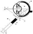
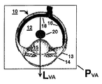
본원에 개시된 것은, 그리고 도 1 내지 4에 도시된 바와 같이, 안구의 유리체액(12) 내로 물질(20)을 주입하여 대상의 안구(10) 장애를 치료하는 방법이다. 일 양태에서, 물질(20)은 주사기(30)를 사용하여 안구(10)의 유리체액(12) 내로 주입될 수 있다. 본 양태에서, 주사기(30)는 주입 이전에 물질(20)을 수용하도록 구성된 배럴(32)을 가질 수 있다. 또 다른 양태에서, 주사기(30)는 바늘(34)을 가질 수 있다. 본 양태에서, 바늘(34)은 주사기의 배럴(32)과의 유체 소통하는 팁(36)과 루멘(38)을 가질 수 있다. 바늘(34)은 금속성일 수 있다는 것이 고려된다. 또한 바늘(34)의 팁(36)은 뾰족하거나 아니면 안구(10) 내에 도입될 수 있도록 구성될 수 있다는 것이 고려된다. 바늘(34)은 안구(10) 내에 도입되기에 적합한 직경을 가질 수 있으며, 따라서, 예를 들어 그리고 제한 없이, 20, 21, 22, 23, 24, 25, 26, 27, 28, 29, 30, 31, 32, 33 및 34 구경을 포함하는, 안구 내에 도입되기에 적합한 구경을 가질 수 있다. 추가적인 양태에서, 주사기(30)는 플런저(33)를 가질 수 있다. 본 양태에서, 플런저(33)는 배럴(32) 내에서 바늘(34)에 대해 이동할 수 있다. 바늘(34)이 물질(20)과의 유체 소통 내에 배치된 후, 플런저(33)는 주사기(30)의 배럴(32)로 원하는 양의 물질을 끌어오기 위해 바늘로부터 멀리 이동될 수 있다는 것이 고려된다. 물질(20)이 주사기(30)의 배럴(32) 내에 수용된 후, 플런저(33) 및 바늘(34) 사이의 배럴(32) 내에 갇혀 있는 모든 공기는 배출되거나 또는 다른 종래 방법을 이용하여 제거될 수 있다. 본원에 개시된 방법의 주입 단계가 대체로 주사기의 사용에 의해 수행되기는 하지만, 개시된 방법은 또한, 예를 들어 그리고 제한 없이, 펌프 주입 메커니즘(pump injection mechanism), 양변위 피스톤 로드(positive displacement piston rod), 유압 주입 메커니즘(hydraulic injection mechanism) 등을 포함하는 그 밖의 종래의 주입 메커니즘을 이용하여 수행될 수 있다는 것이 고려된다.Disclosed herein, and as shown in FIGS. 1-4, is a method of treating an ocular 10 disorder in a subject by injecting a
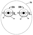
일 양태에서, 그리고 도 5A 및 5B에 도시된 바와 같이, 안구 장애의 치료 방법은 안구의 시축(visual axis, LVA)을 중심으로 한 원호(50)를 따라 위치하는 주입 지점(40)에서 안구(10) 내에 바늘(34)을 삽입하는 단계를 포함한다. 도 5A 및 5B에 나타낸 얼굴(70)에 도시된 바와 같이, 원호(50)는 오른쪽 안구(10a) 또는 왼쪽 안구(10b) 상에 위치할 수 있다. 본 양태에서, 그리고 도 5A에 도시된 바와 같이, 원호(50)는 안구의 시축(LVA)을 포함하는 가상의 수평면(imaginary horizontal plane, PVA) 위의 대략 30°(도)의 안구(10a, 10b)의 측두측(temporal side) 상의 제 1 지점(52)으로부터 가상의 수평면 위의 대략 30°(도)의 안구의 비측(nasal side) 상의 제 2 지점 아래로 연장될 수 있다는 것이 고려된다. 본원에서 사용된 바와 같이, "비측(nasal side)"이란 용어는 대상의 코에 가장 근접한 안구의 측면을 의미하는 반면, "측두측(temporal side)" 용어는 관자놀이에 가장 근접한 안구의 측면을 의미하며, 따라서, 안구의 비측과 반대에 있다. 따라서, 원호(50)는 가상의 수평면(PVA) 위의 30°(도) 지점에서 출발해서, 가상의 수평면 아래에서 안구(10a, 10b)의 일부를 지나서, 가상의 수평면 위의 30°(도) 지점에서 끝날 수 있다. 안구(10a, 10b) 상의 원호(50)의 위치를 설명함에 있어서, 안구의 정면도 상에 겹쳐지는 시계 문자판을 상상하는 것이 도움이 된다. 이러한 설명에서, 본원에 개시된 바와 같이 원호(50)는 시계의 2시 위치에 해당하는 지점으로부터 시계의 10시 위치에 해당하는 지점까지 연장될 수 있다.In one aspect, and as shown in FIGS. 5A and 5B, a method for treating an ocular disorder may include an eye at an
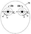
추가의 양태에서, 주입 지점(40)은 실질적으로 가상의 수평면(PVA) 내의 안구(10a, 10b)의 측두측 상에 위치하는 지점과 실질적으로 가상의 수평면 내의 안구의 비측 상에 위치하는 지점 사이의 원호(50) 상에 위치할 수 있다. 본 양태에서, 이전의 설명을 계속하자면, 주입 지점(40)은 시계의 3시와 9시 위치에 해당하는 지점들 사이의 원호(50) 상에 위치할 수 있다. 또 다른 양태에서, 주입 지점(40)은 안구(10a, 10b)의 측두측 상의 가상의 수평면(PVA) 아래의 대략 30°(도)에 위치한 지점과 안구의 비측 상의 가상의 수평면 아래의 대략 30°(도)에 위치한 지점 사이의 원호(50) 상에 위치할 수 있다. 본 양태에서, 주입 지점(40)은 시계의 4시와 8시 위치에 해당하는 지점들 사이의 원호(50) 상에 위치할 수 있다. 또 다른 양태에서, 주입 지점(40)은 안구의 측두측 상의 가상의 수평면(PVA) 아래의 대략 90°(도)에 위치하는 지점(시계의 6시 위치)과 안구의 비측 상의 가상의 수평면(PVA) 아래의 대략 30°(도) 지점(왼쪽 안구에 대해 시계의 8시 위치 및 오른쪽 안구에 대해 시계의 4시 위치) 사이의 원호(50) 상에 위치할 수 있다. 더욱 바람직하게는, 그리고 도 5B에 나타낸 얼굴(70)에 도시된 바와 같이, 주입 지점(40)은 안구의 측두측 상의 가상의 수평면(PVA) 아래의 대략 30°(도)에 위치하는 지점(왼쪽 안구에 대해 시계의 4시 위치 및 오른쪽 안구에 대해 시계의 8시 위치)과 안구의 측두측 상의 가상의 수평면 아래의 대략 90°(도)에 위치하는 지점(시계의 6시 위치) 사이의 원호(50) 상에 위치할 수 있다.In a further aspect, the
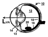
또 다른 양태에서, 그리고 도 1 내지 4를 참고하면, 원호(50)는 안구(10)의 평면부(pars plana, 13)의 적어도 일부 위에 가로놓일 수 있다. 본 양태에서, 원호(50)가 안구(10)의 전체 평면부(13) 위에 가로놓일 수 있다는 것이 고려된다. 추가의 양태에서, 그리고 도 4를 참고하면, 원호(50)는 안구(10)의 가장자리(limbus, 14) 뒤쪽의 대략 3 mm 내지 5 mm에 위치할 수 있다. 더욱 바람직하게는, 원호(50)는 안구(10)의 가장자리(14) 뒤쪽의 대략 3 mm 내지 4 mm에 위치할 수 있다. 본 양태에서, 원호(50)가 안구(10)의 가장자리(14)와 중심이 같을 수 있다는 것이 고려된다. 따라서, 원호(50)와 가장자리(14)는 모두 안구(10)의 시축(LVA)을 중심으로 할 수 있다는 것이 고려된다.In another embodiment, and with reference to FIGS. 1-4,
또 다른 양태에서, 그리고 도 2를 참고하면, 상기 방법은 주입 지점(40)에서 안구(10)의 표면에 대해 접선인 가상선(LT)에 대해 대략 90°(도) 내지 대략 45°(도)의 배향각(orientation angle, OA)으로 바늘(34)을 배향시키는 단계를 포함할 수 있다. 더욱 바람직하게는, 배향각(OA)은 주입 지점(40)에서 안구(10)의 표면에 대해 접선인 가상선(LT)에 대해 대략 90°(도) 내지 대략 85°(도)일 수 있다. 가장 바람직하게는, 배향각(OA)은 주입 지점(40)에서 안구(10)의 표면에 대해 접선인 가상선(LT)에 대해 대략 87°(도) 내지 대략 85°(도)일 수 있다. 안구(10)의 표면에 대해 접선인 가상선(LT)은 모든 방향으로 연장될 수 있다는 것이 고려된다. 따라서, 바늘(34)은 주입 지점(40)에 대해 모든 방향으로 배향될 수 있다. 선택적으로, 일 양태에서, 가상선(LT)은 교차 지점(I)에서 안구의 시축(LVA)과 교차할 수 있다. 추가의 양태에서, 바늘(34)은 안구(10) 내에 삽입되는 단계 이전에 배향각(OA)에서 배향될 수 있다는 것이 고려된다. 그렇지 않으면, 바늘(34)은 안구(10) 내에 삽입되는 단계 이후에 배향각(OA)에서 배향될 수 있다.In another aspect, and with reference to FIG. 2, the method comprises approximately 90 ° (degrees) to approximately 45 ° (s) with respect to an imaginary line L T which is tangent to the surface of the
일 양태에서, 그리고 도 3을 참고하면, 상기 방법은 안구(10) 내에 위치한 가상의 원추(60) 내에 바늘(34)을 배향시키는 단계를 포함할 수 있다는 것이 고려된다. 본 양태에서, 원추(60)는 주입 지점(40)과 일치하는 정점을 가질 수 있다. 추가의 양태에서, 원추는 주입 지점(40)에서 안구(10)의 표면에 대해 직각으로 배향된 선(LC)로부터 측정된 대략 45 도의 원추각(CA)을 가질 수 있다.In one aspect, and with reference to FIG. 3, it is contemplated that the method may include orienting the
또 다른 양태에서, 그리고 도 4를 참고하면, 바늘(34)은 바늘의 팁(36)이 가상의 수평면(PVA) 아래에 위치하도록 삽입 지점(40)에서 안구(10) 내의 소정 깊이(D)로 삽입될 수 있다는 것이 고려된다. 본 양태에서, 안구(10) 내의 바늘(34)의 팁(36)의 깊이(D)는 삽입 지점(40)에서 망막(16)으로부터 대략 1 mm 내지 대략 10 mm일 수 있다. 더욱 바람직하게는, 안구(10) 내의 바늘(34)의 팁(36)의 깊이(D)는 삽입 지점(40)에서 망막(16)으로부터 대략 1 mm 내지 대략 4 mm일 수 있다.In another aspect, and with reference to FIG. 4, the
추가의 양태에서, 그리고 도 1 내지 4에 도시된 바와 같이, 상기 방법은 바늘(34)을 향해 플런저(33)를 이동시켜 배럴(32)로부터 물질(20)이 루멘(38)을 통해 유리체액(12) 내로 주입되게 하는 단계를 더 포함할 수 있다. 일 양태에서, 바늘(34)을 선택적으로 이동시켜 주사기(30)의 배럴(32)로부터 물질(20)을 받기 위해 유리체액(12) 내에 포켓을 형성할 수 있다는 것이 고려된다. 따라서, 물질(20)이 주사기(30)의 배럴(32)에서 배출되어 유리체액(12) 내로 진입한 후, 물질이 유리체액 내에 남아 있게 하면서 동시에 바늘(34)을 유리체액으로부터 제거할 수 있다는 것이 고려된다. 도 1, 6 및 7에 도시된 바와 같이, 물질(20)이 안구(10) 내의 망막 황반(macula, 18)과 수정체(15)와 접촉하는 것을 피하도록 유리체액(12) 내에서 하향 안착할 수 있도록 하여 대상의 시야와의 간섭을 방지할 수 있다는 것이 또한 고려된다.In a further embodiment, and as shown in FIGS. 1-4, the method moves the
일부 양태에서, 주입 가이드 및 주입 보조 장치들이 주사기 및 기타 종래의 삽입 메커니즘과 결합되어 본원에 개시된 방법의 단계를 수행할 수 있다는 것이 고려된다. 또한 주입 가이드 및 주입 보조 장치들이 사용되어 물질이 원하는 깊이, 각도 및 위치에 주입되는 것을 보장할 수 있다는 것이 고려된다. 따라서, 본원에 개시된 주사기 및 기타 종래의 주입 메커니즘들이, 예를 들어, 그리고 제한 없이, 주입의 깊이를 측정하기 위한 게이지, 주입 각도를 측정하기 위한 게이지, 주입을 안정화하기 위한 게이지, 주입 위치를 조절하기 위한 게이지 등에 결합될 수 있다는 것이 고려된다. 일 양태에서, 주사기는 FCI Ophthalmics(Pembroke, MA)에서 제조한 InVitria? 유리체강내 주입 보조기(Intravitreal Injection Assistant)에 결합될 수 있다는 것이 고려된다.In some aspects, it is contemplated that infusion guides and infusion aids may be combined with syringes and other conventional insertion mechanisms to perform the steps of the methods disclosed herein. It is also contemplated that injection guides and injection aids may be used to ensure that the material is injected at the desired depth, angle and location. Thus, the syringes and other conventional injection mechanisms disclosed herein, for example, and without limitation, adjust the gauge for measuring the depth of injection, the gauge for measuring the injection angle, the gauge for stabilizing the injection, the injection position. It is contemplated that it may be coupled to a gauge or the like for the purpose. In one embodiment, the syringe is an InVitria® manufactured by FCI Ophthalmics (Pembroke, MA). It is contemplated that it may be coupled to an intravitreal injection assistant.
개시된 방법은 전후의 안구 질환 모두를 포함하여 다양한 안구 장애를 치료하거나 예방하기 위해 사용될 수 있다. 일 양태에서, 상기 방법은 망막 부종(retinal edema) 및 망막 혈관신생(retinal neovascularization)과 관련될 수 있는 황반 변성(macular degeneration) 및 황반 혈관신생(macular angiogenesis)을 치료하기 위해 사용될 수 있다.The disclosed methods can be used to treat or prevent various ocular disorders, including both pre and post ocular diseases. In one aspect, the method can be used to treat macular degeneration and macular angiogenesis that may be associated with retinal edema and retinal neovascularization.
그 밖의 양태에서, 상기 방법은, 예를 들어 그리고 제한 없이, 망막 부종, 건성 및 습성 황막변성(dry and wet macular degeneration), 맥락막 혈관신생(choroidal neovascularization), 당뇨성 망막병증(diabetic retinopathy), 급성 황반성 신경망막병증(acute macular neuroretinopathy), 중심성 장액 맥락망막병증(central serous chorioretinopathy), 낭포 황반 부종(cystoid macular edema), 및 당뇨성 황반 부종(diabetic macular edema), 포도막염(uveitis), 망막염(retinitis), 맥락막염(choroiditis), 급성 후부다발성 판상 색소 상피증(acute multifocal placoid pigment epitheliopathy), 베체트병(Behcet's disease), 산탄 맥락망막병증(birdshot retinochoroidopathy), 매독(syphilis), 라임병(lyme), 결핵(tuberculosis), 톡소플라스마증(toxoplasmosis), 중간 포도막염(intermediate uveitis, 중간 포도막염(pars planitis)), 다소성 맥락막염(multifocal choroiditis), 다발성 소실성 백반 증후군(multiple evanescent white dot syndrome, MEWDS), 눈 사르코이드증(ocular sarcoidosis), 후공막염(posterior scleritis), 포행성 맥락막염(serpiginous choroiditis), 망막하 섬유증 및 포도막염 증후군(subretinal fibrosis and uveitis syndrome), 보크트-고야나기-하라다 증후군(Vogt-Koyanagi-and Harada syndrome)을 포함하는 하나 이상의 포유류의 후안구 장애를 치료하기 위해 실행되거나 제공될 수 있다.In other embodiments, the method may include, for example and without limitation, retinal edema, dry and wet macular degeneration, choroidal neovascularization, diabetic retinopathy, acute Acute macular neuroretinopathy, central serous chorioretinopathy, cystoid macular edema, and diabetic macular edema, uveitis, retinitis ), Choroiditis, acute multifocal placoid pigment epitheliopathy, Behcet's disease, shotshot retinochoroidopathy, syphilis, syphilis, lyme, Tuberculosis, toxoplasmosis, intermediate uveitis, pars planitis, multifocal choroiditis, multiple disappearance Multiple evanescent white dot syndrome (MEWDS), ocular sarcoidosis, posterior scleritis, serpiginous choroiditis, subretinal fibrosis and uveitis syndrome And may be practiced or provided to treat posterior ocular disorders in one or more mammals, including Vogt-Koyanagi-and Harada syndrome.
추가의 양태에서, 상기 방법은, 예를 들어 그리고 제한 없이, 망막 동맥 폐쇄 질환(retinal arterial occlusive disease), 전포도막염(anterior uveitis), 망막 정맥 폐쇄(retinal vein occlusion), 망막 중심 정맥 폐쇄(central retinal vein occlusion), 파종성 혈관내 응고병증(disseminated intravascular coagulopathy), 망막 정맥 분지 폐쇄(branch retinal vein occlusion), 고혈압성 안저 변화(hypertensive fundus changes), 안허혈 증후군(ocular ischemic syndrome), 망막 미세 동맥류(retinal arterial microaneury), 코우츠병(Coat's disease), 중심부 모세관확장증(parafoveal telangiectasis), 절반망막 정맥 폐쇄(hemiretinal vein occlusion), 망막 중심 정맥 폐쇄(papillophlebitis), 망막 중심 동맥 폐쇄(central retinal artery occlusion), 망막 동맥 분지 폐쇄(branch retinal artery occlusion), 경동맥 질환(carotid artery disease, CAD), 서리가지 동맥염(frosted branch angiitis), 낫적혈구 망막증(sickle cell retinopathy), 혈관무늬 망막병증(angioid streaks), 가족성 삼출유리체 망막병증 *familial exudative vitreoretinopathy) 및 일스병(Eales disease)을 포함하는 하나 이상의 안구의 혈관 질환 및 장애를 치료하기 위해 사용될 수 있다.In a further aspect, the method includes, for example and without limitation, retinal arterial occlusive disease, anterior uveitis, retinal vein occlusion, central retinal vein occlusion. vein occlusion, disseminated intravascular coagulopathy, branch retinal vein occlusion, hypertensive fundus changes, ocular ischemic syndrome, retinal micro aneurysms retinal arterial microaneury, Coat's disease, parafoveal telangiectasis, hemotinal vein occlusion, central retinal vein occlusion, central retinal artery occlusion, retinal retinal artery occlusion Branch retinal artery occlusion, carotid artery disease (CAD), frosted branch angiitis, sickle To treat one or more ocular vascular diseases and disorders including sickle cell retinopathy, angioid streaks, familial exudative vitreoretinopathy and Eales disease Can be used.
또 다른 양태에서, 상기 발명은, 예를 들어 그리고 제한 없이, 교감성 안염(sympathetic ophthalmia), 포도막 망막병증(uveitic retinal disease), 망막 박리(retinal detachment), 외상(trauma), 광응고(photocoagulation), 수술시 관류저하(hypoperfusion during surgery), 방사선 망막병증(radiation retinopathy), 및 골수 이식 망막병증(bone marrow transplant retinopathy); 증식성 유리체 망막병증 및 망막 전막(proliferative vitreal retinopathy and epiretinal membranes), 및 증식성 당뇨성 망막병증(proliferative diabetic retinopathy); 안 히스토플라스마증(ocular histoplasmosis), 안 톡소카라증(ocular toxocariasis), 추정 안 히스토플라스마 증후군(presumed ocular histoplasmosis syndrome, POHS), 내안구염(endophthalmitis), 톡소플라스마증(toxoplasmosis) HIV 감염 관련 망막 장애(retinal diseases associated with HIV infection), HIV 감염 관련 맥락막 장애(choroidal disease associated with HIV infection), HIV 감염 포도막염 장애(uveitic disease associated with HIV infection), 바이러스성 망막염(viral retinitis), 급성 망막 괴사(acute retinal necrosis), 진행성 외망막 괴사(progressive outer retinal necrosis), 진균성 망막 질환(fungal retinal diseases), 안구 매독(ocular syphilis), 안구 결핵(ocular tuberculosis), 광범위 단안 아급성 시신경망막염(diffuse unilateral subacute neuroretinitis), 및 구더기증(myiasis)과 같은 감염 질환을 포함하는 정신적 외상/외과 질환 및 장애를 치료하기 위해 사용될 수 있다.In another embodiment, the invention, for example and without limitation, sympathetic ophthalmia, uveitic retinal disease, retinal detachment, trauma, photocoagulation Hypoperfusion during surgery, radiation retinopathy, and bone marrow transplant retinopathy; Proliferative vitreal retinopathy and epiretinal membranes, and proliferative diabetic retinopathy; Retinal eye related to ocular histoplasmosis, ocular toxocariasis, presumed ocular histoplasmosis syndrome (POHS), endophthalmitis, toxoplasmosis HIV infection Disorders associated with HIV infection, choroidal disease associated with HIV infection, uveitic disease associated with HIV infection, viral retinitis, acute retinal necrosis retinal necrosis, progressive outer retinal necrosis, fungal retinal diseases, ocular syphilis, ocular tuberculosis, diffuse unilateral subacute neuroretinitis ), And to treat mental trauma / surgical diseases and disorders, including infectious diseases such as myiasis Can.
그 밖의 양태에서, 상기 방법은, 예를 들어 그리고 제한 없이, 색소성 망막염(retinitis pigmentosa), 망막 이양증과 관련된 전신 장애(systemic disorders with associated retinal dystrophies), 선천성 정지형 야맹증(congenital stationary night blindness), 추체 이영양증(cone dystrophies), 스타가르트병 및 황색반 안저(Stargardt's disease and fundus flavimaculatus), 베스트병(Best's disease), 망막 색소상피의 무늬 이영양증(pattern dystrophy of the retinal pigmented epithelium), X-염색체 관련 망막층간분리(X-linked retinoschisis), 솔스비 안저 이상증(Sorsby's fundus dystrophy), 양성 동심원 황반병증(benign concentric maculopathy), 비에띠 결정각막 이상증(Bietti's crystalline dystrophy), 및 탄력섬유성 가황색종(pseudoxanthoma elasticum)을 포함하는 유전적 질환 및 장애를 치료하기 위해 사용될 수 있다.In other embodiments, the method includes, for example and without limitation, retinitis pigmentosa, systemic disorders with associated retinal dystrophies, congenital stationary night blindness, vertebral bodies Cone dystrophies, Stargardt's disease and fundus flavimaculatus, Best's disease, pattern dystrophy of the retinal pigmented epithelium, X-chromosome related retina X-linked retinoschisis, Sorsby's fundus dystrophy, benign concentric maculopathy, Bietti's crystalline dystrophy, and pseudoxanthoma elasticum May be used to treat genetic diseases and disorders, including).
추가의 양태에서, 개시된 방법은 또한, 예를 들어 그리고 제한 없이, 선천성 망막 색소상피 비대증(congenital hypertrophy of the retinal pigmented epithelium), 후방 포도막 흑색종(posterior uveal melanoma), 맥락막 혈관증(choroidal hemangioma), 맥락막 골종(choroidal osteoma), 맥락막 전이종양(choroidal metastasis), 망막과 망막 색소상피 복합 과오종(combined hamartoma of the retina and retinal pigmented epithelium), 망막아종(retinoblastoma), 안저 혈관확장 종양(vasoproliferative tumors of the ocular fundus), 망막 성상세포종(retinal astrocytoma), 및 안내 림프 종양(intraocular lymphoid tumors)을 포함하는 암 및 종양 관련 망막 질환을 치료하기 위해 사용될 수 있다.In a further aspect, the disclosed methods also include, for example and without limitation, congenital hypertrophy of the retinal pigmented epithelium, posterior uveal melanoma, choroidal hemangioma, Choroidal osteoma, choroidal metastasis, combined hamartoma of the retina and retinal pigmented epithelium, retinoblastoma, vasoproliferative tumors of the ocular fundus, retinal astrocytoma, and intraocular lymphoid tumors, and can be used to treat tumor-associated retinal diseases.
또 다른 양태에서, 상기 발명은, 예를 들어 그리고 제한 없이, 점상내층 맥락막병증(punctuate inner choroidopathy), 급성 후극부 다소성 색소 상피증(acute posterior multifocal placoid pigment epitheliopathy), 근시성 망막 변성(myopic retinal degeneration), 급성 망막 색소 상피염(acute retinal pigment epithelitis), 망막 색소변성(retinitis pigmentosa), 증식성 유리체 망막병증(proliferative vitreal retinopathy, PVR), 노인성 황반변성(age- related macular degeneration, ARMD), 당뇨성 망막병증(diabetic retinopathy), 당뇨성 황반 부종(diabetic macular edema), 망막 박리(retinal detachment), 망막 열공(retinal tears), 포도막염(uveitis), 황반 열공(macular tears), 거대세포바이러스 망막염(cytomegalovirus retinitis), 녹내장(glaucoma), 및 망막 신경절 세포의 신경변성(neurodegeneration of retinal ganglion cells)과 같은 안구 퇴화(ocular degeneration)를 포함하는 질환을 포함하는 광범위한 안구 질환을 치료하거나 개선하는데 사용될 수 있다.In another embodiment, the invention includes, for example and without limitation, punctuate inner choroidopathy, acute posterior multifocal placoid pigment epitheliopathy, myopic retinal degeneration. degeneration, acute retinal pigment epithelitis, retinitis pigmentosa, proliferative vitreal retinopathy (PVR), age-related macular degeneration (ARMD), diabetes Diabetic retinopathy, diabetic macular edema, retinal detachment, retinal tears, uveitis, macular tears, cytomegalovirus vagina, including ocular degeneration, such as retinitis, glaucoma, and neurodegeneration of retinal ganglion cells It can be used to treat or ameliorate a wide range of ocular diseases, including the ring.
일 양태에서, 안구 내에 주입되는 물질은 미세입자를 포함할 수 있다. 본 양태에서, 안구 내에 주입되는 물질은 주입 담체(injection vehicle) 내에 떠 있는 대략 1 내지 대략 500 mg의 미세입자를 포함할 수 있다. 더욱 바람직하게는, 물질은 주입 담체 내에 떠 있는 대략 2 내지 대략 300 mg의 미세입자를 포함할 수 있다. 가장 바람직하게는, 물질은 주입 담체 내에 떠 있는 대략 3 내지 대략 150 mg의 미세입자를 포함할 수 있다. 주입 담체는, 일 양태에서, 대략 1% 내지 대략 50%의 고체를 포함할 수 있다. 더욱 바람직하게는, 주입 담체는 대략 10% 내지 대략 40%의 고체를 포함할 수 있다. 가장 바람직하게는, 주입 담체는 대략 20% 내지 대략 30%의 고체를 포함할 수 있다. 바람직한 일 양태에서, 안구 내에 주입되는 물질은 대략 20% 내지 대략 30%의 고체를 포함하는 주입 담체 내에 떠 있는 대략 10 mg 내지 대략 50 mg의 미세입자를 포함할 수 있다. 사용에 있어서, 본원에 개시된 물질은 일반적으로 주사 당 대략 10 내지 대략 150 μL의 부피로 안구의 유리체액 내로 직접 주입된다.In one aspect, the material injected into the eye may comprise microparticles. In this aspect, the material to be injected into the eye may comprise about 1 to about 500 mg of microparticles suspended in an injection vehicle. More preferably, the material may comprise about 2 to about 300 mg of microparticles suspended in the infusion carrier. Most preferably, the material may comprise from about 3 to about 150 mg of microparticles suspended in the infusion carrier. Infusion carriers may, in one aspect, comprise approximately 1% to approximately 50% solids. More preferably, the injection carrier may comprise about 10% to about 40% solids. Most preferably, the infusion carrier may comprise about 20% to about 30% solids. In a preferred embodiment, the material injected into the eye may comprise from about 10 mg to about 50 mg of microparticles suspended in an infusion carrier comprising from about 20% to about 30% solids. In use, the materials disclosed herein are generally injected directly into the vitreous fluid of the eye in a volume of about 10 to about 150 μL per injection.
또 다른 양태에서, 개시된 방법에 사용될 수 있는 미세입자는 대략 10 μm 내지 대략 125 μm의 평균 입자 크기를 가질 수 있다. 더욱 바람직하게는, 미세입자는 대략 20 μm 내지 대략 90 μm의 평균 입자 크기를 가질 수 있다. 가장 바람직하게는, 미세입자는 대략 30 μm 내지 대략 80 μm의 평균 입자 크기를 가질 수 있다. 상술한 입자 크기 분포는 본 기술 분야의 숙련자에게 공지된 레이저 회절법에 의해 측정될 수 있다는 것이 고려된다.In another embodiment, the microparticles that can be used in the disclosed methods can have an average particle size of about 10 μm to about 125 μm. More preferably, the microparticles can have an average particle size of about 20 μm to about 90 μm. Most preferably, the microparticles can have an average particle size of about 30 μm to about 80 μm. It is contemplated that the particle size distribution described above can be measured by laser diffraction methods known to those skilled in the art.
또 다른 양태에서, 미세입자는 하나 또는 그 이상의 약물 조성물을 사용하여 제조될 수 있다. 본 양태에서, 약물 조성물은 하나 또는 그 이상의 수용성 담체(carrier) 또는 부형제(excipient)를 포함할 수 있다. 이러한 담체 또는 부형제는 일반적으로 설탕, 당류(saccharide), 다당류(polysaccharide), 계면 활성제(surfactant), 완충염(buffer salt), 증량제(bulking agent), 증점제(viscosity agent) 등을 포함할 수 있다. 부형제의 비제한적인 예는 2-(하이드록시메틸)-6-[3,4,5-트리하이드록시-6-(하이드록시메틸)테트라히이드로피란-2-일]옥시- 테트라히이드로피란-3,4,5-트리올(2-(hydroxymethyl)-6-[3,4,5-trihydroxy-6-hydroxymethyl)tetrahydropyran-2-yl]oxy- tetrahydropyran-3,4,5-triol, [trehalose])이다. 일 양태에서, 약물 조성물은 시작 약물 조성물 내의 트레할로스(trehalose)의 중량을 근거로 대략 1 wt% 내지 대략 200 wt%의 트레할로스를 포함할 수 있다. 더욱 바람직하게는, 약물 조성물은 시작 약물 조성물 내의 트레할로스의 중량을 근거로 대략 10 wt% 내지 대략 50 wt%의 트레할로스를 포함할 수 있다. 가장 바람직하게는, 약물 조성물은 시작 약물 조성물 내의 트레할로스의 중량을 근거로 대략 25 wt% 내지 대략 35 wt%의 트레할로스를 포함할 수 있다.In another embodiment, the microparticles can be prepared using one or more drug compositions. In this aspect, the drug composition may comprise one or more water soluble carriers or excipients. Such carriers or excipients may generally include sugars, saccharides, polysaccharides, surfactants, buffer salts, bulking agents, viscosity agents and the like. Non-limiting examples of excipients are 2- (hydroxymethyl) -6- [3,4,5-trihydroxy-6- (hydroxymethyl) tetrahydropyran-2-yl] oxy-tetrahydropyran -3,4,5-triol (2- (hydroxymethyl) -6- [3,4,5-trihydroxy-6-hydroxymethyl) tetrahydropyran-2-yl] oxy-tetrahydropyran-3,4,5-triol, [ trehalose]). In one aspect, the drug composition may comprise from about 1 wt% to about 200 wt% trehalose based on the weight of trehalose in the starting drug composition. More preferably, the drug composition may comprise from about 10 wt% to about 50 wt% trehalose based on the weight of trehalose in the starting drug composition. Most preferably, the drug composition may comprise from about 25 wt% to about 35 wt% trehalose based on the weight of trehalose in the starting drug composition.
또 다른 양태에서, 부형제는, 예를 들어 그리고 제한 없이, 폴리소르베이트 20(polysorbates 20), 폴리소르베이트 80 등을 포함하는 하나 또는 그 이상의 계면 활성제를 포함할 수 있다. 바람직한 일 양태에서, 부형제는 폴리소르베이트 20(또는 트윈 20(Tween 20))를 포함할 수 있다. 본 양태에서, 약물 조성물은 시작 약물 조성물 내의 폴리소르베이트 20의 중량을 근거로 대략 0.01 wt% 내지 대략 5 wt%의 폴리소르베이트 20을 포함할 수 있다. 더욱 바람직하게는, 약물 조성물은 시작 약물 조성물 내의 폴리소르베이트 20의 중량을 근거로 대략 0.05 wt% 내지 대략 0.25 wt%의 폴리소르베이트 20을 포함할 수 있다. 가장 바람직하게는, 약물 조성물은 시작 약물 조성물 내의 폴리소르베이트 20의 중량을 근거로 대략 0.1 wt%의 폴리소르베이트 20을 포함할 수 있다. 약물 조성물은 본원에 개시된 바와 같은 둘 또는 그 이상의 담체 및/또는 부형제를 포함할 수 있다는 것이 고려된다. 예를 들어, 그리고 제한 없이, 약물 조성물은 시작 약물 조성물 내의 각각의 약물의 중량을 근거로 대략 25 wt% 내지 대략 35 wt% 트레할로스 및 대략 0.1 wt%의 폴리소르베이트 20을 포함할 수 있다.In another aspect, the excipient may include one or more surfactants, including, for example and without limitation, polysorbates 20, polysorbate 80, and the like. In one preferred aspect, the excipient may comprise polysorbate 20 (or Tween 20). In this aspect, the drug composition may comprise from about 0.01 wt% to about 5 wt
추가의 양태에서, 부형제는, 예를 들어 그리고 제한 없이, 하이드록시프로필 메틸셀룰로스(hydroxypropyl methylcellulose, HPMC), 히알루론산(hyaluronic acid) 등을 포함하는 하나 또는 그 이상의 증점제를 포함할 수 있다.In further embodiments, excipients may include one or more thickeners including, for example and without limitation, hydroxypropyl methylcellulose (HPMC), hyaluronic acid, and the like.
선택적으로, 종래의 습식 또는 마찰 저감제를 물질에 첨가하여 물질의 습윤성(wettability) 또는 윤활성(lubricity)을 증가시킬 수 있다. 이러한 첨가제들은 안구 내에 물질의 주입 이후에 물질의 하향 이동을 촉진하도록 구성될 수 있다는 것이 고려된다.Optionally, conventional wet or friction reducing agents can be added to the material to increase the wettability or lubricity of the material. It is contemplated that such additives may be configured to facilitate downward movement of the substance after injection of the substance into the eye.
일 양태에서, 개시된 물질은 원하는 투여 스케줄에 따라 본원에 개시된 바와 같이 주입될 수 있다. 예를 들면, 그리고 제한 없이, 원하는 투여 스케줄은 대략 매월, 대략 매 2 개월, 대략 매 3 개월, 대략 매 4 개월, 대략 매 6 개월, 대략 매 8 개월, 대략 매 9 개월, 대략 매 10 개월, 그리고 대략 매 12 개월에 한 번의 투여량을 포함할 수 있다.In one aspect, the disclosed materials can be infused as disclosed herein according to a desired dosing schedule. For example, and without limitation, the desired dosing schedule may be approximately monthly, approximately every two months, approximately every three months, approximately every four months, approximately every six months, approximately every eight months, approximately every nine months, approximately every ten months, And about once every 12 months.
실험예Experimental Example
다음의 실시예는, 본원에 개시되고 청구된 화합물, 조성물, 물품, 장치, 및/또는 장치들이 어떻게 제조되고 평가되는지, 그리고 전적으로 본보기인 것으로 의도되며, 그리고 본 발명자들이 그들의 발명이라고 간주하는 범위를 제한하도록 의도되지 않은 것이라는 완벽한 개시 및 설명을 통해 본 기술 분야의 통상의 지식을 가진 자에게 제공하기 위해 제시된다. 숫자(예를 들어, 양, 온도 등)와 관련해서 정확성을 보장하기 위해 노력했으나, 일부 오류 및 편차가 설명되어야 한다. 달리 나타내지 않는 한, 부(parts)는 중량부(parts by weight)이고, 온도는 ℃ 또는 주위 온도이며, 압력은 대기 또는 대기에 가깝다.The following examples are intended to explain how the compounds, compositions, articles, devices, and / or devices disclosed and claimed herein are made and evaluated, and are intended to be wholly exemplary, and to the extent that the inventors deem their invention. It is presented to those skilled in the art through a complete disclosure and description that they are not intended to be limiting. Efforts have been made to ensure accuracy with respect to numbers (eg amounts, temperature, etc.) but some errors and deviations should be accounted for. Unless indicated otherwise, parts are parts by weight, temperature is in degrees Centigrade or ambient temperature, and pressure is at or near atmospheric.
실시예Example 1 One
미세입자의 분포를 조절하기 위해 다양한 주입법을 조사하였다. 특히, HPMC 및 힐론(Healon) 주입 담체(50 μL)를 갖는 쿠머린-로딩된(coumarin-loaded) 미소구체를 25 게이지 UTW 바늘을 통해 온전한 돼지 사체 안구 내(Sierra Medical)에 주입하였다. 최적의 초기 배치를 위해, 주입 속도는 한계 속도가 아니었다. 얕은 바늘 주입이 이상적인 것으로 보였다. 주입시, 바늘의 움직임을 방지하여 주입된 입자가 바늘에 의해 형성된 채널 및 평면을 따르려는 경향을 최소화했다. 조성물내의 기포를 최소화하여 입자가 유리체액 내에서 기포에 의해 상향 이동하는 것을 방지하였다. 주입을 시축 아래에 위치시켜 주입된 입자가 하부 위치에 조기 안착하는 것을 촉진시켰다. Various injection methods were investigated to control the distribution of microparticles. In particular, coumarin-loaded microspheres with HPMC and Healon injection carriers (50 μL) were injected into intact porcine carcinoma eye (Sierra Medical) through a 25 gauge UTW needle. For optimal initial placement, the feed rate was not a limit speed. Shallow needle injection seemed to be ideal. During injection, needle movement was prevented to minimize the tendency for injected particles to follow the channels and planes formed by the needle. Bubbles in the composition were minimized to prevent particles from moving upward by the bubbles in the vitreous fluid. The injection was placed below the visual axis to facilitate premature settling of the injected particles in the lower position.
실시예Example 2 2
유리체강내 주입 이후 안구 내 고분자계의 내성을 평가하였다. 또한, 주입법 및 시간의 경과에 따른 미세입자 분포 상의 시스템 변수(입자 크기, 투여량, 주입 담체, 및 주입 위치)의 영향을 평가하였다. 10 미만, 10 내지 32, 32 내지 63 및 63 μm 초과의 미세입자 크기를 평가하였다. 투여량은 3, 10 및 20 mg에서 변화시켰다. Diluted Healon (2000 kD, rooster comb) 및 HA Genzyme (500 kD, fermented)을 주입 담체로서 테스트하였다. 폴리(락티드-코-글리콜리드(poly(lactide-co-glycolide)) 미소구체를 주입 담체 내의 미세입자로서 평가하였다. 3 및 10 mg의 투여량에 대해 안구 내에 한 번의 50 μL의 주입을 가한 반면, 20 mg의 투여량에 대해 안구 내에 두 번의 50 μL의 주입을 가하였다.After intravitreal injection, intraocular polymer resistance was evaluated. In addition, the effects of system variables (particle size, dosage, infusion carrier, and infusion location) on the microparticle distribution over the injection method and over time were evaluated. Microparticle sizes less than 10, 10-32, 32-63 and more than 63 μm were evaluated. Dosages were varied at 3, 10 and 20 mg. Diluted Healon (2000 kD, rooster comb) and HA Genzyme (500 kD, fermented) were tested as injection carriers. Poly (lactide-co-glycolide) microspheres were evaluated as microparticles in infusion carriers. One 50 μL infusion was added to the eye for doses of 3 and 10 mg. In contrast, two 50 μL infusions were added to the eye for a 20 mg dose.
다섯 개 그룹의 무색소상피의 뉴잉글랜드 흰토끼를 양측성 주입 시험(bilateral dosing study)에 사용하였다. 안과 검사(안저 검사, 촬영, 및 안압 측정을 포함)를 수술 전 그리고 수술 후 1, 8, 15, 31, 61, 91, 및 180 일째 (그룹 D 내지 E에 대해) 수행하였다. 망막전위도검사(electroretinography, ERG) 및 광간섭 단층촬영(optical coherence tomography (OCT) 분석을 수술 전 그리고 180일 째 그룹 D 내지 E에 대해 수행하였다. 테스트의 마지막에 (그룹 A 내지 C에 대해 90일, 그룹 D 내지 E에 대해 180일), 조직병리학 샘플들을 수집하고 분석하였다.Five groups of pigmented epithelial New England white rabbits were used in the bilateral dosing study. Ophthalmic examinations (including fundus examination, imaging, and intraocular pressure measurement) were performed preoperatively and on
주입의 상부 배치는 주입된 입자가 시야 내에 상당량 존재하는 결과를 보였다. 반대로, 주입의 하부 배치는 주입된 입자가 시야 내에 최소로 존재하는 결과를 보였으며, 시야 내에 존재하는 하부에 주입된 입자의 수는 시야 내에 존재하는 상부에 주입된 입자보다 상당히 빠르게 감소하였다. 또한, 주입의 깊은, 하부 배치는 3일 이내에 시야 밖으로의 입자의 안착으로 이어졌다. 안착 후, 입자는 안구 기저에서 분산되었다. 반대로, 주입의 상부 배치는 대체로 입자가 더욱 천천히 시야 밖으로 안착되는 결과로 이어졌다 (90일 이내). 전반적으로, 하부에 배치한 주입에 있어서, 시야 밖에서 안정적으로 잔존하는 입자와 함께, 대체로 수술 후 최대 60일까지 입자의 위치에서 거의 변화가 없었다. 하부로 주입된 입자의 저하는 수술 후 60일에서 180 일 사이에서 눈에 띄었다.The upper batch of injection resulted in a significant amount of injected particles in the field of view. In contrast, the lower batch of implantation resulted in the minimal presence of injected particles in the field of view, and the number of particles injected at the bottom present in the field of view decreased significantly faster than the particles injected at the top present in the field of view. In addition, the deep, lower placement of the injection led to the settling of particles out of sight within three days. After settling, the particles dispersed at the base of the eye. In contrast, the upper batch of injection generally resulted in particles settling out of sight more slowly (within 90 days). Overall, there was little change in the position of the particles in the underlying implants, with the particles remaining stably out of view, usually up to 60 days post-surgery. Lowering of the injected particles was noticeable between 60 and 180 days after surgery.
본 발명의 여러 실시형태가 상기 명세서에 개시되었지만, 상기 설명 및 관련된 도면에 제시된 교시의 혜택을 가지고, 본 발명이 속한 기술 분야의 숙련자는 다양한 변경 및 본 발명의 기타 실시형태를 고려할 수 있다고 생각된다. 따라서, 본 발명은 위에 개시된 구체적인 실시형태로 제한되지 않으며, 다양한 변경 및 기타 실시형태가 첨부된 특허 청구범위 내에 포함되도록 의도된 것이라 생각된다. 또한, 뒤따르는 청구범위뿐만 아니라 본원에 특정 용어들이 사용되었지만, 이들은 단지 포괄적이고 기술적인 의미로 사용되며, 개시된 발명 또는 뒤따르는 청구범위를 한정하기 위한 목적으로 사용되지 않는다.While various embodiments of the invention have been disclosed herein above, it is believed that having the benefit of the teachings presented in the foregoing descriptions and the associated drawings, those skilled in the art may consider various modifications and other embodiments of the invention. . Accordingly, it is intended that the invention not be limited to the specific embodiments disclosed above, but that various modifications and other embodiments are intended to be included within the scope of the appended claims. Moreover, although specific terms are used herein as well as the claims that follow, they are used only in a generic and technical sense and are not intended to limit the disclosed invention or the claims that follow.
10: 안구 12: 유리체약
13: 평면부 14: 가장자리
15: 수정체 16: 망막
18: 망막 황반 20: 물질
30: 주사기 32: 배럴
33: 플런저 34: 바늘
36: 팁 38:루멘
40: 주입 지점 50: 원호
PVA: 수평면 LVA:시축
OA: 배향각10: eye 12: glass contract
13: plane 14: edge
15: lens 16: retina
18: retinal macula 20: substance
30: syringe 32: barrel
33: plunger 34: needle
36: tip 38: lumen
40: injection point 50: arc
P VA : Horizontal plane L VA : Time axis
OA: orientation angle
Claims (30)
상기 안구의 시축(visual axis)을 중심으로 하고 그리고 상기 시축을 포함하는 가상의 수평면(imaginary horizontal plane) 위의 대략 30°의 상기 안구의 측두측(temporal side) 상의 제 1 지점으로부터 상기 가상의 수평면 위의 대략 30°의 상기 안구의 비측(nasal side) 상의 제 2 지점 아래로 연장되는 원호를 따라 위치하는 주입 지점에서 상기 안구 내의 소정 깊이로 상기 바늘을 삽입하여 상기 바늘의 상기 팁이 상기 가상의 수평면 아래에 위치하도록 하는 단계; 및
상기 바늘을 향해 상기 플런저를 이동시켜 상기 배럴로부터 상기 물질이 상기 루멘을 통해 상기 유리체액 내로 주입되게 하는 단계를 포함하는 것을 특징으로 하는 방법.A method of treating an eye disorder by injecting a substance into a vitreous humor of an eye using a syringe, wherein the syringe comprises a barrel containing the substance, a tip in fluid communication with the barrel. ) And a lumen, and a plunger movable relative to the needle in the barrel, the method comprising:
The imaginary horizontal plane about the eye's visual axis and from a first point on the temporal side of the eye at approximately 30 ° on an imaginary horizontal plane that includes the visual axis Insert the needle to a predetermined depth within the eye at an injection point located along an arc extending below a second point on the nasal side of the eye at approximately 30 ° above so that the tip of the needle Positioning it below a horizontal plane; And
Moving the plunger toward the needle to cause the material to be injected from the barrel into the vitreous fluid through the lumen.
상기 원호는 상기 안구의 평면부(pars plana) 위에 가로놓이는 것을 특징으로 하는 방법.The method of claim 1,
Wherein said arc lies on a pars plana of said eyeball.
상기 원호는 상기 안구의 가장자리(limbus) 뒤쪽의 대략 3 mm 내지 5 mm에 위치하고, 상기 원호는 상기 가장자리와 중심이 같은 것을 특징으로 하는 방법.The method of claim 1,
Wherein said arc is located approximately 3 mm to 5 mm behind the limbus of said eyeball, said arc being centered with said edge.
상기 주입 지점은, 실질적으로 상기 가상면 내의 상기 안구의 측두측 상에 위치하는 제 3 지점 및 실질적으로 상기 가상면 내의 상기 안구의 비측 상에 위치하는 제 4 지점 사이의 상기 원호 상에 위치하는 것을 특징으로 하는 방법.The method of claim 1,
The injection point is located on the arc between a third point located substantially on the temporal side of the eyeball in the imaginary plane and a fourth point located substantially on the nasal side of the eyeball in the imaginary plane. How to feature.
상기 주입 지점은, 상기 가상면 아래의 대략 30°의 상기 안구의 측두측 상에 위치하는 제 3 지점 및 상기 가상면 아래의 대략 90°의 상기 안구의 측두측 상에 위치하는 제 4 지점 사이의 상기 원호 상에 위치하는 것을 특징으로 하는 방법.The method of claim 1,
The injection point is between a third point located on the temporal side of the eyeball at approximately 30 ° below the imaginary plane and a fourth point located on the temporal side of the eyeball at approximately 90 ° below the imaginary plane. And located on the arc.
상기 주입 지점에서 상기 안구의 표면에 대해 접선인 가상선에 대해 대략 90° 내지 대략 45°의 배향각으로 상기 바늘을 배향시키는 단계를 더 포함하는 것을 특징으로 하는 방법.The method of claim 1,
Orienting the needle at an orientation angle of about 90 ° to about 45 ° with respect to an imaginary line tangent to the surface of the eye at the injection point.
상기 가상선은 상기 시축과 교차하는 것을 특징으로 하는 방법.The method according to claim 6,
The virtual line intersects the time axis.
상기 바늘은 상기 바늘을 삽입하는 단계 이전에 상기 배향각으로 배향되는 것을 특징으로 하는 방법.The method according to claim 6,
The needle is oriented at the orientation angle prior to inserting the needle.
상기 배향각은 상기 가상의 접선에 대해 대략 90° 내지 대략 85°인 것을 특징으로 하는 방법.The method according to claim 6,
The orientation angle is about 90 ° to about 85 ° with respect to the imaginary tangent.
상기 배향각은 상기 가상의 접선에 대해 대략 87° 내지 대략 85°인 것을 특징으로 하는 방법.The method according to claim 6,
The orientation angle is about 87 ° to about 85 ° with respect to the imaginary tangent.
상기 안구 내의 상기 팁의 상기 깊이는 상기 주입 지점에서 망막으로부터 대략 1 mm 내지 대략 10 mm인 것을 특징으로 하는 방법.The method of claim 1,
The depth of the tip in the eye is from about 1 mm to about 10 mm from the retina at the injection point.
상기 안구 내에 위치한 가상의 원추 내에 상기 바늘을 배향시키는 단계를 더 포함하고, 상기 원추는 상기 주입 지점과 일치하는 정점을 갖는 것을 특징으로 하는 방법.The method of claim 1,
Orienting the needle in a virtual cone located within the eyeball, wherein the cone has a vertex coincident with the injection point.
상기 원추는 상기 주입 지점에서 상기 안구의 표면에 대해 직각으로 배향된 선로부터 측정된 대략 45 도의 원추각을 갖는 것을 특징으로 하는 방법.13. The method of claim 12,
And the cone has a cone angle of approximately 45 degrees measured from a line oriented perpendicular to the surface of the eye at the injection point.
상기 물질은 미세입자(microparticles)를 포함하는 것을 특징으로 하는 방법.The method of claim 1,
And the material comprises microparticles.
상기 안구의 시축 아래에 위치한 주입 지점에서 평면부를 통해 상기 바늘을 상기 안구 내의 소정 깊이로 삽입하여 상기 바늘의 상기 팁이 상기 시축 아래에 위치하도록 하는 단계; 및
상기 바늘을 향해 상기 플런저를 이동시켜 상기 배럴로부터 상기 물질이 상기 루멘을 통해 상기 유리체액 내로 주입되게 하는 단계를 포함하는 것을 특징으로 하는 방법.A method of treating an ocular disorder by injecting a substance into the vitreous fluid of an eye using a syringe, the syringe comprising a barrel containing the substance, a needle having a tip and lumen in fluid communication with the barrel, and the barrel Having a plunger movable relative to the needle within the method,
Inserting the needle through a planar portion to a predetermined depth within the eye at an injection point located below the eye axis so that the tip of the needle is below the eye axis; And
Moving the plunger toward the needle to cause the material to be injected from the barrel into the vitreous fluid through the lumen.
상기 주입 지점은 상기 안구의 가장자리 뒤쪽의 대략 3 mm 내지 4 mm에 위치하는 것을 특징으로 하는 방법.The method of claim 15,
The injection point is located approximately 3 mm to 4 mm behind the edge of the eyeball.
상기 주입 지점은 상기 안구의 시축을 중심으로 한 원호 상에 위치하고, 상기 원호는 시축을 포함하는 가상의 수평면 아래의 대략 30°의 상기 안구의 측두측 상에 위치한 제 1 지점으로부터 상기 가상의 수평면 아래의 대략 30°의 상기 안구의 비측 상에 위치하는 제 2 지점 아래로 연장되는 것을 특징으로 하는 방법.The method of claim 15,
The injection point is located on a circular arc about the eye axis of the eye, the arc being below the virtual horizontal plane from a first point located on the temporal side of the eye at approximately 30 ° below the virtual horizontal plane including the eye axis. Extending below a second point located on the nasal side of the eyeball at approximately 30 °.
상기 주입 지점은 상기 안구의 시축을 중심으로 한 원호 상에 위치하고, 상기 원호는 시축을 포함하는 가상의 수평면 아래의 대략 30°의 상기 안구의 측두측 상에 위치한 제 1 지점으로부터 상기 가상의 수평면 아래의 대략 90°의 상기 안구의 측두측 상에 위치하는 제 2 지점 아래로 연장되는 것을 특징으로 하는 방법.The method of claim 15,
The injection point is located on a circular arc about the eye axis of the eye, the arc being below the virtual horizontal plane from a first point located on the temporal side of the eye at approximately 30 ° below the virtual horizontal plane including the eye axis. Extending below a second point located on the temporal side of the eye at approximately 90 °.
상기 주입 지점은 상기 안구의 시축을 중심으로 한 원호 상에 위치하고, 상기 원호는 시축을 포함하는 가상의 수평면 아래의 대략 90°의 상기 안구의 측두측 상에 위치한 제 1 지점으로부터 상기 가상의 수평면 아래의 대략 30°의 상기 안구의 비측 상에 위치하는 제 2 지점 위로 연장되는 것을 특징으로 하는 방법.The method of claim 15,
The injection point is located on a circular arc about the eye axis of the eye, the arc below the virtual horizontal plane from a first point located on the temporal side of the eye at approximately 90 ° below the virtual horizontal plane including the eye axis. Extending over a second point located on the nasal side of the eyeball at approximately 30 ° of.
상기 주입 지점에서 상기 안구의 표면에 대해 접선인 가상선에 대해 대략 90° 내지 대략 45°의 배향각으로 상기 바늘을 배향시키는 단계를 더 포함하는 것을 특징으로 하는 방법.The method of claim 15,
Orienting the needle at an orientation angle of about 90 ° to about 45 ° with respect to an imaginary line tangent to the surface of the eye at the injection point.
상기 가상선은 상기 시축과 교차하는 것을 특징으로 하는 방법.21. The method of claim 20,
The virtual line intersects the time axis.
상기 바늘은 상기 바늘을 삽입하는 단계 이전에 상기 배향각으로 배향되는 것을 특징으로 하는 방법.21. The method of claim 20,
The needle is oriented at the orientation angle prior to inserting the needle.
상기 배향각은 상기 가상의 접선에 대해 대략 90° 내지 대략 85°인 것을 특징으로 하는 방법.21. The method of claim 20,
The orientation angle is about 90 ° to about 85 ° with respect to the imaginary tangent.
상기 배향각은 상기 가상의 접선에 대해 대략 87° 내지 대략 85°인 것을 특징으로 하는 방법.21. The method of claim 20,
The orientation angle is about 87 ° to about 85 ° with respect to the imaginary tangent.
상기 안구 내의 상기 팁의 상기 깊이는 상기 주입 지점에서 망막으로부터 대략 1 mm 내지 대략 10 mm인 것을 특징으로 하는 방법.The method of claim 15,
The depth of the tip in the eye is from about 1 mm to about 10 mm from the retina at the injection point.
상기 안구 내에 위치한 가상의 원추 내에 상기 바늘을 배향시키는 단계를 더 포함하고, 상기 원추는 상기 주입 지점과 일치하는 정점을 갖는 것을 특징으로 하는 방법.The method of claim 15,
Orienting the needle in a virtual cone located within the eyeball, wherein the cone has a vertex coincident with the injection point.
상기 원추는 상기 주입 지점에서 상기 안구의 표면에 대해 직각으로 배향된 선로부터 측정된 대략 45 도의 원추각을 갖는 것을 특징으로 하는 방법.The method of claim 26,
And the cone has a cone angle of approximately 45 degrees measured from a line oriented perpendicular to the surface of the eye at the injection point.
상기 물질은 미세입자를 포함하는 것을 특징으로 하는 방법.The method of claim 15,
And the material comprises microparticles.
상기 안구의 평면부의 표면 상의 주입 지점을 확인하는 단계, 여기서 상기 주입 지점은 상기 안구의 시축을 중심으로 하고 그리고 상기 시축을 포함하는 가상의 수평면 위의 대략 30°의 상기 안구의 측두측 상의 제 1 지점으로부터 상기 가상의 수평면 위의 대략 30°의 상기 안구의 비측 상의 제 2 지점 아래로 연장되는 원호를 따라 위치하고, 상기 주입 지점은 상기 안구의 가장자리 뒤쪽의 대략 3 내지 대략 5 mm에 위치하고;
상기 주입 지점에 대해 접선인 가상선에 대해 대략 90° 내지 대략 45°의 배향각으로 상기 바늘을 배향시키는 단계, 여기서 상기 가상선은 상기 비축과 교차하고;
상기 주입 지점을 통해 상기 배향각으로 상기 바늘을 상기 안구 내 소정 깊이로 삽입하여 상기 바늘의 상기 팁이 상기 가상의 수평면 아래에 위치하도록 하는 단계, 여기서 상기 안구 내의 상기 팁의 상기 깊이는 상기 주입 지점에서 망막으로부터 대략 1 mm 내지 대략 10 mm이며; 및
상기 바늘을 향해 상기 플런저를 이동시켜 상기 배럴로부터 상기 물질이 상기 루멘을 통해 상기 유리체액 내로 주입되게 하는 단계를 포함하는 것을 특징으로 하는 방법.A method of treating an ocular disorder by injecting a substance into the vitreous fluid of an eye using a syringe, the syringe comprising a barrel containing the substance, a needle having a tip and lumen in fluid communication with the barrel, and the barrel Having a plunger movable relative to the needle within the method,
Identifying an injection point on the surface of the planar portion of the eyeball, wherein the injection point is about a first axis on the temporal side of the eyeball about 30 ° about an imaginary horizontal plane about the eye axis and including the eye axis; Located along an arc extending from a point below a second point on the nasal side of the eye at approximately 30 ° above the imaginary horizontal plane, wherein the injection point is located about 3 to about 5 mm behind the edge of the eyeball;
Orienting the needle at an orientation angle of about 90 ° to about 45 ° relative to the imaginary line tangent to the injection point, wherein the imaginary line intersects the stockpile;
Inserting the needle through the injection point at the orientation angle at a predetermined depth in the eye so that the tip of the needle is located below the imaginary horizontal plane, wherein the depth of the tip in the eye is the injection point From about 1 mm to about 10 mm from the retina; And
Moving the plunger toward the needle to cause the material to be injected from the barrel into the vitreous fluid through the lumen.
상기 물질은 미세입자를 포함하는 것을 특징으로 하는 방법.30. The method of claim 29,
And the material comprises microparticles.
Applications Claiming Priority (3)
| Application Number | Priority Date | Filing Date | Title |
|---|---|---|---|
| US23271109P | 2009-08-10 | 2009-08-10 | |
| US61/232,711 | 2009-08-10 | ||
| PCT/US2010/045008 WO2011019709A2 (en) | 2009-08-10 | 2010-08-10 | Methods, devices, and compositions for intravitreal injection |
Publications (1)
| Publication Number | Publication Date |
|---|---|
| KR20130007531A true KR20130007531A (en) | 2013-01-18 |
Family
ID=42829001
Family Applications (1)
| Application Number | Title | Priority Date | Filing Date |
|---|---|---|---|
| KR1020127006379A KR20130007531A (en) | 2009-08-10 | 2010-08-10 | Methods, devices, and compositions for intravitreal injection |
Country Status (10)
| Country | Link |
|---|---|
| US (1) | US20110054441A1 (en) |
| EP (1) | EP2464321A1 (en) |
| JP (1) | JP2013501579A (en) |
| KR (1) | KR20130007531A (en) |
| CN (1) | CN103052367A (en) |
| BR (1) | BR112012003025A2 (en) |
| CA (1) | CA2770900A1 (en) |
| IL (1) | IL218061A0 (en) |
| IN (1) | IN2012DN02101A (en) |
| WO (1) | WO2011019709A2 (en) |
Cited By (1)
| Publication number | Priority date | Publication date | Assignee | Title |
|---|---|---|---|---|
| KR20220029106A (en) | 2020-09-01 | 2022-03-08 | 한국화학연구원 | Devices for intraocular administration of experimental animals and method of intraocular administration of experimental animals using the same |
Families Citing this family (11)
| Publication number | Priority date | Publication date | Assignee | Title |
|---|---|---|---|---|
| US9408746B2 (en) | 2010-03-31 | 2016-08-09 | Ocuject, Llc | Device and method for intraocular drug delivery |
| US9320647B2 (en) | 2010-03-31 | 2016-04-26 | Ocuject, Llc | Device and method for intraocular drug delivery |
| CZ201285A3 (en) * | 2012-02-02 | 2013-05-09 | Stodulka@Pavel | Ultrathin needle for application of substances into sensitive tissues |
| US9504603B2 (en) | 2012-04-02 | 2016-11-29 | Ocuject, Llc | Intraocular delivery devices and methods therefor |
| US9421129B2 (en) | 2012-04-02 | 2016-08-23 | Ocuject, Llc | Intraocular delivery devices and methods therefor |
| EP2922919B1 (en) | 2012-11-21 | 2020-03-25 | University Of Louisville Research Foundation, Inc. | Compositions and methods for reducing oxidative damage |
| SG10201702674PA (en) * | 2013-05-03 | 2017-06-29 | Clearside Biomedical Inc | Apparatus and methods for ocular injection |
| WO2016130478A1 (en) | 2015-02-09 | 2016-08-18 | University Of Louisville Research Foundation, Inc. | Ophthalmic compositions and methods for reducing oxidative damage to an eye lens |
| CN110167494A (en) * | 2017-01-03 | 2019-08-23 | 维特安公司 | Method and apparatus for treating detachment of retina |
| CN107049594B (en) * | 2017-06-14 | 2020-04-07 | 京东方科技集团股份有限公司 | Recovery device and magnetic particle recovery method |
| JP2022543648A (en) * | 2019-08-06 | 2022-10-13 | ドーズ メディカル コーポレーション | Bioerodible Crosslinked Hydrogel Implants and Associated Methods of Use |
Family Cites Families (10)
| Publication number | Priority date | Publication date | Assignee | Title |
|---|---|---|---|---|
| US4265618A (en) * | 1977-09-09 | 1981-05-05 | Solar Energy Technology, Inc. | Electrically heated endodontic syringe for injecting thermoplastic material into a root canal cavity |
| US4973308A (en) * | 1987-05-22 | 1990-11-27 | Ramon M. Rovira | Injection syringe with mechanism preventing reuse |
| IL105706A (en) * | 1992-05-15 | 1996-10-16 | Safe T Ltd | Hollow needle applicator |
| AUPQ496500A0 (en) * | 2000-01-06 | 2000-02-03 | University Of Sydney, The | Kit |
| US7485113B2 (en) * | 2001-06-22 | 2009-02-03 | Johns Hopkins University | Method for drug delivery through the vitreous humor |
| US7959600B2 (en) * | 2004-12-30 | 2011-06-14 | Byeong S. Chang | Container closure delivery system |
| EP1702636A1 (en) * | 2005-03-16 | 2006-09-20 | Chiron Behring GmbH & Co. | Syringe accessory device |
| US20070060887A1 (en) * | 2005-08-22 | 2007-03-15 | Marsh David A | Ophthalmic injector |
| JP5011542B2 (en) * | 2005-10-31 | 2012-08-29 | 国立大学法人 長崎大学 | Intravitreal injection fixture |
| US20080097335A1 (en) * | 2006-08-04 | 2008-04-24 | Allergan, Inc. | Ocular implant delivery assemblies |
-
2010
- 2010-08-10 BR BR112012003025A patent/BR112012003025A2/en not_active IP Right Cessation
- 2010-08-10 JP JP2012524783A patent/JP2013501579A/en active Pending
- 2010-08-10 WO PCT/US2010/045008 patent/WO2011019709A2/en active Application Filing
- 2010-08-10 CA CA2770900A patent/CA2770900A1/en not_active Abandoned
- 2010-08-10 CN CN2010800430527A patent/CN103052367A/en active Pending
- 2010-08-10 IN IN2101DEN2012 patent/IN2012DN02101A/en unknown
- 2010-08-10 US US12/853,781 patent/US20110054441A1/en not_active Abandoned
- 2010-08-10 KR KR1020127006379A patent/KR20130007531A/en not_active Application Discontinuation
- 2010-08-10 EP EP10747987A patent/EP2464321A1/en not_active Withdrawn
-
2012
- 2012-02-12 IL IL218061A patent/IL218061A0/en unknown
Cited By (1)
| Publication number | Priority date | Publication date | Assignee | Title |
|---|---|---|---|---|
| KR20220029106A (en) | 2020-09-01 | 2022-03-08 | 한국화학연구원 | Devices for intraocular administration of experimental animals and method of intraocular administration of experimental animals using the same |
Also Published As
| Publication number | Publication date |
|---|---|
| JP2013501579A (en) | 2013-01-17 |
| IN2012DN02101A (en) | 2015-08-21 |
| IL218061A0 (en) | 2012-04-30 |
| BR112012003025A2 (en) | 2017-05-09 |
| EP2464321A1 (en) | 2012-06-20 |
| CN103052367A (en) | 2013-04-17 |
| CA2770900A1 (en) | 2011-02-17 |
| WO2011019709A2 (en) | 2011-02-17 |
| US20110054441A1 (en) | 2011-03-03 |
Similar Documents
| Publication | Publication Date | Title |
|---|---|---|
| KR20130007531A (en) | Methods, devices, and compositions for intravitreal injection | |
| JP2013501579A5 (en) | ||
| RU2532333C2 (en) | Intraocular systems of sustained-release drug delivery and method of treating ophthalmic diseases | |
| US20140323995A1 (en) | Targeted Drug Delivery Devices and Methods | |
| CN104884049A (en) | Methods and devices for the treatment of ocular diseases in human subjects | |
| US20230398063A1 (en) | Methods and compositions for sustained release microparticles for ocular drug delivery | |
| JP2017526655A (en) | Method and biocompatible composition for achieving sustained drug release in the eye | |
| EP3247406A1 (en) | Compositions for the sustained release of anti-glaucoma agents to control intraocular pressure | |
| KR20210138596A (en) | Drug delivery composition for ocular administration of therapeutic agent and method of use thereof | |
| JP2013523748A5 (en) | ||
| US20220118159A1 (en) | Antioxidant-releasing vitreous substitutes and uses thereof | |
| Wilson | Ophthalmic Formulation | |
| WO2023091955A1 (en) | Compositions and methods for the treatment of ocular diseases and injuries | |
| WO2017151879A1 (en) | Compositions for sustained release of anti-glaucoma agents to control intraocular pressure |
Legal Events
| Date | Code | Title | Description |
|---|---|---|---|
| N231 | Notification of change of applicant | ||
| WITN | Application deemed withdrawn, e.g. because no request for examination was filed or no examination fee was paid |