KR20120101421A - Method for assaying component to be assayed in specimen and assay kit - Google Patents
Method for assaying component to be assayed in specimen and assay kit Download PDFInfo
- Publication number
- KR20120101421A KR20120101421A KR1020127013819A KR20127013819A KR20120101421A KR 20120101421 A KR20120101421 A KR 20120101421A KR 1020127013819 A KR1020127013819 A KR 1020127013819A KR 20127013819 A KR20127013819 A KR 20127013819A KR 20120101421 A KR20120101421 A KR 20120101421A
- Authority
- KR
- South Korea
- Prior art keywords
- antibody
- measured
- component
- polyoxyethylene
- kit
- Prior art date
Links
Classifications
-
- G—PHYSICS
- G01—MEASURING; TESTING
- G01N—INVESTIGATING OR ANALYSING MATERIALS BY DETERMINING THEIR CHEMICAL OR PHYSICAL PROPERTIES
- G01N33/00—Investigating or analysing materials by specific methods not covered by groups G01N1/00 - G01N31/00
- G01N33/48—Biological material, e.g. blood, urine; Haemocytometers
- G01N33/50—Chemical analysis of biological material, e.g. blood, urine; Testing involving biospecific ligand binding methods; Immunological testing
- G01N33/53—Immunoassay; Biospecific binding assay; Materials therefor
-
- G—PHYSICS
- G01—MEASURING; TESTING
- G01N—INVESTIGATING OR ANALYSING MATERIALS BY DETERMINING THEIR CHEMICAL OR PHYSICAL PROPERTIES
- G01N33/00—Investigating or analysing materials by specific methods not covered by groups G01N1/00 - G01N31/00
- G01N33/48—Biological material, e.g. blood, urine; Haemocytometers
- G01N33/50—Chemical analysis of biological material, e.g. blood, urine; Testing involving biospecific ligand binding methods; Immunological testing
- G01N33/68—Chemical analysis of biological material, e.g. blood, urine; Testing involving biospecific ligand binding methods; Immunological testing involving proteins, peptides or amino acids
- G01N33/6863—Cytokines, i.e. immune system proteins modifying a biological response such as cell growth proliferation or differentiation, e.g. TNF, CNF, GM-CSF, lymphotoxin, MIF or their receptors
- G01N33/6866—Interferon
-
- G—PHYSICS
- G01—MEASURING; TESTING
- G01N—INVESTIGATING OR ANALYSING MATERIALS BY DETERMINING THEIR CHEMICAL OR PHYSICAL PROPERTIES
- G01N33/00—Investigating or analysing materials by specific methods not covered by groups G01N1/00 - G01N31/00
- G01N33/48—Biological material, e.g. blood, urine; Haemocytometers
- G01N33/50—Chemical analysis of biological material, e.g. blood, urine; Testing involving biospecific ligand binding methods; Immunological testing
- G01N33/53—Immunoassay; Biospecific binding assay; Materials therefor
- G01N33/531—Production of immunochemical test materials
-
- G—PHYSICS
- G01—MEASURING; TESTING
- G01N—INVESTIGATING OR ANALYSING MATERIALS BY DETERMINING THEIR CHEMICAL OR PHYSICAL PROPERTIES
- G01N33/00—Investigating or analysing materials by specific methods not covered by groups G01N1/00 - G01N31/00
- G01N33/48—Biological material, e.g. blood, urine; Haemocytometers
- G01N33/50—Chemical analysis of biological material, e.g. blood, urine; Testing involving biospecific ligand binding methods; Immunological testing
- G01N33/53—Immunoassay; Biospecific binding assay; Materials therefor
- G01N33/543—Immunoassay; Biospecific binding assay; Materials therefor with an insoluble carrier for immobilising immunochemicals
Landscapes
- Health & Medical Sciences (AREA)
- Life Sciences & Earth Sciences (AREA)
- Immunology (AREA)
- Engineering & Computer Science (AREA)
- Molecular Biology (AREA)
- Urology & Nephrology (AREA)
- Biomedical Technology (AREA)
- Chemical & Material Sciences (AREA)
- Hematology (AREA)
- Cell Biology (AREA)
- Physics & Mathematics (AREA)
- Microbiology (AREA)
- Biotechnology (AREA)
- Food Science & Technology (AREA)
- Medicinal Chemistry (AREA)
- Analytical Chemistry (AREA)
- Biochemistry (AREA)
- General Health & Medical Sciences (AREA)
- General Physics & Mathematics (AREA)
- Pathology (AREA)
- Proteomics, Peptides & Aminoacids (AREA)
- Investigating Or Analysing Biological Materials (AREA)
- Peptides Or Proteins (AREA)
Abstract
When measuring the component to be measured in a sample such as an antigen, the present invention provides a method and a kit for measuring the component to be measured in a sample, which enables accurate measurement without being affected by reaction temperature or the like.
After reacting the component to be measured in the sample with the first antibody bound to the component to be measured in the presence of fatty acid alkanolamide, the labeled second antibody having the label bound to the second antibody bound to the component to be measured is reacted. And generating an immunocomplex consisting of the first antibody, the component to be measured, and the labeled second antibody, and measuring the amount of label in the generated immune complex, and the method for measuring the measurement. Kit for measurement of a component to be measured in a sample for use in a test.
Description
The present invention relates to a method for measuring a component to be measured in a specimen and a kit for measurement.
As a method of measuring the component in a sample, an immunological measurement method is used. As immunological measurement methods, RIA method (radioimmunoassay), EIA method (enzyme immunoassay), CLIA method (chemiluminescence immunoassay), CLEIA (chemiluminescence enzyme immunoassay), LA method (latex aggregation method), TIA method There are many methods, such as the (immune turbidity method) and the immunochromatography method. In these assays, when measured by an immunological technique, an antigen-antibody reaction between a component (or an antibody) and an antibody (or antigen) to the sample is used. In addition, in these immunological measurement methods, a calibration curve (standard curve) is prepared by plotting a relationship between a numerical value (absorbance) obtained by measuring a standard substance having known concentration in advance and a measured value (concentration) for each, The measured value of the component in the target sample is calculated | required. As the standard substance used in these measurements, many recombinant antigens are readily available and can be prepared in large quantities.
However, the recombinant antigen does not necessarily match the immunoreactivity with the native antigen as a component in the sample, and the reactivity may be different depending on the buffer and the additive at the time of measurement. In particular, when the reaction temperature at the time of antigen antibody reaction fluctuates, the difference of both reactivity becomes remarkable and there exists a problem of causing fluctuation of the measurement by temperature (refer patent document 1-2).
MxA protein is one of a series of proteins induced by type I interferon (interferon α / β), belongs to a molecular weight of 78 kDa, the Dynamin superfamily, has GTPase activity, and is expressed in the cytoplasm of leukocytes, especially monocytes. As a function, it is known to have an antiviral effect by inhibiting the proliferation of a virus, and it is known that it is involved in the establishment of the antiviral state of a living body in the early stage of virus infection (refer nonpatent literature 1-4).
MxA proteins, also seen in some animal species, have an amino acid sequence characteristic at their amino acid termini. The N equine G domain is an essential part of activity as an GTPase and antiviral action, and the C equine region is rich in α helix structure and has a leucine zipper structure. It is reported that these two parts react with each other in a molecule | numerator or couple | bond with each other in a molecule | numerator, and generate self aggregation (refer nonpatent literature 5).
SUMMARY OF THE INVENTION An object of the present invention is to provide a method for measuring a component to be measured in a sample and a kit for measurement, which allow accurate measurement without being affected by reaction temperature when measuring a component to be measured in a sample such as an antigen. have.
MEANS TO SOLVE THE PROBLEM The present inventors earnestly examined in order to solve this subject, and in the immunoassay method of the component to be measured in a sample, the component to be measured in the sample and the first antibody bound to the component to be measured are reacted in the presence of fatty acid alkanolamide. By further reacting the component to be measured in the sample with the first antibody bound to the component to be measured, the labeling second antibody having a label bound to the second antibody bound to the component to be measured is polyoxyethylene. By reacting in the presence of the system nonionic surfactant, it was found that accurate measurement is possible without being influenced by the reaction temperature and the like, thereby completing the present invention. That is, the present invention provides the following [1]? [28] relating to.
[1] A labeling second in which a label is bound to a component to be measured in a sample and a first antibody to be bound to the component to be measured in the presence of fatty acid alkanolamide, and then to a second antibody to be bound to the component to be measured. An antibody is made to react, the immune complex which consists of a 1st antibody, the component to be measured, and a labeling 2nd antibody is produced | generated, and the amount of label in the produced | generated immune complex is measured, The measuring method of the component to be measured.
[2] The method according to [1], wherein the labeling second antibody is reacted in the presence of a polyoxyethylene-based nonionic surfactant.
[3] After reacting the component to be measured in the sample with the first antibody bound to the component to be measured, the labeling second antibody having a label bound to the second antibody bound to the component to be measured is polyoxyethylene-based. Reacting in the presence of a nonionic surfactant, generating an immune complex comprising the first antibody, the component to be measured, and the labeled second antibody, and measuring the amount of label in the generated immune complex. How to measure.
[4] The method according to [1] or [2], wherein the fatty acid alkanolamide is a fatty acid diethanolamide.
[5] polyoxyethylene, wherein the polyoxyethylene nonionic surfactant is selected from the group consisting of polyoxyethylene polyoxypropylene condensates, polyoxyethylene polyoxypropylene alkyl ethers, and ethylenediamine polyoxyethylene polyoxypropylene condensates The method as described in [2] or [3] which is a system type nonionic surfactant.
[6] fatty acid alkanolamides are fatty acid diethanolamides, and polyoxyethylene-based nonionic surfactants are polyoxyethylene polyoxypropylene condensates, polyoxyethylene polyoxypropylene alkyl ethers, and ethylenediamine polyoxyethylene polyoxypropylene The method according to [2], which is a polyoxyethylene-based nonionic surfactant selected from the group consisting of condensates.
[7] [1] to which a bile acid derivative is added to react the component to be measured in the sample with the first antibody bound to the component to be measured. The method as described in any one of [6].
[8] The method according to [7], wherein the bile acid derivative is a bile acid derivative having an amphoteric surfactant activity.
[9] Bile acid derivatives having an amphoteric surfactant action are 3-[(3-colamidepropyl) dimethylammonio] propanesulfonate or 3-[(3-colamidepropyl) dimethylammonio] -2-hydroxy The method as described in [8] which is propanesulfonate.
[10] The method of [7], wherein the bile acid derivative is a bile acid derivative having a nonionic surfactant action.
[11] The bile acid derivative having a nonionic surfactant action is N, N-bis (3-gluconamide propyl) colamide or N, N-bis (3-D-gluconamide propyl) deoxycolamide [ 10].
[12] Is the first antibody immobilized on an insoluble carrier [1]? The measuring method in any one of [11].
[13] The sample is whole blood [1]? The method as described in any one of [12].
[14] The component to be measured is MxA protein [1]? The method as described in any one of [13].
[15] a second reagent comprising a first antibody binding to the component to be measured and a first reagent containing a fatty acid alkanolamide, and a labeled second antibody whose label is bound to the second antibody binding to the component to be measured Kit for the measurement of the component to be measured in the specimen comprising a.
[16] The kit of [15], wherein the second reagent further contains a polyoxyethylene-based nonionic surfactant.
[17] A first reagent comprising a first antibody that binds to a component to be measured, a labeled second antibody having a label bound to a second antibody that binds to a component to be measured, and a polyoxyethylene-based nonionic surfactant A kit for measurement of a side- and target component in a sample, comprising a second reagent.
[18] The kit of [15] or [16], wherein the fatty acid alkanolamide is a fatty acid diethanolamide.
[19] A polyoxyethylene selected from the group consisting of polyoxyethylene polyoxypropylene condensates, polyoxyethylene polyoxypropylene alkyl ethers, and ethylene diamine polyoxyethylene polyoxypropylene condensates, wherein the polyoxyethylene nonionic surfactant is The kit as described in [16] or [17] which is a system type nonionic surfactant.
[20] The fatty acid alkanolamide is a fatty acid diethanolamide, and the polyoxyethylene nonionic surfactant is a polyoxyethylene polyoxypropylene condensate, a polyoxyethylene polyoxypropylene alkyl ether, and an ethylenediamine polyoxyethylene polyoxypropylene The kit according to [16], which is a polyoxyethylene-based nonionic surfactant selected from the group consisting of condensates.
[21] Is the first reagent further comprising a bile acid derivative? The kit as described in any one of [20].
[22] The kit of [21], wherein the bile acid derivative is a bile acid derivative having an amphoteric surfactant activity.
[23] Bile acid derivatives having an amphoteric surfactant action are 3-[(3-colamidepropyl) dimethylammonio] propanesulfonate or 3-[(3-colamidepropyl) dimethylammonio] -2-hydroxy The kit of [22] which is propanesulfonate.
[24] The kit of [21], wherein the bile acid derivative is a bile acid derivative having a nonionic surfactant activity.
[25] The bile acid derivative having a nonionic surfactant is N, N-bis (3-gluconamidepropyl) colamide or N, N-bis (3-D-gluconamidepropyl) deoxycolamide [ 24].
[26] Is the first antibody immobilized on an insoluble carrier [15]? The kit as described in any one of [25].
[27] The sample is whole blood [15]? The kit as described in any one of [26].
[28] The component to be measured is MxA protein [15]? The kit as described in any one of [27].
According to the present invention, there is provided a measuring method and a kit for measuring a component to be measured in a sample that enables accurate measurement without being affected by the reaction temperature or the like.
(1) specimen
The sample to be used in the present invention is not particularly limited as long as the sample enables the measurement according to the present invention. For example, whole blood (blood), blood cells, serum, plasma, bone marrow fluid, urine, tissues and cultured cells may be used. Can be mentioned. The whole blood also includes a sample in which plasma is mixed with a blood cell fraction derived from whole blood. The whole blood may be blood collected from the subject, but the collected blood may be treated, and the treated blood is preferable. Examples of the treatment include anticoagulation treatment, hemolysis treatment, and the like.
When the component (object to be measured) is an intracellular component of blood cells, hemolytically treated blood is preferred as whole blood, and blood which has been treated with both anticoagulant treatment and hemolytic treatment is particularly preferable. Examples of the anticoagulant treatment include treatment of adding EDTA, heparin, and the like to the collected blood. Examples of the hemolytic treatment include the addition of a surfactant or saponin solution, mixing with a stock solution, freeze thawing, and sonication.
(2) Component to be measured
There is no restriction | limiting in particular as a measurement target component which enables the measurement by this invention as a measurement target component in this invention, For example, a nucleic acid, a protein, a lipid, a vitamin, a polysaccharide, etc. are mentioned. Nucleic acids include DNA, RNA, ATP, ADP, AMP, cyclic AMP and the like. Moreover, as a protein, an enzyme, a hormone, various peptides, etc. are mentioned.
Preferred components to be measured in the present invention include substances contained in cells, metabolites, and the like. Proteins induced in cells by various cytokines such as interferon are preferable. As a specific measurement object component, the MxA protein (refer non-patent document 2 -3 mentioned above) guide | induced in cytoplasm by I type interferon, etc. are mentioned.
(3) fatty acid alkanolamides
As fatty acid alkanolamide in this invention, fatty acid diethanolamide, fatty acid monoethanolamide, fatty acid N-methylethanolamide, fatty acid monoisopropanolamide, fatty acid diisopropanolamide, etc. are mentioned, for example. Amides are preferred. As fatty acid diethanolamide, for example, lauric acid diethanolamide, capric acid diethanolamide, caprylic acid diethanolamide, decanoic acid diethanolamide, myristic acid diethanolamide, palmitic acid diethanolamide, stearic acid diethanolamide, Isostearic acid diethanolamide, oleic acid diethanolamide, linoleic acid diethanolamide, octyldecanoic acid diethanolamide, coconut oil fatty acid diethanolamide, palm fatty acid diethanolamide, tallow fatty acid diethanolamide, alkylalkanolamide, palm kernel oil fatty acid di Ethanolamide. Of these, oleic acid diethanolamide, palm fatty acid diethanolamide, and palm kernel oil fatty acid diethanolamide are preferable. As a specific example (commercially available product) of oleic acid ethanolamide, For example, as a specific example (commercially available product) of palm fatty acid diethanolamide, such as Starform DO and Starform DOS (above, Nichiyu Corporation make), it is Starform F, a starform Specific examples (commercially available products) of palm kernel oil fatty acid diethanolamide, such as DFC and Starform DF4 (above manufactured by Nichiyu Co., Ltd.), include Aminone PK-02S and Aminon PK-03S (above, manufactured by Kao Corporation). have.
The concentration in the antigen antibody reaction of the fatty acid alkanolamide is not particularly limited as long as it is a concentration that enables the measuring method of the present invention. 1.4%. In this invention, although fatty acid alkanolamide can also be used individually (one type), it can also be used in combination of 2 or more type.
(4) polyoxyethylene-based nonionic surfactants
The polyoxyethylene-based nonionic surfactant in the present invention is not particularly limited as long as it enables the measuring method of the present invention. For example, the polyoxyethylene polyoxypropylene condensate (hereinafter referred to as POE-POP condensate) is described. Polyoxyethylene polyoxypropylene alkyl ether (hereinafter referred to as POE-POP alkyl ether), polyoxyethylene polyoxypropylene alkyl phenyl ether (hereinafter referred to as POE-POP alkylphenyl ether), polyoxyethylene Polycyclic phenyl ether (hereinafter referred to as POE polycyclic phenyl ether), polyoxyethylene polyoxypropylene polycyclic phenyl ether (hereinafter referred to as POE-POP polycyclic phenyl ether), or ethylenediamine polyoxyethylene polyoxy Propylene condensates (hereinafter referred to as ethylenediamine POE-POP condensates); and the like, POE-POP condensates, POE-POP alkyl ethers, Ethylene diamine POE? POP condensates are preferred, POE? The POP condensate is particularly preferred.
The POE PPO condensate may be either a block copolymer or a random copolymer. As a specific example (commercially available product) of a POE-POP condensate, For example, phonon 102, phonon 104, phonon 201, phonon 202B, phonon 204, phonon 208, phonon 403 (above, Nichiyu Corporation make), Emargen PP-230, Emargen PP-250, Emargen PP-290 (above, manufactured by Kao Corporation), Pluronic L-101, Pluronic L-103, Pluronic L-121, Flu Lonic L-122, Pluronic F-108 (above, Asahi telephone company make), etc. are mentioned.
As a specific example (commercially available) of POE-POP alkyl ether, for example, UniLube 50MB-168, UniLube 75DE-25, UniLube 75DE-3800, UniLube MT-0620B (above, Nichiyu Co., Ltd.), UniSafe PKA -5015, UNICEF PKA-5016 (above, Nichiyu Co., Ltd.), EMALEX DAPE-220, EMALEX DAPE-230 (above, Nippon Emulsion Co., Ltd.), Neugen XL-400, Neugen XL-1000F (above, Daiichi Industrial Pharmaceutical Co., Ltd.) etc. are mentioned.
As a specific example (commercially available product) of POE-POP alkylphenyl ether, Emargen L40 (made by Kao Corporation), dispanol KP189-40, dispanol KP189R-40 (above, Nichiyu Corporation make), etc. are mentioned, for example. Can be.
As a specific example (commercially available) of POE polycyclic phenyl ether, For example, Newcall 714, Newcall 707, Newcall 2609, Newcall 2614 (above, Nippon Emulsifier), Emargen A-60, Emargen A-90, Emargen B-66 (above manufactured by Kao Corporation), BLAUNON DSP-9, BLAUNON DSP-12.5, BLAUNON TSP-5, BLAUNON TSP-16 (above, Aoki Yuji Co., Ltd.), etc. are mentioned.
As a specific example (commercially available) of POE-POP polycyclic phenyl ether, For example, New call 2616F, New call 710-F, New call 2608F, New call 707-F (above, the Nippon emulsifier) make, New cargen CP-160, Neukargen GP-120 (above, Takemoto Oils and Fats) is mentioned.
As a specific example (commercially available product) of an ethylenediamine POE-POP condensate, ethylenediamine PO40EO40 (made by Nichi Oil Co., Ltd.), Pluronic TR-704 (made by Asahi Telephone Co., Ltd.), etc. are mentioned, for example.
There is no restriction | limiting in particular as concentration of the polyoxyethylene type nonionic surfactant in the measuring method of this invention, if it is a density | concentration which enables the measuring method of this invention, For example, 0.01? 1% and 0.05? 0.2% is preferable. In the present invention, polyoxyethylene-based nonionic surfactants may be used alone (one type), but may be used in combination of two or more thereof.
(5) bile acid derivatives
The bile acid derivative in the present invention is not particularly limited as long as it enables the measurement of the present invention. Examples thereof include bile acid derivatives having an amphoteric surfactant action, bile acid derivatives having a nonionic surfactant action, and the like. have. As bile acid derivatives having an amphoteric surfactant function, for example, 3-[(3-colamidepropyl) dimethylammonio] propanesulfonate {3-[(3-cholamidopropyl) dimethylammonio] pro panesulfonic acid} (hereinafter, Abbreviated as CHAPS), 3-[(3-colamidepropyl) dimethylammonio] -2-hydroxypropanesulfonate {3-[(3-cholamidopropyl) dimethylammonio] -2-hydroxypropanesulfonic acid} (hereinafter as CHAPSO Abbreviation), etc. are mentioned.
As bile acid derivatives having a nonionic surfactant function, for example, N, N-bis (3-D-gluconamide propyl) collamide [N, N-Bis (3-D-gluconamidopropyl) cholamide] (hereinafter, Abbreviated as BIGCHAP), N, N-bis (3-D-gluconamide propyl) deoxycolamide [N, N-Bis (3-D-gluconamidopropyl) deoxycholamide] (hereinafter abbreviated as deoxy-BIGCHAP) Etc. can be mentioned.
The concentration of the bile acid derivative in the measuring method of the present invention is 1 times the critical micelle concentration (cmc). Use at concentrations in the range of 50 times, especially 1 times the cmc concentration. 10 times is preferred. In the present invention, bile acid derivatives may be used alone (one type), but may be used in combination of two or more thereof.
(6) antibodies and labeled antibodies
The antibody in the present invention is not particularly limited as long as it is an antibody that specifically binds to the component to be measured, and both polyclonal antibodies and monoclonal antibodies can be used. Monoclonal antibodies are preferred. Moreover, as an antibody in this invention, the antibody fragment which removed Fc parts, such as Fab obtained by papain treatment, F (ab ') 2 obtained by pepsin treatment, Fab' obtained by pepsin treatment-reduction treatment, etc. Can also be used. As the antibody fragment, F (ab ') 2 is preferable.
Although the antibody in this invention can be acquired by a conventional method using the component corresponding to a component to be measured or the peptide corresponding to the epitope as an antigen, it can also be obtained as a commercial item.
When the component to be measured is an MxA protein, as an antibody that specifically binds to the MxA protein, for example, the hybridoma cell lines KM1122, KM1123, KM1124 (FERM BP-4729), described in International Publication No. WO96 / 05230, KM1125, KM1126, KM1127, KM1128, KM1129, KM1130, KM1131, KM1132 (FERM BP-4730), KM1133, KM1134, KM1135 (FERM BP-4731) respectively produced anti-human MxA protein monoclonal antibodies KM1122, KM1242 , KM1125, KM1126, KM1127, KM1128, KM1129, KM1130, KM1131, KM1132, KM1133, KM1134, KM1135 and the like.
The labeled antibody in this invention can be used in the measuring method of this invention, and can be manufactured by the method mentioned later using said antibody and the labeling substance mentioned later.
(7) measuring method
In the measuring method of the present invention, a label is bound to a component to be measured in a sample and a first antibody to be bound to the component to be measured in the presence of fatty acid alkanolamide, and then to a second antibody to be bound to the component to be measured. The measurement of the component to be measured is carried out by reacting a labeled second antibody, generating an immune complex consisting of the first antibody, the component to be measured and the labeled second antibody, and measuring the amount of label in the generated immune complex. It is a way. In addition, the measuring method of the present invention reacts a component to be measured in a sample with a first antibody which binds to the component to be measured, and then carries out a labeled second antibody having a label bound to the second antibody which is bound to the component to be measured. And reacting in the presence of a polyoxyethylene nonionic surfactant, generating an immune complex comprising the first antibody, the component to be measured, and the labeled second antibody, and measuring the amount of label in the generated immune complex. It is a measuring method of the component to be measured. The specific aspect of the measuring method of this invention is shown below.
(1) The component to be measured and the first antibody bound to the component to be measured are reacted in the presence of fatty acid alkanolamide (first reaction step), and then the label is applied to the second antibody to be bound to the component to be measured. Reacting the bound labeled second antibody (second reaction step), generating an immune complex consisting of the first antibody, the component to be measured, and the labeled second antibody, and measuring the amount of label in the generated immune complex ( Detection process) method.
(2) reacting the component to be measured with the first antibody bound to the component to be measured (first reaction step), and then labeling the second antibody having the label bound to the second antibody bound to the component to be measured. React in the presence of a polyoxyethylene nonionic surfactant (second reaction step) to generate an immune complex consisting of the first antibody, the component to be measured, and the labeled second antibody, and the amount of the label in the generated immune complex. (Detection process) method of measuring.
(3) The first antibody bound to the component to be measured and the component to be measured is reacted in the presence of fatty acid alkanolamide (first reaction step), and then the label is applied to the second antibody bound to the component to be measured. The bound labeled second antibody is reacted in the presence of a polyoxyethylene-based nonionic surfactant (second reaction step) to generate an immune complex consisting of the first antibody, the component to be measured, and the labeled second antibody, thereby producing (Detection process) method for measuring the amount of label in the immune complex.
(1) above? In (3), the first reaction step can also be carried out by adding a bile acid derivative.
(1) above? In (3), in the first reaction step, an immune complex of the component to be measured and the first antibody is generated. In the second reaction step, the immune complex of the first antibody generated in the first reaction step and the component to be measured and the labeled second antibody react to form an immune complex of the first antibody, the component to be measured and the labeled second antibody. Is generated. In the detection step, the amount of label in the immune complex of the first antibody, the component to be measured, and the labeled second antibody generated in the second reaction step is measured. The concentration of the component to be measured in the sample used is the same measurement using a standard substance which is a component to be measured at a known concentration, and a calibration curve indicating the relationship between the concentration and the information amount derived from the label is prepared, The labeling amount can be determined by associating it.
As a standard substance, it can be prepared from a biological sample, but can also be prepared by using a recombinant component prepared by genetic recombination. The standard substance may be in any form such as a solution form or a lyophilized form, and may be dissolved and used in an aqueous medium or the like described later in use depending on the form. In the preparation of the standard substance, an aqueous medium, metal ions, salts, sugars, surfactants, proteins, protein stabilizers and the like described later may also be used.
In the measuring method of said (1) and (3), the sample and fatty acid alkanolamide are mixed previously, or a sample and fatty acid alkanolamide and a bile acid derivative are pretreated, and a sample is pretreated. You may provide for reaction with a 1st antibody. In the measuring method of said (2), you may mix a sample and a bile acid derivative previously, pretreat a sample, and may then provide a pretreated sample for reaction with a 1st antibody.
The measuring method of the present invention is applicable to both dry chemistry and reaction in solution. There is no restriction | limiting in particular as reaction temperature in a 1st reaction process and a 2nd reaction process as long as it is reaction temperature which enables the measuring method of this invention, For example, 0 degreeC? 50 degreeC is mentioned, and 4 degreeC? 40 degreeC is preferable. There is no restriction | limiting in particular as reaction time as long as reaction time which enables the measuring method of this invention, For example, for 1 minute? 72 hours, 5 minutes? 20 hours is preferred.
Although the washing | cleaning process may or may not be formed between a 1st reaction process and a 2nd reaction process, it is preferable to form a washing process. Moreover, although the washing | cleaning process may or may not be formed between a 2nd reaction process and a detection process, it is preferable to form a washing process. Although the 1st antibody does not need to be immobilized (immobilized) to an insoluble carrier, it may be immobilized (immobilized), but it is preferable that it is immobilized (immobilized). When the first antibody is immobilized (immobilized) on an insoluble carrier, the insoluble carrier after the first reaction step is washed, so that the immune complex of the first antibody generated in the first reaction step and the measurement target component is unreacted. Derived components, excess first antibody, and the like). Similarly, by washing the insoluble carrier after the second reaction step, the immune complex of the first antibody, the component to be measured, and the labeled second antibody generated in the second reaction step is unreacted (excessive labeled second antibody, etc.). Can be separated from. Examples of the cleaning solution include phosphate buffered physiological saline [10 mmol / L phosphate buffer containing 0.15 mol / L sodium chloride, pH 7.2 (hereinafter referred to as PBS)], PBS containing a surfactant, an aqueous medium described below, and the like. Can be mentioned. As said surfactant, nonionic surfactant, such as Tween 20, etc. are mentioned, for example.
The insoluble carrier is not particularly limited as long as the first antibody is immobilized (immobilized) to enable antigen antibody reaction and detection reaction. Preferred materials for the insoluble carrier include polystyrene, polycarbonate, polyvinyl toluene, polypropylene, polyethylene, polyvinyl chloride, nylon, polymethacrylate, gelatin, agarose, cellulose, nitrocellulose, cellulose acetate, cellulose acetate, polyethylene Polymer materials such as terephthalate, glass, ceramics, magnetic particles and metals; Preferable shapes of the insoluble carrier include tubes, beads, plates, fine particles such as latex, sticks, and the like, and 96 well / sheet polystyrene microtiter plates and the like are preferable.
As the immobilization (immobilization) method for the insoluble carrier of the first antibody, a known method such as a method using a physical bond, a method using a chemical bond, or a combination thereof is used. As a physical bond, an electrostatic bond, a hydrogen bond, a hydrophobic bond, etc. are mentioned, for example. As a chemical bond, a covalent bond, a coordination bond, etc. are mentioned, for example. For example, when using a polystyrene microtiter plate as an insoluble carrier, the solution of a 1st antibody is added to the well in a plate, and it is 4 degreeC? By incubating at 30 degreeC, the method of physically adsorption and immobilizing is mentioned.
The first antibody may be immobilized (immobilized) directly on an insoluble carrier or indirectly (immobilized) on an insoluble carrier. As an indirect immobilization (immobilization) method, for example, a solution of a biotinylated first antibody is added to an insoluble carrier immobilized with avidin, and the first antibody is added to the insoluble carrier through specific binding of biotin and avidin. And a method of immobilization. In addition, an antibody that specifically binds to the first antibody may be immobilized on an insoluble carrier, and the first antibody may be immobilized on an insoluble carrier via the antibody. Alternatively, the first antibody may be immobilized on an insoluble carrier by covalent linkage via a linker. Examples of the linker include a molecule capable of covalently binding to both the functional group of the first antibody and the functional group held on the surface of the insoluble carrier, and the agent capable of reacting with the functional group of the first antibody. The molecule which has 1 reaction activator group and the 2nd reaction activator which can react with the functional group which the insoluble carrier hold | maintains on the surface in the same molecule is preferable, The molecule | numerator which is a group from which a 1st reaction activator and a 2nd reaction activator differ from each other Is particularly preferred. As a functional group which the functional group and insoluble carrier of a 1st antibody hold | maintain on the surface, a carboxyl group, an amino group, a glycidyl group, a sulfohydryl group, a hydroxyl group, an amide group, an imino group, N-hydroxysuccinyl group, a maleimide group Etc. can be mentioned. As an active reactive group in a linker, allyl azide, carbodiimide, hydrazide, aldehyde, hydroxymethyl phosphine, imide ester, isocyanate, maleimide, N-hydroxysuccinimide (NHS) ester, penta Groups, such as a fluorophenyl (PFP) ester, a soralene, a pyridyl sulfide, and a vinyl sulfone, are mentioned.
If the first antibody is not immobilized (immobilized) on the insoluble carrier, the reaction solution after the first reaction step is provided to an insoluble carrier immobilized (immobilized) on a substance capable of reacting with the first antibody. By washing the insoluble carrier, the immune complex of the first antibody and the component to be measured can be separated from the unreacted component (component derived from the sample, excess first antibody, etc.). The immobilization (immobilization) of the substance which can react with the first antibody to the insoluble carrier can be carried out by the same method as the immobilization (immobilization) of the first antibody to the insoluble carrier.
Examples of labeling substances for labeling the second antibody include enzymes, fluorescent substances, luminescent substances, radioisotopes, biotin, digoxygenin, polypeptides including a tag sequence, metal colloidal particles, colored latex particles, and the like. Examples of the enzymes include alkali phosphatase, peroxidase, galactosidase, glucuronidase, and luciferase. As fluorescent substance, FITC (fluorescein isothiocyanate), RITC (rhodamine B isothiocyanate), etc. are mentioned, for example. As other fluorescent materials, for example, quantum dot (Science, 281, 2016-2018, 1998), picobili proteins such as phycoerythrin, GFP (Green fluorescent Protein), RFP (Red fluorescent Protein), YFP (Yellow fluorescent) Protein) and BFP (Blue fluorescent Protein). As a luminescent material, acridinium and its derivative (s), a ruthenium complex compound, a lophin, etc. are mentioned, for example. Moreover, as a ruthenium complex compound, Clin. Which electroluminescently emits with an electron donor is carried out. Chem. Preferred are those shown in 37, 9, 1534-1539, 1991. A radioactive isotope element, for example, there may be mentioned 3 H, 14 C, 35 S , 32 P, 125 I, 131 I and the like.
Polypeptides comprising a tag array include FLAG peptide (FLAG tag, Asp Tyr Lys Asp Asp Asp Asp Lys), polyhistidine (His tag, His His His His His His), myc epitope peptide (myc tag, Glu Gln Lys Leu Ile Ser Glu Glu Asp Leu), hemagglutinin epitope peptide (HA tag, Tyr Pro Tyr Asp Val Pro Asp Tyr Ala), and the like.
Labeling of the second antibody can be carried out by a reaction causing covalent bonds with or without the linker between the functional group of the second antibody and the functional group of the labeling substance. Examples of the functional group include a carboxyl group, an amino group, a glycidyl group, a sulfohydryl group, a hydroxyl group, an amide group, an imino group, a hydroxysuccinyl ester group, a maleimide group, and an isothiocyanate group. It is possible to make condensation reaction perform between these functional groups.
As a coupling method which does not pass through a linker, the method of using carbodiimide compounds, such as EDC, etc. are mentioned, for example. In this case, you may use active ester, such as NHS or its derivative (s). The condensation reaction between the isothiocyanate group and the amino group does not require other reagents, and neutral? It is preferable because it proceeds only by mixing under mildly alkaline conditions.
As a linker, what has a functional group which responds to the functional group of a 2nd antibody, and both functional groups which react with the functional group of a labeling substance in a molecule | numerator, for example, can react with the amino acid residue of a 2nd antibody. The molecule which has the 1st functional group which exists and the 2nd functional group which can react with the functional group of a labeling substance in the same molecule is preferable, and the molecule | numerator which the group in which a 1st functional group and a 2nd functional group differ is especially preferable. As a functional group of a linker, the functional group mentioned above is mentioned, for example.
As a method of chemically bonding a radioisotope, the method as described in Antibody Immunoconj. Radiopharm., 3, 60, 1990 is mentioned, for example.
When the labeling substance is a polypeptide such as an enzyme, avidin, a fluorescent protein, a picobiliy protein, or a polypeptide including a tag sequence, known genetic recombination techniques (Molecular Cloning: A Laboratory Manual, 3rd Edition, Cold Spring Harbor Laboratory Press) , 2001) can be prepared by preparing an expression vector containing a DNA encoding a fusion protein of a labeling substance and an antibody, introducing the expression vector into a suitable host, and culturing the host. The DNA encoding the fusion protein can be obtained by cloning the DNA encoding the antibody and the labeling substance, respectively, by PCR and linking the respective DNAs by a ligase reaction.
In the detection step, the labeling amount in the immune complex of the first antibody, the component to be measured, and the labeled second antibody generated in the second reaction step is determined. The measurement of label amount can select an appropriate method according to a label substance. When the labeling substance is a chromophoric substance, that is, a substance which absorbs light of a certain wavelength, a spectrophotometer, a multi-well plate reader, or the like can be used. When the labeling substance is a fluorescent substance, a fluorescent photometer, a fluorescent multi-well plate reader, or the like can be used. When the labeling substance is a light emitting material, a light emission photometer, a light emitting multi-well plate reader, or the like can be used. When the labeling substance is a radioisotope, the amount of radioisotope can be measured by scintillation counter, γ-well counter or the like.
In the case where the label is an enzyme, the measurement of the label amount means measuring the enzyme activity. The amount of label can be measured by reacting the substrate of the enzyme with the enzyme and measuring the produced substance. When the enzyme is a peroxidase, peroxidase activity can be measured, for example, by absorbance method, fluorescence method, luminescence method, or the like. As a method of measuring peroxidase activity by the absorbance method, for example, a combination of peroxidase, a substrate of hydrogen peroxide and an chromophore type chromophore is reacted, and the absorbance of the reaction solution is measured using a spectrophotometer, a multi-well plate reader, or the like. The measuring method etc. are mentioned. Examples of the oxidic chromophore include a leuco chromophore, an oxidative coupling chromophore, and the like.
A leuco-type color source is a substance which is converted into a pigment | dye independently in presence of peroxide active substance, such as hydrogen peroxide and peroxidase. Specifically, tetramethylbenzidine, o-phenylenediamine, 10-N-carboxymethylcarbamoyl-3,7-bis (dimethylamino) -10H-phenothiazine (CCAP), 10-N-methylcarba Moyl-3,7-bis (dimethylamino) -10H-phenothiazine (MCDP), N- (carboxymethylaminocarbonyl) -4,4'-bis (dimethylamino) diphenylamine sodium salt (DA-64 ), 4,4'-bis (dimethylamino) diphenylamine, bis [3-bis (4-chlorophenyl) methyl-4-dimethylaminophenyl] amine (BCMA), and the like.
An oxidative coupling chromophore is a substance in which two compounds are oxidatively coupled to produce a pigment in the presence of peroxide active substances such as hydrogen peroxide and peroxidase. As a combination of two compounds, the combination of a coupler and anilines (tender reagent), the combination of a coupler and a phenol, etc. are mentioned. As a coupler, 4-amino antipyrine (4-AA), 3-methyl-2- benzothiazolinone hydrazine, etc. are mentioned, for example. Examples of anilines include N- (3-sulfopropyl) aniline, N-ethyl-N- (2-hydroxy-3-sulfopropyl) -3-methylaniline (TOOS), and N-ethyl-N- (2- Hydroxy-3-sulfopropyl) -3,5-dimethylaniline (MAOS), N-ethyl-N- (2-hydroxy-3-sulfopropyl) -3,5-dimethoxyaniline (DAOS), N- Ethyl-N- (3-sulfopropyl) -3-methylaniline (TOPS), N- (2-hydroxy-3-sulfopropyl) -3,5-dimethoxyaniline (HDAOS), N, N-dimethyl- 3-methylaniline, N, N-di (3-sulfopropyl) -3,5-dimethoxyaniline, N-ethyl-N- (3-sulfopropyl) -3-methoxyaniline, N-ethyl-N- (3-sulfopropyl) aniline, N-ethyl-N- (3-sulfopropyl) -3,5-dimethoxyaniline, N- (3-sulfopropyl) -3,5-dimethoxyaniline, N-ethyl- N- (3-sulfopropyl) -3,5-dimethylaniline, N-ethyl-N- (2-hydroxy-3-sulfopropyl) -3-methoxyaniline, N-ethyl-N- (2-hydro Roxy-3-sulfopropyl) aniline, N-ethyl-N- (3-methylphenyl) -N'-succinylethylenediamine (EMSE), N-ethyl-N- (3-methylphenyl) -N'-ace Ethylenediamine, and the like can be mentioned N- ethyl -N- (2- hydroxy-3-sulfopropyl) -4-fluoro-3,5-dimethoxy-aniline (F-DAOS). Examples of the phenols include phenol, 4-chlorophenol, 3-methylphenol, 3-hydroxy-2,4,6-triiobenzoic acid (HTIB), and the like.
As a method for measuring peroxidase activity by the fluorescence method, for example, a combination of peroxidase, hydrogen peroxide and a fluorescent substance thereof as a substrate is reacted to measure the intensity of fluorescence generated by a fluorescence photometer, a fluorescence multi-well plate reader, or the like. The method etc. are mentioned. As said fluorescent substance, 4-hydroxyphenyl acetic acid, 3- (4-hydroxyphenyl) propionic acid, coumarin, etc. are mentioned, for example.
As a method for measuring the peroxidase activity by the luminescence method, for example, a combination of a peroxidase, its peroxide hydrogen peroxide and a luminescent substance is reacted to measure the intensity of luminescence generated by a luminescence intensity meter, a luminescent multi-well plate reader, or the like. And the like can be mentioned. As said light emitting substance, a luminol compound, a lucigenin compound, etc. are mentioned, for example.
When the enzyme is an alkaline phosphatase, for example, the alkaline phosphatase activity can be measured by a luminescence method or the like. As a method of measuring alkali phosphatase activity by the luminescence method, for example, a method of reacting an alkaline phosphatase with a substrate thereof and measuring the luminescence intensity of the generated luminescence with a luminescence intensity meter or a luminescent multi-well plate reader, etc. Etc. can be mentioned. As a substrate of alkaline phosphatase, for example, 3- (2'-spiroadamantane) -4-methoxy-4- (3'-phosphoryloxy) phenyl-1,2-dioxetane-sodium Salt (AMPPD), 2-chloro-5- {4-methoxyspiro [1,2-dioxetane-3,2 '-(5'-chloro) tricyclo [3.3.1.13,7] decane] -4- Japanese phenyl phosphate disodium salt (CDP-Star TM ), 3- {4-methoxyspiro [1,2-dioxetane-3,2 '-(5'-chloro) tricyclo [3.3.1.13,7 ] Decane] -4-yl} phenylphosphate-2 sodium salt (CSPD ™ ), [10-methyl-9 (10H) -acridinylidene] phenoxymethylphosphate-2 sodium salt (Lumigen ™ APS-5), etc. Can be mentioned.
When the enzyme is β-D-galactosidase, the β-D-galactosidase activity can be measured, for example, by absorbance method (colorimetric method), luminescence method or fluorescence method. As a method of measuring (beta) -D-galactosidase activity by the absorbance method (colorimetric method), o-nitrophenyl- (beta) -D-galactopyranoside etc. are mentioned, for example. As a method for measuring β-D-galactosidase activity by the luminescence method, for example, β-D-galactosidase is reacted with a substrate thereof, and the luminescence of the reaction solution is determined by a luminescence intensity meter or a luminescence multiwell. The measuring method with a plate reader etc. are mentioned. Examples of the substrate of β-D-galactosidase include galactone-plus (manufactured by Galacton-Plus, manufactured by Applied Biosystems) or a similar compound thereof. As a method for measuring β-D-galactosidase activity by the fluorescence method, for example, β-D-galactosidase and a substrate are reacted, and the fluorescence of the reaction solution is measured using a fluorescent photometer or a fluorescent multiwell plate. The measuring method with a reader etc. is mentioned. As a substrate of (beta) -D-galactosidase, 4-methyl unbery peryl- (beta) -D-galactopyranoside etc. are mentioned, for example.
In the case where the enzyme is luciferase, luciferase activity can be measured, for example, by luminescence. As a method of measuring luciferase activity by the luminescence method, the method of reacting luciferase and its substrate, and measuring the luminescence of a reaction liquid with a luminescence intensity meter, a luminescent multi-well plate reader, etc. are mentioned, for example. As a substrate of luciferase, luciferin, a serene terazine, etc. are mentioned, for example.
When the labeling substance is a substance other than a fluorescent substance, a luminescent substance, a radioisotope, and an enzyme (called substance A), the substance which specifically binds to the substance A (material B) is a fluorescent substance, a luminescent substance, a radioactive isotope. Reacting a labeled substance B labeled with an enzyme or the like with an immune complex of a first antibody, a component to be measured, and a labeled second antibody (ie, a second antibody labeled with substance A) generated in the second reaction step, An immune complex of one antibody, a component to be measured, a labeled second antibody (ie, a second antibody labeled with substance A) and a labeled substance B was generated, and the amount of label in the generated immune complex was measured by the method described above. , The component to be measured in the sample can be measured. Examples of substance B include, for example, antibodies to substance A, avidin (if substance A is biotin), streptavidin (if substance A is biotin), biotin (if substance A is avidin, streptavidin), and the like. Can be mentioned. An antibody fragment may be sufficient as an antibody with respect to substance A, and the above-mentioned Fab, F (ab ') 2 , Fab' etc. are mentioned as an antibody fragment, for example.
In addition, the 1st reaction process of the measuring method (1) and (2) of this invention is applicable also to a competition method. As a competition method, the following aspects are specifically mentioned.
(4) The component to be measured is reacted with a labeling competitive substance having a label bound to the competitive substance in the presence of fatty acid alkanolamide, and an antibody binding to both the component to be measured and the labeled competitive substance (competition reaction step) And (detection step) method of measuring the amount of label in the immune complex of the produced | generated labeling competition substance and the antibody.
(5) the component to be measured is reacted with a labeled antibody having a label bound to a competitive substance and an antibody binding to the competitive substance and both the component to be measured and the competitive substance in the presence of a fatty acid alkanolamide (competition reaction step), A method for detecting the amount of label in the immune complex of the produced competition substance and the labeled antibody (detection step).
The competition reaction step may be performed by adding a bile acid derivative. Moreover, although the washing | cleaning process may or may not be provided between a competition reaction process and a detection process, it is preferable to form a washing process. As a washing | cleaning process, the above-mentioned measuring method (1)? The washing process in (3), etc. are mentioned.
In the method (4), the antibody that binds to both the component to be measured and the labeled competitive substance may or may not be immobilized (immobilized) on an insoluble carrier, but is immobilized (immobilized). Do. In the method (5), the competitive substance does not have to be immobilized (immobilized) on the insoluble carrier, but may be immobilized (immobilized).
Although a competition reaction process may be performed in presence of an aqueous medium, and may be performed in absence of an aqueous medium, it is preferable to carry out in presence of an aqueous medium. As an aqueous medium, the aqueous medium etc. which are mentioned later are mentioned, for example. Here, a competitive substance is a substance which can bind to the "antibody couple | bonded with the component to be measured", and also means the substance whose binding is competitive with the component to be measured, and also includes the component to be measured. A competitive substance is used when measuring the component to be measured in a sample by a competition method. Therefore, the antibody that binds to the component to be measured used in the competition method is an antibody that binds to the component to be measured and the competitive substance. The antibody binds to the component to be measured to produce an immune complex, and also binds to the competition substance to immunize. Create a complex.
As a competitive substance, the substance which has the same structure as the structure of the epitope recognized by the antibody which binds a component is preferable, and the intensity | strength of the binding with respect to the antibody couple | bonded with the component to be measured is a thing of the binding of the component to the antibody. It is preferable that it is about the same as intensity. The component to be measured itself is preferred as a competitive substance. The labeled competitive substance can be prepared by the same method as the aforementioned labeled second antibody, using the competitive substance and the above-mentioned labeling substance.
As an aqueous medium used in this invention, deionized water, distilled water, a buffer solution, etc. are mentioned, for example, A buffer solution is preferable. The buffer used for preparing the buffer is not particularly limited as long as it has a buffering capacity. Examples of 11 lactic acid buffer, citric acid buffer, acetic acid buffer, succinic acid buffer, phthalic acid buffer, phosphate buffer, triethanolamine buffer, diethanolamine buffer, lysine buffer, barbitur buffer, imidazole buffer, malic acid buffer, oxalic acid buffer, glycine Buffers, boric acid buffers, carbonic acid buffers, glycine buffers, gut buffers and the like.
As a good buffer, for example, 2-morpholinoethanesulfonic acid (MES) buffer, bis (2-hydroxyethyl) iminotris (hydroxymethyl) methane (Bis-Tris) buffer, tris (hydroxymethyl) Aminomethane (Tris) buffer, N- (2-acetoamide) imino diacetic acid (ADA) buffer, piperazine-N, N'-bis (2-ethanesulfonic acid) (PIPES) buffer, 2- [N- ( 2-acetoamide) amino] ethanesulfonic acid (ACES) buffer, 3-morpholino-2-hydroxypropanesulfonic acid (MOPSO) buffer, 2- [N, N-bis (2-hydroxyethyl) amino] ethanesulfonic acid (BES) buffer, 3-morpholinopropanesulfonic acid (MOPS) buffer, 2- {N- [tris (hydroxymethyl) methyl] amino} ethanesulfonic acid (TES) buffer, N- (2-hydroxyethyl)- N '-(2-sulfoethyl) piperazine (HEPES) buffer, 3- [N, N-bis (2-hydroxyethyl) amino] -2-hydroxypropanesulfonic acid (DIPSO) buffer, 2-hydroxy -3-{[N-tris (hydroxymethyl) methyl] ami No} propanesulfonic acid (TAPSO) buffer, piperazine-N, N'-bis (2-hydroxypropane-3-sulfonic acid) (POPSO) buffer, N- (2-hydroxyethyl) -N '-(2- Hydroxy-3-sulfopropyl) piperazine (HEPPSO) buffer, N- (2-hydroxyethyl) -N '-(3-sulfopropyl) piperazine (EPPS) buffer, tricin [N-tris (hydroxy Methyl) methylglycine] buffer, bisine [N, N-bis (2-hydroxyethyl) glycine] buffer, 3- [N-tris (hydroxymethyl) methyl] aminopropanesulfonic acid (TAPS) buffer, 2- (N -Cyclohexylamino) ethanesulfonic acid (CHES) buffer, 3- (N-cyclohexylamino) -2-hydroxypropanesulfonic acid (CAPSO) buffer, 3- (N-cyclohexylamino) propanesulfonic acid (CAPS) buffer and the like Can be mentioned.
The concentration of the buffer solution is not particularly limited as long as it is a concentration suitable for the measurement. 2.0 mol / L is preferable and 0.005? 1.0 mol / l is more preferable, and 0.01? 0.1 mol / l is especially preferable.
In the measuring method of the present invention, metal ions, salts, sugars, preservatives, proteins, protein stabilizers and the like can coexist. As a metal ion, magnesium ion, manganese ion, zinc ion, etc. are mentioned, for example. As a salt, sodium chloride, potassium chloride, etc. are mentioned, for example. Examples of the saccharides include mannitol and sorbitol. Examples of the preservative include sodium azide, antibiotics (streptomycin, penicillin, gentamicin, and the like), bioacetic acid, Proclin 300, Proxel GXL, and the like. Examples of the protein include bovine serum albumin (BSA), fetal bovine serum (FBS), casein, and block ace (manufactured by Dainippon Pharmaceutical Co., Ltd.). As a protein stabilizer, a peroxidase stabilizing buffer (Peroxidase Stabilizing Buffer, Dako Cytomation, Inc.) etc. are mentioned, for example.
(8) Measurement kit
The measurement kit of the present invention is a kit for immunological measurement of the component to be measured in a specimen, and can be used in the measurement method of the present invention.
The kit for measurement of the present invention comprises a first antibody that binds to a component to be measured, a first reagent containing a fatty acid alkanolamide, and a labeled second antibody whose label is bound to a second antibody that binds to a component to be measured. It is a measurement kit comprising a second reagent to. In addition, the measurement kit of the present invention comprises a first reagent comprising a first antibody bound to a component to be measured, a labeled second antibody having a label bound to a second antibody bound to a component to be measured, and a polyoxyethylene-based A kit for measurement comprising a second reagent containing a nonionic surfactant. The specific aspect of the measurement kit of this invention is shown below.
(1) a second reagent comprising a first antibody bound to the component to be measured and a first reagent containing a fatty acid alkanolamide, and a labeled second antibody whose label is bound to the second antibody bound to the component to be measured Kit for the measurement of the component to be measured in a specimen comprising a.
(2) a first reagent comprising a first antibody bound to a component to be measured, a labeled second antibody having a label bound to a second antibody bound to a component to be measured, and a polyoxyethylene-based nonionic surfactant Kit for measurement of the component to be measured in a sample containing a second reagent to be.
(3) a first antibody that binds to the component to be measured and a first reagent containing a fatty acid alkanolamide, a labeled second antibody having a label bound to the second antibody that is bound to the component to be measured, and a polyoxyethylene-based A kit for measurement of a component to be measured in a sample comprising a second reagent containing a nonionic surfactant.
A bile acid derivative may be contained in the 1st reagent.
In the kits of (1) and (3), the first reagent comprises a reagent [first reagent (A)] containing a fatty acid alkanolamide and a reagent comprising a first antibody to a component to be measured. Reagent (B)] may take the form of a reagent in which it is separately stored. In the kits of (1) and (3), when the bile acid derivative is included in the first reagent, the first reagent includes a reagent [first reagent (A)] comprising a fatty acid alkanolamide and a bile acid derivative; It is also possible to take the form of a reagent in which a reagent [first reagent (B)] comprising a first antibody to the component to be measured is separately stored. In the kit of the above (2), when the bile acid derivative is included in the first reagent, the first reagent is a reagent [first reagent (A)] containing the bile acid derivative and a first to the component to be measured. It is also possible to take the form of a reagent in which a reagent [first reagent (B)] comprising an antibody is separately stored. Here, the 1st reagent (A) can be used as a sample pretreatment liquid.
The form of the kit of the present invention may be in any form, such as a solution form or a lyophilized form. Examples of the first antibody, labeled second antibody, fatty acid alkanolamide, polyoxyethylene-based nonionic surfactant, and bile acid derivative in the kit of the present invention include those described above. In addition, the kit of the present invention may contain the aforementioned aqueous medium, metal ions, salts, sugars, preservatives, proteins, protein stabilizers and the like.
EXAMPLES Hereinafter, although an Example demonstrates this invention in detail, these do not limit the scope of this invention at all.
Example 1
[1] preparation of anti-MxA protein monoclonal antibody
Two kinds of anti-human MxA protein monoclonal antibodies KM1124 (WO96 / 05230) and KM1135 (WO96 / 05230) having different epitopes were prepared as follows. KM1124 was also found to be 220? From the amino terminus of human MxA protein. The epitope, KM1135, which is present in the 297 residue, is 10? Mouse monoclonal antibodies that each bind to an epitope present in 220 residues.
Pristan treated 8 week old nude female mice (Balb / c) were subjected to hybridoma strain KM1124 (FERM BP-4729) producing monoclonal antibody KM1124 and hybridoma strain KM1135 (producing monoclonal antibody KM1135). FERM BP-4731) 5? Intraperitoneal injections of 20 × 10 6 cells / horse were made. 10? After 21 days, the hybridoma strain was ascites and the ascites was collected from the mice in which the ascites were deceased. The collected ascites was centrifuged at 3,000 rpm for 5 minutes, solid content was removed, and supernatant was collect | recovered. From this supernatant, the monoclonal antibody purified by the caprylic acid precipitation method (Antibodies-A Laboratory Manual, Cold Spring Harbor Laboratory, 1988) was used for the immunoassay method of MxA protein.
[2] preparation of reconstituted MxA protein
An NdeI-BamHI fragment containing a cDNA encoding a human MxA protein (prepared based on a nucleotide sequence registered as Gen03 BC22602) was prepared by vector pET-14b [Novagen, EMD Biosciences. Escherichia coli BL21 (DE3) pLysS strain was transformed into human MxA protein expression vector pET14b-MxA (Nucleic Acids Res, 32, 643-652, 2004) prepared by insertion between NdeI-BamHI. This transformant expresses an MxA protein with a His tag added to the N terminus.
The obtained transformant was inoculated in 5 ml of LB medium containing ampicillin and shaken at 37 ° C until the absorbance (OD600) of 600 nm was 0.5. The culture solution was inoculated into 250 ml of LB medium containing ampicillin, and the absorbance at 600 nm was 0.3? Shaking was incubated at 37 ° C until it became 0.5. To this, isopropylthiogalactoside (IPTG) was added so as to have a final concentration of 0.4 mmol / L, and further cultured with shaking at 37 ° C for 2 hours. The obtained culture solution was centrifuged at 3,000 rpm for 10 minutes at 4 ° C to recover the cells. Cells were stored at 80 ° C until preparation of MxA protein.
Since the MxA protein was present in the cell as an inclusion body, the cell was melted on ice to freeze the binding buffer (5 mmol / L imidazole, 0.5 mol / L sodium chloride, 20 mmol / L Tris-HCl). , pH 7.9) 20 ml was added and suspended. The cell suspension was sonicated five times for 30 seconds, and the cells were crushed, and then centrifuged at 4,000 rpm for 10 minutes at 4 ° C. The supernatant was removed, suspended by adding 20 ml of the binding buffer ice-cold to the precipitate, and again subjected to sonication and centrifugation. The supernatant was removed and suspended by adding 20 ml of binding buffer containing 6 mol / l urea to the precipitate. In the same manner, after the sonication, the insoluble material was dissolved by standing still for 30 minutes on ice, and centrifuged at 10,000 rpm for 30 minutes at 4 ° C. The supernatant was recovered and filtered with a 0.45 nm Millipore filter.
0.5 ml of Ni-NTA His Bind resin (Novagen, EMD Biosciences Co., Ltd.) was added to the obtained solution, and it was mixed while rotating at 4 degreeC for 2 hours, and MxA protein was couple | bonded with resin through His tag. The resin was recovered by centrifugation at 4 ° C. at 3,000 rpm for 2 minutes. After adding 10 ml of the binding buffer containing 6 mol / L urea ice-cold to resin, it centrifuged for 2 minutes at 3,000 rpm at 4 degreeC, and resin was collect | recovered. After repeating this washing operation again, further 10 ml of washing buffer (6 mol / L urea, 60 mmol / L imidazole, 0.5 mol / L sodium chloride, 20 mmol / L Tris-HCl, pH 7.9) iced in resin Was added, and it centrifuged at 3,000 rpm for 2 minutes at 4 degreeC, and resin was collect | recovered.
To the resin, 10 ml of ice-cold elution buffer (6 mol / L urea, 1 mol / L imidazole, 0.5 mol / L sodium chloride, 20 mmol / L Tris-HCl, pH 7.9) was added and rotated at 4 ° C. for 2 hours. MxA protein was eluted from the resin. The mixed solution containing this resin was centrifuged at 4 degreeC and 3,000 rpm for 2 minutes, and the supernatant MxA protein solution was collect | recovered. The recovered MxA protein solution was used to prepare a standard solution used for the measurement of MxA protein.
[3] preparation of native MxA proteins
Cell line T98G derived from an adherent human glia subcellular tumor (purchased from DS Palmer Biomedical, Inc., J. Cell. Physiol., 99, 43-54, 1979), 10% fetal bovine serum (FBS), 1% non-essential amino acids In a 10 ml flask for cell culture to which 10 ml of E-MEM medium (manufactured by Wako Pure Chemical Industries Co., Ltd.) to which 1 mmol / L sodium pyruvate (manufactured by Invitrogen) was added (manufactured by Invitrogen) was added. Until carbon dioxide is reached, use a carbon dioxide gas incubator (5% CO 2 , 37 ° C.) to obtain 2? Incubated for 3 days. In the state where the cells became confluent, the cells were transferred to a 150 cm 2 flask and cultured in the same manner. The medium in which the cells became confluent was aspirated off in a 150 cm 2 flask, washed with PBS (-) (phosphate buffer containing no calcium or magnesium), and washed with the addition of 0.02% EDTA. Next, the cells were detached by adding 0.25% trypsin solution, and then an equivalent amount of culture medium was added to stop the action of trypsin, and the cells were collected and centrifuged (1,400 rpm) for 25 minutes at 3 ° C. The number of cells was counted, the cells were suspended in a 150 cm 2 flask to which fresh culture medium was added, at about 1 × 10 5 cells / ml, and Interferon alpha A protein (Funakoshi) was added to 2,000 U / ml. The cells were cultured using a carbon dioxide gas culture apparatus for 24 hours, the medium was aspirated off, washed with PBS (-), and washed with the addition of 0.02% EDTA. Next, the cells were detached by adding 0.25% trypsin solution, and then an equivalent amount of culture medium was added to stop the action of trypsin, and the cells were collected and centrifuged (1,400 rpm) for 25 minutes at 3 ° C. The supernatant was removed and 0.5 ml of storage buffer (10 mmol / L HEPES, 1.5 mmol / L MgCl 2 , 10 mmol / L KCl) was added to suspend the cells to recover the solution and stored at -80 ° C until use. It was.
[4] preparation of anti-MxA protein antibody solidification plates
The anti-MxA protein monoclonal antibody KM1135 prepared in [1] was diluted with 100 mmol / L phosphate buffer (pH 7.5) containing 100 mmol / L sodium chloride to 5 µg / mL, and prepared in a 96-well microtiter plate [ Manufactured by Nalge Nunc International, Inc., in an amount of 100 μl / well. After standing for 3 days, the supernatant was aspirated off, 300 μl of a pH 7.2 phosphate buffer containing 1% Block Ace (manufactured by Dainippon Pharmaceuticals Co., Ltd.) and 50 mmol / L sodium chloride was dispensed into each well, and allowed to stand overnight at room temperature for blocking. . After the blocking solution was removed, the solution was washed with PBS. What was dried for 3 days in a vacuum dryer was used as an anti-MxA protein monoclonal antibody solidification plate.
[5] preparation of peroxidase-labeled anti-MxA protein antibody
The anti-MxA protein monoclonal antibody KM1124 prepared in [1] was combined with peroxidase (hereinafter abbreviated as POD) by the maleimide method as follows to prepare a POD-labeled anti-MxA protein antibody.
First, the phosphate buffer containing KM1124 (2 mg) prepared in [1] was replaced with 0.1 mol / L boric acid buffer (pH 8.0), and an Amicon stirred cell (manufactured by Millipore) was replaced. Concentrated to a volume of 1 ml. To the concentrated solution, 40 µl of 0.1 mol / L boric acid buffer solution (pH 8.0) containing 2.15 mg / mL of 2-iminothiolane hydrochloride (manufactured by Pierce Co., Ltd.) was added thereto, followed by stirring at 30 占 폚. The reaction was carried out for a minute. In the above reaction, 2-iminothiolane hydrochloride was used at 50-fold in molar ratio relative to KM1124. Sepadex G25 (made by Amarsham Biosciences) equilibrated with 0.1 mol / L phosphate buffer (pH 6.0) to which 5 mmol / L ethylenediamine diacetic acid disodium acetate (EDTA-2Na) was added. 1.5 cm x 30 cm), gel filtration of the solution after reaction was performed, unreacted 2-iminothiolan hydrochloride was removed, and the sulfohydrylated KM1124 was collect | recovered. The collected solution was concentrated to a volume of 5 ml using an Amicon stirred cell.
On the other hand, 2.5 mg of POD (Toyo Spin Co., Ltd., peroxidase I-C) corresponding to 5 times in molar ratio relative to KM1124 was dissolved in 250 µl of 0.1 mol / L phosphate buffer (pH 7.0). After warming this solution at 30 degreeC for 5 minutes, N, N-dimethylformamide (EMC, the product made by Donin Chemical Research Institute) of 20 mg / mL N- (6-maleimide caproyloxy) succinimide (NAC) 36 microliters of lytesque Co., Ltd. solution was added, it stirred, and it was made to react at 30 degreeC for 30 minutes. EMCS used 40 times the molar ratio with respect to POD in this reaction. Using a Sephadex G25 column (1.5 cm x 30 cm in diameter) equilibrated with 0.1 mol / L phosphate buffer (pH 6.0), gel filtration of the solution after the reaction was carried out to remove unreacted EMCS and maleimation. Recovered POD. The recovered solution was concentrated using an Amicon stirred cell.
The solution of the sulfohydrylated KM1124 obtained above and the solution of the maleimide POD were mixed, and the volume was further concentrated to 2 ml using an amicon stirred cell, followed by reaction at 30 ° C. for 1 hour. . The labeled antibody obtained was stored at -80 ° C until use.
[6] preparation of sample dilutions and standards
The sample dilution liquid which consists of the following compositions was prepared.
HEPES (manufactured by Donin Chemical Research Institute) (pH 8.0) 0.1 mol / l
CHAPS (manufactured by Tonin Chemical Research Institute) 4.9%
Surfactants (Types and Concentrations Listed in Table 1)
Sodium chloride 1.5 mol / l
BSA (manufactured by InterGen) 0.1%
Sodium azide 0.1%
The recomponent MxA protein solution prepared in [2] above was diluted with the sample dilution solution, and the concentrations of 0 (sample dilution only), 0.375, 0.75, 1.5, 3, 6, 12, 24 ng / ml of MxA protein were diluted. A solution was prepared to make a standard solution.
[7] calibration curve manufacturing
To the anti-MxA protein antibody (KM1135) immobilization plate prepared in [4] above, 100 μl of the standard solution prepared in [6] was added and incubated for 1 hour to bind the MxA protein to the antibody. After removing the reaction solution, a washing operation was performed five times to remove and remove 400 µl of the washing solution [PBS containing 0.05% Tween 20 (manufactured by Kanto Chemical Co., Ltd.). Subsequently, the POD-labeled anti-MxA protein antibody (KM1124) prepared in [5] was subjected to POD-labeled antibody dilution (liquid composition) buffer [50 mmol / L Bis-Tris (manufactured by Donin Chemical Research Institute), 0.1% BSA (Intergen). (InterGen), 0.01% 4-aminoantipyrine (4-AA; manufactured by Saikyo Chemical Co., Ltd.), 0.035% ProClean 300 (manufactured by Sigma Co., Ltd.), and 0.1% nonidate P40; 100 µl of this was added and allowed to react for 30 minutes. The reaction solution was removed, 400 μl of the above-described washing solution was added to wash the plate, and the washing operation of removing the washing solution was performed five times. In a dark place, 100 µl of the coloring substrate TMBlue (manufactured by Serological) of POD containing 0.05% tetramethylbenzidine and hydrogen peroxide was added and allowed to react at room temperature for 10 minutes. 100 µl of 0.5 mol / l sulfuric acid was added and incubated at room temperature for 10 minutes to stop the reaction. The absorbance at the wavelength of 450 nm was measured with a plate reader. By these series of operations, a calibration curve showing the relationship between MxA protein concentration and absorbance was produced.
Next, instead of the previous standard solution, the same operation was performed using cultured cells or whole blood samples, and the measured values in each sample were obtained, and the obtained measured values were correlated with the calibration curve prepared in advance. MxA protein concentration was determined.
[8] Examination of measured value variation in MxA protein measurement 1 (first-order reaction)
The cell line T98G derived from the adhesive human glia subcellular tumor used in [3] was stimulated with interferon to induce MxA protein to obtain native MxA protein. By comparing the reactivity of the obtained native MxA protein with the recomponent MxA protein, the difference between the reactivity of the antibody with respect to the recomponent MxA protein and the reactivity with respect to the native MxA protein of the antibody was examined.
The native MxA protein produced in the above [3] was diluted 20 times with the sample dilution solution of [6], and the cells were solubilized by standing still for 30 minutes, further diluted 8 times with the sample dilution solution to obtain a sample for measurement.
This sample and the standard liquid of each concentration prepared in [6] of the Example were used as a sample, and the measurement was performed in accordance with the operation of [7]. However, primary reaction was implemented at both temperature of 25 degreeC and 37 degreeC, and about 25 degreeC about secondary reaction and color development reaction. Here, the change rate of the measured value of the native MxA protein at the time of 25 degreeC reaction and 37 degreeC reaction was computed by the following formula (I). The results are shown in the first table.
[Equation 1]
% Change = [native MxA protein concentration at 37 ° C reaction) / (native MxA protein concentration at 25 ° C reaction]-1] * 100 (I)
Comparative Example 1
In the composition of the sample dilution liquid of [6] of Example 1, the sample dilution liquid of the same composition was used for the same method as Example 1 except having made surfactant surfactant 1.2% nonidet P40 (polyoxyethylene alkylphenyl ether). The rate of change of the measured value by the reaction temperature was calculated. The results are shown in the first table.
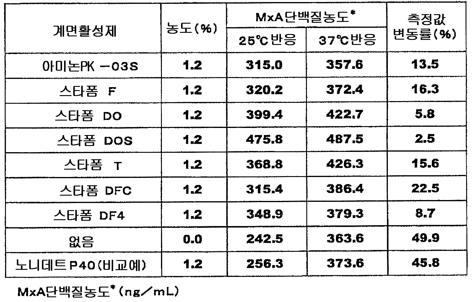
As shown in Table 1, nonidet P40 (International Publication No. 2008/053973) is known for use in the measurement of MxA protein when a surfactant is not used (+ 49.9%) with respect to the measured value variation. Compared to (+ 45.8%) when using a pamphlet, it turns out that when fatty acid alkanolamide is used, the influence on the measured value by temperature is suppressed remarkably.
Example 2
Examination 2 of measurement value variation in MxA protein measurement (primary reaction)
Blood collected from an EDTA-2Na blood collection tube was used as a sample from five patients with viral infection confirmed and showing MxA protein positiveness. Whole blood samples were diluted 10-fold with a sample dilution liquid to obtain a measurement sample.
As the sample dilution liquid, the first reaction was carried out at 25 ° C and 37 ° C in the same manner as in Example 1 except that a sample dilution liquid containing 1.2% starform DO and a sample dilution liquid containing 1.2% nonidet P40 were used. The measured value fluctuation by reaction temperature was examined. The results are shown in the second table.

As shown in the second table, even when a whole blood sample was used, the rate of change of the measured value was 0? In the 1.2% starform DO. Within + 12%, 1.2% + 8% of Nonidet P40? It turns out that the influence on the measured value by reaction temperature is remarkably suppressed compared with 23%.
Example 3
Examination of measured value variation in MxA protein measurement 3 (second order reaction)
POD labeled antibody dilution buffer consisting of the following compositions was prepared.
Bis-Tris (manufactured by Donin Chemical Research Institute) (pH 7.0) 50 mmol / l
BSA (manufactured by InterGen) 0.1%
ProClean 300 (Sigma) 0.035%
Surfactants (Types and Concentrations Listed in Table 3)
4-AA (manufactured by Saikyo Chemical Co., Ltd.) 0.01%
The native MxA protein produced in [3] of Example 1 was diluted 20-fold with the sample dilution of [6] of Example 1 [using Nonidet P40 as "surfactant"), and the cells were stopped for 30 minutes. Solubilization was carried out, and further diluted to 8-fold to obtain a sample for measurement.
As a specimen, the POD-labeled anti-MxA protein antibody prepared in Example 5 as a POD-labeled antibody was prepared using the sample and the standard solution of each concentration prepared in [6] of Example 1, using the POD-labeled antibody dilution buffer. It measured according to the operation as described in [7] of Example 1 using the thing diluted 800-fold. However, primary reaction was performed at 25 degreeC and secondary reaction was performed at both temperature of 25 degreeC and 37 degreeC. The change rate of the measured value of the native MxA protein at the time of 25 degreeC reaction and 37 degreeC reaction was computed by the following formula (I). The results are shown in Table 3.
&Quot; (2) "
% Change = [native MxA protein concentration at 37 ° C reaction) / (native MxA protein concentration at 25 ° C reaction]-1] * 100 (I)
Comparative Example 2
The rate of change of the measured value according to the secondary reaction temperature was calculated in the same manner as in Example 3 except that the surfactant in the POD-labeled antibody dilution buffer of Example 3 was 0.1% Nonidet P40. The results are shown in the third table.
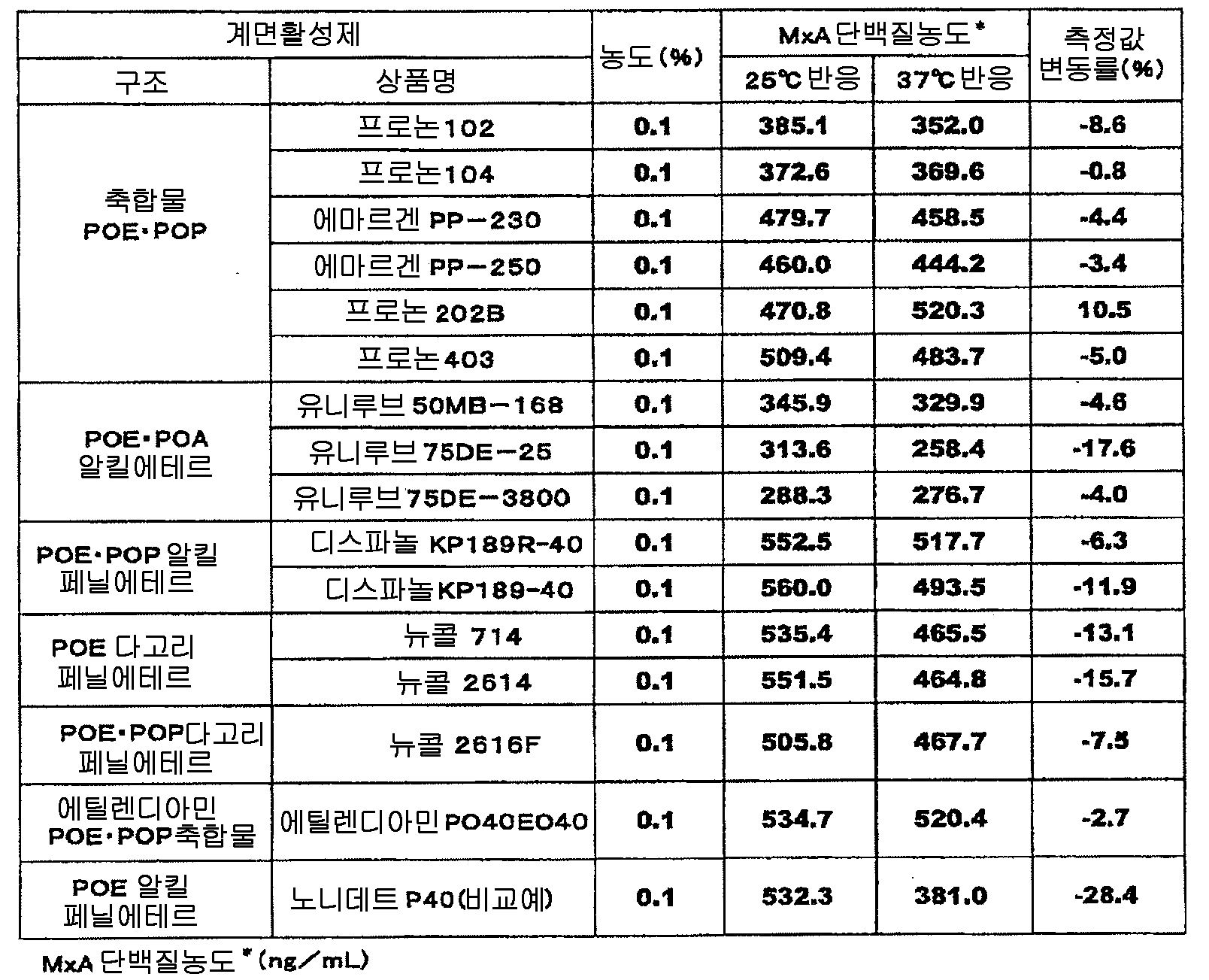
As shown in Table 3, the POE PPO condensate, POE PPO alkyl ether, and POE PPO alkylphenyl ether were compared with the case where nonidet P40 was used as the surfactant (-28.4%) for the measured value variation. When POE polycyclic phenyl ether, POE-POP polycyclic phenyl ether, and ethylenediamine POE-POP condensate are used, it turns out remarkably and it turns out that the influence on the measured value of reaction temperature is suppressed remarkably.
Example 4
Examination of measured value fluctuation in MxA protein measurement 4 (second order reaction)
The blood collected using the EDTA-2Na blood collection tube from 4 patients with viral infection confirmed and showing MxA protein positive was used as a sample. The whole blood sample was diluted 10-fold with the sample dilution liquid of [6] of Example 1 [But using nonidet P40 (1.2%) as a "surfactant", and it was set as the sample for measurement.
In the POD-labeled antibody dilution buffer of Example 3, it carried out similarly to Example 3 using the POD-labeled antibody dilution buffer which contains 0.1% phenone 403, 0.1% phenone 102, and 0.1% nonidet P40 as surfactant, respectively. The measurement was carried out (however, the first reaction was carried out at 25 ° C. and the second reaction was carried out at both temperatures of 25 ° C. and 37 ° C.), and the variation in the measured value due to the reaction temperature was examined. The results are shown in the fourth table.
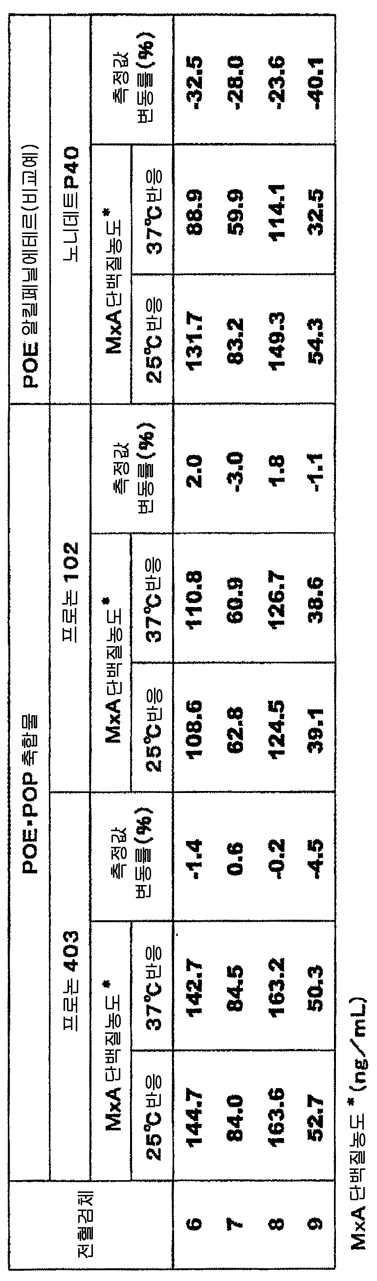
As shown in Table 4, even when whole blood samples were used, the rate of change of the measured value was -4.5% in Pronone 403? + 0.6%, -3.0% at Fronon 102? + 2.0%, -40.1% of Nonidet P40? Compared with -23.6%, it turns out that the influence on the measured value of reaction temperature is remarkably suppressed.
Example 5
Examination of measured value variation in MxA protein measurement 5 (second order reaction)
Blood collected using an EDTA-2Na blood collection tube from five patients with viral infection confirmed and MxA protein positive was used as a sample. The whole blood sample was diluted 10-fold with the sample dilution liquid of [6] of Example 1 [But using nonidet P40 (1.2%) as a "surfactant", and it was set as the sample for measurement.
In the POD-labeled antibody dilution buffer of Example 3, 0.1% emargen PP-250, 0.1% unilube 50MB-168, 0.1% dispanol KP189R-40, 0.1% nucol 2616F, 0.1% ethylenediamine PO40EO40 as surfactant Using a POD-labeled antibody dilution buffer each containing the same as in Example 3, the measurement was carried out (however, the first reaction was carried out at 25 ° C and the second reaction was carried out at both temperatures of 25 ° C and 37 ° C. ), The variation of the measured value due to the reaction temperature was examined. The results are shown in Table 5.

As shown in Table 5, even when a whole blood sample was used, the rate of change of the measured value was -1% in the Emargen PP-250? -11%, 0 in UniLube 50MB-168? -8%, -5 in dispanol KP189R-40? -15%, -7 in Newcall 2616F? -15%, in ethylenediamine PO40EO40 -1? -12%, -33% of Nonidet P40? Compared with -20%, it turns out that the influence on the measured value of reaction temperature is remarkably suppressed.
Example 6
(A) the following components: The kit for measuring the MxA protein which consists of (g) was prepared.
(a) anti-MxA protein antibody immobilization plate
Example 1 According to the method of [4], anti-MxA protein antibody immobilization plate was prepared by the following method. First, a solution of 5 μg / mL of the anti MxA protein monoclonal antibody KM1135, 100 mmol / L phosphate buffer (pH 7.5) containing 100 mmol / L sodium chloride, was prepared in a 96-well microtiter plate [Narzen Nuk International (Nalge Nunc International Co., Ltd.) was dispensed in an amount of 100 µl / well, and the supernatant was suctioned off after standing for 3 days. Subsequently, 300 µL of a 100 mmol / L phosphate buffer (pH 7.5) solution containing 1 mmol Blockace (manufactured by Dainippon Pharmaceutical Co., Ltd.) containing 100 mmol / L sodium chloride was dispensed into each well, and stopped at room temperature overnight to stop blocking. It was. After the blocking solution was removed, the resultant was washed with PBS, dried in a vacuum dryer for 3 days, and an anti-MxA protein monoclonal antibody solidification plate was prepared.
(b) sample dilution
The sample dilution liquid which consists of the following compositions was prepared.
Tris (manufactured by Donin Chemical Research Institute) (pH 8.5) 0.1 mol / l
CHAPS (manufactured by Donin Chemical Research Institute) 2.5%
Starform DO 1.2%
Sodium chloride 0.1 mol / l
BSA (Biochemical Industry) 0.1%
Sodium azide 0.1%
(c) POD-labeled anti MxA protein antibody solution
Example 1 The POD labeled anti MxA protein antibody KM1124 prepared by the method of [5] was diluted 800-fold with the dilution solution of the POD labeled anti MxA protein antibody which consists of the following compositions, and the POD labeled anti MxA protein antibody solution was prepared.
Bis-Tris (manufactured by Donin Chemical Research Institute) (pH 7.0) 0.05 mol / l
Pronon 202B 0.1%
Sodium Chloride 50 mmol / l
BSA (Biochemical Industry) 0.1%
4-AA (manufactured by Saikyo Chemical Co., Ltd.) 0.01%
ProClean 300 0.035%
(d) colorant
TMBlue (cerolologic company make)
(e) reaction stopper
0.5 mol / l sulfuric acid aqueous solution
(f) cleaning liquid
The following washing liquids were prepared.
Phosphate buffer (pH 7.2) 10 mmol / l
Tween 20 0.05%
Sodium chloride 0.15 mol / l
(g) Standards and Standards
Example 1 The recombinant MxA protein prepared in [2] was diluted with phosphate buffer and then lyophilized to prepare a reference material of MxA protein.
The prepared lyophilized standard substance was diluted with the sample dilution of (b) above, and MxA protein solution of each concentration of 0 (sample dilution only), 0.375, 0.75, 1.5, 3, 6, 12, 24 ng / ml was prepared. It prepared and these were made into the standard liquid.
[Comparative Example 3]
(A) the following components: The kit for measuring the MxA protein which consists of (g) was prepared.
(a) anti-MxA protein antibody immobilization plate
The anti-MxA protein antibody immobilization plate same as (a) of Example 6.
(b) sample dilution
The sample dilution liquid which consists of the following compositions was prepared.
HEPES (manufactured by Donin Chemical Research Institute) (pH 8.0) 0.1 mol / l
CHAPS (manufactured by Tonin Chemical Research Institute) 4.9%
Nonidet P40 1.2%
Sodium chloride 0.1 mol / l
BSA (Biochemical Industry) 0.1%
Sodium azide 0.1%
(c) POD-labeled anti MxA protein antibody solution
Example 1 The POD labeled anti MxA protein antibody KM1124 prepared by the method of [5] was diluted 800-fold with the dilution of the POD labeled anti MxA protein antibody which consists of the following compositions, and the POD labeled anti MxA protein antibody solution was prepared.
Bis-Tris (manufactured by Donin Chemical Research Institute) (pH 6.0) 50 mmol / l
Nonidet P40 0.1%
Sodium Chloride 50 mmol / l
BSA (Biochemical Industry) 0.1%
4-AA (manufactured by Saikyo Chemical Co., Ltd.) 0.01%
ProClean 300 0.035%
(d) colorant
TMBlue (cerolologic company make)
(e) reaction stopper
0.5 mol / l aqueous sulfuric acid solution
(f) cleaning liquid
The following washing liquids were prepared.
Phosphate buffer (pH 7.2) 10 mmol / l
Tween 20 0.05%
Sodium chloride 0.15 mol / l
(g) Standards and Standards
Example 1 The recombinant MxA protein prepared in [2] was diluted with phosphate buffer, and then lyophilized to prepare a reference material of MxA protein.
The prepared lyophilized standard substance was diluted with the sample dilution of (b) above, and MxA protein solution of each concentration of 0 (sample dilution only), 0.375, 0.75, 1.5, 3, 6, 12, 24 ng / ml was prepared. It prepared and these were made into the standard liquid.
Example 7
As a sample for measurement, the cells prepared in Example 1 [3] were diluted 20 times with the sample dilution solution of Example 6, stopped for 30 minutes, and then further diluted 8 times with the sample dilution solution, and used as a kit. The measurement was performed using the kit of Example 6 according to the following procedure.
To the anti-MxA protein antibody immobilization plate of Example 6 (a), 100 µl of each standard solution prepared in (g) was added, and predetermined temperatures (each temperature at 22 ° C, 25 ° C, 28 ° C, 30 ° C and 32 ° C) were added. The reaction was carried out for 1 hour at which MxA protein was bound to the antibody. After removing the reaction solution, the plate was washed five times with 400 µl of the washing solution in (f). Subsequently, 100 µL of the POD-labeled anti-MxA protein antibody solution prepared in (c) was added and reacted at a predetermined temperature (each temperature at 22 ° C, 25 ° C, 28 ° C, 30 ° C and 32 ° C) for 0.5 hour. After the reaction, the reaction solution was removed, and the plate was washed five times with 400 µl of the washing solution in (f). Subsequently, in a dark place, 100 µl of the color developing solution of (d) containing 0.05% tetramethylbenzidine and hydrogen peroxide was added and reacted at room temperature for 10 minutes, and then 100 µl of the reaction stopper of (e) was added, and Incubated for 10 minutes at, to stop the reaction. The absorbance at 450 nm of the reaction solution was measured by a plate reader, and a calibration curve showing the relationship between the MxA protein concentration and the absorbance was prepared.
[Comparative Example 4]
Except using the kit of Comparative Example 3 instead of the kit of Example 6, it measured by the same method as Example 7, and produced the analytical curve which shows the relationship of MxA protein concentration and absorbance.
The result of the measurement of Example 7 and the comparative example 4 is shown in a 6th table | surface.

The sixth table describes both the measured value in the reaction at each reaction temperature and the relative value of the measured value at each temperature when the measured value in the reaction at 22 ° C. is 100. . The closer the relative value is to 100, the less likely the measurement is to be affected by the reaction temperature. As is clear from the sixth table, the measurement method of Example 7 using the kit of Example 6 containing starform DO (fatty acid alkanolamide) and phenone 202B (POE-POP condensate) is nonidet P. The measurement was remarkably stable compared with the measuring method of Comparative Example 4 using the kit of Comparative Example 3 containing -40 (POE alkylphenyl ether). From this, it turns out that the measuring method of this invention is a method which is not influenced by reaction temperature well.
Industrial availability
The present invention provides a method for measuring a component to be measured in a specimen and a kit for measurement useful for diagnosing an infection.
Claims (28)
A method of reacting a labeled second antibody in the presence of a polyoxyethylene-based nonionic surfactant.
The fatty acid alkanolamide is a fatty acid diethanolamide.
Polyoxyethylene nonionic polyoxyethylene nonionic surfactant selected from the group consisting of polyoxyethylene polyoxypropylene condensates, polyoxyethylene polyoxypropylene alkyl ethers, and ethylenediamine polyoxyethylene polyoxypropylene condensates The method is a surfactant.
The fatty acid alkanolamides are fatty acid diethanolamides, and the polyoxyethylene-based nonionic surfactants are polyoxyethylene polyoxypropylene condensates, polyoxyethylene polyoxypropylene alkyl ethers, and ethylenediamine polyoxyethylene polyoxypropylene condensates. The method is a polyoxyethylene-based nonionic surfactant selected from the group consisting of.
A method for reacting a component to be measured in a sample with a bile acid derivative to react with a first antibody bound to the component to be measured.
The bile acid derivative is a bile acid derivative having an amphoteric surfactant action.
Bile acid derivatives having an amphoteric surfactant action are 3-[(3-colamidepropyl) dimethylammonio] propanesulfonate or 3-[(3-colamidepropyl) dimethylammonio] -2-hydroxypropanesulfonate How to be.
The bile acid derivative is a bile acid derivative having a nonionic surfactant action.
The bile acid derivative having a nonionic surfactant action is N, N-bis (3-gluconamide propyl) colamide or N, N-bis (3-D-gluconamide propyl) deoxycolamide.
The measurement method in which the first antibody is immobilized on an insoluble carrier.
The method is whole blood.
The component to be measured is an MxA protein.
A kit wherein the second reagent further contains a polyoxyethylene-based nonionic surfactant.
And the fatty acid alkanolamide is a fatty acid diethanolamide.
Polyoxyethylene nonionic polyoxyethylene nonionic surfactant selected from the group consisting of polyoxyethylene polyoxypropylene condensates, polyoxyethylene polyoxypropylene alkyl ethers, and ethylenediamine polyoxyethylene polyoxypropylene condensates A kit that is a surfactant.
The fatty acid alkanolamides are fatty acid diethanolamides, and the polyoxyethylene-based nonionic surfactants are polyoxyethylene polyoxypropylene condensates, polyoxyethylene polyoxypropylene alkyl ethers, and ethylenediamine polyoxyethylene polyoxypropylene condensates. A kit which is a polyoxyethylene-based nonionic surfactant selected from the group consisting of.
A kit wherein the first reagent further comprises a bile acid derivative.
A kit wherein the bile acid derivative is a bile acid derivative with amphoteric surfactant activity.
Bile acid derivatives having an amphoteric surfactant action are 3-[(3-colamidepropyl) dimethylammonio] propanesulfonate or 3-[(3-colamidepropyl) dimethylammonio] -2-hydroxypropanesulfonate Kit.
A kit wherein the bile acid derivative is a bile acid derivative having a nonionic surfactant action.
A kit wherein the bile acid derivative having nonionic surfactant action is N, N-bis (3-gluconamide propyl) colamide or N, N-bis (3-D-gluconamide propyl) deoxycolamide.
The kit wherein the first antibody is immobilized on an insoluble carrier.
Kit in which the sample is whole blood.
Kit wherein the component to be measured is an MxA protein.
Applications Claiming Priority (3)
| Application Number | Priority Date | Filing Date | Title |
|---|---|---|---|
| JPJP-P-2009-250639 | 2009-10-30 | ||
| JP2009250639 | 2009-10-30 | ||
| PCT/JP2010/069027 WO2011052620A1 (en) | 2009-10-30 | 2010-10-27 | Method for assaying component to be assayed in specimen and assay kit |
Related Child Applications (1)
| Application Number | Title | Priority Date | Filing Date |
|---|---|---|---|
| KR1020177016794A Division KR101921012B1 (en) | 2009-10-30 | 2010-10-27 | Method and kit for measuring component to be assayed in specimen |
Publications (2)
| Publication Number | Publication Date |
|---|---|
| KR20120101421A true KR20120101421A (en) | 2012-09-13 |
| KR101808872B1 KR101808872B1 (en) | 2017-12-13 |
Family
ID=43922045
Family Applications (2)
| Application Number | Title | Priority Date | Filing Date |
|---|---|---|---|
| KR1020177016794A KR101921012B1 (en) | 2009-10-30 | 2010-10-27 | Method and kit for measuring component to be assayed in specimen |
| KR1020127013819A KR101808872B1 (en) | 2009-10-30 | 2010-10-27 | Method and kit for measuring component to be assayed in specimen |
Family Applications Before (1)
| Application Number | Title | Priority Date | Filing Date |
|---|---|---|---|
| KR1020177016794A KR101921012B1 (en) | 2009-10-30 | 2010-10-27 | Method and kit for measuring component to be assayed in specimen |
Country Status (8)
| Country | Link |
|---|---|
| US (1) | US9274125B2 (en) |
| EP (2) | EP2495565B9 (en) |
| JP (2) | JP5764493B2 (en) |
| KR (2) | KR101921012B1 (en) |
| CN (3) | CN104034882B (en) |
| CA (2) | CA2779191C (en) |
| HK (1) | HK1212445A1 (en) |
| WO (1) | WO2011052620A1 (en) |
Cited By (1)
| Publication number | Priority date | Publication date | Assignee | Title |
|---|---|---|---|---|
| US10571479B2 (en) | 2013-02-06 | 2020-02-25 | Fujirebio Inc. | Vitamin D measurement method and measurement kit |
Families Citing this family (13)
| Publication number | Priority date | Publication date | Assignee | Title |
|---|---|---|---|---|
| JP6208473B2 (en) * | 2012-06-20 | 2017-10-04 | アークレイ株式会社 | Method for processing samples containing blood components |
| WO2016121821A1 (en) * | 2015-01-27 | 2016-08-04 | Jsr株式会社 | Separation method, detection method, signal measurement method, method for determining disease, method for evaluating drug efficacy, kit, liquid composition, and analyte diluent |
| US20180291424A1 (en) * | 2015-05-20 | 2018-10-11 | Jsr Corporation | Separation method, detection method, signal measurement method, disease determination method, drug efficacy assessment method, kit, liquid composition, and specimen diluent |
| US9835622B2 (en) | 2015-08-04 | 2017-12-05 | Abaxis, Inc. | Signal amplification in solution-based plasmonic specific-binding partner assays |
| IL268238B (en) * | 2017-01-30 | 2022-09-01 | Abaxis Llc | Solution-based plasmon specific-binding partner assays and metallic nanostructures |
| WO2018221544A1 (en) * | 2017-05-31 | 2018-12-06 | 協和メデックス株式会社 | Measurement method for fibroblast growth factor–23, measurement reagent, and measurement kit |
| JP7007862B2 (en) * | 2017-10-31 | 2022-01-25 | アークレイ株式会社 | Chemiluminescent reagent composition and chemiluminescent reagent kit |
| JP7156807B2 (en) * | 2018-02-28 | 2022-10-19 | デンカ株式会社 | Method for improving accuracy of test measurements and additive therefor |
| CN108593641B (en) * | 2018-04-09 | 2021-12-03 | 桂林优利特医疗电子有限公司 | Kit and method for quantitatively detecting substance to be detected in whole blood sample |
| CN108445233A (en) * | 2018-04-10 | 2018-08-24 | 安徽金标点生物科技有限公司 | A kind of antiviral protein MxA diagnostic kits and preparation method thereof |
| CN109239334A (en) * | 2018-09-10 | 2019-01-18 | 吉林大学 | Settling time resolved fluorometric immunochromatographyassay assay MxA kit |
| TWI842764B (en) * | 2018-11-09 | 2024-05-21 | 日商積水醫療股份有限公司 | Methods for suppressing abnormal detections in immunoassays conducted with an automatic analyzer, detection methods, immunoassay reagents, and immunoassay reagent kits |
| CN110988363A (en) * | 2019-12-31 | 2020-04-10 | 苏州康和顺医疗技术有限公司 | Detection reagent for antiviral protein MxA and preparation method thereof |
Family Cites Families (18)
| Publication number | Priority date | Publication date | Assignee | Title |
|---|---|---|---|---|
| US4208479A (en) * | 1977-07-14 | 1980-06-17 | Syva Company | Label modified immunoassays |
| JPH0699292B2 (en) * | 1985-06-25 | 1994-12-07 | ライオン株式会社 | Oral composition |
| EP0318081B1 (en) | 1987-11-27 | 1991-07-24 | Akzo N.V. | Stabilization of antibodies |
| US5352583A (en) | 1989-10-02 | 1994-10-04 | Teijin Limited | Human tissue plasminogen activator-human plasminogen activator inhibitor complex immunoassay and kit therefor |
| JP2702354B2 (en) * | 1992-05-11 | 1998-01-21 | 帝人株式会社 | Marker of lung tissue damage in blood |
| JPH062417A (en) * | 1992-06-17 | 1994-01-11 | Aica Kogyo Co Ltd | Method of constructing floor coated with anti-static paint |
| JP3228791B2 (en) | 1992-08-19 | 2001-11-12 | 日本化薬株式会社 | Measuring method of antigen or antibody in sample |
| ATE230418T1 (en) | 1994-08-08 | 2003-01-15 | Kyowa Hakko Kogyo Kk | MONOCLONAL ANTIBODIES AGAINST THE HUMAN MX PROTEIN MXA |
| JP4524446B2 (en) * | 2002-03-31 | 2010-08-18 | 株式会社シノテスト | Glycolipid antigen activator and activation method, and method and reagent kit for measuring anti-glycolipid antibody in a sample |
| US20060142175A1 (en) * | 2003-02-03 | 2006-06-29 | Elke Haiss | Washing agent and fabric softener formulations |
| WO2005052583A1 (en) * | 2003-11-28 | 2005-06-09 | Olympus Corporation | Method of detecting substance to be analyzed |
| US20060127421A1 (en) * | 2004-12-09 | 2006-06-15 | Celgene Corporation | Treatment using D-threo methylphenidate |
| WO2006073073A1 (en) | 2005-01-07 | 2006-07-13 | Yamasa Corporation | Stabilized ornithine transcarbamylase and immunoassay method for ornithine transcarbamylase using the same |
| JP5317040B2 (en) | 2006-10-17 | 2013-10-16 | 公益財団法人東京都医学総合研究所 | Method for immunological measurement of components in urine and reagent for use in the method |
| JP5557450B2 (en) | 2006-11-02 | 2014-07-23 | 協和メデックス株式会社 | Immunoassay for components to be measured |
| CN101017172A (en) * | 2007-03-08 | 2007-08-15 | 湖南景达生物工程有限公司 | Method for measuring all-core antigen of hepatitis C virus |
| JP2009186359A (en) | 2008-02-07 | 2009-08-20 | Tanaka Kikinzoku Kogyo Kk | Specimen processing reagent composition for immunological measurement |
| JP6099292B1 (en) * | 2015-07-30 | 2017-03-22 | 浩康 佐々木 | Power transmission device and magnetic resonance power transmission system including the same |
-
2010
- 2010-10-27 KR KR1020177016794A patent/KR101921012B1/en active IP Right Grant
- 2010-10-27 CN CN201410235359.7A patent/CN104034882B/en not_active Expired - Fee Related
- 2010-10-27 CN CN2010800596039A patent/CN102687015A/en active Pending
- 2010-10-27 CN CN201611218081.8A patent/CN106959367B/en not_active Expired - Fee Related
- 2010-10-27 EP EP10826752.7A patent/EP2495565B9/en active Active
- 2010-10-27 EP EP15157275.7A patent/EP2919011B1/en active Active
- 2010-10-27 US US13/501,318 patent/US9274125B2/en not_active Expired - Fee Related
- 2010-10-27 KR KR1020127013819A patent/KR101808872B1/en active IP Right Grant
- 2010-10-27 CA CA2779191A patent/CA2779191C/en not_active Expired - Fee Related
- 2010-10-27 JP JP2011538449A patent/JP5764493B2/en active Active
- 2010-10-27 CA CA2978998A patent/CA2978998C/en not_active Expired - Fee Related
- 2010-10-27 WO PCT/JP2010/069027 patent/WO2011052620A1/en active Application Filing
-
2012
- 2012-11-16 HK HK16100292.7A patent/HK1212445A1/en not_active IP Right Cessation
-
2014
- 2014-09-11 JP JP2014185247A patent/JP6014091B2/en active Active
Cited By (1)
| Publication number | Priority date | Publication date | Assignee | Title |
|---|---|---|---|---|
| US10571479B2 (en) | 2013-02-06 | 2020-02-25 | Fujirebio Inc. | Vitamin D measurement method and measurement kit |
Also Published As
| Publication number | Publication date |
|---|---|
| HK1212445A1 (en) | 2016-06-10 |
| EP2495565A1 (en) | 2012-09-05 |
| JP2015007652A (en) | 2015-01-15 |
| EP2919011A1 (en) | 2015-09-16 |
| JP6014091B2 (en) | 2016-10-25 |
| EP2495565B1 (en) | 2017-09-20 |
| CA2779191A1 (en) | 2011-05-05 |
| US20120219966A1 (en) | 2012-08-30 |
| CN102687015A (en) | 2012-09-19 |
| CN104034882A (en) | 2014-09-10 |
| KR101921012B1 (en) | 2018-11-21 |
| CA2978998A1 (en) | 2011-05-05 |
| EP2495565B9 (en) | 2018-03-21 |
| EP2919011B1 (en) | 2018-10-03 |
| EP2495565A4 (en) | 2013-05-01 |
| KR101808872B1 (en) | 2017-12-13 |
| CN106959367B (en) | 2019-06-14 |
| US9274125B2 (en) | 2016-03-01 |
| CA2978998C (en) | 2019-11-26 |
| CN104034882B (en) | 2017-04-12 |
| WO2011052620A1 (en) | 2011-05-05 |
| CN106959367A (en) | 2017-07-18 |
| CA2779191C (en) | 2018-01-30 |
| KR20170075017A (en) | 2017-06-30 |
| JP5764493B2 (en) | 2015-08-19 |
| JPWO2011052620A1 (en) | 2013-03-21 |
Similar Documents
| Publication | Publication Date | Title |
|---|---|---|
| US9274125B2 (en) | Method and kit for measuring component in the presence of fatty acid alkanolamide or nonionic polyoxyethylene surfactant | |
| US8257935B2 (en) | Method of immunoassaying a component to be measured | |
| KR101233837B1 (en) | Method of immunoassay having nonspecific reaction inhibited and reagent therefor | |
| JP2022058732A (en) | Method for measuring fibroblast growth factor-23, measurement reagent and measuring kit | |
| JP2007225603A (en) | Method and reagent for immunoassay of measured object in sample | |
| JP7281092B2 (en) | Method for measuring exosomes in sample, measurement reagent and measurement kit | |
| WO2018079674A1 (en) | Method for selecting subject needing treatment for dyslipidemia and reagent for such selection | |
| WO2017159473A1 (en) | Method for assessing likelihood of chronic kidney disease patient developing hyperphosphatemia | |
| JP2015212237A (en) | Novel peptide and method of use thereof |
Legal Events
| Date | Code | Title | Description |
|---|---|---|---|
| A201 | Request for examination | ||
| E902 | Notification of reason for refusal | ||
| A107 | Divisional application of patent | ||
| E701 | Decision to grant or registration of patent right | ||
| GRNT | Written decision to grant |