KR20120069663A - Pluripotent stem cell that can be isolated from body tissue - Google Patents
Pluripotent stem cell that can be isolated from body tissue Download PDFInfo
- Publication number
- KR20120069663A KR20120069663A KR1020127003879A KR20127003879A KR20120069663A KR 20120069663 A KR20120069663 A KR 20120069663A KR 1020127003879 A KR1020127003879 A KR 1020127003879A KR 20127003879 A KR20127003879 A KR 20127003879A KR 20120069663 A KR20120069663 A KR 20120069663A
- Authority
- KR
- South Korea
- Prior art keywords
- cells
- cell
- muse
- pluripotent stem
- derived
- Prior art date
Links
Images


Classifications
-
- C—CHEMISTRY; METALLURGY
- C12—BIOCHEMISTRY; BEER; SPIRITS; WINE; VINEGAR; MICROBIOLOGY; ENZYMOLOGY; MUTATION OR GENETIC ENGINEERING
- C12N—MICROORGANISMS OR ENZYMES; COMPOSITIONS THEREOF; PROPAGATING, PRESERVING, OR MAINTAINING MICROORGANISMS; MUTATION OR GENETIC ENGINEERING; CULTURE MEDIA
- C12N5/00—Undifferentiated human, animal or plant cells, e.g. cell lines; Tissues; Cultivation or maintenance thereof; Culture media therefor
- C12N5/06—Animal cells or tissues; Human cells or tissues
- C12N5/0602—Vertebrate cells
- C12N5/0607—Non-embryonic pluripotent stem cells, e.g. MASC
-
- A—HUMAN NECESSITIES
- A61—MEDICAL OR VETERINARY SCIENCE; HYGIENE
- A61K—PREPARATIONS FOR MEDICAL, DENTAL OR TOILETRY PURPOSES
- A61K35/00—Medicinal preparations containing materials or reaction products thereof with undetermined constitution
- A61K35/12—Materials from mammals; Compositions comprising non-specified tissues or cells; Compositions comprising non-embryonic stem cells; Genetically modified cells
-
- A—HUMAN NECESSITIES
- A61—MEDICAL OR VETERINARY SCIENCE; HYGIENE
- A61P—SPECIFIC THERAPEUTIC ACTIVITY OF CHEMICAL COMPOUNDS OR MEDICINAL PREPARATIONS
- A61P1/00—Drugs for disorders of the alimentary tract or the digestive system
-
- A—HUMAN NECESSITIES
- A61—MEDICAL OR VETERINARY SCIENCE; HYGIENE
- A61P—SPECIFIC THERAPEUTIC ACTIVITY OF CHEMICAL COMPOUNDS OR MEDICINAL PREPARATIONS
- A61P1/00—Drugs for disorders of the alimentary tract or the digestive system
- A61P1/04—Drugs for disorders of the alimentary tract or the digestive system for ulcers, gastritis or reflux esophagitis, e.g. antacids, inhibitors of acid secretion, mucosal protectants
-
- A—HUMAN NECESSITIES
- A61—MEDICAL OR VETERINARY SCIENCE; HYGIENE
- A61P—SPECIFIC THERAPEUTIC ACTIVITY OF CHEMICAL COMPOUNDS OR MEDICINAL PREPARATIONS
- A61P1/00—Drugs for disorders of the alimentary tract or the digestive system
- A61P1/16—Drugs for disorders of the alimentary tract or the digestive system for liver or gallbladder disorders, e.g. hepatoprotective agents, cholagogues, litholytics
-
- A—HUMAN NECESSITIES
- A61—MEDICAL OR VETERINARY SCIENCE; HYGIENE
- A61P—SPECIFIC THERAPEUTIC ACTIVITY OF CHEMICAL COMPOUNDS OR MEDICINAL PREPARATIONS
- A61P1/00—Drugs for disorders of the alimentary tract or the digestive system
- A61P1/18—Drugs for disorders of the alimentary tract or the digestive system for pancreatic disorders, e.g. pancreatic enzymes
-
- A—HUMAN NECESSITIES
- A61—MEDICAL OR VETERINARY SCIENCE; HYGIENE
- A61P—SPECIFIC THERAPEUTIC ACTIVITY OF CHEMICAL COMPOUNDS OR MEDICINAL PREPARATIONS
- A61P13/00—Drugs for disorders of the urinary system
-
- A—HUMAN NECESSITIES
- A61—MEDICAL OR VETERINARY SCIENCE; HYGIENE
- A61P—SPECIFIC THERAPEUTIC ACTIVITY OF CHEMICAL COMPOUNDS OR MEDICINAL PREPARATIONS
- A61P13/00—Drugs for disorders of the urinary system
- A61P13/10—Drugs for disorders of the urinary system of the bladder
-
- A—HUMAN NECESSITIES
- A61—MEDICAL OR VETERINARY SCIENCE; HYGIENE
- A61P—SPECIFIC THERAPEUTIC ACTIVITY OF CHEMICAL COMPOUNDS OR MEDICINAL PREPARATIONS
- A61P13/00—Drugs for disorders of the urinary system
- A61P13/12—Drugs for disorders of the urinary system of the kidneys
-
- A—HUMAN NECESSITIES
- A61—MEDICAL OR VETERINARY SCIENCE; HYGIENE
- A61P—SPECIFIC THERAPEUTIC ACTIVITY OF CHEMICAL COMPOUNDS OR MEDICINAL PREPARATIONS
- A61P15/00—Drugs for genital or sexual disorders; Contraceptives
-
- A—HUMAN NECESSITIES
- A61—MEDICAL OR VETERINARY SCIENCE; HYGIENE
- A61P—SPECIFIC THERAPEUTIC ACTIVITY OF CHEMICAL COMPOUNDS OR MEDICINAL PREPARATIONS
- A61P17/00—Drugs for dermatological disorders
-
- A—HUMAN NECESSITIES
- A61—MEDICAL OR VETERINARY SCIENCE; HYGIENE
- A61P—SPECIFIC THERAPEUTIC ACTIVITY OF CHEMICAL COMPOUNDS OR MEDICINAL PREPARATIONS
- A61P17/00—Drugs for dermatological disorders
- A61P17/02—Drugs for dermatological disorders for treating wounds, ulcers, burns, scars, keloids, or the like
-
- A—HUMAN NECESSITIES
- A61—MEDICAL OR VETERINARY SCIENCE; HYGIENE
- A61P—SPECIFIC THERAPEUTIC ACTIVITY OF CHEMICAL COMPOUNDS OR MEDICINAL PREPARATIONS
- A61P19/00—Drugs for skeletal disorders
-
- A—HUMAN NECESSITIES
- A61—MEDICAL OR VETERINARY SCIENCE; HYGIENE
- A61P—SPECIFIC THERAPEUTIC ACTIVITY OF CHEMICAL COMPOUNDS OR MEDICINAL PREPARATIONS
- A61P21/00—Drugs for disorders of the muscular or neuromuscular system
-
- A—HUMAN NECESSITIES
- A61—MEDICAL OR VETERINARY SCIENCE; HYGIENE
- A61P—SPECIFIC THERAPEUTIC ACTIVITY OF CHEMICAL COMPOUNDS OR MEDICINAL PREPARATIONS
- A61P25/00—Drugs for disorders of the nervous system
-
- A—HUMAN NECESSITIES
- A61—MEDICAL OR VETERINARY SCIENCE; HYGIENE
- A61P—SPECIFIC THERAPEUTIC ACTIVITY OF CHEMICAL COMPOUNDS OR MEDICINAL PREPARATIONS
- A61P27/00—Drugs for disorders of the senses
- A61P27/02—Ophthalmic agents
-
- A—HUMAN NECESSITIES
- A61—MEDICAL OR VETERINARY SCIENCE; HYGIENE
- A61P—SPECIFIC THERAPEUTIC ACTIVITY OF CHEMICAL COMPOUNDS OR MEDICINAL PREPARATIONS
- A61P3/00—Drugs for disorders of the metabolism
-
- A—HUMAN NECESSITIES
- A61—MEDICAL OR VETERINARY SCIENCE; HYGIENE
- A61P—SPECIFIC THERAPEUTIC ACTIVITY OF CHEMICAL COMPOUNDS OR MEDICINAL PREPARATIONS
- A61P35/00—Antineoplastic agents
-
- A—HUMAN NECESSITIES
- A61—MEDICAL OR VETERINARY SCIENCE; HYGIENE
- A61P—SPECIFIC THERAPEUTIC ACTIVITY OF CHEMICAL COMPOUNDS OR MEDICINAL PREPARATIONS
- A61P37/00—Drugs for immunological or allergic disorders
- A61P37/02—Immunomodulators
-
- A—HUMAN NECESSITIES
- A61—MEDICAL OR VETERINARY SCIENCE; HYGIENE
- A61P—SPECIFIC THERAPEUTIC ACTIVITY OF CHEMICAL COMPOUNDS OR MEDICINAL PREPARATIONS
- A61P43/00—Drugs for specific purposes, not provided for in groups A61P1/00-A61P41/00
-
- A—HUMAN NECESSITIES
- A61—MEDICAL OR VETERINARY SCIENCE; HYGIENE
- A61P—SPECIFIC THERAPEUTIC ACTIVITY OF CHEMICAL COMPOUNDS OR MEDICINAL PREPARATIONS
- A61P7/00—Drugs for disorders of the blood or the extracellular fluid
-
- A—HUMAN NECESSITIES
- A61—MEDICAL OR VETERINARY SCIENCE; HYGIENE
- A61P—SPECIFIC THERAPEUTIC ACTIVITY OF CHEMICAL COMPOUNDS OR MEDICINAL PREPARATIONS
- A61P9/00—Drugs for disorders of the cardiovascular system
-
- C—CHEMISTRY; METALLURGY
- C12—BIOCHEMISTRY; BEER; SPIRITS; WINE; VINEGAR; MICROBIOLOGY; ENZYMOLOGY; MUTATION OR GENETIC ENGINEERING
- C12N—MICROORGANISMS OR ENZYMES; COMPOSITIONS THEREOF; PROPAGATING, PRESERVING, OR MAINTAINING MICROORGANISMS; MUTATION OR GENETIC ENGINEERING; CULTURE MEDIA
- C12N5/00—Undifferentiated human, animal or plant cells, e.g. cell lines; Tissues; Cultivation or maintenance thereof; Culture media therefor
- C12N5/06—Animal cells or tissues; Human cells or tissues
- C12N5/0602—Vertebrate cells
- C12N5/0603—Embryonic cells ; Embryoid bodies
- C12N5/0605—Cells from extra-embryonic tissues, e.g. placenta, amnion, yolk sac, Wharton's jelly
-
- C—CHEMISTRY; METALLURGY
- C12—BIOCHEMISTRY; BEER; SPIRITS; WINE; VINEGAR; MICROBIOLOGY; ENZYMOLOGY; MUTATION OR GENETIC ENGINEERING
- C12N—MICROORGANISMS OR ENZYMES; COMPOSITIONS THEREOF; PROPAGATING, PRESERVING, OR MAINTAINING MICROORGANISMS; MUTATION OR GENETIC ENGINEERING; CULTURE MEDIA
- C12N5/00—Undifferentiated human, animal or plant cells, e.g. cell lines; Tissues; Cultivation or maintenance thereof; Culture media therefor
- C12N5/06—Animal cells or tissues; Human cells or tissues
- C12N5/0602—Vertebrate cells
- C12N5/0696—Artificially induced pluripotent stem cells, e.g. iPS
-
- A—HUMAN NECESSITIES
- A61—MEDICAL OR VETERINARY SCIENCE; HYGIENE
- A61K—PREPARATIONS FOR MEDICAL, DENTAL OR TOILETRY PURPOSES
- A61K35/00—Medicinal preparations containing materials or reaction products thereof with undetermined constitution
- A61K35/12—Materials from mammals; Compositions comprising non-specified tissues or cells; Compositions comprising non-embryonic stem cells; Genetically modified cells
- A61K35/28—Bone marrow; Haematopoietic stem cells; Mesenchymal stem cells of any origin, e.g. adipose-derived stem cells
-
- A—HUMAN NECESSITIES
- A61—MEDICAL OR VETERINARY SCIENCE; HYGIENE
- A61K—PREPARATIONS FOR MEDICAL, DENTAL OR TOILETRY PURPOSES
- A61K35/00—Medicinal preparations containing materials or reaction products thereof with undetermined constitution
- A61K35/12—Materials from mammals; Compositions comprising non-specified tissues or cells; Compositions comprising non-embryonic stem cells; Genetically modified cells
- A61K35/48—Reproductive organs
- A61K35/50—Placenta; Placental stem cells; Amniotic fluid; Amnion; Amniotic stem cells
-
- C—CHEMISTRY; METALLURGY
- C12—BIOCHEMISTRY; BEER; SPIRITS; WINE; VINEGAR; MICROBIOLOGY; ENZYMOLOGY; MUTATION OR GENETIC ENGINEERING
- C12N—MICROORGANISMS OR ENZYMES; COMPOSITIONS THEREOF; PROPAGATING, PRESERVING, OR MAINTAINING MICROORGANISMS; MUTATION OR GENETIC ENGINEERING; CULTURE MEDIA
- C12N15/00—Mutation or genetic engineering; DNA or RNA concerning genetic engineering, vectors, e.g. plasmids, or their isolation, preparation or purification; Use of hosts therefor
- C12N15/09—Recombinant DNA-technology
- C12N15/11—DNA or RNA fragments; Modified forms thereof; Non-coding nucleic acids having a biological activity
- C12N15/113—Non-coding nucleic acids modulating the expression of genes, e.g. antisense oligonucleotides; Antisense DNA or RNA; Triplex- forming oligonucleotides; Catalytic nucleic acids, e.g. ribozymes; Nucleic acids used in co-suppression or gene silencing
- C12N15/1138—Non-coding nucleic acids modulating the expression of genes, e.g. antisense oligonucleotides; Antisense DNA or RNA; Triplex- forming oligonucleotides; Catalytic nucleic acids, e.g. ribozymes; Nucleic acids used in co-suppression or gene silencing against receptors or cell surface proteins
-
- C—CHEMISTRY; METALLURGY
- C12—BIOCHEMISTRY; BEER; SPIRITS; WINE; VINEGAR; MICROBIOLOGY; ENZYMOLOGY; MUTATION OR GENETIC ENGINEERING
- C12N—MICROORGANISMS OR ENZYMES; COMPOSITIONS THEREOF; PROPAGATING, PRESERVING, OR MAINTAINING MICROORGANISMS; MUTATION OR GENETIC ENGINEERING; CULTURE MEDIA
- C12N15/00—Mutation or genetic engineering; DNA or RNA concerning genetic engineering, vectors, e.g. plasmids, or their isolation, preparation or purification; Use of hosts therefor
- C12N15/09—Recombinant DNA-technology
- C12N15/63—Introduction of foreign genetic material using vectors; Vectors; Use of hosts therefor; Regulation of expression
- C12N15/79—Vectors or expression systems specially adapted for eukaryotic hosts
- C12N15/85—Vectors or expression systems specially adapted for eukaryotic hosts for animal cells
- C12N15/86—Viral vectors
-
- C—CHEMISTRY; METALLURGY
- C12—BIOCHEMISTRY; BEER; SPIRITS; WINE; VINEGAR; MICROBIOLOGY; ENZYMOLOGY; MUTATION OR GENETIC ENGINEERING
- C12N—MICROORGANISMS OR ENZYMES; COMPOSITIONS THEREOF; PROPAGATING, PRESERVING, OR MAINTAINING MICROORGANISMS; MUTATION OR GENETIC ENGINEERING; CULTURE MEDIA
- C12N2506/00—Differentiation of animal cells from one lineage to another; Differentiation of pluripotent cells
- C12N2506/13—Differentiation of animal cells from one lineage to another; Differentiation of pluripotent cells from connective tissue cells, from mesenchymal cells
- C12N2506/1346—Differentiation of animal cells from one lineage to another; Differentiation of pluripotent cells from connective tissue cells, from mesenchymal cells from mesenchymal stem cells
- C12N2506/1353—Differentiation of animal cells from one lineage to another; Differentiation of pluripotent cells from connective tissue cells, from mesenchymal cells from mesenchymal stem cells from bone marrow mesenchymal stem cells (BM-MSC)
-
- C—CHEMISTRY; METALLURGY
- C12—BIOCHEMISTRY; BEER; SPIRITS; WINE; VINEGAR; MICROBIOLOGY; ENZYMOLOGY; MUTATION OR GENETIC ENGINEERING
- C12N—MICROORGANISMS OR ENZYMES; COMPOSITIONS THEREOF; PROPAGATING, PRESERVING, OR MAINTAINING MICROORGANISMS; MUTATION OR GENETIC ENGINEERING; CULTURE MEDIA
- C12N2509/00—Methods for the dissociation of cells, e.g. specific use of enzymes
-
- C—CHEMISTRY; METALLURGY
- C12—BIOCHEMISTRY; BEER; SPIRITS; WINE; VINEGAR; MICROBIOLOGY; ENZYMOLOGY; MUTATION OR GENETIC ENGINEERING
- C12N—MICROORGANISMS OR ENZYMES; COMPOSITIONS THEREOF; PROPAGATING, PRESERVING, OR MAINTAINING MICROORGANISMS; MUTATION OR GENETIC ENGINEERING; CULTURE MEDIA
- C12N5/00—Undifferentiated human, animal or plant cells, e.g. cell lines; Tissues; Cultivation or maintenance thereof; Culture media therefor
- C12N5/06—Animal cells or tissues; Human cells or tissues
- C12N5/0602—Vertebrate cells
- C12N5/0652—Cells of skeletal and connective tissues; Mesenchyme
- C12N5/0662—Stem cells
- C12N5/0663—Bone marrow mesenchymal stem cells (BM-MSC)
-
- C—CHEMISTRY; METALLURGY
- C12—BIOCHEMISTRY; BEER; SPIRITS; WINE; VINEGAR; MICROBIOLOGY; ENZYMOLOGY; MUTATION OR GENETIC ENGINEERING
- C12N—MICROORGANISMS OR ENZYMES; COMPOSITIONS THEREOF; PROPAGATING, PRESERVING, OR MAINTAINING MICROORGANISMS; MUTATION OR GENETIC ENGINEERING; CULTURE MEDIA
- C12N5/00—Undifferentiated human, animal or plant cells, e.g. cell lines; Tissues; Cultivation or maintenance thereof; Culture media therefor
- C12N5/06—Animal cells or tissues; Human cells or tissues
- C12N5/0602—Vertebrate cells
- C12N5/0652—Cells of skeletal and connective tissues; Mesenchyme
- C12N5/0662—Stem cells
- C12N5/0667—Adipose-derived stem cells [ADSC]; Adipose stromal stem cells
-
- C—CHEMISTRY; METALLURGY
- C12—BIOCHEMISTRY; BEER; SPIRITS; WINE; VINEGAR; MICROBIOLOGY; ENZYMOLOGY; MUTATION OR GENETIC ENGINEERING
- C12N—MICROORGANISMS OR ENZYMES; COMPOSITIONS THEREOF; PROPAGATING, PRESERVING, OR MAINTAINING MICROORGANISMS; MUTATION OR GENETIC ENGINEERING; CULTURE MEDIA
- C12N5/00—Undifferentiated human, animal or plant cells, e.g. cell lines; Tissues; Cultivation or maintenance thereof; Culture media therefor
- C12N5/06—Animal cells or tissues; Human cells or tissues
- C12N5/0602—Vertebrate cells
- C12N5/0652—Cells of skeletal and connective tissues; Mesenchyme
- C12N5/0662—Stem cells
- C12N5/0668—Mesenchymal stem cells from other natural sources
Abstract
본 발명은 생체조직으로부터 다능성 줄기세포를 직접 얻는 방법의 제공 및 그렇게 해서 얻어진 다능성 줄기세포의 제공을 목적으로 하고 있고, 본 발명은 생체조직으로부터 단리(單離)할 수 있는 SSEA-3 양성의 다능성 줄기세포다.The present invention aims to provide a method for directly obtaining pluripotent stem cells from biological tissues and to provide pluripotent stem cells thus obtained, and the present invention provides SSEA-3 positive that can be isolated from biological tissues. Pluripotent stem cells.
Description
본 발명은 생체조직 유래의 다능성 줄기세포에 관한 것이다.The present invention relates to pluripotent stem cells derived from living tissue.
플라나리아(planarians)나 영원류(newts)는 몸의 일부가 절단된 후라도 몸을 재생할 수 있다. 이들의 높은 재생 능력은 간엽계(間葉系)에 존재하는 다능성(多能性) 줄기세포(adult pluripotent stem cell)의 존재에 따르고 있다. 한편, 인간 등의 고등생물에 있어서는 조직 재생 능력은 이들 동물에 비교해서 훨씬 낮다. 배반포(胚盤胞)의 내부세포 덩어리(ICM :Inner cell mass)는 다능성(多能性) 세포의 모임으로 인식되고 있어 외배엽(外胚葉), 중배엽(中胚葉), 내배엽(內胚葉)계의 세포로 분화되는 능력을 가진다. 그렇지만 발생이 진행함과 동시에 이러한 다능성(多能性)이 제한되어도 좋은 조직에 특화한 세포로 분화되어 간다.Planarians or newts can regenerate the body even after part of the body has been cut. Their high regenerative capacity depends on the presence of adult pluripotent stem cells present in the mesenchymal system. On the other hand, in higher organisms such as humans, tissue regeneration ability is much lower than those of these animals. The inner cell mass (ICM) of the blastocyst is recognized as a group of pluripotent cells, and the ectoderm, mesoderm, and endoderm system Has the ability to differentiate into cells. However, as development progresses, they are differentiated into cells specialized in tissues that may have limited pluripotency.
최근 조직 재생에 공헌할 수 있는 성인 줄기세포 또는 조직 줄기세포가 주목받고 있다. 그러나 포유류의 성체(成體)에 플라나리아(planarians)나 영원류(newt s)와 같이 다능성(多能性;pluripotent) 줄기세포가 존재할 것인지 아닌지는 불분명했다.Recently, adult stem cells or tissue stem cells that can contribute to tissue regeneration are attracting attention. However, it was unclear whether or not pluripotent stem cells, such as planarians and newts, exist in adult mammals.
성체(成體)로 얻을 수 있는 분화 능력을 갖는 세포로서 예를 들면 뼈, 연골, 지방세포, 신경세포, 골격근육 등으로의 분화 능력을 갖는 골수간엽계(骨髓間葉系) 세포분획(MSC:Bone marrow stromal cell)이 보고되어 있다(비특허문헌 1 및 2을 참조). 그렇지만 골수간엽계(骨髓間葉系) 세포분획(細胞分劃)은 다양한 세포를 포함하는 세포군이며, 그 분화 능력은 다양하면서 본체가 분명하지 않고, 또 특정한 세포로 분화되도록 하기 위해서 특정한 화합물에 의한 자극이나 유전자도입 등이 필요하여 분화 유도시스템을 구축할 필요가 있었다.Bone marrow mesenchymal cell fraction (MSC) having differentiation ability to be obtained as an adult, for example, to differentiation into bone, cartilage, adipocytes, nerve cells, skeletal muscle, etc. : Bone marrow stromal cells) have been reported (see Non-Patent
게다가 성체 유래의 다능성 줄기세포로서 iPS세포(induced pluripotent st em cell; 특허문헌 1, 특허문헌 2, 비특허문헌 3등을 참조)가 보고되어 있었다. 그렇지만 iPS세포의 수립에는 간엽계(間葉系) 세포인 피부(皮膚) 섬유아세포(纖維芽細胞) 분획(dermal fibroblast)에 특정한 유전자나 특정한 화합물을 체세포(體細胞)에 도입한다고 하는 특정한 물질을 채용한 유도조작이 필요하였다.In addition, iPS cells (induced pluripotent st em cells; see
본 발명은 생체조직으로부터 다능성(多能性) 줄기세포를 직접 얻는 방법의 제공 및 그 방법에 의해 얻어진 다능성 줄기세포의 제공을 목적으로 한다.An object of the present invention is to provide a method for directly obtaining pluripotent stem cells from living tissue and to provide a pluripotent stem cell obtained by the method.
본 발명자들은 골수간엽계(骨髓間葉系) 세포분획(MSC:Bone marrow stromal cell)에 관한 연구 과정에서 무처리(無處理)의 인간 MSC세포로부터 지극히 낮은 빈도로 특징적인 세포덩어리가 형성되는 것을 찾아냈다. 초기 세포덩어리의 외견(外見)은 ES세포를 매우 닮아 있었다. 그렇지만 ES세포와는 달리 무한증식을 하지 않고 어떤 일정한 기간인 크기에 도달하면 증식을 정지하고, 더구나 털, 색소세포 등의 각종 세포를 포함하는 불균일한 집단으로 되었다. 또한 이 세포덩어리에 대해서 면역세포화학(immunocytochemistry)을 한바, 외배엽(外胚葉), 중배엽(中胚葉) 및 내배엽(內胚葉) 마커(makers)에 각각 양성의 세포가 세포덩어리 내에 혼재해서 검출되었다. 본 발명자 들은 이 결과보다 무처리(無處理)의 통상으로 유지ㆍ배양되고 있는(naive) 인간 MSC세포 분획 중에 다능성 세포에 상당하는 세포가 존재할 가능성을 고려해 더욱 예의 검토를 하였다.The present inventors have found that in the study of bone marrow stromal cells (MSCs), characteristic cell masses are formed at extremely low frequency from untreated human MSC cells. I found it. The appearance of early cell masses was very similar to ES cells. However, unlike ES cells, proliferation stops when reaching a certain period of time without infinite growth, and furthermore, it becomes a heterogeneous group including various cells such as hair and pigment cells. In addition, immunocytochemistry was performed on the cell mass, and positive cells were mixed in the cell mass and detected in the ectoderm, mesoderm, and endoderm markers, respectively. The present inventors examined the result more in consideration of the possibility that there exist a cell equivalent to a pluripotent cell in the naive human MSC cell fraction which is normally maintained and cultured rather than this result.
생체가 스트레스를 받거나 상해를 받으면 휴면 상태의 조직 줄기세포가 활성화되어 조직 재생에 기여하는 것이 알려져 있다. 본 발명자는 골수간엽계(骨髓間葉系) 세포분획이나 피부(皮膚) 섬유아세포(纖維芽細胞) 분획 등의 간엽계(間葉系) 세포 또는 중배엽계 세포를 배양하고 있는 때에 각종 방법으로 스트레스 자극을 주어(예컨대, 무혈청배양, Hank's Balanced Salt Solution(HBSS)에 의한 배양, 저산소배양, 토탈 3시간의 간헐적 단시간 트립신(trypsin) 배양, 8시간 혹은 16시간의 장시간의 트립신(trypsin) 배양 등), 생존하고 있는 세포를 모으고, 메틸 셀룰로오스(MC) 함유 배양지 중에서 부유배양(MC 배양이라고 한다)을 하였다. 그 결과 최대지름 150㎛까지의 다양한 크기의 배양체양(胚樣體樣; embry oid body-like) 세포덩어리의 형성이 인정을 받았다. 특히 장시간의 트립신(trypsin) 처리를 한 인간 피부(皮膚) 섬유아세포(纖維芽細胞) 분획 및 인간 MSC 분획에 있어서 가장 많은 배양체양(胚樣體樣) 세포덩어리의 형성율이 인정을 받았다.When the living body is stressed or injured, it is known that dormant tissue stem cells are activated to contribute to tissue regeneration. MEANS TO SOLVE THE PROBLEM The present inventor stresses by various methods when culturing mesenchymal cells or mesodermal cells, such as a bone marrow mesenchymal cell fraction and a skin fibroblast fraction. Stimulating (e.g., serum-free culture, culture with Hank's Balanced Salt Solution (HBSS), hypoxic culture, intermittent short-time trypsin culture for a total of 3 hours, long trypsin culture for 8 or 16 hours, etc. ), The living cells were collected, and suspended culture (called MC culture) was carried out in a culture medium containing methyl cellulose (MC). As a result, the formation of embryoid body-like cell masses of various sizes up to 150 μm was recognized. In particular, the formation rate of the most cultured cell masses was recognized in the human skin fibroblast fraction and human MSC fraction subjected to long time trypsin treatment.
본 발명자들은 얻어진 배양체양(胚樣體樣) 세포덩어리 중의 세포의 특성을 조사하여 그 세포가 다능성 줄기세포의 특성을 갖고 있는 것을 찾아냈다. 게다가 본 발명자들은 얻어진 배양체양(胚樣體樣) 세포덩어리 중의 세포가 종래 보고되어 있었던 다능성 줄기세포가 갖지 않은 특성을 갖는 것을 찾아내고, 게다가 얻어진 세포덩어리 중의 세포의 발현 단백질을 조사하고, 종래 보고되어 있었던 ES세포, iPS세포 등의 다능성 줄기세포와는 다른 발현 패턴을 나타내는 것을 찾아냈다.The present inventors investigated the characteristics of the cells in the obtained culture-like cell mass and found that the cells had the characteristics of pluripotent stem cells. In addition, the present inventors have found that the cells in the obtained culture mass cell mass have characteristics that the pluripotent stem cells have not been reported in the past, and further, by examining the expression proteins of the cells in the obtained cell mass, It was found that the cells exhibit different expression patterns from pluripotent stem cells such as ES cells and iPS cells.
본 발명자들은 더구나 상기 다능성 줄기세포의 표면 항원으로서 SSEA-3이 발현되고 있는 것을 찾아내고, SSEA-3의 발현을 지표(指標)로 상기 다능성 줄기세포를 생체조직으로부터도 단리(單離) 할 수 있는 것을 찾아냈다.The present inventors further find that SSEA-3 is expressed as a surface antigen of the pluripotent stem cells, and isolated the pluripotent stem cells from biological tissues using the expression of SSEA-3 as an index. I found something I could do.
본 발명자들은 상기 다능성 줄기세포는 신규한 다능성 줄기세포이며, 종래 보고되어 있었던 ES세포나 iPS세포등의 다능성 줄기세포와 다르고, 생체조직으로부터 유도 조작 없이 직접 얻을 수 있는 다능성 줄기세포인 것을 찾아내고, 본 발명을 완성하기에 이르렀다. 본 발명자들은 그 다능성 줄기세포를 Muse세포(Multilin eage differentiating Stress Enduring cells)라고 명명했다.The present inventors said that pluripotent stem cells are novel pluripotent stem cells, which are different from pluripotent stem cells such as ES cells and iPS cells, which have been previously reported, and are pluripotent stem cells which can be directly obtained without induction manipulation from biological tissues. It was found and completed the present invention. The inventors have named the pluripotent stem cells Muse cells (Mu ltilin eage differentiating S tress nduring E cells).
즉, 본 발명은 아래와 같다.That is, the present invention is as follows.
[1] 생체조직으로부터 단리(單離)할 수 있는 SSEA-3 양성인 다능성 줄기세포.[1] SSEA-3 positive pluripotent stem cells capable of isolation from living tissue.
그 다능성 줄기세포는 배양선(培養線) 유아세포(維芽細胞)나 골수 줄기세포 등의 생체조직의 배양물로부터도 단리(單離)할 수 있고, 또 단일 세포로서 단리(單離)할 수 있다.The pluripotent stem cells can also be isolated from cultures of biological tissues such as cultured embryonic cells and bone marrow stem cells, and isolated as single cells. can do.
[2] CD105 양성인 [1]의 다능성 줄기세포.[2] The pluripotent stem cell of [1], which is CD105 positive.
[3] CD117(c-Kit) 음성 및 CD146 음성인 [1] 또는 [2]의 다능성 줄기세포.[3] The pluripotent stem cell of [1] or [2], which is CD117 (c-Kit) negative and CD146 negative.
[4] CD117 음성, CD146 음성, NG2 음성, CD34 음성, vWF 음성 및 CD271 음성인 [1] 또는 [2]의 다능성 줄기세포.[4] The pluripotent stem cell of [1] or [2], which is CD117 negative, CD146 negative, NG2 negative, CD34 negative, vWF negative and CD271 negative.
[5] CD34 음성, CD117 음성, CD146 음성, CD271 음성, NG2 음성, vWF 음성, Sox10 음성, Snail 음성, Slug 음성, Tyrpl 음성 및 Dct 음성인 [1] 또는 [2]의 다능성 줄기세포.[5] The pluripotent stem cell of [1] or [2], which is CD34 negative, CD117 negative, CD146 negative, CD271 negative, NG2 negative, vWF negative, Sox10 negative, Snail negative, Slug negative, Tyrpl negative and Dct negative.
[6] 텔로머라아제(telomerase) 활성이 낮거나 또는 없는 [1] ? [5]의 어느 것인가의 다능성 줄기세포.[6] Low or no telomerase activity [1]? Pluripotent stem cells of any of [5].
[7] 3배엽으로 분화하는 능력을 갖는 [1] ? [6]의 어느 것인가의 다능성 줄기세포.[7] having the ability to differentiate into trioderm [1]? Pluripotent stem cells of any of [6].
본 발명의 다능성 줄기세포는 in vitro 의 접착배양에서 3배엽으로 분화하는 능력을 갖고, in vitro 에서 유도 배양함으로써 피부, 간, 신경, 근육, 뼈, 지방 등으로 분화하여 얻는다. 또한 in vivo 에서 정소(精巢)로 이식한 경우에도 3배엽으로 분화하는 능력을 갖는다. 더구나 정주(靜注)에 의해 생체로 이식하는 것에서 손상을 받은 장기(피부, 척수, 간, 근육 등)에 생착(生着)하여 분화하는 능력을 갖는다. Pluripotent stem cells of the present invention have the ability to differentiate into three germ layers in adhesion culture in vitro, and obtained by differentiation into skin, liver, nerve, muscle, bone, fat and the like by induction culture in vitro. It also has the ability to differentiate into three germ layers even when implanted into the testis (精巢) in vivo. Moreover, it has the ability to engraft and differentiate in the injured organs (skin, spinal cord, liver, muscles, etc.) by implantation into living body by seizures.
[8] 종양성 증식을 나타내지 않는 [1] ? [7]의 어느 것인가의 다능성 줄기세포.[8] Does not exhibit neoplastic proliferation [1]? Pluripotent stem cells of any of [7].
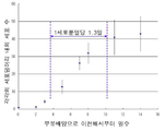
본 발명의 다능성 줄기세포는 부유배양(浮遊培養)에서 증식속도 약 1.3일로 증식하지만, 10일 동안 정도로 증식을 멈춘다고 하는 성질을 갖고, 더우기 정소(精巢)에 이식했을 경우 적어도 반년간은 암화(癌化) 하지 않는다고 하는 성질을 갖는다.The pluripotent stem cells of the present invention proliferate at about 1.3 days in proliferation rate, but have a property of stopping proliferation for about 10 days, and furthermore, when transplanted into the testis, cancerous at least half a year. It has a property that it does not (癌 化).
[9] 셀프 리뉴얼(self-renewal) 능력을 갖는 [1] ? [8]의 어느 것인가의 다능성 줄기세포.[9] have self-renewal capability [1]? Pluripotent stem cells of any of [8].
본 발명의 다능성 줄기세포는 부유배양(浮遊培養)과 접착배양(接培養着)의 조작을 반복함으로써 증식시킬 수 있다. 또한 본 발명의 다능성 줄기세포는 다른 체성(體性) 줄기세포와 같이 비대칭 분열을 한다.The pluripotent stem cells of the present invention can be proliferated by repeating the operations of suspension culture and adhesion culture. In addition, the pluripotent stem cells of the present invention have asymmetric division like other somatic stem cells.
[10] 스트레스 내성(耐性)인 [1] ? [9]의 어느 것인가의 다능성 줄기세포.[10] Stress-resistant [1]? Pluripotent stem cells of any of [9].
[11] 탐식능(貪食能)이 높은 [1] ? [10]의 어느 것인가의 다능성 줄기세포.[11] High phagocytosis [1]? Pluripotent stem cells of any of [10].
[12] 아래에 나타낸 22개의 오드란트(odorant) 수용체의 적어도 하나가 양성(陽性)인 [1] ? [11]의 어느 것인가의 다능성 줄기세포.[12] At least one of the 22 odorant receptors shown below is positive [1]? Pluripotent stem cells of any of [11].
olfactory receptor, family 8, subfamily G, member 2 (OR8G2);olfactory receptor,
olfactory receptor, family 7, subfamily G, member 3 (OR7G3);olfactory receptor,
olfactory receptor, family 4, subfamily D, member 5 (OR4D5);olfactory receptor,
olfactory receptor, family 5, subfamily AP, member 2 (OR5AP2);olfactory receptor,
olfactory receptor, family 10, subfamily H, member 4 (OR10H4);olfactory receptor,
olfactory receptor, family 10, subfamily T, member 2 (OR10T2);olfactory receptor,
olfactory receptor, family 2, subfamily M, member 2 (OR2M2);olfactory receptor,
olfactory receptor, family 2, subfamily T, member 5 (OR2T5);olfactory receptor,
olfactory receptor, family 7, subfamily D, member 4 (OR7D4);olfactory receptor,
olfactory receptor, family 1, subfamily L, member 3 (OR1L3);olfactory receptor,
olfactory receptor, family 4, subfamily N, member 4 (OR4N4);olfactory receptor,
olfactory receptor, family 2, subfamily A, member 7 (OR2A7);olfactory receptor,
guanine nucleotide binding protein (G protein), alpha activating activity polypeptide, olfactory type (GNAL);guanine nucleotide binding protein (G protein), alpha activating activity polypeptide, olfactory type (GNAL);
olfactory receptor, family 6, subfamily A, member 2 (OR6A2);olfactory receptor,
olfactory receptor, family 2, subfamily B, member 6 (OR2B6);olfactory receptor,
olfactory receptor, family 2, subfamily C, member 1 (OR2C1);olfactory receptor,
olfactory receptor, family 52, subfamily A, member 1 (OR52A1);olfactory receptor, family 52, subfamily A, member 1 (OR52A1);
olfactory receptor, family 10, subfamily H, member 3 (OR10H3);olfactory receptor,
olfactory receptor, family 10, subfamily H, member 2 (OR10H2);olfactory receptor,
olfactory receptor, family 51, subfamily E, member 2 (OR51E2);olfactory receptor, family 51, subfamily E, member 2 (OR51E2);
olfactory receptor, family 5, subfamily P, member 2 (OR5P2); 및olfactory receptor,
olfactory receptor, family 10, subfamily P, member 1 (OR10P1).olfactory receptor,
[13] 아래에 나타낸 5개의 케모카인(chemokine)의 적어도 하나가 양성(陽性)인 [1] ? [12]의 어느 것인가의 다능성 줄기세포:[13] At least one of the five chemokines shown below is benign [1]? Pluripotent stem cells of any of [12]:
chemokine (C-C motif) receptor 5 (CCR5);chemokine (C-C motif) receptor 5 (CCR5);
chemokine (C-X-C motif) receptor 4 (CXCR4);chemokine (C-X-C motif) receptor 4 (CXCR4);
chemokine (C-C motif) receptor 1 (CCR1);chemokine (C-C motif) receptor 1 (CCR1);
Duffy blood group, chemokine receptor(DARC); 및Duffy blood group, chemokine receptor (DARC); And
chemokine (C-X-C motif) receptor 7 (CXCR7).chemokine (C-X-C motif) receptor 7 (CXCR7).
[14] 중배엽계(中胚葉系) 조직 또는 간엽계(間葉系) 조직 유래인 [1] ? [13]의 어느 것인가의 다능성 줄기세포.[14] Is it derived from mesoderm or mesenchymal tissues [1]? Pluripotent stem cells of any of [13].
[15] [1] ? [14]의 어느 것인가의 다능성 줄기세포를 포함하는 세포 덩어리 또는 세포분획.[15] [1] A cell mass or cell fraction comprising any of the pluripotent stem cells of [14].
[16] 생체조직으로부터 아래의 (i) ?(ⅵ)의 특성 중 적어도 하나의 특성을 지표(指標)로 다능성 줄기세포 또는 다능성 세포분획을 단리(單離)하는 방법:[16] A method of isolating pluripotent stem cells or pluripotent cell fractions from living tissue using at least one of the following (i)?
(i) SSEA-3 양성(陽性);(i) SSEA-3 positive;
(ⅱ) CD105 양성(陽性);(Ii) CD105 positive;
(ⅲ) CD117 음성(陰性) 및 CD146 음성(陰性)(Ⅲ) CD117 negative and CD146 negative
(ⅳ) CD117 음성, CD146 음성, NG2 음성, CD34 음성, vWF 음성 및 CD271 음성;(Iii) CD117 voice, CD146 voice, NG2 voice, CD34 voice, vWF voice and CD271 voice;
(ⅴ) CD34 음성, CD117 음성, CD146 음성, CD 271 음성, NG2 음성, vWF 음성, Sox10 음성, Snail 음성, Slug 음성, Tyrpl 음성 및 Dct 음성; 및(Iii) CD34 voice, CD117 voice, CD146 voice, CD 271 voice, NG2 voice, vWF voice, Sox10 voice, Snail voice, Slug voice, Tyrpl voice and Dct voice; And
(ⅵ) 텔로머라아제(telomerase) 활성이 낮거나 또는 없다.(Iii) low or no telomerase activity.
[17] 생체조직 유래 세포를 세포 스트레스에 폭로(暴露)해 살아남은 세포를 회수하는 것을 포함하는 다능성 줄기세포 또는 다능성 세포분획을 단리(單離)하는 방법.[17] A method for isolating pluripotent stem cells or pluripotent cell fractions comprising exposing living tissue-derived cells to cellular stress and recovering the surviving cells.
[18] 세포 스트레스가 프로테아제(protease) 처리, 저산소 조건하에서의 배양, 저인산 조건하에서의 배양, 혈청 기아상태에서의 배양, 당 기아상태에서의 배양, 방사선 폭로하에서의 배양, 열 쇼크에의 폭로하에서의 배양, 유해물질 존재하에서의 배양, 활성 산소 존재하에서의 배양, 기계적 자극하에서의 배양 및 압력처리하에서의 배양으로부터 선택되는 [17]의 다능성 줄기세포 또는 다능성 세포분획을 단리(單離)하는 방법.[18] Cell stress is treated with protease, culture under hypoxic conditions, culture under hypophosphate conditions, culture under serum starvation, culture under sugar starvation, under radiation exposure, under exposure to heat shock, A method for isolating pluripotent stem cells or pluripotent cell fractions according to [17] selected from culture in the presence of harmful substances, culture in the presence of active oxygen, culture under mechanical stimulation and culture under pressure treatment.
[19] 세포 스트레스가 트립신(trypsin) 처리인 [18]의 다능성 줄기세포 또는 다능성 세포분획을 단리(單離)하는 방법.[19] A method for isolating pluripotent stem cells or pluripotent cell fractions of [18], wherein the cell stress is trypsin treatment.
[20] [1]?[14]의 어느 것인가의 다능성 줄기세포의 파생세포 또는 유도세포인 다능성 줄기세포.[20] A pluripotent stem cell, which is a derivative or induced cell of the pluripotent stem cell of any of [1] and [14].
그 파생세포 또는 유도세포로서 예를 들면 유전자의 도입이나 화합물 첨가에 의해 유도한 세포를 들 수 있다. 또 본 발명의 줄기세포 유래의 iPS세포를 들 수 있다.Examples of the derived cells or induced cells include cells induced by introduction of a gene or addition of a compound. In addition, iPS cells derived from stem cells of the present invention can be cited.
[21] [1]?[14]의 어느 것인가의 다능성 줄기세포의 파생세포 또는 유도세포인 분화된 세포.[21] A differentiated cell, which is a derivative or induced cell of pluripotent stem cells according to any of [1] and [14].
[22] [1]?[14] 및 [20]의 어느 것인가의 다능성 줄기세포를 포함하는 의약조성물.[22] A pharmaceutical composition comprising the pluripotent stem cell of any of [1]? [14] and [20].
[23] [21]의 분화된 세포를 포함하는 의약조성물. [23] A pharmaceutical composition comprising the differentiated cells of [21].
본 명세서는 본원의 우선권의 기초인 미국 가출원 61/213,788호 및 미국 가출원 61/290,159호의 명세서 및/또는 도면에 기재되는 내용을 포함한다.This specification includes information set forth in the specification and / or drawings of US Provisional Application No. 61 / 213,788 and US Provisional Application No. 61 / 290,159, which are the basis of priority herein.
본 발명에 의해 생식 세포나 초기배(初期胚)를 이용하는 일 없이 한편, 외래 유전자의 도입이나 특정 화합물의 도입 등의 인위적인 유도 조작을 경과하지 않고, 생체조직으로부터 다능성 줄기세포를 얻을 수 있다. 외래 유전자의 도입 등의 인위적 조작을 경과하지 않기 때문에 본 발명의 다능성 줄기세포는 효율적으로 제작하는 것이 가능하여 치료에 이용할 경우여도 안전하게 이용할 수 있다. 또한 본 발명의 다능성 줄기세포는 재생 의료나 기능 부전조직(不全組織)의 치료 등에 이용할 수 있고, 게다가 세포분화나 조직 재생의 연구 등에 이용할 수 있다.According to the present invention, pluripotent stem cells can be obtained from biological tissues without using germ cells or early embryos, and without performing artificial induction operations such as introduction of foreign genes or introduction of specific compounds. Since no artificial manipulation such as introduction of foreign genes has been performed, the pluripotent stem cells of the present invention can be efficiently produced and can be used safely even when used for treatment. In addition, the pluripotent stem cells of the present invention can be used for regenerative medicine, treatment of dysfunctional tissue, and the like, and can also be used for cell differentiation and tissue regeneration research.
본 명세서에서 인용한 모든 간행물, 특허 및 특허출원을 그대로 참고로서 본명세서에 넣는 것으로 한다.All publications, patents, and patent applications cited herein are hereby incorporated by reference in their entirety.
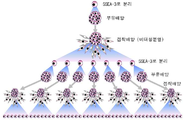
도 1a는 세포분획, Muse세포 및 Muse세포 유래의 배양체양(胚樣體樣) 세포덩어리의 관계를 나타낸 도면이다. 도 1에 나타낸 바와 같이 SSEA-3 양성세포(陽性細胞)를 직접 분리하고, 장시간의 스트레스를 거는 일 없이 부유 배양(浮遊培養)함으로써 Muse세포 유래의 배양체양(胚樣體樣) 세포덩어리를 얻을 수 있다.
도 1b는 Muse세포를 대량으로 증식시키는 방법을 나타낸 도면이다.
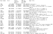
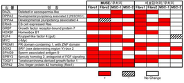
도 2는 Muse세포 유래의 배양체양(胚樣體樣) 세포덩어리/무처리(無處理) 세포분획에서의 발현량의 비가 높은 인자를 나타낸 도면이다.
도 3은 Muse세포 유래의 배양체양(胚樣體樣) 세포덩어리/인간 ES세포에서의 발현량의 비가 높은 인자를 나타낸 도면이다.
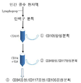
도 4는 MACS소팅(sorting)의 프로토콜을 나타낸 도면이다.
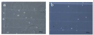
도 5는 트리판블루 염색상(染色像)에서 죽은 세포의 제거를 나타낸 사진으로서, a는 인간 섬유아세포(H-fibroblast) 분획의 16시간의 장시간 트립신(try psin) 처리후의 트리판블루 염색상(染色像)에서 죽은 세포의 제거를 나타낸 사진이고, b는 1800 ? 2200rpm/분으로 3분간 보텍스(vortex) 처리를 한 후의 트리판블루 염색상(染色像)에서 죽은 세포의 제거를 나타낸 사진이다.
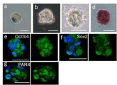
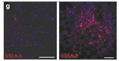
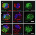
도 6은 각종 세포의 사진으로서, a는 부(富) Muse세포 분획 중 1세포(Bar = 10μm)를 나타낸 사진이고, b는 인간 ES세포 유래의 배양체세포(胚樣體細胞) 덩어리(Bar = 25μm)를 나타냉 사진이며, c는 직경 약25μm의 Muse세포 유래의 배양체세포(胚樣體細胞) 덩어리(Bar = 25μm)를 나타낸 사진이고, d는 인간 ES세포 유래 세포덩어리 4일째의 알칼리 포스파타아제(phosphatase) 염색상(染色像; Bar = 25μm)를 나타낸 사진이며, e ? g는 Muse세포 유래의 배양체양(胚樣體樣) 세포덩어리의 Oct3/4(e), Sox2(f) 및 PAR4(g)의 면역 염색상(染色像)을 나타낸 사진이다.
도 7a는 H-fibroblast 분획 및 인간 MSC(H-MSC) 분획 유래의 세포덩어리의 특징을 나타낸 사진으로서, a 및 b는 무처리(無處理) 인간 MSC 분획의 통상의 접착 배양에 있어서 자발적으로 생긴 세포덩어리(Bar = 100μm)를 나타내고, c 및 d는 0일째(c) 및 7일째(d)의 H-fibroblast-1 분획의 장시간 트립신(trypsin) 처리후의 MC배양의 상태를 나타내며(Bar = 100μm), (d)의 화살표는 Muse세포 유래 배양체양(胚樣體樣) 세포덩어리를 나타내고, e 및 f는 MC배양 7일후의 H-fibroblast-1 분획으로부터 형성된 Muse세포 유래 배양체세포(胚樣體細胞) 덩어리를 나타낸다(Bar = 50μm).
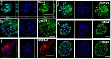
도 7b는 H-fibroblast 분획 및 인간 MSC(H-MSC) 분획 유래의 세포덩어리의 특징을 나타낸 사진으로서, g ? l은 H-fibroblast 분획으로부터 형성된 Muse세포 유래 배양체양(胚樣體樣) 세포덩어리(g, i 및 k) 및 H-MSC 분획으로부터 형성된 Muse세포 유래 배양체양(胚樣體樣) 세포덩어리(h, j 및 l)의 Nanog(g 및 j), Oct3/4(h), SSEA-3(i), PAR4(k) 및 Sox2(l)의 국(局) 존재를 나타내는 면역염색의 결과를 나타낸다(Bar = 50μm).
도 7c는 H-fibroblast 분획 및 인간 MSC(H-MSC) 분획 유래의 세포덩어리의 특징을 나타내는 사진으로서, m ? o는 인간 ES세포(m), H-fibroblast 분획 유래의 Muse세포 유래 배양체양(胚樣體樣) 세포덩어리(n) 및 무처리(無處理) H-fibroblas t-1 분획(o)의 알칼리 포스파타아제(phosphatase) 염색의 결과를 나타낸다(Bar = 50μm).
도 7d는 H-fibroblast 분획 및 인간 MSC(H-MSC) 분획 유래의 세포덩어리의 특징을 나타낸 전자현미경 사진으로서, p ? r은 인간 ES세포 배양체(胚樣體)(p, MC배양 3일째), H-fibroblast-1 분획 유래의 Muse세포 유래 배양체양(胚樣體樣) 세포덩어리(q 및 r, MC배양 5일째)의 전자현미경상을 나타낸다(Bar = 5μm).
도 8a는 Muse세포 유래 배양체양(胚樣體樣) 세포덩어리(M-cluster)의 클론성(clonality) 및 셀프 리뉴얼을 나타내는 도면이며, Muse세포의 클론성(clonalit y) 및 셀프 리뉴얼을 결정하기 위해 실행한 실험의 개요를 나타낸 도면이다.
도 8b는 Muse세포의 부유세포(浮遊細胞)에 있어서의 증식 속도를 나타낸 도면이다.
도 8c는 단일의 Muse세포 유래 배양체양(胚樣體樣) 세포덩어리(H-fibrobla st-1 유래, 제1세대(사이클)로부터 증식한 세포(clonally expanded cells)의 정상핵형(正常核型) 을 나타낸 도면이다.
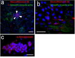
도 9a는 Muse세포 유래 배양체양(胚樣體樣) 세포덩어리의 분화를 나타낸 도면으로서, a ? c는 H-fiboroblas-1 분획 유래의 분화 세포의 덩어리의 α평활근 액틴(actin) 및 신경 필라멘트(a 및b) 및 α-페토프로테인(c)의 국(局) 존재를 나타낸 면역염색상(免疫染色像)이다(Bar는 a가 500μm, b 및 c가 50μm). a의 화살표 머리는 접착한 Muse세포 유래 배양체양(胚樣體樣) 세포덩어리를 나타낸다.
도 9b는 무처리(無處理) 세포분획, H-fibroblast 분획 유래의 제1 및 제3세대의 Muse세포 유래 배양체양(胚樣體樣) 세포덩어리(Cluster)를 젤라틴(gelatin)상에서 배양해 자발적 분화를 유도한 세포군에 있어서의 α-페토프로테인(α-FP), GATA6, MAP-2 및 Nkx2.5 발현의 RT-PCR 분석의 결과를 나타낸다. 양성 컨트롤로서는 α-FT에 대하여 인간 태아 간장을, GATA6, MAP-2 및 Nkx 2.5에 대하여는 인간 배(胚) 전체를 채용했다.
도 9c e ? l은 부(富) Muse세포 분획을 투여한 면역부전(免疫不全) 마우스의 정소(精巢)를 나타낸다. e는 컨트롤(intact)이 되는 무상(無傷)의 정소(精巢) 및 마우스 ES세포(mES cells)(8주), MEF[피더(feeder)세포)(8주), Muse세포 유래 배양체양(胚樣體樣) 세포덩어리(M-cluster)(6개월) 및 부(富) Muse세포 분획(Muse) (6개월)을 투여한 정소(精巢)를 나타낸다. f ? l는 부(富) Muse세포 분획 또는 Muse세포 유래 배양체양(胚樣體樣) 세포덩어리를 투여한 정소(精巢) 조직의 신경 필라멘트M(f, 사진에서는 초록으로 염색), α-페토프로테인(g, 사진에서는 초록으로 염색) 및 평활근 액틴(actin)(h, 도면에서는 빨강으로 염색)의 면역염색상(免疫染色像)을 나타낸다(Bar = 50μm). i의 3장의 패널은 인간 미토콘드리아(초록으로 염색) 및 평활근 액틴(actin)(빨강으로 염색)의 이중 염색상(染色像)을 나타낸다 (Bar = 20μm). j ? l은 부(富) Muse세포 분획을 투여한 정소(精巢)의 조직상(組織像)을 나타낸다(j 및 k). k에 인정을 받는 튜브 모양의 구조는 인간 미토콘드리아에 대한 항체로 염색되어 있다 (Bar는 j가 500μm, k ? l이 50μm).
도 10a는 H-fibroblast(Fibro-1, Fibro-2) 및 H-MSC(MSC-1, MSC-2)의 pluripotency 및 미분화 세포상태에 관여하는 인자의 정량PCR의 결과를 나타낸 도면이다(그의 2). 도면 중 열(column)의 모양은 부(富) Muse세포 분획 및 Muse세포유래 배양체양(胚樣體樣) 세포덩어리(7일째)의 무처리(無處理) 세포분획에 비교한 유전자 발현 레벨을 나타낸다. 흰 부분은 부(富) Muse세포군 또는 Muse세포 유래 배양체양(胚樣體樣) 세포덩어리/무처리(無處理) 세포획분의 비가 1/3보다 크고 3보다 작은 것을 나타내고, 회색(gray) 부분은 부(富) Muse세포 분획 또는 Muse세포 유래 배양체양(胚樣體樣) 세포덩어리/무처리(無處理) 세포분획의 비가 3보다 큰 것을 나타내며, 사선부분은 부(富) Muse세포 분획 또는 Muse세포 유래 배양체양(胚樣體樣) 세포덩어리/무처리(無處理) 세포분획의 비가 1/3보다 작은 것을 나타낸다.
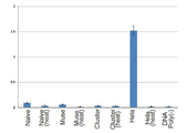
도 10b는 H-MSC 유래의 무처리(無處理) 세포분획(Naive), 부(富) Muse세포ㅂ분획(Muse) 및 Muse세포 유래 배양체양(胚樣體樣) 세포덩어리(M-cluster)(7일째)의 텔로머라아제(telomerase) 활성을 나타낸 도면이다. 열 불활성화 샘플(Heat)을 음성(陰性) 컨트롤로서 이용했다.
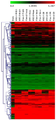
도 11은 H-fibroblast 분획 및 H-MSC 분획 유래의 무처리(無處理) 세포분획, 부(富) Muse세포 분획 및 Muse세포 유래 배양체양(胚樣體樣) 세포덩어리의 DNA마이크로 어레이 분석의 결과를 나타낸 도면이다.
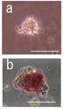
도 12는 인간 골수의 단핵 구성분(球成分)으로부터 SSEA-3/CD 105 이중 양성세포(陽性細胞)로서 직접 채취한 Muse세포를 MC배양하여 형성된 배양체양(胚樣體樣) 세포덩어리를 나타낸 사진으로서, a는 인간 골수로부터 단리(單離)하여 8시간의 장시간 트립신(trypsin) 처리를 한 단핵세포 분획을 MC배양하여(8hr-hBM-MC, 7일)형성된 Muse세포 유래 배양체양(胚樣體樣) 세포덩어리를 나타낸다(Bar = 100μm). b는 8hr-hBM-MC(7일)에 의해 형성된 Muse세포 유래 배양체양(胚樣體樣) 세포덩어리의 알칼리 포스파타아제(phosphatase) 염색상(染色像)을 나타낸다(Bar = 50μm).
도 13은 무처리(無處理) H-MSC-1 분획(naive1), 무처리(無處理) H-MSC-2 분획(naive2)(어느 것도 네가티브 컨트롤), 및 8시간의 트립신(trypsin) 처리를 한 인간골수 유래 단핵세포 분획(8hr-hBM) 혹은 트립신(trypsin) 처리를 하지 않은 인간골수 유래 단핵세포 분획(naive hBM)으로부터 형성된 Muse세포 유래 배양체양(胚樣體樣) 세포덩어리를 젤라틴(gelatin) 상에서 배양해 자발적 분화를 유도한 세포군에 있어서의 α-페토프로테인(α-FP), GATA6, MAP-2 및 Nkx 2.5의 RT-PCR 분석의 결과를 나타낸 도면이다.
도 14는 H-fibroblast 분획(무처리 세포) 및 H-MSC 분획(무처리 세포)의 FACS 분석의 결과를 나타내는 도면이다.
도 15a는 무처리 세포 분획 중의 SSEA-3 양성세포(a 왼쪽) 및 FACS 소팅(sorting)에 의해 채취한 SSEA-3 양성세포 유래의 단일 Muse세포 유래 배양체양(胚樣體樣) 세포덩어리로부터 클론(clone) 증식한 SSEA-3 양성세포(a 오른쪽)의 염색상(染色像)을 나타낸 사진이다. 도면 중 Bar는 100μm이다.
도 15b는 Muse세포(H-fibroblast)의 세포분열 중의 비대칭 분열에 영향을 미치는 인자인 Numblike(녹색)의 국(局) 존재를 나타내는 염색상(染色像)을 나타낸 사진이다. 도면 중 Bar는 10μm이다.
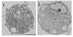
도 15c는 H-fibroblast 유래의 SSEA-3 음성세포(c) 및 SSEA-3 양성세포(d)의 전자현미경사진이다. 도면 중 Bar는 5μm이다.
도 15d는 H-fibroblast 유래 Muse세포의 Oct3/4(녹색)(e), Sox2(녹색)(f) 및 SSEA-3(적색)(g)의 염색상(染色像)을 나타낸 사진이다.
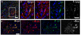
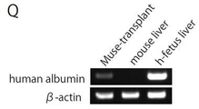
도 16a는 초면역(超免疫) 부전(不全) 마우스(Nog 마우스) 손상 조직에 있어서의 GFP 표식 SSEA-3 양성 Muse세포 분획의 분화를 나타낸 사진이다. N 및 O는 압박 손상 척수(4주일후)에 있어서의 GFP 양성세포이며, 신경 필라멘트(neurofilame nt; 적색) 및 인간 골지(golgi) 복합체(백색)가 발현되어 있다. O는 N의 사각으로 둘러싼 부분의 확대 상이다. P는 손상간장(損傷肝臟; 4주일후)의 GFP 양성 표식세포이며, 인간 알부민(적색) 및 인간 골지(golgi) 복합체(백색)가 발현되어 있다.
도 16b는 RT-PCR에서 조사한 SSEA-3 양성 Muse세포를 이식한 간장(肝臟)에 있어서의 인간 알부민의 발현을 나타낸 사진이다.
도 16c는 초면역(超免疫) 부전(不全) 마우스(Nog마우스) 손상 조직에 있어서의 GFP 표식 SSEA-3 양성 Muse세포 분획의 분화를 나타낸 사진이며, 인간 디스트로핀(dystrophin; 적색)이 발현되고 있는 근육(3주일후)의 GFP 양성세포를 나타낸 사진이다.
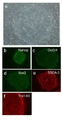
도 17a는 단일 Muse세포로부터 형성된 Muse세포 유래 배양체양(胚樣體樣) 세포덩어리로부터 증식시킨 세포의 분화를 나타내는 사진으로서, A?D는 신경유도의 결과를 나타내고, A는 형성된 sphere를 나타내며, 더욱이 shpere의 면역 염색 데이터로서 A는 네스틴(nestin), B는 Musashi, C는 NuroD의 발현을 나타낸다. E는 이들 sphere를 더욱 신경계 세포로의 분화를 시킨 것이고, MAP-2 양성세포를 나타낸다. F?G는 뼈세포 유도의 결과를 나타내며, 오스테오칼신(osteocalcin; F) 및 ALP(G)의 발현을 나타낸다. H 및 I는 지방세포 유도의 결과를 나타내고, H는 기름방울을 포함하는 세포를 나타내며, I는 오일 레드(oil red) 염색의 결과를 나타낸다. J는 간장세포(肝臟細胞) 유도의 결과를 나타내고, α-페토프로테인(α-fetopr otein) 양성세포를 나타낸다.
도 17b는 RT-PCR에서 조사한 간세포(肝細胞) 유도한 세포에 있어서의 인간 알부민 및 인간α-페토프로테인의 발현을 나타낸 사진이다.
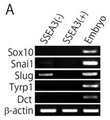
도 18a는 RT-PCR에서 조사한 SSEA-3 양성 Muse세포에 있어서의 Sox10, Sna il1, Slug, Tyrp1 및 Dct의 발현을 나타낸 사진이다.
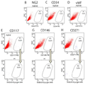
도 18b는 FACS에서 분석한 NG2, CD34, vWF, CD117, CD146 및 CD271의 발현을 나타낸 도면이다. 무처리(無處理) 인간 피부(皮膚) 섬유아세포(纖維芽細胞)에 있어서, 주피세포(周皮細胞; pericyte) 마커(marker)인 NG2, 내피(內皮) 전구세포(前驅細胞) 마커(marker)인 CD34 및 vWF는 음성이며, SSEA-3 양성세포에서도 음성이었다. 맬라노블래스트(melanoblast) 마커(marker)인 CD117, 주피세포(周皮細胞; pericyte) 마커(marker)인 CD146, NCSC 마커인 CD271은 무처리(無處理) 인간 피부선(皮膚) 섬유아세포(纖維芽細胞)에서는 조금 양성세포가 인정을 받았지만(각각 0.2%, 0.2% 및 0.8%), 그것들은 SSEA-3 음성세포이었기 때문에 Muse세포가 아니라고 생각된다.
도 18c는 Muse세포가 페라이트(ferrite)를 탐식(貪食)한 것을 나타낸 도면이다.
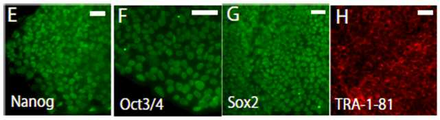
도 19는 Muse세포로부터 제작한 iPS세포의 형성을 나타낸 사진이며, a는 피부선(皮膚) 섬유아세포(纖維芽細胞; NHDF) 유래 Muse세포로부터 유도된 인간 iPS세포의 형태를 나타내고, b?f는 다능성 세포 마커(b가 Nonog, c가 Oct3/4, d가 Sox2, e가 SSEA-3, f가 Tra-1-60)의 발현을 나타낸다.
도 20은 Nonog(E), Oct3/4(F), Sox2(G) 및 Tra-1-81(H)의 면역 조직화학(組織化學)의 결과를 나타낸 사진이다.
도 21은 Muse 유래 iPS세포(Mi-1, Mi-2) 및 SSEA-3 음성세포로부터 증식한 콜로니((-)-1, (-)-2)의 RT-PCR에 의해 조사한 다능성 마커(marker)의 발현을 나타낸 사진이다.
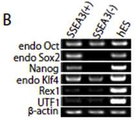
도 22a는 Oct3/4, Sox2, Klf4, c-Myse를 레트로바이러스(retrovirus)에서 도입하고, 그 후 피더(feeder) 세포 MEF 상에서 배양한 30일후의 SSEA-3 양성 및 음성세포로부터 형성된 콜로니의 Tra-1-81 면역 염색의 결과를 나타낸 사진이다. 인간 ES세포를 컨트롤로서 이용하고 있다. SSEA-3 양성세포로부터의 콜로니(a1) 및 인간 ES세포(a2)는 Tra-1-81 양성이지만, SSEA-3 음성세포로부터의 콜로니는 모두 음성이다.
도 22b는 22-1과 같이 MEF에서 30일 배양한 단계에 있어서의 SSEA-3 양성 및 음성세포의 다능성 마커[내인성 Oct3/4(endo Oct), 내인성 Sox2(endo Sox2), Na nog, 내인성 Klf4(endo Klf4), Rex1 및 UTF1]의 발현을 나타낸 사진이다. SSEA-3 음성 세포군에서는 Sox2, Nanog의 시그널이 보이지 않는다.
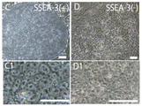
도 22c는 Muse세포로부터 유도한 iPS세포(Muse세포 유래 iPS세포)(C 및 C1) 및 SSEA-3 음성세포로부터 증식한 콜로니(D 및 D1)의 콜로니를 나타낸 사진이다.
도 23a는 피부선(皮膚) 섬유아세포(纖維芽細胞; NHDF) 유래 Muse세포로부터 유도된 iPS세포의 in vitro에서의 분화의 모양을 나타낸 사진이다. i는 내배엽 마커인 α-페토프로테인(녹색) 및 중배엽(中胚葉) 마커인 평활근 액틴(actin; 적색, 청색은 DNA)의 발현을 나타내고, j는 외배엽 마커인 신경 필라멘트(녹색)의 발현을 나타낸다.
도 23b는 Muse세포로부터 유도된 iPS세포의 in vitro에서의 분화의 RT-PCR 분석 결과를 나타낸 도면이며, 3배엽 마커의 발현을 나타냈다.
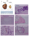
도 23c는 피부(皮膚) 섬유아세포(纖維芽細胞; NHDF) 유래 Muse세포로부터 유도된 iPS세포로부터 형성된 기형종(teratomas)의 조직 구조를 나타낸 사진이며, HE(Hematoxylin 및 eosin)염색에 의해 iPS세포가 각종 조직에 분화되어 있는 것을 나타내고 있다. m은 연골(cartilage), n은 근육(Muscle), o는 신경상피(neural epi thelium), p는 색소상피(pigmented epithelium), q는 원주상피(columnar epitheli um)을 나타낸다.
도 24는 SSEA-3 음성 세포분획, Muse세포 유래 배양체양(胚樣體樣) 세포덩어리 및 Muse 유래 iPS세포의 Nanog 유전자 및 Oct3/4 유전자의 Bisulfite(아황산수소염) 시퀀스(order)의 결과를 나타낸 도면이다. 각각의 칼럼(column)의 수치는 전사(轉寫) 시작 부위(TSS)의 하류에 대한 CpG의 위치를 나타낸다. 흰색 원(白丸)은 메틸화되지 않고 있는 시토신(cytosine)을, 검은 원(黑丸)은 메틸화된 시토신(cytosine)을 나타낸다.
도 25는 무처리(無處理) 섬유아세포(纖維芽細胞; Naive), Muse세포 유래 배양체양(胚樣體樣) 세포덩어리(Cluster) 및 iPS세포 중의 세포주기에 관련된 인자의 정량 PCR의 결과를 나타낸 도면이다.「/Naive」에서 나타낸 칼럼(column) 중 흰색 칼럼은 무처리(無處理) 세포에 대한 Muse분획 또는 Muse세포 유래 배양체양(胚樣體樣) 세포덩어리와의 비가 2 미만으로 1/2보다 큰 것을 나타낸다. 또 빈틈없이 칠된 칼럼은 비가 2보다 큰 것을 나타내고, 사선이 쳐진 칼럼은 비가 1/2보다 작은 것을 나타낸다.「/iPS」에서 나타낸 칼럼 중「*」은 발현된 유전자의 양이 iPS보다 Muse세포 유래 배양체양(胚樣體樣) 세포덩어리에서 큰 것을 나타내고,「**」은 발현된 유전자의 양이 Muse세포 유래 배양체양(胚樣體樣) 세포덩어리보다 iPS에서 큰 것을 나타낸다.
도 26은 무처리(無處理) 섬유아세포(纖維芽細胞; Naive), Muse세포 유래 배양체양(胚樣體樣) 세포덩어리(Cluster) 및 iPS세포 중의 다능성 및 미분화 세포상태에 관련된 인자의 정량 PCR의 결과를 나타낸 도면이다. 각 칼럼의 의미는 도 25와 같다.
도 27은 인간 및 마우스 모델에 있어서 제작된 iPS세포주의 유도 효율에 관한 논문보고를 정리한 도면이다. 도 27은 핵의 리프로그래밍(reprogramming)을 유도하는 전사(轉寫) 인자의 편성을 나타내고 있다.Fig. 1A is a diagram showing the relationship between cell fractions, Muse cells, and culture-like cell masses derived from Muse cells. As shown in Fig. 1, SSEA-3 positive cells are directly isolated and cultured cell masses derived from Muse cells are obtained by floating culture without stressing for a long time. Can be.
Figure 1b is a diagram showing a method for proliferating a large amount of Muse cells.
FIG. 2 is a diagram showing a high ratio of expression levels in cultured cell masses / untreated cell fractions derived from Muse cells. FIG.
Fig. 3 is a diagram showing a factor of a high ratio of expression amount in cultured cell masses / human ES cells derived from Muse cells.
4 is a diagram illustrating a protocol of MACS sorting.
Figure 5 is a photograph showing the removal of dead cells in trypan blue staining, a is trypan blue staining after 16 hours long trypsin treatment of human fibroblast (H-fibroblast) fraction (染色 像) shows the removal of dead cells, b is 1800? Photograph showing the removal of dead cells from trypan blue staining after vortex treatment at 2200 rpm / min for 3 minutes.
6 is a photograph of various cells, a being a photograph showing one cell (Bar = 10 µm) in the fraction of a rich Muse cell, b is a mass of culture cell derived from human ES cells (Bar = 25 µm), c is a photograph showing the culture cell mass (Bar = 25 µm) derived from Muse cells with a diameter of about 25 µm, and d is the alkaline force on the 4th day of the human ES cell-derived cell mass. It is a photograph showing the fate (phosphatase) stain (染色 像; Bar = 25μm), e? g is a photograph showing the immunostaining image of Oct3 / 4 (e), Sox2 (f), and PAR4 (g) of culture cell mass derived from Muse cells.
Figure 7a is a photograph showing the characteristics of the cell mass derived from the H-fibroblast fraction and human MSC (H-MSC) fraction, a and b are spontaneously generated in the normal adhesion culture of untreated human MSC fraction Cell mass (Bar = 100 μm), c and d represent the state of MC culture after long time trypsin treatment of H-fibroblast-1 fractions on day 0 (c) and day 7 (d) (Bar = 100 μm) Arrows of (d) and (d) indicate a mass of culture cells derived from Muse cells, and e and f represent culture cells derived from Muse cells formed from H-fibroblast-1
Figure 7b is a photograph showing the characteristics of cell masses derived from H-fibroblast fraction and human MSC (H-MSC) fraction, g? l is a culture cell mass derived from Muse cells formed from the H-fibroblast fraction (g, i and k) and a cell mass derived from Muse cell formed from the H-MSC fraction (h). shows the results of immunostaining showing the presence of bureaux of Nanog (g and j),
7C is a photograph showing the characteristics of cell masses derived from H-fibroblast fraction and human MSC (H-MSC) fraction, m? o is an alkali of human ES cell (m), cultured cell mass derived from Muse cell derived from H-fibroblast fraction (n) and untreated H-fibroblas t-1 fraction (o) Results of phosphatase staining are shown (Bar = 50 μm).
7D is an electron micrograph showing the characteristics of cell masses derived from H-fibroblast fraction and human MSC (H-MSC) fraction. r is the human ES cell culture (p,
FIG. 8A is a diagram showing the clonality and self renewal of Muse cell-derived cultured cell mass (M-cluster), and to determine the clonalit y and self renewal of Muse cells. This is a diagram showing an outline of the experiments performed for the purpose.
Fig. 8B is a diagram showing the proliferation rate in the floating cells of Muse cells.
Fig. 8C shows a normal karyotype of cells expanded from a single Muse cell-derived culture-like cell mass (H-fibrobla st-1-derived, cells expanded from the first generation (cycle)). It is a diagram showing.
Fig. 9A is a diagram showing the differentiation of a culture cell mass of Muse cell-derived culture mass, a? c is an immunostain showing the presence of α smooth muscle actin and neural filaments (a and b) and α-fetoprotein (c) in clumps of differentiated cells derived from the H-fiboroblas-1 fraction.染色 像) (Bar is a 500μm, b and c is 50μm). The arrowhead in a represents a cultured cell mass derived from adherent Muse cells.
Fig. 9B shows spontaneous culture of gelatinous cultured cell masses derived from first and third generations of Muse cells derived from H-fibroblast fraction without treatment. The results of RT-PCR analysis of α-fetoprotein (α-FP), GATA6, MAP-2, and Nkx2.5 expression in the cell group inducing differentiation are shown. As a positive control, human fetal liver was used for α-FT, and the whole human embryo was adopted for GATA6, MAP-2 and Nkx 2.5.
9C e? 1 indicates testis of immunocompromised mice to which the rich Muse cell fraction was administered. e is the intact testis and mouse ESES (mES cells) (8 weeks), MEF (feeder cells) (8 weeks), Muse cell-derived cultures (intact) It represents the testis administered with the M-cluster (6 months) and the rich Muse cell fraction (6 months). f? l is the neural filament M (f, stained green in the photo) and α-fetoprotein of testis tissues to which the Muse cell fraction or Muse cell-derived culture mass cell mass was administered. g, the picture shows green) and the immunostaining of smooth muscle actin (h, red in the figure) (Bar = 50 µm). Three panels of i show double staining images of human mitochondria (dyed green) and smooth muscle actin (dyed red) (Bar = 20 μm). j? 1 indicates the texture of the testis to which the rich Muse cell fraction was administered (j and k). The tube-like structure recognized by k is stained with an antibody against human mitochondria (Bar has 500 μm in j and 50 μm in k l).
FIG. 10A shows the results of quantitative PCR of factors involved in pluripotency and undifferentiated cell state of H-fibroblasts (Fibro-1, Fibro-2) and H-MSCs (MSC-1, MSC-2) (2) ). In the figure, the shape of the column shows the gene expression level compared to the untreated cell fraction of the rich Muse cell fraction and the Muse cell-derived culture mass cell (day 7). Indicates. White part indicates the ratio of rich Muse cell population or Muse cell-derived culture mass cell mass / untreated cell fraction is larger than 1/3 and smaller than 3, and gray part Indicates that the ratio of the rich Muse cell fraction or the Muse cell-derived culture mass cell mass / untreated cell fraction is greater than 3, and the hatched portion represents the rich Muse cell fraction or Muse cell-derived culture mass cell mass / untreated cell fraction ratio is less than 1/3.
Fig. 10B shows untreated cell fraction derived from H-MSC, rich Muse cell fraction and Muse cell-derived culture-like cell mass (M-cluster). It is a figure which shows the telomerase activity of (7th day). The heat inactivation sample (Heat) was used as a negative control.
FIG. 11 shows DNA microarray analysis of untreated cell fractions derived from H-fibroblast fractions and H-MSC fractions, enriched Muse cell fractions, and cultured cell masses derived from Muse cells. The figure which showed the result.
FIG. 12 shows a culture-mass cell mass formed by MC culture of Muse cells directly collected as SSEA-3 / CD 105 double positive cells from mononuclear constituents of human bone marrow. As a photograph, a is the amount of Muse cell-derived culture formed by MC culture (8hr-hBM-MC, 7 days) of MC cells (8hr-hBM-MC, 7 days) isolated from human bone marrow and treated with 8-hour long trypsin. Cell mass (Bar = 100 μm) b.Alkali phosphatase staining of cell mass of Muse cell-derived culture formed by 8hr-hBM-MC (7 days) A phase is shown (Bar = 50 micrometers).
FIG. 13 shows an untreated H-MSC-1 fraction (naive1), an untreated H-MSC-2 fraction (naive2) (both negative control), and trypsin treatment for 8 hours. Cell mass derived from Muse cells formed from human bone marrow-derived mononuclear cell fraction (8hr-hBM) or human bone marrow-derived mononuclear cell fraction (naive hBM) without trypsin treatment was gelatinized. Fig. 2 shows the results of RT-PCR analysis of α-fetoprotein (α-FP), GATA6, MAP-2 and Nkx 2.5 in a group of cells cultured on gelatin) and induced spontaneous differentiation.
Fig. 14 shows the results of FACS analysis of H-fibroblast fractions (untreated cells) and H-MSC fractions (untreated cells).
Fig. 15A shows clones from a single mass of Muse cell-derived culture mass cell derived from SSEA-3 positive cells taken by SSEA-3 positive cells (a left) and FACS sorting in an untreated cell fraction. (clone) The photograph shows the staining image of the proliferated SSEA-3 positive cells (a right side). Bar in the figure is 100μm.
FIG. 15B is a photograph showing staining images showing the presence of Numblike (green), a factor influencing asymmetric division in cell division of Muse cells (H-fibroblast). FIG. Bar in the figure is 10μm.
Figure 15c is an electron micrograph of SSEA-3 negative cells (c) and SSEA-3 positive cells (d) derived from H-fibroblast. Bar in the figure is 5μm.
15d is a photograph showing staining images of Oct3 / 4 (green) (e), Sox2 (green) (f), and SSEA-3 (red) (g) of H-fibroblast-derived Muse cells.
FIG. 16A is a photograph showing the differentiation of GFP-labeled SSEA-3 positive Muse cell fractions in hyperimmune insufficiency mouse (Nog mouse) damaged tissue. FIG. N and O are GFP-positive cells in the compression injury spinal cord (4 weeks later), and neurofilaments (red) and human golgi complexes (white) are expressed. O is an enlarged image of the portion enclosed by the square of N. P is a GFP positive marker cell of damaged liver (4 weeks later) and expresses human albumin (red) and human golgi complex (white).
Fig. 16B is a photograph showing the expression of human albumin in the liver transplanted with SSEA-3 positive Muse cells irradiated with RT-PCR.
Fig. 16C is a photograph showing the differentiation of GFP-labeled SSEA-3 positive Muse cell fractions in hyperimmune insufficiency mouse (Nog mouse) damaged tissues, in which human dystrophin (red) is expressed. Photograph showing GFP positive cells of muscle (3 weeks later).
Figure 17a is a photograph showing the differentiation of cells proliferated from the culture cell mass derived from Muse cells formed from a single Muse cells, A? D shows the results of neuronal induction, A represents the sphere formed, Furthermore, as the immunostaining data of shpere, A represents nestin, B represents Musashi, and C represents NuroD. E further differentiated these spheres into neuronal cells and represents MAP-2 positive cells. F-G indicates the result of bone cell induction and expression of osteocalcin (F) and ALP (G). H and I represent the result of adipocyte induction, H represents the cell containing oil droplets, and I represents the result of oil red staining. J represents the result of hepatic cell induction and represents α-fetopreotene positive cells.
Fig. 17B is a photograph showing the expression of human albumin and human α-fetoprotein in hepatocyte-induced cells irradiated with RT-PCR.
Figure 18a is a photograph showing the expression of Sox10, Sna il1, Slug, Tyrp1 and Dct in SSEA-3 positive Muse cells irradiated by RT-PCR.
Figure 18B shows the expression of NG2, CD34, vWF, CD117, CD146 and CD271 analyzed in FACS. In untreated human dermal fibroblasts, NG2, a pericyte marker, endothelial progenitor markers ) CD34 and vWF were negative and also negative in SSEA-3 positive cells. CD117, a melanoblast marker, CD146, a pericyte marker, and CD271, an NCSC marker, are non-treated human dermal gland fibroblasts. In i), some positive cells were recognized (0.2%, 0.2% and 0.8%, respectively), but they are not Muse cells because they were SSEA-3 negative cells.
FIG. 18C is a diagram showing that Muse cells phage ferrite.
Figure 19 is a photograph showing the formation of iPS cells prepared from Muse cells, a shows the morphology of human iPS cells derived from dermal gland fibroblasts (NHDF) -derived Muse cells, b? F Indicates the expression of pluripotent cell markers (b is Nonog, c is Oct3 / 4, d is Sox2, e is SSEA-3, f is Tra-1-60).
20 is a photograph showing the results of immunohistochemistry of Nonog (E), Oct3 / 4 (F), Sox2 (G), and Tra-1-81 (H).
21 shows pluripotency markers examined by RT-PCR of colony ((-)-1, (-)-2) grown from Muse-derived iPS cells (Mi-1, Mi-2) and SSEA-3 negative cells ( It is a photograph showing the expression of a marker).
FIG. 22A shows Tra of colonies formed from SSEA-3 positive and negative cells 30 days after Oct3 / 4, Sox2, Klf4, c-Myse was introduced in retrovirus and then cultured on feeder cell MEF The photograph showing the result of -1-81 immunostaining. Human ES cells are used as controls. Colonies from a SSEA-3 positive cell (a1) and human ES cells (a2) are Tra-1-81 positive, while colonies from SSEA-3 negative cells are all negative.
FIG. 22B shows pluripotency markers of SSEA-3 positive and negative cells (endogenous Oct3 / 4 (endo Oct), endogenous Sox2, endo Sox2), Na nog, endogenous in the stage of 30 days culture in MEF as in 22-1 Klf4 (endo Klf4), Rex1 and UTF1] is a picture showing the expression. Signals of Sox2 and Nanog are not visible in SSEA-3 negative cell population.
22C is a photograph showing colonies of iPS cells (Muse cell-derived iPS cells) derived from Muse cells (C and C1) and colonies (D and D1) grown from SSEA-3 negative cells.
Fig. 23A is a photograph showing the appearance of differentiation in vitro of iPS cells derived from cutaneous fibroblasts (NHDF) -derived Muse cells. i represents the expression of the endoderm marker α-fetoprotein (green) and the mesoderm marker smooth muscle actin (red and blue are DNA), and j is the expression of the neural filament (green), the ectoderm marker. .
Figure 23b is a diagram showing the results of RT-PCR analysis of differentiation in vitro of iPS cells derived from Muse cells, it showed the expression of three germ cells markers.
Figure 23c is a photograph showing the tissue structure of teratomas formed from iPS cells derived from skin fibroblasts (NHDF) -derived Muse cells, iPS cells by HE (Hematoxylin and eosin) staining Indicates that the tissue is differentiated into various tissues. m is cartilage, n is muscle, o is neural epithelium, p is pigmented epithelium, and q is columnar epithelium.
Fig. 24 shows the results of SSEA-3 negative cell fraction, Muse cell-derived culture mass cell mass, and Bisulfite (hydrosulfite) sequence of Nanog gene and Oct3 / 4 gene of Muse-derived iPS cells. The figure shown. The value of each column represents the position of CpG relative to the downstream of the transcription start site (TSS). White circles represent cytosine which is not methylated, and black circles represent methylated cytosine.
Fig. 25 shows the results of quantitative PCR of factors related to the cell cycle in untreated fibroblasts Naive, Muse cell derived culture mass cell clusters and iPS cells. The white column in the column shown in "/ Naive" has a ratio of the Muse fraction to the untreated cells or the mass of the cell mass derived from the Muse cell to less than two. It is larger than / 2. A tightly painted column indicates that the ratio is greater than 2, and an oblique column indicates that the ratio is less than 1/2. The asterisk of * / indicates that the amount of the expressed gene is derived from Muse cells rather than iPS. It shows that it is large in a culture mass cell mass, and "**" shows that the quantity of the expressed gene is larger in iPS than the culture mass mass cell mass derived from Muse cell.
Figure 26 Quantification of factors related to pluripotency and undifferentiated cell state in untreated fibroblasts Naive, Muse cell-derived culture mass cell clusters and iPS cells It is a figure which shows the result of PCR. Meaning of each column is the same as FIG.
FIG. 27 is a summary of papers on the induction efficiency of iPS cell lines prepared in human and mouse models. FIG. 27 shows the organization of transcription factors that induce nucleus reprogramming.
이하, 본 발명을 상세하게 설명한다.EMBODIMENT OF THE INVENTION Hereinafter, this invention is demonstrated in detail.
본 발명은 생체의 생체조직으로부터 직접 얻을 수 있는 다능성(pluripotent)줄기세포 또는 다능성 줄기세포 분획 및 그 다능성 줄기세포 또는 그 다능성 줄기세포획 분획을 단리(單離)하는 방법, 및 그 방법에 의해 얻어진 생체조직 유래의 다능성 줄기세포 또는 다능성 줄기세포 분획이다. 본 발명의 다능성 줄기세포를 Muse세포(multilineage differentiating stress enduring cells)라고 말한다.The present invention provides a method for isolating pluripotent stem cells or pluripotent stem cell fractions and pluripotent stem cells or pluripotent stem cell fractions thereof that can be directly obtained from living tissues of a living body, and Pluripotent stem cells or pluripotent stem cell fraction derived from living tissue obtained by the method. The pluripotent stem cells of the present invention are called Muse cells (multilineage differentiating stress enduring cells).
본 발명에 있어서, 세포분획이라고 할 때는 단리(單離)하고 싶은 세포를 적어도 일정량 포함하는 세포군을 말한다. 예를 들면 다능성 줄기세포 분획으로는 다능성 줄기세포를 1%이상, 10%이상, 30%이상, 50%이상, 70%이상, 90%이상 또는 95%이상 포함하는 세포군을 들 수 있고, 다능성 줄기세포의 배양에 의해 얻어진 세포덩어리나 다능성 줄기세포를 농축한 세포군을 포함한다. 또한 상기 세포분획을 실질적으로 균일한 세포분획이라고 하는 것도 있다.In the present invention, the cell fraction refers to a cell group containing at least a certain amount of cells to be isolated. For example, the pluripotent stem cell fraction includes a cell group containing 1% or more, 10%, 30%, 50%, 70%, 90% or 95% or more of pluripotent stem cells, It includes cell masses obtained by culturing pluripotent stem cells or cell groups enriched in pluripotent stem cells. The cell fraction may also be referred to as a substantially uniform cell fraction.
생체(生體)는 포유 동물의 생체를 좋은, 어느 정도 발생이 진행한 동물체(動物體)를 말한다. 본 발명에 있어서 생체로는 수정란이나 포배기보다 발생 단계가 앞의 배(胚)는 포함되지 않지만, 태아나 포배를 포함하는 포배기 이후의 발생 단계의 배(胚)는 포함된다. 포유 동물은 한정되지 않지만, 예를 들면 인간, 원숭이 등의 영장류(靈長類), 생쥐(mice), 쥐(rats), 토끼(rabbits), 모르모트(guinea pigs) 등의 설치류, 고양이, 개, 양, 돼지, 소, 말, 당나귀, 염소, 페렛(ferrets) 등이 포함된다. 본 발명의 다능성 줄기세포는 생체의 조직 유래인 점에서 배성간(胚性幹)세포(ES세포)나 배성생식(胚性生殖) 줄기세포(EG세포)로 명확히 구별된다.The living body refers to an animal body in which the development of the living body of a mammal is good to some extent. In the present invention, the embryos before the embryonic stage or embryonic stage do not include embryos, but embryos of embryonic stages after embryonic stage including fetuses or blastocysts are included. Although mammals are not limited, for example, rodents such as primates such as humans and monkeys, mice, rats, rabbits, guinea pigs, cats, dogs, Sheep, pigs, cattle, horses, donkeys, goats, ferrets and the like. Pluripotent stem cells of the present invention are clearly distinguished into embryonic liver cells (ES cells) and embryogenic reproductive stem cells (EG cells) in that they are derived from tissues of living bodies.
중배엽계(中胚葉系) 조직으로는 동물의 초기 발생 도상(途上)으로 나타나는 중배엽(中胚葉) 기원의 조직이 좋은, 근육계 조직, 결합조직, 순환계 조직, 배설계 조직, 생식계 조직 등이 포함된다. 예를 들면 본 발명의 다능성 줄기세포는 골수액이나 진피(眞皮) 결합조직 등의 피부조직으로부터 얻을 수 있다.Mesoderm tissues include muscle tissue, connective tissue, circulatory tissue, germline tissue, reproductive tissue, and the like, having a good mesodermal origin, which appears as an early developing animal. For example, the pluripotent stem cells of the present invention can be obtained from skin tissues such as bone marrow fluid and dermal connective tissue.
간엽계(間葉系) 조직으로는 뼈, 연골, 지방, 혈액, 골수, 골격근(骨格筋), 진피(眞皮), 인대, 힘줄(腱), 심장 등의 조직을 말한다. 예컨대 본 발명의 다능성 줄기세포는 골수나 피부로부터 얻을 수 있다. 또한 탯줄로부터 얻을 수도 있다.Mesenchymal system (間 葉 系) tissue, such as bone, cartilage, fat, blood, bone marrow, skeletal muscle (骨 格 筋), dermis (眞 皮), ligaments, tendons, heart, and the like. For example, the pluripotent stem cells of the present invention can be obtained from bone marrow or skin. It can also be obtained from the umbilical cord.
세포가 조직으로부터 직접 얻을 수 있는 것이라 함은 조직으로부터 단리(單離) 할 수 있고, 외래 유전자나 외래 단백질의 도입 또는 화합물의 투여 등 화합물처리 등의 인위적인 유도 조작을 경과하지 않고 얻을 수 있는 것을 의미한다. 여기서 외래 유전자는 한정되지 않지만, 예를 들면 체세포의 핵을 초기화할 수 있는 유전자가 좋은 예컨대 Oct3/4유전자 등의 Oct 패밀리 유전자, Klf 유전자 등의 Klf 패밀리 유전자, c-Myc 유전자 등의 Myc 패밀리 유전자, Sox2 유전자 등의 Sox 패밀리 유전자를 들 수 있다. 또 외래 단백질로서는 이들의 유전자가 코드 하는 단백질이나 사이토카인(cytokine)을 들 수 있다. 게다가 화합물로서는 예를 들면 상기한 체세포의 핵을 초기화할 수 있는 유전자의 발현을 유도하는 저분자 화합물이나 DM SO, 환원제로서 기능하는 화합물, DNA 메틸화제 등을 들 수 있다. 본 발명의 다능성 줄기세포는 생체 혹은 조직으로부터 직접 얻을 수 있다고 하는 점에서 iPS(induced pluripotent stem cell)세포 및 ES세포와는 명확히 구별된다. 한편 본 발명에 있어서는 세포의 배양, 세포의 표면 마커(marker)를 지표로 세포 또는 세포분획을 단리(單離)하는 것, 세포를 세포 스트레스에 폭로(暴露)하는 것 및 세포에 물리적 충격을 주는 것은 인위적인 유도 조작에는 포함되지 않는다. 또 본 발명의 다능성세포는 리프로그래밍(reprogramming) 또는 탈(脫) 분화의 유도를 필요로 하지 않고 얻을 수 있는 것을 특징으로 하여도 좋다.The fact that a cell can be obtained directly from a tissue means that it can be isolated from the tissue and obtained without performing artificial induction operations such as compound treatment such as introduction of a foreign gene or foreign protein or administration of a compound. do. Herein, although the foreign gene is not limited, for example, a gene capable of initiating the nucleus of the somatic cell is good, for example, Oct family gene such as Oct3 / 4 gene, Klf family gene such as Klf gene, and Myc family gene such as c-Myc gene. And Sox family genes such as Sox2 gene. Examples of foreign proteins include proteins and cytokines encoded by these genes. Moreover, as a compound, the low molecular weight compound which induces the expression of the gene which can initialize the nucleus of said somatic cell, DMSO, the compound which functions as a reducing agent, a DNA methylating agent, etc. are mentioned, for example. The pluripotent stem cells of the present invention are clearly distinguished from induced pluripotent stem cell (iPS) cells and ES cells in that they can be obtained directly from a living body or tissue. On the other hand, in the present invention, culturing cells, isolating cells or cell fractions as indicators of surface markers of cells, exposing the cells to cellular stress, and giving physical shocks to the cells. It is not included in artificial induction manipulation. The pluripotent cells of the present invention may be obtained without requiring reprogramming or induction of differentiation.
본 발명의 다능성 줄기세포는 생체의 중배엽계(中胚葉系) 조직 또는 간엽계(間葉系) 조직 등에 존재하고 있다고 생각되어 본 발명에 있어서는 이들 조직에 존재하고 있는 세포 또는 세포분획을 단리(單離) 한다. 본 발명의 다능성 줄기세포는 예를 들면, 골수에 존재하고 있어 골수로부터 혈액 등을 통해서 생체의 각 조직에 공급될 가능성이 있다. 이 때문에 골수나, 피부 등의 생체의 각 조직, 더욱이 혈액으로부터 단리(單離)하는 것이 가능하다.It is thought that the pluripotent stem cells of the present invention exist in the mesodermal tissues or mesenchymal tissues of the living body, and according to the present invention, the cells or cell fractions present in these tissues are isolated ( Iii) The pluripotent stem cells of the present invention are present in the bone marrow, for example, and may be supplied from the bone marrow to each tissue of the living body through blood or the like. For this reason, it is possible to isolate from each tissue of living bodies, such as bone marrow and skin, and also blood.
다능성 줄기세포로는 pluripotency를 갖는 세포가 좋은, 이하의 특성을 갖는다.As pluripotent stem cells, cells having pluripotency have good characteristics as follows.
(1) Nanog, Oct3/4, SSEA-3, PAR-4 및 Sox2 등의 다능성 마커(Pluripotent marker)를 발현한다.(1) Pluripotent markers such as Nanog, Oct3 / 4, SSEA-3, PAR-4, and Sox2 are expressed.
(2) 1세포로부터 증식하고, 자기의 클론(clone)을 계속해서 만드는 클론성(Clonality)을 갖는다.(2) It has clonality to proliferate from one cell and continue making its own clone.
(3)자기복제(셀프 리뉴얼) 능력을 갖는다. (3) Self-replicating (self renewal) ability.
(4) 3배엽계(내배엽계, 중배엽계 및 외배엽계)에로 in vitro 및 in vivo에서 분화될 수 있다. (4) They can be differentiated in vitro and in vivo into three germ layers (endoderm, mesoderm and ectoderm).
(5)마우스의 정소(精巢)나 피하에 이식한 경우 3배엽계에로 분화를 보인다.(5) When the mouse is implanted in the testis (精 이식) or subcutaneous, it shows differentiation into three germ layers.
(6)알칼리 포스파타아제(phosphatase) 염색으로 양성이 된다.(6) It is positive by alkaline phosphatase staining.
본 발명의 다능성 줄기세포는 pluripotency를 갖고 있는 점에서 성인 줄기세포, 조직 줄기세포와는 명확히 구별된다. 또 본 발명의 다능성 줄기세포는pluripotency를 갖고 있는 단일의 또는 복수의 세포로서 단리(單離) 되어 있는 점에서 골수 간엽계(間葉系) 세포 등의 세포분획과는 명확히 구별된다.The pluripotent stem cells of the present invention are clearly distinguished from adult stem cells and tissue stem cells in that they have pluripotency. The pluripotent stem cells of the present invention are clearly distinguished from cell fractions such as bone marrow mesenchymal cells in that they are isolated as single or plural cells having pluripotency.
게다가, 본 발명의 다능성 줄기세포는 이하의 특성을 갖는다.In addition, the pluripotent stem cells of the present invention have the following characteristics.
(i) 증식속도가 비교적 완만해서 분열 주기가 1일 이상, 예를 들면 1.2 ?1.5일이다. 단, ES세포나 iPS세포가 나타내는 것 같은 무한증식은 나타내지 않는다.(i) The rate of proliferation is relatively slow, resulting in a cleavage cycle of at least one day, for example 1.2 to 1.5 days. However, it does not show infinite proliferation such as that of ES cells or iPS cells.
(ii) 면역부전(免疫不全) 마우스에 이식한 경우에 내배엽계(內胚葉系), 중배엽계(中胚葉系) 및 외배엽계(外胚葉系)에로의 분화를 나타낸다. ES세포나 iPS세포에서는 기형종(奇形腫; teratomas)이 단기간에 암화(癌化) 하는데 비해 반년 이상 암화(癌化)하지 않는 것을 특징으로 한다.(ii) Differentiation into endoderm, mesoderm and ectoderm when transplanted into immunocompromised mice. In ES cells and iPS cells, teratomas do not become cancerous for more than half a year, compared to cancers in a short period of time.
(iii) 부유 배양에 의해 배양체양(胚樣體樣) 세포덩어리를 형성한다.(iii) A culture mass cell mass is formed by suspension culture.
(iv) 부유 배양에서 배양체양(胚樣體樣) 세포덩어리를 형성하고, 10일 정도에서 증식이 정지한다. 그 후 접착배양으로 이동시킴으로써 재증식한다.(iv) A culture mass cell mass is formed in suspension culture, and proliferation is stopped in about 10 days. It is then repopulated by transfer to adhesive culture.
(V) 증식할 때에 비대칭 분열을 따른다.(V) Follows asymmetric cleavage when proliferating.
(vi) 핵형은 정상이다.(vi) karyotype is normal.
(vii) 텔로머라아제(telomerase) 활성이 없거나 또는 낮다. 여기서 텔로머라아제(telomerase) 활성이 없거나 또는 낮다고 하는 것은 예를 들면 TRAPEZE XL telomerase detection kit(Millipore사)를 이용해서 텔로머라아제(telomerase) 활성을 검출한 경우에 검출할 수 없거나 또는 낮은 것을 말한다. 텔로머라아제(telomerase) 활성이 낮다고 하는 것은 예컨대 인간 섬유아세포(纖維芽細胞)와 같은 정도의 텔로머라아제(telomerase) 활성을 갖고 있거나 혹은 Hela세포에 비해서 1/5 이하, 바람직하게는 1/10 이하의 텔로머라아제(telomerase) 활성을 갖고 있는 것을 말한다.(vii) no or low telomerase activity. Here, the absence or low of telomerase activity means that it cannot be detected or is low when telomerase activity is detected using, for example, TRAPEZE XL telomerase detection kit (Millipore). Low telomerase activity is, for example, having a telomerase activity equivalent to that of human fibroblasts, or less than 1/5, preferably 1/10 of Hela cells. It has the following telomerase activity.
(viii) 메틸화의 상태에 대해서는 Muse세포로부터 유도한 iPS세포에 관해서는 Nanog 및 Oct3/4의 프로모터(promoter) 영역의 메틸화 레벨이 낮다.(viii) The methylation level of the promoter region of Nanog and Oct3 / 4 is low for iPS cells derived from Muse cells for the state of methylation.
(ix) 탐식(貪食) 능력이 높다.(ix) The ability to eat is high.
(x) 종양성 증식을 나타내지 않는다. 여기서 종양성 증식을 나타내지 않는 것은 부유 배양을 했을 경우, 일정한 크기의 세포덩어리(클러스터)에 달하면 증식이 정지하고 무한증식하지 않는 것을 말한다. 또 면역부전(免疫不全) 마우스의 정소(精巢)에 이식해도 기형종(奇形腫)을 형성하지 않는 것이다. 또한 상기(i) ? (iv) 등도 종양성 증식을 나타내지 않는 것에 관련된다.(x) does not exhibit tumorous proliferation. In this case, the tumor growth does not indicate that in suspension culture, when reaching a certain size of cell clusters (clusters), the proliferation stops and does not proliferate indefinitely. In addition, even if transplanted into the testis of the immunocompromised (免疫 不全) mice do not form teratoma (奇 形 腫). Also in (i)? (iv) and the like also relate to not showing tumorous proliferation.
즉, 본 발명의 세포는 예를 들면 아래의 다능성 줄기세포이다.That is, the cell of this invention is the following pluripotent stem cell, for example.
(A) 생체의 중배엽계(中胚葉系) 조직 또는 간엽계(間葉系) 조직 등으로부터 얻을 수 있는 세포이며, 해당 세포 내에 화학물질, 외래 유전자 또는 외래 단백질을 도입할 일 없이 직접 얻을 수 있는 다능성 줄기세포.(A) A cell obtained from a mesodermal tissue or a mesenchymal tissue of a living body, which can be directly obtained without introducing a chemical, a foreign gene or a foreign protein into the cell. Pluripotent Stem Cells.
(B) 생체의 중배엽계(中胚葉系) 조직 또는 간엽계(間葉系) 조직 등이 골수, 피부, 혈액, 탯줄, 지방 등으로 이루어지는 군으로부터 선택되는 상기(1)의 특성을 갖는 다능성 줄기세포. (B) a pluripotent having the characteristics of (1) above, wherein the mesoderm or mesenchymal tissue of the living body is selected from the group consisting of bone marrow, skin, blood, umbilical cord, and fat. Stem Cells.
(C) 리프로그래밍(reprogramming) 또는 탈(脫) 분화를 유도하는 일 없이 얻을 수 있는 상기(A) 또는 (B)의 다능성 줄기세포.(C) The pluripotent stem cells of (A) or (B) which can be obtained without inducing reprogramming or dedifferentiation.
(D) 정소(精巢)에로 이식한 경우에 적어도 반년간은 암화(癌化)하지 않는 상기(A ) 또는 (B)의 다능성 줄기세포.(D) The pluripotent stem cell of the above-mentioned (A) or (B) which does not become cancerous for at least half a year when transplanted into a testis.
(E) ES세포, iPS세포와 같이 무한증식을 나타내지 않는 상기(A) 또는 (B)의 다능성 줄기세포.(E) The pluripotent stem cells of (A) or (B), which do not exhibit infinite proliferation, such as ES cells and iPS cells.
(F) 생체의 중배엽계(中胚葉系) 조직 또는 간엽계(間葉系) 조직 등 유래의 다능성 줄기세포이며, 생체의 중간층계(中胚葉系) 조직 또는 간엽계(間葉系) 조직등의 세포를 프로테아제(protease)로 처리했을 때에 살아남는 프로테아제(proteas e)에 내성인 다능성 줄기세포.(F) pluripotent stem cells derived from living mesodermal tissues or mesenchymal tissues, and the mesenchymal tissues or mesenchymal tissues of the living body. Pluripotent stem cells resistant to proteases that survive when cells such as these are treated with protease.
게다가 본 발명의 다능성 줄기세포는 생체의 중배엽계(中胚葉系) 조직 또는 간엽계(間葉系) 조직 등의 세포에 세포 스트레스를 걸어 살아 남은 세포를 회수함으로써 단리(單離) 할 수 있다. 여기서 세포 스트레스로는 외적 스트레스가 좋은, 프로테아제(protease) 처리, 저산소 조건하에서의 배양, 저인산 조건하에서의 배양, 혈청(血淸) 기아상태(飢餓狀態)에서의 배양, 당(糖) 기아상태(飢餓狀態)에서의 배양, 방사선 폭로(暴露) 하에서의 배양, 열쇼크에의 폭로(暴露) 하에서의 배양, 유해물질 존재하에서의 배양, 활성산소 존재하에서의 배양, 기계적 자극하에서의 배양, 압력처리하에서의 배양 등에 의해 스트레스에 폭로하는 것을 말한다. 이 중에서도 프로테아제 처리 즉, 프로테아제(protease) 존재하에서의 배양이 바람직하다. 프로테아제(protease)는 한정되지 않고, 트립신(trypsin),키모트립신(chymotr ypsin) 등의 세린(serine) 프로테아제, 펩신 등의 아스파라긴산 프로테아제, 파파인(papain), 키모파파인(chymopapain) 등의 시스테인(systein) 프로테아제, 서물리신(thermolysin) 등의 금속 프로테아제, 글루타민산(Glutamicacid) 프로테아제, N- 말단(末端) 트레오닌(threonin) 프로테아제 등을 이용할 수 있다. 프로테아제를 배양에 첨가할 때의 첨가 농도는 한정되지 않고, 일반적으로 샬레(petri dish) 등으로 배양한 부착 세포를 벗겨낼 때에 이용하는 농도로 이용하면 좋다. 본 발명의 다능성 줄기세포는 상기 외적 스트레스에 내성을 갖는 줄기세포 예를 들면, 트립신(trypsin)에 내성을 갖는 세포라고 할 수 있다.In addition, the pluripotent stem cells of the present invention can be isolated by subjecting cells such as living mesodermal tissues or mesenchymal tissues to cells and recovering the surviving cells. . Herein, cell stress includes protease treatment, culture under hypoxic conditions, culture under hypophosphate conditions, culture under serum starvation, and sugar starvation, which have good external stress. (I) culture, under exposure to radiation, under exposure to heat shock, under toxic substances, under active oxygen, under active stimulation, under mechanical stimulation, under pressure treatment, and the like. To expose. Among them, protease treatment, that is, culture in the presence of protease is preferable. Protease is not limited, and cysteines such as serine proteases such as trypsin and chymotr ypsin, aspartic acid proteases such as pepsin, papain, and chymopapain Proteases, metal proteases such as thermolysin, glutamic acid proteases, N-terminal threonine proteases and the like can be used. The addition concentration at the time of adding a protease to a culture is not limited, Generally, it is good to use it as the density | concentration used when peeling off adherent cells culture | cultivated by the petri dish. The pluripotent stem cells of the present invention may be said to be stem cells resistant to the external stress, for example, cells resistant to trypsin.
생체의 중배엽계(中胚葉系) 조직 또는 간엽계(間葉系) 조직 등은 한정되지 않고, 골수단핵세포, 피부세포 등의 섬유아세포(纖維芽細胞) 분획, 치수조직(齒髓組織), 안구조직, 모근조직 등이 포함된다. 세포로서는 배양 세포도 조직으로부터 채취한 세포도 이용할 수도 있다. 이 중에서도 골수세포, 피부세포가 바람직하고, 예컨대 인간 골수 간엽계(間葉系) 세포(MSC) 분획 또는 인간 피부(皮膚) 섬유아세포(纖維芽細胞) 분획을 들 수 있다. 골수 간엽계(間葉系) 세포분획은 골수천자액(骨髓穿刺液)을 2?3주일 배양함으로써 얻을 수 있다.The mesoderm or mesenchymal tissues of the living body are not limited, and fibroblast fractions such as bone means nucleus cells and skin cells and pulp tissues are not limited. , Eye tissue, hair root tissue, and the like. As cells, cultured cells or cells obtained from tissues can also be used. Among these, bone marrow cells and skin cells are preferable, and a human bone marrow mesenchymal cell (MSC) fraction or a human skin fibroblast fraction is mentioned, for example. Bone marrow mesenchymal cell fraction can be obtained by culturing the bone marrow puncture solution for 2-3 weeks.
상기의 각종 스트레스를 받은 조직 세포의 대부분은 사멸하고, 살아남은 세포 중에 본 발명의 다능성 줄기세포가 포함된다. 세포에 스트레스를 건 뒤, 죽은 세포를 제거할 필요가 있지만, 프로테아제를 이용했을 경우는 이들의 죽은 세포는 프로테아제의 작용에 의해 분해된다.Most of the various stressed tissue cells are killed and the pluripotent stem cells of the present invention are included in the surviving cells. It is necessary to remove dead cells after stressing the cells, but when proteases are used, these dead cells are degraded by the action of the protease.
또한, 세포에 스트레스를 건 후에 세포에 물리적 충격을 주어 깨지기 쉬워진 세포를 제거해도 좋다. 물리적 충격은 예를 들면 심한 피펫팅(petting), 심한 휘저음, 보텍스(vortex) 등에 의해 줄 수 있다.In addition, after stressing a cell, the cell may be physically shocked to remove the cell which becomes fragile. Physical impacts can be caused by, for example, severe pipetting, severe agitation, vortex, and the like.
세포에 세포 스트레스를 걸고, 필요에 따라 물리적 충격을 준 후에 세포군을 원심분리에 걸어 살아남은 세포를 펠렛(pellet)으로서 얻어서 회수함으로써 본 발명의 다능성 줄기세포를 단리(單離) 할 수 있다. 또 이렇게하여 얻어진 세포로부터 더욱이 아래의 표면 마커(marker)를 지표로 본 발명의 다능성 줄기세포 또는 다능성 세포분획을 단리(單離) 할 수도 있다.The pluripotent stem cells of the present invention can be isolated by subjecting the cells to cell stress, subjecting them to physical shocks as necessary, and centrifuging the cell population to obtain and recover the surviving cells as pellets. Moreover, the pluripotent stem cell or pluripotent cell fraction of this invention can also be isolated from the cell obtained in this way using the following surface marker as an index | index.
또한, 외상이나 화상 등의 스트레스를 받은 생체의 중배엽계(中胚葉系) 조직 또는 간엽계(間葉系) 조직 등을 배양하고, 유주(遊走)한 세포를 회수해도 본 발명의 다능성 줄기세포 또는 다능성 세포분획을 단리(單離) 할 수 있다. 상해를 받은 조직의 세포는 스트레스에 폭로(暴露)되므로 본 발명에 있어서는 상해를 받은 생체의 중배엽계(中胚葉系) 조직 또는 간엽계(間葉系) 조직 등을 배양하는 것도 생체의 중배엽계(中胚葉系) 조직 또는 간엽계(間葉系) 조직 세포 등에 세포 스트레스를 건다고 한다.In addition, the pluripotent stem cells of the present invention may be cultured by culturing mesodermal tissues or mesenchymal tissues of living organisms subjected to stress such as trauma or burns, and recovering the cells that have been fed. Alternatively, pluripotent cell fractions can be isolated. Since the cells of the injured tissue are exposed to stress, in the present invention, culturing the mesoderm or mesenchymal tissue of the injured living body also includes the mesodermal system of the living body ( It is said to apply cellular stress to the cells of the 中 胚 葉 系 tissue or mesenchymal tissue.
하나의 예로서 이들 세포를 트립신(trypsin) 처리하는 방법에 대하서 설명한다. 이때의 트립신(trypsin) 농도는 한정되지 않지만 예를 들면 접착 세포의 통상의 배양에 있어서, 배양 용기에 접착한 접착 배양을 벗길 때에 이용하는 농도범위에서 이용하면 좋고, 0.1 ? 1%, 바람직하게는 0.1 ? 0.5%가 예시된다. 예컨대 10 ? 50 만개의 세포를 포함하는 생체의 중배엽계(中胚葉系) 조직 또는 간엽계(間葉系) 조직 등 유래의 세포를 상기 농도의 트립신(trypsin) 용액 5ml 중에서 인큐베이팅(incubating) 함으로써 외적 스트레스에 노출할 수 있다. 트립신(trypsin) 처리 시간은 5 ? 24시간, 바람직하게는 5 ? 20시간 정도이다. 본 발명에 있어서는 8시간 이상의 트립신(trypsin) 처리 예를 들면 8시간 또는 16시간의 처리를 장시간 트립신(trypsin) 처리라고 한다.As an example, a method for trypsin treatment of these cells is described. Although trypsin concentration at this time is not limited, For example, in normal culture of adhesive cells, what is necessary is just to use in the concentration range used when peeling off the adhesive culture adhere | attached to a culture container, 1%, preferably 0.1? 0.5% is illustrated. For example, 10? Exposure to external stress by incubating cells from mesodermal or mesenchymal tissue containing 500,000 cells in 5 ml of trypsin solution at the above concentration can do. Trypsin processing time is 5? 24 hours, preferably 5? 20 hours or so. In the present invention, trypsin treatment of 8 hours or more, for example, 8 hours or 16 hours of treatment is referred to as long time trypsin treatment.
트립신(trypsin) 처리후 상기한 바와 같이 피펫팅(petting), 휘저음, 보텍스(vortex) 등에 의해 물리적 충격을 주는 것이 바람직하다. 그것은 죽은 세포 혹은 다 죽어 가고 있는 세포를 제거하기 위해서다.After trypsin treatment, it is preferable to give a physical impact by pipetting, stirring, vortex, etc. as described above. It is to remove dead or dead cells.
트립신(trypsin) 처리후의 부유(浮遊) 배양의 때는 세포끼리의 응집을 막기 위해서 예를 들면, 메틸셀룰로오스 겔(methylcellulose gel) 등의 겔 중에서 인큐베이션(incubation) 하는 것이 바람직하다. 또, 세포의 배양 용기에의 부착을 막아 부유(浮遊) 상태를 유지하기 위해 용기를 Poly(2-hydroxyethyl methacrylate) 등에서 코트(coat) 해 두는 것이 바람직하다.In suspension culture after trypsin treatment, it is preferable to incubate in a gel such as methyl cellulose gel in order to prevent aggregation between cells. In addition, in order to prevent cell adhesion to the culture vessel and to maintain a floating state, it is preferable to coat the vessel with Poly (2-hydroxyethyl methacrylate) or the like.
외적 스트레스에 노출한 세포를 원심분리에 의해 모아 배양을 하면 세포덩어리(세포 클러스터)을 형성한다. 이 세포덩어리의 크기는 지름 25μm으로부터 150μm 정도이다. 본 발명의 다능성 줄기세포(Muse세포)는 이 외적 스트레스에 노출되어 살아 남은 세포집단 중에 농축한 상태로 포함된다. 이 세포집단을 부(富) Muse 세포분획(Muse enriched population)이라고 부른다. 부(富) Muse 세포분획 중의 Muse세포의 존재 비율은 스트레스 처리의 방법에 의해 다르다.Cells exposed to external stress are collected by centrifugation and cultured to form cell masses (cell clusters). The cell masses range in size from 25 µm to 150 µm in diameter. Pluripotent stem cells (Muse cells) of the present invention are contained in a concentrated state in the remaining cell population exposed to external stress. This cell population is called the Muse enriched population. The percentage of Muse cells present in the rich Muse cell fraction depends on the method of stress treatment.
이와 같이 본 발명의 다능성 줄기세포 또는 다능성 줄기세포 분획이 스트레스를 건 후도 생존하는 것은 본 발명의 다능성 줄기세포 또는 다능성 줄기세포 분획이 스트레스 내성인 것을 나타내고 있다.As such, the survival of the pluripotent stem cell or pluripotent stem cell fraction of the present invention even after stress indicates that the pluripotent stem cell or pluripotent stem cell fraction of the present invention is stress resistant.
생체의 중배엽계(中胚葉系) 조직 또는 간엽계(間葉系) 조직 등 유래의 세포의 배양에 이용하는 배지, 배양 조건은 통상의 동물세포의 배양에 이용하는 배지, 배양 조건을 채용하면 좋다. 또, 공지의 줄기세포 배양용 배지를 이용해도 좋다. 배지로는 적당한 송아지(calf) 태아 혈청 등의 혈청이나 페니실린, 스트렙토마이신(streptomycin) 등의 항생 물질 및 각종 생리 활성물질을 첨가해도 좋다.As a medium used for culturing cells derived from living mesodermal tissues or mesenchymal tissues, and the culture conditions, a medium used for culturing normal animal cells and culture conditions may be employed. In addition, a known stem cell culture medium may be used. As the medium, serum such as suitable calf fetal serum, antibiotics such as penicillin and streptomycin, and various physiologically active substances may be added.
게다가, 본 발명은 본 발명의 생체의 중배엽계(中胚葉系) 조직 또는 간엽계(間葉系) 조직 등으로부터 직접 얻을 수 있는 다능성 줄기세포의 파생세포 또는 유도세포인 다능성 줄기세포도 포함한다. 파생세포 또는 유도세포로는 상기 다능성 줄기세포를 배양해서 얻을 수 있는 세포 또는 세포군 혹은 상기 다능성 줄기세포에 외래 유전자의 도입 등 인위적인 유도 조작을 해 얻을 수 있는 세포가 좋은 자손세포(子孫細胞)도 포함한다. 한편, 본 발명 시점에 있어서 보고되어 있는 iPS 세포는피부(皮膚線) 섬유아세포(纖維芽細胞)등 생체조직의 분화한 세포에 외래 유전자 도입 등을 함으로써 리프로그래밍(reprogramming) 한 결과, 다능성 줄기세포에 유도된 세포라고 하고 있어 본 발명의 조직으로부터 직접 얻을 수 있고, 이미 다능성 줄기세포로서의 성질을 갖는 세포에 외래 유전자 도입 등의 인위적인 유도 조작을 하여 얻을 수 있었던 세포는 iPS세포와 구별된다.In addition, the present invention also includes pluripotent stem cells which are derived cells or induced cells of pluripotent stem cells which can be obtained directly from the mesoderm tissue or mesenchymal tissue of the living body of the present invention. do. As a derived cell or induced cell, a cell or cell group obtained by culturing the pluripotent stem cell or a cell obtained by artificial induction operation such as introduction of a foreign gene into the pluripotent stem cell is a good progeny cell. Also includes. On the other hand, iPS cells reported at the time of the present invention are pluripotent stems as a result of reprogramming by introducing foreign genes into differentiated cells of biological tissues such as skin fibroblasts. Cells derived from cells, which can be obtained directly from the tissue of the present invention, and which have already been obtained by artificial induction operation such as introduction of foreign genes into cells having properties as pluripotent stem cells are distinguished from iPS cells.
본 발명의 다능성 줄기세포를 부유 배양함으로써 배양체양(胚樣體樣)(Embry oid body(EB body)-like) 세포덩어리를 얻을 수 있지만, 본 발명은 이 배양체양(胚樣體樣) 세포덩어리 및 배양체양(胚樣體樣)에 포함되는 세포도 포함한다. 배양체(胚樣體)는 본 발명의 다능성 줄기세포를 부유 배양함으로써 세포덩어리로서 형성된다. 이때 본 발명에 있어서는 본 발명의 다능성 줄기세포를 배양함으로써 얻어지는 배양체(胚樣體)를 Muse세포 유래 배양체양(胚樣體樣) 세포덩어리라고 부를 수 있다[M클러스터(M-cluster)라고 부를 수도 있다]. 배양체양(胚樣體樣) 세포덩어리를 형성하기 위한 부유 배양의 방법으로서 메틸 셀룰로오스(cellulose) 등의 수용성 폴리머를 함유한 배지를 이용한 배양(Nakahata, T. et al., Blood 60, 352- 361(1982))이나 행잉 드롭(hanging drop) 배양(Keller, J. Physiol. (Lond) 168: 131-139, 1998) 등을 들 수 있다. 본 발명은 상기 배양체양(胚樣體樣) 세포덩어리로부터 셀프 리뉴얼해서 얻어진 배양체양(胚樣體樣) 세포덩어리 및 배양체양(胚樣體樣) 세포덩어리에 포함되는 세포 및 다능성 줄기세포도 포함한다. 여기서 셀프 리뉴얼로는 배양체양(胚樣體樣) 세포덩어리에 포함되는 세포를 배양하고, 다시 배양체양(胚樣體樣) 세포덩어리를 형성시키는 것을 말한다. 셀프 리뉴얼은 1?복수회의 사이클을 되풀이하면 좋다. 또 본 발명은 상기 어느 것인가의 배양체양(胚樣體樣) 세포덩어리 및 배양체양(胚樣體樣) 세포덩어리에 포함되는 세포로부터 분화한 세포 및 조직도 포함한다.By culturing the pluripotent stem cells of the present invention, a cell mass (Embry oid body (EB body) -like) can be obtained. Cells included in the mass and culture amount are also included. The culture is formed as a cell mass by floating culture of the pluripotent stem cells of the present invention. At this time, in the present invention, a culture obtained by culturing the pluripotent stem cells of the present invention can be referred to as a culture cell-derived cell mass derived from Muse cells [M-cluster]. May be]. Culture method Suspension culture for forming a cell mass culture using a medium containing a water-soluble polymer such as methyl cellulose (Nakahata, T. et al.,
도 1a에 간엽계(間葉系) 세포[인간 섬유아세포(纖維芽細胞), 인간 골수 간 엽계(間葉系) 세포, 신선골수액(新鮮骨髓液)] 분획과 Muse세포 및 Muse세포 유래의 배양체양(胚樣體樣) 세포덩어리의 관련을 나타낸다. 간엽계(間葉系) 세포양(細胞 樣) 세포덩어리에 장시간 트립신(trypsin) 처리(Long term trypsin incubatio n;LTT)등의 스트레스 자극을 주면, Muse세포가 농축되어 Muse세포를 많이 포함하는 세포분획을 얻을 수 있고[부(富) Muse세포 분획이라고 부른다], 그 세포분획 중의 Muse세포를 부유 배양하면 배양체양(胚樣體樣) 세포덩어리[Muse세포 유래 배양체양(胚樣體樣) 세포덩어리]을 얻을 수 있다. 배양체양(胚樣體樣) 세포덩어리를 젤라틴으로 코트한 배양접시에서 배양하면 3배엽(3胚葉)의 세포에 분화한다. 또, 도 1a에 나타낸 바와 같이 SSEA-3양성세포를 직접 분리하고, 장시간의 스트레스를 거는 일 없이 부유 배양함으로써 Muse세포 유래의 배양체양(胚樣體樣) 세포덩어리를 얻을 수 있다.Figure 1a derived from mesenchymal cells (human fibroblasts, human bone marrow mesenchymal cells, fresh bone marrow fluid) fraction ê ¼ Muse cells and Muse cells derived from The culture mass cell mass is shown to be related. When stress stimulation such as long term trypsin incubatio n (LTT) is applied to a mass of mesenchymal cell-like cells, the cells are concentrated and the cells contain a large amount of muse cells. A fraction can be obtained (called a rich Muse cell fraction), and when cultured in suspension of the Muse cells in the cell fraction, the culture-like cell mass [the culture-like cell mass derived from the Muse cell] is obtained. Chunks]. Cultured mass cells are cultured in a culture plate coated with gelatin to differentiate into cells of three germ layers. In addition, as shown in FIG. 1A, SSEA-3 positive cells can be directly isolated and cultured in suspension without prolonged stress to obtain culture mass cells derived from Muse cells.
Muse세포는 부유 배양으로 일단 증식이 정지하여도 접착 배양으로 옮김으로써 증식을 시작한다. 부유 배양-접착 배양-SSEA-3발현을 지표로 한 분리를 되풀이함으로써 Muse세포를 대량으로 증식시키는 것이 가능하다(도 1b).Muse cells begin to proliferate by floating into an adhesive culture even if proliferation stops. It is possible to proliferate a large amount of Muse cells by repeating the separation using the floating culture-adhesive culture-SSEA-3 expression as an indicator (FIG. 1B).
게다가 본 발명의 다능성 줄기세포 또는 다능성 세포분획은 생체조직으로부터 세포 스트레스에 폭로(暴露)하지 않고 직접 단리(單離) 할 수도 있다. 즉, 본 발명의 다능성 줄기세포 또는 다능성 줄기세포 분획은 생체의 중배엽계(中胚葉系) 조직 또는 간엽계(間葉系) 조직 등으로부터 외래 유전자를 도입하는 일 없이 유도 조작없이 이하의 방법으로 단리(單離) 할 수 있다.In addition, the pluripotent stem cell or pluripotent cell fraction of the present invention can be isolated directly from living tissue without exposing cell stress. That is, the pluripotent stem cell or pluripotent stem cell fraction of the present invention is the following method without induction manipulation without introducing foreign genes from the mesoderm tissue or mesenchymal tissue of a living body, etc. It can be isolated by
생체조직은 한정되지 않지만 생체의 중배엽계(中胚葉系) 조직 또는 간엽계(間葉系) 조직, 예를 들면 골수, 피부, 탯줄 등을 들 수 있다. 골수를 이용할 경우, 골수의 단핵구(單核球) 분획을 이용할 수 있다. 단리(單離)는 Muse세포의 표면에 많이 발현되고 있는 세포표면 마커(maker)를 이용해서 할 수 있고, 예컨대 SSEA-3의 발현을 지표로 단리(單離) 할 수 있다. 본 발명의 다능성 줄기세포를 SSEA-3 양성 Muse세포라고 할 것도 있다. 게다가, Muse세포는 간엽세포(間葉細胞) 마커(maker)인 CD105를 발현하고 있어 SSEA-3 양성이며, CD105 양성이다. 따라서 SSEA-3 및 CD105의 양쪽의 발현을 지표로 단리(單離) 할 수 있다. 이 세포표면 마커(maker)를 이용함으로써 본 발명의 다능성 줄기세포를 단일세포로서 단리(單離) 할 수 있고, 단리(單離) 한 단일세포를 배양에 의해 증식시킬 수 있다. 한편 본 발명은 인간 이외의 포유 동물의 생체조직으로부터 SSEA-3에 상당하는 마커(maker)에 의해 단리(單離) 할 수 있는 다능성 줄기세포도 포함하는 것으로 한다.Although the biological tissue is not limited, the mesodermal tissue or mesenchymal tissue of a living body, for example, bone marrow, skin, a umbilical cord, etc. are mentioned. If bone marrow is used, the monocyte fraction of bone marrow can be used. Isolation can be performed using a cell surface marker expressed on the surface of Muse cells. For example, expression of SSEA-3 can be isolated as an index. The pluripotent stem cells of the present invention may be referred to as SSEA-3 positive Muse cells. In addition, Muse cells express CD105, a mesenchymal marker, and are SSEA-3 positive and CD105 positive. Therefore, expression of both SSEA-3 and CD105 can be isolated as an index. By using this cell surface marker, the pluripotent stem cells of the present invention can be isolated as single cells, and the isolated single cells can be grown by culturing. On the other hand, the present invention is intended to include pluripotent stem cells which can be isolated from a tissue of a mammal other than human by a marker equivalent to SSEA-3.
한편, Muse세포는 NG2, CD34, vWF(폰빌레브란트인자; von Willebrand facto r), c-kit(CD117), CD146, CD271(NGFR)이 음성이다. 게다가 Sox10, Snai1, Slug, Tyrp1, Dct가 음성이다.Muse cells are negative for NG2, CD34, vWF (von Willebrand factor), c-kit (CD117), CD146, CD271 (NGFR). In addition, Sox10, Snai1, Slug, Tyrp1, and Dct are negative.
NG2, CD34, vWF, CD117, CD146, CD271 등의 표면항원이 음성인가 아닌가, 발현이 약한 것인가 아닌가는 이들 항원에 대한 항체이며, 발색효소, 형광 화합물 등으로 표식한 항체를 이용해서 세포가 염색된 것인가 아닌가를 현미경 관찰 등에 의해 결정할 수 있다. 예를 들면 이들 항체를 이용해서 세포를 면역 염색하고, 표면항원의 유무를 결정할 수 있고, 또 그 항체를 결합시킨 자성 비즈(beads)를 이용해도 결정할 수 있다. 또, FACS 또는 플로사이토 메타(flowcyte meter)를 이용해도 표면항원이 있는 것인가 아닌가 결정할 수 있다. 플로사이토 메타(flowcyte meter)로서는 예컨대 FACSAria(Becton Dickinson 사제), FACS vantage(Becton Dickinson 사제), FACS Calibur(Becton Dickinson 사제) 등을 이용할 수 있다.Whether or not surface antigens such as NG2, CD34, vWF, CD117, CD146, CD271 are negative or weakly expressed are antibodies to these antigens, and cells stained using antibodies labeled with chromophores, fluorescent compounds, etc. Whether or not can be determined by microscopic observation or the like. For example, these antibodies can be used for immunostaining the cells, the presence or absence of surface antigens can be determined, and magnetic beads bound to the antibodies can also be used. In addition, it is possible to determine whether or not there is a surface antigen by using a FACS or a flowcyte meter. Examples of the flow cytometer include FACSAria (manufactured by Becton Dickinson), FACS vantage (manufactured by Becton Dickinson), FACS Calibur (manufactured by Becton Dickinson), and the like.
또, Sox10, Snai1, Slug, Tyrp1, Dct 등의 전사(轉寫) 인자에 관해서는 RT- PCR 등의 수법에 의해 발현을 조사할 수도 있다.Transcription factors such as Sox10, Snai1, Slug, Tyrp1, and Dct can also be examined by expression such as RT-PCR.
이들의 표면항원이 음성으로는 상기한 바와 같이 FACS를 이용해서 분석했을 경우에 양성세포로서 소팅(sorting) 되지 않는 것 혹은 RT-PCR에 의해 발현을 조사했을 경우에 발현이 인정을 받지 않은 것이 좋은 이들 수법에 의해 검출할 수 없는 정도 발현되어 있었다고 하여도 본 발명에 있어서는 음성으로 한다. 또한 상기 마커(maker)가 양성인 것이 공지의 조혈 줄기세포 등의 세포와 동시에 측정을 하고, 이들 양성세포와 비교하고, 대부분 검출되지 않거나 혹은 유의로 발현량이 낮은 경우에 음성으로 하여도 좋다. When these surface antigens were negatively analyzed using FACS as described above, they were not sorted as positive cells, or the expression was not recognized when the expression was examined by RT-PCR. In the present invention, even if expressed to a degree that cannot be detected by these methods, it is negative. The positive marker may be measured simultaneously with cells such as known hematopoietic stem cells or the like, and may be negative when most of them are not detected or significantly low in expression level.
본 발명의 세포는, 이들 세포표면의 항원특성에 근거해서 단리(單離) 할 수 있다.The cells of the present invention can be isolated based on the antigenic characteristics of these cell surfaces.
상기한 바와 같이 Muse세포는, SSEA-3 양성을 지표로 단리(單離) 할 수 있고, 더욱이 CD105의 발현을 지표로 단리(單離) 할 수 있지만, 게다가 NG2, CD34, vWF(폰빌레브란트인자; von Willebrand factor), c-kit(CD117), CD146, CD271(NGF R), Sox10, Snai1, Slug, Tyrp1 및 Dct로부터 이루어지는 군으로에서 선택되는 11개의 마커(maker) 가운데 적어도 1개, 예를 들면 2개, 3개, 4개, 5개, 6개, 7개, 8개, 9개, 10개 또는 11개의 마커(maker)의 비발현을 지표로 단리(單離) 할 수 있다. 예를 들면 CD117 및 CD146의 비발현을 지표에 단리(單離) 할 수 있고, 게다가, CD117, CD146, NG2, CD34, vWF 및 CD271의 비발현을 지표로 단리(單離) 할 수 있고, 게다가 상기 11개의 마커(maker)의 비발현을 지표로 단리(單離) 할 수 있다.As mentioned above, Muse cells can isolate SSEA-3 positive as an indicator and can also isolate CD105 expression as an indicator, but also NG2, CD34, vWF (von Willebrand). At least one of 11 markers selected from the group consisting of von Willebrand factor, c-kit (CD117), CD146, CD271 (NGF R), Sox10, Snai1, Slug, Tyrp1 and Dct, eg For example, non-expression of two, three, four, five, six, seven, eight, nine, ten or eleven markers can be isolated as an indicator. For example, non-expression of CD117 and CD146 can be isolated to the index, and in addition, non-expression of CD117, CD146, NG2, CD34, vWF and CD271 can be isolated to the index. The non-expression of the 11 markers can be isolated as an index.
표면 마커(maker)를 이용해서 단리(單離) 할 경우 생체의 중배엽계(中胚葉系) 조직 또는 간엽계(間葉系) 조직 등으로부터 1개 또는 복수개의 본 발명 다능성 줄기세포를 배양 등을 경과하는 일 없이 직접 단리(單離) 하는 것이 가능하다. 또, 본 발명의 다능성 줄기세포의 세포형태를 현미경 등을 사용해서 눈으로 봄으로써 동정(同定)하여 단리(單離) 할 수 있다.When isolated using a surface marker, one or more pluripotent stem cells of the present invention are cultured from a mesoderm or mesenchymal tissue of a living body. It is possible to directly isolate without passing through this. In addition, the cell morphology of the pluripotent stem cells of the present invention can be identified and isolated by visual observation using a microscope or the like.
생체의 중배엽계(中胚葉系) 조직 또는 간엽계(間葉系) 조직 등에 세포 스트레스를 건 후에 생존한 세포군으로부터 표면 마커(maker)를 이용하여 단리(單離) 하여도 좋다.The cell marker may be isolated from a living cell group after applying a cell stress to a living mesodermal tissue or mesenchymal tissue.
또, 상기의 마커(maker)에 더하여 본 발명의 다능성 줄기세포 또는 다능성 세포분획은 다른 특정한 인자의 고발현에 의해서도 특징 지울 수 있다.In addition to the above markers, the pluripotent stem cells or pluripotent cell fractions of the present invention can also be characterized by high expression of other specific factors.
무처리의 골수 간엽계(間葉系) 세포분획 또는 피부(皮膚) 섬유아세포(纖維芽細胞) 분획으로부터 본 발명의 다능성 줄기세포인 Muse세포를 얻을 수 있고, 더욱 Muse세포를 배양함으로써 Muse세포 유래의 배양체(EB)양 세포덩어리를 얻을 수 있다. Muse세포, 무처리 세포, Muse 유래 배양체양(胚樣體樣) 세포덩어리 및 인간 ES세포에 있어서 발현되고 있는 인자를 비교 검토함으로써 Muse세포에서 고발현하고 있는 인자를 안다. 여기서 인자로는 유전자 전사(轉寫) 산물, 단백질, 지방질, 당을 포함한다.Muse cells, the pluripotent stem cells of the present invention, can be obtained from untreated bone marrow mesenchymal cell fractions or dermal fibroblast fractions. Muse cells are further obtained by culturing Muse cells. Cell masses of cultured cells (EB) derived can be obtained. By comparing and examining the factors expressed in Muse cells, untreated cells, cultured cell mass derived from Muse, and human ES cells, the factors highly expressed in Muse cells are known. Factors include gene transcription products, proteins, fats, and sugars.
도 2에 Muse세포 유래 배양체양(胚樣體樣) 세포덩어리/무처리 세포에서의 발현량의 비가 높은 인자를 나타낸다. 특히 이하의 18개의 인자의 비교가 높다.Fig. 2 shows the factors with a high ratio of expression in the culture cell mass of Muse cell-derived cell mass / untreated cells. In particular, the comparison of the following 18 factors is high.
(ⅰ) SSEA-3(Iii) SSEA-3
(ⅱ) v-fos FBJ murine osteosarcoma viral oncogene homolog(Ii) v-fos FBJ murine osteosarcoma viral oncogene homolog
(ⅲ) solute carrier family 16, member 6 (monocarboxylic acid transporter 7)(Ⅲ)
(ⅳ) tyrosinase-related protein 1(Iii) tyrosinase-related
(ⅴ) Calcium channel, voltage-dependent, P/Q type, alpha 1A subunit(Iii) Calcium channel, voltage-dependent, P / Q type,
(ⅵ) chromosome 16 open reading frame 81(Iii)
(ⅶ) chitinase 3-like 1 (cartilage glycoprotein-39)(Ⅶ) chitinase 3-like 1 (cartilage glycoprotein-39)
(ⅷ) protease, serine, 35(Iii) protease, serine, 35
(ⅸ) kynureninase (L-kynurenine hydrolase)Kynureninase (L-kynurenine hydrolase)
(ⅹ) solute carrier family 16, member 6 (monocarboxylic acid transporter 7)(Ⅹ)
(ⅹⅰ) apolipoprotein E(Ⅹⅰ) apolipoprotein E
(ⅹⅱ) synaptotagmin-like 5(Iii) synaptotagmin-like 5
(ⅹⅲ) chitinase 3-like 1 (cartilage glycoprotein-39)(Ⅹⅲ) chitinase 3-like 1 (cartilage glycoprotein-39)
(ⅹⅳ) ATP-binding cassette, sub-family A (ABC1), member 13(Iii) ATP-binding cassette, sub-family A (ABC1),
(ⅹⅴ) angiopoietin-like 4(Ⅹⅴ) angiopoietin-like 4
(ⅹⅵ) prostaglandin-endoperoxide synthase 2 (prostaglandin G/H synthase and cyclooxygenase)(V) prostaglandin-endoperoxide synthase 2 (prostaglandin G / H synthase and cyclooxygenase)
(ⅹⅶ) stanniocalcin 1(Ⅹⅶ)
(ⅹⅷ) coiled-coil domain containing 102B(Iii) coiled-coil domain containing 102B
본 발명의 다능성 줄기세포 또는 다능성 줄기세포 분획은 상기 인자의 적어도 2개, 3개, 4개, 5개, 6개, 7개, 8개, 9개, 10개, 11개, 12개, 13개, 14개, 15개, 16개, 17개 또는 18개가 고발현하고 있는 것을 특징으로서, 적어도 2개의 인자가 고발현하고 있는 것을 지표로 단리(單離) 할 수 있다.The pluripotent stem cell or pluripotent stem cell fraction of the present invention is at least two, three, four, five, six, seven, eight, nine, ten, eleven, twelve of these factors. , 13, 14, 15, 16, 17, or 18 are highly expressed, and at least two factors can be isolated as an index.
도 3에 Muse세포 유래 배양체양(胚樣體樣) 세포덩어리/인간 ES세포에서의 발현량의 비가 높은 인자를 나타냈다. 특히 이하의 20개의 인자의 비가 높다.Fig. 3 shows a factor in which the ratio of the expression amount in the cultured cell mass of a Muse cell-derived mass / human ES cell is high. In particular, the ratio of the following 20 factors is high.
(a) matrix metallopeptidase 1 (interstitial collagenase)(a) matrix metallopeptidase 1 (interstitial collagenase)
(b) epiregulin(b) epiregulin
(c) chitinase 3-like 1 (cartilage glycoprotein-39)(c) chitinase 3-like 1 (cartilage glycoprotein-39)
(d) Transcribed locus(d) Transcribed locus
(e) chitinase 3-like 1 (cartilage glycoprotein-39)(e) chitinase 3-like 1 (cartilage glycoprotein-39)
(f) serglycin(f) serglycin
(g) MRNA full length insert cDNA clone EUROIMAGE 1913076(g) MRNA full length insert cDNA clone EUROIMAGE 1913076
(h) Ras and Rab interactor 2(h) Ras and
(i) lumican(i) lumican
(j) CLCA family member 2, chloride channel regulator(j)
(k) interleukin 8(k)
(l) Similar to LOC166075(l) Similar to LOC166075
(m) dermatopontin(m) dermatopontin
(n) EGF, latrophilin and seven transmembrane domain containing 1(n) EGF, latrophilin and seven transmembrane domain containing 1
(o) insulin-like growth factor binding protein 1(o) insulin-like growth
(p) solute carrier family 16, member 4 (monocarboxylic acid transporter 5)(p)
(q) serglycin(q) serglycin
(r) gremlin 2, cysteine knot superfamily, homolog (Xenopus laevis)(r)
(s) insulin-like growth factor binding protein 5(s) insulin-like growth
(t) sulfide quinone reductase-like (yeast)(t) sulfide quinone reductase-like (yeast)
본 발명의 다능성 줄기세포 또는 다능성 줄기세포 분획은 상기 인자의 적어도 2개, 3개, 4개, 5개, 6개, 7개, 8개, 9개, 10개, 11개, 12개, 13개, 14개, 15개, 16개, 17개, 18개, 19개 또는 20가 고발현하고 있는 것을 특징으로서, 적어도 2개의 인자가 고발현하고 있는 것을 지표로 단리(單離) 할 수 있다.The pluripotent stem cell or pluripotent stem cell fraction of the present invention is at least two, three, four, five, six, seven, eight, nine, ten, eleven, twelve of these factors. , 13, 14, 15, 16, 17, 18, 19, or 20 are highly expressed, and at least two factors are highly expressed. Can be.
게다가, 본 발명의 다능성 줄기세포 또는 다능성 줄기세포 분획은 상기 (i)? (xviii)의 인자의 적어도 2개와 상기(a) ? (t)의 인자의 적어도 2개가 동시에 고발현하고 있어도 좋고, 이들 유전자가 고발현하고 있는 것을 지표로 단리(單離) 할 수 있다.Furthermore, the pluripotent stem cell or pluripotent stem cell fraction of the present invention is the above (i)? at least two of the factors of (xviii) and (a)? At least two of the factors of (t) may be high expressed at the same time, and can be isolated as an indicator that these genes are high expressed.
더욱이 본 발명의 다능성 줄기세포 또는 다능성 줄기세포 분획은 다능성 마커(marker) 이외의 오더런트(odorant) 수용체[알팩터리 수용체(olfactory recepto r)]군 및 케모카인(chemokine) 수용체 군의 인자를 발현되고 있는 것, 즉 특정의 오더런트(odorant) 수용체나 케모카인(chemokine) 수용체 양성인 것을 특징으로 한다.Furthermore, the pluripotent stem cell or pluripotent stem cell fraction of the present invention is a factor of an odorant receptor (olfactory receptor) group and a chemokine receptor group other than the pluripotent marker. Is expressed, that is, it is characterized by being positive for a specific odorant receptor or chemokine receptor.
본 발명의 다능성 줄기세포 또는 다능성 줄기세포 분획으로 발현되고 있는 오더런트(odorant) 수용체로서 예를 들면, 이하의 22개의 수용체를 들 수 있다.As an odorant receptor expressed by the pluripotent stem cell or pluripotent stem cell fraction of this invention, the following 22 receptors are mentioned, for example.
olfactory receptor, family 8, subfamily G, member 2 (OR8G2);olfactory receptor,
olfactory receptor, family 7, subfamily G, member 3 (OR7G3);olfactory receptor,
olfactory receptor, family 4, subfamily D, member 5 (OR4D5);olfactory receptor,
olfactory receptor, family 5, subfamily AP, member 2 (OR5AP2);olfactory receptor,
olfactory receptor, family 10, subfamily H, member 4 (OR10H4);olfactory receptor,
olfactory receptor, family 10, subfamily T, member 2 (OR10T2);olfactory receptor,
olfactory receptor, family 2, subfamily M, member 2 (OR2M2);olfactory receptor,
olfactory receptor, family 2, subfamily T, member 5 (OR2T5);olfactory receptor,
olfactory receptor, family 7, subfamily D, member 4 (OR7D4);olfactory receptor,
olfactory receptor, family 1, subfamily L, member 3 (OR1L3);olfactory receptor,
olfactory receptor, family 4, subfamily N, member 4 (OR4N4);olfactory receptor,
olfactory receptor, family 2, subfamily A, member 7 (OR2A7);olfactory receptor,
guanine nucleotide binding protein (G protein), alpha activating activity polypeptide, olfactory type (GNAL);guanine nucleotide binding protein (G protein), alpha activating activity polypeptide, olfactory type (GNAL);
olfactory receptor, family 6, subfamily A, member 2 (OR6A2);olfactory receptor,
olfactory receptor, family 2, subfamily B, member 6 (OR2B6);olfactory receptor,
olfactory receptor, family 2, subfamily C, member 1 (OR2C1);olfactory receptor,
olfactory receptor, family 52, subfamily A, member 1 (OR52A1);olfactory receptor, family 52, subfamily A, member 1 (OR52A1);
olfactory receptor, family 10, subfamily H, member 3 (OR10H3);olfactory receptor,
olfactory receptor, family 10, subfamily H, member 2 (OR10H2);olfactory receptor,
olfactory receptor, family 51, subfamily E, member 2 (OR51E2);olfactory receptor, family 51, subfamily E, member 2 (OR51E2);
olfactory receptor, family 5, subfamily P, member 2 (OR5P2); 및olfactory receptor,
olfactory receptor, family 10, subfamily P, member 1 (OR10P1).olfactory receptor,
본 발명의 다능성 줄기세포 또는 다능성 줄기세포 분획으로 발현되고 있는 케모카인(chemokine) 수용체로서는 이하의 5개의 수용체를 들 수 있다.The following five receptors are mentioned as chemokine (chemokine) receptor expressed by the pluripotent stem cell or pluripotent stem cell fraction of this invention.
chemokine (C-C motif) receptor 5 (CCR5);chemokine (C-C motif) receptor 5 (CCR5);
chemokine(C-X-C motif) receptor 4 (CXCR4);chemokine (C-X-C motif) receptor 4 (CXCR4);
chemokine(C-C motif) receptor 1 (CCR1);chemokine (C-C motif) receptor 1 (CCR1);
Duffy blood group, chemokine receptor(DARC); 및Duffy blood group, chemokine receptor (DARC); And
chemokine(C-X-C motif) receptor 7(CXCR7)chemokine (C-X-C motif) receptor 7 (CXCR7)
본 발명의 다능성 줄기세포 또는 다능성 줄기세포 분획은 상기한 후각 수용체의 적어도 1개를 발현하고 있고, 혹은 상기한 케모카인(chemokine) 수용체의 적어도 1개를 발현하고 있다.The pluripotent stem cell or pluripotent stem cell fraction of the present invention expresses at least one of the olfactory receptors described above, or at least one of the chemokine receptors described above.
이들 오더런트(odorant) 수용체나 케모카인(chemokine) 수용체와 수용체에 결합하는 유주인자(遊走因子)의 작용으로 본 발명의 다능성 줄기세포는 손상 조직에로 유주(遊走) 하고, 생착(生着)하여 그 자리에서 분화한다. 예컨대 간장, 피부, 척수, 근육이 손상한 경우, 특정의 유주인자(遊走因子)와 세포표면에 발현되고 있는 오더런트(odorant) 수용체의 활동으로 각각의 조직에 유주(遊走) 하고, 생착(生着)하여 간장(내배엽), 피부(외배엽), 척수(외배엽), 근육(중배엽) 세포로 분화하여 조직을 재생할 수 있다.By the action of these odorant receptors, chemokine receptors, and chemotactic factors that bind to the receptors, the pluripotent stem cells of the present invention flow into damaged tissues and engraft. To differentiate on the spot. For example, when the liver, skin, spinal cord, and muscles are damaged, specific tissues and the odorant receptors expressed on the surface of the cells are applied to each tissue, resulting in engraftment. It can regenerate tissue by differentiating into liver (endoderm), skin (ectoderm), spinal cord (ectoderm), muscle (mesoderm) cells.
게다가, 본 발명의 다능성 줄기세포인 Muse세포가 풍부하게 포함되는 부(富) Muse세포 분획에 있어서, Rex1, Sox2, KLF-4, c-Myc, DPPA2, ERAS, GRB7, SPAG9, TDGF1 등이 업레귤레이트(upregulate) 되고 있고 Muse세포의 세포덩어리에 있어서, DAZL, DDX4, DPPA4, Stella, Hoxb1, PRDM1, SPRY2 등이 업레귤레이트(upregulate) 되고 있다.Furthermore, in the rich Muse cell fraction containing abundant Muse cells, the pluripotent stem cells of the present invention, Rex1, Sox2, KLF-4, c-Myc, DPPA2, ERAS, GRB7, SPAG9, TDGF1, etc. In the cell mass of Muse cells upregulated, DAZL, DDX4, DPPA4, Stella, Hoxb1, PRDM1, SPRY2, etc. are upregulated.
또, 본 발명의 다능성 줄기세포 또는 다능성 줄기세포 분획분에 있어서는 조혈 줄기세포 마커(marker)인 CD34 및 CD117의 발현은 인정되지 않거나 혹은 발현이 지극히 낮다.In the pluripotent stem cell or pluripotent stem cell fraction of the present invention, the expression of hematopoietic stem cell markers CD34 and CD117 is not recognized or extremely low.
본 발명은 Muse세포 뿐만 아니라 Muse세포를 농축한 세포집단, Muse세포를 증식시킨 세포집단, Muse세포를 분화시킨 세포집단을 포함하고, 게다가 Muse세포나 Muse세포 유래의 세포를 포함하는 연구용 키트(kit), 세포 칩, 치료용 디바이스(device)도 포함한다.The present invention includes not only Muse cells but also cell populations enriched with Muse cells, cell populations proliferating Muse cells, and cell populations differentiated from Muse cells, and further including Muse cells or cells derived from Muse cells. ), Cell chips, and therapeutic devices.
본 발명의 다능성 줄기세포는 pluripotency를 갖고 있어 모든 조직으로도 분화할 수 있다. 그 다능성 줄기세포 또는 다능성세포 분획은 재생 의료 등에 이용할 수 있다. 예를 들면 각종 조직, 각종 기관(器官) 등의 재생에 이용할 수 있다. 구체적으로는 피부(皮膚), 뇌척수(腦脊髓), 간장(肝臟), 근육(筋肉) 등을 들 수 있다. 본 발명의 다능성 줄기세포 또는 다능성 줄기세포 분획을 손상 혹은 장해를 받은 조직, 기관(器官) 등에 직접 혹은 근방에 투여함으로써 그 다능성 줄기세포는 그 조직, 기관내에 침입하여 그 조직 특유의 세포로 분화하고, 조직, 기관의 재생, 재건에 공헌할 수 있다. 또, 정맥투여 등에 의해 전신투여해도 좋다. 이 경우 그 다능성 줄기세포는 예컨대 손상을 받은 조직이나 기관을 호밍(homing) 등에 의해 지향하고, 거기에 도달ㆍ침입한 상에서 그 조직이나 기관의 세포로 분화하여 조직, 기관의 재생, 재건에 공헌할 수 있다.Pluripotent stem cells of the present invention have pluripotency and can differentiate into any tissue. The pluripotent stem cell or pluripotent cell fraction can be used for regenerative medicine and the like. For example, it can be used for regeneration of various tissues, various organs and the like. Specific examples include skin, cerebrospinal fluid, liver, muscle, and the like. By administering the pluripotent stem cell or pluripotent stem cell fraction of the present invention directly to or near a damaged or injured tissue, organ, or the like, the pluripotent stem cell invades into the tissue and organ and is a cell unique to the tissue. Can contribute to the regeneration and reconstruction of organizations and organs. In addition, systemic administration may be performed by intravenous administration or the like. In this case, the pluripotent stem cells are directed, for example, to homing damaged tissues or organs, and differentiated into cells of the tissues or organs upon reaching and invading them, thereby contributing to the regeneration and reconstruction of tissues and organs. can do.
투여는 예를 들면 피하주(皮下注), 정주(靜注), 근주(筋注) 복강내주(腹腔 內注)) 등의 비경구 투여나 경구투여, 혹은 배(胚)에의 자궁내 주사 등에 의해 할 수 있다. 또, 국소투여라도 전신투여라도 좋다. 국소투여는 예컨대 카테터(cathet er)를 이용해서 할 수 있다. 투여량은 재생하려고 하는 기관, 조직의 종류나, 사이즈에 의해 적당히 결정할 수 있다.Administration may be, for example, parenteral administration such as subcutaneous injection, intravenous injection, intramuscular intraperitoneal injection, oral administration, or intrauterine injection into the abdomen. You can do it by In addition, local or whole body administration may be used. Topical administration can be carried out, for example, using a catheter. The dosage can be appropriately determined by the type and size of the organ or tissue to be regenerated.
재생하려고 하는 기관은 한정되지 않고, 골수(骨髓), 척수(脊髓), 혈액(血液), 비장(脾臟), 간장(肝臟), 폐(肺), 장관(腸管), 눈(眼), 뇌(腦), 면역계(免疫系), 순환계(循環系), 뼈(骨), 결합조직(結合組織), 근(筋), 심장(心臟), 혈관(血管), 췌장(膵臟), 중추신경계(中樞神經系), 말초신경계(末梢神經系), 신장(腎臟), 방광(膀胱), 피부(皮膚), 상피부속기(上皮付屬器), 유방(乳房)-유선(乳腺), 지방조직(脂肪組織), 및 입(口), 식도(食道), 질(膣), 항문(肛門)을 포함하는 점막(粘膜) 등을 포함한다. 또, 치료 대상이 되는 질환으로서 암(癌), 심혈관질환(心血管疾患), 대사질환(代謝疾患), 간질환(肝疾患), 당뇨병(糖尿病), 간염(肝炎), 혈우병(血友病), 혈액계(血液系) 질환, 척수손상(脊髓損傷) 등의 변성(變性) 또는 외상성(外傷性) 신경질환(神經疾患), 자기면역질환(自己免疫疾患), 유전적(遺傳的) 결함, 결합조직(結合組織) 질환, 빈혈(貧血), 감염증(感染症), 이식거절(移植拒絶)), 허혈(虛血), 염증(炎症), 피부(皮膚)나 근육(筋肉)의 손상 등을 들 수 있다.The organ to be regenerated is not limited. Bone marrow, spinal cord, blood, spleen, liver, lung, intestinal tract, eyes, brain (腦), immune system, circulatory system, bone, connective tissue, muscle, heart, blood vessel, pancreas, central Nervous System, Peripheral Nervous System, Kidney, Bladder, Skin, Epithelial Attachment, Breast-Wired, Adipose Tissue (脂肪 組織) and mucous membranes including the mouth, esophagus, vagina, anus, and the like. Also, the diseases to be treated are cancer, cardiovascular disease, metabolic disease, liver disease, diabetes, hepatitis and hemophilia. ), Blood system diseases, degenerative or traumatic neurological diseases such as spinal cord injury, autoimmune diseases, and genetics Defect, connective tissue disease, anemia, infectious disease, transplant rejection, ischemia, inflammation, skin or muscle Damage, and the like.
세포는 의약으로서 허용되는 기본 재료와 함께 투여해도 좋다. 그 기본 재료는 예를 들면 콜라겐 등으로 된 생체친화성이 높은 물질이거나, 생분해성의 물질일 수 있고, 입자상(粒子狀), 판상(板狀), 통상(筒狀), 용기상(容器狀) 등의 형상으로 하면 좋으며, 세포를 그 기본 재료에 결합시켜 혹은 그 기본 재료 중에 수용하여 투여하면 좋다.The cells may be administered with a base material that is acceptable as a medicament. The base material may be, for example, a high biocompatibility material made of collagen or the like, or may be a biodegradable material, and may be particulate, plate-shaped, ordinary, or container-like. What is necessary is just to make it etc., and it is good to combine a cell with the base material, or to receive and administer in the base material.
또한, 본 발명의 다능성 줄기세포를 in vitro에서 분화 유도하고, 게다가 분화된 세포를 이용하여 조직을 구축시켜 그 분화한 세포 또는 그 조직을 이식해도 좋다. 본 발명의 다능성 줄기세포는 종양화(腫瘍化) 하지 않으므로 이식한 상기 분화한 세포 또는 그 조직에 본 발명의 다능성 줄기세포가 미분화 인채로 포함되어 있어도 암화(癌化)의 가능성이 낮아 안전하다. 이들 재생 의료에 있어서, 이식한 세포 또는 조직의 수용자(recipient)에 의한 거절을 피하기 위해서는 재생 의료를 받으려고 하는 환자로부터 중배엽계(中胚葉系) 조직 또는 간엽계(間葉系) 조직 등을 채취하고, 그 조직으로부터 본 발명의 다능성 줄기세포 또는 다능성 세포분획을 단리(單離) 하여 이용하는 것이 바람직하다. 게다가 본 발명의 다능성 줄기세포 또는 다능성 줄기세포 분획을 조직의 변성이나 기능 부전(不全)을 원인으로 하는 질환의 치료로 이용할 수 있다. 이 경우 예컨대 본 발명의 다능성 줄기세포 또는 다능성 줄기세포 분획을 ex vivo에서 농축하고, 증식시켜 혹은 분화시켜 체내로 되돌리면 좋고, 예컨대 다능성 줄기세포를 특정한 조직의 세포로 분화시켜 그 세포를 치료하려고 하는 조직에 이식하면 좋다. 또, 세포의 이식에 의해 in situ 세포치료를 할 수도 있다. 이 경우 대상세포의 예로서 간장세포, 신경 세포나 신경교(神經膠) 세포(glial cells) 등의 신경계세포, 피부세포, 골격근(骨格筋) 세포 등의 근육세포를 들 수 있고, 본 발명의 다능성 줄기세포를 이들 세포로 분화시켜 이식하여 in situ에서 치료를 할 수 있다. 그 치료에 의해, 예를 들면 파킨슨 병(Parkingson's disease), 뇌경색, 척수손상, 근변성(筋變性) 질환 등을 치료할 수 있다. 본 발명의 다능성 줄기세포는 종양화(腫瘍化) 하지 않으므로 이러한 치료에 이용해도 암화(癌化)의 가능성이 낮아 안전하다.In addition, the pluripotent stem cells of the present invention may be differentiated in vitro, and furthermore, tissues may be constructed using the differentiated cells, and the differentiated cells or the tissues may be transplanted. Since the pluripotent stem cells of the present invention do not become tumorigenic, even if the pluripotent stem cells of the present invention are contained in the differentiated cells or tissues of the present invention as undifferentiated, the possibility of cancer is low. Do. In these regenerative medicines, in order to avoid rejection of transplanted cells or tissues by recipients, mesoderm tissues or mesenchymal tissues are collected from patients who are going to undergo regenerative medicine. It is preferable to isolate and use the pluripotent stem cell or pluripotent cell fraction of the present invention from the tissue. In addition, the pluripotent stem cell or pluripotent stem cell fraction of the present invention can be used for the treatment of diseases caused by tissue degeneration or dysfunction. In this case, for example, the pluripotent stem cells or the pluripotent stem cell fraction of the present invention may be concentrated in vivo, expanded or differentiated, and returned to the body. For example, the pluripotent stem cells may be differentiated into cells of a specific tissue to differentiate the cells. You can transplant it into the tissue you want to treat. In situ cell therapy can also be performed by cell transplantation. In this case, examples of the target cells include hepatic cells, nerve cells such as nerve cells and glial cells, muscle cells such as skin cells, skeletal muscle cells, and the like. The pluripotent stem cells can be differentiated and transplanted into these cells for treatment in situ. By the treatment, for example, Parkingson's disease, cerebral infarction, spinal cord injury, myopathy disease, and the like can be treated. Since the pluripotent stem cells of the present invention do not become tumorous, they are safe even when used in such treatments because of the low possibility of cancer.
또, 본 발명의 다능성 줄기세포를 분화시켜 혈액이나 혈액 성분을 형성시킴으로써 혈액이나 혈액 성분을 ex vivo, in vitro로 형성시킬 수 있는 혈액 성분으로서, 적혈구, 백혈구, 혈소판 등을 들 수 있다. 이와 같이 하여 형성시킨 혈액이나 혈액 성분을 자가수혈(自家輸血)이나 타가수혈(他家輸血)에 이용할 수 있다.Moreover, red blood cells, white blood cells, platelets, etc. are mentioned as a blood component which can differentiate blood and blood components ex vivo and in vitro by differentiating the pluripotent stem cell of this invention, and forming a blood or a blood component. The blood and blood components thus formed can be used for autologous or other blood transfusion.
상기한 바와 같이 본 발명의 다능성 줄기세포 또는 다능성 줄기세포 분획을 치료에 이용하는 경우, ex vivo, in vivo, in vitro의 어느 것으로 분화시켜도 좋다. 본 발명의 다능성 줄기세포는 예를 들면 골아세포(骨芽細胞;osteoblast), 연골세포, 지방세포, 섬유아세포(纖維芽細胞), 골수간질(骨髓間質), 골격근(骨格筋), 평활근(平滑筋), 심근(心筋), 눈, 내피(內皮), 상피(上皮), 간, 췌장, 조혈(造血), 신경교(神經膠; glial), 신경세포, 희돌기교세포(稀突起膠細胞) 등으로 분화한다. 본 발명의 다능성 줄기세포의 분화는 분화 인자의 존재 하에서 배양함으로써 달성할 수 있다. 분화 인자로서는 염기성 섬유아세포(纖維芽細胞) 성장인자(bFGF), 혈관 내피 성장인자(VEGF), 디메틸설폭시드(DMSO; dimethyl sulfoxide) 및 이소프로테레놀(isoproterenol) 혹은 섬유아세포(纖維芽細胞) 성장인자 4(FGF4), 간세포 성장인자(HGF) 등을 들 수 있다. 본 발명은 본발명의 다능성 줄기세포로부터 분화한 세포도 포함한다.As described above, when the pluripotent stem cell or pluripotent stem cell fraction of the present invention is used for treatment, it may be differentiated into any of ex vivo, in vivo, and in vitro. The pluripotent stem cells of the present invention are, for example, osteoblasts, chondrocytes, adipocytes, fibroblasts, myeloid epilepsy, skeletal muscle, smooth muscle (滑 筋), myocardium (heart), eyes, endothelial, epithelium, liver, pancreas, hematopoietic, glial, nerve cells, oligodendrocytes ), Etc. Differentiation of pluripotent stem cells of the present invention can be achieved by culturing in the presence of differentiation factors. Differentiation factors include basic fibroblast growth factor (bFGF), vascular endothelial growth factor (VEGF), dimethyl sulfoxide (DMSO) and isoproterenol or fibroblasts Growth factor 4 (FGF4), hepatocyte growth factor (HGF), and the like. The present invention also includes cells differentiated from pluripotent stem cells of the present invention.
본 발명의 다능성 줄기세포를 치료로 채용할 경우, 단백질성의 항암물질이나 생리활성 물질 등을 엔코딩(encoding) 하는 유전자를 도입해도 좋다. 그에 따라 본 발명의 다능성 줄기세포는 치료 약의 딜리버리(delivery) 기능도 갖게 된다. 이러한 물질로서 예를 들면 항혈관(抗血管) 신생약(新生藥)을 들 수 있다.When employing the pluripotent stem cells of the present invention as a treatment, genes encoding proteinaceous anticancer substances, physiologically active substances, or the like may be introduced. Accordingly, the pluripotent stem cells of the present invention also have a delivery function of a therapeutic drug. As such a substance, an anti-vascular new drug is mentioned, for example.
본 발명은 Muse세포, Muse세포로부터 생긴 배양체양(胚樣體樣) 세포덩어리,및 Muse세포나 상기 배양체양(胚樣體樣) 세포덩어리로부터 분화시켜 얻어진 세포 혹은 조직ㆍ기관을 포함하는 세포이식 치료용 재료 혹은 세포이식 치료용 조성물,또는 재생 의료용 재료 혹은 재생 의료용 조성물을 포함한다. 그 조성물은 Muse세포, Muse세포로부터 생긴 배양체양(胚樣體樣) 세포덩어리, 또는 Muse세포나 상기 배양체양(胚樣體樣) 세포덩어리로부터 분화시켜 얻어진 세포 혹은 조직ㆍ기관에 더해서 의약적으로 허용되는 완충액이나 희석액 등을 포함한다.The present invention provides a cell transplant comprising Muse cells, a culture-like cell mass produced from Muse cells, and cells, tissues, and organs obtained by differentiating from Muse cells or the culture-like cell masses. Therapeutic material or composition for cell transplantation treatment, or regenerative medical material or regenerative medical composition. The composition is medicamentally added to cells or tissues or organs obtained by differentiating Muse cells, cultured cell masses produced from Muse cells, or Muse cells or the cultured cell masses. Acceptable buffers, dilutions, and the like.
게다가 환자에게서 세포를 채취하고, Muse세포를 단리(單離)하여 그 Muse세포를 이용해서 각종 진단에 이용할 수 있다. 예를 들면, Muse세포로부터 환자의 유전자를 채취하여 유전자정보를 얻고, 그 정보를 반영시킨 정확한 진단이 가능해진다. 예컨대 피험체(被驗體)의 세포 유래의 Muse세포를 분화시키는 것으로, 피험자와 같은 유전자 배경 등의 성질을 가진 각 조직ㆍ기관의 세포를 얻을 수 있기 때문에 질병의 진단이나 병태(病態) 해명(解明), 약제의 효과나 부작용의 진단 등에 관하여 각각의 피험자의 성질에 맞추어 적절한 진단을 할 수 있다. 즉, Muse세포, Muse세포로부터 생긴 배양체양(胚樣體樣) 세포덩어리 및 Muse세포나 상기 배양체양(胚樣體樣) 세포덩어리로부터 분화시켜 얻어진 세포 혹은 조직ㆍ기관은 진단용 재료로서 이용할 수 있고, 예를 들면 본 발명은 피험체(被驗體)로부터 Muse세포를 단리(單離)하여 그 Muse세포 또는 Muse세포로부터 분화시켜 얻어진 피험체(被驗體)와 같은 유전자 배경을 갖는 조직이나 기관을 이용해서 피험체(被驗體)의 질병 등을 진단하는 방법을 포함한다.In addition, cells can be collected from the patient, the muse cells can be isolated and used for various diagnosis using the muse cells. For example, the patient's genes are collected from the Muse cells to obtain genetic information and accurate diagnosis can be made by reflecting the information. For example, by differentiating Muse cells derived from the cells of a subject, cells of tissues and organs having the same genetic background and the like can be obtained. Appropriate diagnosis can be made in accordance with the characteristics of each subject regarding the solution, the diagnosis of the effect of the drug, and the side effects. That is, cells or tissues and organs obtained by differentiating the culture cell mass of Muse cells, the culture-like cell formed from the Muse cells, and the mass of the cell or the culture-like cell mass can be used as a diagnostic material. For example, in the present invention, tissues and organs having the same genetic background as those obtained by isolating Muse cells from a subject and differentiated from the Muse cells or Muse cells. It includes a method for diagnosing a disease, etc. of the subject using.
또한, Muse세포를 분화시키는 것으로 체세포를 대량으로 얻을 수 있기 때문에 질병의 메커니즘 해명 등의 기초적 연구, 치료약 개발, 약제의 효과나 독성에 관한 스크리닝(screening), 약제평가 등을 할 수 있다. 즉, Muse세포, Muse세포로부터 생긴 배양체양(胚樣體樣) 세포덩어리 및 Muse세포나 상기 배양체양(胚樣體樣) 세포덩어리로부터 분화시켜 얻어진 세포 혹은 조직ㆍ기관을 약제평가나 약제 스크리닝(screening)의 재료로서 이용할 수 있다. 예를 들면 본 발명은 Muse세포를 분화ㆍ증식시켜 체세포를 얻고, 그 체세포에 후보약제를 투여하여 체세포의 응답을 조사함으로써 약제의 스크리닝이나 약제평가를 하는 방법을 포함한다.In addition, since somatic cells can be obtained in large quantities by differentiating Muse cells, basic studies such as the mechanism mechanism of the disease, development of therapeutic drugs, screening of drug effects and toxicity, drug evaluation, and the like can be performed. In other words, drug evaluation or drug screening is performed on cells or tissues and organs obtained by differentiating the cultured cell masses formed from Muse cells and Muse cells and the masses of Muse cells or the cultured cell masses. It can be used as a material of screening). For example, the present invention includes a method for screening drugs or evaluating drugs by differentiating and proliferating Muse cells to obtain somatic cells, administering candidate drugs to the somatic cells, and examining the response of the somatic cells.
또한, 각종(예컨대 여러 가지 HLA형의) Muse세포를 라이브러리(library)화 한 Muse세포 뱅크를 구축함으로써 상기한 Muse세포 이용 장면에 있어서의 세포를 필요에 따라 제공할 수 있는 체제를 실현할 수 있고, 예를 들면 상기에 든 목적과 다른 긴급하게 요하는 세포이식 치료를 위한 거절반응이 없는(적은) 세포제공 등을 할 수 있다. 즉, 본 발명은 각종 유전자 특성을 갖는 Muse세포를 단리(單離)하고, 모음으로써 다른 유전자 특성을 갖는 Muse세포의 라이브러리(library), 즉 Muse세포 뱅크를 제작하는 방법을 포함한다. 또 Muse세포뿐만 아니라 Muse세포로부터 생긴 배양체양(胚樣體樣) 세포덩어리 및 Muse세포나 상기 배양체양(胚樣體樣) 세포덩어리로부터 분화시켜 얻어진 세포 혹은 조직?기관을 얻어서 라이브러리나 뱅크를 구축할 수도 있다. 본발명에 있어서는, 이 Muse세포로부터 할 수 있었던 배(胚)님체님 세포덩어리,및 Muse세포나 전기 배(胚)님체님 세포덩어리로부터 분화되게 해서 얻을 수 있었던 세포 와카(若) 미친다는 조직ㆍ기관을 얻어서 라이브러리나 뱅크도 세포 라이브러리 또는 세포 뱅크라고 칭한다. 본 발명은 이렇게 하여 제작한 세포 라이브러리 또는 세포 뱅크를 포함한다. 그 세포 라이브러리 또는 세포 뱅크는 예를 들면 다른 유전적 특성을 소유하는 세포 등이 수납된 복수의 튜브 등의 용기로 이루어지고, 그 세포는 동결되어 있어도 좋다. 예컨대 피험체(被驗體)에 있어서, 조직이나 기관을 이식하고, 혹은 재생할 필요가 생겼을 경우에 상기 세포 라이브러리 또는 세포 뱅크에서 상기 피험체(被驗體)에 유전적 배경 등에 관해서 적합한 세포를 선택하여 그 세포를 이용해서 이식이나 재생 치료를 할 수 있다.In addition, by constructing a library of Muse cells in which various (eg, various HLA-type) Muse cells are formed into a library, a system capable of providing cells in the above-described Muse cell utilization scene can be realized as needed. For example, there may be cell rejection (less) rejection for urgently needed cell transplantation treatments other than those listed above. That is, the present invention includes a method of isolating Muse cells having various gene characteristics and preparing a library of Muse cells having different gene characteristics, that is, Muse cell banks by collection. In addition, a library or bank was constructed by obtaining a cell mass or a tissue or an organ obtained by differentiating from a cell mass of a culture cell formed from not only Muse cells but also from a cell of Muse or from the culture cell mass. You may. In the present invention, a tissue waka that can be obtained by differentiating from a lump cell of a mouse, which can be made from these Muse cells, and a mass of a cell of Muse or an electric cell of a cell; The organ or library is also called a cell library or cell bank by obtaining an organ. The present invention includes a cell library or cell bank prepared in this way. The cell library or cell bank is made of, for example, a container such as a plurality of tubes in which cells and the like possessing other genetic characteristics are stored, and the cells may be frozen. For example, when a tissue or organ needs to be transplanted or regenerated in a subject, a cell suitable for the genetic background of the subject is selected from the cell library or cell bank. The cells can then be used for transplantation or regenerative therapy.
본 발명은 질환의 치료를 위해 본 발명의 다능성 줄기세포나 그 세포분획이나 그 세포 유래의 파생 세포나 유도 세포의 치료상 유효한 양을 치료를 필요로 하고 있는 환자에게 투여하는 것을 포함한 치료 방법을 포함한다. 여기서 유효한 양으로는 예를 들면 투여하는 세포수로 특정할 수 있고, 질환의 종류나 중독도(重篤度; severity)에 의해 적당히 결정할 수 있다. 상기 치료법에 있어서는 본 발명의 다능성 줄기세포는 테라토마(teratoma; 奇形腫)을 형성하지 않기 때문에 환자에게 테라토마(teratoma; 奇形腫)가 형성되지 않는다. 또 자기세포 유래의 Muse세포를 투여할 경우 환자를 방사선조사나 화학요법 등의 처치에 의해 골수기능을 결손시킬 필요는 없지만, 자기세포가 아닌 Muse세포를 채용할 경우는 상기 처치를 하면 좋다.The present invention provides a therapeutic method comprising administering to a patient in need thereof a therapeutically effective amount of a pluripotent stem cell of the present invention, a cell fraction thereof, or a derivative or derived cell derived from the cell for the treatment of a disease. Include. The effective amount can be specified, for example, by the number of cells to be administered, and can be appropriately determined depending on the type of disease and the degree of severity. In the above therapy, since the pluripotent stem cells of the present invention do not form teratoma, no teratoma is formed in the patient. In addition, when the Muse cells derived from the magnetic cells are administered, the patient does not need to be deficient in bone marrow function by treatment such as irradiation or chemotherapy.
게다가 Muse세포는 iPS세포(induced pluripotent stem cell)의 소스(source)가 될 수 있다. Muse세포를 소스(source)로 한 iPS세포의 제작 효율은 다른 세포 (예를 들면, SSEA-3 발현을 지표로 분획하지 않는 피부 섬유아세포)를 소스(sourc e)로 했을 경우에 비해 훨씬(적어도 25배 이상, 바람직하게는 30배 이상) 높다.In addition, Muse cells can be a source of induced pluripotent stem cells. The production efficiency of iPS cells using Muse cells as a source is at least (at least) higher than that of other cells (for example, skin fibroblasts that do not fractionate SSEA-3 expression as an indicator). 25 times or more, preferably 30 times or more).
Muse세포에 특정의 유전자를 도입하고, 혹은 특정의 화합물을 도입하는 것 등에 의해 세포 형질을 변화시킴으로써 iPS세포를 제작할 수 있다. 세포 형질의 변화는 리프로그래밍(reprogramming)이나 암화(癌化)를 포함하고, 현재 알려져 있는 방법 혹은 장래적으로 확립되는 모든 방법을 이용할 수 있다.IPS cells can be produced by introducing a specific gene into muuse cells or by changing a cell trait by introducing a specific compound. Changes in cell traits include reprogramming or cancerization, and any known method or any future method can be used.
예를 들면, 특허 4182742호의 기재에 따라서 유전자를 Muse세포에 도입하고,혹은 도 27의 기재에 따라서 Muse세포로부터 iPS세포를 확립할 수 있다. 또한 도 27에 기재한 방법 이외에 화학물질, 외래 유전자 또는 외래 단백질을 도입하고, iPS세포를 수립하는 것이 가능하다고 말할 수 있다. Muse세포로부터의 iPS세포의 확립은 예컨대 후술의 실시 예에 기재한 방법으로 할 수 있다.For example, a gene may be introduced into Muse cells according to the description of Patent 4182742, or iPS cells may be established from the Muse cells according to the description of FIG. 27. In addition, it can be said that it is possible to introduce chemicals, foreign genes or foreign proteins and establish iPS cells in addition to the method described in FIG. 27. Establishment of iPS cells from muse cells can be performed, for example, by the method described in Examples below.
이와 같이 하여 Muse세포로부터 얻어진 iPS세포를 Muse 유래 iPS세포(Muse- iPSC)이라고 부르는 일이 있어 본 발명은 그 Muse 유래 iPS세포도 포함한다. Muse유래 iPS세포는 Muse세포 유래의 증식성을 갖는 다능성 줄기세포라고 할 수 있다.Thus, iPS cells obtained from Muse cells may be called Muse-derived iPS cells (Muse-iPSC), and the present invention also includes the Muse-derived iPS cells. Muse-derived iPS cells can be said to be pluripotent stem cells having proliferation derived from Muse cells.
본 발명을 이하의 실시예에 따라서 구체적으로 설명하지만, 본 발명은 이들 실시예에 의해 한정되는 것은 아니다.Although this invention is demonstrated concretely according to the following Example, this invention is not limited by these Examples.
실시예 1Example 1
부(富) Muse세포 분획 및 Muse세포 유래 배양체양(胚樣體樣) 세포덩어리의Fraction of Rich Muse Cells and Cell Masses from Culture Cells Derived from Muse Cells
조제 및 특성분석Preparation and Characterization
재료 및 방법Materials and methods
간엽계(間葉系) 세포로서 2주(2株)의 인간 피부 섬유아세포(纖維芽細胞) 분획 및 4주(4株)의 인간 MSC(bone marrow stromal cell) 분획을 이용했다. 인간 섬유아세포(纖維芽細胞) 분획은 (1)H-fibroblast-1(정상 인간 섬유아세포(NHDF), Lo nza사에서 입수) 및 (2)H-fibroblast-2(성인 인간 섬유아(HDFA), ScienCell사에서 입수)을 이용했다. 인간 MSC 분획으로서는 H-MSC-1, H-MSC-2, H-MSC-3 및 H-MSC-4를 Lonza사 및 ALLCELLS사에서 입수해서 이용했다. 인간 MSC 분획에 대해서는 Pittenger, M. F. et al. Science 284, 143-147(1999); Dezawa, M. et al. J Clin Invest 113, 1701-1710(2004); Dezawa, M. et al. Science 309, 314-317(2005)에 상세하게 기재되어 있다.As the mesenchymal cells, human dermal fibroblast fractions of 2 weeks and human bone marrow stromal cell (MSC) fractions of 4 weeks were used. Human fibroblast fractions include (1) H-fibroblast-1 (normal human fibroblast (NHDF), available from Lo nza) and (2) H-fibroblast-2 (adult human fibroblast (HDFA) , Obtained from ScienCell, Inc.). As the human MSC fraction, H-MSC-1, H-MSC-2, H-MSC-3 and H-MSC-4 were obtained from Lonza and ALLCELLS. For human MSC fractions see Pittenger, M. F. et al. Science 284, 143-147 (1999); Dezawa, M. et al. J Clin Invest 113, 1701-1710 (2004); Dezawa, M. et al. Science 309, 314-317 (2005).
세포는 10% FBS 및 0.1mg/ml 카나마이신(kanamycin) 함유α-MEM(alpha- minimum essential medium)을 이용하고, 37도, 5% CO2 조건하에서 배양했다. 입수(入手)했지만 최초의 배양을 제1대라고 해서 그 후 세포가 95% 칸플루언스(confluence)가 된 바에서 세포 배양액:배지의 비를 1:2로하여 계대(繼代)하고, 4?10 대 계대(繼代)) 한 것을 이용했다.Cells were incubated under 37 ° C. and 5% CO 2 , using 10% FBS and 0.1 mg / ml kanamycin-containing α-MEM (alpha- minimum essential medium). Although the first culture was obtained as the first generation, and then the cells became 95% confluence, they were passaged at a ratio of 1: 2 of the cell culture medium: medium and 4 ? 10 generations were used.
인간 ES세포(hESC)는 교토대학에서 입수한 kyoto hESC-1(KhES-1)을 이용했다.Human ES cells (hESCs) used kyoto hESC-1 (KhES-1) obtained from Kyoto University.
마우스 ES세포(TT2세포) 및 인간 ES세포(KhES-1)은 C57BL/6 마우스의 12.5일 배(胚)보다 확립한 마우스 유래 피더(feeder) 세포 존재하에서 유지했다.Mouse ES cells (TT2 cells) and human ES cells (KhES-1) were maintained in the presence of established mouse derived feeder cells than 12.5 days fold of C57BL / 6 mice.
이하의 방법으로 실험을 했다.The experiment was conducted in the following manner.
1.간엽계(間葉系) 세포의 스트레스 자극1. Stimulation of stress in mesenchymal cells
스트레스 자극으로서 저영양 조건에서의 배양, 저혈청 농도에서의 배양, 저산소 농도에서의 배양, 반복 트립신(trypsin) 처리, 장시간 트립신(trypsin) 처리를 하기 위해 이하의 배양 조건을 채용했다.As the stress stimulus, the following culture conditions were employed for culturing in a low nutritional condition, culturing at a low serum concentration, culturing at a low oxygen concentration, repeated trypsin treatment, and long time trypsin treatment.
(1) 무혈청 배지(STEMPRO MSC SFM(Invitrogen 사)를 이용한 2일간의 배양(무혈청)(1) 2 days culture (serum free) using serum free medium (STEMPRO MSC SFM (Invitrogen))
(2) Hanks' Balanced Salt Solution(HBSS) 버퍼(buffer)(Invitrogen 사)를 이용한 2일간의 배양(HBSS)(2) 2 days culture (HBSS) using Hanks' Balanced Salt Solution (HBSS) buffer (Invitrogen)
(3) 10% FBS함유α-MEM을 이용해서 저산소 농도(1%)에서의 2일간의 배양(10% FBS + LowO2)(3) 2 days of incubation at low oxygen concentration (1%) using 10% FBS-containing α-MEM (10% FBS + LowO 2 )
(4) 트립신(trypsin) 중 (0.25% 트립신(trypsin)-HBSS)에서의 1시간 인큐베이션(i ncubation) 3회의 처리(토탈 3시간의 트립신(trypsin) 처리)(Try 3×1hr)(4) Three treatments of 1 hour incubation (0.25% trypsin-HBSS) in trypsin (total 3 hours trypsin treatment) (Try 3 × 1hr)
(5) 트립신(trypsin) 중에서의 8시간 인큐베이션(incubation)(LTT 8hr)(5) 8 hour incubation in trypsin (LTT 8hr)
(6) 트립신(trypsin) 중에서의 16시간 인큐베이션(incubation)(LTT 16hr) 음성대조로서, 인간 말초혈단(末梢血單) 핵세포 분획을 이용했다.(6) As a negative control for 16 hours incubation (
상기 (4), (5) 및 (6)의 조건의 경우, 100,000개?500,000개의 세포를 5ml 트립신(trypsin) 용액 중에 부유시켜 배양을 했다. 상기 (1)?(3)의 스트레스 자극의 경우, 자극후 세포를 5분간의 트립신(trypsin) 처리에 의해 모으고, (4)?(6)의 스트레스 자극의 경우, 세포를 직접 튜브에 모았다.In the case of the conditions (4), (5) and (6), 100,000 to 500,000 cells were suspended in 5 ml trypsin solution and cultured. In the case of stress stimulation of (1) to (3), cells after stimulation were collected by trypsin treatment for 5 minutes, and in the case of stress stimulation of (4) to (6), cells were directly collected in a tube.
스트레스 자극후에 생기는 대량의 죽은 세포는 보텍스(vortex) 처리함으로써 파괴했다. 즉, 최대 500,000개의 세포를 포함하는 5ml의 배지를 15ml의 팔콘 튜브에 옮기고, 보텍스(vortex) 믹서(IKA Works 사)를 이용해서 1800?2200rpm/min으로 3분간 보텍스(vortex) 처리를 했다. 그 후 2000rpm으로 15분간 원심분리를 하여 상청(上淸)을 제거했다. 생세포(生細胞))의 회수율은 약 70?80% 이었다.Massive dead cells after stress stimulation were destroyed by vortexing. That is, 5 ml of medium containing up to 500,000 cells was transferred to a 15 ml Falcon tube and subjected to vortex treatment at 1800-2200 rpm / min for 3 minutes using a vortex mixer (IKA Works). Thereafter, the supernatant was removed by centrifugation at 2000 rpm for 15 minutes. The recovery rate of living cells was about 70-80%.
2.MC배양2.MC culture
본 실시예에 있어서는 세포를 메틸 셀룰로오스(methylcellulose) 함유 배지 중에서 부유 배양했다. 메틸 셀룰로오스(methylcellulose) 함유 배지 중에서의 배양을 MC배양이라고 부른다. MC배양에 대해서는 Nakahata, T. et al. ,Blood 60, 352-361(1982)에 기재되어 있다.In this example, cells were suspended in culture in a methyl cellulose-containing medium. Culture in methyl cellulose-containing medium is called MC culture. For MC culture, see Nakahata, T. et al.
세포가 용기 밑바닥에 부착하는 것을 막기 위해서 배양 디시(dish)를 Poly- HEMA(Poly(2-hydroxyethyl methacrylate)에서 코트(coat) 했다. 즉, 600mg의 poly- HEMA(SIGMA 사)를 95% 에탄올 40ml에 37℃에서 교반하여 용해하고, 디시(dish)에 첨가해(96 웰(well) 배양 디시(dish)의 경우 40μl/웰, 12웰 디시(dish)의 경우 200μl/웰), 하룻밤 건조시켰다.To prevent the cells from adhering to the bottom of the vessel, the culture dish was coated in Poly-HEMA (Poly (2-hydroxyethyl methacrylate)), ie, 40 ml of 600 mg poly-HEMA (SIGMA) in 40 ml of 95% ethanol. The solution was stirred at 37 DEG C, dissolved, and added to a dish (40 µl / well for 96 well culture dish, 200 µl / well for 12 well dish) and dried overnight.
MC(MethoCult H4100)(StemCell technologies 사에서 입수)를 20% FBS함유α- MEM에 최종 농도 2%로 현탁했다. 겔상의 MC 배지 중의 세포 농도는 세포끼리의 응집을 억제하는 충분한 세포간 거리를 확보할 수 있는 8000세포/ml로 했다. 세포와 MC 배지를 온화한 피펫팅(pipetting)에 의해 충분히 혼합하고, poly-HEMA 코트(coat) 한 배양 디시(dish)로 옮겼다. 건조를 막기 위해 10% FBS함유 α-MEM을 3일마다 최초의 MC배양 용적의 1/10 양, 천천히 첨가했다.MC (MethoCult H4100) (obtained from StemCell technologies) was suspended in 20% FBS containing α-MEM at a final concentration of 2%. The cell concentration in the gel-like MC medium was set at 8000 cells / ml to ensure sufficient intercellular distance for inhibiting aggregation of cells. The cells and MC medium were thoroughly mixed by gentle pipetting and transferred to a culture dish with a poly-HEMA coat. To prevent drying, 10% FBS-containing α-MEM was added slowly every 1/10 of the original MC culture volume every three days.
세포덩어리(세포덩어리는 본 발명의 다능성 줄기세포 Muse세포 유래의 세포덩어리이기 때문에 Muse세포 유래 배양체양(胚樣體樣) 세포덩어리라고 부른다)의 클로닝(cloning)은 7일째에 했다. 0.01M의 PBS를 배지에 첨가하고, 2000rpm으로 20분간 원심 하여 상청(上淸)을 버렸다. 이 처리를 3회 되풀이하고, 세포를 세정했다. 회수한 세포 펠렛(pellet)을 10μl의 0.01M 함유 트리판블루(Trypan Blue) 용액에 현탁하고, 슬라이드 글라스(slide glass)에 재치시켜 위상차 현미경을 이용해서 사진을 찍었다. 지름 25μm보다 큰 인간 ES세포와 외관이 닮아 있는 트리판블루(Trypan Blue) 음성 세포덩어리만을 Muse세포 유래 배양체양(胚樣體樣) 세포덩어리로서 세었다. Muse세포 유래 배양체양(胚樣體樣) 세포덩어리의 형성율은 세포덩어리의 수/모든 생세포수(모든 트리판블루(Trypan Blue) 음성세포)에 의해 산출했다. 세포의 계수 때 세포덩어리는 그 크기에 관계없이 1세포로서 세었다. 이것은 Muse세포 유래 배양체양(胚樣體樣) 세포덩어리 중에 포함되는 세포 수의 정확한 계측은 곤란하기 때문이다.Cloning of the cell mass (called a cell mass derived from a culture cell derived from Muse cell because the cell mass is a cell mass derived from the pluripotent stem cell Muse cell of the present invention) was performed on the 7th day. 0.01 M PBS was added to the medium, and the supernatant was discarded by centrifugation at 2000 rpm for 20 minutes. This treatment was repeated three times, and the cells were washed. The recovered cell pellet was suspended in 10 µl of 0.01 M-containing Trypan Blue solution, placed on a slide glass, and photographed using a phase contrast microscope. Only Trypan Blue negative cell masses that resemble the appearance of human ES cells larger than 25 μm in diameter were counted as culture cell masses derived from Muse cells. Muse cell-derived culture mass cell mass formation rate was calculated by the number of cell masses / the number of all live cells (all Trypan Blue negative cells). When counting cells, cell masses were counted as 1 cell regardless of their size. This is because accurate measurement of the number of cells contained in the culture cell mass derived from Muse cells is difficult.
인간 ES세포에 있어서는 세포를 주의 깊게 피더(feeder) 세포가 포함되지 않도록 단리(單離)하고, 상기의 방법으로 MC배양을 했다. 배양 3일째에 위상차 현미경 관찰상을 얻었다.In human ES cells, cells were carefully isolated so as not to contain feeder cells, and MC cultured by the above method.
3.단일세포 부유배양3. Single Cell Suspension Culture
96웰 디시(dish)를 상기한 방법으로 poly-HEMA에서 코트(coat)하고, 10% FBS함유α-MEM을 이용해서 한계 희석법에서 단일세포를 각각의 웰에 뿌려 위상차 현미경에서 웰(well) 중의 실제의 세포수를 계수하고, 세포가 들어 있지 않은 웰 혹은 복수 들어 있는 웰은 계측에서 제외했다. 배양 10일째에 배양체(胚樣體)(EB)(Muse세포 유래 배양체양(胚樣體樣) 세포덩어리) 형성을 계수했다. 각각의 세포주(細胞株)에 대하여 3회의 실험을 하고, 일회의 실험에서는 최저 250웰 이상의 관찰을 했다.A 96 well dish was coated in poly-HEMA by the method described above, and single cells were sprayed into each well by limiting dilution using α-MEM containing 10% FBS, and then in wells under a phase contrast microscope. The actual number of cells was counted, and wells containing no cells or wells containing a plurality of cells were excluded from the measurement. On the 10th day of culture, the formation of the culture (EB) (Muse cell-derived culture mass cell mass) was counted. Three experiments were performed for each cell line, and at least 250 wells were observed in one experiment.
4.알칼리 포스파타아제(phosphatase) 염색4.Alkali phosphatase staining
H-fibroblast 분획 및 H-MSC 분획 유래의 Muse세포 유래 배양체양(胚樣體樣) 세포덩어리를 모아 생리식염수로 수회 세정하고, Leukocyte Alkaline Phosphatase kit(sigma 사)를 이용해서 염색했다.Muse cell-derived culture mass cells derived from H-fibroblast fraction and H-MSC fraction were collected, washed several times with physiological saline, and stained using Leukocyte Alkaline Phosphatase kit (sigma).
5.Muse세포 유래 배양체양(胚樣體樣) 세포덩어리의 in vitro에서의 분화5. Differentiation of Muse Cell-derived Cultured Cell Mass in Vitro
MC배양 또는 단일세포 부유배양의 7?10일 후, H-fibroblast 분획 및 H-MSC분획 유래의 배양체양(胚樣體樣) 세포덩어리를 글라스 마이크로피펫(glass microp ipette)에 의해 채취하고, 젤라틴 코트(coat) 배양 디시(dish) 또는 커버 유리 상에 옮겨 더 7일간 배양하면 세포덩어리로부터 세포가 퍼진다. 이러한 세포에 있어서의 분화의 유무를 면역조직화학 분석 및 RT-PCR분석에 제공했다.After 7-10 days of MC culture or single cell suspension culture, culture-like cell masses derived from H-fibroblast fraction and H-MSC fraction were collected by glass micropipette and gelatin. Transfer to a coat culture dish or cover glass and incubate for 7 more days to spread the cells from the cell mass. The presence or absence of differentiation in these cells was provided for immunohistochemical analysis and RT-PCR analysis.
6.면역조직화학6. Immunohistochemistry
세포를 0.01M PBS중 4% 파라포름알데히드(paraformaldehyde)로 고정하고, H- fibroblast 분획 및 H-MSC 분획 유래의 부(富) Muse 세포분획 또는 Muse 세포 유래 배양체양(胚樣體樣) 세포덩어리를 원심분리에 의해 모으고, OCT 콤파운드(compoun d) 중에 매입(埋入) 8μm의 동결(凍結) 절편(切片)을 제작했다. 세포덩어리는 젤라틴 코트(gelatin-coat) 슬라이드 글라스 상에서 고정하고, 면역조직화학 분석에 제공했다.Cells were fixed with 4% paraformaldehyde in 0.01 M PBS and enriched with Muse cell fractions or Muse cell-derived cell masses derived from H-fibroblast fractions and H-MSC fractions. Were collected by centrifugation, and frozen fragments of 8 µm were prepared in an OCT compound. Cell masses were fixed on gelatin-coat slide glass and provided for immunohistochemical analysis.
1차 항체로서 Nanog(1:500, Chemicon 사), Oct3/4(1:800, 오사카대학 Dr. H. Hamada에서 입수), Sox2(1:1000, Abcam사), PAR4 (1:100, Santa Cruz 사), SSEA-3 (1:20, DSHB 사), 평활근 액틴(1:100, Lab Vision 사), neurofilamentM(1:200, Che micon 사), α-페토프로테인(1:100, DAKO 사), 마우스 Numblike(1:500, 캘리포니아 대학 샌프란시스코교의 Dr. Yuh-Nung Jan에서 입수) 및 type1 콜라겐(1:20, Southern Biotech 사)에 대한 항체를 이용했다. 2차 항체로서 Alexa488 또는 568과 콘주게이트(conjugate) 항토끼 IgG, 항마우스 IgG 또는 항마우스 IgM항체(Molecul ar Probes 사)를 이용하여 면역조직화학 분석을 했다.Nanog (1: 500, Chemicon), Oct3 / 4 (1: 800, available from Dr. H. Hamada, Osaka University), Sox2 (1: 1000, Abcam), PAR4 (1: 100, Santa) as primary antibodies Cruz), SSEA-3 (1:20, DSHB), smooth muscle actin (1: 100, Lab Vision), neurofilamentM (1: 200, Che micon), α-fetoprotein (1: 100, DAKO) ), Antibodies to mouse Numblike (1: 500, obtained from Dr. Yuh-Nung Jan of the University of California, San Francisco) and type1 collagen (1:20, Southern Biotech) were used. Immunohistochemical analysis was performed using Alexa488 or 568 as a secondary antibody and conjugate anti-rabbit IgG, anti-mouse IgG or anti-mouse IgM antibody (Molecul ar Probes).
7.핵형(核型)의 결정7. Determination of karyotype
H-fibroblast 분획 및 H-MSC 분획 유래의 Muse세포 유래 배양체양(胚樣體樣) 세포덩어리(Muse세포 유래 배양체양(胚樣體樣) 세포덩어리로부터 단일세포를 취해 다시 Muse세포 유래 배양체양(胚樣體樣) 세포덩어리를 형성시키는 것을 1?3회 되풀이한 것)로부터의 증식세포(clonally expanded cells)의 핵형을 quinacrine-Hoe chst 염색에 의해 결정했다.Muse cell-derived culture mass cell derived from H-fibroblast fraction and H-MSC fraction (Muse cell-derived culture mass cell mass) The karyotyping of clonal expanded cells from 1 to 3 repetitions of cell mass formation was determined by quinacrine-Hoe chst staining.
8.면역부전(免疫不全) 마우스 정소(精巢)에의 세포의 주입8. Injection of cells into immune testicular mouse testes
무처리(無處理) 분획과 H-fibroblast 분획 및 H-MSC 분획 유래의 부(富) Muse세포 분획 및 Muse세포 유래 배양체양(胚樣體樣) 세포덩어리를 이용했다. 부(富) Muse세포 분획의 경우, 장시간 트립신(trypsin) 처리 후 혈청을 첨가하고, 0.01M PBS로 3회 세정했다. Muse세포 유래 배양체양(胚樣體樣) 세포덩어리도 MC배양으로부터 채취 후에 동(同) PBS로 3회 세정했다. 1×10⁵개의 세포를 PBS에 부유하고, NOG마우스(등록상표)(NOD/Shi-scid, IL-2RγKO Jic, 8주령(8週齡), 재단법인 실험동물중앙연구소에서 입수)의 정소(精巢)에 글라스 마이크로튜브(glass microt ube)를 이용해서 주입했다. Muse세포 유래 배양체양(胚樣體樣) 세포덩어리를 이용g한 경우, 레이저 공초점 현미경의 3D 그래픽 분석 수법을 이용하고, 50개의 Muse세포 유래 배양체양(胚樣體樣) 세포덩어리를 취하여 Muse세포 유래 배양체양(胚樣體樣) 세포덩어리의 토탈 부피를 측정해 핵의 수로 나눔으로써 각각의 구성 세포의 평균 부피를 측정했다. 측정의 결과 1.5×10⁵세포/μl인 것이 산정(算定)되어, 이 산정(算定)에 따라서 상기 수의 세포를 모아 NOG 마우스 정소(精巢)에 주입했다. 마우스는 주입 6개월 후에 실험에 제공했다.Rich Muse cell fraction derived from untreated fraction, H-fibroblast fraction and H-MSC fraction and cultured cell mass derived from Muse cell were used. For the rich Muse cell fraction, serum was added after trypsin treatment for a long time, and washed three times with 0.01 M PBS. Muse cell-derived culture cell masses were also washed three times with the same PBS after extraction from MC culture. 1 × 10 5 cells were suspended in PBS, and testis of NOG mice (NOD / Shi-scid, IL-2RγKO Jic, 8 weeks old, obtained from the Central Research Laboratory). ) Was injected using glass microtubes. In the case of using cultured cell mass derived from Muse cells, 50 Muse cell-derived cultured cell masses were obtained using a laser confocal microscopy 3D graphic analysis. The total volume of the cell-derived culture mass cell mass was measured and divided by the number of nuclei to determine the average volume of each constituent cell. As a result of the measurement, it was calculated that 1.5 x 10 5 cells / μl, and according to this calculation, the cells of the number were collected and injected into the NOG mouse testis. Mice were given to the
대조시험으로서, 1×10⁶의 마우스 ES세포(양성대조) 및 마이토마이신(mitom ycin)C 처리MEF(마우스 배성(胚性) 피더(feeder) 세포)(음성대조)을 SCID 마우스 정소(精巢)에 주입하고, 주입 후 8주 후에 실험에 제공했다.As a control test, 1 × 10 6 mouse ES cells (positive control) and mitom ycin C treated MEFs (mouse feeder cells) (negative control) were used for SCID mouse testis. ) And provided to the
9.광학현미경에 의해 세포의 고해상도 분석9. High resolution analysis of cells by optical microscope
H-fibroblast 분획 및 H-MSC 분획 유래의 부(富) Muse세포 분획 및 Muse세포유래 배양체양(胚樣體樣) 세포덩어리에 대해서 안정한 고해상도 광학현미경을 이용해서 인간 MSC, 섬유아세포(纖維芽細胞) 및 신경세포 등의 세포 타입의 관찰을 했다.Human MSCs and fibroblasts using high-resolution optical microscopes that are stable against the masses of Muse cells derived from H-fibroblast fractions and H-MSC fractions, and the masses of culture cells derived from Muse cells. ) And neuronal cell types were observed.
10.전자현미경에 의한 극박(極薄) 절편(切片) 관찰10. Observation of ultrathin sections by electron microscopy
Muse세포 유래 배양체양(胚樣體樣) 세포덩어리, H-fibroblast 분획 및 H- MSC 분획 유래의 SSEA-3 양성세포 및 SSEA-3음성세포, 및 인간 ES세포의 세포덩어리를 원심분리에 의해 모으고, 100mM 인산 버퍼(buffer)(pH7.2) 중 2 .5% 글루타르알데히드(glutaraldehyde)에서 30분간 고정하고, 1% 한천 중에 포매(包埋) 하며, 1mm3로 잘라 PBS에서 세정 후, 100mM 인산 버퍼(buffer)(pH7.2) 중 2% OsO4로 4℃에서 10분간 염색했다. 샘플을 증류수로 세정 후, 5방울의 2% 초산 우라닐(uranyl)로 4℃에서 20분간 염색했다. 증류수로 세정 후, 염색 샘플을 50%, 70% 또는 90% 에탄올을 이용해 4℃에서 10분간 탈수하고, 이어서 100% 에탄올을 3회 교환함으로써 완전히 탈수했다. 얻어진 샘플을 산화 프로필렌으로 5분간 치환하고, 산화 프로필렌중 50% 에폭시 레진 중에 60분간 포매(包埋) 했다. 이것을 순수한 에폭시 레진에 포매(包埋) 하고, 60℃ 오버 나이트(overnight)로 경화시켰다. 초박(超薄) 절편(切片)은 70 ? 80nm의 두께로 제작하고, CCD카메라 부착의 100kV 전자현미경으로 관찰했다.Cell masses of culture cell masses derived from muuse cells, SSEA-3 positive cells and SSEA-3 negative cells derived from H-fibroblast fraction and H-MSC fraction, and human ES cells were collected by centrifugation. , Fixed in 2.5% glutaraldehyde in 100mM phosphate buffer (pH7.2) for 30 minutes, embedded in 1% agar, cut into 1mm 3 and washed in PBS, then 100mM Staining with 2% OsO 4 in phosphate buffer (pH 7.2) at 4 ° C. for 10 minutes. The sample was washed with distilled water and then stained with 5 drops of 2% uranyl acetate at 4 ° C. for 20 minutes. After washing with distilled water, the dyeing samples were dehydrated at 4 ° C. for 10 minutes with 50%, 70% or 90% ethanol and then completely dehydrated by exchanging 100% ethanol three times. The obtained sample was substituted with propylene oxide for 5 minutes and embedded in 50% epoxy resin in propylene oxide for 60 minutes. This was embedded in pure epoxy resin and cured to 60 ° C overnight. The ultrathin section is 70? It was produced at a thickness of 80 nm and observed with a 100 kV electron microscope with a CCD camera.
11.Muse세포 유래 배양체양(胚樣體樣) 세포덩어리의 증식 속도11.Proliferation rate of cell mass derived from muuse cells
H-fibroblast 분획 및 H-MSC 분획 유래의 Muse세포 유래 배양체양(胚樣體樣) 세포덩어리에 대해서 세포군의 배화시간(倍化時間)을 측정하기 위해 각각의 Muse세포 유래 배양체양(胚樣體樣) 세포덩어리를 하나씩 96 웰플레이트에 옮기고, 15분간의 트립신(trypsin) 처리를 한 후에 글라스 마이크로피펫(glass micropipette)을 이용해 페펫팅(petting)을 했다. 세포의 수를 계측하고, 소정의 시간 후마다 적어도 20 ? 30개의 Muse세포 유래 배양체양(胚樣體樣) 세포덩어리에 대해서 분석했다.Muse cell-derived culture mass derived from H-fibroblast fraction and H-MSC fraction To determine the time of incubation of the cell population with respect to cell masses I) Cell masses were transferred to 96 well plates one by one, trypsin treatment for 15 minutes and then petting using glass micropipette. The number of cells is measured and at least 20? After every predetermined time. Thirty Muse cell-derived culture mass cell masses were analyzed.
12.RT-PCR12.RT-PCR
무처리(無處理) 세포분획(24 웰 스케일 중 약 10,000세포) 및 H-fibroblast분획 및 H-MSC 분획(24 웰 스케일 중 약 10,000세포) 유래의 in vitro에서 단일Muse세포 유래 배양체양(胚樣體樣) 세포덩어리로부터 분화된 세포(1 ? 3 사이클)를 이용했다. 토탈 RNA를 NucleoSpin RNA XS(Macherey-Nagel 사)를 이용해서 추출, 정제하고, 제1쇄(鎖) cDNA를 SuperScript VILO cDNA Synthesis Kit(Invitrogen)를 이용해서 조제했다. PCR 반응은 적절한 프라이머(primer)를 설계하고, Ex Taq DNA polymerase(다카라 바이오 사)를 이용해 하였다. 이용한 프라이머(primer)는 이하 대로하였다.Single-use cell-derived cultures in vitro from untreated cell fraction (approximately 10,000 cells in 24 well scale) and H-fibroblast fraction and H-MSC fraction (approximately 10,000 cells in 24 well scale). The cells differentiated from the cell mass (1-3 cycles) were used. Total RNA was extracted and purified using NucleoSpin RNA XS (Macherey-Nagel) and first-chain cDNA was prepared using SuperScript VILO cDNA Synthesis Kit (Invitrogen). PCR reactions were designed using an appropriate primer, Ex Taq DNA polymerase (Takara Bio Co.). The used primer was as follows.
양성대조로서는 α-페토프로테인 프라이머에 대해서는 인간 태아간장(Clone tech 사)를 이용하고, 그 이외는 인간 완전배(完全胚)(Clonetech 사)를 이용했다.As a positive control, the human fetal liver (Clone tech) was used for the α-fetoprotein primer, and the human perfect embryo (Clonetech) was used.
13.정량적 PCR(Q-PCR)13.quantitative PCR (Q-PCR)
무처리(無處理) 세포분획, H-fibroblast-1, H-fibroblast-2, H-MSC-1 및 H- MSC-2 유래의 부(富) Muse세포 분획 및 Muse세포 배양체양(胚樣體樣) 세포덩어리로부터의 토탈 RNA를 Rneasy Mini Kit(Qiagen GmbH 사)에 의해 모으고, cDNA를 RT2 First Strand Kit(SA Biosciences 사)를 이용해서 합성했다. 프라이머(primer)는 SA Biosciences 사에서 특별 주문하고, 7300real time PCR시스템(Applied Biosyst ems 사)를 이용해서 정량적 PCR을 하였다. 얻어진 데이터는 ΔΔCT방법(Livak KJ et al., Methods 25:402-408, 2001)에 의해 해석했다.Untreated cell fraction, rich Muse cell fraction derived from H-fibroblast-1, H-fibroblast-2, H-MSC-1 and H-MSC-2 and Muse cell culture volume I) Total RNA from the cell mass was collected by Rneasy Mini Kit (Qiagen GmbH), and cDNA was synthesized using RT 2 First Strand Kit (SA Biosciences). Primers were specially ordered from SA Biosciences, and quantitative PCR was performed using a 7300 real time PCR system (Applied Biosyst ems). The data obtained were interpreted by the ΔΔC T method (Livak KJ et al., Methods 25: 402-408, 2001).
14.DNA 마이크로 어레이 분석14.DNA microarray analysis
무처리(無處理) 세포분획, H-fibroblast-1, H-fibroblast-2, H-MSC-1 및 H- MSC-2 유래의 부(富) Muse세포 분획 및 Muse세포 유래 배양체양(胚樣體樣) 세포덩어리, 및 4명의 정상인 유래 인간 말초혈단핵세포(末梢血單核細胞) 분획 혼합물을 이용했다. total RNA를 Rneasy Mini Kit(Qiagen GmbH 사)에 의해 모으고, DNA 마이크로 어레이에 의해 분석했다(다카라 바이오 사). 어레이 시그널은 Affimetrix Expression Console V1.1 소프트웨어)에 의해 처리해 정규화(normalize) 했다. Pathway Studio 6.0(Ariadne genomics 사)를 이용해 발현 변동이 인정된 유전자를 유전자 온톨로지(ontology)의 기능적 카테고리에 할당했다. 계층적 클러스터링은(clustering)은 MeV4에 의한 군평균(群平均) 클러스터링의 수법을 이용하고, 유전자의 발현 변동에 근거해서 유클리드(Euclid)의 거리를 이용해 하였다(Saeed AI et al., Biotechniques 34(2):374-378, 2003).Untreated cell fraction, rich Muse cell fraction derived from H-fibroblast-1, H-fibroblast-2, H-MSC-1 and H-MSC-2, and culture volume derived from Muse cell A cell mass and a mixture of four normal human-derived human mononuclear cells fractions were used. Total RNA was collected by Rneasy Mini Kit (Qiagen GmbH) and analyzed by DNA microarray (Takara Bio Co.). Array signals were processed and normalized by Affimetrix Expression Console V1.1 software. Pathway Studio 6.0 (Ariadne genomics) was used to assign genes with recognized expression variation to functional categories of gene ontology. Hierarchical clustering was performed using the method of group mean clustering by MeV4, and using Euclid's distance based on gene expression variation (Saeed AI et al., Biotechniques 34 ( 2): 374-378, 2003).
15.텔로머라아제(telomerase) 활성의 검출15. Detection of telomerase activity
H-fibroblast 분획 및 H-MSC 분획 유래의 부(富) Muse세포 분획, Muse세포 유래 배양체양(胚樣體樣) 세포덩어리 및 Hela세포를 이용했다. 텔로머라아제(telo merase) 활성은 TRAPEZE XL telomerase detection kit(Millipore 사)와 Ex Taq 폴리메라아제(polymerase)(다카라 바이오 사)를 이용해서 검출했다. 형광강도는 마이크로 플레이트 리더(TECAN 사)를 이용해서 측정했다.Rich Muse cell fractions derived from H-fibroblast fractions and H-MSC fractions, cultured cell masses derived from Muse cells, and Hela cells were used. Telomerase activity was detected using the TRAPEZE XL telomerase detection kit (Millipore) and Ex Taq polymerase (Takara Bio). Fluorescence intensity was measured using a microplate reader (TECAN).
16.Bisulfite(아황산수소염) 시퀀스(sequence)16.Bisulfite sequence
무처리(無處理) 세포분획, H-fibroblast 분획 및 H-MSC 분획 유래의 부(富) Muse세포 분획 및 Muse세포 유래 배양체양(胚樣體樣) 세포덩어리 유래의 1μg의 게놈 DNA를 CpGenome DNA modification kit(chemicon 사)로 처리했다. DNA를 QIAqui ck column(Qiagen 사)로 정제했다. 인간 Oct3/4 및 Nanog 유전자의 프로모터(prom oter) 영역을 PCR에 의해 증폭하고, PCR 산물을 pCR 2.1-TOPO 중에 서브 클로닝(s ubcloning)해 각각의 샘플에 대해서 10클론(clone)까지를 M13 유니버설 프라이머( primer)를 이용해 배열 결정하고, 프로모터(promoter) 영역의 메틸화의 상태를 조사했다. PCR 증폭에는 Shimazaki T et al., EMBO J, 12:4489-4498, 1993에 기재된 프라이머(primer)를 이용했다.1 μg of genomic DNA from untreated cell fractions, H-fibroblast fractions and H-MSC fractions, Muse cell fractions, and Muse cell-derived culture mass cell masses were obtained from CpGenome DNA. Treatment was performed with a modification kit (chemicon). DNA was purified by QIAquick column (Qiagen). Promoter regions of the human Oct3 / 4 and Nanog genes were amplified by PCR, and the PCR product was subcloned in pCR 2.1-TOPO to Mc. Universal up to 10 clones for each sample. The alignment was determined using a primer, and the state of methylation of the promoter region was examined. For PCR amplification, primers described in Shimazaki T et al., EMBO J, 12: 4489-4498, 1993 were used.
17.인간 골수천자액(骨髓穿刺液)에서의 Muse세포 유래 배양체양(胚樣體樣) 세포덩 어리 형성17. Muse Cell-derived Culture Cell Formation in Human Bone Marrow Aspirate
3명의 정상인에서의 인간 골수천자액(骨髓穿刺液)(ALLCELLS 사에서 입수)으로부터 단핵세포 분획을 Lymphoprep Tube(Axis-Shield PoC AS 사)를 이용해서 모으고, 8시간의 장시간 트립신(trypsin) 처리를 한 후에 MC배양을 하였다. 또 트립신( trypsin) 처리를 하지 않고 직접 그대로 MC배양에도 실행했다. 7일째에 세포수를 측정했다.Mononuclear cell fractions from human bone marrow aspirate (obtained from ALLCELLS) from three normal individuals were collected using Lymphoprep Tube (Axis-Shield PoC AS) and subjected to 8-hour long trypsin treatment. MC culture was then performed. In addition, it did not trypsin processing and directly carried out MC culture. Cell number was measured on
18.MACS소팅(sorting)18.MACS sorting
3명의 정상인에서의 인간 골수천자액(骨髓穿刺液)(ALLCELLS 사에서 입수) 유래의 단핵세포 분획을 항CD105 항체와 마이크로비즈(microbeads)의 콘주게이트(co njugate)와 반응시켜 MS칼럼(Miltenyi Biotech 사)을 이용해 소팅(sorting) 했다. CD105 양성세포를 프랙션(Fraction)1(간엽계(間葉系) 세포군)로서 모으고, 더욱 CD105 음성세포를 항CD34 항체 및 항CD117 항체의 혼합물과 마이크로비즈(microbe ads)의 콘주게이트(conjugate)와 인큐베이트(incubate) 하고, 다시 소팅(sorting) 하여 CD34 양성ㆍCD117 양성세포(프랙션(Fraction)2;조혈 줄기세포군에 해당한다) 및 CD105 음성ㆍCD34 음성ㆍCD117 음성세포(프랙션(Fraction)3)을 얻었다(도 4). 모은 샘플을 8시간의 장시간 트립신(trypsin) 처리를 한 후에 Muse세포 유래 배양체양(胚樣體樣) 세포덩어리의 형성을 측정했다.A mononuclear cell fraction derived from human bone marrow aspirate (obtained from ALLCELLS) from three normal individuals was reacted with a conjugate of anti-CD105 antibodies and microbeads to the MS column (Miltenyi Biotech). 4) Sorted using CD105 positive cells were collected as Fraction 1 (mesenchymal cell group), and CD105 negative cells were further conjugated with a mixture of anti-CD34 and anti-CD117 antibodies and microbe adsorption. Incubated with, and sorted again, CD34 positive and CD117 positive cells (Fraction2; corresponding to hematopoietic stem cell population) and CD105 negative, CD34 negative, CD117 negative cells (Fraction 3) was obtained (FIG. 4). After the collected samples were subjected to trypsin treatment for 8 hours for a long time, the formation of culture cell masses derived from Muse cells was measured.
19.면역조직화학(免疫組織化學)19.Immunohistochemistry
마우스 정소(精巢)를 0.02MPBS 중 4% 파라포름알데히드(paraformaldehyde)를 이용해 고정했다. 절편(切片)은 동결 절편으로서 10μm의 두께로 제작했다. 샘플을 0.02MPBS로 세정하고, 20% BlockAce(유끼지루시(雪印) 사) 함유 버퍼(buffer)를 이용해서 블로킹(blocking)한 후에 면역조직화학(免疫組織化學) 분석용 1차 항체와 인큐베이트(incubate) 했다. 이용한 1차 항체는 항평활근 액틴(actin) 항체(1: 200, Lab Vision 사), 항MAP-2 항체(1:200, Biogenesis 사) 및 抗α-페토프로테인항체(1:10, DAKO 사)이었다.Mouse testis was fixed with 4% paraformaldehyde in 0.02 MPBS. Sections were produced with a thickness of 10 μm as frozen sections. Samples were washed with 0.02 MPBS, blocked with a 20% BlockAce containing buffer and incubated with the primary antibody for immunohistochemistry analysis. (incubate). The primary antibodies used were anti- smooth muscle actin antibodies (1: 200, Lab Vision), anti-MAP-2 antibodies (1: 200, Biogenesis), and α-fetoprotein antibodies (1:10, DAKO) It was.
2차 항체로서는 Alexa488 또는 Alexa568을 결합한 항토끼 IgG항체, Alexa568을 결합한 항마우스 IgG항체를 이용하고, DAPI존재 하에서 반응시켰다. 샘플은 니콘(Nikon) 공초점 현미경 시스템 C1si(니콘)을 이용해 관찰했다. As the secondary antibody, an anti-rabbit IgG antibody bound with Alexa488 or Alexa568 and an anti-mouse IgG antibody bound with Alexa568 were used and reacted in the presence of DAPI. Samples were observed using a Nikon confocal microscope system C1si (Nikon).
20.플로우 사이타머트리(Flow cytometry) 및 세포 소팅(sorting)20.Flow cytometry and cell sorting
세포를 피코에리트린(phycoerythrin) 표식(標識) 항CD11c 항체, 항CD29 항체, 항CD34 항체, 항CD44 항체, 항CD45항체, 항CD49f 항체, 항CD54 항체, 항CD71 항체, 항CD90 항체, 항CD105 항체, 항CD166 항체, 항CD271 항체 또는 항vWF 항체(B eckton Dickinson 사) 또는 항SSEA-3 항체(Millipore 사)와 인큐베이트(incubate)했다. 항SSEA-3 항체를 이용할 경우, 세포를 더욱 FITC결합 항래트(rat) IgM 항체와 반응시켰다. 2mM EDTA 및 0.5% 소혈청(牛血淸) 알부민을 첨가한 칼슘 및 마그네슘을 포함하지 않는 0.02M PBS를 FACS항체 희석액으로서 이용했다. FACSCalibur(B ecton Dickinson)에 의해 CellQuest 소프트웨어 또는 FACSAria에 의해 DIA 소프트웨어를 이용해 데이터 해석을 하였다. 세포를 FACS항체 희석액 중에서 항SSEA-3 항체와 인큐베이트(incubate) 해 FACSAria(Becton Dickinson)에 의해 저유속 및 4way purity 소팅(sorting) 모드로 소팅(sorting)하여 세포의 소팅(sorting)을 하였다.Phycoerythrin-labeled anti-CD11c antibody, anti-CD29 antibody, anti-CD34 antibody, anti-CD44 antibody, anti-CD45 antibody, anti-CD49f antibody, anti-CD54 antibody, anti-CD71 antibody, anti-CD90 antibody, anti-CD105 Incubated with an antibody, anti-CD166 antibody, anti-CD271 antibody or anti-vWF antibody (Beckton Dickinson) or anti-SSEA-3 antibody (Millipore). When using anti-SSEA-3 antibodies, cells were further reacted with FITC binding anti-rat IgM antibodies. 0.02 M PBS without calcium and magnesium with 2 mM EDTA and 0.5% bovine albumin was used as a FACS antibody diluent. Data were interpreted using either CellQuest software by FACSCalibur (Becton Dickinson) or DIA software by FACSAria. Cells were incubated with an anti-SSEA-3 antibody in FACS antibody dilution and sorted in low flow and 4 way purity sorting mode by FACSAria (Becton Dickinson) to sort the cells.
21.통계 해석21 Statistical interpretation
데이터는 평균±SEM으로 나타냈다. 데이터는 Bonferroni법에 의한 한쌍 비교에 의해 ANOVA를 이용해 비교했다.Data are shown as mean ± SEM. Data were compared using ANOVA by a pairwise comparison by Bonferroni method.
결과result
A. H-fibroblast 분획 및 H-MSC 분획의 스트레스 자극A. Stress Stimulation of H-fibroblast Fraction and H-MSC Fraction
H-fibroblast 분획과 H-MSC 분획의 스트레스 자극의 결과의 일예를 표 1에 나타냈다.An example of the results of stress stimulation of the H-fibroblast fraction and the H-MSC fraction is shown in Table 1.
스트레스 자극과 보텍스(vortex) 처리 후, 트리판블루(Trypan Blue) 염색에 의해 생세포(生細胞) 수를 계측하고, 생존율을 계산했다. 생세포(生細胞)를 회수하고, 7일간의 MC배양에 제공했다. 조건(2)의 경우, 대량의 죽은 세포가 존재하고, 생세포(生細胞) 회수 효율이 낮았기 때문에 Muse세포 유래 배양체양(胚樣體樣) 세포덩어리는 잘 계측할 수 없었다. 표 1에는 「ND(not determined)」로 나타냈다.After stress stimulation and vortex treatment, viable cell counts were measured by Trypan Blue staining, and survival rates were calculated. Live cells were recovered and used for 7 days of MC culture. In the case of condition (2), a large amount of dead cells existed and the viable cell recovery efficiency was low, so that the mass of cell culture derived from Muse cells could not be measured well. In Table 1, it showed as "ND (not determined)."
6개의 조건 중에서, H-fibroblast 분획의 16시간 트립신(trypsin) 처리 및 H-MSC 분획의 8시간의 트립신(trypsin) 처리가 가장 효율적인 자극이었다. 2주(2 株)의 H-fibroblast 분획 및 4주의 H-MSC 분획을 이용해서 반복하여 이 실험을 한 바, 같은 경향이 인정을 받았다. 음성대조(인간 말초혈단핵세포(末梢血單核細胞) 분획)에 있어서는 Muse세포 유래 배양체양(胚樣體樣) 세포덩어리는 인정을 받지 못했다. 대표적인 관찰 값을 표 1에 나타냈다.Of the six conditions, 16 hours trypsin treatment of the H-fibroblast fraction and 8 hours trypsin treatment of the H-MSC fraction were the most efficient stimuli. The same trend was recognized when this experiment was repeated with two weeks of H-fibroblast fraction and four weeks of H-MSC fraction. In the negative control (human peripheral mononuclear cell fraction), the mass of cell culture derived from Muse cells was not recognized. Representative observations are shown in Table 1.

ND(not determined): 대량의 죽은 세포가 존재하고, 생세포(生細胞) 회수 효율이 낮았기 때문에 Muse세포 유래 배양체양(胚樣體樣) 세포덩어리를 계측할 수 없었다.
ND (not determined): Muse cell-derived culture mass cell mass could not be measured because a large number of dead cells existed and the efficiency of viable cell recovery was low.
Muse세포 유래 배양체양(胚樣體樣) 세포덩어리 형성 효율로부터 판단하면, 16시간(H-fibroblast 분획) 및 8시간(H-MSC 분획)의 트립신(trypsin) 처리가 6개의 조건 중에서 가장 효율적이다고 생각되었다. 16시간 또는 8시간 트립신(trypsin) 처리, 1800 ? 2000rpm/min에서의 3분간의 보텍스(vortex) 및 2000rpm 15분간의 원심분리의 일련의 조작을 Muse세포의 농축을 위한 장시간 트립신(trypsin) 처리(LT T)라고 명명했다. 보텍스(vortex) 처리 후 세포의 회수율은 약 70 ? 80%이었다 (도 5).Judging from the efficiency of cell mass formation from the muse cell, trypsin treatment for 16 hours (H-fibroblast fraction) and 8 hours (H-MSC fraction) was the most efficient of the six conditions. It was thought that. 16 or 8 hour trypsin treatment, 1800 A series of manipulations of vortex for 3 minutes at 2000 rpm / min and centrifugation for 15 minutes at 2000 rpm was named long time trypsin treatment (LT T) for enrichment of Muse cells. The recovery rate of cells after vortex treatment is about 70? 80% (FIG. 5).
B. Muse세포 유래 배양체양(胚樣體樣) 세포덩어리의 판정 기준B. Criteria for determination of cell mass derived from culture cells derived from Muse cells
본 실시예에 있어서, Muse세포 유래 배양체양(胚樣體樣) 세포덩어리의 판정 기준을 마련했다. H-fibroblast 분획 및 H-MSC 분획 유래의 부(富) Muse세포 분획 중의 단일세포의 평균 직경은 10 ? 13μm이었다(도 6a). 이들 세포를 MC배양하면, 세포는 분열을 시작했다. 분열되면 각각의 세포 사이즈는 작아져 8 ? 10μm의 세포로부터 구성되는 다세포 덩어리가 서서히 형성되었다(도 7e 및 7f). 각각의 세포의 사이즈와 외관은 MC배양한 인간 ES세포에 유사하였다(도 6b 및 6c). 7일째에 대부분의 다세포 덩어리는 25μm보다 커지고, 직경 100 ? 150μm가 되었다. 그 세포덩어리는 ES세포와 유사한 외관을 갖고 있었다. Φ25μm의 필터를 이용함으로써 25μm보다 큰 덩어리를 회수했다(도 6b). H-fibroblast 분획 및 H-MSC 분획의 100개의 Muse세포 유래 배양체양(胚樣體樣) 세포덩어리까지의 면역세포화학(免疫細胞化學)에 의해 대부분의 Muse세포 유래 배양체양(胚樣體樣) 세포덩어리는 다능성 마커(maker) Nanog, Oct3/4, Sox2, PAR4 및 SSEA-3이 양성인 것을 알았고, 알칼리 포스파타아제(phosphatase) 염색에서도 양성이었다(도 6e-g). 25μm보다 작은 세포덩어리의 경우, 마커(maker)는 검출될 경우로 여겨지지 않을 경우가 있고, 또 다능성 마커(maker)의 국재(局在)는 이들 마커(maker)에 전형적인 패턴을 나타내지 않을 경우도 있었다. 게다가 세포의 외관은 오히려 부(富) Muse세포 분획에 유사하고 있을 경우가 있었다.In the present Example, the determination criteria of the cultured cell mass of Muse cell derived were prepared. The average diameter of single cells in the H-fibroblast fraction and the H-MSC fraction-derived Muse cell fraction was 10? 13 μm (FIG. 6A). When MC cells were cultured, the cells began to divide. When dividing, each cell gets smaller, 8? Multicellular masses composed of 10 μm cells slowly formed (FIGS. 7E and 7F). The size and appearance of each cell were similar to that of human ES cells cultured with MC (FIGS. 6B and 6C). On
상기 결과에서 지름 25μ을 넘는 다세포덩어리를 Muse세포 유래 배양체양(胚樣體樣) 세포덩어리로 했다.In the above results, the multicellular masses having a diameter of 25 μm were used as the cultured cell masses derived from Muse cells.
C. 인간 간엽계(間葉系) 세포 분획으로부터 형성되는 세포덩어리의 분석C. Analysis of cell masses formed from human mesenchymal cell fractions
생체가 스트레스에 노출되거나 상해를 받으면 휴면 상태의 조직 줄기세포가 활성화되는 것이 알려져 있다. 본 실시예에 있어서, H-MSC 분획 및 H-fibroblastㅂ분획에 각종 방법으로 스트레스를 걸어(무혈청 처리, Hank”s Balanced Salt Solu tion(HBSS)에 의한 처리, 저산소 처리, 토탈 3시간, 8시간 혹은 16시간의 트립신(trypsin) 처리 등), 생존한 세포를 모으고, 그 후 메틸 셀룰로오스(cellulos e)(MC)함유 배지 중에서 부유 배양(MC배양이라고 한다)을 하고, 더욱이 8000세포/mL의 밀도로 7일간 MC배양을 했다(도 7a d). 그 결과 최대로 약 직경 150μm까지의 각종 크기의 세포덩어리의 생성이 인정되었다(도 7a e 및 f). 도 7a c는 H- fibroblast-1 분획의 MC배양의 0일째의 상태를, 도 7a d는 7일째의 상태를 나타낸다. 16시간의 트립신(trypsin) 처리를 한 H-fibroblast 분획 및 8시간의 트립신(trypsin) 처리를 한 H-MSC 분획에 있어서, 가장 많은 세포덩어리의 형성이 인정되었다. 도 7a e 및 f에 H-fibroblast-1 분획에서 형성된 세포덩어리(Muse세포 유래 배양체양(胚樣體樣) 세포덩어리)의 상태를 나타냈다. 도 7a e는 MC배양 7일째의 상태이며, 도 7a f는 단일세포의 부유 배양 10일째의 상태다. 세포덩어리를 사이즈마다 필터를 이용해서 분획하고, 면역세포화학(免疫細胞化學) 분석을 했다. 직경 25μm을 넘는 세포덩어리 중에 Nanog, Oct3/4, SSEA? 3, PAR-4 및 Sox2의 다능성 줄기세포 마커(maker)가 양성이며(도 7b g-l), 더욱이 알칼리 포스파타아제(phosph atase) 염색에서 양성의 세포(도 7c m-o)가 검출되었다. 이들 세포에 대해서 전자현미경을 이용해서 관찰한 바, H-fibroblast 분획 및 H-MSC 분획으로부터 형성한 세포덩어리에 있어서, ES세포와 같은 핵/세포질비(細胞質比), 세포 내 기관(器官)의 감소, 핵 중의 1개 혹은 2개의 거대한 핵소체(核小體)의 존재라고 하는 특징이 인정되었다(도 7d p-r).It is known that dormant tissue stem cells are activated when the living body is exposed to stress or injury. In this embodiment, the H-MSC fraction and the H-fibroblast® fraction were subjected to stress in various ways (serum-free treatment, treatment with Hank's Balanced Salt Solution (HBSS), hypoxic treatment, total 3 hours, 8 Time or 16 hours of trypsin treatment, etc.) and the surviving cells are collected, and then suspended in culture (called MC culture) in methyl cellulose (MC) -containing medium, and further subjected to 8000 cells / mL. MC culture was carried out for 7 days at a density (FIG. 7A d). As a result, the production of cell masses of various sizes up to about 150 μm in diameter was recognized (FIGS. 7A e and f). Figure 7a c shows the state on
생체의 H-MSC 분획 및 H-fibroblast 분획으로부터 다능성 마커(maker) 양성 및 알칼리 포스파타아제(phosphatase) 염색 양성을 나타내는 세포덩어리를 부유 배양으로 형성할 수 있는 세포를 찾아냈다. 본 발명자들은 이 세포를 Muse세포(mult ilineage differentiating stress enduring cells)라고 명명했다. 16시간의 트립신(trypsin) 처리를 한 H-fibroblast 분획 및 8시간의 트립신(trypsin) 처리를 한 H-MSC 분획으로부터 형성한 세포집단을「부(富) Muse세포 분획(Muse-enriched pop ulation)」이라고 부르고, 그 집단으로부터 얻은 단일세포를 부유 세포로 한바, 부(富) Muse세포 분획의 9 ? 10%에 있어서, 세포덩어리의 형성(Muse세포 유래 세포덩어리:Muse세포 유래 배양체양(胚樣體樣) 세포덩어리)이 인정되었다. 이것은 부(富) Muse세포 분획에는 약 9 ? 10%의 Muse세포가 포함되는 것을 나타내고 있다.From the H-MSC fraction and the H-fibroblast fraction of the living body, a cell mass showing positive pluripotency marker (maker) and alkaline phosphatase staining was found in suspension culture. We named this cell Muse cells (mult ilineage differentiating stress enduring cells). A population of cells formed from H-fibroblast fractions treated with 16 hours of trypsin and H-MSC fractions treated with 8 hours of trypsin was referred to as `` Muse-enriched pop ulation. And single cells obtained from the population were suspended cells, and 9? In 10%, formation of cell masses (Muse cell-derived cell masses: Muse cell-derived culture mass cell masses) was recognized. This is about 9? In the rich Muse cell fraction. 10% of Muse cells are included.
단리(單離)한 Muse세포의 증식에 대해서 조사한바, 배양 시작 후 1 ? 2일째에 세포 분열이 관찰되어 세포는 10일째경까지는 약 1.3일의 분열 주기로 증식했다 (도 8b). 그러나 11 ? 12일째에는 증식 속도는 저하되고, 14일째에 직경 150μm의 크기의 세포덩어리가 형성된 시점에서 증식이 정지했다. Muse세포 유래 배양체양(胚樣體樣) 세포덩어리를 5분의 트립신(trypsin) 처리로 뿔뿔이 흩어지게 해 단일세포로 하여 단일부유 배양을 한바, 세포는 생존했지만, 증식 속도는 분열 주기가 5 ? 7일로 매우 낮은 채로이며, 세포에 따라서는 세포증식의 정지도 관찰되었다(도 8a의(1)). 이것은 이들 세포덩어리의 증식이 제한되어 일단 속도가 저하되면 부유 배양에 있어서, 증식 속도는 재상승하기 어려운 것을 나타낸다. 그렇지만, 단일의 Muse세포 유래 배양체양(胚樣體樣) 세포덩어리를 접착 배양으로 옮기면, 증식을 재개했다. 5 ? 7일 후, 비교적 소규모의 증식 세포군(3000 ? 5000 세포)을 5분의 트립신(trypsin) 처리로 뿔뿔이 흩어지게 하여 MC배양하면, 약 40%의 효율로 세포덩어리의 형성이 인정되었다(도 8a의(2)). 세포군의 스케일을 더욱 올려서 약 5 ? 10×104에 달한 바에서, 트립신(trypsin)으로 장시간 처리하야 다시 Muse세포(2nd cycle)을 생성하고, 약 10%의 율(率)로 Muse세포 유래 배양체양(胚樣體樣) 세포덩어리를 형성했다(도 8a). 트립신(trypsin)에서의 장시간 처리-부유 배양- 접착 배양을 5회 반복한바, 각각의 세대(世代)에 있어서, 같은 특성 및 Muse세포 유래 배양체양(胚樣體樣) 세포덩어리 형성율을 나타냈다. 5cycle 째의 Muse세포 유래 배양체양(胚樣體樣) 세포덩어리에 있어서도 다능성 마커(maker) 및 알칼리 포스파타아제(phosphatase)는 양성이었다.The proliferation of isolated Muse cells was investigated. Cell division was observed on
이들의 현상이 이변 등을 일으킨 이상한 세포에 의한 것이 아닌 것을 확인하기 위해서 핵형검사(核型檢査)를 했다. Muse세포 유래 배양체양(胚樣體樣) 세포덩어리 유래의 세포(clonally expanded cells)에 있어서는 대부분은 정상인 핵형(核型)을 갖고, 염색체의 이상은 인정되지 않았다(도 8-3). 그에 따라 정상인 세포에 의한 현상인 것을 나타냈다.A karyotyping was performed to confirm that these phenomena were not caused by abnormal cells that caused abnormalities. In cells expanded from cultured cell masses derived from muuse cells, most of them had normal karyotypes and chromosomal abnormalities were not recognized (Fig. 8-3). This resulted in the phenomenon by normal cells.
상기한 결과는 Muse세포가 자기복제(셀프 리뉴얼)능력을 갖고, 클론(clone) 증식하는 것을 나타내고 있다. Muse세포는 Muse세포-Muse세포 유래 배양체양(胚樣體樣) 세포덩어리 클론(clone) 증식의 일련의 사이클에 의해 증식한다. 따라서 간엽계(間葉系) 세포집단으로부터 대량인 Muse세포를 얻는 것이 가능하다고 예측된다.The above results show that Muse cells have self-replicating (self-renewal) capability and clone proliferation. Muse cells proliferate by a series of cycles of Muse cell-Muse cell-derived culture-like cell mass clone proliferation. Therefore, it is expected that a large amount of Muse cells can be obtained from the mesenchymal cell population.
D. Muse세포 유래 배양체양(胚樣體樣) 세포덩어리의 3배엽 세포로의 분화D. Differentiation of Muse Cell-derived Culture Cell Mass into Three Germ Cells
분화 능력을 확인하기 위해서 단일의 Muse세포 유래 배양체양(胚樣體樣) 세포덩어리를 젤라틴으로 코트(coat) 밑 디시(dish)에 옮겨 분화시켰다. 7일째에 α평활근 액틴(중배엽(中胚葉) 마커(maker)), 데스민(desmin; 중배엽(中胚葉) 마커(m aker)), 신경 필라멘트-M(외배엽(外胚葉) 마커(maker)), α페토프로테인(내배엽( 內胚葉) 마커(maker)) 및 시토케라틴(cytokeratin)-7(내배엽(內胚葉) 마커(maker))이 검출되었다(도 9-1a-c). RT-PCR에 의해 1에서 3사이클의 배양을 한 Muse세포 유래 배양체양(胚樣體樣) 세포덩어리(제1로에서 3cycle)는 α페토프로테인(fetoprot ein) 및 GATA6(내배엽(內胚葉) 마커(maker)), 미소 관결합 단백질 2:MAP-2(중배엽(中胚葉) 마커(maker)) 및 Nkx 2.5(중배엽(中胚葉) 마커(m aker))를 발현하고 있는 것이 확인되었지만, 무처리의 H-fibroblast 또는 MSC군은 젤라틴 코트(coat) 한 디시(dish) 상에서의 배양에서도 분화가 인정되지 않았다(도 9b).In order to confirm the differentiation capacity, a single mass of Muse cell-derived cultured cell mass was transferred to a dish under a coat with gelatin and differentiated. On
게다가, 부(富) Muse세포 분획, Muse세포 유래 배양체양(胚樣體樣) 세포덩어리 및 ES세포를 면역부전(免疫不全) 마우스의 정소(精巢)에 주입하고, 테라토마(奇形腫; teratoma)를 형성할 것인지 여부를 확인했다(도 9c e). 조직학검사의 결과, ES세포에서는 모든 마우스에 있어서 8주 이내에 테라토마(奇形腫; teratoma)의 형성이 인정되었다. 그렇지만, 부(富) Muse세포 분획에서는 13마리 중 10마리로, Muse세포 유래 배양체양(胚樣體樣) 세포덩어리에서는 11마리 중 10마리로, 도 9- 3e에 나타낸 바와 같이 이식된 인간 세포의 잔존과, 각종 세포종으로의 분화가 인정되었다. 테라토마(奇形腫; teratoma)는 적어도 6개월까지 부(富) Muse세포 분획 및 Muse세포 유래 배양체양(胚樣體樣) 세포덩어리의 이식군에 있어서 형성이 완전히 인정되지 않았다. 이식한 인간 세포는 항 인간 미토콘드리아 항체에 의해 표식(標識) 되지만, 그들 세포는 동시에 외배엽(外胚葉) 마커(maker)(신경 필라멘트), 내배엽(內胚葉) 마커(maker)(α-페토프로테인) 및 중배엽(中胚葉) 마커(maker)(평활근액틴)을 발현하는 것이 확인되었다(도 9-3f-i).Furthermore, the rich Muse cell fraction, the Muse cell-derived culture mass cell mass, and the ES cells are injected into the testis of immunocompromised mice, and the teratoma It was confirmed whether to form a (Fig. 9c e). As a result of histological examination, the formation of teratoma was recognized in ES cells within 8 weeks in all mice. However, human cells transplanted as 10 out of 13 in the rich Muse cell fraction, and 10 out of 11 in a mass mass of culture cells derived from Muse cells, as shown in Figs. 9-3E. And the differentiation into various cell types was recognized. Teratoma was not fully recognized in the transplant group of the rich Muse cell fraction and the Muse cell-derived culture mass cell mass for at least 6 months. Transplanted human cells are labeled with anti human mitochondrial antibodies, but these cells are simultaneously ectoderm markers (nerve filaments) and endoderm markers (α-fetoproteins). And mesoderm markers (smooth muscle actin) were confirmed (Fig. 9-3f-i).
이들 데이터는 H-fibroblast 분획, H-MSC 분획 유래 Muse세포 및 Muse세포 유래 배양체양(胚樣體樣) 세포덩어리는 in vitro에서도 in vivo에서도 3배양(胚樣)으로 분화할 수 있는 것을 나타내고 있다.These data show that H-fibroblast fraction, Muse cell derived from H-MSC fraction and cultured cell mass derived from Muse cell can be differentiated into three cultures in vitro and in vivo. .
E. 정량적 PCRE. Quantitative PCR
다능성 및 분화 상태에 관련된 마커(maker)의 발현을 도 10에 나타냈다. Nanog의 발현은 부(富) Muse세포 분획 및 세포 클러스터(cluster)에 있어서는 무처리 세포에 비해 그다지 높지 않았다. 다능성 줄기세포의 몇 가지에 있어서는, Nanog은 그다지 발현되지 않는다(Chou YF et al., Cell 135, 449-461(2008);Bui HT et al., Development. 135(23):3935-3945(2008)). Nanog과 동일하게 Q-PCR에서의 Oct-4는 마우스 ES세포에 비교하면 리프로그래밍 체세포(體細胞)에 있어서 낮다 (Bui HT et al., Development. 135(23):3935-3945(2008)). 따라서, Nanog 및 다른 다능성(pluripotency) 마커(maker)의 발현량은 pluripotency에 있어서 그다지 중요하지 않다.Expression of markers involved in pluripotency and differentiation status is shown in FIG. 10. Nanog expression was not much higher in untreated cells in the rich Muse cell fraction and cell cluster. In some of the pluripotent stem cells, Nanog is not very expressed (Chou YF et al., Cell 135, 449-461 (2008); Bui HT et al., Development. 135 (23): 3935-3945 ( 2008)). Like Nanog, Oct-4 in Q-PCR is lower in reprogramming somatic cells compared to mouse ES cells (Bui HT et al., Development. 135 (23): 3935-3945 (2008)). . Therefore, the expression levels of Nanog and other pluripotency markers are not very important for pluripotency.
F. 부(富) Muse세포 분획 및 Muse세포 유래 배양체양(胚樣體樣) 세포덩어리 의 유 전자 발현F. Gene Expression of Rich Muse Cell Fraction and Cultured Cell Mass from Muse Cells
정량 PCR에 의해 다능성 및 비분화 상태에 관련되는 몇 가지의 마커(maker)가 부(富) Muse세포 분획 및 Muse세포 유래 배양체양(胚樣體樣) 세포덩어리에서 업레귤레이트(up-regulate) 되고 있는 것이 나타났다. 부(富) Muse세포 분획은 상기한 바와 같이 9 ? 10%의 Muse세포를 포함하고 있는 것에 지나지 않지만, Rex1(ZF P42), Sox2, KLF-4, c-Myc, DPPA2(developmental pluripotency associated 2), ER AS, GRB7(Growth factor receptor-bound protein 7), SPAG9(Sperm associated ant igen), TDGF1(teratocarucinoma-derived growth factor1 이 무처리 세포분획에 비해서 고도로 또는 적당하게 업레귤레이트(up-regulate) 되고 있었다. Muse세포 유래 배양체양(胚樣體樣) 세포덩어리에 있어서는 DAZL(azoospermia-like), DDX4(VAS A), DPPA4(developmental pluripotency associated 4), Stella, Hoxb1, PRDM1 및 S PRY2(sprouty homolog2)이 무처리 세포에 비해서 업레귤레이트(up-regulate) 되고 있었다(도 10a).Several markers involved in pluripotency and non-differentiation status are up-regulated in rich Muse cell fractions and Muse cell-derived cultured cell masses by quantitative PCR. It became. The rich Muse cell fraction is 9? It contains only 10% of Muse cells, but includes Rex1 (ZF P42), Sox2, KLF-4, c-Myc, developmental pluripotency associated 2 (DPPA2), ER AS, and growth factor receptor-bound protein 7 (GRB7). , SPAG9 (Sperm associated ant igen) and TDGF1 (teratocarucinoma-derived growth factor1) were up-regulated highly or moderately compared to untreated cell fractions. In clumps, azoospermia-like (DAZL), DDX4 (VAS A), developmental pluripotency associated 4 (DPPA4), Stella, Hoxb1, PRDM1 and S PRY2 (sprouty homolog2) are up-regulated compared to untreated cells. (FIG. 10A).
H-fibroblast 분획 및 H-MSC 분획 유래의 무처리 세포분획, 부(富) Muse세포분획 및 Muse세포 유래 배양체양(胚樣體樣) 세포덩어리의 전체적인 유전자 발현을 인간 말초혈단핵세포(末梢血單核細胞) 분획을 컨트롤로서 비교한바, 무처리 세포ㅂ분획, 부(富) Muse세포 분획 및 Muse세포 유래 배양체양(胚樣體樣) 세포덩어리의 사이에 몇 가지의 유전자에 있어서 발현 패턴의 변화가 인정되었다(도 10a).The overall gene expression of the untreated cell fraction derived from the H-fibroblast fraction and the H-MSC fraction, the rich Muse cell fraction, and the cultured cell mass derived from the Muse cell were expressed in human peripheral blood mononuclear cells. 분획) Fractions were compared as controls to determine the expression patterns of several genes between the untreated cell fraction, the rich Muse cell fraction and the Muse cell derived culture mass cell mass. Change was recognized (FIG. 10A).
부(富) Muse세포 분획 및 Muse세포 유래 배양체양(胚樣體樣) 세포덩어리에 있어서의 텔로머라아제(telomerase) 활성은 낮았다. 이것은 텔로머라아제(telomer ase) 활성이 Muse세포의 증식 활성에 그다지 관여하지 않고 있는 것을 나타내고 있다(도 10b).The telomerase activity in the rich Muse cell fraction and the Muse cell-derived culture mass cell mass was low. This shows that telomerase activity is not very involved in the proliferation activity of Muse cells (FIG. 10B).
G. DNA 마이크로 어레이에 의한 전체적 유전자 발현G. Global Gene Expression by DNA Microarrays
108 프로브(probe)의 피어슨(Person) 상관 분석을 인간 말초혈단핵세포(末梢血單核細胞; 음성대조(陰性對照)), 무처리 세포분획, H-fibroblast 분획 및 H-MSC분획 유래의 부(富) Muse세포 분획 및 Muse세포 유래 배양체양(胚樣體樣) 세포덩어리에 대해서 하였다(도 11).108 Pearson correlation analysis of probes was performed using human peripheral blood mononuclear cells (negative control), untreated cell fractions, H-fibroblast fractions, and portions derived from H-MSC fractions. The Muse cell fraction and Muse cell-derived culture mass cell mass were examined (FIG. 11).
또한, DNA 마이크로 어레이에 의해 발현되고 있는 오더런트(odorant) 수용체 및 케모카인(chemokine) 수용체를 픽업했다.In addition, odorant receptors and chemokine receptors expressed by the DNA microarray were picked up.
H. 생체에 있어서의 Muse세포의 존재H. Presence of Muse Cells in Living Organs
상기에 나타낸 검토는 안정한 배양 세포를 이용해 하였다. 세포를 성체로부터 취출해 배양한 경우, 생체 내와는 다른 특성을 가질 수 있으므로 Muse세포나 Muse세포 유래 배양체양(胚樣體樣) 세포덩어리가 아티팩트(artifact) 한 산물일 가능성은 부정할 수 없다. 거기서 인간 생체 즉, 인간 골수 세포로부터 배양을 경유하지 않고 직접 Muse세포 유래 배양체양(胚樣體樣) 세포덩어리를 얻는 것을 시도했다. 인간 골수천자액(骨髓穿刺液)에서 단핵세포 분획을 단리(單離)하고, 직접 MC배양에 제공하거나(naive hBM-MC) 혹은 8시간의 트립신(trypsin) 처리를 한 후에 MC배양에 제공했다(8hr-hBM-MC, 트립신(trypsin) 처리 후의 단핵세포 분획의 생존율은 약 3.5%이었다). 7일 후, Muse세포 유래 배양체양(胚樣體樣) 세포덩어리의 형성이 무처리의 naive hBM-MC에서는 약 0.004%의 효율로 인정되고, 8hr-hBM-MC에서는 약 0.3% 즉, 약 75배의 효율이었다(도 12a). 이들의 Muse세포 유래 배양체양(胚樣體樣) 세포덩어리는 알칼리 포스파타아제(phosphatase) 염색 양성이었다(도 12b). 무처리의 naive hBM-MC 및 8hr-hBM-MC 유래의 단일 Muse세포 유래 배양체양(胚樣體樣) 세포덩어리로부터 클론(clone) 증식한 세포의 RT-PCR에 의해 α-페토프로테인, GATA6, MAP-2 및 Nkx 2.5의 발현이 인정되었다(도 13). 이들의 결과는 인간 골수중에 in vivo에 있어서도 Muse세포가 존재하고, 8시간의 트립신(trypsin) 처리로 부화(富化) 해 Muse세포 배양체양(胚樣體樣) 세포덩어리를 형성시킬 수 있는 것을 나타내고 있다. 게다가 골수의 많은 타입의 세포 중에서 Muse세포가 CD105 양성 간엽계(間葉系) 세포분획에 속해 있는 것이 확인되었다.The examination shown above was carried out using stable cultured cells. When cells are extracted from adults and cultured, they may have different characteristics from those in vivo. Therefore, the possibility that Muse cells or Muse cell-derived cultured cell masses are artifact products cannot be denied. . There, an attempt was made to directly obtain a mass of cell-derived culture cells derived from Muse cells without culturing from human living bodies, ie, human bone marrow cells. Mononuclear cell fractions were isolated from human bone marrow aspirate and given directly to MC culture (naive hBM-MC) or after 8 hours of trypsin treatment to MC culture. (8hr-hBM-MC, survival rate of mononuclear cell fraction after trypsin treatment was about 3.5%). After 7 days, the formation of cultured cell masses derived from Muse cells was recognized as an efficiency of about 0.004% in untreated naive hBM-MC and about 0.3% or 8 in 8hr-hBM-MC. It was twice as efficient (FIG. 12A). These cultured cell masses derived from Muse cells were positive for alkaline phosphatase staining (FIG. 12B). Α-fetoprotein, GATA6, by RT-PCR of cells cloned from a single mass of Muse cell-derived culture-derived cell mass derived from untreated naive hBM-MC and 8hr-hBM-MC Expression of MAP-2 and Nkx 2.5 was recognized (FIG. 13). These results indicate that Muse cells are present in human bone marrow in vivo, and can be enriched by trypsin treatment for 8 hours to form mass cells of Muse cell culture. It is shown. In addition, it was confirmed that Muse cells belong to the CD105 positive mesenchymal cell fraction among many types of bone marrow cells.
상기한 바와 같이, 인간 골수천자액(骨髓穿刺液)에서 직접 단리(單離)한 단핵세포 분획 중의 무처리 hBM-MC의 형성 효율은 0.004%로 현저하게 낮았다. 배양 중의 세포에 있어서는 세포집단의 구성이 변화할 수 있기 때문에 안정적인 배양을 하고 있는 세포는 골수로부터 단리(單離)한 무처리 단핵구(單核球)와는 다른 Muse세포 유래 배양체양(胚樣體樣) 세포덩어리 형성 경향을 나타낸다고 생각된다. 이것을 확인하기 위해 이어서 인간 골수천자액(骨髓穿刺液)을 배양하고, 초대(初代) MSC를 회수해 MC배양을 한바, 약 0.2%의 Muse세포 유래 배양체양(胚樣體樣) 세포덩어리 형성이 인정되었다. 이 초대(初代) MSC를 더욱 2회 또는 5회 계대(繼代) 배양한바, Muse세포 유래 배양체양(胚樣體樣) 세포덩어리 형성 효율은 무처리 세포분획에 대하여 약 0.5% ? 1.0% 각각 상승했다. 무처리의 H-fibroblast 분획 및 인간MSC 분획의 약 1.2%가 Muse세포 유래 배양체양(胚樣體樣) 세포덩어리를 형성한다. 이들의 결과는 Muse세포와 같은 스트레스 내성을 갖는 세포는 계대(繼代) 배양 등의 in vitro배양 환경에서 잔류해, 이것 때문에 안정한 계대(繼代) 배양 세포분획이 골수천자액(骨髓穿刺液)에서 직접 단리(單離)한 단핵세포 분획보다도 보다 높은 Muse세포 유래 배양체양(胚樣體樣) 세포덩어리 형성 효율을 나타낸 것을 시사한다.As described above, the formation efficiency of untreated hBM-MC in mononuclear cell fractions isolated directly from human bone marrow aspirate fluid was remarkably low at 0.004%. Since the composition of the cell population may change in the cells in culture, the cells in stable culture are cultured cells derived from Muse cells different from the untreated monocytes isolated from the bone marrow. ) It is considered to exhibit a tendency of cell mass formation. In order to confirm this, human bone marrow aspirate fluid was cultured, primary MSCs were recovered, and MC cultured. As a result, about 0.2% of Muse cell-derived culture cell mass was formed. Admitted. When the primary MSCs were further passaged two or five times, the cultured cell mass formation efficiency of Muse cell-derived cells was about 0.5% relative to the untreated cell fraction. Each increased by 1.0%. Approximately 1.2% of the untreated H-fibroblast fraction and human MSC fraction form a mass cell mass derived from Muse cells. These results indicate that stress-resistant cells such as Muse cells remain in an in vitro culture environment, such as passage culture, and because of this, stable passage culture cell fractions result in bone marrow aspirate fluid. This suggests that Muse cell-derived culture mass cell formation efficiency was higher than that of mononuclear cell fraction isolated directly from.
골수는 MSC, 조혈계 세포, 내피세포를 포함하는 다종의 단핵세포를 포함한다. 어느 분획이 Muse세포를 포함하는 지를 조사하기 위해, 인간 골수천자액(骨髓穿刺液)에서 단리(單離)한 단핵세포 분획을, CD34, CD117(어느 것도 조혈 줄기세포 마커) 및 CD105(간엽계(間葉系) 세포의 마커)에 대한 항체를 이용해 직접 MACS소팅(sorting)에 제공하고, 각각의 분획을 8시간 트립신(trypsin) 처리하여 7일간 MC배양을 했다. CD34양성, CD117양성 분획에는 Muse세포 유래 배양체양(胚樣體樣) 세포덩어리는 대부분 검출되지 않았지만, CD34음성, CD117음성, CD105양성 분획에는 CD34음성, CD117음성, CD105음성 분획의 50배의 클러스터(cluster)의 형성이 인정e되었다. 이 결과는 Muse세포는 주로 CD105양성 간엽계(間葉系) 세포분획 중에 존재하는 것을 시사하고 있다.Bone marrow includes a variety of monocytes, including MSCs, hematopoietic cells, and endothelial cells. To examine which fractions contain Muse cells, the mononuclear cell fractions isolated from human bone marrow aspirate fluid were collected from CD34, CD117 (both hematopoietic stem cell markers) and CD105 (mesenchymal system). MACS was sorted directly using an antibody against a marker of a cell), and each fraction was MC-cultured for 7 days by trypsin treatment for 8 hours. Muse cell-derived culture-like cell mass was not detected in the CD34 positive and CD117 positive fractions, but the CD34 negative, CD117 negative, and CD105 positive fractions had 50 times the clusters of CD34 negative, CD117 negative, and CD105 negative fractions. The formation of the cluster was recognized. These results suggest that Muse cells are mainly present in CD105-positive mesenchymal cell fractions.
I. MACS소팅(sorting)I. sorting MACS
골수단핵세포 분획에서의 각각의 프랙션(fraction)의 효율은 프랙션(fracti on)1(CD105양성 프랙션(fraction))에서 1.8%, 프랙션(fraction)2(CD34양성ㆍCD117양성 프랙션(fraction))에서 8.5%, 프랙션(fraction)3(CD34음성ㆍCD117음성ㆍCD105음성 프랙션(fraction))에서 89.7%이었다. 프랙션(fraction)1, 2 및 3의 세포덩어리 형성율은 각각 0.5%, 0% 및 0.01%이었다. 따라서, 프랙션(fraction)1의 세포덩어리 형성율은 프랙션(fraction)3의 약 50배이었다.The efficiency of each fraction in the osteoblastic cell fraction was 1.8% at fraction 1 (CD105 positive fraction), fraction 2 (CD34 positive and CD117 positive fraction). 8.5% at fraction) and 89.7% at fraction 3 (CD34 audio, CD117 audio, and CD105 audio fraction). Cell mass formation rates of
J. FACS소팅(sorting)J. FACS sorting
하나의 예로서 SSEA-3을 마커로서 FACS소팅(sorting)을 하였다.As an example, FACS sorting was performed using SSEA-3 as a marker.
H-fibroblast 및 HMSC의 양쪽에 대해서 SSEA-3 양성세포 및 SSEA-3 음성세포를 FACS소팅(sorting)에 의해 분리하고, 단일세포 현탁 배양에 제공했다. SSEA-3 양성세포의 약 50 ? 60%가 Muse세포 유래 배양체양(胚樣體樣) 세포덩어리를 형성했다. 한편, SSEA-3 음성세포에서는 Muse세포 유래 배양체양(胚樣體樣) 세포덩어리를 형성하지 않았다.SSEA-3 positive cells and SSEA-3 negative cells were isolated by FACS sorting for both H-fibroblast and HMSC and provided for single cell suspension culture. About 50 of SSEA-3
K. Muse세포의 특징K. Muse Cell Characteristics
FACS 분석에 의해 무처리H-fibroblast 분획 및 H-MSC 분획은 간엽계(間葉系) 세포에 발현하는 CD44, CD49f, CD54, CD90, CD105 양성의 분획을 포함하는 것을 알았다. 그러나 CD11c, CD34, CD45, CD71, CD166, CD271 및 폰빌레브란트(von Willebrand)(vWF)인자는 음성이었다. 부(富) Muse세포 분획에 있어서는 CD44 및 CD54이 음성으로 된 SSEA-3 양성분획은 약 0.7 ? 1.9%이었다(도 14).It was found by FACS analysis that the untreated H-fibroblast fraction and H-MSC fraction contained CD44, CD49f, CD54, CD90, and CD105 positive fractions expressed in mesenchymal cells. However, the CD11c, CD34, CD45, CD71, CD166, CD271 and von Willebrand (vWF) factors were negative. In the rich Muse cell fraction, the SSEA-3 positive fraction with CD44 and CD54 negative was about 0.7? 1.9% (FIG. 14).
SSEA-3은 다능성 마커(maker)의 1개이므로 무처리의 H-fibroblast 분획 및 H-MSC 분획 중의 Muse세포 유래 배양체양(胚樣體樣) 세포덩어리 형성율(약 1.2%) 및 부(富) Muse세포 분획에서의 형성율(9 ? 10%)은 각각의 SSEA-3의 양성율(陽性率; 무처리 세포분획은 약 0.7 ? 0.9%, 부(富) Muse세포 분획에서는 7 ? 8.3%)에 유사하였다. SSEA-3의 양성율(陽性率)은 Muse세포가 있는 상태를 나타내고 있는 가능성이 있다. 면역조직화학(免疫組織化學)에 의하면, 무처리의 H-fibroblast 분획 및 H-MSC 분획 중의 SSEA-3 양성세포의 수는 1% 미만이었다. 거기서 H-fibroblast분획 및 H-MSC 분획 유래의 부(富) Muse세포 분획으로부터 SSEA-3 양성세포를 소팅(sorting)하고, 단일 부유배양을 했다. 그 결과 SSEA-3 양성세포의 50 ? 60%가 Muse세포 유래 배양체양(胚樣體樣) 세포덩어리를 형성했다. 이것은 부(富) Muse세포 분획의 Muse세포 유래 배양체양(胚樣體樣) 세포덩어리 형성의 약 6 ? 7배, 무처리 세포분획의 Muse세포 유래 배양체양(胚樣體樣) 세포덩어리 형성의 약 60배다. 한편 SSEA-3 음성의 세포분획으로부터는 Muse세포 유래 배양체양(胚樣體樣) 세포덩어리의 형성은 인정되지 않았다. FACS에 의한 소팅(sorting)에 의해 얻어진 SSEA- 3 양성세포 분획 유래의 단일세포 덩어리로부터 클론(clone) 증식에 의해 얻어진 세포(3,000 ? 5,000 세포)의 SSEA-3 양성율은 약 45%이었다(도 15a a). 이것은 Mu se세포 유래 배양체양(胚樣體樣) 세포덩어리의 형성 과정에 있어서, 비대칭성 분열이 관여하고 있어 단일 Muse세포 유래 배양체양(胚樣體樣) 세포덩어리의 클론(clo ne) 증식에 있어서도 같은 것을 시사하고 있다. 실제로 비대칭성 분열에 관여하고 있는 것이 알려져 있는 Numblike는 2개의 세포로의 분열의 과정에서 1개의 세포 중에만 존재한다(도 15b b). 이들의 결과는 비대칭성 세포분열이 Muse세포의 증식에 관여하고 있는 것을 시사한다.Since SSEA-3 is one of the pluripotency markers, the cell mass formation rate (about 1.2%) and the amount of Muse cell-derived culture cells in untreated H-fibroblast fraction and H-MSC fraction were The rate of formation (9-10%) in the Muse cell fraction was positive for each SSEA-3 (positive cell fraction; 0.7-0.9% in the untreated cell fraction, 7-8.3% in the rich Muse cell fraction). Similar to). Positive rate of SSEA-3 may indicate the presence of Muse cells. According to immunohistochemistry, the number of SSEA-3 positive cells in the untreated H-fibroblast fraction and H-MSC fraction was less than 1%. There, the SSEA-3 positive cells were sorted from the H-fibroblast fraction and the rich Muse cell fraction derived from the H-MSC fraction, and a single suspension culture was performed. As a result, 50? 60% formed masses of culture cells derived from Muse cells. This is approximately 6% of the cell mass formation of Muse cells derived from the rich Muse cell fraction. Seven times, Muse cell-derived culture volume of the untreated cell fraction was about 60 times that of cell mass formation. On the other hand, the formation of Muse cell-derived culture-like cell mass was not recognized from the SSEA-3 negative cell fraction. The SSEA-3 positive rate of the cells (3,000-5,000 cells) obtained by cloning from a single cell mass derived from the SSEA-3 positive cell fraction obtained by sorting by FACS was about 45% (FIG. 15A). a). This is because asymmetric division is involved in the formation of Mu se cell-derived cultured cell masses, which is responsible for the clonal proliferation of a single Muse cell-derived cultured cell mass. The same is true. Numblike, which is actually known to be involved in asymmetric division, exists only in one cell in the process of division into two cells (FIG. 15B b). These results suggest that asymmetric cell division is involved in the proliferation of Muse cells.
전자현미경 관찰에 의해 H-fibroblast 분획 및 장시간 트립신(trypsin) 처리후에 소팅(sorting)한 H-MSC 분획 유래 SSEA-3 음성세포에 있어서 핵의 변형과 세포질 중의 소포(小胞)가 관찰되어 세포가 장해를 받고 있는 것이 나타났다. 그러나, SSEA-3 음성세포와 양성세포의 사이에 명확한 형태적인 차이는 인정되지 않았다(도 15c c 및 d).Electron microscopy showed that nuclear changes and vesicles in the cytoplasm were observed in SSEA-3 negative cells derived from H-MSC fraction sorted after long-time trypsin treatment with H-fibroblast fraction. It appeared to be being disturbed. However, no clear morphological differences between SSEA-3 negative cells and positive cells were recognized (FIGS. 15C c and d).
SSEA-3 양성세포의 중요성은 이식 실험에 있어서도 나타났다. SSEA-3 음성세포 분획을 이식했을 경우 SSEA-3 양성세포 분획을 이식했을 경우에 비해, 조직 마커의 발현은 대부분의 세포에서 인정되지 않았다.The importance of SSEA-3 positive cells was also shown in transplantation experiments. Transplantation of SSEA-3 Negative Cell Fractions The expression of tissue markers was not recognized in most cells compared to the implantation of SSEA-3 positive cell fractions.
부(富) Muse세포 분획에 있어서, 대부분의 SSEA-3 양성세포는 Oct3/4 및 Sox2을 공발현(共發現))하고 있어 세포질 중에 검출되었다(도 15d e 및 g). 그러나, 핵 중에서 발현하고 있는 세포는 지극히 적었다(도 15d f). 이 결과는 SSEA-3이 Muse세포가 좋은 마커가 될 수 있는 것을 나타내고 있다. 한편 Muse세포 유래 배양체양(胚樣體樣) 세포덩어리의 세포에 있어서는 Oct3/4 및 Sox2은 핵 중에 주로 국재(局在) 하고 있었다(도 7b h 및 l). 이 2개 마커의 세포내 국재(局在)의 차이는 세포 상태의 차이를 반영하고 있는 가능성이 있다.In the rich Muse cell fraction, most SSEA-3 positive cells co-express Oct3 / 4 and Sox2 and were detected in the cytoplasm (FIG. 15D e and g). However, there were very few cells expressing in the nucleus (FIG. 15D f). This result shows that SSEA-3 can be a good marker for Muse cells. On the other hand, in the cells of the culture cell mass derived from Muse cells, Oct3 / 4 and Sox2 were mainly localized in the nucleus (Figs. 7B h and 1). The difference in the intracellular localization of these two markers may reflect the difference in the cell state.
Muse세포가 장시간의 트립신(trypsin) 처리에 의해 인공적으로 유도된 가능성도 부정할 수 없다. 많은 Muse세포가 골수 중의 CD105 양성세포 분획에 존재한다. 게다가 SSEA-3 양성세포는 Muse세포의 특성을 나타낸다. 거기서 인간 성체 골수천자액(骨髓穿刺液)으로 SSEA-3 및 CD105의 이중양성(double positive)세포로서, Muse세포를 직접 얻는 것을 시도했다. 2중 양성세포는 골수 유래 단핵구(單核球) 중에 0.025% ? 0.05% 존재하고, 그것을 장시간의 트립신(trypsin) 처리 없이, 직접 단일세포 부유배양에 제공했다. 7일 후, 11.4 ± 1.2%의 세포(단핵구(單核球)의 0.003 ? 0.005%에 상당한다)가 Muse세포 유래 배양체양(胚樣體樣) 세포덩어리를 형성하고, 알칼리 포스파타아제(phosphatase) 양성이었다. 그 다음에 단일 Muse세포 유래 배양체양(胚樣體樣) 세포덩어리를 접착 배양에 의해 3,000 세포까지 증식시켜 단일 부유배양에 제공했다. 이 세포 중 33.5 ± 3.1%의 세포가 제2세대의 Muse세포 유래 배양체양(胚樣體樣) 세포덩어리를 형성했다. 또, 단일 Muse세포 유래 배양체양(胚樣體樣) 세포덩어리로부터 젤라틴으로 코트(coat)한 배양 디시(dis h)에서 증식시킨 세포에 대해서 RT-PCR을 한바, α-페토프로테인, GATA6, MAP-2 및 Nkx 2.5의 발현이 인정되었다. 이것은 인간 성체 골수 중에 Muse세포의 특성을 갖는 세포가 존재하는 것을 시사한다.The possibility that Muse cells are artificially induced by prolonged trypsin treatment cannot be denied. Many Muse cells are present in the CD105 positive cell fraction in the bone marrow. In addition, SSEA-3 positive cells show the characteristics of Muse cells. There, we attempted to obtain Muse cells directly as double positive cells of SSEA-3 and CD105 with human adult bone marrow aspirate. Double positive cells were found to be 0.025% in bone marrow-derived monocytes. 0.05% was present and provided directly to single cell suspension culture without prolonged trypsin treatment. After 7 days, 11.4 ± 1.2% of cells (corresponding to 0.003 to 0.005% of monocytes) form a mass of culture cells derived from Muse cells and form alkaline phosphatase. ) Was positive. Subsequently, a single mass of Muse cell-derived culture mass was propagated up to 3,000 cells by adhesive culture to provide a single suspension culture. Among these cells, 33.5 ± 3.1% of the cells formed a mass of cell culture cells derived from the second generation of Muse cells. In addition, RT-PCR was performed on cells grown in a culture dish containing gelatin-coated cell masses from a single Muse cell-derived cell mass, and α-fetoprotein, GATA6, and MAP. Expression of -2 and Nkx 2.5 was recognized. This suggests that cells with the characteristics of Muse cells exist in human adult bone marrow.
상기한 바와 같이, 스트레스 자극에 의해 비줄기세포를 제거함으로써 줄기세포를 부화(富化)할 수 있었다. Muse세포를 장시간 트립신(trypsin) 처리와 그 후의 SSEA-3 양성세포의 소팅(sorting)에 의해 효율적으로 모을 수 있었다. Muse세포는 다능성 마커를 발현하고, 알칼리 포스파타아제(phosphatase) 염색 양성이며, 외배엽(外胚葉), 중배엽(中胚葉) 및 내배엽계(內胚葉系) 세포로 분화할 수 있는 Muse세포 유래 배양체양(胚樣體樣) 세포덩어리를 형성했다. 또, 증식 속도의 측정에 있어서 종양 형성성 증식에 관한 특징을 갖고 있지 않고,텔로머라아제(telomerase) 활성도 갖고 있지 않았다. 이것은 Muse세포는 폭발적 증식을 막기 위한 다중의 시큐리티(security) 시스템을 갖고 있는 것을 나타내고 있다. Muse세포의 비종양 형성성 특성은 Muse세포를 마우스 정소(精巢)에 주입하는 실험에 있어서도 인정되었다. 이 특성은 생체 기능의 밸런스를 유지하는데 있어서 형편이 좋고, 이 특성이 없으면, 이상 증식이나 기형성에 의해 생체는 파괴되어 종양 형성이나 테라토마(teratoma) 형성을 초래한다.As described above, stem cells could be enriched by removing non-stem cells by stress stimulation. Muse cells could be efficiently collected by long time trypsin treatment and subsequent sorting of SSEA-3 positive cells. Muse cells express pluripotent markers, are positive for alkaline phosphatase staining, and can be differentiated into ectoderm, mesoderm, and endoderm cells. A mass of sheep cells was formed. Moreover, in the measurement of the proliferation rate, it did not have the characteristic regarding tumorigenic proliferation, nor did it have telomerase activity. This indicates that Muse cells have multiple security systems to prevent explosive proliferation. The non-tumorigenic properties of Muse cells were also recognized in experiments in which Muse cells were injected into mouse testis. This property is favorable for maintaining the balance of biological functions. Without this property, the living body is destroyed by abnormal proliferation or malformation, resulting in tumor formation or teratoma formation.
Muse세포의 다능성은 접착 배양계에서는 표면화되지 않고, 부유 배양에서 관찰되었다.Pluripotency of muse cells did not surface in adhesion culture system, but was observed in suspension culture.
통상, Muse세포는 불활성 휴면 상태에 있어 생체가 위기적(危機的) 상태에 빠지거나, 혹은 위독한 상해를 입거나 또는 허혈상태(虛血狀態) 등의 스트레스 과다 상태가 유지되면 어느 종류의 시그널이 전달되어 활성화 한다라고 생각된다. 그 후, Muse세포는 조직 재생에 공헌하고, 세포간 상호 작용이나 조직화에 공헌한다.In general, Muse cells are in an inactive dormant state, and when a living body enters a crisis state, suffers severe injury, or maintains an excessive stress state such as an ischemic state, a signal of some kind is signaled. It is thought to be delivered and activated. Muse cells then contribute to tissue regeneration and contribute to intercellular interaction and organization.
실시예 2Example 2
SSEA-3을 이용해서 단리(單離)한 Muse세포의 특성 분석Characterization of Muse Cells Isolated Using SSEA-3
실시예 1의 검토에 있어서, FACS에 의해 얻어진 SSEA-3 양성세포 분획이 다능성 줄기세포의 특성을 갖고 있는 것 즉, Muse세포인 것이 밝혀졌지만(상기 J, K등), 게다가 단리(單離)한 SSEA-3 양성세포를 이용하여 in vitro에서의 분화능(分化能) 및 in vivo에서의 분화능(分化能)을 검토하고, 더욱 Muse 유래 iPS세포의 확립을 하였다.In the examination of Example 1, it was found that the SSEA-3 positive cell fraction obtained by FACS had the characteristics of pluripotent stem cells, that is, Muse cells (J, K, etc.), but was isolated. Using different SSEA-3 positive cells, the differentiation potential in vitro and the differentiation ability in vivo were examined, and further Muse-derived iPS cells were established.
1.손상 조직에의 이식에 의한 in vivo에서의 분화능(分化能)의 검토1. Examination of differentiation ability in vivo by transplantation into damaged tissue
GFP(녹색형광 단백질)-렌티바이러스(lentivirus)로 표식(標識)한 SSEA-3 양성 Muse세포를 단리(單離)하고, 척수(압박손상), 간장(CCl4을 복강내 주사, 극증(劇症) 간염 모델) 또는 비복근(cardiotoxin 주사)에 손상을 준 면역부전(免疫不全) 마우스(NOG마우스)에 정맥 주사에 의해 이식했다. 인간 피부세포 유래의 Muse세포를 GFP(녹색형광 단백질)-렌티바이러스(lentivirus)로 표식(標識)하고(Hayase M et al., J Cereb Blood Flow Metab. 29(8):1409-20, 2009), Muse세포 유래 배양체양(胚樣體樣) 세포덩어리가 표식세포(標識細胞) 유래인 것을 GFP에 의해 확인했다. 척수손상은 NOG마우스에 대하여 레벨 Th9로 하고(Farooque M et al., Acta Neurop athol., 100;13-22, 2000), NOG마우스의 비복근(gastrocnemius)에 cardiotoxin을 투여해 근육변성을 유발하여 사염화탄소를 NOG마우스에 복강내 투여하고, 간장변성을(肝臟變性) 유발했다. 근육 및 간장에 대해서는 2일 후, 척수에 대해서는 7일 후에 1×10⁵의 Muse세포를 정맥 주사에 의해 이식했다(각 6마리의 마우스를 사용). 컨트롤로서 GFP 표식(標識) MEC집단을 정맥 주사한 마우스를 이용했다. 이식의 3 또는 4주일 후에 마우스를 파라포름알데히드(paraformaldehyde)로 고정하고, 면역조직화학(免疫組織化學) 및 공초점 레이저 현미경 관찰에 제공했다.SSEA-3 positive Muse cells labeled with GFP (green fluorescence protein) -lentivirus were isolated, spinal cord (compression injury), liver (CCl 4 intraperitoneal injection) (Iv) hepatitis model) or gastrocnemius (cardiotoxin injection) was implanted by intravenous injection into immunocompromised mice (NOG mice). Muse cells derived from human skin cells were labeled with GFP (green fluorescence protein) -lentivirus (Hayase M et al., J Cereb Blood Flow Metab. 29 (8): 1409-20, 2009) GFP confirmed that the cultured cell mass derived from Muse cells was derived from marker cells. Spinal cord injury is leveled Th9 to NOG mice (Farooque M et al., Acta Neurop athol., 100; 13-22, 2000), and cardiotoxin is administered to the gastrocnemius of NOG mice to induce muscle degeneration resulting in carbon tetrachloride. Was intraperitoneally administered to NOG mice and hepatic degeneration was induced. After 2 days for muscle and hepatic and 7 days for spinal cord, 1 × 10 5 Muse cells were implanted by intravenous injection (6 mice each). As a control, a mouse intravenously injected with a GFP labeled MEC group was used. Three or four weeks after transplantation, mice were fixed with paraformaldehyde and provided for immunohistochemistry and confocal laser microscopy.
척수손상의 4주일 후 GFP 및 인간 골지(Golgi) 복합체 양성세포가 신경 필라멘트(neurofilament)를 형성하고 있었다(도 16a N 및 O). 또한 4주일 후 재생하고 있는 간장에 있어서, GFP 및 인간 골지(Golgi) 복합체 양성세포가 인간 알부민을 발현하고 있었다(도 16a P). RT-PCR의 결과에 의해 Muse세포를 이식한 NOG 마우스 간장에 있어서 인간 알부민의 형성이 나타났다(도 16b). GFP 양성세포를 재생하고 있는 근육에 주사한 경우, 3주일에서 인간 디스트로핀(dystrophin)을 발현했다(도 16c). 한편, SSEA-3 음성 인간 피부 섬유아세포(纖維芽細胞) 분획을 이식한 경우, 세포의 인티그레이트(integrate)는 현저하게 적고, 조직 마커(maker)가 양성인 세포는 대부분 인정되지 않았다. 이들 결과는 Muse세포가 손상 조직에 통합되어 각각 in vivo에서 외배엽(外胚葉), 중배엽(中胚葉) 및 내배엽(內胚葉)으로 분화할 수 있는 것을 나타낸다.Four weeks after spinal cord injury, GFP and human Golgi complex positive cells formed neural filaments (FIGS. 16A N and O). In the liver regenerating after 4 weeks, GFP and human Golgi complex-positive cells expressed human albumin (FIG. 16A P). The results of RT-PCR showed the formation of human albumin in NOG mouse livers transplanted with Muse cells (FIG. 16B). When injected into muscle regenerating GFP positive cells, human dystrophin was expressed at 3 weeks (FIG. 16C). On the other hand, when the SSEA-3 negative human dermal fibroblast fraction was transplanted, the integrate of the cells was remarkably small, and most of the cells having a positive tissue marker were not recognized. These results indicate that Muse cells can be integrated into damaged tissues and differentiate into ectoderm, mesoderm and endoderm in vivo, respectively.
2.단일 Muse세포 유래의 Muse세포 유래 배양체양(胚樣體樣) 세포덩어리로부터의 증 식세포의 분화2. Differentiation of Proliferating Cells from Muse Cell-Derived Culture Cell Cell Mass Derived from Single Muse Cells
Muse세포의 분화 조절에 유도 시스템이 유효한지 어떤지를 검토했다. 단일SSEA-3 양성 Muse세포 유래의 Muse세포 유래 배양체양(胚樣體樣) 세포덩어리를 접착 배양에 제공하여 증식시켰다. 단일 Muse세포 유래의 증식세포를 회수하고, 4개의 군으로 나누어 각각의 군에 대하여 신경(神經), 뼈세포(骨細胞), 지방세포(脂肪 細胞) 및 줄기세포로의 분화 유도를 하였다(n=5).We examined whether the induction system is effective for regulating the differentiation of muse cells. Muse cell-derived culture mass cells derived from single SSEA-3 positive Muse cells were grown in adhesive culture. Proliferation cells derived from a single Muse cell were recovered, and divided into four groups to induce differentiation into neurons, bone cells, adipocytes and stem cells for each group (n = 5).
신경유도를 위해서는 세포를 poly-HEMA에서 코트(coat)한 디시(dish)를 이용해서 1×10⁵/ml의 밀도로 B-27 supplement를 포함하는 NEUROBASAI 배지(Gibco 사)중에서 7일간 배양하고, sphere(구상(球狀)의 세포덩어리)을 형성시켰다. 분화시키기 위해 sphere를 폴리-L-리신(lysin)에서 코트(coat)한 글라스 위로 옮기고, 25ng/ml FGF 및 25ng/ml BDNF를 포함하는 2% FBS 중에서 10일간 배양했다.For neuro-induction, cells were cultured in poly-HEMA-coated dish for 7 days in NEUROBASAI medium (Gibco) containing B-27 supplement at a density of 1 × 10 5 / ml, Spheres were formed. To differentiate, the spheres were transferred onto poly-L-lysine coated glass and incubated for 10 days in 2% FBS containing 25 ng / ml FGF and 25 ng / ml BDNF.
뼈세포(骨細胞) 유도를 위해서는 세포를 4.2×10³세포/cm2의 밀도로 Human M esenchymal Stem Cell Functional Identification Kit(R&D Systems)의 뼈세포(骨細胞) 유도 배지를 이용해서 14일간 배양했다.For bone cell induction, the cells were incubated for 14 days using bone cell induction medium of Human Human Stem Cell Functional Identification Kit (R & D Systems) at a density of 4.2 × 10 3 cells / cm 2 . .
지방세포(脂肪細胞) 유도를 위해서는 세포를 2.1×10⁴세포/cm2의 밀도로 Human Mesenchymal Stem Cell Functional Identification Kit(R&D Systems)의 지방세포(脂肪細胞) 유도 배지를 이용해서 14일간 배양했다.For induction of adipocytes, cells were cultured for 14 days using adipocyte induction medium of Human Mesenchymal Stem Cell Functional Identification Kit (R & D Systems) at a density of 2.1 × 10 4 cells / cm 2 .
간장세포(肝臟細胞) 유도를 위해서는 세포를 2.0×10⁴세포/cm2의 밀도로 DMEM(10% FBS, 10nM 덱사메타손(dexamethasone) 및 100ng/ml HGF를 포함하는 10×ITS(GIBOCO 사), 50ng/ml FGF4을 포함한다)을 이용해서 콜라겐 코트(collagen coat)한 디시(dish) 상에서 14일간 배양했다.For induction of hepatic cells, cells were harvested at a density of 2.0 × 10 4 cells / cm 2 , 10 × ITS containing 10% FBS, 10 nM dexamethasone and 10ng ITS (GIBOCO), 100ng / ml HGF. / ml FGF4) was used to incubate for 14 days on a collagen coated dish.
신경유도에 의해 sphere가 형성되어 sphere는 신경 줄기세포 마커(maker)인 네스틴(nestin), Musashi 및 NeuroD 양성인 것이 확인되었다(도 17a A?D). 이들 sphere를 분화배지 중에서 배양하면 MAP-2 또는 GFAP 양성세포로 분화하였다(도 17a E, MAP-2, GFAP 모두 89 ± 5.7%의 양성율). 세포를 뼈세포(骨細胞) 유도했을 때, 오스테오칼신(osteocalcin) 양성세포(97 ± 3.5%) 및 알칼리 포스파타아제(ph osphatase)가 인정되었다(도 17a F 및 G). 지방세포 분화에 대해서는 오일 레드(oil red)로 염색되는 지방방울을 갖는 세포가 형성되었다(90 ± 4.9%)(도 17a H?I). 간세포(肝細胞) 유도에 인간 α페토프로테인(fetoprotein) 양성세포(도 17a J, 87 ± 7.6%) 및 인간 알부민과 인간α페토프로테인(fetoprotein) 양성세포(도 17b)의 형성이 RT-PCR에 의해 인정되었다. 이들 결과는 Muse세포의 3배엽으로의 분화가 대단히 높은 효율로 유도에 의해 조절할 수 있는 것을 나타내고 있다.The sphere was formed by nerve induction, and it was confirmed that the sphere is a neural stem cell marker (nestin) (Nestin, Musashi and NeuroD positive) (Fig. 17a A? D). When these spheres were cultured in differentiation medium, they differentiated into MAP-2 or GFAP positive cells (positive rate of 89 ± 5.7% in all of Figure 17a E, MAP-2, GFAP). Osteocalcin positive cells (97 ± 3.5%) and alkaline phosphatase (ph osphatase) were recognized when the cells were induced in bone cells (FIGS. 17A F and G). For adipocyte differentiation, cells with fat droplets stained with oil red were formed (90 ± 4.9%) (FIG. 17A HI). The formation of human α-fetoprotein positive cells (FIG. 17a J, 87 ± 7.6%) and human albumin and human α-fetoprotein positive cells (FIG. 17B) in hepatocyte induction was observed in RT-PCR. Was recognized by. These results indicate that differentiation of Muse cells into three germ layers can be regulated by induction at very high efficiency.
3.인간 성인 피부로부터의 SSEA-3 양성세포의 채취3. Collection of SSEA-3 Positive Cells from Human Adult Skin
배양 세포를 배양하거나 Muse세포 유래 배양체양(胚樣體樣) 세포덩어리를 형성시키는 일 없이 인간 성인 피부로부터 직접 Muse세포를 단리(單離)하는 것을 시도했다.It was attempted to isolate Muse cells directly from human adult skin without culturing the cultured cells or forming lumps of culture cells derived from Muse cells.
인간 정상인(n=3)으로부터의 피부(BIOPREDIC International에서 입수)의 상피 및 지방조직을 제외한 진피(眞皮)를 얻어 진피(眞皮)를 10% FBS를 포함하는 α-MEM 중 콜라게나제(Collagenase)/디스파아제(Dispase)와 함께 37℃에서 36시간 인큐베이트 했다. 여과에 의해 소화(消化)된 피부세포를 회수하고, 1500rpm으로 20분간 원심분리하고, α-MEM에서 세정하여 0.25% 트립신(trypsin)-HBSS에서 5분간 인큐베이트 했다. 세포를 더욱 FACS버퍼(buffer)에서 천조지(千兆誌), SSEA-3과 인큐베이트 하고, FACS를 이용해서 SSEA-3 양성세포를 소팅(sorting) 했다. 약 7cm2의 피부조직으로부터 1.3 ± 0.3 × 10⁴의 단일세포를 회수할 수 있었다. SSEA-3 양성세포는 회수한 단일세포의 1.7 ± 0.2%이었다.Collagenase in α-MEM containing 10% FBS of dermis obtained from dermis except for epithelial and adipose tissue of skin from human normal (n = 3) (obtained from BIOPREDIC International) Incubated at 37 ° C. for 36 hours with Collagenase / Dispase. The digested skin cells were collected by filtration, centrifuged at 1500 rpm for 20 minutes, washed in α-MEM, and incubated in 0.25% trypsin-HBSS for 5 minutes. The cells were further incubated with cheonjiji and SSEA-3 in a FACS buffer, and sorted for SSEA-3 positive cells using FACS. Single cells of 1.3 ± 0.3 × 10 4 could be recovered from about 7 cm 2 of skin tissue. SSEA-3 positive cells were 1.7 ± 0.2% of recovered single cells.
SSEA-3 양성세포의 21.0 ± 1.7%가 한계희석에 의한 단일세포 부유 배양으로 7일에 Muse세포 유래 배양체양(胚樣體樣) 세포덩어리를 형성했다. Muse세포 유래 배양체양(胚樣體樣) 세포덩어리는 ALP 양성이며, 단일의 Muse세포 유래 배양체양(胚樣體樣) 세포덩어리로부터 젤라틴으로 코트(coat)한 디시(dish)를 이용해서 증식시킨 세포에 대해서 RT-PCR에서 확인한바, MAP-2, Brachyury, Nkx 2.5, GATA6 및 α-페토프로테인(fetoprotein)을 발현하고 있었다. 이 결과는 Muse세포와 같은 성질을 갖는 세포가 상술한 성인 인간 골수액과 동일하게 성인 인간 피부에도 존재하는 것을 나타내고 있다.21.0 ± 1.7% of SSEA-3 positive cells formed a culture cell mass derived from Muse cells on
인간 성인 피부는 SKP(피부 유래 전구세포(前驅細胞)), NCSC(신경제(神經堤) 줄기세포), 맬라노블래스트(melanoblast; MB), 혈관주위세포(PC), 내피 전구세포( 前驅細胞; EP), 지방 유래 줄기세포(ADSC)등 각종 타입의 줄기세포나 전구세포(前驅細胞)를 포함하고 있다. Muse세포가 이들 줄기세포 등과 같은 가능성을 배제하기 위해 Muse세포에 있어서의 Snai1(SKP의 마커), Slug(SKP의 마커), Sox10(NCSC의 마커), CD271(NCSC의 마커), Tyrp1(MB의 마커), Dct(MB의 마커), CD117(c-Kit)(MB의 마커), CD146(PC 및 ADSC의 마커), NG2(PC의 마커), CD34(EP 및 ADSC의 마커) 및 폰빌레브란트(von Willebrand) 인자(EP의 마커)의 발현을 분석했다. 이들 마커의 모두 SSEA-3 양성세포에 있어서, FACS분석 또는 RT-PCR에서 인정되지 않았다(도 18a 및 도 18b). 이 결과는 Muse세포는 인간 성인 피부에 존재하는 것이 공지(公知)인 줄기세포나 전구세포(前驅細胞)와 다른 세포인 것을 나타내고 있다.Human adult skin is composed of SKP (skin-derived progenitor cells), NCSC (new economic stem cells), melanoblast (MB), perivascular cells (PC), endothelial progenitor cells (previous cells); EP), adipose derived stem cells (ADSC), and various types of stem cells and progenitor cells are included. In order to exclude the possibility that these cells are such as stem cells, Snai1 (marker of SKP), Slug (marker of SKP), Sox10 (marker of NCSC), CD271 (marker of NCSC), Tyrp1 (MB of Muse cells) Marker), Dct (MB Marker), CD117 (c-Kit) (MB Marker), CD146 (Marker of PC and ADSC), NG2 (Marker of PC), CD34 (Marker of EP and ADSC) and Von Willebrand Expression of (von Willebrand) factor (marker of EP) was analyzed. All of these markers were not recognized by FACS analysis or RT-PCR in SSEA-3 positive cells (FIGS. 18A and 18B). These results indicate that Muse cells are different from stem cells or progenitor cells that are known to exist in human adult skin.
게다가, 페라이트(ferrite) 입자를 이용해서 Muse세포의 탐식능(貪食能)을 조사한바, Muse세포는 페라이트(ferrite) 입자를 용이하게 혼합, 높은 탐식능(貪食能)을 갖고 있었다(도 18c).In addition, the fertility particles were used to examine the phagocytosis of Muse cells, and the Muse cells were easily mixed with ferrite particles and had a high phagocytic ability (Fig. 18C).
4. Muse유래 iPS세포(Muse-iPSC)의 확립4. Establishment of Muse-derived iPS cells (Muse-iPSC)
iPS세포는 Oct3/4 유전자, Sox2 유전자, Klf4 유전자, c-Myc 유전자, Nanog유전자, Lin28 유전자 등을 도입해 제작되고 있다. Muse세포는 다능성(pluripoten cy) 마커를 발현하고, 외배엽(外胚葉), 내배엽(內胚葉) 및 중배엽계(中胚葉系) 세포로 분화할 수 있다고 하는 점에서 iPS세포와 유사한 특성을 갖고 있다. 거기서 Muse세포가 iPS세포의 좋은 재료로 될 수 있을지를 검토했다. 방법은 아래대로 이었다.iPS cells are produced by introducing Oct3 / 4 gene, Sox2 gene, Klf4 gene, c-Myc gene, Nanog gene, Lin28 gene and the like. Muse cells express pluripoten cy markers and have similar characteristics to iPS cells in that they can differentiate into ectoderm, endoderm, and mesoderm cells. . There, we examined whether Muse cells could be a good material for iPS cells. The way was as below.
H-fibroblast 분획 유래의 SSEA-3 양성세포 및 음성세포에 Takahashi et a l., Cell, 131, 861-872(2007)의 기재에 따라서 레트로바이러스(retroviral) 벡터를 이용하고, Nanog, Oct3/4, KLF4 및 c-Myc의 4개의 인자를 도입하여 배양했다. 이하에, 방법의 상세를 나타냈다.SSEA-3 positive cells and negative cells derived from the H-fibroblast fraction were prepared using retroviral vectors according to the description of Takahashi et al., Cell, 131, 861-872 (2007), Nanog, Oct3 / 4 Four factors, KLF4 and c-Myc, were introduced and cultured. The detail of the method is shown below.
플라스미드(plasmid)의 확립Establishment of a plasmid
pMXs 레트로바이러스(retroviral) 벡터(Cell Biolabs)에 인간 Oct3/4, Sox2, Klf4, c-Myc의 open reading frame를 주입했다.pMXs retroviral vectors (Cell Biolabs) were injected with open reading frames of human Oct3 / 4, Sox2, Klf4, and c-Myc.
레트로바이러스(retroviral)의 감염 및 iPS세포의 확립Infection of retroviral and establishment of iPS cells
PLAT-A 세포를 100mm dish에 5×10⁶ 세포의 밀도로 뿌려 하룻밤 배양했다. 다음 날에 Fugene HD를 이용해서 트랜스펙션(transfection)을 하였다. 트랜스펙션(transfection)으로부터 24시간 후, 새로운 배지로 교환했다. 3일 후에 상청(上淸)를 회수하고, 0.45μm의 필터에 통과시켜 4μg/ml의 폴리브렌(polybrene)을 가했다. 60mm dish에 1×10⁵ 세포의 밀도로 뿌린 NHDF(피부 섬유아세포(纖維芽細胞))에 바이러스(virus) 용액을 감염시켰다. 24시간 후 바이러스가 들어 있지 않은 새로운 배지로 교환했다. 바이러스 감염으로부터 4일 후에 트립신(trypsin)에 의해 벗긴 세포를 MEF(피더(feeder) 세포) 상에 3×10⁴ 세포의 밀도로 뿌렸다. 다음날 배지를 4ng/ml bFGF를 가한 Primate ES medium으로 교환했다. 그 후 2일에 1회 배지교환을 하고, 30일 후 콜로니를 픽업하여 24웰 플레이트에 뿌렸다.PLAT-A cells were sprinkled overnight at a density of 5 × 10 6 cells in a 100 mm dish. The next day, the transfection was performed using Fugene HD. After 24 hours from transfection, fresh medium was replaced. After 3 days, the supernatant was collected, passed through a 0.45 µm filter, and 4 µg / ml polybrene was added thereto. Virus solution was infected with NHDF (skin fibroblasts) sprinkled at a density of 1 × 10 5 cells in a 60 mm dish. After 24 hours, the cells were replaced with fresh medium containing no virus. Cells stripped by
PCR분석PCR analysis
RNeasy mini kit(QIAGEN)에 의해 RNA를 정제했다. 500ng의 RNA를 Super Scr iptII를 이용해서 역전사(逆轉寫) 했다. 내인성(內因性)의 Oct, Sox2, Klf4, Myc, Nanog의 프라이머(primer)와 PCR의 조건 등은 akahashi et al., Cell, 131, 861- 872(2007)에 기재한 대로 하였다.RNA was purified by RNeasy mini kit (QIAGEN). 500 ng of RNA was reverse transcribed using Super Scr iptII. The endogenous Oct, Sox2, Klf4, Myc, Nanog primers and PCR conditions were as described in akahashi et al., Cell, 131, 861-872 (2007).
in vitro에 있어서의 iPS세포의 분화iPS cell differentiation in vitro
콜라게나제(collagenase)에 의해 iPS세포를 채취했다. 세포의 덩어리를 Poly -HEMA에서 코팅(coating)한 dish에 20% Knockout serum replacement(Invitrogen), 2mML-Glutamine, 1×10⁻⁴M nonessential amino acid, 1×10⁻⁴M 2-mercaptoethanol( Nacalai), 0.5% Penicillin/Streptomycin을 포함하는 DMEM/F12 배지에서 배양했다. 배지는 2일에 일회 교환했다. 7일 후 EB를 젤라틴 코트(coat)한 dish에 뿌려 같은 배지에서 일주일 간 배양했다.IPS cells were harvested by collagenase. The cell mass was coated in a poly-HEMA coated dish with 20% Knockout serum replacement (Invitrogen), 2 mM-Glutamine, 1 × 10 -4 M nonessential amino acid, 1 × 10 -4 M 2-mercaptoethanol (Nacalai) Cultured in DMEM / F12 medium containing 0.5% Penicillin / Streptomycin. The medium was changed once every two days. Seven days later, EB was sprinkled on a gelatin-coated dish and incubated for one week on the same medium.
테라토마(teratoma)의 형성Formation of teratoma
60mm dish의 iPS세포를 Rock inhibitor에 의해 처리하고, Accutase(등록상표)에 의해 채취해 튜브에 모아 원심 후 PBS에 부유시켰다. 이것을 NOG mouse(등록상표)(재단법인 실험동물중앙연구소)의 정소(精巢)에 주사를 하였다. 12주 후에 4% 파라포름알데히드(paraformaldehyde)에 의해 고정했다. 파라핀(paraffin) 절편은 HE(Hematoxylin & Eosin) 염색을 했다.
IPS cells in 60 mm dish were treated with Rock inhibitor, collected by Accutase®, collected in tubes, centrifuged and suspended in PBS. This was injected into the testis of NOG mouse (registered trademark). After 12 weeks it was fixed with 4% paraformaldehyde. Paraffin sections were HE (Hematoxylin & Eosin) stained.
아래의 결과를 얻었다.The following results were obtained.
H-fibroblast 분획 유래의 SSEA-3 양성세포 및 음성세포에 Takahashi et al., Cell, 131, 861-872(2007)의 기재에 따라 레트로바이러스(retroviral) 벡터를 이용하고, Nanog, Oct3/4, KLF4 및 c-Myc의 4개의 인자를 도입하여 5일 후에 MEF 상에 다시 뿌려 배양했다. colony pickup을 하는 직전 즉, MEF상에서의 배양 30일째에 있어서는 SSEA-3 음성세포 중에서 형성된 콜로니는 비ES 세포양(細胞樣) 콜로니이며, ES세포의 마커인 Tra-1-80 양성의 콜로니는 완전히 보여지지 않았다. 한편, 많은 SSEA-3 양성세포에 있어서는 음성 세포군의 약 7배 정도 수의 콜로니를 형성하고, 그것들의 Tra-1-80은 양성이었다. 주목해야 할 것은 SSEA-3 음성세포(콜로니나 콜로니를 형성하지 않은 세포 모두를 회수)에서는 RT-PCR에서 측정한바, MEF 상 30일째의 colony pick up직전에서도 Nonog, Sox2 등의 다능성에 밀접하게 관련한 중요한 유전자는 음성이었다. 한편, SSEA-3 양성세포에 있어서, 내인성(內因性)의 Oct3/4, KLF4 및 Rex1이 업레귤레이션(up-regulation) 되어 있어 더욱 Nanog, Sox2이 발현하고 있었다. 예측되고 있었던 대로 SSEA-3 양성세포를 colony pickup 하고, 새로운 MEF(피더(feeder) 세포) 상으로 옮겼을 경우, 무처리 H-fibr oblast 분획 세포를 이용했을 경우보다도(25배 정도, 바람직하게는 30배 정도) 높은 효율로 iPS 세포(줄기세포 포함)를 제작할 수 있었다. 이들 iPS세포에 있어서 면역세포화학(免疫組織化學), RT-PCR 및 Q-PCR에 의해 Tra-1-60, Tra-a-80, Rex1, UTF-1, 텔로머라아제(telomerase) 역전사(逆轉寫) 효소(TERT) 및 인간 ES세포에 있어서 발현하고 있는 인자가 업레귤레이션(up-regulation) 하고 있고, 혹은 새롭게 발현하고 있었다(도 19, 21). 또, 얻어진 Muse세포 유래 iPS세포에 있어서 Nanog, Oct3/4, Sox2 및 TRA-1-81이 발현하고 있었다(도 19). RT-PCR에 의해 Muse세포 유래 iPS세포에서는 Nanog, Oct3/4 및 Sox2이 발현하고 있지만, SSEA-3 음성세포 유래 콜로니에서는 발현하지 않는 것을 알았다(도 21).SSEA-3 positive cells and negative cells derived from the H-fibroblast fraction were prepared using retroviral vectors according to Takahashi et al., Cell, 131, 861-872 (2007), Nanog, Oct3 / 4, Four factors, KLF4 and c-Myc, were introduced and re-inoculated on
SSEA-3 양성세포와 SSEA-3 음성세포 분획에서 Oct3/4 유전자, Sox2 유전자, Klf4 유전자 및 c-Myc 유전자의 도입 효율은 대부분 같았다. 도입한 세포를 마우스 피더(feeder) 세포 상에서 디시(dish)당 1 × 10⁵의 밀도로 배양했다. 양쪽의 각군에서 콜로니를 형성했지만, SSEA-3 음성세포 분획으로부터 형성된 콜로니 수는 SSEA-3 양성세포 분획의 7분의 1정도이었다. 게다가 SSEA-3 양성세포 분획과 달리 SSEA-3 음성세포 분획분에서는 인간 다능성 줄기세포 마커인 TRA-1-81 양성 콜로니는 콜로니를 채취하는 직전의 배양 30일 후에서도 인정되지 않았다(도 22a). RT-PCR에 의해 내인성(內因性) Sox2 및 Nanog은 SSEA-3 양성세포 유래 분획만으로 발현하고 있고, SSEA-3 음성세포 분획에서는 발현하고 있지 않았다(도 22b).The efficiency of introduction of Oct3 / 4 gene, Sox2 gene, Klf4 gene and c-Myc gene in SSEA-3 positive and SSEA-3 negative cell fractions was almost the same. The introduced cells were incubated at a density of 1 × 10 5 per dish on mouse feeder cells. Colonies were formed in both groups, but the number of colonies formed from the SSEA-3 negative cell fraction was about one seventh of the SSEA-3 positive cell fraction. Moreover, unlike SSEA-3 positive cell fraction, TRA-1-81 positive colony, a human pluripotent stem cell marker, was not recognized even 30 days after culture immediately before colony collection (FIG. 22A). . The endogenous Sox2 and Nanog were expressed only by the SSEA-3 positive cell-derived fraction by RT-PCR, but not by the SSEA-3 negative cell fraction (FIG. 22B).
게다가 SSEA-3 양성 및 음성세포 분획을 각각 계대(繼代) 하고, iPS세포주(細胞株)를 확립했다. 3계대(繼代) 후 인간 ES세포양(細胞樣)의 형태(평평한 콜로니)을 나타낸 각각의 콜로니(도 22c C 및 C1)에 대해서 RT-PCR을 하고, 내인성(內因性) oct3/4, Sox2 및 Nanog을 발현하고 있는 콜로니를 iPS콜로니로서 카운트했다. 그 결과 iPS세포는 SSEA-3 양성세포 유래 콜로니만으로부터 형성되어 효율은 0.03%이었다. SSEA-3 음성세포 유래 콜로니로부터는 형성되지 않았다(도 22c D 및 D1).In addition, the SSEA-3 positive and negative cell fractions were passaged respectively to establish an iPS cell line. RT-PCR was performed for each colony (FIG. 22C C and C1) showing the morphology (flat colony) of human ES cell mass after three passages, and endogenous oct3 / 4, Colonies expressing Sox2 and Nanog were counted as iPS colonies. As a result, iPS cells were formed from SSEA-3 positive cell-derived colonies only, and the efficiency was 0.03%. It was not formed from SSEA-3 negative cell derived colonies (FIGS. 22C D and D1).
게다가 Muse세포로부터 확립한 iPS세포는 외배엽(外胚葉), 내배엽(內胚葉) 및 중배엽계(中胚葉系) 세포로 분화하여 마우스 정소(精巢)에서 테라토마(teratom a)를 형성했다(도 23a?23c).In addition, iPS cells established from Muse cells differentiated into ectoderm, endoderm and mesoderm cells to form teratom a in mouse testis (FIG. 23A? 23c).
Muse세포의 증식 활성은 증식속도 및 텔로머라아제(telomerase) 활성의 관점에서 그다지 높지 않다. 이것은 Muse세포는 마우스 정소(精巢) 중에서 3배엽성의 세포로 분화하지만, 테라토마(teratoma)는 형성하지 않는 것과 대응하고 있다. 만일 Muse세포가 피부나 골수 등의 인간 성인 조직에 있어서 유지되고 있을 경우, 그 증식은 엄밀하게 조절되고 있을 것이며, 그렇지 않으면, Muse세포는 생체의 모든 부분으로 종양을 형성해버릴 것인 것을 생각해도 타당하다. 게다가 어떤 조건 아래에서 배양한 배반옆상층(杯盤葉上層; epiblast) 줄기세포가 마우스 정소(精巢) 중에서 테라토마(teratoma)를 형성하지 않는 것이 나타나 있는 것을 고려하면(Chou et al., Cell, 135, 449-461(2008)), 다능성 세포가 항상 테라토마(teratoma)를 형성한다고는 말할 수 없다. Muse세포는 처음부터 다능성 마커의 발현이나 분화능(分化能) 등에 있어서 다능성 세포의 특성을 나타내므로 Muse세포는 단독으로 iPS세포가 되고, 증식 활성이 상승하여 마우스 정소(精巢) 중에서 테라토마(teratoma)를 형성하게 되는 것이 예측된다. iPS의 유도 메커니즘은 해명되지 않고 있지만, 간엽계(間葉系) 세포 중의 Muse세포에 있어서의 종양성 증식성의 획득 가능성이 있다.The proliferative activity of muse cells is not so high in terms of proliferation rate and telomerase activity. This counteracts that Muse cells differentiate into trioderm cells in mouse testis, but do not form teratoma. If Muse cells are maintained in human adult tissues such as skin or bone marrow, their proliferation will be tightly regulated, otherwise Muse cells will form tumors in all parts of the body. Do. Furthermore, considering that epiblast stem cells cultured under certain conditions do not form teratoma in mouse testis (Chou et al., Cell, 135). 449-461 (2008)), pluripotent cells do not always form teratoma. Muse cells exhibit the characteristics of pluripotent cells in the expression of pluripotency markers or differentiation ability from the beginning, so Muse cells become iPS cells alone, and their proliferative activity is increased, resulting in teratoma in mouse testis. Is expected to form The mechanism of induction of iPS has not been elucidated, but there is a possibility of acquiring tumorigenic proliferation in Muse cells in mesenchymal cells.
무처리 인간 피부 섬유아세포(纖維芽細胞) 분획으로부터 약 0.001%의 효율로 iPS세포를 확립할 수 있었지만, 이것은 K. Takahashi et al., Cell 131, 861( 2007)의 보고와 일치한다. 따라서 SSEA-3 양성세포로부터의 iPS세포 제작 효율은 무처리 섬유아세포(纖維芽細胞)에 대하여 30배 높았다. 이것은 iPS세포의 형성에 주로 Muse세포가 기여하고 있는 것을 시사하고 있다.Although iPS cells could be established with an efficiency of about 0.001% from untreated human dermal fibroblast fractions, this is consistent with the report of K. Takahashi et al., Cell 131, 861 (2007). Therefore, iPS cell production efficiency from SSEA-3 positive cells was 30 times higher than that of untreated fibroblasts. This suggests that Muse cells mainly contribute to iPS cell formation.
면역조직화학(免疫組織化學) 및 RT-PCR에 의해 Muse 유래 iPS세포로부터 얻은 배양체(胚樣體)는 in vitro에서 외배엽(外胚葉; 신경 필라멘트 및 MAP-2), 중배엽(中胚葉; SMA, Brachyury 및 Nkx 2.5), 및 내배엽세포(內胚葉細胞; α-페토프로테인(fetoprotein) 및 GATA-6)로 분화하는 것을 알았다. 게다가 면역부전(免疫不全) 마우스 정소(精巢)에 Muse유래 iPS세포를 투여한바, 테라토마(teratoma)의 형성이 관찰되었다. 한편, Muse세포 배양체양(胚樣體樣) 세포덩어리를 정소(精巢)에 주사한 경우, 6개월 후에 있어서도 테라토마(teratoma)의 형성은 인정되지 않고, MEF를 주사한 컨트롤과 비교해서 크기도 그다지 커지지 않았다. 그러나 인간 미토콘드리아 및 SMA, α-페토프로테인(fetoprotein) 및 신경 필라멘트가 양성의 세포로 동정(同定) 되었다. 이 결과는 Muse 유래 iPS세포와 달리 원래의 Muse세포는 테라토마(teratoma)를 형성하지 않지만, 면역부전(免疫不全) 마우스 중에서 외배엽(外胚葉), 중배엽(中胚葉) 및 내배엽(內胚葉)으로 분화할 수 있는 것을 나타낸다.Cultures obtained from Muse-derived iPS cells by immunohistochemistry and RT-PCR were evaluated in vitro in ectoderm (neural filament and MAP-2), mesoderm (SMA, Brachyury and Nkx 2.5) and endoderm cells (α-fetoprotein and GATA-6). In addition, administration of Muse-derived iPS cells to immunodeficiency mouse testis resulted in the formation of teratoma. On the other hand, in the case of injecting a cell mass of Muse cell culture into the testis, teratoma formation was not recognized even after 6 months, and the size was much larger than that of the control injected with MEF. It didn't grow However, human mitochondria and SMA, α-fetoprotein and neural filaments were identified as positive cells. This result shows that unlike Muse-derived iPS cells, the original Muse cells do not form teratoma, but differentiate into ectoderm, mesoderm, and endoderm among immunocompromised mice. Show what you can do.
Muse세포 유래 배양체양(胚樣體樣) 세포덩어리와 Muse 유래 iPS세포에 대해서 정량 PCR(Q-PCR)을 하였다. 결과를 도 25 및 26에 나타냈다. 세포 주기조절에 관련한 유전자의 발현 패턴은 크게 차이가 났다. 세포 주기진행에 관련한 유전자의 대부분은 Muse세포 유래 배양체양(胚樣體樣) 세포덩어리에서는 다운 레귤레이트(d own-regulate) 되고 있었지만, Muse 유래 iPS세포에서는 업 레귤레이트(up-regula te)되고 있었다. 한편, Muse세포 유래 배양체양(胚樣體樣) 세포덩어리와 Muse 유래iPS세포에 있어서 다능성 및 미분화 상태에 관여한 유전자의 발현은 같은 경향을 나타냈다. 단, Muse세포 유래 배양체양(胚樣體樣) 세포덩어리에 있어서의 Nanog, Oct3/4 및 Sox2의 발현 레벨은 Muse 유래 iPS세포보다 훨씬 낮았다. Nanog 유전자 및 Oct3/4 유전자의 프로모터(promoter) 영역의 시토신(cytosine)ㆍ구아닌 뉴클레오티드(CpG)는 Muse 유래 iPS세포로 Muse세포 유래 배양체양(胚樣體樣) 세포덩어리에 비해서 메틸화의 정도는 작았다. 한편, Nanog 유전자의 프로모터(promoter 영역의 CpG의 메틸화는 SSEA-3 음성 무처리 세포분획에 비해 Muse세포 유래 배양체양(胚樣體樣) 세포덩어리에서 낮았다(도 24). 이 결과는 Muse세포와 Muse 유래 iPS세포 사이의 다능성 마커의 발현 레벨의 차이를 설명한다.Quantitative PCR (Q-PCR) was performed on cultured cell masses derived from Muse cells and iPS cells derived from Muse. The results are shown in FIGS. 25 and 26. The expression patterns of genes involved in cell cycle regulation differed greatly. Most of the genes involved in cell cycle progression were down-regulated in the mass of Muse cell-derived culture cells, but up-regulated in Muse-derived iPS cells. there was. On the other hand, the expression of genes involved in pluripotency and undifferentiated state in the cultured cell mass derived from Muse cells and iPS cells derived from Muse showed the same trend. However, expression levels of Nanog, Oct3 / 4, and Sox2 in the culture cell mass derived from Muse cells were much lower than those of Muse derived iPS cells. Cytosine and guanine nucleotides (CpG) in the promoter region of the Nanog and Oct3 / 4 genes were Muse-derived iPS cells, and the degree of methylation was smaller than that of the cultured cell mass derived from Muse cells. All. Meanwhile, the promoter of the Nanog gene (methylation of CpG in the promoter region was lower in Muse cell-derived cultured cell masses than in SSEA-3 negative untreated cell fractions (FIG. 24). Differences in the expression level of pluripotency markers between Muse derived iPS cells are described.
Claims (24)
olfactory receptor, family 8, subfamily G, member 2 (OR8G2);
olfactory receptor, family 7, subfamily G, member 3 (OR7G3);
olfactory receptor, family 4, subfamily D, member 5 (OR4D5);
olfactory receptor, family 5, subfamily AP, member 2 (OR5AP2);
olfactory receptor, family 10, subfamily H, member 4 (OR10H4);
olfactory receptor, family 10, subfamily T, member 2 (OR10T2);
olfactory receptor, family 2, subfamily M, member 2 (OR2M2);
olfactory receptor, family 2, subfamily T, member 5 (OR2T5);
olfactory receptor, family 7, subfamily D, member 4 (OR7D4);
olfactory receptor, family 1, subfamily L, member 3 (OR1L3);
olfactory receptor, family 4, subfamily N, member 4 (OR4N4);
olfactory receptor, family 2, subfamily A, member 7 (OR2A7);
guanine nucleotide binding protein (G protein), alpha activating activity polypeptide, olfactory type (GNAL);
olfactory receptor, family 6, subfamily A, member 2 (OR6A2);
olfactory receptor, family 2, subfamily B, member 6 (OR2B6);
olfactory receptor, family 2, subfamily C, member 1 (OR2C1);
olfactory receptor, family 52, subfamily A, member 1 (OR52A1);
olfactory receptor, family 10, subfamily H, member 3 (OR10H3);
olfactory receptor, family 10, subfamily H, member 2 (OR10H2);
olfactory receptor, family 51, subfamily E, member 2 (OR51E2);
olfactory receptor, family 5, subfamily P, member 2 (OR5P2); 및
olfactory receptor, family 10, subfamily P, member 1 (OR10P1).The pluripotent stem cell of claim 1, wherein at least one of the 22 odorant receptors shown below is positive.
olfactory receptor, family 8, subfamily G, member 2 (OR8G2);
olfactory receptor, family 7, subfamily G, member 3 (OR7G3);
olfactory receptor, family 4, subfamily D, member 5 (OR4D5);
olfactory receptor, family 5, subfamily AP, member 2 (OR5AP2);
olfactory receptor, family 10, subfamily H, member 4 (OR10H4);
olfactory receptor, family 10, subfamily T, member 2 (OR10T2);
olfactory receptor, family 2, subfamily M, member 2 (OR2M2);
olfactory receptor, family 2, subfamily T, member 5 (OR2T5);
olfactory receptor, family 7, subfamily D, member 4 (OR7D4);
olfactory receptor, family 1, subfamily L, member 3 (OR1L3);
olfactory receptor, family 4, subfamily N, member 4 (OR4N4);
olfactory receptor, family 2, subfamily A, member 7 (OR2A7);
guanine nucleotide binding protein (G protein), alpha activating activity polypeptide, olfactory type (GNAL);
olfactory receptor, family 6, subfamily A, member 2 (OR6A2);
olfactory receptor, family 2, subfamily B, member 6 (OR2B6);
olfactory receptor, family 2, subfamily C, member 1 (OR2C1);
olfactory receptor, family 52, subfamily A, member 1 (OR52A1);
olfactory receptor, family 10, subfamily H, member 3 (OR10H3);
olfactory receptor, family 10, subfamily H, member 2 (OR10H2);
olfactory receptor, family 51, subfamily E, member 2 (OR51E2);
olfactory receptor, family 5, subfamily P, member 2 (OR5P2); And
olfactory receptor, family 10, subfamily P, member 1 (OR10P1).
chemokine (C-C motif) receptor 5 (CCR5);
chemokine (C-X-C motif) receptor 4 (CXCR4);
chemokine (C-C motif) receptor 1 (CCR1);
Duffy blood group, chemokine receptor(DARC); 및
chemokine (C-X-C motif) receptor 7 (CXCR7).The pluripotent stem cell of claim 1, wherein at least one of the five chemokines shown below is positive.
chemokine (CC motif) receptor 5 (CCR5);
chemokine (CXC motif) receptor 4 (CXCR4);
chemokine (CC motif) receptor 1 (CCR1);
Duffy blood group, chemokine receptor (DARC); And
chemokine (CXC motif) receptor 7 (CXCR7).
(i) SSEA-3 양성(陽性);
(ⅱ) CD105 양성(陽性);
(ⅲ) CD117 음성(陰性) 및 CD146 음성(陰性)
(ⅳ) CD117 음성, CD146 음성, NG2 음성, CD34 음성, vWF 음성 및 CD271 음성;
(ⅴ) CD34 음성, CD117 음성, CD146 음성, CD 271 음성, NG2 음성, vWF 음성, Sox10 음성, Snail 음성, Slug 음성, Tyrpl 음성 및 Dct 음성; 및
(ⅵ) 텔로머라아제(telomerase) 활성이 낮거나 또는 없다.(I) below from living tissue? A method of isolating pluripotent stem cells or pluripotent cell fractions using at least one of the properties of (iii) as an index:
(i) SSEA-3 positive;
(Ii) CD105 positive;
(Ⅲ) CD117 negative and CD146 negative
(Iii) CD117 voice, CD146 voice, NG2 voice, CD34 voice, vWF voice and CD271 voice;
(Iii) CD34 voice, CD117 voice, CD146 voice, CD 271 voice, NG2 voice, vWF voice, Sox10 voice, Snail voice, Slug voice, Tyrpl voice and Dct voice; And
(Iii) low or no telomerase activity.
Applications Claiming Priority (4)
| Application Number | Priority Date | Filing Date | Title |
|---|---|---|---|
| US21378809P | 2009-07-15 | 2009-07-15 | |
| US61/213,788 | 2009-07-15 | ||
| US29015909P | 2009-12-24 | 2009-12-24 | |
| US61/290,159 | 2009-12-24 |
Related Child Applications (2)
| Application Number | Title | Priority Date | Filing Date |
|---|---|---|---|
| KR1020167008209A Division KR101764080B1 (en) | 2009-07-15 | 2010-07-15 | Pluripotent stem cell that can be isolated from body tissue |
| KR1020157013171A Division KR101651795B1 (en) | 2009-07-15 | 2010-07-15 | Pluripotent stem cell that can be isolated from body tissue |
Publications (1)
| Publication Number | Publication Date |
|---|---|
| KR20120069663A true KR20120069663A (en) | 2012-06-28 |
Family
ID=43449509
Family Applications (4)
| Application Number | Title | Priority Date | Filing Date |
|---|---|---|---|
| KR1020167008209A KR101764080B1 (en) | 2009-07-15 | 2010-07-15 | Pluripotent stem cell that can be isolated from body tissue |
| KR1020127003879A KR20120069663A (en) | 2009-07-15 | 2010-07-15 | Pluripotent stem cell that can be isolated from body tissue |
| KR1020177020765A KR101945343B1 (en) | 2009-07-15 | 2010-07-15 | Pluripotent stem cell that can be isolated from body tissue |
| KR1020157013171A KR101651795B1 (en) | 2009-07-15 | 2010-07-15 | Pluripotent stem cell that can be isolated from body tissue |
Family Applications Before (1)
| Application Number | Title | Priority Date | Filing Date |
|---|---|---|---|
| KR1020167008209A KR101764080B1 (en) | 2009-07-15 | 2010-07-15 | Pluripotent stem cell that can be isolated from body tissue |
Family Applications After (2)
| Application Number | Title | Priority Date | Filing Date |
|---|---|---|---|
| KR1020177020765A KR101945343B1 (en) | 2009-07-15 | 2010-07-15 | Pluripotent stem cell that can be isolated from body tissue |
| KR1020157013171A KR101651795B1 (en) | 2009-07-15 | 2010-07-15 | Pluripotent stem cell that can be isolated from body tissue |
Country Status (15)
| Country | Link |
|---|---|
| US (1) | US9550975B2 (en) |
| EP (3) | EP3680323A1 (en) |
| JP (7) | JP5185443B2 (en) |
| KR (4) | KR101764080B1 (en) |
| CN (2) | CN102858951B (en) |
| AU (1) | AU2010271722B8 (en) |
| CA (1) | CA2768238C (en) |
| DK (2) | DK2455452T4 (en) |
| ES (2) | ES2647360T5 (en) |
| HU (2) | HUE037448T2 (en) |
| NO (1) | NO2455452T3 (en) |
| PL (2) | PL3214170T3 (en) |
| PT (2) | PT2455452T (en) |
| SG (1) | SG177705A1 (en) |
| WO (1) | WO2011007900A1 (en) |
Cited By (2)
| Publication number | Priority date | Publication date | Assignee | Title |
|---|---|---|---|---|
| KR20160056127A (en) * | 2014-11-11 | 2016-05-19 | 한국원자력의학원 | Biomarker composition for isolating fibroblast with capacity of multipotency comprising CD105 and method for isolating fibroblast with capacity of multipotency using the same marker |
| KR20180100337A (en) * | 2016-01-15 | 2018-09-10 | 국립대학법인 도야마 다이가쿠 | Mobilization of pluripotent stem cells into ischemic stroke |
Families Citing this family (81)
| Publication number | Priority date | Publication date | Assignee | Title |
|---|---|---|---|---|
| US9399758B2 (en) | 2009-07-15 | 2016-07-26 | Mari Dezawa | SSEA3(+) pluripotent stem cell that can be isolated from body tissue |
| US9550975B2 (en) * | 2009-07-15 | 2017-01-24 | Mari Dezawa | SSEA-3 pluripotent stem cell isolated from body tissue |
| JP2014132830A (en) * | 2011-03-30 | 2014-07-24 | Clio Inc | Pluripotent stem cell that can be isolated from umbilical cord or adipose tissue of living body |
| JP2014133703A (en) * | 2011-03-30 | 2014-07-24 | Clio Inc | Composition for cell therapy for allotransplant including ssea-3 positive pluripotent stem cell that can be isolated from biological tissue |
| MX2012002117A (en) | 2009-08-21 | 2012-06-01 | Univ Leland Stanford Junior | Enhanced efficiency of induced pluripotent stem cell generation from human somatic cells. |
| US8883210B1 (en) | 2010-05-14 | 2014-11-11 | Musculoskeletal Transplant Foundation | Tissue-derived tissuegenic implants, and methods of fabricating and using same |
| US10130736B1 (en) | 2010-05-14 | 2018-11-20 | Musculoskeletal Transplant Foundation | Tissue-derived tissuegenic implants, and methods of fabricating and using same |
| US9352003B1 (en) | 2010-05-14 | 2016-05-31 | Musculoskeletal Transplant Foundation | Tissue-derived tissuegenic implants, and methods of fabricating and using same |
| KR101955053B1 (en) * | 2011-06-06 | 2019-03-06 | 리제네시스 비브이비에이 | Expansion of stem cells in hollow fiber bioreactors |
| US9453840B2 (en) | 2011-07-27 | 2016-09-27 | Kyoto University | Markers for dopaminergic neuron progenitor cells |
| KR101751355B1 (en) | 2011-08-24 | 2017-07-04 | (주)아모레퍼시픽 | Adult stem cells derived human skin dermis |
| CN103917641B (en) | 2011-10-21 | 2018-04-27 | 爱科来株式会社 | The cultivation of the single cell dispersion of the maintenance versatility carried out by laminar flow |
| WO2013131192A1 (en) * | 2012-03-06 | 2013-09-12 | Aris Aziz | Placental stem cells, methods for isolating same and use thereof |
| CN112048466A (en) * | 2012-04-24 | 2020-12-08 | 细胞疗法有限责任公司 | De novo generation of pluripotent cells |
| AU2013295793A1 (en) * | 2012-07-27 | 2015-02-19 | Bioquark, Inc. | Extracts isolated from electroporated ambhibian oocytes and use thereof in treating diseases and disorders |
| WO2014027474A1 (en) | 2012-08-17 | 2014-02-20 | 株式会社Clio | Pluripotent stem cell that induces repair and regeneration after myocardial infarction |
| US20150329827A1 (en) * | 2012-12-21 | 2015-11-19 | Rutgers , The State University Of New Jersey | Muse cells isolation and expansion |
| US9709554B2 (en) | 2013-01-31 | 2017-07-18 | Rutgers, The State University Of New Jersey | In vitro model of macrosteatotic (fatty) liver |
| EP2955223B1 (en) | 2013-02-08 | 2019-12-18 | Kyoto University | Production methods for megakaryocytes and platelets |
| WO2014133090A1 (en) * | 2013-02-28 | 2014-09-04 | 国立大学法人浜松医科大学 | Pluripotent stem cell for treating brain tumor |
| EP2962698B1 (en) | 2013-03-01 | 2021-05-12 | Life Science Institute, Inc. | Pharmaceutical composition including migratory factor for guiding pluripotent stem cells to injury |
| AU2014239954B2 (en) * | 2013-03-15 | 2020-07-16 | The Jackson Laboratory | Isolation of non-embryonic stem cells and uses thereof |
| WO2014148646A1 (en) | 2013-03-21 | 2014-09-25 | 国立大学法人京都大学 | Pluripotent stem cell for neuronal differentiation induction |
| JP6473686B2 (en) | 2013-03-25 | 2019-02-20 | 公益財団法人神戸医療産業都市推進機構 | Cell sorting method |
| JP6461787B2 (en) | 2013-04-12 | 2019-01-30 | 国立大学法人京都大学 | Method for inducing alveolar epithelial progenitor cells |
| JP6429280B2 (en) | 2013-05-14 | 2018-11-28 | 国立大学法人京都大学 | Efficient cardiomyocyte induction method |
| EP2999780B1 (en) | 2013-05-22 | 2019-10-09 | The Regents of the University of California | Pluripotent human adipose adult stem cells: isolation, characterization and clinical implications |
| EP3027235A1 (en) | 2013-07-30 | 2016-06-08 | Musculoskeletal Transplant Foundation | Acellular soft tissue-derived matrices and methods for preparing same |
| JP6378183B2 (en) | 2013-08-07 | 2018-08-22 | 国立大学法人京都大学 | Method for producing pancreatic hormone-producing cells |
| WO2015030149A1 (en) | 2013-08-29 | 2015-03-05 | 国立大学法人鳥取大学 | Biomolecular group related to cell anti-aging |
| EP3048169B1 (en) * | 2013-09-04 | 2019-07-10 | Otsuka Pharmaceutical Factory, Inc. | Method for preparing pluripotent stem cells |
| KR102320537B1 (en) | 2013-09-05 | 2021-11-01 | 고쿠리츠 다이가쿠 호진 교토 다이가쿠 | New method for inducing dopamine-producing neural precursor cells |
| KR102219743B1 (en) | 2013-11-01 | 2021-02-23 | 고쿠리츠 다이가쿠 호진 교토 다이가쿠 | Novel chondrocyte induction method |
| US9987310B2 (en) | 2013-11-27 | 2018-06-05 | University Of Louisville Research Foundation, Inc. | Cardiac progenitor cells and methods of use therefor |
| JP6519038B2 (en) * | 2014-02-26 | 2019-05-29 | 株式会社生命科学インスティテュート | Pluripotent stem cells for the treatment of cerebral infarction |
| JP2015160820A (en) * | 2014-02-26 | 2015-09-07 | 株式会社Clio | Pluripotent stem cell for treating chronic renal damage |
| AU2015231258B2 (en) | 2014-03-19 | 2021-10-07 | Vcell Therapeutics, Inc. | Methods relating to pluripotent cells |
| EP3147353B1 (en) | 2014-05-21 | 2022-03-30 | Kyoto University | Method for producing pancreatic blast cells and pancreatic disease treatment agent containing pancreatic blast cells |
| KR101592401B1 (en) * | 2014-06-13 | 2016-02-05 | 주식회사 비비에이치씨 | Method for Preparing patient-specific Induced Pluripotency Stem Cell from adipose-derived Mesenchymal Stem Cell and Production thereof |
| JP6391364B2 (en) * | 2014-08-25 | 2018-09-19 | 国立大学法人 鹿児島大学 | Cell preparation for improving cognitive impairment |
| JP6452107B2 (en) | 2014-09-05 | 2019-01-16 | 国立大学法人 東京大学 | Pluripotent stem cells for the treatment of diabetic skin ulcers |
| US10531957B2 (en) | 2015-05-21 | 2020-01-14 | Musculoskeletal Transplant Foundation | Modified demineralized cortical bone fibers |
| EP3305307A4 (en) * | 2015-05-29 | 2019-02-20 | Nippon Zoki Pharmaceutical Co., Ltd. | Pluripotent stem cell migration promoter |
| US10912864B2 (en) | 2015-07-24 | 2021-02-09 | Musculoskeletal Transplant Foundation | Acellular soft tissue-derived matrices and methods for preparing same |
| US11052175B2 (en) | 2015-08-19 | 2021-07-06 | Musculoskeletal Transplant Foundation | Cartilage-derived implants and methods of making and using same |
| JP6627027B2 (en) * | 2015-09-11 | 2020-01-08 | 福美人株式会社 | Flavonoid derivatives exhibiting skin cell activating action |
| JP6770673B2 (en) * | 2016-02-26 | 2020-10-21 | 国立大学法人東海国立大学機構 | Prognostic markers for acute myocardial infarction and their use |
| DK3430122T3 (en) * | 2016-03-17 | 2021-08-23 | Synova Life Sciences Inc | SEPARATION, SEPARATION AND / OR DIVISION OF CELLS BY MECHANICAL SHOCK |
| JP7011260B2 (en) | 2016-04-22 | 2022-02-10 | 国立大学法人京都大学 | Method for producing dopamine-producing neural progenitor cells |
| CA3024518C (en) | 2016-05-16 | 2022-02-15 | National University Corporation Nagoya University | Amelioration and treatment of perinatal brain damage with pluripotent stem cells |
| WO2018003997A1 (en) | 2016-07-01 | 2018-01-04 | 国立大学法人東北大学 | Prophylactic or therapeutic agent for organ fibrosis |
| US11419899B2 (en) | 2016-07-29 | 2022-08-23 | Tohoku University | Method of treating aortic aneurysm using muse cells |
| CA3032917A1 (en) | 2016-08-03 | 2018-02-08 | National University Corporation Nagoya University | Amelioration and treatment of chronic lung disease using pluripotent stem cells |
| EP3494979A4 (en) | 2016-08-03 | 2020-03-04 | Life Science Institute, Inc. | Alleviation and treatment of ischemia reperfusion-induced lung injury using pluripotent stem cells |
| CA3032920A1 (en) * | 2016-08-03 | 2018-02-08 | Life Science Institute, Inc. | Method for inducing differentiation of pluripotent stem cells in vitro |
| EP3578641A4 (en) | 2017-02-03 | 2020-11-11 | Kyushu University National University Corporation | Method for preparing dental pulp stem cells from cells derived from dental pulp tissue |
| JP7181534B2 (en) * | 2017-03-28 | 2022-12-01 | 味の素株式会社 | Undifferentiated maintenance medium additive |
| WO2018235834A1 (en) | 2017-06-19 | 2018-12-27 | 国立大学法人北海道大学 | Treatment agent for epidermolysis bullosa |
| JP7255805B2 (en) | 2017-06-20 | 2023-04-11 | 国立大学法人東海国立大学機構 | Improvement and treatment of brain disorders associated with fetal growth restriction by pluripotent stem cells |
| US20200237828A1 (en) | 2017-10-17 | 2020-07-30 | Hiroshima University | Pluripotent stem cells inducing osteochondral repair |
| CA3080691C (en) * | 2017-11-10 | 2022-11-15 | Regenesis Science Co., Ltd. | Method for producing cultured cell, and method for producing therapeutic agent for spinal cord injury disease |
| US11841369B2 (en) | 2017-11-28 | 2023-12-12 | Cell2In, Inc. | Method for improving quality of therapeutic cell through real-time glutathione measurement |
| JP6604492B2 (en) * | 2018-04-09 | 2019-11-13 | 株式会社生命科学インスティテュート | Pluripotent stem cells for cerebral infarction treatment |
| CA3099661A1 (en) * | 2018-05-09 | 2019-11-14 | Life Science Institute, Inc. | Therapeutic agent for spinal cord injury |
| JP7072777B2 (en) * | 2018-07-03 | 2022-05-23 | 株式会社生命科学インスティテュート | Pluripotent stem cells for the treatment of chronic nephropathy |
| JP7285015B2 (en) | 2018-07-19 | 2023-06-01 | 国立大学法人京都大学 | Lamellar cartilage derived from pluripotent stem cells and method for producing the same |
| EP3888751B1 (en) * | 2018-11-30 | 2024-04-17 | Kyoto Prefectural Public University Corporation | Cell product for use in treating a peripheral blood flow disorder |
| US20220056413A1 (en) | 2018-12-21 | 2022-02-24 | Kyoto University | Lubricin-localized cartilage-like tissue, method for producing same and composition comprising same for treating articular cartilage damage |
| EP4011382A1 (en) | 2019-08-09 | 2022-06-15 | Tohoku University | Agent for treating or preventing cerebrovascular dementia |
| EP4026551A1 (en) | 2019-09-05 | 2022-07-13 | Tohoku University | Therapeutic agent for myocarditis |
| JPWO2021066076A1 (en) | 2019-10-01 | 2021-04-08 | ||
| CA3156700A1 (en) | 2019-10-31 | 2021-05-06 | Life Science Institute, Inc. | Therapy for interstitial cystitis by pluripotent stem cells |
| CN115023233A (en) | 2019-12-12 | 2022-09-06 | 国立大学法人千叶大学 | Freeze-dried preparation comprising megakaryocytes and platelets |
| EP4123016A1 (en) | 2020-03-19 | 2023-01-25 | Orizuru Therapeutics, Inc. | Method for purifying cardiomyocytes |
| IL296317A (en) | 2020-03-19 | 2022-11-01 | Orizuru Therapeutics Inc | Cardiomyocyte purification method |
| EP4130251A1 (en) | 2020-04-02 | 2023-02-08 | Tohoku University | High-potential pluripotent stem cells |
| WO2022014604A1 (en) | 2020-07-13 | 2022-01-20 | 国立大学法人京都大学 | Skeletal muscle precursor cells and method for purifying same, composition for treating myogenic diseases, and method for producing cell group containing skeletal muscle precursor cells |
| WO2022014681A1 (en) | 2020-07-15 | 2022-01-20 | 国立大学法人 岡山大学 | Pluripotent stem cells effective for treatment of motor neuron disease (mnd) |
| JP2021073305A (en) * | 2021-02-09 | 2021-05-13 | 株式会社生命科学インスティテュート | Pluripotent stem cells for treating chronic kidney disease |
| CN115607570A (en) * | 2021-07-15 | 2023-01-17 | 国玺干细胞应用技术股份有限公司 | Use of stem cell preparations for treating arthritis and method for producing stem cell preparations |
| CN117187174B (en) * | 2023-11-08 | 2024-02-06 | 广州正源生物技术有限公司 | Muse cell culture medium and extraction method of fat Muse cells |
Family Cites Families (17)
| Publication number | Priority date | Publication date | Assignee | Title |
|---|---|---|---|---|
| CA2381292C (en) | 1999-08-05 | 2016-05-24 | Mcl Llc | Multipotent adult stem cells and methods for isolation |
| US7575921B2 (en) * | 1999-12-30 | 2009-08-18 | Vbi Technologies, L.L.C. | Spore-like cells and uses thereof |
| WO2002057428A1 (en) * | 2000-10-30 | 2002-07-25 | University Of Massachusetts | Isolation of spore-like cells from tissues exposed to extreme conditions |
| JP4182742B2 (en) | 2002-12-12 | 2008-11-19 | 日産自動車株式会社 | Front body structure of automobile |
| US7569385B2 (en) | 2003-08-14 | 2009-08-04 | The Regents Of The University Of California | Multipotent amniotic fetal stem cells |
| CA2536909A1 (en) * | 2003-08-29 | 2005-03-10 | Regents Of The University Of Minnesota | Kidney derived stem cells and methods for their isolation, differentiation and use |
| US20060216821A1 (en) * | 2004-02-26 | 2006-09-28 | Reliance Life Sciences Pvt. Ltd. | Pluripotent embryonic-like stem cells derived from corneal limbus, methods of isolation and uses thereof |
| RU2007146777A (en) * | 2005-05-17 | 2009-06-27 | Релайанс Лайф Сайенсиз Пвт Лтд (In) | OBTAINING A LINE OF HUMAN EMBRYONIC STEM CELLS USING MAMMAL CELLS |
| US9388382B2 (en) * | 2005-10-05 | 2016-07-12 | The Board Of Trustees Of The University Of Illinois | Isolation of CD14 negative, CD45 positive and CD117 positive embryonic-like stem cells free of monocytes from human umbilical cord blood mononuclear cells |
| EP1960553A2 (en) | 2005-10-17 | 2008-08-27 | Academia Sinica | Pulmonary stem cells, related methods and kits |
| EP2206724A1 (en) | 2005-12-13 | 2010-07-14 | Kyoto University | Nuclear reprogramming factor |
| WO2008080200A1 (en) | 2006-12-29 | 2008-07-10 | Irina Kerkis | Process for obtaining stem cells |
| JP2008307007A (en) | 2007-06-15 | 2008-12-25 | Bayer Schering Pharma Ag | Human pluripotent stem cell induced from human tissue-originated undifferentiated stem cell after birth |
| EP2214497A4 (en) * | 2007-10-30 | 2010-11-24 | Univ Louisville Res Found | Uses and isolation of very small embryonic-like (vsel) stem cells |
| US20090172832A1 (en) * | 2007-12-28 | 2009-07-02 | Timothy Caspar | Expression of rubisco enzyme from a non-rubisco locus |
| ITRM20080342A1 (en) * | 2008-06-26 | 2009-12-27 | Univ Degli Studi Udine | DOLLS-SIMILAR PULP CELLS, INSULATION AND USE METHODS. |
| US9550975B2 (en) * | 2009-07-15 | 2017-01-24 | Mari Dezawa | SSEA-3 pluripotent stem cell isolated from body tissue |
-
2010
- 2010-07-14 US US12/836,264 patent/US9550975B2/en active Active
- 2010-07-15 SG SG2012003836A patent/SG177705A1/en unknown
- 2010-07-15 PL PL17163323T patent/PL3214170T3/en unknown
- 2010-07-15 KR KR1020167008209A patent/KR101764080B1/en active IP Right Grant
- 2010-07-15 HU HUE10799956A patent/HUE037448T2/en unknown
- 2010-07-15 PL PL10799956T patent/PL2455452T5/en unknown
- 2010-07-15 KR KR1020127003879A patent/KR20120069663A/en active Search and Examination
- 2010-07-15 CN CN201080041908.7A patent/CN102858951B/en active Active
- 2010-07-15 PT PT107999567T patent/PT2455452T/en unknown
- 2010-07-15 DK DK10799956.7T patent/DK2455452T4/en active
- 2010-07-15 NO NO10799956A patent/NO2455452T3/no unknown
- 2010-07-15 WO PCT/JP2010/062480 patent/WO2011007900A1/en active Application Filing
- 2010-07-15 ES ES10799956T patent/ES2647360T5/en active Active
- 2010-07-15 KR KR1020177020765A patent/KR101945343B1/en active IP Right Grant
- 2010-07-15 CN CN201611060144.1A patent/CN107012117B/en active Active
- 2010-07-15 AU AU2010271722A patent/AU2010271722B8/en active Active
- 2010-07-15 EP EP20160074.9A patent/EP3680323A1/en active Pending
- 2010-07-15 KR KR1020157013171A patent/KR101651795B1/en active IP Right Grant
- 2010-07-15 PT PT171633233T patent/PT3214170T/en unknown
- 2010-07-15 JP JP2011522886A patent/JP5185443B2/en active Active
- 2010-07-15 ES ES17163323T patent/ES2831030T3/en active Active
- 2010-07-15 EP EP17163323.3A patent/EP3214170B1/en active Active
- 2010-07-15 HU HUE17163323A patent/HUE052559T2/en unknown
- 2010-07-15 DK DK17163323.3T patent/DK3214170T3/en active
- 2010-07-15 EP EP10799956.7A patent/EP2455452B2/en active Active
- 2010-07-15 CA CA2768238A patent/CA2768238C/en active Active
-
2012
- 2012-05-01 JP JP2012104994A patent/JP5852503B2/en active Active
-
2015
- 2015-12-04 JP JP2015237186A patent/JP6092357B2/en active Active
-
2017
- 2017-02-08 JP JP2017021036A patent/JP6416298B2/en active Active
-
2018
- 2018-10-03 JP JP2018188234A patent/JP6641441B2/en active Active
-
2019
- 2019-12-27 JP JP2019239248A patent/JP6937821B2/en active Active
-
2021
- 2021-08-31 JP JP2021140656A patent/JP7134317B2/en active Active
Cited By (2)
| Publication number | Priority date | Publication date | Assignee | Title |
|---|---|---|---|---|
| KR20160056127A (en) * | 2014-11-11 | 2016-05-19 | 한국원자력의학원 | Biomarker composition for isolating fibroblast with capacity of multipotency comprising CD105 and method for isolating fibroblast with capacity of multipotency using the same marker |
| KR20180100337A (en) * | 2016-01-15 | 2018-09-10 | 국립대학법인 도야마 다이가쿠 | Mobilization of pluripotent stem cells into ischemic stroke |
Also Published As
Similar Documents
| Publication | Publication Date | Title |
|---|---|---|
| JP7134317B2 (en) | Pluripotent stem cells that can be isolated from living tissue | |
| US11261426B2 (en) | Pluripotent stem cell that can be isolated from body tissue | |
| WO2012133948A1 (en) | Composition for allotransplantation cell therapy, said composition containing ssea-3 positive pluripotent stem cell capable of being isolated from body tissue | |
| WO2012133942A1 (en) | Pluripotent stem cell capable of being isolated from fat tissue or umbilical cord of biological body |
Legal Events
| Date | Code | Title | Description |
|---|---|---|---|
| A201 | Request for examination | ||
| E902 | Notification of reason for refusal | ||
| AMND | Amendment | ||
| E601 | Decision to refuse application | ||
| AMND | Amendment | ||
| E90F | Notification of reason for final refusal | ||
| A107 | Divisional application of patent | ||
| AMND | Amendment | ||
| E801 | Decision on dismissal of amendment |