KR20120043694A - System and method for deep brain stimulation - Google Patents
System and method for deep brain stimulation Download PDFInfo
- Publication number
- KR20120043694A KR20120043694A KR1020117027945A KR20117027945A KR20120043694A KR 20120043694 A KR20120043694 A KR 20120043694A KR 1020117027945 A KR1020117027945 A KR 1020117027945A KR 20117027945 A KR20117027945 A KR 20117027945A KR 20120043694 A KR20120043694 A KR 20120043694A
- Authority
- KR
- South Korea
- Prior art keywords
- stimulation
- electrodes
- brain
- array
- neuronal activity
- Prior art date
Links
Images


Classifications
-
- A—HUMAN NECESSITIES
- A61—MEDICAL OR VETERINARY SCIENCE; HYGIENE
- A61B—DIAGNOSIS; SURGERY; IDENTIFICATION
- A61B5/00—Measuring for diagnostic purposes; Identification of persons
- A61B5/24—Detecting, measuring or recording bioelectric or biomagnetic signals of the body or parts thereof
- A61B5/30—Input circuits therefor
-
- A—HUMAN NECESSITIES
- A61—MEDICAL OR VETERINARY SCIENCE; HYGIENE
- A61N—ELECTROTHERAPY; MAGNETOTHERAPY; RADIATION THERAPY; ULTRASOUND THERAPY
- A61N1/00—Electrotherapy; Circuits therefor
- A61N1/02—Details
- A61N1/04—Electrodes
- A61N1/05—Electrodes for implantation or insertion into the body, e.g. heart electrode
- A61N1/0526—Head electrodes
- A61N1/0529—Electrodes for brain stimulation
- A61N1/0534—Electrodes for deep brain stimulation
-
- A—HUMAN NECESSITIES
- A61—MEDICAL OR VETERINARY SCIENCE; HYGIENE
- A61B—DIAGNOSIS; SURGERY; IDENTIFICATION
- A61B5/00—Measuring for diagnostic purposes; Identification of persons
- A61B5/24—Detecting, measuring or recording bioelectric or biomagnetic signals of the body or parts thereof
- A61B5/316—Modalities, i.e. specific diagnostic methods
- A61B5/369—Electroencephalography [EEG]
-
- A—HUMAN NECESSITIES
- A61—MEDICAL OR VETERINARY SCIENCE; HYGIENE
- A61B—DIAGNOSIS; SURGERY; IDENTIFICATION
- A61B5/00—Measuring for diagnostic purposes; Identification of persons
- A61B5/72—Signal processing specially adapted for physiological signals or for diagnostic purposes
- A61B5/7235—Details of waveform analysis
- A61B5/7239—Details of waveform analysis using differentiation including higher order derivatives
-
- A—HUMAN NECESSITIES
- A61—MEDICAL OR VETERINARY SCIENCE; HYGIENE
- A61N—ELECTROTHERAPY; MAGNETOTHERAPY; RADIATION THERAPY; ULTRASOUND THERAPY
- A61N1/00—Electrotherapy; Circuits therefor
- A61N1/18—Applying electric currents by contact electrodes
- A61N1/32—Applying electric currents by contact electrodes alternating or intermittent currents
- A61N1/36—Applying electric currents by contact electrodes alternating or intermittent currents for stimulation
- A61N1/3605—Implantable neurostimulators for stimulating central or peripheral nerve system
- A61N1/36128—Control systems
- A61N1/36146—Control systems specified by the stimulation parameters
- A61N1/36182—Direction of the electrical field, e.g. with sleeve around stimulating electrode
-
- A—HUMAN NECESSITIES
- A61—MEDICAL OR VETERINARY SCIENCE; HYGIENE
- A61B—DIAGNOSIS; SURGERY; IDENTIFICATION
- A61B2562/00—Details of sensors; Constructional details of sensor housings or probes; Accessories for sensors
- A61B2562/02—Details of sensors specially adapted for in-vivo measurements
- A61B2562/0209—Special features of electrodes classified in A61B5/24, A61B5/25, A61B5/283, A61B5/291, A61B5/296, A61B5/053
-
- A—HUMAN NECESSITIES
- A61—MEDICAL OR VETERINARY SCIENCE; HYGIENE
- A61B—DIAGNOSIS; SURGERY; IDENTIFICATION
- A61B2562/00—Details of sensors; Constructional details of sensor housings or probes; Accessories for sensors
- A61B2562/04—Arrangements of multiple sensors of the same type
- A61B2562/046—Arrangements of multiple sensors of the same type in a matrix array
-
- A—HUMAN NECESSITIES
- A61—MEDICAL OR VETERINARY SCIENCE; HYGIENE
- A61B—DIAGNOSIS; SURGERY; IDENTIFICATION
- A61B5/00—Measuring for diagnostic purposes; Identification of persons
- A61B5/24—Detecting, measuring or recording bioelectric or biomagnetic signals of the body or parts thereof
Abstract
본 발명은 심부 뇌 자극을 위한 시스템(10) 및 방법이 제공된다. 본 시스템(10)은 프로브(11) 및 프로세서(14)를 포함한다. 프로브(11)는 뇌의 상응하는 위치에서 뉴런 활성을 나타내는 신호를 포착하기 위한 감지 전극(12)의 어래이, 및 자극 진폭을 상응하는 뇌 영역에 적용하기 위한 자극 전극(12)의 어래이를 포함한다. 프로세서(14)는 감지 전극(12) 및 자극 전극(12)에 작동 가능하게 연결되고, 본 발명에 따른 방법을 수행하기 위해 배열된다. 본 방법은 감지 전극(12)으로부터 포착된 신호를 수신하고(21), 포착된 신호를 처리하여 비정형적 뉴런 활성을 갖는 적어도 하나의 뇌 영역을 발견하고(22), 포착된 신호를 기초로 하여 자극 전극(12)의 어래이에 대한 자극 진폭의 공간적 분포를 결정하고, 비정형적 뉴런 활성을 갖는 적어도 하나의 영역을 자극하기 위하여 자극 전극(12)의 어래이에 자극 진폭의 공간적 분포를 적용함(24)을 포함한다.The present invention provides a system (10) and method for deep brain stimulation. The system 10 includes a probe 11 and a processor 14. The probe 11 comprises an array of sensing electrodes 12 for capturing a signal indicative of neuronal activity at corresponding locations in the brain, and an array of stimulation electrodes 12 for applying stimulation amplitudes to corresponding brain regions. . The processor 14 is operably connected to the sensing electrode 12 and the stimulation electrode 12 and is arranged to perform the method according to the invention. The method receives 21 the captured signal from the sensing electrode 12, processes the captured signal to find at least one brain region with atypical neuronal activity (22) and based on the captured signal Determine the spatial distribution of the stimulus amplitude with respect to the array of stimulation electrodes 12 and apply the spatial distribution of the stimulus amplitude to the array of stimulation electrodes 12 to stimulate at least one region with atypical neuronal activity (24 ).
Description
본 발명은 상응하는 뇌 영역에 자극 진폭(stimulation amplitude)을 적용하기 위한 자극 전극의 어래이를 구비한 프로브, 및 자극 전극의 어래이에서의 자극 진폭의 공간적 분포를 결정하고, 비정형적 뉴런 활성을 갖는 적어도 하나의 영역을 자극하기 위해 자극 전극의 어래이에 자극 진폭의 결정된 공간적 분포를 적용하기 위한 프로세서를 포함하는, 심부 뇌 자극을 위한 시스템에 관한 것이다.The present invention determines probes with an array of stimulation electrodes for applying stimulation amplitudes to corresponding brain regions, and determines a spatial distribution of stimulus amplitudes in the array of stimulation electrodes, at least having atypical neuronal activity. A system for deep brain stimulation, comprising a processor for applying a determined spatial distribution of stimulus amplitude to an array of stimulation electrodes to stimulate a region.
본 발명은 또한 심부 뇌 자극을 위한 자극 진폭의 공간적 분포를 결정하는 방법, 및 상기 방법을 수행하기 위한 컴퓨터 프로그램 제품에 관한 것이다.The invention also relates to a method of determining the spatial distribution of stimulus amplitudes for deep brain stimulation, and to a computer program product for performing the method.
심부 뇌 자극 (deep brain stimulation; DBS)은 뇌 조직에 가벼운 전기적 펄스를 가하여 병리학적 활성을 붕괴시키는 기술이다. 현존하는 DBS 디바이스는 조직에 펄스를 전달하기 위한 4 개의 큰(통상적으로 6 ㎟) 실린더형 전극을 갖는다. 이는 조직에 전기 에너지의 매우 비특이적인 전달(non-specific delivery)을 초래하게 한다. DBS 타겟은 1 mm 이하 정도로 작을 수 있는데, 이러한 현존하는 DBS 디바이스로 자극 전류가 이러한 작은 특징부에 정확하게 타겟화되지 못할 수 있다. 이러한 비특이적 전달은 몇 가지 단점들을 가지고 있다. 이는, 예를 들어 타겟 구역에 인접한 조직들을 흥분시킴으로 인한 부작용을 유발시킬 수 있다. 또한, 이는 타겟 구조를 최적으로 덮지 못함으로 인해 차선의 치료학적 효과를 형성시킬 수 있다. 개발 중인 신규한 DBS 디바이스는 어래이-유사 방식으로 프로브를 따라 (축방향으로 및 방위각으로) 분포된 다수의 전극(예를 들어, 16개 내지 128개)을 제공함으로써 이러한 문제를 처리할 수 있다. 자극 전극의 이러한 고밀도 어래이를 가짐으로써, 대체로 전기적 자극의 매우 정확한 전달이 가능하여, 보다 큰 전극의 상기 언급된 단점을 개선시킨다.Deep brain stimulation (DBS) is a technique that disrupts pathological activity by applying light electrical pulses to brain tissue. Existing DBS devices have four large (typically 6 mm 2) cylindrical electrodes for delivering pulses to tissue. This leads to very non-specific delivery of electrical energy to the tissue. DBS targets can be as small as 1 mm or less, and with these existing DBS devices the stimulation current may not be targeted precisely to these small features. Such nonspecific delivery has several disadvantages. This can cause side effects, for example, by exciting tissues adjacent to the target area. In addition, it may create suboptimal therapeutic effects due to the inability to optimally cover the target structure. The new DBS device under development can address this problem by providing multiple electrodes (eg, 16-128) distributed along the probe (axially and azimuthally) in an array-like manner. By having such a high density array of stimulation electrodes, a very accurate transmission of electrical stimulation is generally possible, which ameliorates the above mentioned disadvantages of larger electrodes.
이러한 여러 자극 전극과 관련하여, 전기적 자극을 최적으로 분포시키는 것이 중요하다. 어래이에 대한 자극의 최상의 분포를 발견하기 위한 최적화의 문제는 신규한 고해상도 DBS 디바이스에 의해 제공되는 큰 자유도로 인해 매우 복잡하다. 이에 따라, 최적 셋팅을 빠르게 결정하기 위한 실용적이고 신뢰성 있는 방법이 요구되고 있다. 모델-기반 최적화 방법은 이러한 목적을 위해 개발되고 있다. 그러나, 이러한 방법은 모든 파라미터가 충분히 세세히 알려져 있지 않고 알 수 없을 수 있기 때문에 고유의 한계를 지니고 있다(가장 두드러지게 국소적 불균일 및 이방성 전도도 분포는 장(field)에 크게 영향을 미치지만 이의 정확한 측정은 매우 어렵다). 이에 따라, 이러한 방법들은 부정확성이 존재한다.With regard to these various stimulation electrodes, it is important to optimally distribute the electrical stimulation. The problem of optimization to find the best distribution of stimuli for an array is very complex due to the large degree of freedom offered by the new high resolution DBS devices. Accordingly, there is a need for a practical and reliable method for quickly determining optimal settings. Model-based optimization methods are being developed for this purpose. However, these methods have inherent limitations because not all parameters are known in detail and may be unknown (most notably local nonuniformity and anisotropic conductivity distributions greatly affect the field, but their accurate measurement Is very difficult). Thus, these methods are inaccurate.
본 발명의 목적Object of the Invention
본 발명의 목적은 자극 진폭(stimulation amplitude)의 공간적 분포를 결정하기 위한 덜 복잡하고 더욱 신뢰성 있는 방법 및 시스템을 제공하기 위한 것이다.It is an object of the present invention to provide a less complex and more reliable method and system for determining the spatial distribution of stimulation amplitudes.
본 발명의 개요Summary of the invention
본 발명의 제 1 양태에 따르면, 본 목적은 프로브 및 프로세서를 포함하는 심부 뇌 자극용 시스템을 제공함으로써 달성된다. 프로브는 뇌의 상응하는 위치에서 뉴런 활성을 나타내는 신호를 포착하기 위한 감지 전극의 어래이, 및 상응하는 뇌 영역에 자극 진폭을 적용하기 위한 자극 전극의 어래이를 포함한다. 프로세서는 감지 전극 및 자극 전극에 작동 가능하게 연결된다. 프로세서는 감지 전극으로부터 포착된 신호를 수신하고, 포착된 신호를 처리하여 비정형적 뉴런 활성(atypical neuronal activity)을 갖는 적어도 하나의 뇌 영역을 발견하고, 포착된 신호를 기초로 하여, 자극 전극의 어래이에서의 자극 진폭의 공간적 분포를 결정하고, 비정형적 뉴런 활성을 갖는 적어도 하나의 영역을 자극하기 위하여 자극 전극의 어래이에 자극 진폭의 공간적 분포를 적용하도록 배열된다.According to a first aspect of the invention, this object is achieved by providing a system for deep brain stimulation comprising a probe and a processor. The probe includes an array of sensing electrodes for capturing a signal indicative of neuronal activity at corresponding locations in the brain, and an array of stimulation electrodes for applying stimulation amplitudes to corresponding brain regions. The processor is operably connected to the sensing electrode and the stimulation electrode. The processor receives the captured signal from the sensing electrode, processes the captured signal to find at least one brain region with atypical neuronal activity, and based on the captured signal, the array of stimulation electrodes It is arranged to determine the spatial distribution of the stimulus amplitude in and to apply the spatial distribution of the stimulus amplitude to the array of stimulation electrodes to stimulate at least one region with atypical neuronal activity.
감지 전극의 어래이는 뉴런 활성의 공간적 개관(spatial overview)을 결정하기 위해 사용된다. 측정된 뉴런 활성으로부터, 고려되는 뇌 영역에 병리학적으로 거동하는 뉴런 또는 뉴런 구조가 존재하는 지의 여부 및 존재하는 위치가 결정된다. 병리학적 활성의 국소 핫스폿(hotspot)의 위치를 알아내었을 때, 프로세서는 이러한 핫스폿을 타겟화하기 위하여 자극 진폭의 공간적 분포를 결정한다.The array of sense electrodes is used to determine a spatial overview of neuronal activity. From the measured neuron activity, the presence and location of pathologically behaving neurons or neuronal structures in the brain regions under consideration are determined. When locating local hotspots of pathological activity, the processor determines the spatial distribution of stimulus amplitudes in order to target these hotspots.
본 발명에 따른 시스템의 구체예에서, 뇌의 영역에서의 뉴런 활성은 포착된 신호가 사전결정된 수준을 초과할 때 비정형적인 것으로 간주된다. 더욱 발전된 구체예에서, 병리학적 전기적 활성의 소스(source)를 발견하기 위해 더욱 복잡한 처리가 이용된다. 이러한 처리는, 예를 들어, 포착된 신호를 대역통과 필터링(band pass filtering)함을 포함한다. 예를 들어, 증가된 베타-밴드 활성 (8-30 Hz)은 파킨슨 질환의 일어날 수 있는 증상과 관련이 있다.In an embodiment of the system according to the invention, neuronal activity in the region of the brain is considered atypical when the captured signal exceeds a predetermined level. In more developed embodiments, more complex treatments are used to find sources of pathological electrical activity. Such processing includes, for example, band pass filtering the captured signal. For example, increased beta-band activity (8-30 Hz) is associated with possible symptoms of Parkinson's disease.
처리(processing)는 포착된 신호 또는 포착된 신호로부터 추출된 특정 특징부의 2차 공간 미분을 계산함을 포함할 수 있다. 포착된 신호의 2차 공간 미분은 활성 소스(activity source)의 명확한 지시를 제공하고, 이에 따라 적용하기 위한 자극 진폭의 적합한 특정 분포를 결정하기 위한 유용한 정보를 제공한다. 자극 진폭의 공간적 분포는 포착된 신호의 2차 공간 미분에 비례하도록 선택될 수 있다.Processing may include calculating the second spatial derivative of the captured signal or of a particular feature extracted from the captured signal. The secondary spatial derivative of the captured signal provides a clear indication of the activity source and thus provides useful information for determining a suitable specific distribution of stimulus amplitudes for application. The spatial distribution of the stimulus amplitude can be selected to be proportional to the second spatial derivative of the captured signal.
감지 전극 중 적어도 하나 및 자극 전극 중 적어도 하나는 바람직하게 하나의 겸용 전극(combined electrode)으로 결합된다. 스위칭 수단은 겸용 전극의 감지 기능과 자극 기능 간의 스위칭을 위해 제공될 수 있다. 이러한 스위칭은 모든 겸용 전극에 대해 동시에 또는 각 겸용 전극에 대해 개별적으로 이루어질 수 있다.At least one of the sensing electrodes and at least one of the stimulation electrodes are preferably combined into one combined electrode. Switching means may be provided for switching between the sensing and stimulating functions of the combined electrode. This switching can be done simultaneously for all combined electrodes or separately for each combined electrode.
본 발명의 제 2 양태에 따르면, 자극 진폭의 공간적 분포를 결정하기 위한 방법이 제공된다. 본 방법은 감지 프로브 상의 감지 전극 어래이로부터, 뇌의 상응하는 위치에서 뉴런 활성을 나타내는 포착된 신호를 수신하고, 포착된 신호를 처리하여 비정형적 뉴런 활성을 갖는 적어도 하나의 뇌 영역을 발견하고, 포착된 신호를 기반으로 자극 전극의 어래이에서의 자극 진폭의 분포를 결정함을 포함한다.According to a second aspect of the invention, a method for determining the spatial distribution of stimulus amplitudes is provided. The method receives, from a sensing electrode array on a sensing probe, a captured signal indicative of neuronal activity at a corresponding location in the brain, and processes the captured signal to find at least one brain region with atypical neuronal activity and capture Determining the distribution of the stimulus amplitudes in the array of stimulation electrodes based on the received signals.
본 발명의 이러한 양태 및 그 밖의 양태는 하기에 기술되는 구체예들로부터 명확해지고 이를 참조로 하여 설명될 것이다.These and other aspects of the invention will be apparent from and elucidated with reference to the embodiments described below.
도면에서,
도 1은 본 발명에 따른 심부 뇌 자극을 위한 시스템을 개략적으로 도시한 것이다.
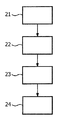
도 2는 본 발명에 따른 방법의 흐름 다이아그램을 도시한 것이다.
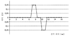
도 3은 감지 전극의 어래이에 의해 포착된 신호의 일 예를 도시한 것이다.
도 4는 도 2의 신호의 2차 공간 미분을 도시한 것이다
도 5는 자극 진폭의 예시적 공간적 분포를 도시한 것이다.
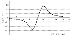
도 6은 도 4의 자극 진폭으로부터 얻어진 세포외 전기적 전위 분포를 도시한 것이다.
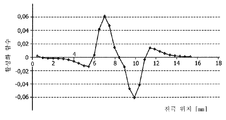
도 7은 타겟화된 신경 조직에 대한 활성 함수를 도시한 것이다.In the drawing,
1 schematically depicts a system for deep brain stimulation in accordance with the present invention.
2 shows a flow diagram of the method according to the invention.
3 shows an example of a signal captured by an array of sensing electrodes.
FIG. 4 illustrates the second spatial derivative of the signal of FIG. 2.
5 illustrates an example spatial distribution of stimulus amplitudes.
FIG. 6 shows the extracellular electrical potential distribution obtained from the stimulus amplitude of FIG. 4.
7 shows activity functions for targeted neural tissue.
도 1은 본 발명에 따른 심부 뇌 자극용 시스템(10)을 개략적으로 도시한 것이다. 본 시스템(10)은 전극(12)의 어래이를 구비한 프로브(11)를 포함한다. 전극(12)은 바람직하게 프로브 표면의 일부 상에 규칙적으로 배열된다. 선택된 전극들(12)은 감지(sensing)를 위해 디자인될 수 있으며, 나머지 전극들은 자극을 위해 제공될 수 있다. 감지 전극(12)의 치수는 자극 전극(12)의 치수와 상이할 수 있다. 정확한 측정을 위하여, 전극(12)은 바람직하게 작다. 자극 전달을 위해 임피던스를 낮추기 위하여, 자극 전극(12)은 바람직하게 다소 크다.1 schematically illustrates a system for
본 발명에 따른 시스템(10)의 바람직한 구체예에서, 모든 전극(12)은 감지 뿐만 아니라 자극을 위해 사용될 수 있다. 스위칭 유닛(switching unit; 13)은 전극(12)의 감지 기능과 자극 기능 간에 스위칭을 위해 제공된다. 전극(12)의 치수는 바람직하게 정확한 감지 및 자극을 위한 낮은 임피던스 둘 모두를 허용하기 위해 족절히 조절한다. 타겟 크기, 예를 들어 시상밑핵(subthalamic nucleus)의 서브-파트(sub-part)는 1 mm 정도 작을 수 있다. 이에 따라, 정확한 감지를 가능하게 하기 위하여, 이중 목적 전극은 0.1 내지 1 ㎟ 범위의 표면적을 갖는다.In a preferred embodiment of the
이중 목적 전극(12)을 사용함에 있어서 중요한 장점은, 자극 진폭의 적합한 분포를 결정하는 전기적 문제가 대칭적이고 이에 따라 최상의 결과를 얻는다는 것이다. 스위칭 유닛(13)은 프로세서(14)에 의해 제어된다. 프로세서(14)는 모든 전극(12)을 동일 모드 (감지 또는 자극)에 놓이게 하도록 스위칭 유닛(13)을 제어할 수 있거나 전극(12)의 일부를 자극을 위해 사용하면서 나머지 전극(12)을 감지 모드에 놓이도록 선택할 수 있다.An important advantage in using the
프로세서(14)의 영향 하에서, 스위칭 유닛(13)은 전극(12)을 감지 제어기(15) 또는 자극 제어기(16) 중 하나에 연결시킨다. 시스템(10)은 전력 관리 모듈(power management module)을 포함할 수 있는 배터리 유닛(17)에 의해 전력 공급된다. 배터리 유닛(17)은 전력을 필요로 하는 시스템의 모든 부품에 전력 공급한다. 프로세서(14)는 시스템(10)의 기능(functioning)을 제어하고 심부 뇌 자극을 위한 자극 진폭의 공간적 분포를 결정하고 이를 전극(12)에 적용하기 위해 시스템(10)의 상기 언급된 기능적 부품들에 명령하도록 작동한다.Under the influence of the
본 발명이 또한 별도의 측정 시스템의 이식된 프로브로부터 감지된 신호를 수신하는 오프-라인(off-line) 시스템에서 사용될 수 있다는 것에 주목된다. 이후에, 오프-라인 시스템은 본 발명에 따라 신호를 처리하고, 자극 신호의 공간적 분포를 이식된 프로브로 보낸다. 이식된 프로브는 자극 신호의 수신된 공간적 분포에 따라 자극을 제공한다.It is noted that the present invention can also be used in an off-line system that receives a sensed signal from an implanted probe of a separate measurement system. The off-line system then processes the signal according to the present invention and sends the spatial distribution of the stimulus signal to the implanted probe. The implanted probe provides the stimulus according to the received spatial distribution of the stimulus signal.
도 2는 본 발명에 따른 방법의 흐름 다이아그램을 도시한 것이다. 본 방법은 프로세서(14)의 제어 하에서, 도 1의 시스템(10)에 의해 실행될 수 있다. 본 방법은 프로브(11) 상의 전극(12)으로부터의 뉴런 활성을 나타내는 신호를 포착하기 위한 감지 단계(21)로 개시한다. 이러한 목적을 위하여, 프로세서(14)는 전극(12)의 적어도 일부를 감지 제어기(15)에 연결하도록 스위칭 유닛(13)에 명령한다. 전극(12)은 전극에 (또는 전극 가까이에) 뉴런 활성을 나타내는 신호를 입수한다. 프로세서(14)는 이후에 감지 제어기(15)를 통해 포착된 신호를 수신한다. 전극(12)로부터 수신될 수 있는 감지 신호의 일 예는 도 3에 제공된다.2 shows a flow diagram of the method according to the invention. The method may be executed by the
도 3은 감지 전극(12)의 어래이에 의해 포착된 신호(31)의 일 예를 도시한 것이다. 도 3에 도시된 신호(31)는 설명을 위해 제공된 시뮬레이션된 신호(31)이다. 이러한 측정된 뉴런 활성 신호(31)를 형성하는 시뮬레이션된 상황에서, 0.5 mm 거리로 동일하게 이격된 전극(12)을 갖는 1-차원 프로브(11)가 사용된다. 전극 어래이의 높이는 0 내지 16 mm이다. 병리학적 (전기적) 뉴런 활성의 소스/싱크(source/sink)는 높이 7 및 10 mm 각각에서 프로브(11)로부터 측면으로 1 mm에 위치되어, 상응하는 전극(12)에서 측정된 피크 진폭(peak amplitude)을 형성시킨다.3 shows an example of the
뇌에서 뉴런 활성을 측정한 후에, 프로세서(14)는 비정형적 뉴런 활성을 갖는 하나 이상의 뇌 영역을 발견하기 위하여, 신호 처리 단계(22)(도 2)에서 포착된 신호(31)를 처리한다. 비정형적 뉴런 활성을 갖는 이러한 영역은 전기적 자극으로부터 혜택을 받을 수 있는 병리학적으로 거동하는 뉴런을 지시할 수 있다. 신호 처리 단계(22)에서, 여러 상이한 알고리듬이 비정형적인 또는 의심되는 뉴런 활성을 인지하기 위해 사용될 수 있다. 매우 기본적인 알고리듬에서, 프로세서(14)는 사전결정된 (양성 또는 음성) 유발 수준(trigger level)을 초과하는 신호 진폭을 찾을 수 있다. 자극 진폭은 이후에 과도한 뉴런 활성을 측정하는 전극(12)에서 유도될 수 있다.After measuring neuronal activity in the brain,
보다 발달된 알고리듬에서, 포착된 신호(31)로부터 특징부(feature)가 추출될 수 있다. 예를 들어, 대역통과 필터는 특정 주파수 범위에서 뉴런 활성을 검출하기 위해 사용될 수 있다. 베타-밴드(8-30 Hz)에서의 활성 증가는, 예를 들어 파킨슨 질환의 증상과 관련이 있다. 이는 처리된 신호의 가장 높은 진폭을 갖는 전극(12)을 자극함으로써 처리될 수 있다. 베타-밴드의 예에서, 8 내지 30 Hz 주파수 범위에서 최대 신호를 입수하는 전극(12)은 자극화될 수 있다.In more developed algorithms, features can be extracted from the captured
비정형적으로 작용하는 뉴런 조직을 찾기 위한 바람직한 알고리듬은 도 4 내지 도 6을 참조로 하여 하기에 기술된다. 이러한 바람직한 알고리듬은 헬름홀츠 교환 이론(Helmholtz reciprocity theorem)과 결합된 전류-소스-밀도 이미지화로서 공지된 기술을 이용한다.Preferred algorithms for finding atypically functioning neuronal tissues are described below with reference to FIGS. This preferred algorithm uses a technique known as current-source-density imaging combined with the Helmholtz reciprocity theorem.
도 4는 도 3의 신호(31)의 2차 공간 미분(41)을 도시한 것이다. 계산된 2차 공간 미분은, 또한 뉴런 활성 소스가 포착된 신호(31)로부터 직접적으로 보다 덜 가시적일 때, 뉴런 활성 소스의 공간적 위치의 매우 명확한 지시를 제공한다. 대안적으로, 시스템은 대역통과 필터링된 신호 또는 측정된 신호로부터 추출된 다른 특징부의 2차 공간 미분을 사용할 수 있다.FIG. 4 shows the secondary
진폭 선택 단계(23)(도 2)에서, 자극 진폭의 적합한 공간적 분포가 결정된다. 이러한 공간적 분포는 포착된 신호(31)로부터 직접적으로 유도될 수 있거나, 추가 처리 단계에 의해 간접적으로 유도될 수 있다. 헬름홀츠 교환 이론에 따르면, 계산된 2차 공간 미분 값과 직접적으로 관련하여 전극(12) 어래이에 전류 분포의 인가는 측정된 활성의 소스 쪽으로 향하는 전기적 자극을 초래한다.In the amplitude selection step 23 (FIG. 2), a suitable spatial distribution of the stimulus amplitude is determined. This spatial distribution can be derived directly from the captured
도 5는 도 4에 도시된 2차 공간 미분 값을 기초로 하여 결정된 자극 진폭(51)의 예시적 공간적 분포를 도시한 것이다. 이러한 분포(51)를 위하여, 단 두 개의 독립적인 펄스 발생기가 이용 가능할 것으로 추측된다. 먼저, 0(zero)으로부터 가장 크게 벗어난 2차 공간 미분 값의 두 개의 영역이 결정된다: 6.5 내지 7.5 mm 및 9.5 내지 10.5 mm. 둘 모두의 구역에서 세 개의 전극이 이용 가능하다. 둘 모두의 영역에 대해 평균처리하고 스케일링하여, 자극 진폭 +0.15가 세 개의 전극의 제 1 영역에 인가되고 -0.15가 세 개의 전극의 제 2 영역에 인가된다. 자극 진폭(51)의 공간적 분포의 결정은 프로세서(14)에 의해 수행된다.FIG. 5 shows an exemplary spatial distribution of the stimulus amplitude 51 determined based on the second order spatial differential value shown in FIG. 4. For this distribution 51 it is assumed that only two independent pulse generators will be available. First, two regions of the secondary spatial differential value that deviate most significantly from zero are determined: 6.5 to 7.5 mm and 9.5 to 10.5 mm. Three electrodes are available in both zones. Averaging and scaling for both regions, stimulus amplitude +0.15 is applied to the first region of the three electrodes and -0.15 to the second region of the three electrodes. Determination of the spatial distribution of the stimulus amplitudes 51 is performed by the
자극 단계(24)에서, 프로세서(14)는 이후에, 스위칭 유닛(13)이 프로브 또는 적어도 필수적인 전극(12)을 자극 모드에 놓이게 하면서, 자극 진폭(51)의 공간적 분포를 전극(12)에 인가하도록 자극 제어기에 명령한다.In the
도 6은 본 발명의 방법에 따른 자극 장 전위(stimulation field potential)를 도시한 것이다. 이는 각각 7 mm 및 10 mm의 높이에서 1 mm 측면에 위치된 뉴런 활성을 위한 2차 미분(도 4)으로부터 유도된 것이다. 이러한 플롯은 큰 전위 구배가 추정된 뉴런 활성의 위치에서 형성되는 것을 나타낸다.6 shows the stimulation field potential according to the method of the present invention. This is derived from the second derivative (FIG. 4) for neuronal activity located 1 mm laterally at heights of 7 mm and 10 mm, respectively. This plot shows that a large potential gradient is formed at the location of the estimated neuronal activity.
도 7은 (뉴런 활성을 유도하는) 소위 활성 함수를 도시한 것으로서, 이는 7 및 10 mm의 요망되는 위치에서 인가된 자극 장 피크의 2차 미분에 비례한다.FIG. 7 shows the so-called activity function (inducing neuronal activity), which is proportional to the second derivative of the applied stimulus field peaks at the desired positions of 7 and 10 mm.
본 발명에 따른 방법의 일 구체예에서, 뉴런 활성은 전극(12)에서 2회 측정된다. 1회는 약물 복용 중인 환자에게서 측정되고 나머지 1회는 약물 복용 중이지 않는 환자에게서 측정된다. 활성(또는 유도된 특징부)에서 가장 큰 차이가 측정되는 이러한 전극들(12)은 자극 진폭을 제공하기 위해 사용된다. 결론적으로, 약물-반응은 전극 사용 시에 사전-필터링(pre-filtering)을 제공한다.In one embodiment of the method according to the invention, neuronal activity is measured twice at the
본 발명은 또한 본 발명을 실행시키도록 구성된, 컴퓨터 프로그램, 특히 캐리어 상 또는 캐리어 내의 컴퓨터 프로그램으로 확장하는 것으로 인식될 것이다. 이러한 프로그램은 소스 코드, 대상 코드, 코드 중간 소스 및 대상 코드, 예를 들어 일부 컴파일링된 형태, 또는 본 발명에 따른 방법의 실행에서 사용하기에 적합한 임의의 다른 형태로 존재할 수 있다. 또한, 이러한 프로그램이 여러 상이한 아키텍쳐 디자인(architectural design)을 가질 수 있는 것으로 인식될 것이다. 예를 들어, 본 발명에 따른 방법 또는 시스템의 기능을 실행하는 프로그램 코드는 하나 이상의 서브루틴(subroutine)으로 세분화될 수 있다. 이러한 서브루틴 중에서 기능을 분포시키기 위한 여러 상이한 방식은 당업자에게 자명할 것이다. 서브루틴은 독립형 프로그램(self-contained program)을 형성시키기 위해 하나의 실행 가능한 파일에 함께 저장될 수 있다. 이러한 실행 가능한 파일은 컴퓨터 실행 가능한 명령(instruction), 예를 들어, 프로세서 명령 및/또는 인터프리터(interpreter) 명령 (예를 들어, 자바 인터프리터 명령)을 포함할 수 있다. 대안적으로, 서브루틴 중 하나 이상 또는 전부는 적어도 하나의 외부 라이브러리 파일에 저장될 수 있고 예를 들어 런-타임(run-time)에 통계적 또는 동적 중 하나로 메인 프로그램과 연결될 수 있다. 메인 프로그램은 서브루틴 중 적어도 하나에 적어도 하나의 콜(call)을 포함한다. 도한, 서브루틴은 서로에 함수 콜(function call)을 포함할 수 있다. 컴퓨터 프로그램 제품에 관한 구체예는 기술된 방법들 중 적어도 하나의 처리 단계 각각에 대해 상응하는 컴퓨터 실행 가능한 명령을 포함한다. 이러한 명령은 서브루틴에 세분화되고/거나 통계적으로 또는 동적으로 연결될 수 있는 하나 이상의 파일에 저장될 수 있다. 컴퓨터 프로그램 제품에 관한 다른 구체예는 기술된 시스템 및/또는 제품 중 적어도 하나의 각각의 수단에 해당하는 컴퓨터 실행 가능한 명령을 포함한다. 이러한 명령은 서브루틴으로 세분화될 수 있고/거나 통계적으로 또는 동적으로 연결될 수 있는 하나 이상의 파일에 저장될 수 있다.It will be appreciated that the present invention also extends to computer programs, in particular computer programs on or in a carrier, configured to carry out the invention. Such a program may be present in source code, object code, intermediate code source and object code, for example in some compiled form, or in any other form suitable for use in the execution of the method according to the invention. It will also be appreciated that such a program can have many different architectural designs. For example, program code for carrying out the functions of the method or system according to the invention may be subdivided into one or more subroutines. Many different ways to distribute functions among these subroutines will be apparent to those skilled in the art. Subroutines can be stored together in one executable file to form a self-contained program. Such executable files may include computer executable instructions, eg, processor instructions and / or interpreter instructions (eg, Java interpreter instructions). Alternatively, one or more or all of the subroutines may be stored in at least one external library file and may be associated with the main program either statistically or dynamically at run-time, for example. The main program includes at least one call in at least one of the subroutines. Also, subroutines may include function calls to each other. Embodiments of the computer program product include corresponding computer executable instructions for each of the processing steps of at least one of the described methods. Such instructions may be stored in one or more files that may be subdivided into subroutines and / or linked statistically or dynamically. Another embodiment of a computer program product includes computer executable instructions corresponding to each means of at least one of the described systems and / or products. Such instructions may be subdivided into subroutines and / or stored in one or more files that may be linked statistically or dynamically.
컴퓨터 프로그램의 캐리어는 프로그램을 운반할 수 있는 임의의 실재물 또는 디바이스일 수 있다. 예를 들어, 캐리어는 ROM과 같은 저장 매체, 예를 들어 CD ROM 또는 반도체 ROM, 또는 자기 기록 매체, 예를 들어 플로피 디스크 또는 하드 디스크를 포함할 수 있다. 또한, 캐리어는 전기적 또는 광학적 신호와 같은 전송 가능한 캐리어일 수 있는데, 이러한 캐리어는 전기 또는 광학 케이블을 통해 또는 라디오 또는 다른 수단에 의해 전달될 수 있다. 프로그램이 이러한 신호로 구체화될 때, 캐리어는 이러한 케이블 또는 다른 디바이스 또는 수단에 의해 구성될 수 있다. 대안적으로, 캐리어는 프로그램이 포함되는 집적 회로일 수 있는데, 이러한 집적 회로는 관련된 방법을 수행하도록 또는 이의 수행에서 사용하도록 구성된다.The carrier of the computer program may be any entity or device capable of carrying the program. For example, the carrier may comprise a storage medium such as a ROM, for example a CD ROM or a semiconductor ROM, or a magnetic recording medium, for example a floppy disk or a hard disk. In addition, the carrier may be a transmittable carrier such as an electrical or optical signal, which carrier may be delivered via an electrical or optical cable or by radio or other means. When a program is embodied in such a signal, the carrier may be configured by such a cable or other device or means. Alternatively, the carrier may be an integrated circuit in which a program is included, which integrated circuit is configured to perform or in use in a related method.
상술된 구체예는 본 발명을 한정하기 보다는 예시적인 것이며, 당업자가 첨부된 청구범위를 벗어나지 않는 여러 다른 구체예들을 디자인할 수 있는 것으로 보인다. 청구항들에서, 괄호 사이에 위치된 임의의 문헌 표시는 청구항을 한정하는 것으로 해석되지 않을 것이다. 동사 "포함하다" 및 이의 활용형의 사용은 청구항에 기술된 것과 상이한 구성요소 또는 단계의 존재를 배제하지 않는다. 단수의 구성성분이라는 용어는 복수의 이러한 구성성분들의 존재를 배제하지 않는다. 본 발명은 여러 별도의 구성성분들을 포함하는 하드웨어에 의해 및 적절하게 프로그래밍된 컴퓨터에 의해 실행될 수 있다. 여러 수단들을 열거한 디바이스 청구항에서, 이러한 수단들 중 수개는 하드웨어의 하나의 동일한 항목에 의해 포함될 수 있다. 특정 수치가 서로 상이한 종속항에 기술되어 있다는 사실은 이러한 측정의 조합이 유리하게 사용되지 못할 수 있다는 것으로 명시하는 것은 아니다.
The above-described embodiments are illustrative rather than limiting of the invention, and it appears that those skilled in the art can design various other embodiments without departing from the appended claims. In the claims, any reference signs placed between parentheses shall not be construed as limiting the claim. The use of the verb "comprises" and their conjugations does not exclude the presence of elements or steps other than those set forth in the claims. The term singular component does not exclude the presence of a plurality of such components. The invention can be implemented by means of hardware comprising several separate components and by means of a suitably programmed computer. In the device claim enumerating several means, several of these means may be included by one and the same item of hardware. The fact that certain values are described in different dependent claims does not specify that a combination of these measures may not be used advantageously.
Claims (14)
감지 전극(12)으로부터 포착된 신호를 수신하고,
포착된 신호를 처리하여 비정형적 뉴런 활성을 갖는 하나 이상의 뇌 영역을 발견하고,
포착된 신호를 기반으로 자극 전극(12)의 어래이에 대한 자극 진폭의 공간적 분포를 결정하고,
비정형적 뉴런 활성을 갖는 하나 이상의 영역을 자극하기 위해, 자극 전극(12)의 어래이에 자극 진폭의 공간적 분포를 적용하기 위한, 감지 전극(12) 및 자극 전극(12)에 작동 가능하게 연결된 프로세서(14)를 포함하는 심부 뇌 자극용 시스템(10).A probe 11 having an array of sensing electrodes 12 for capturing a signal indicative of neuronal activity at a corresponding location in the brain and an array of stimulation electrodes 12 for applying stimulation amplitudes to corresponding brain regions; And
Receive the captured signal from the sensing electrode 12,
Process the captured signal to find one or more brain regions with atypical neuronal activity,
Determine the spatial distribution of the stimulus amplitude with respect to the array of stimulation electrodes 12 based on the captured signal,
A processor operably connected to the sensing electrode 12 and the stimulation electrode 12 for applying a spatial distribution of stimulus amplitudes to the array of stimulation electrodes 12 to stimulate one or more regions with atypical neuronal activity. System for deep brain stimulation, comprising 14).
상기 포착된 신호를 처리하여 비정형적 뉴런 활성을 갖는 하나 이상의 뇌 영역을 발견하는 단계(22); 및
상기 포착된 신호를 기초로 하여, 자극 전극의 어래이에서의 자극 진폭의 분포를 결정하는 단계(23)를 포함하는, 심부 뇌 자극을 위한 공간적 분포 자극 진폭을 결정하는 방법.Receiving (21) from the array of sense electrodes on the sense probe a captured signal indicative of neuronal activity at the corresponding location in the brain;
Processing (22) the captured signal to find one or more brain regions with atypical neuronal activity; And
Based on the captured signal, determining (23) a distribution of stimulus amplitudes in the array of stimulation electrodes.
상기 포착된 신호를 처리하여 비정형적 뉴런 활성을 갖는 하나 이상의 뇌 영역을 발견하는 단계(22);
상기 포착된 신호를 기초로 하여, 자극 전극의 어래이에서의 자극 진폭의 공간적 분포를 결정하는 단계(23); 및
비정형적 뉴런 활성을 갖는 하나 이상의 영역을 자극하기 위해 자극 진폭의 공간적 분포를 자극 전극의 어래이에 적용하는 단계(24)를 포함하는, 심부 뇌 자극을 위해 자극 진폭이 공간적 분포를 적용하는 방법.Using (21) a sense probe with an array of sense electrodes for capturing a signal indicative of neuronal activity at a corresponding location in the brain;
Processing (22) the captured signal to find one or more brain regions with atypical neuronal activity;
Based on the captured signal, determining (23) a spatial distribution of stimulus amplitudes in the array of stimulation electrodes; And
Applying a spatial distribution of the stimulus amplitude to the array of stimulation electrodes to stimulate one or more regions with atypical neuronal activity (24).
Applications Claiming Priority (2)
| Application Number | Priority Date | Filing Date | Title |
|---|---|---|---|
| EP09166840 | 2009-07-30 | ||
| EP09166840.0 | 2009-07-30 |
Publications (1)
| Publication Number | Publication Date |
|---|---|
| KR20120043694A true KR20120043694A (en) | 2012-05-04 |
Family
ID=42983706
Family Applications (1)
| Application Number | Title | Priority Date | Filing Date |
|---|---|---|---|
| KR1020117027945A KR20120043694A (en) | 2009-07-30 | 2010-07-23 | System and method for deep brain stimulation |
Country Status (9)
| Country | Link |
|---|---|
| US (1) | US9119543B2 (en) |
| EP (1) | EP2459276B1 (en) |
| JP (1) | JP2013500115A (en) |
| KR (1) | KR20120043694A (en) |
| CN (1) | CN102470247B (en) |
| BR (1) | BRPI1015422A2 (en) |
| IL (1) | IL217735A0 (en) |
| RU (1) | RU2012107474A (en) |
| WO (1) | WO2011013041A1 (en) |
Cited By (1)
| Publication number | Priority date | Publication date | Assignee | Title |
|---|---|---|---|---|
| KR20230078644A (en) * | 2020-08-14 | 2023-06-02 | 유니버시티 오브 휴스턴 시스템 | System and method for determining deep brain stimulation parameters |
Families Citing this family (29)
| Publication number | Priority date | Publication date | Assignee | Title |
|---|---|---|---|---|
| JP5653918B2 (en) | 2008-07-30 | 2015-01-14 | エコーレ ポリテクニーク フェデラーレ デ ローザンヌ (イーピーエフエル) | Apparatus and method for optimized stimulation of neural targets |
| EP3563902B1 (en) | 2008-11-12 | 2021-07-14 | Ecole Polytechnique Fédérale de Lausanne | Microfabricated neurostimulation device |
| CA3026948C (en) | 2009-12-01 | 2022-07-12 | Ecole Polytechnique Federale De Lausanne | Microfabricated neurostimulation device and methods of making and using the same |
| CA2795159C (en) | 2010-04-01 | 2020-11-03 | Ecole Polytechnique Federale De Lausanne | Device for interacting with neurological tissue and methods of making and using the same |
| EP2626109A1 (en) * | 2012-02-08 | 2013-08-14 | Sapiens Steering Brain Stimulation B.V. | A probe system for brain applications |
| US20140074187A1 (en) * | 2012-04-23 | 2014-03-13 | Medtronic, Inc. | Electrode selection based on current source density analysis |
| EP2849839A4 (en) | 2012-05-16 | 2015-12-09 | Univ Utah Res Found | Charge steering high density electrode array |
| US10039507B2 (en) | 2012-09-19 | 2018-08-07 | The Regents Of The University Of Michigan | Advanced intraoperative neural targeting system and method |
| JP6300208B2 (en) * | 2012-10-05 | 2018-03-28 | 大学共同利用機関法人自然科学研究機構 | Device for acquiring electrical activity in the brain and use thereof |
| US10016606B2 (en) | 2014-01-17 | 2018-07-10 | Medtronic, Inc. | Movement disorder symptom control |
| US11311718B2 (en) | 2014-05-16 | 2022-04-26 | Aleva Neurotherapeutics Sa | Device for interacting with neurological tissue and methods of making and using the same |
| US10966620B2 (en) | 2014-05-16 | 2021-04-06 | Aleva Neurotherapeutics Sa | Device for interacting with neurological tissue and methods of making and using the same |
| US9403011B2 (en) | 2014-08-27 | 2016-08-02 | Aleva Neurotherapeutics | Leadless neurostimulator |
| US9474894B2 (en) | 2014-08-27 | 2016-10-25 | Aleva Neurotherapeutics | Deep brain stimulation lead |
| US10095837B2 (en) | 2014-11-21 | 2018-10-09 | Medtronic, Inc. | Real-time phase detection of frequency band |
| CN104548390B (en) | 2014-12-26 | 2018-03-23 | 中国科学院深圳先进技术研究院 | It is a kind of to obtain the method and system that the ultrasound emission sequence that cranium focuses on ultrasound is worn for launching |
| EP3061491A1 (en) * | 2015-02-27 | 2016-08-31 | Oticon Medical A/S | Auditory device with a filter bank and with means for extracting an onset time and a method for generating a stimulation signal |
| CN107921260B (en) * | 2015-07-10 | 2022-02-25 | 神经毫微股份公司 | Method and system for improved stimulation of excitable tissue |
| WO2017134587A1 (en) | 2016-02-02 | 2017-08-10 | Aleva Neurotherapeutics, Sa | Treatment of autoimmune diseases with deep brain stimulation |
| US10786674B2 (en) | 2016-03-08 | 2020-09-29 | Medtronic, Inc. | Medical therapy target definition |
| US10864368B2 (en) | 2016-09-27 | 2020-12-15 | Medtronic, Inc. | Adaptive deep brain stimulation using homeostatic window |
| US10702692B2 (en) | 2018-03-02 | 2020-07-07 | Aleva Neurotherapeutics | Neurostimulation device |
| CN108744273B (en) * | 2018-05-29 | 2020-04-28 | 西安交通大学 | Transcranial noninvasive deep brain bifocal stimulation system and method for neural circuit |
| EP3823714A4 (en) * | 2018-07-17 | 2022-04-20 | Dignity Health | Systems and methods for treatment of cancer using alternating electric field generation |
| US11318296B2 (en) | 2018-10-26 | 2022-05-03 | Medtronic, Inc. | Signal-based automated deep brain stimulation programming |
| US11045652B2 (en) | 2019-04-26 | 2021-06-29 | Medtronic, Inc. | Determination of therapy electrode locations relative to oscillatory sources within patient |
| US11238591B1 (en) | 2020-07-15 | 2022-02-01 | Taipei Medical University (Tmu) | Medical image processing system and method thereof |
| TWI737404B (en) * | 2020-07-15 | 2021-08-21 | 臺北醫學大學 | Medical image processing system and method thereof |
| US11872402B2 (en) | 2020-10-22 | 2024-01-16 | Medtronic, Inc. | Determining relative phase relationships for delivery of electrical stimulation therapy |
Family Cites Families (8)
| Publication number | Priority date | Publication date | Assignee | Title |
|---|---|---|---|---|
| US20020169485A1 (en) * | 1995-10-16 | 2002-11-14 | Neuropace, Inc. | Differential neurostimulation therapy driven by physiological context |
| US6016449A (en) * | 1997-10-27 | 2000-01-18 | Neuropace, Inc. | System for treatment of neurological disorders |
| US6366813B1 (en) | 1998-08-05 | 2002-04-02 | Dilorenzo Daniel J. | Apparatus and method for closed-loop intracranical stimulation for optimal control of neurological disease |
| US7299096B2 (en) * | 2001-03-08 | 2007-11-20 | Northstar Neuroscience, Inc. | System and method for treating Parkinson's Disease and other movement disorders |
| US7236830B2 (en) | 2002-12-10 | 2007-06-26 | Northstar Neuroscience, Inc. | Systems and methods for enhancing or optimizing neural stimulation therapy for treating symptoms of Parkinson's disease and/or other movement disorders |
| US8190248B2 (en) * | 2003-10-16 | 2012-05-29 | Louisiana Tech University Foundation, Inc. | Medical devices for the detection, prevention and/or treatment of neurological disorders, and methods related thereto |
| CN1597011B (en) * | 2004-07-27 | 2011-06-29 | 天津大学 | Outlay brain deep part stimulator |
| US8295934B2 (en) * | 2006-11-14 | 2012-10-23 | Neurovista Corporation | Systems and methods of reducing artifact in neurological stimulation systems |
-
2010
- 2010-07-23 KR KR1020117027945A patent/KR20120043694A/en not_active Application Discontinuation
- 2010-07-23 BR BRPI1015422A patent/BRPI1015422A2/en not_active Application Discontinuation
- 2010-07-23 WO PCT/IB2010/053352 patent/WO2011013041A1/en active Application Filing
- 2010-07-23 EP EP10742289.1A patent/EP2459276B1/en active Active
- 2010-07-23 RU RU2012107474/14A patent/RU2012107474A/en unknown
- 2010-07-23 CN CN201080033903.XA patent/CN102470247B/en active Active
- 2010-07-23 JP JP2012522298A patent/JP2013500115A/en active Pending
- 2010-07-23 US US13/321,190 patent/US9119543B2/en active Active
-
2012
- 2012-01-25 IL IL217735A patent/IL217735A0/en unknown
Cited By (1)
| Publication number | Priority date | Publication date | Assignee | Title |
|---|---|---|---|---|
| KR20230078644A (en) * | 2020-08-14 | 2023-06-02 | 유니버시티 오브 휴스턴 시스템 | System and method for determining deep brain stimulation parameters |
Also Published As
| Publication number | Publication date |
|---|---|
| BRPI1015422A2 (en) | 2016-04-19 |
| US9119543B2 (en) | 2015-09-01 |
| EP2459276A1 (en) | 2012-06-06 |
| WO2011013041A1 (en) | 2011-02-03 |
| JP2013500115A (en) | 2013-01-07 |
| RU2012107474A (en) | 2013-09-10 |
| EP2459276B1 (en) | 2016-11-09 |
| US20120150256A1 (en) | 2012-06-14 |
| CN102470247A (en) | 2012-05-23 |
| CN102470247B (en) | 2015-11-25 |
| IL217735A0 (en) | 2012-03-29 |
Similar Documents
| Publication | Publication Date | Title |
|---|---|---|
| KR20120043694A (en) | System and method for deep brain stimulation | |
| Lefaucheur | Transcranial magnetic stimulation | |
| FI114613B (en) | Method and apparatus for dose calculation of magnetic stimulation | |
| RU2471517C2 (en) | One-trial correct wire installation for deep brain stimulation | |
| JP6254281B2 (en) | EEG measurement and brain stimulation system | |
| Hemm et al. | Deep brain stimulation in movement disorders: stereotactic coregistration of two-dimensional electrical field modeling and magnetic resonance imaging | |
| TWI498101B (en) | Method of analyzing nerve fiber distribution and measuring standardized induced compound motion electric potential | |
| JP2018513714A (en) | Electrode-nerve distance estimation | |
| Shpaner et al. | Disambiguating the roles of area V1 and the lateral occipital complex (LOC) in contour integration | |
| Favilla et al. | Ranking brain areas encoding the perceived level of pain from fMRI data | |
| JP5420644B2 (en) | Method and system for determining a threshold for spike detection of electrophysiological signals | |
| EP1366782A1 (en) | Targeting method and apparatus for the magnetic stimulation of the brain | |
| JP2013519418A (en) | Method and system for determining settings in deep brain stimulation therapy | |
| US9339642B1 (en) | System and method for conducting multi-electrode electrical stimulation | |
| KR20120117821A (en) | A system and a method for determination of an orientation of a biomedical stimulation device | |
| KR20150061609A (en) | Brain signal detecting and brain stimulating system | |
| US20230075205A1 (en) | Devices, systems and methods for cortical stimulation | |
| Lycke et al. | Low-threshold, high-resolution, chronically stable intracortical microstimulation by ultraflexible electrodes | |
| US20100249577A1 (en) | Synergistic Electromagnetic Tracking With TMS Systems | |
| CN105193412B (en) | A kind of method removed for transcranial magnetic stimulation brain evoked brain potential artefact | |
| Winestone et al. | The use of macroelectrodes in recording cellular spiking activity | |
| KR101630747B1 (en) | Method and apparatus for estimating olfactory function | |
| Trillenberg et al. | Variation of stimulation intensity in transcranial magnetic stimulation with depth | |
| WO2019243596A1 (en) | System and method for the estimation of physical parameters of a medium | |
| Osanai et al. | Micro-coil-induced inhomogeneous electric field produces sound-driven-like neural responses in microcircuits of the mouse auditory cortex in vivo |
Legal Events
| Date | Code | Title | Description |
|---|---|---|---|
| WITN | Application deemed withdrawn, e.g. because no request for examination was filed or no examination fee was paid |