KR20120006998A - Intraocular sustained release drug delivery systems and methods for treating ocular conditions - Google Patents
Intraocular sustained release drug delivery systems and methods for treating ocular conditions Download PDFInfo
- Publication number
- KR20120006998A KR20120006998A KR1020117023983A KR20117023983A KR20120006998A KR 20120006998 A KR20120006998 A KR 20120006998A KR 1020117023983 A KR1020117023983 A KR 1020117023983A KR 20117023983 A KR20117023983 A KR 20117023983A KR 20120006998 A KR20120006998 A KR 20120006998A
- Authority
- KR
- South Korea
- Prior art keywords
- microspheres
- implant
- anterior
- eye
- latanoprost
- Prior art date
Links
Images


Classifications
-
- A—HUMAN NECESSITIES
- A61—MEDICAL OR VETERINARY SCIENCE; HYGIENE
- A61K—PREPARATIONS FOR MEDICAL, DENTAL OR TOILETRY PURPOSES
- A61K9/00—Medicinal preparations characterised by special physical form
- A61K9/0012—Galenical forms characterised by the site of application
- A61K9/0048—Eye, e.g. artificial tears
- A61K9/0051—Ocular inserts, ocular implants
-
- A—HUMAN NECESSITIES
- A61—MEDICAL OR VETERINARY SCIENCE; HYGIENE
- A61K—PREPARATIONS FOR MEDICAL, DENTAL OR TOILETRY PURPOSES
- A61K31/00—Medicinal preparations containing organic active ingredients
-
- A—HUMAN NECESSITIES
- A61—MEDICAL OR VETERINARY SCIENCE; HYGIENE
- A61K—PREPARATIONS FOR MEDICAL, DENTAL OR TOILETRY PURPOSES
- A61K31/00—Medicinal preparations containing organic active ingredients
- A61K31/557—Eicosanoids, e.g. leukotrienes or prostaglandins
-
- A—HUMAN NECESSITIES
- A61—MEDICAL OR VETERINARY SCIENCE; HYGIENE
- A61K—PREPARATIONS FOR MEDICAL, DENTAL OR TOILETRY PURPOSES
- A61K31/00—Medicinal preparations containing organic active ingredients
- A61K31/557—Eicosanoids, e.g. leukotrienes or prostaglandins
- A61K31/5575—Eicosanoids, e.g. leukotrienes or prostaglandins having a cyclopentane, e.g. prostaglandin E2, prostaglandin F2-alpha
-
- A—HUMAN NECESSITIES
- A61—MEDICAL OR VETERINARY SCIENCE; HYGIENE
- A61K—PREPARATIONS FOR MEDICAL, DENTAL OR TOILETRY PURPOSES
- A61K9/00—Medicinal preparations characterised by special physical form
- A61K9/14—Particulate form, e.g. powders, Processes for size reducing of pure drugs or the resulting products, Pure drug nanoparticles
- A61K9/16—Agglomerates; Granulates; Microbeadlets ; Microspheres; Pellets; Solid products obtained by spray drying, spray freeze drying, spray congealing,(multiple) emulsion solvent evaporation or extraction
- A61K9/1605—Excipients; Inactive ingredients
- A61K9/1629—Organic macromolecular compounds
- A61K9/1641—Organic macromolecular compounds obtained otherwise than by reactions only involving carbon-to-carbon unsaturated bonds, e.g. polyethylene glycol, poloxamers
- A61K9/1647—Polyesters, e.g. poly(lactide-co-glycolide)
-
- A—HUMAN NECESSITIES
- A61—MEDICAL OR VETERINARY SCIENCE; HYGIENE
- A61K—PREPARATIONS FOR MEDICAL, DENTAL OR TOILETRY PURPOSES
- A61K9/00—Medicinal preparations characterised by special physical form
- A61K9/70—Web, sheet or filament bases ; Films; Fibres of the matrix type containing drug
-
- A—HUMAN NECESSITIES
- A61—MEDICAL OR VETERINARY SCIENCE; HYGIENE
- A61P—SPECIFIC THERAPEUTIC ACTIVITY OF CHEMICAL COMPOUNDS OR MEDICINAL PREPARATIONS
- A61P27/00—Drugs for disorders of the senses
- A61P27/02—Ophthalmic agents
- A61P27/06—Antiglaucoma agents or miotics
-
- A—HUMAN NECESSITIES
- A61—MEDICAL OR VETERINARY SCIENCE; HYGIENE
- A61K—PREPARATIONS FOR MEDICAL, DENTAL OR TOILETRY PURPOSES
- A61K2121/00—Preparations for use in therapy
Abstract
본 발명은 10일 내지 1년의 기간에 걸쳐 치료량의 안압강하제를 방출시킴으로써 (녹내장과 같은) 눈의 고안압 상태를 치료하기에 효과적인, 안압강하제 및 생분해성 중합체를 포함하는 전방내 또는 전방 유리체 배치를 위한 생체적합성, 생침식성 서방성 임플란트 및 마이크로스피어에 관한 것이다.The present invention provides an anterior or anterior vitreous arrangement comprising an ocular hypotensive and a biodegradable polymer, effective for treating ocular hypertensive conditions of the eye (such as glaucoma) by releasing a therapeutic amount of an antihypertensive agent over a period of 10 days to 1 year. A biocompatible, bioerodible sustained release implant and microspheres for
Description
교차참조Cross-reference
본 출원은 본원에 특정 참고문헌으로 인용된, 2009년 3월 25일자로 출원된 미국 비-임시 특허 출원 번호 제12/411,250호의 특권을 주장한다.This application claims the privilege of US Non-Temporary Patent Application No. 12 / 411,250, filed March 25, 2009, which is incorporated herein by reference in its entirety.
본 발명은 안내(眼內) 시스템 및 안질환을 치료하기 위한 방법에 관한 것이다. 특히, 본 발명은 녹내장 또는 녹내장 위험의 증상일 수 있는 바와 같은 물주머니 상승 안내압 (즉, 고안압 상태)을 치료하기 위한 눈의 전방 (즉, 전방내 투여) 및/또는 유리체전방으로의 서방성 약물 전달 시스템 (즉, 약물 혼입 마이크로스피어 및/또는 임플란트)의 국소 투여에 관한 것이다. The present invention relates to intraocular systems and methods for treating ocular diseases. In particular, the present invention is directed to the anterior (ie, anterior administration) and / or anterior to the vitreous of the eye to treat water bag elevated intraocular pressure (ie, ocular pressure) as may be a symptom of glaucoma or glaucoma risk. To topical administration of a sex drug delivery system (ie drug incorporation microspheres and / or implants).
본 발명의 약물 전달 시스템은 약물 함유 임플란트 (즉, 단일, 일체식 서방성 약물 전달 시스템) 또는 임플란트 또는 복수의 약물 함유 마이크로스피어 (동의어로 "극미립자")일 수 있다. 약물 전달 시스템은 상승된 안내압 및/또는 녹내장과 같은 안구 질환 또는 질병을 치료하기 위해 치료적으로 사용될 수 있다. 녹내장은 증가된 물주머니 안내압 (IOP)를 특징으로 하는 눈의 질환이다. 비치료 녹내장은 실명을 결과할 수 있다. 녹내장은 일차 또는 이차 녹내장일 수 있다. 어른의 일차 녹내장 (선천적 녹내장)은 개방각 또는 급성 또는 만성 폐쇄각일 수 있다. 이차 녹내장은 포도막염, 안내 종양 또는 확대된 백내장과 같은 기존 안질환으로부터 결과한다. 다양한 안압강하제가 IOP를 저하시키고 녹내장을 치료하기 위해 사용되어 왔다. 예를 들어, 상표명 Xalatan® 하에 시판되는 특정 프로스타글란딘, 및 PGF2 α 유도체 (종종 프로스타글란딘 F2α유사체) 라타노프로스트와 같은 이들의 유사체 및 유도체가 고안압증 및 녹내장을 치료하기 위해 사용되어 왔다. 안내 프로스타글란딘 및 프로스타마이드 임플란트 및 마이크로스피어는 예를 들어 미국 특허 출원 번호 제11/368,845호; 11/303,462호, 10/837,260호 및 12/259,153호에 기술되어 있다. 출원 번호 제12/259,253호의 페이지 36 내지 47의 실시예 1 내지 5가 특히 중요하다. 출원 번호 제11/952,938호가 또한 중요하다. 추가적으로, 미국 특허 제5,972,326호 및 5,965,152호가 또한 중요하다.The drug delivery system of the present invention may be a drug containing implant (ie, a single, integral sustained release drug delivery system) or an implant or a plurality of drug containing microspheres (synonymously “microparticles”). Drug delivery systems can be used therapeutically to treat eye diseases or conditions such as elevated intraocular pressure and / or glaucoma. Glaucoma is an eye disease characterized by increased water pocket intraocular pressure (IOP). Untreated glaucoma can result in blindness. Glaucoma may be primary or secondary glaucoma. Primary glaucoma (congenital glaucoma) in adults can be open angle or acute or chronic oblique angle. Secondary glaucoma results from existing eye diseases such as uveitis, intraocular tumors or enlarged cataracts. Various hypotensive agents have been used to lower IOP and treat glaucoma. For example, certain prostaglandins sold under the trade name Xalatan®, and their analogs and derivatives such as PGF 2 α derivatives (often prostaglandin F 2α analogs) latanoprost have been used to treat ocular hypertension and glaucoma. Intraocular prostaglandins and prosamide implants and microspheres are described, for example, in US Patent Application No. 11 / 368,845; 11 / 303,462, 10 / 837,260 and 12 / 259,153. Of particular importance are Examples 1-5 of pages 36-47 of Application No. 12 / 259,253. Application number 11 / 952,938 is also important. In addition, US Pat. Nos. 5,972,326 and 5,965,152 are also important.
녹내장의 통상적인 치료는 IOP를 감소시키기 위한 안압강하제를 함유하는 안약의 매일 투여이다. 자주, 안약의 규칙적인 매일 사용에 대한 환자의 복약순응도는 낮다 (참조예: Nordstrom et al. AJO 2005; 140: 598). 추가적으로, 눈 감염이 부적절한 점안기 사용으로부터 결과할 수 있다. 따라서, 예를 들어 병원에 방문하는 동안 편리하게 관리될 수 있는 고안압증에 대한 장기간 (즉, 서방성) 치료 방법이 요구된다. 따라서, 상승된 IOP 및/또는 녹내장을 치료하기 위해 안내 치료적 사용을 위한 서방성 안내 약물 전달 시스템 (임플란트 및/또는 마이크로스피어를 포함함)을 제공하는 것이 유리하다.
A common treatment for glaucoma is the daily administration of eye drops containing an ocular hypotensive to reduce IOP. Frequently, the patient's compliance with the regular daily use of eye drops is low (see Nordstrom et al. AJO 2005; 140: 598). In addition, eye infection may result from inadequate eye drop use. Thus, there is a need for a long-term (ie sustained release) treatment method for ocular hypertension that can be conveniently managed, for example, during a visit to the hospital. Thus, it is advantageous to provide sustained release intraocular drug delivery systems (including implants and / or microspheres) for intraocular therapeutic use to treat elevated IOP and / or glaucoma.
요약summary
본 발명은 상기 요구를 충족시키며, 서방성 안내 약물 전달 시스템, 약물 전달 시스템을 제조하기 위한 방법, 및 약물 전달 시스템을 사용하여 안질환을 치료하기 위한 방법을 제공한다. 서방성 안내 약물 전달 시스템은 하나 이상의 치료용 안압강하제 (예를 들어, 라타노프로스트와 같은 프로스타글란딘 또는 프로스타마이드)의 연장된 방출을 제공하는 것이 유리한 임플란트 또는 마이크로스피어의 형태를 갖는다.The present invention meets these needs and provides a sustained release intraocular drug delivery system, a method for manufacturing a drug delivery system, and a method for treating ocular disease using the drug delivery system. Sustained release intraocular drug delivery systems take the form of implants or microspheres that are advantageous to provide prolonged release of one or more therapeutic hypotensive agents (eg, prostaglandins or prosamides such as latanoprost).
정의Justice
본원에서는 하기의 정의가 사용된다.The following definitions are used herein.
"약"은 수, 파라미터 또는 이렇게 정성화된 특징의 ±10%를 의미한다."About" means ± 10% of a number, parameter, or such qualitative feature.
"마이크로스피어" 및 "극미립자"는 전방내 투여되도록 구조화되거나, 크기화되거나 다른식으로 구성되는 작은 직경 또는 치수 (하기 참조)의 장치 또는 소자를 언급하기 위해 동의어로 사용된다. 마이크로스피어 또는 극미립자는 치료제를 캡슐화시키거나 혼입시킨 생체적합성 매트릭스를 포함하는 입자, 마이크로 또는 나노스피어, 작은 단편, 극미립자, 나노입자, 미세 분말 등을 포함한다. 마이크로스피어는 일반적으로 눈의 생리학적 조건과 생체적합성이며, 현저한 부작용을 유발하지 않는다. 본워너에 기술된 바와 같이 제조되고 사용되는 마이크로스피어는 전방내 투여되고, 시야를 방해하지 않으면서 안전하게 사용될 수 있다. 마이크로스피어는 1 ㎜ 미만과 같은 직경 또는 길이와 같은 최대 치수를 가질 수 있다. 예를 들어, 극미립자는 약 500 ㎛ 미만의 최대 치수를 가질 수 있다. 마이크로스피어는 또한 약 200 ㎛ 이하의 최대 치수를 가질 수 있으며, 바람직하게는 30 ㎛ 초과 내지 약 50 ㎛ 또는 약 75 미크론의 최대 치수를 가질 수 있다. "임플란트"는 마이크로스피어보다 상당히 더 큰 약물 전달 장치이며, 복수 (즉, 수백 또는 수천개)의 마이크로스피어가 (녹내장과 같은) 안질환을 치료하기 위해 투여되는 반면, 일반적으로 단지 1개 내지 6개 이항의 임플란트가 동일한 용도로 투여된다."Microspheres" and "microparticles" are used synonymously to refer to devices or devices of small diameter or dimension (see below) that are structured, sized, or otherwise configured for anterior administration. Microspheres or microparticles include particles, micro or nanospheres, small fragments, microparticles, nanoparticles, fine powders, and the like, which contain a biocompatible matrix that encapsulates or incorporates a therapeutic agent. Microspheres are generally biocompatible with the physiological conditions of the eye and do not cause significant side effects. Microspheres prepared and used as described in Bonwarner can be administered intranasally and used safely without obstructing the field of view. The microspheres can have a maximum dimension such as a diameter or length such as less than 1 mm. For example, the microparticles can have a maximum dimension of less than about 500 μm. The microspheres may also have a maximum dimension of about 200 μm or less, and preferably have a maximum dimension of greater than 30 μm to about 50 μm or about 75 microns. An “implant” is a drug delivery device that is considerably larger than microspheres, where multiple (ie hundreds or thousands) of microspheres are administered to treat eye diseases (such as glaucoma), but typically only one to six Implants of dog binomial are administered for the same use.
"눈의 영역" 또는 "눈의 부위"는 눈의 전안부 및 후안부를 포함하는 안구의 임의의 부분을 의미하며, 이는 일반적으로 안구에서 발견된 임의의 기능적 (예를 들어, 시아를 위한) 또는 구조적 조직, 또는 안구의 내부 또는 외부를 부분적으로 또는 완전히 구분하는 조직 또는 세포층을 포함하지만 이들로 제한되지는 않는다. 눈의 영역에서의 안구의 부분의 특정 예는 전방 (물주머니), 후방, 유리체강, 맥락막, 맥락막위 공간, 결막, 결막위 공간, 상공막 공간, 각막 공간, 상각막 공간, 공막, 평면부, 수술 유도 무혈성 영역, 망막황반 및 망막을 포함한다."Area of the eye" or "part of the eye" means any part of the eye, including the anterior and posterior part of the eye, which is generally any functional (e.g. for sia) found in the eye. Or structural tissues, or tissues or cell layers that partially or completely separate the inside or outside of the eye. Specific examples of the part of the eye in the area of the eye are anterior (pocket), posterior, vitreous cavity, choroid, suprachoroidal space, conjunctiva, supraconjunctival space, epithelial space, corneal space, epicondyle space, sclera, planar portion , Surgically induced avascular region, macular and retina.
"안질환"은 눈 또는 눈의 부분 또는 영역 중 하나에 영향을 주거나 이를 수반하는 질환, 질병 또는 상태를 의미한다. 넓게 말하면, 눈은 안구, 및 안구를 구성하는 조직 및 유체, (복사근 및 작근과 같은) 안와부 근육 및 안구 내에 있거나 인접한 시신경의 부분을 포함한다."Eye disease" means a disease, disorder or condition that affects or accompanies one or a part of an eye or part of an eye. Broadly speaking, the eye includes the eye, and the tissues and fluids that make up the eye, the orbital muscles (such as the abdominal muscles and the apex) and parts of the optic nerve within or adjacent to the eye.
전방 안질환 수정체낭 또는 모양체 근육의 전방 내지 후방 벽에 위치한 안와부 근육, 눈커플 또는 안구 조직 또는 유체와 같은 전방 (즉, 눈의 전방) 눈의 영역 또는 부위에 영향을 주거나 이를 수반하는 질환, 질병 또는 상태이다. 따라서, 전방 안질환은 결막, 각막, 전방, 홍채, 후방 (망막의 뒤에 위치하지만, 수정체낭의 후방 벽의 전방에 위치함), 수정체 또는 수정체낭 및 전방 눈의 영역 또는 부위의 혈관을 지나거나 신경이 통하는 혈관 및 신경에 일차적으로 영향을 주거나 이를 수반한다.Anterior eye disease A disease affecting or accompanying an area or site of the anterior (ie, anterior of the eye) eye, such as an orbital muscle located in the anterior or posterior wall of the capsular bag or ciliary muscle, eye couple or ocular tissue or fluid, Disease or condition. Thus, the anterior ocular disease may pass through the conjunctiva, cornea, anterior, iris, posterior (located behind the retina, but in front of the posterior wall of the capsular bag), lens or capsular bag, and blood vessels in the region or region of the anterior eye. It primarily affects or accompanies the blood vessels and nerves through which the nerve passes.
따라서, 전방 안질환은 예를 들어, 무수정체안; 인공수정체안; 난시; 안검 경련증; 백내장; 결막 질환; 결막염; 각막 질환;, 각막 궤양; 건성안증후군; 누커플 질환; 눈물기관 질환; 비루관폐쇄; 근시; 노안; 동공 장애; 원시 및 사시와 같은 질환, 질병 또는 상태를 포함할 수 있다. 녹내장은 또한 녹내장 치료의 임상적 목적이 눈의 전방의 방수 유체의 고안압을 감소시키는 것 (즉, 안내압을 감소시키는 것)일 수 있기 때문에 전방 안질환인 것으로 고려될 수 있다.Thus, anterior ocular disease may be, for example, in an amorphous lens; Intraocular lens; astigmatism; Blepharospasm; Cataract; Conjunctival disease; conjunctivitis; Corneal disease; corneal ulcers; Dry eye syndrome; Nucouple disease; Tear organ disease; Nasolacrimal obstruction; nearsighted; Presbyopia; Pupil disorders; Diseases, conditions or conditions such as primordial and strabismus. Glaucoma may also be considered anterior ocular disease because the clinical purpose of treating glaucoma may be to reduce the ocular pressure of the aqueous fluid in front of the eye (ie, to reduce intraocular pressure).
후방 안질환은 맥락막 또는 공막 (수정체낭의 후방 벽을 통해 후방 내지 평면부에 위치함), 유리체, 유리체방, 망막, 시신경 (즉, 시신경유두), 및 후방 눈의 영역 또는 부위에 혈관이 지나거나 신경이 통하는 혈관 및 신경에 일차적으로 영향을 주거나 이를 수반하는 질환, 질병 또는 상태이다.Posterior ocular disease is caused by blood vessels passing through the choroid or sclera (located posterior to planar through the posterior wall of the capsular bag), vitreous, vitreous chamber, retina, optic nerve (ie optic nerve papilla), and region or site of the posterior eye. Or a disease, disease or condition that primarily affects or accompanies the blood vessels and nerves through which the nerve passes.
따라서, 후방 안질환은 예를 들어, 급성 망막황반 신경망막병증; 베체트병 질환; 맥락막 신혈관 형성; 당뇨병 포도막염; 히스토플라스마증; 진균 또는 바이러스 감염과 같읕 감염; 급성 망막황반 변성, 비삼출성 노인성 망막황반 변성 및 삼출성 노인성 망막황반 변성과 같은 망막황반 변성; 망막황반 부종, 낭포 망막황반 부종 및 당뇨병 망막황반 부종과 같은 부종; 다초점 맥락막염; 후방 눈의 부위 또는 위치에 영향을 주는 눈의 외상; 눈의 종양; 중심 망막 정맥 폐쇄 망막 장애, 증식성 유리체망막병증 (PVR), 망막 동맥 폐쇄 질환, 망막 박리, 포도막염 망막 질환과 같은 당뇨병 망막증 (증식성 당뇨병 망막증 포함); 교환성 안염; 보그트 코야나기 하라다 (VKH) 증후군; 포도막 확산; 눈의 레이저 치료에 의해 유발되거나 영향받는 후방 안질환; 광역학 치료, 광응고에 의해 유발되거나 영향받는 후방 안질환, 방사선 망막증, 망막전막 장애, 망막분지 정맥 폐쇄, 전방 허혈성시신경병증, 비-망막증 당뇨병 망막 기능장애, 망막색소변성증 및 녹내장과 같은 질환, 질병 또는 상태를 포함할 수 있다. 녹내장은 치료 목적이 망막 세포 또는 시신경 세포에 대한 손상 또는 이의 손실로 인한 시력의 손실을 예방하거나 손실의 발생을 감소시키는 것 (즉, 신경보호)이기 때문에 후방 안질환으로 고려될 수 있다.Thus, posterior ocular diseases include, for example, acute retinal macular neuroretinopathy; Behcet's disease; Choroidal neovascularization; Diabetic uveitis; Histoplasmosis; Infections such as fungal or viral infections; Macular degeneration, such as acute retinal macular degeneration, non-exudative senile macular degeneration and exudative senile macular degeneration; Edema, such as retinal macular edema, cystic retinal macular edema and diabetic macular edema; Multifocal choroiditis; Trauma of the eye affecting the area or location of the posterior eye; Tumor of the eye; Diabetic retinopathy (including proliferative diabetic retinopathy) such as central retinal vein occlusion retinal disorder, proliferative vitreoretinopathy (PVR), retinal artery obstruction, retinal detachment, uveitis retinal disease; Exchangeable ophthalmitis; Vogue Koyanagi Harada (VKH) syndrome; Uveal diffusion; Posterior ocular disease caused or affected by laser treatment of the eye; Photodynamic therapy, diseases such as posterior ocular disease, radiation retinopathy, retinal retinal vein, retinal branch vein occlusion, anterior ischemic optic neuropathy, non-retinopathy diabetic retinal dysfunction, retinal pigmentosa and glaucoma, Disease or condition. Glaucoma can be considered as a posterior ocular disease because the therapeutic purpose is to prevent or reduce the occurrence of vision (ie neuroprotection) due to damage to or loss of retinal cells or optic nerve cells (ie neuroprotection).
"생분해성 중합체"는 생체내에서 분해되는 중합체(들)을 의미하며, 시간 경과에 따른 중합체(들)의 침식이 치료제의 방출과 동시에 또는 후속하여 일어난다. 용어 "생분해성" 및 "생침식성"은 동등하며, 본원에서 호환적으로 사용된다. 생분해성 중합체은 단일중합체, 공중합체, 또는 2개 초과의 상이한 중합체 단위를 포함하는 중합체일 수 있다. 중합체는 겔 또는 하이드로겔형 중합체, PLA 또는 PLGA 중합체 또는 이들의 혼합물 또는 유도체일 수 있다."Biodegradable polymer" means a polymer (s) that degrades in vivo, and erosion of the polymer (s) over time occurs concurrently with or subsequent to the release of the therapeutic agent. The terms "biodegradable" and "bioerodible" are equivalent and are used interchangeably herein. Biodegradable polymers can be homopolymers, copolymers, or polymers comprising more than two different polymer units. The polymer may be a gel or hydrogel type polymer, PLA or PLGA polymer or mixtures or derivatives thereof.
"치료적 유효량"은 안질환을 치료하거나, 눈 또는 눈의 영역에 대한 현저한 부정적 또는 불리한 부작용을 유발하지 않으면서 눈의 상해 또는 손상을 감소시키거나 예방하기 위해 필요한 제제의 수준 또는 양을 의미한다. 상기의 관점에서, 라타노프로스트와 같은 치료제의 치료적 유효량은 안질환의 하나 이상의 증상을 감소시키기 위해 효과적인 양이다.A "therapeutically effective amount" means the level or amount of agent necessary to treat an ocular disease or to reduce or prevent eye injury or damage without causing significant negative or adverse side effects on the eye or areas of the eye. . In view of the above, the therapeutically effective amount of a therapeutic agent such as latanoprost is an amount effective to reduce one or more symptoms of eye disease.
본 발명의 범위 내에서 임플란트 및 마이크로스피어는 안압강하제 함유 임플란트 또는 마이크로스피어의 안내 (즉, 전방내) 투여 후에 비교적 장기간에 걸쳐, 예를 들어, 약 1주 이상 또는 예를 들어 약 2개월 내지 약 6개월 동안 안압강하제를 방출할 수 있다. 이러한 연장된 방출 시간은 성공적인 치료 결과를 얻는 것을 용이하게 한다. 바람직하게는, 서방성 안내 약물 전달 시스템은 전방내 (눈의 물주머니 [또한 전방]) 또는 눈의 후방 (또한, 유리체방)의 전방부 내로 투여된다.Within the scope of the present invention, implants and microspheres may be administered over a relatively long period of time, eg, about 1 week or more, for example from about 2 months to about 2 hours after intraocular (i.e., intraocular) administration of a hypotensive agent-containing implant or microsphere. It can release tonics for six months. This extended release time facilitates obtaining a successful treatment result. Preferably, the sustained release intraocular drug delivery system is administered in the anterior (pocket of the eye [also anterior]) or in the anterior part of the rear of the eye (also vitreous).
본 발명의 한 구현예는 안질환을 치료하기 위한 안내요 약제 조성물이다. 조성물은 마이크로스피어에 의해 함유된 생침식성 중합체, 및 라타노프로스트, 비마토프로스트 및 트라보프로스트 및 이들의 염, 에스테르 및 유도체와 같은 안압강하제로 이루어진 복수의 마이크로스피어를 포함할 수 있다. 마이크로스피어는 약 1 중량% 내지 약 99 중량%의 중합체를 포함할 수 있으며, 중합체는 PLGA 및/또는 PLA일 수 있다. 추가적으로, 마이크로스피어는 약 5 미크론 내지 약 1 ㎜ 범위를 평균 최대 치수를 가질 수 있으며, 예를 들어 마이크로스피어는 약 15 미크론 내지 약 55 미크론의 평균 직경을 가질 수 있고, 치료제는 약 8 내지 15 중량%의 라타노프로스트와 같이, 마이크로스피어의 약 0.1 중량% 내지 약 90 중량%를 구성할 수 있다.One embodiment of the present invention is an intraocular pharmaceutical composition for treating ocular disease. The composition may comprise a bioerodible polymer contained by the microspheres and a plurality of microspheres consisting of tonostatic agents such as latanoprost , bimatoprost and travoprost and salts, esters and derivatives thereof. The microspheres may comprise from about 1 wt% to about 99 wt% polymer, and the polymer may be PLGA and / or PLA. Additionally, the microspheres can have an average maximum dimension in the range of about 5 microns to about 1 mm, for example the microspheres can have an average diameter of about 15 microns to about 55 microns, and the therapeutic agent is about 8 to 15 weights. Like% latanoprost, it may comprise from about 0.1% to about 90% by weight of the microspheres.
본 발명의 또 다른 구현예에서, 조성물은 고점도 히알루론산을 포함할 수 있으며, 치료되는 안질환은 녹내장일 수 있다. 본 발명의 하나의 구체적인 구현은 PLGA 및/또는 PLA, 마이크로스피어에 의해 함유되는 라타노프로스트, 및 고점도 히알루론산으로 이루어지는 복수의 마이크로스피어를 포함하는 녹내장을 치료하기 위한 안내용 약제 조성물이다. 본 발명의 또 다른 구현은 녹내장을 치료하기 위한 안내용 약제 조성물이며, 조성물은 PLGA 중합체, PLA 중합체, 및 PEG 공용매, 및 임플란트에 의해 함유되는 라타노프로스트로 이루어지는 서방성 임플란트를 포함하며, 임플란트는 약 30 중량%의 라타노프로스트를 포함하고, 임플란트는 20일, 30일, 40일, 50일, 60일, 70일 이상 또는 최고 180일까지의 기간에 걸쳐 라타노프로스트를 방출할 수 있다.In another embodiment of the invention, the composition may comprise high viscosity hyaluronic acid and the ocular disease to be treated may be glaucoma. One specific embodiment of the present invention is an intraocular pharmaceutical composition for treating glaucoma comprising a plurality of microspheres consisting of PLGA and / or PLA, latanoprost contained by microspheres, and high viscosity hyaluronic acid. Another embodiment of the present invention is an intraocular pharmaceutical composition for treating glaucoma, wherein the composition comprises a sustained release implant consisting of a PLGA polymer, a PLA polymer, and a PEG cosolvent, and a latanopropete contained by the implant, Comprises about 30% by weight of latanoprost and the implant can release latanoprost over a period of at least 20 days, 30 days, 40 days, 50 days, 60 days, 70 days or up to 180 days. .
본 발명의 또 다른 구현예는 녹내장을 치료하는 방법이며, 방법은 PLGA 및/또는 PLA; 마이크로스피어 또는 임플란트에 의해 함유되는 라타노프로스트 또는 안압강하제 EP2 작용물질, 및 고점도 히알루론산 (HA)로 이루어지는 상기 기재된 임플란트 또는 복수의 마이크로스피어를 포함하는 약제 조성물을 녹내장에 걸린 환자에게 안내 투여하여, 녹내장을 치료하는 것을 포함한다. 바람직하게는, HA는 복수의 마이크로스피어 제형에 사용되지만, 투여되는 단일 임플란트에는 사용되지 않는다. 마이크로스피어는 투여 단계 후에 약 1주 이상 동안 안압강하제를 방출할 수 있다. 안내 투여 단계는 전방 테논낭하 공간과 같은 테논낭하 공간 내로의 주입에 의해 수행될 수 있으며, 약제 조성물은 기저 안내압을 20%, 30%, 40% 이하 또는 50% 이하 또는 그 이상으로 감소시킴으로써 녹내장을 치료한다.Another embodiment of the invention is a method of treating glaucoma, the method comprising PLGA and / or PLA; An intravenous administration of a pharmaceutical composition comprising a latanoprost or an ocular hypotensive EP2 agonist contained by a microsphere or an implant and an implant or a plurality of microspheres as described above consisting of high viscosity hyaluronic acid (HA), to a patient with glaucoma, Treating glaucoma. Preferably, HA is used in a plurality of microsphere formulations, but not in a single implant to be administered. The microspheres can release the hypotonic agent for at least about 1 week after the administering step. The intraocular administration step can be performed by infusion into a subtenonal subspace, such as an anterior sublingual space, and the pharmaceutical composition can reduce glaucoma by reducing the base intraocular pressure to 20%, 30%, 40% or less, or 50% or less. To cure.
본 발명은 30 내지 60 미크론의 평균 직경을 갖고, 약 10 내지 약 30 중량%의 안압강하제 및 약 70 내지 약 90 중량%의 생분해성 중합체를 포함하는 복수의 서방성 생분해성 마이크로스피어를 상승된 안내압에 걸린 환자에 안내 투여함으로써 상승된 안내압을 치료하는 방법을 포함하며, 마이크로스피어는 약 10일 내지 약 120일의 기간 동안 치료적 유효량의 안압강하제를 방출한다. 생분해성 중합체는 폴리락트산 폴리글리콜산 공중합체 (PLGA) 및/또는 폴리락트산 중합체 (PLA)를 포함할 수 있다. 안압강하제는 라타노프로스트, 비마토프로스트 및 트라보프로스트 및 이들의 염, 에스테르 및 전구약물일 수 있다. 대안적으로, 안압강하제는 화합물 A 내지 O (EP2 수용체 작용물질) (하기에 나타냄) 중 하나 이상, 및 이들의 염, 에스테르 및 전구약물일 수 있다: The present invention provides an elevated guidance of a plurality of sustained release biodegradable microspheres having an average diameter of 30 to 60 microns and comprising about 10 to about 30 weight percent of a tonic lowering agent and about 70 to about 90 weight percent of a biodegradable polymer. A method of treating elevated intraocular pressure by intraocular administration to a patient suffering from pressure, wherein the microspheres release a therapeutically effective amount of an antihypertensive agent for a period of about 10 days to about 120 days. Biodegradable polymers may include polylactic acid polyglycolic acid copolymers (PLGA) and / or polylactic acid polymers (PLA). The hypotonic agents may be latanoprost, bimatoprost and travoprost and salts, esters and prodrugs thereof. Alternatively, the hypotensive agent may be one or more of compounds A through O (EP2 receptor agonists) (shown below), and salts, esters, and prodrugs thereof:


본 발명의 한 구현예는 약물 전달 시스템의 일부로서 고점도 히알루론산을 포함한다. 본 발명의 하나의 구체적인 구현은 30 내지 60 미크론의 평균 직경을 갖고, 약 10 내지 약 30 중량%의 라타노프로스트 및 약 70 내지 약 90 중량%의 생분해성 중합체를 포함하는 복수의 서방성 생분해성 마이크로스피어를 상승된 안내압에 걸린 환자에 안내 투여함으로써 상승된 안내압을 치료하는 방법이며, 마이크로스피어는 약 10일 내지 약 120일의 기간 동안 치료적 유효량의 라타노프로스트를 방출한다.One embodiment of the invention includes high viscosity hyaluronic acid as part of a drug delivery system. One specific embodiment of the present invention has a plurality of sustained release biodegradable polymers having an average diameter of 30 to 60 microns and comprising about 10 to about 30 weight percent latanoprost and about 70 to about 90 weight percent biodegradable polymer. A method of treating elevated intraocular pressure by intraocular administration of a microsphere to a patient with elevated intraocular pressure, wherein the microsphere releases a therapeutically effective amount of latanoprost for a period of about 10 days to about 120 days.
본 발명의 추가의 상세한 구현예는 라타노프로스트 및 생분해성 중합체를 포함하는 서방성 로드형 임플란트를 상승된 안내압에 걸린 환자에게 소주망에 대해 전방내 투여함으로써 상승된 안내압을 치료하는 방법이며, 임플란트는 약 10 내지 약 50 중량%의 안압강하제 및 약 50 내지 약 90 중량%의 생분해성 중합체를 포함하고, 임플란트는 약 10일 내지 약 120일의 기간 동안 치료적 유효량의 라타노프로스트를 방출한다.A further detailed embodiment of the present invention is a method of treating elevated intraocular pressure by administering sustained-release rod-type implants comprising latanoprost and biodegradable polymers to the patient with elevated intraocular pressure intranasally to the subcutaneous network. , The implant comprises about 10 to about 50 weight percent of a hypotension agent and about 50 to about 90 weight percent of a biodegradable polymer, wherein the implant releases a therapeutically effective amount of latanoprost for a period of about 10 days to about 120 days do.
본 발명은 또한 안질환을 치료하기 위한 안내용 약제 조성물을 포함하며, 조성물은 30 내지 60 미크론의 평균 직경을 갖고, 약 10 내지 약 30 중량%의 안압강하제 및 약 70 내지 약 90 중량%의 생분해성 중합체를 포함하는 복수의 서방성 생분해성 마이크로스피어를 포함하며, 마이크로스피어는 약 10일 내지 약 120일의 기간 동안 치료적 유효량의 안압강하제를 방출한다. 마이크로스피어는 약 1 중량% 내지 약 99 중량%의 중합체를 포함할 수 있다. The present invention also includes an intraocular pharmaceutical composition for treating an ocular disease, wherein the composition has an average diameter of 30 to 60 microns, about 10 to about 30 weight percent of an antihypertensive agent and about 70 to about 90 weight percent of biodegradation. A plurality of sustained release biodegradable microspheres comprising a polymeric polymer, wherein the microspheres release a therapeutically effective amount of an antihypertensive agent for a period of about 10 days to about 120 days. The microspheres may comprise from about 1% to about 99% by weight of polymer.
본 발명의 가장 바람직한 구현예는 주사기형 (즉, 22 게이지) 어플리케이터 (주입기)를 사용하여 소주망에 대해 6시 또는 12시 위치에서 이식된, 약 2 ㎜ 내지 약 4 ㎜ 길이 및 약 0.5 ㎜ 내지 약 2 ㎜ 폭을 갖는 전방내에 위치한 서방성 로드형 단일 일체식 (즉, 안압강하제는 임플란트의 중합체 매트릭스 전체에 걸쳐 균일하게 분포됨 [즉, 수용기형 임플란트는 본 발명의 가장 바람직한 구현예의 범위로부터 제외됨]) 임플란트 (치료량의 안압강하제를 함유함)이다. 디스크형 임플란트는 소주망 다음의 위치에 잘 인접하지 않고/않거나 유지되지 않을 것이기 때문에 바람직하지 않다. 작은 로드형 임플란트 (상기 주어진 치수를 가짐)의 소주망에 대한 위치는 소주망에 대한 위치에서 임플란트를 보유하여 임플란트가 그 위치 밖으로 부유하는 것을 방지하기 위해 소주망 내로의 물주머니 흐름 및 유체의 통과의 장점이 있다. 가장 바람직한 구현예에 대해, 임플란트의 안정한 배치시에 시야 차폐는 없고, 홍채 마찰도 없다.The most preferred embodiment of the present invention is from about 2 mm to about 4 mm long and from about 0.5 mm to about 6 mm or 12 o'clock implanted with respect to the small vial using a syringe-type (ie 22 gauge) applicator (injector) Sustained release rod-shaped single unit located within the front with a width of about 2 mm (i.e., the hypotonic agent is evenly distributed throughout the polymer matrix of the implant [ie, the receiver implant is excluded from the scope of the most preferred embodiment of the present invention) ]) Implants (containing a therapeutic amount of hypotensive agents). Disc-shaped implants are undesirable because they will not be well adjacent to and / or held in position following the sub-vessel network. The location of the small rod-shaped implant (having the dimensions given above) relative to the sub-vessel network retains the implant at the location of the sub-vegetation network and prevents the flow of water bag into the sub-vessel network and prevents the implant from floating out of its position. Has the advantage. For the most preferred embodiment, there is no field of view shielding and no iris friction upon stable placement of the implant.
본 발명의 부가적 일면 및 장점은 특히 첨부 도면과 함께 고려되는 경우에 하기의 설명 및 특허청구범위에 기재되어 있다.Additional aspects and advantages of the invention are set forth in the following description and claims, particularly when considered in conjunction with the accompanying drawings.
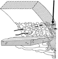
도 1은 섬유주 중의 큰 공극을 통한 세관 근접 영역 (수직 화살표에 의해 표시됨)으로의 방수의 흐름 방향 (수평 화살표)를 나타내는 정상 사람 눈 전방각의 영역의 횡단면도이다.
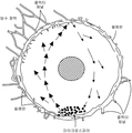
도 2는 화살표가 전방에서 하위에 위치한 제시된 마이크로스피어를 사용하여전방에서 방수 대류를 나타내는 개략도이다.
도 3a는 일차 시선에서 토끼 눈의 외부 사진이다.
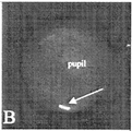
도 3b는 형광 임플란트의 이식 (화살표) 2일 후에 하이델베르크 HRA 영상화 장치 상의 위치에서 형광 필터를 사용하는 3a에서의 토끼 눈의 영상이다.
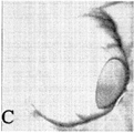
도 3c는 하향 회전된 토끼 눈의 외부 사진이다.
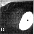
도 3d는 임플란트로부터 방출된 형광의 분포 (회살표)를 나타내는, 형광 임플란트의 이식 7일 후의 HRA를 사용하는 3c에서의 토끼 눈의 영상이다.
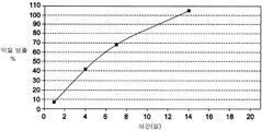
도 4는 제형 A 마이크로스피어로부터 시간(일)의 경과 (x 축)에 따라 생체외 (1% 트리톤을 갖는 PBS)에서 방출되는 라타노프로스트의 축적 % (y 축)을 나타내는 그래프이다.
도 5는 제형 B 마이크로스피어로부터 시간(일)의 경과 (x 축)에 따라 생체외 (1% 트리톤을 갖는 PBS)에서 방출되는 라타노프로스트의 축적 % (y 축)을 나타내는 그래프이다.
도 6은 약물 전달 장치 안내 투여 후에 기저 IOP로부터의 y 축 변동율 및 x 축 시간 (일)에 대한 그래프이다. 도 6 결과는 좌측 개 눈에서 제형 A 서방성 마이크로스피어의 안내 개 (비글) 주입 후에 얻어진다 (도 6에서 실선: "API"). 동료 (우측) 대조 눈 (도 6에서 점선)은 IOP 감소를 나타내지 않았다.
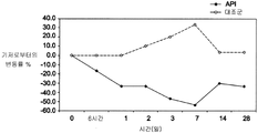
도 7은 실시예 5 비마토프로스트 막대형 임플란트 ("API")의 전방내 투여 후에 84일 기간에 걸쳐 시간(일) (x 축)에 대한 대상 개 기저 안내압 (y 축)으로부터의 변동율의 그래프이며, 이는 약 50% 내지 60%의 IOP 감소가 84일 관찰 기간 동안 유지됨을 나타낸다. 동료 (좌측 또는 "대조군l") 눈은 위약 (비마토프로스트 비함유) 임플란트를 수용하였다.
설명
공막 경유 전달은 눈의 국부 (즉, 눈물) 및 공막내 (즉, 결막 아래 또는 테논낭하) 배치 (예를 들어, 주입, 삽입 또는 이식에 의한) 약물 투여를 포함한다. 본 발명은 공막 경유 전달이 상승된 안내압의 치료를 위해 물주머니 또는 유리체방 표적 조직으로 안압강하제 (약물 또는 생물제제)를 투여하기 위한 비효과적인 방법이라는 관찰에 근거한 것이다. 본 발명자들은 명백하게 공막 경유 약물 전달을 방해하는 3가지 유형의 장벽 - 고정적, 동적 및 대사적 장벽이 있기 때문에 이렇계 되는 것으로 믿는다. 약물 확산에 대한 육체적 장벽 (공막, 맥락막-브루흐 막, 망막 색소 상피)를 제공하는 눈의 조직은 고정적 장벽을 위태롭게 한다. 동적 장벽은 결막에 주로 위치하는 혈관 및 림프관을 통한 약물 제거 메카니즘, 망막을 통한 전방으로부터 후방으로의 벌크 유체 흐름 및 맥락막모세혈관층 및 공막을 통한 제거, 및 망막 색소 상피의 전달 단백질에 의해 생성된다. 대사적 장벽이 또한 눈에 존재하며, 공막 투여 약물의 빠른 분해에 의해 눈 내로의 약물 관통을 감소시킨다. 동적 장벽은 고안압증 및 녹내장을 치료하기 위해 눈 (전방)의 전방으로의 치료제의 공막 경유 (즉, 테논낭하) 전달에 대한 가장 중요한 장벽인 것으로 보인다.
본 발명은 본원에 기재된 서방성 안내 약물 전달 시스템 (안압강하제 함유 임플란트 또는 마이크로스피어 포함)의 직접 전방내 또는 전방 유리체강내 투여가 활발한 공막 약물 제거 메카니즘을 우회함으로써 상승된 안내압 녹내장을 특징으로 하는 녹내장과 같은 안질환을 치료하기 위한 효과적인 사용일 수 있다는 발견에 근거한 것이다.
본 발명자들은 약물을 눈 (전방)의 전방을 전달하여 안내압 (IOP)을 저하시키고 공막 경유 장벽의 공격적 제거를 방지하기 위한 적합한 대안적 위치를 결정하였다. 평면부를 통한 전방내 주입 (즉, 전방 내로의 직접 주입) 및 전방 유리체 주입은 공막 경유 장벽을 효과적으로 피하고, 눈의 안압강하제 화합물의 효율을 개선시킨다. 중요하게는, 본 발명자들은 전방내 약물 전달 시스템이 전방의 특유의 해부학 및 생리학 때문에 새로운 서방성 약물 전달 시스템 육체적 특징의 개발을 필요로 한다는 점을 발견하였다. 예를 들어, 전방에서 방수 흐름율은 높고, 이는 IOP 저하 약물을 함유하는 서방성 마이크로스피어를 효과적으로 제거하고, 다른 중합체 전달 시스템의 분해를 촉진시킬 수 있다. 방수는 한외여과롤 불리우는 공정을 통해 모양체에 의해, 특히 모양체의 비-색소 상피에 의해 후방 내로 분비된다. 이는 수정체의 전방과 홍채의 후방 사이의 좁은 틈을 통해 유동하여, 동공을 통해 전방 내로 빠져나간다. 방수는 초기에 사람에서 10 내지 30 미크론 미만의 공극 크기 직경을 갖는 소주망 내로 360 도 빠져나간다 (도 1 참조). 도 1은 섬유주 중의 더 큰 공극 (약 10 내지 30 미크론 미만)을 통한 그리고 점차적으로 공극이 쉴렘관에 들어가기 전에 약 6 미크론으로 감소된 세관 근접 부분 (수직 화살표에 의해 표시됨)을 통한 방수의 흐름 방향 (수평 화살표)를 나타내는 전방각의 부분의 횡단면도이다. 방수는 쉴렘관을 통해 빠져나가고, 방수정맥 내로 그리고 궁극적으로 안와의 상공막 맥관 구조 및 정맥 내로 25 내지 30개의 집결관을 통해 눈을 빠져나간다 (도 2 참조). 도 2는 화살표가 전방에서 방수 대류를 나타내는 개략도이다. 눈의 안압강하제를 방출하는 마이크로스피어는 하위에 위치하는 것으로 도시된다. 중합체 마이크로스피어 (또는 임플란트)로부터 용출되는 유리 약물이 방수 대류 (화살표)에 들어간다. 그 다음, 약물은 전방 전체에 걸쳐 성공적으로 분산되고, 홍채 뿌리 영역을 통해 소주망 및 모양체와 같은 표적 조직에 들어간다.
전방내 주입의 또 다른 장점은 전방이 체내의 면역 회피 부위이며, 중합체 약물 전달 시스템과 같은 이물에 덜 반응할 것이라는 점이다. 이는 이물에 대한 염증 반응이 공통적인 테논낭하 공간에 있는 경우가 아니다. 면역 회피를 제공하는 면역조절 인자를 함유하는 전방 이외에, 30 미크론 초과 직경을 갖는 입자는 덜 면역원성이며, 눈의 염증을 유발시키는 것에 대해 더 낮은 경향을 갖는다. 눈 내의 체류 매크로파지는 이물 또는 감염제를 방어하는 제 1 라인이지만; 30 미크론보다 큰 입자는 파괴시키기가 어렵다. 따라서, 30 미크론보다 큰 입자는 하기의 매크로파지 활성화 및 염증 연쇄반응에 대한 경향이 덜하다.
본 발명자들은 중합체 방출 시스템을 사용하여 약물을 방수로 전달하는 효율이 전방내 위치 대 테논낭하 도포를 사용하는 것보다 훨씬 더 높음을 발견하였다. 따라서, 테논낭하 공간에서 전달되는 약물의 1% 미만이 방수에 들어갈 것이며, 반면에, 전방내 시스템으로부터 방출되는 약물의 100%가 방수에 들어갈 것이다. 따라서, 테논낭하와 비교하여 효과적인 전방내 약물 전달 시스템에 필요한 약물 하중이 저하되며, 결과적으로 전신적 약물 노출이 덜해질 것으로 예측된다. 그 외에, 활성 약제 성분에 대한 결막의 노출이 덜해지고, 프로스타글란딘 유사체와 같은 약물을 전달할 때에 결막 충혈을 발달시키려는 경향이 덜해질 것이다. 마지막으로, 약물은 방수정맥을 경유하는 전방내 주입 직후에 결막/상공막 혈관에 들어갈 것이다. 이는 많은 혈관이 결막의 혈관밖 공간에 확산적으로 존재하는 고농도의 약물에 의한 팽창의 위험이 있는 경우에, 테논낭하 주입과 비교하여 프로스타글란딘 유사체에 의한 결막 충혈을 최소화시킬 수 있다. 눈 내로의 직접 주입은 또한 국소적 방울에 사용될 때에 눈 표면을 자극할 수 있는 방부제에 대한 필요성을 배제시킨다.
본원에 기술된 전방내 및 유리체강내 서방성 약물 전달 시스템에서 활성제로서 사용하기에 적합한 안압강하제는 하기를 포함한다: 프로스타글란딘, 프로스타마이드 및 안압강하 지질 (예를 들어, 비마토프로스트 (Lumigan] {비마토프로스트는 방수의 포도막공막을 통한 유출을 증가시킬 뿐만 아니라 섬유주 유출을 증가시킨다} 및 미국 특허 제5,352,708호에 기재된 화합물). 프로스타글란딘은 아라키돈산으로부터 유도되는 다양한 포유동물 조직에서 생성되는 성분과 같은 한 부류의 약리학적 활성 호르몬이며, 안압, 평활근 수축 및 염증을 포함하는 광범위한 생리학적 기능을 조정한다. 프로스타글란딘의 예는 프로스타글란딘 E1 (알프로스타딜), 프로스타글란딘 E2 (디노프로스톤), 라타노프로스트 및 트라보프로스트이다. 라타노프로스트 및 트라보프로스트는 사실상 프로스타글란딘 전구약물 (즉, 프로스타글란딘의 1-이소프로필 에스테르)이지만, 이들은 1-카르복실산으로 가수분해된 후에 프로스타글란딘 F 수용체에 작용하기 때문에 프로스타글란딘으로서 언급된다. 프로스타마이드 (또한 프로스타글란딘-에탄올아민으로 명명됨)는 프로스타글란딘 유사체이며, 이는 프로스타글란딘과 약리학적으로 구별되며 (즉, 프로스타마이드는 프로스타글란딘보다 상이한 세포 수용체 [프로스타마이드 수용체]에 작용한다), (아난다미드와 같은) 엔도칸나비노이드의 시클로옥시게나아제-2 ("COX-2") 효소 산화의 생성물로서 생성되는 천연 지질이다. 추가적으로, 프로스타마이드는 1-카르복실산로 원위치에서 가수분해되지 않는다. 프로스타마이드의 예는 비마토프로스트 (17-페닐 프로스타글란딘 F2α의 합성 에틸 아미드) 및 프로스타마이드 F2α, 프로스타글란딘 유사체 (프로스타글란딘 유사체는 방수의 포도막공막 유출을 증가시킴) (즉, 라타노프로스트 [Xalatan], 트라보프로스트 (Travatan), 우노프로스톤; EP2/EP4 수용체 작용물질; 베타-아드레날린성 수용체 길항물질 (티몰롤, 베타크솔롤, 로베베타크솔롤, 카르테올롤, 로베부놀롤 및 프로프라놀롤, 이는 모양체에 의한 방수 생성을 감소시킨다); 브리모니딘 (Alphagan) 및 아프라클로니딘 (이오피딘)과 같은 알파 아드레날린성 수용체 작용물질 (이는 방수 생성을 감소시키고 포도막공막 유출을 증가시키는 이중 메카니즘에 의해 작용한다); 에피네프린 및 디피베프린 (Propine)과 같은 덜 선택성 교감신경흥분제 (아마도 베타 2-작용물질 작용에 의해 소주망을 통한 그리고 가능하게는 포도막공막 유출 경로를 통한 방수의 유출을 증가시키도록 작용함; 필로카르핀과 같은 동공축소제 (부교감신경흥분제) (소주망을 치밀화시키고 방수의 유출을 증가시키는 모양체 근육의 수축에 의해 작용함); 도졸라미드 (Trusopt), 브리졸라미드 (Azopt), 아세타졸라미드 (Diamox)와 같은 탄산 탈수 효소 억제제 (모양체에서 탄산 탈수 효소를 억제시킴으로써 방수의 분비를 저하시킴); 로키나제 억제제 (소주망의 액틴 세포골격을 분열시킴으로써 IOP를 저하시킴); 칼슘 통로 차단제; 밥탄 (바소프레신-수용체 길항물질); 아네코르타브 아세테이트 및 유사체; 에타크린산; 칸나비노이드; 카르테올롤, 로베부놀롤, 메티파라놀롤, 티몰롤 반수화물, 티몰롤 말레에이트, 베타크솔롤과 같은 베타. 1-덜 선택성 길항물질을 포함하는 베타-차단제 (또는 베타-아드레날린성 길항물질); 에피네프린 보레이트, 에피네프린 염산염 및 디피베프린과 같은 비-선택성 아드레날린성 작용물질; 아프라클로니딘 및 브리모니딘과 같은 알파2 선택성 아드레날린성 작용물질; 아세타졸라미드, 디클로르페나미드, 메타졸라미드, 브린졸라미드 및 도르졸라미드를 포함하는 탄산 탈수 효소 억제제; 카르바콜, 필로카프린 염산염; 필로카르빈 니트레이트 및 필로카프린과 같은 직접 직용 콜린성 작용물질을 포함하는 콜린성 작용물질; 데메카륨, 에코티오페이트 및 피소스티그민과 같은 클롤리네스테라제 억제제; 글루타메이트 길항물질; 메만틴, 아만타딘, 리만타딘, 니트로글리세린, 덱스트로판, 데트로메토르판, 디히드록시피리딘, 베라파밀, 에모파밀, 벤조티아제핀, 베프리딜, 디페닐부틸피페리딘, 디페닐피페라진, 플루스피릴렌, 엘리프로딜, 이펜프로딜, 티발로신, 플루나리진, 니카르디핀, 니페딤핀, 니모디핀, 바르니디핀, 베라파밀, 리도플라진, 페닐아민 락테이트 및 아밀로리드를 포함하는 칼슘 통로 차단제; 비마토프로스트와 같은 프로스타마이드, 또는 이들의 약제학적으로 허용될 수 있는 염 또는 전구약물; 및 트라보프로스트, 클로프로스테놀, 플루프로스테놀, 13,14-디히드로-클로프로스테놀, 이소부틸 우노프로스톤 및 라타노프로스트를 포함하는 프로스타글란딘; AR-102 (Aerie Pharmaceuticals, Inc.의 제품인 프로스타글란딘 FT 작용물질); AL-3789 (아네코르타브 아세테이트, Alcon의 제품인 신생혈관형성 스테로이드); AL-6221 (트라바프로스트 [Travatan], 프로스타글란딘 FP 작용물질); PF-03187207 (Pfizer의 제품인 산화질소 제공 프로스타글란딘); PF-04217329 (또한 Pfizer의 제품); INS115644 (Inspire Pharmaceuticals의 제품인 란트룬쿨린 B 화합물; 및
INS117548 (또한 Inspire Pharmaceuticalsdml 제품인 로키나제 억제제)이다.
베타 차단제 및 프로스타글란딘 유사체와 같은 눈의 안압강하제의 배합물이 또한 전달 시스템에 사용될 수 있다. 이들은 간포르트 (비마토프로스트/티몰롤), 엑스트라반 또는 두오트라브 (트라보프로스트/티몰롤), 크살콤 (라타노프로스트/티몰롤, 콤비간 (브리모니딘/티몰롤) 및 코소프트 (도르졸라미드/티몰롤)을 포함한다. IOP 저하 약물과의 배합물에서, 신경보호를 제공하는 제제가 또한 전달 시스템 내에 위치할 수 있으며, 메만틴 및 세로토닌 작동제 [예를 들어, S-(+)-1-(2-아미노프로필)-인다졸-6-올)과 같은 5-HT.sub.2 작용물질]을 포함한다.
본 발명자들은 다양한 기간에 걸쳐 약물 함유물을 방출시킬 수 있는 임플란트 및 마이크로스피어를 개발하였다. 이들 임플란트 또는 마이크로스피어는 전방내 또는 전방 유리체 치료제 내로 삽입되는 경우에 연장된 기간 동안 (예를 들어, 약 1 주 내지 약 1 년 이하 동안) 치료 수준의 안압강하제를 제공한다. 추가적으로, 본 발명자들은 임플란트 및 마이크로스피어를 제조하기 위한 새로운 방법을 개발하였다. 본 발명의 임플란트 및 마이크로스피어의 안압강하제는 바람직하게는 마이크로스피어의 약 1 중량% 내지 90 중량%이다. 더욱 바람직하게는, 안압강하제는 임플란트 또는 마이크로스피어의 약 5 중량% 내지 약 30 중량이다. 바람직한 구현예에서, 안압강하제는 마이크로스피어의 약 15 중량% (예를 들어, 5-30 중량%)를 구성한다. 또 다른 구현예에서, 안압강하제는 마이크로스피어의 약 40 중량%를 구성한다.
임플란트 또는 마이크로스피어에 사용하기 위한 적합한 중합체 재료 또는 조성물은 눈의 기능 또는 생리학에 실질적 간섭을 유발하지 않도록 눈과의 양립성, 즉 생체적합성인 재료를 포함한다. 이러한 재료는 바람직하게는 최소한 부분적으로 또는 더욱 바람직하게는 실질적으로 완전히 생분해성 또는 생침식성이다.
유용한 중합체 재료의 예는 비제한적으로, 분해될 때에 단량체를 포함하는 생리학적으로 허용될 수 있는 분해 생성물을 결과하는 유기 에스테르 및 유기 에테르로부터 유도되고/거나 이를 포함하는 재료를 포함한다. 또한, 자체적으로 또는 다른 단량체와 조합하여 무수물, 아미드, 오르토에스테르 등으로부터 유도되고/거나 이들을 포함하는 중합체 재료의 사용이 또한 발견되었다. 중합체 재료는 부가 또는 축합 중합체, 유리하게는 축합 중합체일 수 있다. 중합체 재료는 교차결합 또는 비-교차결합될 수 있으며, 예를 들어 중합체 재료의 약 5% 미만과 같이 가볍게만 교차결합되건, 또는 약 1% 미만이 교차결합된다. 대부분, 탄소 및 수소 이외에, 중합체는 산소 및 질소 중 하나 이상, 유리하게는 산소를 포함할 것이다. 산소는 옥시, 예를 들어 히드록시 또는 에테르, 카르보닐, 예를 들어 카르복실산 에스테르와 같은 비-옥소-카르보닐 등으로서 존재할 수 있다. 질소는 아미드, 시아노 및 아미노로서 존재할 수 있다. 조절된 약물 전달을 위한 캡슐화를 기술하고 있는 문헌 [Heller, Biodegradable Polymers in Controlled Drug Deliverry, In: CRC Critical Reviews in Therapeutic Drug Carrier Systems, Vol. 1, CRC Press, Boca Raton, FL 1987, pp 39-90]에 기재된 중합체가 본 마이크로스피어에서의 사용이 발견될 수 있다.
히드록시알리파틱 카르복실산 단일중합체 또는 공중합체, 및 다당류의 중합체가 추가적으로 중요하다. 관심있는 폴리에스테르는 D-락트산, L-락트산, 라세미체 락트산, 글리콜산, 폴리카프로락톤 및 이들의 조합물의 중합체를 포함한다. 일반적으로, L-락테이트 또는 D-락테이트를 사용함으로써, 중합체 또는 중합체 재료의 느린 침식이 달성되며, 침식은 실질적으로 락테이트 라세미체에 의해 향상된다. 유용한 다당류 중에는, 비제한적으로, 알긴산칼슘, 및 작용기화된 셀룰로오스, 특히 예를 들어 약 5 kD 내지 500 kD의 분자량의 수용성임을 특징으로 하는 카르복시메틸셀룰로오스 에스테르가 있다.
관심있는 다른 중합체는 비제한적으로 생체적합성이고 생분해성 및/또는 생침식성일 수 있는 폴리비닐 알코올, 폴리에스테르, 폴리에테르 및 이들의 조합물을 포함한다. 본 발명에 사용하기 위한 중합체 또는 중합체 재료의 일부 바람직한 특징은 생체적합성, 선택된 치료제와의 양립성, 본 발명의 약물 전달 시스템을 제조하기 위한 중합체의 사용의 용이성, 약 6시간 이상, 바람직하게는 약 1일 초과의 생리학적 환경에서의 반감기, 및 수불용성을 포함할 수 있다.
매트릭스를 생성시키기 위해 포함되는 생분해성 중합체 재료는 바람직하게는 효소적 또는 가수분해적 불안정성이 된다. 수용성 중합체는 가수분해적 또는 생분해성 불안정 교차결합제와 교차결합되어 유용한 수불용성 중합체를 제공할 수 있다. 안정도는 단량체의 선택, 중합체의 혼합물을 사용하여 단일중합체 또는 공중합체가 사용되는 지의 여부 및 중합체가 말단 산기를 포함하는 지의 여부에 의존하여 넓게 변할 수 있다.
중합체의 생분해를 조절하는 것이 동동하게 중요하며, 따라서 임플란트의 연장된 방출 프로파일은 임플란트 또는 마이크로스피어에 사용되는 중합체 조성물의 상대적 평균 분자량이다. 동일하거나 상이한 중합체 조성물의 상이한 분자량이 마이크로스피어에 포함되어 방출 프로파일을 조절할 수 있다. 라타노프로스트 임플란트에 대해, 중합체의 상대적 평균 분자량은 바람직하게는 약 4 내지 약 25 kD, 더욱 바람직하게는 약 5 내지 약 20 kD, 가장 바람직하게는 약 5 내지 약 15 kD일 것이다.
일부 임플란트 및 마이크로스피어에서, 생분해율이 글리콜산 대 락트산의 비에 의해 조절되는 경우에, 글리콜산과 락트산의 공중합체가 사용된다. 가장 빠르게 분해되는 공중합체는 대략적 동일량의 글리콜산 및 락트산을 갖는다. 동등한 것과는 다른 비를 갖는 단일중합체 또는 공중합체는 분해에 대해 더욱 저항성이다. 글리콜산 대 락트산의 비는 또한 마이크로스피어의 취성에 영향을 줄 것이다. 폴리락트산 폴리글리콜산 (PLGA) 공중합체 중의 폴리락트산의 비율은 0-100%, 바람직하게는 약 15-85%, 더욱 바람직하게는 약 35-65%일 수 있다. 일부 임플란트에서는, 50/50 PLG공중합체가 사용된다.
임플란트 및 마이크로스피어는 일체식일 수 있으며, 즉 활성제(들)이 중합체 매트릭스를 통해 균일하게 분포되거나, 또는 활성제의 수용기가 중합체 매트릭스에 의해 캡슐화되는 경우에 캡슐화된다. 제조의 용이함으로 인해, 일체식 임플란트는 일반적으로 캡슐화 형태보다 바람직하다. 그러나, 캡슐화된 마이크로스피어에 의해 제공되는 더 큰 조절은 일부 경우에, 약물의 치료적 수준이 좁은 윈도우 내에 있는 경우에 유익할 수 있다. 그외에, 라타노프로스트 성분을 포함하는 치료 성분이 매트릭스 내에 비균일 패턴으로 분포될 수 있다. 예를 들어, 마이크로스피어는 마이크로스피어의 제 2 부분에 비해 라타노프로스트의 더 높은 농도를 갖는 부분을 포함한다.
본원에 기술된 마이크로스피어는 바늘에 의한 투여를 위해 약 5 ㎛ 내지 약 1 ㎜, 또는 약 10 ㎛ 내지 약 0.8 ㎜의 크기를 가질 수 있다. 바늘 주입 마이크로스피어에 대해, 마이크로스피어는 마이크로스피어의 가장 긴 치수가 마이크로스피어를 바늘을 통해 이동하게 할 정도로 긴 임의의 적절한 치수를 가질 수 있다. 이는 일반적으로 마이크로스피어의 투여에서 문제점이 아니다.
단일 투여량으로 임플란트 또는 마이크로스피어의 총중량은 전방의 부피 및 활성제의 활성 또는 용해도에 의존하는 최적량이다. 가장 자주, 투여량은 일반적으로 투여당 임플란트 또는 마이크로스피어 약 0.1 ㎎ 내지 약 200 ㎎이다. 예를 들어, 단일 전방내 주입은 혼입된 치료 성분을 포함하는 마이크로스피어 약 1 ㎎, 3 ㎎, 또는 약 5 ㎎, 또는 약 8 ㎎, 또는 약 10 ㎎, 또는 약 100 ㎎ 또는 약 150 ㎎, 또는 약 175 ㎎, 또는 약 200 ㎎을 함유할 수 있다.
임플란트 또는 마이크로스피어는 마이크로 및 나노스피어, 마이크로 및 나노입자, 스피어, 분말, 단편 등의 임의의 미립 기하학적 형태를 가질 수 있다. 마이크로스피어 크기에 대한 상한은 임플란트에 대한 허용, 삽입에 대한 크기 제한, 바람직한 방출 속도, 취급의 용이성 등과 같은 요인에 의해 결정될 것이다. 스피어는 다른 형태의 입자에 대한 비교할 수 있는 부피와 함께 직경이 약 0.5 ㎛ 내지 4 ㎜일 수 있다.
안압강하제, 중합체 및 임의의 다른 변형제의 비율은 평균 비율을 변동시킴으로써 수가지 마이크로스피어 배치를 제형화시킴으로써 실험적으로 결정될 수 있다. 용해 또는 방출 시험을 위한 USP 승인 방법이 방출 속도를 측정하기 위해 사용될 수 있다 (USP 23; NF 18 (1995) pp. 1790-1798). 예를 들어, 무한 침전 방법을 사용하여, 마이크로스피어의 칭량 샘플이 수중에 0.9% NaCl을 함유하는 용액의 측정된 부피에 첨가되며, 여기에서 용액 부피는 방출 후의 약물 농도가 5% 미만의 포화일 정도일 것이다. 혼합물은 37℃에서 유지되고, 서서히 교반되어 마이크로스피어를 현탁액 중에 유지시킨다. 시간의 함수로서 용해된 약물의 출현은 흡수도가 일정해질 때까지 또는 약물의 90% 초과가 방출될 때까지 분광법, HPLC, 질량분석법 등과 같은 당분야에 공지된 다양한 방법이 후속될 수 있다.
치료 성분 이외에, 본원에 기술된 임플란트 및 마이크로스피어는 유효량의 완충제, 방부제 등을 포함하는 조성물을 포함할 수 있거나 이에 제공될 수 있다. 적합한 수용성 완충제는 비제한적으로 나트륨 인산염, 시트르산염, 붕산염, 아세트산염, 중탄산염, 탄산염 등과 같은 알칼리 및 알칼리토 금속 탄산염, 인산염, 중탄산염, 시트르산염, 붕산염, 아세트산염, 숙신산염 등을 포함한다. 이들 제제는 유리하게는 약 2 내지 약 9 및 더욱 바람직하게는 약 4 내지 약 8의 시스템의 pH를 유지시키기에 충분한 양으로 존재한다. 그 자체로, 완충제는 전체 임플란트의 약 5 중량%일 수 dl있다. 적합한 수용성 방부제는 나트륨 중아황산염, 나트륨 중황산염, 나트륨 티오황산염, 아스코르브산염, 염화벤잘코늄, 클로로부탄올, 티메로살 , 페닐수은 아세트산염, 페닐수은 붕산염, 페닐수은 질산염, 파라벤, 메틸파라벤, 폴리비닐 알코올, 알코올, 페닐에탄올 등 및 이들의 혼합물을 포함한다. 이들 제제는 약 0.001 중량% 내지 약 5 중량% 및 바람직하게는 약 0.01% 내지 약 2 중량%의 양으로 존재할 수 있다. 본 발명의 마이크로스피어 중 하나 이상에서, 염화벤잘코늄 방부제는 라타노프로스트가 본질적으로 비마토프로스트로 구성되는 경우와 같이 임플란트 내에 제공된다.
다양한 기술이 사용되어 본원에 기술된 임플란트 및/또는 마이크로스피어를 생성시킬 수 있다. 유용한 기술는 필수적으로 제한되지는 않지만, 자기유화 방법, 초임계유체 방법, 용매 증발 방법, 상분리 방법, 분무건조 방법, 연삭 방법, 계면 방법, 성형 방법, 사출성형 방법, 이들의 조합 등을 포함한다.
본원에 기술된 바와 같이, 본 방법에 열거된 중합체 성분은 생분해성 중합체 또는 생분해성 공중합체를 포함할 수 있다. 하나 이상의 구현예에서, 중합체 성분은 폴리(락티드-코-글리콜리드) PLG 공중합체를 포함한다. 하나의 추가의 구현예에서, PLG 공중합체는 75/25의 락티드/글리콜리드 비를 가질 수 있다. 추가의 구현예에서, PLG 공중합체는 약 63 킬로달톤의 분자량 및 약 0.6 dL/g의 고유 점도 중 하나 이상을 갖는다.
그 외에, 본 극미립자의 집단은 약 200 ㎛ 미만의 최대 입자 직경을 가질 수 있다. 특정 구현예에서, 극미립자의 집단은 약 50 ㎛ 미만의 평균 또는 중간 입자 직경을 갖는다. 추가의 구현예에서, 극미립자의 집단은 약 30 ㎛ 내지 약 50 ㎛의 중간 입자 직경을 갖는다.
본원에 기술된 안압강하제 함유 임플란트 및 마이크로스피어는 하기와 같은 안질환을 치료하기 위해 사용될 수 있다: 황반변증/망막 변성: 비삼출성 노인성 망막황반 변성 및 삼출성 노인성 망막황반과 같은 노인성 망막황반 변성 (ARMD)를 포함하는 변성망막황반 변성, 맥락막 신혈관 형성, 당뇨병 망막증, 급성 및 만성 망막황반 신경망막병증을 포함하는 망막증, 중심성맥락망막염, 및 낭포 망막황반 부종 및 당뇨병 망막황반 부종을 포함하는 망막황반 부종. 포도막염/망막염/맥락막염: 급성 다발성 판상색소 상피증, 베체트병, 버드샷 맥락망막병증, 감염 (매독, 라임병, 결핵, 톡소플라스마증), 중간 포도막염 (주변부 포도막염) 및 전방 포도막염을 포함하는 포도막염, 다초점 맥락막염, 다발성소실성백반증후군 (MEWDS), 눈의 유육종증, 후방 공막염, 포행성 맥락막염, 망막하 섬유증, 포도막염 증후군 및 보그트-고야나기-하라다 증후군. 혈관질환/삼출질환: 망막 동맥 폐쇄 질환, 중심 망막 정맥 폐쇄, 파종성혈관내응고장애, 망막분지 정맥 폐쇄, 고안압성 안저 변화, 눈의 허혈성 증후군, 망막 미세동맥류, 코츠씨병, 중심와부근 모세혈관 확장증, 반구망막정맥페쇄, 유두정맥염, 중심 망막 동맥폐쇄, 망막분지 동맥폐쇄, 경동맥 질환 (CAD), 언가지모양혈관염, 겸상적혈구 망막증 및 다른 혈색소병, 망막색소선조, 가족성 삼출유리체망막병증, 일스병. 외상/수술: 교감성안염, 포도막염 망막 질환, 망막 박리, 외상, 레이저, PDT, 광응고, 수술 동안의 저관류, 방사선 망막증, 골수 이식 망막증. 증식성 장애: 증식성 유리체 망막증 및 망막전막, 증식성 당뇨병 망막증. 감염 장애: 눈의 히스토플라스마증, 눈의 톡소카라증, 추정 안구 히스토플라스마증 증후군 (POHS), 안내염, 톡소플라스마증, HIV 감염과 관련된 망막 질환, HIV 감염과 관련된 맥락막 질환, HIV 감염과 관련된 포도막염 질환, 바이러스성 망막염, 급성 망막 괴사, 진행성 외부 망막 괴사, 진균성 망막 질환, 눈매독, 눈결핵, 확산 편측 아급성 시신경망막염 및 구더기증. 유전적 장애: 망막색소변성증, 망막 이영양증과 관련된 전신적 장애, 선천적 비진행성 야맹증, 추세포 영양장애, 스타가르트병 및 황반안저, 베스트병, 패턴 망막 색소상피의 이영양증, X-연관 망막층간분리, 소르스비 안전 이영양증, 양성 중심 황반변성, 베티 결정 이영양증, 탄력섬유성가황색종. 망막 열공: 망막 박리, 망막황반 열공, 거대 망막 열공. 종양: 종양과 관련된 망막 질환, RPE의 선천적 비대, 후방 포도막 흑색종, 맥락막 혈관종, 맥락막 골종, 맥락막 전이, 망막 및 망막 색소상피의 복합 과오종, 망막아세포종, 안저의 혈관증식 종양, 망막 성상세포종, 안내 림프성 종양. 다양한 질환: 점상내층맥락막병증, 급성 후방 다발성 판상색소 상피증, 근시 망막 변성, 급성 망막 색소 상피증 등.
본 발명의 범위 내의 약제 조성물 (임플란트 또는 마이크로스피어와 같은)은 고점도 중합체 겔로 제형화되어, 안내 주입 시에 조성물의 분산을 감소시킬 수 있다. 바람직하게는, 겔은 고전단 특징을 가지며, 이는 겔이 25-30 게이지 바늘, 및 더욱 바람직하게는 27-30 게이지 바늘을 통해 안내 부위 내로 주입될 수 있음을 의미한다. 본 용도를 위한 적합한 겔은 수중 분산액 또는 다른 수성 매질로서 생성되는 하이드로겔 또는 콜로이드성 겔일 수 있다. 적합한 겔의 예는 폴리히드록시 에틸 메타크릴레이트와 같은 합성 중합체, 및 화학적으로 또는 물리적으로 교차결합된 폴리비닐 알코올, 폴리아크릴아미드, 폴리(N-비닐 피롤리돈), 폴리에틸렌 산화물, 및 가수분해된 폴리아크릴로니트릴을 포함한다. 유기 중합체인 적합한 하이드로겔의 예는 알긴산염의 다가 금속 염, 펙틴, 카르복시메틸 셀룰로오스, 헤파린, 히알루론산염 (즉, 중합체 히알루론산) 및 키틴, 키토산, 풀룰란, 젤란, 크산탄 및 히드록시프로필메틸셀룰로오스로부터의 하이드로겔과 같은 공유결합 또는 이온적으로 교차결합된 다당류계 하이드로겔을 포함한다. 시판용 피부 충전물 (Hylafrom®, Restylane®, SculpturaTM 및 Radiesse과 같은)이 본 약제 조성물의 구현예에서 고점도 겔로서 사용될 수 있다.
히알루론산 ("HA")은 다양한 체조직으로부터 제조되는 다당류이다. 미국 특허 5,166,331에는 안내 유체에 대한 치환물로서 그리고 국소 안과용 약물 담체로서 사용하기 위한 히알루론산의 상이한 분획의 정제가 기술되어 있다. 히알루론산의 눈의 사용을 기술하는 다른 미국 특허 출원은 출원 번호 11/859,627; 11/952,927;10/966,764; 11/741,366; 및 11/039,192를 포함한다. 본 발명의 범위 내의 약제 조성물은 바람직하게는 약 1 내지 4백만 달톤의 평균 분자량, 더욱 바람직하게는 약 2 내지 3백만 달톤의 평균 분자량, 가장 바람직하게는 약 (±10%) 2백만 달톤의 평균 분자량을 갖는 고점도 히알루론산을 포함한다.
건조 비교차결합된 HA 재료는 시판용 HA의 섬유 또는 분말, 예를 들어 나트륨 히알루론산염 (NaHA)의 섬유 또는 분말을 포함한다. The HA는 박테리아원 나트륨 히알루론산염, 동물 유도 나트륨 히알루론산염 또는 이들의 조합물일 수 있다. 일부 구현예에서, 건조 HA 재료는 HA 및 하나 이상의 다른 다당류, 예를 들어, 글리코사미노글리칸 (GAG)을 포함하는 원료의 조합물이다. 본 발명에서, 사용되는 HA는 고분자량 HA를 포함하거나 이로 구성된다. 즉, 본 조성물 중의 HA 재료의 거의 100%는 고분자량 HA이다. 고분자량 HA는 약 1백만 달톤 (mw ≥ 106 Da ) 이상 내지 약 4백만 달톤 (mw ≤ 4×10⁶ Da)의 분자량을 갖는 HA를 의미한다. 예를 들어, 본 조성물 중의 고분자량 HA는 약 2백만 Da (mw 2×10⁶ Da)의 분자량을 가질 수 있다. 또 다른 에에서, 고분자량 HA는 약 2.8 백만 Da (mw 2.8×10⁶ Da)의 분자량을 가질 수 있다.
본 발명의 한 구현예에서, 바람직한 고/저 분자량 비를 갖는 건조 또는 원료 HA (특정 예에서, NaHA)는 세척되고 정제된다. 이들 단계는 일반적으로 예를 들어 정수를 사용하여 바람직한 고/저 분자량 비의 건조 HA 섬유 또는 분말을 수화시키고, 재료를 여과시켜서 큰 이물 및/또는 다른 불순물을 제거하는 것을 수반한다. 그 다음, 여과되고 수화된 재료는 건조되고 정제된다. 고분자량 및 저분자량 NaHA는 분리적으로 세척되고 정제될 수 있거나, 예를 들어 교차결합 직전에 바람직한 비로 서로 혼합될 수 있다. 공정 중의 상기 단계에서, 순수하고 건조된 NaHA 섬유는 알칼리성 용액 중에서 수화되어 비교차결합 NaHA 알칼리성 겔을 생성시킨다. 임의의 적합한 알칼리성 용액은 상기 단계에서 NaHA를 수화시키기 위해 사용될 수 있지만, NaOH를 함유하는 수용액으로 제한되지 않는다. 생성된 알칼리성 겔은 7.5 초과의 pH, 예를 들어 8 초과이 pH, 예를 들어 9 초과의 pH, 예를 들어 10 초과의 pH, 예를 들어 12 초과의 pH, 예를 들어 13 초과의 pH를 가질 것이다. 상기 특정 예에서, 제조 공정 중의 다음 단계는 수화된 알칼리성 NaHA 겔을 적합한 교차결합제, 예를 들어 BDDE와 교차결합시키는 단계를 포함한다.
교차결합의 단계는 당업자들에게 공지된 수단을 사용하여 수행될 수 있다. 당업자들은 HA의 성질에 따라 교차결합의 조건을 최적화시키고, 교차결합을 최적화된 정도로 수행하는 방법을 인지한다. 본 발명의 일부 구현예에서, 교차결합도는 약 2% 이상 내지 약 20%, 예를 들어, 약 4% 내지 약 12%이며, 교차결합도는 조성물 중의 HA-단량체 단위에 대한 교차결합제의 중량% 비로서 규정된다. 수화된 교차결합된, HA 겔은 HCl을 함유하는 수용액을 첨가함으로써 중화될 수 있다. 그 다음, 겔은 저온에서 충분한 시간 동안 인산염 완충 식염수 중에서 팽윤된다.
특정 구현예들에서, 생성된 팽윤 겔 (HA)은 실질적으로 가시적인 독특한 입자를 갖지 않는, 예를 들어 육안으로 관찰하였을 때에 가시적으로 독특한 입자를 갖지 않는 점착성 겔이다. 일부 구현예에서, 겔은 35X의 배율 하에 실질적으로 가시적으로 독특한 입자를 갖지 않는다. 겔 (HA)는 통상적인 수단, 예를 들어, 투석 또는 알코올 침전에 의해 정제되어, 교차결합된 재료를 회수하고, 재료의 pH를 안정화시키고 임의의 비반응 교차결합체를 제거한다. 부가적 물 및 약한 알칼리성 수용액이 첨가되어 조성물 중의 NaHA의 농도를 바람직한 농도가 되게 할 수 있다. 일부 구현예에서, 조성물 중의 NaHA의 농도는 약 10 ㎎/㎖ 내지 약 30 ㎎/㎖이다.
본 발명의 범위 내의 임플란트는 미국 특허 출원 제11/455,392호; 11/552,835호; 11/552,630호 및 12/355,709호에 나타낸 어플리케이터 (주입기)를 포함하는 임의의 적합한 안내 주입 장치를 사용하여 투여될 수 있다.
본 발명의 구현예들은 서방성 생분해성 마이크로스피어 또는 임플란트일 수 있다. 본 발명의 바람직한 구현예는 이러한 조성물의 임플란트가 전방내 또는 전방 유리체 투여 시에 현저히 덜한 염증 (즉, 덜한 각막 충혈)을 결과함이 결정되었기 때문에, 안압강하제를 함유하는 PLA 및/또는 PLGA 임플란트이다. 본 발명의 한 구현예는 동일한 임플란트의 상이한 세그먼트에 또는 동시에 투여되는 상이한 임플란트에 함유된 복수의 안압강하제를 갖는 약물 전달 시스템을 포함할 수 있다. 예를 들어, 하나의 세그먼트 (즉, 하나의 임플란트)는 무스카린 안압강하제를 함유할 수 있고, 제 2 세그먼트 (즉, 제 2 임플란트)는 안압강하제 프로스타글란딘를 함유할 수 있으며, 제 3 세그먼트 (즉, 제 3 임플란트)는 안압강하제 베타 차단제를 함유할 수 있다. 다중 임플란트 ("세그먼트")는 동시에 주입될 수 있으며, 예를 들어 안압강하제를 갖는 하나의 임플란트는 소주망을 통한 방수 유출 (예를 들어, 무스카린제)를 향상시키기 위해 사용될 수 있고, 제 2 임플란트는 포도막공막 흐름 (예를 들어, 안압강하 지질)을 향상시키기 위해 사용될 수 있으며, 제 3 임플란트는 방수 생성 (예를 들어, 베타 차단제)를 감소시킬 수 있다. 상이한 작용 메카니즘을 갖는 다중 안압강하제는 안압강하제의 단일 유형의 사용인 단일치료제보다 IOP를 저하시키는 데에 더욱 효과적일 수 있다. 다중 세그먼트 (임플란트)는 단일치료제에 필요한 투여량보다 사용되는 각각의 분리 안압강하제의 더 낮은 투여량을 허용하여 사용되는 각각의 안압강하제의 부작용을 감소시키는 장점을 갖는다. 예를 들어 신경보호 또는 신경증강 화합물을 함유하는 분리 및 부가 세그먼트가 또한 안압강하제를 함유하는 다른 세그먼트와 함께 전달될 수 있다.
다중 세그먼트 (즉, 투여되는 복수의 임플란트)를 사용하는 경우, 각각의 세그먼트는 바람직하게는 약 2 ㎜ 이하의 길이를 갖는다. 바람직하게는, 동일한 22 내지 25G 직경 바늘 구멍에 투여되는 세그먼트의 총수는 약 4개이다. 27G 직경 바늘에 대해, 바늘 구멍 또는 루멘 내의 총 세그먼트 길이는 최고 약 12 ㎜일 수 있다.
소주망 (TM)이 물주머니 유체에 대한 검출할 수 있는 유체 흡수 또는 흡인 작용을 갖는다. 상기 TM 유체 흡수는 30 미크론 미만의 직경을 갖는 마이크로스피어 (MS)가 우각경검사 영상화에 의해 결정되는 바와 같이 TM 내로 방출되도록 한다.
또한, TM의 유체 흡수 작용은 시야 방해를 유발하는 전방 둘레의 부유로부터 적절한 기하학적 형태를 갖는 MS 또는 임플란트를 유지시키도록 활용될 수 있음이 결정되었다. 중력은 이들 임플란트를 6 시 위치로 강하시키며, 상기 위치에서 임플란트 또는 MS가 매우 안정함 (비교적 부동성)임이 주목된다. 총 약 6 내지 8 ㎜ 이하의 길이를 갖는 22G 내지 30G 직경 바늘에 의해 안내 투여될 수 있는 임플란트 (포함되는 모든 세그먼트)가 안내 임플란트 부동성을 결과하고 시야 방해를 하지 않는 TM 유체 흡수 메카니즘의 장점을 취하기 위해 가장 바람직하다. 따라서, TM 유체 흡수 효과로 인해 전방에서 6 시 위치에서 견고하게 됨에도 불구하고, 임플란트는 TM 제거율을 초과하는 방출 속도를 가질 수 있으며, 이는 임플란트에 의해 방출되는 안압강하제가 전방을 빠르게 충전시키고, 360 도 분포 패턴을 따라 표적 조직 내로 잘 분포시키도록 한다. 우각경검사에 의한 전방의 각에서의 임플란트의 하나의 시험은 임플란트의 부근에서 어떠한 염증 조직의 캡슐화도 없음을 나타낸다.
실시예
하기의 실시예는 본 발명의 비제한적 구현예를 기재한 것이다.
실시예 1
전방 대류의 결정
눈의 전방에서, 홍채와 접촉하고 있는 방수의 더 높은 온돈에 의해 유도되는 6 시 위치로부터 12 시 위치로 흐르는 수직 상향 대류가 있음을 결정하였다. 또한 전방에서, 각막 상피에 인접한 방수의 더 낮은 온도에 의해 유도되는 12 시 위치로부터 6 시 위치로 진행하는 수방 흐름의 하향 대류가 있음을 결정하였다. 도 2 참조. 전달 시스템이 방수 내로 직접 약물을 방출시키는 경우에, 이들 방수 흐름이 360 도 분포 패턴으로 전방 전체에 걸쳐 그리고 그 둘레에 안압강하제를 효과적으로 수반할 수 있음을 가정하였으며, 전방에 위치한 서방성 임플란트의 영상 연구에 의해 전방에서 약물 대용물을 분포시키는 대류의 효능을 입증하였다. 도 3 참조.
추가적으로, 30 미크론 초과의 직경 및 마이크로스피어의 전방내 주입 후에 전방의 하위 각 내로 정착시키기 위해 충분한 밀도를 갖는 마이크로스피어를 제조하였다. 중요하게는, 마이크로스피어의 30 미크론 초과 직경은 마이크로스피어가 소주망을 통해 제거되거나 그 안에 끼워지지 않아서, 유리 약물이 방수 흐름 내로 직접 방출되어 360 도 분포 패턴을 따르는 각에 약물을 효과적으로 분포시키는 것을 보장할 정도이다. 유리 약물은 소주망 및 홍채 뿌리를 통해 모양체 영역 내로 전달될 수 있다. 6 시 위치로의 마이크로스피어 제형의 정착을 가속시키기 위해, 히알루론산 또는 메틸셀룰로오스 화합물과 같은 비교차결합 또는 교차결합 하이드로겔을 담체로서 마이크로스피어에 0.2% 내지 4% 농도로 첨가하였다. 겔의 첨가는 작은 게이지 (예를 들어 27 내지 30G) 바늘을 통한 30 미크론 초과 직경을 갖는 마이크로스피어의 통과를 촉진시키고, 사전 충전 주사기에의 사용을 허용할 수 있다. 생침식성 중합체를 갖는 고체 임플란트와 같은 대안적 전달 시스템이 또한, 이들이 전방 내로의 주입 후에 6 시 위치에서 정착될 것이기 때문에 사용될 수 있다 (도 3b 참조).
따라서, 도 3은 하이델베르크 HRA 영상화 장치를 사용하여 가시화되는 대류를 통한 방수 약물 전달 (6 시 위치에서 임플란트의 전방내 배치 후에)의 증명을 제공한다. 도 3a는 일차 시선에서 토끼 눈의 외부 사진이다. 도 3b는 HRA 상의 위치에서 3a에서의 토끼 눈의 영상이다. 도 3b에서, 토끼는 전방에서 서방성 형광 임플란트의 이식 2일 후이며, 임플란트는 6 시 위치 (화살표)에서 정착됨이 관찰될 수 있다. 도 3c는 하향 회전시킨 동일한 토끼 눈의 외부 사진이다.
도 3d는 HRA를 사용하여 3c에서의 토끼 눈의 영상이다. 도 3d에서, 토끼는 3b에 도시된 바와 같이 정착된 전방에서 서방성 형광 임플란트의 이식 7일 후이다. 대류에 의해, 임플란트로부터 방출된 유리 형광은 전방 (화살표) 전체에 걸쳐 고르게 분포하게 되며, 따라서 안압강하제 치료를 위한 표적 조직인 소주망 및 모양체에 대한 360 동 노출을 가질 것이다.
또한, 방수보다 낮은 밀도를 갖는 마이크로스피어 또는 다른 서방성 전달 시스템이 이들이 12 시 위치에서 부유하고 상위에 정착될 것이기 때문에 치료적 이용성을 가질 수 있음을 결정하였다. 따라서, 약물 전달 시스템은 약물을 대류 내로 방출시킬 수 있으며, 이는 360 도의 각에 유리 약물을 분포시키기 위해 6 시 위치에 위치한 전달 시스템에 대한 적합한 대안이다. 따라서, 12 시 위치에서 주입된 약물 대용물의 영상이 소멸되는 시간을 관찰하였으며, 20 분 내에 전방 전체에 걸쳐 (360 도에 걸친 균일한 약물 노출) 약물 대용물을 분포시키는 전방 대류의 존재를 입증하였다.
하기에 기재된 바와 같이, 제조 공정에 사용되는 PVA 안정화제를 물로 5회 세척하여 PVA 성분을 마이크로스피어로부터 벗겨내는 기술을 개발하였다. 상기 화학적 변형은 마이크로스피어 표면이 친수성 PVA를 상실한 후에 매우 소수성이 되고 물이 입자 표면을 효과적으로 습윤시킬 수 없기 때문에, 마이크로스피어가 전방에서 12 시 위치 까지 부유하도록 하였다. 약물 전달 시스템이 하위 또는 상위에 빠르게 정착하여 임의의 폐쇄의 시축을 제거하는 것이 중요하다.
예기치 않게, 모양체 중의 약물 조직 수준을 시험하는 약동학적 연구는 전방 유리체 영역 내로의 서방성 임플란트의 주입 후의 높은 수준을 입증하였다. 이전에는, 유리체강 내로 주입되는 대부분의 약물이 확산되고/거나 다양한 메카니즘에 의해 눈의 후방으로 향하고 망막 및 맥락막을 통해 제거되는 것으로 여겨졌다. 영상화 및 약리학적 연구 및 전방 유리체 기부에서의 전달 시스템의 배치를 수행하였으며, 따라서 모양체로의 안압강하제의 전달이 결과적인 더 낮은 IOP에 의해 달성될 수 있음을 결정하였다. 이들 영상화 연구는 전방 유리체 기부 내에 위치한 약물이 후방에서 방수에 접근하고, 동물 및 사람 눈 둘 모두에서 350 도로 약물을 빠르게 분산시킬 수 있음을 입증하였다. 마이크로스피어 및 임플란트와 같은 약물 전달 시스템은 표준 외과 수술을 사용하여 전방 유리체 내에 일상적으로 위치할 수 있다. MRI 영상화 연구를 전방 유리체 내로의 약물 대용물의 주입 후에 불결한 눈으로 수행하였다. 약물를 후방 내로 빠르게 통과시키고, 360 도 패턴으로 모양체 둘레에 분포시켰다. 추가적으로, 전방 유리체 내로의 약물 대용물의 주입 후에 사람 눈의 MRI 영상화 연구를 수행하였으며, 약물이 후방 및 전방 내로 빠르게 통과됨을 입증하였으며, 이는 유리체 주입이 약물을 방수로 전달할 수 있음을 나타낸다.
실시예 2
서방성 마이크로스피어의 개발
서론
본 실시예에서는, 녹내장 및 관련된 안질환을 치료하기 위해 사용하기 위한 다양한 안압강하제 함유 마이크로스피어를 제조하고 평가하였다. 따라서, 고안압증의 치료를 위한 서방성 마이크로스피어를 개발하였다. 제조된 마이크로스피어는 매우 감소된 각막 충혈 (동일한 마이크로스피어 또는 임플란트의 테논낭하 투여와 비교하여)로 약 3 개월 내지 약 6 개월의 IOP 감소 (단일 치료제로서, 즉 보충 안압강하제 함유 안약에 대한 필요성 없이)를 제공할 수 있다. 마이크로스피어는 약 10 중량% 이상의 안압강하제 함유물을 함유하며, 30 ㎛ 초과 직경을 갖는 마이크로스피어의 사용이 마이크로스피어의 전방내 투여 후에 눈 충혈을 감소시킴이 결정된 바와 같이 30 ㎛ 초과 평균 직경을 갖는다.
마이크로스피어 제조 공정을 용매로서 디클로로메탄 및 SDS 계면활성제를 사용하여 용매 증발 공정으로 출발하였다. 그러나, 라타노프로스트가 혼입된 경우에, 공정은 매우 낮은 수율, 훨씬 더 작은 입자 크기 및 불량한 약물 포획 효율의 많은 문제점을 갖는다. 따라서 용매 및 계면활성제 둘 모두를 변동시킴으로써 공정 개선을 개발하였다. 궁극적으로, 용매로서 에틸 아세테이트 및 안정화제로서 1% 폴리비닐 알코올 (PVA)를 사용하여 공정을 종결하였다. 또한, 19 중량% 만큼 높은 안압강하제 함유물을 얻을 수 있었다. 마이크로스피어 직경을 30 um 초과로 유지시켰으며, 감소된 전단속도를 사용하는 경우에 65 ㎛ 까지 만들 수 있다. 마이크로스피어 직경 및 직경 분포를 Malvern Mastersizer 2000 기기를 사용하여 결정하였다. 각각의 샘플을 5회 판독의 평균에 의해 분석하였다. 마이크로스피어 분류를 또한 체를 통한 여과에 의해 실시하여 최소 크기 컷오프를 유지시켰다. 많은 상이한 PLA 및 PLGA 중합체 및 중합체 배합물을 선별하여 일종의 방출 프로파일을 얻고 생체내 마이크로스피어를 위한 후보를 선택하였다. 연구된 생체외 방출 속도는 17 내지 88 ㎍/일이었다.
광범위한 형태학적 연구를 제조한 마이크로스피어에 대해 수행하였다. 따라서, 마이크로스피어 표면을 SEM (Zeiss EVO 40 기기를 사용함)에 의해 시험하고, 입자 내측의 약물 분포를 동결열개 SEM에 의해 결정하였다. 표면 및 내부 형태를 알루미늄 스터브에 도포된 나머지 측면을 갖는 이중측면 접착제 그라파이트 테이프 상에 뿌려진 SEM 동결건조 마이크로스피어를 사용하여 시험하였다. 과량의 샘플을 제거하고, 스터브 스퍼터를 5-10 nm 금층으로 코팅시켰다. 내부 마이크로스피어 형태를 스터브 상의 또 다른 탄소 테이프로 덮어진 탄소 테이프 상에 단층 마이크로스피어를 도포시킴으로써 수행되는 마이크로스피어 동결열개 후에 관찰하고, 상기 샌드위치 구조를 10초 동안 액체 질소 내에 침지시켰다. 샌드위치 단층을 열개 마이크로스피어가 될 때까지 붕괴시켰다.
약물 방출 전후의 샘플을 비교하였으며, 약물 함유 샘플을 또한 위약과 비교하였다. 이들은 현저히 상이한 형태를 나타내며, 중합체 특성, 형태 및 방출 작용 중에서 밀접한 관계를 나타내었다. 모두 23개의 상이한 마이크로스피어 제형을 제조하였다. 2개의 마이크로스피어 제형 A 및 제형 B를 생체내에서 평가하였다. 이들 서방성 마이크로스피어 제형 A 및 B가 수개월에 걸쳐 안압강하제를 방출시킬 수 있으며, 이들 마이크로스피어가 외래환자 기준으로 안내 주입에 의해 투여될 수 있음을 결정하였다.
제형 A 마이크로스피어
초기에, 중합체 매트릭스 내로의 라타노프로스트의 도입이 점착을 현저히 감소시키고 작은 마이크로입자 직경을 결과함을 발견하였다. 추가적으로, 불량한 약물 포획 효율 (낮은 중량% 약물 함유)이 SDS 및 장기간 느린 DCM 증발 공정으로 인해 훨씬 증가된 수중 약물 용해도의 원인이었다. DCM은 수혼화성이 아니고, 이의 증발 공정은 매우 긴 시간이 걸리며, 그 동안 라타노프로스트가 수성상 내로 확산하기 위해 많은 시간을 갖는다. 라타노프로스트 수용해도를 감소시키고 사용되는 안정화제를 (SDS로부터 폴리비닐 알코올로) 변동시키기 위한 실험을 수행하였다. 또 다른 공정 개선은 아세토니트릴 또는 에틸 아세테이트와 같은 더 수혼화성인 용매를 점차적으로 첨가하는 것이다. 이는 미립자 건조 공정을 촉진시켰다. 사용되는 최종 공정은 용매 추출 공정이었다.
이들 공정 개선으로, 제형 A 마이크로스피어를 23.8%의 라타노프로스트 함량을 갖는 중합체 75:25 폴리(D,L, 락티드-코글리콜리드) (Resomer RG755, Boehringer Ingelheim, Ingelheim, Germany)를 사용하여 제조하였다. 라타노프로스트 (200 ㎎), 실온에서의 점성 오일 및 중합체 (600 ㎎)을 5.6 ㎖ 에틸 아세테이트 중에 용해시켰다. 이 용액을 마이크로피펫을 통해 160 ㎖ 1% PVA 물에 첨가하면서 전단시켰다. 혼합물을 3000 rpm에서 5 분 동안 Silverson 균질화기를 사용하여 전단시켰다. 전단 후에, 우유빛 백색 에멀션을 3-5 시간 동안 후드에서 교반시켜서 용매 증발을 허용하였다. 현탁액을 106 um 및 34 um 체를 통해 통과시켜서 106 um 보다 크고 34 um 보다 작은 임의의 단편을 제거하였다. 상청액을 2000 rpm에서 15 분 동안 현탁액을 원심분리시킴으로써 제거하고, 10 ㎖ DI 물을 첨가하여 마이크로스피어를 재구성하였다. 마이크로스피어 현탁액을 동결건조시켜서 자유 유동 건조 분말을 얻었다. 주입 전에 마이크로스피어를 현탁시키기 위해 사용되는 부형제는 2% CMC 및 0.9% 식염 중의 0.1 중량% Tween 80 (폴리소르베이트 80)이었다. 평균 마이크로스피어 직경 크기는 약 60 um이었다. 제형 A 마이크로스피어로부터 라타노프로스트의 생체 방출 속도 (0.1% Triton X-100 [옥틸페놀 폴리에톡실레이트]를 갖는 PBS 매질 중의 50 ul 투여량의 20% 마이크로스피어)는 도 4에 도시된 바와 같이 현저한 기간에 걸쳐 0차 (단위 시간당 방출되는 약물의 일정한 양) 방출 반응 속도이었다. 제형 A 마이크로스피어는 첫 번째 2주 동안 약 21 ug/일의 생체외 방출 속도를 나타내었다.
통상적으로, 서방성 약물 전달 시스템은 1차 방출 반응 속도에 따라 혼입된 약물을 방출시키며, 이에 의해 초기 고수준의 약물이 방출된 후에 약물 방출 속도가 감소 (종종, 급격히 감소)된다. 표적 조직에 대한 약물 투여의 이러한 변동율 (과투여 후에 부족한 투여)는 안질환의 치료제 치료에 대한 차선이다. 다른 한편으로는, 1차 약물 방출이 최적이며, 안내 조직의 성공적인 치료를 위한 매우 유익한 투여 요법이다.
마이크로스피어를 20% 농도로 상기 언급된 부형제 중에 현탁시켰다. 생성된 현탁액을 25G 바늘을 통해 공막을 통해 전방 또는 유리체 내로 주입시켰다. 대안적으로, 마이크로스피어를 다양한 점성 겔 중에 현탁시킬 수 있고, 이들은 30G 만큼 작은 바늘로 주입시킬 수 있다. 2 내지 10 ㎎ 마이크로스피어를 개의 눈 내에 주입하였으며, 5주를 초과하는 기간 동안 현저한 IOP (기준으로부터 50%) 감소를 나타내었다. 마이크로스피어를 전방내, 유리체강내 및 테논하를 포함하는 눈의 상이한 단면 내로 주입시킬 수 있다. 방출 속도를 마이크로스피어의 상이한 투여량을 사용함으로써 조절할 수 있다. 마이크로스피어를 또한 에어로절의 사용 또는 사용 없이 '건조' 주입을 허용하는 어플리케이터로 주입할 수 있다. 따라서, 습윤 부형제를 사용하지 않는 마이크로스피어를 구획의 부피를 현저히 증가시키지 않으면서 전방 또는 유리체강 내로 주입할 수 있다. 제형 A 마이크로스피어 제형은 단일 전방내 또는 유리체강내 주입에 의해 2 내지 7 개월 동안 라타노프로스트를 방출할 가능성이 있다.
제형 B 마이크로스피어
제형 B 마이크로스피어를 12.4%의 라타노프로스트 함량을 갖는 Resomer R203H (폴리-DL-락트산) ("PLA")로 제조하였다. 제조 공정은 상기 기재된 제형 A 마이크로스피어 공정과 유사하였다. 마이크로스피어를 선별하여, 34 미크론 이상의 직경을 갖는 것을 단리시켰으며, 결과적인 평균 직경은 45 미크론이었다. 0차 방출 반응 속도 근처를 나타내는 0.1% 트리톤 X-100 (옥틸페놀 폴리에톡실레이트)를 갖는 PBS 매질 중의 생체외 방출 속도를 도 5에 도시하였다. 제형 B 마이크로스피어 방출의 생체외 방출 속도는 2주에 걸쳐 약 88 ㎍/일이었다.
제조된 마이크로스피어에 대한 추가의 공정 단계를 수행하면 마이크로스피어의 생체내 투여 시에 바람직하지 않은 부작용을 감소시키는 중요한 특징이 제공됨을 발견하였다. 따라서, 눈에 주입되는 마이크로스피어의 하나의 부작용은 각막 충혈일 수 있다. 크기 분류 및 세척의 2가지 정제 단계의 사용이 추가로 가공된 마이크로스피어의 안내 주입 시에 충혈을 거의 모두 방지함을 결정하였다. 이들 2가지 단계를 마이크로스피어 현탁액을 34 ㎛ 체를 통해 여과시켜서 마이크로스피어의 임의의 더 작은 집단을 제거한 후에, 생성된 극미립자를 물로 3 내지 5 회 세척함으로써 수행하였다. 이들 정제한 마이크로스피어를 개의 눈 내로 전방내 주입하는 경우, 각막 충혈의 현저한 (50 내지 80% 감소) 개선이 관찰되었다.
개요
마이크로스피어를 용매 추출 공정으로 성공적으로 제조하였다. 균질화 전단 속도 및 중합체 농도가 입자 크기 조절을 위한 주된 인자인 것으로 밝혀졌다. 마이크로스피어 직경은 1 um로부터 100 um 이하로 변할 수 있으며, 체에 의한 분류는 잘 규정된 크기를 생성시켰다. 라타노프로스트 함유량은 약 25 중량%로 최적화시킬 수 있다. 마이크로스피어는 임의의 보호제 없이 동결건조시킬 수 있으며, 현저한 크기 안정성을 나타내었다. 중간 선량 (18 KGy)에서의 빔 조사가 마이크로스피어로부터의 후속 약물 방출에 대한 영향 없이 탁월한 살균을 달성시킴이 밝혀졌다. 매우 다양한 방출 프로파일을 주로 상이한 중합체 매트릭스를 사용함으로써 달성하였다. 마이크로스피어은 중합체 특성 및 공정 조건에 밀접하게 관련된 상이한 형태를 나타내었다. 마이크로스피어는 빠르게 정착하려는 경향이 있으며, 고점도 부형제, 예를 들어 2% CMC는 정착을 느리게 강하시키고 주입을 더 쉽게 할 수 있다. 27 내지 30G 바늘에 의한 주입이 적합한 겔 담체의 사용 시에 얻어질 수 있다.
하기의 실시예 3 내지 7은 수성 부형제 (2% 카르복시 메틸셀룰로오스 [CMC], 0.9% 식염 중의 0.1% Tween 80) 또는 고점도의 고전단 속도 겔 (즉, 적합한 고분자량, 중합체 히알루론산) 중에 현탁된 마이크로스피어 또는 임플란트의 안내 주입에 의해 수행된 생체내 (비글) 연구를 기재한 것이다. 안압강하제를 함유하는 마이크로스피어 또는 임플란트를 하나의 눈에 주입하고, 위약 마이크로스피어 또는 임플란트를 대조군으로서 다른 눈에 주입하였다. IOP 및 충혈을 많은 주 동안 관측하였다. 초박 벽 25-27 게이지 바늘을 사용하였다. 마이크로스피어를 사용하는 경우, 이들을 10% 또는 20 중량%에서 부형제 중에 현탁시켰다.
실시예 3
전방내 제형 A 마이크로스피어
10 ㎕의 제형 A를 12 시 위치에서 25G 피하 주사기 바늘을 사용하여 각막을 통한 체질법을 사용하여 비글의 좌전방 내에 주입하였다. 상처를 제체 봉합하고, 마이크로스피어를 30 분 내에 하위 각 내로 빠르게 정착시켰다. 도 6은 비처리 우측 눈과 비교하여 좌측 눈에서 IOP의 큰 감소를 나타내는 것이다. IOP의 상기 감소는 많은 주 동안 지속되었다. 도 6에 도시된 바와 같이, 좌측 눈 (실선)에 서방성 라타노프로스트 마이크로스피어의 전방내 주입물을 투여하였고, 기저로부터 약 50%의 IOP 감소를 3일 까지 좌측 눈에서 기록하였으며, IOP 감소가 1개월 이상 까지 지속되었다. 주입물을 투여하지 않은 한쪽 조절 눈 (점선)에서는 IOP의 감소가 기록되지 않았다. 눈의 외부 사진은 단지 완만한 결막 충혈을 나타내었다.
실시예 4
전방내 제형 B 마이크로스피어
유리체강내 주입물을 코위 사분면에서 연곽의 4 ㎜ 뒤에서 좌측 눈에서 20㎕의 제형 B 마이크로스피어 제형을 사용하여 전방내에서 유리체강내 주입물을 개에 투여하고, 우측 눈에는 위약 마이크로스피어의 주입물을 투여하였다. 좌측 눈에서, IOP는 기저선의 약 40% 미만의 최대값으로 감소하였으며, 주입 부위에 편재된 결막 충혈은 완만하거나 중간 정도이었다. 반대 사분면에서의 충혈 등급은 일관되게 0으로 기록되었다. 위약 마이크로스피어를 투여한 우측 눈에서는 IOP 감소가 관찰되지 않았다.
실시예 5
유리체강내 및 전방내 비마토프로스트 임플란트
서방성 비마토프로스트 열압출된 임플란트를 45 중량% Resomer R203 (폴리-DL-락트산), 20 중량% R202H (폴리(DL- 락티드)), 30 중량% 약물 함유물 및 공용매로서 5 중량% PEG3350를 사용하여 제조하였다. 제조된 막대형 임플란트의 총중량은 1.64 ㎎ (492.4 ㎍ 약물 함유물) 또는 800 ㎍이었으며, 후자의 반 크기 막대형 임플란트 1 ㎜ 폭 및 2 ㎜ 길이로 측정되었다. 임플란트는 4개월에 걸쳐 비마토프로스트의 생체외 방출을 나타내었다.
유리체강내
하나의 비마토프로스트 임플란트를 외과적 연관의 4 ㎜ 뒤에서 좌측 개 눈 전방 유리체의 코위 사분면에 삽입하였다. 위약 (비마토프로스트 비함유) 임플란트를 동급 눈에 넣었다. 좌측 눈 대 우측 눈에서 IOP의 감소가 현저하였다 (기저의 약 45% 미만 까지). 그 외에, 테논낭하 배치와 비교하여 유리체강내 임플란트 배치에서 결막 충혈 상당히 덜하였다.
전방내
하나의 800 ㎍ 비마토프로스트 임플란트를 우측 눈의 연곽의 상위 근처의 맑은 각막에서 경사진 절개부를 통해 전방 내로 삽입하고, 좌측 눈에는 위약 (비마토프로스트 비함유) 임플란트를 투여하였다. 임플란트를 6 시간 시점에서 상위에 위치시키고, 삽입 후에 24 시간 까지 6 시 위치에서 하위에 정착시켰다. 주목된 임플란트의 생침식은 느렸고, 안내 독성 효과의 징후는 없었다. 첫 번째 24 시간 내에 주목된 기저 기록의 60 내지 70% 미만의 IOP의 감소는 컸으며, 그 후에 유지되었다. 도 7 참조.
임플란트는 투여 후 첫 번째 30일 동안 매일 약 6 ㎍ 비마토프로스트를 방출하였다. 투여 시의 임플란트는 6 시 위치에서 소주망을 따라 각의 웰 내로 웰을 맞추었다. 전방각은 각막이 홍채와 만나는 위치에 존재한다. 상기 위치에서, (정상 눈에서) 방수가 눈 밖으로 배출되는 부위인 소주망이 발견되었다. 안방수가 눈 밖으로 적절하게 배출되지 않는 경우, 상승된 안내압이 결과된다. 도 7은 비마토프로스트 막대형 임플란트의 전방내 투여 후에 84일에 걸쳐 시간(일) (x 축)에 대한 대상 개 눈 기저 안내압 (y 축)으로부터의 변동율의 그래프이며, 이는 약 50% 내지 60%의 강하가 84일 관찰 기간을 통해 유지되며, 이는 상승된 IOP를 치료하기 위해 대안적 매일 (84일 동안 매일 1회 이상) 안압강하제 점안약 투여를 초과하는 상기 단일 전방내 서방성 임플란트의 큰 우수성을 나타내는 것이다.
실시예 6
전방내 및 유리체강내 EP2
EP2 작용물질 화합물 A (키랄 중심을 갖는 분자)의 단일 전방내 및 유리체강내 순수 주입의 안전성 및 허용성을 평가하였다. 제형은 생리식염수 중의 0.1%의 화합물 A를 사용하였다. 화합물 A의 화학명은 5-{(R)-1-[4-((S)-1-히드록시-헥실)-페닐]-5-옥소-피롤리딘-2-일메톡시메틸}-티오펜-2-카르복실산 이소프로필 에스테르이고, 이의 화학식은 C26H35NO5S이고 이의 분자량 473.63이었다.

화합물 A는 또한 히드록실 형태로 존재한다:

개 1: 전방내 주입
27G 피하주사기 바늘을 사용하여 화합물 A 제형의 50 ㎕ 주입을 좌측 눈의 전방 내로 수행하고, 부형제를 우측 눈에 주입하였다. IOP는 우측 눈에서 기저로부터 최대 약 50%로 감소하였다. IOP 감소는 한쪽 눈 기록과 비교하여 3일 동안 유지되었다. 첫 번째 일에서 양쪽 눈의 주입 부위 근처에 존재하는 최대 결막 충혈 스코어는 +0.5이었으며, 후속 기록은 모두 0이었다. 후속 시험에서 눈의 염증의 징후는 없었다.
개 2: 전방 유리체 주입
27G 피하주사기 바늘을 사용하여 모양체의 바로 후방에 들어가는 화합물 A 제형의 50 ㎕ 주입을 좌측 눈의 전방 유리체에서 수행하였다. 부형제를 우측 눈 내로 주입하였다. IOP는 우측 눈에서 기저로부터 최대 약 30%로 감소하였다. IOP 감소는 한쪽 눈 기록과 비교하여 3일 동안 유지되었다. 주입이 일어나는 반구체로 편재된 양쪽 눈에서 3일 동안 결막 충혈은 완만하였다. 후속 시험에서 눈의 염증의 징후는 없었다.
요약하면, 전방 및 전방 유리체에서 화합물 A의 순수 주입은 잘 허용되었으며, 단일 주입 후 많은 일수 동안 IOP가 저하되었다. IOP를 저하시키는 것 이외에, EP2 및 EP4 작용물질은 또한 강력한 신경보호제일 수 있다.
화합물 A는 상기 실시예에 기재된 방법을 사용하여, 생분해성 중합체 마이크로스피어 또는 생분해성 중합체 임플란트 중에서 제형화될 수 있으며, 전방내 또는 전방 유리체 내로 투여되어 지속된 안압강하 (녹내장 치료 효과)를 제공할 수 있다.
실시예 7
전방내 라타노프로스트 임플란트
30% 라타노프로스트, 40% RG752, 20% RG502, 5% 플라스돈 및 5% PEG 3350을 포함하는 서방성 라타노프로스트 (열압출) 임플란트을 제조하고, 개의 좌측 눈 내로 전방내 주입하였다. 경사 절개를 각막절개도에 의해 11 시 위치에서 수행하고, 라타노프로스트 임플란트를 전방 내에 삽입하였다. 절개부를 9-0 비크릴 봉합사를 사용하여 봉합하였다. 24 시간 시점에서 상기 눈에서 기저로부터의 IOP의 50% 감소가 있었다.
실시예 8
히알루론산 중의 제형 A 마이크로스피어
23.8%의 약물 함량을 갖는 제형 A 마이크로스피어를 교차결합된 히알루론산 (Juvederm)과 혼합시켰다. 겔-기재 마이크로스피어 현탁액을 사용하여, 27G 바늘을 사용하여 좌측 눈에 활성 마이크로스피어를 주입하고, 우측 눈에 위약을 주입하였다. 주입을 겔을 사용하여 촉진시켰으며, 바늘이 막히게 됨으로 인한 중단은 없었다. 주입 후 1일 째에, 기저 값과 비교하여 좌측 눈에서 IOP의 35% 감소가 있었다. 마이크로스피어는 하위각에서 겔 중에서 응집되는 것으로 나타났으며, 눈의 염증은 최소이었다.
실시예 9
안압강하제 함유 마이크로스피어 및 임플란트
안압강하제 함유 서방성 생분해성 마이크로스피어를 전방내 또는 전방 유리체강내 주입을 위해 제조하여 녹내장과 같은 고안압 상태를 치료할 수 있었다. 안압강하제는 염, 에스테르, 전구약물 및 유도체를 포함하는 화합물 A 내지 O와 같은 EP2 작용물질 중 하나 이상일 수 있다. 하기 기재된 바와 같이, 각각의 화합물 A 내지 H 및 J는 하나 이상의 키랄 중심을 갖는다. 마이크로스피어는 15 내지 25 중량%의 화합물 (화합물 A 내지 O 중 임의의 것) 중량% 함량을 갖는 중합체 75:25 폴리(D,L, 락티드-코-글리콜리드)(Resomer RG755, Boehringer Ingelheim, Ingelheim, Germany)dmf 사용하여 제조할 수 있다. 200 ㎎의 화합물 (화합물 A 내지 O 중 임의의 것) 및 600 ㎎의 중합체를 약 6 ㎖의 아세트산에틸 중에 용해시켰다. 상기 용액을 마이크로피페트를 통해 160 ㎖의 1% PVA 물 중에 첨가하면서 전단시켰다. 혼합물을 Silverson 균질화기를 사용하여 약 3000 rpm에서 5 분 동안 원심분리시켰다. 전단 후에, 우유빛 백색 에멀션이 얻어질 수 있으며, 이는 후드에서 약 3 내지 5 시간 동안 완만히 교반되어 용매 증발을 허용한다. 현탁액을 106 um 및 34 um 체를 통해 통과시켜서, 106 um보다 크고 34 um보다 작은 임의의 분획을 제거하였다. 상청액을 2000 rpm에서 15 분 동안 현탁액을 원심분리시킴으로써 제고하고, 10 ㎖ DI 물을 첨가하여 마이크로스피어를 재구성하였다. 마이크로스피어 현탁액을 동결건조시켜서 자유 유동 건조 분말을 수득하였다. 주입 전에 마이크로스피어를 현탁시키기 위해 사용되는 부형제는 0.9% 식염수 중의 2% CMC 및 0.1 중량% Tween 80 (폴리소르베이트 80)일 수 있다.
추가적으로, 안압강하제 함유 서방성 생분해성 임플란트를 전방내 또는 전방 유리체강내 주입을 위해 제조하여 녹내장과 같은 고안압 상태를 치료할 수 있다. 안압강하제는 화합물 A 내지 O과 같은 EP2 작용물질 중 하나 이상일 수 있다. 임플란트는 약 30 중량% 화합물 (화합물 A 내지 O 중 임의의 것), 40-60 중량%의 생분해성 폴리 (D,L-락티드-코-글리콜리드) 중합체 (Resomer®RG752s)(PLGA), 0-20%의 생분해성 폴리 (D,L-락티드) 중합체 (Resomer®R202s)(PLA) 및 10% PEG-3350을 함유하도록 고온 용융 압출에 의해 제조할 수 있다.
상기 실시예에서 화합물 함유 생침식성 중합체 임플란트는 기계적으로 구동되는 램 미세압출기를 사용하여 고온 용융 압출에 의해 제조할 수 있지만, 이들은 또한 직접 압축 또는 용매 캐스팅에 의해 제조할 수 있다. 임플란트는 바람직하게는 로드형이지만, 이들은 압출 또는 압축 다이를 변동시킴으로써 임의의 기하학적 형태로 제조할 수 있다. 중합체 (레조머)를 Boehringer Ingelheim으로부터 입수한 바와 같이 사용하였다.
화합물 (화합물 A 내지 O 중 임의의 것) 및 중합체 레조머 분말을 15 분 동안 중량 보트에서 약수저를 사용하여 잘 혼합하였다 (10 중량% PEG 포함). 혼합물을 2개의 1/4" 스테인레스강 볼을 함유하는 스테인레스강 함유 용기 내로 전달하고, 2회의 별도의 15 분 주기 동안 Turbula 믹서를 사용하여 혼합을 계속하였다. 분말 배합물을 각각의 주기 사이에서 그리고 최종 주기 후에 약수저를 사용하여 손으로 혼합시켰다. 배합된 재료를 압출기 배럴 내로 압축시키고, 압출기 배럴을 피스톤 압출기의 가열된 웰 (50 내지 55℃) 내에 위치시키고, 500 pm 노즐 및 0.0025의 속도 설정가를 사용하여 압출시켰다. 압출된 필라멘트 (로드형) 임플란트를 1 ㎎ 임플란트 (약 3 ㎜ 길이)로 절단하였다. 이들 서방성 생분해성 임플란트는 전방내 투여되어 1 내지 6 개월 (또는 더 장기간) 저하된 IOP (안압강하제 효과)를 제공할 수 있다.


본원에 열거된 모든 참고문헌, 논문, 공보 및 특허출원은 그 전체 내용이 참고문헌으로 인용되었다.
본 발명은 다양한 특정 예 및 구현예에 대해 기술되었지만, 발명이 이들로 제한되지 않고 하기의 특허청구범위 내에서 다양하게 실시될 수 있음이 이해되어야 한다.1 is a cross-sectional view of the region of the normal human eye anterior angle showing the direction of flow of water (horizontal arrow) of the waterproofing to the proximal tubules (indicated by the vertical arrows) through large voids in the fibrous column.
Figure 2 is a schematic diagram showing waterproof convection from the front using the presented microspheres with arrows pointing forward from the bottom.
3A is an external picture of the rabbit eye at the primary line of sight.
3B is an image of a rabbit eye at 3a using a fluorescence filter at a location on the Heidelberg HRA imaging device two days after implantation (arrow) of fluorescent implant.
3C is an external picture of the rabbit eye rotated downward.
FIG. 3D is an image of a rabbit eye at 3c using
FIG. 4 is a graph showing the percent accumulation (latent y) of latanoprost released in vitro (PBS with 1% Triton) over time (x axis) from formulation A microspheres.
FIG. 5 is a graph showing the percent accumulation of latanoprost (y-axis) released in vitro (PBS with 1% Triton) over time (x-axis) from formulation B microspheres.
FIG. 6 is a graph of y-axis variation and x-axis time (days) from basal IOP after drug delivery device intraocular administration. FIG. 6 Results are obtained after intraocular dog (Beagle) injection of Formulation A sustained release microspheres in the left dog eye (solid line in FIG. 6: “API”). Peer (right) control eyes (dashed line in FIG. 6) showed no IOP reduction.
FIG. 7 shows the rate of variation from subject dog base intraocular pressure (y-axis) over time (days) (x-axis) over an 84-day period following an anterior administration of Example 5 bimatoprost rod-shaped implant (“API”). It is a graph, indicating that an IOP reduction of about 50% to 60% is maintained for the 84 day observation period. Coworker (left or “control”) eyes received placebo (non-matotoprost free) implants.
Explanation
Trans scleral delivery includes topical (ie, tear) eye and intrascleral (ie, subconjunctival or subtenonous) placement (eg, by infusion, insertion, or transplantation) of drug administration. The present invention is based on the observation that transmucosal transit delivery is an ineffective method for administering a hypotensive agent (drug or biologic) to a water bag or vitreous target tissue for the treatment of elevated intraocular pressure. We believe this is because there are three types of barriers that are clearly obstructive via drug delivery via the sclera—fixed, dynamic and metabolic barriers. Tissues of the eye that provide a physical barrier to drug diffusion (sclera, choroid-Bruch's membrane, retinal pigment epithelium) jeopardize the fixed barrier. Dynamic barriers are created by drug removal mechanisms through blood vessels and lymphatic vessels located primarily in the conjunctiva, bulk fluid flow from the anterior to the posterior through the retina and through the choriocapillaris and sclera, and delivery proteins of the retinal pigment epithelium. . Metabolic barriers are also present in the eye and reduce drug penetration into the eye by rapid degradation of sclera-drug drugs. The dynamic barrier appears to be the most important barrier to delivery of the therapeutic agent via the sclera (ie subtenonous cysts) to the anterior (forward) eye to treat ocular hypertension and glaucoma.
The present invention is directed to glaucoma characterized by elevated intraocular pressure glaucoma by bypassing the scleral drug removal mechanism in which direct anterior or intravitreal administration of a sustained release intraocular drug delivery system (including an ocular hypotensive-containing implant or microsphere) described herein is active. It is based on the finding that it may be an effective use to treat eye diseases such as.
We have determined a suitable alternative location for delivering the drug anteriorly to the eye (front) to lower intraocular pressure (IOP) and prevent aggressive removal of the scleral transit barrier. Anterior injection through the planar portion (ie, direct injection into the anterior) and anterior vitreous injection effectively avoid the scleral transit barrier and improve the efficiency of the ocular hypotensive compound. Importantly, the inventors have found that the anterior drug delivery system requires the development of new sustained release drug delivery system physical features because of the anatomy and physiology of the anterior chamber. For example, the waterproof flow rate in the front is high, which can effectively remove sustained release microspheres containing IOP-lowering drugs and promote degradation of other polymer delivery systems. Waterproofing is secreted into the posterior by the ciliary body through a process called ultrafiltration, in particular by the non-pigmented epithelium of the ciliary body. It flows through a narrow gap between the front of the lens and the back of the iris, exiting through the pupil into the front. The waterproofing initially escapes 360 degrees into the shochu network having a pore size diameter of less than 10-30 microns in humans (see FIG. 1). 1 shows the flow direction of waterproofing through larger pores (less than about 10 to 30 microns) in the fiber column and gradually through the tubule proximal portion (indicated by the vertical arrow) reduced to about 6 microns before the pores enter the Schlemm's canal. It is a cross-sectional view of the part of the front angle which shows (a horizontal arrow). The waterproofing exits through the Schlemm's canal and exits the eye through the 25-30 collecting tubes into the waterproofing vein and ultimately into the orbital epithelial vasculature and vein (see FIG. 2). 2 is a schematic diagram showing an arrow indicating waterproof convection from the front. The microspheres releasing ocular hypotensive agents are shown to be located downstream. Free drug eluting from the polymeric microspheres (or implants) enters the waterproof convection (arrow). The drug then successfully disperses throughout the anterior area and enters target tissues such as the subocular network and ciliary body through the iris root region.
Another advantage of anterior injection is that the anterior is the site of immune evasion in the body and will be less responsive to foreign bodies such as polymeric drug delivery systems. This is not the case when the inflammatory response to the foreign body is in a common subtenonal space. In addition to the anterior containing immunomodulatory factors that provide immune evasion, particles with diameters greater than 30 microns are less immunogenic and have a lower tendency to cause eye inflammation. Retention macrophages in the eye are the first line of defense against foreign bodies or infectious agents; Particles larger than 30 microns are difficult to break. Thus, particles larger than 30 microns are less prone to the following macrophage activation and inflammatory cascades.
The inventors have found that the efficiency of delivering the drug to the aqueous hull using a polymer release system is much higher than using an anterior position versus subtenonous application. Thus, less than 1% of the drug delivered in the subtenonal space will be waterproof, while 100% of the drug released from the anterior system will be waterproof. Thus, the drug load required for an effective anterior intravenous drug delivery system is lowered compared to subtenon subatum and consequently is expected to reduce systemic drug exposure. In addition, there will be less exposure of the conjunctiva to the active drug component and less tendency to develop conjunctival hyperemia when delivering drugs such as prostaglandin analogs. Finally, the drug will enter the conjunctival / hyperconjunctival vessel immediately after an anterior infusion via the waterproof vein. This may minimize conjunctival hyperemia by prostaglandin analogues when compared to subtenonal subcutaneous injections where there is a risk of swelling by high concentrations of drugs in which many vessels are diffusely present in the extravascular space of the conjunctiva. Direct injection into the eye also eliminates the need for preservatives that can irritate the eye surface when used in topical drops.
Suitable hypotensive agents for use as active agents in the anterior and intravitreal sustained release drug delivery systems described herein include the following: prostaglandins, prosamides and hypotonic lipids (eg, Bimatoprost { Bimatoprost not only increases the outflow through the waterproof uveal sclera, but also increases the outflow of the trabecular} and compounds described in US Pat. No. 5,352,708.) Prostaglandins are components such as those produced in various mammalian tissues derived from arachidonic acid. It is a class of pharmacologically active hormones and modulates a wide range of physiological functions including intraocular pressure, smooth muscle contraction, and inflammation Examples of prostaglandins are prostaglandinsOne (Alprostadil), Prostaglandin E2 (Dinoprostone), latanoprost and travoprost. Although latanoprost and travoprost are actually prostaglandin prodrugs (ie, 1-isopropyl esters of prostaglandins), they are referred to as prostaglandins because they act on the prostaglandin F receptor after hydrolysis to 1-carboxylic acid. Prosamide (also named prostaglandin-ethanolamine) is a prostaglandin analog, which is pharmacologically distinct from prostaglandin (ie, prostagide acts on a different cellular receptor [prostamide receptor] than prostaglandins), ( Natural lipids that are produced as a product of the cyclooxygenase-2 (“COX-2”) enzymatic oxidation of endocannabinoids, such as anandamide. In addition, prostatamide is not hydrolyzed in situ with 1-carboxylic acid. Examples of prostamides are bimatoprost (17-phenyl prostaglandin F2αSynthesis of Ethylamide) and Prostamide F2α, Prostaglandin analogues (prostaglandin analogues increase the waterproofing uveal effusion) (ie, latanoprost [Xalatan], travoprost, unoprostone; EP2 / EP4 receptor agonists; beta-adrenergic receptor antagonists) Substances (timolol, betaxololol, lovebetaxololol, carteolol, robebunolol and propranolol, which reduces the production of water-resistant by ciliary bodies); such as brimonidine (Alphagan) and apraclonidine (iopidine) Alpha adrenergic receptor agonists (which act by dual mechanisms that reduce the production of waterproofing and increase uveoscleral outflow); less selective sympathetic neurostimulants such as epinephrine and dipibephrine (probably beta 2-agonist action Thereby increasing the outflow of the aqueous humor through the subtidal network and possibly through the uveoscleral outflow pathway; Pupil depressants such as locarpin (parasympathetic neurostimulant) (acts by contraction of ciliary muscles, which densify the soju network and increase the outflow of the aqueous humor); Carbonic anhydrase inhibitors, such as zolamide (Diamox), which inhibits carbonic anhydrase in the morphology, thereby lowering the secretion of aqueous humor); lokinase inhibitors (which lower IOP by cleaving the actin cytoskeleton of the soju network); Baptane (vasopressin-receptor antagonist); anecortave acetate and analogues; ethacrynic acid; cannabinoids; carteolol, robebunol, metiparanolol, timolol hemihydrate, timolol maleate, betaxololol and Beta-blockers (or beta-adrenergic antagonists), including beta.1-selective antagonists; epinephrine borate, epinephrine hydrochloride, and dipibeb Non-like-selective adrenergic agonists; Apra alpha, such as clonidine and brimonidine2 Selective adrenergic agonists; Carbonic anhydrase inhibitors including acetazolamide, dichlorphenamide, metazolamide, brinzolamide and dorzolamide; Carbacol, pilocaprine hydrochloride; Cholinergic agonists, including direct direct cholinergic agonists such as pilocarbine nitrate and pilocaprine; Clolinasease inhibitors such as demecarium, ecothioate and physostigmine; Glutamate antagonists; Memantine, amantadine, rimantadine, nitroglycerin, dextrose, detromethorpan, dihydroxypyridine, verapamil, emopamil, benzothiazepine, bepridil, diphenylbutylpiperidine, diphenylpiperazin, Including flupyrylene, elliprodil, ifenprodil, tivalosine, flunarizine, nicardipine, nifedimpin, nimodipine, barnidipine, verapamil, lidofrazine, phenylamine lactate and amylolide Calcium channel blockers; Prostamides such as bimatoprost, or pharmaceutically acceptable salts or prodrugs thereof; And prostaglandins, including travoprost, cloprostenol, fluprostenol, 13,14-dihydro-cloprostenol, isobutyl unoprostone, and latanoprost; AR-102 (prostaglandin FT agonist from Aerie Pharmaceuticals, Inc.); AL-3789 (anecorthab acetate, an angiogenic steroid from Alcon); AL-6221 (Travatan, Prostaglandin FP Agonist); PF-03187207 (nitrogen oxide providing prostaglandins from Pfizer); PF-04217329 (also a product of Pfizer); INS115644 (Ranthrunculin B compound from Inspire Pharmaceuticals; and
INS117548 (also a kinase inhibitor from Inspire Pharmaceuticalsdml).
Combinations of ocular hypotensive agents such as beta blockers and prostaglandin analogs can also be used in delivery systems. These include liverport (bimatoprost / timolol), extravan or duotrab (travoprost / timolol), xsalcomb (latanoprost / timolol, combigan (brimonidine / timolol) and cosoft (Dorzolamide / timolol) In combination with IOP-lowering drugs, agents that provide neuroprotection can also be located in the delivery system, including memantine and serotonin agonists [eg, S- ( 5-HT.sub.2 agonists such as +)-1- (2-aminopropyl) -indazol-6-ol).
We have developed implants and microspheres capable of releasing drug content over various periods of time. These implants or microspheres, when inserted into an anterior or anterior vitreous therapeutic agent, provide a therapeutic level of hypotension for an extended period of time (eg, from about 1 week up to about 1 year). In addition, the inventors have developed new methods for producing implants and microspheres. The intraocular pressure lowering agent of the implants and microspheres of the present invention is preferably from about 1% to 90% by weight of the microspheres. More preferably, the hypotensive agent is from about 5% to about 30% by weight of the implant or microsphere. In a preferred embodiment, the hypotonic agent makes up about 15% (eg 5-30%) by weight of the microspheres. In yet another embodiment, the hypotonic agent makes up about 40% by weight of the microspheres.
Suitable polymeric materials or compositions for use in implants or microspheres include materials that are compatible with the eye, ie biocompatible, so as not to cause substantial interference with the function or physiology of the eye. Such materials are preferably at least partially or more preferably substantially completely biodegradable or bioerodible.
Examples of useful polymeric materials include, but are not limited to materials derived from and / or comprising organic esters and organic ethers which, when degraded, result in physiologically acceptable degradation products comprising monomers. In addition, the use of polymeric materials derived from and / or comprising anhydrides, amides, orthoesters and the like, on their own or in combination with other monomers, has also been found. The polymeric material may be an addition or condensation polymer, advantageously a condensation polymer. The polymeric material may be crosslinked or non-crosslinked, with only lightly crosslinking, such as less than about 5% of the polymeric material, or less than about 1% crosslinked, for example. Mostly, in addition to carbon and hydrogen, the polymer will comprise at least one of oxygen and nitrogen, advantageously oxygen. Oxygen may be present as oxy, for example hydroxy or ether, carbonyl, for example non-oxo-carbonyl such as carboxylic acid esters and the like. Nitrogen may be present as amide, cyano and amino. Heller, Biodegradable Polymers in Controlled Drug Deliverry, In: CRC Critical Reviews in Therapeutic Drug Carrier Systems, Vol. 1, CRC Press, Boca Raton, FL 1987, pp 39-90 can be found for use in this microsphere.
Of additional importance are hydroxyaliphatic carboxylic acid homopolymers or copolymers, and polymers of polysaccharides. Polyesters of interest include polymers of D-lactic acid, L-lactic acid, racemic lactic acid, glycolic acid, polycaprolactone and combinations thereof. In general, by using L-lactate or D-lactate, slow erosion of the polymer or polymeric material is achieved, and erosion is substantially enhanced by lactate racemates. Among the useful polysaccharides are, but are not limited to, calcium alginate and functionalized cellulose, especially carboxymethylcellulose esters characterized by being water soluble at molecular weights of, for example, about 5 kD to 500 kD.
Other polymers of interest include, but are not limited to, polyvinyl alcohols, polyesters, polyethers and combinations thereof that can be biocompatible and biodegradable and / or bioerodible. Some preferred features of the polymer or polymeric material for use in the present invention include biocompatibility, compatibility with the selected therapeutic agent, ease of use of the polymer for preparing the drug delivery system of the present invention, about 6 hours or more, preferably about 1 Half-life in more than one physiological environment, and water insolubility.
The biodegradable polymeric material included to produce the matrix is preferably enzymatic or hydrolytically unstable. Water soluble polymers can be crosslinked with hydrolytic or biodegradable labile crosslinkers to provide useful water insoluble polymers. Stability can vary widely depending on the choice of monomers, whether a homopolymer or copolymer is used using a mixture of polymers and whether the polymer contains terminal acid groups.
Equally important is the control of biodegradation of the polymer, so the extended release profile of the implant is the relative average molecular weight of the polymer composition used in the implant or microsphere. Different molecular weights of the same or different polymer compositions can be included in the microspheres to control the release profile. For latanoprost implants, the relative average molecular weight of the polymer will preferably be about 4 to about 25 kD, more preferably about 5 to about 20 kD, most preferably about 5 to about 15 kD.
In some implants and microspheres, copolymers of glycolic acid and lactic acid are used when the biodegradation rate is controlled by the ratio of glycolic acid to lactic acid. The fastest degrading copolymers have approximately the same amount of glycolic acid and lactic acid. Homopolymers or copolymers with ratios different from equivalent are more resistant to degradation. The ratio of glycolic acid to lactic acid will also affect the brittleness of the microspheres. The proportion of polylactic acid in the polylactic acid polyglycolic acid (PLGA) copolymer may be 0-100%, preferably about 15-85%, more preferably about 35-65%. In some implants, 50/50 PLG copolymers are used.
The implant and microsphere may be integral, ie encapsulated if the active agent (s) are distributed evenly throughout the polymer matrix, or if the receptor of the active agent is encapsulated by the polymer matrix. Because of ease of manufacture, unitary implants are generally preferred over encapsulated forms. However, greater control provided by encapsulated microspheres may be beneficial in some cases, where the therapeutic level of the drug is within a narrow window. In addition, therapeutic components comprising latanoprost components may be distributed in a non-uniform pattern within the matrix. For example, the microspheres include portions having a higher concentration of latanoprost as compared to the second portion of the microspheres.
The microspheres described herein can have a size of about 5 μm to about 1 mm, or about 10 μm to about 0.8 mm, for administration by a needle. For needle injection microspheres, the microspheres can have any suitable dimension that is long enough that the longest dimension of the microsphere causes the microsphere to move through the needle. This is generally not a problem in the administration of microspheres.
In a single dose the total weight of the implant or microsphere is the optimum amount depending on the volume of the anterior and the activity or solubility of the active agent. Most often, the dosage is generally about 0.1 mg to about 200 mg of implant or microsphere per dosage. For example, a single anterior infusion may comprise about 1 mg, 3 mg, or about 5 mg, or about 8 mg, or about 10 mg, or about 100 mg or about 150 mg, or microspheres comprising a therapeutic component incorporated therein. About 175 mg, or about 200 mg.
The implant or microsphere can have any particulate geometry such as micro and nanospheres, micro and nanoparticles, spheres, powders, fragments and the like. The upper limit for microsphere size will be determined by factors such as tolerance to implants, size limitations on insertion, desired release rate, ease of handling, and the like. The spheres can be about 0.5 μm to 4 mm in diameter with comparable volumes for other types of particles.
The proportions of the hypotonic agent, the polymer and any other modifiers can be determined experimentally by formulating several microsphere batches by varying the average ratio. USP approval methods for dissolution or release testing can be used to measure release rates (USP 23; NF 18 (1995) pp. 1790-1798). For example, using the infinite precipitation method, a weighed sample of microspheres is added to a measured volume of a solution containing 0.9% NaCl in water, where the solution volume is saturated at less than 5% drug concentration after release. It will be enough. The mixture is maintained at 37 ° C. and slowly stirred to maintain the microspheres in suspension. The appearance of the dissolved drug as a function of time may be followed by various methods known in the art, such as spectroscopy, HPLC, mass spectrometry, etc. until the absorbance becomes constant or until more than 90% of the drug is released.
In addition to the therapeutic component, the implants and microspheres described herein may comprise or be provided with a composition comprising an effective amount of a buffer, preservative, and the like. Suitable water soluble buffers include, but are not limited to, alkali and alkaline earth metal carbonates, such as sodium phosphate, citrate, borate, acetate, bicarbonate, carbonate, and the like, phosphate, bicarbonate, citrate, borate, acetate, succinate and the like. These agents are advantageously present in an amount sufficient to maintain the pH of the system of about 2 to about 9 and more preferably about 4 to about 8. As such, the buffer may be about 5% by weight of the total implant. Suitable water-soluble preservatives include sodium bisulfite, sodium bisulfate, sodium thiosulfate, ascorbate, benzalkonium chloride, chlorobutanol, thimerosal, phenyl mercury acetate, phenyl mercury borate, phenyl mercury nitrate, parabens, methyl parabens, polyvinyl Alcohols, alcohols, phenylethanol and the like and mixtures thereof. These agents may be present in an amount from about 0.001% to about 5% by weight and preferably from about 0.01% to about 2% by weight. In one or more of the microspheres of the invention, the benzalkonium chloride preservative is provided in the implant, such as when latanoprost consists essentially of bimatoprost.
Various techniques may be used to produce the implants and / or microspheres described herein. Useful techniques include, but are not necessarily limited to, self-emulsifying methods, supercritical fluid methods, solvent evaporation methods, phase separation methods, spray drying methods, grinding methods, interfacial methods, molding methods, injection molding methods, combinations thereof, and the like.
As described herein, the polymeric components listed in the present method may include biodegradable polymers or biodegradable copolymers. In at least one embodiment, the polymer component comprises a poly (lactide-co-glycolide) PLG copolymer. In one further embodiment, the PLG copolymer may have a lactide / glycolide ratio of 75/25. In further embodiments, the PLG copolymer has one or more of a molecular weight of about 63 kilodaltons and an intrinsic viscosity of about 0.6 dL / g.
In addition, the population of the present microparticles can have a maximum particle diameter of less than about 200 μm. In certain embodiments, the population of microparticles has an average or median particle diameter of less than about 50 μm. In further embodiments, the population of microparticles has a median particle diameter of about 30 μm to about 50 μm.
The hypotensive-containing implants and microspheres described herein can be used to treat eye diseases such as: macular degeneration / retinal degeneration: non-exudative senile macular degeneration and senile retinal macular degeneration, such as exudative senile macular degeneration (ARMD). Macular degeneration including choroidal neovascularization, choroidal neovascularization, diabetic retinopathy, retinopathy including acute and chronic retinal macular neuroretinopathy, central chorioretinitis, and macular edema including cystic retinal macular edema and diabetic macular edema. . Uveitis / Retinitis / choroiditis: Uveitis, including acute multiple plaque pigment epithelium, Behcet's disease, budshot chorioretinopathy, infection (syphilis, Lyme disease, tuberculosis, toxoplasmosis), intermediate uveitis (peripheral uveitis) and anterior uveitis , Multifocal choroiditis, multiple vaginal virulent syndrome (MEWDS), sarcoidosis of the eye, posterior scleritis, nasal choroiditis, subretinal fibrosis, uveitis syndrome, and Vogue-Goyanagi-Harada syndrome. Vascular diseases / exudative diseases: retinal artery occlusion disease, central retinal vein occlusion, disseminated intravascular coagulopathy, branch retinal vein occlusion, ocular fundus change, ocular ischemic syndrome, retinal microaneurysms, Coch's disease, central hypertrophic capillary dilator , Hemispheric retinal vein occlusion, papillary veinitis, central retinal artery occlusion, branch retinal artery occlusion, carotid artery disease (CAD), branched vasculitis, sickle cell retinopathy and other hemoglobinopathies, retinal pigmentation, familial exudate vitreoretinopathy, day Bottle. Trauma / surgical: sympathetic ophthalmitis, uveitis retinal disease, retinal detachment, trauma, laser, PDT, photocoagulation, hypoperfusion during surgery, radiation retinopathy, bone marrow transplant retinopathy. Proliferative Disorders: Proliferative vitreoretinopathy and epiretinal membrane, proliferative diabetic retinopathy. Infection Disorders: Eye histoplasmosis, eye toxocaratosis, putative ocular histoplasmosis syndrome (POHS), endophthalmitis, toxoplasmosis, retinal diseases associated with HIV infection, choroidal diseases associated with HIV infection, HIV infection Uveitis disease, viral retinitis, acute retinal necrosis, progressive external retinal necrosis, fungal retinal disease, eye syphilis, eye tuberculosis, diffuse unilateral subacute optic retinitis and maggotosis. Genetic disorders: retinitis pigmentosa, systemic disorders associated with retinal dystrophy, congenital non-progressive night blindness, dystrophic dystrophy, Stargard's disease and macular fundus, Best's disease, dystrophy of patterned retinal pigment epithelium, X-linked retinal detachment, Sorsby Safe Dystrophy, Benign Central Macular Degeneration, Betty Crystal Dystrophy, Elastic Fibrous Vulcanoma. Retinal tear: Retinal detachment, macular tear, giant retinal tear. Tumors: retinal disease associated with tumors, congenital hypertrophy of RPE, posterior uveal melanoma, choroidal hemangioma, choroidal osteoma, choroid metastasis, retinopathy of retinal and retinal pigment epithelium, retinoblastoma, fundus angioproliferative tumor, retinal astrocytoma, intraocular Lymphoid tumor. Various diseases: streptococcal choroidal disease, acute posterior multiple plaque pigment epithelial disease, myopic retinal degeneration, acute retinal pigment epithelium, etc.
Pharmaceutical compositions (such as implants or microspheres) within the scope of the present invention may be formulated with a high viscosity polymer gel to reduce dispersion of the composition upon intraocular injection. Preferably, the gel has a high shear feature, which means that the gel can be injected into the guiding site through a 25-30 gauge needle, and more preferably a 27-30 gauge needle. Suitable gels for this application may be hydrogels or colloidal gels which are produced as dispersions in water or as other aqueous media. Examples of suitable gels are synthetic polymers such as polyhydroxy ethyl methacrylate and chemically or physically crosslinked polyvinyl alcohol, polyacrylamide, poly (N-vinyl pyrrolidone), polyethylene oxide, and hydrolysis Polyacrylonitrile. Examples of suitable hydrogels which are organic polymers are polyvalent metal salts of alginates, pectin, carboxymethyl cellulose, heparin, hyaluronic acid salts (ie polymeric hyaluronic acid) and chitin, chitosan, pullulan, gellan, xanthan and hydroxypropylmethyl Covalent or ionic crosslinked polysaccharide hydrogels such as hydrogels from cellulose. Commercial dermal fillers (Hylafrom®, Restylane®, SculpturaTM And Radiesse) can be used as high viscosity gels in embodiments of the present pharmaceutical compositions.
Hyaluronic acid ("HA") is a polysaccharide made from various body tissues. U.S. Patent 5,166,331 describes the purification of different fractions of hyaluronic acid for use as a substitute for intraocular fluid and as a topical ophthalmic drug carrier. Other US patent applications describing the use of the eye of hyaluronic acid are described in Application No. 11 / 859,627; 11 / 952,927; 10 / 966,764; 11 / 741,366; And 11 / 039,192. Pharmaceutical compositions within the scope of the present invention preferably have an average molecular weight of about 1 to 4 million daltons, more preferably an average molecular weight of about 2 to 3 million daltons, and most preferably an average of about (± 10%) 2 million daltons. High viscosity hyaluronic acid having a molecular weight.
Dry non-crosslinked HA materials include fibers or powders of commercially available HA, such as fibers or powders of sodium hyaluronate (NaHA). The HA may be a bacterial source sodium hyaluronate, animal derived sodium hyaluronate or a combination thereof. In some embodiments, the dry HA material is a combination of raw materials comprising HA and one or more other polysaccharides such as glycosaminoglycans (GAG). In the present invention, HA used includes or consists of high molecular weight HA. That is, almost 100% of the HA material in the composition is high molecular weight HA. High molecular weight HA is about 1 million Daltons (mw ≥ 106Da ) To about 4 million Daltons (mw ≤ 4 × 10)6HA) with a molecular weight of Da). For example, the high molecular weight HA in this composition is about 2 million Da (
In one embodiment of the invention, the dry or raw HA (in certain embodiments, NaHA) with the preferred high / low molecular weight ratio is washed and purified. These steps generally involve hydration of the desired high / low molecular weight dry HA fibers or powder, for example using purified water, and filtering the material to remove large foreign bodies and / or other impurities. The filtered and hydrated material is then dried and purified. High molecular weight and low molecular weight NaHA may be washed separately and purified, or may be mixed with one another in the desired ratio, for example just before crosslinking. In this step of the process, the pure and dried NaHA fibers are hydrated in alkaline solution to produce a noncrosslinked NaHA alkaline gel. Any suitable alkaline solution can be used to hydrate NaHA in this step, but is not limited to aqueous solutions containing NaOH. The resulting alkaline gel may have a pH of greater than 7.5, for example greater than 8, for example greater than 9, for example greater than 10, for example greater than 12, for example greater than 13 will be. In this particular example, the next step in the manufacturing process includes crosslinking the hydrated alkaline NaHA gel with a suitable crosslinker such as BDDE.
The step of crosslinking can be carried out using means known to those skilled in the art. Those skilled in the art know how to optimize the conditions of crosslinking according to the nature of HA and how to perform crosslinking to an optimal degree. In some embodiments of the invention, the degree of crosslinking is at least about 2% to about 20%, for example about 4% to about 12%, and the degree of crosslinking is the weight of the crosslinker relative to the HA-monomer units in the composition. Defined as% ratio. Hydrated crosslinked, HA gels can be neutralized by adding an aqueous solution containing HCl. The gel is then swollen in phosphate buffered saline for a sufficient time at low temperature.
In certain embodiments, the resulting swelling gel (HA) is a tacky gel that does not have unique particles that are substantially visible, for example, that do not have visible unique particles when viewed visually. In some embodiments, the gel does not have unique particles that are substantially visible under a magnification of 35 ×. The gel (HA) is purified by conventional means, for example dialysis or alcohol precipitation, to recover the crosslinked material, stabilize the pH of the material and remove any unreacted crosslinker. Additional water and a weak alkaline aqueous solution may be added to bring the concentration of NaHA in the composition to the desired concentration. In some embodiments, the concentration of NaHA in the composition is about 10 mg / ml to about 30 mg / ml.
Implants within the scope of this invention are described in US patent application Ser. No. 11 / 455,392; 11 / 552,835; It may be administered using any suitable intraocular injection device including applicators (injectors) shown in 11 / 552,630 and 12 / 355,709.
Embodiments of the invention may be sustained release biodegradable microspheres or implants. Preferred embodiments of the invention are PLA and / or PLGA implants containing an ocular hypotensive agent, since it has been determined that the implants of such compositions result in significantly less inflammation (ie less corneal hyperemia) upon intranasal or anterior vitreous administration. . One embodiment of the present invention may include a drug delivery system having a plurality of hypotensives contained in different implants administered at the same time or in different segments of the same implant. For example, one segment (ie, one implant) may contain a muscarinic hypotensive agent, and the second segment (ie, a second implant) may contain a hypotonic agent prostaglandin, and a third segment (ie, The third implant) may contain a hypotension beta blocker. Multiple implants (“segments”) may be injected at the same time, for example one implant with an ocular hypotensive agent may be used to enhance the waterproofing outflow (eg, muscarinic agent) through the subcutaneous network, and the second Implants can be used to enhance uveoscleral flow (eg, intraocular pressure lipids) and third implants can reduce water production (eg, beta blockers). Multiple hypotensive agents with different mechanisms of action may be more effective in lowering IOP than monotherapy, which is the use of a single type of hypotensive agent. Multi-segments (implants) have the advantage of reducing the side effects of each hypotensive agent used, allowing a lower dose of each discrete hypotensive agent to be used than the dosage required for the monotherapy. For example, separate and additional segments containing neuroprotective or neuroenhancing compounds may also be delivered with other segments containing hypotensive agents.
When using multiple segments (ie a plurality of implants to be administered), each segment preferably has a length of about 2 mm or less. Preferably, the total number of segments administered to the same 22-25G diameter needle hole is about four. For 27G diameter needles, the total segment length in the needle hole or lumen can be up to about 12 mm.
The host network (TM) has a detectable fluid uptake or aspiration effect on the water bag fluid. The TM fluid uptake allows microspheres (MS) having a diameter of less than 30 microns to be released into the TM as determined by right angleoscopy imaging.
In addition, it has been determined that the fluid absorption action of the TM can be utilized to keep the MS or implant with the appropriate geometry from the floating around front that causes visual field obstruction. Gravity lowers these implants to the 6 o'clock position, where it is noted that the implant or MS is very stable (relatively immobile). To take advantage of the TM fluid absorption mechanism, which implants (all segments included) that can be administered intraocularly by 22G to 30G diameter needles having a total length of about 6 to 8 mm or less result in intraocular implant immobility and do not interfere with visual field Most preferred. Thus, despite being rigid at the 6 o'clock position from the front due to the TM fluid absorption effect, the implant may have a release rate exceeding the TM removal rate, which causes the tonic release released by the implant to rapidly fill the anterior, Allow good distribution into the target tissue along the degree distribution pattern. One test of the implant at the anterior angle by right psoriasis indicates that there is no encapsulation of any inflammatory tissue in the vicinity of the implant.
Example
The following examples describe non-limiting embodiments of the invention.
Example 1
Determination of Forward Convection
At the front of the eye, it was determined that there was a vertical upward convection that flows from the 6 o'clock position to the 12 o'clock position induced by the higher warming of the tarp in contact with the iris. It was also determined that there was a downward convection of submerged flow from the 12 o'clock position to the 6 o'clock position induced by the lower temperature of the aqueous humor adjacent to the corneal epithelium. See FIG. 2. When the delivery system releases the drug directly into the aqueous humor, it is assumed that these aqueous humors can effectively involve tonostatic agents throughout and around the front in a 360-degree distribution pattern, with images of the sustained-release implant located in the front Studies have demonstrated the efficacy of convection in distributing drug surrogates from the front. See FIG. 3.
In addition, microspheres with diameters greater than 30 microns and sufficient density to settle into the anterior subangle after an intra-injection of the microspheres were prepared. Importantly, the over 30 micron diameter of the microspheres prevents the microspheres from being removed through or embedded in the subcutaneous net so that the free drug is released directly into the waterproof stream to effectively distribute the drug at an angle that follows a 360 degree distribution pattern. It's guaranteed. The free drug can be delivered into the ciliary region through the subtilis and iris roots. To accelerate the fixation of the microsphere formulation to the 6 o'clock position, a noncrosslinked or crosslinked hydrogel, such as hyaluronic acid or a methylcellulose compound, was added to the microspheres at a concentration of 0.2% to 4% as a carrier. The addition of the gel may facilitate the passage of microspheres with diameters greater than 30 microns through small gauge (eg 27-30G) needles and allow for use in prefilled syringes. Alternative delivery systems, such as solid implants with bioerodible polymers, may also be used because they will settle at the 6 o'clock position after injection into the anterior (see FIG. 3B).
Thus, FIG. 3 provides proof of waterproof drug delivery (after intra-internal placement of the implant at the 6 o'clock position) visualized using the Heidelberg HRA imaging device. 3A is an external picture of the rabbit eye at the primary line of sight. 3B is an image of rabbit eyes at 3a at the location on the HRA. In FIG. 3B, it can be observed that the rabbit is at 2 days after implantation of the sustained release fluorescent implant in the anterior and the implant is settled at the 6 o'clock position (arrow). 3C is an external photograph of the same rabbit's eye rotated downward.
3D is an image of rabbit eyes at 3c using HRA. In FIG. 3D, the rabbit is 7 days after implantation of a sustained release fluorescent implant in the anchored anterior as shown in 3b. By convection, the free fluorescence emitted from the implant will be evenly distributed throughout the anterior (arrow) and will therefore have 360 dynamic exposures to the subcutaneous network and ciliary body, which is the target tissue for hypotensive treatment.
In addition, it was determined that microspheres or other sustained release delivery systems with densities less than watertight may have therapeutic utility as they will float and settle above the 12 o'clock position. Thus, the drug delivery system can release the drug into the convection, which is a suitable alternative to the delivery system located at the 6 o'clock position to distribute the free drug at an angle of 360 degrees. Thus, the time of disappearance of the image of the drug surrogate injected at the 12 o'clock position was observed, demonstrating the presence of anterior convection that distributes the drug surrogate throughout the anterior (uniform drug exposure over 360 degrees) within 20 minutes. .
As described below, a technique was developed in which the PVA stabilizer used in the manufacturing process was washed five times with water to strip the PVA component from the microspheres. The chemical modification caused the microspheres to float from the front to the 12 o'clock position because the microsphere surface became very hydrophobic after losing the hydrophilic PVA and water could not effectively wet the particle surface. It is important that the drug delivery system settles quickly to the lower or upper level, eliminating the start of any closure.
Unexpectedly, pharmacokinetic studies testing drug tissue levels in the ciliary body demonstrated high levels after infusion of sustained release implants into the anterior vitreous region. Previously, it was believed that most drugs injected into the vitreous cavity were diffused to the rear of the eye and / or removed through the retina and choroid by various mechanisms. Imaging and pharmacological studies and placement of the delivery system at the anterior vitreous base were performed and it was thus determined that delivery of the hypotension to the ciliary body could be achieved by the resulting lower IOP. These imaging studies have demonstrated that drugs located within the anterior vitreous base can access the aqueous humor in the posterior and rapidly disperse the drug at 350 degrees in both animal and human eyes. Drug delivery systems such as microspheres and implants can be routinely located within the anterior vitreous using standard surgical procedures. MRI imaging studies were performed with filthy eyes after injection of drug substitutes into the anterior vitreous. The drug quickly passed into the posterior and distributed around the ciliary body in a 360 degree pattern. In addition, MRI imaging studies of the human eye were performed after the injection of the drug surrogate into the anterior vitreous, demonstrating that the drug passes rapidly into the posterior and anterior, indicating that the vitreous infusion can deliver the drug waterproof.
Example 2
Sustained release Microsphere Development
Introduction
In this example, various hypotensive-containing microspheres for use in treating glaucoma and related eye diseases were prepared and evaluated. Therefore, sustained release microspheres have been developed for the treatment of ocular hypertension. The prepared microspheres have a greatly reduced corneal hyperemia (compared to subtenonal administration of the same microsphere or implant) with an IOP reduction of about 3 months to about 6 months (as a single treatment, ie without the need for eye drops containing supplemental tonics) ) Can be provided. The microspheres contain at least about 10% by weight of a hypotensive content and have an average diameter greater than 30 μm, as determined by the use of microspheres with diameters greater than 30 μm to reduce ocular hyperemia after intramicron administration of the microspheres. .
The microsphere preparation process was started with a solvent evaporation process using dichloromethane and SDS surfactant as solvents. However, when latanoprost is incorporated, the process has many problems with very low yields, much smaller particle sizes and poor drug capture efficiency. Thus process improvements have been developed by varying both solvent and surfactant. Ultimately, the process was terminated using ethyl acetate as solvent and 1% polyvinyl alcohol (PVA) as stabilizer. It was also possible to obtain as high a tonicifier as high as 19% by weight. The microsphere diameter was maintained above 30 um and can be made up to 65 μm when using a reduced shear rate. Microsphere diameter and diameter distribution were determined using a Malvern Mastersizer 2000 instrument. Each sample was analyzed by the average of five readings. Microsphere sorting was also performed by filtration through a sieve to maintain the minimum size cutoff. Many different PLA and PLGA polymers and polymer blends were screened to obtain some sort of release profile and to select candidates for in vivo microspheres. The ex vivo release rate studied was 17-88 μg / day.
Extensive morphological studies were performed on the prepared microspheres. Thus, the microsphere surface was tested by SEM (using
Samples were compared before and after drug release and drug containing samples were also compared to placebo. These exhibited significantly different morphologies and showed a close relationship among the polymer properties, morphology and release behavior. All 23 different microsphere formulations were prepared. Two microspheres Formula A and Formula B were evaluated in vivo. It has been determined that these sustained release microspheres formulations A and B can release the hypotonic agent over several months and that these microspheres can be administered by intraocular infusion on an outpatient basis.
Formulation A Microspheres
Initially, the introduction of latanoprost into the polymer matrix was found to significantly reduce adhesion and result in small microparticle diameters. In addition, poor drug capture efficiency (containing low weight percent drug) was the cause of much increased drug solubility in water due to the SDS and long-term slow DCM evaporation process. DCM is not water miscible, and its evaporation process takes a very long time, during which latanoprost has a lot of time to diffuse into the aqueous phase. Experiments were performed to reduce latanoprost water solubility and to vary the stabilizer used (from SDS to polyvinyl alcohol). Another process improvement is the gradual addition of more water miscible solvents such as acetonitrile or ethyl acetate. This facilitated the particulate drying process. The final process used was a solvent extraction process.
These process improvements resulted in Formulation A microspheres using polymer 75:25 poly (D, L, lactide-coglycolide) (Resomer RG755, Boehringer Ingelheim, Ingelheim, Germany) with a latanoprost content of 23.8%. Prepared. Ratanoprost (200 mg), viscous oil and polymer at room temperature (600 mg) were dissolved in 5.6 mL ethyl acetate. This solution was sheared while adding to 160
Typically, sustained release drug delivery systems release the incorporated drug according to the rate of first release reaction, whereby the rate of drug release decreases (often abruptly) after the initial high level of drug is released. This rate of change of drug administration to target tissues (lack of administration after overdose) is suboptimal for the treatment of eye diseases. On the other hand, primary drug release is optimal and is a very beneficial dosing regimen for successful treatment of intraocular tissues.
Microspheres were suspended in the aforementioned excipients at 20% concentration. The resulting suspension was injected through the sclera through the sclera into the anterior or vitreous body. Alternatively, the microspheres can be suspended in various viscous gels, which can be injected with a needle as small as 30G. 2-10 mg microspheres were injected into the eye of the dog and showed a significant IOP (50% from baseline) reduction over a period of more than 5 weeks. Microspheres can be injected into different cross sections of the eye, including anterior, intravitreal, and under tenon. The release rate can be controlled by using different doses of microspheres. Microspheres can also be injected with an applicator that allows for 'dry' injections with or without the use of aerogels. Thus, microspheres without the use of wet excipients can be injected into the anterior or vitreous cavity without significantly increasing the volume of the compartment. Formulation A microsphere formulations are likely to release latanoprost for 2 to 7 months by a single intra- or intravitreal injection.
Formulation B Microspheres
Formulation B microspheres were prepared with Resomer R203H (poly-DL-lactic acid) (“PLA”) having a latanoprost content of 12.4%. The preparation process was similar to the Formulation A microsphere process described above. Microspheres were screened to isolate those having a diameter of at least 34 microns and the resulting average diameter was 45 microns. The ex vivo release rate in PBS medium with 0.1% Triton X-100 (octylphenol polyethoxylate) showing near zero order release reaction rate is shown in FIG. 5. The ex vivo release rate of Formulation B microsphere release was about 88 μg / day over two weeks.
Further processing steps for the prepared microspheres have been found to provide important features that reduce undesirable side effects upon in vivo administration of the microspheres. Thus, one side effect of microspheres injected into the eye may be corneal hyperemia. It was determined that the use of two purification steps of size sorting and washing prevented nearly all congestion upon intraocular infusion of further processed microspheres. These two steps were performed by filtration of the microsphere suspension through a 34 μm sieve to remove any smaller population of microspheres, followed by washing the resulting microparticles three to five times with water. Significant (50-80% reduction) improvement in corneal hyperemia was observed when these purified microspheres were injected in the anterior into the eye of the dog.
summary
Microspheres were successfully prepared by solvent extraction process. Homogenization shear rate and polymer concentration have been found to be the main factors for particle size control. Microsphere diameters can vary from 1 um up to 100 um and sifting by sieve produces a well defined size. The latanofrost content can be optimized to about 25% by weight. Microspheres can be lyophilized without any protective agent and showed significant size stability. It has been found that beam irradiation at medium dose (18 KGy) achieves excellent sterilization without affecting subsequent drug release from the microspheres. A wide variety of release profiles were achieved mainly by using different polymer matrices. Microspheres exhibited different forms that are closely related to polymer properties and process conditions. Microspheres tend to settle quickly, and high viscosity excipients, for example 2% CMC, can slow down the settling and make injection easier. Injection with 27-30G needles can be obtained with the use of a suitable gel carrier.
Examples 3 to 7 below are suspended in an aqueous excipient (2% carboxy methylcellulose [CMC], 0.1
Example 3
In front Formulation A Microsphere
10 μl of Formulation A was injected in the left anterior of the beagle using a sieving through the cornea using a 25G hypodermic syringe needle at the 12 o'clock position. The wound was embroiled and the microspheres quickly settled into the lower angles within 30 minutes. Figure 6 shows a large reduction in IOP in the left eye compared to the untreated right eye. The decrease in IOP lasted for many weeks. As shown in FIG. 6, an anterior infusion of sustained-release latanoprost microspheres was administered to the left eye (solid line), about 50% IOP reduction from baseline was recorded in the left eye by
Example 4
In front Formulation B Microsphere
Intravitreal injections were administered to dogs using an intravitreal injection in the anterior chamber using a 20 μl formulation B microsphere formulation in the
Example 5
Intravitreal And In front Bimatofrost Implant
Sustained release bimatoprost heat-extruded implants were 45 wt% Resomer R203 (poly-DL-lactic acid), 20 wt% R202H (poly (DL-lactide)), 30 wt% drug content and 5 wt% as cosolvent Prepared using PEG3350. The total weight of the rod implants produced was 1.64 mg (492.4 μg drug content) or 800 μg, and the latter half size rod implants were measured 1 mm wide and 2 mm long. The implant showed ex vivo release of bimatoprost over 4 months.
Intravitreal
One bimatoprost implant was inserted into the nose quadrant of the anterior vitreous of the
In front
One 800 μg bimatoprost implant was inserted into the anterior chamber through an inclined incision in the clear cornea near the top of the right eye's rim, and a placebo (non-matotoprost free) implant was administered to the left eye. The implant was positioned at the top at 6 h time point and settled at the 6 h position up to 24 h after insertion. The bioerosion of the noted implants was slow and there were no signs of intraocular toxic effects. The reduction in IOP of less than 60-70% of the basal recordings noted within the first 24 hours was large and then maintained. See FIG. 7.
The implant released about 6 μg bimatoprost each day for the first 30 days after administration. At the time of administration, the implants were well fitted into each well along the soju network at the 6 o'clock position. The anterior angle is where the cornea meets the iris. At this location, a distillate network was found, where the waterproofing (in normal eyes) drains out of the eye. If intraocular water is not adequately drained out of the eye, elevated intraocular pressure results. FIG. 7 is a graph of the rate of change from the subject dog ocular basal intraocular pressure (y-axis) over time (day) (x-axis) over 84 days after anterior administration of the bimatoprost rod-shaped implant, which is from about 50% to A 60% drop is maintained throughout the 84 day observation period, which is a large amount of the single anterior sustained release implant that exceeds alternative daily (at least once daily for 84 days) eyedropper eye drops to treat elevated IOPs. It indicates excellence.
Example 6
In front And Intravitreal EP2
The safety and tolerability of a single intra- and intravitreal pure injection of EP2 agonist compound A (molecule with chiral center) was evaluated. The formulation used 0.1% of Compound A in physiological saline. The chemical name of compound A is 5-{(R) -1- [4-((S) -1-hydroxy-hexyl) -phenyl] -5-oxo-pyrrolidin-2-ylmethoxymethyl} -thiophene 2-carboxylic acid isopropyl ester, the chemical formula of which is C26H35NO5S and its molecular weight was 473.63.
Compound A also exists in hydroxyl form:
Dog 1: Anterior Injection
A 50 μl injection of Compound A formulation was performed into the anterior of the left eye using a 27G hypodermic needle and an excipient was injected into the right eye. IOP decreased by up to about 50% from the base in the right eye. IOP reduction was maintained for 3 days compared to one eye record. On the first day, the maximum conjunctival hyperemia score near the injection site of both eyes was +0.5 and all subsequent records were zero. There was no sign of eye inflammation in subsequent trials.
Dog 2: Anterior Vitreous Injection
A 50 μl injection of Compound A formulation, which enters just behind the ciliary body using a 27G hypodermic needle, was performed in the anterior vitreous of the left eye. Excipients were injected into the right eye. IOP decreased up to about 30% from the base in the right eye. IOP reduction was maintained for 3 days compared to one eye record. Conjunctival hyperemia was slow for three days in both eyes ubiquitous with infusion. There was no sign of eye inflammation in subsequent trials.
In summary, pure infusion of Compound A in the anterior and anterior vitreous was well tolerated, with IOPs lowered for many days after a single infusion. In addition to lowering IOP, EP2 and EP4 agonists may also be potent neuroprotective agents.
Compound A may be formulated in biodegradable polymeric microspheres or biodegradable polymeric implants, using the methods described in the above examples, and administered in anterior or anterior vitreous to provide sustained intraocular pressure (glaucoma therapeutic effects). Can be.
Example 7
In front Ratanofrost Implant
Sustained release latanoprost (thermal extrusion) implants comprising 30% latanoprost, 40% RG752, 20% RG502, 5% plasmon and 5% PEG 3350 were prepared and injected in anterior into the left eye of the dog. The oblique incision was performed at the 11 o'clock position by corneal incision and the latanoprost implant was inserted in the anterior chamber. The incision was closed using a 9-0 Vicryl suture. There was a 50% reduction in IOP from the base in the eye at the 24 hour time point.
Example 8
Formulation A in hyaluronic acid Microsphere
Formulation A microspheres with a drug content of 23.8% were mixed with crosslinked hyaluronic acid (Juvederm). Using a gel-based microsphere suspension, active microspheres were injected into the left eye using a 27G needle and placebo into the right eye. Infusion was facilitated with gel and no interruption due to clogged needles. One day after infusion, there was a 35% reduction in IOP in the left eye compared to baseline. Microspheres appeared to aggregate in the gel at the lower angles and the inflammation of the eyes was minimal.
Example 9
Containing Tonic Lowering Agent Microsphere And implants
Sustained release biodegradable microspheres containing an intraocular pressure-reducing agent can be prepared for anterior or intravitreal intravitreal injection to treat ocular conditions such as glaucoma. The hypotonic agent may be one or more of EP2 agonists such as compounds A to O, including salts, esters, prodrugs and derivatives. As described below, each compound A through H and J has one or more chiral centers. The microspheres are polymers 75:25 poly (D, L, lactide-co-glycolide) having a weight percent content of 15-25 wt% of a compound (any of compounds A-O) (Resomer RG755, Boehringer Ingelheim, Ingelheim, Germany) dmf. 200 mg of the compound (any of compounds A to O) and 600 mg of the polymer were dissolved in about 6 ml of ethyl acetate. The solution was sheared while adding in 160 ml of 1% PVA water via micropipette. The mixture was centrifuged for 5 minutes at about 3000 rpm using a Silverson homogenizer. After shearing, a milky white emulsion can be obtained, which is gently stirred in the hood for about 3 to 5 hours to allow solvent evaporation. The suspension was passed through 106 um and 34 um sieves to remove any fractions larger than 106 um and smaller than 34 um. The supernatant was removed by centrifugation of the suspension for 15 minutes at 2000 rpm and the microspheres were reconstituted by addition of 10 ml DI water. Lyophilization of the microsphere suspension gave a free flowing dry powder. The excipient used to suspend the microspheres prior to injection can be 2% CMC and 0.1% Tween 80 (polysorbate 80) in 0.9% saline.
In addition, sustained-release biodegradable implants containing an intraocular pressure-reducing agent may be prepared for intra- or intra-vitreal intravitreal injection to treat ocular conditions such as glaucoma. The hypotonic agent may be one or more of EP2 agonists such as compounds A through O. The implant comprises about 30% by weight compound (any of compounds A to O), 40-60% by weight biodegradable poly (D, L-lactide-co-glycolide) polymer (Resomer® RG752s) (PLGA), It can be prepared by hot melt extrusion to contain 0-20% biodegradable poly (D, L-lactide) polymer (Resomer® R202s) (PLA) and 10% PEG-3350.
The compound containing bioerodible polymer implants in this example can be prepared by hot melt extrusion using mechanically driven ram microextruders, but they can also be prepared by direct compression or solvent casting. The implants are preferably rod-shaped, but they can be made in any geometry by varying the extrusion or compression dies. The polymer (resomer) was used as obtained from Boehringer Ingelheim.
The compound (any of compounds A to O) and the polymer resomer powder were mixed well using a water spoon in a weight boat for 15 minutes (including 10% PEG by weight). The mixture was transferred into a stainless steel containing vessel containing two 1/4 "stainless steel balls and the mixing continued using a Turbula mixer for two separate 15 minute cycles. The powder blend was placed between each cycle and the final The cycle was followed by hand mixing using a water dispenser, blending the material into the extruder barrel, placing the extruder barrel into a heated well (50-55 ° C.) of the piston extruder, and applying a 500 pm nozzle and a speed setting of 0.0025. Extruded filament (rod-shaped) implants were cut into 1 mg implants (about 3 mm long) These sustained release biodegradable implants were administered in the anterior chamber and degraded 1-6 months (or longer). It can provide a (tongue lowering effect).
All references, papers, publications and patent applications listed herein are incorporated by reference in their entirety.
While the present invention has been described with respect to various specific examples and embodiments, it is to be understood that the invention is not limited thereto but may be practiced in various ways within the scope of the following claims.
Claims (18)


Applications Claiming Priority (2)
| Application Number | Priority Date | Filing Date | Title |
|---|---|---|---|
| US12/411,250 US20100247606A1 (en) | 2009-03-25 | 2009-03-25 | Intraocular sustained release drug delivery systems and methods for treating ocular conditions |
| US12/411,250 | 2009-03-25 |
Related Child Applications (1)
| Application Number | Title | Priority Date | Filing Date |
|---|---|---|---|
| KR1020167027799A Division KR20160120800A (en) | 2009-03-25 | 2010-03-25 | Intraocular sustained release drug delivery systems and methods for treating ocular conditions |
Publications (1)
| Publication Number | Publication Date |
|---|---|
| KR20120006998A true KR20120006998A (en) | 2012-01-19 |
Family
ID=42326990
Family Applications (4)
| Application Number | Title | Priority Date | Filing Date |
|---|---|---|---|
| KR1020117023983A KR20120006998A (en) | 2009-03-25 | 2010-03-25 | Intraocular sustained release drug delivery systems and methods for treating ocular conditions |
| KR1020187027407A KR20180108883A (en) | 2009-03-25 | 2010-03-25 | Intraocular sustained release drug delivery systems and methods for treating ocular conditions |
| KR1020177014845A KR20170064556A (en) | 2009-03-25 | 2010-03-25 | Intraocular sustained release drug delivery systems and methods for treating ocular conditions |
| KR1020167027799A KR20160120800A (en) | 2009-03-25 | 2010-03-25 | Intraocular sustained release drug delivery systems and methods for treating ocular conditions |
Family Applications After (3)
| Application Number | Title | Priority Date | Filing Date |
|---|---|---|---|
| KR1020187027407A KR20180108883A (en) | 2009-03-25 | 2010-03-25 | Intraocular sustained release drug delivery systems and methods for treating ocular conditions |
| KR1020177014845A KR20170064556A (en) | 2009-03-25 | 2010-03-25 | Intraocular sustained release drug delivery systems and methods for treating ocular conditions |
| KR1020167027799A KR20160120800A (en) | 2009-03-25 | 2010-03-25 | Intraocular sustained release drug delivery systems and methods for treating ocular conditions |
Country Status (13)
| Country | Link |
|---|---|
| US (1) | US20100247606A1 (en) |
| EP (1) | EP2411013B1 (en) |
| JP (2) | JP2012521997A (en) |
| KR (4) | KR20120006998A (en) |
| CN (1) | CN102497865A (en) |
| AU (3) | AU2010229891A1 (en) |
| BR (1) | BRPI1012324A2 (en) |
| CA (1) | CA2756065A1 (en) |
| IL (1) | IL215205A0 (en) |
| MX (1) | MX2011009901A (en) |
| NZ (1) | NZ595294A (en) |
| RU (2) | RU2532333C2 (en) |
| WO (1) | WO2010111449A1 (en) |
Families Citing this family (59)
| Publication number | Priority date | Publication date | Assignee | Title |
|---|---|---|---|---|
| US7431710B2 (en) | 2002-04-08 | 2008-10-07 | Glaukos Corporation | Ocular implants with anchors and methods thereof |
| US20090148527A1 (en) * | 2007-12-07 | 2009-06-11 | Robinson Michael R | Intraocular formulation |
| US7993634B2 (en) | 2004-04-30 | 2011-08-09 | Allergan, Inc. | Oil-in-oil emulsified polymeric implants containing a hypotensive lipid and related methods |
| US8722097B2 (en) | 2004-04-30 | 2014-05-13 | Allergan, Inc. | Oil-in-water method for making polymeric implants containing a hypotensive lipid |
| US9498457B2 (en) | 2004-04-30 | 2016-11-22 | Allergan, Inc. | Hypotensive prostamide-containing biodegradable intraocular implants and related implants |
| US8673341B2 (en) * | 2004-04-30 | 2014-03-18 | Allergan, Inc. | Intraocular pressure reduction with intracameral bimatoprost implants |
| US7799336B2 (en) | 2004-04-30 | 2010-09-21 | Allergan, Inc. | Hypotensive lipid-containing biodegradable intraocular implants and related methods |
| US7947732B2 (en) * | 2007-07-13 | 2011-05-24 | Allergan, Inc. | Therapeutic substituted chlorocyclopentanols |
| US10064819B2 (en) | 2008-05-12 | 2018-09-04 | University Of Utah Research Foundation | Intraocular drug delivery device and associated methods |
| CN102026599A (en) | 2008-05-12 | 2011-04-20 | 犹他大学研究基金会 | Intraocular drug delivery device and associated methods |
| US9095404B2 (en) | 2008-05-12 | 2015-08-04 | University Of Utah Research Foundation | Intraocular drug delivery device and associated methods |
| US9877973B2 (en) | 2008-05-12 | 2018-01-30 | University Of Utah Research Foundation | Intraocular drug delivery device and associated methods |
| US20100104654A1 (en) | 2008-10-27 | 2010-04-29 | Allergan, Inc. | Prostaglandin and prostamide drug delivery systems and intraocular therapeutic uses thereof |
| US10206813B2 (en) | 2009-05-18 | 2019-02-19 | Dose Medical Corporation | Implants with controlled drug delivery features and methods of using same |
| KR102126007B1 (en) * | 2010-01-22 | 2020-06-24 | 알러간, 인코포레이티드 | Intracameral sustained release therapeutic agent implants |
| US10413506B2 (en) | 2010-04-03 | 2019-09-17 | Praful Doshi | Medical devices including medicaments and methods of making and using same including enhancing comfort, enhancing drug penetration, and treatment of myopia |
| EP2555751B8 (en) | 2010-04-03 | 2018-02-14 | Praful Doshi | Methods of coating a contact lens |
| EP2595604B1 (en) * | 2010-07-21 | 2016-03-30 | Allergan, Inc. | Method of controlling initial drug release of sirna from sustained-release implants |
| SG187770A1 (en) | 2010-08-12 | 2013-03-28 | Univ Nanyang Tech | A liposomal formulation for ocular drug delivery |
| SG187861A1 (en) * | 2010-08-17 | 2013-03-28 | Allergan Inc | Ep2 or ep4 agonists for treating corneal haze |
| US8715713B2 (en) | 2011-04-29 | 2014-05-06 | Allergan, Inc. | Solvent cast film sustained release latanoprost implant |
| US10245178B1 (en) | 2011-06-07 | 2019-04-02 | Glaukos Corporation | Anterior chamber drug-eluting ocular implant |
| US20130035338A1 (en) * | 2011-08-05 | 2013-02-07 | Gordon Tang | Eyelid treatment |
| EP2567689A1 (en) * | 2011-09-12 | 2013-03-13 | Visiotact Pharma | Ophthtalmic compositions comprising prostaglandin F2 alpha derivatives and hyaluronic acid |
| EP2623490A1 (en) * | 2012-02-03 | 2013-08-07 | Fovea Pharmaceuticals | 2-(3-aminophenoxy)-acetic acid derivatives as EP2 receptor agonists for use in treatment of glaucoma |
| US9249085B2 (en) * | 2011-09-16 | 2016-02-02 | Fovea Pharmaceuticals | Aniline derivatives, their preparation and their therapeutic application |
| EP2570402A1 (en) * | 2011-09-16 | 2013-03-20 | Fovea Pharmaceuticals | Bi-aryl derivatives, their preparation and their therapeutic application |
| TWI560172B (en) * | 2011-11-02 | 2016-12-01 | Theravance Biopharma R&D Ip Llc | Neprilysin inhibitors |
| US9283231B2 (en) | 2013-01-31 | 2016-03-15 | Icon Bioscience, Inc. | Sustained release formulations for the treatment of intraocular pressure of glaucoma |
| CN104055695B (en) * | 2013-03-22 | 2016-12-28 | 深圳市绿蛙生物科技有限公司 | Traditional-Chinese-medicine-type fast dry sterilization hand cream and preparation method thereof |
| CN105188666A (en) * | 2013-04-01 | 2015-12-23 | 阿勒根公司 | Microsphere drug delivery system for sustained intraocular release |
| BR112015025915A8 (en) * | 2013-04-12 | 2020-01-14 | Allergan Inc | use of fat reduction compound and compositions |
| US9492316B2 (en) | 2013-10-31 | 2016-11-15 | Allergan, Inc. | Prostamide-containing intraocular implants and methods of use thereof |
| CA2932015A1 (en) * | 2013-12-06 | 2015-06-11 | Envisia Therapeutics Inc. | Intracameral implant for treatment of an ocular condition |
| US9956195B2 (en) | 2014-01-07 | 2018-05-01 | Nanyang Technological University | Stable liposomal formulations for ocular drug delivery |
| WO2015175544A1 (en) * | 2014-05-12 | 2015-11-19 | Allergan, Inc. | Quaternary ammonium alkyl esters of prostanoid derivatives as stable prodrugs |
| WO2015184173A1 (en) | 2014-05-29 | 2015-12-03 | Dose Medical Corporation | Implants with controlled drug delivery features and methods of using same |
| CN105266952A (en) * | 2014-07-09 | 2016-01-27 | 首都医科大学附属北京同仁医院 | A device used in anti-glaucoma surgeries for preventing postoperative conjunctiva adhesion |
| CN109481447A (en) * | 2014-09-06 | 2019-03-19 | 整体生物系统有限责任公司 | For realizing the method and biocompatible composition of Sustained drug release in eye |
| WO2016097297A1 (en) * | 2014-12-18 | 2016-06-23 | Dsm Ip Assets B.V. | Drug delivery system for delivery of acid sensitive drugs |
| WO2016187426A1 (en) * | 2015-05-19 | 2016-11-24 | Amorphex Therapeutics Llc | A device that delivers a sustained low-dose of a myopia-suppressing drug |
| US9820954B2 (en) * | 2015-08-19 | 2017-11-21 | Jenivision Inc. | Quantitative peri-orbital application of ophthalmology drugs |
| US11925578B2 (en) | 2015-09-02 | 2024-03-12 | Glaukos Corporation | Drug delivery implants with bi-directional delivery capacity |
| US11564833B2 (en) | 2015-09-25 | 2023-01-31 | Glaukos Corporation | Punctal implants with controlled drug delivery features and methods of using same |
| WO2017062770A1 (en) | 2015-10-08 | 2017-04-13 | Silverberg Noah | Punctal plug and bioadhesives |
| GB201522441D0 (en) | 2015-12-18 | 2016-02-03 | Midatech Pharma Wales Ltd | Sustained release cyclosporine-loaded microparticles |
| CN109937025B (en) | 2016-04-20 | 2022-07-29 | 多斯医学公司 | Delivery device for bioabsorbable ocular drugs |
| US20180085307A1 (en) * | 2016-09-23 | 2018-03-29 | Incept, Llc. | Intracameral drug delivery depots |
| JP2020527591A (en) * | 2017-07-17 | 2020-09-10 | ウルフクリーク バイオテック プライベート リミテッド | Fine particle formulation for active drug delivery |
| RU2685499C1 (en) * | 2018-07-19 | 2019-04-18 | федеральное государственное автономное учреждение "Национальный медицинский исследовательский центр "Межотраслевой научно-технический комплекс "Микрохирургия глаза" имени академика С.Н. Федорова" Министерства здравоохранения Российской Федерации | Method for treating progressive myopia in children with detectably regular epactal tension of accommodation |
| US20200069696A1 (en) * | 2018-08-30 | 2020-03-05 | Yunxiang Liu | Ophthalmic injectable formulation preparing and oculopathy treating and preventing |
| PL427837A1 (en) * | 2018-11-22 | 2020-06-01 | Gbj Pharma Spółka Z Ograniczoną Odpowiedzialnością | Systems for continuous peripheral administration of latanoprost for lowering intraocular pressure |
| WO2020163872A1 (en) * | 2019-02-08 | 2020-08-13 | Ohio State Innovation Foundation | Antioxidant-releasing vitreous substitutes and uses thereof |
| WO2021158968A1 (en) | 2020-02-06 | 2021-08-12 | Ocular Therapeutix, Inc. | Compositions and methods for treating ocular diseases |
| HUE062928T2 (en) | 2020-03-25 | 2023-12-28 | Ocular Therapeutix Inc | Ocular implant containing a tyrosine kinase inhibitor |
| US20230201110A1 (en) * | 2020-05-15 | 2023-06-29 | Georgia Tech Research Corporation | Methods and Compositions For Reducing Intraocular Pressure |
| WO2021247690A1 (en) * | 2020-06-03 | 2021-12-09 | Glaukos Corporation | Rho kinase inhibitor releasing implants and related methods of use |
| JP2024506872A (en) * | 2021-02-05 | 2024-02-15 | ジェニヴィジョン インク. | Compositions and methods for periorbital administration of EP2 receptor agonists |
| CN113133431A (en) * | 2021-02-25 | 2021-07-20 | 中南大学 | Establishment method, model and application of chronic ocular hypertension combined long-axis animal model |
Family Cites Families (45)
| Publication number | Priority date | Publication date | Assignee | Title |
|---|---|---|---|---|
| AT252264B (en) * | 1965-03-17 | 1967-02-10 | Etapharm Chem Pharm Lab Ges M | Process for the production of a pure, highly viscous hyaluronic acid preparation |
| US3749776A (en) * | 1970-08-28 | 1973-07-31 | Allergan Pharma | Method for blocking prostaglandin activity |
| US4403353A (en) * | 1981-06-25 | 1983-09-13 | Tennant Jerald L | Anterior chamber implant lens |
| US4599353A (en) * | 1982-05-03 | 1986-07-08 | The Trustees Of Columbia University In The City Of New York | Use of eicosanoids and their derivatives for treatment of ocular hypertension and glaucoma |
| US5166331A (en) * | 1983-10-10 | 1992-11-24 | Fidia, S.P.A. | Hyaluronics acid fractions, methods for the preparation thereof, and pharmaceutical compositions containing same |
| US6309669B1 (en) * | 1984-03-16 | 2001-10-30 | The United States Of America As Represented By The Secretary Of The Army | Therapeutic treatment and prevention of infections with a bioactive materials encapsulated within a biodegradable-biocompatible polymeric matrix |
| US5585401A (en) * | 1994-12-09 | 1996-12-17 | The Reents Of The University Of California | Method for enhancing outflow of aqueous humor in treatment of glaucoma |
| US5612027A (en) * | 1995-04-18 | 1997-03-18 | Galin; Miles A. | Controlled release of miotic and mydriatic drugs in the anterior chamber |
| US5972326A (en) * | 1995-04-18 | 1999-10-26 | Galin; Miles A. | Controlled release of pharmaceuticals in the anterior chamber of the eye |
| US7867186B2 (en) * | 2002-04-08 | 2011-01-11 | Glaukos Corporation | Devices and methods for treatment of ocular disorders |
| PE20020146A1 (en) * | 2000-07-13 | 2002-03-31 | Upjohn Co | OPHTHALMIC FORMULATION INCLUDING A CYCLOOXYGENASE-2 (COX-2) INHIBITOR |
| WO2003092665A2 (en) * | 2002-05-02 | 2003-11-13 | Massachusetts Eye And Ear Infirmary | Ocular drug delivery systems and use thereof |
| MXPA05004927A (en) * | 2002-11-06 | 2005-09-08 | Alza Corp | Controlled release depot formulations. |
| US20040193095A1 (en) * | 2003-03-29 | 2004-09-30 | Shadduck John H. | Implants for treating ocular hypertension, methods of use and methods of fabrication |
| DE602005011928D1 (en) * | 2004-01-20 | 2009-02-05 | Allergan Inc | COMPOSITIONS FOR LOCALIZED THERAPY OF THE EYE, PREFERABLY CONTAINING TRIAMCINOLONE ACETONIDE AND HYALURONIC ACID |
| CN101018541A (en) * | 2004-01-26 | 2007-08-15 | 普西维达公司 | Controlled and sustained delivery of nucleic acid-based therapeutic agents |
| US7799336B2 (en) * | 2004-04-30 | 2010-09-21 | Allergan, Inc. | Hypotensive lipid-containing biodegradable intraocular implants and related methods |
| US7993634B2 (en) * | 2004-04-30 | 2011-08-09 | Allergan, Inc. | Oil-in-oil emulsified polymeric implants containing a hypotensive lipid and related methods |
| US8722097B2 (en) * | 2004-04-30 | 2014-05-13 | Allergan, Inc. | Oil-in-water method for making polymeric implants containing a hypotensive lipid |
| US20050244471A1 (en) * | 2004-04-30 | 2005-11-03 | Allergan, Inc. | Estradiol derivative and estratopone containing sustained release intraocular implants and related methods |
| US8673341B2 (en) * | 2004-04-30 | 2014-03-18 | Allergan, Inc. | Intraocular pressure reduction with intracameral bimatoprost implants |
| US20060182781A1 (en) * | 2004-04-30 | 2006-08-17 | Allergan, Inc. | Methods for treating ocular conditions with cyclic lipid contraining microparticles |
| US20050244469A1 (en) * | 2004-04-30 | 2005-11-03 | Allergan, Inc. | Extended therapeutic effect ocular implant treatments |
| US8147865B2 (en) * | 2004-04-30 | 2012-04-03 | Allergan, Inc. | Steroid-containing sustained release intraocular implants and related methods |
| PL384090A1 (en) * | 2005-03-10 | 2008-06-23 | Allergan, Inc. | Substituted gamma lactams as therapeutic agents |
| MX2007012061A (en) * | 2005-10-18 | 2007-12-13 | Allergan Inc | Ocular therapy using glucocorticoid derivatives selectively penetrating posterior segment tissues. |
| US7585895B2 (en) * | 2005-12-06 | 2009-09-08 | Allergan, Inc. | Therapeutic substituted cyclopentanes |
| US7427685B2 (en) * | 2005-12-06 | 2008-09-23 | Allergan, Inc. | Therapeutic substituted cyclopentanes |
| CN101330914A (en) * | 2005-12-16 | 2008-12-24 | 爱尔康公司 | Control of intraocular pressure using ALK5 modulation agents |
| ES2653845T3 (en) * | 2006-01-17 | 2018-02-09 | Novartis Ag | Drug administration treatment device |
| US7592364B2 (en) * | 2006-02-28 | 2009-09-22 | Allergan, Inc. | Substituted gamma lactams as therapeutic agents |
| US20080097335A1 (en) * | 2006-08-04 | 2008-04-24 | Allergan, Inc. | Ocular implant delivery assemblies |
| EP2364707B1 (en) * | 2006-08-07 | 2013-04-03 | Bausch & Lomb Incorporated | Compositions and methods for treating, controlling, reducing, or ameliorating infections and sequelae thereof |
| US8969415B2 (en) * | 2006-12-01 | 2015-03-03 | Allergan, Inc. | Intraocular drug delivery systems |
| US8846073B2 (en) * | 2006-12-19 | 2014-09-30 | Allergan, Inc. | Low temperature processes for making cyclic lipid implants for intraocular use |
| WO2008091818A1 (en) * | 2007-01-25 | 2008-07-31 | Allergan, Inc. | Substituted arylcyclopentenes as therapeutic agents |
| US7589213B2 (en) * | 2007-04-27 | 2009-09-15 | Old David W | Therapeutic substituted lactams |
| US11078262B2 (en) * | 2007-04-30 | 2021-08-03 | Allergan, Inc. | High viscosity macromolecular compositions for treating ocular conditions |
| US7947732B2 (en) * | 2007-07-13 | 2011-05-24 | Allergan, Inc. | Therapeutic substituted chlorocyclopentanols |
| IL186598A0 (en) * | 2007-10-11 | 2008-11-03 | Mohammad Abdulrazik | Composition and method for the treatment or prevention of glaucoma and ocular hypertension |
| US9006464B2 (en) * | 2007-11-09 | 2015-04-14 | Allergan, Inc. | Substituted cyclopentanes having prostaglandin activity |
| WO2009129187A2 (en) * | 2008-04-16 | 2009-10-22 | Allergan, Inc. | Combination therapy for glaucoma |
| EP2296621A1 (en) * | 2008-05-20 | 2011-03-23 | Yale University | Biodegradable sustained-release polymeric microparticulates comprising a hydrophobic drug and determined for ophthalmic use |
| US20100104654A1 (en) * | 2008-10-27 | 2010-04-29 | Allergan, Inc. | Prostaglandin and prostamide drug delivery systems and intraocular therapeutic uses thereof |
| KR102126007B1 (en) * | 2010-01-22 | 2020-06-24 | 알러간, 인코포레이티드 | Intracameral sustained release therapeutic agent implants |
-
2009
- 2009-03-25 US US12/411,250 patent/US20100247606A1/en not_active Abandoned
-
2010
- 2010-03-25 CA CA2756065A patent/CA2756065A1/en not_active Abandoned
- 2010-03-25 RU RU2011140433/15A patent/RU2532333C2/en active
- 2010-03-25 AU AU2010229891A patent/AU2010229891A1/en not_active Abandoned
- 2010-03-25 CN CN2010800229237A patent/CN102497865A/en active Pending
- 2010-03-25 KR KR1020117023983A patent/KR20120006998A/en not_active Application Discontinuation
- 2010-03-25 JP JP2012502224A patent/JP2012521997A/en active Pending
- 2010-03-25 KR KR1020187027407A patent/KR20180108883A/en not_active Application Discontinuation
- 2010-03-25 KR KR1020177014845A patent/KR20170064556A/en not_active Application Discontinuation
- 2010-03-25 WO PCT/US2010/028584 patent/WO2010111449A1/en active Application Filing
- 2010-03-25 EP EP10723400.7A patent/EP2411013B1/en active Active
- 2010-03-25 NZ NZ595294A patent/NZ595294A/en not_active IP Right Cessation
- 2010-03-25 MX MX2011009901A patent/MX2011009901A/en not_active Application Discontinuation
- 2010-03-25 KR KR1020167027799A patent/KR20160120800A/en not_active Application Discontinuation
- 2010-03-25 BR BRPI1012324A patent/BRPI1012324A2/en not_active Application Discontinuation
-
2011
- 2011-09-18 IL IL215205A patent/IL215205A0/en unknown
-
2014
- 2014-08-15 RU RU2014133501A patent/RU2664686C2/en active
- 2014-09-09 JP JP2014183177A patent/JP2015007117A/en active Pending
-
2016
- 2016-09-23 AU AU2016231616A patent/AU2016231616A1/en not_active Abandoned
-
2018
- 2018-08-03 AU AU2018211329A patent/AU2018211329A1/en not_active Abandoned
Also Published As
| Publication number | Publication date |
|---|---|
| US20100247606A1 (en) | 2010-09-30 |
| IL215205A0 (en) | 2011-12-29 |
| EP2411013B1 (en) | 2020-08-19 |
| AU2018211329A1 (en) | 2018-08-23 |
| AU2010229891A1 (en) | 2011-10-20 |
| RU2011140433A (en) | 2013-04-27 |
| RU2532333C2 (en) | 2014-11-10 |
| KR20160120800A (en) | 2016-10-18 |
| CN102497865A (en) | 2012-06-13 |
| MX2011009901A (en) | 2011-11-18 |
| RU2664686C2 (en) | 2018-08-21 |
| WO2010111449A1 (en) | 2010-09-30 |
| KR20170064556A (en) | 2017-06-09 |
| BRPI1012324A2 (en) | 2016-03-15 |
| JP2015007117A (en) | 2015-01-15 |
| NZ595294A (en) | 2013-11-29 |
| CA2756065A1 (en) | 2010-09-30 |
| RU2014133501A (en) | 2016-03-10 |
| AU2016231616A1 (en) | 2016-10-20 |
| JP2012521997A (en) | 2012-09-20 |
| KR20180108883A (en) | 2018-10-04 |
| EP2411013A1 (en) | 2012-02-01 |
Similar Documents
| Publication | Publication Date | Title |
|---|---|---|
| KR20120006998A (en) | Intraocular sustained release drug delivery systems and methods for treating ocular conditions | |
| JP6946503B2 (en) | Intraocular pressure reduction due to intraocular bimatoprost injection | |
| RU2440102C2 (en) | Intraocular drug delivery systems | |
| KR102126007B1 (en) | Intracameral sustained release therapeutic agent implants | |
| JP6570513B2 (en) | Microsphere drug delivery system for sustained intraocular release | |
| JP2014014694A (en) | Sustained-release intraocular implant comprising vasodilator agent |
Legal Events
| Date | Code | Title | Description |
|---|---|---|---|
| A201 | Request for examination | ||
| E902 | Notification of reason for refusal | ||
| A107 | Divisional application of patent | ||
| WITB | Written withdrawal of application |