KR20100085917A - Improved detection of mage-a expression - Google Patents
Improved detection of mage-a expression Download PDFInfo
- Publication number
- KR20100085917A KR20100085917A KR1020107008201A KR20107008201A KR20100085917A KR 20100085917 A KR20100085917 A KR 20100085917A KR 1020107008201 A KR1020107008201 A KR 1020107008201A KR 20107008201 A KR20107008201 A KR 20107008201A KR 20100085917 A KR20100085917 A KR 20100085917A
- Authority
- KR
- South Korea
- Prior art keywords
- mage
- primer
- gene
- nucleotide sequence
- seq
- Prior art date
Links
- 238000001514 detection method Methods 0.000 title claims abstract description 42
- 230000014509 gene expression Effects 0.000 title description 32
- 108090000623 proteins and genes Proteins 0.000 claims abstract description 284
- SSOORFWOBGFTHL-OTEJMHTDSA-N (4S)-5-[[(2S)-1-[[(2S)-1-[[(2S)-1-[[(2S)-1-[[(2S)-1-[[(2S)-1-[[(2S)-1-[[(2S)-6-amino-1-[[(2S)-1-[[(2S)-1-[[(2S)-1-[[(2S)-1-[[2-[(2S)-2-[[(2S)-1-[[(2S)-1-[[(2S)-1-[[(2S)-1-[[(2S)-1-[[(2S)-1-[[(2S)-6-amino-1-[[(2S)-1-[[(2S)-1-[[(2S,3S)-1-[[(2S)-1-[[(2S)-1-[[(2S)-6-amino-1-[[(2S)-1-[[(2S)-1-[[(2S)-1-[[(2S)-1-[[(2S)-1-[[(2S)-5-amino-1-[[(2S)-1-[[(2S)-1-[[(2S)-6-amino-1-[[(2S)-6-amino-1-[[(2S)-1-[[(2S)-1-[[(2S)-5-amino-1-[[(2S)-5-carbamimidamido-1-[[(2S)-5-carbamimidamido-1-[[(1S)-4-carbamimidamido-1-carboxybutyl]amino]-1-oxopentan-2-yl]amino]-1-oxopentan-2-yl]amino]-1,5-dioxopentan-2-yl]amino]-5-carbamimidamido-1-oxopentan-2-yl]amino]-5-carbamimidamido-1-oxopentan-2-yl]amino]-1-oxohexan-2-yl]amino]-1-oxohexan-2-yl]amino]-5-carbamimidamido-1-oxopentan-2-yl]amino]-4-methyl-1-oxopentan-2-yl]amino]-1,5-dioxopentan-2-yl]amino]-4-methyl-1-oxopentan-2-yl]amino]-3-hydroxy-1-oxopropan-2-yl]amino]-3-hydroxy-1-oxopropan-2-yl]amino]-3-hydroxy-1-oxopropan-2-yl]amino]-1-oxopropan-2-yl]amino]-1-oxohexan-2-yl]amino]-3-hydroxy-1-oxopropan-2-yl]amino]-1-oxo-3-phenylpropan-2-yl]amino]-3-methyl-1-oxopentan-2-yl]amino]-3-methyl-1-oxobutan-2-yl]amino]-5-carbamimidamido-1-oxopentan-2-yl]amino]-1-oxohexan-2-yl]amino]-3-methyl-1-oxobutan-2-yl]amino]-5-carbamimidamido-1-oxopentan-2-yl]amino]-3-methyl-1-oxobutan-2-yl]amino]-4-methyl-1-oxopentan-2-yl]amino]-1-oxopropan-2-yl]amino]-5-carbamimidamido-1-oxopentan-2-yl]carbamoyl]pyrrolidin-1-yl]-2-oxoethyl]amino]-3-(1H-indol-3-yl)-1-oxopropan-2-yl]amino]-4-methyl-1-oxopentan-2-yl]amino]-1-oxo-3-phenylpropan-2-yl]amino]-5-carbamimidamido-1-oxopentan-2-yl]amino]-1-oxohexan-2-yl]amino]-3-methyl-1-oxobutan-2-yl]amino]-5-carbamimidamido-1-oxopentan-2-yl]amino]-4-methyl-1-oxopentan-2-yl]amino]-1-oxo-3-phenylpropan-2-yl]amino]-3-(1H-imidazol-4-yl)-1-oxopropan-2-yl]amino]-3-methyl-1-oxobutan-2-yl]amino]-4-methyl-1-oxopentan-2-yl]amino]-4-[[(2S)-2-[[(2S)-2-[[(2S)-2,6-diaminohexanoyl]amino]-3-methylbutanoyl]amino]propanoyl]amino]-5-oxopentanoic acid Chemical compound CC[C@H](C)[C@H](NC(=O)[C@@H](NC(=O)[C@H](CCCNC(N)=N)NC(=O)[C@H](CCCCN)NC(=O)[C@@H](NC(=O)[C@H](CCCNC(N)=N)NC(=O)[C@@H](NC(=O)[C@H](CC(C)C)NC(=O)[C@H](C)NC(=O)[C@H](CCCNC(N)=N)NC(=O)[C@@H]1CCCN1C(=O)CNC(=O)[C@H](Cc1c[nH]c2ccccc12)NC(=O)[C@H](CC(C)C)NC(=O)[C@H](Cc1ccccc1)NC(=O)[C@H](CCCNC(N)=N)NC(=O)[C@H](CCCCN)NC(=O)[C@@H](NC(=O)[C@H](CCCNC(N)=N)NC(=O)[C@H](CC(C)C)NC(=O)[C@H](Cc1ccccc1)NC(=O)[C@H](Cc1c[nH]cn1)NC(=O)[C@@H](NC(=O)[C@H](CC(C)C)NC(=O)[C@H](CCC(O)=O)NC(=O)[C@H](C)NC(=O)[C@@H](NC(=O)[C@@H](N)CCCCN)C(C)C)C(C)C)C(C)C)C(C)C)C(C)C)C(C)C)C(=O)N[C@@H](Cc1ccccc1)C(=O)N[C@@H](CO)C(=O)N[C@@H](CCCCN)C(=O)N[C@@H](C)C(=O)N[C@@H](CO)C(=O)N[C@@H](CO)C(=O)N[C@@H](CO)C(=O)N[C@@H](CC(C)C)C(=O)N[C@@H](CCC(N)=O)C(=O)N[C@@H](CC(C)C)C(=O)N[C@@H](CCCNC(N)=N)C(=O)N[C@@H](CCCCN)C(=O)N[C@@H](CCCCN)C(=O)N[C@@H](CCCNC(N)=N)C(=O)N[C@@H](CCCNC(N)=N)C(=O)N[C@@H](CCC(N)=O)C(=O)N[C@@H](CCCNC(N)=N)C(=O)N[C@@H](CCCNC(N)=N)C(=O)N[C@@H](CCCNC(N)=N)C(O)=O SSOORFWOBGFTHL-OTEJMHTDSA-N 0.000 claims abstract description 206
- 238000000034 method Methods 0.000 claims abstract description 202
- 239000000523 sample Substances 0.000 claims abstract description 154
- 125000003729 nucleotide group Chemical group 0.000 claims abstract description 121
- 239000002773 nucleotide Substances 0.000 claims abstract description 118
- 230000011987 methylation Effects 0.000 claims abstract description 90
- 238000007069 methylation reaction Methods 0.000 claims abstract description 90
- 108091034117 Oligonucleotide Proteins 0.000 claims abstract description 88
- 206010028980 Neoplasm Diseases 0.000 claims abstract description 87
- 238000011282 treatment Methods 0.000 claims abstract description 77
- 201000011510 cancer Diseases 0.000 claims abstract description 57
- JLCPHMBAVCMARE-UHFFFAOYSA-N [3-[[3-[[3-[[3-[[3-[[3-[[3-[[3-[[3-[[3-[[3-[[5-(2-amino-6-oxo-1H-purin-9-yl)-3-[[3-[[3-[[3-[[3-[[3-[[5-(2-amino-6-oxo-1H-purin-9-yl)-3-[[5-(2-amino-6-oxo-1H-purin-9-yl)-3-hydroxyoxolan-2-yl]methoxy-hydroxyphosphoryl]oxyoxolan-2-yl]methoxy-hydroxyphosphoryl]oxy-5-(5-methyl-2,4-dioxopyrimidin-1-yl)oxolan-2-yl]methoxy-hydroxyphosphoryl]oxy-5-(6-aminopurin-9-yl)oxolan-2-yl]methoxy-hydroxyphosphoryl]oxy-5-(6-aminopurin-9-yl)oxolan-2-yl]methoxy-hydroxyphosphoryl]oxy-5-(6-aminopurin-9-yl)oxolan-2-yl]methoxy-hydroxyphosphoryl]oxy-5-(6-aminopurin-9-yl)oxolan-2-yl]methoxy-hydroxyphosphoryl]oxyoxolan-2-yl]methoxy-hydroxyphosphoryl]oxy-5-(5-methyl-2,4-dioxopyrimidin-1-yl)oxolan-2-yl]methoxy-hydroxyphosphoryl]oxy-5-(4-amino-2-oxopyrimidin-1-yl)oxolan-2-yl]methoxy-hydroxyphosphoryl]oxy-5-(5-methyl-2,4-dioxopyrimidin-1-yl)oxolan-2-yl]methoxy-hydroxyphosphoryl]oxy-5-(5-methyl-2,4-dioxopyrimidin-1-yl)oxolan-2-yl]methoxy-hydroxyphosphoryl]oxy-5-(6-aminopurin-9-yl)oxolan-2-yl]methoxy-hydroxyphosphoryl]oxy-5-(6-aminopurin-9-yl)oxolan-2-yl]methoxy-hydroxyphosphoryl]oxy-5-(4-amino-2-oxopyrimidin-1-yl)oxolan-2-yl]methoxy-hydroxyphosphoryl]oxy-5-(4-amino-2-oxopyrimidin-1-yl)oxolan-2-yl]methoxy-hydroxyphosphoryl]oxy-5-(4-amino-2-oxopyrimidin-1-yl)oxolan-2-yl]methoxy-hydroxyphosphoryl]oxy-5-(6-aminopurin-9-yl)oxolan-2-yl]methoxy-hydroxyphosphoryl]oxy-5-(4-amino-2-oxopyrimidin-1-yl)oxolan-2-yl]methyl [5-(6-aminopurin-9-yl)-2-(hydroxymethyl)oxolan-3-yl] hydrogen phosphate Polymers Cc1cn(C2CC(OP(O)(=O)OCC3OC(CC3OP(O)(=O)OCC3OC(CC3O)n3cnc4c3nc(N)[nH]c4=O)n3cnc4c3nc(N)[nH]c4=O)C(COP(O)(=O)OC3CC(OC3COP(O)(=O)OC3CC(OC3COP(O)(=O)OC3CC(OC3COP(O)(=O)OC3CC(OC3COP(O)(=O)OC3CC(OC3COP(O)(=O)OC3CC(OC3COP(O)(=O)OC3CC(OC3COP(O)(=O)OC3CC(OC3COP(O)(=O)OC3CC(OC3COP(O)(=O)OC3CC(OC3COP(O)(=O)OC3CC(OC3COP(O)(=O)OC3CC(OC3COP(O)(=O)OC3CC(OC3COP(O)(=O)OC3CC(OC3COP(O)(=O)OC3CC(OC3COP(O)(=O)OC3CC(OC3COP(O)(=O)OC3CC(OC3CO)n3cnc4c(N)ncnc34)n3ccc(N)nc3=O)n3cnc4c(N)ncnc34)n3ccc(N)nc3=O)n3ccc(N)nc3=O)n3ccc(N)nc3=O)n3cnc4c(N)ncnc34)n3cnc4c(N)ncnc34)n3cc(C)c(=O)[nH]c3=O)n3cc(C)c(=O)[nH]c3=O)n3ccc(N)nc3=O)n3cc(C)c(=O)[nH]c3=O)n3cnc4c3nc(N)[nH]c4=O)n3cnc4c(N)ncnc34)n3cnc4c(N)ncnc34)n3cnc4c(N)ncnc34)n3cnc4c(N)ncnc34)O2)c(=O)[nH]c1=O JLCPHMBAVCMARE-UHFFFAOYSA-N 0.000 claims abstract description 53
- 238000002560 therapeutic procedure Methods 0.000 claims abstract description 6
- 239000013615 primer Substances 0.000 claims description 276
- 108020004414 DNA Proteins 0.000 claims description 157
- 239000002987 primer (paints) Substances 0.000 claims description 57
- 102000004169 proteins and genes Human genes 0.000 claims description 46
- 102000005962 receptors Human genes 0.000 claims description 43
- 108020003175 receptors Proteins 0.000 claims description 43
- 239000003153 chemical reaction reagent Substances 0.000 claims description 42
- 150000007523 nucleic acids Chemical class 0.000 claims description 42
- OPTASPLRGRRNAP-UHFFFAOYSA-N cytosine Chemical group NC=1C=CNC(=O)N=1 OPTASPLRGRRNAP-UHFFFAOYSA-N 0.000 claims description 41
- 239000000203 mixture Substances 0.000 claims description 41
- 102000039446 nucleic acids Human genes 0.000 claims description 40
- 108020004707 nucleic acids Proteins 0.000 claims description 40
- 102100037840 Dehydrogenase/reductase SDR family member 2, mitochondrial Human genes 0.000 claims description 25
- 101710188053 Protein D Proteins 0.000 claims description 25
- 101710132893 Resolvase Proteins 0.000 claims description 25
- 150000001413 amino acids Chemical class 0.000 claims description 25
- 239000002671 adjuvant Substances 0.000 claims description 24
- 239000002955 immunomodulating agent Substances 0.000 claims description 24
- 230000000295 complement effect Effects 0.000 claims description 21
- 238000012546 transfer Methods 0.000 claims description 21
- 108090000765 processed proteins & peptides Proteins 0.000 claims description 19
- 108091028043 Nucleic acid sequence Proteins 0.000 claims description 18
- 238000010791 quenching Methods 0.000 claims description 18
- 230000004927 fusion Effects 0.000 claims description 17
- 239000012634 fragment Substances 0.000 claims description 15
- 230000001024 immunotherapeutic effect Effects 0.000 claims description 14
- 230000000171 quenching effect Effects 0.000 claims description 11
- 230000002441 reversible effect Effects 0.000 claims description 10
- 108010076504 Protein Sorting Signals Proteins 0.000 claims description 9
- 108091027568 Single-stranded nucleotide Proteins 0.000 claims description 9
- 239000003814 drug Substances 0.000 claims description 9
- 238000012737 microarray-based gene expression Methods 0.000 claims description 8
- 238000012243 multiplex automated genomic engineering Methods 0.000 claims description 8
- 108020001507 fusion proteins Proteins 0.000 claims description 7
- 102000037865 fusion proteins Human genes 0.000 claims description 7
- 230000015572 biosynthetic process Effects 0.000 claims description 6
- 238000004519 manufacturing process Methods 0.000 claims description 5
- 229940035032 monophosphoryl lipid a Drugs 0.000 claims description 5
- 239000007764 o/w emulsion Substances 0.000 claims description 5
- 239000001397 quillaja saponaria molina bark Substances 0.000 claims description 5
- 229930182490 saponin Natural products 0.000 claims description 5
- 150000007949 saponins Chemical class 0.000 claims description 5
- 238000003786 synthesis reaction Methods 0.000 claims description 5
- 102000007056 Recombinant Fusion Proteins Human genes 0.000 claims description 4
- 108010008281 Recombinant Fusion Proteins Proteins 0.000 claims description 4
- HVYWMOMLDIMFJA-DPAQBDIFSA-N cholesterol Chemical compound C1C=C2C[C@@H](O)CC[C@]2(C)[C@@H]2[C@@H]1[C@@H]1CC[C@H]([C@H](C)CCCC(C)C)[C@@]1(C)CC2 HVYWMOMLDIMFJA-DPAQBDIFSA-N 0.000 claims description 4
- 230000003308 immunostimulating effect Effects 0.000 claims description 4
- 239000011732 tocopherol Substances 0.000 claims description 4
- 229930003799 tocopherol Natural products 0.000 claims description 4
- 238000011269 treatment regimen Methods 0.000 claims description 4
- 101150051089 A3 gene Proteins 0.000 claims description 3
- 102000019034 Chemokines Human genes 0.000 claims description 3
- 108010012236 Chemokines Proteins 0.000 claims description 3
- 102000004127 Cytokines Human genes 0.000 claims description 3
- 108090000695 Cytokines Proteins 0.000 claims description 3
- 241000606768 Haemophilus influenzae Species 0.000 claims description 3
- 239000013604 expression vector Substances 0.000 claims description 3
- 230000037452 priming Effects 0.000 claims description 3
- 230000028327 secretion Effects 0.000 claims description 3
- 241000894006 Bacteria Species 0.000 claims description 2
- 101710154606 Hemagglutinin Proteins 0.000 claims description 2
- 102000018697 Membrane Proteins Human genes 0.000 claims description 2
- 108010052285 Membrane Proteins Proteins 0.000 claims description 2
- 101710093908 Outer capsid protein VP4 Proteins 0.000 claims description 2
- 101710135467 Outer capsid protein sigma-1 Proteins 0.000 claims description 2
- 101710176177 Protein A56 Proteins 0.000 claims description 2
- 102000008235 Toll-Like Receptor 9 Human genes 0.000 claims description 2
- 108010060818 Toll-Like Receptor 9 Proteins 0.000 claims description 2
- 239000005557 antagonist Substances 0.000 claims description 2
- 210000004899 c-terminal region Anatomy 0.000 claims description 2
- 235000012000 cholesterol Nutrition 0.000 claims description 2
- 239000000185 hemagglutinin Substances 0.000 claims description 2
- 229920002704 polyhistidine Polymers 0.000 claims description 2
- 125000006850 spacer group Chemical group 0.000 claims description 2
- 150000003573 thiols Chemical class 0.000 claims description 2
- 125000002640 tocopherol group Chemical class 0.000 claims description 2
- 235000019149 tocopherols Nutrition 0.000 claims description 2
- 239000007762 w/o emulsion Substances 0.000 claims description 2
- 229940047650 haemophilus influenzae Drugs 0.000 claims 1
- 229940044655 toll-like receptor 9 agonist Drugs 0.000 claims 1
- 238000007855 methylation-specific PCR Methods 0.000 abstract description 26
- 230000003321 amplification Effects 0.000 description 93
- 238000003199 nucleic acid amplification method Methods 0.000 description 93
- 238000003556 assay Methods 0.000 description 80
- 102100025082 Melanoma-associated antigen 3 Human genes 0.000 description 63
- 101001005719 Homo sapiens Melanoma-associated antigen 3 Proteins 0.000 description 62
- 238000012360 testing method Methods 0.000 description 40
- 239000000047 product Substances 0.000 description 39
- XUMBMVFBXHLACL-UHFFFAOYSA-N Melanin Chemical compound O=C1C(=O)C(C2=CNC3=C(C(C(=O)C4=C32)=O)C)=C2C4=CNC2=C1C XUMBMVFBXHLACL-UHFFFAOYSA-N 0.000 description 38
- 210000004027 cell Anatomy 0.000 description 36
- 238000003752 polymerase chain reaction Methods 0.000 description 35
- 235000018102 proteins Nutrition 0.000 description 35
- 238000002866 fluorescence resonance energy transfer Methods 0.000 description 28
- 210000001519 tissue Anatomy 0.000 description 27
- 238000006243 chemical reaction Methods 0.000 description 26
- FWMNVWWHGCHHJJ-SKKKGAJSSA-N 4-amino-1-[(2r)-6-amino-2-[[(2r)-2-[[(2r)-2-[[(2r)-2-amino-3-phenylpropanoyl]amino]-3-phenylpropanoyl]amino]-4-methylpentanoyl]amino]hexanoyl]piperidine-4-carboxylic acid Chemical compound C([C@H](C(=O)N[C@H](CC(C)C)C(=O)N[C@H](CCCCN)C(=O)N1CCC(N)(CC1)C(O)=O)NC(=O)[C@H](N)CC=1C=CC=CC=1)C1=CC=CC=C1 FWMNVWWHGCHHJJ-SKKKGAJSSA-N 0.000 description 23
- 230000035945 sensitivity Effects 0.000 description 20
- 229940024606 amino acid Drugs 0.000 description 18
- 235000001014 amino acid Nutrition 0.000 description 18
- 201000001441 melanoma Diseases 0.000 description 18
- LSNNMFCWUKXFEE-UHFFFAOYSA-M Bisulfite Chemical compound OS([O-])=O LSNNMFCWUKXFEE-UHFFFAOYSA-M 0.000 description 17
- 208000002154 non-small cell lung carcinoma Diseases 0.000 description 17
- LFQSCWFLJHTTHZ-UHFFFAOYSA-N Ethanol Chemical compound CCO LFQSCWFLJHTTHZ-UHFFFAOYSA-N 0.000 description 15
- 208000029729 tumor suppressor gene on chromosome 11 Diseases 0.000 description 15
- 108091032973 (ribonucleotides)n+m Proteins 0.000 description 14
- 108091007433 antigens Proteins 0.000 description 14
- 239000000427 antigen Substances 0.000 description 13
- 102000036639 antigens Human genes 0.000 description 13
- 239000000463 material Substances 0.000 description 13
- 201000010099 disease Diseases 0.000 description 12
- 208000037265 diseases, disorders, signs and symptoms Diseases 0.000 description 12
- 210000004072 lung Anatomy 0.000 description 11
- 208000020816 lung neoplasm Diseases 0.000 description 11
- 238000000746 purification Methods 0.000 description 11
- 238000011002 quantification Methods 0.000 description 11
- WSFSSNUMVMOOMR-UHFFFAOYSA-N Formaldehyde Chemical compound O=C WSFSSNUMVMOOMR-UHFFFAOYSA-N 0.000 description 10
- 108050008953 Melanoma-associated antigen Proteins 0.000 description 10
- WCKQPPQRFNHPRJ-UHFFFAOYSA-N 4-[[4-(dimethylamino)phenyl]diazenyl]benzoic acid Chemical compound C1=CC(N(C)C)=CC=C1N=NC1=CC=C(C(O)=O)C=C1 WCKQPPQRFNHPRJ-UHFFFAOYSA-N 0.000 description 9
- 108700039691 Genetic Promoter Regions Proteins 0.000 description 9
- 206010058467 Lung neoplasm malignant Diseases 0.000 description 9
- ISAKRJDGNUQOIC-UHFFFAOYSA-N Uracil Chemical group O=C1C=CNC(=O)N1 ISAKRJDGNUQOIC-UHFFFAOYSA-N 0.000 description 9
- 230000000875 corresponding effect Effects 0.000 description 9
- 201000005202 lung cancer Diseases 0.000 description 9
- 239000012188 paraffin wax Substances 0.000 description 9
- 239000013598 vector Substances 0.000 description 9
- 102000000440 Melanoma-associated antigen Human genes 0.000 description 8
- CTQNGGLPUBDAKN-UHFFFAOYSA-N O-Xylene Chemical compound CC1=CC=CC=C1C CTQNGGLPUBDAKN-UHFFFAOYSA-N 0.000 description 8
- 108700009124 Transcription Initiation Site Proteins 0.000 description 8
- 238000005516 engineering process Methods 0.000 description 8
- 238000003757 reverse transcription PCR Methods 0.000 description 8
- 239000008096 xylene Substances 0.000 description 8
- 238000001574 biopsy Methods 0.000 description 7
- 238000013480 data collection Methods 0.000 description 7
- 238000013461 design Methods 0.000 description 7
- 230000000694 effects Effects 0.000 description 7
- 238000009472 formulation Methods 0.000 description 7
- 108010085238 Actins Proteins 0.000 description 6
- 238000004458 analytical method Methods 0.000 description 6
- 238000013459 approach Methods 0.000 description 6
- 229940104302 cytosine Drugs 0.000 description 6
- 238000003745 diagnosis Methods 0.000 description 6
- 239000007850 fluorescent dye Substances 0.000 description 6
- 238000001556 precipitation Methods 0.000 description 6
- 238000011897 real-time detection Methods 0.000 description 6
- 241000701161 unidentified adenovirus Species 0.000 description 6
- 102000007469 Actins Human genes 0.000 description 5
- 206010005003 Bladder cancer Diseases 0.000 description 5
- 108091029430 CpG site Proteins 0.000 description 5
- 102000053602 DNA Human genes 0.000 description 5
- 230000007067 DNA methylation Effects 0.000 description 5
- 208000007097 Urinary Bladder Neoplasms Diseases 0.000 description 5
- 210000001124 body fluid Anatomy 0.000 description 5
- 239000010839 body fluid Substances 0.000 description 5
- 238000010790 dilution Methods 0.000 description 5
- 239000012895 dilution Substances 0.000 description 5
- 239000000975 dye Substances 0.000 description 5
- 230000005284 excitation Effects 0.000 description 5
- GNBHRKFJIUUOQI-UHFFFAOYSA-N fluorescein Chemical group O1C(=O)C2=CC=CC=C2C21C1=CC=C(O)C=C1OC1=CC(O)=CC=C21 GNBHRKFJIUUOQI-UHFFFAOYSA-N 0.000 description 5
- 230000001965 increasing effect Effects 0.000 description 5
- 239000011541 reaction mixture Substances 0.000 description 5
- 238000003753 real-time PCR Methods 0.000 description 5
- 201000005112 urinary bladder cancer Diseases 0.000 description 5
- PXBFMLJZNCDSMP-UHFFFAOYSA-N 2-Aminobenzamide Chemical compound NC(=O)C1=CC=CC=C1N PXBFMLJZNCDSMP-UHFFFAOYSA-N 0.000 description 4
- BZTDTCNHAFUJOG-UHFFFAOYSA-N 6-carboxyfluorescein Chemical compound C12=CC=C(O)C=C2OC2=CC(O)=CC=C2C11OC(=O)C2=CC=C(C(=O)O)C=C21 BZTDTCNHAFUJOG-UHFFFAOYSA-N 0.000 description 4
- 108091093088 Amplicon Proteins 0.000 description 4
- 101100173447 Caenorhabditis elegans ger-1 gene Proteins 0.000 description 4
- HEDRZPFGACZZDS-UHFFFAOYSA-N Chloroform Chemical compound ClC(Cl)Cl HEDRZPFGACZZDS-UHFFFAOYSA-N 0.000 description 4
- 108091029523 CpG island Proteins 0.000 description 4
- 108060002716 Exonuclease Proteins 0.000 description 4
- KFZMGEQAYNKOFK-UHFFFAOYSA-N Isopropanol Chemical compound CC(C)O KFZMGEQAYNKOFK-UHFFFAOYSA-N 0.000 description 4
- 108020005187 Oligonucleotide Probes Proteins 0.000 description 4
- ISWSIDIOOBJBQZ-UHFFFAOYSA-N Phenol Chemical compound OC1=CC=CC=C1 ISWSIDIOOBJBQZ-UHFFFAOYSA-N 0.000 description 4
- 241000239226 Scorpiones Species 0.000 description 4
- 241000700605 Viruses Species 0.000 description 4
- 230000009471 action Effects 0.000 description 4
- 230000008901 benefit Effects 0.000 description 4
- 239000000872 buffer Substances 0.000 description 4
- 239000000356 contaminant Substances 0.000 description 4
- 238000011161 development Methods 0.000 description 4
- 230000018109 developmental process Effects 0.000 description 4
- 102000013165 exonuclease Human genes 0.000 description 4
- 230000001900 immune effect Effects 0.000 description 4
- 230000005764 inhibitory process Effects 0.000 description 4
- 108010047926 leucyl-lysyl-tyrosine Proteins 0.000 description 4
- 230000004048 modification Effects 0.000 description 4
- 238000012986 modification Methods 0.000 description 4
- 238000012544 monitoring process Methods 0.000 description 4
- 239000002751 oligonucleotide probe Substances 0.000 description 4
- 239000013612 plasmid Substances 0.000 description 4
- 230000008569 process Effects 0.000 description 4
- 102000004196 processed proteins & peptides Human genes 0.000 description 4
- 238000011160 research Methods 0.000 description 4
- 150000003839 salts Chemical class 0.000 description 4
- 238000000926 separation method Methods 0.000 description 4
- 206010041823 squamous cell carcinoma Diseases 0.000 description 4
- ABZLKHKQJHEPAX-UHFFFAOYSA-N tetramethylrhodamine Chemical compound C=12C=CC(N(C)C)=CC2=[O+]C2=CC(N(C)C)=CC=C2C=1C1=CC=CC=C1C([O-])=O ABZLKHKQJHEPAX-UHFFFAOYSA-N 0.000 description 4
- 230000001225 therapeutic effect Effects 0.000 description 4
- 229940035893 uracil Drugs 0.000 description 4
- 210000002700 urine Anatomy 0.000 description 4
- XLYOFNOQVPJJNP-UHFFFAOYSA-N water Substances O XLYOFNOQVPJJNP-UHFFFAOYSA-N 0.000 description 4
- SJQRQOKXQKVJGJ-UHFFFAOYSA-N 5-(2-aminoethylamino)naphthalene-1-sulfonic acid Chemical compound C1=CC=C2C(NCCN)=CC=CC2=C1S(O)(=O)=O SJQRQOKXQKVJGJ-UHFFFAOYSA-N 0.000 description 3
- 101100315624 Caenorhabditis elegans tyr-1 gene Proteins 0.000 description 3
- 230000004544 DNA amplification Effects 0.000 description 3
- 238000007400 DNA extraction Methods 0.000 description 3
- 102000004190 Enzymes Human genes 0.000 description 3
- 108090000790 Enzymes Proteins 0.000 description 3
- 101150067233 MAGEA3 gene Proteins 0.000 description 3
- 101150045589 MAGEA6 gene Proteins 0.000 description 3
- DWAQJAXMDSEUJJ-UHFFFAOYSA-M Sodium bisulfite Chemical compound [Na+].OS([O-])=O DWAQJAXMDSEUJJ-UHFFFAOYSA-M 0.000 description 3
- 230000002159 abnormal effect Effects 0.000 description 3
- 238000000137 annealing Methods 0.000 description 3
- 230000015556 catabolic process Effects 0.000 description 3
- 230000008859 change Effects 0.000 description 3
- 210000000349 chromosome Anatomy 0.000 description 3
- 150000001875 compounds Chemical class 0.000 description 3
- 238000006731 degradation reaction Methods 0.000 description 3
- 229940079593 drug Drugs 0.000 description 3
- YQGOJNYOYNNSMM-UHFFFAOYSA-N eosin Chemical compound [Na+].OC(=O)C1=CC=CC=C1C1=C2C=C(Br)C(=O)C(Br)=C2OC2=C(Br)C(O)=C(Br)C=C21 YQGOJNYOYNNSMM-UHFFFAOYSA-N 0.000 description 3
- 238000000605 extraction Methods 0.000 description 3
- JYPCXBJRLBHWME-UHFFFAOYSA-N glycyl-L-prolyl-L-arginine Natural products NCC(=O)N1CCCC1C(=O)NC(CCCN=C(N)N)C(O)=O JYPCXBJRLBHWME-UHFFFAOYSA-N 0.000 description 3
- 238000010348 incorporation Methods 0.000 description 3
- 239000003112 inhibitor Substances 0.000 description 3
- 239000003446 ligand Substances 0.000 description 3
- 239000002502 liposome Substances 0.000 description 3
- 230000001404 mediated effect Effects 0.000 description 3
- 108091033319 polynucleotide Proteins 0.000 description 3
- 102000040430 polynucleotide Human genes 0.000 description 3
- 239000002157 polynucleotide Substances 0.000 description 3
- 238000002360 preparation method Methods 0.000 description 3
- 230000004044 response Effects 0.000 description 3
- PYWVYCXTNDRMGF-UHFFFAOYSA-N rhodamine B Chemical compound [Cl-].C=12C=CC(=[N+](CC)CC)C=C2OC2=CC(N(CC)CC)=CC=C2C=1C1=CC=CC=C1C(O)=O PYWVYCXTNDRMGF-UHFFFAOYSA-N 0.000 description 3
- 229940001607 sodium bisulfite Drugs 0.000 description 3
- 235000010267 sodium hydrogen sulphite Nutrition 0.000 description 3
- 239000000243 solution Substances 0.000 description 3
- MPLHNVLQVRSVEE-UHFFFAOYSA-N texas red Chemical compound [O-]S(=O)(=O)C1=CC(S(Cl)(=O)=O)=CC=C1C(C1=CC=2CCCN3CCCC(C=23)=C1O1)=C2C1=C(CCC1)C3=[N+]1CCCC3=C2 MPLHNVLQVRSVEE-UHFFFAOYSA-N 0.000 description 3
- 238000012800 visualization Methods 0.000 description 3
- GIANIJCPTPUNBA-QMMMGPOBSA-N (2s)-3-(4-hydroxyphenyl)-2-nitramidopropanoic acid Chemical compound [O-][N+](=O)N[C@H](C(=O)O)CC1=CC=C(O)C=C1 GIANIJCPTPUNBA-QMMMGPOBSA-N 0.000 description 2
- LAXVMANLDGWYJP-UHFFFAOYSA-N 2-amino-5-(2-aminoethyl)naphthalene-1-sulfonic acid Chemical compound NC1=CC=C2C(CCN)=CC=CC2=C1S(O)(=O)=O LAXVMANLDGWYJP-UHFFFAOYSA-N 0.000 description 2
- GOLORTLGFDVFDW-UHFFFAOYSA-N 3-(1h-benzimidazol-2-yl)-7-(diethylamino)chromen-2-one Chemical compound C1=CC=C2NC(C3=CC4=CC=C(C=C4OC3=O)N(CC)CC)=NC2=C1 GOLORTLGFDVFDW-UHFFFAOYSA-N 0.000 description 2
- ZWONWYNZSWOYQC-UHFFFAOYSA-N 5-benzamido-3-[[5-[[4-chloro-6-(4-sulfoanilino)-1,3,5-triazin-2-yl]amino]-2-sulfophenyl]diazenyl]-4-hydroxynaphthalene-2,7-disulfonic acid Chemical compound OC1=C(N=NC2=CC(NC3=NC(NC4=CC=C(C=C4)S(O)(=O)=O)=NC(Cl)=N3)=CC=C2S(O)(=O)=O)C(=CC2=C1C(NC(=O)C1=CC=CC=C1)=CC(=C2)S(O)(=O)=O)S(O)(=O)=O ZWONWYNZSWOYQC-UHFFFAOYSA-N 0.000 description 2
- NJYVEMPWNAYQQN-UHFFFAOYSA-N 5-carboxyfluorescein Chemical compound C12=CC=C(O)C=C2OC2=CC(O)=CC=C2C21OC(=O)C1=CC(C(=O)O)=CC=C21 NJYVEMPWNAYQQN-UHFFFAOYSA-N 0.000 description 2
- YEELWQSXYBJVSV-UWJYBYFXSA-N Ala-Cys-Tyr Chemical compound [H]N[C@@H](C)C(=O)N[C@@H](CS)C(=O)N[C@@H](CC1=CC=C(O)C=C1)C(O)=O YEELWQSXYBJVSV-UWJYBYFXSA-N 0.000 description 2
- SOBIAADAMRHGKH-CIUDSAMLSA-N Ala-Leu-Ser Chemical compound [H]N[C@@H](C)C(=O)N[C@@H](CC(C)C)C(=O)N[C@@H](CO)C(O)=O SOBIAADAMRHGKH-CIUDSAMLSA-N 0.000 description 2
- QCVXMEHGFUMKCO-YUMQZZPRSA-N Asp-Gly-Leu Chemical compound CC(C)C[C@@H](C(O)=O)NC(=O)CNC(=O)[C@@H](N)CC(O)=O QCVXMEHGFUMKCO-YUMQZZPRSA-N 0.000 description 2
- 241000282693 Cercopithecidae Species 0.000 description 2
- 241001227713 Chiron Species 0.000 description 2
- 206010009944 Colon cancer Diseases 0.000 description 2
- 239000003155 DNA primer Substances 0.000 description 2
- RTZKZFJDLAIYFH-UHFFFAOYSA-N Diethyl ether Chemical compound CCOCC RTZKZFJDLAIYFH-UHFFFAOYSA-N 0.000 description 2
- 241000588724 Escherichia coli Species 0.000 description 2
- QTANTQQOYSUMLC-UHFFFAOYSA-O Ethidium cation Chemical compound C12=CC(N)=CC=C2C2=CC=C(N)C=C2[N+](CC)=C1C1=CC=CC=C1 QTANTQQOYSUMLC-UHFFFAOYSA-O 0.000 description 2
- JRCUFCXYZLPSDZ-ACZMJKKPSA-N Glu-Asp-Ser Chemical compound OC(=O)CC[C@H](N)C(=O)N[C@@H](CC(O)=O)C(=O)N[C@@H](CO)C(O)=O JRCUFCXYZLPSDZ-ACZMJKKPSA-N 0.000 description 2
- QNJNPKSWAHPYGI-JYJNAYRXSA-N Glu-Phe-Leu Chemical compound OC(=O)CC[C@H](N)C(=O)N[C@H](C(=O)N[C@@H](CC(C)C)C(O)=O)CC1=CC=CC=C1 QNJNPKSWAHPYGI-JYJNAYRXSA-N 0.000 description 2
- YPHPEHMXOYTEQG-LAEOZQHASA-N Glu-Val-Asp Chemical compound OC(=O)C[C@@H](C(O)=O)NC(=O)[C@H](C(C)C)NC(=O)[C@@H](N)CCC(O)=O YPHPEHMXOYTEQG-LAEOZQHASA-N 0.000 description 2
- JYPCXBJRLBHWME-IUCAKERBSA-N Gly-Pro-Arg Chemical compound NCC(=O)N1CCC[C@H]1C(=O)N[C@@H](CCCNC(N)=N)C(O)=O JYPCXBJRLBHWME-IUCAKERBSA-N 0.000 description 2
- KHUFDBQXGLEIHC-BZSNNMDCSA-N His-Leu-Tyr Chemical compound C([C@H](N)C(=O)N[C@@H](CC(C)C)C(=O)N[C@@H](CC=1C=CC(O)=CC=1)C(O)=O)C1=CN=CN1 KHUFDBQXGLEIHC-BZSNNMDCSA-N 0.000 description 2
- 101001005722 Homo sapiens Melanoma-associated antigen 6 Proteins 0.000 description 2
- XEEYBQQBJWHFJM-UHFFFAOYSA-N Iron Chemical class [Fe] XEEYBQQBJWHFJM-UHFFFAOYSA-N 0.000 description 2
- WQWSMEOYXJTFRU-GUBZILKMSA-N Leu-Glu-Ser Chemical compound [H]N[C@@H](CC(C)C)C(=O)N[C@@H](CCC(O)=O)C(=O)N[C@@H](CO)C(O)=O WQWSMEOYXJTFRU-GUBZILKMSA-N 0.000 description 2
- LXKNSJLSGPNHSK-KKUMJFAQSA-N Leu-Leu-Lys Chemical compound CC(C)C[C@@H](C(=O)N[C@@H](CC(C)C)C(=O)N[C@@H](CCCCN)C(=O)O)N LXKNSJLSGPNHSK-KKUMJFAQSA-N 0.000 description 2
- ONPJGOIVICHWBW-BZSNNMDCSA-N Leu-Lys-Tyr Chemical compound CC(C)C[C@H](N)C(=O)N[C@@H](CCCCN)C(=O)N[C@H](C(O)=O)CC1=CC=C(O)C=C1 ONPJGOIVICHWBW-BZSNNMDCSA-N 0.000 description 2
- AIMGJYMCTAABEN-GVXVVHGQSA-N Leu-Val-Glu Chemical compound [H]N[C@@H](CC(C)C)C(=O)N[C@@H](C(C)C)C(=O)N[C@@H](CCC(O)=O)C(O)=O AIMGJYMCTAABEN-GVXVVHGQSA-N 0.000 description 2
- 102100025075 Melanoma-associated antigen 6 Human genes 0.000 description 2
- QTZXSYBVOSXBEJ-WDSKDSINSA-N Met-Asp Chemical compound CSCC[C@H](N)C(=O)N[C@H](C(O)=O)CC(O)=O QTZXSYBVOSXBEJ-WDSKDSINSA-N 0.000 description 2
- 206010027476 Metastases Diseases 0.000 description 2
- BQVUABVGYYSDCJ-UHFFFAOYSA-N Nalpha-L-Leucyl-L-tryptophan Natural products C1=CC=C2C(CC(NC(=O)C(N)CC(C)C)C(O)=O)=CNC2=C1 BQVUABVGYYSDCJ-UHFFFAOYSA-N 0.000 description 2
- 229910019142 PO4 Inorganic materials 0.000 description 2
- ZYFVNVRFVHJEIU-UHFFFAOYSA-N PicoGreen Chemical compound CN(C)CCCN(CCCN(C)C)C1=CC(=CC2=[N+](C3=CC=CC=C3S2)C)C2=CC=CC=C2N1C1=CC=CC=C1 ZYFVNVRFVHJEIU-UHFFFAOYSA-N 0.000 description 2
- XYHMFGGWNOFUOU-QXEWZRGKSA-N Pro-Ile-Gly Chemical compound OC(=O)CNC(=O)[C@H]([C@@H](C)CC)NC(=O)[C@@H]1CCCN1 XYHMFGGWNOFUOU-QXEWZRGKSA-N 0.000 description 2
- 238000011529 RT qPCR Methods 0.000 description 2
- 206010041067 Small cell lung cancer Diseases 0.000 description 2
- 229910052771 Terbium Inorganic materials 0.000 description 2
- 102000008233 Toll-Like Receptor 4 Human genes 0.000 description 2
- 108010060804 Toll-Like Receptor 4 Proteins 0.000 description 2
- HSVPZJLMPLMPOX-BPNCWPANSA-N Tyr-Arg-Ala Chemical compound [H]N[C@@H](CC1=CC=C(O)C=C1)C(=O)N[C@@H](CCCNC(N)=N)C(=O)N[C@@H](C)C(O)=O HSVPZJLMPLMPOX-BPNCWPANSA-N 0.000 description 2
- ZTKGDWOUYRRAOQ-ULQDDVLXSA-N Val-His-Phe Chemical compound CC(C)[C@@H](C(=O)N[C@@H](CC1=CN=CN1)C(=O)N[C@@H](CC2=CC=CC=C2)C(=O)O)N ZTKGDWOUYRRAOQ-ULQDDVLXSA-N 0.000 description 2
- 239000011543 agarose gel Substances 0.000 description 2
- KOSRFJWDECSPRO-UHFFFAOYSA-N alpha-L-glutamyl-L-glutamic acid Natural products OC(=O)CCC(N)C(=O)NC(CCC(O)=O)C(O)=O KOSRFJWDECSPRO-UHFFFAOYSA-N 0.000 description 2
- AZDRQVAHHNSJOQ-UHFFFAOYSA-N alumane Chemical class [AlH3] AZDRQVAHHNSJOQ-UHFFFAOYSA-N 0.000 description 2
- 230000003698 anagen phase Effects 0.000 description 2
- 108010013835 arginine glutamate Proteins 0.000 description 2
- 108010093581 aspartyl-proline Proteins 0.000 description 2
- 239000013584 assay control Substances 0.000 description 2
- 229960000074 biopharmaceutical Drugs 0.000 description 2
- 210000003443 bladder cell Anatomy 0.000 description 2
- 210000004419 bladder transitional cell Anatomy 0.000 description 2
- 239000002981 blocking agent Substances 0.000 description 2
- 239000013522 chelant Substances 0.000 description 2
- ZYGHJZDHTFUPRJ-UHFFFAOYSA-N coumarin Chemical compound C1=CC=C2OC(=O)C=CC2=C1 ZYGHJZDHTFUPRJ-UHFFFAOYSA-N 0.000 description 2
- GVJHHUAWPYXKBD-UHFFFAOYSA-N d-alpha-tocopherol Natural products OC1=C(C)C(C)=C2OC(CCCC(C)CCCC(C)CCCC(C)C)(C)CCC2=C1C GVJHHUAWPYXKBD-UHFFFAOYSA-N 0.000 description 2
- 238000006481 deamination reaction Methods 0.000 description 2
- 230000003828 downregulation Effects 0.000 description 2
- 238000011143 downstream manufacturing Methods 0.000 description 2
- 238000000295 emission spectrum Methods 0.000 description 2
- 239000000839 emulsion Substances 0.000 description 2
- 239000003623 enhancer Substances 0.000 description 2
- 238000000695 excitation spectrum Methods 0.000 description 2
- 230000005281 excited state Effects 0.000 description 2
- 238000002474 experimental method Methods 0.000 description 2
- 239000000499 gel Substances 0.000 description 2
- 230000002068 genetic effect Effects 0.000 description 2
- 108010055341 glutamyl-glutamic acid Proteins 0.000 description 2
- 102000006602 glyceraldehyde-3-phosphate dehydrogenase Human genes 0.000 description 2
- 108020004445 glyceraldehyde-3-phosphate dehydrogenase Proteins 0.000 description 2
- 108010020688 glycylhistidine Proteins 0.000 description 2
- UYTPUPDQBNUYGX-UHFFFAOYSA-N guanine Chemical compound O=C1NC(N)=NC2=C1N=CN2 UYTPUPDQBNUYGX-UHFFFAOYSA-N 0.000 description 2
- 108010028295 histidylhistidine Proteins 0.000 description 2
- 108010092114 histidylphenylalanine Proteins 0.000 description 2
- 238000009396 hybridization Methods 0.000 description 2
- 230000028993 immune response Effects 0.000 description 2
- 238000009169 immunotherapy Methods 0.000 description 2
- 230000002401 inhibitory effect Effects 0.000 description 2
- 230000003993 interaction Effects 0.000 description 2
- PHTQWCKDNZKARW-UHFFFAOYSA-N isoamylol Chemical compound CC(C)CCO PHTQWCKDNZKARW-UHFFFAOYSA-N 0.000 description 2
- 108010057821 leucylproline Proteins 0.000 description 2
- 238000007834 ligase chain reaction Methods 0.000 description 2
- 239000007788 liquid Substances 0.000 description 2
- 210000005265 lung cell Anatomy 0.000 description 2
- FDZZZRQASAIRJF-UHFFFAOYSA-M malachite green Chemical compound [Cl-].C1=CC(N(C)C)=CC=C1C(C=1C=CC=CC=1)=C1C=CC(=[N+](C)C)C=C1 FDZZZRQASAIRJF-UHFFFAOYSA-M 0.000 description 2
- 229940107698 malachite green Drugs 0.000 description 2
- 239000003550 marker Substances 0.000 description 2
- 230000009401 metastasis Effects 0.000 description 2
- 239000003068 molecular probe Substances 0.000 description 2
- 239000013642 negative control Substances 0.000 description 2
- 230000036961 partial effect Effects 0.000 description 2
- 239000013610 patient sample Substances 0.000 description 2
- -1 polyethylene Polymers 0.000 description 2
- 239000013641 positive control Substances 0.000 description 2
- SCVFZCLFOSHCOH-UHFFFAOYSA-M potassium acetate Chemical compound [K+].CC([O-])=O SCVFZCLFOSHCOH-UHFFFAOYSA-M 0.000 description 2
- 239000002244 precipitate Substances 0.000 description 2
- AJMSJNPWXJCWOK-UHFFFAOYSA-N pyren-1-yl butanoate Chemical compound C1=C2C(OC(=O)CCC)=CC=C(C=C3)C2=C2C3=CC=CC2=C1 AJMSJNPWXJCWOK-UHFFFAOYSA-N 0.000 description 2
- 230000002829 reductive effect Effects 0.000 description 2
- 230000001105 regulatory effect Effects 0.000 description 2
- 230000010076 replication Effects 0.000 description 2
- 230000003248 secreting effect Effects 0.000 description 2
- 238000002864 sequence alignment Methods 0.000 description 2
- 108010026333 seryl-proline Proteins 0.000 description 2
- 208000000587 small cell lung carcinoma Diseases 0.000 description 2
- 239000000758 substrate Substances 0.000 description 2
- 208000024891 symptom Diseases 0.000 description 2
- GZCRRIHWUXGPOV-UHFFFAOYSA-N terbium atom Chemical compound [Tb] GZCRRIHWUXGPOV-UHFFFAOYSA-N 0.000 description 2
- 229940124597 therapeutic agent Drugs 0.000 description 2
- RWQNBRDOKXIBIV-UHFFFAOYSA-N thymine Chemical compound CC1=CNC(=O)NC1=O RWQNBRDOKXIBIV-UHFFFAOYSA-N 0.000 description 2
- 229960001295 tocopherol Drugs 0.000 description 2
- 235000010384 tocopherol Nutrition 0.000 description 2
- 108010020532 tyrosyl-proline Proteins 0.000 description 2
- 238000002255 vaccination Methods 0.000 description 2
- 239000012646 vaccine adjuvant Substances 0.000 description 2
- 229940124931 vaccine adjuvant Drugs 0.000 description 2
- GVJHHUAWPYXKBD-IEOSBIPESA-N α-tocopherol Chemical compound OC1=C(C)C(C)=C2O[C@@](CCC[C@H](C)CCC[C@H](C)CCCC(C)C)(C)CCC2=C1C GVJHHUAWPYXKBD-IEOSBIPESA-N 0.000 description 2
- PKOHVHWNGUHYRE-ZFWWWQNUSA-N (2s)-1-[2-[[(2s)-2-amino-3-(1h-indol-3-yl)propanoyl]amino]acetyl]pyrrolidine-2-carboxylic acid Chemical compound O=C([C@H](CC=1C2=CC=CC=C2NC=1)N)NCC(=O)N1CCC[C@H]1C(O)=O PKOHVHWNGUHYRE-ZFWWWQNUSA-N 0.000 description 1
- 102000040650 (ribonucleotides)n+m Human genes 0.000 description 1
- 108020004463 18S ribosomal RNA Proteins 0.000 description 1
- QKNYBSVHEMOAJP-UHFFFAOYSA-N 2-amino-2-(hydroxymethyl)propane-1,3-diol;hydron;chloride Chemical compound Cl.OCC(N)(CO)CO QKNYBSVHEMOAJP-UHFFFAOYSA-N 0.000 description 1
- BFSVOASYOCHEOV-UHFFFAOYSA-N 2-diethylaminoethanol Chemical compound CCN(CC)CCO BFSVOASYOCHEOV-UHFFFAOYSA-N 0.000 description 1
- LRSASMSXMSNRBT-UHFFFAOYSA-N 5-methylcytosine Chemical compound CC1=CNC(=O)N=C1N LRSASMSXMSNRBT-UHFFFAOYSA-N 0.000 description 1
- 101150039504 6 gene Proteins 0.000 description 1
- WQZIDRAQTRIQDX-UHFFFAOYSA-N 6-carboxy-x-rhodamine Chemical compound OC(=O)C1=CC=C(C([O-])=O)C=C1C(C1=CC=2CCCN3CCCC(C=23)=C1O1)=C2C1=C(CCC1)C3=[N+]1CCCC3=C2 WQZIDRAQTRIQDX-UHFFFAOYSA-N 0.000 description 1
- 229930024421 Adenine Natural products 0.000 description 1
- GFFGJBXGBJISGV-UHFFFAOYSA-N Adenine Chemical compound NC1=NC=NC2=C1N=CN2 GFFGJBXGBJISGV-UHFFFAOYSA-N 0.000 description 1
- RLMISHABBKUNFO-WHFBIAKZSA-N Ala-Ala-Gly Chemical compound C[C@H](N)C(=O)N[C@@H](C)C(=O)NCC(O)=O RLMISHABBKUNFO-WHFBIAKZSA-N 0.000 description 1
- SVBXIUDNTRTKHE-CIUDSAMLSA-N Ala-Arg-Glu Chemical compound [H]N[C@@H](C)C(=O)N[C@@H](CCCNC(N)=N)C(=O)N[C@@H](CCC(O)=O)C(O)=O SVBXIUDNTRTKHE-CIUDSAMLSA-N 0.000 description 1
- JBGSZRYCXBPWGX-BQBZGAKWSA-N Ala-Arg-Gly Chemical compound OC(=O)CNC(=O)[C@@H](NC(=O)[C@@H](N)C)CCCN=C(N)N JBGSZRYCXBPWGX-BQBZGAKWSA-N 0.000 description 1
- HXNNRBHASOSVPG-GUBZILKMSA-N Ala-Glu-Leu Chemical compound [H]N[C@@H](C)C(=O)N[C@@H](CCC(O)=O)C(=O)N[C@@H](CC(C)C)C(O)=O HXNNRBHASOSVPG-GUBZILKMSA-N 0.000 description 1
- PUBLUECXJRHTBK-ACZMJKKPSA-N Ala-Glu-Ser Chemical compound C[C@H](N)C(=O)N[C@@H](CCC(O)=O)C(=O)N[C@@H](CO)C(O)=O PUBLUECXJRHTBK-ACZMJKKPSA-N 0.000 description 1
- LJFNNUBZSZCZFN-WHFBIAKZSA-N Ala-Gly-Cys Chemical compound N[C@@H](C)C(=O)NCC(=O)N[C@@H](CS)C(=O)O LJFNNUBZSZCZFN-WHFBIAKZSA-N 0.000 description 1
- ZPXCNXMJEZKRLU-LSJOCFKGSA-N Ala-His-Arg Chemical compound NC(N)=NCCC[C@@H](C(O)=O)NC(=O)[C@@H](NC(=O)[C@@H](N)C)CC1=CN=CN1 ZPXCNXMJEZKRLU-LSJOCFKGSA-N 0.000 description 1
- VCSABYLVNWQYQE-SRVKXCTJSA-N Ala-Lys-Lys Chemical compound NCCCC[C@H](NC(=O)[C@@H](N)C)C(=O)N[C@@H](CCCCN)C(O)=O VCSABYLVNWQYQE-SRVKXCTJSA-N 0.000 description 1
- VCSABYLVNWQYQE-UHFFFAOYSA-N Ala-Lys-Lys Natural products NCCCCC(NC(=O)C(N)C)C(=O)NC(CCCCN)C(O)=O VCSABYLVNWQYQE-UHFFFAOYSA-N 0.000 description 1
- GFEDXKNBZMPEDM-KZVJFYERSA-N Ala-Met-Thr Chemical compound [H]N[C@@H](C)C(=O)N[C@@H](CCSC)C(=O)N[C@@H]([C@@H](C)O)C(O)=O GFEDXKNBZMPEDM-KZVJFYERSA-N 0.000 description 1
- XRUJOVRWNMBAAA-NHCYSSNCSA-N Ala-Phe-Ala Chemical compound OC(=O)[C@H](C)NC(=O)[C@@H](NC(=O)[C@@H](N)C)CC1=CC=CC=C1 XRUJOVRWNMBAAA-NHCYSSNCSA-N 0.000 description 1
- IPZQNYYAYVRKKK-FXQIFTODSA-N Ala-Pro-Ala Chemical compound C[C@H](N)C(=O)N1CCC[C@H]1C(=O)N[C@@H](C)C(O)=O IPZQNYYAYVRKKK-FXQIFTODSA-N 0.000 description 1
- IORKCNUBHNIMKY-CIUDSAMLSA-N Ala-Pro-Glu Chemical compound C[C@H](N)C(=O)N1CCC[C@H]1C(=O)N[C@@H](CCC(O)=O)C(O)=O IORKCNUBHNIMKY-CIUDSAMLSA-N 0.000 description 1
- 241000710929 Alphavirus Species 0.000 description 1
- 108700023418 Amidases Proteins 0.000 description 1
- USFZMSVCRYTOJT-UHFFFAOYSA-N Ammonium acetate Chemical compound N.CC(O)=O USFZMSVCRYTOJT-UHFFFAOYSA-N 0.000 description 1
- 239000005695 Ammonium acetate Substances 0.000 description 1
- 241000272478 Aquila Species 0.000 description 1
- 101001007348 Arachis hypogaea Galactose-binding lectin Proteins 0.000 description 1
- SGYSTDWPNPKJPP-GUBZILKMSA-N Arg-Ala-Arg Chemical compound NC(=N)NCCC[C@H](N)C(=O)N[C@@H](C)C(=O)N[C@@H](CCCNC(N)=N)C(O)=O SGYSTDWPNPKJPP-GUBZILKMSA-N 0.000 description 1
- HPSVTWMFWCHKFN-GARJFASQSA-N Arg-Glu-Pro Chemical compound C1C[C@@H](N(C1)C(=O)[C@H](CCC(=O)O)NC(=O)[C@H](CCCN=C(N)N)N)C(=O)O HPSVTWMFWCHKFN-GARJFASQSA-N 0.000 description 1
- PAPSMOYMQDWIOR-AVGNSLFASA-N Arg-Lys-Val Chemical compound [H]N[C@@H](CCCNC(N)=N)C(=O)N[C@@H](CCCCN)C(=O)N[C@@H](C(C)C)C(O)=O PAPSMOYMQDWIOR-AVGNSLFASA-N 0.000 description 1
- UVTGNSWSRSCPLP-UHFFFAOYSA-N Arg-Tyr Natural products NC(CCNC(=N)N)C(=O)NC(Cc1ccc(O)cc1)C(=O)O UVTGNSWSRSCPLP-UHFFFAOYSA-N 0.000 description 1
- DATSKXOXPUAOLK-KKUMJFAQSA-N Asn-Tyr-Leu Chemical compound [H]N[C@@H](CC(N)=O)C(=O)N[C@@H](CC1=CC=C(O)C=C1)C(=O)N[C@@H](CC(C)C)C(O)=O DATSKXOXPUAOLK-KKUMJFAQSA-N 0.000 description 1
- YNCHFVRXEQFPBY-BQBZGAKWSA-N Asp-Gly-Arg Chemical compound OC(=O)C[C@H](N)C(=O)NCC(=O)N[C@H](C(O)=O)CCCN=C(N)N YNCHFVRXEQFPBY-BQBZGAKWSA-N 0.000 description 1
- DWOGMPWRQQWPPF-GUBZILKMSA-N Asp-Leu-Glu Chemical compound [H]N[C@@H](CC(O)=O)C(=O)N[C@@H](CC(C)C)C(=O)N[C@@H](CCC(O)=O)C(O)=O DWOGMPWRQQWPPF-GUBZILKMSA-N 0.000 description 1
- MVRGBQGZSDJBSM-GMOBBJLQSA-N Asp-Pro-Ile Chemical compound CC[C@H](C)[C@@H](C(=O)O)NC(=O)[C@@H]1CCCN1C(=O)[C@H](CC(=O)O)N MVRGBQGZSDJBSM-GMOBBJLQSA-N 0.000 description 1
- 208000035404 Autolysis Diseases 0.000 description 1
- 206010006187 Breast cancer Diseases 0.000 description 1
- 208000026310 Breast neoplasm Diseases 0.000 description 1
- OYPRJOBELJOOCE-UHFFFAOYSA-N Calcium Chemical class [Ca] OYPRJOBELJOOCE-UHFFFAOYSA-N 0.000 description 1
- 241000178270 Canarypox virus Species 0.000 description 1
- 208000017897 Carcinoma of esophagus Diseases 0.000 description 1
- 206010057248 Cell death Diseases 0.000 description 1
- 208000001333 Colorectal Neoplasms Diseases 0.000 description 1
- 108091035707 Consensus sequence Proteins 0.000 description 1
- IZUNQDRIAOLWCN-YUMQZZPRSA-N Cys-Leu-Gly Chemical compound CC(C)C[C@@H](C(=O)NCC(=O)O)NC(=O)[C@H](CS)N IZUNQDRIAOLWCN-YUMQZZPRSA-N 0.000 description 1
- YVGGHNCTFXOJCH-UHFFFAOYSA-N DDT Chemical compound C1=CC(Cl)=CC=C1C(C(Cl)(Cl)Cl)C1=CC=C(Cl)C=C1 YVGGHNCTFXOJCH-UHFFFAOYSA-N 0.000 description 1
- 238000007399 DNA isolation Methods 0.000 description 1
- 230000008836 DNA modification Effects 0.000 description 1
- 102000016928 DNA-directed DNA polymerase Human genes 0.000 description 1
- 108010014303 DNA-directed DNA polymerase Proteins 0.000 description 1
- 241000702421 Dependoparvovirus Species 0.000 description 1
- KCXVZYZYPLLWCC-UHFFFAOYSA-N EDTA Chemical compound OC(=O)CN(CC(O)=O)CCN(CC(O)=O)CC(O)=O KCXVZYZYPLLWCC-UHFFFAOYSA-N 0.000 description 1
- 208000008999 Giant Cell Carcinoma Diseases 0.000 description 1
- JESJDAAGXULQOP-CIUDSAMLSA-N Gln-Arg-Ser Chemical compound C(C[C@@H](C(=O)N[C@@H](CO)C(=O)O)NC(=O)[C@H](CCC(=O)N)N)CN=C(N)N JESJDAAGXULQOP-CIUDSAMLSA-N 0.000 description 1
- XEYMBRRKIFYQMF-GUBZILKMSA-N Gln-Asp-Leu Chemical compound [H]N[C@@H](CCC(N)=O)C(=O)N[C@@H](CC(O)=O)C(=O)N[C@@H](CC(C)C)C(O)=O XEYMBRRKIFYQMF-GUBZILKMSA-N 0.000 description 1
- LPYPANUXJGFMGV-FXQIFTODSA-N Gln-Gln-Ala Chemical compound C[C@@H](C(=O)O)NC(=O)[C@H](CCC(=O)N)NC(=O)[C@H](CCC(=O)N)N LPYPANUXJGFMGV-FXQIFTODSA-N 0.000 description 1
- VSXBYIJUAXPAAL-WDSKDSINSA-N Gln-Gly-Ala Chemical compound OC(=O)[C@H](C)NC(=O)CNC(=O)[C@@H](N)CCC(N)=O VSXBYIJUAXPAAL-WDSKDSINSA-N 0.000 description 1
- JNEITCMDYWKPIW-GUBZILKMSA-N Gln-His-Cys Chemical compound C1=C(NC=N1)C[C@@H](C(=O)N[C@@H](CS)C(=O)O)NC(=O)[C@H](CCC(=O)N)N JNEITCMDYWKPIW-GUBZILKMSA-N 0.000 description 1
- LKVCNGLNTAPMSZ-JYJNAYRXSA-N Gln-His-Phe Chemical compound C1=CC=C(C=C1)C[C@@H](C(=O)O)NC(=O)[C@H](CC2=CN=CN2)NC(=O)[C@H](CCC(=O)N)N LKVCNGLNTAPMSZ-JYJNAYRXSA-N 0.000 description 1
- FYAULIGIFPPOAA-ZPFDUUQYSA-N Gln-Ile-Met Chemical compound [H]N[C@@H](CCC(N)=O)C(=O)N[C@@H]([C@@H](C)CC)C(=O)N[C@@H](CCSC)C(O)=O FYAULIGIFPPOAA-ZPFDUUQYSA-N 0.000 description 1
- GHAXJVNBAKGWEJ-AVGNSLFASA-N Gln-Ser-Tyr Chemical compound [H]N[C@@H](CCC(N)=O)C(=O)N[C@@H](CO)C(=O)N[C@@H](CC1=CC=C(O)C=C1)C(O)=O GHAXJVNBAKGWEJ-AVGNSLFASA-N 0.000 description 1
- RUFHOVYUYSNDNY-ACZMJKKPSA-N Glu-Ala-Ala Chemical compound OC(=O)[C@H](C)NC(=O)[C@H](C)NC(=O)[C@@H](N)CCC(O)=O RUFHOVYUYSNDNY-ACZMJKKPSA-N 0.000 description 1
- ITYRYNUZHPNCIK-GUBZILKMSA-N Glu-Ala-Leu Chemical compound [H]N[C@@H](CCC(O)=O)C(=O)N[C@@H](C)C(=O)N[C@@H](CC(C)C)C(O)=O ITYRYNUZHPNCIK-GUBZILKMSA-N 0.000 description 1
- ZJICFHQSPWFBKP-AVGNSLFASA-N Glu-Asn-Tyr Chemical compound [H]N[C@@H](CCC(O)=O)C(=O)N[C@@H](CC(N)=O)C(=O)N[C@@H](CC1=CC=C(O)C=C1)C(O)=O ZJICFHQSPWFBKP-AVGNSLFASA-N 0.000 description 1
- HNVFSTLPVJWIDV-CIUDSAMLSA-N Glu-Glu-Gln Chemical compound OC(=O)CC[C@H](N)C(=O)N[C@@H](CCC(O)=O)C(=O)N[C@@H](CCC(N)=O)C(O)=O HNVFSTLPVJWIDV-CIUDSAMLSA-N 0.000 description 1
- BUZMZDDKFCSKOT-CIUDSAMLSA-N Glu-Glu-Glu Chemical compound OC(=O)CC[C@H](N)C(=O)N[C@@H](CCC(O)=O)C(=O)N[C@@H](CCC(O)=O)C(O)=O BUZMZDDKFCSKOT-CIUDSAMLSA-N 0.000 description 1
- PXXGVUVQWQGGIG-YUMQZZPRSA-N Glu-Gly-Arg Chemical compound OC(=O)CC[C@H](N)C(=O)NCC(=O)N[C@H](C(O)=O)CCCN=C(N)N PXXGVUVQWQGGIG-YUMQZZPRSA-N 0.000 description 1
- LRPXYSGPOBVBEH-IUCAKERBSA-N Glu-Gly-Leu Chemical compound [H]N[C@@H](CCC(O)=O)C(=O)NCC(=O)N[C@@H](CC(C)C)C(O)=O LRPXYSGPOBVBEH-IUCAKERBSA-N 0.000 description 1
- WDTAKCUOIKHCTB-NKIYYHGXSA-N Glu-His-Thr Chemical compound C[C@H]([C@@H](C(=O)O)NC(=O)[C@H](CC1=CN=CN1)NC(=O)[C@H](CCC(=O)O)N)O WDTAKCUOIKHCTB-NKIYYHGXSA-N 0.000 description 1
- DWBBKNPKDHXIAC-SRVKXCTJSA-N Glu-Leu-Met Chemical compound CSCC[C@@H](C(O)=O)NC(=O)[C@H](CC(C)C)NC(=O)[C@@H](N)CCC(O)=O DWBBKNPKDHXIAC-SRVKXCTJSA-N 0.000 description 1
- YKBUCXNNBYZYAY-MNXVOIDGSA-N Glu-Lys-Ile Chemical compound [H]N[C@@H](CCC(O)=O)C(=O)N[C@@H](CCCCN)C(=O)N[C@@H]([C@@H](C)CC)C(O)=O YKBUCXNNBYZYAY-MNXVOIDGSA-N 0.000 description 1
- FQFWFZWOHOEVMZ-IHRRRGAJSA-N Glu-Phe-Gln Chemical compound [H]N[C@@H](CCC(O)=O)C(=O)N[C@@H](CC1=CC=CC=C1)C(=O)N[C@@H](CCC(N)=O)C(O)=O FQFWFZWOHOEVMZ-IHRRRGAJSA-N 0.000 description 1
- NNQDRRUXFJYCCJ-NHCYSSNCSA-N Glu-Pro-Val Chemical compound [H]N[C@@H](CCC(O)=O)C(=O)N1CCC[C@H]1C(=O)N[C@@H](C(C)C)C(O)=O NNQDRRUXFJYCCJ-NHCYSSNCSA-N 0.000 description 1
- MIWJDJAMMKHUAR-ZVZYQTTQSA-N Glu-Trp-Val Chemical compound CC(C)[C@@H](C(=O)O)NC(=O)[C@H](CC1=CNC2=CC=CC=C21)NC(=O)[C@H](CCC(=O)O)N MIWJDJAMMKHUAR-ZVZYQTTQSA-N 0.000 description 1
- FVGOGEGGQLNZGH-DZKIICNBSA-N Glu-Val-Phe Chemical compound OC(=O)CC[C@H](N)C(=O)N[C@@H](C(C)C)C(=O)N[C@H](C(O)=O)CC1=CC=CC=C1 FVGOGEGGQLNZGH-DZKIICNBSA-N 0.000 description 1
- WGYHAAXZWPEBDQ-IFFSRLJSSA-N Glu-Val-Thr Chemical compound [H]N[C@@H](CCC(O)=O)C(=O)N[C@@H](C(C)C)C(=O)N[C@@H]([C@@H](C)O)C(O)=O WGYHAAXZWPEBDQ-IFFSRLJSSA-N 0.000 description 1
- GZUKEVBTYNNUQF-WDSKDSINSA-N Gly-Ala-Gln Chemical compound NCC(=O)N[C@@H](C)C(=O)N[C@@H](CCC(N)=O)C(O)=O GZUKEVBTYNNUQF-WDSKDSINSA-N 0.000 description 1
- LJPIRKICOISLKN-WHFBIAKZSA-N Gly-Ala-Ser Chemical compound NCC(=O)N[C@@H](C)C(=O)N[C@@H](CO)C(O)=O LJPIRKICOISLKN-WHFBIAKZSA-N 0.000 description 1
- GNBMOZPQUXTCRW-STQMWFEESA-N Gly-Asn-Trp Chemical compound C1=CC=C2C(C[C@H](NC(=O)[C@H](CC(N)=O)NC(=O)CN)C(O)=O)=CNC2=C1 GNBMOZPQUXTCRW-STQMWFEESA-N 0.000 description 1
- SUDUYJOBLHQAMI-WHFBIAKZSA-N Gly-Asp-Cys Chemical compound NCC(=O)N[C@@H](CC(O)=O)C(=O)N[C@@H](CS)C(O)=O SUDUYJOBLHQAMI-WHFBIAKZSA-N 0.000 description 1
- SOEATRRYCIPEHA-BQBZGAKWSA-N Gly-Glu-Glu Chemical compound [H]NCC(=O)N[C@@H](CCC(O)=O)C(=O)N[C@@H](CCC(O)=O)C(O)=O SOEATRRYCIPEHA-BQBZGAKWSA-N 0.000 description 1
- PDAWDNVHMUKWJR-ZETCQYMHSA-N Gly-Gly-His Chemical compound NCC(=O)NCC(=O)N[C@H](C(O)=O)CC1=CNC=N1 PDAWDNVHMUKWJR-ZETCQYMHSA-N 0.000 description 1
- FSPVILZGHUJOHS-QWRGUYRKSA-N Gly-His-Leu Chemical compound CC(C)C[C@@H](C(O)=O)NC(=O)[C@@H](NC(=O)CN)CC1=CNC=N1 FSPVILZGHUJOHS-QWRGUYRKSA-N 0.000 description 1
- UHPAZODVFFYEEL-QWRGUYRKSA-N Gly-Leu-Leu Chemical compound CC(C)C[C@@H](C(O)=O)NC(=O)[C@H](CC(C)C)NC(=O)CN UHPAZODVFFYEEL-QWRGUYRKSA-N 0.000 description 1
- MIIVFRCYJABHTQ-ONGXEEELSA-N Gly-Leu-Val Chemical compound [H]NCC(=O)N[C@@H](CC(C)C)C(=O)N[C@@H](C(C)C)C(O)=O MIIVFRCYJABHTQ-ONGXEEELSA-N 0.000 description 1
- ZZJVYSAQQMDIRD-UWVGGRQHSA-N Gly-Pro-His Chemical compound NCC(=O)N1CCC[C@H]1C(=O)N[C@@H](Cc1cnc[nH]1)C(O)=O ZZJVYSAQQMDIRD-UWVGGRQHSA-N 0.000 description 1
- HAOUOFNNJJLVNS-BQBZGAKWSA-N Gly-Pro-Ser Chemical compound NCC(=O)N1CCC[C@H]1C(=O)N[C@@H](CO)C(O)=O HAOUOFNNJJLVNS-BQBZGAKWSA-N 0.000 description 1
- RIYIFUFFFBIOEU-KBPBESRZSA-N Gly-Tyr-Leu Chemical compound CC(C)C[C@@H](C(O)=O)NC(=O)[C@@H](NC(=O)CN)CC1=CC=C(O)C=C1 RIYIFUFFFBIOEU-KBPBESRZSA-N 0.000 description 1
- 108010017213 Granulocyte-Macrophage Colony-Stimulating Factor Proteins 0.000 description 1
- 102100039620 Granulocyte-macrophage colony-stimulating factor Human genes 0.000 description 1
- 241000238631 Hexapoda Species 0.000 description 1
- MWAJSVTZZOUOBU-IHRRRGAJSA-N His-Arg-Lys Chemical compound NCCCC[C@@H](C(O)=O)NC(=O)[C@H](CCCN=C(N)N)NC(=O)[C@@H](N)CC1=CN=CN1 MWAJSVTZZOUOBU-IHRRRGAJSA-N 0.000 description 1
- SVVULKPWDBIPCO-BZSNNMDCSA-N His-Phe-Leu Chemical compound [H]N[C@@H](CC1=CNC=N1)C(=O)N[C@@H](CC1=CC=CC=C1)C(=O)N[C@@H](CC(C)C)C(O)=O SVVULKPWDBIPCO-BZSNNMDCSA-N 0.000 description 1
- 241000282412 Homo Species 0.000 description 1
- HGNUKGZQASSBKQ-PCBIJLKTSA-N Ile-Asp-Phe Chemical compound CC[C@H](C)[C@@H](C(=O)N[C@@H](CC(=O)O)C(=O)N[C@@H](CC1=CC=CC=C1)C(=O)O)N HGNUKGZQASSBKQ-PCBIJLKTSA-N 0.000 description 1
- YBJWJQQBWRARLT-KBIXCLLPSA-N Ile-Gln-Ser Chemical compound CC[C@H](C)[C@H](N)C(=O)N[C@@H](CCC(N)=O)C(=O)N[C@@H](CO)C(O)=O YBJWJQQBWRARLT-KBIXCLLPSA-N 0.000 description 1
- UQXADIGYEYBJEI-DJFWLOJKSA-N Ile-His-Asp Chemical compound CC[C@H](C)[C@@H](C(=O)N[C@@H](CC1=CN=CN1)C(=O)N[C@@H](CC(=O)O)C(=O)O)N UQXADIGYEYBJEI-DJFWLOJKSA-N 0.000 description 1
- BBQABUDWDUKJMB-LZXPERKUSA-N Ile-Ile-Ile Chemical compound CC[C@H](C)[C@H]([NH3+])C(=O)N[C@@H]([C@@H](C)CC)C(=O)N[C@@H]([C@@H](C)CC)C([O-])=O BBQABUDWDUKJMB-LZXPERKUSA-N 0.000 description 1
- QZZIBQZLWBOOJH-PEDHHIEDSA-N Ile-Ile-Val Chemical compound N[C@@H]([C@@H](C)CC)C(=O)N[C@@H]([C@@H](C)CC)C(=O)N[C@@H](C(C)C)C(=O)O QZZIBQZLWBOOJH-PEDHHIEDSA-N 0.000 description 1
- FZWVCYCYWCLQDH-NHCYSSNCSA-N Ile-Leu-Gly Chemical compound CC[C@H](C)[C@@H](C(=O)N[C@@H](CC(C)C)C(=O)NCC(=O)O)N FZWVCYCYWCLQDH-NHCYSSNCSA-N 0.000 description 1
- ZLFNNVATRMCAKN-ZKWXMUAHSA-N Ile-Ser-Gly Chemical compound CC[C@H](C)[C@@H](C(=O)N[C@@H](CO)C(=O)NCC(=O)O)N ZLFNNVATRMCAKN-ZKWXMUAHSA-N 0.000 description 1
- WLRJHVNFGAOYPS-HJPIBITLSA-N Ile-Ser-Tyr Chemical compound CC[C@H](C)[C@@H](C(=O)N[C@@H](CO)C(=O)N[C@@H](CC1=CC=C(C=C1)O)C(=O)O)N WLRJHVNFGAOYPS-HJPIBITLSA-N 0.000 description 1
- 102000013462 Interleukin-12 Human genes 0.000 description 1
- 108010065805 Interleukin-12 Proteins 0.000 description 1
- 102000000588 Interleukin-2 Human genes 0.000 description 1
- 108010002350 Interleukin-2 Proteins 0.000 description 1
- 102000000704 Interleukin-7 Human genes 0.000 description 1
- 108010002586 Interleukin-7 Proteins 0.000 description 1
- SITWEMZOJNKJCH-UHFFFAOYSA-N L-alanine-L-arginine Natural products CC(N)C(=O)NC(C(O)=O)CCCNC(N)=N SITWEMZOJNKJCH-UHFFFAOYSA-N 0.000 description 1
- CKLJMWTZIZZHCS-REOHCLBHSA-N L-aspartic acid Chemical compound OC(=O)[C@@H](N)CC(O)=O CKLJMWTZIZZHCS-REOHCLBHSA-N 0.000 description 1
- FFEARJCKVFRZRR-BYPYZUCNSA-N L-methionine Chemical compound CSCC[C@H](N)C(O)=O FFEARJCKVFRZRR-BYPYZUCNSA-N 0.000 description 1
- 241000713666 Lentivirus Species 0.000 description 1
- QPRQGENIBFLVEB-BJDJZHNGSA-N Leu-Ala-Ile Chemical compound [H]N[C@@H](CC(C)C)C(=O)N[C@@H](C)C(=O)N[C@@H]([C@@H](C)CC)C(O)=O QPRQGENIBFLVEB-BJDJZHNGSA-N 0.000 description 1
- HASRFYOMVPJRPU-SRVKXCTJSA-N Leu-Arg-Glu Chemical compound CC(C)C[C@H](N)C(=O)N[C@@H](CCCN=C(N)N)C(=O)N[C@@H](CCC(O)=O)C(O)=O HASRFYOMVPJRPU-SRVKXCTJSA-N 0.000 description 1
- LOLUPZNNADDTAA-AVGNSLFASA-N Leu-Gln-Leu Chemical compound CC(C)C[C@H](N)C(=O)N[C@@H](CCC(N)=O)C(=O)N[C@@H](CC(C)C)C(O)=O LOLUPZNNADDTAA-AVGNSLFASA-N 0.000 description 1
- PRZVBIAOPFGAQF-SRVKXCTJSA-N Leu-Glu-Met Chemical compound [H]N[C@@H](CC(C)C)C(=O)N[C@@H](CCC(O)=O)C(=O)N[C@@H](CCSC)C(O)=O PRZVBIAOPFGAQF-SRVKXCTJSA-N 0.000 description 1
- LLBQJYDYOLIQAI-JYJNAYRXSA-N Leu-Glu-Tyr Chemical compound [H]N[C@@H](CC(C)C)C(=O)N[C@@H](CCC(O)=O)C(=O)N[C@@H](CC1=CC=C(O)C=C1)C(O)=O LLBQJYDYOLIQAI-JYJNAYRXSA-N 0.000 description 1
- LAPSXOAUPNOINL-YUMQZZPRSA-N Leu-Gly-Asp Chemical compound CC(C)C[C@H](N)C(=O)NCC(=O)N[C@H](C(O)=O)CC(O)=O LAPSXOAUPNOINL-YUMQZZPRSA-N 0.000 description 1
- KGCLIYGPQXUNLO-IUCAKERBSA-N Leu-Gly-Glu Chemical compound CC(C)C[C@H](N)C(=O)NCC(=O)N[C@H](C(O)=O)CCC(O)=O KGCLIYGPQXUNLO-IUCAKERBSA-N 0.000 description 1
- AOFYPTOHESIBFZ-KKUMJFAQSA-N Leu-His-His Chemical compound CC(C)C[C@H](N)C(=O)N[C@@H](Cc1cnc[nH]1)C(=O)N[C@@H](Cc1cnc[nH]1)C(O)=O AOFYPTOHESIBFZ-KKUMJFAQSA-N 0.000 description 1
- IEWBEPKLKUXQBU-VOAKCMCISA-N Leu-Leu-Thr Chemical compound [H]N[C@@H](CC(C)C)C(=O)N[C@@H](CC(C)C)C(=O)N[C@@H]([C@@H](C)O)C(O)=O IEWBEPKLKUXQBU-VOAKCMCISA-N 0.000 description 1
- HVHRPWQEQHIQJF-AVGNSLFASA-N Leu-Lys-Glu Chemical compound [H]N[C@@H](CC(C)C)C(=O)N[C@@H](CCCCN)C(=O)N[C@@H](CCC(O)=O)C(O)=O HVHRPWQEQHIQJF-AVGNSLFASA-N 0.000 description 1
- SQUFDMCWMFOEBA-KKUMJFAQSA-N Leu-Ser-Tyr Chemical compound CC(C)C[C@H](N)C(=O)N[C@@H](CO)C(=O)N[C@H](C(O)=O)CC1=CC=C(O)C=C1 SQUFDMCWMFOEBA-KKUMJFAQSA-N 0.000 description 1
- LMDVGHQPPPLYAR-IHRRRGAJSA-N Leu-Val-His Chemical compound N[C@@H](CC(C)C)C(=O)N[C@@H](C(C)C)C(=O)N[C@@H](CC1=CNC=N1)C(=O)O LMDVGHQPPPLYAR-IHRRRGAJSA-N 0.000 description 1
- 108090001030 Lipoproteins Proteins 0.000 description 1
- 102000004895 Lipoproteins Human genes 0.000 description 1
- KCXUCYYZNZFGLL-SRVKXCTJSA-N Lys-Ala-Leu Chemical compound [H]N[C@@H](CCCCN)C(=O)N[C@@H](C)C(=O)N[C@@H](CC(C)C)C(O)=O KCXUCYYZNZFGLL-SRVKXCTJSA-N 0.000 description 1
- AAORVPFVUIHEAB-YUMQZZPRSA-N Lys-Asp-Gly Chemical compound [H]N[C@@H](CCCCN)C(=O)N[C@@H](CC(O)=O)C(=O)NCC(O)=O AAORVPFVUIHEAB-YUMQZZPRSA-N 0.000 description 1
- CNGOEHJCLVCJHN-SRVKXCTJSA-N Lys-Pro-Glu Chemical compound NCCCC[C@H](N)C(=O)N1CCC[C@H]1C(=O)N[C@@H](CCC(O)=O)C(O)=O CNGOEHJCLVCJHN-SRVKXCTJSA-N 0.000 description 1
- RPWTZTBIFGENIA-VOAKCMCISA-N Lys-Thr-Leu Chemical compound [H]N[C@@H](CCCCN)C(=O)N[C@@H]([C@@H](C)O)C(=O)N[C@@H](CC(C)C)C(O)=O RPWTZTBIFGENIA-VOAKCMCISA-N 0.000 description 1
- LMKSBGIUPVRHEH-FXQIFTODSA-N Met-Ala-Asn Chemical compound CSCC[C@H](N)C(=O)N[C@@H](C)C(=O)N[C@H](C(O)=O)CC(N)=O LMKSBGIUPVRHEH-FXQIFTODSA-N 0.000 description 1
- HKRYNJSKVLZIFP-IHRRRGAJSA-N Met-Asn-Tyr Chemical compound [H]N[C@@H](CCSC)C(=O)N[C@@H](CC(N)=O)C(=O)N[C@@H](CC1=CC=C(O)C=C1)C(O)=O HKRYNJSKVLZIFP-IHRRRGAJSA-N 0.000 description 1
- DNDVVILEHVMWIS-LPEHRKFASA-N Met-Asp-Pro Chemical compound CSCC[C@@H](C(=O)N[C@@H](CC(=O)O)C(=O)N1CCC[C@@H]1C(=O)O)N DNDVVILEHVMWIS-LPEHRKFASA-N 0.000 description 1
- OOSPRDCGTLQLBP-NHCYSSNCSA-N Met-Glu-Val Chemical compound [H]N[C@@H](CCSC)C(=O)N[C@@H](CCC(O)=O)C(=O)N[C@@H](C(C)C)C(O)=O OOSPRDCGTLQLBP-NHCYSSNCSA-N 0.000 description 1
- HZVXPUHLTZRQEL-UWVGGRQHSA-N Met-Leu-Gly Chemical compound CSCC[C@H](N)C(=O)N[C@@H](CC(C)C)C(=O)NCC(O)=O HZVXPUHLTZRQEL-UWVGGRQHSA-N 0.000 description 1
- LPNWWHBFXPNHJG-AVGNSLFASA-N Met-Val-Lys Chemical compound CSCC[C@H](N)C(=O)N[C@@H](C(C)C)C(=O)N[C@H](C(O)=O)CCCCN LPNWWHBFXPNHJG-AVGNSLFASA-N 0.000 description 1
- 102100025825 Methylated-DNA-protein-cysteine methyltransferase Human genes 0.000 description 1
- 208000034578 Multiple myelomas Diseases 0.000 description 1
- MSFSPUZXLOGKHJ-UHFFFAOYSA-N Muraminsaeure Natural products OC(=O)C(C)OC1C(N)C(O)OC(CO)C1O MSFSPUZXLOGKHJ-UHFFFAOYSA-N 0.000 description 1
- SITLTJHOQZFJGG-UHFFFAOYSA-N N-L-alpha-glutamyl-L-valine Natural products CC(C)C(C(O)=O)NC(=O)C(N)CCC(O)=O SITLTJHOQZFJGG-UHFFFAOYSA-N 0.000 description 1
- KTHDTJVBEPMMGL-VKHMYHEASA-N N-acetyl-L-alanine Chemical compound OC(=O)[C@H](C)NC(C)=O KTHDTJVBEPMMGL-VKHMYHEASA-N 0.000 description 1
- KTHDTJVBEPMMGL-UHFFFAOYSA-N N-acetyl-L-alanine Natural products OC(=O)C(C)NC(C)=O KTHDTJVBEPMMGL-UHFFFAOYSA-N 0.000 description 1
- 101100342977 Neurospora crassa (strain ATCC 24698 / 74-OR23-1A / CBS 708.71 / DSM 1257 / FGSC 987) leu-1 gene Proteins 0.000 description 1
- 206010030155 Oesophageal carcinoma Diseases 0.000 description 1
- 241000282579 Pan Species 0.000 description 1
- 108010013639 Peptidoglycan Proteins 0.000 description 1
- BKWJQWJPZMUWEG-LFSVMHDDSA-N Phe-Ala-Thr Chemical compound C[C@@H](O)[C@@H](C(O)=O)NC(=O)[C@H](C)NC(=O)[C@@H](N)CC1=CC=CC=C1 BKWJQWJPZMUWEG-LFSVMHDDSA-N 0.000 description 1
- BFYHIHGIHGROAT-HTUGSXCWSA-N Phe-Glu-Thr Chemical compound [H]N[C@@H](CC1=CC=CC=C1)C(=O)N[C@@H](CCC(O)=O)C(=O)N[C@@H]([C@@H](C)O)C(O)=O BFYHIHGIHGROAT-HTUGSXCWSA-N 0.000 description 1
- HGNGAMWHGGANAU-WHOFXGATSA-N Phe-Gly-Ile Chemical compound CC[C@H](C)[C@@H](C(O)=O)NC(=O)CNC(=O)[C@@H](N)CC1=CC=CC=C1 HGNGAMWHGGANAU-WHOFXGATSA-N 0.000 description 1
- YTILBRIUASDGBL-BZSNNMDCSA-N Phe-Leu-Leu Chemical compound CC(C)C[C@@H](C(O)=O)NC(=O)[C@H](CC(C)C)NC(=O)[C@@H](N)CC1=CC=CC=C1 YTILBRIUASDGBL-BZSNNMDCSA-N 0.000 description 1
- HQPWNHXERZCIHP-PMVMPFDFSA-N Phe-Leu-Trp Chemical compound C([C@H](N)C(=O)N[C@@H](CC(C)C)C(=O)N[C@@H](CC=1C2=CC=CC=C2NC=1)C(O)=O)C1=CC=CC=C1 HQPWNHXERZCIHP-PMVMPFDFSA-N 0.000 description 1
- WKLMCMXFMQEKCX-SLFFLAALSA-N Phe-Phe-Pro Chemical compound C1C[C@@H](N(C1)C(=O)[C@H](CC2=CC=CC=C2)NC(=O)[C@H](CC3=CC=CC=C3)N)C(=O)O WKLMCMXFMQEKCX-SLFFLAALSA-N 0.000 description 1
- AAERWTUHZKLDLC-IHRRRGAJSA-N Phe-Pro-Asp Chemical compound [H]N[C@@H](CC1=CC=CC=C1)C(=O)N1CCC[C@H]1C(=O)N[C@@H](CC(O)=O)C(O)=O AAERWTUHZKLDLC-IHRRRGAJSA-N 0.000 description 1
- KUSYCSMTTHSZOA-DZKIICNBSA-N Phe-Val-Gln Chemical compound CC(C)[C@@H](C(=O)N[C@@H](CCC(=O)N)C(=O)O)NC(=O)[C@H](CC1=CC=CC=C1)N KUSYCSMTTHSZOA-DZKIICNBSA-N 0.000 description 1
- OAICVXFJPJFONN-UHFFFAOYSA-N Phosphorus Chemical compound [P] OAICVXFJPJFONN-UHFFFAOYSA-N 0.000 description 1
- 206010035226 Plasma cell myeloma Diseases 0.000 description 1
- 239000004698 Polyethylene Substances 0.000 description 1
- YFNOUBWUIIJQHF-LPEHRKFASA-N Pro-Asp-Pro Chemical compound C1C[C@H](NC1)C(=O)N[C@@H](CC(=O)O)C(=O)N2CCC[C@@H]2C(=O)O YFNOUBWUIIJQHF-LPEHRKFASA-N 0.000 description 1
- DIFXZGPHVCIVSQ-CIUDSAMLSA-N Pro-Gln-Ser Chemical compound [H]N1CCC[C@H]1C(=O)N[C@@H](CCC(N)=O)C(=O)N[C@@H](CO)C(O)=O DIFXZGPHVCIVSQ-CIUDSAMLSA-N 0.000 description 1
- LCUOTSLIVGSGAU-AVGNSLFASA-N Pro-His-Arg Chemical compound [H]N1CCC[C@H]1C(=O)N[C@@H](CC1=CNC=N1)C(=O)N[C@@H](CCCNC(N)=N)C(O)=O LCUOTSLIVGSGAU-AVGNSLFASA-N 0.000 description 1
- SRBFGSGDNNQABI-FHWLQOOXSA-N Pro-Leu-Trp Chemical compound N([C@@H](CC(C)C)C(=O)N[C@@H](CC=1C2=CC=CC=C2NC=1)C(O)=O)C(=O)[C@@H]1CCCN1 SRBFGSGDNNQABI-FHWLQOOXSA-N 0.000 description 1
- OFGUOWQVEGTVNU-DCAQKATOSA-N Pro-Lys-Ala Chemical compound [H]N1CCC[C@H]1C(=O)N[C@@H](CCCCN)C(=O)N[C@@H](C)C(O)=O OFGUOWQVEGTVNU-DCAQKATOSA-N 0.000 description 1
- DWGFLKQSGRUQTI-IHRRRGAJSA-N Pro-Lys-Lys Chemical compound NCCCC[C@@H](C(O)=O)NC(=O)[C@H](CCCCN)NC(=O)[C@@H]1CCCN1 DWGFLKQSGRUQTI-IHRRRGAJSA-N 0.000 description 1
- CGSOWZUPLOKYOR-AVGNSLFASA-N Pro-Pro-Leu Chemical compound CC(C)C[C@@H](C(O)=O)NC(=O)[C@@H]1CCCN1C(=O)[C@H]1NCCC1 CGSOWZUPLOKYOR-AVGNSLFASA-N 0.000 description 1
- GZNYIXWOIUFLGO-ZJDVBMNYSA-N Pro-Thr-Thr Chemical compound [H]N1CCC[C@H]1C(=O)N[C@@H]([C@@H](C)O)C(=O)N[C@@H]([C@@H](C)O)C(O)=O GZNYIXWOIUFLGO-ZJDVBMNYSA-N 0.000 description 1
- 108010003201 RGH 0205 Proteins 0.000 description 1
- 108010009460 RNA Polymerase II Proteins 0.000 description 1
- 238000002123 RNA extraction Methods 0.000 description 1
- 239000013614 RNA sample Substances 0.000 description 1
- 240000004808 Saccharomyces cerevisiae Species 0.000 description 1
- BCKYYTVFBXHPOG-ACZMJKKPSA-N Ser-Asn-Gln Chemical compound C(CC(=O)N)[C@@H](C(=O)O)NC(=O)[C@H](CC(=O)N)NC(=O)[C@H](CO)N BCKYYTVFBXHPOG-ACZMJKKPSA-N 0.000 description 1
- BYIROAKULFFTEK-CIUDSAMLSA-N Ser-Asp-Lys Chemical compound NCCCC[C@@H](C(O)=O)NC(=O)[C@H](CC(O)=O)NC(=O)[C@@H](N)CO BYIROAKULFFTEK-CIUDSAMLSA-N 0.000 description 1
- GHPQVUYZQQGEDA-BIIVOSGPSA-N Ser-Asp-Pro Chemical compound C1C[C@@H](N(C1)C(=O)[C@H](CC(=O)O)NC(=O)[C@H](CO)N)C(=O)O GHPQVUYZQQGEDA-BIIVOSGPSA-N 0.000 description 1
- GZSZPKSBVAOGIE-CIUDSAMLSA-N Ser-Lys-Ala Chemical compound [H]N[C@@H](CO)C(=O)N[C@@H](CCCCN)C(=O)N[C@@H](C)C(O)=O GZSZPKSBVAOGIE-CIUDSAMLSA-N 0.000 description 1
- WLJPJRGQRNCIQS-ZLUOBGJFSA-N Ser-Ser-Asn Chemical compound [H]N[C@@H](CO)C(=O)N[C@@H](CO)C(=O)N[C@@H](CC(N)=O)C(O)=O WLJPJRGQRNCIQS-ZLUOBGJFSA-N 0.000 description 1
- NVNPWELENFJOHH-CIUDSAMLSA-N Ser-Ser-His Chemical compound C1=C(NC=N1)C[C@@H](C(=O)O)NC(=O)[C@H](CO)NC(=O)[C@H](CO)N NVNPWELENFJOHH-CIUDSAMLSA-N 0.000 description 1
- BMKNXTJLHFIAAH-CIUDSAMLSA-N Ser-Ser-Leu Chemical compound [H]N[C@@H](CO)C(=O)N[C@@H](CO)C(=O)N[C@@H](CC(C)C)C(O)=O BMKNXTJLHFIAAH-CIUDSAMLSA-N 0.000 description 1
- XQJCEKXQUJQNNK-ZLUOBGJFSA-N Ser-Ser-Ser Chemical compound OC[C@H](N)C(=O)N[C@@H](CO)C(=O)N[C@@H](CO)C(O)=O XQJCEKXQUJQNNK-ZLUOBGJFSA-N 0.000 description 1
- JURQXQBJKUHGJS-UHFFFAOYSA-N Ser-Ser-Ser-Ser Chemical compound OCC(N)C(=O)NC(CO)C(=O)NC(CO)C(=O)NC(CO)C(O)=O JURQXQBJKUHGJS-UHFFFAOYSA-N 0.000 description 1
- YEDSOSIKVUMIJE-DCAQKATOSA-N Ser-Val-Leu Chemical compound [H]N[C@@H](CO)C(=O)N[C@@H](C(C)C)C(=O)N[C@@H](CC(C)C)C(O)=O YEDSOSIKVUMIJE-DCAQKATOSA-N 0.000 description 1
- HSWXBJCBYSWBPT-GUBZILKMSA-N Ser-Val-Val Chemical compound CC(C)[C@H](NC(=O)[C@@H](NC(=O)[C@@H](N)CO)C(C)C)C(O)=O HSWXBJCBYSWBPT-GUBZILKMSA-N 0.000 description 1
- 241000700584 Simplexvirus Species 0.000 description 1
- 241000193998 Streptococcus pneumoniae Species 0.000 description 1
- 101710172711 Structural protein Proteins 0.000 description 1
- 210000001744 T-lymphocyte Anatomy 0.000 description 1
- ZUUDNCOCILSYAM-KKHAAJSZSA-N Thr-Asp-Val Chemical compound [H]N[C@@H]([C@@H](C)O)C(=O)N[C@@H](CC(O)=O)C(=O)N[C@@H](C(C)C)C(O)=O ZUUDNCOCILSYAM-KKHAAJSZSA-N 0.000 description 1
- GUZGCDIZVGODML-NKIYYHGXSA-N Thr-Gln-His Chemical compound C[C@H]([C@@H](C(=O)N[C@@H](CCC(=O)N)C(=O)N[C@@H](CC1=CN=CN1)C(=O)O)N)O GUZGCDIZVGODML-NKIYYHGXSA-N 0.000 description 1
- MQUZMZBFKCHVOB-HJGDQZAQSA-N Thr-Gln-Met Chemical compound [H]N[C@@H]([C@@H](C)O)C(=O)N[C@@H](CCC(N)=O)C(=O)N[C@@H](CCSC)C(O)=O MQUZMZBFKCHVOB-HJGDQZAQSA-N 0.000 description 1
- CQNFRKAKGDSJFR-NUMRIWBASA-N Thr-Glu-Asn Chemical compound C[C@H]([C@@H](C(=O)N[C@@H](CCC(=O)O)C(=O)N[C@@H](CC(=O)N)C(=O)O)N)O CQNFRKAKGDSJFR-NUMRIWBASA-N 0.000 description 1
- KZSYAEWQMJEGRZ-RHYQMDGZSA-N Thr-Leu-Val Chemical compound [H]N[C@@H]([C@@H](C)O)C(=O)N[C@@H](CC(C)C)C(=O)N[C@@H](C(C)C)C(O)=O KZSYAEWQMJEGRZ-RHYQMDGZSA-N 0.000 description 1
- HUPLKEHTTQBXSC-YJRXYDGGSA-N Thr-Ser-Tyr Chemical compound C[C@@H](O)[C@H](N)C(=O)N[C@@H](CO)C(=O)N[C@H](C(O)=O)CC1=CC=C(O)C=C1 HUPLKEHTTQBXSC-YJRXYDGGSA-N 0.000 description 1
- 102100032467 Transmembrane protease serine 13 Human genes 0.000 description 1
- 239000007983 Tris buffer Substances 0.000 description 1
- CZWIHKFGHICAJX-BPUTZDHNSA-N Trp-Glu-Glu Chemical compound C1=CC=C2C(C[C@H](N)C(=O)N[C@@H](CCC(O)=O)C(=O)N[C@@H](CCC(O)=O)C(O)=O)=CNC2=C1 CZWIHKFGHICAJX-BPUTZDHNSA-N 0.000 description 1
- 102000044209 Tumor Suppressor Genes Human genes 0.000 description 1
- 108700025716 Tumor Suppressor Genes Proteins 0.000 description 1
- HKIUVWMZYFBIHG-KKUMJFAQSA-N Tyr-Arg-Gln Chemical compound C1=CC(=CC=C1C[C@@H](C(=O)N[C@@H](CCCN=C(N)N)C(=O)N[C@@H](CCC(=O)N)C(=O)O)N)O HKIUVWMZYFBIHG-KKUMJFAQSA-N 0.000 description 1
- KSCVLGXNQXKUAR-JYJNAYRXSA-N Tyr-Leu-Glu Chemical compound [H]N[C@@H](CC1=CC=C(O)C=C1)C(=O)N[C@@H](CC(C)C)C(=O)N[C@@H](CCC(O)=O)C(O)=O KSCVLGXNQXKUAR-JYJNAYRXSA-N 0.000 description 1
- UUJHRSTVQCFDPA-UFYCRDLUSA-N Tyr-Tyr-Val Chemical compound C([C@@H](C(=O)N[C@@H](C(C)C)C(O)=O)NC(=O)[C@@H](N)CC=1C=CC(O)=CC=1)C1=CC=C(O)C=C1 UUJHRSTVQCFDPA-UFYCRDLUSA-N 0.000 description 1
- 241000700618 Vaccinia virus Species 0.000 description 1
- QHFQQRKNGCXTHL-AUTRQRHGSA-N Val-Gln-Glu Chemical compound CC(C)[C@H](N)C(=O)N[C@@H](CCC(N)=O)C(=O)N[C@@H](CCC(O)=O)C(O)=O QHFQQRKNGCXTHL-AUTRQRHGSA-N 0.000 description 1
- APEBUJBRGCMMHP-HJWJTTGWSA-N Val-Ile-Phe Chemical compound CC(C)[C@H](N)C(=O)N[C@@H]([C@@H](C)CC)C(=O)N[C@H](C(O)=O)CC1=CC=CC=C1 APEBUJBRGCMMHP-HJWJTTGWSA-N 0.000 description 1
- VPGCVZRRBYOGCD-AVGNSLFASA-N Val-Lys-Val Chemical compound CC(C)[C@H](N)C(=O)N[C@@H](CCCCN)C(=O)N[C@@H](C(C)C)C(O)=O VPGCVZRRBYOGCD-AVGNSLFASA-N 0.000 description 1
- XBJKAZATRJBDCU-GUBZILKMSA-N Val-Pro-Ala Chemical compound CC(C)[C@H](N)C(=O)N1CCC[C@H]1C(=O)N[C@@H](C)C(O)=O XBJKAZATRJBDCU-GUBZILKMSA-N 0.000 description 1
- SJRUJQFQVLMZFW-WPRPVWTQSA-N Val-Pro-Gly Chemical compound CC(C)[C@H](N)C(=O)N1CCC[C@H]1C(=O)NCC(O)=O SJRUJQFQVLMZFW-WPRPVWTQSA-N 0.000 description 1
- GVNLOVJNNDZUHS-RHYQMDGZSA-N Val-Thr-Lys Chemical compound [H]N[C@@H](C(C)C)C(=O)N[C@@H]([C@@H](C)O)C(=O)N[C@@H](CCCCN)C(O)=O GVNLOVJNNDZUHS-RHYQMDGZSA-N 0.000 description 1
- HCHKCACWOHOZIP-UHFFFAOYSA-N Zinc Chemical class [Zn] HCHKCACWOHOZIP-UHFFFAOYSA-N 0.000 description 1
- UZQJVUCHXGYFLQ-AYDHOLPZSA-N [(2s,3r,4s,5r,6r)-4-[(2s,3r,4s,5r,6r)-4-[(2r,3r,4s,5r,6r)-4-[(2s,3r,4s,5r,6r)-3,5-dihydroxy-6-(hydroxymethyl)-4-[(2s,3r,4s,5s,6r)-3,4,5-trihydroxy-6-(hydroxymethyl)oxan-2-yl]oxyoxan-2-yl]oxy-3,5-dihydroxy-6-(hydroxymethyl)oxan-2-yl]oxy-3,5-dihydroxy-6-(hy Chemical compound O([C@H]1[C@H](O)[C@@H](CO)O[C@H]([C@@H]1O)O[C@H]1[C@H](O)[C@@H](CO)O[C@H]([C@@H]1O)O[C@H]1CC[C@]2(C)[C@H]3CC=C4[C@@]([C@@]3(CC[C@H]2[C@@]1(C=O)C)C)(C)CC(O)[C@]1(CCC(CC14)(C)C)C(=O)O[C@H]1[C@@H]([C@@H](O[C@H]2[C@@H]([C@@H](O[C@H]3[C@@H]([C@@H](O[C@H]4[C@@H]([C@@H](O[C@H]5[C@@H]([C@@H](O)[C@H](O)[C@@H](CO)O5)O)[C@H](O)[C@@H](CO)O4)O)[C@H](O)[C@@H](CO)O3)O)[C@H](O)[C@@H](CO)O2)O)[C@H](O)[C@@H](CO)O1)O)[C@@H]1O[C@H](CO)[C@@H](O)[C@H](O)[C@H]1O UZQJVUCHXGYFLQ-AYDHOLPZSA-N 0.000 description 1
- 238000002679 ablation Methods 0.000 description 1
- 229960000643 adenine Drugs 0.000 description 1
- 208000009956 adenocarcinoma Diseases 0.000 description 1
- 108010005233 alanylglutamic acid Proteins 0.000 description 1
- 108010044940 alanylglutamine Proteins 0.000 description 1
- 108010047495 alanylglycine Proteins 0.000 description 1
- 150000001298 alcohols Chemical class 0.000 description 1
- 230000004075 alteration Effects 0.000 description 1
- 229940037003 alum Drugs 0.000 description 1
- ILRRQNADMUWWFW-UHFFFAOYSA-K aluminium phosphate Chemical compound O1[Al]2OP1(=O)O2 ILRRQNADMUWWFW-UHFFFAOYSA-K 0.000 description 1
- 229940024546 aluminum hydroxide gel Drugs 0.000 description 1
- SMYKVLBUSSNXMV-UHFFFAOYSA-K aluminum;trihydroxide;hydrate Chemical compound O.[OH-].[OH-].[OH-].[Al+3] SMYKVLBUSSNXMV-UHFFFAOYSA-K 0.000 description 1
- 102000005922 amidase Human genes 0.000 description 1
- 125000000539 amino acid group Chemical group 0.000 description 1
- 125000004103 aminoalkyl group Chemical group 0.000 description 1
- 235000019257 ammonium acetate Nutrition 0.000 description 1
- 229940043376 ammonium acetate Drugs 0.000 description 1
- 239000012491 analyte Substances 0.000 description 1
- 125000000129 anionic group Chemical group 0.000 description 1
- 210000000612 antigen-presenting cell Anatomy 0.000 description 1
- 230000005975 antitumor immune response Effects 0.000 description 1
- 239000008346 aqueous phase Substances 0.000 description 1
- 108010009111 arginyl-glycyl-glutamic acid Proteins 0.000 description 1
- 210000003567 ascitic fluid Anatomy 0.000 description 1
- 235000003704 aspartic acid Nutrition 0.000 description 1
- 238000002820 assay format Methods 0.000 description 1
- 239000011324 bead Substances 0.000 description 1
- OQFSQFPPLPISGP-UHFFFAOYSA-N beta-carboxyaspartic acid Natural products OC(=O)C(N)C(C(O)=O)C(O)=O OQFSQFPPLPISGP-UHFFFAOYSA-N 0.000 description 1
- 210000000941 bile Anatomy 0.000 description 1
- 230000005540 biological transmission Effects 0.000 description 1
- 210000005068 bladder tissue Anatomy 0.000 description 1
- 210000004369 blood Anatomy 0.000 description 1
- 239000008280 blood Substances 0.000 description 1
- 210000001185 bone marrow Anatomy 0.000 description 1
- 239000011575 calcium Substances 0.000 description 1
- 229910052791 calcium Inorganic materials 0.000 description 1
- 125000002091 cationic group Chemical group 0.000 description 1
- 239000013592 cell lysate Substances 0.000 description 1
- 238000005119 centrifugation Methods 0.000 description 1
- 210000001175 cerebrospinal fluid Anatomy 0.000 description 1
- 229960001231 choline Drugs 0.000 description 1
- OEYIOHPDSNJKLS-UHFFFAOYSA-N choline Chemical compound C[N+](C)(C)CCO OEYIOHPDSNJKLS-UHFFFAOYSA-N 0.000 description 1
- 210000001268 chyle Anatomy 0.000 description 1
- 238000010367 cloning Methods 0.000 description 1
- 210000001072 colon Anatomy 0.000 description 1
- 239000002299 complementary DNA Substances 0.000 description 1
- 238000012790 confirmation Methods 0.000 description 1
- 239000013068 control sample Substances 0.000 description 1
- 238000007796 conventional method Methods 0.000 description 1
- 230000002596 correlated effect Effects 0.000 description 1
- 229960000956 coumarin Drugs 0.000 description 1
- 235000001671 coumarin Nutrition 0.000 description 1
- 238000004132 cross linking Methods 0.000 description 1
- 239000013078 crystal Substances 0.000 description 1
- 230000009615 deamination Effects 0.000 description 1
- 238000003066 decision tree Methods 0.000 description 1
- 238000000354 decomposition reaction Methods 0.000 description 1
- 230000002950 deficient Effects 0.000 description 1
- 230000002939 deleterious effect Effects 0.000 description 1
- 238000012217 deletion Methods 0.000 description 1
- 230000037430 deletion Effects 0.000 description 1
- 238000004925 denaturation Methods 0.000 description 1
- 230000036425 denaturation Effects 0.000 description 1
- 210000004207 dermis Anatomy 0.000 description 1
- 238000002405 diagnostic procedure Methods 0.000 description 1
- 238000010586 diagram Methods 0.000 description 1
- 230000029087 digestion Effects 0.000 description 1
- 239000013024 dilution buffer Substances 0.000 description 1
- 239000012153 distilled water Substances 0.000 description 1
- 238000009826 distribution Methods 0.000 description 1
- 238000010828 elution Methods 0.000 description 1
- 230000002708 enhancing effect Effects 0.000 description 1
- 210000002615 epidermis Anatomy 0.000 description 1
- 230000001973 epigenetic effect Effects 0.000 description 1
- 201000005619 esophageal carcinoma Diseases 0.000 description 1
- 150000002148 esters Chemical class 0.000 description 1
- 238000011156 evaluation Methods 0.000 description 1
- 239000013613 expression plasmid Substances 0.000 description 1
- 108010006664 gamma-glutamyl-glycyl-glycine Proteins 0.000 description 1
- 206010017758 gastric cancer Diseases 0.000 description 1
- 208000010749 gastric carcinoma Diseases 0.000 description 1
- 238000001476 gene delivery Methods 0.000 description 1
- 230000030279 gene silencing Effects 0.000 description 1
- 230000007274 generation of a signal involved in cell-cell signaling Effects 0.000 description 1
- 210000004602 germ cell Anatomy 0.000 description 1
- 108010073628 glutamyl-valyl-phenylalanine Proteins 0.000 description 1
- 150000004676 glycans Chemical class 0.000 description 1
- 125000003630 glycyl group Chemical group [H]N([H])C([H])([H])C(*)=O 0.000 description 1
- 108010051307 glycyl-glycyl-proline Proteins 0.000 description 1
- 108010025801 glycyl-prolyl-arginine Proteins 0.000 description 1
- YMAWOPBAYDPSLA-UHFFFAOYSA-N glycylglycine Chemical compound [NH3+]CC(=O)NCC([O-])=O YMAWOPBAYDPSLA-UHFFFAOYSA-N 0.000 description 1
- 108010050848 glycylleucine Proteins 0.000 description 1
- 201000010536 head and neck cancer Diseases 0.000 description 1
- 208000014829 head and neck neoplasm Diseases 0.000 description 1
- 206010073071 hepatocellular carcinoma Diseases 0.000 description 1
- 231100000844 hepatocellular carcinoma Toxicity 0.000 description 1
- 125000000487 histidyl group Chemical group [H]N([H])C(C(=O)O*)C([H])([H])C1=C([H])N([H])C([H])=N1 0.000 description 1
- 210000005260 human cell Anatomy 0.000 description 1
- 210000000987 immune system Anatomy 0.000 description 1
- 238000003018 immunoassay Methods 0.000 description 1
- 230000002998 immunogenetic effect Effects 0.000 description 1
- 230000001771 impaired effect Effects 0.000 description 1
- 238000000126 in silico method Methods 0.000 description 1
- 238000000338 in vitro Methods 0.000 description 1
- 208000037798 influenza B Diseases 0.000 description 1
- 229910052742 iron Inorganic materials 0.000 description 1
- 238000011901 isothermal amplification Methods 0.000 description 1
- 230000003902 lesion Effects 0.000 description 1
- 108010076756 leucyl-alanyl-phenylalanine Proteins 0.000 description 1
- 230000000670 limiting effect Effects 0.000 description 1
- 238000012417 linear regression Methods 0.000 description 1
- 150000002632 lipids Chemical class 0.000 description 1
- 201000007270 liver cancer Diseases 0.000 description 1
- 208000014018 liver neoplasm Diseases 0.000 description 1
- DLBFLQKQABVKGT-UHFFFAOYSA-L lucifer yellow dye Chemical compound [Li+].[Li+].[O-]S(=O)(=O)C1=CC(C(N(C(=O)NN)C2=O)=O)=C3C2=CC(S([O-])(=O)=O)=CC3=C1N DLBFLQKQABVKGT-UHFFFAOYSA-L 0.000 description 1
- 201000000014 lung giant cell carcinoma Diseases 0.000 description 1
- 210000002751 lymph Anatomy 0.000 description 1
- 238000005259 measurement Methods 0.000 description 1
- 230000036564 melanin content Effects 0.000 description 1
- 210000002752 melanocyte Anatomy 0.000 description 1
- 238000002844 melting Methods 0.000 description 1
- 230000008018 melting Effects 0.000 description 1
- 239000012528 membrane Substances 0.000 description 1
- 229930182817 methionine Natural products 0.000 description 1
- 108010056582 methionylglutamic acid Proteins 0.000 description 1
- 108040008770 methylated-DNA-[protein]-cysteine S-methyltransferase activity proteins Proteins 0.000 description 1
- 238000002493 microarray Methods 0.000 description 1
- 230000000813 microbial effect Effects 0.000 description 1
- 238000000765 microspectrophotometry Methods 0.000 description 1
- 239000004005 microsphere Substances 0.000 description 1
- 239000000178 monomer Substances 0.000 description 1
- 235000019799 monosodium phosphate Nutrition 0.000 description 1
- 238000007838 multiplex ligation-dependent probe amplification Methods 0.000 description 1
- 238000007857 nested PCR Methods 0.000 description 1
- 230000003287 optical effect Effects 0.000 description 1
- 239000012074 organic phase Substances 0.000 description 1
- 239000003960 organic solvent Substances 0.000 description 1
- 230000003647 oxidation Effects 0.000 description 1
- 238000007254 oxidation reaction Methods 0.000 description 1
- 230000000737 periodic effect Effects 0.000 description 1
- 239000008194 pharmaceutical composition Substances 0.000 description 1
- 239000000546 pharmaceutical excipient Substances 0.000 description 1
- 239000012071 phase Substances 0.000 description 1
- 108010051242 phenylalanylserine Proteins 0.000 description 1
- 235000021317 phosphate Nutrition 0.000 description 1
- 150000003013 phosphoric acid derivatives Chemical class 0.000 description 1
- 239000011574 phosphorus Substances 0.000 description 1
- 229910052698 phosphorus Inorganic materials 0.000 description 1
- 210000002826 placenta Anatomy 0.000 description 1
- 210000002381 plasma Anatomy 0.000 description 1
- 210000004910 pleural fluid Anatomy 0.000 description 1
- 229920002627 poly(phosphazenes) Polymers 0.000 description 1
- 229920000573 polyethylene Polymers 0.000 description 1
- 229920001282 polysaccharide Polymers 0.000 description 1
- 239000005017 polysaccharide Substances 0.000 description 1
- 235000011056 potassium acetate Nutrition 0.000 description 1
- 238000004321 preservation Methods 0.000 description 1
- 125000002924 primary amino group Chemical group [H]N([H])* 0.000 description 1
- 238000012545 processing Methods 0.000 description 1
- 108010053725 prolylvaline Proteins 0.000 description 1
- 230000000069 prophylactic effect Effects 0.000 description 1
- 238000003908 quality control method Methods 0.000 description 1
- 238000004445 quantitative analysis Methods 0.000 description 1
- 238000012207 quantitative assay Methods 0.000 description 1
- 238000012113 quantitative test Methods 0.000 description 1
- 150000003248 quinolines Chemical class 0.000 description 1
- 108700022487 rRNA Genes Proteins 0.000 description 1
- 239000000376 reactant Substances 0.000 description 1
- 238000010188 recombinant method Methods 0.000 description 1
- 230000006798 recombination Effects 0.000 description 1
- 238000005215 recombination Methods 0.000 description 1
- 230000009467 reduction Effects 0.000 description 1
- 230000003362 replicative effect Effects 0.000 description 1
- 108091008146 restriction endonucleases Proteins 0.000 description 1
- 230000001177 retroviral effect Effects 0.000 description 1
- 210000003296 saliva Anatomy 0.000 description 1
- 230000028043 self proteolysis Effects 0.000 description 1
- 238000012163 sequencing technique Methods 0.000 description 1
- 238000013207 serial dilution Methods 0.000 description 1
- 210000002966 serum Anatomy 0.000 description 1
- 206010040882 skin lesion Diseases 0.000 description 1
- 231100000444 skin lesion Toxicity 0.000 description 1
- 238000001228 spectrum Methods 0.000 description 1
- 230000004936 stimulating effect Effects 0.000 description 1
- 201000000498 stomach carcinoma Diseases 0.000 description 1
- 229940031000 streptococcus pneumoniae Drugs 0.000 description 1
- 239000000126 substance Substances 0.000 description 1
- 238000006467 substitution reaction Methods 0.000 description 1
- 229940031626 subunit vaccine Drugs 0.000 description 1
- 235000000346 sugar Nutrition 0.000 description 1
- 150000008163 sugars Chemical class 0.000 description 1
- 239000006228 supernatant Substances 0.000 description 1
- 239000000725 suspension Substances 0.000 description 1
- 230000002381 testicular Effects 0.000 description 1
- 210000001550 testis Anatomy 0.000 description 1
- 238000011285 therapeutic regimen Methods 0.000 description 1
- 229940113082 thymine Drugs 0.000 description 1
- 238000013518 transcription Methods 0.000 description 1
- 230000035897 transcription Effects 0.000 description 1
- 230000010474 transient expression Effects 0.000 description 1
- 230000007704 transition Effects 0.000 description 1
- 206010044412 transitional cell carcinoma Diseases 0.000 description 1
- 108010038745 tryptophylglycine Proteins 0.000 description 1
- 210000004881 tumor cell Anatomy 0.000 description 1
- 125000001493 tyrosinyl group Chemical class [H]OC1=C([H])C([H])=C(C([H])=C1[H])C([H])([H])C([H])(N([H])[H])C(*)=O 0.000 description 1
- 241000701447 unidentified baculovirus Species 0.000 description 1
- 241001529453 unidentified herpesvirus Species 0.000 description 1
- 241000712461 unidentified influenza virus Species 0.000 description 1
- 241001430294 unidentified retrovirus Species 0.000 description 1
- 230000003827 upregulation Effects 0.000 description 1
- 238000011144 upstream manufacturing Methods 0.000 description 1
- 239000003981 vehicle Substances 0.000 description 1
- 239000013603 viral vector Substances 0.000 description 1
- 239000011701 zinc Chemical class 0.000 description 1
- 229910052725 zinc Chemical class 0.000 description 1
Images
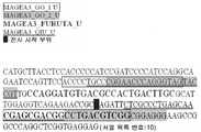


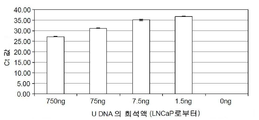

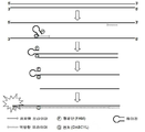
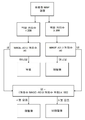


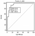


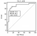


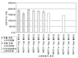

Classifications
-
- C—CHEMISTRY; METALLURGY
- C12—BIOCHEMISTRY; BEER; SPIRITS; WINE; VINEGAR; MICROBIOLOGY; ENZYMOLOGY; MUTATION OR GENETIC ENGINEERING
- C12Q—MEASURING OR TESTING PROCESSES INVOLVING ENZYMES, NUCLEIC ACIDS OR MICROORGANISMS; COMPOSITIONS OR TEST PAPERS THEREFOR; PROCESSES OF PREPARING SUCH COMPOSITIONS; CONDITION-RESPONSIVE CONTROL IN MICROBIOLOGICAL OR ENZYMOLOGICAL PROCESSES
- C12Q1/00—Measuring or testing processes involving enzymes, nucleic acids or microorganisms; Compositions therefor; Processes of preparing such compositions
- C12Q1/68—Measuring or testing processes involving enzymes, nucleic acids or microorganisms; Compositions therefor; Processes of preparing such compositions involving nucleic acids
- C12Q1/6813—Hybridisation assays
- C12Q1/6827—Hybridisation assays for detection of mutation or polymorphism
-
- A—HUMAN NECESSITIES
- A61—MEDICAL OR VETERINARY SCIENCE; HYGIENE
- A61K—PREPARATIONS FOR MEDICAL, DENTAL OR TOILETRY PURPOSES
- A61K38/00—Medicinal preparations containing peptides
- A61K38/04—Peptides having up to 20 amino acids in a fully defined sequence; Derivatives thereof
- A61K38/08—Peptides having 5 to 11 amino acids
-
- A—HUMAN NECESSITIES
- A61—MEDICAL OR VETERINARY SCIENCE; HYGIENE
- A61K—PREPARATIONS FOR MEDICAL, DENTAL OR TOILETRY PURPOSES
- A61K38/00—Medicinal preparations containing peptides
- A61K38/04—Peptides having up to 20 amino acids in a fully defined sequence; Derivatives thereof
- A61K38/10—Peptides having 12 to 20 amino acids
-
- A—HUMAN NECESSITIES
- A61—MEDICAL OR VETERINARY SCIENCE; HYGIENE
- A61K—PREPARATIONS FOR MEDICAL, DENTAL OR TOILETRY PURPOSES
- A61K39/00—Medicinal preparations containing antigens or antibodies
- A61K39/0005—Vertebrate antigens
- A61K39/0011—Cancer antigens
-
- A—HUMAN NECESSITIES
- A61—MEDICAL OR VETERINARY SCIENCE; HYGIENE
- A61K—PREPARATIONS FOR MEDICAL, DENTAL OR TOILETRY PURPOSES
- A61K39/00—Medicinal preparations containing antigens or antibodies
- A61K39/0005—Vertebrate antigens
- A61K39/0011—Cancer antigens
- A61K39/001184—Cancer testis antigens, e.g. SSX, BAGE, GAGE or SAGE
- A61K39/001186—MAGE
-
- A—HUMAN NECESSITIES
- A61—MEDICAL OR VETERINARY SCIENCE; HYGIENE
- A61P—SPECIFIC THERAPEUTIC ACTIVITY OF CHEMICAL COMPOUNDS OR MEDICINAL PREPARATIONS
- A61P35/00—Antineoplastic agents
-
- C—CHEMISTRY; METALLURGY
- C12—BIOCHEMISTRY; BEER; SPIRITS; WINE; VINEGAR; MICROBIOLOGY; ENZYMOLOGY; MUTATION OR GENETIC ENGINEERING
- C12Q—MEASURING OR TESTING PROCESSES INVOLVING ENZYMES, NUCLEIC ACIDS OR MICROORGANISMS; COMPOSITIONS OR TEST PAPERS THEREFOR; PROCESSES OF PREPARING SUCH COMPOSITIONS; CONDITION-RESPONSIVE CONTROL IN MICROBIOLOGICAL OR ENZYMOLOGICAL PROCESSES
- C12Q1/00—Measuring or testing processes involving enzymes, nucleic acids or microorganisms; Compositions therefor; Processes of preparing such compositions
- C12Q1/68—Measuring or testing processes involving enzymes, nucleic acids or microorganisms; Compositions therefor; Processes of preparing such compositions involving nucleic acids
- C12Q1/6844—Nucleic acid amplification reactions
-
- C—CHEMISTRY; METALLURGY
- C12—BIOCHEMISTRY; BEER; SPIRITS; WINE; VINEGAR; MICROBIOLOGY; ENZYMOLOGY; MUTATION OR GENETIC ENGINEERING
- C12Q—MEASURING OR TESTING PROCESSES INVOLVING ENZYMES, NUCLEIC ACIDS OR MICROORGANISMS; COMPOSITIONS OR TEST PAPERS THEREFOR; PROCESSES OF PREPARING SUCH COMPOSITIONS; CONDITION-RESPONSIVE CONTROL IN MICROBIOLOGICAL OR ENZYMOLOGICAL PROCESSES
- C12Q1/00—Measuring or testing processes involving enzymes, nucleic acids or microorganisms; Compositions therefor; Processes of preparing such compositions
- C12Q1/68—Measuring or testing processes involving enzymes, nucleic acids or microorganisms; Compositions therefor; Processes of preparing such compositions involving nucleic acids
- C12Q1/6876—Nucleic acid products used in the analysis of nucleic acids, e.g. primers or probes
- C12Q1/6883—Nucleic acid products used in the analysis of nucleic acids, e.g. primers or probes for diseases caused by alterations of genetic material
- C12Q1/6886—Nucleic acid products used in the analysis of nucleic acids, e.g. primers or probes for diseases caused by alterations of genetic material for cancer
-
- C—CHEMISTRY; METALLURGY
- C12—BIOCHEMISTRY; BEER; SPIRITS; WINE; VINEGAR; MICROBIOLOGY; ENZYMOLOGY; MUTATION OR GENETIC ENGINEERING
- C12Q—MEASURING OR TESTING PROCESSES INVOLVING ENZYMES, NUCLEIC ACIDS OR MICROORGANISMS; COMPOSITIONS OR TEST PAPERS THEREFOR; PROCESSES OF PREPARING SUCH COMPOSITIONS; CONDITION-RESPONSIVE CONTROL IN MICROBIOLOGICAL OR ENZYMOLOGICAL PROCESSES
- C12Q2525/00—Reactions involving modified oligonucleotides, nucleic acids, or nucleotides
- C12Q2525/30—Oligonucleotides characterised by their secondary structure
- C12Q2525/301—Hairpin oligonucleotides
-
- C—CHEMISTRY; METALLURGY
- C12—BIOCHEMISTRY; BEER; SPIRITS; WINE; VINEGAR; MICROBIOLOGY; ENZYMOLOGY; MUTATION OR GENETIC ENGINEERING
- C12Q—MEASURING OR TESTING PROCESSES INVOLVING ENZYMES, NUCLEIC ACIDS OR MICROORGANISMS; COMPOSITIONS OR TEST PAPERS THEREFOR; PROCESSES OF PREPARING SUCH COMPOSITIONS; CONDITION-RESPONSIVE CONTROL IN MICROBIOLOGICAL OR ENZYMOLOGICAL PROCESSES
- C12Q2561/00—Nucleic acid detection characterised by assay method
- C12Q2561/113—Real time assay
-
- C—CHEMISTRY; METALLURGY
- C12—BIOCHEMISTRY; BEER; SPIRITS; WINE; VINEGAR; MICROBIOLOGY; ENZYMOLOGY; MUTATION OR GENETIC ENGINEERING
- C12Q—MEASURING OR TESTING PROCESSES INVOLVING ENZYMES, NUCLEIC ACIDS OR MICROORGANISMS; COMPOSITIONS OR TEST PAPERS THEREFOR; PROCESSES OF PREPARING SUCH COMPOSITIONS; CONDITION-RESPONSIVE CONTROL IN MICROBIOLOGICAL OR ENZYMOLOGICAL PROCESSES
- C12Q2565/00—Nucleic acid analysis characterised by mode or means of detection
- C12Q2565/10—Detection mode being characterised by the assay principle
- C12Q2565/101—Interaction between at least two labels
-
- C—CHEMISTRY; METALLURGY
- C12—BIOCHEMISTRY; BEER; SPIRITS; WINE; VINEGAR; MICROBIOLOGY; ENZYMOLOGY; MUTATION OR GENETIC ENGINEERING
- C12Q—MEASURING OR TESTING PROCESSES INVOLVING ENZYMES, NUCLEIC ACIDS OR MICROORGANISMS; COMPOSITIONS OR TEST PAPERS THEREFOR; PROCESSES OF PREPARING SUCH COMPOSITIONS; CONDITION-RESPONSIVE CONTROL IN MICROBIOLOGICAL OR ENZYMOLOGICAL PROCESSES
- C12Q2600/00—Oligonucleotides characterized by their use
- C12Q2600/154—Methylation markers
-
- C—CHEMISTRY; METALLURGY
- C12—BIOCHEMISTRY; BEER; SPIRITS; WINE; VINEGAR; MICROBIOLOGY; ENZYMOLOGY; MUTATION OR GENETIC ENGINEERING
- C12Q—MEASURING OR TESTING PROCESSES INVOLVING ENZYMES, NUCLEIC ACIDS OR MICROORGANISMS; COMPOSITIONS OR TEST PAPERS THEREFOR; PROCESSES OF PREPARING SUCH COMPOSITIONS; CONDITION-RESPONSIVE CONTROL IN MICROBIOLOGICAL OR ENZYMOLOGICAL PROCESSES
- C12Q2600/00—Oligonucleotides characterized by their use
- C12Q2600/158—Expression markers
-
- C—CHEMISTRY; METALLURGY
- C12—BIOCHEMISTRY; BEER; SPIRITS; WINE; VINEGAR; MICROBIOLOGY; ENZYMOLOGY; MUTATION OR GENETIC ENGINEERING
- C12Q—MEASURING OR TESTING PROCESSES INVOLVING ENZYMES, NUCLEIC ACIDS OR MICROORGANISMS; COMPOSITIONS OR TEST PAPERS THEREFOR; PROCESSES OF PREPARING SUCH COMPOSITIONS; CONDITION-RESPONSIVE CONTROL IN MICROBIOLOGICAL OR ENZYMOLOGICAL PROCESSES
- C12Q2600/00—Oligonucleotides characterized by their use
- C12Q2600/16—Primer sets for multiplex assays
Abstract
Description
발명의 분야Field of invention
본 발명은 MAGE-A 패밀리 유전자 발현의 검출에 관한 것이다. 더욱 특히, 본 발명은 MAGE-A3의 메틸화된 형태 또는 메틸화되지 않은 형태를 검출하는 방법, 및 관련 올리고누클레오티드, 프라이머, 프로브, 프라이머 쌍 및 키트에 관한 것이다. 본 발명의 방법은 증폭 기술, 특히 형광 기반의 실시간 및 종점(end-point) PCR 방법을 수반하고, 이는 진단적, 예후적 및 치료적 유용성을 갖는다.The present invention relates to the detection of MAGE-A family gene expression. More particularly, the present invention relates to methods for detecting methylated or unmethylated forms of MAGE-A3 and related oligonucleotides, primers, probes, primer pairs and kits. The method of the present invention involves amplification techniques, in particular fluorescence-based real-time and end-point PCR methods, which have diagnostic, prognostic and therapeutic utility.
발명의 배경Background of the Invention
MAGE 유전자는 암/고환 항원의 패밀리에 속한다. MAGE 패밀리의 유전자는 20개를 초과하는 일원을 포함하고, 이는 MAGE A, B, C 및 D 유전자로 구성된다(Scanlan et al, (2002) Immunol Rev. 188:22-32; Chomez et al, (2001) Cancer Res. 61 (14): 5544-51). 이들은 염색체 X에 밀집되어 있고(Lucas et al., 1998 Cancer Res. 58.743-752; Lucas et al., 1999 Cancer Res 59:4100-4103; Lucas et al., 2000 Int J Cancer 87:55-60; Lurquin et al., 1997 Genomics 46:397-408; Muscatelli et al., 1995 Proc Natl Acad Sci USA 92:4987-4991; PoId et al., 1999 Genomics 59:161-167; Rogner et al 1995 Genomics 29:725-731), 아직 규명되지 않은 기능을 갖는다(Ohman et al 2001 Exp Cell Res. 265(2): 185-94). MAGE 유전자는 고도로 상동성이고, MAGE-A 패밀리의 일원은 특히 60 내지 98%의 상동성을 갖는다.The MAGE gene belongs to a family of cancer / testis antigens. The genes of the MAGE family include more than 20 members, which consist of the MAGE A, B, C and D genes (Scanlan et al, (2002) Immunol Rev. 188: 22-32; Chomez et al, ( 2001) Cancer Res. 61 (14): 5544-51). They are concentrated on chromosome X (Lucas et al., 1998 Cancer Res. 58.743-752; Lucas et al., 1999 Cancer Res 59: 4100-4103; Lucas et al., 2000 Int J Cancer 87: 55-60; Lurquin et al., 1997 Genomics 46: 397-408; Muscatelli et al., 1995 Proc Natl Acad Sci USA 92: 4987-4991; PoId et al., 1999 Genomics 59: 161-167; Rogner et al 1995 Genomics 29: 725-731), which has yet to be identified (Ohman et al 2001 Exp Cell Res. 265 (2): 185-94). The MAGE gene is highly homologous, and members of the MAGE-A family have particularly 60-98% homology.
인간 MAGE-A3 유전자는 다양한 유형의 종양, 예를 들어, 흑색종(Furuta et al. 2004 Cancer Sci. 95, 962-968.), 방광암, 간세포암종(Qiu et al. 2006. Clinical Biochemistry 39, 259-266), 위암종(Honda et al. 2004 British Journal of Cancer 90, 838-843), 직장결장암(Kim et al. 2006 World Journal of Gastroenterology 12, 5651-5657) 및 폐암(NSCLC)(Scanlan et al 2002 Immunol Rev. 188:22-32; Jang et al 2001 Cancer Res. 61, 21: 7959-7963)에서 발현된다. 고환 생식 세포 또는 태반만을 제외하고는 임의의 정상적인 성체 조직에서는 발현이 관찰되지 않는다(Haas et al. 1988 Am J Reprod Immunol Microbiol 18:47-51; Takahashi et al. 1995 Cancer Res 55:3478-382).The human MAGE-A3 gene can be used for various types of tumors such as melanoma (Furuta et al. 2004 Cancer Sci. 95, 962-968.), Bladder cancer, hepatocellular carcinoma (Qiu et al. 2006. Clinical Biochemistry 39, 259 -266), gastric carcinoma (Honda et al. 2004 British Journal of Cancer 90, 838-843), colorectal cancer (Kim et al. 2006 World Journal of Gastroenterology 12, 5651-5657) and lung cancer (NSCLC) (Scanlan et al. 2002 Immunol Rev. 188: 22-32; Jang et al 2001 Cancer Res. 61, 21: 7959-7963). No expression is observed in any normal adult tissue except testicular germ cells or placenta (Haas et al. 1988 Am J Reprod Immunol Microbiol 18: 47-51; Takahashi et al. 1995 Cancer Res 55: 3478-382) .
항원 특이적 암 면역치료제(Antigen-Specific Cancer Immunotherapeutics, ASCI)는 고도로 특이적인 방식으로 암 세포를 인지하고 이를 제거하도록 면역계를 훈련시키도록 디자인된 약물의 신규한 부류이다. 이와 같이, ASCI는 표적화된 치료를 가능케 한다. ASCI는 다음과 같은 2개의 주요 성분을 갖는다: 암세포에 대한 특이적인 면역 반응을 유도하는 "종양 항원" 및 항-종양 면역 반응을 증가시키도록 선택된 면역-자극 화합물을 포함하는 "애쥬번트 시스템". ASCI에 사용하기에 적합한 MAGE-A3 항원 및 작제물은 WO99/40188호에 기재되어 있고, 비소세포폐암(NSCLC)을 갖는 환자에서의 MAGE-A3 ASCI를 이용한 유망한 II상 연구 결과가 최근에 보고되었다(J. Clin. Oncol. Vol. 25, No. 18S (June 20 Suppl.) 2007: 7554).Antigen-Specific Cancer Immunotherapeutics (ASCI) is a new class of drugs designed to train the immune system to recognize and eliminate cancer cells in a highly specific manner. As such, ASCI enables targeted treatment. ASCI has two main components: an "adjuvant system" comprising an "tumor antigen" that elicits specific immune responses against cancer cells and an immune-stimulating compound selected to increase an anti-tumor immune response. MAGE-A3 antigens and constructs suitable for use in ASCI are described in WO99 / 40188 and recently reported promising Phase II studies with MAGE-A3 ASCI in patients with non-small cell lung cancer (NSCLC). (J. Clin. Oncol. Vol. 25, No. 18S (June 20 Suppl.) 2007: 7554).
면역요법으로부터 이로울 수 있는 MAGE-A3-발현 환자를 특이적으로 확인하거나, 투여 목적을 위한 MAGE-A3 발현을 모니터하거나, 임상 시험에서 Mage-A3 발현 샘플을 확인하거나, 간단히 암을 가진 초기 단계의 환자에서 확인할 수 있는 고 처리량의 정량적 검정을 갖는 것이 중요하다. 다수의 적용가능한 진단적 방법이 기재되어 있고, 이는 반정량적 RT-PCR(De Plaen et al. 1994 Immunogenetics 40 (5):360-9), 기타 PCR 기반 기술 및 저밀도 마이크로어레이(Zammatteo et al. 2002 Clinical Chemistry 48(1) 25-34)를 포함한다. 추가로, MAGE-A3 ASCI와 함께 사용하기 위한 개선된 RT-PCR 방법이 WO2007/147876호에 논의되어 있다.Specifically identify MAGE-A3-expressing patients that may benefit from immunotherapy, monitor MAGE-A3 expression for administration purposes, identify Mage-A3 expression samples in clinical trials, or simply initial stages with cancer It is important to have a high throughput quantitative assay that can be seen in patients. A number of applicable diagnostic methods have been described, which include semiquantitative RT-PCR (De Plaen et al. 1994 Immunogenetics 40 (5): 360-9), other PCR based techniques and low density microarrays (Zammatteo et al. 2002). Clinical Chemistry 48 (1) 25-34). In addition, an improved RT-PCR method for use with MAGE-A3 ASCI is discussed in WO2007 / 147876.
현존하는 검정의 가장 큰 단점은 이들은 MAGEA3 발현을 평가하기 위해 RNA 분리를 필요로 한다는 점이다. 포르말린 고정 파라핀 포매(Formalin-Fixed, Paraffin-Embedded, FFPE)된 종양 조직은 임상 센터에서의 통상적인 종양 조직 보존 방법이다. 포르말린 내에서의 고정은 조직 내의 RNA의 분자 구조를 변경시켜, 가교 및 또한 부분적인 분해를 야기시킨다. 부분적인 분해는 100 내지 300개의 염기쌍의 RNA의 보다 작은 단편을 생성시킨다. 이러한 RNA에 대한 구조적 변화는 MAGEA3 발현 수준을 측정하기 위해 FFPE 조직으로부터 추출된 RNA의 용도를 제한한다.The major disadvantage of existing assays is that they require RNA isolation to assess MAGEA3 expression. Formalin-fixed, paraffin-embedded (FFPE) tumor tissue is a common method of preserving tumor tissue in clinical centers. Fixation in formalin alters the molecular structure of RNA in tissue, causing crosslinking and also partial degradation. Partial digestion results in smaller fragments of 100-300 base pairs of RNA. Structural changes to these RNAs limit the use of RNA extracted from FFPE tissue to measure MAGEA3 expression levels.
본 발명의 목적은 현존하는 검정의 단점을 배제시키는 개선된 검정을 제공하기 위한 것이다.It is an object of the present invention to provide an improved assay that eliminates the disadvantages of existing assays.
유전자 메틸화는 유전자 발현의 중요한 조절인자이다. 특히, 특정 유전자의 프로모터 영역 내의 CpG 디누클레오티드 쌍에서 밝혀진 시토신 잔기에서의 메틸화는 유전자 발현의 하향 조절을 통해 다수의 질병 상태에 기여할 수 있다. 예를 들어, 종양 억제인자 유전자의 비정상 메틸화는 상기 유전자의 상향 또는 하향 조절을 초래할 수 있고, 따라서 이는 다수의 암의 존재 및 발달과 관련된다(Hoffmann et al. 2005 Biochem Cell Biol 83: 296-321). 비정상 유전자 메틸화의 패턴은 종종 기원 조직에 특이적이다. 따라서, 특정 유전자의 메틸화 상태의 검출은 예후적 및 진단적으로 유용할 수 있고, 질병의 상대적 단계를 결정하고, 또한 특정 유형의 요법에 대한 반응을 예측하는데 사용될 수 있다(Laird. 2003 Nat Rev Cancer 3: 253-266).Gene methylation is an important regulator of gene expression. In particular, methylation at cytosine residues found in CpG dinucleotide pairs in the promoter region of a particular gene can contribute to multiple disease states through down regulation of gene expression. For example, abnormal methylation of tumor suppressor genes can lead to up- or down-regulation of these genes, and thus are associated with the presence and development of multiple cancers (Hoffmann et al. 2005 Biochem Cell Biol 83: 296-321 ). The pattern of abnormal gene methylation is often specific to the tissue of origin. Thus, detection of methylation status of certain genes can be useful prognosticly and diagnostically, and can be used to determine the relative stage of the disease and also predict the response to certain types of therapies (Laird. 2003 Nat Rev Cancer 3 253-266).
다른 기술을 이용한 정량적 시험이 개발(Laird PW., Nat Rev Cancer 2003; 3: 253-66; Eads et al. Nucleic Acids Res 2000;28:E32; Mikeska T, et al. J Mol Diagn 2007)되었으나, 겔 상의 결과의 시각화를 이용한 메틸화 특이적 PCR(MSP)(겔 기반의 MSP 검정)이 유전자의 후생유전학적 사일런싱(epigenetic silencing)을 결정하는데 널리 사용된다(Esteller M et al. Cancer Res 2001; 61: 3225-9.).Quantitative tests using other techniques have been developed (Laird PW., Nat Rev Cancer 2003; 3: 253-66; Eads et al. Nucleic Acids Res 2000; 28: E32; Mikeska T, et al. J Mol Diagn 2007). Methylation specific PCR (MSP) (gel based MSP assay) using visualization of results on gels is widely used to determine epigenetic silencing of genes (Esteller M et al. Cancer Res 2001; 61 : 3225-9.).
다수의 형광 기반 기술이 핵산 증폭 반응의 실시간 모니터링에 이용가능하다. 하나의 이러한 기술이 US 6,090,552호 및 EP 0912597호에 기재되어 있고, 이는 앰플리플루오르®(Amplifluor®)로 상업적으로 공지되어 있다. 이러한 방법은 또한 핵산 증폭 반응의 종점 모니터링에 적합하다. 문헌[Vlassenbroeck et al. (Vlassenbroeck et al., 2008. Journal of Molec. Diagn., V10, No. 4]에는 앰플리플루오르® 기술을 이용한 표준화된 직접적인 실시간 MSP 검정이 추가로 기재되어 있다.Many fluorescence based techniques are available for real time monitoring of nucleic acid amplification reactions. One such technique is described in US Pat. No. 6,090,552 and EP 0912597, which are commercially known as Amplifluor®. This method is also suitable for endpoint monitoring of nucleic acid amplification reactions. See Vlassenbroeck et al. (Vlassenbroeck et al., 2008. Journal of Molec. Diagn., V10, No. 4) further describes standardized direct real-time MSP assays using Amplifluor® technology.
발명의 개요Summary of the Invention
본 발명은 MAGE-A3 발현을 측정하는 개선된 방법 및/또는 검정에 관한 것이다. 본 발명은 추가로 특정 유형의 요법, 특히 본원에 기재된 올리고누클레오티드, 프라이머, 프로브, 프라이머 쌍, 키트 및/또는 방법의 사용을 통해 MAGE-A3를 발현하는 것으로 확인된 환자의 항원 특이적 암 면역치료제(ASCI) 기반의 치료에 관한 것이다. MAGE-A3 단백질 발현이 MAGE-A3 유전자 자체의 발현 수준을 측정하기 보단 MAGE-A3 유전자의 메틸화 상태를 결정함으로써 검출된다. 본 발명자들은 MAGE-A3 유전자의 메틸화 시험의 메틸화 상태 결과가 MAGE-A3 면역요법으로부터의 이점에 대해 NSCLC에서의 MAGE-A3 발현의 예측 값을 확립하는 RT-PCR 검정으로 수득된 결과와 양호하게 일치하는 것을 제시한다. 따라서, 상기 검정은 환자의 성공적인 치료 가능성을 예측하기 위해 치료에 적합한 환자를 선택하는데 유용하고, 이는 환자 요법 선택을 돕는데 사용될 수 있다.The present invention relates to improved methods and / or assays for measuring MAGE-A3 expression. The present invention further provides antigen-specific cancer immunotherapeutics for patients identified to express MAGE-A3 through the use of certain types of therapies, in particular oligonucleotides, primers, probes, primer pairs, kits and / or methods described herein. (ASCI) based treatment. MAGE-A3 protein expression is detected by determining the methylation status of the MAGE-A3 gene rather than measuring the expression level of the MAGE-A3 gene itself. We find that the methylation status results of the methylation test of the MAGE-A3 gene are in good agreement with the results obtained with the RT-PCR assay, which establishes predictive values of MAGE-A3 expression in NSCLC for the benefit from MAGE-A3 immunotherapy. Present what you do. Thus, the assay is useful for selecting patients suitable for treatment in order to predict the likelihood of successful treatment of the patient, which can be used to help select patient therapy.
한 양태에서, 본 발명은 서열 목록 번호:2, 3, 4, 5, 6, 7, 8, 9, 11, 12, 13, 14, 15, 16, 17, 18, 19 또는 25 중 임의의 서열 목록 번호의 누클레오티드 서열을 포함하거나, 이로 본질적으로 구성되거나, 이로 구성되는 올리고누클레오티드, 프라이머 또는 프로브를 제공하며, 상기 올리고누클레오티드, 프라이머 또는 프로브는 유전자의 메틸화 상태의 검출에 유용하다.In one embodiment, the invention is any sequence of SEQ ID NO: 2, 3, 4, 5, 6, 7, 8, 9, 11, 12, 13, 14, 15, 16, 17, 18, 19 or 25 Provided are oligonucleotides, primers or probes comprising, consisting essentially of, or consisting of the nucleotide sequence of List No., which oligonucleotides, primers or probes are useful for the detection of the methylation state of a gene.
상기 올리고누클레오티드, 프라이머 또는 프로브는 바람직하게는 5'에서 3'의 순서로,The oligonucleotide, primer or probe is preferably in the order of 5 'to 3',
(a) 약 6 내지 30개의 누클레오티드의 제 1 누클레오티드 서열로서, 상기 제 1 누클레오티드 서열 내의 누클레오티드가 분자 에너지 전달쌍의 공여체 부분 및 수용체 부분으로부터 선택된 제 1 부분으로 표지되고, 상기 공여체 부분이 여기시 하나 이상의 특정 파장에서 형광을 방출하고, 상기 수용체 부분이 상기 공여체 부분에 의해 방출된 상기 형광을 흡수하고/하거나 켄칭(quenching)시키는 제 1 누클레오티드 서열;(a) a first nucleotide sequence of about 6 to 30 nucleotides, wherein the nucleotides in the first nucleotide sequence are labeled with a first portion selected from a donor portion and a acceptor portion of a molecular energy transfer pair, wherein the donor portion is one A first nucleotide sequence that emits fluorescence at the specific wavelength or above and wherein the receptor portion absorbs and / or quenchs the fluorescence emitted by the donor portion;
(b) 약 3 내지 20개의 누클레오티드를 포함하거나, 이로 본질적으로 구성되거나, 이로 구성되는 제 2 단일 가닥 누클레오티드 서열;(b) a second single stranded nucleotide sequence comprising, consisting essentially of, or consisting of about 3 to 20 nucleotides;
(c) 약 6 내지 30개의 누클레오티드를 포함하거나, 이로 본질적으로 구성되거나, 이로 구성되는 제 3 누클레오티드 서열로서, 상기 제 3 누클레오티드 서열 내의 누클레오티드가 상기 공여체 부분 및 상기 수용체 부분으로부터 선택된 제 2 부분으로 표지되고, 상기 제 2 부분이 상기 제 1 누클레오티드 서열을 표지하지 않는 상기 기의 일원이고, 상기 제 3 누클레오티드 서열이 상기 제 1 누클레오티드 서열에 대해 역순으로 상보적이어서, 상기 제 1 누클레오티드 서열과 상기 제 3 누클레오티드 서열 사이에 듀플렉스(duplex)를 형성할 수 있어, 상기 제 1 부분 및 상기 제 2 부분이 근접하여 위치하여, 상기 공여체 부분이 여기되어 형광을 방출하고, 상기 수용체 부분이 상기 공여체 부분에 의해 방출된 상기 형광을 흡수하고 켄칭시키는 제 3 누클레오티드 서열; 및(c) a third nucleotide sequence comprising, consisting essentially of, or consisting of about 6 to 30 nucleotides, wherein the nucleotides in the third nucleotide sequence are labeled with a second portion selected from the donor portion and the acceptor portion Wherein the second portion is a member of the group that does not label the first nucleotide sequence, and wherein the third nucleotide sequence is complementary to the first nucleotide sequence in reverse order, such that the first nucleotide sequence and the third A duplex can be formed between nucleotide sequences such that the first portion and the second portion are located in proximity such that the donor portion is excited to emit fluorescence, and the acceptor portion is emitted by the donor portion A third nucleotide sequence for absorbing and quenching the fluorescence; And
(d) 프라이머의 3' 말단에서, 서열 목록 번호:2, 4, 5, 7, 8, 11, 13, 14, 16, 17, 19 또는 25 중 임의의 서열 목록 번호의 누클레오티드 서열을 3' 말단에 포함하는 약 8 내지 40개의 누클레오티드를 포함하거나, 이로 본질적으로 구성되거나, 이로 구성되는 제 4 단일 가닥 누클레오티드 서열의, 연속된 서열을 포함하거나, 이로 본질적으로 구성되거나, 이로 구성되며;(d) at the 3 'end of the primer, replace the nucleotide sequence of any of SEQ ID NO: 2, 4, 5, 7, 8, 11, 13, 14, 16, 17, 19 or 25 with the nucleotide sequence of A continuous sequence of, or consisting essentially of, a fourth single stranded nucleotide sequence comprising, consisting essentially of, or consisting of about 8 to 40 nucleotides included therein;
상기 듀플렉스가 형성되지 않는 경우, 상기 제 1 부분 및 상기 제 2 부분은 상기 제 1 부분과 상기 제 2 부분 사이의 분자 에너지 전달을 방지하는 거리로 분리된다.If the duplex is not formed, the first portion and the second portion are separated by a distance that prevents molecular energy transfer between the first portion and the second portion.
3' 말단에서의 특정 누클레오티드 서열은 결정되는 MAGE-A3 유전자의 메틸화 상태를 허용한다. 이들 프라이머는 적절한 시약(본원에 논의된 바와 같음)을 이용한 처리 후에 MAGEA3 유전자의 메틸화되지 않은 형태에 우선적으로 결합한다. 이러한 올리고누클레오티드의 특성은 본원에 논의되어 있고, 이러한 논의는 뮤타티스 뮤탄디스(Mutatis mutandis)에 적용된다. 특정 누클레오티드 서열은 MAGE A 패밀리 유전자의 메틸화된 DNA 또는 메틸화되지 않은 DNA의 일부를 포함하는 핵산 가닥에 상보적인 누클레오티드 서열의 핵산 중합효소에 의한 합성을 프라이밍(priming) 할 수 있다.Certain nucleotide sequences at the 3 'end allow the methylation status of the MAGE-A3 gene to be determined. These primers preferentially bind to the unmethylated form of the MAGEA3 gene after treatment with the appropriate reagents (as discussed herein). The properties of such oligonucleotides are discussed herein and this discussion applies to Mutatis mutandis . Certain nucleotide sequences may prime the synthesis by nucleic acid polymerases of nucleotide sequences complementary to nucleic acid strands comprising methylated DNA or portions of unmethylated DNA of the MAGE A family gene.
가장 바람직하게는, 올리고누클레오티드, 프라이머 또는 프로브는 서열 목록 번호:3, 6, 9, 12, 15 또는 18의 누클레오티드 서열로 구성되고, 이는 관심 유전자의 일부를 증폭시키는데 사용된다.Most preferably, the oligonucleotide, primer or probe consists of the nucleotide sequence of SEQ ID NO: 3, 6, 9, 12, 15 or 18, which is used to amplify a portion of the gene of interest.
서열 목록 번호:2, 3, 4, 5, 6, 7, 8, 9, 11, 12, 13, 14, 15, 16, 17, 18, 19 또는 25 중 임의의 서열 목록 번호의 누클레오티드 서열을 포함하거나, 이로 본질적으로 구성되거나, 이로 구성되는 프라이머를 포함하는 프라이머 쌍이 추가로 제공된다.SEQ ID NO: 2, 3, 4, 5, 6, 7, 8, 9, 11, 12, 13, 14, 15, 16, 17, 18, 19 or 25 includes the nucleotide sequence of any of SEQ ID NO: Further provided are primer pairs comprising or consisting essentially of, or comprising a primer consisting of.
한 추가 양태에서, 서열 목록 번호:2, 3, 4, 5, 6, 7, 8, 9, 11, 12, 13, 14, 15, 16, 17, 18, 19 또는 25 중 임의의 서열 목록 번호의 누클레오티드 서열을 포함하거나, 이로 본질적으로 구성되거나, 이로 구성되는 하나 이상의 프라이머, 프라이머 쌍, 프라이머들의 쌍을 포함하는 키트가 제공된다. 키트는 유전자, 특히 MAGE-A 패밀리 유전자, 예를 들어, MAGE-A3의 메틸화 상태를 검출하기 위한 것이다.In one further aspect, SEQ ID NO: 2, 3, 4, 5, 6, 7, 8, 9, 11, 12, 13, 14, 15, 16, 17, 18, 19, or 25 Kits are provided comprising one or more primers, primer pairs, pairs of primers comprising, consisting essentially of, or consisting of the nucleotide sequence of. The kit is for detecting the methylation status of the gene, in particular the MAGE-A family gene, for example MAGE-A3.
한 추가 양태에서, 본 발명은, (a) DNA 함유 샘플에, DNA 내의 메틸화되지 않은 시토신 잔기를 선택적으로 변형시키는 시약을 접촉시키고/처리하여 메틸화된 시토신 잔기를 변형시키지 않으나 검출가능한 변형된 잔기를 생성시키는 단계; 및 (b) 하나 이상의 프라이머 쌍을 이용하여 메틸화되거나 메틸화되지 않은 관심 유전자의 일부 또는 전부를 증폭시키는 단계로서, 상기 프라이머 쌍 중 하나 이상의 프라이머가 상기 시약을 이용한 처리 후에 메틸화된 DNA 또는 메틸화되지 않은 DNA 각각의 서열에만 결합하도록 디자인되고, 상기 프라이머 쌍 중 하나 이상의 프라이머가 서열 목록 번호:2, 3, 4, 5, 6, 7, 8, 9, 11, 12, 13, 14, 15, 16, 17, 18, 19 또는 25(적절한 경우) 중 임의의 서열 목록 번호의 누클레오티드 서열을 포함하거나, 이로 본질적으로 구성되거나, 이로 구성되는 단계를 포함하여, DNA 함유 샘플 내의 Mage-A3 유전자의 메틸화 상태를 검출하는 방법을 제공한다.In a further aspect, the invention provides a modified residue that is detectable without modifying the methylated cytosine residues by (a) contacting the DNA containing sample with a reagent that selectively modifies unmethylated cytosine residues in the DNA. Generating; And (b) amplifying some or all of the genes of interest that are not methylated or unmethylated with one or more primer pairs, wherein one or more primers of the primer pairs are methylated or unmethylated DNA after treatment with the reagents. Designed to bind only to each sequence, one or more of the primer pairs described above is SEQ ID NO: 2, 3, 4, 5, 6, 7, 8, 9, 11, 12, 13, 14, 15, 16, 17 Detecting the methylation status of the Mage-A3 gene in a DNA-containing sample, including the step of comprising, consisting essentially of, or consisting of the nucleotide sequence of any of SEQ ID NO: 18, 19, or 25 (where appropriate) Provide a way to.
한 추가 양태에서, 본원에 기재된 올리고누클레오티드, 프라이머 또는 프로브, 프라이머 쌍, 키트 또는 방법을 이용하여 샘플 내의 MAGE-A3 유전자의 메틸화 상태를 검출하는 것을 포함하여 암 또는 암에 대한 소인을 진단하는 방법이 제공되며, 여기서 샘플 내의 메틸화되지 않은 MAGE-A3의 존재는 암 또는 암에 대한 소인을 나타낸다.In a further aspect, a method of diagnosing predisposition to cancer or cancer, including detecting methylation status of the MAGE-A3 gene in a sample using oligonucleotides, primers or probes, primer pairs, kits, or methods described herein Wherein the presence of unmethylated MAGE-A3 in the sample indicates cancer or predisposition to cancer.
한 추가 양태에서, 본원에 기재된 올리고누클레오티드, 프라이머 또는 프로브, 프라이머 쌍, 키트 또는 방법을 이용하여 샘플 내의 MAGE-A3 유전자의 메틸화 상태를 검출하는 것을 포함하여 MAGE-A3 양성 종양의 존재를 결정하는 방법이 제공되며, 여기서 메틸화되지 않은 MAGE-A3의 존재는 MAGE-A3 양성 종양의 존재를 나타낸다. "MAGE-A3 양성 종양"은 MAGE-A3 항원을 발현하는 임의의 종양 또는 종양 세포(환자로부터 분리됨)를 의미한다.In a further aspect, a method of determining the presence of a MAGE-A3 positive tumor, including detecting the methylation status of the MAGE-A3 gene in a sample using an oligonucleotide, primer or probe, primer pair, kit, or method described herein Provided where the presence of unmethylated MAGE-A3 indicates the presence of a MAGE-A3 positive tumor. "MAGE-A3 positive tumor" means any tumor or tumor cell expressing the MAGE-A3 antigen (isolated from the patient).
본 발명은 추가로 본원에 기재된 올리고누클레오티드, 프라이머 또는 프로브, 프라이머 쌍, 키트 또는 방법을 이용하여 환자의 샘플 내의 MAGE-A3 유전자의 메틸화 상태를 검출하는 것을 포함하여 MAGE-A3 면역치료제를 이용한 치료에 적합한 환자를 확인하고/하거나 선택하는 방법을 제공하며, 여기서 MAGE-A3 유전자가 메틸화되지 않은 경우, 피검체는 (바람직하게는) MAGE-A3 면역치료제를 이용한 치료에 대해 확인되고/되거나 선택된다. 따라서, 메틸화되지 않은 MAGE-A3를 갖는 환자가 상기 유전자가 메틸화된 환자에 우선하여 선택된다.The present invention further provides for the treatment with a MAGE-A3 immunotherapeutic agent, including detecting the methylation status of the MAGE-A3 gene in a patient's sample using oligonucleotides, primers or probes, primer pairs, kits or methods described herein. A method of identifying and / or selecting a suitable patient is provided wherein the subject is (preferably) identified and / or selected for treatment with a MAGE-A3 immunotherapeutic agent if the MAGE-A3 gene is not methylated. Thus, patients with unmethylated MAGE-A3 are selected in preference to patients whose genes are methylated.
대안적으로, 상기 유전자가 메틸화된 경우, 상기 피검체는 바람직하게는 MAGE-A3 면역치료제를 이용한 치료에 대해 확인되고/되거나 선택되지 않는다.Alternatively, if the gene is methylated, the subject is preferably not identified and / or selected for treatment with a MAGE-A3 immunotherapeutic agent.
한 관련 양태에서, 본 발명은 본원에 기재된 올리고누클레오티드, 프라이머 또는 프로브, 프라이머 쌍, 키트 또는 방법을 이용하여 환자의 샘플 내의 MAGE-A3 유전자의 메틸화 상태를 검출하는 것을 포함하여 암의 성공적인 치료 가능성을 예측하는 방법을 제공하며, 여기서 상기 유전자가 메틸화되지 않은 경우, MAGE-A3 면역치료제를 이용한 성공적인 치료 가능성이 상기 유전자가 메틸화된 경우보다 높다.In one related aspect, the present invention provides for the possibility of successful treatment of cancer, including detecting the methylation status of the MAGE-A3 gene in a patient's sample using oligonucleotides, primers or probes, primer pairs, kits, or methods described herein. A method of predicting is provided wherein where the gene is not methylated, the likelihood of successful treatment with a MAGE-A3 immunotherapeutic is higher than when the gene is methylated.
대안적으로, 샘플 내의 메틸화되지 않은 MAGE-A3의 부재는 MAGE-A3 면역치료제를 이용한 치료에 대한 내성의 가능성이 상기 유전자가 메틸화된 경우보다 높은 것을 나타낸다. 따라서, 메틸화된 MAGE-A3 유전자의 검출은 면역치료제를 이용한 성공적인 치료 가망성이 낮음을 나타낸다.Alternatively, the absence of unmethylated MAGE-A3 in the sample indicates that the likelihood of resistance to treatment with the MAGE-A3 immunotherapeutic is higher than when the gene is methylated. Thus, detection of the methylated MAGE-A3 gene indicates a low likelihood of successful treatment with an immunotherapeutic agent.
한 추가 관련 양태에서, 본 발명은 본원에 기재된 올리고누클레오티드, 프라이머 또는 프로브, 프라이머 쌍, 키트 또는 방법을 이용하여 환자의 샘플 내의 MAGE-A3 유전자의 메틸화 상태를 검출하는 것을 포함하여 암에 적합한 치료 요법을 선택하는 방법을 제공하며, 여기서 상기 유전자가 메틸화되지 않은 경우, 면역치료제가 치료에 대해 선택된다.In a further related aspect, the invention is a therapeutic regimen suitable for cancer comprising detecting the methylation status of the MAGE-A3 gene in a patient's sample using oligonucleotides, primers or probes, primer pairs, kits or methods described herein A method of selecting is provided wherein an immunotherapeutic is selected for treatment if the gene is not methylated.
대안적으로, 상기 유전자가 메틸화된 경우, 면역치료제를 이용한 치료가 금기된다.Alternatively, if the gene is methylated, treatment with immunotherapeutic agents is contraindicated.
또한, 면역치료제의 투여를 포함하여 피검체에서 암을 치료하는 방법이 제공되며, 여기서 피검체는 본 발명의 방법 중 임의의 방법에 따르거나, 본원에 기재된 올리고누클레오티드, 프라이머 또는 프로브, 프라이머 쌍, 키트 또는 방법을 이용함으로써 MAGE-A3 유전자의 메틸화 상태를 측정하는 것을 기초로 하여 치료에 대해 선택된다.Also provided are methods of treating cancer in a subject, including administration of an immunotherapeutic agent, wherein the subject is in accordance with any of the methods of the invention or described herein in an oligonucleotide, primer or probe, primer pair, Selection for treatment is based on determining the methylation status of the MAGE-A3 gene by using a kit or method.
바람직하게는, 본원에 기재된 모든 다양한 양태에 대해, 메틸화되지 않은 MAGE-A3 유전자의 검출은 증가된 수준의 MAGE-A3 단백질에 해당한다.Preferably, for all of the various embodiments described herein, the detection of the unmethylated MAGE-A3 gene corresponds to increased levels of MAGE-A3 protein.
본 발명은 본원에 기재된 올리고누클레오티드, 프라이머 또는 프로브, 프라이머 쌍, 키트 또는 방법을 이용함으로써 본 발명의 방법 중 임의의 방법에 따라 MAGE-A3 유전자의 메틸화 상태를 측정한 후, 본원에 기재된 바와 같이 MAGE-A3를 포함하는 조성물을 환자에게 투여하는 것을 포함하여 환자를 치료하는 방법을 추가로 제공한다. 조성물은 바람직하게는 MAGE-A3 유전자가 메틸화되지 않은 것으로 밝혀진 경우에 투여된다.The present invention measures the methylation status of the MAGE-A3 gene according to any of the methods of the invention by using oligonucleotides, primers or probes, primer pairs, kits or methods described herein, and then MAGE as described herein. Further provided are methods for treating a patient, including administering to the patient a composition comprising -A3. The composition is preferably administered when the MAGE-A3 gene is found not to be methylated.
한 추가 양태에서, MAGE-A3 발현 종양의 재발에 민감한 환자를 치료하는 방법이 제공되며, 상기 환자는 종양 조직을 제거하기 위해 치료되고, 상기 방법은 본 발명의 방법 중 임의의 방법에 따르거나 본원에 기재된 올리고누클레오티드, 프라이머 또는 프로브, 프라이머 쌍, 키트 또는 방법을 이용하여 종양 조직에서 MAGE-A3 유전자의 메틸화 상태를 측정한 후, 본원에 기재된 바와 같이 MAGE-A3를 포함하는 조성물을 환자에게 투여하는 것을 포함한다. 조성물은 바람직하게는 MAGE-A3 유전자가 메틸화되지 않은 것으로 밝혀지는 경우에 투여된다.In a further aspect, a method of treating a patient susceptible to relapse of a MAGE-A3 expressing tumor is provided, the patient being treated to remove tumor tissue, the method according to any of the methods of the present invention or Measuring the methylation status of the MAGE-A3 gene in tumor tissue using the oligonucleotides, primers or probes, primer pairs, kits or methods described herein, and then administering to the patient a composition comprising MAGE-A3 as described herein. It includes. The composition is preferably administered when the MAGE-A3 gene is found to be unmethylated.
본 발명의 한 추가 양태에서, 종양을 가진 환자의 치료를 위한 약제의 제조에 있어서 MAGE-A3를 포함하는 조성물의 용도가 제공되며, 상기 환자는 본 발명의 방법 중 임의의 방법에 따르거나 본원에 기재된 올리고누클레오티드, 프라이머 또는 프로브, 프라이머 쌍, 키트 또는 방법을 이용하여 MAGE-A3 유전자의 메틸화 상태를 측정하는 것을 기초로 하여 치료에 대해 선택된다. 또한, 종양을 가진 환자의 치료에 사용하기 위한 MAGE-A3를 포함하는 조성물이 제공되며, 상기 환자는 본 발명의 방법 중 임의의 방법에 따르거나 본원에 기재된 올리고누클레오티드, 프라이머 또는 프로브, 프라이머 쌍, 키트 또는 방법을 이용하여 MAGE-A3 유전자의 메틸화 상태를 측정하는 것을 기초로 하여 치료에 대해 선택된다.In a further aspect of the invention there is provided the use of a composition comprising MAGE-A3 in the manufacture of a medicament for the treatment of a patient with a tumor, wherein the patient is in accordance with any of the methods of the invention or Selection is made for treatment based on determining the methylation status of the MAGE-A3 gene using the described oligonucleotides, primers or probes, primer pairs, kits or methods. Also provided is a composition comprising MAGE-A3 for use in the treatment of a patient with a tumor, wherein the patient is in accordance with any of the methods of the present invention or described herein, an oligonucleotide, primer or probe, primer pair, Selection is made for the treatment based on measuring the methylation status of the MAGE-A3 gene using a kit or method.
한 추가 구체예에서, MAGE-A3 발현 종양의 재발에 민감한 환자의 치료를 위한 약제의 제조에 있어서 MAGE-A3를 포함하는 조성물의 용도가 제공되며, 상기 환자는 본 발명의 방법 중 임의의 방법에 따르거나 본원에 기재된 올리고누클레오티드, 프라이머 또는 프로브, 프라이머 쌍, 키트 또는 방법을 이용하여 MAGE-A3 유전자의 메틸화 상태를 측정하는 것을 기초로 하여 치료에 대해 선택된다. 또한, MAGE-A3 발현 종양의 재발에 민감한 환자의 치료에 사용하기 위한 MAGE-A3를 포함하는 조성물이 제공되며, 상기 환자는 본 발명의 방법 중 임의의 방법에 따르거나 본원에 기재된 올리고누클레오티드, 프라이머 또는 프로브, 프라이머 쌍, 키트 또는 방법을 이용하여 MAGE-A3 유전자의 메틸화 상태를 측정하는 것을 기초로 하여 치료에 대해 선택된다.In a further embodiment, there is provided the use of a composition comprising MAGE-A3 in the manufacture of a medicament for the treatment of a patient susceptible to relapse of a MAGE-A3 expressing tumor, wherein the patient is directed to any of the methods of the invention. Or selected for treatment based on determining the methylation status of the MAGE-A3 gene using the oligonucleotides, primers or probes, primer pairs, kits, or methods described herein. Also provided are compositions comprising MAGE-A3 for use in the treatment of a patient susceptible to relapse of a MAGE-A3 expressing tumor, wherein the patient is in accordance with any of the methods of the present invention or oligonucleotides, primers described herein. Or selected for treatment based on measuring the methylation status of the MAGE-A3 gene using a probe, primer pair, kit or method.
발명의 상세한 설명Detailed description of the invention
본 발명은 DNA 함유 샘플 내의 메틸화되거나 메틸화되지 않은 MAGE-A3 유전자의 존재 및/또는 양을 검출하기 위한 검정을 제공한다. 이러한 검정을 개발하기 위해, MAGE-A3 유전자에서 메틸화에 민감한 영역을 확인하고, MAGE-A3 유전자의 메틸화된 형태로부터 메틸화되지 않은 형태를 구별할 수 있는 특정 올리고누클레오티드를 개발하는 것이 필요하였다.The present invention provides an assay for detecting the presence and / or amount of methylated or unmethylated MAGE-A3 gene in a DNA containing sample. To develop this assay, it was necessary to identify regions sensitive to methylation in the MAGE-A3 gene and to develop specific oligonucleotides that can distinguish unmethylated forms from methylated forms of the MAGE-A3 gene.
따라서, 첫번째 양태에서, 본 발명은 서열 목록 번호:2, 3, 4, 5, 6, 7, 8, 9, 11, 12, 13, 14, 15, 16, 17, 18, 19 또는 25 중 임의의 서열 목록 번호의 누클레오티드 서열을 포함하거나, 이로 본질적으로 구성되거나, 이로 구성되는 올리고누클레오티드를 제공한다. 이러한 올리고누클레오티드는 관심 유전자의 메틸화 상태의 검출에 유용하다. 올리고누클레오티드는 프라이머 및/또는 프로브로 제공될 수 있다. 특정 구체예에서, 올리고누클레오티드는 유전자에 대해 메틸화되지 않은 형태를 검출한다. 메틸화되지 않은 형태를 검출하기에 적합한 올리고누클레오티드는 서열 목록 번호:2, 4, 5, 7, 8, 11, 13 또는 25 중 임의의 서열 목록 번호의 누클레오티드 서열을 포함하거나, 이로 본질적으로 구성되거나, 이로 구성된다. 메틸화된 형태를 검출하기에 적합한 올리고누클레오티드는 서열 목록 번호:14, 16, 17 또는 19 중 임의의 서열 목록 번호의 누클레오티드 서열을 포함하거나, 이로 본질적으로 구성되거나, 이로 구성된다. 특정 구체예에서, 이러한 올리고누클레오티드는 본 발명에 기재된 바와 같은 헤어핀 구조를 포함한다. 이러한 바람직한 올리고누클레오티드는 유전자의 메틸화되지 않은 형태를 검출하기 위한 서열 목록 번호:3, 6, 9 및 12 및 유전자의 메틸화된 형태를 검출하기 위한 서열 목록 번호:15 및 18에 따른 서열을 포함한다.Thus, in a first aspect, the invention provides any of SEQ ID NOs: 2, 3, 4, 5, 6, 7, 8, 9, 11, 12, 13, 14, 15, 16, 17, 18, 19 or 25 Provided are oligonucleotides comprising, consisting essentially of, or consisting of the nucleotide sequence of SEQ ID NO: of. Such oligonucleotides are useful for the detection of the methylation status of the gene of interest. Oligonucleotides may be provided as primers and / or probes. In certain embodiments, oligonucleotides detect unmethylated forms for the gene. Oligonucleotides suitable for detecting unmethylated forms include or consist essentially of the nucleotide sequence of any of SEQ ID NO: 2, 4, 5, 7, 8, 11, 13 or 25, or This consists of. Oligonucleotides suitable for detecting methylated forms include, consist essentially of, or consist of the nucleotide sequence of any of SEQ ID NO: 14, 16, 17 or 19. In certain embodiments, such oligonucleotides comprise a hairpin structure as described herein. Such preferred oligonucleotides include the sequences according to SEQ ID NOs: 3, 6, 9 and 12 for detecting unmethylated forms of genes and SEQ ID NOs: 15 and 18 for detecting methylated forms of genes.
본 발명의 "유전자" 또는 "관심 유전자"는 바람직하게는 MAGEA3 및/또는 MAGEA6 유전자이다. MAGEA3 및 MAGEA6는 HUGO 유전자 명명법 위원회에 의해 승인된 유전자 기호이다. MAGEA3 유전자는 염색체 X(위치 q28)에 위치되어 있고, 유전자 서열은 수탁 번호 NM__005362 및 ENSG00000197172로 등록되어 있다. MAGE-A3 유전자는 흑색종 항원 패밀리 A, 3을 엔코딩한다. MAGEA6 유전자는 염색체 X(위치 q28)에 위치되어 있다. MAGE-A3는 종종 MAGE-3 또는 MAGEA3로 상호교환적으로 언급된다. 마찬가지로, MAGE-A6는 종종 MAGE-6 또는 MAGEA6로 상호교환적으로 언급되고; 모두 본원에서 사용된다. 상기 유전자의 저메틸화는 암, 예를 들어, 흑색종 또는 폐암(NSCLC 포함)의 발병과 연관될 수 있다.The "gene" or "gene of interest" of the invention is preferably a MAGEA3 and / or MAGEA6 gene. MAGEA3 and MAGEA6 are genetic symbols approved by the HUGO Gene Nomenclature Committee. The MAGEA3 gene is located at chromosome X (position q28) and the gene sequence is registered under accession numbers NM__005362 and ENSG00000197172. The MAGE-A3 gene encodes melanoma antigen family A, 3. The MAGEA6 gene is located on chromosome X (position q28). MAGE-A3 is often referred to interchangeably as MAGE-3 or MAGEA3. Likewise, MAGE-A6 is often referred to interchangeably as MAGE-6 or MAGEA6; All are used herein. Hypomethylation of such genes may be associated with the development of cancer, such as melanoma or lung cancer (including NSCLC).
메틸화에 민감한 CpG 디누클레오티드는 통상적으로 인간 유전자의 프로모터 영역에 집중되어 있다. 한 바람직한 구체예에서, 유전자의 메틸화 상태는 유전자의 프로모터 영역에서의 메틸화 수준을 결정함으로써 평가된다. "프로모터"는 전사 시작 부위(TSS)로부터 업스트림 영역이며, TSS로부터 약 10 Kb, 3Kb, 1 Kb, 500 bp 또는 150 내지 300 bp에 걸쳐 있다. MAGE-3에 대해, 프로모터 영역 내의 CpG 분포는 다소 적다.CpG dinucleotides sensitive to methylation are typically concentrated in the promoter region of human genes. In one preferred embodiment, the methylation status of a gene is assessed by determining the methylation level in the promoter region of the gene. A "promoter" is the region upstream from the transcription start site (TSS) and spans about 10 Kb, 3Kb, 1 Kb, 500 bp or 150-300 bp from the TSS. For MAGE-3, the CpG distribution in the promoter region is rather small.
용어 "메틸화 상태(state or status)"는 DNA 서열 내의 하나 또는 다수의 CpG 디누클레오티드에서의 5-메틸시토신("5-mCyt")의 존재 또는 부재를 의미한다. "과메틸화"는 정상적인 수준을 초과하는 메틸화의 수준의 증가로 정의된다. 따라서, 이는 유전자 내의 특정 CpG 부위, 종종 프로모터 영역에서 시토신(5-mCyt)의 비정상적 메틸화와 관련된다. 메틸화의 정상적인 수준은, 예를 들어, 비암성 세포에서의 메틸화의 수준을 결정함으로써 규정될 수 있다.The term "state or status" refers to the presence or absence of 5-methylcytosine ("5-mCyt") in one or multiple CpG dinucleotides in a DNA sequence. "Supermethylation" is defined as an increase in the level of methylation above the normal level. Thus, it is associated with abnormal methylation of cytosine (5-mCyt) at certain CpG sites in genes, often promoter regions. Normal levels of methylation can be defined, for example, by determining the level of methylation in noncancerous cells.
"저메틸화"는 "정상" DNA 서열(적합한 대조 샘플에서 발견됨) 내의 상응하는 CpG 디누클레오티드에서 발견되는 5-mCyt의 양에 비한, DNA 서열(시험 DNA 샘플 중) 내의 하나 또는 다수의 CpG 디누클레오티드에서의 5-mCyt의 감소된 존재를 의미한다."Low methylation" refers to one or multiple CpG dinucleotides in a DNA sequence (in a test DNA sample) relative to the amount of 5-mCyt found in the corresponding CpG dinucleotide in a "normal" DNA sequence (found in a suitable control sample). Reduced presence of 5-mCyt at
또한, 메틸화의 "정상" 수준은, 예를 들어, 비암성 세포에서의 메틸화의 수준을 결정함으로써 규정될 수 있다. 본 발명에서, MAGE-A3 및/또는 MAGE-A6 유전자의 저메틸화는 상기 종양 관련 항원 유전자의 증가된 발현을 나타내고, 이는 암의 신뢰성 있는 지표를 제공한다.In addition, the "normal" level of methylation can be defined, for example, by determining the level of methylation in noncancerous cells. In the present invention, hypomethylation of the MAGE-A3 and / or MAGE-A6 genes indicates increased expression of the tumor associated antigen genes, which provides a reliable indicator of cancer.
본 발명은 두번째 양태에서 서열 목록 번호:2, 3, 4, 5, 6, 7, 8, 9, 11, 12, 13, 14, 15, 16, 17, 18, 19 또는 25 중 임의의 서열 목록 번호의 누클레오티드 서열을 포함하거나, 이로 본질적으로 구성되거나, 이로 구성되는 하나 이상의 올리고누클레오티드와 DNA 함유 샘플을 접촉시키는 단계를 포함하여, DNA 함유 샘플 내의 메틸화되거나 메틸화되지 않은 유전자의 존재 및/또는 양을 검출하는 방법을 제공한다. 상기 방법은 바람직하게는 유전자가 메틸화되거나 메틸화되지 않은 지의 여부를 평가하기 위한 추가 단계를 포함한다. 이는 본원에 논의된 바와 같이 올리고누클레오티드가 DNA 함유 샘플 내의 DNA에 안정적으로 결합하는지의 여부에 좌우될 수 있다.The present invention provides a sequence listing of any of SEQ ID NOs: 2, 3, 4, 5, 6, 7, 8, 9, 11, 12, 13, 14, 15, 16, 17, 18, 19 or 25 in a second embodiment. The presence and / or amount of methylated or unmethylated genes in the DNA-containing sample, including contacting the DNA-containing sample with one or more oligonucleotides comprising, consisting essentially of, or consisting of a nucleotide sequence of number It provides a method for detecting. The method preferably comprises an additional step for assessing whether the gene is methylated or unmethylated. This may depend on whether the oligonucleotides stably bind to the DNA in the DNA containing sample as discussed herein.
메틸화 상태를 평가하기 위한 기술은 별개의 접근법을 기초로 한다. 본 발명의 올리고누클레오티드를 적용하는 임의의 적합한 기술이 이용될 수 있다. 한 구체예에서, 메틸화된 CpG 디누클레오티드 모티프를 검출하기 위한 접근법은 검출가능한 변형된 잔기를 생성하도록 DNA 내의 메틸화되지 않은 시토신 잔기를 선택적으로 변형시키는 시약을 이용한다. 시약은 메틸화된 시토신 잔기를 변형시키지 않으며, 따라서 바람직하게는 핵산 증폭을 포함할 수 있는 다운스트림 과정에서 메틸화되지 않은 핵산 분자와 메틸화된 핵산 분자 사이의 구별을 가능케 한다. 한 구체예에서, 시약은 메틸화되지 않은 시토신 잔기를 선택적으로 탈아민화시키는 작용을 할 수 있다. 따라서, 시약에 대한 노출 후, 메틸화되지 않은 DNA는 상응하는 메틸화된 DNA의 누클레오티드 서열과 상이한 누클레오티드 서열을 함유한다. 시토신의 탈아민화는 시토신 염기쌍 형성 거동과 상이한, 티민과 동일한 염기쌍 형성 특성을 갖는 우라실 잔기가 존재하게 한다. 이는 메틸화된 시토신과 메틸화되지 않은 시토신 사이의 구별을 가능하게 한다.Techniques for assessing methylation status are based on a separate approach. Any suitable technique for applying the oligonucleotides of the present invention can be used. In one embodiment, an approach for detecting methylated CpG dinucleotide motifs employs reagents that selectively modify unmethylated cytosine residues in DNA to produce detectable modified residues. The reagents do not modify the methylated cytosine residues, thus allowing the distinction between unmethylated nucleic acid molecules and methylated nucleic acid molecules in downstream processes, which may preferably include nucleic acid amplification. In one embodiment, the reagent may serve to selectively deaminize unmethylated cytosine residues. Thus, after exposure to the reagent, the unmethylated DNA contains a nucleotide sequence that is different from the nucleotide sequence of the corresponding methylated DNA. Deamination of cytosine results in the presence of uracil residues having the same base pairing properties as thymine, which differ from the cytosine base pairing behavior. This allows the distinction between methylated cytosine and unmethylated cytosine.
서열 차이를 평가하기에 유용한 통상적인 기술은 올리고누클레오티드 프라이머를 이용한다. 프라이머 디자인에 대한 두 접근법이 가능하다. 첫번째로, 프라이머는 이들이 DNA 메틸화의 임의의 잠재적 부위를 포함하지 않도록 디자인될 수 있다. 차별적 메틸화의 부위에서의 서열 변화가 두 프라이머 결합 부위 사이에 위치되고, 서열 변화의 시각화는 추가 검정 단계를 필요로 한다. 두번째로, 프라이머는 최초 처리된 서열의 메틸화되거나 메틸화되지 않은 형태와 특이적으로 하이브리드화되도록 디자인될 수 있다. 하이브리드화 후, 증폭 반응이 수행될 수 있고, 증폭 생성물은 당 분야에 공지된 임의의 검출 시스템을 이용하여 검정된다. 증폭 생성물의 존재는 프라이머가 DNA에 하이브리드화된 것을 나타낸다. 프라이머의 특이성은 DNA가 변형되거나 변형되지 않은 지의 여부를 나타내고, 차례로 이는 DNA가 메틸화되거나 메틸화되지 않은 지의 여부를 나타낸다. 예를 들어, 12, 13, 14, 15, 16, 17, 18, 19, 20, 21 또는 22개의 누클레오티드의 표적에 대한 충분한 상보성 영역이 존재하는 경우, 프라이머는 또한 다른 조작에 유용할 수 있는 하이브리드화를 방해하지 않는 추가의 누클레오티드 잔기를 함유할 수 있다. 이러한 다른 잔기의 예는 제한 엔도누클레아제 분해를 위한 부위, 리간드 결합 또는 인자 결합 또는 링커 또는 반복부를 위한 부위, 또는 시각화를 위한 잔기를 위한 부위일 수 있다. 올리고누클레오티드 프라이머는 이들이 변형된 메틸화된 잔기에 특이적이 되도록 존재할 수 있거나, 그렇지 않을 수 있다. 프라이머로 사용하기에 바람직한 올리고누클레오티드는 서열 목록 번호:2, 3, 4, 5, 6, 7, 8, 9, 11, 12, 13, 14, 15, 16, 17, 18, 19 또는 25 중 임의의 서열 목록 번호의 누클레오티드 서열을 포함하거나, 이로 본질적으로 구성되거나, 이로 구성된다.Conventional techniques useful for evaluating sequence differences utilize oligonucleotide primers. Two approaches to primer design are possible. First, primers can be designed so that they do not contain any potential site of DNA methylation. The sequence change at the site of differential methylation is located between the two primer binding sites and the visualization of the sequence change requires an additional assay step. Secondly, the primers can be designed to specifically hybridize with the methylated or unmethylated forms of the originally processed sequence. After hybridization, an amplification reaction can be performed and the amplification product is assayed using any detection system known in the art. The presence of the amplification product indicates that the primer is hybridized to DNA. The specificity of the primer indicates whether the DNA is modified or unmodified, which in turn indicates whether the DNA is methylated or unmethylated. For example, if there is sufficient complementarity region for a target of 12, 13, 14, 15, 16, 17, 18, 19, 20, 21 or 22 nucleotides, the primer may also be a hybrid that may be useful for other manipulations. It may contain additional nucleotide residues that do not interfere with the oxidation. Examples of such other residues may be sites for restriction endonuclease degradation, sites for ligand binding or factor binding or linkers or repeats, or sites for residues for visualization. Oligonucleotide primers may or may not be present so that they are specific for the modified methylated residue. Preferred oligonucleotides for use as primers are any of SEQ ID NOs: 2, 3, 4, 5, 6, 7, 8, 9, 11, 12, 13, 14, 15, 16, 17, 18, 19 or 25 Or consist essentially of or consist of the nucleotide sequence of SEQ ID NO:
변형된 핵산과 변형되지 않은 핵산 사이를 구별하는 한 추가 방법은 올리고누클레오티드 프로브를 사용하는 것이다. 이러한 프로브는 면형된 핵산 또는 변형된 핵산의 추가 생성물, 예를 들어, 증폭에 의해 수득된 생성물에 직접 하이드리드화될 수 있다. 프로브 기반 검정은 특정 서열에 대한 올리고누클레오티드 하이브리드화 및 이후의 하이브리드의 검출을 이용한다. 증폭 생성물이 검출되기 전에 추가 정제 단계, 예를 들어, 침전 단계가 존재할 수 있다. 올리고누클레오티드 프로브는 당 분야에 공지된 임의의 검출 시스템을 이용하여 표지될 수 있다. 이는 형광 부분, 방사성동위원소 표지된 부분, 생체발광 부분, 발광 부분, 화학발광 부분, 효소, 기질, 수용체 또는 리간드를 포함하나, 이에 제한되지는 않는다. 프로브로서 사용하기에 바람직한 올리고누클레오티드는 서열 목록 번호:2, 4, 5, 7, 8, 11, 13, 14, 16, 17, 19 또는 25 중 임의의 서열 목록 번호의 누클레오티드 서열을 포함하거나, 이로 본질적으로 구성되거나, 이로 구성된다.One further method of distinguishing between modified and unmodified nucleic acids is to use oligonucleotide probes. Such probes may be hybridized directly to further products of the faceted nucleic acid or modified nucleic acid, for example the product obtained by amplification. Probe-based assays utilize oligonucleotide hybridization for a particular sequence and subsequent detection of the hybrid. There may be further purification steps, for example precipitation steps, before the amplification product is detected. Oligonucleotide probes can be labeled using any detection system known in the art. This includes, but is not limited to, fluorescent moieties, radioisotope labeled moieties, bioluminescent moieties, luminescent moieties, chemiluminescent moieties, enzymes, substrates, receptors or ligands. Preferred oligonucleotides for use as probes comprise, or consist of, the nucleotide sequence of any of SEQ ID NO: 2, 4, 5, 7, 8, 11, 13, 14, 16, 17, 19 or 25. Or consist essentially of it.
"올리고누클레오티드 프라이머"는 본원에서 "프라이머"와 상호교환적으로 언급된다. 마찬가지로, "올리고누클레오티드 프로브"는 본원에서 "프로브"와 상호교환적으로 언급된다."Oligonucleotide primer" is referred to herein interchangeably with "primer". Likewise, "oligonucleotide probe" is referred to herein interchangeably with "probe."
바람직한 구체예에서, 유전자(또는 이의 일부, 특히 CpG 섬(CpG islands))의 메틸화 상태는 메틸화 특이적 PCR(MSP)을 이용하여 결정된다.In a preferred embodiment, the methylation status of a gene (or a portion thereof, in particular CpG islands) is determined using methylation specific PCR (MSP).
MSP 기술은 당업자에게 친숙할 것이다. MSP 접근법에서, DNA는 소듐-비술파이트 처리의 결과로서의 서열 차이를 이용함으로써 메틸화된 DNA로부터 메틸화되지 않은 DNA를 구별하도록 디자인된 프라이머 쌍을 이용하여 증폭될 수 있다(Herman JG et al. Proc Natl Acad Sci U S A. 1996 Sep 3; 93 (18): 9821-6 and WO 97/46705). MSP 기술의 한 특정 예는 실시간으로 메틸화된 DNA의 신뢰성 있는 정량을 가능케 하는 실시간 정량 MSP(QMSP)이다.MSP techniques will be familiar to those skilled in the art. In the MSP approach, DNA can be amplified using primer pairs designed to distinguish unmethylated DNA from methylated DNA by using sequence differences as a result of sodium-bisulfite treatment (Herman JG et al. Proc Natl Acad Sci US A. 1996
실시간 방법은 일반적으로 증폭 과정의 연속적인 광학 모니터링을 기초로 하고, 형광 표지된 시약을 이용하며, 이러한 형광 표지된 시약의 생성물 중의 혼입이 정량될 수 있고, 형광 표지된 시약의 정량은 주형에서의 서열의 카피 수(copy number)를 나타낸다. 이러한 표지된 시약은 이중 가닥 DNA에 우선적으로 결합하는 형광 염료일 수 있고, 형광 염료의 형광은 이중 가닥 DNA의 결합에 의해 크게 향상된다. 대안적으로, 표지된 프라이머 및/또는 표지된 프로브가 이용될 수 있다. 이들은 널리 공지되고 상업적으로 이용가능한 실시간 증폭 기술, 예를 들어, TAQMAN®, MOLECULAR BEACONS®, AMPLIFLUOR® 및 SCORPION® DzyNA® 등의 특정 적용에 제공된다. 종종, 이러한 실시간 방법은 중합효소 연쇄 반응(PCR)과 함께 사용된다.Real-time methods are generally based on continuous optical monitoring of the amplification process, using fluorescently labeled reagents, incorporation in the product of such fluorescently labeled reagents can be quantified, and quantification of fluorescently labeled reagents in the template The copy number of the sequence is shown. Such labeled reagents can be fluorescent dyes that preferentially bind to double stranded DNA, and the fluorescence of the fluorescent dye is greatly enhanced by the binding of the double stranded DNA. Alternatively, labeled primers and / or labeled probes can be used. They are provided for specific applications of well known and commercially available real time amplification techniques such as TAQMAN®, MOLECULAR BEACONS®, AMPLIFLUOR® and SCORPION® DzyNA®. Often, this real time method is used in conjunction with polymerase chain reaction (PCR).
TaqMan 기술은 형광 염료 및 켄칭 염료를 함유하는 선형의 가수분해성 올리고누클레오티드 프로브를 이용한다.TaqMan technology utilizes linear hydrolyzable oligonucleotide probes containing fluorescent dyes and quench dyes.
방사선 조사되는 경우, 여기된 형광 염료는 에너지를 형광이 아닌 근처의 켄칭 염료 분자에 전달한다(FRET 원리). TaqMan 프로브는 PCR 생성물의 내부 영역으로 어닐링되고, 이는 주형을 복제하는 경우 중합효소의 엑소누클레아제 활성에 의해 분해된다. 이는 켄처(quencher)의 활성을 종료시키고, 리포터 염료가 프로브 분해 속도에 비례하여 각각의 주기에서 증가하는 형광을 방출하기 시작한다.When irradiated, the excited fluorescent dyes transfer energy to nearby quenching dye molecules that are not fluorescent (FRET principle). TaqMan probes are annealed into the internal regions of the PCR product, which are degraded by the exonuclease activity of the polymerase when replicating the template. This terminates the activity of the quencher and the reporter dye begins to emit increasing fluorescence in each cycle in proportion to the probe degradation rate.
분자 비콘(beacon)은 또한 형광 및 켄칭 염료를 함유하나, 이들은 용액 중에서 자유롭게 존재하는 경우 헤어핀 구조를 채택하도록 디자인되어 있으며, 상기 둘 모두의 염료는 매우 근접하게 위치하여 형광 공명 에너지 전달(Fluorescence Resonance Energy Transfer, FRET)이 발생한다. 비콘이 어닐링 단계 동안 표적에 하이브리드화되는 경우, 헤어핀은 선형화되고, 둘 모두의 염료(공여체 및 수용체/켄처)가 분리된다. 공여체로부터 검출된 형광의 증가는 이용가능한 PCR 생성물의 양과 상관될 것이다.Molecular beacons also contain fluorescent and quenching dyes, but they are designed to adopt a hairpin structure when freely present in solution, both of which are located in close proximity and thus fluorescence resonance energy transfer. Transfer, FRET) occurs. If the beacons hybridize to the target during the annealing step, the hairpins are linearized and both dyes (donor and acceptor / quencher) are separated. The increase in fluorescence detected from the donor will be correlated with the amount of PCR product available.
스콜피온(scorpion) 프로브와 함께, 단일 올리고누클레오티드를 이용하여 서열 특이적 프라이밍 및 PCR 생성물 검출이 달성된다. 스콜피온 프로브는 하이브리드화되지 않은 상태에서 스템-루프(stem-loop) 형태를 유지하고, FRET이 형광단과 켄처 사이에서 발생한다. 스템의 3' 부분은 또한 프라이머의 신장 생성물에 상보적인 서열을 함유한다. 이러한 서열은 증폭가능하지 않은 단량체를 통해 특정 프라이머의 5' 말단에 연결된다. 스콜피온 프라이머의 신장 후, 특정 프로브 서열은 신장된 앰플리콘 내에서 상보체에 결합할 수 있고, 이에 따라 헤어핀 루프를 개방하고, 형광단 및 켄처를 분리시키고, 형광 신호를 제공할 수 있다.In conjunction with scorpion probes, sequence specific priming and PCR product detection is achieved using a single oligonucleotide. Scorpion probes maintain a stem-loop form in the unhybridized state, and FRET occurs between the fluorophore and the quencher. The 3 'portion of the stem also contains a sequence complementary to the extension product of the primer. This sequence is linked to the 5 'end of a specific primer through a monomer that is not amplifiable. After stretching of the scorpion primers, certain probe sequences may bind to the complement in the stretched amplicon, thus opening up the hairpin loop, separating the fluorophores and quencher, and providing a fluorescent signal.
헤비메틸(Heavymethyl)에서, 프라이밍은 메틸화 특이적이나, 신장가능하지 않은 올리고누클레오티드 차단제가 프라이머 자체 대신 상기 특이성을 제공한다. 차단제는 메틸화 특이적 방식으로 비술파이트 처리된 DNA에 결합하고, 이의 결합 부위는 프라이머 결합 부위와 중첩된다. 차단제가 결합되는 경우, 프라이머는 결합할 수 없고, 따라서 앰플리콘이 생성되지 않는다. 헤비메틸은 실시간 검출과 함께 사용될 수 있다.In Heavymethyl, priming is methylation specific, but non-extensible oligonucleotide blockers provide this specificity instead of the primer itself. The blocking agent binds to the bisulfite treated DNA in a methylation specific manner, the binding site of which overlaps the primer binding site. If the blocking agent is bound, the primers cannot bind and thus no amplicons are produced. Heavymethyl can be used with real time detection.
플렉소르(Plexor™) qPCR 및 qRT-PCR 시스템은 정량적 PCR 분석을 달성하기 위해 두개의 변형된 누클레오티드 사이의 특정 상호작용의 이점을 취한다. PCR 프라이머 중 하나는 5' 말단에서 이소-dC 잔기에 인접한 형광 표지를 함유한다. 두번째 PCR 프라이머는 표지되지 않는다. 반응 혼합물은 데옥시누클레오티드 및 켄처 dabcyl로 변형된 이소-dGTP를 포함한다. dabcyl-이소-dGTP는 이소-dC 잔기에 상보적인 위치에서 우선적으로 혼입된다. 이러한 위치에서의 dabcyl-이소-dGTP의 혼입은 상보적 가닥에서의 형광 염료의 켄칭 및 형광의 감소를 발생시키고, 이는 증폭 동안 정량을 가능케 한다. 이러한 멀티플렉스(multiplex) 반응을 위해, 다양한 형광단을 갖는 프라이머 쌍이 각각의 표적 서열에 대해 사용된다.The Flexor ™ qPCR and qRT-PCR systems take advantage of certain interactions between two modified nucleotides to achieve quantitative PCR analysis. One of the PCR primers contains a fluorescent label adjacent to the iso-dC residue at the 5 'end. The second PCR primer is not labeled. The reaction mixture comprises iso-dGTP modified with deoxynucleotides and quencher dabcyl. dabcyl-iso-dGTP is preferentially incorporated at positions complementary to iso-dC residues. Incorporation of dabcyl-iso-dGTP at this position results in quenching of fluorescent dyes in the complementary strands and a reduction in fluorescence, which allows for quantification during amplification. For this multiplex reaction, primer pairs with various fluorophores are used for each target sequence.
따라서, 서열 목록 번호:2, 3, 4, 5, 6, 7, 8, 9, 11, 12, 13, 14, 15, 16, 17, 18, 19 또는 25 중 임의의 서열 목록 번호의 누클레오티드 서열을 포함하거나, 이로 본질적으로 구성되거나, 이로 구성되는 올리고누클레오티드는 관심 유전자의 메틸화 상태의 검출을 위한 상기 언급된 방법에서 프라이머 또는 프로브로 사용될 수 있다.Thus, the nucleotide sequence of any of SEQ ID NOs: 2, 3, 4, 5, 6, 7, 8, 9, 11, 12, 13, 14, 15, 16, 17, 18, 19 or 25 Oligonucleotides comprising, consisting essentially of, or consisting of these may be used as primers or probes in the above-mentioned methods for the detection of the methylation status of the gene of interest.
한 바람직한 구체예에서, 본 발명은, (a) DNA 함유 샘플에, DNA 내의 메틸화되지 않은 시토신 잔기를 선택적으로 변형시키는 시약을 접촉시키고/처리하여 메틸화된 시토신 잔기를 변형시키지 않으나 검출가능한 변형된 잔기를 생성시키는 단계; 및 (b) 하나 이상의 프라이머 쌍을 이용하여 메틸화되거나 메틸화되지 않은 관심 유전자의 일부 또는 전부를 증폭시키는 단계로서, 상기 프라이머 쌍 중 하나 이상의 프라이머가 상기 시약을 이용한 처리 후에 메틸화된 DNA 또는 메틸화되지 않은 DNA 각각의 서열에만 결합하도록 디자인되고, 상기 프라이머 쌍 중 하나 이상의 프라이머가 서열 목록 번호:2, 3, 4, 5, 6, 7, 8, 9, 11, 12, 13, 14, 15, 16, 17, 18 또는 19 중 임의의 서열 목록 번호의 누클레오티드 서열을 포함하거나, 이로 본질적으로 구성되거나, 이로 구성되는 단계를 포함하여, DNA 함유 샘플 내의 메틸화되거나 메틸화되지 않은 관심 유전자의 존재 및/또는 양을 검출하는 실시간 방법을 제공한다.In one preferred embodiment, the present invention provides a modified residue that (a) is contacted with a sample that selectively modifies an unmethylated cytosine residue in the DNA and / or is modified to detect a methylated cytosine residue without modifying the methylated cytosine residue. Generating a; And (b) amplifying some or all of the genes of interest that are not methylated or unmethylated with one or more primer pairs, wherein one or more primers of the primer pairs are methylated or unmethylated DNA after treatment with the reagents. Designed to bind only to each sequence, one or more of the primer pairs described above is SEQ ID NO: 2, 3, 4, 5, 6, 7, 8, 9, 11, 12, 13, 14, 15, 16, 17 Detecting the presence and / or amount of a methylated or unmethylated gene of interest in a DNA containing sample, including the step of comprising, consisting essentially of, or consisting of a nucleotide sequence of any of SEQ ID NO: 18 or 19 To provide a real-time method.
본 발명의 방법에서의 관심 유전자는 바람직하게는 MAGE-A3 및/또는 MAGEA6 유전자이다. 바람직하게는, 프라이머 쌍 중 하나 이상의 프라이머는 증폭의 부재하에서 수용체 부분이 공여체 부분에 의해 방출(여기시)된 형광을 켄칭시키고, 증폭 동안 검출가능한 형광 신호를 생성시키기에 충분히 공여체 부분 및 수용체 부분을 분리시키기 위해 스템 루프 구조가 붕괴되도록 배열된, 분자 에너지 전달쌍의 공여체 부분 및 수용체 부분을 갖는 스템 루프 구조를 함유하는 프라이머이다. 이는 메틸화되거나 메틸화되지 않은 관심 유전자의 존재의 확인을 제공하기 위해 실시간으로 검출될 수 있다. 서열 목록 번호:2, 3, 4, 5, 6, 7, 8, 9, 11, 12, 13, 14, 15, 16, 17, 18 또는 19 중 임의의 서열 목록 번호의 누클레오티드 서열을 포함하거나, 이로 본질적으로 구성되거나, 이로 구성되는 프라이머 쌍 중의 프라이머는 바람직하게는 스템 루프 구조를 갖는다.The gene of interest in the methods of the invention is preferably the MAGE-A3 and / or MAGEA6 gene. Preferably, one or more primers of the primer pairs quench the fluorescence where the receptor moiety is released (here) by the donor moiety in the absence of amplification and is sufficient to generate the donor moiety and the receptor moiety to produce a detectable fluorescence signal during amplification. A primer containing a stem loop structure having a donor portion and a acceptor portion of a molecular energy transfer pair arranged to disrupt the stem loop structure for separation. It can be detected in real time to provide confirmation of the presence of the gene of interest that is methylated or unmethylated. The nucleotide sequence of any of SEQ ID NO: 2, 3, 4, 5, 6, 7, 8, 9, 11, 12, 13, 14, 15, 16, 17, 18 or 19, or Primers in or consisting essentially of these primer pairs preferably have a stem loop structure.
특정 구체예에서, 메틸화되거나 메틸화되지 않은 유전자의 유전자 카피수가 결정된다. 여기서, 본원에 기재된 방법은 바람직하게는, (c) 유전자 카피수의 결과를 생성시키기 위해, 메틸화되거나 메틸화되지 않은 관심 유전자에 대한 표준 곡선에 대해 실시간 검출의 결과를 정량하는 추가 단계를 포함한다.In certain embodiments, the gene copy number of a methylated or unmethylated gene is determined. Here, the methods described herein preferably comprise an additional step of quantifying the results of real-time detection against a standard curve for the gene of interest, either methylated or unmethylated, to generate (c) the result of gene copy number.
바람직하게는, 단계 (c)는 추가로 주기 역치 값이 40 미만인 경우에 증폭이 유효한 것으로 간주되는 것을 특징으로 한다.Preferably, step (c) is further characterized in that amplification is considered valid when the period threshold value is less than 40.
MageA3 및/또는 MageA6와 같은 유전자에 대해, 유전자의 메틸화되지 않은 형태의 검출이 가장 적절할 수 있다.For genes such as MageA3 and / or MageA6, detection of the unmethylated form of the gene may be most appropriate.
본 발명의 방법은 샘플 내에서 메틸화되거나 메틸화되지 않은 관심 유전자의 존재를 실시간으로 검출하는 것을 가능케 한다. 본 발명의 방법은 정량적 방법이므로, 관심 유전자의 메틸화되거나 메틸화되지 않은 형태의 (상대량) 양은 또한 반응이 진행됨에 따라 결정될 수 있다. 그러나, 실시간 방법이 이용되는 것이 요구되지는 않는다. 분석은 표적 DNA가 샘플 내에 존재하거나 존재하지 않는 지의 여부를 발견하는 경우에만 수행될 수 있다. 종점 증폭 검출 기술은 실시간 PCR에 광범위하게 사용되는 것과 동일한 접근법을 이용한다. 따라서, 본 발명의 방법은 DNA 함유 샘플 중의 메틸화되거나 메틸화되지 않은 관심 유전자의 존재 및/또는 양을 검출하는 종점 방법을 포함할 수 있다.The method of the present invention makes it possible to detect in real time the presence of a methylated or unmethylated gene of interest in a sample. Since the method of the present invention is a quantitative method, the (relative amount) of the methylated or unmethylated form of the gene of interest can also be determined as the reaction proceeds. However, it is not required that a real time method be used. The analysis can only be performed if it finds whether or not the target DNA is present in the sample. Endpoint amplification detection techniques use the same approach as is widely used for real-time PCR. Thus, the methods of the present invention can include endpoint methods for detecting the presence and / or amount of methylated or unmethylated genes of interest in a DNA containing sample.
따라서, 본 발명은, (a) DNA 함유 샘플에, DNA 내의 메틸화되지 않은 시토신 잔기를 선택적으로 변형시키는 시약을 접촉시키고/처리하여 메틸화된 시토신 잔기를 변형시키지 않으나 검출가능한 변형된 잔기를 생성시키는 단계; 및 (b) 하나 이상의 프라이머 쌍을 이용하여 메틸화되거나 메틸화되지 않은 관심 유전자의 일부 또는 전부를 증폭시키는 단계로서, 상기 프라이머 쌍 중 하나 이상의 프라이머가 상기 시약을 이용한 처리 후에 메틸화된 DNA 또는 메틸화되지 않은 DNA 각각의 서열에만 결합하도록 디자인되고, 상기 프라이머 쌍 중 하나 이상의 프라이머가 서열 목록 번호:2, 3, 4, 5, 6, 7, 8, 9, 11, 12, 13, 14, 15, 16, 17, 18, 19 또는 25 중 임의의 서열 목록 번호의 누클레오티드 서열을 포함하거나, 이로 본질적으로 구성되거나, 이로 구성되는 단계를 포함하여, DNA 함유 샘플 내의 메틸화되거나 메틸화되지 않은 관심 유전자의 존재 및/또는 양을 검출하는 (종점) 방법을 제공한다.Accordingly, the present invention provides a method of producing a modified residue that is detectable without modifying the methylated cytosine residue by contacting and / or treating the DNA-containing sample with a reagent that selectively modifies the unmethylated cytosine residue in the DNA. ; And (b) amplifying some or all of the genes of interest that are not methylated or unmethylated with one or more primer pairs, wherein one or more primers of the primer pairs are methylated or unmethylated DNA after treatment with the reagents. Designed to bind only to each sequence, one or more of the primer pairs described above is SEQ ID NO: 2, 3, 4, 5, 6, 7, 8, 9, 11, 12, 13, 14, 15, 16, 17 The presence and / or amount of a methylated or unmethylated gene of interest in a DNA containing sample, including the step of comprising, consisting essentially of, or consisting of the nucleotide sequence of any of SEQ ID NO: 18, 19, or 25 It provides a (endpoint) method for detecting the problem.
상기 언급된 바와 같이, 본 발명의 방법에서의 관심 유전자는 바람직하게는 MAGE-A3 및/또는 MAGE-A6 유전자이다. 바람직하게는, 프라이머 쌍 중의 하나 이상의 프라이머는 본원에 기재된 바와 같은 특성을 갖는 분자 에너지 전달쌍의 공여체 부분 및 수용체 부분을 갖는 스템 루프 구조를 함유하는 프라이머이다. 서열 목록 번호:2, 3, 4, 5, 6, 7, 8, 9, 11, 12, 13, 14, 15, 16, 17, 18 또는 19 중 임의의 서열 목록 번호의 누클레오티드 서열을 포함하거나, 이로 본질적으로 구성되거나, 이로 구성되는 프라이머 쌍 중의 프라이머는 바람직하게는 스템 루프 구조를 갖는다.As mentioned above, the gene of interest in the method of the invention is preferably the MAGE-A3 and / or MAGE-A6 gene. Preferably, at least one of the primer pairs is a primer containing a stem loop structure having a donor portion and a receptor portion of a molecular energy transfer pair having the properties as described herein. The nucleotide sequence of any of SEQ ID NO: 2, 3, 4, 5, 6, 7, 8, 9, 11, 12, 13, 14, 15, 16, 17, 18 or 19, or Primers in or consisting essentially of these primer pairs preferably have a stem loop structure.
MAGE-A3 및/또는 MAGE-A6 유전자에 대해, 유전자의 메틸화되지 않은 형태의 검출이 가장 적절할 수 있다. 서열 목록 번호:2, 3, 4, 5, 6, 7, 8, 9, 11, 12, 13 또는 25 중 임의의 서열 목록 번호의 누클레오티드 서열을 포함하거나, 이로 본질적으로 구성되거나, 이로 구성되는 프라이머는 시약을 이용한 처리 후의 메틸화되지 않은 MAGEA3 DNA의 검출을 위해 디자인된다.For MAGE-A3 and / or MAGE-A6 genes, detection of the unmethylated form of the gene may be most appropriate. A primer comprising, consisting essentially of, or consisting of a nucleotide sequence of any of SEQ ID NO: 2, 3, 4, 5, 6, 7, 8, 9, 11, 12, 13 or 25 Is designed for the detection of unmethylated MAGEA3 DNA after treatment with reagents.
메틸화되지 않은 유전자의 부재는 메틸화된 유전자의 존재를 나타낼 것이다. 그러나, 메틸화된 유전자의 검출이 또한 본 발명의 범위에 속한다. 서열 목록 번호:14, 15, 16, 17, 18 또는 19 중 임의의 서열 목록 번호의 누클레오티드 서열을 포함하거나, 이로 본질적으로 구성되거나, 이로 구성되는 프라이머는 시약을 이용한 처리 후의 메틸화된 MAGEA3 DNA의 검출을 위해 디자인된다.Absence of unmethylated genes will indicate the presence of methylated genes. However, detection of methylated genes is also within the scope of the present invention. A primer comprising, consisting essentially of, or consisting of the nucleotide sequence of any of SEQ ID NO: 14, 15, 16, 17, 18 or 19, the detection of methylated MAGEA3 DNA after treatment with a reagent Is designed for.
메틸화되거나 메틸화되지 않은 유전자의 유전자 카피수가 요망되는 경우, 상기 방법은 바람직하게는, (c) 유전자 카피수의 결과를 생성시키기 위해, 메틸화되거나 메틸화되지 않은 관심 유전자에 대한 표준 곡선에 대해 검출 결과를 정량하는 추가 단계를 포함한다.If the gene copy number of the methylated or unmethylated gene is desired, the method preferably includes (c) detecting the detection results against a standard curve for the gene of interest, which is methylated or not methylated, to produce a result of gene copy number. An additional step of quantification.
본 발명의 모든 구체예는 본 발명의 종점 양태에 적용가능하고, 따라서 뮤타티스 뮤탄디스에 적용가능하다. 종점 분석은 증폭의 종료시 형광을 결정하기 위한 형광 플레이트 판독기 또는 다른 적합한 기계의 사용을 필요로 한다.All embodiments of the present invention are applicable to the endpoint aspects of the present invention and are therefore applicable to mutatis mutandis. Endpoint analysis requires the use of a fluorescence plate reader or other suitable instrument to determine fluorescence at the end of the amplification.
본 발명의 방법은 가장 바람직하게는 임의의 적합한 (DNA 함유) 시험 샘플에서 수행되는 생체외 또는 시험관내 방법이다. 그러나, 한 구체예에서, 상기 방법은 또한 샘플을 수득하는 단계를 포함할 수 있다. 시험 샘플은 DNA 함유 샘플, 특히 관심 유전자를 포함하는 DNA 함유 샘플이다. 본 발명의 방법은 특히 관심 유전자의 메틸화가 질병의 발병과 관련된(관련된 것으로 공지된) 경우에 질병의 진단에 사용될 수 있다.The method of the invention is most preferably an ex vivo or in vitro method performed on any suitable (DNA containing) test sample. However, in one embodiment, the method may also include obtaining a sample. Test samples are DNA containing samples, in particular DNA containing samples comprising the gene of interest. The method of the present invention can be used for the diagnosis of a disease, particularly where methylation of the gene of interest is associated with (known as related to) the onset of the disease.
DNA 함유 샘플은 임의의 적합한 조직 샘플 또는 체액을 포함할 수 있다. 바람직하게는, 시험 샘플은 인간 피검체로부터 수득된다. 암 적용을 위해, 샘플은 암성인 것으로 여겨지는 조직 또는 대표적 체액으로부터 수득된 조직 샘플을 포함할 수 있다.The DNA containing sample may comprise any suitable tissue sample or body fluid. Preferably, the test sample is obtained from a human subject. For cancer applications, the sample may comprise tissue samples obtained from tissues or representative body fluids that are considered cancerous.
MAGE-A3 유전자의 저메틸화는 폐암과 관련된다. 따라서, 한 구체예에서, MAGE-A3 유전자를 포함하는 본 발명의 방법에서 사용되는 시험 샘플은 바람직하게는 폐 세포 또는 폐 세포로부터의 핵산을 함유한다. 가장 바람직하게는, 샘플은 포르말린 고정 파라핀 포매(FFPE) 조직이다. 비소세포폐암(NSCLC) 및 소세포폐암(SCLC)의 두 유형의 폐암이 존재한다. 상기 명칭은 단순히 종양에서 발견된 세포의 유형을 기재한다. 시험 샘플은 바람직하게는 비소세포폐암종(NSCLC)으로부터의 세포 또는 핵산을 함유한다. NSCLC는 편평세포 암종, 선암종 및 거대세포 암종을 포함하고, 폐암의 약 80%를 차지한다. 한 바람직한 구체예에서, 암이 NSCLC인 경우, 샘플은 폐 조직 샘플이거나 담(sputum) 샘플이다. NSCLC는 치료가 어렵고, 치료는 가능한 한 생명을 연장시키고 질병의 증상을 경감시키는데 목적을 두는 경향이 있다. NSCLC는 가장 흔한 유형의 폐암이고, 불량한 결과와 연관된다.Hypomethylation of the MAGE-A3 gene is associated with lung cancer. Thus, in one embodiment, the test sample used in the method of the invention comprising the MAGE-A3 gene preferably contains lung cells or nucleic acid from lung cells. Most preferably, the sample is formalin fixed paraffin embedded (FFPE) tissue. There are two types of lung cancers: non-small cell lung cancer (NSCLC) and small cell lung cancer (SCLC). The name simply describes the type of cell found in the tumor. The test sample preferably contains cells or nucleic acids from non-small cell lung carcinoma (NSCLC). NSCLC includes squamous cell carcinoma, adenocarcinoma and giant cell carcinoma and accounts for about 80% of lung cancers. In one preferred embodiment, when the cancer is NSCLC, the sample is a lung tissue sample or a putum sample. NSCLC tends to be difficult to treat, and treatment tends to aim at prolonging life and reducing symptoms of the disease as much as possible. NSCLC is the most common type of lung cancer and is associated with poor outcomes.
MAGE-A3 유전자의 저메틸화는 또한 방광암과 관련된다. 따라서, 추가 구체예에서, 본 발명의 방법에 사용되는 추가의 바람직한 시험 샘플은 방광 이행 세포 또는 편평 암종 방광 세포를 함유한다. 바람직하게는, 시험 샘플은 방광 조직으로부터 수득된다. 더욱 바람직하게는, 이는 소변으로부터 유래되고, 이는 방광 이행 세포 또는 편평 암종 방광 세포로부터의 핵산을 함유한다. 시험 샘플은 액체 소변, 이의 침전물, 또는 소변 내의 침전물로부터 유래될 수 있다. 조직 및 체액은 임의의 적합한 방법을 이용하여 수집될 수 있고, 이중 다수는 당 분야에 널리 공지되어 있다.Hypomethylation of the MAGE-A3 gene is also associated with bladder cancer. Thus, in a further embodiment, further preferred test samples used in the methods of the invention contain bladder transitional cells or squamous carcinoma bladder cells. Preferably, the test sample is obtained from bladder tissue. More preferably, it is derived from urine, which contains nucleic acid from bladder transitional cells or squamous carcinoma bladder cells. Test samples may be derived from liquid urine, precipitates thereof, or precipitates in urine. Tissue and body fluids can be collected using any suitable method, many of which are well known in the art.
MAGE-A3 유전자의 저메틸화는 또한 흑색종과 관련된다. 흑색종은 조직병리학적 용어에서 널리 규정된 착색된 용이하게 접근가능한 병변이다. 초기의 방사 성장기(radial growth phase, RGP) 흑색종은 표피 및 유두 진피를 침범할 수 있으나, 전이의 능력은 갖지 않고; 이 단계에서의 절제는 거의 완전히 치료성이 있다. 이후의 수직 성장기(VGP)는 전이가 가능한 보다 공격적인 단계로의 이행을 나타낸다. RGP/VGP 전이에서 발생하는 유전자 발현의 변화는 따라서 대단히 흥미롭다. 따라서, 추가 구체예에서, 본 발명의 방법에 사용되는 추가의 바람직한 시험 샘플은 흑색종 세포를 함유한다. 바람직하게는, 시험 샘플은 피부 병변으로부터 수득된다.Hypomethylation of the MAGE-A3 gene is also associated with melanoma. Melanoma is a colored, easily accessible lesion that is well defined in histopathological terms. Early radial growth phase (RPG) melanoma can invade the epidermis and papillary dermis, but lacks the ability to metastasize; Ablation at this stage is almost completely curable. The later vertical growth phase (VGP) represents a transition to a more aggressive stage where metastasis is possible. Changes in gene expression that occur in RGP / VGP metastasis are therefore of great interest. Thus, in a further embodiment, further preferred test samples used in the methods of the invention contain melanoma cells. Preferably, the test sample is obtained from skin lesions.
본 발명의 방법에 사용하기 위한 다른 DNA 함유 샘플은 진단, 예후, 또는 개인화된 의학 용도를 위한 샘플을 포함한다. 이러한 샘플은 수술 샘플, 예를 들어, 생검 또는 미세 바늘 흡출물, 또는 파라핀 포매된 조직, 동결된 종양 조직 샘플, 새로운 종양 조직 샘플 또는 새롭거나 동결된 체액으로부터 수득될 수 있다. 비제한적인 예는 전혈, 골수, 뇌척수액, 복막액, 흉수, 림프액, 혈청, 혈장, 소변, 유미(chyle), 대변, 사정액, 담, 유두 흡출물, 타액, 면봉 표본, 결장 세척 표본 및 솔 표본을 포함한다. 조직 및 체액은 임의의 적합한 방법을 이용하여 수집될 수 있고, 많은 이러한 방법은 당 분야에 널리 공지되어 있다. 파라핀 포매된 표본의 평가는 직접 또는 조직 섹션 상에서 수행될 수 있다. 용어 "샘플, "환자 샘플" 및 "환자의 샘플"은 상호교환적으로 사용되고, 상기 기재된 바와 같이 환자로부터의 DNA 함유 샘플을 의미한다.Other DNA containing samples for use in the methods of the invention include samples for diagnostic, prognostic, or personalized medical use. Such samples may be obtained from surgical samples, such as biopsies or microneedle aspirates, or paraffin embedded tissues, frozen tumor tissue samples, new tumor tissue samples, or new or frozen body fluids. Non-limiting examples include whole blood, bone marrow, cerebrospinal fluid, peritoneal fluid, pleural fluid, lymph, serum, plasma, urine, chyle, stool, ejaculate, bile, papillary effluent, saliva, swab specimens, colon wash specimens and brushes Include a sample. Tissue and body fluids can be collected using any suitable method, and many such methods are well known in the art. Evaluation of paraffin embedded specimens can be performed directly or on tissue sections. The terms “sample,“ patient sample ”and“ patient sample ”are used interchangeably and refer to a DNA containing sample from a patient as described above.
본 발명의 방법은 정제되거나 정제되지 않은 DNA 함유 샘플 상에서 수행될 수 있다. 그러나, 한 바람직한 구체예에서, 단계 (a)(시약 처리 단계) 전에 또는 예비 단계로서, DNA는 DNA 함유 샘플로부터 분리되고/추출되고/정제된다. 임의의 적합한 DNA 분리 기술이 이용될 수 있다. 정제 기술의 예는 문헌[Molecular Cloning - A Laboratory Manual(Third Edition), Sambrook and Russell(특히 부록 8 및 챕터 5 참조)]과 같은 표준 문헌에서 발견될 수 있다. 한 바람직한 구체예에서, 정제는 DNA의 알코올 침전을 포함한다. 바람직한 알코올은 에탄올 및 이소프로판올을 포함한다. 적합한 정제 기술은 또한 염 기반의 침전 방법을 포함한다. 따라서, 한 특정 구체예에서, DNA 정제 기술은 오염물질을 침전시키기 위해 고농도의 염의 사용을 포함한다. 염은, 예를 들어, 아세트산칼륨 및/또는 아세트산암모늄을 포함하거나, 이로 본질적으로 구성되거나, 이로 구성될 수 있다. 상기 방법은 침전된 오염물질을 제거하는 단계, 및 이후 알코올 침전을 통한 DNA 회수 단계를 추가로 포함할 수 있다.The method of the invention can be performed on purified or unpurified DNA containing samples. However, in one preferred embodiment, before or as a preliminary step (a) (reagent treatment step), the DNA is isolated / extracted / purified from the DNA containing sample. Any suitable DNA separation technique can be used. Examples of purification techniques can be found in standard documents such as the Molecular Cloning-A Laboratory Manual (Third Edition), Sambrook and Russell (see especially Appendix 8 and Chapter 5). In one preferred embodiment, the purification comprises alcohol precipitation of DNA. Preferred alcohols include ethanol and isopropanol. Suitable purification techniques also include salt based precipitation methods. Thus, in one specific embodiment, DNA purification techniques include the use of high concentrations of salts to precipitate contaminants. The salt may comprise, consist essentially of, or consist of, for example, potassium acetate and / or ammonium acetate. The method may further comprise removing the precipitated contaminants, and then recovering the DNA via alcohol precipitation.
한 대안적 구체예에서, DNA 정제 기술은 세포 용해질로부터 오염물질을 추출하는 유기 용매의 사용을 기초로 한다. 따라서, 한 구체예에서, 상기 방법은 DNA를 추출하기 위한 페놀, 클로로포름 및 이소아밀 알코올의 사용을 포함한다. 오염물질을 유기상으로 분리시키고, DNA를 수성상에 유지시키는 것을 보장하기 위해 적합한 조건이 이용된다. 추가로, 키트는 지기 비드, 실리카-막 등을 사용한다. 이러한 키트는 당 분야에 널리 공지되어 있고, 시판된다. 본 발명의 방법은 PUREGENE® DNA 정제 키트를 사용할 수 있다.In one alternative embodiment, DNA purification techniques are based on the use of organic solvents to extract contaminants from cell lysates. Thus, in one embodiment, the method comprises the use of phenol, chloroform and isoamyl alcohol to extract DNA. Suitable conditions are used to ensure that the contaminants are separated into the organic phase and that the DNA is maintained in the aqueous phase. In addition, the kit uses paper beads, silica-membrane, and the like. Such kits are well known in the art and commercially available. The methods of the present invention can use the PUREGENE® DNA Purification Kit.
상기 정제 기술의 바람직한 구체예에서, 추출된 DNA가 알코올 침전, 예를 들어, 에탄올 또는 이소프로판올 침전을 통해 회수된다.In a preferred embodiment of the purification technique, the extracted DNA is recovered via alcohol precipitation, for example ethanol or isopropanol precipitation.
포르말린 고정 파라핀 포매된(FFPE) 종양 조직은 임상 센터에서 종양 조직 보존을 위한 일반적인 방법이다. 이러한 FFPE 포매된 샘플은 DNA 추출 전에 탈랍(dewaxing) 단계를 필요로 한다. 한 바람직한 구체예에서, 슬라이드 상에 고정된 FFPE 조직 샘플 또는 샘플 물질은 먼저 크실렌 처리에 의해 탈랍된다. 크실렌과의 접촉 기간은 크실렌이 접촉되고 샘플과 상호작용하기에 충분해야 한다. 한 더욱 바람직한 구체예에서, FFPE 샘플은 약 2시간 동안 100% 크실렌 중에서 탈파라핀화된다. 이러한 단계는 완전한 탈파라핀화를 보장하기 위해 1회 더 반복될 수 있다. 크실렌 처리 후, 샘플은 70% 에탄올을 이용하여 재수화된다.Formalin fixed paraffin embedded (FFPE) tumor tissue is a common method for tumor tissue preservation in clinical centers. Such FFPE embedded samples require a dewaxing step prior to DNA extraction. In one preferred embodiment, the FFPE tissue sample or sample material immobilized on the slide is first dewaxed by xylene treatment. The period of contact with xylene should be sufficient to allow xylene to contact and interact with the sample. In one more preferred embodiment, the FFPE sample is deparaffinized in 100% xylene for about 2 hours. This step can be repeated once more to ensure complete deparaffinization. After xylene treatment, the sample is rehydrated with 70% ethanol.
본 발명의 방법은 또한 적절한 경우 샘플 내의 분리되고/추출되고/정제된 DNA의 정량화를 포함(또한 단계 (a) 전 또는 예비 단계로서)한다. 샘플 내의 DNA의 정량화는 임의의 적합한 수단을 이용하여 달성될 수 있다. 예를 들어, 핵산의 정량은 분광광도계, 형광계 또는 UV 투과조명기의 사용을 기초로 할 수 있다. 적합한 기술의 예는 문헌[Molecular Cloning - A Laboratory Manual(Third Edition), Sambrook and Russell(특히 부록 8 참조)]과 같은 표준 문헌에 기재되어 있다. 한 바람직한 구체예에서, 몰레큘라 프로브스(Molecular Probes)(Invitrogen)로부터 이용가능한 피코그린(Picogreen®) dsDNA 정량 키트와 같은 키트가 DNA를 정량하기 위해 이용될 수 있다.The method of the present invention also includes quantification (also before or as a preliminary step) of the separated / extracted / purified DNA in the sample, as appropriate. Quantification of DNA in the sample can be accomplished using any suitable means. For example, quantification of nucleic acids can be based on the use of spectrophotometers, fluorometers or UV transmission illuminators. Examples of suitable techniques are described in standard documents such as the Molecular Cloning-A Laboratory Manual (Third Edition), Sambrook and Russell (see especially Appendix 8). In one preferred embodiment, kits such as the Picogreen® dsDNA quantification kit available from Molecular Probes (Invitrogen) can be used to quantify DNA.
본 발명의 방법은 검출가능한 변형된 잔기를 생성하기 위해 DNA 내의 메틸화되지 않은 시토신 잔기를 선택적으로 변형시키는 시약에 의존한다. 상기 시약의 작용 방법은 이미 설명되어 있다. 한 바람직한 구체예에서, 검출가능한 변형된 잔기를 생성시키나 메틸화된 시토신 잔기를 변형시키지 않는, DNA 내의 메틸화되지 않은 시토신 잔기를 선택적으로 변형시키는 시약은 비술파이트 시약을 포함하거나, 이로 본질적으로 구성되거나, 이로 구성된다(Frommer et al., Proc. Natl. Acad. Sci. USA 1992 89:1827-1831). 여러 비술파이트 함유 시약은 당 분야에 공지되어 있고, 탈아민화 반응을 수행하기에 적합한 키트는 시판된다(예를 들어, 자이모 리서치(Zymo Research)사로부터의 EZ DNA 메틸화 키트). 본 발명의 방법에 사용하기에 특히 바람직한 시약은 소듐 비술파이트를 포함하거나, 이로 본질적으로 구성되거나, 이로 구성된다.The methods of the present invention rely on reagents for selectively modifying unmethylated cytosine residues in DNA to produce detectable modified residues. The method of action of the reagents has already been described. In one preferred embodiment, reagents for selectively modifying unmethylated cytosine residues in the DNA that produce detectable modified residues but do not modify the methylated cytosine residues include, or consist essentially of, bisulfite reagents, It consists of (Frommer et al., Proc. Natl. Acad. Sci. USA 1992 89: 1827-1831). Several bisulfite containing reagents are known in the art and suitable kits for carrying out the deamination reaction are commercially available (eg, EZ DNA methylation kits from Zymo Research). Particularly preferred reagents for use in the methods of the present invention include, consist essentially of, or consist of sodium bisulfite.
샘플 내의 DNA가 시약으로 처리된 후, 시약에 의해 야기된 누클레오티드 서열의 차이를 검출하는 것이 필요하다. 이는 핵산 증폭 기술을 이용하여 수행된다. 이미 언급된 바와 같이, 기능적으로 적절한 메틸화는 유전자의 프로모터 영역과 가장 통상적으로 연관된다. 특히, 소위 "CpG 섬"은 CpG 잔기의 비교적 높은 발생을 포함하고, 이는 종종 유전자의 프로모터 영역에서 발견된다. 관심 유전자 내의 CpG 섬을 확인하는 것을 가능케 하는 다양한 소프트웨어 프로그램이 존재한다. 따라서, 본 발명의 방법은 하나 이상의 프라이머 쌍을 이용하여 메틸화되거나 메틸화되지 않은 관심 유전자의 일부 또는 전부를 증폭시키는 것을 포함할 수 있다. 상기 논의된 바와 같이, 메틸화 상태가 연구되는 관심 잔기가 규정된 CpG 섬 및/또는 관심 유전자의 프로모터 영역에서 통상적으로 발견되므로, 프라이머 쌍은 통상적으로 전체가 아닌 유전자의 일부(상기 영역 내)만 증폭시킬 것이다. 증폭 생성물이 관심 유전자의 존재의 신뢰성 있는 지표로서 검출되는 한, 유전자의 임의의 적합한 일부가 본 발명의 방법에 따라 증폭될 수 있다. 특히 용이하게 검출가능한 증폭 생성물은 약 50 내지 250bp의 크기이다. 더욱 바람직하게는, 메틸화되거나 메틸화되지 않은 관심 유전자의 증폭을 위한 하나 이상의 프라이머 쌍을 이용한 증폭은 약 100 내지 200bp 또는 50 내지 100bp의 증폭 생성물을 생성한다. 이는 특히 조직 샘플, 특히 제한된 DNA 품질이 통상적으로 수득되고 보다 작은 앰플리콘이 요망될 수 있는 파라핀 포매된 샘플에 적절하다. 한 바람직한 구체예에서, 검출가능한 증폭 생성물은 적어도 서열 목록 번호:2, 4, 5, 7, 8, 11, 13, 14, 16, 17 또는 19 중 임의의 서열 목록 번호의 누클레오티드 서열을 포함한다. 바람직하게는, (약) 100 bp, 110 bp, 115 bp, 120 bp, 125 bp, 126 bp, 130 bp, 135 bp, 140bp 또는 142 bp의 증폭 생성물이 생성된다.After the DNA in the sample has been treated with the reagents, it is necessary to detect differences in the nucleotide sequences caused by the reagents. This is done using nucleic acid amplification techniques. As already mentioned, functionally appropriate methylation is most commonly associated with the promoter region of a gene. In particular, the so-called "CpG islands" comprise a relatively high occurrence of CpG residues, which are often found in the promoter region of genes. There are a variety of software programs that make it possible to identify CpG islands in a gene of interest. Thus, the methods of the present invention may comprise amplifying some or all of the genes of interest that are methylated or unmethylated using one or more primer pairs. As discussed above, since the residue of interest in which the methylation status is studied is commonly found in defined CpG islands and / or promoter regions of the gene of interest, primer pairs are typically amplified only in part (in the region) of the gene but not in its entirety. I will. As long as the amplification product is detected as a reliable indicator of the presence of the gene of interest, any suitable portion of the gene can be amplified according to the methods of the invention. Particularly easily detectable amplification products are about 50 to 250 bp in size. More preferably, amplification with one or more primer pairs for amplification of the methylated or unmethylated gene of interest produces an amplification product of about 100-200 bp or 50-100 bp. This is particularly suitable for tissue samples, especially paraffin embedded samples where limited DNA quality is typically obtained and smaller amplicons may be desired. In one preferred embodiment, the detectable amplification product comprises at least the nucleotide sequence of any of SEQ ID NO: 2, 4, 5, 7, 8, 11, 13, 14, 16, 17 or 19. Preferably, amplification products of (about) 100 bp, 110 bp, 115 bp, 120 bp, 125 bp, 126 bp, 130 bp, 135 bp, 140 bp or 142 bp are produced.
프라이머 쌍 중의 하나 이상의 프라이머, 및 바람직하게는 둘 모두의 프라이머는 시약을 이용한 처리 후의 메틸화되거나 메틸화되지 않은 DNA의 서열에만 결합하도록 디자인된다. 따라서, 프라이머는 상기 방법이 수행되는 적용에 따라 유전자의 메틸화된 형태(시약을 이용한 처리 후에 변형되지 않고 유지됨) 또는 유전자의 메틸화되지 않은 형태(시약에 의해 변형됨)에 따라서만 염기쌍을 이룸으로써 메틸화된 유전자와 메틸화되지 않은 유전자를 구별하는 작용을 한다. 따라서, 프라이머는 관심 유전자 내의 하나 이상의 메틸화 부위를 포함해야 한다. 바람직하게는, 프라이머는 1, 2, 3, 4, 5, 6, 7 또는 8개 이상의 메틸화 부위를 포함하는 유전자의 영역에 결합한다. 가장 바람직하게는, 프라이머는 프라이머 결합 부위 내의 CpG 쌍 중의 모든 시토신 잔기가 메틸화되거나 메틸화되지 않은 서열, 즉 "완전히 메틸화된" 또는 "완전히 메틸화되지 않은" 서열에 결합하도록 디자인된다. 그러나, 단일 또는 소수의 메틸화 부위만이 기능적으로 적절한 경우, 프라이머는 효과적인 결합이 발생하도록 하기 위해 상기 잔기가 메틸화(시토신으로 유지)되거나 메틸화되지 않아야 되는(우라실로 전환) 경우에만 표적 서열에 결합하도록 디자인될 수 있다. 메틸화의 다른(기능적으로 적절하지 않은) 잠재 부위는 적절한 프라이머 디자인을 통해 완전히 회피될 수 있거나, 상기 덜 적절한 부위의 메틸화 상태와 독립적(예를 들어, 프라이머 서열 내의 적절한 위치에서의 G 및 A 잔기의 혼합물을 포함시킴)으로 결합하는 프라이머가 디자인될 수 있다. 따라서, 관심 유전자의 메틸화되거나 메틸화되지 않은 형태가 본래의 DNA 함유 샘플에 존재하는 경우에만 증폭 생성물이 예상된다. 추가로 또는 대안적으로, 프라이머 쌍 중 하나 이상의 프라이머가 시약을 이용한 처리 후의 메틸화되지 않은 DNA의 서열에만 결합하고, 처리 후에 메틸화된 DNA에만 결합하는 것이 적절할 수 있다(예를 들어, 유전자가 메틸화되는 기능적으로 중요한 부위 및 메틸화되지 않는 별도의 기능적으로 중요한 부위를 포함하는 경우).One or more primers, and preferably both primers of a primer pair, are designed to bind only to sequences of methylated or unmethylated DNA after treatment with reagents. Thus, primers are methylated by base pairing only according to the methylated form of the gene (which remains unmodified after treatment with the reagent) or the unmethylated form of the gene (modified by the reagent), depending on the application in which the method is performed. It acts to distinguish between genes that are unmethylated and genes that are unmethylated. Thus, the primer should include one or more methylation sites in the gene of interest. Preferably, the primer binds to a region of the gene comprising one, two, three, four, five, six, seven or eight or more methylation sites. Most preferably, the primers are designed such that all cytosine residues in the CpG pair within the primer binding site bind to a methylated or unmethylated sequence, ie, a “fully methylated” or “not completely methylated” sequence. However, if only a single or few methylation sites are functionally appropriate, the primers may only bind to the target sequence if the residues are methylated (keep cytosine) or should not be methylated (converted to uracil) in order for effective binding to occur. Can be designed. Other (functionally unsuitable) potential sites of methylation can be avoided entirely through proper primer design, or independent of the methylation state of the less suitable site (eg, of G and A residues at appropriate positions in the primer sequence). Primers can be designed). Thus, amplification products are only expected if methylated or unmethylated forms of the gene of interest are present in the original DNA containing sample. Additionally or alternatively, it may be appropriate for one or more of the primer pairs to bind only to sequences of unmethylated DNA after treatment with reagents and only to methylated DNA after treatment (eg, where the gene is methylated). Functionally important sites and separate functionally important sites that are not methylated).
바람직하게는, 프라이머 쌍 중의 하나 이상의 프라이머는 분자 에너지 전달쌍의 공여체 부분 및 수용체 부분을 갖는 스템 루프 또는 "헤어핀" 구조를 함유하는 프라이머이다. 이러한 프라이머는 요망시 메틸화된 DNA와 메틸화되지 않은 DNA를 구별하는 프라이머이거나, 이러한 프라이머가 아닐 수 있다. 프라이머는 증폭의 부재시 수용체 부분이 여기시에 공여체 부분에 의해 방출되는 형광을 켄칭시키도록 배열된다. 따라서, 프라이머에 의해 유도되는 증폭 전 또는 증폭의 부재시, 스템 루프 또는 "헤어핀" 구조는 온전한 채로 남아 있다. 공여체 부분에 의해 방출된 형광은 수용체 부분에 의해 효과적으로 수용되어 형광의 켄칭을 발생시킨다.Preferably, at least one of the primer pairs is a primer containing a stem loop or "hairpin" structure having a donor portion and a receptor portion of a molecular energy transfer pair. Such primers may or may not be primers that distinguish methylated DNA from unmethylated DNA as desired. The primers are arranged so that in the absence of amplification the receptor portion quenches the fluorescence emitted by the donor portion upon excitation. Thus, the stem loop or “hairpin” structure remains intact before or in the absence of amplification induced by the primer. Fluorescence emitted by the donor moiety is effectively received by the acceptor moiety resulting in quenching of the fluorescence.
증폭 동안, 프라이머의 스템 루프 또는 헤어핀 구조의 형태가 변경된다. 특히, 프라이머가 증폭 생성물, 특히 이중 가닥 DNA로 혼입(특히, 두번째 라운드의 증폭 동안)된 후, 스템 루프 또는 헤어핀 구조가 붕괴된다. 이러한 구조의 변경은 공여체 부분 및 수용체 부분을 충분히 분리시켜, 수용체 부분은 더 이상 공여체 부분에 의해 방출되는 형광을 효과적으로 켄칭시킬 수 없게 된다. 따라서, 공여체 부분은 검출가능한 형광 신호를 생성시킨다. 이러한 신호는 실시간으로 검출되어, 메틸화되거나 메틸화되지 않은 관심 유전자의 유전자 카피수의 지표를 제공한다.During amplification, the shape of the stem loop or hairpin structure of the primer is altered. In particular, after the primer has been incorporated into the amplification product, in particular double stranded DNA (especially during the second round of amplification), the stem loop or hairpin structure is disrupted. This alteration of the structure is sufficient to separate the donor portion and the receptor portion so that the receptor portion can no longer effectively quench the fluorescence emitted by the donor portion. Thus, the donor moiety produces a detectable fluorescence signal. These signals are detected in real time, providing an indication of the gene copy number of the gene of interest, either methylated or unmethylated.
따라서, 본 발명의 방법은 분자 에너지 전달(MET) 표지로 검출가능하게 표지되는 핵산의 증폭을 위한 올리고누클레오티드를 이용할 수 있다. 프라이머는 MET 쌍의 공여체 및/또는 수용체 부분을 함유하고, 이는 증폭 반응의 증폭된 생성물에 혼입되어, 증폭 생성물은 MET 쌍의 공여체 부분 및 수용체 부분 둘 모두를 함유한다.Thus, the methods of the present invention can utilize oligonucleotides for the amplification of nucleic acids detectably labeled with a molecular energy transfer (MET) label. The primer contains the donor and / or acceptor portion of the MET pair, which is incorporated into the amplified product of the amplification reaction so that the amplification product contains both the donor portion and the acceptor portion of the MET pair.
증폭된 생성물이 이중 가닥인 경우, 증폭된 생성물로 혼입되는 MET 쌍은 동일한 가닥에 존재할 수 있거나, 증폭이 삼중증폭(triamplification)인 경우, 반대 가닥에 존재할 수 있다. 증폭에 사용되는 중합효소가 5'-3' 엑소누클레아제 활성을 갖는 특정 예에서, MET 쌍 부분 중 하나는 상기 엑소누클레아제 활성에 의해 증폭된 생성물의 집단 중 일부 또는 전부로부터 분해될 수 있다. 이러한 엑소누클레아제 활성은 본 발명의 증폭 방법에 유해하지 않다.If the amplified product is double stranded, the MET pairs incorporated into the amplified product may be present on the same strand or, if the amplification is triamplification, on the opposite strand. In certain instances where the polymerase used for amplification has 5'-3 'exonuclease activity, one of the MET pair portions may be cleaved from some or all of the population of products amplified by the exonuclease activity. have. Such exonuclease activity is not deleterious to the amplification method of the present invention.
본원에 논의된 바와 같은 본 발명의 방법은 중합효소 연쇄 반응(PCR), 삼중증폭 및 다른 증폭 시스템을 포함하는 핵산 서열의 증폭을 위한 다수의 방법에 적용가능하다.The methods of the invention as discussed herein are applicable to a number of methods for the amplification of nucleic acid sequences, including polymerase chain reaction (PCR), triple amplification and other amplification systems.
한 바람직한 구체예에서, MET는 형광 공명 에너지 전달(FRET)이며, 여기서 올리고누클레오티드는 공여체 부분 및 수용체 부분으로 표지되고, 공여체 부분은 형광단이고 수용체 부분은 형광단일 수 있어, 공여체 부분에 의해 방출된 형광 에너지는 수용체 부분에 의해 흡수된다. 수용체 부분은 켄처일 수 있다. 따라서, 증폭 프라이머는 공여체 부분 및 수용체 부분 둘 모두를 함유하는 헤어핀 프라이머이고, 이는 수용체 부분이 공여체의 형광을 켄칭시키도록 배열된다. 프라이머가 증폭 생성물로 혼입되는 경우, 이의 형태가 변화하고, 켄칭이 제거되고, 공여체 부분의 형광이 검출될 수 있다.In one preferred embodiment, MET is fluorescence resonance energy transfer (FRET), wherein the oligonucleotide is labeled with a donor moiety and a receptor moiety, the donor moiety can be a fluorophore and the acceptor moiety can be a fluorophore released by the donor moiety. Fluorescent energy is absorbed by the receptor portion. The receptor portion may be quencher. Thus, the amplification primers are hairpin primers containing both the donor portion and the receptor portion, which are arranged such that the receptor portion quenches the fluorescence of the donor. When the primer is incorporated into the amplification product, its shape can change, quenching can be removed, and fluorescence of the donor moiety can be detected.
본 발명의 방법은 혼입되지 않은 올리고누클레오티드의 사전 분리 없이 증폭 생성물의 검출을 가능케 한다. 또한, 이들은 표지된 올리고누클레오티드의 생성물로의 혼입에 의해 직접적으로 증폭 생성물의 검출을 가능케 한다.The method of the present invention enables the detection of amplification products without prior separation of unincorporated oligonucleotides. In addition, they allow for the detection of amplification products directly by incorporation of labeled oligonucleotides into the product.
한 바람직한 구체예에서, 본 발명의 방법은 또한 참고 유전자의 발현을 결정하는 것을 포함한다. 참고 유전자는 다양한 샘플 사이의 비교를 가능케 하므로 중요하다. 비교되는 샘플 사이에서 안정적이고 신뢰성 있는 방식으로 발현되는 것으로 생각되는 적절한 유전자를 선택함으로써, 참고 유전자와 함께 관심 유전자의 증폭을 검출하는 것은 샘플간 변수, 예를 들어, 투입 물질의 양, 효소 효율, 샘플 분해 등을 고려한다. 이상적으로는, 신뢰성 있는 양의 투입 DNA의 존재하에서, 참고 유전자는 시험 중인 샘플 사이에서 일정하게 발현되는 것이어야 한다. 따라서, 관심 유전자로부터의 결과는 상응하는 카피수의 참고 유전자에 대해 표준화될 수 있다. 본 발명에 대해 적합한 참고 유전자는 베타-액틴, 글리세르알데히드-3-포스페이트 데히드로게나아제(GAPDH), 리보솜 RNA 유전자, 예를 들어, 18S 리보솜 RNA 및 RNA 중합효소 II 유전자를 포함한다(Radonic A. et al., Biochem Biophys Res Commun. 2004 Jan 23; 313 (4): 856-62). 한 특히 바람직한 구체예에서, 참고 유전자는 베타-액틴이다.In one preferred embodiment, the methods of the invention also comprise determining the expression of a reference gene. Reference genes are important because they allow comparisons between various samples. By selecting the appropriate genes that are believed to be expressed in a stable and reliable manner between the samples being compared, detecting amplification of the gene of interest along with the reference genes may vary between samples, eg amount of input material, enzyme efficiency, Consider sample decomposition. Ideally, in the presence of a reliable amount of input DNA, the reference gene should be one that is consistently expressed between the samples under test. Thus, results from the gene of interest can be normalized to the reference gene of the corresponding copy number. Suitable reference genes for the present invention include beta-actin, glyceraldehyde-3-phosphate dehydrogenase (GAPDH), ribosomal RNA genes such as 18S ribosomal RNA and RNA polymerase II genes (Radonic A et al., Biochem Biophys Res Commun. 2004 Jan 23; 313 (4): 856-62). In one particularly preferred embodiment, the reference gene is beta-actin.
따라서, 본 발명의 방법은 하나 이상의 프라이머 쌍을 이용하여 참고 유전자의 일부 또는 전부를 증폭시키는 것을 추가로 특징으로 할 수 있고, 여기서 프라이머 쌍 중의 하나 이상의 프라이머는 상기 언급된 특성을 갖는 스템 루프 구조를 함유하는 프라이머이다.Thus, the methods of the present invention may further be characterized by amplifying some or all of the reference genes using one or more primer pairs, wherein one or more primers of the primer pairs have a stem loop structure having the aforementioned characteristics. It is a primer containing.
증폭 생성물이 참고 유전자의 존재의 신뢰성 있는 지표로서 검출되는 한, 참고 유전자의 임의의 적합한 일부가 본 발명의 방법에 따라 증폭될 수 있다. 특히 용이하게 검출가능한 증폭 생성물은 약 50 내지 250bp의 크기이다. 더욱 바람직하게는, 참고 유전자의 증폭을 위한 하나 이상의 프라이머 쌍을 이용한 증폭은 약 100 내지 200bp의 증폭 생성물을 생성시킨다. 이는 특히 조직 샘플, 특히 제한된 DNA 품질이 통상적으로 수득되는 파라핀 포매된 샘플에 적절하다.As long as the amplification product is detected as a reliable indicator of the presence of the reference gene, any suitable portion of the reference gene can be amplified according to the methods of the present invention. Particularly easily detectable amplification products are about 50 to 250 bp in size. More preferably, amplification with one or more primer pairs for amplification of a reference gene produces an amplification product of about 100-200 bp. This is particularly suitable for tissue samples, especially paraffin embedded samples in which limited DNA quality is usually obtained.
본 발명의 방법에 참고 유전자가 포함되는 구체예에서, 상기 방법은 메틸화되거나 메틸화되지 않은 관심 유전자에 대한 표준 곡선에 대해 (실시간) 검출의 결과를 정량하는 것을 포함하는 방법의 단계가 또한 각각의 경우에 유전자 카피수의 결과를 생성시키기 위한 참고 유전자에 대한 표준 곡선에 대해 참고 유전자의 실시간 검출의 결과를 정량하는 것을 포함하고, 임의로 메틸화되거나 메틸화되지 않은 관심 유전자의 유전자 카피수를 참고 유전자의 유전자 카피수로 나눔으로써 결과를 표준화시키는 것을 추가로 포함하는 것을 추가의 특징으로 할 수 있다.In an embodiment wherein the method of the invention includes a reference gene, the method further comprises quantifying the results of (real time) detection against a standard curve for the methylated or unmethylated gene of interest, in each case. Quantifying the result of real-time detection of the reference gene against a standard curve for the reference gene for generating a result of the gene copy number, wherein the gene copy number of the gene of interest, optionally methylated or unmethylated, is obtained from the gene copy of the reference gene. It may be an additional feature to further include normalizing the results by dividing by the number.
또한, 상기 방법은 주기 역치값이 40 미만인 경우에 증폭이 유효한 것으로 간주되는 것을 특징으로 한다. 이는 바람직하게는 관심 유전자 및 참고 유전자 둘 모두에 대한 경우이다.In addition, the method is characterized in that amplification is considered effective when the periodic threshold value is less than 40. This is preferably the case for both the gene of interest and the reference gene.
참고 유전자의 일부 또는 전부의 증폭은 하나 이상의 프라이머 쌍을 이용한다. 바람직하게는, 프라이머 쌍 중의 하나 이상의 프라이머는 관심 유전자에 대해 분자 에너지 전달쌍의 공여체 부분 및 수용체 부분을 갖는 스템 루프 구조를 함유하는 프라이머이다. 증폭 동안의 이러한 구조의 작용 방식은 본원에 설명되어 있다.Amplification of some or all of the reference genes utilizes one or more primer pairs. Preferably, at least one of the primer pairs is a primer containing a stem loop structure having a donor portion and a receptor portion of a molecular energy transfer pair for the gene of interest. The mode of action of these structures during amplification is described herein.
본 발명의 방법에 사용하기 위한 "헤어핀" 프라이머는 가장 바람직하게는 전체 내용이 참조로서 본원에 포함되는 US 6,090,552호 및 EP 0912597호에 기재되어 있다. 이러한 프라이머는 상업적으로 앰플리플루오르(Amplifluor®) 프라이머로 공지되어 있다. 따라서, 한 특히 바람직한 구체예에서, 관심 유전자 및/또는 참고 유전자의 일부를 증폭시키는데 사용되는 스템 루프 구조를 함유하는 프라이머는 5'에서 3'의 순서로,"Hairpin" primers for use in the methods of the invention are most preferably described in US Pat. No. 6,090,552 and EP 0912597, which are incorporated herein by reference in their entirety. Such primers are known commercially as Amplifluor® primers. Thus, in one particularly preferred embodiment, the primers containing the stem loop structure used to amplify a portion of the gene of interest and / or reference gene are in 5 'to 3' order,
(a) 약 6 내지 30개의 누클레오티드의 제 1 누클레오티드 서열로서, 상기 제 1 누클레오티드 서열 내의 누클레오티드가 분자 에너지 전달쌍의 공여체 부분 및 수용체 부분으로부터 선택된 제 1 부분으로 표지되고, 상기 공여체 부분이 여기시 하나 이상의 특정 파장에서 형광을 방출하고, 상기 수용체 부분이 상기 공여체 부분에 의해 방출된 상기 형광을 흡수하고/하거나 켄칭시키는 제 1 누클레오티드 서열;(a) a first nucleotide sequence of about 6 to 30 nucleotides, wherein the nucleotides in the first nucleotide sequence are labeled with a first portion selected from a donor portion and a acceptor portion of a molecular energy transfer pair, wherein the donor portion is one A first nucleotide sequence that emits fluorescence at the specified wavelength and wherein the acceptor portion absorbs and / or quenches the fluorescence emitted by the donor portion;
(b) 약 3 내지 20개의 누클레오티드를 포함하거나, 이로 본질적으로 구성되거나, 이로 구성되는 제 2 단일 가닥 누클레오티드 서열;(b) a second single stranded nucleotide sequence comprising, consisting essentially of, or consisting of about 3 to 20 nucleotides;
(c) 약 6 내지 30개의 누클레오티드를 포함하거나, 이로 본질적으로 구성되거나, 이로 구성되는 제 3 누클레오티드 서열로서, 상기 제 3 누클레오티드 서열 내의 누클레오티드가 상기 공여체 부분 및 상기 수용체 부분으로부터 선택된 제 2 부분으로 표지되고, 상기 제 2 부분이 상기 제 1 누클레오티드 서열을 표지하지 않는 상기 기의 일원이고, 상기 제 3 누클레오티드 서열이 상기 제 1 누클레오티드 서열에 대해 역순으로 상보적이어서, 상기 제 1 누클레오티드 서열과 상기 제 3 누클레오티드 서열 사이에 듀플렉스(duplex)를 형성할 수 있어, 상기 제 1 부분 및 상기 제 2 부분이 근접하여 위치하여, 상기 공여체 부분이 여기되어 형광을 방출하고, 상기 수용체 부분이 상기 공여체 부분에 의해 방출된 상기 형광을 흡수하고 켄칭시키는 제 3 누클레오티드 서열; 및(c) a third nucleotide sequence comprising, consisting essentially of, or consisting of about 6 to 30 nucleotides, wherein the nucleotides in the third nucleotide sequence are labeled with a second portion selected from the donor portion and the acceptor portion Wherein the second portion is a member of the group that does not label the first nucleotide sequence, and wherein the third nucleotide sequence is complementary to the first nucleotide sequence in reverse order, such that the first nucleotide sequence and the third A duplex can be formed between nucleotide sequences such that the first portion and the second portion are located in proximity such that the donor portion is excited to emit fluorescence, and the acceptor portion is emitted by the donor portion A third nucleotide sequence for absorbing and quenching the fluorescence; And
(d) 프라이머의 3' 말단에서, 서열 목록 번호:2, 4, 5, 7, 8, 11, 13, 14, 16, 17, 19 또는 25 중 임의의 서열 목록 번호의 서열을 3' 말단에 포함하는 약 8 내지 40개의 누클레오티드를 포함하거나, 이로 본질적으로 구성되거나, 이로 구성(따라서, 메틸화되거나 메틸화되지 않은 유전자의 DNA의 일부를 포함하는 핵산 가닥에 상보적인 누클레오티드 서열의 핵산 중합효소에 의한 합성을 프라이밍할 수 있음)된 제 4 단일 가닥 누클레오티드 서열의, 연속된 서열을 포함하거나, 이로 본질적으로 구성되거나, 이로 구성되며; (d) at the 3 'end of the primer the sequence of any of SEQ ID NO: 2, 4, 5, 7, 8, 11, 13, 14, 16, 17, 19 or 25 at the 3' end Synthesis by nucleic acid polymerase of nucleotide sequences complementary to nucleic acid strands comprising, or consisting essentially of, about 8-40 nucleotides comprising, or consisting of, (ie, a portion of DNA of a methylated or unmethylated gene). May comprise, consist essentially of, or consist of a contiguous sequence of a fourth single stranded nucleotide sequence);
상기 듀플렉스가 형성되지 않는 경우, 상기 제 1 부분 및 상기 제 2 부분은 상기 제 1 부분과 상기 제 2 부분 사이의 분자 에너지 전달을 방지하는 거리로 분리된다.If the duplex is not formed, the first portion and the second portion are separated by a distance that prevents molecular energy transfer between the first portion and the second portion.
한 특히 바람직한 구체예에서, 공여체 부분 및 수용체 부분은 형광 공명 에너지 전달(FRET) 쌍을 형성한다. 분자 에너지 전달(MET)은 에너지가 공여체 분자와 수용체 분자 사이에서 비방사성으로 통과되는 과정이다. 형광 공명 에너지 전달(FRET)은 MET의 형태이다. FRET은 특정한 화학적 화합물의 특성으로부터 발생하며; 특정 파장의 광에 대한 노출에 의해 여기되는 경우, 이는 다양한 파장의 광을 방출(즉, 형광을 발함)한다. 이러한 화합물은 형광단으로 언급된다. FRET에서, 에너지는 형광단인 공여체 분자와 수용체 분자 사이의 긴 거리(10-100Å)에 걸쳐 비방사성으로 통과된다. 공여체는 광자를 흡수하고, 이러한 에너지를 수용체에 비방사성으로 전달한다(Foerster, 1949, Z. Naturforsch. A4: 321-327; Clegg, 1992, Methods Enzymol. 211: 353-388). 여기 및 방출 스펙트럼이 중첩하는 두개의 형광단이 매우 근접하여 위치하는 경우, 하나의 형광단의 여기는 제 2의 형광단에 의해 흡수되어 이를 자극하는 파장의 광을 방출시켜, 제 2의 형광단이 형광을 발하도록 할 것이다. 즉, 제 1 (공여체) 형광단의 여기 상태의 에너지는 공명 유도된 쌍극자-쌍극자 상호작용에 의해 이웃하는 제 2 (수용체) 형광단으로 전달된다. 결과로서, 공여체 분자의 수명이 감소되고, 이의 형광이 켄칭되는 반면, 수용체 분자의 형광 강도는 향상되고, 탈분극된다. 공여체의 여기 상태의 에너지가 비-형광단 수용체로 전달되는 경우, 공여체의 형광은 수용체에 의한 형광의 이후의 방출 없이 켄칭된다. 이러한 경우, 수용체는 켄처로 작용한다. 켄처 및 수용체 둘 모두가 본 발명에서 사용된다. 형광 공명 에너지 전달(FRET)에 관여될 수 있는 분자쌍은 FERT 쌍으로 언급된다. 에너지 전달을 발생시키기 위해, 공여체 및 수용체 분자는 통상적으로 매우 근접하여 위치해야 한다(70 이하 내지 100Å)(Clegg, 1992, Methods Enzymol. 211: 353-388; Selvin, 1995, Methods Enzymol. 246: 300-334). 에너지 전달 효율은 공여체 분자와 수용체 분자 사이의 거리에 따라 급격히 떨어진다. 푀르스터(Foerster)(1949, Z. Naturforsch. A4:321-327)에 따르면, 에너지 전달 효율은 D x 10-6에 비례하며, 여기서 D는 공여체와 수용체 사이의 거리이다. 실제, 이는 FRET이 약 70Å의 거리 이하에서 가장 효율적으로 발생할 수 있는 것을 의미한다. FRET에서 통상적으로 사용되는 분자는 별도의 섹션에 논의되어 있다. 형광단이 공여체 또는 수용체인지의 여부는 이의 여기 및 방출 스펙트럼, 및 쌍을 이루게 되는 형광단에 의해 규정된다. 예를 들어, FAM은 488 nm의 파장을 갖는 광에 의해 가장 효율적으로 여기되고, 500 내지 650 nm의 스펙트럼 및 525 nm의 최대 방출을 갖는 광을 방출한다. FAM은 JOE, TAMRA 및 ROX(모두 514 nm에서 최대 여기를 가짐)와 함께 사용하기에 적합한 공여체 형광단이다.In one particularly preferred embodiment, the donor portion and the acceptor portion form a fluorescence resonance energy transfer (FRET) pair. Molecular energy transfer (MET) is the process by which energy passes non-radioactively between donor and acceptor molecules. Fluorescence resonance energy transfer (FRET) is a form of MET. FRET arises from the properties of certain chemical compounds; When excited by exposure to light of a particular wavelength, it emits (ie fluoresces) light of various wavelengths. Such compounds are referred to as fluorophores. In FRET, energy passes non-radioactively over a long distance (10-100 μs) between the donor molecule and the acceptor molecule, which are fluorophores. The donor absorbs photons and delivers this energy non-radioactively to the receptor (Foerster, 1949, Z. Naturforsch. A4: 321-327; Clegg, 1992, Methods Enzymol. 211: 353-388). When two fluorophores with overlapping excitation and emission spectra are located in close proximity, excitation of one fluorophore is absorbed by the second fluorophore and emits light of a wavelength stimulating it, thereby causing the second fluorophore to Will fluoresce. That is, the energy of the excited state of the first (donor) fluorophore is transferred to the neighboring second (receptor) fluorophore by resonance induced dipole-dipole interaction. As a result, the lifetime of the donor molecule is reduced and its fluorescence is quenched, while the fluorescence intensity of the receptor molecule is improved and depolarized. When the energy of the donor's excited state is delivered to the non-fluorophore receptor, the fluorescence of the donor is quenched without subsequent emission of fluorescence by the receptor. In this case, the receptor acts as a quencher. Both quencher and receptor are used in the present invention. Molecular pairs that may be involved in fluorescence resonance energy transfer (FRET) are referred to as FERT pairs. To generate energy transfer, donor and acceptor molecules typically have to be located in close proximity (up to 70 μs) (Clegg, 1992, Methods Enzymol. 211: 353-388; Selvin, 1995, Methods Enzymol. 246: 300). -334). The energy transfer efficiency drops drastically with the distance between the donor and acceptor molecules. According to Foerster (1949, Z. Naturforsch. A4: 321-327), the energy transfer efficiency is proportional to D × 10 −6 , where D is the distance between the donor and the acceptor. In practice, this means that FRET can occur most efficiently at distances of about 70 ms or less. The molecules commonly used in FRET are discussed in a separate section. Whether a fluorophore is a donor or acceptor is defined by its excitation and emission spectra, and by the pairing fluorophore. For example, FAM is most efficiently excited by light having a wavelength of 488 nm and emits light having a spectrum of 500 to 650 nm and a maximum emission of 525 nm. FAM is a donor fluorophore suitable for use with JOE, TAMRA and ROX (all with maximum excitation at 514 nm).
한 특히 바람직한 구체예에서, 상기 공여체 부분은 플루오레세인(fluorescein) 또는 이의 유도체이고, 상기 수용체 부분은 DABCYL이다. 바람직하게는, 플루오레세인 유도체는 6-카르복시 플루오레세인을 포함하거나, 이로 본질적으로 구성되거나, 이로 구성된다.In one particularly preferred embodiment, the donor moiety is fluorescein or a derivative thereof and the receptor moiety is DABCYL. Preferably, the fluorescein derivative comprises, consists essentially of, or consists of 6-carboxy fluorescein.
MET 표지는 프라이머 내의 임의의 적합한 지점에 부착될 수 있다. 한 특히 바람직한 구체예에서, 공여체 부분 및 수용체 부분은 스템 루프 구조 내의 상보적 누클레오티드 상에 위치되어, 스템 루프가 온전하게 존재하면서 상기 부분이 서로 물리적으로 매우 근접하여 존재한다. 그러나, 본 발명의 프라이머는 증폭의 부재하에서의 각각의 공여체 및 수용체 사이의 MET/FRET, 및 프라이머가 증폭 생성물에 혼입된 후의 공여체 및 수용체의 분리를 가능케 하기에 효과적인 임의의 위치에서 부분으로 표지될 수 있다.The MET label can be attached at any suitable point in the primer. In one particularly preferred embodiment, the donor moiety and the acceptor moiety are located on complementary nucleotides within the stem loop structure such that the moieties are in physical proximity to one another while the stem loops are intact. However, the primers of the present invention may be labeled with MET / FRET between each donor and receptor in the absence of amplification, and at any position effective to allow for separation of the donor and receptor after the primer has been incorporated into the amplification product. have.
스템 루프 또는 헤어핀 구조 서열은 표적 유전자(관심 유전자 또는 참고 유전자)의 누클레오티드 서열에 의존하지 않는데, 이는 스템 루프 또는 헤어핀 구조가 표적 유전자에 결합하지 않기 때문이다. 따라서, 이후에 서열 특이적 프라이머와 조합되어 관심 서열의 실시간 검출을 촉진할 수 있는 "유니버셜(universal)" 스템 루프 또는 헤어핀 서열이 디자인될 수 있다. 주요 서열 요구사항은 서열이 증폭의 부재하에서 안정적(따라서, 효과적인 켄칭을 보장함)인 스템 루프/헤어핀 구조를 형성하는 것이다. 따라서, 프라이머의 서열 특이적 일부는 주형 가닥에 결합하여, 상보적 가닥의 합성을 유도한다. 따라서, 프라이머는 첫번째 라운드의 증폭에서 증폭 생성물의 일부가 된다. 상보적 가닥이 합성되는 경우, 증폭은 스템 루프/헤어핀 구조를 통해 일어난다. 이는 형광단과 켄처 분자를 분리시켜, 이에 따라 증폭이 진행됨에 따라 형광의 발생을 야기시킨다. 스템 루프 구조는 바람직하게는 증폭에 사용되는 프라이머의 서열 특이적 일부의 5' 말단에서 발견된다.The stem loop or hairpin structure sequence does not depend on the nucleotide sequence of the target gene (gene of interest or reference gene) because the stem loop or hairpin structure does not bind to the target gene. Thus, a "universal" stem loop or hairpin sequence can be designed that can then be combined with sequence specific primers to facilitate real-time detection of the sequence of interest. The main sequence requirement is for the sequence to form a stem loop / hairpin structure that is stable in the absence of amplification (and thus ensures effective quenching). Thus, the sequence specific portion of the primer binds to the template strand, leading to the synthesis of the complementary strand. Thus, the primer becomes part of the amplification product in the first round of amplification. When complementary strands are synthesized, amplification occurs through the stem loop / hairpin structure. This separates the fluorophore and the quencher molecule, thus causing the generation of fluorescence as amplification proceeds. The stem loop structure is preferably found at the 5 'end of the sequence specific portion of the primer used for amplification.
상기 언급된 바와 같이, 이러한 검출자 서열은 일반적으로 FRET 쌍으로 표지된다. 바람직하게는, FRET 쌍 내의 하나의 부분이 서열의 5' 말단 앞, 근처 또는 5' 말단에서 발견되고, 다른 부분은 서열의 3' 말단 앞, 근처 또는 3' 말단에서 발견되어, 스템 루프 또는 헤어핀 구조가 온전하게 유지되는 경우, FRET이 상기 두 부분 사이에서 효과적이다.As mentioned above, such detector sequences are generally labeled with FRET pairs. Preferably, one part in the FRET pair is found before, near or at the 5 'end of the sequence, and the other part is found before, near or at the 3' end of the sequence, resulting in a stem loop or hairpin. If the structure remains intact, FRET is effective between the two parts.
실험 섹션에 상세하게 기술된 바와 같이, 프라이머는 본 발명의 방법의 민감성 및 특이성을 보장하기 위해 신중하게 선택되어야 한다. 따라서, 유전자의 메틸화 상태를 검출하는데 사용하기에 특히 바람직한 프라이머는 하기 기술되는 누클레오티드 서열을 포함하거나, 이로 본질적으로 구성되거나, 이로 구성되는 프라이머를 포함한다:
As detailed in the experimental section, primers should be carefully selected to ensure the sensitivity and specificity of the methods of the invention. Thus, particularly preferred primers for use in detecting the methylation status of a gene include primers that comprise, consist essentially of, or consist of the nucleotide sequence described below:
5'-AGCGATGCGTTCGAGCATCGCU-3' (서열 목록 번호:1) 5'-AGCGATGCGTTCGAGCATCGCU-3 '(SEQ ID NO: 1)
5'-ATTTTTGTTTGGAATTTAGGGTAG-3' (서열 목록 번호:2) 및/또는,5'-ATTTTTGTTTGGAATTTAGGGTAG-3 '(SEQ ID NO: 2) and / or
5'-AGCGATGCGTTCGAGCATCGCUCCAACATCAAACCATCACTCA-3' (서열 목록 번호:3) 및/또는,5'-AGCGATGCGTTCGAGCATCGCUCCAACATCAAACCATCACTCA-3 '(SEQ ID NO: 3) and / or
5'-CCAACATCAAACCATCACTCA-3' (서열 목록 번호:4) 및/또는,5'-CCAACATCAAACCATCACTCA-3 '(SEQ ID NO: 4) and / or
5'-TGGAATTTAGGGTAGTATTGT-3' (서열 목록 번호:5) 및/또는,5'-TGGAATTTAGGGTAGTATTGT-3 '(SEQ ID NO: 5) and / or
5'-AGCGATGCGTTCGAGCATCGCUTGGAATTTAGGGTAGTATTGT-3' (서열 목록 번호:6) 및/또는,5'-AGCGATGCGTTCGAGCATCGCUTGGAATTTAGGGTAGTATTGT-3 '(SEQ ID NO: 6) and / or
5'-CCCTCCACCAACATCAAA-3' (서열 목록 번호:7) 및/또는,5'-CCCTCCACCAACATCAAA-3 '(SEQ ID NO: 7) and / or
5'-TTAGGATGTGATGTTATTGATTTGT-3' (서열 목록 번호:8) 및/또는,5'-TTAGGATGTGATGTTATTGATTTGT-3 '(SEQ ID NO: 8) and / or
5'-AGCGATGCGTTCGAGCATCGCUTTAGGATGTGATGTTATTGATTTGT-3' (서열 목록 번호:9) 및/또는,5'-AGCGATGCGTTCGAGCATCGCUTTAGGATGTGATGTTATTGATTTGT-3 '(SEQ ID NO: 9) and / or
5'-TGTTTGGAATTTAGGGTAGTATTGT-3' (서열 목록 번호:11) 및/또는,5'-TGTTTGGAATTTAGGGTAGTATTGT-3 '(SEQ ID NO: 11) and / or
5'-AGCGATGCGTTCGAGCATCGCUTGTTTGGAATTTAGGGTAGTATTGT-3' (서열 목록 번호:12) 및/또는,5'-AGCGATGCGTTCGAGCATCGCUTGTTTGGAATTTAGGGTAGTATTGT-3 '(SEQ ID NO: 12) and / or
5'-CCATCACTCATTACTCAAAACAAA-3' (서열 목록 번호:13) 및/또는,5'-CCATCACTCATTACTCAAAACAAA-3 '(SEQ ID NO: 13) and / or
5'-ATTTTTGTTCGGAATTTAGGGTAG-3' (서열 목록 번호:14) 및/또는,5'-ATTTTTGTTCGGAATTTAGGGTAG-3 '(SEQ ID NO: 14) and / or
5'-AGCGATGCGTTCGAGCATCGCUCCGACGTCAAACCGTCGCTCG-3' (서열 목록 번호:15) 및/또는,5'-AGCGATGCGTTCGAGCATCGCUCCGACGTCAAACCGTCGCTCG-3 '(SEQ ID NO: 15) and / or
5'-CCGACGTCAAACCGTCGCTCG-3' (서열 목록 번호:16) 및/또는,5'-CCGACGTCAAACCGTCGCTCG-3 '(SEQ ID NO: 16) and / or
5'-CGGAATTTAGGGTAGTATCGT-3' (서열 목록 번호:17) 및/또는,5'-CGGAATTTAGGGTAGTATCGT-3 '(SEQ ID NO: 17) and / or
5'-AGCGATGCGTTCGAGCATCGCUCCCTCCGCCGACGTCAAA-3' (서열 목록 번호:18) 및/또는,5'-AGCGATGCGTTCGAGCATCGCUCCCTCCGCCGACGTCAAA-3 '(SEQ ID NO: 18) and / or
5'-CCCTCCGCCGACGTCAAA-3' (서열 목록 번호:19)
5'-CCCTCCGCCGACGTCAAA-3 '(SEQ ID NO: 19)
서열 목록 번호:1은 헤어핀 구조의 서열이다.SEQ ID NO: 1 is the sequence of the hairpin structure.
서열 목록 번호:2, 5, 8 또는 11은 Mage 프로모터의 비술파이트 전환된 메틸화되지 않은 서열에 상보적인 정방향 프라이머 서열이다.SEQ ID NO: 2, 5, 8 or 11 is the forward primer sequence complementary to the bisulfite converted unmethylated sequence of the Mage promoter.
서열 목록 번호:1은 헤어핀 구조 서열이다.SEQ ID NO: 1 is the hairpin structural sequence.
서열 목록 번호:6, 9 및 12는 헤어핀 구조 서열, 및 서열 목록 번호:5, 8 및 11 각각의 서열을 포함한다.SEQ ID NOs: 6, 9, and 12 include hairpin structural sequences, and sequences of SEQ ID NOs: 5, 8, and 11, respectively.
서열 목록 번호:4, 7 및 13은 Mage 프로모터의 비술파이트 전환된 메틸화되지 않은 서열에 상보적인 역방향 프라이머 서열이다.SEQ ID NOs: 4, 7 and 13 are reverse primer sequences complementary to the bisulfite converted unmethylated sequences of the Mage promoter.
서열 목록 번호:3은 헤어핀 구조 서열, 및 서열 목록 번호:4의 서열을 포함한다.SEQ ID NO: 3 comprises a hairpin structural sequence and the sequence of SEQ ID NO: 4.
서열 목록 번호:14 및 17은 Mage 프로모터의 비술파이트 전환된 메틸화된 서열에 상보적인 정방향 프라이머 서열이다.SEQ ID NOs: 14 and 17 are forward primer sequences complementary to the bisulfite converted methylated sequences of the Mage promoter.
서열 목록 번호:16 및 19는 Mage 프로모터의 비술파이트 전환된 메틸화된 서열에 상보적인 역방향 프라이머 서열이다.SEQ ID NOs: 16 and 19 are reverse primer sequences complementary to the bisulfite converted methylated sequences of the Mage promoter.
서열 목록 번호:15 및 18은 헤어핀 구조 서열, 및 서열 목록 번호:16 및 19 각각의 서열을 포함한다.SEQ ID NOs: 15 and 18 include hairpin structural sequences, and the sequences of SEQ ID NOs: 16 and 19, respectively.
실험 섹션에 상세하게 기술된 바와 같이, MAGE-A3의 발현 및 메틸화 수준은 서열 5'-TGGAATTTAGGGTAG-3' (서열 목록 번호:25)을 포함하는 프라이머인 서열 목록 번호:2, 5 또는 11이 통합된 검정에서 최적의 일치를 나타내었다. 따라서, 또 다른 구체예에서, MAGE-A3의 프로모터 영역에 결합하는 바람직한 프라이머는 서열 목록 번호:25를 포함한다. MAGE-A3의 비술파이트 전환된 서열에 상보적인 프라이머의 부분은 바람직하게는 25 bp 미만이고; 이는 바람직하게는 23, 22, 21, 20 또는 19 bp의 길이이다. 따라서, 상기 바람직한 프라이머의 Mage-A3 특이적 부분은 바람직하게는 24 내지 18 bp, 또는 23 내지 19 bp 길이이다. 바람직하게는, 19 bp 길이이다. 따라서, 프라이머는 서열 5'-ATTTTTGTTTGGAATTTAGGGTAGTATTGT-3' (서열 목록 번호:26)로부터의 23, 22, 21, 20 또는 19개의 연속적 염기 중 임의의 서열을 포함할 수 있다. 프라이머의 Mage-A3 특이적 부분은 가장 바람직하게는 서열 목록 번호:2, 4, 5 또는 7로 기재된 누클레오티드 서열로 구성된다.As described in detail in the experimental section, expression and methylation levels of MAGE-A3 are integrated into SEQ ID NO: 2, 5 or 11, which is a primer comprising SEQ ID NO: 5'-TGGAATTTAGGGTAG-3 '(SEQ ID NO: 25). The best match was shown in the test. Thus, in another embodiment, preferred primers that bind to the promoter region of MAGE-A3 include SEQ ID NO: 25. The portion of the primer complementary to the bisulphite converted sequence of MAGE-A3 is preferably less than 25 bp; It is preferably 23, 22, 21, 20 or 19 bp in length. Thus, the Mage-A3 specific portion of the preferred primers is preferably 24 to 18 bp, or 23 to 19 bp in length. Preferably, it is 19 bp long. Thus, the primer may comprise any of 23, 22, 21, 20 or 19 consecutive bases from SEQ ID NO: 5'-ATTTTTGTTTGGAATTTAGGGTAGTATTGT-3 '(SEQ ID NO: 26). The Mage-A3 specific portion of the primers most preferably consists of the nucleotide sequence set forth in SEQ ID NO: 2, 4, 5 or 7.
서열 목록 번호:2, 3, 4, 5, 6, 7, 8, 9, 11, 12 또는 13 중 임의의 서열 목록 번호의 누클레오티드 서열을 포함하거나, 이로 본질적으로 구성되거나, 이로 구성되는 프라이머는 저메틸화된(메틸화되지 않은) MAGE-A3 유전자의 검출에 특히 유용하다. 바람직한 프라이머는 서열 목록 번호:2, 3, 4, 5, 6 또는 7의 누클레오티드 서열을 갖는다.Primers comprising, consisting essentially of, or consisting of the nucleotide sequence of any of SEQ ID NO: 2, 3, 4, 5, 6, 7, 8, 9, 11, 12, or 13 Particularly useful for the detection of methylated (unmethylated) MAGE-A3 genes. Preferred primers have a nucleotide sequence of SEQ ID NO: 2, 3, 4, 5, 6 or 7.
서열 목록 번호:14, 15, 16, 17, 18 또는 19 중 임의의 서열 목록 번호의 누클레오티드 서열을 포함하거나, 이로 본질적으로 구성되거나, 이로 구성되는 프라이머는 과메틸화된(메틸화된) MAGE-A3 유전자의 검출에 특히 유용하다.A primer comprising, consisting essentially of, or consisting of a nucleotide sequence of any of SEQ ID NO: 14, 15, 16, 17, 18, or 19, the hypermethylated (methylated) MAGE-A3 gene It is particularly useful for the detection of.
본 발명의 방법/키트 및 검정에 사용하기에 바람직한 프라이머 쌍은 서열 목록 번호:2, 3, 4, 5, 6, 7, 8, 9, 11, 12, 13, 14, 15, 16, 17, 18, 19 또는 25 중 임의의 서열 목록 번호의 누클레오티드 서열을 포함하거나, 이로 본질적으로 구성되거나, 이로 구성되는 하나 이상의 프라이머를 포함한다. 바람직한 프라이머 쌍은 서열 목록 번호:2 및 3; 서열 목록 번호:6 및 7; 서열 목록 번호:9 및 4; 서열 목록 번호:12 및 13; 서열 목록 번호:14 및 15; 또는 서열 목록 번호:17 및 18 중 임의의 서열 목록 번호의 누클레오티드 서열을 포함하거나, 이로 본질적으로 구성되거나, 이로 구성된다. 가장 바람직한 프라이머 쌍은 서열 목록 번호:6 및 7의 누클레오티드 서열을 포함하거나, 이로 본질적으로 구성되거나, 이로 구성된다.Preferred primer pairs for use in the methods / kits and assays of the invention include SEQ ID NOs: 2, 3, 4, 5, 6, 7, 8, 9, 11, 12, 13, 14, 15, 16, 17, One or more primers comprising, consisting essentially of, or consisting of the nucleotide sequence of any of SEQ ID NO: 18, 19, or 25. Preferred primer pairs include SEQ ID NOs: 2 and 3; SEQ ID NOs: 6 and 7; SEQ ID NOs: 9 and 4; SEQ ID NOs: 12 and 13; SEQ ID NOs: 14 and 15; Or comprises, or consists essentially of, the nucleotide sequence of any of SEQ ID NOs: 17 and 18. Most preferred primer pairs comprise, consist essentially of, or consist of the nucleotide sequences of SEQ ID NOs: 6 and 7.
프라이머 중 하나 또는 둘 모두는 상기 상세히 논의된 바와 같이 바람직하게는 5' 말단에서 공여체 부분 및 수용체 부분을 갖는 적합한 스템 루프 또는 헤어핀 구조로 표지될 수 있거나, 상기 스템 루프 또는 헤어핀 구조를 포함하도록 합성될 수 있다. 한 바람직한 구체예에서, 프라이머(들) 중 하나 또는 둘 모두는 바람직하게는 5' 말단에서 5'-AGCGATGCGTTCGAGCATCGCU-3' (서열 목록 번호:1)로 기재된 누클레오티드 서열을 포함하거나, 이로 본질적으로 구성되거나, 이로 구성되는 스템 루프 구조로 표지되거나, 상기 스템 루프 구조를 포함하도록 합성된다.One or both primers may be labeled with a suitable stem loop or hairpin structure, preferably having a donor portion and a receptor portion at the 5 'end, as discussed in detail above, or synthesized to include the stem loop or hairpin structure. Can be. In one preferred embodiment, one or both of the primer (s) comprises or consists essentially of a nucleotide sequence described as 5'-AGCGATGCGTTCGAGCATCGCU-3 '(SEQ ID NO: 1), preferably at the 5' end. It is labeled with a stem loop structure consisting of, or synthesized to include the stem loop structure.
이러한 검출자 서열은 일반적으로 FRET 쌍으로 표지된다. 바람직하게는, FRET 쌍 중의 하나의 부분은 서열의 5' 말단 앞, 근처 또는 5' 말단에서 발견되고, 다른 부분은 서열의 3' 말단 앞, 근처 또는 3' 말단에서 발견되어, 스템 루프 또는 헤어핀 구조가 온전하게 유지되는 경우, FRET이 상기 두 부분 사이에서 효과적이다. 한 특히 바람직한 구체예에서, 스템 루프 또는 헤어핀 구조, 특히 서열 목록 번호:1에 기재된 서열을 포함하거나, 이로 본질적으로 구성되거나, 이로 구성되는 핵산은 5' 말단에서 FAM으로 표지되고, 3' 말단에서 DABCYL로 표지된다. 다른 바람직한 조합은 본원에 논의되어 있고, 이러한 논의는 뮤타티스 뮤탄디스에 적용된다.Such detector sequences are generally labeled with FRET pairs. Preferably, a portion of one of the FRET pairs is found before, near or at the 5 'end of the sequence, and the other portion is found before, near or at the 3' end of the sequence, resulting in a stem loop or hairpin. If the structure remains intact, FRET is effective between the two parts. In one particularly preferred embodiment, the nucleic acid comprising, consisting essentially of, or consisting of a stem loop or hairpin structure, in particular the sequence set forth in SEQ ID NO: 1, is labeled with FAM at the 5 'end and at the 3' end. Labeled with DABCYL. Other preferred combinations are discussed herein and this discussion applies to Mutatitis mutandis.
이러한 프라이머는 본 발명의 별도의 양태를 형성한다. 상기 프라이머의 추가의 특징은 하기 상세한 설명(실험 부분)에 요약되어 있다. 상기 서열의 변이체가 본 발명에서 이용될 수 있음이 인지된다. 특히, 예를 들어 필요시 스템 루프의 결합 특이성 또는 형성을 개선시키기 위해 추가의 플랭킹(flanking) 서열이 첨가될 수 있다. 변이체 서열은 바람직하게는 서열 목록 번호:1 내지 9 및 11 내지 19 또는 25에 기재된 프라이머 및/또는 프로브의 누클레오티드 서열과 90% 이상, 91% 이상, 92% 이상, 93% 이상, 94% 이상, 95% 이상, 96% 이상, 97% 이상, 98% 이상, 또는 99% 이상의 누클레오티드 서열 동일성을 갖는다. 프라이머 및 헤어핀 구조는 적절한 경우 합성 누클레오티드 유사체를 포함할 수 있거나, 이는, 예를 들어, DNA, RNA 또는 PNA 기반일 수 있거나 이들의 혼합물일 수 있다. 유사하게, 대안적 형광 공여체 부분 및 수용체 부분/FRET 쌍이 적절한 경우에 이용될 수 있다. 형광 공여체 부분 및 수용체 부분으로 표지되는 것 이외에, 프라이머는 본 발명의 방법에서의 프라이머 및/또는 스템 루프/헤어핀 구조와 같은 기능성이 손상되지 않는 경우 변형된 올리고누클레오티드 및 다른 추가 기 및 표지를 포함할 수 있다.Such primers form a separate aspect of the present invention. Additional features of the primers are summarized in the detailed description (experimental section) below. It is recognized that variants of these sequences can be used in the present invention. In particular, additional flanking sequences can be added, for example, to improve the binding specificity or formation of the stem loops as needed. Variant sequences are preferably at least 90%, at least 91%, at least 92%, at least 93%, at least 94%, with the nucleotide sequences of the primers and / or probes set forth in SEQ ID NOs: 1 to 9 and 11 to 19 or 25, At least 95%, at least 96%, at least 97%, at least 98%, or at least 99% of nucleotide sequence identity. Primer and hairpin structures may include synthetic nucleotide analogues as appropriate, or may be, for example, DNA, RNA or PNA based or mixtures thereof. Similarly, alternative fluorescent donor moieties and receptor moieties / FRET pairs may be used where appropriate. In addition to being labeled with fluorescent donor moieties and receptor moieties, primers will include modified oligonucleotides and other additional groups and labels when the functionality of the present invention is not impaired, such as primers and / or stem loops / hairpin structures. Can be.
각각의 프라이머 쌍에 대해, 하나 이상의 프라이머는 증폭의 부재시 수용체 부분이 공여체 부분에 의해 방출(여기시)되는 형광을 켄칭시키고, 증폭 동안 스템 루프 구조가 붕괴되어 실시간으로 검출되는 검출가능한 형광 신호를 생성시키기에 충분히 공여체 부분 및 수용체 부분을 분리시켜 유전자의 유전자 카피수의 지표를 제공하도록 배열된, 분자 에너지 전달쌍의 공여체 부분 및 수용체 부분으로 표지된다. 바람직하게는, 상기 공여체 부분 및 상기 수용체 부분은 FRET 쌍이다. 한 구체예에서, 상기 공여체 부분 및 상기 수용체 부분은 5-카르복시플루오레세인 또는 6-카르복시플루오레세인(FAM), 2'7'-디메톡시-4'5'-디클로로-6-카르복시플루오레세인(JOE), 로다민, 6-카르복시로다민(R6G), N,N,N'-테트라메틸-6-카르복시로다민(TAMRA), 6-카르복시-X-로다민(ROX), 5-(2'-아미노에틸)아미노나프탈렌-1-술폰산(EDANS), 안트라닐아미드, 쿠마린, 테르븀 킬레이트 유도체, 말라카이트 그린(Malachite green), 리액티브 레드 4(Reactive Red 4), DABCYL, 테트라메틸 로다민, 피렌 부티레이트, 에오신 니트로티로신, 에티디움 및 텍사스 레드(Texas Red)로부터 선택된다. 한 추가 구체예에서, 상기 공여체 부분은 플루오레세인, 5-카르복시플루오레세인 또는 6-카르복시플루오레세인(FAM), 로다민, 5-(2'-아미노에틸)아미노나프탈렌-1-술폰산(EDANS), 안트라닐아미드, 쿠마린, 테르븀 킬레이트 유도체, 말라카이트 그린 및 리액티브 레드 4로부터 선택되고, 상기 수용체 부분은 DABCYL, 로다민, 테트라메틸 로다민, 피렌 부티레이트, 에오신 니트로티로신, 에티디움 및 텍사스 레드로부터 선택된다. 바람직하게는, 상기 공여체 부분은 플루오레세인 또는 이의 유도체이고, 상기 수용체 부분은 DABCYL이고, 가장 바람직하게는 공여체 부분은 6-카르복시플루오레세인이다. 특히 멀티플렉싱(multiplexing) 환경에서의 다른 바람직한 조합물이 본원에 논의되어 있고, 이러한 조합물은 또한 본 발명의 상기 양태에 대해 예견된다.For each primer pair, one or more primers quench the fluorescence in which the receptor portion is released (here) by the donor portion in the absence of amplification, and during the amplification the stem loop structure collapses to produce a detectable fluorescence signal that is detected in real time. The donor portion and the receptor portion of the molecular energy transfer pair are arranged so as to separate the donor portion and the receptor portion sufficiently to provide an indication of the gene copy number of the gene. Preferably, the donor portion and the acceptor portion are FRET pairs. In one embodiment, the donor portion and the acceptor portion are 5-carboxyfluorescein or 6-carboxyfluorescein (FAM), 2'7'-dimethoxy-4'5'-dichloro-6-carboxyfluoresce. Phosphorus (JOE), rhodamine, 6-carboxyrodamine (R6G), N, N, N'-tetramethyl-6-carboxyrodamine (TAMRA), 6-carboxy-X-rhodamine (ROX), 5- (2'-aminoethyl) aminonaphthalene-1-sulfonic acid (EDANS), anthranilamide, coumarin, terbium chelate derivatives, malachite green, reactive red 4, DABCYL, tetramethyl rhodamine , Pyrene butyrate, eosin nitrotyrosine, ethidium and Texas Red. In a further embodiment, the donor moiety is fluorescein, 5-carboxyfluorescein or 6-carboxyfluorescein (FAM), rhodamine, 5- (2'-aminoethyl) aminonaphthalene-1-sulfonic acid ( EDANS), anthranilamide, coumarin, terbium chelate derivatives, malachite green and reactive red 4 and the receptor moiety is DABCYL, rhodamine, tetramethyl rhodamine, pyrene butyrate, eosin nitrotyrosine, ethidium and texas red Is selected from. Preferably, the donor portion is fluorescein or a derivative thereof, the acceptor portion is DABCYL, most preferably the donor portion is 6-carboxyfluorescein. Other preferred combinations, in particular in a multiplexing environment, are discussed herein, and such combinations are also envisaged for this aspect of the invention.
본 발명은 또한 본 발명의 방법을 수행하기 위해 사용될 수 있는 키트를 제공한다. 싱기 키트는 본원에 기재된 본 발명의 다양한 방법(및 용도)과 관련하여 언급된 바람직한 특징 중 임의의 특징을 포함할 수 있다. 따라서, 본 발명은 본 발명의 하나 이상의 프라이머 쌍을 포함하는, DNA 함유 샘플 중의 메틸화되거나 메틸화되지 않은 관심 유전자의 존재 및/또는 양을 검출하기 위한 키트를 제공한다. 바람직하게는, 키트는 메틸화되지 않고/않거나 메틸화된 MAGE-A3 유전자의 존재 및/또는 양을 검출하기 위한 본 발명의 프라이머 쌍, 및 참고 유전자, 특히 베타-액틴의 존재 및/또는 양을 검출하기 위한 프라이머 쌍을 포함한다. 따라서, 키트는 서열 목록 번호:2, 3, 4, 5, 6, 7, 8, 9, 11, 12, 13, 14, 15, 16, 17, 18, 19 또는 25로 기술된 누클레오티드 서열을 포함하거나, 이로 본질적으로 구성되거나, 이로 구성되는 프라이머를 포함하는 프라이머 쌍을 포함할 수 있다. 바람직하게는, 각각의 프라이머 쌍 중의 하나 이상의 프라이머는 상기 논의된 바와 같이 실시간 검출을 촉진하기 위해 적절한 스템 루프 또는 헤어핀 구조로 표지된다(상기 논의는 본원에서 뮤타티스 뮤탄디스에 적용됨). 가장 바람직하게는, 각각의 프라이머 쌍 중의 하나 이상의 프라이머는 서열 목록 번호:1로 기재된 누클레오티드 서열을 포함하거나, 이로 본질적으로 구성되거나, 이로 구성되는 스템 루프 또는 헤어핀 구조를 포함한다. 스템 루프 구조는 본원에 논의된 바와 같이 적절한 공여체 부분 및 수용체 부분으로 표지된다(상기 논의는 본원에서 뮤타티스 뮤탄디스에 적용됨).The invention also provides kits that can be used to carry out the methods of the invention. The singer kit may comprise any of the preferred features mentioned in connection with the various methods (and uses) of the invention described herein. Accordingly, the present invention provides a kit for detecting the presence and / or amount of a methylated or unmethylated gene of interest in a DNA containing sample, comprising one or more primer pairs of the invention. Preferably, the kit detects the presence and / or amount of primer pairs of the invention for detecting the presence and / or amount of unmethylated and / or methylated MAGE-A3 genes, and reference genes, in particular beta-actin. For primer pairs. Thus, the kit comprises a nucleotide sequence set forth as SEQ ID NO: 2, 3, 4, 5, 6, 7, 8, 9, 11, 12, 13, 14, 15, 16, 17, 18, 19 or 25. Or a pair of primers consisting essentially of or comprising a primer consisting of the same. Preferably, one or more primers of each primer pair are labeled with an appropriate stem loop or hairpin structure to facilitate real time detection as discussed above (the discussion applies to mutatis mutandis herein). Most preferably, at least one primer of each primer pair comprises a stem loop or hairpin structure comprising, consisting essentially of, or consisting of the nucleotide sequence set forth in SEQ ID NO: 1. The stem loop structure is labeled with the appropriate donor moiety and receptor moiety as discussed herein (the above discussion applies to mutatis mutandis herein).
상기 언급된 바와 같이, 본 발명의 프라이머의 추가 특징은 하기 상세한 설명(실험 부분)에 요약되어 있다. 이러한 서열의 변이체는 본원에 논의된 바와 같이 본 발명에서 이용될 수 있다. 대안적 형광 공여체 부분 및 수용체 부분/FRET 쌍이 본원에 논의된 바와 같이 적절한 경우에 이용될 수 있다.As mentioned above, further features of the primers of the present invention are summarized in the detailed description (experimental section) below. Variants of such sequences can be used in the present invention as discussed herein. Alternative fluorescent donor moieties and receptor moieties / FRET pairs may be used where appropriate, as discussed herein.
한 구체예에서, 본 발명의 키트는 추가로 본원에 논의된 바와 같이 메틸화되지 않은 시토신을 변형시키는 시약을 포함한다(보호되는 메틸화된 시토신 잔기에 우선함). 이러한 시약은 메틸화되지 않은 시토신 잔기로부터 메틸화된 시토신 잔기를 구별하는데 유용하다. 한 바람직한 구체예에서, 시약은 비술파이트, 바람직하게는 소듐 비술파이트를 포함한다. 이러한 시약은 메틸화되지 않은 시토신 잔기를 우라실로 전환시킬 수 있는 반면, 메틸화된 시토신은 변환되지 않은 채로 유지된다. 잔기에서의 이러한 차이는 다운스트림 과정, 예를 들어, 시토신과 우라실을 구별(시토신은 구아닌과 쌍을 이루는 반면, 우라실은 아데닌과 쌍을 이룬다)하는 프라이머를 이용하는 PCR에서 메틸화된 핵산과 메틸화되지 않은 핵산을 구별하는데 이용될 수 있다.In one embodiment, the kits of the present invention further comprise reagents that modify unmethylated cytosine as discussed herein (overriding protected methylated cytosine residues). Such reagents are useful for distinguishing methylated cytosine residues from unmethylated cytosine residues. In one preferred embodiment, the reagent comprises bisulfite, preferably sodium bisulfite. Such reagents can convert unmethylated cytosine residues to uracil, while methylated cytosine remains unconverted. This difference in residues is not methylated with the methylated nucleic acid in a downstream process, such as PCR using primers that distinguish between cytosine and uracil (cytosine pairs with guanine, while uracil pairs with adenine). It can be used to distinguish nucleic acids.
본원 발명의 방법과 관련하여 논의된 바와 같이, 상기 방법의 품질 관리로서 작용하도록 하기 위해 적합한 대조군이 이용될 수 있다. 따라서, 한 구체예에서, 본 발명의 키트는 추가로 메틸화 상태가 공지된 하나 이상의 대조군 핵산 분자를 포함하거나, 이로 본질적으로 구성되거나, 이로 구성된다. 이러한 (하나 이상의) 대조군 핵산 분자는 메틸화된 것으로 공지되거나 메틸화되도록 처리된 핵산 및/또는 메틸화되지 않은 것으로 공지되거나 메틸화되지 않도록 처리된 핵산 분자 둘 모두를 포함할 수 있다. 일반적으로 메틸화되어 있지 않지만 메틸화되도록 처리될 수 있는 적합한 내부 참고 유전자의 한 예는 베타-액틴이다.As discussed in connection with the method of the present invention, suitable controls may be used to serve as quality control of the method. Thus, in one embodiment, the kit of the present invention further comprises, consists essentially of, or consists of one or more control nucleic acid molecules of known methylation status. Such (one or more) control nucleic acid molecules can include both nucleic acids that are known to be methylated or treated to be methylated and / or nucleic acid molecules that are known to be unmethylated or that are not methylated. One example of a suitable internal reference gene that is not generally methylated but can be treated to be methylated is beta-actin.
본 발명의 키트는 본 발명의 청구된 방법을 수행하기 위한 적합한 완충액 및 다른 시약을 추가로 포함할 수 있다. 따라서, 본 발명의 방법에 관하여 제공된 논의는 본원에서 뮤타티스 뮤탄디스에 적용되고, 이는 간결성의 이유로 반복되지 않는다. 한 구체예에서, 본 발명의 키트는 추가로 핵산 증폭 완충액을 포함하거나, 이로 본질적으로 구성되거나, 이로 구성된다.Kits of the present invention may further comprise suitable buffers and other reagents for carrying out the claimed methods of the present invention. Thus, the discussion provided with respect to the method of the present invention applies herein to mutatis mutandis, which is not repeated for reasons of brevity. In one embodiment, the kit of the present invention further comprises, consists essentially of, or consists of nucleic acid amplification buffer.
키트는 또한 추가로 핵산 증폭을 촉매하는 효소를 포함하거나, 이로 본질적으로 구성되거나, 이로 구성될 수 있다. 따라서, 키트는 또한 추가로 핵산 증폭을 위한 적합한 중합효소를 포함하거나, 이로 본질적으로 구성되거나, 이로 구성될 수 있다. 예로는 패밀리 A 및 패밀리 B 유형 중합효소로부터의 중합효소, 예를 들어, Taq, Pfu, Vent 등을 포함한다.The kit may further comprise, consist essentially of, or consist of enzymes that further catalyze nucleic acid amplification. Thus, the kit may further comprise, consist essentially of, or consist of a suitable polymerase for nucleic acid amplification. Examples include polymerases from family A and family B type polymerases such as Taq, Pfu, Vent and the like.
키트의 다양한 성분이 개별적 구획 내에 별도로 패키징될 수 있거나, 예를 들어, 적절한 경우 함께 저장될 수 있다.The various components of the kit may be packaged separately in separate compartments, or may be stored together, for example, where appropriate.
키트는 또한, 예를 들어, 별도의 시트에 프린트될 수 있거나, 키트의 패키지에 포함될 수 있는 적합한 사용 설명서를 포함할 수 있다. 설명서는 적절한 실시간 증폭 장치를 이용하여 본 발명의 키트의 사용을 도울 수 있으며, 상기 실시간 증폭 장치의 다수는 시판된다.The kit may also include a suitable instruction manual, for example, which may be printed on a separate sheet or may be included in the kit's package. Instructions can assist in the use of the kits of the present invention using suitable real time amplification devices, many of which are commercially available.
본 발명의 실시간 방법의 마지막 단계는 메틸화되거나 메틸화되지 않은 관심 유전자, 및 임의로 참고 유전자(포함되는 경우)에 대한 표준 곡선에 대해 실시간 검출의 결과를 정량하는 것을 포함한다. 표준 곡선은 한 세트의 표준을 이용하여 생성될 수 있다. 각각의 표준은 관심 유전자 및/또는 적절한 경우 참고 유전자의 공지된 카피수 또는 농도를 함유한다. 통상적으로, 형광의 기준선 값은 백그라운드 형광을 없애도록 설정될 것이다. 예를 들어, 한 구체예에서, 서열 검출 시스템(SDS) 소프트웨어가 이용된다. 이러한 소프트웨어는 증폭 생성물이 검출되기 전에 증폭 반응의 주기 3 내지 15의 디폴트(default) 기준선 범위를 설정한다. 이후, 형광의 역치 값이 상기 기준선을 초과하는 통계적으로 유의한 값에서 규정된다. 통상적으로, 역치는 기준선 형광을 초과하는 10 표준 편차로 설정된다. 적절한 소프트웨어가 실시간 증폭 반응을 수행하기 위한 장치와 함께 제공된다. 상기 소프트웨어는 반응을 위한 기준선 및 역치 값을 자동적으로 계산한다. 이후, 역치 주기 값(Ct)이 각각의 표준에 대해 결정될 수 있다. 이는 역치 증폭 수준을 달성하는데 필요한 주기의 수이다. 따라서, 반응 혼합물 중의 유전자 표준의 최초 농도가 커질수록, 증폭된 생성물의 특정 수율을 달성하는데 필요한 주기의 수가 작아진다. 표준 DNA 세트의 공지된 최초 카피수의 log10에 대한 Ct의 작도는 직선을 생성시킨다. 이것이 표준 곡선이다. 따라서, 관심 유전자 및 이용되는 경우 참고 유전자의 증폭에 대한 Ct 값은 DNA 함유 샘플 내의 카피수를 결정하기 위해 각각의 표준 곡선에 대해 각각 보간(interpolation)될 수 있다. 따라서, 상기 방법의 결과는 관심 유전자 및 참고 유전자 각각에 대한 유전자 카피수이다. 결과는 메틸화되거나 메틸화되지 않은 관심 유전자의 유전자 카피수를 참고 유전자의 유전자 카피수로 나눔으로써 표준화될 수 있다. 한 바람직한 구체예에서, 어플라이드 바이오시스템즈 7900 HT (Applied Biosystems 7900 HT) 고속 실시간 PCR 시스템이 본 발명의 방법을 수행하기 위해 사용된다. 바람직하게는, 개별적 검출자에 대해 자동적으로 기준선 및 역치 값을 발생시키는 오토 CT 알고리듬(Auto CT algorithm)과 같은 적합한 알고리듬을 포함하는 SDS 소프트웨어가 이용된다.The final step of the real-time method of the present invention involves quantifying the results of real-time detection against a standardized curve for the gene of interest, optionally methylated or unmethylated, and if included. Standard curves can be generated using a set of standards. Each standard contains a known copy number or concentration of the gene of interest and / or reference gene, if appropriate. Typically, the baseline value of the fluorescence will be set to eliminate background fluorescence. For example, in one embodiment, sequence detection system (SDS) software is used. This software sets a default baseline range of
본 발명의 방법은 임의의 적합한 증폭 기술과 함께 이용될 수 있으나, 증폭은 중합효소 연쇄 반응(PCR)을 이용하여 수행되는 것이 가장 바람직하다. 따라서, 네스티드(nested) PCR과 같이 기본적인 기술에 대한 변형을 포함하는 PCR이 바람직한 증폭 방법이나, 동등물이 또한 본 발명의 범위에 포함될 수 있다. 예로는 등온 증폭 기술, 예를 들어, NASBA, 3SR, TMA 및 삼중증폭(triamplification)을 포함하나, 이에 제한되지는 않고, 이들 모두는 당 분야에 널리 공지되어 있고, 적합한 시약이 시판된다. 다른 적합한 증폭 방법은 리가아제 연쇄 반응(LCR)(Barringer et al, 1990), MLPA, 표적 폴리누클레오티드 서열의 선택적 증폭(US Patent No. 6,410,276), 콘센서스 서열 프라이밍되는 중합효소 연쇄 반응(US Patent No 4,437,975), 인베이더(invader) 기술(Third Wave Technologies, Madison, WI), 가닥 대체 기술, 임의로 프라이밍되는 중합효소 연쇄 반응(WO90/06995) 및 닉(nick) 치환 증폭(WO2004/067726)을 포함하나, 이에 제한되지는 않는다.The method of the present invention may be used with any suitable amplification technique, but amplification is most preferably performed using polymerase chain reaction (PCR). Thus, PCR including a modification to basic techniques, such as nested PCR, is a preferred method of amplification, but equivalents may also be included within the scope of the present invention. Examples include, but are not limited to, isothermal amplification techniques such as NASBA, 3SR, TMA, and triamplification, all of which are well known in the art, and suitable reagents are commercially available. Other suitable amplification methods include ligase chain reaction (LCR) (Barringer et al, 1990), MLPA, selective amplification of target polynucleotide sequences (US Patent No. 6,410,276), polymerase chain reaction primed consensus sequence (US Patent No. 4,437,975), including invader technology (Third Wave Technologies, Madison, Wis.), Strand replacement technology, optionally primed polymerase chain reaction (WO90 / 06995) and nick substitution amplification (WO2004 / 067726), It is not limited to this.
본 발명의 실시간 PCR 방법은 일반적으로 프라이머 어닐링을 가능케 하기 위해 온도를 낮추는 단계, 프라이머 신장을 위해 온도를 상승시키는 단계, 변성을 위해 온도를 상승시키는 단계 및 데이터 수집을 위해 온도를 낮추는 단계를 포함한다. 한 특정 구체예에서, 데이터 수집 단계는 약 60℃ 내지 64℃, 가장 바람직하게는 62℃의 온도에서 수행되는데, 이는 상기 온도가 실시예 섹션에 논의되는 바와 같이 최대로 민감하고 특이적인 결과를 발생시키는 것으로 나타났기 때문이다.Real-time PCR methods of the present invention generally include lowering the temperature to enable primer annealing, raising the temperature for primer extension, raising the temperature for denaturation, and lowering the temperature for data collection. . In one specific embodiment, the data collection step is carried out at a temperature of about 60 ° C. to 64 ° C., most preferably 62 ° C., with the temperature producing the most sensitive and specific results as discussed in the Examples section. Because it appears.
한 특정 구체예에서, 중합효소 연쇄 반응의 온도 프로파일링은 하기의 40 내지 50회의 반복, 바람직하게는 약 45회 반복의 주기를 포함한다:In one specific embodiment, the temperature profiling of the polymerase chain reaction comprises the following 40-50 repetitions, preferably about 45 repetitions:
(a) 약 2분 동안 약 50℃(a) about 50 ° C. for about 2 minutes
(b) 약 10분 동안 약 95℃(b) about 95 ° C. for about 10 minutes
(c) 약 15초 동안 약 95℃(c) about 95 ° C. for about 15 seconds
(d) 약 1분 동안 약 62℃(d) about 62 ° C. for about 1 minute
본 발명의 방법에서 특이적이고 민감한 결과를 발생시키는 것으로 밝혀진 바람직한 반응 계획은 다음과 같다: 단계 1: 2분 동안 50℃, 단계 2: 10분 동안 95℃, 단계 3: 45회 반복의, 15초 동안 95℃, 30초 동안 59℃, 30초 동안 59℃(= 안정기-데이터 수집).A preferred reaction scheme found to produce specific and sensitive results in the process of the present invention is as follows: Step 1: 50 ° C. for 2 minutes, Step 2: 95 ° C. for 10 minutes, Step 3: 45 repetitions 95 ° C., 59 ° C. for 30 seconds, 59 ° C. for 30 seconds (= stabilizer-data collection).
동일 반응에서 하나 이상의 관심 유전자를 검출하기 위해 본 발명의 방법이 사용되는 것이 가능하다. 여러 특정 세트의 프라이머의 사용을 통해, 여러 핵산 표적의 증폭이 동일 반응 혼합물에서 수행될 수 있다. 이는 "멀티플렉싱(multiplexing)"으로 언급될 수 있다. 한 바람직한 구체예에서, 각각의 표적에 대한 하나 또는 둘 모두의 프라이머는 FRET 쌍을 형성하는 형광 부분 및 켄칭 부분으로 표지된 헤어핀 프라이머일 수 있다. 여러 핵산 표적의 증폭은 각각의 세트의 프라이머를 표지하기 위해 다양한 방출 파장을 갖는 다양한 형광 공여체 및/또는 수용체 부분이 사용되는 것을 필요로 한다. 증폭 후의 검출 및 분석 동안, 반응 혼합물은 조사되고, 반응에 사용된 프라이머 세트 각각에 대해 특징적인 특정 파장 각각에서 판독된다. 따라서, 혼합물 중의 어떠한 특정 표적 DNA가 증폭되고 표지되는지 결정될 수 있다. 한 특정 구체예에서, 다양한 각각의 표적 서열의 증폭을 위해 두개 이상의 프라이머 쌍이 사용된다. 따라서, 메틸화된 관심 유전자/메틸화되지 않은 관심 유전자의 패널의 존재 및/또는 양이 단일 DNA 함유 샘플에서 검출될 수 있다.It is possible that the methods of the invention can be used to detect more than one gene of interest in the same reaction. Through the use of several specific sets of primers, amplification of several nucleic acid targets can be performed in the same reaction mixture. This may be referred to as "multiplexing". In one preferred embodiment, one or both primers for each target may be hairpin primers labeled with a fluorescent moiety and a quenching moiety that form a FRET pair. Amplification of several nucleic acid targets requires the use of various fluorescent donor and / or receptor moieties with various emission wavelengths to label each set of primers. During detection and analysis after amplification, the reaction mixture is examined and read at each of the specific wavelengths characteristic for each of the primer sets used in the reaction. Thus, it can be determined which specific target DNA in the mixture is amplified and labeled. In one particular embodiment, two or more primer pairs are used for amplification of various respective target sequences. Thus, the presence and / or amount of the methylated gene of interest / unmethylated gene of interest can be detected in a single DNA containing sample.
멀티플렉싱은 또한 동일 반응에서 관심 유전자 및 참고 유전자 둘 모두를 검출하는 상황에서 이용될 수 있다. 또한, 적절한 구별가능한 공여체 및/또는 수용체 부분으로 표지된 프라이머는 관심 유전자 및 참고 유전자의 증폭에 의해 발생된 신호가 각각 구별되는 것을 가능케 한다.Multiplexing can also be used in the context of detecting both the gene of interest and the reference gene in the same response. In addition, primers labeled with appropriate distinguishable donor and / or receptor moieties allow for the distinction of signals generated by amplification of the gene of interest and the reference gene, respectively.
한 구체예에서, 유니버셜 켄처는 구별가능한 최대 방출 파장을 각각 갖는 적합한 형광단 공여체와 함께 사용된다. 특히 바람직한 켄처는 DABCYL이다. 켄처로서 DABCYL과 함께, 하기 형광단이 각각 멀티플렉싱을 가능케 하기 위해 사용될 수 있다: 쿠마린(475nm의 최대 방출), EDANS (491nm), 플루오레세인(515nm), 루시퍼 옐로우(Lucifer yellow)(523nm), BODIPY(525nm), 에오신(543nm), 테트라메틸로다민(575nm) 및 텍사스 레드(texas red)(615nm)(Tyagi et al., Nature Biotechnology, Vol. 16, Jan 1998; 49-53). 다른 바람직한 조합물이 본원에 논의되어 있다.In one embodiment, universal quencher is used with suitable fluorophore donors, each having a distinguishable maximum emission wavelength. Particularly preferred quencher is DABCYL. In conjunction with DABCYL as a quencher, the following fluorophores can be used to enable multiplexing, respectively: coumarin (maximum emission of 475 nm), EDANS (491 nm), fluorescein (515 nm), Lucifer yellow (523 nm), BODIPY (525 nm), eosin (543 nm), tetramethyltamine (575 nm) and texas red (615 nm) (Tyagi et al., Nature Biotechnology, Vol. 16, Jan 1998; 49-53). Other preferred combinations are discussed herein.
한 대안적 구체예에서, DNA 함유 샘플은 분할될 수 있고, 본 발명의 방법은 직접적으로 비교가능한 결과를 수득하기 위해 샘플의 적절한 일부에서 수행된다. 따라서, 관심 유전자 및 참고 유전자 둘 모두가 검출되는 경우, 샘플은 하나의 샘플 일부에서 실시간의 관심 유전자 증폭 검출 및 다른 샘플 일부에서 실시간의 참고 유전자 증폭 검출을 가능케 하는 두 방식으로 분할될 수 있다. 샘플은 필요에 따라 적합한 대조 반응이 수행되는 것을 가능케 하기 위해 추가로 분할될 수 있다. 이러한 방식의 이점은 유니버셜 FRET 쌍이 각각의 프라이머 쌍을 표지하고, 다양한 파장에서의 방출을 검출하기 위한 필요조건을 제거한다는 점이다. 그러나, 이러한 방법은 처음에 샘플을 나누는 것을 허용하기에 적합한 샘플의 수득에 의존한다. 임의의 적합한 반응 부피가 이용될 수 있으나, 한 특정 구체예에서, 증폭 단계를 위한 전체 반응 부피는 약 10 내지 40μl, 더욱 바람직하게는 약 10 내지 30μl, 가장 바람직하게는 약 12μl이다.In one alternative embodiment, the DNA containing sample can be divided and the method of the present invention is performed on the appropriate portion of the sample to obtain a directly comparable result. Thus, if both the gene of interest and the reference gene are detected, the sample can be divided in two ways to enable real time gene amplification detection in one sample part and real time reference gene amplification detection in another sample part. The sample can be further divided to allow the appropriate control reaction to be carried out as needed. The advantage of this approach is that universal FRET pairs label each primer pair and remove the requirement to detect emission at various wavelengths. However, this method relies on obtaining a sample suitable for allowing the sample to be divided at first. Any suitable reaction volume may be used, but in one specific embodiment, the total reaction volume for the amplification step is about 10-40 μl, more preferably about 10-30 μl, most preferably about 12 μl.
한 양태에서, 본 발명의 올리고누클레오티드, 프라이머 또는 프로브, 프라이머 쌍, 키트 또는 방법은 암 또는 암의 소인을 진단하기 위해 사용되며, 여기서 샘플 내의 메틸화되지 않은(또는 저메틸화된) MAGE-A3의 존재는 암 또는 암의 소인을 나타낸다. 따라서, 본 발명은 암 또는 암의 소인을 진단하기 위한 키트, 방법 및 프라이머를 제공한다.In one embodiment, oligonucleotides, primers or probes, primer pairs, kits or methods of the invention are used to diagnose cancer or predisposition to cancer, wherein the presence of unmethylated (or hypomethylated) MAGE-A3 in the sample Indicates the predisposition to cancer or cancer. Accordingly, the present invention provides kits, methods and primers for diagnosing cancer or predisposition to cancer.
"진단"은 본원에서 질병 또는 질병의 전단계(pre-stadia)에 대해 스크리닝하고, 질병 또는 질병의 전단계를 확인하고, 병기결정(staging) 및 질병의 상태 및 진행을 모니터링하고, 치료 후의 질병의 재발을 확인하고, 특정 치료의 성공을 모니터링하는 것을 포함하는 것으로 정의된다. 시험은 또한 예후 값을 가질 수 있고, 이는 용어 "진단"의 정의에 포함된다. 시험의 예후 값은 암에 대한 잠재적 민감성의 마커 또는 암으로의 진행에 대한 마커로 사용될 수 있다. 따라서, 상기 질병이 환자에서 확인가능한 증상에 의해 자체적으로 징후가 나타날 가능성을 갖기 전에 위험 환자가 확인될 수 있다. 한 바람직한 구체예에서, 암은 폐암, 흑색종 또는 방광암으로부터 선택된다. 한 바람직한 구체예에서, 진단을 위한 방법 및 검정은 서열 목록 번호:2, 3, 4, 5, 6, 7, 8, 9, 11, 12, 13, 14, 15, 16, 17, 18, 19 또는 25 중 임의의 서열 목록 번호의 누클레오티드 서열을 포함하거나, 이로 본질적으로 구성되거나, 이로 구성되는 하나 이상의 올리고누클레오티드를 이용한다. 한 바람직한 구체예에서, 암 또는 암에 대한 소인의 진단은 서열 목록 번호:2, 3, 4, 5, 6, 7, 8, 9, 11, 12, 13 또는 25 중 임의의 서열 목록 번호의 누클레오티드 서열을 포함하거나, 이로 본질적으로 구성되거나, 이로 구성되는 올리고누클레오티드를 사용하고, 유전자의 메틸화되지 않은 형태를 검출한다. 한 대안적 구체예에서, 진단을 위한 방법 및 검정은 서열 목록 번호:14, 16, 17 또는 19 중 임의의 서열 목록 번호의 누클레오티드 서열을 포함하거나, 이로 본질적으로 구성되거나, 이로 구성되는 하나 이상의 올리고누클레오티드를 이용한다.“Diagnosis” herein screens for a disease or pre-stadia of a disease, identifies a disease or pre-stage of a disease, monitors staging and disease state and progression, and relapses of disease after treatment It is defined to include identifying and monitoring the success of a particular treatment. The test may also have a prognostic value, which is included in the definition of the term "diagnosis". The prognostic value of the test can be used as a marker of potential sensitivity to cancer or as a marker for progression to cancer. Thus, at-risk patients can be identified before the disease has the potential to manifest itself by symptoms that are identifiable in the patient. In one preferred embodiment, the cancer is selected from lung cancer, melanoma or bladder cancer. In one preferred embodiment, the methods and assays for diagnosis are SEQ ID NOs: 2, 3, 4, 5, 6, 7, 8, 9, 11, 12, 13, 14, 15, 16, 17, 18, 19 Or one or more oligonucleotides comprising, consisting essentially of, or consisting of the nucleotide sequence of any of SEQ ID NO: 25. In one preferred embodiment, the diagnosis of predisposition to cancer or cancer is a nucleotide of any of SEQ ID NO: 2, 3, 4, 5, 6, 7, 8, 9, 11, 12, 13 or 25 Oligonucleotides comprising, consisting essentially of, or consisting of sequences are used to detect unmethylated forms of genes. In one alternative embodiment, the methods and assays for diagnosis comprise one or more oligos comprising, consisting essentially of, or consisting of the nucleotide sequence of any of SEQ ID NO: 14, 16, 17 or 19 Nucleotides are used.
시험은 진단적으로 또는 치료 요법과 함께 수행될 수 있다. 상기 언급된 바와 같이, NSCLC에서 MAGE-A3 발현의 예측 값을 확립하는 RT-PCR 검정이 기재되어 있다. 이러한 검정은 MAGE-A3 면역치료제를 이용한 치료에 적합한 환자의 선택에 적용된다. 본 발명자들은 본 발명의 올리고누클레오티드, 프라이머 또는 프로브, 프라이머 쌍 또는 키트를 이용하는 메틸화되지 않은 MAGE-A3의 검출을 위해 디자인된 검정이 MAGE-A3가 발현함에 따라 샘플을 신뢰성 있게 분류하는 것을 발견하였다. 메틸화 시험으로 수득된 메틸화 상태 결과는 RNA 샘플에 대해 사용되는 MAGE-A3 검출을 위한 현존하는 RT-PCR 시험으로 수득된 결과와 양호하게 일치한다. 따라서, 메틸화 시험은 임상적 적용성을 갖는다.The test can be performed diagnostically or in conjunction with a treatment regimen. As mentioned above, an RT-PCR assay is described which establishes predictive values of MAGE-A3 expression in NSCLC. This assay applies to the selection of patients suitable for treatment with a MAGE-A3 immunotherapeutic agent. We have found that assays designed for the detection of unmethylated MAGE-A3 using oligonucleotides, primers or probes, primer pairs or kits of the invention classify samples reliably as MAGE-A3 expresses. The methylation status results obtained with the methylation test are in good agreement with the results obtained with the existing RT-PCR test for MAGE-A3 detection used for RNA samples. Thus, methylation trials have clinical applicability.
한 추가 양태에서, 본 발명은, (a) 피검체로부터 수득된 DNA 함유 샘플에, DNA 내의 메틸화되지 않은 시토신 잔기를 선택적으로 변형시키는 시약을 접촉시키고/처리하여 메틸화된 시토신 잔기를 변형시키지 않으나 검출가능한 변형된 잔기를 생성시키는 단계; (b) 하나 이상의 프라이머 쌍을 이용하여 메틸화되지 않은 MAGE A3 유전자의 일부 또는 전부를 증폭시키는 단계로서, 상기 프라이머 쌍 중 하나 이상의 프라이머가 상기 시약을 이용한 처리 후에 메틸화되지 않은 DNA 서열에만 결합하도록 디자인되고, 상기 프라이머 쌍 중의 하나 이상의 프라이머가 서열 목록 번호:2, 3, 4, 5, 6, 7, 8, 9, 11, 12, 13 또는 25 중 임의의 서열 목록 번호의 누클레오티드 서열을 포함하거나, 이로 본질적으로 구성되거나, 이로 구성되는 단계; 및 (c) MAGE-A3 유전자의 메틸화 상태를 결정하는 단계를 포함하여, 피검체에서 암의 성공적인 치료 가능성을 예측하는 방법을 제공하며; 여기서, 샘플 내의 메틸화되지 않은 MAGE-A3의 존재는 MAGE-A3 면역치료제를 이용한 성공적인 치료 가능성이 메틸화되지 않은 MAGE-A3 유전자의 수준이 낮거나 이의 수준이 없는 것으로 검출되는 경우보다 높은 것을 나타낸다.In a further aspect, the present invention is directed to (a) contacting a DNA-containing sample obtained from a subject with a reagent that selectively modifies an unmethylated cytosine residue in the DNA and / or treating it without modifying the methylated cytosine residue. Generating possible modified residues; (b) amplifying some or all of the unmethylated MAGE A3 gene with one or more primer pairs, wherein one or more primers of the primer pairs are designed to bind only to the unmethylated DNA sequence after treatment with the reagents; At least one primer of the primer pair comprises or is nucleotide sequence of any of SEQ ID NO: 2, 3, 4, 5, 6, 7, 8, 9, 11, 12, 13 or 25 Consisting essentially of, or consisting of; And (c) determining the methylation status of the MAGE-A3 gene, thereby predicting the likelihood of successful treatment of cancer in the subject; Here, the presence of unmethylated MAGE-A3 in the sample indicates that the likelihood of successful treatment with the MAGE-A3 immunotherapeutic is higher than if the level of unmethylated MAGE-A3 gene is detected as low or no level.
단계 (c)는 증폭 생성물이 형성되는 지의 여부를 확인하는 단계를 포함한다. 증폭 생성물의 확인(본원에 논의된 임의의 적합한 기술을 이용함)은 샘플 내의 메틸화되지 않거나 저메틸화된 MAGEA3의 존재를 나타낸다.Step (c) comprises identifying whether an amplification product is formed. Identification of the amplification product (using any suitable technique discussed herein) indicates the presence of unmethylated or hypomethylated MAGEA3 in the sample.
물론, 반대의 상황도 또한 적용가능하고, 따라서 본 발명의 방법은 MAGEA3 면역치료제에 대해 내성이거나 MAGEA3 면역치료제를 이용하여 치료가 성공하지 못할 수 있는지의 여부를 결정하기 위해 사용될 수 있고, 샘플 중의 메틸화되지 않은 MAGE-A3의 부재는 치료에 대해 내성이 존재하고/하거나 치료가 성공적이지 못함을 나타낸다. 메틸화된 DNA에 대해 특이적인 프라이머가 또한 특정 구체예에서 보충적 방법에 사용될 수 있다.Of course, the opposite situation is also applicable, so the method of the present invention can be used to determine whether resistance to MAGEA3 immunotherapeutic agent or treatment using MAGEA3 immunotherapeutic may not be successful and methylation in the sample. Absence of MAGE-A3 that is not present indicates that resistance to the treatment exists and / or the treatment is not successful. Primers specific for methylated DNA can also be used in complementary methods in certain embodiments.
본 발명의 방법은 또한 환자에 대한 치료의 적합한 과정을 선택하기 위해 선택될 수 있고, 메틸화되지 않은 MAGE-A3의 존재는 MAGE-A3 면역치료제가 유익하게 투여된 것을 나타내는 반면, 메틸화되지 않은 MAGE-A3의 부재 또는 이의 낮은 수준은 면역치료제가 금기됨을 나타낸다. 본 발명의 올리고누클레오티드, 프라이머 또는 프로브, 프라이머 쌍, 키트 또는 방법과 관련하여 제공된 논의는 본 발명의 양태 뮤타티스 뮤탄디스에 적용되며, 따라서 모든 구체예는 적절한 경우 본 발명의 상기 양태에 대해 예견된다.The methods of the invention can also be selected to select a suitable course of treatment for a patient, where the presence of unmethylated MAGE-A3 indicates that the MAGE-A3 immunotherapeutic is beneficially administered, while the unmethylated MAGE- The absence or low level of A3 indicates that the immunotherapeutic is contraindicated. The discussion provided in connection with the oligonucleotides, primers or probes, primer pairs, kits or methods of the invention applies to aspects mutatis mutandis of the invention, and therefore all embodiments are foreseen with respect to the above aspects of the invention where appropriate. .
"성공적인 치료 가능성"은 나열된 치료제, 바람직하게는 MAGE-A3 면역치료제 또는 MAGE-A3를 포함하는 조성물 중 임의의 하나 이상을 이용한 암의 치료가 성공적이 될 가능성을 의미한다."Successful therapeutic potential" means the possibility of a successful treatment of cancer with any one or more of the listed therapeutic agents, preferably MAGE-A3 immunotherapeutic agents or compositions comprising MAGE-A3.
"내성"은 암의 치료가 특정된 면역치료제 중 어느 하나를 이용하여 성공될 가능성이 감소되고/되거나, 치료 효과를 달성하는데 보다 높은 용량이 필요하게 되는 것으로 정의된다."Resistant" is defined as reducing the likelihood that treatment of cancer will be successful with any of the specified immunotherapeutic agents and / or require a higher dose to achieve a therapeutic effect.
MageA3의 저메틸화는 특정 암 유형과 연관될 수 있다. 따라서, 한 특정 구체예에서, 본 발명은 본 발명의 올리고누클레오티드, 프라이머 또는 프로브, 프라이머 쌍, 키트 또는 방법을 이용하여 MAGE-A3 유전자의 메틸화 상태를 검출하는 것을 포함하여 샘플 중의 방광암, NSCLC를 포함하는 폐암 또는 흑색종에 대한 소인 또는 이의 발병을 검출하는 방법을 제공하며, 여기서 샘플 중의 메틸화되지 않은 MAGE-A3의 검출은 암, 특히 흑색종; 비소세포폐암종(NSCLC)을 포함하는 폐암; 또는 이행세포 암종을 포함하는 방광암에 대한 소인 또는 이의 발병을 나타낸다. 한 추가 구체예에서, 종양 또는 암은 유방암; 식도 암종을 포함하는 두경부암; 편평세포 암종; 정상피종; 간암; 다발골수종 및 결장 암종으로부터 선택된다.Hypomethylation of MageA3 may be associated with certain cancer types. Thus, in one specific embodiment, the present invention includes bladder cancer, NSCLC, in a sample comprising detecting the methylation status of the MAGE-A3 gene using an oligonucleotide, primer or probe, primer pair, kit, or method of the invention A method for detecting predisposition or onset of lung cancer or melanoma, wherein detection of unmethylated MAGE-A3 in a sample is cancer, in particular melanoma; Lung cancer, including non-small cell lung carcinoma (NSCLC); Or predisposition to, or onset of, bladder cancer, including transitional cell carcinoma. In a further embodiment, the tumor or cancer is breast cancer; Head and neck cancer, including esophageal carcinoma; Squamous cell carcinoma; Normal somatoma; Liver cancer; Selected from multiple myeloma and colon carcinoma.
한 추가 양태에서, 본원에 기재된 올리고누클레오티드, 프라이머 또는 프로브, 프라이머 쌍, 키트 또는 방법을 사용하여 샘플 중의 MAGE-A3 유전자의 메틸화 상태를 검출하는 것을 포함하여 MAGE-A3 양성 종양의 존재를 결정하는 방법이 제공되며, 메틸화되지 않은 MAGE-A3의 존재는 MAGE-A3 양성 종양의 존재를 나타낸다.In a further aspect, a method of determining the presence of a MAGE-A3 positive tumor, including detecting the methylation status of the MAGE-A3 gene in a sample using an oligonucleotide, primer or probe, primer pair, kit, or method described herein Provided, the presence of unmethylated MAGE-A3 indicates the presence of MAGE-A3 positive tumors.
시험은 진단적으로 또는 치료 요법과 함께 수행될 수 있다. MAGE-A3 특이적 면역치료제(ASCI)가 개발되었고, 현재 임상 시험으로 평가되고 있다. 시험은 또한 환자에 대해 어떠한 치료 또는 예방적 요법이 이용되는지의 여부를 결정하고, 치료 요법의 효능을 모니터하기 위해 사용될 수 있다.The test can be performed diagnostically or in conjunction with a treatment regimen. MAGE-A3 specific immunotherapeutics (ASCI) have been developed and are currently being evaluated in clinical trials. The test can also be used to determine whether any therapeutic or prophylactic regimen is used for the patient and to monitor the efficacy of the treatment regimen.
따라서, 본 발명은 본원에 기재된 올리고누클레오티드, 프라이머 또는 프로브, 프라이머 쌍, 키트 또는 방법을 사용하여 환자의 샘플 중의 MAGE-A3 유전자의 메틸화 상태를 검출하는 것을 포함하여 MAGE-A3 면역치료제를 이용한 치료에 적합한 환자를 확인하고/하거나 선택하는 방법을 추가로 제공하며, 여기서 MAGE-A3 유전자가 메틸화되지 않은 경우, 피검체는 MAGE-A3 면역치료제를 이용한 치료에 대해 확인되고/되거나 선택된다.Accordingly, the present invention is directed to a treatment with a MAGE-A3 immunotherapeutic agent comprising detecting the methylation status of the MAGE-A3 gene in a patient's sample using an oligonucleotide, primer or probe, primer pair, kit or method described herein. Further provided are methods for identifying and / or selecting suitable patients, where the subject is identified and / or selected for treatment with a MAGE-A3 immunotherapeutic agent if the MAGE-A3 gene is not methylated.
대안적으로, 유전자가 메틸화된 경우, 피검체는 바람직하게는 MAGE-A3 면역치료제를 이용한 치료에 대해 선택되지 않는다.Alternatively, when the gene is methylated, the subject is preferably not selected for treatment with a MAGE-A3 immunotherapeutic agent.
한 관련 양태에서, 본 발명은 본원에 기재된 올리고누클레오티드, 프라이머 또는 프로브, 프라이머 쌍, 키트 또는 방법을 사용하여 환자의 샘플 중의 MAGE-A3 유전자의 메틸화 상태를 검출하는 것을 포함하여 암의 성공적인 치료 가능성을 예측하는 방법을 제공하며, 여기서 유전자가 메틸화되지 않은 경우, MAGE-A3 면역치료제를 이용한 성공적인 치료 가능성이 상기 유전자가 메틸화된 경우보다 높다.In one related aspect, the present invention provides for the successful treatment of cancer, including detecting the methylation status of the MAGE-A3 gene in a sample of a patient using an oligonucleotide, primer or probe, primer pair, kit, or method described herein. A method of predicting is provided, wherein if the gene is not methylated, the likelihood of successful treatment with a MAGE-A3 immunotherapeutic is higher than if the gene is methylated.
대안적으로, 샘플 중의 메틸화되지 않은 MAGE-A3의 부재는 MAGE-A3 면역치료제를 이용한 치료에 대한 내성 가능성이 상기 유전자가 메틸화되지 않은 경우보다 높은 것을 나타낸다. 따라서, 메틸화된 MAGE-A3 유전자의 검출(또는 저메틸화된 유전자의 검출의 결핍)은 면역치료제를 이용한 성공적인 치료 가능성이 낮음을 나타낸다.Alternatively, the absence of unmethylated MAGE-A3 in the sample indicates that the likelihood of resistance to treatment with the MAGE-A3 immunotherapeutic is higher than if the gene was not methylated. Thus, detection of methylated MAGE-A3 gene (or lack of detection of hypomethylated genes) indicates a low likelihood of successful treatment with an immunotherapeutic agent.
따라서, 환자 집단은 MAGE-A3 유전자와 관련하여 이의 메틸화 상태를 기초로 하여 치료에 대해 선택될 수 있다. 이는 더욱 더 집중되고 개인화된 형태의 약물을 발생시키고, 따라서 환자가 가장 효과적으로 보이는 약물로 치료될 것이므로 성공률을 향상시킬 것이다.Thus, the patient population can be selected for treatment based on its methylation status with respect to the MAGE-A3 gene. This will result in a more concentrated and personalized form of the drug, thus improving the success rate since the patient will be treated with the drug that looks most effective.
한 추가 관련 양태에서, 본 발명은 본원에 기재된 올리고누클레오티드, 프라이머 또는 프로브, 프라이머 쌍, 키트 또는 방법을 사용하여 환자의 샘플 중의 MAGE-A3 유전자의 메틸화 상태를 검출하는 것을 포함하여 암에 대한 적합한 치료 요법을 선택하는 방법을 제공하며, 여기서 상기 유전자가 메틸화되지 않은 경우, 면역치료제(특히, MAGE 면역치료제)가 치료에 대해 선택된다.In a further related aspect, the invention is a suitable treatment for cancer comprising detecting the methylation status of the MAGE-A3 gene in a sample of a patient using an oligonucleotide, primer or probe, primer pair, kit, or method described herein A method of selecting a therapy is provided wherein an immunotherapeutic agent (especially a MAGE immunotherapeutic agent) is selected for treatment if the gene is not methylated.
대안적으로, 유전자가 메틸화된 경우, 면역치료제를 이용한 치료가 금기된다.Alternatively, if the gene is methylated, treatment with immunotherapeutic agents is contraindicated.
또한, 면역치료제의 투여를 포함하여 피검체의 암을 치료하는 방법이 제공되며, 여기서 상기 치료제는 본 발명의 방법 중 임의의 방법에 따르거나, 본원에 기재된 올리고누클레오티드, 프라이머 또는 프로브, 프라이머 쌍, 키트 또는 방법을 이용함으로써 MAGE-A3 유전자의 메틸화 상태를 측정하는 것을 기초로 하여 치료에 대해 선택된다. 바람직하게는, 본원에 기재된 모든 다양한 양태에 대해, 메틸화되지 않은 MAGE-A3 유전자의 검출은 MAGE-A3 단백질의 증가된 수준에 해당한다.Also provided are methods of treating cancer in a subject, including administration of an immunotherapeutic agent, wherein the therapeutic agent is in accordance with any of the methods of the invention, or described herein, oligonucleotides, primers or probes, primer pairs, Selection for treatment is based on determining the methylation status of the MAGE-A3 gene by using a kit or method. Preferably, for all the various embodiments described herein, the detection of the unmethylated MAGE-A3 gene corresponds to an increased level of MAGE-A3 protein.
본 발명에 유용한 MAGE-A3 면역치료제는 MAGE-A3 기반 조성물을 포함한다. MAGE-A3를 포함하는 조성물의 예는 전장의 MAGE-A3, 실질적으로 전장의 MAGE-A3 및 MAGE-A3의 단편, 예를 들어, MAGE-A3의 펩티드를 포함하는 조성물을 포함한다.MAGE-A3 immunotherapeutic agents useful in the present invention include MAGE-A3 based compositions. Examples of compositions comprising MAGE-A3 include compositions comprising full length MAGE-A3, substantially full length MAGE-A3 and fragments of MAGE-A3, eg, peptides of MAGE-A3.
본 발명에 사용될 수 있는 펩티드의 예는 하기 MAGE-A3 펩티드를 포함한다:Examples of peptides that can be used in the present invention include the following MAGE-A3 peptides:

MAGE 단백질은 전장의 MAGE-A3일 수 있거나, MAGE-A3의 실질적으로 전장의 단편, 예를 들어, MAGE3의 아미노산 3-314(전체 312개의 아미노산), 또는 MAGE-A3 단백질의 N-말단 및/또는 C-말단으로부터 1 내지 10개의 아미노산이 결실된 기타 MAGE-A3 단편을 포함할 수 있다.The MAGE protein may be full length MAGE-A3, or a substantially full length fragment of MAGE-A3, eg, amino acids 3-314 (312 total amino acids) of MAGE3, or the N-terminus of MAGE-A3 protein and / or Or other MAGE-A3 fragments having 1 to 10 amino acids deleted from the C-terminus.
한 구체예에서, MAGE-A3 단백질, 단편 또는 펩티드는 융합 파트너 단백질에 연결될 수 있다.In one embodiment, the MAGE-A3 protein, fragment or peptide can be linked to a fusion partner protein.
MAGE-A3 단백질, 단편 또는 펩티드 및 융합 파트너 단백질은 화학적으로 컨쥬게이션될 수 있거나, 재조합 융합 단백질로 발현될 수 있다. 항원 및 파트너가 재조합 융합 단백질로 발현되는 한 구체예에서, 이는 융합되지 않은 단백질에 비해 발현 시스템에서 증가된 수준이 발생되는 것을 가능케 할 수 있다. 따라서, 융합 파트너 단백질은 T 헬퍼(helper) 에피토프(면역학적 융합 파트너 단백질), 바람직하게는 인간에 의해 인지되는 T 헬퍼 에피토프를 제공하는 것을 돕고/돕거나, 천연 재조합 단백질 보다 높은 수율로 단백질(발현 증강자 단백질)을 발현하는 것을 도울 수 있다. 한 구체예에서, 융합 파트너 단백질은 면역학적 융합 파트너 단백질 및 발현 증강 파트너 단백질 둘 모두일 수 있다.MAGE-A3 proteins, fragments or peptides and fusion partner proteins may be chemically conjugated or expressed as recombinant fusion proteins. In one embodiment where the antigen and partner are expressed as a recombinant fusion protein, this may allow for increased levels to occur in the expression system relative to the protein that is not fused. Thus, the fusion partner protein helps to provide a T helper epitope (immunological fusion partner protein), preferably a T helper epitope that is recognized by humans, and / or expresses the protein (expressed in higher yield than the native recombinant protein). Enhancer protein). In one embodiment, the fusion partner protein can be both an immunological fusion partner protein and an expression enhancing partner protein.
본 발명의 한 구체예에서, 이용될 수 있는 면역학적 융합 파트너 단백질은 그람 음성 세균인 헤모필루스 인플루엔자 B(WO 91/18926)의 표면 단백질인 단백질 D 또는 이의 유도체로부터 유래된다. 단백질 D 유도체는 단백질의 처음 1/3, 또는 단백질의 대략 처음 1/3을 포함할 수 있다. 한 구체예에서, 단백질 D의 처음 N-말단 109개의 잔기가 추가의 외인성 T-세포 에피토프를 갖는 MAGE-A3 항원을 제공하고, 대장균에서의 발현 수준을 증가(따라서, 발현 증강자로도 작용함)시키는 융합 파트너로서 사용될 수 있다. 한 대안적 구체예에서, 단백질 D 유도체는 N-말단의 처음 100-110개의 아미노산 또는 N-말단의 대략 처음 100-110개의 아미노산을 포함할 수 있다. 한 구체예에서, 단백질 D 또는 이의 유도체는 지질화될 수 있고, 지질단백질 D가 사용될 수 있고; 지질 꼬리는 항원 제시 세포로의 항원의 최적 제시를 보장할 수 있다. 한 대안적 구체예에서, 단백질 D 또는 이의 유도체는 지질화되지 않는다. 단백질 D의 "분비 서열" 또는 "신호 서열"은 천연 발생 단백질의 대략 아미노산 1 내지 16, 17, 18 또는 19를 의미한다. 한 구체예에서, 단백질 D의 분비 또는 신호 서열은 단백질 D의 N-말단의 19개의 아미노산을 의미한다. 한 구체예에서, 분비 또는 신호 서열은 단백질 D 융합체 파트너의 N-말단에 포함된다. 본원에서 사용되는 "처음 삼분의 일(1/36)", "처음 109개의 아미노산" 및 "N-말단의 처음 100-110개의 아미노산"은 분비 또는 신호 서열 직후의 단백질 D 서열의 아미노산을 의미한다. 신호 서열의 아미노산 2-K 및 3-L은 아미노산 2-M 및 3-D로 치환되거나 비치환될 수 있다.In one embodiment of the invention, an immunological fusion partner protein that can be used is derived from protein D or a derivative thereof, which is the surface protein of Gram-negative bacterium Haemophilus influenza B (WO 91/18926). Protein D derivatives may comprise the first 1/3 of the protein, or approximately the first 1/3 of the protein. In one embodiment, the first N-terminal 109 residues of Protein D provide a MAGE-A3 antigen with additional exogenous T-cell epitopes and increase the level of expression in E. coli (and therefore act as an expression enhancer) Can be used as a fusion partner. In an alternative embodiment, the Protein D derivative may comprise the first 100-110 amino acids of the N-terminus or approximately the first 100-110 amino acids of the N-terminus. In one embodiment, Protein D or a derivative thereof can be lipidated and lipoprotein D can be used; Lipid tails can ensure optimal presentation of antigen to antigen presenting cells. In one alternative embodiment, protein D or derivatives thereof are not lipidated. The “secretory sequence” or “signal sequence” of protein D means approximately
한 구체예에서, MAGE-A3는 432개의 아미노산 잔기의 융합 단백질인 단백질 D-MAGE-A3-His일 수 있다. 이러한 융합 단백질은 하기와 같은 단백질 D의 신호 서열, 단백질 D의 아미노산 1 내지 109, MAGE-A3 단백질로부터의 312개의 아미노산(아미노산 3-314), 스페이서 및 생성 과정 동안 융합 단백질의 정제를 촉진할 수 있는 폴리히스티딘 꼬리(His)를 포함한다:In one embodiment, MAGE-A3 may be protein D-MAGE-A3-His, which is a fusion protein of 432 amino acid residues. Such a fusion protein may facilitate the purification of the fusion protein during signal generation of the following protein D, amino acids 1-109 of protein D, 312 amino acids (amino acids 3-314) from MAGE-A3 protein, spacers and during the production process. Polyhistidine tail (His) which includes:
ⅰ) 18개 잔기의 신호 서열 및 단백질 D의 N-말단의 처음 109개의 잔기;Iii) a signal sequence of 18 residues and the first 109 residues at the N-terminus of Protein D;
ⅱ) 2개의 관련되지 않은 잔기(메티오닌 및 아스파르트산);Ii) two unrelated residues (methionine and aspartic acid);
ⅲ) 천연 MAGE-3 단백질의 잔기 3-314;Iii) residues 3-314 of the native MAGE-3 protein;
ⅳ) 힌지(hinge) 영역으로 작용하는 두개의 글리신 잔기; 및Iii) two glycine residues that act as hinge regions; And
ⅴ) 7개의 히스티딘 잔기.V) 7 histidine residues.
이러한 분자의 아미노산 서열은 도 10(서열 목록 번호:40)에 도시되어 있다. 이러한 항원 및 하기 요약된 항원들은 WO 99/40188호에 보다 상세하게 기재되어 있다.The amino acid sequence of this molecule is shown in FIG. 10 (SEQ ID NO: 40). Such antigens and antigens summarized below are described in more detail in WO 99/40188.
또 다른 구체예에서, 면역학적 융합 파트너 단백질은 LytA로 공지된 단백질 또는 이로부터 유래된 단백질일 수 있다. LytA는 N-아세틸-L-알라닌 아미다아제인 아미다아제 LytA(LytA 유전자에 의해 코딩됨(Gene, 43 (1986) page 265-272))를 합성하는 스트렙토코커스 뉴모니애로(Streptococcus pneumoniae)부터 유래되고, 이는 펩티도글리칸 백본에서 특정 결합을 특이적으로 분해하는 자가용해소이다. LytA 단백질의 C-말단 도메인은 콜린 또는 DEAE와 같은 일부 콜린 유사체에 대한 친화성을 책임진다. 이러한 특성은 융합 단백질의 발현에 유용한 대장균(E. coli) C-LytA 발현 플라스미드의 개발에 대해 이용되어 왔다. 아미노 말단에 C-LytA 단편을 함유하는 하이브리드 단백질의 정제가 기재되어 있다(Biotechnology: 10, (1992) page 795-798). 한 구체예에서, 분자의 C 말단 일부가 이용될 수 있다. 잔기 178에서 시작하는 C 말단에서 발견되는 LytA 분자의 반복 부분이 이용될 수 있다. 한 구체예에서, LytA 일부는 잔기 188-305를 포함할 수 있다.In another embodiment, the immunological fusion partner protein can be a protein known as LytA or a protein derived therefrom. LytA originates from Streptococcus pneumoniae , which synthesizes N-acetyl-L-alanine amidase amidase LytA (encoded by LytA gene (Gene, 43 (1986) page 265-272)). Which is an autolysis that specifically cleaves specific bonds in the peptidoglycan backbone. The C-terminal domain of LytA protein is responsible for affinity for some choline analogs such as choline or DEAE. This property has been used for the development of E. coli C-LytA expression plasmids useful for expression of fusion proteins. Purification of hybrid proteins containing C-LytA fragments at the amino terminus is described (Biotechnology: 10, (1992) page 795-798). In one embodiment, a portion of the C terminus of the molecule can be used. The repeating portion of the LytA molecule found at the C terminus beginning at residue 178 can be used. In one embodiment, the LytA moiety can comprise residues 188-305.
기타 융합 파트너는 인플루엔자 바이러스의 비-구조 단백질인 NS1(헤마글루티닌)을 포함한다. 한 구체예에서, NS1의 N 말단의 81개의 아미노산이 이용되나, T-헬퍼 에피토프를 포함하는 한 다양한 단편이 이용될 수 있다.Other fusion partners include NS1 (hemagglutinin), a non-structural protein of influenza viruses. In one embodiment, 81 amino acids of the N terminus of NS1 are used, but various fragments may be used as long as they include a T-helper epitope.
본 발명의 한 구체예에서, MAGE-A3 단백질은 유도체화된 유리 티올을 포함할 수 있다. 이러한 항원은 WO99/40188호에 기재되어 있다. 특히, 카르복시아미드화되거나 카르복시메틸화된 유도체가 이용될 수 있다.In one embodiment of the invention, the MAGE-A3 protein may comprise derivatized free thiols. Such antigens are described in WO99 / 40188. In particular, carboxyamidated or carboxymethylated derivatives can be used.
한 추가 구체예에서, MAGE-A3 조성물은 본원에 기재된 MAGE-A3 단백질, 단편 또는 펩티드 또는 융합 단백질을 엔코딩하는 핵산 분자를 포함한다. 본 발명의 한 구체예에서, 서열은 적합한 발현 벡터에 삽입될 수 있고, DNA/RNA 백신접종에 사용될 수 있다. 핵산을 발현하는 미생물 벡터가 또한 벡터-유래 면역치료제로서 사용될 수 있다.In a further embodiment, the MAGE-A3 composition comprises a nucleic acid molecule encoding a MAGE-A3 protein, fragment or peptide or fusion protein described herein. In one embodiment of the invention, the sequence can be inserted into a suitable expression vector and used for DNA / RNA vaccination. Microbial vectors that express nucleic acids can also be used as vector-derived immunotherapeutic agents.
적합한 바이러스 벡터의 예는 레트로바이러스, 렌티바이러스, 아데노바이러스, 아데노-관련 바이러스, 단순헤르페스 바이러스를 포함하는 헤르페스 바이러스, 알파-바이러스, 폭스 바이러스, 예를 들어, 카나리폭스(Canarypox) 및 백시니아 바이러스 기반 시스템을 포함한다. 상기 바이러스를 이용하는 유전자 전달 기술은 당업자에게 공지되어 있다. 예를 들어, 레트로바이러스 벡터가 본 발명의 폴리누클레오티드를 숙주 유전체에 안정적으로 통합시키기 위해 사용될 수 있으나, 이러한 재조합이 선호되지 않는다. 대조적으로, 복제 결여 아데노바이러스 벡터는 에피솜으로 존재하고, 따라서 일시적 발현을 가능케 한다. 본 발명의 폴리누클레오티드에 의해 엔코딩되는 MAGE-A3 단백질의 양을 생성시키기 위해, 예를 들어, 서브유닛 백신 또는 면역검정으로 사용하기 위해, 곤충 세포(예를 들어, 배큘로바이러스 벡터), 인간 세포, 효모 또는 세균에서 발현을 구동시킬 수 있는 벡터가 이용될 수 있다.Examples of suitable viral vectors are based on retroviruses, lentiviruses, adenoviruses, adeno-associated viruses, herpes viruses, including herpes simplex viruses, alpha-viruses, pox viruses, such as Canarypox and vaccinia virus based. It includes a system. Gene delivery techniques using such viruses are known to those skilled in the art. For example, retroviral vectors can be used to stably integrate the polynucleotides of the invention into the host genome, but such recombination is not preferred. In contrast, replication deficient adenovirus vectors exist as episomes and thus allow for transient expression. Insect cells (eg, baculovirus vectors), human cells, to generate amounts of MAGE-A3 protein encoded by the polynucleotides of the invention, for example, for use as subunit vaccines or immunoassays Vectors that can drive expression in yeast or bacteria can be used.
한 바람직한 구체예에서, 살아있는 벡터로 사용되는 아데노바이러스는 복제 결여 원숭이 아데노바이러스이다. 통상적으로, 이러한 바이러스는 E1 결실을 함유하고, E1 유전자로 형질전환되는 세포주에서 성장될 수 있다. 바람직한 원숭이 아데노바이러스는 침팬지로부터 분리된 바이러스이다. 특히, C68(Pan 9로도 공지되어 있음)(참조: US patent No 6083 716) 및 Pan 5, 6 및 Pan 7(WO 03/046124)이 본 발명에 사용하기에 바람직하다. 이러한 벡터는 본 발명의 이종유래 유전자를 삽입하여 이러한 유전자 생성물이 발현될 수 있도록 하기 위해 조작될 수 있다. 상기 재조합 아데노바이러스 벡터의 용도, 제형 및 제조는 WO 03/046142호에 상세히 기재되어 있다.In one preferred embodiment, the adenovirus used as a live vector is a monkey adenovirus lacking replication. Typically, such viruses contain El deletions and can be grown in cell lines transformed with the El gene. Preferred monkey adenoviruses are viruses isolated from chimpanzees. In particular, C68 (also known as Pan 9) (see US patent No 6083 716) and
핵산 서열을 수득하기 위한 통상적인 재조합 기술, 및 발현 벡터의 생성은 문헌[Maniatis et al ., Molecular Cloning - A Laboratory Manual; Cold Spring Harbor, 1982-1989]에 기재되어 있다.Conventional recombinant techniques for obtaining nucleic acid sequences, and the generation of expression vectors, are described in Maniatis et. al . Molecular Cloning-A Laboratory Manual; Cold Spring Harbor, 1982-1989.
단백질 기반 조성물에 대해, 본 발명의 단백질은 액체로 용해된 형태 또는 동결건조된 형태로 제공될 수 있다.For protein based compositions, the proteins of the present invention may be provided in liquid dissolved or lyophilized form.
각각의 인간 용량은 1 내지 1000 μg의 단백질을 포함할 수 있다. 한 구체예에서, 용량은 30 - 300 μg의 단백질을 포함할 수 있다.Each human dose may comprise 1 to 1000 μg of protein. In one embodiment, the dose may comprise 30-300 μg of protein.
본원에 기재된 MAGE-A3 함유 조성물은 백신 애쥬번트, 및/또는 면역자극성 사이토카인 또는 케모카인을 추가로 포함할 수 있다.The MAGE-A3 containing composition described herein may further comprise a vaccine adjuvant, and / or an immunostimulatory cytokine or chemokine.
본 발명에 사용하기에 적합한 백신 애쥬번트는, 예를 들어, 프로인트 불완전 애쥬번트 및 완전 애쥬번트(Freund's Incomplete Adjuvant and Complete Adjuvant)(Difco Laboratories, Detroit, MI); 머크(Merck) 애쥬번트 65(Merck and Company, Inc., Rahway, NJ); AS-2(SmithKline Beecham, Philadelphia, PA); 알루미늄염, 예를 들어, 수산화알루미늄 겔(명반) 또는 인산알루미늄; 칼슘, 철 또는 아연의 염; 아실화된 티로신의 불용성 현탁액; 아실화된 당; 양이온 또는 음이온적으로 유도체화된 다당류; 폴리포스파젠; 생분해가능한 미세구; 모노포스포릴 지질 A 및 quil A와 같이 시판된다. 사이토카인, 예를 들어, GM-CSF 또는 인터루킨-2, -7 또는 -12, 및 케모카인이 또한 애쥬번트로 사용될 수 있다.Vaccine adjuvant suitable for use in the present invention includes, for example, Freund's Incomplete Adjuvant and Complete Adjuvant (Difco Laboratories, Detroit, MI); Merck adjuvant 65 from Merck and Company, Inc., Rahway, NJ; AS-2 from SmithKline Beecham, Philadelphia, PA; Aluminum salts such as aluminum hydroxide gel (alum) or aluminum phosphate; Salts of calcium, iron or zinc; Insoluble suspension of acylated tyrosine; Acylated sugars; Cationic or anionic derivatized polysaccharides; Polyphosphazenes; Biodegradable microspheres; Commercially available as monophosphoryl lipid A and quil A. Cytokines such as GM-CSF or Interleukin-2, -7 or -12, and chemokines can also be used as adjuvants.
한 구체예에서, 애쥬번트는 모노포스포릴 지질 A, 예를 들어, 3-de-0-아실화된 모노포스포릴 지질 A(3D-MPL)과 알루미늄염의 조합물을 포함할 수 있다. 대안적으로, 애쥬번트는 WO 98/50399, WO 01/34617 및 WO 03/065806에 기재된 바와 같이 3D-MPL 또는 기타 toll 유사 수용체 4(TLR4) 리간드, 예를 들어, 아미노알킬 글루코사미니드 포스페이트를 포함할 수 있다.In one embodiment, the adjuvant may comprise a combination of monophosphoryl lipid A, eg, 3-de-0-acylated monophosphoryl lipid A (3D-MPL) with an aluminum salt. Alternatively, the adjuvant may contain 3D-MPL or other toll like receptor 4 (TLR4) ligands such as aminoalkyl glucosamide phosphates as described in WO 98/50399, WO 01/34617 and WO 03/065806. It may include.
사용될 수 있는 또 다른 애쥬번트는 단독으로 또는 다른 애쥬번트와 함께 사용될 수 있는 사포닌, 예를 들어, QS21(Aquila Biopharmaceuticals Inc., Framingham, MA)이다. 예를 들어, 한 구체예에서, WO 94/00153호에 기재된 바와 같은 모노포스포릴 지질 A 및 사포닌 유도체의 조합물, 예를 들어, QS21 및 3D-MPL의 조합물, 또는 WO 96/33739호에 기재된 바와 같은 QS21이 콜레스테롤로 켄칭된 조성물이 제공된다. 다른 적합한 제형은 수중유 에멀젼 및 토코페롤을 포함한다. 한 구체예에서, 애쥬번트는 WO 95/17210호에 기재된 바와 같은 수중유 에멀젼 중의 QS21, 3D-MPL 및 토코페롤을 포함한다.Another adjuvant that can be used is saponin, for example QS21 (Aquila Biopharmaceuticals Inc., Framingham, Mass.), Which can be used alone or in combination with other adjuvants. For example, in one embodiment, a combination of monophosphoryl lipid A and saponin derivatives as described in WO 94/00153, for example a combination of QS21 and 3D-MPL, or WO 96/33739. A composition is provided wherein QS21 as described is quenched with cholesterol. Other suitable formulations include oil-in-water emulsions and tocopherols. In one embodiment, the adjuvant comprises QS21, 3D-MPL and tocopherol in an oil-in-water emulsion as described in WO 95/17210.
본 발명에 사용하기 위한 다른 애쥬번트는 TLR9 길항제, 예를 들어, 메틸화되지 않은 CpG 함유 올리고누클레오티드를 포함할 수 있고, 여기서 CpG 디누클레오티드는 메틸화되지 않는다. 이러한 올리고누클레오티드는 널리 공지되어 있고, 예를 들어, WO 96/02555호에 기재되어 있다.Other adjuvants for use in the present invention may include TLR9 antagonists such as unmethylated CpG containing oligonucleotides, wherein the CpG dinucleotide is not methylated. Such oligonucleotides are well known and are described, for example, in WO 96/02555.
(본 상황에서) 본 발명에 사용하기에 적합한 올리고누클레오티드는 하기를 포함한다:Oligonucleotides suitable for use in the present invention (in this context) include:

CpG-함유 올리고누클레오티드는 또한 단독으로 또는 다른 애쥬번트와 함께 사용될 수 있다. 예를 들어, 한 구체예에서, 애쥬번트는 CpG-함유 올리고누클레오티드 및 사포닌 유도체의 조합물, 특히 WO 00/09159호 및 WO 00/62800호에 기재된 바와 같은 CpG 및 QS21의 조합물을 포함한다.CpG-containing oligonucleotides may also be used alone or in combination with other adjuvants. For example, in one embodiment, the adjuvant comprises a combination of CpG-containing oligonucleotides and saponin derivatives, in particular a combination of CpG and QS21 as described in WO 00/09159 and WO 00/62800.
따라서, 본원에 기재된 바와 같은 MAGE-A3를 포함하는 조성물이 제공되며, 여기서 애쥬번트는 3D-MPL, QS21, CpG 올리고누클레오티드, 폴리에틸렌 에테르 또는 에스테르, 또는 상기 애쥬번트 중 2개 이상의 조합물을 포함한다. 조성물 내의 MAGE-A3 성분은 특정 구체예에서 수중유 또는 유중수 에멀전 비히클 또는 리포솜 제형으로 제공될 수 있다.Accordingly, there is provided a composition comprising MAGE-A3 as described herein, wherein the adjuvant comprises 3D-MPL, QS21, CpG oligonucleotide, polyethylene ether or ester, or a combination of two or more of the above adjuvant. . The MAGE-A3 component in the composition may be provided in an oil-in-water or water-in-oil emulsion vehicle or liposome formulation in certain embodiments.
한 구체예에서, 애쥬번트는 3D-MPL, QS21 및 면역자극성 CpG 올리고누클레오티드 중 하나 이상을 포함할 수 있다. 한 구체예에서, 모든 3개의 애쥬번트 성분이 제공된다. 상기 성분은 WO 95/17210호에 기재된 바와 같이 리포솜 제형 또는 수중유 에멀젼으로 제공될 수 있다.In one embodiment, the adjuvant may comprise one or more of 3D-MPL, QS21 and immunostimulatory CpG oligonucleotides. In one embodiment, all three adjuvant components are provided. The components may be provided in liposome formulations or oil-in-water emulsions as described in WO 95/17210.
또 다른 구체예에서, 3D MPL- 및 Qs21은 CpG 올리고누클레오티드의 부재하에서 수중유 에멀젼으로 제공된다.In another embodiment, 3D MPL- and Qs21 are provided in an oil-in-water emulsion in the absence of CpG oligonucleotides.
사용되는 3D-MPL의 양은 일반적으로 적으나, 제형에 의존적이고, 이는 용량당 1 내지 1000μg, 바람직하게는 용량당 1 내지 500μg, 더욱 바람직하게는 용량당 1 내지 100μg의 범위일 수 있다.The amount of 3D-MPL used is generally small, but depending on the formulation, which may range from 1 to 1000 μg per dose, preferably 1 to 500 μg per dose, more preferably 1 to 100 μg per dose.
본 발명의 애쥬번트 중의 CpG 또는 면역자극성 올리고누클레오티드의 양은 일반적으로 적으나, 제형에 의존적이고, 이는 용량당 1 내지 1000μg, 바람직하게는 용량당 1 내지 500μg, 더욱 바람직하게는 용량당 1 내지 100μg의 범위일 수 있다.The amount of CpG or immunostimulatory oligonucleotides in the adjuvant of the present invention is generally small but depending on the formulation, which is 1 to 1000 μg per dose, preferably 1 to 500 μg per dose, more preferably 1 to 100 μg per dose. It can be a range.
본 발명의 애쥬번트에 사용하기 위한 사포닌의 양은 용량당 1 내지 1000μg, 바람직하게는 용량당 1 내지 500μg, 더욱 바람직하게는 용량당 1 내지 250μg, 가장 바람직하게는 용량당 1 내지 100μg의 범위일 수 있다.The amount of saponin for use in the adjuvant of the present invention may range from 1 to 1000 μg per dose, preferably 1 to 500 μg per dose, more preferably 1 to 250 μg per dose, most preferably 1 to 100 μg per dose. have.
본원에 기재된 애쥬번트 제형은 추가로 수중유 에멀젼 및/또는 토코페롤을 포함할 수 있거나, 리포솜 조성물로 제형화될 수 있다.The adjuvant formulations described herein may further comprise an oil-in-water emulsion and / or tocopherol, or may be formulated into a liposome composition.
기타 적절한 애쥬번트는 몬타니드(Montanide) ISA 720(Seppic, France), SAF(Chiron, California, United States), ISCOMS(CSL), MF-59(Chiron), Ribi Detox, RC-529(GSK, Hamilton, MT) 및 기타 아미노알킬 글루코사미니드 4-포스페이트(AGPs)를 포함한다.Other suitable adjuvants include Montanide ISA 720 (Seppic, France), SAF (Chiron, California, United States), ISCOMS (CSL), MF-59 (Chiron), Ribi Detox, RC-529 (GSK, Hamilton , MT) and other aminoalkyl glucosaminide 4-phosphates (AGPs).
일반적으로, 각각의 인간 용량은 0.1 내지 1000 μg의 항원, 예를 들어, 0.1 내지 500 μg, 0.1 내지 100 μg, 또는 0.1 내지 50 μg을 포함할 수 있다. 특정 면역치료제의 최적량은 백신접종된 피검체에서의 적절한 면역 반응의 관찰을 포함하는 표준 연구에서 확인될 수 있다. 최초 백신접종 후, 피검체는 적절한 간격으로 1회 또는 수회의 부스터(booster) 면역화될 수 있다.In general, each human dose may comprise 0.1 to 1000 μg of antigen, eg, 0.1 to 500 μg, 0.1 to 100 μg, or 0.1 to 50 μg. Optimal amounts of certain immunotherapeutic agents can be identified in standard studies, including the observation of appropriate immune responses in vaccinated subjects. After the initial vaccination, the subject may be immunized once or several boosters at appropriate intervals.
대안적으로, 본 발명의 방법에 사용하기 위한 조성물은 약학적으로 허용되는 부형제 중의 본원에 기재된 바와 같은 MAGE-A3를 포함하는 약학적 조성물을 포함할 수 있다.Alternatively, the composition for use in the methods of the present invention may comprise a pharmaceutical composition comprising MAGE-A3 as described herein in a pharmaceutically acceptable excipient.
본 발명은 이제 하기 비제한적 실시예를 참조로 하여 기재될 것이다.The invention will now be described with reference to the following non-limiting examples.
도면의 간단한 설명:Brief description of the drawings:
도 1: 전환되지 않은 서열(도 1a - 서열 목록 번호:10) 및 상응하는 전환된 서열(도 1b - 서열 목록 번호:41) 상의 MAGEA3_U 프라이머의 위치. MAGEA3_GO1 U 프라이머 위치는 박스 내에 있고, MAGEA3_GO2 U 프라이머 위치는 강조되어 있고, MAGEA3_FURUTA U 프라이머 위치는 굵은 글씨로 되어 있고, MAGEA3_QIU U 프라이머 위치는 밑줄이 그어져 있다. ■로 나타낸 G 위치는 전사 시작 부위에 해당한다.1: Position of the MAGEA3_U primer on the unconverted sequence (FIG. 1A-SEQ ID NO: 10) and the corresponding converted sequence (FIG. 1B-SEQ ID NO: 41). The MAGEA3_GO1 U primer position is in the box, the MAGEA3_GO2 U primer position is highlighted, the MAGEA3_FURUTA U primer position is in bold, and the MAGEA3_QIU U primer position is underlined. The G position indicated by corresponds to the transcription start site.
도 2: 전환되지 않은 서열(도 2a - 서열 목록 번호:10) 및 상응하는 전환된 서열(도 2b - 서열 목록 번호:41) 상의 MAGEA3_GO_2_U 프라이머의 위치. 밑줄은 전사 시작 부위에서 시작한다.Figure 2: Location of the MAGEA3_GO_2_U primers on the unconverted sequence (Figure 2A-SEQ ID NO: 10) and the corresponding converted sequence (Figure 2B-SEQ ID NO: 41). The underscore starts at the start of transcription.
도 3: 검출 그래프 한도.3: Detection graph limit.
도 3a: MAGEA3_GO_2_U: 투입 U DNA(LNCaP 세포)가 Ct 값에 대해 작도되고, 1.5ng의 U 투입 DNA가 여전히 검출가능하다.FIG. 3A: MAGEA3_GO_2_U: Input U DNA (LNCaP cells) is plotted against Ct value and 1.5 ng of U input DNA is still detectable.
도 3b: MAGEA3_Furuta_U: 투입 U DNA(Ger1 세포)가 Ct 값에 대해 작도되고, 1.5ng의 U 투입 DNA가 여전히 검출가능하다.FIG. 3B: MAGEA3_Furuta_U: Input U DNA (Ger1 cells) are plotted against Ct values and 1.5 ng of U input DNA is still detectable.
도 4: 앰플리플루오르(Amplifluor®) 기술의 개략도. 프라이머 쌍 중의 하나 이상의 프라이머(이 경우에는 정방향 프라이머)는 분자 에너지 전달쌍의 공여체 부분(FAM) 및 수용체 부분(DABCYL)을 지니는 "헤어핀" 구조를 함유한다. 증폭의 부재하에서, 공여체 부분에 의해 방출된 형광은 수용체 부분에 의해 효과적으로 수용되어 형광의 켄칭이 발생한다. 증폭 동안, 프라이머는 증폭 생성물에 혼입된다. 두번째 라운드의 증폭 동안, 스템 루프 또는 헤어핀 구조가 붕괴된다. 수용체 부분은 더 이상 공여체 부분에 의해 방출된 형광을 효과적으로 켄칭할 수 없다. 따라서, 공여체 부분은 검출가능한 형광 신호를 발생시킨다.4: Schematic diagram of Amplifluor® technology. One or more primers (in this case forward primers) of the primer pairs contain a "hairpin" structure with the donor portion (FAM) and the receptor portion (DABCYL) of the molecular energy transfer pair. In the absence of amplification, the fluorescence emitted by the donor portion is effectively received by the acceptor portion so that quenching of the fluorescence occurs. During amplification, primers are incorporated into the amplification product. During the second round of amplification, the stem loop or hairpin structure collapses. The receptor portion can no longer quench fluorescence emitted by the donor portion. Thus, the donor portion generates a detectable fluorescence signal.
도 5: 샘플 분류(메틸화, 비메틸화 또는 무효(Invalid))를 위한 결정 트리(decision tree).5: Decision tree for sample classification (methylated, unmethylated or invalid).
도 6: 흑색종 샘플에서의 MAGE-A3 메틸화 상태: 위양성률(false positive rate)(100-특이성)의 함수로서 진양성률(true positive rate)(민감성)을 작도함으로써 메틸화되지 않은 4 MAGE-A3 검정에 대해 수용체 작용 특성(Receiver Operating Characteristics, ROC) 곡선을 계산하였다.Figure 6: MAGE-A3 methylation status in melanoma samples: 4 MAGE-A3 assay that was not methylated by constructing a true positive rate (sensitivity) as a function of false positive rate (100-specificity). Receptor Operating Characteristics (ROC) curves were calculated.
도 6a: GO_1_U 검정: 민감성 91.7%, 특이성 76.5%, 컷-오프(cut-off) 214.8, 곡선하영역(AUC) 0.912. 95% CI 범위는 영역=5에 대해 P = 0.0001의 유의성에서 0.781 내지 0.977이었다.6A: GO_1_U assay: sensitivity 91.7%, specificity 76.5%, cut-off 214.8, area under the curve (AUC) 0.912. The 95% CI ranged from 0.781 to 0.977 at the significance of P = 0.001 for region = 5.
도 6b: GO_2_U 검정: 민감성 87.5%, 특이성 100%, 컷-오프 292.6, 곡선하영역(AUC) 0.971. 95% CI 범위는 영역=5에 대해 P = 0.0001의 유의성에서 0.863 내지 0.996이었다.6B: GO_2_U assay: sensitivity 87.5%,
도 6c: Furuta_U 검정: 민감성 66.7%, 특이성 100%, 컷-오프 943.1, 곡선하영역(AUC) 0.939. 95% CI 범위는 영역=5에 대해 P = 0.0001의 유의성에서 0.817 내지 0.989였다.6C: Furuta_U assay: sensitivity 66.7%,
도 6d: Qiu_U 검정: 민감성 83.3%, 특이성 94.1%, 컷-오프 431.1, 곡선하영역(AUC) 0.944. 95% CI 범위는 영역=5에 대해 P = 0.0001의 유의성에서 0.824 내지 0.990이었다.6D: Qiu_U assay: sensitivity 83.3%, specificity 94.1%, cut-off 431.1, area under the curve (AUC) 0.944. The 95% CI ranged from 0.824 to 0.990 at the significance of P = 0.001 for region = 5.
도 6e: 4개의 검정 각각에 대해 수득된 결과의 요약 표.6E: Summary table of results obtained for each of the four assays.
도 7: 폐 생검에서의 MAGE-A3 메틸화 상태: 위양성률(100-특이성)의 함수로서 진양성률(민감성)을 작도함으로써 메틸화되지 않은 4 MAGE-A3 검정에 대해 수용체 작용 특성(ROC) 곡선을 계산하였다.Figure 7: MAGE-A3 methylation status in lung biopsies: Receptor action characteristic (ROC) curves were calculated for the unmethylated 4 MAGE-A3 assay by constructing true positive rate (sensitivity) as a function of false positive rate (100-specificity). .
도 7a: GO_1_U 검정: 민감성 84.6%, 특이성 91.7%, 컷-오프 115.8, 곡선하영역(AUC) 0.954. 95% CI 범위는 영역=5에 대해 P = 0.0001의 유의성에서 0.868 내지 0.990이었다.FIG. 7A: GO_1_U assay: sensitivity 84.6%, specificity 91.7%, cut-off 115.8, area under curve (AUC) 0.954. The 95% CI ranged from 0.868 to 0.990 at the significance of P = 0.001 for region = 5.
도 7b: GO_2_U 검정: 민감성 88.5%, 특이성 94.4%, 컷-오프 108.28, 곡선하영역(AUC) 0.971. 95% CI 범위는 영역=5에 대해 P = 0.0001의 유의성에서 0.893 내지 0.996이었다.7B: GO_2_U assay: sensitivity 88.5%, specificity 94.4%, cut-off 108.28, area under the curve (AUC) 0.971. The 95% CI ranged from 0.893 to 0.996 at significance of P = 0.001 for region = 5.
도 7c: Furuta_U 검정: 민감성 84.6%, 특이성 91.7%, 컷-오프 296.8, 곡선하영역(AUC) 0.949. 95% CI 범위는 영역=5에 대해 P = 0.0001의 유의성에서 0.861 내지 0.988이었다.7C: Furuta_U assay: sensitivity 84.6%, specificity 91.7%, cut-off 296.8, area under the curve (AUC) 0.949. The 95% CI ranged from 0.861 to 0.988 at the significance of P = 0.0001 for region = 5.
도 7d: Qiu_U 검정: 민감성 84.6%, 특이성 91.7%, 컷-오프 176.71, 곡선하영역(AUC) 0.948. 95% CI 범위는 영역=5에 대해 P = 0.0001의 유의성에서 0.859 내지 0.988이었다.7D: Qiu_U assay: sensitivity 84.6%, specificity 91.7%, cut-off 176.71, area under the curve (AUC) 0.948. The 95% CI ranged from 0.859 to 0.988 at the significance of P = 0.0001 for region = 5.
도 7e: 폐 생검에서의 4개의 검정 각각에 대해 수득된 결과의 요약 표.7E: Summary table of results obtained for each of the four assays in lung biopsy.
도 8: FFPE 샘플에서의 MAGE-A3 메틸화 상태: 위양성률(100-특이성)의 함수로서 진양성률(민감성)을 작도함으로써 메틸화되지 않은 4 MAGE-A3 검정에 대해 수용체 작용 특성(ROC) 곡선을 계산하였다.Figure 8: MAGE-A3 Methylation Status in FFPE Samples: Receptor action characteristic (ROC) curves were calculated for the unmethylated 4 MAGE-A3 assay by plotting true positive rate (sensitivity) as a function of false positive rate (100-specificity). .
도 8a: GO_1_U 검정: 민감성 84.0%, 특이성 96.0%, 컷-오프 21.88, 곡선하영역(AUC) 0.933. 95% CI 범위는 영역=5에 대해 P = 0.0001의 유의성에서 0.825 내지 0.984였다.8A: GO_1_U assay: sensitivity 84.0%, specificity 96.0%, cut-off 21.88, area under the curve (AUC) 0.933. The 95% CI ranged from 0.825 to 0.984 at the significance of P = 0.001 for region = 5.
도 8b: GO_2_U 검정: 민감성 84.0%, 특이성 96.3%, 컷-오프 17.75, 곡선하영역(AUC) 0.932. 95% CI 범위는 영역=5에 대해 P = 0.0001의 유의성에서 0.826 내지 0.983이었다.FIG. 8B: GO_2_U assay: sensitivity 84.0%, specificity 96.3%, cut-off 17.75, area under the curve (AUC) 0.932. The 95% CI ranged from 0.826 to 0.983 at significance of P = 0.001 for region = 5.
도 8c: Furuta_U 검정: 민감성 80.0%, 특이성 96.2%, 컷-오프 214.26, 곡선하영역(AUC) 0.923. 95% CI 범위는 영역=5에 대해 P = 0.0001의 유의성에서 0.813 내지 0.979였다.FIG. 8C: Furuta_U assay: sensitivity 80.0%, specificity 96.2%, cut-off 214.26, subcurve area (AUC) 0.923. The 95% CI ranged from 0.813 to 0.979 at the significance of P = 0.0001 for region = 5.
도 8d: Qiu_U 검정: 민감성 72.0%, 특이성 96.2%, 컷-오프 68.91, 곡선하영역(AUC) 0.912. 95% CI 범위는 영역=5에 대해 P = 0.0001의 유의성에서 0.799 내지 0.973이었다.8D: Qiu_U assay: sensitivity 72.0%, specificity 96.2%, cut-off 68.91, area under the curve (AUC) 0.912. The 95% CI ranged from 0.799 to 0.973 at the significance of P = 0.001 for region = 5.
도 8e: 폐 FFPE 샘플에서의 4개 검정 각각에 대해 수득된 결과의 요약 표.8E: Summary table of results obtained for each of the four assays in lung FFPE samples.
도 9: 반응 과정의 다양한 단계에서 스파이킹(spiking)되는 경우 PCR 억제에 대한 멜라닌의 효과.Figure 9: Effect of melanin on PCR inhibition when spiked at various stages of the reaction process.
도 9a: MAGE-A3 U 실시간 MSP를 통해 처리된 스파이킹된 멜라닌을 갖거나 갖지 않는 LNCaP 세포주 물질. BT = 비술파이트 처리.9A: LNCaP cell line material with or without spiked melanin treated via MAGE-A3 U real time MSP. BT = bisulphite treatment.
도 9b: Gst-Pi M 실시간 MSP를 통해 처리된 스파이킹된 멜라닌을 갖거나 갖지 않는 MCF7 세포주 물질.9B: MCF7 cell line material with or without spiked melanin treated via Gst-Pi M real-time MSP.
도 10: 단백질 D-MAGE-A3-HisFigure 10: Protein D-MAGE-A3-His
![]()
![]()
![]()
![]()
![]()
![]()
굵은 글씨 = MAGE3의 단편: MAGE3의 아미노산 3-314(전체 312 AA)Bold = fragment of MAGE3: amino acids 3-314 of MAGE3 (312 AA total)
회색 = 7 his 꼬리Gray = 7 his tail
상세한 설명 - 실험 섹션Detailed Description-Experiment Section
실시예 1 : 실시간 앰플리플루오르 검정Example 1 Real-Time Amplifluor Assay
직접적인 실시간의 형광 기반 메틸화 특이적 PCR 검정(실시간 MSP 검정)을 MGMT 프로모터의 메틸화 상태를 규정하기 위해 개발하였다(Vlassenbroeck et al., J Mol Diagn 2008, 10:332-337). 이러한 기술은 70페이지 상의 도 4에 대한 도 범례에 예시되고 요약되어 있다.Direct real-time fluorescence based methylation specific PCR assay (real time MSP assay) was developed to define the methylation status of the MGMT promoter (Vlassenbroeck et al., J Mol Diagn 2008, 10: 332-337). This technique is illustrated and summarized in the figure legend for FIG. 4 on page 70.
Mage-A3에 대한 분석물 정량을 상기 기술을 이용하여 성공적으로 수행하였다. 이는 ABI 프리즘(Prism®) 7900HT 기계(Applied Biosystems)에서 앰플리플루오르® 검정 포맷을 이용한 Mage-A3에 대한 특정 프라이머 및 프라이머/검출자 쌍을 이용한 병행 증폭/정량 과정으로 구성된다.Analyte quantification for Mage-A3 was successfully performed using this technique. This consists of a parallel amplification / quantification process using specific primers and primer / detector pairs for Mage-A3 using the Ampliffluor® Assay format in the ABI Prism® 7900HT Machine (Applied Biosystems).
반응 혼합물에서의 최종 프라이머 농도는 정방향 프라이머/검출자 및 역방향 프라이머 둘 모두에 대해 100nM이었다. 12.5μl의 Rox(BioRad, 2x완충액)를 갖는 iTaqTM 수퍼믹스(Supermix)를 PCR 반응에 대해 사용하였다. 5μl의 변형된 주형 DNA를 포함하는 반응당 전체 부피는 25μl였다. ABI 7900HT SDS 기계를 사용 10분 전에 작동시켜, 덮개를 가열하여 105℃에 도달시켰다. 다음과 같은 열 프로파일을 사용하였다: 단계 1: 2분 동안 50℃, 단계 2: 10분 동안 95℃, 단계 3: 45회 반복의, 15초 동안 95℃, 1분 동안 62℃(= 안정기-데이터 수집).Final primer concentration in the reaction mixture was 100 nM for both forward primer / detector and reverse primer. ITaqTM Supermix with 12.5 μl Rox (BioRad, 2 × buffer) was used for the PCR reaction. The total volume per reaction containing 5 μl of modified template DNA was 25 μl. The ABI 7900HT SDS machine was run 10 minutes before use, and the lid was heated to 105 ° C. The following thermal profiles were used: Step 1: 50 ° C. for 2 minutes, Step 2: 95 ° C. for 10 minutes, Step 3: 45 ° C., 95 ° C. for 15 seconds, 62 ° C. for 1 minute (= stabilizer- Data collection).
표준 곡선으로 사용된 플라스미드 물질을 다음과 같이 생성시켰다: 프라이머에 의해 규정된 프로모터 서열을 PCR 증폭시키고, 클로닝시켰다(적합한 분리되고, 비술파이트 변형된 세포주 DNA를 이용함). 서열을 서열분석에 의해 확인하고, 공개된 프로모터 서열에 대해 비교하였다.Plasmid material used as standard curve was generated as follows: Promoter sequence defined by primers was PCR amplified and cloned (using appropriate isolated, bisulfite modified cell line DNA). Sequences were identified by sequencing and compared against published promoter sequences.
표준 곡선에 대한 Ct 값의 보간법(interpolation)에 의해 공지되지 않은 샘플의 카피수를 결정하기 위해 표준 곡선(2x10⁶ - 20 카피)을 포함시켰다. B-액틴을 상기 검정에서 참고 유전자로 사용하였다.It was included - the standard curve (20 copies 2x10 6) in order to determine the copy number of unknown samples by interpolation (interpolation) of the Ct value to the standard curve. B-actin was used as reference gene in this assay.
실시예 2 : MAGE-A3 검정 및 프라이머 디자인Example 2 MAGE-A3 Assay and Primer Design
문헌[Qiu et al.: Clinical Biochemistry 39 (2006), 259-2; Jang et al.: Cancer Research 61 (2001), 7959-7963 and Furuta et al.: Cancer Sci 95 (2004), 962-968]에 기재된 바와 같이 메틸화되지 않은 MAGE-A3를 검출하는데 유용한 프라이머를 합성하였고, 신규한 프라이머 서열에 더하여 표 1에 도시되어 있다.Qiu et al .: Clinical Biochemistry 39 (2006), 259-2; Primers useful for detecting unmethylated MAGE-A3 were synthesized as described in Jang et al .: Cancer Research 61 (2001), 7959-7963 and Furuta et al .: Cancer Sci 95 (2004), 962-968. In addition to the novel primer sequences, it is shown in Table 1.
Mage A3의 메틸화되지 않은 형태 또는 대안적으로 메틸화된 형태를 검출하기 위한 정방향(F) 및 역방향(R) 프라이머의 인 실리코(In silico) 디자인을 MSP 필요조건에 적합된 프라이머3(Primer3) 소프트웨어를 이용하여 수행하였다(http://fokker.wi.mit.edu/primer3/input.htm). 조건은 다음과 같았다: 앰플리콘 크기: 60-120nt; 프라이머 크기: 18-27nt; 용융 온도: 55-65℃; 최대 3' 자가 상보성(self complementarity) = 0; TSS 주위의 200 bp의 윈도우(Window)(복귀수(number to return) = 2000).In silico design of forward (F) and reverse (R) primers to detect unmethylated or alternatively methylated forms of Mage A3 can be prepared using Primer3 software suitable for MSP requirements. It was performed using (http://fokker.wi.mit.edu/primer3/input.htm). Conditions were as follows: amplicon size: 60-120nt; Primer size: 18-27nt; Melting temperature: 55-65 ° C .; Up to 3 'self complementarity = 0; 200 bp Window around TSS (number to return = 2000).
메틸화되지 않은 Mage-A3를 검출하기 위해 U_프라이머를 디자인하고, 메틸화된 Mage-A3를 검출하기 위해 M_프라이머를 디자인하였다. 최종적으로, 추가 연구를 위해 프라이머 A MAGE_A3 및 MAGEA3_GO_1_U_F, MAGEA3_GO_2_U_R, MAGEA3_GO_1_U_R_AMP, MAGEA3_GO_2_U_F_AMP, MAGEA3_GO_1_M_F, MAGEA3_GO_2_M_F, MAGEA3_GO_1_M_R_AMP 및 MAGEA3_GO_2_M_R_AMP를 포함시켰다. 전사 시작 부위(TSS)에 대한 U-프라이머의 위치는 도 1 및 도 2에 도시되어 있다. 프라이머는 전사 시작 부위 주위에 위치되어 있다.U_primers were designed to detect unmethylated Mage-A3 and M_primers were designed to detect methylated Mage-A3. Finally, primers A MAGE_A3 and MAGEA3_GO_1_U_F, MAGEA3_GO_2_U_R, MAGEA3_GO_1_U_R_AMP, MAGEA3_GO_2_U_F_AMP, MAGEA3_GO_2_M_F, MAGEA3_GO_M_AMP, and MAGEA3_GO_M_AMP_1 were included. The location of the U-primer relative to the transcription start site (TSS) is shown in FIGS. 1 and 2. The primer is located around the transcription start site.
누클레오티드 서열: 5'-AGCGATGCGTTCGAGCATCGCU-3'(서열 목록 번호:1)을 갖는, 5' 말단에 공여체 부분 및 수용체 부분을 지닌 적합한 스템 루프 또는 헤어핀 구조를 혼입시키기 위해 정방향 또는 역방향 프라이머를 합성하였다.Forward or reverse primers were synthesized to incorporate a suitable stem loop or hairpin structure with donor and acceptor moieties at the 5 'end, with nucleotide sequence: 5'-AGCGATGCGTTCGAGCATCGCU-3' (SEQ ID NO: 1).
다양한 MAGEA3 프라이머 조합을 시험하였다. 최종적으로, 추가 개발을 위해 4개의 U-검정 및 2개의 M-검정을 포함시켰다. 각각의 검정에 대해 선택된 프라이머 조합이 표 1에 요약되어 있다.Various MAGEA3 primer combinations were tested. Finally, four U-tests and two M-tests were included for further development. Primer combinations selected for each assay are summarized in Table 1.


표 1: 프라이머 및 앰플리플루오르 검출자 서열 MAGEA3Table 1: Primer and Amplifluor Detector Sequences MAGEA3
실시예Example 3: 분석 검정 성능 3: Analytical Test Performance
검정의 분석 성능(검출 한도 및 특이성)을 재구성된 기질을 이용하여 입증하였다.Analytical performance (detection limit and specificity) of the assay was demonstrated using the reconstituted substrate.
검출 한도Detection limit
메틸화되지 않은 패턴에 대한 MSP의 민감성을 결정하기 위해, 양성으로 확인된 세포주 물질(LNCaP 및 Ger1)을 연속으로 희석시키고, 대조군(음성) 세포주 DNA(DU145)와 혼합시켰다. 1/10, 1/100 및 1/500의 희석물을 제조하였다(표 2 참조). 750ng의 DNA(U DNA + M DNA)의 전체량을 자이모 리서치(Zymo Research)사로부터의 EZ DNA 메틸화 키트를 이용하여 비술파이트 처리하였다.To determine the sensitivity of MSPs to unmethylated patterns, positively identified cell line materials (LNCaP and Ger1) were serially diluted and mixed with control (negative) cell line DNA (DU145). Dilutions of 1/10, 1/100 and 1/500 were prepared (see Table 2). The total amount of 750 ng of DNA (U DNA + M DNA) was bisulphite treated using the EZ DNA methylation kit from Zymo Research.

표 2: 희석 계획 세포 혼합물Table 2: Dilution Scheme Cell Mixtures
이후, 2.4μl의 화학적으로 처리된 DNA를 메틸화되지 않은 GO_2_U 검정(LNCaP/DU145 DNA 혼합물) 및 메틸화되지 않은 Furuta 검정(Ger1/DU145 DNA 혼합물)에 대해 특이적인 프라이머를 이용하는 MAGEA3 실시간 MSP에 대한 주형으로 사용하였다. 결과는 도 3에 제시되어 있다.2.4 μl of chemically treated DNA was then used as a template for MAGEA3 real-time MSP using primers specific for the unmethylated GO_2_U assay (LNCaP / DU145 DNA mixture) and the unmethylated Furuta assay (Ger1 / DU145 DNA mixture). Used. The results are shown in FIG.
관찰될 수 있는 바와 같이, 전체 샘플 제조 과정을 고려하여, MAGEA3 GO_2_U 및 MAGEA3_Furuta 실시간 MSP의 검출 하한을 1.5ng(1/500 희석물)에서 반복적으로 설정하였다. 샘플의 10%가 PCR 반응마다 사용되므로, 최종 분석 민감성은 0.15ng이다.As can be observed, taking into account the entire sample preparation process, the lower detection limit of MAGEA3 GO_2_U and MAGEA3_Furuta real-time MSP was set repeatedly at 1.5 ng (1/500 dilution). Since 10% of the sample is used per PCR reaction, the final assay sensitivity is 0.15 ng.
분석 특이성Assay specificity
MAGEA3 GO_1_U/GO_2_U/Furuta_U 및 QIU_U 프라이머 세트의 특이성을 CpGenomeTM 유니버셜 메틸화된 DNA/메틸화되지 않은 DNA(Chemicon International, CA, USA; Cat. # S7821 and Cat. # S7822) 및 이후의 아가로오스 겔 분석을 이용하는 MSP에 의해 확인하였다. 간단히, 앰플리플루오르 실시간 MSP를 다음과 같은 열 프로파일을 이용하여 Ⅰ 사이클러(Cycler)(Bio-Rad)에서 수행하였다: 단계 1: 2분 동안 50℃, 단계 2: 10분 동안 95℃, 단계 3: 45회 반복의, 15초 동안 95℃, 1분 동안 62℃(= 안정기-데이터 수집). 높은 특이성이 앰플리플루오르 기반 검출에 필수적이므로, 온도 구배를 최적의 어닐링 온도(57℃, 58.1℃, 60.3℃ 및 61.8℃)를 선택하기 위해 단계 3에 적용시켰다.The specificity of the MAGEA3 GO_1_U / GO_2_U / Furuta_U and QIU_U primer sets were analyzed for CpGenomeTM universal methylated DNA / unmethylated DNA (Chemicon International, CA, USA; Cat. # S7821 and Cat. # S7822) and subsequent agarose gel analysis. It confirmed by MSP to be used. Briefly, amplifluor real-time MSP was performed in Cycle I (Bio-Rad) using the following thermal profile: Step 1: 50 ° C. for 2 minutes, Step 2: 95 ° C. for 10 minutes, Step 3 : 45 repetitions, 95 ° C. for 15 seconds, 62 ° C. for 1 minute (= stabilizer-data collection). Since high specificity is essential for amplifluor based detection, a temperature gradient was applied in
결과로 발생된 모든 PCR 생성물을 3% 아가로오스 겔 상에서 영동시켰다. CpGenomeTM 유니버셜 메틸화된 DNA를 주형 DNA로 사용하는 경우(57℃에서 시험됨)에 밴드가 시각화되지 않았으며, 이는 메틸화되지 않은 DNA에 대한 특이성을 입증한다.All resulting PCR products were run on 3% agarose gel. When using CpGenome ™ universal methylated DNA as template DNA (tested at 57 ° C.), no band was visualized, demonstrating specificity for unmethylated DNA.
또한, MAGEA3 검정의 특이성을 서열 정렬을 이용하여 특히 MAGE-A 패밀리의 유전자 일원에 대해 연구하였다. MAGE-A3 GO_1_U/GO_2_U/Furuta_U 및 QIU_U 프라이머 세트 대 MAGE-A2 및 MAGE-A12 서열(전환된 서열)의 미스매치(mismatch)의 수는 표 3에 제시되어 있다. 연구된 U 프라이머는 MAGE-A3 U/MAGE-A6 U에 대해 특이적인 것으로 보인다.In addition, the specificity of the MAGEA3 assay was studied using sequence alignments, particularly for gene members of the MAGE-A family. The number of mismatches of the MAGE-A3 GO_1_U / GO_2_U / Furuta_U and QIU_U primer sets versus the MAGE-A2 and MAGE-A12 sequences (converted sequences) is shown in Table 3. The U primers studied appear to be specific for MAGE-A3 U / MAGE-A6 U.

표 3: 서열 정렬Table 3: Sequence Alignment
MAGEMAGE -A3 조절 서열의 Of the -A3 regulatory sequence 클로닝Cloning 및 성능 표준 곡선 And performance standard curve
364bp의 조절 MAGE-A3 U DNA 서열을 표 4에 나타낸 플랭킹 프라이머를 이용하여 클로닝하였다.A regulatory MAGE-A3 U DNA sequence of 364 bp was cloned using the flanking primers shown in Table 4.

표 4: MAGEA3 플라스미드 물질을 생성시키는데 사용된 플랭킹 프라이머Table 4: flanking primers used to generate MAGEA3 plasmid material
이러한 클로닝된 물질을 실시간 MSP에 대한 표준 곡선 물질로 사용하였다. 6개의 표준 곡선의 2개의 플레이트를 수행함으로써 먼저 재현성을 확인하였다(2*106 - 2*101 카피)(2명의 상이한 작업자, 3개의 PCR 혼합물/작업자/플레이트). 기울기, PCR 효율 및 R2 값을 모니터하고, 수용가능한 결과를 발생시켰다.This cloned material was used as standard curve material for real time MSP. By performing the two plates of the six standard curve it was first check the reproducibility (2 * 10 6-2 * 10 1 copy) (two different operators, three PCR mixture / operator / plate). The slope, PCR efficiency and R 2 values were monitored and resulted in acceptable results.
성능 표준 곡선.Performance standard curve.
MAGEA3 플라스미드 물질(2x10⁶ 내지 2x101 카피/5μl)의 연속 희석물을 다음과 같이 최적화된 열 프로파일을 이용하여 표 1의 특정 서열 및 앰플리플루오르 검출자 서열을 이용하여 이중으로 로딩시켰다: 단계 1: 2분 동안 50℃, 단계 2: 10분 동안 95℃, 단계 3: 45회 반복의, 15초 동안 95℃, 30초 동안 59℃, 30초 동안 59℃(= 안정기-데이터 수집). SDS 2.2 소프트웨어(Applied Biosystems)를 이용하여 결과를 생성시키고, Ct 값으로 나타내었다(증폭 곡선이 역치 값을 교차하는 주기수는 소프트웨어에 의해 자동적으로 설정됨). 표준 곡선의 성능이 표 5에 제시되어 있다.Serial dilutions of MAGEA3 plasmid material (2 × 10 6 to 2 × 10 1 copies / 5 μl) were loaded in duplicate using the specific sequences and amplifluor detector sequences in Table 1 using the optimized thermal profile as follows: Step 1: 50 ° C. for 2 minutes, Step 2: 95 ° C. for 10 minutes, Step 3: 45 ° C., 95 ° C. for 15 seconds, 59 ° C. for 30 seconds, 59 ° C. for 30 seconds (= stabilizer-data collection). Results were generated using SDS 2.2 software (Applied Biosystems) and expressed as Ct values (the frequency at which the amplification curve intersects the threshold value is automatically set by the software). The performance of the standard curves is shown in Table 5.

표 5: MAGEA3의 기울기 및 PCR 효율의 요약Table 5: Summary of slope and PCR efficiency of MAGEA3
실시예Example 4: 세포주 물질에 대한 검정의 성능. 4: Performance of assay for cell line material.
MAGEA3 메틸화 상태를 19개의 세포주에 대해 연구하였다. MAGEA3 U 및 M 검정 각각에 대한 최적의 메틸화된 세포주 및 메틸화되지 않은 세포주가 하기 제시되어 있다.MAGEA3 methylation status was studied for 19 cell lines. Optimal methylated and non-methylated cell lines for each of the MAGEA3 U and M assays are shown below.

표 6: MAGEA3 U 검정을 통해 처리된 세포주Table 6: Cell Lines Treated with MAGEA3 U Assay

표 7 : MAGEA3 M 검정을 통해 처리된 세포주Table 7: Cell lines treated with MAGEA3 M assay
실시예Example 5: 매개 정밀성( 5: medium precision ( IntermediateIntermediate precisionprecision ))
MAGEA3 프로모터 서열의 메틸화되지 않은 형태 및 메틸화된 형태에 대해 동일한 검정을 반복적으로 수행함으로써 매개 정밀성을 시험하였다. 다양한 수의 완전히 변형된 MAGEA3 U 및 MAGEA3 M 프로모터 DNA 분자(표준 곡선 물질)를 반복적으로 측정하였다. 또한, 2개의 상이한 날짜에 반복적으로 2명의 숙련된 실험자(작업자 A 및 B)가 수행함으로써 작업자 인자를 시험하였다(하루에 작업자당 3개의 상이한 표준 곡선 희석물을 이중으로 수행함). 표 8 및 9는 GO_1 및 GO_2 U 및 M MAGEA3 검정의 매개 정밀성을 시험하기 위해 수행된 실험을 요약한다. 동일한 수의 분자와 관련된 모든 결과의 표준 편차가 0.11 내지 1.29의 범위인 것으로 나타났다. 모든 상관계수(상이한 작업자 및 날짜)의 요약이 표 9에 제시되어 있고, 평균 R 2 는 0.9959 내지 0.9997의 범위였다.Mediated precision was tested by repeatedly performing the same assay on the unmethylated and methylated forms of the MAGEA3 promoter sequence. Various numbers of fully modified MAGEA3 U and MAGEA3 M promoter DNA molecules (standard curve material) were measured repeatedly. In addition, worker factors were tested by performing two experienced experimenters (workers A and B) repeatedly on two different dates (duplicate three different standard curve dilutions per worker per day). Tables 8 and 9 summarize the experiments performed to test the mediated precision of the GO_1 and GO_2 U and M MAGEA3 assays. The standard deviation of all results associated with the same number of molecules appeared to range from 0.11 to 1.29. A summary of all correlation coefficients (different workers and dates) is shown in Table 9 and the average R 2 ranged from 0.9959 to 0.9997.
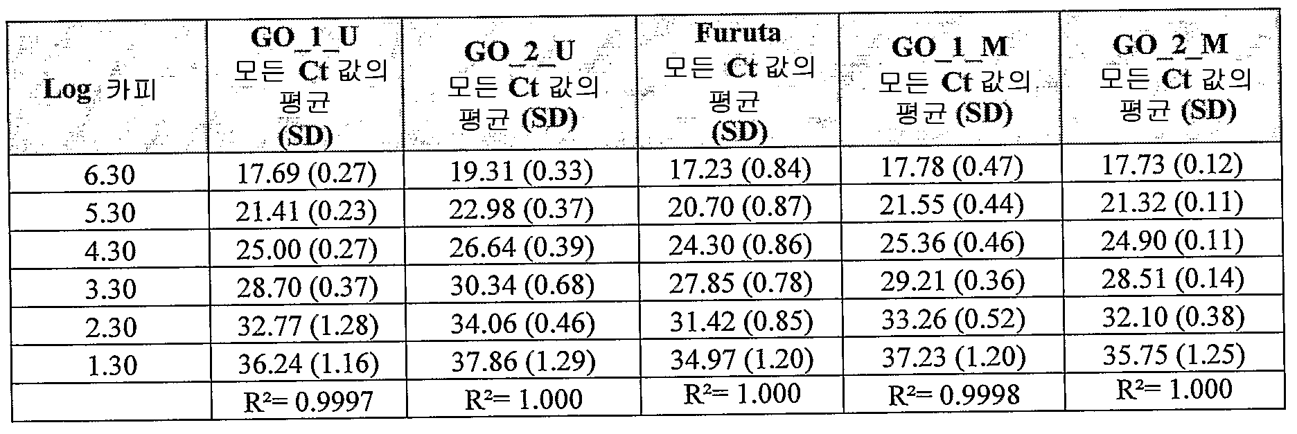
표 8: 매개 정밀성을 시험하기 위해 수행된 검정(2개의 상이한 날자에서 작업자 A 및 B): 컬럼 1: 분자의 수(log), 다음의 컬럼: 각각의 MAGEA3 검정에 대한 평균 Ct 값 및 표준 편차)Table 8: Assays performed to test mediated precision (operators A and B on two different dates): column 1: number of molecules (log), column following: mean Ct value and standard deviation for each MAGEA3 assay )

표 9: 각각의 분석된 희석 시리즈에 대해 밝혀진 상관 계수.Table 9: Correlation coefficients found for each analyzed dilution series.
실시예 6: 멜라닌 간섭(Melanin interference)Example 6 Melanin Interference
이전에, 멜라닌을 함유하는 샘플로부터의 PCR의 효율이 낮은 것으로 보고되었다. 문헌[Eckhart et al . (2000)]에서 멜라닌세포로부터 유래된 RNA 및 cDNA 제조물 둘 모두가 핵산과 함께 공동 정제된 RT-PCR 억제제를 함유하는 것으로 밝혀졌다. 후보 억제제 멜라닌의 연구는 이러한 후보 억제제 멜라닌이 열안정성 DNA 중합효소에 가역적으로 결합하고, 이의 활성을 억제하는 것을 나타내었다. MAGEA3 U 앰플리플루오르 검정을 통한 흑색종 샘플의 처리 전, 멜라닌에 의한 잠재적 억제를 연구하였다. 합성 멜라닌을 문헌[Eckhart et al.]에 기재된 바와 같이 제조하였다. 간단히, 멜라닌(SIGMA M8631)을 2mg/ml의 농도로 증류수에 용해시키고, 광범위하게 볼텍싱(vortexing)하고, 10분 동안 실온에서 수조에서 초음파 처리하였다. 용해되지 않은 멜라닌을 9000g에서의 원심분리에 의해 제거하였다. 잠재적 억제 효과를 반응 과정의 다양한 단계에서 멜라닌을 첨가함으로써 시험하였다:Previously, the efficiency of PCR from samples containing melanin was reported to be low. Eckhart et al . (2000), both RNA and cDNA preparations derived from melanocytes contain RT-PCR inhibitors co-purified with nucleic acids. Studies of candidate inhibitor melanin have shown that such candidate inhibitor melanin reversibly binds to and inhibits its activity on thermostable DNA polymerase. Potential inhibition by melanin was studied before treatment of melanoma samples via the MAGEA3 U amplifluor assay. Synthetic melanin is described in Eckhart et al. As described in the following. Briefly, melanin (SIGMA M8631) was dissolved in distilled water at a concentration of 2 mg / ml, vortexed extensively and sonicated in a water bath at room temperature for 10 minutes. Undissolved melanin was removed by centrifugation at 9000 g. Potential inhibitory effects were tested by adding melanin at various stages of the reaction process:
1) 추출 전: 1μg 및 5μg의 제조된 멜라닌을 250,000개의 LNCaP 세포 및 250,000개의 MCF7 세포에 첨가하였다.1) Before extraction: 1 μg and 5 μg of prepared melanin were added to 250,000 LNCaP cells and 250,000 MCF7 cells.
2) 추출 후: 1μg 및 5μg의 제조된 멜라닌을 1μg의 LNCap 및 MCF7 DNA에 첨가하였다.2) After extraction: 1 μg and 5 μg of prepared melanin were added to 1 μg of LNCap and MCF7 DNA.
3) 비술파이트 처리 후: 1μg 및 5μg의 제조된 멜라닌을 PCR 반응에서 직접 스파이킹(spiking)하였다. 비술파이트 용리 부피를 일정한 주형 농도('a'로 주가 달림, 예를 들어, LNCaP a)를 가지거나, PCR 반응에서 일정량의 주형('b'로 주가 달림, 예를 들어, LNCaP b)을 가지도록 적합화시켰다.3) After bisulfite treatment: 1 μg and 5 μg of prepared melanin were spiked directly in a PCR reaction. The bisulfite elution volume has a stock run at a constant template concentration ('a', e.g. LNCaP a), or a certain amount of template ('b' runs, e.g. LNCaP b) in a PCR reaction. And so on.
이러한 멜라닌 함유 LNCaP 및 MCF7 샘플을 상응하는 스파이킹되지 않은 멜라닌 샘플과 동시에 처리하였다.These melanin containing LNCaP and MCF7 samples were treated simultaneously with the corresponding unspiked melanin samples.
샘플을 PUREGENE® DNA 정제 키트 및 EZ DNA 메틸화 키트를 이용하여 추가로 처리하였다. 화학적으로 변형된 DNA를 MAGEA3 U, Gst-Pi M 및 ACTB 실시간 MSP에 대한 투입 물질로 사용하였다. 시험된 유전자 프로모터 및 ACTB 참고 유전자의 회수된 카피수를 계산하고, 각각의 조건에 대해 비교하였다.Samples were further processed using the PUREGENE® DNA Purification Kit and the EZ DNA Methylation Kit. Chemically modified DNA was used as input for MAGEA3 U, Gst-Pi M and ACTB real-time MSP. Recovered copy numbers of the tested gene promoter and ACTB reference gene were calculated and compared for each condition.
결과는 도 9에 도시되어 있다. DNA 추출 전 또는 후에 멜라닌이 첨가되는 경우 현저한 PCR 억제 효과가 관찰되지 않았다. PCR 반응으로 직접 스파이킹되는 경우에만 멜라닌이 명백한 억제를 나타내었다. RT-PCR과는 대조적으로, 높은 멜라닌 함량을 갖는 흑색종 샘플은 PCR 억제의 위험 없이 실시간 MSP를 통해 처리할 수 있다.The results are shown in FIG. No significant PCR inhibitory effect was observed when melanin was added before or after DNA extraction. Melanin showed obvious inhibition only when directly spiked into the PCR reaction. In contrast to RT-PCR, melanoma samples with high melanin content can be processed via real-time MSP without the risk of PCR inhibition.
실시예 7: 흑색종/폐 샘플에서의 MAGE-A3 메틸화 상태 및 RNA 발현과의 일치Example 7: Concordance with MAGE-A3 Methylation Status and RNA Expression in Melanoma / Lung Samples
물질 및 방법Substances and Methods
임상 샘플Clinical sample
흑색종 및 폐암 환자로부터의 수술 표본은 GSKBio에 의해 제공되었고: 유전체 DNA 샘플(gDNA), RNA 레이터(later®) 용액 중의 생검 물질 및 상응하는 포르말린 고정 파라핀 포매된 조직(FFPE)을 GSKBio RNA 발현 데이터에 기초하여 MAGEA3 양성 또는 MAGEA3 음성으로 분류하였다. 제공된 샘플 세트의 개관은 표 10에 상술되어 있다.Surgical specimens from melanoma and lung cancer patients were provided by GSKBio: GSKBio RNA expression data for genomic DNA samples (gDNA), biopsy material in RNA later® solution and corresponding formalin fixed paraffin embedded tissue (FFPE). Classified as either MAGEA3 positive or MAGEA3 negative. An overview of the sample sets provided is detailed in Table 10.

표 10: 임상 샘플 수집Table 10: Clinical Sample Collection
* 분류를 해당 RNA 레이터® 조직에 기초하여 수행하였다.* Sorting was performed based on the corresponding RNA Rater® tissue.
세포주: Cell line :
세포주를 각각의 수행에서 양성 및 음성 대조군으로 포함시켰다. 임상 샘플에 대해 앰플리플루오르 실시간 MSP 검정을 적용시키기 전에, 검정의 민감성 및 특이성을 세포주 물질 상에서 확인하였다. 메틸화되거나 메틸화되지 않은 최적의 MAGEA3의 세포주는 표 11에 요약되어 있다. Ger1, Staq en CRL9609를 GSKBio로부터 수득하고, 세포주 LNCaP 및 DU145를 미국 미생물보존센터(American Type Culture Collection)로부터 입수하였다.Cell lines were included as positive and negative controls in each run. Prior to applying the Amplifluor real-time MSP assay to clinical samples, the sensitivity and specificity of the assay was confirmed on cell line material. The optimal MAGEA3 cell line, methylated or unmethylated, is summarized in Table 11. Ger1, Staq en CRL9609 was obtained from GSKBio and cell lines LNCaP and DU145 were obtained from the American Type Culture Collection.

표 11: MAGEA3 대조군 세포주Table 11: MAGEA3 Control Cell Lines
DNA 분리: DNA isolation :
포르말린 고정 파라핀 포매된 샘플을 먼저 2시간 동안 750μl 크실렌 중에서 파라핀을 제거하였다. 두번째의 크실렌 처리를 수행하였다(2시간 동안 400μl 크실렌). 다음으로, 250μl의 70% 에탄올을 첨가하고, 15분 동안 13000rpm에서 원심분리하였다. 상층액을 제거하고, 샘플을 실온에서 대기 건조시켰다. RNA 레이터® 중의 샘플을 RNA 레이터® 용액의 제거 후에 면도칼(razor blade)을 이용하여 매우 작은 단편으로 절단하였다. 이후, 고전적인 페놀/클로로포름 추출 방법을 이용하여 DNA를 추출하고, 50μl LoTE(3mM TRIS, 0.2mM EDTA, pH 8.0)에 재현탁시켰다. DNA를 제조업체의 권고사항에 따라 피코그린(Picogreen®) dsDNA 정량 키트(Molecular Probes, #P7589)를 이용하여 정량하였다. 표준 곡선을 제조하기 위해 키트에 제공된 λDNA를 사용하였다. 플루오스타 갤럭시(FluoStar Galaxy) 플레이트 판독기(BMG Lab technologies, Germany)를 이용하여 데이터를 수집하였다.Formalin fixed paraffin embedded samples were first removed paraffin in 750 μl xylene for 2 hours. A second xylene treatment was performed (400 μl xylene for 2 hours). 250 μl of 70% ethanol was then added and centrifuged at 13000 rpm for 15 minutes. The supernatant was removed and the sample was air dried at room temperature. Samples in RNA Razer® were cut into very small fragments using a razor blade after removal of RNA Razer® solution. DNA was then extracted using the classical phenol / chloroform extraction method and resuspended in 50 μl LoTE (3 mM TRIS, 0.2 mM EDTA, pH 8.0). DNA was quantified using Picogreen® dsDNA quantification kit (Molecular Probes, # P7589) according to manufacturer's recommendations. The λ DNA provided in the kit was used to prepare a standard curve. Data was collected using a FluoStar Galaxy plate reader (BMG Lab technologies, Germany).
DNA 변형: 1.5μg의 DNA를 자이모 리서치(Zymo Research)사로부터의 EZ DNA 메틸화 키트를 이용하여 비술파이트 변형에 적용시켰다. 간단히, 45μl의 분취량을 5μl의 M-희석 완충액과 혼합시키고, 1100rpm에서 진탕시키면서 15분 동안 37℃에서 인큐베이션하였다. 이후, 100μl의 희석된 CT 전환 시약을 첨가하고, 샘플을 어두운 곳에서 1100rpm에서 진탕하면서 3시간 동안 70℃에서 인큐베이션하였다. 전환 후, 샘플을 제조업체의 설명서에 따라 추가로 탈염시키고, 탈술폰화시키고, 25μl Tris-HCl 1mM pH8.0에서 용리시켰다. 변형된 DNA를 추가 처리때까지 -80℃에 저장하였다. DNA Modification : 1.5 μg of DNA was subjected to bisulfite modification using the EZ DNA methylation kit from Zymo Research. Briefly, 45 μl aliquots were mixed with 5 μl M-dilution buffer and incubated at 37 ° C. for 15 minutes with shaking at 1100 rpm. Then 100 μl of diluted CT conversion reagent was added and the samples were incubated at 70 ° C. for 3 hours with shaking at 1100 rpm in the dark. After conversion, the samples were further desalted, desulfonated and eluted at 25 μl Tris-
DNA 증폭: 실시간 MSP를 어플라이드 바이오시스템즈(Applied Biosystems)로부터의 7900HT 패스트(fast) 실시간 PCR 사이클러(cycler) 상에서 적용시켰다. DNA Amplification : Real time MSP was applied on a 7900HT fast real time PCR cycler from Applied Biosystems.
유전자 프로모터 서열의 메틸화되지 않은 형태를 표적화하도록 디자인된 4개의 MAGEA3 저메틸화 검정을 제공된 RNA 발현 데이터와의 일치에 대해 시험하였고, 이를, 예를 들어, 표 2의 프라이머 및 프로브, 서열 목록 번호:3, 4 및 13의 Exon 3 MAGE-A3 특이적 프라이머 및 프로브를 이용하여 WO2007/147876호에 기재된 방법에 따라 측정하였다. 독립적 참고 유전자 β-액틴(ACTB)을 또한 측정하였다. 프라이머 및 앰플리플루오르 검출자 서열은 표 1에 제시되어 있다.Four MAGEA3 hypomethylation assays designed to target the unmethylated form of the gene promoter sequence were tested for concordance with the provided RNA expression data, eg, the primers and probes of Table 2, SEQ ID NO: 3 Measurements were made according to the method described in WO2007 / 147876 using
2.4μl의 변형된 유전체 DNA 샘플을, Rox(BioRad, 2x완충액)를 지닌 6μl의 iTaq™ 수퍼믹스 및 정방향 프라이머/검출자 및 역방향 프라이머 둘 모두에 대해 100nM의 최종 프라이머 농도를 포함하는 최종 12μl의 PCR 반응 부피에 첨가하였다. 각각의 MAGEA3 디자인에 대한 주기 조건은, 2분 동안 50℃; 10분 동안 95℃; 이후 45 주기의, 15초 동안 95℃, 30초 동안 59℃[ACTB에 대해 62℃] 및 30초 동안 59℃[ACTB에 대해 62℃](= 안정기-데이터 수집)였다.A 2.4 μl modified genomic DNA sample was subjected to a final 12 μl PCR including a final primer concentration of 100 nM for 6 μl iTaq ™ supermix with Rox (BioRad, 2 × buffer) and both the forward primer / detector and reverse primer It was added to the reaction volume. Cycle conditions for each MAGEA3 design were 50 ° C. for 2 minutes; 95 ° C. for 10 minutes; This was followed by 45 cycles of 95 ° C. for 15 seconds, 59 ° C. for 30 seconds [62 ° C. for ACTB] and 30 seconds for 59 ° C. [62 ° C. for ACTB] (= ballast-data collection).
SDS 2.2.2 소프트웨어(Applied Biosystems)를 이용하여 결과를 발생시키고, Ct 값으로 나타낸 후(증폭 곡선이 역치 값을 교차하는 주기수는 소프트웨어에 의해 자동적으로 설정됨), 이를 이용하여 관심 비술파이트 변형된 서열을 함유하는 정제된 PCR 생성물 또는 플라스미드 DNA를 이용하여 20 - 2 x 10^6 유전자 카피 당량의 표준 곡선에 대해 작도된 값의 선형 회귀에 기초하여 카피수를 계산하였다. MAGEA3와 ACTB 사이의 비를 계산하여 시험 결과를 발생시켰다. 데이터를 해석하기 위해, 맹검이 아닌(un-blinded) RNA 발현 데이터에 기초하여 임상 컷오프(역치)를 규정하였다.The results are generated using SDS 2.2.2 software (Applied Biosystems), expressed as Ct values (the frequency at which the amplification curve intersects the threshold value is automatically set by the software), and then used to modify the bisulfite of interest. Copy numbers were calculated based on linear regression of the values plotted against a standard curve of 20-2 × 10 ^ 6 gene copy equivalents using purified PCR product or plasmid DNA containing the sequence sequence. Test results were generated by calculating the ratio between MAGEA3 and ACTB. To interpret the data, clinical cutoffs (thresholds) were defined based on un-blinded RNA expression data.
샘플을 도 5에 도시된 결정 트리에 기초하여 메틸화, 비메틸화 또는 무효로 분류하였다. 세포주를 각각의 수행에서 양성 및 음성 대조군으로 포함시키고, DNA 추출 단계에서 상기 절차를 포함시켰다.Samples were classified as methylated, unmethylated or invalid based on the crystal tree shown in FIG. 5. Cell lines were included as positive and negative controls in each run and the procedure was included in the DNA extraction step.
하기 기준을 충족하는 경우에 수행을 유효한 것으로 간주하였다: a) 80%를 초과하는 둘 모두의 표준 곡선의 PCR 효율; b) 0.990을 초과하는 4개 이상의 관련 데이터 포인트의 r^2; c) < 1.5의 이중 수행(duplicate) 사이의 Δ Ct; d) 통상적으로 포함되는 NTC가 증폭되지 않음; e) 양성 세포주 검정 대조군의 1.5μg 전환 반응물의 10%가 검출가능함; 및 f) 음성 세포주 검정 대조군의 1.5μg 전환 반응물의 10%가 표준 곡선 내에서 검출되지 않음.Performance was considered valid if the following criteria were met: a) PCR efficiency of both standard curves above 80%; b) r ^ 2 of four or more related data points in excess of 0.990; c) Δ Ct between duplicates of <1.5; d) commonly included NTCs are not amplified; e) 10% of the 1.5 μg conversion reactants of the positive cell line assay control are detectable; And f) 10% of the 1.5 μg conversion reactions of the negative cell line assay control were not detected within the standard curve.
결과result
메틸화와 유전자 발현 사이의 일치: Consensus between methylation and gene expression :
흑색종: Melanoma :
MAGEA3의 발현 및 메틸화 수준을 동일한 샘플 세트에서 비교하였다. 전체적으로, 41개의 흑색종 샘플을 RT-PCR 및 실시간 MSP를 이용하여 처리하였다. 어떠한 검정이 GSK에 의해 제공된 RNA 발현 데이터와 최적으로 일치하는지 확인하기 위해 MAGEA3 U 앰플리플루오르 검정의 여러 디자인을 시험하였다. 최대 일치 및 최소 위양성을 갖는 방식으로 임상 컷-오프를 설정하였다(표 12 참조). 이러한 샘플 중의 MAGEA3 메틸화 상태에 대한 ROC 곡선은 도 6에 도시되어 있다. 17개의 음성 샘플 중, 9개가 다른 MAGE-A 패밀리 일원에 대해 양성이었고; GO_2_U 및 Furuta U 검정은 상기 9개의 샘플을 정확히 분류하였다(100%의 특이성). 모든 이러한 데이터를 함께 고려하여, MAGEA3_GO_2_U 검정을 92.7% 일치 및 100% 특이성으로 최적으로 수행하였다.Expression and methylation levels of MAGEA3 were compared in the same sample set. In total, 41 melanoma samples were processed using RT-PCR and real time MSP. Several designs of the MAGEA3 U amplifluor assay were tested to identify which assay best matches the RNA expression data provided by GSK. Clinical cut-off was set in such a way as to have maximum match and minimum false positive (see Table 12). The ROC curve for the MAGEA3 methylation state in this sample is shown in FIG. 6. Of the 17 negative samples, 9 were positive for other MAGE-A family members; GO_2_U and Furuta U assay correctly classified the nine samples (100% specificity). Taking all these data together, the MAGEA3_GO_2_U assay was optimally performed with 92.7% consensus and 100% specificity.

표 12: 일치 데이터 흑색종 샘플Table 12: Matched data melanoma samples
폐 샘플(생검 및 FFPE): Lung Samples (Biopsy and FFPE) :
상기와 동일한 구성을 다양한 샘플 세트에서 시험하였다: RNA 레이터® 중의 52개의 폐 FFPE 샘플 및 61개의 폐 조직을 4 MAGEA3 U 앰플리플루오르 검정을 통해 스크리닝하였고, 상응하는 RNA 데이터와 일치하였다. 이러한 폐 생검 및 FFPE 샘플에서의 MAGEA3 메틸화 상태에 대한 ROC 곡선이 각각 도 7 및 8에 제시되어 있다. MAGEA3 음성 샘플에서, 기타 MAGE-A 패밀리 일원에 대해 9개가 양성이었고; MAGEA3 U 검정은 모든 9개의 샘플을 정확히 분류하였다(100%의 특이성). 수득된 결과는 MAGEA3_GO_2_U 검정이 FFPE에서 90.4%의 일치 및 생검에서 91.8%의 일치를 갖는 최적의 수행 검정임을 입증하였고, 이는 100%의 특이성을 유지하였다(표 13).The same configuration was tested in various sample sets: 52 lung FFPE samples and 61 lung tissues in RNA Rater® were screened through 4 MAGEA3 U amplifluor assays and matched with corresponding RNA data. ROC curves for MAGEA3 methylation status in these lung biopsies and FFPE samples are shown in FIGS. 7 and 8, respectively. In the MAGEA3 negative sample, 9 were positive for other MAGE-A family members; The MAGEA3 U assay correctly classified all nine samples (100% specificity). The results obtained demonstrated that the MAGEA3_GO_2_U assay was the best performing assay with 90.4% match in FFPE and 91.8% match in biopsy, which maintained 100% specificity (Table 13).

표 13: 일치 데이터 폐 샘플(MAGEA3_GO_2_U 검정)Table 13: Match data lung samples (MAGEA3_GO_2_U test)
실시예 8: MAGEA3 검정을 통한 폐 샘플의 시험Example 8: Testing of Lung Samples via MAGEA3 Assay
폐암으로부터의 DNA를 LNCaP & DU145 대조군 세포주와 함께 병행하여 MAGEA3(U & M 검정 형태) 및 β-액틴 검정을 통해 처리하였다. 어떠한 검정이 MAGEA3 GO_2 U 검정과 최적으로 일치하는지 확인하기 위해 MAGEA3 U 앰플리플루오르 검정의 여러 디자인을 시험하였다. 실시예 7에 기재된 바와 같은 실험 조건을 이용하였다. 표준 곡선은 80%를 초과하는 효율을 나타내었다. R2는 0.99보다 높았다. 컷 오프 값은 MAGEA3 GO_2 U 검정에 대해 29; MAGEA3 GO_1 U 검정에 대해 22; MAGEA3 Furuta U 검정에 대해 229; MAGEA3 Qiu 검정에 대해 87; MAGEA3 GO_1 M 검정에 대해 148; 및 MAGEA3 GO_2 M 검정에 대해 167로 설정하였다. MAGEA3의 메틸화 수준을 동일한 샘플 세트에서 비교하였다.DNA from lung cancer was treated in parallel with the LNCaP & DU145 control cell line via MAGEA3 (U & M assay form) and β-actin assay. Several designs of the MAGEA3 U amplifluor assay were tested to determine which assay best matches the MAGEA3 GO_2 U assay. Experimental conditions as described in Example 7 were used. Standard curves showed efficiencies above 80%. R 2 was higher than 0.99. Cut off values were 29 for the MAGEA3 GO_2 U test; 22 for MAGEA3 GO_1 U test; 229 for the MAGEA3 Furuta U assay; 87 for the MAGEA3 Qiu assay; 148 for the MAGEA3 GO_1 M test; And 167 for the MAGEA3 GO_2 M assay. The methylation level of MAGEA3 was compared in the same sample set.
결과는 표 14 및 15에 도시되어 있다. MAGEA3 GO_2 U 검정(컷 오프 = 29)에 대해:The results are shown in Tables 14 and 15. For the MAGEA3 GO_2 U test (cut off = 29):
- 3개의 샘플이 무효한 것으로 분류되었고, 이러한 샘플을 다른 U & M 검정과의 비교에 포함시키지 않았다;Three samples were classified as invalid and these samples were not included in the comparison with other U & M assays;
- 9개의 샘플이 메틸화되지 않은 것으로 분류되었다;9 samples were classified as not methylated;
- 15개의 샘플이 메틸화된 것으로 분류되었다.15 samples were classified as methylated.
4개의 MAGEA3 U 검정은 유효한 샘플에 대한 MAGEA3 GO_2 U 검정과 96%의 일치를 갖는 유사한 결과를 발생시켰다. MAGEA3 M 검정은 MAGEA3 GO_2 U 검정과 71% 및 75%의 일치를 발생시켰다.Four MAGEA3 U assays produced similar results with 96% agreement with the MAGEA3 GO_2 U assay for valid samples. The MAGEA3 M test produced 71% and 75% agreement with the MAGEA3 GO_2 U test.

표 14: 다양한 U 검정을 비교하고, GO_2 U 검정에 비한 U 검정에 대해 계산된 일치를 나타내는 요약 표(이러한 표는 유효한 샘플만을 고려함).Table 14: Summary table comparing the various U tests and showing the calculated matches for the U test over the GO_2 U test (these tables only consider valid samples).

표 15: 다양한 M 검정을 비교하고, GO_2 U 검정에 비한 M 검정에 대해 계산된 일치를 나타내는 요약 표(이러한 표는 유효한 샘플만을 고려함).Table 15: Summary table comparing the various M tests and showing the calculated matches for the M test over the GO_2 U test (these tables only consider valid samples).
본 발명은 본원에 기재된 특정 구체예에 의해 범위가 제한되지 않는다. 또한, 본원에 기재된 것 외의 다양한 본 발명의 변형은 상기 기재 및 수반하는 도면으로부터 당업자에게 명백할 것이다. 이러한 변형은 첨부된 청구항의 범위 내에 포함된다. 또한, 본원에 기재된 모든 구체예는 적절한 경우 임의의 및 모든 다른 일치하는 구체예와 함께 광범위하게 적용가능하고 비교가능한 것으로 간주된다.The present invention is not to be limited in scope by the specific embodiments described herein. In addition, various modifications of the present invention other than those described herein will be apparent to those skilled in the art from the foregoing description and the accompanying drawings. Such modifications are included within the scope of the appended claims. In addition, all embodiments described herein are considered to be broadly applicable and comparable with any and all other matching embodiments where appropriate.
다양한 간행물이 본원에 인용되어 있으며, 이의 개시내용은 전체내용이 참조로서 본원에 포함된다.Various publications are cited herein, the disclosures of which are incorporated herein by reference in their entirety.
SEQUENCE LISTING <110> OncoMethylome Sciences SA GlaxoSmithKline Biologicals <120> IMPROVED DETECTION OF MAGE-A EXPRESSION <130> P94294WO01 <150> US 60/960,128 <151> 2007-09-17 <160> 41 <170> PatentIn version 3.4 <210> 1 <211> 22 <212> DNA <213> Artificial sequence <220> <223> Oligonucleotide (hairpin) <400> 1 agcgatgcgt tcgagcatcg cu 22 <210> 2 <211> 24 <212> DNA <213> Artificial sequence <220> <223> Primer <400> 2 atttttgttt ggaatttagg gtag 24 <210> 3 <211> 43 <212> DNA <213> Artificial sequence <220> <223> Hairpin primer <400> 3 agcgatgcgt tcgagcatcg cuccaacatc aaaccatcac tca 43 <210> 4 <211> 21 <212> DNA <213> Artificial sequence <220> <223> Primer <400> 4 ccaacatcaa accatcactc a 21 <210> 5 <211> 21 <212> DNA <213> Artificial sequence <220> <223> Primer <400> 5 tggaatttag ggtagtattg t 21 <210> 6 <211> 43 <212> DNA <213> Artificial sequence <220> <223> Hairpin primer <400> 6 agcgatgcgt tcgagcatcg cutggaattt agggtagtat tgt 43 <210> 7 <211> 18 <212> DNA <213> Artificial sequence <220> <223> Primer <400> 7 ccctccacca acatcaaa 18 <210> 8 <211> 25 <212> DNA <213> Artificial sequence <220> <223> Primer <400> 8 ttaggatgtg atgttattga tttgt 25 <210> 9 <211> 47 <212> DNA <213> Artificial sequence <220> <223> Primer <400> 9 agcgatgcgt tcgagcatcg cuttaggatg tgatgttatt gatttgt 47 <210> 10 <211> 203 <212> DNA <213> Artificial sequence <220> <223> Portion of MAGE-A3 unconverted sequence <400> 10 catgcttacc tccaccccca tccgatcccc atccaggcag aatccagttc cacccctgcc 60 cggaacccag ggtagtaccg ttgccaggat gtgacgccac tgacttgcgc attggaggtc 120 agaagaccgc gagattctcg ccctgagcaa cgagcgacgg cctgacgtcg gcggagggaa 180 gccggcccag gctcggtgag gag 203 <210> 11 <211> 25 <212> DNA <213> Artificial sequence <220> <223> Primer <400> 11 tgtttggaat ttagggtagt attgt 25 <210> 12 <211> 47 <212> DNA <213> Artificial sequence <220> <223> Hairpin primer <400> 12 agcgatgcgt tcgagcatcg cutgtttgga atttagggta gtattgt 47 <210> 13 <211> 24 <212> DNA <213> Artificial sequence <220> <223> Primer <400> 13 ccatcactca ttactcaaaa caaa 24 <210> 14 <211> 24 <212> DNA <213> Artificial sequence <220> <223> Primer <400> 14 atttttgttc ggaatttagg gtag 24 <210> 15 <211> 43 <212> DNA <213> Artificial sequence <220> <223> Hairpin primer <400> 15 agcgatgcgt tcgagcatcg cuccgacgtc aaaccgtcgc tcg 43 <210> 16 <211> 21 <212> DNA <213> Artificial sequence <220> <223> Primer <400> 16 ccgacgtcaa accgtcgctc g 21 <210> 17 <211> 21 <212> DNA <213> Artificial sequence <220> <223> Primer <400> 17 cggaatttag ggtagtatcg t 21 <210> 18 <211> 40 <212> DNA <213> Artificial sequence <220> <223> Hairpin primer <400> 18 agcgatgcgt tcgagcatcg cuccctccgc cgacgtcaaa 40 <210> 19 <211> 18 <212> DNA <213> Artificial sequence <220> <223> Primer <400> 19 ccctccgccg acgtcaaa 18 <210> 20 <211> 26 <212> DNA <213> Artificial sequence <220> <223> Primer <400> 20 tagggagtat ataggttggg gaagtt 26 <210> 21 <211> 48 <212> DNA <213> Artificial sequence <220> <223> Hairpin primer <400> 21 agcgatgcgt tcgagcatcg cutagggagt atataggttg gggaagtt 48 <210> 22 <211> 27 <212> DNA <213> Artificial sequence <220> <223> Primer <400> 22 aacacacaat aacaaacaca aattcac 27 <210> 23 <211> 20 <212> DNA <213> Artificial sequence <220> <223> Primer <400> 23 attttgaggg atgatcgaag 20 <210> 24 <211> 20 <212> DNA <213> Artificial sequence <220> <223> Primer <400> 24 ctaaaataaa acccgcctca 20 <210> 25 <211> 15 <212> DNA <213> Artificial sequence <220> <223> Overlapping primer sequence <400> 25 tggaatttag ggtag 15 <210> 26 <211> 30 <212> DNA <213> Artificial sequence <220> <223> Combined primer sequence <400> 26 atttttgttt ggaatttagg gtagtattgt 30 <210> 27 <211> 9 <212> PRT <213> Artificial sequence <220> <223> MAGE-A3 peptide <400> 27 Phe Leu Trp Gly Pro Arg Ala Leu Val 1 5 <210> 28 <211> 9 <212> PRT <213> Artificial sequence <220> <223> MAGE-A3 peptide <400> 28 Glu Val Asp Pro Ile Gly His Leu Tyr 1 5 <210> 29 <211> 10 <212> PRT <213> Artificial sequence <220> <223> MAGE-A3 peptide <400> 29 Met Glu Val Asp Pro Ile Gly His Leu Tyr 1 5 10 <210> 30 <211> 10 <212> PRT <213> Artificial sequence <220> <223> MAGE-A3 peptide <400> 30 Val His Phe Leu Leu Leu Lys Tyr Arg Ala 1 5 10 <210> 31 <211> 10 <212> PRT <213> Artificial sequence <220> <223> MAGE-A3 peptide <400> 31 Leu Val His Phe Leu Leu Leu Lys Tyr Arg 1 5 10 <210> 32 <211> 10 <212> PRT <213> Artificial sequence <220> <223> MAGE-A3 peptide <400> 32 Leu Lys Tyr Arg Ala Arg Glu Pro Val Thr 1 5 10 <210> 33 <211> 16 <212> PRT <213> Artificial sequence <220> <223> MAGE-A3 peptide <400> 33 Ala Cys Tyr Glu Phe Leu Trp Gly Pro Arg Ala Leu Val Glu Thr Ser 1 5 10 15 <210> 34 <211> 12 <212> PRT <213> Artificial sequence <220> <223> MAGE-A3 peptide <400> 34 Thr Gln His Phe Val Gln Glu Asn Tyr Leu Glu Tyr 1 5 10 <210> 35 <211> 20 <212> DNA <213> Artificial sequence <220> <223> Oligonucleotide <400> 35 tccatgacgt tcctgacgtt 20 <210> 36 <211> 18 <212> DNA <213> Artificial sequence <220> <223> Oligonucleotide <400> 36 tctcccagcg tgcgccat 18 <210> 37 <211> 30 <212> DNA <213> Artificial sequence <220> <223> Oligonucleotide <400> 37 accgatgacg tcgccggtga cggcaccacg 30 <210> 38 <211> 24 <212> DNA <213> Artificial sequence <220> <223> Oligonucleotide <400> 38 tcgtcgtttt gtcgttttgt cgtt 24 <210> 39 <211> 20 <212> DNA <213> Artificial sequence <220> <223> Oligonucleotide <400> 39 tccatgacgt tcctgatgct 20 <210> 40 <211> 450 <212> PRT <213> Artificial sequence <220> <223> Recombinant tagged MAGE-A3 <400> 40 Met Asp Pro Lys Thr Leu Ala Leu Ser Leu Leu Ala Ala Gly Val Leu 1 5 10 15 Ala Gly Cys Ser Ser His Ser Ser Asn Met Ala Asn Thr Gln Met Lys 20 25 30 Ser Asp Lys Ile Ile Ile Ala His Arg Gly Ala Ser Gly Tyr Leu Pro 35 40 45 Glu His Thr Leu Glu Ser Lys Ala Leu Ala Phe Ala Gln Gln Ala Asp 50 55 60 Tyr Leu Glu Gln Asp Leu Ala Met Thr Lys Asp Gly Arg Leu Val Val 65 70 75 80 Ile His Asp His Phe Leu Asp Gly Leu Thr Asp Val Ala Lys Lys Phe 85 90 95 Pro His Arg His Arg Lys Asp Gly Arg Tyr Tyr Val Ile Asp Phe Thr 100 105 110 Leu Lys Glu Ile Gln Ser Leu Glu Met Thr Glu Asn Phe Glu Thr Met 115 120 125 Asp Leu Glu Gln Arg Ser Gln His Cys Lys Pro Glu Glu Gly Leu Glu 130 135 140 Ala Arg Gly Glu Ala Leu Gly Leu Val Gly Ala Gln Ala Pro Ala Thr 145 150 155 160 Glu Glu Gln Glu Ala Ala Ser Ser Ser Ser Thr Leu Val Glu Val Thr 165 170 175 Leu Gly Glu Val Pro Ala Ala Glu Ser Pro Asp Pro Pro Gln Ser Pro 180 185 190 Gln Gly Ala Ser Ser Leu Pro Thr Thr Met Asn Tyr Pro Leu Trp Ser 195 200 205 Gln Ser Tyr Glu Asp Ser Ser Asn Gln Glu Glu Glu Gly Pro Ser Thr 210 215 220 Phe Pro Asp Leu Glu Ser Glu Phe Gln Ala Ala Leu Ser Arg Lys Val 225 230 235 240 Ala Glu Leu Val His Phe Leu Leu Leu Lys Tyr Arg Ala Arg Glu Pro 245 250 255 Val Thr Lys Ala Glu Met Leu Gly Ser Val Val Gly Asn Trp Gln Tyr 260 265 270 Phe Phe Pro Val Ile Phe Ser Lys Ala Ser Ser Ser Leu Gln Leu Val 275 280 285 Phe Gly Ile Glu Leu Met Glu Val Asp Pro Ile Gly His Leu Tyr Ile 290 295 300 Phe Ala Thr Cys Leu Gly Leu Ser Tyr Asp Gly Leu Leu Gly Asp Asn 305 310 315 320 Gln Ile Met Pro Lys Ala Gly Leu Leu Ile Ile Val Leu Ala Ile Ile 325 330 335 Ala Arg Glu Gly Asp Cys Ala Pro Glu Glu Lys Ile Trp Glu Glu Leu 340 345 350 Ser Val Leu Glu Val Phe Glu Gly Arg Glu Asp Ser Ile Leu Gly Asp 355 360 365 Pro Lys Lys Leu Leu Thr Gln His Phe Val Gln Glu Asn Tyr Leu Glu 370 375 380 Tyr Arg Gln Val Pro Gly Ser Asp Pro Ala Cys Tyr Glu Phe Leu Trp 385 390 395 400 Gly Pro Arg Ala Leu Val Glu Thr Ser Tyr Val Lys Val Leu His His 405 410 415 Met Val Lys Ile Ser Gly Gly Pro His Ile Ser Tyr Pro Pro Leu His 420 425 430 Glu Trp Val Leu Arg Glu Gly Glu Glu Gly Gly His His His His His 435 440 445 His His 450 <210> 41 <211> 203 <212> DNA <213> Artificial sequence <220> <223> Bisulphite converted MAGE-A3 sequence <400> 41 tatgtttatt tttattttta tttgattttt atttaggtag aatttagttt tatttttgtt 60 tggaatttag ggtagtattg ttgttaggat gtgatgttat tgatttgtgt attggaggtt 120 agaagattgt gagatttttg ttttgagtaa tgagtgatgg tttgatgttg gtggagggaa 180 gttggtttag gtttggtgag gag 203 SEQUENCE LISTING <110> OncoMethylome Sciences SA GlaxoSmithKline Biologicals <120> IMPROVED DETECTION OF MAGE-A EXPRESSION <130> P94294WO01 <150> US 60 / 960,128 <151> 2007-09-17 <160> 41 <170> PatentIn version 3.4 <210> 1 <211> 22 <212> DNA <213> Artificial sequence <220> Oligonucleotide (hairpin) <400> 1 agcgatgcgt tcgagcatcg cu 22 <210> 2 <211> 24 <212> DNA <213> Artificial sequence <220> <223> Primer <400> 2 atttttgttt ggaatttagg gtag 24 <210> 3 <211> 43 <212> DNA <213> Artificial sequence <220> <223> hairpin primer <400> 3 agcgatgcgt tcgagcatcg cuccaacatc aaaccatcac tca 43 <210> 4 <211> 21 <212> DNA <213> Artificial sequence <220> <223> Primer <400> 4 ccaacatcaa accatcactc a 21 <210> 5 <211> 21 <212> DNA <213> Artificial sequence <220> <223> Primer <400> 5 tggaatttag ggtagtattg t 21 <210> 6 <211> 43 <212> DNA <213> Artificial sequence <220> <223> hairpin primer <400> 6 agcgatgcgt tcgagcatcg cutggaattt agggtagtat tgt 43 <210> 7 <211> 18 <212> DNA <213> Artificial sequence <220> <223> Primer <400> 7 ccctccacca acatcaaa 18 <210> 8 <211> 25 <212> DNA <213> Artificial sequence <220> <223> Primer <400> 8 ttaggatgtg atgttattga tttgt 25 <210> 9 <211> 47 <212> DNA <213> Artificial sequence <220> <223> Primer <400> 9 agcgatgcgt tcgagcatcg cuttaggatg tgatgttatt gatttgt 47 <210> 10 <211> 203 <212> DNA <213> Artificial sequence <220> <223> Portion of MAGE-A3 unconverted sequence <400> 10 catgcttacc tccaccccca tccgatcccc atccaggcag aatccagttc cacccctgcc 60 cggaacccag ggtagtaccg ttgccaggat gtgacgccac tgacttgcgc attggaggtc 120 agaagaccgc gagattctcg ccctgagcaa cgagcgacgg cctgacgtcg gcggagggaa 180 gccggcccag gctcggtgag gag 203 <210> 11 <211> 25 <212> DNA <213> Artificial sequence <220> <223> Primer <400> 11 tgtttggaat ttagggtagt attgt 25 <210> 12 <211> 47 <212> DNA <213> Artificial sequence <220> <223> hairpin primer <400> 12 agcgatgcgt tcgagcatcg cutgtttgga atttagggta gtattgt 47 <210> 13 <211> 24 <212> DNA <213> Artificial sequence <220> <223> Primer <400> 13 ccatcactca ttactcaaaa caaa 24 <210> 14 <211> 24 <212> DNA <213> Artificial sequence <220> <223> Primer <400> 14 atttttgttc ggaatttagg gtag 24 <210> 15 <211> 43 <212> DNA <213> Artificial sequence <220> <223> hairpin primer <400> 15 agcgatgcgt tcgagcatcg cuccgacgtc aaaccgtcgc tcg 43 <210> 16 <211> 21 <212> DNA <213> Artificial sequence <220> <223> Primer <400> 16 ccgacgtcaa accgtcgctc g 21 <210> 17 <211> 21 <212> DNA <213> Artificial sequence <220> <223> Primer <400> 17 cggaatttag ggtagtatcg t 21 <210> 18 <211> 40 <212> DNA <213> Artificial sequence <220> <223> hairpin primer <400> 18 agcgatgcgt tcgagcatcg cuccctccgc cgacgtcaaa 40 <210> 19 <211> 18 <212> DNA <213> Artificial sequence <220> <223> Primer <400> 19 ccctccgccg acgtcaaa 18 <210> 20 <211> 26 <212> DNA <213> Artificial sequence <220> <223> Primer <400> 20 tagggagtat ataggttggg gaagtt 26 <210> 21 <211> 48 <212> DNA <213> Artificial sequence <220> <223> hairpin primer <400> 21 agcgatgcgt tcgagcatcg cutagggagt atataggttg gggaagtt 48 <210> 22 <211> 27 <212> DNA <213> Artificial sequence <220> <223> Primer <400> 22 aacacacaat aacaaacaca aattcac 27 <210> 23 <211> 20 <212> DNA <213> Artificial sequence <220> <223> Primer <400> 23 attttgaggg atgatcgaag 20 <210> 24 <211> 20 <212> DNA <213> Artificial sequence <220> <223> Primer <400> 24 ctaaaataaa acccgcctca 20 <210> 25 <211> 15 <212> DNA <213> Artificial sequence <220> <223> Overlapping primer sequence <400> 25 tggaatttag ggtag 15 <210> 26 <211> 30 <212> DNA <213> Artificial sequence <220> <223> Combined primer sequence <400> 26 atttttgttt ggaatttagg gtagtattgt 30 <210> 27 <211> 9 <212> PRT <213> Artificial sequence <220> <223> MAGE-A3 peptide <400> 27 Phe Leu Trp Gly Pro Arg Ala Leu Val 1 5 <210> 28 <211> 9 <212> PRT <213> Artificial sequence <220> <223> MAGE-A3 peptide <400> 28 Glu Val Asp Pro Ile Gly His Leu Tyr 1 5 <210> 29 <211> 10 <212> PRT <213> Artificial sequence <220> <223> MAGE-A3 peptide <400> 29 Met Glu Val Asp Pro Ile Gly His Leu Tyr 1 5 10 <210> 30 <211> 10 <212> PRT <213> Artificial sequence <220> <223> MAGE-A3 peptide <400> 30 Val His Phe Leu Leu Leu Lys Tyr Arg Ala 1 5 10 <210> 31 <211> 10 <212> PRT <213> Artificial sequence <220> <223> MAGE-A3 peptide <400> 31 Leu Val His Phe Leu Leu Leu Lys Tyr Arg 1 5 10 <210> 32 <211> 10 <212> PRT <213> Artificial sequence <220> <223> MAGE-A3 peptide <400> 32 Leu Lys Tyr Arg Ala Arg Glu Pro Val Thr 1 5 10 <210> 33 <211> 16 <212> PRT <213> Artificial sequence <220> <223> MAGE-A3 peptide <400> 33 Ala Cys Tyr Glu Phe Leu Trp Gly Pro Arg Ala Leu Val Glu Thr Ser 1 5 10 15 <210> 34 <211> 12 <212> PRT <213> Artificial sequence <220> <223> MAGE-A3 peptide <400> 34 Thr Gln His Phe Val Gln Glu Asn Tyr Leu Glu Tyr 1 5 10 <210> 35 <211> 20 <212> DNA <213> Artificial sequence <220> <223> Oligonucleotide <400> 35 tccatgacgt tcctgacgtt 20 <210> 36 <211> 18 <212> DNA <213> Artificial sequence <220> <223> Oligonucleotide <400> 36 tctcccagcg tgcgccat 18 <210> 37 <211> 30 <212> DNA <213> Artificial sequence <220> <223> Oligonucleotide <400> 37 accgatgacg tcgccggtga cggcaccacg 30 <210> 38 <211> 24 <212> DNA <213> Artificial sequence <220> <223> Oligonucleotide <400> 38 tcgtcgtttt gtcgttttgt cgtt 24 <210> 39 <211> 20 <212> DNA <213> Artificial sequence <220> <223> Oligonucleotide <400> 39 tccatgacgt tcctgatgct 20 <210> 40 <211> 450 <212> PRT <213> Artificial sequence <220> <223> Recombinant tagged MAGE-A3 <400> 40 Met Asp Pro Lys Thr Leu Ala Leu Ser Leu Leu Ala Ala Gly Val Leu 1 5 10 15 Ala Gly Cys Ser Ser His Ser Ser Asn Met Ala Asn Thr Gln Met Lys 20 25 30 Ser Asp Lys Ile Ile Ile Ala His Arg Gly Ala Ser Gly Tyr Leu Pro 35 40 45 Glu His Thr Leu Glu Ser Lys Ala Leu Ala Phe Ala Gln Gln Ala Asp 50 55 60 Tyr Leu Glu Gln Asp Leu Ala Met Thr Lys Asp Gly Arg Leu Val Val 65 70 75 80 Ile His Asp His Phe Leu Asp Gly Leu Thr Asp Val Ala Lys Lys Phe 85 90 95 Pro His Arg His Arg Lys Asp Gly Arg Tyr Tyr Val Ile Asp Phe Thr 100 105 110 Leu Lys Glu Ile Gln Ser Leu Glu Met Thr Glu Asn Phe Glu Thr Met 115 120 125 Asp Leu Glu Gln Arg Ser Gln His Cys Lys Pro Glu Glu Gly Leu Glu 130 135 140 Ala Arg Gly Glu Ala Leu Gly Leu Val Gly Ala Gln Ala Pro Ala Thr 145 150 155 160 Glu Glu Gln Glu Ala Ala Ser Ser Ser Ser Thr Leu Val Glu Val Thr 165 170 175 Leu Gly Glu Val Pro Ala Ala Glu Ser Pro Asp Pro Pro Gln Ser Pro 180 185 190 Gln Gly Ala Ser Ser Leu Pro Thr Thr Met Asn Tyr Pro Leu Trp Ser 195 200 205 Gln Ser Tyr Glu Asp Ser Ser Asn Gln Glu Glu Glu Gly Pro Ser Thr 210 215 220 Phe Pro Asp Leu Glu Ser Glu Phe Gln Ala Ala Leu Ser Arg Lys Val 225 230 235 240 Ala Glu Leu Val His Phe Leu Leu Leu Lys Tyr Arg Ala Arg Glu Pro 245 250 255 Val Thr Lys Ala Glu Met Leu Gly Ser Val Val Gly Asn Trp Gln Tyr 260 265 270 Phe Phe Pro Val Ile Phe Ser Lys Ala Ser Ser Ser Leu Gln Leu Val 275 280 285 Phe Gly Ile Glu Leu Met Glu Val Asp Pro Ile Gly His Leu Tyr Ile 290 295 300 Phe Ala Thr Cys Leu Gly Leu Ser Tyr Asp Gly Leu Leu Gly Asp Asn 305 310 315 320 Gln Ile Met Pro Lys Ala Gly Leu Leu Ile Ile Val Leu Ala Ile Ile 325 330 335 Ala Arg Glu Gly Asp Cys Ala Pro Glu Glu Lys Ile Trp Glu Glu Leu 340 345 350 Ser Val Leu Glu Val Phe Glu Gly Arg Glu Asp Ser Ile Leu Gly Asp 355 360 365 Pro Lys Lys Leu Leu Thr Gln His Phe Val Gln Glu Asn Tyr Leu Glu 370 375 380 Tyr Arg Gln Val Pro Gly Ser Asp Pro Ala Cys Tyr Glu Phe Leu Trp 385 390 395 400 Gly Pro Arg Ala Leu Val Glu Thr Ser Tyr Val Lys Val Leu His His 405 410 415 Met Val Lys Ile Ser Gly Gly Pro His Ile Ser Tyr Pro Pro Leu His 420 425 430 Glu Trp Val Leu Arg Glu Gly Glu Glu Gly Gly His His His His His 435 440 445 His His 450 <210> 41 <211> 203 <212> DNA <213> Artificial sequence <220> <223> Bisulphite converted MAGE-A3 sequence <400> 41 tatgtttatt tttattttta tttgattttt atttaggtag aatttagttt tatttttgtt 60 tggaatttag ggtagtattg ttgttaggat gtgatgttat tgatttgtgt attggaggtt 120 agaagattgt gagatttttg ttttgagtaa tgagtgatgg tttgatgttg gtggagggaa 180 gttggtttag gtttggtgag gag 203
Claims (42)
(a) 약 6 내지 30개의 누클레오티드의 제 1 누클레오티드 서열로서, 상기 제 1 누클레오티드 서열 내의 누클레오티드가 분자 에너지 전달쌍의 공여체 부분 및 수용체 부분으로부터 선택된 제 1 부분으로 표지되고, 상기 공여체 부분이 여기시 하나 이상의 특정 파장에서 형광을 방출하고, 상기 수용체 부분이 상기 공여체 부분에 의해 방출된 상기 형광을 흡수하고/하거나 켄칭(quenching)시키는 제 1 누클레오티드 서열;
(b) 약 3 내지 20개의 누클레오티드를 포함하거나, 이로 본질적으로 구성되거나, 이로 구성되는 제 2 단일 가닥 누클레오티드 서열;
(c) 약 6 내지 30개의 누클레오티드를 포함하거나, 이로 본질적으로 구성되거나, 이로 구성되는 제 3 누클레오티드 서열로서, 상기 제 3 누클레오티드 서열 내의 누클레오티드가 상기 공여체 부분 및 상기 수용체 부분으로부터 선택된 제 2 부분으로 표지되고, 상기 제 2 부분이 상기 제 1 누클레오티드 서열을 표지하지 않는 상기 기의 일원이고, 상기 제 3 누클레오티드 서열이 상기 제 1 누클레오티드 서열에 대해 역순으로 상보적이어서, 상기 제 1 누클레오티드 서열과 상기 제 3 누클레오티드 서열 사이에 듀플렉스(duplex)를 형성할 수 있어, 상기 제 1 부분 및 상기 제 2 부분이 근접하여 위치하여, 상기 공여체 부분이 여기되어 형광을 방출하고, 상기 수용체 부분이 상기 공여체 부분에 의해 방출된 상기 형광을 흡수하고 켄칭시키는 제 3 누클레오티드 서열; 및
(d) 프라이머의 3' 말단에서, 서열 목록 번호:5, 7, 2, 4, 8, 11, 13 또는 25 중 임의의 서열 목록 번호의 서열을 3' 말단에 포함하거나, 이로 본질적으로 구성되거나, 이로 구성(및 MAGE A3 유전자의 메틸화되지 않은 DNA의 일부를 포함하는 핵산 가닥에 상보적인 누클레오티드 서열의 핵산 중합효소에 의한 합성을 프라이밍(priming)할 수 있음)되는 약 8 내지 40개의 누클레오티드를 포함하거나, 이로 본질적으로 구성되거나, 이로 구성되는 제 4 단일 가닥 누클레오티드 서열의, 연속된 서열을 포함하거나, 이로 본질적으로 구성되거나, 이로 구성되며;
상기 듀플렉스가 형성되지 않는 경우, 상기 제 1 부분 및 상기 제 2 부분이 상기 제 1 부분과 상기 제 2 부분 사이의 분자 에너지 전달을 방지하는 거리로 분리되는, 제 1항에 따른 올리고누클레오티드, 프라이머 또는 프로브.In order of 5 'to 3',
(a) a first nucleotide sequence of about 6 to 30 nucleotides, wherein the nucleotides in the first nucleotide sequence are labeled with a first portion selected from a donor portion and a acceptor portion of a molecular energy transfer pair, wherein the donor portion is one A first nucleotide sequence that emits fluorescence at the specific wavelength or above and wherein the receptor portion absorbs and / or quenchs the fluorescence emitted by the donor portion;
(b) a second single stranded nucleotide sequence comprising, consisting essentially of, or consisting of about 3 to 20 nucleotides;
(c) a third nucleotide sequence comprising, consisting essentially of, or consisting of about 6 to 30 nucleotides, wherein the nucleotides in the third nucleotide sequence are labeled with a second portion selected from the donor portion and the acceptor portion Wherein the second portion is a member of the group that does not label the first nucleotide sequence, and wherein the third nucleotide sequence is complementary to the first nucleotide sequence in reverse order, such that the first nucleotide sequence and the third A duplex can be formed between nucleotide sequences such that the first portion and the second portion are located in proximity such that the donor portion is excited to emit fluorescence, and the acceptor portion is emitted by the donor portion A third nucleotide sequence for absorbing and quenching the fluorescence; And
(d) at, or essentially consists of, the sequence of any of SEQ ID NO: 5, 7, 2, 4, 8, 11, 13, or 25 at the 3 'end of the primer at the 3'end; And about 8 to 40 nucleotides consisting of (and may prime the synthesis by nucleic acid polymerases of nucleotide sequences complementary to nucleic acid strands comprising a portion of the unmethylated DNA of the MAGE A3 gene). Or comprise, consist essentially of, or consist of a contiguous sequence of a fourth single stranded nucleotide sequence consisting of, or consisting essentially of;
The oligonucleotide, primer or primer according to claim 1, wherein when the duplex is not formed, the first portion and the second portion are separated by a distance that prevents molecular energy transfer between the first portion and the second portion. Probe.
(a) 약 6 내지 30개의 누클레오티드의 제 1 누클레오티드 서열로서, 상기 제 1 누클레오티드 서열 내의 누클레오티드가 분자 에너지 전달쌍의 공여체 부분 및 수용체 부분으로부터 선택된 제 1 부분으로 표지되고, 상기 공여체 부분이 여기시 하나 이상의 특정 파장에서 형광을 방출하고, 상기 수용체 부분이 상기 공여체 부분에 의해 방출된 상기 형광을 흡수하고/하거나 켄칭시키는 제 1 누클레오티드 서열;
(b) 약 3 내지 20개의 누클레오티드를 포함하거나, 이로 본질적으로 구성되거나, 이로 구성되는 제 2 단일 가닥 누클레오티드 서열;
(c) 약 6 내지 30개의 누클레오티드를 포함하거나, 이로 본질적으로 구성되거나, 이로 구성되는 제 3 누클레오티드 서열로서, 상기 제 3 누클레오티드 서열 내의 누클레오티드가 상기 공여체 부분 및 상기 수용체 부분으로부터 선택된 제 2 부분으로 표지되고, 상기 제 2 부분이 상기 제 1 누클레오티드 서열을 표지하지 않는 상기 기의 일원이고, 상기 제 3 누클레오티드 서열이 상기 제 1 누클레오티드 서열에 대해 역순으로 상보적이어서, 상기 제 1 누클레오티드 서열과 상기 제 3 누클레오티드 서열 사이에 듀플렉스를 형성할 수 있어, 상기 제 1 부분 및 상기 제 2 부분이 근접하여 위치하여, 상기 공여체 부분이 여기되어 형광을 방출하고, 상기 수용체 부분이 상기 공여체 부분에 의해 방출된 상기 형광을 흡수하고 켄칭시키는 제 3 누클레오티드 서열; 및
(d) 프라이머의 3' 말단에서, 서열 목록 번호:14, 16, 17 또는 19 중 임의의 서열 목록 번호의 서열을 3' 말단에 포함하거나, 이로 본질적으로 구성되거나, 이로 구성되는 (MAGE A3 유전자의 메틸화된 DNA의 일부를 포함하는 핵산 가닥에 상보적인 누클레오티드 서열의 핵산 중합효소에 의한 합성을 프라이밍할 수 있는) 약 8 내지 40개의 누클레오티드를 포함하거나, 이로 본질적으로 구성되거나, 이로 구성되는 제 4 단일 가닥 누클레오티드 서열의, 연속된 서열을 포함하거나, 이로 본질적으로 구성되거나, 이로 구성되며;
상기 듀플렉스가 형성되지 않는 경우, 상기 제 1 부분 및 상기 제 2 부분이 상기 제 1 부분과 상기 제 2 부분 사이의 분자 에너지 전달을 방지하는 거리로 분리되는, 제 1항에 따른 올리고누클레오티드, 프라이머 또는 프로브.In order of 5 'to 3',
(a) a first nucleotide sequence of about 6 to 30 nucleotides, wherein the nucleotides in the first nucleotide sequence are labeled with a first portion selected from a donor portion and a acceptor portion of a molecular energy transfer pair, wherein the donor portion is one A first nucleotide sequence that emits fluorescence at the specified wavelength and wherein the acceptor portion absorbs and / or quenches the fluorescence emitted by the donor portion;
(b) a second single stranded nucleotide sequence comprising, consisting essentially of, or consisting of about 3 to 20 nucleotides;
(c) a third nucleotide sequence comprising, consisting essentially of, or consisting of about 6 to 30 nucleotides, wherein the nucleotides in the third nucleotide sequence are labeled with a second portion selected from the donor portion and the acceptor portion Wherein the second portion is a member of the group that does not label the first nucleotide sequence, and wherein the third nucleotide sequence is complementary to the first nucleotide sequence in reverse order, such that the first nucleotide sequence and the third The fluorescence can form a duplex between nucleotide sequences such that the first portion and the second portion are located proximate so that the donor portion is excited to emit fluorescence and the receptor portion is emitted by the donor portion. A third nucleotide sequence that absorbs and quenches; And
(d) at the 3 'end of the primer, comprises, consists essentially of, or consists of the sequence of any of SEQ ID NO: 14, 16, 17 or 19 at the 3' end of the (MAGE A3 gene A fourth, comprising, consisting essentially of, or consisting of from about 8 to 40 nucleotides) capable of priming synthesis by a nucleic acid polymerase of a nucleotide sequence complementary to a nucleic acid strand comprising a portion of the methylated DNA of Single stranded nucleotide sequence comprising, consisting essentially of, or consisting of a contiguous sequence;
The oligonucleotide, primer or primer according to claim 1, wherein when the duplex is not formed, the first portion and the second portion are separated by a distance that prevents molecular energy transfer between the first portion and the second portion. Probe.
(b) 하나 이상의 프라이머 쌍을 이용하여 메틸화되지 않은 관심 유전자의 일부 또는 전부를 증폭시키는 단계로서, 상기 프라이머 쌍 중 하나 이상의 프라이머가 상기 시약을 이용한 처리 후에 메틸화되지 않은 DNA의 서열에만 결합하도록 디자인되고, 상기 프라이머 쌍 중의 하나 이상의 프라이머가 서열 목록 번호:5, 6, 7, 2, 3, 4, 8, 9, 11, 12, 13 또는 25 중 임의의 서열 목록 번호의 누클레오티드 서열을 포함하거나, 이로 본질적으로 구성되거나, 이로 구성되는 단계를 포함하여,
DNA 함유 샘플 내의 메틸화되지 않은 Mage-A3 유전자의 존재 및/또는 양을 검출하는 방법.(a) contacting a DNA-containing sample with a reagent that selectively modifies unmethylated cytosine residues in the DNA to produce a modified residue that is detectable without modifying the methylated cytosine residues; And
(b) amplifying some or all of the unmethylated gene of interest with one or more primer pairs, wherein one or more of the primer pairs are designed to bind only to sequences of unmethylated DNA after treatment with the reagents Or wherein at least one primer of the primer pair comprises or is nucleotide sequence of any of SEQ ID NO: 5, 6, 7, 2, 3, 4, 8, 9, 11, 12, 13 or 25 Including or consisting essentially of,
A method for detecting the presence and / or amount of unmethylated Mage-A3 gene in a DNA containing sample.
(b) 하나 이상의 프라이머 쌍을 이용하여 메틸화되지 않은 관심 유전자의 일부 또는 전부를 증폭시키는 단계로서, 상기 프라이머 쌍 중 하나 이상의 프라이머가 상기 시약을 이용한 처리 후에 메틸화된 DNA의 서열에만 결합하도록 디자인되고, 상기 프라이머 쌍 중의 하나 이상의 프라이머가 서열 목록 번호:14, 15, 16, 17, 18 또는 19 중 임의의 서열 목록 번호의 누클레오티드 서열을 포함하거나, 이로 본질적으로 구성되거나, 이로 구성되는 단계를 포함하여,
DNA 함유 샘플 내의 메틸화된 Mage-A3 유전자의 존재 및/또는 양을 검출하는 방법.(a) contacting a DNA-containing sample with a reagent that selectively modifies unmethylated cytosine residues in the DNA to produce a modified residue that is detectable without modifying the methylated cytosine residues; And
(b) amplifying some or all of the genes of interest that are not methylated with one or more primer pairs, wherein one or more primers of the primer pairs are designed to bind only to sequences of methylated DNA after treatment with the reagents, At least one primer of said primer pair comprises, consists essentially of, or consists of the nucleotide sequence of any of SEQ ID NO: 14, 15, 16, 17, 18 or 19,
A method for detecting the presence and / or amount of methylated Mage-A3 gene in a DNA containing sample.
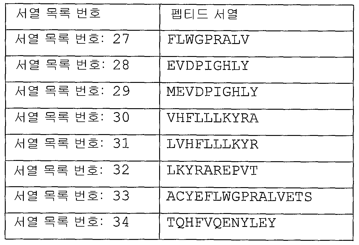
Applications Claiming Priority (2)
| Application Number | Priority Date | Filing Date | Title |
|---|---|---|---|
| US96012807P | 2007-09-17 | 2007-09-17 | |
| US60/960,128 | 2007-09-17 |
Publications (1)
| Publication Number | Publication Date |
|---|---|
| KR20100085917A true KR20100085917A (en) | 2010-07-29 |
Family
ID=40239643
Family Applications (1)
| Application Number | Title | Priority Date | Filing Date |
|---|---|---|---|
| KR1020107008201A KR20100085917A (en) | 2007-09-17 | 2008-09-17 | Improved detection of mage-a expression |
Country Status (18)
| Country | Link |
|---|---|
| US (4) | US8481700B2 (en) |
| EP (2) | EP2201131B8 (en) |
| JP (1) | JP5406839B2 (en) |
| KR (1) | KR20100085917A (en) |
| CN (1) | CN101932723B (en) |
| AU (1) | AU2008300397A1 (en) |
| BR (1) | BRPI0817008A2 (en) |
| CA (2) | CA2699853A1 (en) |
| CO (1) | CO6260156A2 (en) |
| CR (1) | CR11504A (en) |
| DO (1) | DOP2010000076A (en) |
| EA (1) | EA017879B1 (en) |
| IL (1) | IL204525A0 (en) |
| MA (1) | MA31753B1 (en) |
| MX (1) | MX2010002965A (en) |
| NZ (2) | NZ583796A (en) |
| WO (2) | WO2009037438A1 (en) |
| ZA (1) | ZA201001878B (en) |
Families Citing this family (18)
| Publication number | Priority date | Publication date | Assignee | Title |
|---|---|---|---|---|
| US20130237445A1 (en) * | 2010-09-14 | 2013-09-12 | The University Of North Carolina At Chapel Hill | Methods and kits for detecting melanoma |
| WO2012129488A2 (en) * | 2011-03-23 | 2012-09-27 | Virginia Commonwealth University | Gene signatures associated with rejection or recurrence of cancer |
| CN102251033B (en) * | 2011-07-05 | 2013-03-20 | 北京大学人民医院 | Quantitative detection kit for assistant diagnosis of multiple myeloma patient based on MAGE-A3 gene |
| JP2016509480A (en) | 2013-01-24 | 2016-03-31 | カリフォルニア インスティチュート オブ テクノロジー | Chromophore-based characterization and detection methods |
| ES2859645T3 (en) * | 2013-03-14 | 2021-10-04 | Mayo Found Medical Education & Res | Neoplasia detection |
| CN107208162B (en) | 2015-02-26 | 2020-12-29 | 株式会社日立高新技术 | Method for constructing nucleic acid molecule |
| CN105100961B (en) * | 2015-07-23 | 2018-03-13 | 华为技术有限公司 | Video thumbnail generation method and generating means |
| US11685955B2 (en) | 2016-05-16 | 2023-06-27 | Dimo Dietrich | Method for predicting response of patients with malignant diseases to immunotherapy |
| DE102016005947B3 (en) * | 2016-05-16 | 2017-06-08 | Dimo Dietrich | A method for estimating the prognosis and predicting the response to immunotherapy of patients with malignant diseases |
| US10294518B2 (en) | 2016-09-16 | 2019-05-21 | Fluxion Biosciences, Inc. | Methods and systems for ultra-sensitive detection of genomic alterations |
| WO2018167572A1 (en) | 2017-03-17 | 2018-09-20 | Mdxhealth Sa | Mgmt epigenetic deep-sequencing assay |
| US20180271710A1 (en) * | 2017-03-22 | 2018-09-27 | Bragi GmbH | Wireless earpiece for tinnitus therapy |
| WO2018237327A1 (en) * | 2017-06-22 | 2018-12-27 | Triact Therapeutics, Inc. | Methods of treating glioblastoma |
| WO2019067991A1 (en) | 2017-09-29 | 2019-04-04 | Triact Therapeutics, Inc. | Iniparib formulations and uses thereof |
| US11441182B2 (en) * | 2018-08-08 | 2022-09-13 | Virginia Commonwealth University | Multiplexed and recyclable single-molecule sensors for quantitative analysis of nucleic-acid biomarkers |
| CN109540856B (en) * | 2018-11-08 | 2022-01-28 | 南京师范大学 | Reagent for detecting different subtype breast cancer cells based on fluorescence resonance energy transfer |
| CN111551978B (en) * | 2020-05-08 | 2022-09-27 | 中国辐射防护研究院 | Method for evaluating uncertainty of radioactive soil sample sampling process |
| WO2023108145A1 (en) * | 2021-12-10 | 2023-06-15 | Nuprobe Usa, Inc. | Universal probes for amplification and detection of nucleic acids |
Family Cites Families (35)
| Publication number | Priority date | Publication date | Assignee | Title |
|---|---|---|---|---|
| US4437975A (en) * | 1977-07-20 | 1984-03-20 | Mobil Oil Corporation | Manufacture of lube base stock oil |
| WO1989001050A1 (en) * | 1987-07-31 | 1989-02-09 | The Board Of Trustees Of The Leland Stanford Junior University | Selective amplification of target polynucleotide sequences |
| BR8907830A (en) | 1988-12-16 | 1991-10-22 | Siska Diagnostics Inc | PROCESS FOR PREPARING A DOUBLE-FILAMENT DNA THAT CODES A SEQUENCE THAT MATCHES A OBJECTIVE NUCLEIC ACID SEQUENCE AND PROCESS USED FOR DETECTION OF AT LEAST ONE SPECIFIC OBJECTIVE NUCLEIC ACID SEQUENCE |
| SE466259B (en) | 1990-05-31 | 1992-01-20 | Arne Forsgren | PROTEIN D - AN IGD BINDING PROTEIN FROM HAEMOPHILUS INFLUENZAE, AND THE USE OF THIS FOR ANALYSIS, VACCINES AND PURPOSE |
| ATE188613T1 (en) | 1992-06-25 | 2000-01-15 | Smithkline Beecham Biolog | VACCINE COMPOSITION CONTAINING ADJUVANTS |
| GB9326253D0 (en) | 1993-12-23 | 1994-02-23 | Smithkline Beecham Biolog | Vaccines |
| EP1167379A3 (en) | 1994-07-15 | 2004-09-08 | University Of Iowa Research Foundation | Immunomodulatory oligonucleotides |
| UA56132C2 (en) | 1995-04-25 | 2003-05-15 | Смітклайн Бічем Байолоджікалс С.А. | Vaccine composition (variants), method for stabilizing qs21 providing resistance against hydrolysis (variants), method for manufacturing vaccine |
| US6117635A (en) * | 1996-07-16 | 2000-09-12 | Intergen Company | Nucleic acid amplification oligonucleotides with molecular energy transfer labels and methods based thereon |
| US6113918A (en) | 1997-05-08 | 2000-09-05 | Ribi Immunochem Research, Inc. | Aminoalkyl glucosamine phosphate compounds and their use as adjuvants and immunoeffectors |
| US6303347B1 (en) | 1997-05-08 | 2001-10-16 | Corixa Corporation | Aminoalkyl glucosaminide phosphate compounds and their use as adjuvants and immunoeffectors |
| US6291430B1 (en) * | 1997-09-12 | 2001-09-18 | Ludwig Institute For Cancer Research | Mage-3 peptides presented by HLA class II molecules |
| KR100633212B1 (en) * | 1998-02-05 | 2006-10-11 | 글락소스미스클라인 바이오로지칼즈 에스.에이. | Tumor-associated antigen derivatives from the mage family, and nucleic acid sequences encoding them, used for the preparation of fusion proteins and of compositions for vaccination |
| US6768000B1 (en) * | 1998-06-12 | 2004-07-27 | Intergen Company | Multi-fluorescent hairpin energy transfer oligonucleotides |
| PT1104306E (en) | 1998-08-10 | 2006-05-31 | Antigenics Inc | CPG COMPOSITIONS AND SAPONIN ADJUVANTS AND THEIR METHODS |
| US6667037B1 (en) * | 1998-10-09 | 2003-12-23 | Ludwig Institute For Cancer Research | Isolated peptides which bind to HLA-B35 molecules, larger peptides which contain these, nucleic acid molecules encoding peptides, and uses thereof |
| BRPI0010612B8 (en) | 1999-04-19 | 2021-05-25 | Smithkline Beecham Biologicals S A | vaccines |
| US6331393B1 (en) * | 1999-05-14 | 2001-12-18 | University Of Southern California | Process for high-throughput DNA methylation analysis |
| EP1283906A4 (en) * | 2000-03-31 | 2005-04-27 | Univ Southern California | Epigenetic sequences for esophageal adenocarcinoma |
| ATE352640T1 (en) | 2001-03-15 | 2007-02-15 | Jacques Schrenzel | DETECTION OF METHICILLIN-RESISTANT STAPHYLOCOCCUS AUREUS BACTERIA (MRSA) |
| AU2002360429A1 (en) | 2001-11-26 | 2003-06-10 | Avigen, Inc. | Methods for producing stocks of recombinant aav virions |
| HUP0500539A2 (en) | 2002-02-04 | 2006-09-28 | Corixa Corp | New immunoeffector 2-deoxi-2-amino-3-d glucopyranose derivatives |
| DE10215770A1 (en) * | 2002-04-10 | 2003-10-30 | Michael Giesing | Detecting methylated and non-methylated cytosines, useful for assessing methylation status of cancer-associated gene promoters, by specific amplification and hybridization |
| WO2004067726A2 (en) | 2003-01-29 | 2004-08-12 | Keck Graduate Institute | Isothermal reactions for the amplification of oligonucleotides |
| WO2004087957A2 (en) * | 2003-04-03 | 2004-10-14 | Oncomethylome Sciences S.A. | Hypermethylated genes and cervical cancer |
| US7881873B2 (en) * | 2003-04-29 | 2011-02-01 | The Jackson Laboratory | Systems and methods for statistical genomic DNA based analysis and evaluation |
| DE10329240A1 (en) * | 2003-06-24 | 2005-01-20 | Epigenomics Ag | Method for analyzing the cytosine methylation status of cancer testis antigens for individualized immunotherapy |
| DE10338308B4 (en) * | 2003-08-15 | 2006-10-19 | Epigenomics Ag | Method for the detection of cytosine methylations in DNA |
| WO2005042713A2 (en) | 2003-10-28 | 2005-05-12 | The Johns Hopkins University | Quantitative multiplex methylation-specific pcr |
| US20050130170A1 (en) * | 2003-12-16 | 2005-06-16 | Jeanne Harvey | Identification and verification of methylation marker sequences |
| ATE396282T1 (en) * | 2004-10-11 | 2008-06-15 | Epigenomics Ag | METHOD FOR CONTAMINATION PROTECTION OF DNA AMPLIFICATION REACTIONS FOR METHYLATION ANALYSIS THROUGH MODIFIED PRETREATMENT OF NUCLEIC ACIDS |
| US20070059753A1 (en) * | 2005-09-15 | 2007-03-15 | Tatiana Vener | Detecting gene methylation |
| GB0612342D0 (en) | 2006-06-21 | 2006-08-02 | Glaxosmithkline Biolog Sa | Method |
| GB0700374D0 (en) * | 2007-01-09 | 2007-02-14 | Oncomethylome Sciences S A | NDRG family methylation markers |
| US8911937B2 (en) * | 2007-07-19 | 2014-12-16 | Brainreader Aps | Method for detecting methylation status by using methylation-independent primers |
-
2008
- 2008-09-17 AU AU2008300397A patent/AU2008300397A1/en not_active Abandoned
- 2008-09-17 WO PCT/GB2008/003142 patent/WO2009037438A1/en active Application Filing
- 2008-09-17 CA CA2699853A patent/CA2699853A1/en not_active Abandoned
- 2008-09-17 BR BRPI0817008A patent/BRPI0817008A2/en not_active IP Right Cessation
- 2008-09-17 US US12/678,702 patent/US8481700B2/en not_active Expired - Fee Related
- 2008-09-17 KR KR1020107008201A patent/KR20100085917A/en active IP Right Grant
- 2008-09-17 EP EP08806301.1A patent/EP2201131B8/en active Active
- 2008-09-17 CA CA2699856A patent/CA2699856C/en active Active
- 2008-09-17 EP EP08806298A patent/EP2198045A1/en not_active Withdrawn
- 2008-09-17 NZ NZ583796A patent/NZ583796A/en not_active IP Right Cessation
- 2008-09-17 JP JP2010524578A patent/JP5406839B2/en not_active Expired - Fee Related
- 2008-09-17 US US12/678,706 patent/US9050280B2/en active Active
- 2008-09-17 WO PCT/GB2008/003145 patent/WO2009037441A1/en active Application Filing
- 2008-09-17 NZ NZ596545A patent/NZ596545A/en not_active IP Right Cessation
- 2008-09-17 CN CN200880117371.0A patent/CN101932723B/en not_active Expired - Fee Related
- 2008-09-17 MX MX2010002965A patent/MX2010002965A/en not_active Application Discontinuation
- 2008-09-17 EA EA201000334A patent/EA017879B1/en not_active IP Right Cessation
-
2010
- 2010-03-11 DO DO2010000076A patent/DOP2010000076A/en unknown
- 2010-03-16 ZA ZA2010/01878A patent/ZA201001878B/en unknown
- 2010-03-16 IL IL204525A patent/IL204525A0/en unknown
- 2010-03-19 CO CO10033047A patent/CO6260156A2/en not_active Application Discontinuation
- 2010-04-01 MA MA32736A patent/MA31753B1/en unknown
- 2010-06-16 CR CR11504A patent/CR11504A/en not_active Application Discontinuation
-
2013
- 2013-06-13 US US13/917,108 patent/US20130302363A1/en not_active Abandoned
-
2015
- 2015-06-08 US US14/733,152 patent/US10053724B2/en active Active
Also Published As
Similar Documents
| Publication | Publication Date | Title |
|---|---|---|
| JP5406839B2 (en) | Improved detection of MAGE-A expression | |
| US20120039993A1 (en) | Detection of Gene Expression | |
| EP2046985B1 (en) | Method for the detection of egfr mutations in blood samples | |
| JP2005518218A5 (en) | ||
| JP2005518218A (en) | Methods and nucleic acids for the analysis of colorectal cell proliferative disorders | |
| KR20140054399A (en) | Detection of prame gene expression in cancer | |
| JP5705191B2 (en) | Method for detection and diagnosis of cancer comprising primers and probes for specific detection of MAGE-A3 marker |
Legal Events
| Date | Code | Title | Description |
|---|---|---|---|
| A201 | Request for examination | ||
| E902 | Notification of reason for refusal | ||
| E701 | Decision to grant or registration of patent right |