KR20080068147A - Methods for enhancing the efficacy of cancer therapy - Google Patents
Methods for enhancing the efficacy of cancer therapy Download PDFInfo
- Publication number
- KR20080068147A KR20080068147A KR1020087015948A KR20087015948A KR20080068147A KR 20080068147 A KR20080068147 A KR 20080068147A KR 1020087015948 A KR1020087015948 A KR 1020087015948A KR 20087015948 A KR20087015948 A KR 20087015948A KR 20080068147 A KR20080068147 A KR 20080068147A
- Authority
- KR
- South Korea
- Prior art keywords
- cells
- tumor
- antibody
- wnt
- human
- Prior art date
Links
- 238000000034 method Methods 0.000 title claims abstract description 130
- 230000002708 enhancing effect Effects 0.000 title abstract description 4
- 238000011275 oncology therapy Methods 0.000 title description 6
- 206010028980 Neoplasm Diseases 0.000 claims abstract description 265
- 230000014509 gene expression Effects 0.000 claims abstract description 110
- 150000004492 retinoid derivatives Chemical class 0.000 claims abstract description 49
- 108090000623 proteins and genes Proteins 0.000 claims description 215
- 239000002246 antineoplastic agent Substances 0.000 claims description 51
- 238000011534 incubation Methods 0.000 claims description 8
- 229940034982 antineoplastic agent Drugs 0.000 claims 1
- 238000011282 treatment Methods 0.000 abstract description 113
- SHGAZHPCJJPHSC-YCNIQYBTSA-N all-trans-retinoic acid Chemical compound OC(=O)\C=C(/C)\C=C\C=C(/C)\C=C\C1=C(C)CCCC1(C)C SHGAZHPCJJPHSC-YCNIQYBTSA-N 0.000 abstract description 89
- 229930002330 retinoic acid Natural products 0.000 abstract description 85
- 201000011510 cancer Diseases 0.000 abstract description 82
- 239000000427 antigen Substances 0.000 abstract description 69
- 108091007433 antigens Proteins 0.000 abstract description 68
- 102000036639 antigens Human genes 0.000 abstract description 68
- 229960001727 tretinoin Drugs 0.000 abstract description 65
- 108050003627 Wnt Proteins 0.000 abstract description 64
- 102000013814 Wnt Human genes 0.000 abstract description 59
- 230000011664 signaling Effects 0.000 abstract description 49
- 230000001976 improved effect Effects 0.000 abstract description 3
- 230000001594 aberrant effect Effects 0.000 abstract 1
- 210000004027 cell Anatomy 0.000 description 333
- 241000282414 Homo sapiens Species 0.000 description 156
- 108090000765 processed proteins & peptides Proteins 0.000 description 152
- 102000004196 processed proteins & peptides Human genes 0.000 description 140
- 229920001184 polypeptide Polymers 0.000 description 130
- 101150092419 STRA6 gene Proteins 0.000 description 119
- 102000004169 proteins and genes Human genes 0.000 description 97
- 101100156752 Caenorhabditis elegans cwn-1 gene Proteins 0.000 description 96
- 108700020987 Wnt-1 Proteins 0.000 description 96
- 102000052547 Wnt-1 Human genes 0.000 description 94
- 235000018102 proteins Nutrition 0.000 description 91
- 108091032973 (ribonucleotides)n+m Proteins 0.000 description 65
- 208000009956 adenocarcinoma Diseases 0.000 description 63
- 210000001519 tissue Anatomy 0.000 description 62
- 239000000523 sample Substances 0.000 description 57
- 125000003275 alpha amino acid group Chemical group 0.000 description 55
- 108020004999 messenger RNA Proteins 0.000 description 50
- 210000004881 tumor cell Anatomy 0.000 description 49
- 108020004414 DNA Proteins 0.000 description 47
- 230000027455 binding Effects 0.000 description 47
- 241001465754 Metazoa Species 0.000 description 45
- 239000002609 medium Substances 0.000 description 45
- 239000012634 fragment Substances 0.000 description 41
- 108060003951 Immunoglobulin Proteins 0.000 description 40
- 235000001014 amino acid Nutrition 0.000 description 40
- 208000029742 colonic neoplasm Diseases 0.000 description 40
- 102000018358 immunoglobulin Human genes 0.000 description 40
- 108060000903 Beta-catenin Proteins 0.000 description 37
- 229940024606 amino acid Drugs 0.000 description 37
- 102000015735 Beta-catenin Human genes 0.000 description 36
- 150000001413 amino acids Chemical class 0.000 description 36
- IAZDPXIOMUYVGZ-UHFFFAOYSA-N Dimethylsulphoxide Chemical compound CS(C)=O IAZDPXIOMUYVGZ-UHFFFAOYSA-N 0.000 description 33
- 230000000694 effects Effects 0.000 description 33
- 239000013615 primer Substances 0.000 description 32
- 206010009944 Colon cancer Diseases 0.000 description 31
- 239000003814 drug Substances 0.000 description 30
- 230000004913 activation Effects 0.000 description 28
- 229940079593 drug Drugs 0.000 description 27
- 108010021625 Immunoglobulin Fragments Proteins 0.000 description 26
- 102000008394 Immunoglobulin Fragments Human genes 0.000 description 26
- 239000000126 substance Substances 0.000 description 26
- 239000000872 buffer Substances 0.000 description 25
- 229940002612 prodrug Drugs 0.000 description 25
- 239000000651 prodrug Substances 0.000 description 25
- RAXXELZNTBOGNW-UHFFFAOYSA-N imidazole Natural products C1=CNC=N1 RAXXELZNTBOGNW-UHFFFAOYSA-N 0.000 description 24
- 241000699670 Mus sp. Species 0.000 description 23
- 208000026310 Breast neoplasm Diseases 0.000 description 22
- PEDCQBHIVMGVHV-UHFFFAOYSA-N Glycerine Chemical compound OCC(O)CO PEDCQBHIVMGVHV-UHFFFAOYSA-N 0.000 description 22
- 238000006243 chemical reaction Methods 0.000 description 22
- 206010006187 Breast cancer Diseases 0.000 description 21
- 239000002299 complementary DNA Substances 0.000 description 21
- 229940127089 cytotoxic agent Drugs 0.000 description 21
- 230000035772 mutation Effects 0.000 description 21
- 210000001072 colon Anatomy 0.000 description 20
- 210000004408 hybridoma Anatomy 0.000 description 20
- 238000004519 manufacturing process Methods 0.000 description 20
- 230000006698 induction Effects 0.000 description 19
- FAPWRFPIFSIZLT-UHFFFAOYSA-M Sodium chloride Chemical compound [Na+].[Cl-] FAPWRFPIFSIZLT-UHFFFAOYSA-M 0.000 description 18
- 230000012010 growth Effects 0.000 description 18
- 239000000203 mixture Substances 0.000 description 18
- 150000001875 compounds Chemical class 0.000 description 17
- 102000005962 receptors Human genes 0.000 description 17
- 102000003702 retinoic acid receptors Human genes 0.000 description 17
- 108090000064 retinoic acid receptors Proteins 0.000 description 17
- 239000000243 solution Substances 0.000 description 17
- 206010041823 squamous cell carcinoma Diseases 0.000 description 17
- 238000010240 RT-PCR analysis Methods 0.000 description 16
- 239000004098 Tetracycline Substances 0.000 description 16
- 238000003556 assay Methods 0.000 description 16
- 229940072221 immunoglobulins Drugs 0.000 description 16
- 102000039446 nucleic acids Human genes 0.000 description 16
- 108020004707 nucleic acids Proteins 0.000 description 16
- 150000007523 nucleic acids Chemical class 0.000 description 16
- 239000000047 product Substances 0.000 description 16
- 108020003175 receptors Proteins 0.000 description 16
- 229930101283 tetracycline Natural products 0.000 description 16
- 229960002180 tetracycline Drugs 0.000 description 16
- 235000019364 tetracycline Nutrition 0.000 description 16
- 150000003522 tetracyclines Chemical class 0.000 description 16
- FWMNVWWHGCHHJJ-SKKKGAJSSA-N 4-amino-1-[(2r)-6-amino-2-[[(2r)-2-[[(2r)-2-[[(2r)-2-amino-3-phenylpropanoyl]amino]-3-phenylpropanoyl]amino]-4-methylpentanoyl]amino]hexanoyl]piperidine-4-carboxylic acid Chemical compound C([C@H](C(=O)N[C@H](CC(C)C)C(=O)N[C@H](CCCCN)C(=O)N1CCC(N)(CC1)C(O)=O)NC(=O)[C@H](N)CC=1C=CC=CC=1)C1=CC=CC=C1 FWMNVWWHGCHHJJ-SKKKGAJSSA-N 0.000 description 15
- SHGAZHPCJJPHSC-ZVCIMWCZSA-N 9-cis-retinoic acid Chemical compound OC(=O)/C=C(\C)/C=C/C=C(/C)\C=C\C1=C(C)CCCC1(C)C SHGAZHPCJJPHSC-ZVCIMWCZSA-N 0.000 description 15
- WEVYAHXRMPXWCK-UHFFFAOYSA-N Acetonitrile Chemical compound CC#N WEVYAHXRMPXWCK-UHFFFAOYSA-N 0.000 description 15
- 102100021977 Ectonucleotide pyrophosphatase/phosphodiesterase family member 2 Human genes 0.000 description 15
- 102000004190 Enzymes Human genes 0.000 description 15
- 108090000790 Enzymes Proteins 0.000 description 15
- 208000008839 Kidney Neoplasms Diseases 0.000 description 15
- 229960001445 alitretinoin Drugs 0.000 description 15
- 125000000539 amino acid group Chemical group 0.000 description 15
- -1 engrailed Proteins 0.000 description 15
- 229940088598 enzyme Drugs 0.000 description 15
- 239000013604 expression vector Substances 0.000 description 15
- 238000000746 purification Methods 0.000 description 15
- 108091008760 retinoic acid receptors γ Proteins 0.000 description 15
- 239000013598 vector Substances 0.000 description 15
- 108050004000 Ectonucleotide pyrophosphatase/phosphodiesterase family member 2 Proteins 0.000 description 14
- 241000588724 Escherichia coli Species 0.000 description 14
- 241000124008 Mammalia Species 0.000 description 14
- 238000004458 analytical method Methods 0.000 description 14
- 239000005557 antagonist Substances 0.000 description 14
- 238000001514 detection method Methods 0.000 description 14
- 208000037265 diseases, disorders, signs and symptoms Diseases 0.000 description 14
- 102000006602 glyceraldehyde-3-phosphate dehydrogenase Human genes 0.000 description 14
- 108020004445 glyceraldehyde-3-phosphate dehydrogenase Proteins 0.000 description 14
- 238000009396 hybridization Methods 0.000 description 14
- 230000002018 overexpression Effects 0.000 description 14
- 108091026890 Coding region Proteins 0.000 description 13
- 241001529936 Murinae Species 0.000 description 13
- 102100033912 Retinoic acid receptor gamma Human genes 0.000 description 13
- 230000002159 abnormal effect Effects 0.000 description 13
- 230000003321 amplification Effects 0.000 description 13
- 230000001472 cytotoxic effect Effects 0.000 description 13
- 238000002474 experimental method Methods 0.000 description 13
- 230000001965 increasing effect Effects 0.000 description 13
- 238000003199 nucleic acid amplification method Methods 0.000 description 13
- 238000002360 preparation method Methods 0.000 description 13
- 208000001333 Colorectal Neoplasms Diseases 0.000 description 12
- 108010047041 Complementarity Determining Regions Proteins 0.000 description 12
- AOJJSUZBOXZQNB-TZSSRYMLSA-N Doxorubicin Chemical compound O([C@H]1C[C@@](O)(CC=2C(O)=C3C(=O)C=4C=CC=C(C=4C(=O)C3=C(O)C=21)OC)C(=O)CO)[C@H]1C[C@H](N)[C@H](O)[C@H](C)O1 AOJJSUZBOXZQNB-TZSSRYMLSA-N 0.000 description 12
- 206010058467 Lung neoplasm malignant Diseases 0.000 description 12
- 230000004156 Wnt signaling pathway Effects 0.000 description 12
- 230000000295 complement effect Effects 0.000 description 12
- 231100000433 cytotoxic Toxicity 0.000 description 12
- 238000011161 development Methods 0.000 description 12
- 230000018109 developmental process Effects 0.000 description 12
- 239000000975 dye Substances 0.000 description 12
- 238000000338 in vitro Methods 0.000 description 12
- 230000002401 inhibitory effect Effects 0.000 description 12
- 239000003446 ligand Substances 0.000 description 12
- 230000007246 mechanism Effects 0.000 description 12
- 201000001441 melanoma Diseases 0.000 description 12
- 239000013612 plasmid Substances 0.000 description 12
- 238000012216 screening Methods 0.000 description 12
- 230000001225 therapeutic effect Effects 0.000 description 12
- 241000699666 Mus <mouse, genus> Species 0.000 description 11
- 241000699660 Mus musculus Species 0.000 description 11
- 238000004113 cell culture Methods 0.000 description 11
- 230000003750 conditioning effect Effects 0.000 description 11
- 201000010099 disease Diseases 0.000 description 11
- 102000012803 ephrin Human genes 0.000 description 11
- 108060002566 ephrin Proteins 0.000 description 11
- 229940127121 immunoconjugate Drugs 0.000 description 11
- 238000001727 in vivo Methods 0.000 description 11
- 239000002502 liposome Substances 0.000 description 11
- 208000020816 lung neoplasm Diseases 0.000 description 11
- 239000002773 nucleotide Substances 0.000 description 11
- 125000003729 nucleotide group Chemical group 0.000 description 11
- 230000037361 pathway Effects 0.000 description 11
- 238000011160 research Methods 0.000 description 11
- 238000012360 testing method Methods 0.000 description 11
- 102000002627 4-1BB Ligand Human genes 0.000 description 10
- 201000009030 Carcinoma Diseases 0.000 description 10
- 108010052285 Membrane Proteins Proteins 0.000 description 10
- 206010028851 Necrosis Diseases 0.000 description 10
- 201000010897 colon adenocarcinoma Diseases 0.000 description 10
- 238000007901 in situ hybridization Methods 0.000 description 10
- 230000017074 necrotic cell death Effects 0.000 description 10
- 238000011580 nude mouse model Methods 0.000 description 10
- 230000002441 reversible effect Effects 0.000 description 10
- 150000003384 small molecules Chemical class 0.000 description 10
- 230000002195 synergetic effect Effects 0.000 description 10
- 238000002560 therapeutic procedure Methods 0.000 description 10
- 238000013518 transcription Methods 0.000 description 10
- 230000035897 transcription Effects 0.000 description 10
- XLYOFNOQVPJJNP-UHFFFAOYSA-N water Chemical compound O XLYOFNOQVPJJNP-UHFFFAOYSA-N 0.000 description 10
- 108010082808 4-1BB Ligand Proteins 0.000 description 9
- IJGRMHOSHXDMSA-UHFFFAOYSA-N Atomic nitrogen Chemical compound N#N IJGRMHOSHXDMSA-UHFFFAOYSA-N 0.000 description 9
- 102000004127 Cytokines Human genes 0.000 description 9
- 108090000695 Cytokines Proteins 0.000 description 9
- 102000018697 Membrane Proteins Human genes 0.000 description 9
- 108091034117 Oligonucleotide Proteins 0.000 description 9
- 206010035226 Plasma cell myeloma Diseases 0.000 description 9
- 239000013543 active substance Substances 0.000 description 9
- 239000002671 adjuvant Substances 0.000 description 9
- 238000010171 animal model Methods 0.000 description 9
- 230000000692 anti-sense effect Effects 0.000 description 9
- 210000000481 breast Anatomy 0.000 description 9
- 230000006870 function Effects 0.000 description 9
- 239000000499 gel Substances 0.000 description 9
- 239000001963 growth medium Substances 0.000 description 9
- 238000009169 immunotherapy Methods 0.000 description 9
- 239000003112 inhibitor Substances 0.000 description 9
- 210000003734 kidney Anatomy 0.000 description 9
- 210000004072 lung Anatomy 0.000 description 9
- 201000000050 myeloid neoplasm Diseases 0.000 description 9
- 230000004044 response Effects 0.000 description 9
- 108091008146 restriction endonucleases Proteins 0.000 description 9
- 239000011780 sodium chloride Substances 0.000 description 9
- 208000024891 symptom Diseases 0.000 description 9
- 230000009261 transgenic effect Effects 0.000 description 9
- 108091003079 Bovine Serum Albumin Proteins 0.000 description 8
- 229910019142 PO4 Inorganic materials 0.000 description 8
- 239000003966 growth inhibitor Substances 0.000 description 8
- 206010073071 hepatocellular carcinoma Diseases 0.000 description 8
- 238000002347 injection Methods 0.000 description 8
- 239000007924 injection Substances 0.000 description 8
- 230000003834 intracellular effect Effects 0.000 description 8
- 201000005202 lung cancer Diseases 0.000 description 8
- 239000010452 phosphate Substances 0.000 description 8
- 239000007790 solid phase Substances 0.000 description 8
- 238000010561 standard procedure Methods 0.000 description 8
- 108020005544 Antisense RNA Proteins 0.000 description 7
- 206010003445 Ascites Diseases 0.000 description 7
- 206010014759 Endometrial neoplasm Diseases 0.000 description 7
- 102100031780 Endonuclease Human genes 0.000 description 7
- 241000282326 Felis catus Species 0.000 description 7
- ZRALSGWEFCBTJO-UHFFFAOYSA-N Guanidine Chemical compound NC(N)=N ZRALSGWEFCBTJO-UHFFFAOYSA-N 0.000 description 7
- 101710163270 Nuclease Proteins 0.000 description 7
- 206010061535 Ovarian neoplasm Diseases 0.000 description 7
- 102000035195 Peptidases Human genes 0.000 description 7
- 108091005804 Peptidases Proteins 0.000 description 7
- 239000004365 Protease Substances 0.000 description 7
- 241000700159 Rattus Species 0.000 description 7
- 240000004808 Saccharomyces cerevisiae Species 0.000 description 7
- 108700019146 Transgenes Proteins 0.000 description 7
- 238000001042 affinity chromatography Methods 0.000 description 7
- 238000005119 centrifugation Methods 0.000 description 7
- 239000013522 chelant Substances 0.000 description 7
- 239000003153 chemical reaction reagent Substances 0.000 description 7
- 239000003795 chemical substances by application Substances 0.000 description 7
- 210000004978 chinese hamster ovary cell Anatomy 0.000 description 7
- 239000003184 complementary RNA Substances 0.000 description 7
- 230000001419 dependent effect Effects 0.000 description 7
- 230000004927 fusion Effects 0.000 description 7
- 229940124452 immunizing agent Drugs 0.000 description 7
- 230000005764 inhibitory process Effects 0.000 description 7
- 230000001404 mediated effect Effects 0.000 description 7
- 238000002493 microarray Methods 0.000 description 7
- 239000008188 pellet Substances 0.000 description 7
- NBIIXXVUZAFLBC-UHFFFAOYSA-K phosphate Chemical compound [O-]P([O-])([O-])=O NBIIXXVUZAFLBC-UHFFFAOYSA-K 0.000 description 7
- 239000000758 substrate Substances 0.000 description 7
- 230000014616 translation Effects 0.000 description 7
- 230000004614 tumor growth Effects 0.000 description 7
- 238000002965 ELISA Methods 0.000 description 6
- 241000282412 Homo Species 0.000 description 6
- 101000977638 Homo sapiens Immunoglobulin superfamily containing leucine-rich repeat protein Proteins 0.000 description 6
- PXHVJJICTQNCMI-UHFFFAOYSA-N Nickel Chemical compound [Ni] PXHVJJICTQNCMI-UHFFFAOYSA-N 0.000 description 6
- 241000283973 Oryctolagus cuniculus Species 0.000 description 6
- 206010060862 Prostate cancer Diseases 0.000 description 6
- 208000000236 Prostatic Neoplasms Diseases 0.000 description 6
- 102100024952 Protein CBFA2T1 Human genes 0.000 description 6
- 108010092799 RNA-directed DNA polymerase Proteins 0.000 description 6
- 108020004511 Recombinant DNA Proteins 0.000 description 6
- 102000000479 TCF Transcription Factors Human genes 0.000 description 6
- 108010016283 TCF Transcription Factors Proteins 0.000 description 6
- NKANXQFJJICGDU-QPLCGJKRSA-N Tamoxifen Chemical compound C=1C=CC=CC=1C(/CC)=C(C=1C=CC(OCCN(C)C)=CC=1)/C1=CC=CC=C1 NKANXQFJJICGDU-QPLCGJKRSA-N 0.000 description 6
- 239000007983 Tris buffer Substances 0.000 description 6
- 239000013504 Triton X-100 Substances 0.000 description 6
- 229920004890 Triton X-100 Polymers 0.000 description 6
- JLCPHMBAVCMARE-UHFFFAOYSA-N [3-[[3-[[3-[[3-[[3-[[3-[[3-[[3-[[3-[[3-[[3-[[5-(2-amino-6-oxo-1H-purin-9-yl)-3-[[3-[[3-[[3-[[3-[[3-[[5-(2-amino-6-oxo-1H-purin-9-yl)-3-[[5-(2-amino-6-oxo-1H-purin-9-yl)-3-hydroxyoxolan-2-yl]methoxy-hydroxyphosphoryl]oxyoxolan-2-yl]methoxy-hydroxyphosphoryl]oxy-5-(5-methyl-2,4-dioxopyrimidin-1-yl)oxolan-2-yl]methoxy-hydroxyphosphoryl]oxy-5-(6-aminopurin-9-yl)oxolan-2-yl]methoxy-hydroxyphosphoryl]oxy-5-(6-aminopurin-9-yl)oxolan-2-yl]methoxy-hydroxyphosphoryl]oxy-5-(6-aminopurin-9-yl)oxolan-2-yl]methoxy-hydroxyphosphoryl]oxy-5-(6-aminopurin-9-yl)oxolan-2-yl]methoxy-hydroxyphosphoryl]oxyoxolan-2-yl]methoxy-hydroxyphosphoryl]oxy-5-(5-methyl-2,4-dioxopyrimidin-1-yl)oxolan-2-yl]methoxy-hydroxyphosphoryl]oxy-5-(4-amino-2-oxopyrimidin-1-yl)oxolan-2-yl]methoxy-hydroxyphosphoryl]oxy-5-(5-methyl-2,4-dioxopyrimidin-1-yl)oxolan-2-yl]methoxy-hydroxyphosphoryl]oxy-5-(5-methyl-2,4-dioxopyrimidin-1-yl)oxolan-2-yl]methoxy-hydroxyphosphoryl]oxy-5-(6-aminopurin-9-yl)oxolan-2-yl]methoxy-hydroxyphosphoryl]oxy-5-(6-aminopurin-9-yl)oxolan-2-yl]methoxy-hydroxyphosphoryl]oxy-5-(4-amino-2-oxopyrimidin-1-yl)oxolan-2-yl]methoxy-hydroxyphosphoryl]oxy-5-(4-amino-2-oxopyrimidin-1-yl)oxolan-2-yl]methoxy-hydroxyphosphoryl]oxy-5-(4-amino-2-oxopyrimidin-1-yl)oxolan-2-yl]methoxy-hydroxyphosphoryl]oxy-5-(6-aminopurin-9-yl)oxolan-2-yl]methoxy-hydroxyphosphoryl]oxy-5-(4-amino-2-oxopyrimidin-1-yl)oxolan-2-yl]methyl [5-(6-aminopurin-9-yl)-2-(hydroxymethyl)oxolan-3-yl] hydrogen phosphate Polymers Cc1cn(C2CC(OP(O)(=O)OCC3OC(CC3OP(O)(=O)OCC3OC(CC3O)n3cnc4c3nc(N)[nH]c4=O)n3cnc4c3nc(N)[nH]c4=O)C(COP(O)(=O)OC3CC(OC3COP(O)(=O)OC3CC(OC3COP(O)(=O)OC3CC(OC3COP(O)(=O)OC3CC(OC3COP(O)(=O)OC3CC(OC3COP(O)(=O)OC3CC(OC3COP(O)(=O)OC3CC(OC3COP(O)(=O)OC3CC(OC3COP(O)(=O)OC3CC(OC3COP(O)(=O)OC3CC(OC3COP(O)(=O)OC3CC(OC3COP(O)(=O)OC3CC(OC3COP(O)(=O)OC3CC(OC3COP(O)(=O)OC3CC(OC3COP(O)(=O)OC3CC(OC3COP(O)(=O)OC3CC(OC3COP(O)(=O)OC3CC(OC3CO)n3cnc4c(N)ncnc34)n3ccc(N)nc3=O)n3cnc4c(N)ncnc34)n3ccc(N)nc3=O)n3ccc(N)nc3=O)n3ccc(N)nc3=O)n3cnc4c(N)ncnc34)n3cnc4c(N)ncnc34)n3cc(C)c(=O)[nH]c3=O)n3cc(C)c(=O)[nH]c3=O)n3ccc(N)nc3=O)n3cc(C)c(=O)[nH]c3=O)n3cnc4c3nc(N)[nH]c4=O)n3cnc4c(N)ncnc34)n3cnc4c(N)ncnc34)n3cnc4c(N)ncnc34)n3cnc4c(N)ncnc34)O2)c(=O)[nH]c1=O JLCPHMBAVCMARE-UHFFFAOYSA-N 0.000 description 6
- FPIPGXGPPPQFEQ-OVSJKPMPSA-N all-trans-retinol Chemical compound OC\C=C(/C)\C=C\C=C(/C)\C=C\C1=C(C)CCCC1(C)C FPIPGXGPPPQFEQ-OVSJKPMPSA-N 0.000 description 6
- 230000010056 antibody-dependent cellular cytotoxicity Effects 0.000 description 6
- 230000004071 biological effect Effects 0.000 description 6
- 230000015572 biosynthetic process Effects 0.000 description 6
- 230000010261 cell growth Effects 0.000 description 6
- 230000008859 change Effects 0.000 description 6
- 238000002512 chemotherapy Methods 0.000 description 6
- 238000003776 cleavage reaction Methods 0.000 description 6
- 239000002254 cytotoxic agent Substances 0.000 description 6
- 231100000599 cytotoxic agent Toxicity 0.000 description 6
- 238000003745 diagnosis Methods 0.000 description 6
- 238000007877 drug screening Methods 0.000 description 6
- 239000012091 fetal bovine serum Substances 0.000 description 6
- 239000007850 fluorescent dye Substances 0.000 description 6
- 230000009395 genetic defect Effects 0.000 description 6
- 231100000844 hepatocellular carcinoma Toxicity 0.000 description 6
- 229910052751 metal Inorganic materials 0.000 description 6
- 239000002184 metal Substances 0.000 description 6
- 206010061289 metastatic neoplasm Diseases 0.000 description 6
- 229920002401 polyacrylamide Polymers 0.000 description 6
- 238000001959 radiotherapy Methods 0.000 description 6
- 238000003753 real-time PCR Methods 0.000 description 6
- 239000011347 resin Substances 0.000 description 6
- 229920005989 resin Polymers 0.000 description 6
- 230000007017 scission Effects 0.000 description 6
- 238000002415 sodium dodecyl sulfate polyacrylamide gel electrophoresis Methods 0.000 description 6
- PUZPDOWCWNUUKD-UHFFFAOYSA-M sodium fluoride Chemical compound [F-].[Na+] PUZPDOWCWNUUKD-UHFFFAOYSA-M 0.000 description 6
- 238000007920 subcutaneous administration Methods 0.000 description 6
- 239000003053 toxin Substances 0.000 description 6
- 231100000765 toxin Toxicity 0.000 description 6
- 108700012359 toxins Proteins 0.000 description 6
- 238000013519 translation Methods 0.000 description 6
- LENZDBCJOHFCAS-UHFFFAOYSA-N tris Chemical compound OCC(N)(CO)CO LENZDBCJOHFCAS-UHFFFAOYSA-N 0.000 description 6
- 230000003827 upregulation Effects 0.000 description 6
- 108020004491 Antisense DNA Proteins 0.000 description 5
- KCXVZYZYPLLWCC-UHFFFAOYSA-N EDTA Chemical compound OC(=O)CN(CC(O)=O)CCN(CC(O)=O)CC(O)=O KCXVZYZYPLLWCC-UHFFFAOYSA-N 0.000 description 5
- 206010014733 Endometrial cancer Diseases 0.000 description 5
- 241000282324 Felis Species 0.000 description 5
- 102000005698 Frizzled receptors Human genes 0.000 description 5
- 108010045438 Frizzled receptors Proteins 0.000 description 5
- 102000003886 Glycoproteins Human genes 0.000 description 5
- 108090000288 Glycoproteins Proteins 0.000 description 5
- XQFRJNBWHJMXHO-RRKCRQDMSA-N IDUR Chemical compound C1[C@H](O)[C@@H](CO)O[C@H]1N1C(=O)NC(=O)C(I)=C1 XQFRJNBWHJMXHO-RRKCRQDMSA-N 0.000 description 5
- 102100023538 Immunoglobulin superfamily containing leucine-rich repeat protein Human genes 0.000 description 5
- 239000005089 Luciferase Substances 0.000 description 5
- 206010027476 Metastases Diseases 0.000 description 5
- 108091028043 Nucleic acid sequence Proteins 0.000 description 5
- 108020005187 Oligonucleotide Probes Proteins 0.000 description 5
- 108700020796 Oncogene Proteins 0.000 description 5
- 238000012408 PCR amplification Methods 0.000 description 5
- 241000283984 Rodentia Species 0.000 description 5
- 108010006785 Taq Polymerase Proteins 0.000 description 5
- 102000040945 Transcription factor Human genes 0.000 description 5
- 108091023040 Transcription factor Proteins 0.000 description 5
- 230000000259 anti-tumor effect Effects 0.000 description 5
- 239000003816 antisense DNA Substances 0.000 description 5
- 238000013459 approach Methods 0.000 description 5
- 229930195731 calicheamicin Natural products 0.000 description 5
- HXCHCVDVKSCDHU-LULTVBGHSA-N calicheamicin Chemical compound C1[C@H](OC)[C@@H](NCC)CO[C@H]1O[C@H]1[C@H](O[C@@H]2C\3=C(NC(=O)OC)C(=O)C[C@](C/3=C/CSSSC)(O)C#C\C=C/C#C2)O[C@H](C)[C@@H](NO[C@@H]2O[C@H](C)[C@@H](SC(=O)C=3C(=C(OC)C(O[C@H]4[C@@H]([C@H](OC)[C@@H](O)[C@H](C)O4)O)=C(I)C=3C)OC)[C@@H](O)C2)[C@@H]1O HXCHCVDVKSCDHU-LULTVBGHSA-N 0.000 description 5
- 238000002591 computed tomography Methods 0.000 description 5
- 230000001086 cytosolic effect Effects 0.000 description 5
- 230000003013 cytotoxicity Effects 0.000 description 5
- 231100000135 cytotoxicity Toxicity 0.000 description 5
- 230000007547 defect Effects 0.000 description 5
- 229960004679 doxorubicin Drugs 0.000 description 5
- 238000009510 drug design Methods 0.000 description 5
- 238000011223 gene expression profiling Methods 0.000 description 5
- 230000003993 interaction Effects 0.000 description 5
- 208000037841 lung tumor Diseases 0.000 description 5
- 210000004698 lymphocyte Anatomy 0.000 description 5
- 239000012139 lysis buffer Substances 0.000 description 5
- 230000009401 metastasis Effects 0.000 description 5
- 210000004877 mucosa Anatomy 0.000 description 5
- 229910052757 nitrogen Inorganic materials 0.000 description 5
- 239000002751 oligonucleotide probe Substances 0.000 description 5
- 230000002246 oncogenic effect Effects 0.000 description 5
- 239000012071 phase Substances 0.000 description 5
- 239000002987 primer (paints) Substances 0.000 description 5
- 238000003259 recombinant expression Methods 0.000 description 5
- 238000010839 reverse transcription Methods 0.000 description 5
- 230000019491 signal transduction Effects 0.000 description 5
- 239000007787 solid Substances 0.000 description 5
- 239000006228 supernatant Substances 0.000 description 5
- 238000002054 transplantation Methods 0.000 description 5
- 230000000381 tumorigenic effect Effects 0.000 description 5
- 241000701447 unidentified baculovirus Species 0.000 description 5
- 238000001262 western blot Methods 0.000 description 5
- QWPXBEHQFHACTK-KZVYIGENSA-N (10e,12e)-86-chloro-12,14,4-trihydroxy-85,14-dimethoxy-33,2,7,10-tetramethyl-15,16-dihydro-14h-7-aza-1(6,4)-oxazina-3(2,3)-oxirana-8(1,3)-benzenacyclotetradecaphane-10,12-dien-6-one Chemical compound CN1C(=O)CC(O)C2(C)OC2C(C)C(OC(=O)N2)CC2(O)C(OC)\C=C\C=C(C)\CC2=CC(OC)=C(Cl)C1=C2 QWPXBEHQFHACTK-KZVYIGENSA-N 0.000 description 4
- 102000002260 Alkaline Phosphatase Human genes 0.000 description 4
- 108020004774 Alkaline Phosphatase Proteins 0.000 description 4
- 108091093088 Amplicon Proteins 0.000 description 4
- 108090000994 Catalytic RNA Proteins 0.000 description 4
- 102000053642 Catalytic RNA Human genes 0.000 description 4
- 108020004705 Codon Proteins 0.000 description 4
- GHASVSINZRGABV-UHFFFAOYSA-N Fluorouracil Chemical compound FC1=CNC(=O)NC1=O GHASVSINZRGABV-UHFFFAOYSA-N 0.000 description 4
- WSFSSNUMVMOOMR-UHFFFAOYSA-N Formaldehyde Chemical compound O=C WSFSSNUMVMOOMR-UHFFFAOYSA-N 0.000 description 4
- WQZGKKKJIJFFOK-GASJEMHNSA-N Glucose Natural products OC[C@H]1OC(O)[C@H](O)[C@@H](O)[C@@H]1O WQZGKKKJIJFFOK-GASJEMHNSA-N 0.000 description 4
- DHMQDGOQFOQNFH-UHFFFAOYSA-N Glycine Chemical compound NCC(O)=O DHMQDGOQFOQNFH-UHFFFAOYSA-N 0.000 description 4
- 108010091358 Hypoxanthine Phosphoribosyltransferase Proteins 0.000 description 4
- 102100029098 Hypoxanthine-guanine phosphoribosyltransferase Human genes 0.000 description 4
- 108060001084 Luciferase Proteins 0.000 description 4
- 206010033128 Ovarian cancer Diseases 0.000 description 4
- NBIIXXVUZAFLBC-UHFFFAOYSA-N Phosphoric acid Chemical compound OP(O)(O)=O NBIIXXVUZAFLBC-UHFFFAOYSA-N 0.000 description 4
- 239000002202 Polyethylene glycol Substances 0.000 description 4
- 108010076504 Protein Sorting Signals Proteins 0.000 description 4
- 208000005718 Stomach Neoplasms Diseases 0.000 description 4
- IQFYYKKMVGJFEH-XLPZGREQSA-N Thymidine Chemical compound O=C1NC(=O)C(C)=CN1[C@@H]1O[C@H](CO)[C@@H](O)C1 IQFYYKKMVGJFEH-XLPZGREQSA-N 0.000 description 4
- 108060008682 Tumor Necrosis Factor Proteins 0.000 description 4
- XSQUKJJJFZCRTK-UHFFFAOYSA-N Urea Chemical compound NC(N)=O XSQUKJJJFZCRTK-UHFFFAOYSA-N 0.000 description 4
- QYSXJUFSXHHAJI-XFEUOLMDSA-N Vitamin D3 Natural products C1(/[C@@H]2CC[C@@H]([C@]2(CCC1)C)[C@H](C)CCCC(C)C)=C/C=C1\C[C@@H](O)CCC1=C QYSXJUFSXHHAJI-XFEUOLMDSA-N 0.000 description 4
- RJURFGZVJUQBHK-UHFFFAOYSA-N actinomycin D Natural products CC1OC(=O)C(C(C)C)N(C)C(=O)CN(C)C(=O)C2CCCN2C(=O)C(C(C)C)NC(=O)C1NC(=O)C1=C(N)C(=O)C(C)=C2OC(C(C)=CC=C3C(=O)NC4C(=O)NC(C(N5CCCC5C(=O)N(C)CC(=O)N(C)C(C(C)C)C(=O)OC4C)=O)C(C)C)=C3N=C21 RJURFGZVJUQBHK-UHFFFAOYSA-N 0.000 description 4
- 239000003242 anti bacterial agent Substances 0.000 description 4
- 230000000890 antigenic effect Effects 0.000 description 4
- 239000000074 antisense oligonucleotide Substances 0.000 description 4
- 238000012230 antisense oligonucleotides Methods 0.000 description 4
- 210000000069 breast epithelial cell Anatomy 0.000 description 4
- 239000002738 chelating agent Substances 0.000 description 4
- 238000004590 computer program Methods 0.000 description 4
- 125000000151 cysteine group Chemical group N[C@@H](CS)C(=O)* 0.000 description 4
- 239000012636 effector Substances 0.000 description 4
- 210000002257 embryonic structure Anatomy 0.000 description 4
- 238000005516 engineering process Methods 0.000 description 4
- 239000013613 expression plasmid Substances 0.000 description 4
- 229960002949 fluorouracil Drugs 0.000 description 4
- 238000009472 formulation Methods 0.000 description 4
- 108020001507 fusion proteins Proteins 0.000 description 4
- 102000037865 fusion proteins Human genes 0.000 description 4
- 206010017758 gastric cancer Diseases 0.000 description 4
- 239000003102 growth factor Substances 0.000 description 4
- 229960004198 guanidine Drugs 0.000 description 4
- 230000007062 hydrolysis Effects 0.000 description 4
- 238000006460 hydrolysis reaction Methods 0.000 description 4
- FDGQSTZJBFJUBT-UHFFFAOYSA-N hypoxanthine Chemical compound O=C1NC=NC2=C1NC=N2 FDGQSTZJBFJUBT-UHFFFAOYSA-N 0.000 description 4
- 230000002163 immunogen Effects 0.000 description 4
- 208000014018 liver neoplasm Diseases 0.000 description 4
- 239000012160 loading buffer Substances 0.000 description 4
- 210000004962 mammalian cell Anatomy 0.000 description 4
- 239000003550 marker Substances 0.000 description 4
- 239000000463 material Substances 0.000 description 4
- 230000004048 modification Effects 0.000 description 4
- 238000012986 modification Methods 0.000 description 4
- 231100000590 oncogenic Toxicity 0.000 description 4
- 230000008506 pathogenesis Effects 0.000 description 4
- 230000001575 pathological effect Effects 0.000 description 4
- 229920001223 polyethylene glycol Polymers 0.000 description 4
- RXWNCPJZOCPEPQ-NVWDDTSBSA-N puromycin Chemical compound C1=CC(OC)=CC=C1C[C@H](N)C(=O)N[C@H]1[C@@H](O)[C@H](N2C3=NC=NC(=C3N=C2)N(C)C)O[C@@H]1CO RXWNCPJZOCPEPQ-NVWDDTSBSA-N 0.000 description 4
- 238000010791 quenching Methods 0.000 description 4
- 108091092562 ribozyme Proteins 0.000 description 4
- GEHJYWRUCIMESM-UHFFFAOYSA-L sodium sulfite Chemical compound [Na+].[Na+].[O-]S([O-])=O GEHJYWRUCIMESM-UHFFFAOYSA-L 0.000 description 4
- 241000894007 species Species 0.000 description 4
- 238000010186 staining Methods 0.000 description 4
- 201000011549 stomach cancer Diseases 0.000 description 4
- 238000001890 transfection Methods 0.000 description 4
- 238000012546 transfer Methods 0.000 description 4
- QYSXJUFSXHHAJI-YRZJJWOYSA-N vitamin D3 Chemical compound C1(/[C@@H]2CC[C@@H]([C@]2(CCC1)C)[C@H](C)CCCC(C)C)=C\C=C1\C[C@@H](O)CCC1=C QYSXJUFSXHHAJI-YRZJJWOYSA-N 0.000 description 4
- 235000005282 vitamin D3 Nutrition 0.000 description 4
- 239000011647 vitamin D3 Substances 0.000 description 4
- 229940021056 vitamin d3 Drugs 0.000 description 4
- QKNYBSVHEMOAJP-UHFFFAOYSA-N 2-amino-2-(hydroxymethyl)propane-1,3-diol;hydron;chloride Chemical compound Cl.OCC(N)(CO)CO QKNYBSVHEMOAJP-UHFFFAOYSA-N 0.000 description 3
- TVZGACDUOSZQKY-LBPRGKRZSA-N 4-aminofolic acid Chemical compound C1=NC2=NC(N)=NC(N)=C2N=C1CNC1=CC=C(C(=O)N[C@@H](CCC(O)=O)C(O)=O)C=C1 TVZGACDUOSZQKY-LBPRGKRZSA-N 0.000 description 3
- 108020000948 Antisense Oligonucleotides Proteins 0.000 description 3
- 241000282472 Canis lupus familiaris Species 0.000 description 3
- 108010075016 Ceruloplasmin Proteins 0.000 description 3
- 102100023321 Ceruloplasmin Human genes 0.000 description 3
- UHDGCWIWMRVCDJ-CCXZUQQUSA-N Cytarabine Chemical compound O=C1N=C(N)C=CN1[C@H]1[C@@H](O)[C@H](O)[C@@H](CO)O1 UHDGCWIWMRVCDJ-CCXZUQQUSA-N 0.000 description 3
- FBPFZTCFMRRESA-KVTDHHQDSA-N D-Mannitol Chemical compound OC[C@@H](O)[C@@H](O)[C@H](O)[C@H](O)CO FBPFZTCFMRRESA-KVTDHHQDSA-N 0.000 description 3
- 108010014303 DNA-directed DNA polymerase Proteins 0.000 description 3
- 102000016928 DNA-directed DNA polymerase Human genes 0.000 description 3
- 101100411708 Danio rerio rarga gene Proteins 0.000 description 3
- 239000006144 Dulbecco’s modified Eagle's medium Substances 0.000 description 3
- 206010058314 Dysplasia Diseases 0.000 description 3
- 108700039887 Essential Genes Proteins 0.000 description 3
- LFQSCWFLJHTTHZ-UHFFFAOYSA-N Ethanol Chemical compound CCO LFQSCWFLJHTTHZ-UHFFFAOYSA-N 0.000 description 3
- 101710181403 Frizzled Proteins 0.000 description 3
- 102100022057 Hepatocyte nuclear factor 1-alpha Human genes 0.000 description 3
- 101000831949 Homo sapiens Receptor for retinol uptake STRA6 Proteins 0.000 description 3
- 108010067060 Immunoglobulin Variable Region Proteins 0.000 description 3
- 102000017727 Immunoglobulin Variable Region Human genes 0.000 description 3
- ZDXPYRJPNDTMRX-VKHMYHEASA-N L-glutamine Chemical compound OC(=O)[C@@H](N)CCC(N)=O ZDXPYRJPNDTMRX-VKHMYHEASA-N 0.000 description 3
- FFEARJCKVFRZRR-BYPYZUCNSA-N L-methionine Chemical compound CSCC[C@H](N)C(O)=O FFEARJCKVFRZRR-BYPYZUCNSA-N 0.000 description 3
- 206010025323 Lymphomas Diseases 0.000 description 3
- KDXKERNSBIXSRK-UHFFFAOYSA-N Lysine Natural products NCCCCC(N)C(O)=O KDXKERNSBIXSRK-UHFFFAOYSA-N 0.000 description 3
- 239000004472 Lysine Substances 0.000 description 3
- 229930195725 Mannitol Natural products 0.000 description 3
- 101100534527 Mus musculus Stra6 gene Proteins 0.000 description 3
- NWIBSHFKIJFRCO-WUDYKRTCSA-N Mytomycin Chemical compound C1N2C(C(C(C)=C(N)C3=O)=O)=C3[C@@H](COC(N)=O)[C@@]2(OC)[C@@H]2[C@H]1N2 NWIBSHFKIJFRCO-WUDYKRTCSA-N 0.000 description 3
- ZDZOTLJHXYCWBA-VCVYQWHSSA-N N-debenzoyl-N-(tert-butoxycarbonyl)-10-deacetyltaxol Chemical compound O([C@H]1[C@H]2[C@@](C([C@H](O)C3=C(C)[C@@H](OC(=O)[C@H](O)[C@@H](NC(=O)OC(C)(C)C)C=4C=CC=CC=4)C[C@]1(O)C3(C)C)=O)(C)[C@@H](O)C[C@H]1OC[C@]12OC(=O)C)C(=O)C1=CC=CC=C1 ZDZOTLJHXYCWBA-VCVYQWHSSA-N 0.000 description 3
- CHJJGSNFBQVOTG-UHFFFAOYSA-N N-methyl-guanidine Natural products CNC(N)=N CHJJGSNFBQVOTG-UHFFFAOYSA-N 0.000 description 3
- 206010061309 Neoplasm progression Diseases 0.000 description 3
- 108700026244 Open Reading Frames Proteins 0.000 description 3
- 101710160107 Outer membrane protein A Proteins 0.000 description 3
- 101800005149 Peptide B Proteins 0.000 description 3
- 238000011529 RT qPCR Methods 0.000 description 3
- 102100030086 Receptor tyrosine-protein kinase erbB-2 Human genes 0.000 description 3
- 206010038389 Renal cancer Diseases 0.000 description 3
- 230000018199 S phase Effects 0.000 description 3
- 206010041067 Small cell lung cancer Diseases 0.000 description 3
- 108010022394 Threonine synthase Proteins 0.000 description 3
- 102000000852 Tumor Necrosis Factor-alpha Human genes 0.000 description 3
- 102100036856 Tumor necrosis factor receptor superfamily member 9 Human genes 0.000 description 3
- 102000005789 Vascular Endothelial Growth Factors Human genes 0.000 description 3
- 108010019530 Vascular Endothelial Growth Factors Proteins 0.000 description 3
- 241000700605 Viruses Species 0.000 description 3
- 208000008383 Wilms tumor Diseases 0.000 description 3
- 238000002835 absorbance Methods 0.000 description 3
- 230000003213 activating effect Effects 0.000 description 3
- 210000001943 adrenal medulla Anatomy 0.000 description 3
- 229960003896 aminopterin Drugs 0.000 description 3
- 238000012870 ammonium sulfate precipitation Methods 0.000 description 3
- 229940088710 antibiotic agent Drugs 0.000 description 3
- 229940041181 antineoplastic drug Drugs 0.000 description 3
- 210000004556 brain Anatomy 0.000 description 3
- AIYUHDOJVYHVIT-UHFFFAOYSA-M caesium chloride Chemical compound [Cl-].[Cs+] AIYUHDOJVYHVIT-UHFFFAOYSA-M 0.000 description 3
- 210000000170 cell membrane Anatomy 0.000 description 3
- 239000006285 cell suspension Substances 0.000 description 3
- 239000007795 chemical reaction product Substances 0.000 description 3
- 238000004587 chromatography analysis Methods 0.000 description 3
- KRKNYBCHXYNGOX-UHFFFAOYSA-N citric acid Chemical compound OC(=O)CC(O)(C(O)=O)CC(O)=O KRKNYBCHXYNGOX-UHFFFAOYSA-N 0.000 description 3
- 238000004440 column chromatography Methods 0.000 description 3
- 229920001577 copolymer Polymers 0.000 description 3
- 235000018417 cysteine Nutrition 0.000 description 3
- XUJNEKJLAYXESH-UHFFFAOYSA-N cysteine Natural products SCC(N)C(O)=O XUJNEKJLAYXESH-UHFFFAOYSA-N 0.000 description 3
- 238000004925 denaturation Methods 0.000 description 3
- 230000036425 denaturation Effects 0.000 description 3
- 238000000502 dialysis Methods 0.000 description 3
- 102000004419 dihydrofolate reductase Human genes 0.000 description 3
- SWSQBOPZIKWTGO-UHFFFAOYSA-N dimethylaminoamidine Natural products CN(C)C(N)=N SWSQBOPZIKWTGO-UHFFFAOYSA-N 0.000 description 3
- 208000035475 disorder Diseases 0.000 description 3
- 238000009826 distribution Methods 0.000 description 3
- 230000003828 downregulation Effects 0.000 description 3
- 238000012377 drug delivery Methods 0.000 description 3
- 210000001671 embryonic stem cell Anatomy 0.000 description 3
- 230000002255 enzymatic effect Effects 0.000 description 3
- 210000002919 epithelial cell Anatomy 0.000 description 3
- 239000006167 equilibration buffer Substances 0.000 description 3
- VJJPUSNTGOMMGY-MRVIYFEKSA-N etoposide Chemical compound COC1=C(O)C(OC)=CC([C@@H]2C3=CC=4OCOC=4C=C3[C@@H](O[C@H]3[C@@H]([C@@H](O)[C@@H]4O[C@H](C)OC[C@H]4O3)O)[C@@H]3[C@@H]2C(OC3)=O)=C1 VJJPUSNTGOMMGY-MRVIYFEKSA-N 0.000 description 3
- 229960005420 etoposide Drugs 0.000 description 3
- 238000010195 expression analysis Methods 0.000 description 3
- 239000000284 extract Substances 0.000 description 3
- 238000000855 fermentation Methods 0.000 description 3
- 230000004151 fermentation Effects 0.000 description 3
- 239000011521 glass Substances 0.000 description 3
- 229940088597 hormone Drugs 0.000 description 3
- 239000005556 hormone Substances 0.000 description 3
- 102000056428 human STRA6 Human genes 0.000 description 3
- 230000002209 hydrophobic effect Effects 0.000 description 3
- 230000003053 immunization Effects 0.000 description 3
- 230000016784 immunoglobulin production Effects 0.000 description 3
- 238000002991 immunohistochemical analysis Methods 0.000 description 3
- 238000011065 in-situ storage Methods 0.000 description 3
- 238000003780 insertion Methods 0.000 description 3
- 230000037431 insertion Effects 0.000 description 3
- 201000010982 kidney cancer Diseases 0.000 description 3
- 150000002632 lipids Chemical class 0.000 description 3
- 210000004185 liver Anatomy 0.000 description 3
- 201000005296 lung carcinoma Diseases 0.000 description 3
- 230000003211 malignant effect Effects 0.000 description 3
- 239000000594 mannitol Substances 0.000 description 3
- 235000010355 mannitol Nutrition 0.000 description 3
- 239000011159 matrix material Substances 0.000 description 3
- 230000001394 metastastic effect Effects 0.000 description 3
- 229960004452 methionine Drugs 0.000 description 3
- 239000003094 microcapsule Substances 0.000 description 3
- 210000000056 organ Anatomy 0.000 description 3
- 210000001672 ovary Anatomy 0.000 description 3
- 239000012188 paraffin wax Substances 0.000 description 3
- 239000008194 pharmaceutical composition Substances 0.000 description 3
- 230000003389 potentiating effect Effects 0.000 description 3
- 239000002244 precipitate Substances 0.000 description 3
- 239000002243 precursor Substances 0.000 description 3
- 238000004321 preservation Methods 0.000 description 3
- 230000002265 prevention Effects 0.000 description 3
- 210000002307 prostate Anatomy 0.000 description 3
- 238000000159 protein binding assay Methods 0.000 description 3
- 238000011002 quantification Methods 0.000 description 3
- 230000000171 quenching effect Effects 0.000 description 3
- 230000009467 reduction Effects 0.000 description 3
- 230000002829 reductive effect Effects 0.000 description 3
- 230000008886 response to retinoic acid Effects 0.000 description 3
- 230000002207 retinal effect Effects 0.000 description 3
- 238000012552 review Methods 0.000 description 3
- 230000028327 secretion Effects 0.000 description 3
- 210000002966 serum Anatomy 0.000 description 3
- 239000012679 serum free medium Substances 0.000 description 3
- 208000000587 small cell lung carcinoma Diseases 0.000 description 3
- 239000011775 sodium fluoride Substances 0.000 description 3
- 235000013024 sodium fluoride Nutrition 0.000 description 3
- 210000000952 spleen Anatomy 0.000 description 3
- 230000006641 stabilisation Effects 0.000 description 3
- 238000011105 stabilization Methods 0.000 description 3
- 239000007858 starting material Substances 0.000 description 3
- 210000002784 stomach Anatomy 0.000 description 3
- 238000001356 surgical procedure Methods 0.000 description 3
- 230000004083 survival effect Effects 0.000 description 3
- 229960001603 tamoxifen Drugs 0.000 description 3
- 229940124597 therapeutic agent Drugs 0.000 description 3
- 230000001988 toxicity Effects 0.000 description 3
- 231100000419 toxicity Toxicity 0.000 description 3
- 238000011830 transgenic mouse model Methods 0.000 description 3
- 230000014621 translational initiation Effects 0.000 description 3
- 230000004565 tumor cell growth Effects 0.000 description 3
- 230000005751 tumor progression Effects 0.000 description 3
- 231100000588 tumorigenic Toxicity 0.000 description 3
- 210000004291 uterus Anatomy 0.000 description 3
- 229910052720 vanadium Inorganic materials 0.000 description 3
- 229910000166 zirconium phosphate Inorganic materials 0.000 description 3
- IEXUMDBQLIVNHZ-YOUGDJEHSA-N (8s,11r,13r,14s,17s)-11-[4-(dimethylamino)phenyl]-17-hydroxy-17-(3-hydroxypropyl)-13-methyl-1,2,6,7,8,11,12,14,15,16-decahydrocyclopenta[a]phenanthren-3-one Chemical compound C1=CC(N(C)C)=CC=C1[C@@H]1C2=C3CCC(=O)C=C3CC[C@H]2[C@H](CC[C@]2(O)CCCO)[C@@]2(C)C1 IEXUMDBQLIVNHZ-YOUGDJEHSA-N 0.000 description 2
- 102000004277 11-beta-hydroxysteroid dehydrogenases Human genes 0.000 description 2
- 108090000874 11-beta-hydroxysteroid dehydrogenases Proteins 0.000 description 2
- FPIPGXGPPPQFEQ-UHFFFAOYSA-N 13-cis retinol Natural products OCC=C(C)C=CC=C(C)C=CC1=C(C)CCCC1(C)C FPIPGXGPPPQFEQ-UHFFFAOYSA-N 0.000 description 2
- NHBKXEKEPDILRR-UHFFFAOYSA-N 2,3-bis(butanoylsulfanyl)propyl butanoate Chemical compound CCCC(=O)OCC(SC(=O)CCC)CSC(=O)CCC NHBKXEKEPDILRR-UHFFFAOYSA-N 0.000 description 2
- LSBDFXRDZJMBSC-UHFFFAOYSA-N 2-phenylacetamide Chemical class NC(=O)CC1=CC=CC=C1 LSBDFXRDZJMBSC-UHFFFAOYSA-N 0.000 description 2
- VPFUWHKTPYPNGT-UHFFFAOYSA-N 3-(3,4-dihydroxyphenyl)-1-(5-hydroxy-2,2-dimethylchromen-6-yl)propan-1-one Chemical compound OC1=C2C=CC(C)(C)OC2=CC=C1C(=O)CCC1=CC=C(O)C(O)=C1 VPFUWHKTPYPNGT-UHFFFAOYSA-N 0.000 description 2
- AOJJSUZBOXZQNB-VTZDEGQISA-N 4'-epidoxorubicin Chemical compound O([C@H]1C[C@@](O)(CC=2C(O)=C3C(=O)C=4C=CC=C(C=4C(=O)C3=C(O)C=21)OC)C(=O)CO)[C@H]1C[C@H](N)[C@@H](O)[C@H](C)O1 AOJJSUZBOXZQNB-VTZDEGQISA-N 0.000 description 2
- QFVHZQCOUORWEI-UHFFFAOYSA-N 4-[(4-anilino-5-sulfonaphthalen-1-yl)diazenyl]-5-hydroxynaphthalene-2,7-disulfonic acid Chemical compound C=12C(O)=CC(S(O)(=O)=O)=CC2=CC(S(O)(=O)=O)=CC=1N=NC(C1=CC=CC(=C11)S(O)(=O)=O)=CC=C1NC1=CC=CC=C1 QFVHZQCOUORWEI-UHFFFAOYSA-N 0.000 description 2
- GANZODCWZFAEGN-UHFFFAOYSA-N 5-mercapto-2-nitro-benzoic acid Chemical compound OC(=O)C1=CC(S)=CC=C1[N+]([O-])=O GANZODCWZFAEGN-UHFFFAOYSA-N 0.000 description 2
- STQGQHZAVUOBTE-UHFFFAOYSA-N 7-Cyan-hept-2t-en-4,6-diinsaeure Natural products C1=2C(O)=C3C(=O)C=4C(OC)=CC=CC=4C(=O)C3=C(O)C=2CC(O)(C(C)=O)CC1OC1CC(N)C(O)C(C)O1 STQGQHZAVUOBTE-UHFFFAOYSA-N 0.000 description 2
- KDCGOANMDULRCW-UHFFFAOYSA-N 7H-purine Chemical compound N1=CNC2=NC=NC2=C1 KDCGOANMDULRCW-UHFFFAOYSA-N 0.000 description 2
- 208000003200 Adenoma Diseases 0.000 description 2
- 206010001233 Adenoma benign Diseases 0.000 description 2
- 229920000936 Agarose Polymers 0.000 description 2
- 102100026605 Aldehyde dehydrogenase, dimeric NADP-preferring Human genes 0.000 description 2
- 102100034044 All-trans-retinol dehydrogenase [NAD(+)] ADH1B Human genes 0.000 description 2
- 101710193111 All-trans-retinol dehydrogenase [NAD(+)] ADH4 Proteins 0.000 description 2
- 208000002109 Argyria Diseases 0.000 description 2
- CIWBSHSKHKDKBQ-JLAZNSOCSA-N Ascorbic acid Chemical compound OC[C@H](O)[C@H]1OC(=O)C(O)=C1O CIWBSHSKHKDKBQ-JLAZNSOCSA-N 0.000 description 2
- 241000894006 Bacteria Species 0.000 description 2
- DWRXFEITVBNRMK-UHFFFAOYSA-N Beta-D-1-Arabinofuranosylthymine Natural products O=C1NC(=O)C(C)=CN1C1C(O)C(O)C(CO)O1 DWRXFEITVBNRMK-UHFFFAOYSA-N 0.000 description 2
- 108010006654 Bleomycin Proteins 0.000 description 2
- 241000283690 Bos taurus Species 0.000 description 2
- 241000283707 Capra Species 0.000 description 2
- 102000004225 Cathepsin B Human genes 0.000 description 2
- 108090000712 Cathepsin B Proteins 0.000 description 2
- 108010001857 Cell Surface Receptors Proteins 0.000 description 2
- 206010057248 Cell death Diseases 0.000 description 2
- 241000282693 Cercopithecidae Species 0.000 description 2
- HEDRZPFGACZZDS-UHFFFAOYSA-N Chloroform Chemical compound ClC(Cl)Cl HEDRZPFGACZZDS-UHFFFAOYSA-N 0.000 description 2
- 102000007644 Colony-Stimulating Factors Human genes 0.000 description 2
- 108010071942 Colony-Stimulating Factors Proteins 0.000 description 2
- 206010052360 Colorectal adenocarcinoma Diseases 0.000 description 2
- 239000004971 Cross linker Substances 0.000 description 2
- 102000053602 DNA Human genes 0.000 description 2
- 239000003155 DNA primer Substances 0.000 description 2
- 108010092160 Dactinomycin Proteins 0.000 description 2
- 102100035784 Decorin Human genes 0.000 description 2
- 108090000738 Decorin Proteins 0.000 description 2
- 229920002307 Dextran Polymers 0.000 description 2
- BWGNESOTFCXPMA-UHFFFAOYSA-N Dihydrogen disulfide Chemical compound SS BWGNESOTFCXPMA-UHFFFAOYSA-N 0.000 description 2
- 101100014709 Drosophila melanogaster wisp gene Proteins 0.000 description 2
- 241000196324 Embryophyta Species 0.000 description 2
- 108010042407 Endonucleases Proteins 0.000 description 2
- 102100029727 Enteropeptidase Human genes 0.000 description 2
- 108010013369 Enteropeptidase Proteins 0.000 description 2
- 102000006397 Ephrin-B1 Human genes 0.000 description 2
- HTIJFSOGRVMCQR-UHFFFAOYSA-N Epirubicin Natural products COc1cccc2C(=O)c3c(O)c4CC(O)(CC(OC5CC(N)C(=O)C(C)O5)c4c(O)c3C(=O)c12)C(=O)CO HTIJFSOGRVMCQR-UHFFFAOYSA-N 0.000 description 2
- 102000003951 Erythropoietin Human genes 0.000 description 2
- 108090000394 Erythropoietin Proteins 0.000 description 2
- 108010087819 Fc receptors Proteins 0.000 description 2
- 102000009109 Fc receptors Human genes 0.000 description 2
- 201000008808 Fibrosarcoma Diseases 0.000 description 2
- 102000012673 Follicle Stimulating Hormone Human genes 0.000 description 2
- 108010079345 Follicle Stimulating Hormone Proteins 0.000 description 2
- ZHNUHDYFZUAESO-UHFFFAOYSA-N Formamide Chemical compound NC=O ZHNUHDYFZUAESO-UHFFFAOYSA-N 0.000 description 2
- 102100021265 Frizzled-2 Human genes 0.000 description 2
- 101710140946 Frizzled-2 Proteins 0.000 description 2
- 241000233866 Fungi Species 0.000 description 2
- 230000037057 G1 phase arrest Effects 0.000 description 2
- 102000054184 GADD45 Human genes 0.000 description 2
- 102000006395 Globulins Human genes 0.000 description 2
- 108010044091 Globulins Proteins 0.000 description 2
- 239000004471 Glycine Substances 0.000 description 2
- 108010051975 Glycogen Synthase Kinase 3 beta Proteins 0.000 description 2
- 102100031154 Growth arrest and DNA damage-inducible protein GADD45 gamma Human genes 0.000 description 2
- WZUVPPKBWHMQCE-UHFFFAOYSA-N Haematoxylin Chemical compound C12=CC(O)=C(O)C=C2CC2(O)C1C1=CC=C(O)C(O)=C1OC2 WZUVPPKBWHMQCE-UHFFFAOYSA-N 0.000 description 2
- 241000238631 Hexapoda Species 0.000 description 2
- 102100026122 High affinity immunoglobulin gamma Fc receptor I Human genes 0.000 description 2
- 101000925219 Homo sapiens Ephrin-B1 Proteins 0.000 description 2
- 101001066158 Homo sapiens Growth arrest and DNA damage-inducible protein GADD45 alpha Proteins 0.000 description 2
- 101001066163 Homo sapiens Growth arrest and DNA damage-inducible protein GADD45 gamma Proteins 0.000 description 2
- 101001045751 Homo sapiens Hepatocyte nuclear factor 1-alpha Proteins 0.000 description 2
- 101001139130 Homo sapiens Krueppel-like factor 5 Proteins 0.000 description 2
- 101000972291 Homo sapiens Lymphoid enhancer-binding factor 1 Proteins 0.000 description 2
- 108010000521 Human Growth Hormone Proteins 0.000 description 2
- 102000002265 Human Growth Hormone Human genes 0.000 description 2
- 239000000854 Human Growth Hormone Substances 0.000 description 2
- 238000009015 Human TaqMan MicroRNA Assay kit Methods 0.000 description 2
- GRRNUXAQVGOGFE-UHFFFAOYSA-N Hygromycin-B Natural products OC1C(NC)CC(N)C(O)C1OC1C2OC3(C(C(O)C(O)C(C(N)CO)O3)O)OC2C(O)C(CO)O1 GRRNUXAQVGOGFE-UHFFFAOYSA-N 0.000 description 2
- UGQMRVRMYYASKQ-UHFFFAOYSA-N Hypoxanthine nucleoside Natural products OC1C(O)C(CO)OC1N1C(NC=NC2=O)=C2N=C1 UGQMRVRMYYASKQ-UHFFFAOYSA-N 0.000 description 2
- DGAQECJNVWCQMB-PUAWFVPOSA-M Ilexoside XXIX Chemical compound C[C@@H]1CC[C@@]2(CC[C@@]3(C(=CC[C@H]4[C@]3(CC[C@@H]5[C@@]4(CC[C@@H](C5(C)C)OS(=O)(=O)[O-])C)C)[C@@H]2[C@]1(C)O)C)C(=O)O[C@H]6[C@@H]([C@H]([C@@H]([C@H](O6)CO)O)O)O.[Na+] DGAQECJNVWCQMB-PUAWFVPOSA-M 0.000 description 2
- 102000009786 Immunoglobulin Constant Regions Human genes 0.000 description 2
- 108010009817 Immunoglobulin Constant Regions Proteins 0.000 description 2
- 102000006496 Immunoglobulin Heavy Chains Human genes 0.000 description 2
- 108010019476 Immunoglobulin Heavy Chains Proteins 0.000 description 2
- 102100033180 Indolethylamine N-methyltransferase Human genes 0.000 description 2
- 108010063738 Interleukins Proteins 0.000 description 2
- 102000015696 Interleukins Human genes 0.000 description 2
- QNAYBMKLOCPYGJ-REOHCLBHSA-N L-alanine Chemical compound C[C@H](N)C(O)=O QNAYBMKLOCPYGJ-REOHCLBHSA-N 0.000 description 2
- WHUUTDBJXJRKMK-VKHMYHEASA-N L-glutamic acid Chemical compound OC(=O)[C@@H](N)CCC(O)=O WHUUTDBJXJRKMK-VKHMYHEASA-N 0.000 description 2
- 229930182816 L-glutamine Natural products 0.000 description 2
- ROHFNLRQFUQHCH-YFKPBYRVSA-N L-leucine Chemical compound CC(C)C[C@H](N)C(O)=O ROHFNLRQFUQHCH-YFKPBYRVSA-N 0.000 description 2
- FBOZXECLQNJBKD-ZDUSSCGKSA-N L-methotrexate Chemical compound C=1N=C2N=C(N)N=C(N)C2=NC=1CN(C)C1=CC=C(C(=O)N[C@@H](CCC(O)=O)C(O)=O)C=C1 FBOZXECLQNJBKD-ZDUSSCGKSA-N 0.000 description 2
- ROHFNLRQFUQHCH-UHFFFAOYSA-N Leucine Natural products CC(C)CC(N)C(O)=O ROHFNLRQFUQHCH-UHFFFAOYSA-N 0.000 description 2
- 102100029185 Low affinity immunoglobulin gamma Fc region receptor III-B Human genes 0.000 description 2
- 102000009151 Luteinizing Hormone Human genes 0.000 description 2
- 108010073521 Luteinizing Hormone Proteins 0.000 description 2
- 208000007433 Lymphatic Metastasis Diseases 0.000 description 2
- 102100022699 Lymphoid enhancer-binding factor 1 Human genes 0.000 description 2
- 102000004083 Lymphotoxin-alpha Human genes 0.000 description 2
- 108090000542 Lymphotoxin-alpha Proteins 0.000 description 2
- QWPXBEHQFHACTK-UHFFFAOYSA-N Maytansinol Natural products CN1C(=O)CC(O)C2(C)OC2C(C)C(OC(=O)N2)CC2(O)C(OC)C=CC=C(C)CC2=CC(OC)=C(Cl)C1=C2 QWPXBEHQFHACTK-UHFFFAOYSA-N 0.000 description 2
- 244000302512 Momordica charantia Species 0.000 description 2
- 235000009811 Momordica charantia Nutrition 0.000 description 2
- 101000925217 Mus musculus Ephrin-B1 Proteins 0.000 description 2
- 101100126332 Mus musculus Islr gene Proteins 0.000 description 2
- 238000000636 Northern blotting Methods 0.000 description 2
- 229930012538 Paclitaxel Natural products 0.000 description 2
- 206010061902 Pancreatic neoplasm Diseases 0.000 description 2
- 241000282520 Papio Species 0.000 description 2
- 241001494479 Pecora Species 0.000 description 2
- ISWSIDIOOBJBQZ-UHFFFAOYSA-N Phenol Chemical compound OC1=CC=CC=C1 ISWSIDIOOBJBQZ-UHFFFAOYSA-N 0.000 description 2
- 229940124158 Protease/peptidase inhibitor Drugs 0.000 description 2
- 102000052575 Proto-Oncogene Human genes 0.000 description 2
- 108700020978 Proto-Oncogene Proteins 0.000 description 2
- 238000002123 RNA extraction Methods 0.000 description 2
- 101710100968 Receptor tyrosine-protein kinase erbB-2 Proteins 0.000 description 2
- 108700008625 Reporter Genes Proteins 0.000 description 2
- 108091027981 Response element Proteins 0.000 description 2
- 206010039491 Sarcoma Diseases 0.000 description 2
- 102000014105 Semaphorin Human genes 0.000 description 2
- 108050003978 Semaphorin Proteins 0.000 description 2
- 229920002684 Sepharose Polymers 0.000 description 2
- PPBRXRYQALVLMV-UHFFFAOYSA-N Styrene Chemical compound C=CC1=CC=CC=C1 PPBRXRYQALVLMV-UHFFFAOYSA-N 0.000 description 2
- 241000282887 Suidae Species 0.000 description 2
- QAOWNCQODCNURD-UHFFFAOYSA-L Sulfate Chemical compound [O-]S([O-])(=O)=O QAOWNCQODCNURD-UHFFFAOYSA-L 0.000 description 2
- 210000001744 T-lymphocyte Anatomy 0.000 description 2
- 208000024313 Testicular Neoplasms Diseases 0.000 description 2
- 108030006219 Thioether S-methyltransferases Proteins 0.000 description 2
- 108010000499 Thromboplastin Proteins 0.000 description 2
- 102000036693 Thrombopoietin Human genes 0.000 description 2
- 108010041111 Thrombopoietin Proteins 0.000 description 2
- 102000011923 Thyrotropin Human genes 0.000 description 2
- 108010061174 Thyrotropin Proteins 0.000 description 2
- 102100030859 Tissue factor Human genes 0.000 description 2
- 101710120037 Toxin CcdB Proteins 0.000 description 2
- 102000006757 Wnt Receptors Human genes 0.000 description 2
- 108010047118 Wnt Receptors Proteins 0.000 description 2
- 239000002253 acid Substances 0.000 description 2
- 238000010306 acid treatment Methods 0.000 description 2
- 150000007513 acids Chemical class 0.000 description 2
- RJURFGZVJUQBHK-IIXSONLDSA-N actinomycin D Chemical compound C[C@H]1OC(=O)[C@H](C(C)C)N(C)C(=O)CN(C)C(=O)[C@@H]2CCCN2C(=O)[C@@H](C(C)C)NC(=O)[C@H]1NC(=O)C1=C(N)C(=O)C(C)=C2OC(C(C)=CC=C3C(=O)N[C@@H]4C(=O)N[C@@H](C(N5CCC[C@H]5C(=O)N(C)CC(=O)N(C)[C@@H](C(C)C)C(=O)O[C@@H]4C)=O)C(C)C)=C3N=C21 RJURFGZVJUQBHK-IIXSONLDSA-N 0.000 description 2
- 230000009471 action Effects 0.000 description 2
- 125000002252 acyl group Chemical group 0.000 description 2
- 210000004404 adrenal cortex Anatomy 0.000 description 2
- 239000000556 agonist Substances 0.000 description 2
- 235000004279 alanine Nutrition 0.000 description 2
- 229910000147 aluminium phosphate Inorganic materials 0.000 description 2
- 229940046836 anti-estrogen Drugs 0.000 description 2
- 230000001833 anti-estrogenic effect Effects 0.000 description 2
- 230000003302 anti-idiotype Effects 0.000 description 2
- 210000003719 b-lymphocyte Anatomy 0.000 description 2
- 230000009286 beneficial effect Effects 0.000 description 2
- IQFYYKKMVGJFEH-UHFFFAOYSA-N beta-L-thymidine Natural products O=C1NC(=O)C(C)=CN1C1OC(CO)C(O)C1 IQFYYKKMVGJFEH-UHFFFAOYSA-N 0.000 description 2
- 230000003115 biocidal effect Effects 0.000 description 2
- 239000012472 biological sample Substances 0.000 description 2
- 238000001574 biopsy Methods 0.000 description 2
- 229960001561 bleomycin Drugs 0.000 description 2
- OYVAGSVQBOHSSS-UAPAGMARSA-O bleomycin A2 Chemical compound N([C@H](C(=O)N[C@H](C)[C@@H](O)[C@H](C)C(=O)N[C@@H]([C@H](O)C)C(=O)NCCC=1SC=C(N=1)C=1SC=C(N=1)C(=O)NCCC[S+](C)C)[C@@H](O[C@H]1[C@H]([C@@H](O)[C@H](O)[C@H](CO)O1)O[C@@H]1[C@H]([C@@H](OC(N)=O)[C@H](O)[C@@H](CO)O1)O)C=1N=CNC=1)C(=O)C1=NC([C@H](CC(N)=O)NC[C@H](N)C(N)=O)=NC(N)=C1C OYVAGSVQBOHSSS-UAPAGMARSA-O 0.000 description 2
- 108010047153 bovine corneal protein 54 Proteins 0.000 description 2
- 201000008274 breast adenocarcinoma Diseases 0.000 description 2
- DQXBYHZEEUGOBF-UHFFFAOYSA-N but-3-enoic acid;ethene Chemical compound C=C.OC(=O)CC=C DQXBYHZEEUGOBF-UHFFFAOYSA-N 0.000 description 2
- 238000004422 calculation algorithm Methods 0.000 description 2
- 238000004364 calculation method Methods 0.000 description 2
- 239000004202 carbamide Substances 0.000 description 2
- 239000011203 carbon fibre reinforced carbon Substances 0.000 description 2
- 230000004663 cell proliferation Effects 0.000 description 2
- HVYWMOMLDIMFJA-DPAQBDIFSA-N cholesterol Chemical compound C1C=C2C[C@@H](O)CC[C@]2(C)[C@@H]2[C@@H]1[C@@H]1CC[C@H]([C@H](C)CCCC(C)C)[C@@]1(C)CC2 HVYWMOMLDIMFJA-DPAQBDIFSA-N 0.000 description 2
- 239000012539 chromatography resin Substances 0.000 description 2
- 210000000349 chromosome Anatomy 0.000 description 2
- DQLATGHUWYMOKM-UHFFFAOYSA-L cisplatin Chemical compound N[Pt](N)(Cl)Cl DQLATGHUWYMOKM-UHFFFAOYSA-L 0.000 description 2
- 229960004316 cisplatin Drugs 0.000 description 2
- 208000029664 classic familial adenomatous polyposis Diseases 0.000 description 2
- 238000010367 cloning Methods 0.000 description 2
- 238000011260 co-administration Methods 0.000 description 2
- 238000012875 competitive assay Methods 0.000 description 2
- 238000005094 computer simulation Methods 0.000 description 2
- 238000007796 conventional method Methods 0.000 description 2
- 230000008878 coupling Effects 0.000 description 2
- 238000010168 coupling process Methods 0.000 description 2
- 238000005859 coupling reaction Methods 0.000 description 2
- 238000004132 cross linking Methods 0.000 description 2
- 239000012228 culture supernatant Substances 0.000 description 2
- 210000004748 cultured cell Anatomy 0.000 description 2
- 230000009089 cytolysis Effects 0.000 description 2
- 230000001085 cytostatic effect Effects 0.000 description 2
- 229960000640 dactinomycin Drugs 0.000 description 2
- STQGQHZAVUOBTE-VGBVRHCVSA-N daunorubicin Chemical compound O([C@H]1C[C@@](O)(CC=2C(O)=C3C(=O)C=4C=CC=C(C=4C(=O)C3=C(O)C=21)OC)C(C)=O)[C@H]1C[C@H](N)[C@H](O)[C@H](C)O1 STQGQHZAVUOBTE-VGBVRHCVSA-N 0.000 description 2
- 230000002950 deficient Effects 0.000 description 2
- 238000000432 density-gradient centrifugation Methods 0.000 description 2
- 238000013461 design Methods 0.000 description 2
- 239000003599 detergent Substances 0.000 description 2
- 229960002086 dextran Drugs 0.000 description 2
- 229960000633 dextran sulfate Drugs 0.000 description 2
- 230000004069 differentiation Effects 0.000 description 2
- 238000010790 dilution Methods 0.000 description 2
- 239000012895 dilution Substances 0.000 description 2
- LOKCTEFSRHRXRJ-UHFFFAOYSA-I dipotassium trisodium dihydrogen phosphate hydrogen phosphate dichloride Chemical compound P(=O)(O)(O)[O-].[K+].P(=O)(O)([O-])[O-].[Na+].[Na+].[Cl-].[K+].[Cl-].[Na+] LOKCTEFSRHRXRJ-UHFFFAOYSA-I 0.000 description 2
- 150000004662 dithiols Chemical class 0.000 description 2
- 229940000406 drug candidate Drugs 0.000 description 2
- 239000003596 drug target Substances 0.000 description 2
- 238000004520 electroporation Methods 0.000 description 2
- 210000002308 embryonic cell Anatomy 0.000 description 2
- 239000000839 emulsion Substances 0.000 description 2
- 229960001904 epirubicin Drugs 0.000 description 2
- 229940105423 erythropoietin Drugs 0.000 description 2
- 239000000328 estrogen antagonist Substances 0.000 description 2
- DEFVIWRASFVYLL-UHFFFAOYSA-N ethylene glycol bis(2-aminoethyl)tetraacetic acid Chemical compound OC(=O)CN(CC(O)=O)CCOCCOCCN(CC(O)=O)CC(O)=O DEFVIWRASFVYLL-UHFFFAOYSA-N 0.000 description 2
- 239000005038 ethylene vinyl acetate Substances 0.000 description 2
- 230000001747 exhibiting effect Effects 0.000 description 2
- 239000000835 fiber Substances 0.000 description 2
- 210000002950 fibroblast Anatomy 0.000 description 2
- 238000001914 filtration Methods 0.000 description 2
- 238000000684 flow cytometry Methods 0.000 description 2
- XRECTZIEBJDKEO-UHFFFAOYSA-N flucytosine Chemical compound NC1=NC(=O)NC=C1F XRECTZIEBJDKEO-UHFFFAOYSA-N 0.000 description 2
- 229960004413 flucytosine Drugs 0.000 description 2
- 229940028334 follicle stimulating hormone Drugs 0.000 description 2
- 239000012737 fresh medium Substances 0.000 description 2
- 125000000524 functional group Chemical group 0.000 description 2
- 210000004602 germ cell Anatomy 0.000 description 2
- 208000005017 glioblastoma Diseases 0.000 description 2
- 239000008103 glucose Substances 0.000 description 2
- 230000009036 growth inhibition Effects 0.000 description 2
- RQFCJASXJCIDSX-UUOKFMHZSA-N guanosine 5'-monophosphate Chemical compound C1=2NC(N)=NC(=O)C=2N=CN1[C@@H]1O[C@H](COP(O)(O)=O)[C@@H](O)[C@H]1O RQFCJASXJCIDSX-UUOKFMHZSA-N 0.000 description 2
- 208000014829 head and neck neoplasm Diseases 0.000 description 2
- 210000002216 heart Anatomy 0.000 description 2
- 230000006801 homologous recombination Effects 0.000 description 2
- 238000002744 homologous recombination Methods 0.000 description 2
- 102000057885 human EFNB1 Human genes 0.000 description 2
- 239000000017 hydrogel Substances 0.000 description 2
- GRRNUXAQVGOGFE-NZSRVPFOSA-N hygromycin B Chemical compound O[C@@H]1[C@@H](NC)C[C@@H](N)[C@H](O)[C@H]1O[C@H]1[C@H]2O[C@@]3([C@@H]([C@@H](O)[C@@H](O)[C@@H](C(N)CO)O3)O)O[C@H]2[C@@H](O)[C@@H](CO)O1 GRRNUXAQVGOGFE-NZSRVPFOSA-N 0.000 description 2
- 229940097277 hygromycin b Drugs 0.000 description 2
- 230000028993 immune response Effects 0.000 description 2
- 238000002649 immunization Methods 0.000 description 2
- 238000003119 immunoblot Methods 0.000 description 2
- 230000005847 immunogenicity Effects 0.000 description 2
- 230000002637 immunotoxin Effects 0.000 description 2
- 239000002596 immunotoxin Substances 0.000 description 2
- 229940051026 immunotoxin Drugs 0.000 description 2
- 231100000608 immunotoxin Toxicity 0.000 description 2
- 230000008676 import Effects 0.000 description 2
- 230000006872 improvement Effects 0.000 description 2
- 230000001939 inductive effect Effects 0.000 description 2
- 230000028709 inflammatory response Effects 0.000 description 2
- NOESYZHRGYRDHS-UHFFFAOYSA-N insulin Chemical compound N1C(=O)C(NC(=O)C(CCC(N)=O)NC(=O)C(CCC(O)=O)NC(=O)C(C(C)C)NC(=O)C(NC(=O)CN)C(C)CC)CSSCC(C(NC(CO)C(=O)NC(CC(C)C)C(=O)NC(CC=2C=CC(O)=CC=2)C(=O)NC(CCC(N)=O)C(=O)NC(CC(C)C)C(=O)NC(CCC(O)=O)C(=O)NC(CC(N)=O)C(=O)NC(CC=2C=CC(O)=CC=2)C(=O)NC(CSSCC(NC(=O)C(C(C)C)NC(=O)C(CC(C)C)NC(=O)C(CC=2C=CC(O)=CC=2)NC(=O)C(CC(C)C)NC(=O)C(C)NC(=O)C(CCC(O)=O)NC(=O)C(C(C)C)NC(=O)C(CC(C)C)NC(=O)C(CC=2NC=NC=2)NC(=O)C(CO)NC(=O)CNC2=O)C(=O)NCC(=O)NC(CCC(O)=O)C(=O)NC(CCCNC(N)=N)C(=O)NCC(=O)NC(CC=3C=CC=CC=3)C(=O)NC(CC=3C=CC=CC=3)C(=O)NC(CC=3C=CC(O)=CC=3)C(=O)NC(C(C)O)C(=O)N3C(CCC3)C(=O)NC(CCCCN)C(=O)NC(C)C(O)=O)C(=O)NC(CC(N)=O)C(O)=O)=O)NC(=O)C(C(C)CC)NC(=O)C(CO)NC(=O)C(C(C)O)NC(=O)C1CSSCC2NC(=O)C(CC(C)C)NC(=O)C(NC(=O)C(CCC(N)=O)NC(=O)C(CC(N)=O)NC(=O)C(NC(=O)C(N)CC=1C=CC=CC=1)C(C)C)CC1=CN=CN1 NOESYZHRGYRDHS-UHFFFAOYSA-N 0.000 description 2
- 238000007912 intraperitoneal administration Methods 0.000 description 2
- 238000001990 intravenous administration Methods 0.000 description 2
- 238000002955 isolation Methods 0.000 description 2
- 238000001499 laser induced fluorescence spectroscopy Methods 0.000 description 2
- 208000032839 leukemia Diseases 0.000 description 2
- 210000000265 leukocyte Anatomy 0.000 description 2
- RGLRXNKKBLIBQS-XNHQSDQCSA-N leuprolide acetate Chemical compound CC(O)=O.CCNC(=O)[C@@H]1CCCN1C(=O)[C@H](CCCNC(N)=N)NC(=O)[C@H](CC(C)C)NC(=O)[C@@H](CC(C)C)NC(=O)[C@@H](NC(=O)[C@H](CO)NC(=O)[C@H](CC=1C2=CC=CC=C2NC=1)NC(=O)[C@H](CC=1N=CNC=1)NC(=O)[C@H]1NC(=O)CC1)CC1=CC=C(O)C=C1 RGLRXNKKBLIBQS-XNHQSDQCSA-N 0.000 description 2
- 239000007788 liquid Substances 0.000 description 2
- 238000011068 loading method Methods 0.000 description 2
- 210000005265 lung cell Anatomy 0.000 description 2
- 229940040129 luteinizing hormone Drugs 0.000 description 2
- 210000001165 lymph node Anatomy 0.000 description 2
- 230000002934 lysing effect Effects 0.000 description 2
- 210000005075 mammary gland Anatomy 0.000 description 2
- 238000005259 measurement Methods 0.000 description 2
- SGDBTWWWUNNDEQ-LBPRGKRZSA-N melphalan Chemical compound OC(=O)[C@@H](N)CC1=CC=C(N(CCCl)CCCl)C=C1 SGDBTWWWUNNDEQ-LBPRGKRZSA-N 0.000 description 2
- 229960001924 melphalan Drugs 0.000 description 2
- 239000012528 membrane Substances 0.000 description 2
- 210000004379 membrane Anatomy 0.000 description 2
- GLVAUDGFNGKCSF-UHFFFAOYSA-N mercaptopurine Chemical compound S=C1NC=NC2=C1NC=N2 GLVAUDGFNGKCSF-UHFFFAOYSA-N 0.000 description 2
- 230000004060 metabolic process Effects 0.000 description 2
- 125000001360 methionine group Chemical group N[C@@H](CCSC)C(=O)* 0.000 description 2
- 229960000485 methotrexate Drugs 0.000 description 2
- 239000004005 microsphere Substances 0.000 description 2
- 229960004857 mitomycin Drugs 0.000 description 2
- 125000002950 monocyclic group Chemical group 0.000 description 2
- UXOUKMQIEVGVLY-UHFFFAOYSA-N morin Natural products OC1=CC(O)=CC(C2=C(C(=O)C3=C(O)C=C(O)C=C3O2)O)=C1 UXOUKMQIEVGVLY-UHFFFAOYSA-N 0.000 description 2
- 208000025189 neoplasm of testis Diseases 0.000 description 2
- 230000001537 neural effect Effects 0.000 description 2
- 230000003472 neutralizing effect Effects 0.000 description 2
- 239000002547 new drug Substances 0.000 description 2
- 229940046166 oligodeoxynucleotide Drugs 0.000 description 2
- 238000002966 oligonucleotide array Methods 0.000 description 2
- 229950011093 onapristone Drugs 0.000 description 2
- 230000002611 ovarian Effects 0.000 description 2
- 229960001592 paclitaxel Drugs 0.000 description 2
- 210000000496 pancreas Anatomy 0.000 description 2
- 201000002528 pancreatic cancer Diseases 0.000 description 2
- 239000000137 peptide hydrolase inhibitor Substances 0.000 description 2
- 230000002093 peripheral effect Effects 0.000 description 2
- 239000002953 phosphate buffered saline Substances 0.000 description 2
- 229920003023 plastic Polymers 0.000 description 2
- 239000004033 plastic Substances 0.000 description 2
- 229920001200 poly(ethylene-vinyl acetate) Polymers 0.000 description 2
- 229920000642 polymer Polymers 0.000 description 2
- 102000040430 polynucleotide Human genes 0.000 description 2
- 108091033319 polynucleotide Proteins 0.000 description 2
- 239000002157 polynucleotide Substances 0.000 description 2
- 229920002451 polyvinyl alcohol Polymers 0.000 description 2
- 239000011148 porous material Substances 0.000 description 2
- OXCMYAYHXIHQOA-UHFFFAOYSA-N potassium;[2-butyl-5-chloro-3-[[4-[2-(1,2,4-triaza-3-azanidacyclopenta-1,4-dien-5-yl)phenyl]phenyl]methyl]imidazol-4-yl]methanol Chemical compound [K+].CCCCC1=NC(Cl)=C(CO)N1CC1=CC=C(C=2C(=CC=CC=2)C2=N[N-]N=N2)C=C1 OXCMYAYHXIHQOA-UHFFFAOYSA-N 0.000 description 2
- 239000013587 production medium Substances 0.000 description 2
- 230000003623 progesteronic effect Effects 0.000 description 2
- 230000002062 proliferating effect Effects 0.000 description 2
- 230000035755 proliferation Effects 0.000 description 2
- 230000000069 prophylactic effect Effects 0.000 description 2
- 229950010131 puromycin Drugs 0.000 description 2
- 238000003127 radioimmunoassay Methods 0.000 description 2
- 230000009257 reactivity Effects 0.000 description 2
- 238000010188 recombinant method Methods 0.000 description 2
- 230000001105 regulatory effect Effects 0.000 description 2
- 229960003471 retinol Drugs 0.000 description 2
- 235000020944 retinol Nutrition 0.000 description 2
- 239000011607 retinol Substances 0.000 description 2
- 238000012340 reverse transcriptase PCR Methods 0.000 description 2
- 239000003161 ribonuclease inhibitor Substances 0.000 description 2
- 230000035945 sensitivity Effects 0.000 description 2
- 238000012163 sequencing technique Methods 0.000 description 2
- 208000000649 small cell carcinoma Diseases 0.000 description 2
- 210000000813 small intestine Anatomy 0.000 description 2
- 239000011734 sodium Substances 0.000 description 2
- 229910052708 sodium Inorganic materials 0.000 description 2
- 239000001509 sodium citrate Substances 0.000 description 2
- NLJMYIDDQXHKNR-UHFFFAOYSA-K sodium citrate Chemical compound O.O.[Na+].[Na+].[Na+].[O-]C(=O)CC(O)(CC([O-])=O)C([O-])=O NLJMYIDDQXHKNR-UHFFFAOYSA-K 0.000 description 2
- CMZUMMUJMWNLFH-UHFFFAOYSA-N sodium metavanadate Chemical compound [Na+].[O-][V](=O)=O CMZUMMUJMWNLFH-UHFFFAOYSA-N 0.000 description 2
- 235000010265 sodium sulphite Nutrition 0.000 description 2
- 238000000527 sonication Methods 0.000 description 2
- 210000004989 spleen cell Anatomy 0.000 description 2
- 210000004988 splenocyte Anatomy 0.000 description 2
- 230000010473 stable expression Effects 0.000 description 2
- 230000000638 stimulation Effects 0.000 description 2
- 239000012536 storage buffer Substances 0.000 description 2
- UCSJYZPVAKXKNQ-HZYVHMACSA-N streptomycin Chemical compound CN[C@H]1[C@H](O)[C@@H](O)[C@H](CO)O[C@H]1O[C@@H]1[C@](C=O)(O)[C@H](C)O[C@H]1O[C@@H]1[C@@H](NC(N)=N)[C@H](O)[C@@H](NC(N)=N)[C@H](O)[C@H]1O UCSJYZPVAKXKNQ-HZYVHMACSA-N 0.000 description 2
- 210000002536 stromal cell Anatomy 0.000 description 2
- 238000013268 sustained release Methods 0.000 description 2
- 239000012730 sustained-release form Substances 0.000 description 2
- 238000010189 synthetic method Methods 0.000 description 2
- RCINICONZNJXQF-MZXODVADSA-N taxol Chemical compound O([C@@H]1[C@@]2(C[C@@H](C(C)=C(C2(C)C)[C@H](C([C@]2(C)[C@@H](O)C[C@H]3OC[C@]3([C@H]21)OC(C)=O)=O)OC(=O)C)OC(=O)[C@H](O)[C@@H](NC(=O)C=1C=CC=CC=1)C=1C=CC=CC=1)O)C(=O)C1=CC=CC=C1 RCINICONZNJXQF-MZXODVADSA-N 0.000 description 2
- 229940063683 taxotere Drugs 0.000 description 2
- 208000001608 teratocarcinoma Diseases 0.000 description 2
- 150000003505 terpenes Chemical group 0.000 description 2
- 229940104230 thymidine Drugs 0.000 description 2
- WYWHKKSPHMUBEB-UHFFFAOYSA-N tioguanine Chemical compound N1C(N)=NC(=S)C2=C1N=CN2 WYWHKKSPHMUBEB-UHFFFAOYSA-N 0.000 description 2
- 239000003104 tissue culture media Substances 0.000 description 2
- 231100000331 toxic Toxicity 0.000 description 2
- 230000002588 toxic effect Effects 0.000 description 2
- 238000010361 transduction Methods 0.000 description 2
- 230000026683 transduction Effects 0.000 description 2
- 239000012096 transfection reagent Substances 0.000 description 2
- 230000009466 transformation Effects 0.000 description 2
- 108091005703 transmembrane proteins Proteins 0.000 description 2
- 102000035160 transmembrane proteins Human genes 0.000 description 2
- 238000011144 upstream manufacturing Methods 0.000 description 2
- 230000035899 viability Effects 0.000 description 2
- OGWKCGZFUXNPDA-XQKSVPLYSA-N vincristine Chemical compound C([N@]1C[C@@H](C[C@]2(C(=O)OC)C=3C(=CC4=C([C@]56[C@H]([C@@]([C@H](OC(C)=O)[C@]7(CC)C=CCN([C@H]67)CC5)(O)C(=O)OC)N4C=O)C=3)OC)C[C@@](C1)(O)CC)CC1=C2NC2=CC=CC=C12 OGWKCGZFUXNPDA-XQKSVPLYSA-N 0.000 description 2
- 229960004528 vincristine Drugs 0.000 description 2
- OGWKCGZFUXNPDA-UHFFFAOYSA-N vincristine Natural products C1C(CC)(O)CC(CC2(C(=O)OC)C=3C(=CC4=C(C56C(C(C(OC(C)=O)C7(CC)C=CCN(C67)CC5)(O)C(=O)OC)N4C=O)C=3)OC)CN1CCC1=C2NC2=CC=CC=C12 OGWKCGZFUXNPDA-UHFFFAOYSA-N 0.000 description 2
- 239000011534 wash buffer Substances 0.000 description 2
- 238000005406 washing Methods 0.000 description 2
- 210000005253 yeast cell Anatomy 0.000 description 2
- 150000003952 β-lactams Chemical class 0.000 description 2
- DGVVWUTYPXICAM-UHFFFAOYSA-N β‐Mercaptoethanol Chemical compound OCCS DGVVWUTYPXICAM-UHFFFAOYSA-N 0.000 description 2
- HDTRYLNUVZCQOY-UHFFFAOYSA-N α-D-glucopyranosyl-α-D-glucopyranoside Natural products OC1C(O)C(O)C(CO)OC1OC1C(O)C(O)C(O)C(CO)O1 HDTRYLNUVZCQOY-UHFFFAOYSA-N 0.000 description 1
- XMQUEQJCYRFIQS-YFKPBYRVSA-N (2s)-2-amino-5-ethoxy-5-oxopentanoic acid Chemical compound CCOC(=O)CC[C@H](N)C(O)=O XMQUEQJCYRFIQS-YFKPBYRVSA-N 0.000 description 1
- MZOFCQQQCNRIBI-VMXHOPILSA-N (3s)-4-[[(2s)-1-[[(2s)-1-[[(1s)-1-carboxy-2-hydroxyethyl]amino]-4-methyl-1-oxopentan-2-yl]amino]-5-(diaminomethylideneamino)-1-oxopentan-2-yl]amino]-3-[[2-[[(2s)-2,6-diaminohexanoyl]amino]acetyl]amino]-4-oxobutanoic acid Chemical compound OC[C@@H](C(O)=O)NC(=O)[C@H](CC(C)C)NC(=O)[C@H](CCCN=C(N)N)NC(=O)[C@H](CC(O)=O)NC(=O)CNC(=O)[C@@H](N)CCCCN MZOFCQQQCNRIBI-VMXHOPILSA-N 0.000 description 1
- IEUUDEWWMRQUDS-UHFFFAOYSA-N (6-azaniumylidene-1,6-dimethoxyhexylidene)azanium;dichloride Chemical compound Cl.Cl.COC(=N)CCCCC(=N)OC IEUUDEWWMRQUDS-UHFFFAOYSA-N 0.000 description 1
- FDKXTQMXEQVLRF-ZHACJKMWSA-N (E)-dacarbazine Chemical compound CN(C)\N=N\c1[nH]cnc1C(N)=O FDKXTQMXEQVLRF-ZHACJKMWSA-N 0.000 description 1
- 102000040650 (ribonucleotides)n+m Human genes 0.000 description 1
- VILFTWLXLYIEMV-UHFFFAOYSA-N 1,5-difluoro-2,4-dinitrobenzene Chemical compound [O-][N+](=O)C1=CC([N+]([O-])=O)=C(F)C=C1F VILFTWLXLYIEMV-UHFFFAOYSA-N 0.000 description 1
- VILCJCGEZXAXTO-UHFFFAOYSA-N 2,2,2-tetramine Chemical compound NCCNCCNCCN VILCJCGEZXAXTO-UHFFFAOYSA-N 0.000 description 1
- YBBNVCVOACOHIG-UHFFFAOYSA-N 2,2-diamino-1,4-bis(4-azidophenyl)-3-butylbutane-1,4-dione Chemical compound C=1C=C(N=[N+]=[N-])C=CC=1C(=O)C(N)(N)C(CCCC)C(=O)C1=CC=C(N=[N+]=[N-])C=C1 YBBNVCVOACOHIG-UHFFFAOYSA-N 0.000 description 1
- FZDFGHZZPBUTGP-UHFFFAOYSA-N 2-[[2-[bis(carboxymethyl)amino]-3-(4-isothiocyanatophenyl)propyl]-[2-[bis(carboxymethyl)amino]propyl]amino]acetic acid Chemical compound OC(=O)CN(CC(O)=O)C(C)CN(CC(O)=O)CC(N(CC(O)=O)CC(O)=O)CC1=CC=C(N=C=S)C=C1 FZDFGHZZPBUTGP-UHFFFAOYSA-N 0.000 description 1
- WYMDDFRYORANCC-UHFFFAOYSA-N 2-[[3-[bis(carboxymethyl)amino]-2-hydroxypropyl]-(carboxymethyl)amino]acetic acid Chemical compound OC(=O)CN(CC(O)=O)CC(O)CN(CC(O)=O)CC(O)=O WYMDDFRYORANCC-UHFFFAOYSA-N 0.000 description 1
- BFSVOASYOCHEOV-UHFFFAOYSA-N 2-diethylaminoethanol Chemical compound CCN(CC)CCO BFSVOASYOCHEOV-UHFFFAOYSA-N 0.000 description 1
- XBBVURRQGJPTHH-UHFFFAOYSA-N 2-hydroxyacetic acid;2-hydroxypropanoic acid Chemical compound OCC(O)=O.CC(O)C(O)=O XBBVURRQGJPTHH-UHFFFAOYSA-N 0.000 description 1
- AOPRXJXHLWYPQR-UHFFFAOYSA-N 2-phenoxyacetamide Chemical class NC(=O)COC1=CC=CC=C1 AOPRXJXHLWYPQR-UHFFFAOYSA-N 0.000 description 1
- PQYGLZAKNWQTCV-HNNXBMFYSA-N 4-[N'-(2-hydroxyethyl)thioureido]-L-benzyl EDTA Chemical compound OCCNC(=S)NC1=CC=C(C[C@@H](CN(CC(O)=O)CC(O)=O)N(CC(O)=O)CC(O)=O)C=C1 PQYGLZAKNWQTCV-HNNXBMFYSA-N 0.000 description 1
- FHIDNBAQOFJWCA-UAKXSSHOSA-N 5-fluorouridine Chemical compound O[C@@H]1[C@H](O)[C@@H](CO)O[C@H]1N1C(=O)NC(=O)C(F)=C1 FHIDNBAQOFJWCA-UAKXSSHOSA-N 0.000 description 1
- 108010066676 Abrin Proteins 0.000 description 1
- 102100022907 Acrosin-binding protein Human genes 0.000 description 1
- 102100022900 Actin, cytoplasmic 1 Human genes 0.000 description 1
- 108010085238 Actins Proteins 0.000 description 1
- 108010059616 Activins Proteins 0.000 description 1
- 102000005606 Activins Human genes 0.000 description 1
- 208000036832 Adenocarcinoma of ovary Diseases 0.000 description 1
- 102100032534 Adenosine kinase Human genes 0.000 description 1
- 108010076278 Adenosine kinase Proteins 0.000 description 1
- 108010088751 Albumins Proteins 0.000 description 1
- 102000009027 Albumins Human genes 0.000 description 1
- 102000005751 Alcohol Oxidoreductases Human genes 0.000 description 1
- 108010031132 Alcohol Oxidoreductases Proteins 0.000 description 1
- 102100022463 Alpha-1-acid glycoprotein 1 Human genes 0.000 description 1
- 101710186701 Alpha-1-acid glycoprotein 1 Proteins 0.000 description 1
- 108090001067 Angiotensinogen Proteins 0.000 description 1
- 102000004881 Angiotensinogen Human genes 0.000 description 1
- 108090000656 Annexin A6 Proteins 0.000 description 1
- 102000004154 Annexin A6 Human genes 0.000 description 1
- 108010032595 Antibody Binding Sites Proteins 0.000 description 1
- 239000004475 Arginine Substances 0.000 description 1
- 102000009133 Arylsulfatases Human genes 0.000 description 1
- DCXYFEDJOCDNAF-UHFFFAOYSA-N Asparagine Natural products OC(=O)C(N)CC(N)=O DCXYFEDJOCDNAF-UHFFFAOYSA-N 0.000 description 1
- 108090001008 Avidin Proteins 0.000 description 1
- 102100026189 Beta-galactosidase Human genes 0.000 description 1
- 206010005003 Bladder cancer Diseases 0.000 description 1
- 241000908115 Bolivar Species 0.000 description 1
- 101001011741 Bos taurus Insulin Proteins 0.000 description 1
- 101000766308 Bos taurus Serotransferrin Proteins 0.000 description 1
- 208000003174 Brain Neoplasms Diseases 0.000 description 1
- 208000003170 Bronchiolo-Alveolar Adenocarcinoma Diseases 0.000 description 1
- COVZYZSDYWQREU-UHFFFAOYSA-N Busulfan Chemical compound CS(=O)(=O)OCCCCOS(C)(=O)=O COVZYZSDYWQREU-UHFFFAOYSA-N 0.000 description 1
- 102100032366 C-C motif chemokine 7 Human genes 0.000 description 1
- 101710155834 C-C motif chemokine 7 Proteins 0.000 description 1
- 102100031173 CCN family member 4 Human genes 0.000 description 1
- 101710137353 CCN family member 4 Proteins 0.000 description 1
- 102100025215 CCN family member 5 Human genes 0.000 description 1
- 101710137354 CCN family member 5 Proteins 0.000 description 1
- 108050007957 Cadherin Proteins 0.000 description 1
- 102000000905 Cadherin Human genes 0.000 description 1
- 101100264044 Caenorhabditis elegans cwn-2 gene Proteins 0.000 description 1
- 101100310928 Caenorhabditis elegans sra-6 gene Proteins 0.000 description 1
- OYPRJOBELJOOCE-UHFFFAOYSA-N Calcium Chemical compound [Ca] OYPRJOBELJOOCE-UHFFFAOYSA-N 0.000 description 1
- 101710158575 Cap-specific mRNA (nucleoside-2'-O-)-methyltransferase Proteins 0.000 description 1
- OKTJSMMVPCPJKN-NJFSPNSNSA-N Carbon-14 Chemical compound [14C] OKTJSMMVPCPJKN-NJFSPNSNSA-N 0.000 description 1
- 108010006303 Carboxypeptidases Proteins 0.000 description 1
- 102000005367 Carboxypeptidases Human genes 0.000 description 1
- 108010080937 Carboxypeptidases A Proteins 0.000 description 1
- 102000000496 Carboxypeptidases A Human genes 0.000 description 1
- 102000003952 Caspase 3 Human genes 0.000 description 1
- 108090000397 Caspase 3 Proteins 0.000 description 1
- 108090000426 Caspase-1 Proteins 0.000 description 1
- 102400001321 Cathepsin L Human genes 0.000 description 1
- 108090000624 Cathepsin L Proteins 0.000 description 1
- 102000005600 Cathepsins Human genes 0.000 description 1
- 108010084457 Cathepsins Proteins 0.000 description 1
- 241000700198 Cavia Species 0.000 description 1
- 102000000844 Cell Surface Receptors Human genes 0.000 description 1
- 102100025064 Cellular tumor antigen p53 Human genes 0.000 description 1
- 206010008342 Cervix carcinoma Diseases 0.000 description 1
- 108050001186 Chaperonin Cpn60 Proteins 0.000 description 1
- 102000052603 Chaperonins Human genes 0.000 description 1
- 201000005262 Chondroma Diseases 0.000 description 1
- 102100021809 Chorionic somatomammotropin hormone 1 Human genes 0.000 description 1
- 208000037051 Chromosomal Instability Diseases 0.000 description 1
- KRKNYBCHXYNGOX-UHFFFAOYSA-K Citrate Chemical compound [O-]C(=O)CC(O)(CC([O-])=O)C([O-])=O KRKNYBCHXYNGOX-UHFFFAOYSA-K 0.000 description 1
- 208000015943 Coeliac disease Diseases 0.000 description 1
- 102000005870 Coenzyme A Ligases Human genes 0.000 description 1
- 102000004266 Collagen Type IV Human genes 0.000 description 1
- 108010042086 Collagen Type IV Proteins 0.000 description 1
- 206010010356 Congenital anomaly Diseases 0.000 description 1
- 108091035707 Consensus sequence Proteins 0.000 description 1
- 108010087195 Contactin 1 Proteins 0.000 description 1
- 102100024326 Contactin-1 Human genes 0.000 description 1
- 241000699800 Cricetinae Species 0.000 description 1
- 241000699802 Cricetulus griseus Species 0.000 description 1
- 208000011231 Crohn disease Diseases 0.000 description 1
- 108700032819 Croton tiglium crotin II Proteins 0.000 description 1
- XZMCDFZZKTWFGF-UHFFFAOYSA-N Cyanamide Chemical compound NC#N XZMCDFZZKTWFGF-UHFFFAOYSA-N 0.000 description 1
- 102000003910 Cyclin D Human genes 0.000 description 1
- 108090000259 Cyclin D Proteins 0.000 description 1
- 102000006311 Cyclin D1 Human genes 0.000 description 1
- 108010058546 Cyclin D1 Proteins 0.000 description 1
- CMSMOCZEIVJLDB-UHFFFAOYSA-N Cyclophosphamide Chemical compound ClCCN(CCCl)P1(=O)NCCCO1 CMSMOCZEIVJLDB-UHFFFAOYSA-N 0.000 description 1
- 102100027350 Cysteine-rich secretory protein 2 Human genes 0.000 description 1
- 102000000311 Cytosine Deaminase Human genes 0.000 description 1
- 108010080611 Cytosine Deaminase Proteins 0.000 description 1
- FBPFZTCFMRRESA-FSIIMWSLSA-N D-Glucitol Natural products OC[C@H](O)[C@H](O)[C@@H](O)[C@H](O)CO FBPFZTCFMRRESA-FSIIMWSLSA-N 0.000 description 1
- 150000008574 D-amino acids Chemical group 0.000 description 1
- FBPFZTCFMRRESA-JGWLITMVSA-N D-glucitol Chemical compound OC[C@H](O)[C@@H](O)[C@H](O)[C@H](O)CO FBPFZTCFMRRESA-JGWLITMVSA-N 0.000 description 1
- WQZGKKKJIJFFOK-QTVWNMPRSA-N D-mannopyranose Chemical compound OC[C@H]1OC(O)[C@@H](O)[C@@H](O)[C@@H]1O WQZGKKKJIJFFOK-QTVWNMPRSA-N 0.000 description 1
- XUIIKFGFIJCVMT-GFCCVEGCSA-N D-thyroxine Chemical compound IC1=CC(C[C@@H](N)C(O)=O)=CC(I)=C1OC1=CC(I)=C(O)C(I)=C1 XUIIKFGFIJCVMT-GFCCVEGCSA-N 0.000 description 1
- 108020003215 DNA Probes Proteins 0.000 description 1
- 239000012624 DNA alkylating agent Substances 0.000 description 1
- 230000004544 DNA amplification Effects 0.000 description 1
- 239000003298 DNA probe Substances 0.000 description 1
- 230000004543 DNA replication Effects 0.000 description 1
- 238000001712 DNA sequencing Methods 0.000 description 1
- 229940124087 DNA topoisomerase II inhibitor Drugs 0.000 description 1
- 101100317380 Danio rerio wnt4a gene Proteins 0.000 description 1
- WEAHRLBPCANXCN-UHFFFAOYSA-N Daunomycin Natural products CCC1(O)CC(OC2CC(N)C(O)C(C)O2)c3cc4C(=O)c5c(OC)cccc5C(=O)c4c(O)c3C1 WEAHRLBPCANXCN-UHFFFAOYSA-N 0.000 description 1
- 239000004375 Dextrin Substances 0.000 description 1
- 229920001353 Dextrin Polymers 0.000 description 1
- 102000002148 Diacylglycerol O-acyltransferase Human genes 0.000 description 1
- 108010001348 Diacylglycerol O-acyltransferase Proteins 0.000 description 1
- 108090000204 Dipeptidase 1 Proteins 0.000 description 1
- 102000016607 Diphtheria Toxin Human genes 0.000 description 1
- 108010053187 Diphtheria Toxin Proteins 0.000 description 1
- 102000017944 Dishevelled Human genes 0.000 description 1
- 108050007016 Dishevelled Proteins 0.000 description 1
- 102100034323 Disintegrin and metalloproteinase domain-containing protein 2 Human genes 0.000 description 1
- 102100032484 Down syndrome critical region protein 8 Human genes 0.000 description 1
- 101100373143 Drosophila melanogaster Wnt5 gene Proteins 0.000 description 1
- 238000012286 ELISA Assay Methods 0.000 description 1
- 102100021658 Embigin Human genes 0.000 description 1
- 108700038048 Embigin Proteins 0.000 description 1
- 102000004533 Endonucleases Human genes 0.000 description 1
- 108010044099 Ephrin-B1 Proteins 0.000 description 1
- 241000283086 Equidae Species 0.000 description 1
- 101100390711 Escherichia coli (strain K12) fhuA gene Proteins 0.000 description 1
- 101000867232 Escherichia coli Heat-stable enterotoxin II Proteins 0.000 description 1
- 229930189413 Esperamicin Natural products 0.000 description 1
- 101710082714 Exotoxin A Proteins 0.000 description 1
- 102000018233 Fibroblast Growth Factor Human genes 0.000 description 1
- 108050007372 Fibroblast Growth Factor Proteins 0.000 description 1
- 102000003971 Fibroblast Growth Factor 1 Human genes 0.000 description 1
- 108090000386 Fibroblast Growth Factor 1 Proteins 0.000 description 1
- 108091007911 GSKs Proteins 0.000 description 1
- 102000030782 GTP binding Human genes 0.000 description 1
- 108091000058 GTP-Binding Proteins 0.000 description 1
- 101000650147 Gallus gallus Protein Wnt-9a Proteins 0.000 description 1
- 206010017993 Gastrointestinal neoplasms Diseases 0.000 description 1
- 108010010803 Gelatin Proteins 0.000 description 1
- 108700004714 Gelonium multiflorum GEL Proteins 0.000 description 1
- 101710112780 Gene 1 protein Proteins 0.000 description 1
- 108010015776 Glucose oxidase Proteins 0.000 description 1
- 239000004366 Glucose oxidase Substances 0.000 description 1
- 108010060309 Glucuronidase Proteins 0.000 description 1
- 102000053187 Glucuronidase Human genes 0.000 description 1
- SXRSQZLOMIGNAQ-UHFFFAOYSA-N Glutaraldehyde Chemical compound O=CCCCC=O SXRSQZLOMIGNAQ-UHFFFAOYSA-N 0.000 description 1
- 102000005720 Glutathione transferase Human genes 0.000 description 1
- 108010070675 Glutathione transferase Proteins 0.000 description 1
- JZNWSCPGTDBMEW-UHFFFAOYSA-N Glycerophosphorylethanolamin Natural products NCCOP(O)(=O)OCC(O)CO JZNWSCPGTDBMEW-UHFFFAOYSA-N 0.000 description 1
- 244000068988 Glycine max Species 0.000 description 1
- 235000010469 Glycine max Nutrition 0.000 description 1
- 108010001483 Glycogen Synthase Proteins 0.000 description 1
- 102000019058 Glycogen Synthase Kinase 3 beta Human genes 0.000 description 1
- 102000004103 Glycogen Synthase Kinases Human genes 0.000 description 1
- 102100038104 Glycogen synthase kinase-3 beta Human genes 0.000 description 1
- 108010031186 Glycoside Hydrolases Proteins 0.000 description 1
- 102000005744 Glycoside Hydrolases Human genes 0.000 description 1
- 102000006771 Gonadotropins Human genes 0.000 description 1
- 108010086677 Gonadotropins Proteins 0.000 description 1
- 102000050057 Goosecoid Human genes 0.000 description 1
- 108700031316 Goosecoid Proteins 0.000 description 1
- 208000031886 HIV Infections Diseases 0.000 description 1
- 208000037357 HIV infectious disease Diseases 0.000 description 1
- 208000002250 Hematologic Neoplasms Diseases 0.000 description 1
- 208000032843 Hemorrhage Diseases 0.000 description 1
- 206010019695 Hepatic neoplasm Diseases 0.000 description 1
- 108010020382 Hepatocyte Nuclear Factor 1-alpha Proteins 0.000 description 1
- 101710182312 High affinity immunoglobulin gamma Fc receptor I Proteins 0.000 description 1
- 208000017604 Hodgkin disease Diseases 0.000 description 1
- 208000021519 Hodgkin lymphoma Diseases 0.000 description 1
- 208000010747 Hodgkins lymphoma Diseases 0.000 description 1
- 108700005087 Homeobox Genes Proteins 0.000 description 1
- 101000756551 Homo sapiens Acrosin-binding protein Proteins 0.000 description 1
- 101000916173 Homo sapiens Catenin beta-1 Proteins 0.000 description 1
- 101000726255 Homo sapiens Cysteine-rich secretory protein 2 Proteins 0.000 description 1
- 101000780288 Homo sapiens Disintegrin and metalloproteinase domain-containing protein 2 Proteins 0.000 description 1
- 101001016533 Homo sapiens Down syndrome critical region protein 8 Proteins 0.000 description 1
- 101001066129 Homo sapiens Glyceraldehyde-3-phosphate dehydrogenase Proteins 0.000 description 1
- 101000913074 Homo sapiens High affinity immunoglobulin gamma Fc receptor I Proteins 0.000 description 1
- 101001003135 Homo sapiens Interleukin-13 receptor subunit alpha-1 Proteins 0.000 description 1
- 101001003132 Homo sapiens Interleukin-13 receptor subunit alpha-2 Proteins 0.000 description 1
- 101000793859 Homo sapiens Kappa-casein Proteins 0.000 description 1
- 101000971521 Homo sapiens Kinetochore scaffold 1 Proteins 0.000 description 1
- 101001130171 Homo sapiens L-lactate dehydrogenase C chain Proteins 0.000 description 1
- 101001054842 Homo sapiens Leucine zipper protein 4 Proteins 0.000 description 1
- 101001064302 Homo sapiens Lipase member I Proteins 0.000 description 1
- 101000878605 Homo sapiens Low affinity immunoglobulin epsilon Fc receptor Proteins 0.000 description 1
- 101000917826 Homo sapiens Low affinity immunoglobulin gamma Fc region receptor II-a Proteins 0.000 description 1
- 101000917824 Homo sapiens Low affinity immunoglobulin gamma Fc region receptor II-b Proteins 0.000 description 1
- 101000917858 Homo sapiens Low affinity immunoglobulin gamma Fc region receptor III-A Proteins 0.000 description 1
- 101000917839 Homo sapiens Low affinity immunoglobulin gamma Fc region receptor III-B Proteins 0.000 description 1
- 101001088883 Homo sapiens Lysine-specific demethylase 5B Proteins 0.000 description 1
- 101001018100 Homo sapiens Lysozyme C Proteins 0.000 description 1
- 101001028659 Homo sapiens MORC family CW-type zinc finger protein 1 Proteins 0.000 description 1
- 101000825217 Homo sapiens Meiotic recombination protein SPO11 Proteins 0.000 description 1
- 101001057156 Homo sapiens Melanoma-associated antigen C2 Proteins 0.000 description 1
- 101000812677 Homo sapiens Nucleotide pyrophosphatase Proteins 0.000 description 1
- 101000633613 Homo sapiens Probable threonine protease PRSS50 Proteins 0.000 description 1
- 101000725916 Homo sapiens Putative tumor antigen NA88-A Proteins 0.000 description 1
- 101100087538 Homo sapiens RHOU gene Proteins 0.000 description 1
- 101001012157 Homo sapiens Receptor tyrosine-protein kinase erbB-2 Proteins 0.000 description 1
- 101000742859 Homo sapiens Retinoblastoma-associated protein Proteins 0.000 description 1
- 101100534526 Homo sapiens STRA6 gene Proteins 0.000 description 1
- 101000821981 Homo sapiens Sarcoma antigen 1 Proteins 0.000 description 1
- 101001059454 Homo sapiens Serine/threonine-protein kinase MARK2 Proteins 0.000 description 1
- 101000824971 Homo sapiens Sperm surface protein Sp17 Proteins 0.000 description 1
- 101000914514 Homo sapiens T-cell-specific surface glycoprotein CD28 Proteins 0.000 description 1
- 101000666379 Homo sapiens Transcription factor Dp family member 3 Proteins 0.000 description 1
- 101000894428 Homo sapiens Transcriptional repressor CTCFL Proteins 0.000 description 1
- 241001135569 Human adenovirus 5 Species 0.000 description 1
- 101150064122 ISLR gene Proteins 0.000 description 1
- 108010058683 Immobilized Proteins Proteins 0.000 description 1
- 206010061598 Immunodeficiency Diseases 0.000 description 1
- 108010054477 Immunoglobulin Fab Fragments Proteins 0.000 description 1
- 102000001706 Immunoglobulin Fab Fragments Human genes 0.000 description 1
- 102000013463 Immunoglobulin Light Chains Human genes 0.000 description 1
- 108010065825 Immunoglobulin Light Chains Proteins 0.000 description 1
- 102000012745 Immunoglobulin Subunits Human genes 0.000 description 1
- 108010079585 Immunoglobulin Subunits Proteins 0.000 description 1
- 102000004877 Insulin Human genes 0.000 description 1
- 108090001061 Insulin Proteins 0.000 description 1
- 108090000723 Insulin-Like Growth Factor I Proteins 0.000 description 1
- 108090001117 Insulin-Like Growth Factor II Proteins 0.000 description 1
- 102100037852 Insulin-like growth factor I Human genes 0.000 description 1
- 102400000022 Insulin-like growth factor II Human genes 0.000 description 1
- 238000012695 Interfacial polymerization Methods 0.000 description 1
- 102000006992 Interferon-alpha Human genes 0.000 description 1
- 108010047761 Interferon-alpha Proteins 0.000 description 1
- 102000003996 Interferon-beta Human genes 0.000 description 1
- 108090000467 Interferon-beta Proteins 0.000 description 1
- 102000008070 Interferon-gamma Human genes 0.000 description 1
- 108010074328 Interferon-gamma Proteins 0.000 description 1
- 102000014150 Interferons Human genes 0.000 description 1
- 108010050904 Interferons Proteins 0.000 description 1
- 102000000589 Interleukin-1 Human genes 0.000 description 1
- 108010002352 Interleukin-1 Proteins 0.000 description 1
- 102100020791 Interleukin-13 receptor subunit alpha-1 Human genes 0.000 description 1
- 108010002350 Interleukin-2 Proteins 0.000 description 1
- 108010002386 Interleukin-3 Proteins 0.000 description 1
- 108090000978 Interleukin-4 Proteins 0.000 description 1
- 108010002616 Interleukin-5 Proteins 0.000 description 1
- 108090001005 Interleukin-6 Proteins 0.000 description 1
- 108010002586 Interleukin-7 Proteins 0.000 description 1
- 108090001007 Interleukin-8 Proteins 0.000 description 1
- 108010002335 Interleukin-9 Proteins 0.000 description 1
- 108010013792 Isovaleryl-CoA Dehydrogenase Proteins 0.000 description 1
- 102100025392 Isovaleryl-CoA dehydrogenase, mitochondrial Human genes 0.000 description 1
- 101150023743 KLF9 gene Proteins 0.000 description 1
- 102000001399 Kallikrein Human genes 0.000 description 1
- 108060005987 Kallikrein Proteins 0.000 description 1
- 102100029874 Kappa-casein Human genes 0.000 description 1
- 102100021464 Kinetochore scaffold 1 Human genes 0.000 description 1
- 102100020680 Krueppel-like factor 5 Human genes 0.000 description 1
- 102100020684 Krueppel-like factor 9 Human genes 0.000 description 1
- ODKSFYDXXFIFQN-BYPYZUCNSA-P L-argininium(2+) Chemical compound NC(=[NH2+])NCCC[C@H]([NH3+])C(O)=O ODKSFYDXXFIFQN-BYPYZUCNSA-P 0.000 description 1
- DCXYFEDJOCDNAF-REOHCLBHSA-N L-asparagine Chemical compound OC(=O)[C@@H](N)CC(N)=O DCXYFEDJOCDNAF-REOHCLBHSA-N 0.000 description 1
- WQZGKKKJIJFFOK-ZZWDRFIYSA-N L-glucose Chemical compound OC[C@@H]1OC(O)[C@@H](O)[C@H](O)[C@H]1O WQZGKKKJIJFFOK-ZZWDRFIYSA-N 0.000 description 1
- AGPKZVBTJJNPAG-WHFBIAKZSA-N L-isoleucine Chemical compound CC[C@H](C)[C@H](N)C(O)=O AGPKZVBTJJNPAG-WHFBIAKZSA-N 0.000 description 1
- 102100031357 L-lactate dehydrogenase C chain Human genes 0.000 description 1
- KDXKERNSBIXSRK-YFKPBYRVSA-N L-lysine Chemical compound NCCCC[C@H](N)C(O)=O KDXKERNSBIXSRK-YFKPBYRVSA-N 0.000 description 1
- AYFVYJQAPQTCCC-GBXIJSLDSA-N L-threonine Chemical compound C[C@@H](O)[C@H](N)C(O)=O AYFVYJQAPQTCCC-GBXIJSLDSA-N 0.000 description 1
- QIVBCDIJIAJPQS-VIFPVBQESA-N L-tryptophane Chemical compound C1=CC=C2C(C[C@H](N)C(O)=O)=CNC2=C1 QIVBCDIJIAJPQS-VIFPVBQESA-N 0.000 description 1
- OUYCCCASQSFEME-QMMMGPOBSA-N L-tyrosine Chemical compound OC(=O)[C@@H](N)CC1=CC=C(O)C=C1 OUYCCCASQSFEME-QMMMGPOBSA-N 0.000 description 1
- 101100312053 Lactococcus lactis subsp. lactis (strain IL1403) glyS gene Proteins 0.000 description 1
- 208000018142 Leiomyosarcoma Diseases 0.000 description 1
- 102100026910 Leucine zipper protein 4 Human genes 0.000 description 1
- 108010000817 Leuprolide Proteins 0.000 description 1
- 102100030659 Lipase member I Human genes 0.000 description 1
- 108010053632 Lipopolysaccharide-binding protein Proteins 0.000 description 1
- 102000052508 Lipopolysaccharide-binding protein Human genes 0.000 description 1
- 108010011449 Long-chain-fatty-acid-CoA ligase Proteins 0.000 description 1
- 102100038007 Low affinity immunoglobulin epsilon Fc receptor Human genes 0.000 description 1
- 101710122625 Low affinity immunoglobulin gamma Fc region receptor II Proteins 0.000 description 1
- 102100029204 Low affinity immunoglobulin gamma Fc region receptor II-a Human genes 0.000 description 1
- 101710177649 Low affinity immunoglobulin gamma Fc region receptor III Proteins 0.000 description 1
- 101710099301 Low affinity immunoglobulin gamma Fc region receptor III-A Proteins 0.000 description 1
- 239000006137 Luria-Bertani broth Substances 0.000 description 1
- 102100032131 Lymphocyte antigen 6E Human genes 0.000 description 1
- 108010074338 Lymphokines Proteins 0.000 description 1
- 102000008072 Lymphokines Human genes 0.000 description 1
- 102100033247 Lysine-specific demethylase 5B Human genes 0.000 description 1
- 101001133631 Lysinibacillus sphaericus Penicillin acylase Proteins 0.000 description 1
- 102100026001 Lysosomal acid lipase/cholesteryl ester hydrolase Human genes 0.000 description 1
- 102000014944 Lysosome-Associated Membrane Glycoproteins Human genes 0.000 description 1
- 108010064171 Lysosome-Associated Membrane Glycoproteins Proteins 0.000 description 1
- 102100037200 MORC family CW-type zinc finger protein 1 Human genes 0.000 description 1
- 241000282553 Macaca Species 0.000 description 1
- 239000004907 Macro-emulsion Substances 0.000 description 1
- 102100022253 Meiotic recombination protein SPO11 Human genes 0.000 description 1
- 102100027252 Melanoma-associated antigen C2 Human genes 0.000 description 1
- 102000047724 Member 2 Solute Carrier Family 12 Human genes 0.000 description 1
- 229930192392 Mitomycin Natural products 0.000 description 1
- 108010050619 Monokines Proteins 0.000 description 1
- 102000013967 Monokines Human genes 0.000 description 1
- 241000713333 Mouse mammary tumor virus Species 0.000 description 1
- 241000714177 Murine leukemia virus Species 0.000 description 1
- 101100282356 Mus musculus Gbp4 gene Proteins 0.000 description 1
- 101000819572 Mus musculus Glyceraldehyde-3-phosphate dehydrogenase Proteins 0.000 description 1
- 101000852967 Mus musculus Interleukin-1 receptor-like 1 Proteins 0.000 description 1
- 101000638253 Mus musculus Tumor necrosis factor ligand superfamily member 9 Proteins 0.000 description 1
- 102100038895 Myc proto-oncogene protein Human genes 0.000 description 1
- 101710135898 Myc proto-oncogene protein Proteins 0.000 description 1
- 206010051141 Myeloblastoma Diseases 0.000 description 1
- BACYUWVYYTXETD-UHFFFAOYSA-N N-Lauroylsarcosine Chemical compound CCCCCCCCCCCC(=O)N(C)CC(O)=O BACYUWVYYTXETD-UHFFFAOYSA-N 0.000 description 1
- 230000004988 N-glycosylation Effects 0.000 description 1
- 125000001429 N-terminal alpha-amino-acid group Chemical group 0.000 description 1
- WTBIAPVQQBCLFP-UHFFFAOYSA-N N.N.N.CC(O)=O.CC(O)=O.CC(O)=O.CC(O)=O.CC(O)=O Chemical compound N.N.N.CC(O)=O.CC(O)=O.CC(O)=O.CC(O)=O.CC(O)=O WTBIAPVQQBCLFP-UHFFFAOYSA-N 0.000 description 1
- 229920002274 Nalgene Polymers 0.000 description 1
- 241001460678 Napo <wasp> Species 0.000 description 1
- 108010025020 Nerve Growth Factor Proteins 0.000 description 1
- 102000007072 Nerve Growth Factors Human genes 0.000 description 1
- 108010006232 Neuraminidase Proteins 0.000 description 1
- 102000005348 Neuraminidase Human genes 0.000 description 1
- 206010029260 Neuroblastoma Diseases 0.000 description 1
- 108010017545 Nuclear Receptor Co-Repressor 1 Proteins 0.000 description 1
- 108010062309 Nuclear Receptor Interacting Protein 1 Proteins 0.000 description 1
- 102100029558 Nuclear receptor-interacting protein 1 Human genes 0.000 description 1
- 102100039306 Nucleotide pyrophosphatase Human genes 0.000 description 1
- 102000043276 Oncogene Human genes 0.000 description 1
- 101100081884 Oryza sativa subsp. japonica OSA15 gene Proteins 0.000 description 1
- 206010061328 Ovarian epithelial cancer Diseases 0.000 description 1
- 238000009004 PCR Kit Methods 0.000 description 1
- 238000010222 PCR analysis Methods 0.000 description 1
- 101150004094 PRO2 gene Proteins 0.000 description 1
- 101150082245 PSAG gene Proteins 0.000 description 1
- 241000282579 Pan Species 0.000 description 1
- 240000005373 Panax quinquefolius Species 0.000 description 1
- 235000003140 Panax quinquefolius Nutrition 0.000 description 1
- 206010033799 Paralysis Diseases 0.000 description 1
- 102000003982 Parathyroid hormone Human genes 0.000 description 1
- 108090000445 Parathyroid hormone Proteins 0.000 description 1
- 206010033963 Parathyroid tumour Diseases 0.000 description 1
- 229930182555 Penicillin Natural products 0.000 description 1
- 108010073038 Penicillin Amidase Proteins 0.000 description 1
- JGSARLDLIJGVTE-MBNYWOFBSA-N Penicillin G Chemical compound N([C@H]1[C@H]2SC([C@@H](N2C1=O)C(O)=O)(C)C)C(=O)CC1=CC=CC=C1 JGSARLDLIJGVTE-MBNYWOFBSA-N 0.000 description 1
- 101710123388 Penicillin G acylase Proteins 0.000 description 1
- 102000015731 Peptide Hormones Human genes 0.000 description 1
- 108010038988 Peptide Hormones Proteins 0.000 description 1
- 108010010677 Phosphodiesterase I Proteins 0.000 description 1
- 102100033616 Phospholipid-transporting ATPase ABCA1 Human genes 0.000 description 1
- 101710205202 Phospholipid-transporting ATPase ABCA1 Proteins 0.000 description 1
- 108091000080 Phosphotransferase Proteins 0.000 description 1
- 101100413173 Phytolacca americana PAP2 gene Proteins 0.000 description 1
- 108010003044 Placental Lactogen Proteins 0.000 description 1
- 239000000381 Placental Lactogen Substances 0.000 description 1
- 231100000742 Plant toxin Toxicity 0.000 description 1
- 241000276498 Pollachius virens Species 0.000 description 1
- 239000004793 Polystyrene Substances 0.000 description 1
- 239000004372 Polyvinyl alcohol Substances 0.000 description 1
- 241000288906 Primates Species 0.000 description 1
- 102100029523 Probable threonine protease PRSS50 Human genes 0.000 description 1
- 108010076181 Proinsulin Proteins 0.000 description 1
- 102000003946 Prolactin Human genes 0.000 description 1
- 108010057464 Prolactin Proteins 0.000 description 1
- 102000008866 Prostaglandin E receptors Human genes 0.000 description 1
- 108010088540 Prostaglandin E receptors Proteins 0.000 description 1
- 102000000471 Prostaglandin F receptors Human genes 0.000 description 1
- 108050008995 Prostaglandin F receptors Proteins 0.000 description 1
- 101710092489 Protein kinase 2 Proteins 0.000 description 1
- 241000589517 Pseudomonas aeruginosa Species 0.000 description 1
- 102100027596 Putative tumor antigen NA88-A Human genes 0.000 description 1
- CZPWVGJYEJSRLH-UHFFFAOYSA-N Pyrimidine Chemical compound C1=CN=CN=C1 CZPWVGJYEJSRLH-UHFFFAOYSA-N 0.000 description 1
- 239000013614 RNA sample Substances 0.000 description 1
- 238000012180 RNAeasy kit Methods 0.000 description 1
- 239000012980 RPMI-1640 medium Substances 0.000 description 1
- 102100021258 Regulator of G-protein signaling 2 Human genes 0.000 description 1
- 101710140412 Regulator of G-protein signaling 2 Proteins 0.000 description 1
- 208000006265 Renal cell carcinoma Diseases 0.000 description 1
- 108010052090 Renilla Luciferases Proteins 0.000 description 1
- 206010057190 Respiratory tract infections Diseases 0.000 description 1
- 102100038042 Retinoblastoma-associated protein Human genes 0.000 description 1
- 102100038399 Rho-related GTP-binding protein RhoU Human genes 0.000 description 1
- 101710192189 Ribonuclease 1 Proteins 0.000 description 1
- 102100039832 Ribonuclease pancreatic Human genes 0.000 description 1
- 108091006620 SLC12A2 Proteins 0.000 description 1
- 101000767160 Saccharomyces cerevisiae (strain ATCC 204508 / S288c) Intracellular protein transport protein USO1 Proteins 0.000 description 1
- 206010061934 Salivary gland cancer Diseases 0.000 description 1
- 102100021466 Sarcoma antigen 1 Human genes 0.000 description 1
- 108091058545 Secretory proteins Proteins 0.000 description 1
- 102000040739 Secretory proteins Human genes 0.000 description 1
- 102100028904 Serine/threonine-protein kinase MARK2 Human genes 0.000 description 1
- 241000607720 Serratia Species 0.000 description 1
- 108010071390 Serum Albumin Proteins 0.000 description 1
- 102000007562 Serum Albumin Human genes 0.000 description 1
- 102000009105 Short Chain Dehydrogenase-Reductases Human genes 0.000 description 1
- 108010048287 Short Chain Dehydrogenase-Reductases Proteins 0.000 description 1
- 101710142587 Short-chain dehydrogenase/reductase Proteins 0.000 description 1
- 102220497176 Small vasohibin-binding protein_T47D_mutation Human genes 0.000 description 1
- 101000579490 Solanum lycopersicum Suberization-associated anionic peroxidase 1 Proteins 0.000 description 1
- 102100029608 Sorting nexin-10 Human genes 0.000 description 1
- 101710182523 Sorting nexin-10 Proteins 0.000 description 1
- 238000002105 Southern blotting Methods 0.000 description 1
- 102100022441 Sperm surface protein Sp17 Human genes 0.000 description 1
- 241000269319 Squalius cephalus Species 0.000 description 1
- 208000000102 Squamous Cell Carcinoma of Head and Neck Diseases 0.000 description 1
- 108010055297 Sterol Esterase Proteins 0.000 description 1
- 108010090804 Streptavidin Proteins 0.000 description 1
- 108090000787 Subtilisin Proteins 0.000 description 1
- LSNNMFCWUKXFEE-UHFFFAOYSA-N Sulfurous acid Chemical compound OS(O)=O LSNNMFCWUKXFEE-UHFFFAOYSA-N 0.000 description 1
- 108091008874 T cell receptors Proteins 0.000 description 1
- 230000005867 T cell response Effects 0.000 description 1
- 102000016266 T-Cell Antigen Receptors Human genes 0.000 description 1
- 102100027213 T-cell-specific surface glycoprotein CD28 Human genes 0.000 description 1
- 102100029886 T-complex protein 1 subunit epsilon Human genes 0.000 description 1
- 101710186197 T-complex protein 1 subunit epsilon Proteins 0.000 description 1
- 102000043043 TCF/LEF family Human genes 0.000 description 1
- 108091084789 TCF/LEF family Proteins 0.000 description 1
- 229940123237 Taxane Drugs 0.000 description 1
- 108020005038 Terminator Codon Proteins 0.000 description 1
- WDLRUFUQRNWCPK-UHFFFAOYSA-N Tetraxetan Chemical compound OC(=O)CN1CCN(CC(O)=O)CCN(CC(O)=O)CCN(CC(O)=O)CC1 WDLRUFUQRNWCPK-UHFFFAOYSA-N 0.000 description 1
- FOCVUCIESVLUNU-UHFFFAOYSA-N Thiotepa Chemical compound C1CN1P(N1CC1)(=S)N1CC1 FOCVUCIESVLUNU-UHFFFAOYSA-N 0.000 description 1
- 108091036066 Three prime untranslated region Proteins 0.000 description 1
- AYFVYJQAPQTCCC-UHFFFAOYSA-N Threonine Natural products CC(O)C(N)C(O)=O AYFVYJQAPQTCCC-UHFFFAOYSA-N 0.000 description 1
- 239000004473 Threonine Substances 0.000 description 1
- 208000000728 Thymus Neoplasms Diseases 0.000 description 1
- 208000024770 Thyroid neoplasm Diseases 0.000 description 1
- 102100034686 Tight junction protein ZO-1 Human genes 0.000 description 1
- 108050001370 Tight junction protein ZO-1 Proteins 0.000 description 1
- 239000000317 Topoisomerase II Inhibitor Substances 0.000 description 1
- 108700009124 Transcription Initiation Site Proteins 0.000 description 1
- 102100038129 Transcription factor Dp family member 3 Human genes 0.000 description 1
- 101710150448 Transcriptional regulator Myc Proteins 0.000 description 1
- 102100021393 Transcriptional repressor CTCFL Human genes 0.000 description 1
- 102000004887 Transforming Growth Factor beta Human genes 0.000 description 1
- 108090001012 Transforming Growth Factor beta Proteins 0.000 description 1
- 102400001320 Transforming growth factor alpha Human genes 0.000 description 1
- 101800004564 Transforming growth factor alpha Proteins 0.000 description 1
- HDTRYLNUVZCQOY-WSWWMNSNSA-N Trehalose Natural products O[C@@H]1[C@@H](O)[C@@H](O)[C@@H](CO)O[C@@H]1O[C@@H]1[C@H](O)[C@@H](O)[C@@H](O)[C@@H](CO)O1 HDTRYLNUVZCQOY-WSWWMNSNSA-N 0.000 description 1
- 229940122618 Trypsin inhibitor Drugs 0.000 description 1
- 101710162629 Trypsin inhibitor Proteins 0.000 description 1
- QIVBCDIJIAJPQS-UHFFFAOYSA-N Tryptophan Natural products C1=CC=C2C(CC(N)C(O)=O)=CNC2=C1 QIVBCDIJIAJPQS-UHFFFAOYSA-N 0.000 description 1
- 108010020713 Tth polymerase Proteins 0.000 description 1
- 108010040002 Tumor Suppressor Proteins Proteins 0.000 description 1
- 102000001742 Tumor Suppressor Proteins Human genes 0.000 description 1
- 102100040247 Tumor necrosis factor Human genes 0.000 description 1
- 108090000848 Ubiquitin Proteins 0.000 description 1
- 102000044159 Ubiquitin Human genes 0.000 description 1
- 208000007097 Urinary Bladder Neoplasms Diseases 0.000 description 1
- 208000006105 Uterine Cervical Neoplasms Diseases 0.000 description 1
- 108091034131 VA RNA Proteins 0.000 description 1
- 108010073929 Vascular Endothelial Growth Factor A Proteins 0.000 description 1
- 241000251539 Vertebrata <Metazoa> Species 0.000 description 1
- JXLYSJRDGCGARV-WWYNWVTFSA-N Vinblastine Natural products O=C(O[C@H]1[C@](O)(C(=O)OC)[C@@H]2N(C)c3c(cc(c(OC)c3)[C@]3(C(=O)OC)c4[nH]c5c(c4CCN4C[C@](O)(CC)C[C@H](C3)C4)cccc5)[C@@]32[C@H]2[C@@]1(CC)C=CCN2CC3)C JXLYSJRDGCGARV-WWYNWVTFSA-N 0.000 description 1
- 241000863480 Vinca Species 0.000 description 1
- 108020005202 Viral DNA Proteins 0.000 description 1
- 208000036142 Viral infection Diseases 0.000 description 1
- 206010047741 Vulval cancer Diseases 0.000 description 1
- 208000004354 Vulvar Neoplasms Diseases 0.000 description 1
- 102100038968 WAP four-disulfide core domain protein 1 Human genes 0.000 description 1
- IXKSXJFAGXLQOQ-XISFHERQSA-N WHWLQLKPGQPMY Chemical compound C([C@@H](C(=O)N[C@@H](CC=1C2=CC=CC=C2NC=1)C(=O)N[C@@H](CC(C)C)C(=O)N[C@@H](CCC(N)=O)C(=O)N[C@@H](CC(C)C)C(=O)N1CCC[C@H]1C(=O)NCC(=O)N[C@@H](CCC(N)=O)C(=O)N[C@@H](CC(O)=O)C(=O)N1CCC[C@H]1C(=O)N[C@@H](CCSC)C(=O)N[C@@H](CC=1C=CC(O)=CC=1)C(O)=O)NC(=O)[C@@H](N)CC=1C2=CC=CC=C2NC=1)C1=CNC=N1 IXKSXJFAGXLQOQ-XISFHERQSA-N 0.000 description 1
- 101150010310 WNT-4 gene Proteins 0.000 description 1
- 101150019524 WNT2 gene Proteins 0.000 description 1
- 102000052556 Wnt-2 Human genes 0.000 description 1
- 108700020986 Wnt-2 Proteins 0.000 description 1
- 102000052549 Wnt-3 Human genes 0.000 description 1
- 108700020985 Wnt-3 Proteins 0.000 description 1
- 102000052548 Wnt-4 Human genes 0.000 description 1
- 108700020984 Wnt-4 Proteins 0.000 description 1
- 241000269370 Xenopus <genus> Species 0.000 description 1
- 101001124893 Xenopus laevis Nodal homolog 3-A Proteins 0.000 description 1
- 101100485097 Xenopus laevis wnt11b gene Proteins 0.000 description 1
- HMNZFMSWFCAGGW-XPWSMXQVSA-N [3-[hydroxy(2-hydroxyethoxy)phosphoryl]oxy-2-[(e)-octadec-9-enoyl]oxypropyl] (e)-octadec-9-enoate Chemical compound CCCCCCCC\C=C\CCCCCCCC(=O)OCC(COP(O)(=O)OCCO)OC(=O)CCCCCCC\C=C\CCCCCCCC HMNZFMSWFCAGGW-XPWSMXQVSA-N 0.000 description 1
- SXEHKFHPFVVDIR-UHFFFAOYSA-N [4-(4-hydrazinylphenyl)phenyl]hydrazine Chemical compound C1=CC(NN)=CC=C1C1=CC=C(NN)C=C1 SXEHKFHPFVVDIR-UHFFFAOYSA-N 0.000 description 1
- 230000035508 accumulation Effects 0.000 description 1
- 238000009825 accumulation Methods 0.000 description 1
- 239000003929 acidic solution Substances 0.000 description 1
- 230000002378 acidificating effect Effects 0.000 description 1
- 239000012190 activator Substances 0.000 description 1
- 239000004480 active ingredient Substances 0.000 description 1
- 239000000488 activin Substances 0.000 description 1
- 230000001154 acute effect Effects 0.000 description 1
- 239000000654 additive Substances 0.000 description 1
- 229940009456 adriamycin Drugs 0.000 description 1
- 230000002411 adverse Effects 0.000 description 1
- 239000000443 aerosol Substances 0.000 description 1
- 238000000246 agarose gel electrophoresis Methods 0.000 description 1
- 230000002776 aggregation Effects 0.000 description 1
- 238000004220 aggregation Methods 0.000 description 1
- 108010035879 albumin-bilirubin complex Proteins 0.000 description 1
- 150000001299 aldehydes Chemical class 0.000 description 1
- 108010022198 alkylglycerophosphoethanolamine phosphodiesterase Proteins 0.000 description 1
- HDTRYLNUVZCQOY-LIZSDCNHSA-N alpha,alpha-trehalose Chemical compound O[C@@H]1[C@@H](O)[C@H](O)[C@@H](CO)O[C@@H]1O[C@@H]1[C@H](O)[C@@H](O)[C@H](O)[C@@H](CO)O1 HDTRYLNUVZCQOY-LIZSDCNHSA-N 0.000 description 1
- AVKUERGKIZMTKX-NJBDSQKTSA-N ampicillin Chemical compound C1([C@@H](N)C(=O)N[C@H]2[C@H]3SC([C@@H](N3C2=O)C(O)=O)(C)C)=CC=CC=C1 AVKUERGKIZMTKX-NJBDSQKTSA-N 0.000 description 1
- 229960000723 ampicillin Drugs 0.000 description 1
- 238000012443 analytical study Methods 0.000 description 1
- 210000003484 anatomy Anatomy 0.000 description 1
- 230000033115 angiogenesis Effects 0.000 description 1
- 230000002491 angiogenic effect Effects 0.000 description 1
- 210000004102 animal cell Anatomy 0.000 description 1
- 231100000659 animal toxin Toxicity 0.000 description 1
- 239000003817 anthracycline antibiotic agent Substances 0.000 description 1
- 230000001093 anti-cancer Effects 0.000 description 1
- 229940124650 anti-cancer therapies Drugs 0.000 description 1
- 230000003432 anti-folate effect Effects 0.000 description 1
- 238000009175 antibody therapy Methods 0.000 description 1
- 210000000628 antibody-producing cell Anatomy 0.000 description 1
- 238000011319 anticancer therapy Methods 0.000 description 1
- 229940127074 antifolate Drugs 0.000 description 1
- 210000000612 antigen-presenting cell Anatomy 0.000 description 1
- 239000003963 antioxidant agent Substances 0.000 description 1
- 235000006708 antioxidants Nutrition 0.000 description 1
- 239000003418 antiprogestin Substances 0.000 description 1
- 230000005975 antitumor immune response Effects 0.000 description 1
- 230000006907 apoptotic process Effects 0.000 description 1
- 239000007864 aqueous solution Substances 0.000 description 1
- ODKSFYDXXFIFQN-UHFFFAOYSA-N arginine Natural products OC(=O)C(N)CCCNC(N)=N ODKSFYDXXFIFQN-UHFFFAOYSA-N 0.000 description 1
- 238000003491 array Methods 0.000 description 1
- 125000003118 aryl group Chemical group 0.000 description 1
- 229960005070 ascorbic acid Drugs 0.000 description 1
- 235000010323 ascorbic acid Nutrition 0.000 description 1
- 239000011668 ascorbic acid Substances 0.000 description 1
- 229960001230 asparagine Drugs 0.000 description 1
- 235000009582 asparagine Nutrition 0.000 description 1
- 230000003140 astrocytic effect Effects 0.000 description 1
- VSRXQHXAPYXROS-UHFFFAOYSA-N azanide;cyclobutane-1,1-dicarboxylic acid;platinum(2+) Chemical compound [NH2-].[NH2-].[Pt+2].OC(=O)C1(C(O)=O)CCC1 VSRXQHXAPYXROS-UHFFFAOYSA-N 0.000 description 1
- 230000008901 benefit Effects 0.000 description 1
- WQZGKKKJIJFFOK-VFUOTHLCSA-N beta-D-glucose Chemical compound OC[C@H]1O[C@@H](O)[C@H](O)[C@@H](O)[C@@H]1O WQZGKKKJIJFFOK-VFUOTHLCSA-N 0.000 description 1
- 108010051210 beta-Fructofuranosidase Proteins 0.000 description 1
- 108010005774 beta-Galactosidase Proteins 0.000 description 1
- 102000006635 beta-lactamase Human genes 0.000 description 1
- 238000004166 bioassay Methods 0.000 description 1
- 230000033228 biological regulation Effects 0.000 description 1
- 229960002685 biotin Drugs 0.000 description 1
- 239000011616 biotin Substances 0.000 description 1
- 201000000053 blastoma Diseases 0.000 description 1
- 230000000740 bleeding effect Effects 0.000 description 1
- 230000037396 body weight Effects 0.000 description 1
- 238000006664 bond formation reaction Methods 0.000 description 1
- 210000001185 bone marrow Anatomy 0.000 description 1
- 108010006025 bovine growth hormone Proteins 0.000 description 1
- IXIBAKNTJSCKJM-BUBXBXGNSA-N bovine insulin Chemical compound C([C@@H](C(=O)N[C@@H](CC(C)C)C(=O)N[C@H]1CSSC[C@H]2C(=O)N[C@@H](C)C(=O)N[C@@H](CO)C(=O)N[C@H](C(=O)N[C@H](C(N[C@@H](CO)C(=O)N[C@@H](CC(C)C)C(=O)N[C@@H](CC=3C=CC(O)=CC=3)C(=O)N[C@@H](CCC(N)=O)C(=O)N[C@@H](CC(C)C)C(=O)N[C@@H](CCC(O)=O)C(=O)N[C@@H](CC(N)=O)C(=O)N[C@@H](CC=3C=CC(O)=CC=3)C(=O)N[C@@H](CSSC[C@H](NC(=O)[C@H](C(C)C)NC(=O)[C@H](CC(C)C)NC(=O)[C@H](CC=3C=CC(O)=CC=3)NC(=O)[C@H](CC(C)C)NC(=O)[C@H](C)NC(=O)[C@H](CCC(O)=O)NC(=O)[C@H](C(C)C)NC(=O)[C@H](CC(C)C)NC(=O)[C@H](CC=3NC=NC=3)NC(=O)[C@H](CO)NC(=O)CNC1=O)C(=O)NCC(=O)N[C@@H](CCC(O)=O)C(=O)N[C@@H](CCCNC(N)=N)C(=O)NCC(=O)N[C@@H](CC=1C=CC=CC=1)C(=O)N[C@@H](CC=1C=CC=CC=1)C(=O)N[C@@H](CC=1C=CC(O)=CC=1)C(=O)N[C@@H]([C@@H](C)O)C(=O)N1[C@@H](CCC1)C(=O)N[C@@H](CCCCN)C(=O)N[C@@H](C)C(O)=O)C(=O)N[C@@H](CC(N)=O)C(O)=O)=O)CSSC[C@@H](C(N2)=O)NC(=O)[C@H](CCC(N)=O)NC(=O)[C@H](CCC(O)=O)NC(=O)[C@H](C(C)C)NC(=O)[C@@H](NC(=O)CN)[C@@H](C)CC)C(C)C)NC(=O)[C@H](CCC(N)=O)NC(=O)[C@H](CC(N)=O)NC(=O)[C@@H](NC(=O)[C@@H](N)CC=1C=CC=CC=1)C(C)C)C1=CN=CN1 IXIBAKNTJSCKJM-BUBXBXGNSA-N 0.000 description 1
- 229940098773 bovine serum albumin Drugs 0.000 description 1
- 201000008275 breast carcinoma Diseases 0.000 description 1
- 238000009395 breeding Methods 0.000 description 1
- 230000001488 breeding effect Effects 0.000 description 1
- 210000000621 bronchi Anatomy 0.000 description 1
- 239000007853 buffer solution Substances 0.000 description 1
- 244000309464 bull Species 0.000 description 1
- 229960002092 busulfan Drugs 0.000 description 1
- 210000004899 c-terminal region Anatomy 0.000 description 1
- 229910052791 calcium Inorganic materials 0.000 description 1
- 239000011575 calcium Substances 0.000 description 1
- 229940041514 candida albicans extract Drugs 0.000 description 1
- 239000002775 capsule Substances 0.000 description 1
- FPPNZSSZRUTDAP-UWFZAAFLSA-N carbenicillin Chemical compound N([C@H]1[C@H]2SC([C@@H](N2C1=O)C(O)=O)(C)C)C(=O)C(C(O)=O)C1=CC=CC=C1 FPPNZSSZRUTDAP-UWFZAAFLSA-N 0.000 description 1
- 229960003669 carbenicillin Drugs 0.000 description 1
- 150000001720 carbohydrates Chemical class 0.000 description 1
- 235000014633 carbohydrates Nutrition 0.000 description 1
- 229960004562 carboplatin Drugs 0.000 description 1
- 239000000969 carrier Substances 0.000 description 1
- 230000015556 catabolic process Effects 0.000 description 1
- 239000006143 cell culture medium Substances 0.000 description 1
- 230000006369 cell cycle progression Effects 0.000 description 1
- 230000030833 cell death Effects 0.000 description 1
- 230000024245 cell differentiation Effects 0.000 description 1
- 239000013592 cell lysate Substances 0.000 description 1
- 230000010001 cellular homeostasis Effects 0.000 description 1
- 201000010881 cervical cancer Diseases 0.000 description 1
- 210000000038 chest Anatomy 0.000 description 1
- 229960004630 chlorambucil Drugs 0.000 description 1
- JCKYGMPEJWAADB-UHFFFAOYSA-N chlorambucil Chemical compound OC(=O)CCCC1=CC=C(N(CCCl)CCCl)C=C1 JCKYGMPEJWAADB-UHFFFAOYSA-N 0.000 description 1
- 235000012000 cholesterol Nutrition 0.000 description 1
- 210000003737 chromaffin cell Anatomy 0.000 description 1
- 230000001684 chronic effect Effects 0.000 description 1
- 101150074451 clpP gene Proteins 0.000 description 1
- 101150043719 clpP1 gene Proteins 0.000 description 1
- 101150102296 clpP2 gene Proteins 0.000 description 1
- 230000004186 co-expression Effects 0.000 description 1
- 238000005354 coacervation Methods 0.000 description 1
- 201000010989 colorectal carcinoma Diseases 0.000 description 1
- 238000011284 combination treatment Methods 0.000 description 1
- 230000000052 comparative effect Effects 0.000 description 1
- 239000008139 complexing agent Substances 0.000 description 1
- 239000012141 concentrate Substances 0.000 description 1
- 238000012790 confirmation Methods 0.000 description 1
- 201000006617 congenital myasthenic syndrome 21 Diseases 0.000 description 1
- 201000006625 congenital myasthenic syndrome 5 Diseases 0.000 description 1
- 230000001268 conjugating effect Effects 0.000 description 1
- 210000002808 connective tissue Anatomy 0.000 description 1
- 239000000356 contaminant Substances 0.000 description 1
- 239000013068 control sample Substances 0.000 description 1
- 238000007887 coronary angioplasty Methods 0.000 description 1
- 239000007822 coupling agent Substances 0.000 description 1
- 239000013078 crystal Substances 0.000 description 1
- 238000012258 culturing Methods 0.000 description 1
- 229960004397 cyclophosphamide Drugs 0.000 description 1
- UFULAYFCSOUIOV-UHFFFAOYSA-N cysteamine Chemical compound NCCS UFULAYFCSOUIOV-UHFFFAOYSA-N 0.000 description 1
- 239000000824 cytostatic agent Substances 0.000 description 1
- 229960003901 dacarbazine Drugs 0.000 description 1
- 230000006378 damage Effects 0.000 description 1
- 229960000975 daunorubicin Drugs 0.000 description 1
- 230000034994 death Effects 0.000 description 1
- 230000008260 defense mechanism Effects 0.000 description 1
- 239000013530 defoamer Substances 0.000 description 1
- 230000003412 degenerative effect Effects 0.000 description 1
- 238000006731 degradation reaction Methods 0.000 description 1
- 239000003405 delayed action preparation Substances 0.000 description 1
- 235000019425 dextrin Nutrition 0.000 description 1
- 238000011026 diafiltration Methods 0.000 description 1
- 239000000032 diagnostic agent Substances 0.000 description 1
- 229940039227 diagnostic agent Drugs 0.000 description 1
- 238000001085 differential centrifugation Methods 0.000 description 1
- 230000009274 differential gene expression Effects 0.000 description 1
- 230000029087 digestion Effects 0.000 description 1
- 125000005442 diisocyanate group Chemical group 0.000 description 1
- 239000003085 diluting agent Substances 0.000 description 1
- 239000000539 dimer Substances 0.000 description 1
- 239000004205 dimethyl polysiloxane Substances 0.000 description 1
- 235000013870 dimethyl polysiloxane Nutrition 0.000 description 1
- 206010013023 diphtheria Diseases 0.000 description 1
- 150000002016 disaccharides Chemical class 0.000 description 1
- 125000002228 disulfide group Chemical group 0.000 description 1
- VHJLVAABSRFDPM-QWWZWVQMSA-N dithiothreitol Chemical compound SC[C@@H](O)[C@H](O)CS VHJLVAABSRFDPM-QWWZWVQMSA-N 0.000 description 1
- 229960003668 docetaxel Drugs 0.000 description 1
- 239000003937 drug carrier Substances 0.000 description 1
- 238000007878 drug screening assay Methods 0.000 description 1
- 230000009977 dual effect Effects 0.000 description 1
- 230000008143 early embryonic development Effects 0.000 description 1
- 230000003028 elevating effect Effects 0.000 description 1
- 230000013020 embryo development Effects 0.000 description 1
- 201000008184 embryoma Diseases 0.000 description 1
- 201000003908 endometrial adenocarcinoma Diseases 0.000 description 1
- 201000003914 endometrial carcinoma Diseases 0.000 description 1
- 230000002357 endometrial effect Effects 0.000 description 1
- 208000029382 endometrium adenocarcinoma Diseases 0.000 description 1
- 230000003511 endothelial effect Effects 0.000 description 1
- 239000002158 endotoxin Substances 0.000 description 1
- 239000003623 enhancer Substances 0.000 description 1
- 108010028531 enomycin Proteins 0.000 description 1
- 102000052116 epidermal growth factor receptor activity proteins Human genes 0.000 description 1
- 108700015053 epidermal growth factor receptor activity proteins Proteins 0.000 description 1
- 206010015037 epilepsy Diseases 0.000 description 1
- LJQQFQHBKUKHIS-WJHRIEJJSA-N esperamicin Chemical compound O1CC(NC(C)C)C(OC)CC1OC1C(O)C(NOC2OC(C)C(SC)C(O)C2)C(C)OC1OC1C(\C2=C/CSSSC)=C(NC(=O)OC)C(=O)C(OC3OC(C)C(O)C(OC(=O)C=4C(=CC(OC)=C(OC)C=4)NC(=O)C(=C)OC)C3)C2(O)C#C\C=C/C#C1 LJQQFQHBKUKHIS-WJHRIEJJSA-N 0.000 description 1
- 150000002148 esters Chemical class 0.000 description 1
- 229960005542 ethidium bromide Drugs 0.000 description 1
- ZMMJGEGLRURXTF-UHFFFAOYSA-N ethidium bromide Chemical compound [Br-].C12=CC(N)=CC=C2C2=CC=C(N)C=C2[N+](CC)=C1C1=CC=CC=C1 ZMMJGEGLRURXTF-UHFFFAOYSA-N 0.000 description 1
- 238000011156 evaluation Methods 0.000 description 1
- 238000001704 evaporation Methods 0.000 description 1
- 230000008020 evaporation Effects 0.000 description 1
- 230000005713 exacerbation Effects 0.000 description 1
- 238000002270 exclusion chromatography Methods 0.000 description 1
- 230000035558 fertility Effects 0.000 description 1
- 239000012894 fetal calf serum Substances 0.000 description 1
- 210000003754 fetus Anatomy 0.000 description 1
- 229940126864 fibroblast growth factor Drugs 0.000 description 1
- 239000000706 filtrate Substances 0.000 description 1
- 239000012530 fluid Substances 0.000 description 1
- 150000002222 fluorine compounds Chemical class 0.000 description 1
- 239000004052 folic acid antagonist Substances 0.000 description 1
- 235000013305 food Nutrition 0.000 description 1
- 239000012537 formulation buffer Substances 0.000 description 1
- 238000004108 freeze drying Methods 0.000 description 1
- 238000007710 freezing Methods 0.000 description 1
- 230000008014 freezing Effects 0.000 description 1
- 238000010230 functional analysis Methods 0.000 description 1
- 108010062699 gamma-Glutamyl Hydrolase Proteins 0.000 description 1
- 238000001502 gel electrophoresis Methods 0.000 description 1
- 239000008273 gelatin Substances 0.000 description 1
- 229920000159 gelatin Polymers 0.000 description 1
- 235000019322 gelatine Nutrition 0.000 description 1
- 235000011852 gelatine desserts Nutrition 0.000 description 1
- 238000002523 gelfiltration Methods 0.000 description 1
- 238000003500 gene array Methods 0.000 description 1
- 238000010363 gene targeting Methods 0.000 description 1
- 230000002068 genetic effect Effects 0.000 description 1
- 230000000762 glandular Effects 0.000 description 1
- 230000002518 glial effect Effects 0.000 description 1
- 229940116332 glucose oxidase Drugs 0.000 description 1
- 235000019420 glucose oxidase Nutrition 0.000 description 1
- 229960002989 glutamic acid Drugs 0.000 description 1
- ZDXPYRJPNDTMRX-UHFFFAOYSA-N glutamine Natural products OC(=O)C(N)CCC(N)=O ZDXPYRJPNDTMRX-UHFFFAOYSA-N 0.000 description 1
- 150000004676 glycans Chemical class 0.000 description 1
- 125000000350 glycoloyl group Chemical group O=C([*])C([H])([H])O[H] 0.000 description 1
- PCHJSUWPFVWCPO-UHFFFAOYSA-N gold Chemical compound [Au] PCHJSUWPFVWCPO-UHFFFAOYSA-N 0.000 description 1
- 239000010931 gold Substances 0.000 description 1
- 229910052737 gold Inorganic materials 0.000 description 1
- 239000002622 gonadotropin Substances 0.000 description 1
- 239000000122 growth hormone Substances 0.000 description 1
- ZJYYHGLJYGJLLN-UHFFFAOYSA-N guanidinium thiocyanate Chemical compound SC#N.NC(N)=N ZJYYHGLJYGJLLN-UHFFFAOYSA-N 0.000 description 1
- 229940093915 gynecological organic acid Drugs 0.000 description 1
- 201000010536 head and neck cancer Diseases 0.000 description 1
- 230000036541 health Effects 0.000 description 1
- 230000023597 hemostasis Effects 0.000 description 1
- 229940022353 herceptin Drugs 0.000 description 1
- 238000013537 high throughput screening Methods 0.000 description 1
- 210000000548 hind-foot Anatomy 0.000 description 1
- 239000000710 homodimer Substances 0.000 description 1
- 230000003054 hormonal effect Effects 0.000 description 1
- 102000056097 human CTNNB1 Human genes 0.000 description 1
- 102000047486 human GAPDH Human genes 0.000 description 1
- 102000057179 human ISLR Human genes 0.000 description 1
- 210000005260 human cell Anatomy 0.000 description 1
- 208000033519 human immunodeficiency virus infectious disease Diseases 0.000 description 1
- ZMZDMBWJUHKJPS-UHFFFAOYSA-N hydrogen thiocyanate Natural products SC#N ZMZDMBWJUHKJPS-UHFFFAOYSA-N 0.000 description 1
- 229920001477 hydrophilic polymer Polymers 0.000 description 1
- 229920001600 hydrophobic polymer Polymers 0.000 description 1
- 238000012872 hydroxylapatite chromatography Methods 0.000 description 1
- 229920003063 hydroxymethyl cellulose Polymers 0.000 description 1
- 229940031574 hydroxymethyl cellulose Drugs 0.000 description 1
- 230000003463 hyperproliferative effect Effects 0.000 description 1
- 210000003016 hypothalamus Anatomy 0.000 description 1
- HOMGKSMUEGBAAB-UHFFFAOYSA-N ifosfamide Chemical compound ClCCNP1(=O)OCCCN1CCCl HOMGKSMUEGBAAB-UHFFFAOYSA-N 0.000 description 1
- 229960001101 ifosfamide Drugs 0.000 description 1
- 150000002463 imidates Chemical class 0.000 description 1
- 230000001900 immune effect Effects 0.000 description 1
- 210000000987 immune system Anatomy 0.000 description 1
- 208000026278 immune system disease Diseases 0.000 description 1
- 230000000984 immunochemical effect Effects 0.000 description 1
- 238000003365 immunocytochemistry Methods 0.000 description 1
- 238000010166 immunofluorescence Methods 0.000 description 1
- 238000003364 immunohistochemistry Methods 0.000 description 1
- 238000001114 immunoprecipitation Methods 0.000 description 1
- 230000002779 inactivation Effects 0.000 description 1
- 230000002757 inflammatory effect Effects 0.000 description 1
- 229960000598 infliximab Drugs 0.000 description 1
- 239000000893 inhibin Substances 0.000 description 1
- ZPNFWUPYTFPOJU-LPYSRVMUSA-N iniprol Chemical compound C([C@H]1C(=O)NCC(=O)NCC(=O)N[C@H]2CSSC[C@H]3C(=O)N[C@@H](CCCCN)C(=O)N[C@@H](C)C(=O)N[C@@H](CCCNC(N)=N)C(=O)N[C@H](C(N[C@H](C(=O)N[C@@H](CCCNC(N)=N)C(=O)N[C@@H](CC=4C=CC(O)=CC=4)C(=O)N[C@@H](CC=4C=CC=CC=4)C(=O)N[C@@H](CC=4C=CC(O)=CC=4)C(=O)N[C@@H](CC(N)=O)C(=O)N[C@@H](C)C(=O)N[C@@H](CCCCN)C(=O)N[C@@H](C)C(=O)NCC(=O)N[C@@H](CC(C)C)C(=O)N[C@@H](CSSC[C@H](NC(=O)[C@H](CC(O)=O)NC(=O)[C@H](CCC(O)=O)NC(=O)[C@H](C)NC(=O)[C@H](CO)NC(=O)[C@H](CCCCN)NC(=O)[C@H](CC=4C=CC=CC=4)NC(=O)[C@H](CC(N)=O)NC(=O)[C@H](CC(N)=O)NC(=O)[C@H](CCCNC(N)=N)NC(=O)[C@H](CCCCN)NC(=O)[C@H](C)NC(=O)[C@H](CCCNC(N)=N)NC2=O)C(=O)N[C@@H](CCSC)C(=O)N[C@@H](CCCNC(N)=N)C(=O)N[C@@H]([C@@H](C)O)C(=O)N[C@@H](CSSC[C@H](NC(=O)[C@H](CC=2C=CC=CC=2)NC(=O)[C@H](CC(O)=O)NC(=O)[C@H]2N(CCC2)C(=O)[C@@H](N)CCCNC(N)=N)C(=O)N[C@@H](CC(C)C)C(=O)N[C@@H](CCC(O)=O)C(=O)N2[C@@H](CCC2)C(=O)N2[C@@H](CCC2)C(=O)N[C@@H](CC=2C=CC(O)=CC=2)C(=O)N[C@@H]([C@@H](C)O)C(=O)NCC(=O)N2[C@@H](CCC2)C(=O)N3)C(=O)NCC(=O)NCC(=O)N[C@@H](C)C(O)=O)C(=O)N[C@@H](CCC(N)=O)C(=O)N[C@H](C(=O)N[C@@H](CC=2C=CC=CC=2)C(=O)N[C@H](C(=O)N1)C(C)C)[C@@H](C)O)[C@@H](C)CC)=O)[C@@H](C)CC)C1=CC=C(O)C=C1 ZPNFWUPYTFPOJU-LPYSRVMUSA-N 0.000 description 1
- 238000011221 initial treatment Methods 0.000 description 1
- 230000000977 initiatory effect Effects 0.000 description 1
- 239000002054 inoculum Substances 0.000 description 1
- 150000002484 inorganic compounds Chemical class 0.000 description 1
- 229910010272 inorganic material Inorganic materials 0.000 description 1
- 229940125396 insulin Drugs 0.000 description 1
- 102000006495 integrins Human genes 0.000 description 1
- 108010044426 integrins Proteins 0.000 description 1
- 229940047124 interferons Drugs 0.000 description 1
- 230000004068 intracellular signaling Effects 0.000 description 1
- 238000000185 intracerebroventricular administration Methods 0.000 description 1
- 238000007918 intramuscular administration Methods 0.000 description 1
- 238000010253 intravenous injection Methods 0.000 description 1
- 230000006831 intrinsic signaling Effects 0.000 description 1
- 239000001573 invertase Substances 0.000 description 1
- 235000011073 invertase Nutrition 0.000 description 1
- 230000002262 irrigation Effects 0.000 description 1
- 238000003973 irrigation Methods 0.000 description 1
- 229960000310 isoleucine Drugs 0.000 description 1
- AGPKZVBTJJNPAG-UHFFFAOYSA-N isoleucine Natural products CCC(C)C(N)C(O)=O AGPKZVBTJJNPAG-UHFFFAOYSA-N 0.000 description 1
- 108010045069 keyhole-limpet hemocyanin Proteins 0.000 description 1
- 210000003292 kidney cell Anatomy 0.000 description 1
- 210000003127 knee Anatomy 0.000 description 1
- 238000002372 labelling Methods 0.000 description 1
- 208000003849 large cell carcinoma Diseases 0.000 description 1
- 230000003902 lesion Effects 0.000 description 1
- 210000004901 leucine-rich repeat Anatomy 0.000 description 1
- 229960004338 leuprorelin Drugs 0.000 description 1
- 108020001756 ligand binding domains Proteins 0.000 description 1
- 230000000670 limiting effect Effects 0.000 description 1
- 238000001638 lipofection Methods 0.000 description 1
- 238000004811 liquid chromatography Methods 0.000 description 1
- 238000009630 liquid culture Methods 0.000 description 1
- 201000007270 liver cancer Diseases 0.000 description 1
- 244000144972 livestock Species 0.000 description 1
- 238000000464 low-speed centrifugation Methods 0.000 description 1
- 201000005243 lung squamous cell carcinoma Diseases 0.000 description 1
- 210000004324 lymphatic system Anatomy 0.000 description 1
- 239000006166 lysate Substances 0.000 description 1
- 230000002101 lytic effect Effects 0.000 description 1
- 238000010841 mRNA extraction Methods 0.000 description 1
- 210000002540 macrophage Anatomy 0.000 description 1
- 238000012423 maintenance Methods 0.000 description 1
- 230000036210 malignancy Effects 0.000 description 1
- 208000015486 malignant pancreatic neoplasm Diseases 0.000 description 1
- 210000001161 mammalian embryo Anatomy 0.000 description 1
- 238000013507 mapping Methods 0.000 description 1
- HAWPXGHAZFHHAD-UHFFFAOYSA-N mechlorethamine Chemical compound ClCCN(C)CCCl HAWPXGHAZFHHAD-UHFFFAOYSA-N 0.000 description 1
- 229960004961 mechlorethamine Drugs 0.000 description 1
- 210000003593 megakaryocyte Anatomy 0.000 description 1
- 229960003151 mercaptamine Drugs 0.000 description 1
- 229960001428 mercaptopurine Drugs 0.000 description 1
- HEBKCHPVOIAQTA-UHFFFAOYSA-N meso ribitol Natural products OCC(O)C(O)C(O)CO HEBKCHPVOIAQTA-UHFFFAOYSA-N 0.000 description 1
- 229930182817 methionine Natural products 0.000 description 1
- DFTAZNAEBRBBKP-UHFFFAOYSA-N methyl 4-sulfanylbutanimidate Chemical compound COC(=N)CCCS DFTAZNAEBRBBKP-UHFFFAOYSA-N 0.000 description 1
- 239000004530 micro-emulsion Substances 0.000 description 1
- 238000012775 microarray technology Methods 0.000 description 1
- 238000000520 microinjection Methods 0.000 description 1
- 238000000386 microscopy Methods 0.000 description 1
- 230000005012 migration Effects 0.000 description 1
- 238000013508 migration Methods 0.000 description 1
- 239000007758 minimum essential medium Substances 0.000 description 1
- 230000000116 mitigating effect Effects 0.000 description 1
- KKZJGLLVHKMTCM-UHFFFAOYSA-N mitoxantrone Chemical compound O=C1C2=C(O)C=CC(O)=C2C(=O)C2=C1C(NCCNCCO)=CC=C2NCCNCCO KKZJGLLVHKMTCM-UHFFFAOYSA-N 0.000 description 1
- 229960001156 mitoxantrone Drugs 0.000 description 1
- 238000002156 mixing Methods 0.000 description 1
- 239000003607 modifier Substances 0.000 description 1
- 238000010369 molecular cloning Methods 0.000 description 1
- 239000000178 monomer Substances 0.000 description 1
- 229940035032 monophosphoryl lipid a Drugs 0.000 description 1
- 150000002772 monosaccharides Chemical class 0.000 description 1
- 230000000877 morphologic effect Effects 0.000 description 1
- 230000003562 morphometric effect Effects 0.000 description 1
- 238000013425 morphometry Methods 0.000 description 1
- 230000004899 motility Effects 0.000 description 1
- 210000000214 mouth Anatomy 0.000 description 1
- 210000004165 myocardium Anatomy 0.000 description 1
- YOHYSYJDKVYCJI-UHFFFAOYSA-N n-[3-[[6-[3-(trifluoromethyl)anilino]pyrimidin-4-yl]amino]phenyl]cyclopropanecarboxamide Chemical compound FC(F)(F)C1=CC=CC(NC=2N=CN=C(NC=3C=C(NC(=O)C4CC4)C=CC=3)C=2)=C1 YOHYSYJDKVYCJI-UHFFFAOYSA-N 0.000 description 1
- 239000002088 nanocapsule Substances 0.000 description 1
- 239000002105 nanoparticle Substances 0.000 description 1
- 230000001338 necrotic effect Effects 0.000 description 1
- 238000013188 needle biopsy Methods 0.000 description 1
- 230000007935 neutral effect Effects 0.000 description 1
- 229910052759 nickel Inorganic materials 0.000 description 1
- 102000042567 non-coding RNA Human genes 0.000 description 1
- 208000002154 non-small cell lung carcinoma Diseases 0.000 description 1
- 239000002736 nonionic surfactant Substances 0.000 description 1
- 231100000252 nontoxic Toxicity 0.000 description 1
- 230000003000 nontoxic effect Effects 0.000 description 1
- 238000010899 nucleation Methods 0.000 description 1
- 238000003499 nucleic acid array Methods 0.000 description 1
- 108010067588 nucleotide pyrophosphatase Proteins 0.000 description 1
- 235000015097 nutrients Nutrition 0.000 description 1
- 229940124276 oligodeoxyribonucleotide Drugs 0.000 description 1
- 230000005959 oncogenic signaling Effects 0.000 description 1
- 230000003287 optical effect Effects 0.000 description 1
- 238000000399 optical microscopy Methods 0.000 description 1
- 201000002740 oral squamous cell carcinoma Diseases 0.000 description 1
- 150000007524 organic acids Chemical class 0.000 description 1
- 235000005985 organic acids Nutrition 0.000 description 1
- 150000002894 organic compounds Chemical class 0.000 description 1
- 230000002138 osteoinductive effect Effects 0.000 description 1
- 229940043515 other immunoglobulins in atc Drugs 0.000 description 1
- 208000013371 ovarian adenocarcinoma Diseases 0.000 description 1
- 201000006588 ovary adenocarcinoma Diseases 0.000 description 1
- 239000006174 pH buffer Substances 0.000 description 1
- 208000008443 pancreatic carcinoma Diseases 0.000 description 1
- 206010033675 panniculitis Diseases 0.000 description 1
- 208000004019 papillary adenocarcinoma Diseases 0.000 description 1
- 239000000199 parathyroid hormone Substances 0.000 description 1
- 229960001319 parathyroid hormone Drugs 0.000 description 1
- 239000002245 particle Substances 0.000 description 1
- 230000007170 pathology Effects 0.000 description 1
- 229940049954 penicillin Drugs 0.000 description 1
- 239000000813 peptide hormone Substances 0.000 description 1
- 125000001151 peptidyl group Chemical group 0.000 description 1
- 210000003516 pericardium Anatomy 0.000 description 1
- 210000005105 peripheral blood lymphocyte Anatomy 0.000 description 1
- 238000002823 phage display Methods 0.000 description 1
- 239000000546 pharmaceutical excipient Substances 0.000 description 1
- 108010076042 phenomycin Proteins 0.000 description 1
- 208000028591 pheochromocytoma Diseases 0.000 description 1
- WTJKGGKOPKCXLL-RRHRGVEJSA-N phosphatidylcholine Chemical compound CCCCCCCCCCCCCCCC(=O)OC[C@H](COP([O-])(=O)OCC[N+](C)(C)C)OC(=O)CCCCCCCC=CCCCCCCCC WTJKGGKOPKCXLL-RRHRGVEJSA-N 0.000 description 1
- 150000008104 phosphatidylethanolamines Chemical class 0.000 description 1
- 150000003904 phospholipids Chemical class 0.000 description 1
- 230000026731 phosphorylation Effects 0.000 description 1
- 238000006366 phosphorylation reaction Methods 0.000 description 1
- 102000020233 phosphotransferase Human genes 0.000 description 1
- 210000002826 placenta Anatomy 0.000 description 1
- 210000004180 plasmocyte Anatomy 0.000 description 1
- 229920001983 poloxamer Polymers 0.000 description 1
- 229920000435 poly(dimethylsiloxane) Polymers 0.000 description 1
- 229920000747 poly(lactic acid) Polymers 0.000 description 1
- 229920003229 poly(methyl methacrylate) Polymers 0.000 description 1
- 229920000728 polyester Polymers 0.000 description 1
- 229920002704 polyhistidine Polymers 0.000 description 1
- 229920002338 polyhydroxyethylmethacrylate Polymers 0.000 description 1
- 238000006116 polymerization reaction Methods 0.000 description 1
- 239000004926 polymethyl methacrylate Substances 0.000 description 1
- 239000000256 polyoxyethylene sorbitan monolaurate Substances 0.000 description 1
- 208000015768 polyposis Diseases 0.000 description 1
- 229920001282 polysaccharide Polymers 0.000 description 1
- 239000005017 polysaccharide Substances 0.000 description 1
- 229920001296 polysiloxane Polymers 0.000 description 1
- 229920000136 polysorbate Polymers 0.000 description 1
- 229920002223 polystyrene Polymers 0.000 description 1
- 239000001267 polyvinylpyrrolidone Substances 0.000 description 1
- 229920000036 polyvinylpyrrolidone Polymers 0.000 description 1
- 235000013855 polyvinylpyrrolidone Nutrition 0.000 description 1
- 230000029279 positive regulation of transcription, DNA-dependent Effects 0.000 description 1
- XOFYZVNMUHMLCC-ZPOLXVRWSA-N prednisone Chemical compound O=C1C=C[C@]2(C)[C@H]3C(=O)C[C@](C)([C@@](CC4)(O)C(=O)CO)[C@@H]4[C@@H]3CCC2=C1 XOFYZVNMUHMLCC-ZPOLXVRWSA-N 0.000 description 1
- 229960004618 prednisone Drugs 0.000 description 1
- 230000008569 process Effects 0.000 description 1
- 238000004393 prognosis Methods 0.000 description 1
- 229940097325 prolactin Drugs 0.000 description 1
- 230000002035 prolonged effect Effects 0.000 description 1
- 230000001915 proofreading effect Effects 0.000 description 1
- 238000011321 prophylaxis Methods 0.000 description 1
- 108010087851 prorelaxin Proteins 0.000 description 1
- 208000023958 prostate neoplasm Diseases 0.000 description 1
- 238000001742 protein purification Methods 0.000 description 1
- 230000017854 proteolysis Effects 0.000 description 1
- 230000002797 proteolythic effect Effects 0.000 description 1
- 230000006337 proteolytic cleavage Effects 0.000 description 1
- 230000005180 public health Effects 0.000 description 1
- 238000004445 quantitative analysis Methods 0.000 description 1
- 230000002285 radioactive effect Effects 0.000 description 1
- 239000000376 reactant Substances 0.000 description 1
- 230000008707 rearrangement Effects 0.000 description 1
- 230000022983 regulation of cell cycle Effects 0.000 description 1
- 229940107685 reopro Drugs 0.000 description 1
- 230000010076 replication Effects 0.000 description 1
- 230000000241 respiratory effect Effects 0.000 description 1
- 230000004043 responsiveness Effects 0.000 description 1
- 230000001177 retroviral effect Effects 0.000 description 1
- 206010039073 rheumatoid arthritis Diseases 0.000 description 1
- 210000003705 ribosome Anatomy 0.000 description 1
- 229960004641 rituximab Drugs 0.000 description 1
- 238000011808 rodent model Methods 0.000 description 1
- 201000003804 salivary gland carcinoma Diseases 0.000 description 1
- 150000003839 salts Chemical class 0.000 description 1
- 238000013391 scatchard analysis Methods 0.000 description 1
- 230000003248 secreting effect Effects 0.000 description 1
- 238000002864 sequence alignment Methods 0.000 description 1
- 101150103084 siamois gene Proteins 0.000 description 1
- 230000007727 signaling mechanism Effects 0.000 description 1
- 229940115586 simulect Drugs 0.000 description 1
- 210000002027 skeletal muscle Anatomy 0.000 description 1
- PTLRDCMBXHILCL-UHFFFAOYSA-M sodium arsenite Chemical compound [Na+].[O-][As]=O PTLRDCMBXHILCL-UHFFFAOYSA-M 0.000 description 1
- FQENQNTWSFEDLI-UHFFFAOYSA-J sodium diphosphate Chemical compound [Na+].[Na+].[Na+].[Na+].[O-]P([O-])(=O)OP([O-])([O-])=O FQENQNTWSFEDLI-UHFFFAOYSA-J 0.000 description 1
- 239000001488 sodium phosphate Substances 0.000 description 1
- 229910000162 sodium phosphate Inorganic materials 0.000 description 1
- 239000012064 sodium phosphate buffer Substances 0.000 description 1
- 229940048086 sodium pyrophosphate Drugs 0.000 description 1
- HAEPBEMBOAIUPN-UHFFFAOYSA-L sodium tetrathionate Chemical compound O.O.[Na+].[Na+].[O-]S(=O)(=O)SSS([O-])(=O)=O HAEPBEMBOAIUPN-UHFFFAOYSA-L 0.000 description 1
- 239000000600 sorbitol Substances 0.000 description 1
- 230000009870 specific binding Effects 0.000 description 1
- 238000001228 spectrum Methods 0.000 description 1
- 208000021550 spleen neoplasm Diseases 0.000 description 1
- 239000003381 stabilizer Substances 0.000 description 1
- 238000011146 sterile filtration Methods 0.000 description 1
- 150000003431 steroids Chemical class 0.000 description 1
- 229960005322 streptomycin Drugs 0.000 description 1
- 229940044603 styrene Drugs 0.000 description 1
- 238000010254 subcutaneous injection Methods 0.000 description 1
- 239000007929 subcutaneous injection Substances 0.000 description 1
- 210000004304 subcutaneous tissue Anatomy 0.000 description 1
- 238000006467 substitution reaction Methods 0.000 description 1
- 150000005846 sugar alcohols Chemical class 0.000 description 1
- 108060007951 sulfatase Proteins 0.000 description 1
- 230000000153 supplemental effect Effects 0.000 description 1
- 239000004094 surface-active agent Substances 0.000 description 1
- 229940036185 synagis Drugs 0.000 description 1
- 230000008685 targeting Effects 0.000 description 1
- 210000004876 tela submucosa Anatomy 0.000 description 1
- NRUKOCRGYNPUPR-QBPJDGROSA-N teniposide Chemical compound COC1=C(O)C(OC)=CC([C@@H]2C3=CC=4OCOC=4C=C3[C@@H](O[C@H]3[C@@H]([C@@H](O)[C@@H]4O[C@@H](OC[C@H]4O3)C=3SC=CC=3)O)[C@@H]3[C@@H]2C(OC3)=O)=C1 NRUKOCRGYNPUPR-QBPJDGROSA-N 0.000 description 1
- 229960001278 teniposide Drugs 0.000 description 1
- 201000003120 testicular cancer Diseases 0.000 description 1
- 210000001550 testis Anatomy 0.000 description 1
- 235000019818 tetrasodium diphosphate Nutrition 0.000 description 1
- 239000001577 tetrasodium phosphonato phosphate Substances 0.000 description 1
- ZRKFYGHZFMAOKI-QMGMOQQFSA-N tgfbeta Chemical compound C([C@H](NC(=O)[C@H](C(C)C)NC(=O)CNC(=O)[C@H](CCC(O)=O)NC(=O)[C@H](CCCNC(N)=N)NC(=O)[C@H](CC(N)=O)NC(=O)[C@H](CC(C)C)NC(=O)[C@H]([C@@H](C)O)NC(=O)[C@H](CCC(O)=O)NC(=O)[C@H]([C@@H](C)O)NC(=O)[C@H](CC(C)C)NC(=O)CNC(=O)[C@H](C)NC(=O)[C@H](CO)NC(=O)[C@H](CCC(N)=O)NC(=O)[C@@H](NC(=O)[C@H](C)NC(=O)[C@H](C)NC(=O)[C@@H](NC(=O)[C@H](CC(C)C)NC(=O)[C@@H](N)CCSC)C(C)C)[C@@H](C)CC)C(=O)N[C@@H]([C@@H](C)O)C(=O)N[C@@H](C(C)C)C(=O)N[C@@H](CC=1C=CC=CC=1)C(=O)N[C@@H](C)C(=O)N1[C@@H](CCC1)C(=O)N[C@@H]([C@@H](C)O)C(=O)N[C@@H](CC(N)=O)C(=O)N[C@@H](CCC(O)=O)C(=O)N[C@@H](C)C(=O)N[C@@H](CC=1C=CC=CC=1)C(=O)N[C@@H](CCCNC(N)=N)C(=O)N[C@@H](C)C(=O)N[C@@H](CC(C)C)C(=O)N1[C@@H](CCC1)C(=O)N1[C@@H](CCC1)C(=O)N[C@@H](CCCNC(N)=N)C(=O)N[C@@H](CCC(O)=O)C(=O)N[C@@H](CCCNC(N)=N)C(=O)N[C@@H](CO)C(=O)N[C@@H](CCCNC(N)=N)C(=O)N[C@@H](CC(C)C)C(=O)N[C@@H](CC(C)C)C(O)=O)C1=CC=C(O)C=C1 ZRKFYGHZFMAOKI-QMGMOQQFSA-N 0.000 description 1
- 125000003396 thiol group Chemical group [H]S* 0.000 description 1
- CNHYKKNIIGEXAY-UHFFFAOYSA-N thiolan-2-imine Chemical compound N=C1CCCS1 CNHYKKNIIGEXAY-UHFFFAOYSA-N 0.000 description 1
- RYYWUUFWQRZTIU-UHFFFAOYSA-K thiophosphate Chemical compound [O-]P([O-])([O-])=S RYYWUUFWQRZTIU-UHFFFAOYSA-K 0.000 description 1
- 229960001196 thiotepa Drugs 0.000 description 1
- 108010037277 thymic shared antigen-1 Proteins 0.000 description 1
- 210000001541 thymus gland Anatomy 0.000 description 1
- 201000002510 thyroid cancer Diseases 0.000 description 1
- 210000001685 thyroid gland Anatomy 0.000 description 1
- 229940034208 thyroxine Drugs 0.000 description 1
- XUIIKFGFIJCVMT-UHFFFAOYSA-N thyroxine-binding globulin Natural products IC1=CC(CC([NH3+])C([O-])=O)=CC(I)=C1OC1=CC(I)=C(O)C(I)=C1 XUIIKFGFIJCVMT-UHFFFAOYSA-N 0.000 description 1
- 229960003087 tioguanine Drugs 0.000 description 1
- 230000000699 topical effect Effects 0.000 description 1
- 230000002103 transcriptional effect Effects 0.000 description 1
- 230000010474 transient expression Effects 0.000 description 1
- 238000011269 treatment regimen Methods 0.000 description 1
- YNJBWRMUSHSURL-UHFFFAOYSA-N trichloroacetic acid Chemical compound OC(=O)C(Cl)(Cl)Cl YNJBWRMUSHSURL-UHFFFAOYSA-N 0.000 description 1
- RYFMWSXOAZQYPI-UHFFFAOYSA-K trisodium phosphate Chemical compound [Na+].[Na+].[Na+].[O-]P([O-])([O-])=O RYFMWSXOAZQYPI-UHFFFAOYSA-K 0.000 description 1
- 239000002753 trypsin inhibitor Substances 0.000 description 1
- 210000004926 tubular epithelial cell Anatomy 0.000 description 1
- 230000005740 tumor formation Effects 0.000 description 1
- 230000005909 tumor killing Effects 0.000 description 1
- 102000003390 tumor necrosis factor Human genes 0.000 description 1
- 208000017997 tumor of parathyroid gland Diseases 0.000 description 1
- 208000025421 tumor of uterus Diseases 0.000 description 1
- 208000029729 tumor suppressor gene on chromosome 11 Diseases 0.000 description 1
- OUYCCCASQSFEME-UHFFFAOYSA-N tyrosine Natural products OC(=O)C(N)CC1=CC=C(O)C=C1 OUYCCCASQSFEME-UHFFFAOYSA-N 0.000 description 1
- 210000003932 urinary bladder Anatomy 0.000 description 1
- 201000005112 urinary bladder cancer Diseases 0.000 description 1
- 206010046766 uterine cancer Diseases 0.000 description 1
- LSGOVYNHVSXFFJ-UHFFFAOYSA-N vanadate(3-) Chemical compound [O-][V]([O-])([O-])=O LSGOVYNHVSXFFJ-UHFFFAOYSA-N 0.000 description 1
- 230000002792 vascular Effects 0.000 description 1
- 239000003981 vehicle Substances 0.000 description 1
- 210000003462 vein Anatomy 0.000 description 1
- 108700026220 vif Genes Proteins 0.000 description 1
- 229960003048 vinblastine Drugs 0.000 description 1
- JXLYSJRDGCGARV-XQKSVPLYSA-N vincaleukoblastine Chemical compound C([C@@H](C[C@]1(C(=O)OC)C=2C(=CC3=C([C@]45[C@H]([C@@]([C@H](OC(C)=O)[C@]6(CC)C=CCN([C@H]56)CC4)(O)C(=O)OC)N3C)C=2)OC)C[C@@](C2)(O)CC)N2CCC2=C1NC1=CC=CC=C21 JXLYSJRDGCGARV-XQKSVPLYSA-N 0.000 description 1
- 230000009385 viral infection Effects 0.000 description 1
- 210000003905 vulva Anatomy 0.000 description 1
- 201000005102 vulva cancer Diseases 0.000 description 1
- 230000036642 wellbeing Effects 0.000 description 1
- 239000012138 yeast extract Substances 0.000 description 1
- 229910052727 yttrium Inorganic materials 0.000 description 1
Images


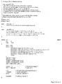


Classifications
-
- A—HUMAN NECESSITIES
- A61—MEDICAL OR VETERINARY SCIENCE; HYGIENE
- A61K—PREPARATIONS FOR MEDICAL, DENTAL OR TOILETRY PURPOSES
- A61K31/00—Medicinal preparations containing organic active ingredients
- A61K31/185—Acids; Anhydrides, halides or salts thereof, e.g. sulfur acids, imidic, hydrazonic or hydroximic acids
- A61K31/19—Carboxylic acids, e.g. valproic acid
- A61K31/20—Carboxylic acids, e.g. valproic acid having a carboxyl group bound to a chain of seven or more carbon atoms, e.g. stearic, palmitic, arachidic acids
- A61K31/203—Retinoic acids ; Salts thereof
-
- C—CHEMISTRY; METALLURGY
- C07—ORGANIC CHEMISTRY
- C07K—PEPTIDES
- C07K16/00—Immunoglobulins [IGs], e.g. monoclonal or polyclonal antibodies
- C07K16/18—Immunoglobulins [IGs], e.g. monoclonal or polyclonal antibodies against material from animals or humans
- C07K16/28—Immunoglobulins [IGs], e.g. monoclonal or polyclonal antibodies against material from animals or humans against receptors, cell surface antigens or cell surface determinants
- C07K16/30—Immunoglobulins [IGs], e.g. monoclonal or polyclonal antibodies against material from animals or humans against receptors, cell surface antigens or cell surface determinants from tumour cells
-
- A—HUMAN NECESSITIES
- A61—MEDICAL OR VETERINARY SCIENCE; HYGIENE
- A61P—SPECIFIC THERAPEUTIC ACTIVITY OF CHEMICAL COMPOUNDS OR MEDICINAL PREPARATIONS
- A61P35/00—Antineoplastic agents
-
- A—HUMAN NECESSITIES
- A61—MEDICAL OR VETERINARY SCIENCE; HYGIENE
- A61P—SPECIFIC THERAPEUTIC ACTIVITY OF CHEMICAL COMPOUNDS OR MEDICINAL PREPARATIONS
- A61P43/00—Drugs for specific purposes, not provided for in groups A61P1/00-A61P41/00
-
- C—CHEMISTRY; METALLURGY
- C12—BIOCHEMISTRY; BEER; SPIRITS; WINE; VINEGAR; MICROBIOLOGY; ENZYMOLOGY; MUTATION OR GENETIC ENGINEERING
- C12Q—MEASURING OR TESTING PROCESSES INVOLVING ENZYMES, NUCLEIC ACIDS OR MICROORGANISMS; COMPOSITIONS OR TEST PAPERS THEREFOR; PROCESSES OF PREPARING SUCH COMPOSITIONS; CONDITION-RESPONSIVE CONTROL IN MICROBIOLOGICAL OR ENZYMOLOGICAL PROCESSES
- C12Q1/00—Measuring or testing processes involving enzymes, nucleic acids or microorganisms; Compositions therefor; Processes of preparing such compositions
- C12Q1/68—Measuring or testing processes involving enzymes, nucleic acids or microorganisms; Compositions therefor; Processes of preparing such compositions involving nucleic acids
- C12Q1/6876—Nucleic acid products used in the analysis of nucleic acids, e.g. primers or probes
- C12Q1/6883—Nucleic acid products used in the analysis of nucleic acids, e.g. primers or probes for diseases caused by alterations of genetic material
- C12Q1/6886—Nucleic acid products used in the analysis of nucleic acids, e.g. primers or probes for diseases caused by alterations of genetic material for cancer
-
- G—PHYSICS
- G01—MEASURING; TESTING
- G01N—INVESTIGATING OR ANALYSING MATERIALS BY DETERMINING THEIR CHEMICAL OR PHYSICAL PROPERTIES
- G01N33/00—Investigating or analysing materials by specific methods not covered by groups G01N1/00 - G01N31/00
- G01N33/48—Biological material, e.g. blood, urine; Haemocytometers
- G01N33/50—Chemical analysis of biological material, e.g. blood, urine; Testing involving biospecific ligand binding methods; Immunological testing
- G01N33/5005—Chemical analysis of biological material, e.g. blood, urine; Testing involving biospecific ligand binding methods; Immunological testing involving human or animal cells
- G01N33/5008—Chemical analysis of biological material, e.g. blood, urine; Testing involving biospecific ligand binding methods; Immunological testing involving human or animal cells for testing or evaluating the effect of chemical or biological compounds, e.g. drugs, cosmetics
- G01N33/502—Chemical analysis of biological material, e.g. blood, urine; Testing involving biospecific ligand binding methods; Immunological testing involving human or animal cells for testing or evaluating the effect of chemical or biological compounds, e.g. drugs, cosmetics for testing non-proliferative effects
- G01N33/5023—Chemical analysis of biological material, e.g. blood, urine; Testing involving biospecific ligand binding methods; Immunological testing involving human or animal cells for testing or evaluating the effect of chemical or biological compounds, e.g. drugs, cosmetics for testing non-proliferative effects on expression patterns
-
- G—PHYSICS
- G01—MEASURING; TESTING
- G01N—INVESTIGATING OR ANALYSING MATERIALS BY DETERMINING THEIR CHEMICAL OR PHYSICAL PROPERTIES
- G01N33/00—Investigating or analysing materials by specific methods not covered by groups G01N1/00 - G01N31/00
- G01N33/48—Biological material, e.g. blood, urine; Haemocytometers
- G01N33/50—Chemical analysis of biological material, e.g. blood, urine; Testing involving biospecific ligand binding methods; Immunological testing
- G01N33/53—Immunoassay; Biospecific binding assay; Materials therefor
- G01N33/574—Immunoassay; Biospecific binding assay; Materials therefor for cancer
-
- G—PHYSICS
- G01—MEASURING; TESTING
- G01N—INVESTIGATING OR ANALYSING MATERIALS BY DETERMINING THEIR CHEMICAL OR PHYSICAL PROPERTIES
- G01N33/00—Investigating or analysing materials by specific methods not covered by groups G01N1/00 - G01N31/00
- G01N33/48—Biological material, e.g. blood, urine; Haemocytometers
- G01N33/50—Chemical analysis of biological material, e.g. blood, urine; Testing involving biospecific ligand binding methods; Immunological testing
- G01N33/68—Chemical analysis of biological material, e.g. blood, urine; Testing involving biospecific ligand binding methods; Immunological testing involving proteins, peptides or amino acids
- G01N33/6854—Immunoglobulins
- G01N33/6857—Antibody fragments
-
- A—HUMAN NECESSITIES
- A61—MEDICAL OR VETERINARY SCIENCE; HYGIENE
- A61K—PREPARATIONS FOR MEDICAL, DENTAL OR TOILETRY PURPOSES
- A61K38/00—Medicinal preparations containing peptides
-
- C—CHEMISTRY; METALLURGY
- C12—BIOCHEMISTRY; BEER; SPIRITS; WINE; VINEGAR; MICROBIOLOGY; ENZYMOLOGY; MUTATION OR GENETIC ENGINEERING
- C12Q—MEASURING OR TESTING PROCESSES INVOLVING ENZYMES, NUCLEIC ACIDS OR MICROORGANISMS; COMPOSITIONS OR TEST PAPERS THEREFOR; PROCESSES OF PREPARING SUCH COMPOSITIONS; CONDITION-RESPONSIVE CONTROL IN MICROBIOLOGICAL OR ENZYMOLOGICAL PROCESSES
- C12Q2600/00—Oligonucleotides characterized by their use
- C12Q2600/136—Screening for pharmacological compounds
-
- G—PHYSICS
- G01—MEASURING; TESTING
- G01N—INVESTIGATING OR ANALYSING MATERIALS BY DETERMINING THEIR CHEMICAL OR PHYSICAL PROPERTIES
- G01N2333/00—Assays involving biological materials from specific organisms or of a specific nature
- G01N2333/435—Assays involving biological materials from specific organisms or of a specific nature from animals; from humans
- G01N2333/475—Assays involving growth factors
-
- G—PHYSICS
- G01—MEASURING; TESTING
- G01N—INVESTIGATING OR ANALYSING MATERIALS BY DETERMINING THEIR CHEMICAL OR PHYSICAL PROPERTIES
- G01N2333/00—Assays involving biological materials from specific organisms or of a specific nature
- G01N2333/82—Translation products from oncogenes
Abstract
Description
본 발명은 암 치료를 위한 약물 표적의 확인에 관한 것이다. 구체적으로, 본 발명은 레티노이드 처리에 의해 선택적으로 증진되는 발현을 보이는 종양 항원의 확인, 및 이러한 항원을 표적화하는 치료 방법에 관한 것이다.The present invention relates to the identification of drug targets for the treatment of cancer. In particular, the present invention relates to the identification of tumor antigens that exhibit expression that is selectively enhanced by retinoid treatment, and to methods of treatment that target such antigens.
종양형성성 형질전환 세포의 비정상적인 생장 및 생존은 정상 세포 항상성을 변화시키는 근본적인 유전적 결함에 기인한다. Wnt 신호전달은 고비율의 인간 암 (적절한 동물 및 세포 배양 모델이 이용가능한 인간암)에서 암의 진행에 기여하는 메카니즘을 나타낸다. Wnt 신호전달 경로의 핵심 성분인 β-카테닌은 전사 인자의 TCF/LEF 패밀리와 상호작용하고 Wnt 표적 유전자의 전사를 활성화시킨다. 최근 연구들은 선종 폴립증 (APC) 종양 억제제와 같은 다수의 단백질 및 액신 (axin)이 Wnt 신호전달 경로의 조절에 관여한다는 것을 밝혔다. 더욱이, APC 또는 β-카테닌에서의 돌연변이가 많은 인간 암을 발생시키는 것으로 밝혀졌다.Abnormal growth and survival of tumorigenic transformed cells is due to a fundamental genetic defect that alters normal cell homeostasis. Wnt signaling represents a mechanism that contributes to cancer progression in high rates of human cancer (human cancers with appropriate animal and cell culture models available). Β-catenin, a key component of the Wnt signaling pathway, interacts with the TCF / LEF family of transcription factors and activates transcription of Wnt target genes. Recent studies have shown that a number of proteins and axins, such as adeno polyposis (APC) tumor inhibitors, are involved in the regulation of the Wnt signaling pathway. Moreover, mutations in APC or β-catenin have been found to cause many human cancers.
예를 들어, 결장직장암의 경우, APC 종양 억제제의 불활성화는 종양 진행에서 초기에 일어나고 시클린 D 및 c-myc과 같은 유전자의 부적절한 활성화로부터 생 기는 생장 이점을 제공한다 (He et al., Science 281:1509-1512 (1998)); Tetsu and McCormick, Nature 38:422-426 (1999)). 이들 유전자는 APC에 의해 정상적으로 하향조절되는 단백질의 일종인 β-카테닌과 상호작용함으로써 활성화되는 LEF/TCF 전사 인자의 표적이다 (Barker et al., Adv Cancer Res 77:1-24 (2000); Polakis, Genes Dev 14:1837-1851 (2000)). 암에서의 이러한 신호전달 경로의 상향조절은 β-카테닌 유전자에서의 미스센스 (missense) 돌연변이로부터 발생될 수도 있다 (Rubinfeld et al., Science 275:1790-1792 (1997)); Korinek et al., Science 275:1784-1787 (1997)). 이 돌연변이들은 β-카테닌이 APC에 의한 하향조절에 불응하게 한다. β-카테닌에서의 돌연변이는 최근에 매우 다양한 인간 종양에서 확인되었고 특히 인간 간세포암에 많다 (상기 문헌 (Polakis, 2000)). β-카테닌 신호전달의 활성화는 세포 표면 프리즐드 (frizzled) 수용체가 분비된 Wnt 리간드에 의해 자극되는 경우에도 일어난다 (Wodarz and Nusse, Annu Rev Cell Dev Biol 14:59-88 (1998)). Wnt 리간드 자체가 인간 암에 기여하는지는 알려져 있지 않지만, 초기 실험들은 뮤린 유선 조직에서의 Wnt 리간드의 과발현이 종양발생원임을 입증하였다 (Nusse and Varmus, Cell 31:99-109 (1982)). Wnt 신호전달 경로는 결장암, 유방암, 위암, 폐암 및 흑색종을 비롯한 다양한 암의 발병기작에 관여한다. 따라서, 대다수의 결장직장 종양은 APC 종양 억제제 또는 β-카테닌을 코딩하는 유전자 내에 돌연변이를 함유한다 (Polakis, Curr. Opin. Genet. Dev. 9:15-21 (1999)). 또한, β-카테닌에서의 활성화 돌연변이는 난소암, 자궁내막암, 윌름스 (Wilm's) 신장 종양 및 흑색종에서 확인되었고, 따라서 Wnt-1 신호전달에서의 결함 이 이들 암의 진행에 기여한다는 것이 입증되었다 (Kobayashi et al., Jpn J Cancer Res 90:55-9 (1999); Koesters et al., Cancer Res 59:3880-2 (1999); Palacios and Hamallo, Cancer Res 58:1344-7 (1998); Rimm et al., Am J Pathol 154:325-9 (1999); Rubinfeld et al., Science 262:1731-1734 (1993); Wright et al., Int J Cancer 82:625-9 (1999)). For example, in colorectal cancer, inactivation of APC tumor suppressors occurs early in tumor progression and provides growth benefits resulting from inappropriate activation of genes such as cyclin D and c-myc (He et al., Science 281: 1509-1512 (1998); Tetsu and McCormick, Nature 38: 422-426 (1999). These genes are targets of LEF / TCF transcription factors that are activated by interacting with β-catenin, a type of protein normally downregulated by APC (Barker et al., Adv Cancer Res 77: 1-24 (2000); Polakis , Genes Dev 14: 1837-1851 (2000)). Upregulation of this signaling pathway in cancer may result from missense mutations in the β-catenin gene (Rubinfeld et al., Science 275: 1790-1792 (1997)); Korinek et al., Science 275: 1784-1787 (1997)). These mutations cause β-catenin to resist downregulation by APC. Mutations in β-catenin have recently been identified in a wide variety of human tumors, especially in human hepatocellular carcinoma (Polakis, 2000, supra). Activation of β-catenin signaling occurs even when cell surface frizzled receptors are stimulated by secreted Wnt ligands (Wodarz and Nusse, Annu Rev Cell Dev Biol 14: 59-88 (1998)). It is not known whether Wnt ligand itself contributes to human cancer, but early experiments have demonstrated that overexpression of Wnt ligand in murine mammary tissue is an oncogenic source (Nusse and Varmus, Cell 31: 99-109 (1982)). The Wnt signaling pathway is involved in the pathogenesis of various cancers, including colon cancer, breast cancer, gastric cancer, lung cancer and melanoma. Thus, the majority of colorectal tumors contain mutations in genes encoding APC tumor inhibitors or β-catenin (Polakis, Curr. Opin. Genet. Dev. 9: 15-21 (1999)). In addition, activating mutations in β-catenin have been identified in ovarian cancer, endometrial cancer, Wilm's kidney tumors, and melanoma, thus demonstrating that defects in Wnt-1 signaling contribute to the progression of these cancers. (Kobayashi et al., Jpn J Cancer Res 90: 55-9 (1999); Koesters et al., Cancer Res 59: 3880-2 (1999); Palacios and Hamallo, Cancer Res 58: 1344-7 (1998) Rimm et al., Am J Pathol 154: 325-9 (1999); Rubinfeld et al., Science 262: 1731-1734 (1993); Wright et al., Int J Cancer 82: 625-9 (1999) .
Wnt 수용체로부터 발산되는 신호는 비정상적인 활성화를 통해 진행되는 것으로 생각되고, 이 신호는 글리코겐 신타제 키나제 3b (GSK3b)를 하향조절한다 (Peifer and Polakis, Science 287:1606-1609 (2000)). 이 키나제는 정상적으로 유비퀴틴-의존성 분해를 위해 단백질을 표적화하는 β-카테닌의 조절 서열을 인산화시킨다 (Miller and Moon, Genes & Dev. 10:2527-2539 (1996)). 따라서, GSK3b의 하향조절은 β-카테닌의 안정성을 증가시키고 TCF/LEF 전사 인자의 활성화를 연장시킨다 (Molenaar, et al., Cell 86:391-399 (1996); Behrens, et al., Nature 382:638-642 (1996)). β-카테닌에 의한 TCF/LEF 전사의 활성화가 잘 확립되어 있지만, 이들 전사 인자와는 무관하면서 β-카테닌이 유전자 활성화에 관여할 수 있는 다른 메카니즘이 있다. 이러한 별도의 메카니즘들 중 하나는 레티노산 수용체 (RAR)에 의한 신호전달과 β-카테닌에 의한 신호전달 사이의 상호작용 가능성을 조사하는 연구에서 바이어스 (Byers)와 그의 동료들에 의해 최근에 제안되었다 (Easwaran, et al., Curr Biol 9:1415-1418 (1999)). 레티노산 반응 요소를 포함하는 합성 레포터 유전자는 배양된 세포주에서 β-카테닌의 과발현 후에 레티노이드에 의해 보다 강하게 활성화되었다.The signal emitted from the Wnt receptor is thought to proceed through abnormal activation, which downregulates glycogen synthase kinase 3b (GSK3b) (Peifer and Polakis, Science 287: 1606-1609 (2000)). This kinase phosphorylates the regulatory sequence of β-catenin, which normally targets proteins for ubiquitin-dependent degradation (Miller and Moon, Genes & Dev. 10: 2527-2539 (1996)). Thus, downregulation of GSK3b increases the stability of β-catenin and prolongs the activation of TCF / LEF transcription factors (Molenaar, et al., Cell 86: 391-399 (1996); Behrens, et al., Nature 382 : 638-642 (1996)). Although activation of TCF / LEF transcription by β-catenin is well established, there are other mechanisms by which β-catenin may be involved in gene activation, independent of these transcription factors. One such separate mechanism has recently been proposed by Byers and his colleagues in a study investigating the potential for interaction between signaling by retinoic acid receptors (RAR) and signaling by β-catenin. (Easwaran, et al., Curr Biol 9: 1415-1418 (1999)). Synthetic reporter genes, including retinoic acid response elements, were more strongly activated by retinoids after overexpression of β-catenin in cultured cell lines.
인간에 사용하거나 마지막 임상 시험 단계에서 사용하도록 승인된 여러 모노클로날 항체 (mAb)를 치료제로 사용하는 것이 증가하고 있다. 동종이식 거부의 치료를 위해 미국 식품의약청 (FDA)에 의해 승인된 첫번째 mAb는 1986년 항-CD3 (OKT3)이었다. 그 후, 특히 1994년 이후로 mAb의 분야에서 발전 속도가 상당히 가속화되고 있기 때문에, 추가로 7개의 mAb가 인간 치료에 사용되도록 승인받았다. 이들 mAb에는 관상 혈관성형술의 합병증의 치료에 사용되는 ReoPro (등록상표) (1994년), 동종이식 거부의 예방에 사용되는 Zenapax (등록상표) (항-CD25) (1997년), B 세포 비-호즈킨 림프종의 치료에 사용되는 Rituxan (등록상표) (항-CD20) (1997), 처음에는 (1998) 크론병의 치료에 사용되었고, 나중에는 (1999) 류마티스성 관절염의 치료에 사용된 Infliximab (등록상표) (항-TNF-α), 및 동종이식 거부의 예방에 사용되는 Simulect (등록상표) (항-CD25) (1998), 호흡기 감염의 치료에 사용되는 Synagis (등록상표) (호흡기 세포융합 바이러스의 항-F 단백질) (1998), 및 HER2 과발현 전이성 유방 종양의 치료에 사용되는 Herceptin (등록상표) (항-HER2/neu) (1998)이 포함된다 (Glennie and Johnson, Immunol Today 21: 403-410 (2000)). There is an increasing use of several monoclonal antibodies (mAb) for therapeutic use in humans or approved for use in the final clinical trial phase. The first mAb approved by the US Food and Drug Administration (FDA) for the treatment of allograft rejection was anti-CD3 (OKT3) in 1986. Since then, especially since 1994, the rate of development has been significantly accelerated in the field of mAbs, an additional seven mAbs have been approved for use in human treatment. These mAbs include ReoPro® (1994) used for the treatment of complications of coronary angioplasty, Zenapax® (anti-CD25) (1997), B cell non- Rituxan® (anti-CD20) (1997), used in the treatment of Hodgkin's lymphoma, was initially used to treat Crohn's disease (1998) and later (1999) to use Infliximab (a treatment for rheumatoid arthritis) ® (anti-TNF-α), and Simulect ® (anti-CD25) (1998), used for the prevention of allograft rejection, Synagis ® (respiratory fusion) used for the treatment of respiratory infections. Anti-F protein of virus) (1998), and Herceptin® (anti-HER2 / neu) (1998) used in the treatment of HER2 overexpressing metastatic breast tumors (Glennie and Johnson, Immunol Today 21: 403). -410 (2000)).
암의 분자생물학적 기초를 이해하고 효과적인 치료법을 개발하기 위한 한 방법은 암세포와 정상 세포간의 유전자 발현의 차이를 찾는 것이다. 정상 세포와 악성 세포 간에 정상-상태 mRNA 수준이 다를 것이라는 가정을 기초로 한 방법들은 차등적으로 발현되는 유전자를 클로닝하는 데 사용된 바 있고 (Zhang et al., Science 276:1268-1272 (1997)), 신규 약물 표적을 찾게 하였다.One way to understand the molecular biological basis of cancer and develop effective therapies is to find differences in gene expression between cancer cells and normal cells. Methods based on the assumption that the steady-state mRNA levels will differ between normal and malignant cells have been used to clone differentially expressed genes (Zhang et al., Science 276: 1268-1272 (1997) , New drug targets were found.
임상에서 인간 암을 치료하는 데 있어서 치료 항체의 입증된 유용성은 최근에 면역요법의 발달 및 개선에 목적을 둔 집중적인 활동을 일축한다. 이상적으로, 이들 요법들은 세포 표면 항원이 신체 전체를 통해 정상 조직 상에 존재하는 수준보다 훨씬 더 높은 수준으로 암세포 상에서 발현될 것을 요구한다. 정상 조직에 비해 종양 상에서의 차등 발현에 대한 이러한 기준은 면역요법과 같은 암치료의 표적으로서 바람직한 것으로 고려되는 항원의 수를 명백히 제한할 것이다. 그러므로, 종양 세포에서의 세포 표면 항원 발현을 선택적으로 상승시키는 방법을 발견하는 것이 바람직하다. 구체적으로, 정상 세포에 비해 암세포의 항원 발현도를 선택적으로 상승시키는 물질이 이들 항원에 대한 면역요법과 같은 치료법의 치료율을 크게 개선시킬 수 있을 것이다.The proven utility of therapeutic antibodies in the treatment of human cancer in the clinic recently dismisses intensive activities aimed at the development and improvement of immunotherapy. Ideally, these therapies require cell surface antigens to be expressed on cancer cells at levels much higher than those present on normal tissue throughout the body. Such criteria for differential expression on tumors as compared to normal tissues will obviously limit the number of antigens considered to be desirable as targets for cancer treatments such as immunotherapy. Therefore, it is desirable to find a method to selectively elevate cell surface antigen expression in tumor cells. Specifically, a substance that selectively raises the antigenic expression of cancer cells compared to normal cells may greatly improve the treatment rate of treatments such as immunotherapy for these antigens.
레티노이드 처리에 의해 선택적으로 상향조절되는 발현을 보이는 종양 항원 을 확인하고, 개선된 암 치료 방법, 특히 레티노산 또는 다른 레티노이드를 투여하여 비정상적인 Wnt 신호전달을 특징으로 하는 암의 치료 효능을 증진시킬 수 있다.Tumor antigens that exhibit expression that is selectively upregulated by retinoid treatment can be identified and improved methods of treating cancer, in particular retinoic acid or other retinoids, can be administered to enhance the therapeutic efficacy of cancers characterized by abnormal Wnt signaling. .
발명의 요약Summary of the Invention
신규 약물 표적은 암세포주 및 조직으로부터 단리된 RNA 전사체의 차등 분석으로 확인할 수 있다. 본 발명을 기본을 이루는 작업에서, 이 방법은 형질전환된 암세포 및 형질전환되지 않은 암세포의 약물 치료로부터 발생되는 유전자 발현의 차이를 분석함으로서 확장될 수 있다. Wnt-1 프로토-온코진을 조건부로 발현하는 유방 상피 세포주를 Wnt-1 발현의 존재 또는 부재 하에 9-시스 레티노산으로 처리하지 않거나 처리하였다. 여러 세포 표면 항원을 비롯한 다수의 유전자가 Wnt-1과 레티노산의 조합작용에 의해 선택적으로 상향조절되었다. 이 관찰결과는 암치료의 효능, 특히 Wnt 신호전달 경로의 수반을 특징으로 하는 암의 면역요법이 레티노산 또는 다른 레티노이드의 동시투여에 의해 상승될 수 있음을 나타낸다.New drug targets can be identified by differential analysis of RNA transcripts isolated from cancer cell lines and tissues. In the work underlying the present invention, the method can be extended by analyzing differences in gene expression resulting from drug treatment of transformed and untransformed cancer cells. Breast epithelial cell lines conditionally expressing Wnt-1 proto-oncozin were treated with or without 9-cis retinoic acid in the presence or absence of Wnt-1 expression. Numerous genes, including several cell surface antigens, were selectively upregulated by the combination of Wnt-1 and retinoic acid. This observation indicates that the efficacy of cancer therapy, in particular the immunotherapy of cancer characterized by the attendance of the Wnt signaling pathway, can be elevated by the simultaneous administration of retinoic acid or other retinoids.
따라서, 한 측면에서, 본 발명은 비정상적인 Wnt 신호전달을 특징으로 하는 종양 세포를 유효량의 레티노이드로 처리하는 것을 포함하는, 상기 종양 세포에서 단백질의 발현을 선택적으로 상승시키는 방법에 관한 것이다.Thus, in one aspect, the present invention is directed to a method of selectively elevating expression of proteins in tumor cells comprising treating tumor cells characterized by abnormal Wnt signaling with an effective amount of retinoids.
구체적인 실시양태에서, 상기 단백질은 Wnt-1 처리와 레티노이드 처리의 병행에 의해 그의 발현이 상승적으로 증진되는 것을 특징으로 한다.In specific embodiments, the protein is characterized in that its expression is synergistically enhanced by a combination of Wnt-1 treatment and retinoid treatment.
바람직한 실시양태에서, 상기 단백질은 상응하는 정상 세포에 비해 종양에서 과발현되는 세포 표면 단백질이다. 예시적인 단백질에는 예컨대, 단백질 4-1BB 리 간드, 에프린 b1, ISLR, 오토탁신 및 Stra6가 포함된다.In a preferred embodiment, said protein is a cell surface protein that is overexpressed in tumor compared to the corresponding normal cell. Exemplary proteins include, for example, protein 4-1BB ligand, ephrin b1, ISLR, autotaxin and Stra6.
치료는 레티노산을 비롯한 임의의 레티노이드에 의해 수행될 수 있다.Treatment can be performed by any retinoid, including retinoic acid.
특정한 실시양태에서, 종양은 인간 암, 예컨대, 난소암, 자궁내막암, 윌름스 (Wilm's) 신장 종양, 결장암, 유방암, 전립선암, 위암, 폐암, 간세포암 또는 흑색종이다.In particular embodiments, the tumor is human cancer, such as ovarian cancer, endometrial cancer, Wilm's kidney tumor, colon cancer, breast cancer, prostate cancer, gastric cancer, lung cancer, hepatocellular carcinoma or melanoma.
또 다른 측면에서, 본 발명은 비정상적인 Wnt 신호전달을 특징으로 하는 종양을 유효량의 레티노이드와 항-종양제로 처리하는 것을 포함하는, 상기 종양의 치료 방법에 관한 것으로, 상기 항-종양제는 Wnt-1 처리와 레티노이드 처리의 병행에 의해 상승적으로 증진되는 발현을 나타내는, 종양 내의 단백질을 표적화한다.In another aspect, the invention relates to a method of treating a tumor, comprising treating a tumor characterized by abnormal Wnt signaling with an effective amount of retinoid and an anti-tumor agent, wherein the anti-tumor agent is Wnt-1. Targets proteins in tumors that exhibit expression that is synergistically enhanced by a combination of treatment and retinoid treatment.
표적화되는 단백질은 바람직하게는 세포 표면 단백질, 예컨대, 단백질 4-1BB 리간드, 에프린 b1, ISLR, 오토탁신 또는 Stra6이다. 레티노이드는 항-종양제의 투여 전, 항-종양제의 투여와 동시에, 또는 항-종양제의 투여 후에 투여될 수 있다.The protein targeted is preferably a cell surface protein such as protein 4-1BB ligand, ephrin b1, ISLR, autotaxin or Stra6. The retinoid can be administered prior to, at the same time as, or after the anti-tumor agent.
바람직한 실시양태에서, 항-종양제는 항체이다. 명세서 전체에 걸쳐, 용어 "항체"는 가장 넓은 의미로 사용되고 Fab, Fab', F(ab')2 및 Fv 단편, 디아보디 (diabody), 단일쇄 항체 분자, 및 항체 단편들로부터 형성된 다중특이적 항체와 같은 항체 단편을 포함한다. 항체는 키메라, 인간화 또는 인간 항체 (항체 단편을 포함함)일 수 있다. In a preferred embodiment, the anti-tumor agent is an antibody. Throughout the specification, the term “antibody” is used in its broadest sense and is multispecific formed from Fab, Fab ', F (ab') 2 and Fv fragments, diabodies, single chain antibody molecules, and antibody fragments. Antibody fragments such as antibodies. The antibody may be a chimeric, humanized or human antibody (including antibody fragments).
치료는 임의로 다른 치료, 예컨대, 화학요법제로의 치료 및(또는) 방사선 치 료를 포함할 수 있다.Treatment may optionally include other treatments, such as treatment with chemotherapeutic agents and / or radiation therapy.
종양은 바람직하게는 예를 들어, 난소암, 자궁내막암, 윌름스 신장 종양, 결장암, 유방암, 전립선암, 위암, 폐암, 간세포암 및 흑색종과 같은 인간암이다.Tumors are preferably human cancers such as, for example, ovarian cancer, endometrial cancer, Wilms' kidney tumor, colon cancer, breast cancer, prostate cancer, gastric cancer, lung cancer, hepatocellular cancer and melanoma.
추가 측면에서, 본 발명은 종양 세포와 레티노이드를 접촉시키고 레티노이드에 의해 선택적으로 상향조절되는 유전자를 찾는 것을 포함하는, 종양 치료용 표적 유전자를 찾는 방법에 관한 것이다. 구체적으로, 표적 유전자는 (a) Wnt 프로토-온코진을 발현하는 세포와 레티노이드를 접촉시키는 단계; (b) 세포의 유전자 발현 프로필을 결정하는 단계; 및 (c) 상응하는 미처리 세포에 비해 레티노이드 처리에 의해 선택적으로 상향조절되는 유전자를 종양 치료용 표적으로서 확인하는 단계로 찾는다.In a further aspect, the present invention relates to a method of finding a target gene for tumor treatment comprising contacting tumor cells with a retinoid and finding a gene that is selectively upregulated by the retinoid. Specifically, the target gene may comprise (a) contacting a retinoid with a cell expressing Wnt proto-oncogene; (b) determining the gene expression profile of the cell; And (c) identifying genes that are selectively upregulated by retinoid treatment as the corresponding untreated cells as targets for tumor treatment.
바람직한 실시양태에서, 종양 세포는 Wnt-1과 더 접촉시키고, 발현이 레티노이드와 Wnt-1에 의해 상승적으로 증진되는 단백질을 찾는다.In a preferred embodiment, tumor cells are further contacted with Wnt-1 and look for proteins whose expression is synergistically enhanced by retinoids and Wnt-1.
또 다른 측면에서, 본 발명은 (a) 종양 샘플을 레티노이드와 인큐베이션시키는 단계; (b) 인큐베이션 전 및 후에 상기 샘플의 유전자 발현 프로필을 결정하는 단계; (c) 발현이 레티노이드에 의해 증진되는 유전자를 찾는 단계; 및 (d) 찾은 유전자를 표적화하는 항-종양제로 환자를 치료하는 단계를 포함하는, 포유동물 대상체에서 종양을 치료하는 방법에 관한 것이다.In another aspect, the present invention provides a method of treating a tumor, comprising: (a) incubating a tumor sample with a retinoid; (b) determining the gene expression profile of the sample before and after incubation; (c) finding a gene whose expression is enhanced by the retinoid; And (d) treating the patient with an anti-tumor agent that targets the found gene.
바람직한 실시양태에서, 상기 샘플은 Wnt-1과 더 인큐베이션시키고, 발현이 레티노이드와 Wnt-1의 조합작용에 의해 상승적으로 증진되는 유전자를 찾는다.In a preferred embodiment, the sample is further incubated with Wnt-1, looking for a gene whose expression is synergistically enhanced by the combination of retinoid and Wnt-1.
상기 실시양태에서와 같이, 항-종양제는 Fab, Fab', F(ab')2 및 Fv 단편과 같은 항체 단편을 비롯한 항체, 디아보디, 단일쇄 항체 분자 또는 항체 단편으로부터 형성된 다중특이적 항체일 수 있다. 항체 또는 항체 단편은 키메라, 인간화 또는 인간 항체일 수 있다.As in the above embodiments, the anti-tumor agent is a multispecific antibody formed from antibodies, diabodies, single chain antibody molecules or antibody fragments, including antibody fragments such as Fab, Fab ', F (ab') 2 and Fv fragments. Can be. The antibody or antibody fragment may be a chimeric, humanized or human antibody.
치료는 임의로 예를 들어, 찾은 단백질에 특이적으로 결합하는 항체에 융합된 세포독성 분자일 수 있는 화학요법제의 투여 및(또는) 방사선 치료를 추가로 포함할 수 있다. The treatment may optionally further comprise, for example, administration of a chemotherapeutic agent and / or radiation therapy, which may be a cytotoxic molecule fused to an antibody that specifically binds to the found protein.
또 다른 측면에서, 본 발명은 레티노이드 치료에 의해 상향조절되는 종양 항원의 검출을 기초로 한, 포유동물에서 비정상적인 Wnt 신호전달과 관련된 암을 진단하는 방법에 관한 것이다. 생물학적 샘플은 예를 들어, 종양 세포 또는 조직일 수 있다.In another aspect, the invention relates to a method of diagnosing cancer associated with abnormal Wnt signaling in a mammal based on the detection of tumor antigens that are upregulated by retinoid therapy. The biological sample can be, for example, tumor cells or tissues.
추가 측면에서, 본 발명은 항-종양제, 및 레티노산과 같은 레티노이드와 함께 상기 항-종양제를 투여하라는 설명서를 포함하는 제품에 관한 것이다. 이 제품은 레티노이드를 추가로 포함할 수 있다.In a further aspect, the invention relates to a product comprising an anti-tumor agent and instructions for administering the anti-tumor agent in combination with a retinoid such as retinoic acid. This product may further comprise a retinoid.
<바람직한 실시양태의 상세한 설명><Detailed Description of the Preferred Embodiments>
A. 정의 A. Definition
달리 정의하지 않는 한, 본 명세서에 사용된 기술적 및 과학적 용어는 본 발명이 속하는 기술분야에서 당업자에 의해 일반적으로 이해되는 바와 동일한 의미를 갖는다. 예를 들어, 문헌 [Singleton et al., Dictionary of Microbiology and Molecular Biology 2nd ed., J. Wiley & Sons (New York, NY 1994); Sambrook et al., Molecular Cloning, A Laboratory Manual, Cold Springs Harbor Press (Cold Springs Harbor, NY 1989)]을 참조한다. 본 발명의 목적상, 하기 용어들은 아래와 같이 정의된다.Unless defined otherwise, technical and scientific terms used herein have the same meaning as commonly understood by one of ordinary skill in the art to which this invention belongs. See, eg, Singleton et al., Dictionary of Microbiology and Molecular Biology 2nd ed., J. Wiley & Sons (New York, NY 1994); Sambrook et al., Molecular Cloning, A Laboratory Manual, Cold Springs Harbor Press (Cold Springs Harbor, NY 1989). For the purposes of the present invention, the following terms are defined below.
용어 "PR010282 폴리펩티드", "PR010282 단백질", "PR010282", "Stra6 폴리펩티드", "Stra6 단백질" 및 "Stra6"는 상호교환하여 사용되고, 천연 서열 PR010282 (Stra6) 및 PR010282 (Stra6) 폴리펩티드 변이체 (본 명세서에 더 정의되어 있음)를 포함한다. PR010282 (Stra6) 폴리펩티드는 인간 조직 타입 또는 또다른 공급원과 같은 다양한 공급원으로부터 단리하거나 재조합 및(또는) 합성 방법으로 제조할 수 있다.The terms “PR010282 polypeptide”, “PR010282 protein”, “PR010282”, “Stra6 polypeptide”, “Stra6 protein” and “Stra6” are used interchangeably and refer to the native sequences PR010282 (Stra6) and PR010282 (Stra6) polypeptide variants (herein More defined in). PR010282 (Stra6) polypeptides can be isolated from a variety of sources, such as human tissue types or another source, or prepared by recombinant and / or synthetic methods.
"천연 서열 PR010282" 또는 "천연 서열 Stra6"는 아미노산 서열이 천연 PR010282와 동일한 폴리펩티드를 포함한다. 이러한 천연 서열 PR010282 (Stra6)는 천연 공급원으로부터 단리할 수 있거나 재조합 및(또는) 합성 방법으로 제조할 수 있다. 용어 "천연 서열 PR010282" 또는 "천연 서열 Stra6"는 특히, 자연적으로 발생하는 말단-절단된 형태 또는 분비된 형태 (예컨대, 세포외 도메인 서열), 자연-발생 변이체 형태 (예를 들어, 얼터네이티브-스플라이싱 형태) 및 PR010282의 자연-발생적 대립유전자 변이체를 포함한다. 본 발명의 한 실시양태에서, 천연 서열 PR010282는 도 2 (서열 2)의 아미노산 1 내지 667을 포함하는 성숙 또는 전장 천연 서열 PR010282이다. 본 발명의 또다른 실시양태에서, 천연 서열 PR010282 폴리펩티드는 서열 5의 아미노산 1 내지 658을 포함하는 성숙 또는 전장 PRO19578 폴리펩 티드이고, 이 폴리펩티드는 서열 2의 천연 서열 PR010282의 얼터네이티브 스플라이싱 형태인 것으로 생각된다. 도 2 (서열 2) 및 도 7 (서열 5)에 개시된 PR010282 폴리펩티드는 본 명세서에서 아미노산 위치 1로 지칭된 메티오닌 잔기로 시작되지만, 도 2 (서열 2) 또는 도 7 (서열 5)에서 아미노산 위치 1로부터 상류 또는 하류에 위치한 또다른 메티오닌 잔기가 PR010282 폴리펩티드의 개시 아미노산 잔기로 사용될 수 있는 가능성이 있다. 5' 말단에서 얼터네이티브 스플라이싱될 수 있는 모든 Stra6 분자도 본 명세서의 정의에 포함된다."Native sequence PR010282" or "natural sequence Stra6" includes polypeptides whose amino acid sequence is identical to native PR010282. Such native sequence PR010282 (Stra6) may be isolated from natural sources or may be prepared by recombinant and / or synthetic methods. The terms “natural sequence PR010282” or “natural sequence Stra6” in particular refer to naturally occurring, terminally-cleaved or secreted forms (eg, extracellular domain sequences), naturally-occurring variant forms (eg, alternatively- Splicing forms) and naturally-occurring allelic variants of PR010282. In one embodiment of the invention, native sequence PR010282 is mature or full length native sequence PR010282 comprising
PR010282 (Stra6) 폴리펩티드 "세포외 도메인" 또는 "ECD"란 본질적으로 막횡단 또는 세포질 도메인이 없는 PR010282 (Stra6) 폴리펩티드의 형태를 의미한다. 임의로, PR010282 (Stra6) 폴리펩티드 ECD는 이러한 막횡단 및(또는) 세포질 도메인을 1% 미만으로 포함하며, 바람직하게는 상기 도메인들을 0.5% 미만으로 포함한다. 본 발명의 PR010282 폴리펩티드에서 확인된 임의의 막횡단 도메인은 당업계에서 소수성 도메인 유형을 밝히는 데 통상적으로 사용되는 기준에 따라 확인된 것임을 이해해야 할 것이다. 막횡단 도메인의 정확한 경계는 다를 수 있지만, 대부분은 본원에서 처음 확인된 도메인의 말단에서 5개 아미노산 범위 내에 존재한다. 따라서, 본 발명의 한 실시양태에서, PR010282 폴리펩티드의 세포외 도메인은 X가 도 2 (서열 2) 또는 도 7 (서열 5)의 아미노산 49 내지 59 중 임의의 아미노산인 아미노산 1 내지 X를 포함한다.PR010282 (Stra6) Polypeptide “Extradomain” or “ECD” refers to a form of PR010282 (Stra6) polypeptide that is essentially free of transmembrane or cytoplasmic domains. Optionally, the PR010282 (Stra6) polypeptide ECD comprises less than 1% of such transmembrane and / or cytoplasmic domains, preferably less than 0.5% of such domains. It should be understood that any transmembrane domain identified in the PR010282 polypeptide of the invention has been identified according to criteria commonly used to identify hydrophobic domain types in the art. The exact boundaries of the transmembrane domains may vary, but most are within the 5 amino acid range at the ends of the domains first identified herein. Thus, in one embodiment of the invention, the extracellular domain of the PR010282 polypeptide comprises
상호교환가능하게 사용되는 용어 "PR010282 변이체 폴리펩티드" 또는 "Stra6 변이체 폴리펩티드"는 (a) 도 2 (서열 2)에 나타낸 PR010282 폴리펩티드의 잔기 1 내지 667 또는 도 7 (서열 5)의 잔기 1 내지 658, (b) 도 2 (서열 2) 또는 도 7 (서열 5)의 잔기 1 내지 X (여기서, X는 도 2 (서열 2) 또는 도 7 (서열 5)의 아미노산 잔기 49 내지 59 중 어느 하나의 아미노산임), 또는 (c) 도 2 (서열 2) 또는 도 7 (서열 5)에 나타낸 아미노산 서열로부터 유도된 또다른 특정 단편의 아미노산 서열과의 아미노산 서열 동일성이 약 80% 이상인 하기 정의된 활성 PR010282 (Stra6) 폴리펩티드를 의미한다. 이러한 PR010282 (Stra6) 변이체 폴리펩티드로는 예컨대, 하나 이상의 아미노산 잔기가 도 2 (서열 2) 또는 도 7 (서열 5)의 서열의 N- 및(또는) C-말단 뿐만 아니라 하나 이상의 내부 도메인 내에 부가되어 있거나 결실되어 있는 PR010282 (Stra6) 폴리펩티드가 있다. 통상적으로, PR010282 변이체 폴리펩티드는 (a) 도 2 (서열 2)에 나타낸 PR010282 폴리펩티드의 잔기 1 내지 667 또는 도 7 (서열 5)의 잔기 1 내지 658, (b) 도 2 (서열 2) 또는 도 7 (서열 5)의 잔기 1 내지 X (여기서, X는 도 2 (서열 2) 또는 도 7 (서열 5)의 아미노산 잔기 49 내지 59 중 어느 하나의 아미노산임), 또는 (c) 도 2 (서열 2) 또는 도 7 (서열 5)에 나타낸 아미노산 서열로부터 유래된 또다른 특정 단편과의 아미노산 서열 동일성이 약 80% 이상, 바람직하게는 약 81% 이상, 보다 바람직하게는 약 82% 이상, 보다 바람직하게는 약 83% 이상, 보다 바람직하게는 약 84% 이상, 보다 바람직하게는 약 85% 이상, 보다 바람직하게는 약 86% 이상, 보다 바람직하게는 약 87% 이상, 보다 바람직하게는 약 88% 이상, 보다 바람직하게는 약 89% 이상, 보다 바람직하게는 약 90% 이상, 보다 바람직하게는 약 91% 이상, 보다 바람직하게는 약 92% 이상, 보다 바람직하게는 약 93% 이상, 보다 바람직하게는 약 94% 이상, 보다 바람 직하게는 약 95% 이상, 보다 바람직하게는 약 96% 이상, 보다 바람직하게는 약 97% 이상, 보다 바람직하게는 약 98% 이상, 보다 바람직하게는 약 99% 이상일 것이다. PR010282 변이체 폴리펩티드는 천연 PR010282 폴리펩티드 서열을 포함하지 않는다. 통상적으로, PR010282 변이체 폴리펩티드의 길이는 약 10개 아미노산 이상, 약 20개 아미노산 이상, 바람직하게는 약 30개 아미노산 이상, 바람직하게는 약 40개 아미노산 이상, 바람직하게는 약 50개 아미노산 이상, 바람직하게는 약 60개 아미노산 이상, 바람직하게는 약 70개 아미노산 이상, 바람직하게는 약 80개 아미노산 이상, 바람직하게는 약 90개 아미노산 이상, 바람직하게는 약 100개 아미노산 이상, 바람직하게는 약 150개 아미노산 이상, 바람직하게는 약 200개 아미노산 이상, 바람직하게는 약 250개 아미노산 이상, 보다 바람직하게는 약 300개 아미노산 이상이고 문헌 (Bouillet et al., Mechanisms of Development 63, 173-186 (1997))에 개시된 뮤린 Stra6 서열의 단편과는 다르다. 본 명세서에 개시된 다른 폴리펩티드의 변이체도 유사한 방식으로 정의된다.As used interchangeably, the terms “PR010282 variant polypeptide” or “Stra6 variant polypeptide” refer to (a)
"아미노산 서열 동일성(%)"은 서열을 정렬하고, 필요하다면, 최대 서열 동일성(%)을 얻기 위해 갭 (gap)을 도입한 후 임의의 보존적인 치환을 서열 동일성의 일부로서 고려하지 않은 상태에서 해당 서열 (예를 들어, 천연 폴리펩티드 서열)의 아미노산 잔기와 동일한, 후보 서열의 아미노산 잔기의 백분율로서 정의된다. 아미노산 서열 동일성(%)을 결정하기 위한 정렬은 당분야의 기술에 속하는 다양한 방법, 예컨대, BLAST, BLAST-2, ALIGN, ALIGN-2 또는 메갈린 (Megalign; DNASTAR) 소프트웨어와 같은 공개적으로 이용가능한 컴퓨터 소프트웨어를 사용하여 달성할 수 있다. 당업자는 비교될 전장 서열의 최대 정렬을 달성하는 데 필요한 임의의 알고리즘을 비롯하여 정렬 측정에 적합한 파라미터를 결정할 수 있다. 그러나, 본원의 목적을 위해, 아미노산 서열 동일성(%)은 서열 비교 컴퓨터 프로그램 ALIGN-2을 이용하여 아래에 기재된 바와 같이 얻는데, 여기서, ALIGN-2 프로그램용 완전 원시 코드는 하기 표 1에 기재되어 있다. ALIGN-2 서열 비교 컴퓨터 프로그램은 제넨텍, 인크사에 의해 개발되었고, 표 1에 나타낸 원시 코드는 미국 저작권청 (Washington D. C., 20559)에 사용자 문서로 등록되어 있고, 미국 저작권 등록번호는 TXU510087이다. ALIGN-2 프로그램은 제넨텍, 인크사 (South San Francisco, California)를 통해 공적으로 이용가능하거나, 표 1에 기재된 원시 코드로부터 편집할 수 있다. ALIGN-2 프로그램은 UNIX 작업 시스템, 바람직하게는 디지탈 UNIX V4.0D 상에서 편집되어 사용될 수 있다. 모든 서열 비교 파라미터는 ALIGN-2 프로그램에 의해 설정되고 변하지 않는다."Amino acid sequence identity (%)" refers to aligning a sequence and, if necessary, introducing a gap to obtain the maximum sequence identity (%) and then not considering any conservative substitution as part of the sequence identity. It is defined as the percentage of amino acid residues in a candidate sequence that are identical to amino acid residues in that sequence (eg, native polypeptide sequence). Alignment to determine percent amino acid sequence identity can be performed in various ways within the art, for example by publicly available computers such as BLAST, BLAST-2, ALIGN, ALIGN-2 or Megaalign (DNASTAR) software. It can be achieved using software. Those skilled in the art can determine appropriate parameters for measuring alignment, including any algorithms needed to achieve maximal alignment of the full length sequences to be compared. However, for purposes herein,% amino acid sequence identity is obtained using the sequence comparison computer program ALIGN-2, as described below, where the complete source code for the ALIGN-2 program is described in Table 1 below. . The ALIGN-2 sequence comparison computer program was developed by Genentech, Inc., and the source code shown in Table 1 is registered as a user document with the United States Copyright Office (Washington D. C., 20559), and the United States copyright registration number is TXU510087. The ALIGN-2 program is publicly available through Genentech, Inc. (South San Francisco, California) or can be edited from the source code listed in Table 1. The ALIGN-2 program can be edited and used on a UNIX working system, preferably digital UNIX V4.0D. All sequence comparison parameters are set by the ALIGN-2 program and do not change.
본원의 목적상, 해당 아미노산 서열 B에, B와, 또는 B에 대한 해당 아미노산 서열 A의 아미노산 서열 동일성 (%)(해당 아미노산 서열 B에, B와, 또는 B에 대한 일정한 아미노산 서열 동일성 (%)을 갖거나 포함하는 해당 아미노산 서열 A로 달리 표현할 수 있음)은 하기와 같이 계산한다.For purposes herein, the amino acid sequence identity (%) of the corresponding amino acid sequence A to B, or B to that amino acid sequence B (constant amino acid sequence identity to the corresponding amino acid sequence B, B, or B (%) It can be expressed differently by the amino acid sequence A having or including)) is calculated as follows.
X/Y ×100X / Y × 100
여기서, X는 A와 B의 프로그램 정렬에서 서열 정렬 프로그램 ALIGN-2에 의해 동일하게 매치되는 것으로 기록되는 아미노산 잔기의 수이고, Y는 B에서 아미노산 잔기의 총 수이다. 아미노산 서열 A의 길이가 아미노산 서열 B의 길이와 같지 않 는 경우, B에 대한 A의 아미노산 서열 동일성 (%)은 A에 대한 B의 아미노산 서열 동일성 (%)과 같지 않음을 인지해야 할 것이다. 아미노산 서열 동일성 계산의 예로서, 하기 표 2 및 3은 "PRO"로 지칭되는 아미노산 서열에 대한 비교 단백질로 지칭되는 아미노산 서열의 아미노산 서열 동일성 (%)을 계산하는 방법을 나타낸다.Where X is the number of amino acid residues recorded as equally matched by the sequence alignment program ALIGN-2 in the program alignment of A and B, and Y is the total number of amino acid residues in B. If the length of amino acid sequence A is not equal to the length of amino acid sequence B, it will be appreciated that the amino acid sequence identity (A) of A to B is not equal to the amino acid sequence identity (B) of B to A. As examples of amino acid sequence identity calculations, Tables 2 and 3 below show how to calculate amino acid sequence identity (%) of an amino acid sequence referred to as a comparative protein to an amino acid sequence referred to as "PRO".
달리 명시하지 않는 한, 모든 아미노산 서열 동일성(%)은 ALIGN-2 서열 비교 컴퓨터 프로그램을 사용하여 상기와 같이 얻을 수 있다. 그러나, 아미노산 서열 동일성 (%)은 서열 비교 프로그램 NCBI-BLAST2를 이용하여 계산할 수도 있다 (Altschul et al., Nucleic Acids Res. 25: 3389-3402 (1997)). 상기 NCBI-BLAST2 서열 비교 프로그램은 웹 사이트 (http://www.ncbi.nlm.nih.gov.)로부터 다운로드 받을 수 있다. NCBI-BLAST2는 여러 가지 검색 파라미터를 이용하는데, 이러한 모든 검색 파라미터는 예를 들어, 언마스크 = 예, 가닥 = 모두, 기대 발생 = 10, 최소 저복합성 길이 = 15/5, 다중-통과 e-값 = 0.01, 다중-통과에 대한 상수 = 25, 최종 갭 정렬에 대한 드롭오프 = 25 및 점수 매트릭스 = BLOSUM62를 포함하는 디폴트값으로 설정된다.Unless otherwise specified, all amino acid sequence identity (%) can be obtained as above using the ALIGN-2 sequence comparison computer program. However,% amino acid sequence identity can also be calculated using the sequence comparison program NCBI-BLAST2 (Altschul et al., Nucleic Acids Res. 25: 3389-3402 (1997)). The NCBI-BLAST2 sequence comparison program can be downloaded from the website (http://www.ncbi.nlm.nih.gov.). NCBI-BLAST2 uses several search parameters, all of which are, for example, unmask = yes, strand = all, expected occurrence = 10, minimum low complexity length = 15/5, multi-pass e-value = 0.01, constant for multi-pass = 25, dropoff for final gap alignment = 25 and score matrix = BLOSUM62.
NCBI-BLAST2가 아미노산 서열 비교에 사용되는 경우, 해당 아미노산 서열 B에, B와, 또는 B에 대한 해당 아미노산 서열 A의 아미노산 서열 동일성 (%)(해당 아미노산 서열 B에, B와, 또는 B에 대한 일정한 아미노산 서열 동일성 (%)을 갖거나 포함하는 해당 아미노산 서열 A로 달리 표현할 수 있음)은 하기와 같이 계산한다.When NCBI-BLAST2 is used for amino acid sequence comparison, the amino acid sequence identity (%) of the corresponding amino acid sequence A to B, or to B (in the corresponding amino acid sequence B, B, or B) Or alternatively represented by the corresponding amino acid sequence A having or including a certain percentage of amino acid sequence identity) is calculated as follows.
X/Y ×100X / Y × 100
여기서, X는 NCBI-BLAST2 프로그램 정렬에서 서열 정렬 프로그램 NCBI-BLAST2에 의해 동일하게 매칭되는 것으로 기록되는 아미노산 잔기의 수이고, Y는 B에서 아미노산 잔기의 총 수이다. 아미노산 서열 A의 길이가 아미노산 서열 B의 길이와 같지 않는 경우, B에 대한 A의 아미노산 서열 동일성 (%)은 A에 대한 B의 아미노산 서열 동일성 (%)과 같지 않음을 인지해야 할 것이다.Where X is the number of amino acid residues recorded as identically matched by the sequence alignment program NCBI-BLAST2 in the NCBI-BLAST2 program alignment, and Y is the total number of amino acid residues in B. If the length of amino acid sequence A is not equal to the length of amino acid sequence B, it will be appreciated that the amino acid sequence identity (%) of A to B is not equal to the amino acid sequence identity (%) of B to A.
용어 "4-1BB 리간드" 및 ""4-1BBL"은 상호교환적으로 사용되고 전형적으로 항원 제시 세포 상에서 발현되는 종양 괴사 인자 수퍼패밀리 (superfamily)에 속하는 천연 서열 막단백질 및 그의 변이체를 말한다. 천연 인간 4-1BB 리간드의 핵산 및 이 핵산으로부터 유도된 아미노산 서열은 문헌 [Zhou et al., Immunol Lett 45:67-73 (1995)]에 개시되어 있다. 천연 서열 4-1BB 리간드의 아미노산 서열 변이체는 서열 동일성을 결정하는 동일한 알고리즘을 이용하여 상기 Stra6와 유사하게 정의한다. 4-1BBL (4-1BB)의 수용체에 대한 항체는 마우스에서 확립된 종양을 제거하고, 4-1BB T 세포 자극 경로는 항종양 면역반응의 증폭에 관여하는 것으로 밝혀졌다.The terms “4-1BB ligand” and “4-1BBL” are used interchangeably and refer to native sequence membrane proteins and variants thereof belonging to the tumor necrosis factor superfamily that are typically expressed on antigen presenting cells. Nucleic acids of 4-1BB ligand and amino acid sequences derived from these nucleic acids are disclosed in Zhou et al., Immunol Lett 45: 67-73 (1995) Amino acid sequence variants of the native sequence 4-1BB ligand are sequenced The same algorithm for determining identity is defined similarly to Stra6 above: antibodies to receptors of 4-1BBL (4-1BB) eliminate tumors established in mice and 4-1BB T cell stimulation pathways are anti-tumor It has been shown to be involved in amplifying the immune response.
본 명세서에서 용어 "에프린 b1"은 천연 서열 에프린 b1 분자 ("lerk2"로도 알려져 있음), 및 Stra6에 대해 상술한 바와 같이 정의되는 그의 변이체를 말하는 데 사용된다. 인간 에프린 b1은 분자량이 45 kDa이고, 24개의 아미노산으로 구성된 신호 서열, 211개의 아미노산으로 구성된 세포외 영역, 26개의 아미노산으로 구성된 막횡단 (TM) 영역 및 83개의 아미노산으로 구성된 세포질 단편을 포함하는, 346개의 아미노산으로 구성된 글리코실화 폴리펩티드이다 [Davis, et al., Science 266:816 (1994); Beckmann, et al., EMBO J 13:3657 (1994)]. 마우스 에프린 b1 폴리펩티드의 세포외 영역과 인간 에프린 b1 폴리펩티드의 세포외 영역 간에는 약 95%의 아미노산 서열 동일성이 있다. 에프린 b1 상의 잠재적 단백질분해 절단 부위가 확인된 바 있다 [상기 문헌 (Beckmann et al.)]. 혈관신생 및 종양 진행에 있어서의 에프린의 역할은 예를 들어, 문헌 [L'Allemain et al., Bull Cancer 87:529-530 (2000)]에 논의되어 있다.The term "ephrin b1" is used herein to refer to the native sequence ephrin b1 molecule (also known as "lerk2"), and variants thereof as defined above for Stra6. Human ephrin b1 has a molecular weight of 45 kDa and comprises a signal sequence of 24 amino acids, an extracellular region of 211 amino acids, a transmembrane (TM) region of 26 amino acids and a cytoplasmic fragment of 83 amino acids , A glycosylated polypeptide consisting of 346 amino acids [Davis, et al., Science 266: 816 (1994); Beckmann, et al., EMBO J 13: 3657 (1994). There is about 95% amino acid sequence identity between the extracellular region of the mouse ephrin b1 polypeptide and the extracellular region of the human ephrin b1 polypeptide. Potential proteolytic cleavage sites on ephrin b1 have been identified (Beckmann et al., Supra). Ephrin's role in angiogenesis and tumor progression is discussed, for example, in L'Allemain et al., Bull Cancer 87: 529-530 (2000).
용어 "ISLR", "류신 풍부 반복부를 포함하는 이뮤노글로불린 수퍼패밀리" 및 "LRR을 포함하는 이뮤노글로불린 수퍼패밀리"는 상호교환적으로 사용되고, 상기 정의된 바와 같이 천연 서열 ISRL 단백질 및 이의 변이체를 말한다. 천연 인간 ISLR의 클로닝 및 시퀀싱은 문헌 [Nagasawa et al., Genomics 44:173-179 (1997)]에 의해 보고되었다. 다른 인간 및 마우스 ISLR 유전자는 문헌 [Nagasawa et al., Genomics 61:37-43 (1999)]에 기재되어 있다.The terms "ISLR", "immunoglobulin superfamily comprising leucine rich repeats" and "immunoglobulin superfamily comprising LLR" are used interchangeably and refer to the native sequence ISRL protein and variants thereof as defined above. Say. Cloning and sequencing of native human ISLRs was reported by Nagasawa et al., Genomics 44: 173-179 (1997). Other human and mouse ISLR genes are described in Nagasawa et al., Genomics 61: 37-43 (1999).
용어 "오토탁신" 및 "ATX"는 상호교환적으로 사용되고, 임의의 동물 종의 천연 서열 오토탁신 분자 및 상기 정의된 오토탁신 변이체를 말한다. 오토탁신은 혈장 세포막 분화 항원 PC-1과의 높은 상동성을 보이는 암-관련 자가분비 운동성 인자이다. 인간 오토탁신은 분자량이 125 kDa이고, 인간 흑색종 세포주로부터 처음 단리된, 915개의 아미노산으로 구성된 당단백질이다 [Murata et al., J Biol Chem 269:30479-84 (1994)]. 인간 기형암종 세포로부터 오토탁신을 클로닝하는 것은 문헌 [Lee et al., Biochem Biophys Res Commun 218:714-719 (1996)]에 의해 보고된 바 있다. 오토탁신은 ATP의 베타-포스페이트의 한 측면 상에 있는 포스포디에스테 르 결합의 가수분해를 촉진하고 GTP가 GDP 및 GMP로 가수분해되는 것을 촉진하고, AMP 또는 PPi가 Pi로 가수분해되는 것을 촉진하고, NAD가 AMP로 가수분해되는 것을 촉진하는 것으로 기재되어 있다. 이들 기질 각각은 자가독소의 인산화에서 포스페이트 공여자로서 작용할 수 있다. 자가독소는 ras-형질전환 세포의 침윤력 및 전이력을 증가시키는 강력한 종양 모토겐 (motogen)인 것으로 생각된다 [Nam et al., Oncogene 19:241-247 (2000)].The terms "autotaxin" and "ATX" are used interchangeably and refer to the native sequence autotaxin molecule of any animal species and autotaxin variants as defined above. Autotaxin is a cancer-associated autosecretory motility factor that shows high homology with plasma cell membrane differentiation antigen PC-1. Human autotaxin is a glycoprotein consisting of 915 amino acids with a molecular weight of 125 kDa and first isolated from human melanoma cell lines (Murata et al., J Biol Chem 269: 30479-84 (1994)). Cloning autotaxin from human teratocarcinoma cells has been reported by Lee et al., Biochem Biophys Res Commun 218: 714-719 (1996). Autotaxin promotes hydrolysis of phosphodiester bonds on one side of beta-phosphate of ATP, promotes GTP hydrolysis to GDP and GMP, promotes AMP or PPi hydrolysis to Pi, , NAD is described to promote hydrolysis to AMP. Each of these substrates can act as a phosphate donor in the phosphorylation of autotoxins. Autotoxins are thought to be potent tumor motogens that increase the invasive and metastatic capacity of ras-transformed cells [Nam et al., Oncogene 19: 241-247 (2000)].
용어 "레티노이드"는 광범위한 의미로 사용되고, 구체적으로 레티노산 (트레티노인, 비타민 A 산 또는 비타민 A1으로도 공지되어 있음), 헤드-투-테일 방식으로 연결된 4개의 이소프레노이드 단위로 구성된 레티노산 유도체 (예를 들어, 레티놀, 레티날, 치환 레티노이드, 세코-레티노이드, 노르-레티노이드 및 레트로-레티노이드)를 포함하나 이에 제한되지 않는다. 모든 레티노이드는 형식적으로 5개의 탄소-탄소 이중 결합을 포함하고 아실 부위 말단에 관능기를 포함하는 모노시클릭 모화합물로부터 유도된다. 레티노이드의 명명법에 대해서는 문헌 [Moss, G.P., Arch. Biochem. Biophys., 224:728-731 (1983); Eur. J. Biochem., 129:1-5 (1982); J. Biol. Chem., 258:5329-5333 (1983); Pure Appl. Chem., 55:721-726 (1983); Biochemical Nomenclature and Related Documents, 2nd edition, Portland Press, 1992, pages 247-251]을 참조한다. 용어 "레티노이드"에는 구체적으로 레티노이드의 생물학적 활성을 보유하는, 자연계에서 발견되는 분자 및 그의 유도체가 포함된다.The term "retinoid" is used in a broad sense and is specifically a retinoic acid derivative consisting of four isoprenoid units connected in a retinoic acid (also known as tretinoin, vitamin A acid or vitamin A 1 ), head-to-tail manner. (Eg, retinol, retinal, substituted retinoids, seco-retinoids, nor-retinoids and retro-retinoids). All retinoids are formally derived from monocyclic parent compounds containing five carbon-carbon double bonds and functional groups at the acyl site ends. For nomenclature of retinoids, see Moss, GP, Arch. Biochem. Biophys., 224: 728-731 (1983); Eur. J. Biochem., 129: 1-5 (1982); J. Biol. Chem., 258: 5329-5333 (1983); Pure Appl. Chem., 55: 721-726 (1983); Biochemical Nomenclature and Related Documents, 2nd edition, Portland Press, 1992, pages 247-251. The term "retinoid" includes molecules found in nature and derivatives thereof that specifically retain the biological activity of the retinoid.
본 명세서에 정의된 용어 "레티노이드의 생물학적 활성"은 배아발생, (상피) 세포 증식, 세포 분화 및(또는) 암발생, 특히 비정상적인 Wnt 신호전달을 특징으로 하는 암과 관련된 종양발생 효과에 관여하는 활성을 말한다. The term "biological activity of retinoids" as defined herein refers to activities that are involved in the oncogenic effects associated with cancer characterized by embryonic development, (epithelial) cell proliferation, cell differentiation and / or cancer development, in particular abnormal Wnt signaling. Say
용어 "Wnt"는 초기 배아발생의 가장 중요한 측면에 관여하는 고도로 보존된 시스테인 풍부 분비 당단백질의 패밀리를 말한다. Wnt 유전자에는 암에도 관여한다. 본 명세서에 사용된 "Wnt"에는 구체적으로 모든 인간 및 비-인간 동물 종 (인간, 마우스, 래트 및 다른 설치류 등과 같은 포유동물을 포함하나 이에 제한되지 않음)의 Wnt 유전자가 포함된다. 용어 "Wnt"에는 구체적으로 천연 인간 Wnt 유전자, 및 코딩된 폴리펩티드 [인간 Wnt-1 (앞서 int-1으로 지칭됨) (van Ooyen et al., EMBO J 4:2905-9 (1985)); Wnt-2 (앞서 Dint-1으로 지칭됨) (Wainwright et al., EMBO J 7:1743-1748 (1988)); Wnt-13 (Katoh et al., Oncogene 13:873-876 (1996)); Wnt-3 (Roelink et al., Genomics 17:790-792 (1993)); Huguet et al., Cancer Res 54:2615-2521 (1994)); Wnt-4 (Huguet et al., 1994, supra); Wnt-5A (Clark et al., Genomics 18:249-260 (1993)); Wnt-6 (Rankin et al., Cytogenet Cell Genet 84:50-52 (1999)); Wnt-7A (Ikegawa et al., Cytogenet Cell Genet 74:149-152 (1996)); Wnt-7B (Huguet et al., 1994, supra); Wnt-8B (Lako et al., Genomics 35:386-388 (1996)); Wnt-10B (Bui et al., Oncogene 14:1249-1253 (1997)); Wnt-11 (Lako et al., Gene 219:101-110 (1998)); Wnt-14 (Bergstein et al., Genomics 46:450-458 (1997)); and Wnt-16 (McWhirter et al., Proc Natl Acad Sci USA 96:11464-11469 (1999); Fear et al., Biochem Biophys Res Commun 278:814-820 (2000))]와 그의 변이체, 특히 천연 Wnt 폴리펩티드와의 아미노산 서열 상동성이 약 80% 이상, 보다 바람직하게는 약 85% 이상, 보다 바람직하게는 약 90% 이상, 훨씬 더 바람직하게는 약 95% 이상, 훨씬 더 바람직하게는 약 98% 이상, 가장 바람직하게는 약 99% 이상인 아미노산 서열 변이체가 포함되는데, 여기서 서열 상동성은 상기 정의한 바와 같이 측정된다. 바람직한 실시양태에서, 용어 "Wnt"는 문헌 [Wong et al., Mol Cell Biol 14:6278-86 (1994)]에 기재된 바와 같은 임의의 형질전환 (종양발생성) Wnt 폴리펩티드를 말한다.The term "Wnt" refers to a family of highly conserved cysteine rich secreted glycoproteins that are involved in the most important aspects of early embryonic development. The Wnt gene is also involved in cancer. As used herein, "Wnt" specifically includes the Wnt genes of all human and non-human animal species (including but not limited to mammals such as humans, mice, rats, and other rodents). The term "Wnt" specifically includes a natural human Wnt gene, and a encoded polypeptide (human Wnt-1 (previously referred to as int-1) (van Ooyen et al., EMBO J 4: 2905-9 (1985)); Wnt-2 (previously referred to as Dint-1) (Wainwright et al., EMBO J 7: 1743-1748 (1988)); Wnt-13 (Katoh et al., Oncogene 13: 873-876 (1996)); Wnt-3 (Roelink et al., Genomics 17: 790-792 (1993)); Huguet et al., Cancer Res 54: 2615-2521 (1994)); Wnt-4 (Huguet et al., 1994, supra); Wnt-5A (Clark et al., Genomics 18: 249-260 (1993)); Wnt-6 (Rankin et al., Cytogenet Cell Genet 84: 50-52 (1999)); Wnt-7A (Ikegawa et al., Cytogenet Cell Genet 74: 149-152 (1996)); Wnt-7B (Huguet et al., 1994, supra); Wnt-8B (Lako et al., Genomics 35: 386-388 (1996)); Wnt-10B (Bui et al., Oncogene 14: 1249-1253 (1997)); Wnt-11 (Lako et al., Gene 219: 101-110 (1998)); Wnt-14 (Bergstein et al., Genomics 46: 450-458 (1997)); and Wnt-16 (McWhirter et al., Proc Natl Acad Sci USA 96: 11464-11469 (1999); Fear et al., Biochem Biophys Res Commun 278: 814-820 (2000)) and variants thereof, in particular natural Wnt At least about 80%, more preferably at least about 85%, more preferably at least about 90%, even more preferably at least about 95%, even more preferably at least about 98% And most preferably at least about 99% amino acid sequence variants, wherein sequence homology is measured as defined above. In a preferred embodiment, the term “Wnt” refers to any transgenic (tumorogenic) Wnt polypeptide as described by Wong et al., Mol Cell Biol 14: 6278-86 (1994).
암은 유전적 결함을 갖고(갖거나) Wnt 신호전달 경로의 어느 한 구성원의 변화된 발현 패턴 (돌연변이, 증폭, 과발현 및(또는) 억제)를 보이는 경우 "비정상적인 Wnt 신호전달을 특징으로 한다". Wnt는 "프리즐드 (Frizzled)"로 지칭되는 세포 표면 수용체와의 상호작용을 통해 그의 효과를 발휘한다. 구조적으로, 프리즐드 수용체는 세포외 Wnt-결합 도메인, 7개의 막횡단 영역 및 세포내 C-말단 테일을 갖는다. 프리즐드 수용체-Wnt 상호작용 경로의 다른 성분들은 프리즐드 수용체의 리간드 결합 도메인에 있는 것과 유사한 시스테인-풍부 도메인 (CRD)을 포함하는 분비 단백질로 구성되는 가용성 Wnt 억제제이다. 이들 Wnt 억제 단백질은 프리즐드 수용체-유사 단백질 (FRP)로 총칭된다. 프리즐드 수용체가 그들의 세포내 도메인 상에 효소 모티프를 갖지 않기 때문에, Wnt 신호는 상기 수용체와 dsh (Dishevelled) 단백질의 커플링을 통해 전달되는 것으로 생각된다. Wnt 신호전달 케스케이드에서 다음 단계는 dsh에 의한 세린/트레오닌 키나제, 글리코겐 신타제 키나제-3b (GSK-3b)의 억제이다. GSK-3b 활성의 Wnt-유도 억제의 결과는 케스케이 드에서의 다음 단백질인 β-카테닌이 안정화된다는 것이다. β-카테닌 단백질의 중심 영역은 선종 폴립증 콜라이 (APC) 좌위 코딩 단백질, 전사 인자 TCF/LEF1, 및 칼슘-의존성 세포-부착 분자의 캐드헤린 (cadherin) 패밀리에 있는 산성 영역과 상호작용하는 것으로 생각되는 양으로 하전된 그로브 (groove)를 포함하고 있다. APC 및 β-카테닌을 비롯한, Wnt 신호전달 경로의 구성원은 결장암, 유방암, 간세포암 및 흑색종을 비롯한 일련의 인간 암의 발병기작에 관여한다 [Morin et al., Science 275:1787-1790 (1997)]. 이들 구성원 유전자들 중 한 유전자의 특정 영역에서 일어나는 돌연변이는 WISP와 같은 표적 유전자의 활성화를 통해 인간 암발생에 기여하는 것으로 생각되는 세포질 β-카테닌의 안정화 및 축적을 초래할 수 있다 [Pennica et al., Proc. Natl. Acad. Sci. USA 95:14717-14722 (1998)]. Wnt-1 유전자에서의 돌연변이는 어린이에서 발견되는 윌름스 종양 및 신장암의 발달에도 관여한다. 상술한 바와 같이, Wnt 신호전달 경로의 다른 구성원에는 Wnt 유전자 패밀리의 다른 구성원, 프리즐드 수용체, 세포질 단백질 Dsh, 글리코겐 신타제 키나제-3β(GSK-3β), 전사 인자 TCF/LEF1, 및 상기 경로의 전사 활성 하류 성분 [예컨대, 결절-관련 3 유전자 Xnr3, 호메오박스 (homeobox) 유전자, 엔그레일드 (engrailed), 구스코이드 (goosecoid), 트윈 (Xtwn) 및 시아모이스 (siamois)], c-myc 및 WISP 유전자 (예를 들어, WISP-1 및 WISP-2)가 포함되나, 이에 제한되지 않는다. 용어 "비정상적인 Wnt 신호전달을 특징으로 하는"에는 Wnt 신호전달 경로의 임의의 상기 구성원들 또는 현재 공지된 또는 이후에 확인되는 다른 구성원의 유전적 결함 및(또는) 변경된 발현 패턴 (돌연변이, 증폭, 과발현 및(또는) 억제)가 포 함된다.Cancer is "characterized by abnormal Wnt signaling" when it has a genetic defect and / or exhibits altered expression patterns (mutations, amplification, overexpression and / or inhibition) of any member of the Wnt signaling pathway. Wnt exerts its effect through interaction with cell surface receptors called "Frizzled". Structurally, the frizzled receptor has an extracellular Wnt-binding domain, seven transmembrane regions and an intracellular C-terminal tail. Other components of the frizzled receptor-Wnt interaction pathway are soluble Wnt inhibitors consisting of secretory proteins comprising cysteine-rich domains (CRDs) similar to those in the ligand binding domain of the frizzled receptor. These Wnt inhibitory proteins are collectively termed frizzled receptor-like proteins (FRP). Since frizzled receptors do not have enzyme motifs on their intracellular domains, it is believed that Wnt signals are delivered through coupling of these receptors with dsh (Dishevelled) proteins. The next step in the Wnt signaling cascade is the inhibition of serine / threonine kinase, glycogen synthase kinase-3b (GSK-3b) by dsh. The result of Wnt-induced inhibition of GSK-3b activity is that the next protein in cascade, β-catenin, is stabilized. The central region of β-catenin protein is thought to interact with the acidic region in the adeno polyposis coli (APC) locus coding protein, the transcription factor TCF / LEF1, and the cadherin family of calcium-dependent cell-adherent molecules It contains a positively charged grove. Members of the Wnt signaling pathway, including APC and β-catenin, are involved in the pathogenesis of a range of human cancers, including colon cancer, breast cancer, hepatocellular carcinoma and melanoma [Morin et al., Science 275: 1787-1790 (1997). )]. Mutations that occur in specific regions of one of these member genes can lead to stabilization and accumulation of cytoplasmic β-catenin, which is thought to contribute to human cancer development through activation of target genes such as WISP [Pennica et al., Proc. Natl. Acad. Sci. USA 95: 14717-14722 (1998). Mutations in the Wnt-1 gene are also involved in the development of Wilms' tumors and kidney cancers found in children. As noted above, other members of the Wnt signaling pathway include other members of the Wnt gene family, frizzled receptors, cytoplasmic protein Dsh, glycogen synthase kinase-3β (GSK-3β), transcription factor TCF / LEF1, and Transcriptional active downstream components (eg, nodule-related 3 genes Xnr3, homeobox gene, engrailed, goosecoid, Xtwn and siamois), c -myc and WISP genes (eg, WISP-1 and WISP-2) are included, but are not limited to these. The term “characterized by abnormal Wnt signaling” includes genetic defects and / or altered expression patterns (mutations, amplifications, overexpressions) of any of these members of the Wnt signaling pathway or other members now known or later identified. And / or inhibition).
종양, 예를 들어, 암에서 특정 항원의 발현이 증진되는 것은 종양 세포에서 이 항원의 발현도가 정상 세포보다 훨씬 더 높이 상승된 경우 "선택적"이다. 바람직하게는, 종양 세포에서 표적 항원 (예를 들어, 세포 표면 항원)의 발현이 증진되면 상응하는 정상 (비-종양발생성) 세포 또는 조직에서의 발현도에 비해 항원이 종양 세포 또는 조직에서 약 2배 이상, 보다 바람직하게는 약 3배 이상, 훨씬 더 바람직하게는 약 5배 이상, 가장 바람직하게는 약 10배 이상 과발현된다. The enhanced expression of certain antigens in tumors, for example cancers, is "selective" when the expression level of this antigen in tumor cells is elevated much higher than normal cells. Preferably, enhanced expression of a target antigen (eg, cell surface antigen) in tumor cells results in the antigen being expressed in tumor cells or tissues as compared to the expression level in the corresponding normal (non-tumorogenic) cells or tissues. Overexpression, more preferably at least about 3 times, even more preferably at least about 5 times, most preferably at least about 10 times.
본 명세서에 사용된 "종양"은 악성이든 양성이든지 관계없이 모든 종양성 세포 생장 및 증식, 및 모든 전구암 세포 및 암세포와 암조직을 말한다.As used herein, "tumor" refers to all tumor cell growth and proliferation, and all prostate cancer cells and cancer cells and cancer tissues, whether malignant or benign.
용어 "암" 및 "암성"은 전형적으로 조절되지 않는 세포 생장을 특징적으로 하는 포유류에서의 생리학적 증상을 말하거나 이를 기술한다. 암의 예로는 암종, 림프종, 아세포종, 육종 및 백혈병이 포함되지만, 이에 제한되는 것은 아니다. 이러한 암의 보다 특정한 예에는 유방암, 전립선암, 결장암, 소(小)세포 폐암, 비-소세포 폐암, 위장암, 췌장암, 신경교아종, 경부암, 난소암, 간암, 방광암, 간세포암, 결장직장암, 자궁내막암, 타액선 암종, 신장암, 음문암, 갑상선암, 간세포암종 및 다양한 유형의 두경부암이 포함된다.The terms “cancer” and “cancerous” refer to or describe physiological symptoms in mammals that are typically characterized by unregulated cell growth. Examples of cancer include, but are not limited to, carcinoma, lymphoma, blastoma, sarcoma, and leukemia. More specific examples of such cancers include breast cancer, prostate cancer, colon cancer, small cell lung cancer, non-small cell lung cancer, gastrointestinal cancer, pancreatic cancer, glioblastoma, cervical cancer, ovarian cancer, liver cancer, bladder cancer, hepatocellular cancer, colorectal cancer, uterus Endometrial cancer, salivary gland carcinoma, kidney cancer, vulvar cancer, thyroid cancer, hepatocellular carcinoma and various types of head and neck cancers.
"치료"는 표적 병리학적 증상 또는 질환을 예방하거나 늦추기 (완화하기) 위한 목적으로 수행되는 치료와 예방 또는 방지 처치를 나타낸다. 치료를 요하는 대상은 이미 질환을 앓고 있는 대상뿐만 아니라 질환에 걸리기 쉬운 대상 또는 질환을 예방하고자 하는 대상을 포함한다. 종양 (예를 들어, 암)의 치료에 있어서, 치 료제는 종양 세포의 병증을 직접 완화시키거나, 종양 세포가 다른 치료제, 예를 들어, 방사선요법 및(또는) 화학요법으로의 치료에 보다 더 민감하도록 할 수 있다."Treatment" refers to treatment and prophylactic or prophylactic treatment performed for the purpose of preventing or slowing (mitigating) a target pathological condition or disease. Subjects in need of treatment include those already afflicted with the disease as well as subjects susceptible to the disease or subjects to prevent the disease. In the treatment of tumors (e.g. cancer), the therapeutic agent directly alleviates the pathology of the tumor cells, or the tumor cells are more than in the treatment with other therapeutic agents, e.g. radiotherapy and / or chemotherapy. It can be sensitive.
암의 "증상"은 환자의 복지를 위험하는 모든 현상을 말한다. 이것에는 비정상적 또는 조절되지 않는 세포 생장, 전이, 이웃하는 세포의 정상 기능의 방해, 비정상적 수준의 사이토킨 또는 다른 분비물의 방출, 염증반응 또는 면역반응의 억제 또는 악화 등이 포함되나, 이에 제한되지 않는다."Symptoms" of cancer refer to any phenomenon that jeopardizes the well-being of the patient. This includes, but is not limited to, abnormal or unregulated cell growth, metastasis, disruption of normal functioning of neighboring cells, release of abnormal levels of cytokines or other secretions, inhibition or exacerbation of inflammatory or immune responses, and the like.
"만성" 투여란 급성 방식과 반대로 연속 방식으로 활성제(들)를 투여하여, 초기 치료 효과 (활성)를 장기간 동안 유지하는 것을 말한다. "간헐적" 투여는 중단없이 계속해서 행하는 것이 아니라 성질상 주기적으로 이루어지는 치료를 의미한다."Chronic" administration refers to the administration of the active agent (s) in a continuous manner as opposed to an acute mode, to maintain the initial therapeutic effect (activity) for a long time. "Intermittent" administration refers to treatment that is performed periodically by nature, rather than continuing without interruption.
치료하기 위한 "포유동물"은 인간 및 다른 고등동물, 가축과 사육 동물, 및 동물원용, 경기용 또는 애완용 동물, 예를 들면, 개, 고양이, 소, 말, 양, 돼지, 염소, 토끼 등을 포함하는 포유동물로서 분류되는 모든 동물을 말한다. 바람직하게는, 포유동물은 인간이다.“Mammals” for treatment include humans and other higher animals, livestock and breeding animals, and zoo, competition or pet animals such as dogs, cats, cattle, horses, sheep, pigs, goats, rabbits, and the like. All animals classified as including mammals. Preferably, the mammal is a human.
하나 이상의 다른 치료제와 "함께" 투여된다는 것 또는 이 용어와 교환하여 사용되는 "동시투여"는 동시에 (동시다발적으로) 투여하거나 어떤 순서로든 연속하여 투여하는 것을 포괄한다.To be administered "in conjunction with" one or more other therapeutic agents or "simultaneous administration" as used interchangeably with this term encompasses simultaneous (simultaneous) administration or administration in any order sequentially.
"항체" (Ab) 및 "이뮤노글로불린" (Ig)은 동일한 구조적 특징을 갖는 당단백질이다. 항체가 특정 항원에 대한 결합 특이성을 나타내지만, 이뮤노글로불린은 항체, 및 항원 특이성이 없는 항체-유사 분자 둘 다를 포함한다. 이뮤노글로불린 종류의 폴리펩티드는 예를 들어, 림프계에 의해 낮은 수준으로 생산되고 골수종에 의해 증가된 수준으로 생산된다."Antibodies" (Ab) and "immunoglobulins" (Ig) are glycoproteins with the same structural features. Although antibodies show binding specificity for specific antigens, immunoglobulins include both antibodies and antibody-like molecules without antigen specificity. Polypeptides of the immunoglobulin class are produced at low levels, for example, by the lymphatic system and at elevated levels by myeloma.
"천연 항체 및 이뮤노글로불린"은 두 개의 동일한 경쇄 (L) 및 두 개의 동일한 중쇄 (H)로 구성된, 보통 약 150,000 달톤의 당단백질이다. 각 경쇄는 하나의 이황화 공유결합에 의해 중쇄에 결합되어 있고, 이황화 결합의 수는 다양한 이뮤노글로불린 이소타입의 중쇄마다 다르다. 각 중쇄 및 경쇄는 일정한 간격을 두고 떨어져 있는 쇄간내 이황화 가교도 갖는다. 각 중쇄의 한 말단에는 한 개의 가변 도메인 (VH)가 있고, 이 가변 도메인 다음에 다수의 불변 도메인이 있다. 각 경쇄의 한 말단에는 한 개의 가변 도메인 (VL)이 있고, 다른 말단에는 한 개의 불변 도메인이 있으며; 경쇄의 불변 도메인은 중쇄의 제1 불변 도메인과 정렬되어 있고, 경쇄의 가변 도메인은 중쇄의 가변 도메인과 정렬되어 있다. 특정한 아미노산 잔기가 경쇄 가변 도메인과 중쇄 가변 도메인의 경계를 형성하는 것으로 생각된다 [Chothia et al., J. Mol. Biol. 186:651 [1985]; Novotny and Haber, Proc. Natl. Acad. Sci. U.S.A. 82:4592 [1985]; Chothia et al., Nature 342: 877-883 (1989)].A "natural antibody and immunoglobulin" is a glycoprotein, usually about 150,000 daltons, composed of two identical light chains (L) and two identical heavy chains (H). Each light chain is bound to the heavy chain by one disulfide covalent bond, and the number of disulfide bonds varies for the heavy chains of the various immunoglobulin isotypes. Each heavy and light chain also has spaced apart interchain disulfide bridges. At one end of each heavy chain is one variable domain (VH) followed by a number of constant domains. There is one variable domain (VL) at one end of each light chain and one constant domain at the other end; The constant domain of the light chain is aligned with the first constant domain of the heavy chain and the variable domain of the light chain is aligned with the variable domain of the heavy chain. Particular amino acid residues are believed to form the boundary between the light and heavy chain variable domains [Chothia et al., J. Mol. Biol. 186: 651 [1985]; Novotny and Haber, Proc. Natl. Acad. Sci. U.S.A. 82: 4592 [1985]; Chothia et al., Nature 342: 877-883 (1989).
용어 "가변"은 가변 도메인의 특정한 부분이 항체들 사이에서 광범위한 서열 상이성을 나타내고 그의 특정한 항원에 대한 각 특정 항체의 결합성과 특이성에 사용된다는 사실을 말한다. 그러나, 이러한 가변성이 항체의 가변 도메인 전반에 걸쳐 고르게 분포되어 있지 않다. 이는 경쇄와 중쇄 가변 도메인 모두에 있어서 상보성-결정 영역 (CDR) 또는 초가변 영역으로 불리우는 3가지 세그먼트에 집중되어 있다. 보다 고도로 보존된 가변 도메인을 골격 영역 (FR)이라 부른다. 천연 중쇄 및 경쇄의 가변 도메인은 각각 4개의 FR를 포함하는데, 크게는 β-쉬이트트 구조를 연결하는 루프를 형성하며, 몇몇 경우에는 상기 β-쉬이트 구조의 일부를 형성하는 3개의 CDR에 의해 연결된 β-쉬이트 구조를 취한다. 각 쇄에서의 CDR은 FR에 의해 서로 가까이 위치되어 있는데, 기타 쇄의 CDR과 함께 항체의 항원-결합성 부위 형성에 기여한다 [Kabat et al., Sequences of Proteins of Immunological Interest, 5th Ed. Public Health Service, National Institutes of Health, Bethesda, MD. (1991)]. 불변 도메인은 항체를 항원에 결합시키는 데 직접적으로 관여하지는 않지만, 항체 의존성 세포-매개 세포독성 (ADCC)에 항체가 참여하는 것과 같은 각종 이펙터 기능을 나타낸다.The term “variable” refers to the fact that certain portions of the variable domains exhibit broad sequence differences between antibodies and are used for the binding and specificity of each particular antibody to its particular antigen. However, this variability is not evenly distributed throughout the variable domains of antibodies. It is concentrated in three segments called complementarity-determining regions (CDRs) or hypervariable regions in both the light and heavy chain variable domains. The more highly conserved variable domains are called the framework regions (FR). The variable domains of the natural heavy and light chains each comprise four FRs, largely forming loops connecting the β-sheet structures, in some cases by three CDRs forming part of the β-sheet structure. Take the connected β-sheet structure. The CDRs in each chain are located close to each other by the FR, which together with the CDRs of the other chain contribute to the formation of the antigen-binding site of the antibody [Kabat et al., Sequences of Proteins of Immunological Interest, 5th Ed. Public Health Service, National Institutes of Health, Bethesda, MD. (1991)]. The constant domains are not directly involved in binding the antibody to the antigen, but exhibit various effector functions such as the antibody's involvement in antibody dependent cell-mediated cytotoxicity (ADCC).
임의의 척추동물종으로부터 유래한 항체(이뮤노글로불린)의 "경쇄"는 그의 불변 도메인의 아미노산 서열을 기초로 하여 카파(κ) 및 람다(λ)로 불리는 2개의 명백하게 상이한 타입 중의 하나로 분류될 수 있다.The "light chain" of an antibody (immunoglobulin) derived from any vertebrate species can be classified into one of two distinctly different types called kappa (κ) and lambda (λ) based on the amino acid sequence of its constant domain. have.
이뮤노글로불린은 그의 중쇄의 불변 도메인의 아미노산 서열에 따라 다양한 클래스로 분류될 수 있다. 이뮤노글로불린에는 IgA, IgD, IgE, IgG 및 IgM의 5개 주요 클래스가 있고, 이들 중 일부는 서브클래스(이소타입), 예를 들어 IgG1, IgG2, IgG3, IgG4, IgA1 및 IgA2로 더 분류될 수 있다. 다양한 부류의 이뮤노글로불린에 상응하는 중쇄 불변 도메인은 각각 α, δ, γ및 μ로 불린다. 다양한 부류의 이뮤노글로불린의 서브유니트 구조 및 3차원적 입체구조는 잘 공지되어 있다.Immunoglobulins can be classified into various classes depending on the amino acid sequence of the constant domain of their heavy chains. Immunoglobulins have five major classes, IgA, IgD, IgE, IgG and IgM, some of which are subclasses (isotypes) such as IgG 1 , IgG 2 , IgG 3 , IgG 4 , IgA 1 and Can be further classified as IgA 2 . The heavy chain constant domains corresponding to the various classes of immunoglobulins are called α, δ, γ, and μ, respectively. Subunit structures and three-dimensional conformations of various classes of immunoglobulins are well known.
용어 "항체"에는 모든 클래스 및 서브클래스의 무손상 이뮤노글로불린이 포함된다. 용어 "항체"는 항체 단편도 포함하는 무손상 항체의 일부를 포함한다. 용어 "항체"는 구체적으로 항체 단편 클론을 비롯한 모노클로날 항체를 포함한다.The term "antibody" includes intact immunoglobulins of all classes and subclasses. The term “antibody” includes a portion of an intact antibody that also includes antibody fragments. The term "antibody" specifically includes monoclonal antibodies, including antibody fragment clones.
"항체 단편"은 온전한 항체의 일부, 바람직하게는 온전한 항체의 항원 결합 영역 또는 가변 영역을 포함한다. 항체 단편의 예로는 Fab, Fab', F(ab')2 및 Fv 단편; 디아보디; 단일쇄 Fv (scFv) 분자를 비롯한 단쇄 항체; 및 항체 단편으로부터 형성된 다중특이성 항체 등이 있다.An “antibody fragment” comprises a portion of an intact antibody, preferably the antigen binding region or variable region of the intact antibody. Examples of antibody fragments include Fab, Fab ', F (ab') 2 and Fv fragments; Diabodies; Single chain antibodies, including single chain Fv (scFv) molecules; And multispecific antibodies formed from antibody fragments.
본 명세서에 사용된 용어 "모노클로날 항체"는 실질적으로 균질한 항체 집단으로부터 얻은 항체를 말하는데, 다시말해, 집단을 구성하는 개별 항체는 소량으로 존재할 수 있는 자연 발생 돌연변이가 일어날 가능성을 제외하고 동일하다.The term “monoclonal antibody” as used herein refers to an antibody obtained from a substantially homogeneous population of antibodies, that is, the individual antibodies that make up the population are identical except for the possibility of naturally occurring mutations that may be present in small amounts. Do.
모노클로날 항체는 단일 항원성 부위에 대해 유도된 고도로 특이적인 항체이다. 또한, 전형적으로 다양한 결정부위 (에피토프)에 대해 유도된 다양한 항체를 포함하는 통상적인 (폴리클로날) 항체 제제와 반대로, 각 모노클로날 항체는 항원 상의 단일 결정부위에 대해 유도된다. 모노클로날 항체는 그의 특이성 이외에 하이브리도마 배양에 의해 합성되고 다른 이뮤노글로불린에 의해 오염되지 않는다는 점에서 유리하다. 수식어 "모노클로날"은 실질적으로 균일한 항체 집단으로부터 얻어진 항체의 특성을 나타내고, 임의의 특정한 방법에 의한 항체의 생산이 필요하다는 것으로 해석되어서는 안된다. 예를 들어, 본 발명에 따라 사용되는 모노클로날 항체는 문헌 [Kohler et al., Nature, 256:495 (1975)]에 의해 최초 기재된 하이브리도마 방법에 의해 제조될 수 있거나, 재조합 DNA 방법 (미국 특허 제4,816,567호 참조)에 의해 제조될 수 있다. "모노클로날 항체"는 예를 들어, 문헌 [Clackson et al., Nature, 352:624-628 (1991) and Marks et al., J. Mol. Biol., 222:581-597 (1991)]에 기재된 기술을 이용하여 파지 항체 라이브러리로부터 단리한 항체 단편 (Fv 클론)을 포함하는 항원-인식 및 결합-부위의 클론도 포함한다.Monoclonal antibodies are highly specific antibodies directed against a single antigenic site. In addition, as opposed to conventional (polyclonal) antibody preparations that typically include a variety of antibodies directed against various determinants (epitopes), each monoclonal antibody is directed against a single determinant on an antigen. Monoclonal antibodies, in addition to their specificity, are advantageous in that they are synthesized by hybridoma culture and are not contaminated by other immunoglobulins. The modifier "monoclonal" indicates the properties of the antibody obtained from a substantially uniform population of antibodies and should not be construed as requiring the production of the antibody by any particular method. For example, monoclonal antibodies used according to the present invention can be prepared by the hybridoma method first described by Kohler et al., Nature, 256: 495 (1975), or by recombinant DNA methods ( US Pat. No. 4,816,567). "Monoclonal antibodies" are described, for example, in Clackson et al., Nature, 352: 624-628 (1991) and Marks et al., J. Mol. Biol., 222: 581-597 (1991), also includes antigen-recognition and binding-site clones comprising antibody fragments (Fv clones) isolated from phage antibody libraries.
본 명세서에서 모노클로날 항체에는 구체적으로 중쇄 및(또는) 경쇄의 일부가 특정한 종으로부터 유래된 항체 또는 특정한 항체 클래스 또는 서브클래스에 속하는 항체의 상응하는 서열과 동일하거나 상동하지만 상기 쇄(들)의 나머지 부위가 다른 종으로부터 유래된 항체 또는 다른 항체 클래스 또는 서브클래스에 속하는 항체 및 이들의 단편 (단, 이들 단편은 원하는 생물 활성을 나타내어야 함)의 상응하는 서열에 동일하거나 상동한 "키메라" 항체 (이뮤노글로불린)가 포함된다 ([미국 특허 제4,816,567호 (Cabilly et al.); Morrison et al., Proc. Natl. Acad. Sci. USA, 81:6851-6855 (1984)]. Monoclonal antibodies herein specifically include a portion of the heavy chain and / or light chain that are identical or homologous to the corresponding sequence of an antibody derived from a particular species or an antibody belonging to a particular antibody class or subclass, “Chimeric” antibodies whose residues are identical or homologous to the corresponding sequence of an antibody derived from another species or an antibody belonging to another antibody class or subclass and fragments thereof, provided that these fragments exhibit the desired biological activity (Immunoglobulins) (US Pat. No. 4,816,567 to Cabilly et al .; Morrison et al., Proc. Natl. Acad. Sci. USA, 81: 6851-6855 (1984)).
비-인간 (예: 설치류) 항체의 "인간화" 형태는 비-인간 이뮤노글로불린으로부터 유래된 최소 서열을 함유하는 키메라 이뮤노글로불린, 이뮤노글로불린쇄 또는 그의 단편 (예컨대, Fv, Fab, Fab', F(ab')2 또는 항체의 다른 항원-결합 하위서열)이다. 대부분의 경우, 인간화 항체는, 수용자의 일부 또는 모든 상보성-결정 영역 (CDR)의 잔기가 목적하는 항체 특이성, 친화성 및 능력을 가진 마우스, 쥐, 토끼 또는 비-인간 영장류 등의 비-인간 종 (공여자 항체)의 CDR의 잔기로 대체된 인간 이뮤노글로불린 (수용자 항체)이다. 몇몇 경우, 인간 이뮤노글로불린의 골격 영역 (FR) 잔기를 상응하는 비-인간 잔기로 대체한다. 더욱이, 인간화 항체는 수용자 항체 또는 이입된 CDR 또는 골격 서열에서는 발견되지 않은 잔기를 포함할 수도 있다. 이들 변형은 항체 성능을 더 증진시키고 최적화하도록 이루어진다. 일반적으로, 인간화 항체는 실질적으로 모든 1개 이상, 및 전형적으로 2개의 가변 도메인을 포함할 것인데, 여기서 모든 또는 실질적으로 CDR은 비-인간 이뮤노글로불린의 CDR에 상응하고 모든 또는 실질적으로 모든 FR은 인간 이뮤노글로불린 서열의 FR이다. 또한, 인간화 항체는 임의로 이뮤노글로불린 불변 영역 (Fc) 중 적어도 일부, 전형적으로는 인간 이뮤노글로불린의 적어도 일부를 포함할 것이다. 보다 상세한 내용은 다음 문헌을 참조할 수 있다 [Jones et al., Nature 321:522-525 (1986); Riechmann et al., Nature 332:323-329 (1988); Presta, Curr. Op. Struct. Biol. 2:593-596 (1992); and Clark, Immunol. Today 21: 397-402 (2000)]. 인간화 항체에는 항체의 항원-결합 부위가 마카크 원숭이를 관심있는 항원으로 면역화시켜 생산한 항체로부터 유래된 것인 프리마티즈드 (Primatized, 상표명) 항체가 포함된다.A “humanized” form of a non-human (eg rodent) antibody is a chimeric immunoglobulin, an immunoglobulin chain or fragment thereof containing a minimum sequence derived from a non-human immunoglobulin (eg, Fv, Fab, Fab ' , F (ab ') 2 or other antigen-binding subsequence of the antibody). In most cases, humanized antibodies include non-human species, such as mice, rats, rabbits, or non-human primates, in which some or all of the complementary-determining regions (CDRs) of the recipient have the desired antibody specificity, affinity, and ability. Human immunoglobulin (receptor antibody) replaced with a residue of a CDR of a (donor antibody). In some cases, the scaffold region (FR) residues of human immunoglobulins are replaced with corresponding non-human residues. Moreover, humanized antibodies may comprise residues not found in the recipient antibody or in the imported CDR or framework sequences. These modifications are made to further enhance and optimize antibody performance. In general, humanized antibodies will comprise substantially all one or more, and typically two variable domains, where all or substantially CDRs correspond to CDRs of non-human immunoglobulins and all or substantially all FRs FR of human immunoglobulin sequence. In addition, the humanized antibody will optionally comprise at least a portion of an immunoglobulin constant region (Fc), typically at least a portion of a human immunoglobulin. For further details, see Jones et al., Nature 321: 522-525 (1986); Riechmann et al., Nature 332: 323-329 (1988); Presta, Curr. Op. Struct. Biol. 2: 593-596 (1992); and Clark, Immunol. Today 21: 397-402 (2000). Humanized antibodies include Primatized (trade name) antibodies wherein the antigen-binding site of the antibody is derived from an antibody produced by immunizing macaque monkeys with the antigen of interest.
"단일쇄 Fv" 또는 "scFv" 항체 단편은 단일 폴리펩티드쇄에 존재하는 VH 및 VL 항체 도메인을 포함한다. 바람직하게는, scFv 폴리펩티드는 scFv가 항원 결합에 바람직한 구조를 형성할 수 있도록 해주는, VH 도메인과 VL 도메인 사이의 폴리펩티드 링커를 추가로 포함한다. scFv에 대한 자세한 내용은 문헌 [Pluckthun, The Pharmacology of Monoclonal Antibodies, vol. 113, Rosenburg and Moore eds. Springer-Verlag, New York, pp.269-315 (1994); Dall'Acqua and Carter, Curr. Opin. Struct. Biol. 8: 443-450 (1998); and Hudson, Curr. Opin. Immunol. 11: 548-557 (1999)]을 참조할 수 있다. "Single-chain Fv" or "scFv" antibody fragments comprise the V H and V L antibody domains present in a single polypeptide chain. Preferably, the scFv polypeptide further comprises a polypeptide linker between the V H domain and the V L domain, which allows the scFv to form the desired structure for antigen binding. For details on scFv, see Pluckthun, The Pharmacology of Monoclonal Antibodies, vol. 113, Rosenburg and Moore eds. Springer-Verlag, New York, pp. 269-315 (1994); Dall'Acqua and Carter, Curr. Opin. Struct. Biol. 8: 443-450 (1998); and Hudson, Curr. Opin. Immunol. 11: 548-557 (1999).
용어 "디아보디"는 한 폴리펩티드쇄에서 경쇄 가변 도메인 (VL)에 연결된 중쇄 가변 도메인 (VH) (VH-VL)을 포함하는, 두 개의 항원 결합 부위를 갖는 작은 항체 단편을 말한다. 한 폴리펩티드쇄 상에서 상기 두 개의 도메인을 페어링시키기에 너무 짧은 링커를 사용함으로써 상기 두 도메인들은 다른 쇄의 상보적 도메인과 페어링하여 두 개의 항원 결합 부위를 생성시킨다. 디아보디는, 예를 들어 문헌 [EP 404 097; WO 93/11161; and Hollinger et al., Proc. Natl. Acad. Sci. USA 90:6444-6448 (1993)]에 보다 상세히 기재되어 있다.The term "diabody" refers to a small antibody fragment having two antigen binding sites, comprising a heavy chain variable domain (VH) (VH-VL) linked to a light chain variable domain (VL) in one polypeptide chain. By using a linker that is too short to pair the two domains on one polypeptide chain, the two domains are paired with the complementary domains of the other chain to create two antigen binding sites. Diabodies are described, for example, in EP 404 097; WO 93/11161; and Hollinger et al., Proc. Natl. Acad. Sci. USA 90: 6444-6448 (1993).
"단리된 항체"는 그의 천연 환경의 성분으로부터 확인, 분리 및(또는) 회수된 항체이다. 그의 천연 환경의 오염 성분은 항체가 진단 또는 치료에 사용되는 것을 방해하는 물질이고, 그 예로는 효소, 호르몬 및 기타 단백질성 또는 비단백질성 용질이 있다. 바람직한 실시양태에서, 상기 항체는 (1) 로우리 (Lowry) 방법으로 측정시 95 중량%를 초과하는 정도, 가장 바람직하게는 99 중량%를 넘는 정도까지, (2) 원심분리 컵 시퀘네이터 (sequenator)를 사용시 15개 이상의 잔기로 구성된 N-말단 또는 내부 아미노산 서열을 얻기에 충분한 정도까지, 또는 (3) 코마시에 블루 또는 바람직하게는 은 염색을 이용하여 환원 또는 비-환원 조건 하의 SDS-PAGE 수행시 균일한 정도까지 정제한다. 단리된 항체에는 재조합 세포 내의 동일 계 항체가 포함되는데, 이는 1종 이상의 항체 천연 환경 성분이 존재하지 않을 것이기 때문이다. 그러나, 통상적으로 단리된 항체는 하나 이상의 정제 단계로 제조될 것이다.An "isolated antibody" is an antibody identified, isolated and / or recovered from a component of its natural environment. Contaminant components of its natural environment are substances that prevent antibodies from being used for diagnosis or treatment, and examples include enzymes, hormones, and other proteinaceous or nonproteinaceous solutes. In a preferred embodiment, the antibody comprises (1) greater than 95% by weight, most preferably greater than 99% by weight, as determined by the Lowry method, and (2) a centrifugal cup sequencer. To an extent sufficient to obtain an N-terminal or internal amino acid sequence consisting of 15 or more residues when used, or (3) SDS-PAGE under reducing or non-reducing conditions using Coomassie Blue or preferably silver staining. Purify to a uniform level. Isolated antibodies include in situ antibodies in recombinant cells since at least one antibody natural environment component will not be present. Ordinarily, however, isolated antibodies will be prepared in one or more purification steps.
"중화 항체"는 그가 결합하는 표적 항원의 이펙터 기능을 없애거나 상당히 감소시키는 항체 분자를 의미한다."Neutralizing antibody" means an antibody molecule that abolishes or significantly reduces the effector function of the target antigen to which it binds.
본 명세서에 사용된 "담체"에는 사용된 투여량 및 농도에서 그에 노출된 세포 또는 포유동물에 독성을 나타내지 않는, 약제학적으로 허용되는 담체, 부형제 또는 안정화제가 포함된다. 종종 생리학적으로 허용되는 담체는 pH 완충 수용액이다. 생리학적으로 허용되는 담체의 예에는 포스페이트, 시트레이트 및 기타 유기산과 같은 완충제; 아스코르브산을 비롯한 항산화제; 저분자량 (약 10개 미만의 잔기) 폴리펩티드; 혈청 알부민, 젤라틴 또는 이뮤노글로불린과 같은 단백질; 폴리비닐피롤리돈과 같은 친수성 중합체; 글리신, 글루타민, 아스파라진, 아르기닌 또는 라이신과 같은 아미노산; 모노사카라이드, 디사카라이드, 및 글루코스, 만노스 및 덱스트린을 비롯한 기타 탄수화물; EDTA와 같은 킬레이팅제; 만니톨 또는 소르비톨과 같은 슈가 알코올; 나트륨과 같은 염-형성 반대이온; 및(또는) TWEEN (상표명), 폴리에틸렌 글리콜 (PEG) 및 PLURONICS (상표명)과 같은 비이온성 계면활성제가 포함된다.As used herein, “carrier” includes pharmaceutically acceptable carriers, excipients or stabilizers that are not toxic to mammals or cells exposed to it at the dosages and concentrations employed. Often the physiologically acceptable carrier is an aqueous pH buffer solution. Examples of physiologically acceptable carriers include buffers such as phosphate, citrate and other organic acids; Antioxidants including ascorbic acid; Low molecular weight (less than about 10 residues) polypeptides; Proteins such as serum albumin, gelatin or immunoglobulins; Hydrophilic polymers such as polyvinylpyrrolidone; Amino acids such as glycine, glutamine, asparagine, arginine or lysine; Monosaccharides, disaccharides, and other carbohydrates including glucose, mannose, and dextrins; Chelating agents such as EDTA; Sugar alcohols such as mannitol or sorbitol; Salt-forming counterions such as sodium; And / or nonionic surfactants such as TWEEN ™, polyethylene glycol (PEG), and PLURONICS ™.
본 명세서에 사용된 "표지"는 항체에 직ㆍ간접적으로 결합하여 "표지된" 항체를 생성시키는 검출가능한 화합물 또는 조성물을 말한다. 표지는 그 자체가 검출가능한 것 (예를 들어, 방사성동위원소 표지 또는 형광 표지)일 수 있거나, 효소 표지의 경우 검출가능한, 기질 화합물 또는 조성물의 화학적 변화를 촉진할 수 있다.As used herein, "label" refers to a detectable compound or composition that binds directly or indirectly to an antibody to produce a "labeled" antibody. The label may itself be detectable (eg, radioisotope labels or fluorescent labels) or, in the case of enzyme labels, may promote chemical changes in the substrate compound or composition.
"고체상 (solid phase)"은 본 발명의 항체가 부착될 수 있는 비수성 매트릭스를 의미한다. 고체상의 예는 부분적으로 또는 완전하게 형성된 유리 (예를 들어 공극이 조절된 유리), 폴리사카라이드 (예를 들어 아가로스), 폴리아크릴아미드, 폴리스티렌, 폴리비닐 알콜 및 실리콘 등이 있다. 특정 실시양태에서, 문맥에 따라 고체상은 분석 플레이트의 웰을 포함할 수 있으며, 다른 실시양태에서 고체상은 정제 컬럼 (예를 들어 친화 크로마토그래피 컬럼)이다. 이 용어는 또한 미국 특허 제4,275,149호에 기재된 것과 같은 별개 입자의 비연속적 고체상도 포함한다."Solid phase" means a non-aqueous matrix to which an antibody of the invention may be attached. Examples of solid phases include partially or completely formed glass (eg, pore-adjusted glass), polysaccharides (eg, agarose), polyacrylamide, polystyrene, polyvinyl alcohol, silicone, and the like. In certain embodiments, depending on the context, the solid phase may comprise a well of an assay plate, and in other embodiments the solid phase is a purification column (eg an affinity chromatography column). The term also includes discrete particles in discrete solid phase, such as those described in US Pat. No. 4,275,149.
"리포좀"은 약물 (예를 들어 PR010282 폴리펩티드 또는 이에 대한 항체)을 포유동물에게 전달하는 데 유용한 여러 형태의 지질, 인지질 및(또는) 계면활성제로 이루어진 작은 소포체 (vesicle)이다. 리포좀의 성분들은 통상적으로 생체막의 지질 정렬과 유사한 이중층 형태로 정렬된다.A "liposome" is a small vesicle composed of various forms of lipids, phospholipids, and / or surfactants useful for delivering a drug (eg, a PR010282 polypeptide or antibody thereto) to a mammal. The components of the liposomes are typically arranged in a bilayer form similar to the lipid alignment of the biofilm.
"소분자 (작은 분자)"는 본 명세서에서 분자량이 약 500 달톤 이하인 것으로 정의된다."Small molecule (small molecule)" is defined herein as having a molecular weight of about 500 Daltons or less.
용어 "상향조절"은 광범위한 의미로 사용되고 예를 들어, mRNA 수준의 정량에 의해 측정된 유전자 발현의 유도 및(또는) 상승을 말한다.The term "upregulation" is used in a broad sense and refers to the induction and / or elevation of gene expression, eg, as measured by quantification of mRNA levels.
어구 "유전자 증폭" 및 "유전자 복제"는 상호교환가능하게 사용되며, 많은 카피의 유전자 또는 유전자 단편이 특정한 세포 또는 세포주에서 형성되는 과정을 말한다. 복제된 영역 (증폭된 DNA의 스트레치)은 종종 "앰플리콘"으로 불린다. 통상적으로, 생성된 메신저 RNA (mRNA)의 양, 즉 유전자 발현도도 발현된 특정 유전자의 카피수에 비례하여 증가한다.The phrases "gene amplification" and "gene replication" are used interchangeably and refer to the process by which many copies of a gene or gene fragment are formed in a particular cell or cell line. Replicated regions (stretch of amplified DNA) are often called "amplicons". Typically, the amount of messenger RNA (mRNA) produced, ie, gene expression, also increases in proportion to the number of copies of the particular gene expressed.
용어 "항-종양제" 또는 "항암제"는 광범위한 의미로 사용되고 암의 치료에 유용한 임의의 분자를 포함한다. 이러한 분자에는 항체, 펩티드, 유기 및 무기 소분자, DNA 및 RNA 분자 등과 같은 폴리펩티드 (단백질을 포함함)가 포함되나, 이에 제한되지 않는다.The term "anti-tumor agent" or "anticancer agent" is used in a broad sense and includes any molecule useful for the treatment of cancer. Such molecules include, but are not limited to, polypeptides, including proteins, such as antibodies, peptides, organic and inorganic small molecules, DNA and RNA molecules, and the like.
본 명세서에 사용된 바와 같은 용어 "세포독성제"는 세포의 기능을 억제 또는 방지시키고 (시키거나) 세포의 파괴를 유발시키는 물질을 말한다. 이러한 용어는 방사성 동위원소 (예: I131, I125, Y90 및 Re186), 화학요법제, 및 독소 (예를 들면, 소분자 독소 또는 세균, 진균, 식물 또는 동물 유래의 효소 활성 독소 또는 이들의 단편)를 포괄한다.The term “cytotoxic agent” as used herein refers to a substance that inhibits or prevents the function of a cell and / or causes destruction of the cell. Such terms include radioisotopes (eg I 131 , I 125 , Y 90 and Re 186 ), chemotherapeutic agents, and toxins (eg small molecule toxins or enzymatically active toxins from bacteria, fungi, plants or animals, or their Fragments).
"화학요법제"는 암 치료에 유용한 화학적 화합물이다. 화학요법제의 예에는 아드리아마이신, 독소루비신, 에피루비신, 5-플루오로우라실, 사이토신 아라비노사이드 ("아라-C"), 시클로포스프아미드, 티오테파, 부술판, 사이톡신, 탁소이드, 예를 들어, 팩클리탁셀 (탁솔, Bristol-Myers Squibb Oncology, Princeton, NJ) 및 독세탁셀 (탁소테레, Rhne Poulenc Rorer, Antony, Rnace), 탁소테레, 메토트렉세이트, 시스플라틴, 멜팔란, 빈블라스틴, 블레오마이신, 에토폭사이드, 이포스파미드, 미토마이신 C, 미톡산트론, 빈크리스틴, 비노렐빈, 카르보플라틴, 테니포사이드, 다우노마이신, 카미노마이신, 아미노프테린, 댁티노마이신, 미토마이신, 에스 페라미신 (미국 특허 제4,675,187호 참조), 5-FU, 6-티오구아닌, 6-메르캅토퓨린, 악티노마이신 D, VP-16, 클로람부실, 멜팔란 및 기타 관련 질소 머스타드가 포함된다. 또한, 타목시펜 및 오나프리스톤과 같은, 종양에 작용하는 호르몬을 조절하거나 억제하는 호르몬성 물질도 화학요법제의 정의에 포함된다.A "chemotherapeutic agent" is a chemical compound useful for treating cancer. Examples of chemotherapeutic agents include adriamycin, doxorubicin, epirubicin, 5-fluorouracil, cytosine arabinoside ("ara-C"), cyclophosphamide, thiotepa, busulfan, cytoxin, taxoid , For example, paclitaxel (taxol, Bristol-Myers Squibb Oncology, Princeton, NJ) and docetaxel (taxotere, Rhne Poulenc Rorer, Antony, Rnace), taxotere, methotrexate, cisplatin, melphalan, vinbla Styrene, Bleomycin, Etoposide, Iphosphamide, Mitomycin C, Mitoxantrone, Vincristine, Vinorelvin, Carboplatin, Teniposide, Daunomycin, Caminomycin, Aminopterin, Dactinomycin, Mitomycin Mycin, esperamicin (see US Pat. No. 4,675,187), 5-FU, 6-thioguanine, 6-mercaptopurine, actinomycin D, VP-16, chlorambucil, melphalan and other related nitrogen mustards Included. Also included in the definition of chemotherapeutic agents are hormonal substances that modulate or inhibit hormones acting on tumors, such as tamoxifen and onapristone.
본 명세서에 사용된 경우의 "생장 억제제"는 시험관내 또는 생체내에서 특정 세포, 특히 본원에 확인된 유전자들 중 임의의 유전자를 발현하는 암세포의 생장을 억제시키는 화합물 또는 조성물을 말한다. 따라서, 이러한 생장 억제제는 S기에서의 상기 유전자 과발현 세포의 비율을 상당히 감소시키는 것일 수 있다. 생장억제제의 예로는 세포 주기 진행을 (S기 이외의 시기에서) 차단하는 물질, 예를 들어 G1 정지 및 M기 정지를 유도하는 물질 등이 있다. 종래의 M기 차단제로는 빈카스 (빈크리스틴 및 빈블라스틴), 탁산 및 토포이소머라제 II 억제제, 예를 들어 독소루비신, 에피루비신, 다우노루비신, 에토포시드 및 블레오마이신 등이 있다. G1 정지 여파로 S기 정지를 초래하는 작용제의 예로는 DNA 알킬화제, 예를 들어 타목시펜, 프레드니손, 다카르바진, 메클로르에타민, 시스플라틴, 메토트렉세이트, 5-플루오로우라실 및 아라-C 등이 있다. 더 자세한 정보는 문헌 [Murakami et al., The Molecular Basis of Cancer, Mendelsohn and Israel, eds., "Cell cycle regulation, oncogens, and antineoplastic drugs" (WB Saunders:Philadelphia, 1995), Chapter 1, 특히 13쪽]에서 찾을 수 있다.As used herein, "growth inhibitor" refers to a compound or composition that inhibits the growth of certain cells, particularly cancer cells, expressing any of the genes identified herein, in vitro or in vivo. Thus, such a growth inhibitory agent may be one which significantly reduces the proportion of said gene overexpressing cells in S phase. Examples of growth inhibitors include substances that block cell cycle progression (at periods other than S phase), such as those that induce G1 arrest and M arrest. Conventional M blockers include vincas (vincristine and vinblastine), taxanes and topoisomerase II inhibitors such as doxorubicin, epirubicin, daunorubicin, etoposide and bleomycin. Examples of agents which cause S phase arrest in the G1 arrest are DNA alkylating agents such as tamoxifen, prednisone, dacarbazine, mechlorethamine, cisplatin, methotrexate, 5-fluorouracil and ara-C. For more information, see Murakami et al., The Molecular Basis of Cancer, Mendelsohn and Israel, eds., "Cell cycle regulation, oncogens, and antineoplastic drugs" (WB Saunders: Philadelphia, 1995),
"독소루비신"은 안트라시클린 항생제이다. 독소루비신의 전체 화학명은 (8S-시스)-10-[(3-아미노-2,3,6-트리데옥시--L-릭소-헥사피라노실)옥시]-7,8,9,10- 테트라히드로-6,8,11-트리히드록시-8-(히드록시아세틸)-1-메톡시-5,12-나프타세네디온이다."Doxorubicin" is an anthracycline antibiotic. The full chemical name of doxorubicin is (8S-cis) -10-[(3-amino-2,3,6-trideoxy-L-lyxo-hexapyranosyl) oxy] -7,8,9,10-tetra Hydro-6,8,11-trihydroxy-8- (hydroxyacetyl) -1-methoxy-5,12-naphthasenedione.
용어 "사이토킨"은 하나의 세포 집단에 의해 방출되는 단백질에 대한 일반명으로서 다른 세포 상에서 세포간 매개자로서 작용한다. 이러한 사이토킨의 예로는 림포킨, 모노킨 및 전통적인 폴리펩티드 호르몬 등이 있다. 사이토킨으로는 성장 호르몬, 예를 들어 인간 성장 호르몬, N-메티오닐 인간 성장 호르몬 및 소 성장 호르몬; 부갑상선 호르몬; 티록신; 인슐린; 프로인슐린; 렐락신; 프로렐락신; 당단백질 호르몬, 예를 들어 여포 자극 호르몬 (FSH), 갑상선 자극 호르몬 (TSH) 및 황체형성 호르몬 (LH); 간 성장 인자; 섬유아세포 성장 인자; 프로락틴; 태반 락토겐; 종양 괴사 인자-α및 -β; 뮐러-억제 물질; 마우스 생식선자극호르몬 관련 펩티드; 인히빈; 액티빈; 혈관 내피 성장 인자; 인테그린; 트롬보포이에틴 (TPO); 신경 성장 인자, 예를 들어 NGF-β; 혈소판-성장 인자; 형질전이 성장 인자 (TGF), 예를 들어 TGF-α및 TGF-β; 인슐린 유사 성장 인자-I 및 -II; 에리트로포이에틴 (EPO); 골유도성 인자; 인터페론, 예를 들어 인터페론-α, -β 및 -γ; 콜로니 자극 인자 (CSF), 예를 들어 대식세포-CSF (M-CSF); 과립구-대식세포-CSF (GM-CSF); 및 과립구-CSF (G-CSF); 인터루킨 (IL), 예를 들어 IL-1, IL-1a,IL-2, IL-3, IL-4, IL-5, IL-6, IL-7, IL-8, IL-9, IL-11, IL-12; 종양 괴사 인자, 예를 들어 TNF-α및 TNF-β; 및 LIF 및 키트 리간드 (KL)을 비롯한 기타 폴리펩티드 인자 등이 있다. 본 명세서에 사용된 바와 같은 용어 사이토킨은 천연 공급원으로부터 또는 재조합 세포 배양물로부터의 단백질 및 천연 서열 사이토킨의 생물학적 활성 등가물을 포함 한다.The term “cytokine” is a generic name for proteins released by one cell population and acts as an intercellular mediator on another cell. Examples of such cytokines include lymphokines, monokines and traditional polypeptide hormones. Cytokines include growth hormones such as human growth hormone, N-methionyl human growth hormone and bovine growth hormone; Parathyroid hormone; Thyroxine; insulin; Proinsulin; Relaxine; Prorelaxin; Glycoprotein hormones such as follicle stimulating hormone (FSH), thyroid stimulating hormone (TSH) and luteinizing hormone (LH); Liver growth factor; Fibroblast growth factor; Prolactin; Placental lactogen; Tumor necrosis factor-α and -β; Muller-inhibiting substance; Mouse gonadotropin-related peptide; Inhibin; Activin; Vascular endothelial growth factor; Integrin; Thrombopoietin (TPO); Nerve growth factors such as NGF-β; Platelet-growth factor; Transgenic growth factors (TGF) such as TGF-α and TGF-β; Insulin-like growth factor-I and -II; Erythropoietin (EPO); Osteoinductive factors; Interferons such as interferon-α, -β and -γ; Colony stimulating factor (CSF) such as macrophage-CSF (M-CSF); Granulocyte-macrophage-CSF (GM-CSF); And granulocyte-CSF (G-CSF); Interleukin (IL), for example IL-1, IL-1a, IL-2, IL-3, IL-4, IL-5, IL-6, IL-7, IL-8, IL-9, IL- 11, IL-12; Tumor necrosis factors such as TNF-α and TNF-β; And other polypeptide factors including LIF and kit ligands (KL). The term cytokine as used herein includes proteins and biologically active equivalents of native sequence cytokines from natural sources or from recombinant cell culture.
본 명세서에 사용된 바와 같은 용어 "전구약물 (prodrug)"은 모약물과 비교해서 종양 세포에 대해 보다 낮은 세포독성을 나타내며 활성이 보다 높은 모 형태로 효소적 활성화되거나 전환될 수 있는, 제약학적 활성 물질의 전구체 또는 유도체 형태를 말한다 [Wilman, "Prodrugs in Cancer Chemotherapy" Biochemical Society Transactions, 14, pp.375-382, 615th Meeting Belfast (1986) and Stella et al., "Prodrugs: A Chemical Approach to Targeted Drug Delivery," Directed Drug Delivery, Borchardt et al., (ed.), pp. 247-267, Humana Press (1985)]. 본 발명의 전구약물에는 보다 높은 활성을 나타내는 세포독성 자유 약물로 전환될 수 있는, 포스페이트 함유 전구약물, 티오포스페이트 함유 전구약물, 술페이트 함유 전구약물, 펩티드 함유 전구약물, D-아미노산-변형 전구약물, 글리코실화 전구약물, β-락탐 함유 전구약물, 임의로 치환된 페녹시아세트아미드 함유 전구약물 또는 임의로 치환된 페닐아세트아미드 함유 전구약물, 5-플루오로시토신 및 기타 5-플루오로유리딘 전구약물이 포함되지만, 이에 제한되는 것은 아니다. 본 발명에서 사용하기 위한 전구약물 형태로 유도체화될 수 있는 세포독성 약물의 예에는 상기 언급된 화학요법제가 포함되지만, 이에 제한되는 것은 아니다.The term “prodrug” as used herein refers to a pharmaceutical activity that exhibits lower cytotoxicity to tumor cells as compared to the parent drug and can be enzymatically activated or converted into higher parental forms. Form of precursor or derivative of a substance [Wilman, "Prodrugs in Cancer Chemotherapy" Biochemical Society Transactions, 14, pp. 375-382, 615th Meeting Belfast (1986) and Stella et al., "Prodrugs: A Chemical Approach to Targeted Drug Delivery, "Directed Drug Delivery, Borchardt et al., (Ed.), Pp. 247-267, Humana Press (1985). Prodrugs of the invention include phosphate-containing prodrugs, thiophosphate-containing prodrugs, sulfate-containing prodrugs, peptide-containing prodrugs, D-amino acid-modified prodrugs, which can be converted to cytotoxic free drugs with higher activity , Glycosylated prodrugs, β-lactam containing prodrugs, optionally substituted phenoxyacetamide containing prodrugs or optionally substituted phenylacetamide containing prodrugs, 5-fluorocytosine and other 5-fluorouridine prodrugs Included, but not limited to. Examples of cytotoxic drugs that can be derivatized in prodrug form for use in the present invention include, but are not limited to, the chemotherapeutic agents mentioned above.
종양형성성 세포 생장, 종양 생장 또는 암세포 생장의 억제와 관련하여, 본원에 개시된 폴리펩티드 또는 그의 길항제의 "유효량"은 표적 세포의 생장을 어느 정도 억제할 수 있는 양이다. 이 용어에는 표적 세포의 생장 억제, 세포증식 억제 효과, 세포독성 효과 및(또는) 아폽토시스를 일으킬 수 있는 양이 포함된다. 종양 성 세포 생장, 종양 세포 또는 암세포의 생장을 억제하기 위한, 폴리펩티드의 "유효량"은 경험적으로 결정하거나 관례적인 방식으로 결정할 수 있다.With respect to the inhibition of tumorigenic cell growth, tumor growth or cancer cell growth, the “effective amount” of the polypeptides or antagonists thereof disclosed herein is an amount capable of somewhat inhibiting the growth of target cells. The term includes amounts that can cause growth inhibition, cytostatic effects, cytotoxic effects, and / or apoptosis of target cells. The "effective amount" of a polypeptide for inhibiting tumor cell growth, tumor cell or cancer cell growth can be determined empirically or in a customary manner.
종양의 치료와 관련하여 "치료 유효량"은 (1) 종양 생장을 늦추는 효과와 완전히 정지시키는 효과를 포함하는, 어느 정도까지 종양 생장을 억제하는 효과; (2) 종양 세포의 수를 감소시키는 효과; (3) 종양 크기를 감소시키는 효과; (4) 종양세포가 말초 기관으로 침윤하는 것을 억제하는 효과 (즉, 종양세포가 말초 기관으로 침윤하는 것을 감소시키거나, 늦추거나 완전히 정지시키는 효과); (5) 전이의 억제 효과 (즉, 전이의 감소, 완화 또는 완전한 정지 효과); (6) 반드시는 아니지만 종양을 억제시키거나 거부할 수 있는 항-종양 면역반응의 강화 효과; 및(또는) (7) 종양과 관련된 하나 이상의 증상을 어느 정도까지 완화시키는 효과 중 하나 이상의 효과를 나타낼 수 있는 양을 말한다. 종양의 치료를 위한 항-종양제의 "치료 유효량"은 경험적으로 결정하거나 관례적인 방식으로 결정할 수 있다.A “therapeutically effective amount” in connection with the treatment of a tumor includes (1) the effect of inhibiting tumor growth to some extent, including the effect of slowing down tumor growth and stopping it completely; (2) reducing the number of tumor cells; (3) effect of reducing tumor size; (4) inhibiting tumor cells from invading into peripheral organs (ie, reducing, slowing or completely stopping tumor cells from invading into peripheral organs); (5) the inhibitory effect of metastasis (ie, the reduction, alleviation or complete stopping effect of metastasis); (6) potentiating effects of anti-tumor immune responses that may, but not necessarily, inhibit or reject tumors; And / or (7) the effect of alleviating to some extent one or more symptoms associated with the tumor. The "therapeutically effective amount" of an anti-tumor agent for the treatment of a tumor can be determined empirically or in a customary manner.
항-종양제의 "생장 억제량"은 시험관내 또는 생체내에서 세포, 특히 종양, 예를 들어, 암세포의 생장을 억제할 수 있는 양이다. The "growth inhibition amount" of an anti-tumor agent is an amount capable of inhibiting the growth of cells, in particular tumors, eg cancer cells, in vitro or in vivo.
항-종양제의 "세포독성량"은 세포, 특히 종양 세포, 예컨대 암 세포를 시험관내 또는 생체내에서 파괴할 수 있는 양이다. 종양성 세포 생장을 억제하기 위한, 항-종양제제의 "세포독성량"은 경험적으로 결정하거나 관례적인 방식으로 결정할 수 있다.The "cytotoxic amount" of an anti-tumor agent is an amount capable of destroying cells, especially tumor cells, such as cancer cells, in vitro or in vivo. The "cytotoxic amount" of an anti-tumor agent for inhibiting tumor cell growth can be determined empirically or in a customary manner.
(상호교환가능하게 사용되는 용어인) "안티센스 올리고데옥시뉴클레오티드" 또는 "안티센스 올리고뉴클레오티드"는 서열 특이적 방식으로 표적 유전자의 전사 및(또는) 번역을 억제할 수 있는 핵산 분자로서 정의된다. 용어 "안티센스"는 핵산이 표적 유전자의 코딩 ("센스") 유전자 서열에 상보적이라는 사실을 말한다. 안티센스 올리고뉴클레오티드는 와슨-크릭 염기-페어링을 통해 생성되는 mRNA에 역평형 방향으로 혼성화된다. 안티센스 올리고뉴클레오티드는 표적 mRNA 주형에 결합함으로써 코딩된 단백질의 성공적인 번역을 차단한다. 구체적으로, 이 용어에는 천연 자가-스플라이싱 활성을 나타내는 서열을 첨가함으로써 표적 RNA의 효소 절단을 유도하도록 지정된 "리보자임"으로 불리는 안티센스 물질이 포함된다 (Warzocha and Wotowiec, "Antisense strategy"biological utility and prospects int he treatment of hematological malignancies."Leuk. Lymphom 24 : 267-281 [1997])."Antisense oligodeoxynucleotide" or "antisense oligonucleotide" (terms used interchangeably) is defined as a nucleic acid molecule capable of inhibiting transcription and / or translation of a target gene in a sequence specific manner. The term "antisense" refers to the fact that a nucleic acid is complementary to the coding ("sense") gene sequence of a target gene. Antisense oligonucleotides hybridize in the counter-equilibrium direction to mRNAs produced via Wasson-Crick base-pairing. Antisense oligonucleotides block successful translation of the encoded protein by binding to the target mRNA template. Specifically, the term includes antisense substances called "ribozymes" designated to induce enzymatic cleavage of target RNA by adding sequences exhibiting natural self-splicing activity (Warzocha and Wotowiec, "Antisense strategy" biological utility and prospects int he treatment of hematological malignancies. "Leuk. Lymphom 24: 267-281 [1997]).
"치료율"은 치료 항체와 같은 약물의 독성 농도와 유효 농도의 비를 말한다."Treatment rate" refers to the ratio of toxic and effective concentrations of a drug such as a therapeutic antibody.
B. 발명의 상세한 설명 B. Detailed Description of the Invention
상술한 바와 같이, Wnt 신호전달 경로의 여러 성분들이 인간 종양 또는 실험 암 모델에 관련되어 있다. Wnt-1은 뮤린 유방암에서 마우스 유선 종양 바이러스 (MMTV)에 의해 활성화되는 온코진으로 밝혀졌다 [Nusse and Varmus, Cell 31:99-109 (1982)]. Wnt 경로의 또다른 구성원인 선종 폴립증 콜라이 (APC) 종양 억제인자는 인간 결장암에서 처음 단리되었다 [Polakis, Biochim. Biophys. Acta 1332(3):F127-47 (1997)]. APC와 β-카테닌이 결합한다는 사실이 확립된 후, 인간 β-카테닌에서의 활성화 돌연변이는 인간 결장암 및 흑색종에서 발견되었다 [Morin et al., Science 275:1752-53 (1997)]). 암에서의 Wnt 신호전달의 역활에 대한 자세한 검토는 문헌 [Polakis, Genes Dev. 14:1837-51 (2000) and Bienz and Clevers, Cell 103:311-20 (2000)]을 참조한다.As mentioned above, several components of the Wnt signaling pathway are involved in human tumor or experimental cancer models. Wnt-1 has been shown to be oncogen activated by mouse mammary tumor virus (MMTV) in murine breast cancer (Nusse and Varmus, Cell 31: 99-109 (1982)). Adenoma polyposis coli (APC) tumor suppressor, another member of the Wnt pathway, was first isolated from human colon cancer [Polakis, Biochim. Biophys. Acta 1332 (3): F127-47 (1997)]. After the fact that APC and β-catenin are established, activation mutations in human β-catenin have been found in human colon cancer and melanoma (Morin et al., Science 275: 1752-53 (1997)). A detailed review of the role of Wnt signaling in cancer can be found in Polakis, Genes Dev. 14: 1837-51 (2000) and Bienz and Clevers, Cell 103: 311-20 (2000).
본 발명은 Wnt 신호전달에 의해 유도된 종양의 치료, 예컨대 면역요법의 효능 증진에 대한 것이다.The present invention is directed to the treatment of tumors induced by Wnt signaling, such as enhancing the efficacy of immunotherapy.
1. 종양 치료용 유전자 표적의 확인 1. Identification of gene targets for tumor treatment
본 발명의 한 측면에서, 종양 치료용 약물 표적은 종양 세포의 레티노이드 치료에 의해 일어나는 유전자 발현의 차이를 분석하여 찾았다. 구체적으로, 본 발명은 암 요법을 위한 표적으로서 레티노산과 같은 레티노이드로 Wnt-형질전환 세포를 처리함으로써 선택적으로 상향조절된 유전자를 찾는 것에 관한 것이다. 바람직한 유전자 표적은 세포 표면 단백질을 발현하고 레티노이드 치료 및 Wnt 신호전달에 의해 상승적으로 상향조절된 것이다.In one aspect of the invention, drug targets for tumor therapy were found by analyzing differences in gene expression caused by retinoid treatment of tumor cells. Specifically, the present invention relates to finding genes that are selectively upregulated by treating Wnt-transformed cells with retinoids such as retinoic acid as targets for cancer therapy. Preferred gene targets are those that express cell surface proteins and are synergistically upregulated by retinoid treatment and Wnt signaling.
a. 유전자 발현 프로필링 a. Gene Expression Profiling
샘플에서 mRNA 발현을 정량하는 데 가장 흔히 사용되는, 당분야에 공지된 방법들에는 노던 블롯팅 및 동일계 혼성화 [Parker & Barnes, Methods in Molecular Biology 106:247-283 (1999)]; RNAse 보호 분석 [Hod, Biotechniques 13:852-854 (1992)]; 및 역전사 중합효소 연쇄 반응 (RT-PCR) [Weis et al., Trends in Genetics 8:263-264 (1992)]이 포함된다. 별법으로, DNA 듀플렉스, RNA 듀플렉스 및 DNA-RNA 하이브리드 듀플렉스 또는 DNA-단백질 듀플렉스를 비롯한 특정 듀플렉스를 인식할 수 있는 항체를 사용할 수 있다.Methods known in the art, most commonly used to quantify mRNA expression in a sample, include Northern blotting and in situ hybridization (Parker & Barnes, Methods in Molecular Biology 106: 247-283 (1999)); RNAse protection assays (Hod, Biotechniques 13: 852-854 (1992)); And reverse transcriptase polymerase chain reaction (RT-PCR) [Weis et al., Trends in Genetics 8: 263-264 (1992)]. Alternatively, antibodies can be used that can recognize specific duplexes, including DNA duplexes, RNA duplexes, and DNA-RNA hybrid duplexes or DNA-protein duplexes.
역전사효소 PCR (RT-PCR)Reverse Transcriptase PCR (RT-PCR)
상기 나열된 기술들 중, 가장 민감하고 가장 다루기 쉬운 정량 방법은 정상 및 종양 조직으로부터 얻은, 약물 처리된 또는 약물 처리되지 않은 다양한 샘플 군에서 mRNA 수준을 비교하여 유전자 발현 패턴을 특징규명하고 밀접하게 관련된 mRNA들을 구별하고 RNA 구조를 분석하는 RT-PCR이다.Of the techniques listed above, the most sensitive and most manageable quantitative methods characterize gene expression patterns and compare closely mRNA levels in different groups of drug-treated or non-drug samples obtained from normal and tumor tissues to characterize and closely relate mRNAs. RT-PCR to differentiate them and to analyze their RNA structure.
제1 단계는 표적 샘플로부터 mRNA를 단리하는 것이다. 출발 물질은 전형적으로 인간 종양 또는 종양 세포주 및 상응하는 정상 조직 또는 세포주 각각으로부터 단리된 전체 RNA이다. 따라서, RNA는 건강한 공여자로부터 모은 DNA와 함께, 유방 종양, 폐 종양, 결장 종양, 전립선 종양, 뇌 종양, 간 종양, 신장 종양, 췌장 종양, 비장 종양, 흉선 종양, 정소 종양, 난소 종양, 자궁 종양 또는 종양 세포주를 비롯한 다양한 원발성 종양으로부터 단리할 수 있다. mRNA의 출처가 원발성 종양인 경우, mRNA는 예를 들어 동결된 또는 보관된 파라핀-포매 및 고정 (예컨대, 포르말린-포매) 조직 샘플로부터 추출할 수 있다.The first step is to isolate mRNA from the target sample. The starting material is typically total RNA isolated from each of a human tumor or tumor cell line and the corresponding normal tissue or cell line. Thus, RNA, along with DNA collected from healthy donors, can include breast tumors, lung tumors, colon tumors, prostate tumors, brain tumors, liver tumors, kidney tumors, pancreatic tumors, spleen tumors, thymic tumors, testicular tumors, ovarian tumors and uterine tumors. Or from a variety of primary tumors, including tumor cell lines. If the source of the mRNA is a primary tumor, the mRNA can be extracted, for example, from frozen or stored paraffin-embedded and fixed (eg formalin-embedded) tissue samples.
종양 세포주에는 예컨대, 인간 폐암종 세포주가 포함되는데, 상기 인간 폐암종 세포주의 예로는 모두 ARCC로 부터 입수할 수 있는 A549(SRCC768), Calu-1(SRCC769), Calu-6(SRCC770), H157(SRCC771), H441(SRCC772), H460(SRCC773), SKMES-1(SRC774), SW900(SRCC775), H522(SRCC832) 및 H810(SRCC833)이 있다. 원발성 인간 폐 종양 세포는 통상 선암종, 편평세포 암종, 대세포 암종, 비-소세포 암종, 소세포 암종 및 기관지 폐포 암종으로부터 유래되며, 그의 예로는 SRCC724(선암종, 약어 "AdenoCa")(LT1), SRCC725(편평세포 암종, 약어 "SqCCa")(LT1a), SRCC726(선암종)(LT2), SRCC727(선암종)(LT3), SRCC728(선암종)(LT4), SRCC729(편평세포 암종)(LT6), SRCC730(선암종/편평세포 암종)(LT7), SRCC731(선암종)(LT9), SRCC732(편평세포 암종(LT10), SRCC733(편평세포 암종)(LT11), SRCC734(선암종)(LT12), SRCC735(선암종/편평세포 암종)(LT13), SRCC736(편평세포 암종)(LT15), SRCC737(편평세포 암종)(LT16), SRCC738(편평세포 암종)(LT17), SRCC739(편평세포 암종)(LT-18), SRCC740(편평세포 암종)(LT19), SRCC741(폐세포 암종, 약어 "LCCa")(LT21), SRCC811(선암종)(LT22), SRCC825(선암종)(LT8), SRCC886(선암종)(LT25), SRCC887(편평세포 암종)(LT26), SRCC888(선-BAC 암종)(LT27), SRCC889(편평세포 암종)(LT28), SRCC890(편평세포 암종)(LT29), SRCC891(선암종)(LT30), SRCC892(편평세포 암종((LT31), SRCC894(선암종)(LT33)이 있다. 또한, SRCC1125 [HF-000631], SRCC1127 [HF-000641], SRCC1129 [HF-000643], SRCC1133 [HF-000840], SRCC1135 [HF-000842], SRCC 1227 [HF-001291], SRCC 1229 [HF-001293], SRCC 1230 [HF-001294], SRCC 1231 [HF-001295], SRCC1232 [HF-001296], SRCC1233 [HF-001297], SRCC1235 [HF-001299] 및 SRCC1236 [HF-001300]로 표시되는 인간 폐 종양도 있다.Tumor cell lines include, for example, human lung carcinoma cell lines, all of which are A549 (SRCC768), Calu-1 (SRCC769), Calu-6 (SRCC770), H157 (available from ARCC). SRCC771), H441 (SRCC772), H460 (SRCC773), SKMES-1 (SRC774), SW900 (SRCC775), H522 (SRCC832) and H810 (SRCC833). Primary human lung tumor cells are usually derived from adenocarcinoma, squamous cell carcinoma, large cell carcinoma, non-small cell carcinoma, small cell carcinoma and bronchial alveolar carcinoma, examples of which include SRCC724 (adenocarcinoma, abbreviation "AdenoCa") (LT1), SRCC725 ( Squamous cell carcinoma, abbreviation "SqCCa" (LT1a), SRCC726 (adenocarcinoma) (LT2), SRCC727 (adenocarcinoma) (LT3), SRCC728 (adenocarcinoma) (LT4), SRCC729 (squamous cell carcinoma) (LT6), SRCC730 (adenocarcinoma) Squamous cell carcinoma (LT7), SRCC731 (adenocarcinoma) (LT9), SRCC732 (squamous cell carcinoma (LT10), SRCC733 (squamous cell carcinoma) (LT11), SRCC734 (adenocarcinoma) (LT12), SRCC735 (adenocarcinoma / squamous cell) Carcinoma) (LT13), SRCC736 (Squamous Cell Carcinoma) (LT15), SRCC737 (Squamous Cell Carcinoma) (LT16), SRCC738 (Squamous Cell Carcinoma) (LT17), SRCC739 (Squamous Cell Carcinoma) (LT-18), SRCC740 ( Squamous Cell Carcinoma) (LT19), SRCC741 (Pulmonary Carcinoma, abbreviation "LCCa") (LT21), SRCC811 (Adenocarcinoma) (LT22), SRCC825 (Adenocarcinoma) (LT8), SRCC886 (Adenocarcinoma) (LT25), SRCC887 (Squamous Cell Carcinoma) Cell Carcinoma) (LT26), SRCC888 (Sun-BAC Carcinoma) (LT27), SRC C889 (squamous cell carcinoma) (LT28), SRCC890 (squamous cell carcinoma) (LT29), SRCC891 (adenocarcinoma) (LT30), SRCC892 (squamous cell carcinoma ((LT31), SRCC894 (adenocarcinoma) (LT33)). SRCC1125 [HF-000631], SRCC1127 [HF-000641], SRCC1129 [HF-000643], SRCC1133 [HF-000840], SRCC1135 [HF-000842], SRCC 1227 [HF-001291], SRCC 1229 [HF-001293] Human lung tumors represented by, SRCC 1230 [HF-001294], SRCC 1231 [HF-001295], SRCC1232 [HF-001296], SRCC1233 [HF-001297], SRCC1235 [HF-001299] and SRCC1236 [HF-001300] There is also.
결장 암 세포주로는 예를 들어, ATCC 세포주 SW480(선암종, SRCC776), SW620(결장 선암종의 림프절 전이, SRCC777), Colo320(암종, SRCC778), HT29(선암종, SRCC779), HM7(UCSF 로버트 워렌 박사에게서 얻은 ATCC 결장 선암종 세포주의 뮤신 다생성 변이체, SRCC780), CaWiDr(선암종, SRCC781), HCT116(선암종, SRCC782), SKCO1(선암종, SRCC783), SW403(선암종, SRCC 784), LS174T(선암종, SRCC785), Colo205(선암종, SRCC828), HCT15(선암종, SRCC829), HCC2998(선암종, SRCC830) 및 KM12(선암종, SRCC831)가 있다. 원발성 결장 종양으로는 CT2(SRCC742), CT3(SRCC743), CT8(SRCC744), CT10(SRCC745), CT12(SRCC746), CT14(SRCC747), CT15(SRCC748), CT16(SRCC749), CT17(SRCC750), CT1(SRCC751), CT4(SRCC752), CT5(SRCC753), CT6(SRCC754), CT7(SRCC755), CT9(SRCC756), CT11(SRCC757), CT18(SRCC758), CT19(선암종, SRCC906), CT20(선암종, SRCC907), CT21(선암종, SRCC908), CT22(선암종, SRCC909), CT23(선암종, SRCC910), CT24(선암종, SRCC911), CT25(선암종, SRCC912), CT26(선암종, SRCC913), CT27(선암종, SRCC914), CT28(선암종, SRCC915), CT29(선암종, SRCC916), CT30(선암종, SRCC917), CT31(선암종, SRCC918), CT32(선암종, SRCC919), CT33(선암종, SRCC920), CT35(선암종, SRCC921) 및 CT36 (선암종, SRCC922)로 표시되는 결장 선암종이 있다. 또한, SRCC1051 [HF-000499], SRCC1052 [HF-000539], SRCC1053 [HF-000575], SRCC1054 [HF-000698], SRCC1142 [HF-000762], SRCC1144 [HF-000789], SRCC1146 [HF-000795] 및 SRCC1148 [HF-000811]로 표시되는 인간 결장암 종양이 있다.Colon cancer cell lines include, for example, ATCC cell lines SW480 (adenocarcinoma, SRCC776), SW620 (lymph node metastasis of colon adenocarcinoma, SRCC777), Colo320 (carcinoma, SRCC778), HT29 (adenocarcinoma, SRCC779), HM7 (from Dr. UCSF Robert Warren). Mucin multigenic variants of the obtained ATCC colon adenocarcinoma cell line, SRCC780), CaWiDr (adenocarcinoma, SRCC781), HCT116 (adenocarcinoma, SRCC782), SKCO1 (adenocarcinoma, SRCC783), SW403 (adenocarcinoma, SRCC 784), LS174T (adenocarcinoma, SRCC785), Colo205 (adenocarcinoma, SRCC828), HCT15 (adenocarcinoma, SRCC829), HCC2998 (adenocarcinoma, SRCC830) and KM12 (adenocarcinoma, SRCC831). Primary colon tumors include CT2 (SRCC742), CT3 (SRCC743), CT8 (SRCC744), CT10 (SRCC745), CT12 (SRCC746), CT14 (SRCC747), CT15 (SRCC748), CT16 (SRCC749), CT17 (SRCC750), CT1 (SRCC751), CT4 (SRCC752), CT5 (SRCC753), CT6 (SRCC754), CT7 (SRCC755), CT9 (SRCC756), CT11 (SRCC757), CT18 (SRCC758), CT19 (Adenocarcinoma, SRCC906), CT20 (Adenocarcinoma , SRCC907), CT21 (Adenocarcinoma, SRCC908), CT22 (Adenocarcinoma, SRCC909), CT23 (Adenocarcinoma, SRCC910), CT24 (Adenocarcinoma, SRCC911), CT25 (Adenocarcinoma, SRCC912), CT26 (Adenocarcinoma, SRCC913), CT27 (Adenocarcinoma, SRCC914), CT28 (Adenocarcinoma, SRCC915), CT29 (Adenocarcinoma, SRCC916), CT30 (Adenocarcinoma, SRCC917), CT31 (Adenocarcinoma, SRCC918), CT32 (Adenocarcinoma, SRCC919), CT33 (Adenocarcinoma, SRCC920), CT35 (Adenocarcinoma, SRCC921) ) And CT36 (adenocarcinoma, SRCC922). Furthermore, SRCC1051 [HF-000499], SRCC1052 [HF-000539], SRCC1053 [HF-000575], SRCC1054 [HF-000698], SRCC1142 [HF-000762], SRCC1144 [HF-000789], SRCC1146 [HF-000795] And human colon cancer tumor represented by SRCC1148 [HF-000811].
인간 유방암 암종 세포주로는 예를 들어, HBL100(SRCC759), MB435s(SRCC760), T47D(SRC761), MB468(SRCC763), MB175(SRCC763), MB361(SRCC764), BT20(SRCC765), MCF7(SRCC766) 및 SKBR3(SRCC767) 및 SRCC1057[HF-000545]로 표시되는 인간 유방 종양 종양도 있다. 또한, SRCC1094, SRCC1095, SRCC1096, SRCC1097, SRCC1098, SRCC1099, SRCC1100 및 SRCC1101로 표시되는 인간 유방 종양, 및 SRCC893[LT32]으로 표시되는 인간 유방-전이-폐-NS 종양이 있다.Human breast cancer carcinoma cell lines include, for example, HBL100 (SRCC759), MB435s (SRCC760), T47D (SRC761), MB468 (SRCC763), MB175 (SRCC763), MB361 (SRCC764), BT20 (SRCC765), MCF7 (SRCC766) and There are also human breast tumor tumors designated SKBR3 (SRCC767) and SRCC1057 [HF-000545]. There are also human breast tumors represented by SRCC1094, SRCC1095, SRCC1096, SRCC1097, SRCC1098, SRCC1099, SRCC1100 and SRCC1101, and human breast-metastatic-lung-NS tumors represented by SRCC893 [LT32].
인간 신장 종양 세포주로는 SRCC989[HF-000611] 및 SRCC1014[HF-000613]이 있다.Human renal tumor cell lines include SRCC989 [HF-000611] and SRCC1014 [HF-000613].
인간 정소 종양 세포주로는 SRCC1001[HF-000733], 정소 종양 주변부로는 SRCC999[HF-000716]이 있다.Human testis tumor cell lines include SRCC1001 [HF-000733] and testicular tumor peripheries include SRCC999 [HF-000716].
인간 부갑상선 종양으로는 SRCC1002[HF-00831] 및 SRCC1003[HF-000832]가 있다.Human parathyroid tumors include SRCC1002 [HF-00831] and SRCC1003 [HF-000832].
mRNA 추출 방법은 당분야에 잘 알려져 있고 문헌 [Ausubel et al., Current Protocols of Molecular Biology, John Wiley and Sons (1997)]을 비롯한 표준 분자생물학 교재에 개시되어 있다. 파라핀 포매 조직으로부터 RNA를 추출하는 방법은 예를 들어, 문헌 [Rupp and Locker, Lab Invest. 56:A67 (1987), and De Andres et al., BioTechniques 18: 42044 (1995)]에 개시되어 있다. 구체적으로, RNA 단리는 퀴아젠과 같은 시판사로부터 판매되는 정제 키트, 완충제 세트 및 프로테아제를 제조자의 지시에 따라 사용하여 수행할 수 있다. 예를 들어, 배양물 중의 세포의 전체 RNA는 퀴아젠 RNeasy 미니-컬럼을 사용하여 단리할 수 있다. 조직 샘플의 전체 RNA는 RNA Stat-60 (Tel-Test)을 사용하여 단리할 수 있다. 종양으로부터 준비되는 RNA는 예를 들어, 염화세슘 밀도 구배 원심분리에 의해 단리될 수 있다.Methods of mRNA extraction are well known in the art and described in standard molecular biology textbooks, including Ausubel et al., Current Protocols of Molecular Biology, John Wiley and Sons (1997). Methods for extracting RNA from paraffin embedded tissues are described, for example, in Rupp and Locker, Lab Invest. 56: A67 (1987), and De Andres et al., BioTechniques 18: 42044 (1995). Specifically, RNA isolation can be performed using purification kits, buffer sets, and proteases sold from commercial companies such as Qiagen, according to the manufacturer's instructions. For example, total RNA of cells in culture can be isolated using Qiagen RNeasy mini-columns. Total RNA from tissue samples can be isolated using RNA Stat-60 (Tel-Test). RNA prepared from the tumor can be isolated by, for example, cesium chloride density gradient centrifugation.
본 발명은 Wnt 신호전달에 의해 유도되는 종양 세포에서 레티노이드 처리에 의해 선택적으로 증진되는 발현을 보이는 유전자, 바람직하게는 세포 표면 항원을 찾는 방법을 제공한다. 따라서, mRNA의 바람직한 출처는 Wnt 신호전달 경로에서의 결함과 관련된 발달 및(또는) 진행을 보이는 종양으로부터 유래된 조직 샘플 또는 세포주이다. 상술한 바와 같이, 예를 들어, β-카테닌에서의 활성화 돌연변이는 난소 및 자궁내막 암, 윌름스 신장 종양 및 흑색종에서 확인되었고, 이는 Wnt-1 신호전달에서의 결함이 상기 암들의 진행에 기여한다는 것을 입증한다 [Kobayashi et al., Jpn J Cancer Res 90:55-9 (1999); Koesters et al., Cancer Res 59:3880-2 (1999); Palacios and Hamallo, Cancer Res 58:1344-7 (1998); Rimm et al. Am J Pathol 154:325-9 (1999); Rubinfeld et al., Science 262:1731-1734 [1993]; Wright et al., Int J Cancer 82:625-9 (1999)]. 유사하게, 일부 결장직장암들은 Wnt-1 신호전달에서의 결함을 특징으로 한다. 따라서, 이러한 종양들로부터 유래된 동결 또는 파라핀-포매 샘플 또는 종양 세포주는 본 발명의 발현 프로필링 분석에 바람직한 mRNA 공급원이다. 또는, Wnt (예를 들어, Wnt-1) 프로토-온코진을 조건부로 발현하도록 개조된 종양 및 정상 세포도 Wnt (예를 들어, Wnt-1) 발현의 존재 또는 부재 하에 레티노이드 처리 전 및 후에 mRNA의 공급원일 수 있다.The present invention provides a method for finding genes, preferably cell surface antigens, that exhibit expression that is selectively enhanced by retinoid treatment in tumor cells induced by Wnt signaling. Thus, a preferred source of mRNA is a tissue sample or cell line derived from a tumor that exhibits development and / or progression associated with defects in the Wnt signaling pathway. As mentioned above, for example, activating mutations in β-catenin have been identified in ovarian and endometrial cancers, Wilms' kidney tumors and melanoma, in which defects in Wnt-1 signaling contribute to the progression of these cancers. Kobayashi et al., Jpn J Cancer Res 90: 55-9 (1999); Koesters et al., Cancer Res 59: 3880-2 (1999); Palacios and Hamallo, Cancer Res 58: 1344-7 (1998); Rimm et al. Am J Pathol 154: 325-9 (1999); Rubinfeld et al., Science 262: 1731-1734 [1993]; Wright et al., Int J Cancer 82: 625-9 (1999). Similarly, some colorectal cancers are characterized by defects in Wnt-1 signaling. Thus, frozen or paraffin-embedded samples or tumor cell lines derived from these tumors are preferred mRNA sources for expression profiling assays of the present invention. Alternatively, tumors and normal cells that have been adapted to conditionally express Wnt (eg, Wnt-1) proto-oncogene may also express mRNA before and after retinoid treatment in the presence or absence of Wnt (eg, Wnt-1) expression. It may be a source of.
RNA는 PCR용 주형으로 작용할 수 없기 때문에, RT-PCR에 의한 유전자 발현 프로필링에서의 제1 단계는 RNA 주형을 cDNA로 역전사한 후 PCR 반응에서 기하급수적으로 증폭시키는 것이다. 가장 흔히 사용되는 2가지 역전사효소는 아빌로 골수모세포종증 바이러스 역전사효소 (AMV-RT) 및 몰로니 뮤린 백혈병 바이러스 역전사효소 (MMLV-RT)이다. 역전사 단계는 전형적으로 발현 프로필링의 환경 및 목적에 따라 특정한 프라이머, 랜덤 헥사머 또는 올리고-dT 프라이머를 사용하여 프라이밍시킨다. 예를 들어, 추출된 RNA는 GeneAmp RNA PCR 키트 (Perkin Elmer, CA, USA)를 제조자의 지시에 따라 사용하여 역전사할 수 있다. 그 다음 유도된 cDNA를 후 속 PCR 반응에서 주형으로 사용할 수 있다.Since RNA cannot act as a template for PCR, the first step in gene expression profiling by RT-PCR is to reverse transcription of the RNA template into cDNA and then amplify exponentially in the PCR reaction. The two most commonly used reverse transcriptases are Avilo myeloblastoma virus reverse transcriptase (AMV-RT) and molony murine leukemia virus reverse transcriptase (MMLV-RT). The reverse transcription step is typically primed using specific primers, random hexamers or oligo-dT primers depending on the environment and purpose of expression profiling. For example, the extracted RNA can be reverse transcribed using the GeneAmp RNA PCR Kit (Perkin Elmer, CA, USA) according to the manufacturer's instructions. The derived cDNA can then be used as a template in subsequent PCR reactions.
PCR 단계에서 다양한 열안정성 DNA-의존성 DNA 중합효소를 사용할 수 있다 하더라도, 5'-3' 뉴클레아제 활성을 갖지만 3'-5' 프루프리딩 (proofreading) 엔도뉴클레아제 활성을 갖지 않는 Taq DNA 중합효소를 통상적으로 사용한다. 따라서, TaqMan PCR은 그의 표적 앰플리콘에 결합된 혼성화 프로브를 가수분해하는 데 Taq 또는 Tth 중합효소의 5'-뉴클레아제 활성을 통상적으로 활용하지만 동등한 5' 뉴클레아제 활성을 갖는 임의의 효소를 사용할 수 있다. 두 개의 올리고뉴클레오티드 프라이머를 사용하여 PCR 반응을 상징하는 앰플리콘을 발생시킬 수 있다. 상기 두 개의 PCR 프라이머 사이에 위치된 뉴클레오티드 서열을 검출하도록 제3의 올리고뉴클레오티드 또는 프로브를 디자인할 수 있다. 이 프로브는 Taq DNA 중합효소에 의해 확장될 수 없고 레포터 형광 염료 및 켄쳐 형광 염료로 표지한다. 리포터 염료로부터의 레이저 유도 방출은 2개의 염료가 프로브 상에 함께 가까이 위치할 때 켄칭 염료에 의해 켄칭된다. PCR 증폭 반응 동안, Taq DNA 중합효소는 주형 의존성 방식으로 프로브를 절단한다. 이렇게 생성된 프로브 단편을 용액 중에서 해리시키고, 방출된 리포터 염료의 신호가 2차 형광단의 켄칭 영향을 받지 않게 한다. 합성된 새로운 분자 각각에 대해 1개의 리포터 염료 분자가 방출되며, 켄칭되지 않은 리포터 염료의 검출은 데이타의 정량적 해석을 위한 기초를 제공한다.Although various thermostable DNA-dependent DNA polymerases can be used in a PCR step, Taq DNA polymerization with 5'-3 'nuclease activity but without 3'-5' proofreading endonuclease activity Enzymes are commonly used. Thus, TaqMan PCR routinely utilizes the 5'-nuclease activity of Taq or Tth polymerase to hydrolyze hybridization probes bound to its target amplicon, but with any enzyme having equivalent 5 'nuclease activity. Can be used. Two oligonucleotide primers can be used to generate amplicons that represent a PCR reaction. A third oligonucleotide or probe can be designed to detect nucleotide sequences located between the two PCR primers. This probe cannot be extended by Taq DNA polymerase and is labeled with reporter fluorescent dyes and quencher fluorescent dyes. Laser induced emission from the reporter dye is quenched by the quenching dye when the two dyes are placed close together on the probe. During the PCR amplification reaction, Taq DNA polymerase cleaves the probe in a template dependent manner. The resulting probe fragment is dissociated in solution and the signal of the released reporter dye is not affected by the quenching of the secondary fluorophore. One reporter dye molecule is released for each new molecule synthesized, and the detection of the unquenched reporter dye provides the basis for quantitative interpretation of the data.
TaqMan RT-PCR은 예를 들어 ABI PRIZM 7700 (상표명) 시퀀스 검출 시스템 (Perkin-Elmer-Applied Biosystems, Foster City, CA, USA; 상표명) 또는 라이트시클러 (Lightcycler; Roche Molecular Biochemicals, Mannheim, Germany)와 같은 시 판되는 장비를 사용하여 수행할 수 있다. 바람직한 실시양태에서, 5' 뉴클레아제 방법은 실시간 정량 PCR 장비, 예를 들어 ABI PRIZM 7700 (등록상표) 시퀀스 검출 시스템 (등록상표)에서 실시된다. 이 시스템은 써모시클러 (thermocycler), 레이저, 전하-커플 장치 (CCD), 카메라 및 컴퓨터로 구성된다. 본 시스템은 써모시클러에서 96 웰 포맷으로 샘플을 증폭시킨다. 증폭시키는 동안 레이저로 유도된 형광 신호가 광섬유 케이블을 통해 96 웰에 실시간으로 모아져 CCD에서 검출된다. 본 시스템은 장치를 작동하고 데이타를 분석하기 위한 소프트웨어를 포함한다.TaqMan RT-PCR can be used in combination with, for example, ABI PRIZM 7700 ™ sequence detection system (Perkin-Elmer-Applied Biosystems, Foster City, CA, USA; tradename) or Lightcycler (Roche Molecular Biochemicals, Mannheim, Germany). This can be done using the same commercially available equipment. In a preferred embodiment, the 5 'nuclease method is carried out in a real time quantitative PCR instrument, for example ABI PRIZM 7700® sequence detection system (registered trademark). The system consists of a thermocycler, a laser, a charge-coupled device (CCD), a camera and a computer. The system amplifies the sample in a 96 well format in a thermocycler. During amplification, the laser-induced fluorescence signal is collected in real time in a 96 well via fiber optic cable and detected in the CCD. The system includes software for operating the device and analyzing the data.
5' 뉴클레아제 분석 데이타는 초기에는 Ct 또는 한계 사이클 (threshold cycle)로 표현된다. 상술한 바와 같이, 형광값은 사이클마다 기록되고 증폭 반응에서 특정 시점까지 증폭되는 생성물의 양을 나타낸다. 형광 신호가 통계학적으로 유의한 것으로서 처음 기록되는 시점이 한계 사이클 (Ct)이다. ΔCt 값은 암 세포의 RNA 발현량을 정상적인 세포의 것과 비교할 때 핵산 샘플 중에 존재하는 특정 표적 서열의 출발 카피의 상대적 수를 정량적으로 측정한 값으로서 사용된다.5 ′ nuclease assay data is initially expressed as Ct or threshold cycle. As described above, the fluorescence value is recorded every cycle and represents the amount of product that is amplified to a certain point in the amplification reaction. The first time the fluorescence signal is recorded as statistically significant is the limit cycle (Ct). The ΔCt value is used as a quantitative measure of the relative number of starting copies of specific target sequences present in nucleic acid samples when comparing the RNA expression of cancer cells with those of normal cells.
오차 및 샘플-투-샘플 변화의 효과를 최소화하기 위해, RT-PCR은 통상 내부 기준물을 이용하여 수행한다. 이상적인 내부 기준물은 다양한 조직에서 일정한 수준으로 발현되고 실험 처리에 의해 영향을 받지 않는다. 유전자 발현의 패턴을 표준화하는 데 가장 흔히 사용되는 RNA는 하우스킵핑 (housekeeping) 유전자의 일정인 글리세르알데히드-3-포스페이트-데히드로게나제 (GAPDH) 및 β-액틴의 mRNA이다.To minimize the effects of error and sample-to-sample variation, RT-PCR is typically performed using internal references. The ideal internal reference is expressed at a constant level in various tissues and is not affected by experimental treatment. RNA most commonly used to normalize patterns of gene expression are mRNAs of glyceraldehyde-3-phosphate-dehydrogenase (GAPDH) and β-actin, which are constants of housekeeping genes.
마이크로어레이Microarray
마이크로어레이 기술을 이용하여 차등 유전자 발현을 찾거나 확인할 수도 있다. 이 방법에서, 관심있는 뉴클레오티드 서열을 마이크로칩 기재 상에 플레이팅하거나 배열한다. 이어서, 배열된 서열을 관심있는 세포 또는 조직으로부터 얻은 특정 DNA 프로브와 혼성화시킨다. 본 발명의 표적이 되는 유전자는, 종양 세포 또는 조직으로부터 추출된 mRNA, 및 건강한 대상체의 세포 또는 조직으로부터 추출된 mRNA로부터 표준화된 및 공제된 cDNA 라이브러리를 제작하고; 이 cDNA로부터 DNA를 정제하고; 종양 샘플 및 건강한 샘플을 동일한 조건 하에 레티노이드로 처리하고; 정제된 DNA를 미세배열하여 발현을 분석하고; 상응하는 정상 세포 또는 조직에 비해 종양 세포 또는 조직에서 레티노이드 처리에 의해 선택적으로 상향조절되는 클론의 유전자를 찾기 위해 마이크로어레이를 프로빙함으로써 찾을 수 있다. RT-PCR 방법에서와 같이, mRNA의 공급원은 전형적으로 인간 종양 또는 종양 세포주 및 상응하는 정상 조직 또는 세포주로부터 단리된 전체 RNA이다. 따라서, RNA는 상기 나열된 종양 또는 종양 세포주를 비롯한 다양한 원발성 종양 또는 종양 세포주로부터 단리할 수 있다. mRNA의 공급원이 원발성 종양인 경우, mRNA는 예를 들어, 현재 임상 관행에서 통상적으로 제조되고 보관되는 동결된 또는 보관된 파라핀-포매 및 고정 (예를 들어, 포르말린-포매) 조직 샘플로부터 추출할 수 있다.Microarray techniques can also be used to find or confirm differential gene expression. In this method, the nucleotide sequence of interest is plated or arranged on a microchip substrate. The arranged sequence is then hybridized with a specific DNA probe obtained from the cell or tissue of interest. The genes of interest of the present invention produce a standardized and subtracted cDNA library from mRNA extracted from tumor cells or tissues, and mRNA extracted from cells or tissues of healthy subjects; Purifying DNA from this cDNA; Tumor samples and healthy samples were treated with retinoids under the same conditions; Microarrays of purified DNA to analyze expression; It can be found by probing the microarray to find genes of clones that are selectively upregulated by retinoid treatment in tumor cells or tissues relative to corresponding normal cells or tissues. As in the RT-PCR method, the source of mRNA is typically total RNA isolated from human tumors or tumor cell lines and corresponding normal tissues or cell lines. Thus, RNA can be isolated from a variety of primary tumors or tumor cell lines, including the tumors or tumor cell lines listed above. If the source of mRNA is a primary tumor, the mRNA can be extracted, for example, from frozen or stored paraffin-embedded and immobilized (eg, formalin-embedded) tissue samples commonly prepared and stored in current clinical practice. have.
마이크로어레이 기술의 특정한 실시양태에서, cDNA 클론의 PCR 증폭 인서트를 조밀한 어레이 내의 기질에 가한다. 바람직하게는, 10,000개 이상의 뉴클레오티드 서열을 기질에 가한다. 10,000개의 요소 각각에서 마이크로칩 상에 고정된 미세배열 유전자는 엄격한 조건 하의 혼성화에 적합하다. 형광 표지된 cDNA 프로 브는 관심있는 조직으로부터 추출된 RNA의 역전사를 통해 형광 뉴클레오티드를 삽입시켜 만들 수 있다. 칩에 가해진 표지 cDNA 프로브는 상기 어레이 상의 DNA의 각 스팟 (spot)에 특이적으로 혼성화된다. 비-특이적으로 결합된 프로브를 제거하기 위한 엄격한 세척 후, 상기 칩을 콘포칼 레이저 현미경으로 스캐닝한다. 배열된 각 요소의 혼성화를 정량하여 상응하는 mRNA 양을 측정할 수 있다. 이중 칼라 혼성화의 경우, 2종의 RNA 공급원으로 생성되어 따로 표지된 cDNA 프로브를 상기 어레이에 한 쌍식 혼성화시킨다. 따라서, 각 특정 유전자에 상응하는 2종의 공급원으로부터 얻은 전사체의 상대적 양을 동시에 측정할 수 있다. 소형화된 규모의 혼성화는 다수의 유전자의 발현 패턴을 편리하고 신속하게 평가할 수 있게 한다. 이 방법은, 세포 당 소수의 카피로 발현되는 드문 전사체를 검출하고 대략 2배 이상의 발현도 차이를 재현가능하게 검출하는 데 요구되는 민감성을 갖는 것으로 밝혀졌다 [Schena et al., Proc. Natl. Acad. Sci. USA 93(20):106-49 (1996)].In certain embodiments of microarray technology, PCR amplification inserts of cDNA clones are added to substrates in a dense array. Preferably, 10,000 or more nucleotide sequences are added to the substrate. Microarray genes immobilized on microchips in each of 10,000 elements are suitable for hybridization under stringent conditions. Fluorescently labeled cDNA probes can be made by inserting fluorescent nucleotides through reverse transcription of RNA extracted from tissue of interest. Labeled cDNA probes applied to the chip hybridize specifically to each spot of DNA on the array. After strict washing to remove non-specifically bound probes, the chip is scanned with a confocal laser microscope. Hybridization of each arrayed element can be quantified to determine the corresponding mRNA amount. In the case of dual color hybridization, two pairs of RNA sources are generated and paired hybridized to the array with separately labeled cDNA probes. Thus, the relative amounts of transcripts from two sources corresponding to each particular gene can be measured simultaneously. Miniaturized scale hybridization allows for convenient and rapid evaluation of expression patterns of multiple genes. This method has been found to have the sensitivity required to detect rare transcripts expressed in a few copies per cell and reproducibly detect a difference in expression levels of approximately two or more times [Schena et al., Proc. Natl. Acad. Sci. USA 93 (20): 106-49 (1996).
핵산 및 마이크로어레이 기술의 혼성화 방법은 당분야에 잘 알려져 있다. 한 실시양태에서, 혼성화용 핵산 및 프로브의 제조, 슬라이드 및 혼성화 조건은 2001년 3월 30일 출원된 PCT 공개 PCT/US01/10482에 상세히 기재되어 있다 (상기 공개 출원의 전문은 본 명세서에 포함되는 것으로 함).Methods of hybridization of nucleic acid and microarray techniques are well known in the art. In one embodiment, the preparation, slide and hybridization conditions of hybridization nucleic acids and probes are described in detail in PCT Publication PCT / US01 / 10482, filed March 30, 2001 (the full disclosure of which is incorporated herein by reference). ).
b. 레티노이드 처리b. Retinoid treatment
유전자 발현 프로필링을 위해 선택된 방법과 무관하게, mRNA 전사를 정량하기 위해 본 발명의 핵심은 Wnt 신호전달에 의해 유도된 암세포에서 정상 세포에 비해 레티노이드 처리에 의해 선택적으로 증진되는 발현을 보이는 유전자를 찾는 것 이다. 구체적으로, Wnt 신호전달 경로의 수반을 특징으로 하는 종양 세포의 유전자 발현 프로필링은 Wnt, 예컨대, Wnt-1 발현의 존재 및 부재 하에, 그리고 레티노이드 처리의 존재 및 부재 하에 결정한다. 유전자 발현 데이타의 정량 후, 동일한 레티노이드로 처리된 정상 세포에 비해, 바람직하게는 레티노이드로 처리되거나 처리되지 않은, Wnt를 발현하지 않는 종양 세포에 비해 Wnt-발현 종양 세포의 레티노이드 처리에 의해 선택적으로 상향조절되는 발현을 보이는 유전자를 찾는다. 상기 논의된 바와 같이, 레티노이드는 올-트랜스-레티노산 (올-트랜스-RA) 및 9-시스-레티노산 (9-시스-RA) 둘 다를 비롯한 레티노산 (트레티노인, 비타민 A 산 또는 비타민 A1으로도 공지되어 있음); 또는 레티놀, 레티날, 치환된 레티노이드, 세코-레티노이드, 노르-레티노이드 및 레트로-레티노이드와 같이 헤드-투-테일 방식으로 연결된 4개의 이소프레노이드 단위로 구성된 레티노산 유도체일 수 있다. 모든 레티노이드는 5개의 탄소-탄소 이중 결합을 포함하고 아실릭 부위 말단에 하나의 관능기를 포함하는 모노시클릭 모화합물로부터 형식적으로 유도할 수 있다.Regardless of the method chosen for gene expression profiling, the key of the present invention to quantify mRNA transcription is to find genes that show expression that is selectively enhanced by retinoid treatment in cancer cells induced by Wnt signaling compared to normal cells. will be. Specifically, gene expression profiling of tumor cells characterized by the involvement of the Wnt signaling pathway is determined in the presence and absence of Wnt, eg, Wnt-1 expression, and in the presence and absence of retinoid treatment. After quantification of the gene expression data, it is selectively upward by retinoid treatment of Wnt-expressing tumor cells compared to normal cells treated with the same retinoid, preferably tumor cells that do not express Wnt, with or without retinoid treatment. Find genes that show regulated expression. As discussed above, the retinoid may be a retinoic acid (tretinoin, vitamin A acid or vitamin A 1 , including both all-trans-retinoic acid (all-trans-RA) and 9-cis-retinoic acid (9-cis-RA). Also known as; Or a retinoic acid derivative composed of four isoprenoid units connected in a head-to-tail fashion, such as retinol, retinal, substituted retinoids, seco-retinoids, nor-retinoids and retro-retinoids. All retinoids can be formally derived from monocyclic parent compounds containing five carbon-carbon double bonds and one functional group at the end of the acyl moiety.
c. 세포-기재 종양 분석 c. Cell-Based Tumor Analysis
본 발명에 따라 찾은 유전자 표적 및 본원의 치료 방법은 세포-기재 종양 분석에서 더 검증할 수 있다. 종양 또는 암의 발생 및 발병기전에 있어서 본원에서 확인된 유전자 및 유전자 생성물의 역할은 본원에서 유전자를 증폭하기 위해 찾은 원발성 종양 세포 또는 세포를 사용하여 시험할 수 있다. 이러한 세포로는 예를 들어, 유방, 결장 및 폐 암세포주 및 상기 열거된 세포주가 있다.Gene targets found in accordance with the present invention and methods of treatment herein can be further validated in cell-based tumor assays. The role of genes and gene products identified herein in the development and pathogenesis of tumors or cancers can be tested using primary tumor cells or cells found herein to amplify the genes. Such cells include, for example, breast, colon and lung cancer cell lines and the cell lines listed above.
다른 방법에서, 특정 종양에 관여하는 것으로 공지된 세포 유형의 세포는 본원에서 찾은 세포 표면 단백질을 코딩하는 cDNA로 형질감염시키고, cDNA가 과도한 생장을 유도해내는 능력을 분석한다. 적합한 세포로는 예를 들어, B104-1 세포주 (neu 프로토-온코진으로 형질감염된 안정한 NIH-3T3 세포주) 및 ras로 형질감염된 NIH-3T3 세포가 있으며, 이들은 목적하는 유전자로 형질감염될 수 있고, 종양발생성 생장에 대해 모니터링할 수 있다. 이어서, 상기 형질감염된 세포주는 레티노이드 처리의 존재 또는 부재 하에 형질전환 세포의 생장에 세포정지 또는 세포독성 활성을 발휘하거나 또는 항체 의존성 세포-매개 세포독성 (ADCC)을 발휘함으로써 레티노이드와 함께 종양발생성 세포 생장을 억제하는 항-종양제, 예컨대, 폴리클로날 항체, 모노클로날 항체 또는 항체 조성물의 능력을 시험하는 데 사용할 수 있다. 본원에서 확인된 유전자의 코딩 서열로 형질감염된 세포는 암 치료를 위한 후보 약물을 확인하는 데에도 사용할 수 있다.In another method, cells of a cell type known to be involved in a particular tumor are transfected with cDNA encoding the cell surface proteins found herein and analyzed for the ability of the cDNA to induce excessive growth. Suitable cells include, for example, the B104-1 cell line (stable NIH-3T3 cell line transfected with neu proto- oncogene ) and NIH-3T3 cells transfected with ras , which can be transfected with the gene of interest, Oncogenic growth can be monitored. The transfected cell line is then tumorigenic with the retinoid by exerting cytostatic or cytotoxic activity in the growth of transformed cells in the presence or absence of retinoid treatment or exerting antibody dependent cell-mediated cytotoxicity (ADCC). It can be used to test the ability of anti-tumor agents such as polyclonal antibodies, monoclonal antibodies or antibody compositions to inhibit growth. Cells transfected with the coding sequences of the genes identified herein can also be used to identify candidate drugs for the treatment of cancer.
또한, 형질전환 동물 (하기 기재된 바와 같음)의 종양으로부터 유래된 1차 배양물을 세포-기재 분석에 사용할 수 있지만, 안정한 세포주가 바람직하다. 형질전환 동물로부터 연속성 세포주를 유도해내는 기술은 당업계에 잘 공지되어 있다 (예를 들어, 문헌 (Small et al., Mol. Cell. Biol., 5:642-648 (1985)) 참조).In addition, primary cultures derived from tumors of transgenic animals (as described below) can be used for cell-based analysis, but stable cell lines are preferred. Techniques for deriving continuous cell lines from transgenic animals are well known in the art (see, eg, Small et al., Mol. Cell. Biol., 5: 642-648 (1985)).
d. 동물 모델 d. Animal model
잘 공지되어 있는 각종 동물 모델은 종양의 발생 및 발병기작에 있어서 본원에서 확인된 유전자의 역할을 더 이해하고, 항체 요법을 비롯한 항암 요법과 함께 레티노이드의 효능을 시험하는 데 사용할 수 있다. 상기 모델의 생체내 특성으로 인해 특히 인간 환자에서의 반응을 예측할 수 있다. 종양 및 암 (예를 들어, 유방암, 결장암, 전립선암, 폐암 등)의 동물 모델로는 비-재조합 및 재조합 (형질전환) 동물이 있다. 비-재조합 동물로는 설치류 모델, 예를 들어 뮤린트 모델이 있다. 상기 모델은 표준 기술, 예를 들어 피하 주사, 꼬리 정맥 주사, 비장 이식, 복강내 이식, 신장 캡슐 하의 이식, 또는 오르토핀(orhtopin) 이식, 예를 들어, 결장 조직에 이식된 결장 암 세포를 사용하여 종양 세포를 동일유전자형 마우스에 도입하여 만든다(예를 들어, 1997년 9월 18일에 공개된 PCT 국제 출원 공개 제97/33551호 참조).Various well known animal models can be used to further understand the role of genes identified herein in the development and pathogenesis of tumors and to test the efficacy of retinoids in combination with anticancer therapies, including antibody therapies. The in vivo nature of the model makes it possible to predict responses in particular in human patients. Animal models of tumors and cancers (eg, breast cancer, colon cancer, prostate cancer, lung cancer, etc.) include non-recombinant and recombinant (transformed) animals. Non-recombinant animals include rodent models, such as the Murint model. The model uses standard techniques such as subcutaneous injection, tail vein injection, spleen transplantation, intraperitoneal transplantation, transplantation under renal capsules, or orhtopin transplantation, eg colon cancer cells transplanted into colon tissue. Tumor cells are introduced into isotype mice (see, eg, PCT International Application Publication No. 97/33551, published September 18, 1997).
종양학 연구에 가장 흔하게 사용되는 동물은 아마도 면역결핍 마우스, 특히 누드 마우스(nude mouse)이다. 과다증식증이 있는 누드 마우스가 인간 종양 이종이식에 대한 숙주로서 성공적으로 작용한다는 관찰 결과로부터 누드 마우스가 본 발명의 목적을 위해 광범위하게 사용될 수 있음을 알 수 있다. 상동염색체 열성 nu 유전자는 누드 마우스의 많은 유사유전자형 종(예를 들어, ASW, A/He, AKB, BALB/c, B10, LP, C17, C3H, C57BL, C57, CBA, DDD, I/st, NC, NFR, NFS, NFS/N, NZB, NAC, NZW, P, RIII 및 SJL)에 도입하였다. 또한, 누드 마우스 이외에 유전적으로 면역 결함이 있는 광범한 범위의 다른 동물을 번식시켜 종양 이식편의 수용자로 사용하였다. 더 상세한 사항은 예를 들어, 문헌 (The Nude Mouse in Oncology Research, E. Boven and B. Winograd, eds., CRC Press, Inc. 1991)을 참조한다.The most commonly used animal for oncology studies is probably immunodeficient mice, especially nude mice. From the observation that nude mice with hyperproliferation successfully act as hosts for human tumor xenografts, it can be seen that nude mice can be widely used for the purposes of the present invention. Homologous recessive nu genes can be found in many pseudogenic species of nude mice (eg, ASW, A / He, AKB, BALB / c, B10, LP, C17, C3H, C57BL, C57, CBA, DDD, I / st, NC, NFR, NFS, NFS / N, NZB, NAC, NZW, P, RIII and SJL). In addition, a wide range of other genetically immunocompromised animals, in addition to nude mice, were used as recipients of tumor grafts. See, for example, The Nude Mouse in Oncology Research, E. Boven and B. Winograd, eds., CRC Press, Inc. 1991.
상기 동물로 도입된 세포는 공지된 종양 또는 암 세포, 예를 들어 본 명세서의 실시예에 사용된, Wnt-1을 안정하게 발현하는 Wnt-1 형질감염 뮤린 C57MG 유방 상피 세포주 (Diatchenko et al., Proc. Natl. Acad. Sci. USA 93:6025-6030 (1996))로부터 유래하거나, Wnt-1로의 형질감염 후 ATCC 인간 세포주 SW40, COLO320DM, HT-29, WiDr 및 SW403 (결장 선암종), SW620 (림프절 전이, 결장 선암종), HT 116 (결장 암종), SK-Co-1 (결장 선암종, 복수) 또는 HM7 (ATCC 결장 선암종 세포주 LS 174T의 변이체)로부터 유래할 수 있다. 종양 또는 암 세포의 샘플은 냉동시켜 액체 질소중에 저장하는 것을 수반하는 표준 조건을 이용하여 수술중인 환자로부터 얻거나, 파라핀-포매 및 포르말린-고정 샘플로부터 얻을 수 있다 (Karmali et al, Br. J. Cancer, 48:689-696 [1983]).The cells introduced into the animal are known tumor or cancer cells, eg, the Wnt-1 transfected murine C57MG breast epithelial cell line stably expressing Wnt-1, as used in the Examples herein (Diatchenko et al., Proc. Natl. Acad. Sci. USA 93: 6025-6030 (1996)) or after transfection with Wnt-1 ATCC human cell lines SW40, COLO320DM, HT-29, WiDr and SW403 (colon adenocarcinoma), SW620 ( Lymph node metastasis, colon adenocarcinoma), HT 116 (colon carcinoma), SK-Co-1 (colon adenocarcinoma, ascites) or HM7 (variant of ATCC colon adenocarcinoma cell line LS 174T). Samples of tumor or cancer cells can be obtained from the patient under surgery using standard conditions involving freezing and storing in liquid nitrogen or from paraffin-embedded and formalin-fixed samples (Karmali et al, Br. J. Cancer, 48: 689-696 [1983].
종양 세포는 각종 방법을 이용하여 동물, 예를 들어 누드 마우스로 도입할 수 있다. 마우스에서의 피하 (s.c.) 공간은 종양 이식에 매우 적합한다. 종양은 고체 덩어리, 투관침을 사용한 침상 생검 또는 세포 현탁액으로서 이식할 수 있다. 고체 덩어리 또는 투관침 이식의 경우, 적합한 크기의 종양 조직 단편을 피하 공간으로 도입한다. 세포 현탁액은 원발성 종양 또는 안정한 종양 세포주로부터 즉석에서 얻어 피하 주사한다. 종양 세포는 또한 경피하 이식편으로서 주사할 수 있다. 이 경우, 접종물은 경피 결합 조직의 하부와 피하 조직의 하부 사이에 침착된다. 상기 문헌(Boven and Winograd(1991))을 참조한다.Tumor cells can be introduced into animals, such as nude mice, using a variety of methods. Subcutaneous (s.c.) spaces in mice are well suited for tumor transplantation. Tumors can be implanted as solid masses, needle biopsies with trocars or cell suspensions. For solid lumps or trocar transplants, tumor tissue fragments of suitable size are introduced into the subcutaneous space. Cell suspensions are obtained immediately from primary or stable tumor cell lines and injected subcutaneously. Tumor cells can also be injected as a subcutaneous graft. In this case, the inoculum is deposited between the lower part of the transdermal connective tissue and the lower part of the subcutaneous tissue. See, above, Boven and Winograd (1991).
유방암의 동물 모델은 근본적으로는 문헌 (Drebin et al., PNAS USA, 83:9129-9133 (1986))에 기재된 바와 같이, 예를 들어 (neu 온코진이 최초로 단리된) 래트 신경모세포종 세포 또는 neu로 형질전환된 NIH-3T3 세포를 누드 마우스 내에 이식하여 만들 수 있다.Animal models of breast cancer are essentially composed of, for example, rat neuroblastoma cells ( neu oncogenes first isolated) or neu , as described in Drebin et al., PNAS USA, 83: 9129-9133 (1986). Transformed NIH-3T3 cells can be made by transplanting into nude mice.
이와 유사하게, 결장 암의 동물 모델은 결장 암 세포를 동물, 예를 들어 누드 마우스에 도입시켜 이들 동물에서 종양을 발생시킴으로써 만들 수 있다. 누드 마우스내 인간 결장암의 동소이식 이식 모델은, 예를 들어 문헌 (Wang et al., Cancer Research, 54:4726-4728(1994) 및 Too et al., Cabcer Research, 55:681-684(1995))에 기재되어 있다. 이 모델은 안티캔서 인크 (AntiCancer Inc.) (미국 캘리포니아주 샌디에고 소재) 제품의 소위 "메타마우스 (METAMOUSE)"를 기초로 한 것이다.Similarly, animal models of colon cancer can be made by introducing colon cancer cells into animals, such as nude mice, to generate tumors in these animals. Allograft transplant models of human colon cancer in nude mice are described, for example, by Wang et al., Cancer Research, 54: 4726-4728 (1994) and Too et al., Cabcer Research, 55: 681-684 (1995). ). This model is based on the so-called "METAMOUSE" from AntiCancer Inc. (San Diego, Calif.).
동물 모델에서 유도된 종양은 제거할 수 있으며 시험관내 배양할 수 있다. 이어서, 시험관내 배양액으로부터 얻은 세포를 동물에게 전달한다. 상기 종양은 추가의 시험 또는 약물 스크리닝을 위한 표적으로 이용될 수 있다. 또한, 상기 전달로부터 발생된 종양을 단리할 수 있고, 전달 전 세포 및 1회 이상의 전달 후에 단리된 세포의 RNA는 관심있는 유전자의 차등적 발현에 대해 분석한다. 상기 전달 기술은 공지된 임의의 종양 또는 암 세포주를 사용하여 수행할 수 있다.Tumors derived from animal models can be removed and cultured in vitro. The cells obtained from the in vitro culture are then delivered to the animal. The tumor can be used as a target for further testing or drug screening. In addition, tumors resulting from such delivery can be isolated, and RNA of cells before delivery and cells isolated after one or more delivery are analyzed for differential expression of the gene of interest. The delivery technique can be performed using any known tumor or cancer cell line.
예를 들어, Meth A, CMS4, CMS5, CMS21 및 WEHI-164는 BALB/c 암컷 마우스 (DeLeo et al., J. Exp. Med., 146:720 [1997])의 화학적으로 유도된 섬유육종이며, 이는 각종 물질의 항-종양 활성을 연구하는 데 고도로 조절가능한 모델 시스템을 제공한다 (Palladino et al., J. Immunol., 138L4023-4032 [1987]). 간단히 말해, 종양 세포는 세포 배양액에서 시험관내 증식한다. 동물 내로 주사하기 전에 세포주를 세척하고, 완충액에 약 10x106 내지 10x107 세포/㎖의 세포 밀도로 현탁시 킨다. 이어서, 동물 모델은 세포 현탁액 10 내지 100 ㎕로 피하 주사하여 종양이 1 내지 3 주 후에 나타나도록 한다.For example, Meth A, CMS4, CMS5, CMS21 and WEHI-164 are chemically induced fibrosarcomas in BALB / c female mice (DeLeo et al., J. Exp. Med., 146: 720 [1997]). This provides a highly controllable model system for studying the anti-tumor activity of various substances (Palladino et al., J. Immunol., 138L4023-4032 [1987]). In short, tumor cells proliferate in vitro in cell culture. Cell lines are washed prior to injection into the animal and suspended in buffer at a cell density of about 10 × 10 6 to 10 × 10 7 cells / mL. The animal model is then subcutaneously injected with 10-100 μl of cell suspension so that tumors appear after 1-3 weeks.
또한, 가장 완전한 수준으로 연구된 실험 종양 중 하나인 마우스의 루이스 폐(3 LL) 암종을 연구용 종양 모델로 사용할 수 있다. 종양 모델에서의 효능은 폐의 소세포 암종(SCCL)인 것으로 진단된 인간 환자의 치료에 있어서의 유익한 효과와 상관관계가 있다. 이 종양은 병에 걸린 마우스로부터 얻은 종양 단편, 또는 배양액 중에 유지된 세포를 주사할 때 정상 마우스에 도입(Zupi et al., Br. J. Cancer, 41:suppl. 4:309 [1980])할 수 있고, 이는 심지어 단일 세포의 주입에 의해서도 종양이 발생될 수 있으며, 상당히 높은 비율의 감염 종양 세포가 생존할 수 있음을 입증한이다. 이 종양 모델에 관한 상세한 정보는 문헌 (Zacharski, Haemostasis, 16:300-320 [1986])을 참조한다.In addition, Lewis lung (3 LL) carcinoma of mice, one of the experimental tumors studied at the most complete level, can be used as a study tumor model. Efficacy in tumor models correlates with beneficial effects in the treatment of human patients diagnosed as being small cell carcinoma of the lung (SCCL). These tumors may be introduced into normal mice (Zupi et al., Br. J. Cancer, 41: suppl. 4: 309 [1980]) when injected with tumor fragments obtained from diseased mice or cells maintained in culture. It can be demonstrated that tumors can be generated even by injection of single cells and that a fairly high percentage of infected tumor cells can survive. For more information on this tumor model, see Zacharski, Haemostasis, 16: 300-320 [1986].
종양이 이식된 동물 모델에서 시험 화합물의 효능을 평가하는 한 방법은 치료 전 및 후의 종양의 크기를 측정하는 것이다. 종래에, 이식된 종양의 크기는 슬라이드 칼리퍼스를 사용하여 2 내지 3 차원에서 측정하였다. 2 차원으로 제한된 측정값은 종양의 크기를 정확히 반영하지 않기 때문에, 통상 수학식을 이용하여 상응하는 체적으로 환산한다. 그러나, 종양 크기의 측정값은 매우 부정확하다. 후보 약물의 치료 효과는 치료로 유도된 생장의 지연 및 특이적 생장의 지연으로 더 잘 설명될 수 있다. 종양 생장의 설명에 있어서의 중요한 다른 변수는 종양이 2 배 체적으로 되기까지 소요되는 시간이다. 문헌 (Rygaard and Spang-Thomsen, Proc. 6th Int. Workshop on Immune-Deficient Animals, Wu and Sheng eds.,개 염 기l, 1989, 301)에 보고된 바와 같이 종양 생장의 계산 및 설명을 위한 컴퓨터 프로그램도 이용할 수 있다. 그러나, 치료 후의 괴사 및 염증 반응은 적어도 초기에 실제적으로 종양 크기를 증가시킬 수 있음을 주의해야 한다. 따라서, 이러한 변화는 형태측정법 및 유동 세포계수법으로 조심스럽게 모니터링할 필요가 있다.One method of assessing the efficacy of test compounds in animal models implanted with tumors is to measure the size of the tumor before and after treatment. Conventionally, the size of transplanted tumors was measured in two to three dimensions using slide calipers. Since the measurement limited to two dimensions does not accurately reflect the size of the tumor, it is usually converted to the corresponding volume using an equation. However, the measurement of tumor size is very inaccurate. The therapeutic effect of the candidate drug can be better explained by the delay in growth induced by treatment and the delay in specific growth. Another important variable in the description of tumor growth is the time it takes for tumors to double volume. Computer program for the calculation and explanation of tumor growth as reported in Rygaard and Spang-Thomsen, Proc. 6th Int. Workshop on Immune-Deficient Animals, Wu and Sheng eds. Also available. However, it should be noted that necrosis and inflammatory responses after treatment can increase tumor size at least initially substantially. Therefore, such changes need to be carefully monitored by morphometrics and flow cytometry.
재조합 (형질전환) 동물 모델은 형질전환 동물을 제조하기 위한 표준 기술을 이용하여 본원에서 확인된 유전자의 코딩 부위를 관심있는 동물의 게놈 내로 도입하여 제작할 수 있다. 형질전환 조작을 위한 표적으로 이용될 수 있는 동물로는 마우스, 래트, 토끼, 기니아 피그, 양, 염소, 돼지 및 인간을 제외한 영장류, 예를 들어, 비비, 침팬지, 원숭이가 있으나, 이에 제한되지는 않는다. 형질전이유전자 (transgene)를 상기와 같은 동물로 도입하는 당업계에 공지된 기술로는 전핵 미세주입 (microinjection) (Hoppe 및 Wanger, 미국 특허 제4,873,191호); 생식세포주로의 레트로바이러스 매개 유전자 전달 (예를 들어, Van Putten et al., Proc. Natl. Acad. Sci. USA, 82:6148-615 [1985]); 배아 줄기세포를 사용한 유전자 표적화(Thompson et al., Cell, 56:313-321 [1989]); 배아의 전기천공법 (Lo, Mol. Cell Biol., 3:1803-1814 [1983]); 정자 매개 유전자 전달 (Lavitrano et al., Cell, 57:717-73 [1989])이 있다. 자세한 검토를 위해서는, 예를 들어 미국 특허 제4,736,866호를 참조한다.Recombinant (transformed) animal models can be constructed by introducing coding regions of genes identified herein into the genome of the animal of interest using standard techniques for making transgenic animals. Animals that can be used as targets for transformation manipulation include, but are not limited to, primates except mice, rats, rabbits, guinea pigs, sheep, goats, pigs, and humans, such as baboons, chimpanzees, and monkeys. Do not. Techniques known in the art for introducing transgenes into such animals include, but are not limited to, prokaryotic microinjection (Hoppe and Wanger, US Pat. No. 4,873,191); Retroviral mediated gene transfer into germ cell lines (eg, Van Putten et al., Proc. Natl. Acad. Sci. USA, 82: 6148-615 [1985]); Gene targeting using embryonic stem cells (Thompson et al., Cell, 56: 313-321 [1989]); Electroporation of embryos (Lo, Mol. Cell Biol., 3: 1803-1814 [1983]); Sperm mediated gene transfer (Lavitrano et al., Cell, 57: 717-73 [1989]). For further review, see, eg, US Pat. No. 4,736,866.
본 발명의 목적상, 형질전환 동물에는 형질전이유전자가 일부 세포에만 존재하는 동물("모자이크 동물")이 포함된다. 형질전이유전자는 단일 형질전이유전자로서 또는 콘카타머 (concatamer), 예를 들어 헤드-투-헤드(head-to-head) 또는 헤 드-투-테일(head-to-tail) 텐덤 방식으로 삽입될 수 있다. 특정 세포 유형으로의 형질전이유전자의 선택적 도입은 또한, 예를 들어 문헌(Lasko et al., Proc. Natl. Acad. Sci. USA, 89:6232-6236 [1992])의 기술로 수행할 수 있다.For the purposes of the present invention, transgenic animals include animals in which the transgene is present only in some cells (“mosaic animals”). Transgenes can be inserted as single transgenes or in concatamers, for example head-to-head or head-to-tail tandem Can be. Selective introduction of transgenes into specific cell types can also be performed, for example, by the techniques of Lasko et al., Proc. Natl. Acad. Sci. USA, 89: 6232-6236 [1992]. .
형질전환 동물에서의 형질전이유전자의 발현은 표준 기술로 모니터링할 수 있다. 예를 들어, 서던 블롯 분석 또는 PCR 증폭은 형질전이유전자의 삽입을 입증하는 데 사용할 수 있다. 이어서, mRNA 발현도는 동일계 혼성화, 노던 블롯 분석, PCR 또는 면역세포화학 등의 기술을 이용하여 분석할 수 있다. 이 동물에 대해 종양 또는 암 발생의 징후를 더 조사할 수 있다.Expression of transgenes in transgenic animals can be monitored by standard techniques. For example, Southern blot analysis or PCR amplification can be used to demonstrate insertion of the transgene. The mRNA expression can then be analyzed using techniques such as in situ hybridization, Northern blot analysis, PCR or immunocytochemistry. The animal may be further investigated for signs of tumor or cancer development.
또한, 본원에서 확인된 단백질 (예를 들어, 세포 표면 단백질)을 코딩하는 내재 유전자와 동물의 배아세포로 도입된 상기 폴리펩티드를 코딩하는 변형된 게놈 DNA 사이의 상동 재조합 결과 상기 단백질을 코딩하는 유전자가 결함을 갖거나 변형된 "넉 아웃(knock out)" 동물을 만들 수 있다. 예를 들어, 상기 단백질을 코딩하는 cDNA는 확립된 기술에 따라 상기 단백질을 코딩하는 게놈 DNA를 클로닝하는 데 사용할 수 있다. 특정 단백질을 코딩하는 게놈 DNA의 일부는 결실시키거나 다른 유전자, 예를 들어 삽입을 모니터링하는 데 사용될 수 있는 선별가능한 마커를 코딩하는 유전자로 치환할 수 있다. 전형적으로, 수 킬로베이스(kb)의 비변형된 플랭킹(flanking) DNA (5' 및 3' 말단 둘 다에서 비변형됨)가 벡터에 포함되어 있다 (상동 재조합 벡터에 대한 설명의 경우, 예를 들어 문헌(Thomas and Capecchi, Cell, 51:503 (1987)) 참조). 벡터를 배아 줄기세포주로 (예를 들어, 전기천공법에 의해) 도입하고, 도입된 DNA가 내재 DNA와 함께 상동 재조합된 세포를 선별한다 (예를 들어, 문헌(Li et al., Cell, 69:915(1992) 참조). 이어서, 선별된 세포는 동물 (예를 들어, 마우스 또는 래트)의 배아세포로 주사하여 응집 키메라를 형성한다(예를 들어, 문헌 (Bradley, in Teratocarcinomas and Embryonic Stem Cells: A Practical Approach, E. J. Robertson, ed.(IRL, Oxford, 1987), pp. 113-152) 참조). 이어서, 키메라 배아를 적합한 가임신 암컷 대리모 동물 및 배아내로 이식하여 "넉 아웃" 동물을 생성하였다. 생식 세포에 상동 재조합 DNA를 함유하는 자손세포는 표준 기술로 확인할 수 있고, 동물의 모든 세포가 상동 재조합 DNA를 함유하는 동물을 번식시키는 데 사용할 수 있다. 넉 아웃 동물은, 예를 들어 특정 병리학적 증상을 방어하는 능력 및 특정 폴리펩티드의 부재로 인한 병리학적 증상의 발생을 특징으로 한다.In addition, as a result of homologous recombination between an endogenous gene encoding a protein (eg, cell surface protein) identified herein and a modified genomic DNA encoding the polypeptide introduced into an embryonic cell of an animal, the gene encoding the protein Defective or modified "knock out" animals can be made. For example, cDNA encoding the protein can be used to clone genomic DNA encoding the protein according to established techniques. Portions of genomic DNA encoding a particular protein can be deleted or substituted with other genes, eg, genes encoding selectable markers that can be used to monitor insertion. Typically, a few kilobases (kb) of unmodified flanking DNA (unmodified at both the 5 'and 3' ends) are included in the vector (for descriptions of homologous recombinant vectors, eg See, eg, Thomas and Capecchi, Cell, 51: 503 (1987). The vector is introduced into an embryonic stem cell line (eg by electroporation) and the introduced DNA is selected for homologous recombination with the endogenous DNA (see, eg, Li et al., Cell, 69). : 915 (1992) .. The selected cells are then injected into embryonic cells of an animal (eg, mouse or rat) to form aggregated chimeras (eg, Bradley, in Teratocarcinomas and Embryonic Stem Cells). (See A Practical Approach, EJ Robertson, ed. (IRL, Oxford, 1987), pp. 113-152).) The chimeric embryos are then transplanted into suitable fertility female surrogate animals and embryos to transfer the "knockout" animals. Progeny cells containing homologous recombinant DNA in germ cells can be identified by standard techniques and all cells of the animal can be used to breed animals containing homologous recombinant DNA. Ability to defend certain pathological symptoms History and pathological symptoms due to the absence of specific polypeptides.
본 발명에 따라 확인된 항원에 특이적으로 결합하는 항체 및 다른 약물 후보물질의 효능은 자연발생 동물 종양의 치료에서도 시험할 수 있다. 이러한 연구에 적합한 표적은 고양이과 경구 편평세포 암종(SCC)이다. 고양이과 경구 SCC는 고양이종에서 보고된 경구 종양의 60% 이상을 차지하는 것으로서 가장 흔한 고양이 경구 악성인, 매우 침입성이 강한 악성 종양이다. 멀리 떨어져 있는 부위로 거의 전이되지 않지만, 이러한 전이의 낮은 발생률은 단지 상기 종양이 있는 고양이의 생존 기간이 짧다는 것을 반영한다. 이 종양은 통상 수술받지 못하는데, 그 주된 이유는 고양이과 구강의 해부학적 구조 때문이다. 현재 이 종양에 대한 효과적인 치료는 없다. 연구에 들어가지 전에 각 고양이를 완전히 임상 조사 (생검)하고, 컴퓨터 X선 단층 촬영(computed tomography, CT)으로 검사한다. 설하 경구 평판세포 종양이 있는 것으로 진단된 고양이는 이 연구에서 제외한다. 이러한 종양의 결과 혀가 마비될 수 있고, 치료를 하여 종양을 사멸시킬지라도, 동물은 음식을 섭취할 수 없다. 각 고양이를 장시간에 걸쳐 반복 치료한다. 치료 기간 동안 종양을 촬영하고, 각 수반 현상을 다시 점검한다. 치료 후, 각 고양이는 추가로 CT 촬영을 한다. CT 촬영 및 흉부 방사선 투과 사진으로 8주 마다 평가한다. 그 데이타는 대조구와 비교한 생존율, 반응 및 독성의 차이를 평가한 것이다. 양성 반응은 종양 퇴행과, 바람직하게는 삶의 질 개선 및(또는) 수명의 연장을 요구할 수 있다.The efficacy of antibodies and other drug candidates that specifically bind antigens identified in accordance with the present invention can also be tested in the treatment of naturally occurring animal tumors. Suitable targets for this study are feline oral squamous cell carcinoma (SCC). Feline oral SCC accounts for over 60% of oral tumors reported in feline species and is the most common feline oral malignant, highly invasive malignant tumor. Although rarely metastasize to distant sites, the low incidence of such metastasis only reflects the short survival of the tumored cat. These tumors are usually not surgically operated, mainly because of the anatomy of the feline oral cavity. There is currently no effective treatment for this tumor. Each cat is thoroughly clinically examined (biopsy) and examined by computed tomography (CT) before entering the study. Cats diagnosed with sublingual oral flat cell tumors are excluded from this study. As a result of these tumors, the tongue may become paralyzed, and even if treated to kill the tumor, the animal cannot eat food. Each cat is treated repeatedly for a long time. Tumors are photographed during the treatment period and each accompanying phenomenon is checked again. After treatment, each cat has an additional CT scan. CT scans and chest radiographs evaluate every 8 weeks. The data assessed differences in viability, response and toxicity compared to the control. Positive reactions may require tumor regression and, preferably, improved quality of life and / or prolonged lifespan.
또한, 다른 자연발생 동물 종양, 예를 들어 개, 고양이 및 비비의 섬유육종, 선암종, 림프종, 연골종, 평활근육종도 시험할 수 있다. 이들 포유동물 중 개 및 고양이의 유선 선암종은 그 외양 및 행동이 인간과 매우 유사하기 때문에 연구 모델로 바람직하다. 그러나, 동물에서 상기 유형의 종양이 드물게 발생하기 때문에 그 모델을 사용하는 것은 제한된다.Other naturally occurring animal tumors can also be tested, such as fibrosarcomas, adenocarcinomas, lymphomas, chondromas, and leiomyosarcomas in dogs, cats and baboons. Mammary adenocarcinomas of dogs and cats among these mammals are preferred as research models because their appearance and behavior are very similar to humans. However, the use of this model is limited because of the rare occurrence of this type of tumor in animals.
2. 확인된 종양 항원을 표적화하는 물질 2. Substances That Target Identified Tumor Antigens
본원에서 확인된 종양 항원은 정상 세포에 비해 레티노이드 처리에 의한 그의 발현의 선택적 증진이 이들 항원에 대해 유도된 암요법의 효능 및 치료율을 개선시킬 것으로 기대되기 때문에 암요법에 바람직한 표적이다. 이 사실은 본 발명에 따라 확인된 종양 항원의 임의의 길항제에 대해서도 일반적으로 적용된다.Tumor antigens identified herein are preferred targets for cancer therapy since selective enhancement of their expression by retinoid treatment over normal cells is expected to improve the efficacy and treatment of cancer therapy induced against these antigens. This fact generally applies to any antagonist of tumor antigens identified in accordance with the invention.
a. 길항제 a. Antagonist
길항제의 보다 구체적인 예에는 종양 항원에 결합하는 올리고뉴클레오티드; 및 특히 폴리- 및 모노클로날 항체, 및 항체 단편, 단일쇄 항체, 항-이디오타입 항 체, 및 이러한 항체 또는 그의 단편의 키메라 형태 또는 인간화 형태, 및 인간 항체 및 항체 단편을 비롯한 항체가 포함되나 이에 한정되지 않는다. 또는, 잠재적인 길항제는 밀접하게 관련된 단백질, 예를 들어 수용체를 인식하지만 영향을 주지는 않으므로, 종양 항원의 작용을 경쟁적으로 억제하는, 종양 항원의 변이된 형태 (예컨대, Stra6, 에프린 b1, 4-1BB 리간드, 오토탁신 또는 ISRL 변이체)일 수 있다. More specific examples of antagonists include oligonucleotides that bind to tumor antigens; And especially antibodies, including poly- and monoclonal antibodies, and antibody fragments, single-chain antibodies, anti-idiotype antibodies, and chimeric or humanized forms of such antibodies or fragments thereof, and human antibodies and antibody fragments. However, it is not limited thereto. Alternatively, mutant forms of tumor antigens (eg, Stra6, ephrin b1, 4) that competitively inhibit the action of tumor antigens, as they recognize but do not affect closely related proteins, such as receptors. -1BB ligand, autotaxin or ISRL variant).
또다른 길항제는 안티센스 기술을 사용하여 제조한 안티센스 RNA 또는 DNA 제작물인데, 여기서 안티센스 RNA 또는 DNA 분자는 표적 mRNA와 혼성화하여 단백질 번역을 방해함으로써 mRNA의 번역을 직접 차단하는 작용을 한다. 안티센스 기술을 사용하여 삼중나선 형성 또는 안티센스 DNA 또는 RNA를 통한 유전자 발현을 조절할 수 있는데, 이 방법들은 모두 폴리뉴클레오티드와 DNA 또는 RNA의 결합을 기초로 한다. 예를 들어, 본원에서 종양 항원을 코딩하는 폴리뉴클레오티드 서열의 5'코딩 부위를 사용하여 염기쌍 약 10 내지 40개 길이의 안티센스 RNA 올리고뉴클레오티드를 디자인한다. DNA 올리고뉴클레오티드는 전사에 관여하는 유전자 영역에 대해 상보적이어서 전사 및 표적 폴리펩티드의 생산을 방해하도록 디자인하였다 (삼중나선-문헌(Lee et al., Nucl. Acids Res., 6:3073 (1979); Cooney et al,, Science, 241:456 (1988); Dervan et al., Science, 251:1360 (1991))을 참조함). 안티센스 RNA 올리고뉴클레오티드는 생체내에서 mRNA와 혼성화하여 mRNA 분자가 표적 폴리펩티드로 번역되는 것을 방해한다 (antisense - Okano, Neurochem., 56:560 (1991); Oligodeoxynucleotides as Antisense Inhibitors of Gene Expression(CRC Press: Boca Raton, FL, 1988)). 또한, 상기 기재된 올리고뉴클레오티드를 세포에 전달하여 안티센스 RNA 또는 DNA를 생체내에서 발현시킴으로써 표적 폴리펩티드의 생산을 억제할 수 있었다. 안티센스 DNA가 사용되는 경우, 전사-개시 부위, 예를 들어 표적 유전자 뉴클레오티드 서열의 약 -10 위치와 +10 위치 사이로부터 유도된 올리고데옥시리보뉴클레오티드가 바람직하였다. Another antagonist is an antisense RNA or DNA construct made using antisense technology, where the antisense RNA or DNA molecule acts to directly block translation of the mRNA by hybridizing with the target mRNA to interfere with protein translation. Antisense techniques can be used to regulate gene expression via triple helix formation or antisense DNA or RNA, all of which are based on the binding of polynucleotides to DNA or RNA. For example, the 5 'coding region of a polynucleotide sequence encoding a tumor antigen herein is used to design antisense RNA oligonucleotides of about 10 to 40 base pairs in length. DNA oligonucleotides are designed to be complementary to the gene regions involved in transcription to interfere with the production of transcription and target polypeptides (Lee et al., Nucl. Acids Res. , 6: 3073 (1979); Cooney et al, Science , 241: 456 (1988); Dervan et al., Science , 251: 1360 (1991)). Antisense RNA oligonucleotides hybridize with mRNA in vivo, preventing the mRNA molecule from being translated into the target polypeptide (antisense-Okano, Neurochem. , 56: 560 (1991); Oligodeoxynucleotides as Antisense Inhibitors of Gene Expression (CRC Press: Boca) Raton, FL, 1988). In addition, the oligonucleotides described above could be delivered to cells to express antisense RNA or DNA in vivo to inhibit production of the target polypeptide. If antisense DNA is used, oligodeoxyribonucleotides derived from transcription-initiation sites such as between about -10 and +10 positions of the target gene nucleotide sequence are preferred.
안티센스 RNA 또는 DNA 분자의 길이는 일반적으로 약 5개 염기, 약 10개 염기, 약 15개 염기, 약 20개 염기, 약 25개 염기, 약 30개 염기, 약 35개 염기, 약 40개 염기, 약 45개 염기, 약 50개 염기, 약 55개 염기, 약 60개 염기, 약 65개 염기, 약 70개 염기, 약 75개 염기, 약 80개 염기, 약 85개 염기, 약 90개 염기, 약 95개 염기, 약 100개 염기 이상이다.Antisense RNA or DNA molecules generally have a length of about 5 bases, about 10 bases, about 15 bases, about 20 bases, about 25 bases, about 30 bases, about 35 bases, about 40 bases, About 45 bases, about 50 bases, about 55 bases, about 60 bases, about 65 bases, about 70 bases, about 75 bases, about 80 bases, about 85 bases, about 90 bases, At least about 95 bases, at least about 100 bases.
잠재적인 길항제에는 활성 부위, 수용체 결합 부위, 또는 본원에서 확인된 종양 항원의 생장 인자 또는 다른 관련 결합 부위와 결합하여 상기 항원의 정상적인 생물학적 활성을 차단하는 소분자가 포함된다. 소분자의 예에는 작은 펩티드 또는 펩티드-유사 분자, 바람직하게는 가용성 펩티드, 및 합성 비펩티딜 유기 또는 무기 화합물이 포함되나 이에 한정되지 않는다. 이들 소분자들은 당업자에게 잘 공지된 스크리닝 기술로 확인할 수 있다.Potential antagonists include small molecules that bind the active site, receptor binding site, or growth factor or other related binding site of a tumor antigen identified herein to block the normal biological activity of the antigen. Examples of small molecules include, but are not limited to, small peptides or peptide-like molecules, preferably soluble peptides, and synthetic bipeptidyl organic or inorganic compounds. These small molecules can be identified by screening techniques well known to those skilled in the art.
리보자임은 RNA의 특이적인 절단을 촉매할 수 있는 효소성 RNA 분자이다. 리보자임은 그와 상보적인 표적 RNA와 서열-특이적으로 혼성화한 다음, 엔도뉴클레아제처럼 절단함으로써 작용한다. 공지된 기술로 잠재적인 RNA 표적내의 특정 리보자임 절단 부위를 확인할 수 있었다. 보다 자세한 사항은 예를 들어 문헌 (Rossi, Current Biology, 4:469-471 (1994) 및 PCT publication No. WO 97/33551(published September 18, 1997))을 참조한다. Ribozymes are enzymatic RNA molecules that can catalyze specific cleavage of RNA. Ribozymes act by sequence-specific hybridization with complementary target RNAs and then cleavage like endonucleases. Known techniques have been able to identify specific ribozyme cleavage sites within potential RNA targets. See, for example, Rossi, Current Biology , 4: 469-471 (1994) and PCT publication No. WO 97/33551 (published September 18, 1997).
전사를 억제하는 데 사용되는 삼중나선 형태의 핵산 분자는 단일-가닥이어야하며, 데옥시뉴클레오티드로 이루어져야 한다. 이러한 올리고뉴클레오티드의 염기 조성은 그가 Hoogsteen 염기-페어링 룰을 통해 삼중나선 형성을 촉진하도록 디자인되어 있는데, 삼중나선 형성을 위해서는 일반적으로 듀플렉스의 한 가닥에 상당한 크기의 퓨린 또는 피리미딘 스트레치가 있어야 한다. 보다 자세한 사항은 예를 들어 상기 문헌 (PCT publication No. WO 97/33551)을 참조한다.The nucleic acid molecule in the triple helix form used to inhibit transcription must be single-stranded and consist of deoxynucleotides. The base composition of such oligonucleotides is designed so that they promote triple helix formation via Hoogsteen base-pairing rules, which typically require significant size purine or pyrimidine stretch on one strand of the duplex. For further details see, for example, PCT publication No. WO 97/33551, supra.
바람직한 실시양태에서, 본 발명의 치료 방법에 사용되는 길항제는 본 발명에 따라 확인된 종양 항원에 특이적으로 결합할 수 있는 항체이다. 레티노이드 (예를 들어, 레티노산) 처리는 확인된 종양 항원을 선택적으로 상향조절하고, 이러한 종양 항원에 대해 유되된 항체의 효능 및 치료율을 개선시킬 것이다.In a preferred embodiment, the antagonist used in the treatment methods of the invention is an antibody capable of specifically binding to the tumor antigen identified according to the invention. Retinoid (eg, retinoic acid) treatment will selectively upregulate identified tumor antigens and improve the efficacy and treatment of antibodies directed against these tumor antigens.
b. 항체 b. Antibodies
폴리클로날 항체의 제조 방법은 당업자에게 공지되어 있다. 폴리클로날 항체는 예를 들어, 면역화제 및 원하는 경우 항원보강제 (adjuvant)를 1회 이상 주사하여 포유동물에서 생산할 수 있다. 전형적으로, 면역화제 및(또는) 항원보강제는 피하 또는 복강내 다회 주사에 의해 포유동물에 주사할 것이다. 면역화제는 본 발명에 따라 찾은 폴리펩티드 또는 그의 융합 단백질을 포함할 수 있다. 면역화제를, 면역화될 포유동물에서 면역원성을 나타내는 것으로 알려진 단백질에 컨쥬게이션시키는 것도 유용할 수 있다. 이러한 면역원성 단백질의 예에는 키홀 림펫 헤모 시아닌, 혈청 알부민, 소 티로글로불린 및 대두 트립신 억제제가 포함되나, 이에 제한되지 않는다. 사용할 수 있는 항원보강제의 예에는 프로인트 완전 항원보강제 및 MPL-TDM 항원보강제 (모노포스포릴 지질 A, 합성 트레할로스 디코리노미콜레이트)가 포함된다. 면역화 프로토콜은 과도한 실험없이 당업자가 선택할 수 있다.Methods of making polyclonal antibodies are known to those skilled in the art. Polyclonal antibodies can be produced in mammals, for example, by one or more injections of an immunizing agent and, if desired, an adjuvant. Typically, the immunizing agent and / or adjuvant will be injected into the mammal by subcutaneous or intraperitoneal multiple injections. Immunizing agents can include polypeptides or fusion proteins thereof found according to the invention. It may also be useful to conjugate the immunizing agent to a protein known to exhibit immunogenicity in the mammal to be immunized. Examples of such immunogenic proteins include, but are not limited to, keyhole limpet hemocyanin, serum albumin, bovine tyroglobulin and soybean trypsin inhibitor. Examples of adjuvant that can be used include Freund's complete adjuvant and MPL-TDM adjuvant (monophosphoryl lipid A, synthetic trehalose dicholinomycolate). Immunization protocols can be chosen by one skilled in the art without undue experimentation.
모노클로날 항체Monoclonal antibodies
항체는 별법으로 모노클로날 항체일 수 있다. 모노클로날 항체는 문헌 [Kohler and Milstein, Nature, 256:495 (1975)]에 기재되 바와 같은 하이브리도마 방법을 사용하여 제조할 수 있다. 하이브리도마 방법에서, 마우스, 햄스터 또는 다른 적절한 숙주 동물을 통상적으로 면역화제로 면역처리하여 면역화제에 특이적으로 결합하는 항체를 생산하거나 생산할 수 있는 림프구를 유도한다. 별법으로, 림프구는 시험관 내에서 면역처리할 수 있다.The antibody may alternatively be a monoclonal antibody. Monoclonal antibodies can be prepared using hybridoma methods as described in Kohler and Milstein, Nature , 256 : 495 (1975). In hybridoma methods, mice, hamsters or other suitable host animals are typically immunized with an immunizing agent to induce lymphocytes capable of producing or producing antibodies that specifically bind to the immunizing agent. Alternatively, lymphocytes may be immunized in vitro.
면역화제는 일반적으로 항체가 결합되는 폴리펩티드 또는 그의 융합 단백질을 포함할 것이다. 일반적으로, 인체 기원의 세포를 원하는 경우 말초 혈액 림프구 ("PBL")가 사용되나, 인간 이외의 포유동물 공급원을 원하는 경우에는 비장 세포 또는 임프절 세포가 사용된다. 이어서, 폴리에틸렌 글리콜과 같은 적합한 융합제를 사용하여 림프구와 불멸화된 세포주를 융합시켜 하이브리도마 세포를 형성시킨다 [Goding, Monoclonal Antibodies: Principle and Practice, Academic Press, (1986) pp. 59-103]. 불멸화된 세포주는 대체로 형질전환된 포유동물 세포, 특히 설치류, 소 및 인체 기원의 골수종 세포이다. 일반적으로, 래트 또는 마우스 골수종 세포주가 사용된다. 하이브리도마 세포는 바람직하게는 비융합된 불멸화된 세 포의 증식 또는 생존을 억제하는 1종 이상의 물질을 함유하는 적합한 배양 배지에서 배양시킬 수 있다. 예를 들면, 모세포가 하이포잔틴 구아닌 포스포리보실 트랜스퍼라제 (HGPRT 또는 HPRT) 효소가 결핍된 경우, 하이브리도마용 배양 배지 ("HAT 배지")는 전형적으로 하이포잔틴, 아미노프테린 및 티미딘을 포함할 것이며, 이들 물질은 HGPRT-결핍 세포의 증식을 억제시킨다.Immunizing agents will generally include the polypeptide to which the antibody is bound or a fusion protein thereof. Generally, peripheral blood lymphocytes (“PBLs”) are used when cells of human origin are desired, but spleen cells or lymph node cells are used when a mammalian source other than human is desired. Then, a suitable fusion agent such as polyethylene glycol is used to fuse the lymphocytes with the immortalized cell line to form hybridoma cells. [Goding, Monoclonal Antibodies: Principle and Practice , Academic Press, (1986) pp. 59-103. Immortalized cell lines are largely transformed mammalian cells, especially myeloma cells of rodent, bovine and human origin. In general, rat or mouse myeloma cell lines are used. Hybridoma cells are preferably cultured in a suitable culture medium containing one or more substances that inhibit the proliferation or survival of unfused immortalized cells. For example, if the parent cell lacks hypoxanthine guanine phosphoribosyl transferase (HGPRT or HPRT) enzyme, the culture medium for hybridomas ("HAT medium") typically includes hypoxanthine, aminopterin and thymidine. These substances will inhibit the growth of HGPRT-deficient cells.
바람직한 불멸화된 세포주는 효율적으로 융합하고, 선택된 항체-생산 세포에 의한 항체의 생산을 높은 수준으로 안정하게 유지하며, HAT 배지와 같은 배지에 감수성이 있는 것이다. 보다 바람직한 불멸화된 세포주는 예를 들어 솔크 인스티튜트 셀 디스트리뷰션 센터 (Salk Institute Cell Distribution Center, 미국 캘리포니아주 샌디에고) 및 아메리칸 타입 컬쳐 컬렉션 (American Type Culture Collection, 미국 버지니아주 마나사스)으로부터 입수가능한 뮤린 골수종 세포주이다. 또한, 인간 골수종 세포주 및 마우스-인간 이종골수종 세포주가 인간 모노클로날 항체의 생산에 대해 기술되었다 [Kozbor, J. Immunol., 133: 3001 (1984); Brodeur et al., Monoclonal Antibody Production Techniques and Applications, Marcel Dekker, Inc., New York, (1987) pp. 51-63].Preferred immortalized cell lines are those that fuse efficiently, maintain stable production of antibodies by selected antibody-producing cells at high levels, and are sensitive to media such as HAT media. More preferred immortalized cell lines are, for example, murine myeloma cell lines available from the Salk Institute Cell Distribution Center (San Diego, Calif.) And the American Type Culture Collection (Manassas, Va.). . In addition, human myeloma cell lines and mouse-human heteromyeloma cell lines have been described for the production of human monoclonal antibodies [Kozbor, J. Immunol., 133 : 3001 (1984); Brodeur et al., Monoclonal Antibody Production Techniques and Applications , Marcel Dekker, Inc., New York, (1987) pp. 51-63.
하이브리도마 세포가 배양되는 배양 배지를 PRO10282에 대해 유도된 모노클로날 항체의 존재에 대해 분석할 수 있다. 바람직하게는, 하이브리도마 세포에 의해 생산된 모노클로날 항체의 결합 특이성을 면역침전법에 의해, 또는 방사성면역분석법 (RIA) 또는 효소-결합 면역흡수 분석법 (ELISA)과 같은 시험관내 결합 분석에 의해 측정하였다. 이러한 기술 및 분석은 당업계에 공지되어 있다. 모노클로 날 항체의 결합 친화도는 예를 들어 스카트카르트(Scatchard) 분석 (Munson and Pollard, Anal. Biochem., 107:220 (1980))에 의해 측정할 수 있다.Culture medium in which hybridoma cells are cultured can be assayed for the presence of monoclonal antibodies directed against PRO10282. Preferably, the binding specificity of monoclonal antibodies produced by hybridoma cells is determined by immunoprecipitation or in in vitro binding assays such as radioimmunoassay (RIA) or enzyme-linked immunosorbent assay (ELISA). Was measured. Such techniques and assays are known in the art. The binding affinity of the monoclonal antibodies can be measured, for example, by Scatchard analysis (Munson and Pollard, Anal. Biochem. , 107 : 220 (1980)).
원하는 하이브리도마 세포를 확인한 후, 제한 희석법에 의해 클론을 서브클로닝시키고 표준 방법 [Goding, 상기문헌]에 의해 배양시켰다. 이러한 목적에 적합한 배양 배지에는 예를 들면, 둘베코스 변형 이글 배지 (Dulbecco's Modified Eagle's Medium) 또는 RPMI-1640 배지가 포함된다. 또는, 하이브리도마 세포는 포유동물에서 복수종양으로서 생체내 배양시킬 수 있다.After identifying the desired hybridoma cells, clones were subcloned by restriction dilution and cultured by standard methods [Goding, supra ]. Suitable culture media for this purpose include, for example, Dulbecco's Modified Eagle's Medium or RPMI-1640 medium. Alternatively, hybridoma cells can be cultured in vivo as ascites tumors in a mammal.
서브클론에 의해 분비된 모노클로날 항체는 예를 들면, 단백질 A-세파로스, 히드록실아파타이트 크로마토그래피, 겔 전기영동, 투석 또는 친화성 크로마토그래피와 같은 통상의 이뮤노글로불린 정제 방법에 의해 배양 배지 또는 복수액으로부터 단리 및 정제시킬 수 있다.Monoclonal antibodies secreted by the subclones are cultured by conventional immunoglobulin purification methods such as, for example, protein A-sepharose, hydroxylapatite chromatography, gel electrophoresis, dialysis or affinity chromatography. Or isolated and purified from ascites fluid.
또한, 모노클로날 항체는 미국 특허 제4,816,567호에 기재된 것과 같은 재조합 DNA 방법에 의해 제조할 수 있다. 본 발명의 모노클로날 항체를 코딩하는 DNA는 통상의 방법을 이용하여 (예를 들면, 뮤린 동물 항체의 중쇄 및 경쇄를 코딩하는 유전자에 특이적으로 결합할 수 있는 올리고뉴클레오티드 프로브를 이용함으로써) 쉽게 단리하여 시퀀싱할 수 있다. 본 발명의 하이브리도마 세포는 그러한 DNA의 바람직한 공급원으로서의 역할을 한다. 일단 단리되면, DNA는 발현 백터 내로 넣은 다음, 원숭이 COS 세포, 차이니즈 햄스터 난소(CHO) 세포 또는 다른 방법으로는 이뮤노글로불린 단백질을 생산하지 않는 골수종 세포와 같은 숙주 세포 내로 발현 벡터를 형질감염시켜 재조합 숙주 세포에서 모노클로날 항체를 생성시킬 수 있 다. 또한, DNA는 예를 들어 상동성을 나타내는 뮤린 동물 서열 부위를 인간 중쇄 및 경쇄 불변 도메인에 대한 코딩 서열로 치환하거나 (미국 특허 제4,816,567호; Morrison et al., 상기 문헌), 또는 비-이뮤노글로불린 폴리펩티드에 대한 코딩 서열의 전체 또는 일부를 이뮤노글로불린 코딩 서열에 공유 결합시킴으로써 변형시킬 수 있다. 그러한 비-이뮤노글로불린 폴리펩티드로 본 발명의 항체의 불변 도메인을 치환하거나, 본 발명의 항체의 한 항체-결합 부위의 가변 도메인을 치환하여 키메라 2가 항체를 생성시킬 수 있다.Monoclonal antibodies can also be prepared by recombinant DNA methods such as those described in US Pat. No. 4,816,567. The DNA encoding the monoclonal antibodies of the invention can be readily obtained using conventional methods (e.g., by using oligonucleotide probes that can specifically bind to the genes encoding the heavy and light chains of murine animal antibodies). Isolation can be sequenced. The hybridoma cells of the present invention serve as a preferred source of such DNA. Once isolated, the DNA is inserted into an expression vector and then transfected with an expression vector into a host cell, such as monkey COS cells, Chinese hamster ovary (CHO) cells or myeloma cells that otherwise do not produce immunoglobulin proteins. Monoclonal antibodies can be produced in host cells. In addition, DNA, for example, replacing the murine animal sequence regions showing homology to the coding sequence for human heavy and light chain constant domain, or (U.S. Patent Nos. 4,816,567;. Morrison et al, supra), or non-immunometric All or part of the coding sequence for the globulin polypeptide can be modified by covalently binding to an immunoglobulin coding sequence. Such non-immunoglobulin polypeptides can be substituted for the constant domains of the antibodies of the invention, or the variable domains of one antibody-binding site of the antibodies of the invention can be used to generate chimeric bivalent antibodies.
항체는 1가 항체일 수 있다. 1가 항체를 제조하는 방법은 당업계에 공지되어 있다. 예를 들어 한 방법은 이뮤노글로불린 경쇄 및 변형 중쇄의 재조합 발현을 포함한다. 중쇄 가교결합을 방해하기 위해서 중쇄는 일반적으로 Fc 영역의 임의의 지점에서 말단이 절단된다. 또는, 관련 시스테인 잔기는 가교결합을 방해하기 위해서 다른 아미노산 잔기로 치환되거나 결실된다.The antibody may be a monovalent antibody. Methods of making monovalent antibodies are known in the art. For example, one method involves recombinant expression of immunoglobulin light and modified heavy chains. Heavy chains are generally truncated at any point in the Fc region to interfere with heavy chain crosslinking. Alternatively, the relevant cysteine residues are substituted or deleted with other amino acid residues to interfere with crosslinking.
또한, 시험관내 방법도 1가 항체의 제조에 적합하다. 항체 단편, 특히 Fab 단편을 제조하기 위한 항체의 절단은 당업계에 통상적으로 알려진 기술을 사용하여 수행할 수 있다.In vitro methods are also suitable for the preparation of monovalent antibodies. Cleavage of antibodies to prepare antibody fragments, particularly Fab fragments, can be performed using techniques commonly known in the art.
인간 및 인간화 항체Human and Humanized Antibodies
본 발명의 치료 방법에 사용하기에 적합한 항체에는 인간하 항체 또는 인간 항체도 포함될 수 있다. 비-인간(예를 들어, 뮤린 동물) 항체의 인간화 형태는 키메라 이뮤노글로불린, 비-인간 이뮤노글로불린으로부터 유도된 최소 서열을 포함하 는 이뮤노글로불린쇄 또는 그의 단편 (예를 들어, Fv, Fab, Fab', F(ab')2 또는 항체의 다른 항원 결합 소서열)이다. 인간화 항체는, 수용체의 상보성 결정 영역 (CDR)의 잔기를 원하는 특이성, 친화도 및 능력을 갖는 마우스, 래트 또는 토끼와 같은 인간 이외의 종 (공여 항체)의 CDR로부터의 잔기로 치환시킨 인간 이뮤노글로불린 (수용 항체)을 포함한다. 몇몇 경우에, 인간 이뮤노글로불린의 Fv 골격 잔기는 대응하는 비-인간 잔기에 의해 치환된다. 또한, 인간화 항체는 수용 항체에서도 또는 도입되는 CDR 또는 골격 서열에서도 발견되지 않는 잔기를 포함할 수 있다. 일반적으로, 인간화 항체는 하나 이상, 일반적으로 둘 이상의 가변 도메인을 실질적으로 모두 포함할 것이며, 여기서 모든 또는 실질적으로 모든 CDR 영역은 비-인간 이뮤노글로불린의 영역에 상응하며, 모든 또는 실질적으로 모든 FR 영역은 인간 이뮤노글로불린 컨센서스 서열의 영역에 해당한다. 또한, 인간화 항체는 이뮤노글로불린 불변 영역 (Fc)의 적어도 일부, 일반적으로 인간 이뮤노글로불린 영역의 적어도 일부를 포함할 것이다 [Jones et al., Nature, 321: 522-525 (1986); Riechmann et al., Nature, 332: 323-329 (1988); 및 Presta, Curr. Op. Struct. Biol. 2:593-596 (1992)].Antibodies suitable for use in the therapeutic methods of the invention may also include subhuman antibodies or human antibodies. Humanized forms of non-human (eg, murine animal) antibodies include immunoglobulin chains or fragments thereof (eg, Fv, which include minimal sequences derived from chimeric immunoglobulins, non-human immunoglobulins). Fab, Fab ', F (ab') 2 or other antigen binding sequence of the antibody). A humanized antibody is a human immunono that has substituted residues from the complementarity determining regions (CDRs) of the receptor with residues from CDRs of species other than the human (donor antibody) such as mouse, rat or rabbit having the desired specificity, affinity and ability. Globulin (receptive antibody). In some cases, the Fv framework residues of human immunoglobulins are replaced by corresponding non-human residues. Humanized antibodies may also include residues that are not found in the recipient antibody or in the CDR or framework sequences to be introduced. In general, humanized antibodies will comprise substantially all of one or more, generally two or more variable domains, where all or substantially all CDR regions correspond to regions of non-human immunoglobulins and all or substantially all FRs. The region corresponds to the region of the human immunoglobulin consensus sequence. In addition, humanized antibodies will comprise at least a portion of an immunoglobulin constant region (Fc), generally at least a portion of a human immunoglobulin region [Jones et al., Nature , 321 : 522-525 (1986); Riechmann et al., Nature , 332 : 323-329 (1988); And Presta, Curr. Op. Struct. Biol. 2 : 593-596 (1992).
비인간 항체를 인간화하는 방법은 당업계에 잘 알려져 있다. 일반적으로, 인간화 항체는 비-인간 공급원으로부터 도입된 하나 이상의 아미노산 잔기를 갖는다. 이들 비-인간 아미노산 잔기는 흔히 "임포트(import)" 잔기로 언급되며, 통상적으로 "임포트" 가변 도메인으로부터 얻어진다. 인간화는 인간 항체의 상응하는 서열을 설치류 CDR 또는 CDR 서열로 치환함으로써 본질적으로 윈터 (Winter) 등의 방법 [Jones et al., Nature, 321: 522-525 (1986); Riechmann et al., Nature, 332: 323-327 (1988); Verhoeyen et al., Science, 239: 1534-1536 (1988)]에 따라 수행할 수 있다. 따라서, 그러한 "인간화" 항체는 실질적으로 보다 적은 무손상 인간 가변 도메인이 비-인간 종 유래의 상응하는 서열에 의해 치환된 키메라 항체 (미국 특허 제4,816,567호)이다. 실제로, 인간화 항체는 통상적으로 일부 CDR 잔기 및 가능하게는 일부 FR 잔기를 설치류 항체에서 유사한 부위로부터의 잔기로 치환시킨 인간 항체이다.Methods of humanizing nonhuman antibodies are well known in the art. In general, humanized antibodies have one or more amino acid residues introduced from a non-human source. These non-human amino acid residues are often referred to as "import" residues and are typically obtained from an "import" variable domain. Humanization consists essentially of methods such as Winter et al., Jones et al., Nature , 321 : 522-525 (1986), by substituting the corresponding sequences of human antibodies with rodent CDRs or CDR sequences. Riechmann et al., Nature , 332 : 323-327 (1988); Verhoeyen et al., Science , 239 : 1534-1536 (1988). Thus, such “humanized” antibodies are chimeric antibodies (US Pat. No. 4,816,567) in which substantially fewer intact human variable domains have been replaced by corresponding sequences from non-human species. In practice, humanized antibodies are typically human antibodies in which some CDR residues and possibly some FR residues are substituted by residues from similar sites in rodent antibodies.
인간 항체는 파지 디스플레이 라이브러리를 비롯한 당업계에 공지된 여러 기술을 사용하여 제조할 수도 있다 [Hoogenboom and Winter., J. Mol. Biol., 227: 381 (1991); Marks et al., J. Mol. Bio., 222: 581 (1991)]. 또한, 코울 등 및 보에르너 등의 기술도 인간 모노클로날 항체의 제조에 사용할 수 있다 (Cole et al., Monoclonal 항체 and Cancer Therapy, Alan R. Liss, p. 77 (1985) and Boerner et al., J. Immunol., 147(1):86-95 (1991)]. 마찬가지로, 유전자도입 동물, 예를 들어 마우스에 인간 이뮤노글로불린 유전자좌를 도입함으로써 인간 항체를 제조할 수 있는데, 여기서 내재 이뮤노글로불린 유전자는 부분적으로 또는 완전히 불활성화된다. 항원투여 후에, 인간 항체 생산이 관찰되었는데, 이는 유전자 재배열, 어셈블리 및 항체 레파토리를 비롯한 모든 점에서 인간에서 관찰되는 항체와 매우 유사하였다. 상기 사항은 예를 들어 문헌 (미국 특허 제5,545,807호, 동 제5,545,806호, 동 제5,569,825호, 동 제5,625,126호, 동 제5,633,425호, 동 5,661,016호) 및 과학 간행물 (Marks et al., Bio/Technology 10, 779-783 (1992); Lonberg et al., Nature 368, 856-859 (1994), Morrison, Nature 368, 812-13 (1994); Fishwild et al., Nature Biotechnology 14, 845-51 (1996); Neuberger, Nature Biotechnology 14, 826 (1996); Lonberg and Huszar, Intern. Rev. Immunol. 13 65-93 (1995))에 기재되어 있다.Human antibodies can also be prepared using a variety of techniques known in the art, including phage display libraries. Hoogenboom and Winter., J. Mol. Biol. 227 : 381 (1991); Marks et al., J. Mol. Bio. , 222 : 581 (1991)]. In addition, techniques such as Coul et al. And Boerner can also be used to prepare human monoclonal antibodies (Cole et al., Monoclonal antibodies and Cancer Therapy , Alan R. Liss, p. 77 (1985) and Boerner et al. , J. Immunol. , 147 (1) : 86-95 (1991)] Similarly, human antibodies can be prepared by introducing human immunoglobulin loci into transgenic animals, eg mice. The munoglobulin gene is partially or completely inactivated After challenge, human antibody production was observed, which was very similar to the antibodies observed in humans in all respects, including gene rearrangement, assembly, and antibody repertoire. See, for example, US Pat. Nos. 5,545,807, 5,545,806, 5,569,825, 5,625,126, 5,633,425, 5,661,016 and Scientific Publications (Marks et al., Bio /
이중특이적 항체Bispecific antibodies
이중특이적 항체는 2종 이상의 상이한 항원에 대해 결합 특이성을 갖는 모노클로날, 바람직하게는 인간 또는 인간화 항체이다. 이 경우, 결합 특이성 중의 하나는 PRO10282에 대한 것이고, 다른 하나는 임의의 다른 항원, 바람직하게는 세포 표면 단백질 또는 수용체 또는 수용체 서브유니트에 대한 것이다.Bispecific antibodies are monoclonal, preferably human or humanized antibodies with binding specificities for at least two different antigens. In this case, one of the binding specificities is for PRO10282 and the other is for any other antigen, preferably for cell surface proteins or receptors or receptor subunits.
이중특이적 항체를 제조하는 방법은 당업계에 공지되어 있다. 통상적으로, 이중 특이적 항체의 재조합 생산은 2개의 이뮤노글로불린 중쇄-경쇄/경쇄-중쇄 쌍의 동시발현에 기초하며, 여기에서, 2개의 중쇄는 상이한 특이성을 갖는다 [Millstein and Cuello, Nature, 305: 537-539 (1983)]. 이뮤노글로불린 중쇄 및 경쇄의 무작위 분류로 인해, 이들 하이브리도마 (쿼드로마)는 10가지 상이한 항체 분자의 가능한 혼합물을 생산하며, 이중 하나만이 정확한 이중특이적 구조를 갖는다. 정확한 분자의 정제는 일반적으로 친화성 크로마토그래피 단계에 의해 수행된다. 유사한 방법이 WO 제93/08829호(1993년 5월 13일에 공개됨) 및 문헌[Traunecker et al., EMBO J., 10: 3655-3659 (1991)]에 기재되어 있다.Methods of making bispecific antibodies are known in the art. Typically, recombinant production of bispecific antibodies is based on the co-expression of two immunoglobulin heavy chain-light chain / light chain-heavy chain pairs, where the two heavy chains have different specificities [Millstein and Cuello, Nature , 305 : 537-539 (1983). Due to the random classification of immunoglobulin heavy and light chains, these hybridomas (quadromas) produce possible mixtures of 10 different antibody molecules, only one of which has an accurate bispecific structure. Purification of the correct molecule is generally carried out by affinity chromatography steps. Similar methods are described in WO 93/08829 (published May 13, 1993) and in Traunecker et al., EMBO J. , 10: 3655-3659 (1991).
원하는 결합 특이성을 갖는 항체의 가변 도메인(항체-항원 결합 부위)을 이 뮤노글로불린 불변 도메인 서열에 연결시킬 수 있다. 이 연결은 힌지, CH2 및 CH3 영역의 적어도 일부를 포함하는 이뮤노글로불린 중쇄 불변 도메인과의 연결이 바람직하다. 경쇄 결합을 위해 필요한 부위를 포함하는 제1 중쇄 불변 영역 (CH1)이 융합체의 적어도 하나에 존재하는 것이 바람직하다. 이뮤노글로불린 중쇄 융합체, 및 원한다면 이뮤노글로불린 경쇄를 코딩하는 DNA를 별도의 발현 벡터에 삽입하고, 적합한 숙주 유기체에 동시 형질감염시킨다. 이중특이적 항체를 생산하기 위한 보다 상세한 내용은 예를 들면, 문헌[Suresh et al., Methods in Enzymmology, 121: 210(1986)]을 참조한다.The variable domain (antibody-antigen binding site) of the antibody with the desired binding specificity can be linked to this munoglobulin constant domain sequence. This linkage is preferably linked to an immunoglobulin heavy chain constant domain comprising at least a portion of a hinge, CH2 and CH3 region. It is preferred that a first heavy chain constant region (CH1) comprising a site necessary for light chain binding is present in at least one of the fusions. DNA encoding the immunoglobulin heavy chain fusion, and if desired, the immunoglobulin light chain, is inserted into a separate expression vector and co-transfected into a suitable host organism. For more details on producing bispecific antibodies, see, eg, Suresh et al., Methods in Enzymmology , 121 : 210 (1986).
WO 96/27011에 개시된 다른 방법에 따르면, 한 쌍의 항체 분자 사이의 공유영역을 개조하여 재조합 세포 배양으로부터 회수되는 이종이량체의 백분율을 최대화할 수 있다. 바람직한 공유영역은 항체 불변 도메인의 CH3 영역의 적어도 일부를 포함한다. 이 방법에서, 제1 항체 분자의 공유영역으로부터 1개 이상의 작은 아미노산 측쇄를 보다 큰 측쇄(예를 들어, 티로신 또는 트립토판)로 대체하였다. 큰 아미노산 측쇄를 작은 아미노산 측쇄(예를 들어, 알라닌 또는 트레오닌)로 대체함으로써 큰 측쇄에 대해 동일하거나 유사한 크기의 보충의 "공동(cavity)"을 제2 항체 분자의 공유영역에 생성시켰다. 이는 이종이량체의 수율을 동종이량체와 같은 다른 원치 않는 최종-생성물의 수율 이상으로 증가시키는 메카니즘을 제공한다. According to another method disclosed in WO 96/27011, the covalent region between a pair of antibody molecules can be modified to maximize the percentage of heterodimers recovered from recombinant cell culture. Preferred covalent regions comprise at least a portion of the CH3 region of an antibody constant domain. In this method, one or more small amino acid side chains from the covalent domain of the first antibody molecule were replaced with larger side chains (eg tyrosine or tryptophan). Replacement of large amino acid side chains with small amino acid side chains (e.g., alanine or threonine) resulted in a supplemental "cavity" of the same or similar size to the large side chain in the covalent region of the second antibody molecule. This provides a mechanism for increasing the yield of heterodimers above the yield of other unwanted end-products such as homodimers.
이중특이적 항체는 전장 항체 또는 항체 단편(예를 들어, F(ab')2 이중특이 적 항체)으로 제조할 수 있다. 항체 단편으로부터 이중특이적 항체를 생성하는 기술은 문헌에 기재되어 있다. 예를 들어, 화학적 연결을 이용하여 이중특이적 항체를 제조할 수 있다. 문헌 (Brennan et al., Science 229:81 (1985))에는 무손상 항체를 단백질 가수분해로 잘라서 F(ab')2 단편을 생성하는 방법이 기재되어 있다. 이러한 단편은 디티올 착화제인 소듐 아르세니트의 존재하에 환원되어 인접한 디티올을 안정화하고 분자내 디술피드 형성을 방해한다. 그 후에, 생성된 Fab' 단편을 티오니트로벤조에이트(TNB) 유도체로 전환시켰다. 그 후에, Fab'-TNB 유도체 중 하나를 메르캅토에틸아민을 사용하여 환원시킴으로써 Fab'-티올로 재전환시키고 등몰량의 다른 Fab'-TNB 유도체와 혼합하여 이중특이적 항체를 형성시켰다. 생성된 이중특이적 항체는 효소의 선택적 고정화를 위한 물질로 사용할 수 있다.Bispecific antibodies can be prepared as full length antibodies or antibody fragments (eg, F (ab ') 2 bispecific antibodies). Techniques for generating bispecific antibodies from antibody fragments are described in the literature. For example, bispecific antibodies can be prepared using chemical linkage. Brennan et al., Science 229: 81 (1985) describe a method of cleaving intact antibodies by proteolysis to generate F (ab ') 2 fragments. Such fragments are reduced in the presence of the dithiol complexing agent sodium arsenite to stabilize adjacent dithiols and prevent intramolecular disulfide formation. The Fab 'fragments generated were then converted to thionitrobenzoate (TNB) derivatives. Thereafter, one of the Fab'-TNB derivatives was reconverted to Fab'-thiol by reduction with mercaptoethylamine and mixed with equimolar amounts of other Fab'-TNB derivatives to form bispecific antibodies. The resulting bispecific antibodies can be used as substances for the selective immobilization of enzymes.
이. 콜라이로부터 Fab' 단편을 직접 회수하여 화학적으로 연결시킴으로써 이중특이적 항체를 형성시켰다. 문헌 (Shalaby et al., J. Exp. Med. 175:217-225 (1992))에는 완전한 인간화 이중특이적 항체 F(ab')2 분자가 개시되어 있다. 각각의 Fab' 단편은 이. 콜라이로부터 별도로 분비되었으며 지정된 시험관내의 화학적 방법으로 연결하여 이중특이적 항체를 형성시켰다. 이와 같이 형성된 이중특이적 항체는 ErbB2 수용체를 과발현시키는 세포 및 정상적인 인간 T 세포와 결합할 수 있을뿐 아니라, 인간 유방암 표적에 대한 인간 세포독성 림프구의 용해 활성을 유발할 수 있었다. this. Bispecific antibodies were formed by directly recovering Fab 'fragments from E. coli and chemically linking them. Shalaby et al., J. Exp. Med. 175: 217-225 (1992) disclose fully humanized bispecific antibody F (ab ') 2 molecules. Each Fab 'fragment is E. coli. Separated from E. coli and linked by designated in vitro chemical methods to form bispecific antibodies. The bispecific antibody thus formed was able to bind cells overexpressing the ErbB2 receptor and normal human T cells, as well as induce lytic activity of human cytotoxic lymphocytes against human breast cancer targets.
또한, 재조합 세포 배양으로부터 직접 이중특이적 항체 단편을 만들고 단리 하는 여러 기술이 개시되어 있다. 예를 들어, 류신 지퍼를 사용하여 이중 특이적 항체를 제조하였다(Kostelny et al., J. Immunol. 148(5):1547-1553 (1992)). 유전자 융합에 의해, Fos 및 Jun 단백질로부터의 류신 지퍼 펩티드를 2종의 상이한 항체의 Fab' 부분에 연결하였다. 항체 동종이량체의 힌지 영역을 환원시켜 단량체를 형성한 다음, 재산화시켜 항체 이종이량체를 형성시켰다. 또한, 이 방법은 항체 동종이량체를 생산하기 위한 방법으로 이용할 수도 있다. 문헌 (Hollinger et al., Proc. Natl. Acad. Sci. USA 90:6444-6448 (1993))에 기재된 "디아바디(diabody)" 기술은 이중특이적 항체 단편을 만드는 다른 메카니즘을 제공한다. 이 단편은 링커에 의해 경쇄 가변 도메인(VL)에 연결된 중쇄 가변 도메인(VH)을 포함하는데, 이 링커는 너무 짧아서 상기 두 도메인을 동일 쇄로 짝지을 수 없다. 따라서, 한 단편의 VH 및 VL 도메인은 다른 단편의 상보적인 VH 및 VL 도메인과 쌍을 이루게 되며, 2개의 항원 결합 부위를 형성한다. 단쇄 Fv(sFv) 이량체를 사용하여 이중특이적 항체 단편을 만드는 또다른 방법이 보고되었다(문헌 (Gruber et al., J. Immunol. 152:5368 (1994))을 참조함). 2가 이상의 결합가를 갖는 항체도 고려 대상이다. 예를 들어, 삼중특이적 항체를 만들 수 있다(Tutt et al., J. Immunol. 147:60 (1991)).In addition, several techniques for making and isolating bispecific antibody fragments directly from recombinant cell culture are disclosed. For example, bispecific antibodies were prepared using leucine zippers (Kostelny et al., J. Immunol . 148 (5): 1547-1553 (1992)). By gene fusion, leucine zipper peptides from Fos and Jun proteins were linked to the Fab 'portion of two different antibodies. The hinge region of the antibody homodimer was reduced to form monomers and then reoxidized to form antibody heterodimers. This method can also be used as a method for producing antibody homodimers. The "diabody" technique described by Hollinger et al., Proc. Natl. Acad. Sci. USA 90: 6444-6448 (1993) provides another mechanism for making bispecific antibody fragments. This fragment comprises a heavy chain variable domain (V H ) linked by a linker to the light chain variable domain (V L ), which is too short to pair the two domains with the same chain. Therefore, V H and V L domains of one fragment are formed complementary to the V H and V L domain pairs and of another fragment, thereby forming two antigen-binding sites. Another method of making bispecific antibody fragments using single chain Fv (sFv) dimers has been reported (see Gruber et al., J. Immunol. 152: 5368 (1994)). Antibodies having a bivalent or higher valency are also considered. For example, trispecific antibodies can be made (Tutt et al., J. Immunol. 147: 60 (1991)).
예시적인 이중특이적 항체는 본원에서 확인된 주어진 폴리펩티드 (예컨대, 세포 표면 항원)의 상이한 2종 에피토프와 결합할 수 있다. 또는, 특정 폴리펩티드를 발현시키는 세포에 대한 세포내 방어 메카니즘에 초점을 맞추기 위해, 표적 폴리펩티드에 결합하는 항체의 한 아암 (arm)을 백혈구 상 촉발 분자, 예를 들어 T-세포 수용체 분자(예를 들어, CD2, CD3, CD28 또는 B7), 또는 IgG의 Fc 수용체(FcR), 예를 들어 FcRI(CD64), FcRII(CD32) 및 FcRIII(CD16)와 결합하는 아암과 연결할 수 있다. 또한, 이중특이적 항체를 사용하여 특정 PRO10282 폴리펩티드를 발현시키는 세포에 세포독성 물질을 국소화할 수 있다. 이러한 항체에는 PRO10282-결합 아암과 세포독성 물질 또는 방사성핵종 킬레이트제, 예를 들어 EOTUBE, DPTA, DOTA 또는 TETA와 결합하는 아암이 있다. 목적하는 또다른 이중특이적 항체는 표적 폴리펩티드와 결합하며 조직 인자(TF)와도 결합한다.Exemplary bispecific antibodies may bind to two different epitopes of a given polypeptide (eg, cell surface antigen) identified herein. Alternatively, to focus on intracellular defense mechanisms against cells expressing a particular polypeptide, one arm of the antibody that binds to the target polypeptide is a leukocyte triggering molecule, such as a T-cell receptor molecule (e.g., , CD2, CD3, CD28 or B7), or IgG binding Fc receptors (FcR), for example FcRI (CD64), FcRII (CD32) and FcRIII (CD16). In addition, bispecific antibodies can be used to localize cytotoxic agents to cells that express particular PRO10282 polypeptides. Such antibodies include PRO10282-binding arms and arms that bind to cytotoxic substances or radionuclide chelating agents such as EOTUBE, DPTA, DOTA or TETA. Another bispecific antibody of interest binds to the target polypeptide and also binds to tissue factor (TF).
헤테로컨쥬게이트 항체Heteroconjugate antibodies
헤테로컨쥬게이트 항체도 본 발명의 범위에 포함된다. 헤테로컨쥬게이트 항체는 2개의 공유결합된 항체로 구성된다. 이러한 항체는 예를 들어 면역시스템 세포를 원하지 않는 세포에 표적화시키기 위해 (미국 특허 제4,676,980호), 및 HIV 감염의 치료를 위해 (WO 제91/00360호, 동 제92/200373호, 및 EP 제03089호) 제안되었다. 헤테로컨쥬게이트 항체는 가교결합제를 사용하는 방법을 포함하여 합성 단백질 화학에 공지된 방법을 이용하여 시험관내에서 제조할 수 있는 것으로 생각된다. 예를 들어, 면역독소는 디술피드 교환 반응 또는 티오에테르 결합을 형성함으로써 제조할 수 있다. 상기 목적에 적합한 시약은 이미노티올레이트 및 메틸-4-메르캅토부티르이미데이트 및 미국 특허 제4,676,980호에 개시된 시약을 포함한다.Heteroconjugate antibodies are also included within the scope of the present invention. Heteroconjugate antibodies consist of two covalently bound antibodies. Such antibodies can be used, for example, to target immune system cells to unwanted cells (US Pat. No. 4,676,980), and for the treatment of HIV infection (WO 91/00360, US 92/200373, and EP agents). 03089). It is contemplated that heteroconjugate antibodies can be prepared in vitro using methods known in synthetic protein chemistry, including methods using crosslinkers. For example, immunotoxins can be prepared by forming disulfide exchange reactions or thioether bonds. Suitable reagents for this purpose include iminothiolates and methyl-4-mercaptobutyrimidate and the reagents disclosed in US Pat. No. 4,676,980.
이펙터 기능 개조Effector Modifications
이펙터 기능에 대해 본 발명의 항체를 변형하여 예를 들어 암치료에 있어서 의 항체의 효과를 증대하는 것이 바람직할 수 있다. 예를 들어, 시스테인 잔기를 Fc 영역에 도입하여 이 영역내 쇄간 디술피드 결합을 형성시킬 수 있다. 이와 같이 생성된 동종이량체 항체는 내부화 능력을 향상시키고(시키거나) 보체-매개 세포 사멸 및 항체-의존성 세포내 세포독성(ADCC)을 증대시킨다 (문헌 (Caron et al., J. Exp Med., 176:1191-1195 (1992) 및 Shopes, J. Immunol., 148:2918-2922 (1992))을 참조함). 또한, 문헌 (Wolff et al. Cancer Research, 53: 2560-2565 (1993))에 기재된 이종이관능성 가교를 사용하여 증대된 항종양 활성을 지닌 동종이량체 항체를 제조할 수 있다. 또는, 이중의 Fc 영역을 갖는 항체를 조작하여 보체 세포용해능 및 ADCC 능력을 증대시킬 수 있다 (문헌 (Stevenson et al., Anti-Cancer Drug Design, 3:219-230 (1989))을 참조함).It may be desirable to modify the antibody of the invention to effector function to enhance the effect of the antibody, for example, in the treatment of cancer. For example, cysteine residues can be introduced into the Fc region to form interchain disulfide bonds in this region. Homodimeric antibodies thus produced enhance internalization capacity and / or increase complement-mediated cell death and antibody-dependent intracellular cytotoxicity (ADCC) (Caron et al., J. Exp Med. 176: 1191-1195 (1992) and Shopes, J. Immunol. , 148: 2918-2922 (1992). Homodimeric antibodies with enhanced antitumor activity can also be prepared using the heterobifunctional crosslinks described in Wolfff et al. Cancer Research , 53 : 2560-2565 (1993). Alternatively, antibodies with double Fc regions can be engineered to enhance complement cytolysis and ADCC ability (see Stevenson et al., Anti-Cancer Drug Design , 3: 219-230 (1989)). ).
이뮤노컨쥬게이트Immunoconjugates
또한, 본 발명은 세포독성 물질, 예를 들어 화학요법제, 생장억제제, 독소(예를 들어, 효소적으로 활성인 박테리아, 진균, 식물 또는 동물성 독소, 또는 그의 단편) 또는 방사활성 동위원소(즉, 방사성컨쥬게이트)와 컨쥬게이션된 항체를 포함하는 이뮤노컨쥬게이트에 관한 것이다.The invention also relates to cytotoxic substances, such as chemotherapeutic agents, growth inhibitors, toxins (eg, enzymatically active bacteria, fungi, plant or animal toxins, or fragments thereof) or radioactive isotopes (ie , An immunoconjugate comprising an antibody conjugated with a radioconjugate).
상기 이뮤노컨쥬게이트의 생성에 유용한 화학요법제는 위에 기재되어 있다. 사용될 수 있는 효소적으로 활성인 독소 및 그의 단편에는 디프테리아 A 쇄, 디프테리아 독소의 비결합 활성 단편, 외독소 A 쇄 (슈도모나스 애루기노사 (Pseudomonas aeruginosa) 유래), 리신 A 쇄, 아브린 A 쇄, 모데신 A 쇄, 알파-사르신, 알레우리테스 포르디이 (Aleurites fordii) 단백질, 디안틴 단백질, 피톨라 카 아메리카나 (Phytolaca americana) 단백질(PAPI, PAPII 및 PAP-S), 쓴 멜론 (momordica charantia) 억제제, 쿠르신, 크로틴, 사파오나리아 오피시날리스 (sapaonaria officinalis) 억제제, 겔로닌, 미토겔린, 레스트릭토신, 페노마이신, 에노마이신, 및 트리코테세네스가 포함된다. 여러 방사성핵종을 방사성컨쥬게이션 항체의 제조에 이용할 수 있다. 예에는 212Bi, 131I, 131In, 90Y 및 186Re가 포함된다.Chemotherapeutic agents useful for the production of such immunoconjugates are described above. Enzymatically active toxins and fragments thereof that can be used include diphtheria A chain, unbound active fragment of diphtheria toxin, exotoxin A chain (from Pseudomonas aeruginosa), lysine A chain, abrin A chain, modede Renal A chain, alpha-sarsine, alleurites fordii protein, diantine protein, phytolaca americana protein (PAPI, PAPII and PAP-S), bitter melon (momordica charantia) inhibitor , Curcin, crotin, sapaonaria officinalis inhibitors, gelonin, mitogelin, resrictocin, phenomycin, enomycin, and tricotessenes. Several radionuclides can be used for the preparation of radioconjugated antibodies. Examples include 212 Bi, 131 I, 131 In, 90 Y and 186 Re.
다양한 이관능성 단백질-커플링제, 예를 들어 N-숙신이미딜-3-(2-피리딜디티올)프로피오네이트(SPDP), 이미노티올란(IT), 이미도에스테르의 이관능성 유도체(예를 들어, 디메틸 아디프이미데이트 HCL), 활성 에스테르(예를 들어, 디숙신이미딜 수베레이트), 알데히드(예를 들어, 글루타르알데히드), 비스-아지도 화합물(예를 들어, 비스(p-아지도벤조일)헥산디아민), 비스-디아조늄 유도체(예를 들어, 비스-(p-디아조늄벤조일)-에틸렌디아민), 디이소시아네이트(예를 들어, 톨리엔 2,6-디이소시아네이트), 및 비스-활성 플루오르 화합물(예를 들어, 1,5-디플루오로-2,4-디니트로벤젠)을 사용하여 항체와 세포독성제의 컨쥬게이트를 만든다. 예를 들어, 문헌 (Vitetta et al., Science, 238:1098 (1987))에 기재된 바와 같이 리신 면역독소를 제조할 수 있다. 탄소-14-표지된 1-이소티오시아네이토벤질-3-메틸디에틸렌 트리아민펜타아세트산(MX-DTPA)은 방사성핵종과 항체를 컨쥬게이션시키는 전형적인 킬레이팅제이다(WO 94/11026을 참조함).Various difunctional protein-coupling agents such as N-succinimidyl-3- (2-pyridyldithiol) propionate (SPDP), iminothiolane (IT), difunctional derivatives of imidoesters (eg Dimethyl adipimidate HCL), active esters (e.g. disuccinimidyl suverate), aldehydes (e.g. glutaraldehyde), bis-azido compounds (e.g. bis (p- Azidobenzoyl) hexanediamine), bis-diazonium derivatives (eg bis- (p-diazoniumbenzoyl) -ethylenediamine), diisocyanates (eg
또다른 실시양태에서, 항체와 종양 예비표적화에 이용되는 "수용체"(예를 들어, 스트렙타비딘)를 컨쥬게이션시킬 수 있는데, 여기서 항체-수용체 결합체를 환 자에게 투여한 다음, 킬레이트제를 사용하여 순환계로부터 결합하지 않은 컨쥬게이트를 제거하고 세포독성제 (예를 들어, 방사성핵종)에 컨쥬게이션된 "리간드" (예를 들어, 아비딘)를 투여한다.In another embodiment, antibodies and “receptors” (eg, streptavidin) used for tumor pretargeting can be conjugated, wherein the antibody-receptor conjugate is administered to the patient, followed by the use of a chelating agent. Unbound conjugate is removed from the circulatory system and administered "ligand" (eg, avidin) conjugated to a cytotoxic agent (eg, radionuclide).
바람직한 이뮤노컨쥬게이트는 항체-메이탄시노이드 컨쥬게이트이다. 메이탄시노이를 함유하는 이뮤노컨쥬게이트 및 그들의 치료 용도는, 예를 들면 미국 특허 제5,208,020호; 제5,416,064호; 및 유럽 특허 EP 0 425 235 B1에 기재되어 있고, 이들 특허의 내용은 본 명세서에 포함되는 것으로 한다. 문헌 [Liu et al., Proc. Natl. Acad. Sci. USA 93:8618-8623 (1996)]에는 인간 결장직장암에 대한 모노클로날 항체 C242에 연결된 메이탄시노이드 (DM1로 명명됨)를 포함하는 이뮤노컨쥬게이트가 기재되어 있다. 이러한 컨쥬게이트는 배양된 결장 암 세포에 대해 강력한 세포독성을 나타내는 것으로 밝혀졌고, 생체내 종양 생장 분석에서 항종양 활성을 나타내었다. 문헌 [Chari et al., Cancer Research 52:127-131 (1992)]에는 메이탄시노이드가 디설파이드 링커를 통하여, 인간 결장 암 세포주 상의 항원과 결합하는 뮤린 항체 A7과 컨쥬게이션되어 있거나, 또는 HER-2/neu 온코진과 결합하는 또다른 뮤린 모노클로날 항체 TA.1과 컨쥬게이션되어 있는 이뮤노컨쥬게이트가 기재되어 있다.Preferred immunoconjugates are antibody-maytansinoid conjugates. Immunoconjugates containing maytansinoi and their therapeutic use are described, for example, in US Pat. No. 5,208,020; 5,416,064; And
항체-메이탄시노이드 컨쥬게이트는 상기 항체나 메이탄시노이드 분자의 생물학적 활성을 그다지 저하시키지 않으면서 항-TAT 항체를 메이탄시노이드 분자에 화학적으로 결합시킴으로써 제조한다. 한 분자의 독소/항체조차도 메이탄시노이드가 결합되어 있지 않은 항체를 사용할 때보다 세포독성을 상승시킬 것으로 예상되었다 하더라도, 항체 당 결합되어 있는 평균 3-4개의 메이탄시노이드 분자는 항체의 기능 또는 용해성에 부정적인 영향을 주지 않으면서 표적 세포의 세포독성을 상승시키는 효과를 나타내었다. 메이탄시노이드는 당분야에 널리 공지되어 있고, 공지된 기술에 의해 합성되거나 천연 공급원으로부터 단리될 수 있다. 적합한 메이탄시노이드는 예를 들어, 미국 특허 제5,208,020호; 제4,137,230호; 제4,248,870호; 제4,256,746호; 제4,260,608호; 제4,265,814호; 제4,294,747호; 제4,307,016호; 제4,308,268호; 제4,308,269호; 제4,309,428호; 제4,313,946호; 제4,315,929호; 제4,317,821호; 제4,322,348호; 제4,331,598호; 제4,361,650호; 제4,364,866호; 제4,424,219호; 제4,450,254호; 제4,362,663호; 및 제4,371,533호에 기재되어 있고, 이들 특허의 전문은 본 명세서에 포함되는 것으로 한다. 바람직한 메이탄시노이드는 메이탄시놀, 및 방향족 환이 변형되거나 메이탄시놀 분자의 다른 위치에서 변형된 메이탄시놀 유사체, 예를 들면 각종 메이탄시놀 에스테르이다.Antibody-maytansinoid conjugates are prepared by chemically binding anti-TAT antibodies to maytansinoid molecules without significantly reducing the biological activity of the antibody or maytansinoid molecule. Although even one molecule of toxin / antibody is expected to increase cytotoxicity than when using an antibody that is not bound to maytansinoid, an average of three to four maytansinoid molecules bound per antibody will function as an antibody. Or to increase cytotoxicity of target cells without negatively affecting solubility. Maytansinoids are well known in the art and can be synthesized by known techniques or isolated from natural sources. Suitable maytansinoids are described, for example, in US Pat. No. 5,208,020; 4,137,230; 4,137,230; 4,248,870; 4,248,870; 4,256,746; 4,256,746; 4,260,608; 4,260,608; No. 4,265,814; 4,294,747; 4,307,016; 4,307,016; No. 4,308,268; 4,308,269; No. 4,309,428; 4,313,946; 4,313,946; 4,315,929; 4,315,929; 4,317,821; 4,317,821; No. 4,322,348; No. 4,331,598; No. 4,361,650; No. 4,364,866; 4,424,219; 4,424,219; No. 4,450,254; 4,362,663; And 4,371,533, which are incorporated by reference in their entirety. Preferred maytansinoids are maytansinol and maytansinol analogs, for example various maytansinol esters, in which the aromatic ring is modified or modified at other positions of the maytansinol molecule.
원하는 또다른 이뮤노컨쥬게이트는 하나 이상의 칼리케아미신 분자에 결합되어 있는 항체를 포함한다. 항생제 중 칼리케아미신 패밀리는 서브-피코몰 농도에서 이중 가닥 DNA를 절단할 수 있다. 칼리케아미신 패밀리의 컨쥬게이트를 제조하는 것에 대해서는 미국 특허 제5,712,374호, 제5,714,586호, 제5,739,116호, 제5,767,285호, 제5,770,701호, 제5,770,710호, 제5,773,001호, 제5,877,296호 (모두 아메리칸 시아나미드사의 특허임)을 참조한다. 사용될 수 있는, 칼리케아미신의 구조적 동족체에는 γ1 1, α2 1, α3 1, N-아세틸-γ1 1, PSAG 및 θ1 1이 포함되지만, 이 에 제한되지 않는다 (Hinman et al. Cancer Research 53 : 3336-3342 (1993), Lode et al. Cancer Research 58: 2925-2928 (1998); 상기 미국 특허는 모두 아메리칸 시아나미드사의 특허임).Still other immunoconjugates of interest include antibodies that are bound to one or more calicheamicin molecules. The calicheamicin family of antibiotics can cleave double stranded DNA at sub-picomole concentrations. For the preparation of conjugates of the calicheamicin family, U.S. Pat.Nos. 5,712,374, 5,714,586, 5,739,116, 5,767,285, 5,770,701, 5,770,710, 5,773,001, 5,877,296 (all American Ciana) Meade's patent). Structural homologues of calicheamicin that may be used include, but are not limited to, γ 1 1 , α 2 1 , α 3 1 , N-acetyl-γ 1 1 , PSAG and θ 1 1 (Hinman et al. Cancer Research 53: 3336-3342 (1993), Lode et al. Cancer Research 58: 2925-2928 (1998); all of these US patents are patents of American Cyanamide.
항체와 컨쥬게이션시킬 수 있는 또다른 항-종양 약물은 안티폴레이트인 QFA이다. 칼리케아미신 및 QFA 둘 다에 세포내 작용 부위가 있고, 이들은 원형질막을 용이하게 통과하지 못한다. 그러므로, 항체에 의해 매개되는 내재화를 통해 이들 약물을 세포내로 흡수시키면 이들의 세포독성 효과가 크게 상승된다.Another anti-tumor drug that can be conjugated with the antibody is QFA, which is an antifolate. There are sites of intracellular action on both calicheamicin and QFA, and they do not readily cross the plasma membrane. Therefore, the uptake of these drugs intracellularly through antibody mediated internalization greatly enhances their cytotoxic effects.
본 발명에 따른 표적 폴리펩티드, 예를 들어, 세포 표면 항원의 상향조절은 이뮤노컨쥬게이트의 유효량을 낮추어 종종 세포독성제 또는 세포독성제 함유 이뮤노컨쥬게이트의 투여와 관련된 독성 문제를 없애거나 감소시킨다.Upregulation of target polypeptides, eg, cell surface antigens, according to the present invention lowers effective amounts of immunoconjugates, often eliminating or reducing toxicity issues associated with administration of cytotoxic or cytotoxic agent-containing immunoconjugates. .
이뮤노리포좀Immunoliposomes
본 발명의 치료 방법에 사용되는 항체를 이뮤노리포좀으로 제제화할 수도 있다. 이 항체를 포함하는 리포좀은 당업계에 공지된 방법, 예를 들어 문헌 (Epstein et al., Proc. Natl. Acad. Sci. USA, 82:3688 (1985); Hwang et al., Proc. Natl Acad. Sci. USA, 77:4030 (1980); 및 미국 특허 제4,485,045호 및 동 제4,544,545호)에 기재된 방법으로 제조할 수 있다. 순환 시간이 증대된 리포좀은 문헌 (미국 특허 제5,013,556호)에 개시되어 있다.Antibodies used in the treatment methods of the invention can also be formulated with immunoliposomes. Liposomes comprising this antibody can be prepared using methods known in the art, for example, Epstein et al., Proc. Natl. Acad. Sci. USA, 82: 3688 (1985); Hwang et al., Proc. Natl Acad. Sci. USA, 77: 4030 (1980); and US Pat. Nos. 4,485,045 and 4,544,545). Liposomes with increased circulation time are disclosed in US Pat. No. 5,013,556.
특히 유용한 리포좀은 포스파티딜콜린, 콜레스테롤 및 PEG-유도체화 포스파티딜에탄올아민(PEG-PE)을 함유하는 지질 조성물을 사용하여 역상 증발법에 의해 생성할 수 있다. 정해진 공극 크기의 필터를 통해 리포좀을 밀어내어 소정의 직경 을 갖는 리포좀을 수득하였다. 문헌 (Martin et al., J. Biol. Chem., 257:286-288 (1982))에 기재된 바와 같이 디술피드-상호교환 반응을 통해 본 발명의 항체의 Fab' 단편을 리포좀에 연결할 수 있다. 화학요법제(예를 들어, 독소루비신(Doxorubicin))은 임의로 리포좀내에 포함된다(문헌 (Gabizon et al., J. National Cancer Inst., 81(19):1484 (1989))을 참조함). Particularly useful liposomes can be produced by reverse phase evaporation using lipid compositions containing phosphatidylcholine, cholesterol and PEG-derivatized phosphatidylethanolamine (PEG-PE). Liposomes were pushed through filters of defined pore size to yield liposomes with the desired diameter. Fab 'fragments of the antibodies of the invention can be linked to liposomes via disulfide-interchange reactions as described in Martin et al., J. Biol. Chem. , 257 : 286-288 (1982). Chemotherapeutic agents (eg, Doxorubicin) are optionally included in liposomes (see Gabizon et al., J. National Cancer Inst. , 81 (19): 1484 (1989)).
항체-의존성 효소 매개 전구약물 요법 (ADEPT)Antibody-Dependent Enzyme-Mediated Prodrug Therapy (ADEPT)
전구약물 (예를 들면, 펩티딜 화학요법제, 국제 공개 제81/01145호 참조)을 활성 항암제로 전환시키는 전구약물-활성화 효소에 이 항체를 결합시킴으로써 본 발명의 항체를 ADEPT에 사용할 수도 있다 (국제 공개 제88/07378호 및 미국 특허 제4,975,278호 참조).Antibodies of the invention can also be used in ADEPT by binding the antibody to prodrug-activating enzymes that convert prodrugs (eg, peptidyl chemotherapeutic agents, see International Publication No. 81/01145) to active anticancer agents ( See International Publication No. 88/07378 and US Pat. No. 4,975,278).
ADEPT에 유용한 이뮤노컨쥬게이트의 효소 성분에는 전구약물을 더 활성적이고 세포독성인 형태로 전환시키는 방식으로 전구약물에 작용할 수 있는 임의의 효소가 포함된다.Enzymatic components of immunoconjugates useful for ADEPT include any enzyme capable of acting on the prodrug in such a way as to convert the prodrug into a more active and cytotoxic form.
본 발명의 방법에 유용한 효소에는 글리코시다제, 글루코스 옥시다제, 인간 리소자임, 인간 글루큐로니다제, 포스페이트-함유 전구약물을 유리 약물로 전환시키는 데 유용한 알칼리성 포스파타제; 술페이트-함유 전구약물을 유리 약물로 전환시키는 데 유용한 아릴술파타제; 무독성 5-플루오로시토신을 항암제 5-플루오로우라실로 전환시키는 데 유용한 시토신 데아미나제; 펩티드-함유 전구약물을 유리 약물로 전환시키는 데 유용한 세라티아 프로테아제, 써모리신, 서브틸리신, 카르복시펩티다제(예를 들어, 카르복시펩티다제 G2 및 카르복시펩티다제 A) 및 카텝신(예를 들어, 카텝신 B 및 L)과 같은 프로테아제; D-아미노산 치환체를 함유하는 전구약의 전환에 유용한 D-알라닐카르복시펩티다제; 글리코실화된 전구약물을 유리 약물로 전환시키는 데 유용한 β-갈락토시다제 및 뉴라미니다제와 같은 탄수화물-절단 효소; β-락탐으로 유도체화된 약물을 유리 약물로 전환시키는 데 유용한 β-락타마제; 및 아민 질소 위치에서 각각 페녹시아세틸 또는 페닐아세틸기로 유도체화된 약물을 유리 약물로 전환시키는 데 유용한 페니실린 V 아미다제 또는 페니실린 G 아미다제와 같은 페니실린 아미다제가 포함되지만, 이에 제한되지는 않는다. 다른 방법으로, 당업계에 "아브자임(abzyme)"이라 공지된, 효소 활성이 있는 항체는 본 발명의 전구약물을 유리 활성 약물로 전환시키는 데 사용될 수 있다 (Massey, Nature, 328:457-458 (1987)). 종양 세포 덩어리로 아브자임을 전달하기 위한 항체-아브자임 결합체는 본원에 기재된 바와 같이 제조될 수 있다.Enzymes useful in the methods of the present invention include alkaline phosphatase useful for converting glycosidase, glucose oxidase, human lysozyme, human glucuronidase, phosphate-containing prodrugs into free drugs; Arylsulfatase useful for converting sulfate-containing prodrugs into free drugs; Cytosine deaminase useful for converting non-toxic 5-fluorocytosine into anticancer agent 5-fluorouracil; Serratia protease, thermolysine, subtilisin, carboxypeptidase (eg, carboxypeptidase G2 and carboxypeptidase A) and cathepsin useful for converting peptide-containing prodrugs to free drugs Proteases such as, for example, cathepsin B and L); D-alanylcarboxypeptidase useful for the conversion of prodrugs containing D-amino acid substituents; Carbohydrate-cleaving enzymes such as β-galactosidase and neuraminidase, which are useful for converting glycosylated prodrugs into free drugs; β-lactamase useful for converting drugs derivatized with β-lactams into free drugs; And penicillin amidases such as penicillin V amidase or penicillin G amidase, which are useful for converting drugs derivatized with phenoxyacetyl or phenylacetyl groups at the amine nitrogen positions to free drugs, respectively. Alternatively, enzymatically active antibodies, known in the art as "abzyme", can be used to convert the prodrugs of the invention to free active drugs (Massey, Nature, 328: 457-458). (1987)). Antibody-abzyme conjugates for delivering abzyme into tumor cell masses can be prepared as described herein.
본 발명의 효소는 상기 논의된 이종이관능성 가교결합제의 사용과 같이 당업계에 공지된 기술에 의해 항-PRO10282 (항-Stra6)에 공유결합될 수 있다. 다른 방법으로, 본 발명 효소의 적어도 기능적 활성 부분에 연결된, 본 발명 항체의 적어도 항원 결합 영역을 포함하는 융합 단백질은 당업계에 잘 공지된 재조합 DNA 기술을 이용하여 제조할 수 있다 (Neuberger et al., Nature, 312:604-608(1984)).The enzymes of the present invention can be covalently linked to anti-PRO10282 (anti-Stra6) by techniques known in the art, such as the use of the heterobifunctional crosslinkers discussed above. Alternatively, fusion proteins comprising at least the antigen binding region of an antibody of the invention, linked to at least the functionally active portion of the enzyme of the invention, can be prepared using recombinant DNA techniques well known in the art (Neuberger et al. , Nature, 312: 604-608 (1984)).
3. 제약 조성물 3. Pharmaceutical Composition
각종 질환을 치료하기 위해, 본 발명에 따라 확인된 종양 항원에 특이적으로 결합하는 항체 및 이 종양 항원을 표적화하는 다른 분자를 제약 조성물 형태로 투여할 수 있다.To treat a variety of diseases, antibodies that specifically bind to tumor antigens identified in accordance with the present invention and other molecules that target these tumor antigens can be administered in the form of pharmaceutical compositions.
표적 폴리펩티드 (종양 항원)가 세포내에 있고 전체 항체가 억제제로 사용되는 경우, 내부화 항체가 바람직하다. 그러나, 또한 리포펙션 또는 리포좀을 사용하여 항체 또는 항체 단편을 세포내에 전달할 수 있다. 항체 단편이 사용되는 경우, 표적 단백질의 결합 도메인에 특이적으로 결합하는 최소 저해성 단편이 바람직하다. 예를 들어, 항체의 가변-영역 서열을 기초로, 표적 단백질 서열과 결합하는 능력을 지닌 펩티드 분자를 디자인할 수 있다. 이러한 펩티드는 화학적으로 합성하고(하거나) 재조합 DNA 기술에 의해 제조할 수 있다 (예를 들어, 문헌 (Marasco et al., Proc. Natl. Acad. Sci. USA, 90:7889-7893 (1993))을 참조함). 또한, 본원의 제형은 치료할 특정 증상에 따라 필요한 경우 1종 이상의 활성 화합물, 바람직하게는 서로 악영향을 주지 않는 상보적인 활성을 지닌 화합물을 포함할 수 있다. 별법으로, 또는 추가로, 이 조성물은 그의 기능을 증대시키는 물질, 예를 들어 세포독성제, 사이토킨, 화학요법제, 또는 생장억제제를 포함할 수 있다. 이러한 분자는 적합하게는 의도된 목적에 효과적인 양으로 함께 존재한다.Internalized antibodies are preferred when the target polypeptide (tumor antigen) is intracellular and whole antibodies are used as inhibitors. However, lipofection or liposomes can also be used to deliver the antibody or antibody fragment intracellularly. If antibody fragments are used, the least inhibitory fragment that specifically binds to the binding domain of the target protein is preferred. For example, based on the variable-region sequences of antibodies, peptide molecules can be designed that have the ability to bind target protein sequences. Such peptides can be chemically synthesized and / or prepared by recombinant DNA techniques (eg, Marasco et al., Proc. Natl. Acad. Sci. USA , 90: 7889-7893 (1993)). ). In addition, the formulations herein may comprise one or more active compounds, preferably compounds with complementary activities that do not adversely affect each other, as necessary depending on the particular condition to be treated. Alternatively, or in addition, the composition may include substances that enhance their function, such as cytotoxic agents, cytokines, chemotherapeutic agents, or growth inhibitors. Such molecules are suitably present together in amounts that are effective for the purpose intended.
활성 성분은 예를 들면, 코아세르베이션 (coacervation) 기술 또는 계면 중합 반응 (예를 들면, 각각 하이드록시메틸셀룰로스 또는 젤라틴-미소캡슐 및 폴리-(메틸메타크릴레이트) 미소캡슐)에 의해 제조된 미소캡슐 내에 넣어 콜로이드성 약물 전달 시스템 (예를 들면, 리포좀, 알부민 미소구, 마이크로에멀젼, 나노-입자 및 나노-캡슐) 또는 마크로에멀젼의 형태로 만들 수 있다. 이러한 기술이 문헌 [Remington's Pharmaceutical Sciences, 16th edition, Oslo, A., Ed., (1980)]에 기재되어 있다.The active ingredient can be prepared, for example, by coacervation techniques or by interfacial polymerization reactions (e.g., hydroxymethylcellulose or gelatin-microcapsules and poly- (methylmethacrylate) microcapsules, respectively). It can be encapsulated in the form of a colloidal drug delivery system (eg liposomes, albumin microspheres, microemulsions, nano-particles and nano-capsules) or in the form of macroemulsions. Such techniques are described in Remington's Pharmaceutical Sciences, 16th edition, Oslo, A., Ed., (1980).
생체내 투여에 사용되는 제제는 멸균처리되어야 한다. 이것은 멸균 여과막을 통한 여과에 의해 용이하게 수행된다.The formulations to be used for in vivo administration must be sterile. This is readily accomplished by filtration through sterile filtration membranes.
서방성 제제도 제조될 수 있다. 서방성 제제의 적합한 예는 항체를 함유하는 고체 소수성 중합체의 성형품, 예를 들어 필름 또는 마이크로캡슐 형태의 반투과성 매트릭스를 포함한다. 서방성 매트릭스의 예는 폴리에스테르, 히드로겔(예를 들어, 폴리(2-히드록시에틸-메타크릴레이트) 또는 폴리(비닐알콜)), 폴리락티드 (미국 특허 제3,773,919호), L-글루탐산과 γ 에틸-L-글루타메이트의 공중합체, 비분해성 에틸렌-비닐 아세테이트, 분해성 락트산-글리콜산 공중합체, 예를 들어 LUPRON DEPOT(등록상표)(락트산-글리콜산 공중합체 및 루프롤라이드 아세테이트로 이루어진 주입가능한 마이크로스피어), 및 폴리-D-(-)-3-히드록시부티르산이 있다. 에틸렌-비닐 아세테이트 및 락트산-글리콜산과 같은 중합체는 100일 이상 동안 분자를 방출시키지만, 어떤 히드로겔은 보다 짧은 기간 동안 단백질을 방출시킨다. 캡슐화된 항체가 오랫 동안 체내에 남아있는 경우, 그는 37 ℃에서 습기에 노출된 결과, 변성 또는 응집되어 생물 활성을 감소시키고 면역원성을 변화시킬 수 있다. 안정화를 위해 관련된 메카니즘에 따라 합리적인 방법을 설계할 수 있다. 예를 들어, 응집 메카니즘이 티오-디술피드 상호교환을 통한 분자내 S-S 결합 형성인 것으로 밝혀진다면, 술프히드릴 잔기의 변형, 산성 용액으로부터의 동결건조, 수분 함량 조절, 적절한 첨가제 사용 및 특이적인 중합체 매트릭스 조성물의 개발에 의해 안정화를 달성할 수 있다.Sustained release formulations may also be prepared. Suitable examples of sustained-release preparations include shaped articles of solid hydrophobic polymers containing the antibody, for example semipermeable matrices in the form of films or microcapsules. Examples of sustained release matrices include polyesters, hydrogels (eg, poly (2-hydroxyethyl-methacrylate) or poly (vinyl alcohol)), polylactide (US Pat. No. 3,773,919), L-glutamic acid Injection consisting of a copolymer of γ ethyl-L-glutamate, a non-degradable ethylene-vinyl acetate, a degradable lactic acid-glycolic acid copolymer, for example LUPRON DEPOT® (lactic acid-glycolic acid copolymer and leuprolide acetate) Possible microspheres), and poly-D-(-)-3-hydroxybutyric acid. Polymers such as ethylene-vinyl acetate and lactic acid-glycolic acid release molecules for more than 100 days, while some hydrogels release proteins for shorter periods of time. If the encapsulated antibody remains in the body for a long time, it may be denatured or aggregated as a result of exposure to moisture at 37 ° C. to reduce biological activity and change immunogenicity. Reasonable methods can be designed according to the mechanism involved for stabilization. For example, if the aggregation mechanism is found to be intramolecular SS bond formation through thio-disulfide interchange, modification of sulfhydryl residues, lyophilization from acidic solutions, control of moisture content, use of appropriate additives and specific polymers Stabilization can be achieved by the development of a matrix composition.
4. 치료 방법 4. Treatment Method
본 발명의 항체 및 항종양 화합물은 본원에 확인된 종양 유전자의 과발현 및(또는) 활성화를 특징으로 하는 증상을 포함하는 다양한 증상을 치료하는 데 사용할 수 있는 것으로 생각된다. 이러한 항체 및 다른 화합물 (이에 제한되지는 않지만 유기 및 무기 소분자, 펩티드, 안티센스 분자 등을 포함함)로 치료하고자 하는 예시적인 증상 또는 질환은 양성 또는 악성 종양 (예, 신장부, 간장, 신장, 방광, 유방, 위, 난소, 결장직장, 전립선, 췌장, 폐, 음문, 갑상선, 간세포 암종; 육종 ; 교아세포종 및 다양한 머리 및 목 종양); 백혈병 및 림프구 악성종양; 다른 장애들, 예를 들어 신경원, 신경교, 성상세포, 시상하부 및 다른 선상, 대식세포, 상피, 간질 및 배강 질환; 및 염증성, 혈관형성성 및 면역질환을 포함한다. 본 발명의 항체 및 다른 길항제를 사용한 치료에 특히 바람직한 표적은 Wnt-1 경로에 유전적 결함 (예, β-카테닌 신호전달의 비정상적 활성화를 야기함)을 갖고(갖거나) 본원에서 찾은 종양 항원을 과발현하는 종양이다. Wnt-1 경로에서의 유전적 결함을 갖는 인간 암은 전형적으로 종양 항원 Stra6의 과발현도 나타내는 것으로 밝혀졌지만, 모든 Stra6 과발현 종양이 Wnt-1 경로에서의 돌연변이와 관련되는 것은 아닌 것으로 알려졌다. 본 발명의 길항제는 특히 종양 항원이 Wnt-1과 레티노이드의 조합작용에 의해 발현이 상승적으로 증진되는 것을 특징으로 하는 경우 비정상적인 Wnt 신호전달을 특징으로 하는 종양의 치료에 사용할 수 있다. 이러한 종양의 바람직한 군은 결장직장 종양, 난소, 자궁내막 및 윌름스 (Wilm's) 신장 종양, 유방암, 전립선암, 위암, 폐암, 간세포암, 흑색종 및 갈색세포종 (부신 피질로부터 유래된 종양)을 포함한다.It is contemplated that the antibodies and antitumor compounds of the present invention may be used to treat a variety of conditions, including those characterized by overexpression and / or activation of the tumor genes identified herein. Exemplary symptoms or diseases intended to be treated with such antibodies and other compounds, including but not limited to organic and inorganic small molecules, peptides, antisense molecules, and the like, may be benign or malignant tumors (eg, kidney, liver, kidney, bladder). , Breast, stomach, ovary, colorectal, prostate, pancreas, lung, vulva, thyroid, hepatocellular carcinoma; sarcoma; glioblastoma and various head and neck tumors); Leukemia and lymphocyte malignancy; Other disorders such as neuronal, glial, astrocytic, hypothalamus and other glandular, macrophage, epithelial, epilepsy and celiac diseases; And inflammatory, angiogenic and immune diseases. Particularly preferred targets for treatment with antibodies and other antagonists of the invention have genetic defects in the Wnt-1 pathway (eg, cause abnormal activation of β-catenin signaling) and / or have tumor antigens found herein. It is a tumor that overexpresses. While human cancers with genetic defects in the Wnt-1 pathway have typically been shown to exhibit overexpression of the tumor antigen Stra6, not all Stra6 overexpressing tumors are known to be associated with mutations in the Wnt-1 pathway. The antagonists of the present invention can be used for the treatment of tumors characterized by abnormal Wnt signaling, particularly when the tumor antigen is synergistically enhanced by the combination of Wnt-1 and retinoids. Preferred groups of such tumors include colorectal tumors, ovaries, endometrial and Wilm's kidney tumors, breast cancer, prostate cancer, gastric cancer, lung cancer, hepatocellular carcinoma, melanoma and pheochromocytoma (tumor derived from adrenal cortex) do.
본 발명의 항-종양제 (예, 항체)는 볼루스, 또는 일정 기간에 걸친 연속 관주에 의한 정맥내 투여, 근육내, 복강내, 뇌척수내, 피하, 관절내, 활액낭내, 포막내, 경구, 국소 또는 흡입 경로에 의한 것과 같은 공지된 방법에 따라 레티노이드, 예를 들어, 레티노산과 함께 포유동물, 바람직하게 인간에게 투여된다. 상기 항체의 정맥내 투여가 바람직하다. 레티노이드는 다양한 투여 경로에 의해 투여될 수 있다. 실시예 15에서, 올-트랜스-레티노산 (ATRA)은 경구 투여될 때 효과적인 것으로 밝혀졌다. 이 실험적인 발견을 기초로 하고 그의 편리함을 비추어 볼 때, 레티노이드의 경구 투여가 바람직하다. 별법으로 또는 추가로, 레티노이드는 종양 주위에 주사하여 투여할 수도 있다.Anti-tumor agents (eg, antibodies) of the invention may be administered intravenously by bolus, or over a period of time, intramuscularly, intraperitoneally, intracerebroventricular, subcutaneous, intraarticular, intramuscular, intravesical, oral It is administered to a mammal, preferably a human, with a retinoid, for example retinoic acid, according to known methods such as by topical or by inhalation route. Intravenous administration of the antibody is preferred. Retinoids can be administered by a variety of routes of administration. In Example 15, all-trans-retinoic acid (ATRA) was found to be effective when administered orally. Based on this experimental finding and in view of its convenience, oral administration of retinoids is preferred. Alternatively or in addition, the retinoid may be administered by injection around the tumor.
레티노이드는 항-종양제, 예컨대, 항체로의 처리 전에 또는 후에 처리하거나 항-종양제, 예컨대 항체로의 처리와 동시에 처리할 수 있다. 레티노이드 처리의 일차 목적이 종양 요법의 효능을 증진시키기 위해 표적 항원을 상향조절하는 것이기 때문에, 레티노이드는 항-종양제로의 처리 전에 또는 항-종양제로의 처리와 동시에 투여하는 것이 바람직하다. 동시 처리하는 경우, 항-종양제 및 레티노이드는 따로 제형화하거나 동일한 제약 조성물 내에 포함시킬 수 있다. 레티노이드의 유효량은 통상적인 시험에 의해 측정할 수 있고, 전형적으로 체중 1 kg 당 약 15 mg 내지 약 400 mg, 바람직하게는 약 20 mg 내지 약 200 mg, 보다 바람직하게는 약 25 mg 내지 약 150 mg이다. 또한, 치료는 레티노이드와 Wnt, 예를 들어, Wnt-1의 동시투여가 본원에서 확인된 유전자를 상승적으로 유도할 수 있기 때문에 Wnt, 예컨대, Wnt-1의 투여를 수반할 수 있다. 동시투여는 이 두 가지 물질을 동시에 투여 하는 것 또는 상기 두 가지 물질을 순서대로 연속 투여하는 것을 포함한다.Retinoids can be treated before or after treatment with an anti-tumor agent, such as an antibody, or concurrently with treatment with an anti-tumor agent, such as an antibody. Since the primary purpose of retinoid treatment is to upregulate the target antigen to enhance the efficacy of tumor therapy, it is preferred to administer the retinoid prior to or concurrent with treatment with the anti-tumor agent. When co-treated, anti-tumor agents and retinoids may be formulated separately or included in the same pharmaceutical composition. An effective amount of retinoid can be measured by conventional tests and is typically about 15 mg to about 400 mg, preferably about 20 mg to about 200 mg, more preferably about 25 mg to about 150 mg per kg of body weight. to be. Treatment may also involve the administration of Wnt, such as Wnt-1, because co-administration of retinoids and Wnt, eg, Wnt-1, may synergistically induce genes identified herein. Co-administration includes the simultaneous administration of these two substances or the sequential administration of the two substances in sequence.
현재, 암의 단계에 따라, 암 치료는 암조직을 제거하기 위한 수술, 방사선 요법 및 화학요법 중 하나 또는 이들의 조합 요법을 수반한다. 본 발명의 치료 방법은 항체와 같은 항-종양제의 치료율을 개선시켜 투여될 유효량을 감소시킬 수 있다. 따라서, 본원의 치료 방법은 중장년층 환자; 및 방사선 요법의 제한된 유용성을 갖는 전이성 질환에서 화학요법의 독성 및 부작용에 대한 내성이 좋지 않은 환자의 치료에 특히 유용하다.Currently, depending on the stage of cancer, cancer treatment involves one or a combination of surgery, radiation therapy and chemotherapy to remove cancer tissue. The therapeutic methods of the present invention can improve the treatment rate of an anti-tumor agent such as an antibody to reduce the effective amount to be administered. Thus, the methods of treatment herein include elderly patients; And in the treatment of patients with poor resistance to the toxicity and side effects of chemotherapy in metastatic disease with limited utility of radiation therapy.
기타 치료 방법을 항암제, 예를 들어 항체 투여와 레티노이드의 투여를 병행할 수 있다. 예를 들어, 이러한 항암제를 사용하여 치료하고자 하는 환자는 또한 방사선 요법 및(또는) 수술을 받을 수 있다. 또한, 화학요법제를 환자에게 투여할 수 있다. 이러한 화학요법제에 대한 제조 및 투여 스케쥴은 제조업자의 지시에 따르거나 당분야의 숙련인에 의해 실험적으로 결정된 바에 따라서 이용될 수 있다. 이러한 화학요법에 대한 제조 및 투여 스케쥴은 문헌 [Chemotherapy Service Ed., M.C.Perry, Williams & Wilkins, Baltimore, MD (1992)]에 기재되어 있다. 화학요법제는 항-종양제 (예, 항체)의 투여 전 또는 후에, 또는 항-종양제와 동시에 투여될 수 있다. 항체는 타목시펜과 같은 항-에스트로겐 화합물 또는 오나프리스톤과 같은 항-프로게스테론 (EP 616 812 참조)과 함께 사용될 수 있다 (상기 항-에스트로겐 또는 항-프로게스테론 화합물은 이들에 대해 공지된 투여량으로 사용됨).Other methods of treatment may include the administration of anticancer agents, such as antibodies and the administration of retinoids. For example, patients wishing to be treated using such anticancer agents may also undergo radiation therapy and / or surgery. In addition, chemotherapeutic agents may be administered to the patient. Preparation and dosing schedules for such chemotherapeutic agents may be used according to the manufacturer's instructions or as determined experimentally by one skilled in the art. Preparation and dosing schedules for such chemotherapy are described in Chemotherapy Service Ed., M. C. Perry, Williams & Wilkins, Baltimore, MD (1992). The chemotherapeutic agent may be administered before or after administration of the anti-tumor agent (eg, antibody), or concurrently with the anti-tumor agent. Antibodies can be used in combination with anti-estrogen compounds, such as tamoxifen, or anti-progesterones, such as onapristone (see EP 616 812), wherein the anti-estrogen or anti-progesterone compounds are used in known dosages for them.
또한, 다른 종양 관련 항원에 대한 항체, 예를 들어 ErbB2, EGFR, ErbB3, ErbB4에 결합하는 항체 또는 혈관 내피 인자 (VEGF)를 투여하는 것이 바람직할 수 있다. 또한, 본 명세서에 개시된 동일한 또는 2종 이상의 상이한 항원에 결합하는 2종 이상의 항체를 환자에게 동시 투여할 수 있다. 종종, 1종 이상의 사이토킨을 환자에게 투여하는 것이 이로울 수도 있다. 바람직한 실시양태에서, 본원의 항체는 생장억제제와 함께 투여된다. 예를 들어, 생장억제제를 먼저 투여한 후 본 발명의 항체를 투여할 수 있다. 그러나, 동시 투여 또는 본 발명의 항체를 먼저 투여하는 것도 예상된다. 생장억제제의 적합한 투여량은 현재 사용되는 것과 같고 생장억제제와 본원의 항체의 조합작용 (상승작용)으로 인해 더 낮아질 수 있다.It may also be desirable to administer antibodies to other tumor associated antigens, such as antibodies that bind ErbB2, EGFR, ErbB3, ErbB4, or vascular endothelial factor (VEGF). In addition, two or more antibodies that bind to the same or two or more different antigens disclosed herein may be administered to a patient simultaneously. Often, it may be beneficial to administer one or more cytokines to the patient. In a preferred embodiment, the antibody herein is administered with a growth inhibitor. For example, growth inhibitors may be administered first followed by administration of the antibodies of the invention. However, simultaneous administration or administration of the antibodies of the invention first is also envisaged. Suitable dosages of growth inhibitors are as currently used and can be lowered due to the combination (synergy) of the growth inhibitors and the antibodies herein.
질환의 예방 또는 치료를 위해, 항-종양제, 예를 들어 항체의 적당한 투여량은 상기 정의된 바와 같은 치료받고자 하는 질환의 유형, 약제를 예방 목적으로 투여하든 치료 목적으로 투여하든지 간에 해당 질환의 중증도와 경과, 선행 치료 요법, 환자의 임상 병력 및 약물에 대한 반응도, 및 담당의의 재량에 따라서 결정될 것이다. 약물은 환자에게 1회 투여하거나 일련의 치료에 걸쳐 투여하는 것이 적합하다. For the prevention or treatment of a disease, an appropriate dosage of an anti-tumor agent, eg, an antibody, may be determined by the type of disease to be treated as defined above, whether the agent is administered for prophylactic or therapeutic purposes, The severity and course, the prior treatment regimen, the patient's clinical history and responsiveness to the drug, and the physician's discretion will be determined. The drug is suitably administered to the patient once or over a series of treatments.
예를 들면, 질환의 유형 및 심각성에 따라, 항체 약 1 g/kg 내지 15 mg/kg (예를 들면, 0.1 내지 20 mg/kg)이 예를 들면, 1 회 이상의 분할 투여 또는 연속 관주에 의해 환자에게 투여하기 위한 초기 후보 투여량이다. 전형적인 1 일 투여량은 상기 언급된 요인에 따라, 약 1 g/kg 내지 100 mg/kg 또는 그 이상의 범위일 수 있다. 증상에 따라 수일 이상에 걸친 반복 투여기간 동안, 질환의 증상이 원하는 정도로 억제될 때까지 치료를 지속한다. 그러나, 다른 투약법이 유용할 수 있다. 이러한 치료법의 진행상태는 통상적인 기술 및 분석법에 의해 쉽게 모니터링 된다.For example, depending on the type and severity of the disease, about 1 g / kg to 15 mg / kg of antibody (eg 0.1 to 20 mg / kg) may be administered, for example, by one or more divided doses or by continuous irrigation. Initial candidate dose for administration to the patient. Typical daily dosages may range from about 1 g / kg to 100 mg / kg or more, depending on the factors mentioned above. Depending on the symptom, treatment may be continued for more than several days, until the symptoms of the disease are suppressed to the desired extent. However, other dosage methods may be useful. The progress of this therapy is easily monitored by conventional techniques and assays.
5. 제품 5. Products
본 발명의 다른 실시양태에서, 상기 기재한 장애의 진단 또는 치료에 유용한 물질을 함유하는 제조 용품이 제공된다. 제조 용품은 용기 및 표지를 포함한다. 적합한 용기의 예로는 병, 바이알, 주사기 및 시험관 등이 있다. 용기는 유리 또는 플라스틱과 같은 다양한 물질로 형성될 수 있다. 용기에는 증상의 진단 또는 치료에 효과적인 조성물이 들어 있으며, 멸균 입구 (access port)가 있을 수 있다 (예를 들어, 용기는 정맥내 용액 백 (bag) 또는 피하 주사기의 바늘로 뚫을 수 있는 마개가 달린 바이알일 수 있음). 조성물 중의 활성 제제는 통상적으로 본원에서 확인한 유전자 산물의 활성을 방해할 수 있는 항-종양제, 예를 들어 항체이다. 용기 위에 있거나 용기에 부착되어 있는 표지는 이 조성물이 선택된 증상의 진단 또는 치료에 사용됨을 지시한다. 제조 용품은 인산염완충염수, 링거액 및 덱스트로스 용액 등의 약제학적으로 허용되는 완충제를 포함하는 제2 용기를 추가로 포함할 수 있다. 상기 용품은 다른 완충제, 희석제, 필터, 바늘, 주사기 및 사용 지침서가 들어있는 팩키지 삽입물을 포함하는, 상업적 관점 및 사용자 관점에서 바람직한 기타 물질을 추가로 포함할 수 있다.In another embodiment of the present invention, an article of manufacture containing materials useful for the diagnosis or treatment of the disorders described above is provided. The article of manufacture comprises a container and a label. Examples of suitable containers include bottles, vials, syringes and test tubes. The container may be formed from various materials such as glass or plastic. The container contains a composition that is effective for diagnosing or treating the symptoms and may have a sterile access port (eg, the container may have a stopper that can be punctured with a needle of an intravenous solution bag or a hypodermic syringe). May be vials). The active agent in the composition is typically an anti-tumor agent, eg, an antibody, which may interfere with the activity of the gene product identified herein. The label on or attached to the container indicates that the composition is to be used for the diagnosis or treatment of the selected condition. The article of manufacture may further comprise a second container comprising a pharmaceutically acceptable buffer such as phosphate buffered saline, Ringer's solution and dextrose solution. The article may further comprise other materials desirable from a commercial and user standpoint, including other buffers, diluents, filters, needles, syringes, and package inserts containing instructions for use.
6. 진단 방법 6. Diagnosis method
특정 종양에서 과발현된 생장수용체와 같은 세포 표면 단백질은 약물 후보 또는 종양 (예를 들어, 암) 치료에 대한 훌륭한 표적인 반면, 상기 단백질은 종양 세포에서 증폭된 유전자에 의해 코딩되는 분비 단백질과 함께 종양의 진단 및 예후 에도 사용할 수 있다. 예를 들어, 종양 세포에서 증폭된 유전자의 단백질 생성물에 대해 유도된 항체는 종양 진단제 또는 예후제로서 사용할 수 있다.Cell surface proteins, such as growth receptors, overexpressed in certain tumors are excellent targets for the treatment of drug candidates or tumors (eg cancer), while the proteins, along with secreted proteins encoded by genes amplified in tumor cells, It can also be used for diagnosis and prognosis. For example, antibodies directed against protein products of genes amplified in tumor cells can be used as tumor diagnostics or prognostic agents.
예를 들어, 항체 단편을 포함하는 항체를 사용하여 증폭된 유전자에 의해 코딩되는 단백질 ("마커 유전자 생성물")의 발현을 정성적 또는 정량적으로 검출할 수 있다. 항체에는 형광 표지 등과 같은 검출가능한 표지를 부착하는 것이 바람직하고, 항체의 결합은 광학현미경법, 유동세포측정법, 형광측정법 또는 당업계에 공지된 기타의 기술을 통해 모니터링할 수 있다. 이들 기술은 증폭된 유전자가 생장 인자와 같은 세포 표면 단백질을 코딩하는 경우에 특히 적합하다. 이러한 결합 분석은 본질적으로 상기 기술된 바대로 수행된다.For example, an antibody comprising an antibody fragment can be used to qualitatively or quantitatively detect the expression of a protein encoded by the amplified gene (“marker gene product”). It is preferable to attach a detectable label, such as a fluorescent label, to the antibody, and the binding of the antibody can be monitored through optical microscopy, flow cytometry, fluorescence, or other techniques known in the art. These techniques are particularly suitable when the amplified genes encode cell surface proteins such as growth factors. This binding assay is performed essentially as described above.
본 발명의 진단 분석은 레티노이드, 예를 들어, 레티노산이 본원에서 확인된 종양 항원들을 선택적으로 상향조절한다는 발견을 이용한다. 따라서, 상기 진단 분석은 레티노이드를 상응하는 항체와 같은, 종양 항원을 표적화하는 진단제의 투여 전에, 상기 진단제의 투여와 동시에 또는 상기 진단제의 투여 후에 처리할 수 있는 경우 레티노이드 처리와 함께 수행한다. 레티노이드 처리에 의한 종양 항원의 이러한 선택적 상향조절은 상기 진단 분석의 민감성을 크게 개선시키고, 검출될 수 없는 종양 항원들을 검출할 수 있게 한다. 이렇게 되면, 종양을 초기 검출할 수 있고, 확인된 종양 항원을 표적화하는 것을 비롯한 가장 적절한 치료를 이용하여 초기 치료를 할 수 있게 된다.The diagnostic assay of the present invention utilizes the discovery that retinoids, such as retinoic acid, selectively upregulate the tumor antigens identified herein. Thus, the diagnostic assay is performed with retinoid treatment if the retinoid can be treated prior to, or after administration of, the diagnostic agent that targets the tumor antigen, such as the corresponding antibody. . This selective upregulation of tumor antigens by retinoid treatment greatly improves the sensitivity of the diagnostic assay and allows the detection of tumor antigens that cannot be detected. This allows for early detection of the tumor and initial treatment with the most appropriate treatment, including targeting the identified tumor antigens.
마커 유전자 생성물에 대한 항체의 동일계 검출은 예를 들어 면역형광법 또는 면역전자현미경법으로 수행할 수 있다. 이러한 목적을 위해, 환자로부터 조직 학적 표본을 채취하고, 바람직하게는 상기 생물학적 샘플 상에 항체를 올려놓음으로써 표지된 항체를 적용시킨다. 또한, 이 방법을 이용하면, 조사되는 조직에서의 마커 유전자 생성물의 분포를 결정할 수 있다. 당업자는 동일계 검출을 위해 광범위한 조직학적 방법을 쉽게 이용할 수 있음을 명백히 알 것이다.In situ detection of the antibody against the marker gene product can be performed, for example, by immunofluorescence or immunoelectron microscopy. For this purpose, a labeled antibody is applied by taking a histological specimen from the patient, preferably by placing the antibody on the biological sample. This method can also be used to determine the distribution of marker gene products in the tissue being investigated. Those skilled in the art will readily appreciate that a wide range of histological methods are readily available for in situ detection.
하기 실시예는 오직 예시를 위한 목적으로 제공되며, 본 발명의 범위를 어떤 방식으로도 제한하고자 함이 아니다.The following examples are provided for illustrative purposes only and are not intended to limit the scope of the invention in any way.
본 명세서에 인용된 모든 특허 및 참고 문헌은 그 전문이 본 명세서에 포함되는 것으로 한다.All patents and references cited herein are to be incorporated herein in their entirety.
[실시예]EXAMPLE
실시예에서 언급한 시판 시약들은 달리 명시하지 않는 한 제조업자의 지시에 따라 사용하였다. 하기 실시예와 명세서 전반에서 ATCC 기탁번호로 확인되는 세포들의 입수원은 미국 버지니아주 마나사스에 소재하는 아메리칸 타입 컬쳐 콜렉션 (American Type Culture Collection)이다. 하기 실시에서 별도의 언급이 없는 경우, "Sra6"은 천연 서열 PR010282 폴리펩티드를 말한다.Commercial reagents mentioned in the examples were used according to the manufacturer's instructions unless otherwise specified. The source of cells identified by the ATCC accession number throughout the following examples and specification is the American Type Culture Collection, Manassas, Virginia. Unless otherwise stated in the Examples below, "Sra6" refers to the native sequence PR010282 polypeptide.
실시예 1:인간 PR010282 및 PR019578 폴리펩티드를 코딩하는 cDNA 클론의 단리Example 1 Isolation of cDNA Clones Encoding Human PR010282 and PR019578 Polypeptides
1차 cDNA 클론의 5' 말단이 우세한 인간 태아의 뇌 cDNA 라이브러리에서 효모 스크리닝법을 사용하여 천연 인간 PR010282 및 PR019578 폴리펩티드를 코딩하는 cDNA 클론 (DNA148380-2827 및 DNA148389-2827-1)을 찾았다. The cDNA clones (DNA148380-2827 and DNA148389-2827-1) encoding native human PR010282 and PR019578 polypeptides were found using the yeast screening method in the brain cDNA library of the human fetus predominantly 5 'end of the primary cDNA clone.
클론 DNA148380-2827은 뉴클레오티드 위치 49 내지 51에 명백한 번역 개시 부위를 갖고 뉴클레오티드 위치 2050 내지 2052에 정지 코돈을 갖는 단일 오픈 리 딩 프레임을 포함한다 (도 1). 예측되는 폴리펩티드 전구체는 667개 아미노산 길이이다 (도 2). 도 2에 나타낸 전장 PRO10282 단백질의 추정 분자량은 약 73502 달톤이고 pI는 약 9.26이다. 도 2 (서열 2)에 나타낸 전장 PR010282 서열의 분석은 도 2에 나타낸 여러가지 중요한 폴리펩티드 도메인의 존재를 입증하며, 도 2에서 상기 중요 폴리펩티드 도메인의 위치는 대략적으로 상기 기재한 바와 같다. 클론 DNA148380-2827을 2000년 1월 11일에 ATCC에 기탁하여 ATCC 기탁번호 PTA-1181를 배정받았다. Clone DNA148380-2827 contains a single open reading frame with an explicit translation initiation site at nucleotide positions 49-51 and a stop codon at nucleotide positions 2050-2052 (FIG. 1). The predicted polypeptide precursor is 667 amino acids long (FIG. 2). The estimated molecular weight of the full-length PRO10282 protein shown in FIG. 2 is about 73502 Daltons and the pi is about 9.26. Analysis of the full length PR010282 sequence shown in FIG. 2 (SEQ ID NO: 2) demonstrates the presence of the various important polypeptide domains shown in FIG. 2, where the location of the important polypeptide domains is approximately as described above. The clone DNA148380-2827 was deposited with the ATCC on January 11, 2000 to be assigned the ATCC accession number PTA-1181.
클론 DNA148389-2827-1은 뉴클레오티드 위치 186 내지 188에 명백한 번역 개시 부위를 갖고 뉴클레오티드 위치 2160 내지 2162에 정지 코돈을 갖는 단일 오픈 리딩 프레임을 포함한다 (도 6, 서열 4). 예측되는 폴리펩티드 전구체는 658개 아미노산 길이이다 (도 7, 서열 5). 도 7에 나타낸 전장 PR019578 단백질의 추정 분자량은 약 72583 달톤이고 pI는 약 9.36이다. 도 7 (서열 5)에 나타낸 전장 PR019578 서열의 분석은 도 7에 나타낸 여러가지 중요한 폴리펩티드 도메인의 존재를 입증하며, 도 7에서 상기 중요 폴리펩티드 도메인의 위치는 상기 기재한 바와 유사하다. 인간 및 상응하는 마우스 서열 사이에 보존된 9개의 잠재적인 막횡단 도메인 및 14개의 시스테인 잔기가 존재한다는 점은 주목할 만하다. 마우스 Stra6에는 잠재적인 N-연결 글리코실화 부위가 3군데 있으나, 인간 PR019578 (천연 인간 Stra6) 폴리펩티드에는 1군데 있다. 클론 148389-2827-1을 2000년 2월 23일에 ATCC에 기탁하여 ATCC 기탁번호 PTA-1405를 배정받았다.Clone DNA148389-2827-1 comprises a single open reading frame with a clear translation initiation site at nucleotide positions 186 to 188 and a stop codon at nucleotide positions 2160 to 2162 (FIG. 6, SEQ ID NO: 4). The predicted polypeptide precursor is 658 amino acids long (FIG. 7, SEQ ID NO: 5). The estimated molecular weight of the full length PR019578 protein shown in FIG. 7 is about 72583 Daltons and the pi is about 9.36. Analysis of the full length PR019578 sequence shown in FIG. 7 (SEQ ID NO: 5) demonstrates the presence of the various important polypeptide domains shown in FIG. 7, wherein the location of the important polypeptide domain in FIG. 7 is similar to that described above. It is noteworthy that there are 9 potential transmembrane domains and 14 cysteine residues conserved between human and corresponding mouse sequences. Mouse Stra6 has three potential N-linked glycosylation sites, while human PR019578 (natural human Stra6) polypeptide has one. Clone 148389-2827-1 was deposited with ATCC on February 23, 2000 to be assigned ATCC accession number PTA-1405.
도 2 (서열 2)에 나타낸 전장 서열 및 도 7 (서열 5)에 나타낸 전장 서열의 ALIGN-2 서열 정렬 분석을 이용한 데이호프 (Dayhoff) 데이타베이스 (버전 35.45 스위스-프롯 (SwissProt) 35) 분석은 PR010282 아미노산 서열과 하기의 데이호프 서열 사이의 서열 동일성을 입증했다: AF062476, P_W88559 및 HGS_RE259. Dayhoff database (version 35.45 SwissProt 35) analysis using the ALIGN-2 sequence alignment analysis of the full length sequence shown in FIG. 2 (SEQ ID NO: 2) and the full length sequence shown in FIG. 7 (SEQ ID NO: 5) Sequence identity between the PR010282 amino acid sequence and the following Dayhof sequence was demonstrated: AF062476, P_W88559 and HGS_RE259.
도 8에 나타낸 바와 같이, 전장 인간 PR010282 폴리펩티드와 PRO19578 폴리펩티드와의 비교는 PR019578이 PR010282 아미노산 서열의 위치 89 내지 97에서 9개 아미노산 (SPVDFLAGD; 서열 13)이 결실되어 있음을 나타낸다. 또한, PR019578은 PR010282의 위치 527에 있는 메티오닌 (M) 대신 상응하는 위치 (아미노산 위치 518)에 이소루이신 (I)을 포함하여, 이 위치에서의 G/A 다형성을 생성한다. PR010282 및 천연 서열 PR019578 폴리펩티드 둘 다 마우스 Stra6 폴리펩티드의 인간 동족체라고 생각되기 때문에 "Stra6"으로 지칭되기도 한다. DNA148340-2827에 의해 코딩되는 천연 서열 인간 전장 PR010282 폴리펩티드 및 마우스 Stra6은 약 74%의 아미노산 서열 동일성을 나타낸다.As shown in FIG. 8, a comparison of the full-length human PR010282 polypeptide with the PRO19578 polypeptide indicates that PR019578 is deleted at 9 amino acids (SPVDFLAGD; SEQ ID NO: 13) at positions 89-97 of the PR010282 amino acid sequence. In addition, PR019578 includes isoleucine (I) at the corresponding position (amino acid position 518) instead of methionine (M) at
도 9는 간략하게 "인간 Stra6"로 지칭되는, DNA148380-2827에 의해 코딩되는 천연 서열 인간 전장 PR010282 폴리펩티드의 소수성 플롯을 나타낸다. 도 9에 나타낸 바와 같이, 이러한 667 개 아미노산 길이 중 약 50%의 아미노산 잔기가 소수성이다. 9 shows a hydrophobicity plot of the native sequence human full length PR010282 polypeptide encoded by DNA148380-2827, briefly referred to as “human Stra6”. As shown in FIG. 9, about 50% of the amino acid residues out of these 667 amino acids are hydrophobic.
인간 Stra6 유전자는 UNIGENE에 의해 결정된 바와 같이 염색체 15q23에 위치한다. 예비 미세 맵핑은 Stra6이 STS 인터벌 (interval) D15S124-D15S160에 위치하며 GeneMap'98 위치는 G3 맵의 244.52에 상응한다는 것을 나타낸다. The human Stra6 gene is located on chromosome 15q23 as determined by UNIGENE. Preliminary fine mapping indicates that Stra6 is located at STS interval D15S124-D15S160 and the GeneMap'98 position corresponds to 244.52 of the G3 map.
실시예 2: 혼성화 프로브로서 PR010282 및 PR019578의 용도Example 2: Use of PR010282 and PR019578 as Hybridization Probes
다음 방법에는 PR010282 및 PR019578를 코딩하는 뉴클레오티드 서열이 혼성화 프로브로서 사용될 수 있다는 것이 설명되어 있다.The following method demonstrates that nucleotide sequences encoding PR010282 and PR019578 can be used as hybridization probes.
본 명세서에 개시된 전장 또는 성숙 PR010282 또는 PR019578의 코딩 서열을 포함하는 DNA는 인간 조직 cDNA 라이브러리 또는 인간 조직 게놈 라이브러리에서 상동성 DNA (예를 들면, PR010282 또는 PR019578의 천연 발생 변이체를 코딩하는 것)를 스크리닝하기 위한 프로브로서도 사용할 수 있다.DNA comprising the coding sequence of the full-length or mature PR010282 or PR019578 disclosed herein screens for homologous DNA (eg, encoding naturally occurring variants of PR010282 or PR019578) in human tissue cDNA libraries or human tissue genomic libraries. It can also be used as a probe for the following.
이들 라이브러리 DNA를 포함하는 필터의 혼성화 및 세척을 다음 고엄격 조건 하에 수행하였다. 방사선 표지된 PR010282-유래 프로브와 필터의 혼성화를 50% 포름아미드, 5 ×SSC, 0.1% SDS, 0.1% 피로인산나트륨, 50 mM 인산나트륨, pH 6.8, 2 ×덴하르트 (Denhardt's) 용액 및 10% 덱스트란 황산염으로 구성된 용액 중에서 42℃에서 20 시간 동안 수행하였다. 필터를 42℃의 0.1 ×SSC 및 0.1% SDS의 수용액 중에서 세척하였다.Hybridization and washing of the filters comprising these library DNAs were performed under the following high stringency conditions. Hybridization of the radiolabeled PR010282-derived probe and filter was performed with 50% formamide, 5 x SSC, 0.1% SDS, 0.1% sodium pyrophosphate, 50 mM sodium phosphate, pH 6.8, 2 x Denhardt's solution and 10% The solution was conducted at 42 ° C. for 20 hours in a solution composed of dextran sulfate. The filter was washed in an aqueous solution of 0.1 x SSC and 0.1% SDS at 42 ° C.
이어서, 전장 천연 서열 PR010282 또는 PR019578를 코딩하는 DNA와의 원하는 서열 동일성을 갖는 DNA를 당업계에 공지된 표준 방법으로 확인할 수 있다. The DNA having the desired sequence identity with the DNA encoding full length native sequence PR010282 or PR019578 can then be identified by standard methods known in the art.
실시예 3: 이. 콜라이에서 PR010282 및 PR019578의 발현Example 3: Expression of PR010282 and PR019578 in E. coli
본 실시예는 이. 콜라이 내의 재조합 발현으로 PR010282 또는 PR019578의 비글리코실화 형태를 제조하는 방법을 설명한다.In this embodiment, Recombinant expression in E. coli describes a method for producing the aglycosylated form of PR010282 or PR019578.
먼저, PR010282 또는 PR019578를 코딩하는 DNA 서열을 선택된 PCR 프라이머를 이용하여 증폭하였다. 프라이머는 선택된 발현 벡터 상의 제한효소 부위에 상응하는 제한효소 부위를 포함해야만 한다. 다양한 발현 벡터를 사용할 수 있다. 적합한 벡터의 예로는 앰피실린 및 테트라사이클린 내성 유전자를 포함하는 pBR322 [이. 콜라이에서 유래; Bolivar et al., Gene, 2:95 (1977)]가 있다. 벡터를 제한효소로 절단하고 탈인산화시켰다. 이어서, PCR 증폭된 서열을 벡터에 라이게이션하였다. 벡터는 바람직하게는 항생제 내성 유전자, trp 프로모터, 폴리-His 리더 (처음 6 개의 STII 코돈, 폴리-His 서열 및 엔테로키나제 절단 부위를 포함함), PR010282 또는 PR019578 코딩 영역, 람다 전사 터미네이터 및 argU 유전자를 코딩하는 서열을 포함할 것이다.First, DNA sequences encoding PR010282 or PR019578 were amplified using selected PCR primers. The primer must include a restriction enzyme site corresponding to the restriction enzyme site on the selected expression vector. Various expression vectors can be used. Examples of suitable vectors include pBR322, which includes ampicillin and tetracycline resistance genes. From coli; Bolivar et al, Gene, 2: . It is 95 (1977). The vector was digested with restriction enzymes and dephosphorylated. PCR amplified sequences were then ligated to the vector. The vector preferably comprises an antibiotic resistance gene, a trp promoter, a poly-His leader (including the first six STII codons, a poly-His sequence and an enterokinase cleavage site), a PR010282 or PR019578 coding region, a lambda transcription terminator and an argU gene. Will include the coding sequence.
이어서, 상기 문헌 [Sambrook et al.]에 기재된 방법으로, 라이게이션 혼합물을 사용하여 선택된 이. 콜라이 균주를 형질전환시켰다. LB 플레이트에서 생장할 수 있는 형질전환체를 확인한 후에 항생제 내성을 갖는 콜로니를 선별하였다. 플라스미드 DNA를 제한 분석법 및 DNA 시퀀싱을 이용하여 단리 및 확인하였다.Subsequently, the method described in Sambrook et al., Above, was selected using a ligation mixture. E. coli strains were transformed. After identifying transformants that can grow on LB plates, colonies with antibiotic resistance were selected. Plasmid DNA was isolated and identified using restriction assays and DNA sequencing.
선별된 클론을 항생제가 보충된 LB 브로스와 같은 액체 배양 배지에서 밤새 생장시킬 수 있다. 그 후, 이 철야 배양액은 더 큰 규모의 배양물을 접종시키는 데 사용할 수 있다. 이어서, 세포를 발현 프로모터가 작동되는 동안 원하는 광학 밀도까지 생장시킨다. Selected clones can be grown overnight in liquid culture medium such as LB broth supplemented with antibiotics. This overnight culture can then be used to inoculate larger scale cultures. The cells are then grown to the desired optical density while the expression promoter is running.
수시간 이상 동안 세포를 배양한 후에 원심분리로 세포를 회수할 수 있다. 원심분리로 얻어진 세포 펠렛을 당업계에 공지된 여러가지 시약을 이용하여 용해시킨 후에, 단백질이 강하게 결합하는 조건 하에서 금속 킬레이팅 컬럼을 이용하여 용해된 PR010282 단백질을 정제할 수 있다.After culturing the cells for several hours or more, the cells can be recovered by centrifugation. After dissolving the cell pellet obtained by centrifugation using various reagents known in the art, the soluble PR010282 protein can be purified using a metal chelating column under conditions in which the protein is strongly bound.
R010282 또는 PR019578는 하기 방법을 이용하여 이. 콜라이에서 폴리-His 태 그가 부착된 형태로 발현시킬 수 있다. 우선, PR010282 또는 PR019578를 코딩하는 DNA는 선택된 PCR 프라이머를 이용하여 증폭시킨다. 프라이머는 선택된 발현 벡터 상의 제한효소 부위에 상응하는 제한효소 부위, 및 효율적이고 신뢰할 만한 번역 개시, 금속 킬레이트 컬럼에서의 신속한 정제, 엔테로키나제를 사용한 단백질 분해 제거를 제공하는 다른 유용한 서열들을 포함한다. 이어서 PCR 증폭된, 폴리-His 태그가 부착된 서열을 발현 벡터에 라이게이션시켜 이. 콜라이 숙주 [균주 52 (W3110fuhA (tonA) lon galE rpoHts (htpRts) clpP (lacIq)를 기재로 한 숙주]를 형질전환시키는 데 사용하였다. 우선 형질전환체를, 50 ㎎/mL의 카르베니실린을 포함하는 LB 배지에서 O.D600이 3 내지 5에 도달할 때까지 30℃에서 진탕 배양하였다. 이어서, 배양액을 CRAP 배지 (물 500 mL 중에서 3.57 g (NH4)2SO4, 0.71 g 시트르산나트륨·2H2O, 1.07 g KCl, 5.36 g Difco 효모 추출물, 5.36 g 쉐필드 하이카제 (Sheffield hycase) SF, 또한 110 mM MPOS, pH 7.3, 0.55% (w/v) 글루코스 및 7 mM MgSO4를 혼합하여 제조함)로 50 내지 100 배 희석하고 약 20 내지 30 시간 동안 30℃에서 진탕 배양시켰다. 샘플을 취해 SDS-PAGE 분석법으로 발현을 확인하고 대량 배양액을 원심분리하여 세포를 펠렛 형태로 얻었다. 세포 펠렛을 정제 및 리폴딩시킬 때까지 동결시켰다.R010282 or PR019578 can be obtained using the following method. In E. coli, poly-His tags can be expressed in attached form. First, the DNA encoding PR010282 or PR019578 is amplified using selected PCR primers. Primers include restriction enzyme sites corresponding to restriction enzyme sites on selected expression vectors, and other useful sequences that provide efficient and reliable initiation of translation, rapid purification on metal chelate columns, and proteolytic removal with enterokinase. The PCR amplified, poly-His tagged sequence was then ligated to the expression vector. E. coli host [host based on strain 52 (W3110fuhA (tonA) lon galE rpoHts (htpRts) clpP (lacIq)] was used to transform the transformants, first containing 50 mg / mL of carbenicillin. In the LB medium, OD 600 was shaken at 30 ° C. until reaching 3 to 5. The culture was then cultured in CRAP medium (3.57 g (NH 4 ) 2 SO 4 , 0.71 g sodium citrate ·
0.5 내지 1 L 발효액으로부터 얻은 이. 콜라이 페이스트 (6 내지 10 g 펠렛)를 10 배 부피 (w/v)의 7 M 구아니딘, 20 mM 트리스 (pH 8) 완충액에 재현탁시켰다. 고체 아황산나트륨 및 사티온산나트륨을 각각 최종 농도 0.1 M과 0.02 M이 되 도록 첨가하고 용액을 4℃에서 밤새 교반하였다. 이 단계에서, 아황산염화에 의해 모든 시스테인 잔기가 블록킹된 변성 단백질이 생성되었다. 이 용액을 벡크만 (Beckman) 초원심분리기에서 40,000 rpm으로 30 분간 원심분리하였다. 상등액을 3 내지 5 배 부피의 금속 킬레이트 컬럼 완충액 (6 M 구아니딘, 20 mM 트리스, pH 7.4)으로 희석하고 0.22 마이크론 필터로 여과시켜 정화하였다. 정화된 추출물을 금속 킬레이트 컬럼 완충액으로 평형화된 5 ml 퀴아젠 (Qiagen) Ni-NTA 금속 킬레이트 컬럼에 로딩하였다. 컬럼을, 50 mM 이미다졸 (Calbiochem, Utrol grade)을 포함하는 추가의 완충액 (pH 7.4)으로 세척하였다. 단백질을, 250 mM 이미다졸을 포함하는 완충액으로 용출시켰다. 원하는 단백질을 포함하는 분획을 모아 4℃에 보관하였다. 아미노산 서열을 기준으로 계산된 흡광 계수를 이용하여 280 nm에서 흡광도를 측정하여 단백질 농도를 추정하였다.E. obtained from 0.5-1 L fermentation broth. E. coli paste (6-10 g pellets) was resuspended in 10-fold volume (w / v) of 7 M guanidine, 20 mM Tris (pH 8) buffer. Solid sodium sulfite and sodium sationate were added to final concentrations of 0.1 M and 0.02 M, respectively, and the solution was stirred at 4 ° C. overnight. At this stage, sulfite produced a denatured protein in which all cysteine residues were blocked. This solution was centrifuged for 30 minutes at 40,000 rpm in a Beckman ultracentrifuge. The supernatant was diluted with 3-5 volumes of metal chelate column buffer (6 M guanidine, 20 mM Tris, pH 7.4) and clarified by filtration with a 0.22 micron filter. The clarified extracts were loaded onto a 5 ml Qiagen Ni-NTA metal chelate column equilibrated with metal chelate column buffer. The column was washed with additional buffer (pH 7.4) containing 50 mM imidazole (Calbiochem, Utrol grade). The protein was eluted with a buffer containing 250 mM imidazole. Fractions containing the desired protein were collected and stored at 4 ° C. The protein concentration was estimated by measuring the absorbance at 280 nm using the absorbance coefficient calculated based on the amino acid sequence.
샘플을, 20 mM 트리스, pH 8.6, 0.3 M NaCl, 2.5 M 우레아, 5 mM 시스테인, 20 mM 글리신 및 1 mM EDTA로 구성된 새롭게 준비한 리폴딩 완충액으로 천천히 희석하여 단백질을 리폴딩시켰다. 리폴딩 부피는 최종 단백질 농도가 50 내지 100 ㎍/ml가 되도록 선택하였다. 리폴딩 용액을 4℃에서 12 내지 36 시간 가량 약하게 교반하였다. 최종 농도가 0.4% (약 pH 3)가 되도록 TFA를 첨가하여 리폴딩 반응을 켄칭하였다. 추가로 단백질을 정제하기 전에, 용액을 0.22 마이크론 필터로 여과시키고 아세토니트릴을 최종 농도 2 내지 10%가 되도록 첨가하였다. 리폴딩된 단백질을 10 내지 80%의 아세토니트릴 농도 구배로 용출시키면서 0.1% TFA의 이동 완충액을 이용하는 포로스 (Poros) R1/H 역상 컬럼 상에서 크로마토그래피하였 다. A280 흡광도를 보이는 분획의 분취액을 SDS 폴리아크릴아미드 겔에서 분석하여 균질하게 리폴딩된 단백질을 포함하는 분획을 회수하였다. 일반적으로, 리폴딩된 단백질이 역상 수지와의 상호작용으로부터 보호되는 소수성 내부가 있는 가장 조밀한 형태의 단백질이기 때문에, 단백질들 중에서 적절하게 리폴딩된 대부분의 단백질은 아세토니트릴의 가장 낮은 농도에서 용출된다. 응집된 단백질은 통상 보다 높은 아세토니트릴 농도에서 용출된다. 역상 단계는 바람직한 형태의 단백질로부터 잘못 폴딩된 형태의 단백질을 분리할 뿐 아니라, 샘플로부터 내독소를 제거한다.The sample was slowly diluted with freshly prepared refolding buffer consisting of 20 mM Tris, pH 8.6, 0.3 M NaCl, 2.5 M urea, 5 mM cysteine, 20 mM glycine and 1 mM EDTA to refold the protein. The refolding volume was chosen to give a final protein concentration of 50-100 μg / ml. The refolding solution was gently stirred at 4 ° C. for 12-36 hours. The refolding reaction was quenched by the addition of TFA to a final concentration of 0.4% (about pH 3). Before further purification of the protein, the solution was filtered through a 0.22 micron filter and acetonitrile was added to a final concentration of 2-10%. The refolded protein was chromatographed on a Poros R1 / H reversed phase column using a transfer buffer of 0.1% TFA eluting with an acetonitrile concentration gradient of 10-80%. Aliquots of fractions showing A280 absorbance were analyzed on SDS polyacrylamide gels to recover fractions containing homogeneously refolded proteins. In general, since the refolded protein is the most compact form of protein with a hydrophobic interior that is protected from interaction with the reverse phase resin, most of the proteins properly refolded elute at the lowest concentration of acetonitrile. do. Aggregated proteins are usually eluted at higher acetonitrile concentrations. The reverse phase step not only separates the misfolded form of protein from the desired form of protein but also removes endotoxins from the sample.
원하는 폴딩된 PR010282 또는 PR019578 폴리펩티드를 포함하는 분획을 모으고, 질소 스트림을 용액에 가하여 아세토니트릴을 제거하였다. 투석법; 또는 제제화 완충액으로 평형화되고 멸균 여과된 G25 수퍼파인 (Superfine) (Pharmacia) 수지를 이용한 겔 여과법으로, 0.14 M 염화나트륨 및 4% 만니톨을 포함하는 20 mM 헤르페스 (pH 6.8_ 중에서 단백질을 제제화하였다.Fractions containing the desired folded PR010282 or PR019578 polypeptide were pooled and a nitrogen stream was added to the solution to remove acetonitrile. Dialysis; Or gel filtration using G25 Superfine (Pharmacia) resin equilibrated and sterile filtered with formulation buffer, to formulate the protein in 20 mM herpes, pH 6.8_, containing 0.14 M sodium chloride and 4% mannitol.
구체적으로, 천연 인간 Stra6 단백질 PR010282의 2 개의 세포외 도메인 (ECD)인 페페티드 (Pepetide) A (아미노산 229 내지 295) 및 페페티드 B (아미노산 532 내지 667)는 이. 콜라이 원형질에서 아미노산 서열이 MKHQHQHQHQHQHQMHQ (서열 12)인 N-말단 폴리히스티딘 리더를 갖는 별개의 펩티드로 발현된다. 상기 리더는 최적의 번역 개시를 제공하며, 니켈 킬레이팅 컬럼에서 정제하고 필요에 따라 TAGZyme 시스템 (Unizyme Laboratories)을 사용하여 효과적으로 제거된다. 전사는 이. 콜라이 알칼리성 포스파타제 프로모터 (Kikuchi et al., Nucleic Acids Res. 9: 5671-5678 [1981]) 및 번역을 위한 trp 오페론 리보좀 결합 부위 (Yanofsky et al., Nucleic Acids Res. 9: 6647-6668 [1981])에 의해 조절된다. 번역 종결 코돈의 하류는 λ전사 터미네이터 (Scholtissek and Grosse, Nucleic Acids Res. 15 : 3185 [1987])이며 드문 코돈인 tRNA 유전자 pro2, argU 및 glyT이 뒤따른다 (Komine et al., J. Mol. Biol. 212: 579-598 [1990] ; Fournier and Ozeki, Microbiol. Rev. 49: 379397 [1985]).Specifically, the two extracellular domains (ECDs) of the natural human Stra6 protein PR010282, Pepetide A (amino acids 229 to 295) and Peptide B (amino acids 532 to 667) are described in E. coli. In E. coli plasma is expressed as a separate peptide with an N-terminal polyhistidine leader with the amino acid sequence MKHQHQHQHQHQHQMHQ (SEQ ID NO: 12). The leader provides optimal translation initiation, is purified on a nickel chelating column and effectively removed using the TAGZyme system (Unizyme Laboratories) as needed. The warrior is this. E. coli alkaline phosphatase promoter (Kikuchi et al., Nucleic Acids Res. 9 : 5671-5678 [1981]) and trp operon ribosomal binding site for translation (Yanofsky et al., Nucleic Acids Res. 9 : 6647-6668 [1981] Is controlled by Downstream of the translation termination codon is the λ transcription terminator (Scholtissek and Grosse, Nucleic Acids Res. 15: 3185 [1987]), followed by the rare codons tRNA genes pro2, argU and glyT (Komine et al., J. Mol. Biol) 212: 579-598 [1990]; Fournier and Ozeki, Microbiol. Rev. 49 : 379397 [1985]).
서열 DNA 단편을 코딩하는 2 개의 Stra6 ECD를 전장 cDNA 클론으로부터의 PCR을 통해 제조하고, 상기 기재한 pST239이라 지칭하는 발현 벡터에 삽입했다. DNA 서열을 밝힌 후에, PE148380A 및 PE148380B이라 지칭하는 새로운 Stra6 발현 플라스미드로 이. 콜라이 균주 58F3 ((fhuA△(tonA△)lon△galE rpoHts (htpRts) △clpP laclq△ompT△ (nmpc-fepE)△slyD)를 형질전환시켰다. 이러한 형질전환체의 루리아 (Luria) 브로스 배양물을 우선 30℃에서 밤새 생장시킨 후에 포스페이트 제한 배지로 100 배 희석하여 알칼리성 포스파타제 프로모터를 유도했다. 30℃에서 24 시간 동안 진탕한 후에, 상기 배양물을 원심분리하고 펩티드 정제를 시작할 때까지 세포 페이스트를 동결시켰다. Two Stra6 ECDs encoding sequence DNA fragments were prepared via PCR from full length cDNA clones and inserted into expression vectors referred to as pST239 described above. After revealing the DNA sequence, a new Stra6 expression plasmid called PE148380A and PE148380B was used. E. coli strain 58F3 ((fhuAΔ (tonAΔ) lonΔgalE rpoHts (htpRts) ΔclpP laclqΔompTΔ (nmpc-fepE) ΔslyD) was transformed. Alkaline phosphatase promoters were first induced by overnight growth at 30 ° C. and then diluted 100-fold with phosphate restriction medium, after shaking for 24 hours at 30 ° C., the cultures were centrifuged and the cell paste frozen until peptide purification started. I was.
정제를 위해, 이. 콜라이 페이스트 (6 내지 10 gm 펠렛)를 10 배 부피의 (w/v)의 7 M 구아니딘 HCl, 20 mM 트리스 (pH 8) 완충액 중에 재현탁했다. 고체 아황산나트륨 및 테트라티온산나트륨을 첨가하여 최종 농도를 각각 0.1 M 및 0.02 M로 만들고 상기 용액을 4℃에서 반새 교반했다. 상기 용액을 원심분리하여 세정하고 6 M 구아니딘, HCl, 20 mM 트리스 (pH 7.4)로 평형화시킨 퀴아젠 Ni-NTA 금속 킬레이트 컬럼에 로딩했다. 상기 컬럼을 50 mM 이미다졸 (Calbiochem, Utrol grade)을 함유하는 추가의 완충액으로 세척했다. 상기 단백질은 250 mM 이미다졸을 함유하는 완충액을 사용하여 용출시켰다. 원하는 단백질을 포함하는 분획들을 모아 1 mM HCl에 대해 투석하고 4℃에 보관했다. For tablets, teeth. E. coli paste (6-10 gm pellet) was resuspended in 10-fold volume (w / v) of 7 M guanidine HCl, 20 mM Tris (pH 8) buffer. Solid sodium sulfite and sodium tetrathionate were added to make final concentrations of 0.1 M and 0.02 M, respectively, and the solution was stirred at 4 ° C. for half an hour. The solution was washed by centrifugation and loaded onto a Qiagen Ni-NTA metal chelate column equilibrated with 6 M guanidine, HCl, 20 mM Tris, pH 7.4. The column was washed with additional buffer containing 50 mM imidazole (Calbiochem, Utrol grade). The protein was eluted using a buffer containing 250 mM imidazole. Fractions containing the desired protein were collected, dialyzed against 1 mM HCl and stored at 4 ° C.
실시예 4: 포유동물 세포에서 PR010282 및 PR019578의 발현Example 4: Expression of PR010282 and PR019578 in Mammalian Cells
본 실시예는 포유동물 세포내의 재조합 발현으로 PR010282 및 PR019578 폴리펩티드의 잠재적인 글리코실화 형태를 제조하는 방법을 예시한다.This example illustrates a method for producing potential glycosylated forms of the PR010282 and PR019578 polypeptides by recombinant expression in mammalian cells.
벡터 pRK5 (1989년 3월 15일자로 공개된 EP 307,247 참조)를 발현 벡터로 사용하였다. 임의로, 상기 문헌 [Sambrook et al.]에 기재된 라이게이션 방법을 이용하여 PR010282 또는 PR019578 DNA를 선택된 제한효소를 사용하여 pRK5에 라이게이션시켜, PR010282 DNA를 삽입하였다. 생성 벡터를 각각 pRK5-PR010282 및 PR019578로 지칭하였다.Vector pRK5 (see EP 307,247, published March 15, 1989) was used as the expression vector. Optionally, PR010282 or PR019578 DNA was ligated to pRK5 using the restriction enzyme described in Sambrook et al., Supra, to insert PR010282 DNA. The production vector was named pRK5-PR010282 and PR019578, respectively.
한 실시양태에서, 선택된 숙주 세포는 293 세포일 수 있다. 소태아 혈청 및 임의로 영양소 및(또는) 항생제가 보충된 DMEM와 같은 배지가 함유된 조직 배양 플레이트에서 인간 293 세포 (ATCC CCL 1573)를 전면생장이 이루어질 때까지 생장시켰다. 약 10 ㎍의 pRK5-PR010282 및 pRK5-PR019578 DNA를 VA RNA 유전자를 코딩하는 DNA [Thimmappaya et al., Cell, 31: 543 (1982)] 약 1 ㎍과 혼합하고, 500 ㎕의 1 mM 트리스-HCl, 0.1 mM EDTA 및 0.227 M CaCl2에 용해시켰다. 이 혼합물에 500 ㎕의 50 mM 헤르페스 (pH 7.35), 280 mM NaCl, 1.5 mM NaPO4를 적가하여 25℃에 서 10 분간 침전물을 형성시켰다. 침전물을 현탁시키고 293 세포에 첨가하여 37℃에서 약 4 시간 동안 정치시켰다. 배양 배지를 흡인 제거하고 PBS 중의 20% 글리세롤 2 mL를 30 초 동안 첨가하였다. 이어서, 293 세포를 무혈청 배지로 세척하고 새로운 배지를 첨가하고 약 5 일 동안 세포를 인큐베이션하였다.In an embodiment, the selected host cell can be 293 cells.
형질감염 약 24 시간 후에 배양 배지를 제거하고 배지(만으로) 또는 200 μCi/ml의 35S-시스테인 및 200 μCi/ml의 35S-메티오닌을 포함하는 배지로 교체하였다. 12 시간 인큐베이션 후, 컨디셔닝 배지를 모아 회전 필터에서 농축시키고 15% SDS 겔에 로딩하였다. 처리된 겔을 건조시키고 선택된 기간동안 필름에 노출시켜 PR010282 폴리펩티드의 존재를 확인할 수 있었다. 형질감염된 세포를 포함하는 배양액을 추가로 인큐베이션시키고 (무혈청 배지에서), 배지를 선택된 생물분석법으로 시험하였다.About 24 hours after transfection, the culture medium was removed and replaced with medium (only) or medium containing 200 μCi / ml of 35 S-cysteine and 200 μCi / ml of 35 S-methionine. After 12 h incubation, the conditioning medium was collected, concentrated on a rotary filter and loaded on a 15% SDS gel. The treated gel was dried and exposed to the film for a selected period of time to confirm the presence of the PR010282 polypeptide. Cultures containing the transfected cells were further incubated (in serum-free medium) and the medium was tested by the selected bioassay.
다른 기술로서 문헌 [Somparyrac et al., Proc. Natl. Acad. Sci., 12:7575 (1981)]에 기재된 덱스트란 술페이트 방법을 이용하여 PR010282 또는 PR019578를 293 세포에 일시적으로 도입시킬 수 있었다. 293 세포는 스피너 플라스크에서 최고 밀도가 되도록 생장시키고 700 ㎍의 pRK5-PR010282 또는 pRK5-PR019578 DNA를 첨가하였다. 세포를 우선 원심분리하여 스피너 플라스크로부터 농축하고 PBS로 세척하였다. DNA-덱스트란 침전물을 4 시간 동안 세포 펠렛 상에서 인큐베이션하였다. 세포를 20% 글리세롤로 90 초간 처리하고 조직 배양 배지로 세척한 후, 조직 배양 배지, 5 ㎍/ml의 소 인슐린 및 0.1 ㎍/ml의 소 트랜스페린을 포함하는 스피너 플라스크에 다시 넣었다. 약 4 일 후에 컨디셔닝 배지를 원심분리하고 여과시켜 세포와 파쇄물을 제거하였다. 이어서, 발현된 PR010282 또는 PR019578를 함유하는 샘플을 농축시키고 선택된 임의의 방법, 예를 들어 투석 및(또는) 컬럼 크로마토그래피로 정제할 수 있었다.As another technique, see Somparyrac et al., Proc. Natl. Acad. Sci. , 12 : 7575 (1981), using the dextran sulfate method described above, could temporarily introduce PR010282 or PR019578 into 293 cells. 293 cells were grown to the highest density in a spinner flask and 700 μg of pRK5-PR010282 or pRK5-PR019578 DNA were added. Cells were first centrifuged and concentrated from spinner flasks and washed with PBS. DNA-dextran precipitates were incubated on cell pellets for 4 hours. Cells were treated with 20% glycerol for 90 seconds and washed with tissue culture medium and then placed back into the spinner flask containing tissue culture medium, 5 μg / ml bovine insulin and 0.1 μg / ml bovine transferrin. After about 4 days the conditioning medium was centrifuged and filtered to remove cells and debris. The sample containing the expressed PR010282 or PR019578 can then be concentrated and purified by any method selected, for example dialysis and / or column chromatography.
다른 실시양태에서, PR010282 또는 PR019578를 CHO 세포에서 발현시킬 수 있다. pRK5-PR010282 또는 pRK5-PR019578를 공지된 시약, 예를 들어 CaPO4 또는 DEAE 덱스트란을 이용하여 CHO 세포 내로 형질감염시킬 수 있다. 상기에서 언급한 바와 같이, 세포 배양물을 인큐베이션하고, 기존 배지를 배지(만으로) 또는 35S-메티오닌과 같은 방사선 표지를 포함하는 배지로 교체할 수 있다. PR010282 또는 PR019578 폴리펩티드의 존재를 확인하고 나서, 배양 배지를 무혈청 배지로 교체할 수 있다. 바람직하게는, 약 6 일 가량 배양물을 인큐베이션하고 컨디셔닝 배지를 모았다. 이어서, 발현된 PR010282 또는 PR019578를 함유하는 배지를 임의의 선택된 방법으로 농축하고 정제할 수 있었다.In other embodiments, PR010282 or PR019578 can be expressed in CHO cells. pRK5-PR010282 or pRK5-PR019578 can be transfected into CHO cells using known reagents such as CaPO 4 or DEAE dextran. As mentioned above, the cell culture can be incubated and the existing medium replaced with medium (only) or with a medium containing a radiolabel such as 35 S-methionine. After confirming the presence of the PR010282 or PR019578 polypeptide, the culture medium can be replaced with a serum-free medium. Preferably, the cultures were incubated for about 6 days and the conditioning medium was collected. The medium containing the expressed PR010282 or PR019578 can then be concentrated and purified by any chosen method.
또한, 에피토프 태그가 부착된 PR010282 또는 PR019578는 CHO 숙주세포에서 발현시킬 수 있다. PR010282 또는 PR019578는 pRK5 벡터로부터 서브클로닝될 수 있다. 서브클론 인서트는 PCR을 통해 바큘로바이러스 발현 벡터 내의 폴리-His 태그와 같은 선택된 에피토프 태그와 인프레임으로 융합시킬 수 있다. 폴리-His 태그가 부착된 PR010282 또는 PR019578 인서트는 안정한 클론을 선택하기 위해 DHFR과 같은 선별 마커를 포함하는 SV40 유래 벡터 내로 서브클로닝시킬 수 있다. 최 종적으로, CHO 세포는 (상술한 바와 같이) SV40 유래 벡터로 형질감염시킬 수 있다. 발현을 확인하기 위해서 상기와 같은 방법으로 표지시킬 수 있다. 이어서, 폴리-His 태그가 부착되어 있는 발현된 PR010282 또는 PR019578를 포함하는 배양 배지를 농축하고 Ni2+-킬레이트 친화성 크로로마토그래피와 같은 임의의 선택된 방법으로 정제할 수 있었다.In addition, epitope tagged PR010282 or PR019578 can be expressed in CHO host cells. PR010282 or PR019578 can be subcloned from the pRK5 vector. Subclonal inserts can be fused in-frame with selected epitope tags such as poly-His tags in baculovirus expression vectors via PCR. The poly010-tagged PR010282 or PR019578 insert can be subcloned into an SV40 derived vector containing a selection marker such as DHFR to select stable clones. Finally, CHO cells can be transfected with SV40 derived vectors (as described above). Labeling can be performed in the same manner as above to confirm expression. The culture medium comprising the expressed PR010282 or PR019578 with the poly-His tag attached can then be concentrated and purified by any selected method, such as Ni 2+ -chelate affinity chromatography.
R010282 또는 PR019578는 또한 일시 발현 방법에 의해 CHO 및(또는) COS 세포에서 발현시킬 수 있거나, 다른 안정 발현 방법에 의해 CHO 세포에서 발현시킬 수 있었다.R010282 or PR019578 could also be expressed in CHO and / or COS cells by transient expression methods or in CHO cells by other stable expression methods.
하기 실험 방법을 이용하여 CHO 세포에서의 안정한 발현을 수행하였다. 단백질은 각 단백질의 가용성 형태 (예를 들면, 세포의 도메인)의 코딩 서열이 힌지, CH2 및 CH2 도메인을 포함하는 IgG1 불변 영역 서열에 융합된 IgG 제작물 (이뮤노어드헤신)로서 발현되고(되거나) 폴리-His 태그가 부착된 형태이다.Stable expression in CHO cells was performed using the following experimental method. The protein is expressed as an IgG construct (immunoadhesine) in which the coding sequence of each protein's soluble form (e.g., the domain of the cell) is fused to an IgGl constant region sequence comprising a hinge, CH2 and CH2 domains, and / or Poly-His tag attached.
PCR 증폭에 이어 문헌 [Ausubel et al., Current Protocols of Molecular Biology, Unit 3.16, Johnn Wiley and Sons (1997)]에 기재된 표준 방법을 이용하여 각 DNA를 CHO 발현 벡터에 서브클로닝하였다. CHO 발현 벡터는 관심있는 DNA의 5' 및 3'에 적합한 제한 부위를 갖도록 제작되어 cDNA가 편리하게 셔틀링될 수 있게 된다. CHO 세포에서 발현에 사용되는 벡터는 문헌 [Lucas et al., Nucl. Acids Res. 24:9 (1774-1779 (1996)]에 의해 기재된 바와 같으며, 관심있는 cDNA와 디히드로폴레이트 리덕타제 (DHFR)를 발현시키기 위해 SV40 초기 프로모터/인핸서를 사 용하였다. DHFR 발현은 형질감염 후 플라스미드의 안정적 유지를 선별할 수 있게 한다.Following PCR amplification, each DNA was subcloned into a CHO expression vector using standard methods described in Ausubel et al., Current Protocols of Molecular Biology, Unit 3.16, Johnn Wiley and Sons (1997). CHO expression vectors are constructed to have restriction sites suitable for the 5 'and 3' of the DNA of interest so that the cDNA can be conveniently shuttled. Vectors used for expression in CHO cells are described by Lucas et al., Nucl. Acids Res. 24: 9 (1774-1779 (1996)), the SV40 early promoter / enhancer was used to express cDNA and dihydrofolate reductase (DHFR) of interest. This allows for the screening of stable maintenance of the plasmid.
원하는 플라스미드 DNA 12 ㎍을 시판되는 형질감염 시약 Superfect (등록상표) (Qiagen), Dosper (등록상표) 또는 Fugene (등록상표) (Boehringer Mannheim)을 이용하여 약 1000만 개의 CHO 세포에 도입하였다. 상기의 문헌 (Lucas et al.)에 기재된 방법에 따라 세포를 생장시켰다. 약 3 ×10⁷ 세포를 추후의 배양 및 생산을 위해 하기에 기재된 생산을 위해 앰플에 동결시켰다.12 μg of the desired plasmid DNA was introduced into about 10 million CHO cells using commercially available transfection reagents Superfect® (Qiagen), Dosper® or Fugene® (Boehringer Mannheim). Cells were grown according to the method described by Lucas et al., Supra. About 3 × 10 7 cells were frozen in ampoules for the production described below for later culture and production.
플라스미드 DNA를 포함하는 앰플을 수조에 넣어 녹인 후 볼텍싱하여 혼합하였다. 내용물을 배지 10 ml가 함유된 원심분리 튜브에 피펫으로 넣고 1000 rpm에서 5 분간 원심분리하였다. 상등액을 흡입해 내고 세포를 10 ml의 선별 배지 (0.2 ㎛ 투석 여과된 5% 소태아 혈청을 포함하는 0.2 ㎛ 여과된 PS20)에 재현탁시켰다. 세포를 90 ml의 선별 배지를 포함하는 100 mL 스피너에 분주하였다. 1 내지 2 일 후, 세포를 150 mL 선별 배지로 채워진 250 mL 스피너로 옮겨 37℃에서 인큐베이션하였다. 2 내지 3 일 후에 250 mL, 500 mL 및 2000 mL 스피너에 3 ×10⁵ 세포/mL를 시딩하였다. 세포 배지를 원심 분리하고 생산 배지에 재현탁하여 신선한 배지로 교체하였다. 임의의 적합한 CHO 배지를 사용할 수 있으나, 미국 특허 제5,122,469호 (1992년 6월 16일자로 허여됨)에 기재된 생산 배지를 실제로 사용할 수 있다. 3 L 생산 스피너에 1.2 ×10⁶ 세포/mL가 되도록 시딩하였다. 시딩 당일, 세포 수와 pH를 측정하였다. 제1 일, 스피너로부터 샘플을 취하고 여과된 공기의 살포를 개 시하였다. 제2 일, 스피너로부터 샘플을 취하고 온도를 33℃로 바꾸고 500 g/L-글루코스 30 ml 및 10% 소포제 0.6 ml (예를 들면, 35% 폴리디메틸 실록산 유액, Dow Corning 365 의약 등급 유액)를 첨가하였다. 전체 생산기 동안, 필요한 경우 pH를 약 7.2로 조정한다. 10 일 후 또는 생존률이 70% 미만으로 떨어질 때, 세포 배양액을 원심분리하여 모으고 0.22 ㎛ 필터를 통해 여과시켰다. 여액을 4℃에 저장하거나, 즉시 컬럼 상에 로딩하여 정제할 수 있다.Ampoules containing the plasmid DNA were dissolved in a water bath and vortexed and mixed. The contents were pipetted into a centrifuge tube containing 10 ml of medium and centrifuged at 1000 rpm for 5 minutes. The supernatant was aspirated and cells were resuspended in 10 ml of selection medium (0.2 μm filtered PS20 with 0.2 μm diafiltration filtered 5% fetal bovine serum). The cells were dispensed into 100 mL spinners containing 90 ml of selection medium. After 1-2 days, cells were transferred to 250 mL spinners filled with 150 mL selection medium and incubated at 37 ° C. After 2-3 days seeded 3 × 10 5 cells / mL in 250 mL, 500 mL and 2000 mL spinners. Cell medium was centrifuged and resuspended in production medium and replaced with fresh medium. Any suitable CHO medium may be used, but the production medium described in US Pat. No. 5,122,469, issued June 16, 1992, may actually be used. Seeded to 3 L production spinner to 1.2 × 10 6 cells / mL. On the day of seeding, cell number and pH were measured. On
폴리-His 태그가 부착된 제작물의 경우, Ni-NTA 컬럼 (Qiagen)을 이용하여 단백질을 정제하였다. 정제하기 전에 이미다졸을 5 mM 농도로 컨디셔닝 배지에 첨가하였다. 0.3 M NaCl 및 5 mM 이미다졸을 포함하는 20 mM 헤르페스 (pH 7.4) 완충액으로 평형화된 6 mL Ni-NTA 컬럼 상에 4℃에서 4 내지 5 mL/분의 유속으로 컨디셔닝 배지를 주입하였다. 로딩 후, 컬럼을 추가의 평형 완충액으로 세척하고, 0.25 M 이미다졸을 포함하는 평형 완충액으로 단백질을 용출하였다. 이어서, 고도로 정제된 단백질을 25 mL의 G25 수퍼파인 (Pharmacia) 컬럼으로 10 mM 헤르페스, 0.14 M NaCl 및 4% 만니톨을 함유하는 저장 완충액 (pH 6.8)에서 염을 제거하고 -80℃에서 보관하였다.For poly-His tagged constructs, proteins were purified using Ni-NTA columns (Qiagen). Imidazole was added to the conditioning medium at 5 mM concentration before purification. Conditioning medium was injected at a flow rate of 4-5 mL / min at 4 ° C. on a 6 mL Ni-NTA column equilibrated with 20 mM herpes (pH 7.4) buffer containing 0.3 M NaCl and 5 mM imidazole. After loading, the column was washed with additional equilibration buffer and the protein was eluted with equilibration buffer containing 0.25 M imidazole. The highly purified protein was then desalted in 25 mL G25 Superfine (Pharmacia) column in storage buffer (pH 6.8) containing 10 mM herpes, 0.14 M NaCl and 4% mannitol and stored at -80 ° C.
이뮤노어드헤신 (Fc를 포함함) 제작물은 다음과 같이 컨디셔닝 배지로부터 정제하였다. 컨디셔닝 배지를 20 mM 인산나트륨 완충액 (pH 6.8)으로 평형화된 5 mL 단백질 A 컬럼 (Pharmacia)에 주입하였다. 로딩 후, 컬럼을 평형화 완충용액으로 철저하게 세척한 후, 100 mM 시트르산 (pH 3.5)으로 용출하였다. 1 ml의 분획을 275 ㎕의 1 M 트리스 완충액 (pH 9)이 함유된 튜브에 모음으로써 용출된 단백질 을 즉시 중화시켰다. 이어서, 폴리-His 태그가 부착된 단백질에 대해 상술한 바와 같이 저장 완충액 중에서 고도로 정제된 단백질로부터 염을 제거하였다. SDS-폴리아크릴 아미드 겔 및 에드만 분해법에 의한 N-말단 아미노산 시퀀싱으로 균질도를 측정하였다.The immunoadhesin (including Fc) construct was purified from the conditioning medium as follows. Conditioning medium was injected into a 5 mL Protein A column (Pharmacia) equilibrated with 20 mM sodium phosphate buffer, pH 6.8. After loading, the column was washed thoroughly with equilibration buffer and then eluted with 100 mM citric acid (pH 3.5). 1 ml of fractions were collected in a tube containing 275 μl of 1 M Tris buffer (pH 9) to immediately neutralize the eluted protein. The salts were then removed from the highly purified protein in storage buffer as described above for the poly-His tagged proteins. Homogeneity was determined by N-terminal amino acid sequencing by SDS-polyacrylamide gel and Edman digestion.
실시예 5: 효모에서 PR010282 및 PR019578의 발현Example 5: Expression of PR010282 and PR019578 in Yeast
다음 방법은 효모에서의 PR010282 및 PR019578의 재조합 발현을 설명한다.The following method describes recombinant expression of PR010282 and PR019578 in yeast.
우선, ADH2/GAPDH 프로모터로부터의 PR010282 폴리펩티드의 세포내 생산 또는 분비를 위한 효모 발현 벡터를 제작하였다. (PRO19578을 비롯한) PR010282 폴리펩티드 및 프로모터를 코딩하는 DNA를 PR010282의 세포내 발현을 위해 선택된 플라스미드의 적합한 제한효소 부위에 삽입하였다. PR010282 폴리펩티드를 분비시키기 위해서는, ADH2/GAPDH 프로모터, 천연 PR010282 신호 펩티드 또는 다른 포유동물의 신호 펩티드 또는 예를 들어 효모 알파-인자 또는 인버타제 분비 신호/리더 서열 및 링커 서열 (필요한 경우)을 코딩하는 DNA와 함께 PR010282 코딩 DNA를 선택된 플라스미드에 클로닝하여 PR010282를 발현시켰다.First, yeast expression vectors for intracellular production or secretion of the PR010282 polypeptide from the ADH2 / GAPDH promoter were constructed. DNA encoding the PR010282 polypeptide and promoter (including PRO19578) was inserted into a suitable restriction enzyme site of the plasmid selected for intracellular expression of PR010282. To secrete the PR010282 polypeptide, the DNA encoding the ADH2 / GAPDH promoter, the native PR010282 signal peptide or other mammalian signal peptide or, for example, the yeast alpha-factor or invertase secretion signal / leader sequence and linker sequence (if required). PR010282 coding DNA was cloned into the selected plasmid to express PR010282.
이어서, 효모 세포, 예를 들어 효모 균주 AB110을 상기 발현 플라스미드로 형질전환시키고 선택된 발효 배지에서 배양할 수 있었다. 형질전환된 효모 상등액을 10% 트리클로로아세트산으로 침전시키고 SDS-PAGE로 분리한 후 겔을 쿠마시 블루로 염색하여 분석할 수 있다.Yeast cells, such as yeast strain AB110, can then be transformed with the expression plasmid and cultured in selected fermentation medium. Transformed yeast supernatants can be precipitated with 10% trichloroacetic acid and separated by SDS-PAGE followed by staining the gel with Coomassie Blue.
이어서, 원심분리에 의해 발효 배지로부터 효모 세포를 제거하고 선택된 카트리지 필터를 사용하여 배지를 농축시켜 재조합 PR010282 (PRO19578을 포함함)를 단리하고 정제할 수 있었다. PR010282 (예를 들어, PRO19578)를 포함하는 농축액을 선택된 컬럼 크로마토그래피 수지를 사용해서 더 정제할 수 있다.The yeast cells were then removed from the fermentation medium by centrifugation and the medium concentrated using a selected cartridge filter to isolate and purify recombinant PR010282 (including PRO19578). Concentrates comprising PR010282 (eg, PRO19578) can be further purified using selected column chromatography resins.
실시예 6: 바큘로바이러스로 감염된 곤충 세포 내에서 PR010282 및 PR01957의 발현Example 6: Expression of PR010282 and PR01957 in Insect Cells Infected with Baculovirus
다음 방법은 바큘로바이러스-감염 곤충 세포 내에서의 PR010282 및 PR019578의 재조합 발현에 대하여 설명한다.The following method describes recombinant expression of PR010282 and PR019578 in baculovirus-infected insect cells.
PR010282 또는 PR019578의 코딩 서열을 바큘로바이러스 발현 벡터에 포함된 에피토프 태그의 상류에 융합시켰다. 상기 에피토프 태그는 폴리-His 태그 및 이뮤노글로블린 태그 (IgG의 Fc 영역과 유사한 것)를 포함하였있다. 시판되는 플라스미드, 예를 들어 pVL1393 (Novagen)으로부터 유도된 플라스미드를 비롯한 다양한 플라스미드를 사용할 수 있다. 요컨대, PR010282 또는 PR019578 코딩 서열 또는 PR010282 또는 PR019578 코딩 서열의 원하는 부분, 예를 들어 PR010282 또는 PR019578 단백질이 세포외에 존재하는 경우, 막횡단 단백질의 세포외 도메인을 코딩하는 서열 또는 성숙 단백질 코딩하는 서열을 5' 및 3' 영역에 상보적인 프라이머를 이용하여 PCR로 증폭하였다. 5' 프라이머는 플랭킹 (선택된) 제한효소 부위를 포함할 수 있다. 이어서, 생성물을 선택된 제한효소로 절단하여 발현 벡터에 서브클로닝하였다.The coding sequence of PR010282 or PR019578 was fused upstream of the epitope tag included in the baculovirus expression vector. The epitope tag included a poly-His tag and an immunoglobulin tag (similar to the Fc region of IgG). Various plasmids can be used, including plasmids commercially available, for example plasmids derived from pVL1393 (Novagen). In short, if the desired portion of the PR010282 or PR019578 coding sequence or PR010282 or PR019578 coding sequence, such as PR010282 or PR019578 protein, is present extracellularly, the sequence encoding the extracellular domain of the transmembrane protein or the sequence encoding the
재조합 바큘로바이러스는 리포펙틴 (GIBCO-BRL로부터 구입)을 이용하여 상기 플라스미드 및 BaculoGold (등록상표) 바이러스 DNA (Pharmingen)를 스포도프테라 프루기페르다 (Spodoptera frugiperda) ("Sf9") 세포 (ATCC CRL 1711)에 동시에 형질감염시킴으로써 만들었다. 28℃에서 4 내지 5 일 동안 배양한 후, 방출된 바이 러스를 모아 추후 증폭에 사용하였다. 바이러스 감염 및 단백질 발현은 문헌 [O'Reilley et al., Baculovirus Expresstion vectors: A laboratory Manual, Oxford: Oxford University Press (1994)]에 기재된 바와 같이 수행하였다.Recombinant baculovirus was used to obtain the plasmid and BaculoGold® viral DNA (Pharmingen) using Supoptera frugiperda ("Sf9") cells (ATCC) using lipofectin (purchased from GIBCO-BRL). CRL 1711) by simultaneous transfection. After incubation at 28 ° C. for 4-5 days, the released viruses were collected and used for later amplification. Viral infection and protein expression were performed as described in O'Reilley et al., Baculovirus Expresstion vectors : A laboratory Manual , Oxford: Oxford University Press (1994).
이어서, 폴리-His 태그가 부착되어 있는 발현된 PR010282 또는 PR019578는 예를 들어 Ni2+-킬레이트 친화성 크로로마토그래피로 다음과 같이 정제할 수 있었다. 추출물을 문헌 [Rupert et al., Nature, 362: 175-179 (1993)]에 기재된 바와 같이 재조합 바이러스-감염 Sf9 세포로부터 얻었다. 요컨대, Sf9 세포를 세척하여 초음파 처리 완충액 (25 mL 헤르페스, pH 7.9; 12.5 mM MgCl2; 0.1 mM EDTA; 10% 글리세롤; 0.1% NP-40; 0.4 M KCl)에 재현탁시키고 빙상에서 20 초간 2 회 초음파 처리하였다. 초음파 처리물을 원심분리하여 정화시키고 상등액을 로딩 완충액 (50 mM 인산, 300 mM NaCl, 10% 글리세롤, pH 7.8)에 50 배 희석하고 0.45 ㎛ 필터를 통해 여과하였다. 층부피가 5 ml인 Ni2+-NTA 아가로스 컬럼 (Quiagen으로부터 구입)을 준비하여 물 25 mL로 세척하고 로딩 완충액 25 ml로 평형화시켰다. 여과시킨 세포 추출물을 분 당 0.5 ml씩 컬럼에 로딩하였다. 컬럼을 로딩 완충액으로 A280 기준값까지 세척하고, 이 시점에서 분획을 모으기 시작하였다. 다음으로, 2차 세척 완충액 (50 mM 인산; 300 mM NaCl, 10% 글리세롤, pH 6.0)으로 컬럼을 세척하여 비특이적으로 결합된 단백질을 용출시켰다. A280이 기준값에 다시 도달하면, 컬럼을 2차 세척 완충액 중의 0 내지 500 mM 이미다졸로 구배로 전개시켰다. 1 ml 의 분획을 모으고, SDS-PAGE 및 은 염색 또는 알칼리 포스파타제에 컨쥬게이션된 Ni2+-NTA (Qiagen)를 사용한 웨스턴 블롯을 통해 분석하였다. His10 태그가 부착되어 있는 용출된 PR010282 또는 PR019578를 함유하는 분획을 모아 로딩 완충액에 대해 투석하였다.The expressed PR010282 or PR019578 with poly-His tag can then be purified, for example by Ni 2+ -chelate affinity chromatography. Extracts were obtained from recombinant virus-infected Sf9 cells as described in Rupert et al., Nature , 362 : 175-179 (1993). In sum, Sf9 cells were washed and resuspended in sonication buffer (25 mL herpes, pH 7.9; 12.5 mM MgCl 2 ; 0.1 mM EDTA; 10% glycerol; 0.1% NP-40; 0.4 M KCl) and washed for 20 seconds on ice. Ultrasonicated twice. Sonication was clarified by centrifugation and the supernatant diluted 50-fold in loading buffer (50 mM phosphoric acid, 300 mM NaCl, 10% glycerol, pH 7.8) and filtered through a 0.45 μm filter. A Ni 2+ -NTA agarose column (purchased from Quiagen) with 5 ml of layer volume was prepared, washed with 25 ml of water and equilibrated with 25 ml of loading buffer. The filtered cell extracts were loaded on the column at 0.5 ml per minute. The column was washed with loading buffer to A 280 baseline, at which point fractions began to collect. Next, the column was washed with secondary wash buffer (50 mM phosphoric acid; 300 mM NaCl, 10% glycerol, pH 6.0) to elute nonspecifically bound protein. When A 280 reached the baseline again, the column was developed in gradient with 0-500 mM imidazole in secondary wash buffer. Fractions of 1 ml were collected and analyzed via Western blot using SDS-PAGE and silver staining or Ni 2+ -NTA (Qiagen) conjugated to alkaline phosphatase. Fractions containing eluted PR010282 or PR019578 with His 10 tag were collected and dialyzed against loading buffer.
별법으로, IgG 태그가 부착된 (또는 Fc 태그가 부착된) PR010282 또는 PR019578의 정제는 공지된 크로마토그래피 기술, 예를 들어 단백질 A 또는 단백질 G 컬럼 크로마토그래피를 이용하여 수행할 수 있다.Alternatively, purification of the IgG-tagged (or Fc-tagged) PR010282 or PR019578 can be performed using known chromatography techniques, such as Protein A or Protein G column chromatography.
실시예 7: PR010282 또는 PR019578에 결합하는 항체의 제조Example 7: Preparation of Antibody Binding to PR010282 or PR019578
본 실시예는 PR010282 또는 PR019578에 특이적으로 결합할 수 있는 모노클로날 항체의 제조 방법을 설명한다.This example describes a method for preparing monoclonal antibodies that can specifically bind PR010282 or PR019578.
모노클로날 항체를 생산하는 기술은 당업계에 공지되어 있으며 예를 들어 상기 문헌 (Goding)에 기재되어 있다. 사용될 수 있는 면역원에는 정제된 PR010282 또는 PR019578, PR010282 또는 PR019578를 포함하는 융합 단백질 및 세포 표면 상에 재조합 PR010282 또는 PR019578를 발현하는 세포가 포함된다. 당업자는 과도한 실험없이 면역원을 선택할 수 있다.Techniques for producing monoclonal antibodies are known in the art and are described, for example, in Goding, supra. Immunogens that can be used include fusion proteins comprising purified PR010282 or PR019578, PR010282 or PR019578 and cells expressing recombinant PR010282 or PR019578 on the cell surface. One skilled in the art can select immunogens without undue experimentation.
마우스, 예를 들어 Balb/c를, 프로인트 완전 항원보강제 중에 유화된 PR010282 또는 PR019578 면역원 1 내지 100 ㎍을 피하 또는 복강내 주사하여 면역화시켰다. 별법으로, 면역원을 MPL-TDM 항원보강제 (Ribi Immunochemical Research, Hamilton, MT) 중에 유화시켜 동물의 뒷발바닥에 주사하였다. 이어서, 면역화된 마우스를 10 내지 12 일 후에 선택된 항원보강제 중에 유화된 추가 면역원으로 부스팅하였다. 그 이후, 수주 동안 마우스를 추가 면역화 주사로 부스팅할 수도 있다. 역회전 출혈법으로 마우스로부터 혈청 샘플을 주기적으로 채취하여 ELISA 분석에서 시험함으로써 항-PR010282 또는 PR019578 항체를 검출하였다.Mice, for example Balb / c, were immunized subcutaneously or intraperitoneally with 1-100 μg of PR010282 or PR019578 immunogen emulsified in Freund's complete adjuvant. Alternatively, the immunogen was emulsified in MPL-TDM adjuvant (Ribi Immunochemical Research, Hamilton, MT) and injected into the hind paw of the animal. Immunized mice were then boosted with additional immunogen emulsified in selected adjuvant after 10-12 days. Thereafter, mice may be boosted with further immunization injections for several weeks. Anti-PR010282 or PR019578 antibodies were detected by periodically taking serum samples from mice by reverse bleeding and testing in ELISA assays.
적합한 항체 역가가 검출되면, 항체에 대해 "양성"인 동물에게 PR010282 또는 PR019578를 최종 정맥주사할 수 있었다. 3 내지 4 일 후에 마우스를 희생시키고 비장 세포를 모았다. 이어서, 비장 세포를 선택된 뮤린 골수종 세포주, 예를 들어 ATCC 기탁번호 CRL 1597로부터 입수가능한 P3X63AgU.1와 융합시켰다 (35% 폴리에틸렌 글리콜 이용). 융합은 비-융합 세포, 골수종 하이브리드 및 비장세포 하이브리드의 증식을 억제하는 HAT (하이포잔틴, 아미노프테린 및 티미딘) 배지를 함유하는 96 웰 조직 배양 플레이트에 플레이팅될 수 있는 하이브리도마 세포를 생성시켰다.Once a suitable antibody titer was detected, a final intravenous injection of PR010282 or PR019578 could be made to the animal "positive" for the antibody. After 3-4 days the mice were sacrificed and splenocytes collected. The spleen cells were then fused with selected murine myeloma cell lines, for example P3X63AgU.1 available from ATCC Accession No. CRL 1597 (using 35% polyethylene glycol). Fusion is performed on hybridoma cells that can be plated in 96 well tissue culture plates containing HAT (hypoxanthine, aminopterin and thymidine) medium that inhibits proliferation of non-fusion cells, myeloma hybrids and splenocyte hybrids. Generated.
하이브리도마 세포를 PR010282 또는 PR019578와의 반응성에 대해 ELISA로 스크리닝하였다. PR010282 또는 PR019578에 대한 원하는 모노클로날 항체를 분비하는 "양성" 하이브리도마 세포를 결정하는 방법은 당업자에게 공지되어 있다.Hybridoma cells were screened by ELISA for reactivity with PR010282 or PR019578. Methods of determining "positive" hybridoma cells secreting the desired monoclonal antibody against PR010282 or PR019578 are known to those of skill in the art.
유전적으로 같은 계통의 Balb/c 마우스가 항-PR010282 또는 PR019578 모노클로날 항체를 함유하는 복수를 생산하도록 양성 하이브리도마 세포를 복강 내에 주사할 수 있다. 별법으로, 하이브리도마 세포는 조직 배양 플라스크 또는 롤러병에서 생장시킬 수 있다. 복수 내에서 생성된 모노클로날 항체는 황산암모늄 침전에 이어 수행된 겔 배제 크로마토그래피를 통해 정제할 수 있다. 별법으로, 항체의 단백질 A 또는 단백질 G의 결합을 기초로 하는 친화성 크로로마토그래피를 이용할 수도 있다.Positive hybridoma cells can be injected intraperitoneally so that Balb / c mice of genetically identical lineage produce ascites containing anti-PR010282 or PR019578 monoclonal antibodies. Alternatively, the hybridoma cells can be grown in tissue culture flasks or roller bottles. Monoclonal antibodies generated in the ascites can be purified through gel exclusion chromatography followed by ammonium sulfate precipitation. Alternatively, affinity chromatography based on the binding of protein A or protein G of the antibody may be employed.
구체적으로, 5마리의 Balb/c 마우스 (Charles River Laboratories, Wilmington, DE)를 리비 (Ribi) 항원보강제 (Ribi Immunochem Research, Inc., Hamilton, MO) 중의 정제된 유니자임-컨쥬게이션 아미노산 펩티드 (인간 Stra6의 아미노산 532-667에 상응함)로 과면역화시켰다. 슬와 림프절로부터 채취한 B-세포를 상기한 바와 같이 마우스 골수종 세포 (X63.Ag8.653; American Type Culture Collection, Rockville, MD)와 융합시켰다. 10-14일 후, 상등액을 모으고, 직접 효소-결합 면역흡착 분석 (ELISA)으로 항체 생산에 대해 스크리닝하였다. 제한 희석에 의한 2회의 서브클로닝 후 직접 ELISA 및 면역조직화학법으로 측정할 때 가장 높은 면역결합을 보이는 8개의 양성 클론을 프리스틴(Pristine)-프라이밍 마우스 내에 주입하여 생체내에서 mAb를 생산하였다 (Freud and Blair, J. Immunol. 129:2826-2830 [1982]). 이미 공지되어 있는 바와 같이 [상기 문헌 (Hongo et al.)], 복수액을 모으고 단백질 A 친화 크로마토그래피 (파마시아사의 고속 단백질 액체 크로마토그래피 (FPLC); Pharmacia, Uppsala, Sweden)로 정제하였다. 정제된 단백질을 멸균 여과하고 (0.2-㎛ 공극 크기; Nalgene, Rochester, NY), 4℃ 인산염 완충생리수 (PBS) 중에 보관하였다.Specifically, five Balb / c mice (Charles River Laboratories, Wilmington, DE) were purified unizyme-conjugated amino acid peptide (Ribi) adjuvant (Ribi Immunochem Research, Inc., Hamilton, MO) Corresponding to amino acids 532-667 of Stra6). B-cells from the knee and lymph nodes were fused with mouse myeloma cells (X63.Ag8.653; American Type Culture Collection, Rockville, MD) as described above. After 10-14 days, the supernatants were pooled and screened for antibody production by direct enzyme-linked immunosorbent assay (ELISA). After two subclonings with limiting dilution, eight positive clones with the highest immune binding as measured by direct ELISA and immunohistochemistry were injected into Pristine-priming mice to produce mAbs in vivo (Freud and Blair, J. Immunol. 129: 2826-2830 [1982]). As is already known [Hongo et al., Supra], ascites was collected and purified by Protein A affinity chromatography (Fast Protein Liquid Chromatography (FPLC) from Pharmacia; Pharmacia, Uppsala, Sweden). Purified proteins were sterile filtered (0.2-μm pore size; Nalgene, Rochester, NY) and stored in 4 ° C. phosphate buffered physiological water (PBS).
실시예 8: 특이적 항체를 사용한 PRO10282 및 PRO19578 폴리펩티드의 정제Example 8: Purification of PRO10282 and PRO19578 Polypeptides Using Specific Antibodies
천연 또는 재조합 PRO10282 및 PRO19578 폴리펩티드를 단백질 정제 분야의 다양한 표준 기술을 통해 정제할 수 있다. 예를 들면, 프로-PRO10282 또는 프로- PRO19578 폴리펩티드, 성숙 PRO10282 또는 PRO19578 폴리펩티드 또는 프리-PRO10282 또는 프리-PRO19578 폴리펩티드를, 관심있는 PRO10282 폴리펩티드에 특이적인 항체를 이용하는 면역친화성 크로로마토그래피로 정제하였다. 일반적으로, 면역친화성 컬럼은 항-PRO10282 또는 항-PRO19578 또는 항-PRO10282 항체를 활성화된 크로마토그래피 수지에 공유결합적으로 커플링시킴으로써 제작한다.Natural or recombinant PRO10282 and PRO19578 polypeptides can be purified through various standard techniques in the field of protein purification. For example, pro-PRO10282 or pro-PRO19578 polypeptide, mature PRO10282 or PRO19578 polypeptide or pre-PRO10282 or pre-PRO19578 polypeptide were purified by immunoaffinity chromatography using an antibody specific for the PRO10282 polypeptide of interest. In general, immunoaffinity columns are constructed by covalently coupling an anti-PRO10282 or anti-PRO19578 or anti-PRO10282 antibody to an activated chromatography resin.
폴리클로날 이뮤노글로불린은 면역 혈청으로부터 황산암모늄 침전법 또는 고정화 단백질 A (Pharmacia LKB Biotechnology, Piscataway, NJ) 상에서의 정제법을 통해 면역 혈청으로부터 얻었다. 유사하게, 모노클로날 항체는 황산암모늄 침전법 또는 고정화 단백질 A 상의 크로마토그래피에 의해 마우스 복수액으로부터 얻었다. 부분 정제된 이뮤노글로불린을 CnBr-활성화 세파로스 (SEPHAROSE) (등록상표명) (Pharmacia LKB Biotechnology)와 같은 크로마토그래피 수지에 공유결합적으로 부착시켰다. 항체를 수지에 커플링시키고, 수지를 블록킹시키고, 유도체 수지를 제조업자의 지시에 따라 세척하였다.Polyclonal immunoglobulins were obtained from immune serum by ammonium sulfate precipitation from immune serum or purification on immobilized protein A (Pharmacia LKB Biotechnology, Piscataway, NJ). Similarly, monoclonal antibodies were obtained from mouse ascites by ammonium sulfate precipitation or chromatography on immobilized Protein A. Partially purified immunoglobulin was covalently attached to a chromatographic resin such as CnBr-activated Sepharose® (Pharmacia LKB Biotechnology). The antibody was coupled to the resin, the resin blocked and the derivative resin was washed according to the manufacturer's instructions.
이러한 면역친화성 컬럼은 PRO10282 또는 PRO19578 폴리펩티드를 가용성 형태로 함유하는 세포로부터 분획을 얻어 PRO10282 또는 PRO19578 폴리펩티드를 정제하는 데 사용하였다. 상기 분획은 디터전트의 첨가 또는 당업계에 공지되어 있는 다른 방법에 의해 온전한 세포를 용해시키거나 차등 원심분리를 통해 수득된 분획 (subcelluar fraction)을 용해시켜 얻는다. 별법으로, 신호 서열을 포함하는 가용성 PRO10282 또는 PRO19578 폴리펩티드는 세포가 생장하는 배지 내로 유용한 양만큼 분비시킬 수 있다.This immunoaffinity column was used to purify PRO10282 or PRO19578 polypeptide by obtaining fractions from cells containing the PRO10282 or PRO19578 polypeptide in soluble form. The fraction is obtained by lysing the intact cells by the addition of detergent or by other methods known in the art or by lysing the subcelluar fraction obtained through differential centrifugation. Alternatively, soluble PRO10282 or PRO19578 polypeptides comprising signal sequences can be secreted in useful amounts into the medium in which the cells grow.
가용성 PRO10282 또는 PRO19578 폴리펩티드 함유 제제를 면역친화성 컬럼에 통과시키고, 컬럼을 PRO10282 또는 PRO19578 폴리펩티드의 선택적인 흡수를 허용하는 조건 (예를 들면, 디터전트의 존재 하의 고이온 강도 완충액) 하에 세척하였다. 이어서, 컬럼을 항체/PRO10282 또는 PRO19578 폴리펩티드 결합을 방해하는 조건 (예를 들면, 약 pH 2 내지 3과 같은 저 pH 완충액 또는 고농도의 카오트로프 (chaotrope), 예를 들어 요소 또는 티오시아네이트 이온) 하에 용출시키고, PRO10282 또는 PRO19578 폴리펩티드를 회수하였다.Formulations containing soluble PRO10282 or PRO19578 polypeptide were passed through an immunoaffinity column, and the column was washed under conditions that allow for selective uptake of the PRO10282 or PRO19578 polypeptide (eg, high ionic strength buffer in the presence of detergent). The column is then subjected to conditions that interfere with antibody / PRO10282 or PRO19578 polypeptide binding (eg, low pH buffers such as about pH 2-3 or high concentrations of chaotrope, such as urea or thiocyanate ions). Eluted and recovered the PRO10282 or PRO19578 polypeptide.
실시예 9: 약물 스크리닝Example 9: Drug Screening
본 발명은 PR010282 (Stra6) 폴리펩티드 (PR019578 포함) 또는 그의 결합 단편을 사용하여 임의의 여러가지 약물 스크리닝 기술으로 화합물을 스크리닝하는 데 특히 유용하다. 이러한 시험에 사용된 PR010282 폴리펩티드 또는 단편은 용액 중에 유리되어 있거나, 고체상 지지체에 고착되거나, 세포 표면에서 유지되거나, 세포 내에 위치할 수 있다. 약물 스크리닝의 한 방법에서는 PRO10282 폴리펩티드 또는 단편을 발현하는 재조합 핵산을 사용하여 안정하게 형질전환된 진핵 또는 원핵 숙주 세포를 이용한다. 경쟁 결합 분석법을 통해 상기 형질전환된 세포에 대한 약물을 스크리닝하였다. 생존형 또는 고정형의 상기 세포는 표준 결합 분석을 위해 사용할 수 있다. 예를 들면, PR010282 폴리펩티드 또는 단편과 시험되는 활성제 사이의 결합체 형성을 측정할 수 있다. 별법으로, 시험되는 활성제의 의해 유발된 PR010282 폴리펩티드와 그의 표적 세포 또는 표적 수용체 사이의 결합체 형성의 감소를 조사할 수 있다.The present invention is particularly useful for screening compounds with any of various drug screening techniques using PR010282 (Stra6) polypeptides (including PR019578) or binding fragments thereof. The PR010282 polypeptide or fragment used in this test may be free in solution, adhered to a solid phase support, maintained at the cell surface, or located within the cell. One method of drug screening utilizes eukaryotic or prokaryotic host cells stably transformed using recombinant nucleic acid expressing a PRO10282 polypeptide or fragment. Drugs for the transformed cells were screened via a competitive binding assay. The viable or fixed cells can be used for standard binding assays. For example, the formation of a conjugate between a PR010282 polypeptide or fragment and an active agent tested can be measured. Alternatively, the reduction in conjugate formation between the PR010282 polypeptide and its target cell or target receptor caused by the active agent being tested can be investigated.
따라서, 본 발명은 PR010282 (Stra6) 폴리펩티드-관련 질환 또는 장애에 영향을 끼칠 수 있는 약물 또는 임의의 다른 활성제를 스크리닝하기 위한 방법을 제공한다. 이들 방법은 상기 활성제를 Stra6 폴리펩티드 또는 그의 단편과 접촉시키고, 당업계에 공지된 방법으로 (i) 활성제와 Stra6 폴리펩티드 또는 단편 사이의 결합체의 존재 또는 (ii) Stra6 폴리펩티드 또는 단편과 세포 사이의 결합체의 존재에 대해 분석하는 것을 포함한다. 이러한 경쟁 결합 분석법에서, Stra6 폴리펩티드 또는 단편은 대개 표지화시킨다. 적합한 인큐베이션 후, 유리 Stra6 폴리펩티드 또는 단편을 결합형으로 존재하는 것과 분리시키며, 유리 또는 비복합된 표지의 양은 특정 활성제의 Stra6 폴리펩티드에 결합하는 능력 또는 Stra6 폴리펩티드/세포 결합체를 방해하는 능력의 척도이다.Accordingly, the present invention provides a method for screening a drug or any other active agent that may affect a PR010282 (Stra6) polypeptide-related disease or disorder. These methods comprise contacting the active agent with a Stra6 polypeptide or fragment thereof and by methods known in the art, (i) the presence of the conjugate between the active agent and the Stra6 polypeptide or fragment or (ii) the binding between the Stra6 polypeptide or fragment and the cell. Involves analyzing for existence. In such competitive binding assays, Stra6 polypeptides or fragments are usually labeled. After suitable incubation, the free Stra6 polypeptide or fragment is separated from what is present in bound form, and the amount of free or uncomplexed label is a measure of the ability of a particular active agent to bind to the Stra6 polypeptide or to interfere with the Stra6 polypeptide / cell conjugate.
또다른 약물 스크리닝법은 폴리펩티드에 적합한 결합 친화성을 갖는 화합물에 대한 고처리량 스크리닝을 제공하고, WO84/03564 (1984년 9월 13일자 공개)에 상세히 기재되어 있다. 간단히 서술하면, 다수의 상이한 작은 펩티드 시험 화합물을 플라스틱 핀 또는 몇몇 다른 표면과 같은 고체상 기질 상에 합성한다. PR010282 (Stra6) 폴리펩티드에 적용시켜, 펩티드 시험 화합물을 Stra6 폴리펩티드와 반응시키고 세척한다. 결합된 Stra6 폴리펩티드는 당업계에 공지된 방법으로 검출할 수 있다. 정제된 Stra6 폴리펩티드는 또한 상기 언급한 약물 스크리닝 기술에 사용하기 위해 플레이트 상에 직접 코팅될 수 있다. 또한, 비-중화 항체를 사용하여 펩티드를 포획하여 이를 고체상 지지체 상에 고정시킬 수 있다.Another drug screening method provides high throughput screening for compounds with suitable binding affinity for polypeptides and is described in detail in WO84 / 03564 (published Sep. 13, 1984). In brief, many different small peptide test compounds are synthesized on a solid phase substrate, such as a plastic fin or some other surface. Applying to PR010282 (Stra6) polypeptide, peptide test compounds are reacted with Stra6 polypeptide and washed. Bound Stra6 polypeptide can be detected by methods known in the art. Purified Stra6 polypeptide may also be coated directly onto the plate for use in the aforementioned drug screening techniques. Non-neutralizing antibodies can also be used to capture peptides and immobilize them on a solid phase support.
본 발명은 또한 Stra6 폴리펩티드에 결합할 수 있는 중화 항체가 Stra6 폴리 펩티드 또는 그의 단편에 결합하는 데 있어서 시험 화합물과 특이적으로 경쟁하는 경쟁 약물 스크리닝 분석의 이용을 고려한다. 이러한 방식으로, 항체를 Stra6 폴리펩티드와 하나 이상의 항원성 결정인자를 공유하는 임의의 펩티드의 존재를 검출하는 데 사용할 수 있다.The present invention also contemplates the use of competing drug screening assays in which neutralizing antibodies capable of binding Stra6 polypeptide specifically compete with the test compound in binding to the Stra6 polypeptide or fragment thereof. In this way, the antibody can be used to detect the presence of any peptide that shares one or more antigenic determinants with the Stra6 polypeptide.
실시예 10: 합리적인 (Rational) 약물 디자인Example 10 Rational Drug Design
합리적 약물 디자인의 목표는 관심있는 생물 활성 폴리펩티드 (즉, PR010282 (Stra6) 폴리펩티드) 또는 이들이 상호작용하는 소분자, 예를 들면, 아고니스트, 길항제 또는 억제제의 구조적 동족체를 생산하는 것이다. 이들 예 중 임의의 것들을 천연 서열 PR010282 폴리펩티드 또는 PRO19578 폴리펩티드의 보다 활성적이거나 보다 안정한 형태인 약물, 또는 생체 내 천연 서열 PR010282 또는 PRO19578 폴리펩티드의 기능을 증진시키거나 방해하는 약물을 형성하기 위해 이용할 수 있다 (문헌 [Hodgson, Bio/Technology, 9: 19-21 (1991)] 참조).The goal of rational drug design is to produce the structural homologs of the biologically active polypeptides of interest (ie, PR010282 (Stra6) polypeptides) or small molecules with which they interact, such as agonists, antagonists or inhibitors. Any of these examples can be used to form a drug that is a more active or more stable form of the native sequence PR010282 polypeptide or PRO19578 polypeptide, or a drug that enhances or interferes with the function of the native sequence PR010282 or PRO19578 polypeptide in vivo ( See Hodgson, Bio / Technology , 9 : 19-21 (1991).
한 접근법에서, 천연 서열 PR010282 또는 PRO19578 폴리펩티드 또는 PR010282 또는 PR019578 폴리펩티드-억제제 결합체의 3차원 구조를 X-선 결정법, 컴퓨터 모델링 또는 가장 전형적으로 상기 두가지 접근법의 조합에 의해 결정한다. 분자의 구조를 밝히고 활성 부위(들)을 결정하기 위해 천연 PRO10282 또는 PRO19578 폴리펩티드의 형태와 전하량 모두를 확인해야 한다. 더 적은 빈도이지만, PR010282 또는 PR019578 폴리펩티드의 구조에 관한 유용한 정보는 상동성 단백질의 구조를 기준으로 모델링함으로써 얻을 수 있다. 두 경우 모두, 관련 구조 정보를 동족 PR010282 폴리펩티드 유사 분자를 디자인하거나 유효한 억제제를 확인하 기 위해 사용한다. 합리적 약물 디자인의 유용한 예는 문헌 [Braxton and Wells, Biochemistry, 31:7796-7801 (1992)]에 나타낸 바와 같이 활성 또는 안정성을 개선시킨 분자 또는 문헌 [Athauda et al., J. Biochem., 113:742-746 (1993)]에 나타낸 바와 같이 천연 펩티드의 억제제, 아고니스트 또는 길항제로서 작용하는 분자를 포함할 수 있다.In one approach, the three-dimensional structure of the native sequence PR010282 or PRO19578 polypeptide or PR010282 or PR019578 polypeptide-inhibitor conjugate is determined by X-ray determination, computer modeling or most typically a combination of the two approaches. Both the shape and the amount of the native PRO10282 or PRO19578 polypeptide should be identified to elucidate the structure of the molecule and determine the active site (s). Although less frequent, useful information about the structure of the PR010282 or PR019578 polypeptide can be obtained by modeling it based on the structure of the homologous protein. In both cases, relevant structural information is used to design cognate PR010282 polypeptide-like molecules or to identify valid inhibitors. Useful examples of rational drug design include molecules or molecules that improve activity or stability, as described by Braxton and Wells, Biochemistry, 31 : 7796-7801 (1992). Athauda et al., J. Biochem. , 113 : 742-746 (1993), may include molecules that act as inhibitors, agonists or antagonists of natural peptides.
기능 분석에 의해 선택된 표적-특이적 항체를 상기한 바와 같이 단리한 후 그의 결정 구조를 해석하는 것도 가능하다. 이러한 접근법은 원칙적으로 후속적인 약물 디자인이 기준이 될 수 있는 파마코어 (pharmacore)를 제공한다. 기능성 약리 활성 항체에 대한 항-이디오타입 항체 (항-id)를 생산함으로써 단백질 결정법을 생략하는 것도 가능하다. 거울상의 거울상으로서, 항-id의 결합 부위는 원래 수용체의 동족체일 것으로 예측된다. 이어서, 항-id를 사용하여 화학적 또는 생물학적으로 생산된 펩티드 뱅크로부터 펩티드를 확인하고 단리할 수 있을 것이다. 이어서, 단리된 펩티드는 파마코어로서 작용할 것이다.It is also possible to isolate a target-specific antibody selected by functional analysis as described above and then to interpret its crystal structure. This approach in principle provides a pharmacore on which subsequent drug design can be based. It is also possible to omit protein determination by producing anti-idiotype antibodies (anti-ids) against functional pharmacologically active antibodies. As a mirror image, the binding site of the anti-id is expected to be the homologue of the original receptor. The anti-id may then be used to identify and isolate peptides from chemically or biologically produced peptide banks. The isolated peptide will then act as a Pharmacore.
본 발명에 의해, X-선 결정법과 같은 분석적 연구를 수행하기 위해 충분한 양의 PR010282 폴리펩티드 (PRO19578 포함)를 이용가능하게 할 수 있다. 또한, 본원에 제공된 PR010282 폴리펩티드 아미노산 서열 정보는 X-선 결정법 대신 또는 그에 덧붙여 컴퓨터 모델링 기술 이용하는 데 지침을 제공할 것이다.By the present invention, sufficient amounts of PR010282 polypeptide (including PRO19578) can be made available for conducting analytical studies such as X-ray determination. In addition, the PR010282 polypeptide amino acid sequence information provided herein will provide guidance in using computer modeling techniques in place of or in addition to X-ray determination.
실시예 11: 조직 발현 분포Example 11: Tissue Expression Distribution
올리고뉴클레오티드 프로브는 정량 PCR 증폭 반응에 사용하기 위한, 첨부되는 도면에 기재된 PRO10282 폴리펩티드 코딩 뉴클레오티드 서열로부터 제작하였다. 이 올리고뉴클레오티드 프로브는 표준 PCR 반응에서 그가 결합되는 주형의 3' 말단부터 대략 200-600개의 염기쌍으로 구성된 단편을 증폭시키도록 선택하였다. 상기 올리고뉴클레오티드 프로브는 다양한 성인 인간 조직 공급원으로부터 단리된 클론텍 RNA와의 표준 정량 PCR 증폭 반응에 사용하고, 아가로스 겔 전기영동으로 분석하여 시험된 다양한 조직에서 PRO10282 폴리펩티드 코딩 핵산의 발현도를 정량적으로 측정하였다. 다양한 인간 조직 타입에서 PRO10282 폴리펩티드 코딩 핵산의 발현 패턴 또는 차등 발현을 알면, 다른 조직 특이적 마커를 이용하거나 이용하지 않고 조직 타이핑하고 전이성 종양의 원발성 조직 공급원을 확인하는 데 유용한 진단 마커가 제공된다. (도 10에 나타낸) 이들 분석의 결과는 DNA148380-2827 분자가 성인 신장, 정소 및 자궁에서 고도로 발현되고, 유방, 전립선 및 기관지에서 상당히 많이 발현되고, 뇌, 심장, 폐 및 흉선에서 약하게 발현되고, 간, 골수, 결장, 골격근, 소장, 비장 및 위에서 전혀 발현되지 않음을 입증하였다.Oligonucleotide probes were constructed from the PRO10282 polypeptide coding nucleotide sequence described in the accompanying figures for use in quantitative PCR amplification reactions. This oligonucleotide probe was chosen to amplify a fragment consisting of approximately 200-600 base pairs from the 3 'end of the template to which it was bound in a standard PCR reaction. The oligonucleotide probes were used for standard quantitative PCR amplification reaction with clonec RNA isolated from various adult human tissue sources and analyzed by agarose gel electrophoresis to quantitatively measure the expression level of PRO10282 polypeptide encoding nucleic acid in various tissues tested. . Knowing the expression pattern or differential expression of PRO10282 polypeptide encoding nucleic acid in various human tissue types provides diagnostic markers useful for tissue typing and identifying primary tissue sources of metastatic tumors with or without other tissue specific markers. The results of these assays (shown in FIG. 10) show that DNA148380-2827 molecules are highly expressed in adult kidney, testis and uterus, significantly higher in breast, prostate and bronchus, weakly expressed in brain, heart, lung and thymus, It is demonstrated that it is not expressed at all in liver, bone marrow, colon, skeletal muscle, small intestine, spleen and stomach.
전체 RNA는 클론텍 (Palo Alto, California)으로부터 구입하였고 PCR 증폭을 위한 하기 프라이머/프로브 세트를 사용하여 분석하였다:Total RNA was purchased from Clontech (Palo Alto, California) and analyzed using the following primer / probe sets for PCR amplification:
h.Stra6.tmf3: 5' CACACTCGAGAGCCAGATATTTT (서열 6)h.Stra6.tmf3: 5 'CACACTCGAGAGCCAGATATTTT (SEQ ID NO: 6)
h.Stra5.tmr4: 5' AACAAGTTTATTGCAGGGAACAC (서열 7)h.Stra5.tmr4: 5 'AACAAGTTTATTGCAGGGAACAC (SEQ ID NO: 7)
h.Stra6.tmp4: 5' TGTAGTTTTTATGCCTTTGGCTATTATGAAAGAGGT (서열 8) h.Stra6.tmp4: 5 'TGTAGTTTTTATGCCTTTGGCTATTATGAAAGAGGT (SEQ ID NO: 8)
tmf = 전방향 프라이머tmf = forward primer
tmr = 후방향 프라이머tmr = backward primer
tmp = 프로브tmp = probe
하기 실시예 12에 기재된 바와 같이 수행된 동일계 혼성화는 신장 세뇨관 상피세포, 자궁근육층, 및 유방관 및 유방 소엽을 둘러싸는 간질세포에서는 Stra6가 발현되지만, 뇌, 간, 비장, 췌장, 심장, 폐, 위, 소장, 결장, 전립선 및 부신피질에서는 거의 발현되지 않거나 전혀 발현되지 않는다는 것을 입증하였다 (데이타는 기재되지 않음). 동일계 혼성화에 의해 조사된 정상 조직들 중, 가장 높은 발현도는 PCR 분석에 포함되지 않은 태반 및 부신 수질에서 관찰되었다. In situ hybridization performed as described in Example 12 below expresses Stra6 in renal tubular epithelial cells, uterine myocardium, and stromal cells surrounding the breast ducts and breast lobules, but the brain, liver, spleen, pancreas, heart, lung, It has been demonstrated that little or no expression in the stomach, small intestine, colon, prostate and adrenal cortex (data not shown). Of the normal tissues examined by in situ hybridization, the highest expression was observed in placenta and adrenal medulla, which was not included in the PCR analysis.
실시예 12: 인간 종양에서 천연 인간 PRO10282 (Stra6) 전사체의 과발현Example 12 Overexpression of Native Human PRO10282 (Stra6) Transcripts in Human Tumors
본 실시예는 천연 인간 전장 PRO10282 (Stra6)를 코딩하는 유전자가 특정 인간 결장 종양 및 또한 결장, 폐, 신장 및 유방 등을 비롯한 여러가지 인간 종양에서 유도된 세포주에서 상당히 과발현된다는 것을 나타낸다.This example shows that genes encoding native human full-length PRO10282 (Stra6) are significantly overexpressed in certain human colon tumors and also in cell lines derived from various human tumors including colon, lung, kidney and breast and the like.
스크리닝을 위한 출발 물질은 인간 결장 종양 또는 여러 인간 결장, 신장, 유방 또는 폐 종양 세포주로부터 단리된 전체 RNA였다. 동일 환자의 정상적인 결장 조직 (점막)으로부터 얻은 RNA를 기준으로 결장 종양 조직에서의 Stra6 RNA 발현량을 측정했다. 여러가지 종양 세포주에서의 Stra6 RNA 발현량을 여러가지 정상 세포주 (즉, 정상적인 결장, 신장 및 폐 세포주)에서의 Stra6 RNA 발현량과 비교하여 측정했다. The starting material for screening was total RNA isolated from human colon tumors or several human colon, kidney, breast or lung tumor cell lines. The amount of Stra6 RNA expression in colon tumor tissue was measured based on RNA obtained from normal colon tissue (mucosa) of the same patient. Stra6 RNA expression levels in various tumor cell lines were measured compared to Stra6 RNA expression levels in various normal cell lines (ie, normal colon, kidney and lung cell lines).
실시간 정량적 PCR (RT-PCR, 예를 들어, TAQMAN ABI PRIZM 7700 (등록상표) 시퀀스 검출 시스템 (Sequence Detection System) (등록상표) [Perkin Elmer, Applied Biosystems Division, Foster City, California])을 이용하고 Taqman 분석 시약을 사용하여 정상적인 세포 및 특정 암 또는 암 세포주의 세포에서 유전자 (DNA148380-2827에 상응함)를 코딩하는 PRO10282 (Stra6)의 발현 수준 차이를 정량적으로 모니터링했다. RT-PCR 반응물 50 ㎕은 10 ×Taqman 완충액 A 5 ㎕, 300 mM 각 dNTF, 5 mM MgCl2, RNAse 억제제 10 유니트, MuLV 역전사효소 12.5 유니트, AmpliTaq Gold DNA 중합효소 1.25 유니트, 200 nM 프로브, 500 nM 프라이머 및 100 ng RNA로 구성되었다. 반응 조건은 48℃에서 30 분 동안의 역전사, 95℃에서 25 초 동안의 변성 및 65℃에서 1 분 동안의 변성으로 구성되었다. 반응 생성물을 4 내지 20% 폴리아크릴아미드 겔 (Novex) 상에서 분석했다.Real-time quantitative PCR (RT-PCR, e.g., TAQMAN ABI PRIZM 7700® Sequence Detection System®) PRO10282 (Stra6), which encodes genes (corresponding to DNA148380-2827) in normal cells and cells of specific cancer or cancer cell lines using Perkin Elmer, Applied Biosystems Division, Foster City, California) and using Taqman assay reagents. The difference in expression levels of quantitatively was monitored. 50 μl of RT-PCR reactant was 5 μl of 10 × Taqman buffer A, 300 mM each dNTF, 5 mM MgCl 2 , 10 units of RNAse inhibitor, MuLV reverse transcriptase 12.5 units, AmpliTaq Gold DNA polymerase 1.25 units, 200 nM probe, 500 nM It consisted of primer and 100 ng RNA. The reaction conditions consisted of reverse transcription at 48 ° C. for 30 minutes, denaturation at 95 ° C. for 25 seconds and denaturation at 65 ° C. for 1 minute. The reaction product was analyzed on 4-20% polyacrylamide gel (Novex).
표준 곡선을 사용하여 각 관심 유전자 및 분석된 각 샘플에서의 글리세르알데히드-3-포스페이트 데히드로게나제 (GAPDH) 하우스킵핑 (housekeeping) 유전자의 상대적인 발현 수준을 측정하였다. 관심 mRNA의 유전자를 GAPDH mRNA 수준으로 나눔으로써 비교적 표준화된 유니트를 얻었다. 실험 샘플과 대조구의 상대적 표준화 유니트를 비교하여 유도율을 측정했다. Standard curves were used to determine the relative expression levels of glyceraldehyde-3-phosphate dehydrogenase (GAPDH) housekeeping genes in each gene of interest and in each sample analyzed. Dividing the gene of mRNA of interest by GAPDH mRNA levels yielded a relatively standardized unit. The induction rate was measured by comparing the relative standardized units of the control sample and the control.
이 결과를 사용하여 PRO10282를 코딩하는 mRNA가 스크리닝한 원발성 결장암 또는 결장암, 신장암, 유방암 또는 폐암 세포주 중 임의의 세포주에서 과발현되는지 여부를 결정하였다. 분석에 사용한 매칭된 일부 정상 인간 샘플 및 결장 종양 샘플의 조직 검사 결과는 다음과 같다 (도 11 및 도 12 참조):This result was used to determine whether mRNA encoding PRO10282 was overexpressed in any of the cell lines screened for primary colon or colon cancer, kidney cancer, breast cancer or lung cancer. The histological results of some matched normal human samples and colon tumor samples used for analysis are as follows (see FIGS. 11 and 12):
번호number 조직 검사 결과Biopsy results
850 침습성 선암종; 괴사가 일어나지 않음; 보존상태 양호.850 invasive adenocarcinoma; Necrosis does not occur; Good preservation
851 침습성 선암종; 괴사가 최소로 일어남; 상태 양호.851 invasive adenocarcinoma; Necrosis occurs minimally; Good condition.
892 침습성 선암종; 괴사가 최소로 일어남; 상태 양호.892 invasive adenocarcinoma; Necrosis occurs minimally; Good condition.
869 침습성 선암종; 괴사가 최소로 일어남; 상태 양호.869 invasive adenocarcinoma; Necrosis occurs minimally; Good condition.
893 정상적인 점막 - 형성이상 - 침습성 선암종; 약간의 괴사가 일어남; 상태 양호.893 normal mucosa-dysplasia-invasive adenocarcinoma; Slight necrosis occurs; Good condition.
870 선암종 - 심각한 형성이상; 괴사가 최소로 일어남; 상태 양호870 adenocarcinoma-severe dysplasia; Necrosis occurs minimally; Good condition
871 선암종 - 형성이상 - 정상적인 점막; 괴사가 일어나지 않음; 상태 양호.871 adenocarcinoma-dysplasia-normal mucosa; Necrosis does not occur; Good condition.
848 선암종 - 융모성 선종에서 발생하는 것으로 여겨짐; 정상적인 점막/점막하; 괴사가 일어나지 않음, 상태 양호.848 adenocarcinomas-believed to occur in chorionic adenoma; Normal mucosa / submucosa; Necrosis does not occur, good condition.
872 침습성 선암종; 약 70%의 종양이 괴사성임; 전반적인 보존상태 양호.872 invasive adenocarcinoma; About 70% of the tumor is necrotic; Good overall preservation.
778 선암종 - 특이한 유두 모양; 정상/증식성 점막; 괴사가 최소로 일어남; 보존상태가 허용가능함.778 adenocarcinoma-unusual papillary appearance; Normal / proliferative mucosa; Necrosis occurs minimally; Preservation status is acceptable.
17 선암종이 중간정도로 잘 분화됨.17 Adenocarcinoma moderately well differentiated.
18 선암종이 잘 분화됨.18 Adenocarcinoma is well differentiated.
64 선암종이 중간정도로 잘 분화됨.64 Adenocarcinoma is moderately well differentiated.
76 선암종이 중간정도로 잘 분화됨.76 Adenocarcinoma is moderately well differentiated.
인간 폐 세포주로는 정상적인 인간 폐 섬유아세포 세포주 MRC5 (CCL-171) 및 IMR90 (CCL-186), 인간 폐 암종 상피성 세포주 A549 (SRCC768, CCL-185), 인간 표피양 폐 암종 세포주 Calu-1 (SRCC769; HTB-54), 인간 퇴행성 암종 세포주 Calu-6 (SRCC770, HTB-56 - 아마도 폐일 것임), 폐에 유두상 선암종이 있는 환자의 심막액 에서 유도한 인간 상피성 세포주 NCI-H441 (SRCC772; HTB-174) 및 인간 폐 편평 세포 암종 세포주 SW900 (SRCC775; HTB-59) 등이 있으며, 이들 모두 ATCC로부터 이용가능하다. Human lung cell lines include normal human lung fibroblast cell lines MRC5 (CCL-171) and IMR90 (CCL-186), human lung carcinoma epithelial cell line A549 (SRCC768, CCL-185), human epidermal lung carcinoma cell line Calu-1 ( SRCC769; HTB-54), human degenerative carcinoma cell line Calu-6 (SRCC770, HTB-56-probably lung), human epithelial cell line NCI-H441 (SRCC772; derived from pericardium of patients with papillary adenocarcinoma in the lung); HTB-174) and human lung squamous cell carcinoma cell line SW900 (SRCC775; HTB-59) and the like, all of which are available from ATCC.
결장 세포주의 예로는 정상적인 결장 섬유아세포 세포주 CCD112Co (CRL-1541), 인간 직장결장의 선암종 세포주 CaCo-2 (HTB-37), 인간 직장결장의 선암종 세포주 WiDr- (CCL-218), 인간 직장결장의 암종 세포주 HCT116 (CCL-247), 인간 직장결장의 선암종 세포주 SK-Co1 (HTB-39), 인간 직장결장의 선암종 세포주 COLO320 (SRCC778; CCL-220), 인간 직장결장의 선암종 세포주 HT29 (SRCC779; HTB-38), 인간 직장결장의 선암종 세포주 SW403 (CCL-230), 인간 결장암 세포주 NCI/HCC2998 및 인간 직장결장의 선암종 세포주 Colo320DM (CCL-220) 등이 있으며, 이들 모두 ATCC 또는 기타의 공공 공급원으로부터 이용가능하다. Examples of the colon cell line is the normal colon fibroblast cell line CCD112Co (CRL-1541), adenocarcinoma cell line CaCo-2 (HTB-37) a human colorectal, adenocarcinoma cell lines of human colorectal WiDr - (CCL-218), the human colorectal Carcinoma Cell Line HCT116 (CCL-247), adenocarcinoma cell line SK-Co1 (HTB-39) of human colon, adenocarcinoma cell line COLO320 (SRCC778; CCL-220) of human colon, adenocarcinoma cell line HT29 (SRCC779; HTB-38) of human colon , Human colorectal adenocarcinoma cell line SW403 (CCL-230), human colon cancer cell line NCI / HCC2998, and human colorectal adenocarcinoma cell line Colo320DM (CCL-220), and the like, all of which are available from ATCC or other public sources.
인간 유방 암종 세포주로는 인간 유방 선암종 세포주 MCF7 (SRCC766; HTB-22) 및 인간 유방암 세포주 NCI/ADR-RES 등이 있으며, 이 두 세포주는 공적으로 입수가능하다. Human breast carcinoma cell lines include human breast adenocarcinoma cell line MCF7 (SRCC766; HTB-22) and human breast cancer cell line NCI / ADR-RES. These two cell lines are publicly available.
신장 세포주로는 아데노바이러스 5 DNA로 형질전환시킨 293 세포주 (CRL-1573)가 있다. 윌름스 종양 세포주인 G401 (CRL-1441) 및 SK-NEP-1 (CRL-1573)도 이 분석에 포함시켰다. Renal cell lines include a 293 cell line (CRL-1573) transformed with
RNA 프렙:RNA preparation:
상기 배양된 세포주로부터 RNA를 프렙하였다. 정제 키트, 완충액 세트 및 프로테아제 (Qiagen)를 하기 기재한 제조업자의 지시에 따라 사용하여 단리하였다. 더욱 구체적으로, 배양물 중의 세포로부터 얻은 전체 RNA는 퀴아젠 RNeasy 미디-컬럼을 사용하여 단리했다. 조직 샘플로부터의 전체 RNA는 RNA Stat-60 (Tel-Test)을 사용하여 단리했다. 종양으로부터 프렙한 RNA는 염화세슘 밀도 구배 원심분리를 통해 단리했다.RNA was prepared from the cultured cell line. Purification kits, buffer sets and proteases (Qiagen) were isolated using the manufacturer's instructions described below. More specifically, total RNA from cells in culture was isolated using Qiagen RNeasy midi-column. Total RNA from tissue samples was isolated using RNA Stat-60 (Tel-Test). RNA prepared from tumors was isolated via cesium chloride density gradient centrifugation.
인간 고형성 종양 샘플 제조 및 세포용해:Human Solid Tumor Sample Preparation and Cytolysis:
종양 샘플의 무게를 달아 50 mL 원뿔형 튜브에 넣고 빙상에 방치하였다. 1 회 제조시 조직 250 mg 이하로 제한하여 처리하였다 (1 팁/1 회 제조). 프로테아제 용액을 차가운 ddH2O 6.25 mL 중에 희석시켜 최종 농도 20 mg/mL로 새로 제조한 후 4℃에 보관하였다. DNAse A의 최종 농도가 200 ㎍/mL가 되도록 희석 (100 mg/mL 스톡을 사용하여 희석)시킨 G2 완충액 (20 mL)을 제조하였다. 에어로졸의 흡입을 방지하기 위해서 라미나-플로우 TC 후드 중에서 폴리트론의 큰 팁을 사용하여, 종양 조직을 G2 완충액 10 mL 중에서 60 초 동안 균질화하고 실온에 두었다. 샘플과 샘플 사이마다 폴리트론을 ddH2O 2 L 중에서 30 초씩 2 회 회전시켜 세정한 후 G2 완충액 (50 mL)으로 세정하였다. 생성기 팁 상에 조직이 남아있는 경우에는 장치를 해체하여 세정하였다.Tumor samples were weighed and placed in 50 mL conical tubes and left on ice. Treatment was limited to less than 250 mg of tissue in one preparation (one tip per one preparation). The protease solution was diluted in 6.25 mL of cold ddH 2 O, freshly prepared to a final concentration of 20 mg / mL and stored at 4 ° C. Prepared G2 buffer (20 mL) diluted to 100 μg / mL final concentration of DNAse A (diluted using 100 mg / mL stock). Tumor tissue was homogenized in 10 mL of G2 buffer for 60 seconds and left at room temperature using a large tip of polytron in a Lamina-Flow TC hood to prevent inhalation of the aerosol. Between the samples and between samples, the polytrons were washed twice by rotating for 30 seconds in ddH 2 O 2 L and then with G2 buffer (50 mL). If tissue remained on the generator tip, the device was disassembled and cleaned.
퀴아젠 프로테아제 (상기한 바와 같이 제조됨, 1.0 mL)를 첨가한 후 볼텍싱하고 3 시간 동안 50℃에서 인큐베이션하였다. 용해물이 투명해질 때까지 인큐베이션 및 원심분리를 반복하였다 (예를 들어 30 내지 60 분을 더 인큐베이션하고 4℃에서 10 분 동안 3000 ×g로 펠렛화함).Qiagen protease (prepared as above, 1.0 mL) was added and then vortexed and incubated at 50 ° C. for 3 hours. Incubation and centrifugation were repeated until the lysate became clear (e.g., 30 to 60 minutes further incubated and pelleted at 3000 xg for 10 minutes at 4 ° C).
정량:dose:
먼저, RNA의 실시간 PCR 분석으로 얻은 결과를 델타 CT 유니트으로 나타냈다. 1 유니트는 PCR 1 사이클에 상응하거나 또는 정상에 비해 약 2 배 증폭에 상응하며, 2 유니트는 4 배, 3 유니트는 8 배 증폭에 상응한다. 이러한 데이타는 배수차로 전환시켜 나타낸다. 우선, 프라이머로서 올리고 (dT) 및 역전사효소를 사용하여 100 ng의 전체 RNA 또는 폴리A + RNA로부터 cDNA를 합성했다. 이어서, 생성된 cDNA를 PCR의 주형으로 사용했다. 정량은 PRO10282 코딩 서열의 3' 비번역 영역으로부터 유도한 프라이머 및 각 삽입 서열에 상응하는 TAQMAN (등록상표) 형광 프로브를 사용하여 수행했다. 3' 영역의 사용으로 게놈 DNA에서 인트론-엑손 경계의 교차를 피할 수 있는데, 이는 이 방법에서 RNA 발현을 정확하게 평가하기 위한 필수적인 필요조건이다. PRO10282 코딩 유전자 증폭에 사용된 프라이머 및 프로브 (전방향 프라이머, 역방향 프라이머 및 프로브) 서열은 다음과 같았다:First, the results obtained by real-time PCR analysis of RNA are shown in delta CT units. One unit corresponds to one cycle of PCR or approximately two times amplification compared to normal, two units corresponding to four times and three units to eight times amplification. These data are shown by conversion to drainage. First, cDNA was synthesized from 100 ng of total RNA or polyA + RNA using oligo (dT) and reverse transcriptase as primers. The resulting cDNA was then used as template for PCR. Quantification was carried out from the 3 'untranslated region of the PRO10282 coding sequence and TAQMAN® corresponding to each insertion sequence. It was performed using a fluorescent probe. The use of the 3 'region avoids the crossing of intron-exon boundaries in genomic DNA, which is an essential requirement for accurate assessment of RNA expression in this method. The primer and probe (forward primer, reverse primer and probe) sequences used for amplification of the PRO10282 coding gene were as follows:
한 세트에는 실시예 11에 기재한 전방향 및 역방향 프라이머 및 프로브가 포함되었으며, 각각 서열 6, 7 및 8과 같았다.One set included the forward and reverse primers and probes described in Example 11, identical to SEQ ID NOs: 6, 7 and 8.
다른 세트도 포함되었다:Other sets were also included:
hStra6.tmfl1: 5'-AGACCAGGTCCCACACTGA (서열 9)hStra6.tmfl1: 5'-AGACCAGGTCCCACACTGA (SEQ ID NO: 9)
hStra6.tmr1: 5'-TTCATAATAGCCAAAGGCATAAAA (서열 10), 및hStra6.tmr1: 5'-TTCATAATAGCCAAAGGCATAAAA (SEQ ID NO: 10), and
h.Stra6.tmp1: 5'-CTGCCCACACTCGAGAGCCAGAT-3' (서열 11)h.Stra6.tmp1: 5'-CTGCCCACACTCGAGAGCCAGAT-3 '(SEQ ID NO: 11)
인간 GAPDH:Human GAPDH:
전방향 프라이머: 5'- GAAGATGGTGATGGGATTTC-3' (서열 14)Omnidirectional Primer: 5'- GAAGATGGTGATGGGATTTC-3 '(SEQ ID NO: 14)
역방향 프라이머 ; 5'-GAAGGTGAAGGTCGGAGTC-3' (서열 15)Reverse primer; 5'-GAAGGTGAAGGTCGGAGTC-3 '(SEQ ID NO: 15)
프로브: 5'-CAAGCTTCCCGTTCTCAGCC-3' (서열 16)Probe: 5'-CAAGCTTCCCGTTCTCAGCC-3 '(SEQ ID NO: 16)
5' 뉴클레아제 분석 반응은 Taq DNA 중합효소의 5' 엑소뉴클레아제 활성을 이용하여 증폭을 실시간으로 모니터링하는 형광 PCR-기재 기술이다. 2 개의 올리고뉴클레오티드 프라이머를 사용하여 PCR 반응에 통상적인 앰플리콘을 생성한다. 제3 올리고뉴클레오티드 또는 프로브는 2 개의 PCR 프라이머 사이에 위치한 뉴클레오티드 서열을 검출하도록 디자인된다. 프로브는 Taq DNA소 중합효소에 의해 신장될 수 없으며 리포터 형광 염료 및 켄쳐 (quencher) 형광 염료로 표지된다. 리포터 염료로부터의 레이저 유도 방출은 2 개의 염료가 프로브 상에 함께 가까이 위치할 때 켄칭 염료에 의해 켄칭된다. 증폭 반응 동안, Taq DNA 중합효소는 주형 의존적 방식으로 프로브를 절단한다. 이렇게 생성된 프로브 단편을 용액 중에서 해리시키고, 방출된 리포터 염료의 신호를 2차 형광단의 켄칭 영향을 받지 않게 한다. 합성된 새로운 분자 각각에 대해 리포터 염료 분자가 하나씩 방출되며, 비켄칭 리포터 염료의 검출은 데이타의 정량적 해석의 기초를 제공한다.The 5 'nuclease assay reaction is a fluorescent PCR-based technique that monitors amplification in real time using the 5' exonuclease activity of Taq DNA polymerase. Two oligonucleotide primers are used to generate amplicons conventional for PCR reactions. The third oligonucleotide or probe is designed to detect a nucleotide sequence located between two PCR primers. Probes cannot be stretched by Taq DNA Small Polymerase and are labeled with reporter fluorescent dyes and quencher fluorescent dyes. Laser induced emission from the reporter dye is quenched by the quench dye when the two dyes are placed close together on the probe. During the amplification reaction, Taq DNA polymerase cleaves the probe in a template dependent manner. The resulting probe fragment is dissociated in solution and the signal of the released reporter dye is not affected by the quenching of the secondary fluorophore. One reporter dye molecule is released for each new molecule synthesized, and the detection of the unquenched reporter dye provides the basis for quantitative interpretation of the data.
상기 기재한 바와 같이, 5' 뉴클레아제 방법은 실시간 정량 PCR 장비, 예를 들어 ABI PRIZM 7700 (등록상표) 시퀀스 검출 시스템 (등록상표)에서 실시된다. 이 시스템은 써모시클러, 레이저, 전하-커플 장치 (CCD), 카메라 및 컴퓨터로 구성된다. 본 시스템은 써모시클러에서 96 웰 포맷으로 샘플을 증폭시킨다. 증폭시키 는 동안 레이저로 유도된 형광 신호가 광섬유 케이블을 통해 96 웰에 실시간으로 모아져 CCD에서 검출된다. 본 시스템은 장치를 작동하고 데이타를 분석하기 위한 소프트웨어를 포함한다.As described above, the 5 'nuclease method is performed on a real-time quantitative PCR instrument, such as the ABI PRIZM 7700® sequence detection system (registered trademark). The system consists of a thermocycler, a laser, a charge-coupled device (CCD), a camera and a computer. The system amplifies the sample in a 96 well format in a thermocycler. During amplification, the laser-induced fluorescence signal is collected in real time into a 96 well via fiber optic cable and detected by the CCD. The system includes software for operating the device and analyzing the data.
5' 뉴클레아제 분석 데이타는 초기에는 Ct 또는 한계 사이클 (threshold cycle)로 표현된다. 이것은 리포터 신호가 기본 형광도 이상으로 축적될 때의 사이클로 정의된다. ΔCt 값은 암 세포의 mRNA 양을 정상적인 인간의 mRNA의 양과 비교할 때 핵산 샘플 중에 포함된 특정 표적 서열의 상대적인 출발 카피수의 정량적 측정치로서 사용된다. 5 ′ nuclease assay data is initially expressed as Ct or threshold cycle. This is defined as the cycle when the reporter signal accumulates above the base fluorescence. The ΔCt value is used as a quantitative measure of the relative starting copy number of a particular target sequence included in a nucleic acid sample when comparing the mRNA amount of cancer cells with that of normal human mRNA.
결과:result:
인간 Stra6 RNA (DNA148380-2827에 상응함)는 인간 결장 종양 조직에서 상응하는 정상적인 인간 결장 조직에 비해 강한 과발현을 나타냈다. 도 11에 나타낸 바와 같이, 인간 Stra6 RNA (DNA148380-2827에 상응함)는 시험한 모든 14개 인간 종양 결장 조직에서 동일 환자의 매칭되는 정상 직장결장 점막의 RNA에 비해 과발현되는 것으로 밝혀졌다. 과발현 정도는 2 내지 170 배 사이였으며, 14 개 종양 조직 샘플 중 7 개에서는 약 10 배 이상이었다. 정량적 PCR에 의해 얻어진 사이클 한계값은 Stra6 mRNA 수준이 정상 점막 샘플 중 많은 샘플에서 매우 낮거나 가능하게는 존재하지 않는다는 것을 의미한다.Human Stra6 RNA (corresponding to DNA148380-2827) showed strong overexpression in human colon tumor tissue as compared to the corresponding normal human colon tissue. As shown in FIG. 11, human Stra6 RNA (corresponding to DNA148380-2827) was found to overexpress compared to RNA of matched normal colorectal mucosa of the same patient in all 14 human tumor colon tissues tested. The extent of overexpression was between 2 and 170 folds and about 10 folds or more in 7 of the 14 tumor tissue samples. Cycle limit obtained by quantitative PCR means that Stra6 mRNA levels are very low or possibly absent in many of the normal mucosal samples.
제2의 검출 방법으로, 정량적 PCR 반응의 완결 후 (각 40 회의 사이클)에 수득된 생성물을 폴리아크릴아미드 겔에서 전기영동시키고 브롬화에티듐으로 염색하여 가시화했다. 도 12a에 나타낸 바와 같이, 하우스킵핑 유전자인 글리세르알데히 드-3-포스페이트 데히드로게나제 (GAPDH)의 발현을 표준으로 이용한 실험에서, 상응하는 정상 샘플에 비해에 비해 종양 샘플의 Stra6 mRNA로부터 실질적으로 훨씬 더 많은 양의 PCR 생성물이 만들어졌다. 반대로, 내부 대조구 GAPDH mRNA로부터 만들어지는 생성물은 모든 샘플에서 비슷한 수준이었다.In a second detection method, after completion of the quantitative PCR reaction (40 cycles each), the product obtained was electrophoresed on polyacrylamide gel and visualized by staining with ethidium bromide. As shown in FIG. 12A, in the experiment using the expression of the housekeeping gene glyceraldehyde-3-phosphate dehydrogenase (GAPDH) as a standard, Stra6 mRNA of a tumor sample compared to the corresponding normal sample Substantially much higher amounts of PCR product were made from In contrast, the product made from the internal control GAPDH mRNA was comparable in all samples.
다음과 같이 수행된 동일계 혼성화 (ISH)를 통해 결장 선암종에서의 Stra6 발현이 상피 종양 세포에서 일어난다는 것을 알아냈다. 32P-표지된 센스 및 안티센스 리보프로브를 DNA148380-2827에 의해 코딩되는 인간 Stra6 폴리펩티드 코딩 서열의 뉴클레오티드 432 내지 1247에 상응하는 874 bp PCR 생성물로부터 전사시켰다. 포르말린으로 고정시키고 파라핀에 포매시킨 조직 절편을 상기 기재한 바와 같이 처리하였다 (Pennica et al., Proc. Natl. Acad. Sci USA 95:14717-22 [1998]). 이 결과를 도 12b에 나타냈다.In situ hybridization (ISH) performed as follows revealed that Stra6 expression in colon adenocarcinoma occurs in epithelial tumor cells. 32 P-labeled sense and antisense riboprobes were transcribed from the 874 bp PCR product corresponding to nucleotides 432-1247 of the human Stra6 polypeptide coding sequence encoded by DNA148380-2827. Tissue sections fixed with formalin and embedded in paraffin were treated as described above (Pennica et al., Proc. Natl. Acad. Sci USA 95 : 14717-22 [1998]). This result is shown in FIG. 12B.
도 13에 나타낸 바와 같이, 인간 Stra6 RNA (DNA148380-2827에 상응함) 역시 여러가지 유방, 신장, 결장 및 폐 종양 세포주에서 상당히 과발현된다. "상대적인 RNA 발현"이란, 정상 조직 및 종양 조직에서의 RNA 발현이 임의로 선택된 표준 세포주 SW480에서의 발현을 기준으로 하여 기재된 것임을 의미한다.As shown in FIG. 13, human Stra6 RNA (corresponding to DNA148380-2827) is also significantly overexpressed in various breast, kidney, colon and lung tumor cell lines. "Relative RNA expression" means that RNA expression in normal tissues and tumor tissues is described based on expression in a randomly selected standard cell line SW480.
또한, 여러가지 종양 절편에서 얻어진 동일계 혼성화 결과도 도 16에 나타냈다. 결장 선암종이 아닌 몇가지 종양 유형에서도 Stra6 발현이 높은 수준인 것으로 나타났다. 이러한 종양 유형에는 3 가지의 흑색종 중 3 가지 (도 16 A 및 B), 4 가지의 자궁내막 선암종 중 3 가지 (도 16 C 및 D), 3 가지의 난소 선암종 중 2 가지 및 신장의 윌름스 종양 (도 16 E 및 F)이 포함된다. 이러한 여러가지 종양에서의 Stra6 동일계 혼성화 신호는 결장 선암종에서의 신호보다 꽤 높았으며, 이는 정상적인 신장 및 자궁에서의 발현 수준이 비교적 높고 정상적인 결장에서 비교적 낮다는 데이타와 일치한다. Stra6이 정상적인 부신 수질에서 검출되기 때문에, 이 조직에서 유도한 종양인 2 종의 크롬친화세포종에 대해서도 시험했다. 이러한 종양에서, Stra6 발현량은 이 연구에서 시험한 임의의 다른 종양 또는 조직에서의 발현량보다 높았다 (도 16G 및 H). Stra6은 정상적인 신장에서는 검출되지 않았고 윌름스 종양에서는 강하게 발현되었으나, 신장 세포 암종에서는 검출되지 않았다. 신장 전이 세포 암종에서, 종양 상피 세포가 아닌 종양-관련 간질 세포가 Stra6을 발현했다 (데이타는 나타내지 않음).In addition, results of in situ hybridization obtained from various tumor fragments are also shown in FIG. 16. Several tumor types, not colon adenocarcinoma, have been shown to have high levels of Stra6 expression. These tumor types include three of three melanoma (Figures 16 A and B), three of four endometrial adenocarcinomas (Figure 16 C and D), two of three ovarian adenocarcinomas, and Wilms of the kidney. Tumors (FIGS. 16E and F) are included. Stra6 in situ hybridization signals in these various tumors were considerably higher than those in colon adenocarcinoma, consistent with data that expression levels in normal kidney and uterus are relatively high and relatively low in normal colon. Since Stra6 is detected in normal adrenal medulla, two tissue-induced tumors were tested as well. In these tumors, Stra6 expression was higher than that in any other tumor or tissue tested in this study (FIGS. 16G and H). Stra6 was not detected in normal kidneys and strongly expressed in Wilms' tumors, but not in renal cell carcinoma. In renal metastatic cell carcinoma, tumor-associated stromal cells but not tumor epithelial cells expressed Stra6 (data not shown).
PRO10282를 코딩하는 DNA148380-2827 mRNA는 여러가지 종양 및 세포주로부터 유래한 수많은 종양에서 과발현되기 때문에, 종양 형성 및(또는) 생장과 관련이 있는 것으로 생각된다. 결과적으로, DNA148380-2827에 의해 코딩되는 단백질 (PRO10282) 또는 상기 단백질의 기타의 천연 발생 변이체, 예를 들어 DNA148389-2827-1에 의해 코딩되는 PRO19578에 대해 유도된 길항제 (예를 들면, 항체, 유기 및 무기 소분자, 펩티드 및 폴리펩티드, 예를 들어 Stra6 변이체, 안티센스 올리고뉴클레오티드)는 암, 특히 결장, 폐, 유방 및(또는) 신장암의 진단, 예방 및(또는) 치료에 유용할 것이라 예측되나, 이에 제한되지 않는다.Since DNA148380-2827 mRNA encoding PRO10282 is overexpressed in numerous tumors derived from various tumors and cell lines, it is believed to be involved in tumor formation and / or growth. As a result, an antagonist (eg, antibody, organic) directed against a protein encoded by DNA148380-2827 (PRO10282) or other naturally occurring variants of the protein, such as PRO19578 encoded by DNA148389-2827-1 And inorganic small molecules, peptides and polypeptides such as Stra6 variants, antisense oligonucleotides are expected to be useful for the diagnosis, prevention and / or treatment of cancer, in particular colon, lung, breast and / or kidney cancer, It is not limited.
DNA148380-2827에 의해 코딩되는 단백질 (PRO10282)에 대해 유도된 치유성 항체 등과 같은 길항제의 효능은 PRO10282를 코딩하는 유전자 발현을 자극하는 활 성제에 의해 증진될 수 있었다. 예를 들면, 9-시스-레티노산 또는 올-트랜스-레티노산을 사용한 인간 직장결장암 세포주의 치료법은 Stra6 mRNA 발현을 크게 증진시켰다 (도 15). 따라서, PRO10282에 대해 유도된 치유성 항체를 적당한 레티노이드와 병용하는 암 환자의 치료법은 항체에 의한 종양 사멸을 증진시킬 것이라 예측된다. 여러가지 종양에서 과발현되는 다른 천연 Stra6 폴리펩티드, 예를 들어 DNA148389-2827-1에 의해 코딩되는 인간 스플라이싱 변이체에 대해 유도된 항체의 경우에도 마찬가지일 것이다.The efficacy of antagonists such as therapeutic antibodies directed against proteins encoded by DNA148380-2827 (PRO10282) could be enhanced by activators that stimulate gene expression encoding PRO10282. For example, treatment of human colorectal cancer cell lines with 9-cis-retinoic acid or all-trans-retinoic acid greatly enhanced Stra6 mRNA expression (FIG. 15). Thus, the treatment of cancer patients using a therapeutic antibody directed against PRO10282 with a suitable retinoid is expected to enhance tumor killing by the antibody. The same would be true for antibodies directed against other splice variants that are overexpressed in various tumors, eg, human splicing variants encoded by DNA148389-2827-1.
실시예 13: Wnt-1 및 레티노이드에 의한, Stra6의 상승적 유도Example 13: Synergistic Induction of Stra6 by Wnt-1 and Retinoids
물질 및 방법Substances and Methods
세포 배양Cell culture
C57MG 및 C57MG/Wnt-1 세포를 10% 소 태아 혈청, 2 mM L-글루타민 및 2.5 ㎍/mL 퓨로마이신으로 보충된 듈베코 변형 이글 배지 (Dulbecco's Modified Eagle medium) (Edge Biosystems) 중에서 생장시켰다. 테트라사이클린에 내성이 있으며 Wnt-1를 발현하는 C57MG 세포를, 퓨로마이신이 없고 400 ㎍/mL G418 (Gibco BRL), 100 ㎍/mL 하이그로마이신 B (Gibco BRL) 및 50 ng/mL 테트라사이클린으로 보충된 완전 배지에서 생장시켰다 (Korinek et al., Mol. Cell Biol. 18:1248-56 [1998]). Wnt-1 유도를 연구하기 위해, 세포를 인산염완충염수로 세척하고, 테트라사이클린이 없는 배지에서 10, 24, 48, 72 및 96 시간 동안 배양한 후에 회수했다. 제0 시간의 대조구 디쉬 (dish)는 전적으로 테트라사이클린을 포함하는 배지 중에서 유지시켰다. 모든 디쉬들을 동시에 회수하고 각 시간 별로 전체 RNA를 추출했다. 각 샘플로부터 얻은 총 100 ng RNA에 대해 Wnt-1, Stra6 및 GAPDH 특이적 프라이머 및 프로브를 사용하여 RT-PCR을 수행했다.C57MG and C57MG / Wnt-1 cells were grown in Dulbecco's Modified Eagle medium (Edge Biosystems) supplemented with 10% fetal bovine serum, 2 mM L-glutamine and 2.5 μg / mL puromycin. C57MG cells resistant to tetracycline and expressing Wnt-1 were treated with 400 μg / mL G418 (Gibco BRL), 100 μg / mL hygromycin B (Gibco BRL), and 50 ng / mL tetracycline without puromycin. Growing in supplemented complete medium (Korinek et al., Mol. Cell Biol. 18 : 1248-56 [1998]). To study Wnt-1 induction, cells were washed with phosphate buffered saline and harvested after incubation for 10, 24, 48, 72 and 96 hours in medium without tetracycline. Control dish at
인간 결장 선암종 세포주 HCT116 및 WiDr 세포는 아메리칸 타입 컬쳐 콜렉션에서 얻었다. HCT116 세포를 10% 소 태아 혈청 (FBS)으로 보충된 맥코이 (McCoy) 5A 배지에서 유지했다. WiDr 세포를 10% FBS로 보충된 듈베코 최소 필수 배지 (DMEM)에서 유지했다. 레티노산 유도를 연구하기 위해, 세포를 배지 2.5 mL가 들어있는 60 mm 디쉬에 디쉬 당 105개 세포 밀로 플레이팅하고 24 시간 동안 배양했다. 비타민 D3, 올-트랜스-RA (Spectrum Laboratory Products) 또는 9-시스-RA (Toronto Research Chemicals Inc.) (DMSO 중에서의 최종 농도: 1 μM)를 지정된 시간 동안 세포에 처리했다. 대조구 세포에는 동일한 부피의 DMSO를 처리했다.Human colon adenocarcinoma cell lines HCT116 and WiDr cells were obtained from the American Type Culture Collection. HCT116 cells were maintained in McCoy 5A medium supplemented with 10% fetal bovine serum (FBS). WiDr cells were maintained in Dulbecco's Minimum Essential Medium (DMEM) supplemented with 10% FBS. To study retinoic acid induction, cells were plated with 10 5 cell mills per dish in 60 mm dishes containing 2.5 mL of medium and incubated for 24 hours. Vitamin D3, All-Trans-RA (Spectrum Laboratory Products) or 9-cis-RA (Toronto Research Chemicals Inc.) (final concentration in DMSO: 1 μM) was treated with cells for the designated time. Control cells were treated with the same volume of DMSO.
C57MG/모세포를 Wnt-3a 컨디셔닝 배지로 처리하기 위해, 상기 세포를 통상의 배지 또는 L-세포 또는 L-W3A 세포로부터의 컨디셔닝 배지에서 1 μM 9-시스-RA의 존재 또는 부재 하에 인큐베이션시켰다. 컨디셔닝 배지는 상기 기재한 바와 같이 제조했다 (Willert et al.,Genes Dev. 13:1768-73 [1999]). 제48 시간에, 플루오르화나트륨 및 바나듐산염을 포함하는 PBS 내로 세포를 긁어내어 회수하고 액체 질소 중에 신속하게 동결시켰다. 제조업자의 지시에 따라 컬럼에 DNaseI을 추가로 처리한 RNAeasy 키트 (Qiagen)를 사용하여 RNA를 프렙했다. To treat C57MG / parent cells with Wnt-3a conditioning medium, the cells were incubated in the presence or absence of 1 μM 9-cis-RA in conventional medium or conditioning medium from L-cells or L-W3A cells. Conditioning medium was prepared as described above (Willert et al., Genes Dev. 13 : 1768-73 [1999]). At 48 hours, cells were scraped into PBS containing sodium fluoride and vanadate to recover and rapidly frozen in liquid nitrogen. RNA was prepared using RNAeasy kit (Qiagen) further treated with DNaseI on the column according to the manufacturer's instructions.
웨스턴 블롯팅Western blotting
C57MG/Wnt-1 TET 세포주를 테트라사이클린 없이 0, 24, 48 또는 72 시간 동 안 배양하여 이 세포주에서 Wnt-1 발현을 유도했다. 세포를 트리톤 X-100 용해 완충액 [20 mM 트리스-HCl (pH 8.0), 137 mM NaCl, 1% 트리톤 X-100, 1 mM EGTA, 10% 글리세롤, 1.5 mM MgCl2, 1 mM 디티오트레이톨, 1 mM 바나듐산나트륨, 50 mM 플루오르화나트륨 및 완전 프로테아제 억제제 칵테일 (Boehringer Mannheim)] 중에서 용해시키고 단백질-등가물을 사용하여 SDS-PAGE 및 면역블롯팅을 수행했다. 블롯을 β-카테닌에 대한 0.2 ㎍/mL 친화성 정제 토끼 폴리클로날 항체 (Rubinfeld et al., Science 262:1731-1734 [1993]), 0.1 ㎍/mL 항-ERK2 모노클로날 항체 (Transduction Laboratories) 또는 RARγ-1에 대한 1 : 2000 토끼 폴리클로날 항혈청 (Affinity Bioreagents)과 함께 인큐베이션했다. WiDr 세포주의 경우, 세포에 1 μM의 올-트랜스-RA를 48 시간 동안 처리한 후, 트리톤 X-100 용해 완충액 중에서 용해시키고 상기 기재한 바와 같이 처리했다. 블롯을 1 : 50 항-Stra6 펩티드 B 모노클로날 하이브리도마 배양 상등액 (클론 12F4.2H9.1D5)과 함께 인큐베이션시켰다.C57MG / Wnt-1 TET cell line was incubated for 0, 24, 48 or 72 hours without tetracycline to induce Wnt-1 expression in this cell line. Cells were treated with Triton X-100 Lysis Buffer [20 mM Tris-HCl (pH 8.0), 137 mM NaCl, 1% Triton X-100, 1 mM EGTA, 10% Glycerol, 1.5 mM MgCl 2 , 1 mM Dithiothreitol, 1 mM sodium vanadate, 50 mM sodium fluoride and complete protease inhibitor cocktail (Boehringer Mannheim)] and SDS-PAGE and immunoblotting were performed using protein-equivalents. Blots were 0.2 μg / mL affinity purified rabbit polyclonal antibody to β-catenin (Rubinfeld et al., Science 262 : 1731-1734 [1993]), 0.1 μg / mL anti-ERK2 monoclonal antibody (Transduction Laboratories Or incubated with 1: 2000 rabbit polyclonal antiserum (Affinity Bioreagents) for RARγ-1. For the WiDr cell line, cells were treated with 1 μM of All-Trans-RA for 48 hours, then lysed in Triton X-100 Lysis Buffer and treated as described above. Blots were incubated with 1:50 anti-Stra6 peptide B monoclonal hybridoma culture supernatant (clone 12F4.2H9.1D5).
면역조직화학 분석Immunohistochemical Analysis
WiDr 세포에 9-시스-레티노산 또는 DMSO를 처리한 후에 떼어내고 저속 원심분리를 통해 펠렛화했다. 세포 펠렛을 10% 중성 포르말린 완충액 중에 밤새 고정시키고 탈수시켜 파라핀에 포매시켰다. 상기 기재한 바와 같이 (Eberhard et al., Am. J. Pathol. 145:640-9 [1994]), 일차 항체로서 항-Stra6 펩티드 B 모노클로날 하이브리도마 배양 상등액 (클론 12F4.2H9.1D5) 또는 비특이적 마우스 이소타입 IgG2A을 사용하여 면역조직화학 분석을 수행한 후, 발색체로서 디아미노벤지딘을 사용하는 아비딘-바이오틴 결합체를 사용한 검출을 수행했다 (Vectastain Elite Kit, Vector Laboratories). 헤마톡실린을 사용하여 절편들을 대조염색했다.WiDr cells were treated with 9-cis-retinoic acid or DMSO and then detached and pelleted via low speed centrifugation. Cell pellets were fixed overnight in 10% neutral formalin buffer and dehydrated and embedded in paraffin. As described above (Eberhard et al., Am. J. Pathol. 145 : 640-9 [1994]), the anti-Stra6 peptide B monoclonal hybridoma culture supernatant (clone 12F4.2H9.1D5) as the primary antibody. Or immunohistochemical analysis using non-specific mouse isotype IgG2A followed by detection using avidin-biotin conjugate using diaminobenzidine as chromosome (Vectastain Elite Kit, Vector Laboratories). Sections were counterstained using hematoxylin.
Wnt-1 형질전환 마우스Wnt-1 transgenic mouse
유선에서 Wnt-1 프로토-온코진을 발현하는 형질전환 마우스는 통상적으로 과다증식성 병변을 나타내고 이 조직에서 종양을 발생시킨다 (Tsukamoto et al., Cell 55:619-625 [1988]). 이러한 마우스를 하기 실험에 사용했다. Transgenic mice expressing Wnt-1 proto-oncozin in the mammary glands typically exhibit hyperproliferative lesions and develop tumors in these tissues (Tsukamoto et al., Cell 55 : 619-625 [1988]). These mice were used for the following experiments.
결과result
이스와란 (Easwaran) 등은 MCF-7 세포를 β-카테닌 돌연변이체로 동시에 형질감염시키고 레티노이드를 처리하는 경우, 합성 레티노산에 반응성을 보이는 리포터 유전자의 활성이 증진된다고 보고한 바 있다 (Easwaran et al., Curr. Biol. 9:1415-1418 [1999]). 이를 고려할 때, Stra6이 레티노산 유도가능한 유전자 (Bouillet et al., Mech Dev. 63:173-186 [1997])로서 확인되었다는 점과 함께 레티노산이 C57MG 세포주에서 Wnt-1과 상승작용을 일으켜 Stra6 수준을 증가시킬 수 있는지 여부에 관해 관심을 가졌다. C57MG 모세포에 9-시스-RA 또는 올-트랜스-RA (ATRA)를 48 시간 동안 처리하였을 때, Stra6 mRNA 수준은 상당히 증가했으나 DMSO 및 비타민 D3는 아무 효과가 없었다 (도 17A). 예측한 바와 같이, DMSO 또는 비타민 D3로 처리한 C57MG/Wnt-1 세포의 Stra6 mRNA 수준은 모 세포주에 비해 증진되었다. Wnt-1에 의한 Stra6 유도 수준은 C57MG 모세포를 9-시스-RA로 자극했을 때 관찰된 경우와 비슷했다. 그러나, C57MG/Wnt-1 세포주에 대한 9-시스-RA 처리는 Stra6 mRNA 수준을 미처리 C57MG/Wnt-1 또는 9-시스-RA 처리한 C57MG 모세포에 비해 10 배 더 증가시켰다. 올-트랜스-RA에 대해 유사한 결과를 얻었다.Easwaran et al. Have reported that the activity of reporter genes responsive to synthetic retinoic acid is enhanced when transfecting MCF-7 cells with β-catenin mutants and treatment with retinoids (Easwaran et al. , Curr. Biol. 9 : 1415-1418 [1999]. In view of this, Stra6 was identified as a retinoic acid inducible gene (Bouillet et al., Mech Dev. 63 : 173-186 [1997]), with retinoic acid synergizing with Wnt-1 in the C57MG cell line, resulting in Stra6 levels. Care was taken as to whether or not it could increase. When C57MG parental cells were treated with 9-cis-RA or all-trans-RA (ATRA) for 48 hours, Stra6 mRNA levels increased significantly but DMSO and vitamin D3 had no effect (FIG. 17A). As expected, Stra6 mRNA levels of C57MG / Wnt-1 cells treated with DMSO or vitamin D3 were enhanced compared to the parent cell line. Stra6 induction levels by Wnt-1 were similar to those observed when C57MG blasts were stimulated with 9-cis-RA. However, 9-cis-RA treatment on C57MG / Wnt-1 cell line increased Stra6 mRNA levels 10-fold more than untreated C57MG / Wnt-1 or 9-cis-RA treated C57MG parental cells. Similar results were obtained for all-trans-RA.
C57MG/Wnt-1 세포주에서는 Wnt-1 발현과 관련없는 잠재적인 클론 변이가 가능하여 레티노산에 대한 반응에서 대조구 모세포와 차이가 나는 것일 수 있다. 이를 밝히기 위해, 9-시스-RA의 존재 또는 부재 하에 가용성 Wnt-3a 자극에 대한 C57MG 모세포의 반응을 시험하였다. Wnt-3a는 Wnt-1과 유사한 형질전환 특성을 나타내는 Wnt-1 상동체로서, 이는 마우스 L-세포에서 가용성 리간드로서 생산될 수 있다. Wnt-3a를 발현하는 L-세포 배양물로부터 얻은 컨디셔닝 배지는 C57MG 세포에서 Stra6의 발현을 유도했지만 대조구 L-세포 배양물로부터 얻은 배지는 그렇지 않았다 (도 17B). C57MG 세포에 9-시스-RA를 처리한 경우에는 유도 수준이 다소 더 높은 것으로 관찰되었다. 그러나, 9-시스-RA와 Wnt-3a를 병용한 경우에는 Stra6 전사체 수준이 상기 활성제 중 어느 하나를 처리한 경우에 비해 훨씬 증가했다.The C57MG / Wnt-1 cell line may have potential clone mutations unrelated to Wnt-1 expression, which may differ from control parental cells in response to retinoic acid. To clarify this, the response of C57MG blasts to soluble Wnt-3a stimulation in the presence or absence of 9-cis-RA was tested. Wnt-3a is a Wnt-1 homologue that exhibits similar transformation properties as Wnt-1, which can be produced as a soluble ligand in mouse L-cells. Conditioning medium obtained from L-cell culture expressing Wnt-3a induced expression of Stra6 in C57MG cells but not medium from control L-cell culture (FIG. 17B). The level of induction was observed when C57MG cells were treated with 9-cis-RA. However, in combination with 9-cis-RA and Wnt-3a, Stra6 transcript levels were significantly increased compared to treatment with either of these active agents.
레티노산 처리한 C57MG/Wnt-1세포에서의 Stra6 유도가 β-카테닌 수준 증가에 의해 증진된다면, β-카테닌 또는 APC에서 돌연변이가 있는 인간 결장 암종 세포에서는 레티노산에 반응한 Stra6의 유사한 유도가 예측된다. 이러한 예측을 확인하기 위해서, β-카테닌에 활성화 돌연변이가 있는 HCT116 세포 및 야생형 APC의 2 카피가 모두 없는 WiDr 세포에서 레티노산 처리 이전 및 이후의 Stra6 mRNA 수준을 측정했다. 두 세포주 모두에서, ATRA 또는 9-시스-RA를 처리한 후의 Stra6 mRNA 수준이 DMSO 또는 비타민 D3를 처리한 경우에 비해 상당히 증가한 것으로 나타났다 (도 17C). HCT116 세포주에서 ATRA에 의한 Stra6 유전자의 활성화를 동일 계 혼성화를 통해 확인하였다 (도 17D). ATRA로 처리한 WiDr 세포에서 예상되는 73 kDa의 Stra6 단백질 밴드의 유도가 Stra6 특이적 모노클로날 항체를 사용한 웨스턴 블롯팅 분석을 통해 검출되었다 (도 17E). WiDr 세포의 면역조직화학적 분석을 통해 레티노산에 대한 반응으로 증가한 Stra6 단백질이 원형질막에 위치함을 알아냈다 (도 17F).If Stra6 induction in retinoic acid treated C57MG / Wnt-1 cells is enhanced by increased β-catenin levels, similar induction of Stra6 in response to retinoic acid is predicted in human colon carcinoma cells with mutations in β-catenin or APC do. To confirm this prediction, Stra6 mRNA levels were measured before and after retinoic acid treatment in HCT116 cells with activating mutations in β-catenin and in WiDr cells without both copies of wild-type APC. In both cell lines, Stra6 mRNA levels after treatment with ATRA or 9-cis-RA were found to be significantly increased compared to treatment with DMSO or vitamin D3 (FIG. 17C). Activation of Stra6 gene by ATRA in HCT116 cell line was confirmed through in situ hybridization (FIG. 17D). Induction of the expected 73 kDa Stra6 protein band in ATRA treated WiDr cells was detected via Western blotting analysis using Stra6 specific monoclonal antibodies (FIG. 17E). Immunohistochemical analysis of WiDr cells revealed that the increased Stra6 protein was located in the plasma membrane in response to retinoic acid (FIG. 17F).
RARγ 유전자는 제노푸스 배아에서 Wnt 신호전달에 관한 표적으로 제안된 바 있고, 레티노이드에 의한 Stra6 유도는 이러한 레티노산 수용체 서브타입의 존재에 따라 달라지는 것으로 나타났다 (McGrew et al., Mech. Dev. 87:21-32 [1999]; Taneja et al., Proc. Natl. Acad. Sci. USA 92:7854-8 [1995]). 이와 더불어, 이러한 관찰 결과들은 Wnt 및 레티노이드에 의한 Stra6의 상승적인 활성화가 Wnt 신호전달에 의한 RARγ 발현의 활성화에 기인할 수 있음을 암시한다. Wnt-1 신호전달이 포유동물 세포에서 RARγ의 수준에 미치는 영향을 측정하기 위해서, 조건에 따라 Wnt-1를 발현하는 C57MG 세포로부터 얻은 세포용해물에 있는 수용체에 관한 웨스턴 블롯팅을 수행했다. Wnt-1 발현 후에, RARγ-1에 특이적인 항체와 반응성을 나타내고 분자량이 명백하게 64 kDa인 단백질이 제24 시간에 유도되었고, 이의 발현은 제48 시간에 증가했다 (도 18A). 또한, Wnt-1 형질전환 마우스로부터의 증식성 유선 및 유선 종양을 분석하고, 정상적인 유선을 기준으로 할 때 상기 조직에서의 RARγ mRNA의 상승 수준을 검출했다. 19 가지 선암종으로부터 단리한 동량의 RNA에 존재하는 RARγ 전사체의 수준을 정량적 PCR로 평가했다. 주목할 만한 것은, RARγ mRNA 발현이 시험된 인간 결장 종양 19 가지 중 14 가지 (74%)에서 정 상적인 인간 결장 조직에 비해 약 2 내지 4 배 증가했다는 점이다 (도 18C). 이러한 결과는 Wnt-1 신호전달이 RARγ의 발현을 촉진하여 마우스 및 인간 종양에서 수용체가 Wnt-1 경로에 의해 상승된다는 것을 증명한다.The RARγ gene has been proposed as a target for Wnt signaling in Xenopus embryos, and Stra6 induction by retinoids has been shown to depend on the presence of this retinoic acid receptor subtype (McGrew et al., Mech. Dev. 87 : 21-32 [1999]; Taneja et al., Proc. Natl. Acad. Sci. USA 92 : 7854-8 [1995]). In addition, these observations suggest that synergistic activation of Stra6 by Wnt and retinoids may be due to activation of RARγ expression by Wnt signaling. To determine the effect of Wnt-1 signaling on the levels of RARγ in mammalian cells, Western blotting was performed on receptors in cytolysates obtained from C57MG cells expressing Wnt-1 under conditions. After Wnt-1 expression, proteins with reactivity with an antibody specific for RARγ-1 and apparently 64 kDa in molecular weight were induced at 24 hours and their expression increased at 48 hours (FIG. 18A). Proliferative mammary and mammary tumors from Wnt-1 transgenic mice were also analyzed and elevated levels of RARγ mRNA in the tissues were detected based on normal mammary gland. The level of RARγ transcripts present in the same amount of RNA isolated from 19 adenocarcinomas was assessed by quantitative PCR. Notably, RARγ mRNA expression increased about 2 to 4 times compared to normal human colon tissue in 14 (74%) of 19 human colon tumors tested (FIG. 18C). These results demonstrate that Wnt-1 signaling promotes expression of RARγ, which results in elevated receptors by the Wnt-1 pathway in mouse and human tumors.
실험적 발견들에 관한 고찰Review of experimental findings
유전자 발현의 프로필링 방법은 mRNA 전사체의 공정한 검출을 기준으로 하므로, 유전자 활성화를 일으키는 메카니즘에 관한 예기치못한 통찰로 이끌 수 있다. 이는 Wnt-1이 레티노산 반응성 유전자 Stra6의 유도를 촉진한다는 것을 나타내며, Wnt 및 레티노산에 의해 유도된 신호전달 경로들간의 관련성을 암시한다. 이러한 관련성은 Wnt와 레티노산의 병용에 의한 Stra6의 상승적 유도를 증명함으로써 더욱 뒷받침되었다. 이러한 상승작용을 설명할 수 있는 대안이 적어도 3 가지 있다: i) Wnt에 직접적으로 반응하는 전사 인자, 예를 들어 TCF/LEF가 Stra6 유전자 내의 프로모터 요소에 결합하여 활성화시킬 수 있음; ii) Wnt 경로 중의 신호전달 성분들이 적당한 레티노산 수용체 (RAR)와 직접 상호작용하여 RAR에 의해 매개되는 유전자 활성화를 상승시킬 수 있음; 또는 iii) Wnt-1에 의한 신호전달이 적당한 RAR의 발현을 활성화시켜서, 세포를 레티노산에 감수성 있게 할 수 있음. 본원에 제시된 데이타는 Wnt-1 신호전달이 RARγ 수용체의 발현을 신속하게 유도한다는 것을 나타내기 때문에 상기 마지막 제안이 유력하지만, 본 발명은 어떠한 특정 이론 또는 메카니즘에 제한되지 않는다.The method of profiling gene expression is based on fair detection of mRNA transcripts and can lead to unexpected insights into the mechanisms that cause gene activation. This indicates that Wnt-1 promotes the induction of the retinoic acid reactive gene Stra6, suggesting a relationship between the signaling pathways induced by Wnt and retinoic acid. This association was further supported by demonstrating synergistic induction of Stra6 by the combination of Wnt and retinoic acid. There are at least three alternatives that could explain this synergy: i) transcription factors that respond directly to Wnt, such as TCF / LEF, can bind and activate promoter elements in the Stra6 gene; ii) signaling components in the Wnt pathway can directly interact with the appropriate retinoic acid receptor (RAR) to elevate gene activation mediated by RAR; Or iii) signaling by Wnt-1 may activate the expression of the appropriate RAR, making the cells susceptible to retinoic acid. While the data presented herein indicate that Wnt-1 signaling rapidly induces expression of RARγ receptors, the last suggestion is likely, but the present invention is not limited to any particular theory or mechanism.
기존의 연구들은 RARγ 유전자의 특이적인 분포가 레티노이드에 의한 Stra6 유전자 유도를 크게 감소시키며, 이것은 상기 수용체의 재발현을 통해 구제된다는 것을 밝혔다 (Taneja et al., 상기 문헌). 그러나, 상기 메카니즘이 RARγ의 Wnt-1-유도 발현 뿐 아니라 상기 관찰된 상승작용에 기여할 수도 있다. β-카테닌이 레티노산 수용체에 결합한다는 제안이 그럴듯 하지만, 본 발명의 실험에 사용한 세포주 내에서는 내재 RARγ과 β-카테닌의 임의의 특정 결합을 관찰하지 못했다. 그러나, RARγ에 대한 β-카테닌의 결합은 시험관내에서 나타난 바와 같이 (Easwaran et al., 1999, 상기 문헌), Wnt-1에 의한 Stra6의 활성화와 관련있을 수 있다. RARγ에 대한 널 (null) 형질전환 동물에서의 Stra6의 발현 패턴은 야생형 새끼에서 관찰되는 것보다 더 넓게 분포되어 있으며, 레티노산에 의한 Stra6 유도는 널 동물의 세포에서 대조구에 비해 증진되었다 (Bouillet et al., 1997, 상기 문헌; Taneja et al., 1995, 상기 문헌). 따라서, RARγ는 Stra6 발현을 억제할 수 있고, β-카테닌이 RARγ 기능을 억제하는 경우, Wnt-1에 의한 β-카테닌 활성화는 이러한 억제를 잠재적으로 경감시킬 수 있다.Existing studies have shown that the specific distribution of the RARγ gene significantly reduces Stra6 gene induction by retinoids, which is rescued through the re-expression of the receptor (Taneja et al., Supra). However, this mechanism may also contribute to the observed synergy as well as Wnt-1-induced expression of RARγ. The suggestion that β-catenin binds to the retinoic acid receptor is plausible, but no specific binding of endogenous RARγ and β-catenin has been observed in the cell lines used in the experiments of the present invention. However, binding of β-catenin to RARγ may be related to the activation of Stra6 by Wnt-1, as shown in vitro (Easwaran et al., 1999, supra). The expression pattern of Stra6 in null transgenic animals for RARγ is more widely distributed than that observed in wild type pups, and Stra6 induction by retinoic acid was enhanced compared to control in null animal cells (Bouillet et. al., 1997, supra; Taneja et al., 1995, supra. Thus, RARγ can inhibit Stra6 expression, and if β-catenin inhibits RARγ function, β-catenin activation by Wnt-1 can potentially mitigate this inhibition.
Wnt-1 신호전달에 의한 레티노산 반응성 유전자의 상승적 유도는 Wnt-1 경로에 유전적 결함이 있는 인간 암이 Stra6을 과발현한다는 것을 암시한다. 직장결장 종양의 대다수가 APC 종양 억제제 또는 β-카테닌을 코딩하는 유전자에 돌연변이가 있으므로 (Polakis, Curr. Opin. Genet. Dev. 9:15-21 [1999]), 14 가지 직장결장 종양 중 14 가지에서 상응하는 정상 조직에 비해 Stra6 전사체가 과발현됨을 검출했다 (실시예 12 참조). β-카테닌에서의 활성화 돌연변이는 난소 및 자궁내막의 암, 윌름스 신장 종양 및 흑색종에서도 확인된 바 있으며, 이는 Wnt-1 신호전달에서의 결함이 이러한 암의 진행에 기여한다는 것을 증명한다 (Kobayashi et al. Jpn. J. Cancer Res. 90:55-9 [1999]; Koesters et al., Cencer Res. 59:3880-2 [1999]; Palacios and Hamallo, Cancer Res. 58:1344-7 [1998]; Rimm et al. Am. J. Pathol.154:325-9 [1999]; Rubinfeld et al. Science 262:1731-1734 [1993]; Wright et al. Int. J. Cancer 82:625-9 [1999]). 이러한 4 가지 인간 암 모두가 Stra6 mRNA을 과발현하며, 이는 동일계 혼성화에 의해 측정되는 바와 같다 (실시예 12 참조). 부신 수질에서 유도된 종양인 크롬친화세포종은 매우 높은 수준의 Stra6 mRNA를 나타내지만, 이러한 종양이 Wnt-1 신호전달 경로에 돌연변이가 있는 상태로 보고된 적은 없다. 흥미롭게도, 흑색종 및 크롬친화세포종을 일으키는 유형의 세포는 모두 배아의 신경능성으로부터 발생된다. 주목할 만한 것은, 윌름스 신장 종양, 크롬친화세포종 및 자궁내막 암종 모두가 Stra6이 정상적으로 발현되는 기관에서 일어난다는 점이다. 이는 종양발생성 신호전달, 예를 들어 Wnt-1 경로에 의한 신호전달이 분화를 담당하는 신호와 상호작용함으로써 Stra6 발현을 과다 활성화시킬 수 있음을 암시한다.Synergistic induction of retinoic acid reactive genes by Wnt-1 signaling suggests that human cancers with genetic defects in the Wnt-1 pathway overexpress Stra6. Since the majority of colorectal tumors have mutations in genes encoding APC tumor inhibitors or β-catenin (Polakis, Curr. Opin. Genet. Dev. 9 : 15-21 [1999]), 14 of 14 colorectal tumors Detected overexpression of Stra6 transcript compared to the corresponding normal tissue at (see Example 12). Activation mutations in β-catenin have also been identified in ovarian and endometrial cancers, Wilms' kidney tumors, and melanoma, demonstrating that defects in Wnt-1 signaling contribute to the progression of these cancers (Kobayashi et al. Jpn. J. Cancer Res. 90 : 55-9 [1999]; Koesters et al., Cencer Res. 59 : 3880-2 [1999]; Palacios and Hamallo, Cancer Res. 58 : 1344-7 [1998] Rimm et al. Am. J. Pathol. 154 : 325-9 [1999]; Rubinfeld et al. Science 262 : 1731-1734 [1993]; Wright et al. Int. J. Cancer 82 : 625-9 [ 1999]. All four of these human cancers overexpress Stra6 mRNA, as measured by in situ hybridization (see Example 12). Chromogenic cell tumors, tumors derived from the adrenal medulla, show very high levels of Stra6 mRNA, but these tumors have not been reported with mutations in the Wnt-1 signaling pathway. Interestingly, the types of cells that cause melanoma and chromaffin cell tumors all result from the neuronal potential of the embryo. Notably, Wilms' renal tumor, chromophiloma and endometrial carcinoma all occur in organs where Stra6 is normally expressed. This suggests that oncogenic signaling, for example signaling by the Wnt-1 pathway, can overactivate Stra6 expression by interacting with signals responsible for differentiation.
인간 Stra6 단백질은 세포 표면에 위치하며, 이는 인간 Stra6 단백질에 특이적인 모노클로날 항체를 사용한 직장결장암 세포의 염색을 통해 확인된 바와 같다. 또한, 세포막에서 관찰된 염색 강도는 세포에 레티노산을 처리한 이후에 증가했다. 따라서, Stra6은 유사하게 세포 표면에 위치한 다횡단의 막횡단 단백질을 코딩한다. Human Stra6 protein is located on the cell surface, as confirmed by staining of colorectal cancer cells with monoclonal antibodies specific for human Stra6 protein. In addition, the staining intensity observed in the cell membranes increased after the cells were treated with retinoic acid. Thus, Stra6 similarly encodes a transmembrane transmembrane protein located on the cell surface.
본 발명의 스크리닝에서 확인된 유전자 중 하나인 ISRL은 Stra6 유전자와 인접해 있는 것으로 밝혀졌다는 것도 주목한다. Stra6 및(또는) ISRL을 플랭킹하는 임의의 다른 유전자, 또는 Stra6 및(또는) ISRL에 인접하여 위치하는 임의의 유전자가 본 발명의 방법에 좋은 후보 유전자라고 생각된다.Note that ISRL, one of the genes identified in the screening of the present invention, was found to be adjacent to the Stra6 gene. Any other gene flanking Stra6 and / or ISRL, or any gene located adjacent to Stra6 and / or ISRL, is considered a good candidate gene for the methods of the invention.
실시예 14: Wnt-1 및 레티노산 수용체 (RAR) 신호전달에 의해 상승적으로 유도되는 다른 유전자의 동정 Example 14 Identification of Other Genes Synergistically Induced by Wnt-1 and Retinoic Acid Receptor (RAR) Signaling
상기 실시예 13에 개시된 데이타는 내재 레티노산 반응 유전자 Stra6가 Wnt-1 발현시 활성화된다는 것을 보여준다. 또한, Stra6는 레티노산과 Wnt-1의 병행 처리시 상승적으로 활성화되었다. Stra6의 상승적 활성화는 세포내 신호전달 성분들의 과발현을 필요로 하지 않으므로, 전적으로 상응하는 수용체에 반응하는 내재 신호전달 분자들에 의해 매개된다. 또한, 과활성 Wnt 신호전달을 나타내는 인간 결장직장 암세포주는 고도의 Stra6 단백질을 생산함으로써 레티노산 처리에 반응하였다. 이것은 정상적인 생리학적 또는 병리학적 조건 하에서 RAR 신호전달 경로와 Wnt 신호전달 경로 사이에 진짜 상호작용이 일어날 수 있다는 것을 암시한다.The data disclosed in Example 13 above show that the endogenous retinoic acid response gene Stra6 is activated upon Wnt-1 expression. In addition, Stra6 was synergistically activated in combination with retinoic acid and Wnt-1. As synergistic activation of Stra6 does not require overexpression of intracellular signaling components, it is mediated entirely by intrinsic signaling molecules that respond to corresponding receptors. In addition, human colorectal cancer cell lines exhibiting overactive Wnt signaling responded to retinoic acid treatment by producing a high level of Stra6 protein. This suggests that under normal physiological or pathological conditions real interactions can occur between the RAR signaling pathway and the Wnt signaling pathway.
레티노산에 의한 Stra6 발현의 유도가 종양발생성 백그라운드에 비해 더 강하다는 것은 본 발명자들로 하여금 이 유도 효과가 활용될 수 있는 잠재적 치료 분야를 생각할 수 있게 한다. Stra6 유전자가 세포 표면 단백질을 코딩하기 때문에, 적당한 응용 분야는 암에서의 면역요법이다. 인간 항체의 임상 치료에서 치료 항체의 입증된 유용성은 최근에 면역요법의 개발 및 개량에 목적을 둔 집중적인 활동을 일축시켰다. 이상적으로, 이들 요법들은 신체 전체에서 정상 조직에 존재하는 수준보다 훨씬 더 높은 수준으로 암세포 상에서 발현되는 세포 표면 항원의 존재를 필요로 한다. 정상 조직에 비해 종양 상에서의 차등 발현에 대한 이러한 기준은 면역요법용 표적으로서 적합한 것으로 고려되는 항원의 수를 분명히 제한할 것이다. 따라서, 정상 세포에 비해 암세포의 항원 발현도를 선택적으로 상승시키는 물질은 이들 항원들에 대해 유도된 면역요법에 대한 치료율을 개선시킬 것으로 기대된다. 결론적으로, 본 발명자들은 Wnt-1 형질전환 세포를 레티노산으로 처리함으로써 선택적으로 상향조절되는 다른 항원들을 찾기 위한 스크리닝을 수행하였다.The induction of Stra6 expression by retinoic acid is stronger than oncogenic backgrounds allows the inventors to consider the potential therapeutic field where this induction effect can be exploited. Since the Stra6 gene encodes cell surface proteins, a suitable field of application is immunotherapy in cancer. The proven utility of therapeutic antibodies in the clinical treatment of human antibodies has recently dismissed intensive activities aimed at the development and improvement of immunotherapy. Ideally, these therapies require the presence of cell surface antigens expressed on cancer cells at levels much higher than those present in normal tissue throughout the body. This criterion for differential expression on tumors compared to normal tissue will clearly limit the number of antigens considered to be suitable as targets for immunotherapy. Thus, substances that selectively raise the antigenic expression of cancer cells compared to normal cells are expected to improve the treatment rate for immunotherapy directed against these antigens. In conclusion, we performed screening to find other antigens that are selectively upregulated by treating Wnt-1 transformed cells with retinoic acid.
재료 및 방법Materials and methods
세포 배양Cell culture
뮤린 C57MG 유방 상피세포주를 세포 배양 배지로부터 테트라시클린을 제거하였을 때 Wnt-1 프로토-온코진을 발현하도록 개조하였다 (Korinek et al., Mol Cell Biol 18:1248-56 (1998)). 10% 소태아 혈청, 2 mM L-글루타민, 100 유니트/ml의 페니실린/스트렙토마이신, 200 ng/ml의 테트라시클린, 400 ㎍/ml의 G418 및 100 ㎍/ml의 히그로마이신 B가 보충된 듀벨코스 변형 이글 배지 (Dulbecco's Modified Eagle medium)를 함유하는 10 cm 디쉬에서 상기 세포를 대략 60%의 전면생장률까지 생장시켰다. 세포를 인산염 완충생리수로 세척하고, 1 μM의 올-트랜스-레티노산 (ATRA) 또는 동일한 부피의 DMSO의 존재 하에 테트라시클린 부재 배지 중에서 48시간 동안 배양한 후, 세포를 모았다. 대조 세포를 1 μM의 ATRA 또는 DMSO의 존재 하에 테트라시클린이 함유된 배지 중에서 전적으로 유지하였다. 모든 디쉬들을 동시에 모으고 전체 RNA를 추출하였다. 세포의 생장 및 처리, 및 이 세포들로부터 RNA를 정제하는 것은 독립적으로 2회 수행하였다. 이들 실험 이외에, 상기 조건 하에 생장된 세포들을 24시간, 48시간 및 72시간에 모으는 시간 과정을 수행하였 다. 이 실험의 48시간 시점으로부터 얻은 결과는 상기 48시간의 2회 실험으로부터 얻은 결과와 함께 결과 단락에 기재된 3개의 데이타 세트를 구성한다.The murine C57MG breast epithelial cell line was adapted to express Wnt-1 proto-oncozin when tetracycline was removed from the cell culture medium (Korinek et al., Mol Cell Biol 18: 1248-56 (1998)). Supplemented with 10% fetal bovine serum, 2 mM L-glutamine, 100 units / ml penicillin / streptomycin, 200 ng / ml tetracycline, 400 μg / ml G418 and 100 μg / ml hygromycin B The cells were grown to approximately 60% confluence rate in a 10 cm dish containing Dulbecco's Modified Eagle medium. The cells were washed with phosphate buffered physiological water and incubated for 48 hours in tetracycline free medium in the presence of 1 μM of all-trans-retinoic acid (ATRA) or the same volume of DMSO, and then the cells were collected. Control cells were maintained entirely in medium containing tetracycline in the presence of 1 μM of ATRA or DMSO. All dishes were pooled at the same time and total RNA extracted. Growth and treatment of cells, and purification of RNA from these cells, was performed independently twice. In addition to these experiments, a time course of collecting cells grown under the above conditions at 24 hours, 48 hours and 72 hours was performed. The results from the 48 hour time point of this experiment together with the results from the two 48 hour experiments constitute the three data sets described in the results paragraph.
전체 RNA 추출Total RNA Extraction
세포를 3.5 ml의 용해 완충제 (4 M의 구아니딘 티오시아네이트, 25 mM의 시트르산나트륨, 0.5%의 N-라우릴 사르코신, 0.7%의 2-메르캅토에탄올) 중에서 용해시키고, 5.7 M 염화세슘/50 mM EDTA (pH 8.0) 용액 1.5 ml에 가하여 층을 형성시켰다. 세포용해물을 150,000 x g에서 밤새 원심분리하였다. RNA 펠렛을 건조하고, 물에 재현탁시키고, 페놀:클로로포름으로 추출하고, 에탄올로 침전시켰다. 상기 RNA를 1 mg/ml의 최종 농도로 물에 재현탁시키고 -70℃에 저장하였다.Cells are lysed in 3.5 ml of lysis buffer (4 M guanidine thiocyanate, 25 mM sodium citrate, 0.5% N-lauryl sarcosine, 0.7% 2-mercaptoethanol) and 5.7 M cesium chloride / It was added to 1.5 ml of 50 mM EDTA (pH 8.0) solution to form a layer. Cytolysates were centrifuged overnight at 150,000 × g. The RNA pellet was dried, resuspended in water, extracted with phenol: chloroform and precipitated with ethanol. The RNA was resuspended in water at a final concentration of 1 mg / ml and stored at -70 ° C.
올리고뉴클레오티드 어레이Oligonucleotide Array
대략 10 mg의 각 RNA 샘플을, 아피메트릭스 (Affymetrix) (Santa Clara, California)의 마이크로어레이 시스템 상에서 올리고뉴클레오티드 어레이 분석에 필요한 프로브의 제조를 위한 출발 물질로 사용하였다. 프로브는 상기 프로토콜 (Wodicka et al. Nat Biotechnol 15:1359-1367 (1997))에 따라 제조하였다. 혼성화 후, 어레이를 세척하고 스트렙타비딘-피코에리쓰린으로 염색한 후, 유전자 어레이 스캐너 (Agilent Technologies)로 스캐닝하였다. 아피매트릭스 데이타 분석 소프트웨어 팩키지에 제공되는 디폴트 파라미터는 평균차로 불리는 신호 강도, 및 아피메트릭스 "A" Mu74 칩 상에 제공되는 12,000개의 유전자 프로브 세트에 대한 배수차 (fold difference)를 측정하는 데 적용하였다. ATRA의 부재 또는 존재 하에 Wnt-1 발현 세포, 또는 Wnt-1 발현의 부재 하에 ATRA로 처리된 세포로부터 유래된 프로브를 사용하여 얻은 평균차는 미처리 대조 세포로부터 얻은 평균차를 기준으로 확보하여 각 유전자 요구에 대한 배수차의 값을 얻었다.Approximately 10 mg of each RNA sample was used as starting material for the preparation of the probes required for oligonucleotide array analysis on a microarray system from Affymetrix (Santa Clara, California). Probes were prepared according to the above protocol (Wodicka et al. Nat Biotechnol 15: 1359-1367 (1997)). After hybridization, the arrays were washed and stained with streptavidin-phycoerythrin and scanned with a gene array scanner (Agilent Technologies). The default parameters provided in the Affitrix data analysis software package were applied to measure the signal strength called the mean difference, and the fold difference for the 12,000 gene probe sets provided on the Affymetrix "A" Mu74 chip. Mean differences obtained using probes derived from Wnt-1 expressing cells in the absence or presence of ATRA, or cells treated with ATRA in the absence of Wnt-1 expression, are obtained based on the mean differences obtained from untreated control cells, and each gene required. The value of the drainage for
역전사효소-PCR (RT-PCR) 분석Reverse Transcriptase-PCR (RT-PCR) Analysis
유전자 발현의 확인은 퍼킨-앨머 어플라이드 바이오시스템 (Perkin-Elmer Applied Biosystems)으로부터 구입한 ABI 7700 서열 검출기에서 Taqman 분석 시약들을 사용하여 정량 RT-PCR로 수행하였다. RT-PCR 반응물은 100 ng RNA, 5 ㎕의 10x Taqman 완충제 B, 300 μM의 각 dNTP, 5 mM의 MgCl2, 10 유니트의 RNase 억제제, 12.5 유니트의 MuLV 역전사효소, 1.25 유니트의 AmpliTaq 골드 DNA 중합효소, 200 nM의 프로브 및 500 nM의 프라이머로 구성되었다. 반응 조건은 48℃에서 30분 동안 역전사, 95℃에서 10분 동안 변성, 및 40 사이클 (95℃에서 25초 및 65℃에서 1분)로 구성되었다. 반응 생성물은 4-20% 폴리아크릴아미드 겔 상에서 분석하였다.Confirmation of gene expression was performed by quantitative RT-PCR using Taqman assay reagents on an ABI 7700 sequence detector purchased from Perkin-Elmer Applied Biosystems. RT-PCR reactions were 100 ng RNA, 5 μl of 10 × Taqman buffer B, 300 μM of each dNTP, 5 mM MgCl 2 , 10 units of RNase inhibitor, 12.5 units of MuLV reverse transcriptase, 1.25 units of AmpliTaq gold DNA polymerase , 200 nM probe and 500 nM primer. The reaction conditions consisted of reverse transcription at 48 ° C. for 30 minutes, denaturation at 95 ° C. for 10 minutes, and 40 cycles (25 seconds at 95 ° C. and 1 minute at 65 ° C.). The reaction product was analyzed on 4-20% polyacrylamide gel.
배수 (fold) 유도는 모든 샘플이 각 샘플 중의 글리세르알데히드-3-포스페이트 데히드로게나제 (GAPDH)의 수준으로 먼저 표준화된 ΔΔCt 방법을 이용하여 얻었다. 이어서, 실험 샘플의 표준화된 상대적 단위와 대조구의 표준화된 상대적 단위를 비교하였다. 마우스 GAPDH, 에프린 B1, 4-1BB 리간드, 및 ISLR 프라이머와 프로브 서열은 다음과 같다:Fold induction was obtained using the ΔΔC t method where all samples were first normalized to the level of glyceraldehyde-3-phosphate dehydrogenase (GAPDH) in each sample. The standardized relative units of the experimental sample were then compared to the standardized relative units of the control. Mouse GAPDH, ephrin B1, 4-1BB ligand, and ISLR primer and probe sequences are as follows:
GAPDH 전방향 프라이머: 5'-TGCACCACCAACTGCTTAG-3' (서열 17); GAPDH Omnidirectional Primer: 5'-TGCACCACCAACTGCTTAG-3 '(SEQ ID NO: 17);
역방향 프라이머: 5'-GGATGCAGGGATGATGTTC-3' (서열 18);Reverse primer: 5'-GGATGCAGGGATGATGTTC-3 '(SEQ ID NO: 18);
프로브: 5'-CAGAAGACTGTGGATGGCCCCTC-3' (서열 19).Probe: 5'-CAGAAGACTGTGGATGGCCCCTC-3 '(SEQ ID NO: 19).
에프린 B1 전방향 프라이머: 5'- GTAGGCCAGGGCR010282 또는 PR019578TTCTG-3' (SEQ D NO: 20); Ephrin B1 omnidirectional primers: 5′-GTAGGCCAGGGCR010282 or PR019578TTCTG-3 ′ (SEQ D NO: 20);
역방향 프라이머: 5'-TGTCTCATGAGGGTCCAAAA-3' (서열 21);Reverse primer: 5'-TGTCTCATGAGGGTCCAAAA-3 '(SEQ ID NO: 21);
프로브: 5'-TGGCGCTCTTCTCTCCTCGGT-3' (서열 22).Probe: 5'-TGGCGCTCTTCTCTCCTCGGT-3 '(SEQ ID NO: 22).
41BB 리간드 전방향 프라이머: 5'-TCGCCAAGCTACTGGCTAA-3' (서열 23);41BB ligand omni primer: 5'-TCGCCAAGCTACTGGCTAA-3 '(SEQ ID NO: 23);
후방향 프라이머: 5'-CTTGGCTGTGCCAGTTCA-3' (서열 24);Backward primer: 5'-CTTGGCTGTGCCAGTTCA-3 '(SEQ ID NO: 24);
프로브: 5'-AACCAAGCATCGTTGTGCAATACAACTC-3' (서열 25).Probe: 5'-AACCAAGCATCGTTGTGCAATACAACTC-3 '(SEQ ID NO: 25).
ISLR 전방향 프라이머: 5'- GCCAGTACAGGATCTGGAAAG-3' (서열 26);ISLR omni primer: 5'- GCCAGTACAGGATCTGGAAAG-3 '(SEQ ID NO: 26);
후방향 프라이머: 5'- CATATCTCATCAGAGAGCATCTAAAA-3' (서열 27);Backward primer: 5'-CATATCTCATCAGAGAGCATCTAAAA-3 '(SEQ ID NO: 27);
프로브: 5'- AAG CTT TTA GCC TGC CCA GCC A-3' (서열 28).Probe: 5'- AAG CTT TTA GCC TGC CCA GCC A-3 '(SEQ ID NO: 28).
웨스턴 블로팅Western blotting
세포를 지시된 처리 후에 트리톤 X-100 용해 완충액 [20 mM 트리스-HCl (pH 8.0), 137 mM NaCl, 1% 트리톤 X-100, 1 mM EGTA, 10% 글리세롤, 1.5 mM MgCl2, 1mM 디티오트레이톨, 1 mM 소듐 바나데이트, 50 mM 소듐 플루오라이드, 및 완전한 프로테아제 억제제 칵테일 (Boehringer Mannheim, Mannheim Germany)]에 용해시키고, 단백질-등가물에 대해 SDS-PAGE 및 면역블롯팅을 수행하였다. 블롯을 0.1 ㎍/ml의 항-ERK2 모노클로날 항체 (Transduction Laboratories, Lexington, KY) 및 항-myc 태그 모노클로날 항체와 인큐베이션시켰다. ECL 시스템 (Amersham)을 사용하여 블 롯을 현상하였다.Cells were treated with Triton X-100 Lysis Buffer [20 mM Tris-HCl (pH 8.0), 137 mM NaCl, 1% Triton X-100, 1 mM EGTA, 10% Glycerol, 1.5 mM MgCl 2 , 1 mM Dithio Pentitol, 1 mM sodium vanadate, 50 mM sodium fluoride, and a complete protease inhibitor cocktail (Boehringer Mannheim, Mannheim Germany)] and SDS-PAGE and immunoblotting were performed on protein-equivalents. Blots were incubated with 0.1 μg / ml of anti-ERK2 monoclonal antibodies (Transduction Laboratories, Lexington, KY) and anti-myc tag monoclonal antibodies. Blots were developed using an ECL system (Amersham).
루시퍼라제 분석Luciferase Analysis
RAR-의존성 전사활성화를 측정하기 위해, 이펙텐 (Effectene) (Qiagen) 형질감염 시약을 제조자의 지시에 따라 사용하여 Cos-7 세포를 지정된 발현 플라스미드 및 TREpal 루시퍼라제 레포터 제작물 (Bayers)로 동시형질감염시켰다. 발현 플라스미드는 다른 단락에 기재되어 있다. 형질감염 당일에 1 μM의 ATRA 또는 DMSO 대조구로 세포를 처리하고 72시간 후에 모아 트리톤 X-100 용해 완충제 중에서 용해시켰다. 세포용해물 10 ㎕ 중의 루시퍼라제 활성은 트로픽스 TR717 마이크로플레이트 발광측정기를 사용하여 분석하였다. 상기 활성은 SV40-레닐라 (Renilla) 루시퍼라제로의 동시형질감염에 의해 나타나는 레닐라 루시퍼라제 활성으로 표준화시켰다. LEF/TCF-의존성 전사의 활성화는 지정된 플라스미드와 Top-tk-루시퍼라제 플라스미드로 동시형질감염시켜 측정하였다. 이 활성은 상기한 바와 같이 측정하였다.To measure RAR-dependent transcriptional activation, Cos-7 cells were co-transfected into designated expression plasmids and TREpal luciferase reporter constructs (Bayers) using Effectene (Qiagen) transfection reagents according to the manufacturer's instructions. Infected. Expression plasmids are described in other paragraphs. Cells were treated with 1 μM of ATRA or DMSO control on the day of transfection and collected after 72 hours to lyse in Triton X-100 Lysis Buffer. Luciferase activity in 10 μl of cell lysate was analyzed using Tropix TR717 microplate luminometer. This activity was normalized to Renilla luciferase activity exhibited by cotransfection with SV40-Renilla luciferase. Activation of LEF / TCF-dependent transcription was measured by cotransfection with the designated plasmid and Top-tk-luciferase plasmid. This activity was measured as described above.
결과result
뮤린 C57MG 유방 상피세포주는 다양한 Wnt 유전자의 발현에 반응하여 형태학적 변형을 겪는다 [Wong et al., Mol Cell Biol 14:6278-6286 (1994))]. 세포 배지로부터 테트라시클린을 제거하였을 때 이에 반응하여 Wnt-1를 조건에 따라 발현하도록 개조된 이 세포주의 버젼을 유전자 발현 프로필링 실험에 사용하였다. 레티노산과 Wnt-1 신호전달의 조합작용에 의해 선택적으로 활성화되는 유전자를 찾기 위해, 세포의 처리를 위한 4가지 다양한 조건을 확립하였다. 대조구로서, 세포를 테트라시클린 + DMSO를 함유하는 배지 (레티노산 대신에 비히클을 사용함) 중에서 배양하였다. 제2 세포 디쉬를 테트라시클린의 존재 하에 100 nM의 올-트랜스-레티노산 (ATRA)으로 처리하였고, 제3 세포 디쉬를 DMSO 대조구로 처리하고, 테트라시클린을 제거하여 Wnt-1의 발현을 활성화시켰다. 마지막으로, 제4 세포 디쉬를 ATRA로 처리하고 테트라시클린을 제거하였다. 48시간의 인큐베이션 기간 후, 세포를 모으고 RNA를 추출 및 정제하였다. RNA로부터 합성된 프로브를 12,000개의 올리고뉴클레오티드 프로브 세트가 함유된 아피메트릭스 마우스 유전자 칩 "A" Mu74에 혼성화시켰다. 세포의 생장 및 처리로부터 개시된 실험을 독립적으로 3회 반복 수행하였다. 모든 3회 실험에서 미처리된 세포에 비해 2배 이상 증가되거나 50% 감소된 mRNA 전사체에 대한 데이타를 얻었다. 또한, 24시간, 48시간 및 72시간에 처리된 세포로부터 RNA가 모아지는 시간 과정을 수행하였다. 선택된 유전자에 대한 데이타만을 포함시켰다.The murine C57MG breast epithelial cell line undergoes morphological modification in response to expression of various Wnt genes (Wong et al., Mol Cell Biol 14: 6278-6286 (1994)). A version of this cell line adapted to conditionally express Wnt-1 in response to the removal of tetracycline from the cell medium was used for gene expression profiling experiments. In order to find genes that are selectively activated by the combination of retinoic acid and Wnt-1 signaling, four different conditions for the treatment of cells were established. As a control, cells were cultured in medium containing tetracycline + DMSO (using vehicle instead of retinoic acid). The second cell dish was treated with 100 nM all-trans-retinoic acid (ATRA) in the presence of tetracycline, the third cell dish was treated with DMSO control and tetracycline was removed to express Wnt-1. Activated. Finally, the fourth cell dish was treated with ATRA and tetracycline was removed. After a 48 hour incubation period, cells were pooled and RNA extracted and purified. Probes synthesized from RNA were hybridized to Affymetrix mouse gene chip “A” Mu74 containing 12,000 oligonucleotide probe sets. Experiments initiated from the growth and treatment of cells were performed three times independently. In all three experiments, data were obtained for mRNA transcripts that were more than doubled or reduced by 50% compared to untreated cells. In addition, a time course of RNA was collected from the treated cells at 24, 48 and 72 hours. Only data for the selected genes were included.
C57MG 세포를 레티노산만으로 처리하였을 때 다수의 유전자가 강하게 활성되었다.When C57MG cells were treated with retinoic acid alone, many genes were strongly active.
wnt에 의한 상향조절upregulation by wnt
wnt에 의한 하향조절down-regulation by wnt
상기 유전자들 중, 데코린 (decorin) [Pearson, D. and Sasse, J., J Biol Chem 267:25364-25370 (1992)], COUPTF-1 [Clotman et al., Neurotoxicol Teratol 20:591-599 (1998)], 11-베타-히드록시스테로이드 데히드로게나제 [Tremblay et al., Biol Reprod 60:541-545 (1999)], 3-O-술포트랜스퍼라제 [Zhang et al., J Biol Chem 273:27998-28003 (1998)], Abca1 [Wade, D. P. and Owen, J. S., Lancet 357:161-163 (2001)] 및 세룰로플라스민 [Koj et al., Biol Chem Hoppe Seyler 374:193-201 (1993)]과 같은 일부 유전자들은 다른 세포 타입에서 레티노산에 의해 상향조절되는 것으로 이미 보고된 바 있다. 본 발명자들의 스크리닝에서 이들 유전자를 찾는 것은 C57MG 세포가 레티노산에 적절하게 반응한다는 것을 입증하고 레티노산 신호전달의 공통 종료점이 다양한 세포 타입에 존재한다는 것을 암시한다. 망막 단쇄 데히드로게나제 및 알데히드 데히드로게나제 3와 같은, 레티노산에 의해 유도되는 다른 유전저는 레티노이드의 대사에 관여하는 효소를 코딩하므로, 레티노산 자체에 대한 피드백 (feedback) 또는 피드-포워드 (feed-forward) 반응을 나타낸다 [Duester, Eur J Biochem 267:4315-4324 (2000)]. 마지막으로, 오토탁신, 티오에테르 S-메틸 트랜스퍼라제 및 안지오텐시오겐과 같은, 레티노산에 의해 유도된 여러 유전자들은 레티노산 신호전달 또는 대사와 명백한 상관성이 없고, 레티노산에 반응하는 수용체에 의해 활성화되는 것으로 기존에 보고된 바가 없다.Among these genes, decorin (Pearson, D. and Sasse, J., J Biol Chem 267: 25364-25370 (1992)), COUPTF-1 [Clotman et al., Neurotoxicol Teratol 20: 591-599 (1998)], 11-beta-hydroxysteroid dehydrogenase [Tremblay et al., Biol Reprod 60: 541-545 (1999)], 3-O-sulfotransferase [Zhang et al., J Biol Chem 273: 27998-28003 (1998)], Abca1 [Wade, DP and Owen, JS, Lancet 357: 161-163 (2001)] and ceruloplasmin [Koj et al., Biol Chem Hoppe Seyler 374: 193-201 (1993) have already been reported to be upregulated by retinoic acid in other cell types. Finding these genes in our screening demonstrates that C57MG cells respond appropriately to retinoic acid and suggests that the common endpoint of retinoic acid signaling is in various cell types. Other genetic agents induced by retinoic acid, such as retinal short-chain dehydrogenase and
Wnt-1에 의해 유도되는 유전자를 찾기 위해, 테트라시클린을 레티노산의 존재 또는 부재 하에 세포로부터 테트라시클린을 제거하였다. 이러한 조건 하에, 이들 모든 실험에서 대조 세포에 비해 2배 이상 더 높은 수준으로 발현된 13종의 전사체를 확인하였다. 이것은 이들 세포에서 Wnt-1 신호전달에 의해 실제 유도되는 유전자의 수보다 적은 수로 평가할 수 있다. 실제로, Wnt-1 신호전달의 표적인 시클린 D1 [Tetsu, O. and McCormick, F., Nature 398:422-426 (1999)]을 비롯한 다수의 다른 유전자는 3회의 실험 중 단지 2회 실험에서만 확인하였거나 2배 컷오프보다 작았다. 본 발명자들의 스크리닝에서 찾은 유전자들 중 2가지 유전자인 TCF1 및 BTEB2 전사 인자는 Wnt 신호전달의 표적으로 이미 보고된 바 있다 [Ziemer et al., Mol Cell Biol 21:562-574 (2001); Roose et al., Science 285:1923-1926 (1999)].To find genes induced by Wnt-1, tetracycline was removed from the cells in the presence or absence of retinoic acid. Under these conditions, all 13 experiments identified 13 transcripts expressed at levels two or more times higher than control cells. This can be estimated by the number of genes actually induced by Wnt-1 signaling in these cells. Indeed, a number of other genes, including Cyclin D1 (Tetsu, O. and McCormick, F., Nature 398: 422-426 (1999)), which are targets of Wnt-1 signaling, were found only in two of three experiments. Confirmed or smaller than 2x cutoff. Two of the genes found in our screening, TCF1 and BTEB2 transcription factors, have already been reported as targets for Wnt signaling [Ziemer et al., Mol Cell Biol 21: 562-574 (2001); Roose et al., Science 285: 1923-1926 (1999).
DNA에 결합하지만 β-카테닌 결합 부위를 갖지 않는 TCF1의 스플라이싱 변이체는 Wnt 신호전달에서 음성 피드백을 제공하였고, 따라서 종양 억제제로서 작용한다. 또한, Wnt 수용체들 중 일종인 프리즐드-2도 C57MG 세포주에서 Wnt 신호전달에 의해 상향조절되었다. GADD45 감마 및 4-1BB 리간드와 같은, Wnt-1에 의해 상향조절되는 유전자들 중 일부는 Wnt-1의 과발현에 의한 부적절한 생장 신호전달에 대한 반응을 나타낼 수 있다. 4-1BB 리간드 (CD137)는 다양한 인간 암종 세포주 상에서 발현되고 종양 거부반응에서 T-세포 반응을 자극하는 TNF 수퍼패밀리 구성원이다. GADD45γ/CR6의 활성화는 Wnt 신호전달이 DNA 복제의 신뢰성에 영향을 주는 에러를 촉진한다는 것을 암시한다. 사실상, P53 돌연변이는 염색체 불안정성을 나타내는 마우스에서 Wnt-1 과발현과 협력하여 종양을 발생시킨다 [Donehower et al., Genes Dev 9:882-895 (1995)]. Wnt-1, 세마포린 E 및 오토탁신에 의해 상향조절되는 2가지 다른 유전자는 암세포의 이동 및 전이에 기여하는 인자로서 인간 종양 진행에 관여한다 [Martin-Satue, M. and Blanco, J., J Surg Oncol 72:18-23 (1999); Yamada et al., Proc Natl Acad Sci U S A 94: 14713-14718 (1997)); Nam et al., Oncogene 19:241-247 (2000)].Splicing variants of TCF1 that bind DNA but do not have a β-catenin binding site, provided negative feedback in Wnt signaling and thus act as tumor suppressors. In addition, frizzled-2, one of the Wnt receptors, was upregulated by Wnt signaling in the C57MG cell line. Some of the genes that are upregulated by Wnt-1, such as GADD45 gamma and 4-1BB ligand, may exhibit a response to inappropriate growth signaling by overexpression of Wnt-1. 4-1BB ligand (CD137) is a TNF superfamily member that is expressed on a variety of human carcinoma cell lines and stimulates T-cell responses in tumor rejection. Activation of GADD45γ / CR6 suggests that Wnt signaling promotes an error that affects the reliability of DNA replication. In fact, P53 mutations produce tumors in concert with Wnt-1 overexpression in mice showing chromosomal instability (Donehower et al., Genes Dev 9: 882-895 (1995)). Two other genes upregulated by Wnt-1, semaphorin E and autotaxin are involved in human tumor progression as factors contributing to the migration and metastasis of cancer cells [Martin-Satue, M. and Blanco, J., J. Surg Oncol 72: 18-23 (1999); Yamada et al., Proc Natl Acad Sci U S A 94: 14713-14718 (1997)); Nam et al., Oncogene 19: 241-247 (2000).
상술한 바와 같이, 본 발명자들은 Wnt-1 신호전달에 의해 활성화되는 유전자로서 stra6를 찾았다. Stra6는 레티노산 수용체 신호전달에 의해 유도되는 mRNA 전사체를 검출하도록 디자인된 스크리닝에서 처음 확인되었다 [Bouillet et al., Dev Biol 170:420-433 (1995)]. 본 발명자들이 레티노산이 stra6의 발현을 유도한다는 것을 발견하였다 하더라도, 본 발명자들은 Wnt-1과 레티노산의 조합작용에 의한 stra6의 상승적 활성화도 입증하였다 (상기 실시예 13 참조). 그러므로, 본 발명자들은 stra6 이외에 어떤 유전자가 이와 같은 방식으로 활성화될 수 있는지에 관심을 가지게 되었다. 본 발명자들의 상기 발견과 일치하여, stra6는 레티노산 또는 Wnt-1에 의해 활성화되지만, 레티노산과 Wnt-1의 조합작용은 stra6의 발현도가 레티노산 단독 또는 Wnt-1 단독 사용시 관찰되는 발현도를 크게 초과하게 한다 (도 19). 이와 유사한 발현 패턴이 레티노산만 존재할 때 또는 Wnt-1만 존재할 때 적당히 활성화되지만 레티노산과 Wnt-1 둘 다에 반응하여 훨씬 더 강하게 유도되는 오토탁신의 경우에도 관찰되었다 (도 20). 오토탁신의 상승적 활성화는 발현 프로필링 스크리닝에서 활용된 처리와 동일하게 처리된 C57MG 세포로부터 추출된 RNA에 대해 RT-PCR 분석을 수행함으로써 확인하였다. 레티노산은 41BB 리간드 및 에프린 b1의 발현에 별로 영향을 주지 않는 것으로 보이지만, 41BB 리간드 및 에프린 b1도 Wnt-1과 레티노산에 의해 상승적으로 유도되었다 (도 21 및 22). 이들 두 유전자의 상승적 활성화는 RT-PCR에 의해서도 확인되었다. 에프린 b1의 경우, 항체는 시판되고, 단백질 발현도에서의 상승적 활성화를 입증하는 데 사용하였다 (도 22). 에프린 b1과 41BB 리간드의 활성화와 반대로, ISLR 유전자는 레티노산에 반응하지만 Wnt에는 반응하지 못한 반면, 레티노산과 Wnt의 조합작용은 레티노산만을 사용한 경우의 활성화를 초과하는 정도로 활성화시켰다 (도 23). M-ras 및 타이트 정션 단백질 ZO-1과 같은 일부 유전자들은 레티노산 또는 Wnt-1 유전자에 별로 반응하지 않는 것으로 보이지만 레티노산과 Wnt-1의 병용 처리에 의해서만 활성화되었다 (도 24).As described above, the present inventors found stra6 as a gene activated by Wnt-1 signaling. Stra6 was first identified in screening designed to detect mRNA transcripts induced by retinoic acid receptor signaling (Bouillet et al., Dev Biol 170: 420-433 (1995)). Although we found that retinoic acid induces expression of stra6, we also demonstrated synergistic activation of stra6 by the combination of Wnt-1 and retinoic acid (see Example 13 above). Therefore, the inventors are interested in which genes other than stra6 can be activated in this manner. Consistent with the findings of the present inventors, stra6 is activated by retinoic acid or Wnt-1, but the combination of retinoic acid and Wnt-1 significantly exceeds the expression observed for the use of retinoic acid alone or Wnt-1 alone. (FIG. 19). Similar expression patterns were also observed for autotaxin, which is moderately active when only retinoic acid is present or when only Wnt-1 is present but is much more strongly induced in response to both retinoic acid and Wnt-1 (FIG. 20). Synergistic activation of autotaxin was confirmed by performing RT-PCR analysis on RNA extracted from treated C57MG cells in the same manner as the treatment utilized in expression profiling screening. Retinoic acid did not appear to affect the expression of 41BB ligand and ephrin b1, but 41BB ligand and ephrin b1 were also synergistically induced by Wnt-1 and retinoic acid (FIGS. 21 and 22). Synergistic activation of these two genes was also confirmed by RT-PCR. For ephrin b1, antibodies are commercially available and used to demonstrate synergistic activation in protein expression (FIG. 22). In contrast to the activation of ephrin b1 and 41BB ligand, the ISLR gene responded to retinoic acid but not to Wnt, whereas the combination of retinoic acid and Wnt was activated to the extent exceeding that of retinoic acid alone (FIG. 23). . Some genes, such as M-ras and tight junction protein ZO-1, do not seem to respond much to the retinoic acid or Wnt-1 genes but were only activated by the combination treatment of retinoic acid and Wnt-1 (FIG. 24).
특정 유전자가 레티노산과 Wnt-1의 조합작용에 의해 상승적으로 활성화된다는 것은 분명하지만, 이 효과를 매개하는 신호전달 메카니즘은 명백히 정의되지 않았다. 본 발명자들은 Wnt-1 발현에 의해 활성화된 세포에서 RARg 단백질의 발현도가 증가된다는 것을 발견하였다 (데이타는 개시되지 않음). 그러므로, Wnt에 의해 활성화된 세포는 보다 높은 수준의 RARg로 인해 레티노산에 보다 더 민감할 것이다. 그러나, Wnt-1과 레티노산에 의한 stra6의 상승적 활성화 후에, 추가 메카니즘이 작용하지 않는다는 것을 암시하는, RARg에서의 임의의 관찰가능한 증가가 있었다. 게다가, Wnt-1 단독이 stra6를 활성화시키는 능력은 RAR 신호전달의 전-길항제에 의해 억제되었다. 이는 Wnt-1이 외부에서 첨가한 RA의 부재 하에서조차도 stra6를 유도하기 위해서는 레티노산 RAR 신호전달에 의존한다는 것을 의미한다. 그러므로, Wnt는 β-카테닌이 TCF/LEF 전사 인자와 상호작용하는 정규 Wnt-1 경로를 수반하지 않는 방식으로 RAR 신호전달을 용이하게 한다고 생각할 수 있다. 이것을 조사하기 위해, 본 발명자들은 β-카테닌의 종양발생성 돌연변이체를 과발현시키고 과발현된 LEF의 존재 또는 부재 하에 RAR 반응 요소 (RARE)의 활성화를 측정하였다. β-카테닌의 발현은 루시퍼라제의 증가된 생산에 의해 측정될 때 RARE를 활성화시켰다 (도 25a). 그러나, β-카테닌의 동시발현은 RARE 활성을 강화시키지 않은 대신에 β-카테닌에 의한 RARE의 활성화를 억제하였다. 이것은 β-카테닌과 LEF의 동시발현시 더 활성화되는 LEF 반응 요소 TopFlash의 경우에는 해당되지 않았다 (도 25b). 이 결과는 β-카테닌이 LEF를 수반하지 않는 방식으로 몇몇 레티노산 반응성 유전자의 활성화에 관여한다는 것을 의미한다. β-카테닌에 의한 RARE의 활성화를 억제하는 LEF의 능력은 LEF 상의 β-카테닌 결합 부위와의 경쟁 결합이 이 억제 효과에 필요하기 때문인 것으로 생각된다 (데이타는 기재되지 않음). 이 결과는 LEF가 RAR 신호전달 성분들과 β-카테닌의 상호작용을 방해하는 방식으로 β-카테닌에 결합하여 β-카테닌이 RAR 신호전달을 강화시키지 못하게 한다는 것을 의미한다.It is clear that certain genes are synergistically activated by the combination of retinoic acid and Wnt-1, but the signaling mechanisms that mediate this effect are not clearly defined. We have found that the expression level of RARg protein is increased in cells activated by Wnt-1 expression (data not disclosed). Therefore, cells activated by Wnt will be more sensitive to retinoic acid due to higher levels of RARg. However, after synergistic activation of stra6 by Wnt-1 and retinoic acid, there was any observable increase in RARg, which suggests that no additional mechanism is acting. In addition, the ability of Wnt-1 to activate stra6 was inhibited by pro-antagonists of RAR signaling. This means that Wnt-1 relies on retinoic acid RAR signaling to induce stra6 even in the absence of externally added RA. Therefore, Wnt can be thought to facilitate RAR signaling in a way that β-catenin does not involve a regular Wnt-1 pathway that interacts with TCF / LEF transcription factors. To investigate this, we overexpressed oncogenic mutants of β-catenin and measured the activation of the RAR response element (RARE) in the presence or absence of overexpressed LEF. Expression of β-catenin activated RARE as measured by increased production of luciferase (FIG. 25A). However, co-expression of β-catenin did not enhance RARE activity but instead inhibited the activation of RARE by β-catenin. This was not the case for the LEF response element TopFlash, which is further activated upon co-expression of β-catenin and LEF (FIG. 25B). This result indicates that β-catenin is involved in the activation of several retinoic acid reactive genes in a manner that does not involve LEF. The ability of LEF to inhibit activation of RARE by β-catenin is believed to be due to the competitive binding of the β-catenin binding site on LEF to this inhibitory effect (data not shown). These results indicate that LEF binds to β-catenin in a manner that interferes with the interaction of RAR signaling components with β-catenin, preventing β-catenin from enhancing RAR signaling.
실시예 15: 이식된 유선 종양에서의 Stra6 유도Example 15 Stra6 Induction in Transplanted Mammary Tumors
상기 실시예 14에 기재된, C57MG 세포주를 사용한 결과는 Wnt 신호전달에 의해 유도된 종양이 Wnt와 레티노산에 의해 상승적으로 유도되는 고도의 mRNA 전사체를 발현함으로써 레티노산에 반응할 수 있음을 암시한다. 이것을 조사하기 위해, 실시에 13에 기재된 바와 같이 준비된 Wnt-1 형질전환 마우스로부터 유래된 유선 종양을 야생형 마우스의 유선 지방체에 이식한 후, 이식된 마우스에게 레티노산을 투여하였다. 레티노산 (ATRA)의 투여는 이식된 마우스 유선 종양에서 Stra6 mRNA를 상향조절하였지만 정상 유선 조직에서의 Stra6 발현에 대해서는 별다른 영향을 주지 못하였고, 비히클 대조구는 이식된 마우스 유선 종양에서 Stra6 mRNA를 상향조절하지 못하였다.Results using the C57MG cell line, described in Example 14 above, suggest that tumors induced by Wnt signaling can respond to retinoic acid by expressing a high mRNA transcript synergistically induced by Wnt and retinoic acid. . To investigate this, mammary tumors derived from Wnt-1 transgenic mice prepared as described in Example 13 were transplanted into mammary fat pads of wild-type mice, and then the retinoic acid was administered to the transplanted mice. Administration of retinoic acid (ATRA) upregulated Stra6 mRNA in transplanted mouse mammary tumors but had little effect on Stra6 expression in normal mammary tissues, and vehicle control upregulated Stra6 mRNA in transplanted mouse mammary tumors I couldn't.
도 26에 기재된 데이타는 100 mg/kg 및 400 mg/kg (마우스 25 그램 당 총 약 10 mg)의 올-트랜스 레티노산을 종양 주위에 주사한 후의 Stra6의 유도 효과를 보여준다. 본 발명자들은 레티노산에만 반응하는 대조 유전자 망막 DHR이 정상 유선 조직에서 유도되었다는 것도 발견하였다 (데이타는 기재되지 않음). 도 26에서, "Tum"은 이식된 마우스 유선 종양을 나타내고, "MG"는 정상 유선 조직을 나타내고, "PT"는 종양 주위 조직을 나타내고, "MG" 및 "Tum" 바로 앞에 있는 숫자는 샘플이 유래된 특정 동물을 지칭하는 데 사용된다. 기재된 데이타는 하우스킵핑 유전자 GAPDH로 표준화된 mRNA 발현도이다. 구체적으로, 표준 곡선을 사용하여 원하는 각 유전자 및 분석될 각 샘플에 대한 GAPDH 하우스킵핑 유전자의 상대적 발현도를 측정하였다. 원하는 유전자의 mRNA 수준을 GAPDH 수준으로 나누어 상대적 표준 단위를 얻었다. 종양 및 정상 인접 유선은 치료한지 8시간 후 채취하였다.The data described in FIG. 26 show the inducing effect of Stra6 after injection of 100 mg / kg and 400 mg / kg (about 10 mg total per 25 grams of mouse total) of all-trans retinoic acid around the tumor. We also found that control retinal DHR, which responded only to retinoic acid, was induced in normal mammary tissue (data not shown). In FIG. 26, "Tum" represents transplanted mouse mammary tumor, "MG" represents normal mammary tissue, "PT" represents peritumoral tissue, and the numbers immediately before "MG" and "Tum" represent Used to refer to the specific animal from which it was derived. Data shown is mRNA expression level normalized to housekeeping gene GAPDH. Specifically, standard curves were used to determine the relative expression of GAPDH housekeeping genes for each desired gene and each sample to be analyzed. Relative standard units were obtained by dividing the mRNA level of the desired gene by the GAPDH level. Tumors and normal adjacent mammary glands were collected 8 hours after treatment.
도 27은 ATRA를 WiDr 이종이식편을 갖는 누드 마우스에게 투여하였을 때에도 Stra6가 유도된다는 것을 보여준다. 구체적으로, 경구 투여 (PO) 당 400 mg/kg의 ATRA를 상기 마우스에게 투여하였다. 종양은 치료한지 12시간 후 채취하였다. 이 실험에서는 ATRA (PO)의 경구 투여뿐 아니라 종양주위 투여도 시험하였다 (데이타는 기재되지 않음). 도 27에 기재된 데이타는 400 mg/kg의 ATRA 경구 투여가 미처리된 대조구에 비해 WiDr 이종이식편에서 Stra6 mRNA를 선택적으로 상향조절한다는 것을 보여준다. mRNA 발현도는 상술한 바와 같이 표준화하였다.FIG. 27 shows that Stra6 is induced even when ATRA is administered to nude mice with WiDr xenografts. Specifically, 400 mg / kg of ATRA per oral administration (PO) was administered to the mice. Tumors were collected 12 hours after treatment. In this experiment, peritumoral administration as well as oral administration of ATRA (PO) was tested (data not shown). The data described in FIG. 27 shows that 400 mg / kg ATRA oral administration selectively upregulates Stra6 mRNA in WiDr xenografts compared to untreated controls. mRNA expression was normalized as described above.
물질의 기탁Deposit of matter
다음 물질들은 미국 버지니아주 20110-2209 마나사스 유니버시티 불러버드 10801에 소재하는 아메리칸 타입 컬쳐 콜렉션 (ATCC)에 기탁되었다:The following materials have been deposited with the American Type Culture Collection (ATCC), Manassas University Boulevard 10801, Virginia, USA: 20110-2209:
물질matter ATCC 기탁번호ATCC Deposit Number 기탁일Deposit date
DNA148380-2827 PTA-1181 2000년 1월 11일DNA148380-2827 PTA-1181 January 11, 2000
DNA148389-2827-1 PTA-1405 2000년 2월 23일DNA148389-2827-1 PTA-1405 February 23, 2000
이들 기탁은 특허 절차상 미생물 기탁의 국제적 승인에 관한 부다페스트 조약 및 그의 규칙 (부다페스트 조약 (Budapest Treaty))의 규정 하에 이루어졌다. 이는 기탁일로부터 30 년 동안 기탁물의 생존 배양물의 유지를 보장한다. 기탁물은 부다페스트 조약의 협약 하에 ATCC로부터 제넨테크 인크와 ATCC 사이의 협정에 따라 분양될 것이며, 이는 관련 미국 특허의 허여시 또는 미국 또는 외국 특허 출원의 공개시 (이 중 먼저인 때) 공공에 대한 기탁물의 배양 프로제니의 영구적이고 비제한적인 분양을 보장하고, 미국 특허 및 상표청장에 의해 35 USC §122 및 그에 따른 미국 특허 및 상표청장의 규칙 (37 CFR §1.14 포함, 특히 886 OG 638 참조)에 따라 권리가 있는 것으로 결정한 이에게 프로제니의 분양을 보장한다.These deposits were made under the provisions of the Budapest Treaty and its rules (Budapest Treaty) on the international approval of microbial deposits under the patent procedure. This ensures maintenance of the viable culture of the deposit for 30 years from the date of deposit. The deposits will be distributed from ATCC under the Convention of the Budapest Treaty under the agreement between Genentech Inc and ATCC, upon the granting of the relevant U.S. patent or the publication of a U.S. or foreign patent application (when it is first). To ensure the permanent and non-limiting distribution of the deposit progeny, and to the regulations of the US Patent and Trademark Commissioner, 35 USC §122 and the rules of the US Patent and Trademark Commissioner (including 37 CFR §1.14, in particular 886 OG 638). Progenie's sale is assured to those who decide to have rights under
본 출원의 양수인은 적합한 조건 하에 배양할 때 기탁 물질의 배양물이 사멸하거나 손실되거나 파손된 경우, 그 통지시 물질을 다른 동일한 물질로 즉시 교체할 것임을 동의하였다. 기탁 물질의 분양은 각국 정부의 권한으로 그 특허법에 따라 승인된 권리에 위배하여 본 발명을 실시하도록 허가하는 것으로서 해석되어서는 안된다.The assignee of the present application agrees that upon incubation under appropriate conditions, if the culture of the deposited material is killed, lost or damaged, the material will be replaced immediately with another identical material upon notification. The sale of deposited materials should not be construed as an authorization of the governments to carry out the invention in violation of the rights granted under the patent law.
앞서 기술한 명세서는 당업자가 본 발명을 실시할 수 있도록 하기에 충분한 것으로 생각된다. 본 발명은 기탁된 실시양태는 본 발명의 특정 측면의 한 예시로서 의도되는 것이므로 기탁된 제작물의 범위에 제한되지 않고, 기능적으로 동등한 임의의 제작물은 본 발명의 범위 내에 있다. 본원에서 물질의 기탁은 본원에 포함된 기재 내용이 본 발명의 최선의 양식을 포함한 임의의 측면을 실시하기에 부적절하다는 것을 의미하지는 않으며, 특허 청구 범위의 범위를 명세서에서 나타내는 구체적인 설명에 제한하려는 것으로 해석되어서는 안된다. The foregoing description is considered to be sufficient to enable one skilled in the art to practice the invention. The present invention is not intended to be limited to the scope of the deposited artwork as the deposited embodiments are intended as an illustration of certain aspects of the invention, and any functionally equivalent construction is within the scope of the invention. The deposit of a substance herein does not mean that the disclosure contained herein is inappropriate for carrying out any aspect, including the best mode of the invention, and is intended to limit the scope of the claims to the specific description set forth in the specification. It should not be interpreted.
특정한 문제점 또는 상황에 대한 본 발명의 교시의 적용은 본 명세서에 기재된 교시에 비추어 볼 때 당업자의 능력 내에 있을 것임을 알 수 있다. 본 발명의 방법 및 생성물의 실시예는 본 발명을 제한하는 것이 아니다.It will be appreciated that the application of the teachings of the present invention to a particular problem or situation will be within the ability of those skilled in the art in light of the teachings described herein. Examples of the methods and products of the present invention are not intended to limit the present invention.
도 1은 천연 서열 PR010282 (Stra6)를 코딩하는 뉴클레오티드 서열 (뉴클레오티드 1-2732)을 포함하는 cDNA의 뉴클레오티드 서열 (서열 1)을 나타내는 것으로, 이 뉴클레오티드 서열 (서열 1)은 본 명세서에서 "DNA148380-2827"로 지칭되는 클론이다. 각 개시 코돈 및 정지 코돈의 위치는 굵고 밑줄친 활자로 나타나 있다.1 shows a nucleotide sequence (SEQ ID NO: 1) of a cDNA comprising a nucleotide sequence (nucleotides 1-2732) encoding native sequence PR010282 (Stra6), wherein the nucleotide sequence (SEQ ID NO: 1) is referred to herein as "DNA148380-2827". Clone. The location of each start codon and stop codon is shown in bold and underlined letters.
도 2는 서열 1의 코딩 서열로부터 유도된 천연 서열 PR010282 폴리펩티드의 아미노산 서열 (서열 2)를 나타낸다. 다양한 다른 중요 폴리펩티드 도메인의 대략적인 위치도 나타나 있다. 2 shows the amino acid sequence of the native sequence PR010282 polypeptide derived from the coding sequence of SEQ ID NO: 1 (SEQ ID NO: 2). Approximate locations of various other important polypeptide domains are also shown.
도 3a-d는 ALIGN-2 서열 비교 컴퓨터 프로그램을 이용하여 아미노산 서열 동일성% (도 3a-b)과 핵산 서열 동일성% (도 3c-d)를 측정하는 하기한 방법을 이용하기 위한 가상예를 도시하며, 여기서, "PRO"는 관심있는 가상 PRO10282 폴리펩티드의 아미노산 서열을 나타내고, "비교 단백질"은 관심있는 "PRO" 폴리펩티드를 비교할 폴리펩티드의 아미노산 서열을 나타내며, "PRO-DNA"는 관심있는 가상 PRO10282-코딩 핵산 서열을 나타내며, "비교 DNA"는 관심있는 "PRO-DNA" 핵산 분자를 비교할 핵산 분자의 뉴클레오티드 서열을 나타내며, "X", "Y" 및 "Z"는 각각 다양한 가상 아미노산 잔기를 나타내며, "N", "L" 및 "V"는 각각 다양한 가상 뉴클레오티드를 나타낸다.3A-D show hypothetical examples for using the method described below to measure percent amino acid sequence identity (FIG. 3A-B) and percent nucleic acid sequence identity (FIG. 3C-D) using the ALIGN-2 sequence comparison computer program. Wherein "PRO" represents the amino acid sequence of the hypothetical PRO10282 polypeptide of interest, "comparative protein" represents the amino acid sequence of the polypeptide to which the "PRO" polypeptide of interest is to be compared, and "PRO-DNA" represents the hypothetical PRO10282- of interest. A coding nucleic acid sequence, "comparative DNA" refers to a nucleotide sequence of a nucleic acid molecule to which a "PRO-DNA" nucleic acid molecule of interest is to be compared, "X", "Y" and "Z" each represent a variety of virtual amino acid residues, "N", "L", and "V" each represent various virtual nucleotides.
도 4a-q는 ALIGN-2 서열 비교 컴퓨터 프로그램에 대한 완전한 원시코드를 제공한다. 이 원시코드는 ALIGN-2 서열 비교 컴퓨터 프로그램을 제공하기 위한 UNIX 운영 시스템 상에서 사용할 수 있도록 통상적으로 편집할 수 있다.4A-Q provide the complete source code for the ALIGN-2 sequence comparison computer program. This source code can typically be edited for use on a UNIX operating system to provide an ALIGN-2 sequence comparison computer program.
도 5는 본 명세서에서 DNA100038 (서열 3)로 지칭되는 뉴클레오티드 서열을 나타낸다.5 shows a nucleotide sequence referred to herein as DNA100038 (SEQ ID NO: 3).
도 6은 천연 서열 인간 Stra6 폴리펩티드 변이체를 코딩하는 뉴클레오티드 서열 (뉴클레오티드 1-2778)을 함유하는 cDNA의 뉴클레오티드 서열 (서열 4)을 나타내는 것으로, 상기 뉴클레오티드 서열 (서열 4)은 본 명세서에서 "DNA148389-2827-1"로 지칭되는 클론이다. 각 개시 코돈 및 정지 코돈의 위치는 굵고 밑줄친 활자로 나타나 있다. FIG. 6 shows the nucleotide sequence (SEQ ID NO: 4) of the cDNA containing the nucleotide sequence (nucleotides 1-2778) encoding the native sequence human Stra6 polypeptide variant, wherein the nucleotide sequence (SEQ ID NO: 4) is referred to herein as “DNA148389-2827. -1 "clone. The location of each start codon and stop codon is shown in bold and underlined letters.
도 7은 서열 4의 코딩 서열로부터 유도된 천연 서열 인간 Stra6 폴리펩티드 변이체의 아미노산 서열 (서열 5)를 나타낸다. 다양한 다른 중요 폴리펩티드 도메인의 대략적인 위치도 나타나 있다.7 shows the amino acid sequence of the native sequence human Stra6 polypeptide variant derived from the coding sequence of SEQ ID NO: 4 (SEQ ID NO: 5). Approximate locations of various other important polypeptide domains are also shown.
도 8은 마우스 Stra6, 및 DNA148380-2827에 의해 코딩되는 인간 Stra6 단백질 (천연 인간 PR010282)을 도식적으로 나타낸 것이다.8 is a schematic representation of mouse Stra6, and human Stra6 protein (natural human PR010282) encoded by DNA148380-2827.
도 9는 DNA148380-2827 (천연 인간 PR010282)에 의해 코딩되는 인간 Stra6 단백질의 소수성 플롯 (plot)을 나타낸다.9 shows a hydrophobic plot of human Stra6 protein encoded by DNA148380-2827 (natural human PR010282).
도 10은 다양한 정상 인간 조직에서 DNA148380-2827에 의해 코딩되는 천연 서열 인간 Stra6 단백질에 대한 상대적인 RNA 발현 프로필을 나타낸다.10 shows the RNA expression profile relative to native sequence human Stra6 protein encoded by DNA148380-2827 in various normal human tissues.
도 11은 정량 PCR에 의해 분석된 결장 종양 환자로부터 얻은 정상 점막에서의 RNA 발현에 비하여, 상기 환자의 인간 결장 종양 조직에서 DNA148380-2827에 의해 코딩되는 천연 서열 인간 Stra6 단백질의 RNA 발현이 증가한다는 것을 보여준다. 상기 데이타는 한 가지 실험을 3회 반복하여 수행함으로써 얻은 것이다. 상 기 실험은 다양한 PCR 프라이머 세트를 사용하여 2회 이상 반복하였다.FIG. 11 shows that RNA expression of native sequence human Stra6 protein encoded by DNA148380-2827 is increased in human colon tumor tissue of the patient compared to RNA expression in normal mucosa obtained from colon tumor patients analyzed by quantitative PCR. Shows. The data was obtained by performing one experiment three times. The experiment was repeated two or more times using various PCR primer sets.
도 12a는 대조구로서 하우스킵핑 (housekeeping) 유전자 중 하나인 GAPDH를 사용하여, 결장 종양 환자로부터 얻은 정상 점막에서의 RNA의 발현에 비하여, 상기 환자의 인간 결장 종양 조직에서 DNA148380-2827에 의해 코딩되는 천연 서열 인간 Stra6 단백질의 RNA 발현을 나타낸다.12A is encoded by DNA148380-2827 in human colon tumor tissue of a patient, as compared to the expression of RNA in normal mucosa obtained from a colon tumor patient, using GAPDH, one of the housekeeping genes as control. Native sequence shows RNA expression of human Stra6 protein.
도 12b는 동일계 혼성화에 의해 결장 선암종에서 Stra6가 상피 종양 세포에 위치함을 나타낸다.12B shows that Stra6 is located in epithelial tumor cells in colon adenocarcinoma by in situ hybridization.
도 13은 인간 유방, 신장, 결장 및 폐 종양 세포주에서 DNA148380-2827에 의해 코딩되는 천연 인간 Stra6 단백질의 RNA 발현을 상응하는 정상 세포주에서의 발현을 기준으로 나타낸 것이다.FIG. 13 shows RNA expression of native human Stra6 protein encoded by DNA148380-2827 in human breast, kidney, colon and lung tumor cell lines based on expression in corresponding normal cell lines.
도 14는 이. 콜라이 (E. coli)에서 DNA148380-2827에 의해 코딩되는 천연 서열 인간 Stra6 단백질로부터 유도된 펩티드 단편의 발현을 나타낸다.14 shows this. Expression of peptide fragments derived from native sequence human Stra6 protein encoded by DNA148380-2827 in E. coli is shown.
도 15는 올-트랜스-레티노산 (ATRA) 및 9-시스-레티노산 (9cRA)의 존재 및 부재 하에 인간 결장 결장암종 세포에서의 Stra6 RNA 발현을 나타낸다.Figure 15 shows Stra6 RNA expression in human colon carcinoma cells in the presence and absence of all-trans-retinoic acid (ATRA) and 9-cis-retinoic acid (9cRA).
도 16은 종양 절편에서의 Stra6에 대한 동일계 혼성화이다. 은 입자를 나타내는 암시야상 (darkfield image) (A, C, E, G)은 헤마토실린에오신으로 염색된 상응하는 명시야상 (brightfield image)과 함께 나타낸다. 중간 밀도의 은 입자는 종양 세포 위에 놓여 있고 악성 흑색종의 혈관 위에는 없다 (A, B). 자궁내막 선암종에서의 종양형성성 상피는 중간 정도로 표지되는 반면, 종양 간질은 표지되지 않는다 (C, D). 윌름스 종양에서의 아체 (Blastema) 영역은 고발현도를 나타내지만 종양 간질은 음성 반응을 나타낸다 (E, F). 크롬친화성세포종은 매우 높은 Stra6 mRNA 발현을 나타내지만, 인접한 정상 부신피질은 음성 반응을 나타낸다 (G, H). 스케일 막대 = 100 마이크론.16 In situ hybridization to Stra6 in tumor sections. Darkfield images (A, C, E, G) representing silver particles are shown with the corresponding brightfield images stained with hematoclineosin. Medium-density silver particles lie on tumor cells and not on the vessels of malignant melanoma (A, B). Tumorogenic epithelium in endometrial adenocarcinoma is moderately labeled, while tumor epilepsy is not labeled (C, D). Blastema regions in Wilms' tumors show high expression while tumor epilepsy shows negative responses (E, F). Chromogenic affinity shows very high Stra6 mRNA expression, but adjacent normal adrenal cortex shows a negative response (G, H). Scale bar = 100 microns.
도 17: (A) C57MG/모세포 및 C57MG/Wnt-1 세포에서 9시스-RA 또는 올-트랜스-RA에 반응한 Stra6 mRNA 발현의 유도; (B) Wnt-3A 컨디셔닝 배지 및 9종의 시스-RA에 반응한, C57MG/모세포에서의 Stra6 mRNA 발현의 유도; (C) HCT116 및 WiDr 결장 선암종 세포에서의 레티노산 처리 후 Stra6 mRNA 발현의 유도; (D) 레티노산으로의 처리 전 (상부 패널) 및 후 (하부 패널), HCT116 세포에서의 동일계 혼성화에 의해 Stra6 발현을 입증하는 암시야상; (E) 인간 Stra6 펩티드 B에 대해 유도된 모노클로날 항체를 사용한 웨스턴 블롯에 의해 가시화되었을 때, 레티노산으로의 처리 전 (-RA) 및 후 (+RA) WiDr 세포에서의 Stra6 단백질의 발현; (F) 레티노산으로 처리되지 않은 WiDr 세포 (좌측 패널) 또는 레티노산으로 처리된 WiDr 세포 (우측 패널)에서의 Stra6의 막 위치. 항-인간 Stra6 펩티드 B 모노클로날 하이브리도마 배양물 상등액 (클론 12F4.2H9.lD5)을 사용하여 면역조직화학법을 수행하였다. A-F에 나타낸 실험의 경우, 세포는 48시간 후 레티노산으로 처리하였다. 정량 PCR 반응 (각각 40 사이클)의 완료 후 얻은 Stra6 생성물은 각각 그래프 A-C에 나타나 있다.17: (A) Induction of Stra6 mRNA expression in response to 9 cis-RA or all-trans-RA in C57MG / parent cells and C57MG / Wnt-1 cells; (B) induction of Stra6 mRNA expression in C57MG / parent cells in response to Wnt-3A conditioning medium and 9 cis-RAs; (C) induction of Stra6 mRNA expression after retinoic acid treatment in HCT116 and WiDr colon adenocarcinoma cells; (D) Dark field image demonstrating Stra6 expression by in situ hybridization in HCT116 cells before (top panel) and after (bottom panel) treatment with retinoic acid; (E) Expression of Stra6 protein in (DRA) and (+ RA) WiDr cells before (-RA) and after treatment with retinoic acid when visualized by Western blot using monoclonal antibodies directed against human Stra6 peptide B; (F) Membrane location of Stra6 in WiDr cells not treated with retinoic acid (left panel) or WiDr cells treated with retinoic acid (right panel). Immunohistochemistry was performed using anti-human Stra6 peptide B monoclonal hybridoma culture supernatant (clone 12F4.2H9.lD5). For the experiment shown in A-F, cells were treated with retinoic acid after 48 hours. Stra6 products obtained after completion of quantitative PCR reactions (40 cycles each) are shown in Graphs A-C, respectively.
도 18: (A) Wnt-1이 RARγ-1의 발현을 유도한다. 0, 24, 48 또는 72 시간 후 테트라시클린의 부재 하에 테트라시클린에 의해 억제될 수 있는 C57MG/Wnt-1 세포로부터 얻은 전체 세포 용해물의 단백질-등량을 SDS-PAGE로 분리하고 RARγ-1 및 ERK2에 대해 면역블롯팅하였다. (B) Wnt-1 형질전환 마우스로부터 얻은 과형성 유선 및 유선 종양에서의 RARγ-1 mRNA의 발현. mRNA의 발현은 정량 RT-PCR로 측정하고, 데이타는 야생형 유선에서의 mRNA의 발현에 비하여 증가된 발현으로서 나타낸다.18: (A) Wnt-1 induces expression of RARγ-1. Protein-equivalents of total cell lysates obtained from C57MG / Wnt-1 cells that can be inhibited by tetracycline in the absence of tetracycline after 0, 24, 48 or 72 hours were separated by SDS-PAGE and RARγ-1 And immunoblotting for ERK2. (B) Expression of RARγ-1 mRNA in hyperplastic mammary and mammary tumors obtained from Wnt-1 transgenic mice. The expression of mRNA is measured by quantitative RT-PCR and the data is shown as increased expression compared to the expression of mRNA in wild type mammary gland.
도 19에는 레티노산 (RA)과 Wnt-1 (Wnt) 치료에 의한 stra6의 상승적 유도가 도시되어 있다.19 shows the synergistic induction of stra6 by treatment with retinoic acid (RA) and Wnt-1 (Wnt).
도 20에는 레티노산 (RA)과 Wnt-1 (Wnt)에 의한 오토탁신의 상승적 유도가 도시되어 있다.20 shows the synergistic induction of autotaxin by retinoic acid (RA) and Wnt-1 (Wnt).
도 21에는 레티노산 (RA)과 Wnt-1 (Wnt)에 의한 41BB 리간드 (41bb)의 상승적 유도가 도시되어 있다.21 shows synergistic induction of 41BB ligand (41bb) by retinoic acid (RA) and Wnt-1 (Wnt).
도 22에는 레티노산 (RA)과 Wnt-1 (Wnt)에 의한 에프린 b1의 상승적 유도가 도시되어 있다.22 shows synergistic induction of ephrin b1 by retinoic acid (RA) and Wnt-1 (Wnt).
도 23은 ISRL 유전자가 레티노산 (RA)에는 반응하고 Wnt-1 (Wnt)에는 반응하지 않지만, 레티노산과 Wnt-1의 병행 처리가 레티노산만을 처리했을 때보다 더 강하게 활성화시킨다는 것을 보여준다.Figure 23 shows that the ISRL gene responds to retinoic acid (RA) and not to Wnt-1 (Wnt), but the parallel treatment of retinoic acid and Wnt-1 activates more strongly than when only retinoic acid was treated.
도 24는 타이트 정션 (tight junction) 단백질 Z0-1 및 M-ras 유전자가 레티노산 (RA) 단독 또는 Wnt-1 (Wnt) 단독에 그다지 반응하지 않지만 병행 투여에 의해 활성화된다는 것을 보여준다.Figure 24 shows that the tight junction proteins Z0-1 and M-ras genes do not respond much to retinoic acid (RA) alone or to Wnt-1 (Wnt) alone but are activated by parallel administration.
도 25a는 루시퍼라제의 증가된 생산에 의해 측정되는 바와 같이, β-카테닌에 의한 RAR 반응 요소 (RARE)의 활성화를 보여준다.25A shows activation of RAR response element (RARE) by β-catenin, as measured by increased production of luciferase.
도 25b는 LEF 반응 요소 TopFlash가 β-카테닌에 의해서만 활성화되는 것이 아니라 β-카테닌과 LEF의 동시발현에 의해서도 활성화됨을 보여준다.25B shows that the LEF response element TopFlash is activated not only by β-catenin but also by co-expression of β-catenin and LEF.
도 26은 다양한 투여량의 올-트랜스-레티노산 (ATRA)에 의해 종양 및 정상 유선에서 Stra6 mRNA가 상향조절됨을 보여준다.FIG. 26 shows that Stra6 mRNA is upregulated in tumors and normal mammary gland by various doses of all-trans-retinoic acid (ATRA).
도 27은 400 mg/kg의 ATRA가 경구 투여된 마우스의 WiDr 이종이식편에서 Stra6 mRNA가 상향조절됨을 보여준다.FIG. 27 shows that Stra6 mRNA is upregulated in WiDr xenografts of mice administered orally with 400 mg / kg ATRA.
SEQUENCE LISTING <110> Genentech, Inc. Pennica, Diane Polakis, Paul Szeto, Wayne <120> METHODS FOR ENHANCING THE EFFICACY OF CANCER THERAPY <130> GENENT.083QPC <150> 60/228,914 <151> 2000-08-29 <150> 09/759,056 <151> 2001-01-11 <160> 28 <170> FastSEQ for Windows Version 4.0 <210> 1 <211> 2732 <212> DNA <213> Homo sapiens <400> 1 agtcccagac gggcttttcc cagagagcta aaagagaagg gccagagaat gtcgtcccag 60 ccagcaggga accagacctc ccccggggcc acagaggact actcctatgg cagctggtac 120 atcgatgagc cccagggggg cgaggagctc cagccagagg gggaagtgcc ctcctgccac 180 accagcatac cacccggcct gtaccacgcc tgcctggcct cgctgtcaat ccttgtgctg 240 ctgctcctgg ccatgctggt gaggcgccgc cagctctggc ctgactgtgt gcgtggcagg 300 cccggcctgc ccagccctgt ggatttcttg gctggggaca ggccccgggc agtgcctgct 360 gctgttttca tggtcctcct gagctccctg tgtttgctgc tccccgacga ggacgcattg 420 cccttcctga ctctcgcctc agcacccagc caagatggga aaactgaggc tccaagaggg 480 gcctggaaga tactgggact gttctattat gctgccctct actaccctct ggctgcctgt 540 gccacggctg gccacacagc tgcacacctg ctcggcagca cgctgtcctg ggcccacctt 600 ggggtccagg tctggcagag ggcagagtgt ccccaggtgc ccaagatcta caagtactac 660 tccctgctgg cctccctgcc tctcctgctg ggcctcggat tcctgagcct ttggtaccct 720 gtgcagctgg tgagaagctt cagccgtagg acaggagcag gctccaaggg gctgcagagc 780 agctactctg aggaatatct gaggaacctc ctttgcagga agaagctggg aagcagctac 840 cacacctcca agcatggctt cctgtcctgg gcccgcgtct gcttgagaca ctgcatctac 900 actccacagc caggattcca tctcccgctg aagctggtgc tttcagctac actgacaggg 960 acggccattt accaggtggc cctgctgctg ctggtgggcg tggtacccac tatccagaag 1020 gtgagggcag gggtcaccac ggatgtctcc tacctgctgg ccggctttgg aatcgtgctc 1080 tccgaggaca agcaggaggt ggtggagctg gtgaagcacc atctgtgggc tctggaagtg 1140 tgctacatct cagccttggt cttgtcctgc ttactcacct tcctggtcct gatgcgctca 1200 ctggtgacac acaggaccaa ccttcgagct ctgcaccgag gagctgccct ggacttgagt 1260 cccttgcatc ggagtcccca tccctcccgc caagccatat tctgttggat gagcttcagt 1320 gcctaccaga cagcctttat ctgccttggg ctcctggtgc agcagatcat cttcttcctg 1380 ggaaccacgg ccctggcctt cctggtgctc atgcctgtgc tccatggcag gaacctcctg 1440 ctcttccgtt ccctggagtc ctcgtggccc ttctggctga ctttggccct ggctgtgatc 1500 ctgcagaaca tggcagccca ttgggtcttc ctggagactc atgatggaca cccacagctg 1560 accaaccggc gagtgctcta tgcagccacc tttcttctct tccccctcaa tgtgctggtg 1620 ggtgccatgg tggccacctg gcgagtgctc ctctctgccc tctacaacgc catccacctt 1680 ggccagatgg acctcagcct gctgccaccg agagccgcca ctctcgaccc cggctactac 1740 acgtaccgaa acttcttgaa gattgaagtc agccagtcgc atccagccat gacagccttc 1800 tgctccctgc tcctgcaagc gcagagcctc ctacccagga ccatggcagc cccccaggac 1860 agcctcagac caggggagga agacgaaggg atgcagctgc tacagacaaa ggactccatg 1920 gccaagggag ctaggcccgg ggccagccgc ggcagggctc gctggggtct ggcctacacg 1980 ctgctgcaca acccaaccct gcaggtcttc cgcaagacgg ccctgttggg tgccaatggt 2040 gcccagccct gagggcaggg aaggtcaacc cacctgccca tctgtgctga ggcatgttcc 2100 tgcctaccat cctcctccct ccccggctct cctcccagca tcacaccagc catgcagcca 2160 gcaggtcctc cggatcactg tggttgggtg gaggtctgtc tgcactggga gcctcaggag 2220 ggctctgctc cacccacttg gctatgggag agccagcagg ggttctggag aaaaaaactg 2280 gtgggttagg gccttggtcc aggagccagt tgagccaggg cagccacatc caggcgtctc 2340 cctaccctgg ctctgccatc agccttgaag ggcctcgatg aagccttctc tggaaccact 2400 ccagcccagc tccacctcag ccttggcctt cacgctgtgg aagcagccaa ggcacttcct 2460 caccccctca gcgccacgga cctctctggg gagtggccgg aaagctcccg gtcctctggc 2520 ctgcagggca gcccaagtca tgactcagac caggtcccac actgagctgc ccacactcga 2580 gagccagata tttttgtagt ttttatgcct ttggctatta tgaaagaggt tagtgtgttc 2640 cctgcaataa acttgttcct gagaaaaaaa aaaaaaaaaa aaaaaaaaaa aaaaaaaaaa 2700 aaaaaaaaaa aaaaaaaaaa aaaaaaaaaa aa 2732 <210> 2 <211> 667 <212> PRT <213> Homo sapiens <400> 2 Met Ser Ser Gln Pro Ala Gly Asn Gln Thr Ser Pro Gly Ala Thr Glu 1 5 10 15 Asp Tyr Ser Tyr Gly Ser Trp Tyr Ile Asp Glu Pro Gln Gly Gly Glu 20 25 30 Glu Leu Gln Pro Glu Gly Glu Val Pro Ser Cys His Thr Ser Ile Pro 35 40 45 Pro Gly Leu Tyr His Ala Cys Leu Ala Ser Leu Ser Ile Leu Val Leu 50 55 60 Leu Leu Leu Ala Met Leu Val Arg Arg Arg Gln Leu Trp Pro Asp Cys 65 70 75 80 Val Arg Gly Arg Pro Gly Leu Pro Ser Pro Val Asp Phe Leu Ala Gly 85 90 95 Asp Arg Pro Arg Ala Val Pro Ala Ala Val Phe Met Val Leu Leu Ser 100 105 110 Ser Leu Cys Leu Leu Leu Pro Asp Glu Asp Ala Leu Pro Phe Leu Thr 115 120 125 Leu Ala Ser Ala Pro Ser Gln Asp Gly Lys Thr Glu Ala Pro Arg Gly 130 135 140 Ala Trp Lys Ile Leu Gly Leu Phe Tyr Tyr Ala Ala Leu Tyr Tyr Pro 145 150 155 160 Leu Ala Ala Cys Ala Thr Ala Gly His Thr Ala Ala His Leu Leu Gly 165 170 175 Ser Thr Leu Ser Trp Ala His Leu Gly Val Gln Val Trp Gln Arg Ala 180 185 190 Glu Cys Pro Gln Val Pro Lys Ile Tyr Lys Tyr Tyr Ser Leu Leu Ala 195 200 205 Ser Leu Pro Leu Leu Leu Gly Leu Gly Phe Leu Ser Leu Trp Tyr Pro 210 215 220 Val Gln Leu Val Arg Ser Phe Ser Arg Arg Thr Gly Ala Gly Ser Lys 225 230 235 240 Gly Leu Gln Ser Ser Tyr Ser Glu Glu Tyr Leu Arg Asn Leu Leu Cys 245 250 255 Arg Lys Lys Leu Gly Ser Ser Tyr His Thr Ser Lys His Gly Phe Leu 260 265 270 Ser Trp Ala Arg Val Cys Leu Arg His Cys Ile Tyr Thr Pro Gln Pro 275 280 285 Gly Phe His Leu Pro Leu Lys Leu Val Leu Ser Ala Thr Leu Thr Gly 290 295 300 Thr Ala Ile Tyr Gln Val Ala Leu Leu Leu Leu Val Gly Val Val Pro 305 310 315 320 Thr Ile Gln Lys Val Arg Ala Gly Val Thr Thr Asp Val Ser Tyr Leu 325 330 335 Leu Ala Gly Phe Gly Ile Val Leu Ser Glu Asp Lys Gln Glu Val Val 340 345 350 Glu Leu Val Lys His His Leu Trp Ala Leu Glu Val Cys Tyr Ile Ser 355 360 365 Ala Leu Val Leu Ser Cys Leu Leu Thr Phe Leu Val Leu Met Arg Ser 370 375 380 Leu Val Thr His Arg Thr Asn Leu Arg Ala Leu His Arg Gly Ala Ala 385 390 395 400 Leu Asp Leu Ser Pro Leu His Arg Ser Pro His Pro Ser Arg Gln Ala 405 410 415 Ile Phe Cys Trp Met Ser Phe Ser Ala Tyr Gln Thr Ala Phe Ile Cys 420 425 430 Leu Gly Leu Leu Val Gln Gln Ile Ile Phe Phe Leu Gly Thr Thr Ala 435 440 445 Leu Ala Phe Leu Val Leu Met Pro Val Leu His Gly Arg Asn Leu Leu 450 455 460 Leu Phe Arg Ser Leu Glu Ser Ser Trp Pro Phe Trp Leu Thr Leu Ala 465 470 475 480 Leu Ala Val Ile Leu Gln Asn Met Ala Ala His Trp Val Phe Leu Glu 485 490 495 Thr His Asp Gly His Pro Gln Leu Thr Asn Arg Arg Val Leu Tyr Ala 500 505 510 Ala Thr Phe Leu Leu Phe Pro Leu Asn Val Leu Val Gly Ala Met Val 515 520 525 Ala Thr Trp Arg Val Leu Leu Ser Ala Leu Tyr Asn Ala Ile His Leu 530 535 540 Gly Gln Met Asp Leu Ser Leu Leu Pro Pro Arg Ala Ala Thr Leu Asp 545 550 555 560 Pro Gly Tyr Tyr Thr Tyr Arg Asn Phe Leu Lys Ile Glu Val Ser Gln 565 570 575 Ser His Pro Ala Met Thr Ala Phe Cys Ser Leu Leu Leu Gln Ala Gln 580 585 590 Ser Leu Leu Pro Arg Thr Met Ala Ala Pro Gln Asp Ser Leu Arg Pro 595 600 605 Gly Glu Glu Asp Glu Gly Met Gln Leu Leu Gln Thr Lys Asp Ser Met 610 615 620 Ala Lys Gly Ala Arg Pro Gly Ala Ser Arg Gly Arg Ala Arg Trp Gly 625 630 635 640 Leu Ala Tyr Thr Leu Leu His Asn Pro Thr Leu Gln Val Phe Arg Lys 645 650 655 Thr Ala Leu Leu Gly Ala Asn Gly Ala Gln Pro 660 665 <210> 3 <211> 676 <212> DNA <213> Homo sapiens <220> <221> misc_feature <222> (0)...(0) <223> n = A, T, C or G <400> 3 gtgctctccg aggacaagca ggaggnggtg gagctggtga agcaccatct gtgggctctg 60 gaagtgtgct acatctcagc cttggtcttg tcctgcttac tcaccttcct ggtcctgatg 120 cgctcactgg tgacacacag gaccaacctt cgagctctgc accgaggagc tgccctggac 180 ttgagtccct tgcatcggag tccccatccc tcccgccaag ccatattctg ttggatgagc 240 ttcagtgcct accagacagc ctttatctgc cttgggctcc tggtgcagca gatcatcttc 300 ttcctgggaa ccacggccct ggccttcctg gtgctcatgc ctgtgctcca tggcaggaac 360 ctcctgctct tccgttccct ggagtcctcg tggcccttct ggctgacttt ggccctggct 420 gtgatcctgc agaacatggc agcccattgg gtcttcctgg agactcatga tggacaccca 480 cagctgacca accggcgagt gctctatgca gccacctttc ttctcttccc cctcaatgtg 540 ctggtgggtg ccatggtggc cacctggcga gtgctcctct ctgccctcta caacgccatc 600 caccttggcc agatggacct cagcctgctg ccaccgagag ccgccactct cgaccccggc 660 tactacacgt accgaa 676 <210> 4 <211> 2777 <212> DNA <213> Homo sapiens <400> 4 cacaaccagc cacccctcta ggatcccagc ccagctggtg ctgggctcag aggagaaggc 60 cccgtgttgg gagcaccctg cttgcctgga gggacaagtt tccgggagag atcaataaag 120 gaaaggaaag agacaaggaa gggagaggtc aggagagcgc ttgattggag gagaagggcc 180 agagaatgtc gtcccagcca gcagggaacc agacctcccc cggggccaca gaggactact 240 cctatggcag ctggtacatc gatgagcccc aggggggcga ggagctccag ccagaggggg 300 aagtgccctc ctgccacacc agcataccac ccggcctgta ccacgcctgc ctggcctcgc 360 tgtcaatcct tgtgctgctg ctcctggcca tgctggtgag gcgccgccag ctctggcctg 420 actgtgtgcg tggcaggccc ggcctgccca ggccccgggc agtgcctgct gctgttttca 480 tggtcctcct gagctccctg tgtttgctgc tccccgacga ggacgcattg cccttcctga 540 ctctcgcctc agcacccagc caagatggga aaactgaggc tccaagaggg gcctggaaga 600 tactgggact gttctattat gctgccctct actaccctct ggctgcctgt gccacggctg 660 gccacacagc tgcacacctg ctcggcagca cgctgtcctg ggcccacctt ggggtccagg 720 tctggcagag ggcagagtgt ccccaggtgc ccaagatcta caagtactac tccctgctgg 780 cctccctgcc tctcctgctg ggcctcggat tcctgagcct ttggtaccct gtgcagctgg 840 tgagaagctt cagccgtagg acaggagcag gctccaaggg gctgcagagc agctactctg 900 aggaatatct gaggaacctc ctttgcagga agaagctggg aagcagctac cacacctcca 960 agcatggctt cctgtcctgg gcccgcgtct gcttgagaca ctgcatctac actccacagc 1020 caggattcca tctcccgctg aagctggtgc tttcagctac actgacaggg acggccattt 1080 accaggtggc cctgctgctg ctggtgggcg tggtacccac tatccagaag gtgagggcag 1140 gggtcaccac ggatgtctcc tacctgctgg ccggctttgg aatcgtgctc tccgaggaca 1200 agcaggaggt ggtggagctg gtgaagcacc atctgtgggc tctggaagtg tgctacatct 1260 cagccttggt cttgtcctgc ttactcacct tcctggtcct gatgcgctca ctggtgacac 1320 acaggaccaa ccttcgagct ctgcaccgag gagctgccct ggacttgagt cccttgcatc 1380 ggagtcccca tccctcccgc caagccatat tctgttggat gagcttcagt gcctaccaga 1440 cagcctttat ctgccttggg ctcctggtgc agcagatcat cttcttcctg ggaaccacgg 1500 ccctggcctt cctggtgctc atgcctgtgc tccatggcag gaacctcctg ctcttccgtt 1560 ccctggagtc ctcgtggccc ttctggctga ctttggccct ggctgtgatc ctgcagaaca 1620 tggcagccca ttgggtcttc ctggagactc atgatggaca cccacagctg accaaccggc 1680 gagtgctcta tgcagccacc tttcttctct tccccctcaa tgtgctggtg ggtgccatag 1740 tggccacctg gcgagtgctc ctctctgccc tctacaacgc catccacctt ggccagatgg 1800 acctcagcct gctgccaccg agagccgcca ctctcgaccc cggctactac acgtaccgaa 1860 acttcttgaa gattgaagtc agccagtcgc atccagccat gacagccttc tgctccctgc 1920 tcctgcaagc gcagagcctc ctacccagga ccatggcagc cccccaggac agcctcagac 1980 caggggagga agacgaaggg atgcagctgc tacagacaaa ggactccatg gccaagggag 2040 ctaggcccgg ggccagccgc ggcagggctc gctggggtct ggcctacacg ctgctgcaca 2100 acccaaccct gcaggtcttc cgcaagacgg ccctgttggg tgccaatggt gcccagccct 2160 gagggcaggg aaggtcaacc cacctgccca tctgtgctga ggcatgttcc tgcctaccac 2220 ctcctccctc cccggctctc ctcccagcat cacaccagcc atgcagccag caggtcctcc 2280 ggatcactgt ggttgggtgg aggtctgtct gcactgggag cctcaggagg gctctgctcc 2340 acccacttgg ctatgggaga gccagcaggg gttctggaga aagaaactgg tgggttaggg 2400 ccttggtcca ggagccagtt gagccagggc agccacatcc aggcgtctcc ctaccctggc 2460 tctgccatca gccttgaagg gcctcgatga agccttctct ggaaccactc cagcccagct 2520 ccacctcagc cttggccttc acgctgtgga agcagccaag gcacttcctc accccctcag 2580 cgccacggac ctctctgggg agtggccgga aagctcccgg gcctctggcc tgcagggcag 2640 cccaagtcat gactcagacc aggtcccaca ctgagctgcc cacactcgag agccagatat 2700 ttttgtagtt tttatgcctt tggctattat gaaagaggtt agtgtgttcc ctgcaataaa 2760 cttgttcctg agaaaaa 2777 <210> 5 <211> 658 <212> PRT <213> Homo sapiens <400> 5 Met Ser Ser Gln Pro Ala Gly Asn Gln Thr Ser Pro Gly Ala Thr Glu 1 5 10 15 Asp Tyr Ser Tyr Gly Ser Trp Tyr Ile Asp Glu Pro Gln Gly Gly Glu 20 25 30 Glu Leu Gln Pro Glu Gly Glu Val Pro Ser Cys His Thr Ser Ile Pro 35 40 45 Pro Gly Leu Tyr His Ala Cys Leu Ala Ser Leu Ser Ile Leu Val Leu 50 55 60 Leu Leu Leu Ala Met Leu Val Arg Arg Arg Gln Leu Trp Pro Asp Cys 65 70 75 80 Val Arg Gly Arg Pro Gly Leu Pro Arg Pro Arg Ala Val Pro Ala Ala 85 90 95 Val Phe Met Val Leu Leu Ser Ser Leu Cys Leu Leu Leu Pro Asp Glu 100 105 110 Asp Ala Leu Pro Phe Leu Thr Leu Ala Ser Ala Pro Ser Gln Asp Gly 115 120 125 Lys Thr Glu Ala Pro Arg Gly Ala Trp Lys Ile Leu Gly Leu Phe Tyr 130 135 140 Tyr Ala Ala Leu Tyr Tyr Pro Leu Ala Ala Cys Ala Thr Ala Gly His 145 150 155 160 Thr Ala Ala His Leu Leu Gly Ser Thr Leu Ser Trp Ala His Leu Gly 165 170 175 Val Gln Val Trp Gln Arg Ala Glu Cys Pro Gln Val Pro Lys Ile Tyr 180 185 190 Lys Tyr Tyr Ser Leu Leu Ala Ser Leu Pro Leu Leu Leu Gly Leu Gly 195 200 205 Phe Leu Ser Leu Trp Tyr Pro Val Gln Leu Val Arg Ser Phe Ser Arg 210 215 220 Arg Thr Gly Ala Gly Ser Lys Gly Leu Gln Ser Ser Tyr Ser Glu Glu 225 230 235 240 Tyr Leu Arg Asn Leu Leu Cys Arg Lys Lys Leu Gly Ser Ser Tyr His 245 250 255 Thr Ser Lys His Gly Phe Leu Ser Trp Ala Arg Val Cys Leu Arg His 260 265 270 Cys Ile Tyr Thr Pro Gln Pro Gly Phe His Leu Pro Leu Lys Leu Val 275 280 285 Leu Ser Ala Thr Leu Thr Gly Thr Ala Ile Tyr Gln Val Ala Leu Leu 290 295 300 Leu Leu Val Gly Val Val Pro Thr Ile Gln Lys Val Arg Ala Gly Val 305 310 315 320 Thr Thr Asp Val Ser Tyr Leu Leu Ala Gly Phe Gly Ile Val Leu Ser 325 330 335 Glu Asp Lys Gln Glu Val Val Glu Leu Val Lys His His Leu Trp Ala 340 345 350 Leu Glu Val Cys Tyr Ile Ser Ala Leu Val Leu Ser Cys Leu Leu Thr 355 360 365 Phe Leu Val Leu Met Arg Ser Leu Val Thr His Arg Thr Asn Leu Arg 370 375 380 Ala Leu His Arg Gly Ala Ala Leu Asp Leu Ser Pro Leu His Arg Ser 385 390 395 400 Pro His Pro Ser Arg Gln Ala Ile Phe Cys Trp Met Ser Phe Ser Ala 405 410 415 Tyr Gln Thr Ala Phe Ile Cys Leu Gly Leu Leu Val Gln Gln Ile Ile 420 425 430 Phe Phe Leu Gly Thr Thr Ala Leu Ala Phe Leu Val Leu Met Pro Val 435 440 445 Leu His Gly Arg Asn Leu Leu Leu Phe Arg Ser Leu Glu Ser Ser Trp 450 455 460 Pro Phe Trp Leu Thr Leu Ala Leu Ala Val Ile Leu Gln Asn Met Ala 465 470 475 480 Ala His Trp Val Phe Leu Glu Thr His Asp Gly His Pro Gln Leu Thr 485 490 495 Asn Arg Arg Val Leu Tyr Ala Ala Thr Phe Leu Leu Phe Pro Leu Asn 500 505 510 Val Leu Val Gly Ala Ile Val Ala Thr Trp Arg Val Leu Leu Ser Ala 515 520 525 Leu Tyr Asn Ala Ile His Leu Gly Gln Met Asp Leu Ser Leu Leu Pro 530 535 540 Pro Arg Ala Ala Thr Leu Asp Pro Gly Tyr Tyr Thr Tyr Arg Asn Phe 545 550 555 560 Leu Lys Ile Glu Val Ser Gln Ser His Pro Ala Met Thr Ala Phe Cys 565 570 575 Ser Leu Leu Leu Gln Ala Gln Ser Leu Leu Pro Arg Thr Met Ala Ala 580 585 590 Pro Gln Asp Ser Leu Arg Pro Gly Glu Glu Asp Glu Gly Met Gln Leu 595 600 605 Leu Gln Thr Lys Asp Ser Met Ala Lys Gly Ala Arg Pro Gly Ala Ser 610 615 620 Arg Gly Arg Ala Arg Trp Gly Leu Ala Tyr Thr Leu Leu His Asn Pro 625 630 635 640 Thr Leu Gln Val Phe Arg Lys Thr Ala Leu Leu Gly Ala Asn Gly Ala 645 650 655 Gln Pro <210> 6 <211> 23 <212> DNA <213> Artificial Sequence <220> <223> Artificial Sequence = synthetic oligonucleotide <400> 6 cacactcgag agccagatat ttt 23 <210> 7 <211> 23 <212> DNA <213> Artificial Sequence <220> <223> Artificial Sequence = synthetic oligonucleotide <400> 7 aacaagttta ttgcagggaa cac 23 <210> 8 <211> 36 <212> DNA <213> Artificial Sequence <220> <223> Artificial Sequence = synthetic oligonucleotide <400> 8 tgtagttttt atgcctttgg ctattatgaa agaggt 36 <210> 9 <211> 19 <212> DNA <213> Artificial Sequence <220> <223> Artificial Sequence = synthetic oligonucleotide <400> 9 agaccaggtc ccacactga 19 <210> 10 <211> 24 <212> DNA <213> Artificial Sequence <220> <223> Artificial Sequence = synthetic oligonucleotide <400> 10 ttcataatag ccaaaggcat aaaa 24 <210> 11 <211> 23 <212> DNA <213> Artificial Sequence <220> <223> Artificial Sequence = synthetic oligonucleotide <400> 11 ctgcccacac tcgagagcca gat 23 <210> 12 <211> 17 <212> PRT <213> Artificial Sequence <220> <223> Artificial Sequence = synthetic peptide <400> 12 Met Lys His Gln His Gln His Gln His Gln His Gln His Gln Met His 1 5 10 15 Gln <210> 13 <211> 9 <212> PRT <213> Homo sapiens <400> 13 Ser Pro Val Asp Phe Leu Ala Gly Asp 1 5 <210> 14 <211> 20 <212> DNA <213> Artificial Sequence <220> <223> Artificial Sequence = synthetic oligonucleotide <400> 14 gaagatggtg atgggatttc 20 <210> 15 <211> 19 <212> DNA <213> Artificial Sequence <220> <223> Artificial Sequence = synthetic oligonucleotide <400> 15 gaaggtgaag gtcggagtc 19 <210> 16 <211> 20 <212> DNA <213> Artificial Sequence <220> <223> Artificial Sequence = synthetic oligonucleotide <400> 16 caagcttccc gttctcagcc 20 <210> 17 <211> 19 <212> DNA <213> Artificial Sequence <220> <223> Artificial Sequence = synthetic oligonucleotide <400> 17 tgcaccacca actgcttag 19 <210> 18 <211> 19 <212> DNA <213> Artificial Sequence <220> <223> Artificial Sequence = synthetic oligonucleotide <400> 18 ggatgcaggg atgatgttc 19 <210> 19 <211> 23 <212> DNA <213> Artificial Sequence <220> <223> Artificial Sequence = synthetic oligonucleotide <400> 19 cagaagactg tggatggccc ctc 23 <210> 20 <211> 20 <212> DNA <213> Artificial Sequence <220> <223> Artificial Sequence = synthetic oligonucleotide <400> 20 gtaggccagg gctatttctg 20 <210> 21 <211> 20 <212> DNA <213> Artificial Sequence <220> <223> Artificial Sequence = synthetic oligonucleotide <400> 21 tgtctcatga gggtccaaaa 20 <210> 22 <211> 21 <212> DNA <213> Artificial Sequence <220> <223> Artificial Sequence = synthetic oligonucleotide <400> 22 tggcgctctt ctctcctcgg t 21 <210> 23 <211> 19 <212> DNA <213> Artificial Sequence <220> <223> Artificial Sequence = synthetic oligonucleotide <400> 23 tcgccaagct actggctaa 19 <210> 24 <211> 18 <212> DNA <213> Artificial Sequence <220> <223> Artificial Sequence = synthetic oligonucleotide <400> 24 cttggctgtg ccagttca 18 <210> 25 <211> 28 <212> DNA <213> Artificial Sequence <220> <223> Artificial Sequence = synthetic oligonucleotide <400> 25 aaccaagcat cgttgtgcaa tacaactc 28 <210> 26 <211> 21 <212> DNA <213> Artificial Sequence <220> <223> Artificial Sequence = synthetic oligonucleotide <400> 26 gccagtacag gatctggaaa g 21 <210> 27 <211> 26 <212> DNA <213> Artificial Sequence <220> <223> Artificial Sequence = synthetic oligonucleotide <400> 27 catatctcat cagagagcat ctaaaa 26 <210> 28 <211> 22 <212> DNA <213> Artificial Sequence <220> <223> Artificial Sequence = synthetic oligonucleotide <400> 28 aagcttttag cctgcccagc ca 22 1 SEQUENCE LISTING <110> Genentech, Inc. Pennica, Diane Polakis, Paul Szeto, Wayne <120> METHODS FOR ENHANCING THE EFFICACY OF CANCER THERAPY <130> GENENT.083QPC <150> 60 / 228,914 <151> 2000-08-29 <150> 09 / 759,056 <151> 2001-01-11 <160> 28 FastSEQ for Windows Version 4.0 <210> 1 <211> 2732 <212> DNA <213> Homo sapiens <400> 1 agtcccagac gggcttttcc cagagagcta aaagagaagg gccagagaat gtcgtcccag 60 ccagcaggga accagacctc ccccggggcc acagaggact actcctatgg cagctggtac 120 atcgatgagc cccagggggg cgaggagctc cagccagagg gggaagtgcc ctcctgccac 180 accagcatac cacccggcct gtaccacgcc tgcctggcct cgctgtcaat ccttgtgctg 240 ctgctcctgg ccatgctggt gaggcgccgc cagctctggc ctgactgtgt gcgtggcagg 300 cccggcctgc ccagccctgt ggatttcttg gctggggaca ggccccgggc agtgcctgct 360 gctgttttca tggtcctcct gagctccctg tgtttgctgc tccccgacga ggacgcattg 420 cccttcctga ctctcgcctc agcacccagc caagatggga aaactgaggc tccaagaggg 480 gcctggaaga tactgggact gttctattat gctgccctct actaccctct ggctgcctgt 540 gccacggctg gccacacagc tgcacacctg ctcggcagca cgctgtcctg ggcccacctt 600 ggggtccagg tctggcagag ggcagagtgt ccccaggtgc ccaagatcta caagtactac 660 tccctgctgg cctccctgcc tctcctgctg ggcctcggat tcctgagcct ttggtaccct 720 gtgcagctgg tgagaagctt cagccgtagg acaggagcag gctccaaggg gctgcagagc 780 agctactctg aggaatatct gaggaacctc ctttgcagga agaagctggg aagcagctac 840 cacacctcca agcatggctt cctgtcctgg gcccgcgtct gcttgagaca ctgcatctac 900 actccacagc caggattcca tctcccgctg aagctggtgc tttcagctac actgacaggg 960 acggccattt accaggtggc cctgctgctg ctggtgggcg tggtacccac tatccagaag 1020 gtgagggcag gggtcaccac ggatgtctcc tacctgctgg ccggctttgg aatcgtgctc 1080 tccgaggaca agcaggaggt ggtggagctg gtgaagcacc atctgtgggc tctggaagtg 1140 tgctacatct cagccttggt cttgtcctgc ttactcacct tcctggtcct gatgcgctca 1200 ctggtgacac acaggaccaa ccttcgagct ctgcaccgag gagctgccct ggacttgagt 1260 cccttgcatc ggagtcccca tccctcccgc caagccatat tctgttggat gagcttcagt 1320 gcctaccaga cagcctttat ctgccttggg ctcctggtgc agcagatcat cttcttcctg 1380 ggaaccacgg ccctggcctt cctggtgctc atgcctgtgc tccatggcag gaacctcctg 1440 ctcttccgtt ccctggagtc ctcgtggccc ttctggctga ctttggccct ggctgtgatc 1500 ctgcagaaca tggcagccca ttgggtcttc ctggagactc atgatggaca cccacagctg 1560 accaaccggc gagtgctcta tgcagccacc tttcttctct tccccctcaa tgtgctggtg 1620 ggtgccatgg tggccacctg gcgagtgctc ctctctgccc tctacaacgc catccacctt 1680 ggccagatgg acctcagcct gctgccaccg agagccgcca ctctcgaccc cggctactac 1740 acgtaccgaa acttcttgaa gattgaagtc agccagtcgc atccagccat gacagccttc 1800 tgctccctgc tcctgcaagc gcagagcctc ctacccagga ccatggcagc cccccaggac 1860 agcctcagac caggggagga agacgaaggg atgcagctgc tacagacaaa ggactccatg 1920 gccaagggag ctaggcccgg ggccagccgc ggcagggctc gctggggtct ggcctacacg 1980 ctgctgcaca acccaaccct gcaggtcttc cgcaagacgg ccctgttggg tgccaatggt 2040 gcccagccct gagggcaggg aaggtcaacc cacctgccca tctgtgctga ggcatgttcc 2100 tgcctaccat cctcctccct ccccggctct cctcccagca tcacaccagc catgcagcca 2160 gcaggtcctc cggatcactg tggttgggtg gaggtctgtc tgcactggga gcctcaggag 2220 ggctctgctc cacccacttg gctatgggag agccagcagg ggttctggag aaaaaaactg 2280 gtgggttagg gccttggtcc aggagccagt tgagccaggg cagccacatc caggcgtctc 2340 cctaccctgg ctctgccatc agccttgaag ggcctcgatg aagccttctc tggaaccact 2400 ccagcccagc tccacctcag ccttggcctt cacgctgtgg aagcagccaa ggcacttcct 2460 caccccctca gcgccacgga cctctctggg gagtggccgg aaagctcccg gtcctctggc 2520 ctgcagggca gcccaagtca tgactcagac caggtcccac actgagctgc ccacactcga 2580 gagccagata tttttgtagt ttttatgcct ttggctatta tgaaagaggt tagtgtgttc 2640 cctgcaataa acttgttcct gagaaaaaaa aaaaaaaaaa aaaaaaaaaa aaaaaaaaaa 2700 aaaaaaaaaa aaaaaaaaaa aaaaaaaaaa aa 2732 <210> 2 <211> 667 <212> PRT <213> Homo sapiens <400> 2 Met Ser Ser Gln Pro Ala Gly Asn Gln Thr Ser Pro Gly Ala Thr Glu 1 5 10 15 Asp Tyr Ser Tyr Gly Ser Trp Tyr Ile Asp Glu Pro Gln Gly Gly Glu 20 25 30 Glu Leu Gln Pro Glu Gly Glu Val Pro Ser Cys His Thr Ser Ile Pro 35 40 45 Pro Gly Leu Tyr His Ala Cys Leu Ala Ser Leu Ser Ile Leu Val Leu 50 55 60 Leu Leu Leu Ala Met Leu Val Arg Arg Arg Gln Leu Trp Pro Asp Cys 65 70 75 80 Val Arg Gly Arg Pro Gly Leu Pro Ser Pro Val Asp Phe Leu Ala Gly 85 90 95 Asp Arg Pro Arg Ala Val Pro Ala Ala Val Phe Met Val Leu Leu Ser 100 105 110 Ser Leu Cys Leu Leu Leu Pro Asp Glu Asp Ala Leu Pro Phe Leu Thr 115 120 125 Leu Ala Ser Ala Pro Ser Gln Asp Gly Lys Thr Glu Ala Pro Arg Gly 130 135 140 Ala Trp Lys Ile Leu Gly Leu Phe Tyr Tyr Ala Ala Leu Tyr Tyr Pro 145 150 155 160 Leu Ala Ala Cys Ala Thr Ala Gly His Thr Ala Ala His Leu Leu Gly 165 170 175 Ser Thr Leu Ser Trp Ala His Leu Gly Val Gln Val Trp Gln Arg Ala 180 185 190 Glu Cys Pro Gln Val Pro Lys Ile Tyr Lys Tyr Tyr Ser Leu Leu Ala 195 200 205 Ser Leu Pro Leu Leu Leu Gly Leu Gly Phe Leu Ser Leu Trp Tyr Pro 210 215 220 Val Gln Leu Val Arg Ser Phe Ser Arg Arg Thr Gly Ala Gly Ser Lys 225 230 235 240 Gly Leu Gln Ser Ser Tyr Ser Glu Glu Tyr Leu Arg Asn Leu Leu Cys 245 250 255 Arg Lys Lys Leu Gly Ser Ser Tyr His Thr Ser Lys His Gly Phe Leu 260 265 270 Ser Trp Ala Arg Val Cys Leu Arg His Cys Ile Tyr Thr Pro Gln Pro 275 280 285 Gly Phe His Leu Pro Leu Lys Leu Val Leu Ser Ala Thr Leu Thr Gly 290 295 300 Thr Ala Ile Tyr Gln Val Ala Leu Leu Leu Leu Val Gly Val Val Pro 305 310 315 320 Thr Ile Gln Lys Val Arg Ala Gly Val Thr Thr Asp Val Ser Tyr Leu 325 330 335 Leu Ala Gly Phe Gly Ile Val Leu Ser Glu Asp Lys Gln Glu Val Val 340 345 350 Glu Leu Val Lys His His Leu Trp Ala Leu Glu Val Cys Tyr Ile Ser 355 360 365 Ala Leu Val Leu Ser Cys Leu Leu Thr Phe Leu Val Leu Met Arg Ser 370 375 380 Leu Val Thr His Arg Thr Asn Leu Arg Ala Leu His Arg Gly Ala Ala 385 390 395 400 Leu Asp Leu Ser Pro Leu His Arg Ser Pro His Pro Ser Arg Gln Ala 405 410 415 Ile Phe Cys Trp Met Ser Phe Ser Ala Tyr Gln Thr Ala Phe Ile Cys 420 425 430 Leu Gly Leu Leu Val Gln Gln Ile Ile Phe Phe Leu Gly Thr Thr Ala 435 440 445 Leu Ala Phe Leu Val Leu Met Pro Val Leu His Gly Arg Asn Leu Leu 450 455 460 Leu Phe Arg Ser Leu Glu Ser Ser Trp Pro Phe Trp Leu Thr Leu Ala 465 470 475 480 Leu Ala Val Ile Leu Gln Asn Met Ala Ala His Trp Val Phe Leu Glu 485 490 495 Thr His Asp Gly His Pro Gln Leu Thr Asn Arg Arg Val Leu Tyr Ala 500 505 510 Ala Thr Phe Leu Leu Phe Pro Leu Asn Val Leu Val Gly Ala Met Val 515 520 525 Ala Thr Trp Arg Val Leu Leu Ser Ala Leu Tyr Asn Ala Ile His Leu 530 535 540 Gly Gln Met Asp Leu Ser Leu Leu Pro Pro Arg Ala Ala Thr Leu Asp 545 550 555 560 Pro Gly Tyr Tyr Thr Tyr Arg Asn Phe Leu Lys Ile Glu Val Ser Gln 565 570 575 Ser His Pro Ala Met Thr Ala Phe Cys Ser Leu Leu Leu Gln Ala Gln 580 585 590 Ser Leu Leu Pro Arg Thr Met Ala Ala Pro Gln Asp Ser Leu Arg Pro 595 600 605 Gly Glu Glu Asp Glu Gly Met Gln Leu Leu Gln Thr Lys Asp Ser Met 610 615 620 Ala Lys Gly Ala Arg Pro Gly Ala Ser Arg Gly Arg Ala Arg Trp Gly 625 630 635 640 Leu Ala Tyr Thr Leu Leu His Asn Pro Thr Leu Gln Val Phe Arg Lys 645 650 655 Thr Ala Leu Leu Gly Ala Asn Gly Ala Gln Pro 660 665 <210> 3 <211> 676 <212> DNA <213> Homo sapiens <220> <221> misc_feature <222> (0) ... (0) N = A, T, C or G <400> 3 gtgctctccg aggacaagca ggaggnggtg gagctggtga agcaccatct gtgggctctg 60 gaagtgtgct acatctcagc cttggtcttg tcctgcttac tcaccttcct ggtcctgatg 120 cgctcactgg tgacacacag gaccaacctt cgagctctgc accgaggagc tgccctggac 180 ttgagtccct tgcatcggag tccccatccc tcccgccaag ccatattctg ttggatgagc 240 ttcagtgcct accagacagc ctttatctgc cttgggctcc tggtgcagca gatcatcttc 300 ttcctgggaa ccacggccct ggccttcctg gtgctcatgc ctgtgctcca tggcaggaac 360 ctcctgctct tccgttccct ggagtcctcg tggcccttct ggctgacttt ggccctggct 420 gtgatcctgc agaacatggc agcccattgg gtcttcctgg agactcatga tggacaccca 480 cagctgacca accggcgagt gctctatgca gccacctttc ttctcttccc cctcaatgtg 540 ctggtgggtg ccatggtggc cacctggcga gtgctcctct ctgccctcta caacgccatc 600 caccttggcc agatggacct cagcctgctg ccaccgagag ccgccactct cgaccccggc 660 tactacacgt accgaa 676 <210> 4 <211> 2777 <212> DNA <213> Homo sapiens <400> 4 cacaaccagc cacccctcta ggatcccagc ccagctggtg ctgggctcag aggagaaggc 60 cccgtgttgg gagcaccctg cttgcctgga gggacaagtt tccgggagag atcaataaag 120 gaaaggaaag agacaaggaa gggagaggtc aggagagcgc ttgattggag gagaagggcc 180 agagaatgtc gtcccagcca gcagggaacc agacctcccc cggggccaca gaggactact 240 cctatggcag ctggtacatc gatgagcccc aggggggcga ggagctccag ccagaggggg 300 aagtgccctc ctgccacacc agcataccac ccggcctgta ccacgcctgc ctggcctcgc 360 tgtcaatcct tgtgctgctg ctcctggcca tgctggtgag gcgccgccag ctctggcctg 420 actgtgtgcg tggcaggccc ggcctgccca ggccccgggc agtgcctgct gctgttttca 480 tggtcctcct gagctccctg tgtttgctgc tccccgacga ggacgcattg cccttcctga 540 ctctcgcctc agcacccagc caagatggga aaactgaggc tccaagaggg gcctggaaga 600 tactgggact gttctattat gctgccctct actaccctct ggctgcctgt gccacggctg 660 gccacacagc tgcacacctg ctcggcagca cgctgtcctg ggcccacctt ggggtccagg 720 tctggcagag ggcagagtgt ccccaggtgc ccaagatcta caagtactac tccctgctgg 780 cctccctgcc tctcctgctg ggcctcggat tcctgagcct ttggtaccct gtgcagctgg 840 tgagaagctt cagccgtagg acaggagcag gctccaaggg gctgcagagc agctactctg 900 aggaatatct gaggaacctc ctttgcagga agaagctggg aagcagctac cacacctcca 960 agcatggctt cctgtcctgg gcccgcgtct gcttgagaca ctgcatctac actccacagc 1020 caggattcca tctcccgctg aagctggtgc tttcagctac actgacaggg acggccattt 1080 accaggtggc cctgctgctg ctggtgggcg tggtacccac tatccagaag gtgagggcag 1140 gggtcaccac ggatgtctcc tacctgctgg ccggctttgg aatcgtgctc tccgaggaca 1200 agcaggaggt ggtggagctg gtgaagcacc atctgtgggc tctggaagtg tgctacatct 1260 cagccttggt cttgtcctgc ttactcacct tcctggtcct gatgcgctca ctggtgacac 1320 acaggaccaa ccttcgagct ctgcaccgag gagctgccct ggacttgagt cccttgcatc 1380 ggagtcccca tccctcccgc caagccatat tctgttggat gagcttcagt gcctaccaga 1440 cagcctttat ctgccttggg ctcctggtgc agcagatcat cttcttcctg ggaaccacgg 1500 ccctggcctt cctggtgctc atgcctgtgc tccatggcag gaacctcctg ctcttccgtt 1560 ccctggagtc ctcgtggccc ttctggctga ctttggccct ggctgtgatc ctgcagaaca 1620 tggcagccca ttgggtcttc ctggagactc atgatggaca cccacagctg accaaccggc 1680 gagtgctcta tgcagccacc tttcttctct tccccctcaa tgtgctggtg ggtgccatag 1740 tggccacctg gcgagtgctc ctctctgccc tctacaacgc catccacctt ggccagatgg 1800 acctcagcct gctgccaccg agagccgcca ctctcgaccc cggctactac acgtaccgaa 1860 acttcttgaa gattgaagtc agccagtcgc atccagccat gacagccttc tgctccctgc 1920 tcctgcaagc gcagagcctc ctacccagga ccatggcagc cccccaggac agcctcagac 1980 caggggagga agacgaaggg atgcagctgc tacagacaaa ggactccatg gccaagggag 2040 ctaggcccgg ggccagccgc ggcagggctc gctggggtct ggcctacacg ctgctgcaca 2100 acccaaccct gcaggtcttc cgcaagacgg ccctgttggg tgccaatggt gcccagccct 2160 gagggcaggg aaggtcaacc cacctgccca tctgtgctga ggcatgttcc tgcctaccac 2220 ctcctccctc cccggctctc ctcccagcat cacaccagcc atgcagccag caggtcctcc 2280 ggatcactgt ggttgggtgg aggtctgtct gcactgggag cctcaggagg gctctgctcc 2340 acccacttgg ctatgggaga gccagcaggg gttctggaga aagaaactgg tgggttaggg 2400 ccttggtcca ggagccagtt gagccagggc agccacatcc aggcgtctcc ctaccctggc 2460 tctgccatca gccttgaagg gcctcgatga agccttctct ggaaccactc cagcccagct 2520 ccacctcagc cttggccttc acgctgtgga agcagccaag gcacttcctc accccctcag 2580 cgccacggac ctctctgggg agtggccgga aagctcccgg gcctctggcc tgcagggcag 2640 cccaagtcat gactcagacc aggtcccaca ctgagctgcc cacactcgag agccagatat 2700 ttttgtagtt tttatgcctt tggctattat gaaagaggtt agtgtgttcc ctgcaataaa 2760 cttgttcctg agaaaaa 2777 <210> 5 <211> 658 <212> PRT <213> Homo sapiens <400> 5 Met Ser Ser Gln Pro Ala Gly Asn Gln Thr Ser Pro Gly Ala Thr Glu 1 5 10 15 Asp Tyr Ser Tyr Gly Ser Trp Tyr Ile Asp Glu Pro Gln Gly Gly Glu 20 25 30 Glu Leu Gln Pro Glu Gly Glu Val Pro Ser Cys His Thr Ser Ile Pro 35 40 45 Pro Gly Leu Tyr His Ala Cys Leu Ala Ser Leu Ser Ile Leu Val Leu 50 55 60 Leu Leu Leu Ala Met Leu Val Arg Arg Arg Gln Leu Trp Pro Asp Cys 65 70 75 80 Val Arg Gly Arg Pro Gly Leu Pro Arg Pro Arg Ala Val Pro Ala Ala 85 90 95 Val Phe Met Val Leu Leu Ser Ser Leu Cys Leu Leu Leu Pro Asp Glu 100 105 110 Asp Ala Leu Pro Phe Leu Thr Leu Ala Ser Ala Pro Ser Gln Asp Gly 115 120 125 Lys Thr Glu Ala Pro Arg Gly Ala Trp Lys Ile Leu Gly Leu Phe Tyr 130 135 140 Tyr Ala Ala Leu Tyr Tyr Pro Leu Ala Ala Cys Ala Thr Ala Gly His 145 150 155 160 Thr Ala Ala His Leu Leu Gly Ser Thr Leu Ser Trp Ala His Leu Gly 165 170 175 Val Gln Val Trp Gln Arg Ala Glu Cys Pro Gln Val Pro Lys Ile Tyr 180 185 190 Lys Tyr Tyr Ser Leu Leu Ala Ser Leu Pro Leu Leu Leu Gly Leu Gly 195 200 205 Phe Leu Ser Leu Trp Tyr Pro Val Gln Leu Val Arg Ser Phe Ser Arg 210 215 220 Arg Thr Gly Ala Gly Ser Lys Gly Leu Gln Ser Ser Tyr Ser Glu Glu 225 230 235 240 Tyr Leu Arg Asn Leu Leu Cys Arg Lys Lys Leu Gly Ser Ser Tyr His 245 250 255 Thr Ser Lys His Gly Phe Leu Ser Trp Ala Arg Val Cys Leu Arg His 260 265 270 Cys Ile Tyr Thr Pro Gln Pro Gly Phe His Leu Pro Leu Lys Leu Val 275 280 285 Leu Ser Ala Thr Leu Thr Gly Thr Ala Ile Tyr Gln Val Ala Leu Leu 290 295 300 Leu Leu Val Gly Val Val Pro Thr Ile Gln Lys Val Arg Ala Gly Val 305 310 315 320 Thr Thr Asp Val Ser Tyr Leu Leu Ala Gly Phe Gly Ile Val Leu Ser 325 330 335 Glu Asp Lys Gln Glu Val Val Glu Leu Val Lys His His Leu Trp Ala 340 345 350 Leu Glu Val Cys Tyr Ile Ser Ala Leu Val Leu Ser Cys Leu Leu Thr 355 360 365 Phe Leu Val Leu Met Arg Ser Leu Val Thr His Arg Thr Asn Leu Arg 370 375 380 Ala Leu His Arg Gly Ala Ala Leu Asp Leu Ser Pro Leu His Arg Ser 385 390 395 400 Pro His Pro Ser Arg Gln Ala Ile Phe Cys Trp Met Ser Phe Ser Ala 405 410 415 Tyr Gln Thr Ala Phe Ile Cys Leu Gly Leu Leu Val Gln Gln Ile Ile 420 425 430 Phe Phe Leu Gly Thr Thr Ala Leu Ala Phe Leu Val Leu Met Pro Val 435 440 445 Leu His Gly Arg Asn Leu Leu Leu Phe Arg Ser Leu Glu Ser Ser Trp 450 455 460 Pro Phe Trp Leu Thr Leu Ala Leu Ala Val Ile Leu Gln Asn Met Ala 465 470 475 480 Ala His Trp Val Phe Leu Glu Thr His Asp Gly His Pro Gln Leu Thr 485 490 495 Asn Arg Arg Val Leu Tyr Ala Ala Thr Phe Leu Leu Phe Pro Leu Asn 500 505 510 Val Leu Val Gly Ala Ile Val Ala Thr Trp Arg Val Leu Leu Ser Ala 515 520 525 Leu Tyr Asn Ala Ile His Leu Gly Gln Met Asp Leu Ser Leu Leu Pro 530 535 540 Pro Arg Ala Ala Thr Leu Asp Pro Gly Tyr Tyr Thr Tyr Arg Asn Phe 545 550 555 560 Leu Lys Ile Glu Val Ser Gln Ser His Pro Ala Met Thr Ala Phe Cys 565 570 575 Ser Leu Leu Leu Gln Ala Gln Ser Leu Leu Pro Arg Thr Met Ala Ala 580 585 590 Pro Gln Asp Ser Leu Arg Pro Gly Glu Glu Asp Glu Gly Met Gln Leu 595 600 605 Leu Gln Thr Lys Asp Ser Met Ala Lys Gly Ala Arg Pro Gly Ala Ser 610 615 620 Arg Gly Arg Ala Arg Trp Gly Leu Ala Tyr Thr Leu Leu His Asn Pro 625 630 635 640 Thr Leu Gln Val Phe Arg Lys Thr Ala Leu Leu Gly Ala Asn Gly Ala 645 650 655 Gln pro <210> 6 <211> 23 <212> DNA <213> Artificial Sequence <220> Artificial sequence = synthetic oligonucleotide <400> 6 cacactcgag agccagatat ttt 23 <210> 7 <211> 23 <212> DNA <213> Artificial Sequence <220> Artificial sequence = synthetic oligonucleotide <400> 7 aacaagttta ttgcagggaa cac 23 <210> 8 <211> 36 <212> DNA <213> Artificial Sequence <220> Artificial sequence = synthetic oligonucleotide <400> 8 tgtagttttt atgcctttgg ctattatgaa agaggt 36 <210> 9 <211> 19 <212> DNA <213> Artificial Sequence <220> Artificial sequence = synthetic oligonucleotide <400> 9 agaccaggtc ccacactga 19 <210> 10 <211> 24 <212> DNA <213> Artificial Sequence <220> Artificial sequence = synthetic oligonucleotide <400> 10 ttcataatag ccaaaggcat aaaa 24 <210> 11 <211> 23 <212> DNA <213> Artificial Sequence <220> Artificial sequence = synthetic oligonucleotide <400> 11 ctgcccacac tcgagagcca gat 23 <210> 12 <211> 17 <212> PRT <213> Artificial Sequence <220> Artificial sequence = synthetic peptide <400> 12 Met Lys His Gln His Gln His Gln His Gln His Gln His Gln Met His 1 5 10 15 Gln <210> 13 <211> 9 <212> PRT <213> Homo sapiens <400> 13 Ser Pro Val Asp Phe Leu Ala Gly Asp 1 5 <210> 14 <211> 20 <212> DNA <213> Artificial Sequence <220> Artificial sequence = synthetic oligonucleotide <400> 14 gaagatggtg atgggatttc 20 <210> 15 <211> 19 <212> DNA <213> Artificial Sequence <220> Artificial sequence = synthetic oligonucleotide <400> 15 gaaggtgaag gtcggagtc 19 <210> 16 <211> 20 <212> DNA <213> Artificial Sequence <220> Artificial sequence = synthetic oligonucleotide <400> 16 caagcttccc gttctcagcc 20 <210> 17 <211> 19 <212> DNA <213> Artificial Sequence <220> Artificial sequence = synthetic oligonucleotide <400> 17 tgcaccacca actgcttag 19 <210> 18 <211> 19 <212> DNA <213> Artificial Sequence <220> Artificial sequence = synthetic oligonucleotide <400> 18 ggatgcaggg atgatgttc 19 <210> 19 <211> 23 <212> DNA <213> Artificial Sequence <220> Artificial sequence = synthetic oligonucleotide <400> 19 cagaagactg tggatggccc ctc 23 <210> 20 <211> 20 <212> DNA <213> Artificial Sequence <220> Artificial sequence = synthetic oligonucleotide <400> 20 gtaggccagg gctatttctg 20 <210> 21 <211> 20 <212> DNA <213> Artificial Sequence <220> Artificial sequence = synthetic oligonucleotide <400> 21 tgtctcatga gggtccaaaa 20 <210> 22 <211> 21 <212> DNA <213> Artificial Sequence <220> Artificial sequence = synthetic oligonucleotide <400> 22 tggcgctctt ctctcctcgg t 21 <210> 23 <211> 19 <212> DNA <213> Artificial Sequence <220> Artificial sequence = synthetic oligonucleotide <400> 23 tcgccaagct actggctaa 19 <210> 24 <211> 18 <212> DNA <213> Artificial Sequence <220> Artificial sequence = synthetic oligonucleotide <400> 24 cttggctgtg ccagttca 18 <210> 25 <211> 28 <212> DNA <213> Artificial Sequence <220> Artificial sequence = synthetic oligonucleotide <400> 25 aaccaagcat cgttgtgcaa tacaactc 28 <210> 26 <211> 21 <212> DNA <213> Artificial Sequence <220> Artificial sequence = synthetic oligonucleotide <400> 26 gccagtacag gatctggaaa g 21 <210> 27 <211> 26 <212> DNA <213> Artificial Sequence <220> Artificial sequence = synthetic oligonucleotide <400> 27 catatctcat cagagagcat ctaaaa 26 <210> 28 <211> 22 <212> DNA <213> Artificial Sequence <220> Artificial sequence = synthetic oligonucleotide <400> 28 aagcttttag cctgcccagc ca 22 One
Claims (1)
Applications Claiming Priority (6)
| Application Number | Priority Date | Filing Date | Title |
|---|---|---|---|
| US22891400P | 2000-08-29 | 2000-08-29 | |
| US60/228,914 | 2000-08-29 | ||
| US09/759,056 US7173115B2 (en) | 2000-01-13 | 2001-01-11 | Stra6 polypeptides |
| US09/759,056 | 2001-01-11 | ||
| US09/901,812 | 2001-07-10 | ||
| US09/901,812 US20020173461A1 (en) | 2000-01-13 | 2001-07-10 | Methods for enhancing the efficacy of cancer therapy |
Related Parent Applications (1)
| Application Number | Title | Priority Date | Filing Date |
|---|---|---|---|
| KR1020037002956A Division KR100865801B1 (en) | 2000-08-29 | 2001-07-10 | Methods for enhancing the efficacy of cancer therapy |
Publications (1)
| Publication Number | Publication Date |
|---|---|
| KR20080068147A true KR20080068147A (en) | 2008-07-22 |
Family
ID=27397886
Family Applications (1)
| Application Number | Title | Priority Date | Filing Date |
|---|---|---|---|
| KR1020087015948A KR20080068147A (en) | 2000-08-29 | 2001-07-10 | Methods for enhancing the efficacy of cancer therapy |
Country Status (10)
| Country | Link |
|---|---|
| EP (2) | EP1666600B1 (en) |
| JP (1) | JP2004507251A (en) |
| KR (1) | KR20080068147A (en) |
| AT (1) | ATE453718T1 (en) |
| AU (1) | AU2001271938A1 (en) |
| CA (1) | CA2420867C (en) |
| DE (1) | DE60140966D1 (en) |
| ES (1) | ES2337696T3 (en) |
| NZ (1) | NZ524230A (en) |
| WO (1) | WO2002018608A2 (en) |
Families Citing this family (9)
| Publication number | Priority date | Publication date | Assignee | Title |
|---|---|---|---|---|
| CA2406527A1 (en) * | 2000-03-23 | 2002-10-03 | Curagen Corporation | Novel human stras-like protein and nucleic acids encoding the same |
| WO2003086374A1 (en) * | 2002-04-05 | 2003-10-23 | University Of Utah Research Foundation | Regulation of a novel colon specific retinol dehydrogenase by apc and cdx2 |
| IL164799A0 (en) | 2002-04-25 | 2005-12-18 | Univ Connecticut | Using heat shock proteins to improve the therapeutic benefit of a non-vaccine treatment modality |
| EP1833970A2 (en) * | 2004-12-22 | 2007-09-19 | Genentech, Inc. | Methods for producing soluble multi-membrane-spanning proteins |
| TWI407971B (en) * | 2007-03-30 | 2013-09-11 | Nitto Denko Corp | Cancer cells and tumor-related fibroblasts |
| WO2012158704A1 (en) | 2011-05-16 | 2012-11-22 | Genentech, Inc. | Fgfr1 agonists and methods of use |
| TWI728373B (en) | 2013-12-23 | 2021-05-21 | 美商建南德克公司 | Antibodies and methods of use |
| CN114574378B (en) * | 2022-04-11 | 2023-09-22 | 浙江大学 | Genetically engineered bacterium for producing retinoic acid, construction method and application thereof |
| CN117625626B (en) * | 2024-01-25 | 2024-04-16 | 中国农业科学院植物保护研究所 | Application of RNAi in improving effect of bacillus thuringiensis insecticidal protein in preventing and controlling chilo suppressalis or spodoptera frugiperda |
Family Cites Families (4)
| Publication number | Priority date | Publication date | Assignee | Title |
|---|---|---|---|---|
| US4997852A (en) * | 1987-08-26 | 1991-03-05 | Ohio State University Research Foundation | Method and composition for achieving cancer chemopreventive and chemotherapeutic activity |
| US5731167A (en) * | 1992-01-17 | 1998-03-24 | The United States Of America As Represented By The Department Of Health And Human Services | Autotaxin: motility stimulating protein useful in cancer diagnosis and therapy |
| AU2582897A (en) | 1996-03-15 | 1997-10-01 | Millennium Pharmaceuticals, Inc. | Compositions and methods for the diagnosis, prevention, and treatment of neoplastic cell growth and proliferation |
| EP1246917B1 (en) * | 2000-01-13 | 2009-03-04 | Genentech, Inc. | Human stra6 polypeptides |
-
2001
- 2001-07-10 AT AT05023231T patent/ATE453718T1/en not_active IP Right Cessation
- 2001-07-10 ES ES05023231T patent/ES2337696T3/en not_active Expired - Lifetime
- 2001-07-10 WO PCT/US2001/021635 patent/WO2002018608A2/en not_active Application Discontinuation
- 2001-07-10 JP JP2002522514A patent/JP2004507251A/en active Pending
- 2001-07-10 KR KR1020087015948A patent/KR20080068147A/en not_active Application Discontinuation
- 2001-07-10 AU AU2001271938A patent/AU2001271938A1/en not_active Abandoned
- 2001-07-10 DE DE60140966T patent/DE60140966D1/en not_active Expired - Lifetime
- 2001-07-10 EP EP05023231A patent/EP1666600B1/en not_active Expired - Lifetime
- 2001-07-10 CA CA2420867A patent/CA2420867C/en not_active Expired - Fee Related
- 2001-07-10 NZ NZ524230A patent/NZ524230A/en not_active IP Right Cessation
- 2001-07-10 EP EP01950996A patent/EP1341923A2/en not_active Withdrawn
Also Published As
| Publication number | Publication date |
|---|---|
| EP1666600A1 (en) | 2006-06-07 |
| CA2420867A1 (en) | 2002-03-07 |
| NZ524230A (en) | 2006-05-26 |
| JP2004507251A (en) | 2004-03-11 |
| ES2337696T3 (en) | 2010-04-28 |
| EP1341923A2 (en) | 2003-09-10 |
| AU2001271938A1 (en) | 2002-03-13 |
| DE60140966D1 (en) | 2010-02-11 |
| EP1666600B1 (en) | 2009-12-30 |
| WO2002018608A3 (en) | 2003-06-26 |
| ATE453718T1 (en) | 2010-01-15 |
| WO2002018608A2 (en) | 2002-03-07 |
| WO2002018608A9 (en) | 2003-08-07 |
| CA2420867C (en) | 2013-02-05 |
Similar Documents
| Publication | Publication Date | Title |
|---|---|---|
| US20070025998A1 (en) | Methods for enhancing the efficacy of cancer therapy | |
| EP1626084B1 (en) | Compositions and methods for the diagnosis of tumours | |
| US20050176104A1 (en) | Compositions and methods for the treatment of tumor | |
| JP2006006332A (en) | Composition and method for the treatment of tumor | |
| JP2009039097A (en) | Composition and method for treatment of tumor | |
| EP1666600B1 (en) | Methods for enhancing the efficacy of cancer therapy | |
| KR20010103576A (en) | Compositions and Methods for The Treatment of Tumor | |
| KR20010102960A (en) | Compositions and Methods for The Treatment of Tumor | |
| KR100865801B1 (en) | Methods for enhancing the efficacy of cancer therapy | |
| AU2007203291B2 (en) | Methods for enhancing the efficacy of cancer therapy | |
| DE60032395T2 (en) | Method for the detection of tumors |
Legal Events
| Date | Code | Title | Description |
|---|---|---|---|
| A107 | Divisional application of patent | ||
| A201 | Request for examination | ||
| E902 | Notification of reason for refusal | ||
| E601 | Decision to refuse application |