KR102461219B1 - Inhibition of vasodilatory ataxia and Rad3-related protein (ATR) - Google Patents
Inhibition of vasodilatory ataxia and Rad3-related protein (ATR) Download PDFInfo
- Publication number
- KR102461219B1 KR102461219B1 KR1020187022169A KR20187022169A KR102461219B1 KR 102461219 B1 KR102461219 B1 KR 102461219B1 KR 1020187022169 A KR1020187022169 A KR 1020187022169A KR 20187022169 A KR20187022169 A KR 20187022169A KR 102461219 B1 KR102461219 B1 KR 102461219B1
- Authority
- KR
- South Korea
- Prior art keywords
- compound
- atr
- liposome
- liposomal
- liposomes
- Prior art date
Links
Images


Classifications
-
- C—CHEMISTRY; METALLURGY
- C07—ORGANIC CHEMISTRY
- C07D—HETEROCYCLIC COMPOUNDS
- C07D403/00—Heterocyclic compounds containing two or more hetero rings, having nitrogen atoms as the only ring hetero atoms, not provided for by group C07D401/00
- C07D403/02—Heterocyclic compounds containing two or more hetero rings, having nitrogen atoms as the only ring hetero atoms, not provided for by group C07D401/00 containing two hetero rings
- C07D403/12—Heterocyclic compounds containing two or more hetero rings, having nitrogen atoms as the only ring hetero atoms, not provided for by group C07D401/00 containing two hetero rings linked by a chain containing hetero atoms as chain links
-
- A—HUMAN NECESSITIES
- A61—MEDICAL OR VETERINARY SCIENCE; HYGIENE
- A61K—PREPARATIONS FOR MEDICAL, DENTAL OR TOILETRY PURPOSES
- A61K31/00—Medicinal preparations containing organic active ingredients
- A61K31/33—Heterocyclic compounds
- A61K31/395—Heterocyclic compounds having nitrogen as a ring hetero atom, e.g. guanethidine or rifamycins
- A61K31/435—Heterocyclic compounds having nitrogen as a ring hetero atom, e.g. guanethidine or rifamycins having six-membered rings with one nitrogen as the only ring hetero atom
- A61K31/47—Quinolines; Isoquinolines
- A61K31/4738—Quinolines; Isoquinolines ortho- or peri-condensed with heterocyclic ring systems
- A61K31/4745—Quinolines; Isoquinolines ortho- or peri-condensed with heterocyclic ring systems condensed with ring systems having nitrogen as a ring hetero atom, e.g. phenantrolines
-
- A—HUMAN NECESSITIES
- A61—MEDICAL OR VETERINARY SCIENCE; HYGIENE
- A61K—PREPARATIONS FOR MEDICAL, DENTAL OR TOILETRY PURPOSES
- A61K31/00—Medicinal preparations containing organic active ingredients
- A61K31/33—Heterocyclic compounds
- A61K31/395—Heterocyclic compounds having nitrogen as a ring hetero atom, e.g. guanethidine or rifamycins
- A61K31/495—Heterocyclic compounds having nitrogen as a ring hetero atom, e.g. guanethidine or rifamycins having six-membered rings with two or more nitrogen atoms as the only ring heteroatoms, e.g. piperazine or tetrazines
- A61K31/496—Non-condensed piperazines containing further heterocyclic rings, e.g. rifampin, thiothixene
-
- A—HUMAN NECESSITIES
- A61—MEDICAL OR VETERINARY SCIENCE; HYGIENE
- A61K—PREPARATIONS FOR MEDICAL, DENTAL OR TOILETRY PURPOSES
- A61K31/00—Medicinal preparations containing organic active ingredients
- A61K31/33—Heterocyclic compounds
- A61K31/395—Heterocyclic compounds having nitrogen as a ring hetero atom, e.g. guanethidine or rifamycins
- A61K31/495—Heterocyclic compounds having nitrogen as a ring hetero atom, e.g. guanethidine or rifamycins having six-membered rings with two or more nitrogen atoms as the only ring heteroatoms, e.g. piperazine or tetrazines
- A61K31/4965—Non-condensed pyrazines
- A61K31/497—Non-condensed pyrazines containing further heterocyclic rings
-
- A—HUMAN NECESSITIES
- A61—MEDICAL OR VETERINARY SCIENCE; HYGIENE
- A61K—PREPARATIONS FOR MEDICAL, DENTAL OR TOILETRY PURPOSES
- A61K31/00—Medicinal preparations containing organic active ingredients
- A61K31/33—Heterocyclic compounds
- A61K31/555—Heterocyclic compounds containing heavy metals, e.g. hemin, hematin, melarsoprol
-
- A—HUMAN NECESSITIES
- A61—MEDICAL OR VETERINARY SCIENCE; HYGIENE
- A61K—PREPARATIONS FOR MEDICAL, DENTAL OR TOILETRY PURPOSES
- A61K31/00—Medicinal preparations containing organic active ingredients
- A61K31/70—Carbohydrates; Sugars; Derivatives thereof
- A61K31/7042—Compounds having saccharide radicals and heterocyclic rings
- A61K31/7052—Compounds having saccharide radicals and heterocyclic rings having nitrogen as a ring hetero atom, e.g. nucleosides, nucleotides
- A61K31/706—Compounds having saccharide radicals and heterocyclic rings having nitrogen as a ring hetero atom, e.g. nucleosides, nucleotides containing six-membered rings with nitrogen as a ring hetero atom
- A61K31/7064—Compounds having saccharide radicals and heterocyclic rings having nitrogen as a ring hetero atom, e.g. nucleosides, nucleotides containing six-membered rings with nitrogen as a ring hetero atom containing condensed or non-condensed pyrimidines
- A61K31/7068—Compounds having saccharide radicals and heterocyclic rings having nitrogen as a ring hetero atom, e.g. nucleosides, nucleotides containing six-membered rings with nitrogen as a ring hetero atom containing condensed or non-condensed pyrimidines having oxo groups directly attached to the pyrimidine ring, e.g. cytidine, cytidylic acid
-
- A—HUMAN NECESSITIES
- A61—MEDICAL OR VETERINARY SCIENCE; HYGIENE
- A61K—PREPARATIONS FOR MEDICAL, DENTAL OR TOILETRY PURPOSES
- A61K47/00—Medicinal preparations characterised by the non-active ingredients used, e.g. carriers or inert additives; Targeting or modifying agents chemically bound to the active ingredient
- A61K47/06—Organic compounds, e.g. natural or synthetic hydrocarbons, polyolefins, mineral oil, petrolatum or ozokerite
- A61K47/08—Organic compounds, e.g. natural or synthetic hydrocarbons, polyolefins, mineral oil, petrolatum or ozokerite containing oxygen, e.g. ethers, acetals, ketones, quinones, aldehydes, peroxides
- A61K47/10—Alcohols; Phenols; Salts thereof, e.g. glycerol; Polyethylene glycols [PEG]; Poloxamers; PEG/POE alkyl ethers
-
- A—HUMAN NECESSITIES
- A61—MEDICAL OR VETERINARY SCIENCE; HYGIENE
- A61K—PREPARATIONS FOR MEDICAL, DENTAL OR TOILETRY PURPOSES
- A61K47/00—Medicinal preparations characterised by the non-active ingredients used, e.g. carriers or inert additives; Targeting or modifying agents chemically bound to the active ingredient
- A61K47/06—Organic compounds, e.g. natural or synthetic hydrocarbons, polyolefins, mineral oil, petrolatum or ozokerite
- A61K47/08—Organic compounds, e.g. natural or synthetic hydrocarbons, polyolefins, mineral oil, petrolatum or ozokerite containing oxygen, e.g. ethers, acetals, ketones, quinones, aldehydes, peroxides
- A61K47/14—Esters of carboxylic acids, e.g. fatty acid monoglycerides, medium-chain triglycerides, parabens or PEG fatty acid esters
-
- A—HUMAN NECESSITIES
- A61—MEDICAL OR VETERINARY SCIENCE; HYGIENE
- A61K—PREPARATIONS FOR MEDICAL, DENTAL OR TOILETRY PURPOSES
- A61K47/00—Medicinal preparations characterised by the non-active ingredients used, e.g. carriers or inert additives; Targeting or modifying agents chemically bound to the active ingredient
- A61K47/06—Organic compounds, e.g. natural or synthetic hydrocarbons, polyolefins, mineral oil, petrolatum or ozokerite
- A61K47/24—Organic compounds, e.g. natural or synthetic hydrocarbons, polyolefins, mineral oil, petrolatum or ozokerite containing atoms other than carbon, hydrogen, oxygen, halogen, nitrogen or sulfur, e.g. cyclomethicone or phospholipids
-
- A—HUMAN NECESSITIES
- A61—MEDICAL OR VETERINARY SCIENCE; HYGIENE
- A61K—PREPARATIONS FOR MEDICAL, DENTAL OR TOILETRY PURPOSES
- A61K47/00—Medicinal preparations characterised by the non-active ingredients used, e.g. carriers or inert additives; Targeting or modifying agents chemically bound to the active ingredient
- A61K47/06—Organic compounds, e.g. natural or synthetic hydrocarbons, polyolefins, mineral oil, petrolatum or ozokerite
- A61K47/28—Steroids, e.g. cholesterol, bile acids or glycyrrhetinic acid
-
- A—HUMAN NECESSITIES
- A61—MEDICAL OR VETERINARY SCIENCE; HYGIENE
- A61K—PREPARATIONS FOR MEDICAL, DENTAL OR TOILETRY PURPOSES
- A61K9/00—Medicinal preparations characterised by special physical form
- A61K9/10—Dispersions; Emulsions
- A61K9/127—Liposomes
-
- A—HUMAN NECESSITIES
- A61—MEDICAL OR VETERINARY SCIENCE; HYGIENE
- A61K—PREPARATIONS FOR MEDICAL, DENTAL OR TOILETRY PURPOSES
- A61K9/00—Medicinal preparations characterised by special physical form
- A61K9/10—Dispersions; Emulsions
- A61K9/127—Liposomes
- A61K9/1271—Non-conventional liposomes, e.g. PEGylated liposomes, liposomes coated with polymers
-
- A—HUMAN NECESSITIES
- A61—MEDICAL OR VETERINARY SCIENCE; HYGIENE
- A61P—SPECIFIC THERAPEUTIC ACTIVITY OF CHEMICAL COMPOUNDS OR MEDICINAL PREPARATIONS
- A61P35/00—Antineoplastic agents
-
- C—CHEMISTRY; METALLURGY
- C07—ORGANIC CHEMISTRY
- C07D—HETEROCYCLIC COMPOUNDS
- C07D241/00—Heterocyclic compounds containing 1,4-diazine or hydrogenated 1,4-diazine rings
- C07D241/02—Heterocyclic compounds containing 1,4-diazine or hydrogenated 1,4-diazine rings not condensed with other rings
- C07D241/10—Heterocyclic compounds containing 1,4-diazine or hydrogenated 1,4-diazine rings not condensed with other rings having three double bonds between ring members or between ring members and non-ring members
- C07D241/14—Heterocyclic compounds containing 1,4-diazine or hydrogenated 1,4-diazine rings not condensed with other rings having three double bonds between ring members or between ring members and non-ring members with hetero atoms or with carbon atoms having three bonds to hetero atoms with at the most one bond to halogen, e.g. ester or nitrile radicals, directly attached to ring carbon atoms
- C07D241/24—Carbon atoms having three bonds to hetero atoms with at the most one bond to halogen, e.g. ester or nitrile radicals
Abstract
ATR 단백질 키나제를 억제하는 신규 화합물은 본원에 기재된 화학식 I의 화합물, 및 ATR 단백질 키나제 억제제 화합물을 포함하는 리포좀 제형을 포함한다. 상기 조성물은 암 치료를 위해 유용하다. Novel compounds that inhibit ATR protein kinase include the compounds of formula (I) described herein, and liposome formulations comprising the ATR protein kinase inhibitor compounds. The composition is useful for the treatment of cancer.
Description
관련 출원 Related applications
본 출원은 하기 미국 출원의 우선권을 주장한다: 가특허 출원번호 제62/277,262호, 2016년 1월 11일자로 출원됨, 미국 가특허 출원번호 제62/420,258호, 2016년 11월 10일자로 출원됨, 미국 가특허 출원 번호 제62/444,172호, 2017년 1월 9일자로 출원됨, 상기 우선권 각각은 이들의 전문이 모든 목적을 위해 본원에 참조로 인용된다. This application claims priority to the following U.S. applications: Provisional Patent Application No. 62/277,262, filed January 11, 2016, U.S. Provisional Patent Application No. 62/420,258, filed November 10, 2016 As filed, U.S. Provisional Patent Application No. 62/444,172, filed January 9, 2017, each of which is incorporated herein by reference in its entirety for all purposes.
기술 분야technical field
본원의 개시내용은 암의 치료를 위해 유용한 방법 및 화합물을 포함하는, 혈관확장성 운동실조증 및 Rad3-관련 단백질 (ATR)을 억제하는 화합물 및 관련 방법에 관한 것이다. The present disclosure relates to compounds and related methods for inhibiting ataxia and Rad3-associated protein (ATR), including methods and compounds useful for the treatment of cancer.
혈관확장성 운동실조증 및 Rad3-관련 (ATR) 키나제는 세포 DNA 손상 복구 프로세스 및 세포 주기 신호전달에 관여하는 것으로 사료되는 세린/트레오닌 단백질 키나제이다. ATR 키나제는 ATM ("혈관확장성 운동실조증 돌연변이된") 키나제, 및 다른 단백질과 작용하여 DNA 손상에 대한 세포의 반응을 조절하고 이는 통상적으로 DNA 손상 반응 ("DDR")으로서 언급된다. DDR은 DNA 복구를 자극하고, 생존을 촉진시키고, 회복을 위한 시간을 제공하는 세포 주기 체크포인트에 의한 세포 주기 진행을 정지시키는 것으로 사료된다. DDR 없이, 세포는 DNA 복구에 매우 많이 민감성이고 DNA 복제와 같은 내인성 세포 프로세스 또는 암 치료요법에 통상적으로 사용되는 외인성 DNA 손상제에 의해 유도된 DNA 병변으로부터 쉽게 죽는다. The vasodilatory ataxia and Rad3-associated (ATR) kinases are serine/threonine protein kinases that are thought to be involved in cellular DNA damage repair processes and cell cycle signaling. ATR kinases work with ATM (“vasodilatory ataxia mutated”) kinases, and other proteins, to modulate a cell's response to DNA damage, commonly referred to as the DNA damage response ("DDR"). DDR is thought to stimulate DNA repair, promote survival, and arrest cell cycle progression by cell cycle checkpoints that provide time for recovery. Without DDR, cells are very sensitive to DNA repair and die easily from DNA lesions induced by endogenous cellular processes such as DNA replication or exogenous DNA damaging agents commonly used in cancer therapy.
ATR 기능의 붕괴 (예를 들어, 유전자 결실에 의한)는 DNA 손상 제제의 부재 및 존재 둘 다에서 암 세포 사멸을 촉진시키는 것으로 나타났다. ATR의 돌연변이는 위 및 자궁내막 암과 관련되어 있고, 이온화 방사선에 증가된 민감성 및 폐지된 세포 주기 체크포인트를 유도한다. ATR은 체 세포의 생존력에 필수적이고, ATR의 결실은 손상 체크포인트 반응의 손실 및 세포사를 유도하는 것으로 나타났다. 다음 문헌을 참조한다: Cortez 등, Science 294:1713-1716 (2001). ATR은 또한 연약한 부위의 안정성에 필수적이고 세켈 증후군 환자에서 낮은 ATR 발현은 복제 스트레스 후 증가된 염색체 파손을 유도한다. 다음 문헌을 참조한다: Casper 등, Am.J.Hum.Genet 75:654-660 (2004). 복제 단백질 A (RPA) 복합체는 ATR 및 이의 상호작용 단백질 ATRIP를 DNA 손상 부위로 결집시키고 ATR 자체는 CHK1 신호전달 캐스케이드의 활성화를 매개한다. 다음 문헌을 참조한다: Zou 등, Science 300:1542-1548 (2003). 이의 관련된 체크포인트 키나제 ATM과 같은 ATR은 DNA-손상된 세포에서 체크포인트 신호전달에 중요한 캐스케이드에서 조기 RAD17을 인산화시킨다. 다음 문헌을 참조한다" Bao 등, Nature 411:969-974 (2001). ATR은 특히, 불완전 DNA 복제를 감지하고 유사분열 재난을 예방하기 위해 조기 포유동물 배아에 특히 필수적인 것으로 사료된다.Disruption of ATR function (eg, by gene deletion) has been shown to promote cancer cell death both in the absence and presence of DNA damaging agents. Mutations in ATR have been associated with gastric and endometrial cancer, leading to increased sensitivity to ionizing radiation and abrogated cell cycle checkpoints. ATR is essential for viability of somatic cells, and deletion of ATR has been shown to lead to loss of damage checkpoint responses and cell death. See Cortez et al., Science 294:1713-1716 (2001). ATR is also essential for the stability of fragile regions and low ATR expression in patients with Shekel syndrome leads to increased chromosomal breakage after replication stress. See Casper et al., Am. J. Hum. Genet 75:654-660 (2004). The replication protein A (RPA) complex aggregates ATR and its interacting protein ATRIP to the site of DNA damage and ATR itself mediates activation of the CHK1 signaling cascade. See Zou et al., Science 300:1542-1548 (2003). ATR, such as its related checkpoint kinase ATM, phosphorylates premature RAD17 in a cascade important for checkpoint signaling in DNA-damaged cells. See Bao et al., Nature 411:969-974 (2001). ATR is thought to be particularly essential in early mammalian embryos to detect incomplete DNA replication and prevent mitotic catastrophe.
그러나, DNA-손상 화학치료요법제 및 이온화 방사선(IR) 치료요법은 암 환자에게 초기 치료학적 이득을 제공하지만 기존의 치료요법은 임상적 효능을 상실하였다(예를 들어, 종양 세포 DNA 복구 반응으로 인해). ATR 억제제 및 DNA 손상제의 생체내 효과는 정상 세포에 상대적으로 암의 선택적 치료, 특히 G1 체크포인트 제어 (이는 생존을 위해 ATR에 보다 의존할 수 있다)가 결핍된 종양 세포를 치료하는데 일부 전망을 보여주었다. However, while DNA-damaging chemotherapeutic agents and ionizing radiation (IR) therapies offer early therapeutic benefits to cancer patients, conventional therapies have lost clinical efficacy (e.g., tumor cell DNA repair responses). Because). In vivo of ATR inhibitors and DNA damaging agents The effect has shown some promise in the selective treatment of cancer relative to normal cells, particularly in the treatment of tumor cells deficient in G1 checkpoint control (which may be more dependent on ATR for survival).
단일 제제로서 또는 조합 치료요법 (예를 들어, 화학치료요법 및/또는 방사선 치료요법과 조합된)의 일부로서, 암의 치료를 위해 ATR 억제제를 전달하기 위한 강력하고 선택적 치료요법을 개발할 필요성이 있다. There is a need to develop potent and selective therapies to deliver ATR inhibitors for the treatment of cancer, either as a single agent or as part of a combination therapy (eg, combined with chemotherapy and/or radiation therapy). .
출원인은 혈관확장성 운동실조증 및 Rad3-관련 (ATR) 키나제를 억제하고 암의 치료를 위해 유용한 신규 화합물, 및 목적하는 성질(예를 들어, 혈액 순환계에서 연장된 반감기 및 종양을 치료하는데 있어서 효능)을 갖는 ATR 단백질 키나제의 특정 억제제의 리포좀 제형을 발견하였다. 본 발명은 부분적으로 ATR 단백질 키나제 억제제 화합물의 특정 리포좀 제형의 연장된 혈장 반감기 및 증진된 항종양 효능뿐만 아니라 ATR 단백질 키나제를 억제하기 위한 특정 신규 화합물의 발견을 기초로 한다. Applicants are seeking novel compounds that inhibit vasodilatory ataxia and Rad3-associated (ATR) kinases and are useful for the treatment of cancer, and desired properties (eg, prolonged half-life in the blood circulation and efficacy in treating tumors). A liposome formulation of a specific inhibitor of ATR protein kinase with The present invention is based, in part, on the discovery of certain novel compounds for inhibiting ATR protein kinase as well as prolonged plasma half-life and enhanced anti-tumor efficacy of certain liposomal formulations of ATR protein kinase inhibitor compounds.
제1 구현예에서, 화학식 I의 신규 화합물 또는 이의 약제학적으로 허용되는 염은 혈관확장성 운동실조증 및 Rad3-관련 (ATR) 키나제를 억제하고 암의 치료에 유용하다:In a first embodiment, the novel compound of formula (I), or a pharmaceutically acceptable salt thereof, inhibits vasodilatory ataxia and Rad3-related (ATR) kinase and is useful for the treatment of cancer:

여기서, R은 7.0 초과 (바람직하게 8.0 초과, 및 가장 바람직하게 적어도 약 9.5)의 pKa을 갖는 아민을 포함하는 모이어티이다. 화학식 I의 화합물은 바람직하게 ATR의 목적하는 억제 및/또는 리포좀 형성 및 안정성 특징을 제공하도록 선택된 R에서 하나 이상의 3급 아민 모이어티를 포함한다. 일부 예에서, R은 제1 3급 치환된 질소를 포함하고, 바람직하게 제2 3급 치환된 질소를 포함하는 알킬아미노 모이어티로 치환된 헤테로사이클릭 모이어티이다. 특히, 화학식 I의 화합물은 R이 하기 화학식의 모이어티일 수 있는 것들을 포함한다:wherein R is a moiety comprising an amine having a pK a greater than 7.0 (preferably greater than 8.0, and most preferably at least about 9.5). The compounds of formula (I) preferably comprise one or more tertiary amine moieties in R selected to provide the desired inhibition of ATR and/or liposome formation and stability characteristics. In some instances, R is a heterocyclic moiety substituted with an alkylamino moiety comprising a first tertiary substituted nitrogen, preferably comprising a second tertiary substituted nitrogen. In particular, compounds of formula (I) include those in which R may be a moiety of the formula:

여기서, A1은 부재이거나 알킬 (예를 들어,C1-C4 알킬 (바람직하게 -(CH2)2-)이고, R1은 저급(예를 들어, C1-C4) 알킬아미노이다. 하나의 구현예에서, R1은 (C1-C4 알킬)-NRaRb이고, 여기서, Ra 및 Rb은 각각 독립적으로 C1-C4 알킬이고, 예를 들어,R1은 -(CH2)2-N(CH3)(CH3)이다. 또 다른 구현예에서, R1은 NRaRb이고, 여기서, Ra 및 Rb는 각각 독립적으로 C1-C4 알킬, 예를 들어, R1은 -N(CH2CH3)(CH2CH3)이다. wherein A 1 is absent or alkyl (eg C 1 -C 4 alkyl (preferably —(CH 2 ) 2 —) and R 1 is lower (eg C 1 -C 4 ) alkylamino In one embodiment, R 1 is (C 1 -C 4 alkyl)-NR a R b , wherein R a and R b are each independently C 1 -C 4 alkyl, eg, R 1 is -(CH 2 ) 2 -N(CH 3 )(CH 3 ) In another embodiment, R 1 is NR a R b , wherein R a and R b are each independently C 1 -C 4 Alkyl, eg, R 1 is —N(CH 2 CH 3 )(CH 2 CH 3 ).
화학식 I의 또 다른 구현예에서, R은 -N(H)(C1-C4 알킬)-NRaRb이고, 여기서, Ra 및 Rb는 각각 독립적으로 C1-C4 알킬이거나 R은 -(G)-NRaRb이고, 여기서, Ra 및 Rb은 각각 독립적으로 C1-C4 알킬이고, 여기서, G는 C1-C4 알킬이고, 여기서, G는 추가로 C1-C4 알킬로 치환될 수 있다. In another embodiment of formula I, R is -N(H)(C 1 -C 4 alkyl)-NR a R b , wherein R a and R b are each independently C 1 -C 4 alkyl or R is -(G)-NR a R b , wherein R a and R b are each independently C 1 -C 4 alkyl, wherein G is C 1 -C 4 alkyl, wherein G is further C 1 -C 4 alkyl may be substituted.
화학식 I의 또 다른 구현예에서, R은 하기 화학식의 모이어티일 수 있다:In another embodiment of formula I, R may be a moiety of the formula:

여기서, Rc 및 Rd는 각각 독립적으로 C1-C4 알킬이다. wherein R c and R d are each independently C 1 -C 4 alkyl.
바람직한 예는 화합물 1, 2, 3, 4, 5, 또는 6으로 이루어진 그룹으로부터 선택되는 화합물을 포함하는 리포좀을 포함한다. Preferred examples include liposomes comprising a compound selected from the group consisting of

제2 구현예에서, ATR 억제제 화합물의 리포좀 제형은 하나 이상의 리포좀-형성 지질(예를 들어, 수소화된 대두 포스파티딜콜린 (HSPC)), 콜레스테롤 및 폴리머-접합된 지질(예를 들어, 메톡시-폴리(에틸렌 글리콜)-1,2-디스아로일-sn-글리세릴(PEG2000-DSG)로부터 형성된 단일라멜라 소포에서 다중음이온 (예를 들어, 다중음이온 당, 예를 들어, 슈크로스 옥타설페이트 또는 적합한 다중음이온화된 폴리올)으로 캡슐화된 화학식 I의 화합물 또는 다른 ATR 억제제 화합물(들) (예를 들어, 비교 화합물 A)을 포함할 수 있다. 리포좀-형성 지질은 바람직하게 하나 이상의 인지질을 포함하고, 인지질(들)과 콜레스테롤의 비율은 리포좀으로부터 화학식 I의 화합물의 충분히 감소된 양의 누출을 유지하면서 리포좀 막 강성의 목적하는 양을 제공하도록 선택된다. 폴리머-접합된 지질의 유형 및 양은 혈류에서 목적하는 수준의 단백질 결합, 리포좀 안정성 및 순환 시간을 제공하도록 선택될 수 있다. 일부 예에서, 리포좀 소포는 3:2 몰 비로 HSPC 및 콜레스테롤을 포함한다. 특히, 리포좀은 3:2:0.15 몰 비로 HSPC, 콜레스테롤 및 PEG2000-DSG로 이루어진 소포를 포함할 수 있다. 화학식 I의 화합물은 슈크로스 옥타설페이트와 같은 적합한 다중음이온을 갖는 리포좀내에 포집될 수 있다. 일부 예에서, 리포좀은 화학식 I의 화합물과 슈크로스 옥타설페이트의 화학양론비에서 또는 이의 근처에서의 비율로 화학식 I의 화합물 및 슈크로스 옥타설페이트를 캡슐화한다. In a second embodiment, the liposomal formulation of the ATR inhibitor compound comprises one or more liposome-forming lipids (eg, hydrogenated soy phosphatidylcholine (HSPC)), cholesterol and a polymer-conjugated lipid (eg, methoxy-poly( Polyanions (e.g. polyanionic sugars such as sucrose octasulfate or suitable polyanions in monolamellar vesicles formed from ethylene glycol)-1,2-disaroyl-sn-glyceryl (PEG2000-DSG) encapsulated polyol) or other ATR inhibitor compound(s) (eg comparative compound A). The liposome-forming lipid preferably comprises one or more phospholipids, and phospholipids ( ) and cholesterol are selected to provide the desired amount of liposomal membrane stiffness while maintaining a sufficiently reduced amount of leakage of the compound of formula I from the liposomes.The type and amount of polymer-conjugated lipids are selected at the desired level in the bloodstream. can be selected to provide protein binding, liposome stability and circulation time of.In some examples, the liposome vesicle comprises HSPC and cholesterol in 3:2 molar ratio.In particular, the liposome comprises HSPC, cholesterol in 3:2:0.15 molar ratio. and vesicles consisting of PEG2000-DSG.The compound of formula (I) can be entrapped in liposomes with suitable polyanions, such as sucrose octasulfate.In some instances, the liposomes contain the compound of formula (I) and sucrose octasulfate. The compound of formula (I) and sucrose octasulfate are encapsulated in a ratio at or near the stoichiometric ratio of the sulfate.
하나의 특정 예는 3:2:0.15 몰비의 HSPC, 콜레스테롤 및 PEG2000-DSG로부터 형성된 소포를 갖는 리포좀을 제공하고, 슈크로스 옥타설페이트 및 화합물 5를 캡슐화한다. 또 다른 예는 3:2:0.15 몰비의 HSPC, 콜레스테롤 및 PEG2000-DSG로부터 형성된 소포를 갖는 리포좀을 제공하고, 슈크로스 옥타설페이트 및 화합물 5를 캡슐화한다. One specific example provides liposomes with vesicles formed from HSPC, cholesterol and PEG2000-DSG in a molar ratio of 3:2:0.15, encapsulating sucrose octasulfate and
또 다른 특정 예는 3:2:0.15 몰비의 HSPC, 콜레스테롤 및 PEG2000-DSG로부터 형성된 소포를 갖는 리포좀을 제공하고, 슈크로스 옥타설페이트 및 화합물 6을 캡슐화한다. Another specific example provides liposomes having vesicles formed from HSPC, cholesterol and PEG2000-DSG in a molar ratio of 3:2:0.15, encapsulating sucrose octasulfate and
또 다른 특정 예는 3:2:0.15 몰비의 HSPC, 콜레스테롤 및 PEG2000-DSG로부터 형성된 소포를 갖는 리포좀을 제공하고, 슈크로스 옥타설페이트 및 화합물 A를 캡슐화한다. Another specific example provides a liposome having vesicles formed from HSPC, cholesterol and PEG2000-DSG in a molar ratio of 3:2:0.15, encapsulating sucrose octasulfate and Compound A.
본원에 기재된 ATR 억제제 화합물 및/또는 이의 리포좀 제형은 치료요법 및 치료 방법에 사용될 수 있다. 일부 구현예에서, 치료요법은 암 치료이다. 치료요법으로서 사용되는 경우, 리포좀 조성물은 하나 이상의 다른 화합물 또는 조성물과 함께 치료 용법에 사용될 수 있다 (예를 들어, 하기 이리노테칸과 조합된 MM-398과 같은 리포좀 제형).The ATR inhibitor compounds and/or liposomal formulations thereof described herein can be used in therapies and methods of treatment. In some embodiments, the therapy is cancer treatment. When used as a therapy, the liposomal composition may be used in a therapeutic regimen with one or more other compounds or compositions (eg, a liposomal formulation such as MM-398 in combination with irinotecan below).

이리노테칸Irinotecan
하나 이상의 다른 화합물 또는 조성물과 함께 리포좀 조성물의 투여는 동시, 별도 또는 연속일 수 있다. 하나 이상의 다른 화합물 또는 조성물은 추가의 치료제, 예를 들어, 추가로 항암제일 수 있거나 치료학적 제제의 음성 부작용을 개선시키도록 디자인된 화합물일 수 있다. Administration of the liposomal composition with one or more other compounds or compositions may be simultaneous, separate or sequential. The one or more other compounds or compositions may be additional therapeutic agents, eg, additional anticancer agents, or may be compounds designed to ameliorate the negative side effects of a therapeutic agent.
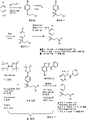
도1은 본원에 기재된 특정 화합물을 제조하는데 유용한 제1 화학 반응 도식이다.
도2는 본원에 기재된 특정 화합물을 제조하는데 유용한 제2 화학 반응 도식이다.
도3은 본원에 기재된 특정 화합물을 제조하는데 유용한 제3 화학 반응 도식이다.
도4는 본원에 기재된 특정 화합물을 제조하는데 유용한 제4 화학 반응 도식이다.
도5 는 실시예 8에 논의된 바와 같이 리포좀 ATR 억제제의 혈액 약력학을 보여주는 그래프이다.
도6은 실시예 9A에 기재된 바와 같이 자궁경부 MS751 이종이식 모델에서 MM-398과 조합된 리포좀 화합물 A의 항종양 효능을 보여주는 그래프이다.
도7은 실시예 9A에 기재된 바와 같이 자궁경부 C33A 이종이식 모델에서 MM-398과 조합된 리포좀 화합물 A의 항종양 효능을 보여주는 그래프이다.
도8은 실시예 9A에 따른 자궁경부 C33A 이종이식 모델에서 MM-398과 조합된 리포좀 화합물 A의 항종양 효능을 보여주는 그래프이다.
도9는 실시예 9A에 따른 자궁경부 MS751에서 MM-398과 조합된 리포좀 화합물 A의 관용성을 보여주는 그래프이다.
도10a 및 도10b는 각각 하기의 도면에서 MM-398과 조합된 리포좀 화합물 5의 효능을 보여주는 그래프이다: NCI-H2170 (도10a) 또는 DMS-114 (도10b) 마우스 이종이식 모델, 실시예 10에서 논의된 바와 같이.
도11a 및 도11b는 각각 하기의 도면에서 MM-398과 조합된 리포좀 화합물 5의 효능을 나타내는 카플란-메이어 생존 곡선을 보여주는 그래프이다: NCI-H2170 (도11a) 및 DMS-114 (도11b) 마우스 이종이식 모델, 실시예 10에서 논의된 바와 같이.
도12a 및 도12b는 각각 하기의 도면에서 MM-398과 조합된 리포좀 화합물 5의 관용성을 보여주는 그래프이다: NCI-H2170 (도12a) 또는 DMS-114 (도12b), 마우스 이종이식 모델에서.
도13a 및 도13b는 각각 하기의 도면에서 MM-398과 조합된 리포좀 화합물 5의 효능을 보여주는 그래프이다: Calu-6 (도13a) 또는 COLO-699 (도13b) 마우스 이종이식 모델, 실시예 11에서 논의된 바와 같이.
도14a 는 폐암 세포주의 패널에서 화합물 6 및 화합물 5의 시험관내 단독치료요법 세포 사멸을 설명하는 그래프이다.
도14b 는 3개의 화학치료학적 제제 (카보플라틴, 젬시타빈, 화?d물 B)와 조합된 화합물 5 및 화합물 6의 효과를 설명하는 그래프이다.
도15 는 다양한 농도의 화합물 B의 존재 또는 부재하에 실시예 2의 ATR 단백질 키나제 억제제 화합물 6에 대해 Sum190PT 세포주 (삼중 음성 유방암, TNBC)에서 측정된 IC50 쉬프트 값을 보여주는 그래프이다.
도16a 는 MDA-MB-453 TNBC 암 세포주에서 측정된, 실시예 2의 ATR 단백질 키나제 억제제 화합물 6의 조합의 IC50 쉬프트 값을 보여주는 그래프이다.
도16b 는 MDA-MB-453 TNBC 암 세포주에서 측정된, ATR 단백질 키나제 억제제 화합물 A의 조합의 IC50 쉬프트 값을 보여주는 그래프이다.
도17은 단독으로 또는 시험관내 조합된, 젬시타빈 [16nM] 또는 ATR 억제제 (화합물 A 또는 화합물 5 [1uM])에 노출된 폐암 세포 DMS-114로부터 수득된 웨스턴 블롯 분석이다.
도18a -b는 U2OS 세포에서, 다양한 농도에서 화합물 A 및 젬시타빈을 조합하여 수행된 세포사 및 성장 검정이다. 상기 결과는 2개의 화합물의 조합과 함께 세포 성장 및 사멸의 예측과 다시 비교된다(도18a). 생존 및 아폽토시스 세포의 수는 또한 USO2 세포에서 화합물 A (1 μM) 및 젬시타빈 (0.04 μM)의 설정 농도에서 결정된다(도18b).
도19는 세포의 수를 시간 경과에 따라 모니터링하면서, 여러 폐암 세포주 (NCI-H520 및 NCI-H596) 및 U2OS 세포에서 설정 농도 (각각 1 μM 및 0.2 μM)에서 단독으로 시험되고 SN38과 조합하여 시험된 화합물 A를 보여준다.
도20 은 자궁경부 암 세포주 MS751에서 조합되고 단독의 화합물 A 및 SN38의 효과를 설명하는 그래프이다.
도21a -c 는 젬시타빈과 화합물 A 또는 화합물 5를 조합한 효과를 입증한다. 도21a는 U2OS, H358, 및 A549 세포주에서, 다양한 농도에서 젬시타빈과 화합물 A 또는 화합물 5를 조합한 효과를 설명하는 히트 (heat) 맵이다. 세포에서 설정된 농도의 화합물 5 및 젬시타빈 또는 화합물 A 또는 젬시타빈로 처리된 세포의 현미경 이미지를 나타낸다(도21b). 설정된 농도와 함께 USO2 및 H358 세포의 증식 검정이 또한 보여진다(도21c).
도22a -b 는 SN38과 조합된 화합물 A 또는 화합물 5와 함께 A549 세포의 성장 곡선을 설명한다 (도22a) 또는 단독으로 (도22b).
도23 은 설정된 농도의 젬시타빈과 다양한 농도의 화합물 A 또는 화합물 5를 사용한 여러 세포주에서 IC50 값 (μM)을 보여준다.
도24 는 젬시타빈 또는 SN38과 조합된 화합물 A 또는 화합물 5에 반응성인 폐암 세포주의 개요를 보여준다.
도25a -b는 ATR (온-표적) 및 ATM (오프-표적)을 억제하는 이들의 능력에 대해 시험된 다양한 ATR 억제제를 보여준다. 억제는 nM의 IC50으로서 보고된다(도25a). 추가의 "오프-표적" 키나제는 또한 화합물 A 또는 화합물 5와 시험한다(도25b).
도26a -f는 하기의 세포에서 화합물 A 또는 화합물 5를 사용한 온-표적 결과를 보여준다: A549 폐 암 세포 (도26a-b), H23 폐 암 세포(도 26c-d), 및 DMS-114 세포 (도26e-f)에서. CHK1 S345 인산화, ATR 억제의 판독은 웨스턴 블롯에 의해 측정되었다. 각각의 화합물은 또한 고정된 농도의 젬시타빈과 함께 사용한다.
도27a -b는 HCC-70 TNBC, MDA-MB-468 TNBC, 및 DMS-114 세포주에 대한 추가의 온-표적 분석을 보여준다. 설정 농도의 SN38은 특정 농도 범위의 화합물 A 또는 화합물 5와 함께 사용된다. 다양한 온-표적 활성 파라미터는 웨스턴 블롯에 의해 시험되고 측정된다(도27a-b)
도28은 SN38 및 화합물 A 또는 화합물 5의 첨가 24시간 후에 SNM149 세포의 세포 주기 단계 프로파일을 보여준다.
도29는 MM398과 조합된 Ls-화합물 A 또는 Ls-화합물 5의 효과를 측정하기 위해 사용된 DMS-114 폐 이종이식 모델을 보여준다. 설정 용량의 MM398은 상이한 용량의 화합물 A 또는 화합물 5(20mpk 또는 80mpk)와 함께 사용된다(5mpk). 치료 효과는 CHK1 S345 인산화 수준을 측정함에 의해 분석된다.
도30a -c는 SUM-149 세포주에서 MM398과 함께 Ls-화합물 A의 효과를 설명한다. 치료 효과는 하기의 인산화된 수준을 측정함에 의해 분석된다: RPA2 (도30a), DNAPK, CHK1, 및 γH2AX (도30b). Ls-화합물 5는 또한 MM398과 조합 없이 시험된다(도30c).
도31은 SUM-149 마우스 이종이식 모델에서 MM-398과 조합된 리포좀 화합물 5의 효능을 보여주는 그래프이다.
도32는 SUM-149 마우스 이종이식 모델에서 MM-398과 조합된 리포좀 화합물 5의 관용성을 보여주는 그래프이다.
도33은 세포주 패널 상에서 웨스턴 블롯에 의해 정량된, DNA 손상 반응 경로와 연합된 다양한 PD 마커의 기본 수준을 보여준다.
도34는 통합 스코어가 동적 세포 생존력 검정에서 각각의 웰에 대해 어떻게 계산되는지를 설명하는 도식이다.
도35는 ATR 억제제 화합물 5 및/또는 SN38에 노출된 폐 암 세포주에 걸친 기본 MRE11 단백질 발현 (웨스턴 블롯에 의해 정량된) 및 동적 세포 생존력의 척도인 통합 스코어의 상호관계를 보여준다.
도36은 ATR 억제제 화합물 5 및/또는 SN38에 노출된 폐 암 세포주에 걸친 기본 ATM 단백질 발현 (웨스턴 블롯에 의해 정량된) 및 동적 세포 생존력의 척도인 통합 스코어의 상호관계를 보여준다.
도37은 ATR 억제제 화합물 5 및/또는 SN38에 노출된 폐 암 세포주에 걸친 기본 NBS 단백질 발현 (웨스턴 블롯에 의해 정량된) 및 동적 세포 생존력의 척도인 통합 스코어의 상호관계를 보여준다.
도38은 ATR 억제제 화합물 5 및/또는 SN38에 노출된 p53 기능적으로 손상된 폐암 세포주에 걸친 기본 NBS 단백질 발현 (웨스턴 블롯에 의해 정량된) 및 동적 세포 생존력의 척도인 통합 스코어의 상호관계를 보여준다.
도39는 ATR 억제제 화합물 5 및/또는 SN38에 노출된 p53 기능적으로 손상된 폐암 세포주에 걸친 기본 NBS 단백질 발현 (웨스턴 블롯에 의해 정량된) 및 동적 세포 생존력의 척도인 통합 스코어의 상호관계를 보여준다.
도40a -f 는 ATR 억제 및/또는 SN38로의 노출 후 암 세포주 NCIH1299 약력학적 마커에서의 배수 변화를 보여준다.
도41a -f 는 ATR 억제 및/또는 SN38로의 노출 후 암 세포주 NCIH460 약력학적 마커에서의 배수 변화를 보여준다.
도42a -f 는 ATR 억제 및/또는 SN38로의 노출 후 암 세포주 DMS114 약력학적 마커에서의 배수 변화를 보여준다.
도43a -f 는 ATR 억제 및/또는 SN38로의 노출 후 암 세포주 HCC70 약력학적 마커에서의 배수 변화를 보여준다.
도44a -f 는 ATR 억제 및/또는 SN38로의 노출 후 암 세포주 MDAMB468 약력학적 마커에서의 배수 변화를 보여준다.
도45 는 ATR 억제 및/또는 젬시타빈에 6 또는 18시간 노출 후 암 세포주 A549에서 약력학 마커의 웨스턴 블롯을 보여준다.
도46 은 ATR 억제 및/또는 젬시타빈에 6 또는 18시간 노출 후 암 세포주 NCIH23에서 약력학 마커의 웨스턴 블롯을 보여준다.
도47 은 ATR 억제 및/또는 젬시타빈에 6 또는 18시간 노출 후 암 세포주 DMS114에서 약력학 마커의 웨스턴 블롯을 보여준다.
도48 은 ATR 억제 및/또는 젬시타빈에 6 또는 18시간 노출 후 암 세포주 U2OS에서 약력학 마커의 웨스턴 블롯을 보여준다.
도49 는 ATR 억제 및/또는 젬시타빈에 6 또는 18시간 노출 후 암 세포주 NCIH460에서 약력학 마커의 웨스턴 블롯을 보여준다.
도50 은 ATR 억제 및/또는 젬시타빈에 6 또는 18시간 노출 후 암 세포주 HCC827에서 약력학 마커의 웨스턴 블롯을 보여준다.
도51 은 화합물 5 및/또는 SN38에 18시간 노출 후 결장직장 암 세포주의 패널에서 약력학 마커의 웨스턴 블롯을 보여준다.
도52 는 화합물 5 및/또는 SN38에 18시간 노출 후 결장직장 암 세포주에서 인산화된 Chk1 수준의 규정화된 정량을 보여준다(각각의 세포주에 대한 신호는 단독의 SN38 의 존재하에 신호로 규정화된다).
도53 은 화합물 5 및/또는 SN38에 18시간 노출 후 결장직장 암 세포주에서 인산화된 RPA2 수준의 규정화된 정량을 보여준다(각각의 세포주에 대한 신호는 단독의 SN38 의 존재하에 신호로 규정화된다).
도54 는 화합물 5 및/또는 SN38에 18시간 노출 후 결장직장 암 세포주의 패널에서 γH2AX 수준의 규정화된 정량을 보여준다(각각의 세포주에 대한 신호는 단독의 SN38의 존재하에 신호로 규정화된다). 1 is a first chemical reaction scheme useful for preparing certain compounds described herein.
2 is a second chemical reaction scheme useful for preparing certain compounds described herein.
3 is a third chemical reaction scheme useful for preparing certain compounds described herein.
4 is a fourth chemical reaction scheme useful for preparing certain compounds described herein.
Figure 5 is a graph showing the blood pharmacokinetics of liposomal ATR inhibitors as discussed in Example 8.
6 is a graph showing the antitumor efficacy of liposomal Compound A in combination with MM-398 in a cervical MS751 xenograft model as described in Example 9A.
7 is a graph showing the antitumor efficacy of liposomal Compound A in combination with MM-398 in a cervical C33A xenograft model as described in Example 9A.
8 is a graph showing the antitumor efficacy of liposomal Compound A in combination with MM-398 in a cervical C33A xenograft model according to Example 9A.
9 is a graph showing the tolerability of liposomal Compound A in combination with MM-398 in cervical MS751 according to Example 9A.
Figures 10a and 10b are graphs showing the efficacy of
11A and 11B are graphs showing Kaplan-Meier survival curves demonstrating the efficacy of
12A and 12B are graphs showing the tolerability of
Figures 13a and 13b are graphs showing the efficacy of
Figure 14a is a graph illustrating in vitro monotherapy cell killing of
Figure 14b is a graph illustrating the effect of
Figure 15 is a graph showing IC 50 shift values measured in the Sum190PT cell line (triple negative breast cancer, TNBC) for the ATR protein
Figure 16a is a graph showing the IC 50 shift value of the combination of the ATR protein
Figure 16b is a graph showing the IC 50 shift value of the combination of ATR protein kinase inhibitor Compound A, measured in the MDA-MB-453 TNBC cancer cell line.
17 is a Western blot analysis obtained from lung cancer cells DMS-114 exposed to gemcitabine [16 nM] or an ATR inhibitor (Compound A or Compound 5 [1 uM]) alone or in combination in vitro.
18A - B are cell death and growth assays performed by combining Compound A and gemcitabine at various concentrations in U2OS cells. The results are compared again with the prediction of cell growth and death with the combination of the two compounds (Figure 18a). The number of viable and apoptotic cells was also determined at set concentrations of Compound A (1 μM) and gemcitabine (0.04 μM) in USO2 cells (Figure 18B).
19 is tested alone and in combination with SN38 at set concentrations (1 μM and 0.2 μM, respectively) in several lung cancer cell lines (NCI-H520 and NCI-H596) and U2OS cells, while monitoring the number of cells over time. Compound A is shown.
20 is a graph illustrating the effect of Compound A and SN38 alone in combination in the cervical cancer cell line MS751.
Figure 21a- c demonstrates the effect of combining gemcitabine with either Compound A or
Figures 22A -B illustrate the growth curves of A549 cells with Compound A or
Figure 23 shows the IC50 values (μM) in different cell lines using set concentrations of gemcitabine and various concentrations of Compound A or
Figure 24 shows an overview of lung cancer cell lines responsive to Compound A or
25A - B show various ATR inhibitors tested for their ability to inhibit ATR (on-target) and ATM (off-target). Inhibition is reported as an IC50 of nM (Figure 25a). Additional “off-target” kinases were also tested with Compound A or Compound 5 (Figure 25B).
26A -F show on-target results with Compound A or
27A - B show additional on-target assays for HCC-70 TNBC, MDA-MB-468 TNBC, and DMS-114 cell lines. A set concentration of SN38 is used with either compound A or
Figure 28 shows the cell cycle phase profile of
29 shows the DMS-114 lung xenograft model used to measure the effect of Ls-Compound A or Ls-
30A -C illustrate the effect of Ls-Compound A in combination with MM398 in SUM-149 cell line. The therapeutic effect was analyzed by measuring the phosphorylated levels of RPA2 (Figure 30A), DNAPK, CHK1, and γH2AX (Figure 30B). Ls-
31 is a graph showing the efficacy of
32 is a graph showing the tolerability of
33 shows basal levels of various PD markers associated with DNA damage response pathways, quantified by Western blot on a panel of cell lines.
34 is a schematic illustrating how an integration score is calculated for each well in a dynamic cell viability assay.
Figure 35 shows the correlation of basal MRE11 protein expression (quantified by Western blot) and integration score, a measure of dynamic cell viability, across lung cancer cell lines exposed to
36 shows the correlation of basal ATM protein expression (quantified by Western blot) and integration score, a measure of dynamic cell viability, across lung cancer cell lines exposed to
37 shows the correlation of basal NBS protein expression (quantified by Western blot) and integration score, a measure of dynamic cell viability, across lung cancer cell lines exposed to
38 shows the correlation of basal NBS protein expression (quantified by Western blot) and integrated score, a measure of dynamic cell viability, across p53 functionally impaired lung cancer cell lines exposed to
Figure 39 shows the correlation of basal NBS protein expression (quantified by Western blot) and integration score, a measure of dynamic cell viability, across p53 functionally impaired lung cancer cell lines exposed to
40A -F show fold changes in cancer cell line NCIH1299 pharmacodynamic markers after ATR inhibition and/or exposure to SN38.
41A -F show fold change in cancer cell line NCIH460 pharmacodynamic markers after ATR inhibition and/or exposure to SN38.
42A -F show fold changes in cancer cell line DMS114 pharmacodynamic markers after ATR inhibition and/or exposure to SN38.
43A -F show fold changes in cancer cell line HCC70 pharmacodynamic markers after ATR inhibition and/or exposure to SN38 .
44A -F show fold change in cancer cell line MDAMB468 pharmacodynamic markers after ATR inhibition and/or exposure to SN38.
Figure 45 shows a Western blot of pharmacodynamic markers in cancer cell line A549 after 6 or 18 h exposure to ATR inhibition and/or gemcitabine.
Figure 46 shows a Western blot of pharmacodynamic markers in cancer cell line NCIH23 after 6 or 18 h exposure to ATR inhibition and/or gemcitabine.
Figure 47 shows a Western blot of pharmacodynamic markers in cancer cell line DMS114 after 6 or 18 h exposure to ATR inhibition and/or gemcitabine.
Figure 48 shows a Western blot of pharmacodynamic markers in the cancer cell line U2OS after 6 or 18 h exposure to ATR inhibition and/or gemcitabine.
Figure 49 shows a Western blot of pharmacodynamic markers in cancer cell line NCIH460 after 6 or 18 h exposure to ATR inhibition and/or gemcitabine.
Figure 50 shows a Western blot of pharmacodynamic markers in cancer cell line HCC827 after 6 or 18 h exposure to ATR inhibition and/or gemcitabine.
Figure 51 shows a Western blot of pharmacodynamic markers in a panel of colorectal cancer cell lines after 18 h exposure to
Figure 52 shows normalized quantification of phosphorylated Chk1 levels in colorectal cancer cell lines after 18 h exposure to
Figure 53 shows normalized quantification of phosphorylated RPA2 levels in colorectal cancer cell lines after 18 h exposure to
Figure 54 shows normalized quantification of γH2AX levels in a panel of colorectal cancer cell lines after 18 h exposure to
ATR 단백질 키나제를 억제하기 위한 신규 화합물 또는 이의 약제학적으로 허용되는 염은 화학식 I로 나타낸다:A novel compound for inhibiting ATR protein kinase or a pharmaceutically acceptable salt thereof is represented by the formula (I):

여기서, R은 마우스에서 적어도 약 5시간의 혈장 반감기를 제공하도록 선택된 (실시예 7에 따라 수득된) 7.0 초과 (바람직하게 8.0 초과, 및 가장 바람직하게 적어도 약 9.5)의 pKa을 갖는 아민을 포함하는 모이어티이다. 바람직하게, R은 4 내지 12개 탄소를 갖는 아민-치환된 알킬 모이어티를 포함한다. R은 3급-치환된 아민 및 수소화된 알킬 그룹의 조합만을 포함하도록 선택될 수 있다. R은 바람직하게 적어도 7, 그러나 가장 바람직하게 적어도 약 9.5의 pKa (예를 들어, 약 9.5 내지 10.5의 pKa)를 갖는 3급-알킬 치환된 아민을 추가로 포함한다. 화학식 I의 화합물 또는 이의 약제학적으로 허용되는 염의 예는 화합물 1 내지 6을 포함한다(실시예 1 내지 6 참조):wherein R comprises an amine having a pK a greater than 7.0 (preferably greater than 8.0, and most preferably at least about 9.5) (obtained according to Example 7) selected to provide a plasma half-life of at least about 5 hours in mice It is a moiety that Preferably, R comprises an amine-substituted alkyl moiety having 4 to 12 carbons. R may be selected to include only combinations of tert-substituted amines and hydrogenated alkyl groups. R further comprises a tert-alkyl substituted amine, preferably having a pK a of at least 7, but most preferably of at least about 9.5 (eg, a pK a of about 9.5 to 10.5 ). Examples of compounds of formula (I) or pharmaceutically acceptable salts thereof include

화학식 I의 화합물은 바람직하게 ATR의 목적하는 억제 및/또는 리포좀 형성 및 안정성 특징을 제공하도록 선택된 R에서 하나 이상의 3급 아민 모이어티를 포함한다. 일부 예에서, R은 제1 3급 치환된 질소를 포함하고, 바람직하게 제2 3급 치환된 질소를 포함하는 알킬아미노 모이어티로 치환된 헤테로사이클릭 모이어티이다. 특히, 화학식 I의 화합물은 R이 하기 화학식의 모이어티일 수 있는 것들을 포함한다:The compounds of formula (I) preferably comprise one or more tertiary amine moieties in R selected to provide the desired inhibition of ATR and/or liposome formation and stability characteristics. In some instances, R is a heterocyclic moiety substituted with an alkylamino moiety comprising a first tertiary substituted nitrogen, preferably comprising a second tertiary substituted nitrogen. In particular, compounds of formula (I) include those in which R may be a moiety of the formula:

여기서, A1은 부재이거나 알킬 (예를 들어,C1-C4 알킬 (바람직하게 -(CH2)2-)이고, R1은 저급(예를 들어,C1-C4) 알킬아미노이다. 하나의 구현예에서, R1은 (C1-C4 알킬)-NRaRb이고, 여기서, Ra 및 Rb은 각각 독립적으로 C1-C4 알킬이고, 예를 들어,R1은 -(CH2)2-N(CH3)(CH3)이다. 또 다른 구현예에서, R1은 NRaRb이고, 여기서, Ra 및 Rb는 각각 독립적으로 C1-C4 알킬, 예를 들어,R1은 -N(CH2CH3)(CH2CH3)이다. wherein A 1 is absent or alkyl (eg, C 1 -C 4 alkyl (preferably —(CH 2 ) 2 —) and R 1 is lower (eg, C 1 -C 4 ) alkylamino In one embodiment, R 1 is (C 1 -C 4 alkyl)-NR a R b , wherein R a and R b are each independently C 1 -C 4 alkyl, eg, R 1 is -(CH 2 ) 2 -N(CH 3 )(CH 3 ) In another embodiment, R 1 is NR a R b , wherein R a and R b are each independently C 1 -C 4 Alkyl, eg, R 1 is —N(CH 2 CH 3 )(CH 2 CH 3 ).
화학식 I의 또 다른 구현예에서, R은 -N(H)(C1-C4 알킬)-NRaRb이고, 여기서, Ra 및 Rb는 각각 독립적으로 C1-C4 알킬이거나 R은 -(G)-NRaRb이고, 여기서, Ra 및 Rb은 각각 독립적으로 C1-C4 알킬이고, 여기서, G는 C1-C4 알킬이고, 여기서, G는 추가로 C1-C4 알킬로 치환될 수 있다. In another embodiment of formula I, R is -N(H)(C 1 -C 4 alkyl)-NR a R b , wherein R a and R b are each independently C 1 -C 4 alkyl or R is -(G)-NR a R b , wherein R a and R b are each independently C 1 -C 4 alkyl, wherein G is C 1 -C 4 alkyl, wherein G is further C 1 -C 4 alkyl may be substituted.
화학식 I의 또 다른 구현예에서, R은 하기 화학식의 모이어티일 수 있다:In another embodiment of formula I, R may be a moiety of the formula:

여기서, Rc 및 Rd는 각각 독립적으로 C1-C4 알킬이다. wherein R c and R d are each independently C 1 -C 4 alkyl.
바람직한 예는 상기 화합물 1, 2, 3, 4, 5, 또는 6으로 이루어진 그룹으로부터 선택되는 화합물을 포함하는 리포좀을 포함한다. 일부 예에서, 상기 화합물은 화합물 5 또는 화합물 6이다. Preferred examples include liposomes containing a compound selected from the group consisting of
화학식 I의 화합물 또는 이의 약제학적으로 허용되는 염은 화학식 Ia의 구조를 가질 수 있고, 여기서, R´은 약 9.5 이상의 pKa를 갖는 3급 알킬 치환된 아민이다:A compound of Formula I, or a pharmaceutically acceptable salt thereof, may have the structure of Formula Ia, wherein R' is a tertiary alkyl substituted amine having a pK a of at least about 9.5:

화학식 Ia의 화합물의 예는 본원에 기재된 화합물 5를 포함한다(예를 들어,실시예 1). 화학식 Ia의 하나의 구현예에서, R'는 NRaRb이고, 여기서, Ra 및 Rb는 각각 독립적으로 C1-C4 알킬이다. Examples of compounds of Formula Ia include
ATR 단백질 키나제 억제제 화합물의 리포좀 제형 (예를 들어, 실시예 7에 기재된 바와 같은)은 실시예 8에 기재된 마우스 모델에서 5시간 이상의 증진된 혈장 반감기와 같은 목적하는 약동학적 성질을 제공할 수 있다. 리포좀은 전형적으로 수성 내측을 둘러싸는 하나 이상의 지질 이중층을 함유하는 소포를 포함한다. 리포좀 조성물은 일반적으로 리포좀 외측 수성 유체와 같은 매질 중에 리포좀을 포함한다. 리포좀 지질은 양친매성 지질 성분을 포함할 수 있고 이는 수성 매질과 접촉시 자발적으로 인지질, 예를 들어, 포스파티틸콜린과 같은 이중층 막을 형성한다. 리포좀은 또한 스테롤, 예를 들어, 콜레스테롤과 같은 막-강화 성분을 포함할 수 있다. 일부 경우에, 리포좀은 또한 리포좀이 응집하는 경향을 감소시키고 또한 다른 이로운 효과를 가질 수 있는 폴리에틸렌글리콜 (PEG) 지질 유도체와 같은 친수성 폴리머에 접합된 지질을 포함한다. A liposomal formulation of an ATR protein kinase inhibitor compound (eg, as described in Example 7) can provide desired pharmacokinetic properties, such as enhanced plasma half-life of at least 5 hours in the mouse model described in Example 8. Liposomes typically comprise vesicles containing one or more lipid bilayers surrounding an aqueous inner side. Liposomal compositions generally include liposomes in a medium such as an aqueous fluid outside the liposomes. Liposomal lipids may comprise an amphiphilic lipid component, which upon contact with an aqueous medium spontaneously forms a bilayer membrane such as a phospholipid, eg, phosphatidylcholine. Liposomes may also contain membrane-enhancing components such as sterols, eg, cholesterol. In some cases, liposomes also include lipids conjugated to hydrophilic polymers, such as polyethyleneglycol (PEG) lipid derivatives, which can reduce the tendency of the liposomes to aggregate and also have other beneficial effects.
리포좀 제형은 하나 이상의 리포좀-형성 지질(예를 들어, 수소화된 대두 포스파티딜콜린 (HSPC)), 콜레스테롤 및 폴리머-접합된 지질(예를 들어, 메톡시-폴리(에틸렌 글리콜)-1,2-디스테아로일-sn-글리세릴(PEG2000-DSG)로부터 형성된 단일라멜라 소포에서 다중음이온 (예를 들어, 다중음이온 당, 예를 들어, 슈크로스 옥타설페이트 또는 적합한 다중음이온화된 폴리올)로 캡슐화된 화학식 I의 화합물을 포함할 수 있다. 리포좀-형성 지질은 바람직하게 하나 이상의 인지질을 포함하고, 인지질(들)과 콜레스테롤의 비율은 리포좀으로부터 화학식 I의 화합물의 충분히 감소된 양의 누출을 유지하면서 리포좀 막 강성의 목적하는 양을 제공하도록 선택된다. The liposome formulation may contain one or more liposome-forming lipids (e.g., hydrogenated soy phosphatidylcholine (HSPC)), cholesterol and a polymer-conjugated lipid (e.g., methoxy-poly(ethylene glycol)-1,2-distea Formula I encapsulated with polyanions (e.g. polyanionic sugars such as sucrose octasulfate or suitable polyanionized polyols) in monolamellar vesicles formed from royl-sn-glyceryl (PEG2000-DSG) The liposome-forming lipid preferably comprises one or more phospholipids, and the ratio of phospholipid(s) to cholesterol maintains the leakage of a sufficiently reduced amount of the compound of formula (I) from the liposome, while maintaining liposome membrane stiffness. is chosen to provide the desired amount of
리포좀은 전형적으로 마이크론 또는 서브마이크론 범위의 크기를 갖고 이리노테칸과 같은 항암 약물을 포함하는 약제학적 물질을 운반하고 다양한 이로운 방식으로 이들의 약제학적 성질을 변화시키는 이들의 능력 때문에 널리 인지되고 있다. 약제학적 리포좀 조성물을 제조하고 특징 분석하는 방법은 당업계에 공지되어 있다 (문헌참조: 예를 들어,Lasic D.Liposomes:From physics to applications, Elsevier, Amsterdam 1993; G.Greroriadis (ed.),Liposome Technology, 3rd edition, vol.1-3, CRC Press, Boca Raton, 2006; Hong 등, 미국 특허 제8,147,867호, 모든 목적을 위해 이들의 전문이 본원에 참조로 인용된다). Liposomes typically have a size in the micron or submicron range and are widely recognized because of their ability to deliver pharmaceutical substances, including anticancer drugs, such as irinotecan, and to alter their pharmaceutical properties in a variety of beneficial ways. Methods for preparing and characterizing pharmaceutical liposome compositions are known in the art (see, e.g., Lasic D. Liposomes: From physics to applications, Elsevier, Amsterdam 1993; G. Greroriadis (ed.), Liposome Technology, 3 rd edition, vol. 1-3, CRC Press, Boca Raton, 2006; Hong et al., US Pat. No. 8,147,867, incorporated herein by reference in their entirety for all purposes).
일부 예에서 (예를 들어,실시예 7), ATR 단백질 키나제 억제제 조성물은 폴리설페이트화된 당 (예를 들어, 슈크로스 옥타설페이트)과 같은 다중음이온을 갖는 리포좀에 캡슐화된 ATR 단백질 키나제 억제제 화합물을 포함하는 리포좀을 포함할 수 있다. 슈크로소페이트, 완전히 양성자화된 형태로 하기의 구조를 갖는 슈크로스의 완전히 치환된 설페이트 에스테르:In some instances (eg, Example 7), the ATR protein kinase inhibitor composition comprises an ATR protein kinase inhibitor compound encapsulated in liposomes having a polyanion, such as a polysulfated sugar (eg, sucrose octasulfate). and liposomes. Sucrose, a fully substituted sulfate ester of sucrose having the structure in its fully protonated form:

슈크로소페이트는 또한 슈크로스 옥타설페이트 또는 슈크로옥타설페이트 (SOS)로서 언급된다. 다양한 염, 예를 들어, 암모늄, 나트륨 또는 칼륨 염의 형태로 슈크로소페이트를 제조하는 방법은 당업계에 널리 공지되어 있다 (예를 들어, 미국 특허 제4,990,610호, 이는 이의 전문이 본원에 참조로 인용된다). Sucrose sulphate is also referred to as sucrose octasulfate or sucrose octasulfate (SOS). Methods for preparing sucrose in the form of various salts, for example, ammonium, sodium or potassium salts, are well known in the art (see, eg, US Pat. No. 4,990,610, incorporated herein by reference in its entirety). do).
ATR 단백질 키나제 억제제 리포좀은 TEA 함유 리포좀의 형성에 이어서 하기 화합물의 로딩을 포함하는 다중 단계로 제조될 수 있다: ATR 단백질 키나제 억제제 화합물(예를 들어,화합물 A 또는 화학식 I의 화합물))의 TEA가 리포좀을 이탈함으로써 리포좀으로의 로딩.예를 들어, ATR 단백질 키나제 억제제 리포좀은 (a) 슈크로소페이트의 트리에틸암모늄 염 (TEA-SOS)으로서 트리에틸아민 (TEA)을 함유하는 리포좀을 제조하는 단계, 및 (b) 후속적으로 이리노테칸이 리포좀으로 진입하고 상응하는 양의 TEA가 리포좀을 이탈하도록 (이에 의해 수득한 리포좀에 걸친 TEA의 농도 구배를 소모시키거나 감소시키는) 하는 효과적인 조건하에서 TEA-SOS 리포좀을 이리노테칸과 접촉시키는 단계를 포함하는 방법에 의해 제조될 수 있다. ATR protein kinase inhibitor liposomes can be prepared in multiple steps comprising the formation of TEA containing liposomes followed by loading of the following compound: Loading into liposomes by leaving the liposomes. For example, ATR protein kinase inhibitor liposomes can be prepared by (a) preparing liposomes containing triethylamine (TEA) as triethylammonium salt of sucroseophate (TEA-SOS). , and (b) TEA-SOS under conditions effective to subsequently cause irinotecan to enter the liposome and a corresponding amount of TEA exit the liposome, thereby consuming or reducing the concentration gradient of TEA across the liposome obtained. It may be prepared by a method comprising the step of contacting the liposome with irinotecan.
제1 단계는 리포좀 지질을 TEA 슈크로소페이트의 용액 중에 수화시키고 분산시킴에 의해 TEA-슈크로소페이트 함유 리포좀을 형성함을 포함할 수 있다. 이것은 예를 들어, 가열된 에탄올 중에서 HSPC 및 콜레스테롤을 포함하는 지질을 용해시키고 상기 용해되고 가열된 지질 용액을 리포좀 지질의 전이 온도 (Tm)초과의 하기의 온도에서 TEA-슈크로소페이트 수용액 중에 분산시킴에 의해 수행될 수 있다: 예를 들어, 60 ℃ 이상. 지질 분산액은 하기의 한정된 공극 크기를 갖는 트랙-에칭된 폴리카보네이트 막을 통해 압출시킴에 의해 리포좀으로 형성될 수 있고 이의 평균 크기는 75-125 nm (예를 들어, 80-120 nm, 또는 일부 구현예에서, 90-115 nm)이다: 예를 들어,100 nm.TEA-슈크로소페이트는 슈크로소페이트의 각각의 몰 당량에 대해 적어도 8몰 당량의 TEA를 포함하여 약 0.40-0.50 N의 농도 및 분산 및 압출 단계 동안에 리포좀 인지질의 허용될 수 없는 분해를 예방하기 위해 선택된 pH (예를 들어, 약 6.5) (예를 들어, 이들 단계 동안에 리포좀의 분해를 최소화하기 위해 선택된 pH)를 가질 수 있는 용액을 수득할 수 있다. 이어서, 비-포집된 TEA-SOS는 예를 들어, 약물 캡슐화 전에 투석, 겔 크로마토그래피, 이온 교환 또는 한외여과에 의해 리포좀 분산액으로부터 제거될 수 있다. 수득한 리포좀은 ATR 단백질 키나제 억제제 슈크로소페이트를 함유할 수 있다. 이들 ATR 억제제 리포좀은 수득한 리포좀 조성물 중 TEA의 양을 4 ℃에서 180일 후 라이소-PC 형성의 소정의 최대 수준 미만 또는 약 4 ℃에서, 또는, 보다 통상적으로, 5 ± 3 ℃에서 냉장고에서 저장 동안에 리포좀 조성물 중 라이소-PC 축적율의 소정의 최대 수준 미만을 유도하는 수준까지 감소시키기에 충분한 약물의 리포좀으로의 로딩에 의해 안정화될 수 있다: 예를 들어 다음으로 측정된다:mg/mL/개월, 또는 단위 시간에 대한 라이소-PC로의 % PC 전환, 예를 들어, mol% 라이소-PC/개월. 이어서, 임의의 비포집된 ATR 억제제와 함께 로딩 공정 동안에 리포좀으로부터 외측 매질로 교환된 TEA는 전형적으로 임의의 적합한 공지된 공정(들)에 의해 (예를 들어, 겔 크로마토그래피, 투석, 투석 여과, 이온 교환 또는 한외여과) 리포좀으로부터 제거된다. 리포좀 외측 매질은 목적하는 pH에서 완충된, 주사가능한 등장성 유체 (예를 들어, 염화나트륨의 등장성 용액)로 교환될 수 있다. The first step may comprise forming the TEA-sucrose phosphate containing liposomes by hydrating and dispersing the liposomal lipids in a solution of TEA sucrose sucrose. This can be done, for example, by dissolving lipids comprising HSPC and cholesterol in heated ethanol and dispersing the dissolved and heated lipid solution in an aqueous TEA-sucrosorbate solution at a temperature above the transition temperature (T m ) of the liposomal lipid. Sikkim: for example, 60° C. or higher. Lipid dispersions can be formed into liposomes by extruding through a track-etched polycarbonate membrane having a defined pore size of 75-125 nm (e.g., 80-120 nm, or in some embodiments) at 90-115 nm): for example, 100 nm.TEA-sucrose with a concentration and dispersion of about 0.40-0.50 N including at least 8 molar equivalents of TEA for each molar equivalent of sucroseophate and a pH selected to prevent unacceptable degradation of the liposomal phospholipids during the extrusion step (e.g., about 6.5) (e.g., a pH selected to minimize degradation of the liposome during these steps). can be obtained. Non-entrapped TEA-SOS can then be removed from the liposome dispersion by, for example, dialysis, gel chromatography, ion exchange or ultrafiltration prior to drug encapsulation. The obtained liposomes may contain the ATR protein kinase inhibitor sucroseophate. These ATR inhibitor liposomes contain the amount of TEA in the obtained liposome composition below the predetermined maximum level of lyso-PC formation after 180 days at 4°C, or at about 4°C, or, more typically, at 5±3°C, in the refrigerator. may be stabilized during storage by loading into the liposome of sufficient drug to reduce the rate of lyso-PC accumulation in the liposomal composition to a level that induces less than a predetermined maximum level: for example, as determined as: mg/mL /month, or % PC conversion to lyso-PC per unit time, e.g., mol % lyso-PC/month. The TEA exchanged from the liposomes to the outer medium during the loading process along with any non-entrapped ATR inhibitor is then typically by any suitable known process(s) (e.g., gel chromatography, dialysis, diafiltration, ion exchange or ultrafiltration) from the liposome. The liposome outer medium can be exchanged with a buffered, injectable isotonic fluid (eg, an isotonic solution of sodium chloride) at the desired pH.
리포좀 캡슐화된 ATR 단백질 억제제 화합물을 포함하는 다양한 리포좀 제형의 항종양 효능은 하기의 세포주에서 시험되었다: 인간 자궁경부 암 세포주 (예를 들어,MS751, C33A 및 SiHa 세포주, 실시예 9에 보여진 바와 같이), 및 편평 세포 암종 세포주를 포함하는 다양한 폐 암 세포주(예를 들어, 실시예 10에서 NCI-H2170 세포주), 소 세포 폐 암종 세포주 (예를 들어, 실시예 10에서 DMS-114 세포주), 및 인간 Calu-6 및 COLO-699 세포주 (실시예 11). The antitumor efficacy of various liposomal formulations comprising a liposomal encapsulated ATR protein inhibitor compound was tested in the following cell lines: human cervical cancer cell lines (e.g., MS751, C33A and SiHa cell lines, as shown in Example 9) , and various lung cancer cell lines (eg, NCI-H2170 cell line in Example 10), small cell lung carcinoma cell lines (eg, DMS-114 cell line in Example 10), including squamous cell carcinoma cell lines, and human Calu-6 and COLO-699 cell lines (Example 11).
하기 도 6-9 및 실시예 9를 참조하면, ATR 억제제 화합물 A의 리포좀 제형(실시예 7)은 단독으로 또는 이리노테칸 리포좀 제형 MM398과 조합하여 (실시예 9B) 마우스 제노그래프 모델 (실시예 9A) 에서 3개의 인간 자궁경부 암 세포주에 대해 시험되었다. 보다 큰 종양 용적은 3개의 자궁경부 암 세포주 중 2개 (MS571 및 C33A)에 대한 대조군 실험과 비교하여 실시예 7의 리포좀 화합물 A 제형에 대해 시간 경과시 관찰되었다. 그러나, 화합물 A 리포좀 제형 (실시예 7)과 조합된 이리노테칸 리포좀 MM398 (실시예 9B)의 투여는 단독의 MM398 또는 단독의 리포좀 화합물 A의 투여 중 어느 하나 보다 모든 3개의 자궁경부 암 세포주의 보다 큰 억제를 유도하였다. 6-9 and Example 9 below, the liposomal formulation of ATR inhibitor Compound A (Example 7) alone or in combination with irinotecan liposome formulation MM398 (Example 9B) is a mouse xenographic model (Example 9A) was tested against three human cervical cancer cell lines in Larger tumor volumes were observed over time for the liposomal Compound A formulation of Example 7 compared to control experiments on 2 of 3 cervical cancer cell lines (MS571 and C33A). However, administration of irinotecan liposomal MM398 (Example 9B) in combination with a Compound A liposomal formulation (Example 7) was greater than any of the administration of MM398 alone or liposomal Compound A alone in all three cervical cancer cell lines. inhibition was induced.
하기 도10a-10b 및 도11a-11b를 참조하면, 화학식 I 및 화학식 Ia의 ATR 억제제 화합물 5 (실시예 7에 기재된 바와 같은 리포좀으로서 제형화된 실시예 1의 화합물)의 리포좀 제형은 단독으로 및 이리노테칸 리포좀 제형 MM398 (실시예 9B)과 조합된 마우스 제노그래프 모델 (실시예 10)에서 2개의 폐 암 세포주에 대해 시험되었다. 하기 도10a 및 도10b를 참조하면, 화합물 5의 리포좀 제형의 투여는 실시예 10에서 대조군 실험과 비교하여, 시험된 각각의 세포주에서 종양 용적을 감소시켰고, MM398과 실시예 7의 화합물 5 리포좀 조성물의 조합은 마우스 모델에서 종양 용적을 다른 것과 무관하게 투여된 화합물 보다 큰 정도로 감소시켰다. 유사하게, 실시예 10에 제공된 카플란-메이어 생존 곡선(도11a 및 도11b)는 실시예 9B의 이리노테칸 리포좀 MM398 둘 다의 조합이 2개의 상이한 세포주를 사용한 실시예 7의 화합물 5 리포좀 제형과 조합하여 투여되는 경우 마우스 폐 암 제노그래프 시험에서 증가된 생존을 입증한다. 10a-10b and 11a-11b below, the liposome formulations of the ATR inhibitor compound 5 (the compound of Example 1 formulated as a liposome as described in Example 7) of Formulas I and Ia alone and Two lung cancer cell lines were tested in a mouse xenographic model (Example 10) in combination with the irinotecan liposome formulation MM398 (Example 9B). 10A and 10B below, administration of the liposome formulation of
하기 도 12a 및 도 12b를 참조하면, ATR 단백질 키나제 억제제 화합물의 다양한 제형의 관용성은 실시예 10에서 평가되었다. 하기 도 12a를 참조하면, NCI-H2170 마우스 제노그래프 모델에서 시험된 마우스 체중에서 감소는 이리노테칸 리포좀 MM398 (실시예 9B), 대조군 또는 MM398과 조합된 화합물 5의 리포좀 제형과 비교하여 화합물 5의 리포좀 제형 (실시예 7)에 대해 시간 경과에 따라 최저였다. 하기 도12b를 참조하면, DMS-114 마우스 제노그래프 모델에서 시험된 마우스 체중에서 감소는 화합물 5의 리포좀 제형 (실시예 7), 또는 독립적으로 투여된 이리노테칸 리포좀 MM398 (실시예 9B)과 비교하여, MM398과 조합된 화합물 5의 리포좀 제형의 조합에 대해 시간 경과에 따라 최저였다. 12A and 12B below, the tolerability of various formulations of ATR protein kinase inhibitor compounds was evaluated in Example 10. Referring to FIG. 12A below, the reduction in body weight of mice tested in the NCI-H2170 mouse xenograph model was compared to the liposome formulation of
하기 도13a 및 도13b를 참조하면, ATR 단백질 키나제 억제제 화합물 5의 리포좀 제형과 이리노테칸 리포좀 MM398 (실시예 9B)의 조합물의 투여는 대조군, 단독의 MM398 이리노테칸 리포좀의 투여, 화합물 A 리포좀 제형 (실시예 7)의 투여 또는 화합물 A 리포좀 제형(실시예 7)과 MM398 이리노테칸 리포좀 (실시예 9B)의 조합물의 투여와 비교하여, 마우스 제노그래프 모델에서 Calu-6 및 COLO699 세포주 둘 다에서 종양 용적의 최대 감소를 유도하였다. 13A and 13B below, the administration of the combination of the ATR protein
실시예Example
하기의 실시예는 본 발명의 일부 구현예를 설명한다. 하기의 실시예 및 제법은 당업자가 이들 및 본 발명의 다른 구현예를 보다 명백하게 이해하고 수행할 수 있도록 제공된다. 이들은 본 발명의 범위를 제한하는 것으로 간주되지 말아야 하고 단지 이의 예시 및 대표적인 예로서 간주되어야 한다. The following examples illustrate some embodiments of the present invention. The following examples and preparations are provided to enable those skilled in the art to more clearly understand and to practice these and other embodiments of the present invention. They are not to be considered as limiting the scope of the invention, but merely as illustrative and representative thereof.
ATR 펩타이드는 문헌에 공지된 다양한 방법을 사용하여 발현되고 단리될 수 있다 (문헌참조: 예를 들어, ![]()
![]()
화합물 A는 공개공보 WO2010/071827A1 (2010년 6월 24일자로 공개된)에 기재된 방법 (예를 들어)에 의해 수득될 수 있고, 화합물 II-A-7의 합성 및 용도에 관한 이의 일부는 본원에 참조로서 인용된다. 화합물 A의 구조는 다음과 같다:Compound A can be obtained by the method (for example) described in publication WO2010/071827A1 (published on June 24, 2010), some of which relate to the synthesis and use of compounds II-A-7 herein is incorporated by reference. The structure of compound A is:

화합물 Acompound A
화학식 I의 다양한 화합물은 본원에 기재된 바와 같이 제조될 수 있고 하기의 표에 요약되어 있다. Various compounds of formula (I) can be prepared as described herein and are summarized in the table below.
표 1:화학식 I의 선택된 화합물Table 1: Selected compounds of formula I

화합물 1, 2, 3 및 6은 하기의 도에서 도식 1에 나타낸 동일계 생성된 보론산 에스테르를 사용한 원-팟 스즈키 교차-커플링에서 제조하였다. 하기 도1을 참조하면, 중간체 3의 합성:1-브로모-4-(2-브로모에틸설포닐)벤젠은 하기된 바와 같이 수득될 수 있다.

중간체2 중간체3Intermediate 2
DCM (400 mL) 중 중간체 2 (35 g, 133 mmol)의 용액에 PBr3 (40 g, 146 mmol)을 0 ℃에서 적가하였다. 이어서 혼합물을 밤새 실온에서 교반하였다. 물 (15 mL)을 첨가하여 반응을 켄칭하였다. 이어서 수득물은 물 (120 mL) 및 염수 (120 mL)로 세척하였다. 유기상을 농축시켜 황색 오일로서 20 g의 조추출물 3을 수득하고 이는 다음 단계에서 추가의 정제 없이 사용하였다. To a solution of intermediate 2 (35 g, 133 mmol) in DCM (400 mL) was added PBr 3 (40 g, 146 mmol) dropwise at 0 °C. The mixture was then stirred overnight at room temperature. Water (15 mL) was added to quench the reaction. The harvest was then washed with water (120 mL) and brine (120 mL). Concentration of the organic phase gave 20 g of
하기 도 1을 참조하면, 중간체 2의 합성은 하기와 같이 수행될 수 있다:Referring to Figure 1 below, the synthesis of

중간체1 중간체2Intermediate 1
DCM (500 mL) 중 중간체 1 (45 g, 194 mmol)의 용액에 m-CPBA (134 g, 776 mmol)을 여러 배치에서 실온에서 첨가하였다. 이어서 혼합물을 밤새 실온에서 교반하였다. 반응 혼합물을 여과하고 DCM (500 ml)을 첨가하여 고체를 세척하였다. 여과물은 NaOH (1M, 300 mL X 3) 및 염수 (300 mL)로 세척하였다. 유기층은 건조 농축시켜 백색 고체로서 36 g의 중간체 2 (70 %)를 수득하였다. To a solution of Intermediate 1 (45 g, 194 mmol) in DCM (500 mL) was added m-CPBA (134 g, 776 mmol) in several batches at room temperature. The mixture was then stirred overnight at room temperature. The reaction mixture was filtered and DCM (500 ml) was added to wash the solid. The filtrate was washed with NaOH (1M, 300 mL X 3) and brine (300 mL). The organic layer was concentrated to dryness to give 36 g of intermediate 2 (70%) as a white solid.
하기 도 1을 참조하면, 중간체 1의 합성은 하기와 같이 수행될 수 있다:Referring to Figure 1 below, the synthesis of

중간체 1Intermediate 1
MeCN (600 ml) 중 4-브로모벤젠티올 (45 g, 238 mmol)의 용액에 K2CO3 (60 g, 476 mmol) 및 NaI (36 g, 238 mmol)을 첨가하였다. 혼합물은 10분 동안 실온에서 교반하였다. 이어서 2-브로모에탄올을 적가하였다. 첨가 후, 상기 혼합물을 밤새 실온에서 교반하였다. 반응 혼합물을 여과하고 여과물을 건조 농축시켰다. 잔사를 실리카-겔 칼럼으로 정제하여 담황색 오일로서 45 g의 중간체 1 (81%)을 수득하였다. To a solution of 4-bromobenzenethiol (45 g, 238 mmol) in MeCN (600 ml) was added K 2 CO 3 (60 g, 476 mmol) and NaI (36 g, 238 mmol). The mixture was stirred at room temperature for 10 minutes. Then 2-bromoethanol was added dropwise. After addition, the mixture was stirred overnight at room temperature. The reaction mixture was filtered and the filtrate was concentrated to dryness. The residue was purified by silica-gel column to give 45 g of intermediate 1 (81%) as a pale yellow oil.
블록 B는 하기의 도에서 도식 2에 따라 제조할 수 있다: 도2.하기 도2를 참조하면, 블록 B의 합성은 하기와 같이 수행될 수 있다:Block B can be prepared according to

DMSO (30 mL) 중 중간체 6 (6.0 g, 27.4 mmol)의 용액에 CDI (8.9 g, 54.8 mmol), DIPEA (3.8g, 30.1 mmol) 및 DMAP (0.17g, 1.37 mmol)를 첨가하였다. 상기 용액은 4시간 동안 실온에서 교반하였다. 아닐린 (2.5 g, 27.4 mmol)을 첨가하고 혼합물은 밤새 실온에서 교반하였다. 물을 첨가하고 형성된 고체는 여과에 의해 수거하였다. 조 생성물을 실리카-겔 칼럼으로 정제하여 황색 고체로서 2.5 g의 블록 B (31 %)를 수득하였다. To a solution of intermediate 6 (6.0 g, 27.4 mmol) in DMSO (30 mL) was added CDI (8.9 g, 54.8 mmol), DIPEA (3.8 g, 30.1 mmol) and DMAP (0.17 g, 1.37 mmol). The solution was stirred at room temperature for 4 hours. Aniline (2.5 g, 27.4 mmol) was added and the mixture was stirred overnight at room temperature. Water was added and the solid formed was collected by filtration. The crude product was purified by silica-gel column to give 2.5 g of block B (31%) as a yellow solid.
LC-MS (M+1):293.2; 1H NMR (400 MHz, DMSO-d6) δ10.28 (s, 1H), 8.42 (s, 1H), 7.78 (d, J = 8.0 Hz, 2H), 7.74 (s, 2H), 7.36 (t, J = 8.0 Hz, 2H), 7.13 (t, J = 7.6 Hz, 1H).LC-MS (M+1): 293.2; 1 H NMR (400 MHz, DMSO-d 6 ) δ10.28 (s, 1H), 8.42 (s, 1H), 7.78 (d, J = 8.0 Hz, 2H), 7.74 (s, 2H), 7.36 (t) , J = 8.0 Hz, 2H), 7.13 (t, J = 7.6 Hz, 1H).
하기 도2를 참조하면, 중간체 6의 합성은 하기와 같이 수행될 수 있다:Referring to Figure 2 below, the synthesis of intermediate 6 can be performed as follows:

중간체 6Intermediate 6
MeOH (70 mL) 중 메틸 3-아미노-6-브로모피라진-2-카복실레이트 (10.0 g 43.1 mmol)의 용액에 물 (70 mL) 중 LiOH (9.0 g, 215 mmol)의 용액을 첨가하였다. 상기 혼합물은 3시간 동안 90 ℃에서 교반하였다. 반응 혼합물을 실온으로 냉각시키고 HCl (2 M)을 사용하여 PH = 4~5로 산성화하였다. 상기 혼합물을 여과하여 황색 고체로서 7.4 g의 중간체 6 (79 %)을 수득하였다. To a solution of methyl 3-amino-6-bromopyrazine-2-carboxylate (10.0 g 43.1 mmol) in MeOH (70 mL) was added a solution of LiOH (9.0 g, 215 mmol) in water (70 mL). The mixture was stirred at 90 °C for 3 hours. The reaction mixture was cooled to room temperature and acidified to PH = 4-5 with HCl (2 M). The mixture was filtered to give 7.4 g of intermediate 6 (79%) as a yellow solid.
LC-MS (M+1):218.0; 1H NMR (400 MHz, DMSO-d6) δ 8.39 (s, 1H), 7.59 (br, 2H). LC-MS (M+1): 218.0; 1H NMR (400 MHz, DMSO-d6) δ 8.39 (s, 1H), 7.59 (br, 2H).
실시예 1: 화합물 5 (3-아미노-6-(4-((2-(4-(2-(디메틸아미노)에틸)피페리딘-1-일)에틸)설포닐)페닐)-N-페닐피라진-2-카복스아미드)의 합성 Example 1: Compound 5 (3-amino-6-(4-((2-(4-(2-(dimethylamino)ethyl)piperidin-1-yl)ethyl)sulfonyl)phenyl)-N- Synthesis of phenylpyrazine-2-carboxamide)

정확한 질량: 536.26; 분자량: 536.70; 화합물 5; 보다 염기성; 143 mg; 수율 8.2 %; pKa 10.00.Exact mass: 536.26; molecular weight: 536.70;
무수 디옥산 (3 ml) 중 2-(1-(2-((4-브로모페닐)설포닐)에틸)피페리딘-4-일)-N,N-디메틸에탄-1-아민 (블록 A1) (261 mg, 0.648 mmol)의 용액에 칼륨 아세테이트 (191 mg, 1.944 mmol) 및 비스(피나콜라토) 디보란 (246 mg, 0.971 mmol)을 첨가하고 반응 용기를 진공/질소 주기를 반복함에 의해 탈기시키고 이어서 Pd(dppf)2Cl2를 첨가하였다. CH2Cl2 (53 mg, 0.0648 mmol)을 다시 탈기시키고 반응물을 질소하에 2시간 동안 90 ℃에서 가열하였다. 반응물을 이어서 실온으로 냉각시키고 3-아미노-6-브로모-N-페닐피라진-2-카복스아미드 (블록 B), 2M K2CO3 (1 ml)를 첨가하고 탈기시키고 질소로 세정하였다. Pd(PPh3)4 (75 mg, 0.0648 mmol)를 첨가하였다. 상기 반응물은 4시간 동안 100 ℃에서 가열하였다. 반응물은 실온으로 냉각시키고 에틸 아세테이트로 희석하고 염수로 3회 세척하고 유기층을 Na2SO4상에서 건조시켰다. 회전증발기 상에 용매의 제거시, 암색 오일 잔여 조생성물을 수득하고 이를 용출제로서 디클로로메탄 중 0 내지 15 % 메탄올을 사용하는 실리카-겔 칼럼 크로마토그래피 (Reveleris Flash Chromatography System) 상에서 정제하였다. 목적하는 생성물은 황색 고체(149 mg, 수율 43 %)로서 수득하였다. MS (M+H)+ 537; 1H NMR (400MHz, DMSO-d6): δ 10.45 (s, 1H), 9.03 (s, 1H), 8.49 (d, 2H, 6.8Hz), 7.95 (d, 2H, 6.8Hz), 7.88 (s, br, 2H), 7.81 (d, 2H, 8.8Hz), 7.40 (t, 2H, 7.2Hz), 7.18( t, 1H, 7.2Hz), 3.53 (t, 2H, 7.2Hz), 2.64 (d, 2H, 11.6 Hz), 2.55 (t, 2H, 7.2Hz), 2.05 (m, 2H), 1.98 (s, 6H), 1.17 (m, 2H), 1.42 (d, 2H, 12.0Hz), 1.18 (m, 3H), 0.78 (m, 2H). 2-(1-(2-((4-bromophenyl)sulfonyl)ethyl)piperidin-4-yl)-N,N-dimethylethan-1-amine (block) in anhydrous dioxane (3 ml) To a solution of A1) (261 mg, 0.648 mmol) was added potassium acetate (191 mg, 1.944 mmol) and bis(pinacolato) diborane (246 mg, 0.971 mmol) and the reaction vessel was repeated with a vacuum/nitrogen cycle. and then Pd(dppf) 2 Cl 2 was added. CH 2 Cl 2 (53 mg, 0.0648 mmol) was again degassed and the reaction heated at 90° C. under nitrogen for 2 h. The reaction was then cooled to room temperature and 3-amino-6-bromo-N-phenylpyrazine-2-carboxamide (block B), 2M K 2 CO 3 (1 ml) was added, degassed and rinsed with nitrogen. Pd(PPh 3 ) 4 (75 mg, 0.0648 mmol) was added. The reaction was heated at 100 °C for 4 hours. The reaction was cooled to room temperature, diluted with ethyl acetate, washed 3 times with brine and the organic layer dried over Na 2 SO 4 . Upon removal of the solvent on the rotovap, a dark oily residual crude product was obtained which was purified on silica-gel column chromatography (Reveleris Flash Chromatography System) using 0-15% methanol in dichloromethane as eluent. The desired product was obtained as a yellow solid (149 mg, 43% yield). MS (M+H) + 537; 1 H NMR (400 MHz, DMSO-d 6 ): δ 10.45 (s, 1H), 9.03 (s, 1H), 8.49 (d, 2H, 6.8 Hz), 7.95 (d, 2H, 6.8 Hz), 7.88 (s) , br, 2H), 7.81 (d, 2H, 8.8 Hz), 7.40 (t, 2H, 7.2 Hz), 7.18 (t, 1H, 7.2 Hz), 3.53 (t, 2H, 7.2 Hz), 2.64 (d, 2H, 11.6 Hz), 2.55 (t, 2H, 7.2 Hz), 2.05 (m, 2H), 1.98 (s, 6H), 1.17 (m, 2H), 1.42 (d, 2H, 12.0 Hz), 1.18 (m) , 3H), 0.78 (m, 2H).
고분리 질량 (Thermo ScientificTM Q ExactiveTM 하이브리드 쿠아드루폴-오비트랩 질량 분광측정기):계산치: C28H36N6O3S + 양성자 (1.00728) = 537.2642; 단일 하전 이온의 이론치 m/z:537.2642; 실측치:537.2636.High Separation Mass (Thermo Scientific™ Q Exactive™ Hybrid Quadrupol-Orbitrap Mass Spectrometer): calc: C 28 H 36 N 6 O 3 S + protons (1.00728) = 537.2642; Theoretical value of a single charged ion m/z:537.2642; Found: 537.2636.
실시예 2: 화합물 6 (3-아미노-6-(4-((2-(4-(디에틸아미노)피페리딘-1-일)에틸)설포닐)페닐)-N-페닐피라진-2-카복스아미드)의 합성 Example 2: Compound 6 (3-amino-6-(4-((2-(4-(diethylamino)piperidin-1-yl)ethyl)sulfonyl)phenyl)-N-phenylpyrazine-2 -synthesis of carboxamide)

정확한 질량: 536.26; 분자량: 536.70; 화합물 6; 보다 염기성; 53 mg; 수율 11.1%; pKa 9.81.Exact mass: 536.26; molecular weight: 536.70;
화합물 2는 블록 A2를 사용한 유사한 방식으로 제조하고 황색 고체로서 (1-(2-((4-브로모페닐)설포닐)에틸)-N,N-디에틸피페리딘-4-아민)을 수득하였다(52 mg, 수율 24 %). MS (M+H)+ 537; 1H NMR (400MHz, DMSO-d6): δ 10.45 (s, 1H), 9.05 (s, 1H), 8.51 (d, 2H, 8.4Hz), 7.94 (d, 2H, 8.8Hz), 7.87 (s, br, 2H), 7.79 (d, 2H, 8.8Hz), 7.42 (t, 2H, 8.4Hz), 7.18( t, 1H, 7.2Hz), 3.54 (t, 2H, 6.4Hz), 2.65 (d, 2H, 11.2 Hz), 2.56 (t, 2H, 6.4Hz), 2.20 (q, 4H, 6.8 Hz), 1.71 (t, 2, 10.4 Hz), 1.33 (d, 2H, 12.4Hz), 0.85 (qd, 2H, 12.4 Hz), 0.74(t, 6H, 7.2 Hz).
1-(2-((4-브로모페닐)설포닐)에틸)-N,N-디에틸피페리딘-4-아민 (블록 A2)은 상응하는 4-디에틸아미노피페리딘을 사용한 유사한 방식으로 제조하였다. 무색 오일을 수득하였다(661 mg, 47 % 수율), MS (M+H)+ 403, 405.1-(2-((4-bromophenyl)sulfonyl)ethyl)-N,N-diethylpiperidin-4-amine (block A2) is similar to that using the corresponding 4-diethylaminopiperidine. prepared in this way. A colorless oil was obtained (661 mg, 47 % yield), MS (M+H)+403, 405.
실시예 3:화합물 2 (3-아미노-6-(4-((2-(디에틸아미노)에틸)설포닐)페닐)-N-페닐피라진-2-카복스아미드)의 합성 Example 3:

정확한 질량: 453.18; 분자량: 453.56; 화합물 2; 덜 염기성; 98 mg; 수율 7.7 %; pKa 7.46.Exact mass: 453.18; molecular weight: 453.56;
실시예 3의 화합물은 중간체 블록 A3을 사용한 유사한 방식으로 제조하고, 황색 고체로서 2-((4-브로모페닐)설포닐)에틸)-N,N-디에틸피페리딘-1-아민)을 수득하였다 (98 mg, 수율 11%). MS (M+H)+ 454; 1H NMR (400MHz, DMSO-d6): δ 10.46 (s, 1H), 9.05 (s, 1H), 8.51 (d, 2H, 6.8Hz), 7.98 (d, 2H, 6.8Hz), 7.88 (s, br, 2H), 7.81 (d, 2H, 8.8Hz), 7.41 (t, 2H, 7.2Hz), 7.17( t, 1H, 7.2Hz), 3.48 (dd, 2H, 6.8Hz), 2.73 (m, 2H), 2.33 (q, 4H, 6.8Hz), 0.81 (t, 6H, 6.8Hz). The compound of Example 3 is prepared in a similar manner using Intermediate Block A3 and as a yellow solid 2-((4-bromophenyl)sulfonyl)ethyl)-N,N-diethylpiperidin-1-amine) was obtained (98 mg, yield 11%). MS (M+H) + 454; 1 H NMR (400 MHz, DMSO-d 6 ): δ 10.46 (s, 1H), 9.05 (s, 1H), 8.51 (d, 2H, 6.8 Hz), 7.98 (d, 2H, 6.8 Hz), 7.88 (s) , br, 2H), 7.81 (d, 2H, 8.8 Hz), 7.41 (t, 2H, 7.2 Hz), 7.17 (t, 1H, 7.2 Hz), 3.48 (dd, 2H, 6.8 Hz), 2.73 (m, 2H), 2.33 (q, 4H, 6.8 Hz), 0.81 (t, 6H, 6.8 Hz).
블록 A3 (2-((4-브로모페닐)설포닐)에틸)-N,N-디에틸에탄-1-아민)은 상응하는 4-디에틸아민을 사용한 유사한 방식으로 제조하였다. 무색 오일을 수득하였다 (1.42 g, 73 % 수율), MS (M+H)+ 320, 322.Block A3 (2-((4-bromophenyl)sulfonyl)ethyl)-N,N-diethylethan-1-amine) was prepared in a similar manner using the corresponding 4-diethylamine. A colorless oil was obtained (1.42 g, 73 % yield), MS (M+H)+320, 322.
실시예 4: 화합물 4 (3-아미노-6-(4-(((2-(디메틸아미노)에틸)-λ2-아자닐)설포닐)페닐)-N-페닐피라진-2-카복스아미드)의 합성 Example 4: Compound 4 (3-amino-6-(4-(((2-(dimethylamino)ethyl)-λ 2 -azanyl)sulfonyl)phenyl)-N-phenylpyrazine-2-carboxamide ) synthesis

정확한 질량: 439.16; 분자량: 439.51; 화합물 4; pKa 8.36.Exact mass: 439.16; molecular weight: 439.51;
화합물 4는 도 3에서 도식 3에 따라 제조할 수 있다. 톨루엔 / 에탄올 / 물 (2 mL / 2 mL / 2 mL) 중 블록 B (150 mg, 0.51 mmol), 블록 F (153 mg, 0.56 mmol) 및 Na2CO3 (216 mg, 2.0 mmol)의 혼합물에 Pd(dppf)Cl2 (30 mg)를 첨가하였다. 상기 혼합물은 4시간 동안 아르곤 대기하에서 75 ℃에서 교반하였다. 상기 반응 혼합물은 건조 농축시켰다. 잔류물을 prep-HPLC로 정제하여 100 mg의 TM4 (45 %)를 백색 고체로서 수득하였다.
LC-MS (M+1):441.4; 1H NMR (400 MHz, CD3OD) δ 8.88 (s, 1H), 8.33 (dd, J = 6.8 Hz, 1.6 Hz, 2H), 8.00 (dd, J = 6.8 Hz, 1.6 Hz, 2H), 7.80 (dd, J = 8.4 Hz, 1.2 Hz, 2H), 7.41 (t, J = 7.6 Hz, 2H), 7.19 (t, J = 7.6 Hz, 1H), 3.10 (t, J = 6.4 Hz, 2H), 2.73 (t, J = 6.4 Hz, 2H), 2.46 (s, 6H). LC-MS (M+1): 441.4; 1 H NMR (400 MHz, CD 3 OD) δ 8.88 (s, 1H), 8.33 (dd, J = 6.8 Hz, 1.6 Hz, 2H), 8.00 (dd, J = 6.8 Hz, 1.6 Hz, 2H), 7.80 (dd, J = 8.4 Hz, 1.2 Hz, 2H), 7.41 (t, J = 7.6 Hz, 2H), 7.19 (t, J = 7.6 Hz, 1H), 3.10 (t, J = 6.4 Hz, 2H), 2.73 (t, J = 6.4 Hz, 2H), 2.46 (s, 6H).
하기 도 3에서 도식 3을 다시 참조하면, 블록 F의 합성은 하기와 같이 수행될 수 있다:Referring back to

아르곤 대기하에 -78 ℃에서 THF (200mL) 중 중간체 7 (10.0 g, 32.6 mmol)의 용액에 B(i-Pr)3 (30.6 g, 163 mmol)를 첨가하였다. 이어서 n-BuLi (2.5 M, 65 mL)를 적가하였다. 상기 혼합물을 2시간 동안 -78 ℃에서 교반하고 이어서 실온에서 또 다른 16시간 동안 교반하였다. 물을 첨가하여 반응을 켄칭하였다. 상기 혼합물은 건조 농축시켰다. 잔사를 prep-HPLC로 정제하여 5.2 g의 블록 F (59 %)를 백색 고체로서 수득하였다. To a solution of intermediate 7 (10.0 g, 32.6 mmol) in THF (200 mL) at -78 °C under argon atmosphere was added B(i-Pr) 3 (30.6 g, 163 mmol). Then n-BuLi (2.5 M, 65 mL) was added dropwise. The mixture was stirred for 2 h at -78 °C and then at room temperature for another 16 h. The reaction was quenched by addition of water. The mixture was concentrated to dryness. The residue was purified by prep-HPLC to give 5.2 g of block F (59%) as a white solid.
LC-MS (M+1):273.4; 1H NMR (400 MHz, DMSO-d6) δ 7.90-7.45 (m, 4H), 2.91 (s, 2H), 2.69 (t, J = 6.4 Hz, 2H), 2.18 (t, J = 6.8 Hz, 2H), 2.03 (s, 6H). LC-MS (M+1): 273.4; 1 H NMR (400 MHz, DMSO-d 6 ) δ 7.90-7.45 (m, 4H), 2.91 (s, 2H), 2.69 (t, J = 6.4 Hz, 2H), 2.18 (t, J = 6.8 Hz, 2H), 2.03 (s, 6H).
하기 도3에서 도식 3을 다시 참조하면, 중간체 7의 합성은 하기와 같이 수행될 수 있다:Referring back to

중간체 7Intermediate 7
0 ℃에서 DCM (300 mL) 중 4-브로모벤젠-1-설포닐 클로라이드 (20 g. 78.3 mmol)의 용액에 TEA (22 mL, 158 mmol)를 첨가하고 이어서 N,N'-디메틸에탄-1,2-디아민 (8.3 g, 94.0 mmol)을 첨가하였다. 수득한 용액을 1시간 동안 실온에서 교반하고 이어서 DCM (300 mL)으로 희석하였다. 용액을 물 (200 mL) 및 염수 (200 mL)로 세척하였다. 상기 유기층을 건조 농축시켰다. 잔사를 실리카-겔 칼럼으로 정제하여 회백색 고체로서 17.0 g의 중간체 7 (71 %) 을 수득하였다. To a solution of 4-bromobenzene-1-sulfonyl chloride (20 g. 78.3 mmol) in DCM (300 mL) at 0 °C was added TEA (22 mL, 158 mmol) followed by N,N′-dimethylethane- 1,2-diamine (8.3 g, 94.0 mmol) was added. The resulting solution was stirred for 1 h at room temperature and then diluted with DCM (300 mL). The solution was washed with water (200 mL) and brine (200 mL). The organic layer was concentrated to dryness. The residue was purified by silica-gel column to give 17.0 g of intermediate 7 (71%) as an off-white solid. was obtained.
실시예 5: 화합물 3 (3-아미노-6-(4-((2-(4-메틸피페라진-1-일)에틸)설포닐)페닐)-N-페닐피라진-2-카복스아미드)의 합성 Example 5: Compound 3 (3-amino-6-(4-((2-(4-methylpiperazin-1-yl)ethyl)sulfonyl)phenyl)-N-phenylpyrazine-2-carboxamide) synthesis of

정확한 질량: 480.19; 분자량: 480.59; 화합물 3; pKa 7.73.Exact mass: 480.19; Molecular Weight: 480.59;
실시예 5의 화합물은 하기 도에 나타낸 도식 4에 의해 수득될 수 있다. 도 4.도식 4에서 중간체 5는 하기된 바와 같이 수득될 수 있다. The compound of Example 5 can be obtained by

아르곤 대기하에 -78 ℃에서 THF (30mL) 중 중간체 4 (1.7 g, 5.0 mmol)의 용액에 B(i-Pr)3 (4.7 g, 25 mmol)를 첨가하였다. 이어서 n-BuLi (2.5 M, 10 mL)를 적가하였다. 상기 혼합물을 2시간 동안 -78 ℃에서 교반하고 이어서 실온에서 또 다른 16시간 동안 교반하였다. 물을 첨가하여 반응을 켄칭하였다. 상기 혼합물은 건조 농축시켰다. 잔사를 prep-HPLC로 정제하여 300 mg의 중간체 5 (19 %)를 백색 고체로서 수득하였다. To a solution of intermediate 4 (1.7 g, 5.0 mmol) in THF (30 mL) at -78 °C under argon atmosphere was added B(i-Pr) 3 (4.7 g, 25 mmol). Then n-BuLi (2.5 M, 10 mL) was added dropwise. The mixture was stirred for 2 h at -78 °C and then at room temperature for another 16 h. The reaction was quenched by addition of water. The mixture was concentrated to dryness. The residue was purified by prep-HPLC to give 300 mg of intermediate 5 (19%) as a white solid.
하기 도 4를 다시 참조하면 도식 4에서 중간체 4는 하기된 바와 같이 수득될 수 있다. Referring back to FIG. 4 below, intermediate 4 in

중간체 3 중간체 4Intermediate 3
MeCN (300 mL) 중 중간체 3 (20 g, 60 mmol)의 용액에 1-메틸피페라진 (9.0 g, 90 mmol) 및 K2CO3 (16.6 g, 120 mmol)을 첨가하였다. 혼합물을 밤새 실온에서 교반하였다. 반응 혼합물을 여과하고 여과물을 건조 농축시켰다. 잔사를 실리카-겔 칼럼으로 정제하여 흐린색 고체로서 15 g의 중간체 4 (71 %)를 수득하였다. To a solution of Intermediate 3 (20 g, 60 mmol) in MeCN (300 mL) was added 1-methylpiperazine (9.0 g, 90 mmol) and K 2 CO 3 (16.6 g, 120 mmol). The mixture was stirred overnight at room temperature. The reaction mixture was filtered and the filtrate was concentrated to dryness. The residue was purified by silica-gel column to give 15 g of intermediate 4 (71%) as a pale solid.
실시예 6: 화합물 1 (3-아미노-6-(4-((1-(디메틸아미노)프로판-2-일)설포닐)페닐)-N-페닐피라진-2-카복스아미드)의 합성 Example 6: Synthesis of compound 1 (3-amino-6-(4-((1-(dimethylamino)propan-2-yl)sulfonyl)phenyl)-N-phenylpyrazine-2-carboxamide)

정확한 질량: 439.17; 분자량: 439.53; 화합물 1; 덜 염기성; 427 mg; 수율 12.9 %; pKa 7.04.Exact mass: 439.17; molecular weight: 439.53;
실시예 6의 화합물은 하기 문헌에 주어진 화합물 1을 제조하기 위한 과정에 따라 제조하였다: J.Med.Chem.1-브로모-4-(2-브로모에틸설포닐)벤젠을 사용하는 것을 제외하고는 2011, 54, 2320 (보충 물질). 황색 고체를 수득하였다 (427 mg, 수율 11 %). MS (M+H)+ 440; 1H NMR (400MHz, DMSO-d6): δ 10.46 (s, 1H), 9.05 (s, 1H), 8.52 (d, 2H, 6.8Hz), 7.93 (d, 2H, 6.8Hz), 7.89 (s, br, 2H), 7.82 (d, 2H, 8.0Hz), 7.40 (t, 2H, 7.2Hz), 7.18( t, 1H, 7.2Hz), 3.53 (t, 2H, 7.2Hz), 2.64 (d, 2H, 11.6 Hz), 2.55 (t, 2H, 7.2Hz), 2.05 (m, 2H), 1.98 (s, 6H), 1.17 (m, 2H), 1.42 (d, 2H, 12.0Hz), 1.18 (m, 3H), 0.78 (m, 2H). The compound of Example 6 was prepared according to the procedure for the preparation of
실시예Example 7: 7: 포집된captured 트리에틸암모늄triethylammonium SOS 염을 갖는 with SOS salts 리포좀의liposome 제조 및 manufacturing and ATRi의ATRi's 리포좀으로의 로딩. Loading into liposomes.
나트륨 슈크로스 옥타설페이트 (당량 중량 144.8)는 모든 하이드록실 그룹이 황산 에스테르를 형성하는 슈크로스 유도체의 나트륨 염이다. 60그램의 슈크로스 옥타설페이트 (SOS) 나트륨 염은 150 ml의 탈이온수에 용해시키고, 50 ℃의 물 욕조의 진탕 (스월링)과 함께 가열하였다. 용액을 설폰화된 폴리스티렌 디비닐벤젠 코폴리머 양이온 교환 수지 비드 (Dowex 50Wx8-100-200 mesh, Dow Chemical Co.)가 팩킹된 칼럼을 통해 통과시켰다. 칼럼은 수성 3-3.6 M HCl로 사전 평형화시켜 수지를 수소 형태로 만들고 유출이 <1 μS/cm의 전도성을 보여줄 때까지 탈이온수로 세척하였다. 용출물은 전도성 검출기를 사용하여 모니터링하였다. 전도성 피크에 상응하는 SOS 분획을 수거하고 즉시 순수 트리에틸아민 (TEA) 용액을 사용하여 pH 6-6.5로 적정하였다. 용액은 나트륨-민감성 전극을 사용한 전위차법에 의한 잔여 나트륨에 대해 및 굴절계를 사용한 SOS 농도에 대해 분석하였다. 0.25 % 미만의 잔여 나트륨을 갖는 용액은 탈이온수를 사용하여 최종 SOS 농도 1.1 M로 희석하고 이어서 Millipore 0.22 μm Steri-Top 필터를 사용하여 멸균-여과하였다. Sodium sucrose octasulfate (equivalent weight 144.8) is the sodium salt of a sucrose derivative in which all hydroxyl groups form a sulfuric acid ester. 60 grams of sucrose octasulfate (SOS) sodium salt was dissolved in 150 ml of deionized water and heated with shaking (swirling) in a water bath at 50°C. The solution was passed through a column packed with sulfonated polystyrene divinylbenzene copolymer cation exchange resin beads (Dowex 50Wx8-100-200 mesh, Dow Chemical Co.). The column was pre-equilibrated with aqueous 3-3.6 M HCl to hydrogenate the resin and washed with deionized water until the effluent showed a conductivity of <1 μS/cm. The eluate was monitored using a conductivity detector. The SOS fraction corresponding to the conductivity peak was collected and immediately titrated to pH 6-6.5 using pure triethylamine (TEA) solution. The solution was analyzed for residual sodium by potentiometry using a sodium-sensitive electrode and for SOS concentration using a refractometer. Solutions with less than 0.25% residual sodium were diluted to a final SOS concentration of 1.1 M with deionized water and then sterile-filtered using a Millipore 0.22 μm Steri-Top filter.
콜레스테롤 (Chol)은 제조원 (Avanti Polar Lipids, Alabaster, Ala, USA)으로부터 구입하였고, 수소화된 대두 포스포콜린 (HSPC) 및 메톡시-폴리(에틸렌 글리콜)-1,2-디스테아로일-sn-글리세릴 (PEG2000-DSG) 은 제조원 (Lipoid GmbH, Ludwigshafen, Germany)으로부터 수득하였다. Cholesterol (Chol) was purchased from the manufacturer (Avanti Polar Lipids, Alabaster, Ala, USA), hydrogenated soybean phosphocholine (HSPC) and methoxy-poly(ethylene glycol)-1,2-distearoyl-sn -Glyceryl (PEG2000-DSG) was obtained from the manufacturer (Lipoid GmbH, Ludwigshafen, Germany).
Chol, HSPC 및 PEG2000-DSG는 100 % 에탄올 중에 65 ℃에서 3:2:0.15의 몰비로 동시 용해시켰다 (200-proof, Sigma cat#:459828). TEA-SOS (첨가된 에탄올의 용적 10배)의 용액은 60-65 ℃에서 지질 용액과 혼합하였고 다중라멜라 소포의 균일한 유백색 현탁액이 형성될 때까지 상기 온도에서 교반하였다. 상기 현탁액은 60-65 ℃에서 아르곤 압력 압출기 (Lipex Biomembranes)를 사용한 100 nm의 공극 크기를 갖는 5 적층된 폴리카보네이트 트랙-에칭된 필터 (Corning Nuclepore)를 통해 3회 압출하였고, 수득한 단일라멜라 리포좀은 빙속에서 신속히 냉각시키고 이어서 사용 전에 4-6 ℃에서 저장하였다. 인지질의 농도는 포스포페이트 검정에 의해 측정하였고 입자 직경은 맬버른 나노사이저 상에서 기록하였다. Chol, HSPC and PEG2000-DSG were co-dissolved in 100% ethanol at 65 °C in a molar ratio of 3:2:0.15 (200-proof, Sigma cat#:459828). A solution of TEA-SOS (10 times the volume of ethanol added) was mixed with the lipid solution at 60-65 °C and stirred at this temperature until a homogeneous milky suspension of multilamellar vesicles was formed. The suspension was extruded three times through 5 stacked polycarbonate track-etched filters (Corning Nuclepore) having a pore size of 100 nm using an argon pressure extruder (Lipex Biomembranes) at 60-65° C., and the obtained monolamellar liposomes The silver was rapidly cooled on ice and then stored at 4-6 °C before use. The concentration of phospholipids was determined by the phosphate assay and particle diameters were recorded on a Melbourne Nanosizer.
약물 로딩 전, TEA-SOS 농도구배는 겔-크로마토그래피 (Sepharose CL-4B, Pharmacia)를 사용하여 과량의 비-포집된 TEA-SOS를 제거함에 의해 생성시켰다. 리포좀의 삼투성은 50 % 덱스트로스 용액을 사용하여 균형화시켰다. 최종 덱스트로스 농도는 15 %였다. Prior to drug loading, a TEA-SOS gradient was generated by removing excess non-entrapped TEA-SOS using gel-chromatography (Sepharose CL-4B, Pharmacia). The osmolality of the liposomes was balanced using a 50% dextrose solution. The final dextrose concentration was 15%.
ATR 억제제는 1 M HCl로 적정하고 45 ℃에서 가열하여 탈이온수 중에 15 % 덱스트로스 용액에 용해시키고 이어서 0.2 마이크론 NALGENE 13 mm 시린지 필터를 사용하여 여과하였다. 용액 중 약물 농도는 HPLC에 의해 검출하였다. 9-10 mg/mL의 약물을 함유하는 ATR 억제제의 스톡 용액을 800 mg/mmol 인지질의 약물/지질 비율로 리포좀에 첨가하고 pH는 1M Hepes 완충액 및 0.1 N NaOH를 사용하여 pH 6.5로 조정하였다. The ATR inhibitor was titrated with 1 M HCl and dissolved in 15% dextrose solution in deionized water by heating at 45° C. and then filtered using a 0.2 micron NALGENE 13 mm syringe filter. The drug concentration in solution was detected by HPLC. A stock solution of ATR inhibitor containing 9-10 mg/mL of drug was added to the liposomes at a drug/lipid ratio of 800 mg/mmol phospholipids and the pH was adjusted to pH 6.5 using 1M Hepes buffer and 0.1 N NaOH.
리포좀-약물 혼합물은 65 ℃에서 30분 동안 가끔의 진탕과 함께 항온처리하였다. 항온처리 혼합물은 신속하게 냉각시키고 0 ℃에서 10분 동안 항온처리함에 이어서 주위 온도에 도달하도록 방치하였다. 비-캡슐화된 약물은 HBS-6.5 완충액 (5 mM 2-(4-(2-하이드록시에틸)-피페라지노)-에틸설폰산 (HEPES), 144 mM NaCl, pH 6.5)을 사용하여 용출되는, Sephadex G-25 (Amersham Pharmacia) 상의 겔 크로마토그래피에 의해 제거하였다. 보이드 용적으로 용출된 리포좀 분획물을 배합하고, 0.2 마이크론 여과에 의해 멸균시키고, 사용 전에 4-6 ℃에 저장하였다. 리포좀은 지질 농도, 약물 농도 및 입자 크기를 특징으로 하였다(표 2). 모든 ATR 억제제는 화합물 1을 제외하고는 양호한 로딩 효능을 보여주었고 여기서, 약물 대 지질 비율이 400 g/mol을 초과하는 시점에서 응집물이 형성되고 침전하였다. The liposome-drug mixture was incubated at 65 °C for 30 min with occasional shaking. The incubation mixture was cooled rapidly and incubated at 0° C. for 10 minutes followed by allowing it to reach ambient temperature. The non-encapsulated drug was eluted using HBS-6.5 buffer (5 mM 2-(4-(2-hydroxyethyl)-piperazino)-ethylsulfonic acid (HEPES), 144 mM NaCl, pH 6.5). , removed by gel chromatography on Sephadex G-25 (Amersham Pharmacia). Liposome fractions eluted to void volume were combined, sterilized by 0.2 micron filtration, and stored at 4-6° C. prior to use. Liposomes were characterized by lipid concentration, drug concentration and particle size (Table 2). All ATR inhibitors showed good loading efficacy with the exception of
표 graph 2ATR2ATR 억제제가 inhibitor 로딩된loaded 리포좀의liposome 특징 분석. feature analysis.
실시예Example 8: 8: 리포좀liposome ATRi의ATRi's PK 연구의 일반 기재사항 General description of the PK study

도5는 리포좀 ATR 억제제의 혈액 약동학을 보여주는 그래프이다. ATR 억제제의 리포좀 제형은 실시예 7에 기재된 바와 같이 제조하였다. 리포좀은 20 mg 약물/kg의 용량으로 3마리의 7-9-주령 암컷 CD-1 마우스 (Charles River) (체중 약 25 g)에 정맥내 투여하였다. 혈액 샘플은 0.08, 1.5, 4, 8 및 24 h 시점에서 복재 정맥으로부터 채혈함에 의해 리튬 헤파린 튜브에 수거하였다. 혈장은 5분 동안 10000 rpm으로 원심분리함에 의해 세포 분획으로부터 분리하였다. 약물은 혈장 샘플을 메탄올 (1 % Ac/MeOH) 중 200 μl의 1% 산성 산으로 -80 ℃에서 적어도 2시간 까지 항온처리함에 의해 추출하였다. 혈장 단백질은 20분 동안 15000 rpm에서 원심분리하여 회전 하강시켰다. 이어서 75 μl의 상등액을 HPLC 바이알 (Thermo Scientific, Cat# C4011-LV1)로 이전시키고 추가로 75 μl의 1 % Ac/MeOH를 첨가하였다. 약물 함량은 HPLC로 분석하여 각각의 샘플을 2회 측정하였다. 데이터는 주사 후 시간에 대해 플롯팅된 % 주사된 용량으로서 표현하였다. 하기의 도5에 나타낸 바와 같이, 화합물 1, 화합물 3 및 화합물 2의 리포좀 제형은 24시간 시점에서 HPLC 분석에 의해 검출될 수 없는 리포좀 화합물 1 및 화합물 3과 함께 순환계에서 불안정하였고 리포좀 화합물 2는 8시간 시점에서 이미 검출될 수 없었다. 2개의 리포좀 제형 화합물 6 및 화합물 5는 24시간 후 초기 주사된 용량 16 % 초과로 양호한 순환계 지속성을 갖는다. 하기 표 2는 혈액 PK 곡선을 요약한다. 리포좀 화합물 6 및 화합물 5는 다른 리포좀 예와 비교하여 최고의 혈장 반감기를 보여준다. 5 is a graph showing the blood pharmacokinetics of liposomal ATR inhibitors. A liposomal formulation of an ATR inhibitor was prepared as described in Example 7. Liposomes were administered intravenously to three 7-9-week-old female CD-1 mice (Charles River) (weight ca. 25 g) at a dose of 20 mg drug/kg. Blood samples were collected into lithium heparin tubes by drawing from the saphenous vein at 0.08, 1.5, 4, 8 and 24 h time points. Plasma was separated from the cell fraction by centrifugation at 10000 rpm for 5 min. Drugs were extracted by incubating plasma samples with 200 μl of 1% acidic acid in methanol (1% Ac/MeOH) at -80°C for at least 2 hours. Plasma proteins were spun down by centrifugation at 15000 rpm for 20 min. Then 75 μl of the supernatant was transferred to an HPLC vial (Thermo Scientific, Cat# C4011-LV1) and an additional 75 μl of 1% Ac/MeOH was added. Drug content was analyzed by HPLC and each sample was measured in duplicate. Data were expressed as % injected dose plotted against time post injection. As shown in Figure 5 below, the liposome formulations of
표 3 Table 3 리포좀liposome ATR 억제제의 약동학 파라미터 Pharmacokinetic parameters of ATR inhibitors

실시예Example 9A: 마우스에서 자궁경부암 이종이식에 대한 TEA.SOS를 사용하여 제조된 9A: Prepared using TEA.SOS for cervical cancer xenografts in mice LSLS -화합물 A의 - of compound A 생체내in vivo 항종양 효능 및 관용성 Anti-tumor efficacy and tolerability
도 6은 실시예 9A에 기재된 바와 같이 자궁경부 MS571 이종이식 모델에서 MM-398과 조합된 리포좀 화합물 A의 항종양 효능을 보여주는 그래프이다. 6 is a graph showing the antitumor efficacy of liposomal Compound A in combination with MM-398 in a cervical MS571 xenograft model as described in Example 9A.
도 7은 실시예 9A에 기재된 바와 같이 자궁경부 C33A 이종이식 모델에서 MM-398과 조합된 리포좀 화합물 A의 항종양 효능을 보여주는 그래프이다. 7 is a graph showing the antitumor efficacy of liposomal Compound A in combination with MM-398 in a cervical C33A xenograft model as described in Example 9A.
도 8은 실시예 9A에 기재된 바와 같이 자궁경부 C33A 이종이식 모델에서 MM-398과 조합된 리포좀 화합물 A의 항종양 효능을 보여주는 그래프이다. 8 is a graph showing the antitumor efficacy of liposomal Compound A in combination with MM-398 in a cervical C33A xenograft model as described in Example 9A.
도 9는 실시예 9A에 따른 자궁경부 MS751에서 MM-398과 조합된 리포좀 화합물 A의 관용성을 보여주는 그래프이다. 9 is a graph showing the tolerability of liposomal Compound A in combination with MM-398 in cervical MS751 according to Example 9A.
MM-398 (리포좀 이리노테칸)과 조합된 ATR 억제제 화합물 A (Ls-화합물 A)가 로딩된 리포좀의 항종양 효능은 인간 자궁경부 MS751, C33A 및 SiHa 세포주의 모델에서 연구하였다. 세포는 아메리칸 타입 컬쳐 콜렉션 (American Type Culture Collection) (Rockville, MD)에서 수득하였고 10 % 태아 소 혈청, 50 U/mL 페니실린 G, 및 50 μg/mL의 스트렙토마이신 설페이트가 보충된 RPMI 배지에서 공급자에 의해 추천된 바와 같이 37 ℃, 5 % CO2에서 증식시켰다. NCR nu/nu 동종접합성 무흉선 수컷 누드 마우스(4-5주령, 체중 적어도 16 g)는 제조원 (Charles River)으로부터 수득하였다. 마우스는 30 % 마트리겔이 보충된 PBS에서 현탁된 5 x 10⁶ 세포를 함유하는 0.1 mL의 현탁액을 우측 옆구리에 피하 접종하였다. 종양이 150 mm3 내지 350 mm3의 크기에 도달한 경우, 동물은 하기의 방법에 따라 치료 그룹에 할당하였다. 동물은 종양 크기에 따라 등급화하고 종양 크기를 감소시키는 6개 카테고리로 분할하였다. 10 동물/그룹의 4개의 치료 그룹은 각각의 크기 카테고리로부터 무작위로 선택함에 의해 형성되어 각각의 치료 그룹에서 모든 종양 크기는 균등하게 제공되도록 하였다. The antitumor efficacy of liposomes loaded with the ATR inhibitor Compound A (Ls-Compound A) in combination with MM-398 (liposomal irinotecan) was studied in models of human cervical MS751, C33A and SiHa cell lines. Cells were obtained from the American Type Culture Collection (Rockville, MD) and sold to suppliers in RPMI medium supplemented with 10% fetal bovine serum, 50 U/mL penicillin G, and 50 μg/mL streptomycin sulfate. were grown at 37° C., 5% CO 2 as recommended by NCR nu/nu homozygous athymic male nude mice (4-5 weeks old, body weight at least 16 g) were obtained from Charles River. Mice were inoculated subcutaneously in the right flank with 0.1 mL of a suspension containing 5×10 6 cells suspended in PBS supplemented with 30% Matrigel. When tumors reached a size of 150 mm 3 to 350 mm 3 , animals were assigned to treatment groups according to the following method. Animals were graded according to tumor size and divided into six categories of decreasing tumor size. Four treatment groups of 10 animals/group were formed by random selection from each size category to ensure that all tumor sizes in each treatment group were provided equally.
동물은 하기 제조의 7일 간격으로 4개의 꼬리 정맥 주사를 투여받았다:1) 대조군 (HEPES-완충 식염수 pH 6.5); 2) 주사 당 2 또는 5 mg/kg의 용량에서 MM-398; 3) 주사 당 20 또는 60 mg/kg에서 리포좀 화합물 A; 4) MM-398에 이어서 24시간 간격으로 리포좀 화합물 A의 주사.주사용 리포좀은 실시예 7에 기재된 바와 같이 제조하였다. 동물 체중 및 종양 크기는 주마다 2회 모니터링하였다. 종양 진행은 주당 2회 최대 (길이) 및 최소 (폭) 축을 따라 종양의 촉지 및 캘리퍼 측정에 의해 모니터링하였다. 종양 크기는 하기 식을 사용한 캘리퍼 측정으로부터 주당 2회 결정하였다(Geran, R.I., 등,1972 Cancer Chemother.Rep.3:1-88):Animals received 4 tail vein injections at 7-day intervals of the following preparations: 1) control (HEPES-buffered saline pH 6.5); 2) MM-398 at a dose of 2 or 5 mg/kg per injection; 3) liposomal Compound A at 20 or 60 mg/kg per injection; 4) MM-398 followed by injection of liposome Compound A at 24 hour intervals. Liposomes for injection were prepared as described in Example 7. Animal body weight and tumor size were monitored twice weekly. Tumor progression was monitored by palpation and caliper measurements of tumors along the maximum (length) and minimum (width) axes twice per week. Tumor size was determined twice per week from caliper measurements using the following formula (Geran, R.I., et al., 1972 Cancer Chemother. Rep. 3: 1-88):
종양 용적 = [(길이) x (폭)2] / 2Tumor volume = [(length) x (width) 2 ] / 2
치료 관련 독성을 평가하기 위해, 동물은 또한 주당 2회 칭량하였다. 동물은 종양 접종 후 60일 동안 관찰하였다. 그룹 내 종양이 마우스 체중의 10 %에 도달하는 경우, 그룹 내 동물을 안락사시켰다. 그룹에 걸친 평균 종양 용적을 함께 플롯팅하고 시간 경과에 따라 비교하였다. 하기에 나타낸 바와 같이, 도 6, 7, 및 8은 MM-398과 리포좀 ATR 억제제 화합물 A의 조합물은 모든 3개의 이종이식 모델에서 단독의 MM-398 및 리포좀 화합물 A와 비교하여 상당히 강한 항종양 효과를 갖는다. 치료 관련 독성은 동물 체중의 동력학에 의해 평가하였다(도9). 어느 그룹도 임의의 유의적 독성을 나타내지 않았다. 모든 치료된 그룹에서 동물의 체중은 대조군 그룹에 상응하였고 지속적으로 증가하였다. 따라서, ATR 억제제 화합물 A의 리포좀 제형은 독성에서 감지할만한 증가 없이 연구된 종양 모델에서 증가된 항종양 활성을 보여주었다. To assess treatment-related toxicity, animals were also weighed twice per week. Animals were observed for 60 days after tumor inoculation. When the tumors in the group reached 10% of the mouse body weight, the animals in the group were euthanized. Mean tumor volumes across groups were plotted together and compared over time. As shown below, Figures 6, 7, and 8 show that the combination of MM-398 with the liposomal ATR inhibitor Compound A was significantly stronger antitumor compared to MM-398 and liposomal Compound A alone in all three xenograft models. have an effect Treatment-related toxicity was assessed by the kinetics of animal body weight (Figure 9). Neither group showed any significant toxicity. The body weights of animals in all treated groups corresponded to the control group and continued to increase. Thus, the liposomal formulation of the ATR inhibitor Compound A showed increased antitumor activity in the studied tumor model without appreciable increase in toxicity.
실시예Example 9B: MM-398 9B: MM-398 이리노테칸Irinotecan 리포좀liposome 제조 Produce
실시예 9a 및 본원의 다른 곳에 사용되는 MM398은 다중-단계 공정에서 제조될 수 있는 이리노테칸 리포좀이다. 먼저, 지질은 가열된 에탄올 중에서 용해시킨다. 지질은 3:2:0.015 몰비로 조합된 DSPC, 콜레스테롤 및 MPEG-2000-DSPE를 포함할 수 있다. 바람직하게, 리포좀은 3:2:0.015 몰비로 조합된 DSPC, 콜레스테롤 및 MPEG-2000-DSPE로 이루어진 소포에 캡슐화된 이리노테칸 슈크로스 옥타설페이트 (SOS)를 캡슐화할 수 있다. 수득한 에탄올-지질 용액은 필수적으로, 용해된 지질로부터 형성된 소포내 캡슐화된 치환된 아민 (암모늄 형태로) 및 다중음이온을 함유하는 단일라멜라 리포좀의 적당한 크기(예를 들어, 80-120 nm)를 형성하는데 효과적인 조건하에서 치환된 아민 및 다중음이온을 함유하는 수성 매질에 분산시킨다. 상기 분산은 예를 들어, 에탄올 지질 용액을 치환된 아민 및 다중음이온을 함유하는 수용액과 지질 전이 온도 초과의 온도에서 혼합함에 의해 수행되고, 예를 들어, 상기 온도는 60-70 ℃이고, 수득한 수화된 지질 현탁액 (다중라멜라 리포좀)을 하나 이상의 트랙-에칭된, 예를 들어, 폴리카보네이트, 한정된 공극 크기를 갖는 막 필터를 통해 압력하에 압출시키고, 예를 들어, 상기 공극 크기는 50 nm, 80 nm, 100 nm, 또는 200 nm이다. 치환된 아민은 트리에틸아민 (TEA)일 수 있고 다중음이온은 약 0.4-0.5N의 농도에서 화학양론적 비로 조합된 슈크로스 옥타설페이트 (SOS)일 수 있다(예를 들어, TEA8SOS). 모든 또는 실질적으로 모든 비-포집된 TEA 또는 SOS는 이어서 이리노테칸이 리포좀을, 이탈하는 TEA를 대체하면서 리포좀으로 진입하는데 효과적인 조건하에서 이리노테칸과 접촉시키기 전 제거 (예를 들어, 겔-여과, 투석 또는 한외여과에 의해)될 수 있다. 상기 조건은 하기로 이루어진 그룹으로부터 선택되는 하나 이상의 조건을 포함할 수 있다:삼투압제 (예를 들어, 5% 덱스트로스)의 리포좀 외부 배지로의 첨가로 포집된 TEA-SOS 용액의 삼투압을 균형화하고/하거나 로딩 동안에 리포좀의 붕괴를 예방하고, pH (예를 들어, 6.5까지)의 조정 및/또는 선택으로 로딩 단계 동안에 약물 및/또는 지질 분해를 감소시키고, 리포좀 지질의 전이 온도 초과로 온도의 증가 (예를 들어, 60 내지 70 ℃로)로 TEA 및 이리노테칸의 막관통 교환을 가속화한다. 리포좀을 거쳐 TEA를 대체하는 이리노테칸의 로딩은 바람직하게 모든 또는 실질적으로 모든 TEA가 리포좀으로부터 제거될 때까지 계속함으로써 리포좀에 걸친 농도 구배를 소모시킨다. 바람직하게, 이리노테칸 리포좀 로딩 공정은 수크로옥타설페이트에 대한 이리노테칸의 그램-당량 비율이 적어도 0.9, 적어도 0.95, 0.98, 0.99 또는 1.0일 때까지 (또는 약 0.9-1.0, 0.95-1.0, 0.98-1.0 또는 0.99-1.0 범위일 때까지) 계속한다. 바람직하게, 이리노테칸 리포좀 로딩 공정은 TEA가 TEA의 적어도 90 %, 적어도 95 %, 적어도 98 %, 적어도 99 % 이상이 리포좀 내부로부터 제거될 때까지 계속한다. 이리노테칸은 리포좀 내 이리노테칸 수크로소페이트를 형성할 수 있고 예를 들어, 이리노테칸 및 슈크로스 옥타설페이트의 몰비는 약 8:1이다. 이어서, 임의의 잔류 리포좀 외 이리노테칸 및 TEA를 예를 들어, 겔 (크기 배제) 크로마토그래피, 투석, 이온 교환 또는 한외여과 방법을 사용하여 제거하여 이리노테칸 리포좀을 수득한다. 리포좀 외부 배지는 주사 가능한, 약리학적으로 허용되는 유체, 예를 들어, 완충 등장 식염수로 대체한다. 최종적으로, 리포좀 조성물은 예를 들어, 0.2-마이크론 여과에 의해 멸균시키고 용량 바이알에 분배하고, 표지시키고 예를 들어 사용 때까지 2-8 ℃에서 냉장하여 저장하였다. 리포좀 외부 배지는 잔류 리포좀 외 이리노테칸 및 TEA가 제거됨과 동시에 약리학적으로 허용되는 유체로 대체될 수 있다. 조성물의 리포좀 외 pH는 예를 들어, 약 6.5 내지 8.0의 pH, 또는 (예를 들어, 7.0-8.0 및 7.25를 포함하는) 이들 사이의 임의의 적합한 pH 값에서 조성물을 제조함에 의해 목적하는 저장 안정성 성질을 제공하도록 (예를 들어, 180일 이상 4 ℃에서 저장 동안에 리포좀 내 라이소-PC의 형성을 감소시키기 위해) 조정되거나 달리 선택될 수 있다. MM398 used in Example 9a and elsewhere herein is an irinotecan liposome that can be prepared in a multi-step process. First, the lipids are dissolved in heated ethanol. The lipid may comprise DSPC, cholesterol and MPEG-2000-DSPE combined in a 3:2:0.015 molar ratio. Preferably, the liposome is capable of encapsulating irinotecan sucrose octasulfate (SOS) encapsulated in a vesicle consisting of DSPC, cholesterol and MPEG-2000-DSPE combined in a 3:2:0.015 molar ratio. The resulting ethanol-lipid solution is essentially a suitable size (e.g., 80-120 nm) of monolamellar liposomes containing polyanions and substituted amines (in ammonium form) encapsulated in vesicles formed from dissolved lipids. Disperse in an aqueous medium containing the substituted amine and polyanion under conditions effective to form. Said dispersion is carried out, for example, by mixing an ethanol lipid solution with an aqueous solution containing a substituted amine and a polyanion at a temperature above the lipid transition temperature, for example, said temperature is 60-70 ° C, the obtained The hydrated lipid suspension (multilamellar liposomes) is extruded under pressure through one or more track-etched, e.g., polycarbonate, membrane filters having a defined pore size, e.g., said pore size is 50 nm, 80 nm, 100 nm, or 200 nm. The substituted amine may be triethylamine (TEA) and the polyanion may be sucrose octasulfate (SOS) combined in a stoichiometric ratio at a concentration of about 0.4-0.5N (eg, TEA 8 SOS). All or substantially all non-entrapped TEA or SOS is then removed (e.g., gel-filtration, dialysis or ultrafiltration) prior to contact with irinotecan under conditions effective for irinotecan to enter the liposome while displacing the leaching TEA. by filtration). The conditions may include one or more conditions selected from the group consisting of: the addition of an osmotic agent (eg, 5% dextrose) to the extraliposomal external medium to balance the osmotic pressure of the entrapped TEA-SOS solution; / or prevent disruption of the liposomes during loading, reduce drug and / or lipid degradation during the loading phase with adjustment and / or selection of pH (eg, up to 6.5), increase the temperature above the transition temperature of liposomal lipids Accelerate the transmembrane exchange of TEA and irinotecan (e.g., to 60-70 °C). Loading of irinotecan displacing TEA across the liposome preferably continues until all or substantially all of the TEA has been removed from the liposome, thereby exhausting the concentration gradient across the liposome. Preferably, the irinotecan liposome loading process is carried out until the gram-equivalent ratio of irinotecan to sucrooctasulfate is at least 0.9, at least 0.95, 0.98, 0.99 or 1.0 (or about 0.9-1.0, 0.95-1.0, 0.98-1.0 or until it is in the 0.99-1.0 range). Preferably, the irinotecan liposome loading process continues until at least 90%, at least 95%, at least 98%, at least 99% or more of the TEA is removed from the interior of the liposome. Irinotecan can form irinotecan sucrose in liposomes, for example, the molar ratio of irinotecan and sucrose octasulfate is about 8:1. Any residual extra-liposome irinotecan and TEA are then removed using, for example, gel (size exclusion) chromatography, dialysis, ion exchange or ultrafiltration methods to obtain irinotecan liposomes. The liposomal external medium is replaced with an injectable, pharmacologically acceptable fluid, eg, buffered isotonic saline. Finally, the liposome composition is sterilized, eg, by 0.2-micron filtration, dispensed into dose vials, labeled and stored refrigerated, eg, at 2-8° C. until use. The medium outside the liposome may be replaced with a pharmacologically acceptable fluid at the same time as residual liposome irinotecan and TEA are removed. The extraliposomal pH of the composition may be adjusted to the desired storage stability by preparing the composition at, for example, a pH of about 6.5 to 8.0, or any suitable pH value therebetween (including, for example, 7.0-8.0 and 7.25). may be adjusted or otherwise selected to provide properties (eg, to reduce the formation of lyso-PC in liposomes during storage at 4° C. for at least 180 days).
DSPC, 콜레스테롤 (Chol), 및 PEG-DSPE는 각각 3:2:0.015 몰비에 상응하는 양으로 칭량하였다(예를 들어, 1264 mg/412.5 mg/22.44 mg). 지질은 클로로포름/메탄올 (4/1 v/v)에 용해시키고, 완전히 혼합하고 4개 분취액(A-D)으로 분할하였다. 각각의 샘플은 60 ℃에서 회전 증발기를 사용하여 건조 증발시켰다. 잔여 클로로포름은 실온에서 12시간 동안 진공 (180 μtorr)하에 위치시킴에 의해 지질로부터 제거하였다. 건조된 지질은 60 ℃에서 에탄올 중에 용해시키고, 적당한 농도의 예비-가온된 TEA8SOS를 첨가하여 최종 알콜 함량이 10 % (v/v)가 되도록 하였다. 지질 농도는 75 mM이었다. 지질 분산물은 리펙스 써모배럴 압출기 (Lipex thermobarrel extruder) (Northern Lipids, Canada)를 사용하여 약 10회 2 적층된 0.1 μm 폴리카보네이트 막 (Nucleopore)를 통해 약 65 ℃에서 압출시켜, 전형적인 평균 직경이 95-115 nm (준탄성 광 산란에 의해 결정된)인 리포좀을 제조하였다. 압출된 리포좀의 pH는 1N NaOH를 사용하여 필요한 만큼 pH 6.5로 조정하였다. 리포좀은 이온-교환 크로마토그래피 및 크기-배제 크로마토그래피로 정제하였다. 먼저, DOWEX IRA 910 수지는 1 N NaOH로 처리함에 이어서 탈이온수로 3회 세척하고 이어서 3N HCl로 3회 세척하고 이어서 물로 다수 세척한다. 리포좀은 제조된 수지를 통해 통과시키고 용출된 분획의 전도성은 유동-세포 전도성 측정기 (Pharmacia, Upsalla, Sweden)를 사용하여 측정하였다. 분획물은 전도성이 15 μS/cm 미만인 경우 추가의 정제를 위해 허용 가능한 것으로 간주되었다. 리포좀 용출물을 이어서 탈이온수로 평형화된 Sephadex G-75 (Pharmacia) 칼럼에 적용하고 수거된 리포좀 분획물은 전도성 (전형적으로 1 μS/cm 미만)에 대해 측정하였다. 교차-막 등장성은 40 % 덱스트로스 용액을 5 % (w/w)의 최종 농도로 첨가하고 스톡 용액 (0.5 M, pH 6.5)으로부터 최종 농도 10 mM까지 완충액 (Hepes)을 첨가하여 성취되었다. DSPC, cholesterol (Chol), and PEG-DSPE were weighed in amounts corresponding to a molar ratio of 3:2:0.015, respectively (eg, 1264 mg/412.5 mg/22.44 mg). Lipids were dissolved in chloroform/methanol (4/1 v/v), mixed thoroughly and divided into 4 aliquots (AD). Each sample was evaporated to dryness using a rotary evaporator at 60 °C. Residual chloroform was removed from the lipids by placing under vacuum (180 μtorr) at room temperature for 12 h. The dried lipids were dissolved in ethanol at 60° C. and an appropriate concentration of pre-warmed TEA 8 SOS was added to give a final alcohol content of 10% (v/v). The lipid concentration was 75 mM. The lipid dispersion was extruded at about 65° C. through a 0.1 μm polycarbonate membrane (Nucleopore) stacked about 10 times using a Lipex thermobarrel extruder (Northern Lipids, Canada) at about 65° C., resulting in a typical average diameter. Liposomes of 95-115 nm (determined by quasi-elastic light scattering) were prepared. The pH of the extruded liposomes was adjusted to pH 6.5 as needed using 1N NaOH. Liposomes were purified by ion-exchange chromatography and size-exclusion chromatography. First, the DOWEX IRA 910 resin was treated with 1 N NaOH followed by three washes with deionized water followed by three washes with 3N HCl followed by multiple washes with water. Liposomes were passed through the prepared resin, and the conductivity of the eluted fraction was measured using a flow-cell conductivity meter (Pharmacia, Upsalla, Sweden). Fractions were considered acceptable for further purification if the conductivity was less than 15 μS/cm. The liposome eluate was then applied to a Sephadex G-75 (Pharmacia) column equilibrated with deionized water and the collected liposome fraction was measured for conductivity (typically less than 1 μS/cm). Cross-membrane isotonicity was achieved by adding 40% dextrose solution to a final concentration of 5% (w/w) and adding buffer (Hepes) from stock solution (0.5 M, pH 6.5) to a final concentration of 10 mM.
이리노테칸의 스톡 용액은 탈이온수 중 이리노테칸·HCl 삼수화물 분말을 물 함량 및 각각의 배치의 분석 서티피케이트로부터 수득된 불순물의 수준을 고려하여 15 mg/mL의 무수 이리노테칸-HCl로 용해시킴에 의해 제조하였다. 약물 로딩은 500 g/mol 리포좀 인지질에서 이리노테칸을 첨가하고 고온수 배치에서 30분 동안 60 ± 0.1 ℃로 가열함에 의해 개시하였다. 용액은 빙냉수에 침지시킴에 의해 물 욕조로부터 제거 즉시 신속히 냉각시켰다. 리포좀 외 약물은 Hepes 완충 식염수 (10 mM Hepes, 145 mM NaC1, pH 6.5)로 평형화하고 용출되는 세파덱스 G75 칼럼을 사용한 크기 배제 크로마토그래피로 제거하였다. 샘플은 HPLC에 의해 이리노테칸에 대해 분석하고 바틀렛 (Bartlett) 방법 (포스페이트 결정을 참조한다)에 의해 포스페이트에 대해 분석하였다. A stock solution of irinotecan was prepared by dissolving irinotecan.HCl trihydrate powder in deionized water with 15 mg/mL of anhydrous irinotecan-HCl taking into account the water content and the level of impurities obtained from the assay certipicate of each batch. did. Drug loading was initiated by adding irinotecan in 500 g/mol liposomal phospholipids and heating to 60±0.1° C. for 30 min in a hot water batch. The solution was rapidly cooled upon removal from the water bath by immersion in ice-cold water. The extraliposomal drug was removed by size exclusion chromatography using an eluted Sephadex G75 column equilibrated with Hepes buffered saline (10 mM Hepes, 145 mM NaCl, pH 6.5). Samples were analyzed for irinotecan by HPLC and for phosphate by the Bartlett method (see phosphate determination).
본원에 기재된 저장 안정한 이리노테칸 리포좀의 하나의 바람직한 예는 ONIVYDE (이리노테칸 리포좀 주사)로서 시판되는 제품이다. ONIVYDE는 정맥내 사용을 위해 이리노테칸 하이드로클로라이드 삼수화물과 함께 리포좀 분산물로 제형화된 토포이소머라제 억제제이다. ONIVYDE는 겜시타민-기반 치료요법 후 질환 진행 후의 췌장의 전이성 선암종의 치료를 위해 지정된다. One preferred example of a storage stable irinotecan liposome described herein is the product marketed as ONIVYDE (Irinotecan Liposome Injection). ONIVYDE is a topoisomerase inhibitor formulated as a liposomal dispersion with irinotecan hydrochloride trihydrate for intravenous use. ONIVYDE is indicated for the treatment of metastatic adenocarcinoma of the pancreas following disease progression following gemcitamine-based therapy.
ONIVYDE는 약 7.25의 pH를 갖는 저장 안정화된 리포좀이다. ONIVYDE 생성물은 이리노테칸 하이드로클로라이드 삼수화물 출발 물질로부터 수득된, 리포좀에 캡슐화된 이리노테칸 슈크로소페이트를 함유한다. 이리노테칸의 화합물 명은 (S)-4,11-디에틸-3,4,12,14-테트라하이드로-4-하이드록시-3,14-디옥소1H-피라노[3',4':6,7]-인돌리지노[1,2-b]퀴놀린-9-일-[1,4'비피페리딘]-1'-카복실레이트이다. ONIVYDE의 용량은 이리노테칸 리포좀을 제조하기 위해 사용된 이리노테칸 삼수화물 하이드로클로라이드 출발 물질의 등가량을 기준으로 또는 리포좀 내 이리노테칸의 양을 기준으로 계산될 수 있다. 이리노테칸 삼수화물 하이드로클로라이드의 그램 당 약 866 mg의 이리노테칸이 있다. 예를 들어, 이리노테칸 하이드로클로라이드 삼수화물 출발 물질의 양을 기준으로 하는 80 mg의 ONIVYDE 용량은 실질적으로 최종 제품 중 이리노테칸 약 0.866x (80 mg) (즉, 이리노테칸 하이드로클로라이드 출발 물질의 중량을 기준으로 ONIVYDE의 80 mg/m2의 용량은 최종 제품에서 이리노테칸의 약 70 mg/m2과 동등하다)을 함유한다. ONIVYDE는 멸균성의 백색 내지 연황색 불투명 등장성 리포좀 분산물이다. 각각의 10 mL의 단일-용량 바이알은 4.3 mg/mL의 농도로 43 mg의 이리노테칸 유리 염기를 함유한다. 리포좀은 단일라멜라 지질 이중층 소포이고 지경은 대략 110 nm이고, 이는 슈크로스 옥타설페이트 염으로서 겔화되거나 침전된 상태로 이리노테칸을 함유하는 수성 공간을 캡슐화한다. 소포는 1,2-디스테아로일-sn-글리세로-3-포스포콜린 (DSPC) 6.81 mg/mL, 콜레스테롤 2.22 mg/mL, 및 메톡시-말단화된 폴리에틸렌 글리콜 (MW 2000)-디스테아로일포스파티딜 에탄올아민 (MPEG-2000-DSPE) 0.12 mg/mL로 구성된다. 각각의 mL는 또한 완충액 4.05 mg/mL로서 2-[4-(2-하이드록시에틸) 피페라진-1-일]에탄설폰산 (HEPES)을 함유하고 등장성 시약 8.42 mg/mL로서 염화나트륨을 함유한다. ONIVYDE의 각각의 바이알은 단일-용량 바이알 중 백색 내지 연황색, 불투명, 리포좀 분산물로서 43 mg/10 mL의 이리노테칸 유리 염기를 함유한다. ONIVYDE is a storage stabilized liposome with a pH of about 7.25. The ONIVYDE product contains liposome encapsulated irinotecan sucroseofate, obtained from the irinotecan hydrochloride trihydrate starting material. The compound name of irinotecan is (S)-4,11-diethyl-3,4,12,14-tetrahydro-4-hydroxy-3,14-dioxo1H-pyrano[3',4':6, 7]-indolizino[1,2-b]quinolin-9-yl-[1,4'bipiperidine]-1'-carboxylate. The dose of ONIVYDE can be calculated based on the equivalent amount of irinotecan trihydrate hydrochloride starting material used to prepare the irinotecan liposomes or based on the amount of irinotecan in the liposomes. There is about 866 mg of irinotecan per gram of irinotecan trihydrate hydrochloride. For example, an ONIVYDE dose of 80 mg based on the amount of irinotecan hydrochloride trihydrate starting material would substantially equal about 0.866x (80 mg) of irinotecan in the final product (i.e. ONIVYDE based on the weight of irinotecan hydrochloride starting material) A dose of 80 mg/m 2 of is equivalent to about 70 mg/m 2 of irinotecan in the final product). ONIVYDE is a sterile white to pale yellow opaque isotonic liposome dispersion. Each 10 mL single-dose vial contains 43 mg of irinotecan free base at a concentration of 4.3 mg/mL. Liposomes are unilamellar lipid bilayer vesicles, approximately 110 nm in diameter, which encapsulate the aqueous space containing irinotecan in a gelled or precipitated state as a sucrose octasulfate salt. Vesicles contain 1,2-distearoyl-sn-glycero-3-phosphocholine (DSPC) 6.81 mg/mL, cholesterol 2.22 mg/mL, and methoxy-terminated polyethylene glycol (MW 2000)-dis Thearoylphosphatidyl ethanolamine (MPEG-2000-DSPE) 0.12 mg/mL. Each mL also contains 2-[4-(2-hydroxyethyl)piperazin-1-yl]ethanesulfonic acid (HEPES) as 4.05 mg/mL buffer and sodium chloride as isotonic reagent 8.42 mg/mL do. Each vial of ONIVYDE contains 43 mg/10 mL of irinotecan free base as a white to pale yellow, opaque, liposomal dispersion in a single-dose vial.
본 발명의 하나의 예에서, ONIVYDE 단위 투여 형태는 이리노테칸 하이드로클로라이드 삼수화물 80 mg/m2과 동등한 이리노테칸의 양 및 라이소-PC 약 20 % 미만을 제공하는, 이리노테칸 약 70 mg/m2의 총량을 제공하는 리포좀 내 캡슐화된 이리노테칸의 양을 포함하는 약제학적 조성물이다. 단위 투여 형태는 약 500 mL의 총 용적을 갖는 정맥내 제형일 수 있다. ONIVYDE는 다음과 같이 바이알로부터의 등장성 리포좀 분산물을 희석시킴에 의해 투여하기 위해 제조된다:바이알로부터 ONIVYDE의 계산된 용적을 인출한다. ONIVYDE는 500 mL 5 % 덱스트로스 주사액, USP 또는 0.9 % 염화나트륨 주사액, USP 중에 희석시키고 약한 뒤집기에 의해 희석된 용액을 혼합하고; 희석된 용액을 광으로부터 보호하고 실온에서 저장되는 경우 4시간 내에 제제의 희석된 용액을 투여하거나 냉장 조건 [2 ℃ 내지 8 ℃ (36 ℉ 내지 46 ℉)]하에서 저장된 경우 24시간 내에 제제의 희석된 용액을 투여한다. In one example of the invention, the ONIVYDE unit dosage form provides an amount of irinotecan equivalent to 80 mg/m2 of irinotecan hydrochloride trihydrate and a total amount of about 70 mg/m2 of irinotecan, providing less than about 20% lyso-PC It is a pharmaceutical composition comprising an amount of irinotecan encapsulated in liposomes. The unit dosage form may be an intravenous formulation having a total volume of about 500 mL. ONIVYDE is prepared for administration by diluting the isotonic liposome dispersion from the vial as follows: Withdraw a calculated volume of ONIVYDE from the vial. ONIVYDE is diluted in 500
ONIVYDE (이리노테칸 리포좀 주사액)는 겜시타빈-기반 치료요법 후 진행된 췌장의 전이성 선암종을 갖는 환자의 치료를 위해, 5-플루오로우라실 및 류코보린과 조합하여 지정된다. 류코보린 및 플루오로우라실 전에 ONIVYDE를 투여한다. ONIVYDE의 추천된 용량은 2주 마다 90분에 걸쳐 정맥내 주입에 의해 투여되는 70 mg/m2 이리노테칸이다. UGT1A1*28 대립유전자에 동종접합성인 것으로 공지된 환자에서 ONIVYDE의 추천된 출발 용량은 90분에 걸쳐 정맥내 주입에 의해 투여되는 50 mg/m2의 이리노테칸이다. ONIVYDE의 용량은 후속 주기에서 관용성인 바와 같이 70 mg/m2로 증가될 수 있다. 정상의 상한치 초과로 혈청 빌리루빈을 갖는 환자를 위한 ONIVYDE에 대해 어떠한 추천된 용량이 없다. ONIVYDE는 90분에 걸쳐 정맥내로 희석된 용액으로서 주입된다. ONIVYDE (irinotecan liposomal injection) is indicated in combination with 5-fluorouracil and leucovorin for the treatment of patients with metastatic adenocarcinoma of the pancreas that has progressed following gemcitabine-based therapy. Administer ONIVYDE before leucovorin and fluorouracil. The recommended dose of ONIVYDE is 70 mg/m2 irinotecan administered by intravenous infusion over 90 minutes every 2 weeks. The recommended starting dose of ONIVYDE in patients known to be homozygous for the UGT1A1*28 allele is 50 mg/m 2 of irinotecan administered by intravenous infusion over 90 minutes. The dose of ONIVYDE may be increased to 70 mg/m 2 as tolerable in subsequent cycles. There is no recommended dose for ONIVYDE for patients with serum bilirubin above the upper limit of normal. ONIVYDE is infused as a diluted solution intravenously over 90 minutes.
적합한 치료 용법은 (l+d 라세미 형태) 류코보린 400 mg/m2 (또는 200 mg/m2 의 활성 l 형태의 류코보린) 및 2주 마다 46시간에 걸쳐 플루오로우라실 2,400 mg/m2 (ONIVYDE/5-FU/LV; n=117), 3주 마다 ONIVYDE 100 mg/m2 (n=147), 또는 4주 동안 이어서 2주 휴지기 (5-FU/LV; n=134)까지 매주 24시간에 걸쳐 류코보린 200 mg/m2 및 플루오로우라실 2000 mg/m2과 함께 ONIVYDE 70 mg/m2을 포함한다. A suitable treatment regimen is (l+d racemic form) leucovorin 400 mg/m 2 (or 200 mg/m 2 active l form of leucovorin) and fluorouracil 2,400 mg/m 2 over 46 hours every 2 weeks. (ONIVYDE/5-FU/LV; n=117),
실시예Example
10: 마우스에서 폐암 이종이식에 대한 TEA.SOS를 사용하여 제조된 LS-화합물 5의 10: Preparation of LS-
도10a 및 도10b는 각각 하기의 도면에서 MM-398과 조합된 리포좀 화합물 5의 효능을 보여주는 그래프이다: NCI-H2170 (도10a) 또는 DMS-114 (도10b) 마우스 이종이식 모델, 실시예 10에서 논의된 바와 같이.Figures 10a and 10b are graphs showing the efficacy of
도11a 및 도11b는 각각 하기의 도면에서 MM-398과 조합된 리포좀 화합물 5의 효능을 나타내는 카플란-메이어 생존 곡선을 보여주는 그래프이다: NCI-H2170 (도11a) 및 DMS-114 (도11b) 마우스 이종이식 모델, 실시예 10에서 논의된 바와 같이.11A and 11B are graphs showing Kaplan-Meier survival curves demonstrating the efficacy of
도12a 및 도12b는 각각 하기의 도면에서 MM-398과 조합된 리포좀 화합물 5의 관용성을 보여주는 그래프이다: NCI-H2170 (도12a) 또는 DMS-114 (도12b), 마우스 이종이식 모델에서.12A and 12B are graphs showing the tolerability of
MM-398 (리포좀 이리노테칸)과 조합된 ATR 억제제 화합물 5로 로딩된 리포좀의 항종양 효능은 인간 NCI-H2170 (폐 편평 세포 암종) 및 DMS-114 (소 세포 폐 암종) 폐 세포주의 모델에서 연구되었다. The antitumor efficacy of liposomes loaded with the
세포는 아메리칸 타입 컬쳐 콜렉션 (American Type Culture Collection) (Rockville, MD)에서 수득하였고 10 % 태아 소 혈청, 50 U/mL 페니실린 G, 및 50 μg/mL의 스트렙토마이신 설페이트가 보충된 RPMI 배지에서 공급자에 의해 추천된 바와 같이 37 ℃, 5 % CO2에서 증식시켰다. NCR nu/nu 동종접합성 무흉선 수컷 누드 마우스(4-5주령, 체중 적어도 16 g)는 제조원 (Charles River)으로부터 수득하였다. 마우스는 30 % 마트리겔이 보충된 PBS에서 현탁된 5 x l06 세포를 함유하는 0.1 mL의 현탁액을 우측 옆구리에 피하 접종하였다. 종양이 150 mm3 내지 350 mm3의 크기에 도달한 경우, 동물은 하기의 방법에 따라 치료 그룹에 할당하였다. 동물은 종양 크기에 따라 등급화하고 종양 크기를 감소시키는 6개 카테고리로 분할하였다. 10 동물/그룹의 4개의 치료 그룹은 각각의 크기 카테고리로부터 무작위로 선택함에 의해 형성되어 각각의 치료 그룹에서 모든 종양 크기는 균등하게 제공되도록 하였다. 동물은 하기 제조의 7일 간격으로 4개의 꼬리 정맥 주사를 투여받았다:1) 대조군 (HEPES-완충 식염수 pH 6.5); 2) 주사 당 5 mg/kg의 용량에서 MM-398; 3) 주사 당 80 mg/kg에서 리포좀 화합물 5; 4) MM-398에 이어서 24시간 간격으로 리포좀 화합물 5의 주사.주사용 리포좀은 실시예 7에 기재된 바와 같이 제조하였다. MM-398은 실시예 9B에 기재되어 있다. Cells were obtained from the American Type Culture Collection (Rockville, MD) and sold to suppliers in RPMI medium supplemented with 10% fetal bovine serum, 50 U/mL penicillin G, and 50 μg/mL streptomycin sulfate. were grown at 37° C., 5% CO 2 as recommended by NCR nu/nu homozygous athymic male nude mice (4-5 weeks old, body weight at least 16 g) were obtained from Charles River. Mice were inoculated subcutaneously in the right flank with 0.1 mL of a suspension containing 5 x 10 6 cells suspended in PBS supplemented with 30% Matrigel. When tumors reached a size of 150 mm 3 to 350 mm 3 , animals were assigned to treatment groups according to the following method. Animals were graded according to tumor size and divided into six categories of decreasing tumor size. Four treatment groups of 10 animals/group were formed by random selection from each size category to ensure that all tumor sizes in each treatment group were provided equally. Animals received 4 tail vein injections at 7-day intervals of the following preparations: 1) control (HEPES-buffered saline pH 6.5); 2) MM-398 at a dose of 5 mg/kg per injection; 3)
동물 체중 및 종양 크기는 주마다 2회 모니터링하였다. 종양 진행은 주당 2회 최대 (길이) 및 최소 (폭) 축을 따라 종양의 촉지 및 캘리퍼 측정에 의해 모니터링하였다. 종양 크기는 하기 식을 사용한 캘리퍼 측정으로부터 주당 2회 결정하였다. Animal body weight and tumor size were monitored twice weekly. Tumor progression was monitored by palpation and caliper measurements of tumors along the maximum (length) and minimum (width) axes twice per week. Tumor size was determined twice per week from caliper measurements using the following formula.
종양 용적 = [(길이) x (폭)2] / 2Tumor volume = [(length) x (width) 2 ] / 2
치료 관련 독성을 평가하기 위해, 동물은 또한 주당 2회 칭량하였다. 그룹 내 종양이 마우스 체중의 10 %에 도달하는 경우, 그룹 내 동물을 안락사시켰다. 그룹에 걸친 평균 종양 용적을 함께 플롯팅하고 시간 경과에 따라 비교하였다. To assess treatment-related toxicity, animals were also weighed twice per week. When the tumors in the group reached 10% of the mouse body weight, the animals in the group were euthanized. Mean tumor volumes across groups were plotted together and compared over time.
하기에 입증된 바와 같이, 도10a 및 10b 및 도.11a 및 11b, 리포좀 ATR 억제제 화합물 5는 폐 이종이식 모델에서 MM-398의 항종양 효능을 유의적으로 개선시켰다. 리포좀 ATR 억제제 및 MM-398의 조합 치료는 동물의 체중에 영향을 미치지 않았다(도 12a 및 12b). As demonstrated below, FIGS. 10A and 10B and FIGS. 11A and 11B , the liposomal
실시예Example
11: 마우스에서 폐암 이종이식에 대해 MM-398과 조합된 11: Combined with MM-398 for lung cancer xenografts in mice
리포좀liposome
억제제 inhibitor
LsLs
-화합물 5 및 -
도13a 및 13b는 각각 하기의 도면에서 MM-398과 조합된 리포좀 화합물 5의 효능을 보여주는 그래프이다: Calu-6 (도13a) 또는 COLO-699 (도13b) 마우스 이종이식 모델. 예를 들어, 도.13a 및 13b의 데이터는, 리포좀 ATR 억제제 화합물 5는 2개의 모델에서 MM-398의 항종양 효능을 유의적으로 개선시키지만, 리포좀 중에 제형화된 화합물 A는 COLO-699 이종이식 모델에서만 활성이었음을 보여준다. 13A and 13B are graphs showing the efficacy of
MM-398 (리포좀 이리노테칸)과 조합된 ATR 억제제 화합물 5로 로딩된 리포좀의 항종양 효능은 인간 Calu-6 및 COLO-699 폐 세포주의 이종이식 모델에서 화합물 A의 리포좀 제형과 비교하였다. The antitumor efficacy of liposomes loaded with the
세포는 아메리칸 타입 컬쳐 콜렉션 (American Type Culture Collection) (Rockville, MD)에서 수득하였고 10% 태아 소 혈청, 50 U/mL 페니실린 G, 및 50 μg/mL의 스트렙토마이신 설페이트가 보충된 RPMI 배지에서 공급자에 의해 추천된 바와 같이 37 ℃, 5 % CO2에서 증식시켰다. NCR nu/nu 동종접합성 무흉선 수컷 누드 마우스(4-5주령, 체중 적어도 16 g)는 제조원 (Charles River)으로부터 수득하였다. 마우스는 30 % 마트리겔이 보충된 PBS에서 현탁된 5 x 10⁶ 세포를 함유하는 0.1 mL의 현탁액을 우측 옆구리에 피하 접종하였다. 종양이 150 mm3 내지 350 mm3의 크기에 도달한 경우, 동물은 하기의 방법에 따라 치료 그룹에 할당하였다. 동물은 종양 크기에 따라 등급화하고 종양 크기를 감소시키는 6개 카테고리로 분할하였다. 10 동물/그룹의 4개의 치료 그룹은 각각의 크기 카테고리로부터 무작위로 선택함에 의해 형성되어 각각의 치료 그룹에서 모든 종양 크기는 균등하게 제공되도록 하였다. 동물은 하기 제조의 7일 간격으로 4개의 꼬리 정맥 주사를 투여받았다: 1) 대조군 (HEPES-완충 식염수 pH 6.5); 2) 주사 당 10 또는 20 mg/kg의 용량에서 MM-398; 3) 주사 당 80 mg/kg에서 리포좀 화합물 5; 4) 주사 당 80 mg/kg에서 리포좀 화합물 A; 5) MM-398에 이어서 24시간 간격으로 리포좀 화합물 5의 주사. 6) MM-398에 이어서 24시간 간격으로 리포좀 화합물 A의 주사. 주사용 리포좀은 실시예 7에 기재된 바와 같이 제조하였다. Cells were obtained from the American Type Culture Collection (Rockville, MD) and cultured by suppliers in RPMI medium supplemented with 10% fetal bovine serum, 50 U/mL penicillin G, and 50 μg/mL streptomycin sulfate. was grown at 37 °C, 5% CO2 as recommended by NCR nu/nu homozygous athymic male nude mice (4-5 weeks old, body weight at least 16 g) were obtained from Charles River. Mice were inoculated subcutaneously in the right flank with 0.1 mL of a suspension containing 5×10 6 cells suspended in PBS supplemented with 30% Matrigel. When tumors reached a size of 150 mm 3 to 350 mm 3 , animals were assigned to treatment groups according to the following method. Animals were graded according to tumor size and divided into six categories of decreasing tumor size. Four treatment groups of 10 animals/group were formed by random selection from each size category to ensure that all tumor sizes in each treatment group were provided equally. Animals received 4 tail vein injections at 7-day intervals of the following preparations: 1) control (HEPES-buffered saline pH 6.5); 2) MM-398 at a dose of 10 or 20 mg/kg per injection; 3)
동물 체중 및 종양 크기는 주마다 2회 모니터링하였다. 종양 진행은 주당 2회 최대 (길이) 및 최소 (폭) 축을 따라 종양의 촉지 및 캘리퍼 측정에 의해 모니터링하였다. 종양 크기는 하기 식을 사용한 캘리퍼 측정으로부터 주당 2회 결정하였다. Animal body weight and tumor size were monitored twice weekly. Tumor progression was monitored by palpation and caliper measurements of tumors along the maximum (length) and minimum (width) axes twice per week. Tumor size was determined twice per week from caliper measurements using the following formula.
종양 용적 = [(길이) x (폭)2] / 2 Tumor volume = [(length) x (width) 2 ] / 2
치료 관련 독성을 평가하기 위해, 동물은 또한 주당 2회 칭량하였다. 그룹 내 종양이 마우스 체중의 10 %에 도달하는 경우, 그룹 내 동물을 안락사시켰다. 그룹에 걸친 평균 종양 용적을 함께 플롯팅하고 시간 경과에 따라 비교하였다. To assess treatment-related toxicity, animals were also weighed twice per week. When the tumors in the group reached 10% of the mouse body weight, the animals in the group were euthanized. Mean tumor volumes across groups were plotted together and compared over time.
실시예 12: 유리 약물 조합 스크리닝 연구Example 12: Free Drug Combination Screening Study
도 14a는 폐암 세포주의 패널에서 화합물 6 및 화합물 5의 시험관내 단독치료요법 세포 사멸을 보여주는 그래프이다. 화합물 5는 화합물 6 보다 낮은 IC50 ~3 (=100.5) 배수로 보다 효과적이다. 도14b는 이에 대한 화합물 5의 효과를 보여주는 그래프이다. 3개의 화학치료학적 제제 (카보플라틴, 젬시타빈, 화합물 B)와 조합된 화합물 6. 상기 도는 1 μg/ml의 화합물 6 또는 화합물 5와 화학치료학적 제제의 조합의 로그 (μM)에서 IC50을 비교한다. 화합물 5의 화학치료학적 제제로의 첨가는 모든 시험된 세포독성 제제에서 및 하나를 제외한 모든 세포주에서 화합물 6 보다 강력하다 (보다 낮은 IC50). 14A is a graph showing in vitro monotherapy cell killing of
도15는 Sum190PT 세포주 (TNBC)에서 조합 (실시예 2 + 화합물 B) IC50 전환을 보여주는 그래프이다. Figure 15 is a graph showing the combination (Example 2 + Compound B) IC 50 conversion in Sum190PT cell line (TNBC).
도16a 및 도16b는 MDA-MB-453 세포주 (TNBC)에서 화합물 A와 화합물 B의 조합에 대한 실시예 2의 화합물과 화합물 B의 IC50 전환의 비교를 보여주는 그래프이다. 16A and 16B are graphs showing the comparison of the IC50 conversion of Compound B and Compound of Example 2 for the combination of Compound A and Compound B in MDA-MB-453 cell line (TNBC).
배양/치료 조건Culture/treatment conditions
시험관내 효능 연구는 Corning Cat #3707 384웰 백색 투명 바닥 플레이트와 함께 CELLTITER-GLO 발광성 세포 생존 검정 (Promega)을 사용하여 수행하였다. 세포는 384웰 포맷에서 분주하고 (1000 세포/웰) 37 ℃에서 24시간 동안 항온처리되도록 방치하였다. 단독치료요법 약물은 24시간 시점에서 첨가하고 이어서 37 ℃에서 24시간 동안 항온처리되도록 방치하였다. 48시간 시점에서, 배지 중 약물을 제거하고, PBS로 세척하고 새로운 배지를 첨가하였다. 세포는 이어서 37 ℃에서 72시간 동안 항온처리하였다. 조합 연구를 위해, 세포는 24시간 동안 카보플라틴 또는 화합물 B 또는 젬시타빈에 노출시킴에 이어서 화학치료학적 제제를 제거하고 세포는 24시간 동안 2차 화합물 (ATR 억제제)에 노출시켰다. 세포는 추가로 48시간 동안 새로운 배지에서 항온처리하였다. In vitro efficacy studies were performed using the CELLTITER-GLO Luminescent Cell Viability Assay (Promega) with Corning Cat #3707 384-well white clear bottom plate. Cells were aliquoted in a 384 well format (1000 cells/well) and left to incubate at 37° C. for 24 hours. Monotherapy drugs were added at the 24 hour time point and then left to incubate at 37° C. for 24 hours. At the 48 hour time point, the drug in the medium was removed, washed with PBS and fresh medium was added. Cells were then incubated at 37° C. for 72 hours. For combination studies, cells were exposed to carboplatin or Compound B or gemcitabine for 24 hours followed by removal of the chemotherapeutic agent and cells exposed to a secondary compound (ATR inhibitor) for 24 hours. Cells were incubated in fresh medium for an additional 48 hours.

카보플라틴 화합물 B 젬시타빈Carboplatin Compound B Gemcitabine
120시간 시점에서 배지를 제거하고 CELLTITER-GLO (CTG) 시약을 첨가하였다(PBS와 1:1 비율). 플레이트는 발광분석기 (Envision 다중표지 판독기)를 사용하여 판독하였다. At the 120 hour time point, the medium was removed and CELLTITER-GLO (CTG) reagent was added (1 : 1 ratio with PBS). Plates were read using a luminometer (Envision multilabel reader).
데이터 분석:Data analysis:
데이터는 Matlab (Mathworks, Natick MA)을 사용하여 개발된 인-하우스 알고리즘을 사용하여 분석하였다. 요약하면, 평균 CTG 평균 발광 값은 4개의 복제 웰에 대해 계산하였다. 특이점 검출은 변동 계수 (CV>20 %)를 계산함에 의해 수행되고 특이점은 평균으로부터 제거하였다. CTG 값은 대조군 비-처리된 웰을 기준으로 규정화하였다. 마이크로몰 (μM)의 약물 농도는 4개 파라미터 로지스틱 곡선에 피팅하기 전에 로그 전환시켰다.Data were analyzed using an in-house algorithm developed using Matlab (Mathworks, Natick MA). In summary, mean CTG mean luminescence values were calculated for 4 replicate wells. Outlier detection was performed by calculating the coefficient of variation (CV>20%) and outliers were removed from the mean. CTG values were normalized relative to control untreated wells. Drug concentrations in micromolar (μM) were log-transformed before fitting to a four parameter logistic curve.
![]()
![]()
여기서, here, C:약물의C: of the drug 농도, density, y:규정화된y: normalized CTGCTG 값, value, a:상부a: upper 점근선 (최대 세포 사멸을 asymptote (maximal apoptosis 나me 타낸다), burns), b:하부b: lower 점근선 (제한된 0.8-1.2), IC50, Asymptote (limited 0.8-1.2), IC50, 기울기:로지스틱Tilt: Logistic 곡선 기울기. curve slope.
데이터 품질 관리는 농도 범위가 이들 법칙에 따라 최적이도록 보장하기 위해 수행되었다: (1) 최저 농도가 70 % 초과의 세포를 사멸시키는 경우, 상기 농도 범위는 또한 강력한 것으로 간주되고 (2) 최고 농도가 30 % 미만의 세포를 사멸시키는 경우, 상기 농도 범위는 낮은 것으로 간주되거나 세포주는 또한 내성이다. 추가로, 피트의 양호함은 R2를 사용하여 평가하였고 R2<0.9는 나쁜 피트로서 플래그된다. Data quality control was performed to ensure that the concentration range was optimal according to these laws: (1) if the lowest concentration killed more than 70% of the cells, then the concentration range was also considered robust and (2) the highest concentration was If less than 30% of cells are killed, the concentration range is considered low or the cell line is also resistant. Additionally, good pits were evaluated using R 2 and R 2 <0.9 are flagged as bad pits.
통계학적 분석은 JMP을 사용하여 수행하였다 (SAS Institute Inc.,NC) 및 p<0.05는 유의적인 것으로 고려되었다. Statistical analysis was performed using JMP (SAS Institute Inc., NC) and p<0.05 was considered significant.
실시예Example 13: ATR 활성 결정 13: Determination of ATR activity
EC50 결정은 기질로서 GST 태그된 전장 p53을 사용한 Eurofins ATR/ATRIP HTRF 검정에서 선형 효소 농도를 사용하여 키나제 ATR/ATRIP (h)에 대해 공급된 세트의 화합물에 대해 수행되었다. EC 50 determinations were performed on the supplied set of compounds for the kinase ATR/ATRIP (h) using linear enzyme concentrations in the Eurofins ATR/ATRIP HTRF assay using GST-tagged full-length p53 as substrate.
15 μM의 Km 내 ATP 농도에서 ATR/ATRIP(h)의 활성은 10 μM로부터 세미-로그 희석된 화합물의 9개 농도에서 결정되었다. Ser15에 대한 p53의 ATR/ATRIP 인산화는 유로피움 표지된 항-포스포 Ser15 p53 항체 및 항-GST-d2 항체로 이루어진 에너지 전달 복합체의 형성을 통해 측정하였다. 모든 데이터 지점은 DMSO 대조군 및 EDTA 블랭크와 함께 2회 수행하였다. The activity of ATR/ATRIP(h) at an ATP concentration in Km of 15 μM was determined at 9 concentrations of compound semi-log diluted from 10 μM. ATR/ATRIP phosphorylation of p53 on Ser15 was measured through the formation of an energy transfer complex consisting of europium-labeled anti-phospho Ser15 p53 antibody and anti-GST-d2 antibody. All data points were performed in duplicate with DMSO control and EDTA blank.
ATR/ATRIP(h)는 25 mM HEPES pH 8.0, 0.01 % Brij-35, 1 % 글리세롤, 5 mM DTT, 1 mg/mL BSA에서 예비 희석하였고 기질로서 30 nM GST,cMyc p53-(hu,FL)을 사용하는 25 mM HEPES pH 8.0, 0.01 % Brij-35, 1 % Glycerol, 10 mM MnCl2에서 검정하였다. 반응은 ATP를 10 μM의 최종 농도로 첨가하여 개시하였다. ATR/ATRIP(h) was pre-diluted in 25 mM HEPES pH 8.0, 0.01% Brij-35, 1% glycerol, 5 mM DTT, 1 mg/mL BSA and 30 nM GST, cMyc p53-(hu,FL) as substrate. was assayed in 25 mM HEPES pH 8.0, 0.01 % Brij-35, 1 % Glycerol, and 10 mM MnCl 2 using The reaction was initiated by the addition of ATP to a final concentration of 10 μM.
개별 레플리케이트는 DMSO 양성 대조군 활성의 % 측면에서 표현하였다. 각각의 개시제 농도의 평균 활성 (% 대조군)은 억제제 농도에 대해서 플롯팅하고, EC50 값은 GRAPHPAD PRISM를 사용하여 결정하였다. Individual replicates were expressed in terms of % of DMSO positive control activity. The mean activity (% control) of each initiator concentration was plotted against inhibitor concentration and EC 50 values were determined using the GRAPHPAD PRISM.
% 대조군 활성으로서 표현되는 데이터는 억제제 농도에 대해 플롯팅하고 GRAPHPAD PRISM을 사용한 4개 파라미터 로지스틱에 피팅하였다. 각각의 화합물에 대한 그래프는 첨부된 엑셀 보고서에서의 데이터와 함께 나타낸다. 화합물 효능의 요약은 하기에 나타낸다. Data expressed as % control activity were plotted against inhibitor concentration and fitted to a four parameter logistic using GRAPHPAD PRISM. Graphs for each compound are shown with data in the attached Excel report. A summary of compound efficacy is presented below.
표 4Table 4

실시예Example 14:ATM14: ATM 활성 결정 active crystal
평가된 IC50 값은 다음과 같다 (표준 키나제 프로파일러를 사용하여 수득된):The evaluated IC 50 values are as follows (obtained using a standard kinase profiler):
표 5Table 5

실시예Example 15: 15: 웨스턴western 블롯blot 분석을 사용한 전체 및 Overall and with analysis 포스포phospho -ATR의 검출-detection of ATR
하기 도17을 참조하여, 폐암 세포 DMS-114는 단독으로 또는 시험관내 조합된 젬시타빈 [16 nM] 또는 ATR 억제제 (화합물 A 또는 화합물 5 [1 μM])에 노출시켰다. 전체 세포 단백질 용해물은 2 % SDS 함유 세포 용해 완충액을 사용한 1, 3, 6 및 24 시간 후 생성하고 샘플이 수거되고 웨스턴 블롯 분석을 위해 동시에 함께 수행할 때까지 -80 ℃에서 저장하였다. 하기의 단백질 및 목적하는 인단백질은 제조원 (Cell Signaling Technology) (하기 방법 및 재료를 참조한다)으로부터 구입한 항체에 의해 검출하였다: 총 및 포스포-ATR (S428), 포스포-CHK1 (S317 및 S345), γH2AX, 베타-액틴. Referring to Figure 17 below, lung cancer cells DMS-114 were exposed to gemcitabine [16 nM] or an ATR inhibitor (Compound A or Compound 5 [1 μM]) alone or in vitro in combination. Whole cell protein lysates were generated after 1, 3, 6 and 24 h with cell lysis buffer containing 2% SDS and stored at -80 °C until samples were collected and run concurrently for western blot analysis. The following proteins and phosphoproteins of interest were detected by antibodies purchased from Cell Signaling Technology (see methods and materials below): total and phospho-ATR (S428), phospho-CHK1 (S317 and S345), γH2AX, beta-actin.
도 17의 결과: 총계 및 포스포-ATR 신호는 2개의 ATR 억제제 (화합물 A 및 화합물 5) 중 어느 하나의 첨가와 함께 시간 경과에 따라 강하게 감소하였고 대조군 또는 젬시타빈 단독의 치료에서는 그렇지 않았다 (예를 들어, 24시간에서 레인 1, 2를 3, 4 또는 5, 6과 비교한다). 젬시타빈에 응답하여, 포스포-CHK1 (S317 및 S345) 신호 전달은 24시간의 기간 동안 상당히 증가한다. 임의의 사용되는 ATR 억제제 (화합물 A 또는 화합물 5)의 첨가는 CHK1의 인산화 신호를 폐지시킨다. 억제제 둘 다에 의한 ATR 단백질 및 다운스트림 CHK-1 신호 전달의 감소는 2개 분자의 특이적 온-표적 효과를 확인시켜 준다. 보다 중요하게, 우리의 가설은 세포 주기 정지 및 DNA 손상 복구를 유도하는 것으로 보이는 CHK1 인산화 신호전달에 의해 구동된 세포 주기의 상실이 DNA 손상의 누적에 의해 세포사를 유도한다는 것이다. 증가된 γH2AX 신호는 DNA 손상의 증가된 독성 및 누적의 측정을 위한 대용물로서 나타날 수 있다. Results in Figure 17: Total and phospho-ATR signals strongly decreased over time with addition of either of the two ATR inhibitors (Compound A and Compound 5) but not with control or treatment with gemcitabine alone (example For example, compare
세포 배양물 cell culture
U2OS 및 모든 다른 세포는 10 % 태아 소 혈청 (FBS) 및 항생제가 보충된 (RPMI)에서 성장시켰다. 이미지화 및 시간-지체 상용성 NucLight 적색 세포주는 제조원 (Essen Bioscience Inc)에 의해 추천된 바와 같이 렌티바이러스 입자, 감염 및 푸로마이신 선택 프로토콜을 사용하여 생성하였다. U2OS and all other cells were grown in 10% fetal bovine serum (FBS) and supplemented with antibiotics (RPMI). Imaging and time-lag compatible NucLight red cell line was generated using the lentiviral particle, infection and puromycin selection protocols as recommended by the manufacturer (Essen Bioscience Inc).
항체 및 antibodies and 웨스턴western -- 블롯blot
모든 표적 및 포스포-특이적 항체는 제조원 (Cell Signaling and/or Epitomics)으로부터 구입하였고 1:1000 희석으로 사용하였다. 모든 항체의 목록은 표 6에서 가용하다. 표준 항체 기반 웨스턴 블롯 및 면역조직화학 프로토콜을 사용하였다. 간략하게, 2 % SDS 기반 세포 용해 완충액을 사용하여 웨스턴 블롯팅에 대한 전체 세포 용해물을 수거하였다. 2차 항체 및 염색 프로토콜은 제조원 (LiCor Biosciences and/or BD Bioscience)으로부터 구입하고 이에 따라 수행하였다. All target and phospho-specific antibodies were purchased from Cell Signaling and/or Epitomics and used at a 1:1000 dilution. A list of all antibodies is available in Table 6. Standard antibody-based Western blot and immunohistochemistry protocols were used. Briefly, whole cell lysates for Western blotting were harvested using 2% SDS-based cell lysis buffer. Secondary antibodies and staining protocols were purchased from the manufacturer (LiCor Biosciences and/or BD Bioscience) and performed accordingly.
표 6Table 6

전체 세포 용해 프로토콜Whole cell lysis protocol
세포는 6 cm 디쉬 스케일에서 성장시키고 처리하였다. 세포를 수거하기 위해 배지를 제거하고 신속하게 빙냉 PBS로 대체하였다. 이어서 PBS는 250 μL의 2 % SDS 용해 완충액으로 대체하였다. 항온처리 5분 후, 용해된 세포는 박리시키고 세포 용해물 균질화기 미세원심분리 회전-칼럼상에 로딩하였다 (QIAshredder, Qiagen, Cat.-Nr.79656). 여과물은 이어서 0.2 μm 원심분리 필터 칼럼 상에 로딩하였다 (Nanosep MF 0.2_m, Pall, Cat.-Nr.ODM02C34). 용해물은 이어서 -80 ℃에 저장하였다. Cells were grown and processed on a 6 cm dish scale. To harvest the cells, the medium was removed and quickly replaced with ice-cold PBS. PBS was then replaced with 250 μL of 2% SDS lysis buffer. After 5 min of incubation, lysed cells were detached and loaded onto a cell lysate homogenizer microcentrifuge spin-column (QIAshredder, Qiagen, Cat.-Nr.79656). The filtrate was then loaded onto a 0.2 μm centrifugal filter column (Nanosep MF 0.2_m, Pall, Cat.-Nr.ODM02C34). The lysate was then stored at -80 °C.
2 % SDS 함유 용해 완충액 (표 7)은 하기에 따라 변형시켰다: Steven 등,"Protein microarrays for multiplex analysis of signal transduction pathways," Nat Med 10, no.12 (December 2004):1390-1396Lysis buffer containing 2% SDS (Table 7) was modified according to Steven et al., "Protein microarrays for multiplex analysis of signal transduction pathways,"
표 7Table 7

실시예Example 16: 광범위 16: wide range 키나제kinase 패널 스크리닝 (359) Panel Screening (359)
359 중 주요 히트는 다음과 같다:ALK, ARK, c-MER, CLK1, DYRK, GSK3a, GSK3b, FLT2, FLT3, MLK1, SIK2, TNIK, 및 YSK4.The main hits among 359 are: ALK, ARK, c-MER, CLK1, DYRK, GSK3a, GSK3b, FLT2, FLT3, MLK1, SIK2, TNIK, and YSK4.
표 8Table 8

발명은 이의 특정 구현예와 연계하여 기재되었지만, 추가로 변형시킬 수 있고 본원은 일반적으로 발명의 원칙에 따르고 발명이 속하는 기술 분야에서 공지되거나 통상적인 관행 내에 있는 본원의 개시내용으로부터 출발함을 포함하는 발명의 임의의 변형, 사용 또는 적응을 보호하고자 하는 것으로 이해되고 본원에 제시된 필수 구성에 적용될 수 있는 것으로 이해된다. While the invention has been described in connection with specific embodiments thereof, which may be further modified and the present application generally follows the principles of the invention, including departing from the disclosure herein which is known or within common practice in the art to which the invention pertains. It is understood that it is intended to protect any variations, uses or adaptations of the invention and may apply to the essential elements presented herein.
실시예Example 17: 17: 비교인자로서as a comparator 화합물 A를 사용한 데이터. Data using compound A.
세포사 및 성장 검정은 U2OS 세포에서, 다양한 농도에서 화합물 A 및 젬시타빈을 조합하여 수행되었다. 상기 결과는 2개의 화합물의 조합과 함께 세포 성장 및 사멸의 예측과 다시 비교된다(도18a). 생존 및 아폽토시스 세포의 수는 또한 USO2 세포에서 화합물 A (1 μM) 및 젬시타빈 (0.04 μM)의 설정 농도에서 결정된다(도18b). 상기 결과는 화합물 A와 젬시타빈의 조합이 다른 단독의 화합물 보다 세포사를 촉진시키는데 양호함을 보여준다. Cell death and growth assays were performed in U2OS cells by combining Compound A and gemcitabine at various concentrations. The results are compared again with the prediction of cell growth and death with the combination of the two compounds (Figure 18a). The number of viable and apoptotic cells was also determined at set concentrations of Compound A (1 μM) and gemcitabine (0.04 μM) in USO2 cells (Figure 18B). The above results show that the combination of Compound A and gemcitabine is better in promoting cell death than other compounds alone.
화합물 A는 또한 세포의 수를 시간 경과에 따라 모니터링하면서, 여러 폐암 세포주 (NCI-H520 및 NCI-H596) 및 U2OS 세포에서 설정 농도(각각 1 μM 및 0.2 μM)에서 단독으로 시험되고 SN38과 조합하여 시험되었다. 상기 결과는 상기 조합이 분리된 화합물 A 또는 SN38 보다 시간 경과에 따라 세포수를 유지하거나 감소시키는데 보다 효과적임을 지적한다 (도19). Compound A was also tested alone at set concentrations (1 μM and 0.2 μM, respectively) and in combination with SN38 in several lung cancer cell lines (NCI-H520 and NCI-H596) and U2OS cells, monitoring cell numbers over time. was tested. The results indicate that the combination is more effective in maintaining or reducing cell numbers over time than the isolated Compound A or SN38 (Figure 19).
시험관내 성장 검정은 또한 자궁경부 암 세포주 Ms-751에서 조합으로 및 단독으로 화합물 A 및 Sn38과 함께 수행되었다. 시험관내 검정은 단독의 화합물 중 어느 하나와 비교하여 조합과 함께 시간 경과에 따라 세포수에서의 감소를 보여준다(도20). In vitro growth assays were also performed with Compound A and Sn38 alone and in combination on the cervical cancer cell line Ms-751. In vitro assays show a decrease in cell number over time with the combination compared to either compound alone (Figure 20).
실시예Example
18: 18:
젬시타빈Gemcitabine
또는 SN38과 조합된 화합물 A와 화합물 5의 비교. or Comparison of compound A and
시험관내 세포 죽음 및 성장 검정은 U2OS, H358, 및 A549 세포주에서, 다양한 농도에서 젬시타빈과 화합물 A 또는 화합물 5를 조합하여 수행하였다. 상기 결과는 세포사를 유발하는데 요구되는 각각의 화합물 및 젬시타빈의 농도를 지적한다 (도21a). 화합물 5 및 젬시타빈 또는 화합물 A 및 젬시타빈의 설정된 농도는 또한 USO2 및 H358 세포에서 시험되었다. 모든 경우에, 상대적 세포 증식은 단독의 약물과 비교하여 조합했을때 감소한다(도21b-c). In vitro cell death and growth assays were performed in U2OS, H358, and A549 cell lines by combining gemcitabine with Compound A or
화합물 A 또는 화합물 5는 A549 세포에서 SN38과 조합하여 사용하였다. 세포 성장은 시간 경과에 따라 그리고 일정 범위의 농도에서 측정하였다. 최적의 화합물/SN38 농도에서 성장 곡선은 강조된다 (도22a). A549 세포의 상대적 증식은 일정 범위의 농도에서 단독의 화합물 A 또는 화합물 5와 함께 측정되었다 (도22b). Compound A or
IC50 값은 설정된 농도의 젬시타빈과 다양한 농도의 화합물 A 또는 화합물 5를 사용한 여러 세포주에서 IC50 값을 보여준다 (도23). IC50 values show IC50 values in various cell lines using a set concentration of gemcitabine and various concentrations of Compound A or Compound 5 (FIG. 23).
젬시타빈 또는 SN38과 조합된 화합물 A 또는 화합물 5에 반응성인 폐암 세포주의 개요는 하기의 도면에 제공된다: 도24. An overview of lung cancer cell lines responsive to Compound A or
실시예Example
19: 화합물 A 및 화합물 5의 온-표적 및 19: on-target of compound A and
다양한 ATR 억제제는 ATR (온-표적) 및 ATM (오프-표적)을 억제하는 이들의 능력에 대해 시험되었다. 억제는 nM의 IC50으로서 보고된다 (도25a). 추가의 "오프-표적" 키나제는 또한 화합물 A 또는 화합물 5와 시험하였다 (도25b). Various ATR inhibitors were tested for their ability to inhibit ATR (on-target) and ATM (off-target). Inhibition is reported as an IC50 of nM (Figure 25a). Additional "off-target" kinases were also tested with either Compound A or Compound 5 (Figure 25B).
온-표적 시험은 하기의 세포에서 화합물 A 또는 화합물 5를 사용하여 수행하였다: A549 폐 암 세포 (도26a-b), H23 폐 암 세포(도 26c-d), 및 DMS-114 세포 (도26e-f)에서.CHK1 S345 인산화, ATR 억제의 판독은 웨스턴 블롯에 의해 측정하였다. 각각의 화합물은 또한 고정된 농도의 젬시타빈과 함께 사용하였다. On-target tests were performed using Compound A or
추가의 온-표적 연구는 HCC-70 TNBC, MDA-MB-468 TNBC, 및 DMS-114 세포주에 대해 수행하였다. 설정 농도의 SN38은 특정 농도 범위의 화합물 A 또는 화합물 5와 함께 사용하였다. 다양한 온-표적 활성 파라미터는 웨스턴 블롯에 의해 시험하고 측정하였다 (도27a-b)Additional on-target studies were performed on HCC-70 TNBC, MDA-MB-468 TNBC, and DMS-114 cell lines. A set concentration of SN38 was used with either Compound A or
SNM149 세포의 세포 주기 단계 프로파일은 SN38 및 화합물 A 또는 화합물 5의 첨가 24시간 후에 결정하였다 (도28). 보다 큰 백분율의 세포는 대조군과 비교하여 조합된 약물을 투여받은 경우 G2 상에서 증식이 억제되었다 The cell cycle phase profile of SNM149 cells was determined 24 h after addition of SN38 and Compound A or Compound 5 (Figure 28). A greater percentage of cells inhibited proliferation in the G2 phase when receiving the combined drug compared to the control.
실시예Example
20: MM398과 조합된 화합물 A 및 화합물 5의 20: of compound A and
DMS-114 폐 이종이식 모델은 MM398과 조합된 Ls-화합물 A 또는 Ls-화합물 5의 효과를 측정하기 위해 사용되었다. 설정 용량의 MM398은 상이한 용량의 화합물 A 또는 화합물 5 (20mpk 또는 80mpk)와 함께 사용되었다 (5mpk). 치료 효과는 CHK1 S345 인산화 수준을 측정함에 의해 분석하였다 (도29). The DMS-114 lung xenograft model was used to determine the effect of Ls-Compound A or Ls-
MM 398과 Ls-화합물 A의 효과는 또한 SUM-149 세포주에서 시험되었다. 치료 효과는 RPA2, DNAPK, CHK1, 및 γH2AX의 인산화된 수준을 측정함에 의해 분석하였다. Ls-화합물 5는 또한 MM398과의 조합 없이 시험하였다 (도 30a-c)The effects of
실시예Example
21: 마우스에서 21: in the mouse
3중triple
음성 유방암 이종이식에 대해 TEA.SOS를 사용하여 제조된 Ls- 화합물 5의 생체내 항종양 효능 및 관용성 In vivo antitumor efficacy and tolerability of Ls-
MM-398 (리포좀 이리노테칸)과 조합된 ATR 억제제 화합물 5로 로딩된 리포좀의 항종양 효능은 인간 SUM-149 (3중 음성 유방암) 세포주의 모델에서 연구되었다. The antitumor efficacy of liposomes loaded with the
세포는 아메리칸 타입 컬쳐 콜렉션 (American Type Culture Collection) (Rockville, MD)에서 수득하였고 10 % 태아 소 혈청, 50 U/mL 페니실린 G, 및 50 μg/mL의 스트렙토마이신 설페이트가 보충된 RPMI 배지에서 공급자에 의해 추천된 바와 같이 37 ℃, 5 % CO2에서 증식시켰다. NCR nu/nu 동종접합성 무흉선 수컷 누드 마우스(4-5주령, 체중 적어도 16 g)는 제조원 (Charles River)으로부터 수득하였다. 마우스는 30 % 마트리겔이 보충된 PBS에서 현탁된 l07 세포를 함유하는 0.1 mL의 현탁액을 우측 옆구리에 피하 접종하였다. 종양이 150 mm3 내지 350mm3 의 크기에 도달한 경우, 동물은 하기의 방법에 따라 치료 그룹에 할당하였다. 동물은 종양 크기에 따라 등급화하고 종양 크기를 감소시키는 6개 카테고리로 분할하였다. 10 동물/그룹의 4개의 치료 그룹은 각각의 크기 카테고리로부터 무작위로 선택함에 의해 형성되어 각각의 치료 그룹에서 모든 종양 크기는 균등하게 제공되도록 하였다. 동물은 하기 제조의 7일 간격으로 5개의 꼬리 정맥 주사를 투여받았다:1) 대조군 (HEPES-완충 식염수 pH 6.5); 2) 주사 당 5 mg/kg의 용량에서 MM-398; 3) 주사 당 80 mg/kg에서 리포좀 화합물 5; 4) MM-398에 이어서 24시간 간격으로 리포좀 화합물 5의 주사; 5) Mm-398에 이어서 유리된 비캡슐화된 ATR 억제제 화합물 A의 주사.주사용 리포좀은 실시예 10에 기재된 바와 같이 제조하였다. Cells were obtained from the American Type Culture Collection (Rockville, MD) and sold to suppliers in RPMI medium supplemented with 10% fetal bovine serum, 50 U/mL penicillin G, and 50 μg/mL streptomycin sulfate. was grown at 37 °C, 5% CO2 as recommended by NCR nu/nu homozygous athymic male nude mice (4-5 weeks old, body weight at least 16 g) were obtained from Charles River. Mice were inoculated subcutaneously in the right flank with 0.1 mL of a suspension containing 107 cells suspended in PBS supplemented with 30% Matrigel. When tumors reached a size of 150 mm3 to 350 mm3, animals were assigned to treatment groups according to the following method. Animals were graded according to tumor size and divided into six categories of decreasing tumor size. Four treatment groups of 10 animals/group were formed by random selection from each size category to ensure that all tumor sizes in each treatment group were provided equally. Animals received 5 tail vein injections at 7-day intervals of the following preparations: 1) control (HEPES-buffered saline pH 6.5); 2) MM-398 at a dose of 5 mg/kg per injection; 3)
동물 체중 및 종양 크기는 주마다 2회 모니터링하였다. 종양 진행은 주당 2회 최대 (길이) 및 최소 (폭) 축을 따라 종양의 촉지 및 캘리퍼 측정에 의해 모니터링하였다. 종양 크기는 하기 식을 사용한 캘리퍼 측정으로부터 주당 2회 결정하였다. Animal body weight and tumor size were monitored twice weekly. Tumor progression was monitored by palpation and caliper measurements of tumors along the maximum (length) and minimum (width) axes twice per week. Tumor size was determined twice per week from caliper measurements using the following formula.
종양 용적 = [(길이) x (폭)2] / 2Tumor volume = [(length) x (width) 2 ] / 2
치료 관련 독성을 평가하기 위해, 동물은 또한 주당 2회 칭량하였다. 그룹 내 종양이 마우스 체중의 10 %에 도달하는 경우, 그룹 내 동물을 안락사시켰다. 그룹에 걸친 평균 종양 용적을 함께 플롯팅하고 시간 경과에 따라 비교하였다. To assess treatment-related toxicity, animals were also weighed twice per week. When the tumors in the group reached 10% of the mouse body weight, the animals in the group were euthanized. Mean tumor volumes across groups were plotted together and compared over time.
도 31에서 입증된 바와 같이, 리포좀 ATR 억제제 화합물 5는 이종이식 모델에서 Mm-398의 항종양 효능을 유의적으로 개선시켰다. 리포좀 ATR 억제제 및 MM-398의 조합 치료는 동물의 체중에 영향을 미치지 않았다 (도 32). As demonstrated in FIG. 31 , the liposomal
실시예Example 22: 세포주 프로파일링 및 22: cell line profiling and IncucyteIncucyte 데이터와의 비교 Comparison with data
형광 표지된 세포주의 패널은 DNA 손상 경로에 관여하는 다양한 단백질에 대해 프로파일링되었다. 모 암 세포주는 NucLight Red 렌티바이러스로 형질도입하고 푸로마이신으로 선택되었다. 기본 단백질 수준은 웨스턴 블롯 분석에 의해 측정하고 정량하였다. 정량이 수행된 경우, PD 마커 신호는 베타 액틴 신호로 규정화하였다. 단백질 수준은 화합물 5 및 화학치료요법 조합 치료에 대한 시험관내 세포 반응의 척도로서 세포주의 "통합 스코어"와 상호 관련시켰다. 통합 스코어는 다음과 같이 계산될 수 있다:A panel of fluorescently labeled cell lines was profiled for various proteins involved in the DNA damage pathway. The parental cancer cell line was transduced with NucLight Red lentivirus and selected with puromycin. Basal protein levels were determined and quantified by Western blot analysis. When quantification was performed, the PD marker signal was normalized to the beta actin signal. Protein levels were correlated with the “integrated score” of the cell line as a measure of the in vitro cellular response to
도 33 및 34를 참조하면, 세포주는 96웰 플레이트에 씨딩하고 각각의 웰은 4일동안 화합물 5 및 화학치료요법의 용량 매트릭스에 노출시켰다. 동력학 세포 생존능은 Incucyte 검정을 사용하여 측정하였다. 용량 매트릭스에서 각각의 약물 조합을 위해, 용량 조합-특이적 통합 스코어는 규정화된 세포 증식 곡선 이하 면적을 합산함에 의해 계산하였다. 세포주 특이적 통합 스코어는 용량 매트릭스 상에서 4개의 최대 스코어를 평균화하여 계산하였다. Referring to Figures 33 and 34, cell lines were seeded in 96 well plates and each well was exposed to a dose matrix of
도 35 및 36을 참조하여, 화합물 5 및 Sn38에 노출된 폐 암 세포주에서, 기본 MRE11 단백질 수준 및 기본 ATM 수준 둘 다가 세포주 통합 스코어와 유의적으로 상호 관련이 있는 것으로 관찰되었다. p53 손상된 배경을 갖고 화합물 5 및 SN38에 노출된 폐 암 세포에서, 통합 스코어는 NBS, MRE11 및 RAD50 단백질 수준과 서로 관련이 있다. "손상된 p53" 세포는 비-제로 기본 p53 단백질 수준 (대부분의 세포에서 p53 수준은 단지 치료 후 웨스턴 블롯에서만 가시적이다)을 갖는 것들이다. (도 37, 도 38 및 도 39를 참조한다). 35 and 36 , in lung cancer cell lines exposed to
실시예Example
23: 신호전달 23: signaling
실험:화합물Experiment:
도 40 내지 44를 참조하여: 약력학 마커들 pChk1, pRPA2, pATR, pDNAPK, pChk2 및 γH2AX는 암 세포주를 Sn38과 조합된 다양한 용량의 화합물 5 또는 화합물 A에 노출시킨 후 웨스턴 블롯에 의해 정량하였다. HCC70, DMS114, MDAMB468, NCIH1299 및 NCIH460 세포는 12웰 플레이트에 씨딩하여 밤새 항온처리되도록 한 다음 SN38 및/또는 ATR 억제제에 노출시킨다. 약물 노출의 24시간 후, 세포는 웨스턴 블롯팅을 위해 용해시켰다. 정량이 수행된 경우, PD 마커 신호는 베타 액틴 신호로 규정화하였다. 40-44: Pharmacodynamic markers pChk1, pRPA2, pATR, pDNAPK, pChk2 and γH2AX were quantified by Western blot following exposure of cancer cell lines to various doses of
실시예Example
24: 신호전달 24: signaling
실험:화합물Experiment:
도 45 내지 50을 참조하여:6개의 형광 표지된 세포주는 ATR 억제제 및/또는 젬시타빈에 노출 후 DNA 손상 경로에 관여하는 다양한 단백질에 대해 프로파일링하였다. 모 암 세포주는 NucLight Red 렌티바이러스로 형질도입하고 푸로마이신으로 선택되었다. 약력학 마커들 pChk1, pATR 및 γH2AX는 암 세포주를 16 nM의 젬시타빈과 조합된 다양한 용량의 화합물 5 또는 화합물 A에 노출시킨 후 웨스턴 블롯에 의해 정량하였다. A549, NCIH23, DMS114, U2OS, HCC827 및 NCIH460 세포는 12웰 플레이트에 씨딩하여 밤새 항온처리되도록 한 다음 SN38 및/또는 ATR 억제제에 노출시킨다. 약물 노출의 6 또는 18시간 후, 세포는 웨스턴 블롯팅을 위해 용해시켰다. 정량이 수행된 경우, PD 마커 신호는 베타 액틴 신호로 규정화하였다. 45-50: Six fluorescently labeled cell lines were profiled for various proteins involved in the DNA damage pathway after exposure to ATR inhibitors and/or gemcitabine. The parental cancer cell line was transduced with NucLight Red lentivirus and selected with puromycin. Pharmacodynamic markers pChk1, pATR and γH2AX were quantified by Western blot after exposure of cancer cell lines to various doses of
Claims (20)

Ia,
식 중, R´는 NRaRb이고, 여기서 Ra 및 Rb는 각각 독립적으로 C1-C4 알킬이다.A compound of formula Ia, or a pharmaceutically acceptable salt thereof:
Ia,
wherein R' is NR a R b , wherein R a and R b are each independently C 1 -C 4 alkyl.

화합물 5.A compound of the formula of compound 5, or a pharmaceutically acceptable salt thereof:
compound 5.

화합물 6.A compound of the formula of compound 6, or a pharmaceutically acceptable salt thereof:
compound 6.

I,
식 중, R은
i)

ii) -N(H)(C1-C4 알킬)-NRaRb (여기서, Ra 및 Rb는 각각 독립적으로 C1-C4 알킬임);
iii) -(G)-NRaRb (여기서, Ra 및 Rb는 각각 독립적으로 C1-C4 알킬이고; G는 C1-C4 알킬이고, 여기서, G는 임의로 C1-C4 알킬로 치환될 수 있음); 또는
iv)

I,
where R is
i)
ii) —N(H)(C 1 -C 4 alkyl)-NR a R b , wherein R a and R b are each independently C 1 -C 4 alkyl;
iii) -(G)-NR a R b , wherein R a and R b are each independently C 1 -C 4 alkyl; G is C 1 -C 4 alkyl, wherein G is optionally C 1 -C 4 may be substituted with alkyl); or
iv)

화합물 A.A liposome composition comprising Compound A or a pharmaceutically acceptable salt thereof, encapsulated in liposomes:
Compound A.


화합물 5.The liposome composition according to claim 7, comprising Compound 5 or a pharmaceutically acceptable salt thereof:
compound 5.

화합물 6.The liposome composition according to claim 7, comprising compound 6 or a pharmaceutically acceptable salt thereof:
compound 6.

Priority Applications (1)
| Application Number | Priority Date | Filing Date | Title |
|---|---|---|---|
| KR1020227037451A KR102558501B1 (en) | 2016-01-11 | 2017-01-11 | Inhibiting ataxia telangiectasia and Rad3-related protein (ATR) |
Applications Claiming Priority (7)
| Application Number | Priority Date | Filing Date | Title |
|---|---|---|---|
| US201662277262P | 2016-01-11 | 2016-01-11 | |
| US62/277,262 | 2016-01-11 | ||
| US201662420258P | 2016-11-10 | 2016-11-10 | |
| US62/420,258 | 2016-11-10 | ||
| US201762444172P | 2017-01-09 | 2017-01-09 | |
| US62/444,172 | 2017-01-09 | ||
| PCT/US2017/012939 WO2017123588A1 (en) | 2016-01-11 | 2017-01-11 | Inhibiting ataxia telangiectasia and rad3-related protein (atr) |
Related Child Applications (1)
| Application Number | Title | Priority Date | Filing Date |
|---|---|---|---|
| KR1020227037451A Division KR102558501B1 (en) | 2016-01-11 | 2017-01-11 | Inhibiting ataxia telangiectasia and Rad3-related protein (ATR) |
Publications (2)
| Publication Number | Publication Date |
|---|---|
| KR20180096787A KR20180096787A (en) | 2018-08-29 |
| KR102461219B1 true KR102461219B1 (en) | 2022-10-31 |
Family
ID=59311885
Family Applications (2)
| Application Number | Title | Priority Date | Filing Date |
|---|---|---|---|
| KR1020187022169A KR102461219B1 (en) | 2016-01-11 | 2017-01-11 | Inhibition of vasodilatory ataxia and Rad3-related protein (ATR) |
| KR1020227037451A KR102558501B1 (en) | 2016-01-11 | 2017-01-11 | Inhibiting ataxia telangiectasia and Rad3-related protein (ATR) |
Family Applications After (1)
| Application Number | Title | Priority Date | Filing Date |
|---|---|---|---|
| KR1020227037451A KR102558501B1 (en) | 2016-01-11 | 2017-01-11 | Inhibiting ataxia telangiectasia and Rad3-related protein (ATR) |
Country Status (12)
| Country | Link |
|---|---|
| US (4) | US10570119B2 (en) |
| EP (2) | EP3936153A1 (en) |
| JP (3) | JP6918829B2 (en) |
| KR (2) | KR102461219B1 (en) |
| CN (2) | CN116211803A (en) |
| AU (2) | AU2017207299B2 (en) |
| DK (1) | DK3402532T3 (en) |
| ES (1) | ES2921008T3 (en) |
| MA (1) | MA43870A (en) |
| MX (1) | MX2018008507A (en) |
| PL (1) | PL3402532T3 (en) |
| WO (1) | WO2017123588A1 (en) |
Families Citing this family (4)
| Publication number | Priority date | Publication date | Assignee | Title |
|---|---|---|---|---|
| CN116211803A (en) | 2016-01-11 | 2023-06-06 | 梅里麦克制药股份有限公司 | Inhibiting ataxia telangiectasia and Rad3 associated protein (ATR) |
| WO2020056102A1 (en) * | 2018-09-13 | 2020-03-19 | Taiwan Liposome Co., Ltd. | Sustained-release pharmaceutical compositions comprising of a sedative drug and uses thereof |
| JP2022505378A (en) * | 2018-10-17 | 2022-01-14 | タイワン リポソーム カンパニー リミテッド | Sustained release pharmaceutical compositions containing immunomodulators and their use |
| KR20230088709A (en) | 2020-10-16 | 2023-06-20 | 상하이 드 노보 파마테크 컴퍼니 리미티드 | Triheterocyclic derivatives, pharmaceutical compositions and uses thereof |
Family Cites Families (53)
| Publication number | Priority date | Publication date | Assignee | Title |
|---|---|---|---|---|
| US4990610A (en) | 1988-08-25 | 1991-02-05 | Applied Analytical Industries, Inc. | Method for preparing high potency sucralfate |
| US5043164A (en) | 1989-01-17 | 1991-08-27 | The University Of Tennessee Research Corporation | Blood-stable, cholesterol-free liposomes |
| SE0102439D0 (en) | 2001-07-05 | 2001-07-05 | Astrazeneca Ab | New compounds |
| SE0203752D0 (en) | 2002-12-17 | 2002-12-17 | Astrazeneca Ab | New compounds |
| BRPI0408251A (en) | 2003-03-11 | 2006-03-01 | Pfizer Prod Inc | pyrazine compounds as transforming growth factor (tgf) inhibitors |
| ES2559828T3 (en) | 2003-07-16 | 2016-02-16 | Protiva Biotherapeutics Inc. | RNA interference encapsulated in lipids |
| DK1746976T3 (en) * | 2004-05-03 | 2017-04-10 | Merrimack Pharmaceuticals Inc | LIPOSOMES SUITABLE FOR PHARMACEUTICAL SUPPLY |
| GB0411401D0 (en) | 2004-05-21 | 2004-06-23 | Tissuomics Ltd | Penetrating radiation measurements |
| CN101115727A (en) * | 2004-08-19 | 2008-01-30 | 艾科斯有限公司 | Compounds useful for inhibiting chk1 |
| GB0501999D0 (en) | 2005-02-01 | 2005-03-09 | Sentinel Oncology Ltd | Pharmaceutical compounds |
| NZ583758A (en) | 2005-05-12 | 2011-02-25 | Abbott Lab | Apoptosis promoters |
| WO2007015632A1 (en) | 2005-08-04 | 2007-02-08 | Cgk Co., Ltd. | Atm and atr inhibitor |
| MX2009002422A (en) | 2006-09-05 | 2009-03-20 | Abbott Lab | Treatment of myeoproliferative diseases. |
| JP5532921B2 (en) | 2007-05-14 | 2014-06-25 | コニカミノルタ株式会社 | Liposome |
| EP2370424A1 (en) | 2008-11-10 | 2011-10-05 | Vertex Pharmaceuticals Incorporated | Compounds useful as inhibitors of atr kinase |
| RU2604066C2 (en) | 2008-12-19 | 2016-12-10 | Вертекс Фармасьютикалз Инкорпорейтед | Pyrazine derivatives used as atr kinase inhibitors |
| JP5951994B2 (en) | 2008-12-19 | 2016-07-13 | イマージョン コーポレーションImmersion Corporation | Interactive painting game and associated controller |
| TW201028410A (en) | 2008-12-22 | 2010-08-01 | Astrazeneca Ab | Chemical compounds 610 |
| JP2013504536A (en) | 2009-09-10 | 2013-02-07 | ノバルティス アーゲー | Sulfonamides as BCL-2 family protein inhibitors for the treatment of cancer |
| JP2013526539A (en) | 2010-05-12 | 2013-06-24 | バーテックス ファーマシューティカルズ インコーポレイテッド | Pyrazines useful as ATR kinase inhibitors |
| MX2012013082A (en) | 2010-05-12 | 2013-05-09 | Vertex Pharma | 2 -aminopyridine derivatives useful as inhibitors of atr kinase. |
| US9334244B2 (en) | 2010-05-12 | 2016-05-10 | Vertex Pharmaceuticals Incorporated | Compounds useful as inhibitors of ATR kinase |
| JP2013526538A (en) | 2010-05-12 | 2013-06-24 | バーテックス ファーマシューティカルズ インコーポレイテッド | Compounds useful as ATR kinase inhibitors |
| US9630956B2 (en) | 2010-05-12 | 2017-04-25 | Vertex Pharmaceuticals Incorporated | Compounds useful as inhibitors of ATR kinase |
| US9062008B2 (en) | 2010-05-12 | 2015-06-23 | Vertex Pharmaceuticals Incorporated | Compounds useful as inhibitors of ATR kinase |
| EP2585468A1 (en) | 2010-06-23 | 2013-05-01 | Vertex Pharmaceuticals Incorporated | Pyrrolo- pyrazine derivatives useful as inhibitors of atr kinase |
| CN102485212B (en) * | 2010-12-01 | 2014-04-09 | 沈阳药科大学 | Sunitinib malate liposome and preparation method thereof |
| CN102485213A (en) * | 2010-12-01 | 2012-06-06 | 沈阳药科大学 | Irinotecan liposome and preparation method thereof |
| AU2012240030A1 (en) | 2011-04-05 | 2013-10-24 | Vertex Pharmaceuticals Incorporated | Aminopyrazine compounds useful as inhibitors of TRA kinase |
| EP2723747A1 (en) | 2011-06-22 | 2014-04-30 | Vertex Pharmaceuticals Inc. | Compounds useful as inhibitors of atr kinase |
| JP2014517079A (en) | 2011-06-22 | 2014-07-17 | バーテックス ファーマシューティカルズ インコーポレイテッド | Compounds useful as ATR kinase inhibitors |
| US8822469B2 (en) | 2011-06-22 | 2014-09-02 | Vertex Pharmaceuticals Incorporated | Pyrrolo[2,3-B]pyrazines useful as inhibitors of ATR kinase |
| EP2751088B1 (en) | 2011-09-30 | 2016-04-13 | Vertex Pharmaceuticals Incorporated | Compounds useful as inhibitors of atr kinase |
| EP2751099B1 (en) * | 2011-09-30 | 2017-06-14 | Vertex Pharmaceuticals Incorporated | Compounds useful as inhibitors of atr kinase |
| WO2013049720A1 (en) | 2011-09-30 | 2013-04-04 | Vertex Pharmaceuticals Incorporated | Compounds useful as inhibitors of atr kinase |
| SG11201401093WA (en) * | 2011-09-30 | 2014-04-28 | Vertex Pharma | Processes for making compounds useful as inhibitors of atr kinase |
| CN103957917A (en) * | 2011-09-30 | 2014-07-30 | 沃泰克斯药物股份有限公司 | Treating pancreatic cancer and non-small cell lung cancer with atr inhibitors |
| WO2013071093A1 (en) | 2011-11-09 | 2013-05-16 | Vertex Pharmaceuticals Incorporated | Pyrazine compounds useful as inhibitors of atr kinase |
| US8841337B2 (en) | 2011-11-09 | 2014-09-23 | Vertex Pharmaceuticals Incorporated | Compounds useful as inhibitors of ATR kinase |
| EP2776429A1 (en) | 2011-11-09 | 2014-09-17 | Vertex Pharmaceuticals Incorporated | Compounds useful as inhibitors of atr kinase |
| WO2013071085A1 (en) | 2011-11-09 | 2013-05-16 | Vertex Pharmaceuticals Incorporated | Pyrazine compounds useful as inhibitors of atr kinase |
| WO2013071088A1 (en) | 2011-11-09 | 2013-05-16 | Vertex Pharmaceuticals Incorporated | Compounds useful as inhibitors of atr kinase |
| RS56673B1 (en) | 2012-04-05 | 2018-03-30 | Vertex Pharma | Compounds useful as inhibitors of atr kinase and combination therapies thereof |
| DK2904406T3 (en) * | 2012-10-04 | 2018-06-18 | Vertex Pharma | METHOD OF DETERMINING THE ATR INHIBITION, INCREASED DNA DAMAGE |
| EP2909202A1 (en) * | 2012-10-16 | 2015-08-26 | Vertex Pharmaceuticals Incorporated | Compounds useful as inhibitors of atr kinase |
| SI3808749T1 (en) | 2012-12-07 | 2023-07-31 | Vertex Pharmaceuticals Incorporated | Pyrazolo(1,5-a)pyrimidines useful as inhibitors of atr kinase for the treatment of cancer diseases |
| WO2014143241A1 (en) | 2013-03-15 | 2014-09-18 | Vertex Pharmaceuticals Incorporated | Compounds useful as inhibitors of atr kinase |
| JP2016512815A (en) | 2013-03-15 | 2016-05-09 | バーテックス ファーマシューティカルズ インコーポレイテッドVertex Pharmaceuticals Incorporated | Condensed pyrazolopyrimidine derivatives useful as inhibitors of ATR kinase |
| EP2970288A1 (en) | 2013-03-15 | 2016-01-20 | Vertex Pharmaceuticals Incorporated | Compounds useful as inhibitors of atr kinase |
| CA2950780C (en) * | 2014-06-17 | 2023-05-16 | Vertex Pharmaceuticals Incorporated | Method for treating cancer using a combination of chk1 and atr inhibitors |
| WO2016004043A1 (en) | 2014-06-30 | 2016-01-07 | Blend Therapeutics, Inc. | Targeted conjugates and particles and formulations thereof |
| EP3355926A4 (en) | 2015-09-30 | 2019-08-21 | Vertex Pharmaceuticals Inc. | Method for treating cancer using a combination of dna damaging agents and atr inhibitors |
| CN116211803A (en) * | 2016-01-11 | 2023-06-06 | 梅里麦克制药股份有限公司 | Inhibiting ataxia telangiectasia and Rad3 associated protein (ATR) |
-
2017
- 2017-01-11 CN CN202310263334.7A patent/CN116211803A/en active Pending
- 2017-01-11 KR KR1020187022169A patent/KR102461219B1/en active IP Right Grant
- 2017-01-11 US US16/069,092 patent/US10570119B2/en active Active
- 2017-01-11 MA MA043870A patent/MA43870A/en unknown
- 2017-01-11 MX MX2018008507A patent/MX2018008507A/en unknown
- 2017-01-11 EP EP21182826.4A patent/EP3936153A1/en active Pending
- 2017-01-11 JP JP2018554659A patent/JP6918829B2/en active Active
- 2017-01-11 KR KR1020227037451A patent/KR102558501B1/en active IP Right Grant
- 2017-01-11 CN CN201780011422.0A patent/CN108697811B/en active Active
- 2017-01-11 PL PL17738831.1T patent/PL3402532T3/en unknown
- 2017-01-11 ES ES17738831T patent/ES2921008T3/en active Active
- 2017-01-11 EP EP17738831.1A patent/EP3402532B1/en active Active
- 2017-01-11 AU AU2017207299A patent/AU2017207299B2/en active Active
- 2017-01-11 WO PCT/US2017/012939 patent/WO2017123588A1/en active Application Filing
- 2017-01-11 DK DK17738831.1T patent/DK3402532T3/en active
-
2020
- 2020-01-17 US US16/745,709 patent/US11028076B2/en active Active
-
2021
- 2021-05-07 US US17/314,753 patent/US11787781B2/en active Active
- 2021-07-21 JP JP2021120731A patent/JP7439023B2/en active Active
-
2023
- 2023-09-01 US US18/460,047 patent/US20230416236A1/en active Pending
- 2023-11-20 JP JP2023196825A patent/JP2024023349A/en active Pending
- 2023-12-15 AU AU2023282309A patent/AU2023282309A1/en active Pending
Non-Patent Citations (1)
| Title |
|---|
| J. Med. Chem. 2011, 54, 2320-2330* |
Also Published As
| Publication number | Publication date |
|---|---|
| MX2018008507A (en) | 2019-07-04 |
| EP3402532B1 (en) | 2022-04-13 |
| JP7439023B2 (en) | 2024-02-27 |
| DK3402532T3 (en) | 2022-07-11 |
| US20190135789A1 (en) | 2019-05-09 |
| US20210300905A1 (en) | 2021-09-30 |
| MA43870A (en) | 2021-05-26 |
| KR20230002511A (en) | 2023-01-05 |
| CN116211803A (en) | 2023-06-06 |
| AU2017207299A1 (en) | 2018-08-02 |
| CN108697811B (en) | 2023-04-07 |
| ES2921008T3 (en) | 2022-08-16 |
| US11787781B2 (en) | 2023-10-17 |
| CN108697811A (en) | 2018-10-23 |
| AU2023282309A1 (en) | 2024-01-18 |
| US20200399247A1 (en) | 2020-12-24 |
| US11028076B2 (en) | 2021-06-08 |
| US20230416236A1 (en) | 2023-12-28 |
| WO2017123588A1 (en) | 2017-07-20 |
| JP2021176879A (en) | 2021-11-11 |
| EP3936153A1 (en) | 2022-01-12 |
| EP3402532A4 (en) | 2019-09-11 |
| EP3402532A1 (en) | 2018-11-21 |
| US10570119B2 (en) | 2020-02-25 |
| JP2024023349A (en) | 2024-02-21 |
| AU2017207299B2 (en) | 2023-09-21 |
| KR20180096787A (en) | 2018-08-29 |
| PL3402532T3 (en) | 2022-08-22 |
| JP6918829B2 (en) | 2021-08-11 |
| JP2019505584A (en) | 2019-02-28 |
| KR102558501B1 (en) | 2023-07-21 |
Similar Documents
| Publication | Publication Date | Title |
|---|---|---|
| JP7439023B2 (en) | Ataxia telangiectasia and inhibition of Rad3-related protein (ATR) | |
| US11452725B2 (en) | Chiral diaryl macrocycles and uses thereof | |
| US9730942B2 (en) | Materials and method for inhibiting replication protein A and uses thereof | |
| WO2017123616A1 (en) | Inhibiting b-cell lymphoma 2 (bcl-2) and related proteins | |
| KR20090031744A (en) | Agent for overcoming resistance to anti-cancer agent | |
| WO2019073296A1 (en) | Liposomal formulations of bisantrene or derivatives or analogs thereof | |
| JP2008517065A (en) | Compositions and methods for disruption of BRCA2-RAD51 interaction | |
| US20110223675A1 (en) | Drug release means from liposomes and method for evaluating releasability | |
| Dou et al. | Discovery of novel ataxia telangiectasia mutated (ATM) kinase modulators: computational simulation, biological evaluation and cancer combinational chemotherapy study | |
| EA039513B1 (en) | INHIBITOR OF ATAXIA-TELANGIECTASIA AND Rad3-RELATED PROTEIN (ATR) AND LIPOSOME COMPOSITIONS COMPRISING SAME | |
| CN115583938A (en) | Small molecule compound targeting BCL 9/beta-catenin interaction | |
| BR112012019691B1 (en) | IN VITRO METHODS TO REDUCE THE ACTIVITY OF A PROTEIN, OR TO CHANGE THE PROGRESSION OF THE EUKARYOTIC CELL CYCLE, COMPOUND TO INHIBIT RPA, ITS USES AND USE OF A COMPOUND A OR A PHARMACEUTICALLY ACCEPTABLE SALT | |
| Walls et al. | Targeting Small Cell Lung Cancer Harboring |
Legal Events
| Date | Code | Title | Description |
|---|---|---|---|
| E902 | Notification of reason for refusal | ||
| E902 | Notification of reason for refusal | ||
| E701 | Decision to grant or registration of patent right | ||
| GRNT | Written decision to grant |