KR102183406B1 - Mirna biogenesis in exosomes for diagnosis and therapy - Google Patents
Mirna biogenesis in exosomes for diagnosis and therapy Download PDFInfo
- Publication number
- KR102183406B1 KR102183406B1 KR1020157028765A KR20157028765A KR102183406B1 KR 102183406 B1 KR102183406 B1 KR 102183406B1 KR 1020157028765 A KR1020157028765 A KR 1020157028765A KR 20157028765 A KR20157028765 A KR 20157028765A KR 102183406 B1 KR102183406 B1 KR 102183406B1
- Authority
- KR
- South Korea
- Prior art keywords
- mir
- hsa
- mmu
- mirna
- exosomes
- Prior art date
Links
Images


Classifications
-
- C—CHEMISTRY; METALLURGY
- C12—BIOCHEMISTRY; BEER; SPIRITS; WINE; VINEGAR; MICROBIOLOGY; ENZYMOLOGY; MUTATION OR GENETIC ENGINEERING
- C12N—MICROORGANISMS OR ENZYMES; COMPOSITIONS THEREOF; PROPAGATING, PRESERVING, OR MAINTAINING MICROORGANISMS; MUTATION OR GENETIC ENGINEERING; CULTURE MEDIA
- C12N15/00—Mutation or genetic engineering; DNA or RNA concerning genetic engineering, vectors, e.g. plasmids, or their isolation, preparation or purification; Use of hosts therefor
- C12N15/09—Recombinant DNA-technology
- C12N15/11—DNA or RNA fragments; Modified forms thereof; Non-coding nucleic acids having a biological activity
- C12N15/113—Non-coding nucleic acids modulating the expression of genes, e.g. antisense oligonucleotides; Antisense DNA or RNA; Triplex- forming oligonucleotides; Catalytic nucleic acids, e.g. ribozymes; Nucleic acids used in co-suppression or gene silencing
-
- A—HUMAN NECESSITIES
- A61—MEDICAL OR VETERINARY SCIENCE; HYGIENE
- A61B—DIAGNOSIS; SURGERY; IDENTIFICATION
- A61B5/00—Measuring for diagnostic purposes; Identification of persons
- A61B5/05—Detecting, measuring or recording for diagnosis by means of electric currents or magnetic fields; Measuring using microwaves or radio waves
- A61B5/055—Detecting, measuring or recording for diagnosis by means of electric currents or magnetic fields; Measuring using microwaves or radio waves involving electronic [EMR] or nuclear [NMR] magnetic resonance, e.g. magnetic resonance imaging
-
- A—HUMAN NECESSITIES
- A61—MEDICAL OR VETERINARY SCIENCE; HYGIENE
- A61B—DIAGNOSIS; SURGERY; IDENTIFICATION
- A61B6/00—Apparatus for radiation diagnosis, e.g. combined with radiation therapy equipment
- A61B6/02—Devices for diagnosis sequentially in different planes; Stereoscopic radiation diagnosis
- A61B6/03—Computerised tomographs
- A61B6/037—Emission tomography
-
- A—HUMAN NECESSITIES
- A61—MEDICAL OR VETERINARY SCIENCE; HYGIENE
- A61P—SPECIFIC THERAPEUTIC ACTIVITY OF CHEMICAL COMPOUNDS OR MEDICINAL PREPARATIONS
- A61P25/00—Drugs for disorders of the nervous system
-
- A—HUMAN NECESSITIES
- A61—MEDICAL OR VETERINARY SCIENCE; HYGIENE
- A61P—SPECIFIC THERAPEUTIC ACTIVITY OF CHEMICAL COMPOUNDS OR MEDICINAL PREPARATIONS
- A61P35/00—Antineoplastic agents
-
- C—CHEMISTRY; METALLURGY
- C07—ORGANIC CHEMISTRY
- C07K—PEPTIDES
- C07K14/00—Peptides having more than 20 amino acids; Gastrins; Somatostatins; Melanotropins; Derivatives thereof
- C07K14/435—Peptides having more than 20 amino acids; Gastrins; Somatostatins; Melanotropins; Derivatives thereof from animals; from humans
- C07K14/46—Peptides having more than 20 amino acids; Gastrins; Somatostatins; Melanotropins; Derivatives thereof from animals; from humans from vertebrates
- C07K14/47—Peptides having more than 20 amino acids; Gastrins; Somatostatins; Melanotropins; Derivatives thereof from animals; from humans from vertebrates from mammals
- C07K14/4701—Peptides having more than 20 amino acids; Gastrins; Somatostatins; Melanotropins; Derivatives thereof from animals; from humans from vertebrates from mammals not used
- C07K14/4702—Regulators; Modulating activity
- C07K14/4703—Inhibitors; Suppressors
-
- C—CHEMISTRY; METALLURGY
- C07—ORGANIC CHEMISTRY
- C07K—PEPTIDES
- C07K16/00—Immunoglobulins [IGs], e.g. monoclonal or polyclonal antibodies
- C07K16/40—Immunoglobulins [IGs], e.g. monoclonal or polyclonal antibodies against enzymes
-
- C—CHEMISTRY; METALLURGY
- C12—BIOCHEMISTRY; BEER; SPIRITS; WINE; VINEGAR; MICROBIOLOGY; ENZYMOLOGY; MUTATION OR GENETIC ENGINEERING
- C12Q—MEASURING OR TESTING PROCESSES INVOLVING ENZYMES, NUCLEIC ACIDS OR MICROORGANISMS; COMPOSITIONS OR TEST PAPERS THEREFOR; PROCESSES OF PREPARING SUCH COMPOSITIONS; CONDITION-RESPONSIVE CONTROL IN MICROBIOLOGICAL OR ENZYMOLOGICAL PROCESSES
- C12Q1/00—Measuring or testing processes involving enzymes, nucleic acids or microorganisms; Compositions therefor; Processes of preparing such compositions
- C12Q1/68—Measuring or testing processes involving enzymes, nucleic acids or microorganisms; Compositions therefor; Processes of preparing such compositions involving nucleic acids
- C12Q1/6876—Nucleic acid products used in the analysis of nucleic acids, e.g. primers or probes
- C12Q1/6883—Nucleic acid products used in the analysis of nucleic acids, e.g. primers or probes for diseases caused by alterations of genetic material
- C12Q1/6886—Nucleic acid products used in the analysis of nucleic acids, e.g. primers or probes for diseases caused by alterations of genetic material for cancer
-
- G—PHYSICS
- G01—MEASURING; TESTING
- G01N—INVESTIGATING OR ANALYSING MATERIALS BY DETERMINING THEIR CHEMICAL OR PHYSICAL PROPERTIES
- G01N33/00—Investigating or analysing materials by specific methods not covered by groups G01N1/00 - G01N31/00
- G01N33/48—Biological material, e.g. blood, urine; Haemocytometers
- G01N33/50—Chemical analysis of biological material, e.g. blood, urine; Testing involving biospecific ligand binding methods; Immunological testing
- G01N33/53—Immunoassay; Biospecific binding assay; Materials therefor
- G01N33/574—Immunoassay; Biospecific binding assay; Materials therefor for cancer
- G01N33/57407—Specifically defined cancers
- G01N33/57415—Specifically defined cancers of breast
-
- G—PHYSICS
- G01—MEASURING; TESTING
- G01N—INVESTIGATING OR ANALYSING MATERIALS BY DETERMINING THEIR CHEMICAL OR PHYSICAL PROPERTIES
- G01N33/00—Investigating or analysing materials by specific methods not covered by groups G01N1/00 - G01N31/00
- G01N33/48—Biological material, e.g. blood, urine; Haemocytometers
- G01N33/50—Chemical analysis of biological material, e.g. blood, urine; Testing involving biospecific ligand binding methods; Immunological testing
- G01N33/53—Immunoassay; Biospecific binding assay; Materials therefor
- G01N33/574—Immunoassay; Biospecific binding assay; Materials therefor for cancer
- G01N33/57484—Immunoassay; Biospecific binding assay; Materials therefor for cancer involving compounds serving as markers for tumor, cancer, neoplasia, e.g. cellular determinants, receptors, heat shock/stress proteins, A-protein, oligosaccharides, metabolites
- G01N33/57488—Immunoassay; Biospecific binding assay; Materials therefor for cancer involving compounds serving as markers for tumor, cancer, neoplasia, e.g. cellular determinants, receptors, heat shock/stress proteins, A-protein, oligosaccharides, metabolites involving compounds identifable in body fluids
-
- G—PHYSICS
- G06—COMPUTING; CALCULATING OR COUNTING
- G06Q—INFORMATION AND COMMUNICATION TECHNOLOGY [ICT] SPECIALLY ADAPTED FOR ADMINISTRATIVE, COMMERCIAL, FINANCIAL, MANAGERIAL OR SUPERVISORY PURPOSES; SYSTEMS OR METHODS SPECIALLY ADAPTED FOR ADMINISTRATIVE, COMMERCIAL, FINANCIAL, MANAGERIAL OR SUPERVISORY PURPOSES, NOT OTHERWISE PROVIDED FOR
- G06Q10/00—Administration; Management
- G06Q10/10—Office automation; Time management
-
- G—PHYSICS
- G16—INFORMATION AND COMMUNICATION TECHNOLOGY [ICT] SPECIALLY ADAPTED FOR SPECIFIC APPLICATION FIELDS
- G16B—BIOINFORMATICS, i.e. INFORMATION AND COMMUNICATION TECHNOLOGY [ICT] SPECIALLY ADAPTED FOR GENETIC OR PROTEIN-RELATED DATA PROCESSING IN COMPUTATIONAL MOLECULAR BIOLOGY
- G16B20/00—ICT specially adapted for functional genomics or proteomics, e.g. genotype-phenotype associations
-
- G—PHYSICS
- G16—INFORMATION AND COMMUNICATION TECHNOLOGY [ICT] SPECIALLY ADAPTED FOR SPECIFIC APPLICATION FIELDS
- G16H—HEALTHCARE INFORMATICS, i.e. INFORMATION AND COMMUNICATION TECHNOLOGY [ICT] SPECIALLY ADAPTED FOR THE HANDLING OR PROCESSING OF MEDICAL OR HEALTHCARE DATA
- G16H15/00—ICT specially adapted for medical reports, e.g. generation or transmission thereof
-
- A—HUMAN NECESSITIES
- A61—MEDICAL OR VETERINARY SCIENCE; HYGIENE
- A61K—PREPARATIONS FOR MEDICAL, DENTAL OR TOILETRY PURPOSES
- A61K39/00—Medicinal preparations containing antigens or antibodies
- A61K2039/505—Medicinal preparations containing antigens or antibodies comprising antibodies
-
- C—CHEMISTRY; METALLURGY
- C12—BIOCHEMISTRY; BEER; SPIRITS; WINE; VINEGAR; MICROBIOLOGY; ENZYMOLOGY; MUTATION OR GENETIC ENGINEERING
- C12N—MICROORGANISMS OR ENZYMES; COMPOSITIONS THEREOF; PROPAGATING, PRESERVING, OR MAINTAINING MICROORGANISMS; MUTATION OR GENETIC ENGINEERING; CULTURE MEDIA
- C12N2310/00—Structure or type of the nucleic acid
- C12N2310/10—Type of nucleic acid
- C12N2310/14—Type of nucleic acid interfering N.A.
- C12N2310/141—MicroRNAs, miRNAs
-
- C—CHEMISTRY; METALLURGY
- C12—BIOCHEMISTRY; BEER; SPIRITS; WINE; VINEGAR; MICROBIOLOGY; ENZYMOLOGY; MUTATION OR GENETIC ENGINEERING
- C12Q—MEASURING OR TESTING PROCESSES INVOLVING ENZYMES, NUCLEIC ACIDS OR MICROORGANISMS; COMPOSITIONS OR TEST PAPERS THEREFOR; PROCESSES OF PREPARING SUCH COMPOSITIONS; CONDITION-RESPONSIVE CONTROL IN MICROBIOLOGICAL OR ENZYMOLOGICAL PROCESSES
- C12Q2600/00—Oligonucleotides characterized by their use
- C12Q2600/158—Expression markers
-
- C—CHEMISTRY; METALLURGY
- C12—BIOCHEMISTRY; BEER; SPIRITS; WINE; VINEGAR; MICROBIOLOGY; ENZYMOLOGY; MUTATION OR GENETIC ENGINEERING
- C12Q—MEASURING OR TESTING PROCESSES INVOLVING ENZYMES, NUCLEIC ACIDS OR MICROORGANISMS; COMPOSITIONS OR TEST PAPERS THEREFOR; PROCESSES OF PREPARING SUCH COMPOSITIONS; CONDITION-RESPONSIVE CONTROL IN MICROBIOLOGICAL OR ENZYMOLOGICAL PROCESSES
- C12Q2600/00—Oligonucleotides characterized by their use
- C12Q2600/178—Oligonucleotides characterized by their use miRNA, siRNA or ncRNA
-
- G—PHYSICS
- G01—MEASURING; TESTING
- G01N—INVESTIGATING OR ANALYSING MATERIALS BY DETERMINING THEIR CHEMICAL OR PHYSICAL PROPERTIES
- G01N2333/00—Assays involving biological materials from specific organisms or of a specific nature
- G01N2333/435—Assays involving biological materials from specific organisms or of a specific nature from animals; from humans
- G01N2333/46—Assays involving biological materials from specific organisms or of a specific nature from animals; from humans from vertebrates
- G01N2333/47—Assays involving proteins of known structure or function as defined in the subgroups
- G01N2333/4701—Details
- G01N2333/4703—Regulators; Modulating activity
- G01N2333/4704—Inhibitors; Supressors
-
- Y—GENERAL TAGGING OF NEW TECHNOLOGICAL DEVELOPMENTS; GENERAL TAGGING OF CROSS-SECTIONAL TECHNOLOGIES SPANNING OVER SEVERAL SECTIONS OF THE IPC; TECHNICAL SUBJECTS COVERED BY FORMER USPC CROSS-REFERENCE ART COLLECTIONS [XRACs] AND DIGESTS
- Y02—TECHNOLOGIES OR APPLICATIONS FOR MITIGATION OR ADAPTATION AGAINST CLIMATE CHANGE
- Y02A—TECHNOLOGIES FOR ADAPTATION TO CLIMATE CHANGE
- Y02A90/00—Technologies having an indirect contribution to adaptation to climate change
- Y02A90/10—Information and communication technologies [ICT] supporting adaptation to climate change, e.g. for weather forecasting or climate simulation
Abstract
miRNA 및 이들의 전구체를 포함하는 엑소좀을 이용함으로써 암을 진단하고 처리하기 위한 방법. 예를 들면, 일부 양태에서, 암은 개체로부터의 샘플 내 엑소좀의 miRNA 함량을 측정함으로써 또는 엑소좀 내 miRNA 프로세싱을 검출함으로써 진단되거나 평가될 수 있다.A method for diagnosing and treating cancer by using exosomes containing miRNAs and their precursors. For example, in some embodiments, cancer can be diagnosed or assessed by measuring the miRNA content of exosomes in a sample from the individual or by detecting miRNA processing in exosomes.
Description
본 출원은 2013년 3월 15일에 출원된 미국 가출원 특허 제61/791,301호의 우선권 이익을 청구하며, 본 출원의 전체 내용은 본 명세서에서 참고로서 포함된다.This application claims the priority benefit of U.S. Provisional Application No. 61/791,301 filed on March 15, 2013, the entire contents of which are incorporated herein by reference.
본 발명은 미 국립보건원이 수여한 인가 번호 제EB003472호, 제EB006462호, 제CA135444호, 제CA125550호, 제CA155370호, 제CA151925호, 제DK081576호, 및 제DK055001호, 그리고 미 국립과학재단이 수여한 인가 번호 제EFRI-1240410호, 제CBET-0922876호, 및 제CBET-1144025호 하의 정부지원으로 이루어졌다. 정부가 본 발명의 특정한 권리를 갖는다.The present invention is granted by the National Institutes of Health, No. EB003472, No. EB006462, No. CA135444, No. CA125550, No. CA155370, No. CA151925, No. DK081576, and No. DK055001, and the National Science Foundation It consisted of government support under the accreditation numbers EFRI-1240410, CBET-0922876, and CBET-1144025 awarded. The government has certain rights in the invention.
발명의 배경Background of the invention
1. 발명의 분야1. Field of invention
본 발명은 일반적으로 분자생물학, 종양학 및 의학의 분야와 관련된다. 더 상세하게는, 본 발명은 암의 고유한 엑소좀 함량을 이용한 암검출 방법 및 향상된 저해성 RNA-기반 치료 방법에 관한 것이다.The present invention relates generally to the fields of molecular biology, oncology and medicine. More specifically, the present invention relates to a cancer detection method and an improved inhibitory RNA-based treatment method using the intrinsic exosome content of cancer.
2. 관련 기술의 설명2. Description of related technologies
모든 세포는 성장 인자, 사이토킨, 호르몬, 케모카인, 막-결합 단백질 및 지질을 비롯한 많은 다양한 경로를 통해 세포 주변 환경과 신호를 주고 받는다. 엑소좀은 그러한 신호전달을 매개할 수 있으며 거리가 먼 경우에도 이를 해낸다 (Mathivanan et al., 2010; Kahlert 및 Kalluri, 2013). 엑소좀을 통한 신호전달은 성장 인자/사이토킨/케모카인/호르몬의 안정성 및 확산과 관련된 한계를 쉽게 넘을 수 있다 (Mathivanan et al., 2010). 엑소좀은 30-140 nm 크기를 가진 나노-소포이며, 지질 이중층으로 보호된 단백질, mRNA, 및 마이크로RNA (miRNA)를 내포한다 (Cocucci et al., 2009; Simons 및 Raposo, 2009; Simpson et al., 2008; Thery et al., 2002). 몇몇 최근 연구들은 엑소좀이 암세포, 줄기세포, 면역세포 및 뉴런을 비롯한 복합적인 세포 유형에 의해 분비됨을 밝혀냈다 (Simpson et al., 2008; Thery, 2001). 암세포가 일반세포보다 더 많은 엑소좀을 분비한다는 사실이 주목된다 (Taylor 및 Gercel-Taylor, 2011). 게다가, 정상인 개체와 비교할 때 엑소좀은 암환자의 순환계에서 더 증가하지만 (Logozzi et al., 2009; Taylor 및 Gercel-Taylor, 2008); 기능적 역할은 미지로 남아있다. 최근의 증거는 엑소좀이 암의 진행과 전이에 중요한 역할을 할 것이라는 점을 시사한다 (Luga et al., 2012; Peinado et al., 2012; Yang et al., 2011).All cells communicate with the environment around the cell through many different pathways including growth factors, cytokines, hormones, chemokines, membrane-binding proteins and lipids. Exosomes can mediate such signaling and do so at long distances (Mathivanan et al. , 2010; Kahlert and Kalluri, 2013). Signaling through exosomes can easily exceed the limitations associated with the stability and diffusion of growth factors/cytokines/chemokines/hormones (Mathivanan et al. , 2010). Exosomes are nano-vesicles with a size of 30-140 nm and contain proteins, mRNA, and microRNA (miRNA) protected by a lipid bilayer (Cocucci et al. , 2009; Simons and Raposo, 2009; Simpson et al. . , 2008; Thery et al. , 2002). Several recent studies have shown that exosomes are secreted by multiple cell types including cancer cells, stem cells, immune cells and neurons (Simpson et al. , 2008; Thery, 2001). It is noted that cancer cells secrete more exosomes than normal cells (Taylor and Gercel-Taylor, 2011). In addition, exosomes increase more in the circulatory system of cancer patients compared to normal individuals (Logozzi et al. , 2009; Taylor and Gercel-Taylor, 2008); The functional role remains unknown. Recent evidence suggests that exosomes will play an important role in cancer progression and metastasis (Luga et al. , 2012; Peinado et al. , 2012; Yang et al. , 2011).
엑소좀이 세포 사이에서 RNA와 miRNA의 전달을 매개한다는 생각은 신체의 세포-대-세포 신호전달의 복잡성을 더욱 증가시킨다. RNAi는 생존 세포 내에서 유전자 발현 및 활성의 조절에 관여하는 자연스러운 생물학적 과정이다. 세포외 miRNA는 처음에는 엑소좀 내부에만 함유되는 것으로 생각되었었다 (Valadi et al., 2007). 그 후로, 몇몇 보고는 miRNA의 존재를 아폽토시스 소체 내 (Zernecke et al., 2009), 고- 및 저-밀도 리포단백질 내 (Vickers et al., 2011) (HDL/LDL), 거대 세포외 소포 내에서 확인하고 미소포 (microvesicle)라고 명명하였고, AGO2와 연관시켰다 (Arroyo et al., 2011; Li et al., 2012; Turchinovich et al., 2011). 하지만, 최근 보고는 인간 혈청 및 타액에서 검출된 대부분의 miRNA가 주로 엑소좀 내에 농축되어 있음을 시사한다 (Gallo et al., 2012). 엑소좀 내 miRNA의 존재는 원거리에 있는 세포의 유전자 발현을 조절할 수 있는 가능성을 제공한다 (Guescini et al., 2010; Valadi et al., 2007; Mittelbrunn et al., 2011; van Balkom et al., 2013). mRNA 번역을 조절함으로써, miRNA는 전체 유전자 세트의 발현을 조정하고 유기체의 전사체를 형성한다 (Bartel, 2009).The idea that exosomes mediate the transfer of RNA and miRNAs between cells further increases the complexity of cell-to-cell signaling in the body. RNAi is a natural biological process involved in the regulation of gene expression and activity in living cells. Extracellular miRNA was initially thought to be contained only inside exosomes (Valadi et al. , 2007). Since then, several reports have indicated the presence of miRNAs in the apoptotic body (Zernecke et al. , 2009), in high- and low-density lipoproteins (Vickers et al. , 2011) (HDL/LDL), in large extracellular vesicles It was identified in and named as microvesicle, and was associated with AGO2 (Arroyo et al. , 2011; Li et al. , 2012; Turchinovich et al. , 2011). However, recent reports suggest that most of the miRNAs detected in human serum and saliva are mainly concentrated in exosomes (Gallo et al. , 2012). The presence of miRNAs in exosomes offers the potential to modulate gene expression in distant cells (Guescini et al. , 2010; Valadi et al. , 2007; Mittelbrunn et al. , 2011; van Balkom et al. , 2013). By regulating mRNA translation, miRNA modulates the expression of the entire set of genes and forms the transcriptome of the organism (Bartel, 2009).
miRNA는 많은 상이한 세포 유형에서 유래된 엑소좀에 농축되어 있다 (Valadi et al., 2007). 이들은 유전자 발현을 전사-후에 조절하는 18-24 뉴클레오티드 (nt) 길이의 작은 비-코딩 RNA이다. 이들은 Drosha와 Dicer 엔도뉴클레아제의 순차 작용으로 합성되며 RISC (RNA 유발성 침묵화 복합체)에 로딩되어 mRNA를 표적하게 된다 (Bartel, 2009; Maniataki 및 Mourelatos, 2005). Dicer 넉아웃 마우스에서, miRNA 생합성 불능은 불완전한 배아 줄기 세포 증식 및 분화를 일으켜 치사성을 유발한다 (Bernstein et al., 2003; Fukagawa et al., 2004).miRNAs are concentrated in exosomes derived from many different cell types (Valadi et al. , 2007). These are small, non-coding RNAs of 18-24 nucleotides (nt) in length that regulate gene expression post-transcriptional. They are synthesized by the sequential action of Drosha and Dicer endonucleases and are loaded onto RISC (RNA-induced silencing complex) to target mRNA (Bartel, 2009; Maniataki and Mourelatos, 2005). In Dicer knockout mice, the inability to biosynthesize miRNA causes incomplete embryonic stem cell proliferation and differentiation, leading to lethality (Bernstein et al. , 2003; Fukagawa et al. , 2004).
마이크로RNA는 서열-특이적 상호작용, 그리고 miRNA-연관 RISC (Dicer, TRBP 및 AGO2 단백질로 이루어짐)와 표적 mRNA의 접합을 통해 작동한다 (Bartel, 2009). 이들의 작용은 결과적으로 번역의 억제 및/또는 mRNA 불안정화를 야기한다 (Filipowicz, 2005). miRNA와 이의 mRNA 표적의 상보성 정도는 mRNA 불안정화/분해를 통해 또는 번역 억제를 통해 mRNA 침묵 과정에 영향을 준다 (Ambros, 2004; Bartel, 2009). 만일 miRNA와 표적 mRNA 서열이 완전히 상보적인 경우에, RISC 복합체는 결합된 mRNA를 절단하여 분해시키도록 작용한다 (Ambros, 2004; Bartel, 2009). 동물 세포 내 miRNA의 대부분의 경우에서처럼, 완벽하게 상보적이지 않는 경우에는 번역이 차단되어 유전자 침묵이 일어난다 (Ambros, 2004; Bartel, 2009).MicroRNA works through sequence-specific interactions and conjugation of miRNA-associated RISCs (consisting of Dicer, TRBP and AGO2 proteins) with target mRNA (Bartel, 2009). Their actions result in inhibition of translation and/or mRNA destabilization (Filipowicz, 2005). The degree of complementarity of miRNAs and their mRNA targets influences the process of mRNA silencing through mRNA destabilization/degradation or through translational inhibition (Ambros, 2004; Bartel, 2009). If the miRNA and target mRNA sequences are completely complementary, the RISC complex acts to cleave and degrade the bound mRNA (Ambros, 2004; Bartel, 2009). As in most cases of miRNAs in animal cells, if they are not completely complementary, translation is blocked and gene silencing occurs (Ambros, 2004; Bartel, 2009).
만일 miRNA를 기능하게 하고 효율적인 miRNA-매개 유전자 침묵을 일으키게 하려면, 반드시 RLC (RISC로딩 복합체) 단백질 Dicer, TRBP 및 AGO2와 복합체를 이루게 해야 한다. RLC 내에서, Dicer와 TRBP는 miRNA 전구체(전구-miRNA)를 가공하기 위해 요망되며, 이들이 핵에서 엑스포틴(exportin)-5에 의해 방출된 후에는 miRNA를 생성하고 AGO2와 연합시키기 위해 요망된다. 성숙한 miRNA에 결합한 AGO2는 최소 RISC를 구성하며 이후에 Dicer와 TRBP로부터 분리될 수 있다 (Chendrimada et al., 2005; Gregory et al., 2005; Haase et al., 2005; MacRae et al., 2008; Maniataki 및 Mourelatos, 2005; Melo et al., 2009). 단일-가닥 miRNA는 그 자체는 거의 RISC에 통합되지 않으며 따라서 전사-후 조절에 있어서 이의 표적 mRNA를 효율적으로 공략하지 못한다 (Tang, 2005; Thomson et al., 2013).If miRNAs are to function and cause efficient miRNA-mediated gene silencing, they must be complexed with the RLC (RISC loading complex) proteins Dicer, TRBP and AGO2. In RLC, Dicer and TRBP are desired to process miRNA precursors (pro-miRNA), and after they are released by exportin-5 in the nucleus, they are desired to produce miRNAs and associate with AGO2. AGO2 bound to mature miRNAs constitutes minimal RISC and can subsequently be isolated from Dicer and TRBP (Chendrimada et al. , 2005; Gregory et al. , 2005; Haase et al. , 2005; MacRae et al. , 2008; Maniataki and Mourelatos, 2005; Melo et al. , 2009). Single-stranded miRNAs themselves rarely integrate into RISCs and thus do not efficiently target their target mRNAs in post-transcriptional regulation (Tang, 2005; Thomson et al. , 2013).
합성 siRNA(이중-가닥)는 이들의 표적 mRNA와 완벽하게 염기쌍을 이룸으로써 mRNA의 퇴화를 야기한다 (Ambros, 2004; Bartel, 2009). 그러한 siRNA는 이의 이중 가닥 성질로 인해 RISC 단백질 Dicer, TRBP 및 AGO2에 직접 로딩된다 (Tang, 2005). 단일-가닥 miRNA는 RISC에 통합되지 못하며 그러므로, 번역 저해 또는 분해에 있어서 이의 표적 mRNA를 공략하지 못한다 (Tang, 2005).Synthetic siRNAs (double-stranded) cause degradation of the mRNA by perfectly base-pairing with their target mRNA (Ambros, 2004; Bartel, 2009). Such siRNAs are loaded directly into the RISC proteins Dicer, TRBP and AGO2 due to their double-stranded nature (Tang, 2005). Single-stranded miRNAs do not integrate into RISCs and therefore do not target their target mRNAs in translation inhibition or degradation (Tang, 2005).
일부 보고서는 엑소좀에 내포된 miRNA가 표적 세포 내 유전자 발현에 영향을 줄 수 있다고 시사해 왔지만 (Ismail et al., 2013; Kogure et al., 2011; Kosaka et al., 2013; Narayanan et al., 2013; Pegtel et al., 2010; Valadi et al., 2007; Zhang et al., 2010), 그러나 이들 miRNA가 RISC에 통합되지 않은 채 전구-miRNA로서 적절한 mRNA 인식 및 효율적인 번역 중지에 관하여 mRNA를 침묵시키는데 얼마나 효율적인지에 대한 물음이 남아있다. 성숙한 miRNA(단일-가닥)는 표적 세포의 RISC와 연합하지 못하는 반면, 엑소좀의 전구-miRNA는 표적 세포의 RISC 단백질을 끌어들임으로써 어느 정도까지는 유전자 침묵을 일으킬 수 있다. 그럼에도 불구하고, 그러한 과정은 표적 세포의 miRNA 생체내 합성 경로에 관여하는 단백질의 포화 전위 상태로 인해 고도로 비효율적이며 느리다. 최근의 보고는 HIV-1 감염 세포 및 HIV 환자 혈청의 세포 배양 상청액에서 유래된 엑소좀에 Drosha와 Dicer가 존재함을 알려주었다 (Narayanan et al., 2013). 또한, 또다른 연구는 후기 엔도좀/MVB (multivesicular body, 다중소포체) 내에 Dicer, TRBP 및 AGO2가 동시에 분리되었음을 보여주었다 (Shen et al., 2013).Some reports have suggested that miRNAs contained in exosomes can affect gene expression in target cells (Ismail et al. , 2013; Kogure et al. , 2011; Kosaka et al. , 2013; Narayanan et al. , 2013; Pegtel et al. , 2010; Valadi et al. , 2007; Zhang et al. , 2010), however, these miRNAs were not incorporated into RISC, but they were not integrated into RISC, but they were not The question remains as to how effective it is to silence. While mature miRNAs (single-stranded) cannot associate with the RISC of the target cell, the pro-miRNA of the exosome can cause gene silencing to some extent by attracting the RISC protein of the target cell. Nevertheless, such a process is highly inefficient and slow due to the state of the saturation potential of the protein involved in the miRNA in vivo synthesis pathway of the target cell. A recent report revealed the presence of Drosha and Dicer in exosomes derived from HIV-1 infected cells and cell culture supernatant of HIV patient serum (Narayanan et al. , 2013). In addition, another study showed that Dicer, TRBP and AGO2 were simultaneously isolated in late endosomes/MVB (multivesicular body) (Shen et al. , 2013).
발명의 요약Summary of the invention
암 세포가 분비하는 엑소좀은 비-암 엑소좀에 비해 독특한데, 암 엑소좀은 고유한 miRNA 레퍼토리뿐만 아니라 활성 RNA 프로세싱 RISC 복합체를 포함한다. 그러한 캡슐화된 RNA-RISC 복합체는 또한 표적 세포에서 세포-독립적 miRNA 생합성 및 고도로 효율적인 mRNA 침묵을 위해 사용될 수 있다.The exosomes secreted by cancer cells are unique compared to non-cancer exosomes, which contain a unique miRNA repertoire as well as an active RNA processing RISC complex. Such encapsulated RNA-RISC complexes can also be used for cell-independent miRNA biosynthesis and highly efficient mRNA silencing in target cells.
한 구체예에서, 본 개시는 개체에서 암 생체마커(biomarker)를 검출하는 방법을 제공하며 상기 방법은 (a) 개체로부터 생물학적 샘플을 얻는 단계, (b) (i) 샘플의 엑소좀 분획 내 표 5에 제공된 miRNA로부터 선택된 하나 이상의 miRNA(들); (ii) 전구체 miRNA; (iii) 샘플의 엑소좀 분획 내 RISC 단백질; 또는 (iv) 샘플의 엑소좀 분획 내 miRNA 프로세싱 활성 (예컨대, 일차 miRNA 및/또는 전구체-miRNA 프로세싱 활성) 중 어느 하나의 수준을 측정하는 단계; 및 (c) 상기 miRNA(들), 전구체 miRNA, RISC 단백질 또는 miRNA 프로세싱 활성의 측정된 수준을 기초로 하여 개체가 암 생체마커를 지녔는지 판별하는 단계를 포함한다. 일부 양태에서, 상기 방법은 상기 miRNA의 적어도 2, 3, 4, 5, 6, 7, 8, 9, 10개의 수준을 측정하는 단계를 포함한다. 추가의 양태에서, 상기 방법은 AGO2, TRBP, 또는 DICER 단백질의 수준을 측정하는 단계를 포함한다.In one embodiment, the present disclosure provides a method for detecting a cancer biomarker in a subject, the method comprising the steps of: (a) obtaining a biological sample from the subject, (b) (i) a table in the exosome fraction of the sample One or more miRNA(s) selected from the miRNAs provided in 5; (ii) precursor miRNA; (iii) RISC protein in the exosome fraction of the sample; Or (iv) measuring the level of any one of miRNA processing activity ( eg, primary miRNA and/or precursor-miRNA processing activity) in the exosome fraction of the sample; And (c) determining whether the individual has a cancer biomarker based on the measured level of the miRNA(s), precursor miRNA, RISC protein, or miRNA processing activity. In some embodiments, the method comprises measuring at least 2, 3, 4, 5, 6, 7, 8, 9, 10 levels of the miRNA. In a further embodiment, the method comprises measuring the level of AGO2, TRBP, or DICER protein.
일부 양태에서, 생물학적 샘플은 본질적으로 세포가 없는 상태이다. 예를 들면, 샘플은 10, 9, 8, 7, 6, 5, 4, 3, 2, 또는 1개 미만의 세포(들)를 가질 수 있다. 한 양태에서, 생물학적 샘플은 세포를 포함하지 않는다. 특정 양태에서, 생물학적 샘플은 림프액, 침, 오줌 또는 혈액 (예컨대, 혈장) 샘플일 수 있다. 추가의 양태에서, 상기 방법은 샘플의 엑소좀 분획을 정제하는 단계 및/또는 샘플의 엑소좀 분획의 생산을 증가시키는 단계를 추가로 포함할 수 있다.In some embodiments, the biological sample is essentially free of cells. For example, a sample may have 10, 9, 8, 7, 6, 5, 4, 3, 2, or less than 1 cell(s). In one aspect, the biological sample does not contain cells. In certain embodiments, the biological sample may be a lymph fluid, saliva, urine or blood ( eg , plasma) sample. In a further aspect, the method may further comprise purifying the exosome fraction of the sample and/or increasing the production of the exosome fraction of the sample.
특정 양태에서, 암은 유방암, 폐암, 두경부암, 전립선암, 식도암, 기관지암, 뇌암, 간암, 방광암, 위암, 췌장암, 난소암, 자궁암, 자궁경부암, 고환암, 결장암, 직장암 또는 피부암이다. 특정 양태에서, 암은 유방암이다. 한 양태에서, 개체는 이미 암치료를 받은 적이 있거나 이미 수술로 종양을 제거한 적이 있다.In certain embodiments, the cancer is breast cancer, lung cancer, head and neck cancer, prostate cancer, esophageal cancer, bronchial cancer, brain cancer, liver cancer, bladder cancer, gastric cancer, pancreatic cancer, ovarian cancer, uterine cancer, cervical cancer, testicular cancer, colon cancer, rectal cancer, or skin cancer. In certain embodiments, the cancer is breast cancer. In one embodiment, the subject has already undergone cancer treatment or has already surgically removed the tumor.
일부 양태에서, 개체가 암 생체마커를 가졌는지 아닌지 판별하는 단계는 측정된 miRNA 수준(들), 전구체 miRNA 수준, RISC 수준 또는 miRNA 프로세싱 활성을 암 발생 위험과 상호연관시키는 단계를 추가로 포함한다. 추가의 양태에서, 개체가 암 생체마커를 가졌는지 아닌지 판별하는 단계는 측정된 miRNA 수준(들), 전구체 miRNA 수준, RISC 수준 또는 miRNA 프로세싱 활성을 알고리즘을 이용하여 분석하는 단계를 추가로 포함한다. 일부 경우에, 분석은 컴퓨터로 수행될 수 있다.In some embodiments, determining whether the individual has a cancer biomarker further comprises correlating the measured miRNA level(s), precursor miRNA level, RISC level, or miRNA processing activity with risk of developing cancer. In a further aspect, determining whether the individual has a cancer biomarker further comprises analyzing the measured miRNA level(s), precursor miRNA level, RISC level, or miRNA processing activity using an algorithm. In some cases, the analysis can be performed with a computer.
특정 양태에서, 상기 구체예의 방법은 (i) 샘플 및 기준 샘플의 엑소좀 분획 내 표 5에서 제공된 miRNA로부터 선택된 하나 이상의 miRNA(들); (ii) 전구체 miRNA; (iii) 샘플 및 기준 샘플의 엑소좀 분획 내 RISC 단백질; 또는 (iv) 샘플 및 기준 샘플의 엑소좀 분획 내 miRNA 프로세싱 활성 중 어느 하나의 수준을 측정하는 단계; 및 (c) 개체로부터의 샘플 내 miRNA(들), 전구체 miRNA, RISC 또는 miRNA 프로세싱 활성의 수준을 기준 샘플 내 miRNA(들), 전구체 miRNA, RISC miRNA 프로세싱 활성의 수준에 비교함으로써 개체가 암 생체마커를 가졌는지 판별하는 단계를 추가로 포함한다.In certain embodiments, the method of this embodiment comprises (i) one or more miRNA(s) selected from the miRNAs provided in Table 5 in the exosome fraction of a sample and a reference sample; (ii) precursor miRNA; (iii) RISC protein in the exosome fraction of the sample and reference sample; Or (iv) measuring the level of any one of miRNA processing activity in the exosome fraction of the sample and the reference sample; And (c) comparing the level of miRNA(s), precursor miRNA, RISC, or miRNA processing activity in the sample from the subject to the level of miRNA(s), precursor miRNA, RISC miRNA processing activity in the reference sample, whereby the individual is a cancer biomarker. It further includes determining whether it has.
일부 양태에서, RISC 단백질 수준을 측정하는 단계는 웨스턴 블롯, ELISA 또는 항체 어레이에 대한 결합 분석을 수행하는 것을 포함한다. 다른 양태에서, miRNA 수준을 측정하는 단계는 프로세싱 miRNA 수준을 측정하는 것을 포함한다. 일부 경우에서, miRNA 수준을 측정하는 단계는 RT-PCR, 노던 블롯 또는 어레이 혼성화를 수행하는 것을 포함한다.In some embodiments, measuring RISC protein levels comprises performing a Western blot, ELISA, or binding assay to the antibody array. In another aspect, measuring the miRNA level comprises measuring the processing miRNA level. In some cases, measuring the miRNA level comprises performing RT-PCR, Northern blot or array hybridization.
일부 양태에서, 상기 방법은 개체가 암 생체마커를 가졌는지 아닌지 보고하는 단계를 추가로 포함한다. 보고하는 단계는 서면, 구술 또는 전자 보고서를 작성하는 것을 포함할 수 있다. 예를 들면, 보고는 환자, 의사, 병원 또는 보험사에게 제공될 수 있다.In some embodiments, the method further comprises reporting whether or not the subject has a cancer biomarker. The reporting step may include writing a written, oral or electronic report. For example, reports may be provided to patients, doctors, hospitals or insurance companies.
추가의 구체예에서, 본 개시는 개체를 치료하는 방법을 제공하며 상기 방법은 구체예에 따라 암 생체마커를 가진 것으로 확인된 개체를 선택하는 단계 및 항암 요법 상기 개체에 항암 요법을 투여하는 단계를 포함한다. 예를 들면, 상기 방법은 (a) 개체로부터의 샘플의 엑소좀 분획에서 (i) 표 5에서 제공된 miRNA로부터 선택된 하나 이상의 miRNA(들); (ii) 전구체 miRNA, (ii) RISC 단백질; 또는 (iii) miRNA 프로세싱 활성의 수준을 얻는 단계; (b) 상기 miRNA(들), 전구체 miRNA, RISC 단백질 또는 miRNA 프로세싱 활성의 수준을 기초로 하여 암 생체마커를 가지는 개체를 선택하는 단계; 및 (c) 선택된 개체를 항암 요법으로 치료하는 단계를 포함할 수 있다. 특정 양태에서, 항암 요법은 화학 요법, 방사선 요법, 호르몬 요법, 표적 요법, 면역 요법 또는 수술 요법이다. In a further embodiment, the present disclosure provides a method of treating a subject, the method comprising selecting an individual identified as having a cancer biomarker according to the embodiment and administering an anticancer therapy to the subject. Include. For example, the method comprises (a) in an exosome fraction of a sample from an individual (i) one or more miRNA(s) selected from the miRNAs provided in Table 5; (ii) precursor miRNA, (ii) RISC protein; Or (iii) obtaining a level of miRNA processing activity; (b) selecting an individual having a cancer biomarker based on the level of the miRNA(s), precursor miRNA, RISC protein or miRNA processing activity; And (c) treating the selected individual with anticancer therapy. In certain embodiments, the anticancer therapy is chemotherapy, radiation therapy, hormone therapy, targeted therapy, immunotherapy or surgical therapy.
추가의 구체예에서, 본 개시는 진단 절차를 위해 개체를 선택하는 방법을 제공하며 상기 방법은 (a) 개체로부터의 샘플의 엑소좀 분획에서 (i) 표 5에서 제공된 miRNA로부터 선택된 하나 이상의 miRNA(들); (ii) 전구체 miRNA 수준, (iii) RISC 단백질; 또는 (iv) miRNA 프로세싱 활성 중 어느 하나의 수준을 얻는 단계; (b) 상기 mRNA(들), RISC 단백질 또는 miRNA 프로세싱 활성의 수준을 기초로 하여 암 생체마커를 가지는 개체를 선택하는 단계; 및 (c) 상기 개체에 진단 절차를 수행하는 단계를 포함한다. 한 양태에서, 상기 진단 절차는 진단적 촬영을 포함한다. 상기 영상화는 생검(biopsy), X-선, CT, MRI 또는 PET 촬영일 수 있다.In a further embodiment, the present disclosure provides a method of selecting an individual for a diagnostic procedure, the method comprising (a) one or more miRNAs selected from the miRNAs provided in Table 5 (i) in the exosome fraction of a sample from the individual ( field); (ii) precursor miRNA levels, (iii) RISC protein; Or (iv) obtaining a level of either miRNA processing activity; (b) selecting an individual having a cancer biomarker based on the level of the mRNA(s), RISC protein or miRNA processing activity; And (c) performing a diagnostic procedure on the subject. In one aspect, the diagnostic procedure includes diagnostic imaging. The imaging may be biopsy, X-ray, CT, MRI, or PET.
더욱 추가적인 구체예에서, 본 개시는 형태가 있는 컴퓨터-판독가능한 매체를 제공하며 상기 매체는 컴퓨터-판독가능한 암호를 포함하고, 컴퓨터에서 실행시켰을 때, 컴퓨터가 (a) 개체로부터의 샘플의 엑소좀 분획에서 (i) 표 5에서 제공된 miRNA로부터 선택된 하나 이상의 miRNA(들); (ii) 전구체 miRNA, (iii) RISC 단백질; 또는 (iv) miRNA 프로세싱 활성 중 어느 하나의 수준에 해당하는 정보를 받는 단계; 및 (b) 기준 수준과 비교하여 상기 miRNA, 전구체 miRNA, RISC 단백질 또는 miRNA 프로세싱 활성 중 하나 이상의 상대적 수준을 결정하는 단계를 포함하는 작업을 수행하게 만들고, 여기서 기준 수준과 비교하여 변화된 수준은 상기 개체가 암 생체마커를 가짐을 나타낸다.In yet a further embodiment, the present disclosure provides a computer-readable medium in form, the medium comprising a computer-readable code, and when executed on the computer, the computer (a) an exosome of a sample from an individual In the fraction (i) one or more miRNA(s) selected from the miRNAs provided in Table 5; (ii) precursor miRNA, (iii) RISC protein; Or (iv) receiving information corresponding to the level of any one of miRNA processing activity; And (b) determining the relative level of one or more of the miRNA, precursor miRNA, RISC protein, or miRNA processing activity compared to a reference level, wherein the level changed compared to the reference level is the subject Indicates that it has a cancer biomarker.
특정 양태에서, 상기 형태가 있는 컴퓨터-판독가능한 매체의 작업은 추가로 암이 없는 개체의 엑소좀 분획에서 (i) 표 5에서 제공된 miRNA로부터 선택된 하나 이상의 miRNA(들); (ii) 전구체 miRNA; (iii) RISC 단백질; 또는 (iv) miRNA 프로세싱 활성의 기준 수준에 해당하는 정보를 받는 단계를 포함한다.In certain embodiments, the operation of the morphological computer-readable medium further comprises (i) one or more miRNA(s) selected from the miRNAs provided in Table 5 in the exosome fraction of a cancer-free individual; (ii) precursor miRNA; (iii) RISC protein; Or (iv) receiving information corresponding to a reference level of miRNA processing activity.
특정 양태에서, 상기 형태가 있는 컴퓨터-판독가능한 매체는 추가로 컴퓨터-판독가능한 암호를 포함하며, 상기 암호는 컴퓨터에서 실행시켰을 때, 컴퓨터가 miRNA; 전구체 miRNA, RISC 단백질 또는 miRNA 프로세싱 활성의 상대적인 수준에 해당하는 정보를 형태가 있는 데이터 저장 장치로 전송하는 것을 포함하는 하나 이상의 추가적인 작업을 실행하게 만든다.In certain embodiments, the computer-readable medium in the form further comprises a computer-readable code, which, when executed on the computer, causes the computer to generate miRNA; One or more additional tasks are performed, including transferring information corresponding to the relative levels of precursor miRNA, RISC protein, or miRNA processing activity to a formatted data storage device.
추가의 양태에서, 기준 수준은 상기 형태가 있는 컴퓨터-판독가능한 매체에 저장된다. 한 양태에서, 정보를 받는 단계는 형태가 있는 데이터 저장 장치로부터 개체의 샘플 내 miRNA; 전구체 miRNA 수준, RISC 단백질 또는 miRNA 프로세싱 활성의 수준에 해당하는 정보를 받는 것을 포함한다. 일부 양태에서, 정보를 받는 단계는 추가로 개체로부터의 샘플 내 상기 miRNA의 적어도 2, 3, 4, 5, 6, 7, 8, 9, 또는 10개의 수준에 해당하는 정보를 받는 것을 포함한다.In a further aspect, the reference level is stored on a computer-readable medium having the above form. In one aspect, the step of receiving the information comprises a miRNA in the subject's sample from the morphed data storage device; It involves receiving information corresponding to the level of precursor miRNA, RISC protein, or miRNA processing activity. In some embodiments, receiving information further comprises receiving information corresponding to at least 2, 3, 4, 5, 6, 7, 8, 9, or 10 levels of the miRNA in a sample from the subject.
일부 양태에서, 컴퓨터-판독가능한 암호는, 컴퓨터에서 실행시켰을 때, 컴퓨터가 추가로 (c) 샘플에 대한 진단 점수를 계산하는 단계를 포함하는 작업을 수행하게 하며, 여기서 상기 진단 점수는 샘플이 암을 가진 개체로부터 왔는지에 관한 가능성 지표이다.In some embodiments, the computer-readable code, when executed on the computer, causes the computer to further perform an operation comprising the step of (c) calculating a diagnostic score for the sample, wherein the diagnostic score is It is a likelihood indicator of whether it is from an entity with
추가의 구체예에서, 본 개시는 개체에서 암 생체마커를 검출하는 방법을 제공하며 상기 방법은 (a) 개체로부터 생물학적 샘플을 얻는 단계; (b) 표 5에 제공된 miRNA로부터 선택된 샘플 내 하나 이상의 miRNA(들) 또는 이의 전구체 miRNA의 수준을 측정하는 단계; 및 (c) 상기 miRNA(들)의 측정된 수준을 기초로 하여 개체가 암 생체마커를 지녔는지 판별하는 단계를 포함한다. 한 양태에서, 생물학적 샘플은 본질적으로 세포가 없는 상태이다. 특정 양태에서, 생물학적 샘플은 림프액, 침, 오줌 또는 혈장 샘플일 수 있다. 한 양태에서, 상기 방법은 추가로 체액의 엑소좀 분획을 정제하는 단계를 포함할 수 있다.In a further embodiment, the present disclosure provides a method of detecting a cancer biomarker in a subject, the method comprising the steps of: (a) obtaining a biological sample from the subject; (b) measuring the level of one or more miRNA(s) or precursor miRNAs thereof in a sample selected from the miRNAs provided in Table 5; And (c) determining whether the individual has a cancer biomarker based on the measured level of the miRNA(s). In one embodiment, the biological sample is essentially free of cells. In certain embodiments, the biological sample may be a lymph fluid, saliva, urine or plasma sample. In one aspect, the method may further include purifying the exosome fraction of bodily fluid.
더욱 추가적인 구체예에서, 본 개시는 능동적 저해 RNA를 송달하는 방법을 제공하며 상기 방법은 RISC 단백질 복합체와 함께 제공되는 저해 RNA를 세포와 접촉시키는 단계를 포함한다. 한 양태에서, RISC 단백질 복합체는 TRBP, DICER 및 AGO2를 포함한다. 일부 양태에서, 저해 RNA는 siRNA 또는 shRNA이다. 한 양태에서, 저해 RNA는 인간 miRNA이다.In yet a further embodiment, the present disclosure provides a method of delivering an active inhibitory RNA, the method comprising contacting a cell with an inhibitory RNA provided with a RISC protein complex. In one embodiment, the RISC protein complex comprises TRBP, DICER and AGO2. In some embodiments, the inhibitory RNA is siRNA or shRNA. In one aspect, the inhibitory RNA is a human miRNA.
특정 양태에서, 저해 RNA와 RISC 단백질 복합체는 리포좀 내에, 지질 이중층을 포함하는 나노입자 또는 마이크로캡슐을 포함한다. 한 양태에서, 마이크로캡슐은 엑소좀이다.In certain embodiments, the inhibitory RNA and RISC protein complex comprises nanoparticles or microcapsules comprising a lipid bilayer within the liposome. In one aspect, the microcapsules are exosomes.
일부 양태에서, 방법은 추가로 저해 RNA와 RISC 단백질 복합체로 세포를 형질주입시키는 단계를 포함한다. 또다른 양태에서, 방법은 추가로 저해 RNA와 RISC 단백질 복합체를 개체에 투여하는 단계를 포함한다.In some embodiments, the method further comprises transfecting the cell with the inhibitory RNA and RISC protein complex. In another embodiment, the method further comprises administering to the subject an inhibitory RNA and RISC protein complex.
더욱 추가적인 구체예에서, 본 개시는 RISC 단백질 복합체와 연합된 재조합 또는 합성 저해 RNA를 포함하는 조성물을 제공하며, 상기 복합체는 리포좀, 나노입자 또는 마이크로캡슐 내에 포함된다. 한 양태에서, RISC 단백질 복합체는 TRBP, DICER 및 AGO2를 포함한다. 일부 양태에서, 저해 RNA는 siRNA 또는 shRNA이다. 일부 양태에서, 저해 RNA는 인간 miRNA이다. 특정 양태에서, 복합체는 합성 리포좀, 나노입자 또는 마이크로캡슐 내에 포함된다. 한 양태에서, 마이크로캡슐은 엑소좀이다.In a further embodiment, the present disclosure provides a composition comprising a recombinant or synthetic inhibitory RNA associated with a RISC protein complex, the complex being contained within a liposome, nanoparticle or microcapsule. In one embodiment, the RISC protein complex comprises TRBP, DICER and AGO2. In some embodiments, the inhibitory RNA is siRNA or shRNA. In some embodiments, the inhibitory RNA is a human miRNA. In certain embodiments, the complex is contained within synthetic liposomes, nanoparticles or microcapsules. In one aspect, the microcapsules are exosomes.
위에서 상세하게 설명된 구체예의 특정 양태는 표 5에 제공된 것들로부터 선택된 샘플의 엑소좀 분획 내 하나 이상의 miRNA(들) (또는 miRNA 전구체)의 수준을 측정하는 것과 연관된다. 예를 들면, 방법은 mmu-miR-709, hsa-miR-1308, mmu-miR-615-3p, hsa-miR-1260b, mmu-miR-1937a, mmu-mir-321-A, hsa-miR-615-3p, hsa-miR-1979, mmu-miR-1937b, hsa-mir-373, mmu-miR-1937c, hsa-miR-1273d-P, mmu-miR-720, mmu-miR-1274a, hsa-mir-565-A, mmu-miR-1931, hsa-miR-1246, hsa-mir-594-P, hsa-mir-321-A, mmu-miR-2145-1-P, hsa-mir-639-P, hsa-miR-720, hsa-miR-1280, mmu-miR-3473, hsa-miR-1260, hsa-miR-1281, mmu-miR-1224-P, mmu-miR-690, hsa-miR-375-P, hsa-miR-4301, mmu-miR-700, mmu-miR-125b-5p, mmu-miR-1191-P, hsa-miR-1274a, hsa-miR-3197, mmu-miR-1935, hsa-miR-1975-P, hsa-miR-4324, hsa-miR-886-3p, hsa-miR-1274b, mmu-miR-1957, hsa-miR-933, hsa-mir-675, hsa-miR-595, mmu-miR-2137, hsa-mir-572-P, mmu-miR-1195, hsa-miR-4294-P, mmu-mir-1899-P, mmu-miR-689-P, hsa-miR-199b-3p, hsa-miR-3117-P, mmu-mir-321-P, mmu-miR-1961-P, hsa-mir-10a, mmu-miR-669d-P, mmu-miR-1937b-2-P, hsa-miR-3125-P, mmu-miR-1934-P, hsa-miR-574-3p, hsa-miR-718, mmu-miR-1198, mmu-miR-2182-P, hsa-miR-1273, mmu-miR-2133-P, hsa-miR-92b*, hsa-miR-1290, hsa-miR-448, mmu-miR-689, mmu-miR-449a, mmu-miR-1937b-4-P, hsa-miR-4286, mmu-miR-1947, mmu-miR-342-3p, hsa-miR-1303-P, mmu-miR-2132, hsa-miR-4321-P, hsa-miR-4256-P, hsa-miR-4311, mmu-miR-130a, mmu-miR-1939, hsa-miR-1268-P, mmu-miR-31, mmu-miR-99b, mmu-miR-2141, hsa-miR-1202-P, mmu-miR-466b-3p, mmu-miR-2133, hsa-miR-1268, hsa-miR-466, mmu-miR-494, hsa-miR-1289, hsa-miR-320b, hsa-miR-4254, hsa-mir-7-3-P, hsa-miR-923, hsa-miR-764, mmu-miR-291a-3p, mmu-miR-883b-3p, hsa-mir-594-A, mmu-miR-1948-P, hsa-miR-206, hsa-mir-565-P, mmu-miR-467e*, hsa-miR-1826, mmu-miR-467a*, mmu-miR-1983, hsa-miR-324-5p, mmu-let-7c, mmu-miR-1965, hsa-mir-632-P, hsa-miR-181a*MM2GT/AC, hsa-miR-1265, hsa-miR-323b-5p, hsa-mir-1914, hsa-mir-1910, hsa-miR-21, hsa-miR-431*, hsa-miR-3135-P, mmu-miR-187-P, mmu-miR-126-3p, mmu-miR-669a-P, hsa-miR-367, mmu-mir-320-P, hsa-miR-181a*MM1G/C, mmu-miR-484-P, mmu-miR-467c-P, hsa-miR-3154, mmu-miR-466d-3p, hsa-miR-3162-P, mmu-miR-201, mmu-miR-1946a, hsa-miR-937, hsa-miR-3147, hsa-mir-596-P, hsa-miR-3148, hsa-miR-1304, hsa-miR-222MM2GG/AC, mmu-miR-125a-5p, hsa-miR-1272-P, hsa-miR-638, hsa-mir-320, hsa-miR-545*, hsa-mir-1908-P, hsa-let-7d-v2-P, mmu-mir-30d-P, hsa-miR-4297, mmu-miR-182, hsa-miR-3166-P, hsa-miR-494, mmu-miR-669o-P, hsa-miR-566, mmu-miR-1188, mmu-miR-2134-AP, hsa-miR-4259-P, mmu-miR-152, mmu-miR-2134, hsa-miR-3193-AP, hsa-miR-125b, hsa-miR-3124-P, hsa-miR-10b, hsa-miR-455-5p, mmu-miR-144, hsa-miR-130a, hsa-miR-1285, hsa-miR-516b*, hsa-miR-27a, hsa-miR-138-1*, mmu-miR-471, hsa-miR-4298-P, hsa-miR-301b, hsa-mir-147-P, hsa-miR-362-5p, mmu-mir-471-P, mmu-miR-466a-3p, hsa-miR-561, hsa-miR-486-5p, mmu-miR-2861, hsa-miR-587, mmu-miR-375, hsa-mir-329-2-P, mmu-miR-2861-P, hsa-miR-144*, hsa-miR-1255a-P, hsa-mir-519a-2-P, hsa-miR-34c-5p, mmu-miR-466e-3p, mmu-miR-743b-5p, mmu-mir-350-P, mmu-miR-181d, hsa-miR-376a*, hsa-miR-1308-P, mmu-miR-467g, mmu-miR-1946a-P, hsa-miR-147-P, hsa-miR-923-P, mmu-miR-465c-5p, hsa-miR-891a, hsa-miR-28-5p, hsa-miR-4292, mmu-miR-677-P, hsa-miR-4257, hsa-miR-4326, hsa-miR-17*MM2GG/AA, hsa-miR-939-P, mmu-miR-2182, hsa-miR-220c-P, hsa-miR-3132-P, hsa-miR-532-5p, mmu-miR-1947-P, mmu-miR-29a, hsa-miR-3162, hsa-miR-375MM1C/G, hsa-miR-768-3p, mmu-miR-182-P, mmu-miR-205-P, hsa-miR-505, hsa-miR-3146-P, mmu-miR-721, mmu-miR-376c, hsa-miR-1179-P, mmu-miR-1970, hsa-miR-3133-P, hsa-miR-200c, hsa-miR-220a, mmu-miR-100, hsa-miR-1255b, hsa-miR-222MM1G/A, hsa-miR-885-3p, hsa-miR-517b, hsa-miR-200a, hsa-miR-3141, mmu-miR-669h-3p, hsa-miR-1301, hsa-miR-877, hsa-mir-941-2, hsa-mir-487b-P, hsa-miR-4302, hsa-miR-99b, hsa-miR-1253, hsa-let-7a*, hsa-miR-34aMM2CT/TC, hsa-miR-3181-P, hsa-miR-3200, hsa-miR-3129-P, hsa-miR-93*, hsa-miR-548q-P, mmu-miR-466g, mmu-miR-155, hsa-miR-2278-P, hsa-miR-3065-5p, hsa-miR-633, hsa-miR-4265, mmu-miR-2135-P, hsa-miR-190, mmu-miR-669f, hsa-miR-1323, hsa-miR-588, mmu-miR-183*, hsa-mir-941-4, hsa-mir-1913, hsa-miR-2116*, hsa-miR-1178, mmu-miR-196a, mmu-miR-574-3p, hsa-miR-346, mmu-miR-1199, mmu-miR-681, hsa-miR-4292-P, hsa-miR-522, hsa-mir-611-P, hsa-miR-3171, hsa-miR-635, hsa-miR-1197-P, hsa-miR-604, mmu-let-7a*, hsa-miR-335, mmu-miR-466c-3p, mmu-miR-466i, hsa-miR-1297, mmu-miR-338-5p, hsa-mir-526a-2-P, hsa-miR-181aMM2GC/AG, hsa-miR-18, hsa-miR-924-P, mmu-miR-190-P, hsa-miR-345, mmu-miR-711, hsa-miR-3116-2-P, hsa-miR-99a, mmu-miR-26a, hsa-miR-1248-P, mmu-miR-721-P, mmu-miR-801-P, hsa-miR-1826-P, hsa-miR-1236, hsa-miR-339-5p, mmu-miR-804, mmu-miR-467d*, mmu-miR-1191, hsa-miR-148a, hsa-miR-141, mmu-miR-1937a-P, mmu-miR-696 및 hsa-miR-302a (즉, 표 5에 나열된 것들)로 이루어진 군에서 선택된 하나 이상의 miRNA의 수준을 측정하는 단계를 포함할 수 있다.Certain aspects of the embodiments detailed above relate to measuring the level of one or more miRNA(s) (or miRNA precursors) in an exosome fraction of a sample selected from those provided in Table 5. For example, the method is mmu-miR-709, hsa-miR-1308, mmu-miR-615-3p, hsa-miR-1260b, mmu-miR-1937a, mmu-mir-321-A, hsa-miR- 615-3p, hsa-miR-1979, mmu-miR-1937b, hsa-mir-373, mmu-miR-1937c, hsa-miR-1273d-P, mmu-miR-720, mmu-miR-1274a, hsa- mir-565-A, mmu-miR-1931, hsa-miR-1246, hsa-mir-594-P, hsa-mir-321-A, mmu-miR-2145-1-P, hsa-mir-639- P, hsa-miR-720, hsa-miR-1280, mmu-miR-3473, hsa-miR-1260, hsa-miR-1281, mmu-miR-1224-P, mmu-miR-690, hsa-miR- 375-P, hsa-miR-4301, mmu-miR-700, mmu-miR-125b-5p, mmu-miR-1191-P, hsa-miR-1274a, hsa-miR-3197, mmu-miR-1935, hsa-miR-1975-P, hsa-miR-4324, hsa-miR-886-3p, hsa-miR-1274b, mmu-miR-1957, hsa-miR-933, hsa-mir-675, hsa-miR- 595, mmu-miR-2137, hsa-mir-572-P, mmu-miR-1195, hsa-miR-4294-P, mmu-mir-1899-P, mmu-miR-689-P, hsa-miR- 199b-3p, hsa-miR-3117-P, mmu-mir-321-P, mmu-miR-1961-P, hsa-mir-10a, mmu-miR-669d-P, mmu-miR-1937b-2- P, hsa-miR-3125-P, mmu-miR-1934-P, hsa-miR-574-3p, hsa-miR-718, mmu-miR-1198, mmu-miR-2182-P, hsa-miR- 1273, mmu-miR-2133-P, hsa-miR-92b*, hsa-miR-12 90, hsa-miR-448, mmu-miR-689, mmu-miR-449a, mmu-miR-1937b-4-P, hsa-miR-4286, mmu-miR-1947, mmu-miR-342-3p, hsa-miR-1303-P, mmu-miR-2132, hsa-miR-4321-P, hsa-miR-4256-P, hsa-miR-4311, mmu-miR-130a, mmu-miR-1939, hsa- miR-1268-P, mmu-miR-31, mmu-miR-99b, mmu-miR-2141, hsa-miR-1202-P, mmu-miR-466b-3p, mmu-miR-2133, hsa-miR- 1268, hsa-miR-466, mmu-miR-494, hsa-miR-1289, hsa-miR-320b, hsa-miR-4254, hsa-mir-7-3-P, hsa-miR-923, hsa- miR-764, mmu-miR-291a-3p, mmu-miR-883b-3p, hsa-mir-594-A, mmu-miR-1948-P, hsa-miR-206, hsa-mir-565-P, mmu-miR-467e*, hsa-miR-1826, mmu-miR-467a*, mmu-miR-1983, hsa-miR-324-5p, mmu-let-7c, mmu-miR-1965, hsa-mir- 632-P, hsa-miR-181a*MM2GT/AC, hsa-miR-1265, hsa-miR-323b-5p, hsa-mir-1914, hsa-mir-1910, hsa-miR-21, hsa-miR- 431*, hsa-miR-3135-P, mmu-miR-187-P, mmu-miR-126-3p, mmu-miR-669a-P, hsa-miR-367, mmu-mir-320-P, hsa -miR-181a*MM1G/C, mmu-miR-484-P, mmu-miR-467c-P, hsa-miR-3154, mmu-miR-466d-3p, hsa-miR-3162-P, mmu-miR -201, mmu-miR-1946a, hsa-miR-937, hsa-miR-3147, hsa -mir-596-P, hsa-miR-3148, hsa-miR-1304, hsa-miR-222MM2GG/AC, mmu-miR-125a-5p, hsa-miR-1272-P, hsa-miR-638, hsa -mir-320, hsa-miR-545*, hsa-mir-1908-P, hsa-let-7d-v2-P, mmu-mir-30d-P, hsa-miR-4297, mmu-miR-182, hsa-miR-3166-P, hsa-miR-494, mmu-miR-669o-P, hsa-miR-566, mmu-miR-1188, mmu-miR-2134-AP, hsa-miR-4259-P, mmu-miR-152, mmu-miR-2134, hsa-miR-3193-AP, hsa-miR-125b, hsa-miR-3124-P, hsa-miR-10b, hsa-miR-455-5p, mmu- miR-144, hsa-miR-130a, hsa-miR-1285, hsa-miR-516b*, hsa-miR-27a, hsa-miR-138-1*, mmu-miR-471, hsa-miR-4298- P, hsa-miR-301b, hsa-mir-147-P, hsa-miR-362-5p, mmu-mir-471-P, mmu-miR-466a-3p, hsa-miR-561, hsa-miR- 486-5p, mmu-miR-2861, hsa-miR-587, mmu-miR-375, hsa-mir-329-2-P, mmu-miR-2861-P, hsa-miR-144*, hsa-miR -1255a-P, hsa-mir-519a-2-P, hsa-miR-34c-5p, mmu-miR-466e-3p, mmu-miR-743b-5p, mmu-mir-350-P, mmu-miR -181d, hsa-miR-376a*, hsa-miR-1308-P, mmu-miR-467g, mmu-miR-1946a-P, hsa-miR-147-P, hsa-miR-923-P, mmu- miR-465c-5p, hsa-miR-891a, hsa-miR-28-5p, hsa-miR-4292, mmu-miR-67 7-P, hsa-miR-4257, hsa-miR-4326, hsa-miR-17*MM2GG/AA, hsa-miR-939-P, mmu-miR-2182, hsa-miR-220c-P, hsa- miR-3132-P, hsa-miR-532-5p, mmu-miR-1947-P, mmu-miR-29a, hsa-miR-3162, hsa-miR-375MM1C/G, hsa-miR-768-3p, mmu-miR-182-P, mmu-miR-205-P, hsa-miR-505, hsa-miR-3146-P, mmu-miR-721, mmu-miR-376c, hsa-miR-1179-P, mmu-miR-1970, hsa-miR-3133-P, hsa-miR-200c, hsa-miR-220a, mmu-miR-100, hsa-miR-1255b, hsa-miR-222MM1G/A, hsa-miR- 885-3p, hsa-miR-517b, hsa-miR-200a, hsa-miR-3141, mmu-miR-669h-3p, hsa-miR-1301, hsa-miR-877, hsa-mir-941-2, hsa-mir-487b-P, hsa-miR-4302, hsa-miR-99b, hsa-miR-1253, hsa-let-7a*, hsa-miR-34aMM2CT/TC, hsa-miR-3181-P, hsa -miR-3200, hsa-miR-3129-P, hsa-miR-93*, hsa-miR-548q-P, mmu-miR-466g, mmu-miR-155, hsa-miR-2278-P, hsa- miR-3065-5p, hsa-miR-633, hsa-miR-4265, mmu-miR-2135-P, hsa-miR-190, mmu-miR-669f, hsa-miR-1323, hsa-miR-588, mmu-miR-183*, hsa-mir-941-4, hsa-mir-1913, hsa-miR-2116*, hsa-miR-1178, mmu-miR-196a, mmu-miR-574-3p, hsa- miR-346, mmu-miR-1199, mmu-miR-681, hs a-miR-4292-P, hsa-miR-522, hsa-mir-611-P, hsa-miR-3171, hsa-miR-635, hsa-miR-1197-P, hsa-miR-604, mmu- let-7a*, hsa-miR-335, mmu-miR-466c-3p, mmu-miR-466i, hsa-miR-1297, mmu-miR-338-5p, hsa-mir-526a-2-P, hsa -miR-181aMM2GC/AG, hsa-miR-18, hsa-miR-924-P, mmu-miR-190-P, hsa-miR-345, mmu-miR-711, hsa-miR-3116-2-P , hsa-miR-99a, mmu-miR-26a, hsa-miR-1248-P, mmu-miR-721-P, mmu-miR-801-P, hsa-miR-1826-P, hsa-miR-1236 , hsa-miR-339-5p, mmu-miR-804, mmu-miR-467d*, mmu-miR-1191, hsa-miR-148a, hsa-miR-141, mmu-miR-1937a-P, mmu- It may comprise the step of measuring the level of one or more miRNAs selected from the group consisting of miR-696 and hsa-miR-302a (ie, those listed in Table 5).
본 명세서에서 사용된 "a" 또는 "an"은 하나 이상을 의미할 수 있다. 본 명세서의 청구범위(들)에서, "a" 또는 "an"은 단어 "포함하는"과 함께 사용되는 경우 하나 또는 하나 이상을 의미할 수 있다.As used herein, "a" or "an" may mean one or more. In the claim(s) of this specification, “a” or “an” may mean one or more than one when used with the word “comprising”.
비록 본 개시가 단지 대안들과 "및/또는"을 나타내는 정의를 택하고 있지만, 청구범위에서 용어 "또는"의 사용은 달리 분명하게 대안만 지칭하거나 대안들이 상호배타적임을 지칭한다고 나타내지 않은 경우 "및/또는"을 의미하기 위해 사용된다. 본 명세서에서 사용된 "또다른"은 적어도 차선 또는 후선의 것들을 의미할 수 있다.Although this disclosure only takes alternatives and definitions representing “and/or”, the use of the term “or” in the claims does not otherwise explicitly refer to alternatives only or to indicate that alternatives are mutually exclusive, “and /Or is used to mean". As used herein, “another” may mean at least those of a lane or a trailing lane.
본 명세서 전반에 걸쳐, 용어 "약"은 수치가 장비, 수치를 측정하기 위해 사용된 방법, 또는 연구 개체 중에 존재하는 변화에 대한 고유한 오차를 포함함을 나타내기 위해 사용된다.Throughout this specification, the term “about” is used to indicate that a number includes an inherent error to the equipment, the method used to measure the value, or changes that exist among the study subjects.
본 발명의 다른 목표, 특징 및 장점이 이어지는 상세한 설명을 통해 명확해질 것이다. 그러나, 당해 분야의 숙련가에게는 본 발명의 사상 및 범위 내에서 다양한 변화와 변형이 상세한 설명으로부터 명백해질 것이기 때문에, 상세한 설명 및 특정한 예시가, 본 발명의 바람직한 구체예를 나타내긴 하지만, 단지 예시의 방식으로 제공됨이 이해되어야 한다.Other objects, features and advantages of the present invention will become apparent through the detailed description that follows. However, since various changes and modifications will become apparent from the detailed description within the spirit and scope of the present invention to those skilled in the art, the detailed description and specific examples indicate preferred embodiments of the present invention, but only by way of illustration. It should be understood that it is provided as.
도면의 간략한 설명
하기의 도면은 본 명세서의 일부를 구성하며 본 발명의 특정한 양태를 추가로 설명하기 위해 포함된다. 본 특허 또는 명세서 파일은 컬러로 된 도면을 하나 이상 포함하고 있다. 컬러 도면(들)을 포함하는 본 특허 또는 특허 공보의 복사본은 요청시 그리고 필수적인 수수료 납부시 특허청에 의해 제공될 것이다. 본 발명은 본 명세서에 제시된 특정한 구체예의 상세한 설명과 함께 하나 이상의 이들 도면을 참고하여 더 잘 이해될 수 있다.
도 1A-F. 엑소좀의 특징 분석 - 발암 소포(oncosome)는 정상 소포(normosome)에 비해 발암성 miRNA가 농축되어 있다. (A) 발암 소포의 투과 전자 현미경 사진 (왼쪽 상단 및 하단 사진 및 삽도 확대 사진; 점선은 확대한 부분을 도시한다). 하단 오른쪽 사진은 항-CD9 항체 및 투과 전자 현미경을 이용하여 면역금 표지법으로 생성되었다. 금 입자가 검은 점으로 나타난다. 그래프는 112개 TEM 사진으로부터 분석된 엑소좀 표본의 평균 크기를 나타낸다. (B) 유방암 세포로부터의 엑소좀의 원자력 현미경 사진. 가운데 그래프는 커버 슬립 내 입자의 분산을 엑소좀 크기 범위로 나타낸다. 오른쪽 그래프는 26개 AFM 사진으로부터 분석된 엑소좀 표본의 평균 크기를 나타낸다. (C) 다음으로부터 수확한 엑소좀 내 항-Dicer 항체를 이용한 면역 블롯: 비-종양형성 마우스 (NMuMG) 및 인간 (MCF10A) 세포주 (왼쪽 블롯, 첫 번째 패널); 마우스 암 세포주, 67NR 및 4T1 (중간 블롯, 첫 번째 패널); 인간 암 세포주 MCF7 및 MDA-MB231 (오른쪽 블롯, 첫 번째 패널). 사용된 대조군은 다음과 같다: 엑소좀의 용해를 유도하고 이어서 엑소좀의 단백질을 분해하기 위해 TritonX으로 처리 후 프로테이나제 K (Triton + PK)로 처리된 엑소좀; 나머지-엑소좀 단백질 (PK)을 분해하기 위해 프로테이나제 K로 처리된 엑소좀; 엑소좀을 수확하기 위해 초원심분리한 후의 상청액 (상청액). TSG101 (두 번째 줄) 및 CD9 (세 번째 줄) 면역 블롯을 사용하여 엑소좀의 존재를 확인하였다. (D) 엑소좀 마커 TSG101, CD9, 플로틸린(flotillin)-1 및 0.4 μm 비드와 연결된 MDA-MB231-유래 엑소좀의 CD63 항체를 이용한 유세포 분석기 분석. (E) 광산란 분광법 (LSS)을 이용한 엑소좀의 크기 분석. 시스템의 보정은 24 nm 및 100 nm의 명목 직경을 가진 유리 미소구체와 119 nm, 175 nm, 356 nm 및 457 nm의 명목 직경을 가진 폴리스티렌 미소구체의 인산 완충 식염수 (PBS) 분산액으로부터의 신호를 이용하여 완료하였다. 100 nm의 명목 직경을 가진 유리 미소구체 및 356 nm의 명목 직경을 가진 폴리스티렌 미소구체에 대한 실험 스펙트럼 및 정합 결과는 왼쪽 그래프에 나타난다. 오른쪽 그래프는 암 엑소좀의 PBS 현탁액의 크기 측정을 제시한다. 삽도는 세포 및 세포 파편으로 우리의 엑소좀 표본의 가능한 오염을 배제하기 위해 동일한 그래프를 10 μm로 확대한 것을 보여준다. (F) 나노사이트(NanoSight)를 이용한 엑소좀 크기 분포. 왼쪽 그래프는 105 nm의 중간 크기를 보여주며 더 큰 크기에서는 피크를 보이지 않은 용액 내 입자의 크기 분포를 나타낸다. 오른쪽 그래프는 나노사이트에 의한 용액 내 입자의 크기 분포 및 농도를 나타낸다. 도면에 나타난 데이터는 각각 세 가지 복제물을 이용한 세 번의 독립된 실험의 결과이며 ± 표준편차로서 표현된다.
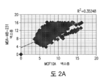
도 2A-F. 발암 소포에는 miRNA가 농축된다. (A) MDA-MB231 엑소좀 및 MCF10A 엑소좀에서 발현된 miRNA의 상관관계 그래프. (B) 72h의 무-세포 배양 후에 정상 소포 및 발암 소포 사이에서 차등하게 발현된 6가지 miRNA (miR-10a, miR-10b, miR-21, miR-27a, miR-155, 및 miR-373)를 이용한 세포 및 각각의 엑소좀 내 miRNA간의 상관관계 그래프. (C) 정상 소포 및 발암 소포를 DMEM 배지에 재현탁시키고 24 및 72h 동안 무-세포 배양을 유지하였다. 24 및 72h 후에, 엑소좀을 회수하고 15가지 miRNA (표 4 참고)를 qPCR로 정량하였다. 72h 무-세포 배양 후 엑소좀 내 각각의 miRNA의 배수-변화를 24h 무-세포 배양 후 엑소좀 내 동일한 miRNA에 비교하여 정량하였다. 그래프 도표는 24h 후에 수확된 것들과 비교하여 72h 후에 수확된 엑소좀 내 종양 억제제 (TS) 및 발암성 (ONC) miRNA에 대한 배수-변화의 평균을 나타낸다. (D) 24 및 72h의 무-세포 배양 후의 정상 소포 및 무배양된 및 24h, 72h 및 96h의 무-세포 배양된 발암 소포로부터의 miR-10b 및 miR-21의 노던 블롯. tRNAMet는 로딩 대조로서 사용하였다. 정량화는 이미지제이(Image J) 소프트웨어로 수행하였다. (E) 72h의 무세포 배양 후 MCF10A, MDA-MB231 및 4T1 세포 및 이들의 각각의 엑소좀 내 15가지 정량화된 miRNA간의 상관관계 도표. 발암 소포는 정상 소포와 비교할 때 (왼쪽 그래프) 이들이 유래한 세포와는 낮은 상관관계 수치를 나타낸다 (중간 및 오른쪽 그래프). (F) 초 (s)당 형광 단위 (FU)로 나타낸 생체분석 그래프 표현 및 정상 소포 및 발암 소포의 엑소좀 RNA 함량의 겔 사진.
도 3A-E. 엑소좀은 전구-miRNA를 함유한다. (A) 연구된 성숙 miRNA에 상응하는 열다섯 가지 전구-miRNA를 MCF10A 및 MDA-MB231 엑소좀의 qPCR을 이용하여 정량하였다. 각각의 전구-miRNA에 대한 ΔCt 값의 역수를 도시하여 이들의 많음을 나타내었고 수치는 ± 표준편차로서 표현된다. (B) 발암 소포 및 정상 소포를 DMEM 배지에 재현탁시키고 24 및 72h 동안 무-세포 배양 조건을 유지하였다. 24 및 72h 후에 엑소좀을 다시 회수하고 15가지 전구-miRNA를 qPCR로 정량하였다. 그래프는 24h 무-세포 배양에 비교하여 72h의 무-세포 배양 후의 MCF10A 및 MDA-MB231 엑소좀 내 각각의 전구-miRNA의 배수-변화를 보여주며 수치는 ± 표준편차로서 표현된다. (C) 24h 및 72h의 무-세포 배양 후의 MCF10A 정상 소포, 및 0h, 24h, 72h 및 96h의 무-세포 배양 후의 MDA-MB231 발암 소포를 이용한 전구miR-10b 및 전구-miR-21의 노던 블롯. tRNAMet는 로딩 대조로서 사용하였다. 정량화는 이미지제이(Image J) 소프트웨어로 수행하였다. (D) 상단 그래프: 발암 소포 (MDA-MB231)의 발암성 전구-miRNA (왼쪽 그래프) 및 발암성 miRNA (오른쪽 그래프)를 24h 및 72h 무-세포 배양 조건 후에 정량하였다. 상이한 시점에서 각각의 전구-miRNA (왼쪽 그래프) 및 miRNA (오른쪽 그래프)에 대한 ΔCt 값의 역수를 도시하여 이들의 많음을 나타내었고 기하급수적 경향이 특징적이다. 제시된 데이터는 세 개의 생물학적 복제 결과이며 표준편차(SD)로서 표현된다. 하단 그래프: 발암 소포 (MDA-MB231)의 전구-miRNA (왼쪽 그래프) 및 성숙 miRNA (오른쪽 그래프)를 6h, 12h, 24h, 36h, 48h, 72h 및 96h의 무-세포 배양 조건 후에 정량하였다. 상이한 시점에서 각각의 전구-miRNA (왼쪽 그래프) 및 miRNA (오른쪽 그래프)에 대한 ΔCt 값의 역수를 도시하였고 기하급수적 경향이 특징적이다. 도면에 나타난 데이터는 각각 세 가지 복제물을 이용한 세 번의 독립된 실험의 결과이며 ± 표준편차로서 표현된다. (E) 발암 소포 및 정상 소포를 DMEM 배지에 재현탁시키고 0h, 24h, 72h 및 96h 동안 무-세포 배양 조건을 유지하였다. 엑소좀을 상이한 시점에 추출하였고 전구-miRNA를 qPCR에 의해 정량하였다. 각각의 전구-miRNA에 대한 ΔCt 값의 역수를 도시하여 이들의 많음을 나타내었다.
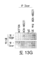
도 4A-N. 발암 소포는 RLC 단백질을 함유한다. (A) 다음으로부터 수확한 엑소좀 내 항-Dicer 항체를 이용한 면역 블롯: 비종양형성 마우스 (NMuMG) 및 인간 (MCF10A) 세포주; 마우스 암 세포주, 67NR 및 4T1; 및 인간 암 세포주 MCF7 및 MDAMB231. 사용된 대조군은 다음과 같다: 엑소좀의 용해를 유도하고 이어서 엑소좀의 단백질을 분해하기 위해 TritonX으로 처리 후 프로테이나제 K (Triton + PK)로 처리된 엑소좀; 및 나머지-엑소좀 단백질 (PK)을 분해하기 위해 프로테이나제 K로 처리된 엑소좀. TSG101 (두 번째 줄) 및 CD9 (세 번째 줄) 면역 블롯을 사용하여 엑소좀의 존재를 확인하였다. (B) 발암 소포 (MDA-MB231) 내 항-Dicer 항체를 이용한 면역금 표지법의 투과 전자 현미경 사진. 오른쪽 상단 사진은 추출물의 새로운 독립적인 사진을 디지털로 확대한 것이다. 음성 대조는 IgG를 지칭한다. 금 입자는 검은 점으로 나타나며 하단 사진에서 검은 화살표로 표시된다. 그래프는 왼쪽에 있는 두 개의 상단 사진을 정량한 것을 나타낸다. (C) 빈 벡터 (pCMV-Tag4B; 각각 첫 번째 및 세 번째 열) 및 Flag-Dicer 벡터 (두 번째 및 네 번째 열)로 형질주입된 세포로부터 수확한 MCF10A 및 MDA-MB231 엑소좀 내 항-플래그(flag) 항체 (상단 패널)를 이용한 면역 블롯. 엑소좀의 존재를 확인하고 로딩 대조(하단 패널)로서 사용하기 위해 CD9 면역 블롯을 이용하였다. (D) 칼슘 이온투과담체 A23187 로 처리한 MCF10A 및 MDA-MB231 세포로부터 수확한 엑소좀 내 Dicer에 대한 면역 블롯(상단 패널). 처리되지 않은 세포에서 추출한 엑소좀을 대조로서 사용하였다. CD9 면역 블롯 (하단 패널)을 대조로 사용하여 증가된 엑소좀 분비를 알아보았다. (E) shScramble 및 shDicer 플라스미드로 형질주입된 MCF10A 및 MDA-MB231 모 세포 및 세포로부터 추출한 엑소좀 내 Dicer에 대한 면역 블롯 (상단 블롯). 엑소좀의 존재를 알아보고 로딩 대조(하단 블롯)로 사용하기 위해 CD9 면역 블롯을 이용하였다. 면역 블롯 정량화는 이미지제이(Image J) 소프트웨어로 수행하였다. (F) MDAMB231shDicer 세포로부터 유래된 발암 소포 내 항-Dicer 항체를 이용한 면역금 표지법의 투과 전자 현미경 사진. 금 입자가 검은 점으로 나타난다. 오른쪽 그래프는 EM 사진에서 금 입자의 정량화를 도시한다. (G) 발암 소포 (MCF7 및 MDA-MB231) 및 정상 소포 (MCF10A)로부터 수확한 엑소좀 내 항-AGO2 항체를 이용한 면역 블롯. 사용된 대조군은 다음과 같다: 엑소좀의 용해를 유도하고 이어서 엑소좀의 단백질을 분해하기 위해 Triton X으로 처리 후 프로테이나제 K (Triton X + PK)로 처리된 엑소좀; 나머지-엑소좀 단백질 (PK)을 분해하기 위해 프로테이나제 K로 처리된 엑소좀; 및 엑소좀을 수확하기 위해 초원심분리한 후의 상청액 (상청액). TSG101 (두 번째 줄) 및 CD9 (세 번째 줄) 면역 블롯을 사용하여 엑소좀의 존재를 확인하였다. (H) 발암 소포 (MCF7 및 MDA-MB231) 및 정상 소포 (MCF10A)로부터 수확한 엑소좀 내 항-TRBP 항체를 이용한 면역 블롯. 사용된 대조군은 다음과 같다: 엑소좀의 용해를 유도하고 이어서 엑소좀의 단백질을 분해하기 위해 Triton X으로 처리 후 프로테이나제 K (Triton X + PK)로 처리된 엑소좀; 나머지-엑소좀 단백질 (PK)을 분해하기 위해 프로테이나제 K로 처리된 엑소좀; 및 엑소좀을 수확하기 위해 초원심분리한 후의 상청액 (상청액). TSG101 (두 번째 줄) 및 CD9 (세 번째 줄) 면역 블롯을 엑소좀 마커로서 사용하였다. (I) GFP-AGO2 플라스미드로 형질주입된 MCF10A 및 MDA-MB231 세포 내 항-GFP 항체를 이용한 면역 블롯 (상단 패널). 베타 액틴을 로딩 대조(하단 패널)로 사용하였다. (J) GFP-AGO2 플라스미드로 형질주입된 MCF10A 및 MDA-MB231 세포로부터 추출한 엑소좀 내 항-GFP 항체를 이용한 면역 블롯 (상단 패널). TSG101 (중간 패널) 및 CD9 (하단 패널)을 엑소좀 마커 및 로딩 대조로서 사용하였다. (K) siAGO2 플라스미드로 형질주입된 MCF10A 및 MDA-MB231 세포 내 AGO2 mRNA 발현. MCF10A 및 MDA-MB231 모세포를 배수 변화 비교를 위한 비교 대조로 사용하였다. 데이터는 세 개의 생물학적 복제 결과이며 표준편차(SD)로서 표현된다. (L) si대조군 또는 siAGO2로 형질주입된 MCF10A 및 MDA-MB231 모세포 또는 세포로부터 추출한 엑소좀 내 AGO2 항체를 이용한 면역 블롯 (상단 패널). TSG101 (중간 블롯) 및 CD9 (하단 블롯)을 엑소좀 마커 및 로딩 대조로서 사용하였다. 정량화는 이미지제이(Image J) 소프트웨어로 수행하였다. (M) Dicer 항체 또는 IgG로 면역침강시킨 MCF10A 및 MDA-MB231 세포로부터 추출한 엑소좀 단백질 내 AGO2 항체를 이용한 면역 블롯 (상단 패널). MDA-MB231 세포로부터 추출한 엑소좀의 용해물을 5% 투입하여 대조로서 사용하였다. Dicer의 면역 블롯을 면역 침강에 대한 대조로서 사용하였다 (하단 패널). (N) Dicer 항체 또는 IgG로 면역침강시킨 MCF10A 및 MDA-MB231 세포로부터 추출한 엑소좀 단백질 내 항-TRBP 항체를 이용한 면역 블롯 (상단 패널). MDA-MB231 세포로부터 추출한 엑소좀의 용해물 투입(5%)을 대조로서 사용하였다. Dicer의 면역 블롯을 대조로서 사용하였다 (하단 패널).
도 5A-E. 발암 소포는 전구-miRNA를 프로세싱하여 성숙 miRNA를 생성한다. (A) 엑소좀을 MCF10A, MCF10A shScramble, MCF10A shDicer 세포 (상단 그래프), MDA-MB231, MDA-MB231 shScramble 및 MDA-MB231 shDicer 세포 (하단 그래프)로부터 수확하고 무-세포 배양 조건 하에 24 및 72h 동안 유지하였다. 24 및 72h 후에 엑소좀을 회수하고 15가지 전구-miRNA를 qPCR로 정량하였다. 그래프는 24h 무-세포 배양에 비교하여 72h의 무-세포 배양 후의 서로 다른 엑소좀 내 각각의 전구-miRNA의 배수-변화를 보여주며 수치는 ± 표준편차로서 표현된다. (B) 엑소좀을 MCF10A, MCF10A shScramble, MCF10A shDicer 세포 (상단 그래프), MDA-MB231, MDA-MB231 shScramble 및 MDA-MB231 shDicer 세포 (하단 그래프)로부터 수확하고 무-세포 배양 조건 하에 24 및 72h 동안 유지하였다. 24 및 72h 후에 엑소좀을 다시 회수하고 15가지 miRNA를 qPCR로 정량하였다. 그래프는 24h 무-세포 배양에 비교하여 72h의 무-세포 배양 후의 서로 다른 엑소좀 내 각각의 miRNA의 배수-변화를 보여주며 수치는 ± 표준편차로서 표현된다. (C) MDA-MB231 세포의 엑소좀 내로 전기천공된 중사슬 (HC) 및 경사슬 (LC) 일차 Dicer 항체 및 일차 액틴 항체를 검출하기 위해 항토끼 및 항-마우스 이차 항체를 이용한 면역 블롯. MDA-MB231 세포에서 유래한 항체가 없는 전기천공된 엑소좀을 음성 대조로 사용하였다. 엑소좀에 포함되지 않는 항체를 완전히 고갈시키기 위해 전기천공 후에 프로테이나제 K 처리를 수행하였다. (D) 발암 소포 (MDA-MB231)를 이중복 (하단 그래프) 또는 사중복 (상단 그래프)으로 수확하였다. 샘플을 항-Dicer 항체, 항-액틴 항체, 또는 항-TRBP 항체로 전기천공하였다. 샘플 더하기 대조를 무-세포 배양 조건 하에 24 및 72h 동안 방치하였다. 24 및 72h 후에 엑소좀을 다시 추출하고 6가지 발암성 전구-miRNA (맨위 그래프) 또는 15가지 전구-miRNA (아래 그래프)를 qPCR로 정량하였다. 72h 무-세포 배양 후 엑소좀 내 각각의 전구-miRNA의 배수-변화를 각 샘플에서 24h 무-세포 배양 후 엑소좀 내 동일한 전구-miRNA에 비교하여 정량하였다. 그래프 도표는 24h 엑소좀에 비하여 72h 엑소좀 내 전구-miRNA에 대한 평균 배수-변화(하단 그래프 - TS = 종양 억제제; ONC = 발암성)를 나타내며 수치는 ± 표준편차로서 표현된다. (E) 발암 소포 (MDAMB231)를 사중복 (상단 그래프) 또는 이중복 (하단 그래프)으로 수확하였다. 샘플을 항-Dicer 항체, 항-액틴 항체, 또는 항-TRBP 항체로 전기천공하였다. 샘플 더하기 대조를 무-세포 배양 조건 하에 24 및 72h 동안 방치하였다. 24 및 72h 후에 엑소좀을 다시 추출하고 6가지 발암성 miRNA (맨위 그래프) 또는 15가지 miRNA (아래 그래프)를 qPCR로 정량하였다. 72h 무-세포 배양 후 엑소좀 내 각각의 miRNA의 배수-변화를 각 샘플에서 24h 무-세포 배양 후 엑소좀 내 동일한 miRNA에 비교하여 정량하였다. 그래프 도표는 24h 엑소좀에 비하여 72h 엑소좀 내 miRNA에 대한 평균 배수-변화(하단 그래프 - TS = 종양 억제제; ONC = 발암성)를 나타내며 수치는 ± 표준편차로서 표현된다.
도 6A-E. 발암 소포는 전구-miRNA를 프로세싱하여 성숙 miRNA를 생성한다. (A) MDA-MB231 세포로부터의 엑소좀을 수확하고 젤다나마이신(Geldanamycin)으로 전기천공시켰다. 샘플을 무-세포 배양 조건 하에 24 및 72h 동안 방치한 뒤, 엑소좀을 추출하고 6가지 miRNA를 qPCR로 정량하였다. 72h 무-세포 배양 후 엑소좀 내 각각의 miRNA의 배수-변화를 각 샘플에서 24h 무-세포 배양 후 엑소좀 내 동일한 miRNA에 비교하여 정량하였다. 그래프 도표는 24h 엑소좀에 비하여 72h 엑소좀 내 miRNA에 대한 평균 배수-변화를 나타내며 수치는 ± 표준편차로서 표현된다. (B) 합성 전구-miRNA -10b, -21 및 -cel-1을 MCF10A (MCF10A 전기천공물), MCF10AshDicer (MCF10AshDicer 전기천공물), MDAMB231 (MDA-MB231 전기천공물) 및 MDA-MB231shDicer (MDAMB231shDicer 전기천공물) 세포로부터 수확한 엑소좀 내로 전기천공시켰다. 무-세포 배양 조건에서 72h 동안 배양한 후에 엑소좀을 회수하였다. 전구-miR-10b, -21 및 -cel-1을 72h의 전기천공 및 배양 전후에 qPCR로 정량하였다. 도표 상의 각각의 막대는 전기천공 후 0h에 비하여 전기천공 후 72h에서 전구-miR-10b, -21 및 -cel-1의 배수-변화를 나타내며 ± 표준편차로서 표현된다. 전구-miRNA의 부재에서 전기천공시킨 MCF10A 및 MDA-MB231 엑소좀 전기천공물을 기저 수준을 강조하기 위한 대조로서 사용하였다. (C) 합성 전구-miRNA -10b, -21 및 -cel-1을 MCF10A (MCF10A 전기천공물), MCF10AshDicer (MCF10AshDicer 전기천공물), MDA-MB231 (MDA-MB231 전기천공물) 및 MDAMB231shDicer (MDA-MB231shDicer 전기천공물) 세포로부터 수확한 엑소좀 내로 전기천공시켰다. 무-세포 배양 조건에서 72h 동안 배양한 후에 엑소좀을 회수하였다. MiR-10b, -21 및 -cel-1을 72h의 전기천공 및 배양 전후에 qPCR로 정량하였다. 도표 상의 각각의 막대는 전기천공 후 0h (위쪽 그래프) 또는 24h (아래쪽 그래프)에 비하여 전기천공 후 72h에서 miR-10b, -21 및 -cel-1의 배수-변화를 나타내며 ± 표준편차로서 표현된다. 전구-miRNA의 부재에서 전기천공시킨 MCF10A 및 MDA-MB231 엑소좀 전기천공물을 정해진 기저 수준에 대한 대조로서 사용하였다. (D) 검출 프로브 없이, 다이싱 분석으로부터의 샘플을 이용하는 노던 블롯. 상이한 엑소좀 단백질 추출물 및 내부에 비오틴으로 표지한 합성 전구-miR-10b를 다이싱 분석을 위해 사용하였다. 사용된 샘플은 MCF10A, MCF10AshDicer, MDA-MB231 엑소좀 (MDA231 엑소좀), MDA-MB231shDicer 클론 1 및 클론2로부터의 엑소좀 (각각 MDA231shDicer 1 엑소좀 및 MDA231shDicer 2 엑소좀), MDA-MB231shDicer 세포 및 Dicer 항체로 전기천공시킨 MDA-MB231 엑소좀 (MDA231 엑소좀 + Dicer AB)였다. (E) 검출 프로브 없이, 다이싱 분석으로부터의 샘플을 이용하는 노던 블롯. 상이한 엑소좀 단백질 추출물 및 내부에 비오틴으로 표지한 합성 전구-miR-21를 다이싱 분석을 위해 사용하였다. 사용된 샘플은 MCF10A, MCF10AshDicer, MDA-MB231 엑소좀 (MDA231 엑소좀), MDA-MB231shDicer 클론 1 및 클론2로부터의 엑소좀 (각각 MDA231shDicer 1 엑소좀 및 MDA231shDicer 2 엑소좀), MDA-MB231shDicer 세포 및 Dicer 항체로 전기천공시킨 MDA-MB231 엑소좀 (MDA231 엑소좀 + Dicer AB)였다. (F) 검출 프로브 없이, 다이싱 분석으로부터의 샘플을 이용하는 노던 블롯. 상이한 엑소좀 단백질 추출물 및 내부에 비오틴으로 표지한 합성 전구-cel-miR-1을 다이싱 분석을 위해 사용하였다. 사용된 샘플은 MCF10A, MCF10AshDicer, MDA-MB231 엑소좀 (MDA231 엑소좀), MDA-MB231shDicer 엑소좀 (MDA231shDicer 엑소좀) 및 Dicer 항체로 전기천공시킨 MDAMB231 엑소좀 (MDA231 엑소좀 + Dicer AB)였다. 데이터는 세 개의 생물학적 복제 결과이며 표준편차(SD)로서 표현된다.
도 7A-H. 발암 소포는 수취 세포에서 Dicer-의존 방식으로 전사체 변형 및 종양 형성을 유발한다. (A) 무-세포 배양 후 0, 30분, 1h, 12h 및 24h동안 MDA-MB231 발암 소포로 처리한 MCF10A 세포의 항-PTEN 항체 및 단백질 추출물을 이용한 면역 블롯. 베타 액틴을 로딩 대조로서 사용하였다. (B) 무-세포 배양 조건 후 0, 30분, 1h, 12h 및 24h동안 MDA-MB231 발암 소포로 처리한 MCF10A 세포의 항-HOXD10 항체 및 단백질 추출물을 이용한 면역 블롯. 베타 액틴을 로딩 대조로서 사용하였다. (C) 3'UTR-PTEN-WT, 3'UTRPTEN-Mut, 3'UTR-HOXD10-WT 및 3'UTR-HOXD10-Mut로 일시적으로 형질주입된 및 MDA-MB231 세포에서 유래한 발암 소포로 처리한 MCF10A 세포에서 루시페라아제 리포터(reporter) 활성을 보여주는 그래프. (D) 무-세포 배양 조건 후 Dicer 항체로 전기천공시킨 MDAMB231 발암 소포로 0, 30분, 1h, 12h 및 24h동안 처리한 MCF10A 세포의 항-PTEN 항체 (상단 패널) 및 항-HOXD10 항체 (중간 패널) 및 단백질 추출물을 이용한 면역 블롯. 베타 액틴을 로딩 대조로서 사용하였다. (E) 무-세포 배양 시간 없이 MDA-MB231 엑소좀과 항-miR-182-5p 및 MDA-MB231 엑소좀으로 처리한 항-Smad4 항체 (상단 패널) 및 MCF10A 세포 및 MCF10A 세포의 단백질 추출물을 이용한 면역 블롯. 베타 액틴을 로딩 대조로서 사용하였다. (F) MCF10A 세포, 무-세포 배양 시간 없이 MDA-MB231 엑소좀으로 처리한 MCF10A 세포 (MCF10A + MDA231 엑소좀), 무-세포 배양 시간을 가지고 MDA-MB231 엑소좀으로 처리한 MCF10A 세포 (MCF10A 세포 + MDA231 엑소좀 배양) 및 무-세포 배양 시간을 가지고 Dicer 항체로 전기천공시킨 MDA-MB231 엑소좀으로 처리한 MCF10A 세포 (MCF10A 세포 + MDA231 엑소좀 Dicer AB)를 5일의 배양 동안 MTT 어세이로 세포 생존능을 측정했으며 수치는 ± 표준편차로서 표현된다 * p=0.0027. (G) 콜로니 형성 어세이는 8일 후 배양 접시 내 MTT 시약으로 표지된 MCF10A 세포 배양물, 무-세포 배양 시간 없이 MDA-MB231 엑소좀으로 처리한 MCF10A 세포 (MCF10A + MDA231 엑소좀), 무-세포 배양 시간을 가지고 MDA-MB231 엑소좀으로 처리한 MCF10A 세포 (MCF10A 세포 + MDA231 엑소좀 배양) 및 무-세포 배양 시간을 가지고 Dicer 항체로 전기천공시킨 MDA-MB231 엑소좀으로 처리한 MCF10A 세포 (MCF10A 세포 + MDA231 엑소좀 Dicer AB) 콜로니의 형성을 보여준다. (H) 상단 그래프: MCF10A 세포, MDA-MB-231 발암 소포에 노출된 MCF10A 세포 (MCF10A 세포 + MDA231 엑소좀 배양물), Dicer 항체로 전기천공시킨 MDA-MB-231 발암 소포에 노출된 MCF10A 세포 (MCF10A 세포 + MDA231 엑소좀 Dicer AB) 및 액틴 항체로 전기천공시킨 MDAMB231발암 소포에 노출된 MCF10A 세포 (MCF10A 세포 + MDA231 엑소좀 액틴 AB)를 무흉선 누드 마우스의 유방 패드에 동소위(orthotopically) 주사하였다. 그래프는 시간에 따른 종양 부피를 도시하며 ± 표준편차로서 표현된다 *p=0.005. 하단 그래프: MCF10A 세포, MDA-MB231 세포 및 발암 소포 (MDA-MB231)에 노출된 MCF10A 세포를 무흉선 누드 마우스의 유방 패드에 동소위 주사하였다. 그래프는 시간에 따른 종양 부피를 도시한다.
도 8A-I. 유방암 환자로부터의 혈청은 Dicer를 함유하며 전구-miRNA를 프로세싱한다. (A) 인간 및 마우스 Dicer를 인식하는 항-Dicer 항체, 및 인간 종양을 이종이식받은 마우스에게서 수확한 혈청 엑소좀의 단백질 추출물을 이용한 면역 블롯 (도 18A에 나타난 바와 같음). OVA1-5는 인간 난소 이종이식편을 나타내고; END1-3은 인간 자궁내막 이종이식편을 나타내며; BRST1 및 2는 인간 유방 이종이식편을 나타낸다. 4T1 엑소좀 및 세포를 쥐의 Dicer에 대한 대조로서 사용하였다. hsa-Dicer는 인간 Dicer 분자량을 나타내고 mmu-Dicer는 쥐의 Dicer 분자량을 나타낸다. 막의 쿠마시(Comassie) 염색에 대한 로딩 대조에 대하여는 도 18D를 참조하라. (B) 8명의 건강한 공여자 (왼쪽 그래프) 및 11명의 유방암 환자 (오른쪽 그래프)의 혈청에서 추출한 엑소좀의 크기 분포를 나타내는 나노사이트 입자 추적 분석. 크기를 더 잘 나타내기 위해 샘플의 농도를 표준화하였다. (C) 유방암 환자의 혈청에서 수확한 엑소좀의 투과 전자 현미경 사진. (D) 나노사이트 입자 추적 분석으로 평가한 8명의 건강한 공여자 및 11명의 유방암 환자의 혈청으로부터의 엑소좀의 농도. *p=0.012 (E) 엑소좀을 8명의 건강한 공여자 및 11명의 유방암 환자의 혈청으로부터 수확하였다. 추출한 샘플을 무-세포 배양 조건 하에 24 및 72h 동안 방치하였다. 24 및 72h 후에 엑소좀을 회수하고 6가지 전구-miRNA를 qPCR로 정량하였다. 72h 무-세포 배양 후 엑소좀 내 각각의 전구-miRNA의 배수-변화를 각 샘플에서 24h 무-세포 배양 후 엑소좀 내 동일한 전구-miRNA에 비교하여 정량하였다. 그래프 점 도표는 24h 엑소좀에 비하여 72h 엑소좀 내 전구-miRNA에 대한 평균 배수변화를 나타내며 수치는 ± 표준편차로서 표현된다. (F) 엑소좀을 8명의 건강한 공여자 및 11명의 유방암 환자의 혈청으로부터 수확하였다. 추출한 샘플을 무-세포 배양 조건 하에 24 및 72h 동안 방치하였다. 24 및 72h 후에 엑소좀을 회수하고 6가지 miRNA를 qPCR로 정량하였다. 72h 무-세포 배양 후 엑소좀 내 각각의 miRNA의 배수변화를 각 샘플에서 24h 무-세포 배양 후 엑소좀 내 동일한 miRNA에 비교하여 정량하였다. 그래프 점 도표는 24h 엑소좀에 비하여 72h 엑소좀 내 miRNA에 대한 평균 배수-변화를 나타낸다. 패널 E와 F는 둘다 각각 세 가지 복제물을 이용한 세 번의 독립된 실험의 결과이며 ± 표준편차로서 표현된다. (G) MCF10A 세포, 건강한 공여자 (H1-8)의 엑소좀과 혼합된 MCF10A 세포 및 유방암 환자 (BC1-11)의 엑소좀과 혼합된 MCF10A 세포를 무흉선 누드 마우스의 유방 패드에 동소위 주입하였다. 사용된 엑소좀의 수는 혈청으로부터 수집한 최초 농도를 반영하여 체중당 산출하였다. 종양을 형성하지 않은 샘플은 그래프의 x축에 겹쳐진 것으로 보인다. 그래프는 시간에 따른 종양 부피를 도시하며 ± 표준편차로서 표현된다. (H) CD9 블롯을 로딩 대조로 사용하고 5명의 건강한 개체(C46, C45, C44, C43 및 C41) 및 4명의 전이성 유방 암종(Met219, Met354, Met299 및 Met356)로부터 수확한 혈청 엑소좀으로부터의 항-Dicer 항체 및 단백질 추출물을 이용한 면역 블롯. HDF와 발암 소포 (MDA-MB231)로 처리한 HDF의 배가 시간(doubling time). * p=0.0114. 면역 블롯 정량화는 이미지제이(Image J) 소프트웨어로 수행하였다.
도 9A-B. Dicer는 다포성 소체로 존재하며 세포질의 CD43가 Dicer를 엑소좀 내로 이동시킨다. (A) Dicer 항체 (IP Dicer) 또는 IgG로 (상단 패널, 각각 오른쪽과 중간 열) 면역침강시킨 MDA-MB231 세포의 단백질 추출물 내 CD43의 면역 블롯. Dicer 단독의 면역 블롯을 대조로서 사용하였다 (하단 패널). (B) MDA-MB231 유래 엑소좀 및 MDA-MB231 siCD43 유래 엑소좀의 단백질 추출물 내 Dicer의 면역 블롯. CD9 면역 블롯을 로딩 대조로서 사용하였다. 정량화는 이미지제이(Image J) 소프트웨어로 수행하였다.
도 10A-E. 엑소좀 특징 분석. (A) 초원심분리 튜브 하단에 있는, PKH26 염색된 엑소좀의 사진. 삽도는 엑소좀의 디지털 확대 사진을 나타낸다. (B) LSS 스펙트럼을 수집하기 위해 사용한 실험 시스템의 모식도. (C) MCF10A, NMuMG, MDA-MB231 및 4T1 세포를 5일 배양하는 동안 MTT 어세이로 측정한 세포 생존능. (D) MDA-MB231 및 4T1 세포의 프로피듐 아이오다이드 (PI) 및 아넥신(Anexin) V에 대한 유세포 분석. 에토포시드로 처리한 MDA-MB231 세포를 아폽토시스에 대한 양성 대조로서 사용하였다. (E) 엑소좀에 대하여 MDA-MB231 세포를 양성 대조로서 및 TSG101를 로딩 대조로서 사용하는 엑소좀 내 시토크롬 C의 면역 블롯 분석. 도면에 나타난 데이터는 각각 세 가지 복제물을 이용한 세 번의 독립된 실험의 결과이며, ± 표준편차로서 표현된다.
도 11A-E. 발암 소포는 정상 소포에 비해 miRNA가 농축되어 있다. (A) 생체분석기 뉴클레오티드 (nt)당 형광 단위 (FU)로 도시된 그래프 도식 (그래프) 및 인간 유방 MCF10A (비-종양형성) 및 MDA-MB231 (유방암) 세포주의 RNA 함량의 겔 사진 (오른쪽 사진). (B) 4T1, MCF10A 및 MDA-MB231 세포로부터 수확한 엑소좀을 DMEM 배지에 재현탁시키고 24 및 72h 동안 무-세포 배양 조건을 유지하였다. 24 및 72h 후에 엑소좀을 회수하고 15가지 miRNA (표 4 참고)를 qPCR로 정량하였다. 그래프는 각각 24 및 72h의 무-세포 배양 후의 정상 소포에 비해 24h (상단 그래프) 및 72h (하단 그래프)의 무-세포 배양 후의 발암 소포 내 각각의 miRNA의 배수 변화를 나타낸다. 제시된 데이터는 세 개의 생물학적 복제 결과이며 표준편차(SD)로서 표현된다. (C) 열다섯 가지의 성숙 miRNA (표 4를 참조)를 MCF10A (왼쪽 그래프), MDA-MB231 (중간 그래프) 및 4T1 (오른쪽 그래프) 세포 및 이들의 각각의 엑소좀에서 qPCR로 정량하였다. 엑소좀 내 각각의 miRNA의 배수 변화를 세포 내 동일한 miRNA에 비교하여 정량하였다. TS: 종양 억제제 miRNA; ONC: 발암성 miRNA. 데이터는 세 개의 생물학적 복제 결과이며 표준편차(SD)로서 표현된다. (D) MCF10A, MDA-MB231 및 4T1 세포에서 수확한 엑소좀을 DMEM 배지에 재현탁시키고 24 및 72h 동안 무-세포 배양 조건을 유지하였다. 24 및 72h 후에 엑소좀을 다시 회수하고 15가지 miRNA(표 4를 참조)를 qPCR로 정량하였다. 72h 무-세포 배양 후 엑소좀 내 각각의 miRNA의 배수 변화를 24h 무-세포 배양 후 엑소좀 내 동일한 miRNA에 비교하여 정량하였다. 데이터는 도 2C의 배수 변화 평균 그래프들 중 상세한 그래프에 해당된다. 도면에 나타난 데이터는 각각 세 가지 복제물을 이용한 세 번의 독립된 실험의 결과이며, ± 표준편차로서 표현된다. (E) 72h의 무세포 배양 후 MCF7 및 67NR 세포 및 이들의 각각의 엑소좀 내 정량화된 15가지 miRNA간의 상관관계 도표.
도 12A-E. 엑소좀은 전구-miRNA를 함유한다. (A) 앞서 정량화된 성숙 miRNA에 상응하는 열다섯 가지 전구-miRNA (표 4를 참조)를 NMuMG 및 4T1 엑소좀에서 qPCR로 정량하였다. 각각의 전구-miRNA에 대한 ΔCt 값의 역수를 도시하여 이들의 많음을 나타내었다. 데이터는 세 개의 생물학적 복제 결과이며 ± 표준편차로서 표현된다. (B) NMuMG 및 4T1 세포에서 수확한 엑소좀을 DMEM 배지에 재현탁시키고 24 및 72h 동안 무-세포 배양 조건을 유지하였다. 24 및 72h 후에 엑소좀을 다시 회수하고 15가지 전구-miRNA를 qPCR로 정량하였다. 그래프는 24h 무-세포 배양에 비교하여 72h의 무-세포 배양 후의 NMuMG 및 4T1 엑소좀 내 각각의 전구-miRNA의 배수 변화를 보여준다. 데이터는 세 개의 생물학적 복제 결과이며 표준편차(SD)로서 표현된다. (C) 대조 세포에 비하여 배수 변화로서 두 가지 일시적으로 형질주입된 XPO5를 표적하는 siRNA를 갖는 MDAMB231 세포 내 XPO5 mRNA 발현. (D) MDA-MB231 세포를 XPO5 siRNA 구조체로 형질주입시키고 12h 형질주입-후 여러 시점(0h, 6h, 12h, 24h, 36h, 48h, 72h 및 96h)에서 miR-21 발현을 평가하였다. 긴 원심분리 기간의 영향을 보기 위한 비교로서 XPO5 siRNA 구조체를 형질주입한 MDA-MB231 세포를 3h 동안 4°C에서 원심분리하고 배양액에 원위치시켰다. MiR-21 발현을 원심분리후 여러 시점(0h, 6h, 12h, 24h, 36h, 48h, 72h 및 96h)에서 평가하였다. 전구miR21에서 miR21로의 프로세싱이 원심분리된 세포에서 지연된다(녹색 막대). 도면에 나타난 데이터는 각각 세 가지 복제물을 이용한 세 번의 독립된 실험의 결과이며 ± 표준편차로서 표현된다. (E) NMuMG 및 4T1 세포에서 수확한 엑소좀을 DMEM 배지에 재현탁시키고 0, 24, 72 및 96h 동안 무-세포 배양 조건을 유지하였다. 엑소좀을 상이한 시점에 추출하였고 전구-miRNA를 qPCR에 의해 정량하였다. 각각의 전구-miRNA에 대한 ΔCt 값의 역수를 도시하여 이들의 많음을 나타내었다. 데이터는 세 개의 생물학적 복제 결과이며 표준편차(SD)로서 표현된다.
도 13A-H. 발암 소포는 Dicer를 함유한다. (A) MCF10A 세포-유래 엑소좀에서 항-Dicer 항체 (오른쪽 사진) 및 음성 대조 (왼쪽 사진)을 이용하여 면역금 표지법으로 생성한 투과 전자 현미경 사진. MDA-MB231 엑소좀의 양성 면역금 표지에 대하여는 도 4B를 비교하라. (B) 항-GFP 항체 MDA-MB231-유래 엑소좀을 이용하여 면역금 표지법으로 생성한 투과 전자 현미경 사진. (C) 빈 벡터 (pCMV-Tag4B; 각각 첫 번째 및 세 번째 열) 및 Flag-Dicer 벡터 (두 번째 및 네 번째 열)로 형질주입된 MCF10A 및 MDAMB231 세포 내 항-플래그 항체 (상단 패널)를 이용한 면역 블롯. 베타 액틴 면역 블롯을 로딩 대조로서 사용하였다 (하단 패널). (D) 각각 MCF10A, MCF10AshScramble 및 MCF10AshDicer 클론 1 및 2 (MCF10AshDicer 클론1 및 MCF10AshDicer 클론2) 세포 내 항-Dicer 항체 (상단 패널)를 이용한 면역 블롯. 베타 액틴 면역 블롯을 로딩 대조로서 사용하였다 (하단 패널). (E) 각각 MDA-MB231, MDA-MB231shScramble 및 MDA-MB231shDicer 클론 1 및 2 (MDA-MB231shDicer 클론1 및 MDA-MB231shDicer 클론2) 세포 내 항-Dicer 항체 (상단 패널)를 이용한 면역 블롯. 베타 액틴 면역 블롯을 로딩 대조로서 사용하였다 (하단 패널). 면역 블롯 정량화는 이미지제이(Image J) 소프트웨어로 수행하였다. (F) Dicer 항체 또는 IgG로 면역침강시킨 MCF10A 및 MDA-MB231 세포로부터 추출한 엑소좀 단백질 내 AGO2 항체를 이용한 면역 블롯 (상단 패널). MDA-MB231 세포로부터 추출한 엑소좀의 용해물을 5% 투입하여 대조로서 사용하였다. Dicer의 면역 블롯을 면역 침강에 대한 대조로서 사용하였다 (하단 패널). (G) Dicer 항체 또는 IgG로 면역침강시킨 MCF10A 및 MDA-MB231 세포로부터 추출한 엑소좀 단백질 내 항-TRBP 항체를 이용한 면역 블롯 (상단 패널). MDA-MB231 세포로부터 추출한 엑소좀의 용해물 투입(5%)을 대조로서 사용하였다. Dicer의 면역 블롯을 면역 침강에 대한 대조로서 사용하였다 (하단 패널). (H) A549 (인간 폐암), SW480 (인간 결장암), HeLa (인간 자궁경부암) 및 4T07 (쥐의 유방암) 세포주 (상단 블롯)로부터의 발암 소포 내 Dicer의 면역 블롯. 엑소좀 및 로딩(하단 블롯)의 존재를 확인하기 위해 TSG101 면역 블롯을 이용하였다.
도 14A-F. 엑소좀 내 Dicer 검출. (A) 4T1, 4T1shScramble 및 4T1shDicer 세포 및 4T1 (4T1 엑소좀) 및 4T1shDicer (4T1shDicer 엑소좀) 세포 (상단 블롯)으로부터 수확한 엑소좀 내 항-Dicer 항체를 이용한 면역 블롯. GADPH 면역 블롯을 로딩 대조로서 사용하였다 (하단 블롯). 정량화는 이미지제이(Image J) 소프트웨어로 수행하였다. (B) 엑소좀을 4T1, 4T1shScramble 및 4T1shDicer 세포로부터 수확하고 무-세포 배양 조건 하에 24 및 72h 동안 유지하였다. 24 및 72h 후에 엑소좀을 다시 회수하고 15가지 전구-miRNA를 qPCR로 정량하였다. 그래프는 24h 무-세포 배양에 비교하여 72h의 무-세포 배양 후의 서로 다른 엑소좀 내 각각의 전구-miRNA의 배수 변화를 보여준다. 데이터는 세 개의 생물학적 복제 결과이며 표준편차(SD)로서 표현된다. (C) 엑소좀을 4T1, 4T1shScramble 및 4T1shDicer 세포로부터 수확하고 무-세포 배양 조건 하에 24 및 72h 동안 유지하였다. 24 및 72h 후에 엑소좀을 다시 회수하고 15가지 miRNA를 qPCR로 정량하였다. 그래프는 24h 무-세포 배양에 비교하여 72h의 무-세포 배양 후의 서로 다른 엑소좀 내 각각의 miRNA의 배수 변화를 보여준다. 데이터는 세 개의 생물학적 복제 결과이며 표준편차(SD)로서 표현된다. (D) 엑소좀을 MDA-MB231 세포로부터 이중복으로 수확하였다. 샘플 중 하나를 항-Dicer 항체로 전기천공시켰다. 양 샘플을 무-세포 배양 조건 하에 24 및 72h 동안 방치하였다. 24 및 72h 후에 엑소좀을 다시 회수하고 15가지 전구-miRNA(표 4를 참조)를 qPCR로 정량하였다. 72h 무-세포 배양 후 엑소좀 내 각각의 전구-miRNA의 배수 변화를 각 샘플에서 24h 무-세포 배양 후 엑소좀 내 동일한 전구-miRNA에 비교하여 정량하였다. 그래프 도표는 24h 엑소좀에 비하여 72h 엑소좀 내 전구-miRNA의 배수 변화를 나타내며 그래프의 자세한 분석은 도 5D에 제시된다. 데이터는 세 개의 생물학적 복제 결과이며 표준편차(SD)로서 표현된다. (E) 엑소좀을 MDA-MB231 세포로부터 이중복으로 수확하였다. 샘플 중 하나를 항-Dicer 항체로 전기천공시켰다. 양 샘플을 무-세포 배양 조건 하에 24 및 72h 동안 방치하였다. 24 및 72h 후에 엑소좀을 다시 회수하고 15가지 miRNA(표 4를 참조)를 qPCR로 정량하였다. 72h 무-세포 배양 후 엑소좀 내 각각의 miRNA의 배수 변화를 각 샘플에서 24h 무-세포 배양 후 엑소좀 내 동일한 miRNA에 비교하여 정량하였다. 그래프 도표는 24h 엑소좀에 비하여 72h 엑소좀 내 miRNA의 배수 변화를 나타내며 그래프의 자세한 분석은 도 5E에 제시된다. 데이터는 세 개의 생물학적 복제 결과이며 표준편차(SD)로서 표현된다. (F) MDA-MB231 엑소좀 (MDA-MB231 엑소좀)와 비교하여 Dicer (MDA-MB231 엑소좀 Dicer AB)로 전기천공시킨 MDA-MB231 엑소좀 내 하향 조절된 miRNA의 카테고리(발암성, 종양 억제성 및 암에 관해 비-결정성)의 그래프 도표. 마이크로RNA는 문헌을 기초로 하여 각각의 카테고리에 배정하였다. 도면에 나타난 데이터는 각각 세 가지 복제물을 이용한, 세 번의 독립된 실험의 결과이며 ± 표준편차로서 표현된다.
도 15A-C. 엑소좀 내 Dicer 검출. (A) 엑소좀을 MCF10, MCF10AshDicer, MDA-MB231 및 MDA-MB231shDicer 세포로부터 수확하고 합성 전구-miRNA-10b, -21 및 -cel-1로 전기천공시켰다. 전기천공시킨 엑소좀 내 각각의 전구-miRNA를 qPCR로 정량하고 전기천공 완충액으로만 전기천공시킨 엑소좀에 비한 배수 변화로서 나타내었다. (B) 전구-miR-21, -10b 및 -cel-1로 내부적으로 표지시킨 비오틴의 점 블롯. (C) 전구-miR-10b, -21 및 -cel-1로 형질주입시킨 MCF10A 세포의 miR-10b, -21 및 -cel-1 발현 분석. 각각의 막대는 미형질주입 세포에 비한 형질주입 세포의 배수 변화를 나타낸다. 도면에 나타난 데이터는 각각 세 가지 복제물을 이용한, 세 번의 독립된 실험의 결과이며 ± 표준편차로서 표현된다.
도 16A-I. Dicer는 다포성 소체로 존재하며 세포질의 CD43가 Dicer를 엑소좀 내로 이동시킨다. (A) 그래프는 이미지제이 소프트웨어를 이용하여 정량한 바와 같이 공초점 사진에서 공존 백분율을 나타낸다. (B) Hrs 및 TSG101에 대한 두 가지 상이한 siRNA 및 BiG2에 대한 두 가지 상이한 sh 클론을 이용한 하향 조절 후 Hrs, TSG101 및 BiG2 mRNA 발현. 미형질주입 세포 및 shScramble 형질주입 세포를 대조로서 사용하였다. (C) MCF10A, MCF10AsiHrs, MDA-MB231 및 MDA-MB231siHrs (왼쪽 그래프), MCF10shScramble, MCF10AshBiG2, MDA-MB231shScramble, MDA-MB231shBiG2 (중간 그래프) 및 MCF10AsiTSG101 및 MDA-MB231siTSG101 (오른쪽 그래프)로부터 추출한 엑소좀의 브래드포드(Bradford) 어세이에 의한 단백질 정량화. 미형질주입 모세포를 배수 변화 분석을 위한 비교 대조로 사용하였다. 데이터는 세포수에 의해 정상화되며 세 개의 생물학적 복제 결과가 표준편차(SD)로서 표현된다. (D) MCF10A, MCF10AsiTSG101 (siTSG101), MCF10AsiHrs (siHrs) 및 MCF10AshBiG2 (shBiG2) 세포의 엑소좀의 단백질 추출물 내 CD9의 면역 블롯 (상단 블롯); MDA-MB231, MDA-MB231siTSG101 (siTSG101), MDA-MB231siHrs (siHrs) 및 MDA-MB231shBiG2 (shBiG2) 세포의 엑소좀의 단백질 추출물 내 CD9의 면역 블롯 (하단 블롯). (E) Hrs, TSG101 및 BiG2 하향 조절된 세포에서의 엑소좀 수의 하향 조절 및 엑소좀의 추정 크기 분포를 나타내는 MDA-MB231, MDA-MB-231siTSG101, -siHrs 및 shBiG2-유래 엑소좀의 나노사이트 입자 추적 분석. (F) MCF10A, MCF10AshScramble, MCF10AsiHrs, MCF10AshBiG2, MCF10AsiTSG101, MDA-MB231, MDA-MB231shScramble, MDA-MB231siHrs, MDA-MB231shBiG2, MDA-MB231siTSG101, 4T1, 4T1siHrs, 4T1shBiG2 및 4T1siTSG101 세포 내 Dicer의 mRNA 발현. 모세포를 배수 변화 비교를 위한 비교 대조로 사용하였다. 데이터는 세 개의 생물학적 복제 결과이며 표준편차(SD)로서 표현된다. (G) 항-Dicer 항체로 면역침강시킨 MDA-MB231 및 4T1 암 세포의 단백질 추출물 (상단 블롯, 왼쪽 두개 열)과 더불어 면역침강을 위해 사용한 단백질 용해물에 해당하는 5% 투입물 (상단 블롯, 오른쪽 두개 열) 내 Dicer의 면역 블롯. 항-Dicer 항체로 면역침강시킨 MDA-MB231 및 4T1 세포의 단백질 추출물 (하단 블롯, 왼쪽 두개 열)과 더불어 면역침강을 위해 사용한 단백질 용해물에 해당하는 5% 투입물 (하단 블롯, 오른쪽 두개 열) 내 다중 유비퀴틴의 면역 블롯. (H) MCF10A, MCF10AsiCD43, MDA-MB231 및 MDA-MB231siCD43 세포 내 CD43의 mRNA 발현. MCF10A 및 MDA-MB231 모세포를 배수 변화 비교를 위한 비교 대조로 사용하였다. 데이터는 세 개의 생물학적 복제 결과이며 표준편차(SD)로서 표현된다. (I) MCF10A, MCF10AsiCD43, MDA-MB231 및 MDA-MB231siCD43 세포 내 Dicer의 mRNA 발현. MCF10A 및 MDA-MB231 모세포를 배수 변화 비교를 위한 비교 대조로 사용하였다. 데이터는 세 개의 생물학적 복제 결과이며 표준편차(SD)로서 표현된다.
도 17A-G. 발암 소포는 수취 세포에서 Dicer-의존 방식으로 전사체 변형 및 종양 형성을 유발한다. (A) MDA-MB231 CD63-GFP 세포로부터 유래한 엑소좀의 나노사이트 입자 추적 분석. 검은 선은 총 엑소좀 군의 척도를 나타내며 녹색 선은 488nm 레이저 빔이 구비된 나노사이트를 이용하여 CD63-GFP로 표지시킨 엑소좀의 군을 도시한다. 옅은 회색과 옅은 녹색은 각각의 척도의 오차 막대를 나타낸다. (B) 0, 30분, 1h, 12h 및 24h동안 갓 추출한 MDA-MB231 발암 소포로 처리한 MCF10A 세포의 항-PTEN 항체 및 단백질 추출물을 이용한 면역 블롯. 베타 액틴을 로딩 대조로서 사용하였다. (C) 0, 30분, 1h, 12h 및 24h동안 갓 추출한 MDA-MB231 발암 소포로 처리한 MCF10A 세포의 항-HOXD10 항체 및 단백질 추출물을 이용한 면역 블롯. 베타 액틴을 로딩 대조로서 사용하였다. (D) MCF10A 세포를 XPO5에 대한 siRNA로 형질주입시켜 핵으로부터 세포질로 흐르는 전구-miRNA의 흐름을 하향 조절시켰다. Dicer 항체가 있는 및 없는 MDA-MB231 엑소좀으로 처리한 MCF10AsiXPO5 세포 및 MCF10AsiXPO5 세포에서 시간에 걸쳐 (6h, 12h, 24h, 36h 및 48h) miR-15의 수준을 측정하여 전구-miR15의 프로세싱을 평가하였다. 중요한 변화는 관찰되지 않았다. (E) miR182-5p 발현을 시간에 걸쳐 (0h, 6h, 12h, 24h, 36h, 48h, 72h 및 96h) MDA-MB231 유래 엑소좀에서 관찰하였다. 각각의 막대는 0h에 비한 각 시점의 배수 변화를 나타낸다. 중요한 차이는 관찰되지 않았다. (F) 그래프는 도 7G의 콜로니 수 정량화를 제공한다. * p=0.0006. (G) 무-세포 배양 조건 후 Dicer 항체로 전기천공시킨 MDA-MB231 발암 소포로 0, 30분, 1, 12h 및 24h동안 처리한 MCF10A 세포의 항-Dicer 항체 및 단백질 추출물을 이용한 면역 블롯. 알파 튜블린을 로딩 대조로서 사용하였다.
도 18A-D. 유방암 환자-엑소좀은 Dicer, 프로세싱 전구-miRNA를 함유하며 상이한 장기 내 세포로 들어간다. (A) 누드 마우스에 갓 이식한 원발성 인간 난소, 자궁내막 및 유방 종양 단편으로부터 유래한 동소위 이종이식편의 대표적인 사진. (B) 난소, 자궁내막 및 유방암 동소위 이종이식편의 헤마톡실린-에오신 (HE) 염색. (C) 동소위 종양 이종이식편을 지닌 마우스로부터 수확한 혈청 엑소좀의 투과 전자 현미경. (D) 도 8A에 도시된 면역 블롯의 막의 쿠마시 염색. Brief description of the drawing
The following drawings form a part of this specification and are included to further illustrate certain aspects of the invention. This patent or specification file contains one or more drawings in color. Copies of this patent or patent publication, including color drawing(s), will be provided by the Office upon request and upon payment of the necessary fees. The invention may be better understood by reference to one or more of these drawings in conjunction with the detailed description of the specific embodiments presented herein.
1A-F. Characterization of exosomes-Oncosomes are enriched in oncogenic miRNAs compared to normal vesicles (normosomes). (A) Transmission electron micrographs of carcinogenic vesicles (upper and lower left photographs and enlarged inset photographs; dotted lines show enlarged portions). The lower right picture was generated by immunogold labeling using an anti-CD9 antibody and a transmission electron microscope. Gold particles appear as black dots. The graph represents the average size of exosome samples analyzed from 112 TEM images. (B) Atomic force micrograph of exosomes from breast cancer cells. The middle graph shows the dispersion of particles in the cover slip as an exosome size range. The graph on the right shows the average size of exosome samples analyzed from 26 AFM pictures. (C) Immunoblot using anti-Dicer antibodies in exosomes harvested from: non-tumorigenic mouse (NMuMG) and human (MCF10A) cell lines (left blot, first panel); Mouse cancer cell lines, 67NR and 4T1 (middle blot, first panel); Human cancer cell lines MCF7 and MDA-MB231 (right blot, first panel). Controls used were as follows: exosomes treated with proteinase K (Triton + PK) after treatment with TritonX to induce dissolution of exosomes and then degrade the protein of exosomes; Exosomes treated with proteinase K to degrade the remaining-exosomal protein (PK); Supernatant (supernatant) after ultracentrifugation to harvest exosomes. TSG101 (second line) and CD9 (third line) immunoblot were used to confirm the presence of exosomes. (D) Flow cytometry analysis using exosome markers TSG101, CD9, flotillin-1, and CD63 antibody of MDA-MB231-derived exosomes linked to 0.4 μm beads. (E) Size analysis of exosomes using light scattering spectroscopy (LSS). The calibration of the system uses signals from phosphate buffered saline (PBS) dispersions of glass microspheres with nominal diameters of 24 nm and 100 nm and polystyrene microspheres with nominal diameters of 119 nm, 175 nm, 356 nm and 457 nm. And finished. The experimental spectra and matching results for glass microspheres with a nominal diameter of 100 nm and polystyrene microspheres with a nominal diameter of 356 nm are shown in the graph on the left. The graph on the right presents the size measurements of the PBS suspension of cancer exosomes. The inset shows the same graph magnified to 10 μm to exclude possible contamination of our exosome samples with cells and cellular debris. (F) Exosome size distribution using NanoSight. The graph on the left shows the median size of 105 nm and the size distribution of particles in the solution that do not show a peak at the larger size. The graph on the right shows the size distribution and concentration of particles in the solution by nanosites. The data shown in the figure are the results of three independent experiments using three replicates, respectively, and are expressed as ± standard deviation.
Figures 2A-F. The miRNA is concentrated in the carcinogenic vesicle. (A) Correlation graph of miRNAs expressed in MDA-MB231 exosomes and MCF10A exosomes. (B) Six miRNAs (miR-10a, miR-10b, miR-21, miR-27a, miR-155, and miR-373) differentially expressed between normal vesicles and carcinogenic vesicles after 72 h of cell-free culture Correlation graph between cells and miRNAs in each exosome using. (C) Normal vesicles and oncogenic vesicles were resuspended in DMEM medium and cell-free culture was maintained for 24 and 72 h. After 24 and 72 h, exosomes were recovered and 15 miRNAs (see Table 4) were quantified by qPCR. The fold-change of each miRNA in the exosomes after 72h cell-free culture was quantified by comparing to the same miRNA in the exosomes after 24h cell-free culture. Graph Plot shows the mean of fold-change for tumor suppressor (TS) and carcinogenic (ONC) miRNAs in exosomes harvested after 72 h compared to those harvested after 24 h. (D) Northern blots of miR-10b and miR-21 from normal vesicles after 24 and 72 h of cell-free culture and from non-cultured and 24 h, 72h and 96 h of cell-free cultured oncogenic vesicles. tRNAMet was used as a loading control. Quantification was performed with Image J software. (E) Correlation diagram between MCF10A, MDA-MB231 and 4T1 cells after 72 h of cell-free culture and 15 quantified miRNAs in their respective exosomes. Oncogenic vesicles show a lower correlation value with the cells from which they are derived (middle and right) when compared to normal vesicles (left graph). (F) Bioanalytical graph expression expressed in fluorescence units per second (s) (FU) and gel photographs of exosome RNA content of normal vesicles and carcinogenic vesicles.
Figures 3A-E. Exosomes contain pro-miRNA. (A) Fifteen progenitor-miRNAs corresponding to the studied mature miRNAs were quantified using qPCR of MCF10A and MDA-MB231 exosomes. The reciprocal of the ΔCt values for each pro-miRNA is plotted to indicate their abundance and values are expressed as ± standard deviation. (B) Oncogenic vesicles and normal vesicles were resuspended in DMEM medium and maintained in cell-free culture conditions for 24 and 72 h. After 24 and 72 h, exosomes were recovered again and 15 pro-miRNAs were quantified by qPCR. The graph shows the fold-change of each pro-miRNA in MCF10A and MDA-MB231 exosomes after 72h of cell-free culture compared to 24h cell-free culture, and the value is expressed as ± standard deviation. (C) Northern blot of pro-miR-10b and pro-miR-21 using MCF10A normal vesicles after 24h and 72h cell-free culture, and MDA-MB231 carcinogenic vesicles after 0h, 24h, 72h and 96h cell-free culture . tRNAMet was used as a loading control. Quantification was performed with Image J software. (D) Top graph: Oncogenic progenitor-miRNA (left graph) and oncogenic miRNA (right graph) of oncogenic vesicles (MDA-MB231) were quantified after 24h and 72h cell-free culture conditions. The reciprocal of the ΔCt values for each pro-miRNA (left graph) and miRNA (right graph) at different time points was plotted to indicate their abundance and an exponential trend is characteristic. Data presented are the results of three biological replicates and are expressed as standard deviation (SD). Bottom graph: pro-miRNA (left graph) and mature miRNA (right graph) of oncogenic vesicles (MDA-MB231) were quantified after cell-free culture conditions of 6h, 12h, 24h, 36h, 48h, 72h and 96h. The reciprocal of the ΔCt values for each of the pro-miRNA (left graph) and miRNA (right graph) at different time points is plotted and an exponential trend is characteristic. The data shown in the figure are the results of three independent experiments using three replicates, respectively, and are expressed as ± standard deviation. (E) Oncogenic vesicles and normal vesicles were resuspended in DMEM medium and cell-free culture conditions were maintained for 0h, 24h, 72h and 96h. Exosomes were extracted at different time points and pro-miRNA was quantified by qPCR. The reciprocal of the ΔCt values for each pro-miRNA was plotted to indicate their abundance.
Figures 4A-N. Oncogenic vesicles contain RLC protein. (A) Immunoblot using anti-Dicer antibodies in exosomes harvested from: non-tumorigenic mouse (NMuMG) and human (MCF10A) cell lines; Mouse cancer cell lines, 67NR and 4T1; And human cancer cell lines MCF7 and MDAMB231. Controls used were as follows: exosomes treated with proteinase K (Triton + PK) after treatment with TritonX to induce dissolution of exosomes and then degrade the protein of exosomes; And exosomes treated with proteinase K to degrade the rest-exosomal protein (PK). TSG101 (second line) and CD9 (third line) immunoblot were used to confirm the presence of exosomes. (B) Transmission electron micrograph of immunogold labeling method using anti-Dicer antibody in carcinogenic vesicle (MDA-MB231). The top right photo is a digitally enlarged view of the extract's new independent photo. Negative control refers to IgG. Gold particles appear as black dots and are indicated by black arrows in the bottom photo. The graph shows the quantification of the two upper photos on the left. (C) Anti-flags in MCF10A and MDA-MB231 exosomes harvested from cells transfected with empty vector (pCMV-Tag4B; first and third columns, respectively) and Flag-Dicer vector (second and fourth columns) (flag) Immunoblot using antibody (top panel). CD9 immunoblot was used to confirm the presence of exosomes and to be used as a loading control (bottom panel). (D) Immunoblot for Dicer in exosomes harvested from MCF10A and MDA-MB231 cells treated with calcium ion-permeable carrier A23187 (top panel). Exosomes extracted from untreated cells were used as controls. CD9 immunoblot (bottom panel) was used as a control to examine increased exosome secretion. (E) MCF10A and MDA-MB231 parental cells transfected with shScramble and shDicer plasmids and immunoblot for Dicer in exosomes extracted from cells (top blot). A CD9 immunoblot was used to determine the presence of exosomes and use as a loading control (bottom blot). Immunoblot quantification was performed with Image J software. (F) Transmission electron micrograph of immunogold labeling method using anti-Dicer antibody in carcinogenic vesicles derived from MDAMB231shDicer cells. Gold particles appear as black dots. The graph on the right shows the quantification of gold particles in the EM picture. (G) Immunoblot using anti-AGO2 antibody in exosomes harvested from oncogenic vesicles (MCF7 and MDA-MB231) and normal vesicles (MCF10A). Controls used were as follows: exosomes treated with proteinase K (Triton X + PK) after treatment with Triton X to induce dissolution of exosomes and then degrade the protein of exosomes; Exosomes treated with proteinase K to degrade the remaining-exosomal protein (PK); And the supernatant (supernatant) after ultracentrifugation to harvest exosomes. TSG101 (second line) and CD9 (third line) immunoblot were used to confirm the presence of exosomes. (H) Immunoblot using anti-TRBP antibody in exosomes harvested from oncogenic vesicles (MCF7 and MDA-MB231) and normal vesicles (MCF10A). Controls used were as follows: exosomes treated with proteinase K (Triton X + PK) after treatment with Triton X to induce dissolution of exosomes and then degrade the protein of exosomes; Exosomes treated with proteinase K to degrade the remaining-exosomal protein (PK); And the supernatant (supernatant) after ultracentrifugation to harvest exosomes. TSG101 (second line) and CD9 (third line) immunoblot were used as exosome markers. (I) Immunoblot using anti-GFP antibodies in MCF10A and MDA-MB231 cells transfected with GFP-AGO2 plasmid (top panel). Beta actin was used as a loading control (bottom panel). (J) Immunoblot using anti-GFP antibody in exosomes extracted from MCF10A and MDA-MB231 cells transfected with GFP-AGO2 plasmid (top panel). TSG101 (middle panel) and CD9 (bottom panel) were used as exosome markers and loading controls. (K) AGO2 mRNA expression in MCF10A and MDA-MB231 cells transfected with siAGO2 plasmid. MCF10A and MDA-MB231 blast cells were used as comparative controls for the fold change comparison. Data are the results of three biological replicates and are expressed as standard deviation (SD). (L) Immunoblot using an AGO2 antibody in an exosome extracted from an MCF10A and MDA-MB231 parental cells or cells transfected with si control or siAGO2 (top panel). TSG101 (middle blot) and CD9 (bottom blot) were used as exosome markers and loading controls. Quantification was performed with Image J software. (M) Immunoblotting using AGO2 antibody in exosome proteins extracted from MCF10A and MDA-MB231 cells immunoprecipitated with Dicer antibody or IgG (top panel). 5% of the lysate of exosomes extracted from MDA-MB231 cells was added and used as a control. Dicer's immunoblot was used as a control for immune sedimentation (bottom panel). (N) Immunoblot using anti-TRBP antibody in exosome proteins extracted from MCF10A and MDA-MB231 cells immunoprecipitated with Dicer antibody or IgG (top panel). The lysate input (5%) of exosomes extracted from MDA-MB231 cells was used as a control. Dicer's immunoblot was used as a control (bottom panel).
Figures 5A-E. Oncogenic vesicles process pro-miRNAs to produce mature miRNAs. (A) Exosomes were harvested from MCF10A, MCF10A shScramble, MCF10A shDicer cells (top graph), MDA-MB231, MDA-MB231 shScramble and MDA-MB231 shDicer cells (bottom graph) and under cell-free culture conditions for 24 and 72 h. Maintained. After 24 and 72 h, exosomes were recovered and 15 pro-miRNAs were quantified by qPCR. The graph shows the fold-change of each pro-miRNA in different exosomes after 72h of cell-free culture compared to 24h cell-free culture, and the values are expressed as ± standard deviation. (B) Exosomes were harvested from MCF10A, MCF10A shScramble, MCF10A shDicer cells (top graph), MDA-MB231, MDA-MB231 shScramble and MDA-MB231 shDicer cells (bottom graph) and under cell-free culture conditions for 24 and 72 h. Maintained. After 24 and 72 h, exosomes were recovered again and 15 miRNAs were quantified by qPCR. The graph shows the fold-change of each miRNA in different exosomes after 72h cell-free culture compared to 24h cell-free culture, and the values are expressed as ± standard deviation. (C) Immunoblot using anti-rabbit and anti-mouse secondary antibodies to detect heavy chain (HC) and light chain (LC) primary Dicer antibodies and primary actin antibodies electroporated into exosomes of MDA-MB231 cells. Electroporated exosomes without antibodies derived from MDA-MB231 cells were used as negative controls. Proteinase K treatment was performed after electroporation in order to completely deplete antibodies not included in exosomes. (D) Carcinogenic vesicles (MDA-MB231) were harvested in double (bottom graph) or quadruple (top graph). Samples were electroporated with anti-Dicer antibody, anti-actin antibody, or anti-TRBP antibody. Sample plus control was left for 24 and 72 h under cell-free culture conditions. After 24 and 72 h, exosomes were extracted again and 6 oncogenic pro-miRNAs (top graph) or 15 pro-miRNAs (bottom graph) were quantified by qPCR. The fold-change of each progenitor-miRNA in exosomes after 72h cell-free culture was quantified by comparing to the same pro-miRNA in exosomes after 24h cell-free culture in each sample. Graph The chart shows the mean fold-change for pro-miRNA in 72h exosomes compared to 24h exosomes (bottom graph-TS = tumor inhibitor; ONC = carcinogenicity) and values are expressed as ± standard deviation. (E) Carcinogenic vesicles (MDAMB231) were harvested in quadruple (top graph) or double (bottom graph). Samples were electroporated with anti-Dicer antibody, anti-actin antibody, or anti-TRBP antibody. Sample plus control was left for 24 and 72 h under cell-free culture conditions. After 24 and 72 h, exosomes were extracted again and 6 oncogenic miRNAs (top graph) or 15 miRNAs (bottom graph) were quantified by qPCR. The fold-change of each miRNA in exosomes after 72h cell-free culture was quantified by comparing to the same miRNA in exosomes after 24h cell-free culture in each sample. Graph The chart shows the mean fold-change for miRNA in 72h exosomes compared to 24h exosomes (bottom graph-TS = tumor suppressor; ONC = carcinogenicity) and values are expressed as ± standard deviation.
Figures 6A-E. Oncogenic vesicles process pro-miRNAs to produce mature miRNAs. (A) Exosomes from MDA-MB231 cells were harvested and electroporated with Geldanamycin. After the sample was left for 24 and 72 h under cell-free culture conditions, exosomes were extracted and 6 miRNAs were quantified by qPCR. The fold-change of each miRNA in exosomes after 72h cell-free culture was quantified by comparing to the same miRNA in exosomes after 24h cell-free culture in each sample. Graph The chart shows the mean fold-change for miRNA in 72h exosomes compared to 24h exosomes and values are expressed as ± standard deviation. (B) Synthetic precursor-miRNA -10b, -21 and -cel-1 were converted to MCF10A (MCF10A electroporated material), MCF10AshDicer (MCF10AshDicer electroporated material), MDAMB231 (MDA-MB231 electroporated material) and MDA-MB231shDicer (MDAMB231shDicer electroporated Perforation) was electroporated into exosomes harvested from cells. After culturing for 72 h in cell-free culture conditions, exosomes were recovered. Progenitor-miR-10b, -21 and -cel-1 were quantified by qPCR before and after electroporation and culture for 72 h. Each bar on the chart represents the multiple-change of bulbs-miR-10b, -21 and -cel-1 at 72h after electroporation compared to 0h after electroporation and is expressed as ± standard deviation. MCF10A and MDA-MB231 exosome electroporates electroporated in the absence of pro-miRNA were used as controls to highlight basal levels. (C) Synthetic precursors-miRNA -10b, -21 and -cel-1 were converted to MCF10A (MCF10A electroporated material), MCF10AshDicer (MCF10AshDicer electroporated material), MDA-MB231 (MDA-MB231 electroporated material) and MDAMB231shDicer (MDA- MB231shDicer electroporation) were electroporated into exosomes harvested from cells. After culturing for 72 h in cell-free culture conditions, exosomes were recovered. MiR-10b, -21 and -cel-1 were quantified by qPCR before and after 72 h of electroporation and culture. Each bar on the chart represents the multiple-change of miR-10b, -21 and -cel-1 at 72h after electroporation compared to 0h (top graph) or 24h (bottom graph) after electroporation and is expressed as ± standard deviation. . MCF10A and MDA-MB231 exosome electroporates electroporated in the absence of pro-miRNA were used as controls for defined basal levels. (D) Northern blot using samples from dicing analysis without detection probe. Different exosome protein extracts and synthetic progenitor-miR-10b labeled with biotin inside were used for dicing analysis. Samples used were MCF10A, MCF10AshDicer, MDA-MB231 exosomes (MDA231 exosomes), exosomes from MDA-
Figures 7A-H. Oncogenic vesicles induce transcriptome transformation and tumor formation in recipient cells in a Dicer-dependent manner. (A) Immunoblot using anti-PTEN antibody and protein extract of MCF10A cells treated with MDA-MB231 carcinogenic vesicles for 0, 30 minutes, 1h, 12h and 24h after cell-free culture. Beta actin was used as a loading control. (B) Immunoblot using anti-HOXD10 antibody and protein extract of MCF10A cells treated with MDA-MB231 carcinogenic vesicles for 0, 30 minutes, 1h, 12h and 24h after cell-free culture conditions. Beta actin was used as a loading control. (C) Temporarily transfected with 3'UTR-PTEN-WT, 3'UTRPTEN-Mut, 3'UTR-HOXD10-WT and 3'UTR-HOXD10-Mut and treated with carcinogenic vesicles derived from MDA-MB231 cells Graph showing luciferase reporter activity in one MCF10A cell. (D) Anti-PTEN antibody (top panel) and anti-HOXD10 antibody (middle) of MCF10A cells treated with MDAMB231 carcinogenic vesicles electroporated with Dicer antibody after cell-free culture conditions for 0, 30 minutes, 1h, 12h and 24h Panel) and immunoblot using protein extract. Beta actin was used as a loading control. (E) MDA-MB231 exosomes without cell-free culture time and anti-Smad4 antibody treated with anti-miR-182-5p and MDA-MB231 exosomes (top panel) and protein extracts of MCF10A cells and MCF10A cells. Immunoblot. Beta actin was used as a loading control. (F) MCF10A cells, MCF10A cells treated with MDA-MB231 exosomes without cell-free culture time (MCF10A + MDA231 exosomes), MCF10A cells treated with MDA-MB231 exosomes with cell-free culture time (MCF10A cells + MDA231 exosome culture) and MCF10A cells (MCF10A cells + MDA231 exosome Dicer AB) treated with MDA-MB231 exosomes electroporated with Dicer antibody with cell-free culture time as an MTT assay for 5 days of culture Cell viability was measured and values are expressed as ± standard deviation * p=0.0027. (G) Colony formation assay after 8 days in culture dish MCF10A cells labeled with MTT reagent, MCF10A cells treated with MDA-MB231 exosomes without cell-free culture time (MCF10A + MDA231 exosomes), non- MCF10A cells treated with MDA-MB231 exosomes with cell culture time (MCF10A cells + MDA231 exosome culture) and MCF10A cells treated with MDA-MB231 exosomes electroporated with Dicer antibody with cell-free culture time (MCF10A Cells + MDA231 exosomes Dicer AB) shows the formation of colonies. (H) Top graph: MCF10A cells, MCF10A cells exposed to MDA-MB-231 carcinogenic vesicles (MCF10A cells + MDA231 exosome culture), MCF10A cells exposed to MDA-MB-231 carcinogenic vesicles electroporated with Dicer antibody (MCF10A cells + MDA231 exosomes Dicer AB) and MCF10A cells (MCF10A cells + MDA231 exosomes actin AB) exposed to MDAMB231 carcinogenic vesicles electroporated with actin antibody were orthotopically injected into breast pads of athymic nude mice I did. The graph shows tumor volume over time and is expressed as ± standard deviation *p=0.005. Bottom graph: MCF10A cells, MDA-MB231 cells, and MCF10A cells exposed to oncogenic vesicles (MDA-MB231) were orthogonally injected into breast pads of athymic nude mice. Graph depicts tumor volume over time.
Figures 8A-I. Serum from breast cancer patients contains Dicer and processes pro-miRNA. (A) An immunoblot using an anti-Dicer antibody recognizing human and mouse Dicer, and a protein extract of serum exosomes harvested from mice xenografted with human tumors (as shown in Fig. 18A). OVA1-5 represents a human ovarian xenograft; END1-3 represents a human endometrial xenograft; BRST1 and 2 represent human breast xenografts. 4T1 exosomes and cells were used as controls for murine Dicer. hsa-Dicer represents the molecular weight of human Dicer and mmu-Dicer represents the molecular weight of rat Dicer. See Figure 18D for the loading control for Coomasie staining of the membrane. (B) Nanosite particle tracking analysis showing the size distribution of exosomes extracted from serum of 8 healthy donors (left graph) and 11 breast cancer patients (right graph). The concentration of the samples was normalized to better represent the size. (C) Transmission electron micrographs of exosomes harvested from the serum of breast cancer patients. (D) Concentration of exosomes from the serum of 8 healthy donors and 11 breast cancer patients as assessed by nanosite particle follow-up analysis. *p=0.012 (E) Exosomes were harvested from the sera of 8 healthy donors and 11 breast cancer patients. The extracted samples were allowed to stand for 24 and 72 h under cell-free culture conditions. After 24 and 72 h, exosomes were recovered and 6 pro-miRNAs were quantified by qPCR. The fold-change of each progenitor-miRNA in exosomes after 72h cell-free culture was quantified by comparing to the same pro-miRNA in exosomes after 24h cell-free culture in each sample. Graph The dot plot shows the mean fold change for the pro-miRNA in 72h exosomes compared to the 24h exosomes, and the values are expressed as ± standard deviation. (F) Exosomes were harvested from the sera of 8 healthy donors and 11 breast cancer patients. The extracted samples were allowed to stand for 24 and 72 h under cell-free culture conditions. After 24 and 72 h, exosomes were recovered and 6 miRNAs were quantified by qPCR. The fold change of each miRNA in exosomes after 72h cell-free culture was quantified by comparing the same miRNA in exosomes after 24h cell-free culture in each sample. Graph The dot plot represents the mean fold-change for miRNA in 72h exosomes compared to 24h exosomes. Panels E and F are the results of three independent experiments, each using three replicates, and are expressed as ± standard deviation. (G) MCF10A cells, MCF10A cells mixed with exosomes from healthy donors (H1-8), and MCF10A cells mixed with exosomes from breast cancer patients (BC1-11) were orthotopically injected into breast pads of athymic nude mice. . The number of exosomes used was calculated per body weight by reflecting the initial concentration collected from serum. Samples that did not form tumors appear to be superimposed on the x-axis of the graph. The graph shows tumor volume over time and is expressed as ± standard deviation. (H) Anti from serum exosomes harvested from 5 healthy individuals (C46, C45, C44, C43 and C41) and 4 metastatic breast carcinomas (Met219, Met354, Met299 and Met356) using CD9 blot as a loading control. -Immune blot using Dicer antibody and protein extract. The doubling time of HDF treated with HDF and carcinogenic vesicles (MDA-MB231). * p=0.0114. Immunoblot quantification was performed with Image J software.
9A-B. Dicer exists as a polyvesicular body, and cytoplasmic CD43 moves Dicer into exosomes. (A) Immunoblot of CD43 in protein extracts of MDA-MB231 cells immunoprecipitated with Dicer antibody (IP Dicer) or IgG (top panel, right and middle row respectively). The immunoblot of Dicer alone was used as a control (bottom panel). (B) MDA-MB231-derived exosomes and MDA-MB231 siCD43-derived exosomes protein extracts of Dicer immunoblot. CD9 immunoblot was used as a loading control. Quantification was performed with Image J software.
Figures 10A-E. Characterization of exosomes. (A) Photo of PKH26 stained exosomes at the bottom of the ultracentrifuge tube. The inset shows a digital enlarged picture of an exosome. (B) Schematic diagram of the experimental system used to collect the LSS spectrum. (C) Cell viability measured by MTT assay during 5 days culture of MCF10A, NMuMG, MDA-MB231 and 4T1 cells. (D) Flow cytometric analysis for propidium iodide (PI) and annexin V of MDA-MB231 and 4T1 cells. MDA-MB231 cells treated with etoposide were used as positive controls for apoptosis. (E) Immunoblot analysis of cytochrome C in exosomes using MDA-MB231 cells as positive control and TSG101 as loading control for exosomes. The data shown in the figure are the results of three independent experiments using three replicates, respectively, and are expressed as ± standard deviation.
Figures 11A-E. Oncogenic vesicles are enriched in miRNA compared to normal vesicles. (A) Graph schematic (graph) shown as fluorescence units (FU) per nucleotide (nt) of bioanalyzer and gel photo of RNA content of human breast MCF10A (non-tumorigenic) and MDA-MB231 (breast cancer) cell lines (right photo ). (B) Exosomes harvested from 4T1, MCF10A and MDA-MB231 cells were resuspended in DMEM medium, and cell-free culture conditions were maintained for 24 and 72 h. After 24 and 72 h, exosomes were recovered and 15 miRNAs (see Table 4) were quantified by qPCR. The graph shows the fold change of each miRNA in the oncogenic vesicles after 24h (top graph) and 72h (bottom graph) cell-free culture compared to normal vesicles after 24 and 72h cell-free culture, respectively. Data presented are the results of three biological replicates and are expressed as standard deviation (SD). (C) Fifteen mature miRNAs (see Table 4) were quantified by qPCR in MCF10A (left graph), MDA-MB231 (middle graph), and 4T1 (right graph) cells and their respective exosomes. The fold change of each miRNA in the exosome was quantified by comparing it to the same miRNA in the cell. TS: tumor suppressor miRNA; ONC: oncogenic miRNA. Data are the results of three biological replicates and are expressed as standard deviation (SD). (D) Exosomes harvested from MCF10A, MDA-MB231 and 4T1 cells were resuspended in DMEM medium, and cell-free culture conditions were maintained for 24 and 72 h. After 24 and 72 h, exosomes were recovered again, and 15 miRNAs (see Table 4) were quantified by qPCR. The fold change of each miRNA in exosomes after 72h cell-free culture was quantified by comparing to the same miRNA in exosomes after 24h cell-free culture. The data corresponds to a detailed graph among the multiple change average graphs of FIG. 2C. The data shown in the figure are the results of three independent experiments using three replicates, respectively, and are expressed as ± standard deviation. (E) Correlation diagram between MCF7 and 67NR cells and their respective quantified 15 miRNAs in exosomes after 72 h of cell-free culture.
12A-E. Exosomes contain pro-miRNA. (A) Fifteen kinds of pro-miRNAs corresponding to the previously quantified mature miRNAs (see Table 4) were quantified by qPCR in NMuMG and 4T1 exosomes. The reciprocal of the ΔCt values for each pro-miRNA was plotted to indicate their abundance. Data are the results of three biological replicates and are expressed as ± standard deviation. (B) Exosomes harvested from NMuMG and 4T1 cells were resuspended in DMEM medium, and cell-free culture conditions were maintained for 24 and 72 h. After 24 and 72 h, exosomes were recovered again and 15 pro-miRNAs were quantified by qPCR. The graph shows the fold change of each pro-miRNA in NMuMG and 4T1 exosomes after 72 h of cell-free culture compared to 24h cell-free culture. Data are the results of three biological replicates and are expressed as standard deviation (SD). (C) XPO5 mRNA expression in MDAMB231 cells with siRNA targeting two transiently transfected XPO5 as fold change compared to control cells. (D) MDA-MB231 cells were transfected with XPO5 siRNA construct and miR-21 expression was evaluated at several time points (0h, 6h, 12h, 24h, 36h, 48h, 72h and 96h) after 12h transfection. As a comparison to see the effect of the long centrifugation period, MDA-MB231 cells transfected with the XPO5 siRNA construct were centrifuged at 4°C for 3 h and placed in the culture solution. MiR-21 expression was evaluated at various time points (0h, 6h, 12h, 24h, 36h, 48h, 72h and 96h) after centrifugation. Processing from progenitor miR21 to miR21 is delayed in centrifuged cells (green bars). The data shown in the figure are the results of three independent experiments using three replicates, respectively, and are expressed as ± standard deviation. (E) Exosomes harvested from NMuMG and 4T1 cells were resuspended in DMEM medium, and cell-free culture conditions were maintained for 0, 24, 72 and 96 h. Exosomes were extracted at different time points and pro-miRNA was quantified by qPCR. The reciprocal of the ΔCt values for each pro-miRNA was plotted to indicate their abundance. Data are the results of three biological replicates and are expressed as standard deviation (SD).
13A-H. Carcinogenic vesicles contain Dicer. (A) Transmission electron micrographs generated by immunogold labeling in MCF10A cell-derived exosomes using anti-Dicer antibodies (right photo) and negative control (left photo). Compare Figure 4B for the positive immunogold labeling of MDA-MB231 exosomes. (B) Transmission electron micrographs generated by immunogold labeling using anti-GFP antibody MDA-MB231-derived exosomes. (C) Using anti-flag antibodies (top panel) in MCF10A and MDAMB231 cells transfected with empty vector (pCMV-Tag4B; first and third columns, respectively) and Flag-Dicer vector (second and fourth columns) Immunoblot. Beta actin immunoblot was used as a loading control (bottom panel). (D) MCF10A, MCF10AshScramble and
14A-F. Dicer detection in exosomes. (A) 4T1, 4T1shScramble and 4T1shDicer cells and 4T1 (4T1 exosomes) and 4T1shDicer (4T1shDicer exosomes) cells (top blot) harvested from exosomes using anti-Dicer antibodies in an immunoblot. GADPH immunoblot was used as a loading control (bottom blot). Quantification was performed with Image J software. (B) Exosomes were harvested from 4T1, 4T1shScramble and 4T1shDicer cells and maintained for 24 and 72 h under cell-free culture conditions. After 24 and 72 h, exosomes were recovered again and 15 pro-miRNAs were quantified by qPCR. The graph shows the fold change of each pro-miRNA in different exosomes after 72h cell-free culture compared to 24h cell-free culture. Data are the results of three biological replicates and are expressed as standard deviation (SD). (C) Exosomes were harvested from 4T1, 4T1shScramble and 4T1shDicer cells and maintained for 24 and 72 h under cell-free culture conditions. After 24 and 72 h, exosomes were recovered again and 15 miRNAs were quantified by qPCR. The graph shows the fold change of each miRNA in different exosomes after 72h cell-free culture compared to 24h cell-free culture. Data are the results of three biological replicates and are expressed as standard deviation (SD). (D) Exosomes were harvested in duplicate from MDA-MB231 cells. One of the samples was electroporated with an anti-Dicer antibody. Both samples were left for 24 and 72 h under cell-free culture conditions. After 24 and 72 h, exosomes were recovered again and 15 pro-miRNAs (see Table 4) were quantified by qPCR. The fold change of each pro-miRNA in exosomes after 72h cell-free culture was quantified by comparing to the same pro-miRNA in exosomes after 24h cell-free culture in each sample. The graph diagram shows the fold change of the pro-miRNA in the 72h exosome compared to the 24h exosome, and a detailed analysis of the graph is presented in FIG. 5D. Data are the results of three biological replicates and are expressed as standard deviation (SD). (E) Exosomes were harvested in duplicate from MDA-MB231 cells. One of the samples was electroporated with an anti-Dicer antibody. Both samples were left for 24 and 72 h under cell-free culture conditions. After 24 and 72 h, exosomes were recovered again, and 15 miRNAs (see Table 4) were quantified by qPCR. The fold change of each miRNA in exosomes after 72h cell-free culture was quantified by comparing to the same miRNA in exosomes after 24h cell-free culture in each sample. The graph diagram shows the fold change of miRNA in the 72h exosome compared to the 24h exosome, and detailed analysis of the graph is presented in FIG. 5E. Data are the results of three biological replicates and are expressed as standard deviation (SD). (F) Category of down-regulated miRNAs in MDA-MB231 exosomes electroporated with Dicer (MDA-MB231 exosomes Dicer AB) compared to MDA-MB231 exosomes (MDA-MB231 exosomes) (carcinogenicity, tumor suppression Non-crystalline) with respect to sex and cancer. MicroRNAs were assigned to each category based on literature. The data shown in the figure are the results of three independent experiments using three replicates, respectively, and are expressed as ± standard deviation.
15A-C. Dicer detection in exosomes. (A) Exosomes were harvested from MCF10, MCF10AshDicer, MDA-MB231 and MDA-MB231shDicer cells and electroporated with synthetic progenitors-miRNA-10b, -21 and -cel-1. Each pro-miRNA in the electroporated exosomes was quantified by qPCR and expressed as a fold change compared to the electroporated exosomes only with electroporation buffer. (B) Dot blot of biotin internally labeled with pro-miR-21, -10b and -cel-1. (C) Analysis of miR-10b, -21 and -cel-1 expression in MCF10A cells transfected with pro-miR-10b, -21 and -cel-1. Each bar represents the fold change of transfected cells compared to untransfected cells. The data shown in the figure are the results of three independent experiments using three replicates, respectively, and are expressed as ± standard deviation.
16A-I. Dicer exists as a polyvesicular body, and cytoplasmic CD43 moves Dicer into exosomes. (A) The graph shows the coexistence percentage in the confocal picture as quantified using ImageJ software. (B) Hrs, TSG101 and BiG2 mRNA expression after downregulation with two different siRNAs for Hrs and TSG101 and two different sh clones for BiG2. Untransfected cells and shScramble transfected cells were used as controls. (C) MCF10A, MCF10AsiHrs, MDA-MB231 and MDA-MB231siHrs (left graph), MCF10shScramble, MCF10AshBiG2, MDA-MB231shScramble, MDA-MB231shBiG2 (middle graph), and MCF10AsiTSG101 (extracted from BradsiTSG101 and MDA-MB231siTSG101 from the right-hand graph) Protein quantification by Bradford assay. Untransfected blast cells were used as comparative controls for fold change analysis. Data are normalized by cell number and the results of three biological replicates are expressed as standard deviation (SD). (D) immunoblot of CD9 in protein extract of exosomes of MCF10A, MCF10AsiTSG101 (siTSG101), MCF10AsiHrs (siHrs) and MCF10AshBiG2 (shBiG2) cells (top blot); Immunoblot of CD9 in protein extracts of exosomes of MDA-MB231, MDA-MB231siTSG101 (siTSG101), MDA-MB231siHrs (siHrs) and MDA-MB231shBiG2 (shBiG2) cells (bottom blot). (E) MDA-MB231, MDA-MB-231siTSG101, -siHrs and shBiG2-derived exosome nanosites showing down-regulation of the number of exosomes and estimated size distribution of exosomes in Hrs, TSG101 and BiG2 down-regulated cells Particle tracking analysis. (F) MCF10A, MCF10AshScramble, MCF10AsiHrs, MCF10AshBiG2, MCF10AsiTSG101, MDA-MB231, MDA-MB231shScramble, MDA-MB231siHrs, MDA-MB231shBiG2, MDA-MB231shBiG2, MDA-MB231siTSG101, 4TsiTSG101, 4T1TsiG1G2 and 4T1TsiGrs. The blast cells were used as a comparative control for comparison of fold change. Data are the results of three biological replicates and are expressed as standard deviation (SD). (G) Protein extract of MDA-MB231 and 4T1 cancer cells immunoprecipitated with anti-Dicer antibody (top blot, left two columns), plus 5% input corresponding to the protein lysate used for immunoprecipitation (top blot, right Two columns) My Dicer's immunoblot. Protein extracts of MDA-MB231 and 4T1 cells immunoprecipitated with anti-Dicer antibodies (bottom blot, left two columns), plus a 5% input (bottom blot, right two columns) corresponding to the protein lysate used for immunoprecipitation. Immunoblot of multiple ubiquitin. (H) mRNA expression of CD43 in MCF10A, MCF10AsiCD43, MDA-MB231 and MDA-MB231siCD43 cells. MCF10A and MDA-MB231 blast cells were used as comparative controls for the fold change comparison. Data are the results of three biological replicates and are expressed as standard deviation (SD). (I) Dicer mRNA expression in MCF10A, MCF10AsiCD43, MDA-MB231 and MDA-MB231siCD43 cells. MCF10A and MDA-MB231 blast cells were used as comparative controls for the fold change comparison. Data are the results of three biological replicates and are expressed as standard deviation (SD).
Figures 17A-G. Oncogenic vesicles induce transcriptome transformation and tumor formation in recipient cells in a Dicer-dependent manner. (A) Nanosite particle tracking analysis of exosomes derived from MDA-MB231 CD63-GFP cells. The black line represents the scale of the total exosome group, and the green line represents the group of exosomes labeled with CD63-GFP using nanosites equipped with a 488nm laser beam. Light gray and light green represent error bars for each scale. (B) Immunoblot using anti-PTEN antibody and protein extract of MCF10A cells treated with freshly extracted MDA-MB231 carcinogenic vesicles for 0, 30 minutes, 1h, 12h and 24h. Beta actin was used as a loading control. (C) Immunoblot using anti-HOXD10 antibody and protein extract of MCF10A cells treated with freshly extracted MDA-MB231 carcinogenic vesicles for 0, 30 minutes, 1h, 12h and 24h. Beta actin was used as a loading control. (D) MCF10A cells were transfected with siRNA against XPO5 to down-regulate the flow of pro-miRNA flowing from the nucleus to the cytoplasm. Processing of pro-miR15 was assessed by measuring the level of miR-15 over time (6h, 12h, 24h, 36h and 48h) in MCF10AsiXPO5 cells and MCF10AsiXPO5 cells treated with MDA-MB231 exosomes with and without Dicer antibody. . No significant change was observed. (E) miR182-5p expression was observed in exosomes derived from MDA-MB231 over time (0h, 6h, 12h, 24h, 36h, 48h, 72h and 96h). Each bar represents the fold change at each time point compared to 0h. No significant difference was observed. (F) Graph provides colony number quantification in Figure 7G. * p=0.0006. (G) Immunoblot using anti-Dicer antibody and protein extract of MCF10A cells treated with MDA-MB231 carcinogenic vesicles electroporated with Dicer antibody after cell-free culture conditions for 0, 30 minutes, 1, 12 h and 24 h. Alpha tubulin was used as a loading control.
18A-D. Breast cancer patient-exosomes contain Dicer, a processing progenitor-miRNA, and enter cells in different organs. (A) Representative photographs of allo-so-called xenografts derived from fragments of primary human ovary, endometrial and breast tumors freshly transplanted into nude mice. (B) Hematoxylin-eosin (HE) staining of ovarian, endometrial and breast cancer orthotopic xenografts. (C) Transmission electron microscopy of serum exosomes harvested from mice bearing an orthotopic tumor xenograft. (D) Coomassie staining of the membrane of the immunoblot shown in Fig. 8A.
예시적인 구체예의 설명Description of exemplary embodiments
암 진행은 종양 내 세포들 사이의 효과적인 신호전달에 의존적이다. 엑소좀은 모든 세포 유형에서 분비되는 나노-소포이며 단백질 및 핵산을 함유한다. 암 세포에서 분비되는 엑소좀은 특히 RNA 유도성 침묵화 복합체 (RISC; Dicer/TRBP/AGO2)와 연합한 마이크로RNA(miRNA)를 함유하며 전구체 마이크로RNA (전구-miRNA)를 성숙한 miRNA로 프로세싱하는 세포의 독자적인 능력을 갖는다. RISC-연합된 miRNA의 존재는, 노출된 miRNA 대신, 표적 세포 내 mRNA의 고도로 효율적이고 빠른 침묵화를 가능하게 하여, 이들의 전사체를 효과적으로 변형시킨다. 암 세포 내 RISC 단백질은 특이적으로 다포성 소체 (MVB)로 및 나아가 CD43-의존성 방식으로 엑소좀 내로 향한다. 엑소좀의 RISC-포함 miRNA는 발암성 경로의 특정한 유도를 통해 비-종양형성 상피 세포를 자극하여 종양을 형성하며 기질의 섬유아세포를 활성화시킨다. 이러한 연구는 나아가 정상 세포를 지배하여 암 발달 및 진행에 동참하게 하는 발암성 "전계 효과(field effect)"를 일으키는 암 엑소좀의 가능한 역할을 밝힌다. 게다가, miRNA 생합성은 엑소좀에서 세포-독립적 방식으로 일어날 수 있고, 이는 무수한 질환에 대하여 효율적인 miRNA-매개 표적 요법을 고안할 수 있는 새로운 기회를 제공한다.Cancer progression is dependent on effective signaling between cells in the tumor. Exosomes are nano-vesicles secreted by all cell types and contain proteins and nucleic acids. Exosomes secreted from cancer cells contain microRNAs (miRNA) associated with RNA-induced silencing complexes (RISC; Dicer/TRBP/AGO2) in particular, and cells that process precursor microRNAs (progenitor-miRNA) into mature miRNAs. Has its own ability. The presence of RISC-associated miRNAs enables highly efficient and rapid silencing of mRNAs in target cells, instead of exposed miRNAs, effectively modifying their transcripts. RISC proteins in cancer cells are directed specifically to polyvesicular bodies (MVBs) and further into exosomes in a CD43-dependent manner. The exosome RISC-containing miRNA stimulates non-tumorigenic epithelial cells through specific induction of oncogenic pathways to form tumors and activate stromal fibroblasts. These studies further reveal the possible role of cancer exosomes in causing oncogenic "field effects" that dominate normal cells to participate in cancer development and progression. In addition, miRNA biosynthesis can occur in a cell-independent manner in exosomes, which provides new opportunities to design efficient miRNA-mediated targeted therapies against a myriad of diseases.
I. 암 유래 엑소좀I. Cancer-derived exosomes
종양은 암 세포 및 기질 요소를 함유한다 (Tse 및 Kalluri, 2011). 최근의 증거들은 종양의 세포와 이들의 주변 사이의 신호 전달이 또한 암의 전신 전이의 속도와 강도를 결정함을 시사한다 (Luga et al., 2012). 일부 연구는 원발성 종양이 학습을 할 수 있으며 암세포가 분비한 인자들을 통해 미래에 전이할 이차 종양 부위를 준비함을 시사한다 (Hood et al., 2011; Peinado et al., 2012). 몇몇 그러한 매개체가 규명된 바 있으며, 가용성 성장 인자, 글루코스 대사산물, 케모카인, 효소, 미세입자, 미소포, 엑소좀 및 유리 핵산이 포함된다 (Guermonprez et al., 2002; Luga et al., 2012; Peinado et al., 2012; Simons 및 Raposo, 2009; Thery 및 Casas, 2002).Tumors contain cancer cells and stromal elements (Tse and Kalluri, 2011). Recent evidence suggests that signaling between tumor cells and their surroundings also determines the rate and intensity of systemic metastasis of cancer (Luga et al. , 2012). Some studies suggest that primary tumors can learn and prepare secondary tumor sites for future metastasis through factors secreted by cancer cells (Hood et al. , 2011; Peinado et al. , 2012). Several such mediators have been identified and include soluble growth factors, glucose metabolites, chemokines, enzymes, microparticles, microvesicles, exosomes and free nucleic acids (Guermonprez et al. , 2002; Luga et al. , 2012; Peinado et al. , 2012; Simons and Raposo, 2009; Thery and Casas, 2002).
최근 수년간 엑소좀과 이들의 암과의 관련성에 관련된 간행물은 정말 많았다 (Yang 및 Robbins, 2011). 대부분의 연구들은 정상 세포와 비교할 때 암 세포가 더 많은 수의 엑소좀을 분비하는 것을 보여준다 (Yang 및 Robbins, 2011). 저산소 암 세포는 정상산소 암 세포에 비해 더 많은 엑소좀을 분비하였다 (King et al., 2012). 암 유래 엑소좀은 miRNA를 비롯하여, 특정한 적재량의 단백질 및 핵산을 지니는 것으로 추측된다 (Valadi et al., 2007). 매우 도발적인 내용이지만, 그러한 연구는 어떻게 단백질과 miRNA가 가깝거나 먼 표적 세포에서 중요한 기능적 변화를 유발시킬 수 있는지 충분히 설명하지 못한다. 대부분의 연구들이 엑소좀에서 성숙한 miRNA를 확인했지만 이들의 기능은 대체로 미지이다. 게다가, 단일-가닥 miRNA는 mRNA 인식을 용이하게 하는 RISC를 포함하지 않는 표적 mRNA를 침묵화하는데 고도로 비효율적이다. RLC의 단백질은 전구-miRNA를 인식하고 이를 22개-뉴클레오티드 RNA 이중가닥으로 프로세싱한다. AGO2가 이어지는 유전자 침묵을 위해 한 가닥을 선택하면 다른 가닥은 대개 분해된다. 전반적인 반응은 자발적으로 일어나며 세 개의 단백질과 포함된 전구-miRNA를 제외한 다른 인자들은 필요로 하지 않는다 (Maniataki 및 Mourelatos, 2005). 그러므로, miRNA가 충분히 기능성을 지니기 위해서는 이의 전구-miRNA의 RLC-포함된 프로세싱과 AGO- 매개 mRNA 인식 및 침묵화가 필요하다.In recent years, there have been numerous publications related to exosomes and their association with cancer (Yang and Robbins, 2011). Most studies show that cancer cells secrete a greater number of exosomes when compared to normal cells (Yang and Robbins, 2011). Hypoxic cancer cells secreted more exosomes than normal oxygen cancer cells (King et al. , 2012). Cancer-derived exosomes are presumed to carry specific loadings of proteins and nucleic acids, including miRNAs (Valadi et al. , 2007). While very provocative, such studies do not fully explain how proteins and miRNAs can trigger important functional changes in nearby or distant target cells. Most studies have identified mature miRNAs in exosomes, but their function is largely unknown. Moreover, single-stranded miRNAs are highly inefficient in silencing target mRNAs that do not contain RISCs that facilitate mRNA recognition. The protein of RLC recognizes the pro-miRNA and processes it into a 22-nucleotide RNA double strand. When AGO2 selects one strand for subsequent gene silencing, the other strand is usually broken down. The overall reaction occurs spontaneously and does not require factors other than the three proteins and the pro-miRNA involved (Maniataki and Mourelatos, 2005). Therefore, in order for miRNAs to be sufficiently functional, RLC-containing processing of their pro-miRNAs and AGO-mediated mRNA recognition and silencing are required.
본 명세서에서, 암 세포 (발암 소포) 및 대조 세포 (정상 소포)로부터의 엑소좀의 miRNA 프로파일은 탐침이 되었고 유전자 침묵화 및 표적 세포 전사체의 변형을 일으키는 엑소좀 miRNA의 기능적 능력을 평가하였다. 발암 소포는 특히 Dicer, TRBP 및 AGO2를 전구-miRNA를 miRNA로 프로세싱하는 능력을 가진 기능적 복합체로서 함유한다. 전구-miRNA는 모든 엑소좀에 존재하지만 RLC의 존재로 인해 발암 소포에서만 프로세싱된다. 흥미롭게도 발암 소포 내에 발암성 전구-miRNA/miRNA가 누적되려는 선호가 존재하며 이것은 단지 일반적으로 발암성 miRNA/전구-miRNA가 농축되어 있는 암 세포의 전구-miRNA 함량을 투영한 것일 수 있다 (Bartels 및 Tsongalis, 2009; Nicoloso et al., 2009).In this specification, the miRNA profiles of exosomes from cancer cells (oncogenic vesicles) and control cells (normal vesicles) were probed and the functional ability of exosomes miRNAs to cause gene silencing and modification of target cell transcriptomes was evaluated. Oncogenic vesicles specifically contain Dicer, TRBP and AGO2 as functional complexes with the ability to process pro-miRNAs into miRNAs. Pro-miRNA is present in all exosomes but is processed only in oncogenic vesicles due to the presence of RLC. Interestingly, there is a preference to accumulate oncogenic pro-miRNA/miRNA in carcinogenic vesicles, and this may only be a projection of the pro-miRNA content of cancer cells in which carcinogenic miRNA/pro-miRNA is usually concentrated (Bartels and Tsongalis, 2009; Nicoloso et al. , 2009).
이전의 보고서들은 엑소좀 내 miRNA의 존재를 시사했으며 이들의 기능을 추측하였다 (Valadi et al., 2007; Zhang et al., 2010). mRNA 표적의 적절한 침묵화를 위해 miRNA가 화학량론적 농도로 존재해야 한다는 것을 생각한다면, 순환계 내 엑소좀이 표적 전사체를 억제하기 위해 충분한 농도의 성숙 miRNA를 제공했을 것 같지 않다. 수용 세포 내 엑소좀으로부터 유래한 전구-miRNA의 프로세싱은 불가능한데 왜냐하면 수용 세포 내 miRNA 생합성은 세포 내에 이미 존재하는 프로세싱을 위해 이용가능한 전구-miRNA의 총량뿐만 아니라 필요한 효소의 속도-제한적인 양 때문에 속도-제한적이기 때문이다. 그러므로, 각각의 성숙 miRNA가 아닌 전구-miRNA가 수용 세포로 전달되는 경우에 발생하는 것처럼, 프로세싱 경로를 통과하는 대신 수용 세포에 성숙 miRNA를 들어가게 하여 전사-후에 유전자 발현을 직접 변화시키는 것이 더 효율적이다. 엑소좀 내 특정한 miRNA 생합성은 암 세포에 대한 이런 난제를 해결해준다. 발암 소포에는 RISC-연합된 성숙 miRNA의 부분집합들이 고도로 농축되며 표적 세포의 표현형을 만들어 내는 데 중요한 생물학적 역할을 수행할 수 있다.Previous reports suggested the presence of miRNAs in exosomes and speculated their function (Valadi et al. , 2007; Zhang et al. , 2010). Given that miRNAs must be present in stoichiometric concentrations for proper silencing of mRNA targets, it is unlikely that exosomes in the circulation would have provided sufficient concentrations of mature miRNAs to inhibit the target transcript. Processing of pro-miRNAs derived from exosomes in the recipient cell is not possible because miRNA biosynthesis in the recipient cell is speedy due to the rate-limiting amount of enzymes required as well as the total amount of pro-miRNAs already present in the cell for processing. -Because it is limited. Therefore, it is more efficient to directly alter gene expression post-transcription by letting the mature miRNA enter the recipient cell instead of passing through the processing pathway, as occurs when each non-mature miRNA pro-miRNA is delivered to the recipient cell. . The biosynthesis of specific miRNAs in exosomes solves this challenge for cancer cells. Oncogenic vesicles are highly enriched in subsets of RISC-associated mature miRNAs and can play an important biological role in generating the phenotype of target cells.
게다가, 암 세포는 발암 능력을 지닌 miRNA, 가령 miR-21 및 miR-155를 과발현하는데, 이들은 세포에게 증식 및 생존 이점을 제공하며 임상 단계의 발전, 전이 및 불량한 예후로 이어진다 (Yan et al., 2008). 또한 이들 miRNA가 암 환자의 순환계에서 과발현된다는 것이 이미 보고된 바 있다 (Mao et al., 2013). 세포 내 miRNA의 합성은 효소적 반응이며 그러므로 세포질에 존재하는 핵심 효소, 가령 Dicer의 양에 의존적이다. Dicer는 유방암 세포와 종양에서 하향 조절되는 것으로 서술되어 왔다 (Grelier et al., 2009; Martello et al., 2010). 그러므로, 이들 암 세포가 합성할 수 있는 miRNA의 양은 제한적이다. 엑소좀 생산은 연속적인 과정이므로, 암 세포가 RLC 단백질을 가진 특정한 전구-miRNA 를 축적하여 엑소좀 내 성숙 miRNA의 농축을 가능하게 하는 동시에, 이들 miRNA를 원래 세포에서 상향-조절되도록 유지시킨다고 가정된다. 발암 소포에는 RISC-연합된 성숙 miRNA이 고도로 농축되며 표적 세포의 표현형을 만들어 내는 데 중요한 생물학적 역할을 수행할 수 있다. 그와 동시에, 원래 세포는 유리한 발암성 miRNA의 과다발현을 유지하는 반면 수용 세포는 엑소좀을 통한 전구-miRNA의 유입으로 과포화된 생합성 경로를 인지하지 못한다.In addition, cancer cells overexpress miRNAs with carcinogenic ability, such as miR-21 and miR-155, which provide cells with proliferative and survival advantages and lead to clinical stage development, metastasis and poor prognosis (Yan et al. , 2008). In addition, it has already been reported that these miRNAs are overexpressed in the circulatory system of cancer patients (Mao et al. , 2013). The synthesis of miRNAs in cells is an enzymatic reaction and is therefore dependent on the amount of key enzymes, such as Dicer, present in the cytoplasm. Dicer has been described to be downregulated in breast cancer cells and tumors (Grelier et al. , 2009; Martello et al. , 2010). Therefore, the amount of miRNA that these cancer cells can synthesize is limited. Because exosome production is a continuous process, it is assumed that cancer cells accumulate specific pro-miRNAs with RLC proteins to enable enrichment of mature miRNAs in exosomes while maintaining these miRNAs to be up-regulated in the original cells. . Oncogenic vesicles are highly enriched with RISC-associated mature miRNAs and may play an important biological role in generating the phenotype of target cells. At the same time, the original cell retains the overexpression of the favorable oncogenic miRNA, whereas the recipient cell does not recognize the supersaturated biosynthetic pathway due to the influx of the pro-miRNA through the exosome.
본 연구는 암 엑소좀이 miRNA의 부분집합으로 농축되는 RISC-의존성 메커니즘을 밝힌다. 암 세포 내 Dicer를 겨냥한 siRNA/shRNA를 이용하는 방법은 엑소좀 내 miRNA 함량을 검출하기에 실현가능한 방안이 아니었는데, 엑소좀 miRNA의 어떠한 감소도 단지 Dicer 억제로 인한 miRNA의 낮은 수준을 투영할 뿐이기 때문이다. 그러므로, 전기천공 방법을 개발하여 중화하는 항체를 직접 엑소좀으로 전달하였다. 이러한 방법은 엑소좀 내 Dicer 활성을 저해하고 전구-miRNA의 프로세싱을 차단하는데 효율적으로 작용했다.This study uncovers the RISC-dependent mechanism by which cancer exosomes are enriched into a subset of miRNAs. The method of using siRNA/shRNA targeting Dicer in cancer cells was not a feasible way to detect miRNA content in exosomes, but any reduction in exosome miRNA only projected low levels of miRNA due to Dicer inhibition. Because. Therefore, an electroporation method was developed to directly deliver neutralizing antibodies to exosomes. This method was effective in inhibiting Dicer activity in exosomes and blocking the processing of pro-miRNA.
특정 miRNA가 특정한 종양에서 상향 조절되는 반면 (Volinia et al., 2006), miRNA의 일반적인 감소가 또한 인간 암에서 일어나는 것으로 보고되어 있다 (Kumar et al., 2007; Lu et al., 2005; Melo et al., 2011; Melo et al., 2010; Melo et al., 2009; Ozen et al., 2008). Dicer는 암 세포에서 억제되는 것으로 보고되지만 낮은 수준으로도 종양 성장을 유지하는데는 충분하다 (Kumar et al., 2009). miR-103/107를 통한 부분적인 Dicer 하향 조절은 세포 증식에 영향을 주지 않고도 암 세포의 침습성을 향상시킨다 (Martello et al., 2010). Dicer의 완전한 손실은 세포 생존에 좋지 않다 (Fukagawa et al., 2004). 한편 낮은 수준의 Dicer는 폐암 및 난소암 환자의 낮은 생존율과 연관된다 (Karube et al., 2005; Merritt et al., 2008). 유사하게, Dicer의 이형접합 손실은 유방암 환자에서 전이와 상관관계가 있다 (Martello et al., 2010). 유방암에서 Dicer의 하향 조절이 또한 전사-후에 일어나는데 mRNA 수준이 변하지 않고 남아있기 때문이다 (Grelier et al., 2009; Wiesen 및 Tomasi, 2009). 암 세포에서, 일부 Dicer는 CD43-의존성 방식으로 엔도좀/MVB을 표적으로 한다. 궁극적으로 Dicer는 엑소좀을 통해 분비된다. 엑소좀의 생합성 경로의 성분들인 Hrs, BiG2 및 TSG101의 하향 조절은 Dicer 단백질의 세포 정착에 극적인 변화를 가져온다. 암 세포 내 억제된 Dicer 수준에 대해 가능한 한 가지 설명은 엑소좀을 통한 능동 수송 때문일 것이다. 만약 엑소좀 분비 경로가 차단되면, 암 세포는 Dicer 단백질의 증가를 감지하고 이들의 mRNA 발현을 하향 조절한다. 또한, 이들은 단백질이 성숙 miRNA의 생산에 더 이상 도움이 되지 않으면 단백질을 핵 내부로 이동시킨다. 따라서, 공격적인 암 세포에서 Dicer 상향-조절은 암을 더 느리게 진행하게 만든다 (Park et al., 2011). While certain miRNAs are upregulated in certain tumors (Volinia et al. , 2006), a general reduction in miRNAs has also been reported to occur in human cancers (Kumar et al. , 2007; Lu et al. , 2005; Melo et al. al. , 2011; Melo et al. , 2010; Melo et al. , 2009; Ozen et al. , 2008). Dicer is reported to be inhibited in cancer cells, but even at low levels it is sufficient to sustain tumor growth (Kumar et al. , 2009). Partial Dicer downregulation through miR-103/107 enhances cancer cell invasiveness without affecting cell proliferation (Martello et al. , 2010). Complete loss of Dicer is not good for cell survival (Fukagawa et al. , 2004). Meanwhile, low levels of Dicer are associated with low survival rates in lung and ovarian cancer patients (Karube et al. , 2005; Merritt et al. , 2008). Similarly, Dicer's heterozygous loss correlates with metastasis in breast cancer patients (Martello et al. , 2010). Downregulation of Dicer also occurs post-transcriptional in breast cancer because the mRNA level remains unchanged (Grelier et al. , 2009; Wiesen and Tomasi, 2009). In cancer cells, some Dicers target endosomes/MVBs in a CD43-dependent manner. Ultimately, Dicer is secreted through exosomes. Downregulation of Hrs, BiG2 and TSG101, which are components of the exosome biosynthetic pathway, lead to dramatic changes in cell settlement of Dicer proteins. One possible explanation for the suppressed levels of Dicer in cancer cells would be due to active transport through exosomes. If the exosome secretion pathway is blocked, cancer cells detect an increase in Dicer proteins and down-regulate their mRNA expression. In addition, they move the protein inside the nucleus when the protein is no longer conducive to the production of mature miRNAs. Thus, Dicer up-regulation in aggressive cancer cells makes cancer progress more slowly (Park et al. , 2011).
CD43은 백혈구에 매우 많이 존재하는 막관통 단백질이다. 일부 암 세포에서, 말단이 잘린 CD43이 세포질과 핵에서 관찰된다 (Shelley at al. 2012). CD43이 특정한 막 단백질을 엑소좀에 표적화할 수 있음이 이미 밝혀진 바 있다 (Shen et al., 2011a). 마우스 모델의 동소위 유방암에서 CD43의 억제는 종양 총량을 76%까지 감소시켰다 (Shelley et al., 2012). 임상 연구는 CD43 발현이 유방암 환자의 낮은 생존율과 상관관계가 있음을 시사한다 (de Laurentiis et al., 2011). 상기 보고서는 CD43이 Dicer를 발암 소포로 가게 하는 과정에 기능적으로 관여함을 밝히고 있다.CD43 is a transmembrane protein that is very abundant in white blood cells. In some cancer cells, truncated CD43 is observed in the cytoplasm and nucleus (Shelley at al. 2012). It has already been shown that CD43 can target specific membrane proteins to exosomes (Shen et al. , 2011a). Inhibition of CD43 in so-called breast cancer in a mouse model reduced the total tumor mass by 76% (Shelley et al. , 2012). Clinical studies suggest that CD43 expression correlates with low survival rates in breast cancer patients (de Laurentiis et al. , 2011). The report reveals that CD43 is functionally involved in the process of driving Dicer into carcinogenic vesicles.
최근의 연구는 흑색종-유래 엑소좀이 전이에서 역할을 담당하며 섬유아세포에서 유래한 엑소좀이 유방암 세포의 이동에서 역할을 담당함을 밝히고 있다 (Luga et al., 2012; Peinado et al., 2012). 암 세포에서 유래한 엑소좀은 mRNA 및 혈관형성-촉진 단백질의 전달과 관련된 종양-촉진 역할을 한다 (Luga et al., 2012; Peinado et al., 2012; Skog et al., 2008). 암 세포에서 유래한 엑소좀은 또한 EGFRvIII와 같은 발암유전자의 수평적 전달에 기여할 수 있다 (Skog et al., 2008). 발암 소포는 RISC-연합된 miRNA를 통해 표적 세포에서 중요한 전사체 변화를 매개한다. 무수한 생물학적 과정들이 표적 세포에 영향을 주어, 증식을 유도하며 비-종양형성 세포를 종양-형성 세포로 전환시킨다. 그렇지만, 수용 세포에 발암 소포가 끼치는 잠재적인 생체내 영향은 몇몇 다른 환경적인 변수 및 접근성 장벽에 의존하는 것 같다.Recent studies have shown that melanoma-derived exosomes play a role in metastasis, and that fibroblast-derived exosomes play a role in the migration of breast cancer cells (Luga et al. , 2012; Peinado et al. , 2012). Exosomes derived from cancer cells play a tumor-promoting role in the delivery of mRNA and angiogenesis-promoting proteins (Luga et al. , 2012; Peinado et al. , 2012; Skog et al. , 2008). Exosomes derived from cancer cells can also contribute to the horizontal delivery of oncogenes such as EGFRvIII (Skog et al. , 2008). Oncogenic vesicles mediate important transcriptome changes in target cells through RISC-associated miRNAs. A myriad of biological processes affect target cells, inducing proliferation and converting non-tumorigenic cells into tumor-forming cells. However, the potential in vivo effects of carcinogenic vesicles on recipient cells seem to depend on several other environmental variables and barriers to access.
발암 소포는 또한 기질의 섬유아세포를 활성화시켜 근섬유아세포 표현형을 습득하게 한다. 한 예로서, 각각 발암 소포 유래 miR-21 및 miR-10b를 통해 종양 억제제 PTEN 및 HOXD10를 침묵시키는 발암 소포의 능력이 설명된 바 있다 (Ma et al., 2007; Maehama, 2007). 이들 결과는 암 세포가 악성이 되기 위해 취하는 신호 전달의 복잡한 성질을 부각시킨다. 이들 데이터는 암 세포가 엑소좀을 이용하여 암 진행을 가속화하고 반응성 기질을 보충하기 위해 주변의 정상 세포를 조종할 수 있음을 나타낸다.Oncogenic vesicles also activate stromal fibroblasts to acquire a myofibroblast phenotype. As an example, the ability of oncogenic vesicles to silence tumor inhibitors PTEN and HOXD10 via miR-21 and miR-10b derived from oncogenic vesicles, respectively, has been described (Ma et al. , 2007; Maehama, 2007). These results highlight the complex nature of signaling that cancer cells take to become malignant. These data indicate that cancer cells can use exosomes to accelerate cancer progression and manipulate surrounding normal cells to replenish reactive substrates.
많은 연구는 섬유아세포 및 정상 상피 세포, 또한 종양 억제제의 하향 조절 및 명백한 돌연변이가 없는 발암유전자의 활성화를 나타냄을 보여준다. 종합적으로, 이러한 연구는 나아가 주변의 정상 세포를 지배하여 암 발달 및 진행에 동참하게 하는 발암성 "전계 효과"를 일으키는 암 엑소좀의 가능한 역할을 밝힌다. 발암 소포는 발암 경로의 활성화를 통해 비-종양형성 세포를 종양 형성 세포로 전환시킬 수 있다. 추가적으로, 발암 소포는 또한 반응성 기질을 생성하는데 관여할 수 있다. 이것은 정해진 유전적 돌연변이를 요하지 않고도 얻어질 수 있으며 어떻게 돌연변이를 일으킨 암 세포가 주변의 미시- 및 거시-환경으로부터 지원을 받으려는 계획을 확장하는지에 관한 복잡한 성질을 설명해준다. Many studies have shown that fibroblasts and normal epithelial cells, as well as downregulation of tumor suppressors and activation of oncogenes without apparent mutations, are shown. Collectively, these studies further reveal the possible role of cancer exosomes in causing carcinogenic "electric field effects" that dominate surrounding normal cells to participate in cancer development and progression. Oncogenic vesicles can convert non-tumorigenic cells into tumorigenic cells through activation of oncogenic pathways. Additionally, carcinogenic vesicles can also be involved in generating reactive substrates. This can be achieved without requiring a defined genetic mutation, and explains the complex nature of how mutated cancer cells expand their plans for support from the surrounding micro- and macro-environments.
II. 생체마커 검출II. Biomarker detection
생체마커 또는 유전자의 발현은 당해 분야에 널리 공지된 다양한 기술에 의해 측정될 수 있다. 생체마커의 전령 RNA (mRNA)의 수준을 정량하는 방법을 사용하여 생체마커의 발현을 측정할 수 있다. 대안적으로, 생체마커의 단백질 산물의 수준을 정량하는 방법을 사용하여 생체마커의 발현을 측정할 수 있다. 하기에서 논의하는 방법에 관한 추가적인 정보는 Ausubel et al. (2003) 또는 Sambrook et al. (1989)에서 찾을 수 있다. 당해 분야의 숙련가는 관심의 mRNA 또는 단백질의 검출을 최적화하기 위해 변수를 조작할 수 있음을 알 것이다.Expression of biomarkers or genes can be measured by various techniques well known in the art. The expression of the biomarker can be measured using a method of quantifying the level of the messenger RNA (mRNA) of the biomarker. Alternatively, a method for quantifying the level of the protein product of the biomarker can be used to measure the expression of the biomarker. For additional information on the methods discussed below, see Ausubel et al. (2003) or Sambrook et al. (1989). One of skill in the art will appreciate that parameters can be manipulated to optimize detection of the mRNA or protein of interest.
일부 구체예에서, 상기 발현 정보를 얻는 방법은 RNA 정량화, 예컨대 cDNA 마이크로어레이, 정량적 RT-PCR, 인시추(in situ) 혼성화, 노던 블로팅(Northern blotting) 또는 핵산분해효소 보호를 포함할 수 있다. 상기 발현 정보를 얻는 방법은 단백질 정량화를 포함할 수 있고, 예컨대, 단백질 정량화는 면역조직화학, ELISA, 방사면역측정법 (RIA), 면역방사계수측정법, 형광면역측정법, 화학발광 어세이, 생물발광 어세이, 겔 전기영동, 웨스턴 블롯 분석, 질량 분석법 분석, 또는 단백질 마이크로어레이를 포함한다.In some embodiments, the method of obtaining the expression information may include RNA quantification, such as cDNA microarray, quantitative RT-PCR, in situ hybridization, Northern blotting, or nuclease protection. . The method of obtaining the expression information may include protein quantification, for example, protein quantification may include immunohistochemistry, ELISA, radioimmunoassay (RIA), immunoradiological assay, fluorescence immunoassay, chemiluminescence assay, bioluminescence assay. Assay, gel electrophoresis, western blot analysis, mass spectrometry analysis, or protein microarrays.
복수의 생체마커의 차등한 발현을 정량하기 위해 핵산 마이크로어레이가 사용될 수 있다. 마이크로어레이 분석은 시판되는 장비, 가령 Affymetrix GeneChip® 테크놀러지사 (Santa Clara, CA) 또는 Incyte사 (Fremont, CA)의 Microarray System를 이용하여, 제조사의 프로토콜에 따라 수행할 수 있다. 예를 들면, 단일-가닥 핵산 (예컨대, cDNA 또는 올리고뉴클레오티드)을 마이크로칩 기판 위에 도말하거나 배열할 수 있다. 배열된 서열은 이후 관심의 세포로부터의 특정한 핵산 탐침과 혼성화된다. 관심의 세포로부터 추출한 RNA의 역전사에 의해 형광 표지된 데옥시뉴클레오티드를 포함시켜 형광 표지된 cDNA 탐침을 생성할 수 있다. 대안적으로, RNA는 시험관내 전사로 증폭되고 마커, 가령 비오틴으로 표지될 수 있다. 표지된 탐침은 이후 매우 엄격한 조건 하에서 마이크로칩 위에 고정된 핵산과 혼성화된다. 비-특이적으로 결합된 탐침을 제거하기 위해 강하게 세척한 후에, 칩을 공초점 레이저 현미경 또는 또다른 검출 방법, 가령 CCD 카메라로 스캔한다. 혼성화 파일 내 무보정 형광 강도 데이터는 일반적으로 확정 멀티칩 평균 (RMA, robust multichip average) 알고리즘으로 전처리되어 발현 값을 생성시킨다.Nucleic acid microarrays can be used to quantify the differential expression of multiple biomarkers. Microarray analysis can be performed according to the manufacturer's protocol using commercially available equipment, such as the Microarray System of Affymetrix GeneChip® Technology (Santa Clara, CA) or Incyte (Fremont, CA). For example, single-stranded nucleic acids (eg, cDNA or oligonucleotides) can be plated or arranged on a microchip substrate. The aligned sequence is then hybridized with a specific nucleic acid probe from the cell of interest. A fluorescently labeled cDNA probe can be generated by including a fluorescently labeled deoxynucleotide by reverse transcription of RNA extracted from the cell of interest. Alternatively, RNA can be amplified by in vitro transcription and labeled with a marker, such as biotin. The labeled probe is then hybridized with the nucleic acid immobilized on the microchip under very stringent conditions. After intensive washing to remove non-specifically bound probes, the chip is scanned with a confocal laser microscope or another detection method, such as a CCD camera. The uncorrected fluorescence intensity data in the hybridization file is generally preprocessed with a robust multichip average (RMA) algorithm to generate an expression value.
복수의 생체마커의 차등한 발현을 측정하기 위해 정량적 실-시간 PCR (qRT-PCR)이 또한 사용될 수 있다. qRT-PCR에서, RNA 주형은 일반적으로 cDNA로 역전사되고, 이는 이후 PCR 반응을 통해 증폭된다. PCR 산물의 양은 실시간으로 주기당(cycle-by-cycle)으로 추적되며, 이는 mRNA의 초기 농도 규명을 가능하게 한다. PCR 산물의 양을 측정하기 위해, 이중-가닥 DNA에 결합하는 SYBR Green과 같은 형광 염료의 존재에서 반응이 수행될 수 있다. 반응은 또한 증폭되는 DNA에 대해 특이적인 형광 리포터 탐침과 함께 수행될 수 있다.Quantitative real-time PCR (qRT-PCR) can also be used to measure the differential expression of multiple biomarkers. In qRT-PCR, the RNA template is usually reverse transcribed into cDNA, which is then amplified through a PCR reaction. The amount of PCR product is tracked in real time on a cycle-by-cycle basis, which makes it possible to determine the initial concentration of mRNA. To measure the amount of PCR product, the reaction can be performed in the presence of a fluorescent dye such as SYBR Green that binds to double-stranded DNA. The reaction can also be carried out with a fluorescent reporter probe specific for the DNA being amplified.
형광 리포터 탐침의 비-제한적인 예시는 TaqMan® 탐침 (Applied Biosystems, Foster City, CA)이다. 형광 리포터 탐침은 PCR 신장 주기 도중 소광제가 제거되면 발광한다. 다중 qRT-PCR은 각각 상이한 형광단을 내포하는 다중 유전자-특이적 리포터 탐침을 이용하여 수행될 수 있다. 형광 수치는 각 주기 동안 기록되며 증폭 반응 중 그 지점까지 증폭된 산물의 양을 표시한다. 오류를 최소화하고 샘플 간의 어떠한 오차도 줄이기 위해, qRT-PCR은 기준 표준을 이용하여 수행될 수 있다. 이상적인 기준 표준은 상이한 조직 간에 불변의 수준으로 표현되며, 실험 처리에 영향받지 않는다. 적절한 기준 표준은 하우스키핑 유전자 글리세르알데히드-3-포스페이트-탈수소화효소 (GAPDH) 및 β-액틴에 대한 mRNA를 포함하지만 이에 제한되지 않는다. 원래의 샘플 내 mRNA 수준 또는 각각의 생체마커의 발현의 배수 변화는 당해 분야에 널리 공지된 계산법을 이용하여 결정될 수 있다.A non-limiting example of a fluorescent reporter probe is the TaqMan® probe (Applied Biosystems, Foster City, CA). The fluorescent reporter probe emits light when the quencher is removed during the PCR extension cycle. Multiple qRT-PCR can be performed using multiple gene-specific reporter probes, each containing a different fluorophore. Fluorescence levels are recorded during each cycle and indicate the amount of product amplified to that point during the amplification reaction. To minimize errors and reduce any errors between samples, qRT-PCR can be performed using a reference standard. The ideal reference standard is expressed as a level of invariability between different tissues and is not affected by the experimental treatment. Appropriate reference standards include, but are not limited to, mRNA for the housekeeping gene glyceraldehyde-3-phosphate-dehydrogenase (GAPDH) and β-actin. The fold change in mRNA level or expression of each biomarker in the original sample can be determined using calculations well known in the art.
복수의 생체마커의 차등한 발현을 측정하기 위해 면역조직화학적 염색이 또한 사용될 수 있다. 이러한 방법은 특이적인 항체와 단백질의 상호작용에 의해 조직 절편 내 세포에서 단백질의 위치 확인을 가능하게 한다. 이를 위해, 조직은 포름알데히드 또는 또다른 적절한 고정액 내에 고정되고, 왁스 또는 수지 내에 매립되고, 조직절편기(microtome)를 이용하여 얇은 절편(약 0.1 mm 내지 수 mm 두께)으로 절삭될 수 있다. 대안적으로, 조직은 동결되고 동결조직절편기(cryostat)을 이용하여 얇은 절편으로 절삭될 수 있다. 조직의 절편은 고체 표면에 배열되고 고정될 수 있다 (즉, 조직 마이크로어레이). 조직의 절편은 관심의 항원에 대한 일차 항체와 함께 배양된 이후 미결합 항체를 제거하기 위해 세척된다. 일차 항체는 검출 시스템과 연결될 수 있거나, 또는 일차 항체는 검출 시스템에 연결된 이차 항체로 검출될 수 있다. 검출 시스템은 형광단일 수 있거나 또는 비색, 형광, 또는 화학발광 산물로 기질을 전환시킬 수 있는 효소, 가령 홀스래디쉬 퍼옥시다제(horseradish peroxidase) 또는 알칼리성 포스파타제일 수 있다. 염색된 조직 절편은 일반적으로 현미경 하에서 스캔된다. 암을 가진 개체로부터의 조직 샘플은 이종일 수 있으므로, 즉, 일부 세포가 정상이고 다른 세포는 암일 수 있으므로, 조직 내 양성으로 염색된 세포의 백분율이 측정될 수 있다. 이러한 측정은, 염색 강도의 정량화와 더불어, 생체마커에 대한 발현값을 생성하기 위해 사용될 수 있다.Immunohistochemical staining can also be used to measure the differential expression of multiple biomarkers. This method makes it possible to locate proteins in cells within tissue sections by interaction of specific antibodies and proteins. To this end, the tissue can be fixed in formaldehyde or another suitable fixative, embedded in wax or resin, and cut into thin sections (about 0.1 mm to several mm thick) using a microtome. Alternatively, the tissue can be frozen and cut into thin sections using a cryostat. Sections of the tissue can be arranged and fixed on a solid surface ( ie , tissue microarray). Sections of the tissue are incubated with the primary antibody against the antigen of interest and then washed to remove unbound antibodies. The primary antibody can be linked to a detection system, or the primary antibody can be detected with a secondary antibody linked to the detection system. The detection system may be a fluorophore or an enzyme capable of converting a substrate into a colorimetric, fluorescent, or chemiluminescent product, such as horseradish peroxidase or alkaline phosphatase. Stained tissue sections are usually scanned under a microscope. Since tissue samples from individuals with cancer may be heterogeneous, i.e. , some cells may be normal and others may be cancer, the percentage of positively stained cells in the tissue can be determined. These measurements can be used to generate expression values for biomarkers, along with quantification of staining intensity.
복수의 생체마커의 차등한 발현을 측정하기 위해 효소-연결 면역흡수 측정법 또는 ELISA가 사용될 수 있다. ELISA 어세이에는 많은 변형이 존재한다. 모두 고체 표면, 일반적으로 미세적정 플레이트 위에 항원 또는 항체를 부동화시키는 것을 기본으로 한다. 원래의 ELISA 방법은 관심의 생체마커 단백질을 함유하는 샘플을 제작하는 단계, 샘플을 담은 미세적정 플레이트의 웰(well)을 코팅하는 단계, 각각의 웰을 특정 항원을 인식하는 일차 항체로 배양하는 단계, 미결합 항체를 세척해내는 단계, 및 이후 항체-항원 복합체를 검출하는 단계를 포함한다. 항체-항체 복합체를 직접 검출할 수 있다. 이를 위해, 일차 항체는 검출 시스템, 가령 검출가능한 산물을 생산하는 효소에 공액결합된다. 항체-항체 복합체를 간접적으로 검출할 수 있다. 이를 위해, 일차 항체는 상기 기술된 바와 같은 검출 시스템에 공액결합된 이차 항체에 의해 검출된다. 미세적정 플레이트는 이후 스캔되고 무보정 강도 데이터는 당해 분야에 공지된 수단을 이용하여 발현값으로 전환될 수 있다.Enzyme-linked immunosorbent assays or ELISA can be used to measure the differential expression of multiple biomarkers. There are many variations of the ELISA assay. All are based on immobilization of the antigen or antibody onto a solid surface, usually a microtiter plate. The original ELISA method involves preparing a sample containing the biomarker protein of interest, coating the wells of a microtiter plate containing the sample, and culturing each well with a primary antibody that recognizes a specific antigen. , Washing off the unbound antibody, and then detecting the antibody-antigen complex. The antibody-antibody complex can be detected directly. To this end, the primary antibody is conjugated to a detection system, such as an enzyme that produces a detectable product. Antibody-antibody complexes can be detected indirectly. To this end, the primary antibody is detected by a secondary antibody conjugated to a detection system as described above. The microtiter plate is then scanned and the uncorrected intensity data can be converted to expression values using means known in the art.
복수의 생체마커의 차등한 발현을 측정하기 위해 항체 마이크로어레이가 또한 사용될 수 있다. 이를 위해, 복수의 항체를 배열하고 마이크로어레이 또는 바이오칩의 표면에 공유적으로 부착시킨다. 관심의 생체마커 단백질을 함유하는 단백질 추출물은 일반적으로 형광 염료 또는 비오틴으로 표지한다. 표지한 생체마커 단백질을 항체 마이크로어레이와 함께 배양한다. 세척으로 미결합 단백질을 제거한 후에, 마이크로어레이를 스캔한다. 무보정 형광 강도 데이터를 당해 분야에 공지된 수단을 이용하여 발현값으로 전환할 수 있다.Antibody microarrays can also be used to measure the differential expression of multiple biomarkers. To this end, a plurality of antibodies are arranged and covalently attached to the surface of a microarray or biochip. Protein extracts containing the biomarker protein of interest are generally labeled with a fluorescent dye or biotin. The labeled biomarker protein is incubated with an antibody microarray. After removing unbound protein by washing, the microarray is scanned. Uncorrected fluorescence intensity data can be converted into expression values using means known in the art.
복수의 생체마커의 차등한 발현을 측정하기 위해 Luminex 다중화 미소구체가 또한 사용될 수 있다. 이들 현미경 폴리스티렌 비드는 내부적으로 형광 염료로 색-암호화되어, 각각의 비드가 고유한 스펙트럼 신호를 가진다 (최대 100가지 존재한다). 동일한 신호를 가진 비드는 관심의 표적에 결합하게 될 특정한 올리고뉴클레오티드 또는 특정한 항체로 태그된다(즉, 각각 생체마커 mRNA 또는 단백질). 상기 표적은, 다시, 형광 리포터로도 태그된다. 따라서, 비드로부터 하나 및 표적 상의 리포터 분자로부터 하나씩, 두 가지 색 원천이 존재한다. 비드는 이후 표적을 함유하는 샘플과 함께 배양되며, 그 중 최대 100개가 한 웰에서 검출될 수 있다. 표적에 대한 비드의 작은 크기/표면적 및 비드의 삼차원적 노출은 결합 반응 동안 거의 용액-상 동역학을 가능하게 한다. 포획된 표적은 유세포 분석을 기반으로 하는 첨단 유체공학으로 검출되며 여기서 레이저는 각각의 비드 및 또한 어세이 동안 포획된 임의의 리포터 염료를 확인해주는 내부 염료를 여기(excite)시킨다. 습득한 파일로부터의 데이터를 당해 분야에 공지된 수단을 이용하여 발현값으로 전환할 수 있다.Luminex multiplexing microspheres can also be used to measure the differential expression of multiple biomarkers. These microscopic polystyrene beads are internally color-coded with a fluorescent dye, so that each bead has a unique spectral signal (up to 100 are present). Beads with the same signal are tagged with specific oligonucleotides or specific antibodies that will bind to the target of interest ( ie , biomarker mRNA or protein, respectively). The target, again, is also tagged with a fluorescent reporter. Thus, there are two color sources, one from the bead and one from the reporter molecule on the target. The beads are then incubated with the sample containing the target, of which up to 100 can be detected in one well. The small size/surface area of the beads and the three-dimensional exposure of the beads to the target enables near solution-phase kinetics during the binding reaction. Captured targets are detected with advanced fluidics based flow cytometry, where the laser excites each bead and also an internal dye that identifies any reporter dyes captured during the assay. Data from the acquired file can be converted into expression values using means known in the art.
복수의 생체마커의 차등한 발현을 측정하기 위해 인시추 혼성화가 또한 사용될 수 있다. 이러한 방법은 조직 절편의 세포에서 관심의 mRNA의 위치 확인을 가능하게 한다. 상기 방법을 위해, 조직을 동결시키거나, 또는 고정 및 매립할 수 있고, 이후 얇은 절편으로 절삭하여 고체 표면 위에 배열하고 고정한다. 조직 절편을 관심의 mRNA와 혼성화를 이루게 될 표지한 안티센스 탐침과 함께 배양한다. 혼성화 및 세척 단계는 일반적으로 고도로 엄격한 조건 하에 수행된다. 탐침은 또다른 단백질 또는 항체로 검출할 수 있어서 표지된 혼성체가 현미경으로 검출되고 가시화될 수 있는, 형광단 또는 소형 태그 (가령 비오틴 또는 디곡시제닌)로 표지될 수 있다. 각각의 안티센스 탐침이 구별가능한 표지를 가지고 있다면 다중 mRNA는 동시에 검출될 수 있다. 혼성화된 조직 어레이는 일반적으로 현미경 하에서 스캔된다. 암을 가진 개체로부터의 조직 샘플은 이종일 수 있으므로, 즉, 일부 세포가 정상이고 다른 세포는 암일 수 있으므로, 조직 내 양성으로 염색된 세포의 백분율이 측정될 수 있다. 이러한 측정은, 염색 강도의 정량화와 더불어, 각 생체마커에 대한 발현값을 생성하기 위해 사용될 수 있다.In situ hybridization can also be used to measure the differential expression of multiple biomarkers. This method allows the localization of the mRNA of interest in the cells of the tissue section. For this method, the tissue can be frozen, or fixed and embedded, then cut into thin sections, arranged and fixed on a solid surface. Tissue sections are incubated with a labeled antisense probe that will hybridize with the mRNA of interest. Hybridization and washing steps are generally carried out under highly stringent conditions. The probe can be detected with another protein or antibody so that the labeled hybrid can be labeled with a fluorophore or small tag (such as biotin or digoxigenin), which can be detected and visualized under a microscope. Multiple mRNAs can be detected simultaneously if each antisense probe has a distinguishable label. Hybridized tissue arrays are generally scanned under a microscope. Since tissue samples from individuals with cancer may be heterogeneous, i.e. , some cells may be normal and others may be cancer, the percentage of positively stained cells in the tissue can be determined. These measurements can be used to generate expression values for each biomarker, along with quantification of staining intensity.
추가의 구체예에서, 마커 수준을 대조군의 마커 수준과 비교할 수 있고, 여기서 대조군은 특정 전이성 종양을 가지거나 특정 전이성 종양을 가지지 않거나, 또는 둘다로 결정된 한 명 이상의 환자에서 취한 하나 이상의 종양 샘플을 포함할 수 있다.In a further embodiment, the marker level can be compared to the marker level of a control, wherein the control comprises one or more tumor samples taken from one or more patients with a specific metastatic tumor, no specific metastatic tumor, or both. can do.
대조군은 환자의 개별적인 데이터와 동일한 시간(예컨대, 동일한 혼성화 실험 내)에 수득한 데이터를 포함할 수 있거나, 또는 저장된 수치 또는 수치들의 집합 예컨대, 컴퓨터, 또는 컴퓨터-판독가능한 매체에 저장된 수치일 수 있다. 후자가 사용될 경우, 초기 또는 후속 샘플에서 수득한, 선택된 마커(들)에 대한 새로운 환자 데이터를 추가로 대조군 실험을 할 필요없이 동일한 마커(들)에 대한 저장된 데이터와 비교할 수 있다.The control can include data obtained at the same time ( e.g. , within the same hybridization experiment) as the individual data of the patient, or can be a stored value or set of values, such as a value stored in a computer, or a computer-readable medium . If the latter is used, new patient data for the selected marker(s), obtained from the initial or subsequent samples, can be compared to stored data for the same marker(s) without the need for further control experiments.
III. 정의III. Justice
본 명세서에서 사용된, "생물학적 샘플을 수득하는 단계" 또는 "혈액 샘플을 수득하는 단계"는 생물학적 또는 혈액 샘플을, 예컨대, 직접 또는 간접적으로 얻는 것을 가리킨다. 예를 들면, 일부 구체예에서, 생물학적 샘플, 가령 혈액 샘플 또는 말초혈 단핵세포 (PBMC)를 함유하는 샘플은 실험실 또는 실험실 부근 또는 생물학적 샘플이 분석될 장소에서 개체로부터 직접 얻는다. 다른 구체예에서, 생물학적 샘플은 제삼자에게서 받거나 취해진 후에 예컨대, 별개의 독립체 또는 장소로 분석을 위해 전달될 수 있다. 다른 구체예에서, 샘플은 수득되어 현장-현시(point-of care) 검사를 이용하여 동일한 장소에서 검사될 수 있다. 이들 구체예에서, 상기 수득하는 단계는 샘플을 얻는 단계, 예컨대, 환자로부터, 실험실로부터, 의사의 진료실로부터, 메일, 소포, 또는 우체국, 등으로부터 받는 것을 가리킨다. 일부 추가의 양태에서, 방법은 개체, 건강보험료 납부자, 주치의, 약사, 약제비 심사평가 전문평가기관, 또는 측정결과에 관심을 가질 수 있는 모든 사람에게 측정결과를 보고하는 단계를 더욱 포함할 수 있다.As used herein, “obtaining a biological sample” or “obtaining a blood sample” refers to obtaining a biological or blood sample, eg , directly or indirectly. For example, in some embodiments, a biological sample, such as a blood sample or a sample containing peripheral blood mononuclear cells (PBMC), is obtained directly from the subject in the laboratory or near the laboratory or at the site where the biological sample is to be analyzed. In other embodiments, the biological sample can be delivered for analysis, such as to a separate entity or location after being received or taken from a third party. In another embodiment, a sample can be obtained and inspected at the same site using a point-of-care inspection. In these embodiments, the obtaining refers to obtaining a sample, such as receiving from a patient, from a laboratory, from a doctor's office, by mail, parcel, or post office, and the like. In some additional aspects, the method may further comprise reporting the measurement results to the individual, the health insurance premium payer, the attending physician, the pharmacist, the drug cost review and evaluation specialist evaluation agency, or any person who may be interested in the measurement results.
"개체" 또는 "환자"는 치료 또는 진단 검사가 필요한 한 명의 개인을 의미한다. 이러한 경우 개체 또는 환자는 일반적으로 인간을 지칭한다. 또한 개체로 포함되도록 의도된 것은 어떠한 질병의 임상 징후를 나타내지 않지만 임상적 연구 실험에 참여한 임의의 개체, 또는 역학 연구에 참여한 개체, 또는 대조군으로서 사용된 개체가 있다.“Individual” or “patient” means a single individual in need of treatment or diagnostic testing. In this case, the subject or patient generally refers to a human. Also, those intended to be included as subjects do not show clinical signs of any disease, but there are any individuals who participated in clinical research trials, or individuals who participated in epidemiological studies, or individuals used as controls.
본 명세서에서 사용된, "증가된 발현"은 적절한 대조 (예컨대, 비-암 조직 또는 세포 샘플, 기준 표준)에 비해 암 샘플에서 발현이 상승된 또는 증가된 수준을 가리키며, 여기서 유전자 발현 수준의 상승 또는 증가는 통계학적으로 유의하다(p < 0.05). 대조에 비해 암 샘플 내 유전자 발현의 증가가 통계학적으로 유의한지 여부는 적절한 t-시험 (예컨대, 단일-샘플 t-시험, 두-샘플 t-시험, Welch의 t-시험) 또는 당해 분야의 숙련가에게 공지된 다른 통계적인 시험을 이용하여 결정할 수 있다. 암에서 과발현되는 유전자는 예를 들면, 암에서 과발현되는 것으로 공지되어 있거나, 또는 이미 확인된 바 있는 유전자일 수 있다.As used herein, “increased expression” refers to an elevated or increased level of expression in a cancer sample compared to an appropriate control ( eg , non-cancer tissue or cell sample, reference standard), wherein an increase in the level of gene expression Or, the increase was statistically significant (p <0.05). Whether the increase in gene expression in the cancer sample is statistically significant compared to the control is determined by the appropriate t-test ( e.g. , single-sample t-test, two-sample t-test, Welch's t-test) or a person skilled in the art. Can be determined using other statistical tests known to Genes that are overexpressed in cancer may be genes that are known to be overexpressed in cancer or have already been identified, for example.
본 명세서에서 사용된, "감소된 발현"은 적절한 대조 (예컨대, 비-암 조직 또는 세포 샘플, 기준 표준)에 비해 암 샘플에서 발현이 하락 또는 감소한 수준을 가리키며, 여기서 유전자 발현 수준의 하락 또는 감소는 통계학적으로 유의하다(p < 0.05). 일부 구체예에서, 유전자 발현의 하락 또는 감소된 수준은 유전자 발현이 전혀 없거나, 또는 제로인 발현 수준일 수 있다. 대조에 비해 암 샘플 내 유전자 발현의 감소가 통계학적으로 유의한지 여부는 적절한 t-시험 (예컨대, 단일-샘플 t-시험, 두-샘플 t-시험, Welch의 t-시험) 또는 당해 분야의 숙련가에게 공지된 다른 통계적인 시험을 이용하여 결정할 수 있다. 암에서 저발현되는 유전자는 예를 들면, 암에서 저발현되는 것으로 공지되어 있거나, 또는 이미 확인된 바 있는 유전자일 수 있다.As used herein, “reduced expression” refers to a level of decreased or decreased expression in a cancer sample compared to an appropriate control ( eg , non-cancer tissue or cell sample, reference standard), wherein the level of gene expression is decreased or decreased. Is statistically significant (p <0.05). In some embodiments, the decreased or decreased level of gene expression can be an expression level that is zero or no gene expression. Whether the reduction in gene expression in the cancer sample is statistically significant compared to the control is determined by the appropriate t-test ( e.g. , single-sample t-test, two-sample t-test, Welch's t-test) or a person skilled in the art. Can be determined using other statistical tests known to Genes that are underexpressed in cancer may be genes that are known to be underexpressed in cancer or have already been identified, for example.
용어 "항원 결합 단편"은 본 명세서에서 광범위한 의미로 사용되며 특히 적어도 두 가지 온전한 항체, 및 항체 단편으로부터 형성된 온전한 단클론 항체, 다클론 항체, 다중특이적 항체 (예컨대, 이중특이적 항체)를 포괄한다.The term “antigen binding fragment” is used in a broad sense herein and particularly encompasses at least two intact antibodies, and intact monoclonal antibodies, polyclonal antibodies, multispecific antibodies ( eg , bispecific antibodies) formed from antibody fragments. .
본 명세서에서 사용된 용어 "프라이머"는 주형-의존적 과정에서 유리된 핵산의 합성을 예비할 수 있는 임의의 핵산을 아우르도록 의도된다. 프라이머는 열 개 내지 스무 개 및/또는 삼십 개 염기쌍의 길이의 올리고뉴클레오티드일 수 있으나, 더 긴 서열이 사용될 수도 있다. 프라이머는 비록 단일-가닥 형태가 바람직하지만, 이중-가닥 및/또는 단일-가닥 형태로 제공될 수 있다.The term “primer” as used herein is intended to encompass any nucleic acid capable of preparing for the synthesis of a nucleic acid freed in a template-dependent process. The primer may be an oligonucleotide having a length of ten to twenty and/or thirty base pairs, but longer sequences may be used. Primers may be provided in double-stranded and/or single-stranded form, although single-stranded form is preferred.
IV. 실시예IV. Example
하기의 실시예는 본 발명의 바람직한 구체예를 보여주기 위해 포함되었다. 당해 분야의 숙련가는 하기의 실시예에 개시된 기술이 본 발명의 실시에서 본 발명자에 의해 잘 기능하는 것으로 확인된 기술을 나타내며, 따라서 본 발명의 실시의 바람직한 방식을 구성하는 것으로 간주될 수 있음을 이해해야 할 것이다. 그러나, 당해 분야의 숙련가는, 본 개시에 비추어, 개시된 특정 구체예에서 많은 변화가 이루어질 수 있으며 본 발명의 정신과 범위에서 벗어나지 않으면서도 유사하거나 비슷한 결과를 얻음을 이해해야 할 것이다.The following examples are included to show preferred embodiments of the present invention. Those skilled in the art should understand that the techniques disclosed in the following examples represent techniques that have been found to function well by the inventors in the practice of the present invention, and thus may be considered to constitute a preferred mode of practice of the invention. something to do. However, those skilled in the art should understand that in light of the present disclosure, many changes may be made in the specific embodiments disclosed and similar or similar results can be obtained without departing from the spirit and scope of the present invention.
실시예 1 - 실험 절차Example 1-Experimental procedure
엑소좀 단리 및 정제. 엑소좀을 앞서 기술한 것과 같이 분별 원심분리로 정제하였다 (Thery et al., 2006; Luga et al., 2012). 요약하면, 24 시간 동안 배양한 세포로부터의 상청액을 800g 및 2000g의 연속적 원심분리 단계를 거치게 하고 상청액을 0.2 μm 필터를 이용하여 배양 병에 여과해 내었다. 엑소좀을 SW40Ti 스윙 버킷 회전자(Beckman)에서 2 시간 동안 100,000g로 분리하여 펠렛을 얻었다. 상청액을 버리고 PBS를 추가하여 1 시간-세척 단계를 거쳤다. 펠렛은 엑소좀인 것으로 분석되었다. RNA 추출을 위한 엑소좀을 500ul의 Trizol에 재현탁시키고; 단백질 추출을 위한 엑소좀을 250ul의 용해 완충액 (8M 우레아/2,5%SDS, 5 μg/ml 류펩틴, 1 μg/ml 펩스타틴 및 1 mM 페닐메틸설포닐 플루오라이드)에 재현탁시키고; 처리를 위한 엑소좀을 PBS에 재현탁시켰다. 동결시킨 혈청 샘플을 얼음에서 해동시키고 500 μl를 12 mL PBS에 부가하고 앞서 언급한 동일한 절차를 따랐다. 원심분리로 정제한 엑소좀을 무-RNase 물에 용해된 500 g/mL 프로테이나제 K (Sigma-Aldrich)로 처리하고 (37℃, 60 분), 이후 프로테아제를 가열 비활성화하고(60℃, 10 분) 2g/mL의 무-프로테아제 RNaseA (Sigma-Aldrich)로 배양하고 (37℃, 15 분) 그 후에 10X 농축된 RNase 저해제 (Ambion)를 부가하였다. 엑소좀 처리를 위해, 엑소좀을 이중복으로 정제하고 한 펠렛을 단백질 정량화를 위해 사용하였다. Isolation and purification of exosomes . Exosomes were purified by fractional centrifugation as previously described (Thery et al. , 2006; Luga et al. , 2012). Briefly, the supernatant from cells cultured for 24 hours was subjected to successive centrifugation steps of 800 g and 2000 g, and the supernatant was filtered through a culture bottle using a 0.2 μm filter. The exosomes were separated at 100,000 g for 2 hours in a SW40Ti swing bucket rotor (Beckman) to obtain a pellet. The supernatant was discarded and PBS was added to undergo a 1 hour-wash step. The pellet was analyzed to be an exosome. The exosomes for RNA extraction were resuspended in 500ul of Trizol; Exosomes for protein extraction were resuspended in 250 ul of lysis buffer (8M urea/2,5%SDS, 5 μg/ml leupeptin, 1 μg/ml pepstatin and 1 mM phenylmethylsulfonyl fluoride); Exosomes for treatment were resuspended in PBS. The frozen serum samples were thawed on ice and 500 μl was added to 12 mL PBS and the same procedure mentioned above was followed. The exosomes purified by centrifugation were treated with 500 g/mL proteinase K (Sigma-Aldrich) dissolved in RNase-free water (37° C., 60 minutes), and then the protease was heat-inactivated (60° C., 10 minutes) Incubated with 2 g/mL of protease-free RNaseA (Sigma-Aldrich) (37° C., 15 minutes), and then 10X concentrated RNase inhibitor (Ambion) was added. For exosome treatment, exosomes were purified in duplicate and one pellet was used for protein quantification.
엑소좀의 유세포 분석. 엑소좀 표본 (5-10 μg)을 5 μl의 4-μm-직경 알데히드/설페이트 라텍스 비드 (Interfacial Dynamics, Portland, OR)와 함께 배양하고 2% BSA를 함유하는 400 μl PBS 내에 재현탁시켰다. 엑소좀-코팅된 비드 (20 μl)를 다음의 항체와 함께 배양하였다: 항-CD63 (Santa Cruz), 항-CD9 (abcam), 항-TSG101 (abcam), 항-플로틸린-1 (Santa Cruz)과 함께 30 분간 4℃에서, 이후 필요한 경우, FITC-공액결합된 이차 항체와 함께 배양하고 FACS Calibur 유세포 분석기(BD Biosciences)로 분석하였다. Flow cytometric analysis of exosomes . Exosomal specimens (5-10 μg) were incubated with 5 μl of 4-μm-diameter aldehyde/sulfate latex beads (Interfacial Dynamics, Portland, OR) and resuspended in 400 μl PBS containing 2% BSA. Exosomal-coated beads (20 μl) were incubated with the following antibodies: anti-CD63 (Santa Cruz), anti-CD9 (abcam), anti-TSG101 (abcam), anti-flotilin-1 (Santa Cruz ) At 4° C. for 30 minutes, then, if necessary, with FITC-conjugated secondary antibody and analyzed by FACS Calibur flow cytometer (BD Biosciences).
엑소좀 전기천공. 100 μg의 총 단백질 농도의 엑소좀 (Bradford Assay로 측정됨) 및 5 μg의 Dicer 항체 (다클론 SC-30226, Santa Cruz, CA), 5 ug의 액틴 항체, 또는 10 μg의 전구-miRNA-21, -10b 및 -cel1을 400 μl의 전기천공 완충액 (1.15 mM 칼륨 포스페이트 pH 7.2, 25 mM 칼륨 클로라이드, 21% Optiprep)에 혼합하고 앞서 기술한 바와 같이 Gene Pulser Xcell Electorporation System (Biorad)를 이용하여 4 mm 큐벳에서 전기천공시켰다 (Alvarez-Erviti et al., 2011). 전기천공 후에, 엑소좀을 프로테이나제 K 및/또는 바람직한 경우 RNA로 처리하였다. Exosome electroporation . 100 μg of total protein concentration exosomes (measured by Bradford Assay) and 5 μg of Dicer antibody (polyclonal SC-30226, Santa Cruz, CA), 5 μg of actin antibody, or 10 μg of pro-miRNA-21 , -10b and -cel1 were mixed in 400 μl of electroporation buffer (1.15 mM potassium phosphate pH 7.2, 25 mM potassium chloride, 21% Optiprep) and 4 using the Gene Pulser Xcell Electorporation System (Biorad) as previously described. It was electroporated in mm cuvette (Alvarez-Erviti et al. , 2011). After electroporation, exosomes were treated with proteinase K and/or RNA if desired.
광산란 분광법 (LSS). 도 10B에 기술된 실험 시스템을 이용하여 LSS 스펙트럼을 수집하였다. Fianium SC-450-2 광대역 초연속 레이저를 백색광의 공급원으로 사용하였다. 초연속 레이저로부터의 빛을 장초점 렌즈를 이용하여 샘플에 집중시켰다. 엑소좀 또는 미소구체 중 어느 하나의 액체 현탁액으로 구성된 샘플을 특수제작한 입방체-형태의 석영 샘플 고정장치에 위치시켰다. 바탕 신호를 엑소좀 또는 미소구체가 없는 용매 샘플로부터 수집하였다. 입사 빔으로 90°에서 엑소좀 또는 미소구체에 의해 산란된 빛을 다른 장초점 렌즈로 수집하고 고효율 Andor Technology iXon DV885 EMCCD 검출기가 연결된 Princiton Instrument Acton 2300i 영상화 분광기로 송출하였다. 검출은 470-870-nm 파장 범위에서 수행하였다. 검출기를 컴퓨터로 제어했고, 상기 컴퓨터를 이용하여 데이터를 받고, 저장하고, 및 처리하였다. Light Scattering Spectroscopy (LSS) . LSS spectra were collected using the experimental system described in FIG. 10B. A Fianium SC-450-2 broadband supercontinuous laser was used as a source of white light. Light from the supercontinuous laser was focused on the sample using a long focus lens. A sample consisting of a liquid suspension of either exosomes or microspheres was placed in a specially designed cube-shaped quartz sample holder. Background signals were collected from solvent samples without exosomes or microspheres. Light scattered by exosomes or microspheres at 90° with an incident beam was collected with another long-focal lens and transmitted to a Princiton Instrument Acton 2300i imaging spectrometer connected with a high-efficiency Andor Technology iXon DV885 EMCCD detector. Detection was carried out in the 470-870-nm wavelength range. The detector was controlled by a computer, and data was received, stored, and processed using the computer.
시스템을 보정하고 파장보다 더 작을 수 있는 입자의 크기를 정확하게 측정할 수 있도록 설정하기 위해, 24 nm 및 100 nm의 명목 직경을 가진 유리 미소구체와 119 nm, 175 nm, 356 nm 및 457 nm의 명목 직경을 가진 폴리스티렌 미소구체의 인산 완충 식염수 (PBS) 분산액으로부터의 신호를 이용하여 완료하였다. Mie 이론으로 예측되는 스펙트럼을 이미 개발된 최소-자승 최소화 방법을 이용하여 데이터에 대입하였다 (Fang et al., 2003). 100 nm의 명목 직경을 가진 유리 미소구체 및 356 nm의 명목 직경을 가진 폴리스티렌 미소구체에 대한 실험 스펙트럼 및 정합 결과는 도 1E에 나타난다. 여기서 파장의 4승으로 곱해진 레일리(Rayleigh) 산란으로부터의 편차는 LSS 스펙트럼의 비-레일리 거동을 강조하기 위해 나타난다. 미소구체에 대해 LSS 수득된 크기 분포를 제조사 제공 규격과 비교함으로써, LSS 방법의 정확도는 10 nm인 것으로 추정된다고 결론지어진다. 재구성된 크기 분포가 미소구체의 굴절성 지수 및 용매에 영향받지 않음이 또한 확실하였다. 여기서 소형 입자의 광산란은 크기의 6배에 비례하기 때문에, 더 큰 입자의 존재에서 50 nm보다 작은 입자의 검출은 실험 시스템의 신호-대-노이즈의 실질적인 증가를 필요로 함을 지적해야 한다.To calibrate the system and set it up to accurately measure the size of particles, which may be smaller than the wavelength, glass microspheres with nominal diameters of 24 nm and 100 nm and nominal diameters of 119 nm, 175 nm, 356 nm and 457 nm. This was done using a signal from a phosphate buffered saline (PBS) dispersion of polystyrene microspheres with diameter. The spectra predicted by Mie's theory were substituted into the data using the previously developed least-squares minimization method (Fang et al. , 2003). Experimental spectra and matching results for glass microspheres with a nominal diameter of 100 nm and polystyrene microspheres with a nominal diameter of 356 nm are shown in Figure 1E. Here, the deviation from Rayleigh scattering multiplied by the fourth power of the wavelength appears to emphasize the non-Rayleigh behavior of the LSS spectrum. It is concluded that by comparing the size distribution obtained for the LSS for the microspheres with the specifications provided by the manufacturer, the accuracy of the LSS method is estimated to be 10 nm. It was also clear that the reconstructed size distribution was not affected by the refractive index and solvent of the microspheres. It should be pointed out here that since the light scattering of small particles is proportional to 6 times the size, detection of particles smaller than 50 nm in the presence of larger particles requires a substantial increase in the signal-to-noise of the experimental system.
엑소좀의 PBS 현탁액을 이용한 LSS 실험을 이후 수행하였다. 엑소좀의 실험상의 LSS 스펙트럼 및 상응하는 Mie 정합이 도 1B에 나타난다. 재구성한 스펙트럼의 정합도는 우수하다. 상기 언급된 재구성 기술 (Fang et al. 2003; Itzkan et al. 2007; Fang et al. 2007)을 이용하여 104 nm에서 피크를 기록한 엑소좀의 크기 분포를 찾았다 (도 1R 오른쪽 그래프 및 삽도를 참조). 이렇게 도출된 크기 분포를 유사한 엑소좀 샘플의 TEM 사진(도 1A)에서 수행한 형태측정 수치와 비교하였다. TEM 사진의 입자의 수가 통계적으로 유의한 분포를 도시할 만큼 충분하지 않기 때문에, 50 nm보다 큰 입자의 중간 크기를 TEM 사진으로부터 산출하였고 95 nm와 같음을 발견하였다. 따라서, 엑소좀의 TEM 사진에 수행된 LSS 재구성된 크기 분포 및 형태측정 측정치는 모든 데이터와 일치했다.LSS experiments using the PBS suspension of exosomes were then performed. The experimental LSS spectrum of exosomes and the corresponding Mie match are shown in Fig. 1B. The degree of matching of the reconstructed spectrum is excellent. Using the above-mentioned reconstruction technique (Fang et al. 2003; Itzkan et al. 2007; Fang et al. 2007), the size distribution of exosomes recording a peak at 104 nm was found (see the graph on the right and inset of Fig. 1R). . The size distribution thus derived was compared with the morphological measurement values performed in the TEM photograph (Fig. 1A) of a similar exosome sample. Since the number of particles in the TEM picture is not enough to show a statistically significant distribution, the median size of particles larger than 50 nm was calculated from the TEM picture and found to be equal to 95 nm. Thus, LSS reconstructed size distribution and morphological measurements performed on TEM images of exosomes were consistent with all data.
N-Rh-PE 처리. 세포를 매우-차가운 1X Hanks 완충액 (Invitrogen, Carlsbad, CA)에 희석시킨 8 μM N-Rh-PE (Avanti Polar Lipids, Alabaster, AL)에 얼음 상에서 1 시간 배양시킴으로써 N-Rh-PE로 표지하였다. 세포를 이후 매우-차가운 Hanks 완충액으로 3 차례 세척한 후에 DMEM 배지에 다시 도말하였다. 표지 후 약 24 시간 후에 N-Rh-PE 세포를 공초점 영상화에 사용하였다. N-Rh-PE treatment . Cells were labeled with N-Rh-PE by incubating for 1 hour on ice in 8 μM N-Rh-PE (Avanti Polar Lipids, Alabaster, AL) diluted in very-cold 1X Hanks buffer (Invitrogen, Carlsbad, CA). The cells were then washed 3 times with very-cold Hanks buffer and plated again in DMEM medium. About 24 hours after labeling, N-Rh-PE cells were used for confocal imaging.
면역금 표지법 및 전자 현미경. 최적 농도로 고정된 시편을 300 메쉬(mesh) 탄소/포름바르 코팅된 격자 위에 점적하고 최소 1분 동안 포름바르에 흡수되게 하였다. 면역금 염색을 위해 격자를 차단/투과화 단계를 위한 차단 완충액에 1 시간 동안 담가두었다. 씻어내지 않고, 격자를 즉시 4℃에서 밤새 적절히 희석된 일차 항체로 옮겼다 (다클론 항-Dicer 1:10 SC-30226, Santa Cruz; 단클론 항-CD9 1:10, Abcam). 대조로서, 일부 격자는 일차 항체에 노출시키지 않았다. 다음 날 모든 격자를 PBS로 헹궈내고 이후 10nm 금 입자가 부착된 적절한 이차 항체(AURION, Hatfield, PA) 방울 상에 2 시간 동안 실온에서 띄워두었다. 격자를 PBS로 헹궈내고 15분간 0.1M 포스페이트 완충액 내 2.5% 글루타르알데히드로 옮겼다. PBS와 증류수로 헹궈낸 후 격자를 건조하고 대비를 위해 우라닐 아세테이트로 염색하였다. 샘플을 Tecnai Bio Twin 투과 전자 현미경 (FEI, Hillsboro, OR)로 관찰하고 AMT CCD 카메라 (Advanced Microscopy Techniques, Danvers, MA)로 촬영하였다. Immunogold labeling and electron microscopy . Specimens fixed at the optimum concentration were dropped onto a 300 mesh carbon/formbar coated grid and allowed to absorb into the formbar for at least 1 minute. For immunogold staining, the grids were immersed in blocking buffer for the blocking/permeation step for 1 hour. Without washing, the grid was immediately transferred to the appropriately diluted primary antibody at 4° C. overnight (polyclonal anti-Dicer 1:10 SC-30226, Santa Cruz; monoclonal anti-CD9 1:10, Abcam). As a control, some grids were not exposed to the primary antibody. The next day, all the grids were rinsed with PBS and then left on a drop of an appropriate secondary antibody (AURION, Hatfield, PA) with 10 nm gold particles attached thereto for 2 hours at room temperature. The grid was rinsed with PBS and transferred to 2.5% glutaraldehyde in 0.1M phosphate buffer for 15 minutes. After rinsing with PBS and distilled water, the grid was dried and stained with uranyl acetate for contrast. Samples were observed with a Tecnai Bio Twin transmission electron microscope (FEI, Hillsboro, OR) and photographed with an AMT CCD camera (Advanced Microscopy Techniques, Danvers, MA).
단백질 블롯 및 항체. 내인성 유전자 반응을 관찰하기 위해, 세포를 RIPA 완충액 내에 및 엑소좀을 8M 우레아/2.5%SDS, 5 μg /ml 류펩틴, 1 μg /ml 펩스타틴 및 1 mM 페닐메틸설포닐 불소 완충액 내에 수확하였다. 단백질을 Bradford 정량법에 따라 아크릴아미드 겔에 로딩하고 습윤 전기영동에 의해 PVDF 막 (ImmobilonP)으로 이동시켰다. 동소위 이종이식편 모델로부터 수집한 혈청 엑소좀의 단백질 샘플에 있어서, 15 cm 높이를 갖는 4% 아크릴아미드 겔을 사용하여 인간 및 마우스 Dicer 밴드를 전개시켰다. 일반적으로, 블롯을 1 시간 동안 RT에서 PBS/0.05% Tween 내 5% 무-지방 분유로 차단시키고 다음의 일차 항체와 함께 밤새 4℃에서 배양하였다: 1:500 항-Dicer (SC-30226) Santa Cruz; 1:1000 항-유비퀴티닐화 단백질, 클론 FK2 Millipore; 1:500 항-플래그 M2-퍼옥시다제 클론 M2 Sigma; 1:500 항-CD43 ab9088 Abcam; 1:500 항-PTEN, ab32199, Abcam; 1:300 항-CD9 ab92726, Abcam; 1:500 항-GADPH ab9483, Abcam; 1:250 항-TRBP ab72110, Abcam; 1:300 항-TSG101 ab83, Abcam; 1:400 항-AGO2 ab32381, Abcam; 1:4000 항-β-액틴 퍼옥시다제 클론 AC-15, Sigma; 1:500 항-GFP ab6556, Abcam; 1:500 항-HOXD10 ab76897 Abcam. 이차 항체를 RT에서 1 시간 동안 배양하였다. 항체 배양 후 회전교반기에서 10분 간격으로 1X PBS 0.05% Tween20으로 네 차례 세척하였다. 블롯을 Pierce사의 화학발광 시약으로 발전시켰다. Protein blots and antibodies . To observe the endogenous gene response, cells were harvested in RIPA buffer and exosomes in 8M urea/2.5%SDS, 5 μg/ml leupeptin, 1 μg/ml pepstatin and 1 mM phenylmethylsulfonyl fluorine buffer. Proteins were loaded onto an acrylamide gel according to Bradford assay and transferred to PVDF membrane (ImmobilonP) by wet electrophoresis. In the protein samples of serum exosomes collected from the so-called xenograft model, a 4% acrylamide gel with a height of 15 cm was used to develop human and mouse Dicer bands. In general, blots were blocked with 5% fat-free milk powder in PBS/0.05% Tween for 1 hour at RT and incubated overnight at 4° C. with the following primary antibody: 1:500 anti-Dicer (SC-30226) Santa Cruz; 1:1000 anti-ubiquitinylated protein, clone FK2 Millipore; 1:500 anti-flag M2-peroxidase clone M2 Sigma; 1:500 anti-CD43 ab9088 Abcam; 1:500 anti-PTEN, ab32199, Abcam; 1:300 anti-CD9 ab92726, Abcam; 1:500 anti-GADPH ab9483, Abcam; 1:250 anti-TRBP ab72110, Abcam; 1:300 anti-TSG101 ab83, Abcam; 1:400 anti-AGO2 ab32381, Abcam; 1:4000 anti-β-actin peroxidase clone AC-15, Sigma; 1:500 anti-GFP ab6556, Abcam; 1:500 anti-HOXD10 ab76897 Abcam. The secondary antibody was incubated for 1 hour at RT. After incubation of the antibody, it was washed four times with 1X PBS 0.05% Tween20 every 10 minutes on a rotary stirrer. The blot was developed with Pierce's chemiluminescent reagent.
실-시간 PCR 분석. DNase 처리한 RNA를 MultiScribe 역전사효소 (Applied Biosystems) 및 올리고-d(T) 프라이머를 이용하여 레트로-전사한 후 총 RNA를 Trizol (Invitrogen)로 정제하였다. SYBR Green Master Mix (Applied Biosystems) 및 β-액틴을 대조로서 사용하여 mRNA의 실-시간 PCR을 ABI PRISM 7300HT 서열 검출 시스템에서 수행하였다. 프라이머는 표 1에 열거된다. Real-time PCR analysis . DNase-treated RNA was retro-transcribed using MultiScribe reverse transcriptase (Applied Biosystems) and oligo-d(T) primers, and then total RNA was purified with Trizol (Invitrogen). Real-time PCR of mRNA was performed on the ABI PRISM 7300HT sequence detection system using SYBR Green Master Mix (Applied Biosystems) and β-actin as controls. Primers are listed in Table 1.
150ng의 DNase로 처리한 RNA 및 SuperScript III Platinum 한 단계 RT-qPCR 키트 (Invitrogen)를 이용하여 전구-miRNA를 정량화하였다 (Schmittgen et al., 2004). 프라이머는 표 1에 열거된다.150 ng of DNase-treated RNA and a SuperScript III Platinum one-step RT-qPCR kit (Invitrogen) were used to quantify pro-miRNA (Schmittgen et al. , 2004). Primers are listed in Table 1.
miRNA 발현 분석을 위해, 10ng의 RNA를 특이적 miRNA 프라이머를 함유하는 TaqMan 마이크로RNA 역전사 키트 시약과 혼합하고 제조사의 설명서(Applied Biosystems)에 따라 역-전사하였다. 반응 혼합물을 16℃에서 30분간, 42℃에서 30분간 및 85℃에서 5분간 배양하였다. 실-시간 PCR을 각각의 연구되는 miRNA에 대해 요구되는 시중에서 입수가능한 어세이 (Applied Biosystems)를 이용하고 ABI PRISM 7300HT 서열 검출 시스템 장비 (Applied Biosystems)를 이용하여 수행하였다. miRNA의 발현을 RNA 양 및 온전성에 대한 내부 대조로서 기능하는 18S rRNA (TaqMan 선-활성화된 어세이 시약; Applied Biosystems)의 발현에 대해 정상화하였다. 각각의 측정은 삼중복으로 실시하였다. 증폭된 표적의 양이 고정된 역치에 도달하는 부분적인 주기의 수인 역치 주기 (Ct)를 측정하고 발현을 앞서 보고된 2-ΔCt 식 (Livak 및 Schmittgen, 2001)을 이용하여 계산하였다.For miRNA expression analysis, 10 ng of RNA was mixed with TaqMan microRNA reverse transcription kit reagents containing specific miRNA primers and reverse-transcribed according to the manufacturer's instructions (Applied Biosystems). The reaction mixture was incubated at 16° C. for 30 minutes, 42° C. for 30 minutes, and 85° C. for 5 minutes. Real-time PCR was performed using commercially available assays (Applied Biosystems) required for each miRNA to be studied and ABI PRISM 7300HT sequence detection system equipment (Applied Biosystems). Expression of miRNA was normalized to the expression of 18S rRNA (TaqMan pre-activated assay reagent; Applied Biosystems), which served as an internal control for RNA amount and integrity. Each measurement was carried out in triplicate. Threshold cycles (Ct), which is the number of partial cycles at which the amount of amplified target reaches a fixed threshold, was measured and expression was calculated using the previously reported 2- ΔCt equation (Livak and Schmittgen, 2001).
표 1. qPCR 프라이머 서열. Table 1. qPCR primer sequences.
노던 블롯. 성숙 miRNA에 대한 역상보서열의 3’ Bio[TEG] DNA 올리고뉴클레오티드를 탐침으로 사용하여 노던 블롯을 수행하였다 (표 2를 참조). 우레아/아크릴아미드 15% 겔을 사용하여 40 μg의 엑소좀 RNA (DNase 처리된 것)을 1X RNA 로딩 염료와 함께 로딩하고 95℃에서 2분 후에 얼음에서 2분간 두었다. 마이크로RNA 마커를 제조사의 설명서 (N2102, New England BioLabs)에 따라 사용하였다. TBE 1X를 사용하여 4℃에서 3 시간 동안 전기영동을 실시하였다. 이동은 Whatman 블로팅 페이퍼 및 BrightStar-Plus 양으로 하전된 나일론 막 (Ambion)을 이용하여 TBE 0.5X로 4℃에서 2 시간 동안 실시하였다. 20분 동안 UV 투과발광기(transilluminator)를 이용하여 RNA를 막에 가교-결합시켰다. 막을 42℃에서 Ambion사의 ULTRAhyb®-Oligo 혼성화 용액 (Ambion) 내에서 1 시간 동안 교반시켜 예비-혼성화하였다. 탐침을 얼음에서 해동시키고 95℃에서 5분간 배양한 혼성화 완충액 mL당 150 ng를 부가하고, 그 후에 막을 42℃의 밤새 회전되게 하였다. 하기의 세척을 실시하였다: 2X SSPE/0.5%SDS - 15분간 2회, 0.2SSPE/0.5%SDS - 30분간 2회 및 2X SSPE - 5분. 이들 초기 세척 단계 이후 더 많은 세척이 뒤따랐으며 그 후에 블롯을 제조사의 설명서 (Ambion)에 따라 BrightStar BioDetect 키트를 이용하여 전개하였다. 블롯을 밤새 두 장으로 쌓은 필름과 함께 노출시켰다. 블롯을 잘 벗겨내고 두 차례 더 탐침으로 조사하였다. Northern blot. Northern blot was performed using the 3'Bio[TEG] DNA oligonucleotide of the reverse complementary sequence to the mature miRNA as a probe (see Table 2). Using a urea/
표 2. 노던 탐침 서열. Table 2. Northern probe sequence.
세포 배양, 플라스미드, 전구-miRNA 및 siRNA. MCF10A, MCF7, MDA-MB231, A549, SW480 및 HeLa 인간 세포주뿐만 아니라 NMuMG, 67NR 및 4T1 마우스 유방 세포주를 DMEM 10% FBS에서 배양하였다 (모든 세포는 미국 타입 배양 컬렉션(American Type Culture Collection - ATCC)로부터 유래하였다). siRNA를 위해 리포펙타민 2000 시약 (Invitrogen)을 이용하여 형질주입을 수행하였다. 합성 전구-miRNA 형질주입을 위해 RNAiFect (Qiagen)를 모든 세포주에 사용하였다. siRNA의 서열은 표 3에 열거된다. Cell culture, plasmids, pro-miRNA and siRNA . MCF10A, MCF7, MDA-MB231, A549, SW480 and HeLa human cell lines as well as NMuMG, 67NR and 4T1 mouse breast cell lines were cultured in DMEM 10% FBS (all cells were from the American Type Culture Collection (ATCC). Originated). For siRNA, transfection was performed using
표 3. siRNA 서열. Table 3. siRNA sequences.
플라스미드. p-CMV-Tag4B-Dicer (Melo et al., 2009); Origene사의 p-CMV6-CD63-GFP (RG217238); Addgene사의 GFP-hAGO2 (플라스미드 11590); Origene사의 pGFP-shBiG2 (TG314697); Origene사의 pGFP-shDicer (TG304991); 합성 전구-miR-10b, -21 및 -cel-1은 Ambion사에서 구입; 3’UTR-WTPTEN, 3’UTR-돌연변이-PTEN (Dr. Joshua Mendell 연구실 유래), 3’UTR-WTHOXD10 및 3’UTR-돌연변이-HOXD10 (Dr. Robert Weinberg 연구실 유래)은 Addgene사로부터 입수. Plasmid . p-CMV-Tag4B-Dicer (Melo et al. , 2009); P-CMV6-CD63-GFP (RG217238) from Origene; Addgene's GFP-hAGO2 (plasmid 11590); PGFP-shBiG2 from Origene (TG314697); PGFP-shDicer (TG304991) from Origene; Synthetic precursors-miR-10b, -21 and -cel-1 purchased from Ambion; 3'UTR-WTPTEN, 3'UTR-mutation-PTEN (from Dr. Joshua Mendell's laboratory), 3'UTR-WTHOXD10 and 3'UTR-mutation-HOXD10 (from Dr. Robert Weinberg's laboratory) were obtained from Addgene.
면역세포화학 및 공초점 현미경. 세포를 장착한 커버 슬립 상의 12 웰 플레이트에 적절한 컨플루언시(confluency)로 도말하고 밤새 배양하였다. 다음날 세포를 차가운 PBS 1X로 세척하고 RT에서 4% PFA/PBS로 20분간 고정시켰다. 슬라이드를 RT에서 PBS 0.5% Triton X-100로 10분간 투과화시키고, RT에서 BSA 5%로 1 시간 동안 차단시키고, 밤새 4℃에서 PBST 내 일차 항체 (PBS, 0.1% Triton) 2% BSA: 1:100 항-Dicer (SC-30226) Santa Cruz; 1:500 항-Flag Sigma; 1:50 항-CD43 ab9088 (Abcam); 1:100 항-TSG101 ab83 (Abcam); 1:500 항-GFP ab6556 (Abcam); 1:100 항-LAPM-1 ab25630 (Abcam); 1:100 항-Hrs ab56468 (Abcam); 1:100 항-BiG2 ab75001 (Abcam); 1:500 항-비오틴 ab66233 (Abcam)와 함께 배양하였다. 이차 항체 염소 항-토끼 Alexa 543 또는 염소 항-마우스 Alexa-488를 PBST 2% BSA 내 1:200으로 희석되어 RT에서 1 시간 동안 배양하였다. 핵을 염색하기 위해 DAPI를 사용하였다. 엑소좀 분석을 위해, 수확된 엑소좀을 RT에서 15분 동안 Triton X-0.05%으로 그 후에 1 시간 동안 5% BSA로 배양하였다. 첫 번째 일차 항체 (항-CD9, 1:50)를 밤새 4℃에서 100ul PBST 내에서 배양하고 두 번째 일차 항체인, 항-플래그 (1:50)를 다음 날 부가하고 RT에서 1 시간 동안 배양하였다. 이차 항체를 이어서 부가하고 다시 RT에서 1 시간 동안 배양하였다. 엑소좀을 45분간 4% PFA 내 12 웰 플레이트에서 커버 슬립의 상단에 올려두고 차가운 PBS로 세척하였다. 이상적인 세팅을 유지하기 위해 재순환 장비를 이용하고 Zeiss LSM510 상향 공초점 시스템으로 사진을 얻었다. 응집된 엑소좀은 공초점 현미경에서 200 nm보다 더 큰 구조로 보였다. 데이터 분석을 위해, 사진을 적어도 두 가지 독립적인 실험으로부터 얻은 풀로부터 선택하였다. 도면은 대표적인 장을 보여준다. Immunocytochemistry and confocal microscopy . A 12-well plate on a cover slip equipped with cells was plated with an appropriate confluency and incubated overnight. The next day, cells were washed with cold PBS 1X and fixed at RT with 4% PFA/PBS for 20 minutes. Slides were permeabilized with PBS 0.5% Triton X-100 for 10 minutes at RT, blocked with
시험관내 다이싱 분석. 엑소좀의 단백질 추출물 (10 μg)을 37℃에서 pH 7.5으로 3 mM MgCl2, 30 mM NaCl 및 100 mM Hepes의 존재에서 3 pmol의 전구-miR-10b, -21 및 -cel-1 비오틴-내부적으로 표지된 헤어핀과 함께 배양하였다. 각각의 반응물의 최종 부피는 10 μl였다. 10 μl의 포름아미드 겔 로딩 완충액을 부가하여 반응을 중지하였다. RNA를 변성 폴리아크릴아미드 겔 전기영동을 이용하여 분해하고 BrightStar BioDetect 키트로 제조사의 설명서 (Ambion)에 따라 전개시켰다. In vitro dicing analysis . Protein extract (10 μg) of exosomes at 37° C. to pH 7.5 in the presence of 3 mM MgCl 2 , 30 mM NaCl and 100 mM Hepes in the presence of 3 pmol of pro-miR-10b, -21 and -cel-1 biotin-internal It was incubated with a hairpin labeled with. The final volume of each reaction was 10 μl. The reaction was stopped by adding 10 μl of formamide gel loading buffer. RNA was digested using denatured polyacrylamide gel electrophoresis and developed with a BrightStar BioDetect kit according to the manufacturer's instructions (Ambion).
세포 생존능 및 콜로니 형성 어세이. 세포를 96 웰 플레이트에 도말하고 수확한 엑소좀을 제1일에 100 μg/mL의 농도로 부가하였다. 세포 생존능을 3-(4,5-디메틸-2-티아졸릴)-2,5-디페닐-2H-테트라졸륨 브로마이드 (MTT) 어세이로 측정하였다. 콜로니 형성 실험을 위해, 세포를 12 웰 플레이트에 도말하고 엑소좀을 배양 제1 및 제5일에 100 μg/mL의 농도로 부가하였다. 8일 후에 콜로니를 고정하고 MTT 시약으로 염색하였다. Cell viability and colony formation assay . Cells were plated on 96 well plates, and the harvested exosomes were added at a concentration of 100 μg/mL on the first day. Cell viability was measured by 3-(4,5-dimethyl-2-thiazolyl)-2,5-diphenyl-2H-tetrazolium bromide (MTT) assay. For colony formation experiments, cells were plated on 12 well plates and exosomes were added at a concentration of 100 μg/mL on the first and fifth days of culture. After 8 days, colonies were fixed and stained with MTT reagent.
Illumina 인간-HT12 mRNA 발현 분석. RNA를 Illumina 인간-HT12 mRNA 발현 분석으로 혼성화하였다. 데이터를 R 패키지 "limma"에 의해 제공된 neqc 루틴을 이용하여 정상화하였다 (Shi et al., 2010). 유전자 존재비를 유전자당 탐침의 평균값으로 측정하였다. 집단화는 유전자(가로) 및 샘플(세로)의 유클리드 거리의 산술 평균으로 수행한 것이다. Illumina human-HT12 mRNA expression analysis . RNA was hybridized by Illumina human-HT12 mRNA expression assay. Data was normalized using the neqc routine provided by the R package “limma” (Shi et al. , 2010). Gene abundance was measured as the average value of probes per gene. Grouping was performed as the arithmetic mean of the Euclidean distances of the gene (horizontal) and the sample (vertical).
miRNA 발현 어레이. 특수제작한 miRNA 어레이를 9에 기술한 바와 같이 사용하였다. 상기 어레이는 1833개 인간 마이크로RNA 탐침, 1084개 마우스 마이크로RNA 탐침 및 다른 78 비암호화 RNA 탐침을 포함한다. 탐침은 이중복으로 프린팅된다. 각 탐침과 연관된 GenBank 접근 ID가 포함된다. R (버전 2.14.2) (웹페이지 주소는 r-project.org) 및 Bioconductor (웹페이지 주소는 bioconductor.org/)을 사용하여 생물정보학적 분석을 수행하였다. 각 탐침에 대한 무보정 강도는 평균 특징 픽셀 강도에서 평균 바탕값을 제한 것이다. 오프셋 1을 설정하는 것은 데이터를 로그-변환한 후에 음수가 존재하지 않게 해준다. 데이터를 분위(quantile) 정상화한 뒤 로그2 변환하였다. 동일한 miRNA를 측정하는 탐침으로부터의 신호를 평균내었다. LIMMA 라이브러리의 기능을 이용하여 분석을 수행하였다. 메이드4 라이브러리의 히트플롯 기능을 이용하여 히트맵(heatmap)을 생성하였다. 기술적 중복을 수행하였을 때, 히트맵은 중복 측정치로부터 입수한 평균 발현 수치를 나타내었다. miRNA expression array . A specially designed miRNA array was used as described in 9. The array included 1833 human microRNA probes, 1084 mouse microRNA probes and 78 other non-coding RNA probes. The probe is printed in duplicate. Contains the GenBank access ID associated with each probe. Bioinformatics analysis was performed using R (version 2.14.2) (webpage address is r-project.org) and Bioconductor (webpage address is bioconductor.org/). The uncorrected intensity for each probe will limit the average background value from the average feature pixel intensity. Setting offset 1 ensures that there are no negative numbers after log-transforming the data. After normalizing the data, it was converted to log2. Signals from probes measuring the same miRNA were averaged. Analysis was performed using the function of the LIMMA library. A heatmap was created using the heatplot function of the
난소, 자궁내막 및 유방 종양의 동소위 이종이식편. 4 내지 6 주령의 암컷 무흉선 nu/nu 마우스 (Harlan)를 12-시간 명-암 주기를 가지며 21 내지 23℃ 및 40% 내지 60% 습도를 지닌 개별적으로 환기되는 우리에 넣었다. 마우스는 방사선 처리한 사료와 정제한 물에 자유롭게 접근할 수 있었다. 모든 동물 프로토콜은 스페인 공립 동물실험 관리 위원회에서 검토하고 승인하였다. Orthotopic xenografts of ovarian, endometrial and breast tumors . Female athymic nu/nu mice (Harlan), 4-6 weeks old, were placed in individually ventilated cages with a 12-hour light-dark cycle and 21-23° C. and 40%-60% humidity. Mice had free access to irradiated feed and purified water. All animal protocols have been reviewed and approved by the Spanish Public Animal Experiment Management Committee.
원발성 종양 시편을 벨비티지 대학병원 (L’Hospitalet de Llobregat, Barcelona, Spain)에서 입수하였다. 시설 내 연구심사위원회가 본 연구를 승인하였다. 정보를 듣고 서명한 동의서를 환자들에게서 수집하였다. 다섯 가지 대표적인 절제된 인간 상피 난소 종양 (EOC)로부터의 비-괴사성 조직 조각 (약 2-3mm3): 장액성, 자궁내막양, 투명세포 종양 및 점액성을 선택하고 실온에서 10% FBS 및 페니실린/스트렙토마이신으로 보충된 DMEM (BioWhittaker)에 배치하였다. 이소플루오란-유도성 마취 하에서, 동물에게 측면 개복술을 행하고, 이들의 난소를 노출시키고 종양 조각을 프롤렌 7.0 봉합으로 난소 표면에 접합시켰다. 추가적으로, 인간 유방 및 자궁내막 종양의 조각을 유방 지방 패드와 자궁내막 벽에 각각 이식하였다.Primary tumor specimens were obtained from the Belvitage University Hospital (L'Hospitalet de Llobregat, Barcelona, Spain). The research review committee in the facility approved this study. The informed consent and signed consent were collected from patients. Non-necrotic tissue pieces (approximately 2-3 mm 3 ) from five representative excised human epithelial ovarian tumors (EOCs): serous, endometrial, clear cell tumor and mucous selected and 10% FBS and penicillin at room temperature / Placed in DMEM (BioWhittaker) supplemented with streptomycin. Under isofluorane-induced anesthesia, animals were subjected to lateral laparotomy, their ovaries were exposed, and tumor pieces were bonded to the ovarian surface with Prolen 7.0 sutures. Additionally, pieces of human breast and endometrial tumors were implanted into the breast fat pad and the endometrial wall, respectively.
동소위 접목한 종양이 자라게 한 뒤 마취시킨 마우스를 희생시키면서 심장 천자로 2 ml의 혈액을 수득하였다. 샘플을 14,000 rpm으로 원심분리하고 -80℃에 동결시켰다. 2 ml of blood was obtained by cardiac puncture while sacrificing the anesthetized mouse after the orthotopic grafted tumor was allowed to grow. Samples were centrifuged at 14,000 rpm and frozen at -80°C.
면역침강. 세포 및 엑소좀을 수확하면, 각각 PBS로 세척하고 원심분리 또는 초원심분리하여 펠렛을 얻었다. 매우-차가운 RIPA 완충액 또는 8 M 우레아/SDS 완충액을 각각 세포와 엑소좀에 부가하였다. 현탁액을 4℃에서, 세포는 15 분간 및 엑소좀은 2 시간 동안 부드럽게 교반하였다. 용해물을 예비-냉각시킨 원심분리기에서 14,000 g로 15분간 원심분리하고 펠렛을 제거하였다. 단백질 A 또는 G 아가로스/세파로스 비드를 PBS로 두 차례 세척하고 PBS와 함께 50% 슬러리로 재저장하였다. 비드/슬러리 혼합물 (100 μl)을 1 mL의 세포 용해물 및 500 μl의 엑소좀의 용해물에 부가하고 10분간 4℃에서 배양하였다. 10분간 4℃에서 14,000 x g로 10분간 원심분리하여 비드를 제거하고 펠렛을 제거하였다. Dicer 항체 (세포에는 5 μg 및 엑소좀에는 10 μg)를 500 μl의 세포 용해물 또는 250 μl의 엑소좀 용해물 (1 μg/μl 세포, 10 μg/μl 엑소좀)에 부가하고 밤새 4℃의 회전교반기에서 배양하였다. 100 μl의 단백질 A 또는 G 아가로스/세파로스 비드 슬러리를 부가하고 4℃에 밤새 방치하였다. 원심분리 후에 상청액을 제거하고 비드를 세포는 매우-차가운 RIPA 완충액으로 또는 엑소좀은 우레아/SDS 완충액으로 3 차례 세척하였다. 비드에서 면역복합체를 떨어뜨리기 위해 아가로스/세파로스 비드를 5분간 끓였다. 비드를 원심분리로 수집하고 상청액으로 단백질 블롯을 수행하였다. Immunoprecipitation . When the cells and exosomes were harvested, each was washed with PBS and centrifuged or ultracentrifuged to obtain a pellet. Very-cold RIPA buffer or 8 M urea/SDS buffer was added to the cells and exosomes, respectively. The suspension was stirred gently at 4° C. for 15 minutes for cells and 2 hours for exosomes. The lysate was centrifuged at 14,000 g for 15 minutes in a pre-cooled centrifuge and the pellet was removed. Protein A or G agarose/sepharose beads were washed twice with PBS and restored as a 50% slurry with PBS. Bead/slurry mixture (100 μl) was added to 1 mL of cell lysate and 500 μl of exosome lysate and incubated at 4° C. for 10 minutes. The beads were removed by centrifugation at 14,000 xg for 10 minutes at 4° C. for 10 minutes, and the pellet was removed. Dicer antibody (5 μg for cells and 10 μg for exosomes) was added to 500 μl of cell lysate or 250 μl of exosome lysate (1 μg/μl cells, 10 μg/μl exosomes) and overnight at 4°C. Incubated in a rotary stirrer. 100 μl of protein A or G agarose/sepharose bead slurry was added and left at 4° C. overnight. After centrifugation, the supernatant was removed and the beads were washed three times with very-cold RIPA buffer for cells or urea/SDS buffer for exosomes. The agarose/sepharose beads were boiled for 5 minutes to drop the immunocomplex from the beads. Beads were collected by centrifugation and protein blots were performed with the supernatant.
Ca 2+ 이온투과담체 A23187의 존재에서 배양 조건. 세포 (8 × 10⁷ 세포)를 DMEM 내 5 × 10⁵ 세포/ml로 시딩하였다. 세포를 처리하기 위해, A23187 (200 nM 최종 농도, Calbiochem, La Jolla, CA)를 네 시간 후에 배양액에 부가하였다. 처리된 및 미-처리된 세포로부터의 배지를 수확하고 엑소좀을 수집하였다. Culture conditions in the presence of Ca 2+ ion-permeable carrier A23187 . Cells (8 × 10 7 cells) were seeded at 5 × 10 5 cells/ml in DMEM. To treat the cells, A23187 (200 nM final concentration, Calbiochem, La Jolla, CA) was added to the culture after 4 hours. Media from treated and untreated cells were harvested and exosomes were collected.
누드 마우스 내 세포의 동소위 주입. 동소위 종양 성장을 MCF10A 비-종양형성 유방 상피 세포, MDA-MB231-유래 엑소좀에 노출된 MCF10A 비-종양형성 유방 상피 세포 및 MDA-MB-231 유방암 세포 (0.2 ml PBS 내 1 × 10⁵ 세포)를 3-주령 암컷 무흉선 누드 마우스의 유방 지방 패드에, 앞서 기술한 바와 같이 (Welch, 1997) 주입하여 측정하였다. 종양 성장은 캘리퍼로 종양의 길이와 폭을 측정하여 매주 관찰하고 앞서 기술한 바와 같이 (Welch, 1997) 평균 종양 직경을 기록하였다. 모든 동물을 종양 세포 주입 후 21일 째에 안락사시켰다. Orthotopic injection of cells in nude mice . The so-called tumor growth was carried out with MCF10A non-tumorigenic mammary epithelial cells, MCF10A non-tumorigenic mammary epithelial cells exposed to MDA-MB231-derived exosomes, and MDA-MB-231 breast cancer cells (1 × 10 5 cells in 0.2 ml PBS). ) Was measured by injecting into the breast fat pad of a 3-week-old female athymic nude mouse as previously described (Welch, 1997). Tumor growth was observed weekly by measuring the length and width of the tumor with a caliper, and the average tumor diameter was recorded as previously described (Welch, 1997). All animals were euthanized 21 days after tumor cell injection.
통계. 오차 막대는 생물학적 복제물 간의 표준편차(S.D.)를 나타낸다. 각 실험은 기술적 및 생물학적으로 삼중복을 수행하였다. 통계적 유의성을 스튜던트 t-시험으로 산출하였다. statistics. Error bars represent standard deviation (SD) between biological replicates. Each experiment was technically and biologically performed in triplicate. Statistical significance was calculated by Student's t-test.
실시예 2 - 결과Example 2-Results
엑소좀의 단리 및 규명. 암 세포 (MDA-MB231 삼중 음성 인간 전이성 유방 암종, MCF7 인간 유방 선암종, 67NR 마우스 비-전이성 유방 암종 및 4T1 마우스 전이성 유방 암종) 및 대조 세포 (MCF10A 비-종양형성 인간 상피 유방 및 NMuMG 비-종양형성 마우스 상피 유방)로부터 확립된 초원심분리 방법을 이용하여 엑소좀을 단리하였다(도 10A) (Luga et al., 2012; Thery et al., 2006). 수확한 엑소좀은 투과 전자 현미경 (TEM) 및 원자력 현미경 (AFM)으로 분석하였다. 40-140 nm 크기 사이의 입자를 확인하였다(도 1A-B) (Thery et al., 2002). 추가로, 엑소좀의 정체를 두 엑소좀 마커인 TSG101 및 CD9를 검출하여 확인하였다 (도 1C) (Ostrowski et al., 2010). 단리된 엑소좀은 면역금-표지 전자 현미경으로 분석할 때 또한 CD9 마커에도 양성을 나타내었다 (도 1A). 라텍스 비드에 연결된 엑소좀을 또한 유세포 분석기로 분석하였고, 흔히-사용되는 엑소좀 마커인 테트라스파닌 CD9, CD63, TSG101 및 플로틸린 1의 표면 발현이 나타났다 (도 1D). 추가적으로, 광산란 분광법 (LSS) (Fang et al., 2007; Itzkan et al., 2007; Bang 및 Setabutr, 2010; Benitez-vieyra et al., 2009; Khairkar et al., 2010; Min et al., 2010)을 사용하여 단리된 샘플이 104 nm 직경에서 조밀한 크기 분포 피크를 나타냄 (도 1E, 오른쪽 패널)을 보았다. LSS 시스템은 상이한 직경의 유리 미소구체를 내부 대조로 사용함으로써 엑소좀 추출물 내 모든 크기 입자의 정확한 검출을 가능하게 한다. LSS는 또한 이들 단리물 내에 가능한 미소포 및 박테리아 또는 세포 파편 오염을 배제시킨다 (도 1E, 오른쪽 그래프 내 삽도를 참조). 추가로, 및 LSS 데이터와 일치하게도, 나노사이트 나노입자 추적 분석은 105 ± 1.0 nm 직경의 크기 분포 피크를 갖는 입자를 밝혀내어 (도 1F) 추가로 여과하지 않았을 때 용액 내에 존재하는 상이한 크기 범위의 가능한 오염물질의 존재를 배제해주었다(도 1F, 오른쪽 그래프). 아폽토시스체 또는 무작위 세포 파편으로 단리물이 오염될 가능성을 제거하기 위해 엑소좀 추출 전에 비색 세포 생존능 어세이 (MTT), 말단 데옥시뉴클레오티딜 전이효소 dUTP 틈 말단 표지 (TUNEL) 어세이, 아넥신(Anexin) V 및 프로포듐 아이오다이드에 대한 유세포 분석, 및 엑소좀의 시토크롬 C 면역블롯 (도 10C-E)을 사용하여 세포의 생존능을 알아보았다. 암 세포에서 단리된 엑소좀을 앞서 정의한 바와 같이 총칭하여 발암 소포라고 부른다 (Lee et al., 2011). 대조 세포에서 단리된 엑소좀을 총칭하여 정상 소포라고 부른다. Isolation and identification of exosomes. Cancer cells (MDA-MB231 triple negative human metastatic breast carcinoma, MCF7 human breast adenocarcinoma, 67NR mouse non-metastatic breast carcinoma and 4T1 mouse metastatic breast carcinoma) and control cells (MCF10A non-tumorigenic human epithelial breast and NMuMG non-tumorigenic Mouse epithelial breast) was isolated using the established ultracentrifugation method (Fig. 10A) (Luga et al. , 2012; Thery et al. , 2006). The harvested exosomes were analyzed by transmission electron microscopy (TEM) and atomic force microscopy (AFM). Particles between 40-140 nm in size were identified (Figs. 1A-B) (Thery et al. , 2002). In addition, the identity of exosomes was confirmed by detecting two exosome markers TSG101 and CD9 (Fig. 1C) (Ostrowski et al. , 2010). The isolated exosomes were also positive for the CD9 marker when analyzed by immunogold-labeled electron microscopy (FIG. 1A). The exosomes linked to the latex beads were also analyzed by flow cytometry, and surface expression of the commonly-used exosome markers tetraspanin CD9, CD63, TSG101 and
정상 소포와 비교할 때 발암 소포는 특히 발암성 miRNA이 농축되어 있다. 발암 소포 및 정상 소포의 전체 miRNA 함량을 조사하였다. 엑소좀으로부터 단리된 RNA의 미소유체역학 분석은 정상 소포와 비교할 때 발암 소포의 소형 RNA 함량의 증가를 보여주었다 (도 2F). 나아가, 정상 소포 (MCF10A-유래) 및 발암 소포 (MDA-MB-231-유래) 내 miRNA의 수준 사이의 낮은 상관관계가 0.35의 R2 수치와 함께 관찰되었다(도 2A). 전체 miRNA 어레이 분석은 정상 소포와 비교할 때 발암 소포 내 miRNA 함량의 농축을 보여주었다. 이러한 분석은 또한 정상 소포와 비교할 때 발암 소포에서의 매우 돋보이는 miRNA 발현 프로파일을 밝혀내었다. miRNA 어레이 데이터는 정상 소포와 비교할 때 발암 소포 내 miRNA 함량의 전반적인 농축과 더불어, 발암 소포와 정상 소포 사이에 차등하게 발현된 305가지의 miRNA를 나타내었다 (표 5). 발암 소포 내 miRNA의 농축은 단지 암 세포 내 miRNA의 증가를 반영하는 것만은 아닌데 왜냐하면 암 세포가 비-종양형성 세포에 비하여 총 소형 RNA의 총량에서 감소를 나타내었기 때문이다 (도 11A). 그러므로, 엑소좀 내 miRNA의 축적은 특이적이며 표적화된 것으로 보인다. Compared with normal vesicles, oncogenic vesicles are particularly enriched in oncogenic miRNAs . Total miRNA content of carcinogenic and normal vesicles was investigated. Microfluidic analysis of RNA isolated from exosomes showed an increase in the small RNA content of oncogenic vesicles compared to normal vesicles (Fig. 2F). Furthermore, a low correlation between the level of miRNA in normal vesicles (MCF10A-derived) and oncogenic vesicles (MDA-MB-231-derived) was observed with an R 2 value of 0.35 (Fig. 2A). Whole miRNA array analysis showed enrichment of miRNA content in oncogenic vesicles compared to normal vesicles. This analysis also revealed a very outstanding miRNA expression profile in oncogenic vesicles compared to normal vesicles. The miRNA array data showed 305 different miRNAs differentially expressed between the carcinogenic and normal vesicles, along with the overall enrichment of the miRNA content in the carcinogenic vesicles compared to the normal vesicles (Table 5). The enrichment of miRNAs in oncogenic vesicles does not only reflect an increase in miRNAs in cancer cells, because cancer cells showed a decrease in the total amount of total small RNA compared to non-tumorigenic cells (Fig. 11A). Therefore, the accumulation of miRNAs in exosomes appears to be specific and targeted.
발암 소포 및 정상 소포 간의 miRNA 어레이에서 차등하게 발현된 것으로 밝혀진 이들 세포로부터 유래한 암 세포 및 엑소좀 내 15가지 miRNA의 발현을 추가로 평가하였다(표 4 및 5). 이러한 조합으로부터의 여섯 개의 miRNA는 암 진행과 연관된 것으로 나타났고 (발암성 miRNA: miR-10a, miR-10b, miR-21, miR-27a, miR-155 및 miR-373) 아홉 개의 miRNA는 종양 억제 기능을 보유하는 것으로 기록되었으며 (종양 억제성 miRNA: let7a, miR15b, miR26a, miR31, miR125a, miR125b, miR200a, miR200c, miR335) 이들 세포로부터 유래된 세포 및 엑소좀에서 발현되었다 (도 11B-C 및 표 4). 엑소좀 내 miRNA의 반-감기를 측정하기 위해, 단리된 엑소좀에서 이들을 연구하기 위한 무-세포 시스템을 개발하였다. 세포에서 유리된, 정제된 엑소좀을 배양 배지에 배치하고 37℃에서 24h 또는 72h 동안 배양하였다. 배양 기간 후에, 엑소좀을 이들의 miRNA 함량에 대해 분석하고 이들이 유래한 원래 세포와 비교하였다. 24h와 비교했을 때 72h에서의 세포에 비교한 발암 소포 내 이들 miRNA의 상관관계 수치가 증가한 반면 (R2 = 0.43 대비 R2 = 0.60), 높은 상관관계가 정상 소포 및 MCF10A 세포 사이에 유지되었다 (정상 소포를 유도하기 위해 사용된 세포, R2 = 0.98 대비 R2 = 0.98) (도 2B). 여섯 개의 분석된 발암성 miRNA의 두드러지는 상향-조절이 24h 동안 배양된 발암 소포에 비해 배타적으로 72h 동안 배양된 발암 소포에서 관찰되었고, MDA-MB231 및 4T1 유래 발암 소포에 있어서, 각각 17.6 및 13.2의 평균 배수-변화를 나타내어, 시간에 따른 발암 소포 내 miRNA 함량이 특정하게 증가함을 더욱 지지하였다(도 2C 중간 및 하단 그래프 및 도 11D 오른쪽, 상단 및 하단 그래프). 발암 소포를 24h이나 72h 동안 배양하였을 때 종양 억제성 miRNA에 있어서는 유의한 차이를 보지 못하였다 (도 2C 및 도 11D). 정상 소포는 배양 시간과 상관 없이 이들의 miRNA 함량에 어떠한 차이도 나타내지 않았다 (도 2C 및 도 11D). 모든 15개 miRNA의 존재는 72h 배양된 정상 소포 및 이들이 유래한 세포에서 동일했으며, 0.93의 상관 계수를 나타내었다 (도 2E, 왼쪽). MDA-MB231 및 4T1 엑소좀의 상관 계수는 상당히 더 낮았으며 (각각 r2 = 0.56 및 0.42), 시간에 따른 발암 소포의 miRNA 수준의 특정한 변화를 더욱 지지하였다 (도 2E, 중간 및 오른쪽). 추가적으로, 상관관계 수준은 발암 소포를 MCF7 (r2 = 0.76), MDA-MB231 (r2 = 0.56), 67NR (r2 = 0.64) 및 4T1 (r2 = 0.42)와 비교했을 때 세포주의 악성이 증가함에 따라 감소한다 (도 2E 및 도 11E). 그러므로, 정상 소포의 miRNA 함량은 모든 시간에서 이들이 유래한 세포의 반영이었던 반면, 발암 소포는 시간에 따라 세포-비의존 방식으로 이들의 miRNA 함량이 변화하였다.The expression of 15 miRNAs in cancer cells and exosomes derived from these cells, which were found to be differentially expressed in the miRNA array between oncogenic and normal vesicles, was further evaluated (Tables 4 and 5). Six miRNAs from this combination were shown to be associated with cancer progression (oncogenic miRNAs: miR-10a, miR-10b, miR-21, miR-27a, miR-155 and miR-373), and nine miRNAs inhibited tumors. It was recorded as having a function (tumor inhibitory miRNA: let7a, miR15b, miR26a, miR31, miR125a, miR125b, miR200a, miR200c, miR335) and expressed in cells and exosomes derived from these cells (Fig. 11B-C and Table 4). To measure the half-life of miRNAs in exosomes, a cell-free system was developed to study them in isolated exosomes. Cell-free, purified exosomes were placed in a culture medium and cultured at 37° C. for 24 h or 72 h. After the incubation period, exosomes were analyzed for their miRNA content and compared to the original cells from which they were derived. Compared to 24h, the correlation value of these miRNAs in oncogenic vesicles increased compared to cells at 72h (R 2 = 0.43 vs. R 2 = 0.60), whereas a high correlation was maintained between normal vesicles and MCF10A cells ( Cells used to induce normal vesicles, R 2 = 0.98 versus R 2 = 0.98) (Fig. 2B). Prominent up-regulation of the six analyzed oncogenic miRNAs was observed in carcinogenic vesicles cultured for 72h exclusively compared to carcinogenic vesicles cultured for 24h, and for carcinogenic vesicles derived from MDA-MB231 and 4T1, respectively, the Mean fold-change was shown, further supporting the specific increase in miRNA content in oncogenic vesicles with time (Fig. 2C middle and lower graphs and Fig. 11D right, upper and lower graphs). When the oncogenic vesicles were cultured for 24 h or 72 h, there was no significant difference in tumor suppressor miRNAs (FIGS. 2C and 11D ). Normal vesicles did not show any difference in their miRNA content regardless of the incubation time (FIGS. 2C and 11D ). The presence of all 15 miRNAs was the same in normal vesicles cultured for 72 h and cells from which they were derived, and showed a correlation coefficient of 0.93 (Fig. 2E, left). The correlation coefficients of MDA-MB231 and 4T1 exosomes were significantly lower (r 2 = 0.56 and 0.42, respectively), and further supported specific changes in miRNA levels of oncogenic vesicles over time (Fig. 2E, middle and right). Additionally, the level of correlation showed that carcinogenic vesicles were malignant in cell lines when compared to MCF7 (r 2 = 0.76), MDA-MB231 (r 2 = 0.56), 67NR (r 2 = 0.64) and 4T1 (r 2 = 0.42). It decreases with increasing (Figs. 2E and 11E). Therefore, the miRNA content of normal vesicles was a reflection of the cells from which they were derived at all times, whereas the oncogenic vesicles changed their miRNA content in a cell-independent manner with time.
MDA-MB231 및 4T1 발암 소포의 miRNA 함량을 MCF10A 및 NMuMG 세포로부터의 정상 소포와 비교하였을 때, 24h 동안 배양된 발암 소포 내 발암성 miRNA의 농축이 각각 2.7 및 2.0의 평균 배수 변화와 함께 관찰되었다(도 11B). 72h 시점에, MCF10A and NMuMG 유래 정상 소포에 비교할 때, 30 및 18.2의 평균 배수-변화가 각각 MDA-MB231 및 4T1 유래 발암 소포 내 발암성 miRNA에서 검출되었다 (도 11B). 노던 블롯은 발암 소포에서 배타적인 발암성 miR-10b 및 miR-21의 상향 조절을 확인하였고, 이는 miRNA 어레이뿐만 아니라 qPCR 분석 모두를 지지한다(도 2D).When comparing the miRNA content of MDA-MB231 and 4T1 carcinogenic vesicles with normal vesicles from MCF10A and NMuMG cells, enrichment of carcinogenic miRNA in carcinogenic vesicles cultured for 24 h was observed with mean fold changes of 2.7 and 2.0, respectively ( 11B). At 72h time point, when compared to normal vesicles derived from MCF10A and NMuMG, mean fold-changes of 30 and 18.2 were detected in oncogenic miRNAs in carcinogenic vesicles derived from MDA-MB231 and 4T1, respectively (Fig. 11B). Northern blot confirmed the upregulation of exclusive oncogenic miR-10b and miR-21 in oncogenic vesicles, which supports both miRNA arrays as well as qPCR analysis (FIG. 2D ).
발암 소포는 전구-miRNA 및 코어(Core) RLC 단백질을 함유한다. 갓 단리된 발암 소포의 무-세포 배양은 miRNA 함량 증가로 나타나서, 엑소좀 내 능동적인 생합성을 지지하였다. 추가적으로, 유체역학 분석은 또한 더 큰 RNA 분자의 존재를 시사하였다 (도 2F). 그러므로, 정상 소포 및 발암 소포 표본 내 전구-miRNA의 가능한 존재를 연구하였다. 엑소좀을 단리 후 24h 또는 72h동안 무-세포 배양을 수행하고 RNA분해효소로 처리하여 임의의 가능한 엑소좀-외 RNA를 고갈시켰다. 그 후에 엑소좀에서 전구-miRNA를 검출하였다. 분석된 전구-miRNA는 앞서 평가한 15가지 성숙 miRNA에 상응하는 것이었다 (표 4). Oncogenic vesicles contain pro-miRNA and Core RLC proteins . Cell-free cultivation of freshly isolated oncogenic vesicles appeared to increase miRNA content, supporting active biosynthesis in exosomes. Additionally, hydrodynamic analysis also suggested the presence of larger RNA molecules (Figure 2F). Therefore, the possible presence of pro-miRNA in normal and oncogenic vesicle samples was studied. After isolation of exosomes, cell-free culture was performed for 24 h or 72 h and treated with RNA degrading enzyme to deplete any possible exosome-exo-RNA. Thereafter, pro-miRNA was detected in exosomes. The analyzed pro-miRNAs corresponded to the 15 mature miRNAs evaluated previously (Table 4).
표 4. 15가지 miRNA는 발암 소포 및 정상 소포 간에 차등하게 발현되었다. Table 4. The 15 miRNAs were differentially expressed between oncogenic and normal vesicles.
모든 분석된 15가지 전구-miRNA는 엑소좀 (정상 소포 및 발암 소포)에 존재하였다 (도 3A 및 도 12A). miRNA로 관찰된 바와 같이, 발암 소포는 발암성 전구-miRNA에 고도로 농축된 반면, 종양-억제성 전구-miRNA는 거의 나타나지 않는 것으로 드러났다 (도 3A 및 도 12A). 엑소좀을 24h 또는 72h 동안 배양했을 때, 24h동안 배양된 발암 소포와 비교할 때 발암성 전구-miRNA의 상당한 하향 조절이 72h 배양된 발암 소포에서 관찰되었다. 그러한 변화는 정상 소포에서는 나타나지 않았다 (도 3B 및 도 12B). 종양-억제성 전구-miRNA는 발암 소포 또는 정상 소포에서 어떠한 차이도 보이지 않았다 (도 3B 및 도 12B). 게다가, 정상 소포에서는 그렇지 않았지만, 발암 소포에서 발암성 전구-miRNA의 양 증가가 96h의 배양 후 나타났으며, 그 시점에서 발암성 전구-miRNA 수준은 종양-억제성 전구-miRNA의 수준에 도달하였다 (도 3E 및 도 12E). 발암 소포 내 발암성 전구-miRNA의 하향 조절을 전구-miR10b 및 전구-miR21에 대한 노던 블로팅으로 확인하였다 (도 3C). 다음으로, 전구-miRNA 및 엑소좀 내 miRNA의 시간-경과 분석을 수행하였다. 단리된 발암 소포를 6h, 12h, 24h, 36h, 48h, 72h 및 96h 배양함으로써, 상기 6가지 분석된 전구-miRNA의 수준이 배양 시간이 증가할 수록 이들 각각의 miRNA에 대해 역비례함을 관찰하였다 (도 3D). 성숙 miRNA가 24 내지 72h의 배양동안 양이 증가하였고, 이후 평형에 도달하였다 (도 3D). 그러므로, 발암 소포는 시간이 지날 수록 각각의 성숙 miRNA의 증가와 더불어 전구miRNA 함량은 고갈되었다. 상기 관찰은 발암 소포가 miRNA 생합성 능력을 가진다는 가설을 도출하였다.All 15 pro-miRNAs analyzed were present in exosomes (normal vesicles and carcinogenic vesicles) (Fig. 3A and Fig. 12A). As observed with the miRNA, it was revealed that the oncogenic vesicles were highly enriched in the oncogenic pro-miRNA, whereas the tumor-suppressing pro-miRNA rarely appeared (FIGS. 3A and 12A). When exosomes were cultured for 24 h or 72 h, significant downregulation of oncogenic pro-miRNA was observed in 72 h cultured oncogenic vesicles compared to oncogenic vesicles cultured for 24 h. Such changes were not seen in normal vesicles (Figures 3B and 12B). Tumor-suppressive pro-miRNA did not show any difference in oncogenic vesicles or normal vesicles (FIGS. 3B and 12B). Moreover, although not in normal vesicles, an increase in the amount of oncogenic pro-miRNA in carcinogenic vesicles appeared after 96 h of culture, at which point the level of oncogenic pro-miRNA reached the level of tumor-inhibiting pro-miRNA. (Figure 3E and Figure 12E). Downregulation of oncogenic pro-miRNA in oncogenic vesicles was confirmed by northern blotting for pro-miR10b and pro-miR21 (Fig. 3C). Next, time-lapse analysis of the pro-miRNA and miRNA in the exosome was performed. The isolated oncogenic vesicles were cultured for 6h, 12h, 24h, 36h, 48h, 72h, and 96h, thereby observing that the levels of the six analyzed pro-miRNAs were inversely proportional to each of these miRNAs as the incubation time increased ( Figure 3D). The amount of mature miRNA increased during 24-72 h of incubation, and then reached equilibrium (Fig. 3D). Therefore, the content of pro-miRNA was depleted with the increase of each mature miRNA over time. This observation led to the hypothesis that oncogenic vesicles have miRNA biosynthetic ability.
어째서 배양된 엑소좀 내 전구-miRNA의 프로세싱이 즉시 일어나지 않고 24h 후에 시작되는지 이해하기 위해, 엑스포틴-5 (XPO5)를 침묵시킨 MDA-MB-231 세포에서 모든 여섯 가지 miRNA를 관찰하였다 (도 12C 및 D). XPO5는 핵에서 세포질로 전구-miRNA를 수송하는 일을 담당한다 (Yi et al., 2003). XPO5 침묵화는 핵에서 세포질로의 전구-miRNA의 흐름을 차단하며 핵으로부터 새로운 세포질 전구-miRNA를 도입하지 않고도 세포질 전구miRNA 프로세싱의 평가를 가능하게 한다. 마이크로RNA-21을 원심분리 전후의 MDA-MB-231siXPO5 세포에서 관찰하였고 (도 12C 및 D), 원심분리는 엑소좀 단리 조건을 모사하기 위해 3시간 동안 4℃에서 실시하였다. miR-21의 상당한 상향-조절은 같은 시점의 원심분리한 세포나 비-원심분리 세포에서 관찰되지 않았고, 여기서 앞의 세포는 24h의 지체 기간을 거쳤다 (도 12C 및 D). 그러므로, 세포 및 엑소좀 모두는 전구-miRNA의 프로세싱을 개시하기 위해서는 4℃에서 원심분리의 스트레스를 회복하기 위해 일정 시간이 필요하다. 그러한 적응이 조직 배양 과정 후 배양된 세포에서 효소적 활성을 위해 예상된다.In order to understand why the processing of the pro-miRNA in the cultured exosome does not occur immediately and begins after 24 h, all six miRNAs were observed in MDA-MB-231 cells in which exportin-5 (XPO5) was silenced (Fig. 12C). And D). XPO5 is responsible for transporting the pro-miRNA from the nucleus to the cytoplasm (Yi et al. , 2003). XPO5 silencing blocks the flow of pro-miRNA from the nucleus to the cytoplasm and allows the evaluation of cytoplasmic pro-miRNA processing without introducing new cytoplasmic pro-miRNA from the nucleus. MicroRNA-21 was observed in MDA-MB-231siXPO5 cells before and after centrifugation (Figs. 12C and D), and centrifugation was performed at 4°C for 3 hours to simulate exosome isolation conditions. Significant up-regulation of miR-21 was not observed in either centrifuged or non-centrifuged cells at the same time point, where the preceding cells had a lag period of 24 h (Figs. 12C and D). Therefore, both cells and exosomes need a certain time to recover the stress of centrifugation at 4° C. to initiate processing of the pro-miRNA. Such an adaptation is expected for enzymatic activity in cultured cells after the tissue culture process.
발암 소포는 코어 RISC (RLC) 단백질을 함유한다. 발암 소포는 시간이 지날 수록 각각의 성숙 miRNA의 증가와 더불어 전구-miRNA 농도가 고갈되었다. 이로 인해 우리는 엑소좀 내 miRNA 생합성 및 전구-miRNA 프로세싱 능력을 검사하기로 했다. 마이크로RNA 생합성은 핵심 단백질 성분인 RLC, Dicer, TRBP 및 AGO2를 필요로 한다 (Chendrimada et al., 2005). Dicer 및 TRBP는 복합체를 형성하여 Dicer 단백질에 안정성을 부여하는 반면, AGO2는 차후에 생합성 경로에서 영입되어 가닥 선택 및 RNA 풀림(unwinding) 과정을 돕는다는 것이 이미 알려져 있다 (Chendrimada et al., 2005). Dicer 단백질이 MCF7, MDA-MB231, 67NR 및 4T1 암 세포에서 유래된 발암 소포에서 검출되었다 (도 1C 및 도 4A-B). 앞서 기술한 바와 같이 (Montecalvo et al., 2012) 엑소좀 단백질 추출을 하기 전에 모든 엑소좀 표본을 프로테이나제 K로 처리함으로써 오염물질인 엑소좀-외 Dicer 단백질이 검출될 가능성을 제거하였다 (도 1C 및 도 4A-B). 또한, 다양한 암 세포주 가령 A549 (인간 폐암), SW480 (인간 대장암), HeLa (인간 자궁경부암) 및 4T07 (마우스 유방암)이 또한 Dicer-함유 엑소좀을 생산한다 (도 13H). Dicer 단백질은 MCF10A (인간 비-종양형성 유방 상피 세포) 및 NMuMG (마우스 비-종양형성 유방 상피 세포) 세포주에 의해 생산된 정상 소포에서 검출되지 않았다 (도 1C 및 도 4A). 투과 전자 현미경을 이용한 엑소좀의 면역금 표지법은 Dicer 단백질이 발암 소포 내에는 존재하고 정상 소포에는 존재하지 않음을 확증하였다 (도 4B 및 도 13A). 추가적으로, 항-GFP 항체를 면역금 표지법 실험에서 또다른 음성 대조로서 사용하였고, 엑소좀에서는 어떤 것도 검출되지 않았다 (도 13B). Oncogenic vesicles contain the core RISC (RLC) protein . Oncogenic vesicles were depleted of pro-miRNA concentrations with the increase of each mature miRNA over time. For this reason, we decided to examine the ability of miRNA biosynthesis and pro-miRNA processing in exosomes. MicroRNA biosynthesis requires key protein components RLC, Dicer, TRBP and AGO2 (Chendrimada et al. , 2005). Dicer and TRBP form a complex to confer stability to the Dicer protein, while AGO2 is later introduced in the biosynthetic pathway to aid in strand selection and RNA unwinding (Chendrimada et al. , 2005). Dicer proteins were detected in oncogenic vesicles derived from MCF7, MDA-MB231, 67NR and 4T1 cancer cells (Figs. 1C and 4A-B). As previously described (Montecalvo et al. , 2012), all exosome samples were treated with proteinase K before extracting exosome proteins, thereby eliminating the possibility of detecting exosomes other than Dicer proteins as contaminants ( 1C and 4A-B). In addition, various cancer cell lines such as A549 (human lung cancer), SW480 (human colon cancer), HeLa (human cervical cancer) and 4T07 (mouse breast cancer) also produce Dicer-containing exosomes (Fig. 13H). Dicer protein was not detected in normal vesicles produced by MCF10A (human non-tumorigenic mammary epithelial cells) and NMuMG (mouse non-tumorigenic breast epithelial cells) cell lines (FIGS. 1C and 4A ). Immunogold labeling of exosomes using a transmission electron microscope confirmed that the Dicer protein was present in the carcinogenic vesicles and not in the normal vesicles (FIGS. 4B and 13A ). Additionally, anti-GFP antibody was used as another negative control in the immunogold labeling experiment, and none were detected in exosomes (Fig. 13B).
Dicer 단백질은 MCF10A 및 MDA-MB231 세포에서 N-말단 Flag 태그로 더욱 과발현되었다 (도 13C). 면역 블롯 및 공초점 현미경은 추가로 Flag-Dicer 단백질이 특정하게 발암 소포에 존재하고 정상 소포에는 존재하지 않음을 확인하였다 (도 4C). 세포내 Ca2+ 수준을 증가시키는 것은 엑소좀 분비를 촉진한다 (Savina et al., 2003). Ca2+ 이온투과담체 A23187를 MCF10A 및 MDA-MB231 세포의 배양 배지에 부가하고 엑소좀을 수집하였다. 우리는 CD9 발현으로 판단가능한 엑소좀 생산의 상당한 증가를 관찰하였다 (도 4D). Dicer 단백질은 발암 소포에서 검출되었지만 정상 소포에서는 검출되지 않았다 (도 4D). 이들 결과는 Dicer 축적을 유도하는 것은 함량을 결정짓는 엑소좀의 양이 아니라 특정한 메커니즘이라는 것을 더욱 시사하였다. 또한, Dicer 발현은 MCF10A 및 MDA-MB-231 세포 내 두 개의 짧은-헤어핀 구조체의 안정한 발현을 통해 감소하였다 (도. 13D-E). 면역블롯 및 면역금 표지법으로 검출된 ShScramble 또는 모 MDA-MB-231 세포에 비해 MDA-MB-231shDicer 세포로부터 유래된 발암 소포는 상당히 더 적은 Dicer를 함유하였다 (도 4E-F). Dicer는 또한 MCF10AshDicer 세포에서 유래한 정상 소포에서는 검출되지 않았다 (도 4E).Dicer protein was further overexpressed with the N-terminal Flag tag in MCF10A and MDA-MB231 cells (Fig. 13C). Immunoblot and confocal microscopy further confirmed that the Flag-Dicer protein was specifically present in oncogenic vesicles and not in normal vesicles (FIG. 4C). Increasing intracellular Ca2+ levels promotes exosome secretion (Savina et al. , 2003). Ca 2+ ion-permeable carrier A23187 was added to the culture medium of MCF10A and MDA-MB231 cells, and exosomes were collected. We observed a significant increase in exosome production judged by CD9 expression (Fig. 4D). Dicer protein was detected in oncogenic vesicles, but not in normal vesicles (Fig. 4D). These results further suggested that inducing Dicer accumulation is not the amount of exosomes that determine the content, but a specific mechanism. In addition, Dicer expression was reduced through stable expression of two short-hairpin constructs in MCF10A and MDA-MB-231 cells (Fig. 13D-E). Oncogenic vesicles derived from MDA-MB-231shDicer cells contained significantly less Dicer compared to ShScramble or parental MDA-MB-231 cells detected by immunoblot and immunogold labeling (Fig. 4E-F). Dicer was also not detected in normal vesicles derived from MCF10AshDicer cells (Fig. 4E).
추가적으로, RLC 단백질, AGO2 및 TRBP는 또한 발암 소포에서는 검출되었으나 정상 소포에는 없었다 (도 4G-H). 엑소좀을 GFP 태그된 AGO2로 형질주입된 MCF10A 및 MDA-MB231 세포로부터 추출하였다 (도 4I). 항-GFP 항체를 이용하여, GFP-AGO2의 존재를 MDA-MB231-GFP-AGO2 세포에서 추출한 엑소좀에서 검출하였다 (도 4J). MCF10A 및 MDA-MB231 세포에서 AGO2의 siRNA 침묵을 시도했더니, MDA-MB231 유래 발암 소포에서 AGO2 단백질의 하향 조절이 관찰되었다 (도 4K-L). 우리는 면역침강법으로 AGO2가 발암 소포 내 Dicer와 결합하는 반면 둘다 정상 소포에서는 검출되지 않았음을 보았다 (도 4M). Dicer의 안정성을 유도하고 이의 전구-miRNA 절단 활성을 돕는 기본적인 조력자는 TRBP이다 (Chendrimada et al., 2005; Melo et al., 2009). 면역침강법은 Dicer/TRBP 복합체가 발암 소포에는 존재하지만 정상 소포에는 없음을 밝혔다 (도 4N).Additionally, RLC proteins, AGO2 and TRBP were also detected in oncogenic vesicles, but not in normal vesicles (Fig. 4G-H). Exosomes were extracted from MCF10A and MDA-MB231 cells transfected with GFP tagged AGO2 (Fig. 4I). Using the anti-GFP antibody, the presence of GFP-AGO2 was detected in exosomes extracted from MDA-MB231-GFP-AGO2 cells (Fig. 4J). When siRNA silencing of AGO2 was attempted in MCF10A and MDA-MB231 cells, down-regulation of AGO2 protein was observed in oncogenic vesicles derived from MDA-MB231 (Fig. 4K-L). We saw that AGO2 binds to Dicer in carcinogenic vesicles by immunoprecipitation, whereas neither was detected in normal vesicles (Fig. 4M). The basic helper inducing the stability of Dicer and helping its pro-miRNA cleavage activity is TRBP (Chendrimada et al. , 2005; Melo et al. , 2009). Immunoprecipitation revealed that the Dicer/TRBP complex was present in carcinogenic vesicles, but not in normal vesicles (Fig. 4N).
항-Dicer 항체를 이용한 면역침강은 AGO2가 발암 소포 내 Dicer와 결합하는 반면 둘다 정상 소포에서는 검출되지 않았음을 밝혔다 (도 13F). Dicer의 안정성을 유도하고 이의 전구miRNA 절단 활성을 돕는 기본적인 조력자는 TRBP이다 (Chendrimada et al., 2005; Melo et al., 2009). 항-Dicer 항체를 이용한 면역침강법은 Dicer/TRBP 복합체가 발암 소포에는 존재하지만 정상 소포에는 없음을 밝혔다 (도 13G).Immunoprecipitation using anti-Dicer antibodies revealed that AGO2 binds to Dicer in oncogenic vesicles, whereas neither was detected in normal vesicles (Fig. 13F). The basic helper inducing the stability of Dicer and helping its pro-miRNA cleavage activity is TRBP (Chendrimada et al. , 2005; Melo et al. , 2009). Immunoprecipitation using an anti-Dicer antibody revealed that the Dicer/TRBP complex was present in carcinogenic vesicles, but not in normal vesicles (Fig. 13G).
발암 소포는 RLC를 이용하여 전구-miRNA를 프로세싱하고 성숙 miRNA를 생성한다. 발암 소포 내 RLC 단백질의 기능 (다이싱 및 침묵화 특성)을 시험하여 전구-miRNA로부터 성숙 miRNA를 생성하였다. Dicer가 결여된 엑소좀을 MCF10AshDicer, MDA-MB231shDicer 및 4T1shDicer 세포로부터 추출하였다 (도 14A). 전구-miRNA 및 miRNA 함량은 시간에 따라 Dicer 하향 조절된 엑소좀에서 어떠한 중요한 변화도 나타내지 않았고, 이는 발암 소포 내 Dicer의 부재에서 전구-miRNA가 프로세싱되지 않아 miRNA를 생성하지 않았음을 말해준다 (도 5A-B 및 도 14B-C). 다음으로, 항-Dicer 및 항-TRBP 항체를 전기천공으로 엑소좀에 삽입하고 전기천공 후 엑소좀 외 항체의 존재를 피하기 위해 프로테이나제 K로 처리한 항-액틴 대조 항체로 전기천공시킨 발암 소포 및 정상 소포와 비교하였다 (도 5C). 대조 항-액틴 항체로 전기천공시킨 발암 소포는 앞서 언급된 바와 같은 전구-miRNA 및 mirNA 수준의 변화를 나타내었다 (도 5D-E 및 도 14D-E). 항-Dicer 및 항-TRBP 항체를 가진 발암 소포에서, 시간에 따른 전구-miRNA 및 miRNA 수준의 중요한 변화는 관찰되지 않았고, 이는 전구-miRNA 프로세싱의 저해를 시사한다 (도 5D-e 및 도 14D-E). 72h의 무-세포 배양 후에 발암 소포 (MDA-MB-231 유래), 항-Dicer 항체 전기천공된 발암 소포 (MDA-MB231 유래) 및 정상 소포 (MCF10A 유래)의 miRNA 발현 어레이에 의해 총 miRNA 함량을 평가하였다. 항-Dicer 항체를 가진 발암 소포의 총 miRNA 함량은 MDA-MB231 유래 발암 소포 (R2 = 0.48)보다 MCF10A 정상 소포 (R2 = 0.79)와 더 밀접하게 유사했다. 발암 소포를 항-Dicer 항체를 갖는 발암 소포와 비교할 때, 198가지의 차등하게 발현되는 miRNA가 관찰되었고, 그 중 48%는 상당하게 하향조절되어 있었다 (표 6). 그 중에서, 19%는 발암성인 반면 단 1%가 앞서 간행된 문헌을 바탕으로 종양 억제 특성을 지니고 있다고 기록되어 있었다 (도 14F, 표 6). Oncogenic vesicles use RLC to process pro-miRNA and produce mature miRNA. The function (dicing and silencing properties) of RLC proteins in oncogenic vesicles was tested to generate mature miRNAs from pro-miRNA. Exosomes lacking Dicer were extracted from MCF10AshDicer, MDA-MB231shDicer and 4T1shDicer cells (Fig. 14A). The pro-miRNA and miRNA content did not show any significant changes in the exosomes downregulated Dicer over time, indicating that the pro-miRNA was not processed in the absence of Dicer in the oncogenic vesicle and thus did not produce miRNA (Fig. 5A-B and FIGS. 14B-C). Next, anti-Dicer and anti-TRBP antibodies were inserted into exosomes by electroporation, and after electroporation, carcinogenesis was electroporated with an anti-actin control antibody treated with proteinase K to avoid the presence of antibodies other than exosomes. Compared with vesicles and normal vesicles (Figure 5C). Oncogenic vesicles electroporated with a control anti-actin antibody showed changes in the levels of pro-miRNA and mirNA as previously mentioned (Figs. 5D-E and 14D-E). In oncogenic vesicles with anti-Dicer and anti-TRBP antibodies, no significant changes in pro-miRNA and miRNA levels over time were observed, suggesting inhibition of pro-miRNA processing (Fig. 5D-e and Fig. 14D- E). After 72 h of cell-free culture, the total miRNA content was determined by the miRNA expression array of oncogenic vesicles (from MDA-MB-231), anti-Dicer antibody electroporated carcinogenic vesicles (from MDA-MB231) and normal vesicles (from MCF10A). Evaluated. The total miRNA content of oncogenic vesicles with anti-Dicer antibodies was more closely similar to those of MCF10A normal vesicles (R 2 = 0.79) than on oncogenic vesicles derived from MDA-MB231 (R 2 = 0.48). When comparing carcinogenic vesicles with oncotic vesicles with anti-Dicer antibodies, 198 differentially expressed miRNAs were observed, of which 48% were significantly downregulated (Table 6). Among them, it was recorded that 19% were carcinogenic, whereas only 1% had tumor suppression properties based on previously published literature (FIG. 14F, Table 6).
전구-miRNA를 성숙 miRNA로 전환시키는 효소 반응은 자발적이며 세 가지 RLC 단백질, 포함된 전구-miRNA, 및 엑소좀 내에 존재하는 단백질인 Hsp90 외에 다른 어떤 요소도 요하지 않음이 공지되어 있다 (Maniataki 및 Mourelatos, 2005; McCready et al., 2010). 이를 추가로 확인하기 위해, 발암 소포를 Hsp90 활성을 선택적으로 저해하는 약물인 젤다나마이신(Geldanamycin)으로 전기천공시켰다 (Miyata, 2005). 대조와 비교했을 때 젤다나마이신의 존재에서 합성된 성숙 miRNA의 양의 상당한 감소가 관찰되었다 (도 6A). 성숙 miRNA 발현에 대한 Hsp90 단백질의 효과는 두 가지 가능하게 겹치는 과정을 통해 매개될 것이다: miRNA 생합성에서 AGO2 활성을 돕는 능동적인 역할 및 RISC에서 AGO2 단백질에 결합하는 성숙 miRNA의 안정화.It is known that the enzymatic reaction of converting pro-miRNA to mature miRNA is spontaneous and does not require any elements other than the three RLC proteins, the included pro-miRNA, and Hsp90, a protein present in the exosome (Maniataki and Mourelatos, 2005; McCready et al. , 2010). To further confirm this, the carcinogenic vesicles were electroporated with geldanamycin, a drug that selectively inhibits Hsp90 activity (Miyata, 2005). A significant reduction in the amount of mature miRNA synthesized in the presence of geldanamycin was observed compared to the control (Fig. 6A). The effect of Hsp90 protein on mature miRNA expression will be mediated through two possible overlapping processes: an active role in aiding AGO2 activity in miRNA biosynthesis and stabilization of mature miRNAs binding to AGO2 protein in RISCs.
발암 소포의 특정한 전구-miRNA 프로세싱능력을 더욱 확인하기 위해, 합성 전구-miRNA -10b 및 -21뿐만 아니라 예쁜꼬마선충 (C. elegans) 전구체 전구-cel-1 전구-miRNA를 엑소좀에 전기천공시켜 이들의 프로세싱을 알아보았다 (도 15A). 전구-miRNA의 상당한 하향 조절 및 이들 각각의 miRNA의 상향-조절이 72h 배양 후 발암 소포에서 관찰되었다 (도 6B-C). Dicer 항체를 갖는 발암 소포는 72h 배양 후에 전구-miRNA 함량의 변화를 나타내지 않았다 (도 6B-C). shDicer 세포에서 유래한 발암 소포는 72h 배양 후에 전구- miRNA 함량의 변화를 나타내지 않았다 (도 6B-C). 추가적으로, 전구-miR-10b, -21 및 -cel-1를 내부적으로 비오틴-데옥시티미딘 (dT)으로 표지하고 이것을 MCF10A 세포에 형질주입하였다. 상기 dT-개질된 전구-miRNA를 프로세싱하여 성숙 miRNA를 생성하였고, 표지가 이들의 프로세싱 능력을 변화시키지 않음을 확인하였다 (도 15B-C). 개질된 전구-miRNA를 ‘다이싱’ 어세이에 사용하여 Dicer 함유 엑소좀이 전구-miRNA를 특정하게 프로세싱하고 성숙 miRNA를 생성할 수 있음을 보았다 (도 6D-F).To further confirm the specific progenitor-miRNA processing ability of oncogenic vesicles, synthetic progenitors-miRNA -10b and -21 as well as C. elegans precursor progenitor-cel-1 progenitor-miRNA were electroporated into exosomes. Their processing was examined (Fig. 15A). Significant down-regulation of pro-miRNAs and up-regulation of each of these miRNAs was observed in oncogenic vesicles after 72 h incubation (Fig. 6B-C). Oncogenic vesicles with Dicer antibody did not show any change in pro-miRNA content after 72h incubation (Figs. 6B-C). Oncogenic vesicles derived from shDicer cells did not show any change in the pro-miRNA content after 72h culture (Figs. 6B-C). Additionally, pro-miR-10b, -21 and -cel-1 were internally labeled with biotin-deoxythymidine (dT) and this was transfected into MCF10A cells. The dT-modified pro-miRNA was processed to generate mature miRNAs, and it was confirmed that the labeling did not change their processing capacity (FIGS. 15B-C). The modified pro-miRNA was used in the'dicing' assay to see that Dicer-containing exosomes can specifically process the pro-miRNA and generate mature miRNAs (Fig. 6D-F).
암 세포 내 세포질 CD43는 Dicer의 동원에 기여한다. 다포성 소체 (MVB)는 혈장 막과 융합시 궁극적으로 엑소좀을 방출하는 엔도좀을 포함하는 세포 기관이다 (Pant et al., 2012). RISC 단백질을 엔도좀 내로 끌어오는 것을 허용하고 이어서 엑소좀으로 방출시키는 가능한 메커니즘을 연구하였다. 먼저, Dicer가 대조 세포에 비해 암 세포 내 MVB와 연합하는지 알아보았다. Dicer의 세포 분포와 공동으로 MVB의 마커 및 엑소좀 생합성 경로를 비교하였다. Hrs 및 BiG2는 초기 엔도좀 마커이며 TSG101는 MVB에 대한 마커이다 (Razi 및 Futter, 2006; Shin et al., 2004). Dicer는 MDA-MB231 및 4T1 세포 내에 Hrs, BiG2 및 TSG101와 공존한다(도 16A). 외생적으로 조달된 N-로다민-표지된 포스파티딜에타놀아민 (NRhPE)은 세포에 흡수되고 MVB 내에 유지된다 (Sherer et al., 2003). MDA-MB231 및 4T1 세포에서 Dicer 염색은 주로 궁극적으로 엑소좀을 생성하는 MVB 내 NRhPE와 공존한다. 이들 데이터는 앞서 Dicer, TRBP 및 AGO2가 후기 엔도좀/MVB 부분에 나타났던 동시-분별 연구에서 관찰한 것과 일치한다 (Shen et al., 2013). 그와 반대로, 대조 세포 (NMuMG 및 MCF10A)에서는 Dicer와 Hrs, BiG2, TSG101 또는 NRhPE의 공존이 존재하지 않았다 (도 16A). 추가로, MDA-MB231 및 MCF10A 세포에서, Hrs 및 TSG101 유전자는 두 가지의 상이한 siRNA를 이용하여, 그리고 BiG2는 두 가지의 상이한 shRNAS를 이용하여 침묵화되었고, Dicer 단백질 발현을 평가하였다(도 16B). Hrs, BiG2 및 TSG101의 침묵화는 MVB 형성을 손상시키며 엑소좀 생산을 하향 조절되게 유도한다 (도 16C-E). 증가된 Dicer 단백질을 siHrs, shBiG2 또는 siTSG101을 갖는 MDA-MB231 세포의 세포질과 핵에서 관찰하였다. MDA-MB231 세포 대신에 4T1 세포를 사용하였을 때도 유사한 결과를 얻었다. Hrs, BiG2 또는 TSG101 유전자가 MCF10A 세포에서 침묵화되었을 때, Dicer 단백질 발현 및 위치(세포질) 변화는 관찰되지 않았다. 흥미롭게도, Dicer mRNA 발현은 siHrs, shBiG2 및 siTSG101 MDA-MB231 및 4T1 세포에서 감소하였다 (도 16F). 이는 세포 내 Dicer 단백질의 양 및 이의 전사 수준 사이의 음성 피드백 고리를 나타낼 수 있다. 이들 결과는 Dicer 단백질의 엑소좀-매개 방출이 암 세포 내 Dicer의 고갈에 있어서 가능하게는 속도-제한적 단계임을 시사한다. 손상된 MVB 형성은 Dicer 전사 수준을 증가시키지 않고 세포질 및 핵 전체에 Dicer 단백질 축적을 야기하였다. Cytoplasmic CD43 in cancer cells contributes to the mobilization of Dicer . Polyvesicular bodies (MVB) are organelles containing endosomes that ultimately release exosomes upon fusion with plasma membranes (Pant et al. , 2012). A possible mechanism for allowing RISC proteins to be pulled into endosomes and then released into exosomes was studied. First, we examined whether Dicer is associated with MVB in cancer cells compared to control cells. The markers of MVB and the exosome biosynthetic pathway were compared in conjunction with the cellular distribution of Dicer. Hrs and BiG2 are early endosome markers and TSG101 is a marker for MVB (Razi and Futter, 2006; Shin et al. , 2004). Dicer coexists with Hrs, BiG2 and TSG101 in MDA-MB231 and 4T1 cells (Fig. 16A). Exogenously procured N-rhodamine-labeled phosphatidylethanolamine (NRhPE) is taken up by cells and maintained in MVB (Sherer et al. , 2003). Dicer staining in MDA-MB231 and 4T1 cells mainly coexists with NRhPE in MVB, which ultimately produces exosomes. These data are consistent with what was previously observed in a co-discrimination study where Dicer, TRBP and AGO2 appeared in the late endosome/MVB segment (Shen et al. , 2013). On the contrary, in the control cells (NMuMG and MCF10A), there was no coexistence of Dicer and Hrs, BiG2, TSG101 or NRhPE (Fig. 16A). In addition, in MDA-MB231 and MCF10A cells, the Hrs and TSG101 genes were silenced using two different siRNAs, and BiG2 was silenced using two different shRNAS, and Dicer protein expression was evaluated (FIG. 16B ). . Silencing of Hrs, BiG2 and TSG101 impairs MVB formation and leads to downregulation of exosome production (Figs. 16C-E). Increased Dicer protein was observed in the cytoplasm and nucleus of MDA-MB231 cells with siHrs, shBiG2 or siTSG101. Similar results were obtained when 4T1 cells were used instead of MDA-MB231 cells. When Hrs, BiG2 or TSG101 genes were silenced in MCF10A cells, Dicer protein expression and location (cytoplasmic) changes were not observed. Interestingly, Dicer mRNA expression was reduced in siHrs, shBiG2 and siTSG101 MDA-MB231 and 4T1 cells (Fig. 16F). This can reveal a negative feedback loop between the amount of Dicer protein in the cell and its transcription level. These results suggest that exosome-mediated release of Dicer protein is a possibly rate-limiting step in the depletion of Dicer in cancer cells. Impaired MVB formation caused Dicer protein accumulation throughout the cytoplasm and nucleus without increasing Dicer transcription levels.
MVB는 또한 추후 리소좀에 의한 분해를 위해 유비퀴틴화된 단백질을 고립시킨다(Luzio et al., 2009). 우리는 Dicer 단백질이 유비퀴틴화되지 않고 리소좀에 대해 널리 사용되는 마커인 LAMP-1과 공존하지 않음을 관찰하였다. 이들 결과는 Dicer가 암 세포에서 분해되지 않고 오히려 엑소좀을 통해 분비된다는 것을 시사한다 (도 16G).MVB also isolates ubiquitinated proteins for later degradation by lysosomes (Luzio et al. , 2009). We observed that the Dicer protein was not ubiquitinated and did not coexist with LAMP-1, a widely used marker for lysosomes. These results suggest that Dicer is not degraded in cancer cells, but rather is secreted through exosomes (Fig. 16G).
단백질을 MVB 및 엑소좀으로 표적하는 신호는 거의 공지되어 있지 않다. 최근, 다양한 혈장 막 고정(anchor) 단백질, 가령 CD43이 MVB 및 엑소좀으로의 단백질 수송의 가능한 매개체로서 연구되었다 (Shen et al., 2011b). CD43는 대부분 백혈구 막관통 시알로당단백질이며, 암 세포에서는 고도로 발현되며 (말단이 잘린 세포질 형태로) 대조 세포에서는 발현되지 않는다 (Shelley et al., 2012). CD43는 유방암을 비롯한 많은 고체 종양에서 검출되며, 암 진행 및 전이와 상관관계가 있다 (Shelley et al., 2012). 우리는 CD43이 RISC 단백질을 MVB로 수송하는데 기여하는지 알아보았다. 우리는 Dicer가 MDA-MB231에서 CD43 단백질과 함께 면역침강되는 것을 확인하였다 (도 9A). CD43이 MCF10A 및 MDA-MB231 세포에서 siRNA를 이용하여 하향 조절되면, Dicer 수준은 발암 소포에서 상당히 감소하며 (도 9B 및 16H), Dicer 단백질은 핵 및 세포질에 축적된다. Dicer mRNA 발현의 하향 조절을 MDA-MB231siCD43 암 세포에서 관찰하였으나 또한 앞서 siHrs, shBiG2 및 siTSG101에서 관찰한 바와 같이 MCF10AsiCD43 비-종양형성 세포에서는 관찰하지 못했다 (도 16I).Little is known about signals targeting proteins to MVBs and exosomes. Recently, various plasma membrane anchor proteins, such as CD43, have been studied as possible mediators of protein transport to MVB and exosomes (Shen et al. , 2011b). CD43 is mostly a leukocyte transmembrane sialoglycoprotein, highly expressed in cancer cells (in the form of truncated cytoplasm) and not expressed in control cells (Shelley et al. , 2012). CD43 is detected in many solid tumors, including breast cancer, and correlates with cancer progression and metastasis (Shelley et al. , 2012). We investigated whether CD43 contributes to the transport of RISC protein to MVB. We confirmed that Dicer was immunoprecipitated with CD43 protein in MDA-MB231 (Fig. 9A). When CD43 is downregulated using siRNA in MCF10A and MDA-MB231 cells, Dicer levels are significantly reduced in oncogenic vesicles (Figures 9B and 16H), and Dicer protein accumulates in the nucleus and cytoplasm. Downregulation of Dicer mRNA expression was observed in MDA-MB231siCD43 cancer cells, but was not observed in MCF10AsiCD43 non-tumorigenic cells as previously observed in siHrs, shBiG2 and siTSG101 (FIG. 16I).
발암 소포는 Dicer-의존성 방식으로 표적 세포의 전사체를 변화시킨다. 암 세포 (MDA-MB231 세포)를 엑소좀에 대한 마커인 CD63-GFP로 형질주입하였다 (Escola et al., 1998). CD63-GFP MDA-MB231 세포를 사용하여 GFP+ 엑소좀을 단리하였고, 이를 이후에 MCF10A 세포와 함께 배양하였다. MDA-MB231-CD63-GFP로부터의 엑소좀은 녹색 형광을 방출하는 입자를 검출하는 레이저 빔이 구비된 나노사이트를 이용하였을 때 녹색인 것으로 나타났다 (도 17A). CD63-GFP+ 발암 소포가 MCF10A 세포 내로 들어간 것이 보이며, 여기서 이들은 세포질에서 나타났다. miRNA 발현 어레이를 이용하여, MDA-MB231 유래 발암 소포에 노출된 MCF10A 세포는 모 MCF10A 세포와는 구별되지만 MDA-MB231 세포와는 닮은 새로운 miRNA 발현 프로파일을 습득한 것이 나타났다. miRNA 발현 어레이를 이용하여, MDA-MB-231-유래 발암 소포에 노출된 MCF10A 세포는 모 MCF10A 세포와는 구별되는 새로운 miRNA 발현 프로파일을 습득한 것이 나타났다. 발암 소포로 처리된 MCF10A의 전체 전사체 프로파일링은 MDA-MB231 세포와 더 밀접하게 닮았다. 그러한 mRNA 발현 프로파일의 상당한 변화는 MCF10A 세포가 Dicer 항체를 갖는 MDA-MB231 발암 소포에 노출되었을 때 역전되며, 발현 패턴이 모 MC10A 세포와 다시 합쳐진다. Oncogenic vesicles change the transcriptome of target cells in a Dicer-dependent manner . Cancer cells (MDA-MB231 cells) were transfected with CD63-GFP, a marker for exosomes (Escola et al. , 1998). CD63-GFP MDA-MB231 cells were used to isolate GFP+ exosomes, which were then incubated with MCF10A cells. Exosomes from MDA-MB231-CD63-GFP appeared to be green when using nanosites equipped with a laser beam to detect particles emitting green fluorescence (FIG. 17A). It is seen that CD63-GFP+ carcinogenic vesicles entered the MCF10A cells, where they appeared in the cytoplasm. Using the miRNA expression array, it was shown that MCF10A cells exposed to MDA-MB231-derived oncogenic vesicles acquired a new miRNA expression profile that was distinct from parental MCF10A cells, but resembled MDA-MB231 cells. Using the miRNA expression array, it was shown that MCF10A cells exposed to MDA-MB-231-derived oncogenic vesicles acquired a new miRNA expression profile that was distinct from parental MCF10A cells. The overall transcriptome profiling of MCF10A treated with oncogenic vesicles more closely resembles MDA-MB231 cells. Significant changes in such mRNA expression profile are reversed when MCF10A cells are exposed to MDA-MB231 carcinogenic vesicles with Dicer antibodies, and the expression pattern rejoins with parental MC10A cells.
모 MCF10A 세포에 비해 MDA-MB231 발암 소포에 노출된 MCF10A 세포의 miRNA 및 mRNA 발현 프로파일의 심도깊은 분석은 처리된 MCF10A 세포에서 특정 miRNA의 상당한 상향-조절 및 이들의 기술된 mRNA 표적의 하향-조절을 나타내었다. 한 예로서, miRNA-21 및 -10b는 처리된 MCF10A 세포에서, 다른 발암성 miRNA 중에서도 상향-조절되었다 (각각 4.6 및 2.3 배). 마이크로RNA-21 및 -10b은 유방암 진행, 침습성 및 전이의 원인으로 짐작되어 왔다 (Ma et al., 2007; Yan et al., 2011). 더 일찍 나타난 바와 같이, miR-21 및 -10b은 발암 소포에서 이들의 전구-miRNA로부터 합성되었다. PTEN 및 HOXD10는 miR-21 및 miR-10b 표적으로서 기술되며 대조 MCF10A 세포와 비교했을 때 발암 소포로 처리된 MCF10A 세포의 발현 어레이 분석에서 억제되었다. 웨스턴 블롯 분석은 PTEN 및 HOXD10 수준이 발암 소포에 노출된 MCF10A 세포에서 억제되었음을 나타내었다 (도 7A-B). 발암 소포 내 miR-21 및 miR-10b가 MCF10A 수용 세포에서 PTEN 및 HOXD10를 침묵시킬 수 있는지 알아보기 위해, MCF10A 세포에 miR-21 및 miR-10b를 결합시킬 수 있는 PTEN 또는 HOXD10 유전자의 야생형 3’UTR를 포함하는 루시페라아제 리포터를 일시적으로 형질주입하였다. PTEN 또는 HOXD10 벡터의 돌연변이 3’UTR을 대조로서 사용하였다. 루시페라아제 리포터 활성의 감소가 발암 소포와 배양한 MCF10A 세포에서 나타났고, 이로써 발암 소포로부터 수용 세포로의 miRNA의 기능적 송달을 확인했다 (도 7C). 발암 소포 배양된 MCF10A 세포에서, PTEN 및 HOXD10 발현 수준을 상이한 시점에서 평가하였다. 72h 배양한 엑소좀으로 세포를 처리한 후 즉시 PTEN 및 HOXD10 발현에서 상당한 감소가 검출되었다 (도 7A-B). PTEN 및 HOXD10 발현 수준은 갓 단리한 엑소좀으로 처리한 MCF10A 세포에서 최소로 변화하였고, 이는 성숙 miRNA의 충분한 농도가 이 시점에는 존재하지 않았을 것이라는 점을 시사하였다 (도 17B-C). 항-Dicer 항체를 갖는 72h 배양된 발암 소포로 처리한 MCF10A 세포는 PTEN 및 HOXD10의 별로 현저하지 않은 하향 조절을 나타내었다 (도 7D 및 도 17G). 추가적으로, MDA-MB231-유래 발암 소포에서 검출되지 않는 miRNA인 세포 내 miR-15는, Dicer 항체를 함유하는 MDA-MB-231 엑소좀으로 MCF10A 세포를 처리하였기 때문에 변하지 않았고, 이는 처리된 세포에서 Dicer 항체의 중요하지 않은 효과를 나타낸다 (도 17D). 동일한 보고는 긴 배양 시간의 필요 없이도 엑소좀과 함께 배양한 세포에서 miRNA 표적의 하향-조절을 보여준다 (Kosaka et al., 2013; Narayanan et al., 2013; Pegtel et al., 2010). MiR-182-5p는 발암 소포 처리시 MCF10A 세포에서 상향-조절되는 miRNA 중 하나이며 miR-182-5p 표적인 Smad4 (Hirata et al., 2012)는 이들 세포의 발암 소포 처리시 하향-조절되는 유전자 중 하나이다 (도 7E). 배양 기간 도중 발암 소포 내 miR-182-5p의 상향-조절은 관찰되지 않았으며 전구-miR182-5p를 발암 소포에서 검출하지 못했다 (도 17E). 그러므로, 발암 소포는 또한 전구-miR를 프로세싱할 필요없이 성숙 miRNA를 포섭한다. 만약 그러한 성숙 miR이 적절한 화학량론적 양인 경우에, 이들은 앞서 나타난 바와 같이 수용 세포의 유전자 발현을 조절할 수 있을 것이다 (Ismail et al., 2013; Kogure et al., 2011; Kosaka et al., 2013; Narayanan et al., 2013; Pegtel et al., 2010; Valadi et al., 2007; Zhang et al., 2010). 그러나, 만일 일부 성숙 miRNA가 엑소좀에 존재하지 않지만 이들의 전구-miRNA는 존재하는 경우, 이들은 성숙 RLC 연합된 miRNA로 프로세싱될 것이기 때문에 여전히 이들의 표적에 생물학적 효과를 줄 수 있다.In-depth analysis of the miRNA and mRNA expression profiles of MCF10A cells exposed to MDA-MB231 carcinogenic vesicles compared to parental MCF10A cells revealed significant up-regulation of specific miRNAs and down-regulation of their described mRNA targets in treated MCF10A cells. Indicated. As an example, miRNA-21 and -10b were up-regulated among other oncogenic miRNAs in treated MCF10A cells (4.6 and 2.3 fold, respectively). MicroRNA-21 and -10b have been presumed to be the cause of breast cancer progression, invasiveness and metastasis (Ma et al. , 2007; Yan et al. , 2011). As shown earlier, miR-21 and -10b were synthesized from their pro-miRNA in oncogenic vesicles. PTEN and HOXD10 were described as miR-21 and miR-10b targets and were inhibited in expression array analysis of MCF10A cells treated with oncogenic vesicles compared to control MCF10A cells. Western blot analysis showed that PTEN and HOXD10 levels were inhibited in MCF10A cells exposed to oncogenic vesicles (Figs. 7A-B). To find out whether miR-21 and miR-10b in oncogenic vesicles can silence PTEN and HOXD10 in MCF10A-receiving cells, wild-type 3'of PTEN or HOXD10 gene capable of binding miR-21 and miR-10b to MCF10A cells. A luciferase reporter containing UTR was transiently transfected. The mutant 3'UTR of the PTEN or HOXD10 vector was used as a control. A decrease in luciferase reporter activity was observed in MCF10A cells cultured with oncogenic vesicles, thereby confirming the functional delivery of miRNAs from oncogenic vesicles to recipient cells (Fig. 7C). In oncogenic vesicle cultured MCF10A cells, PTEN and HOXD10 expression levels were evaluated at different time points. A significant decrease in PTEN and HOXD10 expression was detected immediately after the cells were treated with exosomes cultured for 72 h (Figs. 7A-B). PTEN and HOXD10 expression levels were minimally changed in MCF10A cells treated with freshly isolated exosomes, suggesting that a sufficient concentration of mature miRNA would not have been present at this point (FIGS. 17B-C ). MCF10A cells treated with 72h cultured oncogenic vesicles with anti-Dicer antibodies showed not much downregulation of PTEN and HOXD10 (FIGS. 7D and 17G ). Additionally, intracellular miR-15, a miRNA that is not detected in MDA-MB231-derived oncogenic vesicles, did not change because MCF10A cells were treated with MDA-MB-231 exosomes containing Dicer antibody, which was not changed in the treated cells. Shows the insignificant effect of the antibody (Figure 17D). The same report shows the down-regulation of miRNA targets in cells cultured with exosomes without the need for long incubation times (Kosaka et al. , 2013; Narayanan et al. , 2013; Pegtel et al. , 2010). MiR-182-5p is one of the miRNAs that are up-regulated in MCF10A cells when treated with oncogenic vesicles, and Smad4 (Hirata et al. , 2012), a target of miR-182-5p, is a gene that is down-regulated when treated with oncogenic vesicles of these cells . Is one of them (Fig. 7E). During the culture period, up-regulation of miR-182-5p in the oncogenic vesicle was not observed, and no pro-miR182-5p was detected in the oncogenic vesicle (Fig. Therefore, oncogenic vesicles also entrap mature miRNAs without the need to process pro-miRs. If such mature miRs are of an appropriate stoichiometric amount, they will be able to regulate gene expression in recipient cells as previously shown (Ismail et al. , 2013; Kogure et al. , 2011; Kosaka et al. , 2013; Narayananan et al. , 2013; Pegtel et al. , 2010; Valadi et al. , 2007; Zhang et al. , 2010). However, if some mature miRNAs are not present in exosomes but their pro-miRNAs are present, they can still have biological effects on their targets as they will be processed into mature RLC associated miRNAs.
72h 배양된 발암 소포로 처리된 MCF10A 세포의 세포 생존능 및 증식은 증가하였고, 갓 단리된 발암 소포를 사용했을 때는 이런 것이 관찰되지 않았다 (도 7F). MCF10A 세포를 항-Dicer 항체를 함유하는 MDA-MB231 유래 발암 소포로 처리했을 때 차이는 관찰되지 않았다 (도 7F). 발암 소포로 처리한 MCF10A 세포의 콜로니 형성 능력에 있어서도 동일한 패턴이 유효하다 (도 7G 및 17F). 72h-배양된 발암 소포로 처리한 MCF10A 세포는 비-처리된 세포와 비교할 때 콜로니를 형성한다 (도 7G). 그러한 콜로니 형성은 갓 단리된 발암 소포 또는 AB Dicer 발암 소포를 사용하였을 때는 관찰되지 않았다 (도 7G).Cell viability and proliferation of MCF10A cells treated with 72 h cultured oncogenic vesicles increased, and this was not observed when freshly isolated oncogenic vesicles were used (FIG. 7F). No difference was observed when MCF10A cells were treated with MDA-MB231-derived carcinogenic vesicles containing an anti-Dicer antibody (Fig. 7F). The same pattern is also effective for the colony-forming ability of MCF10A cells treated with oncogenic vesicles (Figs. 7G and 17F). MCF10A cells treated with 72h-cultured oncogenic vesicles form colonies when compared to non-treated cells (FIG. 7G ). Such colony formation was not observed when freshly isolated carcinogenic vesicles or AB Dicer carcinogenic vesicles were used (Fig. 7G).
발암 소포는 비-종양형성 상피 세포의 종양 형성을 유도하고 섬유아세포를 활성화시킨다. 최근 연구는 골수 간엽 기질 세포에서 유래한 엑소좀이 다중 골수종 세포 성장을 지원함을 시사한다 (Roccaro et al., 2013). MCF10A 및 발암 소포에 미리 노출시킨 MCF10A 세포 (MCF10A 세포-발암 소포)의 기능적 ‘발암 능력’을 다루기 위해, 이들 세포를 최근 기술된 프로토콜과 유사하게 암컷 nu/nu 마우스의 유방 지방 세포에 동소위 주입하였다 (Luga et al., 2012). MCF10A 세포는 앞서 역시 보고된 바와 같이 이들 마우스에서 종양을 형성하지 않았다 (Mavel et al., 2002; Thery et al., 2002) (도 7H). MCF10A 세포-발암 소포, 그리고 대조 MDA-MB231 세포는 21 일 후에 종양을 형성하였다 (도 7H). 항-Dicer 항체를 포함하는 (단, 대조 항-액틴 항체는 미포함) 발암 소포와 함께 배양시킨 MCF10A 세포는 종양 부피의 상당한 감소를 보였다 (도 7H). 이들 결과는 MCF10A 세포가 Dicer 단백질을 포함하는 발암 소포에 노출되었을 때 발암성으로 전환됨을 지지한다 (도 7F-H 및 도 17F). Oncogenic vesicles induce tumorigenesis of non-tumorigenic epithelial cells and activate fibroblasts . Recent studies suggest that exosomes derived from bone marrow mesenchymal stromal cells support the growth of multiple myeloma cells (Roccaro et al. , 2013). To address the functional'carcinogenic ability' of MCF10A and MCF10A cells (MCF10A cells-carcinogenic vesicles) previously exposed to oncogenic vesicles, these cells were orthotopic injection into mammary adipocytes of female nu/nu mice, similar to the recently described protocol. (Luga et al. , 2012). MCF10A cells did not form tumors in these mice as also previously reported (Mavel et al. , 2002; Thery et al. , 2002) (Fig. 7H). MCF10A cell-carcinogenic vesicles, and control MDA-MB231 cells formed tumors after 21 days (FIG. 7H ). MCF10A cells cultured with oncogenic vesicles containing anti-Dicer antibodies (but not control anti-actin antibodies) showed a significant reduction in tumor volume (Fig. 7H). These results support the conversion of MCF10A cells to carcinogenicity when exposed to carcinogenic vesicles containing Dicer protein (Figs. 7F-H and 17F).
암 환자로부터의 혈청 엑소좀은 Dicer를 포함하며 전구miRNA를 프로세싱하여 성숙 miRNA를 생성한다. 인간 종양의 엑소좀을 RISC 단백질에 대해 검사하였다. 암 세포 특이성을 얻기 위해, 갓 단리한 인간 원발성 난소, 유방 및 자궁내막 종양 조각을 암컷 무흉선 nu/nu 마우스의 적절한 장기에 동소위 이식하였다 (도 18A-B). 이들 마우스로부터의 혈청 엑소좀을 전자 현미경으로 평가하였다 (도 18C). 이들 엑소좀으로부터 단리한 내용물의 크기 배제 단백질 블롯은 배타적으로 인간 기원의 Dicer 단백질 (hsa-Dicer)의 존재를 나타내었다 (도 8A 및 도 18D). 4T1-유래 엑소좀 및 4T1 세포로부터의 단백질 추출물을 대조로서 사용하여 상이한 분자량을 나타내는 마우스 기원의 Dicer (mmu-Dicer)를 확인하였다 (도 8A). Serum exosomes from cancer patients contain Dicer and process pro-miRNAs to produce mature miRNAs . Human tumor exosomes were tested for RISC protein. In order to obtain cancer cell specificity, freshly isolated human primary ovarian, breast, and endometrial tumor pieces were orthotopically transplanted into appropriate organs of female athymic nu/nu mice (FIGS. 18A-B). Serum exosomes from these mice were evaluated by electron microscopy (Fig. 18C). The size exclusion protein blot of the contents isolated from these exosomes exclusively showed the presence of a Dicer protein (hsa-Dicer) of human origin (FIGS. 8A and 18D ). 4T1-derived exosomes and protein extracts from 4T1 cells were used as controls to identify Dicer (mmu-Dicer) of mouse origin showing different molecular weights (Fig. 8A).
MDA-MB231 세포로부터의 발암 소포를 인간 진피 섬유아세포 (HDF)와 함께 배양하였다. 발암 소포 배양된 섬유아세포의 전체적인 유전자 발현 프로파일링은 대조 세포와 비교할 때, 이들의 전사체에 대한 상당한 영향을 드러내었다. αSMA (ACTA) (18 배), COL1A1 (12 배), TGFβ1 (15 배), CTGF (8 배), Ras (6 배) 및 ERK (4 배)의 상향-조절을 관찰하였다. 발암 소포와 배양한 섬유아세포는 더 높은 속도로 증식하였다 (도 8I). 이들 결과는 발암 소포가 기질의 섬유아세포로 하여금 근섬유아세포 표현형과 닮아지게 하고 암종-연관 섬유아세포와 연관된 특징적인 특성을 보이도록 활성화시킴을 시사한다.Oncogenic vesicles from MDA-MB231 cells were cultured with human dermal fibroblasts (HDF). Global gene expression profiling of oncogenic vesicle cultured fibroblasts revealed a significant effect on their transcriptomes when compared to control cells. Up-regulation of αSMA (ACTA) (18 fold), COL1A1 (12 fold), TGFβ1 (15 fold), CTGF (8 fold), Ras (6 fold) and ERK (4 fold) was observed. Fibroblasts cultured with oncogenic vesicles proliferated at a higher rate (FIG. 8I). These results suggest that oncogenic vesicles activate stromal fibroblasts to resemble the myofibroblast phenotype and exhibit characteristic properties associated with carcinoma-associated fibroblasts.
다음으로, 엑소좀을 8명의 건강한 개체 (H) 및 11명의 유방 암종을 지닌 환자 (BC)로부터의 100 μl의 신선한 혈청 샘플로부터 단리하였다 (도 8B). 지질 이중층 막을 전자 현미경으로 엑소좀에서 구별하였다 (도 8C). 유방암 환자의 혈청은 건강한 공여자의 혈청에 비해 상당히 더 많은 엑소좀을 함유하였다 (도 8D). 동일한 수의 엑소좀을 24 및 72h을 위한 배양물에 배치하였을 때, 6가지 전구-miRNA가 배타적으로 유방암 환자에서 하향조절되어 있으며 이들의 각각의 성숙 miRNA가 72h의 배양 후에 상향-조절되어 있었음을 발견하였고, 이는 전구-miRNA가 유방암 환자의 신선한 혈청으로부터의 엑소좀에서 성숙 형태로 프로세싱되었으며 건강한 대조에서는 그렇지 못했음을 시사한다 (도 8E-F). 다음으로, 엑소좀 단독 또는 MCF10A 세포와 조합된 엑소좀을 암컷 nu/nu 마우스의 유방 지방 패드에 동소위 주입하였다. MCF10A 세포와 조합된 유방암 환자로부터 유래한 11개의 혈청 엑소좀 중 다섯에서 종양을 형성한 반면 건강한 공여자 엑소좀 또는 엑소좀 단독에서는 종양이 형성되지 않았다 (도 8G). 흥미롭게도, 종양을 형성하는 엑소좀은 또한 72h 배양 후 성숙 miRNA의 양에서 가장 높은 배수-변화 증가를 보였다 (도 8E-F).Next, exosomes were isolated from 100 μl fresh serum samples from 8 healthy individuals (H) and 11 patients with breast carcinoma (BC) (FIG. 8B ). The lipid bilayer membrane was distinguished from exosomes by electron microscopy (Fig. 8C). Serum from breast cancer patients contained significantly more exosomes compared to serum from healthy donors (FIG. 8D ). When the same number of exosomes were placed in cultures for 24 and 72 h, the six pro-miRNAs were exclusively downregulated in breast cancer patients and their respective mature miRNAs were up-regulated after 72 h of incubation. Found, suggesting that the pro-miRNA was processed into a mature form in exosomes from fresh serum of breast cancer patients and not in healthy controls (Fig. 8E-F). Next, exosomes alone or in combination with MCF10A cells were injected orthotopically into breast fat pads of female nu/nu mice. Tumors were formed in five of 11 serum exosomes derived from breast cancer patients combined with MCF10A cells, whereas tumors were not formed in healthy donor exosomes or exosomes alone (FIG. 8G ). Interestingly, tumor-forming exosomes also showed the highest fold-change increase in the amount of mature miRNA after 72 h incubation (Fig. 8E-F).
5명의 건강한 개체 (C46, C45, C44, C43, 및 C41) 및 4명의 전이성 유방 암종을 가진 환자 (Met219, Met354, Met299 및 Met356)로부터 취한 혈청 샘플의 새로운 집합으로부터 엑소좀을 추가로 단리하였다. 엑소좀 내 Dicer 발현이 전이성 유방 암종 샘플에서만 관찰되었고 건강한 개체의 혈청으로부터의 엑소좀에서는 그렇지 않았다 (도 8H).Exosomes were further isolated from a new set of serum samples taken from 5 healthy individuals (C46, C45, C44, C43, and C41) and 4 patients with metastatic breast carcinoma (Met219, Met354, Met299 and Met356). Dicer expression in exosomes was observed only in metastatic breast carcinoma samples and not in exosomes from serum from healthy individuals (FIG. 8H ).
표 5. 발암 소포 (MDA-MB231 유래) 및 정상 소포 (MCF10A 유래) 사이에 차등하게 발현된 miRNA. Table 5. MiRNA differentially expressed between oncogenic vesicles (from MDA-MB231) and normal vesicles (from MCF10A).
표 6. 발암 소포 (MDA-MB231 유래) 및 Dicer 항체를 갖는 발암 소포 (MDA-MB-231 유래) 사이에 차등하게 발현된 miRNA. Table 6. MiRNA differentially expressed between oncogenic vesicles (from MDA-MB231) and oncogenic vesicles (from MDA-MB-231) with Dicer antibody.
* * ** * *
본 명세서에 개시되고 청구된 모든 방법은 본 개시에 비추어 무수한 실험을 하지 않고도 이행하고 실시할 수 있다. 본 발명의 구성 및 방법이 바람직한 구체예의 방식으로 기술되었지만, 본 발명의 개념, 사상 및 범위에서 벗어나지 않고서 본 명세서에 기술된 방법 및 방법의 단계 또는 단계의 순서에서 변화들이 적용될 수 있음이 당해 분야의 숙련가에게는 명백할 것이다. 더 상세하게는, 화학적으로 및 생리학적으로 연관된 특정 물질이 본 명세서에 기술된 물질을 대체할 수 있으며 동시에 동일하거나 유사한 결과를 도출할 수 있음이 명백할 것이다. 당해 분야의 숙련가에게 명백한 그러한 모든 유사한 치환 및 변형은 첨부되는 청구범위에 의해 정의되는 본 발명의 사상, 범위 및 개념 내에 속하는 것으로 간주된다.All methods disclosed and claimed herein can be implemented and practiced without countless experimentation in light of this disclosure. Although the construction and method of the present invention has been described in the manner of a preferred embodiment, it is understood in the art that changes can be applied in the steps or sequence of steps of the methods and methods described herein without departing from the concept, spirit and scope of the present invention. It will be obvious to the skilled person. In more detail, it will be apparent that certain chemically and physiologically related substances can replace substances described herein and at the same time lead to the same or similar results. All such similar substitutions and modifications apparent to those skilled in the art are deemed to fall within the spirit, scope and concept of the invention as defined by the appended claims.
참고문헌references
하기의 참고문헌은, 본 명세서에 제시된 것들에 예시적이고 절차적이거나 달리 세부 사항을 보충하는 내용을 제공하는 한도 내에서, 본 명세서에 참고로서 특정하게 포함된다.The following references are specifically incorporated herein by reference, to the extent that they provide illustrative, procedural, or otherwise supplementary details to those presented herein.
Al-Nedawi, K., Meehan, B., Micallef, J., Lhotak, V., May, L., Guha, A., and Rak, J. (2008). Intercellular transfer of the oncogenic receptor EGFRvIII by microvesicles derived from tumour cells. Nature cell biology 10, 619-624.Al-Nedawi, K., Meehan, B., Micallef, J., Lhotak, V., May, L., Guha, A., and Rak, J. (2008). Intercellular transfer of the oncogenic receptor EGFRvIII by microvesicles derived from tumour cells.
Alvarez-Erviti, L., Seow, Y., Yin, H., Betts, C., Lakhal, S., and Wood, M.J. (2011). Delivery of siRNA to the mouse brain by systemic injection of targeted exosomes. Nature biotechnology 29, 341-345.Alvarez-Erviti, L., Seow, Y., Yin, H., Betts, C., Lakhal, S., and Wood, M.J. (2011). Delivery of siRNA to the mouse brain by systemic injection of targeted exosomes. Nature biotechnology 29, 341-345.
Ambros, V. (2004). The functions of animal microRNAs. Nature 431, 350-355.Ambros, V. (2004). The functions of animal microRNAs. Nature 431, 350-355.
Arroyo, J. D., Chevillet, J. R., Kroh, E. M., Ruf, I. K., Pritchard, C. C., Gibson, D. F., Mitchell, P. S., Bennett, C. F., Pogosova-Agadjanyan, E. L., Stirewalt, D. L., et al. (2011). Argonaute2 complexes carry a population of circulating microRNAs independent of vesicles in human plasma. Proceedings of the National Academy of Sciences of the United States of America 108, 5003-Arroyo, JD, Chevillet, JR, Kroh, EM, Ruf, IK, Pritchard, CC, Gibson, DF, Mitchell, PS, Bennett, CF, Pogosova-Agadjanyan, EL, Stirewalt, DL, et al. (2011). Argonaute2 complexes carry a population of circulating microRNAs independent of vesicles in human plasma. Proceedings of the National Academy of Sciences of the United States of
5008.5008.
Ausubel et al., Current protocols in molecular biology, John Wiley & Sons Ltd, Wiley Interscience, 2003.Ausubel et al. , Current protocols in molecular biology, John Wiley & Sons Ltd, Wiley Interscience, 2003.
Bang, G.M., and Setabutr, P. (2010). Periocular capillary hemangiomas: indications and options for treatment. Middle East Afr J Ophthalmol 17, 121-128.Bang, G.M., and Setabutr, P. (2010). Periocular capillary hemangiomas: indications and options for treatment. Middle East
Bartel, D.P. (2009). MicroRNAs: target recognition and regulatory functions. Cell 136, 215-233.Bartel, D.P. (2009). MicroRNAs: target recognition and regulatory functions. Cell 136, 215-233.
Bartels, C.L., and Tsongalis, G.J. (2009). MicroRNAs: novel biomarkers for human cancer. Clinical chemistry 55, 623-631.Bartels, C.L., and Tsongalis, G.J. (2009). MicroRNAs: novel biomarkers for human cancer. Clinical chemistry 55, 623-631.
Benitez-vieyra, S., Medina, A.M., and Cocucci, A.A. (2009). Variable selection patterns on the labellum shape of Geoblasta pennicillata, a sexually deceptive orchid. J Evol Biol 22, 2354-2362.Benitez-vieyra, S., Medina, A.M., and Cocucci, A.A. (2009). Variable selection patterns on the labellum shape of Geoblasta pennicillata, a sexually deceptive orchid. J Evol Biol 22, 2354-2362.
Bernstein, E., Kim, S.Y., Carmell, M.A., Murchison, E.P., Alcorn, H., Li, M.Z., Mills, A.A., Elledge, S.J., Anderson, K.V., and Hannon, G.J. (2003). Dicer is essential for mouse development. Nature genetics 35, 215-217.Bernstein, E., Kim, S.Y., Carmell, M.A., Murchison, E.P., Alcorn, H., Li, M.Z., Mills, A.A., Elledge, S.J., Anderson, K.V., and Hannon, G.J. (2003). Dicer is essential for mouse development. Nature genetics 35, 215-217.
Chendrimada, T.P., Gregory, R.I., Kumaraswamy, E., Norman, J., Cooch, N., Nishikura, K., and Shiekhattar, R. (2005). TRBP recruits the Dicer complex to Ago2 for microRNA processing and gene silencing. Nature 436, 740-744.Chendrimada, T.P., Gregory, R.I., Kumaraswamy, E., Norman, J., Cooch, N., Nishikura, K., and Shiekhattar, R. (2005). TRBP recruits the Dicer complex to Ago2 for microRNA processing and gene silencing. Nature 436, 740-744.
Cocucci, E., Racchetti, G., and Meldolesi, J. (2009). Shedding microvesicles: artefacts no more. Trends Cell Biol 19, 43-51.Cocucci, E., Racchetti, G., and Meldolesi, J. (2009). Shedding microvesicles: artefacts no more.
Cosacov, A. et al. New insights into the phylogenetic relationships, character evolution, and phytogeographic patterns of Calceolaria (Calceolariaceae). Am J Bot 96, 2240-2255, (2009).Cosacov, A. et al. New insights into the phylogenetic relationships, character evolution, and phytogeographic patterns of Calceolaria (Calceolariaceae).
de Laurentiis, A., Gaspari, M., Palmieri, C., Falcone, C., Iaccino, E., Fiume, G., Massa, O., Masullo, M., Tuccillo, F.M., Roveda, L., et al. (2011). Mass spectrometry-based identification of the tumor antigen UN1 as the transmembrane CD43 sialoglycoprotein. Mol Cell Proteomics 10, M111 007898.de Laurentiis, A., Gaspari, M., Palmieri, C., Falcone, C., Iaccino, E., Fiume, G., Massa, O., Masullo, M., Tuccillo, FM, Roveda, L., et al. (2011). Mass spectrometry-based identification of the tumor antigen UN1 as the transmembrane CD43 sialoglycoprotein.
Escola, J.M., Kleijmeer, M.J., Stoorvogel, W., Griffith, J.M., Yoshie, O., and Geuze, H.J. (1998). Selective enrichment of tetraspan proteins on the internal vesicles of multivesicular endosomes and on exosomes secreted by human B-lymphocytes. The Journal of biological chemistry 273, 20121-20127.Escola, J.M., Kleijmeer, M.J., Stoorvogel, W., Griffith, J.M., Yoshie, O., and Geuze, H.J. (1998). Selective enrichment of tetraspan proteins on the internal vesicles of multivesicular endosomes and on exosomes secreted by human B-lymphocytes. The Journal of biological chemistry 273, 20121-20127.
Fang, H., Qiu, L., Vitkin, E., Zaman, M. M., Andersson, C., Salahuddin, S., Kimerer, L. M., Cipolloni, P. B., Modell, M. D., Turner, B. S., et al. (2007). Confocal light absorption and scattering spectroscopic microscopy. Applied optics 46, 1760-1769.Fang, H., Qiu, L., Vitkin, E., Zaman, MM, Andersson, C., Salahuddin, S., Kimerer, LM, Cipolloni, PB, Modell, MD, Turner, BS, et al. (2007). Confocal light absorption and scattering spectroscopic microscopy. Applied optics 46, 1760-1769.
Filipowicz, W. (2005). RNAi: the nuts and bolts of the RISC machine. Cell 122, 17-20.Filipowicz, W. (2005). RNAi: the nuts and bolts of the RISC machine. Cell 122, 17-20.
Fukagawa, T., Nogami, M., Yoshikawa, M., Ikeno, M., Okazaki, T., Takami, Y., Nakayama, T., and Oshimura, M. (2004). Dicer is essential for formation of the heterochromatin structure in vertebrate cells. Nature cell biology 6, 784-791.Fukagawa, T., Nogami, M., Yoshikawa, M., Ikeno, M., Okazaki, T., Takami, Y., Nakayama, T., and Oshimura, M. (2004). Dicer is essential for formation of the heterochromatin structure in vertebrate cells.
Gallo, A., Tandon, M., Alevizos, I., and Illei, G. G. (2012). The majority of microRNAs detectable in serum and saliva is concentrated in exosomes. PloS one 7, e30679.Gallo, A., Tandon, M., Alevizos, I., and Illei, G. G. (2012). The majority of microRNAs detectable in serum and saliva is concentrated in exosomes.
Gibbings, D. J., Ciaudo, C., Erhardt, M., and Voinnet, O. (2009). Multivesicular bodies associate with components of miRNA effector complexes and modulate miRNA activity. Nature cell biology 11, 1143-1149.Gibbings, D. J., Ciaudo, C., Erhardt, M., and Voinnet, O. (2009). Multivesicular bodies associate with components of miRNA effector complexes and modulate miRNA activity.
Gregory, R. I., Chendrimada, T. P., Cooch, N., and Shiekhattar, R. (2005). Human RISC couples microRNA biogenesis and posttranscriptional gene silencing. Cell 123, 631-640.Gregory, R. I., Chendrimada, T. P., Cooch, N., and Shiekhattar, R. (2005). Human RISC couples microRNA biogenesis and posttranscriptional gene silencing. Cell 123, 631-640.
Grelier, G., Voirin, N., Ay, A.S., Cox, D.G., Chabaud, S., Treilleux, I., Leon-Goddard, S., Rimokh, R., Mikaelian, I., Venoux, C., et al. (2009). Prognostic value of Dicer expression in human breast cancers and association with the mesenchymal phenotype. British journal of cancer 101, 673-683.Grelier, G., Voirin, N., Ay, AS, Cox, DG, Chabaud, S., Treilleux, I., Leon-Goddard, S., Rimokh, R., Mikaelian, I., Venoux, C., et al. (2009). Prognostic value of Dicer expression in human breast cancers and association with the mesenchymal phenotype. British journal of
Guermonprez, P., Valladeau, J., Zitvogel, L., Thery, C., and Amigorena, S. (2002). Antigen presentation and T cell stimulation by dendritic cells. Annu Rev Immunol 20, 621-667.Guermonprez, P., Valladeau, J., Zitvogel, L., Thery, C., and Amigorena, S. (2002). Antigen presentation and T cell stimulation by dendritic cells.
Guescini, M., Genedani, S., Stocchi, V., and Agnati, L.F. (2010). Astrocytes and Glioblastoma cells release exosomes carrying mtDNA. J Neural Transm 117, 1-4.Guescini, M., Genedani, S., Stocchi, V., and Agnati, L.F. (2010). Astrocytes and Glioblastoma cells release exosomes carrying mtDNA. J Neural Transm 117, 1-4.
Gyorgy, B., Szabo, T.G., Pasztoi, M., Pal, Z., Misjak, P., Aradi, B., Laszlo, V., Pallinger, E., Pap, E., Kittel, A., et al. (2011). Membrane vesicles, current state-of-the-art: emerging role of extracellular vesicles. Cell Mol Life Sci 68, 2667-2688.Gyorgy, B., Szabo, TG, Pasztoi, M., Pal, Z., Misjak, P., Aradi, B., Laszlo, V., Pallinger, E., Pap, E., Kittel, A., et al. (2011). Membrane vesicles, current state-of-the-art: emerging role of extracellular vesicles. Cell Mol Life Sci 68, 2667-2688.
Haase, A. D., Jaskiewicz, L., Zhang, H., Laine, S., Sack, R., Gatignol, A., and Filipowicz, W. (2005). TRBP, a regulator of cellular PKR and HIV-1 virus expression, interacts with Dicer and functions in RNA silencing. EMBO reports 6, 961-967.Haase, A. D., Jaskiewicz, L., Zhang, H., Laine, S., Sack, R., Gatignol, A., and Filipowicz, W. (2005). TRBP, a regulator of cellular PKR and HIV-1 virus expression, interacts with Dicer and functions in RNA silencing. EMBO reports 6, 961-967.
Hirata, H., Ueno, K., Shahryari, V., Tanaka, Y., Tabatabai, Z. L., Hinoda, Y., and Dahiya, R. (2012). Oncogenic miRNA-182-5p targets Smad4 and RECK in human bladder cancer. PloS one 7, e51056.Hirata, H., Ueno, K., Shahryari, V., Tanaka, Y., Tabatabai, Z. L., Hinoda, Y., and Dahiya, R. (2012). Oncogenic miRNA-182-5p targets Smad4 and RECK in human bladder cancer.
Hood, J.L., San, R.S., and Wickline, S.A. (2011). Exosomes released by melanoma cells prepare sentinel lymph nodes for tumor metastasis. Cancer research 71, 3792-3801.Hood, J.L., San, R.S., and Wickline, S.A. (2011). Exosomes released by melanoma cells prepare sentinel lymph nodes for tumor metastasis. Cancer research 71, 3792-3801.
Ismail, N., Wang, Y., Dakhlallah, D., Moldovan, L., Agarwal, K., Batte, K., Shah, P., Wisler, J., Eubank, T. D., Tridandapani, S., et al. (2013). Macrophage microvesicles induce macrophage differentiation and miR-223 transfer. Blood 121, 984-995.Ismail, N., Wang, Y., Dakhlallah, D., Moldovan, L., Agarwal, K., Batte, K., Shah, P., Wisler, J., Eubank, TD, Tridandapani, S., et al. (2013). Macrophage microvesicles induce macrophage differentiation and miR-223 transfer. Blood 121, 984-995.
Itzkan, I., Qiu, L., Fang, H., Zaman, M. M., Vitkin, E., Ghiran, I. C., Salahuddin, S., Modell, M., Andersson, C., Kimerer, L. M., et al. (2007). Confocal light absorption and scattering spectroscopic microscopy monitors organelles in live cells with no exogenous labels. Proceedings of the National Academy of Sciences of the United States of America 104, 17255-17260.Itzkan, I., Qiu, L., Fang, H., Zaman, MM, Vitkin, E., Ghiran, IC, Salahuddin, S., Modell, M., Andersson, C., Kimerer, LM, et al. (2007). Confocal light absorption and scattering spectroscopic microscopy monitors organelles in live cells with no exogenous labels. Proceedings of the National Academy of Sciences of the United States of
Kahlert, C., and Kalluri, R. (2013). Exosomes in tumor microenvironment influence cancer progression and metastasis. J Mol Med (Berl) 91, 431-437.Kahlert, C., and Kalluri, R. (2013). Exosomes in tumor microenvironment influence cancer progression and metastasis. J Mol Med (Berl) 91, 431-437.
Karube, Y., Tanaka, H., Osada, H., Tomida, S., Tatematsu, Y., Yanagisawa, K., Yatabe, Y., Takamizawa, J., Miyoshi, S., Mitsudomi, T., et al. (2005). Reduced expression of Dicer associated with poor prognosis in lung cancer patients. Cancer science 96, 111-115.Karube, Y., Tanaka, H., Osada, H., Tomida, S., Tatematsu, Y., Yanagisawa, K., Yatabe, Y., Takamizawa, J., Miyoshi, S., Mitsudomi, T., et al. (2005). Reduced expression of Dicer associated with poor prognosis in lung cancer patients.
Khairkar, P.H., Bang, G.M., Singh, A.B., and Tiple, P.G. (2010). Possible cross-sensitivity between sertraline and paroxetine in a panic disorder patient. Indian J Pharmacol 42, 110-111.Khairkar, P.H., Bang, G.M., Singh, A.B., and Tiple, P.G. (2010). Possible cross-sensitivity between sertraline and paroxetine in a panic disorder patient. Indian J Pharmacol 42, 110-111.
King, H.W., Michael, M.Z., and Gleadle, J.M. (2012). Hypoxic enhancement of exosome release by breast cancer cells. BMC Cancer 12, 421.King, H.W., Michael, M.Z., and Gleadle, J.M. (2012). Hypoxic enhancement of exosome release by breast cancer cells.
Kogure, T., Lin, W. L., Yan, I. K., Braconi, C., and Patel, T. (2011). Intercellular nanovesicle-mediated microRNA transfer: a mechanism of environmental modulation of hepatocellular cancer cell growth. Hepatology 54, 1237-1248.Kogure, T., Lin, W. L., Yan, I. K., Braconi, C., and Patel, T. (2011). Intercellular nanovesicle-mediated microRNA transfer: a mechanism of environmental modulation of hepatocellular cancer cell growth. Hepatology 54, 1237-1248.
Kosaka, N., Iguchi, H., Hagiwara, K., Yoshioka, Y., Takeshita, F., and Ochiya, T. (2013). Neutral sphingomyelinase 2 (nSMase2)-dependent exosomal transfer of angiogenic microRNAs regulate cancer cell metastasis. The Journal of biological chemistry 288, 10849-10859.Kosaka, N., Iguchi, H., Hagiwara, K., Yoshioka, Y., Takeshita, F., and Ochiya, T. (2013). Neutral sphingomyelinase 2 (nSMase2)-dependent exosomal transfer of angiogenic microRNAs regulate cancer cell metastasis. The Journal of
Kumar, M.S., Lu, J., Mercer, K.L., Golub, T.R., and Jacks, T. (2007). Impaired microRNA processing enhances cellular transformation and tumorigenesis. Nature genetics 39, 673-677.Kumar, M.S., Lu, J., Mercer, K.L., Golub, T.R., and Jacks, T. (2007). Impaired microRNA processing enhances cellular transformation and tumorigenesis. Nature genetics 39, 673-677.
Kumar, S., Ansari, F.A., and Scaria, V. (2009). Prediction of viral microRNA precursors based on human microRNA precursor sequence and structural features. Virol J 6, 129.Kumar, S., Ansari, F.A., and Scaria, V. (2009). Prediction of viral microRNA precursors based on human microRNA precursor sequence and structural features.
Lee, T.H., D’Asti, E., Magnus, N., Al-Nedawi, K., Meehan, B., and Rak, J. (2011). Microvesicles as mediators of intercellular communication in cancer--the emerging science of cellular ‘debris’. Semin Immunopathol 33, 455-467.Lee, T.H., D'Asti, E., Magnus, N., Al-Nedawi, K., Meehan, B., and Rak, J. (2011). Microvesicles as mediators of intercellular communication in cancer--the emerging science of cellular ‘debris’.
Li, L., Zhu, D., Huang, L., Zhang, J., Bian, Z., Chen, X., Liu, Y., Zhang, C. Y., and Zen, K. (2012). Argonaute 2 complexes selectively protect the circulating microRNAs in cell-secreted microvesicles. PloS one 7, e46957.Li, L., Zhu, D., Huang, L., Zhang, J., Bian, Z., Chen, X., Liu, Y., Zhang, C. Y., and Zen, K. (2012). Argonaute 2 complexes selectively protect the circulating microRNAs in cell-secreted microvesicles.
Liu, C. G., Calin, G. A., Volinia, S. & Croce, C. M. MicroRNA expression profiling using microarrays. Nat Protoc 3, 563-578, (2008).Liu, C. G., Calin, G. A., Volinia, S. & Croce, C. M. MicroRNA expression profiling using microarrays.
Livak, K.J., and Schmittgen, T.D. (2001). Analysis of relative gene expression data using real-time quantitative PCR and the 2(-Delta Delta C(T)) Method. Methods 25, 402-408.Livak, K.J., and Schmittgen, T.D. (2001). Analysis of relative gene expression data using real-time quantitative PCR and the 2(-Delta Delta C(T)) Method.
Logozzi, M., De Milito, A., Lugini, L., Borghi, M., Calabro, L., Spada, M., Perdicchio, M., Marino, M.L., Federici, C., Iessi, E., et al. (2009). High levels of exosomes expressing CD63 and caveolin-1 in plasma of melanoma patients. PloS one 4, e5219.Logozzi, M., De Milito, A., Lugini, L., Borghi, M., Calabro, L., Spada, M., Perdicchio, M., Marino, ML, Federici, C., Iessi, E., et al. (2009). High levels of exosomes expressing CD63 and caveolin-1 in plasma of melanoma patients.
Lu, J., Getz, G., Miska, E.A., Alvarez-Saavedra, E., Lamb, J., Peck, D., Sweet-Cordero, A., Ebert, B.L., Mak, R.H., Ferrando, A.A., et al. (2005). MicroRNA expression profiles classify human cancers. Nature 435, 834-838.Lu, J., Getz, G., Miska, EA, Alvarez-Saavedra, E., Lamb, J., Peck, D., Sweet-Cordero, A., Ebert, BL, Mak, RH, Ferrando, AA, et al. (2005). MicroRNA expression profiles classify human cancers. Nature 435, 834-838.
Luga, V., Zhang, L., Viloria-Petit, A.M., Ogunjimi, A.A., Inanlou, M.R., Chiu, E., Buchanan, M., Hosein, A.N., Basik, M., and Wrana, J.L. (2012). Exosomes Mediate Stromal Mobilization of Autocrine Wnt-PCP Signaling in Breast Cancer Cell Migration. Cell 151, 1542-1556.Luga, V., Zhang, L., Viloria-Petit, A.M., Ogunjimi, A.A., Inanlou, M.R., Chiu, E., Buchanan, M., Hosein, A.N., Basik, M., and Wrana, J.L. (2012). Exosomes Mediate Stromal Mobilization of Autocrine Wnt-PCP Signaling in Breast Cancer Cell Migration. Cell 151, 1542-1556.
Luzio, J.P., Parkinson, M.D., Gray, S.R., and Bright, N.A. (2009). The delivery of endocytosed cargo to lysosomes. Biochemical Society transactions 37, 1019-1021.Luzio, J.P., Parkinson, M.D., Gray, S.R., and Bright, N.A. (2009). The delivery of endocytosed cargo to lysosomes. Biochemical Society transactions 37, 1019-1021.
Ma, L., Teruya-Feldstein, J., and Weinberg, R.A. (2007). Tumour invasion and metastasis initiated by microRNA-10b in breast cancer. Nature 449, 682-688.Ma, L., Teruya-Feldstein, J., and Weinberg, R.A. (2007). Tumor invasion and metastasis initiated by microRNA-10b in breast cancer. Nature 449, 682-688.
MacRae, I. J., Ma, E., Zhou, M., Robinson, C. V., and Doudna, J. A. (2008). In vitro reconstitution of the human RISC-loading complex. Proceedings of the National Academy of Sciences of the United States of America 105, 512-517.MacRae, I. J., Ma, E., Zhou, M., Robinson, C. V., and Doudna, J. A. (2008). In vitro reconstitution of the human RISC-loading complex. Proceedings of the National Academy of Sciences of the United States of
Maehama, T. (2007). PTEN: its deregulation and tumorigenesis. Biological & pharmaceutical bulletin 30, 1624-1627.Maehama, T. (2007). PTEN: its deregulation and tumorigenesis. Biological &
Maniataki, E., and Mourelatos, Z. (2005). A human, ATP-independent, RISC assembly machine fueled by pre-miRNA. Genes & development 19, 2979-2990.Maniataki, E., and Mourelatos, Z. (2005). A human, ATP-independent, RISC assembly machine fueled by pre-miRNA. Genes &
Mao, X., Sun, Y., and Tang, J. (2013). Serum miR-21 is a diagnostic and prognostic marker of primary central nervous system lymphoma. Neurological sciences : official journal of the Italian Neurological Society and of the Italian Society of Clinical Neurophysiology.Mao, X., Sun, Y., and Tang, J. (2013). Serum miR-21 is a diagnostic and prognostic marker of primary central nervous system lymphoma. Neurological sciences: official journal of the Italian Neurological Society and of the Italian Society of Clinical Neurophysiology.
Martello, G., Rosato, A., Ferrari, F., Manfrin, A., Cordenonsi, M., Dupont, S., Enzo, E., Guzzardo, V., Rondina, M., Spruce, T., et al. (2010). A MicroRNA targeting dicer for metastasis control. Cell 141, 1195-1207.Martello, G., Rosato, A., Ferrari, F., Manfrin, A., Cordenonsi, M., Dupont, S., Enzo, E., Guzzardo, V., Rondina, M., Spruce, T., et al. (2010). A MicroRNA targeting dicer for metastasis control. Cell 141, 1195-1207.
Mathivanan, S., Ji, H., and Simpson, R.J. (2010). Exosomes: extracellular organelles important in intercellular communication. Journal of proteomics 73, 1907-1920.Mathivanan, S., Ji, H., and Simpson, R.J. (2010). Exosomes: extracellular organelles important in intercellular communication. Journal of proteomics 73, 1907-1920.
Mavel, S., Thery, I., and Gueiffier, A. (2002). Synthesis of imidazo[2,1-a]phthalazines, potential inhibitors of p38 MAP kinase. Prediction of binding affinities of protein ligands. Arch Pharm (Weinheim) 335, 7-14.Mavel, S., Thery, I., and Gueiffier, A. (2002). Synthesis of imidazo[2,1-a]phthalazines, potential inhibitors of p38 MAP kinase. Prediction of binding affinities of protein ligands. Arch Pharm (Weinheim) 335, 7-14.
McCready, J., Sims, J. D., Chan, D., and Jay, D. G. (2010). Secretion of extracellular hsp90alpha via exosomes increases cancer cell motility: a role for plasminogen activation. BMC cancer 10, 294.McCready, J., Sims, J. D., Chan, D., and Jay, D. G. (2010). Secretion of extracellular hsp90alpha via exosomes increases cancer cell motility: a role for plasminogen activation.
Melo, S., Villanueva, A., Moutinho, C., Davalos, V., Spizzo, R., Ivan, C., Rossi, S., Setien, F., Casanovas, O., Simo-Riudalbas, L., et al. (2011). Small molecule enoxacin is a cancer-specific growth inhibitor that acts by enhancing TAR RNA-binding protein 2-mediated microRNA processing. Proceedings of the National Academy of Sciences of the United States of America 108, 4394-4399.Melo, S., Villanueva, A., Moutinho, C., Davalos, V., Spizzo, R., Ivan, C., Rossi, S., Setien, F., Casanovas, O., Simo-Riudalbas, L. ., et al. (2011). Small molecule enoxacin is a cancer-specific growth inhibitor that acts by enhancing TAR RNA-binding protein 2-mediated microRNA processing. Proceedings of the National Academy of Sciences of the United States of
Melo, S.A., Moutinho, C., Ropero, S., Calin, G.A., Rossi, S., Spizzo, R., Fernandez, A.F., Davalos, V., Villanueva, A., Montoya, G., et al. (2010). A genetic defect in exportin-5 traps precursor microRNAs in the nucleus of cancer cells. Cancer cell 18, 303-315.Melo, SA, Moutinho, C., Ropero, S., Calin, GA, Rossi, S., Spizzo, R., Fernandez, AF, Davalos, V., Villanueva, A., Montoya, G., et al. (2010). A genetic defect in exportin-5 traps precursor microRNAs in the nucleus of cancer cells.
Melo, S.A., Ropero, S., Moutinho, C., Aaltonen, L.A., Yamamoto, H., Calin, G.A., Rossi, S., Fernandez, A.F., Carneiro, F., Oliveira, C., et al. (2009). A TARBP2 mutation in human cancer impairs microRNA processing and DICER1 function. Nature genetics 41, 365-370.Melo, SA, Ropero, S., Moutinho, C., Aaltonen, LA, Yamamoto, H., Calin, GA, Rossi, S., Fernandez, AF, Carneiro, F., Oliveira, C., et al. (2009). A TARBP2 mutation in human cancer impairs microRNA processing and DICER1 function.
Merritt, W.M., Lin, Y.G., Han, L.Y., Kamat, A.A., Spannuth, W.A., Schmandt, R., Urbauer, D., Pennacchio, L.A., Cheng, J.F., Nick, A.M., et al. (2008). Dicer, Drosha, and outcomes in patients with ovarian cancer. The New England journal of medicine 359, 2641-2650.Merritt, WM, Lin, YG, Han, LY, Kamat, AA, Spannuth, WA, Schmandt, R., Urbauer, D., Pennacchio, LA, Cheng, JF, Nick, AM, et al. (2008). Dicer, Drosha, and outcomes in patients with ovarian cancer. The New England journal of medicine 359, 2641-2650.
Min, M., Bang, G.S., Lee, H., and Yu, B.C. (2010). A photoswitchable methylene-spaced fluorinated aryl azobenzene monolayer grafted on silicon. Chem Commun (Camb) 46, 5232-5234.Min, M., Bang, G.S., Lee, H., and Yu, B.C. (2010). A photoswitchable methylene-spaced fluorinated aryl azobenzene monolayer grafted on silicon. Chem Commun (Camb) 46, 5232-5234.
Mittelbrunn, M., Gutierrez-Vazquez, C., Villarroya-Beltri, C., Gonzalez, S., Sanchez-Cabo, F., Gonzalez, M. A., Bernad, A., and Sanchez-Madrid, F. (2011). Unidirectional transfer of microRNA-loaded exosomes from T cells to antigen-presenting cells. Nature communications 2, 282.Mittelbrunn, M., Gutierrez-Vazquez, C., Villarroya-Beltri, C., Gonzalez, S., Sanchez-Cabo, F., Gonzalez, MA, Bernad, A., and Sanchez-Madrid, F. (2011) . Unidirectional transfer of microRNA-loaded exosomes from T cells to antigen-presenting cells.
Miyata, Y. (2005). Hsp90 inhibitor geldanamycin and its derivatives as novel cancer chemotherapeutic agents. Current pharmaceutical design 11, 1131-1138.Miyata, Y. (2005). Hsp90 inhibitor geldanamycin and its derivatives as novel cancer chemotherapeutic agents. Current
Montecalvo, A., Larregina, A. T., Shufesky, W. J., Stolz, D. B., Sullivan, M. L., Karlsson, J. M., Baty, C. J., Gibson, G. A., Erdos, G., Wang, Z., et al. (2012). Mechanism of transfer of functional microRNAs between mouse dendritic cells via exosomes. Blood 119, 756-766.Montecalvo, A., Larregina, AT, Shufesky, WJ, Stolz, DB, Sullivan, ML, Karlsson, JM, Baty, CJ, Gibson, GA, Erdos, G., Wang, Z., et al. (2012). Mechanism of transfer of functional microRNAs between mouse dendritic cells via exosomes. Blood 119, 756-766.
Narayanan, A., Iordanskiy, S., Das, R., Van Duyne, R., Santos, S., Jaworski, E., Guendel, I., Sampey, G., Dalby, E., Iglesias-Ussel, M., et al. (2013). Exosomes derived from HIV-1-infected cells contain trans-activation response element RNA. The Journal of biological chemistry 288, 20014-20033.Narayanan, A., Iordanskiy, S., Das, R., Van Duyne, R., Santos, S., Jaworski, E., Guendel, I., Sampey, G., Dalby, E., Iglesias-Ussel, M., et al. (2013). Exosomes derived from HIV-1-infected cells contain trans-activation response element RNA. The Journal of
Nicoloso, M.S., Spizzo, R., Shimizu, M., Rossi, S., and Calin, G.A. (2009). MicroRNAs--the micro steering wheel of tumour metastases. Nature reviews Cancer 9, 293-302.Nicoloso, M.S., Spizzo, R., Shimizu, M., Rossi, S., and Calin, G.A. (2009). MicroRNAs--the micro steering wheel of tumour metastases. Nature reviews
Ostrowski, M., Carmo, N.B., Krumeich, S., Fanget, I., Raposo, G., Savina, A., Moita, C.F., Schauer, K., Hume, A.N., Freitas, R.P., et al. (2010). Rab27a and Rab27b control different steps of the exosome secretion pathway. Nature cell biology 12, 19-30; sup pp 11-13.Ostrowski, M., Carmo, NB, Krumeich, S., Fanget, I., Raposo, G., Savina, A., Moita, CF, Schauer, K., Hume, AN, Freitas, RP, et al. (2010). Rab27a and Rab27b control different steps of the exosome secretion pathway.
Ozen, M., Creighton, C.J., Ozdemir, M., and Ittmann, M. (2008). Widespread deregulation of microRNA expression in human prostate cancer. Oncogene 27, 1788-1793.Ozen, M., Creighton, C.J., Ozdemir, M., and Ittmann, M. (2008). Widespread deregulation of microRNA expression in human prostate cancer.
Pant, S., Hilton, H., and Burczynski, M.E. (2012). The multifaceted exosome: biogenesis, role in normal and aberrant cellular function, and frontiers for pharmacological and biomarker opportunities. Biochemical pharmacology 83, 1484-1494.Pant, S., Hilton, H., and Burczynski, M.E. (2012). The multifaceted exosome: biogenesis, role in normal and aberrant cellular function, and frontiers for pharmacological and biomarker opportunities. Biochemical pharmacology 83, 1484-1494.
Park, H.J., Bang, G., Lee, B.R., Kim, H.O., and Lee, P.H. (2011). Neuroprotective effect of human mesenchymal stem cells in an animal model of double toxin-induced multiple system atrophy parkinsonism. Cell Transplant 20, 827-835.Park, H.J., Bang, G., Lee, B.R., Kim, H.O., and Lee, P.H. (2011). Neuroprotective effect of human mesenchymal stem cells in an animal model of double toxin-induced multiple system atrophy parkinsonism.
Pegtel, D. M., Cosmopoulos, K., Thorley-Lawson, D. A., van Eijndhoven, M. A., Hopmans, E. S., Lindenberg, J. L., de Gruijl, T. D., Wurdinger, T., and Middeldorp, J. M. (2010). Functional delivery of viral miRNAs via exosomes. Proceedings of the National Academy of Sciences of the United States of America 107, 6328-6333.Pegtel, D. M., Cosmopoulos, K., Thorley-Lawson, D. A., van Eijndhoven, M. A., Hopmans, E. S., Lindenberg, J. L., de Gruijl, T. D., Wurdinger, T., and Middeldorp, J. M. (2010). Functional delivery of viral miRNAs via exosomes. Proceedings of the National Academy of Sciences of the United States of America 107, 6328-6333.
Peinado, H., Aleckovic, M., Lavotshkin, S., Matei, I., Costa-Silva, B., Moreno-Bueno, G., Hergueta-Redondo, M., Williams, C., Garcia-Santos, G., Ghajar, C., et al. (2012). Melanoma exosomes educate bone marrow progenitor cells toward a pro-metastatic phenotype through MET. Nat Med 18, 883-891.Peinado, H., Aleckovic, M., Lavotshkin, S., Matei, I., Costa-Silva, B., Moreno-Bueno, G., Hergueta-Redondo, M., Williams, C., Garcia-Santos, G., Ghajar, C., et al. (2012). Melanoma exosomes educate bone marrow progenitor cells toward a pro-metastatic phenotype through MET.
Razi, M., and Futter, C. E. (2006). Distinct roles for Tsg101 and Hrs in multivesicular body formation and inward vesiculation. Molecular biology of the cell 17, 3469-3483.Razi, M., and Futter, C. E. (2006). Distinct roles for Tsg101 and Hrs in multivesicular body formation and inward vesiculation. Molecular biology of the
Roccaro, A. M., Sacco, A., Maiso, P., Azab, A. K., Tai, Y. T., Reagan, M., Azab, F., Flores, L. M., Campigotto, F., Weller, E., et al. (2013). BM mesenchymal stromal cell-derived exosomes facilitate multiple myeloma progression. The Journal of clinical investigation.Roccaro, AM, Sacco, A., Maiso, P., Azab, AK, Tai, YT, Reagan, M., Azab, F., Flores, LM, Campigotto, F., Weller, E., et al. (2013). BM mesenchymal stromal cell-derived exosomes facilitate multiple myeloma progression. The Journal of clinical investigation.
Rothstein, D. M. et al. Targeting signal 1 through CD45RB synergizes with CD40 ligand blockade and promotes long term engraftment and tolerance in stringent transplant models. J Immunol 166, 322-329 (2001).Rothstein, DM et al. Targeting
Sambrook et al., Molecular cloning: A laboratory manual, Cold Spring Harbor Laboratory Press, 1989.Sambrook et al. , Molecular cloning: A laboratory manual, Cold Spring Harbor Laboratory Press, 1989.
Savina, A., Furlan, M., Vidal, M., and Colombo, M.I. (2003). Exosome release is regulated by a calcium-dependent mechanism in K562 cells. The Journal of biological chemistry 278, 20083-20090.Savina, A., Furlan, M., Vidal, M., and Colombo, M.I. (2003). Exosome release is regulated by a calcium-dependent mechanism in K562 cells. The Journal of biological chemistry 278, 20083-20090.
Schmittgen, T.D., Jiang, J., Liu, Q., and Yang, L. (2004). A high-throughput method to monitor the expression of microRNA precursors. Nucleic acids research 32, e43.Schmittgen, T.D., Jiang, J., Liu, Q., and Yang, L. (2004). A high-throughput method to monitor the expression of microRNA precursors. Nucleic acids research 32, e43.
Shen, B., Fang, Y., Wu, N., and Gould, S.J. (2011a). Biogenesis of the posterior pole is mediated by the exosome/microvesicle protein-sorting pathway. The Journal of biological chemistry 286, 44162-44176.Shen, B., Fang, Y., Wu, N., and Gould, S.J. (2011a). Biogenesis of the posterior pole is mediated by the exosome/microvesicle protein-sorting pathway. The Journal of biological chemistry 286, 44162-44176.
Shen, B., Wu, N., Yang, J.M., and Gould, S.J. (2011b). Protein targeting to exosomes/microvesicles by plasma membrane anchors. The Journal of biological chemistry 286, 14383-14395.Shen, B., Wu, N., Yang, J.M., and Gould, S.J. (2011b). Protein targeting to exosomes/microvesicles by plasma membrane anchors. The Journal of biological chemistry 286, 14383-14395.
Shen, J., Xia, W., Khotskaya, Y. B., Huo, L., Nakanishi, K., Lim, S. O., Du, Y., Wang, Y., Chang, W. C., Chen, C. H., et al. (2013). EGFR modulates microRNA maturation in response to hypoxia through phosphorylation of AGO2. Nature 497, 383-387.Shen, J., Xia, W., Khotskaya, YB, Huo, L., Nakanishi, K., Lim, SO, Du, Y., Wang, Y., Chang, WC, Chen, CH, et al. (2013). EGFR modulates microRNA maturation in response to hypoxia through phosphorylation of AGO2. Nature 497, 383-387.
Sherer, N.M., Lehmann, M.J., Jimenez-Soto, L.F., Ingmundson, A., Horner, S.M., Cicchetti, G., Allen, P.G., Pypaert, M., Cunningham, J.M., and Mothes, W. (2003). Visualization of retroviral replication in living cells reveals budding into multivesicular bodies. Traffic 4, 785-801.Sherer, N.M., Lehmann, M.J., Jimenez-Soto, L.F., Ingmundson, A., Horner, S.M., Cicchetti, G., Allen, P.G., Pypaert, M., Cunningham, J.M., and Mothes, W. (2003). Visualization of retroviral replication in living cells reveals budding into multivesicular bodies.
Shi, W., Oshlack, A., and Smyth, G.K. (2010). Optimizing the noise versus bias trade-off for Illumina whole genome expression BeadChips. Nucleic acids research 38, e204.Shi, W., Oshlack, A., and Smyth, G.K. (2010). Optimizing the noise versus bias trade-off for Illumina whole genome expression BeadChips. Nucleic acids research 38, e204.
Shin, H. W., Morinaga, N., Noda, M., and Nakayama, K. (2004). BIG2, a guanine nucleotide exchange factor for ADP-ribosylation factors: its localization to recycling endosomes and implication in the endosome integrity. Molecular biology of the cell 15, 5283-5294.Shin, H. W., Morinaga, N., Noda, M., and Nakayama, K. (2004). BIG2, a guanine nucleotide exchange factor for ADP-ribosylation factors: its localization to recycling endosomes and implication in the endosome integrity. Molecular biology of the
Simons, M., and Raposo, G. (2009). Exosomes--vesicular carriers for intercellular communication. Curr Opin Cell Biol 21, 575-581.Simons, M., and Raposo, G. (2009). Exosomes--vesicular carriers for intercellular communication. Curr
Simpson, R.J., Jensen, S.S., and Lim, J.W. (2008). Proteomic profiling of exosomes: current perspectives. Proteomics 8, 4083-4099.Simpson, R.J., Jensen, S.S., and Lim, J.W. (2008). Proteomic profiling of exosomes: current perspectives.
Skog, J., Wurdinger, T., van Rijn, S., Meijer, D.H., Gainche, L., Sena-Esteves, M., Curry, W.T., Jr., Carter, B.S., Krichevsky, A.M., and Breakefield, X.O. (2008). Glioblastoma microvesicles transport RNA and proteins that promote tumour growth and provide diagnostic biomarkers. Nature cell biology 10, 1470-1476.Skog, J., Wurdinger, T., van Rijn, S., Meijer, DH, Gainche, L., Sena-Esteves, M., Curry, WT, Jr., Carter, BS, Krichevsky, AM, and Breakefield, XO (2008). Glioblastoma microvesicles transport RNA and proteins that promote tumour growth and provide diagnostic biomarkers.
Tang, G. (2005). siRNA and miRNA: an insight into RISCs. Trends Biochem Sci 30, 106-114.Tang, G. (2005). siRNA and miRNA: an insight into RISCs.
Taylor, D.D., and Gercel-Taylor, C. (2008). MicroRNA signatures of tumor-derived exosomes as diagnostic biomarkers of ovarian cancer. Gynecologic oncology 110, 13-21.Taylor, D.D., and Gercel-Taylor, C. (2008). MicroRNA signatures of tumor-derived exosomes as diagnostic biomarkers of ovarian cancer.
Taylor, D.D., and Gercel-Taylor, C. (2011). Exosomes/microvesicles: mediators of cancer-associated immunosuppressive microenvironments. Semin Immunopathol 33, 441-454.Taylor, D.D., and Gercel-Taylor, C. (2011). Exosomes/microvesicles: mediators of cancer-associated immunosuppressive microenvironments.
Thery, C., Amigorena, S., Raposo, G., and Clayton, A. (2006). Isolation and characterization of exosomes from cell culture supernatants and biological fluids. Current protocols in cell biology / editorial board, Juan S Bonifacino [et al] Chapter 3, Unit 3 22.Thery, C., Amigorena, S., Raposo, G., and Clayton, A. (2006). Isolation and characterization of exosomes from cell culture supernatants and biological fluids. Current protocols in cell biology / editorial board, Juan S Bonifacino [et al]
Thery, C. et al. Indirect activation of naive CD4+ T cells by dendritic cell-derived exosomes. Nat Immunol 3, 1156-1162, (2002).Thery, C. et al. Indirect activation of naive CD4+ T cells by dendritic cell-derived exosomes.
Thery, C., Zitvogel, L., and Amigorena, S. (2002). Exosomes: composition, biogenesis and function. Nat Rev Immunol 2, 569-579.Thery, C., Zitvogel, L., and Amigorena, S. (2002). Exosomes: composition, biogenesis and function.
Thery, C. (2011). Exosomes: secreted vesicles and intercellular communications. F1000 biology reports 3, 15.Thery, C. (2011). Exosomes: secreted vesicles and intercellular communications. F1000 biology reports 3, 15.
Thery, M., and Casas, J. (2002). Predator and prey views of spider camouflage. Nature 415, 133.Thery, M., and Casas, J. (2002). Predator and prey views of spider camouflage. Nature 415, 133.
Thomson, D. W., Bracken, C. P., Szubert, J. M., and Goodall, G. J. (2013). On measuring miRNAs after transient transfection of mimics or antisense inhibitors. PloS one 8, e55214.Thomson, D. W., Bracken, C. P., Szubert, J. M., and Goodall, G. J. (2013). On measuring miRNAs after transient transfection of mimics or antisense inhibitors.
Tse, J.C., and Kalluri, R. (2011). Waking up dormant tumors. Breast cancer research : BCR 13, 310.Tse, J.C., and Kalluri, R. (2011). Waking up dormant tumors. Breast cancer research: BCR 13, 310.
Turchinovich, A., Weiz, L., Langheinz, A., and Burwinkel, B. (2011). Characterization of extracellular circulating microRNA. Nucleic acids research 39, 7223-7233.Turchinovich, A., Weiz, L., Langheinz, A., and Burwinkel, B. (2011). Characterization of extracellular circulating microRNA. Nucleic acids research 39, 7223-7233.
Valadi, H., Ekstrom, K., Bossios, A., Sjostrand, M., Lee, J.J., and Lotvall, J.O. (2007). Exosome-mediated transfer of mRNAs and microRNAs is a novel mechanism of genetic exchange between cells. Nature cell biology 9, 654-659.Valadi, H., Ekstrom, K., Bossios, A., Sjostrand, M., Lee, J.J., and Lotvall, J.O. (2007). Exosome-mediated transfer of mRNAs and microRNAs is a novel mechanism of genetic exchange between cells.
van Balkom, B. W., de Jong, O. G., Smits, M., Brummelman, J., den Ouden, K., de Bree, P. M., van Eijndhoven, M. A., Pegtel, D. M., Stoorvogel, W., Wurdinger, T., and Verhaar, M. C. (2013). Endothelial cells require miR-214 to secrete exosomes that suppress senescence and induce angiogenesis in human and mouse endothelial cells. Blood 121, 3997-4006, S3991-3915.van Balkom, BW, de Jong, OG, Smits, M., Brummelman, J., den Ouden, K., de Bree, PM, van Eijndhoven, MA, Pegtel, DM, Stoorvogel, W., Wurdinger, T., and Verhaar, MC (2013). Endothelial cells require miR-214 to secrete exosomes that suppress senescence and induce angiogenesis in human and mouse endothelial cells. Blood 121, 3997-4006, S3991-3915.
Vickers, K. C., Palmisano, B. T., Shoucri, B. M., Shamburek, R. D., and Remaley, A. T. (2011). MicroRNAs are transported in plasma and delivered to recipient cells by high-density lipoproteins. Nature cell biology 13, 423-433.Vickers, K. C., Palmisano, B. T., Shoucri, B. M., Shamburek, R. D., and Remaley, A. T. (2011). MicroRNAs are transported in plasma and delivered to recipient cells by high-density lipoproteins. Nature cell biology 13, 423-433.
Volinia, S., Calin, G.A., Liu, C.G., Ambs, S., Cimmino, A., Petrocca, F., Visone, R., Iorio, M., Roldo, C., Ferracin, M., et al. (2006). A microRNA expression signature of human solid tumors defines cancer gene targets. Proceedings of the National Academy of Sciences of the United States of America 103, 2257-2261.Volinia, S., Calin, GA, Liu, CG, Ambs, S., Cimmino, A., Petrocca, F., Visone, R., Iorio, M., Roldo, C., Ferracin, M., et al. . (2006). A microRNA expression signature of human solid tumors defines cancer gene targets. Proceedings of the National Academy of Sciences of the United States of
Welch, D.R. (1997). Technical considerations for studying cancer metastasis in vivo. Clinical & experimental metastasis 15, 272-306.Welch, D.R. (1997). Technical considerations for studying cancer metastasis in vivo. Clinical &
Wiesen, J.L., and Tomasi, T.B. (2009). Dicer is regulated by cellular stresses and interferons. Mol Immunol 46, 1222-1228.Wiesen, J.L., and Tomasi, T.B. (2009). Dicer is regulated by cellular stresses and interferons. Mol Immunol 46, 1222-1228.
Yan, L. X., Huang, X. F., Shao, Q., Huang, M. Y., Deng, L., Wu, Q. L., Zeng, Y. X., and Shao, J. Y. (2008). MicroRNA miR-21 overexpression in human breast cancer is associated with advanced clinical stage, lymph node metastasis and patient poor prognosis. RNA 14, 2348-2360.Yan, L. X., Huang, X. F., Shao, Q., Huang, M. Y., Deng, L., Wu, Q. L., Zeng, Y. X., and Shao, J. Y. (2008). MicroRNA miR-21 overexpression in human breast cancer is associated with advanced clinical stage, lymph node metastasis and patient poor prognosis.
Yan, L.X., Wu, Q.N., Zhang, Y., Li, Y.Y., Liao, D.Z., Hou, J.H., Fu, J., Zeng, M.S., Yun, J.P., Wu, Q.L., et al. (2011). Knockdown of miR-21 in human breast cancer cell lines inhibits proliferation, in vitro migration and in vivo tumor growth. Breast cancer research : BCR 13, R2.Yan, LX, Wu, QN, Zhang, Y., Li, YY, Liao, DZ, Hou, JH, Fu, J., Zeng, MS, Yun, JP, Wu, QL, et al. (2011). Knockdown of miR-21 in human breast cancer cell lines inhibits proliferation, in vitro migration and in vivo tumor growth. Breast cancer research: BCR 13, R2.
Yang, C., and Robbins, P.D. (2011). The roles of tumor-derived exosomes in cancer pathogenesis. Clin Dev Immunol 2011, 842849.Yang, C., and Robbins, P.D. (2011). The roles of tumor-derived exosomes in cancer pathogenesis. Clin Dev Immunol 2011, 842849.
Yang, X., Meng, S., Jiang, H., Zhu, C., and Wu, W. (2011). Exosomes derived from immature bone marrow dendritic cells induce tolerogenicity of intestinal transplantation in rats. J Surg Res 171, 826-832.Yang, X., Meng, S., Jiang, H., Zhu, C., and Wu, W. (2011). Exosomes derived from immature bone marrow dendritic cells induce tolerogenicity of intestinal transplantation in rats. J Surg Res 171, 826-832.
Yi, R., Qin, Y., Macara, I. G., and Cullen, B. R. (2003). Exportin-5 mediates the nuclear export of pre-microRNAs and short hairpin RNAs. Genes & development 17, 3011-3016.Yi, R., Qin, Y., Macara, I. G., and Cullen, B. R. (2003). Exportin-5 mediates the nuclear export of pre-microRNAs and short hairpin RNAs. Genes &
Zernecke, A., Bidzhekov, K., Noels, H., Shagdarsuren, E., Gan, L., Denecke, B., Hristov, M., Koppel, T., Jahantigh, M. N., Lutgens, E., et al. (2009). Delivery of microRNA-126 by apoptotic bodies induces CXCL12-dependent vascular protection. Science signaling 2, ra81.Zernecke, A., Bidzhekov, K., Noels, H., Shagdarsuren, E., Gan, L., Denecke, B., Hristov, M., Koppel, T., Jahantigh, MN, Lutgens, E., et al. (2009). Delivery of microRNA-126 by apoptotic bodies induces CXCL12-dependent vascular protection. Science signaling 2, ra81.
Zhang, Y., Liu, D., Chen, X., Li, J., Li, L., Bian, Z., Sun, F., Lu, J., Yin, Y., Cai, X., et al. (2010). Secreted monocytic miR-150 enhances targeted endothelial cell migration. Molecular cell 39, 133-144.Zhang, Y., Liu, D., Chen, X., Li, J., Li, L., Bian, Z., Sun, F., Lu, J., Yin, Y., Cai, X., et al. (2010). Secreted monocytic miR-150 enhances targeted endothelial cell migration. Molecular cell 39, 133-144.
SEQUENCE LISTING <110> Board of Regents, The University of Texas System Beth Israel Deaconess Medical Center, Inc. KALLURI, Raghu MELO, Sonia <120> MIRNA BIOGENESIS IN EXOSOMES FOR DIAGNOSIS AND THERAPY <130> UTFC.P1218WO <140> unknown <141> 2014-03-14 <150> 61/791,301 <151> 2013-03-15 <160> 56 <170> PatentIn version 3.5 <210> 1 <211> 20 <212> DNA <213> Artificial sequence <220> <223> Synthetic primer <400> 1 caggaggtgg tgaaggacat 20 <210> 2 <211> 19 <212> DNA <213> Artificial sequence <220> <223> Synthetic primer <400> 2 cccgttggtc tgtgagttt 19 <210> 3 <211> 20 <212> DNA <213> Artificial sequence <220> <223> Synthetic primer <400> 3 gataccctcc caatcccagt 20 <210> 4 <211> 20 <212> DNA <213> Artificial sequence <220> <223> Synthetic primer <400> 4 gtcactgacc gcagagatga 20 <210> 5 <211> 21 <212> DNA <213> Artificial sequence <220> <223> Synthetic primer <400> 5 agtggctgtc gggtattcat c 21 <210> 6 <211> 23 <212> DNA <213> Artificial sequence <220> <223> Synthetic primer <400> 6 ccgtccatat cccttgaaga atc 23 <210> 7 <211> 19 <212> DNA <213> Artificial sequence <220> <223> Synthetic primer <400> 7 gctggtggta agcccagac 19 <210> 8 <211> 21 <212> DNA <213> Artificial sequence <220> <223> Synthetic primer <400> 8 ggctcgctag tagagaccaa a 21 <210> 9 <211> 21 <212> DNA <213> Artificial sequence <220> <223> Synthetic primer <400> 9 catgtacgtt gctatccagg c 21 <210> 10 <211> 21 <212> DNA <213> Artificial sequence <220> <223> Synthetic primer <400> 10 ctccttaatg tcacgcacga t 21 <210> 11 <211> 20 <212> DNA <213> Artificial sequence <220> <223> Synthetic primer <400> 11 ggctgtattc ccctccatcg 20 <210> 12 <211> 22 <212> DNA <213> Artificial sequence <220> <223> Synthetic primer <400> 12 ccagttggta acaatgccat gt 22 <210> 13 <211> 25 <212> DNA <213> Artificial sequence <220> <223> Synthetic primer <400> 13 aggtagtagg ttgtatagtt ttagg 25 <210> 14 <211> 25 <212> DNA <213> Artificial sequence <220> <223> Synthetic primer <400> 14 taggaaagac agtagattgt atagt 25 <210> 15 <211> 21 <212> DNA <213> Artificial sequence <220> <223> Synthetic primer <400> 15 agcacatcat ggtttacatg c 21 <210> 16 <211> 23 <212> DNA <213> Artificial sequence <220> <223> Synthetic primer <400> 16 ctagagcagc aaataatgat tgg 23 <210> 17 <211> 24 <212> DNA <213> Artificial sequence <220> <223> Synthetic primer <400> 17 ttcaagtaat ccaggatagg ctgt 24 <210> 18 <211> 22 <212> DNA <213> Artificial sequence <220> <223> Synthetic primer <400> 18 tgcaagtaac caagaatagg cc 22 <210> 19 <211> 20 <212> DNA <213> Artificial sequence <220> <223> Synthetic primer <400> 19 tgagtgtgtt ttccctccct 20 <210> 20 <211> 17 <212> DNA <213> Artificial sequence <220> <223> Synthetic primer <400> 20 gccatggctg ctgtcag 17 <210> 21 <211> 20 <212> DNA <213> Artificial sequence <220> <223> Synthetic primer <400> 21 gtccctgaga ccctttaacc 20 <210> 22 <211> 19 <212> DNA <213> Artificial sequence <220> <223> Synthetic primer <400> 22 aacctcacct gtgaccctg 19 <210> 23 <211> 20 <212> DNA <213> Artificial sequence <220> <223> Synthetic primer <400> 23 gtccctgaga ccctaacttg 20 <210> 24 <211> 18 <212> DNA <213> Artificial sequence <220> <223> Synthetic primer <400> 24 agcctaaccc gtggattt 18 <210> 25 <211> 17 <212> DNA <213> Artificial sequence <220> <223> Synthetic primer <400> 25 ttccacagca gcccctg 17 <210> 26 <211> 18 <212> DNA <213> Artificial sequence <220> <223> Synthetic primer <400> 26 gatgtgcctc ggtggtgt 18 <210> 27 <211> 20 <212> DNA <213> Artificial sequence <220> <223> Synthetic primer <400> 27 ctcgtcttac ccagcagtgt 20 <210> 28 <211> 24 <212> DNA <213> Artificial sequence <220> <223> Synthetic primer <400> 28 gtcatcatta ccaggcagta ttag 24 <210> 29 <211> 23 <212> DNA <213> Artificial sequence <220> <223> Synthetic primer <400> 29 gtcaagagca ataacgaaaa atg 23 <210> 30 <211> 21 <212> DNA <213> Artificial sequence <220> <223> Synthetic primer <400> 30 gaggtcagga gcaataatga a 21 <210> 31 <211> 23 <212> DNA <213> Artificial sequence <220> <223> Synthetic primer <400> 31 taccctgtag atccgaattt gtg 23 <210> 32 <211> 24 <212> DNA <213> Artificial sequence <220> <223> Synthetic primer <400> 32 attcccctag atacgaattt gtga 24 <210> 33 <211> 23 <212> DNA <213> Artificial sequence <220> <223> Synthetic primer <400> 33 gcttatcaga ctgatgttga ctg 23 <210> 34 <211> 17 <212> DNA <213> Artificial sequence <220> <223> Synthetic primer <400> 34 cagcccatcg actggtg 17 <210> 35 <211> 18 <212> DNA <213> Artificial sequence <220> <223> Synthetic primer <400> 35 gcagggctta gctgcttg 18 <210> 36 <211> 19 <212> DNA <213> Artificial sequence <220> <223> Synthetic primer <400> 36 ggcggaactt agccactgt 19 <210> 37 <211> 22 <212> DNA <213> Artificial sequence <220> <223> Synthetic primer <400> 37 gttaatgcta atcgtgatag gg 22 <210> 38 <211> 24 <212> DNA <213> Artificial sequence <220> <223> Synthetic primer <400> 38 gctaatatgt aggagtcagt tgga 24 <210> 39 <211> 18 <212> DNA <213> Artificial sequence <220> <223> Synthetic primer <400> 39 ctcaaaatgg gggcgctt 18 <210> 40 <211> 20 <212> DNA <213> Artificial sequence <220> <223> Synthetic primer <400> 40 caccccaaaa tcgaagcact 20 <210> 41 <211> 21 <212> DNA <213> Artificial sequence <220> <223> Synthetic primer <400> 41 ccaccccgtt ctacatactt c 21 <210> 42 <211> 20 <212> DNA <213> Artificial sequence <220> <223> Synthetic primer <400> 42 accgtaccga gctgcatact 20 <210> 43 <211> 21 <212> DNA <213> Artificial Sequence <220> <223> Synthetic probe <400> 43 cacaaattcg gttctacagg g 21 <210> 44 <211> 22 <212> DNA <213> Artificial sequence <220> <223> Synthetic probe <400> 44 tcaacatcag tctgataagc ta 22 <210> 45 <211> 20 <212> DNA <213> Artificial sequence <220> <223> Synthetic probe <400> 45 agtatgcagc tcggtacggt 20 <210> 46 <211> 40 <212> DNA <213> Artificial sequence <220> <223> Synthetic probe <400> 46 tgaagttttt gcatcgacca tatattcccc tagaatcgaa 40 <210> 47 <211> 36 <212> DNA <213> Artificial sequence <220> <223> Synthetic probe <400> 47 tgtcagacag cccatcgact ggtgttgcca tgagat 36 <210> 48 <211> 25 <212> DNA <213> Artificial sequence <220> <223> Synthetic probe <400> 48 cagcacgctt ccgctgcgcc actct 25 <210> 49 <211> 21 <212> DNA <213> Artificial sequence <220> <223> Synthetic oligo <400> 49 ggaacgagcc caaguacaat t 21 <210> 50 <211> 21 <212> DNA <213> Artificial sequence <220> <223> Synthetic oligo <400> 50 uuguacuugg gcucguuccg g 21 <210> 51 <211> 21 <212> DNA <213> Artificial sequence <220> <223> Synthetic oligo <400> 51 guuuaucauu caaguguaat t 21 <210> 52 <211> 21 <212> DNA <213> Artificial sequence <220> <223> Synthetic oligo <400> 52 uuacacuuga augauaaact g 21 <210> 53 <211> 21 <212> DNA <213> Artificial sequence <220> <223> Synthetic oligo <400> 53 ggagagccuu uggucucuat t 21 <210> 54 <211> 21 <212> DNA <213> Artificial sequence <220> <223> Synthetic oligo <400> 54 uagagaccaa aggcucuccg g 21 <210> 55 <211> 21 <212> DNA <213> Artificial sequence <220> <223> Synthetic oligo <400> 55 ggcguuacac gaugcacuut t 21 <210> 56 <211> 21 <212> DNA <213> Artificial sequence <220> <223> Synthetic oligo <400> 56 aagugcaucg uguaacgcct g 21 SEQUENCE LISTING <110> Board of Regents, The University of Texas System Beth Israel Deaconess Medical Center, Inc. KALLURI, Raghu MELO, Sonia <120> MIRNA BIOGENESIS IN EXOSOMES FOR DIAGNOSIS AND THERAPY <130> UTFC.P1218WO <140> unknown <141> 2014-03-14 <150> 61/791,301 <151> 2013-03-15 <160> 56 <170> PatentIn version 3.5 <210> 1 <211> 20 <212> DNA <213> Artificial sequence <220> <223> Synthetic primer <400> 1 caggaggtgg tgaaggacat 20 <210> 2 <211> 19 <212> DNA <213> Artificial sequence <220> <223> Synthetic primer <400> 2 cccgttggtc tgtgagttt 19 <210> 3 <211> 20 <212> DNA <213> Artificial sequence <220> <223> Synthetic primer <400> 3 gataccctcc caatcccagt 20 <210> 4 <211> 20 <212> DNA <213> Artificial sequence <220> <223> Synthetic primer <400> 4 gtcactgacc gcagagatga 20 <210> 5 <211> 21 <212> DNA <213> Artificial sequence <220> <223> Synthetic primer <400> 5 agtggctgtc gggtattcat c 21 <210> 6 <211> 23 <212> DNA <213> Artificial sequence <220> <223> Synthetic primer <400> 6 ccgtccatat cccttgaaga atc 23 <210> 7 <211> 19 <212> DNA <213> Artificial sequence <220> <223> Synthetic primer <400> 7 gctggtggta agcccagac 19 <210> 8 <211> 21 <212> DNA <213> Artificial sequence <220> <223> Synthetic primer <400> 8 ggctcgctag tagagaccaa a 21 <210> 9 <211> 21 <212> DNA <213> Artificial sequence <220> <223> Synthetic primer <400> 9 catgtacgtt gctatccagg c 21 <210> 10 <211> 21 <212> DNA <213> Artificial sequence <220> <223> Synthetic primer <400> 10 ctccttaatg tcacgcacga t 21 <210> 11 <211> 20 <212> DNA <213> Artificial sequence <220> <223> Synthetic primer <400> 11 ggctgtattc ccctccatcg 20 <210> 12 <211> 22 <212> DNA <213> Artificial sequence <220> <223> Synthetic primer <400> 12 ccagttggta acaatgccat gt 22 <210> 13 <211> 25 <212> DNA <213> Artificial sequence <220> <223> Synthetic primer <400> 13 aggtagtagg ttgtatagtt ttagg 25 <210> 14 <211> 25 <212> DNA <213> Artificial sequence <220> <223> Synthetic primer <400> 14 taggaaagac agtagattgt atagt 25 <210> 15 <211> 21 <212> DNA <213> Artificial sequence <220> <223> Synthetic primer <400> 15 agcacatcat ggtttacatg c 21 <210> 16 <211> 23 <212> DNA <213> Artificial sequence <220> <223> Synthetic primer <400> 16 ctagagcagc aaataatgat tgg 23 <210> 17 <211> 24 <212> DNA <213> Artificial sequence <220> <223> Synthetic primer <400> 17 ttcaagtaat ccaggatagg ctgt 24 <210> 18 <211> 22 <212> DNA <213> Artificial sequence <220> <223> Synthetic primer <400> 18 tgcaagtaac caagaatagg cc 22 <210> 19 <211> 20 <212> DNA <213> Artificial sequence <220> <223> Synthetic primer <400> 19 tgagtgtgtt ttccctccct 20 <210> 20 <211> 17 <212> DNA <213> Artificial sequence <220> <223> Synthetic primer <400> 20 gccatggctg ctgtcag 17 <210> 21 <211> 20 <212> DNA <213> Artificial sequence <220> <223> Synthetic primer <400> 21 gtccctgaga ccctttaacc 20 <210> 22 <211> 19 <212> DNA <213> Artificial sequence <220> <223> Synthetic primer <400> 22 aacctcacct gtgaccctg 19 <210> 23 <211> 20 <212> DNA <213> Artificial sequence <220> <223> Synthetic primer <400> 23 gtccctgaga ccctaacttg 20 <210> 24 <211> 18 <212> DNA <213> Artificial sequence <220> <223> Synthetic primer <400> 24 agcctaaccc gtggattt 18 <210> 25 <211> 17 <212> DNA <213> Artificial sequence <220> <223> Synthetic primer <400> 25 ttccacagca gcccctg 17 <210> 26 <211> 18 <212> DNA <213> Artificial sequence <220> <223> Synthetic primer <400> 26 gatgtgcctc ggtggtgt 18 <210> 27 <211> 20 <212> DNA <213> Artificial sequence <220> <223> Synthetic primer <400> 27 ctcgtcttac ccagcagtgt 20 <210> 28 <211> 24 <212> DNA <213> Artificial sequence <220> <223> Synthetic primer <400> 28 gtcatcatta ccaggcagta ttag 24 <210> 29 <211> 23 <212> DNA <213> Artificial sequence <220> <223> Synthetic primer <400> 29 gtcaagagca ataacgaaaa atg 23 <210> 30 <211> 21 <212> DNA <213> Artificial sequence <220> <223> Synthetic primer <400> 30 gaggtcagga gcaataatga a 21 <210> 31 <211> 23 <212> DNA <213> Artificial sequence <220> <223> Synthetic primer <400> 31 taccctgtag atccgaattt gtg 23 <210> 32 <211> 24 <212> DNA <213> Artificial sequence <220> <223> Synthetic primer <400> 32 attcccctag atacgaattt gtga 24 <210> 33 <211> 23 <212> DNA <213> Artificial sequence <220> <223> Synthetic primer <400> 33 gcttatcaga ctgatgttga ctg 23 <210> 34 <211> 17 <212> DNA <213> Artificial sequence <220> <223> Synthetic primer <400> 34 cagcccatcg actggtg 17 <210> 35 <211> 18 <212> DNA <213> Artificial sequence <220> <223> Synthetic primer <400> 35 gcagggctta gctgcttg 18 <210> 36 <211> 19 <212> DNA <213> Artificial sequence <220> <223> Synthetic primer <400> 36 ggcggaactt agccactgt 19 <210> 37 <211> 22 <212> DNA <213> Artificial sequence <220> <223> Synthetic primer <400> 37 gttaatgcta atcgtgatag gg 22 <210> 38 <211> 24 <212> DNA <213> Artificial sequence <220> <223> Synthetic primer <400> 38 gctaatatgt aggagtcagt tgga 24 <210> 39 <211> 18 <212> DNA <213> Artificial sequence <220> <223> Synthetic primer <400> 39 ctcaaaatgg gggcgctt 18 <210> 40 <211> 20 <212> DNA <213> Artificial sequence <220> <223> Synthetic primer <400> 40 caccccaaaa tcgaagcact 20 <210> 41 <211> 21 <212> DNA <213> Artificial sequence <220> <223> Synthetic primer <400> 41 ccaccccgtt ctacatactt c 21 <210> 42 <211> 20 <212> DNA <213> Artificial sequence <220> <223> Synthetic primer <400> 42 accgtaccga gctgcatact 20 <210> 43 <211> 21 <212> DNA <213> Artificial Sequence <220> <223> Synthetic probe <400> 43 cacaaattcg gttctacagg g 21 <210> 44 <211> 22 <212> DNA <213> Artificial sequence <220> <223> Synthetic probe <400> 44 tcaacatcag tctgataagc ta 22 <210> 45 <211> 20 <212> DNA <213> Artificial sequence <220> <223> Synthetic probe <400> 45 agtatgcagc tcggtacggt 20 <210> 46 <211> 40 <212> DNA <213> Artificial sequence <220> <223> Synthetic probe <400> 46 tgaagttttt gcatcgacca tatattcccc tagaatcgaa 40 <210> 47 <211> 36 <212> DNA <213> Artificial sequence <220> <223> Synthetic probe <400> 47 tgtcagacag cccatcgact ggtgttgcca tgagat 36 <210> 48 <211> 25 <212> DNA <213> Artificial sequence <220> <223> Synthetic probe <400> 48 cagcacgctt ccgctgcgcc actct 25 <210> 49 <211> 21 <212> DNA <213> Artificial sequence <220> <223> Synthetic oligo <400> 49 ggaacgagcc caaguacaat t 21 <210> 50 <211> 21 <212> DNA <213> Artificial sequence <220> <223> Synthetic oligo <400> 50 uuguacuugg gcucguuccg g 21 <210> 51 <211> 21 <212> DNA <213> Artificial sequence <220> <223> Synthetic oligo <400> 51 guuuaucauu caaguguaat t 21 <210> 52 <211> 21 <212> DNA <213> Artificial sequence <220> <223> Synthetic oligo <400> 52 uuacacuuga augauaaact g 21 <210> 53 <211> 21 <212> DNA <213> Artificial sequence <220> <223> Synthetic oligo <400> 53 ggagagccuu uggucucuat t 21 <210> 54 <211> 21 <212> DNA <213> Artificial sequence <220> <223> Synthetic oligo <400> 54 uagagaccaa aggcucuccg g 21 <210> 55 <211> 21 <212> DNA <213> Artificial sequence <220> <223> Synthetic oligo <400> 55 ggcguuacac gaugcacuut t 21 <210> 56 <211> 21 <212> DNA <213> Artificial sequence <220> <223> Synthetic oligo <400> 56 aagugcaucg uguaacgcct g 21
Claims (54)
(a) 하기의 하나 이상의 수준을 측정하는 단계:
(i) 상기 개체로부터 단리된 생물학적 샘플의 엑소좀 분획 내 Dicer; 및
(ii) 상기 개체로부터 단리된 생물학적 샘플의 엑소좀 분획 내 전구체 miRNA의 Dicer 프로세싱 활성; 및
(b) 하기의 하나 이상의 수준을 측정하는 단계:
(i) 기준 샘플의 엑소좀 분획 내 Dicer; 및
(ii) 기준 샘플의 엑소좀 분획 내 전구체 miRNA의 Dicer 프로세싱 활성; 및
(c) 상기 생물학적 샘플의 Dicer 또는 Dicer 프로세싱 활성의 측정된 수준을 상기 기준 샘플의 Dicer 또는 Dicer 프로세싱 활성의 측정된 수준과 비교하여, 상기 개체가 암 생체마커를 가졌는지 아닌지 판별하는 단계.In an in vitro method for detecting a cancer biomarker in an individual, a method comprising the following:
(a) measuring one or more levels of:
(i) Dicer in the exosome fraction of a biological sample isolated from the subject; And
(ii) Dicer processing activity of the precursor miRNA in the exosome fraction of a biological sample isolated from the subject; And
(b) measuring one or more levels of:
(i) Dicer in the exosome fraction of the reference sample; And
(ii) Dicer processing activity of the precursor miRNA in the exosome fraction of the reference sample; And
(c) comparing the measured level of Dicer or Dicer processing activity of the biological sample with the measured level of Dicer or Dicer processing activity of the reference sample to determine whether the subject has a cancer biomarker.
i) 상기 샘플의 엑소좀 분획 내, mmu-miR-709, hsa-miR-1308, mmu-miR-615-3p, hsa-miR-1260b, mmu-miR-1937a, mmu-mir-321-A, hsa-miR-615-3p, hsa-miR-1979, mmu-miR-1937b, hsa-mir-373, mmu-miR-1937c, hsa-miR-1273d-P, mmu-miR-720, mmu-miR-1274a, hsa-mir-565-A, mmu-miR-1931, hsa-miR-1246, hsa-mir-594-P, hsa-mir-321-A, mmu-miR-2145-1-P, hsa-mir-639-P, hsa-miR-720, hsa-miR-1280, mmu-miR-3473, hsa-miR-1260, hsa-miR-1281, mmu-miR-1224-P, mmu-miR-690, hsa-miR-375-P, hsa-miR-4301, mmu-miR-700, mmu-miR-125b-5p, mmu-miR-1 191-P, hsa-miR-1274a, hsa-miR-3197, mmu-miR-1935, hsa-miR-1975-P, hsa-miR-4324, hsa-miR-886-3p, hsa-miR-1274b, mmu-miR-1957, hsa-miR-933, hsa-mir-675, hsa-miR-595, mmu-miR-2137, hsa-mir-572-P, mmu-miR-1 195, hsa-miR-4294-P, mmu-mir-1899-P, mmu-miR-689-P, hsa-miR-199b-3p, hsa-miR-31 17-P, mmu-mir-321-P, mmu-miR-1961-P, hsa-mir-10a, mmu-miR-669d-P, mmu-miR-1937b-2-P, hsa-miR-3125-P, mmu-miR-1934-P, hsa-miR-574-3p, hsa-miR-718, mmu-miR-1198, mmu-miR-2182-P, hsa-miR-1273, mmu-miR-2133-P, hsa-miR-92b*, hsa-miR-1290, hsa-miR-448, mmu-miR-689, mmu-miR-449a, mmu-miR-1937b-4-P, hsa-miR-4286, mmu-miR- 1947, mmu-miR-342-3p, hsa-miR-1303-P, mmu-miR-2132, hsa-miR-4321-P, hsa-miR-4256-P, hsa-miR-431 1, mmu-miR-130a, mmu-miR-1939, hsa-miR-1268-P, mmu-miR-31, mmu-miR-99b, mmu-miR-2141, hsa-miR-1202-P, mmu-miR-466b-3p, mmu-miR-2133, hsa-miR-1268, hsa-miR-466, mmu-miR-494, hsa-miR-1289, hsa-miR-320b, hsa-miR-4254, hsa-mir-7-3-P, hsa-miR-923, hsa-miR-764, mmu-miR-291a-3p, mmu-miR-883b-3p, hsa-mir-594-A, mmu-miR-1948-P, hsa-miR-206, hsa-mir-565-P, mmu-miR-467e*, hsa-miR-1826, mmu-miR-467a*, mmu-miR-1983, hsa-miR-324-5p, mmu-let-7c, mmu-miR-1965, hsa-mir-632-P, hsa-miR- 181 a*MM2GT/AC, hsa-miR-1265, hsa-miR-323b-5p, hsa-mir-1914, hsa-mir-1910, hsa-miR-21, hsa-miR-431*, hsa-miR-3135-P, mmu-miR-187-P, mmu-miR-126-3p, mmu-miR-669a-P, hsa-miR-367, mmu-mir-320-P, hsa-miR-181a*MM1G/C, mmu-miR-484-P, mmu-miR-467c-P, hsa-miR-3154, mmu-miR-466d-3p, hsa-miR-3162-P, mmu-miR-201, mmu-miR-1946a, hsa-miR-937, hsa-miR-3147, hsa-mir-596-P, hsa-miR-3148, hsa-miR-1304, hsa-miR-222MM2GG/AC, mmu-miR-125 a-5p, hsa-miR-1272-P, hsa-miR-638, hsa-mir-320, hsa-miR-545*, hsa-mir-1908-P, hsa-let-7d-v2-P, mmu-mir-30d-P, hsa-miR-4297, mmu-miR-182, hsa-miR-3166-P, hsa-miR-494, mmu-miR-669o-P, hsa-miR-566, mmu-miR-1 188, mmu-miR-2134-AP, hsa-miR-4259-P, mmu-miR-152, mmu-miR-2134, hsa-miR-3193-AP, hsa-miR-125b, hsa-miR-3124-P, hsa-miR-10b, hsa-miR-455-5p, mmu-miR-144, hsa-miR-130a, hsa-miR-1285, hsa-miR-516b*, hsa-miR-27a, hsa-miR-138-1*, mmu-miR-471, hsa-miR-4298-P, hsa-miR-301b, hsa-mir-147-P, hsa-miR-362-5p, mmu-mir-471-P, mmu-miR-466a-3p, hsa-miR-561, hsa-miR-486-5p, mmu-miR-2861, hsa-miR-587, mmu-miR-375, hsa-mir-329-2-P, mmu-miR-2861-P, hsa-miR-144*, hsa-miR-1255a-P, hsa-mir-519a-2-P, hsa-miR-34c-5p, mmu-miR-466e-3p, mmu-miR-743b-5p, mmu-mir-350-P, mmu-miR-18 Id, hsa-miR-376a*, hsa-miR-1308-P, mmu-miR-467g, mmu-miR-1946a-P, hsa-miR-147-P, hsa-miR-923-P, mmu-miR-465c-5p, hsa-miR-891a, hsa-miR-28-5p, hsa-miR-4292, mmu-miR-677-P, hsa-miR-4257, hsa-miR-4326, hsa-miR-17*MM2GG/AA, hsa-miR-939-P, mmu-miR-2182, hsa-miR-220c-P, hsa-miR-3132-P, hsa-miR-532-5p, mmu-miR-1947-P, mmu-miR-29a, hsa-miR-3162, hsa-miR-375MM1C/G, hsa-miR-768-3p, mmu-miR-182-P, mmu-miR-205-P, hsa-miR-505, hsa-miR-3146-P, mmu-miR-721, mmu-miR-376c, hsa-miR-1179-P, mmu-miR-1970, hsa-miR-3133-P, hsa-miR-200c, hsa-miR-220a, mmu-miR-100, hsa-miR-1255b, hsa-miR-222MM1G/A, hsa-miR-885-3p, hsa-miR-517b, hsa-miR-200a, hsa-miR-3141, mmu-miR-669h-3p, hsa-miR-1301, hsa-miR-877, hsa-mir-941-2, hsa-mir-487b-P, hsa-miR-4302, hsa-miR-99b, hsa-miR-1253, hsa-let-7a*, hsa-miR-34aMM2CT/TC, hsa-miR-3181-P, hsa-miR-3200, hsa-miR-3129-P, hsa-miR-93*, hsa-miR-548q-P, mmu-miR-466g, mmu-miR-155, hsa-miR-2278-P, hsa-miR-3065-5p, hsa-miR-633, hsa-miR-4265, mmu-miR-2135-P, hsa-miR-190, mmu-miR-669f, hsa-miR-1323, hsa-miR-588, mmu-miR-183*, hsa-mir-941-4, hsa-mir-1913, hsa-miR-21 16*, hsa-miR-1 178, mmu-miR-196a, mmu-miR-574-3p, hsa-miR-346, mmu-miR-1 199, mmu-miR-681, hsa-miR-4292-P, hsa-miR-522, hsa-mir-61 1-P, hsa-miR-3171 , hsa-miR-635, hsa-miR-1197-P, hsa-miR-604, mmu-let-7a*, hsa-miR-335, mmu-miR-466c-3p, mmu-miR-466i, hsa-miR-1297, mmu-miR-338-5p, hsa-mir-526a-2-P, hsa-miR-181 aMM2GC/AG, hsa-miR-18, hsa-miR-924-P, mmu-miR- 190-P, hsa-miR-345, mmu-miR-71 1, hsa-miR-31 16-2-P, hsa-miR-99a, mmu-miR-26a, hsa-miR- 1248-P, mmu-miR-721-P, mmu-miR-801-P, hsa-miR-1826-P, hsa-miR-1236, hsa-miR-339-5p, mmu-miR-804, mmu-miR-467d*, mmu-miR-1 191, hsa-miR-148a, hsa-miR-141, mmu-miR-1937a-P, mmu-miR-696, 및 hsa-miR-302a으로부터 선택된 miRNA(들)로부터 선택된 하나 이상의 miRNA(들)의 수준을 측정하는 단계; 또는
ii) AGO2 또는 TRBP의 수준을 측정하는 단계를 추가로 포함하는 방법.The method of claim 1,
i) In the exosome fraction of the sample, mmu-miR-709, hsa-miR-1308, mmu-miR-615-3p, hsa-miR-1260b, mmu-miR-1937a, mmu-mir-321-A, hsa-miR-615-3p, hsa-miR-1979, mmu-miR-1937b, hsa-mir-373, mmu-miR-1937c, hsa-miR-1273d-P, mmu-miR-720, mmu-miR- 1274a, hsa-mir-565-A, mmu-miR-1931, hsa-miR-1246, hsa-mir-594-P, hsa-mir-321-A, mmu-miR-2145-1-P, hsa- mir-639-P, hsa-miR-720, hsa-miR-1280, mmu-miR-3473, hsa-miR-1260, hsa-miR-1281, mmu-miR-1224-P, mmu-miR-690, hsa-miR-375-P, hsa-miR-4301, mmu-miR-700, mmu-miR-125b-5p, mmu-miR-1 191-P, hsa-miR-1274a, hsa-miR-3197, mmu -miR-1935, hsa-miR-1975-P, hsa-miR-4324, hsa-miR-886-3p, hsa-miR-1274b, mmu-miR-1957, hsa-miR-933, hsa-mir-675 , hsa-miR-595, mmu-miR-2137, hsa-mir-572-P, mmu-miR-1 195, hsa-miR-4294-P, mmu-mir-1899-P, mmu-miR-689- P, hsa-miR-199b-3p, hsa-miR-31 17-P, mmu-mir-321-P, mmu-miR-1961-P, hsa-mir-10a, mmu-miR-669d-P, mmu -miR-1937b-2-P, hsa-miR-3125-P, mmu-miR-1934-P, hsa-miR-574-3p, hsa-miR-718, mmu-miR-1198, mmu-miR-2182 -P, hsa-miR-1273, mmu-miR-2133-P, hsa-miR-92b* , hsa-miR-1290, hsa-miR-448, mmu-miR-689, mmu-miR-449a, mmu-miR-1937b-4-P, hsa-miR-4286, mmu-miR- 1947, mmu-miR -342-3p, hsa-miR-1303-P, mmu-miR-2132, hsa-miR-4321-P, hsa-miR-4256-P, hsa-miR-431 1, mmu-miR-130a, mmu- miR-1939, hsa-miR-1268-P, mmu-miR-31, mmu-miR-99b, mmu-miR-2141, hsa-miR-1202-P, mmu-miR-466b-3p, mmu-miR- 2133, hsa-miR-1268, hsa-miR-466, mmu-miR-494, hsa-miR-1289, hsa-miR-320b, hsa-miR-4254, hsa-mir-7-3-P, hsa- miR-923, hsa-miR-764, mmu-miR-291a-3p, mmu-miR-883b-3p, hsa-mir-594-A, mmu-miR-1948-P, hsa-miR-206, hsa- mir-565-P, mmu-miR-467e*, hsa-miR-1826, mmu-miR-467a*, mmu-miR-1983, hsa-miR-324-5p, mmu-let-7c, mmu-miR- 1965, hsa-mir-632-P, hsa-miR- 181 a*MM2GT/AC, hsa-miR-1265, hsa-miR-323b-5p, hsa-mir-1914, hsa-mir-1910, hsa-miR -21, hsa-miR-431*, hsa-miR-3135-P, mmu-miR-187-P, mmu-miR-126-3p, mmu-miR-669a-P, hsa-miR-367, mmu- mir-320-P, hsa-miR-181a*MM1G/C, mmu-miR-484-P, mmu-miR-467c-P, hsa-miR-3154, mmu-miR-466d-3p, hsa-miR- 3162-P, mmu-miR-201, mmu-miR-1946a, hsa-miR-937, h sa-miR-3147, hsa-mir-596-P, hsa-miR-3148, hsa-miR-1304, hsa-miR-222MM2GG/AC, mmu-miR-125 a-5p, hsa-miR-1272-P , hsa-miR-638, hsa-mir-320, hsa-miR-545*, hsa-mir-1908-P, hsa-let-7d-v2-P, mmu-mir-30d-P, hsa-miR- 4297, mmu-miR-182, hsa-miR-3166-P, hsa-miR-494, mmu-miR-669o-P, hsa-miR-566, mmu-miR-1 188, mmu-miR-2134-AP , hsa-miR-4259-P, mmu-miR-152, mmu-miR-2134, hsa-miR-3193-AP, hsa-miR-125b, hsa-miR-3124-P, hsa-miR-10b, hsa -miR-455-5p, mmu-miR-144, hsa-miR-130a, hsa-miR-1285, hsa-miR-516b*, hsa-miR-27a, hsa-miR-138-1*, mmu-miR -471, hsa-miR-4298-P, hsa-miR-301b, hsa-mir-147-P, hsa-miR-362-5p, mmu-mir-471-P, mmu-miR-466a-3p, hsa -miR-561, hsa-miR-486-5p, mmu-miR-2861, hsa-miR-587, mmu-miR-375, hsa-mir-329-2-P, mmu-miR-2861-P, hsa -miR-144*, hsa-miR-1255a-P, hsa-mir-519a-2-P, hsa-miR-34c-5p, mmu-miR-466e-3p, mmu-miR-743b-5p, mmu- mir-350-P, mmu-miR-18 Id, hsa-miR-376a*, hsa-miR-1308-P, mmu-miR-467g, mmu-miR-1946a-P, hsa-miR-147-P, hsa-miR-923-P, mmu-miR-465c-5p, hsa-miR-891a, hsa-miR-28-5p, hsa-m iR-4292, mmu-miR-677-P, hsa-miR-4257, hsa-miR-4326, hsa-miR-17*MM2GG/AA, hsa-miR-939-P, mmu-miR-2182, hsa- miR-220c-P, hsa-miR-3132-P, hsa-miR-532-5p, mmu-miR-1947-P, mmu-miR-29a, hsa-miR-3162, hsa-miR-375MM1C/G, hsa-miR-768-3p, mmu-miR-182-P, mmu-miR-205-P, hsa-miR-505, hsa-miR-3146-P, mmu-miR-721, mmu-miR-376c, hsa-miR-1179-P, mmu-miR-1970, hsa-miR-3133-P, hsa-miR-200c, hsa-miR-220a, mmu-miR-100, hsa-miR-1255b, hsa-miR- 222MM1G/A, hsa-miR-885-3p, hsa-miR-517b, hsa-miR-200a, hsa-miR-3141, mmu-miR-669h-3p, hsa-miR-1301, hsa-miR-877, hsa-mir-941-2, hsa-mir-487b-P, hsa-miR-4302, hsa-miR-99b, hsa-miR-1253, hsa-let-7a*, hsa-miR-34aMM2CT/TC, hsa -miR-3181-P, hsa-miR-3200, hsa-miR-3129-P, hsa-miR-93*, hsa-miR-548q-P, mmu-miR-466g, mmu-miR-155, hsa- miR-2278-P, hsa-miR-3065-5p, hsa-miR-633, hsa-miR-4265, mmu-miR-2135-P, hsa-miR-190, mmu-miR-669f, hsa-miR- 1323, hsa-miR-588, mmu-miR-183*, hsa-mir-941-4, hsa-mir-1913, hsa-miR-21 16*, hsa-miR-1 178, mmu-miR-196a, mmu-miR-574-3p, hsa-miR-346, mmu-miR- 1 199, mmu-miR-681, hsa-miR-4292-P, hsa-miR-522, hsa-mir-61 1-P, hsa-miR-3171, hsa-miR-635, hsa-miR-1197- P, hsa-miR-604, mmu-let-7a*, hsa-miR-335, mmu-miR-466c-3p, mmu-miR-466i, hsa-miR-1297, mmu-miR-338-5p, hsa -mir-526a-2-P, hsa-miR-181 aMM2GC/AG, hsa-miR-18, hsa-miR-924-P, mmu-miR- 190-P, hsa-miR-345, mmu-miR- 71 1, hsa-miR-31 16-2-P, hsa-miR-99a, mmu-miR-26a, hsa-miR- 1248-P, mmu-miR-721-P, mmu-miR-801-P, hsa-miR-1826-P, hsa-miR-1236, hsa-miR-339-5p, mmu-miR-804, mmu-miR-467d*, mmu-miR-1 191, hsa-miR-148a, hsa- measuring the level of one or more miRNA(s) selected from miRNA(s) selected from miR-141, mmu-miR-1937a-P, mmu-miR-696, and hsa-miR-302a; or
ii) A method further comprising the step of measuring the level of AGO2 or TRBP.
i) 이미 암치료를 받은 적이 있거나,
ii) 수술로 종양을 제거한 적이 있는 방법.The method of claim 1, wherein the individual
i) have already received cancer treatment,
ii) A method in which the tumor has been removed by surgery.
(a) 하기의 하나 이상의 수준을 측정하는 단계:
(i) 상기 생물학적 샘플 및 기준 샘플의 엑소좀 분획 내 Dicer, AGO2 또는 TRBP;
(ii) 상기 생물학적 샘플 및 기준 샘플의 엑소좀 분획 내 전구체 miRNA;
(iii) 상기 생물학적 샘플 및 기준 샘플의 엑소좀 분획 내 제5항에 나열된 miRNA(들)로부터 선택된 하나 이상의 miRNA(들); 및
(iv) 상기 생물학적 샘플 및 기준 샘플의 엑소좀 분획 내 miRNA 프로세싱 활성; 및
(c) 상기 개체로부터의 상기 생물학적 샘플 내 RISC, 전구체 miRNA, miRNA(들), 또는 miRNA 프로세싱 활성의 수준을 상기 기준 샘플 내 miRNA(들), 전구체 miRNA, RISC 또는 miRNA 프로세싱 활성의 수준과 비교함으로써 상기 개체가 암 생체마커를 가졌는지 아닌지 판별하는 단계.The method of claim 1, further comprising:
(a) measuring one or more levels of:
(i) Dicer, AGO2 or TRBP in the exosome fraction of the biological sample and reference sample;
(ii) precursor miRNA in the exosome fraction of the biological sample and the reference sample;
(iii) one or more miRNA(s) selected from the miRNA(s) listed in claim 5 in the exosome fraction of said biological sample and reference sample; And
(iv) miRNA processing activity in the exosome fraction of the biological sample and the reference sample; And
(c) by comparing the level of RISC, precursor miRNA, miRNA(s), or miRNA processing activity in the biological sample from the individual to the level of miRNA(s), precursor miRNA, RISC or miRNA processing activity in the reference sample. Determining whether the individual has a cancer biomarker.
i) Dicer, AGO2 또는 TRBP의 수준을 측정하는 단계는 웨스턴 블롯, ELISA 또는 항체 어레이에 대한 결합 분석을 수행하는 것을 포함하고;
ii) miRNA 수준을 측정하는 단계는 프로세싱된 miRNA 수준을 측정하는 것을 포함하고;
iii) miRNA 수준을 측정하는 단계는 RT-PCR, 노던 블롯 또는 어레이 혼성화를 수행하는 것을 포함하는 방법.The method of claim 8,
i) measuring the level of Dicer, AGO2 or TRBP comprises performing a Western blot, ELISA or binding assay to the antibody array;
ii) measuring the miRNA level comprises measuring the processed miRNA level;
iii) Measuring the miRNA level comprises performing RT-PCR, Northern blot or array hybridization.
Applications Claiming Priority (3)
| Application Number | Priority Date | Filing Date | Title |
|---|---|---|---|
| US201361791301P | 2013-03-15 | 2013-03-15 | |
| US61/791,301 | 2013-03-15 | ||
| PCT/US2014/027541 WO2014152622A1 (en) | 2013-03-15 | 2014-03-14 | Mirna biogenesis in exosomes for diagnosis and therapy |
Publications (2)
| Publication Number | Publication Date |
|---|---|
| KR20160006673A KR20160006673A (en) | 2016-01-19 |
| KR102183406B1 true KR102183406B1 (en) | 2020-12-11 |
Family
ID=51581257
Family Applications (1)
| Application Number | Title | Priority Date | Filing Date |
|---|---|---|---|
| KR1020157028765A KR102183406B1 (en) | 2013-03-15 | 2014-03-14 | Mirna biogenesis in exosomes for diagnosis and therapy |
Country Status (14)
| Country | Link |
|---|---|
| US (2) | US20160024503A1 (en) |
| EP (2) | EP3527670B1 (en) |
| JP (2) | JP6554458B2 (en) |
| KR (1) | KR102183406B1 (en) |
| CN (2) | CN105264092B (en) |
| AU (2) | AU2014239309B2 (en) |
| BR (1) | BR112015023275B1 (en) |
| CA (2) | CA2905081C (en) |
| HK (1) | HK1218143A1 (en) |
| IL (2) | IL241409B (en) |
| MX (2) | MX2015013141A (en) |
| PT (1) | PT3527670T (en) |
| RU (2) | RU2018101152A (en) |
| WO (1) | WO2014152622A1 (en) |
Cited By (1)
| Publication number | Priority date | Publication date | Assignee | Title |
|---|---|---|---|---|
| KR102310302B1 (en) * | 2021-02-08 | 2021-10-06 | 을지대학교 산학협력단 | Method of providing information for diagnosing bladder cancer based on exosomes |
Families Citing this family (34)
| Publication number | Priority date | Publication date | Assignee | Title |
|---|---|---|---|---|
| WO2016150475A1 (en) * | 2015-03-22 | 2016-09-29 | Universite De Liege | Circulating micrornas for the diagnosis of breast cancer |
| CN104774966B (en) * | 2015-05-01 | 2017-07-28 | 北京博奥医学检验所有限公司 | Adenocarcinoma of lung miRNA labels |
| JP2018520125A (en) | 2015-06-10 | 2018-07-26 | ボード・オブ・リージエンツ,ザ・ユニバーシテイ・オブ・テキサス・システム | Use of exosomes for treatment of disease |
| CN107151661A (en) * | 2016-03-02 | 2017-09-12 | 上海润腾生物科技有限公司 | A kind of people's excretion body protein, kit and its application |
| CN106202984B (en) * | 2016-08-26 | 2018-09-04 | 赵毅 | It is a kind of based on multilayer complex network to the screening technique of tumour miRNA marker |
| US20200063208A1 (en) * | 2016-10-13 | 2020-02-27 | University Of Louisville Research Foundation, Inc. | Exosomal biomarkers for diagnosis and prognosis of cancer and related methods |
| EP3513346B1 (en) | 2016-11-30 | 2020-04-15 | Microsoft Technology Licensing, LLC | Dna random access storage system via ligation |
| US10793897B2 (en) * | 2017-02-08 | 2020-10-06 | Microsoft Technology Licensing, Llc | Primer and payload design for retrieval of stored polynucleotides |
| WO2019104445A1 (en) * | 2017-11-30 | 2019-06-06 | Provincial Health Services Authority | Methods for evaluating head and neck cancers |
| US11648260B2 (en) | 2018-03-29 | 2023-05-16 | Technion Research And Development Foundation Limitted | Vesicles comprising a PTEN inhibitor and uses of same |
| WO2019201277A1 (en) * | 2018-04-17 | 2019-10-24 | Wang Mong Lien | Method for blocking stress-induced tumor progression |
| CN112020566B (en) | 2018-04-25 | 2024-02-13 | 东丽株式会社 | Kit, device and method for detection of bladder cancer |
| US20210396633A1 (en) * | 2018-10-17 | 2021-12-23 | H.U. Group Research Institute G.K. | Method for recovering extracellular vesicles |
| US11766484B2 (en) | 2019-01-03 | 2023-09-26 | International Business Machines Corporation | Exosome vessels for delivery of molecular cargo |
| CN113710234A (en) | 2019-02-08 | 2021-11-26 | 得克萨斯州大学系统董事会 | Telomerase-containing exosomes for treatment of age-related diseases and age-related organ dysfunction |
| WO2020163553A1 (en) * | 2019-02-08 | 2020-08-13 | Virginia Commonwealth University | Mda-7/il-24 for cancer treatment and methods of monitoring same |
| EP3904518A4 (en) * | 2019-02-26 | 2023-10-11 | Seoul National University R & DB Foundation | Pharmaceutical composition for preventing or treating cancer, comprising tut4/7 expression modulator |
| KR20220029665A (en) * | 2019-07-02 | 2022-03-08 | 오하이오 스테이트 이노베이션 파운데이션 | Neurodegenerative disease therapy using the skin-brain axis |
| US20220275455A1 (en) * | 2019-07-08 | 2022-09-01 | Preferred Networks, Inc. | Data processing and classification for determining a likelihood score for breast disease |
| CN110358835A (en) * | 2019-07-26 | 2019-10-22 | 泗水县人民医院 | Application of the biomarker in gastric cancer is detected, diagnosed |
| KR102212699B1 (en) * | 2019-08-12 | 2021-02-05 | 한국원자력의학원 | Composition for the prevention or treatment of breast cancer |
| CN110607361A (en) * | 2019-10-08 | 2019-12-24 | 南京市儿童医院 | Application of miR-431 as target point in preparation of medicine for promoting SP expression |
| CN114902043A (en) | 2019-12-23 | 2022-08-12 | 富士瑞必欧株式会社 | Method for recovering extracellular vesicles and blood collection tube |
| EP3862441A3 (en) * | 2020-02-04 | 2021-10-27 | Artemisia S.p.A. | Method for the early diagnosis of cancer by means of ddpcr analysis of mirna and protein markers in liquid biopsy |
| EP3862442A3 (en) * | 2020-02-04 | 2021-10-27 | Artemisia S.p.A. | Method for the early diagnosis of cancer by means of ddpcr analysis of mirna in liquid biopsy |
| KR102318328B1 (en) * | 2020-04-13 | 2021-10-27 | 고려대학교 산학협력단 | Composition for diagnosing or prognosing cancer including exosome overexpressing gcc2 protein |
| WO2021210622A1 (en) | 2020-04-15 | 2021-10-21 | 合同会社H.U.グループ中央研究所 | Method for harvesting extracellular vesicles |
| CN111920961B (en) * | 2020-08-14 | 2023-09-08 | 福建医科大学附属协和医院 | Medicine for treating cancer |
| EP4212632A1 (en) * | 2020-09-08 | 2023-07-19 | Urotech. Co., Ltd. | Method for calculating prostate cancer diagnostic score, and uses thereof |
| KR102497196B1 (en) | 2020-09-08 | 2023-02-07 | (주)유로테크 | A method to calculate a diagnostic score for a prostate cancer and use thereof |
| WO2022102246A1 (en) * | 2020-11-11 | 2022-05-19 | 国立研究開発法人物質・材料研究機構 | Method for analyzing base sequence derived from membrane vesicle produced in cell, device for same, and program for same |
| CN112662752B (en) * | 2021-01-18 | 2022-08-23 | 中国农业大学 | Application of biomarker for diagnosis |
| CN112760381B (en) * | 2021-02-08 | 2022-11-01 | 复旦大学附属中山医院 | miRNA (micro ribonucleic acid) kit for detecting lung adenocarcinoma prognosis |
| CN116121377A (en) * | 2022-11-01 | 2023-05-16 | 山西医科大学 | Application of miRNA (micro ribonucleic acid) rich in esophageal squamous carcinoma exosomes as marker for diagnosing esophageal squamous carcinoma |
Citations (2)
| Publication number | Priority date | Publication date | Assignee | Title |
|---|---|---|---|---|
| US20110172295A1 (en) | 2008-04-11 | 2011-07-14 | The University Of North Carolina At Chapel Hill | METHODS AND COMPOSITIONS FOR THE REGULATION OF microRNA PROCESSING |
| US20150038549A1 (en) | 2011-04-20 | 2015-02-05 | Larry J. Smith | Methods and Compositions for Modulating Gene Expression Using Components That Self Assemble in Cells and Produce RNAi Activity |
Family Cites Families (18)
| Publication number | Priority date | Publication date | Assignee | Title |
|---|---|---|---|---|
| US20030064949A1 (en) | 1998-02-17 | 2003-04-03 | Loretta Nielsen | Combined tumor suppressor gene therapy and chemotherapy in the treatment of neoplasms |
| FR2766205B1 (en) | 1997-07-16 | 2002-08-30 | Inst Nat Sante Rech Med | NOVEL METHOD FOR SENSITIZING ANTIGEN PRESENTING CELLS AND NOVEL MEANS FOR IMPLEMENTING THE METHOD |
| KR100519384B1 (en) | 2002-08-13 | 2005-10-06 | (주)누백스 | Manufacturing method of exosomes using gene transfection and use of the same |
| CN1281279C (en) * | 2004-09-16 | 2006-10-25 | 浙江大学 | Small interference ribonucleic acid molecule for epidermal growth facor gene and its use |
| CA2676113C (en) * | 2007-07-25 | 2014-07-08 | University Of Louisville Research Foundation, Inc. | Exosome-associated microrna as a diagnostic marker |
| AU2008298744B8 (en) * | 2007-09-14 | 2015-01-15 | The Ohio State University Research Foundation | MiRNA expression in human peripheral blood microvesicles and uses thereof |
| CN106310293A (en) * | 2007-09-27 | 2017-01-11 | 免疫疫苗技术有限公司 | Use of liposomes in a carrier comprising a continuous hydrophobic phase for delivery of polynucleotides in vivo |
| WO2009100029A1 (en) | 2008-02-01 | 2009-08-13 | The General Hospital Corporation | Use of microvesicles in diagnosis, prognosis and treatment of medical diseases and conditions |
| CN104031984A (en) * | 2008-02-28 | 2014-09-10 | 俄亥俄州立大学研究基金会 | Microrna-based methods and compositions for the diagnosis, prognosis and treatment of prostate related disorders |
| WO2009147519A1 (en) | 2008-06-06 | 2009-12-10 | Centre National De La Recherche Scientifique - Cnrs- | Use of endo-lysosomal system and secreted vesicles (exosome-like) in treatments and diagnostics based on small rna and experimental study of small rna |
| WO2013022995A2 (en) * | 2011-08-08 | 2013-02-14 | Caris Life Sciences Luxembourg Holdings, S.A.R.L. | Biomarker compositions and methods |
| WO2010056337A2 (en) * | 2008-11-12 | 2010-05-20 | Caris Mpi, Inc. | Methods and systems of using exosomes for determining phenotypes |
| WO2011127219A1 (en) * | 2010-04-06 | 2011-10-13 | Caris Life Sciences Luxembourg Holdings | Circulating biomarkers for disease |
| US10081542B2 (en) | 2010-04-20 | 2018-09-25 | University of Florida Research Foundation, lnc. | Nanozymes, methods of making nanozymes, and methods of using nanozymes |
| CN109432126B (en) * | 2011-03-11 | 2022-06-14 | 儿童医学中心公司 | Methods and compositions related to mesenchymal stem cell exosomes |
| US9777042B2 (en) | 2011-12-15 | 2017-10-03 | Morehouse School Of Medicine | Method of purifying HIV/SIV Nef from exosomal fusion proteins |
| AU2014360344B2 (en) | 2013-12-04 | 2018-11-29 | Board Of Regents, The University Of Texas System | Analysis of genomic DNA, RNA, and proteins in exosomes for diagnosis and theranosis |
| KR102493872B1 (en) * | 2016-09-02 | 2023-01-30 | 다이서나 파마수이티컬, 인크. | 4´-phosphate analogs and oligonucleotides comprising the same |
-
2014
- 2014-03-14 PT PT191512201T patent/PT3527670T/en unknown
- 2014-03-14 JP JP2016502475A patent/JP6554458B2/en active Active
- 2014-03-14 EP EP19151220.1A patent/EP3527670B1/en active Active
- 2014-03-14 EP EP14770497.7A patent/EP2971162B1/en active Active
- 2014-03-14 RU RU2018101152A patent/RU2018101152A/en not_active Application Discontinuation
- 2014-03-14 WO PCT/US2014/027541 patent/WO2014152622A1/en active Application Filing
- 2014-03-14 KR KR1020157028765A patent/KR102183406B1/en active IP Right Grant
- 2014-03-14 CN CN201480022292.7A patent/CN105264092B/en active Active
- 2014-03-14 BR BR112015023275-2A patent/BR112015023275B1/en active IP Right Grant
- 2014-03-14 AU AU2014239309A patent/AU2014239309B2/en active Active
- 2014-03-14 CA CA2905081A patent/CA2905081C/en active Active
- 2014-03-14 RU RU2015144212A patent/RU2644247C2/en active
- 2014-03-14 CN CN201910120129.9A patent/CN109852695B/en active Active
- 2014-03-14 MX MX2015013141A patent/MX2015013141A/en active IP Right Grant
- 2014-03-14 CA CA3209023A patent/CA3209023A1/en active Pending
- 2014-03-14 US US14/775,716 patent/US20160024503A1/en not_active Abandoned
-
2015
- 2015-09-09 IL IL241409A patent/IL241409B/en active IP Right Grant
- 2015-09-15 MX MX2020005165A patent/MX2020005165A/en unknown
-
2016
- 2016-05-30 HK HK16106127.5A patent/HK1218143A1/en unknown
-
2018
- 2018-06-06 JP JP2018108450A patent/JP6807351B2/en active Active
-
2020
- 2020-03-23 US US16/827,343 patent/US11926824B2/en active Active
- 2020-04-06 IL IL273829A patent/IL273829B/en unknown
- 2020-06-01 AU AU2020203590A patent/AU2020203590B2/en active Active
Patent Citations (2)
| Publication number | Priority date | Publication date | Assignee | Title |
|---|---|---|---|---|
| US20110172295A1 (en) | 2008-04-11 | 2011-07-14 | The University Of North Carolina At Chapel Hill | METHODS AND COMPOSITIONS FOR THE REGULATION OF microRNA PROCESSING |
| US20150038549A1 (en) | 2011-04-20 | 2015-02-05 | Larry J. Smith | Methods and Compositions for Modulating Gene Expression Using Components That Self Assemble in Cells and Produce RNAi Activity |
Cited By (1)
| Publication number | Priority date | Publication date | Assignee | Title |
|---|---|---|---|---|
| KR102310302B1 (en) * | 2021-02-08 | 2021-10-06 | 을지대학교 산학협력단 | Method of providing information for diagnosing bladder cancer based on exosomes |
Also Published As
Similar Documents
| Publication | Publication Date | Title |
|---|---|---|
| US11926824B2 (en) | miRNA biogenesis in exosomes for diagnosis and therapy | |
| Li et al. | Exosomal lncRNA ZFAS1 regulates esophageal squamous cell carcinoma cell proliferation, invasion, migration and apoptosis via microRNA-124/STAT3 axis | |
| Melo et al. | Cancer exosomes perform cell-independent microRNA biogenesis and promote tumorigenesis | |
| Zhang et al. | Cell-derived microvesicles mediate the delivery of miR-29a/c to suppress angiogenesis in gastric carcinoma | |
| Liang et al. | MicroRNA-223 delivered by platelet-derived microvesicles promotes lung cancer cell invasion via targeting tumor suppressor EPB41L3 | |
| Qu et al. | Exosome-transmitted lncARSR promotes sunitinib resistance in renal cancer by acting as a competing endogenous RNA | |
| US9315809B2 (en) | Differentially expressed microRNA molecules for the treatment and diagnosis of cancer | |
| Wang et al. | MicroRNA 23b regulates autophagy associated with radioresistance of pancreatic cancer cells | |
| Chen et al. | Loss of expression of miR-335 is implicated in hepatic stellate cell migration and activation | |
| CN102027129B (en) | Microrna-based methods and compositions for diagnosis, pronosis and treatment of prostate related disorders | |
| Menachem et al. | Intercellular transfer of small RNAs from astrocytes to lung tumor cells induces resistance to chemotherapy | |
| INCHE ZAINAL ABIDIN | Role of MicroRNAs in determining the cancer-associated fibroblast phenotype | |
| Liu | MicroRNA regulation of prostate cancer stem/progenitor cells and prostate cancer development | |
| Shah | The Contribution of Cancer Associated Fibroblasts in Breast Cancer Progression | |
| Gaur | The Role of MIR-34a in inhibition of prostate tumor growth in the bone and Induction of Autophagy | |
| Gordanpour | MicroRNAs as Prognostic Biomarkers in Prostate Cancer | |
| Elgamal | Transcriptional and Post Transcriptional Regulation of Gene Expression: Applications to Biology and Cancer | |
| Qi | Regulation of the Proliferation of Glioma Stem Cell-Like Cells | |
| Kim | The function of microRNAs in p53-regulated epithelial-mesenchymal transition | |
| Yang | Molecular and Functional Characterization of MicroRNA-137 in Oligodendroglial Tumors |
Legal Events
| Date | Code | Title | Description |
|---|---|---|---|
| E902 | Notification of reason for refusal | ||
| E701 | Decision to grant or registration of patent right |