KR101901552B1 - New Biomarker for Diagnosis of Vitiligo Susceptibility and method for predicting assessment using the same - Google Patents
New Biomarker for Diagnosis of Vitiligo Susceptibility and method for predicting assessment using the same Download PDFInfo
- Publication number
- KR101901552B1 KR101901552B1 KR1020180076075A KR20180076075A KR101901552B1 KR 101901552 B1 KR101901552 B1 KR 101901552B1 KR 1020180076075 A KR1020180076075 A KR 1020180076075A KR 20180076075 A KR20180076075 A KR 20180076075A KR 101901552 B1 KR101901552 B1 KR 101901552B1
- Authority
- KR
- South Korea
- Prior art keywords
- vitiligo
- keratinocytes
- ser
- protein
- nrf2
- Prior art date
- Legal status (The legal status is an assumption and is not a legal conclusion. Google has not performed a legal analysis and makes no representation as to the accuracy of the status listed.)
- Active
Links
Images


Classifications
-
- G—PHYSICS
- G01—MEASURING; TESTING
- G01N—INVESTIGATING OR ANALYSING MATERIALS BY DETERMINING THEIR CHEMICAL OR PHYSICAL PROPERTIES
- G01N33/00—Investigating or analysing materials by specific methods not covered by groups G01N1/00 - G01N31/00
- G01N33/48—Biological material, e.g. blood, urine; Haemocytometers
- G01N33/50—Chemical analysis of biological material, e.g. blood, urine; Testing involving biospecific ligand binding methods; Immunological testing
- G01N33/68—Chemical analysis of biological material, e.g. blood, urine; Testing involving biospecific ligand binding methods; Immunological testing involving proteins, peptides or amino acids
- G01N33/6893—Chemical analysis of biological material, e.g. blood, urine; Testing involving biospecific ligand binding methods; Immunological testing involving proteins, peptides or amino acids related to diseases not provided for elsewhere
-
- G—PHYSICS
- G01—MEASURING; TESTING
- G01N—INVESTIGATING OR ANALYSING MATERIALS BY DETERMINING THEIR CHEMICAL OR PHYSICAL PROPERTIES
- G01N33/00—Investigating or analysing materials by specific methods not covered by groups G01N1/00 - G01N31/00
- G01N33/48—Biological material, e.g. blood, urine; Haemocytometers
- G01N33/50—Chemical analysis of biological material, e.g. blood, urine; Testing involving biospecific ligand binding methods; Immunological testing
- G01N33/68—Chemical analysis of biological material, e.g. blood, urine; Testing involving biospecific ligand binding methods; Immunological testing involving proteins, peptides or amino acids
- G01N33/6854—Immunoglobulins
-
- G—PHYSICS
- G01—MEASURING; TESTING
- G01N—INVESTIGATING OR ANALYSING MATERIALS BY DETERMINING THEIR CHEMICAL OR PHYSICAL PROPERTIES
- G01N2800/00—Detection or diagnosis of diseases
- G01N2800/20—Dermatological disorders
-
- G—PHYSICS
- G01—MEASURING; TESTING
- G01N—INVESTIGATING OR ANALYSING MATERIALS BY DETERMINING THEIR CHEMICAL OR PHYSICAL PROPERTIES
- G01N2800/00—Detection or diagnosis of diseases
- G01N2800/50—Determining the risk of developing a disease
Landscapes
- Life Sciences & Earth Sciences (AREA)
- Health & Medical Sciences (AREA)
- Engineering & Computer Science (AREA)
- Immunology (AREA)
- Molecular Biology (AREA)
- Urology & Nephrology (AREA)
- Biomedical Technology (AREA)
- Chemical & Material Sciences (AREA)
- Hematology (AREA)
- Food Science & Technology (AREA)
- Analytical Chemistry (AREA)
- Microbiology (AREA)
- Cell Biology (AREA)
- Biotechnology (AREA)
- Medicinal Chemistry (AREA)
- Physics & Mathematics (AREA)
- Proteomics, Peptides & Aminoacids (AREA)
- Biochemistry (AREA)
- General Health & Medical Sciences (AREA)
- General Physics & Mathematics (AREA)
- Pathology (AREA)
- Investigating Or Analysing Biological Materials (AREA)
- Measuring Or Testing Involving Enzymes Or Micro-Organisms (AREA)
Abstract
본 발명은 백반증 발생을 예측할 수 있는 바이오 마커 및 이를 이용한 예측 평가방법에 관한 것으로, 본 발명에 의해 간편하면서도 신속하게 백반증을 조기에 진단하고 치료함으로써, 백반증의 치료 효율을 증가시킬 수 있는 효과가 있으며, 나아가 본 발명을 이용하여 화장품 구성성분이나 제품의 백반증 발생을 예측하는 데 유용하게 사용할 수 있다.The present invention relates to a biomarker capable of predicting the occurrence of vitiligo and a predictive evaluation method using the same. The present invention has the effect of increasing the treatment efficiency of vitiligo by early diagnosis and treatment of vitiligo in a simple and rapid manner Furthermore, the present invention can be used to estimate the occurrence of vitiligo in cosmetic components and products.
Description
본 발명은 백반증 발생을 예측할 수 있는 바이오 마커 및 이를 이용한 예측 평가방법에 관한 것이다.The present invention relates to a biomarker capable of predicting the occurrence of vitiligo and a predictive evaluation method using the same.
백반증 (白斑症)은 후천적으로 멜라닌세포가 죽음으로써 피부색이 하얗게 변하는 질환으로 인종과 성별에 차이가 없이 전 세계 인구의 약 1-2%에서 발생되는 비교적 흔한 질환이다. 발생되는 연령이 정해져 있지는 않으나 환자의 반 이상이 20세 이전에 나타나고, 얼굴이나 손과 같이 의복에 의해 가려지지 않는 부위, 사타구니나 겨드랑이 같이 피부가 접히는 부위, 팔꿈치, 무릎 및 손관절 같이 돌출한 뼈 위의 피부, 입이나 코 같이 신체의 구멍 주위에 잘 생긴다. 대개 좌우 대칭으로 나타나지만, 신경절을 따라 좌우 한쪽에만 있을 수 있고 한곳 또는 몇몇 부위에만 국한되는 경우도 있으며, 드물게는 피부의 거의 대부분이 하얗게 되기도 한다.Vitiligo (白斑症) is a disease in which the skin color turns white due to the death of melanocytes acquired by nature. It is a relatively common disease that occurs in about 1-2% of the world's population without differences in race and gender. The age of occurrence is not determined, but more than half of the patients appear before 20 years of age, and areas that are not covered by clothing such as the face or hands, areas where the skin folds such as the groin or armpits, and protruding bones such as elbows, knees and hand joints It is well formed around the skin of the stomach, or around the body's holes, such as the mouth or nose. It usually appears symmetrically left and right, but it can only be located on one side of the ganglion along the ganglion and is limited to one or a few areas, and in rare cases, almost all of the skin becomes white.
케라틴세포는 전문화된 표피세포로, 그것의 발달 및 항상성 유지에는 다중 시그널 경로가 필요하다. AKT의 활성이 케라틴 세포 말단 분화의 시작에 관여하고, PI3K (phosphoinositide 3-kinase)의 활성에 의해 그 분화가 진행한다는 것이 알려진 바 있다. Keratinocytes are specialized epidermal cells, and their development and maintenance of homeostasis require multiple signaling pathways. It has been known that the activity of AKT is involved in the initiation of keratin cell terminal differentiation, and that differentiation proceeds by the activity of PI3K (phosphoinositide 3-kinase).
백반증 환자의 정상적으로 색소가 침착된 상피에 비해서 색소가 감소한 표피에서 상당히 더 많은 케라틴세포가 아포토시스를 겪으며, 그로 인해 멜라닌세포의 생존에 필요한 케라틴세포 유래 인자의 합성이 감소하여 수동적으로 멜라닌세포의 죽음을 초래한다. 즉, 백반증 환자의 각질형성세포 (Keratinocytes)에서는 PI3K 활성이 감소되어 있으며, 아포토시스와 전염증성 사이토카인 생성이 증가된다. Compared to the normally pigmented epithelium of vitiligo patients, considerably more keratinocytes undergo apoptosis in the pigmented epidermis, and as a result, the synthesis of keratinocyte-derived factors necessary for the survival of melanocytes decreases, thereby passively killing melanocytes. Results. In other words, PI3K activity is decreased in keratinocytes of patients with vitiligo, and apoptosis and pro-inflammatory cytokine production are increased.
상기와 같은 백반증이 생기는 원인에 대해서는 여러 가지 설이 있으나, 약 30%에서 유전적 소인이 작용하는 것으로 알려져 있다. 이에 따라 그 치료요법으로, 스테로이드 외용제, 광화학요법, 부신피질호르몬제 전신요법, 피부이식법 등이 현재 적용되고 있으나, 백반증은 그 증상이 구체적으로 나타나기 시작하는 경우 또는 증상이 상당히 진행된 경우에 치료가 시작되므로, 치유 속도가 느리며, 대부분 완전 치유가 어려운 문제점이 있다. 따라서, 이와 같은 증상이 구체적으로 나타나지 않은 경우라도 백반증을 조기에 진단하여, 그 치료 효율을 높일 필요성이 있다.There are various theories about the cause of vitiligo as described above, but it is known that a genetic predisposition acts in about 30%. Accordingly, as the treatment regimen, external steroids, photochemotherapy, systemic therapy with corticosteroids, and skin transplantation are currently being applied, but vitiligo begins when the symptoms begin to appear in detail or when the symptoms have progressed considerably. Therefore, the healing speed is slow, and most of them have a problem that it is difficult to completely heal. Therefore, there is a need to diagnose vitiligo early even when such symptoms do not appear in detail and to increase the treatment efficiency thereof.
이에, 본 발명자들은 백반증 환자의 각질형성세포에서 PI3K 활성이 감소되어 있음에 근거하여, PI3K 억제 발현된 각질형성세포에 화학물질 처리시 발생기산소 (ROS) 생성의 증가및 Nrf2 (Nuclear factor erythroid 2-related Factor 2) 단백질 감소가 현저해지는 것을 확인함으로써, 본 발명을 완성하였다.Accordingly, the inventors of the present invention based on the decreased PI3K activity in the keratinocytes of patients with vitiligo, increase the production of ROS and Nrf2 (Nuclear factor erythroid 2-) when the keratinocytes expressing inhibition of PI3K are treated with chemical substances. related Factor 2) By confirming that the protein decrease was remarkable, the present invention was completed.
이에, 본 발명의 목적은 Nrf2 (Nuclear factor erythroid 2-related Factor 2) 단백질을 포함하는 백반증 진단 마커용 조성물을 제공하는 것이다.Accordingly, an object of the present invention is to provide a composition for a diagnostic marker for vitiligo comprising Nrf2 (Nuclear factor erythroid 2-related Factor 2) protein.
본 발명의 다른 목적은 Nrf2 (Nuclear factor erythroid 2-related Factor 2) 단백질에 특이적으로 결합하는 분자를 포함하는 것을 특징으로 하는 백반증 진단용 조성물을 제공하는 것이다.Another object of the present invention is to provide a composition for diagnosing vitiligo, comprising a molecule that specifically binds to Nrf2 (Nuclear factor erythroid 2-related factor 2) protein.
본 발명의 또 다른 목적은 백반증 의심환자의 피부 시료로부터 분리된 표피에서 각질형성세포를 추출하여 배양하고, 상기 배양된 세포로부터 Nrf2 (Nuclear factor erythroid 2-related Factor 2) 단백질의 핵내 발현 수준을 측정하고, 이를 정상 대조군과 비교하는 단계를 포함하는 백반증의 진단을 위한 정보제공방법을 제공하는 것이다.Another object of the present invention is to extract and culture keratinocytes from the epidermis isolated from a skin sample of a suspected vitiligo patient, and measure the intranuclear expression level of Nrf2 (Nuclear factor erythroid 2-related factor 2) protein from the cultured cells. And, it is to provide a method of providing information for diagnosis of vitiligo, including the step of comparing it with a normal control group.
본 발명의 또 다른 목적은 Nrf2 (Nuclear factor erythroid 2-related Factor 2) 단백질을 포함하는 것을 특징으로 하는 백반증 발생 위험 예측용 조성물을 제공하는 것이다.Another object of the present invention is to provide a composition for predicting the risk of vitiligo, comprising a protein Nrf2 (Nuclear factor erythroid 2-related factor 2).
그러나 본 발명이 이루고자 하는 기술적 과제는 이상에서 언급한 과제에 제한되지 않으며, 언급되지 않은 또 다른 과제들은 아래의 기재로부터 당업자에게 명확하게 이해될 수 있을 것이다.However, the technical problem to be achieved by the present invention is not limited to the problems mentioned above, and other problems that are not mentioned will be clearly understood by those skilled in the art from the following description.
상기 목적을 달성하기 위하여, 본 발명은 Nrf2 (Nuclear factor erythroid 2-related Factor 2) 단백질을 포함하는 백반증 진단 마커용 조성물을 제공한다.In order to achieve the above object, the present invention provides a composition for a diagnostic marker for vitiligo comprising Nrf2 (Nuclear factor erythroid 2-related Factor 2) protein.
또한 본 발명은 Nrf2 (Nuclear factor erythroid 2-related Factor 2) 단백질에 특이적으로 결합하는 분자를 포함하는 것을 특징으로 하는 백반증 진단용 조성물을 제공한다.In addition, the present invention provides a composition for diagnosing vitiligo, comprising a molecule that specifically binds to Nrf2 (Nuclear factor erythroid 2-related factor 2) protein.
본 발명의 일 구현예로, 상기 분자는 항체일 수 있다.In one embodiment of the present invention, the molecule may be an antibody.
본 발명의 다른 구현예로, 상기 Nrf2 단백질은 서열번호 1의 아미노산 서열로 이루어질 수 있다.In another embodiment of the present invention, the Nrf2 protein may consist of the amino acid sequence of SEQ ID NO: 1.
또한 본 발명은 백반증 의심환자의 피부 시료로부터 분리된 표피에서 각질형성세포를 추출하여 배양하고, 상기 배양된 세포로부터 Nrf2 (Nuclear factor erythroid 2-related Factor 2) 단백질의 핵내 발현 수준을 측정하고, 이를 정상 대조군과 비교하는 단계를 포함하는 백반증의 진단을 위한 정보제공방법을 제공한다.In addition, the present invention extracts and cultures keratinocytes from the epidermis isolated from a skin sample of a patient suspected of vitiligo, and measures the level of expression in the nucleus of Nrf2 (Nuclear factor erythroid 2-related factor 2) protein from the cultured cells. It provides a method of providing information for diagnosis of vitiligo, including comparing with a normal control group.
본 발명의 또 다른 구현예로, 상기 배양된 세포에 존재하는 Nrf2 단백질의 핵내 발현 수준이 대조군에 비해 감소된 경우 백반증이 발병한 것으로 판정할 수 있다.In another embodiment of the present invention, when the level of expression in the nucleus of the Nrf2 protein present in the cultured cells is decreased compared to the control, it may be determined that vitiligo has occurred.
본 발명의 또 다른 구현예로, 상기 단백질의 발현 수준을 측정하기 위한 방법은 웨스턴 블롯, ELISA (enzyme linked immunosorbent assay), 방사선면역분석 (RIA: Radioimmunoassay), 방사 면역 확산법 (radioimmunodiffusion), 오우크테로니 (Ouchterlony) 면역확산법, 로케트 (rocket) 면역전기영동, 조직 면역 염색, 면역침전 분석법 (Immunoprecipitation Assay), 보체 고정 분석법 (Complement Fixation Assay), 유세포분석 (Fluorescence Activated Cell Sorter, FACS) 및 단백질 칩 (protein chip)으로 이루어진 군에서 선택된 1개 이상의 방법일 수 있다.In another embodiment of the present invention, the method for measuring the expression level of the protein is Western blot, ELISA (enzyme linked immunosorbent assay), radioimmunoassay (RIA), radioimmunodiffusion, and octero. Ouchterlony immunodiffusion method, rocket immunoelectrophoresis, tissue immunostaining, immunoprecipitation assay (Immunoprecipitation Assay), complement fixation assay (Complement Fixation Assay), flow cytometry (Fluorescence Activated Cell Sorter, FACS) and protein chip ( protein chip) may be one or more methods selected from the group consisting of.
또한 본 발명은 Nrf2 (Nuclear factor erythroid 2-related Factor 2) 단백질을 포함하는 것을 특징으로 하는 백반증 발생 위험 예측용 조성물을 제공한다.In addition, the present invention provides a composition for predicting the risk of vitiligo, which comprises a protein of Nrf2 (Nuclear factor erythroid 2-related factor 2).
본 발명은 백반증 발생을 예측할 수 있는 바이오 마커 및 이를 이용한 예측 평가방법에 관한 것으로, 본 발명에 의해 간편하면서도 신속하게 백반증을 조기에 진단하고 치료함으로써, 백반증의 치료 효율을 증가시킬 수 있는 효과가 있으며, 나아가 본 발명을 이용하여 화장품 구성성분이나 제품의 백반증 발생을 예측하는 데 유용하게 사용할 수 있다.The present invention relates to a biomarker capable of predicting the occurrence of vitiligo and a predictive evaluation method using the same. The present invention has an effect of increasing the treatment efficiency of vitiligo by easily and quickly diagnosing and treating vitiligo early by the present invention. Furthermore, it can be usefully used to predict the occurrence of vitiligo in cosmetic components or products by using the present invention.
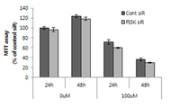
도 1a 및 1b는 PI3K-억제 발현된 (knockdown) 각질형성세포에서 4-TBP (1a) 및 HQ (1b)에 의해 유도된 세포사멸 (apoptosis)을 MTT 분석에 의해 확인하는 도이고, 도 1c는 FACS 분석에 의해 확인하는 도이다.
도 2a 및 2b는 PI3K-억제 발현된 (knockdown) 각질형성세포에서 4-TBP에 의한 TNF-α 및 IL-1α 발현을 나타내는 도이고, 도 2c 및 2d는 HQ에 의한 TNF-α 및 IL-1α 발현을 나타내는 도이다.
도 3a는 PI3K-억제 발현된 (knockdown) 각질형성세포에서 4-TBP에 의해 유도된 ROS 수준을 나타내는 도이고, 도 3b는 HQ에 의해 유도된 ROS 수준을 나타내는 도이다. 또한, 도 3c 및 3d는 상기 ROS 유도된 세포에 N-아세틸-L-시스테인 (NAC)을 처리한 후 생존 가능한 세포를 확인하는 도이다. 도 3e 및 3f는 상기 NAC 처리된 각질형성세포에서 4-TBP에 의한 TNF-α 및 IL-1α 발현을 나타내는 도이고, 도 3g 및 3h는 HQ에 의한 TNF-α 및 IL-1α 발현을 나타내는 도이다.
도 4a 및 4b는 PI3K-억제 발현된 (knockdown) 각질형성세포에서 4-TBP (4a) 및 HQ (4b)에 의한 Nrf2 핵 변위 감소 효과를 나타내는 도이고, 도 4c는 상기 세포를 공초점 현미경으로 확인한 도이며, 도 4d는 면역형광염색으로 확인한 도이다.1A and 1B are diagrams confirming apoptosis induced by 4-TBP (1a) and HQ (1b) in PI3K-inhibited expressed (knockdown) keratinocytes by MTT analysis, and FIG. 1C is It is a diagram confirmed by FACS analysis.
2A and 2B are diagrams showing TNF-α and IL-1α expression by 4-TBP in PI3K-inhibited (knockdown) keratinocytes, and FIGS. 2C and 2D are TNF-α and IL-1α by HQ. It is a diagram showing expression.
FIG. 3A is a diagram showing the ROS level induced by 4-TBP in PI3K-inhibited-expressed (knockdown) keratinocytes, and FIG. 3B is a diagram showing the ROS level induced by HQ. In addition, FIGS. 3C and 3D are diagrams illustrating viable cells after treatment with N-acetyl-L-cysteine (NAC) on the ROS-induced cells. 3E and 3F are diagrams showing the expression of TNF-α and IL-1α by 4-TBP in the NAC-treated keratinocytes, and FIGS. 3G and 3H are diagrams showing the expression of TNF-α and IL-1α by HQ. to be.
4A and 4B are diagrams showing the effect of reducing Nrf2 nuclear displacement by 4-TBP (4a) and HQ (4b) in PI3K-inhibited expression (knockdown) keratinocytes, and FIG. 4C is a confocal microscope of the cells. It is a diagram confirmed, and Figure 4d is a diagram confirmed by immunofluorescence staining.
본 발명자들은, 실시예에서 PI3K-억제 발현된 (knockdown) 각질형성세포에 피부 자극물질인 4-TBP 또는 하이드로퀴논 (HQ)을 처리하는 경우, 세포사멸 (apoptosis) 및 발생기산소 (ROS) 생성이 증가한다는 점에 기반하여 PI3K-억제 발현된 (knockdown) 각질형성세포에서 4-TBP 및 HQ에 의해 유도된 ROS 생성 증가 및 Nrf2 핵 변위 감소 등을 구체적으로 확인하고, 이에 기초하여 본 발명을 완성하였다.The present inventors, in the case of treatment of the skin irritant 4-TBP or hydroquinone (HQ) on PI3K-inhibited (knockdown) keratinocytes in the examples, apoptosis and developmental oxygen (ROS) production Based on the fact that the PI3K-inhibited expression (knockdown) increased ROS production induced by 4-TBP and HQ in keratinocytes and decreased Nrf2 nuclear displacement were specifically confirmed, and based on this, the present invention was completed. .
이하 본 발명을 상세히 설명한다.Hereinafter, the present invention will be described in detail.
본 발명은 Nrf2 (Nuclear factor erythroid 2-related Factor 2) 단백질을 포함하는 백반증 진단 마커용 조성물을 제공한다.The present invention provides a composition for a diagnostic marker for vitiligo comprising Nrf2 (Nuclear factor erythroid 2-related factor 2) protein.
또한, 본 발명은 Nrf2 (Nuclear factor erythroid 2-related Factor 2) 단백질에 특이적으로 결합하는 분자를 포함하는 것을 특징으로 하는 백반증 진단용 조성물을 제공한다.In addition, the present invention provides a composition for diagnosing vitiligo, comprising a molecule that specifically binds to Nrf2 (Nuclear factor erythroid 2-related factor 2) protein.
본 발명의 일 구현예로, 상기 Nrf2 단백질은 서열번호 1의 아미노산 서열로 이루어질 수 있고, 상기 분자는 항체일 수 있다.In one embodiment of the present invention, the Nrf2 protein may consist of the amino acid sequence of SEQ ID NO: 1, and the molecule may be an antibody.
본 발명에서, "항체"란 항원성 부위에 대해서 지시되는 특이적인 단백질 분자를 의미한다. 본 발명의 목적상, 항체는 마커 단백질에 대해 특이적으로 결합하는 항체를 의미하며, 다클론 항체, 단클론 항체 및 재조합 항체를 모두 포함한다.In the present invention, "antibody" refers to a specific protein molecule directed against an antigenic site. For the purposes of the present invention, an antibody refers to an antibody that specifically binds to a marker protein, and includes all of polyclonal antibodies, monoclonal antibodies, and recombinant antibodies.
또한, 본 발명은 백반증 의심환자의 피부 시료로부터 분리된 표피에서 각질형성세포를 추출하여 배양하고, 상기 배양된 세포로부터 Nrf2 (Nuclear factor erythroid 2-related Factor 2) 단백질의 핵내 발현 수준을 측정하고, 이를 정상 대조군과 비교하는 단계를 포함하는 백반증의 진단을 위한 정보제공방법을 제공한다. 이때, 상기 배양된 세포에 존재하는 Nrf2 단백질의 핵내 발현 수준이 대조군에 비해 감소된 경우 백반증이 발병한 것으로 판정할 수 있다.In addition, the present invention extracts and cultures keratinocytes from the epidermis isolated from the skin sample of a patient suspected of vitiligo, and measures the intranuclear expression level of Nrf2 (Nuclear factor erythroid 2-related factor 2) protein from the cultured cells, It provides a method of providing information for diagnosis of vitiligo, including the step of comparing this with a normal control group. At this time, when the level of expression in the nucleus of the Nrf2 protein present in the cultured cells is decreased compared to the control, it may be determined that vitiligo has occurred.
본 발명에서 "단백질 발현 수준 측정"이란 피부 자극 후보 물질을 스크리닝하기 위하여 각질형성세포에서 본 발명의 유전자로부터 발현된 단백질의 존재 여부와 발현 정도를 확인하는 과정으로, 단백질의 양을 측정하여 이루어진다. 이를 위한 분석 방법으로는 웨스턴 블랏, ELISA (enzyme linked immunosorbent assay), 방사선면역분석 (RIA: Radioimmunoassay), 방사 면역 확산법 (radioimmunodiffusion), 오우크테로니 (Ouchterlony) 면역확산법, 로케트 (rocket) 면역전기영동, 조직면역 염색, 면역침전 분석법 (Immunoprecipitation Assay), 보체 고정 분석법 (Complement Fixation Assay), 유세포분석 (Fluorescence Activated Cell Sorter, FACS), 단백질 칩 (protein chip) 등이 있으나 이로 제한되는 것은 아니다. 본 발명에서 단백질 발현 수준을 측정하는 제제는 바람직하게는 항체이다.In the present invention, "measurement of protein expression level" is a process of determining the presence and expression level of a protein expressed from the gene of the present invention in keratinocytes in order to screen for a skin irritation candidate substance, and is performed by measuring the amount of protein. Analysis methods for this include Western blot, ELISA (enzyme linked immunosorbent assay), radioimmunoassay (RIA), radioimmunodiffusion, Ouchterlony immunodiffusion, and rocket immunoelectrophoresis. , Tissue immunostaining, immunoprecipitation assay (Immunoprecipitation Assay), complement fixation assay (Complement Fixation Assay), flow cytometry (Fluorescence Activated Cell Sorter, FACS), protein chip, etc., but are not limited thereto. In the present invention, the agent for measuring the protein expression level is preferably an antibody.
본 발명의 일실시예에서는, 백반증 발생 예측을 위한 Nrf2 핵 변위 감소 효과를 확인하기 위하여, PI3K-억제 발현된 (knockdown) 각질형성세포에 피부 자극물질을 처리하여 이를 확인한 결과, PI3K-억제 발현된 (knockdown) 각질형성세포에 피부 자극물질인 4-TBP 또는 하이드로퀴논 (HQ)을 처리하는 경우, Nrf2 핵 변위가 감소한다는 것을 알 수 있었다 (실시예 5 참조).In one embodiment of the present invention, in order to confirm the effect of reducing Nrf2 nuclear displacement for predicting the occurrence of vitiligo, PI3K-inhibited expression (knockdown) keratinocytes were treated with a skin irritant to confirm this, as a result of confirming this, PI3K-inhibited expression (knockdown) It was found that when keratinocytes were treated with 4-TBP or hydroquinone (HQ), which is a skin irritant, Nrf2 nuclear displacement was reduced (see Example 5).
따라서, 본 발명에 따른 Nrf2 단백질은 PI3K-knockdown 각질세포 (백반증 환자의 표피에서 분리된 각질세포의 모델)에 피부 자극물질 처리 시, 그 발현이 감소되는 바, 상기 단백질을 이용하여 백반증 발생 예측 또는 진단이 요구되는 다양한 목적 및 용도로 사용될 수 있다.Therefore, when the Nrf2 protein according to the present invention is treated with a skin irritant in PI3K-knockdown keratinocytes (a model of keratinocytes isolated from the epidermis of a patient with vitiligo), its expression is reduced. It can be used for various purposes and uses requiring diagnosis.
이에, 본 발명은 Nrf2 (Nuclear factor erythroid 2-related Factor 2) 단백질을 포함하는 것을 특징으로 하는 백반증 발생 위험 예측용 조성물을 제공한다.Accordingly, the present invention provides a composition for predicting the risk of vitiligo, comprising the protein Nrf2 (Nuclear factor erythroid 2-related factor 2).
본 발명에서, 상기 조성물에 화장품 구성성분이나 제품 처리 시, 상기 Nrf2 단백질의 발현이 감소하는 경우, 백반증 발생 위험이 높은 것으로 판단할 수 있다.In the present invention, when the expression of the Nrf2 protein decreases during the treatment of a cosmetic component or product in the composition, it can be determined that the risk of vitiligo is high.
이하, 본 발명의 이해를 돕기 위하여 바람직한 실시예를 제시한다. 그러나 하기의 실시예는 본 발명을 보다 쉽게 이해하기 위하여 제공되는 것일 뿐, 하기 실시예에 의해 본 발명의 내용이 한정되는 것은 아니다.Hereinafter, a preferred embodiment is presented to aid the understanding of the present invention. However, the following examples are provided for easier understanding of the present invention, and the contents of the present invention are not limited by the following examples.
[[ 실시예Example ]]
실시예Example 1. 실험 준비 및 실험 방법 1. Experiment preparation and experiment method
1-1. 환자1-1. patient
본 연구에는 백반증 (vitiligo)으로 진단받은 8명의 환자 (남성 4명 및 여성 4명; 15세와 53세 사이로서 평균 나이는 38.5세; 분절형 2명 및 비분절형 6명)가 포함되었다. 동국대학교 일산 병원의 기관 감사 위원회 (Institutional Review Board)는 헬싱키 선언 (Declaration of Helsinki)의 원칙에 따라 수행된 본 연구를 승인하였다. 각각의 지원자로부터 서면 고지 동의 (written informed consent)를 받은 후, 3명의 환자에서 탈색되고 정상적으로 유색된 피부 시료들을 생검하고, 면역 형광 분석을 위해 기타 2명의 환자에서 흡입 수포의 천정부 (roof)로부터 채집하였다.This study included 8 patients diagnosed with vitiligo (4 males and 4 females; between 15 and 53 years old, with a mean age of 38.5 years; 2 segmented and 6 non-segmented). The Institutional Review Board of Dongguk University Ilsan Hospital approved this study conducted in accordance with the principles of the Declaration of Helsinki. After receiving written informed consent from each volunteer, biopsies of bleached and normally colored skin samples from 3 patients were obtained, and collected from the roof of the inhalation blisters from the other 2 patients for immunofluorescence analysis. I did.
1-2. 1-2. 면역형광염색Immunofluorescence staining
Nrf2 (NF-E2-related Factor 2) 및 인산화된 PI3K의 면역 형광 염색을 위해, 탈색되고 정상적으로 유색된 고정 표피 샘플을 파라핀 왁스에 포매하고, 5 mm로 절편화하였다. 파라핀 제거 이후, 절편을 시트르산 용액 (100 mM citrate, pH 6.0) 및 0.5 % Triton X-100으로 전처리하였다. 3% 소 혈청 알부민 (BSA)으로 차단한 이후, 상기 절편을 항-Nrf2 항체 (토끼 다클론성: Santa Cruz Biotechnology, Santa Cruz, CA, USA)의 1:100 희석액으로 배양하고, Alexa Fluor® 488 염소 항-토끼 IgG (Molecular Probes, Eugene, OR, USA)의 1:200 희석액으로 염색하였다. 이중 염색을 위해, 상기 절편을 항-phospho-PI3K (염소 다클론성: Santa Cruz Biotechnology) 및 Alexa Fluor-표지 당나귀 항-염소 IgG (594; Molecular Probes)로 연속적으로 염색하였다. 핵은 Hoechst 33258 (Sigma Aldrich, St. Louis, MO, USA)로 대비 염색하였다. 상기 염색된 시료는 Dp Manager 2.1 형광 현미경(Olympus Optical Co., Tokyo, Japan)을 이용하여 관측하였다.For immunofluorescence staining of Nrf2 (NF-E2-related Factor 2) and phosphorylated PI3K, a bleached and normally colored fixed epidermal sample was embedded in paraffin wax and sectioned into 5 mm. After paraffin removal, the sections were pretreated with citric acid solution (100 mM citrate, pH 6.0) and 0.5% Triton X-100. After blocking with 3% bovine serum albumin (BSA), the section was incubated with a 1:100 dilution of an anti-Nrf2 antibody (rabbit polyclonal: Santa Cruz Biotechnology, Santa Cruz, CA, USA), and Alexa Fluor® 488 It was stained with a 1:200 dilution of goat anti-rabbit IgG (Molecular Probes, Eugene, OR, USA). For double staining, the sections were sequentially stained with anti-phospho-PI3K (goat polyclonal: Santa Cruz Biotechnology) and Alexa Fluor-labeled donkey anti-goat IgG (594; Molecular Probes). Nuclei were counter-stained with Hoechst 33258 (Sigma Aldrich, St. Louis, MO, USA). The stained sample was observed using a Dp Manager 2.1 fluorescence microscope (Olympus Optical Co., Tokyo, Japan).
1-3. 세포 배양 및 1-3. Cell culture and PI3KPI3K -knockdown-knockdown
제왕절개 수술로부터 수득된 성인 피부 시료들을 세포 배양에 사용하였다. 표피는 피부로부터 분리하고, 개개의 표피 세포에 대한 현탁액을 제조하였다. 소 뇌하수체 추출물, 소 인슐린, 하이드로코티손 (hydrocortisone), 인간 표피 성장 인자 및 소 트랜스페린 (Invitrogen)이 첨가된 EpiLife 배지 (Invitrogen, Carlsbad, CA)에 개개의 표피 세포를 현탁하였다. 상기 수확된 각질 세포를 배양 배지에 7.5×10⁴ cell/mL로 재현탁하고, 6-well 플레이트에 웰당 1.5×10⁵ cell로 접종하였다. Adult skin samples obtained from cesarean section surgery were used for cell culture. The epidermis was separated from the skin and a suspension for individual epidermal cells was prepared. Individual epidermal cells were suspended in EpiLife medium (Invitrogen, Carlsbad, CA) to which bovine pituitary extract, bovine insulin, hydrocortisone, human epidermal growth factor, and bovine transferrin (Invitrogen) were added. The harvested keratinocytes were resuspended in culture medium at 7.5×10 4 cells/mL, and inoculated at 1.5×10 5 cells per well in a 6-well plate.
접종 24 시간 후, 각 6-well 플레이트를 제조사의 지침서에 따라 TransIT-siQUEST 형질 감염 시약 (Mirus, Madison, WI, USA)을 이용하여 인간 PI3K siRNA (50nM) 또는 음성 대조군 (ON-TARGETplus SMARTpool or nontargeting small interfering RNA; Dharmacon, Lafayette, CO, USA)로 형질 감염시켰다. 상기 세포의 형질 감염 24 시간 후, NAC (Sigma Aldrich)를 배양액에 10mM로 첨가하였다. 1 시간 후, 적절한 농도의 각 화학약품을 상기 세포에 첨가하였다. 24 hours after inoculation, each 6-well plate was subjected to human PI3K siRNA (50nM) or negative control (ON-TARGETplus SMARTpool or nontargeting) using TransIT-siQUEST transfection reagent (Mirus, Madison, WI, USA) according to the manufacturer's instructions. small interfering RNA; Dharmacon, Lafayette, CO, USA). 24 hours after transfection of the cells, NAC (Sigma Aldrich) was added to the culture medium at 10 mM. After 1 hour, an appropriate concentration of each chemical was added to the cells.
화학약품에 의한 처리 24 시간 및 48 시간 후, 세포 생존능 및 FACS 분석을 위해 상기 세포를 시험하고, ROS 함량, TNF-α 및 IL-1α 수준 (ELISA 이용)에 대해 검사하였으며, 또한 Nrf2 발현에 대한 웨스턴 블롯 분석 및 공초점 현미경 검사에 적용하였다.After 24 hours and 48 hours of treatment with chemicals, the cells were tested for cell viability and FACS analysis, and were tested for ROS content, TNF-α and IL-1α levels (using ELISA), and also for Nrf2 expression. Western blot analysis and confocal microscopy were applied.
1-4. 1-4. MTTMTT 분석 analysis
세포를 MTT로 4시간 동안 염색하고, 침전된 포르마잔 (formazan)을 DMSO에 용해하고, 광학 밀도는 분광 광도계를 이용하여 570nm (630nm background subtraction)에서 측정하였다. 세포 성장에 대한 화학약품의 효과는 DMSO의 존재 시 세포 생존능에 대한 상기 화학약품의 세포 생존능의 비율로부터 산정하였다.Cells were stained with MTT for 4 hours, precipitated formazan was dissolved in DMSO, and the optical density was measured at 570nm (630nm background subtraction) using a spectrophotometer. The effect of the chemical agent on cell growth was calculated from the ratio of the cell viability of the chemical agent to the cell viability in the presence of DMSO.
1-5. 1-5. FACSFACS 분석 analysis
세포를 결합 완충액에서 재현탁하고, Annexin V-FITC 염색 용액 (5 μl)으로 10분 동안 처리하고 암실에서 요오드화 프로피디움 (PI, 100 μg/ml)을 1 μl 추가하고 5분 동안 처리하였다. CytomicsTM FC500 Flow Cytometry (Beckman Coulter, Brea, CA, USA)에 의해 FACS 분석을 수행하였다.Cells were resuspended in binding buffer, treated with Annexin V-FITC staining solution (5 μl) for 10 minutes, and 1 μl of propidium iodide (PI, 100 μg/ml) was added in the dark and treated for 5 minutes. FACS analysis was performed by Cytomics TM FC500 Flow Cytometry (Beckman Coulter, Brea, CA, USA).
1-6. 1-6. ROSROS 검사 inspection
반응성 산소종 (ROS)은 제조사의 지침서에 따라 총 ROS 검출 키트 (Enzo Life, Farmingdale, NY, USA)를 이용하여 검출하였다. 동일하게 접종된 세포의 웰은 세척 완충액으로 2회 세척하고, 이어 암실에서 37 ℃, 1 시간 동안 ROS 검출 용액으로 배양하였다. 형광 마이크로플레이트 판독기를 이용하여 플레이트를 즉시 판독하였다.Reactive oxygen species (ROS) were detected using a total ROS detection kit (Enzo Life, Farmingdale, NY, USA) according to the manufacturer's instructions. The wells of the same inoculated cells were washed twice with wash buffer, and then incubated with ROS detection solution at 37° C. for 1 hour in a dark room. The plate was read immediately using a fluorescent microplate reader.
1-7. 1-7. 웨스턴Western 블롯Blot 분석 analysis
동일한 양의 추출된 단백질을 용해시키고, 니트로셀룰로오스 멤브레인으로 전달하였다. 상기 멤브레인을 PI3K 및 phospho-PI3K에 대한 항체 (Cell Signaling Technology, Danvers, MA, USA) 및 Nrf2 (Santa Cruz Biotechnology)와 함께 배양하였다. 적절한 항-마우스 또는 항-토끼 horseradish 과산화효소-접합 항체 (Thermo) 및 enhanced 화학발광 용액 (Thermo)와 함께 배양한 이후, 영상 입력장치 (LAS-3000, Fuji Photo Film, Tokyo, Japan) 상에 신호를 포획하였다. 각 레인에 적재된 단백질의 양을 모니터링하기 위해, 상기 멤브레인을 마우스 단클론성 항-β-액틴 항체 (Sigma) 및 항-라민 A/C 항체 (Santa Cruz Biotechnology)로 재-탐침하고, 상술한 바와 같이 가공하였다. 이어 단백질 밴드를 밀도 측정법에 의해 분석하였다.Equal amounts of extracted protein were dissolved and transferred to a nitrocellulose membrane. The membrane was incubated with antibodies against PI3K and phospho-PI3K (Cell Signaling Technology, Danvers, MA, USA) and Nrf2 (Santa Cruz Biotechnology). After incubation with an appropriate anti-mouse or anti-rabbit horseradish peroxidase-conjugated antibody (Thermo) and enhanced chemiluminescent solution (Thermo), signal on an image input device (LAS-3000, Fuji Photo Film, Tokyo, Japan) Was captured. In order to monitor the amount of protein loaded in each lane, the membrane was re-probed with a mouse monoclonal anti-β-actin antibody (Sigma) and an anti-Lamine A/C antibody (Santa Cruz Biotechnology), and as described above. Processed together. The protein band was then analyzed by density measurement.
1-8. ELISA 분석 1-8. ELISA analysis
이전 실험의 모든 단계에서 배양된 각질 세포의 상층액을 수집하고, 제조사의 지침서에 따라 ELISA 키트 (R&D Systems)를 이용하여 상기 상층액 중의 인간 TNFα 또는 IL-1α의 농도를 측정하였다.The supernatant of keratinocytes cultured in all steps of the previous experiment was collected, and the concentration of human TNFα or IL-1α in the supernatant was measured using an ELISA kit (R&D Systems) according to the manufacturer's instructions.
1-9. 1-9. 공초점Confocal 현미경 검사 Microscopic examination
배양된 세포를 4% (w/v) 파라포름알데히드에서 고정하고, 0.05% Triton X-100으로 전처리한 후, 3% 소 혈청 알부민 (BSA)으로 차단하였다. Nrf2 및 인산화된 PI3K 항체를 이용한 이중 염색을 위해, 상기 세포를 상응하는 항체의 1:100 희석액으로 배양하고, 이어 Alexa Fluor® 488 당나귀 항-염소 IgG (Molecular Probes) 또는 Alexa Fluor® 594 염소 항-토끼 IgG (Molecular Probes)의 1:200 희석액으로 염색하였다. 핵은 Hoechst 33258 (Sigma Aldrich, St. Louis, MO, USA)로 대비 염색하였다. 상기 염색된 시료는 C1 공초점 레이저 스캔 현미경 (C1; Nikon, Tokyo, Japan)을 이용하여 관측하였다. 이중 염색된 세포 중의 각 항원의 위치를 분석하기 위해, 동일한 영역에서 수득된 서로 상이한 영상을 EZ-C1 software (EZ-C1; Nikon, Tokyo, Japan)를 이용하여 병합하고, NIS-Elements AR 3.2 software (Nikon Instruments, Melvillie, NY, USA)를 이용하여 측정하였다.The cultured cells were fixed in 4% (w/v) paraformaldehyde, pretreated with 0.05% Triton X-100, and then blocked with 3% bovine serum albumin (BSA). For double staining with Nrf2 and phosphorylated PI3K antibody, the cells are incubated with a 1:100 dilution of the corresponding antibody, followed by Alexa Fluor® 488 donkey anti-goat IgG (Molecular Probes) or Alexa Fluor® 594 goat anti- It was stained with a 1:200 dilution of rabbit IgG (Molecular Probes). Nuclei were counter-stained with Hoechst 33258 (Sigma Aldrich, St. Louis, MO, USA). The stained sample was observed using a C1 confocal laser scanning microscope (C1; Nikon, Tokyo, Japan). To analyze the location of each antigen in the double stained cells, different images obtained in the same region were merged using EZ-C1 software (EZ-C1; Nikon, Tokyo, Japan), and NIS-Elements AR 3.2 software (Nikon Instruments, Melvillie, NY, USA) was used to measure.
1-10. 통계 분석 1-10. Statistical analysis
상기 실험 데이터의 통계 분석은 Student’s t-test를 이용하여 수행하였다. 그 결과는 평균 ± 표준편차 (SD)로 나타냈다. P 값(<0.05)은 유의한 것으로 고려되었다.Statistical analysis of the experimental data was performed using Student's t-test. The results are expressed as mean ± standard deviation (SD). P values (<0.05) were considered significant.
실시예Example 2. 2. PI3KPI3K -억제 발현된 (knockdown) 각질형성세포에서 4--In suppression expressed (knockdown) keratinocytes 4- TBPTBP 및 And HQ에HQ 의해 유도된 세포사멸 ( Induced apoptosis ( apoptosisapoptosis ) 확인) Confirm
PI3K-knockdown 각질형성세포에 화학물질 처리 시, 세포사멸 (apoptosis)이 유도되는지 확인하기 위하여 MTT 분석 (실시예 1-4) 및 FACS 분석 (실시예 1-5)을 실시하였다.MTT analysis (Example 1-4) and FACS analysis (Example 1-5) were performed to confirm whether apoptosis was induced when the PI3K-knockdown keratinocytes were treated with a chemical substance.
그 결과, 도 1a 내지 1c에 나타낸 바와 같이, MTT 분석에 따르면, 4-TBP 및 HQ는 각각 300 μM 및 100 μM로 사용하는 경우 48 시간 시점에 생존 가능한 세포의 개수를 미처리 대조군 세포의 40 내지 50 %까지 감소시키는 것으로 나타났으며 (도 1a 및 1b, 추가 연구를 위해 이들의 농도를 고정하였다.), 4-TBP는 처리 48 시간 이후에 생존 가능한 세포의 개수를 대조군 대비 유의하게 감소시켰으며 (도 1a), HQ의 경우, 처리 24 시간 및 48 시간 이후에 유의하게 감소를 나타내었다 (도 1b). 또한, FACS 분석에 따르면, 초기 세포사멸 (annexin-양성 및 PI-음성) 및 후기 세포사멸 (이중 양성) 비율의 합은 대조군 및 화학물질-처리 PI3K-knockdown 각질형성세포 둘 모두에서 유의하게 증가하는 것으로 나타났다 (도 1c). As a result, as shown in Figs. 1a to 1c, according to MTT analysis, the number of viable cells at 48 hours when 4-TBP and HQ are used at 300 μM and 100 μM, respectively, is 40 to 50 of the untreated control cells. % (FIGS. 1A and 1B, their concentration was fixed for further study), and 4-TBP significantly reduced the number of viable cells after 48 hours of treatment compared to the control group ( Figure 1a), in the case of HQ, showed a significant decrease after 24 hours and 48 hours of treatment (Figure 1b). In addition, according to FACS analysis, the sum of the rates of early apoptosis (annexin-positive and PI-negative) and late apoptosis (double positive) was significantly increased in both control and chemical-treated PI3K-knockdown keratinocytes. Was found (Fig. 1c).
실시예Example 3. 3. PI3KPI3K -억제 발현된 (knockdown) 각질형성세포에서 4--In suppression expressed (knockdown) keratinocytes 4- TBPTBP 및 And HQ에HQ 의한 by TNFTNF -α 및 IL--α and IL- 1α1α 발현 확인 Confirmation of expression
백반증 환자의 탈색된 표피에서 전염증성 사이토카인의 생성이 증가한다고 알려져 있다. 이에, PI3K-knockdown 각질형성세포 및 대조군에서 전염증성 사이토카인 (TNF-α 및 IL-1α)의 생성을 비교하기 위하여 ELISA (실시예 1-8)를 실시하였다. It is known that the production of proinflammatory cytokines is increased in the bleached epidermis of patients with vitiligo. Accordingly, ELISA (Example 1-8) was performed to compare the production of pro-inflammatory cytokines (TNF-α and IL-1α) in PI3K-knockdown keratinocytes and controls.
그 결과, 도 2a 내지 2d에 나타낸 바와 같이, PI3K-knockdown 각질형성세포에서 TNF-α (도 2a 및 2b) 및 IL-1α (도 2c 및 2d)의 수준은 대조군에 비하여 4-TBP 처리 48 시간 시점, 및 HQ 처리 24 시간 및 48 시간 이후 시점에 유의하게 증가하는 것으로 나타났다(도 2a 내지 2d). As a result, as shown in FIGS. 2A to 2D, the levels of TNF-α (FIGS. 2A and 2B) and IL-1α (FIGS. 2C and 2D) in PI3K-knockdown keratinocytes were 48 hours of 4-TBP treatment compared to the control group. It was found to increase significantly at the time point, and at the time point after 24 hours and 48 hours of HQ treatment (FIGS.
실시예Example 4: 4: PI3KPI3K -억제 발현된 (knockdown) 각질형성세포에서 4--In suppression expressed (knockdown) keratinocytes 4- TBPTBP 및 And HQ에HQ 의해 유도된 Induced by ROSROS 생성의 세포독성 효과 Cytotoxic effect of production
ROS 발현에 의한 화학적 세포독성의 효과를 확인하기 위하여 산화 방지제의 존재 또는 부재 하에 이들 각질형성세포에서 ROS 검사 (실시예 1-6), MTT 분석 (실시예 1-4) 및 ELISA (실시예 1-8)를 실시하였다. ROS test (Example 1-6), MTT assay (Example 1-4) and ELISA (Example 1) in these keratinocytes in the presence or absence of antioxidants to confirm the effect of chemical cytotoxicity by ROS expression. -8) was implemented.
그 결과, 도 3a 내지 3h에 나타낸 바와 같이, PI3K-knockdown 각질형성세포에서 ROS 수준은 대조군에 비하여 4-TBP 처리 48 시간 시점, 및 HQ 처리 24 시간 및 48 시간 시점에 유의하게 증가하는 것으로 나타났다 (도 3a 및 3b). 또한, 산화 방지제 및 ROS 차단제인 N-아세틸-L-시스테인 (NAC)은 그 처리 48 시간 시점 및 24 시간 및 48 시간 시점에 4-TBP 및 HQ에 의한 PI3K-knockdown 각질형성세포의 생존 가능한 개수의 감소 (세포사멸)를 억제하였지만, 대조군에서는 4-TBP 및 HQ에 의하여 유도되는 세포사멸을 억제하지 않았다 (도 3c 및 3d). 또한, ELISA에 따르면, PI3K-knockdown 각질형성세포에서 NAC 처리 이후 4-TBP 및 HQ에 의한 TNF-α/IL-1α의 증가는 처리 48 시간 이후 및 처리 24 시간 및 48 시간 이후에 각각 약화되었다 (도 3e 내지 3h).As a result, as shown in Figs. 3a to 3h, the ROS level in PI3K-knockdown keratinocytes was significantly increased at 48 hours of 4-TBP treatment and 24 hours and 48 hours of HQ treatment compared to the control group ( 3a and 3b). In addition, N-acetyl-L-cysteine (NAC), an antioxidant and ROS blocker, is a viable number of PI3K-knockdown keratinocytes by 4-TBP and HQ at 48 hours and 24 hours and 48 hours of treatment. The reduction (cell death) was suppressed, but the control group did not suppress the apoptosis induced by 4-TBP and HQ (FIGS. 3C and 3D ). In addition, according to ELISA, the increase of TNF-α/IL-1α by 4-TBP and HQ after NAC treatment in PI3K-knockdown keratinocytes was attenuated 48 hours after treatment and 24 hours and 48 hours after treatment, respectively ( Figures 3e to 3h).
상기 내용을 종합한 결과, 화학적 세포독성에 의한 ROS 생성 및 NAC 전처리에 의한 이의 회복 (도 3a 내지 도 3h)은, PI3K-knockdown 각질형성세포에서 화학물질 처리에 따른 세포사멸에 산화적 스트레스가 관여하고 있음을 시사하고 있다. As a result of synthesizing the above, the production of ROS by chemical cytotoxicity and its recovery by NAC pretreatment (FIGS. 3A to 3H) is that oxidative stress is involved in apoptosis due to chemical substance treatment in PI3K-knockdown keratinocytes. Suggesting that they are doing.
실시예Example 5: 5: PI3KPI3K -억제 발현된 (knockdown) 각질형성세포에서의 4--Inhibitory expressed (knockdown) keratinocytes 4- TBPTBP 및 And HQ에HQ 의한 by Nrf2Nrf2 핵 변위 감소 효과 Nuclear displacement reduction effect
상기 실시예 4의 결과에 따라, PI3K-knockdown 각질형성세포에서의 산화적 스트레스 유도 기작을 이해하기 위하여 화학물질 처리/비처리 또는 산화 방지제 부재/존재 하에 하기와 같은 방법으로 Nrf2 (Nuclear factor erythroid 2-related Factor 2) 활성화에 의한 PI3K-knockdown 각질형성세포 활성화 효과를 검토하였다. According to the results of Example 4, in order to understand the mechanism of inducing oxidative stress in PI3K-knockdown keratinocytes, Nrf2 (Nuclear factor erythroid 2 The effect of activating PI3K-knockdown keratinocytes by -related factor 2) activation was examined.
Nrf2 핵 수준을 검토하기 위하여, 형질주입 (transfection) 없이 배양된 각질형성세포, 대조군-siRNA 형질주입 각질형성세포 및 PI3K-siRNA 형질주입 각질형성세포에서 각 핵 분획을 다른 분획들로부터 분리하고, 웨스턴 블롯 분석 (실시예 1-7), 공초점 현미경 검사 (실시예 1-9) 및 면역형광염색 (실시예 1-3)을 수행하였다. In order to examine the level of Nrf2 nuclear, each nuclear fraction was isolated from other fractions in keratinocytes cultured without transfection, control-siRNA-transfected keratinocytes, and PI3K-siRNA-transfected keratinocytes. Blot analysis (Example 1-7), confocal microscopy (Example 1-9), and immunofluorescence staining (Example 1-3) were performed.
그 결과, 도 4a 내지 4d에 나타낸 바와 같이, PI3K-knockdown (PI3K-p85α의 인산화를 감소)은 대조군-siRNA 형질주입 세포에 비해 PI3K-siRNA 형질주입 각질형성세포 핵 분획에서 Nrf2 발현을 감소시켰으며 (도 4a), 상기 Nrf2 핵 수준은 48 시간 동안 4-TBP 및 HQ로 처리된 PI3K-knockdown 각질형성세포에서 더욱 낮았고, 이는 NAC 처리에 의해 4-TBP 및 HQ로 처리된 대조군보다 낮지 않는 수준까지 회복하였다 (도 4b). 또한, 공초점 현미경 검사에 따르면, Nrf2의 염색 세기는 PI3K-knockdown 각질형성세포에서 매우 약한 것으로 나타났고 (도 4c), 백반증 환자로부터 생검된 8개 세트의 피부 시료에서의 항-Nrf2 및 항-phospho-PI3K 항체에 의한 면역형광염색에 따르면, 이중 양성 세포의 개수가 적었으며, 탈색된 표피에서의 염색 강도가 낮은 것으로 나타났다 (도 4d).As a result, as shown in Figures 4a to 4d, PI3K-knockdown (reduces phosphorylation of PI3K-p85α) decreased Nrf2 expression in the nuclear fraction of PI3K-siRNA-transfected keratinocytes compared to the control-siRNA-transfected cells. (Figure 4a), the Nrf2 nuclear level was lower in PI3K-knockdown keratinocytes treated with 4-TBP and HQ for 48 hours, up to a level not lower than the control treated with 4-TBP and HQ by NAC treatment. Recovered (Fig. 4b). In addition, according to confocal microscopy, the staining intensity of Nrf2 was very weak in PI3K-knockdown keratinocytes (Fig. 4c), and anti-Nrf2 and anti- According to immunofluorescence staining with phospho-PI3K antibody, the number of double-positive cells was small, and staining intensity in the bleached epidermis was low (FIG. 4D ).
상기 내용을 종합한 결과, 화학적 세포독성은 ROS 생성 증가 및 Nrf2 활성 감소를 통하여 정상세포 대비 PI3K-knockdown 각질형성세포의 세포사멸을 증가시키며, 백반증 환자의 탈색된 표피에서와 마찬가지로 Nrf2의 핵 변위를 감소시키는 것을 알 수 있었다. As a result of synthesizing the above, chemical cytotoxicity increases the apoptosis of PI3K-knockdown keratinocytes compared to normal cells through increased ROS production and decreased Nrf2 activity, and as in the depigmented epidermis of patients with vitiligo, the nuclear displacement of Nrf2 is increased. It was found to decrease.
전술한 본 발명의 설명은 예시를 위한 것이며, 본 발명이 속하는 기술분야의 통상의 지식을 가진 자는 본 발명의 기술적 사상이나 필수적인 특징을 변경하지 않고서 다른 구체적인 형태로 쉽게 변형이 가능하다는 것을 이해할 수 있을 것이다. 그러므로 이상에서 기술한 실시예들은 모든 면에서 예시적인 것이며 한정적이 아닌 것으로 이해해야만 한다.The above description of the present invention is for illustrative purposes only, and those of ordinary skill in the art to which the present invention pertains will be able to understand that other specific forms can be easily modified without changing the technical spirit or essential features of the present invention. will be. Therefore, it should be understood that the embodiments described above are illustrative in all respects and are not limiting.
<110> Dongguk University Industry-Academy Cooperation Foundation <120> New Biomarker for Diagnosis of Vitiligo Susceptibility and method for predicting assessment using the same <130> MP16-360_division <160> 1 <170> KoPatentIn 3.0 <210> 1 <211> 505 <212> PRT <213> Nuclear factor erythroid 2-related Factor 2 isoform 6 <400> 1 Met Lys Arg Gln Val Ala His Ile Pro Lys Ser Asp Ala Leu Tyr Phe 1 5 10 15 Asp Asp Cys Met Gln Leu Leu Ala Gln Thr Phe Pro Phe Val Asp Asp 20 25 30 Asn Glu Val Ser Ser Ala Thr Phe Gln Ser Leu Val Pro Asp Ile Pro 35 40 45 Gly His Ile Glu Ser Pro Val Phe Ile Ala Thr Asn Gln Ala Gln Ser 50 55 60 Pro Glu Thr Ser Val Ala Gln Val Ala Pro Val Asp Leu Asp Gly Met 65 70 75 80 Gln Gln Asp Ile Glu Gln Val Trp Glu Glu Leu Leu Ser Ile Pro Glu 85 90 95 Leu Gln Cys Leu Asn Ile Glu Asn Asp Lys Leu Val Glu Thr Thr Met 100 105 110 Val Pro Ser Pro Glu Ala Lys Leu Thr Glu Val Asp Asn Tyr His Phe 115 120 125 Tyr Ser Ser Ile Pro Ser Met Glu Lys Glu Val Gly Asn Cys Ser Pro 130 135 140 His Phe Leu Asn Ala Phe Glu Asp Ser Phe Ser Ser Ile Leu Ser Thr 145 150 155 160 Glu Asp Pro Asn Gln Leu Thr Val Asn Ser Leu Asn Ser Asp Ala Thr 165 170 175 Val Asn Thr Asp Phe Gly Asp Glu Phe Tyr Ser Ala Phe Ile Ala Glu 180 185 190 Pro Ser Ile Ser Asn Ser Met Pro Ser Pro Ala Thr Leu Ser His Ser 195 200 205 Leu Ser Glu Leu Leu Asn Gly Pro Ile Asp Val Ser Asp Leu Ser Leu 210 215 220 Cys Lys Ala Phe Asn Gln Asn His Pro Glu Ser Thr Ala Glu Phe Asn 225 230 235 240 Asp Ser Asp Ser Gly Ile Ser Leu Asn Thr Ser Pro Ser Val Ala Ser 245 250 255 Pro Glu His Ser Val Glu Ser Ser Ser Tyr Gly Asp Thr Leu Leu Gly 260 265 270 Leu Ser Asp Ser Glu Val Glu Glu Leu Asp Ser Ala Pro Gly Ser Val 275 280 285 Lys Gln Asn Gly Pro Lys Thr Pro Val His Ser Ser Gly Asp Met Val 290 295 300 Gln Pro Leu Ser Pro Ser Gln Gly Gln Ser Thr His Val His Asp Ala 305 310 315 320 Gln Cys Glu Asn Thr Pro Glu Lys Glu Leu Pro Val Ser Pro Gly His 325 330 335 Arg Lys Thr Pro Phe Thr Lys Asp Lys His Ser Ser Arg Leu Glu Ala 340 345 350 His Leu Thr Arg Asp Glu Leu Arg Ala Lys Ala Leu His Ile Pro Phe 355 360 365 Pro Val Glu Lys Ile Ile Asn Leu Pro Val Val Asp Phe Asn Glu Met 370 375 380 Met Ser Lys Glu Gln Phe Asn Glu Ala Gln Leu Ala Leu Ile Arg Asp 385 390 395 400 Ile Arg Arg Arg Gly Lys Asn Lys Val Ala Ala Gln Asn Cys Arg Lys 405 410 415 Arg Lys Leu Glu Asn Ile Val Glu Leu Glu Gln Asp Leu Asp His Leu 420 425 430 Lys Asp Glu Lys Glu Lys Leu Leu Lys Glu Lys Gly Glu Asn Asp Lys 435 440 445 Ser Leu His Leu Leu Lys Lys Gln Leu Ser Thr Leu Tyr Leu Glu Val 450 455 460 Phe Ser Met Leu Arg Asp Glu Asp Gly Lys Pro Tyr Ser Pro Ser Glu 465 470 475 480 Tyr Ser Leu Gln Gln Thr Arg Asp Gly Asn Val Phe Leu Val Pro Lys 485 490 495 Ser Lys Lys Pro Asp Val Lys Lys Asn 500 505 <110> Dongguk University Industry-Academy Cooperation Foundation <120> New Biomarker for Diagnosis of Vitiligo Susceptibility and method for predicting assessment using the same <130> MP16-360_division <160> 1 <170> KoPatentIn 3.0 <210> 1 <211> 505 <212> PRT <213> Nuclear factor erythroid 2-related factor 2 isoform 6 <400> 1 Met Lys Arg Gln Val Ala His Ile Pro Lys Ser Asp Ala Leu Tyr Phe 1 5 10 15 Asp Asp Cys Met Gln Leu Leu Ala Gln Thr Phe Pro Phe Val Asp Asp 20 25 30 Asn Glu Val Ser Ser Ala Thr Phe Gln Ser Leu Val Pro Asp Ile Pro 35 40 45 Gly His Ile Glu Ser Pro Val Phe Ile Ala Thr Asn Gln Ala Gln Ser 50 55 60 Pro Glu Thr Ser Val Ala Gln Val Ala Pro Val Asp Leu Asp Gly Met 65 70 75 80 Gln Gln Asp Ile Glu Gln Val Trp Glu Glu Leu Leu Ser Ile Pro Glu 85 90 95 Leu Gln Cys Leu Asn Ile Glu Asn Asp Lys Leu Val Glu Thr Thr Met 100 105 110 Val Pro Ser Pro Glu Ala Lys Leu Thr Glu Val Asp Asn Tyr His Phe 115 120 125 Tyr Ser Ser Ile Pro Ser Met Glu Lys Glu Val Gly Asn Cys Ser Pro 130 135 140 His Phe Leu Asn Ala Phe Glu Asp Ser Phe Ser Ser Ile Leu Ser Thr 145 150 155 160 Glu Asp Pro Asn Gln Leu Thr Val Asn Ser Leu Asn Ser Asp Ala Thr 165 170 175 Val Asn Thr Asp Phe Gly Asp Glu Phe Tyr Ser Ala Phe Ile Ala Glu 180 185 190 Pro Ser Ile Ser Asn Ser Met Pro Ser Pro Ala Thr Leu Ser His Ser 195 200 205 Leu Ser Glu Leu Leu Asn Gly Pro Ile Asp Val Ser Asp Leu Ser Leu 210 215 220 Cys Lys Ala Phe Asn Gln Asn His Pro Glu Ser Thr Ala Glu Phe Asn 225 230 235 240 Asp Ser Asp Ser Gly Ile Ser Leu Asn Thr Ser Pro Ser Val Ala Ser 245 250 255 Pro Glu His Ser Val Glu Ser Ser Ser Tyr Gly Asp Thr Leu Leu Gly 260 265 270 Leu Ser Asp Ser Glu Val Glu Glu Leu Asp Ser Ala Pro Gly Ser Val 275 280 285 Lys Gln Asn Gly Pro Lys Thr Pro Val His Ser Ser Gly Asp Met Val 290 295 300 Gln Pro Leu Ser Pro Ser Gln Gly Gln Ser Thr His Val His Asp Ala 305 310 315 320 Gln Cys Glu Asn Thr Pro Glu Lys Glu Leu Pro Val Ser Pro Gly His 325 330 335 Arg Lys Thr Pro Phe Thr Lys Asp Lys His Ser Ser Arg Leu Glu Ala 340 345 350 His Leu Thr Arg Asp Glu Leu Arg Ala Lys Ala Leu His Ile Pro Phe 355 360 365 Pro Val Glu Lys Ile Ile Asn Leu Pro Val Val Asp Phe Asn Glu Met 370 375 380 Met Ser Lys Glu Gln Phe Asn Glu Ala Gln Leu Ala Leu Ile Arg Asp 385 390 395 400 Ile Arg Arg Arg Gly Lys Asn Lys Val Ala Ala Gln Asn Cys Arg Lys 405 410 415 Arg Lys Leu Glu Asn Ile Val Glu Leu Glu Gln Asp Leu Asp His Leu 420 425 430 Lys Asp Glu Lys Glu Lys Leu Leu Lys Glu Lys Gly Glu Asn Asp Lys 435 440 445 Ser Leu His Leu Leu Lys Lys Gln Leu Ser Thr Leu Tyr Leu Glu Val 450 455 460 Phe Ser Met Leu Arg Asp Glu Asp Gly Lys Pro Tyr Ser Pro Ser Glu 465 470 475 480 Tyr Ser Leu Gln Gln Thr Arg Asp Gly Asn Val Phe Leu Val Pro Lys 485 490 495 Ser Lys Lys Pro Asp Val Lys Lys Asn 500 505
Claims (2)
b) 상기 채취된 시료에서 표피를 분리하는 단계;
c) 상기 분리된 표피로부터 각질형성세포를 배양하는 단계;
d) 상기 배양된 각질형성세포의 Nrf2 단백질 핵내 발현 수준을 측정하여 대조군인 각질형성세포의 Nrf2 단백질 핵내 발현 수준과 비교하는 단계; 및
e) 상기 배양된 세포에 존재하는 Nrf2 단백질의 핵내 발현 수준이 대조군에 비해 감소된 경우 백반증이 발병한 것으로 판정하는 단계
를 포함하고, 상기 Nrf2 단백질은 서열번호 1의 아미노산 서열로 이루어진 것을 특징으로 하는 백반증의 진단을 위한 정보제공방법.
a) collecting a skin sample of a patient suspected of having vitiligo;
b) separating the epidermis from the harvested sample;
c) culturing keratinocytes from the separated epidermis;
d) measuring the Nrf2 protein expression level of the cultured keratinocytes and comparing the expression level of Nrf2 protein in the keratinocytes of the control group; And
e) determining that the expression of Nrf2 protein present in the cultured cells is lower than that of the control,
Wherein the Nrf2 protein comprises the amino acid sequence of SEQ. ID. NO: 1.
상기 단백질의 발현 수준을 측정하기 위한 방법은 웨스턴 블롯, ELISA (enzyme linked immunosorbent assay), 방사선면역분석 (RIA: Radioimmunoassay), 방사 면역 확산법 (radioimmunodiffusion), 오우크테로니 (Ouchterlony) 면역확산법, 로케트 (rocket) 면역전기영동, 조직 면역 염색, 면역침전 분석법 (Immunoprecipitation Assay), 보체 고정 분석법 (Complement Fixation Assay), 유세포분석 (Fluorescence Activated Cell Sorter, FACS) 및 단백질 칩 (protein chip)으로 이루어진 군에서 선택된 1개 이상의 방법인 것을 특징으로 하는, 방법.The method according to claim 1,
Methods for measuring the expression level of the protein include Western blotting, enzyme linked immunosorbent assay (ELISA), radioimmunoassay (RIA), radioimmunodiffusion, Ouchterlony immunodiffusion, (1) selected from the group consisting of immunoelectrophoresis, immunoelectrophoresis, immunoprecipitation assays, complement fixation assays, fluorescence activated cell sorters (FACS) and protein chips. ≪ / RTI > or more.
Priority Applications (1)
| Application Number | Priority Date | Filing Date | Title |
|---|---|---|---|
| KR1020180076075A KR101901552B1 (en) | 2018-06-29 | 2018-06-29 | New Biomarker for Diagnosis of Vitiligo Susceptibility and method for predicting assessment using the same |
Applications Claiming Priority (1)
| Application Number | Priority Date | Filing Date | Title |
|---|---|---|---|
| KR1020180076075A KR101901552B1 (en) | 2018-06-29 | 2018-06-29 | New Biomarker for Diagnosis of Vitiligo Susceptibility and method for predicting assessment using the same |
Related Parent Applications (1)
| Application Number | Title | Priority Date | Filing Date |
|---|---|---|---|
| KR1020160086046A Division KR20180005857A (en) | 2016-07-07 | 2016-07-07 | New Biomarker for Diagnosis of Vitiligo Susceptibility and method for predicting assessment using the same |
Publications (2)
| Publication Number | Publication Date |
|---|---|
| KR20180080163A KR20180080163A (en) | 2018-07-11 |
| KR101901552B1 true KR101901552B1 (en) | 2018-09-21 |
Family
ID=62917572
Family Applications (1)
| Application Number | Title | Priority Date | Filing Date |
|---|---|---|---|
| KR1020180076075A Active KR101901552B1 (en) | 2018-06-29 | 2018-06-29 | New Biomarker for Diagnosis of Vitiligo Susceptibility and method for predicting assessment using the same |
Country Status (1)
| Country | Link |
|---|---|
| KR (1) | KR101901552B1 (en) |
Families Citing this family (1)
| Publication number | Priority date | Publication date | Assignee | Title |
|---|---|---|---|---|
| KR102116449B1 (en) * | 2018-10-01 | 2020-05-29 | 동국대학교 산학협력단 | Screening Method of Skin Depigmentation Disorder Therapeutic Composition Using OFD1 |
-
2018
- 2018-06-29 KR KR1020180076075A patent/KR101901552B1/en active Active
Non-Patent Citations (2)
| Title |
|---|
| Experimental Dermatology, 2008, Vol. 17, pp 1059-1062. |
| Journal of the Egyptian Women’s Dermatologic Society, 2013, Vol. 10, pp 89-93. |
Also Published As
| Publication number | Publication date |
|---|---|
| KR20180080163A (en) | 2018-07-11 |
Similar Documents
| Publication | Publication Date | Title |
|---|---|---|
| Pla et al. | Canonical transient receptor potential 1 plays a role in basic fibroblast growth factor (bFGF)/FGF receptor-1-induced Ca2+ entry and embryonic rat neural stem cell proliferation | |
| Liu et al. | Galectin-3 regulates intracellular trafficking of EGFR through Alix and promotes keratinocyte migration | |
| Ning et al. | Mesenchymal stem cell marker Stro-1 is a 75kd endothelial antigen | |
| Garrido‐Gómez et al. | Annexin A2 is critical for embryo adhesiveness to the human endometrium by RhoA activation through F‐actin regulation | |
| Wang et al. | Plasma exosome-derived sentrin SUMO-specific protease 1: a prognostic biomarker in patients with osteosarcoma | |
| KR101318521B1 (en) | Skin aging marker and technique for use thereof | |
| Tu’uhevaha et al. | Characterization of protocols for primary trophoblast purification, optimized for functional investigation of sFlt-1 and soluble endoglin | |
| KR20080063326A (en) | Atopic Dermatitis Markers and Their Technology | |
| Sun et al. | PINK1-mediated mitophagy induction protects against preeclampsia by decreasing ROS and trophoblast pyroptosis | |
| EP2437064B1 (en) | Biomarker for liver inflammation | |
| Sundaram et al. | Differential expression patterns of sodium potassium ATPase alpha and beta subunit isoforms in mouse brain during postnatal development | |
| JP4633984B2 (en) | Methods for identifying pre-tumor conditions and / or tumor conditions in mammals | |
| JP2015529633A (en) | BAG3 as a biochemical serum marker and tissue marker | |
| Vasiljevic et al. | Spatio-temporal expression analysis of the calcium-binding protein calumenin in the rodent brain | |
| Barati et al. | Differential expression of endoplasmic reticulum stress-response proteins in different renal tubule subtypes of OVE26 diabetic mice | |
| Tu et al. | ER-α36, a novel variant of estrogen receptor α, is involved in EGFR-related carcinogenesis in endometrial cancer | |
| JP2022020692A (en) | Immune function control agent | |
| KR101901552B1 (en) | New Biomarker for Diagnosis of Vitiligo Susceptibility and method for predicting assessment using the same | |
| Mendes et al. | Age-related oxidative modifications to uterine albumin impair extravillous trophoblast cells function | |
| Zhou et al. | Altered expressions of AQP3 and ADP are closely related with the risk of preeclampsia occurrence | |
| Shirasaki et al. | Expression and localization of GPR99 in human nasal mucosa | |
| Rojanathammanee et al. | The 27-kDa heat shock protein confers cytoprotective effects through a β2-adrenergic receptor agonist-initiated complex with β-arrestin | |
| KR20180005857A (en) | New Biomarker for Diagnosis of Vitiligo Susceptibility and method for predicting assessment using the same | |
| Noriyama et al. | 17β-estradiol (E2) increases ciliary beat frequency via membrane estrogen receptor β | |
| Chovanec et al. | Decrease of Nuclear Reactivity to Growth‐regulatory Galectin‐1 in Senescent Human Keratinocytes and Detection of Non‐uniform Staining Profile Alterations upon Prolonged Culture for Galectin‐1 and‐3 |
Legal Events
| Date | Code | Title | Description |
|---|---|---|---|
| A107 | Divisional application of patent | ||
| A201 | Request for examination | ||
| PA0107 | Divisional application |
St.27 status event code: A-0-1-A10-A18-div-PA0107 St.27 status event code: A-0-1-A10-A16-div-PA0107 |
|
| PA0201 | Request for examination |
St.27 status event code: A-1-2-D10-D11-exm-PA0201 |
|
| PG1501 | Laying open of application |
St.27 status event code: A-1-1-Q10-Q12-nap-PG1501 |
|
| E701 | Decision to grant or registration of patent right | ||
| GRNT | Written decision to grant | ||
| PE0701 | Decision of registration |
St.27 status event code: A-1-2-D10-D22-exm-PE0701 |
|
| PR0701 | Registration of establishment |
St.27 status event code: A-2-4-F10-F11-exm-PR0701 |
|
| PR1002 | Payment of registration fee |
St.27 status event code: A-2-2-U10-U11-oth-PR1002 Fee payment year number: 1 |
|
| PG1601 | Publication of registration |
St.27 status event code: A-4-4-Q10-Q13-nap-PG1601 |
|
| R18-X000 | Changes to party contact information recorded |
St.27 status event code: A-5-5-R10-R18-oth-X000 |
|
| PR1001 | Payment of annual fee |
St.27 status event code: A-4-4-U10-U11-oth-PR1001 Fee payment year number: 4 |
|
| PR1001 | Payment of annual fee |
St.27 status event code: A-4-4-U10-U11-oth-PR1001 Fee payment year number: 5 |
|
| PR1001 | Payment of annual fee |
St.27 status event code: A-4-4-U10-U11-oth-PR1001 Fee payment year number: 6 |
|
| R18-X000 | Changes to party contact information recorded |
St.27 status event code: A-5-5-R10-R18-oth-X000 |
|
| PR1001 | Payment of annual fee |
St.27 status event code: A-4-4-U10-U11-oth-PR1001 Fee payment year number: 7 |
|
| R18-X000 | Changes to party contact information recorded |
St.27 status event code: A-5-5-R10-R18-oth-X000 |
|
| PR1001 | Payment of annual fee |
St.27 status event code: A-4-4-U10-U11-oth-PR1001 Fee payment year number: 8 |
|
| U11 | Full renewal or maintenance fee paid |
Free format text: ST27 STATUS EVENT CODE: A-4-4-U10-U11-OTH-PR1001 (AS PROVIDED BY THE NATIONAL OFFICE) Year of fee payment: 8 |